6-Aminoquinolones: photostability, cellular distribution and phototoxicity
12
6-Aminoquinolones: photostability, cellular distribution and phototoxicity G. Viola a, * , L. Facciolo a , S. DallÕAcqua a , F. Di Lisa b , M. Canton b , D. Vedaldi a , A. Fravolini c , O. Tabarrini c , V. Cecchetti c a Department of Pharmaceutical Sciences, University of Padova, via Marzolo 5, 35131 Padova, Italy b Department of Biological Chemistry, University of Padova, via G. Colombo 3, 35121 Padova, Italy c Department of Chemistry and Technology of Drug, University of Perugia via del Liceo, 06123 Perugia, Italy Received 28 July 2003; accepted 16 January 2004 Abstract Three selected aminoquinolones endowed with a potent antibacterial (compounds 1 and 2) and antiviral activity (compound 3) have been evaluated for their phototoxic properties in vitro. Photostability studies of these compounds indicate that compound 3 is photostable whereas compound 1 and in particular, compound 2 are rapidly photodegraded upon UVA irradiation, yielding a toxic photoproduct. Intracellular localization of these compounds has been evaluated by means of fluorescence microscopy using te- tramethylrhodamine methyl ester and acridine orange, which are specific fluorescent probes for mitochondria and lysosomes, respectively. No co-staining was observed with lysosomal stain for all the test compounds. On the contrary compound 3 was found to be specifically incorporated in mitochondria. The compounds exhibited remarkable phototoxicity in two cell culture lines: human promyelocytic leukaemia (HL-60) and human fibrosarcoma (HT-1080). The quinolone-induced photodamage was also evaluated measuring the photosensitizing cross-linking in erythrocyte ghost membranes, the strand breaks activity and oxidative damage on plasmid DNA. The results show that these derivatives are able to photoinduce crosslink of erythrocytes spectrin, whereas do not significantly photocleavage DNA directly, but single strand breaks were observed after treatment of photosensitized DNA with two base excision repair enzymes, Fpg and Endo III respectively. Ó 2004 Elsevier Ltd. All rights reserved. Keywords: Phototoxicity; Photodegradation; Quinolones; Protein photo-crosslink; DNA-photocleavage 1. Introduction Quinolones are potent chemotherapeutic agents of first choice for the treatment of a broad range of bac- terial infections. Toxicity of quinolones is low and comparable to that of other commonly used antimi- crobial agents; so they can be considered relatively well- tolerated. However, this class of antibiotics exhibits moderate to severe phototoxicity manifested by a dra- matic sunburn in patients exposed to direct sunlight upon treatment with some members of this class of drugs (Andriole, 1999). The phototoxic mechanism(s) underlines abnormal photosensitivity but its relationship with chemical structure remains in many cases unclear. The action of these photosensitizers depends on the production of extremely reactive molecules (reactive oxygen species, free radicals, photoproducts), able to modify the cell components, including lipids, proteins and nucleic acids, when combinated with UVA (Martinez and Chignell, 1998; Wada et al., 1994; Sortino et al., 1998). It has been suggested that the most phototoxic quinolones induce the formation of reactive oxygen species (ROS) able to cause severe tissue damage (Wagai and Tawara, 1992a; Wagai and Tawara, 1992b; Umezawa et al., 1997), Abbreviations: AO, acridine orange; BER, base excision repair enzymes; BPB, bromophenol blue; BSA, bovine serum albumin; DMSO, dimethyl sulfoxide; Endo III Endonuclease III; Fpg, for- mammido pyrimidin glycosilase; His, Histidine; MTT, 3-(4,5-dimeth- ylthiazol-2-yl)-2,5 diphenyl tetrazolium) bromide; ROS, reactive oxygen species; ssb, single strand breaks; TMRM, tetramethyl rhodamine methyl ester; Trp, tryptophan; Tyr, Tyrosine * Corresponding author. Tel.: +39-498275363; fax: +39-498275366. E-mail address: [email protected] (G. Viola). 0887-2333/$ - see front matter Ó 2004 Elsevier Ltd. All rights reserved. doi:10.1016/j.tiv.2004.01.008 Toxicology in Vitro 18 (2004) 581–592 www.elsevier.com/locate/toxinvit
Transcript of 6-Aminoquinolones: photostability, cellular distribution and phototoxicity
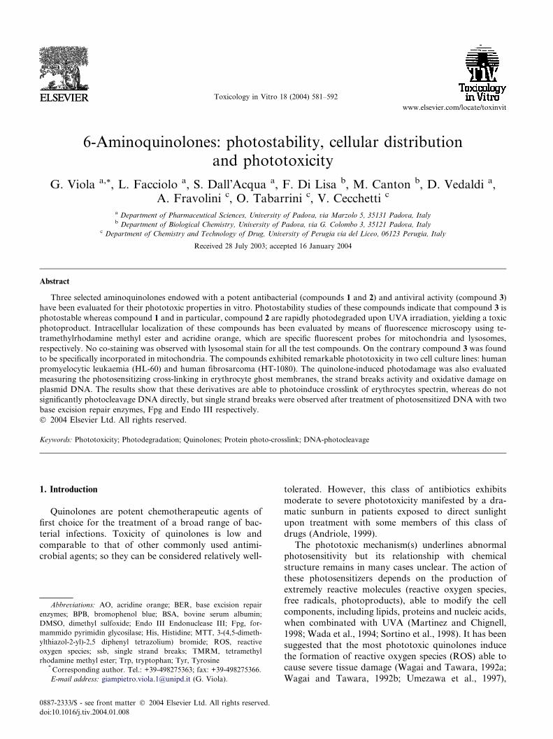
Toxicology in Vitro 18 (2004) 581–592
www.elsevier.com/locate/toxinvit
6-Aminoquinolones: photostability, cellular distributionand phototoxicity
G. Viola a,*, L. Facciolo a, S. Dall�Acqua a, F. Di Lisa b, M. Canton b, D. Vedaldi a,A. Fravolini c, O. Tabarrini c, V. Cecchetti c
a Department of Pharmaceutical Sciences, University of Padova, via Marzolo 5, 35131 Padova, Italyb Department of Biological Chemistry, University of Padova, via G. Colombo 3, 35121 Padova, Italy
c Department of Chemistry and Technology of Drug, University of Perugia via del Liceo, 06123 Perugia, Italy
Received 28 July 2003; accepted 16 January 2004
Abstract
Three selected aminoquinolones endowed with a potent antibacterial (compounds 1 and 2) and antiviral activity (compound 3)
have been evaluated for their phototoxic properties in vitro. Photostability studies of these compounds indicate that compound 3 is
photostable whereas compound 1 and in particular, compound 2 are rapidly photodegraded upon UVA irradiation, yielding a toxic
photoproduct. Intracellular localization of these compounds has been evaluated by means of fluorescence microscopy using te-
tramethylrhodamine methyl ester and acridine orange, which are specific fluorescent probes for mitochondria and lysosomes,
respectively. No co-staining was observed with lysosomal stain for all the test compounds. On the contrary compound 3 was found
to be specifically incorporated in mitochondria. The compounds exhibited remarkable phototoxicity in two cell culture lines: human
promyelocytic leukaemia (HL-60) and human fibrosarcoma (HT-1080). The quinolone-induced photodamage was also evaluated
measuring the photosensitizing cross-linking in erythrocyte ghost membranes, the strand breaks activity and oxidative damage on
plasmid DNA. The results show that these derivatives are able to photoinduce crosslink of erythrocytes spectrin, whereas do not
significantly photocleavage DNA directly, but single strand breaks were observed after treatment of photosensitized DNA with two
base excision repair enzymes, Fpg and Endo III respectively.
� 2004 Elsevier Ltd. All rights reserved.
Keywords: Phototoxicity; Photodegradation; Quinolones; Protein photo-crosslink; DNA-photocleavage
1. Introduction
Quinolones are potent chemotherapeutic agents of
first choice for the treatment of a broad range of bac-
terial infections. Toxicity of quinolones is low and
comparable to that of other commonly used antimi-
crobial agents; so they can be considered relatively well-
Abbreviations: AO, acridine orange; BER, base excision repair
enzymes; BPB, bromophenol blue; BSA, bovine serum albumin;
DMSO, dimethyl sulfoxide; Endo III Endonuclease III; Fpg, for-
mammido pyrimidin glycosilase; His, Histidine; MTT, 3-(4,5-dimeth-
ylthiazol-2-yl)-2,5 diphenyl tetrazolium) bromide; ROS, reactive
oxygen species; ssb, single strand breaks; TMRM, tetramethyl
rhodamine methyl ester; Trp, tryptophan; Tyr, Tyrosine*Corresponding author. Tel.: +39-498275363; fax: +39-498275366.
E-mail address: [email protected] (G. Viola).
0887-2333/$ - see front matter � 2004 Elsevier Ltd. All rights reserved.
doi:10.1016/j.tiv.2004.01.008
tolerated. However, this class of antibiotics exhibits
moderate to severe phototoxicity manifested by a dra-matic sunburn in patients exposed to direct sunlight
upon treatment with some members of this class of
drugs (Andriole, 1999).
The phototoxic mechanism(s) underlines abnormal
photosensitivity but its relationship with chemical
structure remains in many cases unclear. The action of
these photosensitizers depends on the production of
extremely reactive molecules (reactive oxygen species,free radicals, photoproducts), able to modify the cell
components, including lipids, proteins and nucleic acids,
when combinated with UVA (Martinez and Chignell,
1998; Wada et al., 1994; Sortino et al., 1998). It has been
suggested that the most phototoxic quinolones induce
the formation of reactive oxygen species (ROS) able to
cause severe tissue damage (Wagai and Tawara, 1992a;
Wagai and Tawara, 1992b; Umezawa et al., 1997),

582 G. Viola et al. / Toxicology in Vitro 18 (2004) 581–592
although other mechanisms, i.e. generation of highly
reactive photodegradation products, seem to be in-
volved (Martinez et al., 1998).
Many studies on the phototoxic properties of thesedrugs have been reported in the last decades and struc-
ture-side effects relationship of newly developed fluo-
roquinolones has been evaluated (Domagala, 1994;
Traynor et al., 2000; Miolo et al., 2002).
Photoreactivity is mostly influenced by the sub-
stituent at the C-8 position; in fact, halogen atoms,
such as fluorine and chlorine, have relatively high inci-
dence (Domagala, 1994), while the introduction of amethoxy group greatly reduces the phototoxicity
(Von Keutz and Schulter, 1999). The current challenge
in this field is therefore to develop less phototoxic
quinolones.
Previously, some of us have described a new series of
quinolones in which the usual fluorine atom at C-6 po-
sition was replaced with a hydrogen atom or an amino
group. These compounds showed strong activity againstGram-positive bacteria (Cecchetti et al., 1995; Cecchetti
et al., 1996a; Cecchetti et al., 1996b). Moreover an
interesting result is that 6-aminoquinolones, if appro-
priately functionalised, may be useful as antiviral agents.
In particular, the introduction of a 4-aryl-piperazinyl
moiety at C-7 position coupled with a small substituent
at N-1 position (the best being the methyl group), gave
compounds with anti-HIV-1 and anti-HIV-2 activity inboth acutely and chronically infected cells (Cecchetti
et al., 2000; Tabarrini et al., 2002). The antiviral
mechanism of 6-aminoquinolones is clearly distinct
from that of the other known inhibitors of HIV, such as
reverse transcriptase and protease inhibitors. In fact
they interfere with the trans-activation step of the
transcription, thus becoming promising candidates for
N
ONH2
NCH3
COOH
N
ONH2
NCH3
COOH
N
ONH2
NCH3
COOH
NN
1 2
3
Fig. 1. Chemical structure of the examined compounds.
the combination therapy of the AIDS (Parolin et al.,
2003).
Therefore, this paper aims at analyzing the photo-
stability and the phototoxic properties of one of themost active and selective anti-HIV 6-aminoquinolone 3,
as well as of two potent antibacterials 1 and 2 (Fig. 1).
In particular, observations on the in vitro effects on
two cell lines are reported. We have also extended our
studies on the photochemical damages induced by these
new derivatives on biological molecules, such as proteins
and DNA, to better characterize the cellular targets
involved in their phototoxic reactions.
2. Materials and methods
Chemicals. Aminoquinolones 1–3 were synthesized as
previously described (Cecchetti et al., 1996a; Cecchetti
et al., 2000). and their chemical structure is depicted in
Fig. 1. Bovine serum albumin (BSA), Ribonuclease A
from bovine pancreas, 3-(4,5-dimethylthiazol-2-yl)-2,5diphenyl tetrazolium bromide (MTT), Acridine Orange
(AO) and agarose were purchased from Sigma Chemical
Company (St. Louis, MO USA). pBR322 DNA was
purchased from InVitrogen (Milano, Italy). Coomassie
Brilliant Blue G-250 was purchased from Bio Rad
Laboratories (Segrate, Milano, Italy). Acrilamide and
N ;N 0-methylenebisacrylamide, ammonium persulphate,
TEMED (N ;N ;N 0;N 0-tetramethylethylethylenediaminewere purchased from Pharmacia Biotech.
Tetramethylrhodamine methyl ester (TMRM) was
purchased by Molecular Probes (Eugene, OR, USA).
The two base excision repair enzymes (BER) forma-
mydo-pyrimidine glycosilase (FPG) and Endonuclease
III were a generous gift from Dr. S. Boiteux (CEA,
Fontenay aux Roses FRANCE).
Irradiation procedure. HPW 125 Philips lamps,mainly emitting at 365 nm, were used for irradiation
experiments. The spectral irradiance of the source was
4.0 mWcm�2 as measured, at the sample level, by a
Cole-Parmer Instrument Company radiometer (Niles,
IL), equipped with a 365-CX sensor.
2.1. HPLC separation and photoproducts characteriza-
tion
HPLC was performed using a Perkin–Elmer Series
200 equipped with a diode array detector. A Vydac C18
column was used (5 lm, 250 · 4.3 ID) for chromato-graphic separations. The mobile phase consisted of:
Eluent A, water: formic acid (99:1); Eluent B, aceto-
nitrile: formic acid (99:1). Gradient: 5% B (0 min), 95%
B (15 min); flow rate 1 ml/min. ESI/MS measurements
were performed with an API-TOF Mariner. 1H-NMR
analysis were performed by a 200 MHz Varian Gemini
spectrometer.

G. Viola et al. / Toxicology in Vitro 18 (2004) 581–592 583
2.2. Cellular phototoxicity
Human promyelocytic leukaemia cells (HL-60) were
grown in RPMI-1640 medium, human fibrosarcomacell (HT-1080) were grown in DMEM medium both
supplemented with 115 units/mL of penicillin G (Invit-
rogen, Milano, Italy), 115 lg/ml streptomycin (Invit-
rogen, Milano, Italy) and 10% fetal calf serum
(Invitrogen, Milano, Italy). Individual wells of a 96-well
tissue culture microtiter plate (IWAKI, Japan) were
inoculated with 100 ll of complete medium containing
8 · 103 HL-60 cells or 5 · 103 HT-1080 cells. The plateswere incubated at 37 C� in a humidified 5% incubator
for 18 h prior the experiments. After medium removal,
100 ll of the drug solution, dissolved in DMSO and
diluted with Hank�s balanced salt solution (HBSS
pH¼ 7.2), was added to each well. After irradiation, the
solution was replaced with the medium, and the plates
were incubated for 72 h. Cell viability was assayed by
the MTT [(3-(4,5-dimethylthiazol-2-yl)-2,5 diphenyl tet-razolium bromide)] test, as described previously (Elisei
et al., 2002; Miolo et al., 2002).
2.3. Fluorescence microscopy
HT-1080 fibrosarcoma cells were grown in a sterile
microscope slides and treated with the three compounds
at the concentration of 50 lm for 30 min, then washed
with HBSS. Cellular fluorescence images were acquired
with an Olympus IMT-2 inverted microscope, as previ-ously described (Petronilli et al., 1999). For subcellular
co-localization studies Tetramethyl Rhodamine methyl
ester (TMRM) and Acridine Orange (AO), were used as
fluorescent probes which stain mitochondria and lyso-
somes, respectively. Quinolones excitation was per-
formed at 350 nm and emission was read at 440 nm.
Longpass emission filter settings were used to separate
the emission of the probes from that of the test com-pounds. Data were acquired and analysed using the
Metamorph software (Universal Imaging).
2.4. Erithrocyte ghost preparation
Ghosts were prepared from heparinized human blood
following the gradual osmotic lysis method of Steck and
Kant (1974). White membranes (ghosts) were resus-
pended in PBS buffer. Membrane protein contents weredetermined as described (Peterson, 1977), using bovine
serum albumin (BSA) as standard.
2.5. Protein photo cross-link
The test compounds were added to the membrane
suspension (1.0 mg/ml protein concentration) and irra-
diated. The membrane samples were reduced and
denatured by addition of b-mercaptoethanol and SDS at
90 �C for 3 min, and bromophenol blue (BPB) was ad-
ded before polyacrylamide gel electrophoresis analysis
(5% running gel, 3% stacking gel). The quantitation of
the bands, stained with Coomassie Brilliant Blue R-250,was achieved by image analyzer software Quantity
One (BIO RAD, Milano, Italy).
2.6. Studies on isolated proteins
Solutions of Bovine serum albumin (BSA), (0.5 mg/
ml) in phosphate buffer 10 mM were irradiated in the
presence of the test compounds for various time using a
quartz cuvette. At each time the Tryptophan (Trp)
content was followed by monitoring the characteristicTrp fluorescence as described (Balasubramanian et al.,
1990). Solutions of Ribonuclease A (RNAseA), 0.5 mg/
ml in phosphate buffer 10 mM were irradiated in the
presence of the test compounds for various time periods
in a quartz cuvette. Analogous experiments were per-
formed by bubbling nitrogen before and during the
irradiation.
2.7. Studies on isolated amino acids
Irradiation system and buffer in these experiments
were the same used for the irradiation of BSA. Tryp-
tophan solutions (100 lM) were irradiated in the pres-
ence of the test compounds and analysed as described
for BSA. Histidine solutions were irradiated for various
time periods and the resulting samples were analysed for
His content as described (Figueiredo et al., 1993).Tyrosine solutions (1 mM) were irradiated in PBS for
various times and the resulting samples were analysed
for Tyr content as described (Peterson, 1977) Analogous
experiments were performed by bubbling nitrogen prior
and during the irradiation and using phosphate buffer
prepared with deuterium oxide instead of water.
2.8. pBR322 DNA strand breaks
Each pBR322 DNA sample (100 ng) dissolved in TEbuffer (10 mM Tris-HCl, 1 mM EDTA, pH 7.5) was
irradiated with increasing UVA doses in the presence of
the compounds under examination. After irradiation
two aliquots of the sample were incubated for 30 min at
37 �C with Fpg (formamydo pyrimidin glycosilase) and
Endo III (Endonuclease III) as described by Pflaum
et al. (1994). The samples were loaded on 1% agarose
gel, and the run was carried out in TAE buffer (0.04 MTris-acetate, 1 mM EDTA) at 50 V for 3 h, using the
GNA-100 electrophoretic apparatus (Amersham Bio-
sciences). After staining in ethidium bromide solution,
the gel was washed with water and the DNA bands were
detected under with a UV transilluminator. Photo-
graphs were taken with a digital photocamera Kodak
DC265 and the quantitation of the bands was achieved

Table 1
Mass spectral data of photodegradation products of aminoquinolones
Photoproduct Retention time (min) m/z
P1 8.6 359 (+17 amu)a
584 G. Viola et al. / Toxicology in Vitro 18 (2004) 581–592
by image analyzer software Quantity One (BIO RAD,
Milano, Italy). The fractions of supercoiled (Form I)
and open circular DNA (Form II) were calculated
as described (Ciulla et al., 1989).
P2 10.3 357 (+15 amu)P3 9.1 315 ()75 amu)
P4 11.9 386 ()4 amu)a In parenthesis is indicated the variation of mass in comparison to
the parent compound.
3. Results3.1. Photostability
Previous studies (Miolo et al., 2002), showed that
compounds 1 and 2 underwent photodegradation, as
shown by the changes in their absorption spectra upon
UVA irradiation in buffered aqueous solution. Com-
pound 3, in comparison to the other two derivatives,
does not show any variation of the absorption spectrum
even after a long time of irradiation (data not shown).
To obtain precise informations of the photodegra-dation kinetics, at appropriate time intervals the irra-
diated solutions were analysed by HPLC (Fig. 2).
Irradiation of 1 and 2 induces for both compounds the
formation of two photoproducts, while compound 3
practically remains unmodified even at high doses of
UVA (23 J cm�2). Kinetic analysis of the photodegra-
dation process reveals an apparent first order kinetics
for the three derivatives. The rate constants (k) wereobtained from the slopes of the plots by linear regression
analysis. Degradation kinetics followed a first order
reaction with photolysis constants of 2.3 · 10�3,
9.8 · 10�3 and 1.6 · 10�4 min�1, for compounds 1, 2 and
3 respectively.
The characterization of the photoproducts was per-
formed by ESI-MS and the mass spectral data are re-
ported in Table 1. In the case of compound 2, thestructure of the main product, P4, that is produced in
good yield, was elucidated by NMR spectroscopy. Fig. 3
illustrates the 1H-NMR spectra of the photoproduct
(panel A) and of the compound 2 (panel B).
Abso
rban
ce u
nits
0.00
0.02
0.04
RetentionTime (min)0 5 10 15 20 25
Abso
rban
ceun
its
0.00
0.02
0.04
0.06
0.00
0.02
0.04
Retention0 5 10
0.00
0.02
(A) (B)
(D) (E)
P1P2P3
Fig. 2. HPLC profile of aminoquinolones upon UVA irradiation in phosph
panel D¼ 15 J cm�2); compound 2 (panel B¼ 0 J cm�2, panel E¼ 15 J cm�2
The 1H-NMR spectra of P4 and compound 2 showed
similar patterns: two singlets in the aromatic region at
8.9 and 8.4 ppm, respectively; a multiplet between 7.22
and 7.23 ppm which has been assigned to the aromatic
protons of the tetrahydroisoquinolinic ring; a multiplet
at 4.3 ppm; the methyl group in position 8 has been
assigned to the signal at 2.6 ppm; two multiplets be-
tween 1.3 and 0.80 ppm which correspond to the cy-clopropyl group. It is also possible to see a signal at 4.95
ppm which has been assigned to the protons of the
amino group in position 6. The substantial difference
between the two spectra lies in the region from 7 to 9
ppm. In fact, while the spectrum of compound 2 only
displays the presence of the singlet belonging to the
carboxyl group (7.35 ppm), the spectrum of P4 shows a
signal of a doublet at 8.3 ppm which counterparts is amultiplet at 7.20 ppm.
These data, also supported by the mass spectrometry
analysis, lead to postulate a photoinduced aromatisa-
tion of the tetrahydroisoquinolinic nucleus in position 7
of compound 2. The proposed structure is showed in
Fig. 3.
3.2. Intracellular localization
It has recently been demonstrated by microspectro-
fluorimetric techniques on fibroblast cells that some
fluoroquinolones localize in subcellular structures. For
example, lysosomes are a preferential site for lomeflox-
0.00
0.04
0.08
0 8 120.00
0.04
0.08
Time (min)15 20 25
Retention Time (min)
P4
(C)
(F)
4
ate buffer under aerobic conditions. Compound 1 (panel A¼ 0 J cm�2,
); compound 3 (panel C¼ 0 J cm�2, panel F¼ 23 J cm�2).

Fig. 3. 1H-NMR spectra of photoproduct P4 (panel A) with the proposed chemical structure. In the inset is also shown a magnification of the region
between 7 and 9 ppm. Panel B show for the sake of comparison the 1H-NMR spectra of compound 2.
Fig. 4. Fluorescence microphotographs showing the intracellular
localization of the aminoquinolones in HT-1080 fibrosarcoma cells in
the presence of TMRM. The cells were treated as described in Section 2.
Upper panels, compound 1 middle panels compound 2; lower panels
compound 2. Column A, fluorescence image of the aminoquinolones;
Column B, fluorescence image of TMRM; Column C, Overlay image of
aminoquinolones localization generated by transferring the blue color
G. Viola et al. / Toxicology in Vitro 18 (2004) 581–592 585
acin and ciprofloxacin whereas for norfloxacin the cel-
lular localization is less clear (Ouedraogo et al., 1999).
Other subcellular structures could be the target of these
compounds. To investigate the intracellular localizationof 6-aminoquinolones we used two fluorescent probes:
TMRM, a lipophilic cation commonly used for the
assessment of the mitochondrial potential (Bernardi
et al., 1999; Petronilli et al., 1999), and acridine orange,
which stains lysosomes (Kessel et al., 2000). These
markers fluoresce in the visible region (about 550 nm)
while the test compounds emit in the blue region. Both
fluorescences can be easily separated using suitablebandpass optical filters. After an incubation in HT-1080
fibrosarcoma cells, all the compounds were found to
incorporate and associate with subcellular structures.
It can be observed that, despite the studied quino-
lones have similar structure, they localize differently in-
side cells. Compound 1 diffuses largely in the cytoplasm
but does not co-localize with the two fluorescence
probes. Compound 2 does not accumulate in lysosomesbut only partially co-localizes with TMRM. A more
evident accumulation in mitochondria can be observed
for compound 3, suggesting that mitochondria may be a
site of photosensitization (Figs. 4 and 5).
of the compounds seen in column A onto the corresponding fluores-
cence image of column B. Pink is used as a false color to indicate the
localization of aminoquinolones. (For interpretation with reference to
the color picture the reader is referred to the web version of this article.)
3.3. Cellular phototoxicityCellular phototoxicity was investigated using two cell
lines: HT-1080 fibrosarcoma and HL-60 leukemiarespectively. In Fig. 6 the viability curves after irradia-
tion with various UVA-doses and different concentra-
tion of aminoquinolones are depicted. The most potent
compound in both cell lines is compound 3 which
exhibits high levels of toxicity even at very low con-
centrations and low UVA doses. Compound 2 has also a
remarkable toxicity, whereas compound 1 is less pho-
totoxic in comparison to the other two studied amino-
quinolones.
HL-60 cells incubated in the presence of a pre-irra-
diated solution of compound 2 showed a dose depen-dent decrease of viability (data not shown) suggesting
the production of a toxic photoproduct. Further experi-
ments performed without UVA irradiation demon-
strated that the isolated photoproduct P4 induces a

Fig. 5. Fluorescence microphotographs showing the intracellular
localization of the aminoquinolones in HT-1080 fibrosarcoma cells in
the presence of AO. The cells were treated as described in Section 2.
Column A, fluorescence image of the aminoquinolones; Column B,
fluorescence image of AO; Column C, Overlay image of aminoqui-
nolones localization generated by transferring the blue color of the
compounds seen in column A onto the corresponding fluorescence
image of column B. Clear blue is used as a false color to indicate the
localization of aminoquinolones. (For interpretation with reference to
the color picture the reader is referred to the web version of this
article.)
586 G. Viola et al. / Toxicology in Vitro 18 (2004) 581–592
remarkable decrease of cellular viability with an esti-
mated IC50 of about 4 lm (Fig. 7, panel A), whereas the
(A)
0 1 2 3 4
% o
f via
ble
cells
0
20
40
60
80
100
(B)
0 1 2
0
20
40
60
80
100
(E)
UVA do
0 1 20
20
40
60
80
100
(D)
0 1 2 3 40
20
40
60
80
100
% o
f via
ble
cells
UVA dose (Jcm )-2
Fig. 6. Effect of the quinolones derivatives photosensitization upon HL-60 c
and F). The cells were treated with the compounds (1, panels A and D
(r¼ irradiated controls,�¼ 2 lM,j¼ 5 lM,N¼ 10 lM,.¼ 20 lM panels
N¼ 2.55 lM, .¼ 5 lM panel C); (r¼ irradiated controls, �¼ 1.25 lM, j¼as indicated in the figure. The viability was measured with the MTT method a
independent experiments performed in quadruplicate.
value for the parent compound is higher than 20 lm.
Moreover, the isolated photoproduct has also been
evaluated for its phototoxicity (Fig. 7, panel B), upon
irradiation showed a moderate induction of cytotoxicityas compared with parent compound 2.
3.4. Protein photodamage
The photosensitization ability of test compounds to-
wards other components of cellular membranes, such as
proteins, was estimated by measuring the photoinduced
cross-linking in erythrocyte ghost proteins (Merville
et al., 1983). Light-induced cross-linking of spectrin, a
protein associated with the cytoplasmic side of the RBC
membrane, in the presence of these compounds wasdetected by the partial or total disappearance of the two
spectrin bands (220.000 and 245.000 Daltons) on SDS-
PAGE while cross-linked aggregates can not run inside
the gel and remain at the top.
Fig. 8 (panel A) shows the pictures of the SDS-PAGE
gels of ghosts irradiated in the presence of the three
aminoquinolones, at the concentration of 100 lm and
at UVA dose ranging from 3.6 to 14.4 J cm�2. Underaerobic conditions compound 2 causes the almost total
disappearance of the two spectrin subunit bands,
whereas in the presence of nitrogen this effect is lower
indicating the involvement of reactive oxygen species.
The other two compounds exhibit a lower efficacy, al-
though in both cases the effect is partially reduced in
anaerobic conditions. The gel scans of erythrocytes
3 4
se (Jcm )-2
3 4
(C)
0 1 2 3 4
0
20
40
60
80
100
(F)
0 1 2 3 40
20
40
60
80
100
UVA dose (Jcm )-2
ells (panels A, B and C) and HT-1080 fibrosarcoma cells (panels D, E,
; 2, panels B and E; 3, panel C and F) at different concentrations
A, B, D and E); (r¼ irradiated controls,�¼ 0.625 lM,j¼ 1.25 lM,
2.5 lM, N¼ 5 lM, .¼ 10 lM panel G), and various doses of UVA,
s described in Materials and methods. Points are mean±SEM for three

Concentration (µm)0 5 10 15 20
% o
f via
ble
cells
0
20
40
60
80
100
UVA Dose (Jcm-2)0 1 2 3 4
% o
f via
ble
cells
0
20
40
60
80
100
(A)
(B)
Fig. 7. Effect of photoproduct P4 on HT-1080 cells. Panel A: Dark
cytotoxicity evaluated after 72 h of incubation in the presence of P4
(j) and for the sake of comparison compound 2 (�), at different
concentrations. Panel B: Phototoxicity of P4 at different concentra-
tions (r¼ irradiated controls, �¼ 1.25 lM, j¼ 2.5 lM, N¼ 5 lM,
.¼ 10 lM) and UVA doses.
G. Viola et al. / Toxicology in Vitro 18 (2004) 581–592 587
spectrin for all the three drugs investigated are shown in
Fig. 8 (panel B).
To further investigate the photosensitizing properties
of the three compounds towards proteins, solutions
containing bovine serum albumin, as a model system
(Miranda et al., 1998), and the drugs in phosphate buffer
were irradiate for different time periods. Trp contentwas directly analysed by monitoring the characteristics
fluorescence of Trp residues (Balasubramanian et al.,
1990). No effects were observed with the three com-
pounds indicating that Trp is not involved in the protein
photodamage. Another protein model used in this study
was RNAseA, which is devoid of Trp but has Tyr resi-
dues in its sequence. Its emission band centered at about
350 nm does not change after irradiation with com-pound 1 and 3. Interestingly, compound 2 rapidly de-
creases the emission of the protein by about 80% after
irradiation for 15 min (Fig. 9, panel A). This effect is
markedly reduced in anaerobic conditions. Deuterated
phosphate buffer which increases singlet oxygen lifetime,
did not induce any variation of the degradation profile.
Furthermore, gel electrophoresis of RNAseA showed
protein cross link after irradiation in the presence ofcompound 2 (data not shown).
Subsequent analysis were performed to confirm the
results on pure amino acids. Data obtained with Tyr are
in excellent agreement with the observation on the pure
RNAseA. Under aerobic conditions 30% of Tyr was
degradated (Fig. 9, panel B), whereas in anaerobic
conditions no effect was observed. In this case too, Tyr
degradation was not potentiated in deuterated buffer,suggesting that singlet oxygen should not be involved in
the reaction.
Histidine reacts which singlet oxygen faster than
other amino acids, such as Trp and Tyr, so it is useful to
test its degradation if we expect a type II mechanism. In
the case of compounds 1 and 2 we observed a rapid
decrease of His content (Fig. 9, panels C and D). This
effect was abolished in N2-purged solution and poten-tiated in deuterated phosphate buffer, suggesting the
involvement of singlet oxygen. Compound 3 did not
show any effect on this amino acid.
3.5. DNA photodamage
pBR322 Plasmid DNA was used as a model system to
evaluate DNA breaking activity. None of the test
compounds was able to induce DNA damage without
UVA irradiation.
Supercoiled circular DNA allows the detection of
structural alterations such as strand breaks or damagedbases. DNA strand breaks can be induced either directly
(frank strand breaks) or indirectly. In the latter case we
used DNA repair enzymes in order to determine the
characteristics of DNA modifications: Fpg protein rec-
ognizes 8-hydroxyguanine, purines whose imidazole ring
is open (Fapy residues) and sites of base loss (apurinic
sites) and Endonuclease III recognizes apurinic sites and
5,6-dihydropirimidine derivatives. Release of damagedbase is followed by a b–d reaction and a b elimination
step respectively, resulting in DNA breakage (Burrows
and Muller, 1998).
In addition to endonuclease sensitive modifications,
the number of single strand breaks generated by the
excited photosensitizers were quantified. Fig. 10 shows
the results obtained for the three aminoquinolones tes-
ted at a [drug]/[DNA] ratio of 1. For all compounds theformation of single strand breaks expressed as the per-
centage of form II is low and becomes significantly
different from the irradiated control, only at the highest
UVA dose employed (22.5 J cm�2). On the contrary,
high levels of single strand breaks can be detected after
enzyme digestion for 2 and 3 whereas only a slight in-
crease was observed for 1.

Fig. 8. Electrophoretic pattern (A) of the photoinduced cross-link of spectrin in RBC ghosts irradiated in aerobic and anaerobic conditions, for the
indicated times, corresponding to UVA doses of 3.6 J cm�2 (15 min) to 14.4 J cm�2 (60 min), in the presence of aminoquinolones at the concentration
of 50 lm in PBS buffer pH¼ 7.2. (B) Quantitation of the relative abundance of spectrum by gel densitometry; (�¼Air,j¼N2). The spectrin band is
indicated by an arrow.
588 G. Viola et al. / Toxicology in Vitro 18 (2004) 581–592
4. Discussion
In this study we have evaluated the phototoxic po-tential of three aminoquinolones, using various in vitro
methods. Particular attention has been given towards
the investigation of the mechanism involved in their
phototoxicity, extending our studies on the photo-
chemical damage on biological molecules, such as pro-
teins and DNA, to better characterize the cellular targets
involved in their phototoxic reactions.
To have a complete phototoxicological profile westudied the photoreactivity of the test molecules
including degradation reactions. The three aminoqui-
nolone derivatives showed different patterns of degra-
dation depending on their molecular structures. The
UVA lamp emits light with maximum intensity at 365nm and the kmax of the test compounds has similar molar
extinction coefficients at this wavelength, thus differ-
ences underlying in their photodegradation profiles may
not be related to a different absorption of light.
By a photochemical point of view the strength of a
photosensitiser is determined by the nature of the ex-
cited states or by its ability to form reactive species
while, in a phototoxicological context, other factorssuch as subcellular localization, are important parame-
ters. Thus, it is critically important to evaluate the dis-

% o
f und
ecom
pos
ed H
is
0
20
40
60
80
100
UVA Dose (J cm-2)
% o
f und
e co
mpo
sed
His
0
20
40
60
80
100
UVA Dose (J cm )
UVA Dose (J cm )
-2 UVA Dose (J cm )-2
-2
0 4 8 12 16
0 4 8 12 16 0 4 8 12 16
0 4 8 12 16
I 0/I f
0
20
40
60
80
100
% o
fu n
deco
mpo
sed
Tyr
0
20
40
60
80
100
(A) (B)
(C) (D)
Fig. 9. Photosensitising effects of compound 2 on RNAseA (panel A) and Tyr (panel B) after irradiation at the dose of 50 lm in different conditions
(�¼Air, j¼N2 purged solution, N¼Deuterated phosphate buffer purged solution). Photosensitising effects of compounds 1 (panel C) and 2
(panel D) on His after irradiation at the dose of 100 lm in different conditions (� ¼ Air,j¼N2 purged solution,N¼Deuterated phosphate buffer).
G. Viola et al. / Toxicology in Vitro 18 (2004) 581–592 589
tribution of photosensitisers inside cells. Subcellular
localization of a photosensitiser is due to its physico-
chemical properties such as hydrophobicity, charge, pKa
etc. Previous studies have shown that fluoroquinolones
principally accumulate in lysosomes (Ouedraogo et al.,
1999), while the present compounds (aminoquinolones),
interestingly do not shown the same pattern. The
physico-chemical properties of the aminoquinolones
amply differ from that of fluoroquinolones used in
therapy. They are hydrophobic, as demonstrated by
their high values of partition coefficients (Miolo et al.,2002) and do not possess a pKa close to neutrality, while
many fluoroquinolones are present in solution as zwit-
terionic ions. Consistently with these characteristics,
none of the aminoquinolones investigated shows a
lysosome incorporation. On the contrary, preferential
accumulation in mitochondria has been observed in
particular for compound 3. On HL-60 and HT-1080
cells, compound 3 was highly phototoxic, even at thelowest concentrations (0.625 and 1.25 lm respectively)
and UVA dose applied (1 J cm�2) followed by com-
pound 2 and compound 1. Numerous reports show that
photosensitizers which localize in mitochondria are
more efficient in killing cells than those that localize in
other cellular sites (Oleinick et al., 2002 and references
therein).
This study clearly demonstrates that compound 2 has
a phototoxic potential which may be partially mediated
by the induction of a toxic photoproduct. The cytotoxicactivity and the concentration of the photoproduct
are not related and this could be due to the low solu-
bility. Furthermore the presence of a positive charge on
the molecule could be an hindrance for its diffusion into
the cells. It is possible that the cytotoxic effect of this
photoproduct may be caused by a small amount pro-
duced inside the cell from the parent compound. The
target for the P4-induced cell damage is not clear at themoment, although preliminary results obtained with
fluorescence microscopy (data not shown) suggest spe-
cific accumulation of this photoproduct in mitochon-
dria.
An important damage photoinduced by aminoqui-
nolones is the cross-link in membrane proteins. A good
model is represented by erythrocyte membranes because
they contain spectrin, an excellent target of the cross-link reactions (Dubbelman et al., 1978; Merville et al.,
1983). Although the three derivatives promote spectrin
cross-link, their, efficiency differs from each other.
Compound 2 is the most efficient whereas the other two
compounds seem to be less active. The spectrin cross-
link is reduced in anaerobic conditions indicating the
involvement of molecular oxygen in the mechanism.

Fig. 10. DNA strand breaks photoinduced by the aminoquinolones.
pBR322 supercoiled circular DNA was irradiated at the indicated
doses of UVA and then treated as described in materials and methods.
Data are expressed as percentage of form II obtained after densito-
metric analysis of the agarose gel.
590 G. Viola et al. / Toxicology in Vitro 18 (2004) 581–592
The amino acids that are more easily photooxidized inproteins are tryptophan, tyrosine, histidine, cysteine and
methionine (Davies and Truscott, 2001). Thus, in order
to elucidate which amino acids are involved in the
protein photosensitization by the test compounds, we
studied the photoreaction with BSA and RNAseA as
protein models. The experimental results, obtained with
BSA indicated that Trp does not constitute a target for
the photoreaction with test compounds. On the contraryTyr residues in RNAseA are promptly photoxidized
upon irradiation in the presence of compound 2. How-
ever, it should be note thatin a native protein the reac-
tivity of the above amino acids might be affected by the
location of the given amino acid residue or to the par-
ticular binding site of the photosensitiser, thus the
photosensitised reactions of proteins turn out to be very
complex.The behaviour of the three compounds largely differs
in respect of pure amino acids. Compound 1 neither
photooxidizes Trp nor Tyr, but it is able to photoxidize
His, although with a low efficiency. In contrast, com-
pound 2 is able to efficiently photooxidize Tyr and His.
From a mechanistic point of view, the photoreactions
with these two amino acids are strongly reduced in
anaerobic conditions but only in the case of His thephotodegradation is remarkable potentiated in deuter-
ated phosphate buffer, suggesting that this effects may
be mediated by singlet oxygen.
Most of the fluoroquinolones present in therapy have
demonstrated a high extent of DNA photodamage and
this can explain their capability of enhancing UVA-
induced phototumorigenesis (Reavy et al., 1997; Cheta-
lat et al., 1996; Martinez and Chignell, 1998; Belvedereet al., 2002; Cuquerella et al., 2003).
In order to determine whether test compounds were
able to photosensitize DNA, strand break activity was
evaluated using supercoiled circular DNA, a very sen-
sitive tool for damage detection (Marrot et al., 2001). In
addition to frank strand breaks we have also evaluated if
purine and/or pyrimidine bases were involved in the
oxidative damage using base excision repair enzymesFpg and Endo III, respectively. Specific repair enzymes,
such as Fpg or Endo III, induce DNA single strand
breaks at the sites of damaged purine bases such as 8-
oxoguanine or thymine glycol. The aminoquinolones
showed different responses in terms of oxidative dam-
age. While they do not induce a significant direct DNA
breakage, they are able to photooxidize nucleobases, as
assessed after enzymatic digestion with Fpg. No effectswere seen with Endo III. This damage profile for com-
pounds 2 and 3 is an indication that singlet oxygen could
be an intermediate in the DNA damage and is in good
agreement with the data obtained on His photosensiti-
zation.
It should be noted, however, that at the present stage
a definite mechanism for the photoinduced cellular
damage in the presence of aminoquinolones cannot bepresented. Further studies along these lines are neces-
sary and are presently underway to clarify the mecha-
nism.
In conclusion, the three aminoquinolones demon-
strated a potential phototoxicity, in particular com-
pounds 2 and 3. An increase of the phototoxic activity
can be observed in all the utilized tests when the pipe-
ridinyl group is replaced by the more hydrophobic1,2,3,4-tetrahydroisoquinolinyl group. The activity of
compound 3 remains unclear: further experiments are in
progress aimed at defining the targets at cellular level
and the mechanism of phototoxicity.
Acknowledgements
This project was funded by MIUR (Ministero
dell�Istruzione, dell�Universit�a e della Ricerca), pro-
gram: ‘‘Photoprocesses of interest for application’’.
G. Viola et al. / Toxicology in Vitro 18 (2004) 581–592 591
References
Andriole, V.T., 1999. The future of the quinolones. Drugs 58 (Suppl.
2), 1–5.
Balasubramanian, D., Du, X., Zigler Jr., J.S., 1990. The reaction of
singlet oxygen with proteins with special reference to crystallins.
Photochemistry and Photobiology 52, 761–768.
Belvedere, A., Bosca, F., Catalfo, A., Cuquerella, M.C., de Guidi, G.,
Miranda, A.M., 2002. Type II guanine oxidation photoinduced by
the antibacterial fluoroquinolone rufloxacin in isolated DNA and
in 20-deoxyguanosine. Chemical Research in Toxicology 15, 1142–
1149.
Bernardi, P., Scorrano, L., Colonna, R., Petronilli, V., Di Lisa, F.,
1999. Mitochondria and cell death. Mechanistic aspects and
methodological issues. European Journal of Biochemistry 264,
678–701.
Burrows, C.J., Muller, J.G., 1998. Oxidative nucleobase modification
leading to strand scission. Chemical Review 98, 1109–1151.
Cecchetti, V., Clementi, S., Cruciani, G., Fravolini, A., Pagella, P.G.,
Savino, A., Tabarrini, O., 1995. 6-Aminoquinolones: a new class of
quinolone antibacterials? Journal of Medicinal Chemistry 38, 973–
982.
Cecchetti, V., Fravolini, A., Lorenzin, M.C., Tabarrini, O., Terni, P.,
Xin, T., 1996a. Studies on 6-aminoquinolones: synthesis and
antibacterial evaluation of 6-amino-8-methylquinolones. Journal of
Medicinal Chemistry 39, 436–455.
Cecchetti, V., Fravolini, A., Palumbo, M., Sissi, C., Tabarrini, O.,
Terni, P., Xin, T., 1996b. Potent 6-desfluoro-8-methylquinolones as
new lead compounds in antibacterial chemotherapy. Journal of
Medicinal Chemistry 39, 4952–4957.
Cecchetti, V., Parolin, C., Moro, S., Pecere, T., Filippini, E., Calistri,
A, Tabarrini, O., Gatto, B., Palombo, M., Fravolini, A., Pal�u, G.,
2000. Aminoquinolones as new potential anti-HIV agents. Journal
of Medicinal Chemistry 43, 3799–3802.
Chetalat, A., Albertini, S., Gocke, E., 1996. The photomutagenicity of
fluoroquinolones in tests for gene mutations, chromosomal aber-
ration, gene conversion and DNA breakage (comet assay). Muta-
genesis 11, 497–504.
Ciulla, T.A., van Camp, J.R., Rosenfeld, E., Kochevar, I.E.,
1989. Photosensitization of single-strand breaks in pBR322
DNA by rose bengal. Photochemistry and Photobiology 49, 293–
298.
Cuquerella, M.C., Bosca, F., Mirando, A.M., Belvedere, A., Catalfo,
A., de Guidi, G., 2003. Photochemical properties of ofloxacin
involved in oxidative DNA damage: a comparison with rufloxacin.
Chemical Research in Toxicology 16, 562–570.
Davies, M.J., Truscott, R.J.W., 2001. Photo-oxidation of proteins and
its role in caractogenesis. Journal of Photochemistry and Photo-
biology B: Biology 63, 114–125.
Domagala, J.M., 1994. Structure-activity and structure-side-effect
relationships for the quinolone antibacterials. Journal of Antimi-
crobial Chemotherapy 22, 685–706.
Dubbelman, T.M.A.R., De Goeij, A.F.P.M., van Steveninck, J., 1978.
Photodynamic effects of protoporphyrin on human erythrocytes:
nature of the crosslinking of membrane proteins. Biochimica and
Biophysica Acta 511, 141–151.
Elisei, F., Aloisi, G., Latterini, L., Mazzuccato, U., Viola, G., Miolo,
G., Vedaldi, D., Dall�Acqua, F., 2002. Excited state properties and
in vitro phototoxicity studies of three phenothiazine derivatives.
Photochemistry and Photobiology 75, 11–21.
Figueiredo, A., Fontes Ribeiro, A., Gonc�alo, M., Poiares Baptista, A.,
Texeira, F., 1993. Experimental studies on the mechanisms of
tiaprofenic acid photosensitization. Journal of Photochemistry and
Photobiology B: Biology 18, 161–168.
Kessel, D., Luo, Y., Mathieu, P., Reiners Jr., J.J., 2000. Determinants
of apoptotic response to lysosomal photodamage. Photochemistry
and Photobiology 71, 196–200.
Marrot, L., Belaidi, J.P., Chaubo, C., Meunier, J.R., Perez, P.,
Agapakis-Causse, C., 2001. Fluoroquinolones as chemical tools to
define a strategy for photogenotoxicity in vitro assessment.
Toxicology in Vitro 15, 131–142.
Martinez, L., Chignell, C.F., 1998. Photocleavage of DNA by the
fluoroquinolone antibacterials. Journal of Photochemistry and
Photobiology B: Biology 45, 51–59.
Martinez, L.J., Sik, R.H., Chignell, C.F., 1998. Fluoroquinolone
antimicrobials: singlet oxygen, superoxide and phototoxicity.
Photochemistry and Photobiology 67, 399–403.
Merville, M.P., Piette, J., Decuypier, J., Calberg-Bacq, C.M., Van De
Vorst, A., 1983. Phototoxicity of phenothiazines derivatives. II.
Photosensitised cross-linking of erythrocyte membrane proteins.
Chemical Biological Interactions 44, 275–287.
Miolo, G., Viola, G., Vedaldi, D., Dall�Acqua, F., Fravolini, A.,
Tabarrini, O., Cecchetti, V., 2002. In vitro phototoxic properties of
new 6-desfluoro and 6-fluoro 8-methyl quinolones. Toxicology in
Vitro 16, 683–693.
Miranda, M.A., Castell, J.V., Hernandez, D., Gomez-Lechon, M.J.,
Bosca, F., Morera, I.M., Sarabia, Z., 1998. Drug-photosensitized
protein modification: identification of the reactive sites and
elucidation of the reaction mechanisms with tiaprofenic acid/
albumin as model system. Chemical Research in Toxicology 11,
172–177.
Oleinick, N.L., Morris, R.L., Belichenko, I., 2002. The role of
apoptosis in response to photodynamic therapy: what, where, why
and how. Photochemical and Photobiological Sciences 1, 1–21.
Ouedraogo, G., Morliere, P., Bazin, M., Santus, R., Kratzer, B.,
Miranda, M.A, Castell, J.V., 1999. Lysosomes are sites of
fluoroquinolone photosensitization in human skin fibroblasts: a
microspectrofluorometric approach. Photochemistry and Photo-
biology 70, 123–129.
Parolin, C., Gatto, B., Del Vecchio, C., Pecere, T., Tramontano, E.,
Cecchetti, V., Fravolini, A., Masiero, S., Palombo, M., Pal�u, G.,
2003. New anti-human immunodeficiency virus type 16-aminoqu-
inolones: mechanism of action. Antimicrobial Agents and Chemo-
therapy 47, 889–896.
Peterson, G.L., 1977. A simplification of the protein assay method of
Lowry et al. which is more generally applicable. Analytical
Biochemistry 83, 346–356.
Petronilli, V., Miotto, G., Canton, M., Brini, M., Colonna, R.,
Bernardi, P., Di Lisa, F., 1999. Transient and long-lasting opening
of the mitochondrial permeability transition pore can be monitored
directly in intact cells by charges in mitochondrial calcein.
Biophysical Journal 76, 725–734.
Pflaum, M., Boiteux, S., Epe, B., 1994. Visible light generates oxidative
DNA base modifications in high excess of strand breaks in
mammalian cells. Carcinogenesis 15, 297–300.
Reavy, H.J., Traynor, N.J., Gibbs, N.K., 1997. Photogenotoxicity of
skin tumorigenic fluoroquinolone antibiotics detected using the
comet assay. Photochemistry and Photobiology 66, 368–373.
Sortino, S., Condorelli, G., De Guidi, G., Giuffrida, S., 1998.
Molecular mechanisms of photosensitization XI Membrane dam-
age and DNA cleavage photoinduced by enoxacin. Photochemistry
and Photobiology 68, 652–659.
Steck, T.L., Kant, J.A., 1974. Preparation of impermeable ghosts
and erythrocytes membranes. Methods in Enzymology 31, 172–
180.
Tabarrini, O., Cecchetti, V., Gatto, B., Palombo, M., De Clercq, E.,
Fravoilini, A., 2002. Nuovi agenti HIV: 6-aminochinoloni come
inibitori del processo di trans attivazione Tat-mediato. XVI
Convegno Nazionale Divisione di Chimica Farmaceutica, Societ�achimica Italiana, Sorrento, L48.
Traynor, N.J., Barratt, M.D., Lovell, W.W., Ferguson, J., Gibbs,
N.K., 2000. Comparison of an in vitro cellular phototoxicity model
against controlled clinical trials of fluoroquinolone skin phototox-
icity. Toxicology in vitro 14, 275–283.

592 G. Viola et al. / Toxicology in Vitro 18 (2004) 581–592
Umezawa, N.K., Arakane, R.A., Mashiko, S., Hirobe, M., Nagano,
T., 1997. Participation of reactive oxygen species in phototoxicity
induced by quinolone antibacterial agents. Archives of Biochem-
istry and Biophysics 342, 275–281.
Von Keutz, E., Schulter, G., 1999. Preclinical safety evaluation of
moxifloxacin, a novel fluoroquinolone. Journal of Antimicrobial
Chemotherapy 43 (Suppl. B), 91–100.
Wada, K., Saniabadi, A.R., Takiguchi, Y., Nakashima, M., 1994. UV-
dependent quinolone induced human erythrocyte membrane lipid
peroxidation: studies on the phototoxicity of Y-26611, a new
quinolone derivative. Pharmacology and Toxicology 74, 240–
243.
Wagai, N., Tawara, K., 1992a. Important role of oxygen metabolites
in quinolone antibacterial agent-induced cutaneous phototoxicity
in mice. Archives of Toxicology 65, 495–499.
Wagai, N., Tawara, K., 1992b. Possible direct role of reactive oxygens
in the cause of cutaneous phototoxicity induced by five quinolones
in mice. Archives of Toxicology 66, 392–397.