Resistance to Degradation and Cellular Distribution are Important Features for the Antitumor...
10
Resistance to Degradation and Cellular Distribution Are Important Features for the Antitumor Activity of Gomesin Marcus V. Buri 1 , Tatiana M. Domingues 1 , Edgar J. Paredes-Gamero 1,2 *, Rafael L. Casaes-Rodrigues 2 , Elaine Guadelupe Rodrigues 3 , Antonio Miranda 1 * 1 Departamento de Biofı ´sica, Universidade Federal de Sa ˜o Paulo, Sa ˜o Paulo, SP, Brazil, 2 Departamento de Bioquı ´mica, Universidade Federal de Sa ˜o Paulo, Sa ˜o Paulo, SP, Brazil, 3 Departamento de Microbiologia, Imunologia e Parasitologia, Universidade Federal de Sa ˜o Paulo, Sa ˜o Paulo, SP, Brazil Abstract Many reports have shown that antimicrobial peptides exhibit anticancer abilities. Gomesin (Gm) exhibits potent cytotoxic activity against cancer cells by a membrane pore formation induced after well-orchestrated intracellular mechanisms. In this report, the replacements of the Cys by Ser or Thr, and the use D-amino acids in the Gm structure were done to investigate the importance of the resistance to degradation of the molecule with its cytotoxicity. [Thr 2,6,11,15 ]-Gm, and [Ser 2,6,11,15 ]-Gm exhibits low cytotoxicity, and low resistance to degradation, and after 24 h are present in localized area near to the membrane. Conversely, the use of D-amino acids in the analogue [D-Thr 2,6,11,15 ]-D-Gm confers resistance to degradation, increases its potency, and maintained this peptide spread in the cytosol similarly to what happens with Gm. Replacements of Cys by Thr and Gln by L- or D-Pro ([D-Thr 2,6,11,15 , Pro 9 ]-D-Gm, and [Thr 2,6,11,15 , D-Pro 9 ]-Gm), which induced a similar b- hairpin conformation, also increase their resistance to degradation, and cytotoxicity, but after 24 h they are not present spread in the cytosol, exhibiting lower cytotoxicity in comparison to Gm. Additionally, chloroquine, a lysosomal enzyme inhibitor potentiated the effect of the peptides. Furthermore, the binding and internalization of peptides was determined, but a direct correlation among these factors was not observed. However, cholesterol ablation, which increase fluidity of cellular membrane, also increase cytotoxicity and internalization of peptides. b-hairpin spatial conformation, and intracellular localization/target, and the capability of entry are important properties of gomesin cytotoxicity. Citation: Buri MV, Domingues TM, Paredes-Gamero EJ, Casaes-Rodrigues RL, Rodrigues EG, et al. (2013) Resistance to Degradation and Cellular Distribution Are Important Features for the Antitumor Activity of Gomesin. PLoS ONE 8(11): e80924. doi:10.1371/journal.pone.0080924 Editor: Eugene A. Permyakov, Russian Academy of Sciences, Institute for Biological Instrumentation, Russian Federation Received August 29, 2013; Accepted October 18, 2013; Published November 29, 2013 Copyright: ß 2013 Buri et al. This is an open-access article distributed under the terms of the Creative Commons Attribution License, which permits unrestricted use, distribution, and reproduction in any medium, provided the original author and source are credited. Funding: This work was supported by FAPESP, Grant number 2011/17584-0. The funders had no role in study design, data collection and analysis, decision to publish, or preparation of the manuscript. Competing Interests: The authors have declared that no competing interests exist. * E-mail: [email protected] (EG); [email protected] (AM) Introduction Antimicrobial peptides (AMPs) are an evolutionary conserved defense mechanism of animal and plant kingdom [1,2]. At the last years, AMPs are emerged as a new source of molecules that can be used against different target such as bacteria, fungus, protozoa, and more recently their abilities against tumor cells have been confirmed [3–8]. The cationic characteristic of the AMPs has been proposed to be an important feature of its interaction with the outer tumor cell membranes that carries a more net negative charges than non- tumor cells, imparted by negatives molecules such as anionic phospholipids, glycosaminoglicans or negative glycoproteins [7,9,10]. In addition, structural characteristics, and biological properties non-identified allow the AMPs to disturb cellular membrane systems being internalized into the cells, this feature seems to be particularly important in their cytotoxic effect [11– 13]. Several reports have shown different mode of action of these peptides, for instance, AMPs isolated from a wild bee venom such as melectin, lasioglossins, halictines, and macropin induce membrane permeabilization [11], similarly than NK-18 peptide, a mammalian AMP produced by T cells, and natural killer [1]. Conversely, pardaxin, an AMP isolated from secretions of the Red Sea Moses sole, was described to led caspase-dependent, and ROS-mediated apoptosis in fibrosarcoma cell line HT-1080 without membrane permeabilization [9]. Gomesin (Gm) is a b-hairpin AMP isolated from the hemo- lymph of the Brazilian spider Ancathoscurria gomesiana [3,14,15]. Its structure includes six alkaline amino acids (1 Lys and 5 Arg), what makes the Gm a cationic peptide (IP calculated = 9.86), facilitating its interaction with anionic membranes. Moreover, presence of four Cys residues, that forms two disulfide bridges at Cys 2–15 and Cys 6–11 positions [3,14] confers resistance to proteases [16]. Additionally, Gm display cytotoxic activity against tumor cells. The effectiveness of Gm had been shown as a topical agent against B16 melanoma tumor cells [6], neuroblastoma SH-SY5Y, and pheochromocytoma PC12 cells [17]. We recently explore the intracellular mechanisms that promote cell death by Gm in tumor cells demonstrating that the membrane permeabilization induced by Gm is preceded by specific intracellular events such as endoplasmic reticulum disturbance, cytosolic Ca 2+ increase, followed by an accumulation of Ca 2+ in organelles, which induces PLOS ONE | www.plosone.org 1 November 2013 | Volume 8 | Issue 11 | e80924
-
Upload
independent -
Category
Documents
-
view
0 -
download
0
Transcript of Resistance to Degradation and Cellular Distribution are Important Features for the Antitumor...

Resistance to Degradation and Cellular Distribution AreImportant Features for the Antitumor Activity ofGomesinMarcus V. Buri1, Tatiana M. Domingues1, Edgar J. Paredes-Gamero1,2*, Rafael L. Casaes-Rodrigues2,
Elaine Guadelupe Rodrigues3, Antonio Miranda1*
1 Departamento de Biofısica, Universidade Federal de Sao Paulo, Sao Paulo, SP, Brazil, 2 Departamento de Bioquımica, Universidade Federal de Sao Paulo, Sao Paulo, SP,
Brazil, 3 Departamento de Microbiologia, Imunologia e Parasitologia, Universidade Federal de Sao Paulo, Sao Paulo, SP, Brazil
Abstract
Many reports have shown that antimicrobial peptides exhibit anticancer abilities. Gomesin (Gm) exhibits potent cytotoxicactivity against cancer cells by a membrane pore formation induced after well-orchestrated intracellular mechanisms. In thisreport, the replacements of the Cys by Ser or Thr, and the use D-amino acids in the Gm structure were done to investigatethe importance of the resistance to degradation of the molecule with its cytotoxicity. [Thr2,6,11,15]-Gm, and [Ser2,6,11,15]-Gmexhibits low cytotoxicity, and low resistance to degradation, and after 24 h are present in localized area near to themembrane. Conversely, the use of D-amino acids in the analogue [D-Thr2,6,11,15]-D-Gm confers resistance to degradation,increases its potency, and maintained this peptide spread in the cytosol similarly to what happens with Gm. Replacementsof Cys by Thr and Gln by L- or D-Pro ([D-Thr2,6,11,15, Pro9]-D-Gm, and [Thr2,6,11,15, D-Pro9]-Gm), which induced a similar b-hairpin conformation, also increase their resistance to degradation, and cytotoxicity, but after 24 h they are not presentspread in the cytosol, exhibiting lower cytotoxicity in comparison to Gm. Additionally, chloroquine, a lysosomal enzymeinhibitor potentiated the effect of the peptides. Furthermore, the binding and internalization of peptides was determined,but a direct correlation among these factors was not observed. However, cholesterol ablation, which increase fluidity ofcellular membrane, also increase cytotoxicity and internalization of peptides. b-hairpin spatial conformation, andintracellular localization/target, and the capability of entry are important properties of gomesin cytotoxicity.
Citation: Buri MV, Domingues TM, Paredes-Gamero EJ, Casaes-Rodrigues RL, Rodrigues EG, et al. (2013) Resistance to Degradation and Cellular Distribution AreImportant Features for the Antitumor Activity of Gomesin. PLoS ONE 8(11): e80924. doi:10.1371/journal.pone.0080924
Editor: Eugene A. Permyakov, Russian Academy of Sciences, Institute for Biological Instrumentation, Russian Federation
Received August 29, 2013; Accepted October 18, 2013; Published November 29, 2013
Copyright: � 2013 Buri et al. This is an open-access article distributed under the terms of the Creative Commons Attribution License, which permits unrestricteduse, distribution, and reproduction in any medium, provided the original author and source are credited.
Funding: This work was supported by FAPESP, Grant number 2011/17584-0. The funders had no role in study design, data collection and analysis, decision topublish, or preparation of the manuscript.
Competing Interests: The authors have declared that no competing interests exist.
* E-mail: [email protected] (EG); [email protected] (AM)
Introduction
Antimicrobial peptides (AMPs) are an evolutionary conserved
defense mechanism of animal and plant kingdom [1,2]. At the last
years, AMPs are emerged as a new source of molecules that can be
used against different target such as bacteria, fungus, protozoa,
and more recently their abilities against tumor cells have been
confirmed [3–8].
The cationic characteristic of the AMPs has been proposed to
be an important feature of its interaction with the outer tumor cell
membranes that carries a more net negative charges than non-
tumor cells, imparted by negatives molecules such as anionic
phospholipids, glycosaminoglicans or negative glycoproteins
[7,9,10]. In addition, structural characteristics, and biological
properties non-identified allow the AMPs to disturb cellular
membrane systems being internalized into the cells, this feature
seems to be particularly important in their cytotoxic effect [11–
13]. Several reports have shown different mode of action of these
peptides, for instance, AMPs isolated from a wild bee venom such
as melectin, lasioglossins, halictines, and macropin induce
membrane permeabilization [11], similarly than NK-18 peptide,
a mammalian AMP produced by T cells, and natural killer [1].
Conversely, pardaxin, an AMP isolated from secretions of the Red
Sea Moses sole, was described to led caspase-dependent, and
ROS-mediated apoptosis in fibrosarcoma cell line HT-1080
without membrane permeabilization [9].
Gomesin (Gm) is a b-hairpin AMP isolated from the hemo-
lymph of the Brazilian spider Ancathoscurria gomesiana [3,14,15]. Its
structure includes six alkaline amino acids (1 Lys and 5 Arg), what
makes the Gm a cationic peptide (IP calculated = 9.86),
facilitating its interaction with anionic membranes. Moreover,
presence of four Cys residues, that forms two disulfide bridges at
Cys2–15 and Cys6–11 positions [3,14] confers resistance to proteases
[16].
Additionally, Gm display cytotoxic activity against tumor cells.
The effectiveness of Gm had been shown as a topical agent against
B16 melanoma tumor cells [6], neuroblastoma SH-SY5Y, and
pheochromocytoma PC12 cells [17]. We recently explore the
intracellular mechanisms that promote cell death by Gm in tumor
cells demonstrating that the membrane permeabilization induced
by Gm is preceded by specific intracellular events such as
endoplasmic reticulum disturbance, cytosolic Ca2+ increase,
followed by an accumulation of Ca2+ in organelles, which induces
PLOS ONE | www.plosone.org 1 November 2013 | Volume 8 | Issue 11 | e80924
loss of mitochondria potential leading to collapse of mitochondria,
which culminates in the disruption of cellular membrane [12,17].
Due to these diverse results that described the actions of AMPs,
membranolytic, and non-membranolytic mechanisms were pro-
posed by different groups [7,18]. Recently the cytotoxicity ability
of b-hairpin AMPs structure in human erythroleukemia K562 cell
line was evaluated. The treatment with lower concentrations of
AMPs induced controlled cell death mechanisms (e.g. necrosis-
like, necroptosis, apoptosis). On the other hand, with higher
concentrations of AMPs a direct cell membrane disruption was
observed. Among the AMPs tested, gomesin and protegrin, which
possess great homology, were the most potent. However, the
substitution of Cys by Ser, which leads the AMPs to assume a
random conformation due the absence of the disulfide bridges
formation, decreased their potency [19]. Those results corrobo-
rated with the hypothesis that the activity of Gm has been related
with its disulfide bridges that are responsible to its b-hairpin
conformation [16,20].
Despite the investigation of some intracellular mechanism
triggered by Gm to induce cell death, its structural characteristics,
and biological properties associated with cytotoxic activity in
cancer cells remain unclear, similar to others AMPs. In this report,
structural modifications were made in the original peptide gomesin
in order to better understand the importance of the disulfide
bridges, related to resistance to degradation, and antitumor
activity in B16 melanoma cell line. We replaced the Cys at
positions 2, 6, 11, 15 for Thr, which do not permit the formation
of disulfide bridges, changing the b-hairpin structure to a random
conformation similar than Ser [20]. Moreover, the peptide with D-
or L-Pro at position 9, which induces a b-hairpin-like fold, was also
synthesized and tested. Furthermore, we evaluated the interaction
of Gm, and its structural analogues on cellular membrane binding,
internalization, cellular localization, and resistance to degradation
in murine melanoma B16. The modifications in Gm structure
allowed us to determine that resistance to degradation, and the
ability to entry into the cells are important features related with the
cytotoxicity of Gm against B16 melanoma cell line.
Experimental Procedures
Peptide synthesisPeptides were synthesized in-house by the solid-phase method-
ology on a 4-methylbenzhydrylamine resin (MBHAR) (0.8 mmol/
g) according to the t-Boc strategy [16]. Full deprotection and
cleavage of the peptide from the resin were carried out using
anhydrous HF treatment with anisole and dimethyl sulfide (DMS)
as scavengers at 0uC for 1.5 h. The formation of disulfide bridges
was achieved immediately after HF cleavage and extraction of the
crude peptide. The resulting peptide solution was maintained at
pH 6.8–7.0 at 10uC during 72 h. Cyclization reactions were
monitored by reversed-phase liquid chromatography coupled with
an electrospray ionization mass spectrometer (LC/ESI-MS).
Lyophilized crude peptides were purified by preparative RP-
HPLC on a Jupiter C18 column (22.16250 mm, 300 A pore size,
15 mm particle size) in two steps. The first step used triethylam-
monium phosphate (TEAP) pH 2.25 as solvent A and 60%
acetonitrile (ACN) in A as solvent B. The second step used 0.1%
trifluoroacetic acid (TFA)/H2O as solvent A and 60% ACN in A
as solvent B. Pure peptides were characterized by amino acid
analysis and LC/ESI-MS. Peptides were fluorescently labeled by
the addition of rhodamine (Rh) to the Lys side chain by incubating
5 mg of the purified peptides, 2 mg of 5(6)-carboxytetramethylr-
hodamine N-succinimidyl ester, and 4 mL of N,N-diisopropylethy-
lamine (DIPEA) in 500 mL of N,N-dimethylformamide (DMF) for
2 h at room temperature [21]. Gm and its analogues were
biotinylated (Gm-B12) by a similar experimental protocol. Briefly,
biotin was coupled to the Lys side chain by incubating 8 mg of
pure peptide, 2 mg of N-hydroxysuccinimidyl-biotin, and 6 mL of
DIPEA in 500 mL of DMF for 2 h at room temperature. The
resulting labeled peptides were repurified by preparative RP-
HPLC using 0.1% TFA/H2O as solvent A and 60% ACN in A as
solvent B. Pure peptides were characterized by LC/ESI-MS.
Table 1. Primary structure of gomesin and its analogues.
Peptide SequenceaCalcd massb
(Da)Obsd massb
(m/z) Purityc (%)
Gm Z-C-R-R-L-C-Y-K-Q-R-C-V-T-Y-C-R-G-R-NH2 2270,72 2270,5 98
[Ser2,6,11,15]-Gm Z-S-R-R-L-S-Y-K-Q-R-S-V-T-Y-S-R-G-R-NH2 2210,49 2210,6 98
[Thr2,6,11,15]-Gm Z-T-R-R-L-T-Y-K-Q-R-T-V-T-Y-T-R-G-R-NH2 2265,26 2265,3 99
[D-Thr2,6,11,15]-D-Gm z-t-r-r-l-t-y-k-q-r-t-v-t-y-t-r-g-r-NH2 2265,26 2265,5 98
[D-Thr2,6,11,15, Pro9]-D-Gm z-t-r-r-l-t-y-k-P-r-t-v-t-y-t-r-g-r-NH2 2235,58 2235,7 97
[Thr2,6,11,15, D-Pro9]-Gm Z-T-R-R-L-T-Y-K-p-R-T-V-T-Y-T-R-G-R-NH2 2235,58 2235,6 98
[Rh-Lys8]-Gm Z-C-R-R-L-C-Y-X-Q-R-C-V-T-Y-C-R-G-R-NH2 2683,72 2683,7 99
[Ser2,6,11,15; Rh-Lys8]-Gm Z-S-R-R-L-S-Y-X-Q-R-S-V-T-Y-S-R-G-R-NH2 2623,49 2623,5 98
[B12-Lys8]-Gm Z-C-R-R-L-C-Y-B-Q-R-C-V-T-Y-C-R-G-R-NH2 2517,72 2517,9 98
[Ser2,6,11,15, B12-Lys8]-Gm Z-S-R-R-L-S-Y-B-Q-R-S-V-T-Y-S-R-G-R-NH2 2457,49 2457,6 97
[Thr2,6,11,15, B12-Lys8]-Gm Z-T-R-R-L-T-Y-B-Q-R-T-V-T-Y-T-R-G-R-NH2 2512,23 2512,3 98
[D-Thr2,6,11,15, B12-D-Lys8]-D-Gm z-t-r-r-l-t-y-b-q-r-t-v-t-y-t-r-g-r-NH2 2512,23 2512,6 98
[D-Thr2,6,11,15, B12-D-Lys8, Pro9]-D-Gm z-t-r-r-l-t-y-b-P-r-t-v-t-y-t-r-g-r-NH2 2482,58 2482,6 99
[Thr2,6,11,15, B12-Lys8, D-Pro9]-Gm Z-T-R-R-L-T-Y-B-p-R-T-V-T-Y-T-R-G-R-NH2 2482,58 2482,7 97
aZ = pyroglutamic acid, lowercase letters denote D-amino acids, X = Rh-Lys, Rh = rhodamin; B = B12-Lys, B12 = biotin. bThe observed m/z of the unresolved peak wascompared with the calculated [M + H]+ average mass in Da.cPercent purity as determined by HPLC analysis performed on a Waters Nova-Pak C18 (2,16150 mm, 60 A,3,5 mm); UV detection at 214 nm; 0.4 mL/min flow rate; [A] = 0.1% TFA in H2O and [B] = 0.1% TFA in 60% MeCN/H2O; gradient = 5–95%B in 30 min.doi:10.1371/journal.pone.0080924.t001
Important Antitumor Properties of Gomesin
PLOS ONE | www.plosone.org 2 November 2013 | Volume 8 | Issue 11 | e80924
Cell lines and culture conditionsB16 F10 mouse melanoma cell line was cultured in RPMI
medium supplemented with 10% fetal bovine serum (FBS; Gibco,
USA), 10 U/ml penicillin and 10 mg/ml streptomycin. Cells were
cultured in a humidified incubator containing 2.5% CO2 at
37uC.
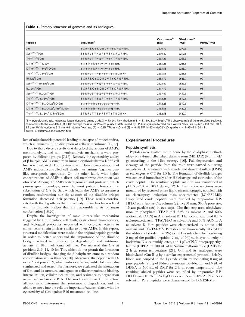
Figure 1. Substitution of some amino acid residues in the Gm structure reduces the cytotoxic ability, but did not modify the celldeath mechanism. B16 cells were stimulated by Gm and analogues for 24 h. (A) Cytotoxic activities of Gm and its analogues were quantified by theMTT reduction test. (B) Cell death type identification caused by Gm was evaluated using annexin-V and 7-AAD assay by flow cytometry using the IC50
values. (C-H) Apoptosis (Z-VAD) and necroptosis (necrostatin) inhibitors were unable to reduce cell death induced by Gm and analogues. Cells wereincubated with inhibitors for 1 h before to stimulation with the peptides (IC50 values for each peptide was used) and the viability was assessed by theMTT. Results are the means 6 SEM of three independent experiments preformed in duplicate.doi:10.1371/journal.pone.0080924.g001
Important Antitumor Properties of Gomesin
PLOS ONE | www.plosone.org 3 November 2013 | Volume 8 | Issue 11 | e80924
Cell viability assayB16 cells were incubated in 96-well microtiter plates in RPMI
medium supplemented with 10% FBS until reach the semi-
confluence and then treated with different concentrations of Gm
and its analogues for 24 h. To investigate the mechanisms of Gm-
induced cell death, B-16 cells were also pre-incubated with 40 mM
cytochalasin D (Tocris, USA); 20 mM necrostatin-1 (Tocris, USA),
10 mM Z-VAD (Tocris, USA), 5 mM MbCD (Tocris, USA), and
100 mM chloroquine (Tocris, USA) for 1 h. Cell viability was
determined using the standard reduction of the tetrazolium salt 3-
(4,5-dimethylthiazol-2-yl)-2,5-diphenyltetrazolium bromide
(MTT). The results were expressed relative to control cell viability
(100%).
Cell death by the annexin-V and 7-AAD assayB16 cells (56105 cells/ml) were treated with Gm and
subsequently harvested, washed with PBS and resuspended in
binding buffer (0.01 M HEPES, pH 7.4, 0.14 M NaCl and
2.5 mM CaCl2). The suspensions were labeled with annexin-V-
APC (An) and 5 mg/ml 7-Amino-actinomycin D (7-AAD) (BD
Biosciences, USA) according to the manufacturer’s instructions.
After incubation at room temperature for 20 min, cells were
analyzed in a FACSCalibur flow cytometer (Becton Dickinson,
USA) using CellQuest, and FlowJo 7.6 software. A total of 10,000
events were collected per sample.
Peptide quantification in membrane and internalizedinto cells by flow cytometry and confocal microscopy
Cells were treated with the 2 mM biotinylated peptide. Then,
the melanoma cells (56105 for flow cytometry or 56103 cell
seeded for confocal microscopy) were labeled with streptavidin-
Alexa 488 (Invitrogen, USA) for 30 min at 4uC and washed with
PBS. The cells were fixed with a solution of 2% paraformaldehyde
for 30 min, washed with 0.1 M glycin, and permeabilized with
0.01% saponin. Then, the cells were labeled with streptavidin-
Alexa 647 (Invitrogen, USA). For flow cytometry, the cells were
extracted using PBS with 5 mM EDTA previously to fixation. The
quantification of label of external biotynilated peptide, and
intracellular peptide were performed by flow cytometry or
confocal microscopy. For flow cytometry the cells were excited
using argon laser (488 nm), and diode laser (633 nm). The
emission was collected in FL-1 and FL-4 channel. A total of
10,000 events were collected per sample. For confocal microscopy,
nuclei were stained with DAPI. Confocal microscopy analyses
were performed with a confocal laser scanning microscope
equipped with a Plan-Apochromat 663 objective under oil
immersion (Zeiss, LSM780). The pinhole device was adjusted to
capture fluorescence of one airy unit. The images correspond to
single focal plane. Streptavidin-Alexa Fluor 488 were excited using
an argon laser (lEx. = 488 nm, lEm. = 505–550 nm),
streptavidin-Alexa Fluor 647 was excited using a HeNe laser
(lEx. = 633 nm, lEm. = 640–710 nm) and DAPI was excited
using a multiphoton laser (Coherent) (lEx. = 750 nm, lEm. =
380–460 nm). Unviable cells were excluded with propidium iodide
(PI) label.
Peptide quantificationThe cultured cells were resuspended using pH 5 Tris buffer, and
then sonicated. Protein concentration was measured using the RC
DC protein assay kit (Bio-Rad, CA, USA) according to the
manufacturer’s instructions. The peptides were incubated with this
protein solution for 1–14 h, and its stability was evaluated by
reversed-phase liquid chromatography coupled with an electro-
spray ionization mass spectrometer (LC/ESI-MS).
Statistical analysisAll data represent at least three independent experiments and
are expressed as the mean 6 standard error of the mean (SEM).
Statistical analyses were performed using Student t-test for
comparison between two groups and analysis of variance
(ANOVA) and Dunnett’s post hoc test for multiple comparisons
among groups. A probability (P) value greater than 0.05 was
considered significant.
Results
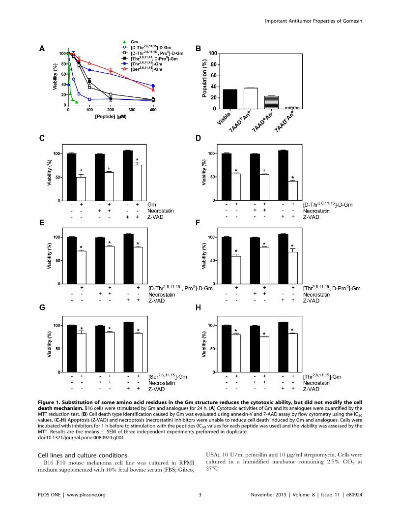
Comparison of cytotoxic activity of Gm and analoguesInitially the cytotoxic activity of Gm and its analogues were
tested in B16 mouse melanoma cell line after 24 h, and then
cytotoxicity was evaluated by MTT assay. Substitution of Cys2–15/
Cys6–11 for Thr, Gln9 for D- or L-Pro and the use D-amino acids
were tested ([D-Thr2,6,11,15]-D-Gm, [D-Thr2,6,11,15, Pro9]-D-Gm,
[Thr2,6,11,15, D-Pro9]-Gm, and [Thr2,6,11,15]-Gm), as well as Ser
substitution ([Ser2,6,11,15]-Gm) that causes a similar effect to Thr
substitution, a random conformation, and low antimicrobial
activity [6,16,20] (Table 1). The cytotoxic results obtained by
MTT assay indicated that the rank order of cytotoxic potency was:
Gm . [D-Thr2,6,11,15]-D-Gm .. [D-Thr2,6,11,15, Pro9]-D-Gm
= [Thr2,6,11,15, D-Pro9]-Gm .. [Thr2,6,11,15]-Gm =
[Ser2,6,11,15]-Gm (see Figure 1A). The results of potency and
efficacy of the peptides are summarized in Table 2. To identify the
type of cell death promoted by Gm and its analogues we used
annexin-V, which binds to phosphatidylserine that become
Table 2. Potencies and efficacies of gomesin and its analogues.
Peptide IC50 (mM)* Max. ExternalFluorescence (AU)
* Max. InternalFluorescence (AU)
# Peptide Degradation(%)
Gomesin 7 3068 603679 060
[D-Thr2,6,11,15]-D-Gm 25 4366 602612 1262
[D-Thr2,6,11,15, Pro9]-D-Gm 75 7164 27516190 1562
[Thr2,6,11,15, D-Pro9]-Gm 90 3866 1165627 1661
[Ser2,6,11,15]-Gm .200 3763 305665 4765
[Thr2,6,11,15]-Gm .200 17.56 7 7536175 4364
*To quantify internal or external amount fluorescence biotinylated peptides were used.# Starting peptide concentration (1024 M) was considered as 100%.doi:10.1371/journal.pone.0080924.t002
Important Antitumor Properties of Gomesin
PLOS ONE | www.plosone.org 4 November 2013 | Volume 8 | Issue 11 | e80924
exposed to extracellular cell membrane during apoptosis, and 7-
AAD, an membrane-impermeant nuclear stain. Gm promotes
primarily An+7-AAD+ label (Figure 1B) that correspond to
necrosis-like mechanism by membrane permeabilization observed
previously in B16 cells [6], and others tumor cell lines [12,17].
Similar results were obtained with Gm analogues (Figure S1). To
confirm this data we used inhibitor of necroptosis (necrostatin,
inhibitor of Rip-1), and apoptosis (Z-Vad, inhibitor of caspases).
Both inhibitors were unable to decrease cell death induced by Gm
and its analogues (see Figure 1C-H). Suggesting that modification
in Gm by substitution of amino acid did not change the mode of
cell death triggered by these peptides.
Potentialization of cytotoxic Gm effect by chloroquineAs necroptosis and apoptosis inhibitors were unable to block the
cell death promoted by Gm and analogues other inhibitors were
employed. The participation of free radicals in the cell death
induced by gomesin and its analogues were evaluated by the use of
N-Acetyl-Cysteine (NAC), a free radical scavenger, which was
unable to significantly inhibit cell death induced by the peptides
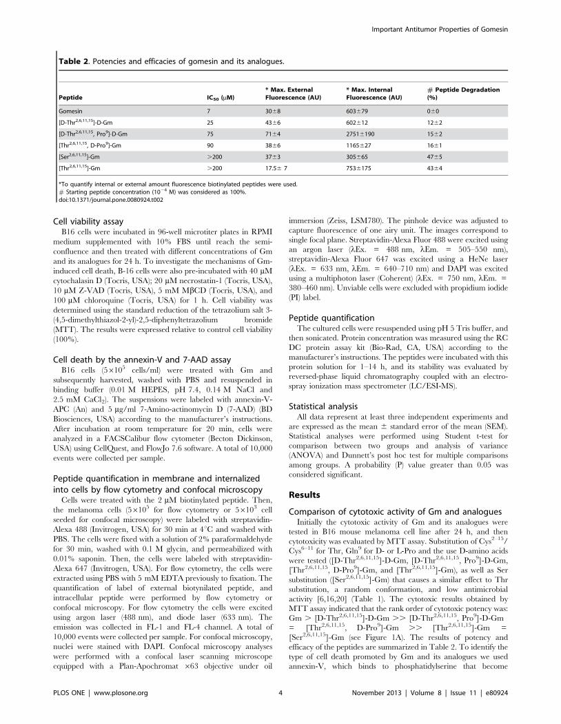
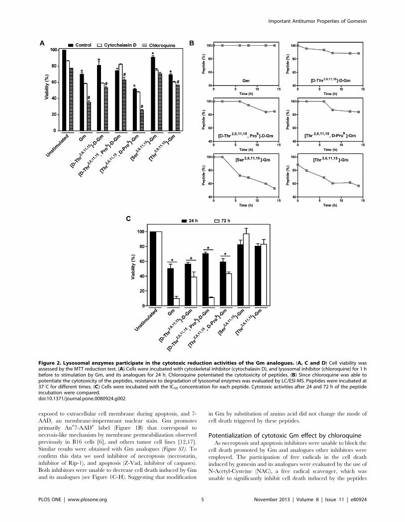
Figure 2. Lysosomal enzymes participate in the cytotoxic reduction activities of the Gm analogues. (A, C and D) Cell viability wasassessed by the MTT reduction test. (A) Cells were incubated with cytoskeletal inhibitor (cytochalasin D), and lysosomal inhibitor (chloroquine) for 1 hbefore to stimulation by Gm, and its analogues for 24 h. Chloroquine potentiated the cytotoxicity of peptides. (B) Since chloroquine was able topotentiate the cytotoxicity of the peptides, resistance to degradation of lysosomal enzymes was evaluated by LC/ESI-MS. Peptides were incubated at37uC for different times. (C) Cells were incubated with the IC50 concentration for each peptide. Cytotoxic activities after 24 and 72 h of the peptideincubation were compared.doi:10.1371/journal.pone.0080924.g002
Important Antitumor Properties of Gomesin
PLOS ONE | www.plosone.org 5 November 2013 | Volume 8 | Issue 11 | e80924

(Data not shown). The use of cytochalasin D, a potent disruptor of
actin filament function that blocks endocytosis mechanisms, were
ineffective (Figure 2A). However, chloroquine, which concentrates
in lysosomes, and raises their medium pH disrupting the function
of lysosomal enzymes, potentiate the effects of Gm and its
analogues, suggesting that the effects of peptides could be related
with degradation by lysosome enzymes. Therefore, the resistance
to lysosomal enzyme degradation of Gm and analogues were
quantified. We observed a direct correlation between resistance of
degradation, and cytotoxic activity. Gm shows great stability to
lysosomal enzymes, and was the most potent peptide, whereas
[Thr2,6,11,15]-Gm and [Ser2,6,11,15]-Gm that are the most sensitive
peptides to degradation exhibit lower activity (Figure 2B). As
expected [D-Thr2,6,11,15]-D-Gm, and [D-Thr2,6,11,15, Pro9]-D-Gm
were little affected by lysosomal enzymes, and unexpectedly
[Thr2,6,11,15, D-Pro9]-Gm was also stable to enzymatic degrada-
tion may be due a structural fold caused by introduction of the D-
Pro residue (Figure 2B). Since Gm and some analogues were little
degraded, we decide to incubate the cells with the peptides, and
observed cytotoxic effect after 72 h. The peptides incubation after
Figure 3. Cytotoxic effect of antimicrobial peptides is related to the entry of peptides but not with the binding to the cellmembrane. B16 cells were incubated with 2 mM of the biotin labeled peptides for different times. Quantification of the peptides were done by flowcytometry. External membrane peptides were labeled with streptavidin-Alexa Fluor 488 conjugated. Internal membrane peptides were labeled withstreptavidin-Alexa Fluor 647 conjugated after fixation, and permeabilization. Unviable cells were excluded using PI stain. External and internalquantifications of (A) Gm, (B) [D-Thr2,6,11,15]-D-Gm, (C) [D-Thr2,6,11,15, Pro9]-D-Gm, (D) [Thr2,6,11,15, D-Pro9]-Gm, (E) [Thr2,6,11,15]-Gm, and (F) [Ser2,6,11,15]-Gm are shown. (G-H) To decrease cholesterol of the membrane, the cells were incubated with MbCD for 1 h. (G) This treatment enhance the entry ofpeptides, (H) and potentiates the cytotoxic effect of most of them. Results are the means 6 SEM of three independent experiments preformed induplicate.doi:10.1371/journal.pone.0080924.g003
Important Antitumor Properties of Gomesin
PLOS ONE | www.plosone.org 6 November 2013 | Volume 8 | Issue 11 | e80924
72 h caused an increase in the cytotoxicity of the ones that are
more resistant to degradation, whereas the activity of the less
resistant peptides ([Ser2,6,11,15]-Gm an [Thr2,6,11,15]-Gm) was not
alter (Figure 2C).
Quantification of binding to cell membrane andinternalization of Gm and its analogues
We quantified the Gm and analogues binding to the cellular
membrane, and their internalization using their biotinylated
counterpart. The cells were incubated at different times using a
low concentration of the peptides (2 mM) that does not induces
membrane permeabilization. Unviable cells were excluded by PI
label (Figure S2). External peptides labeled with streptavidin Alexa
Fluor 488 were identified for the quantification of the green
fluorescence (FL1 channel), and internal peptides labeled with
streptavidin Alexa Fluor 647 were identified for the quantification
of the deep red fluorescence (FL4 channel), after permeabilization
of cellular membrane, by Flow cytometry. Quantification of
external and internal label showed a rapid binding of the peptides
with a peak at 2–4 h for all peptides (Figure 3A-F). [Thr2,6,11,15, D-
Pro9]-Gm exhibits the highest binding among the peptides tested.
Gm and [D-Thr2,6,11,15]-D-Gm, the most cytotoxic peptides,
presents a medium value of binding. [Thr2,6,11,15]-Gm and
[Ser2,6,11,15]-Gm, which possess the lower cytotoxic activity, were
the peptides that less binding to cellular membrane (Figure 3A-F).
The temporal quantification of internalized peptides shows that
peptides follow a similar temporal behavior. It is possible to
observe the internal presence of Gm, [D-Thr2,6,11,15]-D-Gm, [D-
Thr2,6,11,15, Pro9]-D-Gm, and [Thr2,6,11,15, D-Pro9]-Gm after 24
h. On the other hand, the peptides that present lower cytotoxicity
were not retained after 24 h (Figure 3A-F). However, a direct
relation between binding, peptide entrance, and cytotoxicity effect
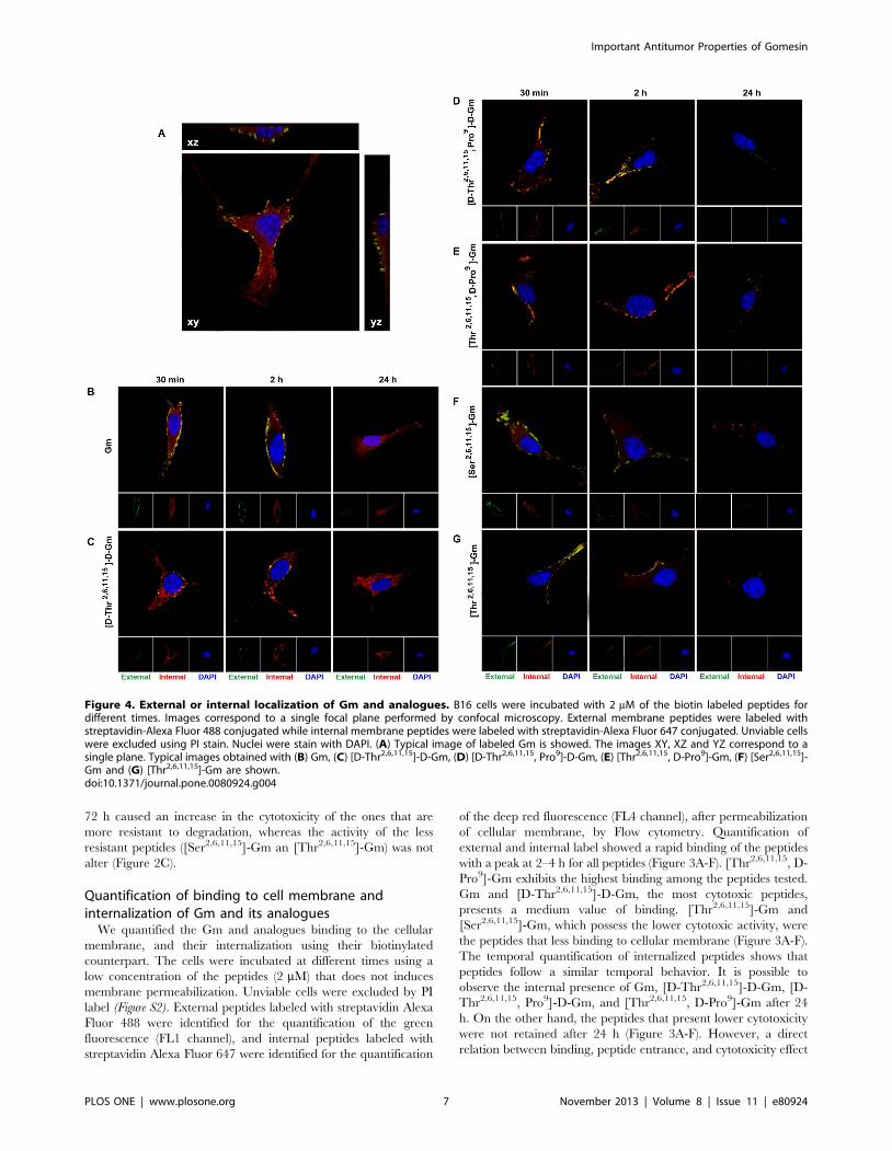
Figure 4. External or internal localization of Gm and analogues. B16 cells were incubated with 2 mM of the biotin labeled peptides fordifferent times. Images correspond to a single focal plane performed by confocal microscopy. External membrane peptides were labeled withstreptavidin-Alexa Fluor 488 conjugated while internal membrane peptides were labeled with streptavidin-Alexa Fluor 647 conjugated. Unviable cellswere excluded using PI stain. Nuclei were stain with DAPI. (A) Typical image of labeled Gm is showed. The images XY, XZ and YZ correspond to asingle plane. Typical images obtained with (B) Gm, (C) [D-Thr2,6,11,15]-D-Gm, (D) [D-Thr2,6,11,15, Pro9]-D-Gm, (E) [Thr2,6,11,15, D-Pro9]-Gm, (F) [Ser2,6,11,15]-Gm and (G) [Thr2,6,11,15]-Gm are shown.doi:10.1371/journal.pone.0080924.g004
Important Antitumor Properties of Gomesin
PLOS ONE | www.plosone.org 7 November 2013 | Volume 8 | Issue 11 | e80924
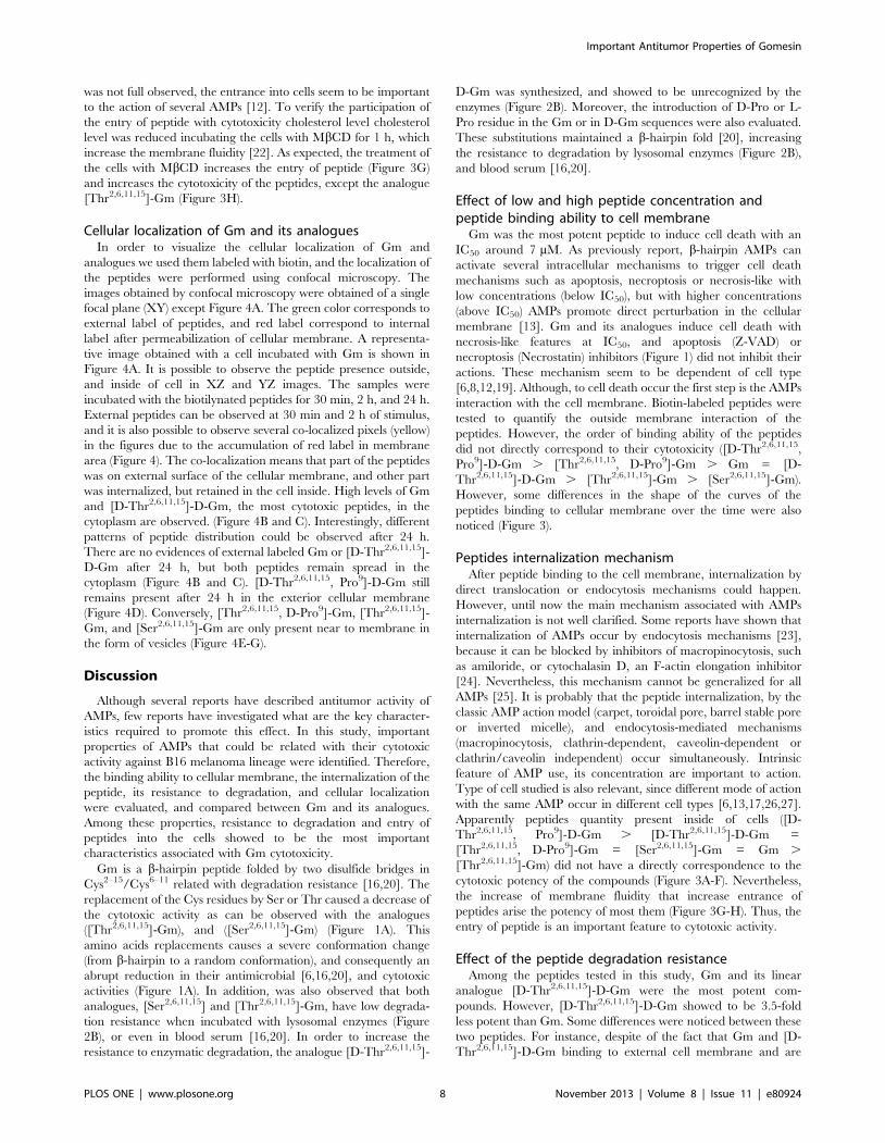
was not full observed, the entrance into cells seem to be important
to the action of several AMPs [12]. To verify the participation of
the entry of peptide with cytotoxicity cholesterol level cholesterol
level was reduced incubating the cells with MbCD for 1 h, which
increase the membrane fluidity [22]. As expected, the treatment of
the cells with MbCD increases the entry of peptide (Figure 3G)
and increases the cytotoxicity of the peptides, except the analogue
[Thr2,6,11,15]-Gm (Figure 3H).
Cellular localization of Gm and its analoguesIn order to visualize the cellular localization of Gm and
analogues we used them labeled with biotin, and the localization of
the peptides were performed using confocal microscopy. The
images obtained by confocal microscopy were obtained of a single
focal plane (XY) except Figure 4A. The green color corresponds to
external label of peptides, and red label correspond to internal
label after permeabilization of cellular membrane. A representa-
tive image obtained with a cell incubated with Gm is shown in
Figure 4A. It is possible to observe the peptide presence outside,
and inside of cell in XZ and YZ images. The samples were
incubated with the biotilynated peptides for 30 min, 2 h, and 24 h.
External peptides can be observed at 30 min and 2 h of stimulus,
and it is also possible to observe several co-localized pixels (yellow)
in the figures due to the accumulation of red label in membrane
area (Figure 4). The co-localization means that part of the peptides
was on external surface of the cellular membrane, and other part
was internalized, but retained in the cell inside. High levels of Gm
and [D-Thr2,6,11,15]-D-Gm, the most cytotoxic peptides, in the
cytoplasm are observed. (Figure 4B and C). Interestingly, different
patterns of peptide distribution could be observed after 24 h.
There are no evidences of external labeled Gm or [D-Thr2,6,11,15]-
D-Gm after 24 h, but both peptides remain spread in the
cytoplasm (Figure 4B and C). [D-Thr2,6,11,15, Pro9]-D-Gm still
remains present after 24 h in the exterior cellular membrane
(Figure 4D). Conversely, [Thr2,6,11,15, D-Pro9]-Gm, [Thr2,6,11,15]-
Gm, and [Ser2,6,11,15]-Gm are only present near to membrane in
the form of vesicles (Figure 4E-G).
Discussion
Although several reports have described antitumor activity of
AMPs, few reports have investigated what are the key character-
istics required to promote this effect. In this study, important
properties of AMPs that could be related with their cytotoxic
activity against B16 melanoma lineage were identified. Therefore,
the binding ability to cellular membrane, the internalization of the
peptide, its resistance to degradation, and cellular localization
were evaluated, and compared between Gm and its analogues.
Among these properties, resistance to degradation and entry of
peptides into the cells showed to be the most important
characteristics associated with Gm cytotoxicity.
Gm is a b-hairpin peptide folded by two disulfide bridges in
Cys2–15/Cys6–11 related with degradation resistance [16,20]. The
replacement of the Cys residues by Ser or Thr caused a decrease of
the cytotoxic activity as can be observed with the analogues
([Thr2,6,11,15]-Gm), and ([Ser2,6,11,15]-Gm) (Figure 1A). This
amino acids replacements causes a severe conformation change
(from b-hairpin to a random conformation), and consequently an
abrupt reduction in their antimicrobial [6,16,20], and cytotoxic
activities (Figure 1A). In addition, was also observed that both
analogues, [Ser2,6,11,15] and [Thr2,6,11,15]-Gm, have low degrada-
tion resistance when incubated with lysosomal enzymes (Figure
2B), or even in blood serum [16,20]. In order to increase the
resistance to enzymatic degradation, the analogue [D-Thr2,6,11,15]-
D-Gm was synthesized, and showed to be unrecognized by the
enzymes (Figure 2B). Moreover, the introduction of D-Pro or L-
Pro residue in the Gm or in D-Gm sequences were also evaluated.
These substitutions maintained a b-hairpin fold [20], increasing
the resistance to degradation by lysosomal enzymes (Figure 2B),
and blood serum [16,20].
Effect of low and high peptide concentration andpeptide binding ability to cell membrane
Gm was the most potent peptide to induce cell death with an
IC50 around 7 mM. As previously report, b-hairpin AMPs can
activate several intracellular mechanisms to trigger cell death
mechanisms such as apoptosis, necroptosis or necrosis-like with
low concentrations (below IC50), but with higher concentrations
(above IC50) AMPs promote direct perturbation in the cellular
membrane [13]. Gm and its analogues induce cell death with
necrosis-like features at IC50, and apoptosis (Z-VAD) or
necroptosis (Necrostatin) inhibitors (Figure 1) did not inhibit their
actions. These mechanism seem to be dependent of cell type
[6,8,12,19]. Although, to cell death occur the first step is the AMPs
interaction with the cell membrane. Biotin-labeled peptides were
tested to quantify the outside membrane interaction of the
peptides. However, the order of binding ability of the peptides
did not directly correspond to their cytotoxicity ([D-Thr2,6,11,15,
Pro9]-D-Gm . [Thr2,6,11,15, D-Pro9]-Gm . Gm = [D-
Thr2,6,11,15]-D-Gm . [Thr2,6,11,15]-Gm . [Ser2,6,11,15]-Gm).
However, some differences in the shape of the curves of the
peptides binding to cellular membrane over the time were also
noticed (Figure 3).
Peptides internalization mechanismAfter peptide binding to the cell membrane, internalization by
direct translocation or endocytosis mechanisms could happen.
However, until now the main mechanism associated with AMPs
internalization is not well clarified. Some reports have shown that
internalization of AMPs occur by endocytosis mechanisms [23],
because it can be blocked by inhibitors of macropinocytosis, such
as amiloride, or cytochalasin D, an F-actin elongation inhibitor
[24]. Nevertheless, this mechanism cannot be generalized for all
AMPs [25]. It is probably that the peptide internalization, by the
classic AMP action model (carpet, toroidal pore, barrel stable pore
or inverted micelle), and endocytosis-mediated mechanisms
(macropinocytosis, clathrin-dependent, caveolin-dependent or
clathrin/caveolin independent) occur simultaneously. Intrinsic
feature of AMP use, its concentration are important to action.
Type of cell studied is also relevant, since different mode of action
with the same AMP occur in different cell types [6,13,17,26,27].
Apparently peptides quantity present inside of cells ([D-
Thr2,6,11,15, Pro9]-D-Gm . [D-Thr2,6,11,15]-D-Gm =
[Thr2,6,11,15, D-Pro9]-Gm = [Ser2,6,11,15]-Gm = Gm .
[Thr2,6,11,15]-Gm) did not have a directly correspondence to the
cytotoxic potency of the compounds (Figure 3A-F). Nevertheless,
the increase of membrane fluidity that increase entrance of
peptides arise the potency of most them (Figure 3G-H). Thus, the
entry of peptide is an important feature to cytotoxic activity.
Effect of the peptide degradation resistanceAmong the peptides tested in this study, Gm and its linear
analogue [D-Thr2,6,11,15]-D-Gm were the most potent com-
pounds. However, [D-Thr2,6,11,15]-D-Gm showed to be 3.5-fold
less potent than Gm. Some differences were noticed between these
two peptides. For instance, despite of the fact that Gm and [D-
Thr2,6,11,15]-D-Gm binding to external cell membrane and are
Important Antitumor Properties of Gomesin
PLOS ONE | www.plosone.org 8 November 2013 | Volume 8 | Issue 11 | e80924

present at similar amount into the cell, faster decrease of
intracellular amount can be observed for [D-Thr2,6,11,15]-D-Gm
(Figure 4A and B). Rapid reduction of the peptide amount into the
cell could occur by degradation or extrusion of the peptide from
the cell. Lysosomal enzyme inhibition with chloroquine potentiates
the response of all peptides utilized in this study, suggesting the
participation of lysosomal enzymes in degradation of the
internalized peptides. Lysosomal enzymes reduce poorly the effect
of [D-Thr2,6,11,15]-D-Gm (Figure 2B), but this could be related to
the decrease potency of this compound. Interestingly, both
peptides, Gm and [D-Thr2,6,11,15]-D-Gm, are also rapid translo-
cated into the cytoplasm of B16 cells, and are retained spread in
intracellular content after 24 h (Figure 4A and C). However,
further investigations are needed to identify the enzymes involved
in the AMP degradation, which might be useful to generate new
resident compounds.
Different internal distributions of Gm and analogues were
observed after internalization of peptides that can be related with
enzyme resistance ability (Figure 4). After 30 min, and 2 h of
incubation all peptides, with the exception of [Ser2,6,11,15]-Gm,
exhibit a great co-localization in the outer, and inner sides of the
cell membrane. The co-localization of the external and internal
labeled peptide means that certain amounts of molecules are
present on the cell surface, and the other is present inside the cell.
This is due to the resolution limit of the confocal microscopy
(,200 nm) and the thickness of cellular membrane (,20 nm)
[28]. Furthermore, the AMPs may have different ways to cross the
cell membrane, and also present different release capabilities into
the cytoplasm, these two phenomenas could be very important
properties, and could be related with the spatial conformation
when the AMPs are crossing the lipid bilayer. Although, only Gm
and [D-Thr2,6,11,15]-D-Gm are present in high amount in the
cytoplasmatic medium (Figure 4B and C). Interestingly, after 24 h
Gm and [D-Thr2,6,11,15]-D-Gm still remain spread in the
cytoplasm, while the other peptides apparently are inside of
vesicles near to the membrane or inserted in the cellular
membrane (Figure 4B-G). The former observation is in agreement
with several reports of the literature that described that the
penetration, and distribution of the AMP did not happen in the
same way; in fact some AMPs are present in the cytoplasm, and
others are in vesicles depending on the peptide involved
[24,25,29]. Apparently, Gm and [D-Thr2,6,11,15]-D-Gm are
resistant to cell extrusion mechanism, while the other peptides
studied are not, probably associated with great enzyme resistance
feature. This also can explain the potentiation of cell death after 72
h of the peptides with most cytotoxic activity.
Conclusions
Summarize, we showed the importance of the resistance to
degradation in the cytotoxic activity of Gm and its analogues
probably associated with intracellular distribution, and extrusion.
In addition, we also noticed that the entry capability is also related
with the resistance to degradation and peptide conformation.
Supporting Information
Figure S1 Cell death type identification caused by Gmand its analogues was evaluated using annexin-V and 7-AAD assay by flow cytometry using the IC50 values.Results are the means 6 SEM of three independent experiments
preformed in duplicate.
(TIF)
Figure S2 Secondary control sample. (A) Control cellslabeled with streptavidin-Alexa Fluor 488 and streptavi-din-Alexa Fluor 647 in absence of biotynilated peptides.(B) Unviable cells were excluded using PI stain. A
representative cell is showed. Nuclei were stain with DAPI.
Confocal microscopy images were performed in a LSM780 (Zeiss)
system.
(TIF)
Author Contributions
Conceived and designed the experiments: MVB TMD EJPG RLCR EGR
AM. Performed the experiments: MVB TMD EJPG RLCR EGR AM.
Analyzed the data: MVB TMD EJPG RLCR EGR AM. Contributed
reagents/materials/analysis tools: MVB EJPG RLCR EGR AM. Wrote
the paper: MVB TMD EJPG RLCR EGR AM.
References
1. Yan JX, Wang KR, Chen R, Song JJ, Zhang BZ, et al. (2012) Membrane active
antitumor activity of NK-18, a mammalian NK-lysin-derived cationic
antimicrobial peptide. Biochimie 94: 184–191.
2. Zasloff M (2002) Antimicrobial peptides of multicellular organisms. Nature 415:
389–395.
3. Silva PI Jr, Daffre S, Bulet P (2000) Isolation and characterization of gomesin, an
18-residue cysteine-rich defense peptide from the spider Acanthoscurria
gomesiana hemocytes with sequence similarities to horseshoe crab antimicrobial
peptides of the tachyplesin family. J Biol Chem 275: 33464–33470.
4. Rodrigues EG, Dobroff AS, Taborda CP, Travassos LR (2009) Antifungal and
antitumor models of bioactive protective peptides. An Acad Bras Cienc 81: 503–
520.
5. Calgarotto AK, da Silva Pereira GJ, Bechara A, Paredes-Gamero EJ, Barbosa
CM, et al. (2012) Autophagy inhibited Ehrlich ascitic tumor cells apoptosis
induced by the nitrostyrene derivative compounds: relationship with cytosolic
calcium mobilization. Eur J Pharmacol 678: 6–14.
6. Rodrigues EG, Dobroff AS, Cavarsan CF, Paschoalin T, Nimrichter L, et al.
(2008) Effective topical treatment of subcutaneous murine B16F10-Nex2
melanoma by the antimicrobial peptide gomesin. Neoplasia 10: 61–68.
7. Giuliani A, Pirri G, Nicoletto SF (2007) Antimicrobial peptides: an overview of a
promising class of therapeutics. Cent Eur J Biol 2: 1–33.
8. Paredes-Gamero EJ, Nogueira-Pedro A, Miranda A, Justo GZ (2013)
Hematopoietic modulators as potential agents for the treatment of leukemia.
Front Biosci (Elite Ed) 5: 130–140.
9. Huang TC, Lee JF, Chen JY (2011) Pardaxin, an antimicrobial peptide, triggers
caspase-dependent and ROS-mediated apoptosis in HT-1080 cells. Mar Drugs
9: 1995–2009.
10. Raz A, Bucana C, McLellan W, Fidler IJ (1980) Distribution of membrane
anionic sites on B16 melanoma variants with differing lung colonising potential.
Nature 284: 363–364.
11. Slaninova J, Mlsova V, Kroupova H, Alan L, Tumova T, et al. (2012) Toxicity
study of antimicrobial peptides from wild bee venom and their analogs toward
mammalian normal and cancer cells. Peptides 33: 18–26.
12. Paredes-Gamero EJ, Casaes-Rodrigues RL, Moura GE, Domingues TM, Buri
MV, et al. (2012) Cell-permeable gomesin peptide promotes cell death by
intracellular Ca(2+) overload. Mol Pharm 9: 2686–2697.
13. Jarczak J, Kosciuczuk EM, Lisowski P, Strzalkowska N, Jozwik A, et al. (2013)
Defensins: natural component of human innate immunity. Hum Immunol.
14. Mandard N, Sodano P, Labbe H, Bonmatin JM, Bulet P, et al. (1998) Solution
structure of thanatin, a potent bactericidal and fungicidal insect peptide,
determined from proton two-dimensional nuclear magnetic resonance data. Eur
J Biochem 256: 404–410.
15. Sacramento RS, Martins RM, Miranda A, Dobroff AS, Daffre S, et al. (2009)
Differential effects of alpha-helical and beta-hairpin antimicrobial peptides
against Acanthamoeba castellanii. Parasitology 136: 813–821.
16. Fazio MA, Oliveira VX Jr, Bulet P, Miranda MT, Daffre S, et al. (2006)
Structure-activity relationship studies of gomesin: importance of the disulfide
bridges for conformation, bioactivities, and serum stability. Biopolymers 84:
205–218.
17. Soletti RC, del Barrio L, Daffre S, Miranda A, Borges HL, et al. (2010) Peptide
gomesin triggers cell death through L-type channel calcium influx, MAPK/
ERK, PKC and PI3K signaling and generation of reactive oxygen species.
Chem Biol Interact 186: 135–143.
18. Hoskin DW, Ramamoorthy A (2008) Studies on anticancer activities of
antimicrobial peptides. Biochim Biophys Acta 1778: 357–375.
Important Antitumor Properties of Gomesin
PLOS ONE | www.plosone.org 9 November 2013 | Volume 8 | Issue 11 | e80924

19. Paredes-Gamero EJ, Martins MN, Cappabianco FA, Ide JS, Miranda A (2012)
Characterization of dual effects induced by antimicrobial peptides: regulated cell
death or membrane disruption. Biochim Biophys Acta 1820: 1062–1072.
20. Moraes LGM, Fazio MA, Vieira RFF, Nakaie CR, Miranda MTM, et al. (2007)
Conformational and functional studies of gomesin analogues by CD, EPR and
fluorescence spectroscopies. Biochimica Et Biophysica Acta-Biomembranes
1768: 52–58.
21. Domingues TM, Riske KA, Miranda A (2010) Revealing the Lytic Mechanism
of the Antimicrobial Peptide Gomesin by Observing Giant Unilamellar Vesicles.
Langmuir 26: 11077–11084.
22. Gimpl G, Burger K, Fahrenholz F (1997) Cholesterol as modulator of receptor
function. Biochemistry 36: 10959–10974.
23. Sandgren S, Wittrup A, Cheng F, Jonsson M, Eklund E, et al. (2004) The human
antimicrobial peptide LL-37 transfers extracellular DNA plasmid to the nuclear
compartment of mammalian cells via lipid rafts and proteoglycan-dependent
endocytosis. J Biol Chem 279: 17951–17956.
24. Wadia JS, Stan RV, Dowdy SF (2004) Transducible TAT-HA fusogenic peptide
enhances escape of TAT-fusion proteins after lipid raft macropinocytosis. NatMed 10: 310–315.
25. Drin G, Cottin S, Blanc E, Rees AR, Temsamani J (2003) Studies on the
internalization mechanism of cationic cell-penetrating peptides. J Biol Chem278: 31192–31201.
26. Chen J, Xu XM, Underhill CB, Yang S, Wang L, et al. (2005) Tachyplesinactivates the classic complement pathway to kill tumor cells. Cancer Res 65:
4614–4622.
27. Chen Y, Xu X, Hong S, Chen J, Liu N, et al. (2001) RGD-Tachyplesin inhibitstumor growth. Cancer Res 61: 2434–2438.
28. Rigaut JP, Vassy J (1991) High-resolution three-dimensional images fromconfocal scanning laser microscopy. Quantitative study and mathematical
correction of the effects from bleaching and fluorescence attenuation in depth.Anal Quant Cytol Histol 13: 223–232.
29. Takeshima K, Chikushi A, Lee KK, Yonehara S, Matsuzaki K (2003)
Translocation of analogues of the antimicrobial peptides magainin and buforinacross human cell membranes. J Biol Chem 278: 1310–1315.
Important Antitumor Properties of Gomesin
PLOS ONE | www.plosone.org 10 November 2013 | Volume 8 | Issue 11 | e80924