5-Aminolevulinic Acid Derivatives in Photomedicine: Characteristics, Application and Perspectives
166
Thesis Reference Optimization of 5-aminolevulinic acid derivatives-mediated photomedicine: new strategies, models, and applications FOTINOS, Nicolas Abstract Dans le domaine de la photo-médecine, la formation in situ de porphyrines photosensibles après administration d'acide 5-aminolevulinique (5-ALA) peut être considérée comme une approche prometteuse et novatrice, présentant de nombreux avantages par rapport à l'utilisation des photosensibilisants conventionnels. Néanmoins, la faible biodisponibilité du 5-ALA a nécessité le développement de molécules dérivées plus lipophiles, dont deux ont obtenu récemment une autorisation sur le marché. Dans le cadre de ce travail, de nouveaux dérivés du 5-ALA ont été synthétisés avec pour objectif une stabilité améliorée, une biodisponibilité accrue ou encore des caractéristiques adaptées à une administration systémique. De plus, un nouveau modèle in vivo, basé sur l'embryon de poulet, a été développé et a permis notamment l'évaluation de molécules faiblement hydrosolubles. Finalement, l'utilisation des dérivés du 5-ALA a été étudié dans le cadre de la photo-inactivation de bactéries et plus spécialement de la souche "Propionibacterium acnes". FOTINOS, Nicolas. Optimization of 5-aminolevulinic acid derivatives-mediated photomedicine: new strategies, models, and applications. Thèse de doctorat : Univ. Genève, 2007, no. Sc. 3907 Available at: http://archive-ouverte.unige.ch/unige:83 Disclaimer: layout of this document may differ from the published version. [ Downloaded 31/12/2011 at 00:47:49 ] 1 / 1
Transcript of 5-Aminolevulinic Acid Derivatives in Photomedicine: Characteristics, Application and Perspectives
Thesis
Reference
Optimization of 5-aminolevulinic acid derivatives-mediated
photomedicine: new strategies, models, and applications
FOTINOS, Nicolas
Abstract
Dans le domaine de la photo-médecine, la formation in situ de porphyrines photosensibles
après administration d'acide 5-aminolevulinique (5-ALA) peut être considérée comme une
approche prometteuse et novatrice, présentant de nombreux avantages par rapport à
l'utilisation des photosensibilisants conventionnels. Néanmoins, la faible biodisponibilité du
5-ALA a nécessité le développement de molécules dérivées plus lipophiles, dont deux ont
obtenu récemment une autorisation sur le marché. Dans le cadre de ce travail, de nouveaux
dérivés du 5-ALA ont été synthétisés avec pour objectif une stabilité améliorée, une
biodisponibilité accrue ou encore des caractéristiques adaptées à une administration
systémique. De plus, un nouveau modèle in vivo, basé sur l'embryon de poulet, a été
développé et a permis notamment l'évaluation de molécules faiblement hydrosolubles.
Finalement, l'utilisation des dérivés du 5-ALA a été étudié dans le cadre de la
photo-inactivation de bactéries et plus spécialement de la souche "Propionibacterium acnes".
FOTINOS, Nicolas. Optimization of 5-aminolevulinic acid derivatives-mediated
photomedicine: new strategies, models, and applications. Thèse de doctorat : Univ.
Genève, 2007, no. Sc. 3907
Available at:
http://archive-ouverte.unige.ch/unige:83
Disclaimer: layout of this document may differ from the published version.
[ Downloaded 31/12/2011 at 00:47:49 ]
1 / 1
UNIVERSITÉ DE GENÈVE FACULTÉ DES SCIENCES
Section des sciences pharmaceutiques Professeur Robert GURNY
Laboratoire de pharmacie galénique Docteur Norbert LANGE et de biopharmacie
Optimization of 5-aminolevulinic acid derivatives-m ediated
photomedicine: new strategies, models, and applicat ions
THÈSE
présentée à la faculté des sciences de l’Université de Genève pour obtenir le grade de Docteur ès sciences, mention sciences pharmaceutiques
par
Nicolas FOTINOS
de
Chavannes-près-Renens (VD)
Thèse N° Sc. 3907
GENÈVE Atelier de reproduction de la Section de physique
2008

(IMPRIMATUR)
Remerciements
Je tiens à exprimer mes sincères remerciements au Professeur Robert Gurny, pour
m’avoir accueilli dans son laboratoire de pharmacie galénique à l’Université de Genève, et
de m’avoir permis de disposer de conditions de travail idéales.
Je témoigne ma gratitude au membres du jury qui ont aimablement accepté de lire,
évaluer et critiquer mon travail de thèse : Le docteur Marie-Ange d’Hallewin du Centre Alexis
Vautrin à Nancy, le docteur Angelika Rueck de l’Institut des technologies laser médicales à
Ulm, le Professeur Pavel Kucera de l’Université de Lausanne, ainsi que le Professeur Jean-
Claude Piffaretti, ce dernier m’ayant également accueilli dans son laboratoire à l’institut
Cantonal de Microbiologie à Bellinzone pour une fructueuse collaboration.
J’adresse mes plus vifs remerciements au Docteur Norbert Lange, mon directeur de
thèse, pour son soutient indéfectible, son enthousiasme contagieux, et sa volonté sans faille
d’aller de l’avant. Ce fut un plaisir et un honneur de travailler dans se groupe PDT, grâce à la
grande solidarité et à la confiance qu’il a su y instaurer. Je le remercie également pour son
humour caustique, sa grande générosité, et ses fabuleuses grillades qui vont beaucoup me
manquer.
Je souhaite exprimer ma sincère gratitude à mes très chers collègues du groupe PDT
avec qui j’ai vécu tant de bons moments. Tout d’abord Sabine Collaud, qui fut une collègue
et amie exemplaire dès les premiers instants. Florence Popowycz pour sa courte période à
Lausanne riche en discussions culinaires, Marino Campo pour sa passionnante curiosité
scientifique mais surtout pour cette grande complicité qui a fait trembler le labo 482. Je
n’oublie pas ma chère Doris Gabriel qui a gagné mon plus profond respect malgré ses
remarques désobligeantes sur le chaos régnant sur mon bureau, Maria Fernanda Zuluaga
pour sa gentillesse et son sourire indispensables à la bonne marche du labo, et Magali
Zeisser-Labouèbe pour les discussions parentales enrichissantes. Je souhaite bonne chance
et beaucoup de plaisir à Gesine Heuck qui passera ses prochaines années dans ce fabuleux
monde la thérapie photodynamique.
Un grand merci également à tous les collègues du BEP, de Sciences II et d’ailleurs,
qu’il serait trop long d’énumérer mais qui se reconnaîtront certainement, et sans qui cette
aventure n’aurait pas été aussi enrichissante, tant scientifiquement qu’humainement. Une
petite pensée à mes compagnons de route, et spécialement Bruno Bard, qui ont transformé
ces interminables trajets de train en riches et passionnants forums de discussion.

Finalement, je voudrais remercier mes amis si importants à mon équilibre, ma grand-
mère pour ses fabuleux gâteux si réputés au laboratoire, et surtout mon frère et mes parents,
qui ont toujours été présents quand j’en avais besoin, et à qui j’adresse toute ma tendresse.
Mes derniers mots pour dire mon éternelle reconnaissance à ma merveilleuse épouse qui
m’a donné confiance, courage et une merveilleuse petite famille.
- i -
TABLE OF CONTENTS
Abbreviations ii
Chapter I 1
Introduction
Chapter II: 17
5-aminolevulinic acid derivatives in photomedicine: characteristics, application and
perspectives
Chapter III 75
On the metabolism of 5-aminolevulinic acid derivatives
Chapter IV 91
The chick embryo model for the evaluation of 5-aminolevulinic acid derivatives
Chapter V: 107
5-aminolevulinic acid and 5-aminolevulinic acid derivatives-mediated effects on gram-
negative and gram-positive bacteria
Chapter VI 125
5-ALA derivatives-mediated photoinactivation of Propionibacterium acnes
Chapter VII 139
Conclusions
Chapter VIII 143
Résumé (français)

Abbreviations
- ii -
ABBREVIATIONS
5-ALA 5-aminolevulinic acid
5-ALA DGME 5-aminolevulinic diethylenglycol monomethylether ester
x-CP x-carboxyporphyrin (x = 8, 7, 6, 5, 4, 2)
ADAG 2-(5-ALA)-1,3-diacetylglyceride
ADBG 2-(5-ALA)-1,3-dibutyrylglyceride
AK actinic keratosis, solar keratosis
AOMM 3-amino-3-oxohenadioic-1-methyl-6-methylester
BAL 5-aminolevulinic acid butylester
BBB blood brain barrier
BzAL 5-aminolevulinic acid benzylester
CAM chorioallantoic membrane
CFU colony forming unit
CIN cervical intraepithelial neoplasia
CIS carcinoma in situ
CNS central nervous system
CP I & III coproporphyrin I & III (structural isomers)
CR complete response
DGME diethylenglycol monomethylether
DMSO dimethylsulfoxide
EDD embryo development day
EDTA ethylenediamine tetraacetic acid
EPR enhanced permeation and retention
FD fluorescence diagnosis
GABA γ-aminobutyric acid
HAL 5-aminolevulinic acid hexylester
Abbreviations
- iii -
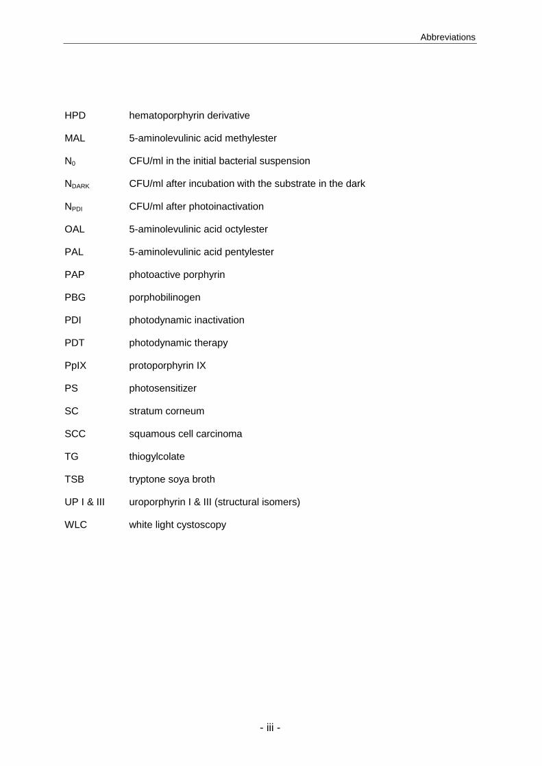
HPD hematoporphyrin derivative
MAL 5-aminolevulinic acid methylester
N0 CFU/ml in the initial bacterial suspension
NDARK CFU/ml after incubation with the substrate in the dark
NPDI CFU/ml after photoinactivation
OAL 5-aminolevulinic acid octylester
PAL 5-aminolevulinic acid pentylester
PAP photoactive porphyrin
PBG porphobilinogen
PDI photodynamic inactivation
PDT photodynamic therapy
PpIX protoporphyrin IX
PS photosensitizer
SC stratum corneum
SCC squamous cell carcinoma
TG thiogylcolate
TSB tryptone soya broth
UP I & III uroporphyrin I & III (structural isomers)
WLC white light cystoscopy

- iv -
Introduction
- 1 -
CHAPTER I
Introduction
Photodynamic Therapy and Fluorescence Diagnosis
In today’s occidental society, medical cares have achieved a very high level of
efficacy. Therefore, most of the formerly lethal diseases can now be treated successfully.
However, effective treatments against the two major causes of death in the western world,
i.e. cardiovascular diseases and cancer, are still missing. The treatment of tumors is in fact
considered as one of the most challenging medical fields, with respect to the difficulty to
target selectively cancer cells. Many drugs or treatment procedures on the market display the
capacity to destroy diseased cells, but often lack selectivity leading to marked collateral
damages of healthy tissues. Different strategies have been developed to deliver the anti-
cancer agents directly and selectively to the tumor, including the coupling to tumor-specific
antibodies, the use of an adapted carrier systems or the targeting of proteases expressed
abundantly in tumor environment 1,2. Since a few decades, one therapeutical methodology
received increasing attention due to its outstanding selectivity: Photodynamic Therapy (PDT).
PDT was discovered in Germany, in the beginning of the 20th century. A PhD student,
O. Raab, and his supervisor H. von Tappeiner studied the lethal effect of some dyes on
paramecium. They discovered that light was strongly influencing the survival of the parasites
incubated with acridine. A few years later, von Tappeiner observed that oxygen was also
involved in this phenomenon and proposed the term “dynamic” to precise the contribution of
oxygen, and to differentiate it from phototherapy 3.
PDT can be defined as followed. It is the combination of three individually non-toxic
components, (i) a photosensitizing agent, termed photosensitizer (PS), (ii) light that activates
the PS, and finally, (iii) oxygen that upon excitation by the activated PS will be transformed
into highly toxic reactive oxygen species (ROS) 4. The selectivity of PDT relies on a
contribution of each of these parameters (Fig. 1). Firstly, the PS accumulates selectively into
the target tissues due to physiological alterations in the pathological environment, like e.g.
leaky vasculature, abnormal enzymatic activity, pH variations, or reduced lymphatic
drainage. Secondly, the local irradiation of the diseased area, and finally, the very short

Chapter I
- 2 -
lifetime of the reactive oxygen species limits the damage to the target tissues by restraining a
migration to the healthy surrounding tissues 5.
PS
A) B)
NH N
HNN
PP
NH N
HNN
PP
Selective accumulation of the photosensitizer (PS) into the targettissues.
Energy transfer from the activated PS to oxygen results in the local production of toxic reactive oxygen species (ROS).
Selective destruction of target tissues
O2
ROS
C)Activation of thePS by light of theappropriatewavelength.
Irradiation islocalized to thearea to be treated.
PS
PS
PSPS PS
PS
PS
PS
PS
PS PS
Figure 1: Principle of photodynamic therapy. The cumulative effect of a photosensitizer
accumulated in the target tissues (A), a localized irradiation (B), and the
formation of an effective, confined, toxic agent (C), is responsible for the
outstanding selectivity encountered in PDT. (ROS = reactive oxygen species).
PDT raised interest due to numerous advantages. It is a non invasive technique, in
contrast to surgery, and does not induce cumulative toxicity, which is a weakness of chemo-
and radiotherapy. Furthermore, sensations of burning or itching, which are the most common
adverse events observed during or after PDT, are usually well tolerated by the patients and
do generally not require use of anesthetics.
To resume, the selectivity, repeatability, and low toxicity of PDT are relatively
uncommon in oncological treatment, and are undoubtedly the strengths of this methodology.
In counterpart, some drawbacks have to be mentioned, as many parameters have to be
controlled for an efficient PDT treatment. Firstly, the PS has to accumulate sufficiently,
selectively, and homogenously into the target tissues after administration, without inducing
significant skin photosensitivity. Secondly, light should be able to reach and activate the PS
deeply enough in the case of non superficial lesions. Finally, oxygen has to be present in
sufficient amount within the tissues, which is not the case in some hypoxic conditions. The
absence of one of these parameters will irremediably lead to the failure of the PDT treatment.

Introduction
- 3 -
Photosensitizers
Since the first PDT treatment with topical eosin, several breakthroughs with respect to the
understanding of phototoxic mechanisms and development of optimized photosensitizers
have been made 6. In fact, PS can adopt various structures, but are generally composed of
polycyclic aromatic rings (Fig. 2).
Figure 2: Examples of Photosensitizers. PS are generally composed of polycyclic
aromatic rings, but can take various structures. Most of the clinically
relevant PS are based on porphyrin (protoporphyrin IX) or expanded
porphyrin (phthalocyanine, texaphyrin, chlorin) skeleton.
In this context, tetrapyrrolic macrocycles, like porphyrins or expanded porpyhrins, have
shown to be extremely efficient molecules for light capture and triplet state quantum yield 7,8
and have therefore been extensively investigated in photomedicine. The first porphyrins used
in PDT were inhomogeneous and badly defined mixes of different hematoporphyrins
extracted from blood, that predominantly suffered from long-lasting skin photosensitization,
high variability in the treatment response, unfavorable absorption spectra, and lack of
selectivity 3. Since this initial trial, numerous optimized porphyrin or porphyrin-like PS have
been synthesized.
N HN
NNH
Protoporphyrin IX
N
N
N
NN
OHO
OH
Lu
O
O
MeO
OMe
O
O
O
Lu (III) Texaphyrin
AcO
OAc
O O
Br
Br Br
Br
COOH
Eosin
S
N
N N
Cl
Methylene blue
OH O OH
OOH OH
O
MeO
MeH
H
Hypericin
N
N
N
N
N
N
N
N
Zinc Phthalocyanine
Zn
N HN
NNH
m-tetrahydroxyphenyl chlorin
OH
OHHO
OH
Chapter I
- 4 -
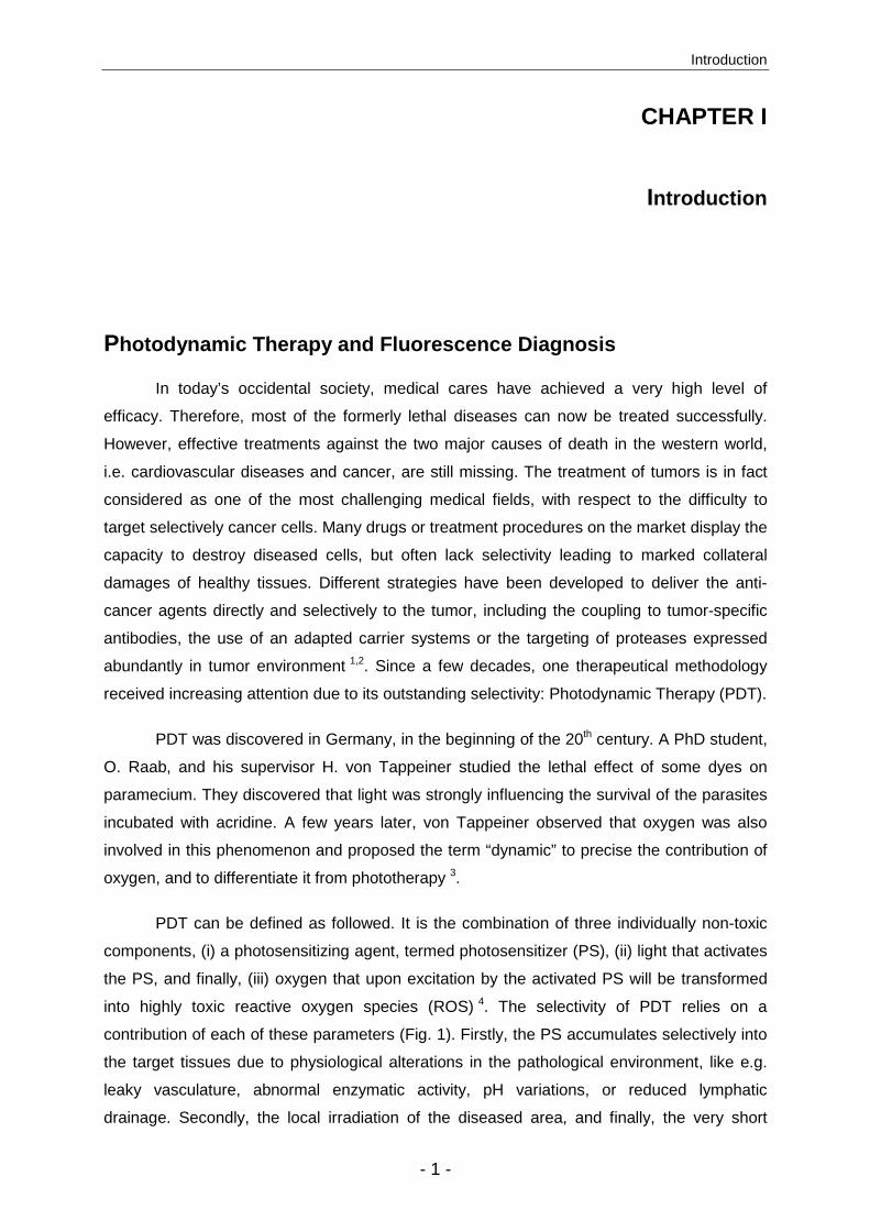
The ideal PS can be characterized by the following desired properties:
• chemically pure product, easy to synthesize
• easy administration, adapted formulation
• preferential accumulation in target tissues
• strong light absorption, ideally in the red part of spectrum (see below)
• high singlet oxygen quantum yield
• no or low dark toxicity
• no skin photosensitization
• fast elimination from the body
Plenty of newly designed PS have been patented and tested in preclinical studies, but
only few of them gained marketing authorization, mainly due to the difficulty to reach all the
requisites cited above.
5-aminolevulinic acid
In the development of new PS, apart from structural modifications of existing
molecules, another strategy has attracted increasing interest; i.e. the induction of
endogenous porphyrins by administration of porphyrin precursors. Almost every living cell
synthesizes porphyrins through heme biosynthetic pathway. It was observed that exogenous
administration of 5-aminolevulininc acid (5-ALA), overcomes endogenous regulation
mechanisms and selectively induces an accumulation of photoactive porphyrins (PAP) within
diseased cells 9 (Fig. 3). The main advantages of this method are the fast clearance of the
induced PS, within 24-48 hours, and the high tolerance for this endogenous molecule.
Another benefit of 5-ALA-mediated porphyrin induction is the high selectivity observed for
pathological tissues, due to environmental, cellular and metabolic abnormalities in
pathological tissues.
Neoplastic cells present an enhanced activity of pre-PpIX enzymes (e.g. PBG
deaminase) and decreased activity of post-PpIX enzymes (e.g. ferrochelatase) compared to
normal cells. This leads into a higher formation of photoactive porphyrins, and lower
transformation of these PS into non-photoactive heme. The poorer iron pool measured in
tumor cells amplifies this phenomenon, as the over-expression of benzodiazepine receptor,
involved in the oxidation of coproporphyrinogen into protoporphyrinogen is also favorable for
an accumulation of photoactive porphyrins 10.
Introduction
- 5 -
5-ALA was rapidly promoted as clinical candidate for the fluorescence detection and
the photodynamic therapy of various pathological conditions, amongst which are urology 11,
pneumology 12, or brain surgery 13. Nevertheless, 5-ALA received approval for only one
dermatological application; the PDT treatment of actinic keratoses14. This somehow
deceiving outcome can be mostly attributed to 5-ALA’s low systemic and local bioavailability.
+NH3-OOC
-OOC SCoA
O
O
-O
O
N+H3
NH
H2N
COO-COO-
NH HN
HNNH
AP
P
A
PP
A
ANH HN
HNNH
P
P
PP
NH HN
HNNH
PP
NH N
HNN
PP
N N
NN
PP
Fe 2+
ALAD PBGD
URO III cosynthase
URO III decaboxylase
5-ALA PBG
URO III COPRO III
PROTO IIIHEMEGLYINE
SUCCINYL CoA
COPRO IIIoxydase
PROTO IIIoxydaseFerrochelatase
PROTO-PORPHYRIN IX
negativefeedback
O
-O
O
N+H3
5-ALA
MITOCHONDRIA
CYTOSOL
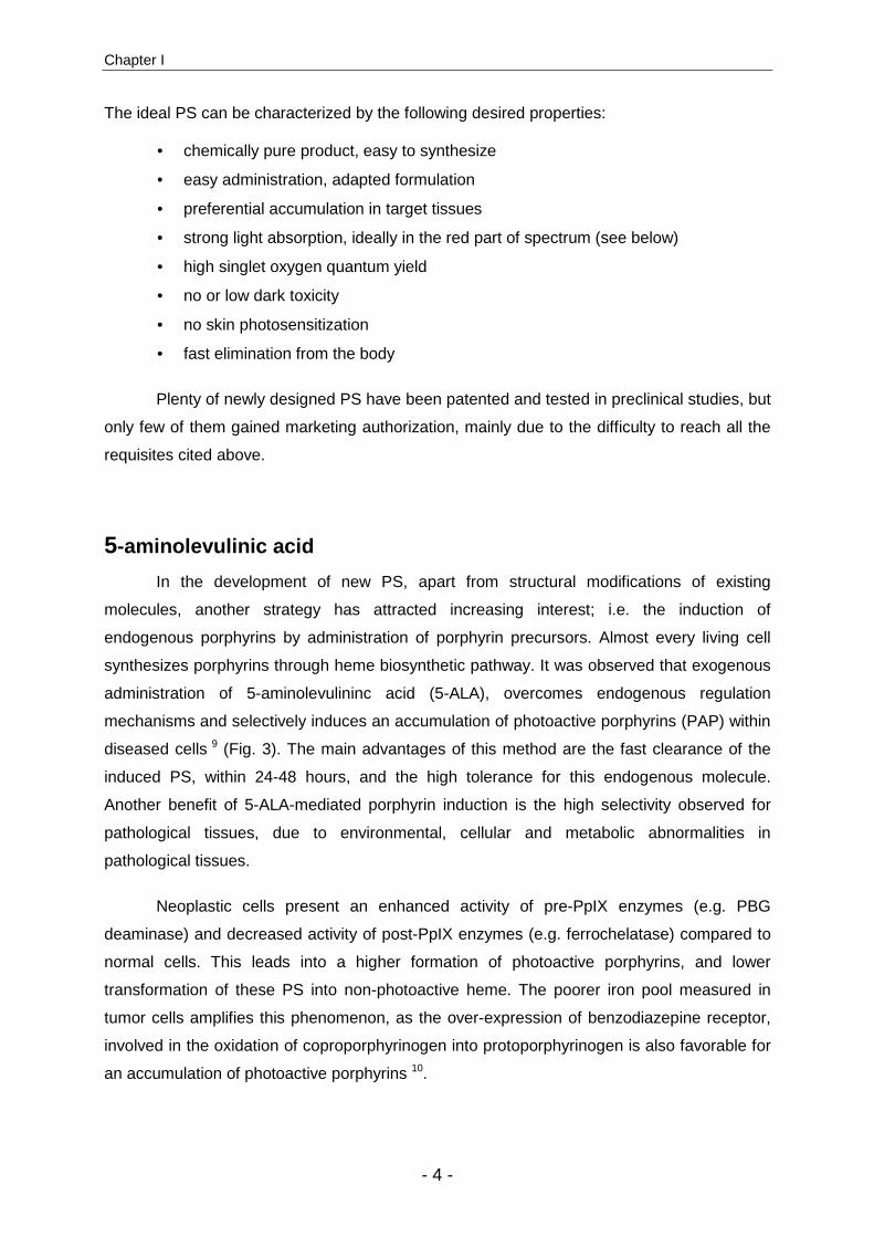
Figure 3: Heme biosynthesis cycle is present in almost every living cell.
5-aminolevulinic acid (5-ALA) is formed in mammals from the condensation
of glycine and succinylCoA. Two molecules of 5-ALA are assembled to form
porphobilinogen (PBG), and four molecules of PBG are linked to form a
tetrapyrrole. After cyclization of the tetrapyrrole, uroporphyrinogen III
(UROIII) undergo a series of enzymatic decarboxylations resulting into the
formation of protoporphyrin IX (PpIX), the photoactive molecule desired in
5-ALA mediated PDT. The ferrochelatase, an enzyme less active in tumor
cells, introduces an atom of iron into PpIX to form heme, the biologically
active, but non photoactive moiety. Heme exerts a negative feedback on
5-ALA formation that is overcome by the exogenous administration of 5-ALA.
(A=acetate, P=propionate)

Chapter I
- 6 -
5-aminolevulinic acid derivatives
The bioavailability of 5-ALA is strongly impaired by its zwitterionic nature under
physiological conditions (Fig. 4), as charged hydrophilic molecules have low ability to cross
biological barriers. Therefore, 5-ALA has to be internalized by active, energy consuming,
transporter mechanisms 15. In consequence, 5-ALA’s low bioavailability led to the
development of 5-ALA derivatives with optimized properties 16. The derivatization of 5-ALA
into more lipophilic molecules permits the passive internalization of PAP precursors,
optimizing the induced porphyrin formation both in vitro and in vivo 17,18. Apart from this
enhanced bioavailability, other advantages toward the parent compound can be mentioned,
e.g. the quasi absence of skin photosensitivity, and the less painful sensation during
irradiation.
O
O
H3N
O
OO
OR1
O
Aminoacid-HN
O
Cl O
O
N
O
R1R2
H
O
O
H3N
O
Cl
O
O
H3N
O
Cl
O
O
H3N
O
Cl
5-aminolevulinic acid (5-ALA)
5-ALA n-alkylesterIncreased lipophilicity andbioavailability
5-ALA methylester
5-ALA hexylester
2 sites (R1 and R2) available for derivatization
5-ALA amino acid derivativeTarget N-aminopeptidase over-expressed in tumoral tissues
5-ALA dendrimers
NH2O
O
O
H2N
O
O
O
H2N
O
O
O
H2N
5-ALA polyethylengylcolderivatives
Figure 4: Structure of 5-ALA and some relevant 5-ALA derivatives; 5-ALA is
presented here under its zwitterionic form, majority under
physiological conditions. 5-ALA derivatives can be developed from
site R1 and/or R2.
Introduction
- 7 -
Chapter II reviews the general knowledge accumulated until today in the domain of
5-ALA derivative-mediated photomedicine. The following points are developed,
(i) biochemistry of 5-ALA derivatives, (ii) topical and systemic bioavailability, (iii) chemistry
and design of 5-ALA derivatives (Fig. 4), (iv) preclinical studies of the most promising
compounds, and finally, (v) clinical studies with 5-ALA derivatives that achieved marketing
authorization. Although considerable progress has been realized with 5-ALA derivative, some
important questions remain unanswered. For example, the exact fate of 5-ALA derivatives
after their entrance into the cell is still hypothetic. Furthermore the optimal derivatization
strategy for a systemic administration of 5-ALA, in order to treat non superficial pathologies,
has not been defined. These subjects are treated later in chapter III and IV, respectively.
Irradiation
To obtain the most favorable PDT regimen, different parameters have to be
optimized, both for the photosensitizer administration (route, time, conditions, formulation),
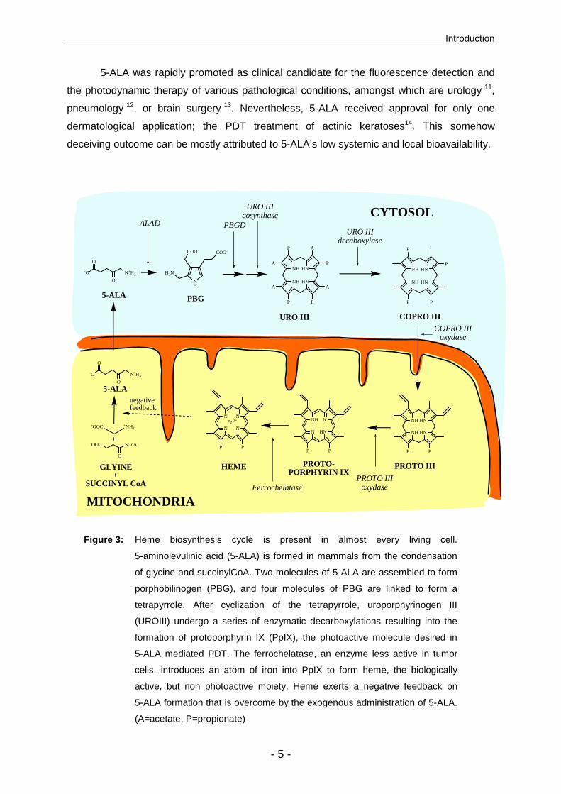
and for the irradiation procedure. The choice of the light source, irradiation wavelength
(Fig. 5), light dose and intensity are primordial for a successful PDT treatment 19,20,21. To be
exhaustive, the PDT irradiation protocol should also include drug administration-light
irradiation interval, repetition of the PDT treatment, and light diffuser device (cylindrical,
frontal, inflating balloon).
Figure 5: The upper part represents a classical absorption spectrum of a porphyrin,
with a maximum peak (Soret band) at around 400 nm followed by four Q
bands between 450 and 700 nm. The lower part shows the penetration
depth of light as a function of wavelength. For optimal irradiation conditions
in PDT, both parameters have to be taken into account
Soret band
Q bands
0
20
40
60
80
100
120
300 350 400 450 500 550 600 650 700wavelength [nm]
Inte
nsi
ty
[A.U
.]
Soret band
Q bands
0
20
40
60
80
100
120
300 350 400 450 500 550 600 650 700wavelength [nm]
Inte
nsi
ty
[A.U
.]
-8
-6
-4
-2
0
Pe
ne
tra
tio
n d
ep
th
[mm
]
-8
-6
-4
-2
0
Pe
ne
tra
tio
n d
ep
th
[mm
]
Chapter I
- 8 -
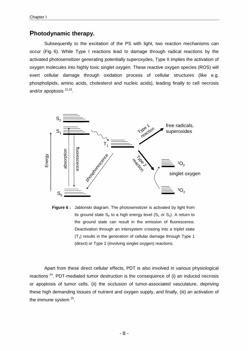
Photodynamic therapy.
Subsequently to the excitation of the PS with light, two reaction mechanisms can
occur (Fig. 6). While Type I reactions lead to damage through radical reactions by the
activated photosensitizer generating potentially superoxydes, Type II implies the activation of
oxygen molecules into highly toxic singlet oxygen. These reactive oxygen species (ROS) will
exert cellular damage through oxidation process of cellular structures (like e.g.
phospholipids, amino acids, cholesterol and nucleic acids), leading finally to cell necrosis
and/or apoptosis 22,23.
abso
rptio
n
fluorescence
phos
phor
esce
nce
S0
S1
T1
Type 1
reaction
Type 2
reaction
3O2
1O2
free radicals, superoxides
singlet oxygen
Ene
rgy
S2
Figure 6 : Jablonski diagram. The photosensitizer is activated by light from
its ground state S0 to a high energy level (S1 or S2). A return to
the ground state can result in the emission of fluorescence.
Deactivation through an intersystem crossing into a triplet state
(T1) results in the generation of cellular damage through Type 1
(direct) or Type 2 (involving singlet oxygen) reactions.
Apart from these direct cellular effects, PDT is also involved in various physiological
reactions 24. PDT-mediated tumor destruction is the consequence of (i) an induced necrosis
or apoptosis of tumor cells, (ii) the occlusion of tumor-associated vasculature, depriving
these high demanding tissues of nutrient and oxygen supply, and finally, (iii) an activation of
the immune system 25.
Introduction
- 9 -
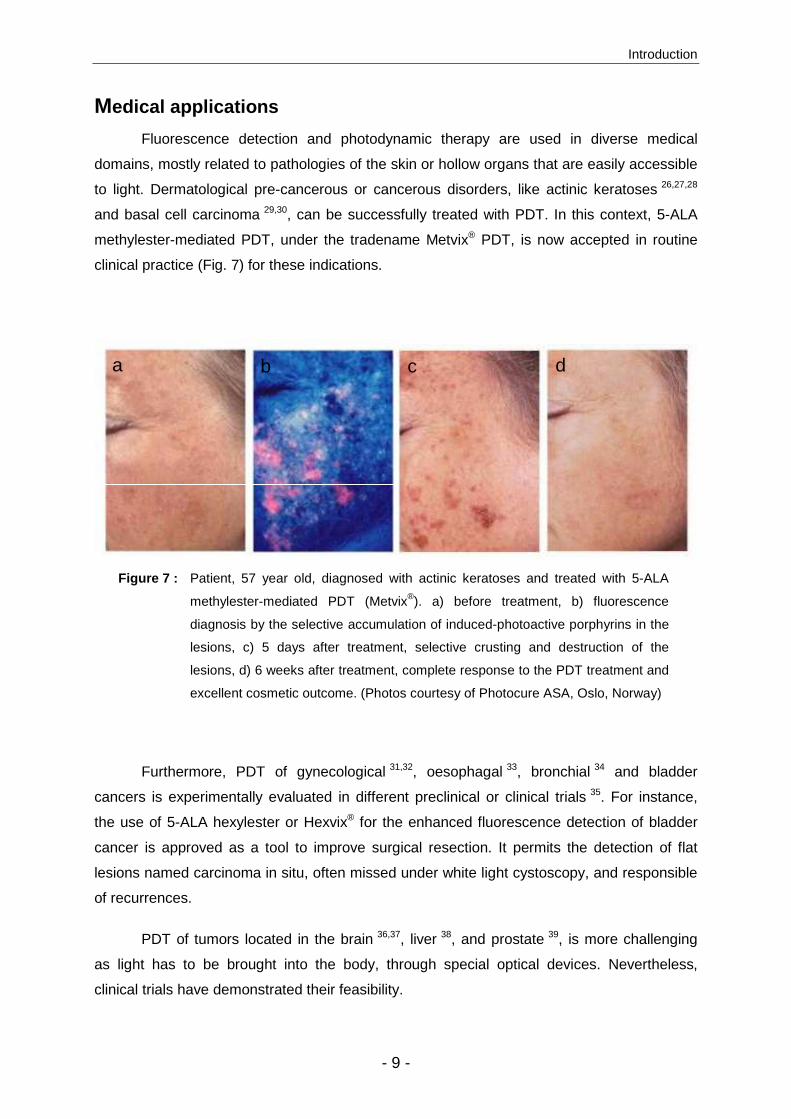
Medical applications
Fluorescence detection and photodynamic therapy are used in diverse medical
domains, mostly related to pathologies of the skin or hollow organs that are easily accessible
to light. Dermatological pre-cancerous or cancerous disorders, like actinic keratoses 26,27,28
and basal cell carcinoma 29,30, can be successfully treated with PDT. In this context, 5-ALA
methylester-mediated PDT, under the tradename Metvix® PDT, is now accepted in routine
clinical practice (Fig. 7) for these indications.
Fluorescence
diagnosis
a b c d
Figure 7 : Patient, 57 year old, diagnosed with actinic keratoses and treated with 5-ALA
methylester-mediated PDT (Metvix®). a) before treatment, b) fluorescence
diagnosis by the selective accumulation of induced-photoactive porphyrins in the
lesions, c) 5 days after treatment, selective crusting and destruction of the
lesions, d) 6 weeks after treatment, complete response to the PDT treatment and
excellent cosmetic outcome. (Photos courtesy of Photocure ASA, Oslo, Norway)
Furthermore, PDT of gynecological 31,32, oesophagal 33, bronchial 34 and bladder
cancers is experimentally evaluated in different preclinical or clinical trials 35. For instance,
the use of 5-ALA hexylester or Hexvix® for the enhanced fluorescence detection of bladder
cancer is approved as a tool to improve surgical resection. It permits the detection of flat
lesions named carcinoma in situ, often missed under white light cystoscopy, and responsible
of recurrences.
PDT of tumors located in the brain 36,37, liver 38, and prostate 39, is more challenging
as light has to be brought into the body, through special optical devices. Nevertheless,
clinical trials have demonstrated their feasibility.
Chapter I
- 10 -
Although photodynamic therapy and fluorescence diagnosis are mostly mentioned in
oncology, they have been tested for numerous non-oncologic medical indications, such as
cutaneous abnormalities (warts, psoriasis, acne) 40,41,42, endometriosis 43, rheumatoid
arthritis 44, choroidal neovascularization secondary to age related macular degeneration 45, or
microbial infections 46. The latter domain comprise the photoinactivation of viruses 47,48,49,
bacteria 50,51,52,53, fungi 54,55 and parasites 56, and will be developed in chapters V and VI.

Introduction
- 11 -
References 1. G. M. Dubowchik and M. A. Walker, Receptor-mediated and enzyme-dependent
targeting of cytotoxic anticancer drugs, Pharmacol Ther, 1999, 83, 67-123.
2. P. S. Huang and A. Oliff, Drug-targeting strategies in cancer therapy, Curr Opin Genet Dev, 2001, 11, 104-110.
3. J. Moan and Q. Peng, An outline of the hundred-year history of PDT., Anticancer Res, 2003, 23, 3591-3600.
4. T. J. Dougherty, C. J. Gomer, B. W. Henderson, G. Jori, D. Kessel, M. Korbelik, J. Moan, and Q. Peng, Photodynamic therapy, J Natl Cancer Inst, 1998, 90, 889-905.
5. A. P. Castano, T. N. Demidova, and M. R. Hamblin, Mechanisms in photodynamic therapy: part one-photosensitizers, photochemistry and cellular localization, Photodiagnonsis Photodynamic Therapy, 2004, 1, 279-293.
6. R. Ackroyd, C. Kelty, N. Brown, and M. Reed, The history of photodetection and photodynamic therapy, Photochem Photobiol, 2001, 74, 565-669.
7. S. K. Pushpan, S. Venkatraman, V. G. Anand, J. Sankar, D. Parmeswaran, S. Ganesan, and T. K. Chandrashekar, Porphyrins in photodynamic therapy - a search for ideal photosensitizers, Curr Med Chem Anti-Canc Agents, 2002, 2, 187-207.
8. A. R. Battersby, Tetrapyrroles: the pigments of life, Nat Prod Rep, 2000, 17, 507-526.
9. Q. Peng, K. Berg, J. Moan, M. Kongshaug, and J. M. Nesland, 5-Aminolevulinic acid-based photodynamic therapy: principles and experimental research, Photochem Photobiol, 1997, 65, 235-251.
10. S. Collaud, A. Juzeniene, J. Moan, and N. Lange, On the selectivity of 5-aminolevulinic acid-induced protoporphyrin IX formation, Curr Med Chem Anti-Canc Agents, 2004, 4, 301-316.
11. P. Jichlinski, M. Forrer, J. Mizeret, T. Glanzmann, D. Braichotte, G. Wagnières, G. Zimmer, L. Guillou, F. Schmidlin, P. Graber, H. van den Bergh, and H. J. Leisinger, Clinical evaluation of a method for detecting superficial surgical transitional cell carcinoma of the bladder by light-induced fluorescence of protoporphyrin IX following the topical application of 5-aminolevulinic acid: preliminary results, Lasers Surg Med, 1997, 20, 402-408.
12. R. Baumgartner, R. M. Huber, H. Schulz, H. Stepp, K. Rick, F. Gamarra, A. Leberig, and C. Roth, Inhalation of 5-aminolevulinic acid: a new technique for fluorescence detection of early stage lung cancer, J Photochem Photobiol B, 1996, 36, 169-174.
13. S. A. Friesen, G. O. Hjortland, S. J. Madsen, H. Hirschberg, O. Engebraten, J. M. Nesland, and Q. Peng, 5-Aminolevulinic acid-based photodynamic detection and therapy of brain tumors (review), Int J Oncol, 2002, 21, 577-582.
14. E. W. Jeffes, J. L. McCullough, G. D. Weinstein, R. Kaplan, S. D. Glazer, and J. R. Taylor, Photodynamic therapy of actinic keratoses with topical aminolevulinic acid hydrochloride and fluorescent blue light, J Am Acad Dermatol, 2001, 45, 96-104.

Chapter I
- 12 -
15. L. Rodriguez, A. M. Batlle, G. Di Venosa, A. J. MacRobert, S. H. Battah, H. Daniel, and A. Casas, Study of the mechanisms of uptake of 5-aminolevulinic acid derivatives by PEPT1 and PEPT2 transporters as a tool to improve photodynamic therapy of tumours, Int J Biochem Cell Biol, 2006, 38, 1530-1539.
16. N. Fotinos, M. A. Campo, F. Popowycz, R. Gurny, and N. Lange, 5-Aminolevulinic Acid Derivatives in Photomedicine: Characteristics, Application and Perspectives, Photochem Photobiol, 2006, 82, 994-1015.
17. P. Uehlinger, M. Zellweger, G. Wagnières, L. Juillerat-Jeanneret, H. van den Bergh, and N. Lange, 5-aminolevulinic acid and its derivatives: physical chemical properties and protoporphyrin IX formation in cultured cells, J Photochem Photobiol B:Biol, 2000, 54, 72-80.
18. J. Moan, L. W. Ma, A. Juzeniene, V. Iani, P. Juzenas, F. Apricena, and Q. Peng, Pharmacology of protoporphyrin IX in nude mice after application of ALA and ALA esters, Int J Cancer, 2003, 103, 132-135.
19. A. Juzeniene, P. Juzenas, L. W. Ma, V. Iani, and J. Moan, Effectiveness of different light sources for 5-aminolevulinic acid photodynamic therapy, Lasers Med Sci, 2004, 19, 139-149.
20. V. Nadeau, M. O'Dwyer, K. Hamdan, I. Tait, and M. Padgett, In vivo measurement of 5-aminolaevulinic acid-induced protoporphyrin IX photobleaching: a comparison of red and blue light of various intensities, Photodermatol Photoimmunol Photomed, 2004, 20, 170-174.
21. D. C. Shackley, C. Whitehurst, J. V. Moore, N. J. George, C. D. Betts, and N. W. Clarke, Light penetration in bladder tissue: implications for the intravesical photodynamic therapy of bladder tumours, BJU Int, 2000, 86, 638-643.
22. A. P. Castano, T. N. Demidova, and M. R. Hamblin, Mechanisms in photodynamic therapy: part two-cellular signaling, cell metabolism and modes of cell death, Photodiagnonsis Photodynamic Therapy, 2005, 2, 1-23.
23. N. L. Oleinick, R. L. Morris, and I. Belichenko, The role of apoptosis in response to photodynamic therapy: what, where, why, and how, Photochem Photobiol Sci, 2002, 1, 1-21.
24. A. P. Castano, T. N. Demidova, and M. R. Hamblin, Mechanisms in photodynamic therapy: Part three-Photosensitizer pharmacokinetics, biodistribution, tumor localization and modes of tumor destruction, Photodiagnonsis Photodynamic Therapy, 2005, 2, 91-106.
25. A. P. Castano, P. Mroz, and M. R. Hamblin, Photodynamic therapy and anti-tumour immunity, Nat Rev Cancer, 2006, 6, 535-545.
26. D. M. Pariser, N. J. Lowe, D. M. Stewart, M. T. Jarratt, A. W. Lucky, R. J. Pariser, and P. S. Yamauchi, Photodynamic therapy with topical methyl aminolevulinate for actinic keratosis: results of a prospective randomized multicenter trial, J Am Acad Dermatol, 2003, 48, 227-232.
27. R. M. Szeimies, S. Karrer, S. Radakovic-Fijan, A. Tanew, P. G. Calzavara-Pinton, C. Zane, A. Sidoroff, M. Hempel, J. Ulrich, T. Proebstle, H. Meffert, M. Mulder, D. Salomon, H. C. Dittmar, J. W. Bauer, K. Kernland, and L. Braathen, Photodynamic
Introduction
- 13 -
therapy using topical methyl 5-aminolevulinate compared with cryotherapy for actinic keratosis: A prospective, randomized study, J Am Acad Dermatol, 2003, 47, 258-262.
28. M. Freeman, C. Vinciullo, D. Francis, L. Spelman, R. Nguyen, P. Fergin, K. E. Thai, D. Murell, W. Weightman, C. Anderson, C. Reid, A. Watson, and P. Foley, A comparison of photodynamic therapy using topical methyl aminolevulinate (Metvix) with single cycle cryotherapy in patients with actinic keratosis: a prospective, randomized study., J Dermatolog Treat, 2003, 14, 99-106.
29. E. Angell-Petersen, Porphyrin formation in actinic keratosis and Basal cell carcinoma after topical application of methyl 5-aminolevulinate, J Invest Dermatol, 2006, 126, 265-271.
30. N. Basset-Séguin, I. Bachmann, S. Pavel, O. Saksela, M. Johnsson, A.M. Ros, B. Paredes, and O.A. Larko, A dose finding study of photodynamic therapy (PDT) with Metvix(R) in patients with basal cell carcinoma (BCC), JEADV, 2000, 14, 39.
31. M. Loning, H. Diddens, W. Kupker, K. Diedrich, and G. Hüttmann, Laparoscopic fluorescence detection of ovarian carcinoma metastases using 5-aminolevulinic acid-induced protoporphyrin IX, Cancer, 2004, 100, 1650-1656.
32. S. Andrejevic-Blant, A. Major, F. Ludicke, J. P. Ballini, G. Wagnières, H. van den Bergh, and M. F. Pelte, Time-dependent hexaminolaevulinate induced protoporphyrin IX distribution after topical application in patients with cervical intraepithelial neoplasia: A fluorescence microscopy study, Lasers Surg Med, 2004, 35, 276-283.
33. T. Stepinac, C. Felley, P. Jornod, N. Lange, T. Gabrecht, C. Fontolliet, P. Grosjean, G. vanMelle, H. van den Bergh, P. Monnier, G. Wagnières, and G. Dorta, Endoscopic fluorescence detection of intraepithelial neoplasia in Barrett's esophagus after oral administration of aminolevulinic acid, Endoscopy, 2003, 35, 663-668.
34. G. M. Loewen, R. Pandey, D. Bellnier, B. Henderson, and T. J. Dougherty, Endobronchial photodynamic therapy for lung cancer, Lasers Surg Med, 2006, 38, 364-370.
35. S. B. Brown, E. A. Brown, and I. Walker, The present and future role of photodynamic therapy in cancer treatment, Lancet Oncology, 2004, 5, 497-508.
36. S. Stylli and A. H. Kaye, Photodynamic therapy of cerebral glioma - a review. Part II - clinical studies, J Clin Neurosci, 2006, 13, 709-717.
37. A. Bogaards, A. Varma, K. Zhang, D. Zach, S. K. Bisland, E. H. Moriyama, L. Lilge, P. J. Muller, and B. C. Wilson, Fluorescence image-guided brain tumour resection with adjuvant metronomic photodynamic therapy: pre-clinical model and technology development, Photochem Photobiol Sci, 2005, 4, 438-442.
38. F. H. van Duijnhoven, J. P. Rovers, K. Engelmann, Z. Krajina, S. F. Purkiss, F. A. Zoetmulder, T. J. Vogl, and O. T. Terpstra, Photodynamic therapy with 5,10,15,20-tetrakis(m-hydroxyphenyl) bacteriochlorin for colorectal liver metastases is safe and feasible: results from a phase I study, Ann Surg Oncol, 2005, 12, 808-816.
39. C. M. Moore, T. R. Nathan, W. R. Lees, C. A. Mosse, A. Freeman, M. Emberton, and S. G. Bown, Photodynamic therapy using meso tetra hydroxy phenyl chlorin (mTHPC) in early prostate cancer, Lasers Surg Med, 2006, 38, 356-363.

Chapter I
- 14 -
40. K. Kalka, H. Merk, and H. Mukhtar, Photodynamic therapy in dermatology, J Am Acad Dermatol, 2000, 42, 389-416.
41. P. Babilas, S. Karrer, A. Sidoroff, M. Landthaler, and R. M. Szeimies, Photodynamic therapy in dermatology--an update, Photodermatol Photoimmunol Photomed, 2002, 21, 142-149.
42. C. Fritsch, G. Goerz, and T. Ruzicka, Photodynamic therapy in dermatology, Arch dermatol, 1998, 134, 207-214.
43. M. J. Gannon and S. B. Brown, Photodynamic therapy and its applications in gynaecology, Br J Obstet Gynaecol, 1999, 106, 1246-1254.
44. G. Kirdaite, N. Lange, N. Busso, H. van den Bergh, P. Kucera, and A. So, Protoporphyrin IX photodynamic therapy for synovitis, Arthrtitis Rheum, 2002, 46, 1371-1378.
45. R. M. Barnes, L. Gee, S. Taylor, M. C. Briggs, and S. P. Harding, Outcomes in verteporfin photodynamic therapy for choroidal neovascularisation--'beyond the TAP study', Eye, 2004, 18, 809-813.
46. G. Jori and S. B. Brown, Photosensitized inactivation of microorganisms, Photochem Photobiol Sci, 2004, 3, 403-405.
47. M. Wainwright, Photoinactivation of viruses, Photochem Photobiol Sci, 2004, 3, 406-411.
48. M. Wainwright, Local treatment of viral disease using photodynamic therapy, Int J Antimicrob Agents, 2003, 21, 510-520.
49. Z. Smetana, Z. Malik, and A. Orenstein, Treatment of viral infections with 5-aminolevulinic acid and light, Lasers Surg Med, 1997, 21, 351-358.
50. G. P. Tegos, M. Anbe, C. Yang, T. N. Demidova, M. Satti, P. Mroz, S. Janjua, F. Gad, and M. R. Hamblin, Protease-stable polycationic photosensitizer conjugates between polyethyleneimine and chlorin(e6) for broad-spectrum antimicrobial photoinactivation, Antimicrob Agents Chemother, 2006, 50, 1402-1410.
51. S. Banfi, E. Caruso, L. Buccafurni, V. Battini, S. Zazzaron, P. Barbieri, and V. Orlandi, Antibacterial activity of tetraaryl-porphyrin photosensitizers: An in vitro study on Gram negative and Gram positive bacteria, J Photochem Photobiol B, 2006, 85, 28-38.
52. M. R. Hamblin, J. Viveiros, C. Yang, A. Ahmadi, R. A. Ganz, and M. J. Tolkoff, Helicobacter pylori accumulates photoactive porphyrins and is killed by visible light, Antimicrob Agents Chemother, 2005, 49, 2822-2827.
53. S. A. Lambrechts, T. N. Demidova, M. C. Aalders, T. Hasan, and M. R. Hamblin, Photodynamic therapy for Staphylococcus aureus infected burn wounds in mice, Photochem Photobiol Sci, 2005, 4, 503-509.
54. P. G. Calzavara-Pinton, M. Venturini, and R. Sala, A comprehensive overview of photodynamic therapy in the treatment of superficial fungal infections of the skin, J Photochem Photobiol B, 2005, 78, 1-6.

Introduction
- 15 -
55. H. Kamp, H. J. Tietz, M. Lutz, H. Piazena, P. Sowyrda, J. Lademann, and U. Blume-Peytavi, Antifungal effect of 5-aminolevulinic acid PDT in Trichophyton rubrum, Mycoses, 2005, 48, 101-107.
56. K. Gardlo, Z. Horska, C. D. Enk, L. Rauch, M. Megahed, T. Ruzicka, and C. Fritsch, Treatment of cutaneous leishmaniasis by photodynamic therapy, J Am Acad Dermatol, 2003, 48, 893-896.

Chapter I
- 16 -
5-Aminolevulinic Acid Derivatives in Photomedicine: Characteristics, Application and Perspectives
- 17 -
CHAPTER II
5-Aminolevulinic Acid Derivatives in Photomedicine:
Characteristics, Application and Perspectives
Nicolas Fotinos, Marino A. Campo, Florence Popowycz, Robert Gurny
and Norbert Lange
Department of Pharmaceutics and Biopharmaceutics, School of Pharmaceutical Sciences, University of Geneva,
University of Lausanne, 30 Quai Ernest Ansermet, 1211 Geneva 4, Switzerland
Published in Photochemistry and Photobiology, 2006 (82), 994-1015
Abstract
The introduction of lipophilic derivatives of the naturally occurring heme precursor
5-aminolevulinic acid (5-ALA) into photomedicine has led to a true revival of this research
area. 5-ALA-mediated photodynamic therapy (PDT) and fluorescence photodetection (FD) of
neoplastic disease is probably one of the most selective cancer treatments currently known
in oncology. To date, this method has been assessed experimentally for the treatment of
various medical indications. However, the limited local bioavailability of 5-ALA has widely
prevented its use in daily clinical practice. Although researchers were already aware of this
drawback early during the development of 5-ALA-mediated PDT, only recently have well-
established concepts in pharmaceutical science been adapted to investigate ways to
overcome this drawback.
Recently, two derivatives of 5-ALA, methylaminolevulinate (MAL) and
hexylaminolevulinate (HAL), gained marketing authorization from the regulatory offices in
Europe and Australia. MAL is marketed under the trade name Metvix® for the treatment of
actinic keratosis and difficult-to-treat basal cell carcinoma. HAL has recently been launched
in Europe under the trade name Hexvix® to improve the detection of superficial bladder
cancer.

Chapter II
- 18 -
This review will first present the fundamental concepts underlying the use of 5-ALA
derivatives in PDT and FD from a chemical, biochemical and pharmaceutical point of view.
Experimental evidences from preclinical data on the improvements and limits observed with
5-ALA derivatives will then be introduced. The state-of-the art from clinical studies with
5-ALA esters will be discussed, with special emphasis placed on the process that led to the
development of MAL in dermatology and HAL in urology. Finally, we will discuss promising
medical fields in which the use of 5-ALA derivatives might potentially lead to further
application of this methodology in photomedicine.
Introduction
After Kelly et al. 1 clinically demonstrated the selective accumulation of exogenously
applied hematoporphyrin derivative (HPD) in human bladder cancer, photodynamic therapy
(PDT) gave rise to a growing interest in the medical community. This alternative treatment
modality consists of the administration of a so-called tumour localizing photosensitizer,
followed by the irradiation of the target tumour site with light of an appropriate wavelength.
The mechanism of PDT action has been the subject of numerous investigations 2,3.
Depending on the photosensitizing agent, light activation results in the generation of highly
active reactive oxygen species that exert damaging action to cellular structures, such as the
cell membrane, mitochondria, lysosomes and nuclei. After this initial photodamage, PDT then
results in selective tumour eradication through a complex cascade of photochemical,
immunological and physiological reactions 4. Unlike ionizing radiation, PDT can be applied
repeatedly at the same site and is characterized particularly by its minimally invasive
character. Among the main advantages of PDT are its cost-effectiveness and simplicity of
use. Furthermore, conventional treatment strategies, such as chemotherapy, ionizing
radiation and surgery, do not preclude PDT.
In addition to the therapeutic role of PDT, the selective accumulation of
photosensitizers in neoplastic tissues can be used for diagnostic purposes. In such
procedures, often referred to as “fluorescence diagnosis” (FD) or “fluorescence
photodetection”, sensitive imaging devices are used to permit the specific detection of the
fluorescence characteristic of the given photosensitizers. The clinical use of FD and its
relevant benefits in patient care have been recently reviewed by Wagnières et al. 5.
Despite considerable efforts of the scientific and medical community only a few
photosensitizers gained marketing authorisation for use in oncological therapy. The limited
clinical use of PDT in this medical area has been mostly attributed to intrinsic drawbacks of

5-Aminolevulinic Acid Derivatives in Photomedicine: Characteristics, Application and Perspectives
- 19 -
conventional photosensitizers, such as poor selectivity, prolonged skin photosensitization,
reduced absorbance in the red part of the visible spectrum and difficulties in the development
of appropriate formulations. However, the fact that PDT can be a powerful tool from a
therapeutic as well as a commercial point of view is demonstrated by the tremendous
success of Visudyne®-mediated PDT of choroidal neovascularisation secondary to age-
related macular degeneration.
+NH3-OOC
-OOC SCoA
O
O
-O
O
N+H3
NH
H2N
COO-COO-
NH HN
HNNH
AP
P
A
PP
A
ANH HN
HNNH
P
P
PP
NH HN
HNNH
PP
NH N
HNN
PP
N N
NN
PP
Fe 2+
ALAD PBGD
URO III cosynthase
URO III decaboxylase
5-ALA PBG
URO III COPRO III
PROTO IIIHEMEGLYINE
SUCCINYL CoA
COPRO IIIoxydase
PROTO IIIoxydase
Ferrochelatase
PROTO-PORPHYRIN IX
negativefeedback
O
-O
O
N+H3
5-ALA
Figure 1 : The heme biosynthetic pathway in mammals. The first enzyme 5-aminolevulinate synthase
(ALAS) catalyses the conversion of glycine and succinyl CoA into one molecule of
5-aminolevulinic acid (5-ALA), which is followed by the assymetric condensation of two
5-ALA molecules by 5-aminolevulinate dehydratase (ALAD) into one molecule of
porphobilinogen (PBG). An enzymatic cascade then converts four molecules of PBG into a
tetrapyrrol ring that undergo decarboxylations, leading to the formation of protoporphyrin IX
(PpIX). Heme is produced when the ferrochelatase inserts a ferrous iron into PpIX.
A, acetate; P, propionate; URO, uropophyrinogen; COPRO, coproporphyrinogen; PROTO,
protoporphyrinogen; PBGD, PBG deaminase
CYTOSOL
MITOCHONDRIA
Chapter II
- 20 -
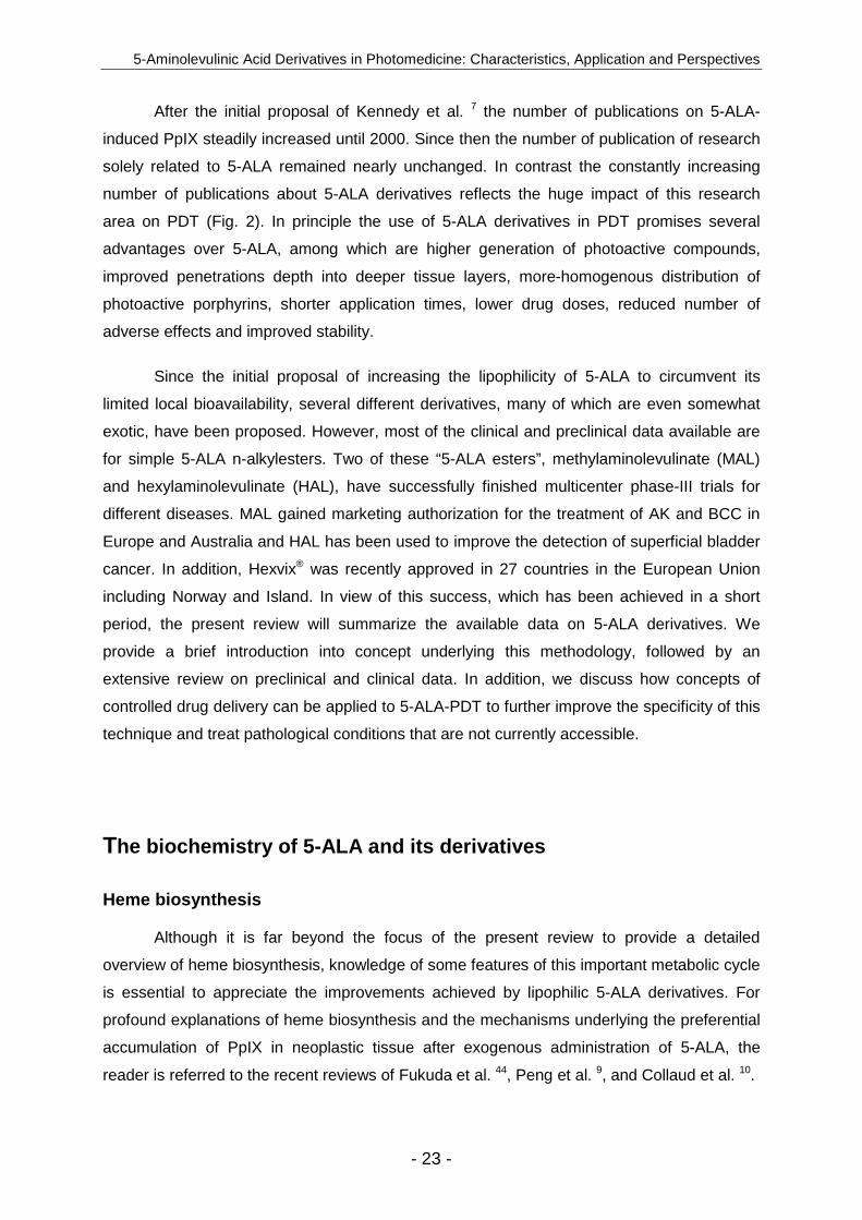
An alternative approach to the direct administration of a photosensitive agent is
offered by the prodrug concept. According to Albert 6 a prodrug is a nonactive compound
that, after administration, is metabolically converted to pharmacologically active compound.
One such prodrugs, 5-aminolevulinic acid (5-ALA), was introduced into PDT by Kennedy et
al. in 1992 7. They showed that, after exogenous administration of 5-ALA in aqueous solution
to patients with superficial cutaneous disorders, photosensitization with protoporphyrin IX
(PpIX) is observed that is mainly confined to diseased epithelium. Indeed, both 5-ALA and
PpIX are naturally occurring intermediates in heme biosynthesis taking place in nearly all
aerobic cells in mammals (Fig. 1). PpIX, in contrast to heme (its iron-containing counterpart),
is a photoactive substance with a singlet oxygen yield of approximately 56% 8 and a
reasonable fluorescence quantum yield. Normally, heme inhibits the endogenous formation
of excess 5-ALA by a negative-feedback control mechanism, thereby avoiding natural PpIX
photosensitization 9,10. However, the presence of exogenous 5-ALA bypasses this regulatory
mechanism and results in the transient formation of excess PpIX that takes place
preferentially in neoplastic cells. The factors underlying this phenomenon have been the
subject of profound debate in the literature and have been recently reviewed by Collaud et
al. 10. However, it seems that apart from differences of metabolic origin in neoplastic cells,
environmental and morphological factors may have an impact on the selective generation of
PpIX.
Today, 5-ALA-mediated PDT can be considered as one of the most selective
treatments for neoplastic disease. The growing interest in this treatment modality is reflected
by an increase in the number of articles in the scientific literature (Fig. 2). In addition to its
good tumor selectivity 5-ALA-induced PpIX has several supplementary advantages over
conventional photosensitising agents, including limited systemic toxicity and low skin
photosensitization 24-48 hours after administration 11. Despite promising results in several
medical studies of 5-ALA-mediated PDT and FD 12, these treatment modalities still lack the
wide acceptance in the medical community. Today the only exception in this context is the
1999 US Food and Drug Administration approval of 5-ALA for the treatment of actinic
keratosis (AK).
The failure of 5-ALA to gain marketing authorisation for the treatment or diagnosis of
other medical indications can indeed be ascribed to a multitude of reasons. However, one of
the main reasons relies on the physical-chemical properties of the PpIX precursor itself.
Under physiological conditions more than 90% of all 5-ALA molecules are present as
zwitterions and carry a positive charge at the amine terminal and a negative charge at the
carboxylic terminal.

5-Aminolevulinic Acid Derivatives in Photomedicine: Characteristics, Application and Perspectives
- 21 -
0
100
200
300
1988-
1989
1990-
1991
1992-
1993
1994-
1995
1996-
1997
1998-
1999
2000-
2001
2002-
2003
2004-
2005
*
Publication date
Num
ber
of p
ublis
hed
artic
les
conc
erni
ng 5
-A
LA
or
5-A
LA
der
ivat
ive
in p
hoto
med
icin
e
5-ALA derivatives
5-ALA
* 2005 publications are extrapolated from the period january to june 2005
Figure 2 : Results of a PubMed search showing the number of published
articles with 5-ALA only (white) or 5-ALA derivatives (black).
Such compounds have limited capacities to reach and ultimately enter the target cell
within a biological environment. This deficiency results in a low penetration depth and a
nonhomogeneous distribution of 5-ALA-induced PpIX after topical application and may well
explain the low response rate of 5-ALA mediated PDT of noduloulcerative basal cell
carcinomas (BCCs) to 5-ALA mediated-PDT 12,13. The limited bioavailability of 5-ALA after
topical administration is even more pronounced when given parenterally. In fact 5-ALA has
been shown to be rapidly eliminated from the human body, with a plasma half-life of 50 min
when given intravenously and 45 min when given orally 14. The small volume of distribution of
only 8.3 L indicates that a large portion will be excreted unchanged in the urine and trapped
by first-pass metabolism. Studies involving dogs revealed that > 50 % of the applied drug
dose will end up in the liver and that approximately 15% will end up in the kidneys 15. Its poor
pharmacokinetic profile is therefore highly unfavourable with respect to the generation of
photodynamically efficient doses of PpIX after systemic administration of 5-ALA.
Apart from its limited bioavailability the parenteral use of 5-ALA is associated with
considerable adverse in humans. In addition to nausea, vomiting and transcient abnormal
liver functions, significant decreases in systolic and diastolic blood and pulmonary pressure
have been reported 16,17. Because of drawbacks associated with the therapeutic use of
Chapter II
- 22 -
5-ALA, research has focused on improving local delivery of this compound. In addition to
modification of 5-ALA containing formulations (Table 1) such as i.e. liposomal formulation
methods, the use of penetration enhancers and heme biosynthesis modifying agents have
been developed. Although these methods have had promising results in experiments, the
implementation of these techniques into daily clinical practice has failed.
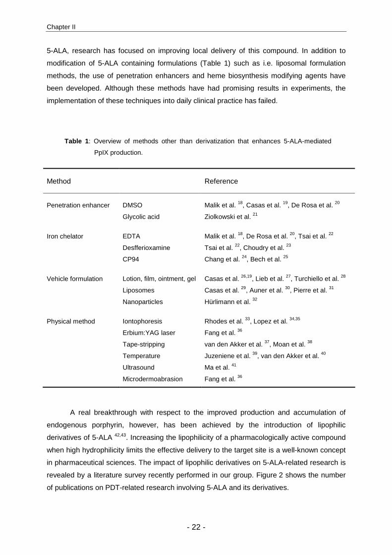
Table 1 : Overview of methods other than derivatization that enhances 5-ALA-mediated
PpIX production.
Method Reference
Penetration enhancer DMSO Malik et al. 18, Casas et al. 19, De Rosa et al. 20
Glycolic acid Ziolkowski et al. 21
Iron chelator EDTA Malik et al. 18, De Rosa et al. 20, Tsai et al. 22
Desfferioxamine Tsai et al. 22, Choudry et al. 23
CP94 Chang et al. 24, Bech et al. 25
Vehicle formulation Lotion, film, ointment, gel Casas et al. 26,19, Lieb et al. 27, Turchiello et al. 28
Liposomes Casas et al. 29, Auner et al. 30, Pierre et al. 31
Nanoparticles Hürlimann et al. 32
Physical method Iontophoresis Rhodes et al. 33, Lopez et al. 34,35
Erbium:YAG laser Fang et al. 36
Tape-stripping van den Akker et al. 37, Moan et al. 38
Temperature Juzeniene et al. 39, van den Akker et al. 40
Ultrasound Ma et al. 41
Microdermoabrasion Fang et al. 36
A real breakthrough with respect to the improved production and accumulation of
endogenous porphyrin, however, has been achieved by the introduction of lipophilic
derivatives of 5-ALA 42,43. Increasing the lipophilicity of a pharmacologically active compound
when high hydrophilicity limits the effective delivery to the target site is a well-known concept
in pharmaceutical sciences. The impact of lipophilic derivatives on 5-ALA-related research is
revealed by a literature survey recently performed in our group. Figure 2 shows the number
of publications on PDT-related research involving 5-ALA and its derivatives.
5-Aminolevulinic Acid Derivatives in Photomedicine: Characteristics, Application and Perspectives
- 23 -
After the initial proposal of Kennedy et al. 7 the number of publications on 5-ALA-
induced PpIX steadily increased until 2000. Since then the number of publication of research
solely related to 5-ALA remained nearly unchanged. In contrast the constantly increasing
number of publications about 5-ALA derivatives reflects the huge impact of this research
area on PDT (Fig. 2). In principle the use of 5-ALA derivatives in PDT promises several
advantages over 5-ALA, among which are higher generation of photoactive compounds,
improved penetrations depth into deeper tissue layers, more-homogenous distribution of
photoactive porphyrins, shorter application times, lower drug doses, reduced number of
adverse effects and improved stability.
Since the initial proposal of increasing the lipophilicity of 5-ALA to circumvent its
limited local bioavailability, several different derivatives, many of which are even somewhat
exotic, have been proposed. However, most of the clinical and preclinical data available are
for simple 5-ALA n-alkylesters. Two of these “5-ALA esters”, methylaminolevulinate (MAL)
and hexylaminolevulinate (HAL), have successfully finished multicenter phase-III trials for
different diseases. MAL gained marketing authorization for the treatment of AK and BCC in
Europe and Australia and HAL has been used to improve the detection of superficial bladder
cancer. In addition, Hexvix® was recently approved in 27 countries in the European Union
including Norway and Island. In view of this success, which has been achieved in a short
period, the present review will summarize the available data on 5-ALA derivatives. We
provide a brief introduction into concept underlying this methodology, followed by an
extensive review on preclinical and clinical data. In addition, we discuss how concepts of
controlled drug delivery can be applied to 5-ALA-PDT to further improve the specificity of this
technique and treat pathological conditions that are not currently accessible.
The biochemistry of 5-ALA and its derivatives
Heme biosynthesis
Although it is far beyond the focus of the present review to provide a detailed
overview of heme biosynthesis, knowledge of some features of this important metabolic cycle
is essential to appreciate the improvements achieved by lipophilic 5-ALA derivatives. For
profound explanations of heme biosynthesis and the mechanisms underlying the preferential
accumulation of PpIX in neoplastic tissue after exogenous administration of 5-ALA, the
reader is referred to the recent reviews of Fukuda et al. 44, Peng et al. 9, and Collaud et al. 10.
Chapter II
- 24 -
In addition to its function as a prosthetic group in numerous hemoproteins, such as
hemoglobin, myoglobin and cytochroms, heme plays an essential role in the regulation of
protein synthesis and cell differentiation 45. Almost all nucleated cells in mammals exhibit the
ability to produce heme. Heme biosynthesis is tightly regulated by various mechanisms at a
cellular level. One of the most important of these mechanisms is the negative-feedback
control that heme exerts on the first enzymatic step in heme biosynthesis. Indeed, heme may
regulate the 5-ALA synthase-catalyzed condensation of glycine and succinyl CoA by
decreasing the enzyme’s mRNA half-life and/or by blocking the transport of the enzyme into
the mitochondria (Fig. 1)
After entry of 5-ALA into the cytosol seven consecutive enzymatic reaction occur; four
are cytosolic and the others mitochondrial. In the cytosol, 5-ALA dehydrase induces the
asymmetric condensation of two molecules of 5-ALA to form porphobilinogen (PBG).
Subsequently, PBG-deaminase and uroporphyrinogen cosynthase catalyse the cyclization of
four PBG molecules that comprise the tetrapyrrolic skeleton of a porphyrin. Finally, a series
of decarboxylations and oxidations inside the cytoplasm and the mitochondria most occur
before the PpIX is formed by the protoporphyrinogen oxidases-catalyzed removal of six
hydrogen atoms from the Protoporphyrinogen IX. Heme biosynthesis is then completed by
the ferrochelatase-mediated insertion of ferrous iron, which takes place in the inner
mitochondrial membrane.
Exogenous provision of excess 5-ALA circumvents the regulatory mechanism that
heme exerts on endogenous 5-ALA formation, thus allowing for the production of heme and
its intermediates at rates that are primarily limited by the activity of involved enzymes and the
amount of available intracellular 5-ALA. In this context 5-ALA derivatives have the potential
to increase the pool of intracellular substrate molecules, because of their modified physical
chemical properties.
However, at present there is no clear experimental evidence whether molecules such
as. 5-ALA esters have to be converted into 5-ALA before entering the heme biosynthesis
cycle or whether they may act directly as a substrate for the enzymes involved in this
pathway (see below). This is partly due to the fact that the PpIX dialkylesters or
monoalkylesters that may be created have essentially the same spectral properties (Fotinos
et al., unpublished). Therefore, we would like to introduce the term “photoactive porphyrins”
(PAP) instead of PpIX to describe 5-ALA derivative-mediated PDT and FD.
5-Aminolevulinic Acid Derivatives in Photomedicine: Characteristics, Application and Perspectives
- 25 -
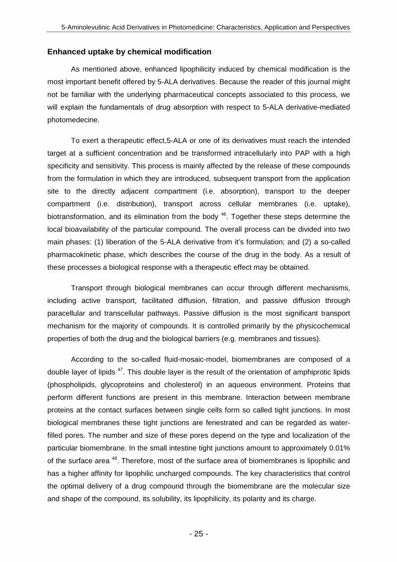
Enhanced uptake by chemical modification
As mentioned above, enhanced lipophilicity induced by chemical modification is the
most important benefit offered by 5-ALA derivatives. Because the reader of this journal might
not be familiar with the underlying pharmaceutical concepts associated to this process, we
will explain the fundamentals of drug absorption with respect to 5-ALA derivative-mediated
photomedecine.
To exert a therapeutic effect,5-ALA or one of its derivatives must reach the intended
target at a sufficient concentration and be transformed intracellularly into PAP with a high
specificity and sensitivity. This process is mainly affected by the release of these compounds
from the formulation in which they are introduced, subsequent transport from the application
site to the directly adjacent compartment (i.e. absorption), transport to the deeper
compartment (i.e. distribution), transport across cellular membranes (i.e. uptake),
biotransformation, and its elimination from the body 46. Together these steps determine the
local bioavailability of the particular compound. The overall process can be divided into two
main phases: (1) liberation of the 5-ALA derivative from it’s formulation; and (2) a so-called
pharmacokinetic phase, which describes the course of the drug in the body. As a result of
these processes a biological response with a therapeutic effect may be obtained.
Transport through biological membranes can occur through different mechanisms,
including active transport, facilitated diffusion, filtration, and passive diffusion through
paracellular and transcellular pathways. Passive diffusion is the most significant transport
mechanism for the majority of compounds. It is controlled primarily by the physicochemical
properties of both the drug and the biological barriers (e.g. membranes and tissues).
According to the so-called fluid-mosaic-model, biomembranes are composed of a
double layer of lipids 47. This double layer is the result of the orientation of amphiprotic lipids
(phospholipids, glycoproteins and cholesterol) in an aqueous environment. Proteins that
perform different functions are present in this membrane. Interaction between membrane
proteins at the contact surfaces between single cells form so called tight junctions. In most
biological membranes these tight junctions are fenestrated and can be regarded as water-
filled pores. The number and size of these pores depend on the type and localization of the
particular biomembrane. In the small intestine tight junctions amount to approximately 0.01%
of the surface area 48. Therefore, most of the surface area of biomembranes is lipophilic and
has a higher affinity for lipophilic uncharged compounds. The key characteristics that control
the optimal delivery of a drug compound through the biomembrane are the molecular size
and shape of the compound, its solubility, its lipophilicity, its polarity and its charge.
Chapter II
- 26 -
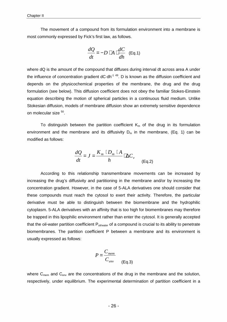
The movement of a compound from its formulation environment into a membrane is
most commonly expressed by Fick’s first law, as follows.
dh
dCAD
dt
dQ ⋅⋅−= (Eq.1)
where dQ is the amount of the compound that diffuses during interval dt across area A under
the influence of concentration gradient dC·dh-1 49. D is known as the diffusion coefficient and
depends on the physicochemical properties of the membrane, the drug and the drug
formulation (see below). This diffusion coefficient does not obey the familiar Stokes-Einstein
equation describing the motion of spherical particles in a continuous fluid medium. Unlike
Stokesian diffusion, models of membrane diffusion show an extremely sensitive dependence
on molecular size 50.
To distinguish between the partition coefficient Km of the drug in its formulation
environment and the membrane and its diffusivity Dm in the membrane, (Eq. 1) can be
modified as follows:
vmm C
h
ADKJ
dt
dQ ∆⋅⋅⋅
== (Eq.2)
According to this relationship transmembrane movements can be increased by
increasing the drug’s diffusivity and partitioning in the membrane and/or by increasing the
concentration gradient. However, in the case of 5-ALA derivatives one should consider that
these compounds must reach the cytosol to exert their activity. Therefore, the particular
derivative must be able to distinguish between the biomembrane and the hydrophilic
cytoplasm. 5-ALA derivatives with an affinity that is too high for biomembranes may therefore
be trapped in this lipophilic environment rather than enter the cytosol. It is generally accepted
that the oil-water partition coefficient Poil/water of a compound is crucial to its ability to penetrate
biomembranes. The partition coefficient P between a membrane and its environment is
usually expressed as follows:
env
mem
C
CP =
(Eq.3)
where Cmem and Cenv are the concentrations of the drug in the membrane and the solution,
respectively, under equilibrium. The experimental determination of partition coefficient in a
5-Aminolevulinic Acid Derivatives in Photomedicine: Characteristics, Application and Perspectives
- 27 -
biological environment is a very difficult task. Therefore, model systems simulating biological
membranes have been proposed.
Although other solvent systems have been proposed for this purpose and certainly
deserve attention as well, the system 1-octanol/water has been widely accepted as a
relatively good predictor 51. Additional reasons to use 1-octanol/water partition coefficients,
usually expressed in their logarithmic form (log P), are the fact that large compilations of
experimental logP values are available and various theoretical approaches exist to estimate
log P values. In addition to these advantages a further practical reason to use the
1-octanol/water system is that 1-octanol, unlike many other organic solvents, is a reasonably
good solvent for many organic compounds.
There is a limited number of comprehensive studies in which membrane permeability
has been related to the corresponding P. These “structure–activity” relationships have a
characteristic pattern. First, they all reveal an essentially linear region in which, according to
Fick’s law, the permeability increases with log P. Second, as log P becomes large the
relationship reaches a plateau after which, in some cases, permeability decreases. Uehlinger
et al. 52 have determined the log P values of a homologous series of 5-ALA n-alkylesters.
They have shown that, by simple esterification, P can be varied by more than four orders of
magnitude between 5-ALA (log P = -1.5) and 5-ALA octylester (logP = 2.6).
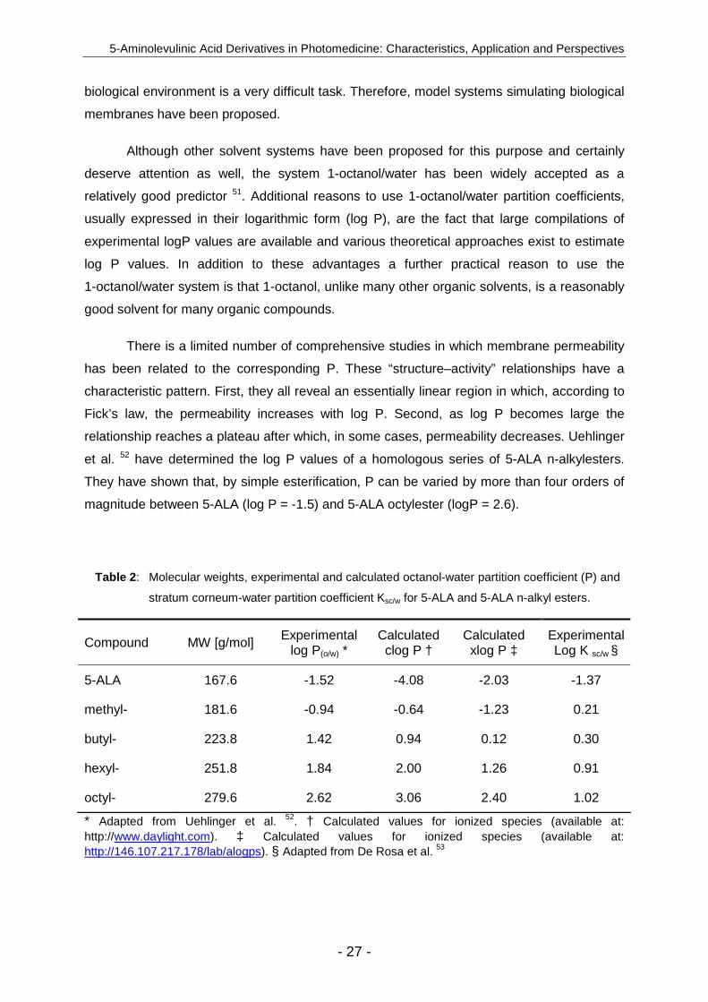
Table 2 : Molecular weights, experimental and calculated octanol-water partition coefficient (P) and
stratum corneum-water partition coefficient Ksc/w for 5-ALA and 5-ALA n-alkyl esters.
Compound MW [g/mol] Experimental log P(o/w) *
Calculated clog P †
Calculated xlog P ‡
Experimental Log K sc/w §
5-ALA 167.6 -1.52 -4.08 -2.03 -1.37
methyl- 181.6 -0.94 -0.64 -1.23 0.21
butyl- 223.8 1.42 0.94 0.12 0.30
hexyl- 251.8 1.84 2.00 1.26 0.91
octyl- 279.6 2.62 3.06 2.40 1.02
* Adapted from Uehlinger et al. 52. † Calculated values for ionized species (available at: http://www.daylight.com). ‡ Calculated values for ionized species (available at: http://146.107.217.178/lab/alogps). § Adapted from De Rosa et al. 53

Chapter II
- 28 -
As mentioned above, a multitude of different theoretical approaches exist to predict
log P values. Table 2 shows the results of these calculations, as compared to experimental
values of Uehlinger et al. 52 obtained by the shake-flask method.
Recently, Brunner et al. 54 screened a large variety of 5-ALA esters (see below) for
their ability to induce PAP in colonic and urothelial carcinoma cell lines. In brief, human
adenocarcinoma cell line HT29 and urothelial carcinoma cell line J82 were incubated with the
different 5-ALA derivatives at a concentration of 0.12 mM. After 3 hours the cellular
fluorescence was quantified by a fluorescence-activated cell sorter. Of interest, when plotting
PAP fluorescence intensity induced by different 5-ALA esters as a function of calculated
lipophilicity, a clear biphasic relationship that accords with the considerations discussed
above becomes apparent (Fig. 3).
0
20
40
60
80
-3 -2 -1 0 1 2 3 4 5 6
lipophilicity (calculated xlog P )
PA
P f
luor
esce
nce
[a.u
.]
5-ALAn-alkylglycolfluoratedthioesteraromaticdendritic
Figure 3 : PAP fluorescence in J82 urothelial cells exposed to various 5-ALA derivatives in
function of the calculated lipophilicity xlog P of the incubated compound 54.
However, lipophilicity seems not to be the sole characteristic responsible for the
efficacy of 5-ALA derivative-induced PAP formation. It has been shown by Uehlinger et al. 52
that 5-ALA hexylester and 5-ALA cyclohexylester displaying essentially the same log P yet
differ widely with respect to their activity. These results where confirmed by Whitaker et al. 55
5-Aminolevulinic Acid Derivatives in Photomedicine: Characteristics, Application and Perspectives
- 29 -
in investigations involving a series of straight-chained, branched and cyclo- 5-ALA alkylester
in a pancreatic tumor cell line.
The advantage of 5-ALA derivatives over 5-ALA can be mainly attributed to the
increases in the following independent processes: the rate at which these compounds reach
the target site, the rate at which they reach the intracellular space and the rate of their
enzymatic conversion into photoactive compounds The rates of these processes vary
significantly, depending on the nature of the particular compound.
Today it is well accepted that 5-ALA gains access to the intracellular space via active-
transport mechanisms that depend on the Na+ and Cl- concentration 56. Whitaker et al. 55
have identified dipeptide and tripeptide transporters, such as PEPT1 transporters, as
potential carriers for 5-ALA. Moreover, in an epithelial cell line Döring et al. 57 observed that
the PEPT2 transporter system appears to be involved in the transmembrane transport of
5-ALA. Despite the structural similarity of 5-ALA and γ-aminobutyric acid (GABA), 5-ALA but
not GABA was found to compete with the active uptake of PEPT1 and PEPT2.
However, Berg et al. 56 have shown in human adenocarcinoma cell line that GABA
inhibits effectively the uptake of 5-ALA and, thus, PpIX production. Furthermore, other
structurally related compounds mainly transported by β system transporters impeded 5-ALA
uptake. With the exception of valine, methionine and threonine, zwitterionic and basic amino
acid were found to inhibit the uptake of 5-ALA.
In contrast to 5-ALA, the cellular uptake of MAL has been shown to be independent
on the Cl- concentration 58. Furthermore, the latter compound neither competes with the
uptake of GABA nor inhibits the facilitated transport of 5-ALA. On the other hand the
transport of MAL can be inhibited by the presence of nonpolar amino acids, such as alanine,
methionine, tryptophan and glycine. Uptake of more-lipophilic compound was essentially
unaffected by inhibitors of the transport of 5-ALA and MAL was the HAL. An apparent
working hypothesis for this phenomenon might be that a passive diffusion or endocytosis are
the main mechanisms contributing to the uptake of moderately lipophilic 5-ALA derivatives.
The absence of significant resistance to the uptake of lipophilic 5-ALA esters is further
supported by the observation that exposure of cells 59 or living pig bladder mucosae 60 for
short periods was sufficient to maintain PAP formation over long periods. In the latter case it
was shown that application durations as short as 10 min provided an intracellular pool of HAL
sufficient to maintain PAP accumulation for at least 5 h. The significant differences in the
cellular uptake mechanisms between 5-ALA and its esters might have an important impact
on the clinical use of PAP-mediated PDT. It has been hypothesized that these differences

Chapter II
- 30 -
might be the basis for less pain during MAL-based treatment of AK, compared 5-ALA based
treatment.
Another important observation with respect to the bioavailability of 5-ALA derivatives
is based on some initial experiments of Kloek et al. 43 using Jurkat lymphoma cells. They
allowed 5-ALA derivatives to penetrate the cell membrane during an incubation period
(0-30 min). Then, after washing the cells, PAP synthesis was observed. These experiments
showed that, when the incubation period was kept to a minimum, the differences between
5-ALA derivative and 5-ALA-induced porphyrin synthesis were maximised. These
observations can mainly be attributed to the fact that 5-ALA esters enter the intracellular
space without significant resistance compared to 5-ALA, thereby rapidly providing pool of
PAP precursors that is sufficient enough to maintain PAP synthesis over long periods. Such
short application times might be advantageous in clinical applications, such as the
fluorescence photodetection of Barrett’s esophagus 61, in which good contact between the
drug and the mucosa can only be maintained for several minutes after topical administration.
Bioconversion of 5-ALA derivatives
Below, it can be seen that there are nearly no limitations with the respect to the
creativity of the medicinal chemist. Therefore, a nearly unlimited number of different 5-ALA
derivatives can be produced that have different chemical, physical, biological and
pharmacological properties. However, during the design of such compounds one should bare
in mind the fundamental ideas (i.e. improvement of bioavailability and increased PAP
formation) on which the use of these compounds in PDT and FD relies. Therefore, several
considerations should be taken into account before planning the synthesis of new 5-ALA
derivatives by means of concepts in rational drug design 62,63.
The tetrapyrrolic structure of porphyrins implies the cleavage of 5-ALA amides before
initiation of heme biosynthesis. This aspect is supported by the experimental observation that
5-ALA amides and their esters generally fail to induce large amounts of PAP in
vitro 43,64,65,66,67,68 and in vivo 68,43 in the absence of specific peptidases. Furthermore, Moan et
al. have recently shown that an N-formyl 5-aminolevulinic acid derivative neither induced
porphyrin synthesis nor inhibited the formation of PpIX induced by 5-ALA 69. However, these
characteristics might be advantageously used to further increase the selectivity of 5-ALA by
targeting specific proteases found in abundance in some tumors 64,70,71,72. Not only
N-substituted 5-ALA derivatives must be cleaved, and dendrimeric 5-ALA derivatives, such

5-Aminolevulinic Acid Derivatives in Photomedicine: Characteristics, Application and Perspectives
- 31 -
as described in the concerned section, must be able to release 5-ALA in order to be potent
substrates for heme biosynthesis. Although some researchers hypothesize that the same
holds true for modified 5-ALA at the C-terminal function 73,64,65,74,67, the situation is less clear
for these esterified compounds from a (bio) chemical point of view.
To the best of our knowledge there is currently no direct experimental evidence that
5-ALA esters cannot act as substrate for the enzymes involved in heme biosynthesis.
Knowledge of the mechanisms of 5-ALA ester-mediated PAP formation at the cellular level
might, however, be mandatory for the medicinal chemist for the synthesis for more potent
porphyrin precursors. The huge difficulties in elucidating differences between 5-ALA and
5-ALA derivative-induced porphyrin formation might be partly explained by the fact that there
are no differences in the absorption or emission spectra between, e.g. PpIX or PpIX dimethyl
ester (a potential PAP following exposure cells to MAL). Furthermore, Gaullier et al., and
Vena et al. have observed no differences in fluorescence localization after exposure to
5-ALA or 5-ALA esters at the cellular level 75,76.
Assuming that 5-ALA esters enter heme biosynthesis without undergoing hydrolysis,
the metalloprotein 5-ALA dehydrase will be the first obstacle of such compounds towards
PAP biosynthesis. As mentioned above this enzyme catalyzes the nonsymmetrical
condensation of two 5-ALA molecules. Recently, Jarret et al. 77 showed that the initial C-N
bound formation is the main pathway for the final dimerization of two 5-ALA molecules to
give porphobilinogen (PBG). Indeed, sterically hindered or extremely large 5-ALA derivatives
would not be able to act as substrate for this enzyme without cleavage prior to this enzyme-
catalyzed reaction.
In this context, one way to test for the ability of 5-ALA esters to act as direct
precursors for PBG monoester or diester would be the use of the purified enzyme and the
determination of the corresponding Michaelis-Menten kinetics for various 5-ALA esters in the
presence and absence of nonspecific esterases. Using TLC methods Taylor et al. 78 showed
that exposure of 5-ALA dehydrase to a mixture of 5-ALA and 5-ALA ethylester resulted in the
appearance of a second peak in chromatograms, which they attributed to the formation of
PBG monoethylester. Furthermore, we have recently shown that the exposure of MAL to
5-ALA dehydrase resulted in a product susceptible to undergo a characteristic reaction with
Ehrlich’s reagent.
Interestingly, topical or intravenous administration of 5-ALA esters to tumour-bearing
mice resulted in a greater porphyrin accumulation in skin, compared with tumor tissue.
Because esterase activity in these normal tissues was found to be higher than in tumor
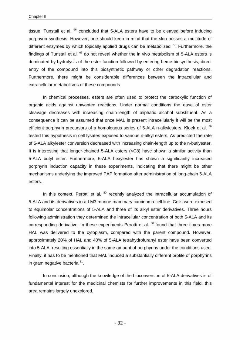
Chapter II
- 32 -
tissue, Tunstall et al. 66 concluded that 5-ALA esters have to be cleaved before inducing
porphyrin synthesis. However, one should keep in mind that the skin posses a multitude of
different enzymes by which topically applied drugs can be metabolized 79. Furthermore, the
findings of Tunstall et al. 66 do not reveal whether the in vivo metabolism of 5-ALA esters is
dominated by hydrolysis of the ester function followed by entering heme biosynthesis, direct
entry of the compound into this biosynthetic pathway or other degradation reactions.
Furthermore, there might be considerable differences between the intracellular and
extracellular metabolisms of these compounds.
In chemical processes, esters are often used to protect the carboxylic function of
organic acids against unwanted reactions. Under normal conditions the ease of ester
cleavage decreases with increasing chain-length of aliphatic alcohol substituent. As a
consequence it can be assumed that once MAL is present intracellularly it will be the most
efficient porphyrin precursors of a homologous series of 5-ALA n-alkylesters. Kloek et al. 59
tested this hypothesis in cell lysates exposed to various n-alkyl esters. As predicted the rate
of 5-ALA alkylester conversion decreased with increasing chain-length up to the n-butlyester.
It is interesting that longer-chained 5-ALA esters (<C8) have shown a similar activity than
5-ALA butyl ester. Furthermore, 5-ALA hexylester has shown a significantly increased
porphyrin induction capacity in these experiments, indicating that there might be other
mechanisms underlying the improved PAP formation after administration of long-chain 5-ALA
esters.
In this context, Perotti et al. 80 recently analyzed the intracellular accumulation of
5-ALA and its derivatives in a LM3 murine mammary carcinoma cell line. Cells were exposed
to equimolar concentrations of 5-ALA and three of its alkyl ester derivatives. Three hours
following administration they determined the intracellular concentration of both 5-ALA and its
corresponding derivative. In these experiments Perotti et al. 80 found that three times more
HAL was delivered to the cytoplasm, compared with the parent compound. However,
approximately 20% of HAL and 40% of 5-ALA tetrahydrofuranyl ester have been converted
into 5-ALA, resulting essentially in the same amount of porphyrins under the conditions used.
Finally, it has to be mentioned that MAL induced a substantially different profile of porphyrins
in gram negative bacteria 81.
In conclusion, although the knowledge of the bioconversion of 5-ALA derivatives is of
fundamental interest for the medicinal chemists for further improvements in this field, this
area remains largely unexplored.

5-Aminolevulinic Acid Derivatives in Photomedicine: Characteristics, Application and Perspectives
- 33 -
Bioavailability and local Bioavailability of 5-ALA derivatives
In addition to the enzymatic conversion of 5-ALA or one of its derivatives into
porphyrins within the cells, the efficacy of this treatment modality largely depends on the
local concentration of the particular compound at the target site (i.e. on its ability to penetrate
the tissue and reach the lesion). The resulting local concentration after topical administration
depends on characteristics such as drug permeability and diffusion coefficients in the tissue,
which are in turn functions of drug structure, formulation, drug release, and local drug
clearance.
Despite the recent approvals of MAL for the photodynamic treatment of skin
conditions and HAL for diagnostic purposes in urology 82,83,74, the ability of 5-ALA derivatives
to induce more porphyrins after topical administration in vivo still remains controversial. In
cell culture several 5-ALA derivatives have been shown to increase PAP formation,
compared to the parent compound, even when they are applied at significantly lower
concentrations 43,52,54. As such, the magnitude of porphyrin production and the optimal
substrate concentration depended on the used cell lines that were used. Furthermore, some
5-ALA esters have been shown to induce higher PAP levels when applied topically to a pig
bladder model that had intact morphologic architecture 84. This is in agreement with clinical
studies 73,74 showing the superiority of HAL over the parent compound with respect to
porphyrin formation and drug penetration.
Using an alcoholic solution of 5-ALA ethyl ester, 5-ALA butylester and 5-ALA
(hydroxymethyl)tetrahydrofuranly ester (10% wt/vol), Kloek et al. 43 showed that PAP
formation increased between two- and three-fold in vivo, compared with 5-ALA, 6 hours after
topical administration. This is in agreement with results of a later study in which higher PAP
levels were observed 14 hours after of nude mice were exposed to creams containing short-
chained 5-ALA esters. However, results obtained after topical administration to the skin in
vivo as well as in vitro are somewhat contradictory. In this context other investigators
observed that MAL applied to the skin of nude mice for 24 hours induced lower porphyrin
levels with a delayed tmax compared with that for 5-ALA under the same conditions 85.
In a more recent study, when 5-ALA and its hexylester were formulated in a standard,
lipophilic ointment similar porphyrin levels were observed when applied to the skin of nude
mice for 24 h or 10 min after removal of the stratum corneum 86. In this study, short term
application, however, resulted in lower fluorescence intensities for HAL than for 5-ALA.
These results have been confirmed under similar experimental conditions with 5-ALA methyl,
hexyl, octyl, and diethylenglycol monomethylether ester 87,88. In hairless mouse skin with

Chapter II
- 34 -
UVB-induced early skin cancer, the pentyl ester derivative of 5-ALA induced slightly more
PAPs than did 5-ALA when formulated in a hydrogel. However, fluorescence microscopy
revealed no significant difference in the displastic layers of the epidermis. The biodistribution
measured by fluorescence intensities after topical administration of 5-ALA and MAL in
human AK have been compared 89. Although PAP formation was found to be more efficient
with 5-ALA, the selectivity for the diseased tissue was higher with MAL.
As discussed above, these somewhat confusing results may be mainly attributed to
the disregard of two basic principles in drug delivery: the role of biological barriers (in the
case of the skin, the stratum corneum [SC]), and the important role of the vehicle for the
optimized delivery to the target site. Therefore, unambiguous conclusions cannot be
deduced. However, it can be concluded from studies in which the SC was removed by either
shaving 43 or tape stripping 86,37,90 that 5-ALA esters were at least as efficient as 5-ALA. On
the other hand, without pretreatment preparation of the application area of long-chained
5-ALA esters, a significant lag time before the onset of the porphyrin fluorescence signal can
be observed 86,88,53. This lag time is characteristic for the “reservoir function” of the SC for
lipophilic compounds 91.
Recently, the partition coefficient between the SC and water (Ksc/w) was determined
by de Rosa et al. 53 (see Table 2). In this study, the log Ksc/w of 5-ALA esters was significantly
higher than the log Ksc/w of 5-ALA. The highest measurable log Ksc/w was approximately 1 for
HAL and 5-ALA octylester. In the same study an oil-in-water emulsion containing different 5-
ALA derivatives (2% w/w) was used to determine the penetration of the compounds into
different compartments of dermatomed skin of mice. In contrast to most other studies using
porphyrin fluorescence as indirect measure to assess 5-ALA uptake, this study directly
quantified the active compound by means of precolumn derivatization fluorescence
HPLC 92,93. It was shown that the movement of 5-ALA across full thickness skin tissue
remained nearly constant over 12 h. However, the movement of HAL constantly increased in
a sigmoidal manner over the same period. After 6 h the flux of HAL was significantly higher
than for 5-ALA. The movements of other esters tested in this study were substantially lower,
but had the same sigmoidal flux-time profile than 5-ALA hexylester.
This might indicate differences in permeation mechanisms between 5-ALA esters and
the parent compound. The permeation of 5-ALA octylester through the skin was found to be
significantly less than that of the hexylester derivative, both differing only slightly in their Ksc/w
values. However, a low permeation across the skin does not necessarily mean low
penetration into the skin. The increased lipophilicity of 5-ALA esters might decrease the
partition between the lipophilic environment of the skin and the hydrophilic environment of
5-Aminolevulinic Acid Derivatives in Photomedicine: Characteristics, Application and Perspectives
- 35 -
the receptor compartment (in vivo, the blood stream). Such a situation would result in the
accumulation of moderately lipophilic compounds in the stratum corneum, epidermis and
dermis. This has been shown by de Rosa et al. 53, who reported that retention of HAL and
5-ALA octylester in the epidermis and the dermis was 2.5-3 times higher than that of 5-ALA.
The results of this study are in slight disagreement with a similar study that was performed by
van den Akker et al. 90, who showed less permeation of HAL through mouse skin. However,
in this study significantly higher drug doses and a different formulation were used.
Furthermore, the pH was significantly higher thus, further increasing the lipophilicity of the
compounds. Finally, an aqueous solution has been employed in the study performed by de
Rosa et al., revealing the important impact of the vehicle in the controlled drug delivery of
5-ALA and its derivatives. Indeed, optimization of the vehicle is a crucial step for transdermal
and dermatological drug delivery. The appropriate choice of the formulation not only involves
delivery of the compound to the target tissue but also maximizes therapeutic efficacy by
considering the etiology of the disease and the mechanism of action of the drug. In this
context only few studies have addressed this optimized formulation of lipophilic 5-ALA
derivatives.
From a very simplified point of view, one has to maximize the partition coefficient
Km/veh (membrane/vehicle) in order to maximize the flux of a particular compound from the
vehicle into the tissue. For relatively lipophilic compounds such as HAL an aqueous solution
might be the optimal choice 73. From a practical point of view, the intravesical administration
of an aqueous solution to the bladder presents no major problem. However, compounds that
are too lipophilic might tend to precipitate in an aqueous environment, thereby impeding the
tissular uptake of the active ingredient. Furthermore, in other medical indications where
topical administration is desired, such as dermatology or gynaecology, a liquid might not fully
guarantee the retention of the compound at the target site. Recently, we have shown, that
the release of 5-ALA derivatives from a hydrogel into a lipid matrix increases with increasing
lipophilicity 94. Furthermore, release nearly vanished after use of standard lipophilic ointments
such as unguentum or a “cold cream” formulation 87,66,95,37,96.
Another factor strongly influencing the release profiles of a topically applied
compound is its localization within the vehicle. For example, in oil-in-water emulsions more-
lipophilic compounds such as HAL will tend to dissolve in the internal phase, whereas
hydrophilic compounds such as 5-ALA or MAL will dissolve preferentially in the external
phase. Thus, it will take more time for lipophilic 5-ALA esters to migrate to the interface
between vehicle and tissue, resulting in substantially different release profiles than observed
for the parent compound under the same conditions 53.
Chapter II
- 36 -
Batlle et al. has expended much effort to optimize formulations for 5-ALA
esters 97,98,29,19. Cell culture experiments showed no increased porphyrin buildup in murine
tumor cells with liposomal formulation of HAL, compared with 5-ALA 29. In another report 97,
they sowed that a lotion containing DMSO and ethanol was favourable with respect to PpIX
buildup when using 5-ALA, whereas a cream seemed to be more advantageous for topical
administration of HAL. Experiments on tumor-bearing mice showed no significant differences
between 5-ALA and HAL-mediated porphyrin formation with these optimized formulations. In
a subsequent study 98, a lotion was identified to be optimal for the delivery of HAL to the skin.
In tumor bearing-mice this formulation led to the induction of substantially higher PAP levels
compared with the cream formulation. Furthermore, fluorescence surpassed that induced by
5-ALA 5 h after administration.
Recently, it was shown that the addition of penetration enhancers, such as ibuprofen
acid or medium-chain triglycerides, to hydrophilic formulations increases the penetration of
5-ALA butlyester through the SC by a factor of 10 99,100. A liposomal formulation of HAL,
however, failed to increase the porphyrin formation in murine tumor cells, compared to
liposomal 5-ALA 29. In contrast, a liposomal formulation of 5-ALA substantially increased its
systemic bioavailability, compared with an aqueous solution.
Another important factor that potentially influences the release of the drug and its
permeation through biological membranes is its physical appearance (i.e. state) within the
delivery vehicle. Using Fourier-transform Infrared spectral measurements, Chen et al. 101
have found that HAL in solution exists principally in its monomeric form whereas 5-ALA,
similar to other organic acids, is present as a mixture of monomers and dimers.
In addition to penetration enhancers, iontophoresis has been proposed to further
increase the bioavailability of 5-ALA derivatives after topical administration. Because 5-ALA
esters have a positive charge at the N-terminal under physiological conditions, they are
supposed to be transported by electromigration through the SC. It has been demonstrated
that 50 times more MAL and 15 times more HAL can be transported through the skin than
5-ALA by iontophoresis 102. Furthermore, Gerscher et al. 103,104 have used iontophoretic
methods to administer 5-ALA, 5-ALA butyl ester, 5-ALA pentyl ester, and HAL to healthy
volunteers. In agreement with the findings of van den Akker et al. 86 they found that HAL-
mediated PAP fluorescence peaked earlier compared to 5-ALA-induced PpIX under similar
conditions. Furthermore, HAL was found to be slightly more phototoxic and more
homogenously distributed than 5-ALA. A more homogenous distribution of PAP fluorescence
after exposure to HAL, as compared to 5-ALA, was also reported by other authors after in

5-Aminolevulinic Acid Derivatives in Photomedicine: Characteristics, Application and Perspectives
- 37 -
vitro and in vivo experiments 84,73,53,105. In these experiments HAL was formulated in an
aqueous solution or in a rather hydrophilic vehicle.
On the basis of the considerations mentioned above it can be deduced that, apart
from an optimal concentration (see below), 5-ALA derivatives have to be formulated with
respect to their lipophilicity, their physicochemical properties and the specific medical
application.
Another important difference between 5-ALA and 5-ALA derivatives in terms of
bioavailability is the systemic uptake of these compounds after topical administration.
Several studies involving topical 5-ALA application to mice have shown that PpIX production
is not limited to the administration area 42,85,19,67,95,37,106. Substantial PpIX fluorescence was
found not only in the flank colateral to application site, but also in other body compartments,
such as blood, liver, spleen and kidneys 26 and liver, intestinal tract and lungs 106. Topical
administration of 5-ALA for 24 hours to nude mice showed fluorescence intensities at other
sites (including the ears and the collateral site, but lower in the liver, muscle and brain) that
were similar to fluorescence intensities at the application site. Although 5-ALA methyl, ethyl,
propyl or hexyl ester can induce similar or higher fluorescence levels at the application sites
of nude mice, such systemic effects were not observed. This can presumably be attributed to
differences in metabolic degradation (de Rosa et al.)53, binding to plasma proteins and
limited capacity to overcome the endothelial layers.
Chemistry and Design of 5-ALA derivatives
5-ALA alkylester
The simpliest way to modify the lipophilicity of an organic acid is the derivatization
with various alkyl alcohols into the corresponding ester derivative. Generally, 5-ALA esters
are prepared using standard reactions for esterification between 5-ALA and the
corresponding alcohol in the presence of thionylchloride or hydrochloric acid 43 (Fig. 4). In
this way several linear (compounds 2-9 ), branched (10-18), cyclic (19-32) as well as
ethylenglycol derivatives (33-38) have been prepared. However, in some cases it is
advantageous to use tert-butylcarbonyl (BOC) chemistry to prevent cross reactivity when
using carbodiimide coupling to activate the carboxylic function.
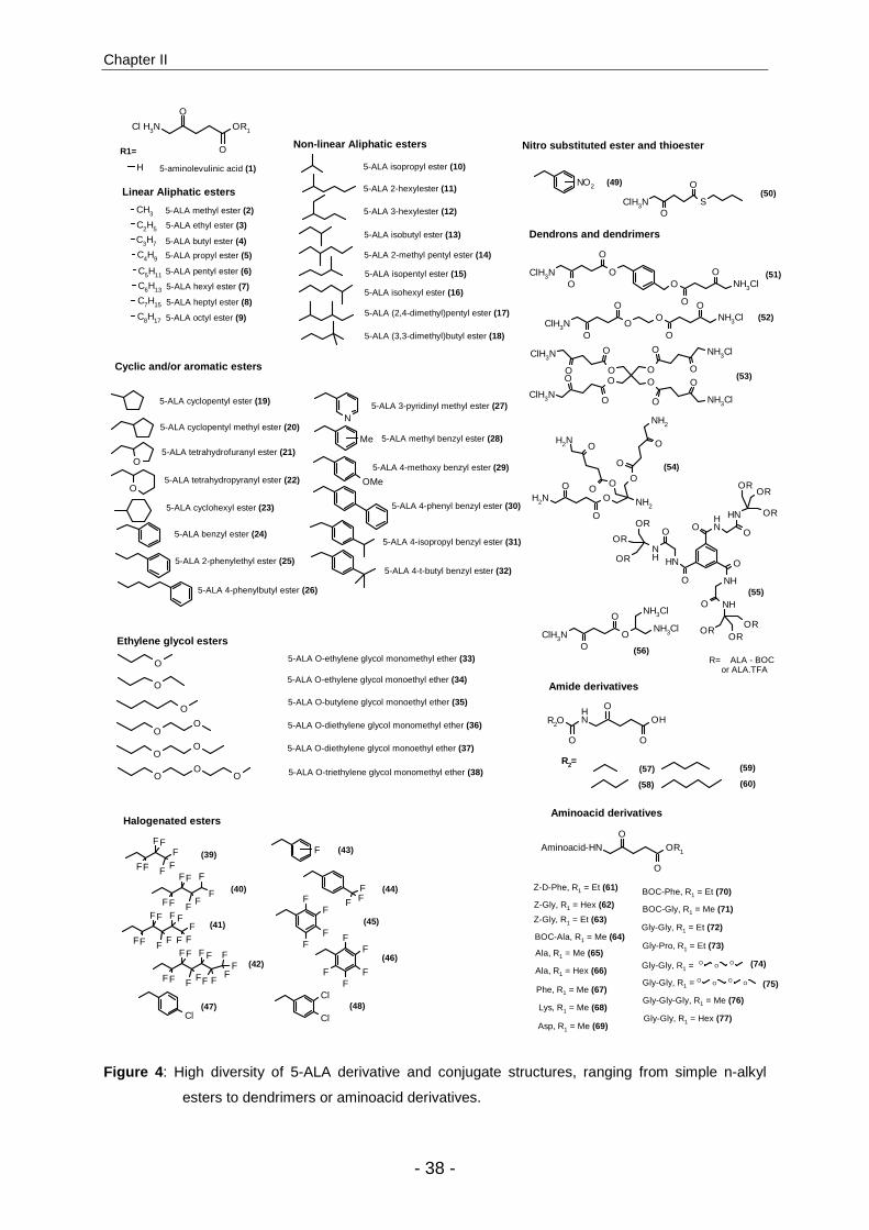
Chapter II
- 38 -
OR1
O
Cl H3N
O
CH3
C2H5
C3H7
C4H9
C5H11
C6H13
C7H15
C8H17
O
O
O
O
OO
OO
O
H
O
OO
N
OMe
Me
FF
FF
FF
F
FF
FF
FF
F
F
FF
FF
FF
F F
FF
F
FF
FF
FF
F F
F F
FF
F
FF
FFF
FF F
F
FFF
ClH3N SO
O
F
Cl Cl
Cl
NO2
ClH3NO
O
OO NH3Cl
O
O
ClH3N
O
O
OO
NH3ClO
OO
NH3ClO
O
ClH3N
O
O
O
NH2 OO
O
NH2
O
O
O
NH2
O
O
O
NH2
NH
O
O NH
OROR
OR
NH
O
O
NH
OR
OR
OR NH
OO
NH
OROR
OR
O NH3Cl
O
O
ClH3NO
O
O
ClH3N ONH3Cl
NH3ClO
O
R2O OH
O
NH
OO
R2=
OR1
OAminoacid-HN
O
OO
O
O O O O
5-aminolevulinic acid (1)
R1=
Linear Aliphatic esters
5-ALA methyl ester (2)
5-ALA ethyl ester (3)
5-ALA butyl ester (4)
5-ALA propyl ester (5)
5-ALA pentyl ester (6)
5-ALA hexyl ester (7)
5-ALA heptyl ester (8)
5-ALA octyl ester (9)
Non-linear Aliphatic esters
5-ALA isopropyl ester (10)
5-ALA isobutyl ester (13)
5-ALA isopentyl ester (15)
5-ALA (2,4-dimethyl)pentyl ester (17)
Cyclic and/or aromatic esters
5-ALA cyclopentyl ester (19)
5-ALA cyclopentyl methyl ester (20)
5-ALA tetrahydrofuranyl ester (21)
5-ALA tetrahydropyranyl ester (22)
5-ALA cyclohexyl ester (23)
5-ALA benzyl ester (24)
Ethylene glycol esters
5-ALA O-ethylene glycol monomethyl ether (33)
5-ALA O-ethylene glycol monoethyl ether (34)
5-ALA O-diethylene glycol monoethyl ether (37)
5-ALA O-triethylene glycol monomethyl ether (38)
5-ALA O-diethylene glycol monomethyl ether (36)
5-ALA O-butylene glycol monoethyl ether (35)
5-ALA 2-hexylester (11)
5-ALA 3-hexylester (12)
5-ALA isohexyl ester (16)
5-ALA 2-methyl pentyl ester (14)
5-ALA (3,3-dimethyl)butyl ester (18)
5-ALA 2-phenylethyl ester (25)
5-ALA 4-phenylbutyl ester (26)
5-ALA 3-pyridinyl methyl ester (27)
5-ALA methyl benzyl ester (28)
5-ALA 4-methoxy benzyl ester (29)
5-ALA 4-phenyl benzyl ester (30)
5-ALA 4-isopropyl benzyl ester (31)
5-ALA 4-t-butyl benzyl ester (32)
(39)
(40)
(41)
(42)
(43)
(44)
(45)
(46)
Halogenated esters
Nitro substituted ester and thioester
(49)(50)
(47) (48)
R= ALA - BOC or ALA.TFA
Dendrons and dendrimers
(51)
(52)
(53)
(54)
(55)
(56)
(57)
(58)
(59)
(60)
Z-D-Phe, R1 = Et (61)
Z-Gly, R1 = Hex (62)
Z-Gly, R1 = Et (63)
BOC-Ala, R1 = Me (64)
Ala, R1 = Me (65)
Ala, R1 = Hex (66)
Phe, R1 = Me (67)
Gly-Gly, R1 = Et (72)
Gly-Pro, R1 = Et (73)
Gly-Gly, R1 =
Gly-Gly, R1 =
Gly-Gly-Gly, R1 = Me (76)
Gly-Gly, R1 = Hex (77)Lys, R1 = Me (68)
Asp, R1 = Me (69)
(74)
(75)
Aminoacid derivatives
Amide derivatives
BOC-Phe, R1 = Et (70)
BOC-Gly, R1 = Me (71)
Figure 4 : High diversity of 5-ALA derivative and conjugate structures, ranging from simple n-alkyl
esters to dendrimers or aminoacid derivatives.
5-Aminolevulinic Acid Derivatives in Photomedicine: Characteristics, Application and Perspectives
- 39 -
However, care should be taken about the acid used for deprotection of the BOC
function, since the resulting salts (e.g. HCl or TFA) might substantially alter the
pharmacokinetics of the active compound 107.
For example 5-ALA thionylester derivatives have been prepared according to this
procedure. These compounds maintained biological activity in vitro 54. Furthermore,
halogenated alcohol moieties have been introduced that enhanced the lipophilicity,
compared with their hydrocarbonyl counterparts (39-48). In this way the lipophilicity of 5-ALA
can easily be modified over several orders of magnitude 52. Furthermore, the electron
withdrawing effect of the halogenosubstituents might have additional influence on the
porphyrin production capacity of such compounds.
The general approaches to assess the different derivatives with respect to their
porphyrin induction capacity are mainly in vitro tests. These can be done in cell
culture 108,75,109, cell culture on hen egg model 110, 3D cell culture 105,111, skin explants 90 99 or
other organ culture models 60,112,113. To which extend these in vitro tests reflect the real in
vivo situation in terms of bioavailability remains unclear. However, some basic conclusions
may already be drawn from these experiences.
First, in tests in which dose-response curves have been determined 52 a characteristic
biphasic relationship was found. In these dose-response curves PAP generation is positively
correlated up to an optimal concentration Copt at which highest fluorescence intensities can
be observed; beyond this value PAP formation sharply decreases. This decrease of PAP
formation at higher doses can most likely be attributed to cytotoxic effects generated by the
particular compound itself or its metabolic products. Furthermore, disruption of the cell
membrane following exposure of high doses of lipophilic 5-ALA esters has been reported 84.
Thus, good equilibrium among the capacity to penetrate cell membrane, the ability to induce,
PAP and cytotoxicity has to be found for each application.
Second, the absolute value of Copt varies with the type of precursor and the particular
cell line. For most long-chained 5-ALA esters (C5-C8) Copt has been found to be 30-150-fold
lower that for 5-ALA. At these low concentrations similar or higher fluorescence intensity
levels have been reached with 5-ALA esters. In terms of phototoxicity no differences have
been observed after 5-ALA and 5-ALA ester-mediated PDT at similar porphyrin levels in vitro
and in vivo. The same behavior was observed in a series of fluorinated 5-ALA alkylesters 54.
For these compounds, the short-chain C4 derivative did not achieve high PAP levels,
whereas the long-chained C7 derivative did. Finally, the best 5-ALA derivative for optimal
PAP formation seems to vary from cell line to cell line.
Chapter II
- 40 -
The in vitro fluorescence intensity time profiles may also vary substantially. Uehlinger
et al. 52 have reported a sigmoidal profile over a of 24 h period in which cells of urothelium
origin exposed to HAL in wide concentration range. This is in agreement with Perotti et al. 80
which showed a similar pharmacokinetics in a murine cell line. However, recently, Brunner et
al. 109,54 reported a very fast increase in porphyrin fluorescence intensity, reaching a plateau
already after approximately 10 minutes in HT29 cells.
Whitaker and coworkers 55 have tested a set of linear and branched 5-ALA esters of
the same number of carbon atoms. Their results agreed with those of other
investigators 52,67,114 that a branching point in position 1 from the carboxylic function impedes
porphyrin synthesis in vitro. However, we have recently tested a number of
1,3-diacylglycerides of 5-ALA that have shown a similar in vivo activity than 5-ALA following
systemic administration (see below). However, the corresponding 5-ALA–CH-(CH2-NH2)2
(56) has not shown a promising biological activity in vitro 54. Although the screening of new
5-ALA derivatives in cell culture might give a preliminary indication for promising drug
candidates, such studies cannot answer the major questions in this area of research,
including improved bioavailability and selectivity. Furthermore, the reduced water solubility of
some 5-ALA derivatives might further impede such screening approaches, because
precipitation in the incubation medium might reduce the total available concentration of
porphyrin precursor or because the used solvent system can alter the pharmacokinetics and
heme metabolism.
On the basis of a previous study reported by Krieg et al. 115, Brunner et al.54 have
proposed a screening approach for various 5-ALA derivatives with respect to PAP induction
capacity and selectivity. In their studies, they separately incubated a human cell line of
cancerous origin and their corresponding “normal” counterparts with different 5-ALA esters
and determined the resulting porphyrin contents. In their bladder cancer model (J82/UROtsa
cells) they found a maximal ratio of tumor tissue to normal tissue (T/N) of 2.1 with a
fluorinated hexylester derivative of 5-ALA (41). This T/N ratio corresponds approximately to
the lower end that can be observed with HAL in humans (Collaud et al. submitted). However,
using a similar system consisting of a human adenocarcinoma cell line and a colonic
fibroblast, they found T/N ratios as high as 55. However, it must be taken into account that in
the latter case the number of tumor cells was five times higher than in the case of their
normal counterparts. Nevertheless, such systems represent a valid approach for the
preliminary screening for better 5-ALA derivatives. Even closer to the real in vivo situation
are tumoral coculture models. Very recently, we developed such a system for bladder cancer
(Fotinos et al. unpublished results). For this purpose, GFP-transfected T24 bladder cancer

5-Aminolevulinic Acid Derivatives in Photomedicine: Characteristics, Application and Perspectives
- 41 -
cells were coincubated with fully differentiated, multilayer urothelium of neonatal origin. Three
to five days after incubation, cells of tumor origin developed clearly visible foci, migrating
towards the basement membrane. Incubation with appropriate doses of HAL showed a
selective accumulation of PAP in GFP-labeled cells. Such models will not only enable to test
local bioavailability and selectivity of new 5-ALA derivatives but also to determine optimal
conditions with respect to PDT.
Although the introduction of 5-ALA esters into the field of fluorescence photodiagnosis
can be considered as success story, their application fields will stay restricted to organs that
are accessible by topical administration. Simple 5-ALA esters have been conceived for
topical administration in creams, gels, ointments or solutions rather than for systemic
administration. Although intraperitoneal administration of HAL has been shown to increase
porphyrin production in mouse brain compared with 5-ALA, approaches to use 5-ALA esters
systemically have shown less drastic effects on the formation of PAP in vivo. This is
presumably a result of their limited stability, their binding to plasma proteins and, thus, their
limited bioavailability, as well as to a pronounced acute toxic potential when given
intravenously. 116, 66.
After oral administration a large fraction of 5-ALA esters might be hydrolyzed or
otherwise degraded under the highly acidic conditions present in the stomach. Therefore, at
equimolar doses, the initial advantage over 5-ALA is lost for this administration route.
However, encapsulation in controlled release formulations protecting the respective
derivative against the acidic attack in the stomach might circumvent this problem. Perotti et
al. 116 hypothesized that, once 5-ALA esters reach the blood circulation, because they are too
lipophilic they might not cross the endothelial barrier of the vascular system and might not be
able to reach their target. Another feature that may hamper the intraperitoneal use of simple
5-ALA n-alkylesters is the acute toxicity observed at doses of approximately 1000 mg·kg-1
that has been observed in mice 116. However, intravenous doses of up to 30 mg·kg-1 or
intraperitoneal doses of up to 800 mg·kg-1 have been reported to be safe in mice 66 and
rats 117, respectively. In this context the binding of 5-ALA n-alkylesters to plasma proteins
might play an important role with respect to both toxicity and bioavailability. After intravenous
bolus administration the amount of free 5-ALA n-alkylester can temporarily exceed the toxic
dose, whereas if it is slowly infused (which favors binding to plasma proteins) it can reduce
the toxic potential of 5-ALA derivatives. However, the irreversible binding to plasma proteins
can strongly reduce the systemic bioavailability of lipophilic compounds such as HAL.
However, to the best of our knowledge neither clinical nor animal studies provide consistent
answer with regard to the efficacy of 5-ALA ester after systemic administration.

Chapter II
- 42 -
In addition to the advantageous us of 5-ALA derivatives for internal use in oncology,
the treatment of pathological conditions (such as rheumatoid arthritis 118) might also benefit
from the advantages offered by 5-ALA derivatives in this context. However, the huge
potential of other 5-ALA derivatives with respect to parenteral applications remains largely
unexplored. Important life-threatening diseases including prostate cancer, mammary
carcinoma, breast cancer, lymphoma and brain cancer can thus hardly be treated by this
approach, although 5-ALA esters have been demonstrated to induce high PAP
concentrations in many cell cultures models of these diseases 80,119,52.
The drawback with respect to 5-ALA derivative related research can be mainly
attributed to the fact that investigators in this research area have largely ignored advances
made in recent prodrug research, until today 120,121,122.
For example. in the context of 5-ALA-mediated treatment of brain cancer, it is
generally assumed that increasing lipophilicity increases blood brain barrier permeability in a
systematic fashion. The successful design of lipophilic esters with the ability to enhance
parent drug concentration in the brain in the context of 5-ALA more than likely requires
optimization rather than maximization of lipophilicity and bioconversion rate. The factors
contributing to the existence of an optimal lipophilicity for CNS delivery have been reviewed
recently and several biophysical kinetic models have been developed in this context 123,124,125.
Brain uptake of highly lipophilic compounds is limited by their interactions with plasma
proteins and by unfavorable partition from the endothelium into the underlying tissue, both of
which increase with increasing lipophilicity. There are now a number of instances in which
poor brain uptake of prodrugs designed with increasing lipophilicity has been attributed to
plasma protein binding or fast cleavage by carboxyesterases in the blood. Apparently, similar
mechanisms are also responsible for the failure of 5-ALA ester to induce porphyrins when
applied intravenously.
Despite evidence mentioned above there are examples of extremely lipophilic
properties for efficient CNS delivery. One of these approaches that takes advantage of the
metabolic pathway of lipids is the triglyceride prodrug approach. It benefits from the
metabolic fate of glycerides to improve intrinsic drawbacks of the present compound when
the latter is covalently coupled to position 2 of the glycerolic backbone. Triglycerides are
major constituents of dietary fats. Their absorption involves the hydrolysis by lipases to
monoglycerides and fatty acids. The following three enzymes participates to the catabolism
of lipids: pancreatic lipase, phospholipase A2 and nonspecific esterases. For the degradation
of triglycerides the pancreatic lipase plays the most important role. It hydrolyses the esters in
positions 1 or 3. Hydrolysis in position 2 occurs very slowly. After being released, the
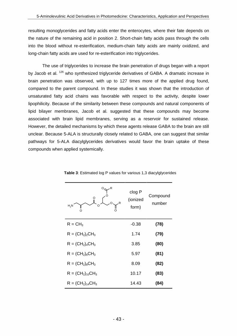
5-Aminolevulinic Acid Derivatives in Photomedicine: Characteristics, Application and Perspectives
- 43 -
resulting monoglycerides and fatty acids enter the enterocytes, where their fate depends on
the nature of the remaining acid in position 2. Short-chain fatty acids pass through the cells
into the blood without re-esterification, medium-chain fatty acids are mainly oxidized, and
long-chain fatty acids are used for re-esterification into triglycerides.
The use of triglycerides to increase the brain penetration of drugs began with a report
by Jacob et al. 126 who synthesized triglyceride derivatives of GABA. A dramatic increase in
brain penetration was observed, with up to 127 times more of the applied drug found,
compared to the parent compound. In these studies it was shown that the introduction of
unsaturated fatty acid chains was favorable with respect to the activity, despite lower
lipophilicity. Because of the similarity between these compounds and natural components of
lipid bilayer membranes, Jacob et al. suggested that these compounds may become
associated with brain lipid membranes, serving as a reservoir for sustained release.
However, the detailed mechanisms by which these agents release GABA to the brain are still
unclear. Because 5-ALA is structurally closely related to GABA, one can suggest that similar
pathways for 5-ALA diacylglycerides derivatives would favor the brain uptake of these
compounds when applied systemically.
Table 3 : Estimated log P values for various 1,3 diacylglycerides
O
O
O R
O
RO
NH2
O
O
clog P
(ionized
form)
Compound
number
R = CH3 -0.38 (78)
R = (CH2)2CH3 1.74 (79)
R = (CH2)4CH3 3.85 (80)
R = (CH2)6CH3 5.97 (81)
R = (CH2)8CH3 8.09 (82)
R = (CH2)10CH3 10.17 (83)
R = (CH2)14CH3 14.43 (84)
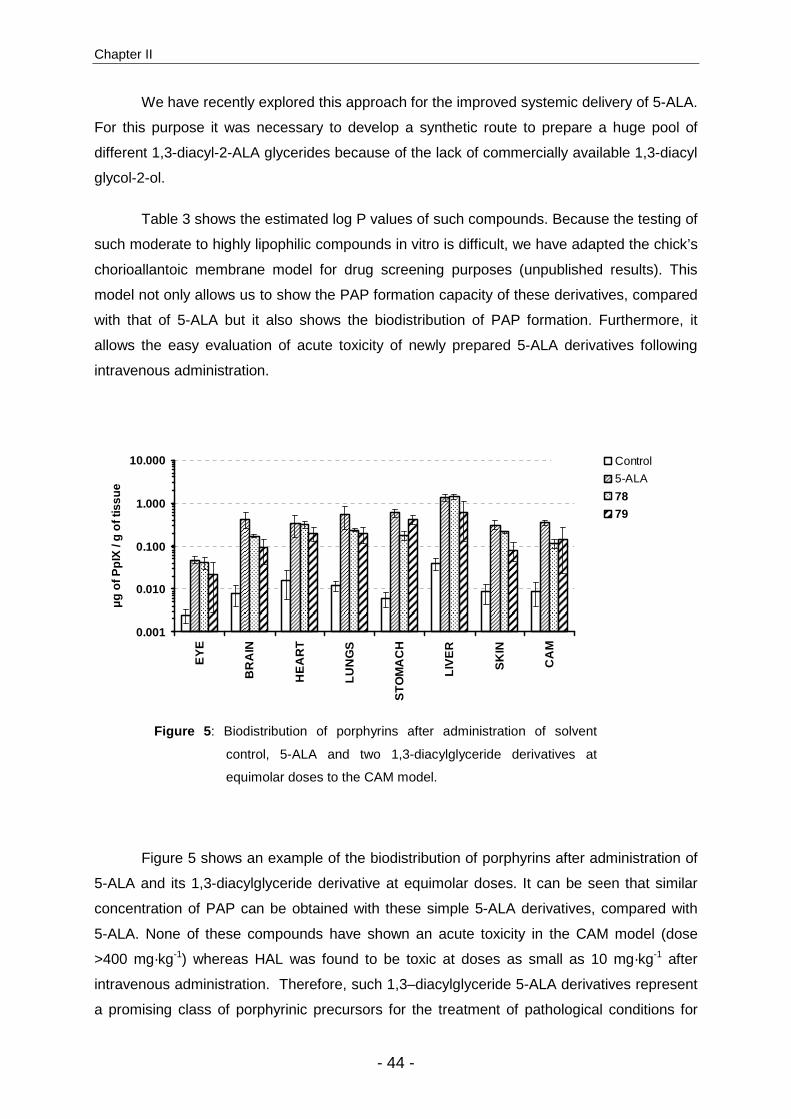
Chapter II
- 44 -
We have recently explored this approach for the improved systemic delivery of 5-ALA.
For this purpose it was necessary to develop a synthetic route to prepare a huge pool of
different 1,3-diacyl-2-ALA glycerides because of the lack of commercially available 1,3-diacyl
glycol-2-ol.
Table 3 shows the estimated log P values of such compounds. Because the testing of
such moderate to highly lipophilic compounds in vitro is difficult, we have adapted the chick’s
chorioallantoic membrane model for drug screening purposes (unpublished results). This
model not only allows us to show the PAP formation capacity of these derivatives, compared
with that of 5-ALA but it also shows the biodistribution of PAP formation. Furthermore, it
allows the easy evaluation of acute toxicity of newly prepared 5-ALA derivatives following
intravenous administration.
0.001
0.010
0.100
1.000
10.000
EY
E
BR
AIN
HE
AR
T
LUN
GS
ST
OM
AC
H
LIV
ER
SK
IN
CA
M
µg
of P
pIX
/ g
of ti
ssue
Control
5-ALA
78
79
Figure 5 : Biodistribution of porphyrins after administration of solvent
control, 5-ALA and two 1,3-diacylglyceride derivatives at
equimolar doses to the CAM model.
Figure 5 shows an example of the biodistribution of porphyrins after administration of
5-ALA and its 1,3-diacylglyceride derivative at equimolar doses. It can be seen that similar
concentration of PAP can be obtained with these simple 5-ALA derivatives, compared with
5-ALA. None of these compounds have shown an acute toxicity in the CAM model (dose
>400 mg·kg-1) whereas HAL was found to be toxic at doses as small as 10 mg·kg-1 after
intravenous administration. Therefore, such 1,3–diacylglyceride 5-ALA derivatives represent
a promising class of porphyrinic precursors for the treatment of pathological conditions for
5-Aminolevulinic Acid Derivatives in Photomedicine: Characteristics, Application and Perspectives
- 45 -
which topical administration is not appropriate, too complicated and/or might run counter to
clinical practice. In addition to this approach other derivatives, such as dendrimers (see
below), might be of potential interest for their use in the treatment of internal diseases.
5-ALA amides and pseudopeptides
Another approach to alter the lipophilicity of 5-ALA by a simple derivatization is its
conversion of the 5-aminogroup into an amide (Fig. 4). In its most simple approach this can
be achieved by the condensation of 5-ALA or its ester with acetic anhydride or acetylchloride
under basic conditions (57-60). Kloek et al. 43 were the first to follow this approach. However,
simple N-acetyl 5-ALA derivatives largely failed to induce porphyrins in vitro and in vivo. This
observation can be predominantly attributed to the reduced ability of cells to cleave the
N-acetyl function which is mandatory for the induction of porphyrin synthesis (see above).
Interestingly, Casas et al. 67 found that the carbobenzyloxy-D-phenylalanyl 5-ALA ethylester
showed considerably high capacity to induce porphyrins in human and rat skin explants (70).
However, after these initial studies some groups became aware that this approach
has the potential to further improve the specificity of 5-ALA-mediated PpIX by targeting
elevated levels of aminopeptidase activity observed in certain human tumor cell lines and
mammalian metastatic tumor cells.
The use of aminoacyl derivatives of anticancer drugs addresses these altered
enzymatic activities has been exploited experimentally 127,128,129. Furthermore, altered
expression of some acidic, basic and neutral aminopeptidases in tumor associated
vasculature is a further indication for this approach.
Berger et al. 64 pioneered this approach in 5-ALA related research. In a first attempt,
they have prepared some BOC-protected and deprotected pseudopeptide derivative of
5-ALA and 5-ALA esters (Fig. 4). On a cellular level the best PAP precursor was the Phe-
5-ALA methylester (67) derivative in all cell lines independently of the level of expression of
aminopeptidases in the particular cell line. The Lys-5-ALA methylester (68) has not shown a
significant biological activity in human endothelial cells (HCECs), despite a high expression
of APB peptidases in the cell line. No PAP formation was observed with N-BOC protected
prodrugs, which demonstrates the importance of the involvement of peptidase activity for
such derivatives. In a second investigation the same group 65 has prepared some dipeptide
5-ALA derivatives in order to increase the specificity of above mentioned peptidase activity
and/or to specifically target cellular peptide transporters. In HCECs both the BOC protected
as well as the deprotected equivalent of Gly-Pro-5-ALA ethylester (73) were more efficient
Chapter II
- 46 -
than 5-ALA itself. Furthermore, in the same cell line the BOC protected dipeptide derivative
was more efficient than its more hydrophyilic deprotected counterpart in terms of dose
response. More importantly BOC-Gly-Pro-5ALA ethylester has been found to very selectively
induce PAP in human endothelial cells. However, relatively high doses of these derivative
are necessary to induce porphyrins in A549 cells as compared to simple n-alkylesters 52.
From dendrimers and other derivatives
Another way to circumvent the poor systemic bioavailability of 5-ALA and its
n-alkylester derivatives might rely on the use of dendrimeric structures. In particular, such
polymeric drug delivery system might represent an attractive method in cancer therapy,
because with different mechanisms the enhanced perfusion and retention effect 130 and the
5-ALA PAP pathway are combined. Furthermore, polymers will also allow the delivery of
several drug molecules simultaneously. In the past, most research of such polymeric drug
delivery systems was somewhat hampered due to the high polydispersity and poorly defined
structure. However, recent advances in polymer chemistry now allow the synthesis of
structurally defined hyperbranched polymers (dendrimers), to which the drug moiety can be
coupled in a controlled way. The minimal structure of a dendrimer is called dendron. Both
Brunner et al. 54 and Di Venosa et al. 131 have evaluated the efficacy of such dendron 5-ALA
conjugates in vitro (see Fig. 4).
In an in vitro screening procedure Brunner et al. found the dendron 5-ALA conjugate
(51) was more effective than the tetramer (53) in terms of PAP induction capacity at
equimolar doses. Furthermore, compound 51 has been shown to induce fluorescence
intensity in J82 and HT29 cells that is respectively 3 and 5 times higher than that induced by
5-ALA. In contrast, Di Venosa et al. 131 found similar in vitro activity of a tetrameric 5-ALA
dendron, compared with 5-ALA, in LM3 cells. However, they have found that 3 h after
administration to the cells on average only one-third of the 5-ALA moieties was released from
the dendrons present intracellularly.
On the other hand dendrons containing as much as 18 5-ALA residues 132 have been
shown to substantially improve the intracellular PAP fluorescence intensity and the
photodynamic efficacy in PAM212 cells, compared with 5-ALA.
However, whether such approaches are potentially interesting must be invetigated in
vivo: derivatives that poorly perform in vitro might show a totally different performance in in
vivo studies. We have recently prepared a homologous series of 3-amino-3-oxohexandioic-1-
methyl-6-alkylester (Fig. 6). These compounds have shown essentially no biological in vitro

5-Aminolevulinic Acid Derivatives in Photomedicine: Characteristics, Application and Perspectives
- 47 -
activity, compared with 5-ALA or its methylester. Only 10 h after incubation some
fluorescence was observed in vitro. However, by far more activity with respect to PAP
formation was observed in vivo (CAM model). Furthermore, the most active compound
AOMM has shown the tendency to increase PAP fluorescence in most organs of the chick’s
embryo while MAL induced PAP fluorescence decreased.
OOR
O
NH3Cl
O
O
Methyl Ethyl Butyl
R= AOMMAOMEAOMB
Figure 6 : Structure of homologous series of 3-amino-3-
oxohexanedioic-1-methyl-6-alkylesters.
Clinical evaluation
MAL is the first 5-ALA derivative authorized for commercialization and is now
marketed under the trade name Metvix®. It is used to treat thin or nonhyperkeratotic and
nonpigmented actinic keratoses (AK) on the face and scalp when other therapies are
considered nonappropriate and to treat superficial and/or nodular basal cell carcinoma, BCC
(such as lesions on the mid-face or ears, lesions on severely sun damaged skin, large
lesions, or recurrent lesions) for which other available therapies are inappropriate because of
the potential for treatment-related morbidity and poor cosmetic outcome. Recently, a second
5-ALA derivative, HAL, received marketing authorization under the trade name Hexvix® for
the improved detection of bladder cancer.
Initial studies with MAL
In contrast to the random clinical evaluation of 5-ALA based PDT in the treatment of
AK, all studies involving MAL used the same standard protocol in order to obtain comparable
and meaningful results under conditions established as optimal in terms of drug
concentration and application time. There is, however, an exception in a European study in
which the second PDT procedure was performed only on lesions not located on scalp or face
(Table 4).
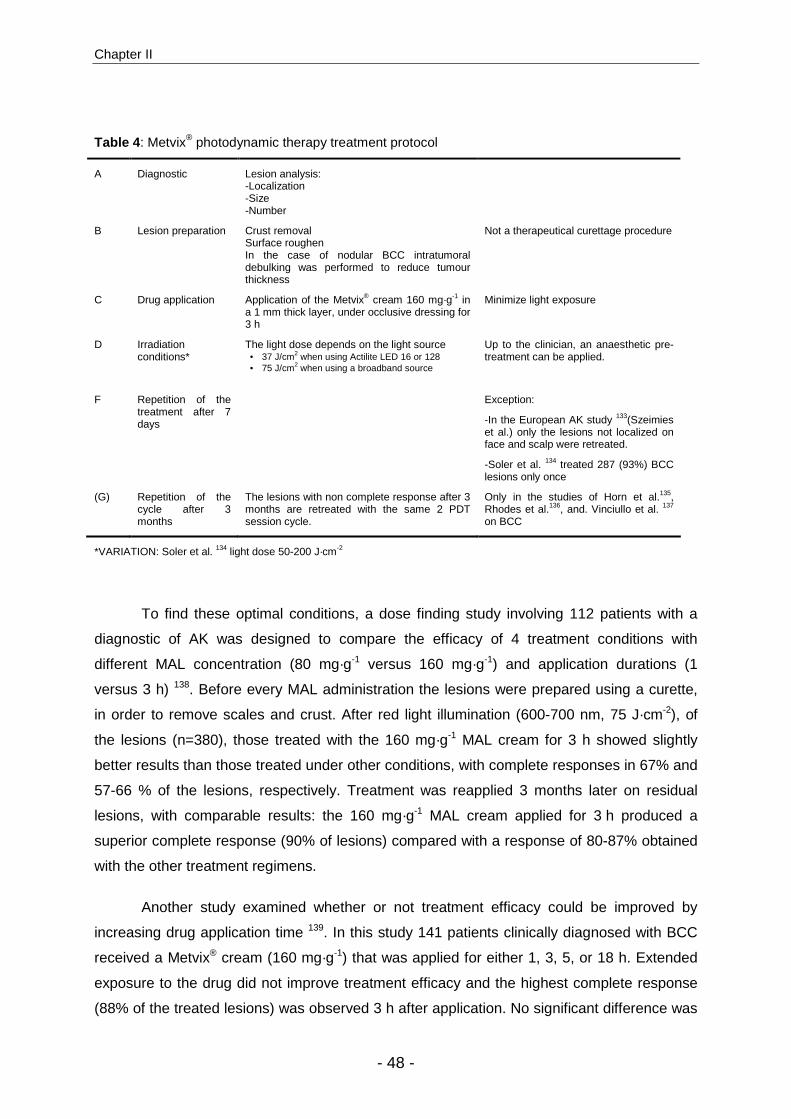
Chapter II
- 48 -
Table 4 : Metvix® photodynamic therapy treatment protocol
A Diagnostic Lesion analysis: -Localization -Size -Number
B Lesion preparation Crust removal Surface roughen In the case of nodular BCC intratumoral debulking was performed to reduce tumour thickness
Not a therapeutical curettage procedure
C Drug application Application of the Metvix® cream 160 mg·g-1 in a 1 mm thick layer, under occlusive dressing for 3 h
Minimize light exposure
D Irradiation conditions*
The light dose depends on the light source • 37 J/cm2 when using Actilite LED 16 or 128 • 75 J/cm2 when using a broadband source
Up to the clinician, an anaesthetic pre-treatment can be applied.
F Repetition of the treatment after 7 days
Exception:
-In the European AK study 133(Szeimies et al.) only the lesions not localized on face and scalp were retreated.
-Soler et al. 134 treated 287 (93%) BCC lesions only once
(G) Repetition of the cycle after 3 months
The lesions with non complete response after 3 months are retreated with the same 2 PDT session cycle.
Only in the studies of Horn et al.135, Rhodes et al.136, and. Vinciullo et al. 137 on BCC
*VARIATION: Soler et al. 134 light dose 50-200 J·cm-2
To find these optimal conditions, a dose finding study involving 112 patients with a
diagnostic of AK was designed to compare the efficacy of 4 treatment conditions with
different MAL concentration (80 mg·g-1 versus 160 mg·g-1) and application durations (1
versus 3 h) 138. Before every MAL administration the lesions were prepared using a curette,
in order to remove scales and crust. After red light illumination (600-700 nm, 75 J·cm-2), of
the lesions (n=380), those treated with the 160 mg·g-1 MAL cream for 3 h showed slightly
better results than those treated under other conditions, with complete responses in 67% and
57-66 % of the lesions, respectively. Treatment was reapplied 3 months later on residual
lesions, with comparable results: the 160 mg·g-1 MAL cream applied for 3 h produced a
superior complete response (90% of lesions) compared with a response of 80-87% obtained
with the other treatment regimens.
Another study examined whether or not treatment efficacy could be improved by
increasing drug application time 139. In this study 141 patients clinically diagnosed with BCC
received a Metvix® cream (160 mg·g-1) that was applied for either 1, 3, 5, or 18 h. Extended
exposure to the drug did not improve treatment efficacy and the highest complete response
(88% of the treated lesions) was observed 3 h after application. No significant difference was

5-Aminolevulinic Acid Derivatives in Photomedicine: Characteristics, Application and Perspectives
- 49 -
observed after retreating the residual lesions 3 months later and lesion complete response
rates remained between 92% and 96%. Te investigators concluded that application of the
drug for durations longer than 3 h did not result in an improved clinical response.
In the specific case of nodular BCC, in which drug penetration depth and
homogeneous distribution plays an important role for the successful treatments, Peng et
al. 140 tested different MAL cream concentrations (16, 80 and 160 mg·g-1) and application
times (3 or 18 h) and concluded that the best results were obtained with the 160 mg·g-1 MAL
cream that was applied for 3 h.
Aktinic keratoses
AKs are known to be the most common type of premalignant skin lesion. In some
areas of the world with high local incident UV radiation, such as Australia, approximately
60% of predisposed persons over 40 years of age have at least one AK, with up to 90% of
persons older than 80 years of age having at least one AK 141. Some studies have shown
that 40% of all metastatic squamous cell carcinoma originate from AKs, whereas others
suggest that AK is itself an early form of carcinoma in situ 142,143. Most AK lesions are located
on sun-exposed areas, such as the face, scalp, ears and forearms. Treatment of these
particular areas tend to be complicated, because of the limited available treatment options.
Although there are some treatments that are effective, as in the case of AK and BCC, can
cure these lesions, the price to pay in terms of disfigurement in the facial area might be
rather high.
A commonly used treatment for AK is cryotherapy, which gained popularity due to its
high effectiveness 144. Other ablative procedures, such as dermoabrasion or surgery, are
sometimes used to treat multiple lesions or to evaluate suspicious lesions 145. Topical
treatments with imiquimod, diclofenac or 5-fluorouracil (5-FU) are also used in relation to the
type of lesions 144. In this context, PDT has enormous potential to demonstrate its numerous
advantages in terms of curing efficacy, noninvasiveness, multiple lesion treatment and
repeatability, with excellent cosmetic outcome.
In a pioneer study Fritsch et al. 89 compared the fluorescence intensity arising after
5-ALA or MAL application on AK lesions in patients who had undergone surgery. A cream
containing 20% of the active drug was applied over a period of 1 or 6h on AK lesions and on
normal skin samples and the resulting porphyrin formation was determined by fluorescence
measurements. Although 5-ALA seemed to produce the more porphyrins, the specificity of
MAL for AK lesions appeared to be 70% higher than that for 5-ALA. Therefore, one can

Chapter II
- 50 -
assume that when applied properly, MAL PDT would be more effective in terms of collateral
damage and induced pain than for instance, 5-ALA PDT.
Table 5 : Reports of Metvix®-mediated treatment of AK
Author N° of
patient
(lesions)
Comparison Follow up
Lesion complete response
Cosmetic outcome †
Pariser et al. (2003) 146
80 (502) Placebo 3 months 89% MAL
38% placebo
Excellent or good 97% MAL
Freeman et al. (2003) 147
204 (855) Cryotherapy & placebo
3 months
91% MAL 68% cryo
30% placebo
Excellent 83% MAL 51% cryo
Szeimies et al. (2003) 133
202 (732) Cryotherapy 3 months 69% MAL 75% cryo
Excellent or good 96% MAL 81% cryo
Dragieva et al. (2004) 148
17 (129) * Placebo 4 months
90% MAL 0%
placebo N.A.
* AK in transplant recipients; † investigator evaluation
A total of 4 clinical trials of MAL-mediated treatment of AK have been
reported 133,146,147,148. The results are summarized in Table 5. In all studies the MAL treatment
were compared to cryotherapy or placebo treatments with respect to response rate, cosmetic
outcome and patient satisfaction. Complete response rates ranged from 69% to 90% for
MAL, compared with 70% for cryotherapy and 30-38% for placebo. In some studies the
cosmetic outcome was evaluated in those patients with more than 75% of the lesions
displaying complete response 133,147. The results indicate a significant advantage of MAL-
mediated PDT: 83-96% of the investigators and 76-98% of the patients graded the cosmetic
outcome as good or excellent, whereas in the case of cryotherapy only 51-81% of the
investigators and 56-91% of the patients graded the cosmetic outcome as good or excellent.
Most adverse events reported for MAL-treated patients (burning, pain and crusting) were of
mild or moderate intensity.
As shown by the results of the three main studies (Table 6), methylaminolevulinate-
mediated PDT effectively treated 716 (82%) out of 875 AK lesions, and 429 (88%) out of 489
focusing on thin or mild lesions. The lowest response rate obtained by the European study

5-Aminolevulinic Acid Derivatives in Photomedicine: Characteristics, Application and Perspectives
- 51 -
can be explained by the single PDT session applied on the face and scalp lesions, which
suggests that a protocol with multiple sessions is highly desirable.
Table 6 : Complete response rates of AK lesions after Metvix®-mediated PDT in function of lesion grade
Lesion grade
Overall* Mild Moderate
n° lesions CR n° lesions CR n° lesions CR
Pariser et al. 236 209 179 161 57 48
Freeman et al. 295 267 167 161 128 106
Szeimies et al. 344 240 143 107 201 133
TOTAL 875 716 489 429 386 287
CR rate 82% 88% 74%
* Thick lesions reported in the the Szeimies et al. study were not taken into account
This point was confirmed by the recent study of Tarstedt et al. 149 who compared two
treatment regimens of MAL-PDT, one with a single application of PDT and the other with two
application of PDT 1 week apart. The single-treatment regimen appeared to be as effective
as the dual regimen for classical lesions but less effective in the case of thicker lesions. The
cosmetic outcome was graded as excellent or good for 83–91 % of the patients treated,
which is significantly superior to the standard treatment cryotherapy. Painful skin sensation,
burning, erythema and crusting were the most common local adverse events reported but
had generally mild or moderate intensity. Furthermore, allergies to 5-ALA or MAL have been
mentioned 150,151.
More recently, Dragieva et al. 148 compared the efficacy of MAL PDT for transplant
recipients known to have an increased tendency to develop multiple AKs or cancer due to
immunosuppression. Because ulceration and infection may be life threatening for
immunodepressed patients, these adverse events are uncommon in PDT, which thus makes
PDT the treatment of choice. These particular lesions responded well to PDT: 56 (90%) of
the 62 lesions had a complete response. However, three patients had only a partial response

Chapter II
- 52 -
and one had no response. This non response was explained by the ineffectiveness of the
immune system to survey and eradicate residual tumor cells after PDT.
Basal Cell Carcinoma
BCC is the most common skin cancer, with an incidence of half a million new cases in the
United States every year. Conventional treatments are either superficial ablative techniques
(electrodesiccation, curettage and cryotherapy), which are used primarily for low-risk tumors,
or full-thickness techniques (Mohs surgery, excisional surgery and radiotherapy) 152.
However, because of the very poor cosmetic outcome after surgical therapy, PDT may
represent an excellent alternative to effectively treat these lesions.
Table 7 : Report of clinical studies concerning Metvix® treatment of BCC
Author No° of patient
(no. of lesions)
Compa-rison
Follow up
Lesion complete response after 3
months
Recurrence after 12 months
Recurrence after 24 months
Cosmetic outcome rated excellent or good
by investigator
Soler et al. (2001) 59 (350) None
24-48 months 89% * 98%
Horn et al. (2003)
94 (123) None 24
months †
87% clinical 77% histological
18% 76% 3 months 94% 24 months
Rhodes et al. (2004)
103 (118) Surgery 24 months
91% MAL 98% surgery
13% MAL 2% surgery
19% MAL 4% surgery
24 months 83% MAL
41% surgery
Vinciullo et al. (2005)
95 (148) None 24 months **
89% 18% 24%
65% 3 months 79% 12 months 84% 24 months
* CR after 3 – 6 months. † Follow-up observation is continuing for a total of 5 years
A summary of the results of clinical studies reporting on MAL-mediated PDT for BCC
can be seen in Table 7. The first work, published in 2001 by Soler et al. 134, is a retrospective,
noncomparative, long term follow up study that evaluated the clinical response of BCC to
MAL PDT. This study, which had a follow up period of 2-4 years, included 59 patients with
350 BCC lesions treated with MAL PDT. A total of 310 lesions (89%) showed a complete
response after 3 months. Only 11% of these 310 lesions recurred after 24 to 48 months.
Fourteen percent of the lesions were especially thick and nodular, and therefore more
difficult to treat, which lowered the long-term complete response to 79%. The cosmetic
outcome was assessed as good or excellent in 98% of the cases.

5-Aminolevulinic Acid Derivatives in Photomedicine: Characteristics, Application and Perspectives
- 53 -
In a more recent multicenter study Horn et al. 135 analyzed MAL PDT effectiveness in
94 patients who presented 123 difficult-to-treat BCC lesions. Lesions with large surface area
and those located in the middle of the face are at high risk for morbidity or disfigurement and
were thus selected as good candidates for PDT. After 3 months the overall clinical response
rate was 87% but is decreased to 77% after histological control. Complete response rates
were slightly better for superficial BCC than for nodular BCC (92% versus 87%). Recurrence
after 24 months was observed in 18 % the cases and the cosmetic outcome was judged as
excellent or good in 94% of all cases, even for large lesions. It is interesting that,
investigations for systemic effects of MAL application showed no effect on liver, kidney or
bone marrow function. These results are in agreement with results of a similar study recently
published by an Australian group.
In contrast to the previously cited studies, all of which are noncomparative, a
European study 136 directly compared MAL PDT with standard excision surgery in a group of
103 patients diagnosed for primary nodular BCC. Lesions with incomplete remission after
first treatment underwent one additional course of PDT. Both PDT and surgical treatments
produced similar rates of short-term complete response (91% and 98%, respectively).
Surgery had slightly better long-term results than MAL PDT with respect to recurrence (2%
versus 13% after 12 months and 4% versus 19% after 24 months). On the other hand, in
terms of cosmetic outcome investigators noted a clear benefit of PDT over surgery. Although
half of the patients who received PDT reported adverse effects, such as burning sensation,
pain and erythema, these were only mild or moderate in nature and the condition resolved
itself without any medical treatment. On the other hand, only a third of the surgical patients
complained of adverse events but more serious cases (e.g. skin infection) were reported.
In summary, MAL-mediated PDT has been shown to be a very competitive treatment
for BCC in terms of clinical response rate and cosmetic outcome, compared with
conventional protocols. Nevertheless, some drawbacks are encountered when treating large
lesions or lesions located in the trunk and neck, but in such cases multiple PDT treatments
might be a reasonable option. A recent study 153 evaluating the economic potential of this
treatment modality revealed that Metvix®-PDT represents an economically efficient
intervention in the therapy of difficult-to-treat BCC, because of the lower total treatment costs
than surgery and greatly reduced need for reconstructive surgery.
However, although the CR rates of MAL PDT 3 months after treatment are highly
encouraging, one must await the results of the long term studies to fully assess this method.

Chapter II
- 54 -
Additional indications
Today there are several other exploratory clinical PDT trials of MAL underway for
treatment of conditions such as acne, oral squamous cell carcinoma. However, the outcomes
of these studies have not been disclosed in the scientific literature until today.
HAL and diagnosis of bladder cancer
Bladder cancer, which is the ninth most common cancer, had an incidence of
>350’000 persons in 2002 and killed 145’000. The prevalence in 2002 was approximately
three times higher among males. Today, the main therapy for superficial bladder cancer still
remains surgical resection, chemotherapy and BCG (Bacillus Calmette-Guerin) therapy.
However, some clinical studies using 5-ALA-mediated PDT have shown encouraging results
in terms of safety, effectiveness and lack of major adverse effects in the treatment of
recurrent bladder cancer and may thus become a useful tool in the clinician’s therapeutic
arsenal 154,155,156. Despite advantages toward the parent compound, no clinical trial using
HAL for the PDT treatment of recurrent bladder cancer has been reported.. However, HAL
has been found to be a valuable tool in tumor detection for the fluorescence guided
transurethral cystoscopy and has consequently gained official marketing authorization in
Europe and has been recently recommended in the guidelines of the European Association
of Urologists. We will briefly summarize the development of HAL-mediated FD from the first
clinical study (in 1994) that tested 5-ALA 157 to the very recent approval of HAL in Europe
(Table 8). For more information about fluorescence detection or treatment of bladder cancer,
the reader is referred to some reviews of particular interest 158,159,160.
Since its initial use in 1992 161 5-ALA-mediated fluorescence cystoscopy, has quickly
spread worldwide and a vast body of knowledge and experience is now available to the
clinician. The general sensitivity achieved by 5-ALA FD in different studies is generally very
high, with values 87-97% 162, 5-ALA-mediated FD was shown to detect two times more
tumors than standard white light cystoscopy 163.
From 1997 through 1999 a pilot study investigated the possibility of enhancing 5-ALA-
mediated bladder FD by the use of HAL, with the goal of increasing the PAP fluorescence
intensity, decreasing the drug dose and decreasing instillation time, which is critical for
patient compliance and higher tumor specificity 74. HAL was selected over other n-alkyl-
chained derivatives because of its satisfactory water solubility and capacity to induce high
amounts of PAP at lower doses than 5-ALA. The optimal drug dose required to obtain the
highest ratio of tumor to healthy tissue fluorescence was established in 25 patients. The
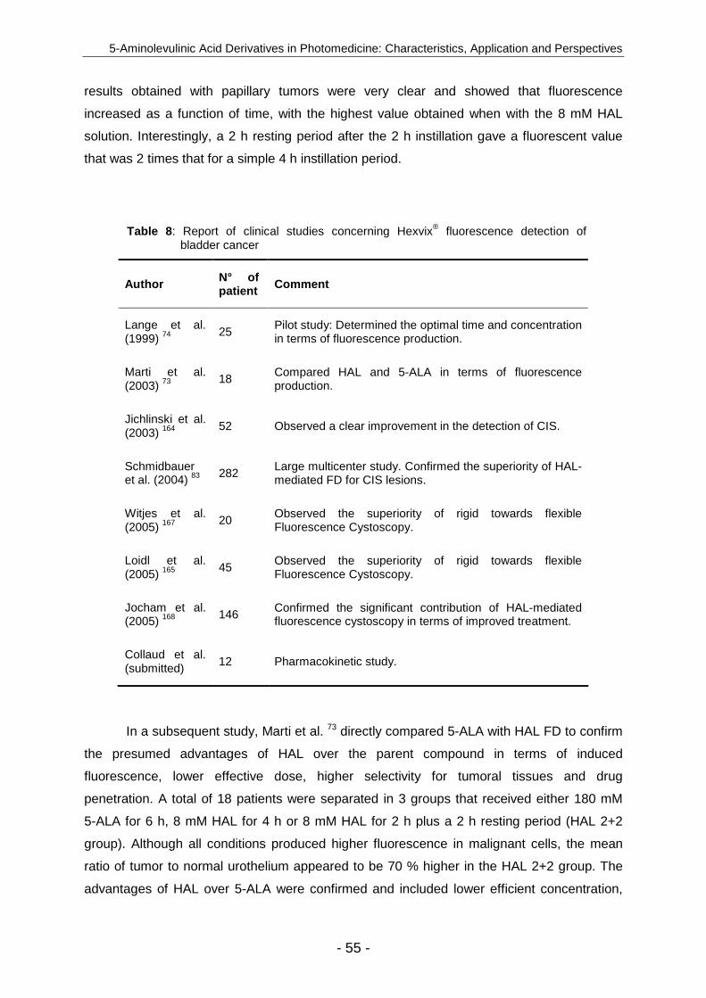
5-Aminolevulinic Acid Derivatives in Photomedicine: Characteristics, Application and Perspectives
- 55 -
results obtained with papillary tumors were very clear and showed that fluorescence
increased as a function of time, with the highest value obtained when with the 8 mM HAL
solution. Interestingly, a 2 h resting period after the 2 h instillation gave a fluorescent value
that was 2 times that for a simple 4 h instillation period.
Table 8 : Report of clinical studies concerning Hexvix® fluorescence detection of bladder cancer
Author N° of patient Comment
Lange et al. (1999) 74
25 Pilot study: Determined the optimal time and concentration in terms of fluorescence production.
Marti et al. (2003) 73
18 Compared HAL and 5-ALA in terms of fluorescence production.
Jichlinski et al. (2003) 164
52 Observed a clear improvement in the detection of CIS.
Schmidbauer et al. (2004) 83 282
Large multicenter study. Confirmed the superiority of HAL-mediated FD for CIS lesions.
Witjes et al. (2005) 167
20 Observed the superiority of rigid towards flexible Fluorescence Cystoscopy.
Loidl et al. (2005) 165
45 Observed the superiority of rigid towards flexible Fluorescence Cystoscopy.
Jocham et al. (2005) 168 146
Confirmed the significant contribution of HAL-mediated fluorescence cystoscopy in terms of improved treatment.
Collaud et al. (submitted)
12 Pharmacokinetic study.
In a subsequent study, Marti et al. 73 directly compared 5-ALA with HAL FD to confirm
the presumed advantages of HAL over the parent compound in terms of induced
fluorescence, lower effective dose, higher selectivity for tumoral tissues and drug
penetration. A total of 18 patients were separated in 3 groups that received either 180 mM
5-ALA for 6 h, 8 mM HAL for 4 h or 8 mM HAL for 2 h plus a 2 h resting period (HAL 2+2
group). Although all conditions produced higher fluorescence in malignant cells, the mean
ratio of tumor to normal urothelium appeared to be 70 % higher in the HAL 2+2 group. The
advantages of HAL over 5-ALA were confirmed and included lower efficient concentration,

Chapter II
- 56 -
shorter administration time (2 h versus 6 h), and deeper penetration into the tissue.
Furthermore, no PAP fluorescence was detected in the basement membrane or the chorion.
Despite of the high fluorescence obtained with the optimal incubation time of 2+2 (or
even with 4 hours), in terms of clinical practice 1 h of instillation is recommended
currently 164,83,165. Furthermore short instillation times may be beneficial with respect to
selectivity 166. These conditions result in the minimum necessary amount of porphyrins in
tumors for proper FD, with sensitivities (97%) and specificities (74%) 164 that are similar to
longer instillation times.
The improved FD of carcinoma in situ (CIS) is one of the most remarkable outcomes
of clinical studies involving HAL. A large multicenter clinical trial 83 was conducted that
involved a total number of 286 patients, 83% (39%) of whom had CIS, an aggressive, high
risk and easily overlooked tumour 169. The results revealed that HAL FD detected 97% of all
lesions, compared with only 78% by conventional white light cystoscopy (WLC). The
superiority of the technique becomes evident when focusing on difficult to detect CIS, with
detection rates of 97% for HAL FD and 58% for WLC. Furthermore, there were only two
patients with adverse effects directly related to HAL FD, which included burning sensation
after illumination and mild bladder spasm. This high tolerance was confirmed by Collaud et
al. (Drugs R D, in press) who proceeded in a safety study concluding on the absence of
serious adverse events and negligible systemic uptake of the drug. Although the use of HAL
FD allowed for improved resection of CIS in 25% more patients (this lesion type was
detected in 80 patients with HAL FD and only 64 with WLC), only a long term observational
study can confirm the real benefits of this methodology in terms of patient survival rate and
tumor free resection survey. Nevertheless, recent studies 170,171,172 have shown a direct
correlation between improved detection using 5-ALA-mediated fluorescence detection and
increased tumor-free resection.
The final two clinical studies 165,167 compared HAL FD involving a flexible cystoscope
(the gold standard for detection of bladder cancer 173,174) and a rigid, cystoscope with respect
to convenience and compliance for the patient. Both studies concluded that the rigid
cystoscopy had higher detection rate (85-94% versus 70-89%).
HAL and other clinical domains
Because photomedicine is limited to light-accessible organs, gynaecological cancer
like ovarian or cervical cancers and the gastrointestinal tract may be good candidates for
photodetection and photodynamic therapy.

5-Aminolevulinic Acid Derivatives in Photomedicine: Characteristics, Application and Perspectives
- 57 -
The early stages of ovarian cancer are generally asymptomatic, and it diagnosed at a
late stage in the majority of cases, when it is incurable, making it the fourth leading cause of
cancer-related death among women. The 5 year survival rate for this type of cancer is 94%
when the tumors are localized but decrease dramatically to 29% when metastasis has
occurred 175. Encouraging results were obtained by a HAL FD on Fisher rats implanted with
NuTu-19 cells (rat epithelial ovarian cell line), resulting in the detection of twice as many
cancer lesions, compared to white-light inspections, and showing a clear advantage in terms
of porphyrin accumulation, compared with 5-ALA 117. Loning et al. 176 have used 5-ALA-
mediated laparoscopic FD for the detection of ovarian carcinoma metastases and observed a
very high sensitivity (92%), allowing for significant improvement in metastasis detection.
However, to the best of our knowledge, no clinical trials using 5-ALA derivatives are
scheduled.
Another gynaecological malignancy for which PDT might be useful is cervical cancer,
which is the second most common cancer among women worldwide 177. The standard
treatment for cervical intraepithelial neoplasia (CIN) is surgical excision. However, this
invasive procedure has many complications, including bleeding, infections, cervical stenosis,
preterm delivery and high recurrence rates demand problematic repetition of the treatment178.
Because of its increased patient tolerance, high selectivity and low tissue destruction PDT
effect on CIN was investigated using different photosensitizers. Ichimura et al. 179 used
hematoporphyrins-mediated PDT and obtained high response rates of approximately 90%,
but patients had to avoid light exposure because of the prolonged skin photosensitivity of
these drugs. Additional clinical studies using 5-ALA-mediated PDT showed contradictory
results in terms of efficacy. Thus Bodner et al. 180 obtained responses similar to cold-knife
conization, whereas Keefe et al. 181 observed no difference from the placebo group.
Recently, a Swiss group 182 examined the dependence of PAP formation as a function HAL
topical application time. They determined that application duration of 100 min for HAL cream
was optimal to obtain the highest ratio of epithelium to underlying lamina propria
fluorescence. However, the results showed a surprisingly low selectivity for tumor cells
versus normal tissues. More information will become available as clinical studies continue.
Apart from bladder and gynaecological cancers, HAL-mediated FD has been
investigated for the diagnosis of rectal adenoma and cancer. For this purpose, a pilot study
evaluated the efficiency of an instilled HAL solution to induce selective fluorescence of rectal
tumors (adenoma and malignant tumors) 183. The study included 10 patients who received an
enema composed of a 3.2 mM HAL solution preceded by an initial treatment with
N-acetylcystein to eliminate the mucus layer at the surface of the rectal epithelium and thus
Chapter II
- 58 -
enhance contact between the drug and the target tissue. One purpose of this study was to
determine the optimal drug application conditions to achieve the best ratio of tumor to healthy
tissue fluorescence. The investigators looked at different application times of a 3.2 mM HAL
solution ranging from 30 to 60 min and also evaluated the effect of a resting period after drug
application. After direct endoscopical observation after 30, 45 and 60 min of HAL application
that gave unsatisfactory results, characterized by weak tumor fluorescence signal, low tumor
selectivity and high background fluorescence, it was decided to follow a 30 min application
period with a 30 min resting period. Under these conditions fluorescence appeared to
emanate selectively from the tumors and a significant difference was observed between
adenomas that had homogenous fluorescence and moderately differentiated carcinoma that
had fluorescence only at the tumor edge. More data are required to determine the exact
improvement of HAL FD in this domain.
Conclusion
5-ALA derivatives have proven to substantially improve the local bioavailability of
5-ALA in vitro and in vivo. Depending on the respective derivatives drug doses as low as two
order of magnitude less than 5-ALA are necessary to achieve similar or sometimes even
better therapeutic results. In some selected cases 5-ALA n-alkylesters induced porphyrins
more selectively in the target tissue. In some cases the increased production of porphyrins
led to a substantial shortening of the interval between drug administration and irradiation.
The superiority of 5-ALA esters over 5-ALA has been recently confirmed by the approval of
Metvix® in Europe and Australia for the treatment of AK and Hexvix® for the improved FD of
bladder cancer.
However, today it seems to be clear that the formulation has to be carefully adapted
with respect to the lipophilicity of the respective 5-ALA derivative and medical indication. In
terms of viscosity the appropriate formulation for the treatment of cervical cancer might be
substantially different from those were other rheological properties are necessary. Although a
moderately lipophilic formulation might be still appropriate for MAL, it will have a tremendous
effect on the local bioavailability of lipophilic 5-ALA esters, such as HAL. In this context,
principle consideration of pharmaceutical technology and biopharmacy might give further rise
to a similar success for 5-ALA derivatives in other clinical indications in which topical
administration is appropriate and convenient. Particular challenges are represented by
indications such as Barett’s esophagus or colon cancer.

5-Aminolevulinic Acid Derivatives in Photomedicine: Characteristics, Application and Perspectives
- 59 -
Cosmetics are another interesting area of application for 5-ALA esters. The outcome
of current studies of the treatment of acne is of particular interest. Indications of purely
cosmetic interest, such as hair removal or skin rejuvenation, are of interest, at least from a
commercial point of view. 5-ALA derivatives could have therapeutic indication in the area of
bacterial infection and have shown a huge potential for the photodynamic inactivation of
gram-positive and gram-negative bacteria in preliminary studies. In this context, 5-ALA esters
might be of some interest as adjuvant treatment for the improvement of wound healing or of
bacterial infestation in the oral cavity.
However, the huge opportunities underlying 5-ALA derivative-related research with
respect to compounds that show enhanced activity following systemic administration remain
largely unexplored until today. At present, the treatment or diagnosis of some life-threatening
diseases, such as brain tumors, breast cancer and ovarian cancer, can hardly be achieved
by simple 5-ALA derivatives. In the future it might be favorable for researchers working in the
field to adopt lessons learned from rational prodrug design in their investigations of 5-ALA
derivatives in order to further increase the impact of this research area.

Chapter II
- 60 -
References 1. J. F. Kelly, M. E. Snell, and M. C. Berenbaum, Photodynamic destruction of human
bladder carcinoma, Br J Cancer, 1975, 31, 237-244.
2. T. J. Dougherty and S. L. Marcus, Photodynamic therapy, Eur J Cancer, 1992, 28A, 1734-1742.
3. T. J. Dougherty, C. J. Gomer, B. W. Henderson, G. Jori, D. Kessel, M. Korberlik, J. Moan, and Q. Peng, Photodynamic therapy, J Natl Cancer Inst, 1998, 90, 889-905.
4. N. L. Oleinick, R. L. Morris, and I. Belichenko, The role of apoptosis in response to photodynamic therapy: what, where, why, and how, Photochem Photobiol Sci, 2002, 1, 1-21.
5. G. A. Wagnieres, W. M. Star, and B. C. Wilson, In vivo fluorescence spectroscopy and imaging for oncological applications, Photochem Photobiol, 1998, 68, 603-632.
6. A. Albert, Chemical aspects of selective toxicity, Nature, 1958, 182, 421-422.
7. J. C. Kennedy and R. H. Pottier, Endogenous protoporphyrin IX, a clinically useful photosensitizer for photodynamic therapy, J Photochem Photobiol B, 1992, 14, 275-292.
8. R. W. Redmond and J. N. Gamlin, A compilation of singlet oxygen yields from biologically relevant molecules, Photochem Photobiol, 1999, 70, 391-475.
9. Q. Peng, K. Berg, J. Moan, M. Kongshaug, and J. M. Nesland, 5-Aminolevulinic acid-based photodynamic therapy: principles and experimental research, Photochem Photobiol, 1997, 65, 235-251.
10. S. Collaud, A. Juzeniene, J. Moan, and N. Lange, On the selectivity of 5-aminolevulinic acid-induced protoporphyrin IX formation, Curr Med Chem Anti-Canc Agents, 2004, 4, 301-316.
11. J. Webber, D. Kessel, and D. Fromm, Plasma levels of protoporphyrin IX in humans after oral administration of 5-aminolevulinic acid, J Photochem Photobiol B, 1997, 37, 151-153.
12. Q. Peng, T. Warloe, K. Berg, J. Moan, M. Kongshaug, K. E. Giercksky, and J. M. Nesland, 5-aminolevulinic acid-based photodynamic therapy - clinical research and future challenges, Cancer, 1997, 79, 2282-2308.
13. C. Fritsch, G. Goerz, and T. Ruzicka, Photodynamic therapy in dermatology, Arch dermatol, 1998, 134, 207-214.
14. J. T. Dalton, C. R. Yates, D. Yin, A. Straughn, S. L. Marcus, A. L. Golub, and M. C. Meyer, Clinical pharmacokinetics of 5-aminolevulinic acid in healthy volunteers and patients at high risk for recurrent bladder cancer., J Pharmacol Exp Ther, 2002, 301, 507-512.
15. J. van den Boogert, R. Van Hillegersberg, F. W. M. de Rooij, R. W. F. de Bruin, A. Edixhoven-Bosdijk, A. B. Houtsmuller, P. D. Siersema, J. H. P. Wilson, and H. W. Tilanus, 5-aminolevulinic acid-induced protoporphyrin IX accumulation in tissues:
5-Aminolevulinic Acid Derivatives in Photomedicine: Characteristics, Application and Perspectives
- 61 -
pharmacokinetics after oral or intravenous administration, J Photochem Photobiol B:Biol, 1998, 44, 29-38.
16. R. Waidelich, A. Hofstetter, H. Stepp, R. Baumgartner, E. Weninger, and M. Kriegmair, Early clinical experience with 5-aminolevulinic acid for the photodynamic therapy of upper tract urothelial tumors., J Urol, 1998, 159, 401-404.
17. R. Waidelich, H. Stepp, R. Baumgartner, E. Weninger, A. Hofstetter, and M. Kriegmair, Clinical experience with 5-aminolevulinic acid and photodynamic therapy for refractory superficial bladder cancer, J Urol, 2001, 165, 1904-1907.
18. Z. Malik, G. Kostenich, L. Roitman, B. Ehrenberg, and A. Orenstein, Topical application of 5-aminolevulinic acid, DMSO and EDTA: protoporphyrin IX accumulation in skin and tumours of mice, J Photochem Photobiol B:Biol, 1995, 28, 213-218.
19. A. Casas, H. Fukuda, G. Di Venosa, and A. M. Batlle, The influence of the vehicle on the synthesis of porphyrins after topical application of 5-aminolaevulinc acid. Implication in cutaneous photodynamic sensitization, Br J Dermatol, 2000, 143, 564-572.
20. F. S. De Rosa, J. M. Marchetti, J. A. Thomazini, A. C. Tedesco, and M. V. Bentley, A vehicle for photodynamic therapy of skin cancer: influence of dimethylsulphoxide on 5-aminolevulinic acid in vitro cutaneous permeation and in vivo protoporphyrin IX accumulation determined by confocal microscopy, J Control Release, 2000, 65, 359-366.
21. P. Ziolkowski, B. J. Osiecka, G. Oremek, M. Siewinski, K. Symonowicz, Y. Saleh, and A. Bronowicz, Enhancement of photodynamic therapy by use of aminolevulinic acid/glycolic acid drug mixture, J Exp Ther Oncol, 2004, 4, 121-129.
22. J. C. Tsai, I. H. Chen, T. W. Wong, and Y. L. Lo, In vitro/in vivo correlations between transdermal delivery of 5-aminolaevulinic acid and cutaneous protoporphyrin IX accumulation and effect of formulation, Br J Dermatol, 2002, 146, 853-862.
23. K. Choudry, R. C. Brooke, W. Farrar, and L. E. Rhodes, The effect of an iron chelating agent on protoporphyrin IX levels and phototoxicity in topical 5-aminolaevulinic acid photodynamic therapy, Br J Dermatol, 2003, 149, 124-130.
24. S. C. Chang, A. J. MacRobert, J. B. Porter, and S. G. Bown, The efficacy of an iron chelator (CP94) in increasing cellular protoporphyrin IX following intravesical 5-aminolaevulinic acid administration: an in vivo study, J Photochem Photobiol B, 1997, 38, 114-122.
25. O. Bech, D. Phillips, J. Moan, and A. J. MacRobert, A hydroxypyridinone (CP94) enhances protoporphyrin IX formation in 5-aminolaevulinic acid treated cells, J Photochem Photobiol B:Biol, 1997, 41, 136-144.
26. A. Casas, H. Fukuda, and A. M. Batlle, Tissue distribution and kinetics of endogenous porphyrins synthesized after topical application of ALA in different vehicles, Br J Cancer, 1999, 81, 13-18.
27. S. Lieb, R. M. Szeimies, and G. Lee, Self-adhesive thin films for topical delivery of 5-aminolevulinic acid, Eur J Pharm Biopharm, 2002, 53, 99-106.
28. R. F. Turchiello, F. C. Vena, P. Maillard, C. S. Souza, M. V. Bentley, and A. C. Tedesco, Cubic phase gel as a drug delivery system for topical application of 5-ALA, its
Chapter II
- 62 -
ester derivatives and m-THPC in photodynamic therapy (PDT), J Photochem Photobiol B:Biol, 2003, 70, 1-6.
29. A. Casas, C. Perotti, M. Saccoliti, P. Sacca, H. Fukuda, and A. M. Batlle, ALA and ALA hexyl ester in free and liposomal formulations for the photosensitisation of tumour organ cultures, Br J Cancer, 2002, 86, 837-842.
30. B. G. Auner, C. Valenta, and J. Hadgraft, Influence of lipophilic counter-ions in combination with phloretin and 6-ketocholestanol on the skin permeation of 5-aminolevulinic acid, Int J Pharm, 2003, 255, 109-116.
31. M. B. Pierre, A. C. Tedesco, J. M. Marchetti, and M. V. Bentley, Stratum corneum lipids liposomes for the topical delivery of 5-aminolevulinic acid in photodynamic therapy of skin cancer: preparation and in vitro permeation study, BMC Dermatol, 2001, 1
32. A. F. Hurlimann, G. Hanggi, and R. G. Panizzon, Photodynamic therapy of superficial basal cell carcinomas using topical 5-aminolevulinic acid in a nanocolloid lotion, Dermatology, 1998, 197, 248-254.
33. L. E. Rhodes, M. M. Tsoukas, R. R. Anderson, and N. Kollias, Iontophoretic delivery of ALA provides a quantitative model for ALA pharmacokinetics and PpIX phototoxicity in human skin, J Invest Dermatol, 1997, 108, 87-91.
34. R. F. Lopez, M. V. Bentley, B. M. Delgado-Charro, and R. H. Guy, Iontophoretic delivery of 5-aminolevulinic acid (ALA): effect of pH, Pharm Res, 2001, 18, 311-315.
35. R. F. Lopez, M. V. Bentley, B. M. Delgado-Charro, and R. H. Guy, Optimization of aminolevulinic acid delivery by iontophoresis, J Control Release, 2003, 88, 65-70.
36. J. Y. Fang, W. R. Lee, S. C. Shen, Y. P. Fang, and C. H. Hu, Enhancement of topical 5-aminolaevulinic acid delivery by erbium:YAG laser and microdermabrasion: a comparison with iontophoresis and electroporation, Br J Dermatol, 2004, 151, 132-140.
37. J. T. van den Akker, V. Iani, W. M. Star, H. J. Sterenborg, and J. Moan, Systemic component of protoporphyrin IX production in nude mouse skin upon topical application of aminolevulinic acid depends on the application conditions, Photochem Photobiol, 2002, 75, 172-177.
38. J. Moan, J. T. van den Akker, P. Juzenas, L. W. Ma, E. Angell-Petersen, O. B. Gadmar, and V. Iani, On the basis for tumor selectivity in the 5-aminolevulinic acid-induced synthesis of protoporphyrin IX, J Porphyrins Phthalocyanines, 2001, 5, 170-176.
39. A. Juzeniene, P. Juzenas, O. kaalhus, V. Iani, and J. Moan, Temperature effect on accumulation of protoporphyrin IX after topical application of 5-aminolevulinic acid and its methylester and hexylester derivatives in normal mouse skin, Photochem Photobiol, 2002, 76, 452-456.
40. J. T. van den Akker, K. Boot, D. I. Vernon, S. B. Brown, L. Groenendijk, G. C. van Rhoon, and H. J. Sterenborg, Effect of elevating the skin temperature during topical ALA application on in vitro ALA penetration through mouse skin and in vivo PpIX production in human skin, Photochem Photobiol Sci, 2004, 3, 263-267.
41. L. W. Ma, J. Moan, Q. Peng, and V. Iani, Production of protoporphyrin IX induced by 5-aminolevulinic acid in transplanted human colon adenocarcinoma of nude mice can be increased by ultrasound, Int J Cancer, 1998, 78, 464-469.

5-Aminolevulinic Acid Derivatives in Photomedicine: Characteristics, Application and Perspectives
- 63 -
42. Q. Peng, J. Moan, T. Warloe, V. Iani, H. B. Steen, A. Bjorseth, and J. M. Nesland, Build-up of esterified aminolevulinic-acid-derivative-induced porphyrin fluorescence in normal mouse skin, J Photochem Photobiol B:Biol, 1996, 34, 95-96.
43. J. Kloek and M. J. Beijersbergen van Henegouwen, Prodrugs of 5-aminolevulinic acid for photodynamic therapy, Photochem Photobiol, 1996, 64, 994-1000.
44. H. Fukuda, A. Casas, and A. M. Batlle, Aminolevulinic acid: from its unique biological function to its star role in photodynamic therapy, Int J Biochem Cell Biol, 2005, 37, 272-276.
45. P. Ponka, Cell biology of heme, Am J Med Sci, 1999, 318, 241-256.
46. M. Hildebrand, Inter-species extrapolation of pharmacokinetic data of three prostacyclin-mimetics, Prostaglandins, 1994, 48, 297-312.
47. S. J. Singer and G. L. Nicolson, The fluid mosaic model of the structure of cell membranes, Science, 1972, 175, 720-731.
48. P. Artursson, Cell cultures as models for drug absorption across the intestinal mucosa, Crit Rev Ther Drug Carrier Syst, 1991, 8, 305-330.
49. K. Moser, K. Kriwet, A. Naik, Y. N. Kalia, and R. H. Guy, Passive skin penetration enhancement and its quantification in vitro, Eur J Pharm Biopharm, 2001, 52, 103-112.
50. Y. N. Kalia and R. H. Guy, Modeling transdermal drug release, Adv Drug Deliv Rev, 2001, 48, 159-172.
51. T. Fujita, J. Iwasa, and C. Hansch, A New Substituent Constant, π, Derived from Partition Coefficients, J Am Chem Soc, 1964, 86, 5175-5180.
52. P. Uehlinger, M. Zellweger, G. Wagnières, L. Juillerat-Jeanneret, H. van den Bergh, and N. Lange, 5-aminolevulinic acid and its derivatives: physical chemical properties and protoporphyrin IX formation in cultured cells, J Photochem Photobiol B:Biol, 2000, 54, 72-80.
53. F. S. De Rosa, A. C. Tedesco, R. F. Lopez, M. B. Riemma Pierre, N. Lange, J. M. Marchetti, J. C. Gomes Rotta, and M. V. Bentley, In vitro skin permeation and retention of 5-aminolevulinic acid ester derivatives for photodynamic therapy, J Control Release, 2003, 89, 261-269.
54. H. Brunner, F. Hausmann, and R. Knuechel, New 5-aminolevulinic acid esters - efficient protoporphyrin precursors for photodetection and photodynamic therapy, Photochem Photobiol, 2003, 78, 481-486.
55. C. J. Whitaker, S. H. Battah, M. J. Forsyth, C. Edwards, R. W. Boyle, and E. K. Matthews, Photosensitization of pancreatic tumour cells by delta-aminolaevulinic acid esters, Anticancer Drug Des, 2000, 15, 161-170.
56. E. Rud, O. Gederaas, A. Hogset, and K. Berg, 5-aminolevulinic acid, but not 5-aminolevulinic acid esters, is transported into adenocarcinoma cells by system BETA transporters, Photochem Photobiol, 2000, 71, 640-647.
57. F. Doring, J. Walter, J. Will, M. Focking, M. Boll, S. Amasheh, W. Clauss, and H. Daniel, Delta-aminolevulinic acid transport by intestinal and renal peptide transporters and its physiological and clinical implications, J Clin Invest, 1998, 101, 2761-2767.
Chapter II
- 64 -
58. O. Gederaas, A. Holroyd, S. B. Brown, D. I. Vernon, J. Moan, and K. Berg, 5-Aminolaevulinic acid methyl ester transport on amino acid carriers in a human colon adenocarcinoma cell line, Photochem Photobiol, 2001, 73, 164-169.
59. J. Kloek, W. Akkermans, and M. J. Beijersbergen van Henegouwen, Derivatives of 5-aminolevulinic acid photodynamic therapy: enzymatic conversion into protoporphyrin, Photochem Photobiol, 1998, 67, 150-154.
60. N. Lange, L. Vaucher, A. Marti, A. L. Etter, P. Gerber, H. van den Bergh, P. Jichlinski, and P. Kucera, Routine experimental system for defining conditions used in photodynamic therapy and fluorescence photodetection of (non-) neoplastic epithelia, J Biomed Opt, 2001, 6, 151-159.
61. T. Stepinac, C. Felley, P. Jornod, N. Lange, T. Gabrecht, C. Fontolliet, P. Grosjean, G. vanMelle, H. van den Bergh, P. Monnier, G. Wagnières, and G. Dorta, Endoscopic fluorescence detection of intraepithelial neoplasia in Barrett's esophagus after oral administration of aminolevulinic acid., Endoscopy, 2003, 35, 663-668.
62. N. Bodor and P. Buchwald, Designing safer (soft) drugs by avoiding the formation of toxic and oxidative metabolites, Mol Biotechnol, 2004, 26, 123-132.
63. N. Bodor and P. Buchwald, Recent advances in the brain targeting of neuropharmaceuticals by chemical delivery systems, Adv Drug Deliv Rev, 1999, 36, 229-254.
64. Y. Berger, A. Greppi, O. Siri, R. Neier, and L. Juillerat-Jeanneret, Ethylene glycol and amino acid derivatives of 5-aminolevulinic acid as new photosensitizing precursors of protoporphyrin IX in cells, J Med Chem, 2000, 43, 4738-4746.
65. Y. Berger, L. Ingrassia, R. Neier, and L. Juillerat-Jeanneret, Evaluation of dipeptide-derivatives of 5-aminolevulinic acid as precursors for photosensitizers in photodynamic therapy, Bioorg Med Chem, 2003, 11, 1343-1351.
66. R. G. Tunstall, A. A. Barnett, J. Schofield, J. Griffiths, D. I. Vernon, S. B. Brown, and D. J. H. Roberts, Porphyrin accumulation induced by 5-aminolaevulinic acid esters in tumour cells growing in vitro and in vivo, Br J Cancer, 2002, 87, 246-250.
67. A. Casas, A. M. Batlle, A. R. Butler, D. Robertson, E. H. Brown, A. MacRoberts, and P. A. Riley, Comparative effect of ALA derivatives on protoporphyrin IX production in human and rat skin organ cultures, Br J Cancer, 1999, 80, 1525-1532.
68. Y. Ninomiya, Y. Itoh, S. Tajima, and A. Ishibashi, In vitro and in vivo expression of protoporphyrin IX induced by lipophilic 5-aminolevulinic acid derivatives, J Dermatol Sci, 2001, 27, 114-120.
69. M. Kaliszewski, A. Juzeniene, P. Juzenas, M. Kwasny, J. Kaminski, Z. Dabrowski, J. Golinski, and J. Moan, Formation of protoporphyrin IX from carboxylic- and amino-derivatives of 5-aminolevulinic, Photodiagnonsis Photodynamic Therapy, 2005
70. L. Juillerat-Jeanneret, S. Lohm, M. F. Hamou, and F. Pinet, Regulation of aminopeptidase A in human brain tumor vasculature: evidence for a role of transforming growth factor-beta, Lab Invest, 2000, 80, 973-980.
71. J. M. Martinez, I. Prieto, M. J. Ramirez, C. Cueva, F. Alba, and M. Ramirez, Aminopeptidase activities in breast cancer tissue, Clin Chem, 1999, 45, 1797-1802.

5-Aminolevulinic Acid Derivatives in Photomedicine: Characteristics, Application and Perspectives
- 65 -
72. T. Suganuma, K. Ino, K. Shibata, S. Nomura, H. Kajiyama, F. Kikkawa, N. Tsuruoka, and S. Mizutani, Regulation of aminopeptidase A expression in cervical carcinoma: role of tumor-stromal interaction and vascular endothelial growth factor, Lab Invest, 2004, 84, 639-648.
73. A. Marti, P. Jichlinski, N. Lange, J. P. Ballini, L. Guillou, H. J. Leisinger, and P. Kucera, Comparison of aminolevulinic acid and hexylester aminolevulinate induced protoporphyrin IX distribution in human bladder cancer, J Urol, 2003, 170, 428-432.
74. N. Lange, P. Jichlinski, M. Zellweger, M. Forrer, A. Marti, L. Guillou, P. Kucera, G. Wagnières, and H. van den Bergh, Photodetection of early human bladder cancer based on the fluorescence of 5-aminolaevulinic acid hexylester induced protoporphyrin IX: a pilot study, Br J Cancer, 1999, 80, 185-193.
75. J. M. Gaullier, K. Berg, Q. Peng, H. Anholt, P. K. Selbo, L. W. Ma, and J. Moan, Use of 5-aminolevulinic acid esters to improve photodynamic therapy on cells in culture, Cancer Res, 1997, 57, 1481-1486.
76. F. C. Vena, R. F. Turchiello, I. Laville, S. Pigaglio, J. Blais, and A. C. Tedesco, 5-aminolevulinic acid ester-induced protoporphyrin IX in a murine melanoma cell line, Lasers Med Sci, 2004, 19, 119-126.
77. C. Jarret, F. Stauffer, M. E. Henz, M. Marty, R. M. Lüönd, J. Bobálová, O. Schürmann, and R. Neier, Inhibition of Escherichia coli porphobilinogen synthase using analogs of postulated intermediates, Chem Biol, 2000, 7, 185-196.
78. E.L. Taylor, D.I. Vernon, and S.B. Brown, Mechanism of protoporphyrin IX formation from ALA esters, ESP 2001 Book of Abstracts, 2001, 178
79. H. Bando, S. Mohri, F. Yamashita, Y. Takakura, and M. Hashida, Effects of skin metabolism on percutaneous penetration of lipophilic drugs, J Pharm Sci, 1997, 86, 759-761.
80. C. Perotti, H. Fukuda, G. Di Venosa, A. J. MacRobert, A. M. Batlle, and A. Casas, Porphyrin synthesis from ALA derivatives for photodynamic therapy. In vitro and in vivo studies, Br J Cancer, 2004, 90, 1660-1665.
81. N. Fotinos, M. Convert, J.C. Piffaretti, and N. Lange, 5-ALA and 5-ALA derivative-mediated effects on gram negative bacteria, ESP 2005 Book of abstracts, 2005, 56
82. P. Jichlinski, L. Guillou, S. J. Karlsen, P. U. Malmström, D. Jocham, B. Brennhovd, E. Johansson, T. Gärtner, N. Lange, H. van den Bergh, and H. J. Leisinger, Hexyl aminolevulinate fluorescence cystoscopy: a new diagnostic tool for the photodiagnosis of superficial bladder canacer - a multicenter study, J Urol, 2003, 170, 226-229.
83. J. Schmidbauer, F. Witjes, N. Schmeller, R. Donat, M. Susani, and M. Marberger, Improved Detection of Urothelial Carcinoma In Situ With Hexaminolevulinate Fluorescence Cystoscopy, J Urol, 2004, 171, 135-138.
84. A. Marti, N. Lange, H. van den Bergh, D. Sedmera, P. Jichlinski, and P. Kucera, Optimisation of the formation and distribution of protoporphyrin IX in the urothelium: an in vitro approach, J Urol, 1999, 162, 546-552.
85. R. Sorensen, P. Juzenas, V. Iani, and J. Moan, Formation of protoporphyrin IX in mouse skin after topical application of 5-aminolevulinic acid and its methyl esther, Proc.SPIE, 1999, 3563, 77-81.

Chapter II
- 66 -
86. J. T. van den Akker, V. Iani, W. M. Star, H. J. Sterenborg, and J. Moan, Topical application of 5-aminolevulinic acid hexyl ester and 5-aminolevulinic acid to normal nude mouse skin: differences in protoporphyrin IX fluorescence kinetics and the role of the stratum corneum, Photochem Photobiol, 2000, 72, 681-689.
87. A. Juzeniene, L. W. Ma, P. Juzenas, V. Iani, N. Lange, and J. Moan, Production of protoporphyrin IX from 5-aminolevulinic acid and two of its esters in cells in vitro and tissues in vivo, Cell Mol Biol, 2002, 48, 911-916.
88. A. Juzeniene, P. Juzenas, V. Iani, and J. Moan, Topical application of 5-aminolevulinic acid and its methylester, hexylester and octylester derivatives: considerations for dosimetry in mouse skin model, Photochem Photobiol, 2002, 76, 329-334.
89. C. Fritsch, B. Homey, W. Stahl, P. Lehmann, T. Ruzicka, and H. Sies, Preferential relative porphyrin enrichment in solar keratoses upon topical application of delta-aminolevulinic acid methylester, Photochem Photobiol, 1998, 68, 218-221.
90. J. T. van den Akker, A. Holroyd, D. I. Vernon, H. J. Sterenborg, and S. B. Brown, Comparative in vitro percutaneous penetration of 5-aminolevulinic acid and two of its esters through excised hairless mouse skin., Lasers Surg Med, 2003, 33, 173-181.
91. Guy, R. H. and Hadgraft, J.,Percutaneous Penetration Enhancement: Physicochemical Considerations and Implications for Prodrug Design, in: Sloan, K.B. (Ed.), Prodrugs: Topical and Ocular Drug Delivery, Marcel Dekker, New York, 1992,1-16.
92. H. Oishi, H. Nomiyama, K. Nomiyama, and K. Tomokuni, Fluorometric HPLC determination of δ-aminolevulinic acid (ALA) in the plasma and urine of lead workers: biological indicators of lead exposure, J Anal Toxicol, 1996, 20, 106-110.
93. K. Tomokuni, M. Ichiba, Y. Hirai, and T. Hasegawa, Optimized liquid-chromatographic method for fluorometric determination of urinary δ-aminolevulinic acid in workers exposed to lead, Clin Chem, 1987, 33, 1665-1667.
94. S. Collaud, F. Evangelisti, L. James, Q. Peng, R. Gurny, and N. Lange, Formulation of a Thermosetting Hydrogel of Hexylaminolevulinate Targeting Barrett's Oesophagus, ESP 2005 Book of abstracts, 2005, 104
95. J. Moan, L. W. Ma, and V. Iani, On the pharmacokinetics of topically applied 5-aminolevulinic acid and two of its esters, Int J Cancer, 2001, 92, 139-143.
96. K. Thoma, Dermatika. 2. Aufl., München, 1983, 49
97. A. Casas, C. Perotti, H. Fukuda, L. Rogers, A. R. Butler, and A. M. Batlle, ALA and ALA hexyl ester-induced porphyrin synthesis in chemically induced skin tumours: the role of different vehicles on improving photosensitization, Br J Cancer, 2001, 85, 1794-1800.
98. C. Perotti, A. Casas, H. Fukuda, P. Sacca, and A. M. Batlle, Topical application of ALA and ALA hexyl ester on a subcutaneous murine mammary adenocarcinoma: tissue distribution, Br J Cancer, 2003, 88, 432-437.
99. A. Winkler and C. C. Muller-Goymann, Comparative permeation studies for delta-aminolevulinic acid and its n-butylester through stratum corneum and artificial skin constructs, Eur J Pharm Biopharm, 2002, 53, 281-287.
5-Aminolevulinic Acid Derivatives in Photomedicine: Characteristics, Application and Perspectives
- 67 -
100. A. Winkler and C. C. Muller-Goymann, The influence of topical formulations on the permeation of 5-aminolevulinic acid and its n-butyl ester through excised human stratum corneum, Eur J Pharm Biopharm, 2005, 60, 427-437.
101. J. Y. Chen, Q. Peng, and H. J. Jodl, Infrared spectral comparison of 5-aminolevulinic acid and its hexyl ester, Spectrochim Acta A Mol Biomol Spectrosc, 2003, 59, 2571-2576.
102. R. F. Lopez, M. V. Bentley, M. B. Delgado-Charro, D. Salomon, H. van den Bergh, N. Lange, and R. H. Guy, Enhanced delivery of 5-aminolevulinic acid esters by iontophoresis in vitro, Photochem Photobiol, 2003, 77, 304-308.
103. S. Gerscher, J. P. Connelly, J. Griffiths, S. B. Brown, A. J. MacRobert, G. A. E. Wong, and L. E. Rhodes, Comparison of the Pharmacokinetics and Phototoxicity of Protoporphyrin IX Metabolized from 5-Aminolevulinic Acid and Two Derivatives in Human Skin In Vivo, Photochem Photobiol, 2000, 72, 569-574.
104. S. Gerscher, J. P. Connelly, M. J. Beijersbergen van Henegouwen, A. J. MacRobert, P. Watt, and L. E. Rhodes, A quantitative assessment of protoporphyrin IX metabolism and phototoxicity in human skin following dose-controlled delivery of the prodrugs 5-aminolaevulinic acid and 5-aminolaevulinic acid-n-pentylester, Br J Dermatol, 2001, 144, 983-990.
105. C. E. Bigelow, S. Mitra, R. Knuechel, and T. H. Foster, ALA- and ALA-hexylester-induced protoporphyrin IX fluorescence and distribution in multicell tumour spheroids, Br J Cancer, 2001, 85, 727-734.
106. J. Moan, L. W. Ma, A. Juzeniene, V. Iani, P. Juzenas, F. Apricena, and Q. Peng, Pharmacology of protoporphyrin IX in nude mice after application of ALA and ALA esters, Int J Cancer, 2003, 103, 132-135.
107. S. M. Berge, L. D. Bighley, and D. C. Monkhouse, Pharmaceutical salts, J Pharm Sci, 1977, 66, 1-19.
108. R. Washbrook and P. A. Riley, Comparison of delta-aminolaevulinic acid and its methyl ester as an inducer of porphyrin synthesis in cultured cells, Br J Cancer, 1997, 75, 1417-1420.
109. H. Brunner, F. Hausmann, R. C. Krieg, E. Endlicher, J. Schoelmerich, R. Knuechel, and H. Messmann, The effects of 5-aminolevulinic acid esters on protoporphyrin IX production in human adenocarcinma cell lines, Photochem Photobiol, 2001, 74, 721-725.
110. V. Zenzen and H. Zankl, Protoporphyrin IX-accumulation in human tumor cells following topical ALA- and h-ALA-application in vivo, Cancer lett, 2003, 202, 35-42.
111. H. Hirschberg, C. H. Sun, B. J. Tromberg, and S. J. Madsen, ALA- and ALA-ester-mediated photodynamic therapy of human glioma spheroids, J Neurooncol, 2002, 57, 1-7.
112. E. Endlicher, P. Rummele, F. Hausmann, R. C. Krieg, R. Knuechel, H. C. Rath, J. Schoelmerich, and H. Messmann, Protoporphyrin IX distribution following local application of 5-aminolevulinic acid and its esterified derivatives in the tissue layers of the normal rat colon, Br J Cancer, 2001, 85, 1572-1576.

Chapter II
- 68 -
113. S. El Khatib, J. Didelon, A. Leroux, L. Bezdetnaya, D. Notter, and M. D'Hallewin, Kinetics, biodistribution and therapeutic efficacy of hexylester 5-aminolevulinate induced photodynamic therapy in an orthotopic rat bladder tumor model, J Urol, 2004, 175, 2013-2017.
114. A. Godal, N. O. Nilsen, J. Klaveness, J. E. Braenden, J. M. Nesland, and Q. Peng, New Derivatives of 5-aminolevulinic Acid for Photodynamic Therapy: Chemical Synthesis and Porphyrin Production in In Vitro and Vivo Biological Systems, J Environ Pathol Toxicol Oncol, 2006, 25, 109-126.
115. R. C. Krieg, S. Fickweiler, O. S. Wolfbeis, and R. Knuechel, Cell-type specific protoporphyrin IX metabolism in human bladder cancer in vitro, Photochem Photobiol, 2000, 72, 226-233.
116. C. Perotti, A. Casas, H. Fukuda, P. Sacca, and A. M. Batlle, ALA and ALA hexyl ester induction of porphyrins after their systemic administration to tumour bearing mice., Br J Cancer, 2002, 87, 790-795.
117. F. Ludicke, T. Gabrecht, N. Lange, G. Wagnières, H. van den Bergh, L. Berclaz, and A. Major, Photodynamic diagnosis of ovarian cancer using hexaminolaevulinate: a preclinical study, Br J Cancer, 2003, 88, 1780-1784.
118. G. Kirdaite, N. Lange, N. Busso, H. van den Bergh, P. Kucera, and A. So, Protoporphyrin IX photodynamic therapy for synovitis, Arthrtitis Rheum, 2002, 46, 1371-1378.
119. S. Eleouet, N. Rousset, J. Carre, L. Bourre, V. Vonarx, Y. Lajat, G. M. Beijersbergen van Henegouwen, and S. Pattabhi, In vitro fluorescence, toxicity and phototoxicity induced by delta-aminolevulinic acid (ALA) or ALA-esters, Photochem Photobiol, 2000, 71, 447-454.
120. P. Ettmayer, G. L. Amidon, B. Clement, and B. Testa, Lessons learned from marketed and investigational prodrugs, J Med Chem, 2004, 47, 2393-2404.
121. K. Beaumont, R. Webster, I. Gardner, and K. Dack, Design of ester prodrugs to enhance oral absorption of poorly permeable compounds: challenges to the discovery scientist, Curr Drug Metab, 2003, 4, 461-485.
122. S. Majumdar, S. Duwuri, and A. K. Mitra, Membrane transporter/receptor-targeted prodrug design: strategies for human and veterinary drug development, Adv Drug Deliv Rev, 2004, 56, 1437-1452.
123. B. S. Anderson, Prodrugs for improved CNS delivery, Adv Drug Deliv Rev, 1996, 19, 171-202.
124. W. M. Pardridge, CNS drug design based on principles of blood-brain barrier transport, J Neurochem, 1998, 70, 1781-1792.
125. N. Bodor and P. Buchwald, Barriers to remember: brain-targeting chemical delivery systems and Alzheimer's disease, Drug Discov Today, 2002, 7, 766-774.
126. J. N. Jacob, G. W. Hesse, and V. E. Shashoua, Synthesis, brain uptake, and pharmacological properties of a glyceryl lipid containing GABA and the GABA-T inhibitor gamma-vinyl-GABA, J Med Chem, 1990, 33, 733-736.
5-Aminolevulinic Acid Derivatives in Photomedicine: Characteristics, Application and Perspectives
- 69 -
127. R. A. Ashmun and A. T. Look, Metalloprotease activity of CD13/aminopeptidase N on the surface of human myeloid cells, Blood, 1990, 75, 462-469.
128. I. Saiki, H. Fujii, J. Yoneda, F. Abe, M. Nakajima, T. Tsuruo, and I. Azuma, Role of aminopeptidase N (CD13) in tumor-cell invasion and extracellular matrix degradation, Int J Cancer, 1993, 54, 137-143.
129. M. A. Smal, Z. Dong, H. T. Cheung, Y. Asano, L. Escoffier, M. Costello, and M. H. Tattersall, Activation and cytotoxicity of 2-alpha-aminoacyl prodrugs of methotrexate, Biochem Pharmacol, 1995, 49, 567-574.
130. H. Maeda, J. Wu, T. Sawa, Y. Matsumura, and K. Hori, Tumor vascular permeability and the EPR effect in macromolecular therapeutics: a review, J Control Release, 2000, 65, 271-284.
131. G. Di Venosa, A. Casas, S. H. Battah, P. Dobbin, H. Fukuda, A. J. MacRobert, and A. M. Batlle, Investigation of a novel dendritic derivative of 5-aminolaevulinic acid for photodynamic therapy, Int J Biochem Cell Biol, 2006, 38, 82-91.
132. S. H. Battah, C. E. Chee, H. Nakanishi, S. Gerscher, A. J. MacRobert, and C. Edwards, Synthesis and biological studies of 5-aminolevulinic acid-containing dendrimers for photodynamic therapy, Bioconjug Chem, 2001, 12, 980-988.
133. R. M. Szeimies, S. Karrer, S. Radakovic-Fijan, A. Tanew, P. G. Calzavara-Pinton, C. Zane, A. Sidoroff, M. Hempel, J. Ulrich, T. Proebstle, H. Meffert, M. Mulder, D. Salomon, H. C. Dittmar, J. W. Bauer, K. Kernland, and L. Braathen, Photodynamic therapy using topical methyl 5-aminolevulinate compared with cryotherapy for actinic keratosis: A prospective, randomized study, J Am Acad Dermatol, 2003, 47, 258-262.
134. A. M. Soler, T. Warloe, A. Berner, and K. E. Giercksky, A follow-up study of recurrence and cosmesis in completely responding superficial and nodular basal cell carcinomas treated with methyl 5-aminolaevulinate-based photodynamic therapy alone and with prior curettage, Br J Dermatol, 2001, 145, 467-471.
135. M. Horn, P. Wolf, H. C. Wulf, T. Warloe, C. Fritsch, L. E. Rhodes, R. Kaufmann, M. De Rie, F. J. Legat, I. M. Stender, A. M. Soler, A. M. Wennberg, G. A. E. Wong, and O. Larko, Topical methyl aminolaevulinate photodynamic therapy in patients with basal cell carcinoma prone to complications and poor cosmetic outcome with conventional treatment., Br J Dermatol, 2003, 149, 1242-1249.
136. L. E. Rhodes, M. De Rie, Y. Enstrom, R. Groves, T. Morken, V. Goulden, G. A. E. Wong, J. J. Grob, S. Varma, and P. Wolf, Photodynamic therapy using topical methyl aminolevulinate vs surgery for nodular basal cell carcinoma: results of a multicenter randomized prospective trial, Arch dermatol, 2004, 140, 17-23.
137. C. Vinciullo, T. Elliott, D. Francis, K. Gebauer, L. Spelman, R. Nguyen, W. Weightman, A. Sheridan, C. Reid, D. Czarnecki, and D. Murrell, Photodynamic therapy with topical methyl aminolaevulinate for 'difficult-to-treat' basal cell carcinoma, Br J Dermatol, 2005, 152, 765-772.
138. L. Braathen, B. Paredes, K. Frölich, I. Wijnen, T. Warloe, C. Fritsch, O. Saksela, and A.M. Ros, A dose finding study of photodynamic therapy (PDT) with Metvix(R) in actinic keratosis (AK), JEADV, 2000, 14 (suppl.1), 38.
Chapter II
- 70 -
139. N. Basset-Séguin, I. Bachmann, S. Pavel, O. Saksela, M. Johnsson, A.M. Ros, B. Paredes, and O. Larko, A dose finding study of photodynamic therapy (PDT) with Metvix(R) in patients with basal cell carcinoma (BCC), JEADV, 2000, 14 (suppl. 1), 39.
140. Q. Peng, A. M. Soler, T. Warloe, J. M. Nesland, and K. E. Giercksky, Selective distribution of porphyrins in skin thick basal cell carcinoma after topical application of methyl 5-aminolevulinate, J Photochem Photobiol B:Biol, 2001, 62, 140-145.
141. M. Helfand, S. M. Mahon, K. B. Eden, P. S. Frame, and C. T. Orleans, Screening for skin cancer, Am J Prev Med, 2001, 20, 47-58.
142. M. Lebwohl, Actinic keratosis: epidemiology and progression to squamous cell carcinoma, Br J Dermatol, 2003, 149, 31-33.
143. J. Anwar, D. A. Wrone, A. Kimyai-Asadi, and M. Alam, The development of actinic keratosis into invasive squamous cell carcinoma: evidence and evolving classification schemes, Clin Dermatol, 2004, 22, 189-196.
144. B. A. Lober and N. A. Fenske, Optimum treatment strategies for actinic keratosis (intraepidermal squamous cell carcinoma), Am J Clin Dermatol, 2004, 5, 395-401.
145. J. L. Jorizzo, Current and novel treatment options for actinic keratosis, J Cutan Med Surg, 2004, 8, 13-21.
146. D. M. Pariser, N. J. Lowe, D. M. Stewart, M. T. Jarratt, A. W. Lucky, R. J. Pariser, and P. S. Yamauchi, Photodynamic therapy with topical methyl aminolevulinate for actinic keratosis: results of a prospective randomized multicenter trial, J Am Acad Dermatol, 2003, 48, 227-232.
147. M. Freeman, C. Vinciullo, D. Francis, L. Spelman, R. Nguyen, P. Fergin, K. E. Thai, D. Murell, W. Weightman, C. Anderson, C. Reid, A. Watson, and P. Foley, A comparison of photodynamic therapy using topical methyl aminolevulinate (Metvix) with single cycle cryotherapy in patients with actinic keratosis: a prospective, randomized study., J Dermatolog Treat, 2003, 14, 99-106.
148. G. Dragieva, B. M. Prinz, J. Hafner, R. Dummer, G. Burg, U. Binswanger, and W. Kempf, A randomized controlled clinical trial of topical photodynamic therapy with methyl aminolaevulinate in the treatment of actinic keratoses in transplant recipients, Br J Dermatol, 2004, 151, 196-200.
149. M. Tarstedt, I. Rosdahl, B. Berne, K. Svanberg, and A. M. Wennberg, A randomized multicenter study to compare two treatment regimens of topical methyl aminolevulinate (Metvix)-PDT in actinic keratosis of the face and scalp, Acta Derm Venereol, 2005, 85, 424-428.
150. B. Gniazdowska, F. Rueff, P. Hillemans, and B. Przybilla, Allergic contact dermatitis from delta-aminolevulinic acid used for photodynamic therapy, Contact Dermatitis, 1998, 38, 348-349.
151. H. C. Wulf and P. Philipsen, Allergic contact dermatitis to 5-aminolaevulinic acid methylester but not to 5-aminolaevulinic acid after photodynamic therapy, Br J Dermatol, 2004, 150, 143-145.
152. J. C. Martinez, The management of melanoma and nonmelanoma skin cancer: a review for the primary care physician, Mayo Clin Proc, 2001, 76, 1253-1265.
5-Aminolevulinic Acid Derivatives in Photomedicine: Characteristics, Application and Perspectives
- 71 -
153. M. Aristides, D. Tilden, P. Robinson, A. Weston, P. Davey, and G. Aldridge, A decision-analytic model to compare the cost of methyl aminolevulinate photodynamic therapy with standard treatments in the UK for difficult-to-treat basal cell carcinoma and actinic keratosis, In 7th Annual International Meeting of the ISPOR, Arlington, VA, 19–22 May 2002.
154. M. Kriegmair, R. Baumgartner, W. Lumper, R. Waidelich, and A. Hofstetter, Early clinical experience with 5-aminolevulinic acid for the photodynamic therapy of superficial bladder cancer, Br J Urol, 1996, 77, 667-671.
155. R. Waidelich, W. Beyer, R. Knuechel, H. Stepp, R. Baumgartner, J. Schroder, A. Hofstetter, and M. Kriegmair, Whole bladder photodynamic therapy with 5-aminolevulinic acid using a white light source, Urology, 2003, 61, 332-337.
156. A. P. Berger, H. Steiner, A. Stenzl, T. Akkad, G. Bartsch, and L. Holtl, Photodynamic therapy with intravesical instillation of 5-aminolevulinic acid for patients with recurrent superficial bladder cancer: a single-center study, Urology, 2003, 61, 338-341.
157. M. Kriegmair, R. Baumgartner, R. Knuechel, P. Steinbach, A. Ehsan, W. Lumper, F. Hofstadter, and A. Hofstetter, Fluorescence photodetection of neoplastic urothelial lesions following intravesical instillation of 5-aminolevulinic acid, Urology, 1994, 44, 836-841.
158. P. Jichlinski and H. J. Leisinger, Fluorescence cystoscopy in the management of bladder cancer: a help for the urologist!, Urol Int, 2005, 74, 97-101.
159. D. Zaak, A. Karl, R. Knuechel, H. Stepp, A. Hartmann, O. Reich, A. Bachmann, M. Siebels, G. Popken, and C. Stief, Diagnosis of urothelial carcinoma of the bladder using fluorescence endoscopy, BJU Int, 2005, 96, 217-222.
160. S. Jain and R. C. Kockelbergh, The role of photodynamic diagnosis in the contemporary management of superficial bladder cancer, BJU Int, 2005, 96, 17-21.
161. M. Kriegmair, R. Baumgartner, and A. Hofstetter, Intravesikale Instillation von Delta-Aminolävulinsäure (ALA)-eine neue Methode zur photodynamischen Diagnostik und Therapie, Laser Med, 1992, 8, 83
162. M. A. D'Hallewin, L. Bezdetnaya, and F. Guillemin, Fluorescence detection of bladder cancer: a review, Eur Urol, 2002, 42, 417-425.
163. S. Schneeweiss, M. Kriegmair, and H. Stepp, Is everything all right if nothing seems wrong? A simple method of assessing the diagnostic value of endoscopic procedures when a gold standard is absent, J Urol, 1999, 161, 1116-1119.
164. P. Jichlinski, D. Aymon, G. A. Wagnieres, A. Marti, N. Lange, L. Guillou, H. J. Leisinger, and H. van den Bergh, On the influence of the instillation time on the results of HAL (Hexvix) fluorescence detection of superficial bladder cancer, Diagnostic Optical Spectroscopy in Biomedicine II.Edited by Wagnieres, Georges A. Proceedings of the SPIE, 2003, 5141, 272-277.
165. W. Loidl, J. Schmidbauer, M. Susani, and M. Marberger, Flexible cystoscopy assisted by hexaminolevulinate induced fluorescence: a new approach for bladder cancer detection and surveillance?, Eur Urol, 2005, 47, 323-326.
166. E. Angell-Petersen, R. Sorensen, T. Warloe, A. M. Soler, J. Moan, Q. Peng, and K. E. Giercksky, Porphyrin Formation in Actinic Keratosis and Basal Cell Carcinoma after
Chapter II
- 72 -
Topical Application of Methyl 5-Aminolevulinate, J Invest Dermatol, 2006, 126, 265-271.
167. J. A. Witjes, P. M. Moonen, and A. G. van der Heijden, Comparison of hexaminolevulinate based flexible and rigid fluorescence cystoscopy with rigid white light cystoscopy in bladder cancer: results of a prospective Phase II study, Eur Urol, 2005, 47, 319-322.
168. D. Jocham, F. Witjes, S. Wagner, B. Zeylmaker, J. van Moorselaar, M. O. Grimm, R. Muschter, G. Popken, F. Konig, R. Knuechel, and K. H. Kurth, Improved detection and treatment of bladder cancer using hexaminolevulinate imaging: a prospective, phase III multicenter study, J Urol, 2005, 174, 862-866.
169. C. L. Amling, Diagnosis and management of superficial bladder cancer, Curr Probl Cancer, 2001, 25, 219-278.
170. M. Kriegmair, D. Zaak, K. H. Rothenberger, J. Rassweiler, D. Jocham, F. Eisenberger, R. Tauber, A. Stenzl, and A. Hofstetter, Transurethral resection for bladder cancer using 5-aminolevulinic acid induced fluorescence endoscopy versus white light endoscopy, J Urol, 2002, 168, 475-478.
171. D. I. Daniltchenko, C. R. Riedl, M. D. Sachs, F. Koenig, K. L. Daha, H. Pflueger, S. A. Loening, and D. Schnorr, Long-term benefit of 5-aminolevulinic acid fluorescence assisted transurethral resection of superficial bladder cancer: 5-year results of a prospective randomized study, J Urol, 2005, 174, 2129-2133.
172. T. Filbeck, U. Pichlmeier, R. Knuechel, W. F. Wieland, and W. Roessler, Clinically relevant improvement of recurrence-free survival with 5-aminolevulinic acid induced fluorescence diagnosis in patients with superficial bladder tumors, J Urol, 2002, 168, 67-71.
173. H. W. Herr, Outpatient flexible cystoscopy and fulguration of recurrent superficial bladder tumors, J Urol, 1990, 144, 1365-1366.
174. R. Yoshimura, S. Wada, and T. Kishimoto, Why the flexible cystoscope has not yet been widely introduced?: A questionnaire to Japanese urologists, Int J Urol, 1999, 6, 549-559.
175. A. Jemal, T. Murray, E. Ward, A. Samuels, R. C. Tiwari, A. Ghafoor, E. J. Feuer, and M. J. Thun, Cancer statistics, 2005, CA Cancer J Clin, 2005, 55, 10-30.
176. M. Loning, H. Diddens, W. Kupker, K. Diedrich, and G. Hüttmann, Laparoscopic fluorescence detection of ovarian carcinoma metastases using 5-aminolevulinic acid-induced protoporphyrin IX, Cancer, 2004, 100, 1650-1656.
177. M. Parkin, F. Bray, J. Ferlay, and P. Pisani, Global Cancer Statistics, 2002, CA Cancer J Clin, 2002, 55, 74-108.
178. L. Sadler, A. Saftlas, W. Wang, M. Exeter, J. Whittaker, and L. McCowan, Treatment for cervical intraepithelial neoplasia and risk of preterm delivery, JAMA, 2004, 291, 2100-2106.
179. H. Ichimura, S. Yamaguchi, A. Kojima, T. Tanaka, K. Niiya, M. Takemori, K. Hasegawa, and R. Nishimura, Eradication and reinfection of human papillomavirus after photodynamic therapy for cervical intraepithelial neoplasia, Int J Clin Oncol, 2003, 8, 322-325.

5-Aminolevulinic Acid Derivatives in Photomedicine: Characteristics, Application and Perspectives
- 73 -
180. K. Bodner, B. Bodner-Adler, F. Wierrani, A. Kubin, J. Szolts-Szolts, B. Spangler, and W. Grunberger, Cold-knife conization versus photodynamic therapy with topical 5-aminolevulinic acid (5-ALA) in cervical intraepithelial neoplasia (CIN) II with associated human papillomavirus infection: a comparison of preliminary results, Anticancer Res, 2003, 23, 1785-1788.
181. K. A. Keefe, Y. Tadir, B. Tromberg, M. Berns, K. Osann, R. Hashad, and B. J. Monk, Photodynamic therapy of high-grade cervical intraepithelial neoplasia with 5-aminolevulinic acid, Lasers Surg Med, 2002, 31, 289-293.
182. S. Andrejevic-Blant, A. Major, F. Ludicke, J. P. Ballini, G. Wagnières, H. van den Bergh, and M. F. Pelte, Time-dependent hexaminolaevulinate induced protoporphyrin IX distribution after topical application in patients with cervical intraepithelial neoplasia: A fluorescence microscopy study, Lasers Surg Med, 2004, 35, 276-283.
183. E. Endlicher, P. Rummele, F. Hausmann, H. C. Rath, R. Knuechel, R. C. Krieg, J. Schoelmerich, and H. Messmann, Detection of dysplastic lesions by fluorescence in a model of chronic colitis in rats after local application of 5-aminolevulinic acid and its esterified derivatives., Photochem Photobiol, 2004, 79, 189-192.

Chapter II
- 74 -

On the metabolism of 5-aminolevulinic acid derivatives
- 75 -
CHAPTER III
On the metabolism of 5-aminolevulinic acid derivativ es
Nicolas Fotinos, Marino Andres Campo, Robert Gurny and Norbert Lange
Department of Pharmaceutics and Biopharmaceutics, School of Pharmaceutical Sciences, University of Geneva,
University of Lausanne, 30 Quai Ernest Ansermet, CH-1211 Geneva 4, Switzerland.
To be submitted to Photochemical and Photobiological Sciences
Abstract
Today, derivatives of 5-aminolevulinic acid (5-ALA) are commonly used in
photomedicine as porphyrin precursors for the fluorescence detection or photodynamic
treatment of cancerous and pre cancerous lesions. Although they have gained marketing
authorization, and thus possess a high degree of security and effectiveness, research in this
domain is still active in order to optimize the pharmacokinetics of these molecules. The main
challenges, with respect to the development of 5-ALA derivatives, are optimized
formulations, increased stability, and newly designed derivatives for specific applications. For
this purpose, a better understanding of the exact fates of these molecules after their
administration is needed.
In this context, we synthesized a 2-amino-3-oxohenadioic-1-methyl-6-methylester
(AOMM), in which a carboxymethyl moiety is attached next to the amino group of 5-ALA
methylester (MAL). This functionalization was designed to follow the accepted metabolism
route of 5-ALA derivatives, and was performed to avoid a dimerization of the substrate, the
main degradation mechanism occurring in aqueous environments. Therefore, this molecule
has to be activated by an esterase to liberate 5-ALA or its methylester.
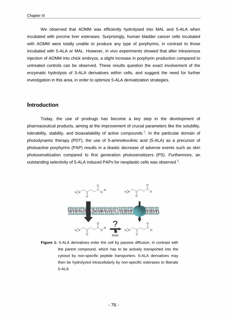
Chapter III
- 76 -
We observed that AOMM was efficiently hydrolysed into MAL and 5-ALA when
incubated with porcine liver esterases. Surprisingly, human bladder cancer cells incubated
with AOMM were totally unable to produce any type of porphyrins, in contrast to those
incubated with 5-ALA or MAL. However, in vivo experiments showed that after intravenous
injection of AOMM into chick embryos, a slight increase in porphyrin production compared to
untreated controls can be observed. These results question the exact involvement of the
enzymatic hydrolysis of 5-ALA derivatives within cells, and suggest the need for further
investigation in this area, in order to optimize 5-ALA derivatization strategies.
Introduction
Today, the use of prodrugs has become a key step in the development of
pharmaceutical products, aiming at the improvement of crucial parameters like the solubility,
tolerability, stability, and bioavailability of active compounds 1. In the particular domain of
photodynamic therapy (PDT), the use of 5-aminolevulinic acid (5-ALA) as a precursor of
photoactive porphyrins (PAP) results in a drastic decrease of adverse events such as skin
photosensitization compared to first generation photosensitizers (PS). Furthermore, an
outstanding selectivity of 5-ALA induced PAPs for neoplastic cells was observed 2.
ROH
NH3+
OR
O
O
NH3+
O
O
O
NH3+
OR
O
O
NH3+
O
O
O?
Figure 1: 5-ALA derivatives enter the cell by passive diffusion, in contrast with
the parent compound, which has to be actively transported into the
cytosol by non-specific peptide transporters. 5-ALA derivatives may
then be hydrolyzed intracellularly by non-specific esterases to liberate
5-ALA.

On the metabolism of 5-aminolevulinic acid derivatives
- 77 -
Nevertheless, the double opposite charges carried by 5-ALA under physiological
conditions seriously limit its ability to cross biological membranes, consequently restraining
its bioavailability. One successful strategy adopted to overcome this low bioavailability was
the derivatization of 5-ALA into more lipophilic n-alkylchained esters 3. Several experiments
have confirmed that these simple derivatives induced equivalent or higher porphyrin
accumulation than the parent compound, but at significantly lower concentrations in vitro 4,5,6.
The explanation of this increased potency is related to the drug internalization
process (Fig. 1). It was demonstrated that active transporters are involved in the entrance of
5-ALA or 5-ALA methylester (MAL) into the cells 7,8. In contrast, lipophilic 5-ALA derivatives
can cross biological barriers by passive diffusion or endocytosis, as incubation in the
presence of transport competitive inhibitors have shown limited influence 8,9,10. This non-
energy dependent penetration of moderately lipophilic 5-ALA esters through cellular
membranes is directly correlated to the lipophilicity of the molecule, enabling a faster
incorporation of porphyrin precursors into the target cells.
NH2COOH
A-Site
P-Site
O
NH2
O
O
O
NH2
O
O
NH2COOH
A-Site
P-Site
NH2COOH
A-Site
P-Site
NH2COOH
A-Site
P-Site
NNH2
COOHCOOH
O
NH2
O
O
R O
NH2
O
O
R
NNH2
O O
R O
O
R
A)
B)
Figure 2: A) 5-ALA dehydratase, the second enzyme involved in the heme
biosynthetic cycle, catalyzes the production of one molecule of
porphobilinogen from two molecules of 5-aminolevulinic acid. B) As the
carboxylic group of 5-ALA is not involved mechanistically in this
enzymatic conversion, 5-ALA esters may be potential substrates,
leading to the formation of esterified porphobilinogen.

Chapter III
- 78 -
After internalization, it is assumed that lipophilic derivatives of 5-ALA are cleaved
intracellularly by non-specific esterases to liberate 5-ALA, the natural heme precursor. In this
context, different experiments have evaluated the hydrolysis rate of 5-ALA derivatives, for
example using lysed cells 11 or tissue homogenates 12. Nevertheless, it is important to note
that the esterified carboxylic groups are not involved in the enzymatic transformation of
5-ALA into porphobilinogen (PBG) 13. It may, therefore, be in principle a direct substrate of
the porphobilinogen synthase (Fig.2).
The aim of the present study was to explore possibilities to increase drug stability and
bioavailability by taking advantage of the 5-ALA derivative metabolism. Therefore, we
synthesized 3-amino-3-oxohenadioic-1,6-dimethyl ester (AOMM) (Fig. 3), which presents the
following particularities, (i) physicochemical properties similar to MAL, (ii) stability towards
dimerization due to the attachment of a methoxycarbonyl function next to the amino group,
(iii) has to be expressly hydrolyzed and activated by esterases in order to undergo a rapid
decarboxylation into MAL.
site B
site A
AOMM MAL 5-ALA
NH3
+O
O
OOO
NH3
+O
O
O
NH3+
O
O
OCO2
-MeOH -MeOH
Figure 3: In the presence of intracellular esterases, AOMM will undergo enzymatic
hydrolysis at two sites. Hydrolysis at site A will instantly release CO2
through direct decarboxylation to form MAL, while cleavage at site B will
finally lead to the liberation of 5-ALA.
Material and Methods
Chemicals. All commercial reagents were used as obtained. D-MEM+GlutaMAX I, Penicillin-
Streptomycin and D-PBS were obtained from Gibco (Invitrogen, Basel, Switzerland). Heat
inactivated fetal-bovine serum and porcine liver esterase were purchased from Sigma
(Buchs, Switzerland). 5-aminolevulinic acid was provided by Fluka (Buchs, Switzerland), and
5-ALA methylester from Organix (Colchester, UK). Protoporphyrin IX standard was obtained
from Porphyrin products (Logan, USA).

On the metabolism of 5-aminolevulinic acid derivatives
- 79 -
AOMM Synthesis. In order to prepare AOMM, commercially available 1,6-dimethyl-3-oxo-
hexanedioic acid was first nitrosilated at the 2-position with nitrous acid to yield 1,6-dimethyl-
2-hydroxyimino-3-oxo-hexanedioic acid. The resulting hydroxyimino functionality was
transformed to a Boc protected amine in a single step by hydrogenating over palladium on
carbon in the presence of Boc anhydride. Subsequently, the Boc protecting group was
removed under acidic conditions to yield the final product as a hydrochloride salt.
1,3-dihydroxy acetone and 1,6-dimethyl-3-oxo-hexanedioic acid were purchased from Fluka
(Buchs, Switzerland).
1,6-Dimethyl-2-(hydroxyimino)-3-oxo-hexanedoic acid synthesis. NaNO2 (0.95 g,
13.8 mmol) in water (1.5 ml) was added dropwise over 1h to a solution of 1,6-dimethyl-3-oxo-
hexanedioic acid (2.16 g, 11.5 mmol) in acetic acid (5.0 ml) at 0 ºC. The resulting solution
was stirred for an additional 2 hours at this temperature. The reaction was then diluted with
ethyl acetate (200 ml) and washed with saturated NaCl solution (20 ml). The organic phase
was separated, dried with MgSO4, and the solvent was removed under reduced pressure.
The crude product was purified by flash chromatography on a silica gel column using ethyl
acetate/hexanes (1:2) to yield 2.17 g (87% yield) of the indicated compound as a clear oil. All
spectral properties were identical to those previously reported 14.
Methyl 5-methoxycarbonyl-5-(N-(tert-Butoxycarbonyl)amino)levulinate synthesis. 1,6-
dimethyl-2-(hydroxyimino)-3-oxo-hexanedoic acid (2.45 g, 11.3 mmol), Boc2O (4.94 g, 22.6
mmol), 10% Pd/C (0.23 g), and methanol (110 ml) were placed in a 250 ml round bottom
flask, and the resulting mixture was stirred under a hydrogen atmosphere for 2.5 h at room
temperature. The reaction mixture was filtered through Celite, and the solvent was removed
under reduced pressure. The crude was purified by flash chromatography on a silica gel
column using ethyl acetate/hexanes (1:3) to yield 2.74 g (80% yield) of the indicated
compound as a clear oil. 1H NMR (300 MHz, CHCl3): 8.70 (br. s, 1H), 5.76 (d, J=6.6Hz, 1H),
5.08 (d, J=6.6Hz, 1H), 3.78 (s, 3H), 3.55 (s, 3H), 2.98-3.17 (m, 1H), 2.80-2.95 (m, 1H), 2.60-
2.80 (m, 2H), 1.43 (s, 9H) ppm. 13C NMR (75.5Hz, CHCl3): 199.9, 177.7, 166.9, 155.1, 80.9,
63.6, 55.6, 53.4, 34.8, 28.3, 27.7 ppm, respectively.
Methyl 5-methoxycarbonyl-5-aminolevulinate hydrochloride synthesis. i-Pr3SiH
(0.43 ml, 2.6 mmol) was added to a solution of methyl 5-methoxycarbonyl-5-(N-(tert-
Butoxycarbonyl)amino)levulinate (0.61 g, 2.0 mmol) in anhydrous THF (20 ml), and then dry
HCl gas was bubbled through the solution until saturation (determined by the evolution of
white fumes). The resulting solution was left to stand at room temperature for 6 h in a closed
container. The solvent was then removed under reduced pressure, and the remaining solid
was washed several times with diethyl ether to yield 0.41 g (89% yield) of the desired
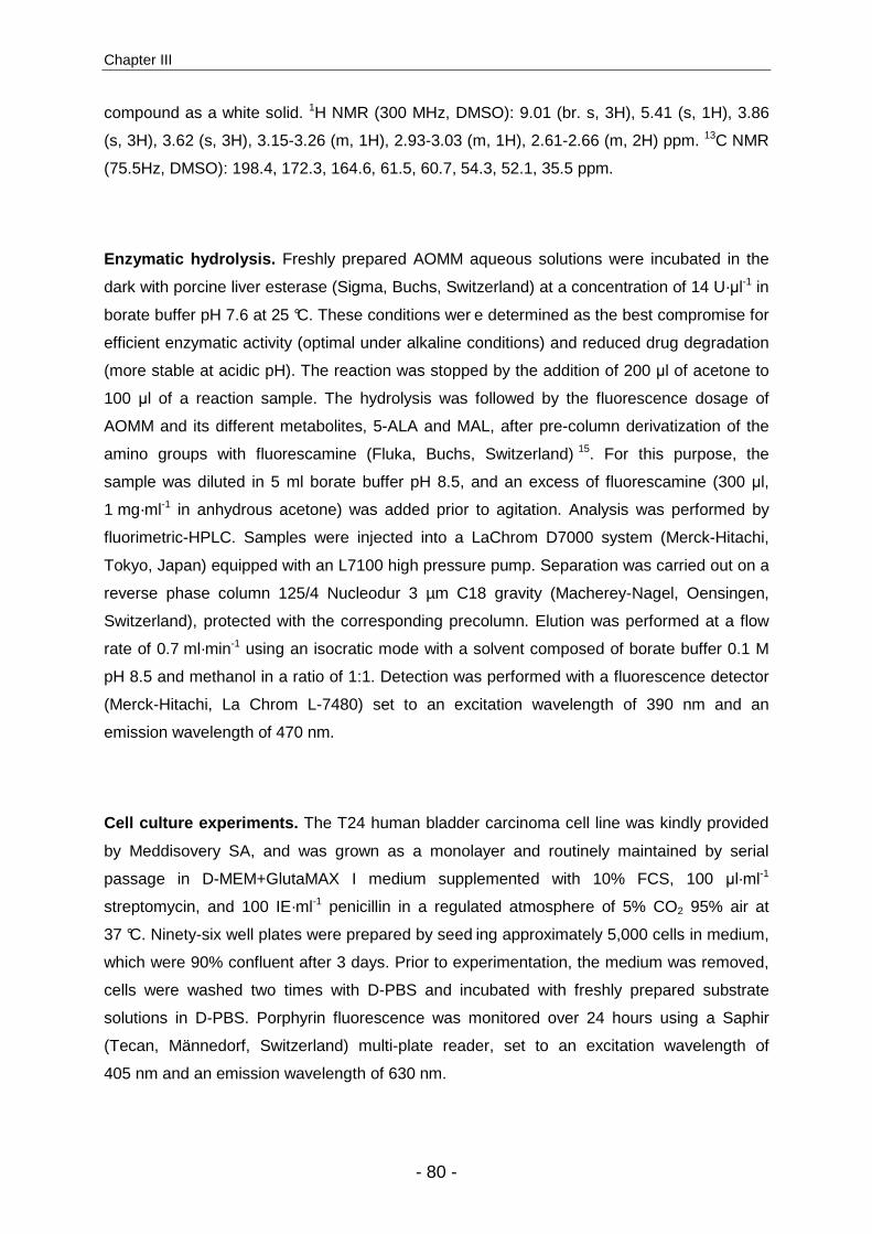
Chapter III
- 80 -
compound as a white solid. 1H NMR (300 MHz, DMSO): 9.01 (br. s, 3H), 5.41 (s, 1H), 3.86
(s, 3H), 3.62 (s, 3H), 3.15-3.26 (m, 1H), 2.93-3.03 (m, 1H), 2.61-2.66 (m, 2H) ppm. 13C NMR
(75.5Hz, DMSO): 198.4, 172.3, 164.6, 61.5, 60.7, 54.3, 52.1, 35.5 ppm.
Enzymatic hydrolysis. Freshly prepared AOMM aqueous solutions were incubated in the
dark with porcine liver esterase (Sigma, Buchs, Switzerland) at a concentration of 14 U·µl-1 in
borate buffer pH 7.6 at 25 °C. These conditions wer e determined as the best compromise for
efficient enzymatic activity (optimal under alkaline conditions) and reduced drug degradation
(more stable at acidic pH). The reaction was stopped by the addition of 200 µl of acetone to
100 µl of a reaction sample. The hydrolysis was followed by the fluorescence dosage of
AOMM and its different metabolites, 5-ALA and MAL, after pre-column derivatization of the
amino groups with fluorescamine (Fluka, Buchs, Switzerland) 15. For this purpose, the
sample was diluted in 5 ml borate buffer pH 8.5, and an excess of fluorescamine (300 µl,
1 mg·ml-1 in anhydrous acetone) was added prior to agitation. Analysis was performed by
fluorimetric-HPLC. Samples were injected into a LaChrom D7000 system (Merck-Hitachi,
Tokyo, Japan) equipped with an L7100 high pressure pump. Separation was carried out on a
reverse phase column 125/4 Nucleodur 3 µm C18 gravity (Macherey-Nagel, Oensingen,
Switzerland), protected with the corresponding precolumn. Elution was performed at a flow
rate of 0.7 ml·min-1 using an isocratic mode with a solvent composed of borate buffer 0.1 M
pH 8.5 and methanol in a ratio of 1:1. Detection was performed with a fluorescence detector
(Merck-Hitachi, La Chrom L-7480) set to an excitation wavelength of 390 nm and an
emission wavelength of 470 nm.
Cell culture experiments. The T24 human bladder carcinoma cell line was kindly provided
by Meddisovery SA, and was grown as a monolayer and routinely maintained by serial
passage in D-MEM+GlutaMAX I medium supplemented with 10% FCS, 100 µl·ml-1
streptomycin, and 100 IE·ml-1 penicillin in a regulated atmosphere of 5% CO2 95% air at
37 °C. Ninety-six well plates were prepared by seed ing approximately 5,000 cells in medium,
which were 90% confluent after 3 days. Prior to experimentation, the medium was removed,
cells were washed two times with D-PBS and incubated with freshly prepared substrate
solutions in D-PBS. Porphyrin fluorescence was monitored over 24 hours using a Saphir
(Tecan, Männedorf, Switzerland) multi-plate reader, set to an excitation wavelength of
405 nm and an emission wavelength of 630 nm.
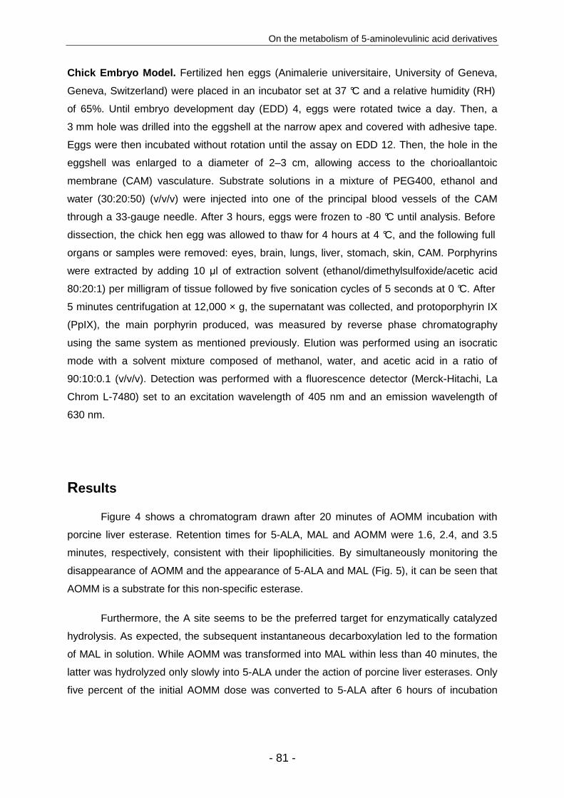
On the metabolism of 5-aminolevulinic acid derivatives
- 81 -
Chick Embryo Model. Fertilized hen eggs (Animalerie universitaire, University of Geneva,
Geneva, Switzerland) were placed in an incubator set at 37 °C and a relative humidity (RH)
of 65%. Until embryo development day (EDD) 4, eggs were rotated twice a day. Then, a
3 mm hole was drilled into the eggshell at the narrow apex and covered with adhesive tape.
Eggs were then incubated without rotation until the assay on EDD 12. Then, the hole in the
eggshell was enlarged to a diameter of 2–3 cm, allowing access to the chorioallantoic
membrane (CAM) vasculature. Substrate solutions in a mixture of PEG400, ethanol and
water (30:20:50) (v/v/v) were injected into one of the principal blood vessels of the CAM
through a 33-gauge needle. After 3 hours, eggs were frozen to -80 °C until analysis. Before
dissection, the chick hen egg was allowed to thaw for 4 hours at 4 °C, and the following full
organs or samples were removed: eyes, brain, lungs, liver, stomach, skin, CAM. Porphyrins
were extracted by adding 10 µl of extraction solvent (ethanol/dimethylsulfoxide/acetic acid
80:20:1) per milligram of tissue followed by five sonication cycles of 5 seconds at 0 °C. After
5 minutes centrifugation at 12,000 × g, the supernatant was collected, and protoporphyrin IX
(PpIX), the main porphyrin produced, was measured by reverse phase chromatography
using the same system as mentioned previously. Elution was performed using an isocratic
mode with a solvent mixture composed of methanol, water, and acetic acid in a ratio of
90:10:0.1 (v/v/v). Detection was performed with a fluorescence detector (Merck-Hitachi, La
Chrom L-7480) set to an excitation wavelength of 405 nm and an emission wavelength of
630 nm.
Results
Figure 4 shows a chromatogram drawn after 20 minutes of AOMM incubation with
porcine liver esterase. Retention times for 5-ALA, MAL and AOMM were 1.6, 2.4, and 3.5
minutes, respectively, consistent with their lipophilicities. By simultaneously monitoring the
disappearance of AOMM and the appearance of 5-ALA and MAL (Fig. 5), it can be seen that
AOMM is a substrate for this non-specific esterase.
Furthermore, the A site seems to be the preferred target for enzymatically catalyzed
hydrolysis. As expected, the subsequent instantaneous decarboxylation led to the formation
of MAL in solution. While AOMM was transformed into MAL within less than 40 minutes, the
latter was hydrolyzed only slowly into 5-ALA under the action of porcine liver esterases. Only
five percent of the initial AOMM dose was converted to 5-ALA after 6 hours of incubation

Chapter III
- 82 -
(Fig. 5). Since AOMM showed the capacity to be efficiently converted into MAL and 5-ALA in
vitro, we proceeded to its assessment in cell culture
0
100
200
300
400
500
600
700
800
900
0 1 2 3 4 5
Elution time [min]
Flu
ores
cenc
e in
tens
ity [A
.U.]
MAL
5-ALAAOMM
Figure 4: HPLC profile of AOMM and its two metabolites 5-ALA and MAL. Detection
was performed by fluorescence (λexcitation 390 nm, λemission 470 nm) after pre-
column derivatization with fluorescamine and reverse phase separation.
0.0
0.2
0.4
0.6
0.8
1.0
0 1 2 3 4 5 6
Incubation time [hours]
Dru
g co
ncen
trat
ion
[mM
]
5-ALA
MAL
AOMM
Figure 5: Enzymatic conversion of AOMM into MAL and 5-ALA (Porcine liver
esterase 14 U·µl-1 in borate buffer pH 7.6, 25 °C).
In vitro cell culture experiments consisted of the monitoring of porphyrin fluorescence
in the T24 human bladder cancer cell line upon incubation with 5-ALA or 5-ALA derivatives
(Fig. 6). Strong porphyrin fluorescence was observed after incubation with exogenous 5-ALA.

On the metabolism of 5-aminolevulinic acid derivatives
- 83 -
Effective substrate concentrations ranged from 0.1 to 10 mM, with a maximum fluorescence
intensity observed around 3 mM. 5-ALA methylester induced similar porphyrin fluorescence
intensities, at slightly higher concentrations than the parent compound, which is in agreement
with previously published data 4. In contrast, AOMM induced no porphyrin production at any
tested concentration during the first 6 hours of incubation. However, a slight porphyrin
fluorescence was detected when increasing the incubation time over 10 hours (data not
shown).
0
4000
8000
12000
16000
20000
0.1 1 10 100
Substrate concentration [mM]
Flu
ores
cenc
e [A
.U.]
5-ALA
MAL
AOMM
A
0
4000
8000
12000
16000
20000
0 1 2 3 4 5 6Incubation time [hours]
Flu
ores
cenc
e [A
.U.]
ALA
MAL
AOMM
B
Figure 6: A) Porphyrin fluorescence of T24 human bladder cancer cells after 6 hours
incubation with increasing concentrations of 5-ALA (□), MAL (○) and
AOMM (∆). B) Porphyrin fluorescence of T24 human bladder cancer cells
incubated with 5-ALA (□), MAL (○) and AOMM (∆) at 3 mM as a function of
time.
Chapter III
- 84 -
As it was demonstrated that AOMM was enzymatically metabolized into MAL and
5-ALA, and despite its limited ability to induce porphyrin formation in vitro, we evaluated its
capacity to induce porphyrin accumulation in vivo using the chick embryo in ovo. Three hours
after intravenous administration in the CAM, we observed that AOMM induced a 3-fold
increase in PAP production in the chick embryo organs as compared to the untreated control.
However, the porphyrin induction was still inferior to 5-ALA and MAL, which induced 40- and
12-fold increases, respectively (Fig. 7).
Nevertheless, a prolonged incubation time of 6 hours increased the formation of
photoactive porphyrin induced by AOMM, while a decrease was observed with MAL (results
not shown). Therefore, AOMM has the capacity to act as a PAP precursor in vivo, although
activation times appear to be delayed when compared to MAL.
0.00
0.01
0.10
1.00
10.00
EYE
BRAIN
HEART
LUNGS
STOMACH
LIVER
SKINCAM
PpI
X µµ µµ
g /
g of
tiss
ue
Control
5-ALA
MAL
AOMM
Figure 7: PpIX biodistribution in the chick embryo model after injection of
equimolar doses of 5-ALA, MAL, and AOMM compared to the
control (injection of solvent). Experimental conditions: 3 hours
incubation time; 350 µmol·Kg-1 dose.
DISCUSSION
Today, the expanding clinical use of 5-ALA derivative-mediated photomedicine has
triggered intense research efforts in this domain. Investigations currently focus on (i) the
optimized formulation of available 5-ALA derivatives for increased bioavailability and
stability 16,17, (ii) the development of new 5-ALA derivatives with optimized properties or

On the metabolism of 5-aminolevulinic acid derivatives
- 85 -
targeting intentions 18,19,20, and (iii) the fundamental investigation of the uptake 21,
metabolism 8,12, and degradation 22,23 processes involved after the administration of 5-ALA
derivatives.
In this context, we decided to focus on novel strategies for the design of 5-ALA
derivatives. Since the stability of 5-ALA derivatives plays an important role in the process of
drug development, we explored different options to prevent degradation. Today, other than
lowering pH to subphysiological values, no method has obtained satisfying results.
Therefore, extemporaneous formulation or shortened shelf life are recommended 23.
Unfortunately, too acidic pH values can potentially harm the ester function. The main
degradation mechanism observed with such compounds in aqueous solutions is the
dimerization of two 5-ALA or 5-ALA derivative molecules into pyrazines. The rate of this
reaction increases with increasing substrate concentration and pH 24.
An apparently promising method to overcome this undesirable degradation process is
the synthesis of 5-ALA derivatives with a protected amino group. Unfortunately, this
approach leads to unsatisfactory PAP induction in vitro 25,26. However, the fact that this
strategy failed, presumably due to the absence of adapted lysis enzymes, does not
necessarily imply the inability to “protect” the amino moiety against intermolecular
condensations. An alternative approach to this problem may consist of the addition of a
protective group next to the amino moiety that impedes intermolecular condensation.
According to the literature, esterases are the ideal target enzymes for prodrug
activation. It is commonly accepted that 5-ALA derivatives, after passive entrance into the
cell, are cleaved by non-specific esterases into free 5-ALA, which enters the heme
biosynthesis pathway. The addition of a methoxy-carbonyl moiety next to the amino group is,
in theory, a promising approach considering that (i) it will not significantly modify lipophilicity
or molecular weight, which could significantly modulate cell penetration, (ii) the dimerization
into pyrazine is sterically hindered, and (iii) esterases will induce a direct decarboxylation
upon hydrolysis of the methoxy moiety.
Our observations confirm that, as expected, AOMM is efficiently hydrolysed by
porcine liver esterase, and undergoes spontaneous decarboxylation into MAL. The latter is
then slowly converted into 5-ALA. It is known that enzyme affinity for the substrate depends
on chain length and lipophilicity. In the case of AOMM, for both methyl ester functions, a
similar cleavage rate would have been expected. The fact that the decarboxylation
subsequent to ester cleavage at site A is irreversible may explain the preferential hydrolysis
of the protecting group.

Chapter III
- 86 -
Reports of the metabolism of 5-ALA derivatives are somewhat controversial. For
example, the apparition of porphyrin fluorescence was taken as proof of 5-ALA derivative
hydrolysis. Nevertheless, it should be considered that 5-ALA esters can be a potential
substrate for heme biosynthesis (Fig. 2) 27.
Different studies investigated the metabolism of 5-ALA derivatives, especially 5-ALA
n-alkyl esters, regarding their hydrolysis rate and enzymatic transformation into the parent
compound 12,28,11. In a recent study, Di Venosa et al. 28 evaluated the regulation of porphyrin
synthesis and hydrolysis from 5-ALA derivatives. The intracellular accumulation of 5-ALA, 5-
ALA derivatives, PBG, and porphyrins under different conditions was measured. These
authors concluded that at high substrate concentrations, porphyrin synthesis from 5-ALA and
5-ALA derivatives was regulated by porphobiligenase and esterase, respectively.
Furthermore, (i) only a small fraction of 5-ALA hexylester (HAL) was converted into 5-ALA
intracellularly, (ii) at very low concentrations of HAL, only 18.7 pmol/105 cells was found, but
porphyrin synthesis was already saturated, and (iii) at these intracellular concentrations,
porphyrin synthesis was not saturated when using 5-ALA in the same cell line.
However, these experiments did not reveal whether the in vivo metabolism of 5-ALA
esters is dominated by hydrolysis followed by heme biosynthesis or by the direct entry of the
5-ALA esters into this biosynthetic pathway. The fact that AOMM failed to induce porphyrin
formation in cells could be explained by several different causes. A less efficient cell uptake
of this compound as compared to MAL seems improbable, since the charge distribution,
lipophilicity, and molecular weights are similar. The small increase of molecular weight
should not inhibit passive transport through biomembranes, considering the in vitro porphyrin
induction of porphyrin induced by 5-ALA derivatives of much higher molecular weights 20.
Consequently, it has to be questioned at which stage during heme biosynthesis
5-ALA derivative cleavage occurs. It was demonstrated that in the presence of tissue
homogenates, long chained esters like 5-ALA hexylester were better substrates for
enzymatic hydrolysis than short chained derivatives 12. In contrast, Kloek et al. incubated 5-
ALA esters of increasing chain length with lysed cells to minimize the influence of cellular
uptake. Under their conditions, porphyrin production decreased with increasing substituent
chain length until 5-ALA butylester. However, increasing porphyrin production was again
observed with 5-ALA pentyl-, hexyl-, and octylester.
However, if MAL has to be converted into 5-ALA prior to entering heme biosynthesis,
then, the presence of intracellular esterase activity is given in our in vitro studies by the MAL
induced fluorescence in cells. Since our experiments with porcine liver esterases showed
On the metabolism of 5-aminolevulinic acid derivatives
- 87 -
that AOMM is efficiently converted into MAL, then AOMM would be thought to be converted
into MAL in cells, and then into a substrate for heme biosynthesis. However, this is not the
case, even at very high concentrations. Therefore, we hypothesize that esterase activity is
not the sole parameter that dominates the conversion of 5-ALA derivatives into porphyrins.
Conclusions
Our observations on the studied methoxycarbonyl-5-ALA methylester underline that
cellular enzymatic cleavage of 5-ALA derivatives may not be mandatory. Focusing on
enzymatic affinity, we observed that AOMM was a more preferred substrate for porcine liver
esterase than MAL. However, cells incubated with MAL but not AOMM were able to
accumulate fluorescent porphyrins. These results are not consistent with the commonly
accepted metabolism route of 5-ALA derivatives, and encourage supplemental investigation
in this area. A deeper understanding of the exact fate of 5-ALA derivatives after their
administration will allow for the development of better targeting strategies for 5-ALA
derivatives in the future.
Acknowledgements
The authors would like to thank Dr Angelica Vargas for her technical assistance with
the chick embryos.

Chapter III
- 88 -
References 1. B. Testa, Prodrug research: futile or fertile?, Biochem Pharmacol, 2004, 68, 2097-2106.
2. S. Collaud, A. Juzeniene, J. Moan, and N. Lange, On the selectivity of 5-aminolevulinic acid-induced protoporphyrin IX formation, Curr Med Chem Anti-Canc Agents, 2004, 4, 301-316.
3. N. Fotinos, M. A. Campo, F. Popowycz, R. Gurny, and N. Lange, 5-Aminolevulinic Acid Derivatives in Photomedicine: Characteristics, Application and Perspectives, Photochem Photobiol, 2006, 82, 994-1015.
4. P. Uehlinger, M. Zellweger, G. Wagnières, L. Juillerat-Jeanneret, H. van den Bergh, and N. Lange, 5-aminolevulinic acid and its derivatives: physical chemical properties and protoporphyrin IX formation in cultured cells, J Photochem Photobiol B:Biol, 2000, 54, 72-80.
5. J. M. Gaullier, K. Berg, Q. Peng, H. Anholt, P. K. Selbo, L. W. Ma, and J. Moan, Use of 5-aminolevulinic acid esters to improve photodynamic therapy on cells in culture, Cancer Res, 1997, 57, 1481-1486.
6. Q. Peng, J. Moan, T. Warloe, V. Iani, H. B. Steen, A. Bjorseth, and J. M. Nesland, Build-up of esterified aminolevulinic-acid-derivative-induced porphyrin fluorescence in normal mouse skin, J Photochem Photobiol B:Biol, 1996, 34, 95-96.
7. F. Doring, J. Walter, J. Will, M. Focking, M. Boll, S. Amasheh, W. Clauss, and H. Daniel, Delta-aminolevulinic acid transport by intestinal and renal peptide transporters and its physiological and clinical implications, J Clin Invest, 1998, 101, 2761-2767.
8. E. Rud, O. Gederaas, A. Hogset, and K. Berg, 5-aminolevulinic acid, but not 5-aminolevulinic acid esters, is transported into adenocarcinoma cells by system BETA transporters, Photochem Photobiol, 2000, 71, 640-647.
9. L. Rodriguez, A. M. Batlle, G. Di Venosa, A. J. MacRobert, S. H. Battah, H. Daniel, and A. Casas, Study of the mechanisms of uptake of 5-aminolevulinic acid derivatives by PEPT1 and PEPT2 transporters as a tool to improve photodynamic therapy of tumours, Int J Biochem Cell Biol, 2006, 38, 1530-1539.
10. A. Novotny, J. Xiang, W. Stummer, N. S. Teuscher, D. E. Smith, and R. F. Keep, Mechanisms of 5-aminolevulinic acid uptake at the choroid plexus, J Neurochem, 2000, 75, 321-328.
11. J. Kloek, W. Akkermans, and M. J. Beijersbergen van Henegouwen, Derivatives of 5-aminolevulinic acid photodynamic therapy: enzymatic conversion into protoporphyrin, Photochem Photobiol, 1998, 67, 150-154.
12. F. S. De Rosa, R. F. Lopez, J. A. Thomazini, A. C. Tedesco, N. Lange, and M. V. Bentley, In vitro metabolism of 5-ALA esters derivatives in hairless mice skin homogenate and in vivo PpIX accumulation studies, Pharm Res, 2004, 21, 2247-2252.
13. E. K. Jaffe, The porphobilinogen synthase family of metalloenzymes, Acta Crystallogr D Biol Crystallogr, 2000, 56, 115-128.

On the metabolism of 5-aminolevulinic acid derivatives
- 89 -
14. A. Nudelman and A. Nudelman, Convenient syntheses of delta-aminolevulinic acid, Synthesis, 1999, 4, 568-570.
15. S. Udenfriend, S. Stein, P. Böhlen, W. Dairman, W. Leimgruber, and M. Weigele, Fluorescamine: a reagent for assay of amino acids, peptides, proteins, and primary amines in the picomole range., Science, 1972, 178, 871-872.
16. S. Collaud, P. Jichlinski, A. Marti, D. Aymon, R. Gurny, and N. Lange, An open pharmacokinetic study of hexylaminolevulinate-induced photodiagnosis after intravesical administration, Drugs R D, 2006, 7, 173-186.
17. A. Casas, C. Perotti, M. Saccoliti, P. Sacca, H. Fukuda, and A. M. Batlle, ALA and ALA hexyl ester in free and liposomal formulations for the photosensitisation of tumour organ cultures, Br J Cancer, 2002, 86, 837-842.
18. Y. Berger, L. Ingrassia, R. Neier, and L. Juillerat-Jeanneret, Evaluation of dipeptide-derivatives of 5-aminolevulinic acid as precursors for photosensitizers in photodynamic therapy, Bioorg Med Chem, 2003, 11, 1343-1351.
19. Y. Berger, A. Greppi, O. Siri, R. Neier, and L. Juillerat-Jeanneret, Ethylene glycol and amino acid derivatives of 5-aminolevulinic acid as new photosensitizing precursors of protoporphyrin IX in cells, J Med Chem, 2000, 43, 4738-4746.
20. S. H. Battah, S. Balaratnam, A. Casas, S. O'neill, C. Edwards, A. M. Batlle, P. Dobbin, and A. J. MacRobert, Macromolecular delivery of 5-aminolaevulinic acid for photodynamic therapy using dendrimer conjugates, Mol Cancer Ther, 2007, 6, 876-885.
21. L. Rodriguez, A. M. Batlle, G. Di Venosa, S. H. Battah, P. Dobbin, A. J. MacRobert, and A. Casas, Mechanisms of 5-aminolevulinic acid ester uptake in mammalian cells, Br J Pharmacol, 2006, 147, 825-833.
22. M. Kaliszewski, M. Kwasny, A. Juzeniene, P. Juzenas, A. Graczyk, L. W. Ma, V. Iani, P. Mikolajewska, and J. Moan, Biological activity of 5-aminolevulinic acid and its methyl ester after storage under different conditions, J Photochem Photobiol B, 2007, 87, 67-72.
23. M. Kaliszewski, M. Kwasny, J. Kaminski, Z. Dabrowski, and E. Burdziakowska, The stability of 5-aminolevulinic acid and its ester derivatives, Acta Pol Pharm, 2004, 61, 15-19.
24. A. Bunke, O. Zerbe, G. Burmeister, H. P. Merkle, and B. Gander, Degradation mechanism and stability of 5-aminolevulinic acid, J Pharm Sci, 2000, 89, 1335-1341.
25. Y. Ninomiya, Y. Itoh, S. Tajima, and A. Ishibashi, In vitro and in vivo expression of protoporphyrin IX induced by lipophilic 5-aminolevulinic acid derivatives, J Dermatol Sci, 2001, 27, 114-120.
26. M. Kaliszewski, A. Juzeniene, P. Juzenas, M. Kwasny, J. Kaminski, Z. Dabrowski, J. Golinski, and J. Moan, Formation of protoporphyrin IX from carboxylic- and amino-derivatives of 5-aminolevulinic, Photodiagnonsis Photodynamic Therapy, 2005
27. C. Jarret, F. Stauffer, M. E. Henz, M. Marty, R. M. Lüönd, J. Bobálová, O. Schürmann, and R. Neier, Inhibition of Escherichia coli porphobilinogen synthase using analogs of postulated intermediates, Chem Biol, 2000, 7, 185-196.

Chapter III
- 90 -
28. G. Di Venosa, H. Fukuda, A. M. Batlle, A. J. MacRobert, and A. Casas, Photodynamic therapy: regulation of porphyrin synthesis and hydrolysis from ALA esters, J Photochem Photobiol B, 2006, 83, 129-136.
The chick embryo model for the evaluation of 5-aminolevulinic derivatives
- 91 -
CHAPTER IV
The chick embryo model for the evaluation
of 5-aminolevulinic acid derivatives
N. Fotinos, M.A. Campo, F. Popowycz, R. Gurny, N. Lange
Department of Pharmaceutics and Biopharmaceutics, School of Pharmaceutical Sciences, University of Geneva,
University of Lausanne, 30 Quai Ernest Ansermet, CH-1211 Geneva 4, Switzerland
Submitted to Photochemical and Photobiological Sciences
Abstract
5-aminolevulinic acid (5-ALA) has arisen as a leading candidate for photodynamic
therapy and fluorescence detection due to its numerous advantages over first generation
photosensitizers, including tolerability, high selectivity for the diseased tissue and rapid
elimination. However, the bioavailability of 5-ALA is structurally limited by its zwitterionic
character at physiological pH, limiting its passage through biological barriers. More lipophilic
5-ALA derivatives, predominantly designed to increase local bioavailability after topical
administration, have been developed to overcome this drawback. Nevertheless, several life
threatening pathologies such as brain, breast or ovarian cancer cannot be efficiently treated
using these compounds.
In this context, we synthesized a series of derivatives adapted to systemic
administration. However, the insufficient aqueous solubility of these compounds makes their
screening in conventional in vitro cell culture systems difficult. Therefore, we have used chick
embryos for the evaluation of the biodistribution of 5-ALA derivative induced porphyrins. The
molecules to be tested were dissolved in a solvent mixture of PEG, ethanol and water, and
injected into a large vessel of the chorioallantoic membrane. After a defined exposure time,
the chick embryo survival was evaluated, and then the embryos were frozen to -80 °C. After
dissection, different organs were collected, and photoactive products were extracted by
sonication and quantified by HPLC.

Chapter IV
- 92 -
This model allowed for the determination of the most promising compounds in terms
of their ability to induce the accumulation of photoactive porphyrins in different organs and
the absence of acute toxicity after intravenous administration. Furthermore, the present
system might be a cost effective alternative for initial biodistribution and toxicity studies.
Introduction
Over the last decade, the use of photodynamic therapy (PDT) and fluorescence
detection (FD) in various medical fields has become increasingly widespread. These non-
invasive techniques combine the use of a photosensitizer (PS) accumulated into the target
tissues with activation by light to induce fluorescence in the case of FD or initiation of the
formation of reactive oxygen species in the case of PDT, ultimately leading to the destruction
of targeted pathological tissues.
Different photosensitizer types have been tested in photomedicine, usually derived
from a porphyrinic skeleton 1. However, one endogenous molecule, 5-aminolevulinic acid
(5-ALA), has raised particular interest in this field, since it acts as a pro-photosensitizer when
administered exogenously. In fact, the formation of 5-ALA catalyzed by 5-ALA synthase is
the first step of heme biosynthesis, and can be observed in all nucleated cells. The limited
accumulation of photoactive porphyrins (PAP) in tissues is physiologically regulated by a
negative feedback induced by heme on 5-ALA synthase. However, an exogenous
administration of 5-ALA overcomes this negative feedback, leading to the temporary
accumulation of PAP within cells, especially those with high metabolic turnover like cancer
cells 2.
One major drawback of 5-ALA-mediated PDT is directly related to the structure of the
molecule, which is present as a zwitterion under physiological conditions. This highly
charged molecule cannot easily cross biological membranes, and has to be internalized by
active transporters 3. To overcome this problem, more lipophilic derivatives have been
proposed 4. Two of them, 5-ALA methylester and 5-ALA hexylester, achieved marketing
authorization and are indicated for PDT treatment of basal cell carcinoma and actinic
keratosis and improved detection of bladder cancer, respectively. However, these two
molecules are adapted for topical administration, but are suboptimal for utilization in a
systemic approach. In this context, life threatening diseases like breast cancer, prostate
cancer, or brain cancer cannot be optimally targeted.
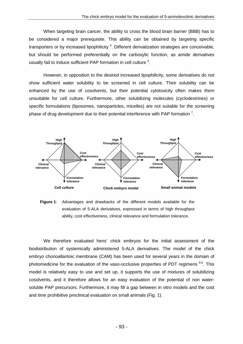
The chick embryo model for the evaluation of 5-aminolevulinic derivatives
- 93 -
When targeting brain cancer, the ability to cross the blood brain barrier (BBB) has to
be considered a major prerequisite. This ability can be obtained by targeting specific
transporters or by increased lipophilicity 5. Different derivatization strategies are conceivable,
but should be performed preferentially on the carboxylic function, as amide derivatives
usually fail to induce sufficient PAP formation in cell culture 6.
However, in opposition to the desired increased lipophilicity, some derivatives do not
show sufficient water solubility to be screened in cell culture. Their solubility can be
enhanced by the use of cosolvents, but their potential cytotoxicity often makes them
unsuitable for cell culture. Furthermore, other solubilizing molecules (cyclodextrines) or
specific formulations (liposomes, nanoparticles, micelles) are not suitable for the screening
phase of drug development due to their potential interference with PAP formation 7.
Cost effectiveness
Clinical relevance
Formulationtolerance
High Throughput
Cell culture
Cost effectiveness
Clinical relevance
Formulation tolerance
High Throughput
Small animal models
Cost effectiveness
Clinical relevance
Formulation tolerance
High Throughput
Chick embyro model
Figure 1 : Advantages and drawbacks of the different models available for the
evaluation of 5-ALA derivatives, expressed in terms of high throughput
ability, cost effectiveness, clinical relevance and formulation tolerance.
We therefore evaluated hens’ chick embryos for the initial assessment of the
biodistribution of systemically administered 5-ALA derivatives. The model of the chick
embryo chorioallantoic membrane (CAM) has been used for several years in the domain of
photomedicine for the evaluation of the vaso-occlusive properties of PDT regimens 8,9. This
model is relatively easy to use and set up, it supports the use of mixtures of solubilizing
cosolvents, and it therefore allows for an easy evaluation of the potential of non water-
soluble PAP precursors. Furthermore, it may fill a gap between in vitro models and the cost
and time prohibitive preclinical evaluation on small animals (Fig. 1).

Chapter IV
- 94 -
Materials and Methods
Chemicals. 5-aminolevulinic acid was obtained from Fluka (Buchs, Switzerland) and 5-ALA
hexylester from Organix (Colchester, UK). Protoporphyrin IX was purchased from Frontier
Scientific (Logan, USA). All other solvents and chemicals used were of analytical grade and
used without further purification.
Chick Embryo Model. Fertilized hen eggs (Animalerie universitaire, University of Geneva,
Geneva, Switzerland) were placed into an incubator set at 37 °C and a relative humidity of
65%. Until embryo development day (EDD) 4, eggs were rotated twice a day. Then, a 3 mm
hole was drilled into the eggshell at the narrow apex and covered with adhesive tape. Eggs
were then incubated without rotation until the experiment on EDD 12.
On EDD 12, the hole in the eggshell was enlarged to a diameter of 2–3 cm to allow
access to the CAM vasculature. Substrates to be evaluated (Fig. 2) were solubilized with a
solvent mixture composed of PEG400, ethanol and water (30:20:50; v/v/v), and injected into
one of the principal blood vessels of CAM through a 33-gauge needle. After defined
incubation times, survival of the embryos was evaluated, and eggs were frozen at -80 °C
until analysis.
Before dissection, the chick egg was allowed to thaw for 4 hours at 4 °C, and the
following full organs or samples were collected (eye, brain, lung, liver, stomach, skin,
CAM) 10. Porphyrins were extracted from the different organ samples after membrane
disruption, according to a modified method described by Peng et al. 11. Five sonication cycles
of 5 seconds at 0 °C were conducted using a probe s onicator (Branson digital sonifier,
Danbury, USA) after the addition of 10 µl of extraction solvent
(ethanol/dimethylsulfoxide/acetic acid; 80:20:1) per milligram of tissue. The samples were
centrifuged at 12,500 × g for 5 minutes, the supernatant was collected, and protoporphyrin IX
was dosed by reverse phase chromatography with a 125/4 Nucleodur C18 gravity column
(Macherey-Nagel, Oensingen, Switzerland) using an isocratic elution with
methanol/water/acetic acid (90:10:0.1; v/v/v). Detection was performed with a fluorescence
detector (Merck-Hitachi, La Chrom L-7480; excitation wavelength: 407 nm and emission
wavelength: 630 nm). A calibration curve was performed with protoporphyrin IX standard
solutions.

The chick embryo model for the evaluation of 5-aminolevulinic derivatives
- 95 -
(a) The toxicity of the solvent, (b) the toxicity of the different substrates, and (c) the
biodistribution of protoporphyrin IX formation in the different organs of the chick embryo were
assessed.
5-ALA diacylglyceride derivative synthesis . 1,3-diacetyl acetone, 1,3-diacetyl-2-hydroxy
glyceride, and 1,3-dibutyrate-2-hydroxy glyceride were prepared according to Paris et al. 12.
15-((N-tert-butoxycarbonyl)amino)levulinic acid synthesis: A solution of 5-aminolevulinic acid
(20.9 mmol) in methanol (50 ml) was added slowly to a solution of Boc anhydride (6.84 g,
31 mmol) and diisopropyl ethyl amine (5.4 g, 2 equiv.) in methanol (40 ml), with stirring. The
reaction was allowed to proceed for 1.5 h, and then the solvent was evaporated under
reduced pressure. The compound of interest was obtained after chromatographic purification
on silica gel using ethyl acetate/hexane (3:2) as a clear oil weighing 4.2 g. 1H NMR (300
MHz, CDCl3): 1.46 (s, 9H), 2.71 (br s, 4H), 4.07 (br s, 2H), 5.25 (br s, 1H), 8.93 (br s, 1H)
ppm. 13C NMR (75.5Hz, CDCl3): 27.8, 28.5, 34.4, 50.5, 80.3, 177.4, 204.5 (missing one sp2
carbon due to overlap) ppm.
1,3-dibutyrate-2-(N-tert-butoxycarbonyl)ALA glyceride synthesis: EDC (0.238 g, 1.5 equiv.)
was added to a solution of 1,3-dibutyrate-2-hydroxy glyceride (0.288 g, 1.24 mmol), 5-((N-
tert-butoxycarbonyl)amino)levulinic acid (0.19 g, 0.827 mmol), and DMAP (5 mg) in DCM
(2.5 ml), and the solution was stirred for 5 h at room temperature. Solvent was then removed
under reduced pressure, and the mixture was purified by column chromatography on a silica
gel using ethyl acetate/hexane (1:2). The desired product was obtained as a clear oil
weighing 0.204 g (55% yield). 1H NMR (300 MHz, CDCl3): 5.10-5.01 (m, 1H), 4.16-3.88 (m,
6H), 2.73 (t, J=6.5Hz, 2H), 2.54 (t, J=6.5Hz, 2H), 2.15 (t, J=7.3Hz, 4H), 1.49 (h, J=7,6Hz,
4H), 1.29 (s, 9H), 0.79 (t, J=7.3Hz, 6H) ppm. 13C NMR (75.5Hz, CDCl3): 13.7, 18.4, 27.8,
28.4, 34.3, 35.9, 50.3, 61.9, 69.7, 171.6, 173.2 (missing two sp2 and one sp3 carbons due to
overlap) ppm.
1,3-dibutyrate-2-ALA glyceride TFA salt synthesis: 1,3-dibutyrate-2-(N-tert-butoxycarbonyl)-
ALA glyceride was dissolved in dichloromethane/TFA (4:1), and the reaction was allowed to
proceed for 2 h at room temperature. Solvent was then removed under reduced pressure to
obtain the desired compound as a thick oil in quantitative yield. 1H NMR (300 MHz, CDCl3):
8.11 (br. s, 3H), 5.04-4.97 (m, 1H), 4.17-3.95 (m, 6H), 2.76 (t, J=6.3Hz, 2H), 2.52 (t, J=6.3Hz,
2H), 2.15 (t, J=7.4Hz, 4H), 1.47 (h, J=7,5Hz, 4H), 0.78 (t, J=7.4Hz, 6H) ppm. 13C NMR
(75.5Hz, CDCl3): 13.7, 18.4, 27.5, 34.8, 35.9, 47.9, 61.8, 69.7, 171.8, 173.3, 202.4 ppm.

Chapter IV
- 96 -
1,3-diacetyl-2-(N-tert-butoxycarbonyl)ALA glyceride synthesis: This compound was prepared
in a fashion similar to 1,3-dibutyrate-2-(N-tert-butoxycarbonyl)ALA glyceride. 1H NMR (300
MHz, CDCl3): 5.28-5.21 (m, 1H), 4.33-4.05 (m, 6H), 2.75 (t, J=6.3Hz, 2H), 2.68 (t, J=6.3Hz,
2H), 2.09 (s, 6H), 1.46 (s, 9H) ppm.
1,3-diacetyl-2-ALA glyceride TFA salt synthesis: This compound was prepared in a fashion
similar to 1,3-dibutyrate-2-ALA glyceride TFA salt. 1H NMR (300 MHz, CDCl3): 8.01 (br. s,
3H), 5.28-5.23 (m, 1H), 4.40-4.02 (m, 6H), 2.88 (t, J=6.3Hz, 2H), 2.70 (t, J=6.3Hz, 2H), 2.08
(s, 6H) ppm.
+H3N
O
O
OO
O
O
O
CF3CO2-
+H3N
O
O
OO
OCF3CO2-
O
O
+H3N
O
O-
O
+H3N
O
O
O
Cl-
5-aminolevulinic acid (5-ALA)
5-ALA hexylester (HAL)
Cl-
5-ALA diacetyl glyceride (ADAG)
5-ALA dibutyryl glyceride (ADBG)
Figure 2 : Chemical structures of the different pro-photosensitizers used for the
evaluation of the chick embryo model.
Results
5-ALA-mediated porphyrin formation . The chick embryo model was evaluated with
respect to 5-ALA-mediated porphyrin formation as a function of increasing drug doses. The
5-ALA solutions were injected into a large vessel of the CAM, and the incubation time was
set to 3 hours. Survival was monitored, and all embryos survived using 5-ALA doses ranging
from 0 to 120 mg·Kg-1, while injection of 240 mg·Kg-1 was lethal to the embryos in all cases.
The reasons for this toxicity are unknown, but some abdominal haemorrhages were
observed that could be related to hepatotoxicity.

The chick embryo model for the evaluation of 5-aminolevulinic derivatives
- 97 -
0.001
0.010
0.100
1.000
10.000
EY
E
BR
AIN
HE
AR
T
LUN
GS
ST
OM
AC
H
LIV
ER
SK
IN
CA
M
µg
of P
pIX
/ g
of ti
ssue
0
1.2 mg/kg
12 mg/kg
60 mg/kg
120 mg/kg
5-ALA dose
Figure 3 : Porphyrin biodistribution after the injection of increasing doses of
5-ALA into the chick embryo model. Incubation time was 3 hours.
The control consisted of an injection of 10-50 µl of solubilizing vehicle, and showed no
death was observed in any of the chick embryos. Samples taken from these control
experiments showed a low basal porphyrin concentration with high inter-organ variation. The
injection of low doses of 5-ALA (1.2 mg·Kg-1) did not induce a significant increase in PpIX
production. At 5-ALA doses of 12 mg·Kg-1, the porphyrin accumulation in the different organs
was 10-20 times higher than the control, and a plateau was reached at 60 mg·Kg-1. At these
optimal concentrations, the porphyrin content for all examined organs was increased by 1-2
orders of magnitude compared to the control, inducing a strong fluorescence in all tissues
(Fig. 3).
5-ALA derivative-mediated porphyrin formation . 5-ALA hexylester, a molecule
adapted for optimal topical application induced an immediate arrest of blood flow after doses
of ≥ 60 mg·Kg-1. Significantly different results were obtained with the 5-ALA diacylglyceride
derivatives after administration to the chick embryo model. No mortality was observed after
the injection of drug doses up to 240 mg·Kg-1 for either 5-ALA diacetylglyceride or
dibutyrylglyceride. The distribution showed an induction of porphyrin accumulation in various
tissues, comparable to 5-ALA at equimolar doses (375 µmol·Kg-1) (Fig. 4).

Chapter IV
- 98 -
0.001
0.010
0.100
1.000
10.000
EY
E
BR
AIN
HE
AR
T
LUN
GS
ST
OM
AC
H
LIV
ER
SK
IN
CA
M
µg
of P
pIX
/ g
of ti
ssue
Control
5-ALA
ADAG
ADBG
Figure 4 : Porphyrin biodistribution after the injection of equimolar doses
(375 µmol·Kg-1) of 5-ALA, ADAG and ADBG into the chick embryo
model, after 3 hours of incubation.
Discussion
Different tools are currently available for the evaluation of the capacity of 5-ALA or
5-ALA derivatives to induce the formation of photoactive porphyrins. Cell culture is an easily
accessible, economically attractive model that allows the screening of numerous molecules
in multi-well plates 13. However, the simplicity of such models, with single cell layers, largely
fails to mimic the complex interactions of tissues in the organism. Such drawbacks can be
improved by the use of three dimensional models such as spheroids 14,15. Ex vivo
experiments on specific animal tissue samples have also been used, e.g., porcine bladder for
the optimization of 5-ALA hexylester mediated FD and PDT 16, but this method is not as easy
to use as in vitro methods, and it presents supply complications.
Furthermore, the high lipophilicity of some molecules in order to enable them to
effectively cross the BBB can be an insurmountable obstacle for facilitated in vitro
experimentation. In fact, the evaluation of molecules with limited water solubility has always
been a problem in pharmaceutical research. Although numerous possibilities exist to
overcome this problem during the formulation phase, such as co-solvents, solubilizing
agents, surfactants, or specific formulations (nanoparticles, emulsions, micelles) 17, their use
is either not compatible with cell culture or not appropriate during a screening phase for
optimized compounds.
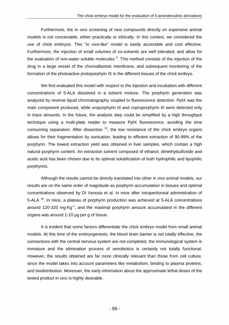
The chick embryo model for the evaluation of 5-aminolevulinic derivatives
- 99 -
Furthermore, the in vivo screening of new compounds directly on expensive animal
models is not conceivable, either practically or ethically. In this context, we considered the
use of chick embryos. This “in vivo-like” model is easily accessible and cost effective.
Furthermore, the injection of small volumes of co-solvents are well tolerated, and allow for
the evaluation of non-water soluble molecules 8. This method consists of the injection of the
drug in a large vessel of the chorioallantoic membrane, and subsequent monitoring of the
formation of the photoactive protoporphyin IX in the different tissues of the chick embryo.
We first evaluated this model with respect to the injection and incubation with different
concentrations of 5-ALA dissolved in a solvent mixture. The porphyrin generation was
analyzed by reverse liquid chromatography coupled to fluorescence detection. PpIX was the
main component produced, while uroporphyrin III and coproporphyrin III were detected only
in trace amounts. In the future, the analysis step could be simplified by a high throughput
technique using a multi-plate reader to measure PpIX fluorescence, avoiding the time
consuming separation. After dissection 10, the low resistance of the chick embryo organs
allows for their fragmentation by sonication, leading to efficient extraction of 90-99% of the
porphyrin. The lowest extraction yield was obtained in liver samples, which contain a high
natural porphyrin content. An extraction solvent composed of ethanol, dimethylsulfoxide and
acetic acid has been chosen due to its optimal solubilization of both hydrophilic and lipophilic
porphyrins.
Although the results cannot be directly translated into other in vivo animal models, our
results are on the same order of magnitude as porphyrin accumulation in tissues and optimal
concentrations observed by Di Venosa et al. in mice after intraperitoneal administration of
5-ALA 18. In mice, a plateau of porphyrin production was achieved at 5-ALA concentrations
around 120-320 mg·Kg-1, and the maximal porphyrin amount accumulated in the different
organs was around 1-10 µg·per g of tissue.
It is evident that some factors differentiate the chick embryo model from small animal
models. At this time of the embryogenesis, the blood brain barrier is not totally effective, the
connections with the central nervous system are not completed, the immunological system is
immature and the elimination process of xenobiotics is certainly not totally functional.
However, the results obtained are far more clinically relevant than those from cell culture,
since the model takes into account parameters like metabolism, binding to plasma proteins,
and biodistribution. Moreover, the early information about the approximate lethal doses of the
tested product in vivo is highly desirable.
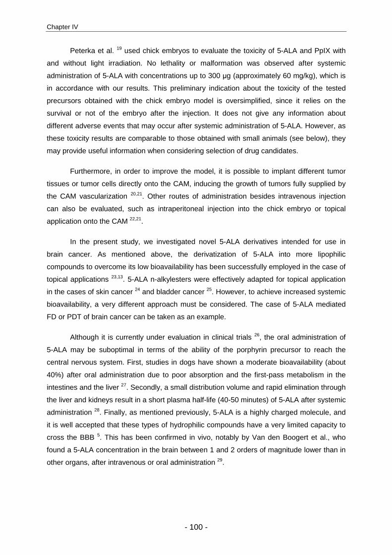
Chapter IV
- 100 -
Peterka et al. 19 used chick embryos to evaluate the toxicity of 5-ALA and PpIX with
and without light irradiation. No lethality or malformation was observed after systemic
administration of 5-ALA with concentrations up to 300 µg (approximately 60 mg/kg), which is
in accordance with our results. This preliminary indication about the toxicity of the tested
precursors obtained with the chick embryo model is oversimplified, since it relies on the
survival or not of the embryo after the injection. It does not give any information about
different adverse events that may occur after systemic administration of 5-ALA. However, as
these toxicity results are comparable to those obtained with small animals (see below), they
may provide useful information when considering selection of drug candidates.
Furthermore, in order to improve the model, it is possible to implant different tumor
tissues or tumor cells directly onto the CAM, inducing the growth of tumors fully supplied by
the CAM vascularization 20,21. Other routes of administration besides intravenous injection
can also be evaluated, such as intraperitoneal injection into the chick embryo or topical
application onto the CAM 22,21.
In the present study, we investigated novel 5-ALA derivatives intended for use in
brain cancer. As mentioned above, the derivatization of 5-ALA into more lipophilic
compounds to overcome its low bioavailability has been successfully employed in the case of
topical applications 23,13. 5-ALA n-alkylesters were effectively adapted for topical application
in the cases of skin cancer 24 and bladder cancer 25. However, to achieve increased systemic
bioavailability, a very different approach must be considered. The case of 5-ALA mediated
FD or PDT of brain cancer can be taken as an example.
Although it is currently under evaluation in clinical trials 26, the oral administration of
5-ALA may be suboptimal in terms of the ability of the porphyrin precursor to reach the
central nervous system. First, studies in dogs have shown a moderate bioavailability (about
40%) after oral administration due to poor absorption and the first-pass metabolism in the
intestines and the liver 27. Secondly, a small distribution volume and rapid elimination through
the liver and kidneys result in a short plasma half-life (40-50 minutes) of 5-ALA after systemic
administration 28. Finally, as mentioned previously, 5-ALA is a highly charged molecule, and
it is well accepted that these types of hydrophilic compounds have a very limited capacity to
cross the BBB 5. This has been confirmed in vivo, notably by Van den Boogert et al., who
found a 5-ALA concentration in the brain between 1 and 2 orders of magnitude lower than in
other organs, after intravenous or oral administration 29.
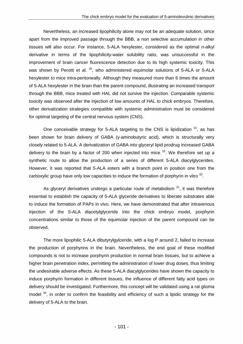
The chick embryo model for the evaluation of 5-aminolevulinic derivatives
- 101 -
Nevertheless, an increased lipophilicity alone may not be an adequate solution, since
apart from the improved passage through the BBB, a non selective accumulation in other
tissues will also occur. For instance, 5-ALA hexylester, considered as the optimal n-alkyl
derivative in terms of the lipophilicity-water solubility ratio, was unsuccessful in the
improvement of brain cancer fluorescence detection due to its high systemic toxicity. This
was shown by Perotti et al. 30, who administered equimolar solutions of 5-ALA or 5-ALA
hexylester to mice intra-peritoneally. Although they measured more than 6 times the amount
of 5-ALA hexylester in the brain than the parent compound, illustrating an increased transport
through the BBB, mice treated with HAL did not survive the injection. Comparable systemic
toxicity was observed after the injection of low amounts of HAL to chick embryos. Therefore,
other derivatization strategies compatible with systemic administration must be considered
for optimal targeting of the central nervous system (CNS).
One conceivable strategy for 5-ALA targeting to the CNS is lipidization 31, as has
been shown for brain delivery of GABA (γ-aminobutyric acid), which is structurally very
closely related to 5-ALA. A derivatization of GABA into glyceryl lipid prodrug increased GABA
delivery to the brain by a factor of 200 when injected into mice 32. We therefore set up a
synthetic route to allow the production of a series of different 5-ALA diacylglycerides.
However, it was reported that 5-ALA esters with a branch point in position one from the
carboxylic group have only low capacities to induce the formation of porphyrin in vitro 33.
As glyceryl derivatives undergo a particular route of metabolism 31, it was therefore
essential to establish the capacity of 5-ALA glyceride derivatives to liberate substrates able
to induce the formation of PAPs in vivo. Here, we have demonstrated that after intravenous
injection of the 5-ALA diacetylglyceride into the chick embryo model, porphyrin
concentrations similar to those of the equimolar injection of the parent compound can be
observed.
The more lipophilic 5-ALA dibutyrylgylceride, with a log P around 2, failed to increase
the production of porphyrins in the brain. Nevertheless, the end goal of these modified
compounds is not to increase porphyrin production in normal brain tissues, but to achieve a
higher brain penetration index, permitting the administration of lower drug doses, thus limiting
the undesirable adverse effects. As these 5-ALA diacylglycerides have shown the capacity to
induce porphyrin formation in different tissues, the influence of different fatty acid types on
delivery should be investigated. Furthermore, this concept will be validated using a rat glioma
model 34, in order to confirm the feasibility and efficiency of such a lipidic strategy for the
delivery of 5-ALA to the brain.
Chapter IV
- 102 -
As we have shown that the chick embryo can be used as a model for the evaluation
of lipophilic molecules that were previously difficult to test in vitro, other derivatization
strategies can now be considered for the targeting of tumors localized in the CNS.
Lipidization was discussed above, and its feasibility was demonstrated with 5-ALA
diacylglycerides that induced PAP in vivo. In this context, 5-ALA may also be linked to fatty
acids or phospholipids.
The “lock-in” approach developed by Bodor et al. 35 involves lipophilic 5-ALA
derivatives that can easily cross the BBB where they will be enzymatically metabolized into
hydrophilic products. Here, selectivity does not depend on the regional presence of target
enzymes, as can occur in other organs, but on the localization into or out of the CNS. Heme
precursors released into the CNS will remain trapped due to their inability to cross the BBB,
while those located elsewhere will be rapidly excreted from the body.
Finally, the Schiff’s base strategy may facilitate the entrance of 5-ALA across the
BBB 36. This approach involves 5-ALA derivatives bearing either an imine or an enamine
moiety linked to a lipophilic carrier, as amines have shown to be unfavorably cleaved.
Conclusions
The chick embryo model can become a useful tool for the evaluation of 5-ALA
derivatives in terms of their ability to induce the accumulation of photoactive porphyrins in
different organs and the absence of acute toxicity after intravenous administration. This
system tolerates the use of co-solvents, and therefore allows for the direct evaluation of
poorly water soluble molecules. Furthermore, the present system might be a cost effective
alternative for initial biodistribution and toxicity studies.
In this context, triglyceride esters of 5-ALA have been shown to induce porphyrin
accumulation in different organs after systemic administration without presenting acute
toxicity. Further investigations should be performed to assess this molecule as a candidate
for brain targeted treatment.
The chick embryo model for the evaluation of 5-aminolevulinic derivatives
- 103 -
References 1. D. E. Dolmans, D. Fukumura, and R. K. Jain, Photodynamic therapy for cancer, Nat
Rev Cancer, 2003, 3, 380-387.
2. S. Collaud, A. Juzeniene, J. Moan, and N. Lange, On the selectivity of 5-aminolevulinic acid-induced protoporphyrin IX formation, Curr Med Chem Anti-Canc Agents, 2004, 4, 301-316.
3. M. M. Bermudez, G. S. Correa, C. Perotti, A. M. Batlle, and A. Casas, Delta-Aminolevulinic acid transport in murine mammary adenocarcinoma cells is mediated by beta transporters, Br J Cancer, 2002, 87, 471-474.
4. N. Fotinos, M. A. Campo, F. Popowycz, R. Gurny, and N. Lange, 5-Aminolevulinic Acid Derivatives in Photomedicine: Characteristics, Application and Perspectives, Photochem Photobiol, 2006, 82, 994-1015.
5. W. M. Pardridge, Transport of small molecules through the blood-brain barrier: biology and methodology, Adv Drug Deliv Rev, 1995, 15, 5-36.
6. M. Kaliszewski, A. Juzeniene, P. Juzenas, M. Kwasny, J. Kaminski, Z. Dabrowski, J. Golinski, and J. Moan, Formation of protoporphyrin IX from carboxylic- and amino-derivatives of 5-aminolevulinicnext term acid, Photodiagnonsis Photodynamic Therapy, 2005
7. A. Casas, C. Perotti, M. Saccoliti, P. Sacca, H. Fukuda, and A. M. Batlle, ALA and ALA hexyl ester in free and liposomal formulations for the photosensitisation of tumour organ cultures, Br J Cancer, 2002, 86, 837-842.
8. A. Vargas, B. Pegaz, E. Debefve, Y. Konan-Kouakou, N. Lange, J. P. Ballini, H. van den Bergh, R. Gurny, and F. Delie, Improved photodynamic activity of porphyrin loaded into nanoparticles: an in vivo evaluation using chick embryos, Int J Pharm, 2004, 286, 131-145.
9. N. Lange, J. P. Ballini, G. Wagnières, and H. van den Bergh, A new drug-screening procedure for photosensitizing agents used in photodynamic therapy for CNV, Invest Ophthalmol Vis Sci, 2001, 42, 38-46.
10. Freshney, R. I.,Disaggregation of the Tissue and Primary Culture,1987,2,107-126.
11. Q. Peng, T. Warloe, J. Moan, A. Godal, F. Apricena, K. E. Giercksky, and J. M. Nesland, Antitumor effect of 5-aminolevulinic acid-mediated photodynamic therapy can be enhanced by the use of a low dose of photofrin in human tumor xenografts, Cancer Res, 2001, 61, 5824-5832.
12. G. Y. Paris, D. L. Garmaise, D. G. Cimon, L. Swett, G. W. Carter, and P. Young, Glycerides as prodrugs. 1. Synthesis and antiinflammatory activity of 1,3-bis(alkanoyl)-2-(O-acetylsalicyloyl)glycerides (aspirin triglycerides), J Med Chem, 1979, 22, 683-687.
13. P. Uehlinger, M. Zellweger, G. Wagnières, L. Juillerat-Jeanneret, H. van den Bergh, and N. Lange, 5-aminolevulinic acid and its derivatives: physical chemical properties and protoporphyrin IX formation in cultured cells, J Photochem Photobiol B:Biol, 2000, 54, 72-80.
Chapter IV
- 104 -
14. H. Hirschberg, C. H. Sun, B. J. Tromberg, and S. J. Madsen, ALA- and ALA-ester-mediated photodynamic therapy of human glioma spheroids, J Neurooncol, 2002, 57, 1-7.
15. C. E. Bigelow, S. Mitra, R. Knuechel, and T. H. Foster, ALA- and ALA-hexylester-induced protoporphyrin IX fluorescence and distribution in multicell tumour spheroids, Br J Cancer, 2001, 85, 727-734.
16. A. Marti, N. Lange, H. van den Bergh, D. Sedmera, P. Jichlinski, and P. Kucera, Optimisation of the formation and distribution of protoporphyrin IX in the urothelium: an in vitro approach, J Urol, 1999, 162, 546-552.
17. R. G. Strickley, Solubilizing excipients in oral and injectable formulations, Pharm Res, 2004, 21, 201-230.
18. G. Di Venosa, A. M. Batlle, H. Fukuda, A. J. MacRobert, and A. Casas, Distribution of 5-aminolevulinic acid derivatives and induced porphyrin kinetics in mice tissues, Cancer Chemother Pharmacol, 2006
19. M. Peterka and I. Klepacek, Light irradiation increases embryotoxicity of photodynamic therapy sensitizers (5-aminolevulinic acid and protoporphyrin IX) in chick embryos, Reprod Toxicol, 2001, 15, 111-116.
20. V. Zenzen and H. Zankl, Protoporphyrin IX-accumulation in human tumor cells following topical ALA- and h-ALA-application in vivo, Cancer lett, 2003, 202, 35-42.
21. R. Hornung, M. J. Hammer-Wilson, S. Kimel, L. H. Liaw, Y. Tadir, and M. W. Berns, Systemic application of photosensitizers in the chick chorioallantoic membrane (CAM) model: photodynamic response of CAM vessels and 5-aminolevulinic acid uptake kinetics by transplantable tumors, J Photochem Photobiol B, 1999, 49, 41-49.
22. M. J. Hammer-Wilson, L. Akian, J. Espinoza, S. Kimel, and M. W. Berns, Photodynamic parameters in the chick chorioallantoic membrane (CAM) bioassay for topically applied photosensitizers, J Photochem Photobiol B, 1999, 53, 44-52.
23. J. Moan, L. W. Ma, A. Juzeniene, V. Iani, P. Juzenas, F. Apricena, and Q. Peng, Pharmacology of protoporphyrin IX in nude mice after application of ALA and ALA esters, Int J Cancer, 2003, 103, 132-135.
24. E. Angell-Petersen, R. Sorensen, T. Warloe, A. M. Soler, J. Moan, Q. Peng, and K. E. Giercksky, Porphyrin Formation in Actinic Keratosis and Basal Cell Carcinoma after Topical Application of Methyl 5-Aminolevulinate, J Invest Dermatol, 2006, 126, 265-271.
25. A. Marti, P. Jichlinski, N. Lange, J. P. Ballini, L. Guillou, H. J. Leisinger, and P. Kucera, Comparison of aminolevulinic acid and hexylester aminolevulinate induced protoporphyrin IX distribution in human bladder cancer, J Urol, 2003, 170, 428-432.
26. W. Stummer, H. Stepp, G. Möller, A. Ehrhardt, M. Leonhard, and H. J. Reulen, Technical principles for protoporphyrin-IX-fluorescence guided microsurgical resection of malignant glioma tissue, Acta Neurochir, 1998, 140, 995-1000.
27. J. T. Dalton, M. C. Meyer, and A. L. Golub, Pharmacokinetics of aminolevulinic acid after oral and intravenous administration in dogs, Drug Metab Dispos, 1999, 27, 432-435.

The chick embryo model for the evaluation of 5-aminolevulinic derivatives
- 105 -
28. J. T. Dalton, C. R. Yates, D. Yin, A. Straughn, S. L. Marcus, A. L. Golub, and M. C. Meyer, Clinical pharmacokinetics of 5-aminolevulinic acid in healthy volunteers and patients at high risk for recurrent bladder cancer., J Pharmacol Exp Ther, 2002, 301, 507-512.
29. J. van den Boogert, R. Van Hillegersberg, F. W. M. de Rooij, R. W. F. de Bruin, A. Edixhoven-Bosdijk, A. B. Houtsmuller, P. D. Siersema, J. H. P. Wilson, and H. W. Tilanus, 5-aminolevulinic acid-induced protoporphyrin IX accumulation in tissues: pharmacokinetics after oral or intravenous administration, J Photochem Photobiol B:Biol, 1998, 44, 29-38.
30. C. Perotti, A. Casas, H. Fukuda, P. Sacca, and A. M. Batlle, ALA and ALA hexyl ester induction of porphyrins after their systemic administration to tumour bearing mice., Br J Cancer, 2002, 87, 790-795.
31. D. M. Lambert, Rationale and applications of lipids as prodrug carriers., Eur J Pharm Sci, 2000, 11, S15-S27.
32. J. N. Jacob, G. W. Hesse, and V. E. Shashoua, Synthesis, brain uptake, and pharmacological properties of a glyceryl lipid containing GABA and the GABA-T inhibitor gamma-vinyl-GABA, J Med Chem, 1990, 33, 733-736.
33. C. J. Whitaker, S. H. Battah, M. J. Forsyth, C. Edwards, R. W. Boyle, and E. K. Matthews, Photosensitization of pancreatic tumour cells by delta-aminolaevulinic acid esters, Anticancer Drug Des, 2000, 15, 161-170.
34. S. A. Friesen, G. O. Hjortland, S. J. Madsen, H. Hirschberg, O. Engebraten, J. M. Nesland, and Q. Peng, 5-Aminolevulinic acid-based photodynamic detection and therapy of brain tumors (review), Int J Oncol, 2002, 21, 577-582.
35. N. Bodor and P. Buchwald, Recent advances in the brain targeting of neuropharmaceuticals by chemical delivery systems, Adv Drug Deliv Rev, 1999, 36, 229-254.
36. J. P. Kaplan and B. M. Raizon, New anticonvulsants: Schiff bases of gamma-aminobutyric acid and gamma-aminobutyramide, J Med Chem, 1980, 23, 702-704.

Chapter IV
- 106 -
5-ALA- and 5-ALA derivatives-mediated effects on gram-negative and gram-positive bacteria
- 107 -
CHAPTER V
5-aminolevulinic acid and 5-aminolevulinic acid Deri vatives
mediated Effects on Gram-Negative and Gram-Positive Bacteria
Nicolas Fotinos 1, Maruska Convert 2, Jean-Claude Piffaretti 2,
Robert Gurny 1 and Norbert Lange 1
1 Department of Pharmaceutics and Biopharmaceutics, School of Pharmaceutical Sciences, University of Geneva,
University of Lausanne, 30 Quai Ernest Ansermet, 1211Geneva 4, Switzerland 2 Institute of Microbiology, Via Mirasole 22A, 6500 Bellinzona, Switzerland
Published in Antimicrobial Agents and Chemotherapy, 2008 (52) 1366-1373
Abstract
Due primarily to the extensive use of antibiotics, the spread of multi-resistant bacterial
strains is one of the most worrying threats to public health. One strategy to overcome
potential shortcomings might be the inactivation of pathogenic microorganisms by
photodynamic therapy (PDT). In the past, different photosensitizers have been assessed on
various pathogenic microorganisms, including bacteria (Escherichia coli,
Staphylococcus aureus), viruses (Papilloma, Herpes), fungi and parasites (Plasmodium,
Leishmania).
5-aminolevulinic acid (5-ALA)-mediated PDT has attracted considerable attention
over the last decade, presumably due to its numerous advantages over conventional
photosensitizers. 5-ALA has no photoactive properties, but when given exogenously, acts as
a precursor of photosensitive porphyrins. This occurs predominantly in tissues or organisms
that are characterized by a high metabolic turnover, such as tumors, macrophages, or
bacteria. However, 5-ALA’s weak ability to cross biological barriers has lead to the
introduction of more lipophilic derivatives, such as methylaminolevulinate or
hexylaminolevulinate, which display an improved capacity to reach the cytoplasm.
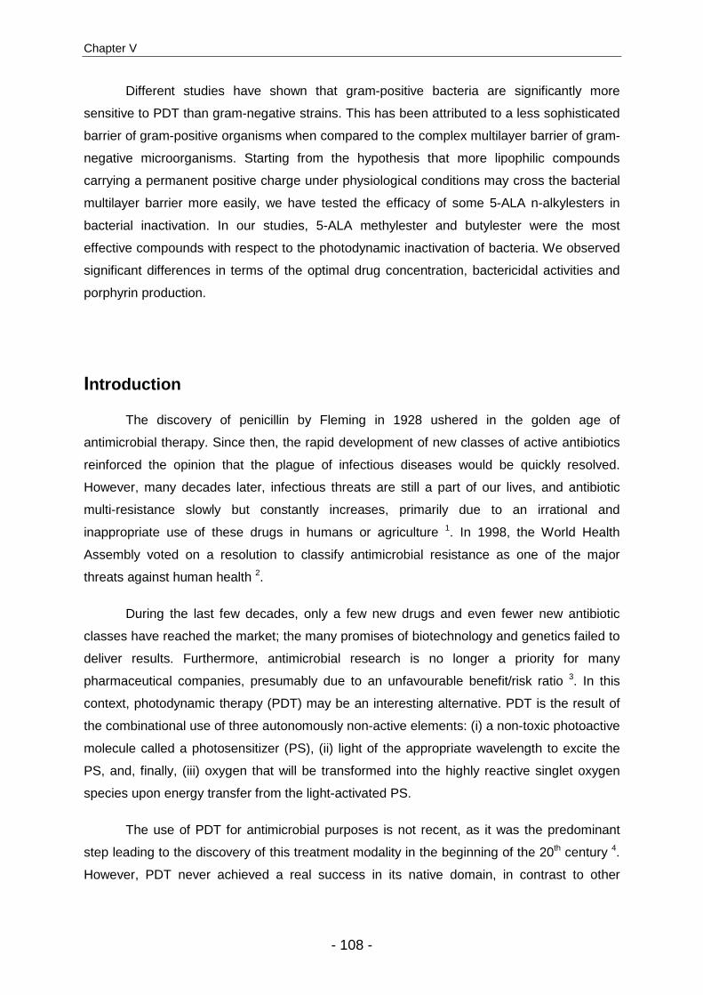
Chapter V
- 108 -
Different studies have shown that gram-positive bacteria are significantly more
sensitive to PDT than gram-negative strains. This has been attributed to a less sophisticated
barrier of gram-positive organisms when compared to the complex multilayer barrier of gram-
negative microorganisms. Starting from the hypothesis that more lipophilic compounds
carrying a permanent positive charge under physiological conditions may cross the bacterial
multilayer barrier more easily, we have tested the efficacy of some 5-ALA n-alkylesters in
bacterial inactivation. In our studies, 5-ALA methylester and butylester were the most
effective compounds with respect to the photodynamic inactivation of bacteria. We observed
significant differences in terms of the optimal drug concentration, bactericidal activities and
porphyrin production.
Introduction
The discovery of penicillin by Fleming in 1928 ushered in the golden age of
antimicrobial therapy. Since then, the rapid development of new classes of active antibiotics
reinforced the opinion that the plague of infectious diseases would be quickly resolved.
However, many decades later, infectious threats are still a part of our lives, and antibiotic
multi-resistance slowly but constantly increases, primarily due to an irrational and
inappropriate use of these drugs in humans or agriculture 1. In 1998, the World Health
Assembly voted on a resolution to classify antimicrobial resistance as one of the major
threats against human health 2.
During the last few decades, only a few new drugs and even fewer new antibiotic
classes have reached the market; the many promises of biotechnology and genetics failed to
deliver results. Furthermore, antimicrobial research is no longer a priority for many
pharmaceutical companies, presumably due to an unfavourable benefit/risk ratio 3. In this
context, photodynamic therapy (PDT) may be an interesting alternative. PDT is the result of
the combinational use of three autonomously non-active elements: (i) a non-toxic photoactive
molecule called a photosensitizer (PS), (ii) light of the appropriate wavelength to excite the
PS, and, finally, (iii) oxygen that will be transformed into the highly reactive singlet oxygen
species upon energy transfer from the light-activated PS.
The use of PDT for antimicrobial purposes is not recent, as it was the predominant
step leading to the discovery of this treatment modality in the beginning of the 20th century 4.
However, PDT never achieved a real success in its native domain, in contrast to other
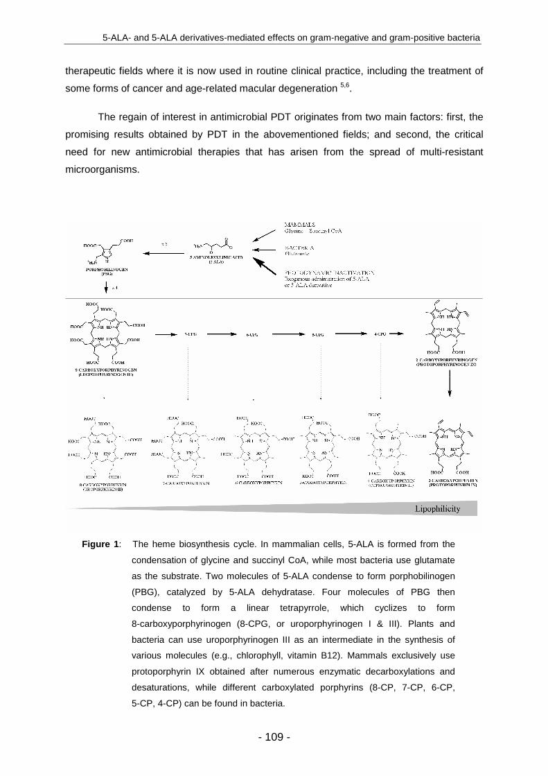
5-ALA- and 5-ALA derivatives-mediated effects on gram-negative and gram-positive bacteria
- 109 -
therapeutic fields where it is now used in routine clinical practice, including the treatment of
some forms of cancer and age-related macular degeneration 5,6.
The regain of interest in antimicrobial PDT originates from two main factors: first, the
promising results obtained by PDT in the abovementioned fields; and second, the critical
need for new antimicrobial therapies that has arisen from the spread of multi-resistant
microorganisms.
Figure 1 : The heme biosynthesis cycle. In mammalian cells, 5-ALA is formed from the
condensation of glycine and succinyl CoA, while most bacteria use glutamate
as the substrate. Two molecules of 5-ALA condense to form porphobilinogen
(PBG), catalyzed by 5-ALA dehydratase. Four molecules of PBG then
condense to form a linear tetrapyrrole, which cyclizes to form
8-carboxyporphyrinogen (8-CPG, or uroporphyrinogen I & III). Plants and
bacteria can use uroporphyrinogen III as an intermediate in the synthesis of
various molecules (e.g., chlorophyll, vitamin B12). Mammals exclusively use
protoporphyrin IX obtained after numerous enzymatic decarboxylations and
desaturations, while different carboxylated porphyrins (8-CP, 7-CP, 6-CP,
5-CP, 4-CP) can be found in bacteria.

Chapter V
- 110 -
One of the main observations obtained from the first attempts to photoinactivate
bacteria with conventional photosensitizers was the relative sensitivity of gram-positive
strains to photodynamic inactivation (PDI), whereas gram-negative strains were significantly
more resistant 7. To overcome the highly impermeable barrier of gram-negative bacteria, pre-
treatment with EDTA or the use of polycationic polymers has been proposed. Furthermore,
the use of positively charged porphyrins, phenotiazines, or phthalocyanines enabled the
photoinactivation of both gram-positive and -negative strains without any permeabilizing pre-
treatment 8.
Besides the classical photosensitizer (PS), one molecule of particular interest,
5-aminolevulinic acid (5-ALA), is gaining importance in the field of PDI. 5-ALA is not a PS by
itself, but is an endogenous component in the heme biosynthesis pathway, and is ubiquitous
in nearly all cells (Fig. 1). When provided exogenously, 5-ALA results in the accumulation of
photoactive porphyrins (PAP) within the targeted cells. This accumulation of PAP is more
pronounced in cells with high metabolic activity 9 like cancer cells, inflammatory cells or
bacteria, resulting in high selectivity. However, the low capacity of 5-ALA to cross biological
barriers led to the development of more lipophilic derivatives with improved local
bioavailability 10. As 5-ALA has been shown to be moderately efficient against gram-negative
bacteria 11, these derivatives might also show a clear improvement in the photoinactivation of
these microorganisms. In the present study, we compared bacterial PAP formation induced
by 5-ALA to that of different 5-ALA derivatives in several gram-positive and gram-negative
bacterial strains, and investigate whether the latter may be interesting candidates for the PDI
of bacteria.
Materials and methods
Chemicals. 5-ALA was obtained from Fluka (Buchs, Switzerland), and its methylester (MAL),
butylester (BAL), pentylester (PAL), hexylester (HAL) and octylester (OAL) from Organix
(Colchester, UK). The porphyrin standards were purchased from Frontier Scientific (Logan,
Utah, US). All solvents and other chemicals were of analytical grade and used without further
purification.
Bacterial strains and growth conditions. This study was conducted with three gram-
negative and one gram-positive strain. The gram-negative strains were: (1) Escherichia coli
K12, a non-pathogenic laboratory strain, (2) E. coli Ti05, a uropathogenic strain, and (3)
Pseudomonas aeruginosa. The gram-positive strain was a Staphylococcus aureus MRSA.

5-ALA- and 5-ALA derivatives-mediated effects on gram-negative and gram-positive bacteria
- 111 -
All strains were provided by the Istituto Cantonale di Microbiologia (Bellinzona, Switzerland),
and are described in Table 1.
Table 1 : Characteristics of the bacterial strains tested in this study
Strain Gram Identification Source Resistance
E. coli K12 - MG1655 laboratory collection
none
E. coli Ti05 - 03-039705 blood Ampicillin, Cotrimoxazole,
Trimethoprim, Streptomycin
P. aeruginosa - 04-022545 expectorate none of the P. aeruginosa-
specific antibiotics a
S. aureus MRSA + 04-022798 wound
puncture
Penicillin, Ampicillin, Oxacillin, Cefazoline, Ciprofloxacine, Tetracycline, Augmentin,
Trimethoprim/Sulfamethoxazole
a Ceftazidine, Cefepime, Piperacillin, Tienam, Aztreonam, Gentamycin, Tobramycin, Amikacin, Ciprofloxacin
All strains were grown for 48 hours on Columbia plates (Columbia Agar Base,
defibrinated sheep blood 5% (v/v); Oxoid, Basel Switzerland). Colonies were transferred into
100 ml of tryptone soya broth (TSB; Oxoid, Basel Switzerland), and incubated overnight at
37 °C. The bacterial suspension was diluted with TS B to an optical density of ~0.10 at
660 nm (OD660). Bacterial suspensions were incubated at 37 °C to an OD660 of ~0.30, and
washed twice with phosphate buffer saline (PBS, 0.1 N, pH 6.5) before incubation with the
substrate.
Photosensitization . Stock solutions of 5-ALA or 5-ALA derivatives MAL, BAL, PAL, HAL
and OAL were freshly prepared by dissolving the different substrates in PBS (0.1 N, pH 6.5)
and kept in the dark after proper dilution. The bacterial suspension was centrifuged 10
minutes at 2500 × g; the bacterial pellets were then resuspended in the substrate solutions
and incubated in the dark for 4 hours at 37 °C in a shaking incubator (100 rpm). One fraction
of the suspension was used for the PDI procedure, while the other was collected for the PAP
analysis.
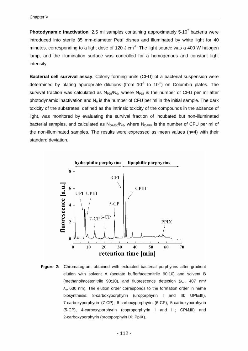
Chapter V
- 112 -
Photodynamic inactivation . 2.5 ml samples containing approximately 5·107 bacteria were
introduced into sterile 35 mm-diameter Petri dishes and illuminated by white light for 40
minutes, corresponding to a light dose of 120 J·cm-2. The light source was a 400 W halogen
lamp, and the illumination surface was controlled for a homogenous and constant light
intensity.
Bacterial cell survival assay . Colony forming units (CFU) of a bacterial suspension were
determined by plating appropriate dilutions (from 10-1 to 10-5) on Columbia plates. The
survival fraction was calculated as NPDI/N0, where NPDI is the number of CFU per ml after
photodynamic inactivation and N0 is the number of CFU per ml in the initial sample. The dark
toxicity of the substrates, defined as the intrinsic toxicity of the compounds in the absence of
light, was monitored by evaluating the survival fraction of incubated but non-illuminated
bacterial samples, and calculated as NDARK/N0, where NDARK is the number of CFU per ml of
the non-illuminated samples. The results were expressed as mean values (n=4) with their
standard deviation.
Figure 2: Chromatogram obtained with extracted bacterial porphyrins after gradient
elution with solvent A (acetate buffer/acetonitrile 90:10) and solvent B
(methanol/acetonitrile 90:10), and fluorescence detection (λem 407 nm/
λex 630 nm). The elution order corresponds to the formation order in heme
biosynthesis: 8-carboxyporphyrin (uroporphyrin I and III; UPI&III),
7-carboxyporphyrin (7-CP), 6-carboxyporphyrin (6-CP), 5-carboxyporphyrin
(5-CP), 4-carboxyporphyrin (coproporphyrin I and III; CPI&III) and
2-carboxyporphyrin (protoporphyin IX; PpIX).

5-ALA- and 5-ALA derivatives-mediated effects on gram-negative and gram-positive bacteria
- 113 -
HPLC analysis . 10 ml bacterial suspensions were centrifuged for 10 minutes at 2500 × g.
The supernatant was discarded, and the bacteria were resuspended in 1.0 ml of extraction
solvent (ethanol/DMSO/acetic acid; 80:20:1; v/v/v) and stored at -80 °C until analysis. For the
extraction of porphyrins, the bacterial wall was disrupted by 5 sonication cycles of 5 seconds
at 0 °C using a sonicator probe (Branson digital so nifier, amplitude 30%, Danbury, USA).
After centrifugation (4 minutes at 13,500 × g), the supernatant was collected and injected into
the HPLC instrument.
Porphyrins were separated by reverse phase chromatography with a 125/4
Nucleodur C18 gravity 3 µm column (Macherey-Nagel, Oensingen, Switzerland) protected
with the corresponding pre-column, using a gradient elution with solvent A (acetate buffer
(pH 5.1 0.5 M)/acetonitrile; 90:10; v/v) and solvent B (methanol/acetonitrile; 90:10; v/v).
Detection was performed by a fluorescence detector (Merck-Hitachi, La Chrom L-7480) with
an excitation wavelength of 407 nm and emission wavelength of 620 nm (Fig. 2).
Results
Effect of 5-ALA PDI on E. coli K12. Fig. 3A shows the results obtained after incubation of
the laboratory strain E. coli K12 with increasing concentrations of 5-ALA. Without irradiation,
bacterial survival was not affected until the 5-ALA concentration reached 40 mM. In contrast,
irradiated samples reacted in a drastically different manner, as two phases could be
observed. At the lowest 5-ALA concentration, a low PDI efficiency was observed, which
increased to a maximal bacterial photoinactivation at the optimal concentration of 0.1 mM.
Beyond this optimal concentration, PDI efficiency declined, resulting in a higher bacterial
viability. In the second phase, high 5-ALA concentrations (40-100 mM) were used which
resulted in a decrease in bacterial viability. This was presumably related to dark toxicity, a
non-light-mediated process.
To understand this phenomenon, the total formation of the photoactive porphyrins
(PAP) was analyzed in parallel to the PDI experiments (Fig. 3 A). The PAP formation profile
seemed to be inversely correlated to bacterial survival after PDI, except at the higher
concentrations. Low amounts of PAP were produced at low 5-ALA concentrations, increasing
to a maximum of 7.5 pmol·10-6 CFU at the optimal concentration of 0.1 mM, and then
decreasing despite higher substrate concentrations. This particular bell-shaped profile is
commonly observed in 5-ALA related photomedicine 12.
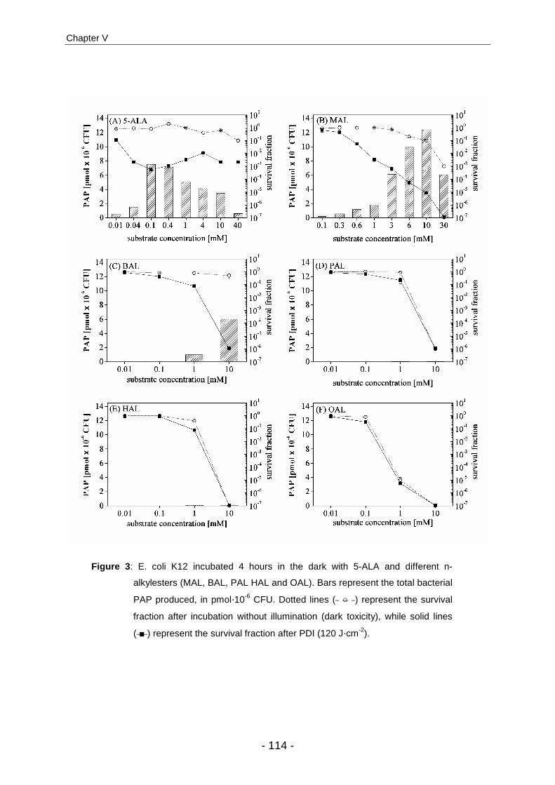
Chapter V
- 114 -
Figure 3 : E. coli K12 incubated 4 hours in the dark with 5-ALA and different n-
alkylesters (MAL, BAL, PAL HAL and OAL). Bars represent the total bacterial
PAP produced, in pmol·10-6 CFU. Dotted lines ( ○ ) represent the survival
fraction after incubation without illumination (dark toxicity), while solid lines
( ■ ) represent the survival fraction after PDI (120 J·cm-2).
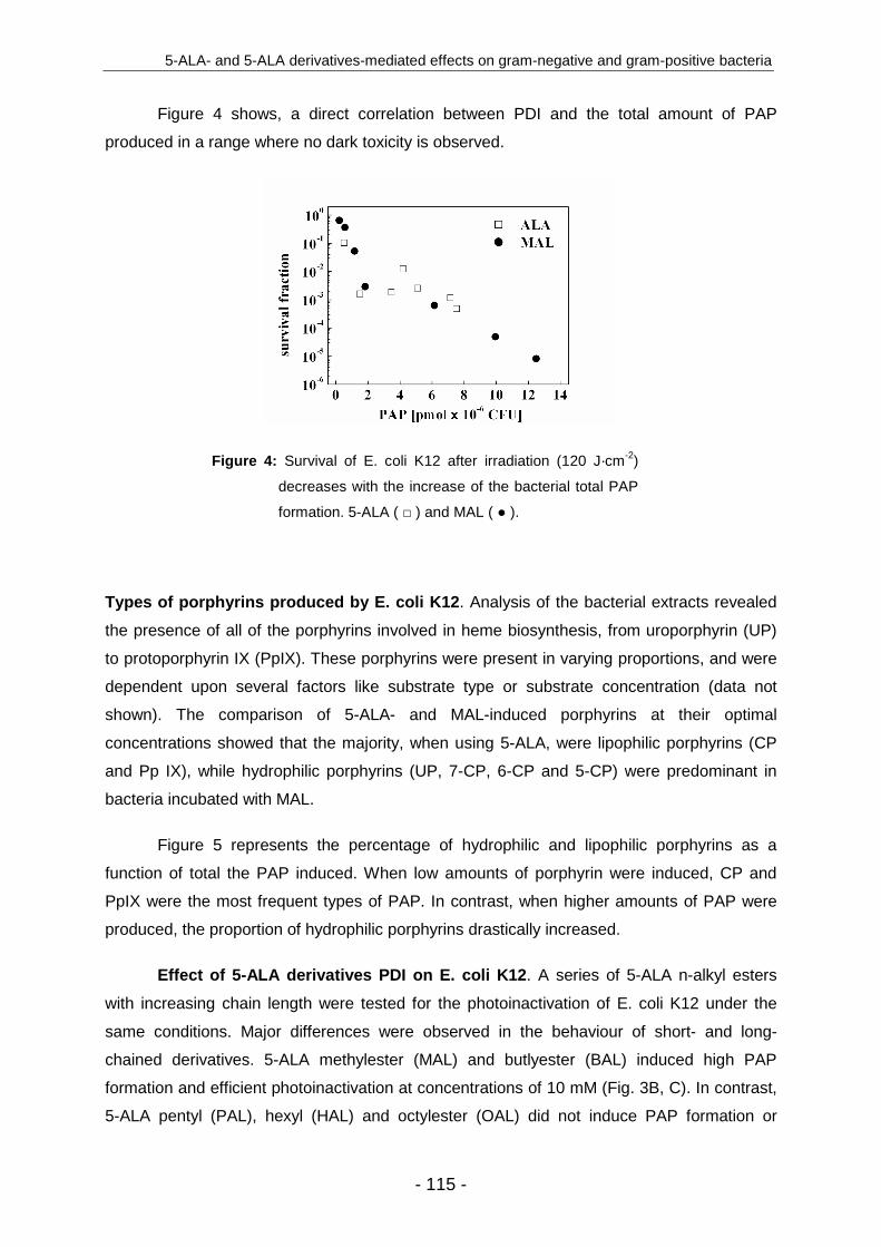
5-ALA- and 5-ALA derivatives-mediated effects on gram-negative and gram-positive bacteria
- 115 -
Figure 4 shows, a direct correlation between PDI and the total amount of PAP
produced in a range where no dark toxicity is observed.
Figure 4: Survival of E. coli K12 after irradiation (120 J·cm-2)
decreases with the increase of the bacterial total PAP
formation. 5-ALA ( □ ) and MAL ( ● ).
Types of porphyrins produced by E. coli K12. Analysis of the bacterial extracts revealed
the presence of all of the porphyrins involved in heme biosynthesis, from uroporphyrin (UP)
to protoporphyrin IX (PpIX). These porphyrins were present in varying proportions, and were
dependent upon several factors like substrate type or substrate concentration (data not
shown). The comparison of 5-ALA- and MAL-induced porphyrins at their optimal
concentrations showed that the majority, when using 5-ALA, were lipophilic porphyrins (CP
and Pp IX), while hydrophilic porphyrins (UP, 7-CP, 6-CP and 5-CP) were predominant in
bacteria incubated with MAL.
Figure 5 represents the percentage of hydrophilic and lipophilic porphyrins as a
function of total the PAP induced. When low amounts of porphyrin were induced, CP and
PpIX were the most frequent types of PAP. In contrast, when higher amounts of PAP were
produced, the proportion of hydrophilic porphyrins drastically increased.
Effect of 5-ALA derivatives PDI on E. coli K12. A series of 5-ALA n-alkyl esters
with increasing chain length were tested for the photoinactivation of E. coli K12 under the
same conditions. Major differences were observed in the behaviour of short- and long-
chained derivatives. 5-ALA methylester (MAL) and butlyester (BAL) induced high PAP
formation and efficient photoinactivation at concentrations of 10 mM (Fig. 3B, C). In contrast,
5-ALA pentyl (PAL), hexyl (HAL) and octylester (OAL) did not induce PAP formation or
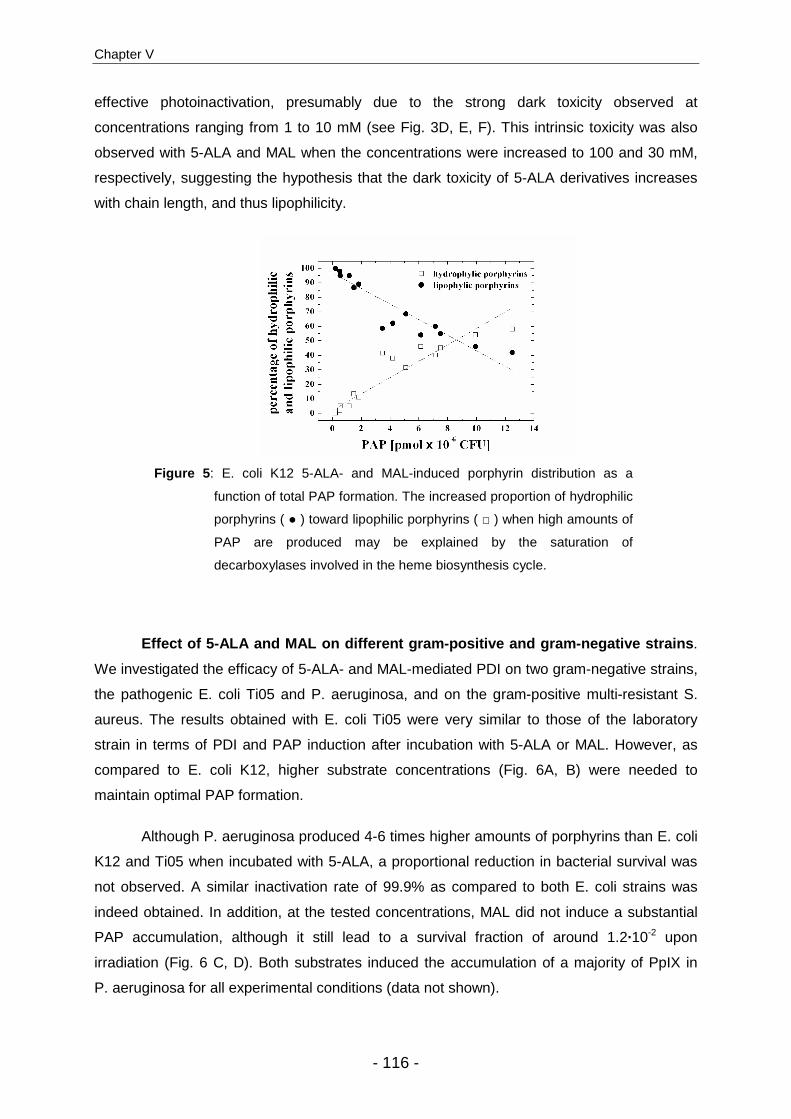
Chapter V
- 116 -
effective photoinactivation, presumably due to the strong dark toxicity observed at
concentrations ranging from 1 to 10 mM (see Fig. 3D, E, F). This intrinsic toxicity was also
observed with 5-ALA and MAL when the concentrations were increased to 100 and 30 mM,
respectively, suggesting the hypothesis that the dark toxicity of 5-ALA derivatives increases
with chain length, and thus lipophilicity.
Figure 5 : E. coli K12 5-ALA- and MAL-induced porphyrin distribution as a
function of total PAP formation. The increased proportion of hydrophilic
porphyrins ( ● ) toward lipophilic porphyrins ( □ ) when high amounts of
PAP are produced may be explained by the saturation of
decarboxylases involved in the heme biosynthesis cycle.
Effect of 5-ALA and MAL on different gram-positive and gram-negative strains .
We investigated the efficacy of 5-ALA- and MAL-mediated PDI on two gram-negative strains,
the pathogenic E. coli Ti05 and P. aeruginosa, and on the gram-positive multi-resistant S.
aureus. The results obtained with E. coli Ti05 were very similar to those of the laboratory
strain in terms of PDI and PAP induction after incubation with 5-ALA or MAL. However, as
compared to E. coli K12, higher substrate concentrations (Fig. 6A, B) were needed to
maintain optimal PAP formation.
Although P. aeruginosa produced 4-6 times higher amounts of porphyrins than E. coli
K12 and Ti05 when incubated with 5-ALA, a proportional reduction in bacterial survival was
not observed. A similar inactivation rate of 99.9% as compared to both E. coli strains was
indeed obtained. In addition, at the tested concentrations, MAL did not induce a substantial
PAP accumulation, although it still lead to a survival fraction of around 1.2·10-2 upon
irradiation (Fig. 6 C, D). Both substrates induced the accumulation of a majority of PpIX in
P. aeruginosa for all experimental conditions (data not shown).

5-ALA- and 5-ALA derivatives-mediated effects on gram-negative and gram-positive bacteria
- 117 -
Figure 6 : E. coli Ti05, P. aeruginosa, and S. aureus MRSA were incubated for
4 hours in the dark with 5-ALA and MAL. Bars represent total
bacterial PAP produced, in pmol × 10-6 CFU. Dotted lines ( ○ )
represent the survival fraction after incubation (dark toxicity) while
solid lines ( ■ ) represent the survival fraction after PDT (120 J ×
cm-2). Please note the different scale used for Pseudomonas.

Chapter V
- 118 -
The gram-positive S. aureus appeared to be significantly more sensitive to 5-ALA-
mediated PDI than the gram-negative strains (survival fraction <10-5), although PAP
production was comparable. MAL also induced sufficient PAP formation to achieve effective
photoinactivation of > 99.9 % of this strain (Fig. 6 E, F). The different porphyrin extracts from
S. aureus contained a majority of uroporphyrins (>80%).
An overview of these results can be seen in Table 2, which compares optimal
concentration, survival and PAP formation for 5-ALA and MAL on the different tested strains.
Table 2: Comparison of 5-ALA- and MAL-mediated PDI on different bacterial strains. The optimal
substrate concentration at which the highest PDI at 120 J·cm-2 is achieved, the survival
fraction, and the total PAP formation induced are reported.
5-ALA MAL
Strain Optimal
concentration [mM]
Survival fraction
PAP [pmol·10-6
CFU]
Optimal concentration
[mM]
Survival fraction
PAP [pmol·10-6
CFU]
E. coli K12 0.1 4.8·10-4 7.5 10 8.1 · 10-6 12.5
E. coli Ti05 1.0 5.0·10-5 5.4 10 2.0 · 10-5 10.2
P. aeruginosa 1.0 1.9·10-3 34.5 10 1.2 · 10-2 6.0
S. aureus 10 5.0·10-6 6.6 10 7.8 · 10-4 2.5
Discussion
The present study demonstrated the possibility of photodynamically inactivating gram-
positive and gram-negative bacteria after incubation with 5-aminolevulinic acid or its
methylester. The characteristic profile of 5-ALA-induced porphyrin production as a function of
substrate concentration underlines the strong relationship between these two parameters,
and the importance of determining the optimal effective concentrations of each substrate.
The observed bell-shaped curve is in agreement with cell culture studies related to 5-ALA-
and 5-ALA derivative-induced porphyrins 12.

5-ALA- and 5-ALA derivatives-mediated effects on gram-negative and gram-positive bacteria
- 119 -
It is known that, in addition to PpIX, porphyrins in general can inhibit different
enzymes involved in the heme biosynthetic cycle in order to avoid accumulation of high
amounts of intracellular PAP 13. However, this negative feedback would result in the
appearance of a plateau in terms of total porphyrin production with increasing 5-ALA
concentration, and cannot explain the bell-shaped curve. Our results showed that at
concentrations above the optimal concentration, 5-ALA itself appears to have an inhibitory
effect on porphyrin formation, even though toxic drug concentrations were not reached.
Furthermore, the same bacteria incubated with MAL produced significantly higher PAP than
the parent compound, suggesting that the maximal PAP production capacities were not
reached.
The differences in optimal concentrations observed for 5-ALA and MAL may result
from different uptake mechanisms. It is well known that in animal cells, 5-ALA and MAL are
transported into the cytosol via different active transport mechanisms 14; however, more
lipophilic derivatives enter via passive diffusion through the membranes or endocytosis. In
gram-negative bacteria, the double membrane prevents penetration of exogenous
molecules, while porins, which are transmembrane channels, allow only small hydrophilic
molecules, usually nutrients, to enter the bacteria 15. Thus, the entrance of 5-ALA into gram-
negative bacteria is not problematic 16, as demonstrated by the low concentration necessary
for optimal PAP induction. In the case of MAL, the addition of a methyl chain on the
carboxylic group increases the log Po/w from -1.52 to -0.94 12, which is still considered
hydrophilic. Hence, this compound may also enter the bacterial cytosol through porin
channels. However, the high concentrations (3-10 mM) necessary to induce sufficient PAP
formation indicate that either the penetration or availability may vary from that of the parent
compound.
The fact that more lipophilic derivatives, such as pentylester, hexylester and
octylester, do not induce any significant PAP formation might be explained by the trapping of
these highly lipophilic compounds in the bacterial membranes, or by the fact that the highly
amphiphilic properties of these derivatives may induce membrane disruption, causing dark
toxicity.
We have seen a strong correlation between PDI efficiency and the total amount of
PAP formation in E. coli K12. However, this cannot be extrapolated to other strains due to
the different types of porphyrin produced, protection mechanisms 17, and porphyrin
localization. All types of porphyrins observed in our study have shown the capacity to absorb
light and generate reactive oxygen species 18. The main difference between these
endogenous porphyrins is related to the number of carboxylic moieties, which influence

Chapter V
- 120 -
solubility, lipophilicity, aggregation and localization. In the case of E. coli, it was seen that the
type of PAP depends on the quantity of the porphyrins produced. When low amounts of
porphyrins are synthesized, the enzymatic machinery of the bacteria was able to proceed to
the decarboxylation of a high proportion of these porphyrins, leading to the accumulation of
mainly lipophilic porphyrins. When very large quantities of PAP are produced, the enzymatic
process may be overwhelmed, leading into an increased proportion of hydrophilic porphyrins.
This tendency was observed only in the E. coli strains (data not shown for the other species).
In contrast, S. aureus produced uroporphyrins nearly exclusively and was efficiently
photoinactivated, while P. aeruginosa produced 4-5 times more porphyrins, mainly PpIX, but
was not more efficiently photoinactivated.
To obtain an efficient 5-ALA- or 5-ALA derivative-mediated PDI, the two main
conditions necessary are sufficient PAP accumulation in the targeted bacteria, and effective
irradiation to activate the PS. In fact, when using this type of photosensitizer prodrug, a PDI
conducted with sub-optimal conditions may lead to contradictory results. For example,
Karrer et al. 19 have shown that 5-ALA-mediated PDI induced significant inhibition of the
growth of S. aureus but not S. epidermis. In contrast, Nitzan et al. 11 observed that 5-ALA
mediated PDI was effective against both S. aureus and S. epidermis but not against gram-
negative strains, including E. coli or P. aeruginosa. The latter strains were successfully
photoinactivated by Szocs et al. 20 and Lee et al. 21, respectively, after incubation with
5-ALA.
In our study, both 5-ALA and MAL have been shown to induce increased PAP in all
treated bacteria when optimal conditions were applied. As 5-ALA presented a biphasic
inactivation curve, intermediate concentrations resulted in a suboptimal inactivation of the
targeted bacteria. A more desirable behaviour was obtained with its methylester, as the
bacterial survival continuously decreased with increasing MAL concentration. In this case,
PAP formation, and thus PDI, was inhibited only when toxic drug concentrations (over
10 mM) were achieved.
These active concentrations may seem relatively high compared to other PS used at
micromolar concentrations; indeed, for some other PS compounds, the application time is in
minutes instead of hours 22,23. However, the high selectivity of the compounds we have
tested, as demonstrated in other medical domains, may provide the means to potentially
eradicate bacterial infections without harming healthy surrounding tissues. Early clinical trials
have demonstrated promising results from 5-ALA-mediated PDI of H. pylori 24, as well as the
treatment of cutaneous mycosis 25 and acne 26. In these contexts, not only is the ability to
induce PAP formation important, but also the drug bioavailability. 5-ALA derivatives have

5-ALA- and 5-ALA derivatives-mediated effects on gram-negative and gram-positive bacteria
- 121 -
demonstrated some advantages as compared to the parent compound, including improved
selectivity, homogeneity in tissue distribution, penetration, lack of systemic effects and less
painful irradiation 10. Furthermore, repeatability may be a major asset of PDI, as cumulative
toxicity 27 and induced bacterial resistance upon repeated treatments 28 have not been
reported to date.
Conclusions
When optimal concentrations were used, 5-ALA and 5-ALA short-chained derivatives
(5-ALA methylester and butylester) induced high PAP accumulation in both the gram-positive
and gram-negative bacterial strains we tested, attaining the first prerequisite for a successful
PDI. In contrast, long-chained 5-ALA derivatives (5-ALA pentylester, hexylester and
octylester) demonstrated a high dark toxicity that increased with lipophilicity, and showed no
significant PAP formation. As 5-ALA methylester efficiently induced PAP formation in
bacteria and presented enhanced bioavailability in comparison to the parent compound, its
capacities to treat local infections should be investigated in in vivo experiments.

Chapter V
- 122 -
References
1. A. J. Alanis, Resistance to antibiotics: are we in the post-antibiotic era?, Arch Med
Res, 2005, 36, 697-705.
2. World Health Organisation (WHO), Emerging and other communicable diseases: antimicrobial resistance, 1998, A51/9
3. D. Hughes, Exploiting genomics, genetics and chemistry to combat antibiotic resistance, Nat Rev Genet, 2003, 4, 432-441.
4. J. Moan and Q. Peng, An outline of the hundred-year history of PDT., Anticancer Res, 2003, 23, 3591-3600.
5. S. B. Brown, E. A. Brown, and I. Walker, The present and future role of photodynamic therapy in cancer treatment, Lancet Oncology, 2004, 5, 497-508.
6. R. M. Barnes, L. Gee, S. Taylor, M. C. Briggs, and S. P. Harding, Outcomes in verteporfin photodynamic therapy for choroidal neovascularisation--'beyond the TAP study', Eye, 2004, 18, 809-813.
7. M. R. Hamblin and T. Hasan, Photodynamic therapy: a new antimicrobial approach to infectious disease?, Photochem Photobiol Sci, 2004, 3, 436-450.
8. G. Jori, C. Fabris, M. Soncin, S. Ferro, O. Coppellotti, D. Dei, L. Fantetti, G. Chiti, and G. Roncucci, Photodynamic therapy in the treatment of microbial infections: basic principles and perspective applications, Lasers Surg Med, 2006, 38, 468-481.
9. S. Collaud, A. Juzeniene, J. Moan, and N. Lange, On the selectivity of 5-aminolevulinic acid-induced protoporphyrin IX formation, Curr Med Chem Anti-Canc Agents, 2004, 4, 301-316.
10. N. Fotinos, M. A. Campo, F. Popowycz, R. Gurny, and N. Lange, 5-Aminolevulinic Acid Derivatives in Photomedicine: Characteristics, Application and Perspectives, Photochem Photobiol, 2006, 82, 994-1015.
11. Y. Nitzan, M. Salmon-Divon, E. Shporen, and Z. Malik, ALA induced photodynamic effects on gram positive and negative bacteria, Photochem Photobiol Sci, 2004, 3, 430-435.
12. P. Uehlinger, M. Zellweger, G. Wagnières, L. Juillerat-Jeanneret, H. van den Bergh, and N. Lange, 5-aminolevulinic acid and its derivatives: physical chemical properties and protoporphyrin IX formation in cultured cells, J Photochem Photobiol B:Biol, 2000, 54, 72-80.
13. S. G. Afonso, R. Enriquez de Salamanca, and A. M. Batlle, Photodynamic and light independent action of 8 to 2 carboxylic free porphyrins on some haem-enzymes, Int J Biochem Cell Biol, 2001, 33, 1208-1214.
14. E. Rud, O. Gederaas, A. Hogset, and K. Berg, 5-aminolevulinic acid, but not 5-aminolevulinic acid esters, is transported into adenocarcinoma cells by system BETA transporters, Photochem Photobiol, 2000, 71, 640-647.
5-ALA- and 5-ALA derivatives-mediated effects on gram-negative and gram-positive bacteria
- 123 -
15. H. Nikaido, Molecular basis of bacterial outer membrane permeability revisited, Microbiol Mol Biol Rev, 2003, 67, 593-656.
16. E. Verkamp, V. M. Backman, J. M. Bjornsson, D. Soll, and G. Eggertsson, The periplasmic dipeptide permease system transports 5-aminolevulinic acid in Escherichia coli, J Bacteriol, 1993, 175, 1452-1456.
17. S. Y. Kim, E. J. Kim, and J. W. Park, Control of singlet oxygen-induced oxidative damage in Escherichia coli, J Biochem Mol Biol, 2002, 35, 353-357.
18. R. W. Redmond and J. N. Gamlin, A compilation of singlet oxygen yields from biologically relevant molecules, Photochem Photobiol, 1999, 70, 391-475.
19. S. Karrer, R. M. Szeimies, S. Ernst, C. Abels, W. Bäumler, and M. Landthaler, Photodynamic Inactivation of Staphylococci with 5-Aminolaevulinic Acid or Photofrin, Lasers Med Sci, 1999, 14, 54-61.
20. K. Szocs, F. Gabor, G. Csik, and J. Fidy, delta-Aminolaevulinic acid-induced porphyrin synthesis and photodynamic inactivation of Escherichia coli B, J Photochem Photobiol B, 1999, 50, 8-17.
21. C. F. Lee, C. J. Lee, C. T. Chen, and C. T. Huang, delta-Aminolaevulinic acid mediated photodynamic antimicrobial chemotherapy on Pseudomonas aeruginosa planktonic and biofilm cultures, J Photochem Photobiol B, 2004, 75, 21-25.
22. J. P. Tome, M. G. Neves, A. C. Tome, J. A. Cavaleiro, M. Soncin, M. Magaraggia, S. Ferro, and G. Jori, Synthesis and antibacterial activity of new poly-S-lysine-porphyrin conjugates, J Med Chem, 2004, 47, 6649-6652.
23. G. P. Tegos, M. Anbe, C. Yang, T. N. Demidova, M. Satti, P. Mroz, S. Janjua, F. Gad, and M. R. Hamblin, Protease-stable polycationic photosensitizer conjugates between polyethyleneimine and chlorin(e6) for broad-spectrum antimicrobial photoinactivation, Antimicrob Agents Chemother, 2006, 50, 1402-1410.
24. C. H. Wilder-Smith, P. Wilder-Smith, P. Grosjean, H. van den Bergh, A. Woodtli, P. Monnier, G. Dorta, F. Meister, and G. Wagnières, Photoeradication of Helicobacter pylori using 5-aminolevulinic acid: preliminary human studies, Lasers Surg Med, 2002, 31, 18-22.
25. P. G. Calzavara-Pinton, M. Venturini, and R. Sala, A comprehensive overview of photodynamic therapy in the treatment of superficial fungal infections of the skin, J Photochem Photobiol B, 2005, 78, 1-6.
26. B. Pollock, D. Turner, M. Stringer, R. A. Bojar, V. Goulden, G. I. Stables, and W. J. Cunliffe, Topical aminolaevulinic acid-photodynamic therapy for the treatment of acne vulgaris: a study of clinical efficacy and mechanism of action, Br J Dermatol, 2004, 151, 616-622.
27. Q. Peng, T. Warloe, K. Berg, J. Moan, M. Kongshaug, K. E. Giercksky, and J. M. Nesland, 5-aminolevulinic acid-based photodynamic therapy - clinical research and future challenges, Cancer, 1997, 79, 2282-2308.
28. F. M. Lauro, P. Pretto, L. Covolo, G. Jori, and G. Bertoloni, Photoinactivation of bacterial strains involved in periodontal diseases sensitized by porphycene-polylysine conjugates, Photochem Photobiol Sci, 2002, 1, 468-470.

Chapter V
- 124 -
5-ALA derivatives for enhanced photoinactivation of Propionibacterium acnes
- 125 -
CHAPTER VI
5-ALA derivatives-mediated photoinactivation of
Propionibacterium acnes
Nicolas Fotinos1, Juro Mikulic1, Maruska Convert2, Marino Andres Campo1,
Jean-Claude Piffaretti2, Robert Gurny1 and Norbert Lange1
1 Department of Pharmaceutics and Biopharmaceutics, School of Pharmaceutical Sciences, University of Geneva,
University of Lausanne, 30 Quai Ernest Ansermet, 1211Geneva 4, Switzerland 2 Cantonale institute of Microbiology, Via Mirasole 22A, 6500 Bellinzona, Switzerland
Submitted to the Journal of Dermatological Sciences
Abstract
Recent studies have shown that 5-aminolevulinic acid (5-ALA)-mediated
photodynamic therapy (PDT) is applicable for the treatment of moderate to severe cases of
acne vulgaris. However, due to sub-optimal pharmacokinetic properties of this molecule,
more lipophilic derivatives have been developed and are currently in use for several clinical
indications. The objective of the present study was the evaluation of different 5-ALA
derivatives with respect to the generation of photoactive porphyrins (PAP) in
Propionibacterium acnes and subsequent photodynamic inactivation (PDI).
For this purpose, suspensions of P. acnes were incubated with 5-ALA or 5-ALA
derivatives as PAP precursors during 4 hours and then irradiated with white light.
Photoinactivation was evaluated by counting the colony forming units before and after PDI
treatment, while PAP formation was analyzed by extracting and measuring the porphyrins
produced.
Chapter VI
- 126 -
The different P. acnes strains were efficiently photoinactivated by 5-ALA, 5-ALA
benzylester (BzAL), and 5-ALA-O-diethylenglycol-monomethylester (5-ALA DGME), while
5-ALA hexylester did not induce any PAP under the tested conditions. With regards to their
promising in vitro results, the use of the moderately lipophilic 5-ALA derivatives BzAL and
5-ALA DGME should be considered to optimize PDT treatment of acne vulgaris, depending
on the ability to increase drug bioavailability and selectivity, and to reduce side effects such
as erythema, pustular eruptions, and epithelial exfoliation.
Introduction
Although acne vulgaris is not a life-threatening pathology, severe forms of this
disease may have a particularly negative influence on self-esteem. Acne vulgaris affects up
to 85% of young people and can continue into adulthood. Treatment of severe cases
frequently involves a combination of different topically or systemically applied agents,
including antibiotics, retinoids, or hormonal agents 1. This broad therapeutic mix, with
potentially severe side effects, is required by the multifactorial aetiology of this complex and
not yet fully understood pathology. Abnormal keratinisation, sebaceous gland hyperplasia,
Propionibacterium acnes colonization, and inflammation processes, however, are broadly
accepted as key factors associated with the disease.
Despite the well-documented colonization of pilo-sebaceous glands by P. acnes, its
exact role and influence on the development, severity, and recurrence of acne is still
unknown. However, the increased drug resistance of P. acnes after long-term oral antibiotic
treatment and the emergence of serious side effects, including upper tract infections 2 and
hepatitis 3, have raised serious doubts about the legitimacy of this antibacterial approach 4.
Thus, photodynamic therapy (PDT) may represent an alternative treatment. PDT is a
relatively recent treatment modality that combines 3 individually non-toxic components: (i) a
photosensitizing molecule (PS), (ii) light, and (iii) oxygen. In the PDT process, the
photosensitizer accumulates at the target site and is activated to a higher energetic level
upon local irradiation by light. The activated PS then transmits energy to the oxygen,
resulting in the formation of short-living highly toxic reactive oxygen species, inducing
selective target destruction without harming surrounding healthy tissues. Clinically, PDT is
currently used for the treatment of various dermatological and non-dermatological
pathologies 5.
5-ALA derivatives for enhanced photoinactivation of Propionibacterium acnes
- 127 -
Photodynamic inactivation (PDI) of microorganisms using different photosensitizing
agents in vitro has led to much hope in the antimicrobial domain 6. Nevertheless, only a few
molecules have been evaluated clinically (e.g., methylene blue, 5-aminolevulinic acid). One
of the most promising approaches may come from the endogenous accumulation of
photoactive porphyrins (PAP) in P. acnes 7, which can partially explain the significant
improvements in patients suffering from acne vulgaris after light-based therapy 8,9. To further
enhance treatment efficacy, exogenous administration of 5-aminolevulinic acid (5-ALA), an
endogenous porphyrin precursor, can selectively boost porphyrin accumulation within
organisms having high metabolic rates 10, such as P. acnes. However, due to the
unfavourable topical bioavailability of 5-ALA, more lipophilic derivatives have been
developed, two of which have received marketed authorization 11. The goal of this study is to
evaluate the capacity of different 5-ALA derivatives, adapted for topical application, to
photoinactivate P. acnes.
Materials and methods
Chemicals : 5-ALA was obtained from Fluka (Buchs, Switzerland). Its methylester (MAL),
hexylester (HAL), and benzylester (BzAL) were purchased from Organix (Colchester, UK).
The O-diethyleneglycol monomethyl-ether ester of 5-ALA (5-ALA-DGME) was synthesized
according to Berger et al. 12. The structure of 5-ALA and its derivatives can be seen in
Table 1. The porphyrin standards were purchased from Frontier Scientific (Logan, Utah, US).
All solvents and other chemicals were of analytical grade and used without further
purifications.
Bacterial strains : The laboratory strain Propionibacterium acnes CIP 53.117 was obtained
from the Institut Pasteur in Paris (France). P. acnes E5947 and Va2376 were isolated from
patients at the Istituto Cantonale di Microbiologia in Bellinzona, from blood and pleural
puncture, respectively.
Bacterial Growth: All three P. acnes strains were grown for 72 hours on Columbia plates
(Columbia Agar Base, defibrinated sheep blood 5% (v/v); Oxoid, Basel Switzerland) under
anaerobic conditions (10% CO2, 10% H2, 80% N2) at 37 °C. Initial bacterial suspensions were
prepared by seeding approximately 3 loops of colonies in thioglycolate broth (TG, Beckton
Dickinson, USA). After overnight incubation, the culture was diluted with TG to an optical
density (OD660) of 0.28 at 660 nm and incubated for approximately 90 minutes until an OD660
of 0.30 was obtained. Before incubation with the substrates, the bacterial suspension was
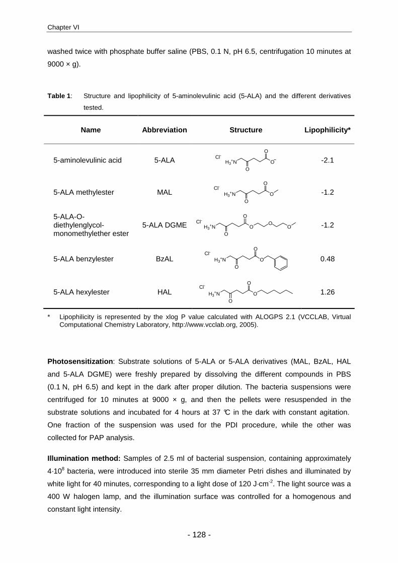
Chapter VI
- 128 -
washed twice with phosphate buffer saline (PBS, 0.1 N, pH 6.5, centrifugation 10 minutes at
9000 × g).
Table 1 : Structure and lipophilicity of 5-aminolevulinic acid (5-ALA) and the different derivatives
tested.
Name Abbreviation Structure Lipophilicity*
5-aminolevulinic acid 5-ALA H3+N O
O
OCl-
-2.1
5-ALA methylester MAL H3+N O
O
OCl-
-1.2
5-ALA-O-diethylenglycol-monomethylether ester
5-ALA DGME H3+N O
O
OO
OCl-
-1.2
5-ALA benzylester BzAL H3+N O
O
OCl-
0.48
5-ALA hexylester HAL H3+N O
O
OCl-
1.26
* Lipophilicity is represented by the xlog P value calculated with ALOGPS 2.1 (VCCLAB, Virtual Computational Chemistry Laboratory, http://www.vcclab.org, 2005).
Photosensitization : Substrate solutions of 5-ALA or 5-ALA derivatives (MAL, BzAL, HAL
and 5-ALA DGME) were freshly prepared by dissolving the different compounds in PBS
(0.1 N, pH 6.5) and kept in the dark after proper dilution. The bacteria suspensions were
centrifuged for 10 minutes at 9000 × g, and then the pellets were resuspended in the
substrate solutions and incubated for 4 hours at 37 °C in the dark with constant agitation.
One fraction of the suspension was used for the PDI procedure, while the other was
collected for PAP analysis.
Illumination method: Samples of 2.5 ml of bacterial suspension, containing approximately
4·108 bacteria, were introduced into sterile 35 mm diameter Petri dishes and illuminated by
white light for 40 minutes, corresponding to a light dose of 120 J·cm-2. The light source was a
400 W halogen lamp, and the illumination surface was controlled for a homogenous and
constant light intensity.

5-ALA derivatives for enhanced photoinactivation of Propionibacterium acnes
- 129 -
Bacterial cell survival assay : Colony forming units (CFU) were determined after
appropriate dilutions of the bacterial suspensions (from 10-1 to 10-5) that were plated and
cultured on Columbia agar. The survival fraction was calculated as NPDI/N0, where NPDI is the
number of CFU per ml after photodynamic inactivation and N0 is the number of CFU per ml in
the initial sample. The dark toxicity of the substrates, defined as the intrinsic toxicity of the
compounds in the absence of light, was monitored by evaluating the survival fraction of
incubated but non-illuminated bacterial samples and calculated as NDARK/N0, where NDARK is
the number of CFU per ml of the non-illuminated samples.
HPLC analysis . 10 ml of the incubated bacterial suspensions were centrifuged for 10
minutes at 9000 × g. The supernatant was discarded, and the bacteria were resuspended in
1.0 ml of extraction solvent (ethanol/DMSO/acetic acid; 80:20:1; v/v/v) and then stored at -80
°C until analysis. For the analysis, the bacterial wall was disrupted by 5 sonication cycles of 5
seconds at 0 °C using a sonicator probe (Branson di gital sonifier, amplitude 30%, Danbury,
USA) to extract the porphyrins. After centrifugation (4 minutes at 13,500 × g), the
supernatant was collected and injected into the HPLC instrument.
Porphyrins were separated by reverse phase chromatography with a 125/4 Nucleodur C18
gravity 3 µm column (Macherey-Nagel, Oensingen, Switzerland) protected with the
corresponding pre-column, using a gradient elution with solvent A (acetate buffer (pH 5.1
0.5M)/acetonitrile; 90:10) and solvent B (methanol/acetonitrile; 90:10). Detection was
performed by a fluorescence detector (Merck-Hitachi, La Chrom L-7480) with an excitation
wavelength of 407 nm and emission wavelength of 620 nm.
Results
P. acnes CIP 53.117: P. acnes naturally accumulates high amounts of porphyrins, and
irradiation of untreated bacteria under our conditions photoinactivated approximately 45% of
the microorganisms (data not shown). However, it was possible to significantly increase
photoactive porphyrin (PAP) accumulation and, thus photoinactivation, after incubation with
5-ALA or some 5-ALA derivatives at the appropriate concentrations.
Different porphyrins can be formed within the heme biosynthesis cycle. The earliest
porphyrin to be produced is the hydrophilic uroporphyrin (UP I & III), which presents 8
carboxylic functions that will undergo a series of enzymatic decarboxylations, leading to the
formation of more lipophilic molecules, [i.e. coproporphyrin (CP I & III, 4 carboxylic groups)

Chapter VI
- 130 -
and protoporphyrin IX (PpIX, 2 carboxylic groups)]. All these porphyrins are known to be
photoactive 13 but may localize or interact differently due to different lipophilicities 14. In the
case of P. acnes, the major porphyrin types detected were uroporphyrins and
coproporphyrins in varying proportions, but no protoporphyrin, which is the primary detected
product in human cells. Depending on the substrate, slight variations in PAP-induced type
distribution were observed (Table 2).
Table 2 : Porphyrin type distribution in P. acnes CIP 53.117 after incubation with different
compounds (mean and standard deviation). Uroporphyrin and coproporphyrin
were the main PAP produced, while no protoporphyrin IX, the main product in
mammalian cells, was detected.
Porphyrin distribution [%]
Compound uroporphyrin coproporphyrin protoporphyrin other
5-ALA 45 ± 14 36 ± 11 0 19 ± 6
MAL 38 ± 27 62 ± 27 0 0
DGME 10 ± 19 88 ± 26 0 2 ± 7
BzAL 64 ± 10 28 ± 13 0 8 ± 6
HAL 62 ± 10 28 ± 14 0 10 ± 4
Figure 1 shows the total amount of PAP accumulated by P. acnes CIP 53.117 for
5-ALA and its derivatives and the survival fraction with and without illumination as a function
of substrate concentration. It can be clearly seen that PAP accumulation and
photoinactivation efficiency was strongly dependent on substrate concentration for each
tested compound. In the case of 5-ALA, the highest PAP formation was observed after
incubating P. acnes CIP 53.117 with an optimal substrate concentration of around 10 mM,
while further increases in 5-ALA concentration resulted in a lower PAP accumulation. This
specific PAP production profile as a function of substrate concentration is well-known in the
5-ALA-mediated PDT domain as a “bell-shaped curve”, and can also be observed for the PDI
profile (Fig. 1A).
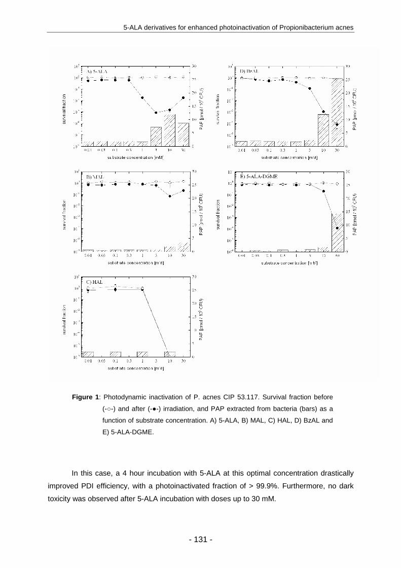
5-ALA derivatives for enhanced photoinactivation of Propionibacterium acnes
- 131 -
Figure 1 : Photodynamic inactivation of P. acnes CIP 53.117. Survival fraction before
(-○-) and after (-●-) irradiation, and PAP extracted from bacteria (bars) as a
function of substrate concentration. A) 5-ALA, B) MAL, C) HAL, D) BzAL and
E) 5-ALA-DGME.
In this case, a 4 hour incubation with 5-ALA at this optimal concentration drastically
improved PDI efficiency, with a photoinactivated fraction of > 99.9%. Furthermore, no dark
toxicity was observed after 5-ALA incubation with doses up to 30 mM.

Chapter VI
- 132 -
MAL-induced PAP formation in P. acnes CIP 53.117 (Fig. 1B) appeared to be less
efficient than with 5-ALA at the concentrations tested on this bacterial strain. Furthermore,
within our experimental conditions, PDI resulted in a CFU reduction of less than 99%.
Similarly to 5-ALA, no dark toxicity was observed with this hydrophilic derivative of 5-ALA. On
the other hand, the lipophilic HAL induced no PAP accumulation at all and revealed a high
dark toxicity, with a viability reduction of more than 5 orders of magnitude when using a
concentration of 10 mM (Fig. 1C).
The two other 5-ALA derivatives, 5-ALA benzylester (Fig 1D) and 5-ALA DGME
(Fig. 1E), which are moderately lipophilic, both demonstrated the capacity to (i) induce high
PAP accumulation when using doses up to 30 mM, (ii) efficiently photoinactivate high
bacterial fractions, up to 99.99% , and finally (iii) show no dark toxicity at doses up to 30 mM.
P. acnes E5947 and VA2376: The results obtained with P. acnes E5947 and VA2376, two
clinical strains, were very similar to the laboratory strain P. acnes CIP 53.117 at the tested
concentrations. Table 3 shows the minimal substrate concentration necessary to inactivate
>99% CFU for all three bacterial strains. In P. acnes E5947, 5-ALA, BzAL, and 5-ALA-DGME
induced high PAP formation at a concentration of 10 mM. As for strain CIP 53.117, MAL
exerted limited PDI, while HAL again presented a high dark toxicity (10-6 survival fraction)
and failed to induce any PDI effect. In the case of P. acnes VA2376, 5-ALA induced PDI at
1 mM, while 10 mM was needed for DGME and BzAL. The high dark toxicity of HAL and low
PDI efficiency of MAL were also confirmed with this strain.
Table 3 : Minimal substrate concentrations necessary to induce a PDI of 99% of CFU for
P. acnes CIP 53.117, E5947, and VA2376.
CIP 53.117 E5947 VA2376
5-ALA 1 mM 10 mM 1 mM
MAL N/A d.t. N/A
DGME 30 mM 10 mM 10 mM
BzAL 10 mM 10 mM 10 mM
HAL d.t. d.t. d.t.
d.t. represents dark toxicity when a reduction of > 99% of CFU is observed in substrate-incubated but
non_irradiated samples; .N/A. indicates that a PDI of 99% is not achieved

5-ALA derivatives for enhanced photoinactivation of Propionibacterium acnes
- 133 -
Discussion
While the efficacy of 5-ALA-mediated PDT is widely accepted, the use of optimized
derivatives allows significant improvements in terms of selectivity, bioavailability, and
reduction of adverse effects 11. However, n-alkyl derivatives of 5-ALA that were successful in
neoplastic cell photoinactivation gave surprising results when tested on different bacteria 15.
Lipophilic, long-chained esters, such as HAL, induced similar PpIX production as did 5-ALA
in human cancer cells but at 100 times lower doses 16. Nevertheless, these derivatives did
not induce a detectable porphyrin production in the tested bacterial strains. In contrast, more
hydrophilic short chained derivatives like MAL induced an increased porphyrin accumulation
and efficient bacterial photoinactivation in a dose-dependant manner. The results obtained in
this study with MAL- and HAL-mediated photoinactivation of P. acnes were concordant with
this tendency. HAL failed to induced PAP expression and appeared to exert a high
bactericidal effect at 10 mM, presumably due to its highly amphiphilic properties that act on
membrane integrity. Therefore, HAL may be inappropriate for P. acnes photoinactivation. In
contrast, MAL induced PAP over-expression and photoinactivation in P. acnes CIP 53.117.
Prone to their authorization for the treatment of diverse dermatological conditions,
5-ALA 17,18,19 and MAL-mediated PDT 20,21 of acne vulgaris have already been investigated in
various clinical studies. Positive therapeutic outcomes were reported with respect to the
number of inflammatory lesions and decrease in the severity grading of the pathology. A
recent comparative study by Wiegell et al. that compared 5-ALA and MAL applied to each
side of the foreheads of patients suffering from acne vulgaris 20 revealed no significant
response rate differences for the compounds. Nevertheless, 5-ALA PDT resulted in more
prolonged and severe side effects, such as erythema, pustular eruptions, and epithelial
exfoliation. These results were not surprising, as it was previously demonstrated that 5-ALA-
mediated PDT was more painful than MAL PDT, presumably due to the lower selectivity of
5-ALA, which induces higher PAP formation in healthy tissue than MAL, or by the activation
of the GABA receptor by 5-ALA but not 5-ALA derivatives due to structural similarities 22.
To the best of our knowledge, the PDI ability of the non-n-alkyl derivatives BzAL and
5-ALA DGME have not been tested until now. Their intermediate lipophilic properties,
apparently favourable for bacterial photoinactivation, should avoid an accumulation of these
compounds in the bacterial membrane. In fact, their ability to induce PAP formation and to
photoinactivate P. acnes appeared to be high, while no dark toxicity was observed up to a
concentration of 30 mM. Furthermore, the covalent linking of the absorption promoter
diethyleneglycol monomethylether to 5-ALA might be of potential benefit for the treatment of
acne. The fact that this molecule, which was especially designed for an optimal topical

Chapter VI
- 134 -
delivery, can also induce efficient PDI of P. acnes is very promising and needs further
investigation.
5-ALA PDT treatment of acne may result in a combination of the following beneficial
effects: (i) inactivation of sebaceous glands and inhibition of sebum production; (ii) photo-
inactivation of P. acnes, thus reducing inflammation; and (iii) a decrease in
hyperkeratinisation by PDT-induced peeling, reducing follicular obstruction The exact
therapeutic mechanism of PDT, however, is still controversial. For instance, Hongcharu et
al. 18 have shown reductions in sebum production and P. acnes fluorescence; in contrast,
Pollock et al. 19 observed no significant reduction in sebum excretion and P. acnes
population. Differences in PDT regimens may be responsible for these variations and
underline the complexity of the procedure and the need for an optimized protocol.
Apart from drug selection, other parameters have to be optimized in order to obtain
optimal treatment and reduced adverse effects. The formulation of the drug is another key
parameter 11,23 that is often underestimated in the development process. For example, Han et
al. 24 have developed liposomal formulations of 5-ALA that were able to induce higher PAP
formation in sebaceous glands than standard solutions. In this context, we have shown that
5-ALA DGME was liberated in vitro much faster when incorporated into a hydrogel of
hydroxyethylcellulose than into a lipophilic cream (vaseline/paraffin 95:5) (results not shown).
The relevance of these results has to be confirmed in vivo in order to evaluate the effect of
formulation on the local bioavailability and also pilosebaceous selectivity.
As reviewed by Ross et al. 25, it seems evident that the use of 5-ALA will expand into
acne treatment, but the PDT regimen has to be optimized in terms of application time,
vehicles, drug dose, irradiation conditions, and treatment repetition. In this context, the use of
better adapted 5-ALA derivatives for topical administration has to be considered.
Conclusions
It is already known that a sufficiently high light dose photoinactivates P. acnes due to
the innate bacterial accumulation of PAPs and that pre-treatment with 5-ALA drastically
increases porphyrin accumulation and, therefore, photoinactivation. Our investigations with
5-ALA derivatives have revealed significant differences between simple n-alkyl versus non-n-
alkyl ester derivatives. Despite their similar lipophilicity, MAL and 5-ALA DGME showed
different abilities to induce PAPs and PDI of bacteria, with the latter compound being more
5-ALA derivatives for enhanced photoinactivation of Propionibacterium acnes
- 135 -
effective. The more lipophilic n-alkylester HAL failed to induce PAPs and consequently PDI.
In our study, BzAL, as well as 5-ALA DGME, induced high PAP accumulation and efficient
photoinactivation of P. acnes. As MAL, BzAL and 5-ALA-DGME appeared to be promising
candidates, their capacity to target sebaceous glands, induce less pain during illumination,
and display a good local bioavailability should be investigated for an optimized PDT
treatment of acne vulgaris as an alternative to 5-ALA.

Chapter VI
- 136 -
References
1. W. D. James, Clinical practice. Acne, N Engl J Med, 2005, 352, 1463-1472.
2. D. J. Margolis, W. P. Bowe, O. Hoffstad, and J. A. Berlin, Antibiotic treatment of acne may be associated with upper respiratory tract infections, Arch dermatol, 2005, 141, 1132-1136.
3. M. C. Chamberlain, S. J. Schwarzenberg, E. U. Akin, and M. H. Kurth, Minocycline-induced autoimmune hepatitis with subsequent cirrhosis, J Pediatr Gastroenterol Nutr, 2006, 42, 232-235.
4. E. A. Eady, M. Gloor, and J. J. Leyden, Propionibacterium acnes resistance: a worldwide problem, Dermatology, 2003, 206, 54-56.
5. T. J. Dougherty, C. J. Gomer, B. W. Henderson, G. Jori, D. Kessel, M. Korbelik, J. Moan, and Q. Peng, Photodynamic therapy, J Natl Cancer Inst, 1998, 90, 889-905.
6. G. Jori, C. Fabris, M. Soncin, S. Ferro, O. Coppellotti, D. Dei, L. Fantetti, G. Chiti, and G. Roncucci, Photodynamic therapy in the treatment of microbial infections: basic principles and perspective applications, Lasers Surg Med, 2006, 38, 468-481.
7. H. Ashkenazi, Z. Malik, Y. Harth, and Y. Nitzan, Eradication of Propionibacterium acnes by its endogenic porphyrins after illumination with high intensity blue light, FEMS Immunol Med Microbiol, 2003, 35, 17-24.
8. P. Papageorgiou, A. Katsambas, and A. Chu, Phototherapy with blue (415 nm) and red (660 nm) light in the treatment of acne vulgaris, Br J Dermatol, 2000, 142, 973-978.
9. M. Elman and J. Lebzelter, Light therapy in the treatment of acne vulgaris, Dermatol Surg, 2004, 30, 139-146.
10. S. Collaud, A. Juzeniene, J. Moan, and N. Lange, On the selectivity of 5-aminolevulinic acid-induced protoporphyrin IX formation, Curr Med Chem Anti-Canc Agents, 2004, 4, 301-316.
11. N. Fotinos, M. A. Campo, F. Popowycz, R. Gurny, and N. Lange, 5-Aminolevulinic Acid Derivatives in Photomedicine: Characteristics, Application and Perspectives, Photochem Photobiol, 2006, 82, 994-1015.
12. Y. Berger, A. Greppi, O. Siri, R. Neier, and L. Juillerat-Jeanneret, Ethylene glycol and amino acid derivatives of 5-aminolevulinic acid as new photosensitizing precursors of protoporphyrin IX in cells, J Med Chem, 2000, 43, 4738-4746.
13. R. W. Redmond and J. N. Gamlin, A compilation of singlet oxygen yields from biologically relevant molecules, Photochem Photobiol, 1999, 70, 391-475.
14. W. S. L. Strauss, R. Sailer, H. Schneckenburger, N. Akgun, V. Gottfried, L. Chetwer, and S. Kimel, Photodynamic efficacy of naturally occurring porphyrins in endothelial cells in vitro and microvasculature in vivo., J Photochem Photobiol B, 1997, 39, 176-184.
5-ALA derivatives for enhanced photoinactivation of Propionibacterium acnes
- 137 -
15. N. Fotinos, M. Convert, J. C. Piffaretti, R. Gurny, and N. Lange, Effects on Gram-Negative and Gram-Positive Bacteria Mediated by 5-Aminolevulinic Acid and 5-Aminolevulinic Acid Derivatives, Antimicrob Agents Chemother, 2008, 52, 1366-1373.
16. P. Uehlinger, M. Zellweger, G. Wagnières, L. Juillerat-Jeanneret, H. van den Bergh, and N. Lange, 5-aminolevulinic acid and its derivatives: physical chemical properties and protoporphyrin IX formation in cultured cells, J Photochem Photobiol B:Biol, 2000, 54, 72-80.
17. S. B. Hong and M. H. Lee, Topical aminolevulinic acid-photodynamic therapy for the treatment of acne vulgaris, Photodermatol Photoimmunol Photomed, 2005, 21, 322-325.
18. W. Hongcharu, C. R. Taylor, Y. Chang, D. Aghassi, K. Suthamjariya, and R. R. Anderson, Topical ALA-photodynamic therapy for the treatment of acne vulgaris, J Invest Dermatol, 2000, 115, 183-192.
19. B. Pollock, D. Turner, M. Stringer, R. A. Bojar, V. Goulden, G. I. Stables, and W. J. Cunliffe, Topical aminolaevulinic acid-photodynamic therapy for the treatment of acne vulgaris: a study of clinical efficacy and mechanism of action, Br J Dermatol, 2004, 151, 616-622.
20. S. R. Wiegell and H. C. Wulf, Photodynamic therapy of acne vulgaris using 5-aminolevulinic acid versus methyl aminolevulinate, J Am Acad Dermatol, 2006, 54, 647-651.
21. C. Hörfelt, J. Funk, M. Frohm-Nilsson, D. Wiegleb Edström, and A. M. Wennberg, Topical methyl aminolaevulinate photodynamic therapy for treatment of facial acne vulgaris: results of a randomized, controlled study, Br J Dermatol, 2006, 155, 608-613.
22. S. R. Wiegell, I. M. Stender, R. Na, and H. C. Wulf, Pain associated with photodynamic therapy using 5-aminolevulinic acid or 5-aminolevulinic acid methylester on tape-stripped normal skin, Arch dermatol, 2003, 139, 1173-1177.
23. A. A. Date, B. Naik, and M. S. Nagarsenker, Novel drug delivery systems: potential in improving topical delivery of antiacne agents, Skin Pharmacol Physiol, 2006, 19, 2-16.
24. I. Han, M. S. Jun, S. K. Kim, M. Kim, and J. C. Kim, Expression pattern and intensity of protoporphyrin IX induced by liposomal 5-aminolevulinic acid in rat pilosebaceous unit throughout hair cycle, Arch Dermatol Res, 2005, 297, 210-217.
25. E. V. Ross, Optical treatments for acne, Dermatol Ther, 2005, 18, 253-266.

Chapter VI
- 138 -
Conclusions
- 139 -
CHAPTER VII
Conclusions
Photodynamic therapy in general, but especially 5-aminolevulinic acid (5-ALA) and
5-ALA derivative-mediated PDT, has been shown to be amongst the most selective
treatments for neoplastic, but also non-neoplastic diseases. To understand the reason of this
success, but also the direction that future development should follow, one has to focus on
some physico-chemical and biological properties of 5-ALA.
The fact that 5-ALA is not a photosensitizer by itself, but only a precursor of the
photoactive porphyrins, is of particular interest, with respect to the different advantages
reported, i.e. the high selectivity for pathological tissues, and limited skin photosensitization.
Furthermore, the fact that 5-ALA is an endogenous molecule is favourable regarding
questions of security, or tolerance. However, the expanded use of 5-ALA was drastically
hindered by a poor bioavailability. Therefore, amongst different more or less successful
strategies applied to overcome this drawback, the development of 5-ALA derivatives has
shown to significantly improve 5-ALA-mediated PDT.
A rich literature review compiles the different aspects of the development 5-ALA
derivatives, and provides an exhaustive view on the different derivatization strategies that
have been considered. It attempts to explain why only few of the numerous different
derivatives were assessed in preclinical studies, through some predicable limitations, or
promising approaches. Furthermore, as two 5-ALA derivatives have entered clinical studies,
an overview of the clinical experience is presented, from early trials to long term studies, and
the reasons they gained market authorization.
Besides the pre-cited successful developments, research using 5-ALA derivatives for
systemic administration remains somehow disappointing. For more pertinent investigations
in this domain, we looked to improve the limited test models available. In this context, the
chick embryo model was used for the in vivo evaluation of newly designed 5-ALA
derivatives. It allowed us not only to select some promising molecules that would have been
Chapter VII
- 140 -
withdrawn if only in vitro cell culture was available, but also to evaluate rapidly the acute
toxicity exerted.
Furthermore, the promising in vivo results obtained with 5-ALA diacylglycerides
derivatives, supposed to be ineffective due to a branched alkyl group in alpha position of the
carboxylic group, reopened some questions about the exact befall of 5-ALA derivatives once
internalized into the cell. From a purely mechanistic point of view, the carboxylic function of
5-ALA is not involved in the enzymatic condensation forming porphobilinogen. It was
reported that only the amino moiety was concerned in the reaction. This raises the question
if 5-ALA carboxy-esters may be direct substrates for the heme biosynthesis. In the latter
case, sterical hindrance should be considered as one of the main limitations. As a matter of
fact, the specially designed AOMM, becoming porphyrin substrate only after enzymatic
activation, has shown that the cleavage of esters within the cell was not mandatory. We
were not able to give a clear answer to the exact fate of 5-ALA derivatives following their
administration, but considered necessary to mention this issue, and input some more
knowledge in the understanding of the process.
Although we were initially interested into anti-cancer applications, we noticed that the
use of 5-ALA derivatives was unexplored in some areas, i.e. microbial photo-inactivation
(PDI). PDI is an expending branch of PDT, and successful achievements in this domain may
potentially lead to effective treatments for different pathologies, e.g. resistant wound
infections, dental colonization, gastric ulcer, or other dermatological illness. Nevertheless,
5-ALA-mediated PDI is somewhat neglected, although literature shows that it was possible
to induce porphyrin accumulation in micro-organisms after incubation with 5-ALA. However,
photo-inactivation efficacy was not constant, depending on different factors related to
treatment conditions and the nature of target organism. While some studies concluded that
5-ALA mediated PDI was efficient only against Gram-positive bacteria, other contradictory
results mentioned the efficient 5-ALA-mediated photo-inactivation of Gram-negative strains.
As the limitation was postulated to be related to the efficient Gram negative bacterial barrier,
we wanted to test whether lipophilic 5-ALA derivative could bring similar improvements, as
demonstrated in cancer-related domains.
We evaluated 5-ALA’s capacity to induce PAP accumulation, and efficient PDI in
different bacterial strains, compared to 5-ALA n-alkylester with increasing chain length. We
found that PAP formation was strongly dependant on the substrate concentration.
Furthermore, optimal concentrations varied strongly between both different substrates and
bacterial strains. This work underlined the importance of this parameter when studying
porphyrin induced PDI. Our results were significantly different from those observed in

Conclusions
- 141 -
mammalian cells lines. The long chained lipophilic 5-ALA derivatives exerted a high intrinsic
toxicity and were unable to induce any PAP formation in all tested bacteria strains. In
contrast, short chained derivative induced PAP accumulation and enhanced PDI, indicating
the possibility for target-specific optimization.
Furthermore, we investigated the potential use of this PDI in a specific therapeutic
application. One opportunity came from the investigation undergone with 5-ALA for the PDT
treatment of severe cases of acne vulgaris. Although follicles colonization by
Propionibacterium acnes is not the only element involved in this complex multi-causal
pathology, the optimization of P. acnes photo-inactivation may improve treatment outcome.
We outlined two potential candidates, 5-ALA benzylester and diethylengylcol-
monomethylether ester, that surpassed 5-ALA and MAL ability to photo-inactivate P. acnes.
Considering that the latter have shown promising results in the clinical treatment of this
disease, we hypothesize that the selected 5-ALA derivatives will lead to significant
improvements.
The utilization of 5-ALA-induced PpIX in photomedicine can be considered as a
milestone in the development of photodynamic therapy and fluorescence detection, due to
an innovative approach of “on site” photosensitizer production. Significant advantages
towards first generation photosensitizers, in terms of ease of utilization and reduced skin
photosensitivity are unanimously admitted. However, regarding 5-ALA’s low bioavailability,
there is no doubt that it will be supplanted by specifically designed derivatives. This is
illustrated by the tremendous success of 5-ALA methylester and 5-ALA hexylester in clinics.
Therefore, we can assume that the future expansion of 5-ALA derivatives-related
photomedicine will depend mainly on the capacity of researchers to develop new 5-ALA
derivatives for specific indications, and optimized formulations.

Chapter VII
- 142 -
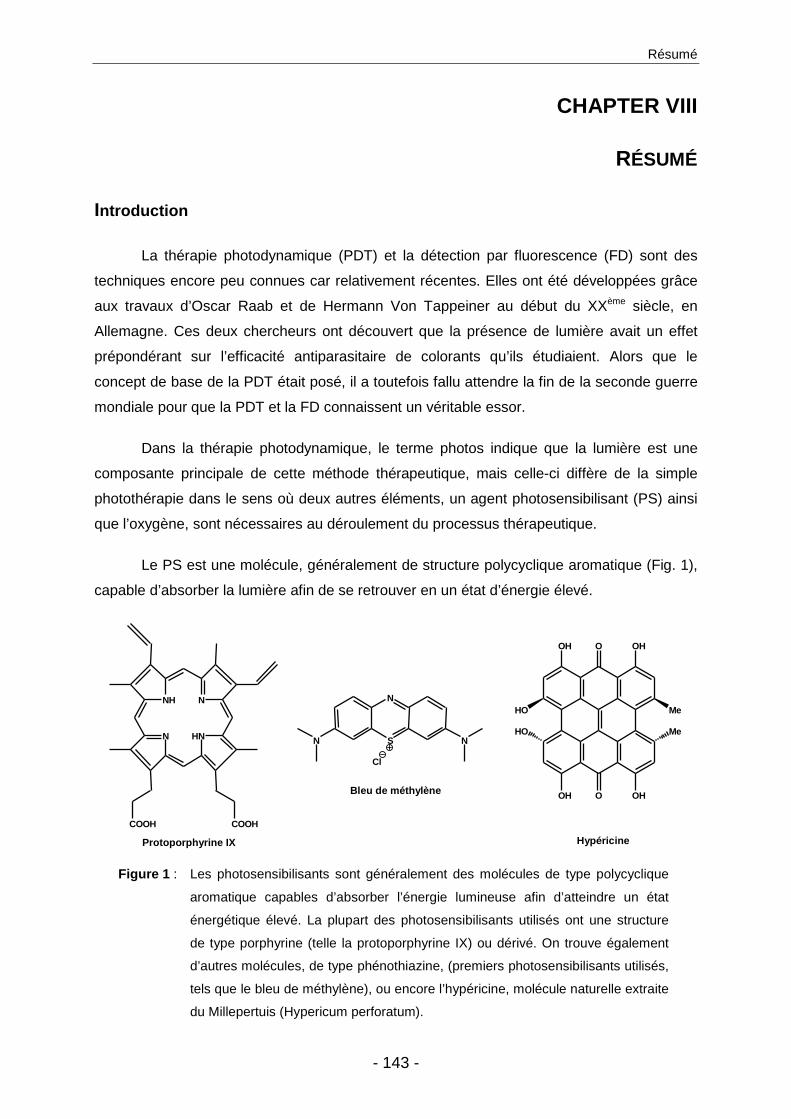
Résumé
- 143 -
CHAPTER VIII
RÉSUMÉ
Introduction
La thérapie photodynamique (PDT) et la détection par fluorescence (FD) sont des
techniques encore peu connues car relativement récentes. Elles ont été développées grâce
aux travaux d’Oscar Raab et de Hermann Von Tappeiner au début du XXème siècle, en
Allemagne. Ces deux chercheurs ont découvert que la présence de lumière avait un effet
prépondérant sur l’efficacité antiparasitaire de colorants qu’ils étudiaient. Alors que le
concept de base de la PDT était posé, il a toutefois fallu attendre la fin de la seconde guerre
mondiale pour que la PDT et la FD connaissent un véritable essor.
Dans la thérapie photodynamique, le terme photos indique que la lumière est une
composante principale de cette méthode thérapeutique, mais celle-ci diffère de la simple
photothérapie dans le sens où deux autres éléments, un agent photosensibilisant (PS) ainsi
que l’oxygène, sont nécessaires au déroulement du processus thérapeutique.
Le PS est une molécule, généralement de structure polycyclique aromatique (Fig. 1),
capable d’absorber la lumière afin de se retrouver en un état d’énergie élevé.
OH O OH
HO Me
HO Me
OH O OH
NH N
HNN
COOH COOH
S
N
N N
Cl
Bleu de méthylène
Protoporphyrine IX Hypéricine
Figure 1 : Les photosensibilisants sont généralement des molécules de type polycyclique
aromatique capables d’absorber l’énergie lumineuse afin d’atteindre un état
énergétique élevé. La plupart des photosensibilisants utilisés ont une structure
de type porphyrine (telle la protoporphyrine IX) ou dérivé. On trouve également
d’autres molécules, de type phénothiazine, (premiers photosensibilisants utilisés,
tels que le bleu de méthylène), ou encore l’hypéricine, molécule naturelle extraite
du Millepertuis (Hypericum perforatum).
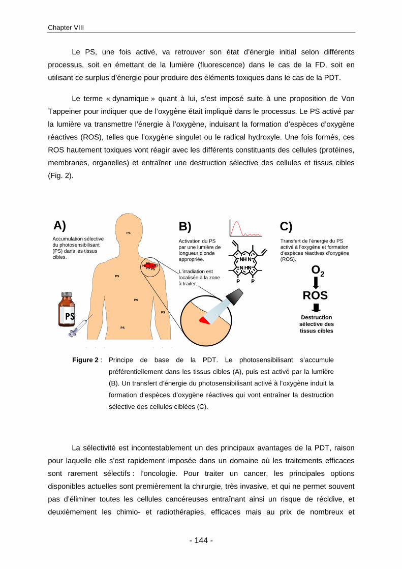
Chapter VIII
- 144 -
Le PS, une fois activé, va retrouver son état d’énergie initial selon différents
processus, soit en émettant de la lumière (fluorescence) dans le cas de la FD, soit en
utilisant ce surplus d’énergie pour produire des éléments toxiques dans le cas de la PDT.
Le terme « dynamique » quant à lui, s’est imposé suite à une proposition de Von
Tappeiner pour indiquer que de l’oxygène était impliqué dans le processus. Le PS activé par
la lumière va transmettre l’énergie à l’oxygène, induisant la formation d’espèces d’oxygène
réactives (ROS), telles que l’oxygène singulet ou le radical hydroxyle. Une fois formés, ces
ROS hautement toxiques vont réagir avec les différents constituants des cellules (protéines,
membranes, organelles) et entraîner une destruction sélective des cellules et tissus cibles
(Fig. 2).
PS
A) B)
NH N
HNN
PP
NH N
HNN
PP
Accumulation sélective du photosensibilisant(PS) dans les tissus cibles.
Transfert de l’énergie du PS activé à l’oxygène et formation d’espèces réactives d’oxygène(ROS).
Destruction sélective des tissus cibles
O2
ROS
C)Activation du PS par une lumière de longueur d’onde appropriée.
L’irradiation est localisée à la zone à traiter.
PS
PS
PS
PS PSPS
PS
PS
PS
PS PS
Figure 2 : Principe de base de la PDT. Le photosensibilisant s’accumule
préférentiellement dans les tissus cibles (A), puis est activé par la lumière
(B). Un transfert d’énergie du photosensibilisant activé à l’oxygène induit la
formation d’espèces d’oxygène réactives qui vont entraîner la destruction
sélective des cellules ciblées (C).
La sélectivité est incontestablement un des principaux avantages de la PDT, raison
pour laquelle elle s’est rapidement imposée dans un domaine où les traitements efficaces
sont rarement sélectifs : l’oncologie. Pour traiter un cancer, les principales options
disponibles actuelles sont premièrement la chirurgie, très invasive, et qui ne permet souvent
pas d’éliminer toutes les cellules cancéreuses entraînant ainsi un risque de récidive, et
deuxièmement les chimio- et radiothérapies, efficaces mais au prix de nombreux et

Résumé
- 145 -
importants effets secondaires résultant du fait que toutes les cellules en division rapide sont
touchées, et pas uniquement les cellules pathogènes. La sélectivité de la PDT pour les
tumeurs est obtenue à plusieurs niveaux : (i) le PS s’accumule préférentiellement dans les
tissus cibles en raison de différents mécanismes, tels que la moins bonne étanchéité de la
neo-vascularisation tumorale qui permettant une extravasation du PS, ou les défauts dans le
drainage lymphatique prévenant une élimination rapide du PS ; (ii) seule la zone à traiter est
irradiée, évitant ainsi d’activer les PS qui se seraient accumulés dans des zones non
désirées ; (iii) la faible durée de vie des ROS, de l’ordre de quelques millisecondes,
empêchant la migration de ces éléments toxiques vers les tissus sains environnants, et
entraînant ainsi une destruction tissulaire confinée à la zone traitée.
Figure 3 : Biosynthèse de l’hème : cycle naturel quasi-ubiquitaire débutant par la
formation d’acide 5-aminolévulinique (5-ALA) dans la mitochondrie, puis
condensation enzymatique de deux molécules de 5-ALA en une molécule de
porphobilinogène (PBG). Quatre PBG sont assemblés pour former un
tétrapyrrole, qui sera transformé par une succession de décarboxylations
enzymatiques en une porphyrine, la protoporphyrine IX (PpIX), molécule
photoactive. La ferrochélatase va ensuite incorporer un atome de fer dans la
PpIX pour former l’hème, biologiquement active, mais non photoactive.
L’hème exerce un rétrocontrôle négatif sur la formation de 5-ALA,
empêchant ainsi l’accumulation intracellulaire de porphyrines photoactives.
Toutefois, une administration exogène de 5-ALA contourne ce rétrocontrôle
négatif, entraînant une accumulation de PpIX, principalement dans les
cellules très actives métaboliquement, par exemple les cellules cancéreuses.
+NH3-OOC
-OOC SCoA
O
O-O
ONH3
+
NH
H2N
COO-COO-
NHHN
HNNH
P
P
PP
NH N
HNN
PP
N N
NN
PP
Fe
Cycle de Cycle de ll ’’hhèèmeme
5-ALA
PBG
PpIXHème
O-O
ONH3
+
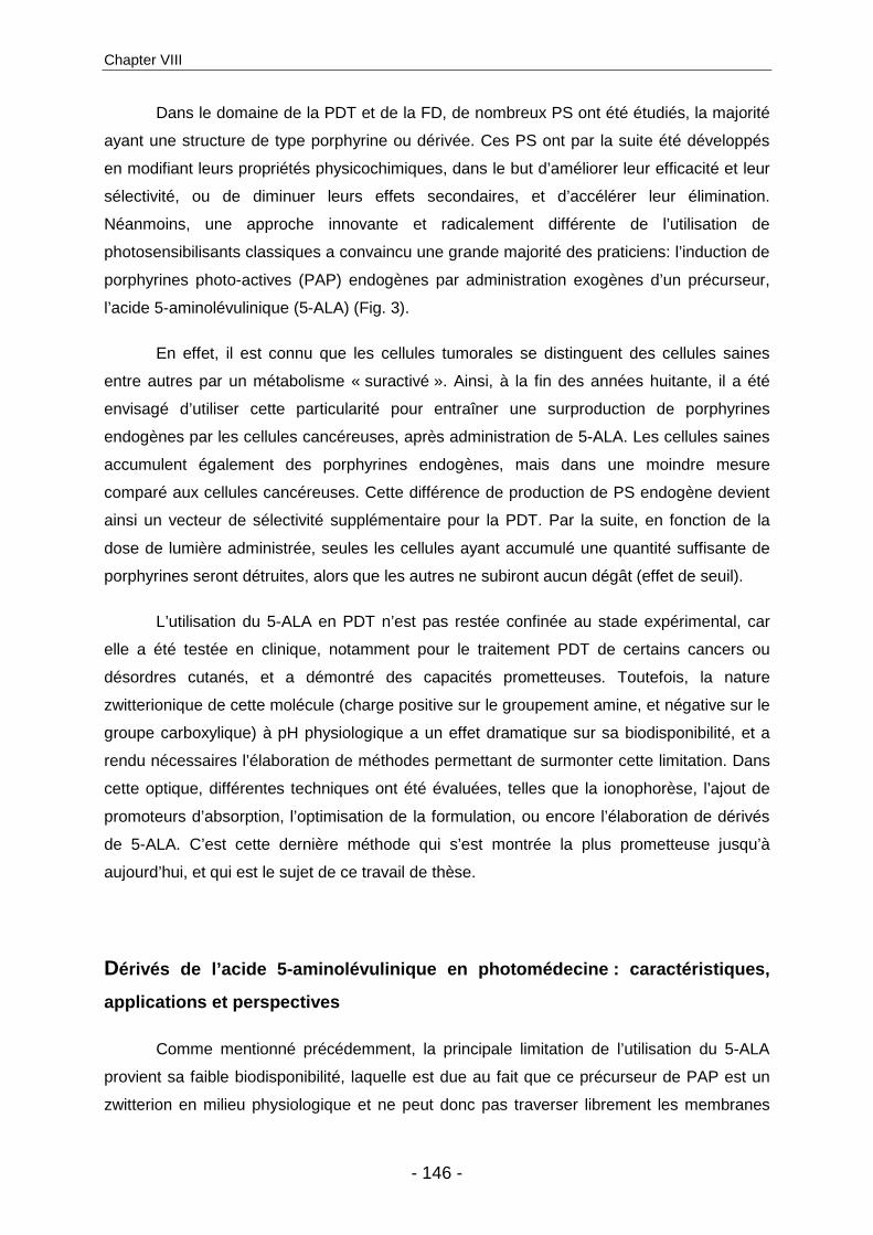
Chapter VIII
- 146 -
Dans le domaine de la PDT et de la FD, de nombreux PS ont été étudiés, la majorité
ayant une structure de type porphyrine ou dérivée. Ces PS ont par la suite été développés
en modifiant leurs propriétés physicochimiques, dans le but d’améliorer leur efficacité et leur
sélectivité, ou de diminuer leurs effets secondaires, et d’accélérer leur élimination.
Néanmoins, une approche innovante et radicalement différente de l’utilisation de
photosensibilisants classiques a convaincu une grande majorité des praticiens: l’induction de
porphyrines photo-actives (PAP) endogènes par administration exogènes d’un précurseur,
l’acide 5-aminolévulinique (5-ALA) (Fig. 3).
En effet, il est connu que les cellules tumorales se distinguent des cellules saines
entre autres par un métabolisme « suractivé ». Ainsi, à la fin des années huitante, il a été
envisagé d’utiliser cette particularité pour entraîner une surproduction de porphyrines
endogènes par les cellules cancéreuses, après administration de 5-ALA. Les cellules saines
accumulent également des porphyrines endogènes, mais dans une moindre mesure
comparé aux cellules cancéreuses. Cette différence de production de PS endogène devient
ainsi un vecteur de sélectivité supplémentaire pour la PDT. Par la suite, en fonction de la
dose de lumière administrée, seules les cellules ayant accumulé une quantité suffisante de
porphyrines seront détruites, alors que les autres ne subiront aucun dégât (effet de seuil).
L’utilisation du 5-ALA en PDT n’est pas restée confinée au stade expérimental, car
elle a été testée en clinique, notamment pour le traitement PDT de certains cancers ou
désordres cutanés, et a démontré des capacités prometteuses. Toutefois, la nature
zwitterionique de cette molécule (charge positive sur le groupement amine, et négative sur le
groupe carboxylique) à pH physiologique a un effet dramatique sur sa biodisponibilité, et a
rendu nécessaires l’élaboration de méthodes permettant de surmonter cette limitation. Dans
cette optique, différentes techniques ont été évaluées, telles que la ionophorèse, l’ajout de
promoteurs d’absorption, l’optimisation de la formulation, ou encore l’élaboration de dérivés
de 5-ALA. C’est cette dernière méthode qui s’est montrée la plus prometteuse jusqu’à
aujourd’hui, et qui est le sujet de ce travail de thèse.
Dérivés de l’acide 5-aminolévulinique en photomédeci ne : caractéristiques,
applications et perspectives
Comme mentionné précédemment, la principale limitation de l’utilisation du 5-ALA
provient sa faible biodisponibilité, laquelle est due au fait que ce précurseur de PAP est un
zwitterion en milieu physiologique et ne peut donc pas traverser librement les membranes
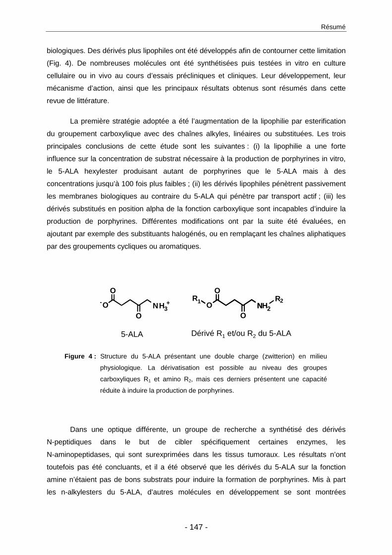
Résumé
- 147 -
biologiques. Des dérivés plus lipophiles ont été développés afin de contourner cette limitation
(Fig. 4). De nombreuses molécules ont été synthétisées puis testées in vitro en culture
cellulaire ou in vivo au cours d’essais précliniques et cliniques. Leur développement, leur
mécanisme d’action, ainsi que les principaux résultats obtenus sont résumés dans cette
revue de littérature.
La première stratégie adoptée a été l’augmentation de la lipophilie par esterification
du groupement carboxylique avec des chaînes alkyles, linéaires ou substituées. Les trois
principales conclusions de cette étude sont les suivantes : (i) la lipophilie a une forte
influence sur la concentration de substrat nécessaire à la production de porphyrines in vitro,
le 5-ALA hexylester produisant autant de porphyrines que le 5-ALA mais à des
concentrations jusqu’à 100 fois plus faibles ; (ii) les dérivés lipophiles pénètrent passivement
les membranes biologiques au contraire du 5-ALA qui pénètre par transport actif ; (iii) les
dérivés substitués en position alpha de la fonction carboxylique sont incapables d’induire la
production de porphyrines. Différentes modifications ont par la suite été évaluées, en
ajoutant par exemple des substituants halogénés, ou en remplaçant les chaînes aliphatiques
par des groupements cycliques ou aromatiques.
5-ALA Dérivé R1 et/ou R2 du 5-ALA
O-O
ONH3
+
O
O NHR1 R
ON 2
2
Figure 4 : Structure du 5-ALA présentant une double charge (zwitterion) en milieu
physiologique. La dérivatisation est possible au niveau des groupes
carboxyliques R1 et amino R2, mais ces derniers présentent une capacité
réduite à induire la production de porphyrines.
Dans une optique différente, un groupe de recherche a synthétisé des dérivés
N-peptidiques dans le but de cibler spécifiquement certaines enzymes, les
N-aminopeptidases, qui sont surexprimées dans les tissus tumoraux. Les résultats n’ont
toutefois pas été concluants, et il a été observé que les dérivés du 5-ALA sur la fonction
amine n’étaient pas de bons substrats pour induire la formation de porphyrines. Mis à part
les n-alkylesters du 5-ALA, d’autres molécules en développement se sont montrées
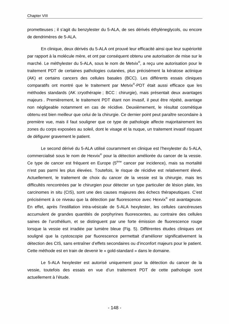
Chapter VIII
- 148 -
prometteuses ; il s’agit du benzylester du 5-ALA, de ses dérivés éthylèneglycols, ou encore
de dendrimères de 5-ALA.
En clinique, deux dérivés du 5-ALA ont prouvé leur efficacité ainsi que leur supériorité
par rapport à la molécule mère, et ont par conséquent obtenu une autorisation de mise sur le
marché. Le méthylester du 5-ALA, sous le nom de Metvix®, a reçu une autorisation pour le
traitement PDT de certaines pathologies cutanées, plus précisément la kératose actinique
(AK) et certains cancers des cellules basales (BCC). Les différents essais cliniques
comparatifs ont montré que le traitement par Metvix®-PDT était aussi efficace que les
méthodes standards (AK :cryothérapie ; BCC : chirurgie), mais présentait deux avantages
majeurs . Premièrement, le traitement PDT étant non invasif, il peut être répété, avantage
non négligeable notamment en cas de récidive. Deuxièmement, le résultat cosmétique
obtenu est bien meilleur que celui de la chirurgie. Ce dernier point peut paraître secondaire à
première vue, mais il faut souligner que ce type de pathologie affecte majoritairement les
zones du corps exposées au soleil, dont le visage et la nuque, un traitement invasif risquant
de défigurer gravement le patient.
Le second dérivé du 5-ALA utilisé couramment en clinique est l’hexylester du 5-ALA,
commercialisé sous le nom de Hexvix® pour la détection améliorée du cancer de la vessie.
Ce type de cancer est fréquent en Europe (5ème cancer par incidence), mais sa mortalité
n’est pas parmi les plus élevées. Toutefois, le risque de récidive est relativement élevé.
Actuellement, le traitement de choix du cancer de la vessie est la chirurgie, mais les
difficultés rencontrées par le chirurgien pour détecter un type particulier de lésion plate, les
carcinomes in situ (CIS), sont une des causes majeures des échecs thérapeutiques. C’est
précisément à ce niveau que la détection par fluorescence avec Hexvix® est avantageuse.
En effet, après l’instillation intra-vésicale de 5-ALA hexylester, les cellules cancéreuses
accumulent de grandes quantités de porphyrines fluorescentes, au contraire des cellules
saines de l’urothélium, et se distinguent par une forte émission de fluorescence rouge
lorsque la vessie est irradiée par lumière bleue (Fig. 5). Différentes études cliniques ont
souligné que la cystoscopie par fluorescence permettait d’améliorer significativement la
détection des CIS, sans entraîner d’effets secondaires ou d’inconfort majeurs pour le patient.
Cette méthode est en train de devenir le « gold-standard » dans le domaine.
Le 5-ALA hexylester est autorisé uniquement pour la détection du cancer de la
vessie, toutefois des essais en vue d’un traitement PDT de cette pathologie sont
actuellement à l’étude.

Résumé
- 149 -
Figure 5 : Cystoscopie d’un patient diagnostiqué pour un cancer de la vessie ; à
gauche, image prise en cystoscopie par lumière blanche ne présentant pas
de lésion détectable ; à droite, la même zone irradiée en lumière bleue après
instillation de Hexvix® permet de mettre en évidence carcinome in situ grâce
à la forte fluorescence rouge émise par les cellules cancéreuses.
D’autres maladies seront peut-être traitées un jour par les dérivés du 5-ALA, des
recherches étant en cours dans différents domaines, par exemple en dermatologie pour le
traitement du psoriasis, de la leishmaniose, de l’acné, des verrues, ou en oncologie pour le
traitement du cancer de l’utérus ou de l’œsophage de Barrett. D’autres pathologies, telles
que les cancers de la prostate, du sein ou du cerveau pourraient être potentiellement traités
par cette méthode, mais nécessiteraient l’élaboration de dérivés du 5-ALA administrables par
voie systémique.
Du métabolisme des dérivés de l’acide 5-aminolévulin ique
Malgré le développement clinique de l’utilisation des dérivés du 5-ALA en
photomédecine, il subsiste des interrogations au niveau de quelques mécanismes
fondamentaux. Par exemple, il a été observé que, tandis que les molécules de 5-ALA
doivent être transporté par des mécanismes actifs, les dérivés lipophiles du 5-ALA traversent
les membranes biologiques par voie passive. Il a été supposé que, par la suite, les esters du
5-ALA doivent être clivés en 5-ALA et son alcool correspondant, par des estérases
cytosoliques non spécifiques, pour entrer dans la biosynthèse de l’hème. Toutefois, la
formation de porphyrines ne peut être considérée comme une preuve suffisante de
l’hydrolyse des dérivés du 5-ALA. En considérant les dernières découvertes dans le domaine

Chapter VIII
- 150 -
de la biosynthèse de l’hème, il apparaît que le mécanisme d’action de la porphobilinogène
synthase (PBGS), l’enzyme catalysant la condensation de deux molécules de 5-ALA en une
molécule de porphobilinogène (PBG), n’implique a aucun moment les groupements
carboxyliques du 5-ALA concernés par la dérivatisation. L’hydrolyse des dérivés du 5-ALA
en 5-ALA ne serait donc pas un prérequis pour devenir substrat de la biosynthèse de l’hème,
pour autant que la dérivatisation soit effectuée sur le groupement carboxylique et que
l’encombrement stérique ne gène pas l’entrée de la molécule dans les poches fonctionnelles
de l’enzyme (Fig. 6).
NH2COOH
A-Site
P-Site
O
NH2
O
O
O
NH2
O
O
NH2COOH
A-Site
P-Site
NH2COOH
A-Site
P-Site
NH2COOH
A-Site
P-Site
NNH2
COOHCOOH
O
NH2
O
O
R O
NH2
O
O
R
NNH2
O O
R O
O
R
A)
B)
Figure 6 : Représentation schématique de la porphobilinogène synthase
(PBGS). A) Deux molécules de 5-ALA prennent place dans les
poches de l’enzyme qui catalyse une réaction de condensation.
B) Les groupements carboxyliques n’étant pas impliqué dans le
processus enzymatique, il est mécanistiquement possible que
les dérivés du 5-ALA soient directement substrat de l’enzyme,
résultant en la formation de dérivés de porphobilinogène.
Afin d’étudier le métabolisme des dérivés du 5-ALA ainsi qu’une possibilité
d’améliorer la stabilité de ces molécules, nous avons synthétisé le 2-amino-3-oxohenadioic-
1,6-diméthyl ester (AOMM). Cette molécule se base sur le 5-ALA méthylester (MAL) auquel
a été ajouté un groupement methoxycarbonyl à proximité de la fonction amine. Cette

Résumé
- 151 -
molécule présente des propriétés de taille, de charge, et de lipophilie proches de la molécule
parente, mais doit impérativement subir un clivage par une estérase pour devenir active. En
effet, le groupement supplémentaire bloque la fonction amine, ce qui empêche à la fois la
dégradation de la molécule par dimérisation, et sa transformation enzymatique en PBG.
En cas de présence d’activité estérasique, AOMM devrait être transformé en 5-ALA
méthlyester et en 5-ALA, et devenir substrats du cycle de biosynthèse de l’hème. Des essais
enzymatiques in vitro ont montré qu’il y avait bien transformation rapide des cette molécule
« témoin » préférentiellement en MAL, puis en 5-ALA, en présence d’estérases de foie de
porc. Néanmoins, les cellules cancéreuses incubées avec AOMM n’ont pas accumulées de
porphyrines fluorescentes, au contraire de celles incubées avec 5-ALA ou MAL, indiquant
une incapacité des cellules à hydrolyser ce substrat, et remet en question la présence et/ou
l’importance de cette activité estérasique présumée. De plus, l’injection d’AOMM dans le
modèle le l’embryon de poulet a entraîné l’accumulation de porphyrines fluorescentes dans
les différents organes analysés.
Ceci confirme la nécessité de poursuivre les recherches fondamentales sur le
métabolisme des dérivés du 5-ALA, afin de pouvoir adapter au mieux les futures stratégies
de ciblage des dérivés du 5-ALA.
Nouvelle méthode d’évaluation de dérivés de l’acide 5-aminolévulinique pour
de nouvelles indications
Il a été montré précédemment que la biodisponibilité du 5-ALA était grandement
améliorée par la dérivatisation de cette molécule en n-alkylester plus lipophiles, tels le
méthylester et l’hexylester du 5-ALA. Toutefois, ces dérivés ont été développés
exclusivement pour un usage topique. Leur utilisation par voie systémique n’étant pas
possible pour des raisons de toxicité, certaines pathologies telles que les cancers du sein, de
la prostate ou du cerveau, ne bénéficient pas de ces évolutions. Pourtant, le 5-ALA a déjà
été utilisé pour la FD ou le traitement PDT de glioblastomes, et a montré un potentiel certain.
Néanmoins la faible capacité du 5-ALA à traverser la barrière hémato-encéphalique pénalise
et rend nécessaire le développement de dérivés du 5-ALA conçus spécifiquement pour une
administration systémique et présentant une capacité améliorée à atteindre le système
nerveux centrale.
Dans cette optique, différents modèles expérimentaux sont disponibles pour
l’évaluation de ces nouvelles molécules, chacun de ces modèles présentant des avantages
Chapter VIII
- 152 -
et inconvénients qui leurs sont propres. La culture cellulaire, par exemple, est facilement
accessible, bon marché, mais reste un model basique peu représentatif de la situation
complexe d’un organisme vivant. D’un autre côté, les essais in vivo sur des animaux donnent
une idée beaucoup plus proche du devenir des molécules actives chez l’homme, mais pour
un coût très élevé, et en présentant de plus des limitations éthiques et légales qui les
rendent inadaptés en phase de criblage.
Pour ces raisons, nous avons envisagé l’utilisation du modèle de l’embryon de poulet
pour évaluer les nouveaux dérivés du 5-ALA. L’embryon de poulet est utilisé depuis de
nombreuses années dans le domaine de la PDT pour sa membrane chorio-allantoïque
(CAM), fortement vascularisée et accessible, qui permet l’évaluation des propriétés vaso-
occlusives de différents régimes PDT. La procédure appliquée a été la suivante. Des œufs
fécondés ont été préparés et incubés jusqu’au douzième jour de développement
embryonnaire. Les molécules à tester ont été dissoutes dans un certain volume de solvant,
puis injectées dans une large veine du CAM. Après un certain temps d’incubation, l’embryon
a été congelé à -80°C. Après dissection et prélèvem ent des différents organes, les
porphyrines produites ont été extraites des échantillons, puis dosées par HPLC-FD. Des
essais effectués avec le 5-ALA ont montrés que la production de porphyrines dans les
différents organes était dépendante de la concentration de 5-ALA administrée. De plus, les
valeurs obtenues étaient comparables aux données de la littérature concernant la
biodistribution des porphyrines après administration du 5-ALA à des souris. De même, une
injection d’hexylester du 5-ALA a montré une forte toxicité aiguë, telle que celle observée
après administration systémique chez la souris.
Des essais ont été effectués avec des dérivés diacylglycérides du 5-ALA (Fig. 7)
spécialement synthétisés pour cibler le cancer du cerveau. Ces molécules doivent être
adaptées à une administration par voie systémique, présenter une lipophilie accrue afin de
traverser aisément la barrière hémato-encéphalique tout en présentant une faible toxicité.
Après avoir été injectées dans le model de l’embryon de poulet, ces dérivés ont montré la
capacité à induire la production de porphyrines dans les différents organes, tandis qu’aucune
toxicité aiguë n’a été observée.
Ce modèle peut nous permettre de déterminer les candidats les plus prometteurs en
terme de capacité à induire une accumulation de porphyrines dans les tissues cibles, ainsi
que d’absence de toxicité aiguë, et pourrait devenir une alternative intéressante aux études
précliniques de biodistribution et de toxicité.
Résumé
- 153 -
+H3N
O
O
OO
O
O
O
CF3CO2-
+H3N
O
O
OO
OCF3CO2-
O
O
5-ALA diacetyl glyceride (ADAG)
5-ALA dibutyryl glyceride (ADBG)
Figure 7 : Structures de deux dérivés diacylglycérides du 5-ALA conçus pour
une utilisation systémique dans le ciblage du cancer du cerveau.
L’avantage de ce modèle est qu’il permet d’étudier les dérivés du 5-ALA quant à leur
capacité à induire la production de PAP, la biodistribution de ces porphyrines dans les
différents organes, ainsi que leur toxicité systémique aiguë suite à l’injection du produit. De
plus, contrairement à la culture cellulaire, les molécules faiblement hydrosolubles peuvent
être facilement testées puisque l’injection de faibles volumes de solvant, ici un mélange de
polyéthylène glycol, d’éthanol et d’eau, est bien tolérée. Ce modèle présente une grande
flexibilité puisque différentes voies d’administration peuvent être testées (intraveineuse,
intraperitonéale, topique). De plus, il est possible d’implanter des tumeurs directement sur la
CAM.
Effets de l’acide 5-aminolévulinique et des dérivés de l’acide
5-aminolévulinique sur des bactéries gram-négatives et gram-positives
Alors que la découverte des antibiotiques par Flemming et les progrès réalisés dans
le domaine durant la décennie qui suivit avaient laissé espérer une éradication totale des
infections bactériennes, nous sommes toutefois obligés de constater la relative impuissance
de la recherche pharmaceutique face au pouvoir d’adaptation des bactéries. La capacité
qu’ont les bactéries à devenir résistantes aux nouveaux traitements et à propager cette

Chapter VIII
- 154 -
résistance à leurs congénères est en train de surpasser celle de la médecine à développer
de nouvelles molécules antibactériennes. Une des raisons pour expliquer ces rapides
adaptations aux antibiotiques est l’usage injustifié et immodéré de grandes quantités
d’antibiotiques, non seulement dans les domaines de la santé, mais également de
l’agroalimentaire. Dans ce contexte, des alternatives thérapeutiques aux antibiotiques
doivent être développées.
Une de ces alternatives pourrait être la PDT. En effet, cette technique actuellement
utilisée pour le traitement de certains cancers ou de la dégénérescence maculaire liée à
l’âge a pour la première fois été observée pour ses propriétés antimicrobiennes. De
nombreux essais dans ce domaine ont été effectués et ont montré une efficacité de la PDT
dans l’élimination in vitro de bactéries, dans la désinfection de l’eau ou du plasma, ainsi que
pour le traitement in vivo de certaines infections. Dans ce dernier cas, le choix du PS reste
crucial, et des propriétés telles qu’une faible toxicité, une sélectivité accrue pour les tissus
cibles ainsi qu’une absence de photosensibilité cutanée résiduelle sont recherchées.
Différentes études ont montré que le 5-ALA induisait une production plus ou moins grande
de porphyrines selon le type de bactérie, la phase de croissance ou les conditions
d’incubations.
Figure 8 : Production de porphyrines (barres), et fraction survivante sans
irradiation (○) et après irradiation (■) de E. coli K12, en fonction de
la concentration de 5-ALA (temps d’incubation 3 heures ;
irradiation 120 J·cm-2).
Résumé
- 155 -
Le but de cette étude était de déterminer s’il était possible d’améliorer ou d’influencer
la production de porphyrines dans différentes souches bactériennes en utilisant des dérivés
lipophiles du 5-ALA. Pour cela, le 5-ALA et des n-alkylesters du 5-ALA de lipophilie
croissante ont été incubés à différentes concentrations avec des souches d’Escherichia coli
K12, Escherichia coli Ti05, Pseudomonas aeruginosa et Staphylococcus aureus MRSA. Leur
survie avec et sans irradiation a ensuite été déterminée, de même que la quantité et le type
de porphyrines produites.
Les premiers essais effectués sur E. coli K12, une souche de laboratoire non
pathogène, ont montré que la production de porphyrines était fortement dépendante de la
concentration en substrat. En effet, la production de porphyrines augmente avec celle de
substrat jusqu’à un optimum d’environ 0.1 mM, puis diminue malgré l’augmentation de la
concentration de 5-ALA (Fig 8). Cette « courbe en cloche » a déjà été observée par exemple
en culture cellulaire, et l’hypothèse communément admise pour expliquer cette baisse est
celle du blocage de certaines enzymes par un excès de 5-ALA. De plus, la production de
porphyrines est bien corrélée à l’efficacité de la photoinactivation, puisque c’est à cette
concentration optimale de production de PAP que la photoinactivation est la plus efficace,
(> 99.9% d’inactivation des bactéries). Les porphyrines majoritairement détectées sont
l’uroporphyrine et la coproporphyrine, tandis qu’on ne trouve que quelques traces de
protoporphyrine IX.
Les esters du 5-ALA à courte chaîne (méthyl- et butylester) ont entraîné la production
de grandes quantités de porphyrines après incubation à une concentration de l’ordre de
10 mM. Au contraire, les n-alkylesters à longue chaîne du 5-ALA (pentyl-, hexyl- et
octylester) n’ont montré aucune capacité à induire la formation de porphyrines à quelque
concentration que ce soit. De plus, ces molécules ont démontré une grande toxicité
intrinsèque, augmentant avec la lipophilie, non liée à une action photochimique, et ne sont
donc pas des candidats intéressants pour une photoinactivation antibactérienne.
Les essais avec les autres souches précitées ont montré des résultats similaires, le
méthylester du 5-ALA induisant des grandes quantités de porphyrines et entraînant une
photoinactivation efficace des bactéries. Cette molécule, déjà utilisée pour le traitement PDT
de différents désordres cutanés, pourrait être un candidat potentiel pour le traitement PDT
d’infections cutanées.

Chapter VIII
- 156 -
Dérivés de l’acide 5-aminolévulinique pour une photoinactivation améliorée de
Propionibacterium acnes
Actuellement, différents traitements monosubstances ou combinés sont utilisés dans
le traitement de l’acné modéré ou sévère, mais leur efficacité doit être mise en relation avec
de lourds effets secondaires, ainsi qu’un problème de résistance des bactéries.
Une solution envisagée pour remplacer ces traitements est la PDT. Elle permettrait
de traiter à la fois le désordre glandulaire, l’hyperproduction sébacée et la prolifération
bactérienne (Propionibacterium acnes) (Fig. 9), cette dernière étant responsable de
l’inflammation. Des études cliniques ont montré la faisabilité de ce type de traitement de
l’acné, avec notamment une amélioration de la maladie chez des patients ayant subi un
traitement PDT avec 5-ALA. Une étude a même comparé directement l’efficacité d’un
traitement PDT après application topique de 5-ALA ou de méthylester du 5-ALA (MAL).
L’efficacité des deux traitements s’est montrée similaire, la différence venant du fait que
l’irradiation était moins douloureuse après application du MAL que du 5-ALA.
occlusion dupore
sébum rance hyperplasieglandulaire
poil rudimentaire
poilglandesébacée
follicule pileuxglandesudoripare
Figure 9 : Schéma représentant un follicule pileux sain (gauche) et acnéique (droite) ;
l’occlusion du pore, l’hyperplasie glandulaire et l’accumulation de sébum
installent un environnement propice à la colonisation du follicule pilo-
sébacé par Propionibactrium acnes, qui va induire ou aggraver le
processus inflammatoire (tiré de « Integrated Pharmacology » 2ème édition).
Résumé
- 157 -
Le but de cette étude était de déterminer quel dérivé du 5-ALA serait le plus efficace
pour une photoinactivation des bactéries de type P. acnes. Pour cela, une souche de
laboratoire de P. acnes, ainsi que deux souches pathogènes ont été incubées avec le 5-ALA
et différents dérivés. Les candidats sélectionnés étaient le méthylester du 5-ALA, qui a
montré une bonne efficacité contre les différentes souches testées dans une étude
précédente, l’hexylester (HAL), le benzylester (BzAL) et le di-éthylène-glycol-
monométhylether ester (DGME) du 5-ALA. Les résultats ont montré qu’il était possible
d’induire une forte accumulation de porphyrines chez P. acnes generale après incubation
avec le 5-ALA. La concentration optimale de 5-ALA se situe entre 3-10 mM, tandis que les
types de porphyrines les plus représentées dans ces conditions expérimentales sont
l’uroporphyrine et la coproporphyrine. MAL n’a que faiblement induit la production de
porphyrines, tandis que HAL a démontré une grande toxicité intrinsèque à des doses
supérieures à 1 mM.
Les deux molécules les plus prometteuses sont BzAL et DGME qui ont induit
l’accumulation de grande quantités de porphyrines chez P. acnes generale, et une
photoinactivation de >99.9 % des bactéries. Des résultats similaires ont été obtenus sur les
souches pathogènes testées, BzAL et DGME induisant une photoinactivation plus importante
que MAL.
Une production élevée de porphyrines in vitro chez P. acnes ne représente toutefois
pas une garantie pour un traitement efficace de l’acné, et l’accumulation de ces molécules
au niveau des follicules pilo-sébacés devra également être étudiée, de même qu’une
optimisation de la formulation et des conditions d’irradiation. Des essais précliniques se
déroulent en ce moment dans le but d’évaluer le réel apport de ces molécules, comparé au
5-ALA et au MAL, pour le traitement PDT de l’acné.