Electricity price disadvantages for German industry decreasing
Upload
independentCategory
view
2download
0
1
© The Author 2015. Published by Oxford University Press. All rights reserved. For permissions,
please e-mail: [email protected]
4-methylumbelliferone inhibits hepatocellular carcinoma growths by
decreasing IL-6 production and angiogenesis.
Flavia Piccioni1, Esteban Fiore1, Juan Bayo1, Catalina Atorrasagasti1,4, Estanislao
Peixoto1, Manglio Rizzo1, Mariana Malvicini1,4, Irene Tirado-González2, Mariana G.
García1,4, Laura Alaniz3,4, Guillermo Mazzolini1,4
1Liver Unit, Gene Therapy Laboratory, Facultad de Ciencias Biomédicas, Universidad
Austral, Buenos Aires, Argentina
2Laboratory of Reproductive Medicine, Medicine University of Berlin, Charité Centre 12
Internal Medicine and Dermatology, Berlin, Germany
3CIT NOBA, Universidad Nacional del Noroeste de la Pcia. de Bs. As., Junín, Buenos
Aires, Argentina.
4CONICET (Consejo Nacional de Investigaciones Científicas y Técnicas), Buenos Aires,
Argentina
Corresponding author: Guillermo D. Mazzolini, MD, PhD, Liver Unit, Gene Therapy
Laboratory, Facultad de Ciencias Biomédicas. Universidad Austral, Avenida Presidente
Perón 1500 (B1629ODT), Derqui-Pilar, Buenos Aires 1635, Argentina. E-
mail: [email protected] or Laura Alaniz, PhD CIT NOBA, Universidad Nacional
del Noroeste de la Pcia. de Bs. As., Jorge Newbery 261 (B600), Junin, Buenos Aires,
Argentina, E-mail: [email protected]
Glycobiology Advance Access published April 16, 2015 by guest on June 4, 2016
http://glycob.oxfordjournals.org/D
ownloaded from
2
Abstract
Cirrhosis is characterized by an excessive accumulation of extracellular matrix components
including hyaluronic acid (HA) and is widely considered a pre-neoplastic condition for
hepatocellular carcinoma (HCC). 4-methylumbelliferone (4MU) is an inhibitor of HA
synthesis and has anti-cancer activity in an orthotopic HCC model with underlying fibrosis.
Our aim was to explore the effects of HA inhibition by 4MU orally administered on tumour
microenvironment. Hepa129 tumour cells were inoculated orthotopically in C3H/HeJ male
mice with fibrosis induced by thioacetamide. Mice were orally treated with 4MU. The
effects of 4MU on angiogenesis were evaluated by immunostaining of CD31 and
quantification of proangiogenic factors (VEGF, IL-6, CXCL12). IL-6 was also quantified in
Hepa129 cells in vitro after treatment with 4MU. Migration of endothelial cells and tube
formation were also analyzed. As a result, 4MU treatment decreases tumour growth and
increased animal survival. Systemic levels of VEGF were significantly inhibited in 4MU-
treated mice. Expression of CD31 was reduced after 4MU therapy in liver parenchyma in
comparison with control group. In addition, mRNA expression and protein levels of IL-6
and VEGF were inhibited both in tumour tissue and in non-tumoral liver parenchyma.
Interestingly, IL-6 production was dramatically reduced in Kupffer cells isolated from
4MU-treated mice, and in Hepa129 cells in vitro. Besides, 4MU was able to inhibit
endothelial cell migration and tube formation. In conclusion, 4MU has antitumour activity
in vivo and its mechanisms of action involve an inhibition of angiogenesis and IL-6
production. 4MU is an orally available molecule with potential for HCC treatment.
Keywords: 4-methylumbelliferone; hyaluronic acid, liver fibrosis, HCC, IL-6, VEGF,
angiogenesis.
by guest on June 4, 2016http://glycob.oxfordjournals.org/
Dow
nloaded from

3
Introduction
Liver fibrosis is a wound-healing process that occurs in response to chronic injuries, mainly
chronic hepatitis C and B virus infection, and alcohol abuse (Bataller and Brenner 2005).
The chronic insult induces liver injury by production of oxidative stress mediators, cellular
DNA damage and necrosis of injured hepatocytes; these alterations are increasingly
recognized as a common feature of human HCC (Bataller and Brenner 2005, Hernandez–
Gea et al. 2013). Consequently, hepatic stellate cells (HSCs) (Hoshida et al. 2008,
Atorrasagasti et al. 2013) and Kupffer cells become activated and secrete extracellular
matrix (ECM) components, growth factors and pro-fibrogenic cytokines, promoting
migration of endothelial cells, angiogenesis and fibrosis (Rappaport et al. 1983, Corpechot
et al. 2002, Atorrasagasti et al. 2013). Angiogenesis, increased capillarization of sinusoids,
and distortion of hepatic vasculature are critically associated with fibrosis progression and
hepatocarcinogenesis (Coulon et al. 2011). The liver tumour microenvironment is a
complex mixture of cancer cells within the extracellular matrix, combined with stromal
cells and the proteins they secrete. These components contribute to the hepatocarcinogenic
process (Hernandez–Gea et al. 2013). Hepatocellular carcinoma (HCC) is the most frequent
primary liver cancer, and represents the third cause of cancer-related death worldwide
(Hernandez–Gea et al. 2013). In addition, HCC is one of the most vascularized solid
tumours (Coulon et al. 2011). Neo-angiogenesis is essential for HCC development,
progression and metastasis; cancer cells require oxygen and nutrients for their survival, and
they achieve it by induction of a new vascular network (Coulon et al. 2011). Vascular
by guest on June 4, 2016http://glycob.oxfordjournals.org/
Dow
nloaded from
4
endothelial growth factor (VEGF) is the main proangiogenic factor, which has been
reported to increase with HCC progression (Rappaport et al. 1983, Zhu et al. 2011).
HCCs frequently emerge in a diseased liver with an inflammatory microenvironment that
finally promotes HCC development. Several inflammatory molecules have been implicated
in chronic liver inflammation where a Th2-type cytokine pattern of response is associated
with a poor prognosis (Budhu et al. 2006). Prediction of venous metastases, recurrence, and
prognosis in hepatocellular carcinoma is based on a unique immune response signature of
the liver microenvironment (Budhu et al. 2006). Interleukin-6 (IL-6) is mainly produced by
monocytes, macrophages and cancer cells (Nagasaki et al. 2013). Particularly, in cirrhotic
livers, IL-6 is highly produced by Kupffer cells and together with other inflammatory
mediators has the ability to induce HSCs transdifferentiation to myofibroblasts during
fibrogenesis (Bataller and Brenner 2005, Hammerich and Tacke 2014). Moreover, IL-6 is
essential for expansion of mutated hepatocytes and subsequent HCC development (He and
Karin 2010). Its elevated levels in serum are considered a risk factor of HCC and poor
prognosis (Hoshida et al. 2008, Zhu et al. 2011). In addition, IL-6 has been related with
tumour progression and angiogenesis in several tumours (He and Karin 2010). Finally, it
has been reported that IL-6 binds selectively to HA, dermatan and dextran sulfate,
suggesting that this retention and concentration near the site of secretion favours its
paracrine and autocrine activity (Vincent et al. 2001).
HA is a large linear glycosaminoglycan (GAG), and is one of the main components of the
extracellular matrix (ECM). It is found in remodeling tissues, since it is implicated in cell
proliferation, migration, changes in developing organs, inflammation and tumour
progression (Boregowda et al. 2006, Itano et al. 2008). An abnormal production of HA
by guest on June 4, 2016http://glycob.oxfordjournals.org/
Dow
nloaded from
5
boosts anomalous biologic processes, as observed in several tumours, in which it stimulates
invasion and metastasis, and facilitates angiogenesis, among other functions (Toole 2004,
Itano et al. 2008). 4-Methylumbelliferone (4MU) is a modified coumarin (7-hydroxy-4-
methylcoumarin), widely used orally as choleretic or biliary anti-spasmodic in humans
(Heparvit™) (Takeda and Aburada 1981) as well as in clinical trials in patients with chronic
hepatitis B and C (ClinicalTrials.gov identifier: NCT00225537) (Takeda and Aburada
1981). 4MU inhibits HA-chain elongation by competition, since it is glucuronidated by
endogenous UDP-glycosiltransferase (UGT). Then, uridine diphosphate glucuronic acid
(UDP-GlcUA) molecules, which are substrate of HA synthases, are depleted to form 4MU-
GlcUA (Kultti et al. 2009). 4MU also acts by reduction in mRNA levels of HA synthase
(Kultti et al. 2009). Its anti-cancer properties have been demonstrated in several tumour
types (Nakazawa et al. 2006, Lokeshwar et al. 2010). It has also been shown that oral 4MU
administration has antiangiogenic activity in a cancer murine model (Lokeshwar et al.
2010) and in vitro treatment of endothelial cells (García-Vilas et al. 2013). However, the
mechanisms of 4MU modulation in the context of liver fibrosis and HCC have not been
reported yet.
In a previous work, we demonstrated that systemic injection of 4MU reduced the degree of
liver fibrosis and decreased the number of HCC satellite nodules (Piccioni et al. 2012). In
the present work, we elucidate the underlying mechanisms of the antitumour and
antifibrotic properties of 4MU orally administered. We explore the effects of HA inhibition
in liver tumour microenvironment with a focus on angiogenesis and IL-6 production, since
both are key players in liver fibrosis and hepatocarcinogenesis.
by guest on June 4, 2016http://glycob.oxfordjournals.org/
Dow
nloaded from
6
Results
4MU treatment impairs HCC growth and increases animal survival.
We first studied the antitumoural activity of 4MU administered orally at the dose of 400
mg/kg/day (Figure 1A). The dose was chosen from other studies in which 4MU was orally
administered (Yoshihara et al. 2005, Lokeshwar et al. 2010). In a previous work we
observed that intraperitoneal administration of 4MU 20 mg/kg/day significantly reduced the
number of tumour satellites with no effects on the main tumour size or animal survival
(Piccioni et al. 2012). The present approach resulted in an increased survival rate of 4MU-
treated mice from 28.5 to 35.5 days (p<0.05) (Figure 1B). In agreement with this result, the
tumour mass-volume was significantly decreased (30±8 vs 10±3 mm3; Figure 1C, left
panel) as well as the number of satellite nodules, which was 7-fold reduced (2.3±0.6 vs
0.3±0.2; Figure 1C, right panel). Importantly, serum alanine aminotransferase (ALT) levels
were within the normal range in mice after 4MU administration (Figure 1D).
4MU therapy impairs angiogenesis in vivo.
It was observed that in both liver fibrosis and HCC the angiogenic process is stimulated
(Coulon et al. 2011). It has been demonstrated that HA synthesis is exacerbated in liver
fibrosis as well in HCC (Piccioni et al. 2012); therefore, we decided to investigate if there is
a link between the therapeutic effects of 4MU and angiogenesis. To this end, microvascular
density was first evaluated by immunohistochemistry of Platelet Endothelial Cell Adhesion
Molecule-1 (PECAM-1 or CD31) expression in tumours and liver parenchyma from mice
treated with 4MU vs. saline. Although in HCC the CD31 signal was negligible (not
by guest on June 4, 2016http://glycob.oxfordjournals.org/
Dow
nloaded from

7
shown), in liver parenchyma CD31 positive staining in portal vessels and in fibrotic bridges
were observed in saline group (Figure 2A, left panel) and was significantly reduced after
treatment with 4MU (0.22±0.15 vs. 0.06±0.10 % positive area; Figure 2A, right panel). In
agreement with this observation, the proangiogenic VEGF immunostaining showed less
positive signal in liver parenchyma, fibrotic bridges and portal areas in mice treated with
4MU in comparison with control group (Figure 2B). Tumoural areas exhibited the same
tendency, and positive signal was heterogeneously distributed (Figure 2B). We then
evaluated VEGF in serum, and found that 4MU-treated mice showed a significant reduction
in VEGF levels in comparison with non-treated mice (151.3±2.8 vs. 83.8±3.4 pg/ml; Figure
2C).
In order to study the effects of 4MU on tumour angiogenesis, tumour nodules were
dissected from the liver parenchyma and changes in proangiogenic molecules were
analyzed. Importantly, intratumoural levels of VEGF, detected by ELISA, were
significantly reduced when mice were treated with 4MU in comparison with mice treated
with saline (1.6±0.6 vs. 0.9±0.2 ng VEGF/total protein; Figure 3A, left panel). Next, we
examined by quantitative PCR the expression of C-X-C motif chemokine 12 (CXCL12 or
SDF-1) mRNA, a chemokine that is induced downstream of VEGF and has been shown to
induce endothelial cell chemotaxis and to stimulate angiogenesis (Grunewald et al. 2006).
As a result, the expression of CXCL12 was significantly reduced in tumours from mice
treated with 4MU in comparison with mice treated with saline (1.02±0.09 vs. 0.63±0.04;
Figure 3A, right panel). In agreement with the results obtained within the HCC, treatment
with 4MU reduced the expression of both protein (1.7±0.6 vs. 0.9±0.1 ng VEGF/total
protein) and VEGF mRNA (1.06±0.19 vs. 0.47±0.07; Figure 3B) in non-tumoural liver
by guest on June 4, 2016http://glycob.oxfordjournals.org/
Dow
nloaded from
8
parenchyma. In addition, CXCL12 mRNA expression was significantly reduced in 4MU
group in non-tumoural liver parenchyma (1.02±0.12 vs. 0.53±0.03; Figure 3B, right panel).
4MU modulates IL-6 production in vivo
IL-6 levels, which are generally elevated in HCC (He and Karin 2010), were also reduced
in tumour tissue from 4MU-treated mice in comparison with mice treated with saline
(188.6±3.7 vs. 88.95±6.2 pg IL-6/ mg total protein; Figure 4A). In addition, expression of
IL-6 mRNA (1.0 ±0.2 vs. 0.3±0.1; Figure 4B) and protein (255.40±48.70 vs. 21.14±0.02 pg
IL-6/mg total protein; Figure 4B) were significantly reduced in non-tumoural liver tissue
after 4MU treatment.
HA inhibition by 4MU modulates IL-6 production by HCC and Kupffer cells.
In a previous study, we have shown that Hepa129 cells produce large quantities of HA, and
that 4MU potently reduced HA production (Piccioni et al. 2012). Given the potent
inhibition of IL-6 expression achieved by 4MU therapy, both in HCC tumours and in non-
tumoural liver parenchyma, we decided to investigate which type of cells were the targets
of 4MU. We first treated Hepa129 cells with 4MU for 20 h (in doses that did not induce
apoptosis) (Piccioni et al. 2012), and measured IL-6 levels by ELISA. We observed that at
0.25 and 0.5 mM of 4MU, IL-6 levels were notably reduced (1.000±0.045 vs.
0.135±0.078 and 0.010±0.005 pg IL-6/ml, respectively; Figure 4C). However, when we
restored the presence of HA, IL-6 production was also restored at 0.25 and 0.5 mM of 4MU
by guest on June 4, 2016http://glycob.oxfordjournals.org/
Dow
nloaded from

9
(0.508±0.042 and 0.052±0.030 pg IL-6/ml, respectively; Figure 4C, left panel). This result
suggests that the decreased production of IL-6 by Hepa129 cells after 4MU therapy is
dependent, at least partially, on the inhibition of HA. On the other hand, Kupffer cells are
critically involved in fibrogenesis and hepatocarcinogenesis (Naugler et al. 2007, Hoshida
et al. 2008). In order to explore if 4MU has the ability to modify IL-6 production by
Kupffer cells in vivo, these cells were isolated from the non-tumoural liver parenchyma by
a density gradient. We confirmed that these cells were indeed liver macrophages by F4/80
staining (>90% of the cells expressed this marker) (not shown). Importantly, we found a
marked reduction of IL-6 protein level produced by Kupffer cells isolated from 4MU-
treated mice, in comparison with control group (237.0±19.8 vs 13.7±1.5 pg IL-6/mg total
protein; Figure 4C, right panel).
4MU modulates the angiogenic capability on endothelial cells.
Tumour angiogenesis involves a number of events in which the vascular basement
membrane of an existing blood vessel is degraded by metalloproteinases (MMPs), vascular
endothelial cells proliferate and migrate, and tube formation is induced by growth factors
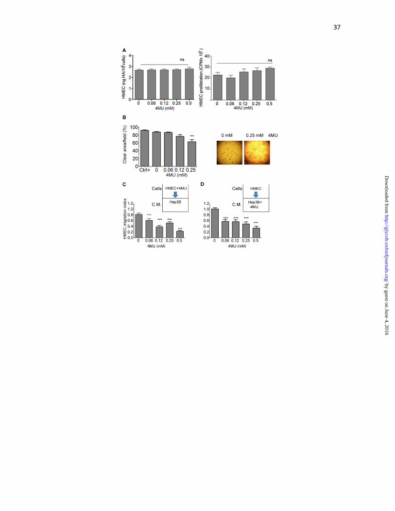
such as VEGF (Kalluri 2003). We evaluated if vascular endothelial cells (HMEC-1)
produce HA and whether 4MU may affect its biosynthesis by an ELISA-like assay. As a
result, we observed that HMEC-1 cells produced low levels of HA, which were not
influenced by 4MU (Figure 5A, left panel). In addition, HMEC-1 proliferation was not
affected by the different doses of 4MU (Figure 5A, right panel). Considering that different
factors produced by cancer cells can modulate endothelial cells behaviour, we analyzed
tube formation capability of HMEC-1 cells incubated with conditioned medium derived
from a line of human hepatocellular carcinoma cells, Hep3B. For that purpose, HMEC-1
by guest on June 4, 2016http://glycob.oxfordjournals.org/
Dow
nloaded from

10
cells were seeded on Matrigel-coated plates and incubated for 6 h with Hep3B conditioned
medium. Importantly, tube formation was significantly reduced when HMEC-1 cells were
incubated with conditioned medium obtained from 4MU-pretreated Hep3B cells (0.25 mM
4MU) (88.1±0.9 0mM vs. 63.2±5.3 0.25 mM % clear area/field, Figure 5B).
We further evaluated whether HA is able to modulate HMEC-1 cell migration. To this end,
HMEC-1 cells or Hep3B cells were pre-treated with 4MU for 18 h. In this experiment,
conditioned medium from Hep3B cells was collected and used as chemoattractant for
HMEC-1 cells migration in a Boyden chamber. When we pre-treated HMEC-1 cells with
4MU, we observed a significant reduction of their migration capability towards Hep3B
conditioned medium at the lowest dose of 4MU employed (0.06 mM) (0.82±0.04 0 mM vs.
0.60±0.06 0.06 mM; Figure 5C). Similar results on HMEC-1 migration were found when
they were incubated with conditioned medium from 4MU-pre-treated Hep3B cells
(1.00±0.04 0mM vs. 0.57±0.07 0.06 mM; Figure 5D).
Discussion
In this work, we show that oral administration of 4MU in our model of HCC with
underlying fibrosis exerts potent antitumour properties in vivo by mechanisms that involved
a reduction of proangiogenic factors such as VEGF, CXCL12 and IL-6, both in tumour and
in adjacent non-tumoural liver parenchyma.
Cancer cells and recruited stromal cells constitute the tumourigenic microenvironment and
are the driving force for progression and metastases (Bissell and Radisky 2001). HA is
major component of the extracellular matrix and is highly expressed in tumour stroma and
by guest on June 4, 2016http://glycob.oxfordjournals.org/
Dow
nloaded from

11
inflammatory processes (Kojima et al. 1975, Toole 2002, Toole 2004, Boregowda et al.
2006). It has been widely demonstrated that increased production of HA, particularly small
HA fragments, are able to induce angiogenesis process in several tumours (West et al.
1985).
We previously demonstrated in our model of HCC and fibrosis that HA inhibition by 4MU
reduces liver fibrosis and impairs tumour growth (Piccioni et al. 2012). Increased HA
deposition was observed in non-tumoural liver parenchyma from mice with fibrosis and in
tumour nodules, which were reduced after 4MU therapy (Piccioni et al. 2012). Considering
that HA might modulate angiogenesis (Savani et al. 2001), we decided to investigate
whether 4MU therapy has an effect on tumour angiogenesis as well as on tumour
microenvironment. We hypothesize that inhibition of HA synthesis by 4MU in tumour
microenvironment and adjacent non-tumoural liver parenchyma, is able to reduce some of
the key proangiogenic factors. In our work, oral administration of 4MU significantly
prolongs animal survival in comparison with controls. This effect was the result of an
important reduction in tumour volume and number of satellite nodules, with no evidence of
significant clinical and hepatic, as assessed by liver transaminases, side effects.
Data is scarce regarding the effects of 4MU on tumour angiogenesis. Lokeshwar et al.
demonstrated that 4MU administration downregulates MMP-2 and -9, and CXCR4 in
prostate cancer, and observed a reduction of microvascular density (Lokeshwar et al. 2010).
Later, García-Vila et al. observed antiangiogenic effects of 4MU evaluated in human
microvascular endothelial cells and in vivo in chorioallantoic membrane and in a zebrafish
model (García-Vilas et al. 2013). Therefore, the effects of 4MU on angiogenesis in the
context of HCC and fibrosis have been never studied.
by guest on June 4, 2016http://glycob.oxfordjournals.org/
Dow
nloaded from
12
We first investigated if tumour volume reduction was a consequence of angiogenesis
modulation by HA-synthesis inhibition achieved with the use of 4MU. For that purpose, we
used CD31 immunostaining to evaluate microvessel density in the liver. We found a
significant reduction of CD31 positive staining area in the liver parenchyma from 4MU-
treated mice in comparison with saline group; although no significant staining was found in
the tumour area. Then, the presence of VEGF, the main proangiogenic factor expressed in
angiogenesis (Corpechot et al. 2002), was quantified in serum, and in tumour and non-
tumour liver parenchyma. Importantly, we found a significant reduction in VEGF protein
level in serum from 4MU-treated mice in comparison with saline group, and the same
tendency was observed in liver sections immunostained for VEGF. These results reported
here suggest that 4MU has the ability to reduce the pathologic vasculature generated during
liver fibrosis. In line with these observations, we separately studied the effects of 4MU on
angiogenesis, in HCC tumours and in non-tumoural liver parenchyma. We found that in
both regions there was a significant reduction of VEGF and IL-6 protein expression in
4MU-treated mice when compared to untreated mice. These results are of particular
importance since IL-6 is increased in cirrhosis and is a key cytokine for HCC development
(He and Karin 2010). Moreover, several IL-6-related angiogenic signaling pathways have
been described, such as IL-6/HIF-1α in ovary cancer, and STAT-3/VEGF in breast cancer
(He and Karin 2010). Therefore, IL-6 and VEGF reduction both in liver parenchyma and in
tumour suggests an antiangiogenic role for 4MU in our tumour model.
Grunewald et al. (Grunewald et al. 2006) showed that during injury VEGF induces the
expression of CXCL12 (or SDF-1) in perivascular fibroblasts, and concomitantly this
chemokine attracts and retains CXCR4+ bone marrow-derived cells, stimulating
by guest on June 4, 2016http://glycob.oxfordjournals.org/
Dow
nloaded from
13
endothelium vessel proliferation during angiogenesis. In agreement with these
observations, CXCL12 mRNA expression was also significantly reduced by 4MU both in
tumour and in liver parenchyma, probably related with VEGF downregulation.
In order to dissect which type of cells were responsible for IL-6 modulation by 4MU
therapy, we studied IL-6 production by Hepa129 in vitro and by Kupffer cells obtained
from the liver parenchyma of mice, using different doses of 4MU. As it was expected,
Hepa129 and Kupffer cells produced IL-6, in agreement with several studies that have
demonstrated that IL-6 is overproduced in HCC (He and Karin 2010), and Kupffer cells in
the context of liver fibrosis (Bataller and Brenner 2005). Importantly, in both type of cells
we found a significant reduction of IL-6 protein levels after 4MU treatment. Interestingly,
in Hepa129 tumour cells we observed that the reduction of IL-6 obtained with 0.25 mM of
4MU was reverted by the addition of HA, indicating that the effect was specific of HA
inhibition. It has been shown that IL-6 binds selectively to several glycosaminoglycans,
including HA (Ramsden and Rider 1992, Mummery and Rider 2000, Vincent et al. 2001); it
was demonstrated in myeloma cells that HA produced could retain and concentrate IL-6
close to the site of secretion favouring an autocrine loop of activation, a feature of this
cytokine. Moreover, in a previous work it was shown that HA induces bone marrow
macrophages to secrete IL-6 (Noble et al. 1993). Therefore, we might hypothesize that HA
overproduced by cancer and stromal cells retain IL-6 secreted by them, favouring an
autocrine loop of activation with the outcome of tumour growth stimulation. It has been
observed in injured lungs and fibrosis that HA is accumulated and low-molecular HA
fragments stimulate local macrophages to produce inflammatory cytokines and growth
factors (Jiang et al. 2011). This, in turn, activates fibroblasts to produce more extracellular
by guest on June 4, 2016http://glycob.oxfordjournals.org/
Dow
nloaded from

14
matrix components. In addition, macrophages express CD44, the main receptor of HA, in
their surfaces which indicates that HA binds to these cells (Jiang et al. 2011). These data are
in line with the effect of HA-inhibition exerted by 4MU on Kupffer cells in our animal
model. However, further studies are needed to dissect the molecular mechanism that leads
to the reduction of IL-6 by 4MU in our model.
It is known that factors produced by cancer cells can induce migration of endothelial cells
promoting tumour angiogenesis (Carmeliet and Jain 2011). In vitro tube formation and
migration assays were performed to evaluate 4MU effects on endothelial cells behaviour.
We observed that 4MU pre-treatment of Hep3B cells was able to inhibit tube formation and
migration of endothelial cells HMEC-1, when used as a stimulant conditioned medium.
Although HMEC-1 cells produce low quantities of HA and 4MU does not affect HA
synthesis, we found a significant reduction in migration index when they were pre-treated
with 4MU and migrated towards Hep3B conditioned medium. This could possibly occur by
some effects of 4MU independent of HA inhibition such as the inhibition of other GAGs. In
fact, 4MU acts, at least in part, by depleting UDP-GlcUA, a precursor of other GAGs (Esko
et al. 2009). Nevertheless, more studies are needed to explain the mechanism behind the
observed effects on endothelial cells.
In conclusion, our study shows that 4MU, an HA-synthesis inhibitor, has antitumour
activity in vivo and its mechanisms of action involve a reduction of the proangiogenic
factors VEGF and CXCL12, as well as a decrease of IL-6 production in liver tumour
microenvironment; therefore, 4MU is a promising therapeutic molecule for HCC.
by guest on June 4, 2016http://glycob.oxfordjournals.org/
Dow
nloaded from
15
Materials and Methods
Cell lines.
Mouse HCC cells syngeneic with C3H/HeJ mice (Hepa129) were kindly provided by Dr. Volker
Schmitz (Bonn University, Germany). A cell line of human hepatocelullar carcinoma (Hep3B) was
purchased by ATCC, Manassas, VA 20108, USA. Human microvascular endothelial cells (HMEC-
1) were from CDC (Centers for Disease Control, Atlanta, GA, USA). Cell lines were cultured in
complete DMEM (2 mM glutamine, 100 U/ml penicillin, 100 mg/ml streptomycin) and 10% heat-
inactivated fetal bovine serum (FBS), except for Hepa129 cells that were grown in RPMI 1640
(GIBCO, Invitrogen Argentina) and supplemented with 10% FBS and 55 µM 2-Mercaptoethanol
(GIBCO).
Conditioned Medium.
To obtain cell conditioned medium (C.M.), Hep3B cells were cultured as previously described to
90% confluence and then washed with PBS and cultured with complete DMEM without FBS.
Eighteen hours later, C.M. was harvested and stored at –80ºC until use.
Mice and in vivo experiments.
Six-to-eight-week-old male C3H/HeJ mice were purchased from Comisión Nacional de Energía
Atómica, Ezeiza, Buenos Aires, Argentina. Animal care and experimental procedures were followed
according to institutional guidelines and conformed to requirement of the state authority for animal
research conduct. Fibrosis was induced by intraperitoneal (i.p.) injections of thioacetamide (TAA)
(at a dose of 200 mg/kg; Sigma-Aldrich, St. Louis, USA) thrice weekly for 42 days. On day 30
orthotopic tumours were established by subcapsular inoculation of 1.25 × 10⁵ Hepa129 cells into
the left liver lobe by laparotomy (Piccioni et al. 2012). Five days before tumour implantation, a
by guest on June 4, 2016http://glycob.oxfordjournals.org/
Dow
nloaded from
16
group of mice received 4MU (Sigma-Aldrich, St. Louis, USA) treatment by oral gavage at a dose of
400 mg/kg per day, for 20 days (4MU group). Twelve days after tumour implantation, mice were
sacrificed (Figure 1A). For serum sampling, blood from retro-orbital bleeding was collected in
heparin-free tubes. Tumour volume (mm3) was calculated by the formula π/6× larger diameter ×
(smaller diameter)2, by calipper measurement. Number of HCC satellites were also quantified. For
pathology studies, sections of liver including the tumour were taken and fixed in formalin 10% until
paraffin inclusion. For quantitative PCR and ELISA from tissue homogenates, tumours were
separated from liver parenchyma by a clear cut with a scalpel.
Liver transaminases.
Serum ALT levels were measured using an ARCHITECT® (Abbott, Buenos Aires, Argentina)
autoanalyzer.
CD31 immunostaining.
Briefly, paraffin liver sections of 5 μm-thick were rehydrated. Antigen retrieval was performed by
incubation with 20 µg/ml Proteinase K for 20 min at room temperature (RT). After washing with tap
water, slides were incubated with 3% H2O2 in distilled water for 30 min at RT to block endogenous
peroxidase, followed by avidin and biotin and blocking (Vector Laboratories, Inc. Burlingame, CA,
USA). Protein was blocked with 2.5% BSA/PBS/Tween0.1% solution for 10 min at RT. Sections
were then incubated with a rat antibody to CD31 (Abcam, MA, USA) 1:100 in PBS over night
(ON) at 4°C, and then with a biotinylated anti-rabbit secondary antibody (Vector Laboratories, Inc.
Burlingame, CA, USA) 1:100 in 0.2% BSA in PBS at RT for 1 h. Peroxidase complex (Sigma-
Aldrich, St. Louis, USA) 1:10 in PBS was used as a revealing system. The signal was detected by
by guest on June 4, 2016http://glycob.oxfordjournals.org/
Dow
nloaded from
17
0.1% diaminobenzidine (Sigma-Aldrich, St. Louis, USA), 4% glucose, 0.08% ClNH4, 5% nickel
ammonium sulfate in 0.2 M AcNa and 0.05% H2O2. Densitometric analysis was performed by
taking 10 images/slide at 400x (Nikon Eclipse E800, Nikon, Buenos Aires, Argentina) and
percentage of positive area was calculated with ImageJ software (National Institutes of Health,
Bethesda, MD).
Enzyme-linked immunosorbent assay.
Serum samples, liver homogenates or conditioned media were tested in competitive ELISA using
kits obtained from R&D Systems to quantify VEGF, following the manufacturer’s
recommendations. IL-6 levels were determined using BD OptEIA™ Set Mouse IL-6 (BD
Bioscience, CA, USA) following the manufacturer’s recommendations.
VEGF immunostaining.
Briefly, 8μm thick cryosections were fixed in acetone for 10 minutes. After washes in PBS, sections
were dehydrated in increasing alcohol passages until xylene, and rehydrated in reverse order.
Endogenous peroxidase was blocked with 3% H2O2 in methanol for 30 minutes at room
temperature. Then, endogenous avidin and biotin were blocked using Blocking Kit (Vector
Laboratories, Inc. Burlingame, CA, USA). Afterwards, proteins were blocked by incubation with
1% BSA / PBS. Primary antibody anti-VEGF (sc-147, Santa Cruz Biotechnology) 1: 100 in 1%
BSA / PBS / 0.1% Triton was incubated for 18 hours at 4 ° C. The negative control was performed
without primary antibody. After washes in PBS, biotinylated anti-rabbit secondary antibody 1: 400
was incubated for 2 h at room temperature. Revealing system was the same as for CD31
immunostaining. The counterstaining was performed incubating for 10 sec. with hematoxylin
solution (Biopur, Bs.As, Argentina).
by guest on June 4, 2016http://glycob.oxfordjournals.org/
Dow
nloaded from
18
Reverse Transcription-polymerase Chain Reaction (RT-PCR).
Total RNA from tumour, liver tissue or cells were extracted using Trizol Reagent (Sigma-Aldrich,
St. Louis, USA). Total RNA (2 µg) was reverse transcribed with 200 U of SuperScript II Reverse
Transcriptase (Invitrogen, Carlsbad, CA, USA) using 500 ng of Oligo primers. cDNAs were
subjected to real time polymerase chain reaction (qPCR) (Stratagene Mx3005p, Stratagene, CA,
USA). For qRT-PCR, the mRNA levels of VEGF, IL-6, and CXCL12 were quantified by
SYBR®Green (Invitrogen, Bs. As, Argentina), using the following primers: VEGF forward 5’-
GTGCACTGGACCCTGGCTTTA-3’ and reverse 5’- GGTCTCAATCGGACGGCAGTA-3’; IL-6
forward 5’-AGTTGCCTTCTTGGGACTGA-3’ and reverse 5’-TCCACGATTTCCCAGAGAAC-
3’; CXCL12 forward 5’-GAGAGCCACATCGCCAGAG-3’ and reverse 5’-
TTTCGGGTCAATGCACACTTG-3’. PCR amplifications were carried out using a cycle of 95ºC
for 10 minutes and 40 cycles under the following parameters: 95ºC for 30 seconds, 56ºC for 30
seconds, 72ºC for 1 minute. At the end of PCR reaction, the temperature was increased from 60ºC to
95ºC at a rate of 2ºC/min, and the fluorescence was measured every 15 seconds to construct the
melting curve. Values were normalized to levels of glyceraldehyde- 3-phosphate dehydrogenase
(GAPDH; used as housekeeping) transcript (forward 5’-CATCTCTGCCCCCTCTGCTG-3’ and
reverse 5’-GCCTGCTTCACCACCTTCTTG-3’). Data were processed by the ΔΔCt method
(Atorrasagasti et al. 2013). The relative amount of the PCR product amplified from untreated cells
was set as 1. A non-template control (NTC) was run in every assay, and all determinations were
performed as triplicates in three separated experiments.
by guest on June 4, 2016http://glycob.oxfordjournals.org/
Dow
nloaded from

19
IL-6 quantification.
Hepa129 cells (5x10⁶ cells/well) were incubated with 4MU (0, 0.06, 0.12, 0.25 and 0.5 mM) for 20
h. After washing, they were subsequently incubated with high molecular weight (HMW) HA
200µg/µl for other 20 h. Finally, supernatant was subjected to IL-6 quantification by ELISA, as
described above. Recombinant HMW-HA (native HA) 1.5–1.8x10⁶ Da (CPN spol.s.r.o, Czech
Republic) was kindly donated by Farmatrade, Argentina.
Kupffer cells isolation.
Primary culture of murine Kupffer cells from mice treated with 4MU or control group (saline) was
obtained through liver perfusion with collagenase (Sigma-Aldrich,St. Louis, USA) and subsequent
washes and separation by a density gradient with Histodenz (Sigma-Aldrich, St. Louis, USA) 30%
in PBS, as described elsewhere (Smedsrød 2012). After 10 min of incubation in a 24-well plate of
the cell suspension obtained, Kupffer cells remained adhered to plastic. Then, they were maintained
in RPMI medium supplemented with 10% FBS for 24 h, and supernatants were collected for IL-6
quantification. Cells were immunostained with a rat anti-mouse F4/80 antibody (Abcam, MA,
USA) 1: 350 in PBS for 45 min at 4°C. After 3 washes with 1%BSA/PBS, cells were incubated
with a fluorescein isothiocyanate anti-rat (Vector Laboratories, Inc.) antibody 1:100 in PBS for 45
min at 4°C. Then, cells were observed under a fluorescence microscope (Nikon Eclipse E800,
Nikon, Buenos Aires, Argentina).
HA quantification by an ELISA-like assay.
HA from cell-free supernatants were measured using a competitive ELISA-like assay as described
elsewhere (Cordo-Russo et al. 2009). Briefly, 96-well microplates (Nunc, Thermo Fisher Scientific
Inc., MA USA) were coated with 100 μg/mL of HA (CPN spol.s.r.o. Czech Republic). Then, wells
by guest on June 4, 2016http://glycob.oxfordjournals.org/
Dow
nloaded from

20
were incubated with 25 μL of sample or standard HA (0–1 μg/mL) and 0.75 μg/mL of biotinylated
HA-binding protein (bHA-BP; Calbiochem, MA, USA) at 37°C for 4 h, and next washed with
PBS–0.05% Tween-20. The bHA-BP bound to the wells was determined using an avidin– biotin
detection system (Sigma-Aldrich,St. Louis, USA). Sample concentrations were calculated from a
standard curve generated by plotting the absorbance at 490 nm against the concentration of HA
(Cordo-Russo et al. 2009). Cell-free culture supernatants were obtained from 1x10⁶ HMEC-1 cells
incubated with 4MU for 24 h.
Cell proliferation assay.
For the proliferation assay, HMEC-1 cells (2.5 × 10⁵/well) were incubated in a 24-well plate with
4MU at different concentrations (0, 0.06, 0.12, 0.25 and 0.5 mM). After 48 h, cultures were pulsed
with 5 μCi/mL [methyl 3H] thymidine for the last 6 h. Then, cells were lysed and the incorporation
of radioactivity was measured in a liquid scintillation β-counter (Beckman Coulter, CA, USA).
Tube formation assay.
Tube formation assay was performed using Matrigel (BD Bioscience, CA, USA), thawed at 4ºC to
prevent polymerization. Forty μl of Matrigel/well were seeded in a 96-well culture plate (GBO,
Frickenhausen, Germany), and allowed to polymerize at 37°C for at least 30 minutes. HMEC-1
cells (2x10⁴), FBS strarved 18 h before, were seeded in 90 µl of FBS-free DMEM, and stimulated
with 10 µl of 4MU-pretreated Hep3B conditioned media. For positive control, 100 ng/ml of human
recombinant VEGF (Calbiochem, MA, USA) was used to stimulate HMEC-1 cells. After 6 h of
incubation at 37ºC, cells were stained and fixed with Crystal Violet solution. Quantification was
performed by analyzing the cell free-area from 3 images/well at 100X (Nikon, Buenos Aires,
Argentina) with Image J software (National Institutes of Health, Bethesda, MD).
by guest on June 4, 2016http://glycob.oxfordjournals.org/
Dow
nloaded from
21
In vitro migration assay.
In vitro migration was performed using a 48-Transwell microchemotaxis Boyden Chamber unit
(Neuroprobe, Inc.). HMEC-1 (1.5×10³ cells/well) were placed in the upper chamber and DMEM or
Hep3B CM were applied to the lower chamber of the transwell unit. Chemokinesis controls were
performed placing Hep3B C.M. in the upper and lower chamber. The system was left for 4 hours at
37°C in a 5% CO2 humidified atmosphere. Cells attached to the lower side of the membrane were
fixed in 2% formaldehyde, stained with 4′,6-diamidino-2-phenylindole dihydrochloride (DAPI,
Sigma-Aldrich,St. Louis, USA) and counted using fluorescent-field microscopy at 100X. Captured
images from 3 representative visual fields were analyzed using CellProfiler software
(www.cellprofiler.com), and the mean number of cells/field ±SEM was calculated.
Ethics statement.
Animals were maintained at our Animal Resource Facilities (Facultad de Ciencias Biomédicas,
Universidad Austral) in accordance with the experimental ethical committee and the NIH guidelines
on the ethical use of animals. The ‘‘Animal Care Committee’’ from Facultad de Ciencias
Biomédicas, Universidad Austral, approved the experimental protocol.
Statistic analysis
All experiments were performed in triplicate and repeated at least three times on different
occasions. Values were expressed as the mean ± SEM. The Mann–Whitney or Kruskal–Wallis
(ANOVA) tests were used to evaluate the statistical differences between two or more than two
groups, respectively. Mice survival was analyzed by a Kaplan-Meier curve. A p-value of <0.05 was
by guest on June 4, 2016http://glycob.oxfordjournals.org/
Dow
nloaded from
22
considered as significant. Prism software (Graph Pad, San Diego, CA) was employed for the
statistical analysis.
Acknowledgements
We would like to thank Flavia V. Ferreira and Guillermo A. Gastón for their expert
technical assistance.
Funding
This work was supported by grants from Austral University (to MR: I01-12; FP: I02-12;
MG: T13-12) and from Agencia Nacional de Promoción Científica y Tecnológica
(ANPCyT) grants (LA: PICT-2007/00082; MG: PICTO 2008/00115; MG and GM: PICT
2010/2818; GM and LA: PICT 2012/1407). The funders had no role in study design, data
collection and analysis, decision to publish, or preparation of the manuscript.
Number of figures and tables: 6
Conflict of interest:
The authors declare that there are no conflicts of interest.
by guest on June 4, 2016http://glycob.oxfordjournals.org/
Dow
nloaded from
23
List of abbreviations:
4MU, 4-Methylumbelliferone
bHA-BP, biotinylated HA- binding protein
CD31 or PECAM-1, platelet endothelial cell adhesion molecule- 1
CM, conditioned medium
CXCL12, C-X-C motif chemokine 12
CXCR4, C-X-C motif receptor 4
ECM, extracellular matrix
GAG, glycosaminoglycan
HA, hyaluronic acid
HBV, Hepatitis B Virus
HCC, hepatocellular carcinoma
HCV, Hepatitis C Virus
HIF-1α, hipoxia- inducible factor-1α
HMW, high-molecular weight
HSC, hepatic stellate cell
IL-6, interleukin-6
by guest on June 4, 2016http://glycob.oxfordjournals.org/
Dow
nloaded from
24
STAT-3, signal transducer and activator of transcription 3
TAA, thioacetamide
UDP-GlcA, uridine diphosphate glucuronic acid
UGT, UDP- glycosiltransferases
VEGF, vascular endothelial growth factor
by guest on June 4, 2016http://glycob.oxfordjournals.org/
Dow
nloaded from
25
References
Atorrasagasti, C., E. Peixoto, J. B. Aquino, N. Kippes, M. Malvicini, L. Alaniz, M. Garcia,
F. Piccioni, E. J. Fiore and J. Bayo 2013. Lack of the matricellular protein SPARC (secreted
protein, acidic and rich in cysteine) attenuates liver fibrogenesis in mice. PloS one 8(2):
e54962.
Bataller, R. and D. A. Brenner 2005. Liver fibrosis. The Journal of clinical investigation
115(2): 209-218.
Bissell, M. J. and D. Radisky 2001. Putting tumours in context. Nat Rev Cancer 1(1): 46-
54.
Boregowda, R. K., H. N. Appaiah, M. Siddaiah, S. B. Kumarswamy, S. Sunila, K.
Thimmaiah, K. Mortha, B. Toole and S. d Banerjee 2006. Expression of hyaluronan in
human tumor progression. Journal of carcinogenesis 5(1): 2.
Budhu, A., M. Forgues, Q.-H. Ye, H.-L. Jia, P. He, K. A. Zanetti, U. S. Kammula, Y. Chen,
L.-X. Qin and Z.-Y. Tang 2006. Prediction of venous metastases, recurrence, and prognosis
in hepatocellular carcinoma based on a unique immune response signature of the liver
microenvironment. Cancer cell 10(2): 99-111.
Carmeliet, P. and R. K. Jain 2011. Molecular mechanisms and clinical applications of
angiogenesis. Nature 473(7347): 298-307.
Cordo-Russo, R., M. Garcia, G. Barrientos, A. Orsal, M. Viola, P. Moschansky, F. Ringel,
A. Passi, L. Alaniz and S. Hajos 2009. Murine abortion is associated with enhanced
hyaluronan expression and abnormal localization at the fetomaternal interface. Placenta
30(1): 88-95.
by guest on June 4, 2016http://glycob.oxfordjournals.org/
Dow
nloaded from
26
Corpechot, C., V. Barbu, D. Wendum, N. Kinnman, C. Rey, R. Poupon, C. Housset and O.
Rosmorduc 2002. Hypoxia‐induced VEGF and collagen I expressions are associated with
angiogenesis and fibrogenesis in experimental cirrhosis. Hepatology 35(5): 1010-1021.
Coulon, S., F. Heindryckx, A. Geerts, C. Van Steenkiste, I. Colle and H. Van Vlierberghe
2011. Angiogenesis in chronic liver disease and its complications. Liver International
31(2): 146-162.
Esko, J. D., K. Kimata and U. Lindahl 2009. Proteoglycans and sulfated
glycosaminoglycans.
García-Vilas, J. A., A. R. Quesada and M. A. n. Medina 2013. 4-Methylumbelliferone
inhibits angiogenesis in vitro and in vivo. Journal of agricultural and food chemistry
61(17): 4063-4071.
Grunewald, M., I. Avraham, Y. Dor, E. Bachar-Lustig, A. Itin, S. Yung, S. Chimenti, L.
Landsman, R. Abramovitch and E. Keshet 2006. VEGF-induced adult neovascularization:
recruitment, retention, and role of accessory cells. Cell 124(1): 175-189.
Hammerich, L. and F. Tacke 2014. interleukins in chronic liver disease: lessons learned
from experimental mouse models. Clinical and experimental gastroenterology 7: 297.
He, G. and M. Karin 2010. NF-κB and STAT3–key players in liver inflammation and
cancer. Cell research 21(1): 159-168.
Hernandez–Gea, V., S. Toffanin, S. L. Friedman and J. M. Llovet 2013. Role of the
microenvironment in the pathogenesis and treatment of hepatocellular carcinoma.
Gastroenterology 144(3): 512-527.
Hoshida, Y., A. Villanueva, M. Kobayashi, J. Peix, D. Y. Chiang, A. Camargo, S. Gupta, J.
Moore, M. J. Wrobel and J. Lerner 2008. Gene expression in fixed tissues and outcome in
hepatocellular carcinoma. New England Journal of Medicine 359(19): 1995-2004.
by guest on June 4, 2016http://glycob.oxfordjournals.org/
Dow
nloaded from

27
Itano, N., L. Zhuo and K. Kimata 2008. Impact of the hyaluronan‐rich tumor
microenvironment on cancer initiation and progression. Cancer science 99(9): 1720-1725.
Jiang, D., J. Liang and P. W. Noble 2011. Hyaluronan as an immune regulator in human
diseases. Physiological Reviews 91(1): 221-264.
Kalluri, R. 2003. Basement membranes: structure, assembly and role in tumour
angiogenesis. Nature Reviews Cancer 3(6): 422-433.
Kojima, J., N. Nakamura, M. Kanatani and K. Omori 1975. The glycosaminoglycans in
human hepatic cancer. Cancer research 35(3): 542-547.
Kultti, A., S. Pasonen-Seppänen, M. Jauhiainen, K. J. Rilla, R. Kärnä, E. Pyöriä, R. H.
Tammi and M. I. Tammi 2009. 4-Methylumbelliferone inhibits hyaluronan synthesis by
depletion of cellular UDP-glucuronic acid and downregulation of hyaluronan synthase 2
and 3. Experimental cell research 315(11): 1914-1923.
Lokeshwar, V. B., L. E. Lopez, D. Munoz, A. Chi, S. P. Shirodkar, S. D. Lokeshwar, D. O.
Escudero, N. Dhir and N. Altman 2010. Antitumor activity of hyaluronic acid synthesis
inhibitor 4-methylumbelliferone in prostate cancer cells. Cancer research 70(7): 2613-
2623.
Mummery, R. S. and C. C. Rider 2000. Characterization of the heparin-binding properties
of IL-6. The Journal of Immunology 165(10): 5671-5679.
Nagasaki, T., M. Hara, H. Nakanishi, H. Takahashi, M. Sato and H. Takeyama 2013.
Interleukin-6 released by colon cancer-associated fibroblasts is critical for tumour
angiogenesis: anti-interleukin-6 receptor antibody suppressed angiogenesis and inhibited
tumour–stroma interaction. British journal of cancer 110(2): 469-478.
Nakazawa, H., S. Yoshihara, D. Kudo, H. Morohashi, I. Kakizaki, A. Kon, K. Takagaki and
M. Sasaki 2006. 4-methylumbelliferone, a hyaluronan synthase suppressor, enhances the
by guest on June 4, 2016http://glycob.oxfordjournals.org/
Dow
nloaded from
28
anticancer activity of gemcitabine in human pancreatic cancer cells. Cancer chemotherapy
and pharmacology 57(2): 165-170.
Naugler, W. E., T. Sakurai, S. Kim, S. Maeda, K. Kim, A. M. Elsharkawy and M. Karin
2007. Gender disparity in liver cancer due to sex differences in MyD88-dependent IL-6
production. Science 317(5834): 121-124.
Noble, P. W., F. Lake, P. Henson and D. Riches 1993. Hyaluronate activation of CD44
induces insulin-like growth factor-1 expression by a tumor necrosis factor-alpha-dependent
mechanism in murine macrophages. Journal of Clinical Investigation 91(6): 2368.
Piccioni, F., M. Malvicini, M. G. Garcia, A. Rodriguez, C. Atorrasagasti, N. Kippes, I. T. P.
Buena, M. M. Rizzo, J. Bayo and J. Aquino 2012. Antitumor effects of hyaluronic acid
inhibitor 4-methylumbelliferone in an orthotopic hepatocellular carcinoma model in mice.
Glycobiology 22(3): 400-410.
Racine, R. and M. E. Mummert 2012. Hyaluronan Endocytosis: Mechanisms of Uptake and
Biological Functions.
Ramsden, L. and C. C. Rider 1992. Selective and differential binding of interleukin (IL)‐1α,
IL‐1β, IL‐2 and IL‐6 to glycosaminoglycans. European journal of immunology 22(11):
3027-3031.
Rappaport, A., P. MacPhee, M. Fisher and M. Phillips 1983. The scarring of the liver acini
(cirrhosis). Virchows Archiv A 402(2): 107-137.
Savani, R. C., G. Cao, P. M. Pooler, A. Zaman, Z. Zhou and H. M. DeLisser 2001.
Differential involvement of the hyaluronan (HA) receptors CD44 and receptor for HA-
mediated motility in endothelial cell function and angiogenesis. Journal of Biological
Chemistry 276(39): 36770-36778.
by guest on June 4, 2016http://glycob.oxfordjournals.org/
Dow
nloaded from
29
Smedsrød, B. 2012. Protocol for preparation of mouse liver Kupffer cells and liver
sinusoidal endothelial cells.
Takeda, S. and M. Aburada 1981. The choleretic mechanism of coumarin compounds and
phenolic compounds. Journal of pharmacobio-dynamics 4(9): 724-734.
Toole, B. P. 2002. Hyaluronan promotes the malignant phenotype. Glycobiology 12(3):
37R-42R.
Toole, B. P. 2004. Hyaluronan: from extracellular glue to pericellular cue. Nature Reviews
Cancer 4(7): 528-539.
Vincent, T., M. Jourdan, M.-S. Sy, B. Klein and N. Mechti 2001. Hyaluronic acid induces
survival and proliferation of human myeloma cells through an interleukin-6-mediated
pathway involving the phosphorylation of retinoblastoma protein. Journal of Biological
Chemistry 276(18): 14728-14736.
West, D. C., I. N. Hampson, F. Arnold and S. Kumar 1985. Angiogenesis induced by
degradation products of hyaluronic acid. Science 228(4705): 1324-1326.
Yoshihara, S., A. Kon, D. Kudo, H. Nakazawa, I. Kakizaki, M. Sasaki, M. Endo and K.
Takagaki 2005. A hyaluronan synthase suppressor, 4-methylumbelliferone, inhibits liver
metastasis of melanoma cells. FEBS letters 579(12): 2722-2726.
Zhu, A. X., D. G. Duda, D. V. Sahani and R. K. Jain 2011. HCC and angiogenesis: possible
targets and future directions. Nature reviews Clinical oncology 8(5): 292-301.
by guest on June 4, 2016http://glycob.oxfordjournals.org/
Dow
nloaded from
30
Figures Legends
Figure 1. Antitumor effect of oral 4MU therapy. (A) Experimental in vivo model.
Fibrosis was induced in C3H/He mice by TAA i.p. injections three times per week for 42
days. At day 30, Hepa129 cells were inoculated into the liver of fibrotic mice. Treatment
with 4MU was started 5 days before the surgery, and orally administrated every day, up to
animal sacrifice. (B) Survival of 4MU-treated mice was analyzed by a Kaplan-Meier
curve.*p<0.05 vs. saline, Log-rank test. (C) HCC tumour volume was measured with
caliper after 12 days from cell inoculation (left panel) and number of satellite nodules were
counted (right panel) *p<0.05 vs. saline, Mann-Whitney test. (D) ALT serum levels. NS=
non significant; Kruskal-Wallis test, ANOVA. Results are representative of at least 3
independent experiments (n=5-7 per group).
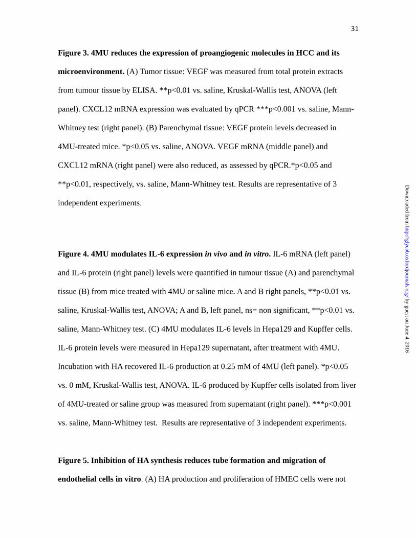
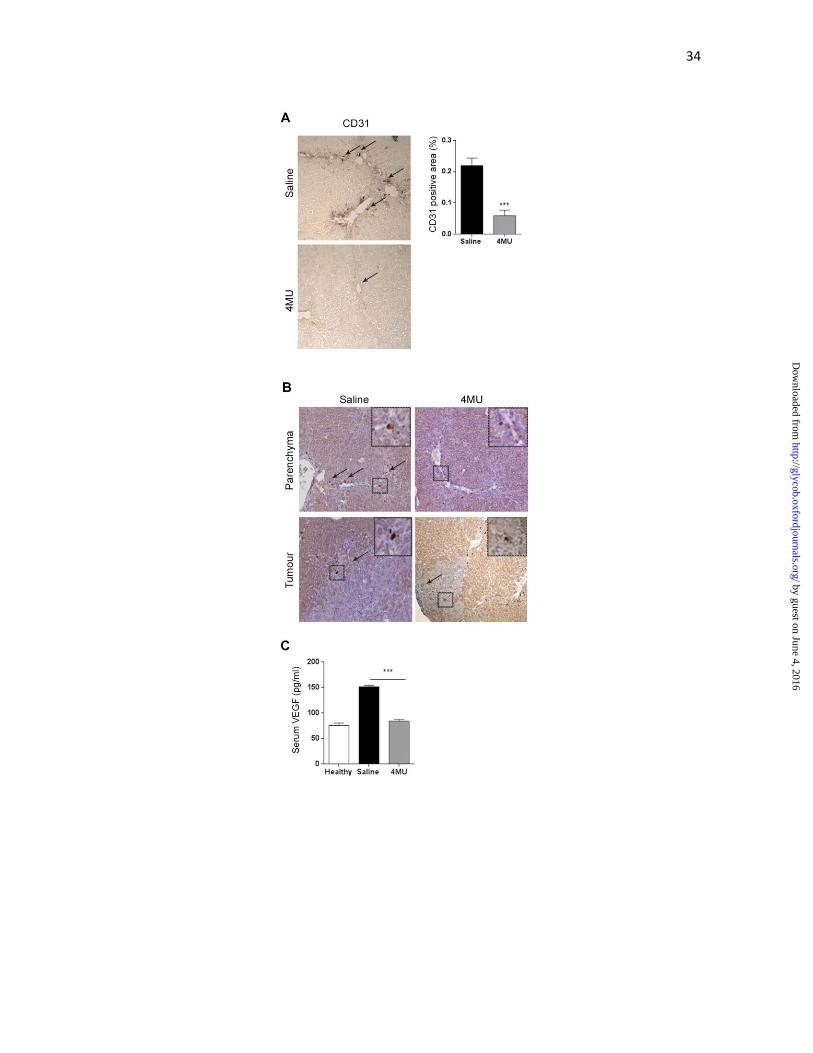
Figure 2. Angiogenesis is reduced by 4MU therapy. (A) Microvascular density in fibrous
liver was evaluated by CD31 immunohistochemistry, and quantified by densitometry
***p<0.001 vs. saline, Mann-Whitney test. Arrows indicate positive immunostaining
signal. (B) Immunostaining for VEGF in liver parenchyma and tumor zone. Positive signal
is evidenced as brown dots around veins or fibrosis bridges (arrows), or heterogeneously
distributed in tumour. Magnification 100x. (C) Systemic VEGF levels were quantified by
ELISA. ***p<0.001 vs. saline, Kruskal-Wallis test, ANOVA. Bars represent the average of
measures of each group ± SEM. Results are representative of 3 independent experiments.
by guest on June 4, 2016http://glycob.oxfordjournals.org/
Dow
nloaded from
31
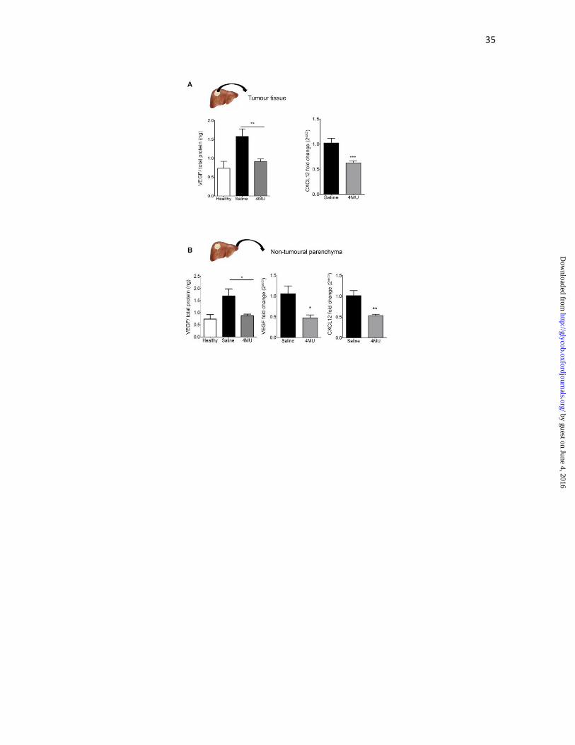
Figure 3. 4MU reduces the expression of proangiogenic molecules in HCC and its
microenvironment. (A) Tumor tissue: VEGF was measured from total protein extracts
from tumour tissue by ELISA. **p<0.01 vs. saline, Kruskal-Wallis test, ANOVA (left
panel). CXCL12 mRNA expression was evaluated by qPCR ***p<0.001 vs. saline, Mann-
Whitney test (right panel). (B) Parenchymal tissue: VEGF protein levels decreased in
4MU-treated mice. *p<0.05 vs. saline, ANOVA. VEGF mRNA (middle panel) and
CXCL12 mRNA (right panel) were also reduced, as assessed by qPCR.*p<0.05 and
**p<0.01, respectively, vs. saline, Mann-Whitney test. Results are representative of 3
independent experiments.
Figure 4. 4MU modulates IL-6 expression in vivo and in vitro. IL-6 mRNA (left panel)
and IL-6 protein (right panel) levels were quantified in tumour tissue (A) and parenchymal
tissue (B) from mice treated with 4MU or saline mice. A and B right panels, **p<0.01 vs.
saline, Kruskal-Wallis test, ANOVA; A and B, left panel, ns= non significant, **p<0.01 vs.
saline, Mann-Whitney test. (C) 4MU modulates IL-6 levels in Hepa129 and Kupffer cells.
IL-6 protein levels were measured in Hepa129 supernatant, after treatment with 4MU.
Incubation with HA recovered IL-6 production at 0.25 mM of 4MU (left panel). *p<0.05
vs. 0 mM, Kruskal-Wallis test, ANOVA. IL-6 produced by Kupffer cells isolated from liver
of 4MU-treated or saline group was measured from supernatant (right panel). ***p<0.001
vs. saline, Mann-Whitney test. Results are representative of 3 independent experiments.
Figure 5. Inhibition of HA synthesis reduces tube formation and migration of
endothelial cells in vitro. (A) HA production and proliferation of HMEC cells were not
by guest on June 4, 2016http://glycob.oxfordjournals.org/
Dow
nloaded from
32
affected by 4MU. (B) Tube formation assay was performed stimulating HMEC cells sealed
on a Matrigel coat with conditioned medium of Hep3B cells pretreated with different doses
of 4MU. Images were taken at 20X and % of clear area/field was quantified. Recombinant
VEGF was used to stimulate HMEC-1 cells in the positive control (Ctrl +). ***p<0.001 vs.
conditioned medium of Hep3B without pretreatment, Kruskal-Wallis test, ANOVA. (C)
Migration of HMECs pretreated with 4MU towards Hep3B conditioned medium (C.M.)
and (D) HMEC towards conditioned medium of Hep3B pretreated with 4MU were
significantly reduced compared with control. ***p<0.001 vs. 0 mM, Kruskal-Wallis test,
ANOVA. Results are representative of 3 independent experiments.
Figure 6. A model for the proposed mechanism of action of 4MU on HCC with
underlying fibrosis. 4MU modulates liver fibrosis by inhibition of HSC proliferation, and
its collagen and HA production (Piccioni et al.). 4MU also reduces HA produced by HCC,
and inhibits its growth by reducing the proangiogenic factors VEGF and SDF-1 both in
tumour and nontumoural liver parenchyma. The proinflammatory cytokine IL-6, a key
cytokine in HCC progression, is also inhibited by 4MU treatment. In addition, IL-6
production by Kupffer cells is also decreased by 4MU. Finally, liver endothelial cells were
reduced in liver parenchyma by 4MU therapy; migration and tube formation by endothelial
cells were also inhibited in vitro suggesting an antiangiogenic effect of 4MU.
by guest on June 4, 2016http://glycob.oxfordjournals.org/
Dow
nloaded from

33
by guest on June 4, 2016http://glycob.oxfordjournals.org/
Dow
nloaded from
34
by guest on June 4, 2016http://glycob.oxfordjournals.org/
Dow
nloaded from
35
by guest on June 4, 2016http://glycob.oxfordjournals.org/
Dow
nloaded from

36
by guest on June 4, 2016http://glycob.oxfordjournals.org/
Dow
nloaded from
37
by guest on June 4, 2016http://glycob.oxfordjournals.org/
Dow
nloaded from

38
by guest on June 4, 2016http://glycob.oxfordjournals.org/
Dow
nloaded from
Copyright © 2022 FDOKUMEN