4-Fluorophenylglycine as a Label for19F NMR Structure Analysis of Membrane-Associated Peptides
13
ChemBioChem 2003, 4, 1151 ±1163 DOI: 10.1002/cbic.200300568 ¹ 2003 Wiley-VCH Verlag GmbH & Co. KGaA, Weinheim 1151 4-Fluorophenylglycine as a Label for 19 F NMR Structure Analysis of Membrane-Associated Peptides Sergii Afonin, [a] Ralf W. Glaser, [b] Marina Berditchevskaia, [c] Parvesh Wadhwani, [a] Karl-Heinz G¸hrs, [d] Ute Mˆllmann, [e] Andrea Perner, [e] and Anne S. Ulrich* [a, c] The non-natural amino acid 4-fluorophenylglycine (4F-Phg) was incorporated into several representative membrane-associated peptides for dual purpose. The 19 F-substituted ring is directly attached to the peptide backbone, so it not only provides a well- defined label for highly sensitive 19 F NMR studies but, in addition, the D and L enantiomers of the stiff side chain may serve as reporter groups on the transient peptide conformation during the biological function. Besides peptide synthesis, which is accompanied by racemisation of 4F-Phg, we also describe separation of the epimers by HPLC and removal of trifluoroacetic acid. As a first example, 18 different analogues of the fusogenic peptide ™B18∫ were prepared and tested for induction of vesicle fusion ; the results confirmed that hydrophobic sites tolerated 4F-Phg labelling. Similar fusion activities within each pair of epimers suggest that the peptide is less structured in the fusogenic transition state than in the helical ground state. In a second example, five doubly labelled analogues of the antimicrobial peptide gramicidin S were compared by using bacterial growth inhibition assays. This cyclic b-sheet peptide could accommodate both L and D substituents on its hydrophobic face. As a third example, we tested six analogues of the antimicrobial peptide PGLa. The presence of D-4F-Phg reduced the biological activity of the peptide by interfering with its amphiphilic a-helical fold. Finally, to illustrate the numerous uses of L-4F-Phg in 19 F NMR spectroscopy, we characterised the interaction of labelled PGLa with uncharged and negatively charged membranes. Observing the signal of the free peptide in an aqueous suspension of unilamellar vesicles, we found a linear saturation behaviour that was dominated by electrostatic attraction of the cationic PGLa. Once the peptide is bound to the membrane, however, solid-state 19 F NMR spectroscopy of macroscopically oriented samples re- vealed that the charge density has virtually no further influence on the structure, alignment or mobility of the peptide. Introduction Protein ± lipid interactions are of fundamental relevance to cellular processes occurring at the plasma membrane and organelles. For example, antimicrobial peptides are used by many organisms as a first-line defence, as they are able to permeabilise the membranes of invading bacteria. [1] Fusogenic peptides represent the minimal membrane-perturbing part of various proteins that are involved in inter- and intracellular fusion processes. [2] Many different amphiphilic peptides and proteins interact permanently or reversibly with membranes, to operate as ion channels, transporters, toxins, enzymes, etc. As a functional picture of these systems is beginning to emerge, new experimental strategies are needed to study the structure and dynamics of peptides when they are bound to the lipid bilayer. Fluorescence and electron paramagnetic resonance spectros- copy are sensitive methods commonly used to characterise peptides in membranes. In many cases, however, bulky labels have to be incorporated, which may affect the peptide ± lipid interactions studied. Depending on the mode of attachment, a flexible reporter group may not reflect the behaviour of the labelled molecule that well. Nonperturbing methods, such as solid-state NMR spectroscopy with selective 2 H, 13 C or 15 N labels, on the other hand, are highly informative but are not very sensitive as they require comparatively large amounts of material. [3] Protons are, of course, the most sensitive reporters, but in view of their natural abundance it is not yet feasible to investigate membrane-bound peptides by solid-state 1 H NMR [a] Prof. Dr. A. S. Ulrich, S. Afonin, Dr. P. Wadhwani Forschungszentrum Karlsruhe, IFIA P.O.B. 3640, 76021 Karlsruhe (Germany) Fax.: ( 49) 721-608-4823 E-mail : [email protected] [b] Dr. R. W. Glaser Institute f¸r Biochemie und Biophysik Friedrich-Schiller-Universit‰t Jena Hans-Knˆll-Strasse 2, 07745 Jena (Germany) [c] Prof. Dr. A. S. Ulrich, Dr. M. Berditchevskaia Institut f¸r Organische Chemie, Universit‰t Karlsruhe Fritz-Haber-Weg 6, 76131 Karlsruhe (Germany) [d] Dr. K.-H. G¸hrs Institut f¸r Molekulare Biotechnologie Beutenbergstrasse 11, 07745 Jena (Germany) [e] Dr. U. Mˆllmann, A. Perner Hans-Knˆll-Institut f¸r Naturstoff-Forschung e.V. Beutenbergstrasse 11a, 07745 Jena (Germany)
Transcript of 4-Fluorophenylglycine as a Label for19F NMR Structure Analysis of Membrane-Associated Peptides
ChemBioChem 2003, 4, 1151 ± 1163 DOI: 10.1002/cbic.200300568 ¹ 2003 Wiley-VCH Verlag GmbH&Co. KGaA, Weinheim 1151
4-Fluorophenylglycine as a Label for 19F NMRStructure Analysis of Membrane-AssociatedPeptidesSergii Afonin,[a] Ralf W. Glaser,[b] Marina Berditchevskaia,[c] Parvesh Wadhwani,[a]
Karl-Heinz G¸hrs,[d] Ute Mˆllmann,[e] Andrea Perner,[e] and Anne S. Ulrich*[a, c]
The non-natural amino acid 4-fluorophenylglycine (4F-Phg) wasincorporated into several representative membrane-associatedpeptides for dual purpose. The 19F-substituted ring is directlyattached to the peptide backbone, so it not only provides a well-defined label for highly sensitive 19F NMR studies but, in addition,the D and L enantiomers of the stiff side chain may serve as reportergroups on the transient peptide conformation during the biologicalfunction. Besides peptide synthesis, which is accompanied byracemisation of 4F-Phg, we also describe separation of the epimersby HPLC and removal of trifluoroacetic acid. As a first example, 18different analogues of the fusogenic peptide ™B18∫ were preparedand tested for induction of vesicle fusion; the results confirmed thathydrophobic sites tolerated 4F-Phg labelling. Similar fusionactivities within each pair of epimers suggest that the peptide isless structured in the fusogenic transition state than in the helicalground state. In a second example, five doubly labelled analogues
of the antimicrobial peptide gramicidin S were compared by usingbacterial growth inhibition assays. This cyclic �-sheet peptide couldaccommodate both L and D substituents on its hydrophobic face. Asa third example, we tested six analogues of the antimicrobialpeptide PGLa. The presence of D-4F-Phg reduced the biologicalactivity of the peptide by interfering with its amphiphilic �-helicalfold. Finally, to illustrate the numerous uses of L-4F-Phg in 19F NMRspectroscopy, we characterised the interaction of labelled PGLawith uncharged and negatively charged membranes. Observingthe signal of the free peptide in an aqueous suspension ofunilamellar vesicles, we found a linear saturation behaviour thatwas dominated by electrostatic attraction of the cationic PGLa.Once the peptide is bound to the membrane, however, solid-state19F NMR spectroscopy of macroscopically oriented samples re-vealed that the charge density has virtually no further influence onthe structure, alignment or mobility of the peptide.
Introduction
Protein ± lipid interactions are of fundamental relevance tocellular processes occurring at the plasma membrane andorganelles. For example, antimicrobial peptides are used bymany organisms as a first-line defence, as they are able topermeabilise the membranes of invading bacteria.[1] Fusogenicpeptides represent the minimal membrane-perturbing part ofvarious proteins that are involved in inter- and intracellularfusion processes.[2] Many different amphiphilic peptides andproteins interact permanently or reversibly with membranes, tooperate as ion channels, transporters, toxins, enzymes, etc. As afunctional picture of these systems is beginning to emerge, newexperimental strategies are needed to study the structure anddynamics of peptides when they are bound to the lipid bilayer.Fluorescence and electron paramagnetic resonance spectros-
copy are sensitive methods commonly used to characterisepeptides in membranes. In many cases, however, bulky labelshave to be incorporated, which may affect the peptide ± lipidinteractions studied. Depending on the mode of attachment, aflexible reporter group may not reflect the behaviour of thelabelled molecule that well. Nonperturbing methods, such assolid-state NMR spectroscopy with selective 2H, 13C or 15N labels,
on the other hand, are highly informative but are not verysensitive as they require comparatively large amounts ofmaterial.[3] Protons are, of course, the most sensitive reporters,but in view of their natural abundance it is not yet feasible toinvestigate membrane-bound peptides by solid-state 1H NMR
[a] Prof. Dr. A. S. Ulrich, S. Afonin, Dr. P. WadhwaniForschungszentrum Karlsruhe, IFIAP.O.B. 3640, 76021 Karlsruhe (Germany)Fax. : (�49)721-608-4823E-mail : [email protected]
[b] Dr. R. W. GlaserInstitute f¸r Biochemie und BiophysikFriedrich-Schiller-Universit‰t JenaHans-Knˆll-Strasse 2, 07745 Jena (Germany)
[c] Prof. Dr. A. S. Ulrich, Dr. M. BerditchevskaiaInstitut f¸r Organische Chemie, Universit‰t KarlsruheFritz-Haber-Weg 6, 76131 Karlsruhe (Germany)
[d] Dr. K.-H. G¸hrsInstitut f¸r Molekulare BiotechnologieBeutenbergstrasse 11, 07745 Jena (Germany)
[e] Dr. U. Mˆllmann, A. PernerHans-Knˆll-Institut f¸r Naturstoff-Forschung e.V.Beutenbergstrasse 11a, 07745 Jena (Germany)
A. S. Ulrich et al.
1152 ¹ 2003 Wiley-VCH Verlag GmbH&Co. KGaA, Weinheim www.chembiochem.org ChemBioChem 2003, 4, 1151 ± 1163
spectroscopy. Hence, a good compromise for studying peptide ±lipid interactions is offered by 19F NMR spectroscopy, for whichsuitable fluorine labels need to be selectively incorporated. Thesensitivity of the 19F nucleus is nearly as high as that of theproton, and there is no natural background. The strong dipolarcouplings of the 19F nucleus provide intra- or intermoleculardistances in solids of up to 12 ä, and the large chemical shiftanisotropy is very useful for analysing molecular orientations.[4]
When a single proton is replaced by a 19F atom, the structuralimplications are expected to be small.[5] Fluorine NMR labelshave been successfully used to study peptides and proteins bothin solution and in the membrane-bound state.[6] 19F NMRspectroscopy has also been applied to characterise the bindingof fluorine-containing drugs and anaesthetics to lipid mem-branes.[4a, 7]
The non-natural amino acid 4-fluorophenylglycine (4F-Phg) isexplored here as a label for 19F NMR studies of membrane-associated peptides. The advantage of 4F-Phg over any of thecommonly used fluorinated amino acids lies in the stiffconnection of the para-fluoro substituent to the peptidebackbone. In all natural amino acids with a proton replaced bya fluorine atom (such as Phe, Trp, Tyr or Leu) the position of thelabel with respect to the peptide backbone would depend onthe side-chain conformation, which is not usually known inmembrane peptides. The special side-chain architecture of 4F-Phg recently enabled us to deduce the backbone conformationof membrane-bound gramicidin S from the intramolecular 19F ±19F dipolar coupling between two 4F-Phg reporter groups.[6b] Thesame study also enabled us to determine the alignment andmobility of the entire peptide in the lipid bilayer. Even though4F-Phg is not a common building block in biology, phenylglycineand its derivatives have found numerous applications ascomponents of semisynthetic antibiotics, artificial sweeteners,enzyme inhibitors and immobile phases for chiral chromatog-raphy, amongst others.[8] Phenylglycine is even part of somenatural antibiotics, like pristinamycin I and virginiamycin S.[9] Thearomatic � branch in phenylglycine may favour a differentpreferential conformation in the protein backbone compared toany standard amino acid. Computer modelling studies haveshown that its global energy minimum corresponds to a nearlyextended �-strand conformation, while a right-handed � helix isless stable by about 1.3 kcalmol�1.[10] Nevertheless, the availableconformational space is almost as large as for alanine, and hence,L-4F-Phg promises to be a relatively nonperturbing side chain.When the D enantiomer is incorporated into a protein, on theother hand, we expect that any intrinsic steric effects will beenhanced due to the stiffness of the bulky side chain.[11]
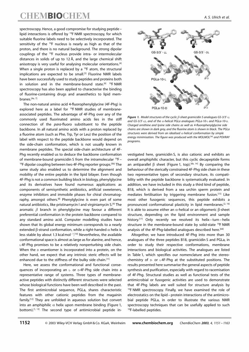
Here, we assess the conformational and functional conse-quences of incorporating an L- or D-4F-Phg side chain into arepresentative range of systems. Three types of membrane-active peptides with distinctly different structures were selectedwhose biological functions have been well described in the past.The first antimicrobial sequence, PGLa, shares characteristicfeatures with other cationic peptides from the magaininfamily.[12] They are unfolded in aqueous solution but convertinto an amphiphilic � helix upon membrane binding (Figure 1,bottom).[1, 13] The second type of antimicrobial peptide in-
Figure 1. Model structures of the cyclic �-sheet gramicidin S analogues GS-3/3�-LL
and GS-3/3�-LD, and of the �-helical PGLa analogues PGLa-10-L and PGLa-10-D.Charged ornithine and lysine side chains as well as 4-fluorophenylglycine sidechains are shown in dark grey, and the fluorine atom is shown in black. The PGLastructures were derived from an idealised �-helical conformation by simpleenergy minimisation. The figure was produced with the MOLMOL[36] and POVRAYprograms.
vestigated here, gramicidin S, is also cationic and exhibits anoverall amphiphilic character, but this cyclic decapeptide formsan antiparallel � sheet (Figure 1, top).[6b, 14] By comparing thebehaviour of the sterically constrained 4F-Phg side chain in thesetwo representative types of secondary structure, its compati-bility with the peptide backbone is systematically evaluated. Inaddition, we have included in this study a third kind of peptide,B18, which is derived from a sea urchin sperm protein andmediates fertilisation by triggering membrane fusion.[15] Likemost other fusogenic sequences, this peptide exhibits apronounced conformational plasticity in lipid membranes.[2, 16]
It is able to assume either an �-helical or an oligomeric �-sheetstructure, depending on the lipid environment and samplehistory.[17] Only recently we resolved its helix ± turn ±helixstructure in the membrane-bound state by solid-state 19F NMRanalysis of the 4F-Phg-labelled analogues described here.[6d]
Altogether, we have introduced 4F-Phg into more than 30analogues of the three peptides B18, gramicidin S and PGLa, inorder to study their respective conformations, membraneinteractions and biological activities. The analogues are listedin Table 1, which specifies our nomenclature and the stereo-chemistry of D- or L-4F-Phg at the substituted positions. Theresults presented here summarise the general aspects of peptidesynthesis and purification, especially with regard to racemisationof 4F-Phg. Structural studies as well as functional tests of theantimicrobial or fusogenic activities are used to demonstratethat 4F-Phg labels are well suited for structure analysis by19F NMR spectroscopy. Finally, we have examined the role ofelectrostatics on the lipid ±protein interactions of the antimicro-bial peptide PGLa, in order to illustrate the various NMRspectroscopy techniques that can be usefully applied to such19F-labelled peptides.
4-Fluorophenylglycine Label for Membrane-Associated Peptides
ChemBioChem 2003, 4, 1151 ± 1163 www.chembiochem.org ¹ 2003 Wiley-VCH Verlag GmbH&Co. KGaA, Weinheim 1153
Results and Discussion
Racemisation of 4-fluorophenylglycine
It is well known that the aromatic side chain of phenylglycinemakes the �-carbon atom of this amino acid sufficiently acidic tobe readily deprotonated by any weak base used during9-fluorenylmethoxycarbonyl (Fmoc) solid-phase peptide syn-thesis. Besides favourable electron delocalisation, in 4F-Phg the�-carbanion species is further stabilised by the presence of theelectronegative para-fluoro substituent. Racemisation via atrigonal �-carbon intermediate is thus virtually inevitable whenL-4F-Phg is introduced into peptides by standard Fmoc chem-istry. We tested several modifications of the protocol, as theextent of racemisation is known to be influenced by the tertiarybase employed for coupling.[18] When 2,4,6-collidine was used incombination with 2-(1H-benzotriazole-1-yl)-1,1,3,3-tetramethyl-uronium hexafluorophosphate (HBTU) to couple both 4F-Phgand the subsequent amino acid, we managed to suppressracemisation to about 15% by lowering the temperature to 0 �Cwith a reaction time of 15 minutes. However, the couplingefficiency was significantly reduced to about 60 ±70%, andhence, the overall yield of the all-L peptide was not increased. Wealso tested tetramethylfluoroformamidinium hexafluorophos-phate (TFFH),[18, 19] bis-(tetramethylenefluoroformamidinium)hexafluorophosphate (BTTFH)[19] and cyanuric fluoride[20] fortheir ability to incorporate 4F-Phg into test peptides withoutracemisation. Out of the many peptide-coupling reagentsdescribed in the literature, we selected these three acid fluorides
because of their long shelf-life and rapid acting ability. In arelated project, TFFH in the presence of collidine providedoptimal conditions for incorporating 3-fluoroalanine (whicheasily eliminates HF) into a test peptide. With 4F-Phg, however,we were unable to achieve a satisfactory suppression ofracemisation.Since racemisation could not be avoided, chromatographic
resolution of the two epimeric products containing L- and D-4F-Phg was crucial. Fortunately, for all of our peptides synthesisedso far the retention times during reversed-phase HPLC weresufficiently different to allow separation and purification of thepeptides in milligram amounts. Therefore, we used the inex-pensive racemic mixture of D/L-4F-Phg for peptide synthesis anddid not pay any further attention to racemisation. In fact, the™unwanted∫ epimer that is obtained as a byproduct (that is, thepeptide containing D-4F-Phg) proved to be highly useful forcomparative structural and functional studies, as discussedbelow. There was no general rule with respect to the order inwhich the epimers eluted from the reversed-phase HPLCcolumn.[11a, 21] In all PGLa samples and in B18-110 the D-4F-Phgepimer eluted first, while in all other B18 samples the L-4F-Phgepimer eluted first. In all gramicidin S analogues the order wasDD, DL, LL, according to increasing amphiphilicity.The configuration of the 4F-Phg in each pair of epimers was
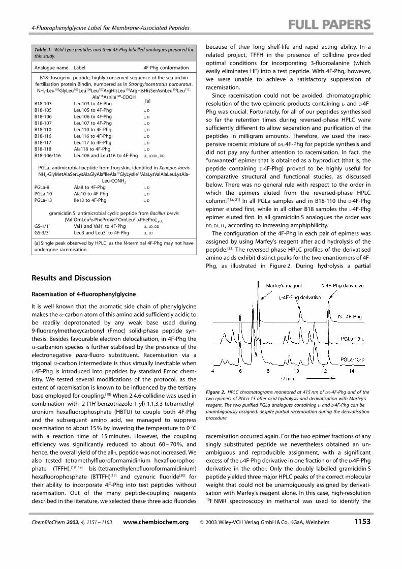
assigned by using Marfey's reagent after acid hydrolysis of thepeptide.[22] The reversed-phase HPLC profiles of the derivatisedamino acids exhibit distinct peaks for the two enantiomers of 4F-Phg, as illustrated in Figure 2. During hydrolysis a partial
Figure 2. HPLC chromatograms monitored at 415 nm of D/L-4F-Phg and of thetwo epimers of PGLa-13 after acid hydrolysis and derivatisation with Marfey'sreagent. The two purified PGLa analogues containing L- and D-4F-Phg can beunambiguously assigned, despite partial racemisation during the derivatisationprocedure.
racemisation occurred again. For the two epimer fractions of anysingly substituted peptide we nevertheless obtained an un-ambiguous and reproducible assignment, with a significantexcess of the L-4F-Phg derivative in one fraction or of the D-4F-Phgderivative in the other. Only the doubly labelled gramicidin Speptide yielded three major HPLC peaks of the correct molecularweight that could not be unambiguously assigned by derivati-sation with Marfey's reagent alone. In this case, high-resolution19F NMR spectroscopy in methanol was used to identify the
Table 1. Wild-type peptides and their 4F-Phg-labelled analogues prepared forthis study.
Analogue name Label 4F-Phg conformation
B18: fusogenic peptide, highly conserved sequence of the sea urchinfertilisation protein Bindin, numbered as in Strongylocentrotus purpuratus.NH2-Leu103GlyLeu105Leu106Leu107ArgHisLeu110ArgHisHisSerAsnLeu116Leu117-
Ala118AsnIle120-COOHB18-103 Leu103 to 4F-Phg L
[a]
B18-105 Leu105 to 4F-Phg L, D
B18-106 Leu106 to 4F-Phg L, D
B18-107 Leu107 to 4F-Phg L, D
B18-110 Leu110 to 4F-Phg L, D
B18-116 Leu116 to 4F-Phg L, D
B18-117 Leu117 to 4F-Phg L, D
B18-118 Ala118 to 4F-Phg L, D
B18-106/116 Leu106 and Leu116 to 4F-Phg LL, LD/DL, DD
PGLa: antimicrobial peptide from frog skin, identified in Xenopus laevisNH2-GlyMetAlaSerLysAlaGlyAla8IleAla10GlyLysIle13AlaLysValAlaLeuLysAla-
Leu-CONH2
PGLa-8 Ala8 to 4F-Phg L, D
PGLa-10 Ala10 to 4F-Phg L, D
PGLa-13 Ile13 to 4F-Phg L, D
gramicidin S: antimicrobial cyclic peptide from Bacillus brevis[Val1OrnLeu3D-PheProVal1�OrnLeu3�D-PhePro]cyclo
GS-1/1� Val1 and Val1� to 4F-Phg LL, LD, DD
GS-3/3� Leu3 and Leu3� to 4F-Phg LL, LD
[a] Single peak observed by HPLC, as the N-terminal 4F-Phg may not haveundergone racemisation.
A. S. Ulrich et al.
1154 ¹ 2003 Wiley-VCH Verlag GmbH&Co. KGaA, Weinheim www.chembiochem.org ChemBioChem 2003, 4, 1151 ± 1163
asymmetric DL stereoisomer by its two distinct NMR signals, incontrast to the symmetric LL and DD stereoisomers, which gave asingle NMR signal each.Once the epimerically pure peptides had been obtained, no
further racemisation could be detected under standard con-ditions of sample preparation, storage and NMR measurements.We reextracted four PGLa analogues and one gramicidin Sanalogue from reconstituted membrane samples, which hadbeen incubated for 2 ± 8 days at 48 �C and had been exposedduring NMR experiments to temperatures of 15 ± 55 �C for up to4 days. None of the peptides contained any significant amountof the unwanted epimer. Even after storage for a year at 8 �C wedetected no chemical degradation for GS-3/3� in 1,2-dimyristoyl-sn-glycero-3-phosphocholine (DMPC). (No bacterial decay wasdetected either as the peptide itself is an antibiotic.)
Removal of trifluoroacetic acid from the peptides
Trifluoroacetic acid (TFA) is commonly used to cleave peptidesfrom the resin and to enhance resolution in reversed-phaseHPLC. In synthetic peptides from various sources we alwaysdetected significant amounts of TFA, in up to 5-fold molarexcess. TFA gives rise to a strong signal in 19F NMR spectra ataround ���75 ppm, which overlaps with the downfield edgeof the static chemical-shift anisotropy (CSA) tensor of the4F-Phg label (about �11��62 ppm, �22��125 ppm, �33��154 ppm).[6b,d] This becomes less of a problem when the CSAis narrowed down by fast motional averaging of the wholepeptide in the bilayer or of the 4F-Phg side chain around itsC��C� bond. Nonetheless, we were further concerned withremoving TFA from the samples, because we noticed severalsigns of tight binding between TFA and the peptides. One ofthese signs was the difficulty in removing TFA, as describedbelow, and another indication was the difference in the 19F NMRsignal of TFA in the absence and presence of peptide. Briefly, theTFA signal in macroscopically oriented membranes in the fluidphase is a triplet, resulting from the 19F dipolar interactionswithin the aligned CF3 group.[4a] We prepared TFA-containingsamples in DMPC/1,2-dimyristoyl-sn-glycero-3-[phospho-rac-(1-glycerol)] (DMPG) without any peptide. Their spectra showedcontributions from several triplets with variable intensities,depending on the temperature, charge and hydration of themembrane. Under typical conditions, with the sample normalaligned, parallel to the static magnetic field, the chemical shiftwas between �69 and �73 ppm, and the dipolar coupling was800 ±2000 Hz. TFA also gave rise to a triplet in the same range inthe presence of peptide, but with significantly different couplingvalues and chemical shifts that depended on the type of peptide.Interestingly, when added to DMPC in the gel phase, TFA gave asingle isotropic signal at ���75 ppm; hence, we conclude thatTFA gets expelled from the membrane when the acyl chainsrigidify.Since the strong ion-pairing effect of TFA may modify the
interaction of amphiphilic peptides with the membrane, we tookcare to replace TFA by less strongly bound anions such as Cl�,even for the 1H NMR and circular dichroism experiments whereTFA is not usually considered to be a problem. The same
precautions are essential for FT-IR measurements, as the COO�
stretching vibration of TFA overlaps with the amide I band of thepeptides.[23] Several methods are commonly used to removeresidual TFA: 1) purification over ion-exchange resins, 2) vacuumdrying over P2O5 or 3) lyophilisation from solutions of othervolatile acids. We found that TFA could be removed on anamberlite ion-exchange column, but the method suffered fromincomplete recovery of peptide. Vacuum drying over P2O5
removed excess free TFA but not the TFA� counterions boundto the cationic peptides. Repeated lyophilisation from dilute HClor acetic acid solutions reduced the content of TFA, but it wasstill detectable after 3 cycles (each with a 100-fold molar excessof acetic acid over peptide). Therefore, we optimised thereversed-phase HPLC peptide purification protocol by usingsolvents with different ion-pairing agents, like hydrochloric,formic or phosphoric acid. We recommend this as the mostefficient technique to remove TFA. With 5 mM HCl in the aqueousphase of the HPLC gradient, a peak separation is achievedcomparable to 0.1% TFA. Note that HCl must not be added tothe organic phase because mixtures of HCl with acetonitrile ormethanol are very corrosive. Special precautions should also betaken when lyophilising the HPLC fractions to prevent con-densation of HCl in the pump. (See Experimental Section fordetails.)
UV absorption of the aromatic 4F-Phg side chain
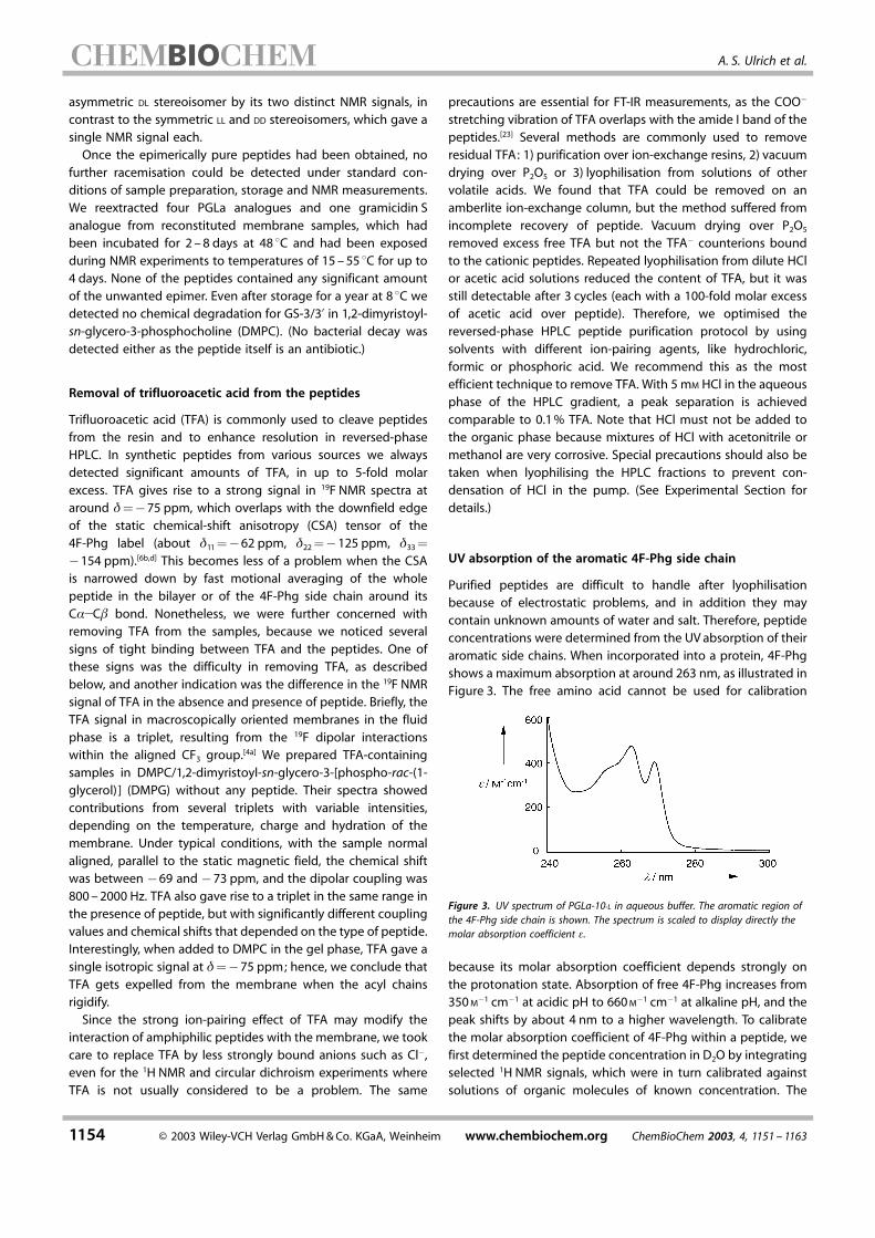
Purified peptides are difficult to handle after lyophilisationbecause of electrostatic problems, and in addition they maycontain unknown amounts of water and salt. Therefore, peptideconcentrations were determined from the UVabsorption of theiraromatic side chains. When incorporated into a protein, 4F-Phgshows a maximum absorption at around 263 nm, as illustrated inFigure 3. The free amino acid cannot be used for calibration
Figure 3. UV spectrum of PGLa-10-L in aqueous buffer. The aromatic region ofthe 4F-Phg side chain is shown. The spectrum is scaled to display directly themolar absorption coefficient �.
because its molar absorption coefficient depends strongly onthe protonation state. Absorption of free 4F-Phg increases from350M�1 cm�1 at acidic pH to 660M�1 cm�1 at alkaline pH, and thepeak shifts by about 4 nm to a higher wavelength. To calibratethe molar absorption coefficient of 4F-Phg within a peptide, wefirst determined the peptide concentration in D2O by integratingselected 1H NMR signals, which were in turn calibrated againstsolutions of organic molecules of known concentration. The
4-Fluorophenylglycine Label for Membrane-Associated Peptides
ChemBioChem 2003, 4, 1151 ± 1163 www.chembiochem.org ¹ 2003 Wiley-VCH Verlag GmbH&Co. KGaA, Weinheim 1155
molar absorption coefficient of 4F-Phg in the peptides at 263 nmwas thus determined to be 473� 13M�1 cm�1. There were nosignificant differences between the absorption coefficients indifferent types of peptides or between the analogues containing4F-Phg in different positions.
The effect of 4-fluorophenylglycine on helical structure
To evaluate the suitability of the 4F-Phg side chain as a label tostudy the orientation and dynamics of peptides in a membraneenvironment we checked whether the replacement of a hydro-phobic amino acid with 4F-Phg changed the structure or thefunction of the peptides. The secondary structure of the PGLaanalogues was studied by circular dichroism (CD) spectroscopyin the presence of lipid vesicles or 30% trifluoroethanol (TFE) andwas compared with wild-type PGLa (data not shown). The CDline shapes of all these peptides were very similar and showed ahigh content of �-helical structure. The D-4F-Phg analoguesexhibited, on average, a signal that was about 10% less negativeat 222 nm than the signals of those analogues labelled with L-4F-Phg, a result which indicates a slightly lower degree of � helix.The CD measurements are not sensitive enough to detect
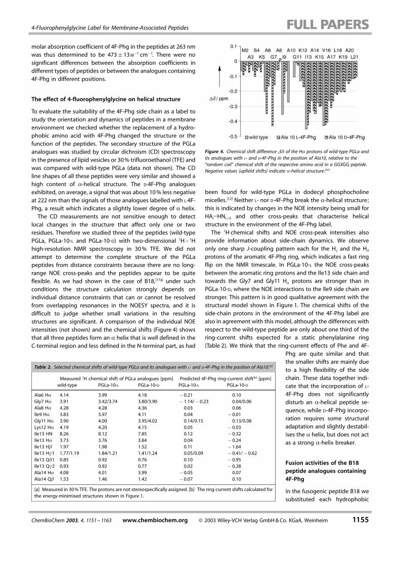
local changes in the structure that affect only one or tworesidues. Therefore we studied three of the peptides (wild-typePGLa, PGLa-10-L and PGLa-10-D) with two-dimensional 1H ± 1Hhigh-resolution NMR spectroscopy in 30% TFE. We did notattempt to determine the complete structure of the PGLapeptides from distance constraints because there are no long-range NOE cross-peaks and the peptides appear to be quiteflexible. As we had shown in the case of B18,[17a] under suchconditions the structure calculation strongly depends onindividual distance constraints that can or cannot be resolvedfrom overlapping resonances in the NOESY spectra, and it isdifficult to judge whether small variations in the resultingstructures are significant. A comparison of the individual NOEintensities (not shown) and the chemical shifts (Figure 4) showsthat all three peptides form an � helix that is well defined in theC-terminal region and less defined in the N-terminal part, as had
Figure 4. Chemical shift difference �� of the H� protons of wild-type PGLa andits analogues with L- and D-4F-Phg in the position of Ala10, relative to the™random coil∫ chemical shift of the respective amino acid in a GGXGG peptide.Negative values (upfield shifts) indicate �-helical structure.[41]
been found for wild-type PGLa in dodecyl phosphocholinemicelles.[12] Neither L- nor D-4F-Phg break the �-helical structure;this is indicated by changes in the NOE intensity being small forHAi�HNi�3 and other cross-peaks that characterise helicalstructure in the environment of the 4F-Phg label.The 1H chemical shifts and NOE cross-peak intensities also
provide information about side-chain dynamics. We observeonly one sharp J-coupling pattern each for the H� and the H�
protons of the aromatic 4F-Phg ring, which indicates a fast ringflip on the NMR timescale. In PGLa-10-L the NOE cross-peaksbetween the aromatic ring protons and the Ile13 side chain andtowards the Gly7 and Gly11 H� protons are stronger than inPGLa-10-D, where the NOE interactions to the Ile9 side chain arestronger. This pattern is in good qualitative agreement with thestructural model shown in Figure 1. The chemical shifts of theside-chain protons in the environment of the 4F-Phg label arealso in agreement with this model, although the differences withrespect to the wild-type peptide are only about one third of thering-current shifts expected for a static phenylalanine ring(Table 2). We think that the ring-current effects of Phe and 4F-
Phg are quite similar and thatthe smaller shifts are mainly dueto a high flexibility of the sidechain. These data together indi-cate that the incorporation of L-4F-Phg does not significantlydisturb an �-helical peptide se-quence, while D-4F-Phg incorpo-ration requires some structuraladaptation and slightly destabil-ises the � helix, but does not actas a strong �-helix breaker.
Fusion activities of the B18peptide analogues containing4F-Phg
In the fusogenic peptide B18 wesubstituted each hydrophobic
Table 2. Selected chemical shifts of wild-type PGLa and its analogues with L- and D-4F-Phg in the position of Ala10.[a]
Measured 1H chemical shift of PGLa analogues [ppm] Predicted 4F-Phg ring-current shift[b] [ppm]wild-type PGLa-10-L PGLa-10-D PGLa-10-L PGLa-10-D
Ala6 H� 4.14 3.99 4.18 �0.21 0.10Gly7 H� 3.91 3.42/3.74 3.80/3.90 �1.14/� 0.23 0.04/0.06Ala8 H� 4.28 4.28 4.36 0.03 0.06Ile9 H� 3.83 3.97 4.11 0.04 �0.01Gly11 H� 3.90 4.00 3.95/4.02 0.14/0.15 0.13/0.08Lys12 H� 4.19 4.20 4.15 0.05 �0.03Ile13 HN 8.26 8.12 7.85 0.12 �0.32Ile13 H� 3.73 3.76 3.84 0.04 �0.24Ile13 H� 1.97 1.98 1.52 0.11 �1.64Ile13 H�1 1.77/1.19 1.84/1.21 1.41/1.24 0.05/0.09 �0.41/� 0.62Ile13 Q�1 0.85 0.92 0.76 0.10 �0.95Ile13 Q�2 0.93 0.92 0.77 0.02 �0.28Ala14 H� 4.08 4.01 3.99 �0.05 0.07Ala14 Q� 1.53 1.46 1.42 �0.07 0.10
[a] Measured in 30% TFE. The protons are not stereospecifically assigned. [b] The ring-current shifts calculated forthe energy-minimised structures shown in Figure 1.
A. S. Ulrich et al.
1156 ¹ 2003 Wiley-VCH Verlag GmbH&Co. KGaA, Weinheim www.chembiochem.org ChemBioChem 2003, 4, 1151 ± 1163
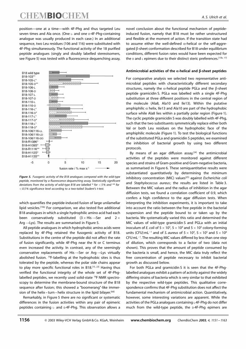
position–one at a time–with 4F-Phg and thus targeted Leuseven times and Ala once. (One L- and one D-4F-Phg-containinganalogue was usually produced in each case.) In an additionalsequence, two Leu residues (106 and 116) were substituted with4F-Phg simultaneously. The functional activity of the 18 purifiedpeptide analogues (singly and doubly labelled stereoisomers,see Figure 5) was tested with a fluorescence dequenching assay,
Figure 5. Fusogenic activity of the B18 analogues compared with the wild-typepeptide, monitored by a fluorescence dequenching assay. Statistically significantdeviations from the activity of wild-type B18 are labelled * for �5% and ** for�0.5% significance level according to a two-tailed Student's t-test.
which quantifies the peptide-induced fusion of large unilamellarlipid vesicles.[15b] For comparison, we also tested five additionalB18 analogues in which a single hydrophilic amino acid had eachbeen conservatively substituted (3�His�Ser and 2�Arg�Lys). The results are summarised in Figure 5.All peptide analogues in which hydrophobic amino acids were
replaced by 4F-Phg retained the fusogenic activity of B18.Substitutions in the centre of the peptide did not affect the rateof fusion significantly, while 4F-Phg near the N or C terminuseven increased the activity. In contrast, any of the seeminglyconservative replacements of His�Ser or Arg�Lys virtuallyabolished fusion. 19F-labelling at the hydrophobic sites is thustolerated by the peptide, whereas the polar side chains appearto play more specific functional roles in B18.[15, 23] Having thusverified the functional integrity of the whole set of 4F-Phg-labelled peptides, we recently used solid-state 19F NMR spectro-scopy to determine the membrane-bound structure of the B18sequence after fusion; this showed a ™boomerang∫-like immer-sion of the helix ± turn ±helix structure in the lipid bilayer.[6d]
Remarkably, in Figure 5 there are no significant or systematicdifferences in the fusion activities within any pair of epimericpeptides containing L- and D-4F-Phg. This observation allows a
novel conclusion about the functional mechanism of peptide-induced fusion, namely that B18 must be rather unstructuredand flexible at the moment of action. If the transition state hadto assume either the well-defined �-helical or the self-aggre-gated �-sheet conformation described for B18 under equilibriumconditions, different fusion rates would have been expected forthe D and L epimers due to their distinct steric preferences.[15b, 17]
Antimicrobial activities of the �-helical and �-sheet peptides
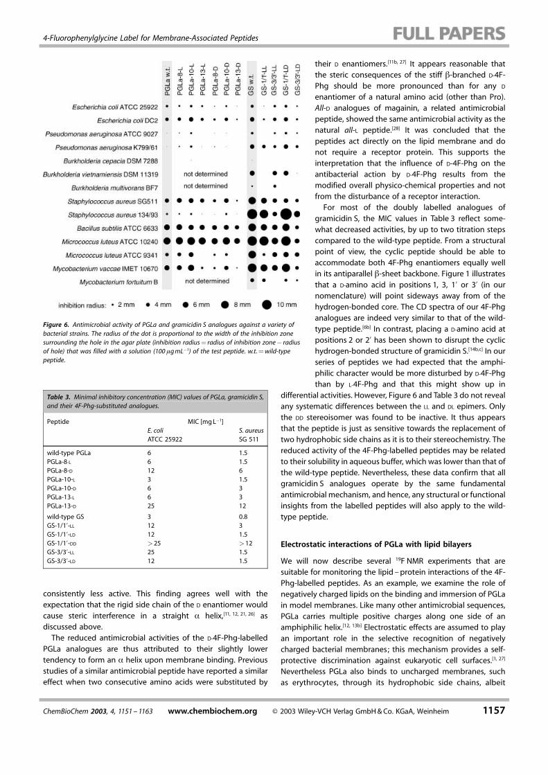
For comparative analysis we selected two representative anti-microbial peptides with characteristically different secondarystructures, namely the �-helical peptide PGLa and the �-sheetpeptide gramicidin S. PGLa was labelled with a single 4F-Phgsubstitution at three different positions in the central region ofthe molecule (Ala8, Ala10 and Ile13). Within the putativeamphiphilic � helix, Ile13 and Ala10 are part of the hydrophobicsurface while Ala8 lies within a partially polar region (Figure 1).The cyclic peptide gramicidin S was doubly labelled with 4F-Phg,such that the two substituents symmetrically replace either bothVal or both Leu residues on the hydrophobic face of theamphiphilic molecule (Figure 1). To test the biological functionsof the substituted PGLa and gramicidin S peptides, we examinedthe inhibition of bacterial growth by using two differentprotocols.By means of an agar diffusion assay,[24] the antimicrobial
activities of the peptides were monitored against differentspecies and strains of Gram-positive and Gram-negative bacteria,as summarised in Figure 6. These semiquantitative results weresubstantiated quantitatively by determining the minimuminhibitory concentration (MIC) values[25] against Escherichia coliand Staphylococcus aureus ; the results are listed in Table 3.Between the MIC values and the radius of inhibition in the agardiffusion tests, we found a correlation coefficient of 0.9, whichconfers a high confidence to the agar diffusion tests. Wheninterpreting the inhibition experiments, it is important to takeinto account the ratio between the free peptide in the bacterialsuspension and the peptide bound to or taken up by thebacteria. We systematically varied this ratio and determined theMIC values of wild-type gramicidin S and PGLa with an initialinoculum of E. coli of 5� 103, 5�104 and 5� 105 colony-formingunits (CFU)mL�1 and of S. aureus of 5�104, 5� 105 and 5�106
CFUmL�1. The resulting MIC values differed by less than one stepof dilution, which corresponds to a factor of two (data notshown). This proves that the amount of peptide consumed bythe bacteria is small, and hence, the MIC data truly reflect thefree concentration of peptide necessary to inhibit bacterialgrowth as discussed below.For both PGLa and gramicidin S it is seen that the 4F-Phg-
labelled analogues exhibit a pattern of activity against the widelydiffering strains of bacteria which is very similar to that exhibitedby the respective wild-type peptides. This qualitative corre-spondence confirms that 4F-Phg substitution does not affect thefundamental mechanism of antimicrobial action. Quantitatively,however, some interesting variations are apparent. While theactivities of the PGLa analogues containing L-4F-Phg do not differmuch from the wild-type peptide, the D-4F-Phg epimers are
4-Fluorophenylglycine Label for Membrane-Associated Peptides
ChemBioChem 2003, 4, 1151 ± 1163 www.chembiochem.org ¹ 2003 Wiley-VCH Verlag GmbH&Co. KGaA, Weinheim 1157
consistently less active. This finding agrees well with theexpectation that the rigid side chain of the D enantiomer wouldcause steric interference in a straight � helix,[11, 12, 21, 26] asdiscussed above.The reduced antimicrobial activities of the D-4F-Phg-labelled
PGLa analogues are thus attributed to their slightly lowertendency to form an � helix upon membrane binding. Previousstudies of a similar antimicrobial peptide have reported a similareffect when two consecutive amino acids were substituted by
their D enantiomers.[11b, 27] It appears reasonable thatthe steric consequences of the stiff �-branched D-4F-Phg should be more pronounced than for any D
enantiomer of a natural amino acid (other than Pro).All-D analogues of magainin, a related antimicrobialpeptide, showed the same antimicrobial activity as thenatural all-L peptide.[28] It was concluded that thepeptides act directly on the lipid membrane and donot require a receptor protein. This supports theinterpretation that the influence of D-4F-Phg on theantibacterial action by D-4F-Phg results from themodified overall physico-chemical properties and notfrom the disturbance of a receptor interaction.For most of the doubly labelled analogues of
gramicidin S, the MIC values in Table 3 reflect some-what decreased activities, by up to two titration stepscompared to the wild-type peptide. From a structuralpoint of view, the cyclic peptide should be able toaccommodate both 4F-Phg enantiomers equally wellin its antiparallel �-sheet backbone. Figure 1 illustratesthat a D-amino acid in positions 1, 3, 1� or 3� (in ournomenclature) will point sideways away from of thehydrogen-bonded core. The CD spectra of our 4F-Phganalogues are indeed very similar to that of the wild-type peptide.[6b] In contrast, placing a D-amino acid atpositions 2 or 2� has been shown to disrupt the cyclichydrogen-bonded structure of gramicidin S.[14b,c] In ourseries of peptides we had expected that the amphi-philic character would be more disturbed by D-4F-Phgthan by L-4F-Phg and that this might show up in
differential activities. However, Figure 6 and Table 3 do not revealany systematic differences between the LL and DL epimers. Onlythe DD stereoisomer was found to be inactive. It thus appearsthat the peptide is just as sensitive towards the replacement oftwo hydrophobic side chains as it is to their stereochemistry. Thereduced activity of the 4F-Phg-labelled peptides may be relatedto their solubility in aqueous buffer, which was lower than that ofthe wild-type peptide. Nevertheless, these data confirm that allgramicidin S analogues operate by the same fundamentalantimicrobial mechanism, and hence, any structural or functionalinsights from the labelled peptides will also apply to the wild-type peptide.
Electrostatic interactions of PGLa with lipid bilayers
We will now describe several 19F NMR experiments that aresuitable for monitoring the lipid ±protein interactions of the 4F-Phg-labelled peptides. As an example, we examine the role ofnegatively charged lipids on the binding and immersion of PGLain model membranes. Like many other antimicrobial sequences,PGLa carries multiple positive charges along one side of anamphiphilic helix.[12, 13b] Electrostatic effects are assumed to playan important role in the selective recognition of negativelycharged bacterial membranes; this mechanism provides a self-protective discrimination against eukaryotic cell surfaces.[1, 27]
Nevertheless PGLa also binds to uncharged membranes, suchas erythrocytes, through its hydrophobic side chains, albeit
Figure 6. Antimicrobial activity of PGLa and gramicidin S analogues against a variety ofbacterial strains. The radius of the dot is proportional to the width of the inhibition zonesurrounding the hole in the agar plate (inhibition radius� radius of inhibition zone� radiusof hole) that was filled with a solution (100 �gmL�1) of the test peptide. w.t.�wild-typepeptide.
Table 3. Minimal inhibitory concentration (MIC) values of PGLa, gramicidin S,and their 4F-Phg-substituted analogues.
Peptide MIC [mgL�1]E. coli S. aureusATCC 25922 SG 511
wild-type PGLa 6 1.5PGLa-8-L 6 1.5PGLa-8-D 12 6PGLa-10-L 3 1.5PGLa-10-D 6 3PGLa-13-L 6 3PGLa-13-D 25 12
wild-type GS 3 0.8GS-1/1�-LL 12 3GS-1/1�-LD 12 1.5GS-1/1�-DD �25 �12GS-3/3�-LL 25 1.5GS-3/3�-LD 12 1.5
A. S. Ulrich et al.
1158 ¹ 2003 Wiley-VCH Verlag GmbH&Co. KGaA, Weinheim www.chembiochem.org ChemBioChem 2003, 4, 1151 ± 1163
with a lower affinity. For example, the CD spectrum of thepeptide in aqueous solution changes towards a more �-helicalconformation in the presence of neutral DMPC vesicles (datanot shown). The actual role of negatively charged lipids for theinitial peptide binding and for further interactions with PGLaand related antimicrobial peptides is not yet completelyunderstood.
19F NMR spectroscopy of fluorine-labelled peptides can con-tribute in at least two ways to resolve this issue. First, solid-stateNMR spectroscopy of peptides in macroscopically oriented mem-brane samples provides information about the alignment anddynamics of the peptide in the membrane-bound state.[5b,6b±e]
This information is encoded in the anisotropic chemical shift,linewidth and lineshape of the 19F NMR signal when an orientedsample is measured at different tilt angles with respect to themagnetic field. To examine the electrostatic effect of thenegatively charged lipid DMPG, we reconstituted the three L-
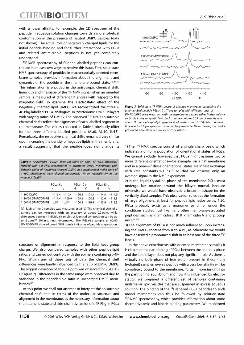
4F-Phg-labelled PGLa analogues in zwitterionic DMPC bilayerswith varying ratios of DMPG. The observed 19F NMR anisotropicchemical shifts reflect the alignment of each labelled segment inthe membrane. The values collected in Table 4 obviously differfor the three different labelled positions (Ala8, Ala10, Ile13).Remarkably, the respective chemical shifts remained very similarupon increasing the density of negative lipids in the membrane,a result suggesting that the peptide does not change its
structure or alignment in response to the lipid head-groupcharge. We also compared samples with other peptide:lipidratios and carried out controls with the epimers containing D-4F-Phg. Within any of these sets of data the chemical shiftdifferences were hardly influenced by the ratio of DMPC:DMPG.The biggest deviation of about 4 ppmwas observed for PGLa-10-L (Figure 7). Differences in the same range were observed due tovariations in the peptide:lipid ratio in uncharged DMPC mem-branes.[29]
At this point we shall not attempt to interpret the anisotropicchemical shift data in terms of the molecular structure andalignment in the membrane, as the necessary information aboutthe rotameric state and side-chain dynamics of L-4F-Phg in PGLa
Figure 7. Solid-state 19F NMR spectra of oriented membranes containing theantimicrobial peptide PGLa-10-L. Three samples with different ratios ofDMPC:DMPG were measured with the membranes aligned either horizontally orvertically in the magnetic field. Each sample contains 0.33 mg of peptide andabout 11 mg of phospholipid (peptide:lipid molar ratio� 1:100). Measurementtime was 1 ± 3 h per spectrum. is not yet fully available. Nevertheless, the resultspresented here allow a number of conclusions:
1) The 19F NMR spectra consist of a single sharp peak, whichindicates a uniform population of orientational states of PGLa.We cannot exclude, however, that PGLa might assume two ormore different orientations–for example, on a flat membraneand in a pore–if those orientational states are in fast exchangewith rate constants�103 s�1, so that we observe only anaverage signal in the NMR experiment.2) In the liquid-crystalline phase of the membrane PGLa mustundergo fast rotation around the bilayer normal, becauseotherwise we would have observed a broad lineshape for thevertically tilted samples. This observation rules out the formationof large oligomers, at least for peptide:lipid ratios below 1:50.PGLa probably exists as a monomer or dimer under theconditions studied, just like many other membrane-associatedpeptides such as gramicidin S, B18, gramicidin A and proteg-rin-1.[6, 30]
3) The alignment of PGLa is not much influenced upon increas-ing the DMPG content from 0 to 40%, as otherwise we wouldhave observed a pronounced shift in at least one of the three 19Flabels.In the above experiments with oriented membrane samples it
is clear that the partitioning of PGLa between the aqueous phaseand the lipid bilayer does not play any significant role. As there isvirtually no bulk phase of free water present in these (fullyhydrated) samples, even a peptide with a very low affinity will becompletely bound to the membrane. To gain more insight intothe partitioning equilibrium and how it is influenced by electro-statics, we prepared a different set of samples containingunilamellar lipid vesicles that are suspended in excess aqueoussolution. The binding of the 19F-labelled PGLa peptides to suchmodel membranes can thus be followed by solution-state19F NMR spectroscopy, which provides information about somethermodynamic and kinetic binding parameters. We monitored
Table 4. Anisotropic 19F NMR chemical shifts (in ppm) of PGLa analogueslabelled with L-4F-Phg, reconstituted in orientated DMPC membranes withdifferent ratios of negatively charged DMPG at a peptide:lipid molar ratio of1:100. Membranes were aligned horizontally (H) or vertically (V) in themagnetic field.[a]
PGLa-8-L PGLa-10-L PGLa-13-LH V H V H V
1:100 DMPC �116.4 �110.4 �96.5 �121.4 �114.8 �114.91:80/20 DMPC/DMPG �117.4 �109.9 � 99.3 �120.3 �112.6 �115.81:60/40 DMPC/DMPG n.d.[b] n.d.[b] �100.8 �119.8 �113.9 �115.3[a] Each of the 9 samples was measured at 35 �C. The chemical shift of asample can be measured with an accuracy of about 0.2 ppm, whiledifferences between individual samples of identical composition can be upto 2 ppm.[29] [b] n.d.�not determined. The PGLa-8-L sample at 60/40DMPC/DMPG showed broad NMR signals indicative of peptide aggregation.

4-Fluorophenylglycine Label for Membrane-Associated Peptides
ChemBioChem 2003, 4, 1151 ± 1163 www.chembiochem.org ¹ 2003 Wiley-VCH Verlag GmbH&Co. KGaA, Weinheim 1159
the 19F NMR signal of PGLa-10-L as a function of increasingconcentration of unilamellar DMPC/DMPG vesicles. When thenegatively charged lipid vesicles are added stepwise to thesample tube, the integral of the 19F NMR signal progressivelydecreases and finally disappears. This shows that by solution-state NMR spectroscopy we observe only the free form of thepeptide. Any peptide bound to the vesicles gives rise to a signalthat is several orders of magnitude broader (up to 20 ppm,corresponding to the chemical shift difference between the twosample orientations shown in Figure 7) and cannot be distin-guished from the baseline.To characterise the binding quantitatively, we titrated a
concentrated PGLa-10-L stock solution into vesicle suspensionsof different DMPC:DMPG ratios (Figure 8). Within the accuracy ofour measurement we observed a fairly linear saturation behav-iour, that is, all of the peptide was immediately captured by thecharged vesicles up to a certain limiting amount. Any sub-sequently added peptide remained free in solution. If we wantedto determine the binding constant, we would have to analysethe exact curvature around the point of inflection, where theconcentration of free peptide is in the order of the bindingconstant. However, this concentration range cannot be reliablyaddressed in this system because of the intrinsically lowsensitivity of the NMR method. The MIC value of PGLa towardssusceptible bacteria is 3 �M or below (Table 3), a fact suggestingan apparent binding constant of the same order of magnitude.The accuracy of integrating the 19F NMR signal is about �10 �Mfor the signal-to-noise ratio achieved within a typical measure-ment time of 1 hour. Despite the relatively high sensitivity of the19F nucleus, this is not sufficient to determine the concentrationof free peptide below and around the saturation point. It istherefore not possible to determine the apparent affinityconstant for peptides with such high affinities by solution-stateNMR spectroscopy.
As seen in Figure 8, PGLa shows very little binding touncharged vesicles composed of 100% DMPC. In the presenceof DMPG the saturating amount of membrane-bound peptidecorrelates roughly with the total number of charged lipid headgroups, if it is assumed that PGLa carries about five positivecharges. In liquid-crystalline bilayers we can exclude thepossibility that a cluster of DMPG molecules would form adistinct binding site for PGLa. The influence of the DMPGconcentration on the binding of PGLa must rather be attributedto an overall electrostatic attraction that changes the localpeptide concentration at the membrane surface. In a neutralDMPC bilayer the accumulation of positively charged amphi-philic peptides soon leads to electrostatic repulsion. The peptideconcentration near the surface thus becomes lower than in thebulk solution, and equilibrium is reached despite a highconcentration in the bulk. A negatively charged membrane, onthe other hand, attracts the positively charged peptides to thesurface and readily accommodates many more molecules beforethe charges are neutralised and equilibrium is attained.Solution-state 19F NMR spectroscopy can provide further
kinetic information over three windows of time. First, ourobservation that the peptide signal loses its integral intensityupon adding the vesicles suggests that any exchange betweenthe free and bound forms must be slower than about 104 s�1.Otherwise, in the case of fast exchange between free and vesicle-bound peptide, the signal would have broadened without loss ofintegral intensity. Second, there are no binding processes withtime constants of 10�2 ± 10�5 s�1, such as a slow trans-bilayer flip-flop. This is shown because we obtained the same results whenmeasuring the concentration of free peptide immediately (a fewminutes) after adding the vesicles and when repeating themeasurement after hours or days. A third time window isaccessible through the transverse relaxation time T2 . Wemeasured the signal decay of a single echo NMR experiment
as a function of the echo delay time. If any ofthe free peptide gets bound to the membraneduring this delay, the corresponding 19F mag-netisation will lose its phase coherence. Even ifthe peptide is released again at the end of thedelay, it can no longer contribute to the echointensity. In these relaxation experiments weobserved a dramatic reduction of T2 from300 ms in the absence of vesicles down to�25 ms in the presence of vesicles (Figure 8).Given the 100-ms range of the echo delaytimes, we conclude that the exchange be-tween free and bound peptide occurs with atime constant in the order of 10 s�1.
Conclusions
We have shown that the non-natural aminoacid 4F-Phg can be readily incorporated intosynthetic peptides by standard Fmoc solid-phase protocols, thereby providing a highlysensitive label for 19F NMR studies of lipid ±peptide interactions. Since racemisation of
Figure 8. Plot of the free-peptide concentration, cfree (determined from the 19F NMR signal intensity insolution), as a function of the total amount of PGLa-10-L, ctotal , titrated to suspensions of vesicles madefrom DMPC/DMPG mixtures containing different ratios of uncharged to negatively charged lipid. Themeasurement time was 20 ± 50 minutes for each concentration data point and 1 ± 2 h for each T2 value.

A. S. Ulrich et al.
1160 ¹ 2003 Wiley-VCH Verlag GmbH&Co. KGaA, Weinheim www.chembiochem.org ChemBioChem 2003, 4, 1151 ± 1163
4F-Phg cannot be avoided, the peptides containing the L and D
enantiomers have to be separated by reversed-phase HPLC in aTFA-free solvent. Using various functional assays on the differenttypes of peptides, we have demonstrated that hydrophobicamino acids may generally be substituted by L-4F-Phg withoutany significant loss of biological activity. The epimeric peptidescontaining D-4F-Phg, on the other hand, showed differentialresponses that may yield information about the transientsecondary structures required for function. For example, thesterically constrained side chain of D-4F-Phg interferes with the�-helical fold of PGLa, whereas it can be accommodated in everyalternate position of the cyclic � sheet of gramicidin S. It will beinteresting to see whether D-4F-Phg inhibits the self-assembly ofoligomeric � sheets, as its stiff aromatic side chain might be amore efficient steric barrier for �-sheet formation than anynatural amino acid that is flexible around C��C��C�. Theracemised ™byproduct∫ from peptide synthesis (containing the D-
4F-Phg analogue) may thus find useful applications both infunctional studies to probe transient conformations and as acomponent of rationally designed oligomerisation inhibitorsagainst amyloid diseases.The L-4F-Phg side chain serves as a highly sensitive and
background-free reporter for 19F NMR experiments on mem-brane-binding peptides. The stiff connection of the label to thepeptide backbone makes it ideal for distance measurements,and the broad CSA tensor can be used as a probe for peptideorientation and dynamics both in solution- and solid-state NMRexperiments. In macroscopically oriented membranes we typi-cally acquire spectra from 0.33 mg of 19F-labelled peptide inabout 3 h; this compares very favourably with analogous solid-state NMR experiments on 2H, 13C or 15N nuclei, which are 10 ±100-fold less sensitive.[6c] Even though the sensitivity of thesolution-state 19F NMR experiments was not yet high enough todetermine directly a binding constant in the �M range, varioustime windows could be addressed to describe the reversibleinteractions of the peptide with lipid vesicles. In the case of thecationic antimicrobial peptide PGLa, we found that electrostaticinteractions determine the initial membrane binding in solution,but once it is associated with the bilayer the presence of chargedlipid head groups makes no difference to the alignment ormobility of the peptide. This finding may have importantconsequences for our understanding of the functional mecha-nism of PGLa and related peptides.
Experimental Section
Materials : Fmoc-L-ornithine(dde)-OH and Fmoc-D-phenylalaninewere purchased from Bachem (Heidelberg, Germany; Fmoc� 9-fluorenylmethoxycarbonyl, dde� 1-(4,4-dimethyl-2,6-dioxocyclo-hex-1-ylidene)ethyl). Tetramethylfluoroformamidinium hexafluoro-phosphate (TFFH) was obtained from Advanced Chemtech (Louis-ville, KY), and 2-(1H-benzotriazole-1-yl)-1,1,3,3-tetramethyluroniumhexafluorophosphate (HBTU) was from Richelieu Biotechnologies(Montreal, Canada). Diethyl ether, acetone, acetonitrile, methanoland N,N-dimethylformamide (DMF; synthesis grade) were obtainedfrom Roth (Karlsruhe, Germany). DMF was purified by standardmethods.[31] N-2-hydroxyethylpiperazine-N�-2-ethanesulfonic acid
(HEPES) was obtained from Sigma, and N,N-diisopropylethylamine(DIEA), trifluoroacetic acid (TFA), triisopropylsilane (TIS), trifluoro-ethanol (TFE), N-methylpyrrolidine (NMP), �-cyano-4-hydroxycin-namic acid, piperidine, sodium fluoride and fluorotrichloromethanewere obtained from Aldrich. The compounds 2,4,6-collidine, 4-fluoro-L-�-phenylglycine (L-4F-Phg), 4-fluoro-DL-�-phenylglycine (D/L-4F-Phg)and hydrazine were obtained from Fluka (Taufkirchen, Germany),while tetrahydrofuran (THF) was from Merck (Darmstadt, Germany).All other chemicals for peptide synthesis were purchased fromCalbiochem-Novabiochem (Bad Soden, Germany). 1-Fluoro-2,4-dini-trophenyl-5-L-alanine amide (Marfey's reagent) was obtained formPierce (Bonn, Germany). N-terminal Fmoc protection of L-4F-Phg andD/L-4F-Phg was performed with Fmoc-Cl as previously reported.[32]
The lipids 1,2-dimyristoyl-sn-glycero-3-phosphocholine (DMPC) and1,2-dimyristoyl-sn-glycero-3-[phospho-rac-(1-glycerol)] (DMPG), aswell as the fluorescence-labelled lipids L-�-phosphatidylethanol-amine-N-(lissamine rhodamine B sulfonyl) and L-�-phosphatidyleth-anolamine-N-(4-nitrobenzo-2-oxa-1,3-diazole), were obtained fromAvanti Polar Lipids (Alabaster, AL). All chemicals used were of thehighest grade and were used as received unless otherwise men-tioned.
Peptide synthesis : Peptides were synthesised either on an AppliedBiosystems 433A instrument, on an Abimed Economy PeptideSynthesizer EPS 221, or manually by using standard solid-phaseFmoc protocols.[33] Deprotection was carried out with 20% piper-idine in DMF, and preactivation was performed with 1-hydroxy-1H-benzotriazole (HOBt) and HBTU in the presence of the base DIEA.NMP or DMF were used as solvents. The deprotection and couplingefficiency was monitored by performing a Kaiser test.[34] Theprotected peptides were cleaved from the resin at room temperatureby treatment with a cocktail of TFA (95%), water (2.5%) and TIS(2.5%) for 2 ± 5 h with occasional shaking. In the case of gramicidin Swe had to use 5% TIS to avoid alkylation by cations produced duringcleavage. The resin was filtered off and washed with pure TFA. Thecombined filtrates were evaporated to a reduced volume under agentle stream of nitrogen. The crude product was precipitated withcold diethyl ether, taken up in acetonitrile/water (1:9) and lyophi-lised.
In our attempts to suppress racemisation of L-4F-Phg, the synthesiswas performed in an automated manner only up to the stage ofintroducing the label. The fluorinated amino acid and the followingone were then introduced manually as described in the Results andDiscussion section. The remaining sequence was then completed onthe peptide synthesizer.
In the cyclisation of gramicidin S, the linear peptide (0.5 mgmL�1)was dissolved in dry degassed dichloromethane (DCM) in a 50-mLround-bottomed flask. Solid pyridinium 1-benzotriazolyloxytris(di-methylamino)phophonium hexafluorophosphate (3 equiv to thepeptide) and a solution of HOBt (3 equiv) in degassed DMF(0.5 mL) were added to the flask, and the mixture was stirred. DIEA(6 equiv) was then added to initiate the reaction. The reaction wasmonitored by HPLC and allowed to proceed overnight under anargon atmosphere. After removing the DCM by rotary evaporation,ethyl acetate (30 mL) was added and the remains of the DMF werewashed off with water (2�30 mL). The organic layer was dried overNa2SO4 and the solvent was removed by rotary evaporation to obtainan oil. This oil was taken up in ethyl acetate, lyophilised and purifiedby HPLC.
The cyclised product still had the dde protecting group on theornithine residues. For the deprotection the peptide was taken up inTHF and treated with hydrazine solution (2% v/v in THF). The mixturewas stirred for about 2 h at room temperature before being left at
4-Fluorophenylglycine Label for Membrane-Associated Peptides
ChemBioChem 2003, 4, 1151 ± 1163 www.chembiochem.org ¹ 2003 Wiley-VCH Verlag GmbH&Co. KGaA, Weinheim 1161
�73 �C overnight to precipitate the removed dde as the oxazolone.The precipitate was filtered and the solvent was removed from thefiltrate by rotary evaporation to give another yellowish oil, which wasdissolved in acetonitrile/water (1:1, 3 mL) and lyophilised. Massspectrometry on this sample provided evidence of proper cyclisationand complete removal of the dde protecting group. The crudeproduct was further purified by HPLC before use.
HPLC purification of peptides : Peptides were purified withreversed-phase HPLC (Jasco, Japan; 10� 250 mm Vydac C18 col-umns; flow rate of 6 mLmin�1). Water/acetonitrile gradients wereindividually optimised at 30 ± 40 �C for each peptide analogue. B18and gramicidin S analogues were purified in a single step, with theamount of deletion peptides and of other side products beingbetween 5 and 15%. PGLa analogues were purified in two steps: theepimers were first separated with 0.1% TFA in both phases and,following lyophilisation, they were further purified in TFA-freesolvent, thereby reducing the amount of deletion peptides to lessthan 3%. For all peptides the last HPLC step was performed with5 mM HCl in the aqueous phase and pure acetonitrile as the organicphase. All samples prepared for this 19F NMR study contained lessthan 0.1 mole of TFA per mole of peptide as judged by the 19F NMRpeak intensities in solution.
Mass spectrometry : The identity of the peptides was confirmed withmass spectrometry. We used electrospray ionisation (ESI) and/ormatrix-assisted laser desorption/ionisation time-of-flight (MALDI-TOF) spectrometry. Samples for ESI were dissolved in 99% methanoland measured on a Fisons VG Quattro triple quadrupole massspectrometer. MALDI-TOF samples were cocrystallised with a matrixof �-cyano-4-hydroxy-cinnamic acid from acetonitrile/water solu-tions onto a stainless steel target. Measurements were made on aBruker Reflex 2 instrument. When both techniques were successfullyemployed, we found identical results.
Assignment of L- and D-4F-Phg enantiomers : Samples of purifiedpeptide (0.2 mg) were hydrolysed with 6M HCl (0.5 mL) for 16 h at105 �C. The amino acids were derivatised with Marfey's reagent:[22]
the HCl was removed by lyophilisation, 0.5M NaHCO3 (25 �L) and 1%Marfey's reagent in acetone (50 �L) were added, the mixture wasincubated at 40 �C for 90 min, and finally 2M HCl (7 �L) was added.The mixture of the derivatised amino acids was resolved by analyticalHPLC (4.6� 250 mm Vydac C18 column; flow rate of 1.5 mLmin�1;detection at 415 nm) at 40 �C with a linear acetonitrile/water gradientcontaining 0.1% TFA, from 10 to 43% acetonitrile over 12 min. Peakswere identified by comparison with chromatograms of purederivatised L- and D-4F-Phg (see Figure 2).
High-resolution 1H NMR spectroscopy : Samples of 1.8 ± 2.4 mM
PGLa were prepared in an aqueous solution (350 �L) containing30% TFE-d3, 10 mM potassium phosphate buffer (pH 7.0 before TFEaddition), 0.05% NaN3 and 0.1 mM 3-(trimethylsilyl)propionic acid-d4
and measured into 5-mm tubes between Doty susceptibility plugs.Two-dimensional 1H± 1H TOCSY (100 ms mixing time; 8 h measure-ment time) and NOESY spectra (150 ms; 16 ± 32 h) were recordedwith WATERGATE water suppression on a 600 MHz Unity Inovaspectrometer (Varian, Palo Alto, CA) with standard equipment. Thespectra were evaluated with the programs NMRPipe, SPSCAN andXEASY,[35] and molecular models were created and visualised withSYBYL (Tripos, St. Louis, MO) and MOLMOL.[36] Ring-current shiftswere determined according to the Johnson±Bovey model, asimplemented in MOLMOL.
Fluorescence assay for fusogenic activity : The peptide-inducedfusion of large unilamellar vesicles (LUVs) was monitored with afluorescence dequenching assay, as previously reported.[15b] LUVs ofbrain sphingomyelin/cholesterol (80:20 molar ratio) with and with-
out 0.8 mol% of both the fluorescence-labelled lipids L-�-phospha-tidylethanolamine-N-(lissamine rhodamine B sulfonyl) and L-�-phos-phatidylethanolamine-N-(4-nitrobenzo-2-oxa-1,3-diazole) were pro-duced in a lipid extruder (Avestin, Canada) with Poreticspolycarbonate membranes of 100-nm pore diameter (Osmonics,Livermore, CA). 40 �M ZnCl2 and 2 �M B18 peptide were added totrigger fusion, and the fluorescence intensity was monitored with anLS 50B luminescence spectrometer (Perkin Elmer) with the excitationset at 465 nm and the emission set at 520 nm. The initial rate offusion was determined from an exponential fit to the increase influorescence intensity 3 ± 30 seconds after addition of peptide, andwas compared with the total increase after addition of Triton X100.
Antimicrobial inhibition assays : As test organisms for antimicrobialactivity assays we used the Gram-negative bacteria Escherichia coliATCC 25922, E. coli DC2 (antibiotic-susceptible penetration mu-tant),[37] Pseudomonas aeruginosa ATCC 9027, P. aeruginosa K799/61(antibiotic-susceptible penetration mutant),[38] Burkholderia cepaciaDSM 7288 (resistant to cationic peptides),[39] B. vietnamiensisDSM 11319, B. multivorans BF7 (clinical isolate obtained from Prof.Dr. A. Bauernfeind, MICOER-Institute, Munich, Germany) and theGram-positive bacteria Staphylococcus aureus SG511, S. aureus 134/93 (MRSA),[40] Bacillus subtilis ATCC 6633, Micrococcus luteusATCC 10240 and ATCC 9341, Mycobacterium vaccae IMET 10670,and Mycobacterium fortuitum B.
Antibacterial activity was determined qualitatively by agar diffusiontests[24] and quantitatively by measurement of MIC values in liquid-growth inhibition assays.[25] For agar diffusion assays, a suspension(100 �L) of the test organism with a density of McFarland standard 1was inoculated into sterile melted 1% Trypticase soy broth (TSB)agarose (32 mL), cooled to 50 �C and then poured into Petri dishes.The final concentration of cells was 106 colony-forming units (CFU)per mL. Each antimicrobial peptide (50 �L) at a concentration of100 �gmL�1 was filled into 9-mm holes that had been cut out of theagar (5 �g of peptide per hole). The diameter of the zone ofinhibition around the hole in mm was determined after 24 h ofincubation at 37 �C.
Minimum inhibitory concentration (MIC) values were determined byusing a standard microbroth dilution method in 1% TSB.[25] The cellswere grown overnight at 37 �C in nutrient broth and then diluted inTSB. The peptide solutions (50 �L) were serially diluted by a factor oftwo with TSB, and then the wells were inoculated with bacteria(50 �L) to give a final concentration of 5� 105 CFUmL�1 in a volumeof 100 �L, unless stated otherwise. Microtiter plates were incubatedat 37 �C for 24 h and read with a Nepheloscan Ascent 1.4 automaticplate reader (Labsystems, Vantaa, Finland). All experiments wereperformed in triplicate. MIC values are stated as the lowest antibioticconcentration that inhibited growth.
Solid-state 19F NMR spectroscopy : NMR measurements were per-formed on a 500 MHz wide-bore Unity Inova spectrometer (Varian,Palo Alto, CA). For solid-state 19F NMR spectroscopy, a 19F/1H double-tuned flat-coil probe head with a goniometer (Doty Scientific Inc. ,Columbia, SC) and an external 470 MHz high-power amplifier(Creative Electronics Inc. , Los Angeles, CA) were used.[4a, 7b]
Oriented membrane samples were prepared by depositing peptide ±lipid mixtures on cleaned and vacuum-dried glass slides (0.08� 7.5�18 mm3, Marienfeld Inc. , Germany).[6b±d] The peptide was dissolved inwater and the lipid in methanol. Both components were then mixedto give a 70% methanol solution containing about 20 ± 100 mgmL�1
of material in total. Drops (20 ± 30 �L) were spread onto each glassplate (the number of plates necessary depends on the desiredpeptide:lipid ratio, typically 8 plates were used for 0.33 mg of PGLa ina 1:100 peptide:lipid ratio). After initial drying in a laminar flow
A. S. Ulrich et al.
1162 ¹ 2003 Wiley-VCH Verlag GmbH&Co. KGaA, Weinheim www.chembiochem.org ChemBioChem 2003, 4, 1151 ± 1163
bench, the membrane films were dried to completion under vacuumovernight. The plates were stacked and hydrated for 2 days at 48 �Cin an atmosphere over a saturated K2SO4 solution (about 96%relative humidity). The stack was wrapped in parafilm and poly-ethylene film to maintain the hydration of the sample during theNMR measurement. 1H-decoupled 31P NMR spectra were recorded tojudge the quality of bilayer orientation in the samples. The well-oriented lipid typically contributed 80 ±95% to the total 31P NMRphospholipid signal.
Peptide ± lipid interactions studied by 19F NMR in solution : LUVswere prepared from DMPC or DMPC/DMPG mixtures in 10 mM HEPESbuffer (pH 7.0) with 1 mM NaF, as described for the fusion assaysabove. Small unilamellar vesicles were prepared by ultrasonication ofthe lipid suspension. The vesicles were mixed with peptide stocksolution, and the mixture (0.6 mL) was filled into 5-mm NMR tubesfor high-resolution measurements. 19F NMR free induction decayswere recorded immediately after a 90� pulse at 470 MHz, with astandard triple-resonance probe head for high-resolution measure-ments (Varian, Palo Alto, CA), where the proton channel was tuned tofluorine. Fluorine chemical shifts were referenced to external CFCl3with a thin capillary filled with pure CFCl3 extending over the fulllength of the tube. After Fourier transformation, the NMR signalintensity of the labelled peptide was determined by integration overa range�0.12 ppm from the peak maximum after visual phasing andlinear baseline correction for the outermost 10% of a�1 ppm range.This was compared with the integral of the NaF signal.
Acknowledgements
We thank Sigrid Reichardt, Waltraud Scheiding and IrmgardHeinemann for excellent technical support, and we gratefullyacknowledge Charles Glabe (University of California, Irvine) forhaving provided the Arg- and His-substituted B18 analogues. Thiswork was supported by the Deutsche Forschungsgemeinschaftwithin program SFB 197 (TP B13).
Keywords: amphiphilic peptides ¥ biomembranes ¥ fluorine ¥fluorophenylglycine ¥ NMR spectroscopy
[1] a) R. E. W. Hancock, D. S. Chapple, Antimicrob. Agents Chemother. 1999, 43,1317 ± 1323; b) M. Zasloff, Nature 2002, 415, 389 ± 395; c) Y. Shai, Curr.Pharm. Des. 2002, 8, 715 ± 725.
[2] E. I. Pe¬cheur, J. Sainte-Marie, A. Bienven¸e, D. Hoekstra, J. Membrane. Biol.1999, 167, 1 ± 17.
[3] R. R. Ketchem, K. C. Lee, S. Huo, T. A. Cross, J. Biomol. NMR 1996, 8, 1 ± 14.[4] a) S. L. Grage, A. S. Ulrich, J. Magn. Reson. 1999, 138, 98 ± 106; b) A. S.
Ulrich in Encyclopedia of Spectroscopy and Spectrometry (Eds. : J. Lindon, G.Trautner, J. Holmes), Academic Press, 2000, 813 ± 825; c) M. L. Gilchrist, Jr. ,K. Monde, Y. Tomita, T. Iwashita, K. Nakanishi, A. E. McDermott, J. Magn.Reson. 2001, 152, 1 ± 6.
[5] a) J. T. Gerig, Prog. in NMR. 1994, 26, 293 ± 370; b) S. L. Grage, J. Salgado, U.D¸rr, S. Afonin, R. W. Glaser, A. S. Ulrich in Perspectives on Solid State NMRin Biology (Eds. : S. Kiihne, H. J. M. de Groot), Kluwers Academic Press,Dordrecht, 2001, 83 ± 91.
[6] a) M. A. Danielson, J. J. Falke, Annu. Rev. Bioph. Biomol. Struct. 1996, 25,163 ± 195; b) J. Salgado, S. L. Grage, L. H. Kondejewski, R. S. Hodges, R. N.McElhaney, A. S. Ulrich, J. Biomol. NMR 2001, 21, 191 ± 208; c) S. L. Grage, J.Wang, T. A. Cross, A. S. Ulrich, Biophys. J. 2002, 83, 3336 ± 3350; d) S.Afonin, U. D¸rr, R. W. Glaser, A. S. Ulrich, Magn. Res. Chem. 2004, in press ;e) O. Toke, R. D. O'Connor, W. L. Maloy, R. W. Glaser, A. S. Ulrich, J. Schaefer,unpublished results.
[7] a) P. Tang, B. Yan, Y. Xu, Biophys. J. 1997, 72, 1676 ± 1682; b) S. L. Grage,A. S. Ulrich, J. Magn. Reson. 2000, 146, 81 ± 88.
[8] U. N. Sundram, J. H. Griffin, J. Am. Chem. Soc. 1996, 118, 13107 ± 13108;b) J. M. Janusz, J. M. Gardlik, P. A. Young, R. V. Burkes, S. J. Stoll, A. F. Estelle,C. M. Riley, J. Med. Chem. 1990, 33, 1052 ± 1061; c) W. M. Kati, D.Montgomery, C. Maring, V. S. Stoll, V. Giranda, X. Chen, W. G. Laver, W.Kohlbrenner, D. W. Norbeck, Antimicrob. Agents Chemother. 2001, 45,2563 ± 2570; d) S. Abu-Lafi, S. A. Turujman, J. Chromatogr. A. 1999, 855,157 ± 170.
[9] a) M. J. Anteunis, R. E. Callens, D. K. Tavernier, Eur. J. Biochem. 1975, 58,259 ± 268; b) Y. Lee, R. B. Silverman, Fourth International Electronic Confer-ence on Synthetic Organic Chemistry (ECSOC-4), 2000, www.mdpi.org/ecsoc-4.htm.
[10] K. Ramanarayan, M. F. Chan, V. N. Balaji, S. Profeta, S. N. Rao, Int. J. PeptideProtein Res. 1995, 45, 366 ± 376.
[11] a) E. Krause, M. Bienert, P. Schmieder, H. Wenschuh, J. Am. Chem. Soc.2000, 122, 4865 ±4870; b) Y. Chen, C. T. Mant, R. S. Hodges, J. Pept. Res.2002, 59, 18 ±33.
[12] B. Bechinger, M. Zasloff, S. J. Opella, Biophys. J. 1998, 74, 981 ± 987.[13] a) M. E. Houston, L. H. Kondejewski, D. N. Karunaratne, M. Gough, S. Fidai,
R. S. Hodges, R. E. Hancock, J. Peptide Res. 1998, 52, 81 ± 88; b) K.Matsuzaki, Biochim. Biophys. Acta 1999, 1462, 1 ± 10; c) J. A. Whiles, R.Brasseur, K. J. Glover, G. Melacini, E. A. Komives, R. R. Vold, Biophys. J. 2001,80, 280 ± 293; d) M. Dathe, H. Nikolenko, J. Meyer, M. Beyermann, M.Bienert, FEBS Lett. 2001, 501, 146 ± 150.
[14] a) S. E. Hull, R. Karlsson, P. Main, M. M. Woolfson, E. J. Dodoson, Nature1978, 275, 206 ±207; b) M. Jelokjani-Nirakai, E. J. Prenner, L. H. Konde-jewski, C. M. Kay, R. N. McElhaney, R. S. Hodges, J. Pept. Res. 2001, 58, 293 ±306; c) M. Jelokjani-Nirakai, E. J. Prenner, C. M. Kay, R. N. McElhaney, R. S.Hodges, J. Pept. Res. 2002, 60, 23 ± 36.
[15] a) P. L. DeAngelis, C. G. Glabe, Peptide Res. 1990, 3, 62 ± 68; b) A. S. Ulrich,M. Otter, C. G. Glabe, D. Hoekstra, J. Biol. Chem. 1998, 273, 16748± 16755.
[16] J. Yang, C. M. Gabrys, D. P. Weliky, Biochemistry 2001, 40, 8126 ± 8137.[17] a) R. W. Glaser, M. Gr¸ne, C. Wandelt, A. S. Ulrich, Biochemistry 1999, 38,
2560 ± 2569; b) A. S. Ulrich, W. Tichelaar, G. Fˆrster, O. Zschˆrnig, S.Weinkauf, H. W. Meyer, Biophys. J. 1999, 77, 829 ± 841.
[18] L. A. Carpino, A. El-Faham, J. Am. Chem. Soc. 1995, 117, 5401 ± 5402.[19] A. El-Faham, Chem. Lett. 1998, 7, 671 ±672.[20] a) G. Karygiannis, C. Athanassopoulos, P. Mamos, N. Karamanos, D.
Papaioannou, G. W. Francis, Acta Chem. Scand. 1998, 52, 1144 ± 1150;b) L. A. Carpino, D. Sadat-Aalaee, H. G. Chao, R. H. DeSelms, J. Am. Chem.Soc. 1990, 112, 9651 ± 9652.
[21] E. Krause, M. Beyermann, M. Dathe, S. Rothemund, M. Bienert, Anal. Chem.1995, 67, 252 ± 258.
[22] P. Marfey, Carlsberg Res. Commun. 1984, 49, 591 ± 596.[23] H. Binder, K. Arnold, A. S. Ulrich, O. Zschˆrnig, Biochim. Biophys. Acta 2000,
1468, 345 ± 358.[24] Deutsches Arzneibuch, 9th ed. , Deutscher Apotheker Verlag, Stuttgart,
1990, pp. 47 ±48 and 424 ± 430.[25] Methods for dilution antimicrobial susceptibility tests for bacteria that grow
aerobically, 4th ed. (Villanova, PA, USA), National Committee for ClinicalLaboratory Standards, 1997, approved standard document M7A4.
[26] a) D. Avrahami, Z. Oren, Y. Shai, Biochemistry 2001, 40, 12591 ± 12603;b) S. Y. Hong, J. E. Oh, K. H. Lee, Biochem. Pharmacol. 1999, 58, 1775 ±1780.
[27] T. Wieprecht, M. Dathe, M. Sch¸mann, E. Krause, M. Beyermann, M.Bienert, Biochemistry 1996, 35, 10844 ± 10853.
[28] D. Wade, A. Boman, B. Wa hlin, C. M. Drain, D. Andreu, H. G. Boman, R. B.Merrifield, Proc. Natl. Acad. Sci. USA 1990, 87, 4761 ± 4765; R. Besalle, A.Kapitkovsky, A. Gorea, I. Shalit, M. Fridkin, FEBS Lett. 1990, 274, 151 ±155.
[29] R. W. Glaser, A. S. Ulrich, J. Magn. Reson. 2003, 164, 104 ± 114.[30] S. Yamaguchi, T. Hong, A. Waring, R. I. Lehrer, M. Hong, Biochemistry 2002,
41, 9852 ± 9862.[31] D. D. Perrin, W. L. F. Amarego, Purification of Laboratory Chemicals, 3rd ed. ,
Pergamon Press, Oxford, 1988.[32] L. A. Carpino, G. Y. Han, J. Org. Chem. 1972, 37, 3404 ± 3409.[33] a) C. D. Chang, J. Meienhofer, Int. J. Peptide Protein Res. 1978, 11, 246 ± 249;
b) G. B. Fields, R. L. Noble, Int. J. Peptide Protein Res. 1990, 35, 161 ±214.[34] E. Kaiser, R. L. Colescott, C. D. Bossinge, P. I. Cook, Anal. Biochem. 1970, 34,
595 ± 598.
4-Fluorophenylglycine Label for Membrane-Associated Peptides
ChemBioChem 2003, 4, 1151 ± 1163 www.chembiochem.org ¹ 2003 Wiley-VCH Verlag GmbH&Co. KGaA, Weinheim 1163
[35] a) F. Delaglio, S. Grzesiek, G. W. Vuister, G. Zhu, J. Pfeifer, A. Bax, J. Biomol.NMR 1995, 6, 277 ± 293; b) T. Szyperski, B. Banecki, D. Braun, R. W. Glaser, J.Biomol. NMR 1998, 11, 387 ±405; c) C. Bartels, T. H. Xia, M. Billeter, P.G¸nthert, K. W¸thrich, J. Biomol. NMR 1995, 2, 619 ± 629.
[36] R. Koradi, M. Billeter, K. W¸thrich, J. Mol. Graphics 1996, 14, 51 ± 55.[37] M. G. Richmond, D. C. Clarke, S. Wotton, Antimicrob. Agents Chemother.
1976, 10, 215 ± 218.[38] W. Zimmermann, J. Clin. Pharmacol. Biopharm. 1979, 17, 131 ± 134.
[39] R. E. W. Hancock, Lancet 1997, 349, 418 ±422.[40] W. Witte, C. Cuny, C. Braulke, D. Heuck. Epidemiol. Infect.1994, 113, 67 ± 73.[41] G. Merutka, H. J. Dyson, P. Wright, J. Biomol. NMR 1995, 5, 14 ± 24.
Received: January 31, 2003Revised version: July 17, 2003 [F568]