25 mined ICE
6
minedICE: A Knowledge Discovery Platform for Neurophysiological Artificial Intelligence Rory A. Lewis 1,2 and Allen Waziri 3 1 Department of Computer Science, University of Colorado at Colorado Springs, Colorado Springs, CO, 80933 2 Departments of Pediatrics & Neurology, University of Colorado Denver, Anschutz Medical Campus, Denver, CO 80262 3 Department of Neurosurgery, University of Colorado Denver, Anschutz Medical Campus, Denver, CO 80262 Abstract. In this paper we present the minedICE TM computer archi- tecture and network comprised of neurological instruments and artifi- cial intelligence (AI) agents. It’s called minedICE because data that is “mined ” via IntraCortical Electroencephalography (ICE ) located deep inside the human brain procures (mined ) knowledge to a Decision Sup- port System (DSS) that is read by a neurosurgeon located either at the bedside of the patient or at a geospatially remote location. The DSS system 1) alerts the neurosurgeon when a severe neurological event is occurring in the patient and 2) identifies the severe neurological event. The neurosurgeon may choose to provide feedback to the AI agent which controls the confidence level of the association rules and thereby teaches the learning component of minedICE. 1 Introduction The detection and interpretation of abnormal brain electrical activity in patients with acute neurological injury remains an area of significant opportunity for tech- nological advancement. Neurosurgeons know that when a patient arrives in the emergency room (ER) with a severe head injury, they rely on their intuition and relatively limited external data to choose the necessary treatment modal- ity. Aside from the initial injury, the brain tissue in these patients is extremely susceptible to secondary injury from ongoing abnormal (and preventable) phys- iological processes. Although a number of invasive neuromonitoring systems ex- ist, current modalities either provide indirect measurement of brain health (and are therefore difficult to interpret) or have limited sensitivity and specificity for accurately identifying critical and deleterious changes in brain health. To overcome this limitation, Waziri developed Intracortical Electroencephalogra- phy (ICE) [10] a technique by which specialized multicontact electrodes can be placed into the cerebral cortex through a burrhole generated at the bedside, as illustrated in Figure 1. Through the use of ICE, high amplitude and high fidelity EEG data can be recorded in an otherwise electrically noisy environment. M. Kryszkiewicz et al. (Eds.): ISMIS 2011, LNAI 6804, pp. 575–580, 2011. c Springer-Verlag Berlin Heidelberg 2011
-
Upload
independent -
Category
Documents
-
view
1 -
download
0
Transcript of 25 mined ICE

minedICE: A Knowledge Discovery Platform for
Neurophysiological Artificial Intelligence
Rory A. Lewis1,2 and Allen Waziri3
1 Department of Computer Science, University of Colorado at Colorado Springs,Colorado Springs, CO, 80933
2 Departments of Pediatrics & Neurology, University of Colorado Denver,Anschutz Medical Campus, Denver, CO 80262
3 Department of Neurosurgery, University of Colorado Denver,Anschutz Medical Campus, Denver, CO 80262
Abstract. In this paper we present the minedICETM computer archi-tecture and network comprised of neurological instruments and artifi-cial intelligence (AI) agents. It’s called minedICE because data that is“mined” via IntraCortical Electroencephalography (ICE) located deepinside the human brain procures (mined) knowledge to a Decision Sup-port System (DSS) that is read by a neurosurgeon located either at thebedside of the patient or at a geospatially remote location. The DSSsystem 1) alerts the neurosurgeon when a severe neurological event isoccurring in the patient and 2) identifies the severe neurological event.The neurosurgeon may choose to provide feedback to the AI agent whichcontrols the confidence level of the association rules and thereby teachesthe learning component of minedICE.
1 Introduction
The detection and interpretation of abnormal brain electrical activity in patientswith acute neurological injury remains an area of significant opportunity for tech-nological advancement. Neurosurgeons know that when a patient arrives in theemergency room (ER) with a severe head injury, they rely on their intuitionand relatively limited external data to choose the necessary treatment modal-ity. Aside from the initial injury, the brain tissue in these patients is extremelysusceptible to secondary injury from ongoing abnormal (and preventable) phys-iological processes. Although a number of invasive neuromonitoring systems ex-ist, current modalities either provide indirect measurement of brain health (andare therefore difficult to interpret) or have limited sensitivity and specificityfor accurately identifying critical and deleterious changes in brain health. Toovercome this limitation, Waziri developed Intracortical Electroencephalogra-phy (ICE) [10] a technique by which specialized multicontact electrodes can beplaced into the cerebral cortex through a burrhole generated at the bedside, asillustrated in Figure 1. Through the use of ICE, high amplitude and high fidelityEEG data can be recorded in an otherwise electrically noisy environment.
M. Kryszkiewicz et al. (Eds.): ISMIS 2011, LNAI 6804, pp. 575–580, 2011.c© Springer-Verlag Berlin Heidelberg 2011
576 R.A. Lewis and A. Waziri
8
567
1
3
24
9
1
(a)
4
1
3
89
6
7
5
2
10
(b)
Fig. 1. Intracortical Electroencephalograph (ICE) Pin, Side Elevations: ((a) ICE 2 in-serted through periostem 5, skull 6, arachnoid and pia mater 7 into brain 8 in varyingpositions 9. Receiving electrodes 1 encapsulated by brain 8. Electrodes 1 transmitsignals along wire 3 to end 4 where it is connected to computer. (b) Cannula andinternal lumen 2 with drainage hole 4 and sharpened end 8. Electrodes 1 at electroderegion 17 allow insertion through burr hole 5 traversing brain 9. External region 6ofcannula remains outside of skull. Connection conductors 7 combine into a single wire2. Drainage holes 10 in drainage region 8 provide openings for fluid to flow 4. Supportmember inserted through 4 into internal lumen for accurate placement).
1.1 minedICE Architecture
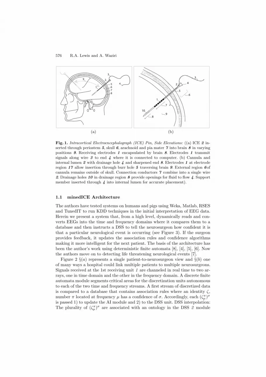
The authors have tested systems on humans and pigs using Weka, Matlab, RSESand TunedIT to run KDD techniques in the initial interpretation of EEG data.Herein we present a system that, from a high level, dynamically reads and con-verts EEGs into the time and frequency domains where it compares them to adatabase and then instructs a DSS to tell the neurosurgeon how confident it isthat a particular neurological event is occurring (see Figure 3). If the surgeonprovides feedback, it updates the association rules and confidence algorithmsmaking it more intelligent for the next patient. The basis of the architecture hasbeen the author’s work using deterministic finite automata [8], [4], [5], [6]. Nowthe authors move on to detecting life threatening neurological events [7].
Figure 2 §(a) represents a single patient-to-neurosurgeon view and §(b) oneof many ways a hospital could link multiple patients to multiple neurosurgeons.Signals received at the 1st receiving unit 1 are channeled in real time to two ar-rays, one in time domain and the other in the frequency domain. A discrete finiteautomata module segments critical areas for the discretization units autonomousto each of the two time and frequency streams. A first stream of discretized datais compared to a database that contains association rules where an identity ζ,number π located at frequency μ has a confidence of σ. Accordingly, each (ζπ
μ )σ
is passed 1) to update the AI module and 2) to the DSS unit. DSS interpolation:The plurality of (ζπ
μ )σ are associated with an ontology in the DSS 2 module

minedICE: A Knowledge Discovery Platform 577
1
2
3
4
5
67
8
7 1st Receiving Unit
2nd Receiving Unit
DSS
AI
(a)
AIDSS
1st Receiving Unit
1st Receiving Unit
1st Receiving Unit
1st Receiving Unit
2nd Receiving Unit
1
6
7
3
5
2
4
(b)
Fig. 2. Architecture: ((a) Local architecture: 1st Receiving Unit 1 DSS 2, 2nd Re-ceiving Unit 3, AI module 4 central database 5 neurosurgeon 6 and patient 7. DSSconnection to Neurosurgeon 7 and AI receiver from neurosurgeon 8. (b) Hospital WAN:Plurality of 1st Receiving Units 1 single DSS server2, single 2nd Receiving Unit server3, AI module 4 central database 5 neurosurgeons 6 and patient 7. DSS connection 7AI receiver 8 ).
which alerts and tells the neurosurgeon 6 the state and the probability of thatdata mined neurological state. The 2nd Receiving Unit 3 receives feedback fromthe neurosurgeon which converts various forms of input back to the (ζπ
μ )σ format.Machine Feedback: Utilizing an unsupervised algorithm by the author illustratedin [3] the difference in the confidence level of each σ in (ζπ
μ )σ [11], is φ(x) foreach number π ∈ score. It is only at this point the database 5 is updated andthe associated rules relying on the new (ζπ
μ )σ in the 1st receiving unit 1 areupdated.
2 Experiments
In order to separate and classify each significant portion of the streaming ICEsignal π in terms of the Fast Fourier Transform (FFT) coefficients of the ar-tifact polluting the signal we extract the features of the ICE 4 times for eachthreshold π before using classical KDD tools as illustrated in Figure 4 to iden-tify each relevant ζπ where π is compared when (π ∈ 1, 2.5, 5....10) for n = 256and (π ∈ 1, 2.5, 5....10) for n = 2048. Next we divide each signal π of the setof selected signals Π into equal-sized non-overlapping hops with size 2n sam-ples Θπ = θ1(π), θ1(π), θ2(π), ..., θr(π) where r is the size of (π)
2n Once this isrepeated for both n = 256 and n = 2048 for each signal π we pick up hopsθi(π)(1 <≤ 1 ≤)r such that s hops for π form the set Γ π (Γ π ⊆ (Θπ(Γ π =(γ1(π), γ2(π), ..., γr(π)))). Now that we have picked up our significant hops weperform FFTs on them such that the amplitude of the complex portion is calcu-lated and stored as a pointer. A simple aggregation loop is them performed at

578 R.A. Lewis and A. Waziri
I II III IV
1st Receiving Unit
2nd Receiving Unit
DSS
AI
Fig. 3. Interim Architecture: I. Learn classifiers between pig / human neurologicalstates. II. 1st Receiving unit sends discretized signal to AI and DSS. III. minedICEprocures DSS to neurosurgeon. IV. Neurosurgeon provides feedback.
select window - nn = 256 or 2048
1st Receiving Unit
tuple π
θπ
select window - α = 256 or 2048 α (1, 2.5, 5 ... 10)
π
νπ
FFT Aggregation
CLASSIFICATION
Weka / J48BayesDFA
Bratko/OrangeRSETC
TV-TREES
π π∑ [θ, Г, ν ] ζ
π π
Г
Fig. 4. Receiving Unit to Classification
each pointer as they turn up in the system. The results are now ready for ourKDD experiments.
To test the feasibility of the various KDD Tools we used C++ DFA1, SVMWeka/J48, Bayes, DFA, Bratko/Orange and RSETC to build models for eachof 9 Reports, made for the purpose of this test wherein the authors provided 9test junctures in signals wherein the overall curve of the DFA tree resembled anarc. These reports were named according to the Power spectrum at each point.As shown in Figure 5 one sees the Report 11010 etc which acted as the trainingdata and we chose the J48 decision tree as our classification algorithm. To testhow the Weka instantiated a tree we found that it did break off at 9 leaves with12 branches. We were not able to get Orange to output a similar tree. Lookingat Figure 5 we see that the Weka /J48 system correlates closest to the trainingset’s general arc. It is interesting and certainly cause for investigation why SVMhsot up, rather than down at Report11011. It is also interesting that DFA, C++

minedICE: A Knowledge Discovery Platform 579
C++ WEKA DFA Orange RSCTC SVM BAYES
Fig. 5. Receiving Unit to Classification
DFA1, RSES and Bayes are clumped together after Report11011 in the 1,000to 2,000 range. It is also interesting that at Report 11110, apart from Bayes,Bratco/Orange, and C++ DFA 1, all the KDD tool nailed it on 2,500 perfectly.
3 Conclusion and Future Work
The system is able to datamine a stream of signals and pickup the correct pat-terns from within FFT’s. This is the good news. The bad news is that these tools’results should have been much closer. Our future work will be to analyze whythe disparities of the experiments existed. Also we were not able to match thedata in a form to work with Action Rules as in the past [1], [9]. This may turnout to be crucial because as the crucial element in minedICE is that the systemmust learn [2]. As the system learns and makes new rules some of the form ofAction Rules or TV Trees may be necessary to manipulate the tree for the DFA.We may find that Weka/J48 is not the best when we correct out possible errors.We may also find that out methodology of converting the signals into FFTs forthe classification module is inherently flawed. Essentially we need to run thesetests a lot more and make the system more robust. Again though, we know thatas each set of tests are concluded the machine is getting to the point that it willread actual human data and possible save lives.
References
1. Lewis, R., Ras, Z.: Rules for Processing and Manipulating Scalar Music Theory.In: Proceedings of the International Conference on Multimedia and UbiquitousEngineering, MUE 2007, April 26-28, pp. 819–824. IEEE Computer Society, LosAlamitos (2007)

580 R.A. Lewis and A. Waziri
2. Lewis, R., Cohen, A., Jiang, W., Ras, Z.W.: Hierarchical tree for disseminationof polyphonic noise. In: Chan, C.-C., Grzymala-Busse, J.W., Ziarko, W.P. (eds.)RSCTC 2008. LNCS (LNAI), vol. 5306, pp. 448–456. Springer, Heidelberg (2008)
3. Lewis, R., Kalita, J., Sarmah, S., Bhattacharyya, D.: Music Industry Scalar Anal-ysis Using Unsupervised Fourier Feature Selection. In: Proceedings of IIS 2009,Recent Advances in Intelligent Information Systems, Krakow, Poland, June 15-18,pp. 562–571 (2009)
4. Lewis, R., Parks, B., Shmueli, D., White, A.M.: Deterministic Finite Automata inthe Detection of Epileptogenesis in a Noisy Domain. In: Proceedings of the JointVenture of the 18th International Conference Intelligent Information Systems (IIS)and the 25th International Conference on Artificial Intelligence, Siedlce, Poland,June 8-10, pp. 207–218 (2010)
5. Lewis, R., Parks, B., White, A.M.: Determination of Epileptic Seizure Onset FromEEG Data Using Spectral Analysis and Discrete Finite Automata. In: Proceedingsof the 2010 IEEE International Conference on Granular Computing, Silicon Valley,August 14-16, page will appear (2010)
6. Lewis, R., Parks, B., White, A.M.: Discrete Finite Automata and KDD for MiningEEG Spikes and Seizures. In: Proceedings of XVIIth International Conference onSystems Science, Warsaw, Poland, September 14-16 (2010) (will appear)
7. Lewis, R., White, A.M.: Multimodal Spectral Analysis and Discrete Finite Au-tomata for Detecting Seizures. In: Proceedings of the IEEE/WIC/ACM Interna-tional Joint Conference on Web Intelligence (WI 2010) and Intelligent Agent Tech-nology, IAT 2010, Toronto, Canada, August 31-September 3 (2010) (will appear)
8. Lewis, R., White, A.M.: Seizure Detection Using Sequential and Coincident PowerSpectra with Deterministic Finite Automata. In: Proceedings of the InternationalConference on Bioinformatics and Computational Biology (BIOCOMP), Las VegasNevada, July 12-15, vol. II, pp. 481–488 (2010)
9. Lewis, R.A., Wieczorkowska, A.: Categorization of Musical Instrument SoundsBased on Numerical Parameters. In:Conceptual Structures: Knowledge Architec-tures for Smart Applications, in Proceedings of RSEISP LNAI, 15th InternationalConference on Conceptual Structures, ICCS 2007, vol. 4604, pp. 784–792. Springer,Heidelberg (2007)
10. Waziri, A., Claassen, A.J., Stuart, R.M., Arif, H., Schmidt, J.M., Mayer, S.A.,Badjatia, N., Jull, L.L., Connolly, E.S., Emerson, R.G., Hirsch, L.J.: Intracorticalelectroencephalography in acute brain injury. Annals of Neurology 66(3), 366–377(2009)
11. Wieczorkowska, A., Synak, P., Lewis, R., Ras, Z.: Creating Reliable Database forExperiments on Extracting Emotions from Music. In: Proceedings of the IIS 2005Symposium on Intelligent Information Processing and Web Mining, Advances inSoft Computing, pp. 395–404. Springer, Gdansk (2005)