2320-5407 Int. J. Adv. Res. 8(05), 797-813 - International ...
17
ISSN: 2320-5407 Int. J. Adv. Res. 8(05), 797-813 797 Journal Homepage: -www.journalijar.com Article DOI:10.21474/IJAR01/10988 DOI URL: http://dx.doi.org/10.21474/IJAR01/10988 RESEARCH ARTICLE REVIEW:ANTIVIRAL AND IMMUNOMODULATORY PROPERTIES OF NUTRACEUTICALS AND HERBS Ganesh Kamath and Supriya Yadav Vital Neutraceuticals Pvt. Ltd., Mahararshtra, India. …………………………………………………………………………………………………….... Manuscript Info Abstract ……………………. ……………………………………………………………… Manuscript History Received: 14 March 2020 Final Accepted: 16 April 2020 Published: May 2020 Key words:- Herbs, Antiviral, Respiratory System, Nutraceuticals, Immunomodulators The antiviral activities of plant extracts have been renewed and have been the topic of passionatescientific investigation. Several medicinal plant extracts have shown antiviral activities against some RNA and DNA viruses. Therefore, extracts of plants and phytochemicals are getting more importance as potential sources for viral inhibitors during the recent decade. Extensive studies have shown that medicinal plants of several parts of the world contain compounds active against viruses that cause human diseases. Regarding nutraceuticals, many single and combined products have showneffectiveness in enhancing immunity in viral infections including influenza. Depending on the availability; many nutraceuticals can be used to enhance immunity.All the significant findings have been compiled and published in the literature, and the data were analysed critically to provide perspectives and directions for its synergistic use in different combinations or as a single ingredient for the formulation of novel immunomodulating agents. Extensive experimental and preclinical studies on the immunomodulating potential of all herbs and Nutraceuticals should be carried out to provide sufficient data to prove that their traditional uses are inherently effective and safe and will allow clinical trials to be pursued for their further development as therapeutic agents to treat immune-related disorders. Copy Right, IJAR, 2020,. All rights reserved. …………………………………………………………………………………………………….... Introduction:- The contribution of natural products to anti-viral chemotherapy, however, has been more modest. Several factors have contributed to this scenario. Viral infections like the common cold are self-limited and require only symptomatic treatment. A large number of plants found in India have, therefore been investigated and found active in Japan, South Korea, US, etc. Plants active in viruses closely related to human virus [e.g. feline Human Immunodeficiency Virus (HIV) or duck hepatitis] have also been included [1] .Several hundred plant and herb species that have potentialas novel antiviral agents have been studied, with surprisinglylittle overlap. A wide variety of active phytochemicals,including the flavonoids, terpenoids, lignans,sulphides, polyphenolics, coumarins, saponins, furylcompounds,alkaloids, polyines, thiophenes, proteins andpeptides have been identified. Some volatile essential oilsof commonly used culinary herbs, spices and herbal teashave also exhibited a high level of antiviral activity. [2] Herbal medicines and purified natural products provide a rich resource for novel antiviral drug development. Identification of the antiviral mechanisms from these natural agents has shed light on where they interact with the Corresponding Author:- Supriya Yadav Address:- Vital Neutraceuticals Pvt. Ltd., Mahararshtra, India.
-
Upload
khangminh22 -
Category
Documents
-
view
0 -
download
0
Transcript of 2320-5407 Int. J. Adv. Res. 8(05), 797-813 - International ...
ISSN: 2320-5407 Int. J. Adv. Res. 8(05), 797-813
797
Journal Homepage: -www.journalijar.com
Article DOI:10.21474/IJAR01/10988
DOI URL: http://dx.doi.org/10.21474/IJAR01/10988
RESEARCH ARTICLE
REVIEW:ANTIVIRAL AND IMMUNOMODULATORY PROPERTIES OF NUTRACEUTICALS AND
HERBS
Ganesh Kamath and Supriya Yadav
Vital Neutraceuticals Pvt. Ltd., Mahararshtra, India.
……………………………………………………………………………………………………....
Manuscript Info Abstract
……………………. ……………………………………………………………… Manuscript History Received: 14 March 2020
Final Accepted: 16 April 2020 Published: May 2020
Key words:- Herbs, Antiviral, Respiratory System,
Nutraceuticals, Immunomodulators
The antiviral activities of plant extracts have been renewed and have
been the topic of passionatescientific investigation. Several medicinal
plant extracts have shown antiviral activities against some RNA and
DNA viruses. Therefore, extracts of plants and phytochemicals are
getting more importance as potential sources for viral inhibitors during
the recent decade. Extensive studies have shown that medicinal plants
of several parts of the world contain compounds active against viruses
that cause human diseases. Regarding nutraceuticals, many single and
combined products have showneffectiveness in enhancing immunity in
viral infections including influenza. Depending on the availability; many nutraceuticals can be used to enhance immunity.All the
significant findings have been compiled and published in the literature,
and the data were analysed critically to provide perspectives and
directions for its synergistic use in different combinations or as a single
ingredient for the formulation of novel immunomodulating agents.
Extensive experimental and preclinical studies on the
immunomodulating potential of all herbs and Nutraceuticals should be
carried out to provide sufficient data to prove that their traditional uses
are inherently effective and safe and will allow clinical trials to be
pursued for their further development as therapeutic agents to treat
immune-related disorders.
Copy Right, IJAR, 2020,. All rights reserved.
……………………………………………………………………………………………………....
Introduction:- The contribution of natural products to anti-viral chemotherapy, however, has been more modest. Several factors
have contributed to this scenario. Viral infections like the common cold are self-limited and require only
symptomatic treatment. A large number of plants found in India have, therefore been investigated and found active in Japan, South Korea, US, etc. Plants active in viruses closely related to human virus [e.g. feline Human
Immunodeficiency Virus (HIV) or duck hepatitis] have also been included[1].Several hundred plant and herb species
that have potentialas novel antiviral agents have been studied, with surprisinglylittle overlap. A wide variety of
active phytochemicals,including the flavonoids, terpenoids, lignans,sulphides, polyphenolics, coumarins, saponins,
furylcompounds,alkaloids, polyines, thiophenes, proteins andpeptides have been identified. Some volatile essential
oilsof commonly used culinary herbs, spices and herbal teashave also exhibited a high level of antiviral activity. [2]
Herbal medicines and purified natural products provide a rich resource for novel antiviral drug development.
Identification of the antiviral mechanisms from these natural agents has shed light on where they interact with the
Corresponding Author:- Supriya Yadav
Address:- Vital Neutraceuticals Pvt. Ltd., Mahararshtra, India.
ISSN: 2320-5407 Int. J. Adv. Res. 8(05), 797-813
798
viral life cycle, such as viral entry, replication, assembly, and release, as well as on the targeting of virus–
host‑ specific interactions. In this brief report, we summarize the antiviral activities from several natural products
and herbal medicines against some notable viral pathogens including coronavirus (CoV), coxsackievirus (CV),
dengue virus (DENV), enterovirus 71 (EV71), hepatitis B virus (HBV), hepatitis C virus (HCV), herpes simplex
virus, human immunodeficiency virus (HIV), influenza virus, measles virus (MV), and respiratory syncytial virus
(RSV).[3]
About one-quarter of marketed orthodox pharmaceutical medicines is either derived from plantsources or from
derivatives of secondary plant metabolites.The success of obtaining new drugs from naturalsources is not very
encouraging. Several factors are responsiblefor such observation. Atpresent, of about 2000 varieties of minor
illnesses andserious diseases only 40% have cure using conventionalorthodox pharmaceuticals. Variouschemicals
and biotechnological products are beingscreened by major multinational pharmaceutical industriesin the hope of
discovering new cures for diseases. [4]
Complementary and alternative medicine offers a wide variety of herbal plants, which may serve as key to unlock
the many mysteries behind human pathologies. According to a World Health Organization (WHO) report, 80 % of
the population in developing countries depends on traditional plants for health requirements. [5]Natural products such
as herbal plant extracts (used in Ayurveda as mentionedinCharaka Samhita and Susruta Samhita or other traditional medicine practices), plant derived compounds (also known as phytoconstituents), extracts of specific plant parts
(roots, stem, bark, flowers, fruits and seeds), dietary supplements and nutraceuticals find wide application in treating
ailments ranging from common to rare infectious and non-infectious diseases.[6],[7]
This review mainly focuses on the effect of herbs and nutraceutical on respiratory health and on viral infections;
however, other studies on viral infections have also been included. Finally, practical recommendations have been
drawn on both preventive and therapeutic nutritional interventions.
Antiviral and Immunomodulatory Effects of Herbs:
The history of the use of herbs as medication is as old as history itself. Some authors state that the first recorded use
of herbs for medical treatment began over 4000 years ago.[8] Theorigin of this type of medical treatment began in China and India. Traditional Chinese medicine centres on interactions between the body and the environment. A
mixture of treatments, including herbs, acupuncture, and massage, is then prescribed. Traditional Indian medicine
has dated back to 3 000 BC. [9]
Andrographis paniculate:
Andrographis paniculata, commonly known as the “king of bitters,” is an herbaceous plant belonging to the
Acanthaceae and is found throughout tropical and subtropical Asia, Southeast Asia, and India. In India,
A.paniculatais known as “Kalmegh” [10],[11].
Andrographolide is a major bioactive phytoconstituent found in various parts of A. paniculate, but particularly in the
leaves.[12]Andrographolide has been reported to significantly reduce the inflammation caused by histamine,dimethyl
benzene, and adrenaline.Andrographolide exhibited potent cytotoxic activity against KB (human epidermoid leukaemia) and P388 (lymphocytic leukaemia) cells.[13]A methanol extract was found to inhibitPlasmodium
falciparum substantially at a 50% inhibitoryconcentration (IC50) of 7.2 𝜇g/mL.[14]
The ethanolic extractofA. paniculatawas effective against upper respiratory tract infection. [15]A. paniculate has been
widely used for upper respiratory tract infections(URTIs). In a randomized, double-blind, and controlledstudy,
Thamlikitkul et al.administeredA. paniculataata dose of 6g/day for 7 days to 152 Thai adults suffering
frompharyngotonsillitis, and the efficiency has been reported to besimilar to that of acetaminophen in relieving the
symptoms offever and sore throat.It had been reported that andrographolide, with oral doses of 100 and 300mg/kg,
produced a significant antipyretic effect after 3 h administration of brewer‟s yeast-induced fever in rats. In addition,
doses of 180 or 360mg/kg of andrographolide were also found to relieve fever in humans by the third day after administration. [13]
Andrographolide has been proposed to be a very effective drug against IAV (Influenza A Virus). IAV is a causative
agent of respiratory infection in humans, and virus replication takes place in epithelial cells of the upper and lower
ISSN: 2320-5407 Int. J. Adv. Res. 8(05), 797-813
799
respiratory tract. Chen et al. showed that andrographolide and its various derivatives inhibit H9N2, H5N1 and H1N1
strains of influenza virus, both in vitro and in vivo.[16]
Host innate immune factors such as retinoic acid inducible gene-1 (RIG-1)-like receptors (RLRs) are involved in
detection of RNA viruses inside the cytoplasm. The RLR family includes RIG-1, MDA5, and LGP2, which, on
sensing RNA viruses, induce the initiation and modulation of antiviral immunity of the host.[17] Infection with H1N1 leads to the activation of the RLR dependent signalling pathway. Andrographolide inhibits the H1N1-induced RIG-
1-like receptor signalling pathway in human bronchial epithelial cells, indicating inhibition of virus-induced
activation of the RLR pathway, leading to amelioration of H1N1-virus-induced cell mortality.[18]Moreover, the
effectiveness of A. paniculate extract SHA-10 on patients suffering from the common cold was evaluated by
Caceres et al. by visual analogue scale measurement. Their study concluded that SHA-10 dried extract (1200
mg/day) effectively reduced the prevalence and intensity of uncomplicated common cold symptoms at day two of
treatment.[19]
The hepatoprotective effects of pre-treatment with various extracts and constituents of A. paniculata are very
consistent. [20] Moreover, its inclusion in effective polyherbal formulations for respiratory ailments not amenable to
any modern intervention lends support to its potential effectiveness. Existing evidence supports A. paniculata‟s role
in the treatment of Respiratory tract.
Azadirachtaindica:
Azadirachtaindica,commonlycalled„Neem‟,hasbeenusedintraditionalmedicinesinceantiquity,isregardedas„holytree‟.[2
1],[22]Nimbolide,anactiveneemcomponentfromneemleafsignificantlyinhibitscellviabilitybyinducingapoptosis,ands
uppressedcellularinvasionandmigrationthroughabrogatingSTAT3activation.Itsuppressedtumourgrowthandmetast
asisintransgenicadenocarcinomaofmouseprostatecancermodel. [ 2 3 ] Neemlimonoidsviz.azadirectinandnimboli
dehavebeenreportedtoinducecellcyclearrestandmitochondria-
mediatedapoptosisincervicalcancer(HeLa)cellline.[24]
The neem leaf has been reported to exhibit various pharmacological activities, including anti-inflammatory [25],
antioxidant [26],[27], antimicrobial [28] and antiviral properties [29]. Neem oil and the bark and leaf extracts have been therapeutically used as folk medicine to control leprosy, intestinal helminthiasis, respiratory disorders, constipation
and also as a general health promoter. The plant is reported to have antipyretic, neuropsychological,antimycotic,
cardiovascular and immunomodulatory and anti hyperglycemic activity[45]. Active constituents of the neem leaf
include nimbin, nimbidine, isomeldenin, β-sitosterol and quercetin [30]. Quercetin [31], β-sitoserol[32] and
nimbidine[33]have been shown to exert anti-inflammatory effects. These effects are due to the inhibition of pro-
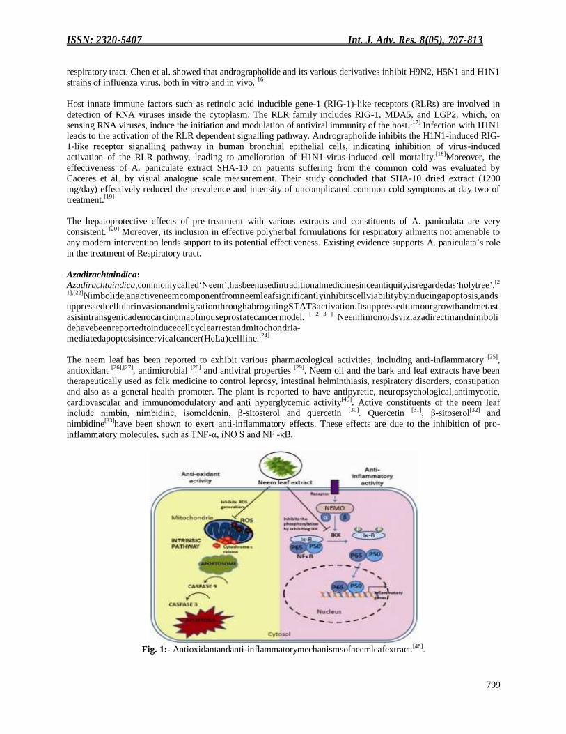
inflammatory molecules, such as TNF-α, iNO S and NF -κB.
Fig. 1:- Antioxidantandanti-inflammatorymechanismsofneemleafextract.[46].
ISSN: 2320-5407 Int. J. Adv. Res. 8(05), 797-813
800
Neem leafextracts(NLEs)showedanti-oxidant/anti-apoptoticpropertiesbyreducingreactiveoxygenspecies(ROS)
generationandinhibitingapoptoticresponsesthroughintrinsic mitochondrialpathway.NLEsalsoshowedanti-
inflammatoryresponsesbyinhibitingIkBkinase (IKK)andnuclear
translocationofNFκB(nuclearfactorkappaB)fortranscriptionofinflammatorygenes.[46]
In the study conducted by Lee J W et al, it was reported that treatment with NLE (Neem Leaf Extract) significantly attenuated the infiltration of inflammatory cells, such as neutrophils and macrophages in bronchoalveolar lavage
fluid (BALF). NLE also reduced the production of reactive oxygen species and the activity of neutrophil elastase in
BALF. Moreover, NLE attenuated the release of pro-inflammatory cytokines, such as tumor necrosis factor-α (TNF-
α) and interleukin (IL)-6 in BALF. NLE inhibited the recruitment of inflammatory cells and the expression of
monocyte chemoattractant protein-1 (MCP-1) in the lungs of mice with CS- and LPS-induced pulmonary
inflammation.[34]
Today, modern societies, finding themselves confounded in the web of their creation, are willing to revert to the
nature for remedies and neem tree provides a promising mean in this matter.
Nigella sativa:
Nigella sativa seeds and its oil had been widely used in traditional medicine (particularly in Unani Medicine) for a wide variety of illnesses including bronchial asthma in adults. There are several pharmacologically active
constituents in the essential oil of the plant, including thymoquinone (TQ).The adjuvant effect of N.sativaoil in
patients of bronchial asthma has already been reported but, no work had yet been done in very common disease of
children called wheeze associated lower respiratory tract illness (wheeze associated LRTI).In the study of Ahmed J
et al it was found that Nigella sativa seeds has effect in patients of wheeze associated LRTI, particularly in
children.[35]
The therapeutic effects of the plant extract against hypertension, asthma, cough, bronchitis, headache, fever,
influenzadiabetes and metabolic syndrome complications (e.g. obesity, dyslipidemia, and high blood glucose), cyclic
mastalgia (analgesic effects), hand eczema, vitiligo, pediatric seizures, opioid dependence, anxiety, infectious
diseases (e.g. infections caused by human immunodeficiency virus, hepatitis C virus, and Helicobacter pylori), infertility, asthma, chemical war injuries, tonsillopharyngitis, allergic rhinitis, rheumatoid arthritis, dyspepsia, celiac
disease, and hepatotoxicity of methotrexate were demonstrated in clinical studies .[35],[36],[37],[44]
Other studies used animal models of respiratory disorders to examine the effect of N. sativa extract and its active
compounds specially TQ. The preventive effect of hydro-ethanolic extract of N. sativa (0.08 g/kg/day, in drinking
water, for 14 days) on tracheal responsiveness and lung inflammation was shown in a guinea pig model of lung
injury induced by sulfur mustard.[38],[39]
Gunes et al investigated the effect of TQ treatment (50 mg/kg/day, administered by gavage for 5 days) on lung tissue
injury induced by hyperbaric oxygen (HBO₂ ) therapy, in a rat model. The antioxidant property of TQ led to
reduction of lipid hydroperoxide (LOOH) and total sulfhydryl group (-SH) causing a preventive effect on HBO₂ -
induced lung injury.[40]In a rabbit model with bacterial rhinosinusitis, N. sativa extract (50, 100, 200 mg/kg/day, administered orally for 7 days) reduced nitric oxide (NO) level and thus, prevented histopathological changes.[41]The
study of Kamal E. H et al suggested that VO-induced respiratory effects were mediated via release of histamine
withdirect involvement of histaminergic mechanisms and indirect activation of muscarinic
cholinergicmechanisms.[42]
The study of Umar S et al determined the possible effects of Nigella sativa on immune-response and pathogenesis of
H9N2 avian influenza virus in turkeys.It was found that the higher antibody titre against H9N2 AIV in turkeys fed
6% NS seeds shows the immunomodulatory nature of NS. Similarly, increased cytokine gene expression suggests
antiviral behaviour of NS especially in dose dependent manner, leading to suppressed pathogenesis of H9N2 viruses.
However, reduced virus shedding and enhanced immune responses were more pronounced in those turkeys received
NS.[43]
Nigella sativa seedshave a good antiviral property and is effective against respiratory related illness.
ISSN: 2320-5407 Int. J. Adv. Res. 8(05), 797-813
801
Phyllantusamarus:
The genus Phyllanthus consists of several species in the family Euphorbiaceae. Phyllanthus virgatusand another two
species, P. amarusand P. urinaria, are closely related in appearance and phytochemical structure. For example, P.
amarusinhibits the growth of human adenocarcinoma cell line Caco-2 [47], hepatoma induced by N -
nitrosodiethylamine in rats [48]
and sarcoma induced by 20-methylcholanthrene in mice. In Brazil and in many South
American countries, the infusion of roots, stems, and leaves of most Phyllanthus species have been used to cure a broad spectrum of diseases including intestinal infections, hepatitis B, diabetes, kidney, and urinary bladder
disturbances [49]. In Asia, several Phyllanthus species are used as febrifuge, diuretic, deobstruent, stomachic, and
antiseptic. Ayurveda uses the greatest number of Phyllanthus species where 15 species have been used in the
management of genitourinary, hypertension, cancer, skin, digestive, hepatic, and respiratory disorders.[50],[51],[52]
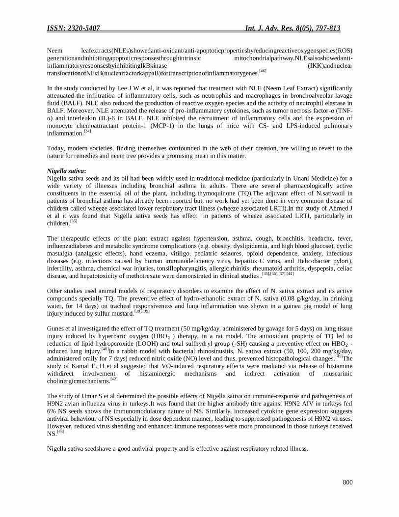
Fig. 2:- Phyllanthin-mediated inhibition of lipopolysaccharide (LPS)-induced inflammatory responses through NF-
κB, MAPKs, and PI3K-Akt signalling pathways in human macrophages.[53].
Recently, Harikrishnan et al., (2018a), Harikrishnan et al., (2018b), and Harikrishnan et al., (2018) investigated the
effects of 80% ethanol extract of P. amarusand its main constituents, phyllanthin, hypophyllanthin , and niranthin
(24–1.5 μM), using LPS-induced U937 human macrophages. They reported that their anti-inflammatory effects
were by downregulating the nuclear factor kappa-B (NF-κB), mitogen-activated protein kinase (MAPK), and
phosphatidylinositol-3-kinase (PI3K-Akt) signaling pathways. Fig 2depicts phyllanthin-mediated inhibition of LPS-
induced inflammatory responses through NF-κB, MAPKs, and PI3K-Akt signaling pathways in human
macrophages.The results demonstrated that P. amarusextract considerably repressed the aforementioned pro-
inflammatory mediators‟ release and expression of COX-2 protein. Also, the raised mRNA transcription of pro-
inflammatory markers was prominently reduced.[54],[55],[56]
A noteworthy inhibition of the percentage of CD4+ and CD8+ expression on spleen cells and in serum cytokines of
IL-2 and IFN-γ and IL-4 was seen [57] at a dose of 400 mg/kg of the extract. Interestingly, it was observed that P.
amarusadministration raised the levels of cellular GSH and GST, hence reducing the detrimental effects of
cyclophosphamide metabolites, a conventional immunosuppressive drug. In subsequent study, standardized P.
amarusextract (50–200 mg/kg) effects on cellular and humoral immune responses in mice were investigated [58].Phyllanthin also downregulated anti-sRBC immunoglobulins (IgM and IgG) antibody titer in immunized and
phyllanthin-treated mice in a dose-dependent manner with maximum inhibition at 100 mg/kg[59].
Hepatitis B virus claims around a million human lives annually. Sarma and colleagues attempted to explore a potent
and efficient antiviral from Phyllanthus with a minimal risk of resistance for hepatitis B virus. Moreover, in this
attempt the Phyllanthus active principles from among 93 phytochemicals were isolated to check the mechanism of
ISSN: 2320-5407 Int. J. Adv. Res. 8(05), 797-813
802
action against hepatitis B virus reverse transcriptase (HBV RT), which is an active target for drugs used against
HBV infections.[60]
The chemical compounds from Phyllanthus bear diverse biological activities, and providesalternative approach to
ongoingtherapy for immunological disorders.
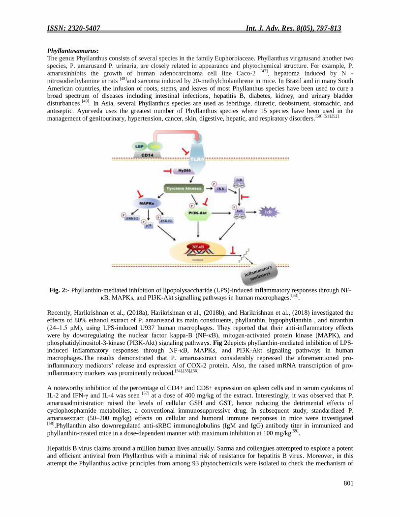
Table 1:- Partial list of viruses inhibited by medicinal plants.
Virus
Medicinal Plant used
Antiviral Effect
Reference
Dengue virus type-2
(DEN-2)
Human
immunodeficiency virus
Human
immunodeficiency virus
H9N2 avian influenza
virus
Human
immunodeficiency virus
Chikungunya
Virus
AzadirachtaindicaJuss.
(Neem)
Phyllanthus
amarusSchum. &Thonn.
Andrographis
paniculata(Burm.f.) Nees
Nigella sativa
Tinosporacordifoila
WithaniaSomnifera
The aqueous extract of neem
leaves inhibited
DEN-2 both in vitro and in
vivo
Inhibits HIV replication both
in vitro and in vivo
Antiviral effect through immunomodulation.
Increased CD4+ counts and
30%
decrease in viral load
Increased cytokine gene
expression suggests antiviral
behavior of NS, leading to
suppressed pathogenesis
Protease inhibitors for HIV
and
drug resistant HIV.
Tyramine
is a neuro‑ modulator. Used
to
treat anxiety and depression
by
inactivating
neurotransmitters
virus clearance in brain and
joint tissues on formulation
treatment revealed adirect
correlation of viral load in
brain to morbidity during
infection; likewise, joint
swelling receded prior to
complete viral
clearance explaining
possibleimmunomodulatory
effect
[61]
[62]
[63]
[43]
[64],[65]
[134]
ISSN: 2320-5407 Int. J. Adv. Res. 8(05), 797-813
803
Tinosporacordifoila:
Tinosporacordifolia[Tinosporacordifolia(Willd.) Miers ex Hook. F. &Thoms],known as Gulvelor Guduchi, has been
anextensively used and investigated plantfrom family Menispermaceae for its variedactivities. It is a deciduous,
fleshy, robustclimber growing with support of mango orneem trees, and is also known as CocculuscordifoliusDec,
MenispermumcordifoliumWilld., and Tinospora glabra (N. Brum.)Merr. Giloya, the Hindi name of theplant refers in
Hindu mythology to aheavenly elixir used to stay off the agingand to stay young forever. The Sanskritname ―Guduchi means one that protects from illnesses. Hence the words ―rejuvenator or ―adaptogen seem to have
appeared in literature.[66]
Tinosporine, Tinosporaside, cordifolide, cordifol, and hepatacosanol are important constituents of Gulvel. Barberine
and palmatine are major alkaloids in stem. The glucosides are 18-norclerodane glucoside, sesquiterpenes like
tinocordioside, tinocordifolioside, tinocordifolin, tinosponone, and cordioside, cordifolisides, and syringene. The
stem contains immunologically active substances –arabinogalactan and (1,4)-alpha-Dglucan[68],[69]. Crude values for
food content in Gulvel include high fibre (15.9%), sufficient protein (4.5%-11.2%), sufficient carbohydrate
(61.66%), and low fat(3.1%) [70],[71].Nutritive value is 292.54 calories per 100 g[70],[72]. Gulvelhas high potassium
(0.845%) (Regulatory function of nerve impulse)[70],[73]high chromium (0.006%) (Regulation of carbohydrate
utilization and pathophysiological alternations in diabetes mellitus)[74],[75]sufficient iron (0.28%) (Hematopoietic
functions)[70],[76],[77]and sufficient calcium (0.131%) (Regulatory functions in bloodcoagulation, and nervous, cardiovascular, and musculoskeletal systems[70],[78],[79].
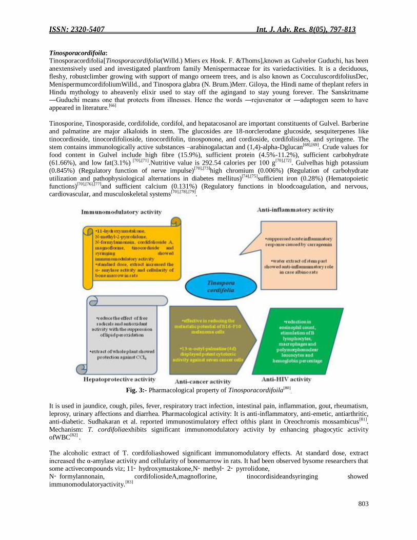
Fig. 3:- Pharmacological property of Tinosporacordifoila[80]
.
It is used in jaundice, cough, piles, fever, respiratory tract infection, intestinal pain, inflammation, gout, rheumatism,
leprosy, urinary affections and diarrhea. Pharmacological activity: It is anti-inflammatory, anti-emetic, antiarthritic,
anti-diabetic. Sudhakaran et al. reported immunostimulatory effect ofthis plant in Oreochromis mossambicus[81].
Mechanism: T. cordifoliaexhibits significant immunomodulatory activity by enhancing phagocytic activity
ofWBC[82] .
The alcoholic extract of T. cordifoliashowed significant immunomodulatory effects. At standard dose, extract
increased the α-amylase activity and cellularity of bonemarrow in rats. It had been observed bysome researchers that some activecompounds viz; 11‑ hydroxymustakone,N‑ methyl‑ 2‑ pyrrolidone,
N‑ formylannonain, cordifoliosideA,magnoflorine, tinocordisideandsyringing showed
immunomodulatoryactivity.[83]
ISSN: 2320-5407 Int. J. Adv. Res. 8(05), 797-813
804
The study of Kalikar M et al investigated that theTinosporacordifoliaextract, a plant derived immunostimulant,
significantly affected the symptoms of HIV. This was validated by clinical evaluation.Thus,
Tinosporacordifoliacould be used as an adjunct to HIV/AIDS management.[84]
The plant possesses anti-oxidant, anti-hyperglycemic, anti-neoplastic, anti-stress, anti-dote, anti-spasmodic, anti-
pyretic, antiallergic, anti-leprotic, antiinflammatory, anti hyperlypidaemia, Immunomodulatory properties. Hence, various parts of the plant contain immense medicinal property.[85]
Withaniasomnifera:
Ashwagandha (Withaniasomnifera, fam. Solanaceae) is commonly known as “Indian Winter cherry” or
“IndianGinseng”. It is one of the most important herbs of Ayurveda (the traditional system of medicine in India)
used for millennia as a Rasayana for its wide-ranging health benefits. The biologically active chemical constituents
of Withaniasomnifera(WS) include alkaloids (isopelletierine, anaferine, cuseohygrine, anahygrine, etc.), steroidal
lactones (withanolides, withaferins) and saponins. Many of its constituents support immunomodulatory
actions.[130],[131].W.somnifera compound, Withanone, docked very well in the binding interface of AEC2-RBD
complex, and was found to move slightly towards the interface centre on simulation. Withanone significantly
decreased electrostatic component of binding free energies of ACE2-RBD complex. Two salt bridges were also
identified at the interface; incorporation of Withanone destabilized these salt bridges and decreased their occupancies.Such an interruption of electrostatic interactions between the RBD and ACE2 would block or weaken
COVID-19 entry and its subsequent infectivity.[132]The collaborative study of DAILAB at Indian Institute of
Technology (IIT) Delhi and National Institute of Advanced Industrial Science and Technology (AIST),
Japan,revealed that the researchers targeted the main SARS-CoV-2 enzyme for splitting proteins, known as the Main
protease (Mpro). Mpro plays a key role in mediating viral replication. This is an attractive drug target for this virus,
and as humans don‟t naturally have this enzyme, compounds that target Mpro are likely to have low toxicity. They
discovered that a natural compound Withanone (Wi-N) derived from Ashwagandha and Caffeic Acid Phenethyl
Ester (CAPE), an active ingredient of New Zealand Propolis, has the potential to interact with and block the activity
of Mpro[133].
The Antiviral and Immunomodulatory effects of Vitamins, Trace elements and Nutraceuticals: Nutraceuticals are dietary supplements, dietary fiber , genetically engineered designer foods, specific diets, and
processed foods, such as cereals, soups, and beverages utilized to ameliorate health, delay senescence, prevent
diseases, and support proper functioning of human body. Currently nutraceuticals are getting substantial attention
due to nutrition and therapeutic potentials. They have benefit over medicine because they avoid side effect. On the
basis of their source, they are categories into different terms such as nutrients, dietary supplements, herbals, dietary
fibre, etc. Global market for nutraceutical is huge i.e. approximately USD 117 billion. Nutraceuticals are products
that claim physiological benefit or protection against chronic disease. [86],[87]
Zinc:
Zinc is an essential trace element which plays an important role in growth, development, and the maintenance of
immune function [88],[89]. Zinc deficiency has been associated with an increased susceptibility to infectious diseases,
including viral infections. Studies have shown that the zinc status of an individual is a critical factor that can influence immunity against viral infections, with zinc-deficient populations being at increased risk of acquiring
infections, such as HIV or HCV [88]. Few RCTs have evaluated the effect of zinc supplementation on the immune
response. A study by Acevedo-Murillo et al. among 103 children (1 month to 5 years) with pneumonia showed a
statically significant clinical improvement (duration of illness, respiratory rate and oxygen saturation) in the zinc
supplemented group compared to placebo [90]. They also demonstrated an increase in the cytokine response in Th1
pattern (IL-2 and INF-γ) only in the zinc group, with Th2 cytokines (IL-4 and IL-10) being elevated or remaining
high in both groups. Another RCT on oral supplementation of high-dose zinc (150 mg/day) after stem cell
transplantation, demonstrated that it enhances thymic function and the output of new CD4+ naïve T cells, helping to
prevent the reactivation of TTV [91]. However, a study by Provincial et al. concluded that although prolonged
supplementation with zinc (400 -250 mg/day) or zinc+arginine (4 d/day) in the elderly (age 64-100 years) restores
zinc plasma concentrations, it is ineffective in inducing or ameliorating the antibody response or number of CD3, CD4 or CD8 lymphocytes after influenza vaccination[92]
ISSN: 2320-5407 Int. J. Adv. Res. 8(05), 797-813
805
Vitamin C:
Vitamin C is known as an essential antioxidant and enzymatic co-factor for many physiological reactions in the
body, such as hormone production, collagen synthesis andimmune potentiation [93]. In-vivo animal studies in mice
have shown that it is an essentialfactor for the antiviral immune responses against the influenza A virus (H3N2)
through theincreased production of interferon-α/β, especially at the early stages of the infection [93]
. However, our
literature search was unable to identify RCTs examining the use of vitamin C for the treatment for specific viral infections. Furthermore, a systematic review and meta-analysis on the role of vitamin C for preventing and treating
the common cold, did not find any conclusive evidence to indicate that there is benefit of using vitamin C mega
dose prophylaxis in the community to reduce the incidence of common cold, which is most often caused by viral
infections[94].
Vitamin E:
Vitamin E, a fat-soluble vitamin, is a potent antioxidant and has the ability to modulate host immune functions [95].
Vitamin E deficiency is known to impairs both humoral and cellular immunity [95]. However, few studies have
shown that vitamin E supplementation might cause harmful effects on the incidence of infectious disease. A study
among 50-69 years old adult smokers showed that vitamin E supplementation increases the risk of pneumonia [96].
Similarly, supplementation of vitamin E (200 IU/day) did not have a statistically significant effect on lower
respiratory tract infections in elderly nursing home residents[97]. However positive effects of vitamin E have been observed in the treatment of chronic hepatitis B in a small pilot RCT, where a significantly higher normalization
ofliver enzymes and HBV-DNA negativization, was observed in the vitamin E group [98].Similar results have been
observed in a RCT in the paediatric population, where vitamin E treatment resulted in a higher anti-HBe
seroconversion and virological response [99].
Curcumin:
Curcumin (CUR) plant-derived polyphenol and is the principal curcuminoid of turmeric and possesses strong
antioxidant and anti-inflammatory activities[100],[101]. CUR prevented oxidative damage and apoptosis in a rodent
model of gentamicin-induced hepato- and nephrotoxicity [100],[101]. Besides its ability to suppress oxidativestress and
inflammation, CUR possesses anticancer, anti-atherosclerotic, anti-diabetic and anti-obesityproperties[102].
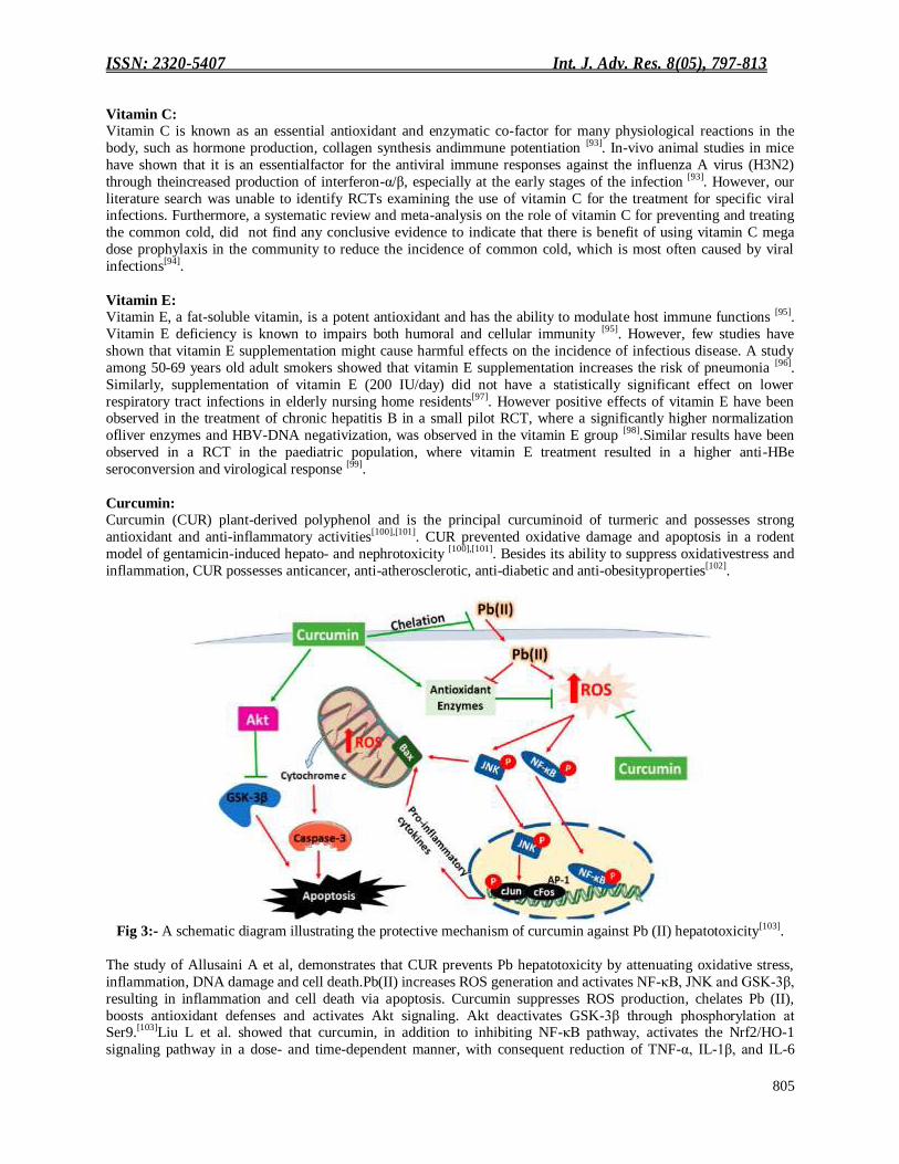
Fig 3:- A schematic diagram illustrating the protective mechanism of curcumin against Pb (II) hepatotoxicity[103].
The study of Allusaini A et al, demonstrates that CUR prevents Pb hepatotoxicity by attenuating oxidative stress,
inflammation, DNA damage and cell death.Pb(II) increases ROS generation and activates NF-κB, JNK and GSK-3β,
resulting in inflammation and cell death via apoptosis. Curcumin suppresses ROS production, chelates Pb (II),
boosts antioxidant defenses and activates Akt signaling. Akt deactivates GSK-3β through phosphorylation at Ser9.[103]Liu L et al. showed that curcumin, in addition to inhibiting NF-κB pathway, activates the Nrf2/HO-1
signaling pathway in a dose- and time-dependent manner, with consequent reduction of TNF-α, IL-1β, and IL-6
ISSN: 2320-5407 Int. J. Adv. Res. 8(05), 797-813
806
levels in vitro, decrease in eosinophil and WBC counts in BAL, and reduction of AHR in a murine model of
asthma.[104]The effects of curcumin on pulmonary fibrosis derive from its action on multiple pathways and through
multiple mechanisms. As in asthma, the curcumin inhibition of NF-κB has a role in pulmonary fibrosis, by causing a
reduction of TNF-α and cyclo-oxygenase 2 (COX-2) levels [105] and TGF-β1 levels [106].Kurup et al used a murine
model of latex allergy to investigate the role of curcumin as animmunomodulator. BALB/c mice were exposed to
latex allergens and developed latex allergywith a thyroid hormone (Th)2-type immune response.[107]
Grape Seed Extract:
Grape seed proanthocyanidins (GSPs) are promising agents that have antioxidant properties [108] and appear to
exhibit minimal toxicity [109]. GSPs are a mixture of polyphenols/flavanols and mainly contain proanthocyanidins
(89%), which constitute dimers, trimers, tetramers, and oligomers/polymers of monomeric catechins and/ or (-)-
epicatechins[110]. The results from the study of Akhtar S et al showed for the first time the chemotherapeutic efficacy
of GSPs in controlling the growth of human NSCLC cells in vitro and tumor xenograft growth in vivo. The in vivo
studies show that inhibition of the growth of lung tumor xenografts in nude mice by dietary GSPs is associated with
the inhibition of tumor cell proliferation, angiogenesis, and up-regulation of IGFBP 3.[111] The study of Zhou S Y et
al evaluated GSPE‟s effects on airway inflammation and airway remodeling in a chronic asthmatic model. The
GSPE treatment markedly decreased interleukin (IL)-4, IL-13, and vascular endothelial growth factor (VEGF) levels
in BALF in addition to the total serum IgE levels. A histological examination demonstrated that GSPE significantly ameliorated allergen-induced lung eosinophilic inflammation and decreased PAS-positive epithelial cells in the
airway.[112] The study of Ali Asghar Hemmatia et al investigated the effect of grape seed extract on bleomycin-
induced lungfibrosis in rat. It was found that grape seed extract was able to diminish the fibrogenic effects of
bleomycin on lung. This effect of grape seed can be attributed to activeingredients of the plant with anti-oxidant
properties.[113]Grape seed extract (GSE) has antiviral activities against hepatitis A virus (HAV) and human
norovirussurrogates (feline calicivirus (FCV-F9) and murine norovirus (MNV-1)).[114]
Stevia:
Stevioside, an abundant component of Stevia rebaudiana leaf, has become well-known for its intensesweetness
(250–300 times sweeter than sucrose) and is used as a non-caloric sweetener in several countries. A number of
studies have suggested that, beside sweetness, stevioside along with related compounds, which include rebaudioside A (second most abundant component of S. rebaudiana leaf), steviol and isosteviol(metabolic components of
stevioside) may also offer therapeutic benefits, as they have anti-hyperglycemic, anti-hypertensive, anti-
inflammatory, anti-tumor, anti-diarrheal, diuretic, and immunomodulatory actions. It is of interest to note that their
effects on plasma glucose level and blood pressure are only observed when these parameters are higher than normal.
As steviol can interact with drug transporters, its role as a drug modulator is proposed.[115]
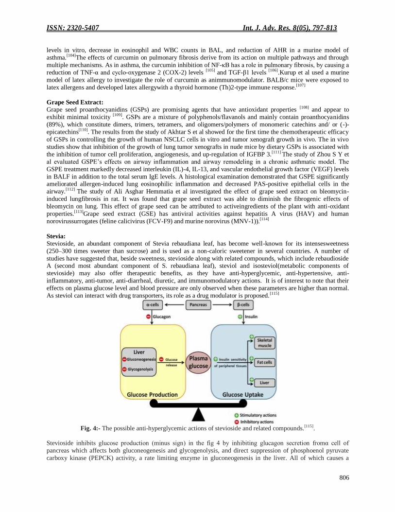
Fig. 4:- The possible anti-hyperglycemic actions of stevioside and related compounds.[115].
Stevioside inhibits glucose production (minus sign) in the fig 4 by inhibiting glucagon secretion fromα cell of
pancreas which affects both gluconeogenesis and glycogenolysis, and direct suppression of phosphoenol pyruvate
carboxy kinase (PEPCK) activity, a rate limiting enzyme in gluconeogenesis in the liver. All of which causes a
ISSN: 2320-5407 Int. J. Adv. Res. 8(05), 797-813
807
reduction of glucose release from the liver. On the other hand, stevioside, steviol and rebaudioside A stimulate
glucose uptake (plus sign) in the fig 4 by increasing insulin secretion from β cell of pancreas and enhancing insulin
sensitivity of peripheral tissues promoting glucose uptake. Therefore, they exhibit antihyperglycemic action by
reducing glucose production while increasing glucose uptake to maintain plasma glucose balance.[115]Stevia
increases the secretion of GIP, insulin, leptin, body weight, and glycaemia but keeps food consumption normal.
Sweeteners modulate the hormonal response of cytokines and the proliferation of lymphocytes in the intestinal mucosa.[116]The study of Boonkaewwan, Cet al suggested that stevioside attenuates synthesis of inflammatory
mediators in LPS-stimulated THP-1 cells byinterfering with the IKKâand NF-_B signaling pathway, and stevioside-
induced TNF-R secretion ispartially mediated through TLR4 thereby elucidating the anti-inflammatory and
immunomodulatory activities of stevioside and its metabolite, steviol.[117]
Vitamin A:
Vitamin A is a fat-soluble vitamin, which is crucial for maintaining vision, promoting growth and development, and
protecting epithelium and mucosal integrity in the body [118]. It is known to play an important role in enhancing
immune function, and having a regulatory function in both cellular and humoral immune responses [118]. Vitamin A
supplementation to infants has shown the potential to improve antibody response after some vaccines, including
measles [118] and anti-rabies vaccination (2.1 times) [119]. Inaddition, an enhanced immune response to influenza virus
vaccination has also been observed in children (2-8 years) who were vitamin A and D-insufficient at baseline, aftersupplementation with vitamin A and D [120].
Vitamin D: Vitamin D, another fat-soluble vitamin, plays a vital role in modulating both innate andadaptive immune responses [121]. Epidemiological data has linked vitamin D deficiency toincreased susceptibility to acute viral respiratory
infections [122]. Recent reviews evaluating possible mechanisms suggest that vitamin D plays an important
modulatory role of the innate immune responses to respiratory viral infections, such as Influenza A and
parainfluenza 1 and 2, and Respiratory syncytial virus (RSV) [123]. A systematic review on the role of vitamin D in
the prevention of acute respiratory infections, which included studies (4 cross-sectional studies, 8 case-control
studies, 13 cohort studies and 14 clinical trials), noted that observational studies predominantly reported statistically
significant associations between low vitamin D status and increased risk of both upper and lower respiratory tract infections [124]. A study by Aglipay et al. on the effect of high dose (2000 IU/day) vs. standard-dose (400 IU/day)
vitamin D supplementation on viral upper respiratory tract infections did not show any significant difference
between the two group[125]. However, only about 1/3 of the study population had vitamin D levels <30 ng/ml. A
recent RCT on the impact of vitamin D supplementation on influenza vaccine response in deficient elderly person,
showed that it promotes a higher TGFβ plasma level without improving antibody production, and suggested that
supplementation seems to direct the lymphocyte polarization toward a tolerogenic immune response [126]. Similarly,
in another RCT, a monthly high-dose (100,000 IU/month) vitamin D supplementation reduced the incidence of acute
respiratory infections in older long-term care residents, in comparison to a standard dose group (12,000 IU/month) [127]. It is evident that the role of vitamin D supplementation on antiviral immunity against respiratory infections is
likely to depend on the vitamin D status of the individual. Furthermore, vitamin D has demonstrated a beneficial
effect in other viralinfections, for example adding vitamin D to conventionalPeg-α-2b/ribavirin therapy for
treatment-naïve patients with chronic HCV genotype 1infection significantly improved the viral response [128], and a similar effect has also beenobserved in patients with HCV genotype 2-3 [129].
Conclusion:- Many traditional medicinal plants and herbs were reported to have strong antiviral activity. In view of the
signification number of plant extracts that have yielded positive results it seems reasonable to concludethat there are
probably numerous kinds of antiviral agents in these materials. The traditional use of some of the medicinal plants for the treatment of infectious diseases of viral origin, therefore, is justified. Finally, the development of new
medicinal plant products is vital in controlling the threats posed by some pathogenic viruses. Although many
synthetic immunomodulatory drugs with various mechanisms of action have been discovered and developed, they
failed to be successful clinically due to their toxicity, less bioavailability, and stability problem. Medicinal herbs and
their active metabolites deliver alternative potential to ongoing therapy for a wide array of immunological disorders
by modulation of the immune response. Research to discover natural products as drug candidates for development of
immunomodulatory agents has gained momentum as they offer safer alternatives to conventional therapies.
ISSN: 2320-5407 Int. J. Adv. Res. 8(05), 797-813
808
Nutraceuticals might be defined as substances that have physiological benefits that prevents against chronic diseases
and has antiviral property and immunomodulatory effects. Nowadays, nutraceuticals have received considerable
interest due to potential nutritional, safety and therapeutic effects. In the present review much effort has been
devoted to present new concepts about nutraceuticals based on their diseases modifying indications. Emphasis has
been made to present herbal nutraceuticals as its antiviral and immunomodulator properties. The use of
nutraceuticals, as an attempt to accomplish desirable therapeutic outcomes with reduced side effects, as compared with other therapeutic agents has met with great monetary success.
References:- 1. Dhawan, B. N. (2012). Anti-Viral Activity of Indian Plants. Proc. Natl. Acad. Sci. Sect B. Biol. Sci. 82,
209–224.
2. Jassim, S. A. A., &Naji, M. A. (2003). Novel antiviral agents: a medicinal plant perspective.Journal of Applied Microbiology.95, 412–427.
3. Lin, L. T., Hsu, W. C., & Lin, C. C. (2014). free information in English and Mandarin on the novel
coronavirus COVID- Antiviral Natural Products and Herbal Medicines. Journal of Traditional and
Complementary Medicine, 4, 24–35.
4. Chan, K. (2003). Some aspects of toxic contaminants in herbal medicines. 52, 1361–1371.
5. Mahady GB. (2001) Global harmonization of herbal health claims. J Nutr;131(3):1120S–3S.
6. Rates SM. (2001) Plants as source of drugs. Toxicon.;39(5): 603–13.
7. Kumar, R., Piya, G., Mudgal, P., Maity, H., &Dowarha, D. (2015). Herbal plants and plant preparations as
remedial approach for viral diseases. VirusDisease, 26(4), 225–236.
8. Williamson E. (2003). Drug interactions between herbal and prescription medicines. Drug Saf; 26:1075-92.
9. Wal, P., Wal, A., Gupta, S., Sharma, G., & Ak, R. (n.d.). Pharmacovigilance of Herbal Products in India.
3(3), 256–258.
10. M. Rajani, N. Shrivastava, and M. N. Ravishankara.(2000). “A rapid method for isolation of andrographolide
from AndrographispaniculataNees (Kalmegh),” Pharmaceutical Biology, 38 (3),204–209.
11. R. A. Kumar, K. Sridevi, N. Vijaya Kumar, S. Nanduri, and S. Rajagopal. (2004). “Anticancer and
immunostimulatory compounds from Andrographis paniculata,” Journal of Ethnopharmacology. 92(2-3),
291–295.
12. Q. Du, G. Jerz, and P. Winterhalter.(2003). “Separation of andrographolide and neoandrographolide from the
leaves of Andrographispaniculatausing high-speed counter-current chromatography,” Journal of
Chromatography A, 984(1), 147–151.
13. Jayakumar, T., Hsieh, C. Y., Lee, J. J., &Sheu, J. R. (2013). Experimental and clinical pharmacology of
andrographispaniculata and its major bioactive phytoconstituent andrographolide. Evidence-Based
Complementary and Alternative Medicine, 2013, 1–16.
14. K. Mishra, A. P. Dash, B. K. Swain, and N. Dey. (2009) “Antimalarial activities of Andrographis
paniculataand Hedyotiscorymbosa extracts and their combination with curcumin,” Malaria Journal, 8(1),
article 26,
15. N. Poolsup, C. Suthisisang, S. Prathanturarug, A. Asawamekin, and U. Chanchareon. (2004) “Andrographis
paniculatain the symptomatic treatment of uncomplicated upper respiratory tract infection: systematic review of randomized controlled trials,” Journal of Clinical Pharmacy and Therapeutics, 29(1), 37–45.
16. Chen J-X, Xue H-J, Ye W-C, Fang B-H, Liu Y-H, Yuan S-H et al (2009) Activity of andrographolide and its
derivatives against influenza virus in vivo and in vitro. Biol Pharmaceut Bull ,32,1385–1391.
17. Lassig C, Hopfner KP (2016) RIG-I-like receptors: one STrEP forward. Trends Microbiol 24(7),517–519.
18. Yu B, Dai C, Jiang Z, Li E, Chen C, Wu X et al .(2014). Andrographolide as an anti-H1N1 drug and the
mechanism related to retinoic acid-inducible gene-I-like receptors signaling pathway. Chin J Integr Med,
20(7).540–545.
19. Ca´ceres DD, Hancke JL, Burgos RA, Sandberg F, Wikman GK. (1999). Use of visual analogue scale
measurements (VAS) to assess the effectiveness of standardized Andrographis paniculate extract SHA-10 in
reducing the symptoms of common cold. A randomized double blind-placebo study. Phytomedicine,
6(4),217–223.
20. Akbar, S. (2011). Andrographis paniculata: A review of pharmacological activities and clinical effects.
Alternative Medicine Review, 16(1), 66–77.
21. HossainMA,Al-ToubiWAS,WeliAM,Al-RiyamiQA& Al-SabahiJN. (2013).
ISSN: 2320-5407 Int. J. Adv. Res. 8(05), 797-813
809
IdentificationandcharacterizationofchemicalcompoundsindifferentcrudeextractsfromleavesofOmanineem.JTa
ibah UnivSci,7 ,181-88.
22. PankajS,LokeshwarT,MukeshB&VishnuB. (2011). Reviewonneem(Azadirachtaindica):Thousandproblems
onesolution.IntRes J Pharm,2 ,97-102.
23. ZhangJ,AhnKS,KimC,ShanmugamMK,SiveenKS, ArfusoF,SamymRP,DeivasigamanimA,LimLH,WangL,GohBC,KumarAP,HuiKM&SethiG. (2016).
Nimbolide-inducedoxidativestressabrogatesSTAT3signalingcascadeand
inhibitstumourgrowthintransgenicadenocarcinomaof mouseprostatemodel.AntioxidRedoxSignal,24, 575-89.
24. PriyadarsiniRV,MuruganRS,SripriyaP,KarunagaranD&NaginiS. (2010).
Theneemlimonoidsazadirachtinandnimbolideinducecellcyclearrestandmitochondria-
mediatedapoptosisinhumancervicalcancer(HeLa)cells.FreeRadRes,44,624-34.
25. Okpanyi SN and Ezeukwu GC. (1981). Anti-inflammatory and antipyreticactivities of Azadirachtaindica.
Planta Med., 41,34-39.
26. Rao AD, Devi KN and ThyagarajuK.(1998). Isolation of antioxidantprinciple from Azadirachtaseed kernels:
determination of itsrole on plant lipoxygenases. J Enzyme Inhib., 14,85-96.
27. Yanpallewar SU, Sen S, Tapas S, Kumar M, Raju SS andAcharya SB. (2003). Effect of Azadirachtaindicaon
paracetamol‑ inducedhepatic damage in albino rats. Phytomedicine, 10,391-396.
28. Almas K. (1999). The antimicrobial effects of extracts of Azadirachtaindica(Neem) and
Salvadorapersica(Arak) chewingsticks. Indian J Dent Res.,10, 23-26.
29. Badam L, Joshi SP and Bedekar SS. (1999). „In vitro‟ antiviral activity ofneem (Azadirachtaindica. A. Juss) leaf extract against group Bcoxsackieviruses. J Commun Dis., 31, 79-90.
30. Siddiqui BS, Afshan F, Gulzar T and Hanif M.(2004). Tetracyclictriterpenoids from the leaves of
Azadirachtaindica.Phytochemistry,65, 2363-2367.
31. Chang YC , Tsai MH, Sheu WH, Hsieh SC and Chiang AN.(2013). Thetherapeutic potential and mechanisms
of action of quercetin inrelation to lipopolysaccharide-induced sepsis in vitro and in vivo.PLoS One, 8,
e80744.
32. Loizou S, Lekakis I, Chrousos GP and Moutsatsou P. (2010). Beta‑ sitosterolexhibits anti-inflammatory activity in human aortic endothelialcells. Mol Nutr Food Res, 54,551-558.
33. Pillai NR and Santhakumari G. (1981). Anti-arthritic and anti-inflammatoryactions of nimbidin. Planta Med.,
43, 59-63.
34. Lee, J. W., Ryu, H. W., Park, S. Y., Park, H. A., Kwon, O. K., Yuk, H. J, Ahn, K. S. (2017). Protective
effects of neem (Azadirachtaindica A. Juss.) leaf extract against cigarette smoke- and lipopolysaccharide-
induced pulmonary inflammation. International Journal of Molecular Medicine, 40(6), 1932–1940.
35. Ahmad, J., Ali Khan, R., & Malik, M. A. (2010). A study of Nigella sativa oil in the management of wheeze associated lower respiratory tract illness in children. African Journal of Pharmacy and Pharmacology, 4(7),
436–439.
36. Gholamnezhad Z, Havakhah S, Boskabady MH. (2016). Preclinical and clinical effects of Nigella sativa and
its constituent, thymoquinone: A review. J Ethnopharmacol, 190,372-386.
37. Tavakkoli A, Mahdian V, Razavi BM, Hosseinzadeh H. (2017). Review on clinical trials of black seed
(Nigella sativa) and its active constituent, thymoquinone. J Pharmacopuncture, 20,179-193.
38. Boskabady MH, Vahedi N, Amery S, Khakzad MR. (2011). The effect of Nigella sativa alone, and in
combination with dexamethasone, on tracheal muscle responsiveness and lung inflammation in sulfur mustard exposed guinea pigs. J Ethnopharmacol, 137,1028-1034.
39. Hossein BM, Nasim V, Sediqa A. (2008). The protective effect of Nigella sativa on lung injury of sulfur
mustard-exposed Guinea pigs. Exp Lung Res, 34,183-194.
40. Gunes AE, Gozeneli O, Akal AA, Guldur ME, Savik E. (2017). Reduction of side effects of hyperbaric
oxygen therapy with thymoquinone treatment in rats. Undersea Hyperb Med, 44,337-343.
41. Yoruk O, Tatar A, Keles ON, Cakir A. (2017). The value of Nigella sativa in the treatment of experimentally
induced rhinosinusitis. Acta Otorhinolaryngol Ital., 37,32-37.
42. Kamal E. H. El Tahir, Mohammad M. S. Ashour And Mohammad M. Al-Harm. (1993). The respiratory effects of the volatile oil of the black seed (Nigella sativa) in guinea-pigs: Elucidation of the mechanism(s) of
action. General Pharmacology, 24(5), 1115–1122.

ISSN: 2320-5407 Int. J. Adv. Res. 8(05), 797-813
810
43. Umar, S., Munir, M. T., Subhan, S., Azam, T., un Nisa, Q., Khan, M. I., Shah, M. A. (2016). Protective and
antiviral activities of Nigella sativa against avian influenza (H9N2) in turkeys. Journal of the Saudi Society of
Agricultural Sciences, 1–7.
44. Shamim Molla, Md. Abul Kalam Azad, Md Ali Azam Al Hasib, M. Monayem Hossain, Md. SohelAhammed, Shohel Rana, Muhammad Torequl Islam. (2019). A Review on Antiviral Effects ofNigella
Sativa L., Phol,2 ,47-53.
45. Barua, C. C., Talukdar, A., Barua, A. G., Chakraborty, A., Sarma, R. K., & Bora, R. S. (2010). Evaluation of
the wound healing activity of methanolic extract of Azadirachta Indica (Neem) and Tinosporacordifolia
(Guduchi) in rats. Pharmacologyonline, 1, 70–77.
46. Yadav, D. K., Bharitkar, Y. P., Chatterjee, K., Ghosh, M., Mondal, N. B., &Swarnakar, S. (2016).
Importance of neem leaf: An insight into its role in combating diseases. Indian Journal of Experimental
Biology, 54(11), 708–718.
47. Lawson-Evi P, Eklu-Gadegbeku K, Agbonon A, Aklikokou K, Moukha S, Creppy EE, Gbeassor M. (2008). Toxicological assessment on extracts of Phyllanthus amarusSchum and Thonn. Sci Res Essay, 3, 410–415.
48. Rajeshkumar NV, Kuttan R. (2000). Phyllanthus amarusextract administration increases the life span of rats
with hepatocellular carcinoma. J Ethnopharmacol, 73, 215–219.
49. Calixto, J. B., Santos, A. R. S., Filho, V. C., and Yunes, R. A. (1998). A review of the plants of the genus
Phyllanthus: Their chemistry, pharmacology, and therapeutic potential. Med. Res. Rev. 18, 225–258.
50. Adil, M. D., Kaiser, P., Satti, N. K., Zargar, A. M., Vishwakarma, R. A., and Tasduq, S. A. (2010). Effect of Emblica officinalis (fruit) against UVB-induced photo-aging in human skin fibroblasts. J. Ethnopharmacol.
132, 109–114.
51. Nain, P., Saini, V., Sharma, S., and Nain, J. (2012). Antidiabetic and antioxidant potential of Emblica
officinalis Gaertn. leaves extract in streptozotocin-induced type-2 diabetes mellitus (T2DM) rats. J.
Ethnopharmacol. 142, 65–71.
52. Sarin, B., Verma, N., Martín, J. P., and Mohanty, A. (2014). An overview of important ethnomedicinal herbs
of Phyllanthus species: present status and future prospects. Sci. World J. 2014, 12.
53. Jantan, I., Haque, M. A., Ilangkovan, M., & Arshad, L. (2019). An insight into the modulatory effects and mechanisms of action of phyllanthus species and their bioactive metabolites on the immune system. Frontiers
in Pharmacology, 10(JULY), 1–19.
54. Harikrishnan, H., Jantan, I., Haque, M. A., and Kumolosasi, E. (2018a). Anti- Inflammatory effects of
hypophyllanthin and niranthin through downregulation of NF-κB/MAPKs/PI3K-Akt signaling pathways.
Inflammation 41, 984–995.
55. Harikrishnan, H., Jantan, I., Haque, M. A., and Kumolosasi, E. (2018b). Phyllanthin from Phyllanthus
amarusinhibits LPS-induced proinflammatory responses in U937 macrophages via downregulation of NF-
κB/MAPK/PI3K-Akt signaling pathways. Phytother. Res. 32 (12), 2510–2519.
56. Harikrishnan, H., Jantan, I., Haque, M. A., and Kumolosasi, E. (2018c). Anti-inflammatory effects of Phyllanthus amarusSchum. &Thonn. through inhibition of NF-κB, MAPK, and PI3K-Akt signaling pathways
in LPS-induced human macrophages. BMC Complement. Alternat. Med. 18, 224.
57. Ilangkovan, M., Jantan, I., Mesaik, M. A., and Bukhari, S. N. A. (2015). Immunosuppressive effects of the
standardized extract of Phyllanthus amaruson cellular immune responses in Wistar-Kyoto rats. Drug Des.
Dev. Ther. 9, 4917–4930.
58. Ilangkovan, M., Jantan, I., and Bukhari, S. N. A. (2016). Phyllanthin from Phyllanthus amarusinhibits
cellular and humoral immune responses in Balb/C mice. Phytomedicine,23, 1441–1450.
59. Janeway, C. A., Travers, P., Walport, M., and Shlomchik, M. J. (2005). “Chapter 14 Manipulation of the
Immune Response” in Immunobiology: the immune system in health and disease, 6th Edition (New York,
USA: Garland Science Publishing).
60. Sarma, K.; Borkakoty, B.; Parida, P.; Jakharia, A.; Dey, D.; Biswas, D.; Panda, D.; K Modi, M.; K
Mohapatra, P.; Mahanta, J.(2016). In Silico Identification of Natural Lead Molecules from the Genus of
Phyllanthus Against Hepatitis B Virus Reverse Transcriptase. Nat. Prod. J., 6, 292–304.
61. Parida, M.M., Upadhyay, C., Pandya, G., Jana, A.M., 2002. Inhibitory potential of neem
(AzadirachtaindicaJuss) leaves on dengue virus type-2 replication. J. Ethnopharmacol. 79 (2), 273–278.
62. Notka, F., Meier, G., Wagner, R., (2004). Concerted inhibitory activities of Phyllanthus amaruson HIV
replication in vitro and ex vivo. Antiviral Res. 64(2), 93–102.
ISSN: 2320-5407 Int. J. Adv. Res. 8(05), 797-813
811
63. Mukhtar, M., Arshad, M., Ahmad, M., Pomerantz, R. J., Wigdahl, B., & Parveen, Z. (2008). Antiviral
potentials of medicinal plants. Virus Research, 131(2), 111–120.
64. Ghosh AK, Martyr CD, Steffey M, Wang YF, Agniswamy J, Amano M.(2011). Design of substituted bis ‑
Tetrahydrofuran (bis- THF) ‑ derived potent HIV ‑ 1 protease inhibitors, protein‑ ligand X‑ ray structure, and convenient syntheses of bis ‑ THF and Substituted bis ‑ THF Ligands. ACS Med Chem Lett.,2,298-
302.
65. Mukherjee R, De UK, Ram GC. (2010). Evaluation of mammary gland immunity and therapeutic potential
of Tinosporacordifoliaagainst bovine subclinical mastitis. Trop Anim Health Prod.,42,645‑ 51.
66. Madhav Mutalik1, M. M. (2011). TinosporaCordifolia And Its Varied Activities: What Is Believed and What
Is Known? International Journal of Current Research and Review, 03(12), 94–109.
67. Ninivaggi FJ. An Elementary Textbookof Ayurveda; Medicine with sixthousand-year-old tradition. Madison,Connecticut: InternationalUniversities/Psychosocial Press; 2001,p.16-20.
68. Chintalwar G, Jain A, SipahimalaniA,Banerji A, SumariwallaP,Ramakrishnan R.(1999)
Animmunologicallyactivearabinogalactan from Tinosporacordifolia. Phytochemistry,52(6),1089-93.
69. Nair PK, Melnick SJ, RamachandranR, Escalon E, Ramachandran C. (2006).Mechanism of macrophage
activationby (1,4)-alpha-D-glucan isolated fromTinosporacordifolia. IntImmunopharmacol,6,1815-24.
70. Nile SH, Khobragade CNN. (2009)Determination of Nutritive Value andMineral Elements of some
ImportantMedicinal Plants from Western Part ofIndia. Journal of Medicinal Plants,8(5), 79-88.
71. Upadhyay AK, Kaushal Kumar,Arvind Kumar, Mishra HS. (2010).Tinosporacordifolia(Willd.) Hook. f. and Thoms.(Guduchi) - Validation of the ayurvedicpharmacology through experimentaland clinical studies. Int J
Ayurveda Res.,1(2),112-21.
72. Brody T. Nutritional Biochemistry. 2ndEdn. San Diego Academic press; 1998,p.11-2.
73. Underwood EJ and Suttle NF. Themineral nutrition of Livestock. 3rd Edn.New York: CABI
publishing;1999,p.51-101.
74. Hambridge KM. (1974). Chromium nutrition inman. Am J Clin Nutr.,27,505-14.
75. Jamal H, Raza H, Janua KM, BhattyMK. (1986). Chromium on human health. PakJ Sci Ind Res,29:45-7.
76. Gaeta A, Hider RC. (2005). The crucial role ofmetal ions in neurodegeneration: Thebasis for promising
therapeuticstrategy. Br J Pharmacol,146,1041-59.
77. Weight LM, Jalobes P, Noakes TD. (1992).Dietary Iron deficiency and sportsanemia. Br J Nutr,68,253-60.
78. Heaney RD. (1994). Thinking straight aboutcalcium. New Engl J Med,328(7),503-5.
79. Hasling C, Sondergard K, MoselkiloeCP. (1991). Calcium metabolism inpostmenopausal osteoporotic woman isdetermined by dietary calcium andcoffee intake. Am. Ins. of Nutria.23,119-26.
80. Antul, K., Amandeep, P., Gurwinder, S., & Anuj, C. (2019). Review on Pharmacological Profile of
Medicinal Vine: Tinosporacordifolia. Current Journal of Applied Science and Technology, 35(5), 1–11.
81. Sudhakaran DS, Srirekha P, Devasree LD. (2006). Immunostimulatory effect of TinosporacordifoliaMiers
leaf extract in Oreochromis mossambicus. Ind J Exp BioI, 44,726-32.
82. Sharma U, Bala M, Kumar N.(2012). Immunomodulatoryactive compounds from Tinosporacordifolia. J
Ethnopharmacol, 141,918-26.
83. Upadhyaya R, PR, Sharma V, Anita KV. (2011). Assessment of the multifaceted immunemodulatory
potential of the aqueous extract of Tinosporacordifolia. Res J Chem Sci.,1,71‑ 9.
84. Kalikar, M., Thawani, V., Varadpande, U., Sontakke, S., Singh, R., &Khiyani, R. (2008).
Immunomodulatory effect of Tinosporacordifolia extract in human immuno-deficiency virus positive
patients. Indian Journal of Pharmacology, 40(3), 107–110.
85. Spandana, U., Ali, S. L., Nirmala, T., Santhi, M., &Sipai Babu, S. D. (2013). A review on
tinosporacordifolia. International Journal of Current Pharmaceutical Review and Research, 4(2), 61–68.
86. Kalra, E.K. (2003). Nutraceutical--definition and introduction. AAPS pharmSci, 5(3), E25-E25.
87. VedantSachdeva,Arpita Roy, NavneetaBharadvaja.(2020).Current Prospective of Nutraceuticals: A Review.
Curr Pharm Biotechnol.
88. Read, S.A. (2019). The Role of Zinc in Antiviral Immunity. Advances in Nutrition, 10(4),696-710.
89. Prasad, A.S. (2013). Discovery of human zinc deficiency: its impact on human health and disease. Advances
in nutrition (Bethesda, Md.), 4, 176-190.
90. Acevedo-Murillo, J.A. (2019). Zinc Supplementation Promotes a Th1 Response and Improves Clinical Symptoms in Fewer Hours in Children with Pneumonia Younger Than 5 Years Old. A Randomized
Controlled Clinical Trial. Frontiers in Pediatrics, 7(431).
ISSN: 2320-5407 Int. J. Adv. Res. 8(05), 797-813
812
91. Iovino, L. (2018). High-dose zinc oral supplementation after stem cell transplantation causes an increase of
TRECs and CD4+ naive lymphocytes and prevents TTV reactivation. Leuk Res, 70, 20-24.
92. Provinciali, M. (1998). Effect of zinc or zinc plus arginine supplementation on antibody titre and lymphocyte
subsets after influenza vaccination in elderly subjects: a randomized controlled trial., Age and Ageing, 27(6),715-722.
93. Kim, Y. (2013). Vitamin C Is an Essential Factor on the Anti-viral Immune Responses through the
Production of Interferon-α/β at the Initial Stage of Influenza A Virus (H3N2) Infection. Immune network,
13(2), 70-74.
94. Hemila, H. and E. Chalker. (2013). Vitamin C for preventing and treating the common cold. Cochrane
Database Syst Rev, 1, CD000980.
95. Moriguchi, S. and M. Muraga.(2000). Vitamin E and immunity. VitamHorm, 59, 305-36.
96. Hemila, H. and J. Kaprio.(2008). Vitamin E supplementation and pneumonia risk in males who initiated
smoking at an early age: effect modification by body weight and dietary vitamin C. Nutr J, 7, 33.
97. Meydani, S.N. (2004). Vitamin E and Respiratory Tract Infections in Elderly Nursing Home ResidentsA
Randomized Controlled Trial. JAMA, 292(7),828-836.
98. Andreone, P. (2001). Vitamin E as treatment for chronic hepatitis B: results of a randomized controlled pilot
trial. Antiviral Res, 49(2),75-81.
99. Fiorino, S. (2017). Vitamin E for the treatment of children with hepatitis B e antigen positive chronic hepatitis: A systematic review and meta-analysis. World journal of hepatology, 9(6), 333-342.
100. Mahmoud, A.M.; Ahmed, O.M.; Galaly, S.R. (2014). Thymoquinone and curcumin attenuate gentamicin-
induced renal oxidative stress, inflammation and apoptosis in rats. EXCLI J. ,13, 98–110.
101. Galaly, S.R.; Ahmed, O.M.; Mahmoud, A.M. (2014). Thymoquinone and curcumin prevent gentamicin-
induced liver injury by attenuating oxidative stress, inflammation and apoptosis. J. Physiol. Pharm., 65, 823–
832.
102. Tsuda, T. (2018). Curcumin as a functional food-derived factor: Degradation products, metabolites, bioactivity, and future perspectives. Food Funct, 9, 705–714.
103. Alhusaini, A., Fadda, L., Hasan, I. H., Zakaria, E., Alenazi, A. M., & Mahmoud, A. M. (2019). Curcumin
ameliorates lead-induced hepatotoxicity by suppressing oxidative stress and inflammation, and modulating
akt/gsk-3β signaling pathway. Biomolecules, 9(11), 1–17.
104. L. Liu, Y. Shang, M. Li, X. Han, J. Wang, J. Wang. (2015). Curcumin ameliorates asthmatic
airwayinflammation by activating nuclear factor-E2-related factor 2/haem oxygenase (HO)-1
signallingpathway, Clin. Exp. Pharmacol. Physiol. 42 , 520–529.
105. Y.J. Cho, C.O. Yi, B.T. Jeon, Y.Y. Jeong, G.M. Kang, J.E. Lee, G.S. Roh, J.D. Lee.(2013). Curcumin attenuates radiation-induced inflammation and fibrosis in rat lungs, Korean J. Physiol. Pharmacol. Off. J.
Korean Physiol. Soc. Korean Soc. Pharmacol. 17,267–274.
106. S. Avasarala, F. Zhang, G. Liu, R. Wang, S.D. London, L. London.(2013).Curcumin modulates
theinflammatory response and inhibits subsequent fibrosis in a mouse model of viral-induced acute
respiratory distress syndrome, PloS One. 8, e57285.
107. Kurup, V. P., & Barrios, C. S. (2008). Immunomodulatory effects of curcumin in allergy. Molecular
Nutrition and Food Research, 52(9), 1031–1039.
108. Sharma SD, Meeran SM, KatiyarSK.(2007). Dietary grape seed proanthocyanidinsinhibitUVB-inducedoxidative stress and activation of mitogen-activated protein kinases andnuclear factor-nBsignaling in
in vivo SKH- 1hairlessmice.Mol CancerTher,6,995-1005.
109. Mittal A, Elmets CA, KatiyarSK.(2003). Dietary feeding of pro-anthocyanidins from grape seeds prevents
photocarcinogenesis in SKH-1hairless mice: relationship to decreased fat and lipid peroxidation.
Carcinogenesis ,24,1379-88.
110. Nandakumar V, Singh T, KatiyarSK.(2008). Multi-targeted prevention and therapy of cancer by pro-
anthocyanidins. Cancer Lett,269,378-87.
111. Akhtar, S., Meeran, S. M., Katiyar, N., &Katiyar, S. K. (2009). Grape seed pro-anthocyanidins inhibit the growth of human non-small cell lung cancer xenografts by targeting insulin-like growth factor binding
protein-3, tumor cell proliferation, and angiogenic factors. Clinical Cancer Research, 15(3), 821–831.
112. Zhou, D. Y., Fang, S. R., Zou, C. F., Zhang, Q., & Gu, W. (2015). Proanthocyanidin from Grape Seed
Extract Inhibits Airway Inflammation and Remodeling in a Murine Model of Chronic Asthma. Natural
Product Communications, 10(2), 257–262.
ISSN: 2320-5407 Int. J. Adv. Res. 8(05), 797-813
813
113. Ali Asghar Hemmatia, Nasrin Aghelb, Zahra Nazaria, b, Babak Mohammadianb, c, N. H. (2006). Protective
Effect of Grape Seed Extract against the Fibrogenic Effect of Bleomycin in Rat Lung. Iranian Journal of
Pharmaceutical Sciences, 2(3), 143–150.
114. Joshi, S. S., Su, X., & D‟Souza, D. H. (2015). Antiviral effects of grape seed extract against feline calicivirus, murine norovirus, and hepatitis A virus in model food systems and under gastric conditions. Food
Microbiology, 52, 1–10.
115. Chatsudthipong, V., &Muanprasat, C. (2009). Stevioside and related compounds: Therapeutic benefits
beyond sweetness. Pharmacology and Therapeutics, 121(1), 41–54.
116. Rosales-Gómez, C. A., Martínez-Carrillo, B. E., Reséndiz-Albor, A. A., Ramírez-Durán, N., Valdés-Ramos,
R., Mondragón-Velásquez, T., &Escoto-Herrera, J. A. (2018). Chronic Consumption of Sweeteners and Its
Effect on Glycaemia, Cytokines, Hormones, and Lymphocytes of GALT in CD1 Mice. BioMed Research
International, 2018, 1–17.
117. Boonkaewwan, C., Toskulkao, C., &Vongsakul, M. (2006). Anti-inflammatory and immunomodulatory activities of stevioside and its metabolite steviol on THP-1 cells. Journal of Agricultural and Food Chemistry,
54(3), 785–789.
118. Huang, Z. (2018). Role of Vitamin A in the Immune System. Journal of clinical medicine, 7(9), 258.
119. Siddiqui, F.Q. (2001). The role of vitamin A in enhancing humoral immunity produced by antirabies vaccine.
East Mediterr Health J, 7(4-5), 799-804.
120. Patel, N., et al. (2019). Baseline Serum Vitamin A and D Levels Determine Benefit of Oral Vitamin A&D Supplements to Humoral Immune Responses Following Paediatric Influenza Vaccination. Viruses, 2019.
11(10).
121. Aranow, C. (2011). Vitamin D and the immune system. J Investig Med, 59(6), 881-6.
122. Monlezun, D.J., et al. (2015). Vitamin D status and acute respiratory infection: cross sectional results from
the United States National Health and Nutrition Examination Survey, 2001-2006. Nutrients, 7(3),1933-44.
123. Zdrenghea, M.T., et al, (2017). Vitamin D modulation of innate immune responses to respiratory viral
infections. Rev Med Virol, 27(1).
124. Jolliffe, D.A., C.J. Griffiths, and A.R. Martineau. (2013). Vitamin D in the prevention of acute respiratory infection: systematic review of clinical studies. J Steroid Biochem Mol Biol, 136, 321-9.
125. Aglipay, M., et al. (2017). Effect of High-Dose vs Standard-Dose Wintertime Vitamin D Supplementation
on Viral Upper Respiratory Tract Infections in Young Healthy Children. JAMA, 318(3), 245-254.
126. Goncalves-Mendes, N., et al. (2019) Impact of Vitamin D Supplementation on Influenza Vaccine Response
and Immune Functions in Deficient Elderly Persons: A Randomized Placebo-Controlled Trial. Frontiers in
Immunology, 10(65).
127. Ginde, A.A., et al. (2017). High-Dose Monthly Vitamin D for Prevention of Acute Respiratory Infection in Older Long-Term Care Residents: A Randomized Clinical Trial. Journal of the American Geriatrics Society,
65(3), 496-503.
128. Abu-Mouch, S., et al. (2011). Vitamin D supplementation improves sustained virologic response in chronic
hepatitis C (genotype 1)-naive patients. World J Gastroenterol,17(47),5184-90.
129. Nimer, A. and A. Mouch, (2012). Vitamin D improves viral response in hepatitis C genotype 2-3 naïve
patients. World journal of gastroenterology, 18(8), 800-805.
130. Singh, N., Bhalla, M., de Jager, P., &Gilca, M. (2011). An overview on Ashwagandha: A Rasayana (Rejuvenator) of Ayurveda. African Journal of Traditional, Complementary and Alternative Medicines, 8(5),
208–213.
131. Mishra, L.C., Singh, B.B., Dagenais, S. (2000). Scientific basis for the therapeutic use of Withaniasomnifera.
(Ashwagandha): A review. Alternative Medicine Reviews,5,334-46.
132. Balkrishna, A., Pokhrel, S., Singh, J., & Varshney, A. (2020). Withanone from Withaniasomnifera May
Inhibit Novel Coronavirus (COVID-19) Entry by Disrupting Interactions between Viral S-Protein Receptor
Binding Domain and Host ACE2 Receptor. Virology Journal.
133. https://www.vigyanprasar.gov.in/isw/Ashwagandha-takes-lead-in-IIT-Delhi-study-to-be-COVID-19-warrior.html (Accessed: 19 May 2020)
134. Jain, J., Narayanan, V., Chaturvedi, S., Pai, S., & Sunil, S. (2018). In Vivo Evaluation of Withaniasomnifera–
Based Indian Traditional Formulation (AmukkaraChoornam), Against Chikungunya Virus–Induced
Morbidity and Arthralgia. Journal of Evidence-Based Integrative Medicine, 23, 1–7.


![La continuidad ecológico-ambiental como estrategia de ordenación del territorio [CyTET XLV (178) 2013, 789-797]](https://static.fdokumen.com/doc/165x107/632331c664690856e1098091/la-continuidad-ecologico-ambiental-como-estrategia-de-ordenacion-del-territorio.jpg)