ISSN: 2320-5407 Int. J. Adv. Res. 7(9), 154-166 - International ...
Upload
khangminh22Category
view
1download
0
ISSN: 2320-5407 Int. J. Adv. Res. 7(1), 540-553
540
Journal Homepage: -www.journalijar.com
Article DOI:10.21474/IJAR01/8360
DOI URL: http://dx.doi.org/10.21474/IJAR01/8360
RESEARCH ARTICLE
THE POTENTIAL ANTIDEPRESSANT EFFECT OF ADENOSINE TRIPHOSPHATE AND
CEREBROLYSIN ON RESERPINE INDUCED DEPRESSION IN MALE RATS.
Asmaa M. Moghazy1, Amel Abd El-Moneim
Saad
1 and Shimaa A. Haridy
2.
1. Department of Hormonal evaluation, National Organization for Drug Control and Research. (NODCAR)
2. Department of Physiology, National Organization for Drug Control and Research .(NODCAR)
……………………………………………………………………………………………………....
Manuscript Info Abstract
……………………. ……………………………………………………………… Manuscript History
Received: 08 November 2018
Final Accepted: 10 December 2018
Published: January 2019
Key words:- Depression, Reserpine, Citalopram,
ATP, Cerebrolysin.
Background: Depression is a heterogeneous disorder that affects a
person’s mood, physical health and behavior. The present study aimed
to investigate the potential antidepressant effect of adenosine
triphosphate and cerebrolysin on reserpine induced depression in male
rats through their effects on the levels of monoamine neurotransmitters
and behavioral changes induced by reserpine.
Material and methods: This study was conducted on 50 adult male
rats divided into five groups: the first is the control group (C); the
second depressed group received reserpine only. The third, fourth and
fifth groups included depressed rats treated orally with citalopram as
classical drug, ATP and cerebrolysin, respectively. Behavioral test,
forced swim test, as well as brain neurotransmitters levels such as
(MDA, 8OHdG, 5HT, DA, DOPAC, HVA, ATP and Na+-K
+ ATPase)
measured in brain tissue. Some biochemical parameters such as ALT,
AST, TSH, T4 and testosterone were carried out in the serum.
Results: Data showed that reserpine induced depressive-like behavior
in the forced swim test. Additionally, reserpine produced a significant
increase in the level of MAD, 8OHdG, DOPAC, HVA, T4, ALT and
AST and significant decreased in 5-HT, DA, TSH, Testosterone, ATP
and Na+-K
+ ATPase as compared with control group. In contrast,
cerebrolysin and ATP improve the behavioral parameters, decrease
oxidative markers and increase neuronal function, liver function, and
thyroid function and enhance testosterone level after 21 days of
treatment in comparing with reserpine group and nearly to control
group.
Conclusion: Cerebrolysin has pronounced effect than ATP, citalopram
and ameliorate reserpine induced depression rats model.
Copy Right, IJAR, 2017,. All rights reserved.
……………………………………………………………………………………………………....
Introduction:- Depression could also be outlined in terms of a state of feeling unhappy. It may also be defined as a
psychoneurotic disorder characterized by mental and functional activity, sadness, reduction in activity, difficulty in
thinking, loss of concentration, perturbations in appetite, sleeping, and feelings of dejection, hopelessness and
generation of suicidal tendencies (Sarko 2000). Patient with major depression have symptoms that reflect changes in
brain neurotransmitter, specifically norepinephrine (NE), serotonin (5HT) and dopamine (DA) (Tripathi 2005).
Corresponding Author:-Asmaa M. Moghazy.
Address:-Department of Hormonal evaluation, National Organization for Drug Control and Research.

ISSN: 2320-5407 Int. J. Adv. Res. 7(1), 540-553
541
Depression is a devastating and prevalent disease, with profound effects on neural structure and function. Major
depressive disorder (MDD) is a recurrent, debilitating, and potentially life threatening illness. Depression is
undoubtedly an extremely complex and heterogeneous condition (Logan 2004). Depression is a leading cause of
morbidity worldwide (Valuck et al., 2012); it is the main cause of suicide as about 70% of all suicides are attributed
to untreated depression (Wong et al., 2000). Antidepressants are widely used for treating major and minor
depression (Hwang et al., 2008). Many antidepressant drugs are available with different pharmacological profiles
from different classes: tricyclic antidepressants (TCAs), monoamine oxidase inhibitors (MAOIs) and selective
serotonin reuptake inhibitors (SSRIs). There are some limitations with these drugs because there is a long delay
before relief for symptoms, some patients with major depression are resistant to treatment, there is a risk to induce
manic symptoms in patients with bipolar disorders and these drugs have no effect on the psychotic symptoms
frequently associated to major depression (Quintin and Thomas 2004). Antidepressants drugs actually used in the
treatment of depressive disorders need a longer duration to produce significant clinical effects or remain sometimes
ineffective, there is a need to develop adjunctive therapeutic approaches that may help to hasten or to improve
antidepressant effects (Venna et al., 2009).
Reserpine (Res.) is a potent naturally occurring alkaloid derived from roots of several members of Rauwolfia genus
(Doyle et al., 1955) and was one of the first psychopharmacological drugs to treat the psychiatric diseases and also
as an antihypertensive (Bleuler and Stoll 1955). It has been used clinically to control hypertension, schizophrenia,
insomnia and insanity (Al-Bloushi et al., 2009). In later years, its use has been reduced because of precipitation of
depression and extra pyramidal symptoms due to its central action (Sreemantula et al., 2004). Reserpine has the
capability to deplete biogenic amines such as 5HT, NE and DA (Metzger et al., 2002).
Citalopram (Cit.), is one of the first-line selected serotonin reuptake inhibitor (SSRIs), is used to treat depression and
anxiety disorders (Cipriani et al., 2012 and Davidson 2009). It previously reported that chronic citalopram treatment
attenuates the NE response to handling stress in the basolateral amygdala due to sensitization of α2-adrenoceptors
(Kawahara et al., 2007). Thus, the monoaminergic network, including the 5HT, DA, and NE pathways, is highly
interconnected, and the interconnection might be modulated by chronic antidepressant treatment (Hamon and Blier
2013). Citalopram has fewer unwanted effects than older antidepressants. However, it’s still possible for some
people to experience side effects such as drowsiness and fatigue, sleep problems such as insomnia, mild nausea,
diarrhea, constipation or stomach upset, agitation, anxiety, nervousness, tremor, disturbance in attention, decreased
sex drive and increased sweating or urination.
Adenosine triphosphate (ATP) is well-known as the universal “energy currency” of living cells and it is also an
important extracellular signaling molecule stems. Adenylyl compounds have profound effect on heart rate and
cardiovascular function (Drury and Szent-Györgyi, 1929). ATP acts as a specific neurotransmitter in the nervous
system (Burnstock 1972). Extracellular ATP has far more versatile functions in the neuronal information processing
than a classical neurotransmitter, participating in pre- and post-synaptic neuromodulation, glia-neuron and glia-glia
interactions. Importantly, purinergic signaling also plays a prominent role in the pathological brain and offers a
number of target sites for therapeutic intervention in many diseases (Burnstock et al., 2011). ATP is released into the
extracellular space in response to physiological neuronal activity and pathological signals such as
hypoxia/hypoglycemia/ischemia, inflammation, metabolic and osmotic stress, and cellular damage.
Cerebrolysin (Cere,) is a nootropic peptidergic complex composed of purified porcine brain proteins and amino
acids. It consists of low-molecular-weight biologically active neuropeptides which are able to cross the blood-brain
barrier and act directly upon neurons (Zuber 1991 and Zhang et al., 2005). Cerebrolysin has a neurotrophic action
and supports the maintenance and growth functions of neurons, prevents the neurological damage linked to the
development of senile dementia and Alzheimer's disease, and maintains the neurons responsible for the activity of 5-
HT, choline and NA systems. Cerebrolysin prevents the forming of free-radicals, increases neurons viability and
prevents neuronal cell death in cerebral hypoxia-ischemia. It improves memory through supporting the functions of
the hippocampus which is the part of the brain where memories are stored (Sharp 2002 and Keilhoff et al., 2014).
Recent studies have shown that animals treated with cerebrolysin achieve better learning performance within a few
days, but the mechanism of this effect is complex (Zou et al., 2015).

ISSN: 2320-5407 Int. J. Adv. Res. 7(1), 540-553
542
Objective:-
The aim of this study is to investigate the potential antidepressant effects of ATP and Cerebrolysin. To achieve this
aim, we investigated the effects of these two drugs on reserpine induced depression in male rats as compared with
the commonly used drug namely citalopram.
Materials and Methods:- Materials:-
1. All chemicals, solvents (HPLC grade) and reagents were purchased from Sigma Aldrich.
2. Reserpine was a provided by Novartis Co. (Cairo, Egypt): it was provided as pure
3. Citalopram (Lundberck, Denemark).
4. ATP was obtained from Sigma Aldrich as a raw material.
5. Cerebrolysin (EVER Neuro Pharma GmbH, Unterach, Austria) .The doses of reserpine, citalopram, adenosine
triphosphate and cerebrolysin in the study were calculated based upon the human dose after conversion to that
of rat according to (Paget and Barnes, 1964) conversion tables.
Experimental animals:-
Adult male rats (180± 20 g) obtained from the National Organization for Drug Control and Research (NODCAR),
Giza, Egypt, was used in this study. The rats were housed in wire mesh fence cages under standard conditions
(temperature 25 ± 2oC, 12h light and, 12h darkness cycles). Animals were fed a pellet standard rat diet and water ad
libitum. The study was conducted in accordance with the recommendations from the declaration of Helsinki on
guiding principles in care and use of animals.
Experimental design:-
Rats were divided into five groups (10 rats each) and were treated as follows: group 1, normal control, animal
received distilled water (0.5 ml/rats orally); group 2, depressed group which received reserpine only (0.5 mg/Kg
b.w); group 3, received citalopram (3.6 mg/kg orally), groups 4 received ATP (2.7 mg/kg b.w orally) and group 5
received cerebrolysin (0.45 ml /kg. b.w .i.p). Groups 3, 4 and 5 received reserpine (0.5 mg/kg b.w) after one hour
daily for 21 days. On day 22, six rats were chosen from each group randomly to perform the behavioral tests,
namely, Forced swim test (FST). After 24 hours, six rats from each group were chosen randomly and killed by
decapitation. The brain was isolated, and washed with cold sterile physiological saline, blotted between two damp
filter papers and stored at -80°C until use for neurohemical analyses.
Forced swim test (FST):-
The forced swimming test was conducted according to the method described by Porsolt et al., 1977. Each rat was
placed for 5 min in a cylindrical water tank (70 cm high, 40 cm diameter), where the water level was about 40 cm
and water temperature was maintained at 23–25°C.The total duration of immobility of each animal was recorded.
The tank was emptied and washed with fresh water flush between each rat to remove any traces of urine or feces.
Blood samples and tissue collection:-
At the end of the experiment, blood samples were collected from orbital plexus veins then the rats were sacrificed by
cervical dislocation. Serum was separated and stored at -20 °C until further biochemical analysis. Determination of
thyroid stimulating hormone (TSH) according to Fisher 1996), determination of tetraiodothyroine (T4) by ELISA
technique according to (Thakur et al., 1997) and assay of testosterone according to (Tietz 1995). The levels of liver
enzymes activity, namely Aspartate aminotransferase (AST) and Alanine aminotransferase (ALT) were determined
by colorimetric method according to (Reitman and Frankel, 1957).
Tissue collection:-
Brain tissues were collected at the end of the experiment to determine the antioxidant levels of malondialdehyde
(MDA) by HPLC according to (Karatepe, 2004). The level of 8-OHdG was determine according to (Lodovici et al.,
1997). Determination of cell energy content (ATP) according to (Teerlink et al., 1993). The determination of 5HT
in brain tissue according to (Pagel et al., 2000). Na
+-K
+ ATPase activity was assayed according to (Taussky and
Shorr, 1953) Assay of DA, Homovalinic acid (HVA) and 3, 4 dihydroxy phenyl acetic acid (DOPAC) by HPLC
according to (Ahmed- Farid et al.,2016).

ISSN: 2320-5407 Int. J. Adv. Res. 7(1), 540-553
543
Statistical analysis:
Data were expressed as means values ± SE (n= 6 rats) values in the different groups. Statistical differences between
groups were evaluated by one-way analysis of variance (ANOVA) followed by Tukey Test using SPSS software
program.
Results:- The results of the FST are represented in Table 1 and Fig.1. Depression-like behavior induced by reserpine could be
clearly demonstrated by the significant increase in the immobility and decrease in swimming and climbing time as
compared with control with percentage change 52.14%, --29.65% and -50% respectively. Treatment with
citalopram, ATP and cerebrolysin caused significantly decrease in the immobility and significantly increase in
swimming and climbing time when compared with reserpine group. Treament wih cerebrolysin induced non
significant change in forced swim test parameter when compared with citalopram as refeerance drug.
Table 1:-Effect of Citalopram, Adenosine triphosphate and Cerebrolysin on Forced Swim Test in reserpine induced
depressed rat model.
Parameter
Animal
Group
Forced Swim Test
Immobility Swimming Climbing
Mean± SE % Change Mean± SE % Change Mean± SE % Change
Control 117 ± 4.13a
-------- 199 ± 6.95b
-------- 28 ± 0.558d
--------
Res. 178 ± 5.18c
52.14% 140 ± 4.75a
-29.65% 14 ± 0.563a
-50.00%
Res+Cit. 134 ± 4.13ab
14.53% 194 ± 6.93b
-2.51% 27 ± 0.428d
-17.86%
Res+ATP 148 ± 4.52b 26.50% 165 ± 4.11
a -17.09% 17 ± 0.365
b -39.29%
Res+Cereb. 127 ± 4.14a
8.55% 195 ± 7.94b
-2.01% 23 ± 0.764c
-17.86%
Data are expressed as Mean ± SE (n= 6 rats)
Means with different superscript letters are significant different at p < 0.05.
Data are analyzed with one way ANOVA followed by Tukey Test at p < 0.05.
Fig. 1:-Percent changes induced in Forced Swim Test under the influence of different treatments with respect to
control value.
Table 2 and Fig. 2 represent the results of oxidative stress as manifested by measuring MDA and 8-OHdG.
Reserpine treatment caused a significant increase (p< 0.05) in MDA and 8-OHdgG with percentage change (78.48%
and 47.02 % respectively) as compared to control group. Treatment with citalopram as reference drug caused non-
significant change in MDA and 8-OHdgG as compared with reserpine group. Treatment with ATP and cerebrolysin
c
a
a
ab
b
d
b
a
b
a
b
c
-60
-40
-20
0
20
40
60
Immobility Swimming Climbing
% c
hange f
rom
contr
ol
Res
Res+Cit
Res+ATP
Res+Cereb

ISSN: 2320-5407 Int. J. Adv. Res. 7(1), 540-553
544
revealed a significant decline in the aforementioned parameters when compared with reserpine group. Treatment
with cerebrolysin exhibited a significant decrease in MAD and 8-OHdG, while treatment with ATP revealed decline
in the level of 8-OHdG when compared with citalopram.
Table 2:-Effect of Citalopram, Adenosine triphosphate and Cerebrolysin on brain oxidative stress markers (MDA
and 8-OHdG) in reserpine induced depressed rat model.
Parameter
Animal
Group
MDA (nmol/g tissue)
8-OHdG (pg/g tissue)
Mean± SE % Change Mean± SE % Change
Control 32.560 ± 2.024a -------- 219.318 ± 4.954
a --------
Res 58.113 ± 1.563d 78.48% 322.443 ± 8.821
c 47.02%
Res+Cit. 53.373 ± 1.437cd
63.92% 323.308 ± 7.128c 47.42%
Res+ATP 48.560 ± 0.918bc
49.14 % 270. 132 ±7.056b 23.17%
Res+Cereb. 43.545 ± 1.108b 33.74% 275.685 ± 5.538
b 25.70%
Data are expressed as Mean ± SE (n= 6 rats)
Means with different superscript letters are significant different at p < 0.05.
Data are analyzed with one way ANOVA followed by Tukey Test at p < 0.05.
Fig. 2: Percent changes induced in MDA and 8-OHdG under the influence of different treatments with respect to
control value.
Table 3 and Fig. 3 illustrate the results of monoamines as manifested by measuring 5HT, DA, DOPAC and HAV.
Reserpine group revealed a significant decrease in DA and 5HT and increase in DOPAC and HAV with percentage
change (-34.12%, -35.26%, 70.18% and 61.89% respectively) when compared to control group (p < 0.05). The
treatment with citalopram and cerebrolysin exhibited a significant increase in 5HT and DA and decrease in DOPAC
and HVA, while the group of rats treated with ATP elicited a significant decrease in 5HT, DOPAC, HVA and
increase in DA when compared with reserpine group. Rats treated with cerebrolysin revealed a significant increase
in 5HT and DA and decrease in HVA and non significant decrease in DOPAC when compared to citalopram group.
On the other hand rats treated with ATP displayed a significant decrease in 5HTand non significant increase in DA
and HVA and significant increase in DOPC when compared with citalopram group. Treated rat with cerebrolysin
revealed the improvement in all monoamines when compared with ATP group.
d
c
cd
c bc
b
b
b
0
10
20
30
40
50
60
70
80
90
MDA 8OHdG
Parameters
% c
hanges fro
m c
ontr
l
Res
Res+Cit
Res+ATP
Res+Cereb

ISSN: 2320-5407 Int. J. Adv. Res. 7(1), 540-553
545
Table 3:-Effect of Citalopram, Adenosine triphosphate and Cerebrolysin on brain monoamines and their metabolites
(5HT, DA, DOPAC and HVA) in reserpine induced depressed rat model.
Data are expressed as Mean ± SE (n= 6 rats)
Means with different superscript letters are significant different at p < 0.05.
Data are analyzed with one way ANOVA followed by Tukey Test at p < 0.05.
Fig. 3:-Percent changes induced in 5HT, DA, DOPAC and HVA under the influence of different treatments with
respect to control value.
Tables 4 and Fig. 4 represent the results of thyroid gonadal (TSH and T4) and testosterone. Reserpine group caused a
significant decrease in TSH and significant increase in T4 with percentage change -35.62% and 69.54% as compared
to normal control group at p< 0.05.While the treatment rats with citalopram as well as ATP revealed a significant
increase in TSH and significant decrease in T4, while in rats treated with cerebrolysin caused a significant decrease
in T4 and non significant decrease in TSH when compared with reserpine group. On the other hand, treatment with
ATP caused a significant increase in TSH and non significant decrease in T4, while treatment with cerebrolysine
caused non significant decrease in TSH and significant increase in T4 when compared with citalopram group. In the
same table, the obtained data showed the result of testosterone. Treatment with reserpine revealed a significant
decrease with percent change -39.54% as compared with control group. Treatment with citalopram induced a
significant decrease as compared with reserpine group. Treatment with ATP and cerebrolysin caused a significant
increase when compared with citalopram.
Parameters
Animal
Group
5HT
(μg/g tissue)
DA
(μg/g tissue)
DOPAC
(μg/g tissue)
HVA
(μg/g tissue) Mean + SE % change Mean + SE % change Mean + SE % change Mean + SE % change
Control 0.337 ±
0.009d
-------- 1.727 ±
0.043d
-------- 0.493 ±
0.010a
-------- 0.412 ±
0.011a
--------
Res 0.222 ±
0.003a
-34.12% 1.118 ±
0.023a
-35.26% 0.839 ±
0.015d
70.18% 0.667 ±
0.019d
61.89%
Res+Cit. 0.300 ±
0.007c
-10.98% 1.268 ±
0.024b
-25.59% 0.605 ±
0.014b
22.72% 0.520 ±
0.014c
26.21%
Res+ATP 0.272 ±
0.003b
19.29% 1.332 ±
0.029b
-22.87% 0.665 ±
0.015c
34.89% 0.482 ±
0.014bc
16.99%
Res+Cereb. 0.328±
0.005d
-2.67% 1.555 ±
0.039c
-9.96% 0.597 ±
0.012b
21.10% 0.438 ±
0.014ab
6.31%
a a
d d
c
b
b c b
b
c
bc
d c
b ab
-60
-40
-20
0
20
40
60
80
5HT DA DOPAC HVA
Parameters
% c
han
ges f
rom
co
ntr
l
Res
Res+Cit
Res+ATP
Res+Cereb
ISSN: 2320-5407 Int. J. Adv. Res. 7(1), 540-553
546
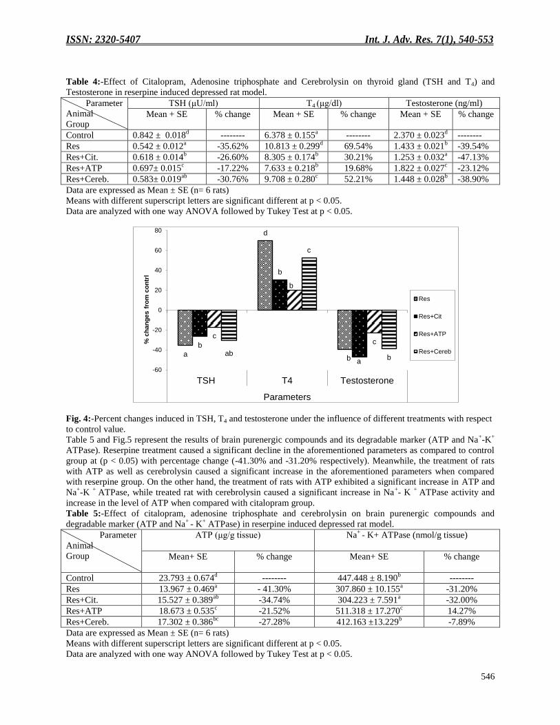
Table 4:-Effect of Citalopram, Adenosine triphosphate and Cerebrolysin on thyroid gland (TSH and T4) and
Testosterone in reserpine induced depressed rat model.
Parameter
Animal
Group
TSH (μU/ml) T4 (μg/dl) Testosterone (ng/ml)
Mean + SE % change Mean + SE % change Mean + SE % change
Control 0.842 ± 0.018d
-------- 6.378 ± 0.155a
-------- 2.370 ± 0.023d
--------
Res 0.542 ± 0.012a -35.62% 10.813 ± 0.299
d 69.54% 1.433 ± 0.021
b -39.54%
Res+Cit. 0.618 ± 0.014b -26.60% 8.305 ± 0.174
b 30.21% 1.253 ± 0.032
a -47.13%
Res+ATP 0.697± 0.015c -17.22% 7.633 ± 0.218
b 19.68% 1.822 ± 0.027
c -23.12%
Res+Cereb. 0.583± 0.019ab
-30.76% 9.708 ± 0.280c 52.21% 1.448 ± 0.028
b -38.90%
Data are expressed as Mean ± SE (n= 6 rats)
Means with different superscript letters are significant different at p < 0.05.
Data are analyzed with one way ANOVA followed by Tukey Test at p < 0.05.
Fig. 4:-Percent changes induced in TSH, T4 and testosterone under the influence of different treatments with respect
to control value.
Table 5 and Fig.5 represent the results of brain purenergic compounds and its degradable marker (ATP and Na+-K
+
ATPase). Reserpine treatment caused a significant decline in the aforementioned parameters as compared to control
group at (p < 0.05) with percentage change (-41.30% and -31.20% respectively). Meanwhile, the treatment of rats
with ATP as well as cerebrolysin caused a significant increase in the aforementioned parameters when compared
with reserpine group. On the other hand, the treatment of rats with ATP exhibited a significant increase in ATP and
Na+-K
+ ATPase, while treated rat with cerebrolysin caused a significant increase in Na
+- K
+ ATPase activity and
increase in the level of ATP when compared with citalopram group.
Table 5:-Effect of citalopram, adenosine triphosphate and cerebrolysin on brain purenergic compounds and
degradable marker (ATP and Na+
- K+ ATPase) in reserpine induced depressed rat model.
Parameter
Animal
Group
ATP (μg/g tissue) Na+
- K+ ATPase (nmol/g tissue)
Mean+ SE % change Mean+ SE % change
Control 23.793 ± 0.674d
-------- 447.448 ± 8.190b
--------
Res 13.967 ± 0.469a - 41.30% 307.860 ± 10.155
a -31.20%
Res+Cit. 15.527 ± 0.389ab
-34.74% 304.223 ± 7.591a -32.00%
Res+ATP 18.673 ± 0.535c -21.52% 511.318 ± 17.270
c 14.27%
Res+Cereb. 17.302 ± 0.386bc
-27.28% 412.163 ±13.229b -7.89%
Data are expressed as Mean ± SE (n= 6 rats)
Means with different superscript letters are significant different at p < 0.05.
Data are analyzed with one way ANOVA followed by Tukey Test at p < 0.05.
a
d
b
b
b
a
c
b
c
ab
c
b
-60
-40
-20
0
20
40
60
80
TSH T4 Testosterone
Parameters
% c
han
ges f
rom
co
ntr
l
Res
Res+Cit
Res+ATP
Res+Cereb

ISSN: 2320-5407 Int. J. Adv. Res. 7(1), 540-553
547
Fig. 5:-% changes induced in ATP and Na+-K
+ ATPase activity under the influence of different treatments with
respect to control value.
Table 6 and Fig. 6 illustrate that treatment of rats with reserpine for 21 days induced a significant increase in ALT
and AST levels when compared with control group at (p<0.05) with percentage change 78.74% and 67.50%
respectively. While, treatment of reserpinized rats with ATP and cerebrolysin caused a significant decrease in ALT
and AST when compared with reserpine group. Meanwhile, reserpinized group treated with cerebrolysin induced a
significant decrease in ALT and AST when compared with citalopram group.
Table 6:-Effect of Citalopram, Adenosine triphosphate and Cerebrolysin on liver enzymes activity in reserpine rats
Parameter
Animal
Group
ALT (U/L)
AST (U/L)
Mean+ SE % change Mean+ SE % change
Control 26.475 ± 0.613a
-------- 23.302 ± 0.541a
--------
Res 47.322 ± 2.380d 78.74% 39.032 ± 1.410
d 67.50%
Res+Cit. 42.240± 0.685cd
59.47% 34.488 ± 1.118cd
48.00%
Res+ATP 39.255 ± 0.781bc
48.27% 32.183 ± 0.937bc
38.12%
Res+Cereb. 35.418 ± 1.141b 33.78% 28.922 ± 1.389
b 24.12%
Data are expressed as Mean ± SE (n= 6 rats)
Means with different superscript letters are significant different at p < 0.05.
Data are analyzed with one way ANOVA followed by Tukey Test at p < 0.05.
Fig. 6:-Percent changes induced in ALT and AST enzymes activity under the influence of different treatments with
respect to control value.
d
d cd
cd bc
bc b
b
0
10
20
30
40
50
60
70
80
90
ALT AST
Parameters
% c
hanges fro
m c
ontr
l
Res
Res+Cit
Res+ATP
Res+Cereb
a
a ab
a
c
c
bc
b
-50
-40
-30
-20
-10
0
10
20
ATP Na+ K+ ATPase
Parameters
% c
han
ges f
rom
co
ntr
l
Res
Res+Cit
Res+ATP
Res+Cereb

ISSN: 2320-5407 Int. J. Adv. Res. 7(1), 540-553
548
Discussion:- Depression is a devastating and current disease, with profound effects on neural structure and function. This study
was planned to investigate the potential antidepressant effects of ATP and cerebrolysin in a reserpine model of
depression against citalopram as a classical drug of depression. The present study showed that, oral administration
of reserpine induced a significant increase in immobility and decreases in swimming and climbing time. This finding
agrees with (Nagakura et al., 2009, Rojas-Corrales et al.,2004 and Kandel 2000), they reported that reserpine is a
vesicular monoamine re-uptake blocker, which depletes monoamines in the brain, and produces depression-like
syndrome in animals. The obtained data showed that, the treatment of depressed rats with citalopram, ATP and
cerebrolysin resulted in a significant reduction in the immobility time and increase in swimming and climbing time
respectively in comparing to its level of the reserpine group. This observation may be due to the improved in the
monoamine level, which normalized via a reference drug pathway as an SSRI and cerebrolysin as a ploy peptide
growth factor which stimulate neuronal function. In addition, ATP acts as neuromodulator which modulate
monoamines and cell function. This finding agrees with (Cipriani et al., 2012; Burnstock et al., 2011; Sharp2002 and
Keilhoff et al., 2014). The current results reveal that, oral administration of reserpine induced a significant increase
in oxidative stress, such as MDA and 8OHdG as compared to control group. This finding is agrees with (Lopresti et
al., 2014) who reported that depression is characterized by increased oxidative stress, as indicated by higher levels
of MDA and 8-OHdG. (Li et al., 2013 and 2015) reported that MDA is one of the major metabolites induced by free
radicals, and it can cause denaturation of biomembranes and mutation of DNA via increase level of 8-OHdG.
Treatment with ATP caused an decline in the level of MDA and 8-OHdG as compared with reserpine group. This
may be due to the ATP decrease production of ROS, decrease oxidative stress and increase activities of enzymes
involved in the respiratory chain, increase ATP production and improve mitochondrial function. This finding agrees
with (Van Remmen, and Richardson, 2001) who reported that the mitochondrial damage caused by oxidative stress
could result in reduced ATP production and compromised cell function. Treatments with cerebrolysin caused
decreases in the level of MDA and 8-OHdG when compared to reserpine group. This finding agrees with (Guzmán
et al., 2016) who recorded that, administration of cerebrolysin prevents the formation of free radical and oxidative
stress due to increased metabolism of DA. ( An et al., 2016) found that cerebroprotein hydrolysate significantly
decreased MDA levels in the brain tissue of rat, indicating that cerebroprotein hydrolysate may significantly
improve the scavenging of oxygen free radicals in brain tissue.
The results of the current study revealed that, oral administration of reserpine resulted in a significant decrease in 5-
HT and DA and increase in DOPAC and HVA respectively as compared to their levels of control rats. This finding
agrees with (Dhingra and Sharma, 2006) who demonstrated that reserpine produces a significant depletion in
biogenic amines (NE, 5HT and DA) rats' brains. Moreover, (Yoshitake 2004) reported that reserpine reduced the
extracellular 5HT, NE and DA concentrations and (Ahmed, et al., 1997) reported that reserpine impairs the storage
of biogenic amines resulting in depletion of NE, DA, and 5HT in both the central and peripheral nervous systems
consequently disturbs the digestive system and decreases food intake. One or more mechanisms could be
responsible for lowering these monoamines in the brains of the rats treated with reserpine. It has been reported that
reserpine exerts its depleting effect specifically by inhibiting the adenosine triphosphate-Mg2+
-dependent
incorporation of biogenic amines into their storage vesicles (Carlsson et al.,1963); secondly, it was found that
reserpine binds irreversibly to a vesicular transporter protein, thereby disrupting storage of catecholamines (DA and
NE) and 5-HT; thirdly, reserpine blocks vesicular monoamine transporter 2 (VMAT2) resulting in the depletion of
monoamines; and it is well known that VMATs are involved in the presynaptic packaging of monoaminergic
neurotransmitters into storage granules (Lohoff et al., 2008) The data of this study shows that, treatment with
citalopram caused an improvement in the level of 5-HT, DA, DOPAC and HVA due to the mode of action of
citalopram which act as SSRI. This finding agrees with (Wessling and Ramsberg 2008) they reported that, the
antidepressant drug inhibits reuptake of neurotransmitters through selective receptors thereby increasing the
concentration of specific neurotransmitter around the nerves in the brain. Meanwhile, treatment with cerebrolysin
causes a significant increase in 5-HT and DA and decrease in DOPAC and HAV as compared with reserpine group.
This finding agrees with that of (Bannerman, et al., 2008, Novak, et al., 2009 and (García, et al., 2011) they
reported that cerebroprotein hydrolysate can affect the respiratory chain by increasing the utilization of oxygen and
glucose in the brain, subsequently, increasing the antioxidant capacity of brain tissues and the ability of the body to
handle stress, improving energy metabolism in the brain, alleviating damage of brain tissues, and promoting
recovery of brain function., it has a brain-strengthening, intelligence-improving, and brain-damage-repairing effects.
The obtained data showed that oral administration of reserpine caused a significant decrease in TSH and an increase

ISSN: 2320-5407 Int. J. Adv. Res. 7(1), 540-553
549
in T4 when compared with the control group. This finding agrees with (Bradham et al., 1998) who reported that, T4
increase in depression is based on the increase of cortisol (hypercortisolism of depression), that believed lead to an
activation of the hypothalamic neurons, which produce the thyrotrophin releasing Hormone (TRH) and
consequently, affected thyroid function. Some studies have found a correlation between the severity of depression
and serum T4 level (Khalil and Richa 2011) reported that SSRI antidepressant drugs may alter TSH and T4 levels
without having major clinical implications for thyroid suggestions coincide with the results of the present study.
The data of this study showed that, the treatment with ATP induce increase in TSH and decline in T4 when
compared with reserpine group. This observation may be due to ATP play important role in neurotransmitter and
compensated the decline of ATP which result from hyperthyroidism induces with reserpine.
In the present study, administration of reserpine induced a significant decrease in testosterone level. This finding
agrees with (Alderson and Baum 1981). who reported that testosterone has been shown to affect a number of
monoamines implicated in mental illness. In relation to depression, testosterone can enhance dopamine release in the
mesolimbic system which may protect against depression-induced anhedonia and the associated decrease in
dopamine activity in reward-related brain pathways. Additionally, intra-nasal administration of testosterone in intact
male rodents increased dopamine and 5-HT release in the neostriatum and nucleus accumbens (de Souza Silva et al.,
2009). The obtained data showed that, treatment with ATP increase level of testosterone as comparing with
reserpine group.
The results of the current study show that, the administration of reserpine caused a significant decrease in the level
of ATP and Na+
- K+ ATPase. This finding is agrees with (Nagakura et al., 2009 and
Rojas-Corrales et al., 2004) they
reported that, reserpine is an inhibitor of VMAT2 and interferes with the storage of monoamines by blocking the
ATP-dependent uptake mechanism of the storage organelles. (Xie 1989) reported also, the free radicals and its
metabolites can cause damage to the membrane of cells and decrease the activity of widely distributed Na+-K
+-
ATPases. This can catalyze the hydrolysis of ATP and promote the release of free energy stored in ATP to drive the
active transport of Na+, K+, and other substances. An addition to the decrease of ATP in mitochondria is associated
with excessive production of ROS and increasing of oxidative stress. The disrupted function of mitochondria and an
insufficient oxygen radical degradation increase the concentration of ROS in the organism that consequently causes
lipid, protein, and DNA damage. The obtained results showed that treatment with cerebrolysin and ATP increased
significantly in the level of ATP and Na+
- K+ ATPase activity as compared with reserpine group. This finding is
agrees with (Teng et al., 2014) who reported that, cerebroprotein hydrolysate can increase Na+-K
+ ATPase activity to
promote the release of neurotransmitters associated with learning and memory, regulate the excitability of neurons,
enhance brain function, and improve the quality and quantity of brain activity, consistent with numerous prior
reports . Meanwhile the treatment with ATP activity could be attributed to the increased level of ATP and Na+-K
+
ATPase may be due to increased level of ATP inside the cell function and improving mitochondrial function. The
data of the current study revealed that, oral administration of reserpine caused significantly increase in ALT and
AST as compared to control group. This data agrees with ( Ahmed-Farid el al., 2016) who reported that, Reserpine
administration is known to disrupt the permeability of the plasma membrane, causing leakage of these enzymes from
the liver into serum. The obtained data showed that, treatment with ATP and cerebrolysin induced significantly
decrease in liver enzymes, may be due to ATP and cerebrolysin decrease oxidative stress.
Conclusion:- Our study concluded that, reserpinized rats showed behavioral signs of depression compared to the control group.
The reserpinized groups received three different potential antidepressant drugs (Citalopram, ATP and
cerebrolysin).Cerebrolysin has pronounced effect than ATP and citalopram and we need to highlight that further
investigation to determine the mode of action of cerebrolysin and ATP on other animal model for depression.
Competing interests:-
The authors report no conflicts of interest in this work.
Authors' contributions:-
Asmaa Moghazy, Amel Abd El-Moneim Saad and Shimaa A. Haridy designed the study and per-formed the
experiments and conducted the data analysis, Asmaa Moghazy, Amel Abd El-Moneim Saad and Shimaa A. Haridy
participated in drafting the paper, wrote the manuscript and approved the manuscript.

ISSN: 2320-5407 Int. J. Adv. Res. 7(1), 540-553
550
Abbreviations:- Norepinephrine
Serotonin
Dopamine
Major depressive disorder
Tricyclic antidepressants
Monoamine oxidase inhibitors
Selective serotonin reuptake inhibitors
Citalopram
Cerebrolysin
Adenosine triphosphate
Forced swim test
Thyroid stimulating hormone
Tetraiodothyroine
Aspartate aminotransferase
Alanine aminotransferase
Malondialdehyde
Homovalinic acid
3,4 dihydroxy phenyl acetic acid
NE
5HT
DA
MDD
TCAs
MAOIs
SSRIs
Cit
Cere
ATP
FST
TSH
T4
AST
ALT
MDA
HVA
DOPAC
References:- 1. Sarko, J. (2000). Antidepressants, old and new. A review of their adverse effects and toxicity in overdose.
Emergency Medicine Clinics of North America, 18(4): 637–654.
2. Tripathi, K.D. (2005). Relevant physiology of urine formation; essentials of medical pharmacology. Arch Ital
Urol Androl, 77, pp.557-560.
3. Logan, A.C. (2004). Omega-3 fatty acids and major depression: a primer for the mental health
professional. Lipids in health and disease, 3(1):25-33
4. Valuck, R.J., Anderson, H.O., Libby, A.M., Brandt, E., Bryan, C., Allen, R.R., Staton, E.W., West, D.R. and
Pace, W.D. (2012). Enhancing electronic health record measurement of depression severity and suicide
ideation: a Distributed Ambulatory Research in Therapeutics Network (DARTNet) study. The Journal of the
American Board of Family Medicine, 25(5):582-593.
5. Wong, M.L., Kling, M.A., Munson, P.J., Listwak, S., Licinio, J., Prolo, P., Karp, B., McCutcheon, I.E.,
Geracioti, T.D., DeBellis, M.D. and Rice, K.C. (2000). Pronounced and sustained central hypernoradrenergic
function in major depression with melancholic features: relation to hypercortisolism and corticotropin-releasing
hormone. Proceedings of the National Academy of Sciences, 97(1):325-330.
6. Hwang, J., Zheng, L.T., Ock, J., Lee, M.G., Kim, S.H., Lee, H.W., Lee, W.H., Park, H.C. and Suk, K. (2008). Inhibition of glial inflammatory activation and neurotoxicity by tricyclic antidepressants. Neuropharmacology, 55 (5), pp.826-834.
7. Quintin, P. and Thomas, P. (2004). Efficacy of atypical antipsychotics in depressive
syndromes. L'Encephale, 30(6):583-589.
8. Venna, V.R., Deplanque, D., Allet, C., Belarbi, K., Hamdane, M. and Bordet, R. (2009). PUFA induce
antidepressant-like effects in parallel to structural and molecular changes in the
hippocampus. Psychoneuroendocrinology, 34(2):199-211.
9. Doyle, A.E., McQueen, E.G. and Smirk, F.H. (1955).Treatment of hypertension with reserpine, with reserpine
in combination with pentapyrrolidinium, and with reserpine in combination with veratrum
alkaloids. Circulation, 11(2):170-181.
10. Bleuler, M. and Stoll, W.A. (1955). Clinical use of reserpine in psychiatry: comparison with
chlorpromazine. Annals of the New York Academy of Sciences, 61(1):167-173.
11. Al-Bloushi, S., Safer, A.M., Afzal, M. and Mousa, S.A. (2009). Green tea modulates reserpine toxicity in
animal models. The Journal of Toxicological Sciences, 34(1):77-87.
12. Sreemantula, S., Boini, K.M. and Nammi, S. (2004). Reserpine methonitrate, a novel quaternary analogue of
reserpine augments urinary excretion of VMA and 5-HIAA without affecting HVA in rats. BMC
Pharmacology, 4(1):30.

ISSN: 2320-5407 Int. J. Adv. Res. 7(1), 540-553
551
13. Metzger, R.R., Brown, J.M., Sandoval, V., Rau, K.S., Elwan, M.A., Miller, G.W., Hanson, G.R. and
Fleckenstein, A.E. (2002).Inhibitory effect of reserpine on dopamine transporter function. European journal of
pharmacology, 456(1-3):39-43.
14. Cipriani, A., Purgato, M., Furukawa, T.A., Trespidi, C., Imperadore, G., Signoretti, A., Churchill, R.,
Watanabe, N. and Barbui, C. (2012). Citalopram versus other anti-depressive agents for depression. The
Cochrane Database of Systematic Reviews, 7, p.CD006534.
15. Davidson, J.R. (2009). First-line pharmacotherapy approaches for generalized anxiety disorder. Journal of
Clinical Psychiatry. 70 (Suppl 2):25–31
16. Kawahara, Y., Kawahara, H., Kaneko, F. and Tanaka, M. (2007). Long-term administration of citalopram
reduces basal and stress-induced extracellular noradrenaline levels in rat brain. Psychopharmacology
(Berl). 194(1):73
17. Hamon, M. and Blier, P. (2013). Monoamine neurocircuitry in depression and strategies for new
treatments. Progress in Neuro-psychopharmacology and Biological Psychiatry, 45:54–63.
18. Drury, A.N. and Szent-Gyorgyi, A (1929) The physiological action of adenine compounds with especial
reference to their action on the mammalian heart. J. Physiol.(Lond), 68: 214-237.
19. Burnstock, G (1972). Purinergic nerves. Pharmacol Rev, 24: 509-81
20. Burnstock, G, Krugel, U, Abbracchio, M.P.and Illes, P (2011). Urinergic signalling: from normal behaviour to
pathological brain function. Prog Neurobiol., 95(2):229-74.
21. Zuber, V.L. (1991). The effect of cerebrolysin on the metabolism of brain phospholipids in growing animals
with experimental demyelination. Nervnaia Sistema, 30:85-90.
22. Zhang, H., Zhang, X. and Xu, B. (2005). Analysis and determination of biological activity of short-chain
peptides from porcine brain hydrolysate. Journal of Pharmaceutical and Biomedical Analysis, 37(2):333-339.
23. Sharp, N., Ellard, M., Hirschowitz, L., Malthouse, S. and Johnson, N. (2002). Successful microwave ablation of
endometrial carcinoma. BJOG: An International Journal of Obstetrics & Gynaecology, 109(12), pp.1410-1412.
24. Keilhoff, G., Lucas, B., Pinkernelle, J., Steiner, M. and Fansa, H. (2014). Effects of cerebrolysin on motor-
neuron-like NSC-34 cells. Experimental Cell Research, 327(2):234-255.
25. Zou, Y., Feng, W., Wang, W., Chen, Y., Zhou, Z., Li, Q., Zhao, T., Mao, G., Wu, X. and Yang, L., (2015).
Protective effect of porcine cerebral hydrolysate peptides on learning and memory deficits and oxidative stress
in lead-exposed mice. Biological Trace Element Research, 168(2):429-440.
26. Paget, G.E. and Barnes, J.M. (1964). Toxicity Testing. In: Evaluation of Drug Activities Pharmacometics
(Laurence, D.R. and Bacharach, A.L., eds.). Academic Press, London,UK, pp.1-13
27. Porsolt, R.D., Le Pichon, M. and Jalfre, M. (1977). Depression: a new animal model sensitive to antidepressant
treatments. Nature, 266 (5604):730–732.
28. Fisher, D.A. (1996). Physiological variations in thyroid hormones: physiological and pathophysiological
considerations. Clinical Chemistry, 42(1):135-139.
29. Thakur, C.H. Saikia, T.C. and Yadav, R.N.S. (1997). Total Serum Levels of Triiodothyronine (T~ 3) Thyroxine
(T~ 4) and Thyrotropine (TSH) in School going Children of Dibrugarh District: An Endemic Goitre Region of
Assam. Indian Journal of Physiology and Pharmacology, 41:167-170.
30. Tietz, N.W. (1995) ed., Clinical Guide to Laboratory Tests, 3rd Edition, W.B. Saunders, Co., Philadelphia,pp.
578-580
31. Reitman, S. and Frankel, S. (1957). A colorimetric method for the determination of serum glutamic oxalacetic
and glutamic pyruvic transaminases. American Journal of Clinical Pathology, 28(1):56-63.
32. Karatepe, M.,( 2004). Simultaneous determination of ascorbic acid and free malondialdehyde in human serum
by HPLC-UV. Lc Gc North America, 22(4), pp.362-365.
33. Lodovici, M., Casalini, C, Briani, C. and Dolara, P. (1997). Oxidative liver DNA damage in rats treated with
pesticide mixtures. Toxicology; 117:55-60.
34. 34-Teerlink, T., Hennekes, M, Bussemaker, J. and Groeneveld, J. (1993). Simultaneous determination of
creatine compounds and adenine nucleotides in myocardial tissue by high-performance liquid chromatography.
Analytical Biochemistry, 214 (1):278-83.
35. Pagel, P., Blome, J. and Wolf, H. U. (2000). High-performance liquid chromatographic separation and
measurement of various biogenic compounds possibly involved in the pathomechanism of Parkinson’s disease.
Journal of Chromatography B: Biomedical Sciences and Applications, 746(2): 297-304.
36. Taussky, H.H. and Shorr, E.E. (1953). A microcolorimetric method for the determination of inorganic
phosphorus. The Journal of Bioliogical Chemistry, 202 (2): 675-285.

ISSN: 2320-5407 Int. J. Adv. Res. 7(1), 540-553
552
37. Ahmed-Farid, O., Ahmed, R. and Saleh, D., 2016. Combination of resveratrol and fluoxetine in an acute model
of depression in mice: Prevention of oxidative DNA fragmentation and monoamines degradation. J Appl Pharm
Sci, 6, pp.1-7.
38. Nagakura, Y., Oe, T., Aoki, T. and Matsuoka, N., 2009. Biogenic amine depletion causes chronic muscular pain and tactile allodynia accompanied by depression: a putative animal model of fibromyalgia. Pain, 146(1-2), pp.26-33.
39. Rojas-Corrales, M.O., Berrocoso, E., Gibert-Rahola, J. and Micó, J.A.. (2004). Antidepressant-Like Effect of
tramadol and its Enantiomers in Reserpinized Mice: Comparativestudy with Desipramine, Fluvoxamine,
Venlafaxine and Opiates. Journal of Psychopharmacology, 18(3), pp.404-411.
40. Kandel ER (2000) Disorders of mood: depression, mania, and anxiety disorders. In: Kandel ER (ed) Principles
of neural science, 4th edn. McGraw-Hill Companies, New York, pp 1216–1217
41. Lopresti, A.L., Maker, G.L., Hood, S.D. and Drummond, P.D., 2014. A review of peripheral biomarkers in
major depression: the potential of inflammatory and oxidative stress biomarkers. Progress in Neuro-
Psychopharmacology and Biological Psychiatry, 48, pp.102-111.
42. Li, Y.S., Hong, Y.F., He, J., Lin, J.X., Shan, Y.L., Fu, D.Y., Chen, Z.P., Ren, X.R., Song, Z.H. and Tao, L.
(2013). Effects of magnolol on impairment of learning and memory abilities induced by scopolamine in
mice. Biological and Pharmaceutical Bulletin, 36(5):764-771.
43. Li, C., Wang, Q., Li, L., Liu, Y. and Diao, H. (2015). Arachidonic acid attenuates learning and memory
dysfunction induced by repeated isoflurane anesthesia in rats. International Journal of Clinical and Experimental
Medicine, 8(8):12365.
44. Van Remmen, H. and Richardson, A. (2001). Oxidative damage to mitochondria and aging. Experimental
Gerontology, 36(7):957-968.
45. Guzmán, D.C., Brizuela, N.O., Herrera, M.O., García, E.H., Mejía, G.B., Olguín, H.J., Peraza, A.V., Attilus, J.
and Ruíz, N.L., (2016). Effect of cerebrolysin on dopaminergic neurodegeneration of rat with oxidative stress
induced by 3-nitropropionic acid. Acta Pharmaceutica, 66(3), pp.443-448.
46. An, L., Han, X., Li, H., Ma, Y., Shi, L., Xu, G., Yuan, G., Sun, J., Zhao, N., Sheng, Y. and Wang, M. (2016).
Effects and mechanism of cerebroprotein hydrolysate on learning and memory ability in mice. Genet Mol
Res, 15(3).
47. Dhingra, D. and Sharma, A. (2006). Antidepressant-like activity of Glycyrrhiza glabra L. in mouse models of
immobility tests. Progress in Neuro-Psychopharmacology and Biological Psychiatry, 30(3): 449-454.
48. Yoshitake, T., Yoshitake, S., Fujino, K., Nohta, H., Yamaguchi, M. and Kehr, J. (2004). High-sensitive liquid
chromatographic method for determination of neuronal release of serotonin, noradrenaline and dopamine
monitored by microdialysis in the rat prefrontal cortex. Journal of Neuroscience Methods, 140(1-2):163-168.
49. Ahmed, N.A., Radwan, N.M., Al-Zahaby, A.S. and El-Salam, M.A. (1997). Reserpine effects on
neurotransmitters in chick heart during growth. Journal of Physiology-Paris, 91(2): 81-90.
50. Carlsson, A., Hillarp, N.A. and Waldeck, B. (1963). Analysis of the Mg++
-ATP dependent storage mechanism
in the amine granules of the adrenal medulla. Acta Physiological Scandinavica. Supplementum. 6(1): Suppl
215:1-38.
51. Lohoff, F.W., Lautenschlager, M., Mohr, J., Ferraro, T.N., Sander, T. and Gallinat, J. (2008). Association
between variation in the vesicular monoamine transporter 1 gene on chromosome 8p and anxiety-related
personality traits. Neuroscience Letters, 434(1): 41-45.
52. Wessling, A. and Ramsberg, J. (2008). The review of antidepressants: the Dental and Pharmaceutical Benefits
Agency. Solna: TLV.
53. Bannerman, D.M., Niewoehner, B., Lyon, L., Romberg, C., Schmitt, W.B., Taylor, A., Sanderson, D.J., Cottam,
J., Sprengel, R., Seeburg, P.H. and Köhr, G. (2008). NMDA receptor subunit NR2A is required for rapidly
acquired spatial working memory but not incremental spatial reference memory. Journal of
Neuroscience, 28(14): 3623-3630.
54. Novak, P.H., Moessler, H., Gusev, E.I. and Guekht, A.B. (2009). Cerebrolysin in vascular dementia: a
randomized, placebo controlled study. Alzheimer's & Dementia: The Journal of the Alzheimer's
Association, 5(4): 249.
55. García, E.H., Guzmán, D.C., Olguín, H.J., Jiménez, F.T., Acosta, E.N., Rioja, F.P., Mejía, G.B., Ruíz, N.L. and
Del Angel, D.S. (2011). Effect of cerebrolysin on the levels of glutathione and 5-HT in different regions of rat
brain in presence of dantrolene. Biomedicine & Aging Pathology, 1(3): 169-174.
56. radham, .A., Pl mpe, ., anns, .P., renner, .A. and Trautwein, C. (1998). I. TNF-induced liver
injury. American Journal of Physiology-Gastrointestinal and Liver Physiology, 275(3): G387-G392

ISSN: 2320-5407 Int. J. Adv. Res. 7(1), 540-553
553
57. Khalil, R.B. and Richa, S. (2011). Thyroid adverse effects of psychotropic drugs: a review. Clinical
neuropharmacology, 34(6), pp.248-255.
58. Alderson, L.M. and Baum, M.J. (1981). Differential effects of gonadal steroids on dopamine metabolism in
mesolimbic and nigro-striatal pathways of male rat brain. Brain Research, 218(1-2): 189-206.
59. de Souza Silva, M.A., Mattern, C., Topic, B., Buddenberg, T.E. and Huston, J.P. (2009). Dopaminergic and
serotonergic activity in neostriatum and nucleus accumbens enhanced by intranasal administration of
testosterone. European Neuropsychopharmacology, 19(1): 53-63.
60. Xie, J.P. (1989). The Overview Research of (Na+-K+)-ATPase. Sheng Wu Hua Xue Yu Sheng Wu Wu Li Jin
Zhan, 16(4): 256-257.
61. Teng, J., Xu, Z., Zhang, J. and Li, J. (2014). Effect of yizhitongxuan decoction on learning and memory ability,
Gaq/11 expression and Na+- K
+ - ATP enzyme activity in rat models of Alzheimer’s disease. ournal of
Traditional Chinese Medicine. 34(4): 470-476.
Copyright © 2022 FDOKUMEN