2157118X.3.2.pdf - FORENSIC SCIENCE SEMINAR
44
ISSN 2157-118X Volume 3 Number 2 July 2013 Indexed by Google Scholar, OCLC, Ulrich ZolCat Academic House Forensic Science Seminar Published by
-
Upload
khangminh22 -
Category
Documents
-
view
1 -
download
0
Transcript of 2157118X.3.2.pdf - FORENSIC SCIENCE SEMINAR

ISSN 2157-118XVolume 3 Number 2 July 2013Indexed by Google Scholar, OCLC, Ulrich ZolCat Academic House Forensic Science SeminarPublished by

Forensic Biomedical Engineering is the temporary title of the special column belongs to Forensic Science Seminar (FORENSIC SCI SEM ISSN 2157-118X) . Original research papers on this special subject can be submitted to this special column (to E-mail: [email protected]).
Important Dates:Paper submissions due: August 20, 2013 Paper notification of acceptance: 1-2 weeks after submissionCamera-ready due: September 25, 2013Publishing Date: October10th, 2013
If possible, some outstanding papers chosen by this special column would be published in a new journal which is under building.
Forensic Science SeminarISSN 2157-118XJuly 2013 Volume 3 Number 2
Contents
Review Articles
Sandplay -- a new correctional treatment for criminal psychologyLI Huijun, LEI Dan
Intrinsic and extrinsic factors involved in the preservation of non-adult skeletal remains in archaeology and forensic scienceManifold BM
Views of medical students on torture in Ahmedabad cityJagdeep Jadav, Gaurang Kothari, Rakesh, Padmraj, Harish Khubachandani, Anand Menat, Kalpesh Shah
Original Research Paper
The development and construction of a motorized blood droplet generation device (BDGD) for detailed analysis of blood droplet dynamicsElisabeth Williams, Michael Taylor
Operating procedures and techniques of the technical replication of the original documentsJIN Xin
Using smartphones as a proxy for forensic evidence contained in cloud storage servicesGeorge Grispos, William Bradley Glisson, Tim Storer ■
Executive Editor
Editorial DepartmentOfficial Website
Published and Printed by
Forensic Science SeminarIndexed by
Impact Factor
Sponsored by
FORENSIC SCI SEM
www.zolcat.com
fss.xxyy.info
: Tilla A. Theresia, PhD E-mail: [email protected]
E-mail: [email protected] http://fss.xxyy.info/
ZolCat Academic House Brooklyn, NY, 11220
(ISSN 2157-118X) Google Scholar OCLC (659514459) Ulrich
= 0.1 (2012)
the Library of Congress
© 2013. All Rights Reserved.
Single Issue Price $150USDPeer Reviewed | Limited Open Access
79
82
93
95
105
108

July 2013 ISSN 2157-118X 79
Sandplay -- A New Correctional Treatment for Criminal Psychology
Abstract In recent years, Sandplay has been gradually known and applied to psychological consultation. Comparing with traditional psychological consultation, sandplay has its particular advantages for prisoners with inferiority feeling and introvert personality. It can create a more free and protective space for the prisoners, playing an important role in correcting individual psychology, establishing good relations among convicts, group counseling and encouragement and assistance from family members,etc . It has become a tool for psychological consultants of policemen to establish good counseling relations and draw prisoners to accept consultancy initiatively and it also has promoted the development of psychological consultation and treatment in prisons.
Keywords: Sandplay, Convict, Psychological correctional treatment.
FORENSIC SCI SEMLI Huijun et al, Sandplay -- A New Correctional Treatment for Criminal Psychology, FORENSIC SCI SEM, (2013) 3(2): 79-81
Received 10 April 2013Received in revised form 26 April 2013
Accepted 29 April 2013Available online 2 May 2013
Peer Review
ed2157118X
.3.2.R1
Sandplay is a kind of self-
expression therapy in which the
visitor selects toys and places them
in a box filled with silver sand to
construct a scene in the companion of
the consultant. It is based on such a
hypothesis that an individual can heal
the psychological trauma by oneself in
a proper situation. [1] In the treatment
of prisoner psychological trauma,
sandplay creates a free and protective
space for the convicts. With the
tolerance, acceptance and care of the
psychological consultants of policemen,
the prisoners’ self-healing ability is
developed, and their unconscious
behaviors become conscious behaviors,
which reflect many aspects of the
individual and the society. In this way,
the psychological problem of prisoners
is settled. [2]
LI Huijun *, a, LEI Danb
a Yu Zhou Prison of Chongqing, Chongqing 400026,China.b Public Security Bureau of Chongqing, Chongqing 400021,China.
* Corresponding Author: LI Huijun. E-mail: [email protected]. Address: Yu Zhou Prison of Chongqing, Tieshan Ping Cuiwei Road No.5, Jiangbei District, Chongqing 400026, China.
Comparing with traditional
linguistic consultation method, sandplay
has its unique features and it adapts
to the special environment—prison.
Sandplay is carried out with non-verbal
form: the convict visitor can design and
construct the scene according to his own
willing and doesn’t have to care about
the consultants of policemen present.
This can reduce double resistance
caused by verbal communication and
identity consciousness, [3] thus to last
the consultation relation and promote
follow-up consultation. With silver
sand and toys as its materials, sandplay
can remind the convict visitor of
his childhood when he played with
sand to arouse his passion for further
exploration and devote himself to the
sandplay making. The more detailed
the scene is made by the visitor, the
more the consultants of policemen can
discover his potential psychological
problems, thus to find the breakout of
the consultation.
In the field of prison psychological
correctional treatment, sandplay is
a kind of psychological treatment
method for various psychological
disease consultations and treatment,
and a means to inspect and find out the
prisoners’ psychological diseases. It can
also be regarded as a technique that can
cultivate the prisoners’ self-confidence
and individuality and develop their
imagination and creativity.
1 The application of sandplay in individual prisoner psychological treatment
Sandplay creates a free and
protective space for the individual and
emphasizes self-participation of the individual.
The prisoners can place the toys and design
the structure of the sand in according to their
imagination and willing and their construction
will not be disturbed. They are fully respected
and cared in this “play” environment. During
the process, the consultants of policemen will
not give any evaluation for the scene and its
content, but accept it wholly. Through the non-
verbal form, the consultant demonstrates a
kind of inclusive love for the prisoner, who
would actively explore his potential under this
encouragement and transfer the acquired support
and potential to his life and self-transformation.
With little verbal communication in the
process of sandplay, the prisoner has to sense
this atmosphere with a peaceful mind and
endow a real implication to the scene with his
imagination. Sandplay is a visualized picture
which makes the invisible world visible. Making
the sandplay is to reveal one’s inner world with
the sandplay and adjust his mind. Every of us
the potential of self-realization, which is the
motive power of individual development. [1] By
stimulating this motivation, sandplay provides
the prisoner an opportunity to “grow up”. It
can be regarded as a wonderful assumption,
which carries the expectation for future.
Through sandplay, the dream of the prisoner
is demonstrated preliminarily, which provides
them hope and encouragement.
2 The application of sandplay in the relations among prisoners and group counseling
The relation among prisoners is an
important factor that affects the attitude of
prisoners. A peaceful relation among prisoners,
especially a good relation among the reformation
of prisoners supervision group, can make the
members respect, encourage and supervise each
other and reduce the occurrence of rule and
regulation violations. Being familiar with each
other, the members will concern a lot to reveal
LI Huijun et al, Sandplay -- A New Correctional Treatment for Criminal Psychology, FORENSIC SCI SEM, (2013) 3(2): 79-81FORENSIC SCI SEM
Forensic Science Seminar Volume 3 number 280
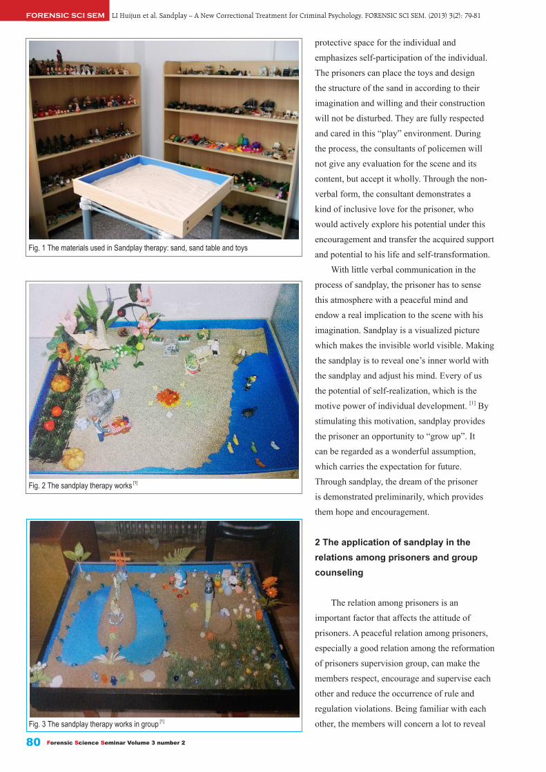
Fig. 1 The materials used in Sandplay therapy: sand, sand table and toys
Fig. 2 The sandplay therapy works [1]
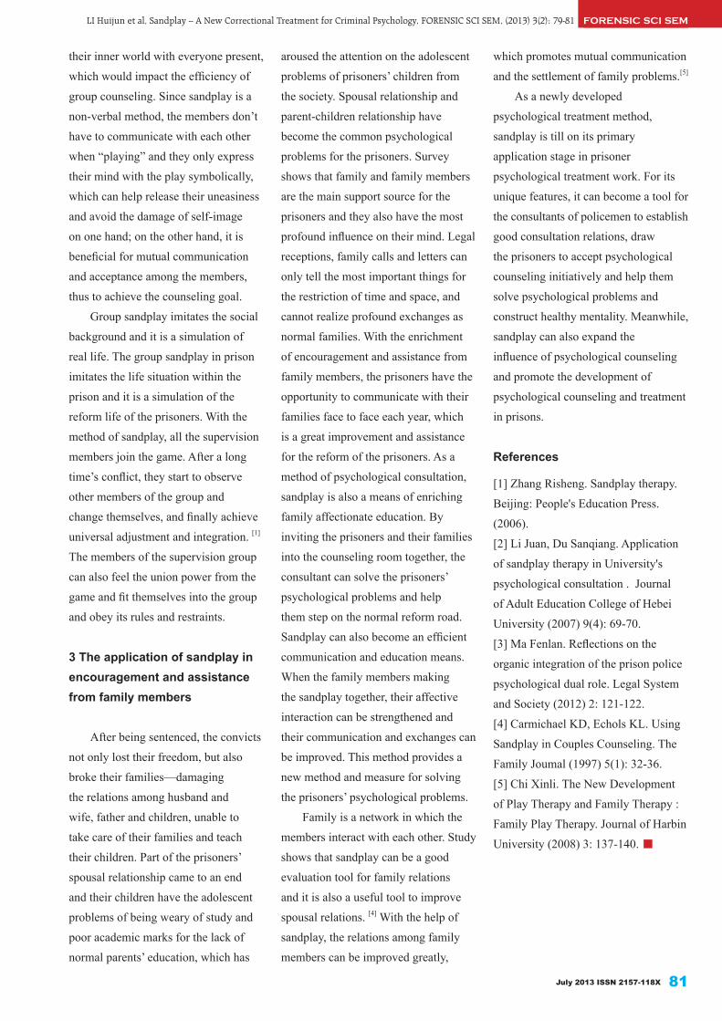
Fig. 3 The sandplay therapy works in group [1]
their inner world with everyone present,
which would impact the efficiency of
group counseling. Since sandplay is a
non-verbal method, the members don’t
have to communicate with each other
when “playing” and they only express
their mind with the play symbolically,
which can help release their uneasiness
and avoid the damage of self-image
on one hand; on the other hand, it is
beneficial for mutual communication
and acceptance among the members,
thus to achieve the counseling goal.
Group sandplay imitates the social
background and it is a simulation of
real life. The group sandplay in prison
imitates the life situation within the
prison and it is a simulation of the
reform life of the prisoners. With the
method of sandplay, all the supervision
members join the game. After a long
time’s conflict, they start to observe
other members of the group and
change themselves, and finally achieve
universal adjustment and integration. [1]
The members of the supervision group
can also feel the union power from the
game and fit themselves into the group
and obey its rules and restraints.
3 The application of sandplay in encouragement and assistance from family members
After being sentenced, the convicts
not only lost their freedom, but also
broke their families—damaging
the relations among husband and
wife, father and children, unable to
take care of their families and teach
their children. Part of the prisoners’
spousal relationship came to an end
and their children have the adolescent
problems of being weary of study and
poor academic marks for the lack of
normal parents’ education, which has
aroused the attention on the adolescent
problems of prisoners’ children from
the society. Spousal relationship and
parent-children relationship have
become the common psychological
problems for the prisoners. Survey
shows that family and family members
are the main support source for the
prisoners and they also have the most
profound influence on their mind. Legal
receptions, family calls and letters can
only tell the most important things for
the restriction of time and space, and
cannot realize profound exchanges as
normal families. With the enrichment
of encouragement and assistance from
family members, the prisoners have the
opportunity to communicate with their
families face to face each year, which
is a great improvement and assistance
for the reform of the prisoners. As a
method of psychological consultation,
sandplay is also a means of enriching
family affectionate education. By
inviting the prisoners and their families
into the counseling room together, the
consultant can solve the prisoners’
psychological problems and help
them step on the normal reform road.
Sandplay can also become an efficient
communication and education means.
When the family members making
the sandplay together, their affective
interaction can be strengthened and
their communication and exchanges can
be improved. This method provides a
new method and measure for solving
the prisoners’ psychological problems.
Family is a network in which the
members interact with each other. Study
shows that sandplay can be a good
evaluation tool for family relations
and it is also a useful tool to improve
spousal relations. [4] With the help of
sandplay, the relations among family
members can be improved greatly,
FORENSIC SCI SEMLI Huijun et al, Sandplay -- A New Correctional Treatment for Criminal Psychology, FORENSIC SCI SEM, (2013) 3(2): 79-81
July 2013 ISSN 2157-118X 81
which promotes mutual communication
and the settlement of family problems.[5]
As a newly developed
psychological treatment method,
sandplay is till on its primary
application stage in prisoner
psychological treatment work. For its
unique features, it can become a tool for
the consultants of policemen to establish
good consultation relations, draw
the prisoners to accept psychological
counseling initiatively and help them
solve psychological problems and
construct healthy mentality. Meanwhile,
sandplay can also expand the
influence of psychological counseling
and promote the development of
psychological counseling and treatment
in prisons.
References
[1] Zhang Risheng. Sandplay therapy.
Beijing: People's Education Press.
(2006).
[2] Li Juan, Du Sanqiang. Application
of sandplay therapy in University's
psychological consultation . Journal
of Adult Education College of Hebei
University (2007) 9(4): 69-70.
[3] Ma Fenlan. Reflections on the
organic integration of the prison police
psychological dual role. Legal System
and Society (2012) 2: 121-122.
[4] Carmichael KD, Echols KL. Using
Sandplay in Couples Counseling. The
Family Joumal (1997) 5(1): 32-36.
[5] Chi Xinli. The New Development
of Play Therapy and Family Therapy :
Family Play Therapy. Journal of Harbin
University (2008) 3: 137-140. ■
Manifold BM, In-&Ex-trinsic Factors in Preservation of Non-Adult Skeletal Remains, FORENSIC SCI SEM, (2013) 3(2): 82-92FORENSIC SCI SEM
Forensic Science Seminar Volume 3 number 282
Intrinsic and Extrinsic Factors Involved in the Preservation of Non-Adult Skeletal Remains in Archaeology and Forensic Science
Abstract Human skeletal remains offers the most direct insight into the health, well-being, and the lifestyles of both past and modern populations, as well as the study of violence and traumas encountered both from archaeological and forensic contexts. They also allow archaeologists and anthropologists to reconstruction demographic details, none more so than those of children, where mortality rates were high in most human populations until the twentieth century. The study of children within biological anthropology had being taking place for many years now, but studies of mortality and morbidity are often hindered by the poor preservation of their skeletons or infrequent representation of skeletal elements. Taphonomic processes are often cited as the cause of this ‘under-representation’ of children from archaeological investigations. This phenomenon is thought to be as a result of the inability of non-adult bone to survive the changing conditions of the burial environment in which they are interred. Taphonomic factors can be divided into two types: intrinsic (resistance to bone) and extrinsic (environmental influences), both of which exert influence on the long term survival of non-adult bone. This paper aims to review the many intrinsic and extrinsic factors which can alter human bone and contribute to its deterioration in the burial environment in both archaeology and forensic science.
Keywords: Forensic science, Bioarchaeology, Taphonomy, Children, Skeletal remains.
Received 17 December 2012Received in revised form 3 April 2013Accepted 7 April 2013Available online 10 June 2013
Peer Review
ed2157118X
.3.2.R2
Introduction
Taphonomy is a term deriving from the Greek words ‘taphos’ (burial) and ‘nomos’ (laws), and was first coined by Efremov in the 1940s [1]. Efremov defined taphonomy as being the study of the ‘transition of all its detail of animal remains from the biosphere to the lithosphere or the geological record’. Taphonomy was originally a palaeotological term, but today, has been adopted by a range of experts, such as, zooarchaeologists, archaeologists and forensic scientists as a means to
Manifold BM *, a
a Department of Archaeology, School of Human and Environmental Sciences, University of Reading, Whiteknights, Reading.
* Corresponding Author: Bernadette M Manifold. E-mail: Email: [email protected]. Address: Department of Archaeology, School of Human and Environmental Sciences, University of Reading, Whiteknights, Reading, Berkshire RG6 6AB, UK.
explain the many processes involved in the decomposition and skeletonization of human and animal burials. Efremov[1] implied that all processes affecting an assemblage (s) prior to its incorporation into a stable subsoil should be termed ‘taphonomic’. This could include both diagenesis and a range of anthropogenic processes such as selective killing, cooking and disposal practices. It can be argued that the main agent responsible for the outcome of human assemblages is humans themselves and how they treat their dead [2].
Numerous authors have defined
taphonomy in different ways. Bonnichsen [3] proposed the meaning of taphonomy as ‘the study of the accumulation and modification of osteological assemblages from a formation perspective’. Alternatively, Olsen [4] defined taphonomy ‘as the reconstructing of history of a fossil from the time of death to the time of recovery’. A more exclusive definition was used by Millard and Hedges [5] who described taphonomy as being distinct from both anthropogenic processes and diagenesis. According to Millard and Hedges the main taphonomic processes

FORENSIC SCI SEMManifold BM, In-&Ex-trinsic Factors in Preservation of Non-Adult Skeletal Remains, FORENSIC SCI SEM, (2013) 3(2): 82-92
July 2013 ISSN 2157-118X 83
include digestion, trampling, burning and weathering.The state of preservation and representation of
human remains can be determined by taphonomic factors, which may in turn be related to funerary practices, grave types, excavation and storage. Since the 1950s, the focus has been on the fossil record in terms of how well it reflects the actual palaeoecology of the biotic community [6], and on the selective processes that determine the contribution of a fossil assemblage [7]. Many authors have contributed to the study of biological and cultural activity in past populations [8-13] and in more recent years the focus has shifted to archaeological and forensic anthropology [14-15].
The survival of human bone is dependent on many variables, such as, soil pH, soil type, bone type and size, age and sex of the individuals. There is often an under-representation of children’s skeletons recovered from archaeological sites [16-19]. This phenomenon is thought to be as a result of the inability of non-adult bone to survive the changing conditions of the burial environment in which they are interred. This paper aims to review the many intrinsic and extrinsic factors involved in bone preservation and how they relate to the skeletons of children.
INTRINSIC FACTORS INVOLVED IN THE PRESERVATION OF BONE
AgeAge is important in relation to bone size. The bones
of children are both smaller and less dense than adult bone; therefore they undergo decomposition processes in a shorter time than adults. Children’s bones have a high organic and low inorganic content which, in theory, makes them more susceptible to decay [20]. However, there is a lack of studies on the chemical makeup of non-adult bones to draw any firm conclusions. Guy et al. [20) stated that infant type remains are soft, ill-structured bones, rich in interstitial water, and poorly protected from chemical and mechanical degradation. In addition, child remains are easier to disarticulate and remove by animals; this can hamper any investigation or excavation (Figure 1) [21-23]. Immature bones are easily dispersed, lost and destroyed compared to adult bones (Figure 2). In a recent study by Manifold [24] on British skeletal assemblages, a preservation pattern was observed in what bones are likely to be present.
Fig. 1 The fragmentary remains of a non-adult skeleton from the site of Auldhame, Scotland (Photo: Bernadette Manifold)
Fig. 2 Well preserved non-adult skeleton from the site of Great Chesterford, Cambridgeshire (Photo: Bernadette Manifold)

Manifold BM, In-&Ex-trinsic Factors in Preservation of Non-Adult Skeletal Remains, FORENSIC SCI SEM, (2013) 3(2): 82-92FORENSIC SCI SEM
Forensic Science Seminar Volume 3 number 284
Bone type and sizeThere is a variation in the
preservation of different bones. The bones most vulnerable to destruction are thought to be those with a high proportion of cancellous material, such as the sternum, vertebrae, ribs, and epiphyses. Among the vertebrae, it has been thought that the lumbar are the least and the cervical the most affected by soil erosion [25]. However, recent studies on large numbers of non-adult skeletons has found this to be in reverse, with the cervical and thoracic vertebrae in abundance, whilst the lumber is poorly preserved or absent [24]. This may also depend on the position of the body during burial, and if grave intercutting occurred. According to Mays [25], the hyoid bone and small bones of the hands and feet are almost always poorly represented. Elements with a high proportion of cortical bone, such as the skull, mandible and the long bones appear less affected by preservation [25]. Von Endt and Ortner[26] have shown that rates of decay are inversely proportional to the bone size. They found when bones of different sizes were kept in water at constant temperature; nitrogen is released at a rate which is inversely proportional to bone size. Any weakening of the protein-mineral bonding of bone will enhance its degradation. Groundwater and its dissolved ions can penetrate bone, and bone size, both the external and internal surface area (porosity), available to groundwater is important in bone breakdown [26].
Waldron [21] demonstrated that, the dense long bones and the compact parts of the cranium were present in 40-50% of cases, but he also found ribs to be well preserved. Around 60-70% of cases included the vertebrae. Bones which were least preserved
included many of the small bones, such as carpals and the phalanges. The body of the scapula was also poorly preserved, possibly due to been thin and vulnerable to damage. This study indicates better preservation of the large dense parts of the skeleton, such as the long bones and the cranium. Finally, Waldron [21] pointed out that the pattern of preservation found in his study is not necessarily the same for other sites. This would suggest that the type of soil and burial environment conditions play an increasingly important role.
Bello et al. [27] analysed four osteological samples, namely St Maximin, St Estève, and Observance, in France; and Spitalfields, in London. In all four samples, the scapulae, sterna, vertebrae, sacra, patellae and hand and foot bones were the least represented in both adults and non-adults. Overall, adult remains appear to survive better than those of non-adults. It was also found that male skeletons were better preserved than female [27]. This suggests that bone density of certain bones is lower and therefore, may not survive the burial environment. Absence of the small bones such as the phalanges and carpals not being present maybe also due to excavation (i.e missed or not identified in the laboratory). The non-adult bones examined by Ingvarsson-Sundström [28] from Asine, Greece were found to be in good preservational condition. The bones most frequently found in a complete state were the bones of the hands, feet and vertebrae (arches). Parts of the cranium (the temporal bone: pars petrosa, and zygomatic) were also completely preserved. The findings were similar to Waldron’s [21] study West Tenter Street, London except for the phalanges, which were often found complete in the Asine material. In skeletal reports,
it needs to be made clear as to what is meant by ‘poor’ bone preservation. Is this due to the condition of the bones of the skeletons or is it referred to the representation of the various elements. As bones can be recovered in a state of poor preservation (i.e the condition of the surface of the bones), but be well-represented.
PathologyPathological conditions and
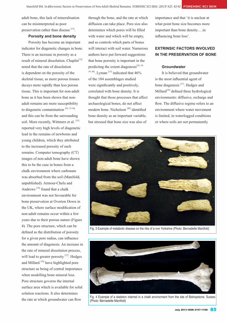
injuries are known to speed up the decomposition of buried bone. When bone is damaged through trauma or as a result of illness, it is easier for micro-organisms to enter; also the same may be said of those individuals with infectious diseases and blood poisoning. When there is a breakdown of bone in life such as with metabolic disease, this can have an effect on the rate of preservation [29-30]. Rickets is caused by vitamin D deficiency in children, prevents calcium from being deposited in the developing cartilage as well as in the newly formed osteoid, which impedes bone mineralisation. The macroscopic appearance of rickets in non-adults tends to be long bone bending deformities and metaphyseal swelling. However, in cases of active rickets there is increased porosity of bone surfaces in particular the cranium and the growth plates. This increased porosity can lead to the bone appearing to ‘dissolve’ in the burial environment, which can make recovery of remains difficult. Another metabolic disease which cannot be frequently diagnosed is scurvy, a condition caused by the lack of vitamin C in the diet. This condition can also lead to an increase in porosity of the non-adult skeleton which makes it vulnerable to the changes of the burial environment (Figure 3). Metabolic conditions such as these, cause a decrease in the mineralisation of non-
FORENSIC SCI SEMManifold BM, In-&Ex-trinsic Factors in Preservation of Non-Adult Skeletal Remains, FORENSIC SCI SEM, (2013) 3(2): 82-92
July 2013 ISSN 2157-118X 85
adult bone, this lack of mineralisation can be misinterpreted as poor preservation rather than disease [31].
Porosity and bone densityPorosity has become an important
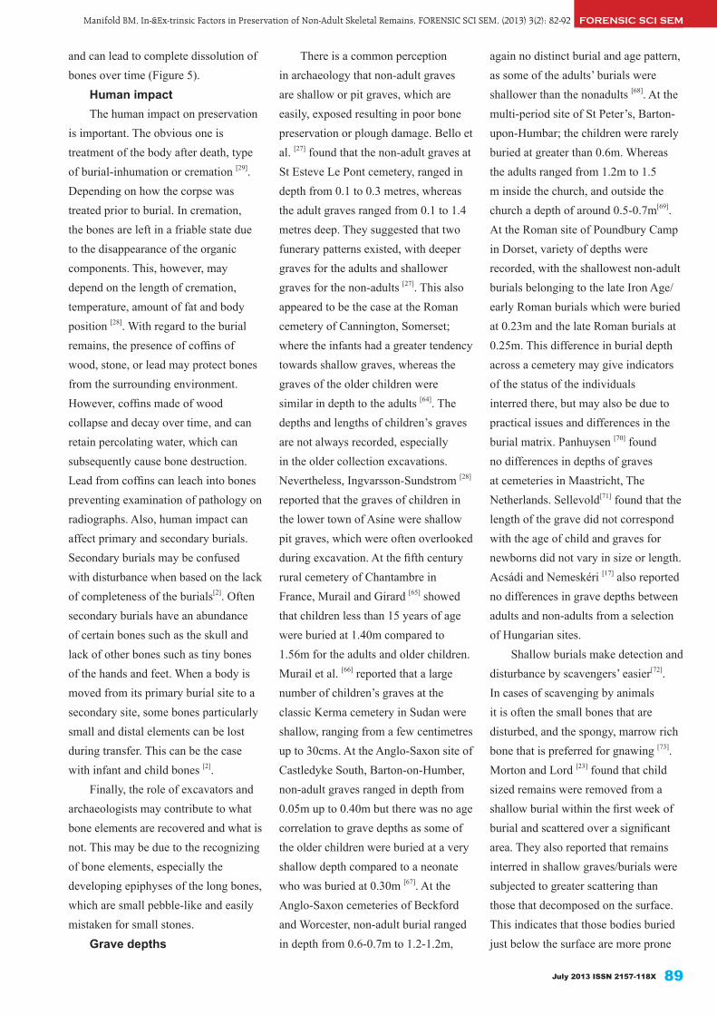
indicator for diagenetic changes in bone. There is an increase in porosity as a result of mineral dissolution. Chaplin[32] noted that the rate of dissolution is dependent on the porosity of the skeletal tissue, as more porous tissues decays more rapidly than less porous tissue. This is important for non-adult bone as it has been shown that non-adult remains are more susceptibility to diagenetic contamination [26, 33-34] and this can be from the surrounding soil. More recently, Wittmers et al. [35] reported very high levels of diagenetic lead in the remains of newborns and young children, which they attributed to the increased porosity of such remains. Computer tomography (CT) images of non-adult bone have shown this to be the case in bones from a chalk environment where carbonate was absorbed from the soil (Manifold, unpublished). Armour-Chelu and Andrews [36] found that a chalk environment was not favourable for bone preservation at Overton Down in the UK, where surface modification of non-adult remains occur within a few years due to their porous nature (Figure 4). The pore structure, which can be defined as the distribution of porosity for a given pore radius, can influence the amount of diagenesis. An increase in the rate of mineral dissolution process, will lead to greater porosity [37]. Hedges and Millard [38] have highlighted pore structure as being of central importance when modelling bone mineral loss. Pore structure governs the internal surface area which is available for solid solution reactions. It also determines the rate at which groundwater can flow
Fig. 3 Example of metabolic disease on the ribs of a non Yorkshire (Photo: Bernadette Manifold)
Fig. 4 Example of a skeleton interred in a chalk environment from the site of Bishopstone, Sussex (Photo: Bernadette Manifold)
through the bone, and the rate at which diffusion can take place. Pore size also determines which pores will be filled with water and which will be empty, and so controls which parts of bones will interact with soil water. Numerous authors have put forward suggestions that bone porosity is important in the predicting the extent diagenesis[29, 38-
39, 40]. Lyman [13] indicated that 46% of the 184 assemblages studied were significantly and positively, correlated with bone density. It is thought that those processes that affect archaeological bones, do not affect modern bone. Nicholson [40] identified bone density as an important variable, but stressed that bone size was also of
importance and that ‘it is unclear at what point bone size becomes more important than bone density….in influencing bone loss’.
EXTRINSIC FACTORS INVOLVED IN THE PRESERVATION OF BONE
GroundwaterIt is believed that groundwater
is the most influential agent of bone diagenesis [37]. Hedges and Millard[38] defined three hydrological environments: diffusive, recharge and flow. The diffusive regime refers to an environment where water movement is limited, in waterlogged conditions or where soils are not permanently

Manifold BM, In-&Ex-trinsic Factors in Preservation of Non-Adult Skeletal Remains, FORENSIC SCI SEM, (2013) 3(2): 82-92FORENSIC SCI SEM
Forensic Science Seminar Volume 3 number 286
saturated. With a recharge regime bones go through wetting and drying cycles, and as a result, porosity increases and the formation of large pores which increases the affects of the water cycle. Finally, in the flow regime the presence of bone buried in such an environment tends to depends on the volume of water, (i.e rainfall and seasonal factors) [38]. Groundwater is the medium for all other processes such as recrystallisation, dissolution, hydrolysis, microbiological attack and ion exchange to take place[37]. In general, bone buried in soil where water movement is limited and
calcium and phosphorous concentration are high, has the potential to survive for an indefinite period. Where water movement is greater there tends to be greater dissolution, and therefore, less wellpreserved bones, both macroscopically and microscopically[41].
Soil type and pHUnfavourable geological conditions
are often cited as a cause of poor preservation, but how much influence this has on sites and skeletal remains in Britain remains unclear. The geology of Great Britain is complex, with varying types and amounts of soil in
each region. Soil type can be broken down into around 13 groups (Table 1). Therefore, preservation of bone varies considerably, not only from soil to soil, but also from one place of burial to another. Soil is made up of mineral and organic matter, air, water with differing soil types composed of differing ratios. Soil can be classified according to particle size as, clay, silt, sand or gravel[42] and soil pH is determined by the amount of hydrogen ions present. The concentration of which can be classed as neutral, acidic or alkaline [43]. Environments affect bone in different ways (Table 2). In acidic environments, which can consist mostly of podsols, these soils tend to be abundant in Northern England and Scotland, where there is a tendency for the soils to be thin, acidic and wet, which may or may not have a negative impact on bone preservation [44]. On the other hand, many peat environments have revealed excellent preservation due to the acidic nature of the sites in such an environment there is a lack of microbial attack and an accumulation of organic matter, which leads to the formation of blanket bog [45]. In a more alkaline environment, which consists of calcareous soils can result in mixed preservation, if remains are recovered from this soil type and have a high pH, then they tend to be in good condition[45], these soils tend to be found
Table 1. Soil type and location in the United Kingdom, adapted from the soil atlas of Europe [75]
Soil group Where foundAlluvial soils Lincolnshire, Kent and Norfolk
Coastal sandy regosols Highlands, Moray, Aberdeenshire, Lincolnshire, Norfolk, Lancashire and Cumbria
Rendzinas or calcaric brown soils, with associated luvic brown soils
Hampshire, Wiltshire, Gloucestershire, Yorkshire, and North Lincolnshire
Brown soils, mainly sandy, with associated rendzinas, podzols or gley soils Norfolk and Suffolk
Brown soils, mainly orthic or prodzolic, with gley soils and rankers
Aberdeenshire, Fife, Angus, Cornwall, Devon, Pembrokeshire, Ceredigion and Powys
Brown soils, mainly luvic with gley soils Kent, Herefordshire, Hertfordshire, Devon, Bath and Glamorgan
Podzols with brown and gley soils Dorset, SurreyNon-hydromorphic Podzols and podzolic brown soils, with stahnopodzols and gley soils
C o r n w a l l , D e v o n , H i g h l a n d s , M o r a y, Aberdeenshire
Oro-artic podzols, rankers and lithosols with gley and peat soils Highlands, Perth and Stirling
Pelo-calcaric brown or pelocalcaric gley soils, with associated brown and gley soils
Essex, Cambridgeshire, Northamptonshire, Lincolnshire, Dorset, Wiltshire and Oxfordshire
Gley soils, mainly orthic or luvic with brown soils
Nor fo lk , Sussex , Sur rey, Kent , Devon, Leicestershire. Powys, Shropshire, Cheshire, Lancashire, Yorkshire, Northumberland, Lothian and Ayreshire.
Lowland peat bogs (fens and raised) with humic gley soils North Lincolnshire, Cambridgeshire
Bog soils (blanket)Highlands, Dumfries ad Galloway, Scottish borders, Northumberland, Cumbria, Conwy, Gwynedd, Devon and Yorkshire
Table 2. Preservation environments with reference to pH, adapted from Evans and O’Connor [76]
DEPOSITIONAL ENVIRONMENT MAIN SOIL AND SEDIMENT TYPE TYPICAL LOCATIONS COMMENTS
Acid, pH <5.5, oxic Podsols and other leaching soils Heathland, uplands moors, some river gravels
Soils are fully aerated; develop on nutrients-poor and freelydraining parent materials. Organic materials not normally preserved (i.e bone)
Basic, pH >7.0. oxic Rendsinas, lake marls, tufa, alluvium, shell sand
Chalk and limestone areas, valley bottoms
Soils are calcareous in nature. Good preservation of organic material, with possible eroded surfaces
Neutral, pH 5.5-7.0, aerobic Brownearths and gleys, river gravels Clay vales and other lowland plains This type of soil is prone to waterlogging.
Organic materials can be poorly preserved
Acid or basic, anoxic Peats and organic deposits Urban sites, wetlands, river floodplains
Varied conditions. Most kinds of biological materials are preserved

FORENSIC SCI SEMManifold BM, In-&Ex-trinsic Factors in Preservation of Non-Adult Skeletal Remains, FORENSIC SCI SEM, (2013) 3(2): 82-92
July 2013 ISSN 2157-118X 87
in the East Anglia and eastern and south-west England. In soils of a neutral pH, there can be varied conditions, these soils are well-drained and mostly located on the gravel and chalk areas of southern England. An increase in biological activity leads to a breakdown of organic matter, which results in a well-mixed, aerated soil and can lead to poor bone preservation [45].
The main constituents of bone; the organic part (collagen) and mineral part (hydroxyapatite), are preserved at opposing pH levels [46]. It is generally known that soils with a neutral or alkaline pH are better for preservation of bone, rather than acidic soils [29, 44], but this is not always the case. Locock et al. [47] found, that soil pH was not said to be the main controlling factor in the preservation of buried bone [47]. Some demineralisation of bone may occur as a result of the action of organic acids released during decomposition of the soft tissues, and therefore present in the soil where the bones are exposed [48]. Overall, it would appear that the literature has produced come contradictions as to what environment is best for bone preservation. Henderson [29] stated that the speed of decomposition is increased in light porous soils, whilst dense clay soils may decrease the rate of decomposition, and the deeper the burial, the poorer the preservation due to waterlogged clay[29]. However, there may be limitations to these studies using animal bones, which may react differently to those of the human skeletons to soil conditions. Nicholas [40] found acid moorland (pH 3.5-4.5) was the most destructive to bone and a chalk environment (pH 7.5-8.9) was the most favourable. However, between these two sets of figures there are many variables and should be used as an indication of
the extremes. Maat [49] reported that the role of soils in preservation may be overestimated. This should be viewed with caution, as a more recent study based on the decomposition of juvenile rats has shown that microbial activity is a major contributor to cadaver decomposition in soil, and it also shows that the persistence of a cadaver in soil can be influenced by the surrounding temperature and soil type [50]. This would make soil pH and soil type a major determinant of bone preservation, and most probably in the less dense bones of non-adults. Other factors such as the depth of burial and type of burial should be considered alongside pH. In study by Nord et al.[51] on the degradation of archaeological objects and bones from prehistoric graves in Sweden, it was found that the environment affects preservation in three ways; firstly, the chemical environment (soil acidity) mainly affects the macroscopic appearance of the bone, secondly, the microbial activity, composed mainly of bacteria and fungi have a destructive affect on the organic contents of bone and the histological structure. Thirdly, the inorganic material is mainly destroyed by soil acidity, whereas proteins degrade at a higher pH. It would appear that calcareous soil is most suitable for the good preservation of macroscopic structure of human bone [52].
Hydroxyapatite is relatively insoluble at pH 7.5, but is very soluble below pH 6, an example of very acidic soils is Sutton Hoo, Suffolk where no bones survived except soil stains [47]. Soil pH in relation to age has proven to have an effect on the preservation of non-adults bones, which tend to decline more rapidly with increasing soil acidity. Mays [52] has reported good preservation in 60% of the
infants recovered at Wharram Percy, and relates this to the alkaline burial environment, which has a pH of 7.3-8.5. Gordon and Buikstra [53] found that bone preservation was correlated with age of death, with younger individuals tending to have poorly preserved bones. It was found that at ‘marginal pH ranges all or most of the infants and children may be systematically eliminated from the mortuary samples by preservational bias’ [53]. Walker et al. [54] examined skeletal remains recovered from Mission La Purisima, California and noted that poorly calcified remains of children were more susceptible to decay, which was due to the acidic soil in which they were buried, which allowed water to permeate through the bone, with subsequent soaking and drying distintegrating the fragile ribs and spine. The burial records for Mission La Purisima indicating 32% of the individuals buried in the cemetery were under 18 years, but only 6% of the skeletons represented individuals of this age. Nielsen-Marsh et al. [55] and Smith et al. [56] found that two categories of bones exist; those where preservation is determined by soil chemistry and those determined by taphonomy. In these studies, soil was classes into two groups, corrosive and benign. The corrosive soils were characterised by a low pH, high exchangeable acidity, and low organic content. These soils were mostly found in north and Western Europe, and are dominated by free-draining soil, (i.e sand and gravel and associated with absence of calcareous bedrock). In contrast, the benign soils had a more neutral pH value, low exchangeable acidity and a high organic content. It was found that ‘benign’ soils did not have a big influence in determining preservation [55]. Smith et al. [56] found

Manifold BM, In-&Ex-trinsic Factors in Preservation of Non-Adult Skeletal Remains, FORENSIC SCI SEM, (2013) 3(2): 82-92FORENSIC SCI SEM
Forensic Science Seminar Volume 3 number 288
that the state of preservation of bone did not appear to be related to soil conditions of a particular site, but to the taphonomic history before burial. Post-mortem defects also occur and must be taken into account when interpreting remains. Defects due to soils chemical erosion, exposure to the sun, water and mechanical processes can be observed on various parts of the skeletons [57]. Soil activity is the primary cause of bone changes; soil chemical erosion causes proteins to be demineralised by acid environment and decomposition of bone occur due to bacteria. As a result, bones can become lighter and totally degrade; but whether this occurs in the remains of children is still debatable [57].
TemperatureTemperature and its affects vary
with latitude, season, and depth of burial [29]. One general rule is the reaction rate, which is approximately double for every 10°c rise in temperature [47]. Temperature can have a profound effect on the chemical and biological processes in the soil [58], any increase in temperature will increase the activity of insects and bacteria, whereas any decreases in temperature will lead to the formation of ice crystals and the destruction of cell structure, the propagation of microfractures of bone, and disruption of the natural soil layer [59]. These influctuations in soil temperature at a burial site can influence the survival of human remains[58]. It has been found that decay of organic components were faster at higher temperatures. Temperature variation can cause expansion and contraction of the earth, which can cause fragmentation of bone. This appears to be a particular concern when the bones are those of infants and children [28]. These changes were observed at the Anglo-Saxon cemetery of Raunds Furnells,
where 70% of the neonates and 10% of adolescents were fragmented, which was thought to be caused by the expansions and contraction of the Blisworth clay[60]. More recently, it is reported that shallow burials of depths less than one metre would be expected to be more affected by soil temperature than those buried at depths of more than one metre [59]. Crist et al. [61] described the process of bone displacement in non-adult crania from forensic contexts. The observed alternations were found to be inconsistent with lesions expected as a result of antemortem or perimortem trauma. It was suggested that the lesions were caused by taphonomic processes, like postmortem warping. This is important in establishing cause of death.
Flora and faunaFlora and fauna plays a part
in preservation, either directly or indirectly. Direct attacks on bone can result in damage and destruction of bone tissue; whereas indirect attacks result in disturbance of the remains and can lead to their removal and scattering of bones which can make collection difficult [29]. Fauna can be responsible for disturbances and breakage of bone.
Insects are known to destroy human remains, their influence varies with conditions of burial and factors such as season, latitude and altitude [62]. They can cause destruction of small bones and teeth. Also snails and other mammals can prey on human remains, destroying bones by gnawing, thus causing damage which can lead to alternations suggestive of pathology [29].
Plant rootsPlant roots can also damage
bone; the marks can resemble pathological conditions and thus, cause misinterpretation of disease [63]. Large roots leave indentations on the surface of bones and often the roots grow through the bones leaving holes which can be misinterpreted as ante mortem injuries, such as cancers and trepanations or injuries from arrows. Roots of plants growing around and above burials can cause both physical and chemical degradation. Roots creep into bones and exert a strong pressure on the bone walls, eventually causing fragmentation. They can also cause the dissolution of mineral components of bones by excreting humic acids. Lyman[13] described ‘root etching’ which results in erosion of the cortical surface
Fig. 5 Example of plough cuts on the vertebrae of a non-adult skeleton from the site of Auldhame, Scotland (Photo: Bernadette Manifold)
FORENSIC SCI SEMManifold BM, In-&Ex-trinsic Factors in Preservation of Non-Adult Skeletal Remains, FORENSIC SCI SEM, (2013) 3(2): 82-92
July 2013 ISSN 2157-118X 89
and can lead to complete dissolution of bones over time (Figure 5).
Human impactThe human impact on preservation
is important. The obvious one is treatment of the body after death, type of burial-inhumation or cremation [29]. Depending on how the corpse was treated prior to burial. In cremation, the bones are left in a friable state due to the disappearance of the organic components. This, however, may depend on the length of cremation, temperature, amount of fat and body position [28]. With regard to the burial remains, the presence of coffins of wood, stone, or lead may protect bones from the surrounding environment. However, coffins made of wood collapse and decay over time, and can retain percolating water, which can subsequently cause bone destruction. Lead from coffins can leach into bones preventing examination of pathology on radiographs. Also, human impact can affect primary and secondary burials. Secondary burials may be confused with disturbance when based on the lack of completeness of the burials[2]. Often secondary burials have an abundance of certain bones such as the skull and lack of other bones such as tiny bones of the hands and feet. When a body is moved from its primary burial site to a secondary site, some bones particularly small and distal elements can be lost during transfer. This can be the case with infant and child bones [2].
Finally, the role of excavators and archaeologists may contribute to what bone elements are recovered and what is not. This may be due to the recognizing of bone elements, especially the developing epiphyses of the long bones, which are small pebble-like and easily mistaken for small stones.
Grave depths
There is a common perception in archaeology that non-adult graves are shallow or pit graves, which are easily, exposed resulting in poor bone preservation or plough damage. Bello et al. [27] found that the non-adult graves at St Esteve Le Pont cemetery, ranged in depth from 0.1 to 0.3 metres, whereas the adult graves ranged from 0.1 to 1.4 metres deep. They suggested that two funerary patterns existed, with deeper graves for the adults and shallower graves for the non-adults [27]. This also appeared to be the case at the Roman cemetery of Cannington, Somerset; where the infants had a greater tendency towards shallow graves, whereas the graves of the older children were similar in depth to the adults [64]. The depths and lengths of children’s graves are not always recorded, especially in the older collection excavations. Nevertheless, Ingvarsson-Sundstrom [28] reported that the graves of children in the lower town of Asine were shallow pit graves, which were often overlooked during excavation. At the fifth century rural cemetery of Chantambre in France, Murail and Girard [65] showed that children less than 15 years of age were buried at 1.40m compared to 1.56m for the adults and older children. Murail et al. [66] reported that a large number of children’s graves at the classic Kerma cemetery in Sudan were shallow, ranging from a few centimetres up to 30cms. At the Anglo-Saxon site of Castledyke South, Barton-on-Humber, non-adult graves ranged in depth from 0.05m up to 0.40m but there was no age correlation to grave depths as some of the older children were buried at a very shallow depth compared to a neonate who was buried at 0.30m [67]. At the Anglo-Saxon cemeteries of Beckford and Worcester, non-adult burial ranged in depth from 0.6-0.7m to 1.2-1.2m,
again no distinct burial and age pattern, as some of the adults’ burials were shallower than the nonadults [68]. At the multi-period site of St Peter’s, Barton-upon-Humbar; the children were rarely buried at greater than 0.6m. Whereas the adults ranged from 1.2m to 1.5 m inside the church, and outside the church a depth of around 0.5-0.7m[69]. At the Roman site of Poundbury Camp in Dorset, variety of depths were recorded, with the shallowest non-adult burials belonging to the late Iron Age/ early Roman burials which were buried at 0.23m and the late Roman burials at 0.25m. This difference in burial depth across a cemetery may give indicators of the status of the individuals interred there, but may also be due to practical issues and differences in the burial matrix. Panhuysen [70] found no differences in depths of graves at cemeteries in Maastricht, The Netherlands. Sellevold[71] found that the length of the grave did not correspond with the age of child and graves for newborns did not vary in size or length. Acsádi and Nemeskéri [17] also reported no differences in grave depths between adults and non-adults from a selection of Hungarian sites.
Shallow burials make detection and disturbance by scavengers’ easier[72]. In cases of scavenging by animals it is often the small bones that are disturbed, and the spongy, marrow rich bone that is preferred for gnawing [73]. Morton and Lord [23] found that child sized remains were removed from a shallow burial within the first week of burial and scattered over a significant area. They also reported that remains interred in shallow graves/burials were subjected to greater scattering than those that decomposed on the surface. This indicates that those bodies buried just below the surface are more prone

Manifold BM, In-&Ex-trinsic Factors in Preservation of Non-Adult Skeletal Remains, FORENSIC SCI SEM, (2013) 3(2): 82-92FORENSIC SCI SEM
Forensic Science Seminar Volume 3 number 290
to destruction and scattering than those in deeper burials. Shallow burial also makes the skeleton more susceptibility to plough damage (Figure 6). Scull[74] observed at Watchfield cemetery in Oxfordshire that infants and young children were interred in shallow graves and those burials recovered were within or at the base of the ploughsoil. In a recent study by Manifold (unpublished), on the grave depths of non-adult and adult burials from a number of Roman and early medieval cemeteries were recorded and the age of the non-adult was explored to see if there was difference in the type of burial the received. It was found that those non-adults in the 0-1 year age category were consistently buried at less depth than the older children and adults (Figure 7). Overall, it was found that the non-adults were buried at less depth than the adults (Figure 8). In the Roman period, there does appear to be differences in the grave depths of non-adults in this age category. It is seen consistently through both periods. In the Roman period, the average depth for the 0-1 year group is 32cms, whereas for the 1-4 years group the average depth is 38cms. A further increase in depth is seen in the 5-10 years age category with an average depth of 43cms. In the older age category of 11-17 years, an average of 39cms. In contrast to the average adult burial depth of the Roman period which is 57cms and for the non-adults 34cms. Similar results were obtained for the Anglo-Saxon period with the 0-1 year age category having an average depth of 35cms and the older age groups having an increasing depth with an average of 40 cm for the 1-4 years, 42cm for the 5-10 years and 45cm for the 11-17 years. The overall average for the Anglo-Saxon period for the non-adults is 43cm and for the adults 49cm (Manifold, unpublished). This may reflect the age of the child and the size of the child, rather than lack of care. With regard to the depths of burials in the medieval periods onwards, they appear to vary considerable and cannot be predicted with confidence; also in many large urban cemeteries intercutting of graves have occurred, so it is difficult to assign a depth to the original grave. As children appear to be buried at similar depths to those of adults, it may indicate a difference in views towards the acceptance of children as full members of the community.
Conclusions
The evidence from the taphonomy literature does suggest that infant and children’s remains do decompose, and that smaller bones, with higher collagen and lower density are more prone to decay more rapidly than their adult counterparts. The literature also suggests that non-adults
Fig. 7 Age and burial depth of Roman and Anglo-Saxon burials
Fig.8 Adult and non-adult grave depths
Fig. 6 Example of root etching on the skull of a non-adult from the site of Great Chesterford, Cambridgeshire (Photo: Bernadette Manifold)

FORENSIC SCI SEMManifold BM, In-&Ex-trinsic Factors in Preservation of Non-Adult Skeletal Remains, FORENSIC SCI SEM, (2013) 3(2): 82-92
July 2013 ISSN 2157-118X 91
remains have the potential to be well-preserved, despite the many factors involved in their decay. Preservation is just one of several reasons why a lack of infants and child remains exist in the burial environment. Burial practice and excavation techniques need to be considered also. There appear to be a distinction in the grave depth between adults and children. Shallow graves can makes non-adult burials more prone to damage. With non-adults now been given more consideration at excavations, and as more sites are published, a true picture of ‘under-representation’ should emerge.
Acknowledgements
This research was funded by the University of Reading.
References
[1] Efremov IA. Taphonomy: a new branch of paleontology. Pan-Amer Geol (1940) 74:81-93.[2] Andrews P, Bello S. Pattern in human burial practice. In: Gowland R, Knüsel C, editors. Social Archaeology of Funerary Remains. Oxford: Oxbow (2006): 16-29.[3] Bonnichsen R. An introduction to taphonomy with an archaeological focus. In: Sorg MN, Bonnichsen R, editors. Bone Modification. Orono: Center for the study of the first Americans, University of Maine (1989): 1-6.[4] Olsen EC. Taphonomy: its history and role in the community Evolution. In: Behrensmeyer AK, Hill AP, editors. Fossils in the Making: Vertebrate Taphonomy and Paleoecology. Chicago: University of Chicago (1980): 5-19.[5] Millard AR, Hedges REM. The role of the environment in uranium uptake in buried bone. J Archaeol Sci (1995) 22: 239-250.[6] Clarke J, Kietzke KK. Paleoecology of the lower Nodular zone, brule formation, in the big badlands of South Dakota. In: Clarke J, Beerbower JR, Keitzke KK, editors. Oligocene Sedimentation Stratigraphy, Paleoecology and Paleoclimatology in the big badlands of South Dakota. Fieldiana: Geology Memoir (1967): 111-137.[7] Johnston RC. Models and methods
of analysis of the mode of formation of fossil assemblages. Geo Soc Amer (1960) 73:1078-1086.[8] Behrensmeyer AK. Taphonomy and paleoecology in the hominid fossil record. Yearb Phys Anthropol (1975) 19:36-50.[9] Dart R. The predatory implemental technique of the australopithecines. Am J Phys Anthropol (1949) 7:1-16.[10] Dart R. Myth of the bone-accumulating hyena. Am Anthropol (1956) 58:40-62.[11] Dart R. The bone tools manufacturing ability of Australopithecus Prometheus. Am Anthropol (1960) 62:134-138.[12] Binford LR. Bones: Ancient Men and Modern Myths. New York: Academic Press (1981).[13] Lyman RL. Vertebrae Taphonomy. Cambridge: Cambridge University Press (1996).[14] Haglund WD, Sorg MH. Forensic Taphonomy: The Post mortem Fate of Human Remains. Boco Raton: CRC Press (1997).[15] Haglund WD, Sorg MH. Advances in Forensic Taphonomy: Method, Theory and Archaeological Perspectives. Boco Raton: CRC Press (2002).[16] Angel JL. The bases of paleodemography. Am J Phys Anthropol (1969) 30: 427-438.[17] Acsádi G, Nemeskéri J. History of Human Lifespan and Mortality. Budapest: Akademiaí Kiado (1970).[18] Weiss KM. Demographic models in anthropology. Am Antiq (1973) 38:2 Memoir 27.[19] Chamberlain A. Demography in Archaeology. Cambridge: Cambridge University Press (2006).[20] Guy H, Masset C, Baud CA. Infant taphonomy. Int J Osteoarchaeol (1997) 7:221-229[21] Waldron T. The relative survival of the human skeleton: implications for paleopathology. In: Boddington A, Garland AN, Janaway RC, editors. Death, Decay and Reconstruction: Approaches to Archaeology and Forensic Science. Manchester: Manchester University Press (1987): 55-64.[22] Morton RJ, Lord W. Detection and recovery of abducted and murdered children: behavioural and taphonomic influences. In: Haglund W, Sorg M. Editors. Advances in Forensic Taphonomy: Methods, Theory and Archaeological Perspectives. New York: CRC Press (2002): 151-171.[23] Morton RJ, Lord WD. Taphonomy of child-sized remains: A study of scattering and scavenging in Virginia, USA. J Forensic Sci (2006) 51(3): 475-479.[24] Manifold BM. The representation of
non-adult skeletal elements recovered from British archaeological sites. Childhood in the past (2010) 3:43-62.[25] Mays S. Papers from bone taphonomy workshop at York, September 1991. Circaea 9 (1992) (2): 54-58.[26] Von Endt DW, Ortner DJ. Experiment effects of bone size and temperature on bone diagenesis. J Archaeol Sci (1984) 11:247-253.[27] Bello SM, Thomann A, Signoli M, Dutour O, Andrews P. Age and sex bias in the reconstruction of past population structures. Am J Phys Anthropol (2006) 129:24-38.[28] Ingvarson-Sundström A. Children lost and found: a bioarchaeological study of Middle Helladic children in Asine with comparison to Lerna. PhD Thesis, Sweden: Uppsala University, Uppsala (2003).[29] Henderson J. Factors determining the state of preservation of human remains. Boddington A, Garland AN, Janaway RC, editors. Death, Decay and Reconstruction: Approaches to Archaeology and Forensic Science. Manchester: Manchester University Press (1987): 43-54.[30] Breitmeier D, Graefe-Kirci U, Albrecht K, Weber M, Tröger HD, Kleeman WJ. Evaluation of the correlation between time corpses spent in in-ground graves and findings at Exhumation. Forensic Sci Int (2005) 154:218-223.[31] Lewis ME. Life and death in a civitas capital: metabolic disease and trauma in the children from late Roman Dorchester, Dorset. Am J Phys Anthropol (2010) 142 (3); 405-416.[32] Chaplin RE. The Study of Animal Bones from Archaeological Sites. London: Seminar Press (1971).[33] Zapata J, Pèrez-Sirvent C, Martínez-Sánchez MJ, Tovar P. Diagenesis, not biogenesis: two late Roman skeletal examples. Sci Total Environ (2006) 369:357-368.[34] Hanson DB, Buikstra JE. Histomorphological alteration in buried human bone from the lower Illinois Valley: implications for palaeodietary research. J Archaeol Sci (1987) 14:549-563.[35] Wittmers LE, Aufderheide AC, Pounds JG, Jones KW, Angel JL. Problems in determination of skeletal lead burden in archaeological samples: an example from the first African baptism church population. Am J Phys Anthropol (2008) 136: 379-386.[36] Armour-Chelu M, Andrews P. Surface modification of bone. In Bell M, Flower PJ, Hillson SW, editors. The Experimental Earthwork Project, 1960-1992. Council for British Archaeology Research Report
Manifold BM, In-&Ex-trinsic Factors in Preservation of Non-Adult Skeletal Remains, FORENSIC SCI SEM, (2013) 3(2): 82-92FORENSIC SCI SEM
Forensic Science Seminar Volume 3 number 292
100. York: Council for British Archaeology (1996): 178-185.[37] Nielsen-Marsh C. The chemical degradation of bones. In: Cox M, Mays S, editors. Human Osteology in Archaeology and Forensic Science. London: Greenwich Medical Media (2000): 439-451.[38] Hedges JW, Millard AR. Bones and groundwater: towards the modelling of diagenetic processes. J Archaeol Sci (1995) 22:155-164.[39] Hedges REM, Millard AR, Pike AWG. Measurements and relationships of diagenetic alternations of bone from three archaeological sites. J Archaeol Sci (1995) 22: 201-209.[40] Nicholson RA. Bone degradation, burial medium and species representation: debucking the myths, an experiment-based approach. J Archaeol Sci (1996) 23:513-533.[41] Nielsen-Marsh C, Hedges REM. Patterns of diagenesis of bone I: the effects of site environments. J Archaeol Sci (2000) 27: 1139-51.[42] Janaway RC. The decay of human burial remains and their associated materials. In: Hunter J, Roberts C, Martin A, editors. Studies in Crime: An Introduction to Forensic Archaeology. London: Batsford (1996): 58-85.[43] Ferllini R. Bone scatter on chalk: the importance of osteological knowledge and environmental assessment. In: Brickley MB, Ferllini R, editors. Forensic Anthropology: Case Studies from Europe. Springfield: Charles C Thomas Publishing Ltd (2007): 216-231.[44] French CAI. Geoarchaeology in Action: Studies in Soil Micromorphology and Landscape Evolution. London: Routledge (2003).[45] Brothwell D. Digging up Bones. Ithaca: Cornell University Press (1981).[46] Mays S. The Archaeology of Human Bones. London: Routledge (1998).[47] Locock M, Currie CK, Gray S. Chemical changes in buried animal bone: data from a postmedieval assemblages. Int J osteoarchaeol (1992) 2: 297-304.[48] Child AM. Microbial taphonomy of archaeological bone. Stud Conserv (1995) 40:19-30.[49] Maat AK. Knowledge acquired from post-war exhumations. In: Boddington A, Garland AN, Janaway RC, editors. Death, Decay and Reconstruction: Approaches to Archaeology and Forensic Science. Manchester: Manchester University Press (1987): 65-80.[50] Carter DO, Yellowless D, Tibbett M. Temperature affects microbial
decomposition of cadavers (Rattus rattus) in contrasting soils. Appl Soil Ecol (2008) 40:129-137.[51] Nord AG, Tronner K, Mattsson E, Borg GC, Ullén I. Environmental threats to buried archaeological remains. Ambio (2005) 34 (3): 256-262.[52] Mays S, Harding C, Heighway C. Wharram XI: The Churchyard. York: York University Archaeological Publication 13 (2007).[53] Gordon CC, Buikstra JE. Soil pH, bone preservation, and sampling bias at mortuary sites. Am Antiq (1981) 46:566-571.[54] Walker P, Johnson J, Lambert P. Age and sex biases in the preservation of human skeletal remains. Am J Phys Anthropol (1988) 76:183-188.[55] Nielsen-Marsh CM, Smith CI, Jans MME, Nord A, Kars H, Collins MJ. Bone diagenesis in the European Holocene II: taphonomic and environmental considerations. J Archaeol Sci (2007) 34(9): 1523-1531.[56] Smith C, Jans M, Nielsen-Marsh C, Collins M. Human and animal taphonomy in Europe: a physical and chemical point of view. In: Corona-M E, Arroyo-Cabrales J, editors. Human and Faunal Relationships Reviewed: An Archaeozoological Approach. BAR International Series 1627.Oxford: Archaeopress (2007): 71-79.[57] Quatrehomme G, Iscan MY. Postmortem skeletal lesions. Forensic Sci Int (1997) 89:155-165.[58] Prangnell J, McGowan. Soil temperature calculation for burial site analysis. Forensic Sci Int (2009) 191 (1): 104-109.[59] Nawrocki SP. Forensic taphonomy. In: Blau S, Ubelaker DH, editors. Handbook of Forensic Anthropology and Archaeology. California: Left Coast Press (2009): 284-294.[60] Boddington A. Survival and decay: flesh, bones and society. In: Boddington A, Garland AN, Janaway RC, editors. Death, Decay, and Reconstruction: Approaches to Archaeology and Forensic Science. Manchester: Manchester University Press (1987): 3-9.[61] Crist TA, Washburn A, Park H, Hood I, Hickey M. Cranial bone displacement as a taphonomic process in potential child abuse cases. In: Haglund W, Sorg M, editors. Forensic Taphonomy: The Postmortem Fate of Human Remains. New York: CRC Press (1997): 319-336.[62] Erzinclioglu YZ. The application of entomology to forensic medicine. Med Sci Law (1983) 23:57-63.[63] Wells C. Pseudopathology. In:
Brothwell D, Sandison AT, editors. Diseases in Antiquity. Springfield Illinois: Thomas (1967): 5-19.[64] Rahtz P, Hirst S, Wright SM. Cannington Cemetery. Britannia Monographs Series, No 17 (2000).[65] Murail P, Girard L. Biology and burial practices from the end of the 1st century AD to the beginning of the 5th century AD: the rural cemetery at Chantambre (Essonne, France). In: Pearce J, Millet M, Struck M, editors. Burial, Society and Context in the Roman world. Oxford: Oxbow (2000): 105-111.[66] Murail P, Maureille B, Peresinotto D, Geus F. An infant cemetery of the classic Kerma period (1750-1500 BC, island of Sai, Sudan). Antiq (2004) 78 (300): 267-277.[67] Drinkall G, Foreman M. The Anglo-Saxon cemetery at Castledyke South, Barton-upon-Humber. Sheffield Excavations Reports 6. Sheffield: Academic Press (1998).[68] Evison V I, Hill P. Two Anglo-Saxon cemeteries at Beckford, Hereford and Worcester. Council for British Archaeology Research Report 103, York: Council for British Archaeology (1996).[69] Waldron T. The human remains. St Peter’s Barton-upon-Humber, Lincolnshire: A Parish Church and its Community. Vol 2. Oxford: Oxbow Books (2007).[70] Panhuysen R. Child mortality in early medieval Maastricht: missing children? Journal of Paleopathology (1999) 11:94.[71] Sellevold BJ. Children’s skeletons and graves in Scandinavian archaeology. In: De Boe G, Verhaeghe F, editors. Death and Burial in Medieval Europe. Belgium: I.A.P. Rapporten (1997): 15-25.[72] Rodriguez WC, Bass WM. Insect activity with its relationship to decay rates of human cadavers in east Tennessee. J Forensic Sci (1983) 28(2): 423-430.[73] Gill-King H. Chemical and ultrastructural aspects of decomposition. In: Haglund WD, Sorg MH, editors. Forensic Taphonomy: The Post Mortem Fate of Human Remains. Florida: CRC Press (1997) : 93-100.[74] Scull C. Comment. In: Hines J. The Anglo-Saxons from the Migration Period to the Eighth Century: An Ethnographic Perspective. Woodbridge: Boydell Press (1997): 164.[75] Soil atlas of Europe. European Commisson. European Soil Bureau Network (2005).[76] Evans J, O’Connor T. Environmental Archaeology: Principles and Methods. Gloucestershire: Sutton Publishing (1997). ■
July 2013 ISSN 2157-118X 93
Views of Medical Students on Torture in Ahmedabad City
Abstract Torture is a serious human rights violation which affects the victim both physically and mentally. Training in medical ethics and human rights has been identified internationally as one of the key strategies for the prevention of torture and other human rights. Medical Students studying 5th semester course of 2nd MBBS at B.J. Medical College, Ahmedabad, Gujarat, India were asked to fill a self administered, predesigned, multiple choice questionnaire during the year 2012. Multiple-choice questions were asked to assess the views of medical students regarding torture. The survey was consisted of the questions relating to the knowledge & attitude of medical students on torture. Total of 200 students were provided with the proforma of questionnaire. Majority of the students were aware of term torture in broad sense. Though many students are not against custodial violence, they have positive attitude in learning and inclusion of torture medicine in their medical curriculum.
Keywords: Attitude, Human rights, Knowledge, Torture.
FORENSIC SCI SEMJagdeep Jadav et al, Views of Medical Students on Torture in Ahmedabad City, FORENSIC SCI SEM, (2013) 3(2): 93-94
Received 15 January 2013Received in revised form 16 February 2013
Accepted 15 May 2013Available online 5 June 2013
Peer Review
ed2157118X
.3.2.R3
INTRODUCTION
Torture of persons held in custody is a global phenomenon. It is a serious violation of human rights which affects the victim both physically and mentally. Persons held in custody by any law enforcing authorities, retain their basic constitutional right except for their right to liberty and a qualified right to privacy. Various international declarations “Declaration of Tokyo”, International Code of Medical Ethics”, the “Declaration of Helsinki”, “Declaration on Protection of All Persons from Torture and other Cruel, Inhuman or Degrading Treatment or Punishment” etc. clearly expressed that Doctor must in no way take part in the practice of Torture or other forms of cruel, inhuman or degrading procedures as his role is to alleviate the distress of his/her fellow persons and, no motive whether personal, collective or political shall prevail against this higher purpose[1].A survey of Amnesty International’s research files from 1997 to mid-2000 found that the organization had received reports of torture by agents of the state in over 150 countries during the period. [2]
Torture is strongly prohibited by all medical organizations there are
Jagdeep Jadav *, a, Gaurang Kotharia, Rakesha, Padmraja, Harish Khubachandania, Anand Menata, Kalpesh Shaha
a Department of Forensic Medicine, B.J. Medical College, Civil Hospital, Ahmedabad, Gujarat, India.
* Corresponding Author: Jagdeep Jadav. E-mail: [email protected].
evidences that physicians have been implicated in torture in many countries.[3, 4, 5]. Medical professionals can play important role in detection and treatment of torture victims and in prevention of torture in community. Forensic medicine is a core discipline in the detection and recording of gross abuse of human rights especially genocide, murder, torture6. United Nation has recognized the role of experts of Forensic science and related fields to investigate human rights violations effectively, Forensic science is an important tool in detecting evidence of torture and other cruel, inhuman or degrading treatment or punishment7.In India, the University Grant Commission has also directed all universities and colleges across the country to incorporate lectures on torture and allied aspects in different undergraduate and postgraduate curriculums8.In 1984, J. L. Thomsen observed that forensic medicine was being practiced in different ways, and that common guidelines and definitions would facilitate communications.9 Training & exposure in issues related to medical ethics and human rights has been identified internationally as one of the key strategies for the prevention of torture and other human rights [10,11,12,13].
MATERIALS AND METHODS
Medical Students studying 5th semester course of 2nd MBBS at B.J Medical College, Ahmedabad, Gujarat, India were asked to fill a self administered, predesigned, multiple choice questionnaire during the year 2012 .The questionnaire was structured on the basis of a study done by SK Verma and G Biswas [14]. Nine multiple-choice questions were asked to assess the views of medical students regarding torture. The survey was consisted of the questions relating to the knowledge & attitude of medical students on torture. There was complete anonymity as no names or numbers were mentioned. Participation in the study was voluntary. A total of 200 students were provided with the proforma of questionnaire. They were asked to return the questionnaire after ticking off the response. Obtained results were analysed.
RESULTS
Total 200 students participated in the study, out of this 73(36.5%) were female and 127(63.5%) were male with means age 20.In response to the question: What

Jagdeep Jadav et al, Views of Medical Students on Torture in Ahmedabad City, FORENSIC SCI SEM, (2013) 3(2): 93-94FORENSIC SCI SEM
Forensic Science Seminar Volume 3 number 294
do you mean by the term torture?. 191 (95.5per cent) of the students responded correctly, six (3.2 per cent) students gave incorrect response, and three students did not respond. Regarding question: What are the objectives of torture?, 96 (48%) students opined that torture is aimed to destroy the mind without killing a person. 54 (27%) students who were of the opinion that torture is committed to break the personality of an individual. 26 (13%) students were of opinion that torture is done to obtain a confession or information. 24(12%) students responded that it aimed at creating terror in society.
In response to the third question (what are the types of torture?), 196(98 per cent) students answered correctly by marking “physical”, “sexual”, and “psychological” as the different types of torture while 4 students were of different opinion.
In response to question: what is commonest method used for physical torture?, 142 (71 per cent) students gave the correct answer as blunt trauma (beating and kicking); 20(10 per cent) students marked burns (cigarettes, heated instruments, hot liquids); and 32 (16 per cent) students marked positional forced positioning, suspension by arms, stretching limbs apart. Six (3%) students said that electric shock is the most common method.
In response to the question: what is the commonest form of sexual torture?. 95 (47.5 per cent) students correctly said it was rape, and 76 (38 per cent) students said it was forced nakedness. Twenty five students (12.5%) said the insertion of foreign bodies into the private parts was the most common form, and four (2%) said it was sodomy. In regarding to question: which organization deals with allegations of torture or cruelty, inhuman or degrading treatment, or punishment? 165 (82.5 per cent) students responded correctly by marking the National Human Rights Commission. 35(17.5%) ticked off other incorrect choices and eight did not respond to the question at all, In response to question number seven is - 75 (37.5 per cent) students did not favour this practice, while 120 (60 per cent) students were in favour of beating in police custody. Five students did not respond.
In response to question: Do you think doctors should be aware of torture medicine or different techniques involved in torture?, 190 (95per cent) students said yes. Eight students were against such awareness and two students did not answer this question. In response to the ninth question is-169 (84.5 per cent)
students were in favour and 22 (11 per cent) students were against. Nine students were undetermined on this issue.
DISCUSSION
Majority of students were of view that doctors should be aware of torture medicine and majority of students favoured inclusion of torture medicine in medical curriculum. Majority of students were having idea of meaning of torture, type of torture. These are consistent with the study conducted by previous studies by S. K Verma and G Biswas 14 & Agnihotri A.K et.al15. 60 per cent students were of opinion that of beating in police custody to get confession or information is proper. These finding is in contrast with study by Agnihotri A.K et al, while studies by Iacopino16 and Sobti17 indicated many of medical practitioners justified the use of coercive techniques and manhandling in dealing with detainees.The study by SK Verma and G Biswas14 indicated that many of students are not against violation of human rights.Majority of students are of views that doctors should know about torture medicine. & majority of students favoured inclusion of torture medicine in medical curricula.
CONCLUSION
For the promotion of awareness of human rights among doctors, it should be started during their undergraduate medical education. The medico-legal and ethical problems of torture cannot be ignored by the medical profession. The skills of doctors with forensic expertise allow detection of human rights abuses and thereby its potential reduction. There is scope for the reduction torture or ill-treatment, if the professions maintain high standards of medical practice and ethics [18]. The medical professionals should be aware of types & methods of infliction of torture and its long term sequelae. It is necessary to make a common forum of forensic and legal fraternities to discuss the role of forensic science in preservation of human rights.
ACKNOWLEDGEMENT
Authors acknowledge the immense help received from the scholars whose articles are cited and included in references of this manuscript. The authors are also grateful to authors / editors / publishers of all those articles, journals
and books from where the literature for this article has been reviewed and discussed.
References
[1] International instruments, resolutions, declarations and statements on torture. Copenhagen, Denmark: International Rehabilitation Council for Torture Victims (1999).[2] Amnesty International 2003Report on Torture, the manual Combating Torture– a Manual for Action cited in Appendix 2 (2003): 9-10.[3] Lifton RJ. Doctors and torture. N Engl J Med (2004) 351: 415-416.[4] Bloche M., Marks J. When doctors go to war. N Engl J Med (2005;352:3-6.[5] Wilks M. A stain on medical ethics. Lancet (2005) 366: 429-431[6] Professor Stephen M cordner, delivered lecture at The University of Hong Kong on a seminar “Forensic Medicine & Human Rights” on October 11 (2001).[7] Human rights and Forensic Science, C.H.R. res. 1998/36, ESCOR Supp. (No. 3) at 130, U.N.Doc. E/CN.4/1998/36 (1998).[8] Hussain M. Rizvi SJ. An encounter with “torture” in the class room --educational aspects. Torture (1999) 9: 87-88.[9] J. L. Thomsen and others, "Amnesty International and the forensic sciences". American Journal for Forensic Pathology (1984) Dec; 5(4): 305-311.[10] Sorensen B, Vesti P. Medical education for the prevention of torture. Medical Education (1990) 24:467-469.[11] Hannibal K. Taking up the challenge: the promotion of human rights. A guide for the scientific community. Science and Human Rights Programme. Washington: American Association for Advancement of Science (AAAS) (1992).[12] BMA (British Medical Association). Medicine betrayed. The participation of doctors in human rights abuses. London: ZedPublishers (1992).[13] American College of Physicians. The role of the physician and the medical profession in the prevention of international torture and in the treatment of its survivors. Annals of Internal Medicine (1995) 122: 607-613.[14] Verma SK, Bisivas G. Knowledge and attitudes on torture by medical students in Delhi. Torture (2005) 15: 46-50[15] A K Agnihotri, B Purwar, Nilima Jeebun, Smriti Agnihotri. . Awareness of issues related to torture among medical students in Mauritius, Indian Journal of Medical Ethics (2007) 4(3): 131-132.[16] Iacopino V, Heisler M, Pishevar S, Kirschner H.Physician. Complicity in misrepresentation and omission of evidence of torture in post detention medical examinations in Turkey. JAMA (1996) 276: 396-402.[17] Sobti JC, Chapparwal BC, Holst E. Study of knowledge, attitude and practice concerning aspects of torture. JIMA (2000) 98: 334-240.[18] Jandoo R. Human rights abuses and the medical profession. Forensic Sci Int (1987) Dec; 35(4): 237-247. ■

July 2013 ISSN 2157-118X 95
The Development and Construction of a Motorized Blood Droplet Generation Device (BDGD) for
Detailed Analysis of Blood Droplet Dynamics
Abstract This work presents a motorized, mechanical blood droplet generation device (BDGD) capable of generating and projecting reproducible blood droplets at a range of sizes, velocities and directions relevant to a number of crime scene applications, particularly castoff and impact patterns. The BDGD has facilitated comprehensive, systematic, controlled experimental work including a detailed analysis of the fluid dynamics of blood drops during flight and impact. This level of control and reproducibility are impossible to achieve in experiments using human-wielded weapons. The BDGD is complemented by an LED lighting system, enabling droplet dynamics to be filmed with one or more high speed digital video cameras. It features an automated blood application pump, ensuring a controlled blood volume. The ability to generate and analyse blood drop dynamics in such a controlled manner lends itself to the development of statistical models which can aid in the presentation of objective bloodstain evidence.
Keywords: Forensic science, Bloodstain pattern analysis, BDGD, Blood drop, Dynamics.
FORENSIC SCI SEMElisabeth Williams et al, BDGD for Detailed Analysis of Blood Droplet Dynamics, FORENSIC SCI SEM, (2013) 3(2): 95-104
Received 20 December 2012Received in revised form 7 February 2013
Accepted 2 March 2013Available online 5 June 2013
Peer Review
ed2157118X
.3.2.P1
Introduction
There is currently a dearth of controlled, systematic experimental work on blood droplet behaviour, in the bloodstain pattern analysis (BPA) literature. This means, for example, the science under-pinning the use of predictive models to calculate the impact angle and the area of origin of bloodstains is limited [1]. Blood droplets are generally thought to travel as oscillating spheres, whose oscillations dampen after a time due to viscosity, with the spherical shape ensured by the surface tension properties of the blood[2-4]. Recent studies have utilized high speed video to examine the dynamics of falling droplets with regard to droplet deformation on angled surfaces, the effects of gravitational and drag forces and terminal velocity[5-7].
Elisabeth Williams *, a, b, Michael Taylor a
a Environmental Science and Research Ltd (ESR), Christchurch, New Zealand.b Department of Sport and Exercise Science, University of Auckland, New Zealand
* Corresponding Author:Elisabeth Williams.
This level of analysis however, has not yet evolved to analysing upwardly moving droplets, relevant to cast-off and impact patterns. While the effect of gravity and drag can be modelled using equations which incorporate the physical properties of blood and air, a variety of other factors including impact surface characteristics can influence blood drop behaviour at crime scenes and thus systematic experimentation is required.
One of the challenges for research into spatter patterns is generating consistently-sized small droplets at higher velocities than a passive dropping experiment can achieve. To make this possible we have designed and built a blood droplet generation device (BDGD). The device is comprised of a motorized rotating disc, based on the concept of rotary
atomisation, to generate uniform droplet sizes. Rotary atomisation is one of many atomization techniques employed in various industrial applications such as spray cooling and ink jet printing[8]. In this process liquid is applied to a rotating disc. During rotation the liquid migrates to the edge of the disc where it forms ligaments or sheets which disintegrate into droplets and detach from the disc’s surface [8].
The design of the circular disc BDGD enables blood droplets to be projected at any angle in the plane in which the disc is set. The direction of the device disc can be reversed, so that stains created from upward-propelled droplets can be compared with those from downward and horizontally propelled droplets.
The BDGD was primarily designed to study cast-off droplets. To determine

Elisabeth Williams et al, BDGD for Detailed Analysis of Blood Droplet Dynamics, FORENSIC SCI SEM, (2013) 3(2): 95-104FORENSIC SCI SEM
Forensic Science Seminar Volume 3 number 296
the relevant range of velocities for cast-off, preliminary biomechanical trials were conducted with the assistance of motivated human volunteers swinging various weapons towards (‘forward swing’) and away from (‘back swing’) a blood-soaked target. The weapon with the greatest end-point velocity was the baseball bat which measured up to 15 m/s during back swing and up to 36 m/s during forward swing [9]. A target velocity of 40 m/sec was chosen for the maximum disc tangential velocity.
Droplet size can be controlled to an extent by controlling the volume applied. Droplets and stains can be mapped and graphed relative to a global coordinate system and a high-intensity backlighting system enables fine details of droplet dynamics to be captured with a high speed video camera. Particular attention was given in the design to the health and safety requirements of the laboratory.
Fig. 1 Photograph showing the final design of the machine. Dimensions inset: A: height of diffuser screen = 2400 mm. B: width of diffuser screen = 1200 mm. C: Diameter of aluminum disc = 600 mm. Fig. 2 The surface of the straight-edged disc with radial grooves and veins.
Prototype Development and Preliminary Experiments
Preliminary droplet generation experiments were conducted using a prototype apparatus comprising of a 200 mm diameter Formica disc with shallow surface grooves, attached to the shaft of a 0.09 kW motor wired to a variable speed drive (VSD). The tangential velocity of the disc perimeter was set to 10 m/s and blood applied to the surface via a syringe. The droplets produced were smaller (0.2-0.4 mm in diameter) than those expected in cast-off, which was attributed to the relatively small radius of the disc and correspondingly large centripetal forces generated by the disc. A much larger disc diameter was therefore required to obtain appropriate sized drops to model cast-off.
Design and Construction of Device
The final design, (Figure 1)
included the following features:• A 6.0 mm thick, 600 mm diameter
aluminum disc with stainless steel hub
• A 0.3 kW motor with a drive shaft and pulley system
• A variable speed drive (VSD) unit, controlled by a 10 turn potentiometer and on/off switch to control the motor
• A comprehensive framing system and weighted support base
• A medical pump to apply blood to the disc
• A safety cage• A high intensity LED array
backlighting system• A 2.4 x 1.2 m frosted Perspex®
diffuser screen, which doubled as a global coordinate system (GCS) gridDisc Size and FeaturesThe diameter of the disc was
chosen to be 600 mm, which represented a realistic size for adequate control for tangential velocities up to 40 m/sec. The disc was cut from a sheet of 6 mm aluminium which provided a lightweight but strong structure capable of spinning without flexing

FORENSIC SCI SEMElisabeth Williams et al, BDGD for Detailed Analysis of Blood Droplet Dynamics, FORENSIC SCI SEM, (2013) 3(2): 95-104
July 2013 ISSN 2157-118X 97
or oscillating. The disc had twelve evenly spaced, 170 mm long, 1.0 x 1.0 mm radial grooves milled into it from the disc perimeter to the centre. These were designed to encourage the fluid to pool, rather than be randomly distributed around the disc as it rotated. Aluminium radial veins were attached to the left edge of each groove (Figure 2) to increase the volume of blood being propelled from the disc at each point on its circumference and thereby produce larger droplets.
Frame and Base of DeviceThe circumference of the 600
mm disc is 1885 mm, so the angular velocity required to achieve a tangential velocity of 40 m/s, is 1273 revolutions per minute. Considering the mass of the disc and the torque generated by these relatively high speeds, a comprehensive framing system was constructed and the entire apparatus bolted to a solid support base to eliminate any vibration and prevent any lateral movement. An adjustable work platform, built to withstand heavy loads in the building industry, was used as the base (Figure 3). A 1.5 x 0.8 m, 20 mm Medium Density
Fibre Board (MDF®) was attached to the platform with 12 screws. Prior to mounting, 15.0 mm nuts were counter-sunk into the board, to bolt the machine frame to the MDF (Figure 3).
The frame was designed for maximum stability and to accommodate the motor and pulley system. A platform to support the motor was attached to the rear of the frame and laterally facing grooves were bored into the platform so the motor position could be adjusted to tension the drive belt (Figure 4). Multistrut® construction framing was chosen to build the frame because of its strength and versatility. Multistrut® can be pieced together using bolts and spring nuts; the positions of which can be adjusted if required. Stainless steel angle brackets were used to hold the frame together once the fixed positions were calculated.
Pulley System, Drive Shaft, Motor and Hub
The direct drive arrangement between the motor and the disc used in the prototype was inadequate for a device of larger dimensions. A larger motor was necessary for the fine
Fig. 3 The work platform and MDF board, with the Multistrut® frame bolted in place.
velocity control required and a pulley system with a fan belt and drive shaft was also necessary for the speed and stability required. The motor was wired up to a variable speed drive, which in turn was connected to a 10-turn potentiometer and an on / off switch, so the speed of the motor could be controlled in fine increments.
The appropriate pulley sizes and drive shaft diameter were calculated to achieve the required maximum tangential velocity of 40 m/s with increments of 0.1 m/s. A 26-5M-15 pulley was fitted to the shaft of the motor and a 60-5M-15 pulley was attached to a 30 mm diameter, 50 mm long drive shaft. The pulleys and drive shaft were stainless steel. Two 30 mm pillow block bearings were bolted to the angle brackets at the top centre of the frame, one at the motor end and one at the disc end and the shaft threaded through (Figure 4). The length of the belt to drive the pulleys was measured once these items were in place, the belt attached and the position of the motor adjusted laterally to tighten the belt.
A two-piece circular stainless steel hub was machined to securely attach the disc to the drive shaft and to prevent any torsion or oscillation of the disc. A bolt-hole matching that on the disc (Figure 3) was drilled into the 20.0 mm thick, 255.0 mm diameter outer portion of the hub and planed to sit flush with the rear surface of the disc (Figure 4). Half of the 40 mm thick inner portion was bolted in a similar fashion to the outer portion, with the innermost 20 mm machined to a 60 mm diameter, with three grub screws securing it to the drive shaft (Figure 4).
Velocity ControlThe 0.3 kW, 3-phase, 4 pole motor
was wired in delta configuration (240 V, AC, 3 phase) and connected to a SEW Eurodrive Movitrac® variable speed drive (VSD) controller (Figure 5).
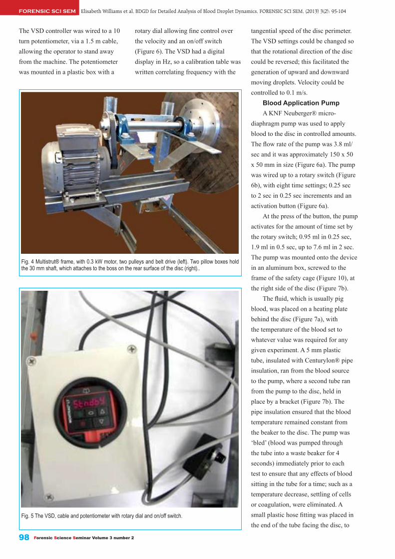
Elisabeth Williams et al, BDGD for Detailed Analysis of Blood Droplet Dynamics, FORENSIC SCI SEM, (2013) 3(2): 95-104FORENSIC SCI SEM
Forensic Science Seminar Volume 3 number 298
tangential speed of the disc perimeter. The VSD settings could be changed so that the rotational direction of the disc could be reversed; this facilitated the generation of upward and downward moving droplets. Velocity could be controlled to 0.1 m/s.
Blood Application PumpA KNF Neuberger® micro-
diaphragm pump was used to apply blood to the disc in controlled amounts. The flow rate of the pump was 3.8 ml/sec and it was approximately 150 x 50 x 50 mm in size (Figure 6a). The pump was wired up to a rotary switch (Figure 6b), with eight time settings; 0.25 sec to 2 sec in 0.25 sec increments and an activation button (Figure 6a).
At the press of the button, the pump activates for the amount of time set by the rotary switch; 0.95 ml in 0.25 sec, 1.9 ml in 0.5 sec, up to 7.6 ml in 2 sec. The pump was mounted onto the device in an aluminum box, screwed to the frame of the safety cage (Figure 10), at the right side of the disc (Figure 7b).
The fluid, which is usually pig blood, was placed on a heating plate behind the disc (Figure 7a), with the temperature of the blood set to whatever value was required for any given experiment. A 5 mm plastic tube, insulated with Centurylon® pipe insulation, ran from the blood source to the pump, where a second tube ran from the pump to the disc, held in place by a bracket (Figure 7b). The pipe insulation ensured that the blood temperature remained constant from the beaker to the disc. The pump was ‘bled’ (blood was pumped through the tube into a waste beaker for 4 seconds) immediately prior to each test to ensure that any effects of blood sitting in the tube for a time; such as a temperature decrease, settling of cells or coagulation, were eliminated. A small plastic hose fitting was placed in the end of the tube facing the disc, to
Fig. 4 Multistrut® frame, with 0.3 kW motor, two pulleys and belt drive (left). Two pillow boxes hold the 30 mm shaft, which attaches to the boss on the rear surface of the disc (right)..
Fig. 5 The VSD, cable and potentiometer with rotary dial and on/off switch.
The VSD controller was wired to a 10 turn potentiometer, via a 1.5 m cable, allowing the operator to stand away from the machine. The potentiometer was mounted in a plastic box with a
rotary dial allowing fine control over the velocity and an on/off switch (Figure 6). The VSD had a digital display in Hz, so a calibration table was written correlating frequency with the

FORENSIC SCI SEMElisabeth Williams et al, BDGD for Detailed Analysis of Blood Droplet Dynamics, FORENSIC SCI SEM, (2013) 3(2): 95-104
99
control the direction of expelled blood.Diffuser Screen and Global/
Local Coordinate SystemsA diffuser, 2400 mm high and 1200
mm wide, was securely positioned 80 mm behind the front surface of the disc, in the same plane as the disc. The screen was comprised of 6 mm thick frosted Perspex®.
The diffuser screen served four functions; to diffuse the backlighting to create an even level of illumination, to provide a surface for a global coordinate system (GCS) grid to be marked on, to provide a surface to attach impact targets and to provide additional stability to the machine.
The GCS was designed so that the positions of each droplet and resulting bloodstain could be correctly plotted relative to each other and to the position of the disc. The grid coordinates 0,0 in the x and y axes were located at the bottom left corner of the diffuser screen, with the x coordinates increasing to the right and the y coordinates increasing in an upward direction. The centre of the disc was located at x,y coordinates 1100, 990 mm. The flight paths of blood droplets propelled from the disc were plotted according to their GCS coordinates. The device can propel blood drops onto any surface to create test bloodstains. Initial tests utilized
Foamcore® strips cut in 2000 mm and 1200 mm lengths, 150 mm wide, marked with the GCS coordinates and attached to the diffuser screen at 90 degrees (Figure 9).
The device and coordinate system were set up for droplet flight to be filmed with a high speed video camera, positioned perpendicular to the diffuser screen. Image dimensions are measured in pixels so a calibration scale, called a local coordinate system (LCS), was constructed in order to plot droplet position relative to the GCS. A transparent Perspex® box with a millimetre grid was placed in the same plane as the disc in front of the diffuser
Fig. 6a KNF Neuberger® pump, rotary switch dial and activation button. Fig. 6b KNF Neuberger® pump and circuit board with rotary switch.
July 2013 ISSN 2157-118X
Fig. 7a Blood in a beaker on an element at the rear of the device with an insulated 5 mm tube running from the beaker to the pump.
Fig. 7b The position of the insulated application hose and pump for upward-moving trials: 20 mm from the perimeter of the disc, 55 degrees from the horizontal, 15 mm from the surface of the disc and the pump (far right).

Elisabeth Williams et al, BDGD for Detailed Analysis of Blood Droplet Dynamics, FORENSIC SCI SEM, (2013) 3(2): 95-104FORENSIC SCI SEM
Forensic Science Seminar Volume 3 number 2100
screen and the camera focused in this plane (Figure 9).
Safety FeaturesFor safe practice while using the
device, several features were added to the design. A safety cage around the disc prevents any projectile accidently released from the rotating disc to be flung toward any personnel or equipment. The cage also catches any blood being released from the edge of the disc, preventing lab contamination and providing stability for the diffuser screen. Lengths of stainless steel slotted angle iron were cut to appropriate lengths and welded in the configuration seen in Figure 11 and bolted to the MDF board. Clear Perspex® windows
were cut to size to form a front cover, left side cover, top and bottom as labeled in Figure 10.
In addition to the cage, the belt drive also required a cover to minimise the risk of injury from fast moving, rotating parts. An aluminum sheet was screwed to a wooden semi-circular plate and fastened over the belt and pulleys and clear Perspex® sheets were fastened to the cover at right angles, preventing any contact with the belt while the device is in use (Figure 11).
Backlighting SystemA high intensity backlighting
system was required for the high speed camera to capture the flight dynamics of small blood droplets travelling at high
velocities at a sufficiently high shutter speed to prevent motion blur and a small enough aperture for sufficient depth of field. A four LED array system was assembled for this purpose and will be detailed in a separate publication. An adjustable frame to hold the system was constructed from Multistrut®, which enabled the positions of the LED arrays to be moved according to the area being filmed. Figure 12a provides an illustration of the position of the backlighting frame relative to the diffuser screen and the proximity of the LED arrays to each other; a configuration which was shown to provide the most even illumination at the right intensity to film the blood droplets being propelled from the disc (Figure 12b).
Performance Experiments
Experiments were conducted to determine whether the droplets generated by the device were in the size and velocity ranges of those generated by human-wielded assault weapons (Figure 13). In these initial validation experiments, the tangential velocity of the disc was set to each of the four different velocities in Table 1. The volume of blood applied to the disc was initially 0.98 ml and this was increased if it was found that the blood droplets were smaller than those generated by the human-wielded weapons, until the volume made no difference to the droplet size.
Results
Figure 14 shows five consecutive images of droplets being propelled at 6 m/s, with 0.98 ml of blood applied to the disc. It was observed that as the disc rotated, blood travelled to the disc perimeter and formed a ligament as it travels away from the disc. The
Fig. 8 The Perspex® diffuser screen with the global coordinate system (GCS) grid; in 100 mm squares.

FORENSIC SCI SEMElisabeth Williams et al, BDGD for Detailed Analysis of Blood Droplet Dynamics, FORENSIC SCI SEM, (2013) 3(2): 95-104
July 2013 ISSN 2157-118X 101
ligament started to break up when surface tension forces were no longer sufficient to provide the necessary centripetal force to keep the blood on the disc. At this point individual droplets continued to travel in the direction they were travelling at the instant of release from the ligament; on a flight path tangential to the perimeter of the disc. Figure 15, an overlay of the images in Figure 14, shows that initially this is a straight line trajectory; however it is assumed that this trajectory will start to curve due to the forces of air resistance and gravity some time later. During ligament breakup very small droplets can be observed between the larger droplets in figures 15 and 16, while the average size of the larger droplets is similar; a standard deviation of 0.16 was calculated for 200 droplets released at 6 m/s, recorded within 200
mm of the disc perimeter.Figure 16 provides a visual
comparison of the droplet sizes summarized in Table 1, projected at disc tangential velocities of 6, 10, 20 and 30 m/s with approximately the same global coordinates. It is evident from these results that the device generates droplets smaller than a human wielded baseball bat at 20 and 30 m/s. However it is unlikely to see shorter weapons with tangential velocities in this range in practice, so this remains an acknowledged limitation of the device.
Conclusions
The BDGD has proved to be capable of generating large numbers of reproducible droplets in any planar direction, within the size and velocity ranges of cast-off from common
humanwielded assault weapons relevant to blunt and sharp force trauma, with the exception of a baseball bat being swung at greater than 20 m/s.
In controlled laboratory conditions, each independent variable in the system can be defined, set at a predetermined value, and large numbers of droplets generated, stains created, and results observed. The experimenter can then change one independent variable in that system, keeping all others constant, test again under the new conditions and observe any changes in the outcome variables and the frequency of those changes under the new conditions. An example would be to compare the number of spines and scallops observed in stains created at a given location under condition A, then increasing the tangential velocity of the disc by 3 m/s, and repeating the experiment. The
Fig. 9 The local coordinate system (LCS) calibration grid being held against the surface of the diffuser screen.
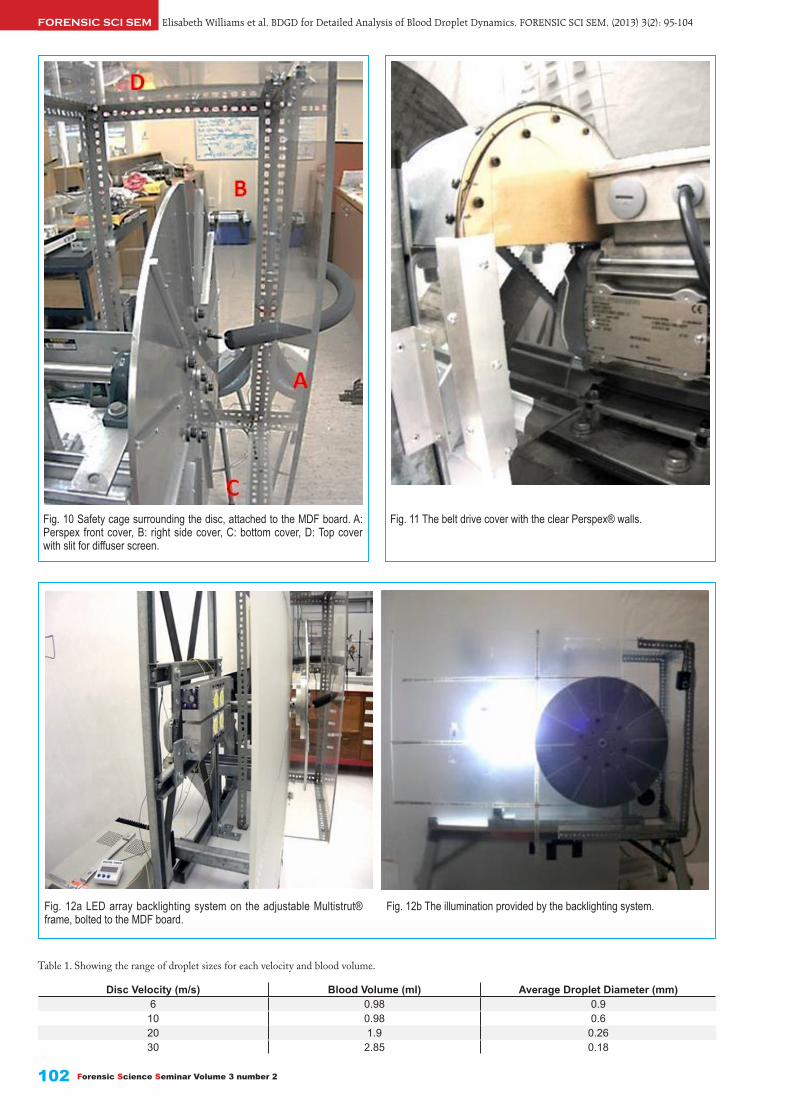
Elisabeth Williams et al, BDGD for Detailed Analysis of Blood Droplet Dynamics, FORENSIC SCI SEM, (2013) 3(2): 95-104FORENSIC SCI SEM
Forensic Science Seminar Volume 3 number 2102
Fig. 10 Safety cage surrounding the disc, attached to the MDF board. A: Perspex front cover, B: right side cover, C: bottom cover, D: Top cover with slit for diffuser screen.
Fig. 11 The belt drive cover with the clear Perspex® walls.
Fig. 12a LED array backlighting system on the adjustable Multistrut® frame, bolted to the MDF board.
Fig. 12b The illumination provided by the backlighting system.
Table 1. Showing the range of droplet sizes for each velocity and blood volume.
Disc Velocity (m/s) Blood Volume (ml) Average Droplet Diameter (mm)6 0.98 0.9
10 0.98 0.620 1.9 0.2630 2.85 0.18

FORENSIC SCI SEMElisabeth Williams et al, BDGD for Detailed Analysis of Blood Droplet Dynamics, FORENSIC SCI SEM, (2013) 3(2): 95-104
July 2013 ISSN 2157-118X 103
viscosity values, so that the effect of oscillation on stain formation and the accuracy of the subsequent impact angle calculation can be evaluated experimentally.
While a simple concept and a relatively straightforward design, the BDGD provides an effective vehicle to improve the scientific rigour behind bloodstain impact angle reconstruction and can quantitatively test some of the fundamental principles of bloodstain pattern analysis.
Acknowledgements
This project would not have
device can generate a sufficient number of results to quantitatively assess the effect of the manipulated independent variable (e.g. initial velocity) on the dependent variables such as measurable stain characteristics. This approach enables, for example, the systematic assessment of the relationship between the trajectory of a constantly accelerating assault weapon swung in an arc, and the resulting cast-off pattern.
Utilizing the backlighting system and global coordinate system, the device can be used as a vehicle to study and understand parabolic blood droplet trajectories and the limitations of the straight line trajectory calculation
methods. Blood droplet trajectories can be mapped and plotted and databases of the dynamics of different sized droplets can be developed over time.
In case specific experimentation, the device can be used in hypothesis testing in a systematic fashion, incorporating factors such as the effect of the impact surface on stain formation, and subsequent impact angle calculation.
The enhanced back-lighting system provides the means to analyse the oscillation amplitude of individual droplets, and the time to dampening for droplets created under a range of conditions; velocities, drop sizes and
Fig. 13 Overlaid images of a kitchen knife being swung forcefully backwards by a human volunteer, after being dipped in pig blood. Overlaid graphics illustrate the blood droplet trajectories.
Fig. 14 Five still images taken every 0.000926 sec, of blood being propelled off the disc at 6 m/s, filmed at 5400 frames per second at a shutter speed of 1/178000th of a second..
Journal of Bloodstain Pattern Analysis 26 Vol. 29 No. 1 March 2013
Figure 16 provides a visual comparison of the droplet sizes summarized in Table 1, projected at disc tangential velocities of 6, 10, 20 and 30 m/s with approximately the same global coordinates. It is evident from these results that the device generates droplets smaller than a human wielded baseball bat at 20 and 30 m/s. However it is unlikely to see shorter weapons with tangential velocities in this range in practice, so this remains an acknowledged limitation of the device.
Figure 14: Five still images taken every 0.000926 sec, of blood being propelled off the disc at 6 m/s, filmed at 5400 frames per second at a shutter speed of 1/178000th of a second.

Elisabeth Williams et al, BDGD for Detailed Analysis of Blood Droplet Dynamics, FORENSIC SCI SEM, (2013) 3(2): 95-104FORENSIC SCI SEM
Forensic Science Seminar Volume 3 number 2104
supported this study. The authors with to thank Dr Rachel Fleming and Gary Gillespie for reviewing this manuscript.
References
[1] NAS, Strengthening Forensic Science in the United States: A Path Forward. The National Academies Press (2009).[2] Raymond, M.A., E.R. Smith, and J. Liesegang, The physical properties of blood - forensic considerations. Science and Justice - Journal of the Forensic Science Society (1996) 36(3): 153-160.[3] Bevel, T.a.G., Ross, Bloodstain Pattern Analysis with an Introduction to Crime Scene Reconstruction. CRC Series in practical aspects of criminal and forensic investigations 1002 Boca Ralton: CRC Press LLC (2006).[4] Raymond, M.A., E.R. Smith, and J. Liesegang, Oscillating blood droplets - implications for crime scene reconstruction. Science and Justice - Journal of the Forensic Science Society (1996) 36(3): 161-171.[5] Hulse-Smith, L., N.Z. Mehdizadeh, and S. Chandra, Deducing drop size and impact velocity from circular bloodstains. Journal of Forensic Sciences (2005) 50(1): 54-63.[6] Knock, C. and M. Davison, Predicting the position of the source of blood stains for angled impacts. Journal of Forensic Sciences (2007) 52(5): 1044-1049.[7] Hulse-Smith, L. and M. Illes, A blind trial evaluation of a crime scene methodology for deducing impact velocity and droplet size from circular bloodstains. Journal of Forensic Sciences (2007) 52(1): 65-69.[8] Liu, H., Science and Engineering of Droplets: Fundementals and Applications (2000) William Andrew Publishing: 527.[9] SWGSTAIN, S.W.G.o.B.P.A., Recommended Terminology. Forensic Science Communications (2009) 11(2).[10] Bevel, T., Gardner, R.M.. Bloodstain Pattern Analysis with an Introduction to Crime Scene Reconstruction. 3rd ed (2008) Boca Raton, FL: CRC Press.[11] MacDonell, H.L., Bloodstain Patterns. 2nd ed (2005) Corning, NY: Laboratory of Forensic Sciences.[12] Williams, E., The Biomechanics of Blunt Force Trauma. Masters Thesis in Forensic Science, University of Auckland (2008).[13] Wonder, A., Blood Dynamics. Academic Press (2008).[14] James, S.H., Kish, P.E., Sutton, T.P., Principles of Bloodstain Pattern Analysis: Theory and Practice (2005) Boca Raton, FL: CRC Press; Taylor and Francis Group.[15] Fischer, W.C., Utilizing Bloodstains in Accident Reconstruction, in Scientific and Legal Applications of Bloodstain Pattern Interpretation, S.H. James, Editor (1998) CRC Press: Boca Raton, FL.[16] Serway, R.A.a.J., J.W, Physics for Scientists and Engineers. 8th Ed. Pacific Grove, CA. Brooks Cole (2009). ■
Fig. 15 The five still images from Figure 14 overlaid, with inserted graphics illustrating the flight path of each droplet, which in the initial flight phase, is tangential to the perimeter of the disc.
Fig. 16 Still photographs of in-flight droplets generated by the device at 6, 10, 20 and 30 m/s.
been possible without the invaluable contribution from the following people and organisations:
Dan Rahn Memorial Research Grant Committee, International Association of Bloodstain Pattern Analysts (IABPA).
Dr. Sharon Walt, Dr. Ir. Ron McDowall, Ian Williams; Agcon Engineering Ltd, Drury, Peter O’Gara; O’Gara Engineering, Graeme Harris,
Scott Aimes, Jim Maclean, Julian Phillips, Dave Read, Ken Brown, Dr Mark Jermy; Department of Mechanical Engineering, University of Canterbury, Gary Donaldson; Bretmar Transmission Company Ltd, Papakura, Multistrut(R) Ltd, NZ. FreshPork NZ Ltd, Bay City Abattoir, Timaru.
The principal author (E.W) acknowledges receipt of a Dan Rahn Memorial research grant which

July 2013 ISSN 2157-118X 105
Operating Procedures and Techniques of the Technical Replication of the Original Documents
Abstract In the recent practice of document identification, the problem that the original samples cannot be extracted is often involved. The principals have no choice but to submit the prints, faxes, photos or electronic scans of the samples to the identification bodies. However, many features of the copies will be blurred, deformed or even lost during the process of replication. The copies will also make the identification work more difficult, and will affect the identification comments. In this case, the identification bodies need to go to relevant departments to do some verification and technical replication of the original documents on the spot. How to restore the samples to their maximum limits is the problem that all the document appraisers must be confronted with.
Keywords: Forensic science, Document identification, Technical replication, Operating procedures, Techniques.
FORENSIC SCI SEMJIN Xin, Operating of Technical Replication of Original Documents, FORENSIC SCI SEM, (2013) 3(2): 105-107
Received 20 May 2013Received in revised form 28 May 2013
Accepted 30 May 2013Available online 17 June 2013
Peer Review
ed2157118X
.3.2.P2
1 Raising of the Problems
In the practice of document identification, the ideal state is to identify the papery orifinal documents of the samples. Under the circumstance that the principals cannot present the original documents, the appraisers can identify the copies of the samples(including the prints, faxes, electronic scans and photos etc.) Because of the restrictions of the copies themselves, for example, the feature blur, deformation and loss will make identification more difficult and finally affect the comments, some of which are tendentious and some even incompatible.
In recent years, I’ve encountered numerous cases that the principals cannot present original documents. The reason is not that the original documents have been lost, but that the paper materials are stored in the archives of related units and cannot be
JIN Xin *, a
a Nanjing Normal University Law School, Nanjing 210046,China.
* Corresponding Author: JIN Xin (1983-, male, engineer, mainly engaged in Theory and Practice of Forensic Studies). E-mail: [email protected]. Address: No.122, Ninghai Road, Nanjing Normal University Law School, Nanjing, Jiangsu Province 210046, China.
drawn directly. Original documents, such as articles of association in administrative departments for industry and commerce, sigatures or seals in the shareholders’ resolutions, handwritings or seal samples retained on the band certificates by the suspects are all unavailable. According to the files management regulations of Industrial and Commercial Authorities, the identification bodies can go to the units above to inquire the papery original documents together with the Court, but cannot get them out. To reduce the impact of the copies, sometimes the principals are required to repicate the originals. However, because the principals are unaware of the identification demands, the copies are usually unsatisfactory. Even the professionals, by using different ways, will reach defferent idetification conclusions due to the lack of scientific and standardized instructions. Therefore, based on the
practical experience of the technical replication of the papery originals on the spot in the identification centre in recent two years, I’ve explored the operating procedures and techniques of the original documents.
2 Equipment Requirements
Because of the restrictions of the technical replication itself, the appraisals cannot use large-sized apparatus, and can only carry some miniatures and portable equipments.
2.1 Measuring EquipmentsCalipers, protractors, straightedge2.2 Electronic Coping
EquipmentsSLR cameras with standard
lens(including a macro lens, dimensions), compact cameras of small-sized lens, portable scanners.
2.3 Observation EquipmentsMagnifiers, miniature microscope
with cold light source

JIN Xin, Operating of Technical Replication of Original Documents, FORENSIC SCI SEM, (2013) 3(2): 105-107FORENSIC SCI SEM
Forensic Science Seminar Volume 3 number 2106
writing is hand-written or printed (Figure 1), whether the seal is chopped or printed(Figure 2), identification of the sort of the typewriter, whether the originals are printed successively at one time. How the originals are formed is hard to recognize with the naked eye. The spparatus need to use a microscope to observe the paper originals on the spot. If they find that there is something wrong with the formation of the documents, they need to take photomicrographs, and take down the feature points. Besides, as to the identification of the seals, they need to measure and take a record of the features’ data of the edge lines’ diameters, pentagonal patterns with calipers, straightedges and protractors.
3.3 RemakePhotographic remake is an
important link in technical replication, and it requires a high level of operation. The remake of the technical replication includes the full view of the originals and the handwriting to be identified or the feature points of the seals. Remake usually causes longitudinal tensile deformation, transverse tensile deformation and the change of the seals’ diametre. The reason is that the shooting angle of the camera cannot be absolutely perpendicular to the paper face, And this can be hardly overcome. Because the copy stand and light source are not easy to carry, the appraisals usually have a handheld remake. Handheld remake will cause more obvious deformations. Because of the deformation problems of remake, the comparison between the remake(Figure3, Figure4, Figure5) and seals or scans(Figure6) cannot be evaluated by diameter, but can be compared in such aspects as the fonts, distribution, detailed features of the seals. The fonts and distribution of the seals will not change with the tensile deformation, and are relatively stable.[1]
3.4 Scanning
Fig. 1
Fig. 2
Fig. 3
Fig. 4
Fig. 5
Fig. 6
3 Operating Procedures
3.1 Check on the SpotAs the copies for inspection may
be forged or altered, there is a need to check on the spot, whose aim is to test whether there is a difference between the paper originaks and the cipies provided by the principals in contents. The appraisals need to check the originals and the copies or electronic scans, photosprovided by the principals, and examine the numbers, dates, contents and inscriptions of the originals. If there is no defference between the originals and the copies or the scans, the identification work can be continued. But if the two don’t match with each other, records of the defferences will be taken and the principals will be imformed in time.
3.2 Inspection on the spotInspection on the spot is to
preliminarily define the formation of the documents such as whether the
Overlap analysis cannot be made between remakes and electronic scans.

July 2013 ISSN 2157-118X 107
FORENSIC SCI SEMJIN Xin, Operating of Technical Replication of Original Documents, FORENSIC SCI SEM, (2013) 3(2): 105-107
If the originals are seals, the identifiers need to carry portable scanners to scan the seals and record the electronic scans. In the later identifications, the copies will be scanned by the same resolution, and the two scans will have overlap analysis on the photoshop software platform.
3.5 Coping a RecordIn judicial practice, the examination
of the idetification conclusion must contain the “originality” of the materials. The judge, as a layman, shouldn’t easily doubt the reasonableness, but must master the “originality”. The fourth paragraph of Article 24 of the General Procedures of Forensic Indentification (hereinafter referred to General Procedures) makes rules for extractions on the spot: ‘There should be at least two forensic experts when there’s a need to extract materials on the spot, and the principals should be imformed to the site to witness the process .’ But there are no related regulations of technical replication of the originals in General Procedures. The technical replication of the originals is different from extracting samples on the spot. The latter is the procedure before identification, while the former is a procedure and technical means of identifecation. Referring to the fourth paragraph of Article 24 of General Procedures, it is considered, to ensure the legality of the identification work, there should be at least two forensic experts to operate on the technical replication of the original documents. After the replication, the identification bodies need to issue a Records of the Technical Replication of the Original Documents (Records). Records should mark the time, place, equipment used, operators of the technical reputation. Finally, the appraisals, principals, workers of the units where the originals are stored will sign their signatures and mark the date.
4 Techniques
4.1 PhotomicrographBecause of the restrictions of the
site, photomicrograph can only take the way that the lens are aligned with the eyepiece of the microscope. Such kind of shooting is more flexible, but has a difficulty imaging. Pay attention to the following points:
1) Select LensBecause that the lens of the
eyepiece of the microscope, the lens of the digital camera can’t be too large, or the images in the microscope cannot be found. Usually, compact cameras with small lens are selected instead of SLR cameras of standard lens.
2) Optical AlignmentFirst, focus the microscope on the
light, better choose white light instead of warm light, for images in the white light are clearer. Portable microscopes for seal examination usually have cold light sources themselves. And the light emitted just meet the requirements.
3) Mining PointFind the feature points which
need to be photographed under the low magnification of the microscope and then convert to high magnification.
4) FocusingFirst adjust the digital camera to a
state in which there should be no macro and no flash, and then focus the lens on the eyepiece of the microscope and focus until the images of the digital camera are the largest and clearest.
5) ShootingThere should be no flash when
shooting. If there is flash, the imagies will be dark.
4.2 Macro PhotographMacro pohotograph is used to
remake the writings or the features of the seals. Macro photograph is different from the photographs above. Generally, the image ratios from 1:1 to 1:4 are macro photographs, but when they reach from 10:1 to 200:1, they
are photomicrographs. The best way of macro photograph is to use special macro lens, whose resolution is high, aberration very light, contrast high and color reproduction good. Macro photograph has a quite strong resolving power when shooting closely. The image quality of the feature points is guaranteed without too much changes within focus range. Usually, there is a label on the macro lens. The resolution should be determined first according to the size of the feature points. Focus in advance, and then get close to the feature points to shoot. In this way of shooting, the image quality of the feature points are guaranteed. Another way is to use zoom lens with macro lens. Though this way is flexible, the image resolution is lower and more severly deformed.[2]
The objects of macro photograph are writings or detailed features of high value in seals. Remakes of the detailed features of handwritings should focus on the shakes, curves, strokes, restarts, stagnations.[3] Remakes of the detailed features of the seals should focus on the broght defects, border defects, azimuth lines etc. When laying the dimensions, pay attention that the dimensions are needed by the overall remake of the writings or seals. Better not to cover the shooting objects. There is no need to lay dimensions when remaking the feature points of the handwritings or seals.
References
[1] LIU Jing-jie, LI Jing. Guan yu chuan zhen wen jian shang yin wen jian ding de jie lun miao shu. Chang san jiao shou jie wen shu jian ding yan tao hui ji pei xun ban lun wen ji an li hui bian, November, (2010): 88-91. (In Chinese)[2] ZHANG Zhi-chao. Qian ming zi ji de jian yan. Forensic Science and Technology (2005) (3): 47-51. (In Chinese)[3] ZHAO Xiu-ping. The direction of digital camera’s photometry. Forensic Science and Technology (2007) (6): 25-28. (In Chinese). ■

Forensic Science Seminar Volume 3 number 2108
Using Smartphones as a Proxy for Forensic Evidence Contained in Cloud Storage Services
Abstract Cloud storage services such as Dropbox, Box and SugarSync have been embraced by both individuals and organizations. This creates an environment that is potentially conducive to security breaches and malicious activities. The investigation of these cloud environments presents new challenges for the digital forensics community.
It is anticipated that smartphone devices will retain data from these storage services. Hence, this research presents a preliminary investigation into the residual artifacts created on an iOS and Android device that has accessed a cloud storage service. The contribution of this paper is twofold. First, it provides an initial assessment on the extent to which cloud storage data is stored on these client-side devices. This view acts as a proxy for data stored in the cloud. Secondly, it provides documentation on the artifacts that could be useful in a digital forensics investigation of cloud services.
Keywords: Forensic science, Forensic evidence, Smartphone, Cloud storage, Iphone, HTC.
George Grispos a, William Bradley Glisson a, Tim Storer a
a University of Glasgow.
* Corresponding Author: George Grispos: E-mail: [email protected]. William Bradley Glisson: E-mail: [email protected]. Tim Storer: E-mail: [email protected].
George Grispos, et al, Smartphones as Proxy for Forensic Evidence in Cloud Storage, FORENSIC SCI SEM, (2013) 3(2): 108-118FORENSIC SCI SEM
Received 27 July 2012Received in revised form 21 August 2012Accepted 7 December 2012Available online 25 June 2013
Peer Review
ed2157118X
.3.2.P3
1. Introduction
Global connectivity, mobile device
market penetration and use of remote
data storage services are all increasing.
Cisco reports that mobile data traffic
reached 597 petabytes per month in
2011, which was over eight times
greater than the amount of Internet
traffic in 2000 [1]. They also predict
that global mobile data transmission
will exceed ten exabytes per month by
2016, with over 100 million smartphone
users transmitting more than 1 gigabyte
of data per month [1]. Supporting these
predictions, cloud storage providers
have experienced tremendous growth
in the past year. A press release from
Dropbox reported that their consumer
base has surpassed 25 million users [2].
They also claim that over one billion
files are saved every three days using its
services [3]. Box reports that enterprise
revenue tripled in 2011 with mobile
device implementation increasing 140%
monthly [4]. Box have also experienced
substantial penetration into the retail,
financial and healthcare enterprise
markets [5].
According to articles by CIO [6],
surveys by Advanced Micro Devices
(AMD) [7] and IBM [8], there is
an apparent consensus that cloud
computing is increasingly integrating
into the business environment. The
business reasons for this migration
range from ideas like focusing on
growth, innovation and customer value
to improved use of resources, increasing
employee productivity and cutting costs
[8].
Cisco have argued that the storage
and retrieval of corporate data from
a number of cloud-based devices is
concerning for security professionals,
noting that “wherever users go,
cybercriminals will follow” [9].
Support for this idea was demonstrated
when Dropbox was used in corporate
espionage [10]. Hence, there is no
practical barrier to further utilization
of cloud storage services to store and
exchange illicit material. Gartner [11]
warns that “investigating inappropriate
or illegal activity may be impossible in
cloud computing”.
Investigating cloud service
providers has been a topic of great
discussion, with a number of challenges
being raised about conducting a digital
forensics investigation in such an
environment [12-14]. One of these
challenges is the investigator’s ability
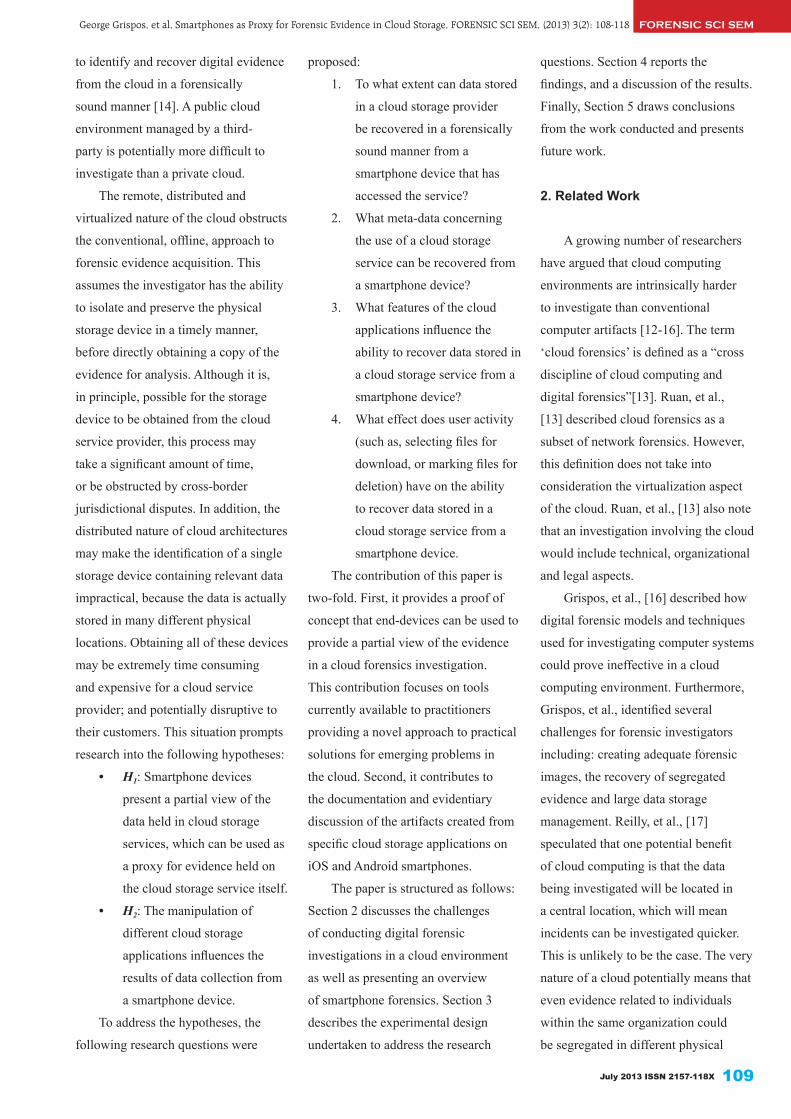
July 2013 ISSN 2157-118X 109
FORENSIC SCI SEMGeorge Grispos, et al, Smartphones as Proxy for Forensic Evidence in Cloud Storage, FORENSIC SCI SEM, (2013) 3(2): 108-118
to identify and recover digital evidence
from the cloud in a forensically
sound manner [14]. A public cloud
environment managed by a third-
party is potentially more difficult to
investigate than a private cloud.
The remote, distributed and
virtualized nature of the cloud obstructs
the conventional, offline, approach to
forensic evidence acquisition. This
assumes the investigator has the ability
to isolate and preserve the physical
storage device in a timely manner,
before directly obtaining a copy of the
evidence for analysis. Although it is,
in principle, possible for the storage
device to be obtained from the cloud
service provider, this process may
take a significant amount of time,
or be obstructed by cross-border
jurisdictional disputes. In addition, the
distributed nature of cloud architectures
may make the identification of a single
storage device containing relevant data
impractical, because the data is actually
stored in many different physical
locations. Obtaining all of these devices
may be extremely time consuming
and expensive for a cloud service
provider; and potentially disruptive to
their customers. This situation prompts
research into the following hypotheses:
• H1: Smartphone devices
present a partial view of the
data held in cloud storage
services, which can be used as
a proxy for evidence held on
the cloud storage service itself.
• H2: The manipulation of
different cloud storage
applications influences the
results of data collection from
a smartphone device.
To address the hypotheses, the
following research questions were
proposed:
1. To what extent can data stored
in a cloud storage provider
be recovered in a forensically
sound manner from a
smartphone device that has
accessed the service?
2. What meta-data concerning
the use of a cloud storage
service can be recovered from
a smartphone device?
3. What features of the cloud
applications influence the
ability to recover data stored in
a cloud storage service from a
smartphone device?
4. What effect does user activity
(such as, selecting files for
download, or marking files for
deletion) have on the ability
to recover data stored in a
cloud storage service from a
smartphone device.
The contribution of this paper is
two-fold. First, it provides a proof of
concept that end-devices can be used to
provide a partial view of the evidence
in a cloud forensics investigation.
This contribution focuses on tools
currently available to practitioners
providing a novel approach to practical
solutions for emerging problems in
the cloud. Second, it contributes to
the documentation and evidentiary
discussion of the artifacts created from
specific cloud storage applications on
iOS and Android smartphones.
The paper is structured as follows:
Section 2 discusses the challenges
of conducting digital forensic
investigations in a cloud environment
as well as presenting an overview
of smartphone forensics. Section 3
describes the experimental design
undertaken to address the research
questions. Section 4 reports the
findings, and a discussion of the results.
Finally, Section 5 draws conclusions
from the work conducted and presents
future work.
2. Related Work
A growing number of researchers
have argued that cloud computing
environments are intrinsically harder
to investigate than conventional
computer artifacts [12-16]. The term
‘cloud forensics’ is defined as a “cross
discipline of cloud computing and
digital forensics”[13]. Ruan, et al.,
[13] described cloud forensics as a
subset of network forensics. However,
this definition does not take into
consideration the virtualization aspect
of the cloud. Ruan, et al., [13] also note
that an investigation involving the cloud
would include technical, organizational
and legal aspects.
Grispos, et al., [16] described how
digital forensic models and techniques
used for investigating computer systems
could prove ineffective in a cloud
computing environment. Furthermore,
Grispos, et al., identified several
challenges for forensic investigators
including: creating adequate forensic
images, the recovery of segregated
evidence and large data storage
management. Reilly, et al., [17]
speculated that one potential benefit
of cloud computing is that the data
being investigated will be located in
a central location, which will mean
incidents can be investigated quicker.
This is unlikely to be the case. The very
nature of a cloud potentially means that
even evidence related to individuals
within the same organization could
be segregated in different physical

George Grispos, et al, Smartphones as Proxy for Forensic Evidence in Cloud Storage, FORENSIC SCI SEM, (2013) 3(2): 108-118FORENSIC SCI SEM
Forensic Science Seminar Volume 3 number 2110
locations [18]. Taylor, et al., [14]
suggest there is also the possibility
that potential important evidence could
be lost in a cloud. Registry entries
in Microsoft Windows platforms,
temporary files, and meta-data could all
be lost if the user leaves the cloud [14].
To enable a forensic investigation
to be conducted, evidence needs to
be collected from the cloud. This is
likely to pose a great challenge to
forensic investigators [19]. Researchers
have begun proposing methods of
acquiring evidence from a variety of
cloud providers and services [19, 20].
Delport, et al., [20] proposed the idea
of isolating a cloud instance for further
investigation and several methods were
proposed. None of these methods were
empirically validated, nor is it clear
how a forensic image of the instance is
obtained from the proposed techniques.
Dykstra and Sherman [19] have
evaluated the performance of current
forensic tools (FTK Imager and Encase
Enterprise) and used these to acquire
evidence from Infrastructure-as-a-
Service (IaaS) instances stored in the
Amazon Elastic Compute Cloud (EC2)
[19]. Although the methods proposed
were empirically evaluated, they are
limited in essence to only IaaS virtual
instances where a remote acquisition
agent can be loaded on the instance
under investigation. A second issue with
this approach is that the investigator
must be in possession of Amazon
EC2 key pairs used to connect to the
instance. The purpose of the key pairs
is to ensure that only the instance’s
owner has access to the instance [21].
These public/private keys are created
by the owner when the instance is first
created using the Amazon Web Services
Management Console [22]. Unless the
investigator can recover these keys,
then the methods of acquisition as
described by Dykstra and Sherman
cannot be used.
Taylor, et al., [14] have extensively
examined the legal issues surrounding
cloud computing and comment that
any evidence gathered from the cloud
should be conducted within local
laws and legislation. Phillips [23]
discussed the issue of the challenge of
keeping a chain of custody for such
an investigation, and asked: “Where
physically is that part of the cloud at
any given time? And where was it when
forensically acquired?”. Phillips argues
that the cloud is a dynamic paradigm
and physically isolating it to conduct an
investigation could be a daunting task
for the investigator.
When multiple devices are used
to access data in the cloud, these
problems are exacerbated. In particular,
smartphones are increasingly being
used to access data stored in a cloud
while the user is mobile. Smartphone
devices are distinguishable from a
traditional mobile phone by its superior
processing capabilities, a larger
storage capacity, as well as its ability
to run complex operating systems and
applications [24]. From an evidentiary
perspective, the smartphone can be
considered a treasure trove of forensic
evidence. A recent study recovered
more than 11,000 data artifacts from
just 49, predominately lowend, devices
[25]. As with a traditional mobile
phone, the smartphone not only stores
call logs, text messages and personal
contacts, but it also has the ability to
store web-browsing artifacts, email
messages, instant messenger lots, GPS
coordinates, as well as third-party
application related data [26-28].
There are a number of tools
that can be used to perform a data
acquisition from a smartphone.
Examples of these include Cellebrite’s
Universal Forensic Extraction Device
(UFED) and its associated tool suites
[29]; Micro Systemation’s XRY tools
[30]; The Mobile Internal Acquisition
Tool [31]; Paraben’s Device Seizure
[32] and RAPI Tools [33]. The
availability of tools for acquiring data
from smartphone devices makes it
possible to investigate the interactions
of these devices with cloud storage
providers, without accessing data in the
cloud service directly. However, there
is currently a lack of evidence as to the
relationship between the data held in the
cloud and that retained by a smartphone
following an interaction.
3. Experimental design
To support the hypothesis proposed
in the introduction, the experiment
was broken into six stages. These
stages included: 1) preparing the
smartphone device and installing the
cloud application; 2) loading a data set
to a cloud storage provider; 3) connect
to the data through the application on
the smartphone; 4) perform various
file manipulations to the data set and
smartphone device; 5) process the
device using the Universal Forensic
Table 1. Smartphone device features.
Feature iPhone 3G HTC DesireOperating system iOS v. 3 Android v. 2.1 (Eclair)Internal memory 8 GB storage 576 MB RAMMemory card No Yes (4 GB)
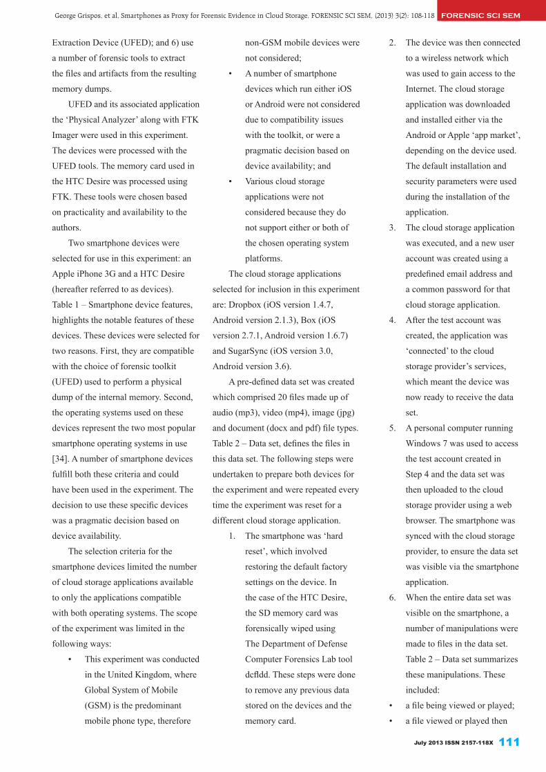
July 2013 ISSN 2157-118X 111
FORENSIC SCI SEMGeorge Grispos, et al, Smartphones as Proxy for Forensic Evidence in Cloud Storage, FORENSIC SCI SEM, (2013) 3(2): 108-118
Extraction Device (UFED); and 6) use
a number of forensic tools to extract
the files and artifacts from the resulting
memory dumps.
UFED and its associated application
the ‘Physical Analyzer’ along with FTK
Imager were used in this experiment.
The devices were processed with the
UFED tools. The memory card used in
the HTC Desire was processed using
FTK. These tools were chosen based
on practicality and availability to the
authors.
Two smartphone devices were
selected for use in this experiment: an
Apple iPhone 3G and a HTC Desire
(hereafter referred to as devices).
Table 1 – Smartphone device features,
highlights the notable features of these
devices. These devices were selected for
two reasons. First, they are compatible
with the choice of forensic toolkit
(UFED) used to perform a physical
dump of the internal memory. Second,
the operating systems used on these
devices represent the two most popular
smartphone operating systems in use
[34]. A number of smartphone devices
fulfill both these criteria and could
have been used in the experiment. The
decision to use these specific devices
was a pragmatic decision based on
device availability.
The selection criteria for the
smartphone devices limited the number
of cloud storage applications available
to only the applications compatible
with both operating systems. The scope
of the experiment was limited in the
following ways:
• This experiment was conducted
in the United Kingdom, where
Global System of Mobile
(GSM) is the predominant
mobile phone type, therefore
non-GSM mobile devices were
not considered;
• A number of smartphone
devices which run either iOS
or Android were not considered
due to compatibility issues
with the toolkit, or were a
pragmatic decision based on
device availability; and
• Various cloud storage
applications were not
considered because they do
not support either or both of
the chosen operating system
platforms.
The cloud storage applications
selected for inclusion in this experiment
are: Dropbox (iOS version 1.4.7,
Android version 2.1.3), Box (iOS
version 2.7.1, Android version 1.6.7)
and SugarSync (iOS version 3.0,
Android version 3.6).
A pre-defined data set was created
which comprised 20 files made up of
audio (mp3), video (mp4), image (jpg)
and document (docx and pdf) file types.
Table 2 – Data set, defines the files in
this data set. The following steps were
undertaken to prepare both devices for
the experiment and were repeated every
time the experiment was reset for a
different cloud storage application.
1. The smartphone was ‘hard
reset’, which involved
restoring the default factory
settings on the device. In
the case of the HTC Desire,
the SD memory card was
forensically wiped using
The Department of Defense
Computer Forensics Lab tool
dcfldd. These steps were done
to remove any previous data
stored on the devices and the
memory card.
2. The device was then connected
to a wireless network which
was used to gain access to the
Internet. The cloud storage
application was downloaded
and installed either via the
Android or Apple ‘app market’,
depending on the device used.
The default installation and
security parameters were used
during the installation of the
application.
3. The cloud storage application
was executed, and a new user
account was created using a
predefined email address and
a common password for that
cloud storage application.
4. After the test account was
created, the application was
‘connected’ to the cloud
storage provider’s services,
which meant the device was
now ready to receive the data
set.
5. A personal computer running
Windows 7 was used to access
the test account created in
Step 4 and the data set was
then uploaded to the cloud
storage provider using a web
browser. The smartphone was
synced with the cloud storage
provider, to ensure the data set
was visible via the smartphone
application.
6. When the entire data set was
visible on the smartphone, a
number of manipulations were
made to files in the data set.
Table 2 – Data set summarizes
these manipulations. These
included:
• a file being viewed or played;
• a file viewed or played then
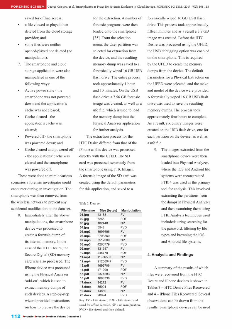
Forensic Science Seminar Volume 3 number 2112
George Grispos, et al, Smartphones as Proxy for Forensic Evidence in Cloud Storage, FORENSIC SCI SEM, (2013) 3(2): 108-118FORENSIC SCI SEM
saved for offline access;
• a file viewed or played then
deleted from the cloud storage
provider; and
• some files were neither
opened/played nor deleted (no
manipulation).
7. The smartphone and cloud
storage application were also
manipulated in one of the
following ways:
• Active power state - the
smartphone was not powered
down and the application’s
cache was not cleared;
• Cache cleared - the
application’s cache was
cleared;
• Powered off - the smartphone
was powered down; and
• Cache cleared and powered off
- the applications’ cache was
cleared and the smartphone
was powered off.
These were done to mimic various
scenarios a forensic investigator could
encounter during an investigation. The
smartphone was then removed from
the wireless network to prevent any
accidental modification to the data set.
8. Immediately after the above
manipulations, the smartphone
device was processed to
create a forensic dump of
its internal memory. In the
case of the HTC Desire, the
Secure Digital (SD) memory
card was also processed. The
iPhone device was processed
using the Physical Analyzer
‘add-on’, which is used to
extract memory dumps of
such devices. A step-by-step
wizard provided instructions
on how to prepare the device
for the extraction. A number of
forensic programs were then
loaded onto the smartphone
[35]. From the selection
menu, the User partition was
selected for extraction from
the device, and the resulting
memory dump was saved to a
forensically wiped 16 GB USB
flash drive. The entire process
took approximately 1 hour
and 10 minutes. On the USB
flash drive a 7.56 GB forensic
image was created, as well as a
ufd file, which is used to load
the memory dump into the
Physical Analyzer application
for further analysis.
The extraction process for the
HTC Desire differed from that of the
iPhone as this device was processed
directly with the UFED. The SD
card was processed separately from
the smartphone using FTK Imager.
A forensic image of the SD card was
created using the default parameters
for this application, and saved to a
forensically wiped 16 GB USB flash
drive. This process took approximately
fifteen minutes and as a result a 3.8 GB
image was created. Before the HTC
Desire was processed using the UFED,
the USB debugging option was enabled
on the smartphone. This is required
by the UFED to create the memory
dumps from the device. The default
parameters for a Physical Extraction on
the UFED were selected, and the make
and model of the device were provided.
A forensically wiped 16 GB USB flash
drive was used to save the resulting
memory dumps. The process took
approximately four hours to complete.
As a result, six binary images were
created on the USB flash drive, one for
each partition on the device, as well as
a ufd file.
9. The images extracted from the
smartphone device were then
loaded into Physical Analyzer,
where the iOS and Android file
systems were reconstructed.
FTK 4 was used as the primary
tool for analysis. This involved
extracting the partitions from
the dumps in Physical Analyzer
and then examining them using
FTK. Analysis techniques used
included: string searching for
the password, filtering by file
types and browsing the iOS
and Android file systems.
4. Analysis and Findings
A summary of the results of which
files were recovered from the HTC
Desire and iPhone devices is shown in
Tables 3 – HTC Desire Files Recovered
and 4 – iPhone Files Recovered. Several
observations can be drawn from the
results. Smartphone devices can be used
Table 2. Data set.
Filename Size (bytes) Manipulation01.jpg 43183 FV02.jpg 6265 FOF03.jpg 102448 NP04.jpg 5548 FVD05.mp3 3997696 FV06.mp3 2703360 FOF07.mp3 3512009 NP08.mp3 4266779 FVD09.mp4 831687 FV10.mp4 245779 FOF11.mp4 11986533 NP12.mp4 21258947 FVD13.pdf 1695706 FV14.pdf 471999 FOF15.pdf 2371383 NP16.pdf 1688736 FVD17.docx 84272 FV18.docx 85091 FOF19.docx 14860 NP20.docx 20994 FVDKey: FV = File viewed, FOF = File viewed and saved for offline accessed, NP = no manipulation, FVD = file viewed and then deleted.

July 2013 ISSN 2157-118X 113
to recover evidence from cloud storage
services. The artifacts recovered include
data stored in the storage service. This
is provided that the user of the device
has accessed these files in some way
using the cloud storage provider’s
application. Files which were not
viewed on the mobile phones were not
recovered. The exception was the Joint
Photographic Experts Group (JPEG)
image, where a thumbnail of the image
was recovered. The results also show
that the recovery of mp3 and mp4 files
was not very successful.
All the files marked for offline
access were recovered from both
devices. The SD memory card used
in the Android device contained files
which were either deleted by the user
or deleted as a result of the application
cache being cleared. Android-based
devices allow files to be stored on either
the device itself or on the SD memory
card [36]. Clearing the application’s
cache has an adverse effect on the
recovery of files. This is more evident
on the iPhone, which does not contain
an SD card. Powering down the
smartphone devices did not have an
effect on the recovery of data. As a
result, the files recovered were identical
to that of the active state scenario.
A variety of meta-data can be
recovered from both devices. Meta-data
recovered from the devices included
email addresses used to register for the
service, databases related to data stored
in the service and transaction logs
related to user activity. Apart from the
Dropbox application on the iPhone, all
other metadata can still be recovered
after the cache is cleared.
4.1. Android ApplicationsAn analysis of the Android
memory dumps revealed that forensic
artifacts can be recovered from both the
smartphone itself and the SD memory
card. Files and meta-data stored in the
device’s internal memory can be found
in a subfolder under the location /data/
data/ [26]. Hoog discusses the Android
file system in further detail [26]. The
location of evidence on the SD card
varies depending on the application
being investigated.
4.1.1. Dropbox. Dropbox related
artifacts can be recovered from two
main locations on the SD card. JPEG
image thumbnails can be found under
/Android/data/com.dropbox.android/
cache/thumbs/. Files saved for offline
viewing and documents viewed and
not deleted on the device, can be
found under the location /Android/
data/com.dropbox.android/files/
scratch. An analysis of the ‘unallocated
space’ revealed that the two document
files which were deleted (16.pdf and
20.docx) were still physically stored
on the SD card. These two files were
recovered by FTK. On the device itself,
two SQLite databases contain the
majority of meta-data evidence. These
databases are found in the directory
/data/data/com.dropbox.android/
databases/. The first database, db.db
contains meta-data related to the files
currently stored in the service, i.e.,
files which have not been deleted. This
information is stored in a table called
dropbox. Examples of the information
FORENSIC SCI SEMGeorge Grispos, et al, Smartphones as Proxy for Forensic Evidence in Cloud Storage, FORENSIC SCI SEM, (2013) 3(2): 108-118
Table 3. HTC Desire files recovered
Filename DB-AP DB-CC DB-PD DB-CPD B-AP B-CC B-PD B-CPD S-AP S-CC S-PD S-CPD01 T T T T √ D √ D √ D √ D02 √ √ √ √ √ D √ D √ √ √ √03 T T T T T T T T T T T T04 T T T T √ D √ D √ D √ D05 √ D √ D06 √ √ √ √ √ D √ D √ √ √ √0708 √ D √ D09 √ D √ D10 √ √ √ √ √ D √ D √ √ √ √1112 √ D √ D13 √ D √ D √ D √ D √ √ √ √14 √ √ √ √ √ D √ D √ √ √ √1516 D D D D √ D √ D √ √ √ √17 √ D √ D √ D √ D √ D √ D18 √ √ √ √ √ D √ D √ √ √ √1920 D D D D √ D √ D √ D √ D
Key: DB = Dropbox; B = Box; S = SugarSync; AP = Active Power state; CC = Cache cleared; PD = Powered Down; CPD = Cache Cleared and Powered Down; √= File Found; T = Only Thumbnail Found; D = Deleted File Recovered.

George Grispos, et al, Smartphones as Proxy for Forensic Evidence in Cloud Storage, FORENSIC SCI SEM, (2013) 3(2): 108-118FORENSIC SCI SEM
Forensic Science Seminar Volume 3 number 2114
that can be recovered includes the
file names, their size in bytes, offline
storage information and location, the
last modified time, and if the file has
been designated as a ‘favorite’.
The second database, prefs.db,
contains userspecific meta-data such
as the user’s name and email address.
This information is stored in a table
called DropboxAccountPrefs. Clearing
the cache of the Dropbox application,
removes the documents viewed and
not deleted on the device (13.pdf and
17.docx) which are stored in the com.
dropbox.android/files/scratch directory.
These files were still physically stored
on the SD card and were recovered by
FTK. The files saved for offline access,
JPEG thumbnails and the contents of
the SQLite databases remain unaltered.
4.1.2. Box. Files stored in the
Box storage service can be recovered
from three locations on the SD card.
The files saved for offline access can
be recovered from the directory /Box/
email_address/, where email_ address
is the email address used to register
for the service. The Box application
creates a cache of all the files the user
has viewed on the smartphone. These
can be recovered from the directory /
Android/data/com.box.android/cache/
filecache. Fifteen files from the data
set were found in this directory. The
files missing, are those files which are
marked as ‘no manipulation’ in Table 2
– Data set. A subdirectory of the above
location called /tempfiles/box_tmp_
images contains thumbnails of the four
JPEG images from the data set.
Meta-data artifacts about the
Box service can be recovered from
the smartphone. The Box application
creates a JavaScript Object Notation
(JSON) file called json_static_model_
emailaddress_0, this is the email
address used at registration. This file
can be found in the directory /data/data/
com.box.android/files/ and it contains
meta-data related to the files stored in
this Box service. The information which
can be recovered includes: the name for
each file stored in the account; the size
of each file in bytes; the Secure Hash
Algorithm (SHA) hash value of the file;
a unique ID number for each file; a ‘flag’
indicating if the file has been shared
with other individuals; and a UNIX
timestamp of the date and time the
last modification was made to the file.
A second directory containing meta-
data about the application can be found
under /data/data/com.box.android/
shared_prefs. The Box service creates
the following two XML files:
• myPreference.xml - which
contains the authentication
token associated with this
particular account and the
email address used to register
for the Box service; and
• Downloaded_Files.xml -
contains data about files
downloaded to the SD card
from the Box service. ‘Long
name’ is the ID number
assigned to that particular file
and ‘value’ is the date and
time the file was deleted and is
stored as a UNIX timestamp.
When the cache is cleared on
the Box application, the contents of
the Android/data/com.box.android/
cache/filecache and the /Box/email_
address/ directories are deleted and are
recovered by FTK. All other files and
meta-data related to the Box service are
not affected.
4.1.3. SugarSync. Files can be
recovered from three main locations
on the SD card. A directory called
/.sugarsync, contains the three PDF
files viewed on the smartphone. A sub-
directory of the above location called
/.httpfilecache, contains, the three JPEG
images viewed, thumbnails of the
four images files, and four document
files viewed (13.pdf, 16.pdf, 17.docx
and 20.docx) on the device. The
final location is a directory called /
MySugarSyncFolders, which contains
the files saved for offline viewing.
Meta-data related to the SugarSync
service can be found on the smartphone
device. A transactional log, which lists
all the activities related to the service,
can be found in the location /data/
data/com.sharpcast.sugarsync/app_
SugarSync/SugarSync/log/sugarsync.
log. Another source of metadata related
to the service is an SQLite database
called SugarSyncDB which is stored in
/com.sharpcast. sugarsync/databases.
In this database, is a table called rec_
to_offline_file_X, where X is a value
appended to the end of the file. This
table contains meta-data related to
the files saved for offline access. This
includes the name of the file saved
offline, and a UNIX timestamp of when
the file was saved. When the SugarSync
cache is cleared, the files affected
are those stored under the location
/.sugarsync/.httpfilecache, which are
deleted from the SD card.
4.2. iOS ApplicationsAs the iPhone does not have an
SD memory card, all the forensic
artifacts recovered were from the
internal memory. No deleted files were
recovered. Files and meta-data related
to the applications are stored under /
private/var/mobile/Applications in the
User Partition [27]. Each application
creates a directory under this path. The

July 2013 ISSN 2157-118X 115
FORENSIC SCI SEMGeorge Grispos, et al, Smartphones as Proxy for Forensic Evidence in Cloud Storage, FORENSIC SCI SEM, (2013) 3(2): 108-118
following discussion concerns evidence
found under this location. Hoog and
Strzempka [37], as well as Morrissey
discuss the iOS file system in further
detail [28].
4.2.1. Dropbox. Dropbox created
a directory called /8D9651F8-C932-
42AC-B3C0-9AF1BE5A1647. Files
can be recovered in the subfolder /
Library/Caches/Dropbox. Under this
location, the following files were
recovered:
• thumbnails of three JPEG
images (01.jpg, 02.jpg and
03.jpg);
• the five files saved for offline
access; and
• the pdf and docx files viewed
but not deleted from the
device.
No other files were found on the
device. The meta-data depository is an
SQLite database called Dropbox.sqlite
which is found under a subfolder called
/Documents/. The ZCACHEDFILE
table found in this database contains
meta-data such as: the file names
currently stored in the service; the size
of the files in bytes; the date and time
the file was last viewed, stored as a
MAC timestamp; a flag to indicate if
the particular file is a ‘favorite’ or not,
if the file is a favorite this flag is set to
‘1’ else the flag is set to ‘0’; and if a
thumbnail exists for that particular file.
A property list (plist) file
stored under the subfolder /Library/
Preferences/com.getdropbox.Dropbox.
plist contains the email address used for
the Dropbox account and information
related to files which were downloaded.
A transaction log called analytics.
log related to the user activity can be
found under the subfolder /Library/
Caches. The timestamps used in this
log are in the UNIX timestamp format
and not the MAC format as used in the
database discussed above. When the
Dropbox cache is cleared, the only files
remaining on the device are those saved
for offline access. The Dropbox.sqlite
database is also affected when the cache
is cleared. This now contains only meta-
data related to the five files remaining
on the device. All other transaction logs
remain unchanged.
4.2.2. Box. This application created
a directory called /6AF4F431-3CD2-
477A-BD59-284782A0166F. Data can
be found in three locations under this
directory. The files saved for offline
access can be found in the subfolder /
Documents/SavedFiles. The thumbnails
of all four JPEG images stored in
the Box service, can be found in the
subfolder /Library/Caches/Thumbnails.
No other files from the data set were
recovered from Box. Meta-data is
stored in an SQLite database called
BoxCoreDataStore.sqlite found
under the subfolder /Documents/.
This database contains a table called
ZBOXBASECOREDATA, which stores
meta-data related to the user account
and files used in the Box service.
Examples of this meta-data include:
• the username and email
address used to create the Box
account;
• a unique authentication token
assigned to the user account,
Table 4. iPhone files recovered
Filename DB-AP DB-CC DB-PD DB-CPD B-AP B-CC B-PD B-CPD S-AP S-CC S-PD S-CPD01 T T T T T T √ √02 √ √ √ √ √ √ √ √ √ √ √ √03 T T T T T T04 T T T T √ √05 √ √ √ √06 √ √ √ √ √ √ √ √ √ √ √ √0708 √ √ √ √09 √ √10 √ √ √ √ √ √ √ √ √ √ √ √1112 √ √13 √ √ √ √14 √ √ √ √ √ √ √ √ √ √ √ √1516 √ √17 √ √ √ √18 √ √ √ √ √ √ √ √ √ √ √ √1920 √ √
Key: DB = Dropbox; B = Box; S = SugarSync; AP = Active Power state; CC = Cache cleared; PD = Powered Down; CPD = Cache Cleared and Powered Down; √= File Found; T = Only Thumbnail Found; D = Deleted File Recovered.

George Grispos, et al, Smartphones as Proxy for Forensic Evidence in Cloud Storage, FORENSIC SCI SEM, (2013) 3(2): 108-118FORENSIC SCI SEM
Forensic Science Seminar Volume 3 number 2116
• the name and the size of each
file in bytes;
• information whether the file is
shared or a ‘favourite’; and
• a unique ID number assigned
to the file.
Clearing the cache of the Box
service has no effect on the data or
meta-data held on the device.
4.2.3. SugarSync. The SugarSync
application created a directory called
/8E8333F9-F034-4B63-BD86-29A4
3946CC13. Files and meta-data were
recovered from various subfolders
under this location. The cache for the
SugarSync application was located
in two subfolders. The three mp3
files viewed were recovered from the
location /tmp/cache. The jpeg, mp4,
docx and pdf files viewed on the device
were found under /tmp/http_cache.
The files saved for offline access can
also be recovered from a folder called /
MyIphone which is located under the /
Documents directory. Meta-data related
to the SugarSync application can also
be found in the /Documents directory.
Account specific information can be
recovered from a file called ringo.
appdata. An SQLite database in the
same directory called Ringo.sqlite,
contains a table called ZSYNCOBJECT.
This table can be used to recover meta-
data related to the files saved for offline
access. When the SugarSync application
cache is cleared, the contents of the /
http_cache folder are deleted. All other
forensic artifacts are not affected by
clearing the cache.
4.3. Analysis SummaryThe results described above can
be used to provide answers to the
research questions proposed in Section
One. First, using mobile forensic
toolkits, data can be recovered from a
smartphone device which has accessed
a cloud storage service. The results
from the experiment have shown that
it is possible to recover files from the
Dropbox, Box and SugarSync services
using smartphone devices. On the HTC
Desire, both deleted and available files
were recovered. The forensic toolkits
recovered nine files from Dropbox,
fifteen from Box and eleven from
SugarSync. On the iPhone, depending
on application and device manipulation
either five or seven files were recovered
from Dropbox, five from Box and seven
or fifteen from SugarSync. No deleted
files from the data set were recovered
from the iPhone. Certain files types
were recovered more than other file
types. For example, the results show
that JPEG images produced thumbnails
on the devices, and very few mp3 and
mp4 files were present on either device.
It is also interesting to note that more
deleted files were recovered from Box
than Dropbox or SugarSync on the
HTC Desire; while this pattern did
not hold true for the iPhone. Second,
meta-data was recovered from all the
applications on both devices. This
meta-data included transactional logs
containing user activity, meta-data
related to the files in the storage service
and information about the user of the
application. Third, the recovery of files
from the smartphone device is impacted
by the application’s cache being cleared.
The Box application on the iPhone was
the only application where there was
no difference in the number of files
recovered from the active power state
and the cache cleared state. The results
show that when the cache was cleared in
all the other instances, fewer files were
recovered from this state. Finally, user
actions on specific files have shown to
influence the recovery of these files. For
example, if a file has been viewed using
the smartphone, there is the opportunity
for it to be recovered using forensic
toolkits. This is provided that the user
has not deleted the file, or cleared the
application’s cache. Files saved for
offline access by the user have been
recovered from all the applications. For
the Box application on the HTC Desire,
these files were deleted when the cache
was cleared. The recovery of these
files is dependent upon them not being
overwritten by new data on the SD
memory card.
Furthermore, the above discussion
and the results from the experiment
can be used to support the hypotheses
proposed in Section One. H1, the
smartphone devices in this experiment
contain a partial view of the data held
in the cloud storage service. This
statement continues to hold when the
device is powered down. Therefore, a
smartphone device potentially presents
a forensic investigator with a proxy
view of the evidence held in the cloud
storage service. In support of H2,
clearing the application’s cache has an
adverse effect on evidence collection.
An interesting observation is that
the same cloud application stores
information in different locations on
different operating systems.
Using artifacts recovered from the
Box application, it is also possible to
download further files from the Box
service. These can include files that
have not been found on the device itself.
This is possible through a URL which
was created by the Box API. The URL
requires three pieces of information:
• The authentication token
recovered from the
myPreference.xml file found

July 2013 ISSN 2157-118X 117
FORENSIC SCI SEMGeorge Grispos, et al, Smartphones as Proxy for Forensic Evidence in Cloud Storage, FORENSIC SCI SEM, (2013) 3(2): 108-118
in the location /data/data/com.
box.android/shared_prefs;
• The unique ID number,
ZBOXID, which is the ID
number assigned to each file
stored in the service. This
information can be recovered
from the json_static_model_
emailaddress_0 file stored
in the directory /data/data/
com.box.android/files/. The
investigator requires the ID
number for each file they
wishes to download from the
Box service; and
• The URL: https://mobile-
api.box.com/api/1.0/
download/auth_token/zboxid,
where auth_token is the
authentication token for the
account and zboxid is the
ID number of the file to be
downloaded.
This information can be merged to
reconstruct a URL, which will result in
the file associated with the ZBOXID
being downloaded. For example, the
URL: https://mobile-api.box.com/
api/1.0/download/u5es7xli4xejrh89
kr6xu14tks6grjn3/2072716265 can
be used to recover the JPEG image
01.jpg. The Android is not unique in
containing this information. The data
needed to reconstruct the URL can also
be recovered from the iPhone device.
Relevant artifacts can be found in the
BoxCoreDataStore.sqlite database in
the directory /Documents/. Privacy and
legal discussions associated with this
practice are out of scope for this paper.
5. Future Work and Conclusions
Conducting digital forensic
investigations in cloud computing
environments is an increasingly
challenging and complex task. The
interest in addressing cloud computing
forensics is growing in both academia
and industry. The diverse range
of devices able to access services
in a cloud environment and the
attractiveness of the cloud infrastructure
model to organizations will mean that
the ability to conduct sound forensic
investigations will be crucial in the
future.
The results from this research
have shown that smartphone devices
which access cloud storage services
can potentially contain a proxy view
of the data stored in the cloud storage
service itself. The recovery of data from
these devices can in some scenarios
provide access to further data stored
in the cloud storage account. From the
client perspective, it can potentially
provide a partial view of the data
without access to the data provider. The
recovery of this evidence is dependent
on two factors. First, the cloud storage
application has been used to view the
files in the cloud. Second, the user has
not attempted to clear the cache of
recently viewed files.
Future work can examine several
key areas that include extending the
smartphone hardware, operating
systems, application datasets, examining
other mobile devices and investigating
scenarios that involve multiple devices.
Future research also needs to consider
cloud computing from a corporate
investigation perspective.
Research needs to be conducted
with a greater variety of smartphone
devices, operating systems and cloud
storage applications. The focus of this
experiment is to evaluate the results
from this paper on a larger scale. This
will focus on the identification of
storage and usage patterns and solutions
that could be helpful in a forensics
investigation.
Since multiple devices are being
used to access cloud environments, the
application of this research to other
mobile devices such as tablets, iPads,
and readers needs to be examined. This
includes an examination of devices
running various operating systems.
An experiment is currently being
undertaken to access residual artifacts
from Gmail, Mozy, Ubuntu One and
Evernote on end-devices connected to
these services.
The very nature of the cloud
environment encourages users to
access data through multiple devices.
Hence, multiple devices need to be
examined from the perspective of a
more complete dataset. This research
will address information from the
client-side and the provider-side. On
the client-side, does the examination
of multiple devices provide a more
complete picture of the dataset that is
stored in the cloud? Can patterns of use
and timelines be established from the
residual artifacts that are left on these
devices? From the provider, what is the
most effective approach to forensically
acquire data? Do the tools that are
currently in the market satisfy market
needs for conducting investigations?
Current work is examining the data
leakage risk that cloud applications
introduce to corporate environments.
The idea is to identify the implications
from a corporate policy perspective
and determine if these applications
introduce opportunities for data leakage
from the organization. If so, what is the
most effective way to minimize risk
and maximize employee productivity?

George Grispos, et al, Smartphones as Proxy for Forensic Evidence in Cloud Storage, FORENSIC SCI SEM, (2013) 3(2): 108-118FORENSIC SCI SEM
Forensic Science Seminar Volume 3 number 2118
This work will propose a set of security
measures for both cloud providers
and smartphone users to mitigate the
potential risk of data leakage.
6. Acknowledgements
This work was supported by the A.G. Leventis Foundation. Any opinions, findings, conclusions or recommendations expressed in this paper are those of the authors and do not reflect the views of the A.G. Leventis Foundation.
References
[1] Cisco. Cisco Visual Networking Index:
Global Mobile Data Traffic Forecast Update,
2011–2016.
[2] Dropbox Raises $250 Million in Series
B Funding. https://www.dropbox.com/
news/20111018
[3] Dropbox Reveals Tremendous Growth
With Over 200 Million Files Saved Daily by
More Than 25 Million People. https://www.
dropbox.com/news/20110418
[4] Box Triples Enterprise Revenue in
2011. http://www.marketwire.com/press-
release/box-triplesenterprise-revenue-
in-2011-1605187.htm
[5] Box Triples Enterprise Cloud Storage
Revenue. http://www.crn.com/news/
cloud/232400190/box-triplesenterprise-
cloud-storage-revenue.htm
[6] CIOs Plan to Increase Cloud Spending.
http://www.cio.com/article/702623/CIOs_
Plan_to_Increase_Cloud_Spending
[7] Advanced Micro Devices. Adoption,
Approaches & Attitudes The Future of
Cloud Computing in the Public and Private
Sectors.
[8] IBM Study: Midsize Businesses
Increasing IT Budgets. Investing in Analytics
and Cloud Computing. http://www-03.ibm.
com/press/us/en/pressrelease/33390.wss
[9] Cisco. Cisco 2011 Annual Security
Report.
[10] Elliott Greenleaf Sues Ex-Partner,
Stevens & Lee Over Client Files. http://
www.law.com/jsp/pa/PubArticlePA.
jsp?id=1202541911172&slreturn=1
[11] Gartner: Seven cloud-computing
security risks. http://www.infoworld.com/
d/security-central/gartner-sevencloud-
computing-security-risks-853
[12] Biggs S, Vidalis S. Cloud Computing:
The Impact on Digital Forensic
Investigations. International Conference
for Internet Technology and Secured
Transactions (2009).
[13] Ruan K, Carthy J, Kechadi T, Crosbie
M. Cloud Forensics. Advances in Digital
Forensics VII (2011): 35-46.
[14] Taylor M, Haggerty J, Gresty D,
Hegarty R. Digital Evidence in Cloud
Computing Systems. Computer Law &
Security Review (2010) 26(3): 304-308.
[15] Biggs S, Vidalis S. Cloud Computing
Storms. International Journal of Intelligent
Computing Research (2010) 1(1): 63-72.
[16] Grispos G, Storer T, Glisson WB.
Calm before the Storm: The Challenges
of Cloud Computing in Digital Forensics.
International Journal of Digital Crime and
Forensics (2012) 4(2): 28-48.
[17] Reilly D, Wren C, Berry T. Cloud
Computing: Forensic Challenges for Law
Enforcement. International Conference
for Internet Technology and Secured
Transactions (ICITST) (2010: 1-7.
[18] Google. Google Security Whitepaper:
Google Apps Messaging and Collaboration
Products.
[19] Dykstra J, Sherman AT. Acquiring
Forensic Evidence from Infrastructure-as-
a-Service Cloud Computing: Exploring and
Evaluating Tools, Trust, and Techniques.
Digital Forensic Research Workshop
(DFRWS) (2012).
[20] Delport W, Olivier MS, Kohn M.
Isolating a Cloud Instance for a Digital
Forensic Investigation. 2011 Information
Security for South Africa (2011).
[21] Amazon Web Services. About AWS
Security Credentials.
[22] Amazon Web Services. Amazon Elastic
Compute Cloud Microsoft Windows Guide.
[23] Phillips A. E-Evidence and International
Jurisdictions: Creating Laws for the 21st
Century. System Sciences (HICSS) 44th
Hawaii International Conference (2011):
1-5.
[24] Grispos G, Storer T, Glisson WB. A
Comparison of Forensic Evidence Recovery
Techniques for a Windows Mobile Smart
Phone. Digital Investigation (2011) 8(1): 23-
36.
[25] Glisson W, Storer T, Mayall G, Moug
I, Grispos G. Electronic Retention: What
Does Your Mobile Phone Reveal About
You? International Journal of Information
Security (2011) 10(6): 337-349.
[26] Hoog A. Android Forensics -
Investigation, Analysis and Mobile Security
for Google Android Syngress (2011).
[27] Levinson A, Stackpole B, Johnson D.
Third Party Application Forensics on Apple
Mobile Devices. System Sciences (HICSS)
44th Hawaii International Conference
(2011): 1-9.
[28] Morrissey S. Ios Forensic Analysis:
For Iphone, Ipad, and Ipod Touch. Apress
(2010).
[29] Cellebrite UFED. http://www.cellebrite.
com/mobileforensics-products/forensics-
products.html
[30] What is XRY? http://www.msab.com/
xry/what-is-xry
[31] Distefano A, Me G. An Overall
Assessment of Mobile Internal Acquisition
Tool. Digital Investigation (2008) 5(S):
S121-S127.
[32] Device Seizure. http://www.paraben.
com/deviceseizure.html
[33] RAPI Tools. http://itsme.home.xs4all.
nl/projects/xda/tools.html
[34] Gartner Says Worldwide Sales of
Mobile Phones Declined 2 Percent in
First Quarter of 2012; Previous Yearover-
Year Decline Occurred in Second Quarter
of 2009. http://www.gartner.com/it/page.
jsp?id=2017015
[35] Cellebrite. Cellebrite Ios Device
Physical Extraction Manual (2011).
[36] Android Developer - Data Storage.
http://developer.android.com/guide/topics/
data/datastorage.html
[37] Hoog A, Strzempka K. Iphone and
Ios Forensics - Investigation, Analysis and
Mobile Security for Apple Iphone, Ipad and
Ios Devices. Syngress (2011). ■

Standard research papers in all fields of forensic sciences are welcome for the journal of Forensic Science Seminar. Please note that the journal has been changed as publishing quarterly. Accepted language is British English or American English. For speakers of other languages, you can provide us the bilingual materials, but the contents must be bijection (one-to-one).
Nun, Forensic Science Seminar, sucht eine Reihe von unveröffentlichten Or ig ina l texten. Hoffen, dass Sie Wissenschaftler und Ingenieure, sich aktiv Unterwerfung. Bitte kontaktieren Sie den Artikel Executive Editor Tilla.
Maintenant, Forensic Science Seminar, cherche une variété de documents inéd i t s or ig inaux . Espérons sincèrement que vous les scientifiques et les ingénieurs de la soumission active. Pour plus de détails, s'il vous plaît contactez Executive Editor Tilla.
现在《法庭科学研讨》(Forensic
Science Seminar)正在征集各种未发表
的原创论文。衷心期待各位科学家及工
程师能够踊跃投稿。详情请咨询本刊执
行编辑椴·阿拉贝拉·特瑞希亚(Tilla A.
Theresia)。
現在《法證科技研討》(Forensic
Science Seminar)正在徵集各種未發表
的原創論文。衷心期待各位科學家及工
程師能夠踴躍投稿。詳情請諮詢本刊執
行編輯椴·阿拉貝拉·特瑞希亞(Tilla A.
Theresia)。
現 在、 フ ォ レ ン シ ク 科 学 の セ ミ ナ ー
(Forensic Science Seminar)では、過去未
発表のオリジナル論文を募集しています。サイ
エンティストやエンジニアの方々が積極的に本
誌へ投稿することを、心より期待しています。
詳細は本誌の編集者チァ アラベァ テレシア
(Tilla A. Theresia)にご連絡ください。
All rights reserved. No part of this journal may be reproduced, in any form or by any means,
without permission in writing from the publisher.
Printed in the United States of America10 9 8 7 6 5 4 3 2 1 0
© 2013 by ZolCat Academic House.Brooklyn, NY 11220E-mail: [email protected]://fss.xxyy.info/
ISSN 2157-118X
Forensic Science Seminar
Call for PapersDear Authors,
I am writing to invite you to submit an article to Forensic Science Seminar (FORENSIC SCI SEM) which is a peer-reviewed and limited open access journal for original research articles as well as review articles related to all aspects of forensic sciences.
Forensic Science Seminar has once been published using an open access publication model, meaning that all interested readers are able to freely access the journal online at http://fss.xxyy.info without the need for a subscription, and authors retain the copyright of their work. Now, the journal has been published by ZolCat Academic House as limited open access (with 10 years of free access online). There will be article processing charges for the blind reviewing, the words polishing, and so on.
The journal has a distinguished Editorial Board with extensive academic
qualifications, ensuring that the journal maintains high scientific standards and has a broad international coverage. A current list of the journal's editors can be found at http://fss.xxyy.info/editors/.
Manuscripts should be submitted to the journal by E-mail: [email protected]. Once a manuscript has been accepted for publication, it will undergo language copyediting, typesetting, and reference validation in order to provide the highest publication quality possible.
Please do not hesitate to contact me if you have any questions about the journal.
Best regards,
Tilla A. Theresia
-- --------------------------------------Tilla A. TheresiaEditorial DepartmentForensic Science SeminarZolCat Academic Househttp://fss.xxyy.info/
Manuscript Template Download Free at ZolCat Academic Forum http://zolcat.org/viewtopic.php?f=2&t=1

Forensic Science SeminarVolume 3 Number 2 July 2013Single issue price $150USD (4.7%)