20th Annual Meeting A.E.T.E. - Lyon, 10-11 September 2004
196
1 20th Annual Meeting A.E.T.E. - Lyon, 10-11 September 2004
-
Upload
khangminh22 -
Category
Documents
-
view
0 -
download
0
Transcript of 20th Annual Meeting A.E.T.E. - Lyon, 10-11 September 2004

1 20th Annual Meeting A.E.T.E. - Lyon, 10-11 September 2004

2 20th Annual Meeting A.E.T.E. - Lyon, 10-11 September 2004
20ème COLLOQUE SCIENTIFIQUE_________________________________
20th SCIENTIFIC MEETING
** *
Professor Torben Greve
Special Celebration
* **
____________________________________________________
Lyon 10th and 11th September 2004____________________________________________________

3 20th Annual Meeting A.E.T.E. - Lyon, 10-11 September 2004

4 20th Annual Meeting A.E.T.E. - Lyon, 10-11 September 2004
Board
BRITTAIN Robert
CALLESEN Henrik
GUTIERREZ-ADAN Alfonso
KANITZ Wilhelm (President)
LALLOZ Jean-Marc (Treausrer)
LAZZARI Giovanna
LONERGAN Pat (Secretary)
PONSART Claire
RATKY Joszef

5 20th Annual Meeting A.E.T.E. - Lyon, 10-11 September 2004

6 20th Annual Meeting A.E.T.E. - Lyon, 10-11 September 2004
C O N T E N T S
OPENING SESSION
The word of the President: Wilhelm Kanitz
4Special Lecture: Cloning in farm animals T. GREVE, A.E.T.E. AETE Medallist 2004 18 4National Statistical Data of Embryo Transfer Activity in Europe for 2003 33
INVITED LECTURES
SHORT COMMUNICATIONS INVITED LECTURES ....................................................................................................................6SHORT COMMUNICATIONS ......................................................................................................6FROM EMBRYO-TECHNOLOGY TO FERTILITY IN CATTLE .......................................................... 14GREVE T. 1 , CALLESEN H. 2........................................................................................................... 14 RESPECTIVE IMPACTS OF ET AND OPU/IVP ON GENETIC SELECTION EFFICIENCY ................... 78IN THE MONTBELIARDE BREED ................................................................................................. 78TISSIER M. 1 , PONSART C. 2 , REGALDO D. 3 , MERVANT G. 1 , HUMBLOT P. 2 .................................... 78 Number ........................................................................................................................................ 78PRESERVATION OF GENETIC DIVERSITY OF FARM ANIMALS : GENE-BANKING OF GERMPLASM ................................................................................................................................................... 86WOELDERS H. 1 , ZUIDBERG C.A. 1 , SULKERS H. 1 , HIEMSTRA S.J. 2................................................ 86 Woelders H, and Chaveiro AEN. Theoretical prediction of ‘optimal’ freezing programs. Cryobiology 2003; 47:254 .............................................................................................................................................. 94RELATIONS BETWEEN ENERGY BALANCE AND REPRODUCTION IN HIGH YIELDING DAIRYCOWS ......................................................................................................................................... 96WATHES D.C. 1 , TAYLOR V.J. 1 , CHENG Z. 1 , SWALI A. 1 , BEEVER D.E. 2 .......................................... 96 EMBRYONIC LOSSES IN CATTLE ................................................................................................ 98BOLAND M.P., RIZOS D., LONERGAN P. ..................................................................................... 98Timing of Embryonic Loss .............................................................................................................. 98MACROSCOPIC AND HISTOLOGICAL CONSEQUENCES OF REPEATED TRANSVAGINALULTRASOUND-GUIDED BIOPSY OF THE BOVINE OVARY .......................................................... 110AERTS J.M.J. 1 , OSTE M. 2 , VAN GINNEKEN C.J.D. 2 , BOLS P.E.J. 1................................................. 110 DETECTION OF CAPRINE ARTHRITIS-ENCEPHALITIS VIRUS (CAEV) IN THE EARLY EMBRYONICCELLS FROM IN VIVO-PRODUCED GOAT EMBRYOS ................................................................ 112ALI AL AHMAD M.Z. a , FIENI F. a , CHATAGNON G. a , LARRAT M. a , CHEBLOUNE Y. b ................... 112 VARIABILITY OF THE DIFFERENT TIME COMPONENTS BETWEEN FLUSHING AND TRANSFER ORFREEZING OF CATTLE EMBRYOS ............................................................................................. 114BOURGOIN G. 1, 2 , QUINTON H. 2 , ROHOU A. 2 , HUMBLOT P. 1 , PONSART C. 1 ................................ 114 COMPARISON OF PROTEIN EXPRESSION AND PROTEIN PHOSPHORYLATION ......................... 116DURING IN VITRO MATURATION OF BOVINE OOCYTES ........................................................... 116BHOJWANI M. A , MAX M. B , KANITZ W. A , RUDOLPH E. A , LEIDING C. B , BECHER D. C , .................... 116ALBRECHT D. C AND TOMEK W. A............................................................................................... 116 VITRIFICATION-SURVIVAL OF OVINE BLASTOCYSTS IS IMPROVED BY CULTURE WITHALBUMIN AND HYALURONAN .................................................................................................. 118DATTENA M. 1 , PILICHI S. 1 , ACCARDO C. 1 , CHESSA B. 2 , MARA L. 1 ,CHESSA F. 1
, CAPPAI P. 1 . ..... 118

7 20th Annual Meeting A.E.T.E. - Lyon, 10-11 September 2004
A COMPARISON ON THE OUTCOME OF TRANSVAGINAL OOCYTE RETRIEVAL AND IN VITROEMBRYO PRODUCTION IN FSH-LH STIMULATED VERSUS NON-STIMULATED DONOR COWS .. 120DE ROOVER R. 1 , BOLS P.E.J. 2 , HANZEN C. 1 ............................................................................... 120 INVESTIGATIONS ON PrP AND PrPres IN PORCINE BRAIN AND IN STIMUFOL ® PREPARATIONS 122DEGAND G. 1 , REMY B. 2 , FOTSING L. 2 , PIRSON J.L. 2 , LALLOZ J.M. 3 , BECKERS J.F. 2 ............... 122 COMPARISON OF FOLLICULAR DEVELOPMENT AND OOCYTE QUALITY IN LANDRACE ANDMANGALICA GILTS AFTER FEEDING WITH DIFFERENT ENERGY LEVELS ................................ 124EGERSZEGI I. 1 , HAZELEGER W. 2 , SCHNEIDER F. 3 , RÁTKY J. 1 , KEMP B. 2 , BRÜSSOW K.-P. 3 ........ 124 ARE OOCYTE PRODUCTION AND OOCYTE QUALITY BEFORE BREEDING RELATED TOSUBSEQUENT FERTILITY IN HIGH YIELDING DAIRY COWS? ..................................................... 126FER G. 1 , MARQUANT-LEGUIENNE B. 2 , HUYART C. 1 , REMY D. 1 , RICHARD C. 3 , PONTER A.A. 1 , HUMBLOT P. 2 , GRIMARD B. 1 ................................................................................................... 126 IN VIVO SURVIVAL RATE OF VITRIFIED OVINE EMBRYOS AFTER BIOPSY .............................. 128GUIGNOT F., BARIL G., COGNIE Y., DUPONT F., MERMILLOD P. .............................................. 128COMPARATIVE INTRACYTOPLASMIC SPERM INJECTION (ICSI) AND IN VITRO FERTILIZATION INPREPUBERTAL GOAT OOCYTES. .............................................................................................. 130JIMÉNEZ-MACEDO A.R., IZQUIERDO D., ANGUITA B., PARAMIO M.T. ...................................... 130DEVELOPMENT OF EMBRYOS AFTER IVF OF BOVINE OOCYTES WITH FLOW CYTOMETRICALLYSEXED, FRESH AND FROZEN SPERM ....................................................................................... 132K_TSKA-KSI__KIEWICZ L., BOCHENEK M., RY_SKA B. ............................................................. 132EFFECTS OF IN VITRO VS IN VIVO CULTURE ON EXPRESSION OF EMBRYO DERIVED XIAPTRANSCRIPTS IN SINGLE BOVINE EMBRYOS ........................................................................... 134KNIJN H.M. 1 , WRENZYCKI C. 2 , VOS P.L.A.M. 1 , VAN DER WEIJDEN G.C. 1 , NIEMANN H. 2 , DIELEMAN S.J. 1......................................................................................................................... 134 STRAIN-DEPENDENT DIFFERENCES IN THE SUPEROVULATORY RESPONSE, EMBRYODEVELOPMENT AND EFFICIENCY OF TRANSGENESIS IN RATS ............................................... 136KRIVOKHARCHENKO A., POPOVA E., GANTEN D., BADER M. .................................................. 136EVALUATION OF THE FUNCTIONAL STATUS OF BOVINE OOCYTES AND EMBRYOS CULTUREDIN VITRO ................................................................................................................................... 138KUZMINA T. 1 , DENISENKO V. 1 , TORNER H. 2 , ALM H. 2 , SHOKIN O. 1............................................ 138 EFFECT OF TRICHOSTATIN A (TSA) TREATMENT OF DONOR CELLS ON DEVELOPMENT OFBOVINE NUCLEAR TRANSFER (NT) EMBRYOS ......................................................................... 140LAGUTINA I., LAZZARI G., GALLI C. ......................................................................................... 140THE EFFECT OF NEGATIVE ENERGY BALANCE ASSOCIATED NON-ESTERIFIED FATTY ACIDLEVELS DURING IN VITRO MATURATION OF BOVINE OOCYTES ON EMBRYO PRODUCTION .. 142LEROY J.L.M.R. 1 , VANHOLDER T. 1 , BOLS P.E.J. 2 , DE CLERCQ J. 1 , VAN SOOM A. 1 ..................... 142 THE INFLUENCE OF THE FOLLICLE ON EMBRYONIC/FETAL MORTALITY IN CATTLE .............. 144LOPES A.S. 1,2 , BUTLER S.T. 3 , GILBERT R.O. 3 , BUTLER W.R. 3..................................................... 144 MATURATION OF BOVINE OOCYTES DEFINED BY FOLLICLE SIZE AND THE PHASE OFFOLLICULAR WAVE IN VITRO : EFFECT OF GONADOTROPINS OR EPIDERMAL GROWTHFACTOR ON EMBRYO DEVELOPMENT ..................................................................................... 146MACHATKOVA M., HORAKOVA J., HANZALOVA K., PESLAROVA Z. ........................................ 146IMPROVING THE MODE OF FSH APPLICATION FOR SUPEROVULATION TREATMENTS: ........... 148CLINICAL DATA AND ENDOCRINOLOGY .................................................................................. 148MARTENS G. 1 , NOHNER H.-P. 1 , LEIDING C. 1 , SCHNEIDER F. 2 , KANITZ W. 2................................. 148 PARENTERAL _-CAROTENE ADMINSTRATION ON BOVINE EMBRYO DONORS. A PRELIMINARYAPPROACH ............................................................................................................................... 150MARTINEZ-BELLO J.D. 1 , FERNÁNDEZ SÁNCHEZ I. 1 , QUINTELA ARIAS L. 2 ................................ 150 EFFECT OF CYSTEAMINE DURING IN VITRO MATURATION OF SLAUGHTERHOUSE DERIVEDAND OPU DERIVED BOVINE OOCYTES ON FURTHER EMBRYONIC DEVELOPMENT ................. 152MERTON J.S., MULLAART E., LANDMAN B. ............................................................................. 152ALTERATIONS AND REVERSIBILITY IN THE CHROMATIN AND CYTOSKELETON OF CALFOOCYTES TREATED WITH ROSCOVITINE ................................................................................. 154MORATÓ R. 1 , ALBARRACIN J.L. 1 , IZQUIERDO M.D. 2 , MOGAS T. 1 .............................................. 154 EFFICIENT GENERATION OF TRANGENIC MICE WITH INTACT YEAST ARTIFICIALCHROMOSOMES BY ICSI .......................................................................................................... 156MOREIRA P.N. 1 , GIRALDO P. 2 , COZAR P. 2 , POZUETA J. 2 , JIMENEZ A. 1 , MONTOLIU L. 2 , GUTIERREZ-ADAN A. 1 ............................................................................................................... 156

8 20th Annual Meeting A.E.T.E. - Lyon, 10-11 September 2004
CORRELATION BETWEEN THE NUMBER OF CELLS AND THE DIAMETER OF EQUINE EMBRYOSBETWEEN 6.5 AND 8 DAYS ....................................................................................................... 158MOUSSA M. 2 , DUCHAMP G. 1 , BRUYAS J.-F. 2 , DAELS P.F. 1 ...................................................... 158 COMPARISON OF THE VIABILITY OF EQUINE EMBRYOS USING TWO TRANSPORT SYSTEMS :EFFECT OF EMBRYO AGE ON VIABILITY AFTER COLD STORAGE ........................................... 160MOUSSA M. 2 , DUCHAMP G. 1 , BRUYAS J-F. 2 , DAELS P.F. 1 ....................................................... 160 COMPARISON OF TWO METHODS FOR EQUINE EMBRYOS CRYOPRESERVATION : SLOWFREEZING AND OPS VITRIFICATION ......................................................................................... 162MOUSSA M. 1-2 , BERSINGER I. 3 , DOLIGEZ P. 3 , GUIGNOT F. 1 , DUCHAMP G. 1 , VIDAMENT M. 1 , BRUYAS J-F. 2 , MERMILLOD P. 1 ................................................................................................ 162 TRIALS ON EQUINE OOCYTES IN VITRO MATURATION ............................................................ 164NERVO T., VINCENTI L. ............................................................................................................ 164Anoestrus .................................................................................................................................... 164RELATIONSHIP BETWEEN FEMALE EARLY EMBRYO SURVIVAL ADVANTAGE AND THE XCHROMOSOME ......................................................................................................................... 166PÉREZ-CRESPO M., JIMENEZ A., RAMIREZ M.A., FERNÁNDEZ-GONZALEZ R., MOREIRA P.N.,REY R., PINTADO B., GUTIÉRREZ-ADÁN A. .............................................................................. 166EFFECT OF PRONUCLEAR MICROINJECTION AND OVERNIGHT IN VITRO CUTURE ONDEVELOPMENT OF RAT ZYGOTES ............................................................................................ 168POPOVA E., KRIVOKHARCHENKO A., GANTEN D., BADER M. .................................................. 168UTILIZATION METHODS OF EMBRYOTRANSFER FOR CHANGING OF THE PRODUCTIONEFFICIENCY OF CATTLE HERDS ............................................................................................... 170PUDILOVÁ, K. 2 , HEGEDÜ_OVÁ, Z. 2 , _ÍHA, J. 1,2 , HAVLÍ_KOVÁ, M. 1 , BJELKA, M. 1 , LAKOMÁ, Z. 1 , SLEZÁKOVÁ, M. 1...................................................................................................................... 170 ANALYSIS OF DOWNREGULATION OF mTert DURING DIFFERENTIATION OF ES CELLS ............ 172RAMIREZ M.A., FERNÁNDEZ-GONZALEZ R., MOREIRA P.N., JIMÉNEZ A., DE LA FUENTE J.,GUTIÉRREZ-ADÁN A. ................................................................................................................ 172CRYOCONSERVATION OF PORCINE EMBRYOS ........................................................................ 174_ÍHA J. 1,2 , VEJNAR J. 3 , _E_OVSK_, J. 3 , PUDILOVÁ, K. 2 , SLEZÁKOVÁ, M. 1.................................. 174 USING OF CATETHER FOR INTRAUTERINE AND INTRACERVICAL INSEMINATION FOR NON- SURGICAL TRANSFER OF PORCINE EMBRYOS ........................................................................ 176_ÍHA J. 1,2 , VEJNAR J. 3 , _E_OVSK_, J. 3 , PUDILOVÁ, K. 2 , SLEZÁKOVÁ, M. 1 , LAKOMÁ, Z. 1 ........... 176 COMPARISONS BETWEEN HEIFERS AND COWS AS OOCYTE DONORS FOR EMBRYOPRODUCTION IN VITRO ............................................................................................................ 178RIZOS D., BURKE L., WADE M., QUINN K., BOLAND M.P., LONERGAN P. ................................. 178A ROLE FOR ENDOGENOUS RETINOID DURING BOVINE MORULA TO BLASTOCYST TRANSITIONIN VITRO. .................................................................................................................................. 180RODRÍGUEZ A. 1 , DÍEZ C. 1 , SALAS A. 2 , HERMSEN M. 2 , ROYO L.J. 1 , GOYACHE F. 1 , FACAL N. 1 , IKEDA S. 1 , ALVAREZ-VIEJO M. 3 , ALVAREZ I. 1 , GÓMEZ E. 1......................................................... 180 EFFECT OF DILUENT ON POSTTHAWED IN VITRO VIABILITY OF SPANISH IBEX (C APRAPYRENAICA HISPANICA ) EPIDIDYMAL SPERMATOZOA .............................................................. 182SANTIAGO-MORENO J. 1 , TOLEDANO-DÍAZ A. 1 , PULIDO-PASTOR A. 2 , GÓMEZ-BRUNET A., LÓPEZ- SEBASTIÁN A. 1 . ........................................................................................................................ 182PRODUCTION OF BOVINE DAY 12-14 EMBRYOS BY INTRAUTERINE CULTURE OF IN VITROPRODUCED EMBRYOS .............................................................................................................. 184SCHMIDT M., AVERY B., GREVE T. .......................................................................................... 184SOMATIC CLONING IN PIGS; THE USE OF LIVE-DNA FLUORESCENT DYE YO-PRO-1 FORDETECTION OF APOPTOSIS IN NUCLEAR DONOR CELLS ......................................................... 186SKRZYSZOWSKA M., SAMIEC M. ............................................................................................. 186SUCCESFULL TRIAL OF PIEMONTESE EMBRYO TRANSFER IN COSTA RICA ........................... 188VINCENTI L., NERVO T., _ SOLIS F. ........................................................................................... 188IMPACT OF THE ACCURACY OF THE TIME INTERVAL BETWEEN oFSH INJECTIONS ONFOLLICULAR QUALITY IN SUPEROVULATED COWS ................................................................. 190VOS P.L.A.M., GROENENDAAL H., VAN GASTEL A.C.T.M., ALGRIANY O., DIELEMAN S.J. ....... 190

9 20th Annual Meeting A.E.T.E. - Lyon, 10-11 September 2004

10 20th Annual Meeting A.E.T.E. - Lyon, 10-11 September 2004
Professor Torben Greve
A.E.T.E. Medalist 2004

11 20th Annual Meeting A.E.T.E. - Lyon, 10-11 September 2004
Torben GreveA.E.T.E. Medalist 2004
Torben was born in 1945 on one of the many Danish islands. He went to the Royal
Veterinary and Agricultural University (KVL) in Copenhagen, Denmark, from where he graduated
as a veterinarian in 1970. His career started in USA where he spent three years first completing a
MSc in dog endocrinology at Kansas State University, then being resident veterinarian at
University of California, Davis. After returning to Denmark in 1973, he continued in private
veterinary practice in the western part of the country, until 1975 when he was appointed as
Assistant Professor at KVL, urged by the late Professor Rasbech. One of the reasons for this
change was that Torben had been involved in some of the first practical attempts with the new
technology of embryo collection and recovery in cattle; one milestone achieved in 1976 was one
pregnancy following two transfers - a 50% rate that would be acceptable even today. At KVL,
Torben was appointed Associate Professor in 1979, before becoming Professor in Animal
Reproduction in 1987.
Superovulation and embryo handling in cattle became a focus area of Torben's research at
KVL. In a strong collaboration with Henrik Lehn-Jensen and Inger Heinze, embryo transfer
including cryopreservation was established for practical application in Denmark, and for Torben it
resulted in a DrVetSci thesis that he defended successfully at KVL in 1981. Many of the practical
problems and considerations were discussed in this thesis, based on, for that time, a large
experiment performed in close collaboration with practice. Through this work Torben created a
basis for the introduction and further development of embryo technologies in cattle and other
farm animals over the next 20 years. He initiated and participated in several new research
programmes, and among the many results obtained, the first European calf born at KVL in 1987
as a result of in-vitro maturation and fertilization was a significant milestone. Other technologies
have had Torben as the initiator, e.g. use of ultrasound in farm animals for Ovum-Pick-Up and
fetal investigations, studies of follicular temperatures in vivo, in vivo development of embryos
and fetuses, cloning of embryos, and in more recent years Torben has also been involved in the
molecular area such as FISH technology. In such a broad field, covering several technologies and
most farm animal species, some areas or disciplines will be favourites, and for Torben it has always
been the clinical and the surgical part, but also to have hands and eyes on the embryos. As one side
of that, he learned micromanipulation during a study visit in 1991 to Guelph, Canada.
The importance of Torben's research achievements have been very many. Through his own
work and subsequent activities, he has contributed to more than 100 articles and book chapters in

12 20th Annual Meeting A.E.T.E. - Lyon, 10-11 September 2004
the scientific literature. His work has been a basis for the development and practical
implementation of embryo technologies in farm animals in Denmark. Another important aspect
of Torben's work has been through his involvement in the work of many research students, PhDs,
post-docs or other colleagues that have spent time at KVL for varying periods under Torben's
supervision and guidance. All these people have learned KVL and Torben's lab as a pleasant place
to be, filled with good research and a good atmosphere. The most serious research work can very
well be combined with a good laugh.
Over the years, Torben has received many recognitions for his scientific achievements. He
has been awarded several academic honours, including an honorary degree from the Swedish
University of Agricultural Sciences, as well as research prices from e.g. the Carlsberg Foundation in
Denmark. He has been invited as main speaker and as session chairman at many scientific
meetings and conferences, and he has been a Board member of the AETE, the ICAR and the
IETS, where he served as President in 1984-1985.
An equally important part of Torben's work has been as a teacher in a number of ways:
Courses at KVL for the veterinary students; courses in embryo transfer for his veterinary
colleagues in Denmark; reviews of numerous scientific papers for various journals; active
participation in the public debate on subjects related to his work. In all contexts, Torben is
respected for his scientific knowledge, but also for his positive attitude and willingness to discuss
also difficult issues, with ethical aspects of the controversy in handling mammalian embryos as
one such example.
All the years, Torben has taken up a lot of administrative work within the research and
teaching community in research councils, advisory boards, editorial boards etc., both at KVL but
also in other institutions and organizations in Denmark, in the Nordic countries, in EU and also
more internationally. In all these situations, Torben has struggled to combine the administrative
tasks with his strong dedication to research.
Torben Greve is a true scientist that has made very many contributions over the last 20-25
years to areas that are fundamental for our society, and that are recognized worldwide. He is also a
person who is good colleague and friend, and who has a humoristic and serious attitude to life and
science. As such, Torben Greve is a very worthy 2004 A.E.T.E. Medalist.
Henrik CALLESEN

13 20th Annual Meeting A.E.T.E. - Lyon, 10-11 September 2004

14 20th Annual Meeting A.E.T.E. - Lyon, 10-11 September 2004
FROM EMBRYO-TECHNOLOGY TO FERTILITY IN CATTLE
GREVE T.1, CALLESEN H.2
1)Royal Veterinary and Agricultural University, Department of Large Animal Sciences, Dyrlægevej68, 1870 Frederiksberg C, Denmark and 2)Danish Institute of Agricultural Sciences, Department
of Animal Breeding and Genetics, 8830 Tjele, Denmark
Introduction
The first 6-7 days old embryos were recovered from superovulated cattle by non-surgicalmeans in the middle of 1970ies. This provided not only a new tool for cattle breeding but it alsoopened an entirely new and exiting research era in reproductive biotechnology in cattle. Thetraditional superovulation and non-surgical embryo recovery and transfer were rapidly succeededby more advanced methods such as cryopreservation, bisection, sexing, in-vitro production andcloning of embryos, as well as transgenic animal production and finally cloning by somatic cellnuclear transfer. Perfection of each of these technologies has required intensive research andyielded information of importance not only to that specific technique but also information whichmay be used in other contexts. The techniques have focused mainly on the embryos beingproduced or handled in various ways, but the subsequent steps leading to birth of animals based onthese embryos are equally important. Thus, the extent of conceptus loss and its causes is far betterunderstood today, and this is by and large due to deeper insight into the structure and function ofthe follicle, the oocyte, the early developing embryo and the progress of pregnancy and evenparturition. The embryo-technologies have visualized this continuum, therefore also learning usmore about the phenomenon of fertility. In this very brief paper we will try to address thequestion about what embryo-technologies and related disciplines have learned us in terms ofnormal and deviating fertility in cattle.
The follicle and its growth
At the time of birth a very high number (> 100.000) of resting primordial follicles ispresent in the ovary of the bovine female. These follicles are formed during fetal life and willthrough the animal's life continuously be recruited to either ovulation or to atresia; the latter isthe fate of most for them. The duration of growth from the early stages and until ovulation isaround three months (Fair 2003). By using daily rectal ultrasonography it has been wellestablished that more than 95% of all estrous cycles consists of 2 or 3 follicular waves (Adams1999; Evans 2003), that the pattern seems to be fairly repeatable within a given animal (Irelandand Burns 2003) but that it apparently is independent of age and species (Evans 2003).
The pattern of growth and selection of the dominant follicles of each follicular wave hasbeen described in details in several reviews and shall not be addressed here (Adams 1999; Ginther2000; Evans 2003). The hormonal and molecular mechanisms regulating growth, deviation andfinal selection of the dominant follicle are very complex and still not fully elucidated. It is clearthat circulating FSH and LH concentrations in concert with inhibin, activin, follistatin and severalgrowth factors play major roles (Ginther 2000; Mihm and Bleach 2003; Webb et al., 2003). Theever increasing knowledge about follicular growth, deviation and atresia has given us a betterunderstanding of the variation in length of cattle estrous cycles (3 wave pattern being 1-2 dayslonger than two wave pattern) and has helped us to explain why a high proportion of heifers andcows are inseminated wrongly around day 10-12. These days coincide with the existence of theestrogen producing dominant follicle of the first follicular wave giving rise to estrous likesymptoms. Although the oocyte contained in this follicle is competent and may be fertilized andundergo normal embryonic development, ovulation of this follicle is extremely rare in cattlebecause of the high progesterone level and lack of an LH-peak. Reports on birth of twinsapproximately 10-14 days apart may well be a consequence of ovulation and fertilization of boththe oocyte of the first follicular wave and the oocyte of the second follicular wave. It can be added

15 20th Annual Meeting A.E.T.E. - Lyon, 10-11 September 2004
that the knowledge about the very fine tuned regulation of follicular deviation and ovulation mayexplain why even subtle changes in feeding, stress etc. may disrupt these processes and thus resultin for example cystic ovaries (Wiltbank et al. 2002).
The knowledge about the follicular wave pattern has been useful also in an embryo-technology context. Firstly, it has enabled us to achieve better superovulation protocols and evenmanipulate the follicular waves (Bo et al. 2002), and secondly it explains why it is better t orecover oocytes by OPU twice weekly rather than once weekly (Petyim 2002), namely becausethe twice weekly OPU sessions suppresses normal follicular wave pattern and thus gives a moreconsistent amount and even a more uniform population of oocytes.
The oocyte
The oocytes present in the ovaries of the newborn heifer calf are maintained in theprophase of the first meiotic division and they remain in this state until the final nuclearmaturation starts approximately 24 h prior to ovulation. However, to be developmentallycompetent and ready for ovulation the oocyte must undergo a series of specific changes,encompassing oocyte growth, capacitation and final preovulatory maturation.
The growth phase is by far the longest period and is estimated to be around 6 months.During this period the oocyte diameter increases from around 30 µm in the primordial follicle t omore than 130 µm in the tertiary follicle. Very important structural and molecular changes occurwithin the ooplasm during this growth period and these really aim at giving the oocyte therequired developmental competences for subsequent normal embryonic and fetal development(Hyttel et al. 1997; Mermillod et al. 1999; Fair 2003; Sirard et al. 2003). In the primary andsecondary follicles the oocyte builds up a reserve of RNA, proteins, lipids and carbohydrates forlater use until the embryo itself become transcriptionally active. Concomitant and obviouslydependent upon these, certain organelles are appearing, namely the Golgi complex, theendoplasmatic reticulum, mitochondriae, vesicles and lipid droplets. In the secondary follicle, thezona pellucida is formed and the oocyte´s communication with the surrounding follicular vicinityis assured through the cumulus cell projections through the zona. At this stage the cortical granulesare formed as well.
Most of the processes that take place in this growth phase are aimed at improving and fine-tuning the ooplasmic competence whereas the nuclear changes take place later. The duration is asmentioned fairly long, namely approximately 6 months, and during this period the oocytes maybe exposed to a number of disrupting compounds (hormones for example), adverse feedingregimen, heat stress, infections etc. All of these factors may affect not only the oocyte qualityand later embryonic development (Butler 2003) and thus have a significant effect on the postpartum fertility, but the embryo-technology era has also learned us that there may be significanteffects on pregnancy, parturition and postnatal development when the normal oocytedevelopment is even slightly disrupted.
The changes occurring within the oocyte, from the time where its follicle becomes selectedfor dominance and until the preovulatory LH-surge, is denominated oocyte capacitation (Hyttel etal. 1997). The processes aim more specifically at preparing the oocyte for the imminentovulation and fertilization and include peripheral localization of organelles with fewer Golgicomplexes, more lipid droplets as well as peripheral migration of the cortical granules to under theplasma membrane. In addition the nuclear membrane shows signs of initial breakdown and thenucleolus undergoes specific changes.
The final phase, the preovulatory period, is initiated by the preovulatory LH-surge and endswith ovulation approximately 24 h later. The ultrastructural changes which occur during thisperiod have been described in detail by Hyttel at al. (1997) and they encompass for example lossof contact between the cumulus projections and the oocyte, peripheral distribution of the corticalgranules, increase in lipid and protein stores and finally resumption of meiosis. Just prior t oovulation the oocyte is at metaphase II in the second meiotic division.

16 20th Annual Meeting A.E.T.E. - Lyon, 10-11 September 2004
The follicular-oocyte interaction
It has become increasingly evident through the era of superovulation and embryo transfer(Greve et al. 1984, 1995) and the experience from both human and animal in vitro fertilizationstudies (Greve et al. 1989) that the follicular microenvironment may affect oocyte quality,fertilizability and subsequent embryonic development. This is on the other hand profoundlyaffected by the donor's endocrine balance and hormonal profiles (in vivo; Callesen et al. 1986,Greve et al. 1995) and the culture in which the oocytes are matured (Greve et al. 1989). Thesubtle and fine tuned structural, molecular and endocrine changes are well synchronized and inhomeostatic balance in the “normal” heifer or cow, and they can obviously easily be disrupted byfactors interfering with the normal endocrine regulation, the follicular microenvironment andhence the oocyte's final maturation and ovulation (delayed). The consequences are as mentionednot only immediate in terms of lack of fertilization and early embryo development to theblastocyst stage. They are long lasting as clearly seen in terms of e.g. the large offspring syndrome(LOS).
In addition to the clear differences between oocytes from the same source (vivo or vitro),newer experiments from Utrecht have very strongly reiterated the differences between the qualityof in vivo versus in vitro matured oocytes (Dieleman et al. 2002). These changes are alsoreflected in differences in the transcriptional pattern of the resulting embryos produced in vivoversus in vitro (Niemann et al. 2002; Wrenzycki et al. 2002).
It may be added that it is still to be elucidated whether the follicle or the oocyte is thedetermining element in achieving the final competence but newer research seems to indicate thatthe oocyte is a very important player in this game (Fair 2003).
Ovulation and the oviduct
Recent experiments have clearly substantiated that as the follicle approaches ovulation, itstemperature decreases and is about 1-1.5 0C cooler than the surrounding stroma (Greve et al.1996; Hunter et al. 2000). The precise mechanisms still await to be elucidated but according t oHunter (2003) it is probably due to chemical reactions in the follicle combined with some kind ofa counter-current system in both the follicle and the ovary. Since the temperature is lower in thefollicles, one might argue that it would be advantageous to perform the final in vitro oocytematuration at a lower temperature than the normally used 39 0C. This was tested some years agowithout giving specific conclusions as to the advantage of lowering the maturation temperature(Shi et al. 1998).
Hunter (2003) has recently described the normal ovulation process in great detail. Amongthe changes taking place are reduced permeability of the vessels of the theca layer which migratesthrough the basal membrane of the granulosa cell layer, increased viscosity of the follicular fluid,increased volume and increased intrafollicular pressure. The ovulation time per se lasts for about1-3 minutes and occurs as oozing where the oocyte with its surrounding cumulus cells is caught bythe fimbriae of the oviduct (Hunter 2003).
From studies in superovulated cattle it is very clear that the oocyte must leave that follicleat a very specific and predetermined time interval following the LH-surge. Delayed or entirelydisruption of ovulation will inevitably lead to oocytes of inferior quality (Greve et al. 1984),observations which have been confirmed by studies in which ovulation has been delayed by meansof abolition or delay of the LH-surge (Goff et al. 1986). In unstimulated animals a similarsituation may well arise in conjunction with stress (high yield, improper feeding) which is knownto negatively interfere with the hormonal regulation of ovulation and thus produce “overmatured”oocytes which are unable to be fertilized and thus give rise to pregnancy.

17 20th Annual Meeting A.E.T.E. - Lyon, 10-11 September 2004
The oviductal function around ovulation and through the early embryonic development hasbeen very well described by Hunter (1988, 2003) and is tightly regulated by the relativeconcentrations of progesterone and estradiol-17_ which reaches the Fallopian tube through thecounter-current system of the oviductal vessels. The hormonal profiles of superovulated animalsprior to, during and after ovulation may be disrupted (Callesen et al. 1986), leading to an adverseenvironment of the oviduct ultimately resulting in improper sperm transport and reduced orabolished fertilization evidenced by a reduced number of supernummary spermatozoa in the zonapellucida (Saacke et al. 1994). Oedema at the site of the utero-tubal junction and enhanced orreduced oviductal motility are other factors leading to lack of fertilization or improper embryonicdevelopment. Again, it may stressed that similar conditions may occur in animals not submitted t osuperovulation, but we can now from the superovulatory data deduct to unstimulated animals.
From the embryo in vivo and in vitro to fertility: what have we really learned?
The processes described above, namely follicular development, oocyte growth, capacitationand maturation, ovulation, fertilization, conceptus development and parturition are underincreasing pressure leading to a reduced fertility in our dairy herds (Lucy 2003). The overallcalving rate may be as low as 33% on day 28 following one AI (Lopes et al. 2004). Many factorssuch as nutrition (Boland et al. 2001), genetics (Royal et al. 2002) and yield (Butler 2003) willcontribute to this trend. In addition the calf mortality is experiencing an increasing incidence thatleads to an even more drastic trend for dairy cattle production: there are quite simply not enoughreplacement heifers.
Superovulation studies including measurements of peripheral as well as follicular endocrineparameters clearly indicated that oocyte quality may be reduced in superstimulated animals.However, when the embryo had reached the blastocyst stage on day 7 their quality wereapparently similar to non-recovered embryos since the pregnancy rate and conceptus loss ratefollowing transfer to recipients were similar to AI data (Callesen et al. 1996). Deviant endocrinepatterns following superstimulation might also contribute to adverse oviduct environment andthus reduced fertility. Again, the periods in question are around fertilization and during the earlyembryo development. The superovulatory era also taught us to categorize embryo quality andthere are now well-defined standards set by the IETS. This in itself was an achievement.
The in vitro era has given important new information the fertilization process, earlyembryo development and embryo quality although in vitro embryos are fundamentally differentfrom the in vivo counterparts in terms of for example morphology (Crosier et al. 2001; Maddox-Hyttel et al. 2003), gene expression patterns (Niemann et al. 2002; Wrenzycki et al. 2003;Lonergan et al. 2003) and chromosomal abnormalities (Viuff et al. 2001, 2002).
The in vitro era has also made it possible to get a much better handle on oocyte quality perse and factors which may affect the quality (Merton et al. 2003). Through detailed morphologicaland molecular studies it has become possible to predict the quality of a given oocyte and thus beable to estimate whether it may give rise to normal embryo development. What is even moreimportant is the possibility of quantifying factors that might lead to a poorer oocyte quality suchas adverse endocrine environment, heat stress etc. because this may explain why the in vivoprocesses often deteriorates.
Probably the most important information we have gained from the in vitro production eraincluding production of embryos by cloning is the so-called large offspring syndrome (LOS) whichwas in fact first reported by Willadsen et al. (1991) and then later addressed in a larger review byKruip and den Daas (1997). It is now well accepted that transfer of in vitro produced embryos andcloned embryos frequently results in a high incidence of conceptus loss, abnormal fetal andplacental development (hydrallantois), larger and weaker calves and a weak labour (Hasler 1998;Wagtendonk-de Leeuw et al. 1998, 2000; Bertolini et al. 2002; Heyman et al. 2002; Wells et al.2003). It is also well established that these deleterious effects can be exerted up till approximately9 months prior their appearance and we know now that disruption of the expression pattern of

18 20th Annual Meeting A.E.T.E. - Lyon, 10-11 September 2004
certain developmentally important genes including imprinted genes is involved in this syndrome(Young et al. 2000).
In addition to the aforementioned factors that drive reproductive capacity in a wrongdirection there is a new player, namely epigenetics. There is no doubt that not just under artificialbut also under normal conditions is epigenetics important for embryo and fetal losses in cattle.One may say that in vitro embryo production and in particular cloning by somatic cell nucleartransfer are overexpressing the problems and thus leading to conceivable causes for reduction inembryo, conceptus and neonatal survival.
Concluding remarks
Over the past 25 years superovulation and embryo-technology have become an integratedpart of cattle breeding in most parts of the world showing that the technology per se is justified.In addition the underlying research has elucidated features of oocyte quality which may be used t oexplain reduced fertility in cattle.
The science and practice of artificial embryo production (in vitro produced and cloned) hasgiven us even more insight into the importance of the early embryo period for the later fetal andneonatal development. How one may affect the epigenetics and how disruption in the normalimprinting pattern may occur and to which extent this contributes to abnormal developmentremains to be clarified. By studying in greater detail the aberrant features of artificially producedembryos, one may find that these very same mechanisms lie behind the so-called “normal” fetaland neonatal loss. In this way we may be able to alleviate the situation.
References
Adams GP (1999) Comparative patterns of follicle development and selection in ruminants. JReprod Fertil Supplement 54, 17-32
Bertolini M, Mason JB, Beam SW, Carneiro GF, Sween ML, Kominek DJ, Moyer AL, Famula TR,Sainz RD, Anderson GB (2002) Morphology and morphometry of in vivo- and in vitro-produced bovine concepti from ealy pregnancy to term and association with high birthweights. Theriogenology 58, 973-994
Bo GA, Baruselli PS, Moreno D, Cutaia L, Caccia M, Tribulo R, Tribulo H, Mapletoft RJ (2002)The control of follicular wave development for self-appointed embryo transfer programs incattle. Theriogenology 57, 53-72
Boland MP, Lonergan P, O’Callaghan D (2001) Effect of nutrition on endocrine parameters,ovarian physiology, and oocyte and embryo development. Theriogenology 55, 1323-1340
Butler WR (2003) Energy balance relationships with follicular development, ovulation andfertility in postpartum dairy cows. Livestock Production Science 83, 211-218
Callesen H, Greve T, Hyttel P (1986) Preovulatory endocrinology and oocyte maturation insuperovulated cattle. Theriogenology 25, 71-86
Callesen H, Liboriussen T, Greve T (1996) Practical aspects of multiple ovulation – embryotransfer in cattle. Anim Reprod Sci 42, 215-226
Crosier AE, Farin PW, Dykstra MJ, Alexander JE, Farin CE (2001) Ultrastructural morphometryof bovine blastocysts produced in vivo or in vitro. Biol Reprod 64, 1375-1385
Dieleman SJ, Hendriksen PJM, Viuff D, Thomsen PD, Hyttel P, Knijn HM, Wrenzycki C, KruipTAM, Niemann H, Gadella BM, Bevers MM, Vos PLAM (2002) Effect of in vivoprematuration and in vivo final maturation on developmental capacity and quality ofpreimplantation embryos. Theriogenology 57, 5-20
Evans ACO (2003) Characteristics of ovarian follicle development in domestic animals. ReprodDom Anim 38, 240-246
Fair T (2003) Follicular oocyte growth and acquisition of developmental competence. AnimReprod Sci 78, 203-216
Ginther OJ (2000) Selection of the dominant follicle in cattle and horses. Anim Reprod Sci 60-61,61-79

19 20th Annual Meeting A.E.T.E. - Lyon, 10-11 September 2004
Goff AK, Greve T, Bousquet D, King WA (1986) Progesterone and luteinizing hormone profilesin heifers used as oocyte donors. Theriogenology 26, 577-586
Greve T, Bousquet D, King WA, Betteridge KJ (1984) In vitro fertilization and cleavage of invivo matured bovine oocytes. Theriogenology 22, 151-165
Greve T, Callesen, H, Hyttel P (1989) Follicular correlates with in vitro fertilization in cattle. JReprod Fert Supplement 38, 117-126
Greve T, Callesen H, Hyttel P, Høier R, Assey R (1995) The effects of exogenous gonadotropinson oocyte and embryo quality in cattle. Theriogenology 43, 41-50
Greve T, Grøndahl C, Schmidt M, Hunter RHF, Avery B (1996) Bovine preovulatory folliculartemperature: implications for in vitro production of embryos. Archive für Tierzucht 39, 7-14
Hasler J (1998) The current status of oocyte recovery, in vitro embryo production, and embryotransfer in domestic animals with an emphasis on the bovine. J Anim Sci 76 Supplement 3,52-74
Heyman Y, Chavatte-Palmer P, LeBourhis D, Camous S, Vignon X, Renard JP (2002) Frequencyand occurrence of late-gestation losses from cattle cloned embryos. Biol. Reprod. 66, 6-13
Hunter, RHF (1988) The Fallopian Tubes. Their role in fertility and infertility. Springer-VerlagHunter RHF, Bøgh IB, Einer-Jensen N, Müller S, Greve T (2000) Pre-ovulatory Graafian follicles
are cooler than neighbouring stroma in pig ovaries. Human Reproduction 15, 273-283Hunter RHF (2003) Physiology of the Graafian follicle and ovulation. Cambridge University
Press, 397 ppHyttel P, Fair T, Callesen H, Greve T (1997) Oocyte growth, capacitation and final maturation in
cattle. Theriogenology 47, 23-32Ireland JJ, Burns DS (2003) Numbers of antral follicles that grow during different follicular waves
in individual cattle are highly repeatable. Biol Reprod Supplement 68, 327 (abstract)Kruip TA, den Daas JH (1997) In vitro produced and cloned embryos: Effects on pregnancy,
parturition and offspring. Theriogenology 47, 43-52Lonergan P, Rizos D, Gutierrez-Adan A, Fair T, Boland MP (2003) Oocyte and embryo quality:
effect of origin, culture conditions and gene expression. Reprod. Dom. Anim. 38, 259-267Lopes AS, Butler ST, Gilbert RO, Butler WR (2004) The follicle´s influence on embryonic/fetal
mortality in cattle. AETE proceedings. This issueLucy MC (2003) Mechanisms linking nutrition and reproduction in postpartum cows.
Reproduction Supplement 61, 415-427Maddox-Hyttel P, Gjørret JO, Vajta G. Alexopoulos NI, Lewis I, Trounson A. Viuff D, Laurincik
J, Müller M, Tveden-Nyborg P, Thomsen PD (2003) Morphological assessment of pre-implantation embryo quality in cattle. Reproduction 125, 607-623
Mermillod P, Oussaid B, Cognié Y (1999) Aspects of follicular and oocyte maturation that affectthe developmental potential of embryos. J Reprod Fertil Supplement 54, 449-460
Merton JS, de Roos APW, Mullaart E, de Ruigh L, Kaal L, Vos PLAM, Dieleman SJ (2003)Factors affecting oocyte quality and quantity in commercial application of embryotechnologies in the cattle breeding industry. Theriogenology 59, 651-674
Mihm M, Bleach ECL (2003) Endocrine regulation of ovarian antral follicle development incattle. Animal Reproduction Science 78, 217-237
Niemann H, Wrenzycki C, Lucas-Hahn A, Brambrink T, Kues WA, Carnwath JW (2002) Geneexpression patterns in bovine in vitro produced and nuclear transfer derived embryos andtheir implications for early development. Cloning and Stem Cells 4, 29-38
Petyim S (2002) Ovum Pick-up in dairy heifers. Phd. Thesis. Swedish University of AgriculturalSciences.
Royal MD, Flint APF, Wooliams JA (2002) Genetic and phenotypic relationships amongendocrine and traditional fertility traits and production traits in Holstein-Friesian dairycows. J Dairy Sci 85, 958-967
Saacke RG, Nadir S, Nebel RL (1994) Relationship of semen quality to sperm transport,fertilization, and embryo quality in ruminants. Theriogenology 41, 45-50
Shi DS, Avery B, Greve T (1998) Effect of temperature gradients on in vitro maturation ofbovine oocytes. Theriogenology 50, 667-674
Sirard M-A, Dufort I, Coenen K, Tremblay K, Massicotte L, Robert C (2003) The use ofgenomics and proteomics to understand oocyte and early embryo functions in farm animals.Reproduction Supplement 61,117-129

20 20th Annual Meeting A.E.T.E. - Lyon, 10-11 September 2004
Viuff D, Hendriksen PJM, Vos PLAM, Dieleman SJ, Bibby BM, Greve T, Hyttel P, Thomsen PD(2001) Chromosomal abnormalities and developmental kinetics in in vivo-developed cattleembryos at days 2 to 5 after ovulation. Biol Reprod 65, 204-208
Viuff D, Palsgaard A, Rickords L, Lawson LG, Greve T, Schmidt M, Avery B, Hyttel P, ThomsenPD (2002) Bovine embryos contain a higher proportion of polyploid cells in thetrophectoderm than in the embryonic disc. Mol Reprod and Develop 62, 483-488
Webb R, Nicholas B, Gong JG, Campbell BK, Gutierrez CG, Garverick HA, Armstrong DG (2003)Mechanisms regulating follicular development and selection of the dominant follicle.Reproduction Supplement 61, 71-90
Wagtendonk-de Leeuw AM, Aerts BJ, den Daas JH (1998) Abnormal offspring following in vitroproduction of bovine preimplantation embryos: a field study. Theriogenology 49, 883-894
Wagtendonk-de Leeuw AM, Mullaart E, de Roos AP, Merton JS, den Daas JH, Kemp B, de RuighL (2000) Effects of different reproduction techniques: AI MOET or IVP, on health andwelfare of bovine offspring. Theriogenology 53, 575-597
Wells DN, Laible G, Tucker FC, Miller AL, Oliver JE, Xiang T, Forsyth JT, Berg MC, CockremK, L’Huillier PJ, Tervit HR, Oback B (2003). Coordination between donor cell type and cellcycle stage improves nuclear cloning efficiency in cattle. Theriogenology 59, 45-59
Willadsen SM, Janzen RE, Mcallister RJ, Shea BF, Hamilton G, Mcdermand D (1991) Theviability of late morulae and blastocysts produced by nuclear transplantation in cattle.Theriogenology 35, 161-170
Wiltbank MC, Gümen A, Sartori R (2002) Physiological classification of anovulatory conditionsin cattle. Theriogenology 57, 21-52
Wrenzycki C, Lucas-Hahn A, Herrmann D, Lemme E, Korsawe K, Niemann H (2002) In vitroproduction and nuclear transfer affect dosage compensation of the X-linked gene transcriptsG6PD, PGK, and Xist in preimplantation bovine embryos. Biol Reprod 66, 127-134
Young LE, Fairburn HR (2000). Improving the safety of embryo technologies: Possible role ofgenomic imprinting. Theriogenology 53, 627-648

21 20th Annual Meeting A.E.T.E. - Lyon, 10-11 September 2004

22 20th Annual Meeting A.E.T.E. - Lyon, 10-11 September 2004
National Statistical Data of
Bovine Embryo Transfer Activity
in Europe (2003).

23 20th Annual Meeting A.E.T.E. - Lyon, 10-11 September 2004

24 20th Annual Meeting A.E.T.E. - Lyon, 10-11 September 2004
TABLE : 1 EMBRYO TRANSFER ACTIVITY IN 2003COUNTRY: AUSTRIA A.E.T.E 2003
Data collected byDrs. Karl Bauer/MichaelaGruber
Total number of approved E.T. teams in the country 15Number of teams providing data 6
EMBRYO PRODUCTION
In vivo Flushed donors A 80 B/A= 16.3Embryos collected B 1307 C/A= 10.1Embryos transferable C 805 C/B= 61.6%
In vitro Nb of oocyte donors 0(OPU) Nb of OPU sessions 0
Nb of transferable embryos D 0In Vitro (Slaughtered donors)
Nb of transferable embryos E 0
Total in vitro embryos F 0 =(D+E)
Total number of transferable embryos G 805 =(C+F)
EMBRYO TRANSFER
In vivo Fresh H 157In vivo Frozen I 393
In vitro Fresh J 0In vitro Frozen K 0Total embryos transferred L 550 H+I+J+K=
Number of frozen stored embryos M 515
% of in vitro embryos transferred N 0 (J+K)/L=% of frozen embryos transferred O 71.4% (I+K)/L= %
Number of E.T. calves born (2003)
Number of calves born from superovulated embryos 0Number of calves born from in vitro embryos 0
Total 0
* data not available

25 20th Annual Meeting A.E.T.E. - Lyon, 10-11 September 2004

26 20th Annual Meeting A.E.T.E. - Lyon, 10-11 September 2004
TABLE : 2 EMBRYO TRANSFER ACTIVITY IN 2003COUNTRY: BELGIUM A.E.T.E 2003
Data collected byDr. Jean-François Beckers
Total number of approved E.T. teams in the country 30Number of teams providing data 5
EMBRYO PRODUCTION
In vivo Flushed donors A 1146 B/A= 6.79Embryos collected B 7783 C/A= 4.49Embryos transferable C 5143 C/B= 66.1%
In vitro Nb of oocyte donors 105(OPU) Nb of OPU sessions 189
Nb of transferable embryos D 479In Vitro (Slaughtered donors)
Nb of transferable embryos E 84
Total in vitro embryos F 563 =(D+E)
Total number of transferable embryos G 5706 =(C+F)
EMBRYO TRANSFER
In vivo Fresh H 1322In vivo Frozen I 3318
In vitro Fresh J 397In vitro Frozen K 0Total embryos transferred L 5037 H+I+J+K=
Number of frozen stored embryos M 1258
% of in vitro embryos transferred N 7.9% (J+K)/L=% of frozen embryos transferred O 65.9% (I+K)/L= %
Number of E.T. calves born (2003)
Number of calves born from superovulated embryos -Number of calves born from in vitro embryos -
Total -
* data not available

27 20th Annual Meeting A.E.T.E. - Lyon, 10-11 September 2004

28 20th Annual Meeting A.E.T.E. - Lyon, 10-11 September 2004
TABLE : 3 EMBRYO TRANSFER ACTIVITY IN 2003COUNTRY: CROATIA A.E.T.E 2003
Data collected byDrs. Jak_a Petri_/Iva Getz
Total number of approved E.T. teams in the country 2Number of teams providing data 2
EMBRYO PRODUCTION
In vivo Flushed donors A 34 B/A= 6.97Embryos collected B 237 C/A= 4.0Embryos transferable C 136 C/B= 57.4%
In vitro Nb of oocyte donors 10(OPU) Nb of OPU sessions 38
Nb of transferable embryos D 125In Vitro (Slaughtered donors)
Nb of transferable embryos E 433
Total in vitro embryos F 558 =(D+E)
Total number of transferable embryos G 694 =(C+F)
EMBRYO TRANSFER
In vivo Fresh H 7In vivo Frozen I 5
In vitro Fresh J 6In vitro Frozen K 0Total embryos transferred L 18 H+I+J+K=
Number of frozen stored embryos M 124
% of in vitro embryos transferred N 33.3% (J+K)/L=% of frozen embryos transferred O 28.0% (I+K)/L= %
Number of E.T. calves born (2003)
Number of calves born from superovulated embryos 0Number of calves born from in vitro embryos 0
Total 0
* data not available

29 20th Annual Meeting A.E.T.E. - Lyon, 10-11 September 2004
30 20th Annual Meeting A.E.T.E. - Lyon, 10-11 September 2004
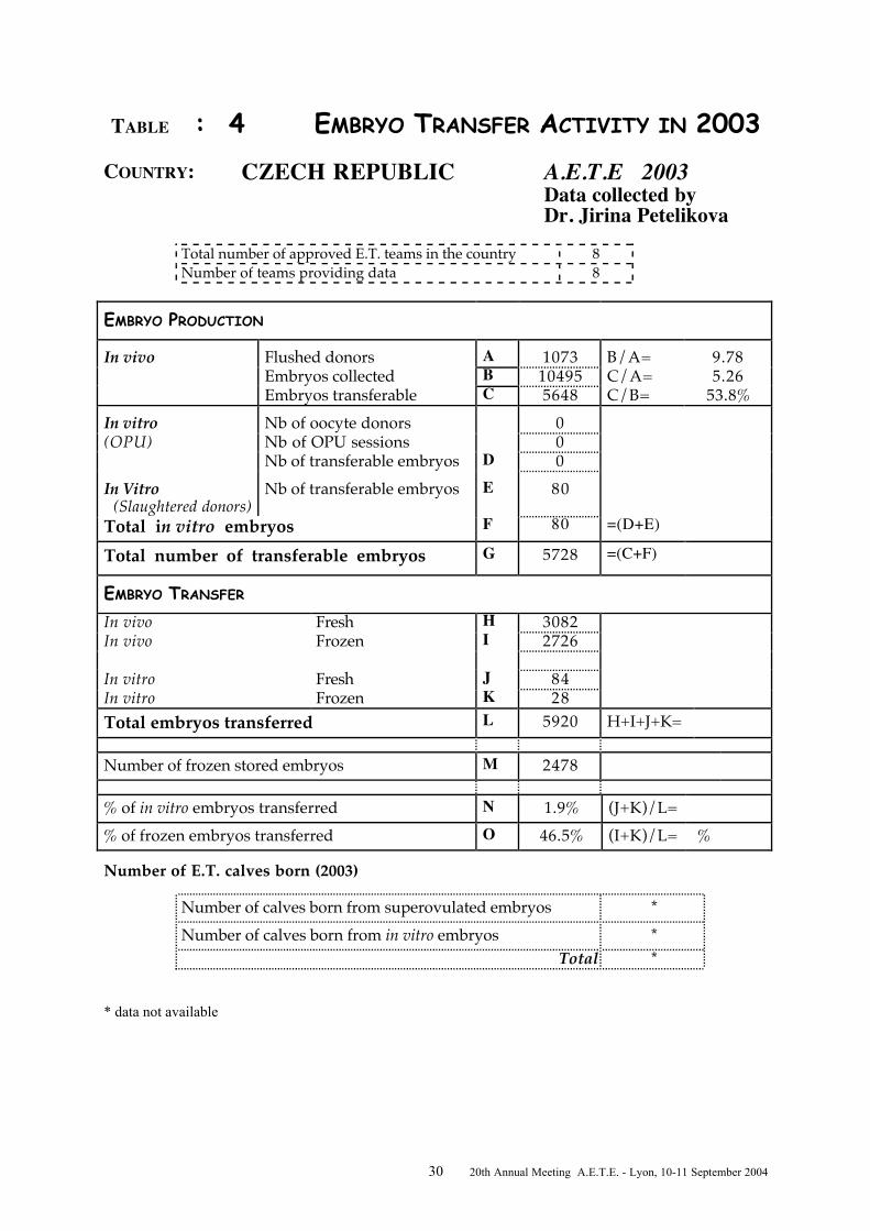
TABLE : 4 EMBRYO TRANSFER ACTIVITY IN 2003COUNTRY: CZECH REPUBLIC A.E.T.E 2003
Data collected byDr. Jirina Petelikova
Total number of approved E.T. teams in the country 8Number of teams providing data 8
EMBRYO PRODUCTION
In vivo Flushed donors A 1073 B/A= 9.78Embryos collected B 10495 C/A= 5.26Embryos transferable C 5648 C/B= 53.8%
In vitro Nb of oocyte donors 0(OPU) Nb of OPU sessions 0
Nb of transferable embryos D 0In Vitro (Slaughtered donors)
Nb of transferable embryos E 80
Total in vitro embryos F 80 =(D+E)
Total number of transferable embryos G 5728 =(C+F)
EMBRYO TRANSFER
In vivo Fresh H 3082In vivo Frozen I 2726
In vitro Fresh J 84In vitro Frozen K 28Total embryos transferred L 5920 H+I+J+K=
Number of frozen stored embryos M 2478
% of in vitro embryos transferred N 1.9% (J+K)/L=% of frozen embryos transferred O 46.5% (I+K)/L= %
Number of E.T. calves born (2003)
Number of calves born from superovulated embryos *Number of calves born from in vitro embryos *
Total *
* data not available

31 20th Annual Meeting A.E.T.E. - Lyon, 10-11 September 2004

32 20th Annual Meeting A.E.T.E. - Lyon, 10-11 September 2004
TABLE : 5 EMBRYO TRANSFER ACTIVITY IN 2003COUNTRY: DENMARK A.E.T.E 2003
Data collected byDr. Henrik Callesen
Total number of approved E.T. teams in the country 14Number of teams providing data 14
EMBRYO PRODUCTION
In vivo Flushed donors A 605 B/A= 10.0Embryos collected B 6055 C/A= 6.94Embryos transferable C 4199 C/B= 69.3%
In vitro Nb of oocyte donors 0(OPU) Nb of OPU sessions 0
Nb of transferable embryos D 0In Vitro (Slaughtered donors)
Nb of transferable embryos E
Total in vitro embryos F 0 =(D+E)
Total number of transferable embryos G 4199 =(C+F)
EMBRYO TRANSFER
In vivo Fresh H 2256In vivo Frozen I 2327
In vitro Fresh J 0In vitro Frozen K 0Total embryos transferred L 4583 H+I+J+K=
Number of frozen stored embryos M 2324
% of in vitro embryos transferred N 0 (J+K)/L=% of frozen embryos transferred O 50.8 (I+K)/L= %
Number of E.T. calves born (2003)
Number of calves born from superovulated embryos 1846Number of calves born from in vitro embryos 0
Total 1846
* data not available

33 20th Annual Meeting A.E.T.E. - Lyon, 10-11 September 2004

34 20th Annual Meeting A.E.T.E. - Lyon, 10-11 September 2004
TABLE : 6 EMBRYO TRANSFER ACTIVITY IN 2003
COUNTRY: ESTONIA A.E.T.E 2003Data collected byDr. Y. Jaakma
Total number of approved E.T. teams in the country 1Number of teams providing data 1
EMBRYO PRODUCTION
In vivo Flushed donors A 4 B/A= 11.5Embryos collected B 46 C/A= 7.75Embryos transferable C 31 C/B= 67.4%
In vitro Nb of oocyte donors 0(OPU) Nb of OPU sessions 0
Nb of transferable embryos D 0In Vitro (Slaughtered donors)
Nb of transferable embryos E 0
Total in vitro embryos F 0 =(D+E)
Total number of transferable embryos G 0 =(C+F)
EMBRYO TRANSFER
In vivo Fresh H 8In vivo Frozen I 14
In vitro Fresh J 0In vitro Frozen K 0Total embryos transferred L 22 H+I+J+K=
Number of frozen stored embryos M 23
% of in vitro embryos transferred N 0% (J+K)/L=% of frozen embryos transferred O 63.6% (I+K)/L= %
Number of E.T. calves born (2003)
Number of calves born from superovulated embryos 35Number of calves born from in vitro embryos -
Total 35
* data not available

35 20th Annual Meeting A.E.T.E. - Lyon, 10-11 September 2004

36 20th Annual Meeting A.E.T.E. - Lyon, 10-11 September 2004
TABLE : 7 EMBRYO TRANSFER ACTIVITY IN 2003
COUNTRY: FINLAND A.E.T.E 2003Data collected byDr. Marja Mikkola
Total number of approved E.T. teams in the country 6Number of teams providing data 6
EMBRYO PRODUCTION
In vivo Flushed donors A 500 B/A= 8.84Embryos collected B 4421 C/A= 5.47Embryos transferable C 2733 C/B= 61.8%
In vitro Nb of oocyte donors 0(OPU) Nb of OPU sessions 0
Nb of transferable embryos D 0In Vitro (Slaughtered donors)
Nb of transferable embryos E 35
Total in vitro embryos F 35 =(D+E)
Total number of transferable embryos G 2766 =(C+F)
EMBRYO TRANSFER
In vivo Fresh H 1100In vivo Frozen I 1201
In vitro Fresh J 10In vitro Frozen K 1Total embryos transferred L 2311 H+I+J+K=
Number of frozen stored embryos M 1339
% of in vitro embryos transferred N 0.5% (J+K)/L=% of frozen embryos transferred O 52.0% (I+K)/L= %
Number of E.T. calves born (2003)
Number of calves born from superovulated embryos *Number of calves born from in vitro embryos *
Total *
* data not available

37 20th Annual Meeting A.E.T.E. - Lyon, 10-11 September 2004

38 20th Annual Meeting A.E.T.E. - Lyon, 10-11 September 2004
TABLE : 8 EMBRYO TRANSFER ACTIVITY IN 2003COUNTRY: FRANCE A.E.T.E 2003
Data collected byDr. Bernard Guérin
Total number of approved E.T. teams in the country 27Number of teams providing data 4
EMBRYO PRODUCTION
In vivo Flushed donors A 5665 B/A= 11.5Embryos collected B 64925 C/A= 6.60Embryos transferable C 37433 C/B= 57.6%
In vitro Nb of oocyte donors 77(OPU) Nb of OPU sessions 77
Nb of transferable embryos D 261In Vitro (Slaughtered donors)
Nb of transferable embryos E 22
Total in vitro embryos F 281 =(D+E)
Total number of transferable embryos G 37714 =(C+F)
EMBRYO TRANSFER
In vivo Fresh H 18415In vivo Frozen I 15076
In vitro Fresh J 231In vitro Frozen K 7Total embryos transferred L 33729 H+I+J+K=
Number of frozen stored embryos M 14655
% of in vitro embryos transferred N 0.7% (J+K)/L=% of frozen embryos transferred O 44.7% (I+K)/L= %
Number of E.T. calves born (2003)
Number of calves born from superovulated embryos 5122Number of calves born from in vitro embryos 113
Total 5235 * data not available

39 20th Annual Meeting A.E.T.E. - Lyon, 10-11 September 2004

40 20th Annual Meeting A.E.T.E. - Lyon, 10-11 September 2004
TABLE : 9 EMBRYO TRANSFER ACTIVITY IN 2003COUNTRY: GERMANY A.E.T.E 2003
Data collected byDr Hubert Cramer
Total number of approved E.T. teams in the country 42Number of teams providing data
EMBRYO PRODUCTION
In vivo Flushed donors A 2687 B/A= 9.80Embryos collected B 26350 C/A= 5.54Embryos transferable C 14889 C/B= 56.5%
In vitro Nb of oocyte donors *(OPU) Nb of OPU sessions *
Nb of transferable embryos D *In Vitro (Slaughtered donors)
Nb of transferable embryos E 3120
Total in vitro embryos F 3120 =(D+E)
Total number of transferable embryos G 18009 =(C+F)
EMBRYO TRANSFER
In vivo Fresh H 5849In vivo Frozen I 4106
In vitro Fresh J 1878In vitro Frozen KTotal embryos transferred L 11833 H+I+J+K=
Number of frozen stored embryos M *
% of in vitro embryos transferred N 15.9% (J+K)/L=% of frozen embryos transferred O 34.7% (I+K)/L= %
Number of E.T. calves born (2003)
Number of calves born from superovulated embryosNumber of calves born from in vitro embryos
Total *
* data not available

41 20th Annual Meeting A.E.T.E. - Lyon, 10-11 September 2004

42 20th Annual Meeting A.E.T.E. - Lyon, 10-11 September 2004
TABLE : 10 EMBRYO TRANSFER ACTIVITY IN 2003COUNTRY: GREECE A.E.T.E 2003
Data collected byDr. Foteini Samartzi
Total number of approved E.T. teams in the country 2Number of teams providing data 2
EMBRYO PRODUCTION
In vivo Flushed donors A 27 B/A= 17.2Embryos collected B 465 C/A= 3.78Embryos transferable C 102 C/B= 21.9%
In vitro Nb of oocyte donors 0(OPU) Nb of OPU sessions
Nb of transferable embryos D 0In Vitro (Slaughtered donors)
Nb of transferable embryos E 0
Total in vitro embryos F 0 =(D+E)
Total number of transferable embryos G 102 =(C+F)
EMBRYO TRANSFER
In vivo Fresh H 48In vivo Frozen I 0
In vitro Fresh J 0In vitro Frozen K 0Total embryos transferred L 48 H+I+J+K=
Number of frozen stored embryos M 51
% of in vitro embryos transferred N 0 (J+K)/L=% of frozen embryos transferred O 0 (I+K)/L= %
Number of E.T. calves born (2003)
Number of calves born from superovulated embryos 29Number of calves born from in vitro embryos 0
Total 29
* data not available

43 20th Annual Meeting A.E.T.E. - Lyon, 10-11 September 2004

44 20th Annual Meeting A.E.T.E. - Lyon, 10-11 September 2004
TABLE : 11 EMBRYO TRANSFER ACTIVITY IN 2003COUNTRY: HUNGARY A.E.T.E 2003
Data collected byDr. Laszlo Solti
Total number of approved E.T. teams in the country 8Number of teams providing data 5
EMBRYO PRODUCTION
In vivo Flushed donors A 224 B/A= 9.92Embryos collected B 2222 C/A= 5.45Embryos transferable C 1220 C/B= 54.9%
In vitro Nb of oocyte donors *(OPU) Nb of OPU sessions *
Nb of transferable embryos D *In Vitro (Slaughtered donors)
Nb of transferable embryos E 607
Total in vitro embryos F 607 =(D+E)
Total number of transferable embryos G 1827 =(C+F)
EMBRYO TRANSFER
In vivo Fresh H 275In vivo Frozen I 347
In vitro Fresh J *In vitro Frozen K *Total embryos transferred L 622 H+I+J+K=
Number of frozen stored embryos M 894
% of in vitro embryos transferred N * (J+K)/L=% of frozen embryos transferred O 55.8% (I+K)/L= %
Number of E.T. calves born (2003)
Number of calves born from superovulated embryos 303Number of calves born from in vitro embryos *
Total 303
* data not available

45 20th Annual Meeting A.E.T.E. - Lyon, 10-11 September 2004

46 20th Annual Meeting A.E.T.E. - Lyon, 10-11 September 2004
TABLE : 12 EMBRYO TRANSFER ACTIVITY IN 2003COUNTRY: IRELAND A.E.T.E 2003
Data collected byDr. Pat Lonergan
Total number of approved E.T. teams in the country 5Number of teams providing data 1
EMBRYO PRODUCTION
In vivo Flushed donors A 272 B/A= 8.82Embryos collected B 2398 C/A= 5.21Embryos transferable C 1418 C/B= 59.1%
In vitro Nb of oocyte donors 0(OPU) Nb of OPU sessions 0
Nb of transferable embryos D 0In Vitro (Slaughtered donors)
Nb of transferable embryos E 0
Total in vitro embryos F 0 =(D+E)
Total number of transferable embryos G 1418 =(C+F)
EMBRYO TRANSFER
In vivo Fresh H 550In vivo Frozen I 714
In vitro Fresh J 0In vitro Frozen K 0Total embryos transferred L 1264 H+I+J+K=
Number of frozen stored embryos M 868
% of in vitro embryos transferred N 0 (J+K)/L=% of frozen embryos transferred O 56.5% (I+K)/L= %
Number of E.T. calves born (2003)
Number of calves born from superovulated embryos *Number of calves born from in vitro embryos *
Total *
* data not available

47 20th Annual Meeting A.E.T.E. - Lyon, 10-11 September 2004

48 20th Annual Meeting A.E.T.E. - Lyon, 10-11 September 2004
TABLE : 13 EMBRYO TRANSFER ACTIVITY IN 2003COUNTRY: ITALY A.E.T.E 2002
Data collected byDr. Francesco Brun
Total number of approved E.T. teams in the countryNumber of teams providing data
EMBRYO PRODUCTION
In vivo Flushed donors A 1002 B/A= 14.32Embryos collected B 14350 C/A= 7.06Embryos transferable C 7076 C/B= 49.3%
In vitro Nb of oocyte donors 147(OPU) Nb of OPU sessions 305
Nb of transferable embryos D 632In Vitro (Slaughtered donors)
Nb of transferable embryos E 3983
Total in vitro embryos F 4615 =(D+E)
Total number of transferable embryos G 11691 =(C+F)
EMBRYO TRANSFER
In vivo Fresh H 2787In vivo Frozen I 3255
In vitro Fresh J 100In vitro Frozen K 3457Total embryos transferred L 9599 H+I+J+K=
Number of frozen stored embryos M 6646
% of in vitro embryos transferred N 37.0% (J+K)/L=% of frozen embryos transferred O 69.9% (I+K)/L= %
Number of E.T. calves born (2003)
Number of calves born from superovulated embryos *Number of calves born from in vitro embryos *
Total *
* data not available

49 20th Annual Meeting A.E.T.E. - Lyon, 10-11 September 2004

50 20th Annual Meeting A.E.T.E. - Lyon, 10-11 September 2004
TABLE : 14 EMBRYO TRANSFER ACTIVITY IN 2003COUNTRY: (The) NETHERLANDS A.E.T.E 2003
Data collected byDr. Bas Landman
Total number of approved E.T. teams in the countryNumber of teams providing data
EMBRYO PRODUCTION
In vivo Flushed donors A 3119 B/A=Embryos collected B * C/A= 5.74Embryos transferable C 17915 C/B=
In vitro Nb of oocyte donors 222(OPU) Nb of OPU sessions 2492
Nb of transferable embryos D 2084In Vitro (Slaughtered donors)
Nb of transferable embryos E 77
Total in vitro embryos F 2161 =(D+E)
Total number of transferable embryos G 20076 =(C+F)
EMBRYO TRANSFER
In vivo Fresh H 3364In vivo Frozen I 11988
In vitro Fresh J 901In vitro Frozen K 1224Total embryos transferred L 17477 H+I+J+K=
Number of frozen stored embryos M *
% of in vitro embryos transferred N 12.1% (J+K)/L=% of frozen embryos transferred O 75.6% (I+K)/L= %
Number of E.T. calves born (2003)
Number of calves born from superovulated embryosNumber of calves born from in vitro embryos
Total *
* data not available

51 20th Annual Meeting A.E.T.E. - Lyon, 10-11 September 2004

52 20th Annual Meeting A.E.T.E. - Lyon, 10-11 September 2004
TABLE : 15 EMBRYO TRANSFER ACTIVITY IN 2003COUNTRY: NORWAY A.E.T.E 2003
Data collected byDr. Elisabeth Kommisrüd
Total number of approved E.T. teams in the country 1Number of teams providing data 1
EMBRYO PRODUCTION
In vivo Flushed donors A 24 B/A= 8.17Embryos collected B 196 C/A= 4.95Embryos transferable C 119 C/B= 60.7
In vitro Nb of oocyte donors 0(OPU) Nb of OPU sessions 0
Nb of transferable embryos D 0In Vitro (Slaughtered donors)
Nb of transferable embryos E 0
Total in vitro embryos F 0 =(D+E)
Total number of transferable embryos G 0 =(C+F)
EMBRYO TRANSFER
In vivo Fresh H 20In vivo Frozen I 98
In vitro Fresh J 0In vitro Frozen K 0Total embryos transferred L 118 H+I+J+K=
Number of frozen stored embryos M 106
% of in vitro embryos transferred N 0 (J+K)/L=% of frozen embryos transferred O 83.0% (I+K)/L= %
Number of E.T. calves born (2003)
Number of calves born from superovulated embryos *Number of calves born from in vitro embryos 0
Total *
* data not available

53 20th Annual Meeting A.E.T.E. - Lyon, 10-11 September 2004

54 20th Annual Meeting A.E.T.E. - Lyon, 10-11 September 2004
TABLE : 16 EMBRYO TRANSFER ACTIVITY IN 2003COUNTRY: POLAND A.E.T.E 2003
Data collected byDr. Jedrzej Jaskowski
Total number of approved E.T. teams in the country 10Number of teams providing data 10
3EMBRYO PRODUCTION
In vivo Flushed donors A 67 B/A= 8.12Embryos collected B 544 C/A= 5.74Embryos transferable C 385 C/B= 70.8%
In vitro Nb of oocyte donors 0(OPU) Nb of OPU sessions 0
Nb of transferable embryos D 0In Vitro (Slaughtered donors)
Nb of transferable embryos E 19
Total in vitro embryos F 19 =(D+E)
Total number of transferable embryos G 404 =(C+F)
EMBRYO TRANSFER
In vivo Fresh H 273In vivo Frozen I 87
In vitro Fresh J 13In vitro Frozen K 6Total embryos transferred L 379 H+I+J+K=
Number of frozen stored embryos M 31
% of in vitro embryos transferred N 5.0% (J+K)/L=% of frozen embryos transferred O 24.5% (I+K)/L= %
Number of E.T. calves born (2003)
Number of calves born from superovulated embryos *Number of calves born from in vitro embryos *
Total *
* data not available

55 20th Annual Meeting A.E.T.E. - Lyon, 10-11 September 2004
TABLE : 17 EMBRYO TRANSFER ACTIVITY IN 2003

56 20th Annual Meeting A.E.T.E. - Lyon, 10-11 September 2004
COUNTRY: PORTUGAL A.E.T.E 2003Data collected byDr. Joao Nestor das Chagas e Silva
Total number of approved E.T. teams in the country 5Number of teams providing data 3
EMBRYO PRODUCTION
In vivo Flushed donors A 61 B/A= 5.67Embryos collected B 346 C/A= 5.15Embryos transferable C 314 C/B= 90.7%
In vitro Nb of oocyte donors 0(OPU) Nb of OPU sessions 0
Nb of transferable embryos D 0In Vitro (Slaughtered donors)
Nb of transferable embryos E 551
Total in vitro embryos F 551 =(D+E)
Total number of transferable embryos G 865 =(C+F)
EMBRYO TRANSFER
In vivo Fresh H 219In vivo Frozen I 135
In vitro Fresh J 0In vitro Frozen K 0Total embryos transferred L 354 H+I+J+K=
Number of frozen stored embryos M 245
% of in vitro embryos transferred N 0 (J+K)/L=% of frozen embryos transferred O 38.1% (I+K)/L= %
Number of E.T. calves born (2003)
Number of calves born from superovulated embryos 35*Number of calves born from in vitro embryos 0
Total 35
* data from 1 team

57 20th Annual Meeting A.E.T.E. - Lyon, 10-11 September 2004

58 20th Annual Meeting A.E.T.E. - Lyon, 10-11 September 2004
TABLE : 18 EMBRYO TRANSFER ACTIVITY IN 2003COUNTRY: ROMANIA A.E.T.E 2003
Data collected byDrs. StelaZamfirescu/George Toba
Total number of approved E.T. teams in the country 4Number of teams providing data 4
EMBRYO PRODUCTION
In vivo Flushed donors A 22 B/A= 8.09Embryos collected B 178 C/A= 5.18Embryos transferable C 114 C/B= 64.0%
In vitro Nb of oocyte donors 0(OPU) Nb of OPU sessions 0
Nb of transferable embryos D 0In Vitro (Slaughtered donors)
Nb of transferable embryos E 0
Total in vitro embryos F 0 =(D+E)
Total number of transferable embryos G 114 =(C+F)
EMBRYO TRANSFER
In vivo Fresh H 36In vivo Frozen I 47
In vitro Fresh J 0In vitro Frozen K 0Total embryos transferred L 83 H+I+J+K=
Number of frozen stored embryos M 69
% of in vitro embryos transferred N 0 (J+K)/L=% of frozen embryos transferred O 56.6% (I+K)/L= %
Number of E.T. calves born (2003)
Number of calves born from superovulated embryos 6Number of calves born from in vitro embryos 0
Total 6
* data not available

59 20th Annual Meeting A.E.T.E. - Lyon, 10-11 September 2004

60 20th Annual Meeting A.E.T.E. - Lyon, 10-11 September 2004
TABLE : 19 EMBRYO TRANSFER ACTIVITY IN 2003
COUNTRY: SLOVAKIA A.E.T.E 2003Data collected byDr. Peter Cesnak
Total number of approved E.T. teams in the countryNumber of teams providing data 2
EMBRYO PRODUCTION
In vivo Flushed donors A 75 B/A= 9.49Embryos collected B 712 C/A= 4.87Embryos transferable C 365 C/B= 51.3%
In vitro Nb of oocyte donors 0(OPU) Nb of OPU sessions 0
Nb of transferable embryos D 0In Vitro (Slaughtered donors)
Nb of transferable embryos E 0
Total in vitro embryos F 0 =(D+E)
Total number of transferable embryos G 365 =(C+F)
EMBRYO TRANSFER
In vivo Fresh H 248In vivo Frozen I 271
In vitro Fresh J 0In vitro Frozen K 0Total embryos transferred L 519 H+I+J+K=
Number of frozen stored embryos M 116
% of in vitro embryos transferred N 0 (J+K)/L=% of frozen embryos transferred O 52.2% (I+K)/L= %
Number of E.T. calves born (2003)
Number of calves born from superovulated embryos 287Number of calves born from in vitro embryos 0
Total 287
* data not available

61 20th Annual Meeting A.E.T.E. - Lyon, 10-11 September 2004

62 20th Annual Meeting A.E.T.E. - Lyon, 10-11 September 2004
TABLE : 20 EMBRYO TRANSFER ACTIVITY IN 2003
COUNTRY: SPAIN A.E.T.E 2003Data collected byDr. Julio de la Fuente
Total number of approved E.T. teams in the countryNumber of teams providing data
EMBRYO PRODUCTION
In vivo Flushed donors A 303 B/A= 10.6Embryos collected B 3207 C/A= 4.73Embryos transferable C 1436 C/B= 44.8%
In vitro Nb of oocyte donors 2(OPU) Nb of OPU sessions 7
Nb of transferable embryos D 22In Vitro (Slaughtered donors)
Nb of transferable embryos E 0
Total in vitro embryos F 22 =(D+E)
Total number of transferable embryos G 1458 =(C+F)
EMBRYO TRANSFER
In vivo Fresh H 314In vivo Frozen I 1041
In vitro Fresh J 15In vitro Frozen K 6Total embryos transferred L 1376 H+I+J+K=
Number of frozen stored embryos M 773
% of in vitro embryos transferred N 1.5% (J+K)/L=% of frozen embryos transferred O 76.1% (I+K)/L= %
Number of E.T. calves born (2003)
Number of calves born from superovulated embryos 426Number of calves born from in vitro embryos 2
Total 428
* data not available

63 20th Annual Meeting A.E.T.E. - Lyon, 10-11 September 2004

64 20th Annual Meeting A.E.T.E. - Lyon, 10-11 September 2004
TABLE : 21 EMBRYO TRANSFER ACTIVITY IN 2003COUNTRY: SWEDEN A.E.T.E 2003
Data collected byDr. Hans Gustafsson
Total number of approved E.T. teams in the country 2Number of teams providing data 1
EMBRYO PRODUCTION
In vivo Flushed donors A 270 B/A= 7.22Embryos collected B 1950 C/A= 4.90Embryos transferable C 1322 C/B= 67.8%
In vitro Nb of oocyte donors 0(OPU) Nb of OPU sessions 0
Nb of transferable embryos D 0In Vitro (Slaughtered donors)
Nb of transferable embryos E 0
Total in vitro embryos F 0 =(D+E)
Total number of transferable embryos G 1322 =(C+F)
EMBRYO TRANSFER
In vivo Fresh H 417In vivo Frozen I 905
In vitro Fresh J 0In vitro Frozen K 0Total embryos transferred L 1322 H+I+J+K=
Number of frozen stored embryos M 0
% of in vitro embryos transferred N 0 (J+K)/L=% of frozen embryos transferred O 68.4% (I+K)/L= %
Number of E.T. calves born (2003)
Number of calves born from superovulated embryos *Number of calves born from in vitro embryos *
Total *
* data not available

65 20th Annual Meeting A.E.T.E. - Lyon, 10-11 September 2004

66 20th Annual Meeting A.E.T.E. - Lyon, 10-11 September 2004
TABLE : 22 EMBRYO TRANSFER ACTIVITY IN 2003COUNTRY: SWITZERLAND A.E.T.E 2003
Data collected byDr. Saner Rainer
Total number of approved E.T. teams in the country 5Number of teams providing data 3
EMBRYO PRODUCTION
In vivo Flushed donors A 243 B/A= 11.0Embryos collected B 2686 C/A= 7.91Embryos transferable C 1923 C/B= 71.6%
In vitro Nb of oocyte donors 0(OPU) Nb of OPU sessions 0
Nb of transferable embryos D 0In Vitro (Slaughtered donors)
Nb of transferable embryos E 0
Total in vitro embryos F 0 =(D+E)
Total number of transferable embryos G 1923 =(C+F)
EMBRYO TRANSFER
In vivo Fresh H 542In vivo Frozen I 1165
In vitro Fresh J 0In vitro Frozen K 46Total embryos transferred L 1753 H+I+J+K=
Number of frozen stored embryos M 1381
% of in vitro embryos transferred N 2.6% (J+K)/L=% of frozen embryos transferred O 69.1% (I+K)/L= %
Number of E.T. calves born (2003)
Number of calves born from superovulated embryos *Number of calves born from in vitro embryos *
Total *
* data not available

67 20th Annual Meeting A.E.T.E. - Lyon, 10-11 September 2004

68 20th Annual Meeting A.E.T.E. - Lyon, 10-11 September 2004
TABLE : 23 EMBRYO TRANSFER ACTIVITY IN 2003
COUNTRY: UNITED KINGDOM A.E.T.E 2003Data collected byDr. Alison Liddle
Total number of approved E.T. teams in the country 17Number of teams providing data 10
EMBRYO PRODUCTION
In vivo Flushed donors A * B/A=Embryos collected B * C/A=Embryos transferable C * C/B=
In vitro Nb of oocyte donors *(OPU) Nb of OPU sessions *
Nb of transferable embryos D *In Vitro (Slaughtered donors)
Nb of transferable embryos E *
Total in vitro embryos F * =(D+E)
Total number of transferable embryos G * =(C+F)
EMBRYO TRANSFER
In vivo Fresh H *In vivo Frozen I *
*In vitro Fresh J *In vitro Frozen K *Total embryos transferred L 4109 H+I+J+K=
Number of frozen stored embryos M 3463
% of in vitro embryos transferred N * (J+K)/L=% of frozen embryos transferred O * (I+K)/L= %
Number of E.T. calves born (2003)
Number of calves born from superovulated embryosNumber of calves born from in vitro embryos
Total *
* data not available

69 20th Annual Meeting A.E.T.E. - Lyon, 10-11 September 2004

70 20th Annual Meeting A.E.T.E. - Lyon, 10-11 September 2004
OVERALL BOVINE EMBRYO TRANSFER ACTIVITY
IN EUROPE IN 2003
I. EMBRYO PRODUCTION
(Data collected from 23 countries)
In vivo produced embryos (superovulation)
- number of flushed donors- number of transferable embryos- mean number per flushed donor
17, 503104, 726
5.98
In vitro produced embryos:
From OPU- number of oocyte donors- number of OPU sessions- number of transferable embryos
From slaughterhouse collected ovaries- number of transferable embryos
Total in vitro
5633, 1083, 683
8, 498
12, 181
Total number of transferable embryos 116, 907
(P. Lonergan, AETE Lyon,2004)

71 20th Annual Meeting A.E.T.E. - Lyon, 10-11 September 2004

72 20th Annual Meeting A.E.T.E. - Lyon, 10-11 September 2004
OVERALL BOVINE EMBRYO TRANSFER ACTIVITY
IN EUROPE IN 2003
II. EMBRYO TRANSFERS
(Data collected from 23 countries)
In vivo produced embryosNumber of embryos transferred
90, 508*(41, 289 fresh/49, 219 frozen)
In vitro produced embryos8, 410*
(3, 635 fresh/4, 775 frozen)
Total number of embryos transferred 98, 918
Proportion of IVF embryos transferred 8.50 %
Proportion of frozen embryos transferred 54.6 %
* These numbers are underestimated as the data from 1 country were not available and notincluded
(P. Lonergan, AETE Lyon 2004)

73 20th Annual Meeting A.E.T.E. - Lyon, 10-11 September 2004

74 20th Annual Meeting A.E.T.E. - Lyon, 10-11 September 2004
EMBRYO TRANSFER ACTIVITY IN OTHER SPECIES*
EUROPE year 2003
Species Embryo Production Embryo
Transfers
Countries
Sheep 326 167
Greece
Portugal
Romania
Swine 1221 816
Czech Republic
Hungary
Poland
Romania
Switzerland
Goat 61 52 Romania
Horse 443 344
Austria
Czech Republic
Finland
France
Hungary
Italy
Poland
Sweden
(P. Lonergan, AETE Lyon, 2004)

75 20th Annual Meeting A.E.T.E. - Lyon, 10-11 September 2004

76 20th Annual Meeting A.E.T.E. - Lyon, 10-11 September 2004
INVITED LECTURES

77 20th Annual Meeting A.E.T.E. - Lyon, 10-11 September 2004
78 20th Annual Meeting A.E.T.E. - Lyon, 10-11 September 2004
RESPECTIVE IMPACTS OF ET AND OPU/IVP ON GENETIC SELECTION EFFICIENCY IN THE MONTBELIARDE BREED
TISSIER M.1, PONSART C.2, REGALDO D.3, MERVANT G.1, HUMBLOT P.2
1UMOTEST – Les Soudanières – 01250 CEYZERIAT2UNCEIA – Département R&D – 13 rue Jouët – 94703 MAISONS-ALFORT3INSTITUT DE L’ELEVAGE – 149 rue de Bercy – 75595 PARIS CEDEX 12
Introduction
In dairy cattle, breeding programmes aim to optimise the genetic progress for differentcharacters. Three groups of selection criteria are usually distinguished. Milk production charactersare predominant for dairy breeds. Morphological criteria are necessary to complete the milkproduction evaluation in terms of body and udder conformation. However, the muscular mass isalso used as a selection criteria for mixed breeds in which equilibrated characteristics are valuable.Functional criteria, such as resistance to mastitis, fertility, longevity, birth or calving abilities andbehaviour are currently becoming more important, because of their economical benefit throughcontrol of animal health status and culling, together with improved working conditions forbreeders (Aswhell et al., 2004 ; Pryce et al., 2004).
The multiplication of the characters of interest leads to more complex breedingprogrammes since there is a need to select animals combining the maximum number of charactersof interest. Therefore, the challenge of these programmes is to obtain the best result derived froma combination of characters with different genetic determinism. This has lead to theestabnlishment of strategies to optimise genetic progress while restricting inbreeding (Quinton etal., 1995 ; Van Arendonk et al., 2003).
During the last decades, biotechnologies such as embryo transfer (ET) and, more recently,ovum pick up associated with in vitro embryo production (OPU-IVP ; Colleau et al., 1998;Marquant Leguienne et Humblot 1998, Marquant Leguienne et al., 2001, Galli et al., 2003,Merton et al., 2003), have been used in breeding programs. Those techniques are more or less usedintensively depending on the country. In Europe, the intensive use of OPU and IVP has beenreported in the Netherlands and Italy (Heyman, 2002, Galli et al., 2003 ; Merton et al., 2003),whereas ET remains the main biotechnology used in breeding schemes in France. With increasedknowledge in genomics, the use of marker assisted selection (MAS) is currently emerging.
This paper aims to investigate and compare the results and realised value of ET and OPU-IVP in the breeding programme conducted by UMOTEST (Union of AI cooperatives) in theMontbeliarde breed, which is one of the main red coloured mixed breed in Europe.
The Montbeliarde breeding program
In the Montbeliarde breed, 25% of females are inseminated with beef bulls, which are used t oproduce crossbred animals. This illustrates the ease of calving and the morphological qualities ofthis breed (Table 1). However, the Montbeliarde breed is mainly a dairy breed with a largemorphological format (sacral height : 1.48 m) and a high protein content used to produce cheesewith local/regional labels (Comté, Bleu de Gex, Morbier, Cantal…).
Table 1 : The UMOTEST breeding program
Type of animals NumberTotal females 600 000

79 20th Annual Meeting A.E.T.E. - Lyon, 10-11 September 2004
Inseminated females- pure breed- cross breed
560 000410 000150 000
Controlled females 330 000Nucleus females 4 200Bulls tested 130
The selection has always taken into account the morphological quality, aiming to produce malecalves and culled cows of a high commercial value (Table 2).
Table 2. Production characteristics in the Montbeliarde breed
• Milk production (adult lactations > 200 days)
Days Milk (kg) % Protein content % Fat content317 7275 3.4 3.88
• Beef
Weight (carcass) Class (Europe)Bulls (18-20 months 365 kg R to R+
Culled cows 345 kg O+ to R-
Use of embryo transfer (ET) on the nucleus females
The realized value of embryo transfer (ET) for females of the selection scheme has beenestimated from embryo collections performed from 1994 to 2003. The number of flushes wasquite stable before 1994, ranging from 150 to 200 between 1988 and 1993, but has doubled withinthe last ten years (Table 3). This contributed to the multiplication of calves born from females ofhigh genetic merit in order to increase the genetic progress.
Table 3. Number of embryo collections for the past 10 years
Year Number of embryocollections
1994 2631995 3391996 3271997 4471998 5081999 4702000 4432001 5602002 6082003 523
Embryo transfer may potentially impact positively on the components contributing to geneticprogress (ΔG=i x p x σG / T) by :
- Increasing the intensity of selection (i), which corresponds to increased numbers of maleand female calves to enter the selection process,
- Increasing the precision of selection (p), with the use of the BLUP model, which allowsto take into account more animals within a family of mothers and leads to highercoefficients of determination.

80 20th Annual Meeting A.E.T.E. - Lyon, 10-11 September 2004
- Increasing the genetic variability (σG), obtained with higher numbers geneticcombinations Father/grand-father (embryo collections with different bulls)
- Shortening the generation interval (T), with the transfer of embryos collected fromheifers and birth of several calves within 9 months.
Embryo collections are programmed among ages and the genetic value of the nucleusfemales in which a majority of bull dams (BD) are submitted to ET. They represent 59% of totalcollections with 23% performed in young bull dams (YBD ; ET performed 13 to 15 months beforefirst breeding) and 36% in adult bull dams (ABD ; ET performed during the first, second, 3rd
lactation or more). The percentage of treated heifer Dams (HD) is 41 %. This shows that effortsare made to control the generation interval, because 2/3 of bull dams are young females. Heiferdams are also mainly collected as young females, in order to renew the nucleus.
The use of ET resulted in a decrease of the generation interval. Between 1994 and 1996,the age of the dam at birth of its offspring averaged 4.7 years, compared to 4.2 since 2001. Thiscorresponds to a decrease of T of 11% within 7 years.
Approximately, half of the bulls and heifers dams selected in 2004 are originating fromfamilies with at least 2 offspring (table 4). The families represented by numerous offspring allowsto increase the intensity and accuracy of selection. For instance, in those families, 7 different bullsand breeding combinations have been used. Generally, the number of different fathers per familycorresponds to half of the number of calves.
Table 4. Repartition of the nucleus females according to the number of offsprings per origin
Number of offspring per familyNumber offamilies 1 2-3 4-5 6-9 ≥ 10
Bull Dams 264 42 % 40 % 8 % 7 % 3 %
Heifers dams 179 42 % 37 % 10 % 6 % 4 %
The genetic value of the females used for ET increased regularly of 4 to 5 points (points ofISU; composite index summarising milk, morphological and functional characters). Consequently,the difference between the ISU index of the dams issued from AI and dams issued from ET didincrease, ranging from 1 point in 1994 to 10 points in 2003. This is easily explained as femalesused for ET are more frequently originating from dams or at least from families already used forET. For instance, about 3/4 of the nucleus females selected in 2004 are originating from familieswhere ET was used, either as bull dams or as heifers dams. The use of ET gives the opportunity t oget rapidly male calves and potential dams, which can be chosen within the female calves.
The realised value of ET on the annual genetic progress has been estimated from malecalves, which entered the progeny testing procedure in station. The genetic progress has beencompared to the reference 100, using the INEL index (mother-calf component), whichcorresponds to the genetic progress observed only with AI. The use of ET in this selection schemeled to obtain 105-106 points of INEL when 1 bull calf per origin entered the station and 108-109points when 2 calves per origin were progeny tested.
The mean cost per embryo collection for the selection unit is 700 euros. Additional costscorresponding to organisation and application of the breeding strategy by geneticists and to thetransfer of frozen embryos represents 350 euros per ET session. Therefore the additional cost ofan ET session when compared to AI is around 1000 euros. However, the mean number of embryosper session calculated from those ten years of records was 5.5 allowing to obtain 1.5 males/session. The supplement of cost when compared to AI can be estimated as 700 euros per male calfand is even reduced to 350 euros when considering the advantages of ET related to the renewal ofthe selection nucleus and to the conservation of families of interest.
Use of OPU and IVF in the nucleus herd

81 20th Annual Meeting A.E.T.E. - Lyon, 10-11 September 2004
Since 1996, UMOTEST has been associated in setting up the OPU-IVF techniquesdevelopped by UNCEIA and UCEAR in the field station of Chateauvillain. Results initiallyobtained in high genetic merit females kept and collected in station have been progressivelyapplied to farm conditions (Guyader-Joly et al., 1997, Marquant-Leguienne et al. 2001). In theMontbeliarde breed, from OPU sessions performed in station, a mean of 2 born calves per weekwas observed (with one oocyte collection per week and per female following superovulation. Thisallowed to produce one male calf per session.
From 137 on farm OPU sessions performed in very high genetic merit cows (85% ofsessions performed in sire dams) since year 2000, results are slightly lower with 1.5 calf and 0.75male calf per session respectively. For those sire dams, technical results were not significantlydifferent between heifers (lactation 0 ; L0), primiparous (Lactation 1 ; L1) or which had 2 ormore lactations before starting OPU (L2 or more).
Due to the supplement of cost when using OPU and IVF under farm conditions, ie 1500euros to be compared to 1000 euros (when considering equivalent costs for ET), these techniquesappears to be profitable if applied to young cows (L1) when compared to heifers and older cows.Effectively, the collected heifers are known for morphology and for the performances of theirparents (grand parents) but only 50% of them are finally selected on milk characteristics, udderconformation and other functional traits when being primiparous cows. Due to this selection, thegenetic progress obtained from superovulating (for ET or OPU) 100 heifers is equivalent to theprogress obtained from 50 primiparous cows. The supplement of cost due to the use of OPU andIVF in large number of heifers (which will be eliminated at a later age and provides large number ofoffspring non suitable to enter subsequent progeny testing), prohibits the use of those technologiesin this kind of genetic scheme.
Older cows (≥ L2) produce more viable embryos than heifers (+ 1 embryo per collectedfemale; Manciaux et al., 2000) or primiparous cows when using conventional ET protocols. Thecost of ET sessions is lower and due to a better knowledge of such donor cows, the advantage ofusing OPU and IVF to shorten the generation interval is very low.
On the contrary, due to significant advantages in terms of precision (p) and to a generationinterval still acceptable for L1 cows, applying OPU-IVF in such females appears as the bestcompromise to optimise the genetic scheme efficiency. In addition to this, the advantages ofcollecting pregnant females, from which the birth of a calf is highly probable, without anyincrease in days open (as observed when using conventional ET) are of a major interest for thegeneticist and the farmer.
Considering these observations, a technical and financial simulation was made to comparethe respective costs and advantages of conventional ET and OPU-IVF when used in theMontbeliarde selection scheme. It was considered that the objective of the geneticist was t oproduce 30 young bulls that will enter the progeny testing process from an initial pool of 100young females , candidates to be sire dams. Four scenarios (S1 to S4) were analysed depending onthe use of AI, ET and OPU-IVF technologies when applied in heifers or in L1 females (Tables 5and 6). It was hypothesised that half of these young heifers will not be confirmed as sire damswhen being primiparous due to initial lack of precision of their genetic value.
In scenario 1, 100 heifers are systematically superovulated and collected once forconventional ET by 13-15 months of age, before their first breeding (Table 6), and finally AI issubsequently used to produce additional calves. The resulting number of male produced and sessionper female, depending on the observed numbers of embryos and calves per session are presented intable 5. After 1 ET session and subsequent AI, a total of 150 males are born. From those, 50% willbe eliminated due to a lack of confirmation of their mother’s genetic value (Table 6). Additionalelimination will take place before they enter progeny testing after individual morphologicalevaluation and control of their reproductive performances (behavioural traits and spermproduction and quality traits).

82 20th Annual Meeting A.E.T.E. - Lyon, 10-11 September 2004
In scenario 2, AI is used in 100 heifers and the best 50 primiparous cows are chosen to becollected by ET. Around 25 calves are kept from AI made previously in those females. Half thenumber of calves and males are produced by ET when compared to the first scenario (Table 5). Allmales will be kept for milk production traits and elimination will be based only on individualperformances (Table 6). The cost for producing the 30 males which will enter progeny testing isconsiderably reduced when compared to the first scenario.
Table 5. Numbers of embryos and calves (Total and per session) and number of session per femaleto produce the desired number of males necessary for progeny testing.
N° Type offemales
Numberof
females
Reproductivetechniques Emb Calf
/sess. /sessNumber ofSess/female
Number ofcalves
Number ofmales
S1Heifers 100
E TAITotal
5 2.5 0.5
1 25050
300
125 25150
S2L1 cows 50
AIE TTotal
0.55 2.5 1
25125150
12.562.575
S3L1 cows 50
(station)
AIOPU-IVFTotal
0.54 2 1.25
25125150
12.562.575
S4L1 cows 50
(farm)
AIOPU-IVFTotal
0.53 1.5 1.66
25125150
12.562.575
The 3rd and 4th scenarios are similar to the second one but OPU and IVF are used instead ofET with collections taking place either in station (S3) or on farm (S4). The culling rate is thesame as in S2. From the 50 primiparous cows, 1.25 and 1.66 sessions are needed with S3 and S4respectively to obtain the planned 75 male calves (Table 5). The costs of these proceduresrepresents approximately 140% and 150% when compared to S2 (100%) for S3 and S4respectively.
Table 6. Respective costs (Kilo euros) of conventional ET and OPU-IVF to produce 30 malespotentially selected for progeny testing.
N° Reproductivetechniques
Type offemales
Number ofcollections
Numberof males
Eliminated(1) (2) Remaining
Costs(K€)
%
S1 ET+AI Heifers 100 150 - 75 - 45 30 142 195S2 AI+ET L1 cows 50 75 0 - 45 30 72 100S3 AI+OPU-IVF
(station) L1 cows 63 75 0 - 45 30 103 140
S4 AI+OPU-IVF(farm) L1 cows 83 75 0 - 45 30 106 150
(1) eliminated for non confirmation of the performances of their mother for milk productiontraits(2) elimination for morphology and reproductive performance traits

83 20th Annual Meeting A.E.T.E. - Lyon, 10-11 September 2004
S1 appears as the most expensive scenario. It’s main advantage when compared to theother alternatives tested here is to shorten the generation interval by one year. However, this isaccompanied by a lack of precision of the selection process especially for traits than can beestimated late (fertility, longevity, udder conformation). S2 looks to be the more economicalscenario. However, due to the use of primiparous females, one year of generation interval is lostbut the gain in precision is important since young bulls enter progeny testing and are evaluatedafter the second lactation of their mother. S3 and S4 are apparently more expensive than S2. Thecharacteristics of the selection scheme are the same (long generation interval, good precision).Due to the higher number of sessions, the variability and precision can even be increased whencompared to S2. When comparing S4 to S1 and S2, due to the high number of on farm OPUsessions, the combinations (Father / maternal Grand Father) can be multiplied by 2 and thiscontributes to increase variability. An additional advantage when compared to S2 is that milkproduction during 1st lactation and subsequent reproductive efficiency are less affected by thesuperovulation treatment when using OPU protocols instead of conventional ET procedures.Despite additional cost are not negligible, the last scenario represents probably the bestcompromise for the geneticist.
Conclusions
The use of embryo technologies allows to optimise the genetic progress for several traits ofinterest for the farmer and the consumer. Embryo transfer has been used extensively in theMontbeliarde breed since the end of the eighties and results improved very significantly to reach aproduction of 5 to 6 transferable embryos per session. However individual variability in responseto superovulation treatment is very high and the percentage of sessions for which 0 transferableembryo is produced is still close to 20%. Lowering this percentage is of a major interest for thegeneticist to conserve families which may disappear due to the low number of progeny of a highgenetic value.
OPU combined to IVF represents an alternative to ET allowing potentially to obtain a lotof progeny from a given high genetic merit mother bred with different sires (theoretically up to 9sessions with 9 different sires ). Another advantage when compared to ET is the possibility ofcollecting pregnant females. In such a situation, one can expect to get embryos in addition to acalf produced by AI without any delay in the reproductive life of the cow. However the results arestill highly variable due to unpredictable individual response. The cost of OPU and IVF should bereduced by 20 to 30% to make this techniques really attractive when compared to ET.
The low number (and associated variability) of transferable embryos produced by OPU andIVF and the difficulties encountered to apply the technique on farm (both for collection andtransfer) represents major limitations when compared to ET. These limitations explains probablywhy the use of these technologies is highly variable from one breed to another or/and in differentcountries. However, due to the possibility of repeating OPU sessions without affecting thereproductive efficiency, OPU and IVF may help in a near future to really optimise multiple traitsselection when associated to the use of the arising technologies based on genomics and markerassisted selection.
References
Ashwell MS, Heyen DW, Sonstegard TS, Van Tassell CP, Da Y, VanRaden PM, Ron M, Weller JI,Lewin HA. Detection of quantitative trait loci affecting milk production, health, andreproductive traits in Holstein cattle. J Dairy Sci JID - 2985126R 2004;87(2):468-75.
Colleau JJ, Heyman Y, Renard JP. Les biotechnologies de la reproduction chez les bovins et leursapplications réelles ou potentielles en sélection. INRA Prod.Anim. 1998;11(1):41-56.GalliC, Duchi R, Crotti G, Turini P, Ponderato N, Colleoni S, Lagutina I, Lazzari G. Bovineembryo technologies. Theriogenology JID - 0421510 2003;59(2):599-616.
Guyader Joly C, Ponchon S, Thuard JM, Durand M, Nibart M, Marquant Leguienne B, Humblot P .Effects of superovulation on repeated ultrasound guided oocyte collection and in vitroproduction in pregnant heifers. [Abstract] Theriogenology, 1997, 47 (1) : 157.

84 20th Annual Meeting A.E.T.E. - Lyon, 10-11 September 2004
Heyman Y, AETE, annual report on ET and OPU-IVF activities, Proceedings of the 18th AETEmeeting, Rolduc, 2002, pp 31-79.
Manciaux L, Ponsart C, Grisouard D, et al. Sources of variation in embryo production followingsuperovulation in the Montbeliard breed. [Abstract] Theriogenology, 2000;53:(1),502
Marquant Leguienne B and Humblot P. practical measures to improve in vitro blastocystproduction in the bovine. Theriogenology, 1998, 49 : 3-11.
Marquant-Le Guienne B, Guyader-Joly C, Ponchon S, et al. Results of in vitro production in acommercial ovum pick-up program. [Abstract] Theriogenology, 2001;55:(1), 433
Merton JS, de Roos AP, Mullaart E, De Ruigh L, Kaal L, Vos PL, Dieleman SJ. Factors affectingoocyte quality and quantity in commercial application of embryo technologies in the cattlebreeding industry. Theriogenology JID - 0421510 2003;59(2):651-74.
Pryce JE, Royal MD, Garnsworthy PC, Mao IL. Fertility in the high-producing dairy cow.Livestock Production Science 2004;86:125-35.
The technicians belonging to UMOTEST AI cooperatives and to the UNCEIA-UCEAR IVF stationof Chateauvillain being the source of results and of the data base are gratefully acknowledged.

85 20th Annual Meeting A.E.T.E. - Lyon, 10-11 September 2004

86 20th Annual Meeting A.E.T.E. - Lyon, 10-11 September 2004
PRESERVATION OF GENETIC DIVERSITY OF FARM ANIMALS : GENE-BANKING OFGERMPLASM
WOELDERS H.1, ZUIDBERG C.A.1, SULKERS H.1, HIEMSTRA S.J.2
1Animal Sciences Group, division of Animal Resources Development, P.O. Box 65, 8200 ABLelystad, THE NETHERLANDS
2Centre for Genetic Resources (CGN), P.O. Box 65, 8200 AB Lelystad, THE NETHERLANDS
Introduction
Currently, the world faces the global challenge of declining genetic diversity in most speciesof farm animals. There is a tendency of ‘globalisation’ in the use of breeds. A limited number ofbreeds are used more and more world-wide, while the use of other breeds is declining. The lesspopular breeds may be maintained only locally, and most often in only small populations. In smallpopulations, genetic diversity is lost as a result of genetic drift. While the commercial breeds arerepresented in large numbers of animals, the genetic diversity of these breeds may also be quitesmall, because a small number of sires are selected to have a multitude of progeny, leading to highrates of inbreeding.
One reason for maintaining rare and local breeds is because these breeds may fulfil specificrequirements with respect to local terrain or climate, or product quality needed for typical regionalproducts. Also, local breeds are seen as cultural heritage. However, also with respect to the morewidely used breeds, it is necessary to preserve genetic diversity. Generally, inbreeding is associatedwith increased frequency of heritable diseases, malformations or dysfunctions. Furthermore, weneed genetic diversity as a toolbox for continued breeding. This is especially true in the situationthat future breeding goals are different from those of today.
In many countries, steps are being taken to preserve genetic diversity of farm animals, e.g.by stimulating the use of indigenous rare breeds. In addition to this so-called in situ conservation,gene banks are set up to preserve germplasm of rare breeds as well as the more widely usedcommercial breeds (so-called ex situ conservation). Long time storage of material is used t opreserve the genetic diversity as it is today for any future use. Furthermore, gene bank resourcesare actively used to support breeding schemes of rare breeds. This requires a regular update ofmaterial in the gene bank, and a ready availability of gene bank resources for use in the field. Inthe Netherlands today, the gene bank is largely based on storage of semen (Danchin and Hiemstra,2003). Other possibilities are considered. Advances in cryobiology can be important for increasingthe efficiency and safety of freezing, storage, and use of semen or other cells for gene banking.Some examples of developments will be discussed.
Purposes of ex situ genetic resources
Cryopreservation allows virtually indefinite storage of biological material withoutdeterioration over a time scale of at least several thousands of years (Mazur 1985), but probablymuch longer. This means that we can preserve the present wealth of genetic diversity in long-timestorage in a biological ‘safe deposit vault’. A germplasm repository may serve a number of differentpurposes (Hiemstra, 2003). One purpose of a germplasm repository is to provide the possibility ofrecreating breeds or breeding lines in case they are lost as a consequence of a calamity. For example,during an outbreak of foot and mouth disease in the Netherlands, several years ago, culling measuresthat were taken to contain the epidemic placed a number of rare Dutch sheep breeds in danger oftotal extinction. If such an event would occur, recreation of that breed would be possible providedthat germplasm from an adequate sample of animals is cryopreserved. Storage of germplasm for thispurpose would typically be long time storage, without frequent use of the stored material and withoutthe need of regular updating of the collection.

87 20th Annual Meeting A.E.T.E. - Lyon, 10-11 September 2004
A second way to make use of gene bank resources is to support ex situ conservation.Frozen semen and embryos can be used to minimise inbreeding and genetic drift in small-managedpopulations, and the combination of live animals and cryopreserved germplasm can be a powerfultool in conservation of small populations (Meuwissen, 1999). Sonesson et al. (2002) proposed ascheme where semen is collected from the first two generations and used alternatively on dams,allowing a reduction of the rate of inbreeding.
Additionally, gene bank resources may be used as a back-up in case genetic problems wouldoccur. Decrease of effective population size and the resulting high level of inbreeding can lead t oan increased relative frequency of deleterious alleles that were not apparent in a larger population.This happens not only in ‘rare’ breeds but can also be found in large commercial breeds, e.g. whena very small number of sires is responsible for a very high number of offspring. In such cases, theeffective gene pool size is still very small. Gene bank resources may be needed to removedeleterious genes from the population by introducing new genotypes (e.g. semen doses) from theoriginal (larger) population.
A fourth important use of the cryopreserved genetic resources is to allow development ofnew lines or breeds, or to quickly modify or reorient the evolution /selection of the population.For instance, storage of original or extreme genotypes can be of use to quickly modify or reorientthe evolution /selection of a selected population. It has been suggested (Verrier et al., 2003) t ostore original and extreme genotypes identified as having extreme breeding values for specifictraits, or that carry rare alleles, or that represent specific founders or pedigree lines. A specificexample can be the storage of material from dual-purpose cattle, reoriented to beef or milkproduction.
Types of genetic resource material
For the conservation of genetic diversity, storage of semen, embryos, oocytes, and somaticcells, among others, have been considered. There are large differences in present day feasibility andpracticality of these possibilit ies.
Semen
Semen has the disadvantage that only a single complement of chromosomes is preserved.For recovery of a lost breed it will take at least 6 generations of backcrossing to restore theoriginal genotype (Ollivier and Renard 1995). Moreover, mitochondrial genes are not preserved,whereas variation in mitochondrial genes between breeds and within breeds does exist (Loftus et al1994; Troy et al. 2001). The advantage of semen is that it can be collected and used in mostspecies. Moreover, for a number of species, notably in cattle and pigs, an existing AIinfrastructure allows easy collection and future use of semen. In other species, however, semencollection can be a problem, as we have experienced in an effort to cryopreserve the genotype ofendangered breeds of Dutch heath sheep. It appeared that in some of the breeds, training of therams for semen collection was very difficult. In these breeds we have been able to cryopreserveepididymal sperm collected from the caudae epididymides of slaughtered rams. From epididdymidesof a total of 34 rams we have collected an average of 20 billion epididymal spermatozoa per ram.This provided an average of 108 doses of 0.2 billion sperm/dose per ram. The post-thaw motilityof the epididymal spermatozoa was at least as good as that seen in frozen-thawed ejaculated ramsperm. This year we have planned insemination experiments to test the fertility of the frozen-thawed epididymal sperm.
Semen plus oocytes
In the last 10 years, considerable progress has been made with cryopreservation of oocytes.Restoration of a lost breed or genotype by using cryopreserved oocytes plus semen would notrequire backcrossing. This advantage is also obtained with freezing embryos. However, in

88 20th Annual Meeting A.E.T.E. - Lyon, 10-11 September 2004
comparison to blastocyst-stage embryos, cryopreservation of oocytes is not very successful.Viable oocytes have been recovered after freezing and thawing in a great number of species, but,i.e. cattle, pig, sheep, rabbit, mouse, monkey and human (as reviewed by Critser et al. 1997) and ingoat (Le Gal and Le Gal, 1996; Agrawal, 1999), horse (Hochi et al., 1994), and buffalo (Das et al.,1999). Some successes have been reported as to oocyte maturation, fertilisation, and even embryodevelopment to the morula and blastocyst stage using frozen-thawed oocytes. In the bovine,pregnancies and even a live born calf (Otoi et al., 1996) have been reported in just a few studies.Live born young have also been obtained in the mouse (Frydman et al., 1998). The presentefficiency and reliability of using frozen thawed oocytes for generating embryos and young isextremely poor.
Embryos
In cattle, cryopreservation of embryos is highly successful. It is used as a routine procedure.Both slow-freezing and vitrification protocols are effective. The success of cryopreservation isdependent on the stage of the embryo; that is, especially good results are obtained withblastocysts. The possibilities for cryopreservation of sheep embryos are similar to that for cattle(e.g. Fogarty et al 1999). Cryopreservation of pig embryos has long been quite problematic, due t oextreme chilling sensitivity and the high lipid content of the pig embryos. However, recent studieshave focussed on overcoming particularly these problems and produced successful methods forcryopreservation of pig embryos (e.g. Vajta, 2000; Nagashima et al. 1999; Dobrinsky et al.,2000). Especially for these species for which collection and transfer techniques are available andoperational, embryo banking is a very good possibility for preservation of genetic diversity, andthe fastest way to restore an original genotype when needed.
Somatic cells
Somatic cells, e.g. skin fibroblasts, can be readily cryopreserved. Collection of suitable somaticcells is possible and easy. Cryopreservation protocols for somatic cells are relatively simple and donot require controlled-rate freezing equipment. This means that establishing the collection is easyand cheap. The opposite is true for using the material when needed. This involves culturing the cellsafter thawing, reprogramming of the nuclei, collection of oocytes by ovum pick-up or fromslaughtered animals, culture and in vitro maturation of the oocytes, enucleation of the oocytes,transfer of the somatic nucleus to (or fusion of the somatic cell with) an enucleated oocyte, cultureof the resulting embryos, and finally, embryo transfer in recipients of the same species. The use ofnuclear transfer means that the original mitochondrial genotype is lost.
In animals, live offspring have been obtained from embryos generated by transfer of nuclei ofsomatic cells in a number of species, i.e. in sheep, cattle, mice, pigs, goats, rabbits and cats (Wilmutet al., 2002). Until now, cloning has failed in the case of the rat, rhesus monkey and dog.Amazingly, some success (at least to the extent of embryo development, not live offspring) waseven obtained when bovine oocytes were used as recipient oocytes for somatic nuclei from othermammalian species (Dominko et al. 1999; Saikhun et al. 2002). However, it must be emphasisedthat current techniques are inadequate to be used safely and efficiently for procreation at this pointin time. In all published research until now, only a small proportion of embryos produced usingsomatic cells developed to become live young, typically between 0 and 4%. The low overall successrate is the cumulative result of inefficiencies at each stage of the process. Many pregnancies areaborted, and full term pregnancies often result in malformed young. Therefore it seems that thepresent cloning techniques introduce errors that affect embryonic and foetal development. Evenapparently healthy live born young could have anomalies that only become apparent later in live,or in the next generation of animals. On the other hand, development of our knowledge on themechanisms of reprogramming the genome is likely to increase dramatically. On a very long timehorizon of e.g. one hundred years it is very likely that cloning methodology will have become bothreliable and efficient.
The type of genetic resource material to be preserved in the gene bank may depend on thepurpose of the gene bank, i.e. whether the gene bank is intended to save and preserve present daybiodiversity for ‘eternity’, or must (also) serve to support breeding schemes (Sonesson et al. 2002).
89 20th Annual Meeting A.E.T.E. - Lyon, 10-11 September 2004
In the latter case, it seems sensible to store semen (and embryos), which can be updated regularly andcan also be regularly taken from the gene bank and be readily used in the field. If gene banking isintended to save and preserve present day biodiversity for ‘eternity’ one would like to have a cheapand fast collection of as many species and breeds as possible. Wherever financial resources andexisting expertise and facilit ies are available, embryos are probably the best choice, but in lack ofthat, collection and cryopreservation of somatic cells could be considered as a possibility.
Table 1. An overview of some characteristics of several ways to cryopreserve genetic diversity(from Woelders et al. 2003)
semen semen plusoocytes
embryos somatic cells
Samples needed t orestore a breed
10000 2 x 100 200 Depends on futureefficiency of
cloningBackcrossingneeded
Yes No No No
Mitochondrialgenes included?
No Yes Yes No
collection possible Mostly, not always Yes, some species.Operational for
bovine
Yes, some species.Operational for
bovine
Always
Cost of collection €€ €€€ €€ €Cryopreservationpossible?
Yes No! Despite some(very poor)
successes
Bovine, Horse andSheep are OK. Pigs
is promising.Poultry isimpossible
Yes
How to use Surgical ornonsurgical
Insemination/backcross-ing ≥ 6generations
IVM/IVF surgical or
nonsurgical ET
Surgical ornonsurgical ET
Transfer inenucleated
oocytes, surgical or
nonsurgical ETPossible? Yes Yes Yes Low efficiency
and many risks.Future
development islikely!
In the Netherlands, we have started with semen for practical reasons. In addition, we willinclude embryos, probably beginning with rare/endangered cattle breeds. We have just startedconsidering the possibility of gene banking of somatic cells. The present semen collection comprises8 breeds of cattle, 3 breeds of horse, 5 breeds of sheep, 16 breeding lines of pigs, and 6 breeds ofpoultry (Tables 2, 3). The Dutch gene bank collections are managed by the Centre for GeneticResources, the Netherlands (CGN), and are partly owned by the Dutch Gene Bank Foundation (SGL)and the CGN.
Table 2. Present numbers of semen doses available in the Dutch gene bank for various livestockspecies (SGL and CGN collections)
# of breeds/lines # males per breed # doses per breedCattle 8 2-2000 400-50000Sheep 5 12-27 1100-3000Pig 16 6-34 200-800

90 20th Annual Meeting A.E.T.E. - Lyon, 10-11 September 2004
Poultry 6 10 70-600Horse 3 4-8 60-100
Table 3. Breeds of various livestock species in the Dutch Gene bank
Cattle HorseGroningen White Headed Gelderland HorseDeep Red Draught HorseFriesian Red Pied Groningen HorseLine BackDutch BeltedDutch Friesian Black and White PigMeuse-Rhine-Yssel 16 breeding lines of Dutch breeding organisationsHolstein Friesian
PoultrySheep WelsumerDrente Heath Sheep BarnevelderSchoonebeeker Drente FowlMergellander Dutch BantamKempen Heath Sheep Dutch UilenbaardVeluwe Heath Sheep Twente Fowl
Developments in cryopreservation
Some of the developments in cryopreservation of oocytes and embryos have beenmentioned above. Both in oocyte and embryo cryopreservation respectable advances have beenobtained by improvement of vitrification techniques (e.g. Vajta, 2000). This technique isespecially important for freezing cells that are sensitive to chilling, like oocytes, particularly pigoocytes. Vitrification allows extremely rapid cooling rates, so that the duration of exposure to thetemperature range in which chilling injury can occur is extremely short. This proved to be vitalfor freezing pig oocytes (Vajta , 2000), but is also reported to be important for freezing cattleoocytes and embryos (Martino et al., 1996; Vajta, 2000). Nevertheless, very good results are alsoobtained using more conventional, slow-cooling methods (e.g. Otoi et al, 1996). In fact, theprocedures for cryopreservation of embryos used in the cattle ET industry today are still based onslow-cooling protocols (Vajta, 2000).
Methods for cryopreservation of semen are available for a number of livestock species butfurther development of cryopreservation methods is wanted in many cases. Progress in this field canimprove the efficiency, because less semen per insemination and fewer inseminations per pregnancyor per live born young may be needed when post thaw sperm quality is improved. Anotherimportant reason to strive for further improvement is that there is a considerable variation amongmales in ‘freezability’ of the semen. This means that semen of some (possibly geneticallyimportant) males cannot be frozen. This is particularly true for some species like pigs and horses.
Theoretical approach; fundamental cryobiology
Further progress could come from studying the mechanisms of cryoinjury. One of thehypotheses is that cell death can occur at too high cooling rates as a consequence of intracellular iceformation (IIF) (see e.g. Mazur 1985), while too slow cooling leads to cell damage as a result of tooexcessive dehydration of the cells. The likelihood of IIF can be estimated for a given cell typefrozen in a given medium at a given cooling rate, by computer simulations of the osmotic behaviourof cells during freezing. For such simulations it is necessary to have reliable estimates of theosmometric and membrane permeability characteristics of the cells.

91 20th Annual Meeting A.E.T.E. - Lyon, 10-11 September 2004
In previous simulation models it was only possible to simulate the osmotic response of cellsduring freezing at a constant cooling rate. There is ample theoretical and empirical evidenceindicating that a non-linear freezing programme (not a constant cooling rate) is better for post-thaw cell survival than a linear programme. We have recently developed a model to simulate theosmotic behaviour of cells during freezing and thawing (Woelders and Chaveiro, 2003). The modelis designed to generate freezing programmes in which at all times the cooling rate is maximized(to minimize slow cooling damage), while preventing conditions that could lead to fast coolingdamage, like IIF (see figure 1). The most important difference with current cryopreservationprotocols is that the simulations predict that the freezing rate should not be constant, but shouldvary as a function of subzero temperatures. With our present non-linear model we are able nowfor the first time to optimise the freezing protocol without being confined to linear coolingprogrammes.
Application of the theoretical approach to cryopreservation of embryos
Embryos are usually frozen with a very simple linear cooling programme, using a constant coolingrate between 0.2 and 1 °C/min, depending on the species. As example, we have applied ourtheoretical model to embryos, using values for the membrane permeability for water andcryoprotectants as published for rat embryos (Pfaff, et al., 2000), and using three differentcryoprotectant concentrations. As is shown in figure 1, the optimal way to freeze the embryoswould depend on the concentration of cryoprotectant (cs) used. The figure also shows that asubstantial reduction of the cryoprotectant concentration would become feasible. To make a propertheoretical simulation for e.g. human or animal embryos, we would need to have reliable estimates ofthe osmometric and membrane permeability parameters for these embryos, which in the case ofembryos can in fact be obtained quite easily, due to their large size and their slow volume changes.The model would then give predicted optimal freezing programmes for a number of chosen mediacompositions (cryoprotectant concentrations). This can be repeated using values for the cellparameters around those measured experimentally. This will result in a number of differentcombinations of freezing programs and medium composition that can be tested empirically. Theadvantage of having the theoretical simulation model available is that we do not need empiricaltesting of all thinkable possibilit ies.
Other mechanisms of cryodamage
Considerable mechanical and temperature shock may result from supercooling. Samples may becomesupercooled to temperatures as low as _12 °C in straws, and even lower in other packages. As soonas ice formation sets in, the temperature jumps to the freezing point due to release of latent heat offusion.

92 20th Annual Meeting A.E.T.E. - Lyon, 10-11 September 2004
Figure 1. Theoretical predicted optimal freezing programmes for embryos using a cryoprotectantconcentration (cs) of 0.3, 1, and 3M, respectively. As example, membranepermeability data for water and cryoprotectant for rat embryos (Pfaff et al, 2004) wereused in the model. The model used was that described by Woelders and Chaveiro (2003).
At the same time there is a very abrupt growth of ice masses and a very sudden increase in osmoticstrength. It has been tried to prevent supercooling by inducing ice nucleation (‘seeding’) in protocolsfor freezing semen, However, seeding of straws did not result in improvement of post-thaw spermsurvival (Fiser, 1988). This is probably due to the fact that the ice front propagates only slowlyfrom the point of seeding, which allows large parts of the straw to supercool extensively (Woelderset al, 2004). We have designed a novel type of freezing apparatus for controlled rate freezing ofstraws, in which supercooling can be effectively prevented in the entire straw. In a split-samplecomparison of semen frozen in straws we found significantly higher percent membrane-intact spermfor the new apparatus, compared to a conventional programmable controlled rate freezer (Table 4).
Table 4. Post-thaw percentage sperm with intact cell membrane (DAPI staining) after freezing instraws in a conventional programmable controlled-rate freezer and in our new apparatus(mean ± SE).
conventional new apparatusBoar semen; 8 boars 9 ejaculates 54.8 ± 1.9 40.7 ±1.7Bull semen; 6 boars 8 ejaculates 67.3 ± 3.0 59.3 ±2.9
Conclusion
Strategic choices for gene banking depend on the objectives of the gene bank. If the genebank must have frequent exchange with an in situ population, semen is probably the most sensiblechoice. For the purpose of preserving particular breeds and to be able to restore a breed whennecessary, embryo storage may be the best choice. For some species, e.g. the pig, more researchdevelopments are needed to improve safety and efficiency of using frozen-thawed embryos.Storage of somatic cells is a possibility to consider for use of very long-time storage, but future usedepends on the (likely) assumption that scientific developments will render nuclear transfertechniques both safe and efficient. In the Netherlands, we will continue to use semen for cattle,pigs, sheep, poultry and horse. The semen will be used to support breeding schemes of e.g. breederassociations for rare breeds. Additionally, we will store embryos, beginning with rare cattle breeds.
Improvement of cryopreservation methods is necessary when adequate methods are notavailable or when large animal differences in freezability are seen with existing methods.Improvement of current methods will also improve efficiency of collection and storage. Furtherimprovement of cryopreservation methods may be obtained by using theoretical modelling t o
-40
-35
-30
-25
-20
-15
-10
-5
0
0 10 20 30 40 50 60time (min)
Tem
pera
ture
(ºC
)c s = 0.3M
c s = 3M
c s = 1M

93 20th Annual Meeting A.E.T.E. - Lyon, 10-11 September 2004
predict optimal freezing programmes, and by using freezing methods or freezing devices in whichsupercooling can be prevented.
References
Agrawal KP (1999) Cryopreservation of caprine oocytes by vitrification. Indian Journal ofAnimal Reproduction 20 6-8
Critser JK, Agca Y and Gunasena KT (1997) The cryobiology of mammalian oocytes. In:Reproductive Tissue Banking; Scientific Principles (AM Karow and JK Critser, eds.Academic Press 1997, pp 329-357
Danchin-Burge C and Hiemstra SJ (2003) Cryopreservation of domestic animal species in Franceand the Netherlands – Experiences similarities and differences. In: Workshop onCryopreservation of Animal Genetic Resources in Europe, Paris 2003. (D. Planchenault ed.)ISBN 2-908447-25-8
Das SK, Manik RS, Tomer OS (1999) Cryopreservation of buffalo oocytes by vitrificationBubalus-Bubalis 5 75-80
Dobrinsky JR, Pursel VG, Long CR, Johnson LA. (2000) Birth of piglets after transfer of embryoscryopreserved by cytoskeletal stabilization and vitrification Biology of Reproduction 62564-70
Dominko T, Mitalipova M, Haley B, Beyhan Z, Memili E, McKusick B, First NL. (1999) Bovineoocyte cytoplasm supports development of embryos produced by nuclear transfer ofsomatic cell nuclei from various mammalian species. Biology of Reproduction 60(6) 1496-1502
Fiser PS, (1988) Effect of induced nucleation (seeding at -6 °C on motility and acrosomalintegrity of ram spermatozoa Cryobiology 25 566-567
Fogarty N, Maxwell C, Eppleston J, Evans G, Murray (1999) Embryo freezing is a viable optionfor conserving valuable genotypes Wool and sheepmeat services. Annual Conference 1999,University of New England, Armidale NSW, Australia, 15-16 November 1999. 55-57
Frydman N, Selva J, Bergere M, Auroux M, Maro B. (1997) Cryopreserved immature mouseoocytes: a chromosomal and spindle study J Assist Reprod Genet. 14 617-23
Hochi S, Fujimoto T, Choi YH, Braun J, Oguri N (1994) Cryopreservation of equine oocytes by 2-step freezing. Theriogenology 42 1085-1094
Le Gal F and Le Gal F. (1996) In vitro maturation and fertilization of goat oocytes frozen at thegerminal vesicle stage. Theriogenology 45 1177-1185
Loftus RT, MacHugh DE, Ngere LO, Balain DS, Badi AM, Bradley DG, Cunningham EP. (1994)Mitochondrial genetic variation in European, African and Indian cattle populations. AnimGenet. 25 (4) 265-71
Martino A, Songsasen N, Leibo SP. (1996) Development into blastocysts of bovine oocytescryopreserved by ultra-rapid cooling Biology of Reproduction 54 1059-69
Mazur P (1985. Basic concepts in freezing cells. Proceedings of the First InternationalConference on Deep Freezing of Boar Semen, Uppsala 1985 (LA Johnson and K Larsson,eds.) pp 91-111
Meuwissen THE (1999) Operation of conservation schemes. In: Genebanks and the conservationof farm animal genetic resources (Oldenbroek, JK, ed.) ID-DLO, Lelystad
Nagashima H, Cameron RDA, Kuwayama M, Young M, Beebe L, Blackshaw AW, Nottle MB(1999) Survival of porcine delipated oocytes and embryos after cryopreservation byfreezing or vitrification Journal of Reproduction and Development 45 167-176
Ollivier L and Renard JP (1995) The costs of cryopreservation of animal genetic resources. 46th
EAAP annual meeting, Prague, September 4-7, 1995, 7pOtoi T, Yamamoto K, Koyama N, Tachikawa S, Suzuki T. (1996) A frozen-thawed in vitro-
matured bovine oocyte derived calf with normal growth and fertility J Vet Med Sci 58 811-3Saikhun J, Pavasuthipaisit K, Jaruansuwan M, Kitiyanant Y. (2002) Xenonuclear transplantation
of buffalo (Bubalus bubalis fetal and adult somatic cell nuclei into bovine (Bos indicus oocytecytoplasm and their subsequent development Theriogenology 57(7) 1829-1837
Sonesson AK, Goddard ME, Meuwissen TH. (2002) The use of frozen semen to minimizeinbreeding in small populations. Genet Res. 80 (1) 27-30

94 20th Annual Meeting A.E.T.E. - Lyon, 10-11 September 2004
Troy CS, MacHugh DE, Bailey JF, Magee DA, Loftus RT, Cunningham P, Chamberlain AT, SykesBC, Bradley DG. (2001) Genetic evidence for Near-Eastern origins of European cattleNature 26 410(6832) 1088-91
Vajta G (2000) Vitrification of the oocytes and embryos of domestic animals. In: Animalreproduction: research and practice II (Forsberg M , Greve T , Gustafsson H , Katila T ,Kindahl H , Ropstad E, eds Proceedings of the 14th International Congress on AnimalReproduction, 2000. Animal Reproduction Science 60-61 357-364
Verrier E, Danchin-Burge C, Moureaux S, Ollivier L, Tixier-Boichard M, Maignel MJ, Bidanel JP,Clement F (2003) What should be preserved: genetic goals and collection protocols for theFrench National Cryobank. In: Workshop on Cryopreservation of Animal GeneticResources in Europe, Paris 2003. (D. Planchenault ed.) ISBN 2-908447-25-8
Wilmut I, Beaujean N, de Sousa PA, Dinnyes A, King TJ, Paterson LA, WellsDN, Young LE. (2002) Somatic cell nuclear transfer Nature 419(6907)583-586
Hiemstra, S.J. (ed., ERFP, 2003. Guidelines for the constitution of national cryopreservationprogrammes for farm animals. Publication no. 1 of the European Regional Focal Point onAnimal Genetic Resources. Hiemstra, S.J. (ed, 2003
Woelders H, Zuidberg CA and Hiemstra SJ 2003: Applications, limitations, possible improvementsand future of cryopreservation for livestock species. in: Workshop on Cryopreservation ofAnimal Genetic Resources in Europe. In: Workshop on Cryopreservation of AnimalGenetic Resources in Europe, Paris 2003. (D. Planchenault ed.) ISBN 2-908447-25-8
Woelders H, Matthijs A, Zuidberg CA, and Chaveiro AEN (2004) Cryopreservation of boarsemen: Equilibrium freezing in the cryomicroscope and in straws. Proceedings of the 5thInternational Conference on Boar Semen Preservation, 24-27 August 203, Doorwerth TheNetherlands. To be published in Theriogenology 2004.
Woelders H, and Chaveiro AEN. Theoretical prediction of ‘optimal’ freezing programs.Cryobiology 2003; 47: 254

95 20th Annual Meeting A.E.T.E. - Lyon, 10-11 September 2004
96 20th Annual Meeting A.E.T.E. - Lyon, 10-11 September 2004
RELATIONS BETWEEN ENERGY BALANCE AND REPRODUCTION IN HIGH YIELDINGDAIRY COWS
WATHES D.C.1, TAYLOR V.J.1, CHENG Z.1, SWALI A.1, BEEVER D.E.2
1Reproduction and Development Group, Royal Veterinary College, Hatfield, Herts AL9 7TA, UK; 2Centrefor Dairy Research, University of Reading, Berks, RG6 9AT, UK
The upward trend in milk production over the past 30 years has been accompanied by a steadydecline in fertility. Over the same period there have been considerable changes to dairy managementsystems in terms of increased herd size, reduced manpower and altered diets. Nutritional status isundoubtedly one of the major influences on fertility, The most important factor appears to be theanimal’s energy balance (EB) status in the periparturient period. When the energy supply is inadequate,the cow mobilizes more of her body tissues for milk production and the extent of this mobilization isrelated to genetic merit. Higher yielding cows remain in a state of negative energy balance (NEB) forlonger; in some cows this may last until 20 weeks after calving. This is associated with a longer intervalfrom calving to first ovulation, a higher proportion of cows showing abnormal oestrous cycles and adecreased likelihood of conception.
Cows in NEB experience a number of metabolic and hormonal changes. NEFA and BHBconcentrations reflect the amount of tissue mobilisation and are thus indirectly associated with fertility.Glucose, insulin and IGF-I all influence reproductive function directly via actions on the brain, ovary andreproductive tract. Of these, we have found that IGF-I links most closely with both return to cylicity andto conception. Concentrations of IGF-I fall precipitously around calving and then slowly recover.Multiparous cows which failed to conceive had lower IGF-I concentrations both before and immediatelyafter calving associated with a higher yield and a greater body condition score (BCS) loss. Both high andlow BCS before calving can be deleterious. When it is too low, the cows have little adipose tissue availablefor mobilization. When it is too high, appetite in the post partum period can be depressed, leading to alower dry matter intake and a higher rate of BCS loss.
Another metabolic parameter often associated with fertility is urea. Rumen degradable protein(RDP) provides a source of metabolic fuel for rumen microbes. Urea is produced during this process andurea levels therefore rise as the amount of dietary protein increases, particularly if there is inadequatedietary energy supplied at the same time Low urea concentrations are indicative of an inadequate proteinintake, whereas high levels are associated with elevated concentrations of ammonia, which may havedirect toxic effects on the oocyte and/or early embryo.
Metabolic profiles differ significantly between first lactation cows (which are still growing) andolder cows (which are producing more milk). Both rapid pre-pubertal growth and first conception at tooearly an age can reduce mammary development and result in poor fertility in the first lactation. The mainfactors associated with a delay in conception during the first lactation were all identifiable around calvingand included high IGF-I and urea in late gestation and a large BCS loss early post partum. More work isrequired to determine the optimum growth rates, age at first conception and nutritional managementduring the first pregnancy for Holstein heifers in terms of both future production and fertility parameters.

97 20th Annual Meeting A.E.T.E. - Lyon, 10-11 September 2004

98 20th Annual Meeting A.E.T.E. - Lyon, 10-11 September 2004
EMBRYONIC LOSSES IN CATTLE
BOLAND M.P., RIZOS D., LONERGAN P.
Faculty of Agriculture, Conway Institute of Biomedical and Biomolecular Science,University College Dublin, Belfield, Dublin 4, Ireland
Introduction
Genetic improvement of dairy cows over the past 50 years has led to a dramatic increase in milkyield, and an associated increase in dry matter intake (Van Arendonk et al., 1991). This increase in milkoutput has been associated with an overall decrease in reproductive performance (Pryce et al., 1999;Starbuck et al., 2004). Reduced fertility is particularly obvious in cows where milk yields are above 6000 Lper lactation (Nebel and McGilliard, 1993; Macmillan et al., 1996), and in cows fed in excess during theprevious dry period (Kruip et al., 1998). High genetic merit cows require up to four times their dailymaintenance energy requirements during peak milk production. The decline in pregnancy rate to a singleservice has been reported to be approximately 0.5-1% per annum (Royal et al., 2000).
Following insemination, embryo mortality is recognised as the major cause of reproductive failurein cattle resulting in fewer calves born, slower genetic progress and significant financial loss to the cattleindustry. The aim of this paper is to review recent data relating to the timing and possible causes of thismortality.
Timing of Embryonic Loss
Published data indicate a fertilisation rate of 90% and average calving rates of about 55% arenormal for heifers and moderate yielding dairy cows, indicating an embryonic and foetal mortality rate ofabout 35%. Very few embryos are lost between fertilization and Day 8 of gestation (corresponding to theblastocyst stage); 70-80% of the total embryonic loss is sustained between days 8 and 16 afterinsemination (corresponding to the day of maternal recognition of pregnancy in cattle; a further 10%between days 16 and 42, by which time implantation is complete, and further 5-8% between day 42 andterm (reviewed by Peters, 1996; Ball, 1997; Sreenan et al., 2001). Over the 20-year period from the1975/82 to 1996/99, Lamming and Royal (2001) reported an increase in early embryonic loss from 24%to 40%, highlighting the importance of embryo mortality to the dairy industry.
A more detailed study by Dunne et al. (2000) reported that embryo survival rates on Days 14, 30and at full-term were similar (68%, 76%, 72%, respectively), indicating that most embryo loss occursbefore Day 14. Silke et al. (2002) reported embryonic loss of 6-7% between Day 28 and 84 of gestation.Starbuck et al. (2004) reported embryonic loss of about 11% between Day 30-60 and related this loss t oconcentration of progesterone at week 5 of gestation, twin ovulation, body condition, age and sire.
Maternal Recognition of Pregnancy – Role of Progesterone
An objective method to study fertility is to examine endocrine parameters associated withreproductive success (Lamming & Royal, 2001). The correct activity of the hypothalamus-pituitary-ovarian–uterine axis is necessary for the establishment and maintenance of pregnancy. The measurementof progesterone concentrations is one robust means of assessing whether normal oestrous cycles areoccurring. In addition, normal progesterone profiles are required for the establishment and maintenance ofpregnancy, particularly during early embryo development and during the critical period of maternalrecognition of pregnancy when luteolysis occurs in non-pregnant cows. Lamming & Royal (2001)examined such patterns in two populations of cows in the UK; one group from 1975-82 and the secondgroup from 1996-98. The disturbing factor from this study was an increase from 32% to 44% in theincidence of atypical progesterone values. This was manifested by a major increase in cows showingdelayed luteolysis such that the average cycle length increased from 20.2 to 22.3 days. Animals withatypical progesterone concentrations had lower pregnancy rates. In addition, there has been a significantdecrease in pregnancy rate (66-45%) in animals with atypical milk progesterone patterns, which mayprovide some insight into subfertility mechanisms. The data indicate that abnormal ovarian activity maybe a major contributing factor to the decreased fertility observed in dairy cows. Whether this is a result ofabnormal luteal activity or the result of abnormal follicular development associated with ovulation of

99 20th Annual Meeting A.E.T.E. - Lyon, 10-11 September 2004
incompetent oocytes is unclear at this time. However, it is most likely a combination of factors resultingin fertilization of poor quality oocytes less capable of establishing and maintaining a pregnancy.
There is increasing evidence of an association between the concentration of progesterone andembryo survival but not all studies are in agreement on the timing of this association. A number of studieshave revealed lower maternal progesterone concentrations in both milk and plasma from Day 10 ininseminated cows in which pregnancy fails compared to those in which pregnancy is successfullyestablished (Henricks et al., 1971; Mann and Lamming, 2001; Lamming and Royal 2001) whilesupplementary progesterone from Days 2-5 has been reported to enhance development by Day 14(Garrett et al., 1988). Antiluteolytic strategies aimed at improving fertility in cattle have been described(Binelli et al., 2001) including hormonal and nutritional manipulations to decrease plasma concentrationsof oestradiol 17 beta while increasing those of progesterone and inhibiting the PGF2alpha-synthesizingenzymatic machinery in the endometrium. Progesterone supplementation results in a 5% enhancement inpregnancy rate but the time of treatment post insemination and the initial pregnancy rate of the herdhave a major influence on the outcome; early supplementation of progesterone in cows with low initialpregnancy rates can be of significant benefit and knowledge of milk progesterone concentrations duringthe early post mating period can be used to select animals requiring supplementation at the appropriatetime (Mann and Lamming, 1999). Stronge et al. (2004) examined the relationship between theprogesterone concentration of milk on Days 4, 5, 6 and 7-post insemination in spring-calving dairy cowsand early embryo survival and identified an optimum range of progesterone on Days 5, 6 and 7 withinwhich embryo survival was maximized, with concentrations above and below this range being detrimentalto survival.
Maternal Recognition of Pregnancy –Role of IFN-tau
The success of early pregnancy depends on the successful maternal recognition of pregnancy(Thatcher et al., 1995; Mann et al., 1999). To achieve this the embryo must prevent the regression ofthe corpus luteum by the production of a protein, IFN-tau, the embryonic signal which inhibits thedevelopment of the luteolytic mechanism. IFN-tau acts locally within the uterus to inhibit luteolyticPGF2a secretion by inhibiting the development of oxytocin receptors on the luminal epithelium(Robinson et al., 1999).
Mann and Lamming (2001) examined IFN-tau production by Day 16 bovine embryos andcompared them with maternal endocrine profiles to identify hormonal patterns conducive to goodembryonic development. They showed that cows with poorly developed embryos on Day 16 that wereproducing little or no IFN-tau exhibited a delayed increase in progesterone concentrations after ovulationand had a lower luteal phase plateau than did cows with well developed embryos.
Bovine embryos begin to express IFN-tau as the blastocyst forms (Farin et al., 1990), althoughthere is considerable variability between individual embryos in the amount they produce (Hernandez-Ledezma et al., 1992) which may be related to the age at which blastocyst formation occurs (Kubisch etal., 1998; 2001), the group size in which culture takes place (Larson et al., 1999), the mediumcomposition (Stojkovic et al., 1995; Kubisch et al., 2001) or to the sex of the embryo (Larson et al.,2001). It has been speculated that this may be related to their developmental potential (Kubisch et al.,1998, 2004) Several studies have shown that the relative abundance of IFN-tau transcripts are affected bythe post fertilization culture conditions; a significantly higher level of expression of IFN-tau mRNA inblastocysts produced in the absence of serum which was correlated with significantly higher cryotolerance(Wrenzycki et al., 1999; Rizos et al., 2003).
Effect of Genetic Merit on Fertility
Genetic improvement of dairy cows has mainly focused on breeding strategies that increase milkproduction. However, selection for milk production alone can lead to deterioration in fertility (Pryce etal., 1999). In earlier studies, increased milk production was associated with delayed ovulation (Butler andSmith, 1989), an increased incidence of silent heats and ovarian cysts (Gröhn et al., 1994), a greaternumber of services per conception and a lower first service conception rate (Dhaliwahl et al., 1996).Whether the reduced reproductive performance recently reported in cows is due to the increased breedingvalue for milk production as suggested by Hoekstra et al. (1994), or to environmental causes as suggestedby Butler and Smith (1989) is not clearly established.

100 20th Annual Meeting A.E.T.E. - Lyon, 10-11 September 2004
Silke et al. (2002) examined late embryonic mortality in maiden heifers and cows. Animalsdiagnosed as pregnant at 28 days after breeding were scanned at 2-week intervals until day 84 ofpregnancy. The results of this study showed that the overall incidence of late EM is similar betweenheifers and lactating cows (6.1% and 7.2%, respectively) and was not affected by calving to conceptioninterval, parity or milk production.
To study this in more detail, Snijders et al. (1999) selected cows based on pedigree indices for milkproduction traits. High and medium genetic merit cows were studied in each of two years to determine thepossible relationships between genetic merit, milk production, body condition, feed intake andreproductive performance.
There were no significant differences between the high genetic merit cows that did not conceiveand those that conceived to first service in terms of milk production (3838 ± 79 kg vs 3843 ± 95 kg in120 days), body condition score change between calving and first service (-0.47 ± 0.06 vs -0.50 ± 0.07)or feed intake (18.3 ± 0.59 vs 18.8 ± 0.66 kg/day), respectively. Medium genetic merit cows that did notconceive to first service lost more body condition between calving and first service than those thatconceived to first service (-0.27 ± 0.09 vs -0.14 ± 0.07; P < 0.05).
Table 1. Milk production, body condition, dry matter feed intake (grass and concentrates) andpregnancy rates in high (n = 48 in year 1 and n = 46 in year 2) and medium genetic merit dairycows (n = 48 in year 1 and n = 48 in year 2).
Genetic Merit High MediumMilk production at 1st service (kg/day)Milk production over 120 days (kg)
34.6 ± 0.64a
3819 ± 65.2a31.0 ± 0.64b
3433 ± 64.5b
Body condition score (BCS) at calvingChange in BCS from calving to 1st service
2.88 ± 0.05b
-0.47 ± 0.05b3.31 ± 0.05a
-0.23 ± 0.05a
Total feed intake at 1st service (kg DM/day) 18.6 ± 0.43a 16.7 ± 0.47b
Day of first ovulation postpartum 18.1 ± 2.28 19.7 ± 2.44Conception rate % (No.)Overall (during 13 wk breeding season)To 1st service
77.7 (73/94)b
41.5 (39/94)d93.8 (90/96)a
52.1 (50/96)c
No. of services (in cows that conceived only)No. of services (No services/no. pregnant.)
1.75 ± 0.112.67 (195/73)a
1.70 ± 0.111.91 (170/90)b
Values within rows with different superscripts a, b P < .05 c, d P = .09 (From Snijders et al., 1999).
Nutritional Effects on Fertility
Body condition, food intake, and stage of lactation or pregnancy can influence nutritionally-mediatedeffects on the efficiency of the reproductive system. Dietary factors can affect reproduction by influencingthe animal at the level of the hypothalamus and pituitary gland, the ovary or the uterus. Ovulation occursfollowing a clearly defined period of follicle growth and oocyte maturation. While the initial stages offollicle growth are independent of gonadotrophins, follicle stimulating hormone (FSH) and luteinizinghormone (LH) support is required for a follicle to proceed to the ovulatory stage and ultimately to allowresumption of meiosis and ovulation. Although the final manifestation of a detrimental effect of nutritionon fertility is the death of the embryo, it is also possible that nutrition affects embryo quality throughchanging the developmental capacity of the oocyte or through changes occurring during embryodevelopment.
Effect of feed intake on fertility
The effect of high milk production on the incidence of reproductive disorders may be related to thedegree of negative energy balance (NEB) in the early postpartum period (Gröhn et al., 1994). Duringearly lactation, many high producing cows are unable to consume enough feed to meet their energydemands, resulting in a period of NEB. In terms of body tissue mobilization, early lactation is similar t osevere undernutrition. It is typical for lactating cows to lose 30-40% of their initial lipid reserves at

101 20th Annual Meeting A.E.T.E. - Lyon, 10-11 September 2004
parturition (Chillard et al., 2000). Many metabolic and endocrine changes are associated with NEB and areimplicated in reduced fertility including diminished release of luteinizing hormone, reduced circulatingconcentrations of insulin, IGF-I, leptin and glucose and elevations in nonesterified fatty acids, beta-hydroxybutyrate and growth hormone (see Diskin et al., 2003, for review).
Although a positive link between early postpartum NEB on the calving to first ovulation intervalwas shown by Butler and Smith (1989), others have been unable to show this relationship (Fonseca et al.,1983; Harrison et al., 1990). Furthermore, Beam and Butler (1997) reported that the initiation offollicular development postpartum occurred regardless of metabolic status and EB. Changes in bodycondition score (BCS) during early lactation follow changes in EB (Villa-Godoy et al., 1988). Thus, cowslosing more BCS early postpartum should experience more severe NEB, which could result in reducedreproductive performance. Indeed, Butler and Smith (1989) observed that severe body condition lossespostpartum were associated with lower first service conception rates. NEB is also associated with reducedblood concentrations of glucose and insulin-like growth factor-1 (IGF-1) and elevated concentrations ofnon-esterified fatty acids (NEFA) (Spicer et al., 1990). Glucose is a major source of energy for the ovary(Villa-Godoy et al., 1988) and a deficiency in glucose may impair ovarian function, resulting in folliclesand oocytes of poorer quality. Plym Forshell et al. (1991) noted that cows that did not conceive to firstservice had lower serum glucose concentrations than cows that conceived to first service. The relativeimportance of peripheral IGF-1 and locally produced IGF-1 to follicular development remains unclear, butstudies suggest that higher concentrations of IGF-1 are related to an earlier resumption of oestrous cycles(Formigoni et al., 1996; Beam and Butler, 1998). However, it is not clearly established howconcentrations of IGF-1 are associated with the reduced conception rates observed in cows with highermilk production. While several studies have indicated associations between energy, milk production,glucose, NEFA and IGF-1 and reproductive performance, there has been little evidence of differentialeffects between cows of different genetic merit.
Total dietary intake can affect fertility, both at the level of the oocyte and embryo. In dairy cows,oocytes aspirated transvaginally from early postpartum dairy cows fed ad libitum grass silage and 1 kg ofconcentrate per day had better morphological grading and cleavage rate and a trend towards a higherblastocyst formation rate than those aspirated from cows on ad libitum grass silage and 5 or 10 kg ofconcentrate per day (Lozano et al., 2000a; Table 3). In other studies ewes were used as experimentalmodels to determine the effect of nutrition on embryo quality, and superovulation was used to increasethe yield of embryos. A lower percentage of good quality embryos was recovered on day 4 fromsuperovulated ewes on both a low diet (0.5 times the maintenance energy requeriments (M)) and an adlibitum diet, compared with animals on a control diet (1.5 M; Lozano et al., 2000b) However, while theembryos from the ad libitum group were delayed in their development, oocytes from the low dietaryintake group had a lower fertilization rate. Overall, due to a poorer response to the superovulatorytreatment in terms of the number of animals showing oestrus and lower ovulation rate, the embryo yieldwas lower in the ad libitum group when compared with the control and the low group (Lozano et al.,2000b). It is possible that similar effects of high dietary intake and metabolic load are affecting thequality of embryos produced in high yielding dairy cows. Preliminary evidence in dairy cows suggests thatembryo quality as recorded by total cell number after recovery on day 7, is reduced in dairy cows offeredlarge quantities of food in early lactation (Snijders, 2000). Thus, high intake diets exert a negative effecton the developmental capacity of embryos. Experimental diets offered below maintenance requirementsresulted, in this study, in improved embryo development compared with animals on ad libitum diets. Theeffect of nutrition is exerted very early in embryo development, possibly before fertilization during theacquisition of developmental competence by the oocyte. This negative effect of high dietary intake ormetabolic load on fertility is a fundamental challenge that needs to be addressed in high production cows,where fertility is compromised.
Table 2. Percentage of grade 1 oocytes (good oocytes) obtained by transvaginal aspiration and cleavageand blastocyst formation rates when cultured in-vitro in groups. Oocytes were aspirated fromearly postpartum dairy cows fed grass silage ad libitum and 10 kg (High), 5 kg (Control) or 1 kg(Low) of concentrate daily and oocytes obtained from bovine slaughterhouse ovaries werecultured as controls.
Slaughterhouse High Control Low

102 20th Annual Meeting A.E.T.E. - Lyon, 10-11 September 2004
Good oocytes (%) 94/232 (40.7)a 66/192 (34.4)a 27/56 (48.2)b
Cleavage rate (%) 65 11/28 (39)a 22/47 (47)a 12/18 (67)b
Blastocyst rate (%) 30.5% 1/28 (3.5) 0/47 (0) 4/18 (22)a,bP<0.05 [From Lozano et al. (2000a)].
Effects of dietary fat on fertility
The modern high genetic merit dairy cow prioritises nutrient supply towards milk production inearly lactation. This demand appears to take precedence over providing optimal conditions forreproduction. Early in lactation, milk output rises faster than nutrients can be supplied from feed intake.Thus, in order to meet the nutritional demands of milk production, the cow mobilises energy reservesfrom her body and enters a phase of negative energy balance. Increasing the energy density of lactatingrations can reduce the energy deficit experienced. Fats are incorporated into the diet of dairy cows inearly lactation to minimize the difference between energy input and energy output.
A review examining the influence of supplemental fats on reproductive tissues and performance oflactating cows (Staples et al., 1998) showed that 11 of 20 articles reported improvement in eitherconception rate to 1st AI, overall pregnancy rate, overall conception rate, days open or AI per conception.Studies that report a negative influence of dietary fat supplementation on reproductive performance areoften accompanied by large increases in milk production (Erickson et al., 1992; Sklan et al., 1994).
Several putative mechanisms have been suggested to explain the positive effects of dietary fat onreproduction. Dietary fat supplementation has been reported to increase the size of preovulatory follicles(Lucy et al., 1993; Beam and Butler, 1997), increase the number of follicles, (Wehrman et al., 1991) andincrease the growth rate of the dominant follicle (Oldick et al., 1997). Supplemental fat may partiallyreduce energy deficits, however, early postpartum improvement in fertility occurred in some studiesindependent of the energy status of cattle (Lucy et al., 1993). Fat supplementation has been reported t oincrease the lifespan of corpora lutea (Williams, 1989). The concentration of plasma cholesterol, whichis a precursor of progesterone, is increased consistently under regimes of supplemental fat (Spicer et al.,1993). Total progesterone production during an oestrous cycle has been increased in cows offered dietaryfat (Hightshoe et al., 1991; Spicer et al., 1993). The increased progesterone was associated with increasedlipid accumulation in luteal cells of the corpus luteum and a slower rate of disappearance of progesteronefrom serum. Increases in cholesterol and progesterone have also been reported in the follicular fluid ofcattle on supplementary fat diets (Ryan et al., 1992).
Effect of crude protein intake on fertility
Much of the crude protein in the diet is hydrolysed in the rumen to ammonia, which is used as asubstrate by rumen microbes for microbial protein synthesis. Excess ammonia is transported to the liverwhere it is metabolised to urea or used in the synthesis of amino acids.
Excess dietary crude protein or direct supplementation of the diet with urea as a source of nitrogencan result in increased plasma urea concentrations (Canfield et al., 1990; O’Callaghan et al., 1997). Ureais a relatively small molecule which has the ability to move freely through cell membranes. It is thereforelogical that when urea concentrations are high, urea will move from the blood into other areas of thebody, for example, the reproductive tract and mammary gland. The association between high dietaryprotein which induces high milk and serum urea, and reduced fertility has been reported previously (Jordanet al., 1983; Kaim et al., 1983; Canfield et al., 1990; Elrod and Butler, 1993; Butler et al., 1996). Inmost cases, as crude protein increases, services per conception and days open also increase.
Urea is offered in the diet of cattle as a source of nitrogen for rumen micro-organisms to synthesisemicrobial protein. Reduced fertility and embryonic loss can occur when dietary urea is in excess(McEvoy et al., 1997) or rumen degradable protein is increased (Blanchard et al., 1990). In anexperiment of Fahey et al. (1998), the effect of high levels of urea on embryo quality and embryosurvival was evaluated in a sheep model. Specifically, the effect of dietary urea on yield and quality ofembryos in superovulated donor ewes and on embryo survival (to day 34-36) in recipient ewes followingembryo transfer was evaluated. Despite high levels of dietary urea and blood urea concentrations, therewas no effect of dietary urea on ovulation rate in donor or recipient ewes. However, embryo quality was

103 20th Annual Meeting A.E.T.E. - Lyon, 10-11 September 2004
reduced as fewer embryos with more than 8 cells were recovered at day 4 from urea-treated ewes. Thediet offered to recipient ewes had no effect on embryo survival.
It was shown that when heifers were supplemented with urea on a basic diet designed to provideapproximately 50% of maintenance ME requirements or 200% of maintenance ME requirements, serumurea was higher in heifers on the low plane of nutrition. This occurred despite similar intakes ofexogenous urea and reduced dietary intakes of dietary crude protein in the animals on the low dietaryintake, and supports the suggestion that liver function is reducing metabolic clearance of urea in animalson a low plane of nutrition. When good quality in-vitro produced embryos were transferred into theseheifers, no difference in pregnancy rate was observed. This suggests that uterine environment is not themain contributor to embryo loss when high urea diets are offered, even in a situation of relative energydeficit (which is common in the lactating dairy cow). Little is known about the effects of highconcentrations of urea on oocyte quality and subsequent development capacity.
Nutrition and embryo quality
Short-term restrictions in dietary intake have been shown to increase the subsequent pregnancy ratesin cattle (Dunne et al., 1997). Mantovani et al. (1993) reported that the yield of transferable embryoswas significantly reduced in beef cattle following superovulation when heifers had ad-libitum access t oconcentrates compared with restricted concentrates. Yaakub et al. (1996) demonstrated that concentratetype and level can affect the subsequent yield of transferable embryos following superovulation in beefcattle. Heifers on a restricted diet, where the predominant concentrate supplement was in the form ofcitrus and beet pulp, produced more transferable embryos compared with those where barley was thepredominant concentrate or with those on ad-libitum quantities of concentrates (Table 4). Thus, nutrientrequirements for optimum follicle growth and embryo development may be quite different. This highlightsthe importance of diet around the time of mating, and in particular the significance of overfeeding, inregulating pregnancy rate.
Table 3. Concentrate intake (3 kg or ad-libitum) and type [barley or citrus/beet pulp] influencedmean±sem superovulatory response and embryo yield in heifers.
Concentrate intake 3 kg ad-libitum Barley Citrus/beet pulpCorpora lutea 15.5±1.6a 12.3±1.4b 13.4±1.5 14.4±1.5
Ova/embryos recovered 9.5±0.9c 6.5±0.9d 7.9±0.9 8.1±0.9
Grade 1 and 2 embryos 2.7±0.5e 1.0±0.3f 1.3±0.3a 2.4±0.5b
Grade 3 embryos 2.1±0.3 1.8±0.3 1.5±0.2 2.3±0.3Within row and concentrate quantity type and quantity a, b (P<0.06); c, d (P<0.05); e, f (P<0.001).From: Yaakub et al. (1996).
Heat Stress
While perhaps not of major significance in Europe, heat stress is a major contributing factor to thelow fertility of dairy cows inseminated in the late summer months (reviewed by Hansen et al., 2001; DeRensis and Scaramuzzi, 2003). Apart from potential affects on both gametes, in terms of embryodevelopment, the intrauterine environment is also compromised in heat-stressed cows including adecreased blood flow to the uterus and an increase in uterine temperature (Roman-Ponce et al., 1978;Gwazdauskas et al., 1975). These changes can potentially inhibit embryo development (Rivera andHansen, 2001) increase embryonic loss and reduce the proportion of successful inseminations. Themagnitude of the effect tends to decrease as embryo development proceeds with most embryo lossoccurring before Day 42 in heat stressed cows (Vasconcelos et al., 1998).
In Vitro Produced/Manipulated Embryos
Pregnancy rates achieved following the transfer of both fresh and frozen IVP embryos aresignificantly lower than the rates observed with in vivo embryos (Hasler, 1998). Problems withpregnancies derived from in vitro produced embryos were described in a worldwide survey (Kruip and DenDaas, 1997) and by several commercial ET operations (Hasler, 1998; van Wagtendonk de Leeuw et al.,1998, 2000). Both an increased rate of embryonic, foetal and perinatal losses, and the occurrence ofdeviations in foetal and placental development are associated with bovine pregnancies obtained from in

104 20th Annual Meeting A.E.T.E. - Lyon, 10-11 September 2004
vitro produced embryos (Kruip and Den Daas, 1997; Young et al., 1998; Taverne et al. 2002). Comparingthe overall efficiency of AI, MOET and IVP, it has been estimated that for every 100 immature oocytes,an average of 55, 30 and 8 result in the birth of a live calf (van Wagtendonk-de Leeuw et al., 2000).
In their world-wide retrospective analysis on results from transfer of IVP embryos, Kruip and DenDaas (1997) reported that while 70% of the embryonic losses after AI or ET occurred within the first 21days this was only 58% for IVF embryos. Van Wagtendonk-de Leeuw et al. (2000) compared pregnancyrates after single transfers of MOET, IVP-coculture or IVP-SOF embryos. Pregnancy rates were 45.6,48.5 and 53.9%, respectively. Total return rates were not significantly different between the groups(54.4, 51.5 and 46.1, respectively) but return rate between 0 and 31 days was higher in recipients with aMOET of IVP-SOF embryo. However, the relative proportion of returns between Days 24 and 31 wassignificantly higher in the IVP-coculture group, while losses beyond Day 53 were also highest in thisgroup(though not significantly different (6.3%, 11.8% and 8.5%, respectively). In another study, therewere no differences in the relative proportions of early embryonic losses (before Day 24) and lateembryonic/early foetal losses (between Days 24 and 119) following transfer of embryos derived from thesame 3 groups (Perenyi et al., cited in Taverne et al., 2002); overall about one-third of all transfers failedbefore Day 24, while slightly more than one-fifth resulted in losses between Days 24 and 119.
Following somatic cell cloning a very limited percentage (0.5-5%) of the reconstructed embryosresult in full term development. This is mainly due to a high frequency of postimplantationdevelopmental arrest. Such losses are predominantly during the first trimester of pregnancy but can occurmuch later (Heyman et al., 2002). Heyman et al. (2002) monitored the evolution of pregnancy followingthe transfer of embryos derived from somatic cell cloning, embryonic cloning and IVP in order to detectthe occurrence of late gestation losses and their frequency. On the basis of progesterone concentrationson Day 21, there were no significant differences in the percentages of initiated pregnancies between thegroups (55.6-62.7%). Confirmed pregnancy rate by Day 35 using ultrasound scanning was significantlylower in the two somatic cloned groups (27.5-33.8%) compared with the embryonic clones (49.2%) andIVF embryos (52.9%). This pattern was maintained at Days 50, 70 and 90. The incidence of loss betweenDay 90 of gestation and calving was 43.7% for adult somatic clones and 33.3% for foetal somatic clonescompared with 4.3% after embryonic cloning and 0% after IVP.
Conclusions
Reproductive efficiency is comprised of multifactorial components and efforts to enhancereproductive function will require more than a single approach. It is hoped that a better understandingwill be achieved with developments in genomics and proteomics. The results of the experiments revieweddemonstrate ways in which dietary intake and composition can affect the reproductive system of cowsand other ruminants at several levels. In general, the results are consistent with the conclusion that highdietary intake or high metabolic load is deleterious to oocyte development and the subsequentestablishment of pregnancy. This highlights the importance of avoidance of extreme changes in theamount or type of diet around the time of mating. The early post-partum period coincides with a periodwhen cows are in negative energy balance following calving, and are producing at or near peak milk yield,the diet in some management systems is switched from winter feed to grazed grass, and the cow isapproaching the breeding season. The challenge remains to modify nutritional and managementstrategies to maintain the levels of production that can now be achieved as a consequence of geneticselection and still maintain an acceptable level of fertility.
References
Ball, P. J. H. (1997). Late embryo and early fetal mortality in the cow. Animal Breeding Abstracts 65(3):167-175.
Beam, S. W., Butler, W. R. (1997). Energy balance and ovarian follicle development prior to the firstovulation postpartum in dairy cows receiving three levels of dietary fat. Biol. Reprod. 56, 133-142.
Beam, S. W., Butler, W. R. (1998). Energy balance, metabolic hormones, and early postpartum folliculardevelopment in dairy cows fed prilled lipid. J. Dairy Sci. 81, 121-131.
Binelli, M., Thatcher, W. W., Mattos, R., Baruselli, P. S. (2001). Antiluteolytic strategies to improvefertility in cattle. Theriogenology 56(9): 1451-63.
Blanchard, T., Ferguson, J., Love, L., Takeda, T., Henderson, B., Hasler, J. & Chalupa, W. (1990). Effectof dietary crude-protein type on fertilization and embryo quality in dairy cattle. Am. J. Vet. Sci.,51 : 905-908.

105 20th Annual Meeting A.E.T.E. - Lyon, 10-11 September 2004
Butler, W.R., Smith, R.D. (1989) Interelationships between energy balance on posrpartum reproductivefunction in cattle. J Dairy Sci. 72: 767-783.
Butler, W.R., Calaman, J.J., Beam, S.W. (1996). Plasma and milk urea nitrogen in relation to pregnancyrate in lactating dairy cattle. J Anim Sci, 74: 858-865.
Canfield, R.W., Sniffen, C.J., Butler, W.R. (1990). Effects of excess degradable protein on postpartumreproductive function in dairy cattle. J Dairy Sci, 73: 2342.
Chillard, Y., Ferlay, A., Faulconnier, Y., Bonnet, M., Rouel, J., Bocquier, F. (2000). Adipose tissuemetabolism and its role in adaptations to undernutrition in ruminants. Proceedings of the NutritionSociety 59: 127-134.
De Rensis, F., Scaramuzzi, R. J. (2003). Heat stress and seasonal effects on reproduction in the dairy cow-a review. Theriogenology 60(6): 1139-51.
Dhaliwal, G. S., Murray, R. D., Downham, D. Y., Dobson, H. (1996). Significance of pregnancy rates t osuccessive services to assess the reproductive performance pattern of individual dairy herds. Anim.Reprod. Sci. 41, 101-108.
Diskin, M. G., Mackey, D. R., Roche, J. F., Sreenan, J. M. (2003). Effects of nutrition and metabolicstatus on circulating hormones and ovarian follicle development in cattle. Anim Reprod Sci 78(3-4): 345-70.
Dunne, L.D., Diskin, M.G., Boland, M.P., O'Farrell, K., Sreenan, J.M. (1997). The effect of pre- andpost-insemination plane of nutrition on early embryo survival in cattle. Proc Brit Soc Anim Sci, p35.
Dunne, L.D., Diskin, M.G., Sreenan, J.M. (2000). Embryo and foetal loss in beef heifers between day 14of gestation and full term. Anim Reprod Sci 58: 39-44
Elrod, C.C., Butler, W.R. (1993). Reduction of fertility and alteration of uterine pH in heifers fed excessruminally degradable protein. J Anim Sci, 71: 694.
Erickson, P.S., Murphy, M.R., Clark, J.H. (1992). Supplementation of dairy cow diets with calcium saltsof long chain fatty acids and nicotinic acid in early lactation. J Dairy Sci 75:1078-1089.
Fahey, J., Boland, M.P., O'Callaghan, D. (1998). Effects of dietary urea on embryo development insuperovulated donor ewes and on embryo survival following transfer in recipient ewes. Proc49th EAAP meeting, p 160.
Farin, C. E., Imakawa, K., Hansen, T. R., McDonnell, J. J., Murphy, C. N., Farin, P. W., Roberts, R. M.(1990). Expression of trophoblastic interferon genes in sheep and cattle. Biol Reprod 43(2): 210-8.
Fonseca, F. A., Britt, J. H., McDaniel, B. T., Wilk, J. C., Rakes, A. H. (1983). Reproductive traits ofHolsteins and Jerseys. Effects of age, milk yield, and clinical abnormalities on involution of cervixand uterus, ovulation, oestrous cycles, detection of oestrus, conception rate, and days open. J. DairySci. 66, 1128-1147.
Formigoni, A., Cornil, M. C., Prandi, A., Mordenti, A., Rossi, A., Portetelle, D., Renaville, R. (1996).Effect of propylene glycol supplementation at parturition on milk yield, reproductive performanceand some hormonal and metabolic characteristics in dairy cows. J. Dairy Sci. 63, 11-24.
Garrett, J. E., Geisert, R. D., Zavy, M. T., Gries, L. K., Wettemann, R. P., Buchanan, D. S. (1988). Effectof exogenous progesterone on prostaglandin F2 alpha release and the interestrous interval in thebovine. Prostaglandins 36(1): 85-96.
Gröhn, Y. T., Hertl, J. A., Harman, J. L. (1994). Effect of early lactation milk yield on reproductivedisorders in dairy cows. Am. J. Vet. Res. 55, 1521-1528.
Gwazdauskas, F. C., Wilcox, C. J., Thatcher, W. W. (1975). Environmental and managemental factorsaffecting conception rate in a subtropical climate. J Dairy Sci 58(1): 88-92.
Hansen, P. J., Drost, M., Rivera, R. M., Paula-Lopes, F. F., al-Katanani, Y. M., Krininger, C. E., Chase,C. C., Jr. (2001). Adverse impact of heat stress on embryo production: causes and strategies formitigation. Theriogenology 55(1): 91-103.
Harrison, R. O., Ford, S. P., Young, J. W., Conley, A. J., Freeman, A. E. (1990). Increased milk productionversus reproductive and energy status of high producing dairy cows. J. Dairy Sci. 73, 2749-2758.
Hasler, J.F. (1998) The current status of oocyte recovery, in vitro embryo production, and embryotransfer in domestic animals, with an emphasis on the bovine. J. Anim Sci 76 (Suppl. 3) 52-74.
Henricks, D. M., Lamond, D. R., Hill, J. R., Dickey, J. F. (1971). Plasma progesterone concentrationsbefore mating and in early pregnancy in the beef heifer. J Anim Sci 33(2): 450-4.
Hernandez-Ledezma, J. J., Sikes, J. D., Murphy, C. N., Watson, A. J., Schultz, G. A., Roberts, R. M.(1992). Expression of bovine trophoblast interferon in conceptuses derived by in vitro techniques.Biol Reprod 47(3): 374-80.
Heyman, Y., Chavatte-Palmer, P., LeBourhis, D., Camous, S., Vignon, X., Renard, J. P. (2002).Frequency and occurrence of late-gestation losses from cattle cloned embryos. Biol Reprod 66(1):6-13.

106 20th Annual Meeting A.E.T.E. - Lyon, 10-11 September 2004
Hightshoe, R.B., Cochran, R.C., Corah, L.R., Kiracofe, G.H., Harmon, D.L. Perry, R.C. (1991). Effects ofcalcium soaps of fatty acids on postpartum reproductive function in beef cows. J Anim Sci 69: 4097-4103.
Hoekstra, J., van der Lugt, A. W., van der Werf , J. H. J., Ouweltjes, W. (1994). Genetic and phenotypicparameters for milk production and reproductive performance traits in upgraded dairy cattle. Livest.Prod. Sci. 40, 225-232.
Jordan, E.R., Chapman, T.E., Holtan, D.W., Swanson, L.V. (1983). Relationship of dietary crude proteinto composition of uterine secretions and blood in high producing postpartum dairy cows. J Dairy Sci,66 : 1854-1862.
Kaim, M., Folman, Y., Neumark, H., Kaufmann, W. (1983). The effect of protein intake and lactationnumber on post-partum body weight loss and reproductive performance in dairy cows. Anim. Prod.37:229-235.
Kruip, T.A.M., Den Daas, J.H.G. (1997). In vitro produced and cloned embryos: effects on pregancy,parturition and offspring. Theriogenology 47: 43-52.
Kruip, T.A.M., Meijer, G.A.L., Rukkwamsuk, T., Wensing, T. (1998). Effects of feed in the dry periodon fertility of dairy cows post partum. Reprod Dom Anim 33: 165-168.
Kubisch, H. M., Larson, M. A., Roberts, R. M. (1998). Relationship between age of blastocyst formationand interferon-tau secretion by in vitro-derived bovine embryos [In Process Citation]. Mol ReprodDev 49(3): 254-60.
Kubisch, H. M., Larson, M. A., Ealy, A. D., Murphy, C. N., Roberts, R. M. (2001). Genetic andenvironmental determinants of interferon-tau secretion by in vivo- and in vitro-derived bovineblastocysts. Anim Reprod Sci 66(1-2): 1-13.
Kubisch, H. M., Sirisathien, S., Bosch, P., Hernandez-Fonseca, H. J., Clements, G., Liukkonen, J. R.,Brackett, B. G. (2004). Effects of developmental stage, embryonic interferon-tau secretion andrecipient synchrony on pregnancy rate after transfer of in vitro produced bovine blastocysts.Reprod Domest Anim 39(2): 120-4.
Lamming, G.E., Royal, M.D. (2001). Ovarian hormone patterns and subfertility in dairy cows. In Fertilityin the High-Producing Dairy Cow; BSAS Occasional Pub. 26 Vol 1: 105-118.
Larson, M. A., Kubisch, H. M. (1999). The effects of group size on development and interferon-tausecretion by in-vitro fertilized and cultured bovine blastocysts. Hum Reprod 14(8): 2075-9.
Larson, M. A., Kimura, K., Kubisch, H. M., Roberts, R. M. (2001). Sexual dimorphism among bovineembryos in their ability to make the transition to expanded blastocyst and in the expression of thesignaling molecule IFN-tau. Proc Natl Acad Sci U S A 98(17): 9677-82.
Lozano, J.M., Boland, M.P., O’Callaghan, D. (2000b). Effect of nutrition on embryo development inewes. Proceedings of the International Congress on Animal Reproduction, Stockholm.
Lozano, J.M., Nation, D.P., Ward, F.A., O’Callaghan, D. (2000a). Effect of nutrition on oocytedevelopmental capacity in dairy cows. Theriogenology 53: 1, 276. Abst.
Lucy, M.C., De La Sota, R.L., Staples, C.R., Thatcher, W.W. (1993). Ovarian follicular populations inlactating dairy cows treated with recombinant bovine somatotropin (Sometribove) or saline and feddiets differing in fat content and energy. J Dairy Sci 76:1014-1027.
Macmillan, K.L., Lean, I.J., Westwood, C.T. (1996). The effects of lactation on the fertility of dairycows. Aust Vet J 73 : 141-147.
Mann, G. E., Lamming, G. E. (2001). Relationship between maternal endocrine environment, earlyembryo development and inhibition of the luteolytic mechanism in cows. Reproduction 121(1):175-80.
Mann, G. E., Lamming, G. E., Robinson, R. S., Wathes, D. C. (1999). The regulation of interferon-tauproduction and uterine hormone receptors during early pregnancy. J Reprod Fertil Suppl 54: 317-28.
Mantovani, R., Enright, W.J., Keane, M.G., Roche, J.F., Boland, M.P. (1993). Effect of nutrition onfollicle stimulating hormone (FSH) on superovulatory response in beef heifers. Proc 9th A.E.T.E.-Lyon, p. 234.
McEvoy, T.G., Sinclair, K.D., Staines, M.E., Robinson, J.J., Armstrong, D.G., Webb, R. (1997). In vitroblastocyst production in relation to energy and protein intake prior to oocyte recovery. J. Reprod.Fert. Abst. Ser. 19 Abst. 132.
Nebel, R.L., McGilliard, M.L. (1993). Interactions of high milk yield and reproductive performance ofdairy cows. J Dairy Sci, 76: 3257.
O’Callaghan, D., Wade, MG., Boland, MP. (1997). Effect of urea and an ammonia-binding agent in-vivoon the subsequent development of oocytes cultured in-vitro. Ir. J. Agric. & Food Res. 36:123-124

107 20th Annual Meeting A.E.T.E. - Lyon, 10-11 September 2004
Oldick, B.S., Staples, C.R., Thatcher, W.W., Gyawu, P. (1997). Abomasal infusion of glucose and fat -Effect on digestion, production, and ovarian and uterine functions of cows. J Dairy Sci 80:1315-1328.
Peters, A. R. (1996). Embryo mortality in the cow. Animal Breeding Abstracts 64(8): 587-598.Plym Forshell, K., Andersson, L., Pehrson, B. (1991). The relationship between fertility of dairy cows and
clinical biochemical measurements with special reference to plasma glucose and milk acetone. J. Vet.Med. A38, 608-616.
Pryce, J. E., Nielsen, B. L., Veerkamp, R. F., Simm, G. (1999). Genotype and feeding system effects andinteractions for health and fertility traits in dairy cattle. Livest. Prod. Sci. 57, 193-201.
Rivera, R. M., Hansen, P. J. (2001). Development of cultured bovine embryos after exposure to hightemperatures in the physiological range. Reproduction 121(1): 107-15.
Rizos, D., Gutierrez-Adan, A., Perez-Garnelo, S., De La Fuente, J., Boland, M. P., Lonergan, P. (2003).Bovine embryo culture in the presence or absence of serum: implications for blastocystdevelopment, cryotolerance, and messenger RNA expression. Biol Reprod 68(1): 236-43.
Robinson, R. S., Mann, G. E., Lamming, G. E., Wathes, D. C. (1999). The effect of pregnancy on theexpression of uterine oxytocin, oestrogen and progesterone receptors during early pregnancy in thecow. J Endocrinol 160(1): 21-33.
Roman-Ponce, H., Thatcher, W. W., Caton, D., Barron, D. H., Wilcox, C. J. (1978). Thermal stresseffects on uterine blood flow in dairy cows. J Anim Sci 46(1): 175-80.
Royal, M.D., Darwash, A.O., Flint, A. P., Webb, R., Wooliams, J.A., Lamming, G. (2000). Decliningfertility in dairy cattle: changes in traditional and endocrine parameters of fertility. Anim Sci 70:487-501.
Ryan, D.P., Spoon, R.A., Williams, G.L. (1992). Ovarian follicular characteristics, embryo recovery, andembryo viability in heifers fed high-fat diets and treated with follicle-stimulating hormone. J AnimSci, 70: 3505-3513.
Silke, V., Diskin, M. G., Kenny, D. A., Boland, M. P., Dillon, P., Mee, J. F., Sreenan, J. M. (2002).Extent, pattern and factors associated with late embryonic loss in dairy cows. Anim Reprod Sci71(1-2): 1-12.
Sklan, D., Kaim, M., Moallem, U., Folman Y (1994). Effect of dietary calcium soaps on milk yield, bodyweight, reproductive hormones, and fertility in first parity and older cows. J Dairy Sci 77:1652-1660.
Snijders, S.E.M., Dillon, P.G., O'Callaghan, D., Boland, M.P. (2000). Effect of genetic merit, milk yield,body condition and lactation number on in-vitro oocyte development in dairy cowsTheriogenology 53:981-989.
Snijders, S,E.M., Dillon, P.G., O'Farrell, K.J., Diskin, M., O'Callaghan, D., Rath, M., Boland, M.P. (1999).Relationship between genetic merit, milk production, body condition, blood parameters andreproductive performance of dairy cows. Proceedings of the British Society of Animal ScienceOccasional Meeting. Abst. 94
Spicer, L.J., Vernon, R.K., Tucker, W.B., Wetteman, R.P., Hogue, J.F., Adams, G.D. (1993). Effects ofinert fat on energy balance, plasma concentrations of hormones, and reproduction in dairy cows. JDairy Sci 76:2664-2673.
Spicer, L.J., Tucker, W.B., Adams, G.D. (1990). Insulin-like growth factor-1 in dairy cows: relationshipamong energy balance, body condition, ovarian activity and estrous behaviour. J Dairy Sci, 73: 929-937.
Sreenan, J.M., Diskin, M.G., Morris, D.G. (2001) Embryo survival rate in cattle: a major limitation to theachievement of high fertility. Proceedings of an International Symposium organized by the BritishSociety of Animal Science entitled ‘Fertility in the high-yielding dairy cow’, Galway, Ireland,September 1999. BSAS, Occasional Publication No. 26, Volume 1, 93-104.
Staples, C.R., Burke, J.M., Thatcher, W.W. (1998). Influence of supplemental fats on reproductive tissuesand performance of lactating cows. J Dairy Sci 81:856-871.
Starbuck, M.J., Dailey, R.A., Inskeep, E.K. (2004). Factors affecting retention of early pregnancy in dairycattle. Anim Reprod Sci (In Press).
Stojkovic, M., Wolf, E., Buttner, M., Berg, U., Charpigny, G., Schmitt, A., Brem, G. (1995). Secretion ofbiologically active interferon tau by in vitro-derived bovine trophoblastic tissue. Biol Reprod 53(6):1500-7.
Stronge, A.J.H, Morris, D.G., Diskin, M.G., Kenny, D.A., Sreenan, J.M. (2004). Post insemination milkprogesterone concentration and embryo survival in dairy cows. Agricultural Research Forum, 1-2March 2004, Tullamore, page 106.
Taverne, M.A.M, Breukelman, S., Perenyi, Z., Dieleman, S.J., Vos, P.L.A.M., Jonker, H.F., de Ruigh, L.,Van Wagtendonk-de Leeuw, A. M., Beckers, J.-F. (2002). The monitoring of bovine pregnancies

108 20th Annual Meeting A.E.T.E. - Lyon, 10-11 September 2004
derived from transplantation of in vitro produced embryos. Proceedings of the 18th Annual Meetingof the European Embryo Transfer Association, Rolduc, September 2002, pages 91-101.
Thatcher, W. W., Meyer, M. D., Danet-Desnoyers, G. (1995). Maternal recognition of pregnancy. JReprod Fertil Suppl 49: 15-28.
Van Arendonk, J.A.M., Nieuwhof, G.J., Vos, H., Korver, S. (1991). Genetic aspects of feed intake andefficiency in lactating dairy heifers. Livest Prod Sci 29: 263-275.
Van Wagtendonk-de Leeuw, A. M., Mullaart E.R., de Roos, A.P.W., Merton, J.S., den Daas, J.H.G.,Kemp, B., de Ruigh, L. (2000). Effects of different reproduction techniques: AI, MOET or IVP, onhealth and welfare of bovine offspring. Theriogenology 53: 575-597.
Van Wagtendonk-de Leeuw, A. M., Aerts, B. J., den Daas, J. H. (1998). Abnormal offspring following invitro production of bovine preimplantation embryos: a field study. Theriogenology 49(5): 883-94.
Vasconcelos, J.L.M., Silcox, R.W., Lacerda, J.A., Pursley, G.R., Wiltbank, M.C. (1998). Pregnancy rate,pregnancy loss and response to heat stress after AI at two different times from ovulation in dairycows. Biol Reprod (Suppl. 1), page 104 (abstract).
Villa-Godoy, A., Hughes, T. L., Emery, R. S., Chapin, L. T., Fogwell, R. L. (1988). Association betweenenergy balance and luteal function in lactating dairy cows. J. Dairy Sci. 71, 1063-1072.
Wehrman, M.E., Welsh, T .H. Jr., Williams, G.L. (1991). Diet-induced hyperlipidemia in cattle modifies theintrafollicular cholesterol environment, modulates ovarian follicular dynamics and hastens the onset ofpostpartum luteal activity. Biol Reprod 45:514-522.
Williams, G.L. (1989). Modulation of luteal activity in postpartum beef cows through changes in dietarylipid. J Anim Sci 67: 785-793.
Wrenzycki, C., Herrmann, D., Carnwath, J. W., Niemann, H. (1999). Alterations in the relativeabundance of gene transcripts in preimplantation bovine embryos cultured in medium supplementedwith either serum or PVA. Mol Reprod Dev 53: 8-18.
Yaakub, H., O' Callaghan, D., Duffy, P., Duby, R.T., Boland, M.P.(1996). Effect of concentrate type andquantity on superovulation in cattle. Proc Tech Gamete Manip Storage - Hamilton, New Zealand p37.
Young, L. E., Sinclair, K. D., Wilmut, I. (1998). Large offspring syndrome in cattle and sheep. RevReprod 3: 155-163.

109 20th Annual Meeting A.E.T.E. - Lyon, 10-11 September 2004
SHORT COMMUNICATIONS

110 20th Annual Meeting A.E.T.E. - Lyon, 10-11 September 2004
MACROSCOPIC AND HISTOLOGICAL CONSEQUENCES OF REPEATED TRANSVAGINALULTRASOUND-GUIDED BIOPSY OF THE BOVINE OVARY
AERTS J.M.J.1, OSTE M.2, VAN GINNEKEN C.J.D.2, BOLS P.E.J.1
1Laboratory for Veterinary Physiology, 2Laboratory of Anatomy and Embryology, Department forVeterinary Sciences, University of Antwerp, Universiteitsplein 1 Gebouw U, B-2610 Wilrijk
Recently, we developed and tested a new method for repeated prelevation of ovarian biopsies inliving donor cattle through ultrasound-guided transvaginal puncture of the ovary (Aerts et al., Reprod FertDev 2004, 16 :229-230). Briefly, the procedure involves a modification of a commercially availableovum pick-up (OPU) instrument. The needle carrier has been equipped with a 14-gauge disposable needle,which acts as a trocar and is inserted through the vaginal wall. After conventional OPU- aspiration of thelarge follicles, a 60 cm long disposable biopsy needle is pushed transvaginally through the trocar needleinto the ovarian stroma, under ultrasound control. The biopsy needle has a 20 mm long specimen notch,an outer cannula (diameter 1.2 mm) with a cutting edge, and an automated spring-loaded handle withtrigger (Cook, Australia). The biopsy cutting mechanism is released when the biopsy needlepoint ispositioned in the ovary.
A total of 10 healthy donor cows with normal reproductive tracts upon rectal examination, weresubjected to repeated transvaginal biopsy prelevation. Nine out of 10 cows were biopsied twice per weekover a four-week period. Ultrasonical aberrations of the normal ovarian image were seen in some cows:fluid filled cavities and dense, apparently luteinised tissue fragments. To assess the consequences ofrepeated biopsy, two of the donor cows were slaughtered on Day 2 after the last biopsy session. Followingslaughter, the ovaries were identified and transported to the lab. They were examined macroscopically andsubsequently fixed in 4% paraformaldehyde for 2 hours at room temperature. After overnight incubationin a 15% sucrose solution for cryoprotection, 14-_m cryostat sections were conventionally stained withhematoxylin. Macroscopical and histological findings are represented in Figure 1.
(Fig 1-A) Postmortem analysis of the ovaries revealed no adhesions and, in accordance with theultrasound image, CL(-like) structures could be identified (arrow). Macroscopically, bloodclots wereapparent throughout the ovary. (Fig1-B) Histological examination showed increased infiltration of redblood cells in the ovarian stroma.
To our knowledge, only one report was previously published on a comparable technique fortransvaginal corpus luteum biopsy (Kot et al., Theriogenology 52, 987-993). In this study multiple biopsyprelevation did not seem to interfere with subsequent in vivo luteal function. A follow-up study will revealwhether repeated biopsy prelevation has adverse effects on bovine reproductive efficiency.
Part of this research was funded by the Fonds voor Wetenschappelijk Onderzoek (FWO), Grant No.1.5.080.04
A B

111 20th Annual Meeting A.E.T.E. - Lyon, 10-11 September 2004
Notes____________________________________________________________________________________

112 20th Annual Meeting A.E.T.E. - Lyon, 10-11 September 2004
DETECTION OF CAPRINE ARTHRITIS-ENCEPHALITIS VIRUS (CAEV) IN THE EARLYEMBRYONIC CELLS FROM IN VIVO-PRODUCED GOAT EMBRYOS
ALI AL AHMAD M.Z. a, FIENI F.a, CHATAGNON G.a, LARRAT M.a, CHEBLOUNE Y.b
a UPSP DGER , Sanitary risks and biotechnology of reproduction, National Veterinary School of Nantes,France, ,
b UMR 754 INRA/ENVL/UCBL, Rétrovirus et pathologie comparée, Virologie Cellulaire Moléculaire etmaladies Emergentes, Université Claude Bernard Lyon1. 50, Av. Tony Garnier, 69366 Lyon Cedex 07
The aim of this study was to examine the susceptibility of early goat embryo cells to caprinearthritis-encephalitis virus (CAEV) infection and replication in vitro.
Zona pellucida (ZP)-free 8–16 cell embryos were delicately loaded on a membrane of culture inserts.The embryo-containing inserts were placed on top of a mixed cell culture monolayer - 80% of COEC(Caprine Oviduct Epithelial Cells) and 20% GSM (Goat Synovial Membrane) cells - infected with CAEV-pBSCA at a multiplicity of infection of 1 (moi=1 in B2 medium supplemented with 10% Fetal Calf Serum(FCS). Co-cultures were incubated at 38.5°C, 5% CO2 in a humidified atmosphere for 6 days. Negativecontrols were prepared in similar conditions, except the monolayer co-culture cells were not infected withCAEV.
After 6 days of co-culture of early embryo cells in the inserts with non infected and infectedmonolayers, embryo cells were harvested, washed successively in ten separate wells containing 2 ml ofMEM (Minimum Essential Medium) and then transferred into a new insert on the top of a GSM cellmonolayer for 6 h. The presence of CAEV was examined by RT PCR in the washing fluids while GSM cellswere cultured and observed for 30 days, (with a trypsination each 6 days) for cytopathic effects ondevelopment (CPE).
The embryo cells were washed and separated into two batches: The first batch was cultured in asimple medium (B2 medium) for 24 h to examine the production of the virus. The medium was filtered on0.22 µm and used for virus titration on GSM cells. The second batch was trypsined (trypsin to 0.25%) andcultured for 4 to 8 days. After an additional trypsin treatment, half of the cells was used to look for theprovirus DNA by PCR and the viral RNA by RT-PCR and the second half was examined byimmunocytochemistry to detect the major viral p28 gag protein expression.
CPE were detected on GSM cells and confirmed as specific from the 4th trypsination at Day 24.The first three washings as well as the simple culture medium were found to be positive by RT-PCR. Theembryonic cells were found PCR positive and the culture media RT-PCR positive. The CAEV-p28 wasdetected by immunocytochemistry. All the control samples were found to be negative.
From these results, we conclude that early embryo cells of ZP-free 8–16 cell embryos are fullysusceptible to infection and do replicate CAEV.

113 20th Annual Meeting A.E.T.E. - Lyon, 10-11 September 2004
Notes____________________________________________________________________________________

114 20th Annual Meeting A.E.T.E. - Lyon, 10-11 September 2004
VARIABILITY OF THE DIFFERENT TIME COMPONENTS BETWEEN FLUSHING ANDTRANSFER OR FREEZING OF CATTLE EMBRYOS
BOURGOIN G.1, 2, QUINTON H.2, ROHOU A.2, HUMBLOT P. 1, PONSART C.1
1 UNCEIA, Département R & D, 13 rue Jouët, BP 65, 94703 Maisons-Alfort cedex2 EMBRYO TOP, URCEO, 69 rue de la Motte Brûlon,BP 1978, 35019 Rennes cedex, France
Previous studies have shown that the time interval between flushing and freezing or transfer offresh embryos can influence pregnancy rates following embryo transfer. The aim of this study was t odetermine which time components are the most variable and can influence embryo survival followingtransfer. The different time components between flushing of a superovulated donor cow and transfer orfreezing of the collected embryos were investigated under field conditions. For each embryo, the totaltime interval spent in vitro (TIV) was recorded. During January 2003, ET technicians (Embryo Top)recorded systematically times corresponding to each step composing the TIV : end of uterus flushing;beginning and end of search /evaluation of embryos; first and last transfer of fresh embryos (if differentgroups of recipients were transferred, times were recorded for each group); beginning of equilibration with1.5M EG and seeding for frozen embryos. Numbers of donor cows and ET technicians were noted for eachrecovery session (RS). Data from 263 collections (187 RS) were analyzed and means and SD calculatedwith SAS (PROC Means). The coefficient of variation (CV) was calculated for each time component. Atotal of 1515 transferable embryos (805 fresh, 710 frozen) were collected. The TIV averaged 200minutes with a linear distribution and ranged from 50 to 510 minutes (min). The TIV was higher forfrozen than for fresh embryos (210 vs. 190 minutes, P<0.01, Student’s t-test). The TIV did not differwith embryo quality (evaluated according to the IETS criteria).
Table 1. Mean, standard deviation (SD), coefficient of variation (CV) of each component of the intervalbetween flushing and transfer or freezing of embryos (minutes.)
Timeinterval
Durationof
Flushing
End of flushingto search
Duration ofsearch,
evaluation
End of evaluationto transfer or
freezing
First tolast
transferEquilibration
Mean 31 31 44 81 24 16SD 8 27 25 64 18 4CV 25.8% 87.1% 56.8% 79.0% 75.0% 25.0%
The greatest variability was observed for the interval between end of flushing and beginning ofsearching. This period was mainly influenced by the number of donor cows per ET technician (1 donorcow = 19± vs > 1 donor cow = 45 min, P<0.01). The number of donors per ET technician alsosignificantly influenced the interval between the end of embryo evaluation and transfer or freezing,together with the freezing of embryos (at least 1 embryo frozen, 85 min vs 53 min. no embryo frozen,P<0.05). The interval between the first and the last transfer varied mainly with the number of herds perRS in which the embryos had to be transferred. The duration of search and evaluation were influenced bythe number of collected embryos per RS (1 to 18 embryos = 39 min vs > 18 = 62 min, P<0.05).
These results show a strong variability of the TIV and the important influence of the number ofdonor cows per ET technician on this variability. Complementary studies are running to investigate theinfluence of TIV and its different components on pregnancy rates.

115 20th Annual Meeting A.E.T.E. - Lyon, 10-11 September 2004
Notes____________________________________________________________________________________

116 20th Annual Meeting A.E.T.E. - Lyon, 10-11 September 2004
COMPARISON OF PROTEIN EXPRESSION AND PROTEIN PHOSPHORYLATIONDURING IN VITRO MATURATION OF BOVINE OOCYTES
BHOJWANI M.A, MAX M.B, KANITZ W.A, RUDOLPH E.A, LEIDING C.B, BECHER D.C,ALBRECHT D.C, TOMEK W.A
AResearch Institute for the Biology of Farm Animals, 18196 Dummerstorf, Germany,BBVN, 91413 Neustadt/Aisch, Germany,
CErnst-Moritz-Arndt-University, 17487 Greifswald, Germany
Phosphorylation of proteins by specific protein kinases controls basic cellular processes, such ascellular growth and differentiation, cell cycle regulation and meiosis. During meiotic maturation(transition from prophase I to metaphase II) of oocytes, the M-phase kinases, cdc2k or MAPK play apredominant role. Using a proteomic approach, we have made an attempt to uncover some of the targetsof these kinases, which are not yet completely identified. 2D-gel electrophoresis was performed withbovine oocytes matured in TCM 199 containing 3% BSA, with 400 oocytes each at the GV stage (0-hmaturation), GVBD/M I (10-h maturation) or M II (24-h maturation) and GV-arrested oocytes [24-hmaturation with Cyclin-dependent kinase (Cdk) inhibitor, Butyrolactone I, BL I]. The use of protein-specific stain Sypro Ruby allowed the separated proteins to be visualized, whereas phosphoproteins couldbe stained by the phosphoprotein-specific stain Pro-Q Diamond. The activity of the kinases, Cdc2 andMAPK, was confirmed by Western Blotting and in vitro kinase assay. Gel-overlay analysis by 2DDecodon software program allowed the quantification of protein spots differently expressed orphosphorylated. A minimum of two-fold increase or decrease in the expression or phosphorylation ofproteins was considered a significant change. We were able to detect approximately 350 protein spots pergel. 64 of them were picked, digested by trypsin and identified by MALDI-TOF-TOF mass spectrometricanalysis. From these peptide mass fingerprints the identity of 30 proteins was certain, while the rest couldbe identified through homology with other species in databases. Proteins involved in energy transduction,metabolism, cytoskeletal proteins and protein folding and chaperone related proteins could be identified.The overall expression of the protein families was found to be not significantly changed, although thephosphorylation status of distinct proteins showed changes during the maturation process. On the otherhand, BL I not only influenced the phosphorylation of some proteins like HSP 70, but also repressed theexpression of several metabolism related proteins. A detailed analysis of expression and phosphorylationpatterns will be presented and the biological relevance of the obtained results will be discussed.
This work was supported by the DFG, To 178/2-2 and by the Eibl-Stiftung.
Correspondence: [email protected]

117 20th Annual Meeting A.E.T.E. - Lyon, 10-11 September 2004
Notes____________________________________________________________________________________

118 20th Annual Meeting A.E.T.E. - Lyon, 10-11 September 2004
VITRIFICATION-SURVIVAL OF OVINE BLASTOCYSTS IS IMPROVED BY CULTURE WITHALBUMIN AND HYALURONAN
DATTENA M.1, PILICHI S.1, ACCARDO C.1, CHESSA B.2, MARA L.1,CHESSA F.1, CAPPAI P.1.
1Istituto Zootecnico e Caseario per la Sardegna, 07040 Olmedo (Sassari), Italy2Istituto di Patol.Spec. e Clin.Med. Vet., Fac.di Med.Vet, 07100 Sassari, Italy
The aim of this paper was to improve the survival rate of vitrified in vitro produced blastocysts byimproving culture media with the addition of bovine serum albumin (BSA) and hyaluronan (HA). In fact,an improved culture media, enhancing the quality of embryos, may increase the cryotolerance, in turnimproving the cryopreservation efficiency.
Embryos were produced by in vitro maturation, fertilisation and culture (IVM/IVF/IVC) of oocytesfrom slaughtered ewes. At day three of culture (day 0 day of fertilisation) the culture medium wassupplemented with 0.4% BSA (fatty acid free) and 6 mg/ml HA while, in the control group 10% ofcharcoal treated serum (FBS) was added, as routinely performed in our laboratory.
Embryos were cryopreserved after exposure at room temperature for 5 min to 10% ethylene glycol(EG) + 10% dimethyl sulphoxide (DMSO), then for 30 sec to 20% EG + 20% DMSO + 0.5M sucrose.They were then loaded into OPS (Open Pulled Straws) and directly plunged into liquid nitrogen. Atwarming, the embryos were placed into a 0.5M sucrose solution for three min.
Embryos from all groups were transferred in pairs into synchronised recipient ewes and allowed t ogo to term. The lambing rate indicated that there was an improved survival rate in the embryos culturedwith BSA and HA
Table 1.Lambing rate of vitrified embryos cultured with Bovine Serum Albumin and Hyaluronan.
Culture conditions N° of Blastocyststransferred
N° ofRecipients Lambs born/Transferred embryos (%)
BSA+Hyaluronan 41 20 14/41 (34.1)
10% FBS (control) 39 19 8/39 (20.5)

119 20th Annual Meeting A.E.T.E. - Lyon, 10-11 September 2004
Notes____________________________________________________________________________________

120 20th Annual Meeting A.E.T.E. - Lyon, 10-11 September 2004
A COMPARISON OF THE OUTCOME OF TRANSVAGINAL OOCYTE RETRIEVAL AND INVITRO EMBRYO PRODUCTION IN FSH-LH STIMULATED VERSUS NON-STIMULATED
DONOR COWS
DE ROOVER R.1, BOLS P.E.J.2, HANZEN C.1
1ULg, Service OGA, Centre Fivete, Bd de Colonster 20 B42, B-4000 Liège - AWE, Rue des ChampsElysées, B-5900 Ciney 2Laboratory for Veterinary Physiology, Departement for Veterinary Sciences,
University of Antwerp, Universiteitsplein 1 Gebouw U, B-2610 Wilrijk
There is general agreement on an overall positive effect of FSH pre-stimulation prior to OPU(Merton et al., Theriogenology 2003). The aim of this retrospective study was to compare the number offollicles, COCs and cultured IVP embryos obtained from 1396 non-stimulated OPU sessions on 81 donoranimals in a twice-weekly OPU scheme, with 640 sessions on 112 donors following FSH-LHsuperstimulation, subjected to OPU once every two weeks. The stimulation protocol started with theinsertion of an ear implant containing 3 mg norgestomet (Crestar, Intervet, Belgium) 8 days beforepuncture (d-8). The dominant follicle was ablated by ultrasound-guided follicle puncture on d-6. On d-3and d-2, cows were injected with 40 _g porcine FSH and 10 _g porcine LH (Stimufol, Ulg, Liege) twicedaily (8 am – 8 pm), for a total dose of 160 _g FSH and 40 _g LG per donor per stimulation cycle.Animals were punctured 48 hours following the last FSH injection (d0), with the progesterone implantsremoved the next day. Stimulated donor cows were treated with this protocol at 14 day intervals. Follicleswere visualized with a dynamic imaging ultrasound scanner, equipped with a 6.5 MHz sectorial probe.Follicles were punctured with 55 cm long, 18-g needles at an aspiration pressure corresponding to a flowrate of 15 ml per minute. Cumulus oocyte complexes (COCs) were recovered and processed in a routineIVF set up. Results are summarized in Table 1.
Table 1. OPU-IVF results following unstimulated and stimulated OPU - IVF sessions.Unstimulated sessions (1996-1999) Stimulated sessions (1999-2003)
Per session(2 sessions/week)
Calculated per 2-week period *
Per session = per 2-week period(1 session every 2 weeks)
Animals 81 71 112No. sessions or periods 1396 310 640No. follicles 9968 8981 9531Mean no. follicles 7.1±3.9a 29±11.6 b 14.9±9.9 c
No. oocytes 5713 5135 7536Mean no. oocytes 4.1±3.1 a 17.0±8.6 b 11.8±8.2 c
Recovery rate 57% a 57% a 79% b
No. embryos at d 7 997 927 2190Mean no. embryos 0.7±1.2 a 3.0±2.9 b 3.4±3.9 b
% development 17% a 18% a 29% b
aDifferent superscripts among columns indicate statistically significant differences, p< 0.05, ( ANOVA forcomparison of means , Chi square for proportions) – * in those cases where data on 2 full weeks (4sessions) were available
Expressed per session, the results demonstrate that FSH stimulation prior to OPU increasesproduction efficiency with significantly more follicles punctured and oocytes retrieved. However, whenoverall results during comparable 2-week periods are considered (4 non-stimulated sessions versus 1stimulated), more folicles are punctured and more oocytes are retrieved using the non-stimulatedprotocol. No significant differences in the number of cultured embryos could be detected, indicating thatFSH-LH stimulation prior to OPU might have a positive effect on in vitro oocyte developmentalcompetence because more embryos are cultured with less, presumably better quality, oocytes.

121 20th Annual Meeting A.E.T.E. - Lyon, 10-11 September 2004
Notes____________________________________________________________________________________

122 20th Annual Meeting A.E.T.E. - Lyon, 10-11 September 2004
INVESTIGATIONS ON PrP AND PrPres IN PORCINE BRAIN AND IN STIMUFOL®
PREPARATIONS
DEGAND G. 1, REMY B. 2, FOTSING L. 2, PIRSON J.L. 2, LALLOZ J.M. 3, BECKERS J.F. 2
1 Depart. Sciences des Denrées alimentaires, Faculté de Médecine Vétérinaire, ULg, Bd de Colonstern°20 B43, B4000 Sart-Tilman, Belgium
2 Depart. Physiologie de la Reproduction Animale, Faculté de Médecine Vétérinaire, ULg, Bd deColonster n°20 B41, B4000 Sart-Tilman, Belgium
3 Merial S.A.S., 29 Avenue Tony Garnier - BP 7123, 69348 Lyon cedex 07, France
As Follitropin and Lutropin, the active molecules contained in Stimufol®, are extracted and purifiedfrom porcine pituitaries, it was interesting to test the behaviour of porcine PrP in the Platelia test BSA(Biorad°) and in the purification procedure.
The aims of this study were (1) to test the capacity of the Platelia-test (Biorad°) to detect thephysiological PrP in extracts of porcine brain, (2) to test the presence of the physiological PrP in theStimufol® preparation and (3) to systematically test all batches of Stimufol® for the absence of PrPresbefore delivery.
Firstly, two samples of porcine brain were collected in a slaughterhouse and 350 mg of tissue (takenin the obex region of the nucleus of vagus nerve and tractus solitarius where PrPres is repeatedly found inlarge amounts) were submitted to the test with and without the degradation step by the action ofproteinase K. The results in terms of optical density were respectively 0.036 and 0.800. These resultsshow that the Platelia-Test is able to detect the PrP protein in porcine brain tissue and the antibodiesrecognize the undigested material. Secondly, 2 samples of Stimufol® corresponding to 10 mg of solubleproteins were submitted to the test without any digestion by proteinase K. These samples generated valuesof 0.034 and 0.032 units of optical density. These negative results show that the PrP protein does notfollow follitropin nor lutropin during the purification procedure and does not contaminate the finalproduct.
Finally, as it was not possible to test the behaviour of the PrPres in the purification procedure offollitropin and lutropin, all batches of Stimufol® released after March 2001 were tested for PrP afterproteinase K digestion. They gave optical density of 0.033-0.033-0.031-0.031-0.030-0.032-0.031-0.033versus 0.029-0.029-0.026-0.031 for four negative controls of bovine protein solution.
In conclusion, the inocuity of all batches of Stimufol® in terms of Prion transmission is verifiedsince March 2001.

123 20th Annual Meeting A.E.T.E. - Lyon, 10-11 September 2004
Notes____________________________________________________________________________________

124 20th Annual Meeting A.E.T.E. - Lyon, 10-11 September 2004
COMPARISON OF FOLLICULAR DEVELOPMENT AND OOCYTE QUALITY IN LANDRACEAND MANGALICA GILTS AFTER FEEDING WITH DIFFERENT ENERGY LEVELS
EGERSZEGI I.1, HAZELEGER W.2, SCHNEIDER F.3, RÁTKY J.1, KEMP B.2, BRÜSSOW K.-P.3
1Research Institute for Animal Breeding and Nutrition, 2053 Herceghalom, Hungary, 2AgriculturalUniversity, 6700 Wageningen, The Netherlands, and 3Research Institute for the Biology of Farm Animals,
18196 Dummerstorf, Germany
The aim of the study was to determine how different feeding levels affect follicular developmentand oocyte maturation in Landrace (L, n=20) and Mangalica (M, n=17) pigs. The animals were dividedbetween two feeding groups with a high (HI – 2.5 kg) or low (LO – 1.25 kg) energy level. Oestrus of giltswas synchronized by 15 days feeding regime of 20 mg Regumate®/animal/day (Roussel-Uclaf). AfterRegumate treatment, follicular growth was stimulated by application of 1000 IU PMSG (Folligon,Intervet) on Day 16. Ovulation was induced by injection of 750 IU hCG (Choriogonin, Richter Gedeon)on Day 19. Ultrasound investigation (US) was done three times (Day 15, 16 and 18) for observation offollicular development. Oocyte and follicular fluid collection was performed by endoscopic ovum pick-up34 hours after the hCG injection. Cumulus-oocyte complexes were evaluated under a stereo microscopeand their morphology was determined. Thereafter they were fixed and stained for further chromatinevaluation. Oocytes were classified as (1) meiosis resumed – GV breakdown, diakinesis, M-I to A-I; or (2) mature –T-I and M-II..
In L gilts considerable differences were observed in follicular development after feeding withdifferent energy levels, whereas in M the treatment had no affect on the number of preovulatory follicles(Table 1). Follicular growth was not affected by feeding levels, but preovulatory follicle size was larger inM.
Table 1. Mean (± SD) number and size of preovulatory follicles in L and M gilts fed with high and lowenergy levels
Landrace MangalicaParameter High Low High Low
No. of follicles32.3 ± 10.5a 17.1 ± 12.3b 25.3 ± 2.9 28.8 ± 7.3
Follicle diameter (mm)US 1 2.2 ± 0.3 2.2 ± 0.5 2.2 ± 0.2 2.1 ± 0.1US 2 4.0 ± 0.5 4.0 ± 0.4 5.1 ± 0.3 5.0 ± 0.4US 3 5.0 ± 0.5 5.2 ± 0.734 h post hCG 5.7 ± 0.7a 5.5 ± 0.8a 7.1 ± 0.9b 6.9 ± 1.1b
a,b P<0.05
No differences were observed in relation to chromatin configuration in both breeds. In L gilts 69.8(HI) and 66.7 % (LO) of the oocytes were classified as mature compared to 66.7 (HI) and 62.5% (LO) inM gilts. A two-fold higher estradiol concentration was detected in the follicular fluid of HI- and LO-M(29.6 ± 6.8 and 30.9 ± 10.3 ng/ml) compared to L (16.9 ± 9.7 and 17.9 ± 3.6 ng/ml, p<0.01). The meanprogesterone level was nearly 4-5 fold higher in M (2020.4 ± 1056 and 1512.2 ± 1121.8 ng /ml)compared to L (386.2 ± 113.7 and 298.8 ± 125.9 ng/ml, p<0.01).
Results indicate an influence of energy feeding on follicular number in modern Landrace but not innative Mangalica breed. Follicular steroid hormone milieu differs as well between Landrace and Mangalicagilts but is not dependent on feeding levels. Oocyte maturation was not affected by feeding levels.

125 20th Annual Meeting A.E.T.E. - Lyon, 10-11 September 2004
Notes____________________________________________________________________________________

126 20th Annual Meeting A.E.T.E. - Lyon, 10-11 September 2004
ARE OOCYTE PRODUCTION AND OOCYTE QUALITY BEFORE BREEDING RELATED TOSUBSEQUENT FERTILITY IN HIGH YIELDING DAIRY COWS?
FER G. 1, MARQUANT-LEGUIENNE B. 2, HUYART C. 1, REMY D. 1, RICHARD C. 3, PONTER A.A. 1,HUMBLOT P. 2, GRIMARD B. 1
UMR INRA/ENVA BDR, ENVA, 7 av Gl de Gaulle, 94704 Maisons-Alfort Cedex FranceUNCEIA, R&D, 13 rue Jouet, BP 65, 94703 Maisons-Alfort Cedex France
INRA UCEA Bressonvilliers, 91630 Leudeville, France
This study aimed to measure the relationships between oocyte production (by OPU), IVF efficiency
between 50 and 70 d post-partum and subsequent fertility in high yielding dairy cows.Fourteen cows were collected twice weekly for 3 weeks before breeding (first OPU session 54.1±0.5
days postpartum). Cumulus oocyte complexes (COC) were morphologically classified into 4 classes fromQ1 (good quality) to Q4 (poor quality). COC recovered from OPU sessions 1, 3 and 5 were in vitromatured, fertilized then cultured in SOF medium for 7 d.
After the last OPU session, cows were oestrus synchronized and artificially inseminated on Day83±0.5 after calving. Pregnancy was assessed by measuring progesterone (P4) on Day 21-24 and byultrasonography on Day 35 post-AI. If a return to oestrus was observed, cows were re-inseminated. Cowswere considered to be fertile (F) if they were pregnant after 2 AI and unfertile (NF) if they needed >2 AIto be pregnant. Relationships between the numbers of punctured follicles, total COC recovered, Q1+Q2COC recovered and fertility was analysed using multivariate models including fertility, OPU sessionnumber, group (3 contemporary groups), operator as a fixed effect and animal as a random effect (procmixed of SAS). Global efficiency (blastocysts+morula/collected), cleavage rate (cleaved/collected) anddevelopment rate (blastocyts+morula/cleaved) were compared between F and NF cows using the Chi-Square Test.
BCS at calving and AI were not different between F (n=6) and NF (n=8) cows (2.1±0.4 vs 1.8±0.3and 1.2±0.3 vs 0.7±0.1 respectively, p>0.10). Milk production and fat content at AI were not differentbetween F and NF cows (33.9±3.1 vs 36.2±1.5 kg/d and 38.1±1.7 vs 35.3±1.7 g/kg, p>0.10). Howeverthere was a trend for higher milk protein content in F than in NF cows at AI (28.1±1.2 vs 25.7±1.0 g/kg;p=0.06). Follicle number, COC production, good quality COC production, development rate and globalIVF efficiency were not different between F and NF, however, cleavage rate was higher in F than in NFcows (Table 1).
Table 1: Oocyte production and IVF efficiency in fertile and unfertile high yielding dairy cows.
Fertile (n=6) Unfertile (n=8) P-valueAspirated follicles 9.0±1.4 8.7±1.3 0.87Oocytes recovered 2.3±0.4 3.0±0.4 0.27
Q1+Q2 0.7±0.2 1.2±0.2 0.11% Q1+Q2 31.2% 41.2% 0.12
% cleaved 63.4% 44.4% 0.05% developped 32.1% 42.9% 0.41
Global efficiency 20.1% 19.1% 0.86
In conclusion, oocyte production was not related to subsequent fertility in high yielding dairy cow.However, cleavage rate after IVF was higher in fertile than in unfertile cows.

127 20th Annual Meeting A.E.T.E. - Lyon, 10-11 September 2004
Notes____________________________________________________________________________________

128 20th Annual Meeting A.E.T.E. - Lyon, 10-11 September 2004
IN VIVO SURVIVAL RATE OF VITRIFIED OVINE EMBRYOS AFTER BIOPSY
GUIGNOT F., BARIL G., COGNIE Y., DUPONT F., MERMILLOD P.
INRA, Unité de Physiologie de la Reproduction et des Comportements, 37380 Nouzilly, France
Due to the time required for multiplex PCR analysis of the embryonic genome, biopsied embryosshould be cryopreserved before transfer. The aim of our study was to test the survival rate, after transferto recipients, of in vivo produced ovine blastocysts after biopsy and vitrification, either immediatelyafter biopsy or after 24 h of in vitro culture.
After a pretreatment with a GnRH antagonist (Antarelix), Ile-de-France ewes were superovulatedwith pFSH (32 Armour Units) and inseminated. The embryos were collected 7 days after oestrus. Biopsywas carried out with a microblade. Biopsies were kept to perform sex determination by PCR amplificationof ZFY/ZFX and SRY sequences and biopsied embryos were vitrified (glycerol 10%, glycerol 10% and EG20%, glycerol 25% and EG 25% ; 3 steps of 5 min each, Mermillod et al., 1997, 13th AETE Meeting,Lyon, p 182) either just after biopsy (group 0 h), either after 24 h of in vitro culture in 500 µL of SOFmedium (group 24h). Control embryos were vitrified in an identical fashion. After thawing, two femaleembryos were directly transferred into each synchronized recipient. Pregnancy rate was assessed at Day17 (progesterone) after oestrus and confirmed at Day 42 by ultrasound. A second experiment was carriedout on Lacaune (farm conditions) and Ile-de-France embryos, without culture before vitrification. FemaleIle de France embryos were transferred whereas male embryos from the Lacaune breed were transferred.
Table 1. Pregnancy and embryo survival rate after thawing and transfer of control andbiopsied ovine embryos.
Experiment 1Biopsied embryos0 h 24 h
Experiment 2 Control embryos biopsied embryosLacaune Ile Lacaune Ile
Recipients 14 13 13 16 13 16Embryos transferred 28 26 26 32 26 32Pregnancies : Day 17 78% 61% 64% 56% 54% 44% Day 42 71% 54% 54% 50% 31% 37% lambing 46% 36% 54% 50% 31% 25%Embryo survival 27% 32% 38.5% 31.3% 15.4% 18.8%
In experiment 1, 2-3 weeks before lambing one recipient died in group 1, and 2 in group 24 h.Lambing and embryo survival rates were not significantly different between the two groups (P > 0.05).Biopsied embryos could be vitrified immediately after biopsy or after 24h of in vitro culture without anydifference on embryo survival rate after transfer into recipients. The same result was obtained withcontrol embryos (not shown).
In experiment 2, the ewe breed had no significant effect on lambing and embryo survival rates. Amean lambing rate of 52% and 27.6%, and a mean embryo survival rate of 34.5% and 17.2% wereobtained, respectively with control and biopsied embryos. A sex prediction error of 4% (1/24) wasobtained in both experiments. Biopsy induced a 20% decrease in embryo survival rate in the two breedstested. Similar results have been obtained previously after transfer of fresh biopsied ewe embryos (Guignotet al, ESDAR 2003, abstract P70, 352). The vitrification technique should be further adapted to improvesurvival of sensitive biopsied ovine embryos.
The authors wish to thank OVITEST, Confederation of Roquefort and the INRA of La Fage forLacaune ewes preparation.

129 20th Annual Meeting A.E.T.E. - Lyon, 10-11 September 2004
Notes____________________________________________________________________________________
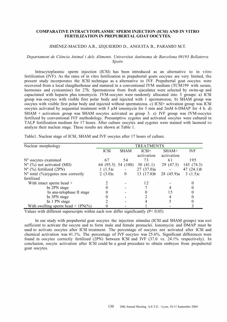
130 20th Annual Meeting A.E.T.E. - Lyon, 10-11 September 2004
COMPARATIVE INTRACYTOPLASMIC SPERM INJECTION (ICSI) AND IN VITROFERTILIZATION IN PREPUBERTAL GOAT OOCYTES.
JIMÉNEZ-MACEDO A.R., IZQUIERDO D., ANGUITA B., PARAMIO M.T.
Departament de Ciència Animal i dels Aliments. Universitat Autònoma de Barcelona 08193 Bellaterra.Spain.
Intracytoplasmic sperm injection (ICSI) has been introduced as an alternative to in vitrofertilization (IVF). As the rates of in vitro fertilization in prepubertal goats oocytes are very limited, thepresent study incorporates the ICSI technique as a alternative to IVF. Prepubertal goat oocytes wererecovered from a local slaugtherhouse and matured in a conventional IVM medium (TCM199 with serum,hormones and cysteamine) for 27h. Spermatozoa from fresh ejaculates were selected by swim-up andcapacitated with heparin plus ionomycin. IVM-oocytes were randomly allocated into 5 groups: a) ICSIgroup was oocytes with visible first polar body and injected with 1 spermatozoa; b) SHAM group wasoocytes with visible first polar body and injected without spermatozoa. c) ICSI+ activation group was ICSIoocytes activated by sequential treatment with 5 µM ionomycin for 5 min and 2mM 6-DMAP for 4 h. d)SHAM + activation group was SHAM oocytes activated as group 3. e) IVF group was IVM-oocytesfertilized by conventional IVF methodology. Presumptive zygotes and activated oocytes were cultured inTALP fertilization medium for 17 hours. After culture oocytes and zygotes were stained with lacmoid t oanalyze their nuclear stage. These results are shown at Table 1.
Table1. Nuclear stage of ICSI, SHAM and IVF oocytes after 17 hours of culture.
TREATMENTSNuclear morphologyICSI SHAM ICSI+
activationSHAM+
activationIVF
Nº oocytes examined 67 54 73 61 195Nº (%) not activated (MII) 64 (95.5) 54 (100) 30 (41.1) 29 (47.5) 145 (74.3)Nº (%) fertilized (2PN) 1 (1.5)c - 27 (37.0)a - 47 (24.1)bNº total (%)zygotes non correctlyfertilized
2 (3.0)c 0 13 (17.8)b 28 (45.9)a 3 (1.5)c
With intact sperm head + 2 - 12 - 0 In 2PN stage 0 - 7 4 0 In ana-telophase II stage 0 - 0 15 0 In 3PN stage 0 - 2 4 0 In 1 PN stage 2 - 4 5 0 With swelling sperm head + 1PN(%) 0 - 1 - 3Values with different superscripts within each row differ significantly (P< 0.05)
In our study with prepubertal goat oocytes the injection stimulus (ICSI and SHAM groups) was notsufficient to activate the oocyte and to form male and female pronuclei. Ionomycin and DMAP must beused to activate oocytes after ICSI treatment. The percentage of oocytes not activated after ICSI andchemical activation was 41.1%. The percentage of IVF oocytes was 25.6%. Significant differences werefound in oocytes correctly fertilized (2PN) between ICSI and IVF (37.0 vs. 24.1% respectively). Inconclusion, oocyte activation after ICSI could be a good procedure to obtain embryos from prepubertalgoat oocytes.

131 20th Annual Meeting A.E.T.E. - Lyon, 10-11 September 2004
Notes____________________________________________________________________________________

132 20th Annual Meeting A.E.T.E. - Lyon, 10-11 September 2004
DEVELOPMENT OF EMBRYOS AFTER IVF OF BOVINE OOCYTES WITH FLOWCYTOMETRICALLY SEXED, FRESH AND FROZEN SPERM
K_TSKA-KSI__KIEWICZ L., BOCHENEK M., RY_SKA B.
Department of Animal Reproduction, National Research Institute of Animal Production,32-083 Balice/Kraków, Poland
There is a great interest in application of sex preselected, X and Y chromosome-bearing sperm(Johnson, 1995: Reprod. Fertil. Dev. 7:893-903; Zhang et al., 2003: Theriogenology 60: 1657-1663).
The aim of our investigations was to develop technology of in vitro embryo production in cattleusing fresh and frozen-thawed, flow cytometrically-sorted, spermatozoa for IVF. Immature oocytes,recovered from slaughtered ovaries, were matured in vitro (IVM) for 22 to 23 h in modified TCM 199and were fertilized in vitro with fresh or frozen-thawed, cytometrically-sorted X and Y fractions ofsperm of bulls with proven fertility. Sperm was sorted in a MoFloSX cytometer (Cytomation) using themethod of XY Inc., Colorado, USA (Research Collaboration Agreement). Simultaneously, control(unsorted) fresh or frozen-thawed sperm of the same ejaculate as sorted spermatozoa was used for IVF.The protocol of sperm capacitation was related to the source of spermatozoa. For control sperm, bothfresh and frozen-thawed, and for fresh fractions of sexed sperm the standard procedure of capacitationwas applied (K_tska et al., 1998: J. Anim. Feed Sci. 7:353-362). Briefly, sperm was separated by Percollgradient (90%:45%) centrifugation (500 g for 30 min), washed and placed in 40 µl fertilization drops(TALP-IVF medium containing 10 µg heparin/ml and mixture of penicillamine, hypotaurine andepinephrine) at concentration 1 to 1.5 x 10⁶ spermatozoa/ml medium. For frozen-thawed fractions ofsexed sperm separation in Percoll gradient was omitted; immediately after thawing X and Y samples werecentrifuged (500 g for 10 min) and placed in fertilization drops at concentration 2 to 3 x 10⁶
spermatozoa/ml TALP-IVF medium. After IVF, the zygotes were cultured for 24 h in medium B2. Theresulting embryos were co-cultured with Vero cells for another 8 days, i.e. up to the hatched blastocyststage.
Up to now a total number of 1572 in vitro matured oocytes were fertilized with sexed and controlsperm of four bulls. The following results were obtained: for fresh, control sperm 46.2% (86/186) and25.6% (22/86); for frozen-thawed, control sperm 50.2% (118/235) and 16.9% (20/118); for X freshfractions 24.5% (50/204) and 12.0% (6/50); for Y fresh fractions 23.8% (40/168) and 30.0% (12/40);for X frozen-thawed fractions 27.6% (104/377) and 13.5% (14/104); and for Y frozen-thawed fractions25.4% (102/402) and 16.7% (17/102), respectively for cleavage rates and blastocyst rates. Thepreliminary results suggest that there are differences due to bull effect and sperm conservation and nodifferences in efficiency between X and Y fractions of sperm. The investigations are continuing.
This research was supported by the State Committee for Scientific Research as a project 3PO6D 044 23.

133 20th Annual Meeting A.E.T.E. - Lyon, 10-11 September 2004
Notes____________________________________________________________________________________

134 20th Annual Meeting A.E.T.E. - Lyon, 10-11 September 2004
EFFECTS OF IN VITRO VS IN VIVO CULTURE ON EXPRESSION OF EMBRYO DERIVED XIAPTRANSCRIPTS IN SINGLE BOVINE EMBRYOS
KNIJN H.M.1, WRENZYCKI C.2, VOS P.L.A.M.1, VAN DER WEIJDEN G.C.1, NIEMANN H.2 ,DIELEMAN S.J.1
1Department of Farm Animal Health, Faculty of Veterinary Medicine, Utrecht, The Netherlands2Department of Biotechnology, Institut für Tierzucht und Tierverhalten (FAL), Neustadt, Germany
Earlier studies reported that the level of apoptosis in in vitro produced bovine blastocysts is higherthen in in vivo developed blastocysts (Gjørret et al., 2003: Biol Reprod 69, 1193-1200). The molecularbasis for this difference has not yet been studied. The regulation and execution of apoptosis is dependingon a cascade of events in which many proteins are involved. One of the proteins involved is X-chromo-some-linked Inhibitor of Apoptosis (XIAP).
The aim of the present study was to analyze expression of XIAP transcripts and determine ifinactivation of the X-chromosome (i.e. dosage compensation) occurred to a similar extend in in vitrocultured female embryos as in vivo at the early blastocysts (eb), blastocysts (b) and expanded blastocysts(xb) stage, respectively. Furthermore, to verify if this transcript detected in the blastocysts stages wasnewly expressed from the embryonic genome, transcription was inhibited with a RNA polymerase IIspecific inhibitor (_-amanitin) to the culture medium from the zygote stage onwards.Oocytes were obtained from abattoir ovaries and after IVM/IVF and IVC in SOF medium, eb, b and xbwere collected (In vitro group). For the In vivo group, embryos were collected from normally cyclic cows,superovulated with 3000 IU eCG (Intergonan; Intervet, Tönisvorst, Germany) at day 7 po by non-surgicaluterine flushing. The developmental stage of the embryos was determined by stereomicroscopy and eb, band xb were collected and stored at -80°C. For the transcription inhibition experiment, embryos werecultured in vitro as for the In vitro group until the 8-16 cell stage (that is until 100 h after the start offertilization), one group with 10 mM _-amanitin added to the culture medium (_-amanitin group) and onegroup without (control group). A highly sensitive semi-quantitative RT-PCR assay (Wrenzycki et al.,1999: Mol Reprod Dev 53, 8-18) was used to determine the relative abundance of gene transcripts insingle blastocysts and pools of five 8-16 cell embryos. For sex determination of the embryos DNA wascollected from the supernatant of the RNA extraction and prepared using Microcon YM-100 (42412;Millipore, Eschborn, Germany) columns according to the manufacturer’s instructions. Embryonic sex wasdetermined by PCR analysis using bovine-specific and Y-chromosome-specific primers.
The relative abundance in 8-16 cell embryos cultured with α-amanitin was 0 (n=6) and in thecontrol group without α-amanitin was 1.62 ± 0.2 (n=6).
Table 1. Relative abundance ± sem (number of replicates) of XIAP transcripts in single male and femaleeb, b and xb produced in vitro and in vivo. Different superscripts between male and femalewithin the same developmental stage are significantly different (Anova followed by multiplepairwise comparisons using the Tukey test).
eb b xbmale female male female male female
In vitro0.22 ± 0.2a
(4) 0.16 ± 0.1a (4)0.60 ± 0.2a
(6)1.72 ± 0.4b
(9)0.53 ± 0.2a
(4)0.38 ± 0.1a
(7)
In vivo0.30 ± 0.2A
(9)0.38 ± 0.2A
(8)0.62 ± 0.1A
(5)1.31 ± 0.3B
(6)0.44 ± 0.2A
(8)1.38 ± 0.4B
(6)
In conclusion, XIAP transcripts in blastocysts are of embryonic origin. No dosage compensationwas observed at the blastocysts stage, irrespective of the production method. However, no dosagecompensation is observed in in vivo-produced expanded blastocysts in contrast to in vitro-producedexpanded blastocysts. Speculatively, the premature inactivation of the X-chromosome in in vitro-produced expanded blastocysts could be related to the increased level of apoptosis in in vitro-producedexpanded blastocysts.

135 20th Annual Meeting A.E.T.E. - Lyon, 10-11 September 2004
Notes____________________________________________________________________________________

136 20th Annual Meeting A.E.T.E. - Lyon, 10-11 September 2004
STRAIN-DEPENDENT DIFFERENCES IN THE SUPEROVULATORY RESPONSE, EMBRYODEVELOPMENT AND EFFICIENCY OF TRANSGENESIS IN RATS
KRIVOKHARCHENKO A., POPOVA E., GANTEN D., BADER M.
Max-Delbrück Center for Molecular Medicine (MDC), Robert-Rössle-Str. 10, D-13092 Berlin-Buch,Germany
The possible effect of the genotype of immature rats on superovulation response, in vitro and invivo development of embryos and overall efficiency of transgenesis was studied. The protocols forinduction of superovulation using a single injection of 15 IU PMSG or minipump with 10 mg FSHfollowed by the injection of 30 IU hCG were compared in LEW, WKY, SHRSP or SD and Wistar asrepresentatives of inbred or outbred strains, respectively. In the case of in vitro culture of rat zygotes t othe blastocyst stage we cultured the ova overnight in M16 medium and than transferred them intomR1ECM medium. Ova were cultured (10-20 embryos per 700 µl of the medium) in 4-well culture dishes(Nunc) under 5% CO2 in air at 37°C. The comparisons for multigroup and multifactorial analyses weredone with a two-way ANOVA and one-way ANOVA on ranks for multiple group comparisons. A value ofP<0.05 was chosen as an indication of statistical significance.
After hCG administration females were mated with males of the same strain and sacrificed on Day 1of pregnancy. The percentage of mated animals was similar in all groups (60.0-100%) independent of theanimal strain used. The mean number of ova per donor was not dependent on the hormonal treatmentused within each rat strain. At the same time SD and Wistar female rats were more effective to hormonaltreatments than animals from inbred strains. SHRSP rats produced significantly more ova per female inresponse to superovulation compared to other inbred strains.
Significantly lower rates of in vitro development in mR1ECM medium was observed for intactzygotes from WKY rats. However, embryos recovered from SD, SD x Wistar and WKY animals at the 2-cell stage showed no difference in competence to reach the blastocyst stage in vitro (77.6-84.6%). Theproportion of offspring developed after oviduct transfer of intact zygotes derived from superovulateddonors was slightly lower for WKY rats.
SD and SHRSP zygotes survived after microinjection significantly better than the zygotes fromWKY and Lewis rats (~75% v.s. ~55%). Lewis and SD animals showed significantly higher rates ofdevelopment to term of microinjected zygotes after transfer to recipients compared to the other strains.At the same time we found no significant differences in efficiency of transgene integration per newborn(5.3-22.2%) or per injected zygotes (0.9-2.1%) produced from studied rat strains.
The results of this study demonstrate strain-dependent differences in the response t osuperovulation protocols, in the sensitivity to the microinjection and in the capability of zygotes t odevelop in vitro until morula and blastocyst stages or in vivo to term after transfer to the foster mothers.However, despite these genotype-dependent differences all studied strains can be successfully used fortransgenic rat production.

137 20th Annual Meeting A.E.T.E. - Lyon, 10-11 September 2004
Notes____________________________________________________________________________________

138 20th Annual Meeting A.E.T.E. - Lyon, 10-11 September 2004
EVALUATION OF THE FUNCTIONAL STATUS OF BOVINE OOCYTES AND EMBRYOSCULTURED IN VITRO
KUZMINA T.1, DENISENKO V.1, TORNER H.2, ALM H.2, SHOKIN O.1
1Department of Genetics & Biotechnology, All-Russian Research Institute for Farm Animal Genetics &
Breeding, 189620 St.Petersburg-Pushkin, Russia2Department of Reproductive Biology, Research Institute for Biology of Farm Animals,18196
Dummerstorf, Germany
In order to increase the quality of in vitro derived and cloned bovine embryos, the basicfundamentals of in vitro cytoplasmic maturation of donor oocytes needs to be completely understood.The precise roles of calcium and some co-enzymes, such as NADH and FAD, which participate inoxidizing and reducing reactions within the developing oocyte needs to be determined as well as its roleduring IVF and IVC. The features of calcium and NADH/FAD metabolism in normal and degenerativeoocytes and embryos need to be understood. On other hand, when effective methods are found t oevaluate quality of donor oocytes before and after IVM, the success to obtaining viable embryos willincrease dramatically. The aim of this study was to evaluate the functional status of bovine oocytes,zygotes and 2-cell embryos cultured in vitro. A morphological classification (cumulus, ooplasm,blastomeres, chromatin status) was used for in vitro matured bovine oocytes, zygotes and 2 cells embryosto determine: 1) density of fluorescence NADH, FAD and the ratio NADH/FAD (Excitation - 365 nm forNADH and 436 nm for FAD, Emission- 465 nm for NADH and 530nm for FAD), and 2) concentrationof the calcium of intracellular stores (fluorescence of complex membrane-bound calcium-chlortetracycline was fixed with a fluorescent microscope – Excitation: 390 nm, Emission: 530nm, using40µM chlortetracycline). The results were expressed in arbitrary units of fluorescence. A total of 378normal (N) and degenerative (D) oocytes, zygotes and embryos were evaluated. Oocytes were culturedfor 24 h at 38.5o C under 5% CO2 in TCM 199 with 10% FCS and 10 ng/ml recombinant bovinesomatotropin (rbGH-“Monsanto”). After IVM, oocytes were fertilized using frozen bovine sperm andzygotes were cultured in TCM 199 with 20% FCS and 10 ng/ml rbGH. Data were analysed using theStudent’s test. Inhibition of __2+ exit from intracellular stores was detected in degenerative oocytes atmetaphase-II (1.95±0.12-D compared to 1.47±0.10 – N, P<0.01) and 2-cell embryos (3.47±0.40-Dcompared to 1.78±0.14 – N, P<0.001). The intensity of a fluorescence NADH also increased indegenerative oocytes at metaphase-II (1.58±0.10-D compared to 1.20±0.12 – N, P<0.05) and in 2-cellembryos (1.48±0.25-D compared to 1.18±0.05 – N). We did not find an influence of degeneration on theintensity of NADH fluorescence and __2+ exit from intracellular stores in zygotes. These datademonstrate that in cattle: 1) NADH concentration and calcium from intracellular stores in oocytes and2-cell embryos are interconnected; 2) NADH concentration and calcium stores are related t omorphological quality and chromatin status in oocytes and 2-cell embryos. The results suggest that theseparameters (NADH concentration and the intracytoplasmic calcium stores) may be considered in furtheroptimization of the conditions for oocyte and embryonic development in vitro.

139 20th Annual Meeting A.E.T.E. - Lyon, 10-11 September 2004
Notes____________________________________________________________________________________

140 20th Annual Meeting A.E.T.E. - Lyon, 10-11 September 2004
EFFECT OF TRICHOSTATIN A (TSA) TREATMENT OF DONOR CELLS ON DEVELOPMENT OFBOVINE NUCLEAR TRANSFER (NT) EMBRYOS
LAGUTINA I., LAZZARI G., GALLI C.
Laboratorio di Tecnologie della Riproduzione, CIZ srl,Istituto Sperimentale Italiano Lazzaro Spallanzani, Cremona, 26100, Italy.
Nuclear reprogramming is the process by which a differentiated nucleus returns to a totipotentstage. It is possible that in somatic cell cloning, donor cells with less epigenetic modifications - lowerlevels of methylated DNA and/or higher levels of histone acetylation - may be better reprogrammed. T oinvestigate the effect of histone acetylation of the donor nucleus on NT-embryo development we treatedadult bovine fibroblasts with trichostatin A (TSA) – a potent histone deacetylase inhibitor - during serumstarvation (0.2 mM TSA) or in complete medium (0.08 mM TSA) 24 h prior to nuclear transfer. NT-embryos were constructed by the zona-free method (Oback et al. 2003). Cell couplets were fused by asingle DC-pulse of 1.2 Kv/cm applied for 30 µsec. NT-embryos were activated 1.5 to 3.5 h after fusionwith 5 µM ionomycin for 4 min followed by 4 h culture in 2 mM 6-DMAP in m-SOF. Zona-free NT-embryos were cultured individually in 3 µl drops under mineral oil at 38.5°C in 5% CO2, 5% O2 and 90%N2. The rates of cleavage, morula compaction D6 (MCD6) and blastocyst formation D7 (BLD7) wererecorded. The data were compared by Chi-square test.
The results of the preliminary experiments showed that TSA-treatment of donor cells during serumstarvation did not affect the developmental ability of NT-embryos to the stage of morula compaction butsignificantly decreased the rate of BLD7 (28.7 vs 42.1%, P<0.05).
In the second series of experiments we evaluated the development of NT-embryos derived fromTSA (complete media)-treated fibroblasts using two different time between fusion and activation that isconsidered to be the time of active remodelling and reprogramming of the donor nucleus (Table 1).
Table 1. Effect of TSA-treatment in relation to different fusion-activation timing.
Group Time *, h N Cleavage(%) MCD6 (%) BLD7 (%)control 2 - 2.5 69 61 (88.4) 24 (34.8) a 14 (20.3) ccontrol 3 - 3.5 121 118 (97.5) 58 (47.9) ab 41 (33.9) dTSA 1.5 - 2 83 78 (94) 43 (51.8) b 29 (34.9) dTSA 3 - 3.5 112 107 (95.5) 57 (50.9) b 42 (37.5) dChi-square test, P<0.05. Time* : time between fusion and activation.Our results indicate that TSA-treatment of donor cells allows NT-embryos to undergo reprogrammingfaster than in control (TSA 1.5-2 h vs control 3-3.5 h).
In conclusion we demonstrate that there is a significant decrease of blastocyst rate after TSA-treatment of donor cells during serum starvation but not during normal culture. In addition we show thatTSA treated donor cell nuclei acquire the ability to be reprogrammed faster. This work was supported byFIRB.

141 20th Annual Meeting A.E.T.E. - Lyon, 10-11 September 2004
Notes____________________________________________________________________________________

142 20th Annual Meeting A.E.T.E. - Lyon, 10-11 September 2004
THE EFFECT OF NEGATIVE ENERGY BALANCE ASSOCIATED NON-ESTERIFIED FATTY ACIDLEVELS DURING IN VITRO MATURATION OF BOVINE OOCYTES ON EMBRYO
PRODUCTION
LEROY J.L.M.R.1, VANHOLDER T.1, BOLS P.E.J.2, DE CLERCQ J.1, VAN SOOM A.1
1Department of Reproduction, Obstetrics and Herd Health, Faculty of Veterinary Medicine, GhentUniversity, B-9820 Merelbeke, Belgium; 2Laboratory of Veterinary Physiology, University of Antwerp, B-
2610 Wilrijk, Belgium.
Dairy cow fertility has declined drastically during the last decades. This subfertility is amultifactorial problem. The negative energy balance (NEB) in high-yielding dairy cattle early postpartum may affect ovarian function and oocyte quality through elevated levels of non-esterified fattyacids (NEFA) in serum as well as in follicular fluid (FF). The composition of the FF in dairy cows during aperiod of NEB has been studied thoroughly in our laboratory. Due to increased lipolysis, the NEFA levelsin FF also rose but remained significantly lower (± 40%) compared to serum concentrations and neverexceeded 0.6 mmol/l. Also the qualitative composition of the NEFA fraction differed significantlybetween serum and FF for C18:1 (oleic acid: 27.8±2.6% vs. 33.1±1.46%), C18:0 (stearic acid: 25.6±1.7%vs. 12.7±0.8%), C16:0 (palmitic acid: 20.0±1.0% vs. 23.0±0.6%) and C18:2 (linoleic acid: 3.4±0.4% vs.7.4±0.7%) (P < 0.05). The aim of this study was to investigate in an in vitro maturation model whetherelevated NEFA levels during in vitro maturation are detrimental to the fertilisation rate, cleavage andsubsequent blastocyst formation. The preliminary results are presented.
A total of 3429 oocytes were matured in serum free maturation medium (TCM 199, 20 ng/mlmEGF). According to treatment, palimitic acid (PA), stearic acid (SA) or oleic acid (OA) in ethanol wereadded at final test concentrations of 0.133, 0.067 and 0.200 mM respectively corresponding to 23%,13% and 33% of a total NEFA concentration of 0.6 mmol/l (cfr. supra). In contrast to the negativecontrol groups (no ethanol or NEFA), positive controls received equal volumes of ethanol (but no NEFA)as the test groups. Oocytes were matured per 60 for 22-24 hours at 38.5°C and 5% CO2. Aftermaturation, all groups were coincubated with 1x10⁶ sperm/ml for 18-20 hours. Half of the presumptivezygotes was fixed and stained with Hoechst to determine fertilisation rate while the other half wascultured per 25 for 7 days in 50 _l drops of SOF medium with 5% FCS under mineral oil (5% CO2, 5% O2
and 90% N2). At respectively 48 h and 8 days post insemination, the number of cleaved zygotes and thenumber of blastocyst were counted. Preliminary data were analysed with a two-way ANOVA in which“replicate” was inserted as random effect. Results are expressed as mean % (±SD) of total number ofoocytes.
No significant effect was observed for ethanol or OA on the fertilisation rate, cleavage orblastocyst development. The presence of 67 _M SA during oocyte maturation, however, resulted insignificant lower fertilisation rates (64±3.1% vs. 74±2.1% in positive control), cleavage (59±7.4% vs.77±6.0%) and blastocyst formation (21±10.1% vs. 35±5.2%) (P < 0.05). Preliminary results with PAalso suggest a negative effect of this fatty acid on the three parameters studied but additional replicatesare needed for full confirmation.
Based on these results we can suggest that high levels of NEFA during a period of NEB mayinfluence fertility in high-yielding dairy cows by hampering oocyte maturation as expressed in lowerfertilisation rates and subsequent lower cleavage and blastocyst development.
This research was supported by IWT-Vlaanderen (Grant 13236).

143 20th Annual Meeting A.E.T.E. - Lyon, 10-11 September 2004
Notes____________________________________________________________________________________

144 20th Annual Meeting A.E.T.E. - Lyon, 10-11 September 2004
THE INFLUENCE OF THE FOLLICLE ON EMBRYONIC/FETAL MORTALITY IN CATTLE
LOPES A.S.1,2, BUTLER S.T.3, GILBERT R.O.3, BUTLER W.R.3
1Danish Inst. Agricultural Sciences, 8830 Tjele, Denmark; 2Royal Veterinary & Agricultural University,1870 Frederiksberg, Denmark; 3Cornell University, Ithaca NY 14850, USA
Impaired reproductive performance is a major problem in dairy cattle, e.g. high embryonic/fetalmortality leading to low pregnancy rates. These problems can originate in the ovulating follicle itself, asit is responsible for providing the adequate environment for oocyte development and is the basis forsubsequent formation of the corpus luteum. Previous studies have demonstrated that pregnancy rates wereinfluenced by follicular size at ovulation (e.g. Vasconcelos et al. 1999, Theriogenology 52:1067-1078),but the conclusions have been inconsistent. Heat stress, considered a major cause of low fertilityworldwide, is also likely to contribute to the high embryonic/fetal mortality.
In our present study, the ovulatory follicle was described in terms of size and function (plasmaoestradiol), as well as its basis for subsequent CL formation and function (plasma progesterone). Theeffect of these parameters was related to the subsequent pregnancy rates on day 28 and on day 63 afterAI. Finally, an effect of environment (season) on these parameters was investigated.
In 2003, lactating cows at the New York Station were studied during December-May (cold season,n=73) or July-September (warm season, n=71), all starting approx. 80 days after calving. Ovulation timewas synchronized and one fixed time AI was used (= day -1), and on this day the diameter of the largestfollicle (only follicles from 8 to 20 mm were considered as ovulatory) was measured by ultrasound. Bloodsamples were collected daily from day –1 until day +7, and then on days 14, 21, 28, 35 and 60. Plasmaconcentrations of progesterone (P4) and oestradiol (E2) were quantified by RIA. Pregnancy diagnosis wasdetermined on day 28 and 63 using ultrasound, and on day 42 using rectal palpation. Data were analysedstatistically using F tests for comparison of means and a mixed linear model (Mixed Procedure, SASInstitute Inc.) for the full data analysis.
Overall, 48 of the 144 cows (33%) were pregnant on day 28; of these, 11 (24%) aborted before day63. None of these rates differed between seasons. The average diameter of the ovulatory follicle waslarger in cows which became pregnant than non-pregnant cows on day 28 (15.9±2.4 vs 14.3±3.6 mm,n=48 and 96, respectively; p<0.001), but not for the cows aborting between day 28 and 63. No differencewas observed between seasons. Follicular function measured as E2 in plasma on day of AI was higher incows pregnant on day 28 than in non-pregnant cows, but only in the cold season. For non-pregnant cows,the E2 value was highest in the cold season. When comparing cows still pregnant on day 63 with cowsaborting between day 28 and 63, the E2 value was higher for the first group, but only in the cold season.The P4 values increased gradually after AI, reaching a plateau from around day 14 in cows still pregnanton day 63. For those cows aborting between day 28 and 63, the P4 started to be lower already from day 7.In both groups, the levels were lower in the warm season. None of these patterns were related to diameterof the ovulatory follicle.
The results illustrate that, in this group of cows, no predictive information regarding embryosurvival is obtained from simply measuring diameters of ovulatory follicles. On the other hand, othermeasures of follicular function (e.g. hormonal profiles) seem to contain predictive information, possiblybecause they also reflect oocyte quality. Furthermore, the CL function (P4 levels throughout pregnancy)is associated with embryonic mortality already from maternal recognition of pregnancy. The effect ofseason during the year 2003 did not result in differences; however, the difference in E2 and P4 levelsobserved between seasons could indicate that follicular and luteal steroidogenesis can be disturbed duringwarm season.
*A.S. Lopes’ travel and stay was supported by FCT grant no. SFRH/BD/8486/2002.

145 20th Annual Meeting A.E.T.E. - Lyon, 10-11 September 2004
Notes____________________________________________________________________________________

146 20th Annual Meeting A.E.T.E. - Lyon, 10-11 September 2004
MATURATION OF BOVINE OOCYTES DEFINED BY FOLLICLE SIZE AND THE PHASE OFFOLLICULAR WAVE IN VITRO : EFFECT OF GONADOTROPINS OR EPIDERMAL GROWTH
FACTOR ON EMBRYO DEVELOPMENT
MACHATKOVA M., HORAKOVA J., HANZALOVA K., PESLAROVA Z.
Department Genetics and Reproduction; Veterinary Research Istitute,621 32 Brno, Czech Republic
In a previously study we have documented a relationship between the developmental competenceof oocytes collected from different-sized follicles in the growth or the dominant phase of follicular waveand embryo production (Machatkova et al., Theriogenology 2004; 61:329-335). The aim of the presentstudy was to compare the effect of gonadotropins or epidermal growth factor (EGF) during maturation ofoocytes defined by follicle size and the phase of folicular wave on embryo development. A total of 3476oocytes collected from small (2-5 mm) or medium follicles (6-10 mm) in the growth/stagnation or thedominance/regression phase of follicular wave were recovered from slaughtered cows. Subsequently, theovaries of each donor were characterized in terms of morphology. Oocytes were matured in TCM-199supplemented with 5% oestrus cow serum (ECS) and either FSH/LH (P.G.600, 15 iu/mL) or EGF (50µ/mL). They were fertilized and cultured by standard methods. The developmental competence of oocyteswas expressed as the percentages of D7 and D8 embryos developed from fertilized oocytes, and as thepercentage of advanced or expanded blastocysts from D7 or D8 embryos. The data was analyzed by theChi-square test. Both the development rate of oocytes into embryos and the percentage of advanced orexpanded blastocysts from embryos were higher for the oocytes collected from small follicles in thegrowth/stagnation phase and matured in medium with FSH/LH than for those matured in medium withEGF (33.4 vs 27.0 %, P < 0.05, and 32.6 vs 25.0 %, P < 0.01 for D7 and D8 embryos; 38.4 vs 30.8 %and 69.5 vs 62.6 % for advanced or expanded blastocysts). On the other hand, the oocytes collected frommedium follicles in the growth/stagnation phase showed both the highest development rate of oocytesinto embryos and the highest percentage of advanced or expanded blastocysts when matured in thepresence of EGF as compared with FSH/LH (49.8 vs 47.1 % and 40.0 vs 38.7 % for D7 and D 8 embryos;54.8 vs 37.9 %, P < 0.01 or 77.0 vs 65.4 %, P < 0.05 for advanced or expanded blastocysts ). Whenoocytes were collected from small or medium follicles in the dominance/regression phase and matured inmedium with either FSH/LH or EGF, the development rates of oocytes into embryos were similar. But inoocytes collected from medium follicles and matured in the presence of EGF, the percentage of advancedblastocysts from D7 embryos was significantly higher than in those matured in the presence FSH/LH(43.3 vs 28.4 %, P < 0.05). In conclusion, the expression of developmental competence of oocytes wasinfluenced by the presence of gonadotropins or EGF in maturation medium. In the growth/stagnationphase, oocytes from small follicles, with lesser developmental competence, prefered FSH/LH to EGF butoocytes from medium follicles, with greater developmental competence, prefered EGF to FSH/LH duringmaturation. In the dominance/regression phase, the two oocyte subpopulations tolerated both FSH/LHand EGF but in oocytes from medium follicles blastocyst formation was accelerated in the presence ofEGF compared with FSH/LH as it was in the growth/stagnation phase.

147 20th Annual Meeting A.E.T.E. - Lyon, 10-11 September 2004
Notes____________________________________________________________________________________

148 20th Annual Meeting A.E.T.E. - Lyon, 10-11 September 2004
IMPROVING THE MODE OF FSH APPLICATION FOR SUPEROVULATION TREATMENTS:CLINICAL DATA AND ENDOCRINOLOGY
MARTENS G.1, NOHNER H.-P.1, LEIDING C.1, SCHNEIDER F.2, KANITZ W.2
Institute of Artificial Insemination, 1BVN 91413 Neustadt/Aisch, GermanyDepartment of Reproductive Biology, 2 Research Institute for the Biology of Farm Animals, 18196
Dummerstorf, Germany
Internationally, the use of Follicle-Stimulating Hormone (FSH) to induce superovulation is favouredin a regime which uses eight injections over four days. However, this conventional regime of treatmenthas some disadvantages: a) Multiple applications given intra-muscularly every 12 hours entail a lot ofwork for the person who is performing the treatment and b) it stresses the animal to be treated.Therefore, a reduction of injection frequency would have obvious benefits. The number of studiespublished is, however, low and the detected results are inconsistent.
The aim of our studies was to optimise both the dose and frequency of FSH administration usingtwo commercial preparations (Pluset® and Folltropin®). As a result of the first joint study performed bythe Institutes named above, the results were evaluated which were found after the treatment of a total of810 Fleckvieh donors of the Bavarian breed according to various treatment protocols (Fig. 1).
0,0
5,0
10,0
15,0
20,0
4-10 8-10 4-11 8-11 4-13 4-15
Pluset
0,0
5,0
10,0
15,0
20,0
25,0
4-10 8-10 4-13 8-13 4-17 8-17
Folltropin
degeneratedembryos
unfertilised ovas
transferableembryos
Figure 1. Results of embryo collection depending upon the type of FSH preparation (Pluset® orFolltropin®), the frequency of injection (left number below columns), and dose (ml; right number)
heifer B3
0
1
2
3
4
5
6
08:00
09:00
10:00
11:00
12:00
13:00
14:00
15:00
time
ng
/ m
l
day 9
day 12
With the aim of characterizing plasmaconcentrations of reproductive hormones, usingthree different variants of the Pluset® regime, atotal of 12 heifers were bled throughout 8 hours ondays 9 and 12 (before and during stimulation byFSH), respectively. The samples were analysed forsteroid hormones, LH (Fig. 2 Example), and porcineFSH which had been administrated.
Figure 2. LH concentrations (ng/ml) onexperimental days 9 and 12 (beginningof FSH administration: day 10). Samplingwas performed every 10 min through 8 h.
This work was supported by the Karl Eibl-Stiftung.Correspondence: [email protected]

149 20th Annual Meeting A.E.T.E. - Lyon, 10-11 September 2004
Notes____________________________________________________________________________________

150 20th Annual Meeting A.E.T.E. - Lyon, 10-11 September 2004
PARENTERAL _-CAROTENE ADMINSTRATION ON BOVINE EMBRYO DONORS. APRELIMINARY APPROACH
MARTINEZ-BELLO J.D.1, FERNÁNDEZ SÁNCHEZ I.1, QUINTELA ARIAS L.2
1UTE. BOS-FEFRIGA; 2Dpto. Reproducción. Fac. Veterinaria de Lugo.U.S.C.
The number of viable embryos produced after each superovulation treatment (SOV) andvariability of response to exogenous gonadotropins are limiting factors to the largescale use of ET onfarms. Despite hormonal methods, micronutrients have been considerated fundamental forreproduction. The aim of this study was to examine the effect of i.m. injection of _-carotene on theyield of viable embryos. 32 adult Holstein cows of top genetic herds were selected for SOV. Allreceived the same SOV protocol with intra vaginal progesterone device (PRID) plus 5 mg oestradiolbenzoate, plus 100 mg progesterone on Day 0 without oestrus control and after gynaecologicalexploration to eliminate cows with any disorders. 16 cows received 600 mg of _-carotene(DALMAVITAL, Fatro-Uriach) 15 days before PRID administration. The same batch of FSH(PLUSET, Calier) was used to treat animals on Day 4 after inserting PRID in a decreasing doseregime during 4 days. Two doses of cloprostenol (Dalmazin, Fatro-Uriach) were administered on Day3 and PRID was removed with the second prostaglandin dose. Artificial insemination was performedtwo times, 48 and 60 hours after PRID removal. Embryos were recovered 7 days post-inseminationand evaluated according to IETS standards. Data were analysed with GLM (General Linear Model) ofone factor. Results are shown in the table below.
Table 1. Mean±standard deviation of total embryos, viable embryos, degenerated embryos andova in function of _-carotene administration.
The total embryos and viable embryos were higher in the _-carotene group although thedifference was not significant owing to large variation in data. The number of corpora lutea evaluatedby hand on day of recovery did not differ significantly between groups (12.18±6.22 control vs13.78±5.17 _-carotene treated). Studying treated cows, 8 of 16 had prior recovery data in our recordsand comparing results from before treatment and after we find an average of viable embryos of 6.4 vs10.8 after treatment. In addition, the control group reached the average viable embryo productionreported for our team in the last two years of over 290 SOV. If we look at the quality of the embryosproduced we find no difference although there was a higher number of quality 1 embryos in the treatedgroup vs control 95/106 (89.6%) vs 64/73 (87.7%) while quality 2 embryos are higher in control group9/73 (12,30%) vs 9/106 (8,5%) in treated cows. Evaluating all data altogether, results suggest thatparenteral administration of _-carotene 30 days before embryo recovery may have some beneficialeffects on viable embryo production although statistical difference could not be demonstrated in thepresent study. Further, more investigation is needed to determinate the effect of this treatment and thebest approach and protocol of administration to enhance embryo production.
GROUP n TOTALEMBRYOS
VIABLEEMBRYOS
DEGENERATEDEMBRYOS OVA
Control 16 9.75±8.43 4.56±4.88 1.75±2.26 3.44±5.45_-carotene 16 14.25±10.80 6.50±8.40 1.94±2.20 5.81±5.94

151 20th Annual Meeting A.E.T.E. - Lyon, 10-11 September 2004
Notes____________________________________________________________________________________

152 20th Annual Meeting A.E.T.E. - Lyon, 10-11 September 2004
EFFECT OF CYSTEAMINE DURING IN VITRO MATURATION OF SLAUGHTERHOUSEDERIVED AND OPU DERIVED BOVINE OOCYTES ON FURTHER EMBRYONIC
DEVELOPMENT
MERTON J.S., MULLAART E., LANDMAN B.
Holland Genetics, P&D, PO Box 5073, 6802 EB Arnhem, The [email protected]
Glutathione (GSH) plays an important protective role in relation to reactive oxygen speciesgenerated by normal oxidative metabolism in the cell. The presence of cysteamine during in vitromaturation may facilitate the synthesis of GSH by immature oocytes. In a previous study weshowed a positive effect of the presence of cysteamine during in vitro maturation ofslaughterhouse derived bovine oocytes on subsequent in vitro embryonic development (Merton etal., 2004, Reprod. Fert. Dev. 16: 279). This report shows the first results of an ongoing field trialwith OPU derived oocytes, in order to confirm our previous results obtained with slaughterhousederived oocytes .
Immature cumulus-oocyte-complexes (COCs) were recovered from ovaries 6 to 8 h uponslaughter (SL) or obtained twice weekly by ultrasound guided transvaginal oocyte collection (OPU)at two collection centres. Both sources of COCs were matured in vitro in TCM199/FCS/LH/FSHsupplemented either with or without cysteamine (0.1 mM). Subsequently, matured oocytes werefertilised with frozen-thawed gradient-separated semen and further cultured for 7 days inSOFaaBSA. In case of OPU derived COCs, the experimental design was a 2X2 factorial. Resultswere analysed by Chi-square analyses.
The results show that the presence of cysteamine during in vitro maturation significantlyaffected embryo production from OPU derived COCs (31.8% and 39.2% Morula + Blastocyst rateat Day 7 for control and cysteamine, respectively). This higher embryo production rate wastotally due to an increased number of Blastocysts. Also the proportion of grade 3 embryos (IETS)was significantly reduced in the cysteamine group. The number of transferable embryos (Morulagrade 1 and early-, exp-Blast grade 1 and 2) per session was 1.38 and 2.07 (50 % relative increase)for control and cysteamine, respectively.
Table 1. Effect of cysteamine during in vitro maturation of slaughterhouse- and OPU-derivedbovine oocytes on subsequent in vitro embryonic development.
Oocytes Group Repl./sess.n
Oocytesn
Cleavagen (%)
Embryo production Day 7
Mor+ Blastn (%)
Relativeincrease
Transferablen (per session)
Sl.* Control 5 1056 586 (55.5) 205 (19.4)a
Cyst. 5 1070 634 (59.3) 257 (24.0)b 23.7 %
OPU Control 181 1365 908 (66.5) 434 (31.8)a 249 (1.38)a
Cyst. 167 1259 881 (70,0) 493 (39.2)b 23.3 % 346 (2.07)b
a,b: For each source of oocytes, values in columns with different superscript are significantlydifferent, P<0.05.* data adapted from Merton et al., 2004, Reprod. Fert. Dev. 16, 279 (abstract).
These results show that the presence of cysteamine during in vitro maturation also affectsfurther in vitro embryonic development (both embryo quality and developmental speed) of OPUderived COCs, resulting in a higher embryo production rate. The relative increase in the totalMorula and Blastocyst rate at Day 7 seems to be equal for both slaughterhouse- and OPU-derivedoocytes. This field trial will be continued in order to investigate the effect of cysteamine onpregnancy rate and offspring born.

153 20th Annual Meeting A.E.T.E. - Lyon, 10-11 September 2004
Notes____________________________________________________________________________________

154 20th Annual Meeting A.E.T.E. - Lyon, 10-11 September 2004
ALTERATIONS AND REVERSIBILITY IN THE CHROMATIN AND CYTOSKELETON OFCALF OOCYTES TREATED WITH ROSCOVITINE
MORATÓ R.1, ALBARRACIN J.L.1, IZQUIERDO M.D.2, MOGAS T.1
1Departament de Medicina i Cirurgia Animals. 2Departament de Ciència Animal i dels Aliments.Facultat de Veterinària. Universitat Autònoma de Barcelona. E-08193 Bellaterra, Spain
Improvement of the ability to maintain germinal vesicle (GV) stage oocytes in vitro isimportant for the acquisition of developmental competence. Maintaining oocytes at this stagewithout damaging their quality would synchronize meiotic resumption and would allow all collectedoocytes to complete nuclear maturation in order to maximize embryo production. GV breakdownin mammalian oocytes is regulated by activation of maturation promoting factor (MPF).Roscovitine (ROS), a specific cdc2 kinase inhibitor, has been shown to reversibly inhibit meioticresumption in cattle oocytes for 24 h without having a negative affect on subsequent developmentto blastocyst stage. Oocytes from calf oocytes are less competent to develop than those from cowovaries. Although fertilization and cleavage rates do not differ greatly between calf and cowoocytes, blastocyst yields are significantly reduced with calf oocytes. This lack of developmentalcompetence may be due to the failure or inability of the calf oocytes to complete cytoplasmicmaturation. It is hypothesized that if oocytes can be cultured in vitro under conditions thatmaintain meiotic arrest at the GV stage, then they may have the opportunity to acquire greaterdevelopmental competence. The aims of the present study were to study the efficacy of ROS t omaintain calf oocytes at the GV stage and to describe changes occurring in the distribution ofchromosomes, microtubules and actin microfilaments following exposure to differentconcentrations of ROS.
Cumulus-oocyte complexes (COCs) were aspirated from slaughterhouse calf ovaries andcultured for 24 h in TCM199 containing different levels of ROS (0, 12.5, 25, 50 and 100 µM).They were either fixed immediately or cultured for a further 24 h in conditions permissive t omaturation and subsequently fixed and processed using specific fluorescent probes in order t oevaluate chromosome, microtubule and microfilament distributions by visualization in a confocalmicroscope.
COCs cultured with any of the concentrations of ROS were significantly blocked at the GVstage. The inhibitory effect varied according to the dose, with 50 µM ROS being the mostefficient, with 83.5 % oocytes being blocked in the GV stage when compared to 0 µM (3.3%).When oocytes were released from the inhibitor, similar proportions of oocytes (74.5 and 75.5%)were observed in the metaphase II stage for 0 and 50µM, respectively. The meiotic spindleexhibited a typical MII morphology in 62.7% and 62.5% of oocytes for 0 and 50µM ROS,respectively. A normal distribution of actin filaments was observed in 93.2% and 85% of oocytesexposed to 0 and 50 µM ROS, respectively.
This study showed that 50 µM roscovitine-treated oocytes resumed meiosis after removalof the inhibitor and no significant influence of the inhibitor was detected on chromosome,microtubule and microfilament distributions during roscovitine treatment (50 µM).

155 20th Annual Meeting A.E.T.E. - Lyon, 10-11 September 2004
Notes____________________________________________________________________________________

156 20th Annual Meeting A.E.T.E. - Lyon, 10-11 September 2004
EFFICIENT GENERATION OF TRANGENIC MICE WITH INTACT YEAST ARTIFICIALCHROMOSOMES BY ICSI
MOREIRA P.N.1, GIRALDO P.2, COZAR P.2, POZUETA J.2, JIMENEZ A.1, MONTOLIU L.2,GUTIERREZ-ADAN A.1
1Departament of Animal Reproduction, INIA, Ctra. de la Coruña Km. 5.9 Madrid 28040, Spain.2Department of Molecular and Cellular Biology, CNB-CSIC, Campus de Cantoblanco, 28049
Madrid, Spain.
The production of animals with large transgenes is an increasingly valuable tool for geneticstudies, including the characterization and manipulation of large genes and polygenic traits.Intracytoplasmic sperm injection (ICSI)-mediated transgenesis has been suggested to be a valuabletool to generate complete integration and phenotypic expression of large DNA constructs;however, such capability was never clearly shown. By co-injecting spermatozoa and YeastArtificial Chromosomes (YACs), which are capable of mobilizing genomic fragments of severalmegabases in length, into metaphase II (MII) oocytes, we were able to produce founders exhibitinggermline transmission of an intact and functional transgene of submegabase magnitude. More than35 % transgenicity was obtained for a YAC transgene (YRT2_LCR) of 250 kb. When comparedwith the efficiency (1%) of YAC transgenesis by pronuclear microinjection (PI), the efficiency ofICSI-Mediated YAC Transfer (IMYT) was significantly greater, and no different than thatobtained after ICSI-Mediated Transfer of EGFP (45%), a construct 50 times smaller thanYRT2_LCR. All 3 IMYT founders crossed with non-transgenic mice, transmitted transgene DNAto progeny following a Mendelian pattern. Besides the huge difference in construct size, this rateof YRT2_LCR germline transmission is comparable to that previously reported for EGFP by ananalogous method, attesting the stability of the YAC transgene integrants. The efficiency of theapplied ICSI methodology in generating transgenics for large DNA constructs was verified when,of the IMYT-derived transgenic offspring, 1/12 (8%) harboured an intact copy of the 250 kbYAC. This was genomically confirmed by Southern Blot Analysis of three transgene markers, andfunctionally, by a co-inherited pigmented coat color characteristic of YRT2_LCR expression,which was able to rescue the albino background of the host. These data clearly demonstrate thepotential of IMYT to generate transgenics. In addition, to our knowledge, this is the first reportof stable incorporation and phenotypic expression of large DNA constructs of submegabasemagnitude mediated by ICSI.
Table 1. Embryo Cleavage and Transgenesis obtained with 5.4 kb (standard plasmid) and 250 kb(YAC) constructs
TechniqueEmployed
Construct(Size in kb)
InjectedOocytes
(No. Sessions)
EmbryosTransferred
(No. Recipients)
LiveAnimals
(%)
LiveTransgenic
Animals (%)
ICSI
IMYT
PI
EGFP (5.4)
YRT2_LCR(250)
YRT2_LCR(250)
219 (6)
367 (6)
1733 (12)
163 (8)
218 (13)
959 (44)
22 (13)
34 (16)
93 (10)
10 (45)
12 (35)
1 (1)
The integrity of genomic integrants was evaluated by PCR and Southern blot analysis forYRT2_LCR, and by PCR for EGFP.

157 20th Annual Meeting A.E.T.E. - Lyon, 10-11 September 2004
Notes____________________________________________________________________________________

158 20th Annual Meeting A.E.T.E. - Lyon, 10-11 September 2004
CORRELATION BETWEEN THE NUMBER OF CELLS AND THE DIAMETER OF EQUINEEMBRYOS BETWEEN 6.5 AND 8 DAYS
MOUSSA M. 2, DUCHAMP G. 1, BRUYAS J.-F. 2, DAELS P.F.1
1 UMR INRA-CNRS-Univ. Physiology of Reproduction and Behavior, 37380 Nouzilly, France2 Laboratory of Biotechnology and Pathology of Reproduction, National Veterinary School of
Nantes, BP 40706, 44307 Nantes, France
The present study was conducted to examine the correlation between the cell number anddiameter of 124 equine embryos collected between 6.5 and 8 days after ovulation. The totalnumber of cells was evaluated by counting DAPI-stained nuclei. After fixation in 4%paraformaldehyde, embryos were incubated in PBS with 1µg/ml DAPI (Sigma Chemical Co., St.Louis, MO) for 15 min at room temperature. The total number of cells (DAPI labelled) weredetermined using a counting grid mounted on the eyepiece. Each embryo was placed on amicroscope slide and flattened with a cover slip. The correlation between the total number of cellsand the diameter were analysed using test correlation coefficient. The diameter of embryos variedfrom 160 – 260 µm, 160 – 840 µm and 380 - 1640 for 6.5, 7 and 8-day old embryos,respectively. The total number of cells varied between 226 – 726, 265 – 7652 and 2251 – 20322cells per embryos for embryos aged 6.5, 7 and 8-day old embryos, respectively. One embryo(1640 – 20322) was consideried an outlier and removed from analysis.
y = 0,0106x2 + 2,0542x - 375,28
R2 = 0,9639
0
2000
4000
6000
8000
10000
12000
14000
16000
18000
0 200 400 600 800 1000 1200 1400
Diameter (µm)
Tot
al n
umbe
r of
cel
ls
The overall trend indicated that the total number of cells and embryo diameter are highlycorrelated. The correlation coefficient between the total number of cells and the diameter was (r=0.98 ; P < 0.001). There are striking differences in cell numbers between 7 and 8 days oldembryos. The cell number in 8-day old embryos was three times higher in comparison withembryos collected 7 days after ovulation. In conclusion, the present study demonstrates that thecell number of equine embryos is correlated with the embryonic diameter and provides a formulato calculate the number of cells based on embryo diameter (µm) (y = 0.0106x2 + 2.0542x –375.28).

159 20th Annual Meeting A.E.T.E. - Lyon, 10-11 September 2004
Notes____________________________________________________________________________________

160 20th Annual Meeting A.E.T.E. - Lyon, 10-11 September 2004
COMPARISON OF THE VIABILITY OF EQUINE EMBRYOS USING TWO TRANSPORTSYSTEMS : EFFECT OF EMBRYO AGE ON VIABILITY AFTER COLD STORAGE
MOUSSA M. 2, DUCHAMP G. 1, BRUYAS J-F. 2, DAELS P.F.1
1 UMR INRA-CNRS-Univ. Physiology of Reproduction and Behavior, 37380 Nouzilly, France2 Laboratory of Biotechnology and Pathology of Reproduction, National Veterinary School of
Nantes, BP 40706, 44307 Nantes, France
The ability to store equine embryos and transport them to other locations provides manybenefits. The aim of this study was to compare the viability of 7 and 8-day embryos after 6 h and24 h cold-storage using two storage systems. Equine embryos (n=100) were recovered on day 7and 8 after ovulation using 1.5 L warm embryo flushing solution with albumin (0.1% w/v) andkanamycin sulphate (25 mg/L) (Emcare Flushing Solution, ICP, Auckland, NZ). Embryo scoreranged between 1 to 4 and diameter ranged from 200 µm to 1700 µm. Embryos were assigned t o10 treatment groups (n=10/group). Embryos recovered on day 7 (n=10) and 8 (n=10) wereevaluated immediately after collection (Group-0 h) or stored for 6 h in a 500 ml of EmcareFlushing Solution (EFS) (Group-B-6 h) in a refrigerator at 5°C; for 24h in a 500 ml of EFS(Group-B-24 h) in a refrigerator at 5°C; in 5ml Emcare Holding Solution (EHS) (Group-E-24 h) inan Equitainer (Hamilton-Thorn, S. Hamilton, MA); or in 5ml Ham's F-10 (Group-H-24 h) in anEquitainer. After storage, embryos were washed in EHS then incubated in EHS with 1 µg/ml DAPI(Sigma Chemical Co., St. Louis, MO) for 15 min at room temperature. Embryos were fixed in 4%paraformaldehyde and maintained at 4°C. To determine the total number of cells, embryos werere-stained with DAPI 0.1 µg/ml in PBS. The percentage of dead cells (DAPI positive, fluorescentcells) per embryo was determined using an inverted fluorescence microscope. Differences betweengroups were analysed using Kruskal Wallis test (significance at P≤ 0.05). The mean diameter ofembryos for all groups and the mean (± SEM) percentage of fluorescent dead cells per embryo aresummarised in Table 1.
Table 1. Mean embryo diameter and percentage dead cells per embryo.
Diameter of embryos Percentage of dead cells
Day 7 Day 8 Day 7 Day 8Group-0 h 387 ± 56a 940 ± 79 b 0.4 ± 0.3c 0.6 ± 0.4c
Group-B-6 h 388 ± 33 a 966 ± 92 b 0.3 ± 0.1 c 0.7 ± 0.3 c
Group-B-24 h 374 ± 38 a 863 ± 130 b 4.2 ± 1.3d 5.1 ± 1.2d
Group-E-24 h 418 ± 41 a 1018 ± 124 b 1.6 ± 0.6e 1.7 ± 0.7 e
Group-H-24 h 390 ± 43 a 1080 ± 83 b 1.7 ± 0.6 e 1.8 ± 0.4 e
a, b, c, d, e Different superscripts indicate significant difference (P ≤ 0.05).
The percentage of dead cells in Group-0 h and B-6 h was similar and was significantly lowerthan for embryos stored for 24 h in Groups B-24 h, E-24 h and H-24 h. The percentage of dead
cells was similar for Groups E-24 h and H-24 h, but was significantly lower than for embryosstored in a 500 ml EFS at 5°C for 24 h. Within each storage system (0 h, B-6 h, B-24 h, E-24 h
and H-24 h) no significant difference in the percentage of dead cells between embryos collected onDay 7 and 8 was observed. These results suggest that there is no significant difference in storagecapacity between 7 and 8-day old embryos. Storage in 500 ml of EFS at 5°C for 6 h resulted inembryos of better quality then when using the Equitainer system (E-24 h and H-24 h) and this
appears to offer a good alternative for short term storage.

161 20th Annual Meeting A.E.T.E. - Lyon, 10-11 September 2004
Notes____________________________________________________________________________________

162 20th Annual Meeting A.E.T.E. - Lyon, 10-11 September 2004
COMPARISON OF TWO METHODS FOR EQUINE EMBRYOS CRYOPRESERVATION :SLOW FREEZING AND OPS VITRIFICATION
MOUSSA M.1-2, BERSINGER I.3, DOLIGEZ P.3, GUIGNOT F.1, DUCHAMP G.1, VIDAMENTM.1, BRUYAS J-F.2 , MERMILLOD P.1
1 UMR INRA-CNRS-Univ. Tours-Haras Nationaux, PRC, 37380 Nouzilly France2 Lab. of Biotechnology and Pathology of Reproduction, Veterinary School of Nantes, France
3 Haras-Nationaux, Jumenterie du Pin, 61310 Exmes, France
Slow freezing methods have been applied with relatively low success to equine embryotransfer programs. Recently, an ultra rapid vitrification (Open Pulled Straw method) has beendeveloped and provided encouraging results with embryos from other species. The aim of thisstudy was to compare the survival of embryos after OPS vitrification and slow freezing.
Embryos aged 6.5 days after ovulation were frozen as reported by Lagneaux et al. (J.Reprod. Fert., Suppl 2000; 56 : 561-568). Embryos (n=19) were placed in F1 (PBS + 0.4% BSA) +100 mM glutamine with increasing glycerol concentration (2.5, 5, 7.5 and 10% (v/v) 5 min each).At thawing, cryoprotectant was removed by 5 steps with decreasing glycerol and sucroseconcentrations. Embryos (n=20) were vitrified using the OPS method as described by Berthelot etal. (Reprod Nutr Dev 2001; 41: 267–72). Embryos were equilibrated in TCM199 hepes + 20%NBCS (newborn calf serum) with 7.5% DMSO + 7.5% ethylene glycol (EG) for 3 min and with18% DMSO + 18% EG + 0.4 M sucrose for 1 min, loaded in OPS and plunged into lN2. Atthawing, embryos were transferred in TCM199-NBCS containing decreasing sucroseconcentrations. Embryos were cultured in SOF medium during 3 h and evaluated using DAPIstaining. The percentage of cells entering in S-phase was evaluated by incorporation of BrdU.Results were analysed using Kruskal Wallis test.
There were no significant differences (P> 0.05) in the mean diameter (175 ± 7 vs 178 ± 6),morphological grade (2.3 ± 0.2 vs 2.7 ± 0.3) and the percentage of degenerated embryos after 3hof culture (42% vs 45%) respectively for slow freezing and OPS methods. The percentage (±SEM)of fluorescent dead cells per embryo for all embryos was 11 ± 2 vs 20 ± 6. The percentage of cellswith BrdU incorporated into newly synthesised DNA strands did not differ significantly betweenthe two procedures (40 ± 4 vs 34 ± 5).
In conclusion, OPS vitrification may be as efficient as slow freezing for thecryopreservation of equine embryos. However, these results should be confirmed by the transfer ofOPS vitrified embryos to recipients.

163 20th Annual Meeting A.E.T.E. - Lyon, 10-11 September 2004
Notes____________________________________________________________________________________

164 20th Annual Meeting A.E.T.E. - Lyon, 10-11 September 2004
TRIALS ON EQUINE OOCYTES IN VITRO MATURATION
NERVO T., VINCENTI L.
Dipartimento di Patologia Animale – 44, Via Leonardo da Vinci - 10095 Grugliasco (To) Italy
The procedures of assisted reproduction (ET-Embryo Transfer, GIFT-gamete IntraFallopian Transfer, ICSI-Intra Cytoplasmic Sperm Injection, OPU-Ovum Pick Up) are findinginterest in the equine reproduction. Currently, they are not used routinely in the clinic, but couldbe profitable in selected infertility cases. With the aim of establishing the ICSI procedure, betweenNovember 2003 and March 2004, 14 series of equine oocyte in vitro maturation (IVM) wereperformed The ovaries were recovered from mares of unknown reproductive anamnesis, at acommercial abattoir. The ovaries were transported in plastic pouches containing 0.9% NaCl andsubmitted to manipulation as soon as possible. In the laboratory, after further washings, dissectionof the follicles took place under a laminar airflow hood with sterile surgical tools. The gonads ofpre pubertal animals or those in deep anoestrous were excluded from the experiment. Afterremoval of the albuginea, each follicle was opened with a scalpel blade and emptied in a Petri dishcontaining the dissection medium (modified Medium 199). The inside wall of each follicle wasscraped with a bone curette and washed with physiological solution. Under the microscope, thecompact cumulus oocyte complexes (COC) were been separate from the expanded or nude ones,submitted to three washings in dissection medium and located in groups (maximum 20 oocytes in300 µm drop of maturation media (modified DMEM-F12, added of SR, EGF, Cystine, Cysteamine,Lactate, FSH, LH, Myoinositol, Glutamine, Ascorbic Acid, Na piruvate, ITS), and incubated for24h, at 38.5°C and 5%CO2. After this time, the COCs have been washed in H-SOF mediasupplemented with Hyaluronidase and Trypsin 1:250 through delicate passages with decreasingdiameter glass capillary the cumulus was completely removed and it was possible to detect thepresence of the polar body (PB). The oocytes that showed the extruded polar body wereconsidered mature, reaching the metaphase II. The obtained data are express in the underlyingtable.
Season Ovary Follicles(foll/ovary)
COC(COC/ovary)
Recovery%
CompactCOC(%)
ExpandedCOC (%)
DamagedCOC (%)
SelectedCOC (%)
MatureCOC(%)
Anoestrus95 390
(4.11)227
(2.39)58.21 96
(42.29)126
(55.51)5
(2.20)176
(77.53)59
(33.33)
Transition 82 372(5.17)
260(3.61)
69.89 93(35.77)
142(54.62)
25(9.62)
227(87.31)
76(41.08)
Total 167 762(4.56)
487(2.92)
63.91 189(38.81)
268(55.03)
30(6.16)
403(82.75)
158(39.18)
Randomly, some maturation control was performed on collected COC that had beenimmediately denuded in order to verify the incidence to underline to maturation independentlyfrom the procedure: the presence of the PB has never been detected. The data derived fromNovember-January (seasonal anoestrus) and from February-March (transition) have been analysedseparately and submitted to statistic analysis (t-test). Any value of the two period showed astatistically significant difference (p>0.05). Nevertheless, in the transition period the recoveredCOCs and selected ones for the IVM, are more numerous and easy to collect. These preliminarydata will be compared with those obtainable in full reproductive season. We can affirm that it ispossible to recover normal and useful COC for the IVM and, subsequently, for the ICSI, during anyperiod of the year, without statistically difference. Nevertheless, with respect to the reproductivephysiology of the mare, during the reproductive season the number of follicles per ovary could besuperior and the recovery of COCs easier.

165 20th Annual Meeting A.E.T.E. - Lyon, 10-11 September 2004
Notes____________________________________________________________________________________

166 20th Annual Meeting A.E.T.E. - Lyon, 10-11 September 2004
RELATIONSHIP BETWEEN FEMALE EARLY EMBRYO SURVIVAL ADVANTAGE ANDTHE X CHROMOSOME
PÉREZ-CRESPO M., JIMENEZ A., RAMIREZ M.A., FERNÁNDEZ-GONZALEZ R., MOREIRAP.N., REY R., PINTADO B., GUTIÉRREZ-ADÁN A.
Departamento de Reproducción Animal y Conservación de Recursos Zoogenéticos, INIA, Ctra. deLa Coruña Km. 5.9, Madrid 28040, Spain
During preimplantation in mammalian embryos, although male show higher metabolic rateand growths faster than female embryos, it has been suggested that they are more vulnerable thanfemales to environmental stress (Cagnacci et al., 2003, Hum Reprod 18:885); however, thebiological basis for this fragility of male embryos is poorly understood. It has been reported thatglucose may alter sex ratio of embryos produced in vitro (Gutiérrez-Adán et al., 1993. AETEMeeting Proceedings, p:206), and that mRNA expression of some important components ofenergetic metabolism, also involved in controlling the amount of oxygen radicals (i.e., glucose 6-phosphate dehydrogenase, G6PD) are expressed at higher levels in female bovine embryos than inmale embryos (Gutiérrez-Adán et al., 2000. Mol Reprod Devel 55:146). We have analyzed in mice,how male and female embryos respond under different stress situations (exposure to dioxins orheat), and how the inhibition of G6PD reduces the difference in developmental speed between maleand female embryos.
In a first experiment, mouse embryos were obtained from superovulated females at the 1-cellstage and cultured in vitro until the morula stage. At that point embryos were cultured for 24 heither in presence of dioxin (TCDD) or at 41ºC of temperature versus a control group. Table 1shows that heat stress but not dioxin affected survival rate of male embryos. This difference couldbe due to an adaptive mechanism that has only evolved under heat stress conditions. In a secondexperiment, the effect of dehydroepiandrosterone (DHEA), an inhibitor of G6PD, on the sexualdimorphic behavior of mouse embryos was analyzed. Culture of morulae for 24 h in the presence ofDHEA supressed differences in developmental speed between male and female (in the control grouptransfer of faster expanding Bl produced 72% males vs 38% males resulting from transfers of slowexpanding Bl. After culture in presence of DHEA, only 43% males were born form the transfer offaster BL vs 48% males born from the slow Bl). In conclusion, male embryos are more vulnerableto some environmental stress agents than females and the differential mRNA expression of G6PD(a developmentally critical cytoprotective enzyme for endogenous and xenobiotic-initiatedembryopathic oxidative stress and DNA damage) contribute to early differences between male andfemale embryos.
Table 1. Effect of both, dioxin or 24h culture at 41ºC, in the sex ratio of murine fetus
Morulae inculture
Blastocysts (% ofdev.)
Blastocyststransferred
Fetus Male/female Males %
Control 184 171 (93 a) 162 (7) 45/37 54.9a
TCDD 322 198 (61,5 b) 184 (9) 21/24 46.7a
41ºC 366 234 (63.9 b) 225 (9) 11/28 28.2b
Morulae were collected on the third day after coitus and cultured for 24 h with or without TCDD orfor 24 h at 41ºC. a,bSignificantly different in the same column, Chi-square analysis (P<0.05).

167 20th Annual Meeting A.E.T.E. - Lyon, 10-11 September 2004
Notes____________________________________________________________________________________

168 20th Annual Meeting A.E.T.E. - Lyon, 10-11 September 2004
EFFECT OF PRONUCLEAR MICROINJECTION AND OVERNIGHT IN VITRO CUTUREON DEVELOPMENT OF RAT ZYGOTES
POPOVA E., KRIVOKHARCHENKO A., GANTEN D., BADER M.
Max-Delbrück Center for Molecular Medicine (MDC),Robert-Rössle-Str. 10, D-13092 Berlin-Buch, Germany
The aim of the present work was to study the possible effects of pronuclear microinjectionof Tris-EDTA (TE) buffer or different DNA-constructs on survival and development of ratzygotes in vitro and in vivo as well as the influence of overnight culture of these embryos beforetransfer into pseudopregnant foster mothers.
One-cell rat embryos were recovered from immature Sprague Dawley rats treated withPMSG and hCG injection in M2 medium containing 0.1% (w/v) hyaluronidase to remove cumuluscells. Solutions (3 µg/ml) of different linear DNA constructs (size range: 5-8 kb) or TE-bufferalone were microinjected into the male or female pronucleus of zygotes. After microinjection,eggs were cultured 2 h in M16 medium and than transferred into pseudopregnant recipients. Incase of in vitro culture of rat zygotes to the blastocyt stage we cultured the ova overnight in M16medium and than transferred them into mR1ECM medium. Ova were cultured (10-20 embryos per700 µl of the medium) in 4-well culture dishes (Nunc) under 5% CO2 in air at 37°C. Integration ofthe transgene was determined by transgene-specific PCRs and Southern blots. The comparisons formultigroup and multifactorial analyses were done with a two-way ANOVA and one-way ANOVAon ranks for multiple group comparisons. A value of P<0.05 was chosen as an indication ofstatistical significance.
The survival rate of zygotes immediately after pronuclear injection of various DNA-constructs or TE-buffer was the same in all groups examined, ranging from 50.0 to 69.2%, thusshowing no significant difference. The competence of injected embryos to reach the two-cellstage was similar to non-injected zygotes (~80%). But microinjection, whether with DNA-constructs or TE-buffer alone, impaired in vitro development of injected zygotes to the blastocyststage to ~10 % v.s. ~35% in control non-injected group. Nevertheless, there was no difference inblastocyst development between zygotes injected with DNA-constructs or with buffer. Also thecell number of in-vitro developed blastocysts was not altered by pronuclear microinjection of thecorresponding zygotes. The survival rate to term was about 30% irrespective whethermicroinjected embryos were transferred immediately after microinjection or after overnightculture in vitro. The percentage of transgenic rats developed from microinjected zygotes wassimilar in all groups regardless of the DNA-construct used (2.7 to 10.0%).
In conclusion, the main detrimental factor in the microinjection of rat zygotes is theintroduction of solution in the pronucleus. Overnight culture of zygotes between microinjectionand oviduct transfer does not decrease the efficiency of transgenic rat generation.

169 20th Annual Meeting A.E.T.E. - Lyon, 10-11 September 2004
Notes____________________________________________________________________________________

170 20th Annual Meeting A.E.T.E. - Lyon, 10-11 September 2004
UTILIZATION METHODS OF EMBRYOTRANSFER FOR CHANGING OF THEPRODUCTION EFFICIENCY OF CATTLE HERDS
PUDILOVÁ, K. 2, HEGEDÜ_OVÁ, Z. 2, _ÍHA, J. 1,2, HAVLÍ_KOVÁ, M. 1, BJELKA, M. 1,LAKOMÁ, Z. 1, SLEZÁKOVÁ, M. 1
1 Research Institute for Cattle Breeding Rapotín Ltd., Vik__ovice, 788 13, Czech Republic2 Agrov_zkum Rapotín, Ltd., Vik__ovice, 788 13, Czech Republic
Coresponding author: Pudilová K. e-mail: /[email protected]
The aim of this study was to evaluate alternative methods of formation and extension of beef cattleherds by means of ET. The following variables were studied:
(1) Embryo import aimed at the formation of a purebred herd or sires; recipient conception rate varied from48.1 to 80.0 % (Table 1).
Table 1. Formation or extension of the herd by transfer of imported frozen embryos, production of the sire BREED Number of ET Conception rate - n %
Aberdeen Angus 60 30 50.0Hereford 52 25 48.1Simmental 10 8 80.0Highland 37 19 51.4Galloway 40 32 80.0
(2) Transfer of fresh and frozen embryos produced in the owner´s herd aimed at the imported herdextension; recipient conception rate ranged from 50.8 to 71.2 % (Table 2)
(3) Extension of the herd by transfer of unfreezable embryos (morphological grade 3); conception rate variedis 46.9 % (Table 2)
Table 2. Transfer of recovered good-quality embryos – extension of owner s imported herd BREED Number of ET Conception rate - n %
Aberdeen Angus 118 60 50.8Hereford 80 51 63.8Simmental 66 47 71.2Piemontese 94 52 54.4Charolais 36 20 55.6Blonde d´Aquitaine 18 12 66.7Salers 36 19 52.8Limousine 77 44 52.1Piemontese – grade 3 128 60 46.9
(4) Formation of the cattle herd by purchase of fresh and frozen embryos produced in Czech Republic;conception rate amounted for fresh embryos from 55.9 to 66.7 %; for frozen embryos from 46.4 to 63.9% (Table 3).
Table 3. Formation or extension of the herd by purchase of fresh and frozen embryosFresh embryos Frozen embryos
BREED Number ofET
Conceptionrate - n
% Number ofET
Conceptionrate - n
%
Simmental 33 22 66,7 72 46 63.9Limousine 129 78 60,5 61 31 50.8Charolais 22 16 72,7 10 6 60.0Piemontese 1 118 66 55,9 153 71 46.4Piemontese 2 71 44 62 65 39 60.0
The price of turning out heifers aged 15 months by embryo transfer in the benefit of the embryo price or of theembryo transfer price was 33 – 66 % of the buying price of imported heifer – same age and breed. This researchwas supported form Ministry of Agriculture by projects NAZV QF 3024 and Ministry of education MSM2678846201.

171 20th Annual Meeting A.E.T.E. - Lyon, 10-11 September 2004
Notes____________________________________________________________________________________

172 20th Annual Meeting A.E.T.E. - Lyon, 10-11 September 2004
ANALYSIS OF DOWNREGULATION OF mTert DURING DIFFERENTIATION OF ESCELLS
RAMIREZ M.A., FERNÁNDEZ-GONZALEZ R., MOREIRA P.N., JIMÉNEZ A., DE LAFUENTE J., GUTIÉRREZ-ADÁN A.
Departamento de Reproducción Animal y Conservación de Recursos Zoogenéticos, INIA, Ctra. deLa Coruña Km. 5.9, Madrid 28040, Spain
Telomerase is the enzyme that maintains the ends of linear chromosomes in eukaryoticcells. Recently it has been characterized a segment of the promoter sequence of the reversetranscriptase of murine telomerase (mTert). mTert is expressed with greatest abundance duringembryogenesis and becomes widely expressed in adults’ tissues at low levels. This low expressionlevel in adult tissues may be due to the presence of pluripotent stem cells present in those tissues.
To examine the relationship between mTert expression and its regulation duringdifferentiation, two constructs comprising different segments of the mTert promoter sequencecoupled to the coding sequence of the green fluorescent protein (GFP), were electroporated intoR1 ES cells, as well as, into B6D2-derived ES cells established in our laboratory. Transfected EScell clones were obtained for the two constructs. These constructs were able to mimic the mTertexpression, which was associated to green fluorescence. The mTert-GFP transfected ES cells wereinitially maintained in medium supplemented with LIF, which was subsequently removed to allowdifferentiation of embryoid bodies (EBs) and other cell types. GFP expression was higher duringthe first 2 days after LIF removal (period of enhanced cell proliferation), decreasing in thefollowing days in consequence of embryod body differentiation. Both ES cell lines showed reducedGFP expression upon differentiation, suggesting that mTert is the principal determinant oftelomerase activity, moreover, different degree of expression and down regulation were reportedwith the different construct. Based on these results, we believe mTert-GFP is an excellent reportersystem for pluripotency on adult tissue, and to identify the promoter elements that down regulatemTert activity through differentiation.
At the present we are generating transgenic mice for these constructs, from which weexpect to select fluorescent stem cells from adult tissue. It will be interesting to see if differentlevels of mTert-GFP expression are associated with different levels of pluripotency.

173 20th Annual Meeting A.E.T.E. - Lyon, 10-11 September 2004
Notes____________________________________________________________________________________

174 20th Annual Meeting A.E.T.E. - Lyon, 10-11 September 2004
CRYOPRESERVATION OF PORCINE EMBRYOS
_ÍHA J.1,2, VEJNAR J.3, _E_OVSK_, J. 3, PUDILOVÁ, K. 2, SLEZÁKOVÁ, M. 1
1 Research Institute for Cattle Breeding Rapotín Ltd., Vik__ovice, 788 13, Czech Republic
2 Agrov_zkum Rapotín, Ltd., Vik__ovice, 788 13, Czech Republic3 Research Institute for Animal Production, Prague- Uh_ín_ves, workplace Kostelec nad Orlic,
Czech Republic
Corresponding author: [email protected]
The aim of this study was to compare two vitrification methods of porcine perihatchingblastocysts with regard to the success of transfer of these embryos to the recipients.
Expanded, hatching, or hatched blastocysts were recovered post mortem from superovulateddonors gilts at 5.5 to 6.0 days after artificial insemination with homospermic doses.
In protocol VSI, embryos at the perihatching developmental stage were equilibrated in aculture medium H-MEMD with 10% v/v of glycerol (1.37 M solution of glycerol in medium) for 10min. and placed in a vitrification medium for 1.5 min. max. (vitrification medium contained 50%v/v 2 M sucrose in triple-distilled water, 30% v/v of glycerol, and 20% v/v of foetal calf serum -FCS). Then they were dropped with a micropipette and stored in liquid nitrogen vapor.
For protocol VSII, we used H-MEMD culture medium supplemented with 20% v/v FCS, 25%v/v ethylene glycol, and 25% v/v dimethyl sulfoxide (DMSO). Embryos were equilibrated for 10min. in a mixture of the vitrification medium and culture medium (1 : 1), and were kept in thevitrification medium for 1.5 min. Then they were dropped with a micropipette and stored in liquidnitrogen vapor.
Embryos were thawed by immersing a drop containing the embryo in H-MEMD culturemedium with 0.8 M sucrose for 10 min. After thawing and washing in the medium with sucrose, allembryos were washed three times in a fresh medium and prepared for transfer.
Recipients were synchronized either using Regumate-feeding followed by treatment withPMSG and HCG (gilts) or using piglet weaning (sows – 1st and 2nd parity). Recipients showingstanding heat at the time of donor insemination were used for laparoscopic and non-surgical ET onDay 5.5-6.0 of the cycle.
The fraction of viable embryo vitrified under VSI or VSII protocol was 85% and 80%,compared to 95% in control fresh embryos (P>0.05). Pregnancy of recipients was 57.3% (5/7),67.0% (4/6) for VSI or VSII group and 42.7% (10/23) for control (P<0.001).
In conclusion, on the basis of our data we can conclude that the both vitrification protocolsyielded similar results and can be used for cryopreservation of porcine embryos.
The research was supported form MZe by projects NAZV QD 0085, NAZV QD 0100, NAZVQF 3218 and MSM 2678846201.

175 20th Annual Meeting A.E.T.E. - Lyon, 10-11 September 2004
Notes____________________________________________________________________________________

176 20th Annual Meeting A.E.T.E. - Lyon, 10-11 September 2004
USING OF CATETHER FOR INTRAUTERINE AND INTRACERVICAL INSEMINATIONFOR NON-SURGICAL TRANSFER OF PORCINE EMBRYOS
_ÍHA J.1,2, VEJNAR J.3, _E_OVSK_, J. 3, PUDILOVÁ, K. 2, SLEZÁKOVÁ, M. 1, LAKOMÁ, Z.1
1 Research Institute for Cattle Breeding Rapotín Ltd., Vik__ovice, 788 13, Czech Republic
2 Agrov_zkum Rapotín, Ltd., Vik__ovice, 788 13, Czech Republic3 Research Institute for Animal Production, Prague- Uh_ín_ves, workplace Kostelec nad Orlic,
Czech Republic. Corresponding author: _íha J. e-mail: /[email protected]
The aim of study to evaluate the use of two catheters for artificial insemination (catheter for intracervicalinsemination – IC, and catheter type Verona for deep cornual insemination –CI) for non-surgical transfer of porcineembryos. Embryos were transfer as fresh and frozen. For transfer were using embryos in development stadium Bl,ExBl and HBl. Post mortem method was used for recovery embryos at 5.5 days of estrus cycle from discardedsuperovulated breeding gilts.In 72% of cases was recipient calm in the course of transfer and reflux of medium wastill 30% from total content. Non-surgical transfer of embryos was realize off 40 clinical healthy gilts and sowsby Czech super meat breed and crossbreds Czech Large White x Landrace. Number of transferred embryos off ICwas at average 12,71 ± 3,405 embryos and off IU 21,92 ± 3,561 embryos.
The attained results informed about first effective non-surgical transfers of porcine embryos. Count increasein cca 40% had positive influence on conception after transfer of embryos by IU transfer ( fresh embryos 40,00% IUvs. 31,81 IC; frozen embryos 66,68% IU vs. 40,00% IC) and on natality; came increas in number of natal pigletsabout 12,84%, it is about 0,90 piglet/farrow. It was detect cca 20% difference in the rate of embryonal mortalityafter IU embryotransfer, which we can clear up so, that: more transferred embryos result in bigger ussing ofbiological reduction of potential progeny number. Better results for conception were get from using of frozenembryos by both methods. Again the natality had increasing tendency for transfer of fresh embryos. The sex ratioof piglets from transferred embryos (frozen and fresh) by both proving methods didn t show swing from normal asex parity.
Table 1. Results of embryo transfer by two different non-surgical methods (IU and IC) :Number ofrecipients
Number of transferredembryos
Pregnant recipientsafter ET
Duration ofpregnancy
Methodof ET
Method of embryonalconservation
[n] [ _± s /_ ] [%] [days]Fresh 22 12.90 ± 3.890 31.81 118.75Frozen 5 12.40 ± 3.361 40.00 118.50IC∑ 27 12.71 ± 3.405 33.31 118.63Fresh 10 22.90 ± 4.840 40.00 121.16Frozen 3 18.66 ± 2.301 66.68 119.50IU∑ 13 21.92 ± 3.561 46.15 120.34
_ ...... average; s ...... standard deviation; IC … intacervical embryotransfer; IU … intrauterine embryotransfer
Table 2. Birth after embryo transfer by two different non-surgical methods (IU and IC) :Methodof ET
Method ofembryonal
conservation
Natality of piglets Weanedpiglets
Birth : Transferred Sex ratio_ : _
Total[ _ ± s ]
Born alive[ _ ± s] [ _ ± s] [%] [%]
Fresh 6.50 ± 1.73 5.82 ± 1.35 5.25 ± 0.95 52.67 67.85Frozen 6.00 ± 0.75 5.50 ± 0.70 5.50 ± 0.70 55.30 52.63IC∑ 6.27 ± 1.25 5.70 ± 1.08 5.38 ± 0.80 53.98 61.24Fresh 7.45 ± 2.60 7.00 ± 1.60 6.33 ± 1.21 71.63 54.33Frozen 7.00 ± 2.11 6.76 ± 1.83 6.76 ± 1.83 75.00 64.28IU∑ 7.26 ± 2.34 6.78 ± 1.35 6.51 ± 1.40 73.11 59.30
_ ...... average; s ...... standard deviation; IC … intacervical embryotransfer; IU … intrauterine embryotransferThe research was supported form MZe by projects NAZV QD 0085, QD 0100, QF 3218 and MSM2678846201.

177 20th Annual Meeting A.E.T.E. - Lyon, 10-11 September 2004
Notes____________________________________________________________________________________

178 20th Annual Meeting A.E.T.E. - Lyon, 10-11 September 2004
COMPARISONS BETWEEN HEIFERS AND COWS AS OOCYTE DONORS FOREMBRYO PRODUCTION IN VITRO
RIZOS D., BURKE L., WADE M., QUINN K., BOLAND M.P., LONERGAN P.
Department of Animal Science and Production, University College Dublin, Lyons ResearchFarm, Newcastle, County Dublin, Ireland.
The aims of the present study were to compare different age categories of crossbred beef
heifers versus cows, in terms of oocyte yield, morphological quality and developmentalcompetence. Three experiments were designed to test the associated hypotheses. In Experiment1 oocytes were obtained by manual aspiration from the ovaries of slaughtered crossbred beefheifers (under 30 months old, n=1241) and cows (over 4 years old, n=1125) and processed to invitro maturation, fertilization and culture. Statistical analysis was done using one-way ANOVA inSAS (Version 6.12, Cary, NC) following arc sine transformation. No significant difference wasobserved between the two groups in terms of the number of aspirated follicles (14.5 vs 16.4 forheifers and cows respectively) or oocytes recovered (6.0 vs 6.2 for heifers and cows,respectively). A significantly higher proportion (P<0.01) of cow oocytes than heifer oocytesreached the blastocyst stage (Day 8: 46.5 vs 33.4%). On the basis of the results from Experiment1, in Experiment 2 heifers were separated into three age groups to answer the question of whethera higher proportion of younger animals could have contributed to the lower blastocystdevelopment rates compared to cow oocytes. Ovaries were separated according to age of heiferinto three groups (1) 12-18 months, (2) 19-24 months, and (3) 25-30 months and compared withcow oocytes. There was no significant difference in the blastocyst yield between the different agegroups of heifers (Table 1). Irrespective of heifer age, the blastocyst yield on Day 8 wassignificantly lower than that from cow oocytes (Table 1). The aim of Experiment 3 was t oexamine whether the difference in developmental rates observed between heifer and cow oocytesin Experiments 1 and 2 would be replicated following culture in vivo. Grade 1-2 oocytes fromcrossbred heifers and cows were matured and fertilized in vitro and cultured in vivo in to the eweoviduct. A significantly higher proportion (P<0.001) of presumptive zygotes derived from cowoocytes reached the blastocyst stage following culture in vivo in the ewe oviduct than thosederived from heifer oocytes (Day 8: 53.1 vs 25.2% for cow and heifer oocytes, respectively). Inconclusion, the origin of the oocyte has a significant impact on its subsequent developmentalpotential. These results would suggest that in an in vitro production system, cow oocytes shouldbe preferentially used over those from heifers in order to maximize blastocyst development.
Table 1. Effect of age of heifer on oocyte developmental competence after in vitro maturation,fertilization and culture.
Animal Type Cleaved (%) Blastocyst Yield (%)No. oocytes
Day 6 Day 7 Day 8
Heifers (12-18 m) 708 84.9ab 15.4 32.3ab 35.0a
Heifers (19-24 m) 804 81.2a 14.2 30.9a 35.2a
Heifers (25-30 m) 682 78.5a 15.5 32.0ab 36.5a
Cows 588 86.7b 15.8 41.0b 48.3b
Overall P value 0.055 0.822 0.12 0.028a, bValues in the same column with different superscripts differ significantly (P<0.05).

179 20th Annual Meeting A.E.T.E. - Lyon, 10-11 September 2004
Notes____________________________________________________________________________________

180 20th Annual Meeting A.E.T.E. - Lyon, 10-11 September 2004
A ROLE FOR ENDOGENOUS RETINOID DURING BOVINE MORULA TOBLASTOCYST TRANSITION IN VITRO.
RODRIGUEZ A.1, DIEZ C.1, SALAS A.2, HERMSEN M.2, ROYO L.J.1, GOYACHE F.1, FACALN.1, IKEDA S.1, ALVAREZ-VIEJO M.3, ALVAREZ I.1, GOMEZ E.1
1Genética y Reproducción, SERIDA, Gijón. 2Citometría e Inmunotecnología, Universidad deOviedo, OVIEDO. 3Hospital Universitario Central de Asturias, Inmunología. Asturias, SPAIN
All-trans retinoic acid (ATRA), a vitamin A (all-trans-retinol; ROH) metabolite, exerts arole on cell growth and differentiation and embryonic development. A number of cell typescontain ROH sterified to fatty acids, being retinyl-palmitate the preferential form of storage.Bovine oocytes and embryos show high triglyceride contents (Majerus et al, Mol Reprod Dev,2000, 57:346-352; Ferguson and Leese, J Reprod Fert, 1999, 116:373-378) and palmitate is themost abundant fatty acid in these oocytes (McEvoy et al, 2000 J. Reprod. Fertil. 118:163-70;Kim et al, 2001, Reproduction,122:131-138). Therefore, ROH might be stored in the bovineembryo and to exert a role during transition to the blastocyst stage. We investigated this by usingcitral, an ALDH generic competitive antagonist that interferes with retinoic acid synthesis.
Cumulus-oocyte complexes from slaughterhouse ovaries were matured and fertilized invitro. Presumptive zygotes were cultured in B2 medium with Vero cells until 139 h post-insemination, time at which embryos (morulae [∼80%] + early blastocysts) were cultured for 24 hin 400 µL SOFaaci + 5% FCS containing: 1) ATRA 0.6 µM; 2) Citral 130 µM; 3) ATRA 0.6 µM +Citral 130 µM; and 4) no additives. Embryos were allowed to reexpand and/or to hatch for 24 h inSOF+aaci+5% FCS (50 µL droplets). Development was recorded (after treatment -24h- and after arecovery period -48h-), and inner cell mass (ICM) and trophectoderm (TE) cells were counted inDay 8 expanded and hatched blastocysts. Data (5 replicates) were analyzed by CATMOD foreffects, processed by GLM and Duncan´s test, and expressed as LSM±SE (x,y p<0.03; a,b p<0.05).
Table 1. Development of bovine embryos following treatment with all-trans retinoic acid and/orcitral.
Day 6 % Blastocysts 24h % Blastocysts 48hTreatment Embryos Total Expanded Total Expanded
ATRA 0.6 µM 93 36.0±4.0 23.1±3.0 40.3±3.7 34.6±3.8Citral 130 µM 95 28.9±4.0x 16.9±3.0 40.0±3.7 27.2±3.8x
ATRA 0.6 µM +Citral 130 µM 95 45.0±4.0y 24.7±3.0 48.2±3.7 41.3±3.8y
Control [-] 94 38.5±4.0 19.0±3.0 49.1±3.7 41.1±3.8y
Citral gave blastocysts with more cells in ICM than ATRA and controls (37.3±3.2a [n=13],32.5±3.3b [n=12] and 29.0±3.1b [N=13] respectively; p<0.05), while no differences were observedin embryos cultured in ATRA+citral (32.7±3.3 [N=13]). Differences could not be observed betweenTE cells and total cells (data not shown); more embryos need to be counted to clarify this.ICM/Total cell percentages were a reflection of differences in ICM for ATRA and citral (28.6±2.5 a
and 32.1±2.4b, respectively). Embryonic cells did contain endogenous retinoid, as RA starvation bycitral could be reversed by the presence of exogenous RA. The amount of stored ROH seems to besufficient to ensure embryonic development, which is in part under the RA control. Embryosdeprived of RA showed proliferation in their ICM, which could represent a mechanism to controlthe undifferentiated cell pool in the bovine. Work is in progress to complete cell counts and t oanalyze apoptosis, ploidy and gene expression in these blastocysts.

181 20th Annual Meeting A.E.T.E. - Lyon, 10-11 September 2004
Notes____________________________________________________________________________________

182 20th Annual Meeting A.E.T.E. - Lyon, 10-11 September 2004
EFFECT OF DILUENT ON POSTTHAWED IN VITRO VIABILITY OF SPANISH IBEX(CAPRA PYRENAICA HISPANICA) EPIDIDYMAL SPERMATOZOA
SANTIAGO-MORENO J.1, TOLEDANO-DÍAZ A. 1, PULIDO-PASTOR A. 2, GÓMEZ-BRUNETA., LÓPEZ-SEBASTIÁN A.1.
1 Dpto. Reproducción Animal. SGIT-INIA. Avda. Puerta de Hierro Km 5.9, 28040 Madrid. Spain2 Consejería de Medio Ambiente, D.P. Málaga C/ Mauricio Moro, 2. Edificio Eurocom. 29006
Málaga. Spain.
The Spanish Ibex (Capra pyrenaica hispanica) is a wild caprine originating exclusively inthe mountains of Spain. Despite its value as a hunting species, the Spanish Ibex is currentlyconsidered as a vulnerable species by the IUCN because of the loss of heterozygosity by inbreedingin small isolated populations formed by habitat fragmentation, and because the high risk of certainepizootic diseases in some areas. Long-term storage of spermatozoa by cryopreservation, withhigh post-thaw survival rates, is essential for the establishment of genetic resource bank invulnerable and endangered species. The purpose of the present study was to evaluate the effect oftwo diluents for the cryopreservation of spermatozoa in the Spanish Ibex. Testicles were obtainedfrom 10 mature Spanish Ibex males that were legally culled and hunted during the rutting season inthe Almijara y Tejeda National Game Reserve (Málaga, Spain). The collection of epididymalspermatozoa was made across a variable range of time after death (2 h - 32 h) by mean smalllongitudinal and transverse cuts made with a surgical scalpel. The sperm mass was diluted to asperm concentration of 800 x 106 spz/ml with a commercial medium containing 6% glycerol(Triladyl-20% egg yolk) for the right epididymis and with a Tris-6% egg yolk mediumcontaining 5% glycerol for the left epididymis. Samples were assessed fresh and after freezing-thawing. No significant differences were seen between diluents (Triladyl-20% egg yolk vs Tris-6% egg yolk) for post-thaw percentage of motile spermatozoa (66.70 vs 65.40), the motility rate(3.0 vs 3.1), the percentage of acrosome integrity (71.23 vs 79.47) and the percentage of spermviability assessed by staining with nigrosin-eosin (67.46 vs 64.73). The plasma membraneintegrity, assessed by means of the hypo-osmotic swelling test, was slightly higher in Triladyl-20% egg yolk than in the Tris-6% egg yolk medium (74.74 vs 65.75, P = 0.05). In conclusion,both diluents appear to be appropriate for cryopreservation of from epididymal Ibexspermatozoa.

183 20th Annual Meeting A.E.T.E. - Lyon, 10-11 September 2004
Notes____________________________________________________________________________________

184 20th Annual Meeting A.E.T.E. - Lyon, 10-11 September 2004
PRODUCTION OF BOVINE DAY 12-14 EMBRYOS BY INTRAUTERINE CULTURE OF INVITRO PRODUCED EMBRYOS
SCHMIDT M., AVERY B., GREVE T.
Department of Large Animal Sciences, Reproduction, Royal Veterinary andAgricultural University, Dyrlaegevej 68, DK-1870 Frederiksberg C, Denmark.
Bovine embryos, which are further developed than the usual in vitro production age of 7-8 days are usable for several purposes, such as detailed morphological analysis and stem cellproduction. However, it has proved to be difficult to culture the bovine embryos in vitro for aperiod of 12-14 days. The present study investigated the possibility of culturing D 6-7 in vitroproduced embryos (IVP) for a period of 5-7 days in the uterine horns of synchronized heifers.
The IVP embryos were produced by standard procedures from abattoir ovaries and frozenthawed semen from one bull. Briefly, IVM took place in DMEM medium supplemented with 5 %serum, EGF and eCG/hCG, and IVF in TALP medium, both under 5 % CO2 in humidified air and38.5 °C, whereas embryo culture took place in SOFaaci supplemented with 10 % serum under 5%CO2, 5 % O2 and 90 % N2.
The embryos were cultured in vitro to D6 or D7 post insemination, when morulae andblastocysts of excellent quality were loaded in groups of 10-30 into 0.25 ml straws in hepes-buffered TCM199 with 10% serum and were transported to the place of transfer in a portableincubator at 38.5°C. The embryos were transferred non-surgically to the distal part of eachuterine horn of 2-2_ year-old dairy heifers (n=28) which had been synchronized with injectionsof cloprostenol (Estrumat Vet, Mallinckrodt Vet, Frederiksberg, Denmark) to a cycle stage of D7after heat for transfer of D6 embryos and D 8 for transfer of D7 embryos. Four of the heiferswere used for transfer twice, 4-6 weeks apart and the remaining ones were used once. After 5-7days, the heifers were flushed non-surgically by standard method, using a flushing catheter oflarge caliber (Rüsch 18”) and gently and slowly retraction of the fluid. During the flushing, it waspossible to notice the largest of the embryos in the fluid as light filaments. The difference inrecovering rate among horns was identified by Fishers Exact test. Data are given as LS means ±SEM values and statistical differences assigned at the p<0.05 level.
No embryos were obtained in 6 flushings and neither the transferred embryos, the heifersnor the flushing procedures differed in any obvious way from the successful flushings, which were22 (79%). The overall mean embryo recovery rate was 40 ± 3% with a variation from 7% t o73%. There was a minor, but not significant difference between the recovery rate of embryoscultured in the horn ipsi- versus contra- lateral to the CL, respectively (44 ± 5 vs. 38 ± 6 %).The ovoid or elongated embryos, with their dark bag-like appearance, were very easy to isolateunder a stereo-microscope. The oldest elongated embryos were in one occasion damaged and inanother tangled, making it difficult to isolate the individual embryo, but apart from that, all theembryos seemed of very excellent healthy quality.
In conclusion, our study have documented that it is possible to culture in vitro produced D6 and D7 bovine blastocysts in the uterus of synchronized heifers and to achieve an acceptablerecovery of high quality embryos 5-7 days later.

185 20th Annual Meeting A.E.T.E. - Lyon, 10-11 September 2004
Notes____________________________________________________________________________________

186 20th Annual Meeting A.E.T.E. - Lyon, 10-11 September 2004
SOMATIC CLONING IN PIGS; THE USE OF LIVE-DNA FLUORESCENT DYE YO-PRO-1FOR DETECTION OF APOPTOSIS IN NUCLEAR DONOR CELLS
SKRZYSZOWSKA M., SAMIEC M.
Department of Animal Reproduction, National Research Institute of Animal Production,32-083 Balice/Kraków, Poland
One of the most important factors which determines the efficiency of mammalian somaticcloning is structuro-functional quality of nuclear donor cells. Morphological criteria, which havebeen used for qualitative evaluation of somatic cells so far, may be insufficient for practicalapplication in cloning. Biochemical changes which are one of the earliest symptoms the signaltransduction of physiological cell death (apoptosis) are not reflected in morphological features ofsomatic cells. Therefore, appropriate systems of cell selection would enable the sorting donornuclei with high morphological and biochemical susceptibility in somatic cloning. In this study theeffect of the live-fluorescent labelling of cumulus cells on in vitro developmental competence ofporcine nuclear-transferred (NT) embryos was investigated. To detect the early-apoptotic changesin the cumulus cells, which had been isolated from in vitro matured cumulus-oocyte complexes(COCs), single nuclear donor cell suspension was subjected to dying with live-DNA greenfluorochrome YO-PRO-1 (Vybrant(T)M Apoptosis Assay Kit 4; Molecular Probes). The sourceof recipient cells were in vitro matured oocytes. Maternal chromosomes were removed bychemically assisted microsurgical technique (Yin et al., Zygote 11: 167-74, 2003). Reconstructionof embryos was achieved by direct microinjection of cumulus cell karyoplasts into a cytoplasm ofpreviously enucleated oocytes. After 1 h incubation in NCSU-23 medium with demecolcine,reconstituted oocytes were artificially activated by application of three consecutive DC pulses of1.0 kV cm-1 for 80 _sec and left in the medium supplemented with cytochalasin B for 2 h. Then,NT embryos were in vitro cultured in 50-µL drops of NCSU-23 supplemented with 0.4% BSA-Vfor 6-7 days at 38.5°C in a humidified atmosphere of 5% CO2 and 95% air. The rates of cleavageand development to morula/blastocyst stages were examined on Days 2 and 6/7, respectively, afteractivation. After fluorescent analysis of approximately 50 different random samples collectedfrom the population of non-cultured follicular cells, which had been labeled with YO-PRO-1 dye,it was found that only minimal proportion of cumulus cells revealed ultrastructural apoptoticchanges. On the basis of morphological features of the cells, it can be ascertained that these onesare the symptoms of late destruction phase of apoptosis. The percentage of late-apoptotic cellswith advanced morphological transformations did not exceed 10-15% of a total pool of thefollicular cells isolated from in vitro matured COCs. A total of 740/892 (82.9%) enucleatedoocytes were successfully injected with karyoplasts derived from cumulus cells, which had beenpreviously analyzed on apoptosis (i.e. classified as non-apoptotic). Following activation, 740/892(82.9%) reconstituted oocytes were selected for in vitro culture. 398/740 (53.8%) cultured NTembryos were cleaved. The frequencies of cloned embryos, that reached the morula and blastocyststages, yielded 171/398 (43.0%) and 32/398 (8.0%), respectively. In conclusion, sufficientselection factor for detection of apoptosis in the cumulus cells are morphological criteria of theirclassification to somatic cloning. Intensity of the green fluorescence, emitted by the cells, isinversely proportional to exposition time of the cells to UV light.
This research was supported by the State Committee for Scientific Research as a Solicited Projectnumber PBZ-KBN-084/P06/2002 from 2003 to 2005 year.

187 20th Annual Meeting A.E.T.E. - Lyon, 10-11 September 2004
Notes____________________________________________________________________________________
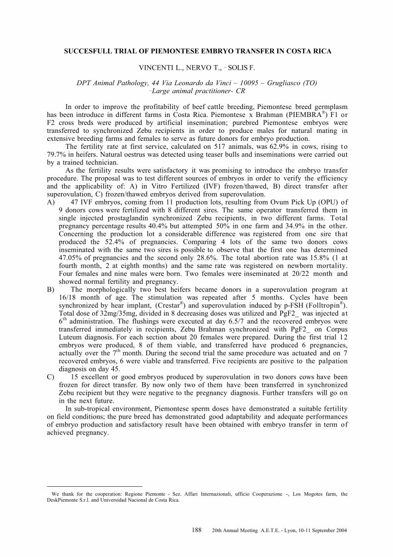
188 20th Annual Meeting A.E.T.E. - Lyon, 10-11 September 2004
SUCCESFULL TRIAL OF PIEMONTESE EMBRYO TRANSFER IN COSTA RICA
VINCENTI L., NERVO T., _ SOLIS F.
DPT Animal Pathology, 44 Via Leonardo da Vinci – 10095 – Grugliasco (TO)_Large animal practitioner- CR
In order to improve the profitability of beef cattle breeding, Piemontese breed germplasmhas been introduce in different farms in Costa Rica. Piemontese x Brahman (PIEMBRA®) F1 orF2 cross breds were produced by artificial insemination; purebred Piemontese embryos weretransferred to synchronized Zebu recipients in order to produce males for natural mating inextensive breeding farms and females to serve as future donors for embryo production.
The fertility rate at first service, calculated on 517 animals, was 62.9% in cows, rising t o79.7% in heifers. Natural oestrus was detected using teaser bulls and inseminations were carried outby a trained technician.
As the fertility results were satisfactory it was promising to introduce the embryo transferprocedure. The proposal was to test different sources of embryos in order to verify the efficiencyand the applicability of: A) in Vitro Fertilized (IVF) frozen/thawed, B) direct transfer aftersuperovulation, C) frozen/thawed embryos derived from superovulation.A) 47 IVF embryos, coming from 11 production lots, resulting from Ovum Pick Up (OPU) of
9 donors cows were fertilized with 8 different sires. The same operator transferred them insingle injected prostaglandin synchronized Zebu recipients, in two different farms. Totalpregnancy percentage results 40.4% but attempted 50% in one farm and 34.9% in the other.Concerning the production lot a considerable difference was registered from one sire thatproduced the 52.4% of pregnancies. Comparing 4 lots of the same two donors cowsinseminated with the same two sires is possible to observe that the first one has determined47.05% of pregnancies and the second only 28.6%. The total abortion rate was 15.8% (1 atfourth month, 2 at eighth months) and the same rate was registered on newborn mortality.Four females and nine males were born. Two females were inseminated at 20/22 month andshowed normal fertility and pregnancy.
B) The morphologically two best heifers became donors in a superovulation program at16/18 month of age. The stimulation was repeated after 5 months. Cycles have beensynchronized by hear implant, (Crestar®) and superovulation induced by p-FSH (Folltropin®).Total dose of 32mg/35mg, divided in 8 decreasing doses was utilized and PgF2_ was injected at6th administration. The flushings were executed at day 6.5/7 and the recovered embryos weretransferred immediately in recipients, Zebu Brahman synchronized with PgF2_ on CorpusLuteum diagnosis. For each section about 20 females were prepared. During the first trial 12embryos were produced, 8 of them viable, and transferred have produced 6 pregnancies,actually over the 7th month. During the second trial the same procedure was actuated and on 7recovered embryos, 6 were viable and transferred. Five recipients are positive to the palpationdiagnosis on day 45.
C) 15 excellent or good embryos produced by superovulation in two donors cows have beenfrozen for direct transfer. By now only two of them have been transferred in synchronizedZebu recipient but they were negative to the pregnancy diagnosis. Further transfers will go onin the next future.
In sub-tropical environment, Piemontese sperm doses have demonstrated a suitable fertilityon field conditions; the pure breed has demonstrated good adaptability and adequate performancesof embryo production and satisfactory result have been obtained with embryo transfer in term ofachieved pregnancy.
We thank for the cooperation: Regione Piemonte - Sez. Affari Internazionali, ufficio Cooperazione -, Los Mogotes farm, theDeskPiemonte S.r.l. and Universidad Nacional de Costa Rica.

189 20th Annual Meeting A.E.T.E. - Lyon, 10-11 September 2004
Notes____________________________________________________________________________________

190 20th Annual Meeting A.E.T.E. - Lyon, 10-11 September 2004
IMPACT OF THE ACCURACY OF THE TIME INTERVAL BETWEEN oFSH INJECTIONSON FOLLICULAR QUALITY IN SUPEROVULATED COWS
VOS P.L.A.M., GROENENDAAL H., VAN GASTEL A.C.T.M., ALGRIANY O., DIELEMAN S.J.
Department of Farm Animal Health, Faculty of Veterinary Medicine, Utrecht University, Yalelaan7, 3584 CL Utrecht, The Netherlands
During superovulation (SO), the variability in ovarian response and number of transferable embryos togonadotrophic stimulation using FSH or eCG within and/or between donor cows has been related to manyinteracting, exogenous and endogenous, factors. In this respect, the use of different SO schemes including theduration and timing of the total dose of injected gonadotrophins have a major impact on the final embryooutcome. Therefore, to validate the impact of the accuracy of the time interval between oFSH injections onfollicular quality we investigated whether one delayed FSH injection may influence the follicular quality inoFSH-stimulated cows using a controlled LH surge SO-procedure.
Synchronized, cyclic HF non-lactating cows (n= 13) were treated for SO. Briefly, a norgestomet earimplant without accompanying injection (Crestar, Intervet International BV, Boxmeer, The Netherlands) wasinserted at Day 9 (oestrus = Day 0) to inhibit the endogenous release of the LH surge, and SO was started at Day10 using oFSH im (Ovagen ICP, Auckland, New Zealand) twice daily during 4 d (total dose 17 ml).Prostaglandin (22.5 mg PG; Prosolvin, Intervet International BV) was administered concomitant with the fifthdose of FSH. Four cows (Experimental group) received a 6.5 h delayed second FSH injection; nine cows servedas controls (Control group). One hour after removal of the ear implants (at 50 h after PG, ie. shortly before thepreovulatory LH surge) the ovaries were collected by laparotomy and all preovulatory-sized follicles (diameter≥10 and ≤15 mm) were counted and immediately thereafter punctured individually to collect follicular fluid (FF;stored at -25ºC) and their concomitant COCs. Moreover, all follicles having a diameter between ≥8 and <10 mmwere counted. Concentrations of oestradiol-17_ (E) and progesterone (P) in FF were estimated by RIA. Follicleswere qualified as ‘normal’ or ‘deviant’ on the basis of steroid hormone concentrations (‘normal’, ‘estrogen active’follicles: > 250 ng/ml); COCs were analysed by light microscopy (‘good’ quality: transparency and more thanfour layers of cumulus investment, ‘bad quality: bold oocytes, complete or partial and no layers of cumuluscells).
None of the animals showed an increase of LH during the norgestomet treatment. The average number offollicles between ≥8 and <10 mm (3.8 follicles per cow) and the total number of preovulatory-sized folliclesstudied were not significantly different between groups (9 ± 1.4 and 12 ± 1.9, respectively; mean ± SEM). Basedon the steroid concentrations estimated in follicular fluid (Table 1), significantly more follicles were classified as‘deviant’ in the experimental than in the control group (82.0% and 34.6%, respectively; RR = 2.4, confidencelimits 1.7-3.3, Chi Square P< 0.001). No morphological differences between groups were observed for thenumber of ‘good’ quality COCs (77.3 and 82.1%, respectively).
Table 1. Steroid concentrations in FF (ng/ml) of preovulatory-sized follicles in oFSH superovulated cows
Steroid oestradiol-17_ ProgesteroneQuality of follicle ‘good’ ‘deviant’ ‘good’ ‘deviant’
C group (9) 541.3 ± 41.2a (51) 141.3 ± 15.8b (27) 41.6 ± 2.3 a (51) 63.9 ± 16.0 a (27)E group (4) 365.2 ± 12.5 a (7) 75 ± 11.5 b (32) 49.1 ± 3.9 a (7) 33.3 ± 1.9 b (32)
Values are mean ± SEM; values within a row with different superscripts are significantly different; (number)
A significant decrease in oestradiol concentration was observed in follicles derived from the E-groupindicating a dramatic effect on follicular function. It is ikely that follicles need continuous gonadotrophinsupport throughout the period of follicular superstimulation until the period of final maturation. Since nomorphological differences for COCs between groups were observed one could speculate whether COCs collectedfrom ‘deviated’ follicles lost their potency to develop into a blastocyst.
In conclusion, the inaccuracy of one delayed FSH injection severely suppresses the functionality of themajority of the gonadotrophin superstimulated follicles. The possible effects of these deviated follicles on thepotential to ovulate or on the developmental competence of the COCs need to be elucidated.

191 20th Annual Meeting A.E.T.E. - Lyon, 10-11 September 2004
Notes____________________________________________________________________________________

192 20th Annual Meeting A.E.T.E. - Lyon, 10-11 September 2004

193 20th Annual Meeting A.E.T.E. - Lyon, 10-11 September 2004
AUTHOR INDEX

194 20th Annual Meeting A.E.T.E. - Lyon, 10-11 September 2004

195 20th Annual Meeting A.E.T.E. - Lyon, 10-11 September 2004
AUTHOR INDEX
ACCARDO C. ...........................................101AERTS J.M.J. ........................................... 93ALBARRACIN J.L....................................137ALBRECHT D. .......................................... 99ALGRIANY O. .........................................171ALI AL AHMAD M.Z. ............................... 95ALM H.....................................................121ALVAREZ I. ............................................161ALVAREZ-VIEJO M. ...............................161ANGUITA B. ............................................113AVERY B. ................................................165BADER M. ........................................119, 151BARIL G..................................................111BECHER D................................................ 99BECKERS J.F. .........................................105BERSINGER I..........................................145BHOJWANI M........................................... 99BOCHENEK M. .......................................115BOLAND M.P...........................................159BOLAND M.P............................................ 78BOLS P.E.J................................. 93, 103, 125BOURGOIN G........................................... 97BRÜSSOW K.-P. ......................................107BRUYAS J.-F............................................141BRUYAS J-F......................................143, 145BURKE L.................................................159BUTLER S.T............................................127BUTLER W.R. .........................................127CALLESEN H. .......................................... 14CAPPAI P.................................................101_E_OVSK_, J. ...................................155, 157CHATAGNON G. ...................................... 95CHEBLOUNE Y. ....................................... 95CHESSA B. ..............................................101CHESSA F. ..............................................101COGNIE Y. ..............................................111COZAR P. ................................................139DAELS P.F........................................141, 143DATTENA M............................................101DE CLERCQ J. ........................................125DE LA FUENTE J. ...................................153DE ROOVER R. .......................................103DEGAND G. .............................................105DENISENKO V. .......................................121DIELEMAN S.J. ................................117, 171DÍEZ C. ...................................................161DOLIGEZ P. ............................................145DUCHAMP G. ............................141, 143, 145DUPONT F. ..............................................111EGERSZEGI I. ........................................107FACAL N. ................................................161
FER G. .................................................... 109FERNÁNDEZ SÁNCHEZ I. ...................... 133FERNÁNDEZ-GONZALEZ R. ........... 149, 153FIENI F. .................................................... 95FOTSING L............................................. 105GALLI C. ................................................ 123GANTEN D....................................... 119, 151GILBERT R.O......................................... 127GIRALDO P. ........................................... 139GÓMEZ E. .............................................. 161GÓMEZ-BRUNET A. ............................... 163GOYACHE F. .......................................... 161GREVE T. .......................................... 14, 165GRIMARD B............................................ 109GROENENDAAL H. ................................ 171GUIGNOT F. .................................... 111, 145GUTIERREZ-ADAN A. ............................ 139GUTIÉRREZ-ADÁN A. ..................... 149, 153HANZALOVA K. ..................................... 129HANZEN C.............................................. 103HAZELEGER W. .................................... 107HERMSEN M. ......................................... 161HORAKOVA J......................................... 129HUMBLOT P...................................... 97, 109HUYART C.............................................. 109IKEDA S. ................................................ 161IZQUIERDO D. ....................................... 113IZQUIERDO M.D. ................................... 137JIMENEZ A...................................... 139, 149JIMÉNEZ A............................................. 153JIMÉNEZ-MACEDO A.R. ........................ 113KANITZ W......................................... 99, 131K_TSKA-KSI__KIEWICZ L. ................... 115KEMP B. ................................................. 107KNIJN H.M. ............................................ 117KRIVOKHARCHENKO A................. 119, 151KUZMINA T............................................ 121LAGUTINA I. .......................................... 123LAKOMÁ, Z............................................ 157LALLOZ J.M. ......................................... 105LANDMAN B. .......................................... 135LARRAT M. .............................................. 95LAZZARI G. ........................................... 123LEIDING C. ....................................... 99, 131LEROY J.L.M.R. ..................................... 125LONERGAN P. ........................................ 159LONERGAN P. .......................................... 78LOPES A.S.............................................. 127LÓPEZ-SEBASTIÁN A. ........................... 163MACHATKOVA M. ................................. 129MARA L. ................................................. 101

196 20th Annual Meeting A.E.T.E. - Lyon, 10-11 September 2004
MARQUANT-LEGUIENNE B....................109MARTENS G............................................131MARTINEZ-BELLO J.D...........................133MAX M. .................................................... 99MERMILLOD P. ...............................111, 145MERTON J.S. ..........................................135MOGAS T. ...............................................137MONTOLIU L..........................................139MORATÓ R. ............................................137MOREIRA P.N. ..........................139, 149, 153MOUSSA M................................141, 143, 145MULLAART E. ........................................135NERVO T..........................................147, 169NIEMANN H. ...........................................117NOHNER H.-P..........................................131OHOU A. .................................................. 97OSTE M.................................................... 93PARAMIO M.T.........................................113PÉREZ-CRESPO M. .................................149PESLAROVA Z. .......................................129PILICHI S................................................101PINTADO B. ............................................149PIRSON J.L. ............................................105PONSART C.............................................. 97PONTER A.A. ..........................................109POPOVA E........................................119, 151POZUETA J. ............................................139PUDILOVÁ, K...................................155, 157PULIDO-PASTOR A. ................................163QUINN K. ................................................159QUINTELA ARIAS L. ..............................133QUINTON H. ............................................ 97RAMIREZ M.A..................................149, 153RÁTKY J. ................................................107REMY B...................................................105
REMY D. ................................................. 109REY R. .................................................... 149RICHARD C. ........................................... 109_ÍHA J.............................................. 155, 157RIZOS D. ................................................ 159RIZOS D. .................................................. 78RODRÍGUEZ A. ...................................... 161ROYO L.J. .............................................. 161RUDOLPH E. ............................................ 99RY_SKA B............................................... 115SALAS A. ................................................ 161SAMIEC M. ............................................. 167SANTIAGO-MORENO J. ......................... 163SCHMIDT M. .......................................... 165SCHNEIDER F. ....................................... 131SCHNEIDER F. ....................................... 107SHOKIN O. ............................................. 121SKRZYSZOWSKA M. ............................. 167SLEZÁKOVÁ, M. ............................. 155, 157SOLIS F. ................................................. 169TOLEDANO-DÍAZ A. .............................. 163TOMEK W. ............................................... 99TORNER H. ............................................ 121VAN DER WEIJDEN G.C......................... 117VAN GASTEL A.C.T.M. .......................... 171VAN GINNEKEN C.J.D.............................. 93VAN SOOM A.......................................... 125VANHOLDER T. ..................................... 125VEJNAR J. ....................................... 155, 157VIDAMENT M. ........................................ 145VINCENTI L. ................................... 147, 169VOS P.L.A.M.................................... 117, 171WADE M. ................................................ 159WRENZYCKI C....................................... 117