208 - Archivos de Medicina del Deporte
56
208 Volume 39(2) March - April 2022 ISSN: 0212-8799 ORIGINAL ARTICLES Performance analysis of women over 55 years on abdominal tests: impact of anthropometry and flexibility Analysis of heart rate variability evolution on table tennis depending in match result Differences in internal and external load between adult and youth players in a friendly match Determining factors of functional physical limitation in patients with myocardial revascularization by acute coronary syndrome REVIEWS Blood flow restriction training on hypertesive subjects: a systematic review Effects of physical activity and eating habits on obesity levels in children between 6 and 12 years old: systematic review
-
Upload
khangminh22 -
Category
Documents
-
view
0 -
download
0
Transcript of 208 - Archivos de Medicina del Deporte

208 Volume 39(2)
March - April 2022
ISSN: 0212-8799
Volu
me
39 N
úm. 2
. Mar
ch -
Apr
il 20
22A
RCH
IVO
S D
E M
EDIC
INA
DEL
DEP
ORT
E 2
08
ORIGINAL ARTICLESPerformance analysis of women over 55 years on abdominal tests: impact of anthropometry and flexibility
Analysis of heart rate variability evolution on table tennis depending in match result
Differences in internal and external load between adult and youth players in a friendly match
Determining factors of functional physical limitation in patients with myocardial revascularization by acute coronary syndrome
REVIEWSBlood flow restriction training on hypertesive subjects: a systematic review
Effects of physical activity and eating habits on obesity levels in children between 6 and 12 years old: systematic review

La Sociedad Española de Medicina del Deporte, en su incesante labor de expansión y consolidación de la Medicina del Deporte y, consciente de su vocación médica de preservar la salud de todas las personas, viene realizando diversas actuaciones en este ámbito desde los últimos años.
Se ha considerado el momento oportuno de lanzar la campaña de gran alcance, denominada CAMPAÑA DE APTITUD FÍSICA, DEPORTE Y SALUD relacionada con la promoción de la actividad física y depor-tiva para toda la población y que tendrá como lema SALUD – DEPORTE – DISFRÚTALOS, que aúna de la forma más clara y directa los tres pilares que se promueven desde la Medicina del Deporte que son el practicar deporte, con objetivos de salud y para la mejora de la aptitud física y de tal forma que se incorpore como un hábito permanente, y disfrutando, es la mejor manera de conseguirlo.
Campaña de aptitud física, deporte y salud
UCAM Universidad Católica San Antonio de Murcia
Campus de los Jerónimos,Nº 135 Guadalupe 30107
(Murcia) - España
Tlf: (+34)968 27 88 01 · [email protected]

Sociedad Española de Medicina del Deporte
Junta de GobiernoPresidente: Pedro Manonelles MarquetaVicepresidente: Carlos de Teresa Galván Secretario General: Luis Franco BonafonteTesorero: Javier Pérez AnsónVocales: Miguel E. Del Valle SotoJosé Fernando Jiménez Díaz Juan N. García-Nieto PortabellaTeresa Gaztañaga Aurrekoetxea
EditaSociedad Española de Medicina del Deporte C/ Cánovas nº 7, local 50004 Zaragoza (España) Tel. +34 976 02 45 09 [email protected] www.femede.es
Correspondencia: C/ Cánovas nº 7, local 50004 Zaragoza (España) [email protected] http://www.archivosdemedicinadeldeporte.com/
Publicidad ESMON PUBLICIDAD Tel. 93 2159034
Publicación bimestral Un volumen por año
Depósito Legal Zaragoza. Z 988-2020ISSN 0212-8799Soporte válido Ref. SVR 389
Indexada en: EMBASE/Excerpta Medica, Índice Médico Español, Sport Information Resource Centre (SIRC), Índice Bibliográfico Español de Ciencias de la Salud (IBECS), Índice SJR (SCImago Journal Rank), y SCOPUS
La dirección de la revista no acepta responsabi-lidades derivadas de las opiniones o juicios de valor de los trabajos publicados, la cual recaerá exclusivamente sobre sus autores.Esta publicación no puede ser reproducida total o parcialmente por ningún medio sin la autoriza-ción por escrito de los autores.Cualquier forma de reproducción, distribución, comunicación pública o transformación de esta obra sólo puede ser realizada con la autoriza-ción de sus titulares, salvo excepción prevista por la ley. Diríjase a CEDRO (Centro Español de Derechos Reprográficos, www.cedro.org) si necesita foto-copiar o escanear algún fragmento de esta obra.
DirectorPedro Manonelles Marqueta
EditorMiguel E. Del Valle Soto
AdministraciónMelissa Artajona Pérez
Adjunto a direcciónOriol Abellán Aynés
Comité EditorialNorbert Bachl. Centre for Sports Science and University Sports of the University of Vienna. Austria. Araceli Boraita. Servicio de Cardiología. Centro de Medicina del Deporte. Consejo Superior de deportes. España. Mats Borjesson. University of Gothenburg. Suecia. Josep Brugada Terradellas. Hospital Clinic. Universi-dad de Barcelona. España. Nicolas Christodoulou. President of the UEMS MJC on Sports Medicine. Chipre. Demitri Constantinou. University of the Witwatersrand. Johannesburgo. Sudáfrica. Jesús Dapena. Indiana University. Estados Unidos. Franchek Drobnic Martínez. Servicios Médicos FC Barcelona. CAR Sant Cugat del Vallés. España. Tomás Fernández Jaén. Servicio Medicina y Traumatología del Deporte. Clínica Cemtro. España. Walter Frontera. Universidad de Vanderbilt. Past President FIMS. Estados Unidos. Pedro Guillén Gar-cía. Servicio Traumatología del Deporte. Clínica Cemtro. España. Dusan Hamar. Research Institute of Sports. Eslovaquia. José A. Hernández Hermoso. Servicio COT. Hospital Universitario Germans Trias i Pujol. España. Pilar Hernández Sánchez. Universidad Católica San Antonio. Murcia. España. Markku Jarvinen. Institute of Medical Technology and Medical School. University of Tampere. Finlandia. Anna Jegier. Medical University of Lodz. Polonia. Peter Jenoure. ARS Ortopedica, ARS Medica Clinic, Gravesano. Suiza. José A. López Calbet. Universidad de Las Palmas de Gran Canaria. España. Javier López Román. Universidad Católica San Antonio. Murcia. España. Alejandro Lucía Mulas. Universidad Europea de Madrid. España. Emilio Luengo Fernández. Servicio de Cardiología. Hospital General de la Defensa. España. Nicola Maffully. Universidad de Salerno. Salerno (Italia). Alejandro Martínez Rodríguez. Universidad de Alicante. España. Estrella Núñez Delicado. Universidad Católica San Antonio. Murcia. España. Sakari Orava. Hospital Universitario. Universidad de Turku. Finlandia. Eduardo Ortega Rincón. Universidad de Extremadura. España. Nieves Palacios Gil-Antuñano. Centro de Medicina del Deporte. Consejo Superior de Deportes. España. Antonio Pelliccia. Institute of Sport Medicine and Science. Italia. José Peña Amaro. Facultad de Medicina y Enfermería. Universidad de Córdoba. España. Fabio Pigozzi. University of Rome Foro Italico, President FIMS. Italia. Yannis Pitsiladis. Centre of Sports Medicine. University of Brighton. Inglaterra. Per Renström. Stockholm Center for Sports Trauma Research, Karolinska Institutet. Suecia. Juan Ribas Serna. Universidad de Sevilla. España. Peter H. Schober. Medical University Graz. Austria. Jordi Segura Noguera. Laboratorio Antidopaje IMIM. Presidente Asociación Mundial de Científicos Antidopajes (WAADS). España. Giulio Sergio Roi. Education & Research Department Isokinetic Medical Group. Italia. Luis Serratosa Fernández. Servicios Médicos Sanitas Real Madrid CF. Madrid. España. Nicolás Terrados Cepeda. Unidad Regional de Medicina Deportiva del Principado de Asturias. Universidad de Oviedo. España. José Luis Terreros Blanco. Director de la Agencia Española de Protección de la Salud en el Deporte (AEPSAD). España. Mario Zorzoli. International Cycling Union. Suiza.
Sociedad Española de Medicina del Deporte


Volumen 39(2) - Núm 208. March - April 2022 / Marzo - Abril 2022
Summary / Sumario
EditorialAgainst sedentary lifestyle, chronicity and bad aging: physical activity/exercise programs Contra el sedentarismo, la cronicidad y el mal envejecimiento, programas de actividad física/ejercicio Cristina Blasco Lafarga ..............................................................................................................................................................................................72
Original articles / OriginalesPerformance analysis of women over 55 years on abdominal tests: impact of anthropometry and flexibility Análisis del desempeño de mujeres mayores de 55 años en test abdominales: impacto de la antropometría y flexibilidad Cláudia E. P. Oliveira, Osvaldo C. Moreira, Dihogo G. Matos, Mauro L. Mazini-Filho, Sandro F. Silva, Eveline T. Pereira, Sylvia C. C. Franceschini, Nádia S. L. Silva, Leonice A. Doimo .......................................................................................................................................75
Analysis of heart rate variability evolution on table tennis depending in match result Análisis de la evolución de la variabilidad de la frecuencia cardíaca antes y después de un partido de tenis de mesa en función del resultado Jon M. Picabea, Jesús Cámara, Javier Yanci .............................................................................................................................................................81
Differences in internal and external load between adult and youth players in a friendly match Diferencias de carga interna y externa entre futbolistas adultos y juveniles en un partido amistoso Jorge Pérez-Contreras, Susana Elgueta-Moya, Rodrigo Villaseca-Vicuña, Esteban Aedo-Muñoz, Bianca Miarka, Pablo Merino-Muñoz ...................89
Determining factors of functional physical limitation in patients with myocardial revascularization by acute coronary syndrome Factores determinantes de la limitación física funcional en pacientes revascularizados por síndrome coronario agudo Laura C. Dávila Landinez, Laura N. Coral Vásquez, Aura N. Carrizales Sánchez, Andrés Mauricio Ariza, Hedilberto Duarte Hernández, Hugo C.D de Souza, Stella V. Philbois, Juan C. Sánchez Delgado .............................................................................................................................95
Reviews / Revisiones
Blood flow restriction training on hypertesive subjects: a systematic review Entrenamiento de restricción del flujo de sangre en sujetos hipertensivos: revisión sistemática Anderson Luiz Bezerra Silveira, Lucas Monteiro de Carvalho, Fabrizio Di Masi, Thiago W.S. Pio, Claudio Melibeu Bentes .................................101
Effects of physical activity and eating habits on obesity levels in children between 6 and 12 years old: systematic review Efectos de la actividad física y hábitos alimenticios en los niveles de obesidad de niños entre 6 y 12 años: revisión sistemática Rubén Palma Fontealva, Pablo Pérez Ojeda, Claudio Hernández-Mosqueira, Fernando Galle Santana, Karoll Ibañez Goudeau ..........................108
Guidelines for authors / Normas de publicación .........................................................................................................................................118

Cristina Blasco Lafarga
72 Arch Med Deporte 2022;39(2):72-73
Editorial
Exercise/physical activity programmes to combat a sedentary lifestyle, chronicity and poor ageing
Contra el sedentarismo, la cronicidad y el mal envejecimiento, programas de actividad física/ejercicio Cristina Blasco Lafarga Directora de UIRFIDE (Unidad de Investigación en Rendimiento Físico y Deportivo, GIUV 2013-140). Departamento de Educación Física y Deportiva de la Universidad de Valencia. Miembro del Comité Científico de la Sociedad Española de Medicina del Deporte
Correspondence Cristina Blasco Lafarga E-mail: [email protected]
doi: 10.18176/archmeddeporte.00075
Nowadays, no one questions the value of physical activity and, essentially, physical exercise programmes, as a health tool and impor-tant factor in social and healthcare policies. Regular and well-planned exercise is prescribed as non-pharmacological treatment for most diseases prevalent in developed societies, regardless of the need for rehabilitation or specific treatment for a particular system that is speci-fically damaged1,2. It is also prescribed as a preventive factor for almost all diseases1,2, regardless of a patient’s gender or age. Scientific evidence strongly demonstrates that, beyond its health value, and much more than medicine, regular exercise is a source of life in our societies that are ageing, sedentary and often extremely socially isolated.
Much more than medicine, movement is life
Today we know that ageing is a gradual, multifactorial process of irreversible and stochastic deterioration, affecting the metabolic, cardiorespiratory and endocrine functions, the immune responses, the inflammatory processes, the performance of the osteo-ligamentous-tendinous-muscular system and, of course, cognitive functions, the regulation of the nervous system and, in general, motor control3,4. The magnitude and persistence of these changes are such that they are accompanied by alterations in the physical condition, motor and mental skills and, in general, the functionality and relationship capacity of older adults.
We are also aware that a sedentary life multiplies any adverse effects such as persistent inflammation and sympathetic nervous system arou-sal, together with an equally persistent vagal inhibition and, in general, autonomic dysfunction. Among other consequences: pathology chroni-city, sarcopenia, emotional disorders, depression, mental illnesses, and, at the end of this continuum, decrease in disability-free life expectancy, multiple pathologies, fragility and dependency5.
A sedentary lifestyle entails systemic disuse or detraining, resulting in the alteration of the neural responses and effort intolerance in these older adults or with limiting pathologies. This make motricity more difficult and movement is perceived as more demanding, more fati-guing. Acting as a feedback loop, psychomotor deterioration induces even greater sedentarism and ends in functional incapacity, autonomic dysfunction and loss of allostasis/resilience, aggravating the already deteriorating effects of the ageing process.
Furthermore, the shortcomings in the motor sphere of a person who feels clumsy, with little or no ability, limit the development of psychomotor and psychosocial skills, hinder efficiency and often involve frustration, with the subsequent lack of motivation to exercise6. Yet again, this is particularly important in the case of older adults and, above all, in those with some type of cognitive or motor skill impairment. These people often age alone and with little resources, requiring powerful interventions in the area of functional improvement and their physical re-education, beyond the improvement or reduction in the symptoms of their pathologies.
It should be noted that physically competent individuals unders-tand and face up to the challenges of motricity, moving with economy and confidence, and can safely and empathetically establish relation-ships with others and with their environment throughout their life6. It should also be remembered that, at the opposite end, adults that feel insecure and are afraid to relate to their environment, hardly go out and frequently end up in unwanted loneliness that is related to the risk of poor health, comorbidity, and, once again, fragility and dependency.
Sustained over time, unwanted loneliness is the absence of in-centives and an accelerated journey towards physical inactivity and pathology, giving another turn of the screw in the poor sedentary ageing loop. If we add to this factors such as a greater risk of falls and/or an increase in mental disorders in these ageing and lonely groups, then the cocktail is served. Let’s do some figures.

Exercise/physical activity programmes to combat a sedentary lifestyle, chronicity and poor ageing
73Arch Med Deporte 2022;39(2):72-73
Exercise is good at all ages
In general, the Consejo Superior de Deportes (CSD - Sports Council of Spain) calculates a saving of between 3 to 15 euro in healthcare expen-diture for each euro invested in physical activity/exercise programmes, emphasising that Sport and Health must go hand in hand, as a strategic approach to social and healthcare policies. The return on investment (ROI) in exercise, is high.
Today there is no doubt of the benefits of increasing physical activity/exercise together with the necessary reduction in sedentary behaviours, given that sufficient amount and intensity of movement demands the complete participation of all our systems, helping to improve/maintain our adaptive responses5,7. When the proposal is demanding and holistic, exercise stands as a powerful neurophysiological tool, capable of ensuring the communication of all our cell organelles and systems in a unified and coordinated way7. And when this same movement occurs within the framework of leisure and active free time, despite its lower physiological impact - resulting from its lower intensity - there are also alternative pathways to lead to the same improvements5. And it continues to be a powerful tool.
In the words of Bennett, Reeves5, the stress pathways are acutely activated and support the physical exertion demanded to move the body through the physical activity, particularly with regard to concerted exercise. This physiological challenge makes it possible to activate the sympathetic branch of the nervous system and the hypothalamic-pituitary axis, and to stop it at the end of the effort, giving rise to a very healthy post-effort “rebound”. This greater specific inflammation is accompanied by a reduction of inflammation at rest, as well as the release of myokines (muscular cytokines) to attract immune cells to repair the tissue damage, also promoting microbiota diversity and gut health5.
The binomial “more physical activity/exercise - less sedentarism” is key to our individual and collective health, with greater impact as we grow older.
As reported by Lazarus, Lord3,there are systems that are age-dependent but are not malleable by exercise (a), systems that are age dependent and are also malleable by exercise (b), systems that are not age dependent but are malleable by exercise (c), and finally, systems that are unaffected by age or exercise (d). The understanding of these balances makes it possible to appreciate that, above the impact of ageing, physical activity/exercise leads to what is known as the healthy ageing phenotype (active, successful); while, at the other end, inactivity/sedentarism con-demns individuals to thepathological ageing phenotype (inactive). The success of the former lies in maintaining the “intrinsic capacity” intact, in other words, the possibility to continue doing those simple things that make a human being feel mentally and physically capable3. In the words of these same authors: walk, think, perceive (see, hear...) and remember.
Far from settling for a minimum number of minutes of physical activity, in its recent guidelines for physical exercise the WHO8 concludes
that older adults must be as physically active as their functional ability allows, adjusting their level of effort to their level of fitness and to their functional abilities so as not to fall below their motor requirements. This also applies to adults with chronic conditions. The WHO also indicates that those adults with greater difficulties may wish to consult a physical activity specialist or health-care professional for advice on the types and amounts of activity appropriate to their individual needs, abilities, functio-nal limitations/complications, medications and overall treatment plan8.
At this stage, the first thing to do is to start, to get going. The second, but no less important, point is to gain the older adult’s loyalty to exercise by finding something that is sufficiently attractive for those who have not taken part in exercise programs for a long time (if indeed they ever have) so that they are sufficiently motivated to allow them to overcome their general disinclination to exercise.
Conquer their fears, personalise the proposal, address the heteroge-neity of older adults and respect their pace.
It will also be extremely important to support them in the process and not leave them alone once the first improvements are made, because age is accompanied by a high level of detrainability, understanding this to be the loss of physical fitness associated with detraining4. Although qualities such as agility or the executive function itself appear to hold up well for a certain amount of time, despite the detraining (negative or regressive effect associated with stopping training), the reduction or even the disappearance of gains is particularly accentuated in the cardiovascular and strength capacities, entailing a loss of physical fitness and the reappearance of fatigue levels4, with a risk of returning to the centre of the loop.
Bibliography 1. Pedersen B, Saltin B. Exercise as medicine–evidence for prescribing exercise as therapy
in 26 different chronic diseases. Scandinavian Journal of Medicine & Science in Sports. 2015;25(S3):1-72.
2. Dishman RK, Heath G, Schmidt MD, Lee I-M. Physical activity epidemiology. Human Kinetics; 2022.
3. Lazarus NR, Lord JM, Harridge SD. The relationships and interactions between age, exercise and physiological function. The Journal of physiology. 2019;597(5):1299-309.
4. Blasco-Lafarga C, Cordellat A, Forte A, Roldán A, Monteagudo P. Short and long-term trainability in older adults: Training and detraining following two years of multicomponent cognitive—physical exercise training. Int J Environ Res Public Health. 2020;17(16):1-16.
5. Bennett JM, Reeves G, Billman GE, Sturmberg JP. Inflammation–Nature’s Way to Efficiently Respond to All Types of Challenges: Implications for Understanding and Managing “the Epidemic” of Chronic Diseases. Frontiers in medicine. 2018;5.
6. Whitehead M. Physical Literacy: Throughout the Lifecourse. Sport RSiPEaY, editor: Routledge, Taylor & Francis e-Library; 2010. 256 p.
7. Blasco-Lafarga C, Cordellat A, Roldán A, Monteagudo P. Reflexiones sobre los beneficios de la actividad Físicodeportiva: la motricidad ordenada al servicio de la felicidad. In: Red_Valenciana_de_Universidades_Públicas_Saludables, editor. Guía para la movi-lidad/actividad física saludable y sostenible en el entorno universitario. 1ª ed. Valencia: Universitat de Valéncia; 2019. p. 33-51.
8. Bull FC, Al-Ansari SS, Biddle S, Borodulin K, Buman MP, Cardon G, et al. World Health Organization 2020 guidelines on physical activity and sedentary behaviour. 2020;54(24):1451-62.

Cristina Blasco Lafarga
74 Arch Med Deporte 2022;39(2):72-73
LLaaccttaattee PPrroo 22 LLTT--11773300
Analizador Instantáneo de Lactato
Importador para España:
c/ Lto. Gabriel Miro, 54, ptas. 7 y 946008 Valencia Tel: 963857395Móvil: 608848455 Fax: 963840104info@bermellelectromedicina.comwww.bermellelectromedicina.com
● Sólo 0,3 μl de sangre
● Determinación en 15 segundos
● Más pequeño que su antecesor
● Calibración automática
● Memoria para 330 determinaciones
● Conexión a PC
● Rango de lectura: 0,5-25,0 mmol/litro
● Conservación de tiras reactivas a temperatura ambiente y
● Caducidad superior a un año
Bermell Electromedicina@BermellElectromedicinaBermell Electromedicina
Performance analysis of women over 55 years on abdominal tests: impact of anthropometry and flexibility
75Arch Med Deporte 2022;39(2):75-80
Original article
Resumen
El objetivo del presente estudio fue evaluar el efecto de las variables las antropométricas y la flexibilidad sobre el desempeño de mujeres mayores de 55 años en protocolos de testes abdominales. La muestra, seleccionada por criterio de accesibilidad, estuvo formada por 20 voluntarias físicamente activas, mayores de 55 años (mediana 61), que participaban en actividades gimnásticas para personas mayores. Cada voluntaria realizó dos pruebas abdominales: flexión parcial del tronco con desliza-miento de las manos de 7,6 cm (P1) y flexión parcial del tronco con las manos en los muslos (P2), ambas ejecutadas con los pies apoyados en el suelo. Para el análisis, se consideró el número de ejecuciones correctas (posición final) en cada prueba, según lo recomendado por los autores. Se realizaron mediciones de masa corporal, flexibilidad, altura, perímetro de cintura y cadera, percepción subjetiva del esfuerzo y cálculos del índice de masa corporal y la relación cintura-cadera. También se evaluó la percepción de esfuerzo abdominal y de malestar o dolor en la región cervical y lumbar. Los resultados mostraron que no hubo asociaciones estadísticamente significativas entre las variables analizadas (Edad: P1: rs = -0,024, p = 0,916; P2: rs = -0,194, p = 0,407; IMC: P1: rs =-0,064, p = 0,792; P2: rs = -0,235, p = 0,327; Perímetro de cintura: P1: rs = -0,143, p = 0,563; P2: rs = 0,027, p = 0,908; Flexibilidad: rs = -0,327, p = 0,169; P2: rs = 0,0009, p = 0,991; Relación cintura/cadera: P1: rs = -0,209, p = 0,371; P2: rs = 0,217, p = 0,353) y el desempeño en las pruebas, y el 35% de las participantes hicieron intentos válidos en P1 mientras que el 45% produjo al menos un intento válido en P2. Se concluyó que ambas pruebas abdominales fueron adecuadas para la muestra estudiada y se pueden aplicar a mujeres adultas y mayores para evaluar su musculatura abdominal.
Palabras clave: Músculos abdominales. Prueba de esfuerzo. Anciano. Antropometría.
Summary
The objective of the present study was to evaluate the effect of anthropometric variables and flexibility on the performance of women aged 55+ years on abdominal test protocols. The sample was composed by 20 physically active volunteers, aged 55 years (median 61), who were participants in gymnastic activities program. Each volunteer performed two abdominal tests: partial trunk flexion with a 7.6 cm sliding of the hands (P1) and partial flexion of the trunk with the hands on the thighs (P2), both executed with the feet resting on the ground. For analysis, the number of correct executions (final position) was consi-dered in each test, as recommended by the authors. Measurements of body mass, flexibility, height, waist and hip perimeters, subjective perception of exertion, and calculations of body mass index and waist-hip ratio were performed. The perception of abdominal effort, and discomfort or pain in the cervical and lumbar region were also evaluated. The results showed that there were no statistically significant associations between the analyzed indicators (Age: P1: rs = -0.024, p = 0.916; P2: rs = -0.194, p = 0.407; BMI: P1: rs =-0.064, p = 0.792; P2: rs = -0.235, p= 0.327; Waist Circumference: P1: rs = -0.143, p = 0.563; P2: rs = 0.027, p = 0.908; Flexibility: r s= -0.327, p = 0.169; P2: rs = 0.0009, p = 0.991; Hip waist ratio: P1: rs = -0.209, p = 0.371; P2: rs = 0.217, p = 0.353) and the performance on the tests. In addition, 35% of the participants made valid attempts on P1 while 45% produced at least one valid attempt on P2. It was concluded that both abdominal tests were adequate for the studied sample and they can be applied to adult and elderly women to assess their abdominal musculature.
Key words: Abdominal muscles. Exercise test.
Elderly. Anthropometry.
Received: 05/05/2021 Accepted: 23/12/2021
Performance analysis of women over 55 years on abdominal tests: impact of anthropometry and flexibility
Cláudia E. P. Oliveira¹, Osvaldo C. Moreira², Dihogo G. Matos3, Mauro L. Mazini-Filho4, Sandro F. Silva5, Eveline T. Pereira¹, Sylvia C. C. Franceschini6, Nádia S. L. Silva7, Leonice A. Doimo8
¹Department of Physical Education. Federal University of Viçosa. Viçosa. Brazil. ²Institute of Biological Sciences and Health. Federal University of Viçosa. Campus Florestal. Florestal. Brazil. ³Cardiovascular and Physiology of Exercise Laboratory. University of Manitoba. Canada. 4Department of Physical Education. Federal University of Juiz de fora. Brazil. 5De-partment of Physical Education. Federal University of Lavras. Lavras. Brazil. 6Department of Nutrition and Health. Federal University of Viçosa. Viçosa. Brazil. 7Institute of Physical Education and Sports. Rio de Janeiro State University. Rio de Janeiro. Brazil. 8Department of Education. Air Force University. Rio de Janeiro. Brazil.
Análisis del desempeño de mujeres mayores de 55 años en test abdominales: impacto de la antropometría y flexibilidad
Correspondence: Claudia Eliza Patrocinio de OliveiraE-mail: [email protected]
doi: 10.18176/archmeddeporte.00076

Cláudia E. P. Oliveira, et al.
76 Arch Med Deporte 2022;39(2):75-80
Introduction
Studies on musculoskeletal fitness in people 55+ years old have shown that its components (especially strength, flexibility, and muscular endurance) are positively associated with health status, i.e., they have a predictive relationship with mortality1-3. In order to evaluate these aspects, fitness tests are generally used to evaluate functional capacity through the assessment of balance, upper and lower limb strength and resistance, displacement velocity, distance traveled, and flexibility4. However, the evaluation of abdominal resistance is not usually studied.
The preservation of abdominal strength during the aging process is fundamental for the support and containment of the abdominal contents, for the maintenance of the normal posture of the pelvis, and for the production and control of the movement of the trunk during flexion and rotation of the trunk5. Moreover, abdominal strength is indi-rectly responsible for the curvature of the lumbar spine and essential for maintaining body posture6,7. Furthermore, weakness of the abdominal muscles is associated with disorders such as ptosis or anterior projec-tion of the abdominal region; difficulty raising the head while supine; impairments in breathing and in performing certain movements such as coughing, vomiting, and sneezing. Also, accentuation of lumbar lordosis, the latter being due to the disproportionate strengthening of the psoas major muscle in relation to the abdominal muscles, which causes low back pain2.
Anthropometrics variables and flexibility undergoes significant physiological changes during the aging process8,9. We hypothesized that this change can affect the performance of people 55+ years old on abdominal tests. However, the relationship between anthropometrics variables, flexibility, and performance is not adequately clarified in the literature.
It is also unclear what factors can interfere with the performance of abdominal exercises, being a problem to be answered. All modifications resulting from the aging process should be considered in the evaluation of the performance of the abdominal muscles of women 55+ years old. Mainly because they can be a source of errors, especially if the perfor-mance of this test is evaluated against the protocols for abdominal tests proposed for young people and adults.
In view of the above, the present study aimed to evaluate the effect of anthropometric variables and flexibility on the performance of women aged 55+ years on two abdominal test protocols. Our hy-pothesis is that anthropometric variables and flexibility will be directly associated with the performance of women aged 55+ years in the proposed abdominal tests.
Materials and method
Participants
The sample of the present study was composed of physically active women. The following inclusion criteria were adopted: participants were required to be 55+ years old; be women; be clinically fit for regular physical exercise; be physically active, practicing physical exercises for at least 1 year with a frequency of 3 times a week; not have any acute or chronic illness that could be affected by the exercise; have experience in
performing abdominal exercises; and consenting freely and voluntarily to perform all study procedures. The exclusion criteria were: presenting bone or joint limitation during the intervention, which prevented the performance of the abdominal exercises; and having used pharma-cological drugs, which could affect the result of anthropometrics and functional assessments.
Those who agreed to participate signed an informed consent form. All procedures were approved by the Ethics Committee on Human Research of the Federal University of Viçosa, according to Resolution 466/2012 of the National Ethics Committee (CONEP), the National Health Council, in accordance with the ethical principles expressed in the Declaration of Helsinki.
The present study is observational and prospective research, with crossover design, being carried out in its entirety, in the Morphophys-iology Laboratory of the Physical Education course of the Federal University of Viçosa (UFV).
Interventions
The data collection was performed on alternate days by two fully trained kinesiologists. The participants were individually evaluated by the same evaluator in a private setting and the order of execution of the tests was determined at random. A warm-up was not allowed before each test was conducted.
Two abdominal tests were used to evaluate abdominal muscle strength, both of which were chosen based on an earlier study10. This choice was made because the participants reported a low rate of dis-comfort or pain in the cervical and/or lumbar spine. The characteristics of the two protocols are described in Table 1 and Figure 1.
Table 1. Abdominal test protocols with respective duration, feet position, form of execution and number of repetitions.
TestsDuration
(min)Feet
PositionExecution Number of
repetitions
Protocol 1 (P1) - partial flexion of the trunk and sliding hands 7.6 cm (13)
1 On the ground
From the extremity of the middle fingers, set 7.6 cm on the ground; in the initial position, flex the trunk and slide hands on the ground trying to reach the 7.6 cm mark.
Higher number of repetitions
Protocol 2 (P2) - partial flexion of the trunk and hands on thighs (14)
6 On the ground
With knees bent between 120-140°; set a mark on the top edge of the patella. From the initial position, flex the trunk and slide hands on thighs until they touch the mark held on the knees.
Cadenced test; 20 repetitions/min; maximum 120 repetitions

Performance analysis of women over 55 years on abdominal tests: impact of anthropometry and flexibility
77Arch Med Deporte 2022;39(2):75-80
Outcomes
On the first day, before the abdominal tests execution, anthropo-metric measures were taken, in each volunteer, (body mass [kg] and height [cm]) to calculate the BMI and also the waist-to-hip ratio11. Flex-ibility was also assessed through the sit-and-reach test (cm)12.
For analysis of performance in the abdominal protocols, the number of correct executions (reaching the correct final position) was considered in each test, as recommended by the developers of these tests13,14. All volunteers complied with the timeframes of the test proto-cols, regardless of whether they performed them correctly or not. In the paced test (P2), a mechanical metronome was used, with a frequency capacity of 40 to 208 beats per minute. The instrument was presented to the participants on the day before the test, to familiarize them with the rhythm to be followed.
At the end of each test, the 20 point Borg scale15 was used to indi-cate the subjective perception of effort (RPE) and a scale of 0 to 4 points was used to verify the perception of discomfort or pain in the cervical and lumbar spine, effort of the abdominal muscles (0 = no discomfort/effort, 1 = very little, 2 = moderate, 3 = intense, 4 = very intense).
Sample size calculation
Considering Wilcoxon test, a priori calculation, an effect size f of 0.8 for abdominal performance10, an α of 5% and a power of 95%. The sample size calculation performed by the G-Power® program at the University of Dusseldorf, indicated that a total sample size of 20 individuals. Thus, the total sample size was of 20 physically active women 55+ years old.
Statistical methods
The data were described as median, minimum and maximum val-ues. The normality was verified by the Shapiro Wilk test. Comparisons between the abdominal tests were made by the Wilcoxon test and the relations among the variables were evaluated by the Spearman correla-tion. Interpretation of the Spearman correlation was assessed according to the following criteria: 0–0.30 negligible, 0.30–0.50 weak, 0.50–0.70 moderate, 0.70–0.90 strong, and 0.90–1.00 very strong16. The effect size was calculated using “r” test for Wilcoxon test. Values were classified as insignificant (<0.20), small (0.20-0.49), medium (0.50-0.79) and large (> 0.79)17. For all analyses, the significance level was set at p < 0.05.
Results
The data of the anthropometric characterization of the participants is shown in Table 2 and the results of the correct execution, the RPE, the perception of discomfort or pain in the cervical and lumbar spine and abdominal muscle effort, and the comparison of the medians of the variables studied in the two tests can be seen in Table 3.
Figure 1. Illustrative pictures of initial and final positions of the five abdominal test protocols used.
Initial position Final position
Initial position Final position
Protocol 214
Table 2. Mean, minimum and maximum values of the variables of anthropometric characterization of the sample.
Median Minimum Maximum
Age (years) 61 55 73
Body mass (kg) 60.8 49.8 80
Height (cm) 153.5 143 160
Body Mass Index (Kg/m²) 26.32 22.56 35.32
Waist circumference (cm) 84.65 71 108
Waist-hip ratio 0.862 0.742 1.023
Flexibility (cm) 31 16 44.3
Table 3. Results (median, minimum and maximum value) of correct performances, rating of perceived exertion (RPE), perception of abdo-minal muscle effort, perception of discomfort or pain in the cervical and lumbar spine and comparison of medians of the P1 test13 and P214.
P1 P2 Med Min Max Med Min Max p* ES
Correct Executions 0 0 48 0 0 23 0,497 0
RPE 12 9 15 13 7 17 0,083 0,07
Perceived abdominal effort 3 1 4 1 0 3 0,320 0,5
Pain in the cervical region 1 1 4 0 0 2 0,147 0,25
Pain in the lower back 1 1 3 0 0 1 0,375 0,33
*p-value obtained through the Wilcoxon test; ES: effect size; RPE: Rating of perceived exertion; Med: Median; Min: Minimum; Max: Maximum.

Cláudia E. P. Oliveira, et al.
78 Arch Med Deporte 2022;39(2):75-80
Comparing the BMI values obtained with the values recommended by WHO18, the participants were in general overweight and also classified as "high risk" for cardiovascular diseases by waist circumference (WC) and waist-to-hip ratio18. The flexibility result, when compared to the reference values18, ranked the group as "good."
The results did not identify any statistically significant differences for all variables measured. Although a greater number of correct executions were obtained for P1, during the tests it was observed that the number of women who achieved at least one correct execution was higher for P2. The associations between test performance and anthropometric pa-rameters are shown in Table 4, and there were no significant correlations.
Discussion
The present study aimed to evaluate the effect of anthropometric variables and flexibility on the performance of women aged 55+ years on two abdominal test protocols. The main results founded were: 1) there no significant differences in performance between the two protocols; 2) the two tests did not present significant differences for RPE, perception of abdominal effort, or perception of pain in the cervical and lumbar region; 3) there no associations between tests performance and anthropometric indicators or flexibility.
The difference in performance between the two tests was not significant (p = 0,497), noting that the median values for both were zero, indicating the difficulty the women had in their performance. This high degree of difficulty can also be verified by the median value of RPE (Table 3), indicating that both constitute exercises that require moderate to intense muscular effort, depending on individual physical fitness. However, they are abdominal tests that do not seem to impose excessive stress on the cervical and lumbar spine, as reported by the participants.
The individual performances for P1, when classified according to MacFarlane19, showed three volunteers with weak performance, one below average and 16 unrated (below "weak"). For P2, the norm proposed by Jetté, Quenneville and Sidney20 shows that three evalu-ated could not be classified because there were no parameters for the age group in question, eight had poor performance, five were below average, two average, and one above average. Therefore, if we consider individual performance only on the basis of the number of correct runs
by strictly observing the test protocols, it can be inferred that, although the elderly women regularly participated in physical activities, their usual exercise program may not develop a compatible abdominal strength level with that required to achieve an expected average result for sex and age group. The physical fitness level of the patients evaluated the stage of aging they are in and the physiological changes resulting from this process, the characteristics of the population used to construct the reference values and tests, and the different physical requirements for performing these abdominal tests can also be related to performance.
Another important aspect is that each test requires distinct physical abilities that also manifest in different ways in the various phases of life. P1 is characterized by being a test where the speed of execution is an important prerequisite, because one must execute the greatest possible number of repetitions in a minute, which quickly leads to muscular fatigue. Logic indicates that 1 minute tests reflect much more than just muscular strength and instead also require muscular endurance21. P1 presented a higher number of correct replicates, but only by four of the women. Another important aspect with respect to P1 is the re-quirement for increased spine flexion to slip the hands on the ground and reach the 7.6 cm mark, which could result in pain in the cervical and lumbar region. This requirement, coupled with the speed required to perform the test, imposed a significant stress on the spine, possibly making the test uncomfortable for some people.
P2 was a cadenced test, lasting six minutes, and more women were able to perform at least one correct repetition than for P1. However, its slow execution requires more time required to support the trunk in relation to P1 and, because of this, requires good conditioning of the abdominal musculature. It has also been shown that P2 is easier to perform than P1 because of the more comfortable position of the arms and greater hip stability, which together do not interfere with the distance traveled by the hands during the exercise22. It also allows for a number of people to be evaluated simultaneously due to the use of the metronome. On the other hand, by controlling the number of repetitions through the metronome (20 per minute), P2 can become long, exhausting, and demotivating, and this should be considered as a possible limiting factor for the application of this test in the elderly. Despite the advantages of P2, another drawback noted was the lack of coordination and rhythm regarding the use of the metronome. The maintenance of the rhythm of movement depends on the integration of the central commands and neuromuscular coordination, particularly of muscle strength and the reaction time23. With aging, there is an in-crease in motor response time resulting from structural and functional modifications of the organism, altering the integrity of the central nervous system, contributing to slower reactions as the person ages. This decline in sensory functions along with the lack of an adequate time of practice with the instrument (metronome) were probably factors that interfered with the results.
In this study, a higher performance was expected on P2 than on P1, but this was not observed. The explanation for this may be the duration of the test, as mentioned earlier. In comparison to younger people, the elderly need to activate a higher percentage of their reduced muscle mass to generate the same force that allows them to perform and sustain exercises that must be performed with a certain intensity and time24. By requiring higher percentages of maximal exercise capacity, muscle
Table 4. Performance ratio in the two abdominal tests with age, body mass index, waist circumference, flexibility and waist/hip ratio.
P1 P2 rs P rs P
Age (years) -0.024 0.916 -0.194 0.407
BMI (Kg/m²) -0.064 0.792 -0.235 0.327
Waist Circumference (cm) -0.143 0.563 0.027 0.908
Flexibility (cm) -0.327 0.169 0.0009 0.991
Hip waist ratio -0.209 0.371 0.217 0.353
BMI: Body Mass Index; rs: Spearman correlation.

Performance analysis of women over 55 years on abdominal tests: impact of anthropometry and flexibility
79Arch Med Deporte 2022;39(2):75-80
fatigue can occur early in response to increased metabolic stress and decreased ability of the neuromuscular system to generate strength, work, or power during repeated muscle contractions25. In addition, localized muscle endurance work requires that a specific muscle group maintain the same strength level for a longer period of time, and in that case, the motivation factor may influence performance on tests aimed at assessing physical aspects. Motivation is an important factor in activities and sports that require high muscular and metabolic activity26.
In addition to the characteristics of each test mentioned above, other aspects that could interfere with the results relate to the degree of prior engagement in physical activity and level of physical fitness of the participants, their lack of familiarity with the tests, difficulties in coordination of movements, and difficulty following the test rhythm dictated by the metronome, among others.
When compared to each other, the two tests did not present statistically significant differences for RPE, perception of abdominal effort, or perception of pain in the cervical and lumbar region (Table 3), suggesting that despite some inadequate performances, with some adaptations they could be used in women 55+ years for the purpose of abdominal muscle testing. Regarding pains in the cervical and lumbar region, it can be said that both tests are satisfactory, since the frequency at which these symptoms appeared was low, in spite of greater reports of discomfort in P1.
In regard to effort, evaluated through RPE, both tests are applicable, since values between 12 and 13 correspond to a low level of difficulty and cardiorespiratory overload15. The low level of perception of reported abdominal effort may be related to the low activation of the abdominal musculature; however, it is important to mention that each test implies a different perception of effort, because the SPE reflects exercise fatigue in a different way27,28, that is, being more sensitive in the active muscles during the performance of power exercises than in central fatigue during the performance of resistance exercises29.
Regarding the relationship between performance on the tests with anthropometric parameters and flexibility, there was also no significant difference between them (Table 4). For BMI and waist-to-hip ratio, there was a tendency for an inverse correlation. This trend indicates that overweight women may be at risk of poor performance30, as well as limiting their involvement in structured physical activities, with a consequent reduction in muscle strength. Similarly, body weight was another variable that did not demonstrate a significant correlation with performance in both protocols. This finding suggests that, for the evaluated group, body weight does not present as a mechanical barrier to the performance of women 55+ years of age on the tests evaluated.
In this study, excess body weight may also be one of the deter-mining factors for the low abdominal exercise performance and, even though the BMI did not present statistical significance (Table 4), it was inversely proportional to the performance of the participants. Even so, it is assumed that the increase in body mass, represented by the fat component, tends to restrict engagement in physical exercises, espe-cially those that require strength and thus reduces muscular fitness and coordination for more complex exercises.
Flexibility did not demonstrate a statistically significant correlation with performance in the abdominal tests used, suggesting that it did not influence performance. In relation to flexibility, the range of motion
of the joint decreases considerably with age, limiting the motion and function of the elderly. A decrease in flexibility along with shortening of the hip flexor muscle and extensor muscles of the back may result in additional mechanical stress on the joints and soft tissues of the lumbar spine and may cause lordosis. Thus, the deep abdominal muscles are essential to support the lumbar spine and strengthening these muscles can reduce back pain31.
Weakening of the abdominal muscles, along with ptosis of the abdomen and shortening of the anteversory muscles of the pelvis, are factors directly related to the degree of flexibility of the lumbar spine and, consequently, directly related to low back pain. This imbalance may limit spinal movements due to impaired levels of adequate flexibility or pain caused by postural deviations. In people with reduced mobility in the articulations of the spine or with shortening of the extensors of the spine, contraction of the abdominal muscles will exert a greater compression force on the intervertebral discs than in individuals with good spine flexibility31. This limitation may interfere with the performance of certain exercises, such as performing trunk flexion during abdominal exercises.
Thus, based on the main findings of this study, we concluded that both abdominal tests evaluated seem to be adequate for women 55+ years, despite the difficulty most of the participants had in performing correct executions, and that anthropometric variables and flexibility did not seem to directly influence the performance.
However, despite the relevance of the results, the present study has some limitations: 1) the use of the same volunteers to carry out the two assessment protocols was a potential limitation. However, this option minimized inter-subject variability; 2) The use of only trained women 55+ years old, which prevents the generalization of the results found here for other populations (for example, men, untrained women, older adults); 3) The use of simple anthropometric measures can also be considered a limitation of the study, since anthropometry has low sensitivity and high variation32. Nonetheless, the use of simple anthropometric meas-ures can increase the ecological validity, and to be applied for different professionals involved with exercise prescription.
Conclusion
Based on the results obtained, it was possible to conclude that both abdominal test protocols were adequate for the sample studied, since they did not present statistically significant differences for performance or perception of pain in the cervical and lumbar region.
Variables such as BMI, body weight, hip waist ratio, and flexibility do not seem to interfere with their performance, at least for the population investigated. In addition, the internal load imposed by the abdominal test protocols, evaluated through SPE, remained within physiological limits, showing that both are safe from the point of view of perceived exertion. On the other hand, the abdominal musculature, evaluated by a perception scale constructed for this study, also did not show any statistically significant results.
Finally, the results indicated that the use of the abdominal test as part of the assessment of musculoskeletal fitness in women 55+ years old proved to be safe, easy to apply, and suitable for this subjects.

Cláudia E. P. Oliveira, et al.
80 Arch Med Deporte 2022;39(2):75-80
Conflict of interest
The authors do not declare a conflict of interest.
Bibliography
1. Chua KY, Lim WS, Lin X, Yuan JM, Koh WP. Handgrip strength and timed up-and-go (TUG) test are predictors of short-term mortality among elderly in a population-based cohort in singapore. J Nutr Health Aging. 2020;24:371-8.
2. Kato S, Murakami H, Demura S, Yoshioka K, Shinmura K, Yokogawa N, Igarashi T, Yone-zawa N, Shimizu T, Tsuchiya H. Abdominal trunk muscle weakness and its association with chronic low back pain and risk of falling in older women. BMC Musculoskelet Disord. 2019;20:273.
3. Laukkanen JA, Voutilainen A, Kurl S, Araujo CGS, Jae SY, Kunutsor SK. Handgrip strength is inversely associated with fatal cardiovascular and all-cause mortality events. Ann Med. 2020;52:109-19.
4. Camara FM, Gerez AG, Miranda MLJ, Velardi M. Elderly functional capacity: types of assessment and trends. Acta Fisiatr. 2008;15:249-56.
5. Kato S, Murakami H, Demura S, Yoshioka K, Shinmura K, Yokogawa N, Igarashi T, Yone-zawa N, Shimizu T, Tsuchiya H. Abdominal trunk muscle weakness and its association with chronic low back pain and risk of falling in older women. BMC Musculoskelet Disord. 2019;20:273.
6. Cuellar WA, Wilson A, Blizzard CL, Otahal P, Callisaya ML, Jones G, Hides JA, Winzenberg TM. The assessment of abdominal and multifidus muscles and their role in physical function in older adults: a systematic review. Physiotherapy. 2017;103:21-39.
7. Raats J, Lamers I, Merken I, Boeckmans J, Soler BM, Normann B, Arntzen EC, Feys P. The content and effects of trunk rehabilitation on trunk and upper limb performance in people with Multiple Sclerosis: a systematic review. Eur J Phys Rehabil Med. 2021. doi: 10.23736/S1973-9087.21.06689-2.
8. Ribeiro MCM, Sañudo A, Simões EJ, Ramos LR. Relationship between physical activity and functional capacity change in aged cohort in São Paulo, Brazil. Rev Bras Enferm. 2021;75:e20200837.
9. Matos DG, Mazini Filho ML, Moreira OC, Oliveira CEP, Venturini GR, Silva-Grigoletto ME, Aidar FJ. Effects of eight weeks of functional training in the functional autonomy of elderly women: a pilot study. J Sports Med Phys Fitness. 2017;57:272-7.
10. Oliveira CEP, Moreira OC, Matos DG, Pereira ET, Franceschini SCC, Silva NSL, Doimo LA. Hemodynamic responses and physical perceptions how indicators of adequacy of abdominal test protocols for women in middle and old age: a pilot study. Motricidade. 2017;13:2-11.
11. Esparza-Ros F, Vaquero-Cristóbal R, Marfell-Jones M. International Society for the Advancement of Kinanthropometry (ISAK). International standards for anthropometric assessment. Adelaide, Australia: National Library of Australia; 2019.
12. Kim WM, Seo YG, Park YJ, Cho HS, Lee CH. Effect of Different Exercise types on the cross-sectional area and lumbar lordosis angle in patients with flat back syndrome. Int J Environ Res Public Health. 2021;18:10923.
13. Robertson LD, Magnusdottir H. Evaluation of criteria associated with abdominal fitness testing. Res Q Exerc Sport. 1987;58:355-9.
14. Sidnei K, Jetté M. The partial curl-up to assess abdominal endurance: age and sex standards. Sports Training Med Rehabil. 1990;2:47-56.
15. Borg G. Perceived exertion as an indicator of somatic stress. Scand J Rehabil Med. 1970; 2:92-8.
16. Hinkle DE, Wiersma W, Jurs SG. Applied statistics for the behavioral sciences. 2003.
17. Cohen J. Statistical power analysis. Curr Dir Psychol Sci. 1992;1:98–101.
18. World Health Organization (WHO). Obesity: preventing and managing the global epide-mic. Report of a WHO, consultation on obesity. Technical Report Series. Geneva, 2000.
19. MacFarlane PA. Out with the sit-up, in with the curl-up! J Physic Educ Recreat Dance. 1993;64:62-6.
20. Jetté M, Quennelille J, Sidney K. Fitness testing and counseling in health promotion. Can J Sport Sci. 1992;17:194-8.
21. Vera-Garcia, FJ, Flores-Parodi B, Elvira JL, Sarti MA. Influence of trunk curl-up speed on muscular recruitment. J Strength Cond Res. 2008;22:684-90.
22. Knudson, D, Johnston D. Validity and reliability of a bench trunk-curl test of abdominal endurance. J Strength Cond Res. 1995;9:165-9
23. Bisio A, Faelli E, Pelosin E, Carrara G, Ferrando V, Avanzino L, Ruggeri P. Evaluation of explicit motor timing ability in young tennis players. Front Psychol. 2021;12:687302.
24. Jiang CH, Ranganathan VK, Siemionow V, Yue GH. The level of effort, rather than muscle exercise intensity determines strength gain following a six-week training. Life Sci. 2017;178:30-4.
25. Kataoka R, Vasenina E, Hammert WB, Ibrahim AH, Dankel SJ, Buckner SL. Is there evidence for the suggestion that fatigue accumulates following resistance exercise? Sports Med. 2021. doi: 10.1007/s40279-021-01572-0.
26. Jurgelis M, Chong WB, Atkins KJ, Cooper PS, Coxon JP, Chong TT. Heightened effort discounting is a common feature of both apathy and fatigue. Sci Rep. 2021;11:22283.
27. Cotter JA, Garver MJ, Dinyer TK, Fairman CM, Focht BC. Ratings of perceived exertion during acute resistance exercise performed at imposed and self-selected loads in recreationally trained women. J Strength Cond Res. 2017; 31: 2313-8.
28. Hess TM, Knight RC. Adult Age Differences in the effects of chronic mental fatigue on task-related fatigue, appraisals, and performance. Motiv Sci. 2021;7:122-32.
29. Moreira OC, Cardozo RMB, Vicente MA, Matos DG, Mazini Filho ML, Guimarães MP, Silva SF, Jeffreys I, Aidar FJ, Oliveira CEP. Acute effect of stretching prior to resistance training on morphological, functional and activation indicators of skeletal muscle in young men. Sport Sci Health. 2021. doi: 10.1007/s11332-021-00793-0.
30. Patiño-Villada FA, González-Bernal JJ, González-Santos J, de Paz JA, Jahouh M, Mielgo-Ayuso J, Romero-Pérez EM, Soto-Cámara R. Relationship of body composition with the strength and functional capacity of people over 70 years. Int J Environ Res Public Health. 2020;17:7767.
31. Hayden JA, Ellis J, Ogilvie R, Malmivaara A, van Tulder MW. Exercise therapy for chronic low back pain. Cochrane Database Syst Rev. 2021;9:CD009790.
32. Moreira OC, Alonso-Aubin DA. Métodos de evaluación de la composición corporal: una revisión actualizada de descripción, aplicación, ventajas y desventajas. Arch Med Deporte. 2015;32:387-94.

Analysis of changes in heart rate variability before and after a table tennis match depending on the outcome
81Arch Med Deporte 2022;39(2):81-88
Original article
Resumen
El objetivo del estudio fue analizar el comportamiento de la variabilidad de la frecuencia cardíaca (VFC) de jugadores de tenis de mesa antes y después de un partido ateniendo al resultado (ganar o perder). Se midió la VFC antes (PRE) y después (POST) del partido a 21 jugadores de tenis de mesa en un total de 30 partidos. No se observaron diferencias significativas ni en el PRE ni en el POST en función del resultado. Se observó un descenso (p < 0,05) en la media de los intervalos RR (media RR), la desviación estándar de los intervalos R-R (SDNN), el logaritmo natural de la raíz cuadrada del valor medio de la suma de las diferencias al cuadrado de todos los intervalos R-R (LnRMSSD), el porcentaje de los intervalos RR consecutivos que discrepan en más de 50 ms entre sí (pNN50), el eje transversal (SD1) y longitudinal (SD2) del diagrama de Poincaré en el POST con respecto al PRE en ambos grupos. Sin embargo, las variables de la banda de baja frecuencia expresada en fuerza absoluta (LF Power), la banda de alta frecuencia expresadas en fuerza absoluta (HF Power) y fuerza normalizada (HF Power) mostraron tendencias distintas en función del resultado (p < 0,05). Los resultados muestran un descenso en la VFC después de disputar un partido de tenis de mesa independientemente del resultado del partido en el dominio del tiempo y en variables no lineales. No obstante, el dominio de la frecuencia muestra una tendencia distinta en función del resultado.
Palabras clave: Fatiga. Tenis de mesa.
Sistema nervioso autónomo. Competición. Rendimiento.
Summary
The aim of this study was to compare heart rate variability (HRV) indices before and after a table tennis match, depending in match result. HRV indices were measured before (PRE) and after (POST) match periods to 21 table tennis players (21.86 ± 8.34 yr) in 30 matches. No significant differences were found neither in PRE nor in POST measures comparing winners and losers. A significantly lower value (p < 0.05) was found in mean of RR intervals (mean RR), standard deviation of RR intervals (SDNN), the natural logarithm transform of the root mean square of successive differences between normal heartbeats (LnRMSSD), relative number of successive RR interval pairs that differ more than 50 ms (pNN50), cross (SD1) and longitudinal (SD2) axis of Poincaré plot comparing POST values with PRE values. Nevertheless, low frequency index expressed in absolute power (LF Power) and high frequency indices expressed in absolute power (HF power) and normalised power (HF Power) showed different trends depending on the results (p < 0.05). The obtained results show a HRV decrease after table tennis match re-gardless the match result, in both time domain and non-linear indices. However, frequency domain indices show a different trend depending on the match outcome.
Key words: Fatigue. Table tennis.
Autonomous nervous system. Competition. Performance
Received: 18/05/2021 Accepted: 23/12/2021
Analysis of changes in heart rate variability before and after a table tennis match depending on the outcome
Jon M. Picabea, Jesús Cámara, Javier YanciDepartamento de Educación Física y Deportiva. Facultad de Educación y Deporte. Universidad del País Vasco (UPV-EHU). Vitoria-Gasteiz.
Análisis de la evolución de la variabilidad de la frecuencia cardíaca antes y después de un partido de tenis de mesa en función del resultado
Correspondence: Javier YanciE-mail: [email protected]
doi: 10.18176/archmeddeporte.00077

Jon M. Picabea, et al.
82 Arch Med Deporte 2022;39(2):81-88
Introduction
Table tennis is an intermittent racket sport, alternating short cycles of high-intensity effort with incomplete recovery periods1–3. Due to the demands of competition, table tennis is considered a mixed sport, with a continuous use of both the aerobic and anaerobic systems4. The aerobic system is the main source of energy during matches, allowing adequate recovery during interruptions that occur during the game2,3. On the other hand, due to the continuous high-intensity actions that occur during matches, the anaerobic system is essential in periods of exertion2,3. In addition to the related physical demands, table tennis is characterized by requiring athletes to perform, different technical actions—in a coordinated manner and at maximum speed—with the upper limbs after having made short and fast displacements with con-tinuous changes of direction2. At the same time, players must deploy a large repertoire of movements to select the correct shot as quickly as possible based on their opponent’s actions5. In addition to the high physical demands, continuous tactical decisions, and the need to ac-curately execute different technical actions, players are also subject to a high cognitive demand and a high level of mental stress6. This form of sport is, therefore, one with high physical and psychological demands2,6.
Previous studies have shown that both physical and psychological demands affect the state of the autonomic nervous system (ANS)7. Dur-ing exercise, increases in intensity entail increases in sympathetic activity and decreased parasympathetic activity, resulting in an increased heart rate (HR)8,9. Heart rate variability (HRV) has been used to understand the activation of the ANS, both in individual and collective sports10,11. HRV is a non-invasive tool that shows the variation in the time elapsed between successive beats by analyzing R-R intervals, thereby allowing an analysis of ANS activity and, thus, showing the activation level of the sympathetic and parasympathetic nervous system12,13. In this sense, HRV analysis allows one to observe the response of the ANS in different exercise situations9,14. The variables used to measure HRV are based on time-domain, frequency-domain, and non-linear variables15. The pa-rameters commonly used in time-domain analysis are the square root of the mean squared difference of all successive R-R intervals (RMSSD) and the standard deviation of consecutive R-R periods (SDNN)16. These variables analyze HR variations, owing to which they depend on it15. To isolate the analysis of HRV for each participant’s HR, and thus to be able to compare different situations independently of the HR, the natural log-transformed root mean square of successive R-R intervals (LnRMSSD)17 was used. On the other hand, frequency-domain analysis breaks down the R-R signal into different components, thus showing: i) the high frequency band (HF), which shows parasympathetic nervous system activity, ii) the low frequency band (LF), affected by both the sympathetic and parasympathetic nervous system and iii) the LF/HF ratio, which reflects sympathetic dominance when it has a high value17. However, it has previously been observed that breathing patterns affect frequency-domain values, which makes it difficult to interpret
results7,18. In addition, analyses with non-linear HRV parameters show parasympathetic modulation without the involvement of breathing19. Specifically, the parameters used are SD1, which reflects parasympa-thetic activity in the heart, and SD2, which reflects both sympathetic and parasympathetic activity20.
In light of the information obtained on ANS activation, HRV has been researched in different training and competition situations in individual and collective sports8,9,21,22. Several studies have analyzed HRV variation before and after different physical exertion with the aim of assessing the influence of physical activity on HRV9,17,22,23. Specifically, in bad-minton players, a sport similar in structure to table tennis, decreased post-exercise values of SDNN and RMSSD have been observed in com-parison to pre-exercise values, thus showing an increase in the activity of the sympathetic nervous system induced by accumulated effort8,22,24. In addition, a recent study—also with badminton players—analyzed pre-post competition HRV based on competitive outcome (win or lose), to observe whether the competitive outcome could affect c hanges in HRV25. In that study, it was observed that the players who won the match had higher values in the LF/ HF ratio and a lower magnitude of the HF and LF variables than the losing players, thus showing a greater sympathetic activation of the ANS in the winners. However, no significant differences were o btained i n t ime-domain p arameters o r non-linear variables. Despite the significance of possible changes in HRV before and after playing a table tennis match according to the outcome, there are no studies that analyze this aspect. This analysis would provide more exhaustive knowledge of competitive demand and the behavior of the ANS in table tennis as relates to winning or losing the match, since the match’s outcome seems to affect the HRV25.
Therefore, the objectives of the present study were, firstly, to analyze the HRV behavior of table tennis players before and after playing a match according to their outcomes (win or lose) and, secondly, to analyze if match duration affects HRV.
Materials and method
Participants
The sample was composed of 21 table tennis players (21.86 ± 8.34 years, 1.73 ± 0.08 m, 64.09 ± 13.39 kg and 21.46 ± 4.38 kg·m-2), who competed in one of the official categories of table tennis, both nationally and provincially in the autonomous community of the Basque Country. The criteria for inclusion in the study were to have a valid license issued by the Spanish Table Tennis Federation and not to be injured or reco-vering from an injury at the time of the research study. All participants had at least two years’ experience in table tennis competitions. All were informed of the objectives and procedures of the research study and voluntarily agreed to be part of it, signing an informed consent form. In the case of underaged players, the informed consent form was also signed by their parents or legal guardians. The study was conducted with the consent of the club to which they belonged. All procedures followed

Analysis of changes in heart rate variability before and after a table tennis match depending on the outcome
83Arch Med Deporte 2022;39(2):81-88
the guidelines set out by the Declaration of Helsinki (2013), respecting the provisions of the Organic Law on the Protection of Personal Data (LOPDCP). Likewise, the study was approved by the Ethics Committee for Research with Human Beings (CEISH, Nº 2080310018-INB0059) of the University of the Basque Country (UPV/EHU).
Procedure
We analyzed 30 best-of-5 table tennis matches played outside the competitive season, thereby obtaining 60 records. In each of the matches, the outcome obtained by the players (win or lose) was noted. The participants’ HRV was measured before and after the matches. The HRV was recorded for 8 minutes PRE and POST match, taking into account the last 3 minutes PRE and the first 3 minutes POST. Each participant was instructed to lie on their backs for 8 minutes before26,27 and after the match8,9,23,28. Pre-match logs were made before the 2-minute warm-up and POST match logs were made immediately after the end of the match. A warm-up was carried out prior to each match, which consisted of 2 minutes of forehand and backhand exchanges, including topspin hitting.
Measurement
HRV analysis: The heart rate signal was measured using a chest band with Bluetooth Smartyse technology, which was recorded on a Polar monitor (V800, Kempele, Finland). The data obtained were transferred to a computer using a specific software (Polar Flow, Kempele, Finland) and were exported for HRV analysis using the Kubios v3.0 software (Biosignal Analysis and Medical Imaging Group at the Department of Applied Physics, University of Kuopio, Kuopio, Finland).
The time-domain parameters obtained were the following: i) the mean R-R interval (Mean RR), ii) the standard deviation of the R-R intervals (SDNN) which describes both alterations in the sympathetic and parasympathetic system; (iii) the mean heart rate (Mean HR); (iv) standard deviation of heart rate (STD HR); (v) the minimum recorded heart rate (Min HR); vi) the maximum recorded heart rate (Max HR); vii) log-transformed root mean square of successive R-R intervals (LnRMSSD), which reflects the variance between HR beats and estimates vagal changes ; and viii) the percentage of successive R-R intervals more than 50 milliseconds apart (pNN50), which has been observed to correlate with changes in the parasympathetic nervous system and RMSSD29. The above parameters quantify the amount of HRV observed in the monitoring periods29.
In regard to frequency-domain parameters, which show the contribution of both the sympathetic and parasympathetic nervous systems, the following were recorded: i) power peaks between 0.04-0.15 Hz (Low frequency (LF)), ii) power peaks between 0.15-0.40 Hz (High frequency (HF)) and iii) the ratio between LF and HF (LF/LF), high values of which are associated with the sympathetic system domain17. These values analyze the frequency with which the distance of the R-R interval changes17, drawing measurements in three different units; i) absolute force (ms2); (ii) logarithmic force (log) (iii) normalized force (u.n.).
The following non-linear parameters were analyzed: i) the transverse axis of the Poincaré diagram (SD1), which analyzes HRV in the short term and is an indicator of sympathetic activity29; ii) the longitudinal axis of the Poincaré diagram (SD2), which analyzes HRV in the long term, correlates with LF, and is an indicator of parasympathetic activity29; and iii) the SD2/SD1 ratio used to analyze autonomous balance and the balance between sympathetic and parasympathetic activity29.
Statistical analysis
The results are shown in mean and standard deviation (SD). Data standardization was analyzed using the Shapiro-Wilk test, observing that the data did not display a normal distribution. The Mann-Whitney U test was used to analyze the differences between players who won and those who lost at both pre and post times. On the other hand, the Wilcoxon test was used to analyze independent differences between the PRE match and POST match values in each of the groups. The percentage difference (Δ. %) was calculated in each case using the following formula: Δ. (%) = [(mean POST – mean PRE) / mean PRE] x 100. The effect size (ES) was calculated both for the differences between groups at each moment and for the differences between the PRE and POST in each of the groups30. Effect sizes less than 0.2, between 0.2 and 0.5, between 0.5 and 0.8 and above 0.8 were considered trivial, low, moderate and high, respectively. The relationship between the duration of the matches and the different HRV variables was analyzed using the Spearman correlation coefficient (r). The correlations obtained were considered high when the absolute value was between 1 and 0.70, moderate between 0.69 and 0.50, low between 0.49 and 0.20, and very low between 0.19 and 0.0931. Statistical significance was established at p < 0.05. Statistical analysis was performed with the Statistical Package for Social Sciences program (version 23.0, SPSS® Inc. Chicago, IL, USA).
Results
Table 1 shows the results obtained for the HRV time-domain values in the PRE match and POST match, by both the players who won and lost the match. Neither the PRE values nor the POST values of any of the HRV time-domain variables showed significant differences between the players who won the match and those who lost (p > 0.05, ES = -0.4 to 0.28, low). The mean parameters RR, SDNN, LnRMSSD and pNN50 showed a significant decrease in the POST match with respect to the PRE (p < 0.05, ES = -0.44 to -2.26, moderate to high) both in the group of players who won and those who lost the match. However, Mean HR, Min HR, and Max HR showed a significant increase in the POST match with respect to the PRE (p < 0.05, ES = 1.25 to 1.7, high) in both groups. No significant differences were observed between PRE and POST in the STD HR variable in either group (p > 0.05, ES = 0.1 to 0.12, trivial).
Table 2 shows the HRV frequency-domain values obtained by both the players who won and those who lost the match, in the PRE and POST match. Neither the PRE values nor the POST values of any

Jon M. Picabea, et al.
84 Arch Med Deporte 2022;39(2):81-88
of the frequency-domain variables showed significant differences
between the players who won the match and those who lost (p > 0.05,
ES = -0.66 to 0.53, moderate). The variables LF Power (log) and HF Power
(log) showed a significant decrease in the POST with respect to the
PRE split values (p < 0.05, ES = -0.43 to -0.82, moderate to high) in both
the players who won and those who lost the match. However, the LF
Power (ms2), HF Power (ms2) and HF Power (u.n.) displayed a different
trend in both groups. While the LF Power (ms2) in the group that won
the match decreased significantly in the POST with respect to the PRE
(p < 0.05, ES = -0.45, moderate), it increased significantly (p < 0.05, ES = 0.02,
trivial) for the players who lost the match. As for HF Power (ms2) and HF
Power (u.n.), no significant changes were observed in the POST match
with respect to the PRE match in the group that won (p > 0.05, ES = -0.03
to 0.07, trivial), while the group that lost the match displayed a significant
decrease (p < 0.05, ES = -0.18 to -0.51, trivial to moderate). With respect to
LF Power (u.n.) no significant changes were observed between the PRE
and the POST match in the group that won (p > 0.05, ES = 0.03, trivial),
while the group that lost the match displayed a significant increase
Table 1. Descriptive parameters of heart rate variability in the pre-match (PRE) and post-match (POST) time domain, categorized by match outcome (win or lose).
PRE POST Δ. (%) ES
Average RR (ms) WIN 784.81 ± 126.01 591.59 ± 90.04** -24.62 -2.15
LOSE 771.68 ± 125.02 574.96 ± 87.12** -25.49 -2.26
Δ. (%) -1.67 -2.81
ES -0.11 -0.19
SDNN (ms) WIN 41.48 ± 16.47 31.97 ± 21.76** -22.92 -0.44
LOSE 38.79 ± 12.96 29.23 ± 20.64** -24.64 -0.46
Δ. (%) -6.49 -8.57
ES -0.21 -0.13
Mean HR (beats/min) WIN 78.12 ± 11.06 103.60 ± 15.02** 32.62 1.7
LOSE 79.49 ± 11.39 106.85 ± 17.54** 34.42 1.56
Δ. (%) 1.75 3.13
ES 0.12 0.18
STD HR (beats/min) WIN 5.38 ± 2.34 5.89 ± 4.09 9.44 0.12
LOSE 5.29 ± 1.49 5.62 ± 3.21 6.24 0.1
Δ. (%) -1.78 -4.65
ES -0.06 -0.09
Min HR (beats/min) WIN 66.34 ± 7.32 85.48 ± 12.13** 28.86 1.58
LOSE 68.17 ± 10.15 89.84 ± 17.31** 31.8 1.25
Δ. (%) 2.76 5.1
ES 0.18 0.25
Max HR (beats/min) WIN 93.93 ± 16.86 131.42 ± 23.86** 39.91 1.57
LOSE 96.27 ± 14.04 138.08 ± 24.12** 43.44 1.73
Δ. (%) 2.48 5.07
ES 0.17 0.28
LnRMSSD (ms) WIN 3.37 ± 0.56 2.92 ± 0.75** -13.31 -0.6
LOSE 3.25 ± 0.46 2.75 ± 0.71** -15.33 -0.71
Δ. (%) -3.43 -5.69
ES -0.25 -0.24
pNN50 (%) WIN 12.36 ± 16.75 6.21 ± 11.73** -49.78 -0.52
LOSE 8.75 ± 11.16 3.13 ± 7.72** -64.27 -0.73
Δ. (%) -29.16 -49.61
ES -0.32 -0.4
Mean RR: Mean of the R-R interval; SDNN: Standard deviation of R-R intervals; Mean HR: Mean heart rate; STD HR: Standard deviation of heart rate; Min HR: Minimum heart rate; Max HR: Maximum heart rate; LnRMSSD: log-transformed root mean square of successive R-R intervals; pNN50: Percentage of successive R-R intervals that exceed more than 50 milliseconds with each other; ES: Effect size; Δ. (%): Percentage of variation.
**p < 0.01 significant differences with respect to the PRE.

Analysis of changes in heart rate variability before and after a table tennis match depending on the outcome
85Arch Med Deporte 2022;39(2):81-88
(p < 0.05, ES = 0.51, moderate). No significant differences were observed between PRE and POST in either group in the variable LF/HF Power (ms2) (p > 0.05, ES = -0.1 to -0.18, trivial).
Table 3 shows the results obtained by both the players who won the match and those who lost in the PRE match and POST match in terms of the non-linear HRV values. Neither the PRE values nor the POST values of any of the non-linear variables showed significant differences when comparing the players who won the match with those who lost (p > 0.05, ES = -0.37 to 0.2, trivial to low). SD1 and SD2 showed a significant decrease in the POST with respect to the PRE (p < 0.05, ES = -0.38 to -0.48, low) in both the players who won and lost the match. No difference was observed between PRE and POST in either group in SD2/SD1 (p > 0.05, SD = 0.24 to 0.28, low).
There was no significant association between match duration and HRV variables in either PRE or POST match in any of the groups. Signifi-cant correlations were only found between match duration and Δ. (%) Min HR in the group that won (r = 0.375, p < 0.05) and in the group that lost the match (r = 0.479, p < 0.01).
Discussion
The objective of this study was to analyze the HRV behavior of table tennis players before and after playing a match, taking into account the outcome (win or lose). HRV is a useful and non-invasive tool that allows one to analyze the behavior of the ANS12,32 and has previously been used to analyze over-training states, to better gauge adjustments to training,
Table 2. Descriptive parameters of heart rate variability in the pre-match (PRE) and post-match (POST) frequency domain, categorized by match outcome (win or lose).
PRE POST Δ. (%) ES
LF Power (ms2) WIN 1250.28 ± 1263.59 756.09 ± 1099.08** -39.53 -0.45
LOSE 948.03 ± 836.46 998.96 ± 2500.83** 5.37 0.02
Δ. (%) -24.17 32.12
ES -0.36 0.1
LF Power (log) WIN 6.74 ± 0.89 6.06 ± 1.02** -10.15 -0.67
LOSE 6.55 ± 0.78 5.69 ± 1.54** -13.22 -0.56
Δ. (%) -2.86 -6.18
ES -0.25 -0.24
LF Power (u.n.) WIN 68.02 ± 15.03 68.49 ± 18.78 0.69 0.03
LOSE 68.79 ± 14.47 76.19 ± 14.53** 10.76 0.51
Δ. (%) 1.12 11.24
ES 0.05 0.53
HF Power (ms2) WIN 657.82 ± 935.46 816.72 ± 2431.46 24.16 0.07
LOSE 439.21 ± 487.93 297.09 ± 789.74** -32.36 -0.18
Δ. (%) -33.23 -63.62
ES -0.45 -0.66
HF Power (log) WIN 5.87 ± 1.03 5.17 ± 1.63** -12.01 -0.43
LOSE 5.66 ± 0.88 4.36 ± 1.58** -22.95 -0.82
Δ. (%) -3.68 -15.65
ES -0.25 -0.51
HF Power (u.n.) WIN 31.91 ± 14.99 31.44 ± 18.76 -1.49 -0.03
LOSE 31.13 ± 14.44 23.71 ± 14.45** -23.83 -0.51
Δ. (%) -2.45 -24.58
ES -0.05 -0.53
LF/HF Power (ms2) WIN 3.32 ± 2.51 3.03 ± 1.63 -8.72 -0.18
LOSE 4.06 ± 2.89 3.78 ± 2.69 -6.67 -0.1
Δ. (%) 22.31 24.93
ES 0.26 0.28
LF: Low frequency; HF: High frequency; LF/HF: Ratio between LF and HF; Power (ms2): Absolute force; Power (log): Logarithmic force; Power (u.n.): Normalized force; ES: Effect size; Δ. (%): Percentage of variation.**p < 0.01 significant differences with respect to the PRE.

Jon M. Picabea, et al.
86 Arch Med Deporte 2022;39(2):81-88
and to quantify the level of pre-competitive stress23,33,34. These aspects allow for one to more adequately plan training strategies to improve sports performance. Although HRV analysis has previously also been used to compare pre- and post-match values in various sports9,21,35 and also in racket sports such as badminton8,22, this type of analysis has not been performed on table tennis players. In addition, the present work is the first study in which the changes in HRV before and after playing a match has been analyzed based on the sports outcome (win or lose) in table tennis, having found only one study in this line in badminton25. Analyzing HRV before and after matches makes it possible to observe changes in the sympathetic-parasympathetic balance, thus showing the athlete’s state of fatigue9. Moreover, the differentiated analysis depending on the outcome may be relevant because winning or los-ing the match can generate different levels of fatigue, thus affecting the activation of the ANS25. In this sense, this analysis provides more exhaustive knowledge of competitive demand in a differentiated way between those who win and lose the game.
HRV time-domain variables have been used in other racket sports, such as badminton, to analyze post-match fatigue8,22,24, showing a decrease in the values of SDNN and pNN50, while an increase in HR variables is observed, possibly related to increased fatigue17. The results of this study showed a decrease in the mean RR, SDNN, LnRMSSD and pNN50 of the POST values with respect to the PRE values, both in the players who won and in those who lost the match. On the contrary, there was an increase in the mean HR, Min HR and Max HR in the POST with respect to the PRE in both groups. These results coincide with those obtained in previous studies8,24,36, which showed a decrease in time-domain variables and an increase in HR variables, associated with an increase in competitive fatigue. In addition, this research provides
differentiated information as it takes into account the match outcome. In this way, it coincides with the results obtained in a previous study carried out with badminton players25. Those authors found decreases in time-domain variables of the POST values with respect to the PRE in badminton matches, both in the players who won and in those who lost, without finding significant differences between the groups25. Those results coincide with those obtained in the present study since the HRV time-domain variables showed no differences between the winners and losers in their change in HRV between PRE and POST. The results obtained suggest that the level of fatigue may have been similar in both groups. The absence of differences in changing HRV between the winning and losing players may be due to the fact that the matches that were analyzed were played between players of similar levels, with very tight scores and high competitive demand until the end of the matches. Therefore, it could be interesting in future studies to analyze whether the change in HRV may be associated with the burden of competition and whether the competitive burden is different for winning or losing players.
The frequency-domain variables break down the strength of the RR signal into different frequency components, thus showing the state of the autonomic nervous system15. Although the analysis of frequen-cy-domain variables has previously been used in other sports to analyze post-match fatigue21,24,25,35, it has not been used previously in table tennis. The results obtained in the present study showed a decrease in the LF Power (log) and HF Power (log) as relates to POST vs. PRE values, both in the players who won and in those who lost the match. These results are partially consistent with previous studies in badminton24,25, rugby21, basketball and football35. In badminton players, decreases were found in HF Power (%) comparing pre- and post-match values, but this difference was not obtained in LF Power (%)24.25. On the other
Table 3. Descriptive parameters of heart rate variability in the non-linear pre-match (PRE) and post-match (POST) domain, categorized by match outcome (win or lose).
PRE POST Δ. (%) ES
Poincaré Plot, SD1 WIN 24.47 ± 17.16 17.85 ± 17.47** -27.05 -0.38
LOSE 20.45 ± 10.95 14.43 ± 12.55** -29.46 -0.48
Δ. (%) -16.41 -19.17
ES -0.37 -0.27
Poincaré Plot, SD2 WIN 52.48 ± 18.41 40.96 ± 26.06** -21.95 -0.44
LOSE 50.45 ± 16.12 37.73 ± 26.60** -25.21 -0.48
Δ. (%) -3.87 -7.88
ES -0.13 -0.12
Poincaré Plot, SD2/SD1 WIN 2.60 ± 1.01 2.86 ± 0.94 10.04 0.28
LOSE 2.79 ± 0.92 3.05 ± 1.10 9.34 0.24
Δ. (%) 7.1 6.43
ES 0.2 0.17
SD1: Cross-sectional axis of the Poincaré plot; SD2: Longitudinal axis of the Poincaré plot; SD2/SD1: Ratio between SD1 and SD2; ES: Effect size; Δ. (%): Percentage of variation. **p < 0.01 significant differences with respect to the PRE.

Analysis of changes in heart rate variability before and after a table tennis match depending on the outcome
87Arch Med Deporte 2022;39(2):81-88
hand, in rugby21, basketball and football35 players, although they did not differentiate between players who won and lost matches, a decrease was obtained in the post as compared to pre-exercise in both LF and HF, thus showing an exercise-induced activation of the sympathetic nervous system. Likewise, in the present study, no significant changes were observed between the pre- and post-match values in the LF/HF variable in either group. However, previous studies found a significant increase in this variable in badminton players24,25, football or basketball players35 and athletes36, thus showing a greater activation of the sympa-thetic system after competition as compared to pre-competition values. One of the main novelties of this study is that it analyzes the change in HRV in a differentiated way according to the match outcome (win or lose) in table tennis. The results obtained suggest that the variables LF Power (ms2), HF Power (ms2), HF Power (u.n.) and LF Power (u.n.) displayed different trends depending on the match outcome. In the group that won the match, decreases were observed in the variable LF Power (ms2), while the variable HF Power (ms2) increased and the vari-ables LF Power (u.n.) and HF Power (u.n.) did not vary, when comparing the PRE and POST match values. On the other hand, in the group that lost the match it was observed that the variables HF Power (ms2) and HF Power (u.n.) decreased, while the variables LF Power (ms2) and LF Power (u.n.) increased, when comparing the values before and after the match. These results do not coincide with the results obtained in a similar study conducted with badminton players25, in which they observed the same pre-post trends in both players who won and those who lost the match. The only exception to this was for the variable LF Power (%), which despite having non-significant pre-post differences, displayed a decrease in the group that won while an increase was observed in the group that lost the match. As previous studies indicate7,18, it is possible that these contradictory results are due, on the one hand, to the fact that frequency-domain values may be affected by breathing patterns, which were not monitored in this study, and on the other hand, to the fact that the type of exercise performed may affect frequency-domain variables34, as well as the athlete’s attention, stress or mood7,25. In this sense, as indicated by other studies18, it is recommended that HRV be analyzed using time-domain or non-linear measurements, since they provide data that is unaffected by breathing patterns. For future research, it would be advisable to analyze aspects such as stress management or breathing in order to know how they affect frequency-domain values depending on match outcome.
As mentioned above, the non-linear HRV variables show parasym-pathetic modulation without the involvement of respiration19. Non-linear methods have been used previously in other sports as a fatigue analysis tool8,22,24,25. The results of this study show a significant decrease between PRE and POST in SD1 and SD2 in both players who won and those who lost the match. These results are consistent with studies carried out pre-viously in other racket sports such as badminton8,22,24,25, in which there is an increase in sympathetic activity and a reduction in parasympathetic activity at the end of the match with respect to the beginning. However, contrary to the results obtained in the frequency-domain variables, there
were no differences in the non-linear variables between the players who won and those who lost, which coincides with the results of a previous study carried out with badminton players25. These differences in the trends observed between frequency-domain variable values and non-linear HRV variables may be due to the fact that in non-linear methods the breathing patterns do not affect the results obtained7,18,25, while respiration can affect frequency-domain variables.
Although previous researchers observed that the duration and time of the exercise session directly affect HRV7, mainly due to the activation of the sympathetic system and decreased activity of the parasympathetic nervous system, there is controversy in this regard. Contrary to the results presented in previous studies7, a study conducted with long-distance runners showed that HRV immediately after exercise was not related to exercise duration37. However, those authors explained that the higher the intensity of the exercise, the longer it took for the post-exercise HRV values to return to baseline37. In the present study, with the exception of Min HR, no significant correlations were found between HRV variables and the duration of the table tennis match in either the group of players who won or lost the match. The absence of a significant association between match duration and HRV parameters obtained in this study seems to confirm the conclusions obtained in previous studies37,38, which state that, both in continuous and intermittent exercise, HRV may be influenced to a greater extent by the intensity of the exercise than by its duration. Therefore, in future studies it would be interesting to control changes in HRV and also to quantify the intensity of matches to analyze any possible associations between both variables.
The main limitation of this study is the absence of previous research with which to compare the results obtained. It was only possible to compare the pre- and post-match differences in HRV depending on the competitive outcome with badminton. Nor have we found any study that analyzed HRV in table tennis players, making it necessary to com-pare the results obtained with other similar sports such as badminton and other less similar ones such as football, basketball or rugby. On the other hand, the results were obtained through simulated matches, so it is possible that, in a real competition, psychological stress, among other factors, could affect HRV behavior in different ways. It would be desira-ble for future studies to analyze whether HRV varies depending on the match outcome in official competitions, monitoring other variables that affect HRV such as match intensity, quality of sleep, or competitive stress.
Conclusions
As observed in this study, there was a decrease in HRV after playing a simulated table tennis match owing to the effort exerted, regardless of the outcome of the match. However, despite the fact that decreases were observed in both the time-domain variables and the non-linear variables in both groups, there was no trend in the frequency-domain variables, possibly due to the effect of respiration in these variables. On the other hand, the results obtained in the present study seem to show that there is no relationship between the duration of the table tennis match and HRV.

Jon M. Picabea, et al.
88 Arch Med Deporte 2022;39(2):81-88
Thanks
The authors appreciate the support of the subproject Mixed Meth-od Approach in Performance Analysis (in Training and Competition) in Elite Sport and Academia [PGC2018-098742-B-C33] (2019-2021) [from the Ministry of Science, Innovation and Universities (MCIU), the State Research Agency (AEI) and the European Regional Development Fund (ERDF)], which is part of the coordinated project New approach of re-search in physical activity and sport from mixed methods perspective (NARPAS_MM) [SPGC201800X098742CV0].
Conflict of interest
The authors do not declare any conflict of interest.
Bibliography
1. Katsikadelis M, Pililanidis T, Mantzouranis N. Test-retest reliability of the “table tennis specific battery test” in competitive level young players. Eur Psychomot J. 2014;6:3–11.
2. Zagatto AM, Morel EA, Gobatto CA. Physiological responses and characteristics of table tennis matches determined in official tournaments. J Strength Cond Res. 2010;24:942–9.
3. Zagatto AM, Papoti M, Gobatto CA. Validity of critical frequency test for measuring table tennis aerobic endurance through specific protocol. J Sport Sci Med. 2008;7:461–6.
4. Melero C, Pradas de la Fuente F, Vargas C. Control biomédico del entrenamiento en tenis de mesa. Ejemplo de test de campo. Apunt Educ Fis y Deport. 2005;81:67–76.
5. Faber IR, Pion J, Munivrana G, Faber NR, Nijhuis-Van der Sanden MWG. Does a per-ceptuomotor skills assessment have added value to detect talent for table tennis in primary school children? J Sports Sci. 2018;36:2716–23.
6. Abenza L, Olmedilla A, Martínez C. Proposal of psychological training integrated into sport training in two table tennis players table-tennis: an experience in the CAR Sant Cugat of Barcelona. Inf psicol. 2016;112:74–94.
7. Aubert AE, Seps B, Beckers F. Heart rate variability in athletes. Sport Med. 2003;33:889–919.
8. Garrido A, De la Cruz B, Garrido MA, Medina M, Naranjo J. Variabilidad de la frecuencia cardiaca en un deportista juvenil durante una competición de bádminton de máximo nivel. Rev Andaluza Med del Deport. 2009;2:70–4.
9. Hernández-Cruz G, Quezada-Chacon JT, González-Fimbres RA, Flores-Miranda FJ, Naranjo-Orellana J, Rangel-Colmenero BR. Effect of consecutive matches on heart rate variability in elite volleyball players. Rev Psicol del Deport. 2017;26:9–14.
10. Cervantes JC, Rodas G, Capdevila L. Heart-rate variability and precompetitive anxiety in swimmers. Psicothema. 2009;21:531–6.
11. Bricout VA, DeChenaud S, Favre-Juvin A. Analyses of heart rate variability in young soccer players: The effects of sport activity. Auton Neurosci Basic Clin. 2010;154:112–6.
12. Laborde S, Mosley E, Thayer JF. Heart rate variability and cardiac vagal tone in psycho-physiological research - Recommendations for experiment planning, data analysis, and data reporting. Front Psychol. 2017;8:1–18.
13. Gavrilova EA. Heart rate variability and sports. Hum Physiol. 2016;42:571–8.
14. Vanderlei LCM, Silva RA, Pastre CM, Azevedo FM, Godoy MF. Comparison of the Polar S810i monitor and the ECG for the analysis of heart rate variability in the time and frequency domains. Brazilian J Med Biol Res. 2008;41:854–9.
15. Rodas G, Caballido CP, Ramos J, Capdevila L. Variabilidad de la frecuencia cardíaca: concepto, medidas y relación con aspectos clínicos (I). Arch Med Deport. 2008;25:41–7.
16. Bourdillon N, Schmitt L, Yazdani S, Vesin JM, Millet GP. Minimal window duration for accurate HRV recording in athletes. Front Neurosci. 2017;11:456.
17. Djaoui L, Haddad M, Chamari K, Dellal A. Monitoring training load and fatigue in soccer players with physiological markers. Physiol Behav. 2017;181:86–94.
18. Saboul D, Pialoux V, Hautier C. The impact of breathing on HRV measurements: Impli-cations for the longitudinal follow-up of athletes. Eur J Sport Sci. 2013;13:534–42.
19. Buchheit M. Monitoring training status with HR measures: Do all roads lead to Rome? Front Physiol. 2014;5:1–19.
20. Makivic B, Nikic MD, Willis MS. Heart rate variability (HRV) as a tool for diagnostic and monitoring performance in sport and physical activities. J Exerc Physiol. 2013;16:103–27.
21. Edmonds RC, Sinclair WH, Leicht AS. Effect of a training week on heart rate variability in elite youth rugby league players. Int J Sports Med. 2013;34:1087–92.
22. Garrido A, De La Cruz B, Medina M, Garrido MA, Naranjo J. Heart rate variability after three badminton matches. Are there gender differences? Arch Med Deport. 2011;28:257–64.
23. Fortes LS, da Costa BDV, Paes PP, do Nascimento Júnior JRA, Fiorese L, Ferreira MEC. Influence of competitive-anxiety on heart rate variability in swimmers. J Sport Sci Med. 2017;16:498–504.
24. Bisschoff CA, Coetzee B, Esco MR. Relationship between autonomic markers of heart rate and subjective indicators of recovery status in male, elite badminton players. J Sport Sci Med. 2016;15:658–69.
25. Bisschoff CA, Coetzee B, Esco MR. Heart rate variability and recovery as predictors of elite, African, male badminton players’ performance levels. Int J Perform Anal Sport. 2018;18:1–16.
26. Plews DJ, Laursen PB, Le Meur Y, Hausswirth C, Kilding AE, Buchheit M. Monitoring trai-ning with heart rate-variability: how much compliance is needed for valid assessment? Int J Sports Physiol Perform. 2014;9:783–90.
27. Schmitt L, Regnard J, Parmentier AL, Mauny F, Mourot L, Coulmy N, et al. Typology of “fatigue” by heart rate variability analysis in elite nordic-skiers. Int J Sports Med. 2015;36:999–1007.
28. Buchheit M, Millet GP, Parisy A, Pourchez S, Laursen PB, Ahmaidi S. Supramaximal training and postexercise parasympathetic reactivation in adolescents. Med Sci Sports Exerc. 2008;40:362–71.
29. Shaffer F, Ginsberg JP. An overview of heart rate variability metrics and norms. Front Public Heal. 2017;5:1–17.
30. Cohen J. Statistical power analysis for the behavioral sciences. Hillsdale, NJ: Lawrence Erlbaum Associates; 1988.
31. Salaj S, Markovic G. Specificity of jumping, sprinting, and quick change of direction motor abilities. J Strength Cond Res. 2011;25:1249–55.
32. Lucini D, Marchetti I, Spataro A, Malacarne M, Benzi M, Tamorri S, et al. Heart rate variability to monitor performance in elite athletes: Criticalities and avoidable pitfalls. Int J Cardiol. 2017;240:307–12.
33. Ravé G, Fortrat JO. Heart rate variability in the standing position reflects training adaptation in professional soccer players. Eur J Appl Physiol. 2016;116:1575–82.
34. Dong JG. The role of heart rate variability in sports physiology. Exp Ther Med. 2016;11:1531–6.
35. Esco MR, Williford HN, Flatt AA, Freeborn TJ, Nakamura FY. Ultra-shortened time-domain HRV parameters at rest and following exercise in athletes: an alternative to frequency computation of sympathovagal balance. Eur J Appl Physiol. 2018;118:175–84.
36. Luft CDB, Takase E, Darby D. Heart rate variability and cognitive function: Effects of physical effort. Biol Psychol. 2009;82:196–201.
37. Saboul D, Balducci P, Millet G, Pialoux V, Hautier C. A pilot study on quantification of training load: The use of HRV in training practice. Eur J Sport Sci. 2016;16:172–81.
38. Stanley J, Peake JM, Buchheit M. Cardiac parasympathetic reactivation following exercise: Implications for training prescription. Sport Med. 2013;43:1259–77.
Differences in internal and external load between adult and youth soccer players in a friendly match
89Arch Med Deporte 2022;39(2):89-94
Original article
Resumen
Objetivo: Determinar diferencias de carga interna y externa durante un partido no-oficial entre jugadores de Primera División Adultos y Sub-19 del mismo club empleando sistemas portátiles de posicionamiento global. Método: Durante un partido no-oficial entre una categoría Adulta y una Sub-19, se monitoreó la carga interna a través de la frecuencia cardíaca y carga externa a través del rendimiento de carrera. Se monitorearon a siete jugadores adultos (25,57 ± 5,06 años) y cinco jugadores Sub-19 (18,6 ± 0,54 años). Se realizaron comparaciones entre las categorías en el primer tiempo, segundo tiempo y partido total mediante la prueba U de Mann-Whitney y calculando los tamaños del efecto a través de porcentajes de diferencia (PD). Resultados: Se encontraron diferencias (p<0,05) de carga externa en velocidad máxima en primer tiempo y partido total, alcanzando los jugadores Sub-19 los valores más elevados (velocidad máxima primer tiempo: 32,34 vs 27,77 km/h y PD = 15,3%; partido total: 32,6 vs 28,14 km/h y PD = 14,7%). Por otro lado, solo se hallaron diferencias en carga interna en zona 3 de frecuencia cardíaca (70 a 80% de la FC máxima) en primer y segundo tiempo, donde los jugadores Sub-19 pasaron más tiempo en esta zona (zona 3 de frecuencia cardiaca primer tiempo: 6,1 vs 1,73 minutos y PD = 111,6%; segundo tiempo: 20,49 vs 5,21 minutos y PD = 118,8%). No se hallaron diferencias en las demás variables analizadas. Conclusión: A partir de los resultados obtenidos podemos concluir que no existen diferencias de carga interna y externa en jugadores adultos con jugadores sub-19, con excepción de velocidad máxima y zona 3 de frecuencia cardiaca en este equipo durante un partido no oficial. Por lo que, para efectos prácticos, los jugadores Sub-19 de este equipo, podrían encontrarse en condiciones para enfrentar las exigencias físicas que requiere la competencia de categoría adulta.
Palabras clave: Deportes. Frecuencia cardiaca.
Fútbol. Monitoreo fisiológico.
Summary
Objective: To determine differences in internal and external load during an unofficial match between First Division Adult and U-19 players of the same club using portable global positioning systems. Methods: During an unofficial match between an adult and a U-19 category, internal load through heart rate and external load through running performance were monitored. Seven adult players (25.57 ± 5.06 years) and five U-19 players (18.6 ± 0.54 years) were monitored. Comparisons were made between categories in the first half, second half and total match using the Mann-Whitney U test and calculating effect sizes through percent difference (PD). Results: Differences were found (p<0.05) of external load in maximum speed in first half and total match, with U-19 players reaching the highest values (maximum speed first half: 32.34 vs 27.77 km/h and PD = 15.3%; total match: 32.6 vs 28.14 km/h and PD = 14.7%). On the other hand, differences in internal load were only found in heart rate zone 3 (70 to 80% of maximum HR) in the first and second half, where U-19 players spent more time in this zone (heart rate zone 3 first half: 6.1 vs 1.73 minutes and PD = 111.6%; second half: 20.49 vs 5.21 minutes and PD = 118.8%). No differences were found in the other variables analyzed. Conclusion: From the results obtained we can conclude that there are no differences in internal and external load in adult players with U-19 players, except for maximum speed and heart rate zone 3 in this team during a non-official match. There-fore, for practical purposes, the U-19 players of this team could be in conditions to face the physical demands required by the adult category competition.
Key words: Sports. Heart rate. Soccer.
Physiologic monitoring.
Received: 29/06/2021 Accepted: 23/12/2021
Differences in internal and external load between adult and youth soccer players in a friendly match
Jorge Pérez-Contreras1, Susana Elgueta-Moya2, Rodrigo Villaseca-Vicuña3, Esteban Aedo-Muñoz4,5, Bianca Miarka5, Pablo Merino-Muñoz5,6
1Escuela de Ciencias del Deporte, Facultad de Salud, Universidad Santo Tomas. Chile. 2Departamento de Educación Física, Deportes y Recreación. Universidad Metropolitana de Ciencias de la Educación. Chile. 3Centro de Rendimiento Físico e Investigación Deportiva. Universidad Pablo de Olavide. Sevilla. España. 4Escuela de Ciencias de la Actividad Física, el Deporte y la Salud. Facultad de Ciencias Médicas. Universidad de Santiago de Chile. Chile. 5Programa de Posgraduación en Educación Física. Universidad Federal de Rio de Janeiro. Brasil. 6Núcleo de investigación en ciencias de la motricidad humana. Universidad Adventista de Chile. Chile.
Diferencias de carga interna y externa entre futbolistas adultos y juveniles en un partido amistoso
Correspondence: Pablo Merino-Muñoz E-mail: [email protected]
doi: 10.18176/archmeddeporte.00078
Jorge Pérez-Contreras, et al.
90 Arch Med Deporte 2022;39(2):89-94
Introduction
Soccer is an intermittent sport in which high intensity action and rest periods alternate1,2. In recent decades, the competitive aspects of soccer have evolved3, which is why greater emphasis has been placed on the development of players’ physical qualities, this factor being considered the basis for their technical and tactical training4. Physical load can be described as external load (EL) (e.g. total distance, sprint distance, etc.) and acute responses which occur as a result of training or a match, also called internal load (IL) (e.g. heart rate, lactate, etc.)5, and technical teams spend considerable time monitoring the physical load their players endure6. Research can be found in the scientific literature which provides varied information regarding the IL and EL of players in different categories and of different ages7,8. The growing interest in analysing IL and EL over the 90 minutes of a match is justified by the need for players to adapt to the multifactorial physical demands of a game9, understanding that their technical and tactical performance relies on their physical performance10. The identification of the IL involved in soc-cer reveals essential information which can help improve players’ training and recovery strategies11, and also determine the onset of fatigue12–14.
Methods based on measuring the heart rate (HR) in training or competition have been used to assess IL6. Alexandre et al. (2012)11 state that HR constitutes one of the physiological variables most used to quantify the IL of soccer players. It allows us to determine when players’ performance drops and when fatigue sets in14. Mean exercise intensity has been reported to range from 80 to 90% of maximum heart rate (HRmax)11.
With regard to EL, information can be found on the movements made by soccer players, where it has been established that the distance covered (DT), regardless of the players’ level of perfection, ranges from 9 to 14 km per game2,15. Another measurement which indicates players’ athletic performance is variation in the speed of movements during a game16. It has been reported that during matches soccer players run 22-24% of the time at speeds over 15 km/h (high intensity threshold;corresponding to the speed above mean speed at the second ven-tilatory threshold in professional soccer players), 8-9% at more than20 km/h (very high intensity threshold; corresponding to the speedabove mean maximal aerobic speed in professional soccer players), and 2-3% at over 25 km/h (sprint threshold; corresponding to the closingspeed at maximum sprint speed when professional soccer players run)17.
This information makes it possible to quantify players’ work-rate profiles, helping coaches identify their players’ performance18. Studies show that speed or sprint actions are carried out frequently during soccer matches 2,19. The ability to sprint repeatedly throughout a game allows us to distinguish different performance levels in players20. This indicator can be analysed by determining the total number of sprints, the distance covered or the duration of high-speed activity registered by the players21. In this way, it is possible to understand empirically that high-speed efforts in a soccer match are not stable properties and that
they depend on factors such as the physical condition of the player, environmental conditions and the tactical functions of the players16,22.
The use of technology by means of satellite location systems (GPS) means researchers can evaluate EL and IL in intermittent high-intensity team sports, monitoring, assessing and controlling athletic performance23,24. The development of specific recording instruments for team sports has provided an essential tool to learn more about the activity patterns of these disciplines and for the quantification of training loads25. This technology has also been applied to detect the onset of fatigue in matches and identify the most intense periods of play and different activity profiles by position, level of competition and sport12,26. The importance of quantifying the exertion load in soccer contributes to a better understanding of the specific p hysical a nd p hysiological demands of the sport and opens the way to optimising athletic per-formance in the game21.
To our knowledge, IL and EL match data are not available between the adult and under-19 categories, so having them could help coaching teams make decisions from a conditional perspective when it comes to identifying whether youth players are prepared to face the adult category and even to see if they are ready to join it. The objective of this research was to assess the IL and EL of players in a professional Chilean first division team in real game conditions during an unofficial match and determine the differences between the adult and under-19 categories of the same club using GPS.
Materials and method
Design
This research took a quantitative, non-experimental, cross-sectional and descriptive approach.
Description of the sample
The sample consisted of 12 soccer players, 7 adult professionals (25.57 ± 5.06 years old) and five under-19 players, (18.6 ± 0.54 years old) belonging to a professional soccer club (Table 1), who competed in the Chilean national championship in the first division adult and youth categories. All the participants were informed of the study’s objectives and voluntary nature through informed consent according to the De-claration of Helsinki (2013)27. These documents were read and signed by each of the participants. Authorisation was also requested from the bodies responsible for the club.
Procedures
The assessment of IL in this study was conducted using heart rate parameters, while EL was assessed using variables of total distance co-vered, speed and number of sprints. The data used for the study were from players who took part throughout the match. Warm-up data were not included in the match load. The match was played on a natural
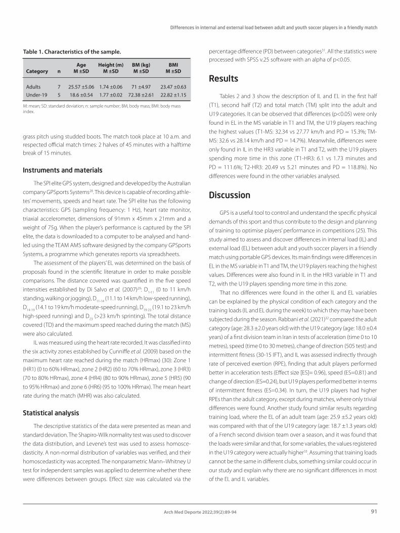
Differences in internal and external load between adult and youth soccer players in a friendly match
91Arch Med Deporte 2022;39(2):89-94
grass pitch using studded boots. The match took place at 10 a.m. and respected official match times: 2 halves of 45 minutes with a halftime break of 15 minutes.
Instruments and materials
The SPI elite GPS system, designed and developed by the Australian company GPSports Systems28. This device is capable of recording athle-tes’ movements, speeds and heart rate. The SPI elite has the following characteristics: GPS (sampling frequency: 1 Hz), heart rate monitor, triaxial accelerometer, dimensions of 91mm x 45mm x 21mm and a weight of 75g. When the player’s performance is captured by the SPI elite, the data is downloaded to a computer to be analysed and hand-led using the TEAM AMS software designed by the company GPSports Systems, a programme which generates reports via spreadsheets.
The assessment of the players’ EL was determined on the basis of proposals found in the scientific literature in order to make possible comparisons. The distance covered was quantified in the five speed intensities established by Di Salvo et al. (2007)29: D1-11 (0 to 11 km/h standing, walking or jogging), D11-14 (11.1 to 14 km/h low-speed running), D14-19 (14.1 to 19 km/h moderate-speed running), D19-23 (19.1 to 23 km/h high-speed running) and D23 (>23 km/h sprinting). The total distance covered (TD) and the maximum speed reached during the match (MS) were also calculated.
IL was measured using the heart rate recorded. It was classified into the six activity zones established by Cunniffe et al. (2009) based on the maximum heart rate reached during the match (HRmax) (30): Zone 1 (HR1) (0 to 60% HRmax), zone 2 (HR2) (60 to 70% HRmax), zone 3 (HR3) (70 to 80% HRmax), zone 4 (HR4) (80 to 90% HRmax), zone 5 (HR5) (90 to 95% HRmax) and zone 6 (HR6) (95 to 100% HRmax). The mean heart rate during the match (MHR) was also calculated.
Statistical analysis
The descriptive statistics of the data were presented as mean and standard deviation. The Shapiro-Wilk normality test was used to discover the data distribution, and Levene’s test was used to assess homosce-dasticity. A non-normal distribution of variables was verified, and their homoscedasticity was accepted. The nonparametric Mann–Whitney U test for independent samples was applied to determine whether there were differences between groups. Effect size was calculated via the
percentage difference (PD) between categories31. All the statistics were processed with SPSS v.25 software with an alpha of p<0.05.
Results
Tables 2 and 3 show the description of IL and EL in the first half (T1), second half (T2) and total match (TM) split into the adult and U19 categories. It can be observed that differences (p<0.05) were only found in EL in the MS variable in T1 and TM, the U19 players reaching the highest values (T1-MS: 32.34 vs 27.77 km/h and PD = 15.3%; TM-MS: 32.6 vs 28.14 km/h and PD = 14.7%). Meanwhile, differences were only found in IL in the HR3 variable in T1 and T2, with the U19 players spending more time in this zone (T1-HR3: 6.1 vs 1.73 minutes and PD = 111.6%; T2-HR3: 20.49 vs 5.21 minutes and PD = 118.8%). No differences were found in the other variables analysed.
Discussion
GPS is a useful tool to control and understand the specific physical demands of this sport and thus contribute to the design and planning of training to optimise players’ performance in competitions (25). This study aimed to assess and discover differences in internal load (IL) and external load (EL) between adult and youth soccer players in a friendly match using portable GPS devices. Its main findings were differences in EL in the MS variable in T1 and TM, the U19 players reaching the highest values. Differences were also found in IL in the HR3 variable in T1 and T2, with the U19 players spending more time in this zone.
That no differences were found in the other IL and EL variables can be explained by the physical condition of each category and the training loads (IL and EL during the week) to which they may have been subjected during the season. Rabbani et al. (2021)32 compared the adult category (age: 28.3 ±2.0 years old) with the U19 category (age: 18.0 ±0.4 years) of a first division team in Iran in tests of acceleration (time 0 to 10 metres), speed (time 0 to 30 metres), change of direction (505 test) and intermittent fitness (30-15 IFT), and IL was assessed indirectly through rate of perceived exertion (RPE), finding that adult players performed better in acceleration tests (Effect size [ES]= 0.96), speed (ES=0.81) and change of direction (ES=0.24), but U19 players performed better in terms of intermittent fitness (ES=0.34). In turn, the U19 players had higher RPEs than the adult category, except during matches, where only trivial differences were found. Another study found similar results regarding training load, where the EL of an adult team (age: 25.9 ±5.2 years old) was compared with that of the U19 category (age: 18.7 ±1.3 years old) of a French second division team over a season, and it was found that the loads were similar and that, for some variables, the values registered in the U19 category were actually higher33. Assuming that training loads cannot be the same in different clubs, something similar could occur in our study and explain why there are no significant differences in most of the EL and IL variables.
Table 1. Characteristics of the sample.
Age Height (m) BM (kg) BMI Category n M ±SD M ±SD M ±SD M ±SD
Adults 7 25.57 ±5.06 1.74 ±0.06 71 ±4.97 23.47 ±0.63Under-19 5 18.6 ±0.54 1.77 ±0.02 72.38 ±2.61 22.82 ±1.15
M: mean; SD: standard deviation; n: sample number; BM, body mass; BMI: body mass index.

Jorge Pérez-Contreras, et al.
92 Arch Med Deporte 2022;39(2):89-94
The MS values obtained by the U19 category were similar in the
study by Hespanhol et al. (2021)34, who related body composition and
states of biological maturation with maximum speed reached during
a soccer match, finding that more biologically developed players, post
peak height velocity (PHV), had higher MS (32.22 ±1.79 km/h) than
players pre-PHV (21.91±2.56 km/h) and at PHV (29.77 ±2.16 km/h),
which may be consistent with the age range of our sample. In turn,
Zúñiga-Morales et al. (2021)35 reported mean maximum speed values of
28.4 ±2.5 to 29 ±2.3 km/h in professional players belonging to a Costa
Rican first division club during 5 pre-season friendly matches, reporting
values slightly higher than those obtained by the professional squad,
although it should be taken into account that, in this case, the players
were only assessed once and against a youth team. However, the values
obtained by the professional players in our study are similar to those
found by Mallo et al. (2015)36, who reported a mean maximum running
speed of 28.3 ± 2.5 km/h in professional soccer players in Spain during
11 pre-season friendly matches. There is a decrease in DT of 5% during
the second half reported by Krustup, et al. (2001)37 and of 3% according
to Mohr et al. (2003)38, similar to those found in both categories.
The HR zones proposed by Cunniffe et al. (2009)30 show that the U19
players spent more time in HR3 (6.1 vs 1.73 and 20.49 vs 5.21), but no
differences were reflected in the distances in any specific speed range,
so these differences could be attributed to the different positions in
which the players play39, performance fatigability or the perceived fati-
gability with which they arrived at the match40,41. In Vargas et al. (2014)42,
MHR values of 165.85±14.88 ppm and HRmax of 189.25 ±5.96 ppm
were recorded during pre-season matches, which are similar to those
obtained by the professional players but lower than those recorded by
the U19 players in our study. The MHR values are 88.4% and 86.9% of
HRmax, which are higher than those recorded by Stølen et al. (2005)10
in elite players. This would imply that age and competitive level may
be a differentiating factor in physical performance 43.
Regarding the results obtained, we recognise limitations in our
study, mainly referring to the sample size, which only includes two
soccer teams, which are different categories in the same club, so it is
not possible to generalise the values reported in this study to other
teams of the same competitive level, It should also be noted that the
data were collected during a single unofficial match, and different res-
ponses in the variables studied might be expected in an official match,
where the values and differences between groups might increase due
to the intrinsic motivation of competition and tactical behaviour39,44.
Playing position should also be considered in the analysis, because the
Table 2. Descriptive and inferential statistics of the first and second halves.
1st Half 2nd Half
Variables Adult U19 Inter-groups Adult U19 Inter-groups
M ±SD M ±SD p PD M ±SD M ±SD p PD
External load
TD (m) 5480 208 5275 409 0.37 3.8 4866 738 4990 464 0.68 2.5
MS (km/h) 27.7 2.3 32.3 2.5 0.02* 15.3 26.2 3.1 27.9 1.5 0.28 6.3
D1-11 (m) 3399 146 3322 190 0.68 2.3 3104 383 3260 179 0.46 4.9
D11-14 (m) 943 161 788 18 0.22 17.9 807 164 698 167 0.16 14.5
D14-19 (m) 792 167 769 140 0.46 2.9 707 143 685 200 0.37 3.2
D19-23 (m) 241 61 259 75 0.93 7.2 181 73 254 95 0.37 33.3
D23 (m) 104 69 134 61 0.46 25.2 67 58 91 50 0.37 30.4
NS (frequency) 6.4 2.7 7.8 3 0.41 19.7 5 4.1 7 2.9 0.36 33.3
Internal load
MHR (ppm) 168 51 180 13 0.8 6.9 154 44 170 12 0.46 9.9
HRmax (ppm) 190 45 207 11 0.74 8.6 185 53 202 11 0.93 8.8
HR1 (min) 3.1 8 0.1 0.2 0.69 187.5 3.9 9.7 0.2 0.3 0.6 180.5
HR2 (min) 3.4 8.3 0.5 0.6 0.89 148.7 0.9 0.9 2.5 2.5 0.22 94.1
HR3 (min) 1.73 1.9 6.1 3.4 0.02* 111.6 5.2 5.3 20.4 22.2 0.04* 118.8HR4 (min) 12.6 9.2 19.8 2.1 0.08 44.4 14.2 8.1 18.7 4.4 0.46 27.6
HR5 (min) 15.5 7.9 14.2 3.9 0.22 8.8 14.9 12.3 10.2 3.5 0.46 37.7
HR6 (min) 8.3 6.3 4.1 1.1 0.29 67.7 5.72 4.8 3.4 2.9 0.46 50.9
*differences <0.05; M: mean; SD: standard deviation; PD: percentage difference; TD: total distance; MS: maximum speed; D1-11 total distance 1-11 km/h; D11-14 total distance 11-14 km/h; D14-19 total distance 14-19 km/h; D19-23 total distance 19-23 km/h; D23 total distance over 23 km/h; NS: number of sprints; MHR: mean heart rate; HRmax: maximum heart rate; HR1 time in heart rate zone 1; HR2 time in heart rate zone 2; HR3 time in heart rate zone 3; HR4 time in heart rate zone 4; HR5 time in heart rate zone 5; HR6 time in heart rate zone 6.

Differences in internal and external load between adult and youth soccer players in a friendly match
93Arch Med Deporte 2022;39(2):89-94
physiological response shown through IL and EL is related to the cha-racteristics and specific requirements of the position39,43. The results and analysis of this study do not permit any explanation of the performance of a team because technical and tactical factors, and the internal logic of the game are not taken into consideration, it only being possible to provide a vision of the conditional and individual aspects of the players45.
Considering the limitations of the study, it has been possible to compare the physical profiles of soccer players during an unofficial match and to see the degree to which the variables studied manifested themselves during the game for different competitive categories. The data presented in this study, which focuses on a sample of Chilean athletes, aims to serve as an aid for coaching teams by allowing them to make plans based on the physical demands of the game in the di-fferent categories and to fill the research gap faced by Chilean soccer in this field, contributing to the collection of data which could benefit the sports performance of teams and making the information recorded available to portray the reality of soccer players in the national sporting landscape, opening the way to future research to favour the different participants in this sport.
Conclusion
From the results obtained, we can conclude that there are no differences in internal and external load between adult and under-19 players, with the exception of MS and HR3, during an unofficial match. Therefore, for practical purposes, the U19 players in this team could be in a position to meet the physical demands that competition in the adult category requires. Finally, due to the low number of participants and data collection over time (longitudinal), it is necessary to continue research to corroborate and confirm these results in other teams.
Contributions of the authors
J.P-C: Design, analysis and composition of the manuscript; S.E-M: Design, data collection and composition of the manuscript; R. V-V: Composition of the manuscript; E.A-M: Analysis, data collection and composition of the manuscript; B.M: Analysis and composition of the manuscript; P.M-M: Design, analysis and composition of the manuscript.
Conflict of interest
The authors declare that they are not subject to any type of conflict of interest.
Bibliography
1. Turner AN, Stewart PF. Strength and conditioning for soccer players. Strength Cond J. 2014;36:1-13.
2. Dolci F, Hart NH, Kilding AE, Chivers P, Piggott B, Spiteri T. Physical and energetic demand of soccer. Strength Cond J. 2020;42:70-7.
3. Bush M, Barnes C, Archer DT, Hogg B, Bradley PS. Evolution of match performance parameters for various playing positions in the English Premier League. Hum Mov Sci. 2015;39:1-11. doi:10.1016/j.humov.2014.10.003
4. Maciel WP, Caputo EL, Silva MC da. Distância percorrida por jogadoras de futebol de diferentes posições durante uma partida. Rev Bras Ciências do Esporte. 2011;33:465-74.
5. Impellizzeri FM, Rampinini E, Marcora SM. Physiological assessment of aerobic training in soccer. J Sports Sci. 2005;23:583-92.
6. Delaney JA, Duthie GM, Thornton HR, Pyne DB. Quantifying the relationship between internal and external work in team sports: development of a novel training efficiency index. Sci Med Footb. 2018;2:149-56.
7. Buchheit M, Mendez-Villanueva A, Simpson BM, Bourdon PC. Match running perfor-mance and fitness in youth soccer. Int J Sports Med. 2010;31:818-25.
8. Lago-Peñas C, Casais L, Dellal A, Rey E, Domínguez E. Anthropometric and physiological characteristics of young soccer players according to their playing positions: Relevance for competition success. J Strength Cond Res. 2011;25:3358-67.
9. Reilly T, Bangsbo J, Franks A. Anthropometric and physiological predispositions for elite soccer. J Sports Sci. 2000;18:669-83.
10. Stølen T, Chamari K, Castagna C, Wisløff U. Physiology of soccer: An update. Sport Med. 2005;35:501-36.
11. Alexandre D, Silva CD Da, Hill-Haas S, et al. Heart ratemonitoring in soccer: Interest and limits during competitive match play and training, practical application. J Strength Cond Res. 2012;26:2890-906.
12. Leduc C, Lacome M, Buchheit M. The use of standardised runs ( and associated data analysis ) to monitor neuromuscular status in team sports players: a call to action. Sport Perform Sci Rep. 2020;1:1-5.
13. Lacome M, Simpson BM, Buchheit M. Monitoring training status with player-tracking technology. Part 2: Increasing coach “buy-in” with good data visualisation. Aspetar Sport Med J. 2018:64-6.
Table 3. Descriptive and inferential statistics of the total match.
Variables Adult Under-19 Inter-groups
M ±SD M ±SD p PD
External load
TD (m) 10347 671.9 10265 813 0.465 0.8
MS (km/h) 28.14 2.27 32.6 2.41 0.018* 14.7
D1-11 (m) 6502 502.9 6582 345.5 0.935 1.2
D11-14 (m) 1751 196.6 1488 320.1 0.167 16.3
D14-19 (m) 1499 171.7 1455 340.6 0.372 3.0
D19-23 (m) 422.7 97.83 514 154.3 0.372 19.5
D23 (m) 171.1 109.9 225.4 86.01 0.167 27.4
NS (frequency) 11.43 5.83 14.8 5.22 0.188 25.7
Internal load
MHR (ppm) 161.1 46.89 175.6 12.94 0.685 8.6
HRmax (ppm) 191.7 45.94 207.4 11.5 0.745 7.9
HR1 (min) 7.11 12.1 0.33 0.46 0.603 182.3
HR2 (min) 4.37 7.97 3.12 3.19 0.935 33.4
HR3 (min) 6.94 7.05 14.6 8.11 0.123 71.1
HR4 (min) 26.81 16.41 38.59 5.71 0.167 36.0
HR5 (min) 30.53 9.26 24.53 6.67 0.167 21.8
HR6 (min) 14.04 10.49 7.64 2.98 0.291 59.0
*differences <0.05; M: mean; SD: standard deviation; PD: percentage difference; TD: total distance; MS: maximum speed; D1-11 total distance 1-11 km/h; D11-14 total distance 11-14 km/h; D14-19 total distance 14-19 km/h; D19-23 total distance 19-23 km/h; D23 total distance over 23 km/h; NS: number of sprints; MHR: mean heart rate; HRmax: maxi-mum heart rate; HR1 time in heart rate zone 1; HR2 time in heart rate zone 2; HR3 time in heart rate zone 3; HR4 time in heart rate zone 4; HR5 time in heart rate zone 5; HR6 time in heart rate zone 6.

Jorge Pérez-Contreras, et al.
94 Arch Med Deporte 2022;39(2):89-94
14. Mohr M, Krustrup P, Bangsbo J. Fatigue in soccer: A brief review. J Sports Sci. 2005; 23:593-9.
15. Sarmento H, Marcelino R, Anguera MT, CampaniÇo J, Matos N, LeitÃo JC. Match analysis in football: a systematic review. J Sports Sci. 2014;32:1831-43.
16. Gregson W, Drust B, Atkinson G, Salvo VD. Match-to-match variability of high-speed activities in premier league soccer. Int J Sports Med. 2010;31:237-42.
17. Rampinini E, Coutts AJ, Castagna C, Sassi R, Impellizzeri FM. Variation in top level soccer match performance. Int J Sports Med. 2007;28:1018-24.
18. Oreilly J, Wong SHS. The development of aerobic and skill assessment in soccer. Sport Med. 2012;42:1029-40.
19. Di Salvo V, Baron R, González-Haro C, Gormasz C, Pigozzi F, Bachl N. Sprinting analysis of elite soccer players during European Champions League and UEFA Cup matches. J Sports Sci. 2010;28:1489-94.
20. Haugen TA, Tønnessen E, Hisdal J, Seiler S. The role and development of sprinting speed in soccer. Int J Sports Physiol Perform. 2014;9:432-41.
21. Andrzejewski M, Chmura J, Pluta B, Strzelczyk R, Kasprzak A. Analysis of sprinting activities of professional soccer players. J Strength Cond Res. 2013;27:2134-40.
22. Buchheit M, Modunotti M, Stafford K, Gregson W, Di Salvo V. Sport Perform Sci Rep. 2018;1:1-3.
23. Buchheit M, Simpson BM. Player-tracking technology: Half-full or half-empty glass? Int J Sports Physiol Perform. 2017;12:35-41.
24. Malone JJ, Lovell R, Varley MC, Coutts AJ. Unpacking the black box: Applications and considerations for using gps devices in sport. Int J Sports Physiol Perform. 2017; 12:18-26.
25. Cummins C, Orr R, O’Connor H, West C. Global positioning systems (GPS) and micro-technology sensors in team sports: A systematic review. Sport Med. 2013;43:1025-42.
26. Aughey RJ. Applications of GPS technologies to field sports. Int J Sports Physiol Perform. 2011;6:295-310.
27. General assembly of the world medical association. world medical association decla-ration of helsinki: Ethical principles for medical research involving human subjects. JAMA - J Am Med Assoc. 2013;310:2191-4.
28. Coutts AJ, Duffield R. Validity and reliability of GPS devices for measuring movement demands of team sports. J Sci Med Sport. 2010;13:133–5.
29. Di Salvo V, Baron R, Tschan H, Calderon Montero FJ, Bachl N, Pigozzi F. Performance cha-racteristics according to playing position in elite soccer. Int J Sports Med. 2007;28:222-7.
30. Cunniffe B, Proctor W, Baker JS, Davies B. an evaluation of the physiological demands of elite rugby union using global positioning system tracking software. J Strength Cond Res. 2009;23:1195-203.
31. Merino-Muñoz P, Pérez-Contreras J, Aedo-Muñoz E. The percentage change and differences in sport: a practical easy tool to calculate. Sport Perform Sci Reports. 2020;118:446–50.
32. Rabbani A, Wong DP, Clemente FM, Kargarfard M. Internal training load and fitness profile between adult team versus junior team soccer players. Kinesiology. 2021;53:71–7.
33. Coppalle S, Ravé G, Moran J, Salhi I, Abderrahman A Ben, Zouita S, et al. Internal and external training load in under-19 versus professional soccer players during the in-season period. Int J Environ Res Public Health. 2021;18:1–10.
34. Hespanhol J, Pignataro J, Lopes T, Pereira N, Moreira J, Arruda M. Influencia de la composición corporal y la masa ósea en la velocidad máxima obtenida durante un partido de fútbol en futbolistas jóvenes Influence of body composition and bone mass on maximum speed during a match in young soccer players. Rev Peru ciencias la Act física y del Deport. 2021;8:1054-63.
35. Zúñiga-morales C, Ugalde-ramírez A, Gutierrez-Vargas R, Rojas-valverde D. Análisis de los comportamientos motrices de jugadores profesionales costarricenses de fútbol en partidos amistosos en pretemporada. Rev Peru ciencias la Act física y del Deport. 2021;8:1116-28.
36. Mallo J, Mena E, Nevado F, Paredes V. Physical Demands of Top-Class Soccer Friendly Matches in Relation to a Playing Position Using Global Positioning System Technology. J Hum Kinet. 2015;47:179-88.
37. Krustrup P, Bangsbo J. Physiological demands of top-class soccer refereeing in rela-tion to physical capacity: Effect of intense intermittent exercise training. J Sports Sci. 2001;19:881-91.
38. Mohr M, Krustrup P, Bangsbo J. Match performance of high-standard soccer players with special reference to development of fatigue. J Sports Sci. 2003;21:519-28.
39. Aquino R, Carling C, Palucci Vieira LH, et al. Influence of Situational Variables, Team Formation, and Playing Position on Match Running Performance and Social Network Analysis in Brazilian Professional Soccer Players. J Strength Cond Res. 2020;34:808-17.
40. Enoka RM, Duchateau J. Translating fatigue to human performance. Med Sci Sports Exerc. 2016;48:2228-38.
41. Thorpe RT, Strudwick AJ, Buchheit M, Atkinson G, Drust B, Gregson W. Tracking morning fatigue status across in-season training weeks in elite soccer players. Int J Sports Physiol Perform. 2016;11:947-52.
42. Vargas A, Urkiza I, Gil S. Variables predictoras de éxito en fútbol: análisis de los partidos de pretemporada mediante gps. Rev Prep Física en el Fútbol. 2014.
43. Slimani M, Nikolaidis PT. Anthropometric and physiological characteristics of male soccer players according to their competitive level, playing position and age group: A systematic review. J Sports Med Phys Fitness. 2019;141-63.
44. Casamichana D, Castellano J. Demandas físicas en jugadores semiprofesionales de fútbol: ¿se entrena igual que se compite? Cult Cienc y Deport. 2011;6:121-7.
45. Reina Gómez A, Hernández Mendo A. Revisión de indicadores de rendimiento en fútbol. Rev Iberoam Ciencias la Act Física y el Deport. 2012;1:1-14.

Determining factors with regard to physical functional impairment in revascularized patients with acute coronary syndrome
95Arch Med Deporte 2022;39(2):95-99
Original article
Resumen
Introducción: La capacidad física funcional se asocia de manera indirecta con la mortalidad, y frecuentemente se ve com-prometida después de un evento cardiovascular, de ahí la importancia de considerar sus factores pronósticos durante el tratamiento en los pacientes cardiópatas. Objetivo: Identificar los factores pronósticos de la limitación física funcional en pacientes revascularizados por Síndrome Coronario Agudo (SCA). Material y método: Se realizó un análisis retrospectivo de 29 historias clínicas tomadas de pacientes sometidos a una prueba de esfuerzo (Protocolo Bruce o Bruce modificado) posterior a una revascularización miocárdica durante los meses de enero a diciembre del 2019. Para el análisis estadístico se realizó una regresión logística univariada y multivariada (odds ratio-OR), así como un análisis de regresión linear simple entre las variables de interés. Resultados: los pacientes analizados tenían una media de edad de 60 ± 9,2 años, y el 76% fueron hombres. La presencia de hipertensión arterial sistémica (HAS), menores valores de Frecuencia Cardíaca (FC) máxima (ß = 0,112; CI: 0,074, 0,149; p <0,001), altos valores de Presión Arterial Sistólica (PAS) (ß = - 0,083; CI 95%: -0,152, -0,014; p <0,020), y un mayor número de factores de riesgo (ß = - 1,580; CI 95%: -2,456-0,868; p <0,001), son considerados predictores de limitación física funcional (<7 METs). Conclusión: La presencia de HAS, un mayor número de factores de riesgo, así como altos niveles de PAS máxima y menores valores de FC máxima alcanzadas durante la prueba de esfuerzo, mostraron ser factores pronósticos de la limitación funcional en sujetos revascularizados por SCA.
Palabras clave: Rehabilitación. Prueba de esfuerzo.
Isquemia miocárdica.
Summary
Introduction: Functional physical capacity is indirectly associated with mortality, and may be compromised after a cardio-vascular event, hence the importance of considering its prognostic factors during treatment in heart disease patients. Aim: To identify the prognostic factors of functional physical limitation in patients with myocardial revascularization for acute coronary syndrome. Material and method: A retrospective analysis was carried out of 29 medical records taken from patients submitted to a stress test (Bruce, modified Bruce) after myocardial revascularization during the months of January to December 2019. For the statistical analysis, a univariate and multivariate logistic regression (Odds Ratio-OR) was performed, as well as a simple linear regression analysis between the variables of interest. Results: the analyzed patients had a mean age of 60 ± 9,2 years, and 76% were men. The presence of systemic arterial hypertension (SAH), lower values of Maximum Heart Rate (HR) (ß = 0,112; CI: 0,074, 0,149; p <0.001), high values of Sys-tolic Arterial Pressure (SBP) (ß = - 0,083; CI 95%: -0,152, -0,014; p <0.020), and a greater number of risk factors (ß = - 1,580; CI 95%: -2,456-0,868; p <0.001), are considered predictors of functional limitation (<7 METs). Conclusion: The presence of SAH, a greater number of risk factors, as well as high maximum SBP levels and lower maximum HR values reached during the stress test, were shown to be prognostic factors of functional limitation in subjects revascula-rized for ACS.
Key words: Rehabilitation. Exercise test.
Myocardial ischemia.
Received: 06/09/2021Accepted: 03/02/2022
Determining factors with regard to physical functional impairment in revascularized patients with acute coronary syndrome
Laura C. Dávila Landinez1, Laura N Coral Vásquez1, Aura N. Carrizales Sánchez1, Andrés Mauricio Ariza1, Hedilberto Duarte Hernández2, Hugo C.D de Souza3, Stella V. Philbois3, Juan C. Sánchez Delgado1,3,4
¹Universidad Santo Tomás. Seccional Bucaramanga. Colombia. 2Instituto del Corazón de Bucaramanga. Colombia. 3Universidade de São Paulo. Faculdade de Medicina. Ribeirão Preto SP. Brasil. 4Universidad de Santander. Bucaramanga. Colombia.
Factores determinantes de la limitación física funcional en pacientes revascularizados por síndrome coronario agudo
Correspondence: Juan Carlos Sánchez Delgado E-mail: [email protected]
doi: 10.18176/archmeddeporte.00079

Laura C Dávila Landinez, et al.
96 Arch Med Deporte 2022;39(2):95-99
Introduction
The effort test in revascularized patients with coronary artery disease has primarily been used for diagnostic purposes in order to detect residual ischemia, arrhythmias, symptoms and dynamic physio-logical responses before starting a cardiac rehabilitation programme. Approximately 30% of these programmes conduct effort tests, despite the fact that these tests are considered to be the gold standard for starting rehabilitation. The increase in longevity and in the multimorbi-dity conditions of the revascularized population, makes its use appear more relevant in order to guide the prescription and progression of pharmacological and physical exercise treatments that are commonly provided to these patients 1,2.
The information commonly extracted during effort tests on this type of patient includes: maximum heart rate and maximum work load determined by fatigue, angina or electrocardiographic evidence of is-chemia. The above makes it possible to establish safe training intensities in order to increase the functional physical capacity3,4,1. This capacity or physical aptitude is commonly reported using metabolic equivalents (METs) and it has been shown to be a prognostic indicator for event-free survival, all-cause mortality and cardiovascular events5,6. Its increase is accompanied by positive influences on the vascular, cardiac, haemato-logical, immunological and nervous systems, it is also considered to be a stronger predictor of mortality when compared to nicotinism, SHBP, high cholesterol, diabetes mellitus and other predictors obtained in an effort test such as segment depression7.
In view of all the foregoing, the increase in functional physical capacity must be considered to be a fundamental treatment goal for revascularized patients7. Furthermore, by recognising factors that exert an influence on functional physical capacity, this could help to improve the coronary disease control strategies following a surgical intervention. Therefore, this study aims to identify the prognostic factors of functional impairment measured through an ergometric test in revascularized patients with acute coronary syndrome.
Material and method
A cross-sectional, descriptive retrospective study was conducted, based on the information derived from the twenty-nine medical his-tories of patients subjected to an ergometric test after having under-gone, for the first time, a revascularization process for acute coronary syndrome (ACS) in the Instituto del Corazón de Bucaramanga (ICB) (Bucaramange Heart Institute) between the months of January and December 2019. The following information was obtained: age, sex, height, weight, ACS type, revascularization type, participation in cardiac rehabilitation programme, number of sessions prescribed, number of sessions completed, cardiovascular risk factors and re-hospitalizations subsequent to revascularization.
Given that the effort tests selected had a diagnostic purpose, the patients suspended the pharmacological treatment to perform them.
The only tests selected were those with no electrocardiographic changes compatible with ischemia or rhythm disorders, and in which the reason for the suspension of the tests was fatigue. The variables analysed were: protocol type (Standard Bruce and modified Bruce), baseline HR, maxi-mum HR achieved during the effort test, baseline SAP, maximum SAP achieved during the effort test,baseline SAP, maximum SAP achieved during the effort test, Diastolic Arterial Pressure (DAP), maximum DAP achieved during the effort test, maximum oxygen consumption (VO2máx) obtained by indirect measurements, METs, and presence of functional impairment “<7 METs of tolerance to exercise”8,9.
Statistical analysis
The information obtained was digitised in Excel and the results were analysed through Stata 12.0. The continuous variables were analysed as measures of central tendency and dispersion, the cate-gorical variables through absolute and relative frequencies. In order to estimate the influence of the different factors associated with the functional impairment of the target population of the study, a univariate and multivariate logistic regression was performed (Odds Ratio-OR). Additionally, Pearson’s test was used to conduct a correlation analysis between tolerance to efforts in METs and the continuous variables of interest. Finally, a simple linear regression analysis was made with those variables showing significant results in the correlation. Associations with p<0.05 were considered to be statistically significant.
Ethical considerations
The authors declare that the procedures were conducted in ac-cordance with the ethical standards of the committee responsible for human experimentation and in accordance with the World Medical Association and the Declaration of Helsinki. The study observed the ethical principles of confidentiality, goodwill, non-dysfunction, auto-nomy and justice. Finally, this investigation was approved by the ethics committee of the Universidad Santo Tomás-Seccional Bucaramanga. (Ethical concept #01402020-1012032020).
Results
Of the population analysed: the mean age was 60 ± 9.2 years, 76% were men, the most prevalent risk factor was the SHBP present, 83% underwent percutaneous coronary intervention, 86% performed the standard Bruce test and 31% exhibited functional impairment (Table 1).
In the univariate and multivariate analysis, the functional impair-ment was associated with the presence of hypertension. Hypertension sufferers are 16 times more likely to show functional impairment in com-parison with those for whom this diagnosis was not reported (Table 2).
Table 3 shows a direct relationship between tolerance to physical effort and MHR and an indirect relationship between this same varia-ble and the number of risk factors and SAP. According to the linear

Determining factors with regard to physical functional impairment in revascularized patients with acute coronary syndrome
97Arch Med Deporte 2022;39(2):95-99
regression analysis, an increased MHR value could increase the METs by 0.11 (ß = 0.112; CI: 0.074, 0.149; p <0.001); additionally, for each mmHg increase of SAP, the METs decreased 0.08 (ß = - 0.083; CI 95%: -0.152, -0.014; p <0.020), and for each risk factor unit increase the METs decreased 1.5 (ß = - 1.580; CI 95%: -2.456-0.868; p <0.001) (Figure 1).
Figure 1. Linear regression analysis between functional physical capacity, number of cardiovascular risk factors, SAP and MHR.
Table 1. General characteristics of the population.
Variable n=29 %
Sex (Male) 22 76Risk factors Obesity 13 44 AHT 14 48 Diabetes 3 10 Dyslipidemia 10 34 Nicotinism 3 10 Ex-nicotinism 12 41 Family history 1 3
Type of ACS NSTE-ACS 13 45 STE-ACS 6 21
Unstable angina 10 34
Type of percutaneous 24 83 revascularization Bypass 5 17
Protocol Bruce Standard 25 86 Bruce Modified 4 14
Participation in CR 25 86 Functional impairment (<7 METs) 9 31 Rehospitalized 11 38
Variables Mean ±SD
Age (years) 60 9.2 Height (cm) 166 8.3 Weight (Kg) 73 10 BMI (Kg/m2) 26 3
Rehabilitation Prescribed 21 20 sessions Completed 16 12
Heart rate Baseline 67 7,3 Maximum 141 23 %Maximum HR 86 12
Blood pressure SAP 132 17 DAP 74 10 Maximum SAP 161 19 Maximum DAP 84 12
Physical capacity METs 9 3 VO2max 32 12
AHT: Arterial hypertension: HR: Heart rate: NSTE-ACS: Non-ST elevation-acute coronary syndrome: STE-ACS: ST elevation acute coronary syndrome: CR: Cardiac rehabilitation; BMI: Body mass index; SAP Systolic arterial pressure: DAP: Diastolic arterial pressure; VO2máx: Maximum oxygen consumption.
Discussion
The presence of SHBP, a greater number of risk factors, high levels of maximum SAP and lower maximum HR values achieved during the effort test were shown to be prognostic factors of functional impairment in subjects revascularized for ACS.
The evidence found shows an indirect relationship between functional capacity and arterial pressure levels in hypertensive and normotensive patients10-14. Although Tadic M, Ivanovic B. 2014 reported that the mechanisms for the relationship are not entirely clear, they do
Number of risk factors
ß= -1.662; R2 = 0.406; p <0,001
MET
s
ß= -0.053; R2 = 0.184; p = 0.020
ß= 0.112; R2 = 0.585; p = 0.001
Maximum Heart Rate bpm
Systolic arterial pressure mmHg
16
14
12
10
8
6
4
20 2 T1 3 4 5 6
MET
sM
ETs
16
14
12
10
8
6
4
2
16
14
12
10
8
6
4
2
90 170100 110 120 130 140 150 160
80 200100 120 140 160 180

Laura C Dávila Landinez, et al.
98 Arch Med Deporte 2022;39(2):95-99
point to some possible reasons such as the presence of endothelial dysfunction, mitochondrial and oxidative stress, which could lead to important alterations in the oxygen delivery and uptake rates in the tissues, thereby affecting functional capacity15-18. There are other factors that could reduce the level of tolerance to effort in this population, such as an increase in the left ventricle filling pressure, which contributes to the dysfunction and dilatation of the left auricle, thereby deteriorating the diastolic phase of the left ventricle, reducing the cardiac output during physical effort16,19,20.
The prognostic value shown by the maximum HR could be justified, taking into account the fact that this determines approximately 30% of the maximum cardiac output, thereby influencing the kinetics of oxygen consumption, a variable that defines the functional physical capacity21. Moreover, it is known that in apparently healthy subjects, VO2.increases 7.7 times during maximum intensity exercise. This is achieved thanks to an increase of: 2.5 times the HR, 2.5 times the arteriovenous oxygen difference and 1.4 times the systolic volume, the above being one of the reasons why the HR is considered to be one of the factors that most helps to maintain the functional physical capacity22,23.
The increase in the maximum HR is primarily conditioned by the expression and functional activity of the adrenergic receptors and by the efficiency of the excitation-contraction coupling. Although these factors are commonly attenuated by ageing and by the extent of the lesion caused by coronary disease, this progressive reduction or alteration of the response of the heart to adrenergic stimulation, does not appear to be so significant in those in better physical condition21,24-26.
There are other factors that are characteristic of coronary disease and some that are considered to be adverse effects of the anaesthesia or revascualrization surgery, such as the dilatation of the left ventricle and damage to the sinoatrial node that could affect chronotropic per-formance and the cardiovascular autonomic functions27,28. Although the maximum HR is fundamental for the adaptation of the cardiac output to metabolic needs, its role as a biomarker or possible modifiable independent risk factor, as well as its clinical or therapeutic objective in revascularized patients is not clear. For this reason, some studies emphasise the importance of analysing its behaviour, which could strongly predict the evolution of patients of this type27,21.
The physical functional capacity is considered to be a prognostic factor of the burden of disease for patients with coronary disease. Specifically, the high levels of this capacity are associated with the mitigation of the risk factors, new coronary events, all-cause mortality and cardiovascular events4,5,29-32. The above is in keeping with what has been shown in this study, in which the number of cardiovascular risk factors, and the presence of SHBP in particular, were considered to be predictors of functional limitation in subjects subjected to an effort test after having undergone, for the first time, a revascularization process for acute coronary syndrome (ACS). The literature available suggests including cardiorespiratory capacity as part of the risk stratification pro-cesses, in patients with cardiovascular disease, which would substantially improve the accuracy of this process7; this is taking into account the fact that this variable has been shown to be a stronger predictor when compared to traditional risk factors or those obtained in the effort test such as the depression of the ST segment, the haemodynamic symp-toms and responses7,33,34.
The limitations found include: the nature of the study, that does not make it possible to control the result of the evaluations, thereby
Table 2. Factors associated with functional impairment of revascularized patients for ACS.
Variable OR (95%IC) p Value AOR (95% CI) p Value
Obesity 3.7 (0.704; 19.58) 0.122
AHT 18 (1.893; 184.0) 0.012 16 (1.010-275.0) 0.049
DM 1.12 (0.088; 14.27) 0.132
Dyslipidemia 1.86 (0.367; 9.487) 0.452
Nicotinism 1.12 (0.088; 14.27) 0.928
Ex-nicotinism 0.10 (0.010; 0.977) 0.048 0.06 (0.003-1.292) 0.704
Participation in CR 0.38 (0.045; 3.323) 0.338
Rehospitalisation 0.34 (0.057; 2.115) 0.252
OR: Odds Ratio; AOR: Adjusted Odds Ratio by sex, obesity and diabetes mellitus.
Table 3. Correlation between the number of cardiac rehabilitation sessions, effort tolerance level and variables of interest.
Variable METs r p
Age - - 0.24 0.198
BMI - 0.36 0.050
Number of risk factors - 0.63 0.002
BHR 0.03 0.873
Maximum HR 0.76 <0.001
SAP - 0.42 0.020
DAP - 0.01 0.878
Maximum SAP - 0.32 0.089
Maximum DAP - 0.34 0.237
NCRS: number of cardiac rehab sessions; BMI: Body Mass Index: BHR: Baseline Heart rate: SAP: Systolic arterial pressure: DAP: Diastolic arterial pressure; * p <0,05 Spearman test.

Determining factors with regard to physical functional impairment in revascularized patients with acute coronary syndrome
99Arch Med Deporte 2022;39(2):95-99
depending on the record reported in the medical histories; the size of the sample that may have been insufficient to detect any other significant associations; the obtaining of the physical capacity by indirect methods, which could be lacking in accuracy, considering that predictive models are more susceptible to confusion factors. Furthermore, it is possible that the results analysed are influenced by the ceiling effect, present when the HR is used as a determining factor of the effort test termination, and by the learning effect, when the patient does not have the possibility to become familiar with the use of the treadmill before starting the test35.
Conclusions
Our work reinforces the importance of adequate handling of co-morbidities and SHBP in particular, in order to improve the functional capacity of these individuals. The increase in the maximum HR and the attenuation of the SAP are determining factors in the increased tolerance to physical effort in revascularized patients.
The effort tests analysed had a diagnostic objective and, for this reason, it is suggested that they also be used as a patient risk stratifi-cation tool based not only on the use of the METs achieved but also on the performance of haemodynamic variables that are simple and relatively easy to obtain such as the maximum HR and the SAP, which were shown to be related to tolerance to physical exercise. They could also show prognostic values for other important conditions to be taken into account during the post-surgery period such as event-free survival levels, all-cause mortality and cardiovascular events in revascularized patients for ACS.
Acknowledgements
The authors would like to thank the Faculty of Physical Culture, Sports and Recreation of the University Santo Tomas - Bucaramanga, and the Heart Institute of the Corazón de Bucaramanga for its support and backing during this present investigation.
Conflict of interest
The authors do not declare a conflict of interest.
Bibliography
1. Mytinger M, Nelson RK, Zuhl M. Exercise prescription guidelines for cardiovascular disease patients in the absence of a baseline stress test. J Cardiovasc Dev. 2020; 7:15.
2. Reeves GR, Gupta S, Forman DE. Evolving role of exercise testing in contemporary cardiac rehabilitation. J Cardiopulm Rehabil Prev. 2016;36:309–19.
3. Fletcher GF, Ades PA, Kligfield P, et al. Exercise standards for testing and training: a scientific statement from the american heart association. Circulation. 2013; 128:873-934.
4. Harb SC, Bhat P, Cremer PC, Wu Y, Cremer LJ, Berger S, Cho L, et al. Prognostic value of functional capacity in different exercise protocols. J Am Heart Assoc. 2020; 9: e015986.
5. Chu DJ, Al Rifai M, Virani SS, Brawner CA, Nasir K, Al-Mallah MH. The relationship between cardiorespiratory fitness, cardiovascular risk factors and atherosclerosis. Atherosclerosis. 2020; 304: 44–52.
6. Martin BJ, Arena R, Haykowsky M, Hauer T, Austford LD, Knudtson M, et al. Cardiovas-cular fitness and mortality after contemporary cardiac rehabilitation. Mayo Clin Proc. 2013;88:455-63.
7. Ross R, Blair SN, Arena R, Church TS, Després JP, Franklin BA, et al. Importance of assessing cardiorespiratory fitness in clinical practice: a case for fitness as a clinical vital sign: a
scientific statement from the american heart association. Circulation. 2016;134:653-99. 8. Goldman L, Hashimoto B, Cook EF, Loscalzo A. Comparative reproducibility and validity
of systems for assessing cardiovascular functional class: advantages of a new specific activity scale. Circulation. 1981;64:1227–34.
9. Redd D, Kuang J, Mohanty A, Bray BE, Zeng-Treitler Q. Regular expression-based learning for mets value extraction. AMIA Jt Summits Transl Sci Proc. 2016;2016:213–220.
10. Faselis C, Doumas M, Kokkinos, JP, Panagiotakos D, Kheirbek R, Sheriff H, et al. Exercise capacity and progression from prehypertension to hypertension. Hypertension. 2012; 60:333-8.
11. Kokkinos P, Manolis A, Pittaras A, Panagiotakos D, Kheirbek R, Sheriff H.M, et al. Exercise capacity and mortality in hypertensive men with and without addition. Hypertension. 2009; 53:494–499.
12. Kokkinos P, Myers J, Doumas M, Faselis C, Manolis A, Pittaras A, et al. Exercise capacity and all-cause mortality in prehypertensive men. Am J Hypertens. 2009;22:735-41.
13. Pescatello LS, Franklin BA, Fagard R, Farquhar WB, Kelley GA, Ray CA. American college of sports medicine american college of sports medicine position stand. Exercise and hypertension. Med Sci Sports Exerc. 2004;36:533–53.
14. Kokkinos P, Pittaras A, Narayan P, Faselis C, Singh S, Manolis A. Exercise capacity and blood pressure associations with left ventricular mass in prehypertensive individuals. Hypertension. 2007;49:55-61.
15. Zinchuk VV, Pronko TP, Lis MA. Blood oxygen transport and endothelial dysfunction in patients with arterial hypertension. Clin Physiol Funct Imaging. 2004;24:205–11.
16. Tadic M, Ivanovic B. Why is functional capacity decreased in hypertensive patients? From mechanisms to clinical studies. Rev Med Cardiol. 2014;15:447-55
17. Sarma, S, Howden E, Carrick-Ranson, G, Lawley J, Hearon C, Samels M, et al. Elevated exercise blood pressure in middle-aged women is associated with altered left ventri-cular and vascular stiffness. J Appl Physiol 1985. 2020;128:1123–29.
18. Zhang A, Jia Z, Wang N, Tidwell TJ, Yang T. Relative contributions of mitochondrial and nadph oxidase to deoxycorticosterone acetate-salt hypertension in mice. Kidney Int. 2011;80:51–60
19. Thomas GD. Functional sympatholysis in hypertension. Auton Neurosci. 2015;188:64–8. 20. Howlett LA, Lancaster MK. Reduced cardiac response to the adrenergic system is a
key limiting factor for physical capacity in old age. Exp Gerontol. 2021;150:111339. 21. Anjos-Andrade FD, Sousa AC, Barreto-Filho JA, Alves EO, Nascimento-Júnior, AC, de
Santana NO, et al. Chronotropic incompetence and coronary artery disease. Acta Cardiol. 2010;65:631–8.
22. Higginbotham MB, Morris KG, Williams RS, Coleman RE, Cobb FR. Physiologic basis for the age-related decline in aerobic work capacity. Am J Cardiol. 1986;57:1374-9.
23. Higginbotham MB, Morris KG, Williams RS, McHale PA, Coleman RE, Cobb FR. Regulation of stroke volume during submaximal and maximal upright exercise in normal man. Circ Res. 1986;58:281-91.
24. Gude, NA, Broughton KM, Firouzi F, Sussman, MA. Cardiac ageing: extrinsic and intrinsic factors in cellular renewal and senescence. Nat Rev Cardiol. 2018;15:523–42.
25. Howlett LA, Lancaster MK. Reduced cardiac response to the adrenergic system is a key limiting factor for physical capacity in old age. Exp Gerontol. 2021;150:111339.
26. Lauer MS, Francis GS, Okin PM, Pashkow FJ, Snader CE, Marwick, TH. Impaired chrono-tropic response to exercise stress testing as a predictor of mortality. JAMA. 1999;281: 524–9.
27. Abulimiti A, Nishitani-Yokoyama M, Shimada K, Kunimoto M, Matsubara T, Fujiwara K, et al. Prognostic impact of peak oxygen uptake and heart rate reserve in patients after off-pump coronary artery bypass grafting. Clin cardiol. 2021;44: 580–7.
28. Böhm M, Reil JC, Deedwania P, Kim JB, Borer JS. Resting heart rate: risk indicator and emerging risk factor in cardiovascular disease. Am J Med. 2015;128:219–28.
29. Al-Mallah, MH, Sakr S, Al-Qunaibet A. Cardiorespiratory fitness and cardiovascular disease prevention: an update. Curr Atheroscler Rep. 2018:20:1.
30. Farrell SW, Finley CE, Barlow CE, Willis BL, DeFina LF, Haskell WL, et al. Moderate to high levels of cardiorespiratory fitness attenuate the effects of triglyceride to high-density lipoprotein cholesterol ratio on coronary heart disease mortality in men. Mayo Clin Proc. 2017; 92:1763–71.
31. Hung RK, Al-Mallah MH, McEvoy JW, Whelton SP, Blumenthal RS, Nasir K, et al. Prognostic value of exercise capacity in patients with coronary artery disease: the fit -Henry Ford exercise - testing project. Mayo Clin Proc. 2014; 89:1644–54.
32. Nichols S, Taylor C, Page R, Kallvikbacka-Bennett A, Nation F, Goodman T, et al. Correction to: Is cardiorespiratory fitness related to cardiometabolic health and all-cause mortality risk in patients with coronary heart disease? A CARE CR Study. Sports Med Int Open. 2019;5:5.
33. Goraya, TY, Jacobsen SJ, Pellikka PA, Miller TD, Khan A, Weston SA, et al. Prognostic value of treadmill exercise testing in elderly persons. Ann Intern Med. 2000;132:862–70.
34. Myers J. New american heart association/american college of cardiology guidelines on cardiovascular risk: when will fitness get the recognition it deserves?. Mayo Clin Proc. 2014; 89:722–6.
35. Sartor F, Vernillo G, de Morree HM, Bonomi AG, La Torre A, Kubis HP, et al. Estimation of maximal oxygen uptake via submaximal exercise testing in sports, clinical, and home settings. Sports Med. 2013;43:865-73.

Laura C Dávila Landinez, et al.
100 Arch Med Deporte 2022;39(2):95-99
· Gestión y Plani�cación de Servicios Sanitarios
· Gestión Integral del Riesgo Cardiovascular
· Ingeniería Biomédica
· Investigación en Ciencias Sociosanitarias
· Investigación en Educación Física y Salud
· Neuro-Rehabilitación
· Nutrición Clínica
· Nutrición y Seguridad Alimentaria
· Nutrición en la Actividad Física y Deporte
· Osteopatía y Terapia Manual
· Patología Molecular Humana
· Psicología General Sanitaria
· Actividad Física Terapéutica
· Alto Rendimiento Deportivo:
Fuerza y Acondicionamiento Físico
· Performance Sport:
Strength and Conditioning
· Audiología
· Balneoterapia e Hidroterapia
· Desarrollos Avanzados
de Oncología Personalizada Multidisciplinar
· Enfermería de Salud Laboral
· Enfermería de Urgencias,
Emergencias y Cuidados Especiales
· Fisioterapia en el Deporte
· Geriatría y Gerontología:
Atención a la dependencia
POSTGRADOS OFICIALES: SALUD Y DEPORTE
UniversitarioUniversitario
(1) Presencial (2) Semipresencial
(2)
(2)
(2)
(2)
(2)
(2)
(2)
(2)
(2)
(2)
(2)
(2)
(1)
(1)
(1)
(1)
(1)
(1)
(1)
(1)
(1)
(1)
Miguel Ángel LópezCampeón del Mundo en 20 km. marcha (Pekín, 2015)Estudiante y deportista de la UCAM

Blood flow restriction training on hypertesive subjects: a systematic review
101Arch Med Deporte 2022;39(2):101-107
Review
Resumen
Introducción: La hipertensión arterial sistémica ha ido creciendo a nivel mundial, provocando miles de muertes y grandes gastos en el tratamiento de esta afección. Actualmente, el entrenamiento de fuerza se prescribe ampliamente como una opción no farmacológica para el control de la presión arterial, pero hay algunas personas con intolerancia al entrenamiento de alta intensidad. Por ello, el entrenamiento de restricción del flujo sanguíneo es un método creado para estimular los altos niveles de adaptación fisiológica con cargas de baja intensidad. Sin embargo, existen incertidumbres sobre la seguridad y eficacia de este método y no hay consenso al respecto. Objetivo: El presente estudio tuvo como objetivo investigar la seguridad y eficacia del entrenamiento de restricción del flujo sanguíneo en individuos hipertensos. Material y método: La investigación se realizó mediante una revisión sistemática, dentro de los criterios establecidos por la declaración PRISMA y se utilizaron las siguientes bases de datos: MEDLINE/PUBMED, Web of Science, SCOPUS y SPORTDiscus, incluyendo solo estudios con individuos con hipertensión arterial en los que se utilizó el método. Resultados: Cinco estudios (4 efectos agudos y 1 efectos crónicos) cumplieron con los requisitos de la presente revisión, con 77 individuos hipertensos. 4 estudios evaluaron efectos agudos y 1 estudio evaluó efectos crónicos. Solo un estudio incluido presenta niveles aumentados de presión arterial por encima de los niveles recomendados en sujetos hipertensos y los otros 4 estudios demostraron la seguridad del entrenamiento de oclusión. Conclusión: A pesar de que el número de estudios aún es insuficiente para concluir sobre su eficacia, los resultados muestran una seguridad de este método como ejercicio alternativo para sujetos hipertensos, especialmente aquellos con intolerancia al entrenamiento de cargas de alta intensidad.
Palabras clave: Entrenamiento de restricción del flujo
sanguíneo. Presión sanguínea. Cardiopatía. Hemodinámica.
Summary
Introduction: Systemic arterial hypertension has been growing worldwide, causing thousands of deaths and large expenses in this condition treatment. Currently, resistance training (RT) is widely prescribed as a non-pharmacological option for blood pressure control, but there are some individuals with intolerance to high load of RT. Hence, blood flow restriction is a method created to stimulate high level of physiological adaptations with low resistive load. However, there are uncertainties about safety and efficacy of this method. Objective: The present study aimed to investigate the safety and efficacy of blood flow restriction training in hypertensive individuals. Material and method: The research was carried out through a systematic review within the criteria established by the PRIS-MA statement. Only studies about blood flow restriction in subjects with arterial hypertension were selected in the following databases: MEDLINE/PUBMED, Web of Science, SCOPUS and SPORTDiscus. Results: Five studies (4 acute and 1 chronic effects ) met the requirements for the present review, with 77 hypertensive indi-viduals. Only one study presented levels of blood pressure above to the recommended for hypertensive subjects. Conclusion: Even though the number of studies is still insufficient to conclude about the efficacy, the results allow concluding that resistance training with blood flow restriction is a safe alternative of exercise method to hypertensive subjects, especially for those with intolerance to high training loads.
Key words: Blood flow restriction training. Blood pressure. Heart disease.
Hemodynamics.
Received: 24/05/2021Accepted: 23/12/2021
Blood flow restriction training on hypertesive subjects: a systematic review
Anderson Luiz Bezerra Silveira1,2,3,4, Lucas Monteiro de Carvalho2,3,4, Fabrizio Di Masi1,2,4, Thiago W.S. Pio4, Claudio Melibeu Bentes1,2,4
1Department of Physical Education and Sport, Institute of Education, Federal Rural University of Rio de Janeiro, Seropédica, Rio de Janeiro, Brazil. 2Physiology and Human Perfor-mance Laboratory, Physical Education and Sports Department, Institute of Education, Federal Rural University of Rio de Janeiro, Seropédica, Rio de Janeiro, Brazil. 3Physiological Sciences Department, Biological and Health Sciences Institute, Federal Rural University of Rio de Janeiro, Seropédica, Rio de Janeiro, Brazil. 4Federal Rural University of Rio de Janeiro, Laboratory of Strength Training, Kinanthropometry and Human Performance, Department of Physical Education and Sports, Institute of Education, Seropedica–RJ, Brazil.
Entrenamiento de restricción del flujo de sangre en sujetos hipertensivos: revisión sistemática
Correspondence: Claudio Melibeu Bentes E-mail: [email protected]
doi: 10.18176/archmeddeporte.00080

Anderson Luiz Bezerra Silveira, et al.
102 Arch Med Deporte 2022;39(2):101-107
Introduction
Systemic arterial hypertension (SAH) is a chronic disease which affect approximately 1.13 billion people worldwide1. Its prevalence increases with aging2 and significantly increases the risk of cardiovas-cular disease, which is the major cause of premature death worldwide3. According to American Heart Association (AHA), SAH is diagnosed when the systolic blood pressure (SBP) ≥ 140 mmHg and diastolic blood pressure (DBP) ≥ 90 mmHg, following repeated examination4. Additionally, AHA proposes some strategies to reduce deaths by SAH, such as lifestyle modification (e.g. physical exercise, nutrition habits) associated to pharmacological treatment4.
Physical exercise, as a component of lifestyle changing, has been showing efficient in the treatment of arterial hypertension. Therefore, aerobic or resistance exercises are recommended as a non-pharma-cological treatment for SAH5. Both of them can improve cardiac6 and vascular function7, and reduce metabolic risk8. According to Cornelissen et al., (2013) engagement in aerobic exercise (e.g. walking, running, swimming or cycling) is recommended for patients diagnosed with SAH, at least 30 minutes/day of moderate-intensity9, because that activity is able to reduce 8 mmHg for SBP and 5 mmHg for DBP10. On the other hand, International Society of Hypertension (ISH) also recommended resistance training (RT) for SAH patients, at least 2-3 days per week, for treat and control hypertension4, and RT alone can induce a mean de-crease of 8.2 mmHg for SBP and 4.1 mmHg for DBP, according to recent meta-analysis study11. Nevertheless, there are more scientific evidences for aerobic than RT, despite physiological benefits mediated by these exercise categories.
SAH prevalence is higher in elderly than young adults4, and aging promotes several physiological consequences such as sarcopenia12, dynapenia13 and frailty12. Thus, RT seems to promote more powerful counter regulatory adaptations related to aging process, and could be a great non pharmacology strategy against SAH for elderly. Despite capacity of RT to reduce blood pressure, the RT load applied to promote the physiological benefits seems to be not well tolerated for elderly, sedentary young and frailty patients14. Then, some coaches are deve-loping training systems and methods to reduce the training load, but preserving or improving the adaptations of conventional RT15.
In this way, a different resistance training method known as Blood Flow Restriction (BFR) or popularly Kaatsu training, recently gained attention in scientific literature16. This method constitutes of the par-tial vascular occlusion in the muscles that will be resistance trained. According to Sumide et al., (2008) vascular occlusion pressure of 50 mmHg is sufficient to improve muscle strength and endurance17. A recent meta-analysis conducted by Centner et al, (2019) conclude that low load resistance training (20-30% of 1RM) with BFR is able to induce similar muscle hypertrophy than high load resistance training (70-85% of 1RM) without BFR18. Consequently, BFR seems to be a great strategy to promote high anabolic stimulus with lower RT intensity for elderly, sedentary young and frailty patients19.
There are several mechanisms involved in skeletal muscle hypertro-phy induced by BFR, such as increasing in metabolic stress, cell swelling, Reactive Oxygen Species (ROS) production, hormones production and others20. Despite those adaptations, while some evidences show
smaller increase in mean blood pressure (MAP) during RT with BFR21,22, Domingos and Polito (2018), in a meta-analytic study, showed that RT with BFR promoted greater increase in SBP and DBP compared to RT without BFR23. However, those studies, including the meta-analytic review of Domingos and Polito, were not conducted in subjects with any cardiovascular condition. Thus, there is there is a lack of knowled-ge in the literature about the safety of this method on cardiovascular responses for SAH individuals.
In this perspective, the safety of resistance training with blood flow restriction for patients with SAH remains controversial. Therefore, the aimed of this research was investigate through a systematic review of the literature about the safety and efficacy of blood flow restriction training as a non-pharmacologic treatment for hypertensive patients.
Material and method
The procedures for this systematic review of the literature adopted followed the Guidelines of Preferred Reported Items for Systematic Reviews and Meta-Analysis (PRISMA)24 and was registered at PROSPERO under the number 156683.
Experimental approach to the problem
The literature search was conducted in international databases, such as PubMed, Scopus, Web of Science SPORTDiscus, Science Direct, Cochrane, Physical Education Index, Scholar and CINAHL using the following search syntax: “resistance training” AND “blood flow restriction training” AND “hemodynamics” AND “hypertensive” OR “hypertensive subjects”. The main purpose was to identify scientific studies related to blood flow restriction training and hypertension according to PICO strategy (patient, intervention, comparison and outcome) Patients: Hypertensive subjects; Intervention: BFR Training; Comparison: Com-pared to without BFR training; Outcome: Changes on blood pressure. The selection and inclusion of articles were made by 2 independent authors (TWSP and CMB)
The papers should would meet the following inclusion criteria: (a) Original articles investigating acute or chronic effects of blood flow restriction training in blood pressure; (b) Studies investigating the acute or chronic effect of blood flow restriction training in cardiac disease patients; (c) Male and/or female hypertensive subjects; (d) Studies that descript the resistance training (Frequency and duration); (e) Studies that descript the blood flow restriction (duration, pressure of occlusion and cuff type); (f ) Studies published in English. To met the inclusion criteria, the included studies need to describe how subjects was diagnosed with hypertension.
Outcomes
The researchers (TWSP, CMB) evaluated independently the full-text of selected paper and conduct data extraction, such as (a) number of subjects per group, (b) duration of intervention, (c) study design, (d) intensity of exercise, (e) intensity of occlusion and (f ) weekly frequency was extracted. Later, the researchers crosschecked the data to confirm their accuracy.

Blood flow restriction training on hypertesive subjects: a systematic review
103Arch Med Deporte 2022;39(2):101-107
Risk of bias
Two expert researchers (TWSP, CMB) in exercise evaluated the methodological quality of the selected papers by TESTEX, according to Smart et al.,25. To reduce the chance of bias, only studies above 8-14 points in the TESTEX scale are included in the current systematic review.
Results
Search results
The search returned 185 articles. After the exclusion of duplicate records, and exclusion by title and abstract, there were 11 articles for reading in full. In the end, 5 studies met the eligibility criteria and were includes in this review. All steps of search results are described in Figure 1. Study quality of included studies was assessed through TESTEX scale and is described in Table 1. All studies were pointed with a quality between 8 and 14 points and the mean quality of the included studies was 10,8 points. 3 studies include range between 8-10 points (Table 1) and 2 studies range between 11-14 points (Table 1). The points bet-ween 8-10 indicates that they had moderate quality and those ranged between 11-14 indicates high quality.
Studies Characteristics
The 5 studies included in this systematic review have a total of 77 hypertension individuals and the characteristics is described Table 2. All studies reported the age as mean ± standard deviation. The age of 77 participants was between 44.9 ± 5.1 and 67.0 ± 7.0 years old. According to Body Mass Index (BMI), the participants included in all studies was classified as overweight (25.0-29.9) or obesity class 1 (30.0-34.9). All studies used subjects clinically diagnosed with hypertension. 4 studies used sedentary individuals or that were not involved in any resistance exercise program for the previous six months26–29 and only one study used individuals with at least one year of recreational resistance exercise experience30. The exercise protocol of the 5 studies included was resis-tance training. Only one study investigated the chronic effects applying 8 weeks of resistance training27, the others 4 studies investigated acute effects. All studies used repetition maximum (RM) to determine intensity of exercise. Three studies applied 30% of 1RM in BFR groups26,27,30 and the others two studies used 20% of 1RM at BFR groups28,29. The study by Cezar et al., (2016) was the only to use a control group without BFR and
without resistance exercises27 and the study by Pinto et al., (2018) was the only to apply BFR in one group without resistance training29. The others three studies randomized individuals in two groups, one with BFR and resistance training and other without BFR but with resistance training26,28,30.
Blood flow restriction methodology
Araujo et al., (2014) applied a sufficient pressure to occlude totally the leg arterial blood flow. The pressure was applied in both legs with 3 minutes of break between each measurement26. After that, 80% of
Figure 1. Flowchart of the process to select articles for systematic review.
Records identified throughdatabase searching
(n = 184)
Records after duplicates removed
(n = 125)
Full texts records screened(n = 22)
Records excluded after read title/
abstract(n = 103)
Full-text articles assessed for eligibility
(n = 8)
Studies included in qualitative synthesis
(n = 5)
Full-text articles excluded, with reasons (n = 7)
Letter to the editor (n = 1)
Performed with normotensive individuals (n = 6)
Incl
uded
Elig
ibili
tySc
reen
ing
Iden
tifica
tion
Table 1. Quality assessment of the studies using TESTEX checklist.
Study 1 2 3 4 5 6a 6b 6c 7 8a 8b 9 10 11 12 Total Score
Araújo et al., (2014) 1 0 0 0 0 1 0 0 0 1 1 1 1 1 1 8
Cezar et al., (2016) 1 1 1 1 1 1 1 0 1 1 1 1 1 1 1 14
Martins et al., (2017) 1 0 0 1 0 0 1 1 0 1 1 1 1 1 1 10
Pinto et al., (2018) 1 1 1 1 0 1 0 0 0 1 1 1 1 1 1 11
Pinto; Polito (2016) 1 0 1 1 0 1 0 0 0 1 1 1 1 1 1 10
1= Criteria met; 0= Criteria not met.

Anderson Luiz Bezerra Silveira, et al.
104 Arch Med Deporte 2022;39(2):101-107
the pressure to totally occlusion was calculated and used. Cezar et al., (2016) calculated the occlusion using 70% of SBP basal values27. Martins et al., (2018) determined the SBP values at supine position and applied a pressure of 20 mmHg upper SBP values. The mean pressure of arterial occlusion was 159.2 ± 12.9 mmHg30. Pinto and Polito (2016) applied a pressure until the moment that the sound of blood flow could not be heard by a doppler equipment and this pressure was used at exercise protocol28. Pinto et al., (2018) used 80% of the pressure necessary to full blood flow interruption29.
Basal systolic blood pressure values
Of the studies included, all of them used individuals which un-derwent an anti-hypertensive therapy but only one study informed that individuals used Angiotensin II receptor class of medicaments24.
The baseline hemodynamics value were reported by 3 studies27–29. The study by Cezar et al., (2016) randomized individuals in three groups and the basal SBP value was 147.7 ± 2.84 mmHg for the group who received BFR and resistance training, 142.5 ± 3.46 mmHg for the group without BFR but with resistance training and 131.43 ± 4.44 mmHg for the control group27. The study done by Pinto and Polito (2016) recruited individuals with 128.7 ± 11.3 mmHg of SBP basal value28. And Pinto et al., (2018) used individuals with 120.2 ± 3.4 mmHg of basal SBP29.
Basal diastolic blood pressure values
For DPB values, Cezar et al., (2016) used individuals with 92.75 ± 2.17 mmHg at BFR group resistance training, 89.25 ± 3.27 mmHg at group without BFR but with resistance training and 85.14 ± 3.29 mmHg at control group27. The basal values of DBP at Pinto and Polito (2016) stu-dy was 77.4 ± 9.7 mmHg28 and at Pinto et al., (2018) was 69.3 ± 1.8 mmHg29.
Basal heart rate values
For Heart Rate values, only the same three studies report them. At Cezar et al., (2016) study, individuals had 81.5 ± 4.93 BPM at BFR group and resistance training, 75.0 ± 3.78 BPM at the group without BFR but with resistance training and 71.4 ± 4.0 BPM at control group27. The study by Pinto and Polito (2016) used individuals with 76.6 ± 9.7 BPM28 and the study by Pinto et al., (2018) used individuals with 78.4 ± 2.1 BPM29.
Effect of blood flow restriction on systolic blood pressure
Araujo et al., (2014) measure hemodynamics values at pre-exercise and during the first, second and third set and 15, 30, 45 and 60 minutes post exercise26. For SBP values, was observed an increase in second set compared to first for group with BFR and during all moments of exercise the group with BFR had higher SBP valued than group without BFR. Cezar
ST: Strength training; MI: Moderate intensity; BFR: Blood Flow Restriction; CTRL: Control; 1RM: One Repetition Maximum; SBP: Systolic Blood Pressure; DBP: Diastolic Blood Pressure; HR: Heart Rate.
Table 2. Description of characteristics of the studies included.
Authors Subjects/ Groups Training Protocol Cuff Method Main Results
Araujo et al., 2014 N = 14Hypertensive womenST- MI (n = 7)ST-BFR(n = 7)
AcuteRT-MI = 80% of 1RMRT-BFR = 30% of 1RM
Sphygmomanometers(18cm width x 80cm length)
↑SBP, DBP and HR during exercise for ST- BFR group compared to ST-MI group.↓SBP, DBP, HR for 15, 30, 45- and 60-min post-exercise on ST-BFR. Hypotensive effect lasted for more than 60 minutes in the ST-BFR group, whereas in the ST-MI group it did not last less than 60 minutes.
Pinto and Polito., 2016 N = 12Hypertensive women
3 randomized sessionsRT-MI = 3x8, 65% de 1RMRT-BFR = 3x15, 20% de 1RMRT-LI = 3x15, 20% de 1RM
Blood pressure cuff for obese (10 cm width x 70 cm length) with a vascular doppler
↑ SBP, DBP, DP and SVR in ST-VO compared to RT-BFR with 20% of 1RM.
Cezar et al., 2016 N = 23Hypertensive women
ST-HI (n = 8)ST-VO (n = 8)CTRL (n = 7)
Chronic studyRT-BFR = 30%1RMRE-MI – 80%1RM
Sphygmomanometers(didn’t relate measures)
↓ SBP, DBP MAP and DBP
Pinto et al., 2018 N = 18Hypertensive women
RT-MI = 3x15, 65%1RMRT-BFR = 3x15, 20%1RMControl = only BFR
Cuff + Vascular doppler(10cm width x 90 cm length)
↓ cardiac output, systemic vascular resistance and blood lactate in ST-VO, compared to ST-HI.
Martins et al., 2017 N=10Hypertensive men
2 sessionsRT-BFR = 3 set at a concen-tric failure, 30% of 1 RM.RT-MI = 3 set at a concentric, 70%1RM
Elastic Belt (100mm width x 800mm length) with pneumatic bag
Both training protocols showed a significant decrease in SBP, DBP and HR after 10 min to 60 min, showing a greater reduction in ST-VO after 20 min post-exercise, compared to ST-HI.

Blood flow restriction training on hypertesive subjects: a systematic review
105Arch Med Deporte 2022;39(2):101-107
et al., (2016) observed that after 8 weeks of training, only BFR was able to induce reduction in SBP (Pre moment: 145.75 ± 2.84 vs. Post moment: 129.75 ± 2.25)27 Martins et al., (2018) measured hemodynamics values at rest in supine position and after exercise with 10 minutes interval between measures until 60 minutes30. As observed by authors, imme-diately after exercise, SBP increased significantly but not differ between groups and 10 minutes after exercise, SBP returned to basal values and remained. The authors calculated the effect size of the results and only for BFR group was observed moderate or large reduction for SBP values at 20 and 40 minutes, respectively30. Pinto and Polito (2016) observed that the BFR induced an increase in SBP values when compared to a group without BFR and only the BFR induce increase in SBP during the rest between28. Pinto et al., (2018) observed that during 3 sets the group with BFR had higher values of SBP than the group without BFR but after exercise they observed a significant reduction29.
Effect of blood flow restriction on diastolic blood pressure
At the study of Araujo et al., (2014) the DBP was higher during exercise for BFR group than for the group without BFR. But only at the second set was observed a statistical significance but only at the second set was observed statistical difference26. Cezar et al., (2016) observed a significative reduction in DBP after 8 weeks of BFR resistance training27. Martins et al., (2018) shown that immediately after exercise DBP increased significantly but not differ between groups and 10 minutes after exer-cise, DBP returned to basal values but only after 20 minutes of exercise DBP significantly reduced compared to basal values30. Pinto and Polito (2016) observed that the BFR induced an increase in DBP values when compared to a group without BFR and only the BFR induce increase in DBP during the rest between sets28. Pinto et al., (2018) observed that DBP increase during 3 sets of resistance training with BFR compared to a group without BFR but similar to SBP, the levels of DBP significantly reduced after exercise29.
Effect of blood flow restriction on heart rate
Only 4 studies reported the effects of BFR on Heart rate. At the study of Araujo et al., (2014) A significant increase in Heart Rate was observed between first and second set for the group with BFR26. Cezar et al., (2016) observed that 8 weeks of resistance training with BFR was able to reduce heart rate values27. Pinto and Polito (2016) shown that BFR induced an increase in Heart Rate during 3 sets and returned to basal levels after exercise28. The study by Pinto et al., (2018) reported an increase in heart rate during 3 sets with BFR but with a hypotension effect after exercise29.
Discussion
To our acknowledgment, this is the first systematic review that investigated the blood flow resistance training safety on hypertensive subjects. The objective of this systematic review was to investigate the effects of BFR as another viable alternative in the treatment of systemic arterial hypertension. The studies about BFR in normotensive individuals
demonstrated positive effects on blood pressure. Crisafulli et al., (2018) investigated the effects of 4 weeks of handgrip exercise with 40% of 1RM with BFR and was observed by authors that BFR were able to induce a reduction of mean arterial pressure response during handgrip exercise after 4 weeks31. The same results were observed by Neto et al., (2015) which realize a study with 24 normotensive individuals submitted to a resistance exercise program and an hypotensive effect was observed af-ter 30 minutes after resistance training with BFR32. According to authors, the hypotensive effect of BFR possible was mediated by a modification in baroreflex activity, with increased efficiency in buffering sympathetic activity31 and an increase in nitric oxide production which was able to induce vasodilatation32.
Despite these positive effects on normotensive individuals, until this review there is a lack of knowledge about the safety and efficacy of the BFR on SAH individuals. And according to our results, the major finding in this systematic review was the safety of BFR method applied in SAH individuals. As observed in included studies by Araujo et al., (2014), Pinto et al., (2018)29, Martins et al., (2018)30 and Pinto and Polito (2016)28, there is an acute increase in SBP, DBP and HR levels during the sets of resistance exercise with BFR, but only in the study by Pinto and Polito (2016)28 there is an increase in SBP and DBP higher than the increase in the group without BFR. These results could be explained because the authors used only 30 seconds of interval between 3 sets of 15 repeti-tions with BFR. So, this methodology of exercise with short rest interval (≤30 seconds) probably increased the accumulation of metabolites which is able to alter muscle recruitment and increase blood pressure mediated by exercise pressor reflex33. So, according to these results, for hypertensive individuals is not recommended use short rest intervals between sets when using BFR. But, based on the included studies, the BFR method with >40 seconds of rest interval between sets is safe for individuals with SAH.
Another acute positive effect of BFR method was the post-exercise hypotension. The study by Araujo et al., (2014), presented hypotensive effects 15, 30, 45 and 60 minutes after resistance training with BFR com-pared to a group without BFR26. And according to Martins et al., (2018), 40 minutes after resistance training with BFR resulted in a reduction of DBP and SBP with a large effect size30. Unfortunately, these results were not supported by Pinto et al., (2018)29 and Pinto and Polito (2016)28 which didn’t present hypotensive effect after BFR training. These discrepancies in results could be explained by the rest interval used in Pinto and Polito (2016)28 study and by the velocity of repetition used in Pinto et al., (2018)29 study which the authors used 2 seconds of concentric and 2 seconds of eccentric contractions. According to Apkrarian (2019) longer cadence of resistance exercise can increase hemodynamics value if compared to faster cadences34.
But, despite these difference in methodology, Pinto and Polito (2016)28 and Pinto et al., (2018)29 compared the group with BFR to a group without BFR and loads of 65% of 1RM. According to the results, the increase in hemodynamics variables were similar between the groups. So, the resistance training with BFR did not increase the risk of cardiac events in patients with Hypertension.
Beside these acute results, one study included evaluated the chronic effects of BFR on SAH individuals. According to Cezar et al., (2016) only the group submitted to a BFR training regimen for 8 weeks presented

Anderson Luiz Bezerra Silveira, et al.
106 Arch Med Deporte 2022;39(2):101-107
a reduction in hemodynamic variables. Additionally, the reduction in blood pressure after 8 weeks of BFR resistance training is due to an increase in vagal activity mediated by a hypoxia-induced oxidative stress, which is probably able to induce a chronic reduction in mean arterial blood pressure27.
Some mechanisms could explain the benefits for blood pressure induced by low loads BFR training. When blood flow is restricted, there is a reduction in oxygen and energetic substrates delivery to the muscle. This delivery reduction is able to increase the levels of vascular endothe-lial growth factor and neuronal and inducible nitric oxide synthase35. These genes are responsible for angiogenesis36 and this pro-angiogenic stimulus might reduce the progression of hypertension37.
In another perspective, some cares need to be taken. For example, during the sets of resistance training with BFR was observed an increase in blood pressure as a consequence of the increased metabolites pro-duction and muscle metaboreflex38. So, in individuals with uncontrolled hypertension, is not recommended BFR training, mainly for the more pronounced increase in blood pressure. But, with pharmacological anti-hypertensive therapy and with controlled blood pressure, is safety to use BFR training.
Unfortunately, there are some limitations in this systematic review that must be highlighted: the number of studies about the effects of Blood flow restriction on individuals with SAH is scarce, mainly studies about chronic effect. Due to this, even with the studies included in this systematic review obtaining good methodological classification evaluated through of the TESTEX scale we cannot conclude about effi-cacy of the BFR in hypertensive individuals. In addition, there is still no standardization of the pressure exerted on the joints through the specific cuffs for the practice of BFR in hypertensive patients, each researcher used a means to determine the pressure exerted on the joints used in the exercises with BFR.
Conclusions
Through this systematic review, we can conclude that low load BFR training can be an alternative method of exercise for individuals with SAH, especially, in individuals with some frailty or some moderate and high load intolerance. Nevertheless, as consequence of the reduced numbers of studies included, we cannot conclude about the efficacy of the BFR on treatment of SAH. As future direction, studies comparing hemodynamic effects of small and large muscle groups with BFR and evaluating the metabolite levels after acute BFR in hypertensive indi-viduals still needed. Another future direction is the urgence in chronic studies to understand the efficacy of this method to an alternative treatment for hypertension.
Funding
Brazilian Council for Scientific and Technological Development (CNPq); Coordination for the Improvement of Higher Education Per-sonnel (CAPES) - financial code 001; Rio de Janeiro State Research Foundation (FAPERJ).
Conflict of interest
The authors do not declare a conflict of interest.
Bibliography
1. World Health Organization. WHO. Hypertension. Who. 2019.
2. Dorans KS, Mills KT, Liu Y, He J. Trends in prevalence and control of hypertension according to the 2017 American College of Cardiology/American Heart Association (ACC/AHA) guideline. J Am Heart Assoc. 2018;7(11):e008888
3. World Health Organization. WHO - The top 10 causes of death. 24 Maggio. 2018.
4. Unger T, Borghi C, Charchar F, Khan NA, Poulter NR, Prabhakaran D, et al. 2020 Interna-tional Society of Hypertension Global Hypertension Practice Guidelines. Hypertension. 2020;75(6):1334-57.
5. Mahmood S, Shah KU, Khan TM, Nawaz S, Rashid H, Baqar SWA, et al. Non-pharmaco-logical management of hypertension: in the light of current research. Irish Journal of Medical Science. 2019;188(2):437-52.
6. Cordina RL, O’Meagher S, Karmali A, Rae CL, Liess C, Kemp GJ, et al. Resistance training improves cardiac output, exercise capacity and tolerance to positive airway pressure in Fontan physiology. Int J Cardiol. 2013;168(2):780-8.
7. Cook MD, Heffernan KS, Ranadive S, Woods JA, Fernhall B. Effect of resistance training on biomarkers of vascular function and oxidative stress in young African-American and Caucasian men. J Hum Hypertens. 2013;27:388-92.
8. Ihalainen JK, Inglis A, Mäkinen T, Newton RU, Kainulainen H, Kyröläinen H, et al. Strength training improves metabolic health markers in older individual regardless of training frequency. Front Physiol. 2019 Feb;10:32
9. Williams B, Mancia G, Spiering W, Rosei EA, Azizi M, Burnier M, et al. 2018 ESC/ESH Guidelines for themanagement of arterial hypertension. European Heart Journal. 2018;39(33):3021-104.
10. Cornelissen VA, Buys R, Smart NA. Endurance exercise beneficially affects ambulatory blood pressure: A systematic review and meta-analysis. Journal of Hypertension. 2013;31(4):639-48.
11. De Sousa EC, Abrahin O, Ferreira ALL, Rodrigues RP, Alves EAC, Vieira RP. Resistance training alone reduces systolic and diastolic blood pressure in prehypertensive and hypertensive individuals: Meta-analysis. Hypertens Res. 2017;40(11):927-31.
12. Landi F, Liperoti R, Russo A, Giovannini S, Tosato M, Capoluongo E, et al. Sarcopenia as a risk factor for falls in elderly individuals: Results from the ilSIRENTE study. Clin Nutr. 2012;31(5):652-8.
13. Newman AB, Haggerty CL, Goodpaster B, Harris T, Kritchevsky S, Nevitt M, et al. Strength and muscle quality in a well-functioning cohort of older adults: The Health, Aging and Body Composition Study. J Am Geriatr Soc. 2003;51(3):323-30.
14. Boyette LW, Lloyd A, Boyette JE, Watkins E, Furbush L, Dunbar SB, et al. Personal characteristics that influence exercise behavior of older adults. J Rehabil Res Dev. 2002;39(1):95-103.
15. Karabulut M, Abe T, Sato Y, Bemben M. Overview of neuromuscular adaptations of skeletal muscle to KAATSU Training. Int J KAATSU Train Res. 2007;3:1-9.
16. Nakajima T, Kurano M, Iida H, Takano H, Oonuma H, Morita T, et al. Use and safety of KAATSU training:Results of a national survey. Int J KAATSU Train Res. 2006;2(1):5-13.
17. Sumide T, Sakuraba K, Sawaki K, Ohmura H, Tamura Y. Effect of resistance exercise trai-ning combined with relatively low vascular occlusion. J Sci Med Sport. 2009;12(1):107-12.
18. Centner C, Wiegel P, Gollhofer A, König D. Effects of Blood Flow Restriction Training on Muscular Strength and Hypertrophy in Older Individuals: A Systematic Review and Meta-Analysis. Sport Med. 2019;49(1):95-108.
19. Yarasheski KE, Zachwieja JJ, Bier DM. Acute effects of resistance exercise on muscle protein synthesis rate in young and elderly men and women. Am J Physiol - Endocrinol Metab. 1993;(2 pt 1):E210-4.
20. Pearson SJ, Hussain SR. A Review on the Mechanisms of Blood-Flow Restriction Resis-tance Training-Induced Muscle Hypertrophy. Sports Medicine. 2015;45(2):187-200.
21. Sundblad P, Kölegård R, Rullman E, Gustafsson T. Effects of training with flow restriction on the exercise pressor reflex. Eur J Appl Physiol. 2018;118(9):1903-9.
22. Brandner CR, Kidgell DJ, Warmington SA. Unilateral bicep curl hemodynamics: Low-pressure continuous vs high-pressure intermittent blood flow restriction. Scand J Med Sci Sport. 2015 Dec;25(6):770-7
23. Domingos E, Polito MD. Blood pressure response between resistance exercise with and without blood flow restriction: A systematic review and meta-analysis. Life Sci. 2018;209:122-31.

Blood flow restriction training on hypertesive subjects: a systematic review
107Arch Med Deporte 2022;39(2):101-107
24. Page MJ, McKenzie JE, Bossuyt PM, Boutron I, Hoffmann TC, Mulrow CD, et al. The PRISMA 2020 statement: An updated guideline for reporting systematic reviews. The BMJ. 2021;372:n71.
25. Smart NA, Waldron M, Ismail H, Giallauria F, Vigorito C, Cornelissen V, et al. Validation of a new tool for the assessment of study quality and reporting in exercise training studies: TESTEX. Int J Evid Based Healthc. 2015;13(1):9-18.
26. Araújo JP, Silva ED, Silva JCG, Souza TSP, Lima EO, Guerra I, et al. The acute effect of resistance exercise with blood flow restriction with hemodynamic variables on hy-pertensive subjects. J Hum Kinet. 2014;46:7-8.
27. Cezar MA, De Sá CA, Da Silva Corralo V, Copatti SL, Dos Santos GAG, Da Silva Grigoletto ME. Effects of exercise training on blood pressure in medicated hypertensive patients with blood flow restriction. Motriz Rev Educ Fis. 2016;22(2).
28. Pinto RR, Polito MD. Haemodynamic responses during resistance exercise with blood flow restriction in hypertensive subjects. Clin Physiol Funct Imaging. 2016;36(5):407-13.
29. Pinto RR, Karabulut M, Poton R, Polito MD. Acute resistance exercise with blood flow restriction in elderly hypertensive women: Haemodynamic, rating of perceived exertion and blood lactate. Clin Physiol Funct Imaging. 2018;38(1):17-24.
30. Martins MSR, Salles B, Marocolo M, Maior AS. Blood flow restriction training pro-motes hypotensive effect in hypertensive middle-age men. Arch Med del Deport. 2018;35(185):162-7.
31. Crisafulli A, De Farias RR, Farinatti P, Lopes KG, Milia R, Sainas G, et al. Blood flow restric-tion training reduces blood pressure during exercise without affecting metaboreflex activity. Front Physiol. 2018;9:1736.
32. Neto GR, Sousa MSC, Costa PB, Salles BF, Novaes GS, Novaes JS. Hypotensive effects of resistance exercises with blood flow restriction. J Strength Cond Res. 2015 ;29(4):1064-70.
33. Sidhu SK, Weavil JC, Rossman MJ, Jessop JE, Bledsoe AD, Buys MJ, et al. Exercise pressor reflex contributes to the cardiovascular abnormalities characterizing hypertensive humans during exercise. Hypertension. 2019;74(6):1468-75.
34. Marc R A. Acute Heart Rate Responses to Resistance Exercise at Different Cadences. Int J Sport Exerc Med. 2019;5(9):143.
35. Larkin KA, MacNeil RG, Dirain M, Sandesara B, Manini TM, Buford TW. Blood flow res-triction enhances post-resistance exercise angiogenic gene expression. Med Sci Sports Exerc. 2012;44(11):2077-83.
36. Shibuya M. Vascular Endothelial Growth Factor (VEGF) and Its Receptor (VEGFR) Signaling in Angiogenesis: A Crucial Target for Anti- and Pro-Angiogenic Therapies. Genes and Cancer. 2011;2(12):1097-105.
37. Humar R, Zimmerli L, Battegay E. Angiogenesis and hypertension: An update. Journal of Human Hypertension. 2009;23(12):773-82.
38. Teixeira AL, Fernandes IA, Vianna LC. Cardiovascular Control during Exercise: The Con-nectivity of Skeletal Muscle Afferents to the Brain. Exerc Sport Sci Rev. 2020;48(2):83-91.

Rubén Palma Fontealva, et al.
108 Arch Med Deporte 2022;39(2):108-117
Review
Resumen
Introducción: La actividad física y los hábitos alimentarios son variables a tener en cuenta para el análisis y corrección de los problemas de obesidad. El objetivo de esta revisión fue evaluar los efectos que tiene la actividad física y los hábitos alimentarios en los niveles de obesidad en niños entre 6 a 12 años de edad. Material y método: Se realizó una búsqueda bibliográfica en las bases de datos WOS y SCOPUS. Los criterios de elegibilidad fueron establecidos en base al acrónimo PICOS: (P) niños de educación básica de entre 6 y 12 años de edad, (I) estudios que llevaran a cabo intervenciones del componente alimenticio, del componente de actividad física o una combinación de ambos. Esto en el ámbito escolar, deportivo y/o familiar, (C) ser sometidos a evaluación mediante la escala de PEDRO y obtener en esta un puntaje igual o superior a 7, (O) evaluar el efecto de los programas alimenticios y/o de actividad física sobre la obesidad infantil, (S) estudios controlados aleatorios, publicados entre los años 2015 y 2020. Resultados: Se identificaron 6.388 artículos, pero solo se incluyeron los que cumplieron con los criterios de inclusión. pero solamente se incluyeron los que cumplieron con los criterios de inclusión (34 estudios). Se encontró que las intervenciones más efectivas fueron las combinadas con una duración de intervención media, y la participación de los padres y el sexo pueden influir en la efectividad de estas intervenciones. Conclusión: Las intervenciones que consideran el componente de actividad física y hábitos alimenticios en conjunto son las más efectivas para logar una disminución de los niveles de obesidad en niños de 6 a 12 años de edad
Palabras clave: Actividad Física. Hábitos Alimenticios.
Obesidad. Niños.
Summary
Introduction: Physical activity and eating habits are variables to take into account for the analysis and correction of obesity problems. The objective of this review was to evaluate the effects of physical activity and eating habits on obesity levels in children between 6 and 12 years of age.Material and method: A bibliographic search was carried out in the WOS and SCOPUS databases. The eligibility criteria were established based on the acronym PICOS: (P) basic education children between 6 and 12 years of age, (I) studies that carried out interventions of the nutritional component, the physical activity component or a combination of both of them. This in the school, sports and / or family environment, (C) be subjected to evaluation using the PEDRO scale and obtain a score equal to or greater than 7, (O) evaluate the effect of food programs and / or physical activity on childhood obesity, (S) randomized controlled studies, published between 2015 and 2020.Results: 6,388 articles were identified, but only those that met the inclusion criteria were included. but only those that met the inclusion criteria (34 studies) were included. The most effective interventions were found to be those combined with a medium duration of intervention, and parental involvement and gender may influence the effectiveness of these interventions.Conclusion: Interventions that consider the component of physical activity and eating habits together are the most effective in achieving a decrease in obesity levels in children 6 to 12 years of age.
Key words: Feeding Habits. Physical Activity.
Obesity. Children.
Received: 01/06/2021 Accepted: 23/12/2021
The effects of physical activity and eating habits on obesity levels among children aged between 6 and 12 years old: systematic review
Rubén Palma Fontealva1, Pablo Pérez Ojeda1, Claudio Hernández-Mosqueira2, Fernando Galle Santana3, Karoll Ibañez Goudeau3
1Programa Magister en Ejercicio Físico y Salud. Universidad San Sebastián. Puerto Montt. Chile. 2Departamento Educación Física Deportes y Recreación. Universidad de La Frontera. Temuco. Chile. 3Universidad San Sebastián. Puerto Montt. Chile.
Efectos de la actividad física y hábitos alimenticios en los niveles de obesidad de niños entre 6 y 12 años: revisión sistemática
Correspondence: Claudio Hernández-Mosqueira E-mail: [email protected]
doi: 10.18176/archmeddeporte.00081

The effects of physical activity and eating habits on obesity levels among children aged between 6 and 12 years old: systematic review
109Arch Med Deporte 2022;39(2):108-117
Introduction
According to the World Health Organization (WHO),1 non commu-nicable diseases (NCD) are responsible for an annual mortality level of 41 million people, corresponding to 71% of deaths throughout the world. NCD are known as chronic diseases, they are generally long duration and have negative consequences in all age groups and all countries. They emerge due to the presence of various risk factors, such as modifiable behaviour and metabolic factors. In terms of metabolic risk factors, one of the main causes of death worldwide is rising blood pressure (19%), followed by excess weight and obesity. The latter is defined as an abnormal or excessive accumulation of fat that presents a health risk.2
According to Oyarce, et al.,3 this is characterised by an excessive increase in the percentage of body fat, produced by a positive energy balance sustained over time, capable of causing other diseases to ap-pear, as mentioned by Schetz, et al.,4 who determine that depending on the degree, distribution and duration of the excess body fat, the health risks include the appearance of hypertension, type II diabetes, cardiovascular diseases, dyslipidaemia, chronic renal diseases, non-al-coholic fatty liver, obstructive sleep apnea or hypoventilation, physical and mood-related disorders. Regarding its prevalence throughout the world, Jaacks, et al.,5 mention that it has increased substantially over the last few decades from under 3% in 1975 to 11% in 2016 among men and from 6% to 15% among women; in the case of children, the increase went from 1% to 6-8% during the same period of time. In this context, the WHO6 highlights that child obesity has become one of the most serious public health issues in the 21st century. This worldwide problem is progressively affecting a large number of low and medi-um-income countries. Indeed, from 2014 onwards, the prevalence of obesity in subjects from 2 to 19 years old is 17%.7 In fact, it is calculated that in 2016, more than 41 million children aged under five through-out the world were overweight or obese.6 Chile has not escaped this phenomenon and excess weight and obesity have increased among schoolchildren and the population aged over 15 years old in Chile over the last few decades, despite prevention work.8 Furthermore, according to Sapunar, et al.,9 this progressive increase in the prevalence of disor-ders due to overnutrition has become one of the highest in the world. Something similar has happened among the child-youth population both in Santiago and in the regions. In fact, Bustos, et al.10 mention that the Junta de Auxilio Escolar y Becas (School Assistance and Grants Board) (JUNAEB), a Chilean Government institution, demonstrated that, during 2013, the obesity level among children aged 6 who were attending the first year of basic schooling was 25.3%. In general terms, low levels of physical activity and an increase in sedentary behaviour are the most important causes of obesity. Indeed, according to Blanco, et al.,11 there is a direct relationship between sedentary behaviour and fat accumulation. In this respect, Aguilar, et al.12 indicate that physical activity corresponds to any body movement performed by the skeletal muscles that requires energy such as tasks performed every day in the
home, work, leisure and transport. It can provide various benefits for mental and physical health when children do 60 minutes of moderate to vigorous physical activity per day. Despite these benefits, Watson, et al.13 show that more than 50% of the child population in Australia and internationally do not meet these recommendations. This data is a cause for concern, due to findings which suggest that functional limitations, subjective well-being, social support, memory, depression and age are associated with physical inactivity and are therefore potential factors on the path to poor health.14 According to Gallota, et al.,15 the prevalence of inappropriate eating habits such as consuming many snacks and sugary drinks mid-morning, skipping breakfast, eating less fruit and/or vegetables can cause an increase in weight among children. Regarding the latter, Seidell and Halberstadt16 suggest that the increase in obesity levels in most countries seems to be mainly caused by changes to how food is supplied worldwide, offering a larger quantity of processed food which is more affordable and more widely sold than before. There are various definitions of eating habits, due to a wide variety of concepts. However, according to Pereira-Chávez, et al.,17 most concur that these are frequent manifestations of individual and collective behaviour that is acquired directly and indirectly and that are related to what is eaten, how, when, with what, for what and who eats the food.
Based on Viljakainen, et al.,18 a healthy diet, based on appropriate consumption of fruit, vegetables, fish, poultry, whole cereals and low-fat dairy products reduce the risk of obesity in adult and paediatric populations while an unhealthy diet rich in processed meat, refined cereals, sweets, food containing starch and high fat dairy products has been associated with excess weight. Consequently, eating habits rec-ommended for children must provide the necessary energy, nutrients and bioactive compounds to keep them in good health.19 These data raise the following questions:
What are the effects of physical activity on obesity in children between 6 and 12 years old?
What are the effects of eating habits on obesity in children between 6 and 12 years old?
In this context, the aim was to evaluate the effects of physical activity and eating habits on obesity levels in children between 6 and 12 years old.
Material and method
Search strategy
This study is a systematic review that followed the PRISMA guide and the PICoR questions model to select the keywords. A search was performed on the Cochrane Library, Pubmed, and Wiley Library data-bases, that included papers published from 2015 to 2020. The search criteria included logically combining the DeCS search terms, using the respective Boolean search engines. Consequently, the following keywords were used for the search: “Eating habits”, “Physical activity”, “Obesity” and “Children” in Spanish and English. The information search and extraction took place between August and October 2020, and it was

Rubén Palma Fontealva, et al.
110 Arch Med Deporte 2022;39(2):108-117
stored in the Mendeley bibliographic quotes manager. The results were filtered by documents with the main theme of evaluating the effects of eating habits and physical activity on child obesity levels.
Inclusion criteria
− Random controlled studies, so that the study groups have equi-valent characteristics, and any possible selection bias is avoided.
− Topics address eating habits, use of diets, physical activity and multidisciplinary interventions that included these variants, to treat excess weight or obesity and their effect on these factors in children aged between 6 and 12 years old, both female and male.
− Some type of result is obtained regarding indicators related to obesity.
− Once the search had been run, the quartile (Q) of the journal was identified, selecting any that are Q1 and Q2 in the Scopus database.
− Researchers from this systematic review have methodologically assessed it using the PEDro scale to quickly identify studies that tend to be validated internally and which have sufficient statistical information to develop this review.All articles that did not meet these criteria were excluded. A total
of 6,388 articles were identified on the “Pubmed”, “Cochrane Library” and “Wileylibrary” platforms, (1,241, 101 and 5,046 studies respectively). The total number of articles excluded by the platform filters was 6,204, including 184 studies to be reviewed, which were verified by title, ke-ywords and study abstract. From this quantity, 128 studies were excluded due to the initially-categorised variables (age group, result obtained, unrelated studies), leaving a total of 56 selected studies of which 21 were duplicated, concluding with the total of 34 articles included in this research (Figure 1).
Results
Data was extracted from the selected articles that met the inclusion criteria. Tables 1 and 2 describe relevant information from the studies, identifying the name of the research, authors and year of publication, plus its duration, location and results of the intervention.
34 random controlled studies were included. To define the ranges in relation to the time frames corresponding to the research, we call on the standard used in the systematic review of Effective techniques for changing behaviour for physical activity and healthy eating among adults who are overweight or obese.53 Short-term research (n = 15) refers to lasting 6 months or less (44.12%), medium-term (n = 7) lasts over 6 months, but under 12 months (20.59%), and long-term (n = 11) refers to research lasting for 12 months or more (32.35%). One study did not specify how long it lasted (2.94%). The sample size makes it possible to demonstrate how many individuals must be studied to be able to detect a certain difference between the groups. Female and male children took part in the following review and the highest research sample size was 10,091, compared to the lowest which was 29.
The included studies consisted of 34 random controlled studies. A large number of them (n = 12) took place in Europe (35.29%), followed by North America with 9 studies (26.47%), and Asia also with 9 studies (26.47%), Oceania with 2 (5.89%), Africa 1 (2.95%) and South America also with 1 study (2.9%).
The included studies involved interventions on the food compo-nent, physical activity and a mixed component. Seven studies carried out interventions on physical activity without a food intervention (20.5%). The same number of studies (n = 7) only carried out interventions on the food component (20.5%). Twenty studies carried out mixed or multi-disciplinary interventions, that included both the food and the physical activity component (59%). These included studies produce different results relating to different anthropometric values for the study subjects, such as body weight, body mass index (BMI), body fat percentage, waist circumference, skin folds, body lean mass and fat mass; plus statistical
Figure 1. Process for including the articles.
According to database:
Pubmed 1,241Cochrane 101
Wiley 5,046
Total articles: 6,388
Pubmed 38Cochrane 70
Wiley 76
Articles included according
to filters: 184
Articles excluded according to
variables: 129
Articles to analyse: 34
Duplicated articles: 21
Filters:- Topic- Age group- Result obtained- Journal quartile- PEDro scale
Pubmed filters:- Publication year- Complete text- Type of article- Language
Total articles excluded by filters: 6,204
Cochrane filters:- Publication year- Type of article- Language
Wiley filters:- Publication year- Complete text
Total articles selected: 55

The effects of physical activity and eating habits on obesity levels among children aged between 6 and 12 years old: systematic review
111Arch Med Deporte 2022;39(2):108-117
results such as the BMI z-score, body weight z-score, probability of developing obesity and percentage of individuals with normal weight. Furthermore, some studies revealed results on fat oxidation levels and triglyceride levels (Table 3).
The included studies comprised 34 random controlled studies. 7 of them only performed interventions on the food component, of which 4 carried out a direct intervention on the food intake by the study subjects (11.8%) while the remaining 3 were based on recommendations and theory classes on this component (8.8%).
Table 1. Duration of the intervention and sample size.
Authors and year Duration Sample size (n)
Kühr, et al. (2020)20 Denmark 5 years 1,299
Cao, et al. (2015)21 China 33 months 2,446
Ochoa-Avilés, et al. (2017)22 Ecuador 28 months 1,430
Hollis, et al. (2016)23 Australia 24 months 1,150
Katan, et al. (2016)24 Netherlands 18 months 641
Makkes, et al. (2016)25 Netherlands 12 months 80
Cohen, et al. (2016)26 Canada 12 months 78
Adab, et al. (2018)27 United Kingdom 12 months 2,562
Anderson, et al. (2017)28 New Zealand 12 months 203
Fulkerson, et al. (2015)29 United States 12 months 160
Li, et al. (2019)30 China 12 months 1,641
Xu, et al. (2015)31 China 1 academic year 1,182
Wang, et al. (2018)32 China 1 academic year 10,091
Kesztyüs, et al. (2017)33 Germany 1 academic year 1,733
Lima, et al. (2020)34 Switzerland 9 months 499
Sánchez-López, et al. (2020)35 Spain 9 months 108
Serra-Paya, et al. (2015)36 Spain 8 months 113
Yu, et al. (2020)37 China 8 months 171
Boutelle, et al. (2017)38 United States 6 months 150
Bibiloni, et al. (2019)39 Spain 6 months 140
Yusop, et al. (2018)40 Malaysia 6 months 50
Staiano, et al. (2018)41 United States 6 months 46
Seo, et al. (2019)42 Korea 4 months 103
Nicolucci, et al. (2017)43 Canada 4 months 42
Ahmad, et al. (2018)44 Malaysia 4 months 134
Koo, et al. (2018)45 Malaysia 3 months 83
Moschonis, et al. (2019)46 Greece 3 months 80
Cvetković, et al. (2018)47 United States 3 months 42
Muller, et al. (2019)48 South Africa 2.5 months 746
Morell-Azanza, et al. (2019)49 Spain 2 months 121
Ojeda-Rodríguez, et al. (2018)50 Spain 2 months 107
Wang, et al. (2019)32 United States 1.5 months 110
Bogart, et al. (2016)51 United States 5 months 4,022
Baum, et al. (2015)52 United States Not indicated 29
Authors and year Intervention Results
Wang, et al.32 (2018) China
Physical activity
Lower BMI and probabilities of obesity
Kühr, et al.20 (2020) Denmark
Physical activity
Lower BMI and WC
Cvetković, et al.47
(2018) United States
Physical activity
Trivial drop in body mass, increase in muscle mass, lower fat mass and BMI.
Hollis, et al.23 (2016) Australia
Physical activity
Lower average weight and BMI
Muller, et al.48 (2019) South Africa
Physical activity
Smaller increase in average BMI-z and a smaller increase in the ave-rage thickness of skin folds.
Lima, et al.34 (2020) Switzerland
Physical activity
Smaller sum of skin folds
Staiano, et al.41 (2018) United States
Physical activity
Lower BMI z-score. Better LDL and total cholesterol
Fulkerson, et al.29 (2015) United States
Physical activity
Drop in excess weight. Lower BMI z-scores. Drop in weight gain for overweight children
Wang, et al.54 (2019) United States
Dietetics Lower BMI-z, increase in physical activity, consumption of fruit and vegetables
Baum, et al.52 (2015) United States
Dietetics Increase in fat oxidation, satiation, less hunger.
Katan, et al.24 (2016) Netherlands
Dietetics Lower increase in the BMI z-score and body weight. Drop in body fat in the high BMI group.
Nicolucci, et al.43 (2017) Canada
Dietetics Lower z-score for body weight, the percentage of body and trunk fat
Koo, et al.45 (2018) Malaysia
Dietetics Lower percentage of body fat, WC
Ochoa-Avilés, et al.22 (2017) Ecuador
Dietetics Drop in WC after stage one
Bogart, et al.51 (2016) United States
Dietetics Reduction in the BMI percentile for obese students after 2 years
Moschonis, et al.46 (2019) Greece
Mixed Lower BMI and BMI z-score. Less increase in body weight and WC
Xu, et al. (2015)31 China
Mixed Lower BMI and increase in the chances of reducing the BMI
Makkes, et al.25 (2016) Netherlands
Mixed Lower BMI-SD due to an average weight loss
Ojeda-Rodríguez, et al.50 (2018) Spain
Mixed Lower body weight, BMI-SD, glucose, total cholesterol levels and total energy intake
Table 2. Types of interventions and results obtained in the studies.
(continued)

Rubén Palma Fontealva, et al.
112 Arch Med Deporte 2022;39(2):108-117
Seven studies performed interventions on physical activity without a food intervention, and all of them carried out a direct intervention that implied doing different types of physical activity at different fre-quencies (20.5%).
The other studies (n = 20) carried out mixed interventions, of which 4 were based on recommendations and theory sessions on these com-
Authors and year Intervention Results
Yusop, et al.40 (2018) Malaysia
Mixed Lower BMI z-score. Increase in the physical activity level from low to moderate.
Cohen, et al.26 (2016) Canada
Mixed Lower BMI z-score and fat mass percentage. Lower fat mass and increase in lean mass
Bibiloni, et al.39 (2019) Spain
Mixed Overweight participants changed to normal weight after 6 months
Sánchez-López, et al.35 (2020) Spain
Mixed Drop in the percentage of average body fat, average BMI and kilograms of body fat
Adab, et al. (2018)27 United Kingdom
Mixed Average BMI z-score was lower in the intervention group
Anderson, et al.28 (2017) New Zealand
Mixed Significant drop in the BMI-SD
Li, et al.30 (2019) China
Mixed Lower BMI z-score and fat percentage
Ahmad, et al.44 (2018) Malaysia
Mixed Lower BMI z-score and body weight. Smaller increase in the average WC percentile
Cao, et al.21 (2015) China
Mixed Drop in the prevalence of obesity and BMI z-score. Increase in the percentage of individuals with normal weight
Boutelle, et al.38 (2017) United States
Mixed Lower BMI-z
Kesztyüs, et al.33 (2017) Germany
Mixed Drop in the BMI percentile and the probabilities of developing abdominal obesity during the study period.
Morell-Azanza, et al.49 (2019) Spain
Mixed Drop in the BMI-SD and hip circumference
Seo, et al.37,42 (2019) Korea
Mixed Lower BMI-z, fat mass and blood pressure
Yu, et al.37 (2020) China
Mixed Drop in the risk of metabolic anomalies. Lower increase in TG.
Serra-Paya, et al.36 (2015) Spain
Mixed Drop in BMI-d and increase in physical activity
ponents (11.8%) while the remaining 16 (47.1%) carried out a direct intervention on the subjects’ food and their levels of physical activity.
Discussion
34 random studies were analysed that examined the effect of ea-ting habits and physical activity to address excess weight and obesity in children aged between 6 and 12. Research was included with an intervention time from 5 weeks to 5 years, consequently, it was clas-sified into short-term (44.12%), medium-term (20.59%) and long-term (32.35%) to ease data interpretation. Only one study did not present an intervention time (2.94%). The interventions in the included studies varied from short to long term, mainly in relation to sample size, as the average for long-term research is 1,062 participants, as opposed to medium-term, where the average was 1,985 and short-term with an average sample size of 373 participants. In relation to this data, Das, et al.55 state that a correct sample size reduces the random error or prevents chance events. Furthermore, samples which are too small in general do not answer the research questions and can deliver an inaccurate answer. On the contrary, while a large sample does deliver answers to the research questions, it might not be particularly ethical. Based on this topic, Heidel56 suggests that the larger the sample size, the higher the chances of detecting significant effects, that make it possible to detect small and large effect sizes, independently of their respective variations. However, this is counter-balanced by Sones, et al.,57 who state that large sample studies can waste a large number of resources and in turn, can give false results. On the other hand, out of the 34 research projects in-cluded, 21 (61.76%) demonstrated a greater effect on obesity indicators. Out of these 21, 6 were long-term, 2 were medium-term and the largest concentration of studies with important effects was 13, which were short-term. This indicates that the most effective type of interventions usually last 6 months or less. This data contrasts with Aguilar, et al.58 who suggest that short-term interventions or any that take place outside the children’s daily activities, produce a clear rebound effect in the results obtained. To this we can add that, regarding the location of the selected studies, most of the interventions took place in Asia or Europe, and our continent (South America) presented the smallest number of studies of this type. This means that more interventions are required to evaluate whether the benefits observed in the review studies are just as effective among children with the genetic, environmental and sociocultural characteristics found in our country, as in the case of the pilot study by Mardones, et al.59, which implemented an intervention based on physical activity among school children aged 6 and 7 years old, obtaining favourable results in variables such as blood pressure and waist circumference. This need increases, because South American countries demonstrate particular characteristics related to eating habits and physical activity, as indicated in the study by Louzada, et al.60 which mentions that ultra-processed food represented 30% of the total energy contribution in Brazil, and this correlated with high BMI levels. On the
Table 2. Types of interventions and results obtained in the studies (continuation).

The effects of physical activity and eating habits on obesity levels among children aged between 6 and 12 years old: systematic review
113Arch Med Deporte 2022;39(2):108-117
Table 3. Effectiveness of the physical activity programmes and nutritional mediation.
Authors and year Physical activity intervention Nutritional mediation
Wang, et al. (2017) China
Study plans in the classroom, support for the school environ-ment, family participation and fun programmes/events.
None
Kühr, et al. (2019) Denmark
4.5 hours of physical education classes a week None
Cvetković, et al. (2018) United States
HIIT training and recreational football None
Hollis, et al. (2016) Australia
Teaching strategies for physical activity (PA), PA plans, school sports programme, PA exhibitions and PA programmes during the school holidays.
None
Muller, et al. (2019) South Africa
Two 40-minute physical education lessons per week; one 40-minute music and movement per week; regular physical activity breaks in class; setting up activity stations and a variety of playground painted games.
None
Lima, et al. (2020) Switzerland
Two additional physical education lessons per week. None
Staiano, et al. (2018) United States
Videogames that implicate physical activity, plans for game studies and videochat sessions with a physical trainer.
None
Fulkerson,et al. (2015) United States
None Family-based change in the planning, frequency and healthy aspect of meals.
Wang, et al. (2019) United States
None Group sessions on nutrition
Baum, et al. (2015) United States
None Eating protein-based breakfasts
Katan, et al. (2016) Netherlands
None Replacing sugary drinks with non-calorific drinks
Nicolucci, et al. (2017) Canada
None Intake of prebiotic doses (8 g a day)
Koo,et al. (2018) Malaysia
None Nutritional education classes and handing out wholemeal food
Ochoa-Avilés, et al. (2017) Ecuador
None Classes and workshops on healthy eating, workshops for parents and preparing healthy breakfasts.
Bogart, et al. (2016) United States
Encouraging physical activity using posters, short films, Stimulus for eating healthy food in the school canteen, educa-tion and peer-based marketing
Moschonis, et al. (2019) Greece
Physical activity recommendations Personalised eating plans and nutritional recommendations
Xu, et al. (2015) China Lessons on physical activity, posters to promote physical activi-ty and lessons for parents or tutors.
Lessons on healthy eating, posters to promote healthy eating and lessons for parents or tutors.
Makkes, et al. (2016) Netherlands
Sessions on physical activity and sports games Classes on nutrition
Ojeda-Rodríguez, et al. (2018) Spain
Physical activity recommendations Use of the Mediterranean diet and nutritional recommendations
Yusop, et al. (2018) Malaysia
Aerobic training sessions Nutritional advice and a practical activity to prepare healthy food
Cohen, et al. (2016) Canada
Family advice sessions on physical activity, 60 minutes of activity per day based on jumping, running or light strength-training activities.
Family-based sessions on nutritional advice, consumption of 2 to 4 portions of dairy per day, colour coding food according to calorie content
Bibiloni, et al. (2017) Spain
5 or more hours of physical activity in a sports centre. Use of the Mediterranean diet, attending dietary clinics.
Sánchez-López, et al. (2020) Spain
90-Minute physical activity sessions based on games Theoretical sessions and nutritional advice practice
Adab, et al. (2018) United Kingdom
Recommendations for physical activity, one additional class per week of physical activity lasting 30 minutes at school, interactive programme with a football club.
Family workshops on healthy cooking.
(continued)

Rubén Palma Fontealva, et al.
114 Arch Med Deporte 2022;39(2):108-117
other hand, the study performed in Chile by Delgado-Floody, et al.61 de-
termined that physical condition variables differed significantly between
the study subjects, which were 100 children aged between 12 and 15.
This variable has an inverse relationship with obesity levels according
to Zurita-Ortega, et al.,62 which included a group of children with an
average age of 10.5 years old and it established a negative relationship
between the BMI and the maximum oxygen consumption, jumping
ability, physical activity and self-esteem among these subjects. Apart
from this, the socio-economic situations are varied and differ from other
continents, which might affect obesity levels,63 where it indicates that
the socio-economic level in childhood influences the body mass index,
waist circumference and obesity in adults, and that this relationship
would differ between genders. All the studies included (n=34) provided
information on some type of effect of the intervention on at least one
indicator related to obesity, where BMI and values related to this indi-
cator, such as the BMI z-score (BMI-z) and the standard deviation of the
BMI (BMI-SD) are the most common focus. In turn, it was identified that
the higher number of studies which obtained significant results (n = 21)
had a mixed intervention (n = 15), which indicates that interventions
combining the physical activity component and the diet component
are the most effective to address child obesity. These findings are
consistent with other similar research, such as Thakur, et al.64, which
demonstrated that a package of lifestyle interventions based at school
had a favourable effect on the anthropometric parameters in a group of
Authors and year Physical activity intervention Nutritional mediation
Anderson, et al. (2017) New Zealand
Family-based physical activity sessions, including sports chosen by the participants.
Assessment evaluations for all participants, through virtual paths round supermarkets, cooking sessions, portion sizes, and concept of healthy food.
Li, et al. (2019) China Moderate to Vigorous Physical Activity sessions (MVPA) Lessons and encouragement for healthy eating behaviour inside and outside school.
Ahmad, et al. (2018) Malaysia
Exercise session of moderate to intense physical activity lasting at least 30 minutes and up to 120 minutes of screen time (TV or videogames).
Skills training for parents on their children’s diet through specific daily behaviour for the child such as avoiding sugary drinks and unhealthy snacks, eating at least five portions of fruit and vegetables (two portions of fruit and three portions of vegetables).
Cao, et al. (2015) China Weekly exercise sessions by applying the 20-metre musical shuttle, 2 or 3 times a week. Ensuring the participation rate for regular physical education at school and out of school activities. More than 1 hour of physical activity every school day and outstanding sports activities such as skipping and football.
Monitoring students’ eating speed during lunch and advice on how to eat less junk food. Reducing the fat content in food in the canteen and making sure that there are more fruit and vegetables available.
Boutelle, et al. (2017) United States
Recommendation of moderate to vigorous physical activity to parents, to apply to their children.
It was assessed with three dietary reminders with multiple 24-hour passes in 3 non consecutive days through a phone interview. The total energy intake was calculated using the Nutrition Data Systems for Research software.
Kesztyüs, et al. (2017) Germany
Physical activity sessions, focussed on parents and children, from moderate to vigorous intensity.
Guidance to reduce the intake of sugary drinks.
Morell-Azanza, et al. (2019) Spain
Individual and group sessions of moderate to vigorous physical activity with weekly measuring using an axial accelerometer.
High consumption of fruit (3 portions a day), vegetables (2 portions a day), pulses, whole grains and olive oil; moderate consumption of dairy, poultry and fish, and less processed and red meat, limited to 1 portion per week.
Seo, et al. (2019) Korea Assessment related to physical exercise by health professionals.All the participants received instructions to walk more than 8000 steps a day and they were sent a text message once a week to encourage daily physical activity.
Personalised medical advice, providing a workbook to set goals and change behaviour, assessment on personalised nutritional advice.
Yu, et al. (2020) China Compulsory daily exercise was implemented. This consists of a 20-minute break in classes in the morning to go jogging. An additional gym class (40 min) after school in the afternoon included three types of exercises (skipping, badminton and 200 m relay races).
Promoting intake of healthy food and developing healthy eating habits.
Serra-Paya, et al. (2015) The programme offered 90 one-hour sessions (3 per week) of moderate physical activity for children.
Guidance and assessment on consumption of fruit, processed meat, excess food and soft drinks.
Table 3. Effectiveness of the physical activity programmes and nutritional mediation (continuation).

The effects of physical activity and eating habits on obesity levels among children aged between 6 and 12 years old: systematic review
115Arch Med Deporte 2022;39(2):108-117
children with the average age of 13 years old. There is also the study by Ranucci, et al.,65 which demonstrates the efficacy of a multidisciplinary intervention in reducing the cardiometabolic risk, significant drop in BMI, body fat percentage and waist circumference in children (5 to 12 years old); and in the case of teenagers (13 to 17 years old), a drop in waist circumference (WC) and body fat percentage.
In turn, this is explained by the results obtained in a study in Spain by Ruiz, et al.66, which indicate that excess calorie intake is not the main reason explaining the high predominance of excess weight or obesity, but it goes hand in hand with a sedentary lifestyle and low levels of physical activity. Interventions carried out on the study subjects were run or jointly run mainly by school staff (teachers), external professionals who specialise in health (dieticians, nurses, paediatricians, psychologists, physiotherapists, nutritionists, doctors, sports medicine specialists), in the social area (social workers), in the physical activity area (physical trai-ners), by research personnel, university students or leading colleagues. In some cases, there was a combination of different types of professionals. Only one study did not specify who performed the intervention, only that it was supervised by research personnel. The implementation of interventions that involved a multidisciplinary approach regarding the professionals in charge were demonstrated to be effective to address obesity, which concurs with the indications provided by Fitzpatrick, et al.,67 determining a model for handling obesity in primary care, based on the 5A assessment framework (assess, advise, agree, assist and arrange), and in its second point, they indicate that: a multidisciplinary team is required to help patients lose weight and maintain their weight loss. In the same way, studies are included that consider participation from family members (parents or tutors) in the applied intervention (n = 26), of which 18 correspond to studies that had an important effect on the obesity indicators. This inclusion is upheld by Gerards, et al.,68 who mention that focussing on styles of child-raising seems to be effective to prevent or treat child obesity and thereby improve the results for children and parents, and also the eating habits and levels of physical activity for the children. Based on the studies that include parents or tutors as mentioned above, the majority demonstrate positive effects on modifying indicators related to obesity. This is upheld by a study performed in Korea, where the results obtained indicate that a parent participation programme combined with an exercise and nutrition intervention for the children was more effective for both parents and children than the intervention only offered to children.69
In the same way, Ek, et al.70 implemented an intervention for parents of obese children aged between 4 and 6, which was demonstrated to be more effective than the standard treatment for obesity in pre-school children, after 12 months. In fact, it was seen to be 5 times more likely to get a clinically significant drop in the BMI-z score compared to the standard treatment. Within our study, we can see certain limitations that mainly lie in low sample sizes and the short duration of some interven-tions, which might not identify significant relationships between the intervention and the effect. Furthermore, few studies were carried out
in continents such as Oceania, Africa and South America, to confirm results in more varied samples with different ethnic and racial origins, where behaviour and cultural beliefs differ regarding physical activity and eating habits.
Conclusion
According to the data analysed in this systematic review, it can be concluded that interventions which jointly consider the physical activity and eating habit components are the most effective to lower obesity levels among children aged 6 to 12 years old, recommending an inter-vention duration of 6 months or less, carried out by a multidisciplinary group of professionals and including participation from parents, to guarantee good results.
Authorship contribution
All the authors of this paper have taken part in the design, search for information, interpretation of the information, writing the text in all the versions completed and finally in the approval of the final version of the paper.
Conflict of interests
The authors do not declare any conflict of interests.
Bibliography
1. Organizacion Mundial de la Salud (OMS). Enfermedades no Transmisibles. Disponible en https://www.who.int/es/news-room/fact-sheets/detail/noncommunicable-diseases. 2018.
2. Organización Mundial de la Salud (OMS). Obesity. Disponible en https://www.who.int/health-topics/obesity#tab=tab_1. 2020.
3. Oyarce Merino K, Valladares Vega M, Elizondo-Vega R, Obregón AM. Conducta alimen-taria en niños. Nutr Hosp. 2016;33:1461-9.
4. Schetz M, De Jong A, Deane AM, et al. Obesity in the critically ill: a narrative review. Intensive Care Med. 2019;45:757-69.
5. Jaacks LM, Vandevijvere S, Pan A, et al. The obesity transition: stages of the global epidemic. Lancet Diabetes Endocrinol. 2019;7:231-40.
6. Organización Mundial de la Salud (OMS). Sobrepeso y obesidad infantiles. Disponible en https://www.who.int/dietphysicalactivity/childhood/es/. 2017.
7. Styne DM, Arslanian SA, Connor EL, et al. Pediatric obesity—assessment, treatment, and prevention: an endocrine society clinical practice guideline. J Clin Endocrinol Metab. 2017;102:709-57.
8. Flores OC, Orellana YZ, Leyton BD, et al. Overnutrition and scholastic achievement: is there a relationship an 8-year follow-up study. Obes Facts. 2018;11:344-59.
9. Sapunar J, Aguilar-Farías N, Navarro J, et al. Alta prevalencia de trastornos nutricionales por exceso, resistencia insulínica y síndrome metabólico en escolares de la comuna de Carahue, Región de la Araucanía. Rev Med Chile. 2018;146:978-86.
10. Bustos N, Olivares S, Leyton B, Cano M, Albala C. Impact of a school-based intervention on nutritional education and physical activity in primary public schools in Chile (KIND) programme study protocol: cluster randomised controlled trial. BMC Public Health. 2016;16:1217-.
11. Blanco M, Veiga OL, Sepúlveda AR, et al. Ambiente familiar, actividad física y sedenta-rismo en preadolescentes con obesidad infantil: estudio ANOBAS de casos-controles. Aten Primaria. 2020;52:250-7.
12. Aguiar LT, Nadeau S, Britto RR, Teixeira-Salmela LF, Martins JC, Faria C. Effects of aerobic training on physical activity in people with stroke: protocol for a randomized controlled trial. Trials. 2018;19:446.

Rubén Palma Fontealva, et al.
116 Arch Med Deporte 2022;39(2):108-117
13. Watson A, Timperio A, Brown H, Best K and Hesketh KD. Effect of classroom-based physical activity interventions on academic and physical activity outcomes: a syste-matic review and meta-analysis. Int J Behav Nutr Phys Act. 2017;14:114.
14. Gomes M, Figueiredo D, Teixeira L, et al. Physical inactivity among older adults across Europe based on the SHARE database. Age Ageing. 2017;46:71-7.
15. Gallotta MC, Iazzoni S, Emerenziani GP, et al. Effects of combined physical education and nutritional programs on schoolchildren's healthy habits. Peer J. 2016;4:e1880.
16. Seidell JC and Halberstadt J. The global burden of obesity and the challenges of prevention. Ann Nutr Metab. 2015;66 Suppl 2:7-12.
17. Pereira-Chaves JM and Salas-Meléndez M. Análisis de los hábitos alimentarios con estudiantes de décimo año de un colegio técnico en pérez zeledón basados en los temas transversales del programa de tercer ciclo de educación general básica de Costa Rica. Rev Electron Educ. 2017;21:229-51.
18. Viljakainen J, Figueiredo RAO, Viljakainen H, Roos E, Weiderpass E, Rounge TB. Eating habits and weight status in Finnish adolescents. Public Health Nutr. 2019;22:2617-24.
19. Pastor R, Tur JA. Effectiveness of interventions to promote healthy eating habits in children and adolescents at risk of poverty: systematic review and meta-analysis. Nutrients. 2020;12:1891.
20. Kühr P, Lima RA, Grøntved A, Wedderkopp N, Klakk H. Three times as much physical education reduced the risk of children being overweight or obese after 5 years. Acta Paediatr. 2020;109:595-601.
21. Cao ZJ, Wang SM, Chen Y. A randomized trial of multiple interventions for childhood obesity in China. Am J Prev Med. 2015;48:552-60.
22. Ochoa-Avilés A, Verstraeten R, Huybregts L, et al. A school-based intervention improved dietary intake outcomes and reduced waist circumference in adolescents: a cluster randomized controlled trial. Nutr J. 2017;16:79-.
23. Hollis JL, Sutherland R, Campbell L, et al. Effects of a 'school-based' physical activity intervention on adiposity in adolescents from economically disadvantaged commu-nities: secondary outcomes of the 'Physical Activity 4 Everyone' RCT. Int J Obes. 2005. 2016;40:1486-93.
24. Katan MB, de Ruyter JC, Kuijper LDJ, Chow CC, Hall KD, Olthof MR. Impact of masked replacement of sugar-sweetened with sugar-free beverages on body weight increases with initial bmi: secondary analysis of data from an 18 month double–blind trial in children. PLOS ONE. 2016;11:e0159771.
25. Makkes S, Renders CM, Bosmans JE, van der Baan-Slootweg OH, Hoekstra T, Seidell JC. One-year effects of two intensive inpatient treatments for severely obese children and adolescents. BMC Pediatr. 2016;16:120.
26. Cohen TR, Hazell TJ, Vanstone CA, Rodd C, Weiler HA. A family-centered lifestyle inter-vention for obese six- to eight-year-old children: Results from a one-year randomized controlled trial conducted in Montreal, Canada. Can J Public Health. 2016;107:e453-e60.
27. Adab P, Pallan MJ, Lancashire ER, et al. Effectiveness of a childhood obesity prevention programme delivered through schools, targeting 6 and 7 year olds: cluster randomised controlled trial (WAVES study). BMJ. 2018;360.
28. Anderson YC, Wynter LE, Grant CC, et al. A novel home-based intervention for child and adolescent obesity: the results of the whānau pakari randomized controlled trial. Obesity. 2017;25:1965-73.
29. Fulkerson JA, Friend S, Flattum C, et al. Promoting healthful family meals to prevent obesity: HOME Plus, a randomized controlled trial. Int J Behav Nutr Phys Act. 2015;12:154.
30. Li B, Pallan M, Liu WJ, et al. The CHIRPY DRAGON intervention in preventing obesity in Chinese primary school aged children: A cluster-randomised controlled trial. PLOS Med. 2019;16:e1002971.
31. Xu F, Ware RS, Leslie E, et al. Effectiveness of a randomized controlled lifestyle inter-vention to prevent obesity among chinese primary school students: CLICK-obesity study. PLoS One. 2015;10:e0141421.
32. Wang Z, Xu F, Ye Q, et al. Childhood obesity prevention through a community-based cluster randomized controlled physical activity intervention among schools in china: the health legacy project of the 2nd world summer youth olympic Games (YOG-obesity study). Int J Obes. 2018;42:625-33.
33. Kesztyüs D, Lauer R, Kesztyüs T, Kilian R, Steinacker JM. Costs and effects of a state-wide health promotion program in primary schools in Germany - the Baden-Württemberg study: A cluster-randomized, controlled trial. PLoS One. 2017;12:e0172332.
34. Lima RA, Andersen LB, Soares FC, Kriemler S. The causal pathway effects of a physical activity intervention on adiposity in children: The KISS study cluster randomized clinical trial. Scand J Med Sci Sports. 2020;30:1685-91.
35. Sánchez-López AM, Menor-Rodríguez MJ, Sánchez-García JC, Aguilar-Cordero MJ. Play as a method to reduce overweight and obesity in children: an RCT. Int J Environ Res Public Health. 2020;17:346.
36. Serra-Paya N, Ensenyat A, Castro-Viñuales I, et al. Effectiveness of a multi-component intervention for overweight and obese children (nereu program): a randomized controlled trial. PLOS ONE. 2015;10:e0144502.
37. Yu H-J, Li F, Hu Y-F, et al. Improving the metabolic and mental health of children with obesity: a school-based nutrition education and physical activity intervention in Wuhan, China. Nutrients. 2020;12:194.
38. Boutelle KN, Rhee KE, Liang J, et al. Effect of attendance of the child on body weight, energy intake, and physical activity in childhood obesity treatment: a randomized clinical trial. JAMA Pediatr. 2017;171:622-8.
39. Bibiloni Md, Fernández-Blanco J, Pujol-Plana N, et al. Reversión de sobrepeso y obesidad en población infantil de Vilafranca del Penedès: programa ACTIVA'T (2012). Gac Sanit. 2019;33:197-202.
40. Md. Yusop NB, Mohd Shariff Z, Hwu TT, Abd. Talib R, Spurrier N. The effectiveness of a stage-based lifestyle modification intervention for obese children. BMC Public Health. 2018;18:299.
41. Staiano AE, Beyl RA, Guan W, Hendrick CA, Hsia DS, Newton RL, Jr. Home-based exergaming among children with overweight and obesity: a randomized clinical trial. Pediatr Obes. 2018;13:724-33.
42. Seo YG, Lim H, Kim Y, et al. The Effect of a multidisciplinary lifestyle intervention on obesity status, body composition, physical fitness, and cardiometabolic risk markers in children and adolescents with obesity. Nutrients. 2019;11.
43. Nicolucci AC, Hume MP, Martínez I, Mayengbam S, Walter J, Reimer RA. Prebiotics reduce body fat and alter intestinal microbiota in children who are overweight or with obesity. Gastroenterology. 2017;153:711-22.
44. Ahmad N, Shariff ZM, Mukhtar F, Lye MS. Family-based intervention using face-to-face sessions and social media to improve Malay primary school children's adiposity: a ran-domized controlled field trial of the Malaysian REDUCE programme. Nutr J. 2018;17:74.
45. Koo HC, Poh BK, Abd Talib R. The GReat-Child™ Trial: A quasi-experimental intervention on whole grains with healthy balanced diet to manage childhood obesity in Kuala Lumpur, Malaysia. Nutrients. 2018;10.
46. Moschonis G, Michalopoulou M, Tsoutsoulopoulou K, et al. Assessment of the effec-tiveness of a computerised decision-support tool for health professionals for the prevention and treatment of childhood obesity. results from a randomised controlled trial. Nutrients. 2019;11:706.
47. Cvetković N, Stojanović E, Stojiljković N, Nikolić D, Scanlan AT, Milanović Z. Exercise training in overweight and obese children: recreational football and high-intensity interval training provide similar benefits to physical fitness. Scand J Med Sci Sports. 2018;28 Suppl 1:18-32.
48. Müller I, Schindler C, Adams L, et al. Effect of a multidimensional physical activity in-tervention on body mass index, skinfolds and fitness in south african children: results from a cluster-randomised controlled trial. Int J Environ Res Public Health. 2019;16.
49. Morell-Azanza L, Ojeda-Rodríguez A, Ochotorena-Elicegui A, et al. Changes in objecti-vely measured physical activity after a multidisciplinary lifestyle intervention in children with abdominal obesity: a randomized control trial. BMC pediatrics. 2019;19:90.
50. Ojeda-Rodríguez A, Zazpe I, Morell-Azanza L, Chueca MJ, Azcona-Sanjulian MC, Marti A. Improved diet quality and nutrient adequacy in children and adolescents with abdominal obesity after a lifestyle intervention. Nutrients. 2018;10.
51. Bogart LM, Elliott MN, Cowgill BO, et al. Two-Year BMI outcomes from a school-based intervention for nutrition and exercise: a randomized trial. Pediatrics. 2016;137.
52. Baum JI, Gray M, Binns A. Breakfasts higher in protein increase postprandial energy expenditure, increase fat oxidation, and reduce hunger in overweight children from 8 to 12 years of age. J Nutr. 2015;145:2229-35.
53. Samdal GB, Eide GE, Barth T, Williams G, Meland E. Effective behaviour change techni-ques for physical activity and healthy eating in overweight and obese adults; systematic review and meta-regression analyses. Int J Behav Nutr Phys Act. 2017;14:42.
54. Wang ML, Otis M, Rosal MC, Griecci CF, Lemon SC. Reducing sugary drink intake through youth empowerment: results from a pilot-site randomized study. Int J Behav Nutr Phys Act. 2019;16:58.
55. Das S, Mitra K, Mandal M. Sample size calculation: Basic principles. Indian J Anaesth. 2016;60:652-6.
56. Heidel RE. Causality in statistical power: isomorphic properties of measurement, research design, effect size, and sample size. Sci. 2016;2016:8920418.
57. Sones W, Julious SA, Rothwell JC, et al. Choosing the target difference and undertaking and reporting the sample size calculation for a randomised controlled trial – the development of the DELTA2 guidance. Trials. 2018;19:542.
58. Aguilar Cordero MJ, Ortegón Piñero A, Baena García L, Noack Segovia JP, Levet Her-nández MC, Sánchez López AM. Efecto rebote de los programas de intervención para reducir el sobrepeso y la obesidad de niños y adolescentes: revisión sistemática. Nutr Hosp. 2015;32:2508-17.

The effects of physical activity and eating habits on obesity levels among children aged between 6 and 12 years old: systematic review
117Arch Med Deporte 2022;39(2):108-117
59. Mardones F, Arnaiz P, Soto-Sánchez J, et al. Physical activity in the classroom to prevent childhood obesity: A pilot study in Santiago, Chile. J Nutr Sci. 2017;6:e21.
60. Louzada ML, Baraldi LG, Steele EM, et al. Consumption of ultra-processed foods and obesity in Brazilian adolescents and adults. Prev Med. 2015;81:9-15.
61. Delgado-Floody P, Caamaño Navarrete F, Guzmán Guzmán IP, et al. Niveles de obesidad, glicemia en ayuno y condición física en escolares Chilenos. Nutr Hosp. 2015;31:2445-50.
62. Zurita-Ortega F, Castro-Sánchez M, Rodríguez-Fernández S, et al. Actividad física, obe-sidad y autoestima en escolares Chilenos: Análisis mediante ecuaciones estructurales. Rev Méd Chile. 2017;145:299-308.
63. Wagner KJ, Bastos JL, Navarro A, Gonzalez-Chica DA, Boing AF. Socioeconomic status in childhood and obesity in adults: A population-based study. Rev Saude Publica. 2018;52:15.
64. Thakur JS, Bharti B, Tripathy JP, Dhawan V, Bhansali A. Impact of 20 week lifestyle intervention package on anthropometric biochemical and behavioral characteristics of schoolchildren in north India. J Trop Pediatr. 2016;62:368-76.
65. Ranucci C, Pippi R, Buratta L, et al. Effects of an intensive lifestyle intervention to treat overweight/obese children and adolescents. Biomed Res Int. 2017;2017:8573725.
66. Ruiz E, Ávila JM, Valero T, et al. Energy intake, profile, and dietary sources in the spanish population: findings of the ANIBES study. Nutrients. 2015;7:4739-62.
67. Fitzpatrick SL, Wischenka D, Appelhans BM, et al. An evidence-based guide for obesity treatment in primary care. Am J Med. 2016;129:115.e1-7.
68. Gerards SM, Dagnelie PC, Gubbels JS, et al. The effectiveness of lifestyle triple P in the Netherlands: A randomized controlled trial. PLoS One. 2015;10:e0122240.
69. Kim HS, Park J, Park KY, Lee MN, Ham OK. Parent involvement intervention in developing weight management skills for both parents and overweight/obese children. Asian Nurs Res. 2016;10:11-7.
70. Ek A, Lewis Chamberlain K, Sorjonen K, et al. A parent treatment program for pres-choolers with obesity: A randomized controlled trial. Pediatrics. 2019;144.

118
Normas de publicación
Arch Med Deporte 2022;39(2):118-119
The ARCHIVES OF SPORTS MEDICINE Journal (Arch Med Deporte) with ISSN 0212-8799 is the official publication of the Spanish Federation of Sports Medicine. This journal publishes original works about all the features related to Medicine and Sports Sciences from 1984. This title has been working uninterruptedly with a frequency of three months until 1995 and two months after this date. Arch Med Deporte works fundamentally with the system of external review carried out by two experts (peer review). It includes regularly articles about clinical or basic research, reviews, articles or publishing commentaries, brief communica-tions and letters to the publisher. The articles may be published in both SPANISH and ENGLISH. The submission of papers in English writing will be particularly valued.
Occasionally oral communications accepted for presentation in the Federation’s Congresses will be published.
The Editorial papers will only be published after an Editor requi-rement.
The manuscripts accepted for publication will become FEMEDE property and their reproduction, total or partial, must be properly authorized. All the authors will have to send a written letter conceding these rights as soon as the article is accepted for publication.
Submit of manuscripts
1. The papers must be submitted at the Editor in Chief’s attention, written in double space in a DIN A4 sheet and numbered in the top right corner. It is recommended to use Word format, Times New Roman and font size 12. They must be sent by e-mail to FEMEDE’s e-mail address: [email protected].
2. On the first page exclusively it should include: title (Spanish and English), authors’ first name, initial of the second name (if applicable), surname and optionally the second one; Main official and academic qualifications, workplace, full address and corresponding author e-mail. Supports received in order to accomplish the study – such as grants, equipments, medicaments, etc- have to be included.
A letter in which the first author on behalf of all signatories of the study, the assignment of the rights for total or partial reproduction of the article, once accepted for publication shall be attached.
Furthermore, the main author will propose up to four reviewers to the editor. According to the reviewers, at least one must be from a different nationality than the main author. Reviewers from the same institutions as the authors, will not be accepted.
Guidelines of publication Archives of Sports Medicine
3. On the second page the abstract of the work will appear both in Spanish and English, and will have an extension of 250-300 words. It will include the intention of the work (aims of the research), methodology, the most out-standing results and the main conclu-sions. It must be written in such a way to allow the understanding of the essence of the article without reading it completely or partially. After the abstract, from three to ten key words will be specified in Spanish and English, derived from the Medical Subject Headings (MeSH) of the National Library of Medicine (available in: http://www.nlm.nih. gov/mesh/MBrowser.html).
4. The extension of the text will change according to the section applicable: a. Original research: maximum 5.000 words, 6 figures and 6 tables. b. Review articles: maximum 5.000 words, 5 figures and 4 tables. In
case of needing a wider extension it is recommended to contact the journal Editor.
c. Editorials: they will be written by Editorial Board request. d. Letters to the Editor: maximum 1.000 words.
5. Structure of the text: it will change according to the section appli-cable:a. ORIGINALS RESEARCH: It will contain an introduction, which
must be brief and will contain the aim of the work, written in such a way that the reader can understand the following text.
Material and method: the material used in the work will be exposed, as well as its characte¬ristics, selection criteria and used techniques, facilitating the necessary data in order to allow the reader to repeat the experience shown. The statistical methods will be detailed described.
Results: Results must report and not describe the observations made with the material and method used. This information can be published in detail in the text or using tables and figures. Information given in the tables or figures must not be repeated in the text.
Discussion: The authors will expose their opinions about the results, their possible interpretation, relating the observations to the results obtained by other authors in similar publications, suggestions for future works on the topic, etc. Connect the conclusions with the aims of the study, avoiding free affirmations and conclusions not supported by the information of the work.
The acknowledgments will appear at the end of the text.
Guidelines of publication

Normas de publicación
119Arch Med Deporte 2022;39(2):118-119
b. REVIEWS ARTICLES: The text will be divided in as much paragraphs as the author considers necessary for a perfect compre-hension of the topic treated.
c. LETTERS TO THE EDITOR: Discussion about published papers in the last two issues, with the contribution of opinions and experiences briefed in 3 pages, will have preference in this Section.
d. OTHERS: Specific sections commissioned by the Journal’s Editorial Board.
6. Bibliography: it will be presented on pages apart and will be ordered following their appearance in the text, with a correlative numeration. In the text the quote’s number will be presented between parentheses, followed or not by the authors’ name; if they are mentioned, in case the work was made by two authors both of them will figure, and if there are more than two authors only the first will figure, followed by “et al”.
There will not be personal communication, manuscripts or any unpublished information included in the bibliographical ap-pointments.
The official citation for the journal Archives of Sports Medicine is Arch Med Sport. References will be exposed in the following way: - Journal: order number; surnames and name’s initial of the
article authors with no punctuation and separated with a comma (if the number of authors is higher than six, only the six first will figure, followed by “et al”); work’s title in its original language; abbreviated journal name, according to the World Medical Periodical; year of publication; volume number; first and last page of the quoted extract. Example: Calbet JA, Radegran G, Boushel R and Saltin B. On the mechanisms that limit oxygen uptake during exercise in acute and chronic hypoxia: role of muscle mass. J Physiol. 2009;587:477-90.
- Book chapter: Authors, chapter title, editors, book title, city, publishing house, year and number of pages. Example: Iselin E. Maladie de Kienbock et Syndrome du canal carpien. En : Simon L, Alieu Y. Poignet et Medecine de Reeducation. Londres : Collection de Pathologie Locomotrice Masson; 1981. p162-6.
- Book. Authors, title, city, publishing house, year of publication, page of the quote. Example: Balius R. Ecografía muscular de la extremidad inferior. Sistemática de exploración y lesiones en el deporte. Barcelona. Editorial Masson; 2005. p 34.
- World Wide Web, online journal. Example: Morse SS. Factors in the emergence of infectious diseases. Emerg Infect Dis (revista electrónica) 1995 JanMar (consultado 0501/2004). Available in: http://www.cdc.gov/ncidod/EID/ eid.htm
7. Tables and figures. Tables and figures will be sent on separate files in JPEG format. Tables must be sent in word format.
Tables shall be numbered according to the order of appearance in the text, with the title on the top and the abbreviations described on the bottom. All nonstandard abbreviations which may be used in the tables shall be explained in footnotes.
Any kind of graphics, pictures and photographies will be denomi-nated figures. They must be numbered correlatively by order of appearance in the text and will be sent in black and white (except in those works in which colour is justified). Color printing is an economic cost that has to be consulted with the editor.
All tables as well as figures will be numbered with Arabic numbers following the order of appearance in the text.
At the end of the text document the tables and figures captions will be included on individual pages.
8. The Journal’s Editorial Staff will communicate the reception of sub-mitted articles and will inform about its acceptance and possible date of publication.
9. After hearing the reviewers’ suggestions (journal uses peer correc-tion system), may reject the works which are not suitable, or indicate the author the modifications which are thought to be necessary for its acceptance.
10. The Editorial Board is not responsible for the concepts, opinions or affirmations supported by the authors.
11. Submissions of the papers: Archives of Sports Medicine. By e-mail to FEMEDE’S e-mail address: [email protected]. The submission will come with a cover letter on which the work’s examination for its publication in the Journal will be requested, article type will be specified, and it will be certified by all authors that the work is original and has not been partially or totally published before.
Conflicts of interests
If there should be any relation between the work’s authors and any public or private entity, from which a conflict of interests could appear, it must be communicated to the Editor. Authors must fulfil a specific document.
Ethics
All authors that sign the articles accept the responsibility defined by the World Association of Medical Editors.
The papers sent to the journal for evaluation must have been elaborated respecting the international recommendations about clinical and laboratory animals’ researches, ratified in Helsinki and updated in 2008 by the American Physiology.
For the performance of controlled clinic essays the CONSORT normative shall be followed, available at http://www.consort-statement. org/
Guidelines of publication


La Sociedad Española de Medicina del Deporte, en su incesante labor de expansión y consolidación de la Medicina del Deporte y, consciente de su vocación médica de preservar la salud de todas las personas, viene realizando diversas actuaciones en este ámbito desde los últimos años.
Se ha considerado el momento oportuno de lanzar la campaña de gran alcance, denominada CAMPAÑA DE APTITUD FÍSICA, DEPORTE Y SALUD relacionada con la promoción de la actividad física y depor-tiva para toda la población y que tendrá como lema SALUD – DEPORTE – DISFRÚTALOS, que aúna de la forma más clara y directa los tres pilares que se promueven desde la Medicina del Deporte que son el practicar deporte, con objetivos de salud y para la mejora de la aptitud física y de tal forma que se incorpore como un hábito permanente, y disfrutando, es la mejor manera de conseguirlo.
Campaña de aptitud física, deporte y salud
UCAM Universidad Católica San Antonio de Murcia
Campus de los Jerónimos,Nº 135 Guadalupe 30107
(Murcia) - España
Tlf: (+34)968 27 88 01 · [email protected]

208 Volume 39(2)
March - April 2022
ISSN: 0212-8799
Volu
me
39 N
úm. 2
. Mar
ch -
Apr
il 20
22A
RCH
IVO
S D
E M
EDIC
INA
DEL
DEP
ORT
E 2
08
ORIGINAL ARTICLESPerformance analysis of women over 55 years on abdominal tests: impact of anthropometry and flexibility
Analysis of heart rate variability evolution on table tennis depending in match result
Differences in internal and external load between adult and youth players in a friendly match
Determining factors of functional physical limitation in patients with myocardial revascularization by acute coronary syndrome
REVIEWSBlood flow restriction training on hypertesive subjects: a systematic review
Effects of physical activity and eating habits on obesity levels in children between 6 and 12 years old: systematic review