2022-tccacs-syllabus-v2.pdf - Trauma Conference
478
• • • CME FORMS: Follow this convenient link to access the Trauma 2022 System, which allows you to sign and com- plete required CME FORMS, as well as download your CME Cerficate. maoxvegastraumacme.com Like us on Facebook: Connect with the TCCACS Group on Facebook for all the latest news and events! Follow our twier page to stay up-to-date on meeng announcements while aending TCCACS 2022. 1
-
Upload
khangminh22 -
Category
Documents
-
view
2 -
download
0
Transcript of 2022-tccacs-syllabus-v2.pdf - Trauma Conference

•••
CME FORMS: Follow this convenient link to access the Trauma 2022 System, which allows you to sign and com-plete required CME FORMS, as well as download your CME Certificate.
mattoxvegastraumacme.com
Like us on Facebook:
Connect with the TCCACS Group on Facebook for all the
latest news and events!
Follow our twitter page to stay up-to-date on meeting announcements while attending TCCACS 2022.
1

2

CONTINUING MEDICAL EDUCATION CREDIT INFORMATION Accreditation
The American College of Surgeons is accredited by the Accreditation Council for Continuing Medical Education (ACCME) to provide continuing medical education for physicians.
AMA PRA Category 1 Credits™ The American College of Surgeons designates this Other activity (hybrid format-live and enduring) for a maximum of 24.5 AMA PRA Category 1 Credits™. Physicians should claim only the credit commensurate with the extent of their participation in the activity.
Of the AMA PRA Category 1 Credits™ listed above, a maximum of 24.5 credits meet the requirements for Self-Assessment.
Of the AMA PRA Category 1 Credits™ listed above, a maximum of 24.5 hours meet the requirements for Surgical Critical Care.*
Of the AMA PRA Category 1 Credits™ listed above, a maximum of 23.75 hours meet the requirements for Trauma.*
Of the AMA PRA Category 1 Credits™ listed above, a maximum of 4.5 hours meet the requirements for Professional Responsibility.*
Of the AMA PRA Category 1 Credits™ listed above, a maximum of 3 hours meet the requirements for Pediatric Trauma.*
Of the AMA PRA Category 1 Credits™ listed above, a maximum of 1.5 hours meet the requirements for COVID.*
Of the AMA PRA Category 1 Credits™ listed above, a maximum of 1.5 hours meet the requirements for Ethics.*
Of the AMA PRA Category 1 Credits™ listed above, a maximum of 1 hour meet the requirements for Geriatric Surgery.*
Of the AMA PRA Category 1 Credits™ listed above, a maximum of 1 hour meet the requirements for New Medical Technology.*
Of the AMA PRA Category 1 Credits™ listed above, a maximum of 1 hour meet the requirements for Pain Management.*
Of the AMA PRA Category 1 Credits™ listed above, a maximum of .75 hour meet the requirements for Cancer.*
Of the AMA PRA Category 1 Credits™ listed above, a maximum of .75 hour meet the requirements for Child Abuse.*
Of the AMA PRA Category 1 Credits™ listed above, a maximum of .75 hour meet the requirements for End-of-Life Care.*
Of the AMA PRA Category 1 Credits™ listed above, a maximum of .75 hour meet the requirements for Palliative Care.*
Of the AMA PRA Category 1 Credits™ listed above, a maximum of .75 hour meet the requirements for Pediatric Surgery.*
Of the AMA PRA Category 1 Credits™ listed above, a maximum of .25 hour meet the requirements for Electronic Medical Records.*
*The content of this activity may meet certain mandates of regulatory bodies. Please note that ACS has not and does not verify the
content for such mandates with any regulatory body. Individual physicians are responsible for verifying the content satisfies suchrequirements.
3

4

PROGRAM OBJECTIVES
1) Describe innovative, appropriate techniques and technology for optimal care of the injured or
seriously ill patient in urban and rural environments
2) Apply concepts from urban and rural trauma and acute care surgery cases to the practice setting
3) Describe practical exposure techniques and guidelines for management and early control of
injuries and acute surgical conditions
4) Identify the dilemmas, ethics, and solutions relative to managing critically ill and injured patients
5) Discuss care issues particular to the surgical intensive care unit, including nutrition, antibiotics,
monitoring, sedation, delirium, postoperative ambulation, ventilators, and low value practices
6) Address issues relative to trauma and acute care surgery evolution, including managing TBI, rib
fractures, appendicitis, DVT prophylaxis, abdominal wall reconstruction, resuscitation, ultrasound,
and pelvic fractures
7) Discuss how to manage complications relative to geriatric trauma, surgical delays, tourniquets,
operative approaches, and imaging
8) Discuss evolving nonclinical issues, including peer review, ransomware attacks on hospitals,
military and civilian collaborations, futility in trauma patients, and mitigating deficiencies in surgical
education
9) Discuss optimal treatment of vascular injuries, colorectal cancer emergencies, the hostile
abdomen, pregnant patient emergencies, hernias, and esophageal injuries.
5

DISCLOSURE INFORMATION
In accordance with the ACCME Accreditation Criteria, the American College of Surgeons must ensure that anyone in a position to control the content of the educational activity (planners and speakers/authors/discussants/moderators) has disclosed all financial relationships with any commercial interest (termed by the ACCME as “ineligible companies”, defined below) held in the last 24 months (see below for definitions). Please note that first authors were required to collect and submit disclosure information on behalf all other authors/contributors, if applicable.
Ineligible Company: The ACCME defines an “ineligible company” as any entity producing, marketing, re-selling, or distributing health care goods or services used on or consumed by patients. Providers of clinical services directly to patients are NOT included in this definition. Financial Relationships: Relationships in which the individual benefits by receiving a salary, royalty, intellectual property rights, consulting fee, honoraria, ownership interest (e.g., stocks, stock options or other ownership interest, excluding diversified mutual funds), or other financial benefit. Financial benefits are usually associated with roles such as employment, management position, independent contractor (including contracted research), consulting, speaking and teaching, membership on advisory committees or review panels, board membership, and other activities from which remuneration is received, or expected. ACCME considers relationships of the person involved in the CME activity to include financial relationships of a spouse or partner. Conflict of Interest: Circumstances create a conflict of interest when an individual has an opportunity to affect CME content about products or services of an ineligible company with which he/she has a financial relationship.
The ACCME also requires that ACS manage any reported conflict and eliminate the potential for bias during the educational activity. Any conflicts noted below have been managed to our satisfaction. The disclosure information is intended to identify any commercial relationships and allow learners to form their own judgments. However, if you perceive a bias during the educational activity, please report it on the evaluation.
Speakers / Moderators / Discussants / Authors
Nothing to
Disclose
Disclosure
Company Role Received
Hasan Alam X
Mary Allen X
Jayson Aydelotte X
Stephen Barnes X
Elizabeth Benjamin X
Andrew Bernard X
Michelle Bramer X
Carlos Brown X
Rachel Callcut Yes GE Healthcare
Royalties Licensed IP
Humacyte Research Funding Research Study Site-PI
BeeKeeperAI, Inc
Equity/shares Co-Founder/ Board member
UpToDate Royalties Author of section (spouse)
Andre Campbell X
Todd Costantini X
6

Speakers / Moderators / Discussants / Authors
Nothing to
Disclose
Disclosure
Company Role Received
Chris Cribari X
Demetrios Demetriades X
Joseph DeBose X
Alexander Eastman X
Jennifer Gurney X
Melissa Red Hoffman X
Kenji Inaba X
Jay Johannigman X
Bellal Joseph X
Mark Kaplan X
Dennis Kim X
Robert Letton, Jr. X
Meghan Lewis X
Matthew Martin X
Kenneth Mattox X
Fredric Pieracci X
Ali Salim X
Martin Schreiber Yes Haemonetics Honorarium and Research Support
Medical Advisor and Research Recipient
CSL Behring Honorarium and Research Support
Medical Advisor and Research Recipient
Tricol Honorarium Medical Advisor
Richard Sidwell X
Michael Sise X
Jeffrey Skubic X
Chadwick Smith Yes
Intuitive Surgica
Proctor Fee Surgical Proctor
Jason Smith X
Duston Smoot X
Scott Steele X
Alan Tyroch X
Sydney Vail
Yes
Z-Medica Consulting Fee Physician Advisory Board
Matthew Wall, Jr. X
Alison Wilson X
7

Planning Committee / Editorial Committee
Nothing to
Disclose
Disclosure
Company Role Received
Mary Allen X
Kenji Inaba X
Bellal Joseph X
Matthew Martin X
Kenneth Mattox X
Martin Schreiber Yes Haemonetics Honorarium and Research Support
Medical Advisor and Research Recipient
CSL Behring Honorarium and Research Support
Medical Advisor and Research Recipient
Tricol Honorarium Medical Advisor
Richard Sidwell X
Alison Wilson X
8

CONTENTS
Follow TCCACS .......................................................................................................................................... 2 Accreditation ............................................................................................................................................. 4 Program Objectives ................................................................................................................................... 5 Disclosure Information.............................................................................................................................. 6 Table of Contents ...................................................................................................................................... 9 Program at a Glance .................................................................................................................................. 9 Scholarship Recipients .............................................................................................................................. 9 General Course Information ..................................................................................................................... 14
Conference Registration .............................................................................................................. 14 General Sessions .......................................................................................................................... 14 Continental Breakfast .................................................................................................................. 14 Lunch Session ............................................................................................................................... 14
Attendance Verification & CME Certificates .......................................................................................... 15 2022 Program Committee ........................................................................................................................ 17 2022 Faculty .............................................................................................................................................. 18 Exhibit Directory ....................................................................................................................................... 9 Satellite Luncheon Program ...................................................................................................................... 9
MONDAY MORNING SESSION 1 Palace Ballroom 1-2, Palace Tower, Emperors Level
HOT TOPICS TCCACS 2022 Review/Preview .................................................................................................................. 60 Resuscitation 2022: What? How Much? When? ..................................................................................... 60 Ultrasound 2022: Is It Really Useful in the Trauma Center? .................................................................... 60 Broken Ribs 2022: Who, When and How? ............................................................................................... 60 Direct Peritoneal Resuscitation 2022: Soothing the Savage Abdomen .................................................... 60 Always be closing! Modern Abdominal Wall Reconstruction Approaches............................................... 60 Pelvic Fracture Management 2022 ........................................................................................................... 60 Appendicitis in 2022: Operate or Antibiotics? ......................................................................................... 60
SESSION 2 CASE MANAGEMENT
Case Management .................................................................................................................................... 97
MONDAY AFTERNOON SESSION 3
KENNETH L. MATTOX ANNUAL LECTURE LUNCHEON SESSION
“The DaMattox Code” ............................................................................................................................... 99
9

SESSION 4 SEE ONE, DO ONE – HOW I DO IT
Vascular Anastomosis for the General Surgeon ....................................................................................... 130 Venous Injuries: What to Ligate? What to Repair? .................................................................................. 130 I'm Out of Joint: Dislocations 101 ............................................................................................................. 130 Damage Control Techniques in the Chest ................................................................................................. 130 CPR In Acute Trauma ................................................................................................................................ 130 Does Size Matter? Pigtails vs large-bore tubes for hemothorax .............................................................. 130 The Difficult Duodenum: Operating in Tiger Country ............................................................................... 130 Hemorrhage Control: "Tips of the Trade" ................................................................................................. 130
SESSION 5 FOCUS ON PROPHYLAXIS DVT 2022
TBI & Spinal Cord Injury ............................................................................................................................ 156 Solid Organ Injury...................................................................................................................................... 156 Orthopedic Injury ...................................................................................................................................... 156 Vascular Injury .......................................................................................................................................... 156 Pedi Patients- Adult Clots, Only Smaller? ................................................................................................. 156
SESSION 6 FOCUS ON TBI & SPINE CARE
Bad Brains: EVD and ICP Management in Severe TBI ............................................................................... 156 Fact vs Fiction-2022 Evidence Based Target MAP & Care Goals............................................................... 156 Palliative Care Principles and Practices..................................................................................................... 156 Role of Craniectomy: Pop the Top, or Stay the Course ............................................................................ 156 Beyond ICP: Advanced Neuromonitoring Modalities for TBI ................................................................... 156
SESSION 7 MEET THE MASTERS
TCC & ACS – EXCITING OPPORTUNITIES
TUESDAY MORNING SESSION 8
CRITICAL TRAUMA/CRITICAL CARE TREATMENT- FOCUS ON DOGMA VS DATA IN THE ICU
Hungry for Data: ICU Nutrition Myths and Pearls .................................................................................... 241 Hemodynamic Monitoring - Now You See It; Now you Don't .................................................................. 241 Sedation - To Sleep: Perchance to Dream ................................................................................................ 241 Bugs and Drugs ......................................................................................................................................... 241 Dazed and Confused - Delirium in the ICU ................................................................................................ 241 The Tortoise and the Hare - Ambulating Your Critical Care Patients........................................................ 241 GI Bleeding and Prophylaxis Practices: Lifesaving or Pneumonia Generating? ....................................... 241 Vents - More Than All You Need to Know ................................................................................................ 241 Be a Quitter: Stop Low-value Practices in the ICU .................................................................................... 241 ICU Fluids: Crystalloids, Colloids, or Heplock? .......................................................................................... 241
10

SESSION 9 HOUDINI SESSION
Navigating the Hostile Abdomen .............................................................................................................. 291 Colorectal Cancer Emergencies: Managing Obstruction, Perforation, and Advanced Disease ................ 291 Disaster Gallbladder Management 101 .................................................................................................... 291 Hard to Swallow: Managing Major Esophageal Injury .............................................................................. 291 Stand and Deliver: Pregnant Patient Emergencies ................................................................................... 291 Nightmare Hernia - Dream Outcomes ...................................................................................................... 291 Tubes, Drains, and Catheters – Managing Complications ........................................................................ 291
TUESDAY AFTERNOON SESSION 10
CAPSULE COMMENTARIES – BECAUSE YOU ASKED
Tips and Tricks Using Balloon Catheters ................................................................................................... 296 The Ostomy "Won't Reach": Appropriate Site Selection for Stomas ...................................................... 296 Difficult Decisions in the Pediatric Patient ............................................................................................... 296 Management of Less Lethal Weapon Injuries .......................................................................................... 296 Limb replantation ...................................................................................................................................... 296 Minor TBI - Neurosurgeons vs Trauma Surgeons ..................................................................................... 296 Prehospital Blood Products ....................................................................................................................... 296
SESSION 11
CASE MANAGEMENT: STRICTLY RURAL
SESSION 12
COMPLICATIONS OF TRAUMA & ACUTE CARE SURGERY
Tourniquets: The Good, The Bad, The Ugly ............................................................................................. 324 Geriatric Trauma Complications - Pointing the Finger of Blame .............................................................. 324 Iatrogenic Time Management Complications ........................................................................................... 324 Incision and Exposure Choices Can Lead to Complications ...................................................................... 324 The Great Contrast Conspiracy: Shattering Myths ................................................................................... 324
SESSION 13
MEET THE PROFESSOR/DISCUSS THE ISSUES RECEPTION
11

WEDNESDAY MORNING SESSION 14
HENRY C. CLEVELAND FORUM ON CONTEMPORARY ISSUES IN TCCACS
Quality and Peer Review - Benefits & Dangers ......................................................................................... 354 An EPIC Disaster: Working in the Dark after a Hospital Ransomware Attack........................................... 354 Building a System Wide Whole Blood Program ........................................................................................ 354 The Acute Care Surgery Team: Evolving Practice Patterns ....................................................................... 354
SESSION 15
ANNUAL TRAUMA DEBATE Resolved: Optimal Outcomes for Vascular Injuries are Best Achieved with Percutaneous Repair
Pro Position ............................................................................................................................................... 396 Con Position .............................................................................................................................................. 399
SESSION 16
WE HAVE MET THE ENEMY…
Child Abuse and Exploitation: Recognizing the Signs and Symptoms ...................................................... 427 Death & Dying in the Trauma Bay - Communicating with Patients, Families, Treatment Team .............. 427 Military & Civilian Collaborations - Do They Work? ................................................................................. 427 Mitigating Deficiencies in Surgical Education ........................................................................................... 427
SESSION 17
COMMENTARY ON AND REVIEW OF PRESENTATIONS
2023 REGISTRATION FORM ...................................................................................................................... 444 Map of Caesars Palace .............................................................................................................................. 445
12

MATTOX VEGAS TCCACSTM 2022 PROGRAM AT A GLANCEMONDAY, MARCH 28, 2022
Time Activity Location
6:30 – 8:30 Continental Breakfast Served in Exhibit Hall Palace Ballroom 3 Palace Tower, Emperors Level
7:00 Registration Opens Palace Tower, Palace Office Emperors Level
7:30 GENERAL SESSION OPENS Palace Ballroom 1-2 Palace Tower, Emperors Level
7:30 – 10:00 SESSION 1 HOT TOPCS
Moderator: Jennifer M. Gurney
Palace Ballroom 1-2 Palace Tower, Emperors Level
TITLE SPEAKER
7:30 – 7:45 TCCACS 2021 Review/Preview Kenneth L. Mattox
7:45 – 8:00 Resuscitation 2022: What? How Much? When? Marty A. Schreiber
8:00 - 8:15 Ultrasound 2022: Is It Really Useful in the Trauma Center?
Joseph J. DuBose
8:15 – 8:30 Broken Ribs 2022: Who, When and How? Fredric M. Pieracci
8:30 – 8:45 Direct Peritoneal Resuscitation 2022: Soothing the Savage Abdomen
Jason W. Smith
8:45 – 9:00 Always be closing! Modern Abdominal Wall Reconstruction Approaches
Matthew J. Martin
9:00 – 9:15 Pelvic Fracture Management 2022 Todd W. Costantini
9:15 – 9:30 Appendicitis in 2022: Operate or Antibiotics? Andrew C. Bernard
9:30 – 10:00 PANEL DISCUSSION
10:00 – 10:30 Break & Visit Exhibits Exhibit Hall Palace 3 Emperors Level
SESSION 2 CASE MANAGEMENT
Moderator: Alison Wilson
Palace Ballroom 1-2 Palace Tower, Emperors Level
TITLE SPEAKER
10:30 – 12:00 Panelists: Carlos C.V. Brown Rachael Calcutt Jennifer M. Gurney Martin Schreiber Jason Smith Matthew J. Wall, Jr.
13

SESSION 3 KENNETH L. MATTOX ANNUAL LECTURE
LUNCHEON SESSION Moderator: Matthew J. Wall, Jr.
Augustus Ballroom Palace Tower, Emperors Level
12:00 – 1:15 “The DaMattox Code” Kenneth L. Mattox
SESSION 4 SEE ONE; DO ONE – HOW I DO IT Moderator: Andrew C. Bernard
Palace Ballroom 1-2 Palace Tower, Emperors Level
TITLE SPEAKER
1:15 – 1:30 Vascular Anastomosis for the General Surgeon Michael J. Sise
1:30 – 1:45 Venous Injuries: What to Ligate? What to Repair? Kenji Inaba
1:45 – 2:00 I'm Out of Joint: Dislocations 101 Michele Bramer
2:00 – 2:15 Damage Control Techniques in the Chest Matthew J. Wall, Jr.
2:15 – 2:30 CPR In Acute Trauma Mark J. Kaplan
2:30 – 2:45 Does Size Matter? Pigtails vs large-bore tubes for hemothorax
Richard A. Sidwell
2:45 – 3:00 The Difficult Duodenum: Operating in Tiger Country
Jay A. Johannigman
3:00 – 3:15 Hemorrhage Control: "Tips of the Trade" Demetrios Demetriades
3:15 – 3:40 PANEL DISCUSSION
3:40 – 4:05 Break & Visit Exhibits Exhibit Hall Palace 3 Emperors Level
SESSION 5 FOCUS ON PROPHYLAXIS DVT 2022
Moderator: Andre' A. Campbell
Palace Ballroom 1-2 Palace Tower, Emperors Level
TITLE SPEAKER
4:05 – 4:12 TBI & Spinal Cord Injury Elizabeth R. Benjamin
4:12 - 4:19 Solid Organ Injury Todd W. Costantini
4:19 – 4:26 Orthopedic Injury Michelle Bramer
4:26 – 4:33 Vascular Injury Chris Cribari
4:33 – 4:40 Pedi Patients- Adult Clots, Only Smaller? Robert W. Letton
14

SESSION 6 FOCUS ON TBI & SPINE CARE
Moderator: Richard A. Sidwell
Palace Ballroom 1-2 Palace Tower, Emperors Level
TITLE SPEAKER
4:40 – 4:47 Bad Brains: EVD and ICP Management in Severe TBI Fredric M.Pieracci
4:47 - 4:54 Fact vs Fiction-2022 Evidence Based Target MAP & Care Goals
Meghan R. Lewis
4:54 – 5:01 Palliative Care Principles and Practices Melissa "Red" Hoffman
5:01 – 5:08 Role of Craniectomy: Pop the Top, or Stay the Course
Matthew J. Martin
5:08 – 5:15 Beyond ICP: Advanced Neuromonitoring Modalities for TBI
Carlos V.R. Brown
5:15 – 6:30 SESSION 7 MEET THE MASTERS
TCC & ACS EXCITING OPPORTUNITIES Moderator: Kenneth L. Mattox
Palace Ballroom 1-2 Palace Tower, Emperors Level
TUESDAY, MARCH 29, 2022
7:00 – 8:30 Continental Breakfast Served in Exhibit Hall Exhibit Hall Palace 3 Emperors Level
SESSION 8 FOCUS ON DOGMA VS DATA IN THE ICU
Moderator: Mark Kaplan
Palace Ballroom 1-2 Palace Tower, Emperors Level
TITLE SPEAKER
7:30-7:42 Hungry for Data: ICU Nutrition Myths and Pearls Stephen L. Barnes
7:42-7:54 Hemodynamic Monitoring - Now You See It; Now You Don't
Andre' R. Campbell
7:54-8:06 Sedation - To Sleep: Perchance to Dream Chadwick P. Smith
8:06-8:18 Bugs and Drugs Dennis Y. Kim
8:18-8:30 Dazed and Confused - Delirium in the ICU Alan H. Tyroch
8:30-8:42 The Tortoise and the Hare - Ambulating Your Critical Care Patients
Chris Cribari
8:42-8:54 GI Bleeding and Prophylaxis Practices: Lifesaving or Pneumonia Generating?
Meghan R. Lewis
8:54-9:06 Vents - More Than All You Need to Know Jay A. Johannigman
9:06-9:18 Be a Quitter: Stop Low-value Practices in the ICU Ali Salim
9:18-9:30 ICU Fluids: Crystalloids, Colloids, or Heplock? Jason W. Smith
9:30-10:00 PANEL DISCUSSION
10:00-10:30 Break & Visit Exhibits Exhibit Hall Palace 3 Emperors Level
15

SESSION 9 HOUDINI SESSION
Moderator: Elizabeth R. Benjamin
Palace Ballroom 1-2 Palace Tower, Emperors Level
TITLE SPEAKER
10:30-10:45 Navigating the Hostile Abdomen Andrew C. Bernard
10:45-11:00 Colorectal Cancer Emergencies: Managing Obstruction, Perforation, and Advanced Disease
Scott R. Steele
11:00-11:15 Disaster Gallbladder Management 101 Rachael A. Calcutt
11:15-11:30 Hard to Swallow: Managing Major Esophageal Injury
Kenji Inaba
11:30-11:45 Stand and Deliver: Pregnant Patient Emergencies Carlos V.R. Brown
11:45-12:00 Nightmare Hernia - Dream Outcomes Chadwick P. Smith
12:00-12:15 Tubes, Drains, and Catheters – Managing Complications
Andre' R. Campbell
12:15-12:30 PANEL DISCUSSION
SESSION 10 CAPSULE COMMENTARIES – BECAUSE YOU ASKED
Moderator: Todd W. Costantini
Palace Ballroom 1-2 Palace Tower, Emperors Level
TITLE SPEAKER
2:00-2:08 Tips and Tricks Using Balloon Catheters Sydney J. Vail
2:08-2:16 The Ostomy "Won't Reach": Appropriate Site Selection for Stomas
Scott R. Steele
2:16-2:24 Difficult Decisions in the Pediatric Patient Robert W. Letton
2:24-2:32 Management of Less Lethal Weapon Injuries Jayson Aydelotte
2:32-2:40 Limb replantation Elizabeth R. Benjamin
2:40-2:48 Minor TBI - Neurosurgeons vs Trauma Surgeons Bellal A. Joseph
2:48-2:56 Prehospital Blood Products Ali Salim
2:56-3:25 Break & Visit Exhibits Exhibit Hall Palace 3 Emperors Level
SESSION 11 CASE MANAGEMENT: “STRICTLY RURAL”
Moderator: Richard A. Sidwell
Palace Ballroom 1-2 Palace Tower, Emperors Level
3:25-4:45 Panelists: Stephen L. Barnes Andrew C Bernard
Jeffrey Skubic Dustin Smoot Alison Wilson
16

SESSION 12 COMPLICATIONS OF TRAUMA &
ACUTE CARE SURGERY Moderator: Kenji Inaba
Palace Ballroom 1-2 Palace Tower, Emperors Level
TITLE SPEAKER
4:45-5:00 Tourniquets: The Good, The Bad, The Ugly Alexander L. Eastman
5:00-5:15 Geriatric Trauma Complications - Pointing the Finger of Blame
Bellal A. Joseph
5:15-5:30 Iatrogenic Time Management Complications Alan H. Tyroch
5:30-5:45 Incision and Exposure Choices Can Lead to Complications
Demetrios Demetriades
5:45-6:00 The Great Contrast Conspiracy: Shattering Myths Dennis Y. Kim
6:00-6:30 PANEL DISCUSSION
7:00-9:30 PM SESSION 13 MEET THE PROFESSOR / DISCUSS THE ISSUES
RECEPTION
Augustus Ballroom Palace Tower, Emperors Level
WEDNESDAY, MARCH 30, 2022
7:00 – 8:30 Continental Breakfast Served in Exhibit Hall Exhibit Hall Palace 3 Emperors Level
SESSION 14 HENRY C. CLEVELAND FORUM ON
CONTEMPORARY ISSUES IN TCCACS Moderator: Michael Sise
Palace Ballroom 1-2 Palace Tower, Emperors Level
TITLE SPEAKER
7:00-7:15 Quality and Peer Review - Benefits & Dangers Mark J. Kaplan
7:15-7:30 An EPIC Disaster: Working in the Dark after a Hospital Ransomware Attack
Matthew J. Martin
7:30-7:45 Building a System Wide Whole Blood Program Martin A. Schreiber
7:45-8:00 The Acute Care Surgery Team: Evolving Practice Patterns
Hasan B. Alam
8:00-8:15 PANEL DISCUSSION
17

8:30-9:30 SESSION 15 ANNUAL TRAUMA DEBATE
Moderator:
Palace Ballroom 1-2 Palace Tower, Emperors Level
TITLE SPEAKER
Resolved: Optimal Outcomes for Vascular Injuries are Best Achieved with Percutaneous Repair
Pro Position Joseph J. DuBose
Con Position Alison Wilson
9:30-10:00 Break & Visit Exhibits Exhibit Hall Palace 3 Emperors Level
SESSION 16 WE HAVE MET THE ENEMY… Moderator: Meghan Lewis
Palace Ballroom 1-2 Palace Tower, Emperors Level
TITLE SPEAKER
10:00-10:15 Child Abuse and Exploitation: Recognizing the Signs and Symptoms
Robert W. Letton
10:15-10:30 Death & Dying in the Trauma Bay - Communicating with Patients, Families, Treatment Team
Melissa "Red" Hoffman
10:30-10:45 Military & Civilian Collaborations - Do They Work? Alison Wilson
10:45-11:00 Mitigating Deficiencies in Surgical Education Hasan B. Alam
11:00-11:25 PANEL DISCUSSION
SESSION 17 VEGAS TCCACS 2022 – REVIEW Moderator: Kenneth L. Mattox
Palace Ballroom 1-2 Palace Tower, Emperors Level
11:25-1:00 Commentary On and Review of Presentations Kenneth L. Mattox
18

MATTOX/VEGAS TCCACS 2022March 28-30, 2022
Caesars Palace, Las Vegas
SCHOLARSHIP RECIPIENTS
ACS RESIDENT TRAUMA PAPER COMPETITION
Marissa Beiling, DOPortland, OR
Lauren S. Kelly, MDGainesville, FL
Victoria P. Miles, MD, EMT-PChattanooga, TN
Jenny Stevens, MD, MPHDenver, CO
Travis M. Sullivan, MDRichmond, VA
Alicia Sykes, MD, MASan Diego, CA
Arielle Thomas, MD, MPHChicago, IL
SOCEY TCCACS STUDENT SCHOLARSHIP AWARD RECIPIENTS
Amir Harb, OMS-II/MSHenderson, NV
John-Henry LambinCarson City, NV
19

20

GENERAL COURSE INFORMATION
Trauma, Critical Care & Acute Care Surgery 2022 is a two and one-half-day course focusing on treatment
of critically ill and injured patients, stressing current basic and cutting-edge guidelines and technology
for evaluation, diagnosis and management. The course is designed to enhance the skills of those caring
for ill and injured patients in rural, urban, and suburban hospitals.
CONFERENCE REGISTRATION
Early registration is 3:00-5:30 p.m., Sunday, March 27, Palace Tower, 4th floor immediately outside the
General Session Room, the Palace Ballrooms 1 & 2. General registration opens at 6:45 a.m., Monday,
March 28th and is in the same location.
GENERAL SESSIONS
All general sessions are held in Palace Ballrooms 1 & 2 on the 4th floor of the Palace Tower. You must
have a TRAUMA, CRITICAL CARE & ACUTE CARE SURGERY badge to enter the General Session. The General
Session begins at 7:30 a.m., Monday, March 28th.
CONTINENTAL BREAKFAST
Continental breakfast will be served in the Exhibit Hall in Palace Ballroom 3, immediately adjacent to the
General Sessions. Hours for continental breakfast are Monday 7:00-8:30 a.m., Tuesday 7:00-8:30 a.m.,
Wednesday 6:30-8:30 a.m.
LUNCH SESSIONS
On Monday at 12:00 noon, the luncheon session will be held in the Augustus Ballroom, 4th floor, Palace
Tower. Your badge will serve as your ticket for this session and admits one person. Doctor Kenneth Mattox
will present, “The DaMattox Code.”
On Tuesday, you will have free time to attend a satellite luncheon program offered by independent
providers. The programs are offered at no charge to you, and if you did not register prior to the
conference and space is available, registration may be done with the provider on-site.
SELF-ASSESSMENT
In response to your requests, we have again offered this important adjunct to the conference. The exam
is part of the online CME Claiming Process and is OPTIONAL. If you choose to apply for SA credits, you
must pass the exam with a 75 or higher score. THE LAST DAY TO TAKE THE EXAM IS APRIL 9, 2022, AND
WE ARE NOT ALLOWED TO MAKE EXCEPTIONS OR EXTEND THE DEADLINE.
BADGES
All registrants will be provided a name badge for use during the meeting. For security purposes, name
badges are required at all times in the convention area. Individuals without a badge will not be admitted
into the course room or exhibit hall. Lost badges will be replaced with a $20 fee.
21

ATTENDANCE VERIFICATION, MOC EXAM, & CME CERTIFICATES
TRAUMA, CRITICAL CARE, ACUTE CARE SURGERY 2022
MARCH 28-30, 2022
www.mattoxvegastraumacme.com
TMEDICAL DISASTER RESPONSE TRAUMA, CRITICAL CARE & ACUTE CARE SURGERY
Log in to Verification of Attendance System
WHICH CONFERENCE O1D YOU ATTEND?
O Medical Disaster Response
0 Trauma & Critical Care
-Choose year-
Badge Number:
Last Name:
+·HMM
Your USER ID is the ID NUMBER PRINTED ON THE LOWER CORNER OF YOUR BADGE, OPPOSITE THE
BARCODE. TAKE A MOMENT NOW TO INPUT YOUR BADGE NUMBER IN THE BLANK ABOVE,
AND/OR NOTE IT IN YOUR TELEPHONE OR OTHER ELECTRONIC DEVICE.
Step#
2
3
4
Description
Fill Out Verifica tion of Attendance Form (VOA)
If you DO NOT wish to apply for SA Credits and take Self Assessment Exams, skip to Step 3 to complete
the course evaluation.
TCCACS Conference Exam Deadline:
Saturday, April 9, 2022 Midnight, PST Take Self-Assessment Exams
Course Evaluation
You may SUBMIT this form multiple times.
Download CME Certificate
(not yet available - must complete VOA form and course evaluation first)
LOG OUT
Completed?
0
N/A
SA Hours
Applied for: 0
Earned: 0
CME / MOC InstructionsYou must submit the steps below in the order listed. The link in the next step will be available upon submitting the prior step. You can access
any of the steps as many times as you wish.
22

STEP 1: VERIFICATION OF ATTENDANCE
• FREE WI-Fl provided in the General Session and your hotel rooms to facilitate your
completing your required forms immediately, during the conference
• You may save and submit this form multiple times
• Once you complete and submit your Verification of Attendance (VOA) Form, you may:
o Take exams if you are seeking MOC credit (Step 2) OPTIONAL
o Complete the course evaluation (Step 3)
STEP 2: MAINTENANCE OF CERTIFICATION MOC EXAM - (OPTIONAL) TCCACS COURSE EXAMS MUST BE COMPLETED/SUBMITTED NO LATER THAN MIDNIGHT PST, SATURDAY APRIL 9, 2022. ABSOLUTELY NO EXCEPTIONS CAN BEMADE
• If you choose NOT to take exam, go to Step 3 to complete course evaluation
• Once you complete and submit the exam as final, you must complete the courseevaluation (Step 3)
STEP 3: COURSE EVALUATION • Course evaluation form must be completed for your certificate to be downloaded (Step 4)
• You may complete the forms in stages, following each session (advised), or at thecompletion of the course
STEP 4: DOWNLOAD CME and MOC CERTIFICATES
• PLEASE ENSURE YOU SAVE A COPY FOR YOUR RECORDS
• If you note any errors on your certificates, contact Mary Allen IMMEDIATELY at
[email protected] or Telephone: 713.798.4557
www.mattoxvegastraumacme.com
SCAN THE ABOVE QR CODE TO GO TO THE TCCAS 2022 SYSTEM, WHICH ALLOWS YOU TO SIGN IN TO ACCESS REQUIRED CME FORMS AND OPTION, AS WELL AS DOWNLOAD YOUR
CME CERTIFICATE.
ALSO, SUBMIT YOUR QUESTIONS TO THE SESSION MODERATORS VIA THIS SYSTEM
The Verification of Attendance system for submitting requests for CME credit and taking self-assessment exam is available via any device connected to the Internet. Should the Wi-Fi network in the meeting room seem slow because of high usage, you may
use your data plan's wireless connection or access the system at another time.
23

TRAUMA, CRITICAL CARE & ACUTE CARE SURGERY 2022
2022 PROGRAM COMMITTEE
Kenneth L. Mattox, MD, FACS, MAMSE Program Director, TCCACS & MDR Distinguished Service Professor Michael E. DeBakey Department of Surgery Special Advisor to the President & CEO Baylor College of Medicine Houston, TX
Mary K. Allen Program Coordinator Manager, Business Operations Michael E. DeBakey Department of Surgery Baylor College of Medicine Houston, TX
Kenji Inaba, MD, FRCSC, FACS Professor and Vice Chair University of Southern California LAC+USC Medical Center Los Angeles, CA
Bellal A. Joseph, MD, FACS Professor of Surgery University of Arizona Medical Director, Southern Arizona Telemedicine and Telepresence (SATT) Program Tucson, AZ
Matthew J. Martin, MD, FACS, FASMBS
Associate Director of Trauma Research
Professor of Surgery
Scripps Mercy Hospital
Professor of Surgery
Uniformed Services University of the
Health Sciences
San Diego, CA
Martin A. Schreiber, MD, FACS
Professor and Chief Division of Trauma
and Critical Care
Oregon Health & Science University
Portland, OR
Richard A. Sidwell, MD, FACS
Past Chair, Rural Trauma Team
Development Course
American College of Surgeons
The Iowa Clinic
Des Moines, IA
Alison Wilson, MD, FACS
Vice-Chair and Professor,
WVU Department of Surgery Skewes
Family Chair for Trauma Surgery
Director
WVU Critical Care and Trauma Institute
Morgantown, WV
24

FACULTY
Hasan B. Alam, MD, FACS Loyal and Edith Davis Professor and Chair Department of Surgery Surgeon-in-Chief, Northwestern Memorial Hospital Chicago, IL
Jayson Aydelotte, MD, FACS Associate Professor of Surgery Dell Medical School The University of Texas at Austin Austin, TX
Stephen L. Barnes, MD, FACS Professor and Hugh E. Stephenson Endowed Chair Department of Surgery University of Missouri School of Medicine Columbia, MO
Elizabeth R. Benjamin, MD, PhD, FACS Associate Professor of Surgery Emory University Trauma Medical Director Grady Memorial Hospital Atlanta, GA
Andrew C. Bernard, MD, FACS Paul A. Kearney, MD Endowed Chair of Trauma Surgery Chief, Section of Trauma and Acute Care Surgery Trauma Medical Director University of Kentucky Lexington, KY
Michelle A. Bramer, MD Associate Professor Assistant Residency Program Director Orthopaedic Trauma Surgery West Virginia University Morgantown, WV
Carlos V.R. Brown, MD, FACS Professor of Surgery Chief, Division of Acute Care Surgery Dell Medical School University of Texas at Austin Austin, TX
Rachael A. Callcut, MD, MSPH, FACS Division Chief, Trauma, Acute Care Surgery and Surgical Critical Care Vice Chair, Clinical Science Director, Trauma Research UC Davis Sacramento, CA
Andre’ R. Campbell, MD, FACS, FACP, FCCM, MAMSE Professor and Vice Chair of Surgery UC San Francisco Attending Surgeon Zuckerberg San Francisco General Hospital and Trauma Center San Francisco, CA
Todd W. Costantini, MD, FACS Associate Professor of Surgery Division of Trauma, Surgical Critical Care, Burns, and Acute Care Surgery Medical Director, Trauma UC San Diego Health San Diego, CA
Chris Cribari, MD, FACS Medical Director of Acute Care Surgery University of Colorado Health System Associate Clinical Professor of Surgery University of Colorado School of Medicine Ft. Collins, CO
25

Demetrios Demetriades, MD, MPH, FACS Professor of Surgery Director, Acute Care Surgery (Trauma, Emergency Surgery & Surgical Intensive Care) LAC+USC Medical Center & University of Southern California – Los Angeles Los Angeles, CA
Joseph J. DuBose, MD, FACS, FCCM Professor of Surgery Dell Medical School The University of Texas at Austin Austin, TX
Alexander L. Eastman, MD, MPH, FACS, FAEMS Senior Medical Officer – Operations US Department of Homeland Security Lieutenant and Chief Medical Officer Dallas Police Department Dallas, TX
Jennifer M. Gurney, MD, FACS COL, Medical Corps, US Army Surgeon, USAIS Chief, Defense Committee on Trauma Chair, Committee on Surgical Combat Casualty Care US Army Institute of Surgical Research San Antonio, TX
Melissa “Red” Hoffman, MD, FACS Clinical Assistant Professor University of North Carolina at Chapel Hill School of Medicine Chapel Hill, NC
Kenji Inaba, MD, FRCSC, FACS Professor and Vice Chair of Surgery Director, General Surgery Program Chief, Trauma, Emergency Surgery and Surgical Critical Care LAC+USC Medical Center & University of Southern California – Los Angeles Los Angeles, CA
Jay A. Johannigman, MD, FACS Brooke Army Medical Center Professor of Surgery Uniformed Services University of the Health Sciences Fort Sam Houston, TX 78234
Bellal A. Joseph, MD, FACS Professor of Surgery University of Arizona Medical Director, Southern Arizona Telemedicine and Telepresence (SATT) Prog Tucson, AZ
Mark J. Kaplan, MD, FACS Associate Chair, Department of Surgery Chair, Division of Trauma/SICU Einstein Medical Center Professor of Surgery Jefferson School of Medicine Philadelphia, PA
Dennis Y. Kim, MD, FACS, FRCSC, FACS, FCCP Associate Professor of Clinical Surgery Vice Chair, College of Applied Anatomy UCLA School of Medicine Medical Director, Surgical Intensive Care Unit Program Director, Surgical Critical Care Fellowship Harbor-UCLA Medical Center Los Angeles, CA
26

Robert W. Letton, MD, FACS Endowed Professor in Pediatric Surgery Nemours Children’s Specialty Care and Wolfson Children’s Hospital Jacksonville, FL
Meghan R. Lewis, MD, FACS Assistant Professor of Clinical Surgery LAC+USC Medical Center & University of Southern California – Los Angeles Los Angeles, CA
Matthew J. Martin, MD, FACS Associate Director of Trauma Research Scripps Mercy Hospital Professor of Surgery Uniformed Services University of the Health Sciences San Diego, CA
Kenneth L. Mattox, MD, FACS, MAMSE Program Director, TCCACS & MDR Distinguished Service Professor Michael E. DeBakey Department of Surgery Special Advisor to the President and CEO Baylor College of Medicine Houston, TX
Fredric M. Pieracci, MD, MPH, MSPH, FACS Interim Director of Surgery Denver Health Medical Center Professor of Surgery University of Colorado Denver Denver, CO
Ali Salim, MD, FACS Division Chief, Trauma, Burns, and Surgical Critical Care Brigham and Woman’s Hospital Professor of Surgery Harvard Medical School Boston, MA
Martin A. Schreiber, MD, FACS Professor and Chief Division of Trauma and Critical Care Oregon Health & Science University Portland, OR
Richard A. Sidwell, MD, FACS Past Chair, Rural Trauma Team Development Course American College of Surgeons The Iowa Clinic Des Moines, IA
Michael J. Sise, MD, FACS Clinical Professor of Surgery UCSD School of Medicine Scripps Mercy Hospital San Diego, CA
Jeffrey J Skubic, DO, MSc, FACS Trauma Medical Director Doctor’s Hospital at Renaissance Assistant Professor of Surgery University of Texas Rio Grande Valley Edinburg, TX
Chadwick P. Smith MD, FACS Director, Surgical Intensive Care Units Program Director, Surgical Critical Care Orlando Regional Medical Center Orlando, FL
Jason W. Smith, MD, PhD, FACS Berel L. Abrams MD Endowed Professor Director, Division of General Surgery University of Louisville Hospital Louisville, KY
Dustin L. Smoot, MD, FACS Surgical Institute of South Dakota Sioux Falls, SD
27

Scott R. Steele, MD, MBA, FACS, FASCRS Chairman, Department of Colorectal Surgery Rupert B. Turnbull, MD Endowed Chair in Colorectal Surgery Cleveland Clinic Professor of Surgery Cleveland Clinic Lerner College of Medicine of Case Western Reserve University Cleveland Clinic Cleveland, OH
Alan H. Tyroch, MD, FACS, FCCM Professor & Chair of Surgery Trauma Medical Director General Surgery, Trauma/Surgical Critical Care Texas Tech University Health Sciences Center El Paso, TX
Sydney J. Vail, MD, FACS Chairman, Department of Surgery Division of Trauma, Surgical Critical Care and ACS Valleywise Health Medical Center Associate Professor of Surgery Creighton University School of Medicine Lt. Col, MD, US Army Reserve Phoenix, AZ
Matthew J. Wall, Jr., MD, FACS, MAMSE Professor, Michael E. DeBakey Department of Surgery Baylor College of Medicine Deputy Chief of Surgery Ben Taub Hospital Houston, Texas
Alison Wilson, MD, FACS Vice-Chair and Professor WVU Department of Surgery Skewes Family Chair for Trauma Surgery Director, WVU Critical Care and Trauma Institute Morgantown, WV
28
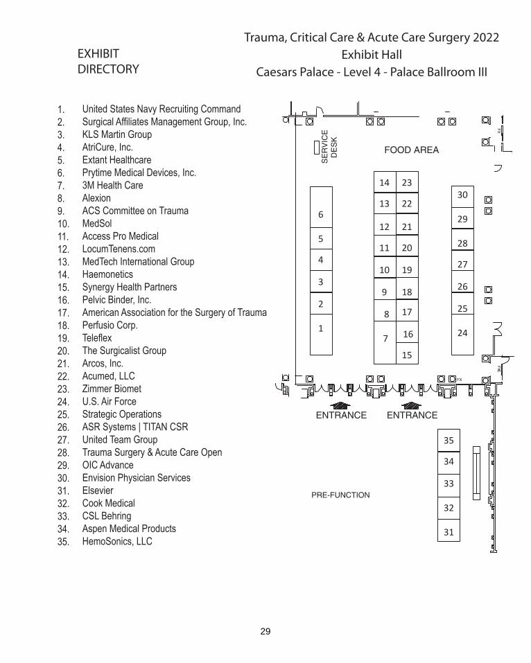
EXHIBITDIRECTORY
Trauma, Critical Care & Acute Care Surgery 2022
Exhibit HallCaesars Palace - Level 4 - Palace Ballroom III
United States Navy Recruiting CommandSurgical Affiliates Management Group, Inc.KLS Martin GroupAtriCure, Inc.Extant HealthcarePrytime Medical Devices, Inc.3M Health CareAlexionACS Committee on TraumaMedSolAccess Pro MedicalLocumTenens.comMedTech International GroupHaemoneticsSynergy Health PartnersPelvic Binder, Inc.American Association for the Surgery of TraumaPerfusio Corp. TeleflexThe Surgicalist GroupArcos, Inc.Acumed, LLCZimmer BiometU.S. Air ForceStrategic OperationsASR Systems | TITAN CSRUnited Team GroupTrauma Surgery & Acute Care OpenOIC AdvanceEnvision Physician ServicesElsevierCook MedicalCSL BehringAspen Medical ProductsHemoSonics, LLC
1.2.3.4.5.6.7.8.9.10.11.12.13.14.15.16.17.18.19.20.21.22.23.24.25.26.27.28.29.30.31.32.33.34.35.
6
5
4
3
2
1
14
13
12
11
10
9
8
7
23
22
21
20
19
18
17
16
15
30
29
28
27
26
25
24
35
34
33
32
31
29

Trauma, Critical Care & Acute Care Surgery 2022 Exhibit Directory
Access Pro Medical - #11 Our mission at Access Pro Medical is to be the single point of contact for “best-in-class” products and customer support for physician and healthcare professionals as they administer unsurpassed care. ACS Committee on Trauma – #9 The mission of the American College of Surgeons Committee on Trauma (ACS COT) is to develop and implement programs that support injury prevention and ensure optimal patient outcomes across the continuum of care. These programs incorporate advocacy, education, trauma center and trauma system resources, best practice creation, outcome assessment, and continuous quality improvement. Acumed, LLC - #22 Acumed | OsteoMed serves highly skilled, specialized surgeons. Acumed offers the most complete selection of upper extremity fixation and specialty plates on the US market and OsteoMed is a leading global innovator of specialty medical devices, surgical implants, and powered surgical instruments. Alexion – Booth #8 Alexion, AstraZeneca Rare Disease, is the group within AstraZeneca focused on rare diseases, created following the 2021 acquisition of Alexion Pharmaceuticals, Inc. As a leader in rare diseases for nearly 30 years, Alexion is focused on serving patients and families affected by rare diseases and devastating conditions. Headquartered in Boston, Massachusetts, Alexion has offices around the globe and serves patients in more than 50 countries. American Association for the Surgery of Trauma – #17 The American Association for the Surgery of Trauma (AAST) is dedicated to the discovery, dissemination, implementation, and evaluation of knowledge related to acute care surgery by fostering research, education, and professional development. Check out the new Trauma and Acute Care Surgery App, both of AAST’s Journals: JTACS and TSACO, research opportunities and the many other programs available through AAST. Arcos, Inc. – Booth #21 Arcos has developed the Blood Navigator™, a real-time data collection tool that helps balance massive transfusion ratios. Blood Navigator™:
• Rapidly track and document blood products • EMR integration & Downloadable Transfusion Report • Calcium and other adjunct reminders • Ensure balanced ratios to improve survivability
30
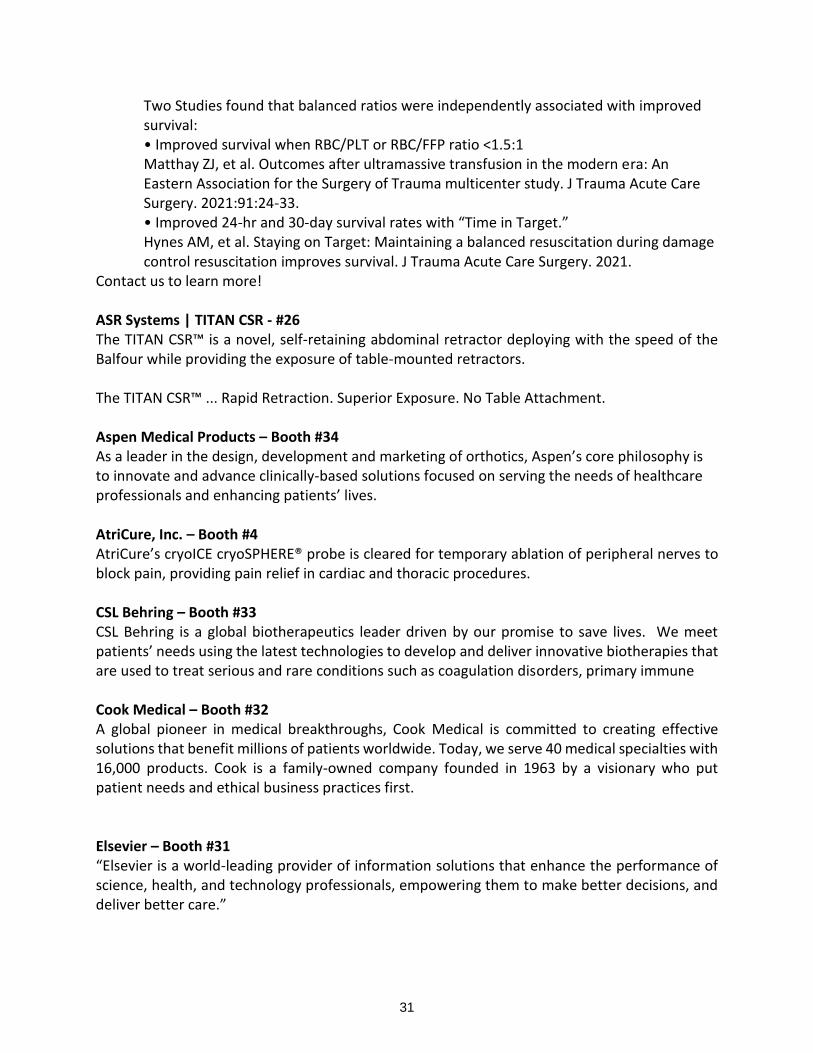
Two Studies found that balanced ratios were independently associated with improved survival: • Improved survival when RBC/PLT or RBC/FFP ratio <1.5:1 Matthay ZJ, et al. Outcomes after ultramassive transfusion in the modern era: An Eastern Association for the Surgery of Trauma multicenter study. J Trauma Acute Care Surgery. 2021:91:24-33. • Improved 24-hr and 30-day survival rates with “Time in Target.” Hynes AM, et al. Staying on Target: Maintaining a balanced resuscitation during damage control resuscitation improves survival. J Trauma Acute Care Surgery. 2021.
Contact us to learn more! ASR Systems | TITAN CSR - #26 The TITAN CSR™ is a novel, self-retaining abdominal retractor deploying with the speed of the Balfour while providing the exposure of table-mounted retractors. The TITAN CSR™ ... Rapid Retraction. Superior Exposure. No Table Attachment. Aspen Medical Products – Booth #34 As a leader in the design, development and marketing of orthotics, Aspen’s core philosophy is to innovate and advance clinically-based solutions focused on serving the needs of healthcare professionals and enhancing patients’ lives. AtriCure, Inc. – Booth #4 AtriCure’s cryoICE cryoSPHERE® probe is cleared for temporary ablation of peripheral nerves to block pain, providing pain relief in cardiac and thoracic procedures. CSL Behring – Booth #33 CSL Behring is a global biotherapeutics leader driven by our promise to save lives. We meet patients’ needs using the latest technologies to develop and deliver innovative biotherapies that are used to treat serious and rare conditions such as coagulation disorders, primary immune Cook Medical – Booth #32 A global pioneer in medical breakthroughs, Cook Medical is committed to creating effective solutions that benefit millions of patients worldwide. Today, we serve 40 medical specialties with 16,000 products. Cook is a family-owned company founded in 1963 by a visionary who put patient needs and ethical business practices first. Elsevier – Booth #31 “Elsevier is a world-leading provider of information solutions that enhance the performance of science, health, and technology professionals, empowering them to make better decisions, and deliver better care.”
31

Envision Physician Services – Booth #30 Envision Physician Services is America’s leading medical group providing anesthesiology, emergency medicine, hospital medicine, radiology, surgical services, and women’s and children’s health services to more than 1,800 clinical departments in 44 states and D.C. We empower our team of more than 27,000 clinicians with the resources they need to deliver high-quality care to patients when and where they need us most. Extant Healthcare – Booth #5 We provide management services for trauma/ ACS hospitals. We are actively recruiting for trauma/ SCC surgeons and APP's for our expanding group. HemoSonics, LLC – Booth # HemoSonics is revolutionizing point-of-care bleeding management with its Quantra® Hemostasis Analyzer - a novel, closed-cartridge viscoelastic monitoring system which delivers rapid, precise, easy to interpret coagulation results for informed treatment decisions. HemoSonics is a Stago Group company a leader in hemostasis and thrombosis. For more information, visit: www.hemosonics.com Haemonetics – Booth #14 The TEG® 6s hemostasis analyzer system from Haemonetics addresses the limitations of routine tests to deliver rapid, comprehensive & actionable information to help guide treatment decisions, improve resuscitation, & drive more efficient blood product use. It offers a small footprint, robust quality control, vibration resistance & simple operation to be readily deployed in trauma settings. TEG Manager• software delivers results wherever informed & timely hemostasis management decisions are needed. KLS Martin - #3 Surgical Innovation is our passion. KLS Martin is dedicated to providing surgical solutions to advanced patient care. We are focused on the development of innovative fixation products for use in the chest wall. We offer a wide variety of medical devices for Thoracic Surgery including our IPS “Individual Patient Solutions” for custom implants, sternal closures, and rib plating. New product developments in our fixation systems allow these products to be used for fracture fixation, reconstruction and chest wall stabilization. Based out of Jacksonville, Florida, we have a highly trained and qualified staff of representatives covering your needs throughout North America.
LocumTenens.com – Booth #12 Founded in 1995, LocumTenens.com is your full-service locum tenens agency recruiting physicians and advanced practitioners to solve employment shortages for healthcare facilities across the U.S. MedSol – Booth #10 Medical supply and equipment.
32

MedTech International Group -#13 Med-Tech International’s strong collaboration with our clinical partners continuously deliver innovative and alternative therapies to fulfill clinical needs and improve patient outcomes.
OIC Advance – #29 OIC Advance is a solutions company that offers our FDA Cleared Class II Tens unit. Our portable and compact Tens unit helps address various health issues. Such breakthrough in the medical device industry promotes confidence and wellness; thus enhancing efficiency on life performance. A step to better health! We are focused on clearer solutions for today with an eye to the future for better results.
PelvicBinder, Inc. – Booth #16 PelvicBinder, Inc is a medical device manufacture and distributor. We make innovative braces for pelvic fractures for adult and pediatric patients. Our ZipperBelt is excellent for sternotomies and sternal fractures. Stop by to see our BellyBinder for your hernia patients.
Perfusio Corp. - #18 Perfusio Corp. is a healthcare technology company that has developed and is marketing patented AI-enhanced algorithms for non-invasive, instantaneous surgical imaging to accurately access blood flow distribution and perfusion in intact and diseased tissue. We are excited to introduce Certes, an innovative and transformational technology for a one-of-a-kind Tissue Analysis (or Digital Technology) Platform. As the standard of healthcare continuously increases, this next generation technology leverages Artificial Intelligence designed to give “new knowledge” to healthcare providers to support optimal patient outcomes. Our patented AI-enhanced Multispectral Physiologic Visualization (MSPV) gives surgeons the ability to see beyond visible light (human eye) and provide deep insight into what is normal tissue and what is not, enabling critical decisions to be made at the time of surgery. Perfusio Corp. is dedicated to creating new standards of care with a purpose to implement solutions that improve patients’ lives and reduce health system costs.
Prytime Medical Devices, Inc. – Booth #6 Prytime Medical Devices, Inc., an innovative medical device company, designs, develops, and commercializes minimally invasive solutions for hemorrhage control. Strategic Operations - #25 Since 2002, Strategic Operations, Inc. (STOPS) has provided Hyper-Realistic® tactical training services and products to the military, law enforcement, first responders, and other organizations responsible for homeland security. STOPS pioneered the introduction of “Hollywood” style special effects and practices into live tactical training – explosions, weapons, realistic props, foreign language speaking actors, and casualty actors. Since the introduction of medical
33

simulation systems such as the Cut Suit (CS) STOPS now offers Advanced Surgical Skills Packages (ASSP), which together are the world’s only Hyper-Realistic® open surgery simulator (CS-ASSP). STOPS has added Hyper-Realistic® training support of civilian medical providers to its portfolio of products and services. STOPS continues to introduce innovative solutions to overcome the challenges of training military personnel, law enforcement personnel, first responders, and medical providers/surgical teams. STOPS is dedicated and focused on maximizing training value by outfitting individuals and organizations with essential training products to accomplish missions and save lives. STOPS also provides Hyper-Realistic® training environments resembling actual scene conditions offering participants “Stress Inoculation.” Active threats and mass casualty incidents have been increasing in frequency and complexity throughout the United States – throughout the world, law enforcement and medical emergency response teams have struggled to manage actual events. A significant contributor to this reality is the way in which first responders are able to or unable to train for these incidents. Many lack the time, funding, and facilities to adequately prepare. STOPS draws upon many years of experience, since 2002, to provide a fully immersive training environment at the Tactical Training Lab in San Diego, CA or at any location throughout the world. Surgical Affiliates Management Group, Inc. – Booth #2 Surgical Affiliates is a national leader in surgical hospitalist care with published, peer-reviewed results in the Journal of the American College of Surgeons that demonstrate how they benefit hospitals, clinicians, and patients by providing quality 24/7 emergency surgical care. The team is made up of a dynamic group of experienced, board-certified surgeons, healthcare providers, and medical directors. Programs offered provide strategic, structured surgical programs that encompass Acute Care, Trauma, Neurosurgery, and Orthopedics, and fuse with a hospital’s ICU, Emergency Department, and Medical Hospitalist Program to ensure quality of care and proper workflow throughout these departments. Synergy Health Partners – Booth#15 GO BEYOND LOCUMS Synergy Health Partners goes beyond locums by building dedicated provider teams that work collaboratively to achieve clinical standards of care and patient satisfaction. This approach builds an engaged team culture, offers performance-based incentives, and promotes continued professional and clinical development. We believe that every clinician’s primary focus should be on patient care. By offering an engaged and proactive environment, we create clinical career opportunities that help restore quality of life and passion for patient care.
34

3M Health Care – Booth #7 3M, with the acquisition of KCI, focuses on providing better care through patient-centered science. From wound and skin care to solutions for BSI and SSI risk reduction, our team is ready to partner with you to help transform patient outcomes. Teleflex - #19 Teleflex is a global provider of medical technologies designed to improve the health and quality of people’s lives. We apply purpose driven innovation – a relentless pursuit of identifying unmet clinical needs – to benefit patients and healthcare providers. Our portfolio is diverse, with solutions in the fields of vascular access, interventional cardiology and radiology, anesthesia, emergency medicine, surgical, urology and respiratory care. Teleflex employees worldwide are united in the understanding that what we do every day makes a difference. For more information, please visit teleflex.com. Teleflex is the home of Arrow®, Deknatel®, LMA®, Pilling®, QuikClot®, Rüsch®, UroLift®, and Weck® – trusted brands united by a common sense of purpose The Surgicalist Group - #20 "The Surgicalist Group is a surgeon founded and led organization providing acute care surgery (trauma and general), advanced wound care, surgical critical care, and advisory/management services to hospitals nationwide. We focus on driving performance improvements through every step of a patient’s hospital course – from ED to discharge. Our surgeons provide acute care surgery and work with hospitals to help demonstrate measurable outcome improvements in quality, satisfaction, safety, efficiency, and cost effectiveness through evidence-based protocols and lean practice techniques. Our practice is collaborative with community general surgeons enabling them to manage their elective practice while we manage the emergent and urgent cases. As we encounter non-emergent cases we refer these to our colleagues in the community." Trauma Surgery & Acute Care Open – #28 Trauma Surgery & Acute Care Open is the American Association for the Surgery of Trauma’s open access journal dedicated to the rapid publication of peer-reviewed, high-quality trauma and acute care research. Trauma Surgery & Acute Care Open provides an interdisciplinary forum for global issues in trauma and acute care surgery and is dedicated to covering epidemiological, educational, and socioeconomic facets of trauma management and injury prevention. United States Navy Recruiting Command – #1 U.S. Navy Medical Corps. Become a leader within the medical world – with financial assistance available. Learn more at the Navy booth or visit navy.com/healthcare.
35

United Team Group – Booth #27
United Team Group is a medical supplier and distributor of a large verity of medical led devices, uniforms, surgical equipment Natural Hemp Creams for Muscles, Joints, Back, Face, Neck and more operating from 2006. U. S. Air Force – Booth #24 Air Force Health Professions https://www.airforce.com Zimmer Biomet – Booth #23 Founded in 1927 and headquartered in Warsaw, Indiana, Zimmer Biomet is a global leader in musculoskeletal healthcare. With operations in more than 25 countries around the world, we design, manufacture, and market a variety of implants and surgical products. Zimmer Biomet continues to be a leader in the thoracic space and has been for more than 25 years, specializing in chest wall reconstruction.
36

SATELLITE LUNCHEON PROGRAMS Tuesday, March 29, 2022
12:30 – 2:00 PM This year, we are pleased to offer three Satellite Luncheon Program options
Innovative Approaches to the Management of Challenging Abdominal Surgical Site Complications: Reviewing Evidence and Complex Case Studies
Tuesday, March 29, 2022. 12:30 – 2:00 PM Augustus Ballroom 3-4
Faculty:
Demetrios Demetriades, MD, PhD, FACS (Moderator) Professor of Surgery University of Southern California Keck School of Medicine Director Acute Care Surgery LAC+USC Medical Center Los Angeles, CA
Mark J. Kaplan, MD, FACS Chairman, Division of Trauma and Surgical Critical Care Associate Chairman, Department of Surgery Albert Einstein Medical Center Philadelphia, PA
Casey J. Thomas, DO, FACOS, FACS Salt Lake Surgical Services Senior Partner Department of Surgery, Division of Acute Care Surgery Salt Lake City, UT
True Partial REBOATM: Who, When, How. Case Reports from the Field Tuesday, March 29, 2022 12:30 – 2:00 PM
Augustus Ballroom 5-6
Speakers:
M. Chance Spalding, DO, PhD, FACS | Emcee Director of Trauma Research, Grant Medical Center
Jonathan Nguyen, DO, FACS, FACOS | Panelist Director of Military Programs, Grady Memorial Hospital Assistant Professor of Surgery, Morehouse School of Medicine
Rishi Kundi, MD, RPVI, FACS, FSVS | Panelist Attending Trauma Surgeon & Attending Vascular Surgeon Deputy Director, GO-Team, R. Adams Cowley Shock Trauma Center
Andrew Beckett, CD, MD, MSc, FRCSC, FACS | Panelist Trauma Program Medical Director, St. Michael’s Hospital
Intrathoracic Rib Fixation Lecture and Hands-on Demonstration Tuesday, March 29, 2022 12:30 – 2:00 PM
Augustus Ballroom 1-2
Faculty:
John M. Green, MD, FACS Associate Professor Trauma/Acute Care Surgery Carolinas Medical Center Charlotte, NC
*These independent satellite luncheon programs are not accredited by or affiliated with the American College of Surgeons or Trauma, Critical Care & Acute Care Surgery 2022.
37

38

SESSION 1
HOT TOPICS
Moderator: Jennifer M. Gurney, MD, FACS
Monday, March 28, 2022
Palace Ballroom 1-2
Palace Tower, Emperors Level
7:30 - 7:45
TCCACS Review/Preview Kenneth L. Mattox, MD, FACS, MAMSE
7:45 - 8:00
Resuscitation 2022: What? How Much? When? Martin A. Schreiber, MD, FACS
8:00 - 8:15
Ultrasound 2022: Is It Really Useful in the Trauma Center? Joseph J. DuBose, MD, FACS, FCCM
8:15 - 8:30
Broken Ribs 2022: Who, When, and How? Fredric M. Pieracci, MD, MPH, MSPH, FACS
8:30 - 8:45
Direct Peritoneal Resuscitation 2022: Soothing the Savage Abdomen Jason W. Smith, MD, PhD, FACS
8:45 - 9:00
Always be closing! Modern Abdominal Wall Reconstruction Approaches Matthew J. Martin, MD, FACS
9:00 - 9:15
Pelvic Fracture Management 2022 Todd W. Costantini, MD, FACS
9:15 - 9:30
Appendicitis in 2022: Operate or Antibiotics? Andrew C. Bernard, MD, FACS
9:30 - 10:00
Panel Discussion - Hot Topics
39

SETTING THE STAGE
Kenneth L. Mattox, MD, FACS, MAMSE
Distinguished Service Professor Michael E. DeBakey Department of Surgery Special Advisor to the President & CEO Baylor College of Medicine Houston, TX
Welcome to Trauma, Critical Care, and Acute Care Surgery 2022, now in its 55t5h year at Caesars Palace. Yes, we are counting 2020, since all the work required to hold the meeting occurred before we were forced to cancel when Nevada’s governor “closed its doors” because of COVID-19. Even with this, we continue to be the “Longest Running Show in Las Vegas.” In 2021, we were the first group in the country to hold a large “live” meeting, with, 500 eager learners in attendance. This was the maximum number allowed under Nevada guidelines in effect at the time. The meeting was held safely and successfully. Now, in 2022, changes continue to occur in the field of trauma surgery, now known as trauma, critical care, and acute care surgery. This conference will continue to try to focus on the clinical and technical aspects of treatment in these areas and leave discussions (and solutions) to the non-clinical issues to the surgical (and other) organizations.
During and after the 2021 TCCACS courses, both the faculty, course attendees overwhelmingly expressed their recommendation that we make the 2022 conference a live event, if possible. We have followed that recommendation! All faculty will be present in Las Vegas to give their presentations. We do recognize that some locations around the world are still under the “COVID cloud,” and, for various reason are not able to travel to Las Vegas. To address the needs of these individuals, we are offering an On Demand option. For six months after the conference, the video of the entire conference will be available online as an On Demand course. The On Demand course is approved for the same number of CME credits. It will not offer the MOC/SA credits that can be claimed at the live course.
The public health rules continue to change (and may well change from the time this goes to press until we meet in Las Vegas)! Caesars Entertainment has maintained strict adherence to CDC, state, local and regulatory guidance throughout the COVID-19 pandemic and will continue to do so.
• SOCIAL DISTANCING Though Las Vegas no longer has any social distancing mandates, we have chosen to reduce capacity to allow for more space per attendee than in past years.
• HAND WASHING & SANITIZING
• Hand sanitizer will be readily available throughout the hotel and convention area
• WEARING MASKS
• Masks to be worn in compliance with local guidelines in effect at the time of the conference
As in past years, the course syllabus, CME claiming forms, and ability to submit questions to session moderators are available by accessing the conference dashboard using complimentary Wi-Fi in the convention area and your hotel room.
You will appreciate that the faculty have been chosen because of their experience and knowledge of a subject, not because they adhere to any particular “party line.” Furthermore, variances and differing views are encouraged among the faculty to inspire you to recognize the differences between dogma and emerging evidence-based issues in the ever changing areas of acute care surgery, surgical critical care,
40

and trauma management. We welcome you and appreciate your enthusiasm for this conference as well as your understanding of our adaptations to meet the constellation of challenges regarding venues, rules, and changing virus updates, to mention only a few. Ultimately, our continuous focus is our patients - to provide you with current knowledge that helps you give the very latest and best medical and surgical care. Let us know ([email protected], [email protected] ), details of specific cases and managerial tactics for which this course affected your practice.
Program details, as well as instructions for claiming CME and sending questions to Session Moderators are in your syllabus. We also encourage you to share your insights on the meeting on Twitter @kmattox1 and Facebook via TCCACS or Kenneth Mattox. Share the educational “pearls” you learn.
The CME and MOC activities are time sensitive. Do not delay completing. THE MOC/SA EXAM MUST BE SUBMITTED BY SATURDAY, APRIL 9, 2020, MIDNIGHT PST – NO ACCEPTIONS ALLOWED. Onsite assistance is available if you have questions about the online CME submission process.
As the TCCACS Course Director, I am grateful for the many individuals who make this course a success. Mary Allen. the glue that holds this course together, is the chief strategist, logistician, coordinator, and administrator – the force that keeps us all marching to the same drumbeat. Lisa Villarreal tabulates and responds to the many registrant inquiries is a major integrator of the day-to-day registration and graphic activities leading to a successful conference. The program committee develops a program format and content based on review of past attendee and faculty evaluations, review of publications and presentations read/observed in the past year, and individual experiences. They also assist in finding exciting, stimulating new faculty. This brings me to our faculty – a more dedicated, hard-working group of individuals you will be hard pressed to find. Each is dedicated to assuring you leave this conference armed with knowledge to guide you as you manage the complicated, as well as day to day challenges in patient care. Each works tirelessly to make their presentations the very best they can be. The exhibitors’ participation adds, yet, another very important aspect to the conference, offering attendees current information on their respective products. Take advantage of this additional learning tool. Our hosts at Caesars Palace work hard to make not only the conference a positive experience, but also to provide a venue that affords you many enjoyable options during those off-conference hours. We have worked with many Caesars personnel over the years, and they consider our group to be a special part of the Caesars Family.
Lastly, you, the attendees play one of the most important roles. Without your interest, participation, and enthusiasm, there would be no conference “energy” – one of the key elements to our success.
Indeed, this is a team effort, and all members of the team are essential to the success of the Mattox/Vegas TCCACS Conference. THANK YOU!
41

RESUSCITATION 2022: WHAT? HOW MUCH? WHEN?
Martin A. Schreiber, MD FACS
Professor and Chief Division of Trauma and Critical Care Oregon Health & Science University Portland, OR
The resuscitation of hemorrhagic shock has evolved tremendously during the period of the wars in Iraq and Afghanistan. Prior to this period, the focus was on early and aggressive resuscitation with crystalloids. Currently, the goals of resuscitation are to restore normal physiology, to include coagulation and to stop bleeding. These concepts are known as either hemostatic resuscitation or more commonly as damage control resuscitation and they have revolutionized modern care of the trauma patient.1
Aggressive resuscitation with crystalloid has been shown to result in increased mortality in both blunt and penetrating trauma.2,3 The randomized trial by Bickell et al performed in Houston and published in the New England Journal of Medicine in 1994 showed that in patients who were hypotensive with penetrating torso injuries, survival was increased when crystalloid resuscitation was delayed until hemorrhage control was achieved. Schreiber et al showed, in a multi-center randomized trial performed by the Resuscitation Outcome Consortium, that survival was increased in blunt trauma patients who received 250 ml boluses of crystalloid for an absent radial pulse or systolic blood pressure less than 70 mmHG compared to patients who were aggressively resuscitated to a goal systolic pressure of 110 mmHg with crystalloid.
In the modern era, emphasis has shifted from aggressive resuscitation to stopping the bleeding by whatever means possible to include pressure dressings, tourniquets, resuscitative endovascular balloon occlusion of the aorta or early hemostatic surgery and from resuscitation with crystalloid to resuscitation with blood products starting in the out-of-hospital setting. Blood product resuscitation is designed to avoid the negative effects of crystalloid resuscitation. Tissue injury and hypoperfusion result in the acute traumatic coagulopathy (ATC) which occurs immediately after injury. Resuscitation with room temperature crystalloid accentuates coagulopathy by producing hemodilution, acidosis and hypothermia resulting in the lethal cycle of trauma induced coagulopathy (TIC). Aggressive resuscitation with crystalloid has also been associated with an intense inflammatory response causing acute respiratory distress syndrome and multiple organ failure, endothelial dysfunction, hyperfibrinolysis, dysfibrinogenemia and platelet dysfunction.
Blood product resuscitation actively corrects both ATC and TIC by preventing each of the defects involved in these processes. The Joint Trauma System Committee on Tactical Combat Casualty Care (CoTCCC) has prioritized the use of resuscitation fluids based on these concepts and lists them from most to least preferred:
1. Liquid cold stored low titer O whole blood (LTOWB) 2. Pre-screened low titer O fresh whole blood 3. Plasma, red blood cells (RBCs) and platelets in a 1:1:1 ratio 4. Plasma and RBC in a 1:1 ratio 5. Plasma or RBCs alone
42

Liquid cold stored low titer O whole blood is preferred over fresh whole blood because it is approved for use by the FDA and it is widely available even in austere conditions. CoTCCC also recommends a 2-gram tranexamic acid bolus for patients undergoing massive transfusion.
In civilian practice, the use of whole blood was preceded by component resuscitation given in a ratio of 1:1:1 plasma:platelets:RBCs or a similar ratio. This was supported by the PROPPR study which randomized patients predicted to receive a massive transfusion to a 1:1:1 ratio or a 1:1:2 ratio of plasma:platelets:RBCs.4 Patients randomized to a 1:1:1 ratio were more likely to be alive at 24 hours and less likely to die from exsanguination. The high ratio blood component resuscitation strategy was utilized to replicate whole blood and eventually liquid cold stored whole blood became increasingly available at US trauma centers.
It is important to realize that not all whole blood is equivalent. Whole blood is considered fresh for 48 hours after donation. It can be stored at 22C for up to 8 hours and must be stored at 4C for up to 40 additional hours. After 48 hours, whole blood is known as liquid cold stored whole blood. It is stored from 1-6C and if stored in CPD or CP2D solution it can be stored up to 21 days. If adenosine is added to the storage solution, it can be stored up to 35 days. Liquid cold stored whole blood is the product that is typically utilized in the US. Liquid cold stored whole blood is theoretically superior to components given in a 1:1:1 ratio because it contains less preservatives and anticoagulants as shown in Table I.5
TABLE I.
As shown in Table I, the total added volume of diluents including citrate to whole blood is 70cc whereas the total added volume of diluents to components is approximately 200cc. In massive transfusions, this can amount to large volumes of diluents and anti-coagulants.
Low titer O whole blood (LTOWB) is typically utilized as a universal donor for whole blood in both military and civilian settings. Low titers to anti-A and anti B antigens can be defined as from anywhere between 50 and 200 depending on the whole blood supplier. Blood transfusion reactions to LTOWB are rarely reported.
In retrospective studies performed in theater that are controlled for severity of injury, a strong association between the use of fresh whole blood and survival has been established.6 Civilian studies evaluating the use of LTOWB have produced mixed results. A retrospective propensity matched trial performed in Pittsburgh which compared 135 LTOWB recipients to component recipients revealed no difference in morbidity or mortality. However, lactate was noted to normalize earlier in whole blood recipients.7 Another retrospective study from Portland, comparing patients who received large volume transfusions with LTOWB to components alone also revealed no association between LTOWB and survival.8 In this study, patients who received LTOWB did receive significantly less RBC and plasma components. There was no difference in platelets, cryoprecipitate or total components given. In a recent prospective observational study from Barnes, 42 patients who received component therapy alone were compared to 44 patients who received LTOWB.9 In this study, there was a significant association between LTOWB use with improved survival and significant reductions in RBCs, platelets and plasma.
43

LTOWB, used in civilian practice has significant differences based on the supplier and these differences are critical for end users to understand. Some blood suppliers like the American Red Cross routinely perform leukoreduction with a platelet sparing filter. Platelet sparing filters do result in a reduction in platelet count and function as measured by ROTEM.10 The coagulation capacity of whole blood also deteriorates over time and by 14 days, LTOWB has lost a significant portion of its coagulation benefit.11 Based on this loss of function and the fact that our LTOWB is leukoreduced, unused whole blood at Oregon Health & Science University is converted to packed red blood cells after 14 days in our blood bank. Due to the variability in coagulation of whole blood units, we routinely perform Thrombelastography every 30 minutes to 1 hour during massive transfusions and give additional components based on the results.
Facilities that utilize LTOWB for the resuscitation of trauma patients have a limited supply of product and must resort to component therapy when that supply is exhausted. Severe blood shortages have occurred due to the Covid pandemic and the component that is most effected is platelets. The majority of platelets utilized in civilian practice are derived from apheresis and a single unit of apheresis platelets is equivalent to 6 single donor units. Platelets are stored at room temperature and require agitation. Due to the risk of infection, they can only be stored for 5 days. Room temperature storage of platelets results increased platelet survival in vivo after transfusion from 1.3 days with cold platelets to 3.9 days. However, cold stored platelets have been shown to have increased function probably resulting in earlier removal from circulation and 1.3 day circulation is more than adequate for trauma indications.12 The FDA has approved cold platelets for use in theater and is now allowing the use of cold platelets with a variance in the US when warm platelets are not available. Under this variance, cold platelets can be stored up to 14 days alleviating shortages associated with using warm platelets and possibly resulting in better hemostasis.
The other critical component utilized in the initial resuscitation of trauma patients is plasma. In order for plasma to be given in a 1:1:1 ratio in a massive transfusion, the product must be in the liquid form due to the 30-minutes required to thaw it. This is problematic as the majority of plasma is stored as fresh frozen plasma which can be stored for up to 1 year. Presumptive thawing of FFP can result in significant waste. Freezing and thawing results in reduction of coagulation function. Once plasma is thawed, it can be stored for up to 5 days with minimal degradation of factor function allowing high ratio transfusion to occur. Optimally, in large institutions with high plasma utilization, donated plasma is never frozen. This product is known as liquid plasma and it can be stored for up to 26 days with minimal factor degradation. This allows a facility’s entire plasma inventory to be available when needed for massive transfusion. Although lyophilized plasma products which are readily available and easily stored are available in other parts of the world such as South Africa, Israel, Germany and France as well in the US deployed setting, they are not yet available in the US itself.
In addition to being an excellent source of volume replacement and coagulopathy correction, plasma has also been shown to decrease the endotheliopathy associated with hemorrhagic shock and rebuild the damaged glycocalyx resulting in reduced extravasation of fluid and improved organ function.13 There are thousands of proteins in plasma which may ameliorate inflammatory dysfunction and multiple organ failure. Prehospital use of LTOWB in the civilian setting has been associated with decreased trauma bay mortality and prehospital use of plasma has been associated with improved overall survival.14, 15
CONCLUSION
The modern resuscitation of trauma patients is designed to correct coagulopathy, restore hemostasis and restore blood volume. This is currently being done with either LTOWB or components given in a 1:1:1 ratio. Whether hypotensive resuscitation or delayed resuscitation remains beneficial in the current era is unknown. Early resuscitation of the trauma patient in hemorrhagic shock should not include crystalloids.
44

REFERENCES
1. Holcomb JB, Jenkins D, Rhee P, et al. Damage Control Resuscitation: Directly Addressing the Early Coagulopathy of Trauma. Journal of Trauma. 2007;62:307-310.
2. Bickell WH, Wall MJ, Pepe PE et al. Immediate Versus Delayed Resuscitation for Hypotensive Patients with Penetrating Torso Injuries. NEJM;331:1105-1109.
3. Schreiber MA, Meier EN, Tisherman SA, et al. A Controlled Resuscitation Strategy is Feasible and Safe in Hypotensive Trauma Patients: Results of A Prospective Randomized Pilot Trial. Journal of Trauma and Acute Care Surgery;78:687-697.
4. Holcomb JB, Tilley BC, Baraniuk S, et al. Transfusion of Plasma, Platelets and Red Blood cells in a 1:1:1 vs a 1:1:2 Ratio and Mortality in Patients with Severe Trauma. JAMA. 2015;313:471-482.
5. Seheult JN, Bahr MP, Spinella PC, et al. The Dead Sea Needs Salt Water . . .Massively Bleeding Patients Need Whole Blood: The Evolution of Blood Product Resuscitation. Transfusion Clinique et Biologique. 2019;26:174-179.
6. Nessen SC, Eastridge BJ, Cronk et al. Fresh Whole Use by Forward Surgical Teams in Afghanistan is Associated with Improved Survival Compared to Component Therapy Without Platelets. Transfusion. 2013;53:107S-113S.
7. Seheult JN, Anto V, Alarcon LH, et al. Clinical Outcomes Among Low-titer Group O Whole Blood Recipients Compared to Recipients of Conventional Components in Civilian Trauma Resuscitation. Transfusion. 2018;9999;1-8.
8. Gallaher JR, Dixon A, Cockroft A, et al. Large Volume Transfusion with Whole Blood is Safe Compared with Component Therapy. Journal of Trauma and Acute Care Surgery. 2020;89:238-245.
9. Shea SM, Staudt A, Thomas KA, et al. The Use of Low-titer Group O Whole Blood is Independently Associated with Improved Survival Compared to Component Therapy in Adults with Severe Traumatic Hemorrhage. Transfusion. 2020;60:S2-S9.
10. Remy KE, Yazer MH, Saini A, et al. Effects of Platelet-sparing Leukocyte Reduction and Agitation Methods on In Vitro Measures of Hemostatic Function in Cold-stored Whole Blood. 2018;84:S104-S114.
11. Strandenes G, Austlid I, Apeseth TO, et al. Coagulation Function of Stored Whole Blood is Preserved for 14 Days in Austere Conditions: A ROTEM Feasibility Study During a Norwegion Antipiracy Mission and Comparison to Equal Ratio Reconstituted Blood. Journal of Trauma and Acute Care Surgery. 2015;78:S31-S38.
12. Reddoch KM, Pidcoke HF, Montgomery RK, et al. Hemostatic Function of Apheresis Platelets Stored at 4C and 22C. Shock. 2014;40, Suppl 1:54-61.
13. Wu F, Chipman A, Pati S, Miyasawa B, Corash L, Kozar RA. Resuscitative Strategies to Modulate the Endotheliopathy of Trauma: From Cell to Patient. Shock 2020;53:575-584.
14. Braverman MA, Smith A, Pokorny D, et al. Prehospital Whole Blood Reduces Early Mortality in Patients with Hemorrhagic Shock. Transfusion. 2021;61:S15-S21.
15. Sperry JL, Guyette FX, Brown JB, et al. Prehospital Plasma During Air Medical Transport in Trauma Patients at Risk for Hemorrhagic Shock. NEJM. 2018;379:315-326.
45

ULTRASOUND 2022: IS IT REALLY USEFUL IN THE TRAUMA CENTER?
Joseph J. DuBose, MD, FACS, FCCM
Professor of Surgery Dell Medical School The University of Texas at Austin Austin, TX
Evaluation of patients with thoraco-abdominal trauma continues to present a challenge for emergency practitioners and traumatologists. As many as 50 % of patients with severe abdominal trauma and/or multiple injuries either have a normal initial abdominal exam or are unconscious and thus unable to provide a reliable abdominal exam. Consequently, the unreliable nature of history and physical exam in the trauma population has led physicians to increasingly depend on diagnostic imaging. Computed tomography (CT) is a widely used imaging modality but involves ionizing radiation and is not the best option for unstable or potentially unstable patients. Concurrently, the use of ultrasound has increased during the past decade, with sonography becoming an essential adjunct in the trauma resuscitation area. 4,5
Ultrasound-based methodology was formally introduced into the trauma literature in the 1990’s. The focused assessment with sonography for trauma (FAST) exam now serves as an important component of trauma algorithms, especially for the evaluation of unstable patients where early surgical intervention of the abdomen may be lifesaving. Prior to the era of the FAST exam, diagnostic peritoneal lavage was utilized to evaluate for the need for laparotomy
in the unstable trauma patient. In 1999, the FAST consensus conference concluded that the ‘FAST’ abbreviation should stand for Focused Assessment with Sonography for Trauma in order to capture the utility of not only viewing the abdomen but also evaluating the heart, the pericardial and pleural spaces. Since its more widespread clinical implementation, the extended FAST (or E-FAST) that incorporates pneumothorax assessment was found to offer sensitivities and specificities that are superior to those of traditional chest radiography.9 Currently, a number of board certifying entities require that emergency physicians, intensivists and surgeons be proficient in bedside ultrasound evaluation of injured and critically ill patients.
THE FAST EXAM
The FAST exam exemplifies the evolution of the general trauma approach over the last two decades, from maximally invasive to minimally- or non-invasive. The overarching assumption of FAST is that all clinically significant abdominal injuries are associated with hemoperitoneum.
The traditional FAST paradigm includes four basic sonographic views (a) pericardial; (b) perihepatic; (c) perisplenic; and (d) pelvic. This simple test has nearly eliminated the need for diagnostic peritoneal lavage. Not only do ultrasound based approaches take the decision-making directly to the patient (i.e., point-of-care methodology) but also facilitate repeated bedside evaluations when resources to perform such assessments using CT or other advanced imaging would be prohibitive. While perhaps not immediately clinically relevant, proprietary algorithms have been published that may lead to better quantification of the amount of fluid found on FAST examinations, allowing even more informed decisions.
There are also important limitations to FAST. Among those, the exam has been noted to have poor accuracy in the very early post-injury phase, where sufficient hemoperitoneum had not yet accumulated
46

thus leading to false-negative results. In addition, its utility in detecting retroperitoneal blood is very limited. Furthermore, FAST is unable to identify hollow viscus or solid organ injuries not associated with hemoperitoneum such as early bowel injury or pancreatic injury. Lastly, it has been anecdotally reported that pericardial FAST window may be falsely negative in the presence of pericardial injury that has resulted in an adjacent pleural defect and thus a pleural effusion. In summary, the FAST exam has a valuable role in the early evaluation of trauma patients, but CT of the abdomen remains the gold standard evaluation technique to exclude abdominal injury in the hemodynamically stable patient who warrants further evaluation due to specific symptoms or complaints. Also FAST exam should not replace other techniques in the evaluation of penetrating trauma, such as traditional radiography.
In this context, the trajectory of the penetrating injury and other imaging studies plays a greater role in decision making than FAST.
FROM FAST TO E‑FAST
The FAST exam continues to gain popularity as it is easily learned, readily accessible, portable, and more physicians are becoming comfortable with point-of-care sonography in general. At many institutions, protocols that encourage the use of the FAST exam have resulted in significant reduction in computed tomographic (CT) scan use in trauma, thus decreasing overall costs and radiation exposure. In one study, the use of the FAST exam changed clinical management in nearly one-third of patients and reduced CT scan utilization from 47 to 34 %. The determination of when it is appropriate to limit abdominal evaluation to FAST exam only is yet to be clearly delineated. More specifically, given the possibility of abdominal injury without free fluid, patients with abdominal contusion, abdominal pain, or altered mental state should not be evaluated with FAST alone. As originally intended, FAST is best used as an adjunct to quickly detect intra-abdominal fluid in trauma patients. Ultrasound can easily detect as little as 200 mL of fluid in Morrison’s pouch and can be completed in less than 1 min in the hands of an experienced operator. The E-FAST further adds to the basic information provided by the FAST by including the examination of the thorax anteriorly to assess for the presence of pneumothorax and at the flanks to assess for hemothorax Pneumothoraces are common in trauma and as many as half are missed on a routine supine chest radiograph. Regarding hemothoraces, a supine or upright CXR requires up to 175 or 50–100 mL of fluid in order to be visualized, compared to E-FAST which can detect as little as 20 mL of fluid in the pleural space.
While ultrasound continues to gain popularity and more healthcare teams embrace protocols that utilize FAST to reduce unnecessary CT scan use, these protocols must be used with caution as FAST does have a number of important limitations. As stated above, proper patient selection is crucial and there are many potential sources of diagnostic bias, including the presence of obesity and subcutaneous fat, body habitus and positioning, the presence of abdominal or retroperitoneal injury without hemoperitoneum, ascites due to medical condition (i.e., hepatic cirrhosis), pre-existing pericardial effusion, and the presence of intra-abdominal cysts or masses . Pitfalls leading to false positive readings in the cardiac window include clotted hemoperitoneum, epicardial fat and pleural effusions being mistaken for pericardial effusions. Patients with the above characteristics or findings should also be evaluated with CT scans, provided that they are hemodynamically stable. In all cases, good clinical judgment is essential and serial FAST exams may increase the overall accuracy of the ultrasound-based assessment.
47

Figure 1. A. Left normal interface between the spleen and the kidney. There is no fluid between the two, indicating a "negative" result. Note the hyperechoic "spine sign" in the far field. This is due to the continuation of the spine above the diaphragm in the presence of fluid (seen as the sharp-angled anechoic area above the diaphragm) in the pleural space. B. Right positive spleno-renal window, with anechoic fluid (arrow) seen clearly around the spleen
Figure 2. A. Left negative right upper quadrant view displays the liver with multiple anechoic areas representing hepatic and portal veins next to the elliptical kidney with the hyperechoic spine in the far field. Note how the spine stops at the diaphragm and is not visualized above (cephalad) to this area. B. Right positive right upper quadrant view with anechoic fluid seen around the liver (arrow)
48

Figure 3. A. Left the "bladder view" provides visualization of the pelvic structures and the most dependent space in the pelvis. This image depicts a moderately filled bladder with the typical "box-like" appearance of a negative scan. B. Right top and bottom free fluid (arrows) present in the pelvis on a positive FAST scan. The sonographer is more likely to see irregularly shaped outlines, often with sharp angles compared with the more "rounded" edges of the bladder in the negative scan on the left
Figure 4. A. Left negative pericardial view. B. Right positive pericardial view, with clearly visible anechoic fluid (arrow) between the heart and the pericardium. This is a critical finding that should prompt further immediate intervention(s)
49

Figure 5. A. Left pneumothorax, with the "bat sign" seen as marked (ribs = wings = x; pleural line= body = arrow). B. Right normal lung on static images, with the presence of "comet tail" artifacts (arrow). Motion picture in this case would demonstrate "pleural/ lung sliding"
50

BROKEN RIBS 2022: WHO, WHEN, AND HOW?
Fredric M. Pieracci, MD, MPH, MSPH, FACS
Interim Director of Surgery Denver Health Medical Center Professor of Surgery University of Colorado Denver Denver, CO
Rib fractures remain the most common thoracic injury encountered in the trauma patient. Most rib fractures are managed non-operatively, with a combination of pulmonary toilet, multimodal analgesia, and physical therapy. One current area of controversy is the role of surgical stabilization of rib fractures (SSRF) in the minority of patients with severe chest wall injuries, mainly either flail chest or multiple severely displaced fractures.
Early attempts at rib fixation were limited by inadequate technology. Ribs are unique bones in that they are relatively thin, flexible, and associated with an acute bend near their articulation with the spine. Over the last ten years, several rib-specific fixation systems have come to market, which respect the unique characteristics of ribs, and have greatly facilitated both reduction and fixation. Examples of technological advancements include “T plates” for fixation to the sternum, ultra-thin plates for fixation to costal cartilage, and thoracoscopic fixation systems. Armed with these tools, the surgeon is now faced with identifying which patients will benefit most from SSRF.
More than ten randomized controlled trials (RCTs) have now been conducted in the flail chest patient population, each of which have compared SSRF to non-operative management. One major source of heterogeneity in these RCTs is a variable definition of flail chest, ranging from paradoxical respiratory motion observed on physical exam, to a radiographic flail segment. Moreover, RCTs vary widely in time from injury to fixation, patient demographics, and surgeon experience (this heterogeneity has led us to propose a standardized reporting structure for studies of SSRF). However, the majority of RCTs have demonstrated benefit to SSRF in patients with flail chest in both the immediate and subsequent recovery phases, and both the Eastern Association for the Surgery of Trauma and Chest Wall Injury Society (CWIS) consensus guidelines recommend consideration of all patients with flail chest for SSRF.
Current research focuses on the utility of SSRF for patients with non-flail fracture patterns. Our recent query of the National Trauma Database revealed that the incidence of SSRF in the United States is increasing exponentially and, somewhat concerningly, predominantly in patients without a diagnosis of flail chest and at lower-level trauma centers. The incongruity between evidence and practice highlights the need for urgent research in this area.
Non flail fracture patterns represent a wide range of injuries, including everything from subscapular to anterior fractures. Furthermore, similar radiographic fracture patterns can demonstrate markedly different clinical phenotypes, such that one patient will be thriving while the other will progress to respiratory failure. We created a dynamic, objective clinical assessment tool to capture patients’ physiologic response to rib fractures, termed the Sequential Clinical Assessment of Respiratory Function (SCARF) score. The score grants one point for incentive spirometry < 50% of predicted, respiratory rate > 20, numeric pain scale ≥ 5, and inadequate cough, for a total possible score of four. We found that, among 100 surgical intensive care unit (SICU) patients with non-flail rib fracture patterns, the SCARF score
51

correlated closely with pneumonia, prolonged SICU length of stay (LOS), and need for supplemental oxygen. We used these data to create an algorithm in which patients with a SCARF score > 2 were considered for additional interventions such as locoregional anesthesia or SSRF (Figure). One advantage of the SCARF score is that it is dynamic, meaning that it may be calculated multiple times over the course of a day, as well as both prior to and after intervention to assess efficacy.
Use of the SCARF score to stratify patient into risk of complication, and therefor need for additional, invasive therapies.
Armed with the SCARF score, and in an attempt to define a patient population in which to study the efficacy of SSRF in non-flail fracture patterns, we performed a survey of the members of CWIS, in which 18 clinical scenarios were varied for patients with rib fractures. Respondents were asked to answer if SSRF was indicated for each scenario, and the scenario that returned closest to equipoise (50%) was a patient with three or more displaced fractures, age < 70, mild to no traumatic brain injury (TBI), and a SCARF score of ≥ 2. We used these inclusion criteria to conduct the first prospective, multicenter study of SSRF among patients with non-flail fracture patterns, termed CWIS NON-FLAIL. A total of 110 subjects were enrolled from 12 centers in the United States over 18 months. We found that the primary outcome of the numeric pain score at two weeks post operatively was significantly decreased in the operative, as compared to the non-operative group. Narcotic use and quality of life were also improved. Finally, pleural space complications were eliminated in the operative arm (routine pleural irrigation was used during SSRF) as opposed to a 10% incidence in the non-operative arm. Although additional data in this specific patient subgroup continue to emerge, we viewed the results of the CWIS NON-FLAIL trial as encouraging for the utility of SSRF.
Another frequent question surrounding SSRF involves timing of surgery. Patients must be stabilized and not have higher competing injuries (e.g, aortic tear, unstable spine) before SSRF is considered. Short of these contra-indications, data suggest that earlier surgery is better, and specifically surgery within 48 hours of injury is optimal. We are actively researching the benefit of a direct “ED to OR" approach for patients with isolated severe chest wall injuries, particularly as a tertiary trauma hospital that receives such patients from outlying, lower-level hospitals.
Our research over the last three years has focused on refining indications for SSRF among additional specific subgroups of patients. The first involved patients aged ≥ 80 years. Although elderly patients were initially considered too high risk for SSRF, increased experience led to the anecdotal observation that older patients were less likely to tolerate the morbidity of severe chest wall injuries managed non-operatively,
52

and tolerated SSRF relatively well, particularly as minimally invasive approaches became more prevalent. This hypothesis was termed “too frail to fail.” In a multicenter retrospective study, we matched patients aged ≥ 80 years by several demographic and injury-related characteristics and divided into operative and non-operative groups. Mortality was reduced by 60% in the operative group, arguing for a role of SSRF in this patient population, with the caveat that pre-operative functional status and goals of care must be considered strongly.
Another multicenter study assessed the role of SSRF in patients with TBI. This international study analyzed 456 patients from 19 countries. In the SSRF group, surgery was performed a median of three days after injury. There was no difference in the primary outcome of ventilator free days for patients with moderate to severe TBI for the operative as compared to the non-operative group. Accordingly, we exercise caution when offering SSRF to any patient with TBI.
In conclusion, SSRF appears beneficial in select patients with non-flail fracture patterns, including the elderly. Patients with TBI should not routinely be considered for SSRF. In assessing patients for SSRF, an objective, dynamic, tool for determining physiologic response should be routinely used.
REFERENCES
1. Kasotakis G, Hasenboehler EA, Streib EW, et al. Operative fixation of rib fractures after blunt trauma. A practice management guideline from the Eastern Association for the Surgery of Trauma. J Trauma Acute Care Surg 2017;82:816-626.
2. Pieracci FM, Majercik S, Ali-Osman F, et al. Consensus statement: surgical stabilization of rib fractures – rib fracture colloquium clinical practice guidelines. Injury 2017;48:307-321
3. Pieracci FM, Schubl S, Gasparri M, Delaplain P, Kirsch J, Towe C, White TW, Whitbeck SA, Doben AR. The Chest Wall Injury Society recommendations for reporting studies of surgical stabilization of rib fractures. Injury 2021; S0020-1383(21).
4. Kane E, Pieracci FM, Majercik S, Doben AD. Quantifying and exploring the recent national increase in surgical stabilization of rib fractures. J Trauma Acute Care Surg 2017;83(6):1047-1052.
5. Hardin KS, Leasia KN, Haenel J, et al. The sequential clinical assessment of respiratory function (SCARF) score: a dynamic pulmonary physiologic score that predicts adverse outtcomes in critically ill rib fracture patients. J Trauma Acute Care Surgery 2019;87(6):1260-1268.
6. Pieracci FM, Agarwal S, Doben A, et al. Indications for surgical stabilization of rib fractures in patients without flail chest: surveyed opinions of members of the chest wall injury society. Orthopedics International 2018;42(2):401-408.
7. Pieracci FM, Leasia K, Bauman Z, et al. A multicenter, prospective, controlled clinical trial of surgical stabilization of rib fractures in patients with severe, non-flail fracture patterns (CWIS NON-FLAIL). J Trauma and Acute Care Surg 2020;88(2):249-257
8. Pieracci FM, Coleman J, Ali-Osman F, et al. A multicenter evaluation of the optimal timing of surgical stabilization of rib fractures. J Trauma Acute Care Surg 2018;84(1):1-10.
9. Pieracci FM, Leasia K, Hernandez MC, et al. Surgical stabilization of rib fractures in octogenarians and beyond – what are the outcomes? J Trauma and Acute Care Surg 2021;90(6):1014-1021.
10. Prins J, Van Lieshout E, Ali-Osman F, Bauman Z, et al. Outcome after surgical stabilization of
rib fractures versus nonoperative treatment in patients with multiple rib fractures and
moderate to severe traumatic brain injury (CWIS-TBI). J Trauma Acute Care Surgery 2021;
90(3):492-500.
53

DIRECT PERITONEAL RESUSCITATION 2022: SOOTHING THE SAVAGE ABDOMEN
Jason W. Smith, MD, PhD, FACS
Berel L. Abrams MD Endowed Professor Director, Division of General Surgery University of Louisville Hospital Louisville, KY
Hemorrhagic shock is a leading cause of death following trauma worldwide. The mainstay of treatment being to control source of hemorrhage and restore hemodynamics through intravenous resuscitation. Despite restoration of central hemodynamics, the visceral organs experience a progressive vasoconstriction that results in prolonged ischemia and subsequently an ischemia reperfusion injury. Following the initial hemorrhage, a subset of trauma patients will go on to develop multi system organ dysfunction secondary to an aberrant immune response related to this phenomenon Attenuating this response has been a target of much research.1-3 One method, direct peritoneal resuscitation involves the instillation of a hypertonic, glucose-based dialysate solution into the abdomen as an adjunct to traditional modes of resuscitation.4
WHAT IS THE INNOVATION?
Direct peritoneal resuscitation (DPR) is a validated resuscitation strategy that, when used as an adjunct to other modes of resuscitation, reverses visceral vasoconstriction, improving blood flow and decreasing downstream organ injury. The mechanism of action is multifactorial but is thought to be due to the hypertonicity of the fluid as well as the lactate, glucose and glucose degradation product components of the DPR itself. The hypertonicity of the fluid reduces transcellular water diffusion through aquaporin channels following ischemia. Glucose and its degradation products, which are formed during heat sterilization of the solution, are vasodilators of the intestinal microvasculature via nitric oxide and adenosine release.5-6 The hypertonicity also contributes to decreased cellular and visceral edema.3-7
WHAT ARE THE KEY ADVANTAGES OVER EXISTING APPROACHES?
These properties led to initial laboratory studies using DPR as an adjunct to other resuscitative methods following hemorrhagic shock. When compared to crystalloid resuscitation alone, DPR improves hepatic blood flow, decreases tissue necrosis, decreases the release of damage associated molecular proteins and improves mortality. More recent laboratory studies have demonstrated the combination of fresh frozen plasma resuscitation and DPR, further improves visceral blood flow and decreases intestinal ischemia reperfusion injury following hemorrhagic shock. This combination has also been shown to protect the endothelial glycocalyx and decrease cardiac and hepatocellular damage.1-2
DPR’s anti-inflammatory effects seem to offer benefit in pathologies other than hemorrhagic shock. When examining combined hemorrhage and burn injury, DPR demonstrated survival benefit as well as delay in the progression of acute lung injury.8 Other laboratory studies have demonstrated that the use of DPR decreases the inflammatory response, improves hepatic blood flow, and decreases pulmonary edema and inflammation within the kidney after acute brain death.9-12 It has also been shown to attenuate ischemia reperfusion induced spinal cord injury.13
54

HOW WILL THIS AFFECT CLINICAL CARE?
In the clinical arena, DPR was initially applied in the setting of damage control surgery for intrabdominal hemorrhage and subsequently broadened to include sepsis or abdominal compartment syndrome in patients with an open abdomen awaiting definitive closure. To make use of DPR as an adjunctive resuscitation method, typically, a drain is placed at the time of the initial operation along the root of the mesentery and then the abdomen closed with a temporary abdominal closure (TAC). DPR is initiated post operatively, as an initial bolus and then at a standard rate (~400mL/hour) through the mesenteric drain and drained via the TAC. The 2.5% glucose based peritoneal dialysis solution (Delflex) is the most widespread and studied type of DPR solution, although some studies have demonstrated the benefit of pyruvate containing DPR fluids.3 The DPR irrigation continues at the previous determined rate until the patient is taken for their definitive operation. Intravenous resuscitation proceeds as directed by the treating physician.
IS THERE EVIDENCE SUPPORTING THE BENEFITS OF THE INNOVATION?
Although limited, human studies have demonstrated numerous benefits to the use of DPR in clinical scenarios. When used as an adjunctive resuscitation method in hemorrhagic shock, a retrospective review of matched patients demonstrated a decreased time to abdominal closure, decreased hernia rate at six months and decreased rate of intra-abdominal complications with DPR compared to traditional methods. 14-15 In other settings that result in TAC, DPR has also shown to have significant benefit. Another study looking at emergency general surgery patients demonstrated that DPR decreases evidence of organ dysfunction and cellular ischemia in patients who have undergone emergency general surgery for intrabdominal sepsis. A retrospective study demonstrated decreased time definitive closure, decreased acidosis, improved pAO2, decreased Cr and overall decreased mortality rate in the DPR group.16
DPR use is not limited to those patients with open abdomens. Due to its anti-inflammatory properties, some institutions have adopted its use in organ donors, following brain death but prior to organ retrieval. In lieu of a laparotomy, a peritoneal catheter is placed at bedside using a direct peritoneal lavage kit. The DPR is then initiated just as it would be with a TAC. In this setting, the use of DPR has been shown to decrease intravenous fluid requirement, decrease vasopressor requirements, improve hepatic blood flow, reduce afterload and improve cardiac function. A prospective, case-control study confirmed these benefits as well as demonstrated and increase in the number of organs transplanted per donor. 17
IN WHAT TIME FRAME WILL THIS INNOVATION LIKELY BE APPLIED ROUTINELY?
Future studies will continue to explore various applications and benefits of adjunctive DPR. Due to its ability to improve microcirculatory flow and attenuate ischemia reperfusion injury, it could prove to beneficial in various diseases where the pathophysiology is related to ischemia reperfusion injury and the aberrant immune response that results from this. As resuscitative methods have continued to improve, with a more widespread application of hemostatic resuscitation, further research into the role and application of DPR in the clinical setting. If these studies prove further benefit, this could lead to broader implementation of the DPR protocol in the future.
REFERENCES
1. Schucht JE, Matheson PJ, Harbrecht BG, Bond L, Ashkettle GR, Smith JW. Plasma resuscitation with adjunctive peritoneal resuscitation reduces ischemia-induced intestinal barrier breakdown following hemorrhagic shock. J Trauma Acute Care Surg. 2021;90(1):27-34.
2. Schucht JE, Matheson PJ, Harbrecht BG, Bond L, Jones S, Alkhateeb KJM, et al. Plasma resuscitation with adjunctive peritoneal resuscitation reduces ischemic intestinal injury following hemorrhagic shock. J Trauma Acute Care Surg. 2020;89(4):649-57.
55

3. Weaver JL, Smith JW. Direct Peritoneal Resuscitation: A review. Int J Surg. 2016;33(Pt B):237-41. 4. Zakaria el R, Ehringer WD, Tsakadze N, Li N, Garrison RN. Direct energy delivery improves tissue
perfusion after resuscitated shock. Surgery. 2005;138(2):195-203. 5. Zakaria el R, Li N, Garrison RN. Mechanisms of direct peritoneal resuscitation-mediated splanchnic
hyperperfusion following hemorrhagic shock. Shock. 2007;27(4):436-42. 6. Garrison RN, Zakaria el R. Peritoneal resuscitation. Am J Surg. 2005;190(2):181-5. 7. Khaneki S, Jensen AR, Drucker NA, Markel TA. Direct peritoneal resuscitation improves mesenteric
perfusion by nitric oxide dependent pathways. J Surg Res. 2017;213:274-80. 8. Jung AD, Friend LA, Stevens-Topie S, Schuster R, Lentsch AB, Gavitt B, et al. Direct Peritoneal
Resuscitation Improves Survival in a Murine Model of Combined Hemorrhage and Burn Injury. Mil Med. 2020;185(9-10):e1528-e35.
9. Weaver JL, Schucht JE, Matheson PJ, Matheson AJ, Ghazi CA, Downard CD, et al. Direct Peritoneal Resuscitation Reduces Lung Injury and Caspase 8 Activity in Brain Death. J Invest Surg. 2020;33(9):803-12.
10. Weaver JL, Matheson PJ, Matheson A, Graham VS, Downard C, Garrison RN, et al. Direct peritoneal resuscitation reduces inflammation in the kidney after acute brain death. Am J Physiol Renal Physiol. 2018;315(2):F406-F12.
11. Weaver JL, Matheson PJ, Matheson A, Downard CD, Garrison RN, Smith JW. Direct Peritoneal Resuscitation Alters Leukocyte Infiltration in the Lung After Acute Brain Death. Shock. 2018;50(5):565-71.
12. Weaver JL, Matheson PJ, Matheson A, Graham V, Harbrecht BG, Downard CD, et al. Direct peritoneal resuscitation reduces intestinal permeability after brain death. J Trauma Acute Care Surg. 2018;84(2):265-72.
13. Xiong Y, Xia Y, Deng J, Yan X, Ke J, Zhan J, et al. Direct Peritoneal Resuscitation with Pyruvate Protects the Spinal Cord and Induces Autophagy via Regulating PHD2 in a Rat Model of Spinal Cord Ischemia-Reperfusion Injury. Oxid Med Cell Longev. 2020;2020:4909103.
14. Smith JW, Matheson PJ, Franklin GA, Harbrecht BG, Richardson JD, Garrison RN. Randomized Controlled Trial Evaluating the Efficacy of Peritoneal Resuscitation in the Management of Trauma Patients Undergoing Damage Control Surgery. J Am Coll Surg. 2017;224(4):396-404.
15. Garrison RN, Conn AA, Harris PD, Zakaria el R. Direct peritoneal resuscitation as adjunct to conventional resuscitation from hemorrhagic shock: a better outcome. Surgery. 2004;136(4):900-8.
16. Smith JW, Neal Garrison R, Matheson PJ, Harbrecht BG, Benns MV, Franklin GA, et al. Adjunctive treatment of abdominal catastrophes and sepsis with direct peritoneal resuscitation: indications for use in acute care surgery. J Trauma Acute Care Surg. 2014;77(3):393-8; discussion 8-9.
17. Smith JW, Matheson PJ, Morgan G, Matheson A, Downard C, Franklin GA, et al. Addition of direct peritoneal lavage to human cadaver organ donor resuscitation improves organ procurement. J Am Coll Surg. 2015;220(4):539-47.
56

ABDOMINAL WALL RECONSTRUCTION 2022: ALWAYS BE CLOSING!
Matthew J. Martin, MD, FACS
Associate Director of Trauma Research Scripps Mercy Hospital Professor of Surgery Uniformed Services University of the Health Sciences San Diego, CA
"As long as the abdomen is open, you control it. Once closed it controls you." Unknown surgeon
BLUF (BOTTOM LINE UP FRONT)
1. Damage control surgery and the use of temporary abdominal closure has resulted in increasing numbers of acute and chronic abdominal wall closure challenges – all trauma/acute care surgeons must be prepared for these cases.
2. Strict criteria for leaving the abdomen open should be utilized, and every program should QI these cases to minimize overuse.
3. Planning for closure should begin at the initial laparotomy, and all attempts to achieve primary fascial closure within 48 hours should be made.
4. The anterior peritoneal space is the key battlefield – if that space is lost then primary fascial closure will no longer be an open in the acute phase.
5. Let the patient’s physiology and disease process drive the timing for takebacks and closure attempts, not arbitrarily selected intervals (i.e. 48 hours).
6. Fascial retraction starts IMMEDIATELY. Always focus on methods to maintain medial traction on the abdominal wall and fascia while the abdomen is open.
7. Bowel anastomoses + open abdomen = enteroatmospheric fistulae. Coverage in these settings should be priority #1 even if a less ideal closure method must be selected.
8. Judicious fluid management and liberal use of diuretics can greatly aid in achieving and maintaining abdominal wall closure. Hypertonic saline may also be a helpful option.
9. Biologic mesh is NOT superior to modern macroporous prosthetic mesh in almost every setting or scenario including contamination. Biosynthetic meshes are a promising newer option in this area.
10. The retrorectus space is nature’s mesh pocket and should be used liberally 11. Anterior and posterior component separation techniques can close almost all abdominal wall
defects, but should usually not be performed in the acute phase 12. The “skin-only” closure is an underutilized approach that can be safer than continued open
abdomen management and highly effective in the acute phase
57

Although the majority of trauma or emergency general surgery (EGS) patients who undergo an emergency laparotomy can undergo primary fascial closure at the conclusion of the procedure, there is a small but significant portion of these patients who are managed with some method of temporary abdominal closure and an “open abdomen”.
The most common reasons for selecting the open abdomen approach are (Figure 1 below):
1. Severely altered patient physiology (acidosis, coagulopathy, hypothermia, instability) 2. Severe injuries or pathology that requires an initial staged or temporizing approach 3. Need for a “second look”, typically in the setting of ischemic bowel
These key factors all need to be considered during any emergency laparotomy and should drive the decision for definitive versus temporary abdominal closure. In addition, a fourth critical factor that may often greatly influence these decisions is the situation. These could include the experience and skillset of the operative team or the need for assistance by a specialist who is not immediately available, the lack of adequate resuscitation support (blood products, personnel) to continue with definitive surgery, the presence of multiple or mass casualties who also require emergent surgery, and the military setting where patients are transferred along a continuum of care.
Figure 1
However, there is an additional category of patients who have their abdomen left open for questionable reasons or indications, and the incidence of this increased dramatically as the damage control surgery approach became widely adopted in the U.S. and worldwide. Like any new approach, the pendulum often swings from wide adoption to over-adoption where many patients who did not meet any hard criteria for a damage- control approach were being left open and undergoing serial laparotomies rather than a single definitive procedure. And like most surgical techniques or approaches, there are both risks and benefits and this risk:benefit ratio shifts significantly when it is inappropriately utilized. This was also driven somewhat by a failure to appreciate some of the major detriments or adverse effects of leaving the abdomen open.
Several studies examined this issue and helped quantify the positive and negative impacts of the open-abdomen approach, and patient selection criteria that could be used to help guide these critical initial decisions.
58

• Focused management on reducing unnecessary open abdomens at single center
• Open abdomen rate decreased from 36% to 9% of emergent laparotomies
• Corresponding improvement in mortality and morbidity
• Found 30% open abdomen rate – way too high!
• Low risk cohort defined: SBP>90, no combined injuries, no major TBI
• Open abdomen had equivalent mortality/morbidity among high-risk cohort
• Open abdomen had INCREASED mortality/morbidity among low-risk cohort
• Suggests utilizing this in low-risk patients has no benefit and likely harmful
Take-home: Open abdomen should be used sparingly and with strict patient selection criteria to minimize excess morbidity/mortality.
ALWAYS BE CLOSING: OPTIMIZING PRIMARY FASCIAL CLOSURE
Although there is often a professed goal of obtaining “100% fascial closure” among patients managed with an open abdomen, there will always be a cohort that simply cannot be closed even with ideal management strategies. However, the size of this cohort of “failures” can be minimized with careful attention to the following 4 areas.
Ideally these are incorporated into a protocolized approach adopted by the entire group of surgeons. There are many individual components and options that can be selected, with a lack of any clear data on superiority/inferiority for many of them. Thus, having a well thought out and widely adopted protocol that minimizes deviations is arguably more important than any one strategy or fascial closure technique.
Approach to timing of takebacks
• Often uses some arbitrary time such as 48-72 hours after index operation
• All efforts should be made to minimize the time with the abdomen open
• Takebacks should be based on the disease/injury and physiology
• Optimal approach is when severely altered physiology is improved or corrected, even if this is as early as 6-8 hours later
• Aggressive attempts at fascial closure at first takeback should be used
• Earlier takebacks are warranted with bowel left in discontinuity, Anastomosis or diversion at the earliest possible timepoint is key.
Resuscitation and de-resuscitation
• Patients often require large volume resuscitation and/or transfusion
• Consider additional insensible losses from the open abdomen
59

• Worsening mesenteric/bowel edema is the enemy
• Focus on stopping resuscitation as soon as possible – may be based on hemodynamics, TEG, lactate/base deficit
• A low-dose pressor may be preferrable to giving more crystalloid in these patients once they are adequately resuscitated
• Active volume removal with diuretics, renal replacement therapy, osmotic agents should be used, particularly in patients with significant mesenteric/bowel edema
• Damage control resuscitation with a 1:1 to 1:2 ratio of RBCs to plasma or low titer type-O whole blood should be used for bleeding patients. Large volume crystalloid resuscitation should be avoided whenever possible.
Open abdomen management strategy
• ALL patients managed with an open abdomen will develop some degree of fascial retraction until the fascia is closed. Focus on minimizing this.
• There are numerous techniques for performing temporary closure including a Bogota bag, skin closure with towel clips, Ioban sheeting, commercial wound vac, or homemade negative pressure dressing (sometimes called a Barker pack)
• Optimal strategy should focus on controlling wound fluids, maintaining medial traction on the skin and fascial edges, avoiding compartment syndrome, and ease of application and use.
• In the acute phase, the commercial vac or homemade vac systems are arguably the best at accomplishing all of the above. There is no clear benefit of commercial over homemade and either is appropriate.
• Either approach should include a strong focus on covering and protecting any exposed abdominal viscera. For the homemade vac-pack approach, the bowel can be covered with a sterile IV bag, 1010 drapes, or an x-ray cassette cover.
• A common mistake is oversizing the vac sponge and thus not creating good medial tension to prevent skin/fascia retraction (Figure 2 left). This is a setup for loss of domain and major fascial retraction.
• Use the smallest vac sponge/dressing that brings the skin edges as close together as possible. This can be done with additional sutures (Figure 2 right), or with staples. I will typically close the superior and inferior ends of the skin incision with staples and then use the smallest vac sponge for the center of the incision.
Figure 2
60

Use of advanced mechanical or pharmaceutical adjuncts
• There are numerous advanced adjuncts that have been proposed and utilized to aid in achieving primary fascial closure (see Table on next page).
• None have Level 1 evidence showing definitive superiority for achieving fascial closure, but have some Level 2 or 3 evidence of efficacy for challenging open abdomens
• Chemical paralysis while the abdomen is open has been proposed but has been widely abandoned due to the negative effects. However, full paralysis should be ensured during attempts at primary fascial closure
• Hypertonic saline infusion (either boluses or drip) may help decrease bowel and mesenteric edema while maintaining intravascular volume. Several groups have reported high closure success rates with hypertonic saline protocols for all open abdomens.
• Several commercially made devices (Figure below) are now available that can be applied to the fascia and skin to apply continuous medial traction and allow for serial adjustments that bring the wound margins together, typically over 1-2 weeks.
• These may be helpful in the small subgroup of patients where primary closure is impossible using all of the above techniques. However, care should be taken whenever instrumenting the fascia prior to definitive closure to avoid iatrogenic injury and loss of additional fascia.
• Botox injection of the lateral abdominal wall muscles is being increasingly utilized to aid closure of large chronic hernias and may have a role in the acute setting as well. It has been shown to provide up to 12cm of additional fascial mobilization. However, there is limited data for efficacy in the acute open abdomen and the required timing (1-2 weeks preoperative injection) may limit its utility to only the patients with loss of domain and/or major fascial retraction who fail all of the above.
Figure 3
61

Jirapongsakorn W et al., 2017 Medicine
THE ABDOMEN WON’T CLOSE: NOW WHAT?
This is the patient population that all acute care surgeons should fear and respect.
There is no single widely accepted approach to these patients but an ever-increasing number of protocols, devices, surgical techniques, and adjuncts that have been utilized or proposed. The initial overriding goal of achieving early primary fascial closure must now be abandoned, and a new set of priorities adopted. The number one priority now should always be PROTECT THE BOWEL! The development of a bowel perforation or breakdown of a bowel repair or anastomosis will result in the dreaded complication of an enteroatmospheric fistula. This greatly compromises and complicates the wound management plan and hopes for fascial closure, causes additional physiologic stress on the patient, worsens the nutritional status and may limit enteral feeding options, and limits the options for management of the abdominal wall defect. Thus, the ideal approach must balance achieving protective coverage of the abdominal viscera with the priority of closure of the abdominal wall defect. Thus, in high-risk cases a technique that provides bowel coverage and protection but is less effective at fascial closure may be selected over a technique that has a higher chance of fascial closure but exposes the viscera for a longerperiod of time. Some of the commonly utilized strategies are listed below along with commentary on the pros and cons of each
approach. Note a indicates the author’s current preferred options:
Absorbable mesh coverage: a large sheet of absorbable (vicryl) mesh is sewn to the fascial edges as an inlay or underlay and the skin is either closed or left open with a wound vac with the goal of forming granulation over the bowel/mesh.
Table I
62

• pros: cheap, provides bowel coverage, technically simple procedure
• cons: fistula formation, leaves a ventral hernia, rapidly dissolves if infection
Dynamic tension systems (Wittman patch, ABRA system): described above in section on adjuncts. Allows serial tightening for gradual fascial closure.
• pros: provides bowel coverage, can be tightened at bedside, highly effective, lower rate of fistula formation
• cons: cost, special equipment and training, requires instrumenting fascia which could compromise later closure
Split-thickness skin graft (STSG) coverage: a meshed split-thickness skin graft is applied directly onto the exposed viscera after a granulation layer has formed to support the graft.
• pros: provides bowel coverage without prosthetic material, technically simple, no instrumentation of fascia
• cons: requires surgery and creates harvest site wound, leaves a large ventral hernia, poor cosmesis, STSG loss if infection or fistula develops
Skin-only closure (aka “planned ventral hernia”): Primary closure of skin and subcutaneous tissue directly over viscera with no attempt at fascial closure. Typically requires development of subcutaneous skin flaps to allow for closure without tension.
• pros: technically simple, achieves closure in one step, low risk of fistula formation, avoids any prosthetic or STSG
• cons: leaves a ventral hernia*, risk of wound infection/dehiscence, need lateral control of any active enteroatmospheric fistulae
Transabdominal Wall Tension (TAWT) approach: A hybrid approach that utilizes the Wittman patch system but with the anchoring sutures and tension all placed lateral to the rectus muscles and not on the midline fascia. See the link below for technical details on this procedure, which can be used for both acute management and closure of the open abdomen and for repairs of chronic giant ventral hernias.
• pros: provides bowel coverage, able to close large/complex defects, no tension or instrumentation of midline fascia
• cons: serial OR procedures, expense for system and supplies, requires training and familiarization with the products and techniques for ideal application
Online instructional material on TAWT: http://www.starsurgical.com/tawt.html
DELAYED ABDOMINAL WALL RECONSTRUCTION APPROACH AND OPTIONS
While many of the above-described approaches will result in a high rate of primary fascial closure, the well-rounded acute care surgeon should also be competent and comfortable in the management of patients who either cannot be closed primarily and undergo some temporizing measure (vicryl mesh, skin graft, skin-only closure, etc) or who undergo primary fascial closure but then develop a large or complex incisional hernia. In general, a delay of at least 6 months (and often longer) from the acute injury/illness and open abdomen management is recommended before proceeding with subsequent formal abdominal wall reconstruction (AWR).
The guiding principle for major AWR is to give the patient the best operation and optimal chance for success the first time, as future repair failures/recurrences exponentially increase the complexity of further repair attempts and the associated complication rates. In addition to timing the surgery when the anatomic and mechanical factors (adhesions, infections, wound healing) are ideal, the patient’s
63
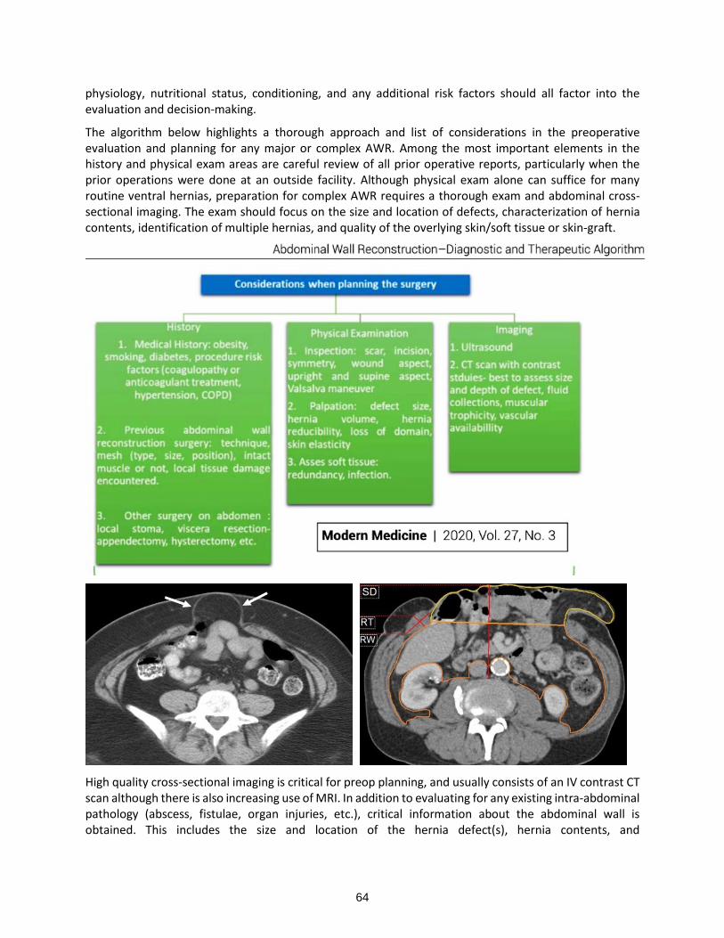
physiology, nutritional status, conditioning, and any additional risk factors should all factor into the evaluation and decision-making.
The algorithm below highlights a thorough approach and list of considerations in the preoperative evaluation and planning for any major or complex AWR. Among the most important elements in the history and physical exam areas are careful review of all prior operative reports, particularly when the prior operations were done at an outside facility. Although physical exam alone can suffice for many routine ventral hernias, preparation for complex AWR requires a thorough exam and abdominal cross-sectional imaging. The exam should focus on the size and location of defects, characterization of hernia contents, identification of multiple hernias, and quality of the overlying skin/soft tissue or skin-graft.
High quality cross-sectional imaging is critical for preop planning, and usually consists of an IV contrast CT scan although there is also increasing use of MRI. In addition to evaluating for any existing intra-abdominal pathology (abscess, fistulae, organ injuries, etc.), critical information about the abdominal wall is obtained. This includes the size and location of the hernia defect(s), hernia contents, and
64

location/size/status of the abdominal wall muscles. The most important of these is the status and location of the rectus muscles, particularly in patients who may have lost a portion or all of one or both rectus muscles from their previous injuries and operations. This allows for identification of relatively small defects with normal intact rectus muscles (Figure above left) that can be managed without the need for component separation, versus a large complex defect with lateralized and atrophic rectus complexes (Figure above right) that will likely require a complex AWR with some component separation technique. It is also relatively common that an ostomy will be present, and this introduces additional technical concerns and considerations including managing the defects at the ostomy site, the need for revision, re-siting, or reversal of the ostomy, the increased risk of surgical site infections, and decisions regarding mesh selection. The algorithm below outlines this decision making:
KEY TECHNICAL PEARLS FOR SUCCESSFUL RECONSTRUCTION
Initial steps of the operation and preparation of the field:
• excision of existing hypertrophic scar, necrotic tissue, previously placed mesh
• mobilization and excision of any skin graft (although some prefer leaving graft intact)
• full lysis of adhesions to anterior/lateral abdominal wall (unless small defect)
• mobilization and takedown of any ostomy, ostomy reversal if indicated
Assessment of defect, mobility of fascia, and tissue planes
• goal should always be primary fascial apposition/closure with mesh reinforcement
• avoid mesh in a bridging position whenever possible
• if fascia able to be approximated then standard repair with mesh in sublay, retrorectus, or onlay position can be performed
• retrorectus repair is ideal as it allows posterior sheath/peritoneal closure, mesh in retrorectus pocket requiring minimal fixation, and anterior fascial closure
65

Selection and details of fascial mobilization approach (Figure below):
• component separation (CS) with mesh reinforcement has become the widely accepted standardfor most AWRs
• broadly categorized into anterior and posterior component separation procedures (see Figurebelow illustrating numerous described approaches)
• anterior CS with release of external oblique aponeurosis
• pros: technical ease and provides significant mobilization (15-20 cm)
• cons: requires large subcutaneous flaps, higher wound complication rates
• posterior CS or transversus abdominis release (TAR) becoming preferred approach
• pros: no subcutaneous flaps created, provides significant mobilization (12-17 cm)
• cons: more technically difficult, slightly less mobilization than anterior CS
• CS procedures DO NOT always have to be bilateral. Do unilateral release and then re- assess ifcontralateral CS is required
• Always try to preserve perforating vessels/nerves at the lateral extent of the posterior rectussheath dissection
• Full mobilization with either anterior or posterior CS can be performed laterally to the spine/psoasand inferiorly to the pelvis/sacrum
• Combining an anterior and posterior CS should be avoided due to high complication rates anddestabilization of the abdominal wall
• Wait to mature or re-site any ostomy until full mobilization is completed and site the ostomy withthe fascial edges temporarily approximated
• If fascial approximation cannot be obtained, then a mesh bridge must usually be utilized. TheTAWT approach can also be considered – see protocol on page 68.
66

Mesh selection and placement, and wound closure:
• biologic mesh is largely falling out of favor due to higher recurrence rates, less ideal handling characteristics, and data showing no benefit vs modern macroporous prosthetic mesh even in contaminated fields
• new “bioprosthetic” or “biosynthetic” (also called “long-acting resorbable”) meshes are now available and increasing in popularity, with preliminary data suggesting lower recurrences vs biologic mesh and lower risk of mesh removal vs prosthetic mesh o see Plastic & Reconstructive Surgery 2018;142:84S-91S for an excellent review on this topic
• biologic mesh or uncoated prosthetic mesh should not be placed in direct contact with bowel due to adhesion and fistula risks
• the retrorectus space has become the preferred location to place/fixate mesh versus the sublay or onlay positions
• placement of a wound vac directly on mesh, particularly biologic mesh, should be avoided whenever possible
• excess redundant skin/subcutaneous tissue should be resected, particularly after anterior CS to avoid closing devascularized skin edges that then go on to necrose
• closed suction drains can be placed in the retrorectus space (above the mesh) and should be placed in the large subcutaneous flaps if anterior CS was performed
• there is some data suggesting that the use of an incisional wound vac (placed on the closed incision) can reduce wound seroma rates and possibly wound infections
• an abdominal binder should be applied which will help provide compression on the incision and can also increase patient comfort as they start to mobilize
67

World Society for Emergency Surgery Open Abdomen Guidelines (2018)
68

PELVIC FRACTURE MANAGEMENT 2022
Todd Costantini, MD, FACS
Associate Professor of Surgery Division of Trauma, Surgical Critical Care, Burns, and Acute Care Surgery Medical Director, Trauma UC San Diego Health San Diego, CA
Severe pelvic fracture is associated with significant morbidity and mortality. A multi-institutional study sponsored by the American Association for the Surgery of Trauma of 1,339 patients with pelvic fracture found an overall in-hospital mortality rate of 9.0%, however, mortality was 32.0% for patients with pelvic fracture admitted in shock.1 Patients with severe pelvic fracture require prompt recognition and treatment of their injuries to stabilize the bony pelvis and control hemorrhage. A multidisciplinary team is frequently needed to provide optimal care for patients with severe pelvic fracture during the initial resuscitation including the trauma surgeon, orthopedic surgeon, and interventional radiologist. In addition, care for injured patients may be delivered in the trauma bay, interventional radiology suite, operating room, or even hybrid operating room depending on the resources available. It is critical that the trauma surgeon direct the multidisciplinary treatment plan and also guides the ongoing resuscitation needs of the patient as they are transported to various areas of the hospital to receive care.
PELVIC ANATOMY
The pelvis is comprised of several bones that form the pelvic ring; including the sacrum, coccyx, ileum, ischium, and pubis. The bones of the pelvis are connected by strong ligamentous attachments that gives the pelvis its stability. The internal iliac artery and its branches provide the blood supply to the soft tissue and organs of the pelvis. The anterior division of the internal iliac artery gives rise to the inferior gluteal, obturator, inferior vesicular, middle rectal and internal pudendal arteries. The posterior division of the internal iliac artery includes the iliolumbar, lateral sacral, and superior gluteal arteries. The superior gluteal artery is the most common source of hemorrhage in patients with pelvic fracture due to its close relation to the sacroiliac joint, while hemorrhage to the internal pudendal artery is also commonly seen. The pelvis is also supplied by a rich venous plexus that drains into the internal iliac veins. Therefore, pelvic fracture hemorrhage may originate from arterial, venous, and bony sources. These sources must be considered when planning hemorrhage control interventions.
THE INITIAL EVALUATION- FIND THE SOURCE(S) OF BLEEDING
Initial treatment of the injured patient is directed at diagnosing significant pelvic fracture while evaluating for other sources of hemorrhage due to the high incidence of associated injuries. Trauma patients are evaluated according to Advanced Trauma Life Support (ATLS) protocol which includes a primary and secondary survey aimed at identifying injuries. Frequently, pelvic fracture is not identified on initial exam, but may be seen on the AP radiograph of the pelvis that is acquired as part of the adjuncts to the primary survey during ATLS evaluation.
Despite the identification of a significant pelvic fracture, bleeding from other sources must also be considered and may dictated the optimal hemorrhage control intervention. Bleeding from the chest and abdomen must always be considered in patients with evidence of ongoing hemorrhage. In a previous
69

multicenter study of patients with pelvic fracture, nearly 50% of patients also had a severe injury in the chest, while 32% of patients had a significant abdominal injury.1 Therefore, it is important to quickly evaluate for other, non-pelvic sources of hemorrhage. A chest x-ray can quickly identify hemothorax. FAST is also utilized to evaluate for free fluid in the abdomen and is a reliable method to rapidly screen for an intraperitoneal source of significant bleeding.2
There is currently no consensus as to the optimal treatment paradigm for patients with hemorrhage due to pelvic fracture. There are several strategies that can be deployed either alone, or in combination, to control hemorrhage from the pelvis. The ability to achieve rapid hemorrhage control is critical and associated with improved survival.3 Interventions aimed at controlling pelvic hemorrhage include pelvic binder placement, external fixator placement, angioembolization, pre-peritoneal pelvic packing, and resuscitative endovascular balloon occlusion of the aorta (REBOA).
A PELVIC BINDER CAN TEMPORIZE PELVIC HEMORRHAGE
Pelvic binders close the pelvic ring and decrease the pelvic and retroperitoneal volume which aids in the tamponade of venous bleeding. Pelvic binders can also stabilize the pelvic fracture, to a certain extent, and limit ongoing movement of unstable fracture elements that can cause ongoing vascular trauma. Pelvic binders have greatest benefit in “open-book” pelvic fractures by reducing symphyseal separation and decreasing pelvic volume (Figure 1). Placement of a pelvic binder is not appropriate in all fracture patterns as pelvic volume can increase and hemorrhage can worsen if applied to the incorrect type of fracture where binder placement can worsen fracture displacement.
A folded sheet or commercially available pelvic binder can be placed quickly during the initial evaluation. The binder should be centered at the level of the greater trochanters and sufficient force must be applied to the binder to reduce pelvic volume. A repeat pelvic radiograph can be obtained after binder placement to ensure appropriate fracture reduction. Improper placement of the binder can lead to ineffective
Figure 1. Initial pelvis xray for patient with “open-book” APC3 pelvic fracture (left panel). Placement
of a pelvic binder (right panel) decreased pelvic volume.
70

stabilization and potentially excessive abdominal pressure. This most commonly occurs when the binder placement is too high over the iliac spines and not over the greater trochanters.
PELVIC ANGIOEMBOLIZATION
Pelvic angiography (Figure 2) and subsequent angioembolization can localize and treat sources of pelvic hemorrhage. The use of angiography requires collaboration with interventional radiologists in most institutions, where timing of mobilization of the interventional radiology suite can vary between institutions and based on time of day. This is an important consideration in the actively bleeding patient and may contribute to time to hemorrhage control where studies have shown that patients that experience a significant delay in time to angiography have increased mortality.4 Identifying which patients need to undergo angiography for pelvic fracture remains a challenge. No active extravasation is seen at the time of angiography in up to 50% of patients despite significant fracture patterns and evidence of significant pelvic hemorrhage. Unstable fracture patterns are much more likely to have a pelvic source of hemorrhage that required angioembolization compared to stable fracture patterns.5 In a multivariate analysis of patients with severe pelvic fracture, APC3 fracture pattern and open pelvic fractures were associated with hemorrhage requiring intervention (Figure 3).6
Potential complications of pelvic angioembolization include ischemic complications caused by decreasing blood flow through the internal iliac arteries that can lead to gluteal necrosis and/or pelvic nerve dysfunction. As with all percutaneous vascular interventions, there are risks related to vascular access and thrombosis related to arterial punction. Finally, contrast injection can contribute to acute kidney injury, especially patients in shock with intravascular volume depletion.
Figure 2. Pelvic Angiogram for patient with pelvic fracture
71

Pre-peritoneal Pelvic Packing
In addition to decreasing pelvic arterial hemorrhage, pre-peritoneal packing can decrease hemorrhage from both venous and bony sources which makes up the majority of pelvic bleeding.7 Preperitoneal packing is an ideal strategy for patients with hemodynamic instability that need prompt hemorrhage control and are not able to be transported to the interventional radiology suite. It is also an ideal approach to control pelvic hemorrhage when patients require prompt transport to the operating room to treat hemorrhage in the chest or abdomen. Patients treated with preperitoneal pelvic packing had been shown to have decreased time to intervention and received fewer blood products during the first 24 hours post-admission.8 A recent single-center study demonstrated a 21% mortality for patients with severe pelvic fracture treated with pelvic packing, which compares favorably to mortality rates in other modern series.9
Preperitoneal pelvic packing is performed through a low midline or transverse incision. The preperitoneal space is entered after splitting the rectus muscles and six lap pads are then placed in the preperitoneal space, three on either side of the bladder and pubis. Preperitoneal packing and angiography are not mutually exclusive but instead can be thought of as adjunctive techniques when needed. If a patient has persistent hemodynamic instability after packing they can then be immediately brought to the angiography suite for treatment of any arterial bleeding. Frequently, pelvic packing is done in conjunction with external fixator placement to stabilize the bony pelvis after packing is completed.
The most common complication associated with pre-peritoneal pelvic packing is infection which can occur in up to 21% of patients. While infection does not appear to be associated with the time that pelvic packs are left in place, infection does increase if pelvic packs are removed and replaced.9 Therefore, the initial pelvic packs should not be removed until the patient is resuscitated, coagulopathy is improved, and hemorrhage controlled. Deep vein thrombosis is another potential complication of pre-peritoneal pelvic packing due to the pressure placed on the pelvic veins. For this reason, many favor routine venous duplex to screen for DVT in these high-risk patients.10
Figure 3. Predictors of need for a hemorrhage control intervention. From Costantini et al. J Trauma Acute Care Surg. 2017;82:1030-1038
72

RESUSCITATIVE BALLOON OCCLUSION OF THE AORTA (REBOA)
REBOA can be a useful tool to control pelvic hemorrhage in patients with severe pelvic fracture. This technique involves gaining arterial access in the groin for placement of the REBOA catheter into the aorta. To control pelvic hemorrhage, the REBOA balloon can be inflated in the distal aorta (Zone 3) just above the bifurcation of the common iliac arteries. This more distal placement of the balloon allows perfusion of the viscera and decrease the ischemic complications that are associated with more proximal balloon occlusion. REBOA is most frequently utilized as a temporizing measure to allow transport of the patient to the interventional radiology suite or operating room for more definitive hemorrhage control interventions. However, a recent study from the AORTA registry demonstrated that 37.5% of patients with REBOA placed after pelvic fracture did not require an additional hemorrhage control intervention.11 REBOA is not without potential complications including vascular injury at the access site, arterial injury at the site of balloon inflation, and ischemia-reperfusion injury caused by limited perfusion. While REBOA seems to have good utility in the management of pelvic hemorrhage, the lack of standardized approaches to REBOA use, and the fact that REBOA use predominates in high volume centers limits generalizability of previously published data. Additional studies are needed, especially as REBOA catheter size decreases in the near future.
EXTERNAL FIXATION TO STABILIZE THE PELVIS
External fixation is another adjunct to control bleeding from pelvic fractures. They can be quickly applied to stabilize fracture elements and reduce the pelvic volume for tamponade of venous bleeding. Historically, the pelvic C-clamp was designed for quick and easy placement and was used to stabilize posterior pelvic fractures, however, these have been replaced by more modern constructs to mechanically stabilize the bony pelvis. Pelvic external fixators can be left in place for several weeks and can also be utilized as definitive treatment for pelvic fracture. External fixators can be more advantageous than a pelvic binder due to improved access to the groin, and decreased risk of skin breakdown.
PELVIC HEMORRHAGE CONTROL- SO MANY OPTIONS, WHICH DO I CHOOSE?
Despite advances in modern damage control resuscitation and a variety of hemorrhage control interventions, mortality for patients with severe pelvic fracture has remained relatively unchanged for decades. There is no specific algorithm that is applicable to all patients with pelvic fracture as is demonstrated by the significant variability that is seen in management trauma centers across this country.1,12 Decision-making for the hemorrhaging patient with severe pelvic fracture can be challenging and is influenced by the patient’s hemodynamic status, availability of resources such as the interventional radiology suite, and can differ across Trauma Centers. A proposed algorithm is shown in Figure 4.
73

When in doubt, I recommend taking the patient to the operating room for pre-peritoneal pelvic packing while the interventional radiology suite is mobilized. This allows you to promptly address hemorrhage and aggressively resuscitate the patient with all of the resources available in the operating room. During this time, the interventional radiology team can mobilize and be ready once pelvic packing is complete. REBOA can be a useful adjunct in the unstable patient to temporize hemorrhage during transport to the operating room or interventional radiology suite for definitive hemorrhage control Future studies are needed to define optimal pathways to treat pelvic hemorrhage and to define the optimal use of REBOA.
REFERENCES
1. Costantini TW, Coimbra R, Holcomb JB, Podbielski JM, Catalono RD, Blackburn A, Scalea TM, Stein DM, Williams L, Keeney S, Hoey C, Zhou T, Sperry J, Skiada D, Inaba K, Williams BH, Minei JP, Privette A, Mackersie RC, Robinson BR, Moore FO; AAST Pelvic Fracture Study Group. Current management of hemorrhage from severe pelvic fractures: Results of an American Association for the Surgery of Trauma multi-institutional trial. J Trauma Acute Care Surg. 2016 May;80(5):717-23;discussion 723-5.
2. Schwed AC, Wagenaar A, Reppert AE, Gore AV, Pieracci FM, Platnick KB, Lawless RA, Campion EM, Coleman JJ, Cohen MJ, Moore EE, Cothren Burlew C. Trust the FAST: Confirmation that the FAST examination is highly specific for intra-abdominal hemorrhage in over 1,200 patients with pelvic fractures. J Trauma Acute Care Surg. 2021;90:137-142.
Figure 4. Algorithm for the treatment of patients with pelvic fracture
74

3. Duchesne J, Costantini TW, Khan M, Taub E, Rhee P, Morse B, Namias N, Schwarz A, Graves J, Kim DY, Howell E, Sperry J, Anto V, Winfield RD, Schreiber M, Behrens B, Martinez B, Raza S, Seamon M, Tatum D. The effect of hemorrhage control adjuncts on outcome in severe pelvic fracture: A multi-institutional study. J Trauma Acute Care Surg. 2019;87:117-124.
4. Schwartz DA, Medina M, Cotton BA, Rahbar E, Wade CE, Cohem AM, Beeler AM, Burgess AR, Holcomb JB. Are we delivering two standards of care for pelvic trauma? Availability of angioembolization after hours and on weekends increases time to therapeutic intervention. J Trauma Acute Care Surg. 2014;76:134-139.
5. Eastridge BJ, Starr A, Minei JP, O’Keefe GE, Scalea TM. The importance of fracture pattern in guiding therapeutic decision-making in patients with hemorrhagic shock. J Trauma. 2002;53:446-50.
6. Costantini TW, Coimbra R, Holcomb JB, Podbielski JM, Catalono RD, Blackburn A, Scalea TM, Stein DM, Williams L, Keeney S, Hoey C, Zhou T, Sperry J, Skiada D, Inaba K, Williams BH, Minei JP, Privette A, Mackersie RC, Robinson BR, Moore FO; AAST Pelvic Fracture Study Group. Pelvic fracture pattern predicts the need for hemorrhage control intervention – Results of an AAST multi-institutional study. J Trauma Acute Care Surg. 2017 Jun; 82(6):1030-1038.
7. Bugaev N, Rattan R, Goodman M, Muckherjee K, Robinson BRH, McDonald AA, Bogert JN, Croft CA, Edavettal M, Engels PT, Jayaraman V, Khwaja K, Kasotakis G, Lawless RA, Maine RG, Hasenboehler EA, Schroeder ME, Schroll RW, Kim D, Mentzer C, Litt J, Como JJ. Preperitoneal packing for pelvic fracture-associated hemorrhage: A systematic review, meta-analysis, and management guidelines from the Eastern Association for the Surgery of Trauma. Am J Surg. 2020 Oct;220(4):873-888.
8. Osborn PM, Smith WR, Moore EE, Cothren CC, Morgan SJ, Williams AE, Stahel PF. Direct retroperitoneal pelvic packing versus pelvic angiography: A comparison of two management protocols for haemodynamically unstable pelvic fractures. Injury. 2009;40:54-60.
9. Burlew CC, Moore EE, Stahel PF, Geddes AE, Wagenaar AE, Pieracci FM, Fox CJ, Campion EM, Johnson JL, Mauffrey C. Preperitoneal pelvic packing reduces mortality in patients with life-threatening hemorrhage due to unstable pelvic fractures. J Trauma Acute Care Surg. 2017;82:233-242.
10. Heelan AA, Freedberg M, Moore EE, Platnick BK, Pieracci FM, Cohen MJ, Lawless R, Campion EM, Coleman JJ, Hoehn M, Burlew CC. Worth looking! venous thromboembolism in patients who undergo preperitoneal pelvic packing warrants screening duplex. Am J Surg. 2020;220:1395-1399.
11. Harfouche M, Inaba K, Cannon J, Seamon M, Moore E, Scalea T, DuBose J. Patterns and outcomes of zone 3 REBOA use in the management of severe pelvic fractures: Results from the AAST Aortic Occlusion for Resuscitation in Trauma and Acute Care Surgery database. J Trauma Acute Care Surg. 2021;90:659-665.
12. Tran TLN, Brasel KJ, Karmy-Jones R, Rowell S, Schreiber MA, Shatz DV, Albrecht RM, Cohen MJ, DeMoya MA, Biffl WL, Moore EE, Namias N. Western Trauma Association Critical Decisions in Trauma: Management of pelvic fracture with hemodynamic instability – 2016 updates. J Trauma Acute Care Surg. 2016 Dec;81(6):1171-1174.
75

APPENDICITIS IN 2022: OPERATE OR ANTIBIOTICS?
Andrew C. Bernard, MD, FACS
Paul A. Kearney, MD Endowed Chair of Trauma Surgery Chief, Section of Trauma and Acute Care Surgery Trauma Medical Director University of Kentucky Lexington, KY
WHAT’S THE CONTEXT?
• Most common surgical emergency worldwide.
• 80-87% are ‘uncomplicated’-no abscess, phlegmon, perforation or mass o Indicators for complicated appendicitis: peritonitis, shock, obstruction, symptoms > 24hr,
neutrophil:lymphocyte ration > 8.8 (Hajibandeh) o Confirmed by CT
WHERE’S THE DATA?
1. Appendicitis Acute (APPAC) (Salminen 2015)
o Multicenter, open-label, noninferiority randomized clinical trial (Salminen) o 2009 to 2012 o Finland o 530 patients o Open appendectomy vs NOM with antibiotics o 27.3% of patients required appendectomy within 12 months of randomization
▪ 21% during the index hospitalization ▪ 79% after a readmission
o Heavily referenced o Did not meet its prespecified noninferiority margin for NOM
▪ Long-term quality of life (QoL) scores were similar between NOM and OM; however, they were lower in those who originally underwent NOM but had to convert to OM for recurrence. (Salminen 2018, Sippola 2020)
2. Comparison of the Outcomes of antibiotic Drugs and Appendectomy (CODA) (CODA Collaborative 2020)
o 25 U.S. centers o 2016 to 2020 o 1552 patients o Uncomplicated appendicitis o Appendectomy vs NOM (with 10 days of antibiotics) o 96% of the surgical arm underwent laparoscopic appendectomy o 47% of NOM arm did not require hospital admission o At 90 days:
▪ 29% of NOM underwent appendectomy (41% if appendicolith, 25% without)
76

▪ 30-day QoL scores as primary outcome: antibiotics were noninferior to appendectomy
3. COMMA (O’Leary 2021)
o 186 patients o Randomized o Antibiotic-only group: 23 patients (25.3%) experienced recurrence within 1yr o Better EQ-VAS quality of life score in the surgery group vs antibiotic-only group at
3 and 12 months postintervention (94.5 vs 90.4, P < 0.001). o EQ-5D-3L quality-of-life score was significantly higher in the surgery group o Mean total cost higher in surgery (€4,816 vs €3,077)
WHAT IS THE OUTCOME WE ARE MEASURING?
• Pain scores
• Missed work
• Overall heath
• Overall complication rate
• Cost
COST
• Most studies show NOM more cost-effective than surgery (peds and adults) (Wu 2015, 2017)
• As much as a $10,000 difference
• Five-year follow-up of the APPAC trial group demonstrated persistent significant cost savings associated with NOM. (Haijanen 2019)
• However, in a study using Markov analysis: When considering LAP appendectomy performed OUTPATIENT and 1) varying perioperative mortality, 2) probability of appendiceal malignancy or recurrent appendicitis after NOM, 3) probability of a complicated recurrence, 4) AND appendectomy cost, operative management WAS cost effective over a lifetime (Sceats 2019).
HOW SHOULD I PRESCRIPE THE ANTIBIOTICS IF I CHOOSE NOM?
In surgical cases: enteric gram positive, gram negative and anaerobic coverage with effective drug levels at incision
In non-op cases:
APPAC:
• IV ertapenem (1 g/d) x 3 days PLUS 7 days of PO levofloxacin and metronidazole
CODA:
• ≥ 24 hours of IV antibiotics (ertapenem, cefoxitin, or cefazolin/metronidazole) PLUS 10 days of ciprofloxacin/metronidazole or cefdinir/metronidazole
COMMA:
• IV co-amoxiclav, 1.2 g, 3x daily until clinical improvement PLUS 5 days of oral co-amoxiclav (625 mg TID for 5 days)
77

CAN WE GET AWAY WITH ORAL ANTIBIOTICS ONLY?
• 7 days of oral moxifloxacin vs 2 days of IV ertapenem PLUS 5 days of levofloxacin and metronidazole:
o 65% in both groups o BUT orals alone NOT non-inferior (not equivalent) (Sippola JAMA 2021)
SO WHAT’S THE RECURRENCE RATE? (APPROXIMATELY 40% LONG TERM)
HOW DOES A FECALITH AFFECT MY DECISION-MAKING?
• 15% of patients
• Higher rates of complicated appendicitis
• Fecaliths excluded from APPAC
• Included in CODA o Higher failure rates o Higher rate of complications
• Summary: Higher risk, not off the table
WHAT GUIDELINES ARE AVAILABLE?
1. American College of Surgeons
“Based on the surgeon's judgment, patient preferences and local resources (e.g., hospital staff, bed and PPE supply availability), antibiotics are an acceptable first-line treatment, with appendectomy offered for those with worsening or recurrent symptoms.”
2. World Society of Emergency Surgery
I. The antibiotic-first strategy can be considered safe and effective in selected patients with uncomplicated acute appendicitis.
II. We recommend discussing NOM with antibiotics as a safe alternative to surgery in selected patients with uncomplicated acute appendicitis and absence of appendicolith, advising of the possibility of failure and misdiagnosing complicated appendicitis
III. We suggest against treating acute appendicitis non-operatively during pregnancy until further high-level evidence is available
COMMENTS ON COMPLICATED APPENDICITIS (GRASSO 2021)
• Free perforation
• Abscess
• Phlegmon
78

1. Distinct from uncomplicated appendicits (above approaches do not apply) 2. Higher rates of postop complications and conversion to open 3. Some cases managed non-operatively with CT-guided drainage + antibiotics
DO I NEED TO PERFORM INTERVAL APPENDECTOMY?
• Historically: interval appendectomy 6-8 weeks after resolution of symptoms
• 2 concerns: 1. cecal or appendiceal neoplasm
2. recurrent appendicitis o Not common (appendix usually obliterated) o 2016 meta-analysis: overall 12.4% rate of recurrent appendicitis (Darwazeh 2016)
• In general: interval appy increases costs
• Breakpoint at 34 years old (ie, patients younger than 34 years should have an interval appendectomy performed from a cost perspective)
• Appendiceal Neoplasm
HOW SHOULD I MANAGE THE RISK OF MALIGNANCY?
• Likelihood of finding a neoplasm in complicated appendicitis is significantly higher than in uncomplicated cases (12.6% vs 1.2%) (Son 2020)
• Risk of a neoplasm increases with age
• PeriAppendicitis Acuta (periAPPAC) trial (Mallinen 2019) o 2019 o 18 to 60 years o appendiceal abscess successfully treated with antibiotics and percutaneous drainage o colonoscopy to exclude a cecal tumor o randomized to either interval appendectomy or continued NOM with MRI at 3 months o planned enrollment 120 patients o trial prematurely terminated after interim analysis of the first 60 patients demonstrated
an unexpectedly high rate of appendiceal neoplasm (17%). o Patients initially randomized to the observation arm were then offered appendectomy,
and this led to a 20% overall rate of neoplasm
• 35% neoplasm rate for patients > 40 years
• One recommendation: interval appendectomy on all patients healthy enough o if < 34 years, from a hospital cost perspective o if > 40 years, from a neoplasm risk reduction perspective
WHO NEEDS A COLONOSCOPY?
• Age > 40 year + appendicitis + MUCH higher risk of colon cancer (OR 38.5) (Lai 2006)
• In general: colonoscopy should be performed in all patients older than 40 years to rule out right-sided colonic neoplasms.
79

OVERALL MANAGEMENT ALGORITHM-UNCOMPLICATED AND COMPLICATED APPENDICITIS (GRASSO 2021)
REFERENCES
1. CODA Collaborative; Flum DR, Davidson GH, Monsell SE et al. A Randomized Trial Comparing Antibiotics with Appendectomy for Appendicitis. N Engl J Med 2020 Nov 12;383(20):1907-1919.
2. Darwazeh G, Cunningham SC, Kowdley GC. A systematic review of perforated appendicitis and phlegmon: interval appendectomy or wait-and-see? Am Surgeon 2016;82(1):11–5.
3. Grasso S and Walker A. Modern Management of the Appendix: So Many Options. Surg Clin N Am 101 (2021) 1023–1031.
4. Haijanen J, Sippola S, Tuominen R et al. Cost analysis of antibiotic therapy versus appendectomy for treatment of uncomplicated acute appendicitis: 5-year results of the APPAC randomized clinical trial PLoS One 2019 Jul 25;14(7):e0220202.
5. Hajibandeh S, Hajibandeh S, Hobbs N, Mansour M. Neutrophil-to-lymphocyte ratio predicts acute appendicitis and distinguishes between complicated and uncomplicated appendicitis: A systematic review and meta-analysis. Am J Surg. 2020 Jan;219(1):154-163.
6. Lai HW, Loong CC, Tai LC, et al. Incidence and odds ratio of appendicitis as first manifestation of colon cancer: a retrospective analysis of 1873 patients. J Gastroenterol Hepatol 2006;21(11):1693–6.
7. Mallinen J, Rautio T, Gronroos J, et al. Risk of appendiceal neoplasm in periappendicular abscess in patients treated with interval appendectomy vs follow-up with magnetic resonance imaging: 1-year outcomes of the peri–appendicitis acuta randomized clinical trial. JAMA Surg 2019;154(3):200–7.
80

8. O'Leary DP, Walsh SM, Bolger J et al. A Randomized Clinical Trial Evaluating the Efficacy and Quality of Life of Antibiotic-only Treatment of Acute Uncomplicated Appendicitis: Results of the COMMA Trial. Ann Surg 2021 Aug 1;274(2):240-247.
9. Salminen P, Paajanen H, Rautio T et al. Antibiotic Therapy vs Appendectomy for Treatment of Uncomplicated Acute Appendicitis: The APPAC Randomized Clinical Trial. JAMA. 2015 Jun 16;313(23):2340-8.
10. Sceats LA, Ku S, Coughran A et al. Operative Versus Nonoperative Management of Appendicitis: A Long-Term Cost Effectiveness Analysis MDM Policy Pract. 2019 Aug 17;4(2):2381468319866448.
11. Sippola S, Haijanen J, Viinikainen L et al. Quality of Life and Patient Satisfaction at 7-Year Follow-up of Antibiotic Therapy vs Appendectomy for Uncomplicated Acute Appendicitis: A Secondary Analysis of a Randomized Clinical Trial. JAMA Surg. 2020 Apr 1;155(4):283-289.
12. Sippola S, Haijanen J, Grönroos J et al. Effect of Oral Moxifloxacin vs Intravenous Ertapenem Plus Oral Levofloxacin for Treatment of Uncomplicated Acute Appendicitis: The APPAC II Randomized Clinical Trial. JAMA. 2021 Jan 26;325(4):353-362.
13. Son J, Park YJ, Lee SR, et al. Increased Risk of Neoplasms in Adult Patients Undergoing Interval Appendectomy. Ann Coloproctol 2020;36(5):311.
14. Wu JX, Dawes AJ, Sacks GD, et al. Cost effectiveness of nonoperative management versus laparoscopic appendectomy for acute uncomplicated appendicitis. Surgery 2015;158(3):712–21.
15. Wu JX, Sacks GD, Dawes AJ, et al. The cost-effectiveness of nonoperative management versus laparoscopic appendectomy for the treatment of acute, uncomplicated appendicitis in children. J Pediatr Surg 2017;52(7):1135–40.
81

82

SESSION 2
CASE MANAGEMENT
Moderator: Alison Wilson, MD, FACS
Monday, March 28, 2022
Palace Ballroom 1-2
Palace Tower, Emperors Level
Panelists: Carlos V.R. Brown Rachael A. Callcut Jennifer M. Gurney Martin A. Schreiber Jason W. Smith Matthew J. Wall, Jr.
83

84

SESSION 3
LUNCHEON SESSION
Moderator: Matthew J. Wall, Jr., MD, FACS, MAMSE
Monday, March 28, 2022
12:00 p.m. - 1:15 p.m.
Augustus Ballroom
Palace Tower, Emperors Level
“THE DAMATTOX CODE” Kenneth L. Mattox, MD, FACS, MAMSE
Program Director, TCCACS & MDR Distinguished Service Professor
Michael E. DeBakey Department of Surgery Special Advisor to the President & CEO
Baylor College of Medicine Houston, TX
85

86

SESSION 4
HOW I DO IT
Moderator: Andrew C. Bernard, MD, FACS
Monday, March 28, 2022
Palace Ballroom 1-2
Palace Tower, Emperors Level
1:15 - 1:30
Vascular Anastomosis for the General Surgeon Michael J. Sise, MD, FACS
1:30 - 1:45
Venous Injuries: What to Ligate? What to Repair? Kenji Inaba, MD, FRCSC, FACS
1:45 - 2:00
I'm Out of Joint: Dislocations 101 Michelle A. Bramer, MD
2:00 - 2:15
Damage Control Techniques in the Chest Matthew J. Wall, Jr., MD, FACS, MAMSE
2:15 - 2:30
CPR In Acute Trauma Mark J. Kaplan, MD, FACS
2:30 - 2:45
Does Size Matter? Pigtails vs large-bore tubes for hemothorax Richard A. Sidwell, MD, FACS
2:45 - 3:00
The Difficult Duodenum: Operating in Tiger Country Jay A. Johannigman, MD, FACS
3:00 - 3:15
Hemorrhage Control: "Tips of the Trade" Demetrios Demetriades, MD, MPH, FACS
3:15 - 3:40
Panel Discussion
87

VASCULAR ANASTAMOSES FOR THE GENERAL SURGEON
Michael J. Sise, MD, FACS
Clinical Professor of Surgery UCSD School of Medicine Scripps Mercy Hospital San Diego, CA
Open vascular trauma surgical technical ability has become a precious commodity. It is apparent that trauma surgeons need to know how to repair injured vessels. This syllabus and the accompanying PowerPoint present the technical fundamentals of vascular repairs.
BACKGROUND
In a multi-institutional study presented in September, 2012 at the annual meeting of the American Association for the Surgery of Trauma, Dr. Steven Shackford and colleagues reported that close to 2/3 of complex extremity vascular reconstructions performed at 12 trauma centers across the country between 1995 and 2010 were performed by general surgeons. The outcome of these repairs by general surgeons was not significantly different than those performed by fellowship trained cardiac and vascular surgeons. The overall amputation rate was low. All 12 hospitals were mature trauma centers with well-organized surgical specialty support. The average age of the surgeons who performed these repairs suggested that they all had adequate vascular experience during their surgical residencies. What the surgeons from the centers in this study had in common was a commitment to maintaining the skills needed to manage vascular injuries. The result of this approach was successful management of complex injuries.
Over ten years later, many of those general surgeons have retired and many of the younger general surgeons who’ve replaced them are, by and large, neither experienced in open vascular repair techniques nor comfortable attempting them. The shrinking volume of open elective and emergency vascular surgery operations they encountered during their training created significant obstacles to obtaining adequate experience in vascular surgical technique. Although there are a number of general surgeons who include vascular surgical cases in their practices, almost all of them completed their training more than more than 20 to 25 years ago in the era prior to the extensive use of endovascular techniques. Each honed skills on open aortic aneurysm repairs and femoral popliteal bypass procedures.
There is a similar paucity of open vascular surgical cases, especially peripheral vascular injuries, in the training of vascular surgical fellows. Equally constraining is the lack of open abdominal vascular procedures in both for both residents and fellows. Successful trauma centers have always provided the right surgeon to do the right operation in a timely fashion. This has been especially important in repairing vascular injuries. If this capability can continue remains to be seen. The paucity of capable vascular surgical emergency back-up represents a threat to most of our trauma and emergency centers. It is apparent that trauma surgeons must accept the challenge of becoming capable to repair vessels whether or not their training provided them adequate experience.
PRINCIPLES OF VASCULAR TRAUMA MANAGEMENT
These are the basic principles of vascular trauma management.
1. Adequate resuscitation and orchestration of the management of the vascular injury with the management of other serious associated injuries
88

2. Proximal and distal vascular control 3. Adequate exposure of the injured vessel 4. Proximal and distal Fogarty catheter thrombectomy and back flushing to remove all thrombus 5. Gentle installation of heparinized saline (10 units/ml) proximally and distally 6. Placement of an appropriate shunt if prolonged ischemia present or anticipated to occur in the
time it will take to restore adequate perfusion 7. Deferring definitive repair once shunt placed if life threatening injuries take precedence 8. Sharp debridement of the damaged arterial wall back to uninjured intima 9. Decide on primary repair or interposition graft 10. Saphenous vein first choice for interposition graft in extremities, synthetic graft in the torso 11. Tension and stenosis free anastomosis with adequate flushing prior to completion 12. Objective documentation of adequate flow with completion on-table angiogram, ultrasound
imaging, or Doppler assessment 13. Adequate soft tissue coverage and careful wound closure 14. Assessment of extremity compartments with pressure measurements to detect compartment
syndrome before leaving the OR and repeated in the early postoperative period 15. Adequate fasciotomy at the same operation or subsequently when indicated 16. Frequent vascular checks in the early postop period and immediate return to the OR for open
exploration if there is any question of repair patency
ANASTOMOSES
Vessels that have been lacerated transversely by a stab wound or have a disruption that, when debrided, allow for direct anastomosis without tension, are best repaired by end-to-end anastomosis. The following illustrations show methods of end-to-end anastomosis. Illustrations from Rutherford Atlas of Vascular Surgery 1st Edition. (See references).
The key elements are intima to intima junction and avoiding purse-stringing the anastomosis by pulling sutures too tight and causing a stenosis. End-to -end anastomosis always involves some luminal compromise if a continuous running suture is use. However, interrupted sutures help mitigate the constriction
Another technique for an end-to end anastamosis also has the goal of avoiding constriction and stenosis by using triangulated stay sutures.
89

The best technique for an end-to-end anastomosis when there is sufficient vessel length is a spatulated suture line. This is only possible if, after debridement, there enough length.
When interposition grafts are required, the same spatulated technique is optimal. The native artery need not be totally transected. This illustration shows this aspect of the technique.
When end to side anastomosis is appropriate, two principles need to be followed. The length of the arteriotomy on the origin vessel should be twice the width of the graft and the angel of the takeoff of the graft should be 30 to 75 degrees as illustrated here.
In the presence of an uncomplicated longitudinal laceration, the best approach is closing the vessel with a patch angioplasty. Direct suture always causes constriction.
90

SUMMARY
Successful vascular repairs require an organized approach. Vascular control, exposure, evacuation of thrombus, vessel preparation, and carefully performed suturing techniques are some of the more important components of effective management. Trauma surgeons should prepare to competently perform vascular trauma repairs in the presence of a growing shortage of vascular surgeons with adequate training and experience to provide this service.
REFERENCES
1. Franz RW General versus vascular surgeon: impact of a vascular fellowship on clinical practice, surgical case load, and lifestyle. Ann Vasc Surg. 2010; 24:196-204.
2. Grabo DJ, DiMuzio PJ, Kairys JC, et al.. Have Endovascular Procedures Negatively Impacted General Surgery Training? Sem Vasc Surg 2006; 19:168-171.
3. Jibawi A, Hanafy M, Guy A. . Is there a Minimum Caseload that Achieves Acceptable Operative Mortality in Abdominal Aortic Aneurysm Operations? Eur J Vasc Endovasc Surg 2006; 32:273-276.
4. Kairys JC, McGuire K, Crawford AG, et al. Cumulative operative experience is decreasing during general surgery residency: a worrisome trend for surgical trainees? J Am Col Surg 2008; 206:804-811.
5. Karthikesalingam A, Hinchliffe RJ, Ian M. Loftus Im, et al. Volume-Outcome Relationships in Vascular Surgery: The Current Status. J Endovasc Ther 2010; 17:356-365.
6. Keir J. Changes in caseload and the potential impact on surgical training: a retrospective review of one hospital's experience. BMC Med Ed 2006; 6: 6
7. Patel MB, Guillamondegui OD, May AK, Diaz JJ. Twenty-year analysis of surgical resident operative trauma experiences. J Surg Res 2012; May 16. [Epub ahead of print].
8. Schanzer A, Robert Steppacher R, Eslami MH, et al. Vascular surgery training trends from 2001-2007: A substantial increase in total procedure volume is driven by escalating endovascular procedure volume and stable open procedure volume. Vasc Surg 2009; 49:1330-1344.
9. Stanley JC, Barnes RW, Ernst CB, at al.. Vascular surgery in the United States: Workforce issues 1 2: Report of the society for vascular surgery and the international society for cardiovascular surgery, North American chapter, committee on workforce issues. J Vasc Surg 1996; 23:172-181.
10. Rutherford, RB. Atlas of Vascular Surgery: Basic Techniques and Exposures. Philadelphia. W.B Saunders, 1993.
91

VENOUS INJURIES
Kenji Inaba, MD, FRCSC, FACS
Professor and Vice Chair of Surgery Director, General Surgery Program Chief, Trauma, Emergency Surgery and Surgical Critical Care LAC+USC Medical Center & University of Southern California – Los Angeles Los Angeles, CA
The diagnosis, and management of injuries to the venous system is often overshadowed by the arterial system. While part of this is due to the relative functional importance of the arterial system as compared to the venous system, as pointed out in a comprehensive review on this topic by Feliciano in 2021, there are major contemporary deficits in our understanding of the ramifications of venous injuries. Advances in the treatment of these injuries has also been hampered by the fact that there are no universally accepted grading systems for the magnitude of a venous injury. Many of these injuries are also not diagnosed, let alone documented and as a direct result, there is no standard treatment algorithm for venous injuries, or, for any adjunct supportive interventions such as anticoagulation or platelet inhibition. Likewise, the role of clinical follow-up and imaging has not been standardized. Because of this, the topic of venous injuries remains a very important issue to discuss, and in this session, a practical approach to the management of venous injuries will be presented so that the surgeon in attendance will have some basic principles that can be used as a foundation for the management of patients who present with these injuries.
DIAGNOSIS
The diagnosis of venous injuries requiring operative intervention is often made intra-operatively, as many of these will be associated with arterial, or other soft tissue and bony injuries. In fact, the majority of venous injuries that are diagnosed, or inferred on cross sectional imaging in patients who are stable enough for a CT scan can be managed non-operatively, provided the patient is clinically tolerating the injury, and there are no associated injuries to the accompanying arterial system or to other adjacent structures that require operative intervention. While a period of observation is likely prudent, the optimal duration, and the role of anticoagulation or follow-up imaging remains undefined. We do know however, that every day, in intensive care units around the world, large caliber venous access catheters are removed from venous structures when they are no longer needed, with just the application of pressure, and no follow-up imaging as the standard of care.
MANAGEMENT
When a venous injury is diagnosed pre-operatively, and is not being tolerated by the patient as the hemorrhage is not being contained, or, the injury is found intra-operatively, the operator has three general techniques at their disposal including ligation, reconstruction and shunting. The use of these techniques must be flexible, and the decision must take into account the venous bed that is injured, and the patient’s clinical status.
The four practical questions that should be answered in order to decide upon the optimal course of action are:
92

1. What is the patient’s clinical status? 2. Which vein(s) is injured, and what is the extent of injury? 3. Can the vein be ligated? 4. What is the associated injury burden?
First, for the hemodynamically unstable patient, or the patient who is stable, with a large injury burden that they will not tolerate the definitive repair of, in general, ligation is the preferred treatment for the vast majority of venous structures that have been breached. The focus should be on controlling hemorrhage, temporizing the remaining injury burden, and the balanced resuscitation of the patient. While uncommon, this would also apply to those working under suboptimal or austere conditions, where anything other than ligation would not be feasible. Ligation of most venous injuries under these conditions is well tolerated with a few notable exceptions. This includes the following venous structures.
1. Superior Mesenteric Vein 2. Portal Vein 3. Right Renal Vein 4. Proximal Left Renal Vein 5. Supra/Peri-Renal IVC 6. Bilateral Internal Jugular Veins 7. Retro-hepatic Veins
For injuries to these venous structures, if the patient is in extremis, meeting the aforementioned criteria, temporary intra-vascular shunting would be the preferred method of damage control. While our experience in general with venous shunting falls far short of that with arterial shunting, this is a viable option that is well tolerated for the limited amount of time required to completely resuscitate the patient. For smaller caliber injuries, standard peripheral vascular shunt sets, can be utilized. Because the bridging distance for these injuries is usually short, and the venous wall relatively fragile, shunt selection should take into consideration the materials, shunt length, and ability to loop the shunt so as to mitigate the risk of undue stress being placed on the wall of the injured vein. For large central veins that are a poor size match for standard peripheral shunts, a wide range of devices such as chest tubes have been described for temporary use, following the same principles that apply to arterial shunting. The tube should exclude the injury, when fashioned and inserted, care should be taken to not cause further injury to the upstream or downstream walls, and minimal native vein should be sacrificed in securing the shunt. While the majority of patients undergoing venous shunting are not in a position to tolerate systemic anticoagulation, if a shunt is utilized for an isolated venous injury because the operator skillset or local infrastructure is inadequate for definitive repair, the use of systemic heparinization should be considered.
For patients who are stable, and could theoretically tolerate venous reconstruction, in general, the treatment options in 2022 still remain ligation, versus reconstruction. As mentioned earlier, if the injured vein is one that would not tolerate ligation, every attempt should be made to reconstruct the injured segment in this patient population. For the remaining injuries however, there remains significant clinical equipoise over the role of reconstruction, even when the patient is able to tolerate the procedure. In fact, within the reconstruction category, the full range of techniques from venorraphy, to interposition grafts and complex reconstruction using harvested vein have been described. Unfortunately, while textbooks have extensive descriptions and schematic illustrations of all of these options, there is insufficient data to support the use of any particular management option over another. Practically, our general practice is to consider the architecture of the injury and the associated injury burden when deciding upon a treatment plan. As a general principle, if the injury is small, and can be repaired quickly without narrowing of the lumen past an arbitrary 50% threshold, venorraphy is likely of value. For all other injuries, our practice is to not perform any complex repairs of injured venous structures. It is interesting that Feliciano in his
93

review noted that there are many centers that only ligate, and those that repair universally, highlighting the lack of evidence based consensus on the treatment of these injuries. As such, there is still significant work that must be done to advance our understanding of the optimal management of patients who sustain these injuries.
REFERENCES
1. Parry, Neil G. et al.; Management and short-term patency of lower extremity venous injuries with various repairs. The American Journal of Surgery (2003) 186: 631–635.
2. Williams, Timothy K. et al.; Current concepts in repair of extremity venous injury. Journal of Vascular Surgery: Venous and Lymphatic Disorders (2015) 4(2):238 – 247.
3. Byerly, Saskya et al.; Impact of inferior vena cava ligation on mortality in trauma patients. Journal of Vascular Surgery: Venous and Lymphatic Disorders (2019) 7(6):793 – 800.
4. Byerly, Saskya et al.; Impact of ligation versus repair of isolated popliteal vein injuries on in-hospital outcomes in trauma patients. Journal of Vascular Surgery: Venous and Lymphatic Disorders (2020) 8(3): 437 – 444.
5. Manley, Nathan R. et al.; Impact of venorrhaphy and vein ligation in isolated lower-extremity venous injuries on venous thromboembolism and edema. J Trauma Acute Care Surgery (2017) 84(2):325 – 329.
6. Quan, Reagan W. et al.; The effect of vein repair on the risk of venous thromboembolic events: A review of more than 100 traumatic military venous injuries. Journal of Vascular Surgery (2008) 47(3):571 – 577.
7. Feliciano, David V. et al.; Review article: History of venous trauma. J Trauma Acute Care Surgery (2021) 91(3):62 – 72.
8. Quan, Reagan W. et al.; The Management of Trauma Venous Injury: Civilian and Wartime Experiences. Perspectives in Vascular Surgery and Endovascular Therapy (2006) 18(2):149 – 156.
9. Christian, Ashton B. et al.; Comparison of Nonoperative and Operative Management of Traumatic Penetrating Internal Jugular Vein Injury. Annals of Vascular Surgery (2021) 72:440-444.
10. Guice, Jordan L. et al,; Analysis of Limb Outcomes by Management of Concomitant Vein Injury in Military
94

I’M OUT OF JOINT: DISLOCATIONS 101
Michelle Bramer, MD
Associate Professor Assistant Residency Program Director Orthopaedic Trauma Surgery West Virginia University Morgantown, WV
Many trauma patients have associated major joint dislocations. An irreducible major joint dislocation is considered an orthopaedic emergency secondary to issues with surrounding neurovascular structures, increased risk of post traumatic arthritis, and certain specific complications such as avascular necrosis. Knee dislocations also pose the real threat of vascular injury leading to a pulseless extremity. Joints that remain dislocated for longer periods of time may become irreducible by closed means and require an open reduction.
Often coordination of care in the trauma bay can be complicated in patients with joint dislocations. Time to reduction has been shown to decrease the risk of avascular necrosis with hip dislocations1. Pulseless limbs with a knee dislocation warrant emergent reduction. However, patients are often multiply injured and hemodynamically unstable which does not allow them to be candidates for immediate reduction or conscious sedation.
A goal of this talk is to determine which dislocations are emergent and require immediate care. Physical exam findings, radiographs and reduction techniques will be discussed, as well as post reduction care.
SHOULDER DISLOCATIONS
The shoulder is the most commonly dislocated joint in the body, in great part due to very little bony constraints or articular contact, with the majority being anterior dislocations2. Patients often present after high energy trauma or sports injuries with shoulder pain. They will complain of decreased motion of the shoulder and hold the arm in an abducted and externally rotated position, often with obvious loss of contour of the lateral shoulder.
Vascular injuries are exceedingly rare (<1-2%)3,4, but the surgeon must always have a high index of suspicion when examining a patient with a shoulder dislocation. Any abnormality in pulse exam (even with a present radial pulse) or evidence of an expanding axillary hematoma should indicate a vascular injury. Nerve injuries are more common. These can occur 10-13% of the time, with axillary nerve injuries occurring more frequently5. This would cause numbness over the lateral deltoid, and difficulty with abduction of the shoulder. Unfortunately, the associated injury of rotator cuff tear can also cause difficulty with shoulder abduction. Brachial plexus injuries are more rare, and often associated with a traction type mechanism. Neurovascular exam must be obtained and documented both pre and post reduction.
Imaging required to diagnose a shoulder dislocation includes AP and axillary views of the glenohumeral joint. The axillary view is necessary to determine if an anterior or posterior dislocation is present. As AP x-rays can often be misleading secondary to rotation or poor films on a trauma backboard, the axillary view is necessary to rule out dislocation.
95

Figure 1: Glenohumeral dislocation, suspected anterior (but need axillary x-ray to confirm)
Figure 2: AP and axillary radiograph of normal shoulder
Figure 3: Axillary shoulder radiograph demonstrating anterior glenohumeral dislocation with Hill-Sachs lesion of posterior humeral head
96

Figure 4: Posterior shoulder dislocation with diagnosis confirmed on axillary radiograph
There are multiple reduction techniques for shoulder dislocations. Data supports the use of intraarticular injections for reductions, and conscious sedation is often not necessary and can lead to increased complications6. Reductions should be a slow, gradual maneuver with adequate analgesia. Post reduction radiographs and neurovascular exam should be performed. Shoulder dislocations are often associated with fractures or soft tissue injuries such as labral and rotator cuff tears5. Patient should be immobilized in a sling and referred to orthopaedics for follow up.
ELBOW DISLOCATIONS
Elbow dislocations are often secondary to falls on outstretched hands. They account for approximately 10-25% of elbow injuries7. Dislocations are often associated with radial head fractures, coronoid process fractures, and lateral/medical collateral ligament injuries7. Neurovascular exam is again paramount secondary to the proximity of the brachial artery and radial, median, and ulnar nerves.
Figure 5: Lateral radiograph demonstrating posterior elbow dislocation
The majority of elbow dislocations are posterior or posterolateral7. These can be reduced with an intraarticular injection of local anesthesia. Although it is sometimes preferred to reduce an elbow dislocation prone, this is often not feasible in a multiply injured trauma patient. Gentle traction on the
97
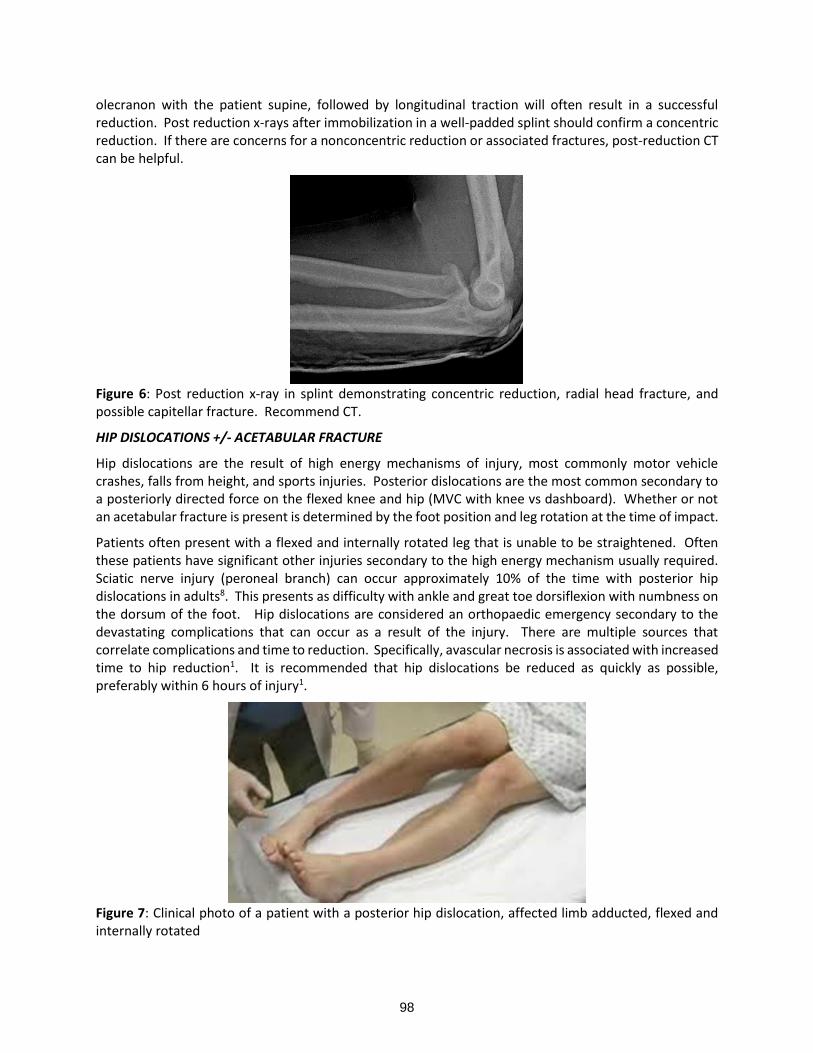
olecranon with the patient supine, followed by longitudinal traction will often result in a successful reduction. Post reduction x-rays after immobilization in a well-padded splint should confirm a concentric reduction. If there are concerns for a nonconcentric reduction or associated fractures, post-reduction CT can be helpful.
Figure 6: Post reduction x-ray in splint demonstrating concentric reduction, radial head fracture, and possible capitellar fracture. Recommend CT.
HIP DISLOCATIONS +/- ACETABULAR FRACTURE
Hip dislocations are the result of high energy mechanisms of injury, most commonly motor vehicle crashes, falls from height, and sports injuries. Posterior dislocations are the most common secondary to a posteriorly directed force on the flexed knee and hip (MVC with knee vs dashboard). Whether or not an acetabular fracture is present is determined by the foot position and leg rotation at the time of impact.
Patients often present with a flexed and internally rotated leg that is unable to be straightened. Often these patients have significant other injuries secondary to the high energy mechanism usually required. Sciatic nerve injury (peroneal branch) can occur approximately 10% of the time with posterior hip dislocations in adults8. This presents as difficulty with ankle and great toe dorsiflexion with numbness on the dorsum of the foot. Hip dislocations are considered an orthopaedic emergency secondary to the devastating complications that can occur as a result of the injury. There are multiple sources that correlate complications and time to reduction. Specifically, avascular necrosis is associated with increased time to hip reduction1. It is recommended that hip dislocations be reduced as quickly as possible, preferably within 6 hours of injury1.
Figure 7: Clinical photo of a patient with a posterior hip dislocation, affected limb adducted, flexed and internally rotated
98

Figure 8: Right posterior hip dislocation with posterior wall acetabular fracture
Initial imaging should include an AP pelvis radiograph in the trauma bay. This can be used to diagnose unstable pelvic ring injuries, as well as hip dislocations. Early recognition of the injury allows for earlier time to reduction. It is not recommended to bypass this step and use a CT scan to diagnose the injury, unless the patient is hemodynamically unstable. This can increase time to reduction. It will also likely increase the need for a secondary CT scan post-reduction, and therefore the radiation dose to the patient9. CT is required post-reduction to evaluate for fracture pattern, associated injuries, and intraarticular bony fragments. These finding can dictate approach and fixation for orthopaedic surgeons.
Reduction of a posterior hip dislocation takes coordination by the emergency medicine, trauma and orthopaedic teams. Conscious sedation allows for a more reliable reduction, with possibly decreased risk of iatrogenic fracture, scraping of the femoral head on the acetabulum, and discomfort/pain for the patient. My preferred method is conscious sedation with muscle paralysis, standing on the bed holding the knee and hip flexed, and then pulling a longitudinally directed force towards the ceiling (Allis technique) while an assistant holds counter traction. Slight internal and external rotation can assist with reduction. When reduced with the hip and knee extended, the legs should be of equal lengths. Post reduction radiograph is necessary to confirm reduction.
Figure 9: Posterior hip dislocation reduction technique (Allis technique)
99

Figure 10: Post reduction CT demonstrating nonconcentric reduction and intraarticular bony fragment
Post reduction care depends on quality of reduction and presence or absence of an acetabulum fracture. Patients with a concentric reduction and no acetabulum fracture can be placed in a knee immobilizer, followed by gradual increases in range of motion and weight bearing. Patients with an acetabulum fracture often require open reduction and internal fixation. If the reduction is concentric and stable, a knee immobilizer is appropriate. Unfortunately, the reduction is often nonconcentric with intraarticular fragments, or the fracture is so severe that the hip does not stay reduced. This makes pain control and transport of these patients to a higher level of care difficult.
Figure 11: Landmarks and radiograph of distal femoral skeletal traction pin placement
Distal femoral skeletal traction is recommended in these patients to maintain hip reduction and decrease wear of the femoral head. The traction pin can safely be placed from medial to lateral in the femur at the
100

level of the superior pole of the patella, often by the orthopaedic surgery team. Approximately 20-25 lbs of traction will suffice to maintain hip reduction. Neurovascular exam should be repeated post-reduction and post pin placement.
KNEE DISLOCATIONS
A true knee dislocation is disruption of the tibiofemoral joint whereas the articular surfaces are no longer in contact with each other. These are associated with a variable number of knee ligament injuries, usually at least 2 of the 4 major knee ligaments. Because of the often higher energy mechanism of injury, knee dislocations have associated neurologic and vascular injuries.
Figure 12: AP and lateral radiograph of anterior knee dislocation (tibiofemoral joint)
Figures 13 and 14: Anterior knee dislocation on CTA pre-reduction.
Initial management should be a detailed neurologic and vascular exam. Any abnormality in pulse exam warrants immediate closed reduction of the knee dislocation, often accomplished by gentle longitudinal traction with immobilization with knee immobilizer or hinged knee brace. Any dislocation with a dimple sign (buttonhole of femoral condyle through the retinaculum) or one requiring excessive force should undergo an open reduction in the operating room10. Repeat pulse exam and ankle-brachial index (ABI) post reduction should be performed. ABI < 0.9 or asymmetric pulse exam post reduction warrants further
Popliteal artery
displaced posteriorly
in femoral notch
101

evaluation by angiography10,11. Often, knee dislocations requiring revascularization by the vascular surgery team will also have external fixation placed by orthopaedics. The goal of external fixation is to protect the vascular repair and improve stability of the dislocation. This also allows for improved soft tissue access for care of prophylactic fasciotomies, repeat vascular checks, and monitoring neurologic status of the limb.
Figure 15: Diagnostic fluoroscopy demonstrating reduction of knee dislocation and external fixation.
Final treatment of knee dislocations involves ligament reconstruction when soft tissues allow. This requires an MRI to evaluate the ligamentous and meniscus complexes of the knee. This often ranges from 4-12 weeks post injury depending on presence/absence of vascular injury and fasciotomies, need for external fixation, progression in physical therapy, and procedure to be performed.
EXTERNAL FIXATION- DAMAGE CONTROL ORTHOPAEDICS
The role of external fixation in the care of the multiply injured patient is paramount. Common uses include damage control orthopaedics for the hemodynamically unstable patient, unstable pelvic ring fractures, periarticular fractures (including unstable bony/ligamentous knee injuries), and severe soft tissue injuries to the limb +/- underlying bony injury.
The goal of damage control orthopaedics is to provide bony stability to open book pelvis injuries and long bone fractures or unstable periarticular fractures in the multiply injured hemodynamically unstable patient requiring further resuscitation. This allows for stability to bony and soft tissue structures, decreases blood loss associated with long bone fractures, pain control, improved mobility in the ICU, and shorter OR times and intraoperative blood loss than definitive fixation with plates or intramedullary nails12. There is also likely decreased fat emboli and a “second hit” to an already injured pulmonary system13.
102

Figures 16 and 17: Diagnostic fluoroscopy demonstrating external fixation s/p I&D of open femoral and tibial shaft fractures.
Severe soft tissue injuries requiring repetitive dressing changes and return trips to the operating room may also benefit from external fixation. It can provide a more stable soft tissue environment for pain control and wound healing, especially after soft tissue reconstruction by plastic surgery. It allows for stable underlying skeletal injuries during dressing changes. External fixators can also be used to decrease pressure points on unwanted areas with soft tissue injury (ie posterior leg or heel).
Figure 18: External fixation of grade 3B open tibia fracture with sever soft tissue injury
103

SUMMARY
• Reduce dislocations as soon as possible
• Emergent if neurovascular compromise
• Most reduction techniques require adequate analgesia, gentle traction, recreation of deformity (physics)
• Hip dislocations have an increased rate of AVN that correlates with time to reduction.
• Hip dislocations often require conscious sedation with muscle paralysis. Preferable to reduce hip prior to CT. May need skeletal traction.
• Knee dislocations can have significant neurovascular injuries that can lead to a pulseless limb. These require emergent reduction.
• Indications for angiography s/p knee dislocation are abnormal/absent pulses or ABI >0.9 after reduction.
• External fixation can be used for damage control orthopaedics, open fractures, pelvis injuries and severe soft tissue injuries
REFERENCES
1. Milenkovic, S., Mitkovic, M, itkovic, M. Avascular necrosis of the femoral head after traumatic posterior hip dislocation with and without acetabular fracture. Eur J Trauma and Emerg Surg. 2020 Sep 14.
2. Kane, P, Bifano, SM, Dodson, CC, Freedman, KB. Approach to the treatment of primary anterior shoulder dislocation: A review. Phys Sportsmed 2015 Feb;43(1):54-64.
3. Eyler, Y, Kilic, TY, Turgut, A, Hakoglu, O, Idil, H. Axillary artery laceration after anterior shoulder dislocation reduction. Turkish J of Emerg Med. 2019 April;19(2):87-89.
4. Sparks, SR, DeLaRossa, J, Bergan, JJ, Hoyt, DB, Owens, EL. Arterial injury in uncomplicated upper extremity dislocations. Ann Vasc Surg. 2000 Mar;14(2):110-113.
5. Robinson, CM, Shur, N, Sharpe, T, Ray, A, Murray, IR. Injuries associated with traumatic anterior glenohumeral dislocations. J Bone Joint Surg Am. 2012 Jan 4;94(1):18-26.
6. Fitch, RW, Kuhn, JE. Intrarticular lidocaine versus intravenous procedural sedation with narcotics and benzodiazepines for reduction of the dislocated shoulder: a systemic review. Acad Emerg Med. 2008 Aug;15(8):703-708.
7. Cohen, MS, Hastings II, H. Acute elbow dislocation: Evaluation and management. J Am Acad Orthop Surg. 1998 Jan-Feb;6(1):15-23.
8. Cornwall, R, Radomisli, TE. Nerve injury in traumatic dislocation of the hip. Clin Orthop Relat Res. 2000 Aug;(377):84-91.
9. Adams, JD, Koerner, MR, Williams, CL, Tanner, SL, Sridhar, MS, Schaller, TM, Jeray, KJ. The dislocated hip on CT scan: An argument for the initial pelvic radiograph in trauma patients. J Surg Orthop Adv. Spring 2020;29(1):43-45.
10. Lachman, JR, Rehman, S, Pipitone, PS. Traumatic knee dislocations: Evaluation, management and surgical treatment. Orthop Clin North Am. 2015 Oct;46(4):479-93.
11. Mills, WJ, Barei, DP, McNair, P. The value of the ankle-brachial index for diagnosing arterial injury after knee dislocation: a prospective study. J Trauma. 2004 Jun;56(6):1261-5.
12. Taeger, G, Ruchholtz, S, Waydhas, C, Lewan, U, Schmidt, B, Nast-Kolb, D. Damage control orthopedics in patients with multiple injuries is effective, time saving and safe. J Trauma. 2005 Aug;59(2):409-416.
13. Volpin, G, Pfeifer, R, Saveski, J, Hasani, I, Cohen, M, Pape, H-C. Damage control orthopaedics in
polytraumatized patients – current concepts. J Clin Orthop Trauma. 2021 Jan;12(1):72-82.
104

DAMAGE CONTROL FOR THORACIC INJURIES
Matthew J. Wall, Jr., MD, FACS, MAMSE
Professor, Michael E. DeBakey Department of Surgery Baylor College of Medicine Deputy Chief of Surgery Ben Taub Hospital Houston, Texas
Following advances in elective procedures, more aggressive and complex operations for trauma were proposed. In the extreme, procedures such as hepatectomy and pancreaticoduodenectomy had been advocated. Although these procedures were often technical successes, patients often exceeded their physiologic limits and succumbed. Thus, techniques emphasizing restoring a survivable physiology, terminating the operation, and then returning to the operating room to complete the procedures were developed. Although the standard planned reoperation has been the essence of the damage control approach, another variation is to modify an existing operation to make it technically simpler and quicker to perform.
Damage control for abdominal trauma relates primarily to solid-organ and vascular trauma. The anatomy of the abdomen permits compression with packing around the liver, thus arresting venous hemorrhage. Additionally, abdominal damage control deals with bleeding from major intra-abdominal vascular structures. The standard trauma laparotomy must often be modified by using temporizing measures such as ligation, intravascular shunts, and rapid closures.
Damage control in the thorax has evolved in a slightly different manner. The damage control philosophy in the chest originated with the application of emergency center thoracotomy in an attempt to restore physiology to a patient that would not survive to the operating room. Although suboptimal from a sterility standpoint, it allowed the trauma patient to survive to be brought to the operating room for the definitive procedure. It also is an effective triage tool, so that patients with lethal injuries are not routinely brought to the operating room.
The rapid closure may not be as applicable to thoracic injury owing to chest wall muscular bleeding. The conditions in which packing is helpful for abdominal trauma may not be present in the thoracic cavity. Thus, rather than focus on packing and planned reoperation, many thoracic damage control techniques emphasize simpler and quicker but definitive procedures. Our institution reported one approach to thoracic damage control in 1997.
DIAGNOSIS
Damage control techniques related to thoracic injuries can be organized by when they are used. The approach of combining resuscitation with diagnosis in trauma care aligns itself with the damage control philosophy. Early endotracheal intubation obtains definitive control of the airway. However, in a patient with a "difficult" airway and indications for operation, the patient can be brought rapidly to the operating room and the definitive airway established there. Liberal use of surgical airways is often needed. Appropriate intravenous access is obtained, perhaps by cutdown. Non-cross-matched type O blood is available in an emergency center refrigerator.
The chest radiograph is often the only radiograph that is needed. In an unstable patient with a complicated combination of injuries, chest tubes can be placed empirically based on physical examination
105

or mechanism of injury as a diagnostic maneuver. While these chest tubes are placed, information can be gained by digital palpation of the pericardium or the diaphragm, thus rapidly making a diagnosis of pericardial tamponade or diaphragmatic injury. The position of the chest tube incision can be altered to facilitate this diagnostic maneuver. Needles can be unreliable to diagnose tension pneumothorax; thus, small incisions should be made through which a chest tube can later be placed. The surgeon's use of ultrasonography to perform a limited study examining the pericardium or pleural cavities for blood helps to make a rapid diagnosis.
In patients who would not tolerate a trip to the operating room, the chest is opened through an anterolateral thoracotomy from the middle of the sternum following the fifth intercostal space to the mid to posterior axillary line. The chest wall muscles are divided with a single knife stroke to the intercostals. The intercostal muscles should be divided in only one area to enter the chest and avoid injuring the lung throughout the incision. The intercostal incision is then extended with the scissors. The chest retractor is placed with the rack toward the table so that if the incision needs to be extended to the opposite side the retractor is not in the way. The hilum of the lung is grasped in the operator's left hand, and the aorta is cross clamped just distal to the left subclavian artery. Care should be taken to avoid the esophagus and to avoid injuries to segmental arteries. The primary pathology in the chest can then be addressed in the emergency center or the patient can be taken to the operating room. Pericardial tamponade is relieved by opening the pericardium anterior to the phrenic nerve. Early hilar cross-clamping may help avoid the possibility of air embolus. The retractor should be opened wide; if a cardiac injury is present, the sternum can be divided for further exposure. If the heart and mediastinum are uninjured and bleeding cannot be explained, the hand can be placed anterior to the pericardium behind the sternum to enter the opposite pleural cavity to rule out significant pleural blood or pneumothorax. The objective is to relieve tamponade and stop the bleeding. The patient can then be rapidly taken to the operating room for formal control and closure. If needed, a large-bore intravenous catheter can be placed directly into the right atrium for vascular access.
OPERATING ROOM
The patient is commonly placed supine with both arms out. If deflation of the left lung is needed, a bronchial blocker or advancement of the endotracheal tube into the right main stem is simpler than trying to place a double-lumen endotracheal tube. Appropriate access is obtained either with large-bore peripheral lines, large central introducers or via cutdown. Blocks of 4 to 6 units of type O or type-specific blood are brought to the room. Many patients tolerate only muscle relaxants. The patient is prepared from chin to knees, so the groin is available for access and for harvest of a saphenous vein. If an unstable patient does not require ED thoracotomy, there are several incision choices.
INCISIONS
Unlike abdominal trauma, in which the midline incision is the utility incision for trauma, there are several incision options for thoracic trauma. Incisions are planned to gain access to the area of the chest with anticipated injury so that proximal and distal control of vascular structures can be obtained. With the classic example of thoracic outlet injuries, imaging can help plan appropriate incisions. Unfortunately, in a critical patient an empiric incision must often be chosen based on clinical suspicion. If these incisions later are found to be suboptimal, great difficulty in achieving proximal and distal control may result. There should be no hesitation to make an additional required incision.
The standard empiric incision for the patient in extremis is an anterolateral thoracotomy. This can readily be extended across the sternum to the opposite side as a clamshell thoracotomy, giving access to all areas in the chest and mediastinum. Posterior injuries to the heart can be repaired through this incision with minimal lifting of the heart. This avoids the need to manipulate the cold and irritable heart, which can
106

easily cause refractory ventricular fibrillation. In addition, the descending thoracic aorta is available for clamping. The difficulties with this incision involve the posterior mediastinal structures such as the descending thoracic aorta and the esophagus. However, in a patient in extremis, it is the incision that statistically offers the greatest number of options.
SPECIFIC INJURIES
Pulmonary
After entrance into the chest is gained, several damage control procedures are available. The phenomenon of air embolism associated with significant lung injury and positive-pressure ventilation is often underappreciated and early hilar cross-clamping is probably underused. If a suitable clamp is unavailable or if it is particularly difficult to cross-clamp the hilum, the inferior pulmonary ligament can be rapidly taken down with scissors and the lung twisted 180 degrees. This achieves vascular and bronchial control while other, more pressing injuries are addressed.
Formal lung resections in critically injured patients can be daunting procedures to the unfamiliar. The damage control approach using large staplers can be helpful. Nonanatomic wedge resections using a stapler can often achieve hemostasis and rapidly decrease air leaks. One particular scenario is a deep through-and-through injury to the lung with ongoing bleeding and air leak. Oversewing the entrance and exit wounds would result in continued intraparenchymal bleeding, with spillage to the uninjured lung causing internal blood aspiration. The resulting intrapulmonary hematoma can become infected and may be very difficult to manage. Furthermore, the possibility of pulmonary venous-systemic air embolism can result in intra-coronary air and cardiac arrest. In parallel with the technique of hepatotomy and selective vascular ligation, pulmonary tractotomy was developed. The lung bridging the wound tract is opened using a linear stapler. This permits direct inspection of the tract, with selective ligation of bleeding points and control of air leaks. It can be used as a diagnostic maneuver to inspect the proximal hilar vessels. This is a simple, easily accomplished procedure that obviates a formal lung resection. This has become a thoracic damage control standard of practice in our institution to control a lung injury associated with a significant vascular injury, in which one is trying to rapidly terminate the procedure. With the significant vascular collaterals of the lung, this technique is well tolerated.
Deep hilar injuries can be very difficult to manage. Often a formal lobectomy or pneumonectomy with standard control of hilar vessels prior to resection is attempted. This can be technically demanding in a patient with a large perihilar hematoma. The damage control technique of rapidly completing the fissure with a stapler, then firing the stapler en-masse across the hilum of the lobe, allows a rapid lobectomy. Although theoretical concerns of arteriovenous fistula are present, in a cold, coagulopathic, critical patient this often provides an expeditious way to manage this injury. In a similar manner, proximal injury to the pulmonary hilum often results in a patient in extremis who perhaps arrested in the emergency center. Avoidance of formal dissection of the hilum by rapidly firing a stapler across the hilum while preparing to oversew remaining small bleeding areas allows this injury to be dealt with rapidly and perhaps increases the survival for trauma pneumonectomy from 25% to 50%.
Cardiac
Cardiac injuries have been written about extensively. During emergency center thoracotomy the aim is to control bleeding, so a single running suture up and back of 4-0 polypropylene on a large needle has been the standard damage control approach. Foley catheters can often tamponade bleeding for transport or while sutures and instruments are prepared. The use of a skin stapler to control the bleeding from the myocardial wound can be used as a temporary solution. These areas can be reinforced in the operating room if the patient survives. This technique limits a surgeon's exposure to communicable disease, by decreasing both the blood spraying from the heart and the incidence of needle stick injury while trying to
107

repair these injuries. Injuries adjacent to coronary arteries have been classically managed by sewing beneath them and maintaining patency. Distal coronary artery injuries are usually ligated; proximal injuries may require the use of coronary artery bypass with cardiopulmonary bypass. In the critically ill trauma patient, the saphenous vein graft is the conduit of choice. A decision sometimes must be made to ligate a coronary artery injury and perhaps support the patient with an intra-aortic balloon pump, thus avoiding the need for cardiopulmonary bypass. Thus, an active decision is made to accept a small myocardial infarction in the territory of that coronary artery, this is usually tolerated as long as it is not a proximal injury to one of the main coronary arteries.
Vascular Injuries
The standard approach to vascular injuries in the chest, where diagnosis of the injury dictates the appropriate incision for proximal and distal control, may not be applicable to the patient requiring a damage control procedure. These patients in extremis are typically opened through an anterolateral thoracotomy. Proximal control is easy to obtain, but definitive repair may be difficult through this incision. Thoracic outlet injuries may require extension of the incisions via median sternotomy or cervical or supraclavicular incisions. The apices of the thoracic cavity can be temporarily packed, or a large Foley balloon can be placed through the wound to control subclavian injuries until a definitive approach in the operating room can be undertaken. Primary repair is often possible; however, there should be no hesitation to place a graft if needed. For vessels greater than 5 mm, polytetrafluoroethylene or knitted Dacron are easily taken from the shelf and sewn in without requiring an additional harvest of the saphenous vein. Aortic injuries can often be managed with primary repair; however, these injuries may require placement of a Dacron graft. In these cases, a prosthetic graft is the conduit of choice, and there is little indication for a complicated saphenous vein panel graft for reconstruction of great vessel injuries.
To maintain blood flow in medium-sized arteries, intravascular shunts such as the Argyle carotid shunts can be placed for example, in the subclavian vessels and tied in place. This allows earlier termination of the procedure and planned reoperation. Larger vessels such as the aorta can be temporarily shunted with large chest tubes. In thoracic outlet injuries in a dying patient, one must often trade a limb to save a life, and ligation is an option that should be considered. Surprisingly, ligation of the subclavian artery is often well tolerated. If the patient survives, creative solutions for reconstruction that include brachiocephalic bypass can be used, thus eliminating a second intra thoracic operation. Fogarty balloons can be used to achieve distal control in inaccessible areas. These can be tied in place temporarily and brought out through the skin to be removed at a later time.
Trachea
Tracheal injuries are uncommon. Many patients require a surgical airway placed through their injury. The flexible and rigid bronchoscope are useful to obtain an airway and make the diagnosis. One-lung ventilation may be difficult to achieve. Endotracheal tubes with integral bronchial blockers may be an option. Early hilar cross-clamping may save these patients' lives. The key principle is controlling the airway distal to the injury. Whereas bronchial repairs and bronchoplastic procedures are feasible in stable patients, those in extremis may be better served by rapid resection of the involved lobe or lung.
Esophagus
The key maneuver in managing esophageal injuries is preventing mediastinal abscess. Thus, in a critical scenario these injuries can be managed by wide drainage. This can be combined with placement of a high nasogastric (esophageal) tube or a proximal cervical esophagostomy for diversion. Although concomitant gastrostomy/jejunostomy tube placements are recommended, these can often be delayed until a second procedure when the patient is more stable. Formal repair is best performed through a posterior incision when the patient is stable.
108

COMBINED PROCEDURES
One of the most difficult scenarios to manage is the thoracoabdominal injury. In a patient who is undergoing thoracotomy in whom injury in the abdomen is suspected, the standard teaching was to make another incision and inspect the abdomen. In a review of thoracoabdominal injuries, this additional incision was often reported to result in significant morbidity. However, if there is equal suspicion for an associated intra-abdominal injury, a separate incision may be justified. In patients in whom the thoracic injury is thought to be dominant, one may wish to check the abdomen only because of the mechanism of injury. A less morbid approach may be to gain entrance to the abdomen through the diaphragm and inspect for blood or bile. A finding of injuries may justify a separate abdominal incision. Another approach is a concomitant ultrasound or diagnostic peritoneal lavage during a thoracotomy.
PACKING
Packing solid-organ injuries in the abdomen has been a well-accepted standard of practice. However, owing to the unique cardiopulmonary physiology related to filling of the heart and expansion of the lungs, packing may not have an equivalent usefulness in the chest. Packing has been used temporarily to control bleeding in the apex of the pleural cavity and in the mediastinum until formal proximal and distal control can be obtained intraoperatively. However, the cold, irritable heart does not tolerate external pressure in the area of the atria or ventricles because passive filling is affected. The upper mediastinum away from the heart and lungs is one area where packing may be beneficial to control a generalized ooze. Packing is also often at tempted as a last resort in the cold, coagulopathic patient with multiple chest wall injuries with oozing from intercostal muscles and multiple rib fractures. However, a tamponade effect is difficult to achieve and requires endotracheal intubation. In addition, these patients often have high ventilator pressures that can be exacerbated by the packing. Thus, packing may be most useful at the extremes of the chest in the apexes and the upper mediastinum, or down in the sulcus away from the heart and hilum of the lungs. Some have advocated sewing packing into the chest closure to tamponade muscular bleeding.
WOUND CLOSURE
In our practice towel-clip closure is reserved for patients in extremis. A more favorable option may be a single enmasse closure of the chest, (muscles and skin), thus controlling bleeding from these areas. The muscles themselves may be rapidly run as a single layer that is primarily hemostatic.
Some of these patients can be difficult to wean from the aortic crossclamp after repair of their injuries. For a patient who is cold and coagulopathic, the cross-damp can be left in place and the incision closed around it once surgical hemostasis has been obtained. The patient can then be weaned from the cross-damp with a closed incision from which they lose less heat. As an extreme, clamps have been retained as a damage-control approach in the past. Unfortunately, the outcome was uniformly fatal, and this is reserved for desperate situations.
The unique physiology of the heart results in different needs for wound closure. After sustaining a period of hypotension, a form of myocardial dysfunction often occurs, resulting in enlargement of the heart to seek a higher point on the Starling curve. Diastolic filling of the heart is primarily a passive event, and any external pressure or constriction of the heart can result in a decrease in stroke volume and cardiac output. Thus, techniques developed from cardiac surgery have been adapted to wound closure on patients with failing hearts. The thoracotomy incision can be propped open with struts made from large syringes. This holds the chest wall open and allows expansion of the heart and venous filling. The chest can then be closed with a temporary patch. If the patient survives and myocardial performance improves, the patient is returned for formal closure. Some patients in our practice that survive were unable to be formally closed despite diuresis and required formal chest wall reconstruction with omental flaps, muscle flaps, and skin
109

grafts to close the thoracic defect. This was performed as an elective procedure weeks after the original insult.
POSTOPERATIVE CARE
The thoracic damage control approach has two thrusts. The first is to use procedures that are simpler and faster that restore a survivable physiology at a single operation. The second approach is an abbreviated thoracotomy such that survivable physiology is restored, but the definitive procedure is performed at a second operation. Postoperative care of the initial group parallels standard postoperative care after any thoracotomy. The patient is rewarmed, and coagulopathy is corrected. It is very common to have a significant pulmonary contusion from the injury or from the required manipulation to control the bleeding. Attempts are made to avoid fluid overload and to keep the patient as dry as possible to thus avoid further pulmonary complications. Multiple therapeutic bronchoscopies for pulmonary toilet are often required.
However, some patients continue to bleed. One of the most difficult decisions to make is when to take a patient back to surgery for ongoing bleeding. This decision is best made by the initial operating surgeon and is based on his or her judgment regarding the adequacy of surgical hemostasis at the original operation. Returning patients to the operating room for bleeding after a damage control procedure is a common occurrence. The massive amount of bleeding that is often present in the cold, coagulopathic patient makes it extremely difficult to ensure that all sites of surgical bleeding have been addressed at the first operation. When an adequate level of hemostasis is achieved, the patient is rapidly closed and taken to the intensive care unit for resuscitation and rewarming. If bleeding continues, the patient can then be taken back to surgery in much better condition, and the remaining bleeding points that were not obvious can be addressed. However, sometimes the ongoing bleeding is so massive that the patient cannot be rewarmed, or the coagulopathy addressed. This is a significant judgment, but reoperation at an appropriate time is sometimes attempted. Unfortunately, hemostasis often can still not be obtained owing to the lethality of the injury.
Another group of patients involves those who undergo an abbreviated thoracotomy. This approach, in parallel to the abdominal approach, is to stop the bleeding and restore a survivable physiology. The timing of the return to the operating room depends upon what is seen at the initial operation. When ligation is done or intravascular shunts are placed, the patient can be returned to the operating room as soon as he or she is warm and no longer coagulopathic. Many times, injuries in areas other than the chest affect the decision on when to return the patient to the operating room.
A common complication is that of air leak following pulmonary procedures. Care should be taken to place appropriate chest tubes prior to definitive closure of the chest to achieve expansion of the lung. Significant parenchymal injuries or resections are likely to require at least two chest tubes in the pleural cavity. An anterior chest tube placed high in the apex can help ensure full expansion of the lung. Mobilization of the inferior pulmonary ligament can also allow the lung to fill the thoracic cavity. The second chest tube should be placed low and posteriorly for drainage. In some selected cases a third tube may be needed. The tubes should be placed and maintained on suction. The time of closure often offers the best opportunity to achieve appropriate expansion of the lung. Bronchoscopy, at the end of the procedure if tolerated, can ensure that no mucous plugging hinders expansion of the lung. Thus, if the lung is fully expanded, an air leak is much easier to manage. If the patient can be readily ventilated without high peak pressures and the air leak does not preclude adequate ventilation, this complication can usually be treated conservatively. Advanced ventilation modes can help make air leak tolerable. If the air leak is massive, resulting in significant loss of volume from the ventilator, or if the lung does not expand to fill the pleural cavity, an operative approach may be required to close the site of leakage or reinforce the raw areas of the lung.
110

SUMMARY
Some of the earliest damage control techniques were applied to the chest during emergency center thoracotomy. It provided a paradigm that was adapted to other areas. Damage control of chest injuries has a different philosophy than that of abdominal injuries. Damage control in the abdomen primarily consists of multiple staged operations with abbreviated closures. Damage control in the chest consists of different technical maneuvers that use quicker and technically less demanding operations to accomplish the same goal. The philosophy of doing only enough to restore a survivable physiology is still a common theme. The following are the major principles of damage control for thoracic injuries:
1. Emergency department thoracotomy is a damage control prototype. 2. Anterolateral thoracotomy is the empiric incision of choice in the patient in extremis with thoracic
trauma. 3. Nonanatomic stapled lung resections, pulmonary tractotomy, and en-masse lobectomy
/pneumonectomy are pulmonary damage control procedures. 4. The unique physiology of the chest may require en-masse closure of muscles or patch closure of
the incision. 5. Cardiopulmonary physiology can be affected by packing. Packing thus has a limited role in thoracic
damage control. 6. Prosthetic grafts, intravascular shunts, and ligation are common thoracic vascular damage control
techniques. 7. Drainage is an important concept for esophageal injuries.
REFERENCES
1. Burch JM, Ortiz VB, Richardson RJ, et al: Abbreviated laparotomy and planned reoperation for critically injured patients. Ann Surg 215:476-484, 1992
2. Espada R, Whisennand HH, Mattox KL, Beall AC Jr: Surgical management of penetrating injuries to the coronary arteries. Surgery 78:755-760, 1975
3. Feliciano DV, Burch JM, Mattox KL, et al: Balloon catheter tamponade in cardiovascular wounds. Am J Surg 160:583, 1990
4. Feliciano DV, Mattox KL, Burch JM, et al: Packing for control of hepatic hemorrhage. J Trauma 26:738-743, 1986
5. Hirshberg A, Mattox KL: Planned reoperation for severe trauma. Ann Surg 222:3-8, 1995 6. Hirshberg A, Wall MJ, Allen MK, Mattox KL: Double jeopardy: Thoracoabdominal injuries requiring
evaluation in both chest and abdomen. J Trauma 39:1-7, 1995 7. Hirshberg A, Wall MJ, Mattox KL: Planned reoperation for trauma: A two-year experience with
124 consecutive patients. J Trauma 37:365-369, 1994 8. Hirshberg A, Wall MJ Jr, Ramchandani MK, Mattox KL: Reoperation for bleeding in trauma. Arch
Surg 128:1163-1167, 1993 9. Johansen K, Bandy D, Thiele B, Hansen ST: Temporary intraluminal shunts: Resolution of a
management dilemma in complex vascular injuries. J Trauma 22:395, 1982 10. Lau JM, Mattox KL, Beall AC, DeBakey ME: Use of substitute conduits in traumatic vascular injury.
J Trauma 17:541, 1977 11. Macho JR, Markison RE, Schecter WP: Cardiac stapling in the management and decreased risk of
personal contamination. J Trauma 34:711-716, 1993 12. Rotando MF, Schwab CW, McGonigal MD, et al: "Damage control": An approach for improved
survival in exsanguinating penetrating abdominal injury. J Trauma 35:375-382, 1993 13. Rozycki GS, Feliciano DV, Schmidt JA, et al: The role of surgeon-performed ultrasound in patients
with possible cardiac wounds. Ann Surg 223:737-746, 1996
111

14. Tominaga GT, Waxman K, Scannell G, et al: Emergency thoracotomy with lung resection following trauma. Am Surg 59:834-837, 1993
15. Trinkle JK, Toon RS, Franz JL, et al: Affairs of the wounded heart: Penetrating cardiac wounds. J Trauma 19:467, 1979
16. Wall MJ Jr, Hirshberg A, Mattox KL: Pulmonary tractotomy with selective vascular ligation for penetrating injuries to the lung. Am J Surg 168:665-669, 1994
17. Wall MJ, Mattox KL, Chen CD, Baldwin JC: Acute management of complex cardiac injuries. J Trauma, in press
18. Wall MJ, Jr., Soltero E: Damage Control for Thoracic Injuries. Surg Clin N Amer 77(4):863-878, 1997
19. Wilson A, Wall MJ, Jr., Maxson R, Mattox K: The pulmonary hilum twist as a thoracic damage control procedure. Am J Surg Jul;186(1):49-52
20. Wall, MJ, Jr., Villavicencio, RT, Miller CC, Shin D, Aucar JA, Granchi TA, Liscum KR, Mattox, KL: Pulmonary Tractotomy as an Abbreviated Thoracotomy Technique. J Trauma 45(6):1015-1023, 1998
112

CPR IN ACUTE TRAUMA
Mark J. Kaplan, MD, FACS
Associate Chair, Department of Surgery Chair, Division of Trauma/SICU Einstein Medical Center Professor of Surgery Jefferson School of Medicine Philadelphia, PA
Resuscitation of trauma patients in cardiopulmonary arrest remains controversial. Older studies have reported survival rates between 0% to 3.7%. Many authors have suggested that because of the poor outcome, attempts at resuscitation from traumatic cardiac arrest are futile and should not be attempted. However, advances in damage control resuscitation and understanding the differences in the pathophysiology of traumatic cardiac arrest compared to medical cardiac arrest have led to an increasing number of survivors. Cardiac arrest is defined as an abrupt cessation of normal circulation due to ineffective cardiac function. Most cardiac arrests in the emergency department are medical. For more than 6 decades external cardiac massage has been a critical component in the resuscitation of patients in cardiac arrest. However, in some trauma programs external chest compressions are used in Traumatic Cardiac Arrest (TCA). TCA is used without a critical understanding of the pathophysiology and hemodynamic effects of external cardiac massage in trauma patients.
There is a reflex approach to managing trauma patients that are pulseless and asystolic upon presentation. There is minimal appreciation that the pathophysiology of traumatic cardiac arrest is very different than the medical cause of cardiac arrest. Therefore, the treatment priorities should be different. The current dogmatic approach to managing cardiac arrest in a trauma patient does not seem right. This is especially perplexing in a patient with penetrating trauma and without a palpable pulse. Suggesting that CPR not to be applied is usually met with disbelief by most of the trauma team and often will lead to arguments and accusations. Most emergency personnel will unequivocally start chest compressions as an absolute matter of medical certainty. A new paradigm needs to be applied to patients with TCA without the reflex initiation of automatic chest compressions. Paradigm shifts appear in response to the accumulation of critical anomalies as well as proposal of new series of information with the power to encompass both the relevant data and explained relevant anomalies. Over the past decade, there have been a number of paradigm shifts that led to significant improvements in trauma care and outcomes, including minimizing the use of crystalloids, damage control resuscitation, increased use of blood and specifically whole blood, hypotensive resuscitation, and refinements in damage control procedures. A new paradigm shift is needed in the use of CPR in trauma patients without a pulse or blood pressure.
CPR
There have been multiple attempts at perfecting CPR, even back to Roman times. Kouwenhoven in 1960 perfected technique of CPR using closed chest compressions. This revolutionized the resuscitation of patients with cardiac arrest and has become a standard of care in all hospitals around the world. This, combined with defibrillation, has become the mainstay treatment for cardiac arrest. CPR is firmly embedded in medical culture. The method of cardiopulmonary resuscitation has been extended across many areas of medicine, including resuscitation of trauma patients in hemorrhagic shock. This has been
113

done without direct evidence and is a classic error in logic from particular to the general. When healthcare workers hear there is an arrest, they start to pump on the chest, no matter what the causes.
Cardiogenic shock secondary to myocardial dysfunction is treated by applying CPR and is associated with a euvolemic state and normal ventricular filling volumes. External cardiac compressions generate forward flow by progressive increase in intrathoracic pressure and not by direct flow out of the heart. The heart functions primarily as a conduit during external compression with forward flow resulting from the presence of venous valves causing back flow resistance. External cardiac massage provides approximately 25% of the baseline cardiac output. Cerebral and coronary perfusion during external cardiac compression is only about 10% of the baseline. External cardiac massage only buys time to correct the inciting reason for the arrest such as ventricular fibrillation. When the underlying cardiac anomaly is corrected, the heart can return to effective perfusion. The basic principle for cardiac massage to be successful is an adequate preload.
CPR IN TRAUMA RESUSCITATION
The main question is, are all cardiac arrests the same? The answer is NO. Medical cardiac arrests are usually due to a cardiac event either from a myocardial infarction or fibrillation that is potentially reversible. CPR was performed in this instance to temporize the patient until the underlying pathophysiology can be corrected. A traumatic cardiac arrest is usually due to a lack of perfusion not primarily due to a myocardial dysfunction. Therefore, they are not the same. Traumatic cardiac arrests are essentially preload deficits from major bleeding, tension pneumothorax or cardiac tamponade. TCA are also caused by extreme hypoxia from airway obstruction or from severe intracranial injury with loss of respiratory drive. Before resuscitation of the trauma patient in TCA is initiated, a useful eponym should be applied: W.A.I.T. What Am I Treating? Patients with penetrating trauma with significant blood loss should not have CPR as the initial treatment.
There is no data to support the use of chest compressions in a trauma patient with evidence of blood loss in cardiac arrest. The pathophysiology does not fit the maneuver. In reality, the heart may be beating but has nothing to pump. Rapid reversal of hypoxia or restoration of preload, volume resuscitation, or reversing tension physiology can restore circulating volume with the return of cardiac function. A significant proportion of patients in TCA are due to a critically low cardiac output state (LCOS). This physiology cannot be reversed by delivering CPR alone.
Circulation is the most pressing factor to consider. Although a central pulse may not be palpated, often there is evidence of pulseless electrical activity (PEA) that usually represents a hypovolemic state, and the heart cannot generate any output. For a variable period, there may be insufficient cardiac output to provide some degree of cerebral/cardiac perfusion, with no external signs of life or palpable pulse. As the traumatic state progresses, there is further reduction in cardiac output and tissue oxygenation, with subsequent bradycardia and then asystole. Many times, PEA has been diagnosed as a terminal event. However, it more likely represents a hypovolemic heart.
Performing chest compressions may take away attention from lifesaving procedures such as chest tube placement, large-bore IV insertion, intubating the patient, and the emergent need for a resuscitative thoracotomy.
PITFALLS IN CPR IN PATIENTS IN TCA
Currently, no physiologic data or studies support the use of chest compressions in a TCA. There is data supporting the negative outcomes impact CPR can have. Numerous animal studies evaluate the effectiveness of closed chest compressions on cardiac physiology and overall outcomes. Luna, et al, in 1989 compared chest compressions in animals with a medical cardiac arrest and hypovolemic arrest. The
114

hypovolemic animals’ systolic blood pressure was only raised to 64 torr as compared to the cardiac arrest of 108 torr. There was, a marked decrease in cardiac perfusion in the hypovolemic group and compressions were of no benefit. Jeffcoach, et al, in 2016, performed a more extensive study in animals as to the systemic effects of CPR on traumatic arrests. There were three groups studied: CPR alone; fluid alone; fluid and CPR. The study clearly showed that chest compressions had no systemic benefits. As a measure of systemic perfusion, the CPR cohort had the highest base deficits, lowest ejection fraction and the highest lactate levels. Histopathologic evaluation showed the highest amount of organ damage was in the CPR only group and the lowest in the fluid only group. Watts, et al, in 2019 performed a large animal study comparing CPR to whole blood, crystalloid resuscitation with and without chest compressions. The highest survival rate was in the whole blood only group. Chest compressions alone and when added to crystalloid infusion had the worst outcomes.
Initiation of chest compressions when a patient with penetrating trauma is actively bleeding immediate on arrival can hinder important interventions. Bleeding control is the most important initial step. Hypoxia is a major cause of cardiac dysfunction, and patients must be rapidly intubated. Chest compressions may impair the placement of high flow lines and obscure the chest exam. Also, cardiac compressions could dislodge clots formed in the ventricle, cause rib fractures, cause cardiac contusions that could impede cardiac performance, exacerbate a cardiac tamponade.
MANAGEMENT OF TCA
Resuscitative steps on admission of a trauma patient in cardiac arrest are: control of external bleeding, rapid sequence intubation, for chest injuries, bilateral chest thoracostomies, and placement of high flow lines. Central lines are not needed if large bore IVs are placed. Resuscitation with whole blood and avoidance of crystalloids are key to success.
Cardiac ultrasound has become an important tool in assessing patients with TCA. Ultrasound evaluation of cardiac motion in pulseless patients is a rapid way to assess cardiac function. PEA is the most common cardiac rhythm reported in over 55% of patient with apparent cardiac arrest. In a hypovolemic patient, this finding may represent a severe hypovolemic state that requires resuscitation with blood and not chest compressions. One study found that in patients with an ECG tracing and PEA on cardiac ultrasound had return of pulses with a survival rate of 31.4% with volume alone. Also, cardiac ultrasound can be a guide to futility. Patients that present with no cardiac activity on ultrasound will not survive, and prolonged resuscitation is probably not warranted.
End-tidal CO2 has become a valuable tool in confirming ET tube placement as well a measuring cardiac output and pulmonary perfusion. EtCO2 has been used as predictor of successful cardiac resuscitation and effective cardiac output during resuscitation. Studies show a positive correlation between cardiac perfusion and EtCO2. This can be used as a guide to resuscitation and survivability.
The use of the Resuscitative Thoracotomy (RT) is an important component in managing TCA. Many times, the loss of effective cardiac perfusion is due to a tension pneumothorax or pericardial tamponade. Even when blood loss is not in the chest, a thoracotomy will allow for rapid control of bleeding below the diaphragm by cross clamping the thoracic aorta. Also, when volume is restored, open cardiac massage is more effective than closed compressions to increase cardiac output and systemic perfusion. The overall survival rate for an ED thoracotomy is about 7.8% but does not take into account newer concepts of resuscitation.
There are only a few clinical trials that investigated the effects of vasopressor use in TCA, and the results are controversial. Most studies that investigated the use of pressors in TCA have reported a negative effect with increased mortality rates. Despite numerous theoretical arguments in favor of early vasopressor use in association with fluids and hemorrhagic shock, there is still insufficient clinical evidence
115

to validate the strategy of vasopressors and traumatic arrests. In experimental models, recent studies have shown increased mortality when vasopressors were used in resuscitating TCA patients. Pressor use was found to be an independent predictor of death. It was unclear whether this was causative or simply related to severity of injury. Once again, there is a reflex use of pressors in a traumatic arrest. Another dogmatic application of a class of drugs with no proven benefit. Pressors can be given when volume has been restored and can be an adjunct to improved cardiac function and perfusion but not before volume.
GUIDELINES FOR MANAGING TCA
Resuscitation of trauma patients in TCA was once thought a futile exercise. Many series have shown improved outcomes, with some survival rates reported at 35%. The guiding principle is that a TCA is not equivalent to a medical arrest and should not be treated the same way. That is not to say that there is not a role for CPR in a trauma patient without signs of cardiac function. Management approaches fall under three main categories.
Blunt Trauma
CPR should be used in blunt trauma patient in cardiac arrest. Considerations should be made, especially in high-risk patients, that a cardiac event may have triggered the traumatic event or that a cardiac arrest was caused by a low energy trauma. Younger patients can also have a medical arrest secondary to the trauma. Commotio cordis can be seen in younger patients caused by minor blunt trauma to the chest caused by a R-on-T phenomena and ventricular tachycardia/fibrillation. Survival is poor at about15-25% and this should be treated as a medical code. That is not to say there may be a significant traumatic injury, but this needs work-up in parallel with the managing the heart as the primary focus.
Penetrating Trauma with Known Cardiac Disease
This is the most challenging scenario. In this case a patient with known cardiac disease may be, in fact, is having a medical cardiac arrest. A medical code, including chest compressions, should be started. This could complicate locating a source for possible blood loss and ATLS principles still apply. Control of bleeding is still a priority, but a reversible cardiac state is also still a priority. Division of tasks should take place with Emergency Medicine managing the heart and trauma surgery looking for sources of blood loss.
Penetrating Trauma
There is essentially no role for CPR in patients with penetrating trauma and blood loss with an apparent TCA. CPR takes attention away from the procedures needed to resuscitate the patient and restore volume and effective cardiac function. US should be performed early during resuscitation and EtCO2 monitor to measure cardiac output. Bilateral thoracostomies are effective in temporizing a tension pneumothorax. Central lines are not needed for infusion unless peripheral lines cannot be established. Thoracotomy should be used early for control of bleeding, especially when a pericardial tamponade is suspected and open cardiac massage performed when volume is restored.
CONCLUSIONS
The application of CPR in patients with TCA needs to be revised. A new approach needs to take into account the pathophysiology of a hypovolemic low output cardiac state. CPR offers no benefit and can be detrimental. There needs to be a clear thought process when to apply CPR and when correction of extreme hypovolemia is needed. With a better understanding, the dogmatic application of CPR in all TCA trauma patients can be applied with better outcomes. It is time for a paradigm change.
116

REFERENCES
1. Smith H, Rickard A, Wise D. Traumatic cardiac arrest. The royal society of medicine. 2015;108:1. 2. Pickens J, Copass M, Bulger E. Trauma patients receiving CPR: Predictors of survival. The Journal
of trauma. 2005;58:5. 3. Harris T, Masud S, Lamond A, et al. Traumatic cardiac arrest: a unique approach. European Journal
of Emergency Medicine. 2015;22:2. 4. Asplin B, White R. Prognostic value of end-tidal carbon dioxide pressures during out-of-hospital
cardiac arrest. Annals of Emergency Medicine. 1995;25:6. 5. Cureton E, Yeung L, Kwan R, et al. The heart of the matter: Utility of ultrasound of cardiac activity
during traumatic arrest. Trauma Acute Care Surg. 2012;73:1. 6. Konesky K, Guo W. Revisiting traumatic cardiac arrest: should CPR be initiated? European Journal
of Trauma and Emergency Surgery. 2018;44:903-908. 7. Lockey D, Crewdson K, Davies G. Traumatic cardiac arrest: Who are the survivors? Annals of
Emergency Medicine. 2006;48:3. 8. Jeffcoach D, Gallegos J, Jesty S, et al. Use of CPR in hemorrhagic shock, a dog model. J Trauma
Acute Care Sure. 2016;81:1. 9. Hermreck A. The history for cardiopulmonary resuscitation. The American Journal of Surgery.
1988;156. 10. Luna G, Pavlin E, Kirkman T, et al. Hemodynamic effects of external cardiac massage in trauma
shock. The Journal of Trauma. 1989;29:10. 11. Costain N. Management controversies in traumatic cardiac arrest. EMOttawa Blog. 2021;1-22. 12. Watts S, Smith J, Gwyther R, et al. Closed chest compressions reduce survival in an animal model
of hemorrhage-induced traumatic cardiac arrest. European Resuscitation Council. 2019;140:37-42. Leidel B, Kanz K. Cardiac arrest following trauma is not a dead end. Resuscitation. 2013;84:709-710.
13. Burlew C, Moore E, Moore F, et al. Western trauma association critical decisions in trauma: Resuscitative thoracotomy. J Trauma Acute Care Surg. 2012;73:6.
14. Lockey D, Lyon R, Davies G. Development of a simple algorithm to guide the effective management of traumatic cardiac arrest. Resuscitation. 2013;84:738-742.
15. Barnard E, Yates D, Edwards A, et al. Epidemiology and etiology of traumatic cardiac arrest in England and Wales- A retrospective database analysis. Resuscitation. 2017;110:90-94.
16. Smith J, Clerc S, Hunt P. Challenging the dogma of traumatic cardiac arrest management: a military perspective. Emerg .Med J. 2015;32:955-960.
17. Kavanagh K, Quinn N. Compressions in traumatic arrest. https:/doi/10.31440/DFTB.31093 18. Camilleri D, Gumm K, Walsh M, et al. Traumatic cardiac arrest guideline. Trauma Service and
Emergency Medicine Guidelines: Traumatic Cardiac Arrest. Royal Melbourne Hospital, Australia.
117

DOES SIZE MATTER? PIGTAILS VS. LARGE-BORE TUBES FOR HEMOTHORAX
Richard A. Sidwell, MD, FACS
Past Chair, Rural Trauma Team Development Course American College of Surgeons The Iowa Clinic Des Moines, IA
A 49-year-old man has presented to the Emergency Department with blunt chest trauma following a 15-foot fall from a ladder. He has mild distress and a large right hemothorax is identified. Chest tube drainage of this hemothorax is necessary. What size of chest tube should be placed? Does it matter?
Historically, the chest tubes placed for trauma (pneumothorax, hemothorax) were of a large caliber, meaning 36-40 French (F). In the current edition of Advanced Trauma Life Support (ATLS), somewhat smaller tubes (28-32 F) are recommended for traumatic hemothoraces1. Small bore chest tubes (14 F) are accepted treatment for a traumatic pneumothorax, but what about traumatic hemothorax?
Is a small-bore chest tube adequate treatment for a hemothorax?
UP-FRONT TAKEAWAY: Yes, a small-bore chest tube (pigtail) is adequate and recommended initial treatment for a traumatic hemothorax. The simplest way to think of this is that LIQUID (non-clotted) blood will drain through any tube, regardless of its size. CLOTTED blood will not drain through any tube, regardless of its size.
THE EVIDENCE FOR SMALL-BORE CHEST TUBES IN TRAUMA
One of the first reports regarding use of smaller than 36-40 F chest tubes for trauma, including hemothorax, was in 2012, where Inaba, et. al., described an observational study comparing the efficacy of smaller (28-32 F) chest tubes2. During the period of the study, 353 chest tubes were placed, 186 of which were smaller caliber. Of the 275 chest tubes placed for hemothorax, 144 (52%) were smaller caliber. There was no difference found between the chest tube sizes in terms of the initial output from the chest tube, number of days with the chest tube in place, or number of complications associated with the chest tube. In terms of the specific complication of retained hemothorax, there was no difference between the small chest tubes (11.8 %) and the large chest tubes (10.7%). There also was no difference between the groups regarding the need for an additional intervention such as placement of an additional chest tube, use of intrapleural thrombolysis, or video-assisted thoracic surgery (VATS) for drainage of a retained hemothorax. The similarities between small- and large-bore chest tubes persisted when comparing those placed for management of a pneumothorax. No difference in patient discomfort was detected in this paper. In conclusion, there was no difference found in clinical outcomes, including retained hemothorax, when comparing the smaller versus the larger chest tubes.
To further address the issue of patient discomfort/pain associated with chest tubes, Kulvatunyou, et. al., reported in 2014 a randomized clinical trial of pigtail (14 F) catheters versus chest tubes (28 F) for traumatic pneumothorax3. By this time, the efficacy of pigtail catheters for treatment of pneumothorax had been established, but it hadn’t yet been shown that the small catheters were associated with less pain for the patient. Twenty patients were randomized to treatment with a 14 F pigtail and 20 patients received a 28 F chest tube. Patient-reported pain at the time of insertion and through the first 48 hours was approximately 50% lower in the patients who were treated with 14 F pigtail catheters. There was no difference in other clinical outcomes, though the study was not powered to detect this. In conclusion,
118

pigtail catheters were found to be more comfortable for patients when used to treat uncomplicated traumatic pneumothorax.
The next question to be settled was whether the pigtail (14 F) catheters are equally satisfactory treatment for traumatic hemothorax when compared to the larger chest tubes. Using a prospectively maintained database, Bauman, et. al., reported on this in 20184. While the patients were not randomized and chest tube size was at the discretion of the attending trauma surgeon, the pigtail catheters were found to similar to the larger chest tubes in terms of initial output, insertion-related complications (8.5 % for pigtails, 5% for chest tubes), and failure rate (21% pigtail, 24% chest tube). The patients with pigtail catheters were found to be LESS likely to require VATS for a retained hemothorax (4% vs. 13%). In conclusion, pigtail catheters had favorable outcomes when compared to traditional chest tubes for traumatic hemothorax. The authors state, “we recommend placement of a 14 F pigtail catheter over a large-bore chest tube for traumatic hemothorax/hemopneumothorax; however, we also recommend a future multi-center study be completed to help improve the experience regarding percutaneous catheter usage and their overall effectiveness.”
This multi-center, randomized, study was published in November 20215. With 5 institutions participating, 119 patients who required treatment for a traumatic pneumothorax were randomized to placement of either a 14 F pigtail catheter or a 28-32 F chest tube. Notably excluded were patients who presented in extremis and required emergent tube placement. 56 patients were treated with pigtail catheters and 63 with chest tubes. With the exception of the patient’s experience, all outcomes were similar between the groups. The primary outcome of failure rate was defined as a residual hemothorax requiring an additional intervention – second chest tube placement, intrapleural thrombolysis, or VATS. There was no statistically significant difference in this primary outcome between the groups; 11% failure with the pigtail catheters and 13% with the chest tubes. While the initial and 24-hour drainage favored the pigtail catheter, this was equivalent at 48 and 72 hours. Other secondary outcomes (tube duration, ventilator days, ICU days, and hospital length of stay) were equivalent between the groups. Insertion pain experience was significantly better in the pigtail group. In conclusion, there is no difference between pigtail catheters and chest tubes in terms of effective treatment of traumatic hemothorax, but the pigtail catheters are associated with an improved patient experience.
To summarize the existing evidence:
• Pigtail (14 F) catheters are equally effective as larger bore (28-32 F) chest tubes in treatment of traumatic pneumothorax and hemothorax
• There is no increase in retained hemothorax or need for subsequent procedures in patients treated with a pigtail catheter for traumatic hemothorax
• Patient discomfort is less with a pigtail catheter
• Use and outcome of pigtail catheters in emergent situations where the patient is in extremis is yet uninvestigated
The Eastern Association for the Surgery of Trauma (EAST) published a practice management guideline for the treatment of traumatic hemothorax in 20216. One patient, intervention, comparison, and outcome (PICO) question addressed the use of pigtail catheter or thoracostomy tube for drainage of a hemothorax. Based on the available evidence, the Practice Management Group conditionally recommended using a pigtail catheter. This recommendation was published before the multi-center, randomized study presented above, and the results of that study should reinforce this recommendation from EAST. This conditional recommendation from EAST Practice Management Group did include the caveat, however, that chest tube thoracostomy remains the preferred choice in emergent situation where the patient is unstable.
119

PLACEMENT OF PIGTAIL CATHETER
Placement of a pigtail catheter is easily learned by surgeons, as it is an extension of skills used with other catheter-over-guidewire procedures using the landmarks associated with traditional chest tube placement. Presented here is the technique used by the author and taught to surgical residents at the author’s institution.
Step 1: The patient is generally in a supine position. A site is chosen at approximately the 5th intercostal space in the mid- to anterior axillary line. This is the same location as is used for traditional large-bore, open chest tube placement. NOTE: Ultrasound visualization can be used. However, the author does NOT generally utilize ultrasound for routine chest tube placement; ultrasound is reserved for placement of a chest tube into a loculated area of pleural effusion or hemothorax or when anatomic irregularities (i.e. marked cardiomegaly when left chest tube is required) are present. The chest wall is prepped and draped to create a wide sterile field.
Step 2: After infiltration with local anesthetic, a small (8-10 mm, enough to accommodate a 14 F tube) skin incision is made. A hemostat is then used to gently separate the subcutaneous fat; this step makes the subsequent step of dilating the tract easier. The pleural space is then percutaneously accessed using the large bore needle. Aspiration of air or blood confirms entry into the pleural space. NOTE: this is the step where unique complications can occur. Without using careful technique and considering the patient’s individual anatomic situation, the large-bore needle can be placed in an unintended location. This includes subdiaphragmatic or transdiaphragmatic, cardiac or pericardial, and intravascular placement. Additionally, unexpected patient movement while the needle is being inserted can create an unintentional pulmonary laceration.
120

Step 3: After accessing the pleural space, the 0.035” guidewire is placed through the needle. The needle is then removed over the guidewire, with care to not lose control of the guidewire.
121

Step 4: Dilate the tract over the guidewire, again taking care to not lose control of the guidewire
Step 5: After dilating the tract, advance the pigtail catheter and its introducer over the guidewire. Advance far enough to ensure that all drainage holes are within the thoracic space.
Step 6: Remove the introducer and guidewire, leaving the pigtail catheter in place. Connect the pigtail catheter to a standard chest tube suction device.
REFERENCES
1. “Student Course Manual,” Advanced Trauma Life Support, 10 ed. American College of Surgeons, 2018, pages 73, 346
2. “Does Size Matter? A Prospective Analysis of 28-38 versus 36-40 French Chest Tube Size in Trauma,” Inaba, et. al., Journal of Trauma, 2012, Vol. 72 (2), 422-427
3. “Randomized Clinical Trial of Pigtail Catheter Versus Chest Tube in Injured Patients with Uncomplicated Traumatic Pneumothorax,” Kulvatunyou, et. al., British Journal of Surgery, 2014, Vol. 171, 17-22.
4. “A Prospective Study of 7-Year Experience Using Percutaneous 14-French Pigtail Catheters for Traumatic Hemothorax/Hemopneumothorax at a Level-1 Trauma Center: Size Still Does Not Matter,” Bauman, et. al., “World Journal of Surgery, 2018, Vol 42, 107-113.
5. “The Small 14-French (Fr) Percutaneous Catheter vs. Large (28-32Fr) Open Chest Tube for Traumatic Hemothorax (P-CAT): A Multi-center Randomized Clinical Trial,” Kulvatunyou, et. al., Journal of Trauma, 2021, Vol. 91 (5), 809-813.
6. “Management of Simple and Retained Hemothorax: A Practice Management Guideline from the Eastern Association for the Surgery of Trauma,” Patel, et. al., The American Journal of Surgery, 2021, Vol. 221, 873-884.
122

THE DIFFICULT DUODENUM: OPERATING IN TIGER COUNTRY
Jay A. Johannigman, MD, FACS
Brooke Army Medical Center Professor of Surgery Uniformed Services University of the Health Sciences Fort Sam Houston, TX 78234 [email protected]
It is a privilege, but also somewhat of a daunting task to tackle the subject of “Entering Tiger Country- the Difficult Duodenum.” In reflection, I must admit that I am of a generation of surgeons who have witnessed a transition from open, to minimally invasive surgery and alternative (non-operative) strategies for managing what used to be surgical interventions. It also occurs to me that the era of aggressive, open surgical management of duodenal and UGI pathology is fading and being replaced by non-operative adjuncts such as endoscopic therapy and interventional radiology. I wonder if a presentation addressing the open surgical management of UGI pathology will be a topic relegated to historical interest in the next few decades (spoiler alert—I doubt it).
As a resident in training, my colleagues and I were comfortable with managing peptic ulcer disease and its attendant complications. We were trained at the operating room table by faculty who had experience dealing with complex perforations and chronic, long standing ulcer disease. My chair, Dr. Josef Fisher, was able to wax poetic for one to two hours on the various surgical options of ulcer disease operations and discuss the finer points of surgical technique of the Bilroth I versus Bilroth II procedures; the various means of addressing the antral resection, or a highly selective vagotomy to decrease the incidence of ulcer recurrence. Today, I find myself as a rapidly vanishing species of surgeons who are familiar and comfortable with the classic Truncal Vagotomy and Antrectomy with reconstruction.
The other part of the landscape of Tiger country that must be recognized is that the disease entity of peptic ulceration – like the Bengal Tiger—is on the endangered list. The introduction of H2 receptor antagonists, and subsequently proton pump inhibitors in the decade of the 1990’s rapidly shifted the course of the disease into a less aggressive entity. Although quite effective at reducing peptic ulcer complications the use of H2 receptor antagonists and PPI’s did not eliminate the disease and the need for surgical intervention. Rather, it soon became apparent that the use of these agents shifted the demographics of those patients who would require surgical consultation to the older, medically morbid, and significantly complicated patients. The younger patients with simpler, less aggressive disease were readily managed by these wonderful medications, leaving behind the really challenging sicker patients who for distinct reasons failed to respond to therapy and developed indolent and chronic peptic ulcerations. To those of you early in your ACS career who wonder if you will ever be called for a case that requires surgical intervention – my money on the table is yes. The literature (to be presented in this discussion) demonstrates that surgical intervention is required much less frequently today. You can bet that if you are called to consult on one of these challenging cases it will be in a patient who has failed
123

standard therapy, has multiple medical risk factors, is malnourished after a protracted period of limited PO intake, and long-standing aggressive disease that is refractory to non-surgical therapy. You will be called when all the other non-operative, endoscopic, and radiologic “tricks of the trade” have failed, and you will meet a malnourished, older, and incredibly sick patient. This is the time to put on our ACS coats, roll up our sleeves and get to do what we do best—managing the sick in the middle of the night. To illustrate this point, I reflect that in the last six months in my practice I have been called on two occasions to manage a patient in hemorrhagic shock from UGI bleeding from peptic ulcer disease. The first patient had undergone three therapeutic endoscopic procedures (injections, clips, and injection) as well as two separate interventional embolization procedures. The ACS service was called on hospital day fifteen when the patient experienced hematemesis and hemorrhagic shock on the ward. The second case was for a patient on our ECMO unit (Hospital Day 52) who had been managed for Peptic ulceration with two endoscopic procedures. The ACS service was called when the patient exhibited hemodynamic instability and the ECMO circuit was “chattering” due to low intravascular volume. The patient was subsequently taken to the Operating room on ECMO for management of a posterior penetrating duodenal ulcer which had eroded into the gastroduodenal artery in a patient anti-coagulated for ECMO.
Managing acute hemorrhage associated with PUD is invariably a result of violation of the gastroduodenal artery that lies posteriorly just beyond the pylorus within the first three centimeter of the duodenum. RESPECT the GDA as it has five recognized sources of inflow. It is little wonder that recurrent bleeding is an issue with an artery that is so richly collateralized. It is also important that both operative and non-operative strategies incorporate a recognition of managing these various inflows to the ulcer bed. The recognized inflow to the GDA includes
1. the GDA arising from the hepatic artery 2. the right gastroepiploic which branches off the GDA but can also retrograde 3. the dorsal pancreatic artery 4. the anterior inferior pancreaticoduodenal 5. the posterior inferior pancreaticoduodenal
If interventional radiologic embolization is employed, it is important that both the prograde gastroduodenal artery and the “retrograde” flow from the pancreaticoduodenals are addressed.
When open surgical intervention is required to address life-threatening hemorrhage from a posterior penetrating ulcer into the gastroduodenal artery the classic surgical teaching consists of the following tenets.
1. A generous pyloromyotomy to expose the ulcer bed. 2. Finger control of the hemorrhage 3. Visualization of the ulcer bed 4. Four quadrant suture ligations of the ulcer bed to address prograde (the GDA) and retrograde (the
pancreaticoduodenals) as well as the dorsal pancreatic artery from a medial aspect 5. Optional- identification of the GDA as it courses from the hepatic artery to the superior aspect of
the cephalad and external portion of the sweep of the first portion of the duodenum. The GDA is identified and ligated in continuity at this location.
124

Management of Anterior Perforations of the Duodenum
Thankfully most anterior perforated duodenal ulcers that will present to the ACS surgeon on call will be the limited and small duodenal perforations. Dr. Roscoe R. Graham (1890-1948) forever changed the outcome of his disease in his publication in the Canadian Medical Journal of 1936
Following the acute perforation of a duodenal ulcer, the surgeon's first responsibility is making or confirming the diagnosis. He must then carry out immediately the most simple operative procedure which will save the patient's life. In deciding upon the proper procedure, the fundamental surgical principle applicable to all abdominal emergencies must be recognized. Deal, in the most simple manner, only with the lesion which creates the hazard to life. Four additional collateral and often disregarded factors accompany a perforated duodenal ulcer and greatly contribute to the hazard. These are starvation, pain, shock and dehydration. (editor’s note--- truer words have never been spoken) In a patient desperately ill from a perforation of many hours' duration, the generous administration of morphine, combined with the intravenous administration of blood, glucose and saline as a preoperative therapy, will greatly improve the patient's chance of recovering, despite the additional delay in carrying out the operative procedure. Further, we must realize that our sole responsibility is to save the patient's life. At this time we have not the responsibility of curing his ulcer. Our operative procedure thus becomes a simple closure of the ulcer. We use only three interrupted sutures, which are tied over a free omental graft. No attempt is made to in fold the ulcer. Any operative procedure directed towards the cure of the ulcer is unsound, meddlesome, and adds greatly to the mortality as well as to the morbidity.
(editor’s note – I hope the reader will share my admiration of the insight and clinical acumen exhibited in this clinical observation and the paper of Dr. Graham. The full reference for this classic paper is provided at the end of this paper.)
It is interesting to note that Graham utilized a free omental graft (an excised/devascularized, omental plug secured with the tails of the sutures which closed the perforation. I am including a slide from the AAST Emergency Surgery Course detailing the assorted options of patch techniques. (With credit to Dr. David Feliciano, the AAST and the Emergency Surgery course—which is a spectacular course to attend).
WHAT ABOUT THE COMPLICATED CHRONIC/GIANT DUODENAL ULCER PERFORATIONS?
This is the case where the Acute Care Surgeon must call upon the absolute best of their General Surgery Skills. It is also the time to call a colleague, particularly if they are a bit older and of the generation that trained prior to the introduction of H2 receptor antagonists and Proton pump inhibitors; an era when open surgical management of ulcer disease was much more common.
This is Tiger Country, but then again, it is also why the privilege of Acute Care Surgery is particularly special. The Acute Care Surgeon is nothing more than a well-trained, and broad-based general surgeon who remembers anatomy and basic surgical principles. It is important to note that our historical predecessors dating back to Halsted were confronted with the complications of Peptic Ulcer Disease on a very frequent basis; and dealt with the surgical management in a very adequate manner.
The shift of the disease process brought about by the introduction of H2 antagonists and Proton pump inhibitors have guaranteed that when the ACS service is called it will be for an extremely sick patient who has failed all other therapeutic interventions. Over the same last thirty years the expertise of our colleagues from Gastroenterology and Interventional Radiology have dramatically increased in their ability to secure hemorrhage without open surgical intervention. As a result, the frequency with which we, the Acute Care Surgeon, are called to the bedside to manage this emergency has markedly declined: but has not been eliminated. As emphasized above; Tiger Country is shrinking, but there will always be a few of these Tigers roaming around. You can bet that when you get called it will be a challenge. It is also
125

reasonable to bet that you are being called because all the tricks and techniques of our colleagues in GI and Radiology have already been applied and the patient has broken through with recurrent, life-threatening bleeding.
There is nothing magical or particularly daunting about managing UGI surgical procedures. The anatomy is recognized, the potential pitfalls well described, and the options for surgical approach described for almost eighty years.
If you find yourself in the situation of managing one of these challenging cases it is time to call a colleague, consider damage control and, more than anything else, improve the physiologic position of the patient. If the condition exceeds capability, it is appropriate to drain, decompress and leave open for subsequent transfer.
But, then again, if you are already there, take a second to reconnoiter, evaluate the anatomy and the degree of perforation and consider if there is a reasonable plan for definitive management. For most large perforations, the considerations are as follows
1. What operative options are available? 2. Resection or closure? 3. Management of the potential for a leak 4. Nutritional access 5. Drainage
In most instances the surgeon is faced with a giant/chronic duodenal perforation – or the “difficult duodenum” The difficult duodenum is defined by the following criterion
• Large perforation (> 2 cm)
• Extensive duodenal tissue loss or compromise
• The potential for both an anterior perforation and a posterior penetrating ulcer- the so called “kissing ulcer”
• Gastric outlet obstruction from chronic inflammation
• Ulcer bed erosion into the body of the pancreas
This is no time for panic ---ok, a moment of anxiety and a deep breath, but not panic. Step back for a second, think and begin to examine the anatomy. Fortunately, the human body is consistent in the major landmarks and structures. Take your time, place your retraction system of choice, (Bookwalter, Omnitract, etc.) and create the exposure to identify anatomy, and by doing so, preparing for surgical solution. Recognize that our surgical predecessors excelled in the management of peptic ulcer disease over sixty years ago. If they could do it, so can we with the advantages of modern medicine and resuscitation.
The presentation today will try to provide you the tools to equip you to guide the patient and their pathology to a safe harbor
What do I do when faced with a huge “blow out ulcer of the duodenum”?
1. Take a deep breath 2. Call for some help 3. Realize that the patient has already survived this process for some time so will continue to do so 4. Develop a plan 5. Above all else, please recognize when it is time to operate. I opine that the most common error
we make is failing to trust our ability as open and operative surgeons to definitively take care of the problem at the operating room table. In doing so we allow the patient to continue to exsanguinate, suffer yet another bleeding episode associated with hemorrhagic shock—and
126

kick the non-operative can down the road a bit further. There are times in today’s world it requires a bit of surgical courage to have faith that open, operative surgery is still the best option. (IMHO).
Principles to consider when dealing with a large, chronic perforation
1. Restore continuity in the simplest fashion possible 2. Plan for a leak 3. Secure distal nutrition 4. Place drainage.
It can be intimidating to open the abdomen and witness bile and succus emanating from the Right Upper quadrant. This is when surgical anatomy and surgical principles must be combined to salvage a demanding situation.
A few more “Tricks of the trade” that I have been taught when in Tiger Country
1. You are at the table because the patient is refractory to all other therapies. You are dealing with the bottom of the heap; the patient who is at substantial risk, non-compliant, with a profile that leads to a high degree of recurrence. Ulcer operations are an inverse risk affair. The simplest operations have the lowest complications but highest recurrence rate. The most aggressive operations have more significant perioperative complications but the lowest recurrence. Err on the side of performing the operation that will secure a long-term cure for the patient.
2. Operate with the assumption that your patient will leak. Plan for wide drainage, distal enteral access, and a gastrostomy tube to drain the stomach proximally (to spare the patient the discomfort of having a NG tube during their potentially protracted post-operative period).
3. Going Big usually requires a Truncal Vagotomy and Antrectomy. The truncal vagotomy is a bit of a lost art. Encircle the esophagus at the hiatus with a Penrose and place the patient in reverse Trendelenburg. Place caudad traction on the Penrose to bring the distal esophagus into visualization. Ask for a “nerve hook” and begin to skeletonize the right side of the esophagus from anterior to posterior. A note of caution- the (Left) anterior Vagus nerve often has a high branch “the criminal nerve of Grassi.” Caudad traction helps expose the anterior nerve prior to the takeoff of the nerve of Grassi. Isolate and elevate the nerve with the hook and apply a vascular ligaclip proximally and distally. Excise the interval section of the nerve and submit for either frozen or permanent pathology. Roll the esophagus clockwise to expose the posterior aspect of the esophagus to visualize the (Right) posterior vagus nerve. Again, place a cephalad and caudad ligaclip and send the interval nerve for pathologic inspection.
4. Antrectomy- identify the “Crow’s foot” innervation along the lesser curvature. This marks the incisura angularis and is the demarcation point for the antrectomy. A GIA 75 mm stapler with GI load is a great tool. Divide the stomach early and use it as a handle to proceed down to the pylorus and across the first portion of the duodenum.
5. A generous Kocher maneuver is conducted to identify duodenal anatomy. 6. You observe a disastrous “blow out perforation of the first portion of the duodenum-ok- that’s
why you are here. 7. Control hemorrhage from the GDA. This will be discussed in length at the presentation. The ulcer
will be on the posterior wall. The key is to control with four quadrant suturing. The 12 o’clock suture controls prograde flow from the GDA proper. The 6 o’clock suture controls retrograde flow from the anterior and posterior pancreaticoduodenals. The 3 o’clock suture controls flow from the dorsal pancreatic artery. The 9 o’clock suture is for good luck.
127

8. The posterior ulcer bed will be welded into the pancreas. Leave it there—resect the anterior and lateral walls of the first portion of the duodenum away from the ulcer bed which is adherent to the pancreas.
9. Once beyond the ulcer bed try to restore a tubular duodenum. 10. Now you will worry that you are encroaching on the second portion of the duodenum and where
the heck is the ampulla? 11. No worries—perform a cholecystectomy and then intubate the cystic duct with a pediatric feeding
tube until the tip of the feeding tube exits across the ampulla and into the duodenum. It will be further distal than you think.
12. You can also palpate via the open duodenal lumen for the ampulla. Place your finger down the duodenal lumen along the medial(pancreatic) aspect of the duodenum and you can often feel the raised “nipple” of the ampulla.
13. Establish a viable circumferential rim of duodenum distal to the ulcer bed—this will be your duodenal closure. Two layered closures with Lembert sutures. When you begin the whole bed will look like a disaster; be patient and place your first layer of inverting Lembert sutures. By the time you have completed the outer layer it will look reasonable.
14. Now, plan that the patient will experience a leak. Plan for the worst-case scenario. If you prepare for a leak with appropriate drainage, gastric decompression, and secured distal enteral access; you can wait this out forever. More importantly, your patient will survive and slowly close the leak.
15. Establish drainage; both external and intra-luminal a) The most conservative, I can wait this leak out forever option is the “triple tube option” b) I have found that I can create a large gastrostomy landing site. I bring all three tubes through
the anterior gastric wall. One proceeds retrograde into the duodenal stump. One proceeds distally as an enteral feeding tube, and one is a gastrostomy tube. I Stamm this entire gastrostomy “landing site” up to the anterior peritoneum in a standard fashion with quadrant sutures of 2-0 silk. I purse string each individual tube first and then Stamm the entire “landing zone” to the anterior peritoneum. On rare occasions this has allowed me to replace or exchange the triple tubes if they become compromised.
128

c) Place drainage tubes external to the duodenal closure. One along the duodenal sweep and into Morrison’s space, the other behind your Bilroth I anastomosis, or into the lesser sac if you reconstruct with a Bilroth II.
d) Irrigate, irrigate, irrigate. e) Check your tubes before closure to assure that they flush easily and that you did not kink
them with the purse strings or Stamm.
POST OPERATIVE MANAGEMENT
The retrograde duodenal stump drainage tube is drained to collection bag by gravity. The collection bag is kept at the lowest point (on the floor). The siphon and gravity effect are meant to keep this tube as an extremely low pressure “pop off” valve to promote drainage of the duodenal stump.
As the patient progresses the retrograde duodenal tube collection bag may be brought off the floor to the bedside and then eventually clamped.
Enteral nutrition is initiated on POD 1
Contrast studies via the gastrostomy or either the retrograde or prograde tubes may all be utilized to evaluate the healing process.
Check your pathology report to make sure you resected both vagal nerves; proton pump inhibitors can be discontinued.
Pro-biotics by tube feeding or when the patient resumes PO. You may allow resumption of early oral intake; leave the gastrostomy tube to drainage if this makes you nervous. Guarantee adequate protein intake via your distal enteral nutritional access.
The other key to success is to visit with the patient each day, help your team manage the incision and all the tubes, encourage the patient and be Patient.
To those who have read this whole monograph—thanks. This one was tough for me to put together. Quite frankly I do not see myself as a subject matter expert on this topic. However, I am older and am very privileged to have been trained by some of the last “classic” open surgery surgeons. I am grateful to state that I was trained by Dr. Josef Fischer (Mastery of Surgery) and have benefitted from the mentorship of Dr. David Feliciano – who gives the most expert didactic presentation on this topic.
And as always—we are all lucky to have a culture of learning and excellence created by Dr. Cleveland and now Dr Mattox as well as members of the Trauma and Acute Care Surgery community.
REFERENCES
1. Graham RR; The Surgeon’s responsibility in the treatment of duodenal ulcer disease. The Canadian Medical Association Journal, Sept 1936; pg 263-268. (ed. Note- a great paper to read for historical context
2. Loffroy R, Guiu B. Role of transcatheter arterial embolization for massive bleeding from gastroduodenal ulcers. World J Gastroenterology. 2009, December 21; 15(47) 5889-5896
129

HEMORRHAGE CONTROL: TOP “TIPS OF THE TRADE”
Demetrios Demetriades, MD, MPH, FACS
Professor of Surgery Director, Acute Care Surgery (Trauma, Emergency Surgery & Surgical Intensive Care) LAC+USC Medical Center & University of Southern California – Los Angeles Los Angeles, CA
Bleeding is the second most common cause of death after brain injuries and the most common cause of preventable deaths following trauma. External bleeding can almost always be controlled temporarily in the field or in the emergency room, using simple techniques such as direct hand compression, tourniquets, local hemostatic agents and balloon tamponade. Currently, there is no proven method of bleeding control from the chest or abdomen and early diagnosis and in the appropriate cases definitive treatment in the operating room or the angiointervention suite remain the only therapeutic options. In this presentation I will discuss some useful tips in controlling hemorrhage in the field, in the emergency room and in the operating room.
PREHOSPITAL BLEEDING CONTROL
Bleeding control in the field can be achieved only in compressible hemorrhage, by direct compression with or without local hemostatic agents, and application of tourniquets. However, sometimes “pressure point control” may be ineffective because collateral circulation. Currently, although there are experimental techniques for bleeding control in the abdomen, there is no proven method in clinical practice for prehospital bleeding control in the torso. Although REBOA has been occasionally used in the field for abdominal or pelvic bleeding control (1), its role is still highly controversial, especially in urban environments with short prehospital times. Future research in this field should be focus on patients with severe haemodynamic instability secondary to intra-abdominal bleeding and long prehospital times.
Tourniquets
The adoption of guidelines by the American College of Surgeons Committee on Trauma and the Eastern Association for the Surgery of Trauma, strongly supporting the use of tourniquets, and the “Stop the Bleed” campaign, encouraged the widespread training of the general population on the use of tourniquets and had a significant impact in reducing deaths due to bleeding from extremity injuries. Teixeira et al reported that the prehospital use of tourniquets resulted in a sixfold mortality reduction in civilian patients with peripheral vascular injuries. (2 However, this enthusiasm should be tempered, and better training on the indications and techniques of application of extremity tourniquets should be introduced! While the incidence of complications is low when tourniquets are used correctly, when applied incorrectly, they can be ineffective or result in significant complications, such as soft tissue ischemia and tissue necrosis, reperfusion injury, compartment syndrome, nerve injury, and limb loss. In a recent study of 147 patients with prehospital tourniquets in Boston. (3) Tourniquets were clinically indicated in only 51% of patients, in 27% the tourniquet was inappropriately placed, and in five patients (in three of them the tourniquet application was indicated) resulted in significant morbidity, including two compartment syndromes requiring fasciotomy, two nerve palsies, and one increased blood loss and patient presenting in ER in extremis related to inappropriate application of the tourniquet. The study concluded that clinical
130

indication and appropriate application of tourniquets may be important areas for tourniquet educational programs.
Junctional Tourniquets
Junctional Tourniquets can be used to control hemorrhage from areas, such as the groin or axilla, where standard extremity tourniquets cannot be applied. They are more complicated and more time-consuming to apply than the extremity tourniquets and are rarely used in civilian trauma. They may be useful in environments with long prehospital times and in the military. The success rate for correct application, even in trained military providers is low and improved training is needed before widespread implementation.(4)
Pelvic Binders in Bleeding Control from Pelvic Fractures.
Pelvic binders are frequently used in the field or the emergency room for bleeding control in patients with severe pelvic fractures. The suggested mechanism for hemorrhage control is by reduction and stabilization of the pelvic stability and producing tamponade by decreasing the pelvic volume. However, there is conflicting evidence regarding the benefit of pelvic binders in transfusion requirements, vital sign improvement and metabolic parameters.(5,6,7) The suggestion that pelvic binder application reduces the pelvic volume, thus producing tamponade and bleeding reduction, is not supported by experimental work on human cadavers. Grimm et al in a human cadaver study with open book fractures, injected 5 liters of fluid into the retroperitoneum, followed by reduction and stabilization of the pelvis. These maneuvers increased the pressure in the retroperitoneum by 3 mmHg. This increase in pressure was achieved with or without the use of external fixation of the pelvis. The study concluded that external fixation may reduce bleeding by reduction and immobilization of the fracture, but it does not substantially affect volumes or pressures in the retroperitoneum. The study concluded that external fixation does not reduce bleeding by generating pressure-induce tamponade.(8)
The basic principles of reduction and stabilization of all fractures is also applicable to pelvic fractures and reduce bleeding and pain. The only pelvic fracture which could definitely benefit from pelvic binder application is the “open book” fracture. However, in many other types of pelvic fractures, such as lateral compression fractures, iliac wing , acetabular and femoral neck fractures, or hip dislocation, application of pelvic binder might worsen the bone displacement and potentially aggravate the hemorrhage. For these reasons, the EMS in the Los Angeles county does not use binders in the field and in the LAC+USC emergency room a binder is considered only in open book fractures after a pelvic X ray.
Local Hemostatic Agents
Various local hemostatic agents are available in the market and can be effective in bleeding control from open wounds. XSTAT is a popular local hemostatic system for bleeding control from extremities or junctional deep penetrating wounds in the groin or axilla. where extremity tourniquet application may not be possible. The system works by injecting numerous small, rapidly expanding cellulose sponges into the deep wound, using a syringe-like applicator. The sponges rapidly expand, and the compression controls the bleeding. It is contraindicated in the neck, chest and abdomen.
Tips and Pitfalls for Prehospital Bleeding Control
• Direct pressure with just enough gauze to cover the wound is the most rapid way of obtaining hemorrhage control and should be initiated immediately.
• Large cavitary wounds will require packing of gauze into the base of the wound in order to achieve bleeding control.
131

• Make sure that a tourniquet is truly required prior to application, especially for long distance transportation. The mere presence of a penetrating extremity injury, which is not bleeding, is not an indication for tourniquet application.
• Prolonged application of a tourniquet may cause extremity compartment syndrome, reperfusion injury, nerve damage and potential limb loss.
• An appropriately placed tourniquet is very painful. Irrespective of pain, it should be tightened until bleeding stops and remain in place until definitive hemorrhage control is available.
• Do not apply the tourniquets over a joint. Apply directly to skin whenever possible.
• Always apply the tourniquet above the wound. When unsure, apply “high and tight”
• If after adequate tightening there is still bleeding, a second tourniquet should be applied above the first.
• An inadequately tightened tourniquet achieves venous but nor arterial occlusion and can worsen bleeding.
• The XSTAT should not be used in injuries to the neck, chest or abdomen.
• Application of XSTAT in the neck may cause airway obstruction.
EMERGENCY ROOM BLEEDING CONTROL
Direct Compression, Local Hemostatics
Temporary bleeding control in the ER can be achieved with direct compression with or without local hemostatic agents and tourniquet application, as in prehospital. In deep penetrating wounds in the neck, the buttocks and junctional injuries, direct finger compression by inserting one or two fingers in the wound can be effective in bleeding control. Local injection of self-expanding hemostatic polymer material has been shown to be effective in bleeding control from deep extremity penetrating wounds.(9)
Foley Balloon Tamponade
In patients with bleeding from deep penetrating wounds in the neck, the buttocks or junctional injuries, balloon tamponade by inserting a Foley catheter and inflating the balloon with sterile water, is usually very effective. If one Foley balloon is not effective, insertion of a second Foley catheter with inflation of the balloon may be needed. In subclavian vascular injuries with external and intrathoracic bleeding, a combination of two Foley balloons is usually effective in hemorrhage control. In these cases, the tip of the first Foley catheter is inserted into the wound, deep into the pleural cavity, the balloon is inflated, and firm traction is applied on the catheter. The traction is maintained by a Kelly clamp applied on the catheter near the skin. The balloon compresses the subclavian vessels and stops the bleeding. If the bleeding persists externally, a second Foley is inserted into the wound and the balloon inflated. This combination of Foley catheters usually controls the intrathoracic and external bleeding.(10,11,12)
Another situation where Foley balloon tamponade is highly effective, is bleeding from the intercostal vessels due to penetrating trauma. A Foley catheter is inserted through the wound into the pleural cavity, the balloon is inflated, and firm traction is applied on the catheter. The inflated balloon compresses the injured intercostal vessel against the rib and the bleeding is controlled. The traction is maintained by a Kelly clamp applied on the catheter near the skin.
Pelvic Binders in Bleeding Control from Pelvic Fractures.
The controversy regarding the routine use of pelvic binders in suspected pelvic fractures has been discussed above. At LAC+USC a binder is considered only in open book fractures after radiological confirmation.
132

REBOA IN THE ER
REBOA may be beneficial in a very small group of selected cases with severe hemodynamic instability due to intra-abdominal or pelvic bleeding, as bridging to the operating room or the angio suite. Liberal use may be harmful, especially in patients with bleeding above the diaphragm. (13,14)
Tips and Pitfalls for Bleeding Control in the ER
• Foley catheter balloon tamponade can be effective in bleeding from deep penetrating injuries to the neck, junctional vessels and intercostal vessels.
• Pelvic binders may be beneficial in open book pelvic fractures but potentially harmful in lateral impact fractures.
• REBOA may be beneficial in a very small group of selected cases with severe hemodynamic instability due to intra-abdominal or pelvic bleeding, as bridging to the operating room or the angio suite.
OPERATING ROOM BLEEDING CONTROL
The most important element for intraoperative bleeding control, especially in anatomically difficult areas, is good exposure using the appropriate incisions and excellent knowledge of the regional anatomy. Proximal and distal vascular control should be achieved whenever possible but in some anatomically difficult areas it might not be possible. In these cases, special maneuvers might improve the exposure or intraluminal balloon catheters may provide temporary bleeding control.
Bleeding Control, Distal Internal Carotid Artery
Exposure of the distal internal carotid artery (ICA) is challenging. Subluxation of the mandible can improve the exposure by about 2-3 cm.
• Subluxation of the mandible may be achieved by grasping the lower teeth with two hands and pulling the mandible downward and anteriorly. An assistant may hold the jaw in position as the surgeon exposes the vessel.
• Exposure to internal carotid at the base of the skull can be achieved by extending the sternomastoid surgical incision posteriorly around the ear and dividing the posterior belly of the digastric, stylohyoid, stylopharngeus, and styloglossus muscles. The styloid process is then removed. Care should be taken to avoid injury to the glossopharyngeal nerve (Cranial Nerve IX) deep to the posterior digastric and along the stylohyoid muscle.
• Distal control of internal carotid injuries at the level of the base of the skull may require external balloon catheter tamponade, followed by ligation. If ligation is not possible the balloon is left in place for 2-3 days until the artery is permanently thrombosed.
• Endoluminal Fogarty catheter placement with balloon inflation is another way of temporary bleeding control from the ICA, near the base of the skull.
Bleeding control in cardiac injuries
• In atrial injuries, a vascular clamp may be used for temporary bleeding control and resuscitation, followed by definitive suture repair.
• In emergency room thoracotomies where a small cardiac injury is found, temporary bleeding control may be achieved by inserting and inflating a Foley catheter, followed by application of gentle traction.
• Injuries near a major coronary vessel should be repaired with horizontal mattress sutures under the vessel to avoid ligation and subsequent myocardial ischemia.
133

• Exposure and repair of injuries to the posterior cardiac wall can be difficult, because lifting of the heart often causes arrhythmia or cardiac arrest. These injuries can be exposed and repaired by grasping the apex of the heart with a Duval clamp and applying mild traction and elevation. Another option is to place a figure-of-eight 2-0 suture on a tapered needle through the apex of the heart for traction and elevation. This option should be performed cautiously because the myocardium may tear during traction. An alternative approach is to slowly elevate the heart by placing sequential laparotomy pads one at a time under the heart to allow adaptation to the change in position.
Bleeding Control, Left Subclavian Artery During Resuscitative Thoracotomy
• In patients with severe bleeding from the left subclavian artery requiring resuscitative thoracotomy, the proximal subclavian artery can easily be seen and cross-clamped, at the apex of the left hemithorax
Proximal Abdominal Aorta Occlusion for Abdominal Bleeding Control
• Major intra-abdominal bleeding with severe hemodynamic instability may require proximal cross-clamping of the aorta, for hemorrhage control. In the vast majority of cases this can be accomplished by clamping of the infradiaphragmatic aorta. A left thoracotomy for proximal aortic cross clamping is not advisable because it is an additional trauma insult, often associated with persistent coagulopathic bleeding and aggravation of the hypothermia. Exposure of the infradiaphragmatic aortic can be done with the appropriate maneuvers, even in morbidly obese patients. The first step is to retract medially the left lobe of the liver. The stomach is then retracted towards the patient’s feet and the gastro-hepatic ligament is divided. The esophagus is circumferentially dissected at the gastroesophageal junction, and a Penrose drain is placed around it for retraction. The esophagus retracted and the diaphragmatic crus is divided at 2 o'clock. The distal thoracic aorta is then identified, isolated cross-clamped.
• The infradiaphragmatic exposure and aortic cross-clamping cannot be used in cases with a supramesocolic large hematoma or bleeding. In this situation, a left thoracotomy with supradiaphragmatic cross clamping of the aorta may be needed. Another alternative is placement of an endovascular aortic occlusion balloon, which is insufflated above the diaphragm.
Bleeding from Central Deep Penetrating Liver Injury
• For bleeding from centrally penetrating wounds, a tractotomy will require the division of a significant volume of normal parenchyma, leading to additional bleeding, especially in a coagulopathic patient.
• An alternative to the tractotomy is damage control tamponade using a balloon catheter. A Sengstaken and Blakemore tube designed for esophageal varices, a large Foley catheter, or a custom-made balloon from a Penrose drain or surgical glove can be used. Of these, Foley catheters are rapidly obtainable, and are quite effective in the appropriate injury pattern. If a Foley catheter is used, however, several catheters may be required to fully fill the tract. Once the bleeding is controlled, perihepatic damage control packing is performed. The balloon is kept in place until the patient has normalized before re-exploration and possible removal. Postoperative angiographic evaluation should be considered.(15)
Intraoperative Expanding or Leaking Pelvic Fracture Hematoma
• Expanding or leaking pelvic fracture hematomas identified during an exploratory laparotomy should always be explored. One reason for this approach, is the high incidence of significant iliac vascular injuries. In a recent NTDB study of 3,221 patients with severe pelvic fracture (AIS 4 or 5),
134

10.7% had common or external iliac vessel injury.(16) A second reason for exploring the pelvic hematoma is the direct visualization of the bleeding areas and direct application of local hemostatic agents, which may significantly aid in hemostasis.
• Exploration of the pelvic hematoma and iliac vessels is not difficult, because the hematoma usually has already done the dissection. Good knowledge of the anatomy of the anatomy of the iliac vessels and ureters is important. The iliac veins are located posterior and to the right of the common iliac arteries and are much more difficult to expose than the arteries. The ureter crosses over the bifurcation of the common iliac artery, as it branches into the external and internal iliac arteries.
REFERENCES
1. Lendrum R, Perkins Z, Chana M, Marsden M, Davenport R, Grier G, Sadek S, Davies G. Pre-hospital Resuscitative Endovascular Balloon Occlusion of the Aorta (REBOA) for exsanguinating pelvic haemorrhage. Resuscitation. 2019 Feb;135:6-13
2. Teixeira PGR, Brown CVR, Emigh B, et al. Civilian prehospital tourniquet use is associated with improved survival in patients with peripheral vascular injury. J Am Coll Surg. 2018;226(5):769–776
3. Mikdad S, Mokhtari AK, Luckhurst CM, Breen KA, Liu B, Kaafarani HMA, Velmahos G, Mendoza AE, Bloemers FW, Saillant N. Implications of the national Stop the Bleed campaign: The swinging pendulum of prehospital tourniquet application in civilian limb trauma. J Trauma Acute Care Surg. 2021 Aug 1;91(2):352-360
4. Flecha I, Naylor JF, Schauer SG, Curtis RA, Cunningham CW. Combat lifesaver-trained, first-responder application of junctional tourniquets: a prospective, randomized, crossover trial. Mil Med Res. 2018 Sep 13;5(1):31)
5. Duchesne J, Constantini TW, Khan M, et al. The effect of hemorrhage control adjuncts on outcome in severe pelvic fracture: A multi-institutional study. J Trauma Acute Care Surg. 2019 Jul;87(1):117-124
6. Agri F, Bourgeat M, Becce F, Moerenhout K, Pasquier M, et al. Association of pelvic fracture patterns, pelvic binder use and arterial angio-embolization with transfusion requirements and mortality rates; a 7-year retrospective cohort study. BMC Surg. Nov 9 2017;17(1):104).
7. Ghaemmaghami V, Sperry J, Gunst M, Friese R, Starr A, Frankel H, Gentilello LM, Shafi S. Effects of early use of external pelvic compression on transfusion requirements and mortality in pelvic fractures. Am J Surg. Dec 2007;194(6):720-3)
8. Grimm MR, Vrahas MS, Thomas KA. Pressure-volume characteristics of the intact and disrupted pelvic retroperitoneum. J Trauma. 1998 Mar;44(3):454-9.
9. (Velmahos GC, Tabbara M, Spaniolas K, Duggan M, Alam HB, Serra M, Sun L, de Luis J. Self-expanding hemostatic polymer for control of exsanguinating extremity bleeding. J Trauma. 2009 Apr;66(4):984-8)
10. Gilroy D, Lakhoo M, Charalambides D, Demetriades D. Control of life-threatening haemorrhage from the neck: a new indication for balloon tamponade. Injury. 1992;23(8):557-9
11. Demetriades D, Chahwan S, Gomez H, Peng R, Velmahos G, Murray J, Asensio J, Bongard F. Penetrating injuries to the subclavian and axillary vessels. J Am Coll Surg. 1999 Mar;188(3):290-5
12. Ball CG, Wyrzykowski AD, Nicholas JM, Rozycki GS, Feliciano DV. A decade's experience with balloon catheter tamponade for the emergency control of hemorrhage. J Trauma. 2011 Feb;70(2):330-3
13. Inoue J, Shiraishi A, Yoshiyuki A, Haruta K, Matsui H, Otomo Y. Resuscitative endovascular balloon occlusion of the aorta might be dangerous in patients with severe torso trauma: A propensity score analysis. J Trauma Acute Care Surg. 2016 Apr;80(4):559-66;
135

14. Norii T, Crandall C, Terasaka Y. Survival of severe blunt trauma patients treated with resuscitative endovascular balloon occlusion of the aorta compared with propensity score-adjusted untreated patients. J Trauma Acute Care Surg. 2015 Apr;78(4):721-8.
15. Demetriades D. Balloon tamponade for bleeding control in penetrating liver injuries. J Trauma. 1998 Mar;44(3):538-9
16. Cho J, Benjamin E, Inaba K, Lam L, Demetriades D. Severe Bleeding in Pelvic Fractures:
Considerations in Planning Damage Control. Am Surg, 2018;84:267-272.
136

SESSION 5
FOCUS ON PROPHYLAXIS 2022
Moderator: Andre’ R. Campbell, MD, FACS, FACP, FCCM, MAMSE
Monday, March 28, 2022
Palace Ballrooms 1-2
Palace Tower, Emperors Level
4:05 - 4:12
TBI & Spinal Cord Injury Elizabeth R. Benjamin, MD, PhD, FACS
4:12 - 4:19
Solid Organ Injury Todd W. Costantini, MD, FACS
4:19 - 4:26
Orthopedic Injury Michelle A. Bramer, MD
4:26 - 4:33
Vascular Injury Chris Cribari, MD, FACS
4:33 - 4:40
Pedi Patients - Adult Clots, Only Smaller? Robert W. Letton, MD, FACS
137

VTE PROPHYLAXIS: TBI AND SPINAL CORD INJURY
Elizabeth R Benjamin MD PhD FACS
Associate Professor of Surgery Emory University Trauma Medical Director Grady Memorial Hospital Atlanta, GA
The severely injured patient is known to be at increased risk of venous thromboembolism (VTE) and for this reason, early initiation of VTE prophylaxis is an integral part of management. Balancing the risk of ongoing hemorrhage with development of VTE, however, is often difficult.
CHEMOPROPHYLACTIC AGENTS
The two most common agents used for chemical VTE prophylaxis are unfractionated heparin (UH) and low molecular weight heparin (LMWH). Historically, UH was thought to have fewer bleeding complications and was preferentially used. Following a sentinel study by Geerts and colleagues in 1994, showing that LMWH was superior to UH in preventing VTE after trauma, LMWH has become the preferred agent. These data were confirmed in a 2017 propensity matched analysis where prevention of pulmonary embolism (PE) was found to be superior with LMWH use. In the traumatic brain injured (TBI) population specifically, LMWH has been shown to be superior to UH for the prevention of VTE without any increased need for delayed operative intervention. In addition, LMWH confers a survival advantage over UH, a finding consistent across the trauma literature.
Bottom line: LMWH is preferred over UH for VTE prophylaxis in the trauma population and in patients with TBI.
TRAUMATIC BRAIN INJURY
Patients with TBI are at increased risk of developing deep vein thrombosis (DVT) and PE. The hypercoagulable state associated with TBI in conjunction with immobility and the frequent need for interventions results in TBI as a known independent risk factor for VTE, even within the already at-risk cohort of the severely injured trauma patient. Further, PE in the trauma population is associated with mortality as high as 50%. For these reasons, there is a desire to initiate early VTE prophylaxis to mitigate the VTE risk. This desire, however, is tempered by considerable risk of bleeding in the early post-traumatic period and, in the setting of TBI, the potentially catastrophic morbidity and/or mortality associated with an iatrogenic bleeding event.
The TQIP Best Practice guideline recommends initiation of VTEp within 72 hours for patients with TBI, stating that earlier prophylaxis is also likely safe. These recommendations, released in 2015, remain largely unchanged. In lower risk patients, initiation in 24 hours is considered safe, particularly in the setting of a stable CT scan, per the Berne-Norwood criteria. In recent years, however, practice patterns have deviated from routine follow up imaging for low grade TBI. The decision to start early VTE prophylaxis is more likely made in the absence of this repeat imaging. Using the TQIP database, several studies have supported the use of routine early VTE prophylaxis. Based on a 2017 TQIP study, initiation of prophylaxis after 72 hours resulted in a greater than three-fold increase in VTE. Similar results were found in patients with subdural and combined subdural and subarachnoid hematomas with significant decrease in VTE when chemical
138

prophylaxis was initiated within 48 hours of admission. Although based in large database cohorts, these studies all found no increase in delayed operative intervention or secondary markers of hemorrhagic complications after initiation of VTE, regardless of timing. Importantly, these studies all support the use of LMWH over UH for VTE prophylaxis in the TBI population.
Bottom Line: Early prophylaxis in 48 hours with LMWH after TBI is safe and effective.
TRAUMATIC BRAIN INJURY + NEUROSURGICAL INTERVENTION
Within the TBI cohort, patients with TBI that have undergone neurosurgical intervention are a specific subgroup with increased risk of complication from VTE prophylaxis. In this population, the concern for recurrent hemorrhage is higher and, although the patients are at risk of VTE, the potential benefit of early prophylaxis may not outweigh the cost.
In analyses of patients post neurosurgical intervention for TBI, similar to the overall TBI cohort, delay in initiation of VTE prophylaxis was associated with an increased risk of VTE. This subgroup, however, is at increased risk of complication after initiation of VTE prophylxais. In a 2021 study published in JAMA Surgery, trauma centers with the highest rates of early post operative initiation of VTE prophylaxis, consistently also had the highest rates of repeat neurosurgical intervention, suggesting increased risk with early initiation. Further, in patients where the initial neurosurgical intervention was ICP monitor placement, survival outcomes were worse after early VTE prophylaxis. These data suggest that post operative or post procedure prophylaxis may need to be delayed, despite the increased risk of VTE. Risk for additional procedure was highest in the first three days post-operative or post-procedure suggesting that a minimum of 72 hours delay should be considered. Although no study has, of yet, investigated the differences based on injury type, there are likely subgroups of the post operative and post procedure cohorts that will better tolerate early prophylaxis.
Bottom Line: Delay of 72 hours prior to initiation of VTE prophylaxis after operative or procedural intervention is reasonable given the current body of evidence. Further investigation is required.
SPINAL CORD INJURY
VTE prophylaxis after spine trauma is one of the most heavily contested debates. Patients with spine injury, particularly those with function limiting spinal cord injury, are at known increased risk of both short and long term VTE. Despite the known risk of VTE and high morbidity and mortality associated with DVT and PE in the spinal cord injured and TBI patients, evidence to inform clinical practice for VTE prophylaxis is scant.
Post traumatic spinal cord injured patients require early attention to VTE prophylaxis. Mechanical prophylaxis with sequential compression devices should occur immediately unless otherwise contraindicated and the timing of chemical prophylaxis should be considered. Similar to all trauma populations, the need to initiate VTE prophylaxis early is weighed with the risk of bleeding complication and the severity of the resultant morbidity or mortality. Expert opinion and systematic literature reviews support the initiation of VTE prophylaxis with LMWH within 72 hours. More recently, however, there is support for earlier initiation at 48 hours after injury with good results both after injury and post operatively.
These data, however, are based on global recommendations for spinal cord injury and likely need to be modified based on subsets of the population. Specific needs may be required in patients with epidural hematoma or in need of complex operative fixation. These considerations may delay safe initiation of chemoprophylaxis.
139

Bottom Line: In the absence of epidural hemorrhage, consider VTE prophylaxis with LMWH 48 hours after injury or post-operatively in patients with spinal cord injury.
INFERIOR VENA CAVA FILTERS
The use of inferior vena cava (IVC) filters has received variable support from the trauma community over the last 20 years. Recent literature does not support the routine use of IVC filters in the trauma population due to their high incidence of thrombosis, complications, difficulty with follow up for removal, and lack of efficacy. In addition, the use of IVC filters has not shown a decrease in DVT or PE in the spinal cord injured population. Spinal cord injured patients should follow standard IVC filter guidelines and undergo filter placement only when significant increased risk exists – for example in the setting of a known lower extremity DVT with contraindication to anticoagulation.
Bottom Line: Prophylactic and routine IVC filters do not play a role in the management of spinal cord injury patients.
REFERENCES
1. Efficacy, Safety, and Timing of Anticoagulant Thromboprophylaxis for the Prevention of Venous Thromboembolism in Patients With Acute Spinal Cord Injury: A Systematic Review. Arnold PM, Harrop JS, Merli G, Tetreault LG, Kwon BK, Casha S, Palmieri K, Wilson JR, Fehlings MG, Holmer HK, Norvell DC.Global Spine J. 2017 Sep;7(3 Suppl):138S-150S.
2. Pharmacological Thromboembolic Prophylaxis in Traumatic Brain Injuries: Low Molecular Weight Heparin Is Superior to Unfractionated Heparin. Benjamin E, Recinos G, Aiolfi A, Inaba K, Demetriades D.Ann Surg. 2017 Sep;266(3):463-469.
3. Effectiveness of low-molecular-weight heparin versus unfractionated heparin to prevent pulmonary embolism following major trauma: A propensity-matched analysis. Byrne JP, Geerts W, Mason SA, Gomez D, Hoeft C, Murphy R, Neal M, Nathens AB.J Trauma Acute Care Surg. 2017 Feb;82(2):252-262.
4. Timing of Pharmacologic Venous Thromboembolism Prophylaxis in Severe Traumatic Brain Injury: A Propensity-Matched Cohort Study. Byrne JP, Mason SA, Gomez D, Hoeft C, Subacius H, Xiong W, Neal M, Pirouzmand F, Nathens AB.J Am Coll Surg. 2016 Oct;223(4):621-631.e5.
5. Association of Venous Thromboembolism Prophylaxis After Neurosurgical Intervention for Traumatic Brain Injury With Thromboembolic Complications, Repeated Neurosurgery, and Mortality. Byrne JP, Witiw CD, Schuster JM, Pascual JL, Cannon JW, Martin ND, Reilly PM, Nathens AB, Seamon MJ. JAMA Surg. 2021
6. A Clinical Practice Guideline for the Management of Patients With Acute Spinal Cord Injury: Recommendations on the Type and Timing of Anticoagulant Thromboprophylaxis. Fehlings MG, Tetreault LA, Aarabi B, Anderson P, Arnold PM, Brodke DS, Burns AS, Chiba K, Dettori JR, Furlan JC, Hawryluk G, Holly LT, Howley S, Jeji T, Kalsi-Ryan S, Kotter M, Kurpad S, Kwon BK, Marino RJ, Martin AR, Massicotte E, Merli G, Middleton JW, Nakashima H, Nagoshi N, Palmieri K, Singh A, Skelly AC, Tsai EC, Vaccaro A, Wilson JR, Yee A, Harrop JS.Global Spine J. 2017 Sep;7(3 Suppl):212S-220S.
7. A comparison of low-dose heparin with low-molecular-weight heparin as prophylaxis against venous thromboembolism after major trauma. Geerts WH, Jay RM, Code KI, Chen E, Szalai JP, Saibil EA, Hamilton PA.N Engl J Med. 1996 Sep 5;335(10):701-7.
8. Prophylactic inferior vena cava (IVC) filter placement may increase the relative risk of deep venous thrombosis after acute spinal cord injury. Gorman PH, Qadri SF, Rao-Patel A.J Trauma. 2009 Mar;66(3):707-12.
140
9. A Multicenter Trial of Vena Cava Filters in Severely Injured Patients. Ho KM, Rao S, Honeybul S, Zellweger R, Wibrow B, Lipman J, Holley A, Kop A, Geelhoed E, Corcoran T, Misur P, Edibam C, Baker RI, Chamberlain J, Forsdyke C, Rogers FB.N Engl J Med. 2019 Jul 25;381(4):328-337
10. Timing of venous thromboembolic pharmacological prophylaxis in traumatic combined subdural and subarachnoid hemorrhage. Jakob DA, Lewis M, Benjamin ER, Mitchao DP, Exadaktylos AK, Demetriades D.Am J Surg. 2021 Nov 18
11. Venous thromboembolic pharmacological prophylaxis in severe traumatic acute subdural hematomas: Early prophylaxis is effective and safe. Jakob DA, Benjamin ER, Recinos G, Cremonini C, Lewis M, Demetriades D. Am J Surg. 2021
12. Efficacy and Safety of Low Molecular Weight Heparin Versus Unfractionated Heparin for Prevention of Venous Thromboembolism in Trauma Patients: A Systematic Review and Meta-analysis. Tran A, Fernando SM, Carrier M, Siegal DM, Inaba K, Vogt K, Engels PT, English SW, Kanji S, Kyeremanteng K, Lampron J, Kim D, Rochwerg B.Ann Surg. 2022 Jan 1;275(1):19-28.
13. Optimal timing of initiation of thromboprophylaxis in spine trauma managed operatively: A nationwide propensity-matched analysis of trauma quality improvement program. Zeeshan M, Khan M, O'Keeffe T, Pollack N, Hamidi M, Kulvatunyou N, Sakran JV, Gries L, Joseph B.J Trauma Acute Care Surg. 2018 Aug;85(2):387-392.
14. ACS Tqip Best Practices in the Management of Traumatic Brain Injury; Released Jan 2015 tbi_guidelines.ashx (facs.org)
141

BLUNT SOLID ORGAN INJURY- VTE PROPHYLAXIS
Todd Costantini, MD, FACS
Associate Professor of Surgery Division of Trauma, Surgical Critical Care, Burns, and Acute Care Surgery Medical Director, Trauma UC San Diego Health San Diego, CA
Venous thromboembolism (VTE), which includes both deep vein thrombosis (DVT) and pulmonary embolism, is a significant cause of morbidity and increased healthcare costs in patients following major trauma. Trauma patients are one of the highest risk groups for VTE due, in part, to prolonged immobilization after injury. Pharmacologic VTE prophylaxis with low molecular weight heparin is currently recommended to limit the occurrence of VTE in trauma patients following injury.1,2 Despite evidence demonstrating the importance of early initiation of pharmacologic VTE prophylaxis in limiting VTE risk for trauma patients, there is significant variability in practice, especially in patients thought to be at high-risk of hemorrhage. Patients with blunt solid organ injury (Figure 1), including those with splenic, liver and kidney lacerations, frequently received delayed pharmacologic VTE prophylaxis, leading to increased risk of VTE. The variability in practice and frequent delays in initiating pharmacologic VTE prophylaxis for these high-risk patients represents a quality gap in the care of trauma patients.
PATIENTS WITH BLUNT SOLID ORGAN INJURY ARE HYPERCOAGULABLE
Trauma patients with blunt solid organ injury have been shown to have a hypercoagulable phenotype within hours of injury. A prospective study by Coleman et al. evaluated patients with blunt solid organ injury from two Level-1 trauma centers to assess their coagulation profile.3 Analyzing blood collected from patients at the time of admission to the ICU, they found that all 95 patients were hypercoagulable based on thromboelastography. This hypercoagulable state persisted throughout serial blood draws collected over 4 days. This highlights the increased VTE risk seen in patients with blunt solid organ injury, and the need to initiate chemoprophylaxis as soon as possible.
WHEN IS THE OPTIMAL TIME TO START PHARMACOLOGIC VTE PROPHYLAXIS?
A majority of studies evaluating timing of chemoprophylaxis in blunt solid organ injury focus on patients with splenic laceration, with less data available for patients with liver and kidney injury. When evaluating the optimal timing of initiating pharmacologic prophylaxis, the balance of bleeding vs. thrombosis must be considered. Therefore, studies must consider not only the effect of timing of chemoprophylaxis on VTE rates, but also bleeding complications and failure of non-operative management.
Figure 1. Patients with blunt solid organ injury (liver, spleen, kidney) are at increased risk of venous thromboembolism.
142

In a retrospective analysis of the American College of Surgeons Trauma Quality Improvement Program database by Skarupa et al., 36,187 patients with blunt solid organ injury treated with non-operative management were evaluated to characterize the impact of early initiation of VTE prophylaxis.4 In this study, patients with blunt spleen, liver and kidney injuries were all well-represented. They found that early VTE prophylaxis (≤ 48 hours) was associated with decreased DVT (1.9% vs. 4.1%) and PE (1.0% vs. 1.8%) rates compared to late prophylaxis (> 48 hours). Importantly, they found no difference in post-chemoprophylaxis packed red blood cell transfusions or failure of non-operative management when comparing early vs. late pharmacologic VTE prophylaxis. Similar findings were seen in a retrospective, single center study by Shellenberg et al. where initiation of chemoprophylaxis within 48 hours of admission was associated with decreased VTE rates.5 This study also did not find an increase in bleeding complications or need for angioembolization or operative intervention in patients treated with early pharmacologic prophylaxis.
A recent systematic review and meta-analysis by Murphy et al. evaluated 10 studies that analyzed the effects of timing of VTE chemoprophylaxis in patients with blunt solid organ injury.6 In this meta-analysis of over 14,000 patients, they found lower odds of VTE in patients treated with early (<48 hours) VTE prophylaxis with an odd ratio of 0.51. They also found that patients treated with early chemoprophylaxis did not have increased risk of hemorrhage as characterized by need for blood transfusion or failure of non-operative management. These data strongly suggest that early pharmacologic VTE prophylaxis is safe in patents with blunt solid organ injury managed non-operatively.
DOES EARLY VTE CHEMOPROPHYLAXIS INCREASE RISK OF RE-BLEEDING IN PATIENTS TREATED WITH ANGIOEMBOLIZATION?
Limited data addressing this topic seems to demonstrate that early pharmacologic VTE prophylaxis is safe in patients treated blunt splenic injury treated with angioembolization. A retrospective study of patients with Grade III-V blunt splenic injury was performed by Lewis et al. using the American College of Surgeons Trauma Quality Improvement Program database.7 They found that patients with blunt splenic injury treated with angioembolization had higher DVT rates compared to patients that did not receive an intervention. They also found that initiating pharmacologic VTE prophylaxis after 48 hours increased VTE rates. This suggests that early pharmacologic prophylaxis should be strongly considered in patients treated with splenic angioembolization based on their VTE risk. While there are no robust data addressing this specific population, a multivariate analysis of patients treated with splenic angioembolization for blunt splenic injury showed that early chemoprophylaxis did not increase the need for splenectomy.8 Additional research is needed to specifically study patients treated with angioembolization.
WHEN SHOULD PHARMACOLOGIC VTE PROPHYLAXIS BE INITIATED IN PATIENTS WITH POLYTRAUMA?
While many studies have evaluated the effects of early pharmacologic VTE prophylaxis on VTE rates in patients with isolated blunt solid organ injury, patients with polytrauma including significant injuries to other body areas must be considered. A study by Eberle et al. evaluated timing of low molecular weight heparin prophylaxis in patients with blunt solid organ injury, stratifying early administration as ≤ 3 days.9 This study included patients with associated traumatic brain injury (TBI), spinal cord injury, and pelvic fracture. In this retrospective study, they found that the timing of initiation of chemoprophylaxis was often driven by concomitant injuries such as TBI with these polytrauma patients often receiving chemoprophylaxis after 3 days. Weighing the risk of hemorrhage vs. thrombosis can often be challenging in patients with significant injuries in multiple body areas, especially in patients with TBI. As these polytrauma patients are frequently high-risk for developing VTE, utilizing current guidelines is important to safely prevent VTE without bleeding complications.1,2 A recent algorithm from the joint American Association for the Surgery of Trauma / American College of Surgeons – Committee on Trauma workgroup provides suggestions for optimal timing of VTE prophylaxis in patients with high-risk injuries (Figure 2).1
143

Figure 2. American Association for the Surgery of Trauma / American College of Surgeons – Committee on Trauma Clinical Protocol for Inpatient Venous Thromboembolism Prophylaxis after Trauma1
SUMMARY
Early pharmacologic VTE prophylaxis (≤ 48 hours) in patients with blunt solid organ injury has been associated with decreased DVT and PE rates without increased risk of failure of non-operative management, transfusion requirements, or mortality in patients with moderate grades of AAST blunt solid organ injury. Initiating pharmacologic VTE prophylaxis within 48 hours of injury in patients with Grade IV and Grade V injuries should be done with caution, as the literature regarding these grades of injury is lacking. Future studies are needed to define the safety of starting early pharmacologic VTE prophylaxis in patients with high-grade injury and potentially identify high-risk features that predispose patients to increase hemorrhage risk in blunt solid organ injury.
REFERENCES
1. Yorkgitis BK, Berndtson AE, Cross A, Kennedy R, Kochuba MP, Tignanelli C, Tominaga GT, Jacobs DG, Marx WH, Ashley DW, Ley EJ, Napolitano L, Costantini TW. American Association for the Surgery of Trauma / American College of Surgeons-Committee on Trauma Clinical Protocol for Inpatient Venous Thromboembolism Prophylaxis after Trauma. J Trauma Acute Care Surg. 2021, Nov. 17, online ahead of print.
**Start chemoprophylaxis once hemodynamically stable
144

2. Ley EJ, Brown CVR, Moore EE, Sava JA, Peck K, Ciesla DJ, Sperry JL, Rizzo AG, Rosen NG, Brasel KJ, et al. Updated guidelines to reduce venous thromboembolism in trauma patients: A Western Trauma Association critical decisions algorithm. J Trauma Acute Care Surg. 2020;89:971-81.
3. Coleman JR, Kay AB, Moore EE, Moore HB, Gonzalez E, Majercik S, Cohen MJ, White T, Pieracci FM. It's sooner than you think: Blunt solid organ injury patients are already hypercoagulable upon hospital admission - Results of a bi-institutional, prospective study. Am J Surg. 2019;218:1065-73.
4. Skarupa D, Hanna K, Zeeshan M, Madbak F, Hamidi M, Haddadin Z, Northcutt A, Gries L, Kulvatunyou N, Joseph B. Is early chemical thromboprophylaxis in patients with solid organ injury a solid decision? J Trauma Acute Care Surg. 2019;87:1104-12.
5. Schellenberg M, Inaba K, Biswas S, Heindel P, Benjamin E, Strumwasser A, Matsushima K, Lam L, Demetriades D. When is It Safe to Start VTE Prophylaxis After Blunt Solid Organ Injury? A Prospective Study from a Level I Trauma Center. World J Surg. 2019;43:2797-803.
6. Murphy PB, de Moya M, Karam B, Menard L, Holder E, Inaba K, Schellenberg M. Optimal timing of venous thromboembolic chemoprophylaxis initiation following blunt solid organ injury: meta-analysis and systematic review. Eur J Trauma Emerg Surg. 2021;Sep. 18, online ahead of print.
7. Lewis M, Piccinini A, Benjamin E, Demetriades D. Splenic artery angioembolization is associated with increased venous thromboembolism. World J Surg. 2021;45:638-644.
8. Clements W, Nandurkar R, Dyer J, Matthew J. Early pharmacologic venous thromboembolism prophylaxis after splenic artery embolization is not associated with an increased risk of rebleed. J Vasc Interv Radiol. 2021;32:1158-1163.
9. Eberle BM, Schnüriger B, Inaba K, Cestero R, Kobayashi L, Barmparas G, Oliver M, Demetriades D. Thromboembolic prophylaxis with low-molecular-weight heparin in patients with blunt solid abdominal organ injuries undergoing nonoperative management: current practice and outcomes. J Trauma. 2011;70:141-6.
Figure 3. Summary- Optimal VTE Prophylaxis in Blunt Solid Organ injury
145

DVT PROPHYLAXIS 2022: ORTHOPAEDIC INJURIES
Michelle Bramer, MD
Associate Professor Assistant Residency Program Director Orthopaedic Trauma Surgery West Virginia University Morgantown, WV
There is ample data to support mechanical and pharmacologic VTE prophylaxis in orthopaedic surgery, specifically hip and knee arthroplasty and hip fractures1. The topic is debated for many other orthopaedic procedures: knee arthroplasty, upper extremity fracture care, isolated lower extremity fracture below the knee, and any of the above with multiple VTE risk factors in the host patient.
Many orthopaedic and trauma surgeons also agree that the multiply injured trauma patient with orthopaedic injuries also requires mechanical and pharmacologic VTE prophylaxis. However, the questions remain:
• Which pharmacologic agent to use?
• When to start/stop perioperatively?
• What dose to give? Is this different for obese patients?
• How long to prescribe it for?
What we do know is that without prophylaxis, the VTE rate for major orthopaedic surgery is 40-60%2. We also know that there are many risk factors that can affect a patient’s risk for developing VTE. Most commonly these include: history of a previous VTE, family history of VTE, thrombophilia, certain medications, pregnancy, obesity, immobility, anesthesia, and hospitalization2. The last three listed above demonstrate how high the risk is for the multiply injured trauma patient with orthopedic injuries, specifically those that limit mobility and weight bearing.
HOW ARE WE DOING IN 2022 AT PREVENTING VTE?
As standard of care for these patients involves both mechanical and pharmacologic prophylaxis, I’m not sure the 40-60% VTE rate is valid, since this is without prophylaxis. From an orthopaedic trauma standpoint, we worry more about pelvis, acetabular and femur fractures having a higher rate of DVT secondary to the high energy mechanism usually required for these injuries and their proximal nature.
In 2019, Wang et al. determined the VTE rate for their patients with pelvis and acetabular fractures who received both mechanical and pharmacologic (LMWH) prophylaxis. They determined the DVT rate (as measured by duplex ultrasound pre and post operatively in all patients) to be 29%, but many of these patients had surgery delayed > 2 weeks and all patients were screened. So, it is difficult to say which patients were symptomatic3. More recently in 2020, Lowe et al reviewed data on >11,000 patients for pelvis, acetabular, hip, and femur/tibia shaft fractures and determined their symptomatic VTE rate to be 0.82% for patients receiving mechanical and pharmacologic (enoxaparin) prophylaxis4. They excluded patients with prior history of DVT and those with other lower extremity fractures. These two studies demonstrate two very different rates for VTE, with the actual rate being somewhere in between. The major differences between the two studies are time to surgery and screening of all patients.
146

Hip fracture patients are also a concerning population for VTE secondary to their multiple medical comorbidities, poor mobility status, and prolonged hospital stays. Macdonald et al determined the VTE rate for hip fractures (where 99% received pharmacologic prophylaxis postoperatively) to be 2.5%5. Interestingly, about half of these were identified after 6 weeks postoperatively5.
WHICH PHARMACOLOGIC AGENT TO USE?
There continues to be current debate regarding which medications to use for VTE prophylaxis. Options include aspirin, unfractionated heparin (UFH), low molecular weight heparin (enoxaparin), adjusted dose vitamin K antagonists (warfarin), synthetic pentasaccharide factor Xa inhibitor (fondaparinux) and newer oral anticoagulants (rivaroxaban, apixaban, dabigatran)2. Each has risks and benefits associated with it. Some things to consider in the multiply injured trauma patient are associated hemorrhagic injuries, half-life of medications in regard to timing of OR, reversibility, cost and compliance.
The biggest controversy seems to be on the use of aspirin for VTE prophylaxis. The recommendations seem to be ever changing for arthroplasty, hip fracture, and other orthopaedic surgeries. The ACCP guidelines initially recommended against the use of aspirin alone for orthopaedic surgery but amended this in 2012 stating it is probably better than placebo1. AAOS guidelines are just as unclear. They do recommend aspirin for patients undergoing orthopaedic surgery who are low risk for VTE6. However, the term low risk is not clarified. According to the SIGN guidelines, because ‘other agents are more effective for prevention of VTE, aspirin is not recommended as the sole pharmacological agent for VTE prophylaxis in orthopaedic patients’7. This was echoed by Marsland et al, who concluded that “aspirin reduces the risk of VTE but does not provide optimal protection compared with other chemical agents; therefore, it is not recommended for sole VTE prophylaxis for hip fracture patients8.
Drescher et al reviewed randomized trials comparing aspirin to anticoagulants for prevention of VTE following major lower extremity orthopedic surgery. They concluded that aspirin had a higher DVT rate for patients undergoing hip fracture surgery, but significantly less bleeding9. As far as for hip and knee arthroplasty, aspirin was as effective as other anticoagulants and had similar bleeding risks9. This demonstrates the difficulty with determining the optimal agent for patients undergoing orthopaedic surgery. Fracture patients (hip and high energy trauma) are not equivalent to elective arthroplasty patients in regard to VTE risk.
VTE and bleeding risks need to be routinely assessed by the multiple teams caring for the trauma patients. Mechanical prophylaxis and early mobilization should be encouraged for all patients. Pharmacologic prophylaxis should be given to patients at high risk of VTE; whether that be due to patient factors, mobilization issues, orthopaedic injuries, or other concomitant injuries.
WHEN TO START/STOP PERIOPERATIVELY?
Bleeding risks are the major driving factor for determining when to administer and hold pharmacologic prophylaxis in trauma and fracture patients. Some of this can depend on what surgery the patient is scheduled for and its inherent bleeding risks / length of procedure. Expected blood loss from open reduction and internal fixation (ORIF) of an acetabulum fracture is significantly higher than that for patients undergoing ORIF of an ankle fracture. Consequences of a postoperative hematoma also vary depending on location and size of the hematoma. This is especially concerning regarding spine surgery patients, where even a small hematoma postoperatively can cause devastating neurologic compromise.
Pharmacologic properties of the medication given, such as half-life, are also important. For hip fracture surgery, a recent review by Ktistakis et al suggests that prophylactic dose LMWH should be started on admission of the patient with a hip fracture, stopped 12 hours before surgery and restarted 6 to 12 hours post-operatively10. We use this recommendation for our geriatric trauma service at our institution and
147

extrapolate this recommendation to our trauma service for younger, higher energy multiply injured patients with pelvis and extremity fractures. The goals are to decrease VTE risk during the hospital stay, while at the same time decreasing intraoperative or postoperative bleeding.
WHAT DOSE TO GIVE? IS THIS DIFFERENT FOR OBESE PATIENTS?
Trauma and obesity are risk factors for VTE. Obesity also can lead to decreased mobility in certain patients with orthopaedic injuries with weight bearing restrictions. Because of this increased VTE risk, there has been research and debate regarding appropriate dosing of anticoagulants in the obese population.
Figure 1: BMI chart with obesity classifications adopted from the WHO 1998 report. Contributed by the World Health Organization - "Report of a WHO consultation on obesity. Obesity Preventing and Managing a Global Epidemic."
Enoxaparin potentiates antithrombin III to form a complex that irreversibly inactivates factor Xa11. Because of the concern for increased risk of VTE in the obese trauma population and the pharmacokinetics of LMWH, the question remains as to what the appropriate prophylactic LMWH dose is for this population. Rondina et al performed a pharmacokinetic study regarding enoxaparin and anti-Xa levels in morbidly obese, medically ill patients (average BMI 48). Current recommended anti-Xa levels for prophylaxis of VTE are 0.08-0.59 units/mL, while recommended therapeutic levels for treatment of VTE are 0.6-2 units/mL12. They determined a dose of 0.5 mg/kg once daily results in peak anti-Xa levels within or near recommended range for thromboprophylaxis (0.25 units/mL) 12.
Bickford et al used a prophylactic dose of 0.5mg/kg twice daily in obese trauma patients (average BMI 35) and measured anti-Xa levels with a mean of 0.42 units/mL13. All patients underwent a duplex of bilateral lower extremities, and a DVT rate of 21% was identified13. The majority of these DVTs were diagnosed prior to starting the increased dose of enoxaparin.13.
Obese patients can already be difficult surgical candidates secondary to increased dissection, time in the operating room, potential dead space. By increasing the dose of anticoagulation, there is also concern for increasing bleeding risk. This can include traumatic hemorrhage, intraoperative blood loss, and postoperative hematoma. Neither of the two previously mentioned studies had increased bleeding events in their patient populations12,13. A recent literature review regarding obese orthopaedic patients recommended increasing the prophylactic dose of enoxaparin for this high-risk group, while routinely evaluating bleeding risk14.
HOW LONG TO GIVE PHARMACOLOGIC VTE PROPHYLAXIS?
There is again no consensus regarding how long to administer VTE pharmacologic prophylaxis. Again, it may depend on how high risk the patient is to develop VTE. Factors to consider include injury/fracture,
5’6” and weight 190 lbs
is BMI 30.7
5’10” and weight 215
lbs is BMI 30.8
148

immobility (cast, weight bearing restrictions, bilateral injuries, lower vs upper extremity injuries), patient risk factors, and complications of VTE therapy. Unfortunately, other factors that contribute to length of VTE prophylaxis include cost of the medication and amount approved by the patient’s insurance.
Wilson et al. determined that hypercoagulability can persist for six weeks after a hip fracture, while venous function remains significantly impaired for up to 42 days following hip fracture surgery15,16. Ktistakis recommended the duration of pharmacological VTE prophylaxis continue for hip fracture patients for 28 to 35 days post-operatively according to product characteristics10.
At our institution, most patients with hip fracture are allowed to be weight bearing, as tolerated, immediately postoperatively. For these patients, we routinely prescribe LMWH for 21 days postoperatively, and then use aspirin until patients are 42 days from surgery. For those patients already on an anticoagulant preoperatively, we restart this 24 hours postoperatively from their last planned operative procedure.
For trauma patients with unilateral orthopaedic injuries, we routinely prescribe LMWH for 21 days postoperatively, and then use aspirin until patients are full weight bearing. This differs for patients who are non-weight bearing bilaterally for lower extremity injuries. We often continue LMWH until patients can bear weight on at least one limb.
WHAT DO WE DO AT WVU?
Review of current literature on the topic of VTE prophylaxis for orthopaedic trauma patients reveals the recommendation of using both pharmacologic and mechanical prophylaxis in these patients, as well as early mobilization. However, there is varying recommendations regarding which medication to use, timing of administration, length of administration and dosing.
Because of the variability in recommendations as stated above, as well as the high rate of obesity and morbid obesity in our state, at West Virginia University we decided to come together as a group and determine a consensus for VTE prophylaxis in orthopaedic trauma and hip fracture patients. The group of involved physicians included orthopaedic trauma surgeons, geriatric trauma medicine hospitalists (hip fracture service), and general surgery trauma/ICU surgeons. Together we reviewed the literature and determined a protocol that takes into account injuries, required surgeries, VTE risk factors, home anticoagulation, BMI, and timing/dosing of anticoagulation perioperatively (figure 2). Spine injuries/surgery have additional restrictions secondary to the risk of epidural hematoma and neurologic decline. Length of VTE prophylaxis is determined by the treating orthopaedic surgeon at follow up visits, considering injury, surgery performed, weight bearing restrictions and VTE risk factors.
Figure 2: West Virginia University VTE prophylaxis protocol for trauma patients with orthopaedic injuries. Weight based enoxaparin is defined as 0.5 mg/kg bid.
149

In 2022, it is recommended to use pharmacologic and mechanical prophylaxis for the multiply injured orthopaedic trauma patient, as well as the geriatric fracture patient. Specific things to consider include injuries/fractures, bleeding risk, patient VTE risk factors, mobility status, BMI, and comorbidities. Each institution should review their specific patient population and determine a protocol that appropriately decreases VTE risk without significantly increasing bleeding risk.
REFERENCES
1. Falck-Ytter Y, Francis CW, Johanson NA, et al. Prevention of VTE in orthopedic surgery patients: Antithrombotic therapy and prevention of thrombosis, 9th ed: American College of Chest Physicians evidence-based clinical practice guidelines. Chest 2012;141:e278S-e325S.
2. Flevas, DA, Megaloikonomos, PD, Dimopoulos, L, Mitsiokapa, E, Koulouvaris, P, Mavrogenis, AF. Thromboembolism prophylaxis in orthopaedics: an update. EOR 2018 April;3:136-148.
3. Wang, P, Kandemir, U, Zhang, B et al. Incidence and risk factors of deep vein thrombosis in patients with pelvic and acetabular fractures. Clin and Applied Thrombosis/Hemostasis. 2019;25:1-7.
4. Lowe, JA, Mitchell, SM, Agarwal, S, Jones, CB. The incidence of venous thromboembolism following pelvic and lower extremity trauma despite adherence to modern prophylactic protocols. J Orthop Trauma. 2020 Aug;34(8):418-421.
5. Macdonald, DRW, Neilly, D, Shneider, PS et al. Venous Thromboembolism in hip fracture patients: A subanalysis of the FAITH and HEALTH trials. J Orthop Trauma. 2020 Nov;34(11):70-75.
6. Unit ACPG ed. AAOS clinical guideline on preventing venous thrombo- embolic disease in patients undergoing elective hip and knee arthroplasty. Rosemont, IL: American Academy of Orthopaedic Surgeons; 2011.
7. Scottish Intercollegiate Guidelines Network (SIGN). Prevention and management of venous thromboembolism. SIGN publication no. 122. Edinburgh: SIGN; 2010.
8. Marsland D, Mears SC, Kates SL. Venous thromboembolic prophylaxis for hip fractures. Osteoporos Int. 2010;21(4):S593-604.
9. Drescher, FS, Sirovich, BE, Lee, A, et al. Aspirin versus anticoagulation for prevention of venous thromboembolism major lower extremity orthopedic surgery: A systematic review and meta-analysis. J Hospital Medicine. 2014 Sep;9(9):579-585.
10. Ktistakis I, Giannoudis V, Giannoudis PV. Anticoagulation therapy and proximal femoral fracture treatment: an update. EFORT Open Rev 2017;1:310-315.
11. Valentovic M. Enoxaparin. In: XPharm: The Comprehensive Pharmacology Reference. Elsevier Inc.; 2007:1-4
12. Rondina MT, Wheeler M, Rodgers GM, Draper L, Pendleton RC. Weight-based dosing of enoxaparin for VTE prophylaxis in morbidly obese, medically-Ill patients. Thrombosis Research. 2010;125(3):220-223.
13. Bickford A, Majercik S, Bledsoe J, et al. Weight-based enoxaparin dosing for venous thromboembolism prophylaxis in the obese trauma patient. American Journal of Surgery. 2013;206(6):847-852
14. Tran VN, Varfolomeev I, Hill G. Prophylactic Enoxaparin Dosing in Obese Orthopedic Patients: A Literature Search. Hospital Pharmacy. 2020;55(6):366-372.
15. Wilson D, Cooke EA, McNally MA, et al. Changes in coagulability as measured by thrombelastography following surgery for proximal femoral fracture. Injury 2001;32: 765-770.
16. Wilson D, Cooke EA, McNally MA, et al. Altered venous function and deep venous thrombosis following proximal femoral fracture. Injury 2002;33:33-39.
150

FOCUS ON DVT PROPHYLAXIS 2022 - VASCULAR INJURY
Chris Cribari, MD, FACS
Medical Director of Acute Care Surgery University of Colorado Health System Associate Clinical Professor of Surgery University of Colorado School of Medicine Ft. Collins, CO
Virchow's triad of prolonged immobility, endothelial injury and hypercoagulable states are well-documented risk factors common in trauma, making patients with vascular injuries particularly susceptible to venous thromboembolism (VTE). Despite significant VTE risk, many surgeons remain reluctant to use VTE chemoprophylaxis in the peri-injury or perioperative period for fear of exacerbating hemorrhage. There is a paucity of literature specific to VTE and vascular injury, so let us start by first looking at the broader population of vascular surgery and VTE for any applicable take away points.
Mommertz et al. found that VTE-risk in vascular surgery patients was significantly higher than the VTE-risk for patients in general and trauma surgery. They also reported that the VTE-risk was often underestimated, and prophylaxis dosing was variable and often delayed or inadequate. Toth et al. reported their meta-analysis of the literature and could not demonstrate a statistically significant benefit of VTE prophylaxis among the vascular surgery patients evaluated. However, their analysis did suggest a lower incidence of VTE among patients who receive VTE prophylaxis. They recommend that clinicians identify the patients at high risk for development of postoperative VTE as the risk-benefit ratio varies among patients and may favor VTE prophylaxis in selected groups of patients.
Khan et al. in a recently published meta-analysis explored current practice in the use of intraoperative anticoagulant treatments in vascular trauma surgery. Their analysis looked at whether there is improved arterial patency, limb salvage and reduced occurrence of venous thromboembolisms following anticoagulation use.
Overall Outcome of Vascular Trauma with Anticoagulation
The authors concluded the use of systemic anticoagulants in the context of vascular trauma is significantly associated with a reduction in the rate of arterial patency failure, and amputation rates, as well as reduced risk of VTE development, and cautiously recommend the use of anticoagulants in the general context of
151

vascular trauma for patients with no existing contraindications. However, they reported these three complications collectively as events, and did not report any subgroup analysis specifying VTE rate.
The risk/benefit analysis of VTE chemoprophylaxis after vascular injury is particularly challenging as the risk/benefit analysis must also consider the added risk of thrombosis or embolization at the site of the vascular injury and competing risk of bleeding at the site of the exposure and vascular repair. The association between direct vascular injury and VTE development has been reported in both military and civilian series in the past. These studies were primarily observational and did not differentiate results based on the location or type of injury. Every vascular injury is different regarding mechanism, vessel injured, whether both arterial and venous, whether the artery is diseased, whether it is an isolated injury, whether it caused hemorrhage or thrombosis, or both, and how long it has been since the injury. Specific vascular injuries are at higher risk not only for thrombosis of the repair and consequences of subsequent ischemic consequences but also the risk of VTE. This review will focus on the two groups of patients with vascular injury that are at highest risk of VTE, those with venous injuries and those requiring amputation.
VENOUS INJURY
Knudson et al. analyzed the National Trauma Data Bank and deemed venous injury a “very high risk factor” by determining that patients with vein injuries were 3.56 times as likely to develop VTE after controlling for other variables. In 2017, Frank et al. reported on 435 consecutive major venous injury patients from three urban, Level I centers collected over eight years. (2005–2013). They included major venous injuries involving the neck, torso, and extremity injuries proximal to elbows/knee. The patients were primarily young males with penetrating wounds, (89% males, mean age 27 years, 84% penetrating). They compared the 108 patients who developed VTE to the 327 patients who did not develop VTE. They observed no difference in age, mechanism, extremity injury, tourniquet use, orthopedic and spine injuries, damage control, local heparinized saline, or vascular surgery consultation (all p > 0.05). The VTE patients had a greater Injury Severity Score (ISS) (17 vs. 12), higher shock indices (1 vs. 0.9), more torso injuries (58% vs. 35%) and more venous injuries (73% vs. 48%). They also found the VTE cohort less frequently received systemic intraoperative anticoagulation (39% vs. 53%) and received postoperative enoxaparin prophylaxis less frequently (47% vs. 61%). All with p < 0.05. After controlling for ISS, hemodynamics, injured vessel, intraoperative anticoagulation, and postoperative prophylaxis, multivariable analysis revealed venous injury was an independently predictor of VTE (odds ratio, 2.7; p = 0.002). Multivariable analysis of the venous injury subset determined that only delay in starting VTE chemoprophylaxis (odds ratio, 1.3/day; p = 0.013) independently predicted VTE after controlling for ISS, hemodynamics, injured vessel, surgical subspecialty, intraoperative anticoagulation, and postoperative prophylaxis. Overall, 3.4% of venous injury patients developed PE, but PE rates were not found to be related to their operative management (p = 0.72). These results support the immediate initiation of postoperative chemoprophylaxis in patients with major venous injuries.
used with permission Frank et al.
152

The most important findings of this multicenter analysis of patients with operative vascular injuries is the confirmation that venous injuries are a significant risk factor for VTE development (2.4 times more likely to develop VTE), and that VTE risk is increased both by the performance of fasciotomies and postoperative delay in VTE prophylaxis initiation.
A prospective, observational, multicenter cohort study conducted by the Consortium of Leaders in the Study of Traumatic Thromboembolism (CLOTT) study group recently published their results. The authors make an important distinction between pulmonary thrombosis and pulmonary embolism. They suggest that most pulmonary clots seen on admission CT scans are not embolic but were secondary to inflammation, endothelial injury, and the hypercoagulable state caused by the injury itself and should be termed pulmonary thrombosis and not considered VTE. As many older studies looking at traumatic injury and VTE rates with divergent conclusions have included any clot noted in the pulmonary arteries on CTA as a pulmonary embolism, the historically reported VTE rates and effectiveness of prophylactic measures in these older studies should be called into question and standard definitions used.
The authors raise another important consideration in that the use of the antifibrinolytic agent, tranexamic acid (TXA), after major trauma has increased since the publication of the CRASH-2 (Clinical Randomization of an Antifibrinolytic in Significant Hemorrhage 2) and MATTERs (Military Application of Tranexamic Acid in Trauma Emergency Resuscitation) studies, but its use and associated risk of VTE remains unclear. In the CLOTT study, TXA administration was associated with increases in the risk of development of DVT by 1.65 times and of PE by 2.48 time, but the true risk will require further study addressing the timing and dosing of TXA.
Although the best prophylactic regimen to prevent VTE after trauma continues to be refined, the CLOTT study group suggested that the 2 doses of enoxaparin (30 mg BID and 40 mg daily) used in this study may not offer equivalent protection against DVT, whereas all 4 prophylactic regimens (including unfractionated heparin 5000 units BID versus TID) appeared to be effective in preventing pulmonary embolism. This paper also importantly emphasizes that not only are patients with venous injuries at an increased risk for VTE development, but also the decision to perform fasciotomies or withhold postoperative pharmacologic prophylaxis are independent VTE risk factors. For each postoperative day VTE prophylaxis is withheld after either vein repair or ligation they reported a 28% increase in VTE risk, suggesting that VTE prophylaxis should be initiated immediately after operative procedures.
Farrell et al. looked to determine if the specific surgical treatment of the venous injury impacts the risk of developing either a deep vein thrombosis (DVT) or a pulmonary embolism (PE). There was no significant difference when comparing VTE outcomes for those who received a ligation procedure to those who received a venous repair procedure. They found that proximal injuries were more commonly associated with the development of DVT and PE. Femoral vein injuries, regardless of treatment approach, were associated with the highest rate of DVT formation, at 11%.
153

Table I. Comparison of DVT and PE rates based on anatomic location by Farrell et al.
Vein Procedure DVT (%) OR 95% CI PE(%) OR 95%CI
Iliac Ligation (n=139) 8.6 0.63 0.32 to 1.28 0.7 0.31 0.04 to 2.73
Repair (n=222) 13.1 2.3
Femoral Ligation (n=168) 13.1 1.37 0.78 to 2.39 1.3 2.3 0.45 to 11.32
Repair (n=383) 9.9 0.8
Popliteal Ligation (n=51) 3.9 0.38 0.08 to 1.69 0 - -
Repair (n=163) 9.8 0
Distal Ligation (n=36) 5.6 0.71 0.12 to 4.08 0 0 -
Repair (n=52) 7.7 1.9
Total Ligation (n=394) 9.6 0.9 0.6 to 1.34 1 0.69 0.19 to 2.57
Repair (n=820) 10.6
1.1
Frank et al. also investigated the use of therapeutic and prophylactic perioperative antithrombotic agents with respect to VTE. Although systemic intraoperative anticoagulation was not associated with VTE prevention, delays in starting postoperative prophylaxis proved to increase VTE risk. These findings may offer some comfort to the surgeon attempting to balance the risk of intraoperative bleeding with risk of postoperative VTE formation. These authors also reported that for each postoperative day VTE prophylaxis was withheld after either vein repair or ligation there is a 28% increase in VTE complications. Although the specific indications for withholding prophylaxis were not assessed, postoperative pharmacologic VTE prophylaxis was thought to most likely be withheld due to a perceived bleeding risk after injury and/or need for subsequent surgical procedures.
AMPUTATION
In 2012, 278,100 lower extremity injuries were entered into the civilian National Trauma Data Bank (NTDB). Traumatic injury in civilian results in an estimated 3700 major amputations annually.
In older reviews, deep vein thrombosis was reported in up to 50 percent of patients following major lower extremity amputation without prophylaxis. Matielo et al. found the incidence of DVT is higher for above-knee amputation compared with below-knee amputation (37.5 versus 21.2 percent, respectively), with DVT occurring in 25.8% of extremities with amputations.
With prophylaxis, venous thromboembolism has been reported to be in the range of 10 to 15 percent. Yeager et al. found lower extremity amputation associated with DVT at or proximal to the popliteal vein in 11% of patients. Lastoria et al. studied the effectiveness of enoxaparin and unfractionated heparin chemoprophylaxis to prevent DVT in patients requiring amputation. In their study an evaluation for DVT was performed by daily clinical examination and by duplex scanning before and 5 to 8 days after surgery. DVT was documented in the operated limb in 9.75% in patients treated with enoxaparin and in 11.76% in patients treated with unfractionated heparin (p=0.92). Bleeding complications were not observed in either group.
Stuijk-Mulder et al. published a prospective cohort study performed to establish the incidences of death and VTE after lower extremity amputation, as detected by bilateral complete compression ultrasonography and ventilation-perfusion scintigraphy performed preoperatively and around day 14
154

postoperatively. Standard low-molecular-weight heparin prophylaxis was given during the study period. A secondary outcome was the incidences of mortality and symptomatic venous thromboembolic complications during the 8-week postoperative follow-up period. Forty-nine patients (53 amputations) were included in the intention-to-treat analysis. Five patients died within the 2-week period and an additional seven patients died during the 8-week clinical follow-up period. The mortality rate was 22.6%. They reported a VTE rate of 13.2%. Six patients (11.3%) developed pulmonary embolisms (of which two were fatal) and one patient developed an asymptomatic contralateral distal deep venous thrombosis.
Patients undergoing major lower extremity amputation are clearly at high risk for VTE due to the nature of the surgery. VTE chemoprophylaxis should be administered prior to amputation, though a risk benefit analysis for individual patients is warranted (depending upon individual patient risk of associated injuries, amputation level, and expected level of activity following amputation).
In summary, all vascular trauma patients require VTE prophylaxis, and some vascular injuries are associated with a high risk for VTE and these patients need prophylaxis initiated as soon as possible. A working group from the AAST has completed their work on a VTE guideline for adult trauma patients. that was accepted by the Journal of Trauma and Acute Care Surgery in November 2021 but has not yet made it to print at the time of writing this piece. The primary goal of this work was to help standardize VTE prophylaxis strategies for adult trauma patients (age ≥ 15 years) across all trauma centers. This clinical protocol once published will provide standardized medication dosing for VTE prophylaxis in the injured patient, and promotes evidence-based, prompt VTE prophylaxis for common, high-risk traumatic injuries.
REFERENCES
1. Anderson DR, Morgano GP, Bennett C, Dentali F, et al., American Society of Hematology 2019 guidelines for management of venous thromboembolism: prevention of venous thromboembolism in surgical hospitalized patients. Blood Adv. 2019 Dec 10;3(23):3898-3944.
2. Burke B, Kumar R, Vickers V, Grant E, Scremin E., Deep vein thrombosis after lower limb amputation. Am J Phys Med Rehabil. 2000;79(2):145
3. Farkas JC, Chapuis C, Combe S, Silsiguen M, Marzelle J, Laurian C, et al. A randomised controlled trial of a low-molecular-weight heparin (Enoxaparin) to prevent deep-vein thrombosis in patients undergoing vascular surgery. Eur J Vasc Surg. 1993 Sep 01;7(5):554–60. 1993.
4. Farrell MS, Knudson MM, Stein DM. Venous ligation versus venous repair: does the procedure impact venous thromboembolism risk? Trauma Surg Acute Care Open. 2021 Mar 18;6(1): e000687.
5. Frank B, Hazelton JP, Resnick S, Dauer E, Goldenberg A, Lubitz AL, Smith BP, Saillant NN, Reilly PM, Seamon MJ. Venous thromboembolism after major venous injuries: competing priorities. Journal of Trauma and Acute Care Surgery. 2017 Dec 1;83(6):1095-101.
6. Huang ME, Johns JS, White J, Sanford K.,Venous thromboembolism in a rehabilitation setting after major lower-extremity amputation. Arch Phys Med Rehabil. 2005;86(1):73
7. Harper DR, Dhall DP, Woodruff PW., Prophylaxis in iliofemoral venous thrombosis. The major amputee as a clinical research model., Br J Surg. 1973;60(10):831.
8. Haykal T, Zayed Y, Kerbage J, Deliwala S, Long CA, Ortel TL. Meta-analysis and systematic review of randomized controlled trials assessing the role of thromboprophylaxis after vascular surgery. J Vasc Surg Venous Lymphat Disord. 2021 Sep 8: S2213-333X(21)00429-7.
9. Khan S, Elghazaly H, Mian A, Khan M. A meta-analysis on anticoagulation after vascular trauma. European Journal of Trauma and Emergency Surgery. 2020 Feb 17:1-9.
155

10. Knudson MM, Ikossi DG, Khaw L, Morabito D, Speetzen LS. Thromboembolism after trauma: an analysis of 1602 episodes from the American College of Surgeons national trauma data bank. Ann Surg 2004;240: 490–6.
11. Knudson MM, Moore EE, Kornblith LZ, Shui AM, et al., Challenging Traditional Paradigms in Posttraumatic Pulmonary Thromboembolism. JAMA Surg. 2021 Dec 15: e216356.
12. Kobayashi L, Coimbra R, Goes Jr AM, Reva V, Santorelli J, Moore EE, Galante JM, Abu-Zidan F, Peitzman AB, Ordonez CA, Maier RV. American Association for the Surgery of Trauma–World Society of Emergency Surgery guidelines on diagnosis and management of abdominal vascular injuries. Journal of trauma and acute care surgery. 2020 Dec 1;89(6):1197-211.
13. Lastória S, Rollo HA, Yoshida WB, Giannini M, Moura R, Maffei FH., Prophylaxis of deep-vein thrombosis after lower extremity amputation: comparison of low molecular weight heparin with unfractionated heparin., Acta Cir Bras. 2006;21(3):184. Epub 2006 May 26.
14. Loja MN, Galante JM, Humphries M, Savage S, Fabian T, Scalea T, Holcomb JB, Poulin N, DuBose J, Rasmussen TE; AAST PROOVIT Study Group. Systemic anticoagulation in the setting of vascular extremity trauma. Injury. 2017 Sep;48(9):1911-1916
15. Masood A, Danawar NA, Mekaiel A, Raut S, Malik BH. The Utility of Therapeutic Anticoagulation in the Perioperative Period in Patients Presenting in Emergency Surgical Department with Extremity Vascular Injuries. Cureus. 2020 Jun;12(6).
16. Matielo MF, Presti C, Casella IB, Netto BM, Puech-Leão P., Incidence of ipsilateral postoperative deep venous thrombosis in the amputated lower extremity of patients with peripheral obstructive arterial disease. J Vasc Surg. 2008 Dec;48(6):1514-9.
17. Mommertz G, Sigala F, Glowka TR, Sigalas P, Langer S, Koeppel T, Michels A, Jacobs MJ. Differences of venous thromboembolic risks in vascular general and trauma surgery patients. J Cardiovasc Surg (Torino). 2007 Dec;48(6):727-33.
18. Romagnoli A, Dubose J, Feliciano D. Through thick or thin: disparities in perioperative anticoagulant use in trauma patients. The American Surgeon. 2019 Sep;85(9):1040-3.
19. Struijk-Mulder M, vanWijhe W, Sze Y, Knollema S, Verheyen C, et al., Death and venous thromboembolism after lower extremity amputation. J Thromb Haemost. 2010 Dec;8(12):2680-4.
20. Sutzko DC, Georgoff PE, Obi AT, Healy MA, Osborne NH. The association of venous thromboembolism chemoprophylaxis timing on venous thromboembolism after major vascular surgery. J Vasc Surg. 2018 Jan;67(1):262-271.e1.
21. Toth S, Flohr TR, Schubart J, Knehans A, Castello MC, Aziz F. A meta-analysis and systematic review of venous thromboembolism prophylaxis in patients undergoing vascular surgery procedures. J Vasc Surg Venous Lymphat Disord. 2020 Sep;8(5):869-881.e2
22. Yeager RA, Moneta GL, Edwards JM, Taylor LM Jr, McConnell DB, Porter JM. Deep vein thrombosis associated with lower extremity amputation. J Vasc Surg. 1995;22(5):612.
23. Yorkgitis, BK, Berndtson, AE., Cross, A., et al. American Association for the Surgery of Trauma /
American College of Surgeons-Committee on Trauma Clinical Protocol for Inpatient Venous
Thromboembolism Prophylaxis after Trauma, Journal of Trauma and Acute Care Surgery:
November 17, 2021 - Volume - Issue - doi: 10.1097/TA.0000000000003475
156

PEDI PATIENTS: ADULT CLOTS, ONLY SMALLER?
Robert W. Letton, MD, FACS
Endowed Professor in Pediatric Surgery Nemours Children’s Specialty Care and Wolfson Children’s Hospital Jacksonville, FL
INCIDENCE
Incidence of VTE in the pediatric trauma population can be upward of 6%. It is extremely rare in patients less than 9 years of age, unless there are other contributing factors such as family history or central lines.1
SCREENING
Evidence that pediatric trauma patients with fibrinolysis shut down on TEG may be at risk for VTE 2
2.
157
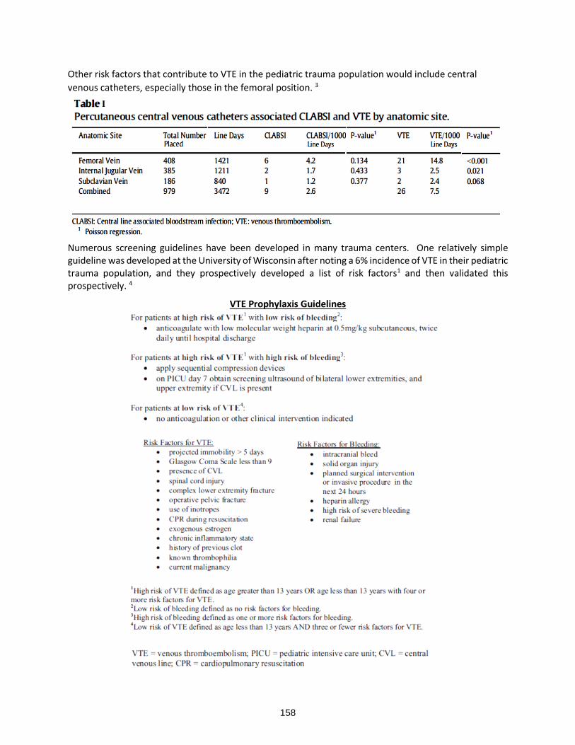
Other risk factors that contribute to VTE in the pediatric trauma population would include central
venous catheters, especially those in the femoral position. 3
Numerous screening guidelines have been developed in many trauma centers. One relatively simple guideline was developed at the University of Wisconsin after noting a 6% incidence of VTE in their pediatric trauma population, and they prospectively developed a list of risk factors1 and then validated this prospectively. 4
VTE Prophylaxis Guidelines
I
158

The same group has also looked at screening in high-risk patients for VTE as well as bleeding. Although the incidence was low in the overall population, in those that were high risk, US screening did detect equal number of symptomatic and asymptomatic patients. 5
Figure 3. Flowchart illustrating the results of our ultrasound experience when using screening guideline
recommendations for risk stratification and prophylaxis. HRHR = high risk for venous thromboembolism
& high risk for bleeding; HR = high risk for venous thromboembolism; ICU = intensive care unit; US =
ultrasound; DVT = deep venous thrombosis.
Other risk factors for VTE increased age, increased ISS, head, thoracic, abdominal, lower extremity, and spinal cord injury. Craniotomy, laparotomy, and spinal operations also associated with VTE.6 Although EAST and PTS developed a GRADE based guideline with recommendations to not perform routine screening dur to the low incidence of asymptomatic patients and recommended prophylaxis in patients 15 and older, as well as younger post-pubertal children with ISS > 25.7 A more complex screening tool developed from the NTDB has recently been developed and validated with a score greater than 550 indicating moderate risk and potential for needing prophylaxis.8
159
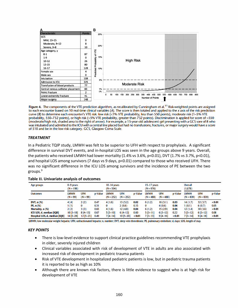
TREATMENT
In a Pediatric TQIP study, LMWH was felt to be superior to UFH with respect to prophylaxis. A significant difference in survival DVT events, and in-hospital LOS was seen in the age groups above 9 years. Overall, the patients who received LMWH had lower mortality (1.4% vs 3.6%, p<0.01), DVT (1.7% vs 3.7%, p<0.01), and hospital LOS among survivors (7 days vs 9 days, p<0.01) compared to those who received UFH. There was no significant difference in the ICU LOS among survivors and the incidence of PE between the two groups.9
Table II. Univariate analysis of outcomes
KEY POINTS
• There is low-level evidence to support clinical practice guidelines recommending VTE prophylaxis in older, severely injured children
• Clinical variables associated with risk of development of VTE in adults are also associated with increased risk of development in pediatric trauma patients
• Risk of VTE development in hospitalized pediatric patients is low, but in pediatric trauma patients it is reported to be as high as 10%
• Although there are known risk factors, there is little evidence to suggest who is at high risk for development of VTE
4.
160

• Routine screening of all pediatric trauma patients is not indicated
• Prophylaxis with Low molecular weight heparin appears to have some advantages over unfractionated heparin in the pediatric trauma population
• There are no consensus guidelines and general agreement on when to initiate VTE prophylaxis in the pediatric trauma patient considered at moderate to high risk
REFERENCES
1. Sheila J. Hanson, MD, MS, Rowena C. Punzalan, MD, Rachel A. Greenup, MD, Hua Liu, MS, Thomas T. Sato, MD, and Peter L. Havens, MD, MS. Incidence and Risk Factors for Venous Thromboembolism in Critically Ill Children After Trauma. J Trauma. 2010;68: 52–56
2. Christine M. Leeper, MD, MS, Matthew D. Neal, MD, Christine McKenna, MSN, CRNP, Jason L. Sperry, MD, MPH, and Barbara A. Gaines, MD. Abnormalities in fibrinolysis at the time of admission are associated with deep vein thrombosis, mortality, and disability in a pediatric trauma population. J Trauma Acute Care Surg. 2017;82: 27–34
3. S. Christopher Derderian, Ryan Good, Raphael N. Vuille-dit-Bille, Todd Carpenter, and Denis D. Bensard. Central venous lines in critically ill children: Thrombosis but not infection is site dependent. Journal of Pediatric Surgery 54 (2019) 1740–1743.
4. Sheila J. Hanson, MD, MS, Rowena C. Punzalan, MD, Marjorie J. Arca, MD, Pippa Simpson, PhD, Melissa A. Christensen, BS, Sydney K. Hanson, Ke Yan, PhD, Kristin Braun, MS, RN, and Peter L. Havens, MD, MS. Effectiveness of clinical guidelines for deep vein thrombosis prophylaxis in reducing the incidence of venous thromboembolism in critically ill children after trauma. J Trauma. 2012;72: 1292–1297.
5. Rachel M. Landisch, Sheila J. Hanson, Rowena C. Punzalan, Kristin Braun, Laura D. Cassidy, and David M. Gourlay. Efficacy of surveillance ultrasound for venous thromboembolism diagnosis in critically ill children after trauma. Journal of Pediatric Surgery 53 (2018) 2195–2201.
6. Monica S. Vavilala, MD, Avery B. Nathens, MD, PhD, MPH, Gregory J. Jurkovich, MD, Ellen Mackenzie, PhD, and Frederick P. Rivara, MD, MPH. Risk Factors for Venous Thromboembolism in Pediatric Trauma. J Trauma. 2002; 52:922–927.
7. Arash Mahajerin, MD, MSCr, John K. Petty, MD, Sheila J. Hanson, MD, MS, A. Jill Thompson, PharmD, Sarah H. O’Brien, MD, Christian J. Streck, MD, Toni M. Petrillo, MD, and E. Vincent S. Faustino, MD, MHS. Prophylaxis against venous thromboembolism in pediatric trauma: A practice management guideline from the Eastern Association for the Surgery of Trauma and the Pediatric Trauma Society. J Trauma Acute Care Surg. 2017;82: 627–636.
8. Daniel F. Labuz, MD, Aaron Cunningham, MD, Joseph Tobias, MD, Alexandra Dixon, MD, MPH, Elizabeth Dewey, MS, Christopher W. Marenco, MD, Mauricio A. Escobar, Jr, MD, Max D. Hazeltine, MD, Muriel A. Cleary, MD, Meera Kotagal, MD, MPH, Richard A. Falcone, Jr, MD, MPH, Sara C. Fallon, MD, Bindi Naik-Mathuria, MD, MPH, Taleen MacArthur, MD, Denise B. Klinkner, MD, MEd, Aashka Shah, Artur Chernoguz, MD, Alberto Orioles, MD, Alicia Zagel, PhD, Ankush Gosain, MD, PhD, Maria Knaus, MD, Nicholas A. Hamilton, MD, and Mubeen A. Jafri, MD. Venous thromboembolic risk stratification in pediatric trauma: A Pediatric Trauma Society Research Committee multicenter analysis. J Trauma Acute Care Surg. 2021;91: 605–611.
9. Muhammad Khurrum, Samer Asmar, Marion Henry, Michael Ditillo, Mohamad Chehab, Andrew Tang, Letitia Bible, Lynn Gries, and Bellal Joseph. The survival benefit of low molecular weight heparin over unfractionated heparin in pediatric trauma patients. Journal of Pediatric Surgery 56 (2021) 494–499
161

162

SESSION 6
FOCUS ON TBI & SPINE CARE
Moderator: Richard A. Sidwell, MD, FACS
Monday, March 28, 2022
Palace Ballroom 1-2
Palace Tower, Emperors Level
4:40 - 4:47
Bad Brains: EVD and ICP Management in Severe TBI Fredric M. Pieracci, MD, MPH, MSPH, FACS
4:47 - 4:54
Fact vs Fiction-2022 Evidence Based Target MAP & Care Goals Meghan R. Lewis, MD, FACS
4:54 - 5:01
Palliative Care Principles and Practices Melissa “Red” Hoffman, MD, FACS
5:01 - 5:08
Role of Craniectomy: Pop the Top, or Stay the Course Matthew J. Martin, MD, FACS
5:08 - 5:15
Beyond ICP: Advanced Neuromonitoring Modalities for TBI Carlos V.R. Brown, MD, FACS
163

BAD BRAINS: EVD AND ICP MANAGEMENT IN SEVERE TBI
Fredric M. Pieracci, MD, MPH, MSPH, FACS
Interim Director of Surgery Denver Health Medical Center Professor of Surgery University of Colorado Denver Denver, CO
Traumatic brain injury (TBI) remains one of the most common causes of mortality and long-term morbidity in injured patients. Trauma surgeons and surgical intensivists are charged with mitigating the aftereffects of the initial brain insult through monitoring and treatment of elevated intra-cranial pressure (ICP). Successful management of patients with TBI involves an understanding of the distinction between primary and secondary brain injury. Primary brain injury occurs as a direct result of the trauma – for example, the impact sustained during a motor vehicle crash. Beyond prevention, the trauma team is unable to influence the severity of primary brain injury. By contrast, secondary brain injury occurs in the hours to days following primary brain injury, and is the result of progressive edema, swelling, ischemia, and potentially infarction. Mitigation of secondary brain injury is the focus of critical care of the TBI patient, and centers upon minimization of periods of cerebral hypoperfusion due to systemic hypotension, hypoxia, and elevated ICPs.
Because the brain exists in the rigid, fixed space of the skull, it is vulnerable to a compartment syndrome, which exists when the ICP exceeds that of venous outflow pressure, and eventually arterial inflow pressure. The ICP is a dynamic parameter, with transient spikes due to daily activities such as sneezing and bearing down being normal. However, sustained elevations in ICP above the normal range of 0 – 10 mm Hg, and in the setting of the systemic response to trauma (i.e., cytokine and cortisol release), can have devastating effects on long term brain recovery. Successful maintenance of normal ICPs requires an understanding of how and when to measure them, followed by a tiered treatment approach that employs escalating therapies, beginning with simple maneuvers such as head of bed elevation and systematically increasing in invasiveness to decompressive craniectomy (DC). A detailed review of pharmacologic modalities employed to manage elevated ICPs is beyond the scope of this syllabus. Rather, this chapter will focus on the indications for ICP monitoring, target systolic blood pressure (SBP), ICP, and cerebral perfusion pressure (CPP), and the indications for DC.
Evidence-based guidelines for the treatment of patients with TBI are periodically updated and published by the Brain Trauma Foundation. This resource has served as a shining example of the power of the trauma community to disseminate objective information and influence the care of trauma patients for the better. The latest guidelines available are from 2017, with an update specifically related to DC published in 2020. These guidelines are referenced at the end of this chapter.
It is important to note that, at its most simple level, monitoring of cerebral perfusion occurs using the physical exam. There is rarely a need to perform invasive monitoring of ICP in a patient with a normal, or near normal, neurologic exam that may be followed, even in the setting of a markedly abnormal CT head. And even in patients with invasive monitors in place (e.g., ventriculostomy), the clinical exam always trumps the number that is being displayed. In some instances, there will be a discrepancy between the clinical exam and the ICP value; these cases are usually due to equipment malfunctioning.
164
Indications for invasive monitoring in patients with severe TBI [Glasgow Coma Score (GCS) 3-8 after resuscitation] are listed in Table I and presuppose patient salvageability. In cases of severe TBI with a normal CT head, there are still instances in which monitoring may be helpful. These include patients with shock, motor posturing, and age > 40 years. In these cases, non-hemorrhagic pathologies such as diffuse axonal injury may be contributing to elevations in ICP, and treatment of this elevation may be beneficial.
Monitoring Type Recommendation Level
Intracranial pressure All salvageable patients with severe TBI (GCS 3-8 after resuscitation) and an abnormal CT head
Severe TBI with normal CT if ≥ 2: age > 40, motor posturing, SBP < 90 mm Hg
IIB
Cerebral perfusion pressure
Recommended in all severe TBI patients to decrease 2-week mortality IIB
Advanced cerebral monitoring
May be considered to reduce mortality and improve outcome at 6 months III
Table I: Indications for monitoring in patients with severe Traumatic Brain Injury
There are three main types of invasive monitoring of brain perfusion: ICP, CPP, and advanced cerebral monitoring. The CPP is defined as the mean arterial pressure – the ICP, such that higher values are more favorable. Advanced cerebral monitoring includes all forms of measurement of brain partial oxygen pressure or perfusion/flow. Advanced cerebral monitoring may not be routinely available at all trauma centers. Because of limited availability, as well as a lack of evidence to support improved outcomes, routine employment is not advised, and given a level III recommendation for consideration in the Brain Trauma Institute guidelines.
Thresholds for SBP, ICP, and CPP are listed in Table II . In general, the ICP and CPP are used together to make management decisions, but the ICP remains the one most followed measurement. In the latest Trauma Brain Foundation guidelines, treatment of ICPs > 22 mm Hg are recommended at a level of IIB. This liberalization of the ICP threshold from the previously recommended 20 to 22 mm Hg represents a gradual sea change in the TBI community to tolerance of higher ICPs to mitigate the negative consequences of the therapies used to lower ICPs, such as sedatives, paralytics, and hypertonic saline. Each of these therapies may paradoxically worsen outcomes in TBI patients through independent mechanisms such as prolonging periods of mechanical ventilation, or causing hypovolemia, decreased CPP, and ultimately exacerbating secondary brain injury.
Threshold Recommendation Level
Systolic blood pressure
≥ 100 mm Hg for patients 50-69 years old
≥ 110mm Hg for patients 15 – 49 or > 70 years old
III
ICP Treating ICP > 22 mg is recommended because values above this level are associated with increased mortality
IIB
CPP 60 to 70 mm Hg
The optimal threshold is unclear and likely depends upon the autoregulatory status of the patient
Avoiding aggressive attempts to maintain CPP > 70 with fluid sand pressors may be considered because of the risk of ARDS
IIB
III
Table II: Threshold for intervention
165

The optimal recommended CPP threshold is 60 to 70 mm Hg but is unclear. Again, efforts to drive up CPP usually involve both volume expansion and vasopressors, which can each contribute to prolonged ventilation, acute respiratory distress syndrome, and development of abdominal compartment syndrome. In general, management of patients with severe TBI and invasive monitoring in place should be guided by the physical exam and ICPs.
The decision to perform DC is controversial. However, two recent, large randomized controlled trials (RCTs) have shed some light on the issue. The Decompressive Craniectomy in Patients with Severe Traumatic Brain Injury (DECRA) trail included an early (< 72 hours from injury) decision to perform DC in patients with ICPs > 20 mm Hg for at least 15 minutes over a one hour period despite optimization of tier one treatments. There were 155 subjects randomized to either DC or no DC. There was no difference in either 6- or 12-month mortality (each around 20%) and no significant difference in a favorable outcome at 12 months as measured by the Glasgow Outcome Scale Extended (GOSE).
The Trial of DC for Traumatic Intra Cranial Hemorrhage (RESCUEicp) trial randomized 398 subjects in a later time period (within 10 days of injury) and with a higher threshold for surgery; specifically, an ICP > 25 mm Hg for 1-12 hours refractory to 2 tiers of treatment. Both 6- and 12-month mortality was significantly reduced in the DC group, with a modest improvement in favorable outcome at 12 months.
The results of these two RCTs informed the 2020 Brain Trauma Foundation recommendations for DC in patients with severe TBI. A DC is recommended (level II) for patients with late refractory ICP elevation, but not recommended for early refractory ICP elevations. Further, DC is noted to decrease ICP and duration of intensive care, in either the early or late period, though the relationship between these effects and favorable outcomes is uncertain.
In conclusion, management of patients with severe TBI centers on mitigation of secondary brain injury. Although the clinical exam trumps all, severe TBI patients (GCS 3-8) with an abnormal CT head should undergo invasive ICP monitoring, with a treatment threshold of > 22 mm Hg. A target CPP of 60 – 70 mm Hg is ideal, although aggressive efforts to keep the CPP in this range with either volume expansion or vasopressor should be avoided, and the auto regulatory patterns of individual patients may preclude ever achieving this goal. Finally, the decision to perform DC remains controversial. The optimal benefit of DC appears to be in refractory elevations of ICPs relatively later in the course (3-10 days post injury).
REFERENCES
1. Carney N, Totten AM, O-Reilly C, et al. Guidelines for the management of severe traumatic brain injury, fourth edition. Neurosurgery 2017;80:6-15.
2. Hawryluk GW, Rubiano AM, Totten AM, et al. Guidelines for the management of severe traumatic brain injury: 2020 update of the decompressive craniectomy recommendation. Neurosurgery 2020;87:427-434.
3. Cooper DJ, Rosenfeld JV, Murray L, et al. Decompressive craniectomy in diffuse traumatic brain injury. N Engl J Med 2011;364(16):1493-1502.
4. Hutchinson PJ, Kolias AG, Timofeev IS, et al. Trial of decompressive craniectomy for traumatic intracranial hypertension. N Engl J Med 2016;375(12):1119-1130.
166

FACT VS FICTION- 2022 EVIDENCE BASED TARGET MAP & CARE GOALS
Meghan Lewis, MD, FACS
Assistant Professor of Clinical Surgery LAC+USC Medical Center & University of Southern California – Los Angeles Los Angeles, CA
BACKGROUND
Trauma to the spinal cord results in both primary and secondary injury. Primary injury refers to the direct damage from the traumatic event. Poor spinal cord perfusion in the days to weeks following acute spinal cord injury (SCI) worsens ischemia from the insult and impairs recovery of neurologic function. This phenomenon is referred to as “secondary injury.”
There is currently no standard method in use for measuring and monitoring spinal cord blood flow itself after injury. Loss of autoregulation after acute injury likely causes spinal cord blood flow to become more dependent on systemic arterial blood pressure. Unfortunately, systemic hypotension is common in the first 1-2 weeks following acute SCI due to both blood loss, hypovolemia, and autonomic instability1,2. Patients with cervical spine or complete upper thoracic spine injury are especially vulnerable, due to the risk of neurogenic shock.
Experimental models in the past have suggested that elevating systemic blood pressure does improve spinal blood flow.3 However, it is not known clinically to what extent, or in which ranges pharmacologic vasoconstriction actually improves spinal cord blood flow. For example, overuse of vasoconstricting medications to increase systemic arterial pressure could potentially impair spinal cord blood flow, by limiting collateral flow through intercostal anastomoses.
The key clinical questions under consideration include:
1. Does pharmacological augmentation of mean arterial blood pressure (MAP) with vasoconstrictors decrease secondary injury and improve neurologic outcomes after acute SCI?
2. If so, what threshold MAP should be targeted? 3. For what period of time is blood pressure augmentation beneficial? 4. Is there a preferred pharmacological vasoconstrictor that optimizes spinal cord blood flow? 5. Which specific patient populations benefit from pharmacologic blood pressure augmentation? All
ages? All injury types? 6. What about targeted blood pressure management to prevent secondary damage after traumatic
brain injury (TBI)?
EVOLUTION OF CARE
The literature to support therapies targeting MAP after acute SCI is limited to experimental, retrospective, and level III clinical evidence. These studies were not specifically designed to investigate the role of targeted blood pressure management, but instead were designed to improve the overall management of patients with SCI. Many of the studies combined new management strategies, such as aggressive monitoring and intervention for cardiovascular abnormalities, with early surgical fixation and techniques to prevent pulmonary complications, or other challenges in the management of acute SCI. The effect of the cardiovascular interventions can therefore not be independently evaluated in such studies.
167
One of the early studies to evaluate cardiovascular targets and timing was performed by Piepmeier et al in 19851. They evaluated 45 acute spinal cord injury patients and observed that cardiac dysrhythmias, hypotension, and hypoxia were common in the first 2 weeks after ASCI. The incidence and severity of these abnormalities appeared to be related to the injury severity. The authors used fluids and vasopressors (specifically dopamine, phenylephrine, and isoproterenol, alone or in combination) to maintain systolic blood pressure (SBP) greater than 100 mm of mercury (mm Hg). Vasopressors were necessary to meet this threshold in 20% of patients for a period of up to 5 days.
In 1991, Wolf et al evaluated patients with acute bilateral locked facets at a single trauma center.4 Patients were reduced with or without surgery, then maintained with mean arterial blood pressure (MAP) greater than 85 mm Hg for 5 days. Neurological improvement was seen at discharge in 21% of complete SCI patients and in 62% of patients with incomplete cervical SCI. No intact patient deteriorated.
Not long after this, Levi et al evaluated 50 ICU patients with acute cervical cord trauma.3 Patients underwent reduction/stabilization, and operative intervention, if indicated. They were then monitored in the ICU with arterial and pulmonary artery catheters and maintained at MAP greater than 90 mm Hg with fluids, dopamine and/or dobutamine. Forty-one patients required vasopressor support for an average of 5-6 days. The authors noted improved neurologic outcomes in 20 patients at 6 weeks post injury.
In 1997, Vale et al performed a prospective pilot study to assess the merits of aggressive medical resuscitation and blood pressure management after acute spinal cord injury.5 They prospectively assessed 77 acute SCI patients. Patients underwent reduction and stabilization as appropriate, arterial and pulmonary artery catheters were placed, and they were maintained with MAP greater than 85 for 7 days (by use of methylprednisolone, colloid, blood, dopamine, and norepinephrine). The authors concluded that improved outcomes could be achieved with aggressive medical care (distinct from surgical benefit) at 1 year.
SUMMARY TARGETS:
>85 or >90 thresholds
5- to 7-day periods
CURRENT GUIDELINES
The most recent Congress of Neurological Surgeons published guidelines in 2013. These guidelines recommend maintenance of a MAP between 85- and 90-mm Hg for the first 7 days following the acute SCI2. The authors acknowledged the low level of evidence to support this recommendation, and they recommended a future multi-institutional prospective trial to further evaluate the impact of blood pressure augmentation on neurologic outcomes.
VALIDATION
Hawryluk et al used a computerized system to correlate the proportion of hypotensive events (MAP <85 mm Hg) over the first 5-7 days post injury to worse neurologic recovery in 100 SCI patients.6
Martin et al retrospectively evaluated average MAP and hypotensive events in 105 SCI patients.7 Their results associated both hypotension and vasopressor use with more severe SCI, however, neither was associated with change in American spinal injury association motor score (AMS).
Weinberg et al retrospectively evaluated blunt spinal cord injury patients between 2014 and 2019 from the registry of a level 1 trauma center.8 They correlated MAP values ≥85 mm Hg with neurologic improvement, as measured by a positive change in the American Spinal Injury Association (AISA)
168
impairment scale. Multivariate modeling demonstrated a significant association between proportion of elevated MAP values and neurologic improvement (p = 0.028).
VASOPRESSOR CHOICE
Inoue et al evaluated the effects of vasopressor use in SCI in a retrospective cohort study of 131 patients from single level 1 trauma center (2005-2011).9 Dopamine was the most commonly used vasopressor (48.0%), followed by phenylephrine (45.0%), norepinephrine (5.0%), epinephrine (1.5%), and vasopressin (0.5%). Logistic regression analysis demonstrated that complications due to vasopressors (e.g., dysrhythmias) were independently associated with the use of dopamine (odds ratio [OR] 8.97; p<0.001) and phenylephrine (OR, 5.92; p=0.004), age ≥60 years old (OR, 5.16; p=0.013), and complete SCI (OR, 3.23; p=0.028). There was no difference in neurological improvement with either dopamine (OR, 1.16; p=0.788) or phenylephrine (OR 0.96; p=0.940).
Altaf et al published a prospective study of vasopressor use in 11 patients with acute SCI.10 Norepinephrine and dopamine were evaluated in crossover fashion to directly compare their effect on the intrathecal pressure (ITP) in the intensive care unit where ITP, mean arterial pressure (MAP), and heart rate were being continuously measured. Spinal cord perfusion pressure (SCPP) was calculated as the difference between MAP and ITP. They reported no difference in MAP with the use of norepinephrine versus dopamine (84±1 mm Hg for both; P=0.33). ITP was significantly lower with the use of norepinephrine than with dopamine (17±1 mm Hg vs 20±1 mm Hg, respectively, P<0.001), which resulted in an increased SCPP with norepinephrine infusion compared to dopamine (67±1 mm Hg vs 65±1 mm Hg respectively, P=0.0049).
PENETRATING SCI
Readdy et al evaluated MAP augmentation specifically in penetrating SCI- a population of patients with decreased likelihood of neurological improvement.11 Only 1 out of 14 patients experience neurological improvement, however, 71% experienced cardiogenic complications. The authors concluded that use of vasopressors may incur more risk than benefit in this specific population of SCI.
INJURY GRADE
Catapano et al evaluated MAP augmentation according to ASIA grade.12 The authors observed an association between MAP values and neurologic recovery in AIS A, B, and C patients, but not AIS D patients.
ACUTE TRAUMATIC CENTRAL CORD SYNDROME
Acute traumatic central cord syndrome (ATCCS) is the most common type of incomplete spinal cord injury (SCI)13. ATCCS complications have been shown to occur at increased rate in elderly patients14. Previous studies have linked vasopressor support to improved outcomes in this population but have recognized that there are no validated protocols for the implementation of these interventions5,15.
The initial recommendation for vasopressors in this specific population came from a single large retrospective study16. However, use of vasopressors is not benign. Cardiogenic complications are frequently associated, and poor clinical outcomes have been previously reported in the setting of early vasopressor use for critically injured, non-neurosurgical trauma patients17.
Readdy et al performed a retrospective cohort analysis of 34 ATCCS patients at a single Level 1 trauma center who received any vasopressor to maintain MAP above predetermined goals18. The patients were identified by a database, then individual charts were reviewed for ASIA grades at admission and discharge, vasopressor administered and associated complications, other interventions and complications, and timing of surgery. The relationship between the 2 most common vasopressors—dopamine and
169

phenylephrine—as well as complications within the cohort were evaluated, and again after stratification by age. Acute SCI methylprednisolone protocol was administered to 20 patients (59%). Decompressive surgery was performed in the first 24 hours in 16 patients (47%), after 24 hours in an additional 9 patients (26%), and not at all in 9 patients. Dopamine was the most commonly used primary vasopressor (91%), followed by phenylephrine (65%). Vasopressors were used to maintain MAP goals of greater than or equal to 85 mm Hg for a mean of 101 hours. Neurological status improved by a median of 1ASIA grade in all patients (regardless of vasopressor use). Cardiogenic complications associated with vasopressor usage were noted in 68% of patients who received dopamine and 46% of those who received phenylephrine, though not statistically significant (OR with dopamine 2.50, p = 0.105). In the subgroup of patients older than 55 years, dopamine was associated with a statistically significant increase in complication rates compared with phenylephrine (83% vs 50%, respectively; OR with dopamine 5.0, p = 0.044). The authors concluded that vasopressor usage in ATCCS is associated with complication rates similar to the reported literature for all SCI, however, dopamine is associated with higher risk of complications in patients older than 55 years. Given the increased incidence of ATCCS in older populations, determination of MAP goals and vasopressor administration should be carefully considered in this type of SCI.
“TEMPLE” TRIAL
A Randomized Trial of Early Hemodynamic Management of Patients following Acute Spinal Cord Injury (TEMPLE) Study19 is currently in process. This is a multicenter, randomized-controlled study of 152 adult subjects being randomized 1:1. The intervention is targeting of conventional blood pressure control (CBP) versus augmented blood pressure (ABP) based on study group assignment. The CBP group has a target MAP of 65-70 mm Hg for 7 days after randomization or until ICU discharge; whereas the ABP group has a goal of MAP 85-90 mm Hg for 7 days after randomization or until ICU discharge. The primary objective is to determine the differential effects of two levels of MAP on long-term motor and sensory outcomes after acute SCI. The secondary objectives are to determine the differential efficacies of two levels of MAP based on long-term pain and functional independence measures, as well as to determine the differential safety profiles of maintaining two different levels of MAP for up to 7 days.
TRAUMATIC BRAIN INJURY
Patients who suffer brain trauma similarly experience both primary and secondary injury. Systemic hypotension is known to contribute to secondary injury after TBI and has been previously associated with worsened outcomes. After brain trauma, elevated intracranial pressure (ICP) is also an important contributor to the decrease in cranial blood flow that results in secondary injury. ICP is measured directly in ICUs by use of an intracranial pressure monitor. Interventions to decrease ICP are often emphasized to minimize secondary injury. Alternatively, cerebral perfusion pressure (CPP) has been used as a surrogate for cerebral blood flow, as higher CPP has been associated with survival and favorable neurologic outcomes.20 CPP can be increased either by maneuvers to decrease intracranial pressure or by augmentation of MAP with fluid or vasopressors.
CPP = MAP - ICP
In 2001, Struchen et al published a cohort of 184 patients with severe TBI admitted to a single level I trauma center neurosurgical ICU who received continuous monitoring of ICP, MAP, cerebral perfusion pressure (CPP), and arterial oxygen saturation (SpO2)21. Primary outcomes for the study were Glasgow Coma Score (GCS) and Disability Rating Score (DRS). The analysis included multiple regression models evaluating effects of physiologic variables on outcomes. Duration of systemic hypotension (MAP <80) was found to be associated with both lower GCS and DRS at several months post injury.
Murray et al used a database of 8686 TBI patients to study the prognostic value of a large number of variables using both univariate and multivariable analysis. Hypotension was noted to have a strong
170

association with neurologic outcome 6 months after injury. The association was stronger with systolic blood pressure (SBP) than with MAP.22
In 2012, Berry et al evaluated a retrospective cohort of 15,733 TBI patients.23 They used a best fit model to determine the optimal threshold of hypotension for different age groups (to minimize the probability of death). They found SBP of 110 mm Hg was optimal for patients 15–49 years (p < 0.0001), 100 mm Hg for patients 50–69 years (p = 0.0002), and 110 mm Hg for patients ≥70 years (p = 0.0003).
Brenner et al published a prospective cohort study of 60 TBI patients who were monitored in the intensive care unit for hypotensive events post injury.24 They found that hypotension was predictive of mortality and poor functional outcomes at a higher threshold than previously described: 120 mm Hg. They concluded that a SBP target of 120 mm Hg in the first 48 hours post injury could potentially minimize secondary brain injury.
The Brain Trauma Foundation guidelines were most recently updated in 2016.20 The guidelines, based on Level III evidence, state that maintaining a SBP at greater than or equal to 100 mm Hg for patients 50 to 69 years old, or at greater than or equal to 110 mm Hg for patients 15 to 49 years or over 70 years old, may be considered to decrease mortality and improve outcomes.
CONCLUSIONS
• MAP thresholds of 85-90 mm Hg after acute SCI are recommended by the Congress of
• Neurological Surgeons, however, this is based on very low-level evidence.
• Hypotensive events below the recommended threshold have been associated with worse neurologic outcomes, though the evidence is not consistent.
• Studies targeting MAP thresholds in acute SCI have reported augmentation periods up to 7 days post injury
• Norepinephrine may be the preferred vasoconstrictor to augment spinal cord blood flow
• Patients with penetrating SCI may incur more risk than benefit from pharmacologic vasoconstriction
• A multicenter prospective trial to evaluate 2 different threshold MAPs is currently ongoing
• Systemic hypotension should be avoided after TBI, however, ICP and CPP thresholds are more commonly targeted than MAP
REFERENCES
1. Piepmeier JM, Lehmann KB, Lane JG. Cardiovascular instability following acute cervical spinal cord trauma. Cent Nerv Syst Trauma. 1985 Fall;2(3):153-60.
2. Ryken TC, Hurlbert RJ, Hadley MN, et al. The acute cardiopulmonary management of patients with cervical spinal cord injuries. Neurosurgery. 2013;72 Suppl 2:84-92
3. Levi L, Wolf A, Belzberg H. Hemodynamic parameters in patients with acute cervical cord trauma: description, intervention, and prediction of outcome. Neurosurgery. 1993;33(6):1007-1016
4. Wolf A, Levi L, Mirvis S, et al. Operative management of bilateral facet dislocation. J Neurosurg. 1991;75(6):883-890
5. Vale FL, Burns J, Jackson AB, Hadley MN. Combined medical and surgical treatment after acute spinal cord injury: results of a prospective pilot study to assess the merits of aggressive medical resuscitation and blood pressure management. J Neurosurg. 1997;87(2):239-246
6. Hawryluk G, Whetstone W, Saigal R, Ferguson A, Talbott J, Bresnahan J, Dhall S, Pan J, Beattie M, Manley G. Mean arterial blood pressure correlates with neurological recovery after human spinal cord injury: analysis of high frequency physiologic data. J Neurotrauma. 2015;32(24):1958–1967
171

7. Martin ND, Kepler C, Zubair M, Sayadipour A, Cohen M, Weinstein M. Increased mean arterial pressure goals after spinal cord injury and functional outcome. J Emerg Trauma Shock. 2015;8(2):94–98.
8. Weinberg, J. , Farber, S. , Kalamchi, L. , Brigeman, S. , Bohl, M. , Varda, B. , Sioda, N. , Radosevich, J. , Chapple, K. & Snyder, L. (2021). Mean arterial pressure maintenance following spinal cord injury: Does meeting the target matter? Journal of Trauma and Acute Care Surgery, 90 (1), 97-106.
9. Inoue T, Manley GT, Patel N, Whetstone WD. Medical and surgical management after spinal cord injury: vasopressor usage, early surgerys, and complications. J Neurotrauma. 2014;31(3):284–291.
10. Altaf F, Griesdale DE, Belanger L, et al. The differential effects of norepinephrine and dopamine on cerebrospinal fluid pressure and spinal cord perfusion pressure after acute human spinal cord injury. Spinal Cord. 2017;55(1):33–38.
11. Readdy WJ, Saigal R, Whetstone WD, et al. Failure of mean arterial pressure goals to improve outcomes following penetrating spinal cord injury. Neurosurgery. 2016;79(5):708–714.
12. Catapano JS, John Hawryluk GW, Whetstone W, et al. Higher mean arterial pressure values correlate with neurologic improvement in patients with initially complete spinal cord injuries. World Neurosurg. 2016;96:72–79
13. Aarabi B, Koltz M, Ibrahimi D: Hyperextension cervical spine injuries and traumatic central cord syndrome. Neurosurg Focus 25(5):E9, 2008
14. Newey ML, Sen PK, Fraser RD: The long-term outcome after central cord syndrome: a study of the natural history. J Bone Joint Surg Br 82:851–855, 2000
15. Ploumis A, Yadlapalli N, Fehlings MG, Kwon BK, Vaccaro AR: A systematic review of the evidence supporting a role for vasopressor support in acute SCI. Spinal Cord 48:356– 362, 2010
16. Aarabi B, Hadley MN, Dhall SS, Gelb DE, Hurlbert RJ, Rozzelle CJ, et al: Management of acute traumatic central cord syndrome (ATCCS). Neurosurgery 72 (Suppl 2):195–204, 2013
17. Plurad DS, Talving P, Lam L, Inaba K, Green D, Demetriades D: Early vasopressor use in critical injury is associated with mortality independent from volume status. J Trauma 71:565–572, 2011
18. Readdy WJ, Whetstone WD, Ferguson AR, Talbott JF, Inoue T, Saigal R, Bresnahan JC, Beattie MS, Pan JZ, Manley GT, Dhall SS. Complications and outcomes of vasopressor usage in acute traumatic central cord syndrome. J Neurosurg Spine. 2015 Nov;23(5):574-580.
19. TEMPLE study. Clinicaltrials.gov 20. Carney N, Totten AM, O'Reilly C, Ullman JS, Hawryluk GW, Bell MJ, Bratton SL, Chesnut R, Harris
OA, Kissoon N, Rubiano AM, Shutter L, Tasker RC, Vavilala MS, Wilberger J, Wright DW, Ghajar J. Guidelines for the Management of Severe Traumatic Brain Injury, Fourth Edition. Neurosurgery. 2017 Jan 1;80(1):6-15.
21. Struchen MA, Hannay HJ, Contant CF, Robertson CS. The relation between acute physiological variables and outcome on the Glasgow Outcome Scale and Disability Rating Scale following severe traumatic brain injury. J Neurotrauma 2001;18(2):115- 125.
22. Murray GD, Butcher I, McHugh GS, Lu J, Mushkudiani NA, Maas AI, Marmarou A, Steyerberg EW. Multivariable prognostic analysis in traumatic brain injury: results from the IMPACT study. J Neurotrauma. 2007 Feb;24(2):329-37.
23. Berry C, Ley EJ, Bukur M, et al. Redefining hypotension in traumatic brain injury. Injury. 2012;43(11):1833-1837.
24. Brenner M, Stein DM, Hu PF, Aarabi B, Sheth K, Scalea TM. Traditional systolic blood pressure
targets underestimate hypotension-induced secondary brain injury. J Trauma Acute Care Surg.
2012;72(5):1135-1139.
172

PALLIATIVE CARE PRINCIPLES AND PRACTICES
Melissa “Red” Hoffman, MD, FACS
Clinical Assistant Professor University of North Carolina at Chapel Hill School of Medicine Chapel Hill, NC
TOP 10 THOUGHTS ON PALLIATIVE CARE (WITH A FOCUS ON NEUROPALLIATIVE CARE)
1. Palliative care is NOT synonymous with hospice care. Palliative care is most often delivered alongside disease-directed treatments while hospice care is reserved for patients who have six months left to live AND who are not pursuing disease-directed treatments.
2. There is no such thing as “going palliative.” 3. Do Not Resuscitate (DNR) does NOT mean Do Not Treat. 4. There is a dearth of fellowship-trained palliative care providers in the United States. As the
population ages and becomes even more medically complex, surgeons will continue to be called upon to practice primary palliative care.
5. Given this fact, making certain that medical students and surgical residents witness and participate in palliative care interventions is of utmost importance.
6. That being said, surgeons should learn to recognize when specialist palliative care is appropriate. 7. High spinal cord injury (SCI) presents multiple challenging ethical situations and specialist
palliative care is often necessary to support the patient, the family, and the entire care team. 8. Severe traumatic brain injury (TBI) represents another condition in which palliative care
(generalist or specialist) is often necessary, for multiple reasons. One, our ability to accurately and precisely predict long-term outcomes after a severe TBI is limited and thus the family is often in need of psychosocial support. Two, questions regarding futility as well as questions regarding code status, life sustaining medical treatments and artificial nutrition and hydration are all often raised in a short period of time, and families will need ongoing support as they grapple with these decisions.
9. Time limited trials are a way to allow patients and families to come to terms with devastating diagnoses while also feeling like they were able to “do everything” for their loved one.
10. Accurate documentation of the health care power of attorney/surrogate decision maker as well as detailed documentation of discussions regarding code status and goals of care is key for several reasons: it spares the family from having to repeat difficult conversations multiple times, it allows for continuity of care between practice partners and various teams, and it ensures that we can get adequately compensated for our time.
OVERVIEW
High SCI and severe TBI are devastating and life-altering events, not just for the patient, but for the family as well. While SCI has well-established prognoses based on injury level, the sudden, irrevocable changes to one’s entire life are often too overwhelming to comprehend. On the other hand, TBIs are complicated by individual clinician’s inability to accurately predict mortality as well as functional outcome. Palliative care – with its recognition of the patient AND the family as the unit of care, as well its focus on communication, decision-making, and psychological and spiritual support – can and should be utilized to assist the patient and the family with both injuries. Early palliative care (see figure below), initiated at the
173

time of diagnosis and continued throughout the disease trajectory, can benefit the patient, the family and the care team.
From: Mayo Clin Proc. 2016:92(2)280-286.
KEY DEFINITIONS:
1. Palliative Care: According to the National Hospice and Palliative Care Organization, palliative care is patient and family-centered care that optimizes quality of life by anticipating, preventing, and treating suffering.
2. Hospice: Hospice is palliative care reserved specifically for patients who have six months or less to live (if the disease were to take its natural course) AND who are no longer pursuing disease-directed treatments like chemotherapy, radiation, dialysis and, in most situations, surgery.
3. Primary Palliative Care (aka Generalist Palliative Care): Skills that all clinicians should possess, including basic management of symptoms such as pain, receptor mediated nausea, constipation and breathlessness; straight-forward discussions about prognosis, code status and goals of care; a basic understanding of advance care planning, including how to determine the legal surrogate decision maker in your state; and a basic understanding of hospice, including who qualifies and when an evaluation is appropriate.
4. Secondary Palliative Care (aka Specialist Palliative Care): Skills usually reserved for those clinicians with advanced training in hospice and palliative medicine, including management of refractory pain and other symptoms; management of complex depression, grief, and existential distress; assistance with conflict resolution regarding code status or goals of care; assistance of cases of near futility; and assistance with transition to hospice.
5. Time Limited Trial (TLT): An agreement between clinicians and a patient/family to utilize certain medical therapies over a defined period of time to see if the patient improves or deteriorates according to prespecified clinical outcomes.
PALLIATIVE CARE PRINCIPLES
Few surgeons realize that the term palliative care was coined by a surgeon, Dr. Balfour Mount, in 1974. Dr. Mount was practicing as a urologic oncologist at the Royal Victoria Hospital in Montreal, Canada when he first heard a lecture by Dr. Elizabeth Kubler-Ross on death and dying. He eventually travelled to England
174

to study under physician Dame Cicely Saunders, known as the founder of the modern hospice movement. On his return to Montreal, Dr. Mount established the first inpatient palliative care unit in North America.
Palliative care can best be described as an “extra layer of support” for patients and families whose lives are affected by life-limiting or life-threatening illnesses. As defined above, specialty palliative care is often delivered by an inter-disciplinary team which may include physicians, advanced practice providers, nurses, social workers, pharmacists, chaplains and music therapists. Palliative care is applicable early in the course of illness, often in conjunction with disease-directed treatments, and can delivered at home, in a clinic, or in a hospital. It affirms life and regards death as a normal process; it neither seeks to hasten nor to postpone death. Palliative care integrates the physical, psychological, and spiritual aspects of patient care and attempts to provide a support system that allows patients to live as actively as possible until death.
One of the most convincing studies on the utility of palliative care was published in 2010 in the New England Journal of Medicine. In this study of 157 patients with metastatic non-small cell lung cancer, one group was randomized to early palliative care integrated with standard oncologic care while the other group received standard oncologic care. Patients who received palliative care were found to have higher quality of life scores, lower depressive symptoms, and a statistically significant longer survival time (11.6 vs 8.9 months) despite fewer patients receiving aggressive end-of-life chemotherapy. In their seminal work on the integration of palliative care into the trauma and surgical intensive care units, Anne Mosenthal and colleagues found that integration of early palliative care alongside aggressive trauma care led to significantly earlier timing of DNR orders and withdrawal of life sustaining treatments (WLST) without any change in mortality. Further, the time from DNR to death was lengthened, leading the authors to postulate that the quality of care at end-of-life was improved. Of note, the palliative care interventions were facilitated by palliative care counselors and chaplains (who were embedded within the ICU team) with active participation by the trauma or surgical intensivist, rather than by a specialty palliative care physician.
Hospice and Palliative Medicine was first recognized as a medical specialty in 2006 and the first certification exam (sponsored by ten different specialty boards, including the American Board of Surgery) was offered in 2008. As of 2022, about 80 surgeons are currently board-certified in Hospice and Palliative Medicine. That being said, one does NOT have to be fellowship trained to offer primary (or sometimes even secondary) palliative care to patients and their families (see figure below). In their article “Palliative Care in the Trauma ICU,” O’Connell and Maier propose that palliative care should be considered the fourth domain of acute care surgery (see figure).
175

From: Curr Opin Crit Care. 2016:22(6)584-590.
176

A FEW WORDS ABOUT HOSPICE
Hospice care first became popular in the United States in the 1970s; interestingly, some of the very first hospices in the country were founded and run by surgeons. As stated above, hospice care is reserved for patients who are nearing the end of life AND, just as importantly, who are no longer pursuing any disease-directed treatment. In that sense, all hospice care can be considered palliative, but only a small subset of palliative care is hospice (see figure below). Hospice is best thought of as a philosophy of care, rather than as a place of care. There are four levels of hospice care: routine hospice care, general inpatient care (GIP), continuous home care and inpatient respite care; the vast majority (over 90%) of hospice patients receive routine hospice care within their place of residence (whether that is a private residence, assisted living or skilled nursing facility), with family, friends or paid caregivers responsible for the bulk of the daily care. GIP is reserved for those patients (less than 10% of hospice patients) who require 24-hour nursing care; it can be delivered at a free-standing hospice facility, at a skilled nursing facility or at a hospital. Hospice is covered by Medicare, Medicaid and most private insurance plans. In 2019, 51% of Medicare patients were enrolled in hospice at their time of death. One of the many benefits of hospice is the bereavement services which are offered to family members for at least one year after the patient dies.
177

PALLIATIVE CARE IN HIGH SPINAL CORD INJURY (SCI)
There are few injuries that present more ethical challenges than high spinal cord injuries, particularly when they occur in young, previously healthy patients. Many times, these patients are cognitively intact but unable to consistently and effectively communicate due to the need for mechanical ventilation. Patients are often in the hospital for several weeks while goals of care are clarified, tracheostomies and surgical feeding tubes are placed (if consistent with goals) and placement in a neuro-rehab center is secured. Given the communication difficulties, the benefits of a consistent care team over a prolonged hospitalization, and the existential angst often experienced by the patient, the family and the care team, high SCI patients (especially younger patients) may benefit from specialty palliative care.
Several palliative care issues may arise during the acute period. One is the question of decision-making capacity (DMC). It is not uncommon for a patient newly diagnosed with high SCI to refuse care, but an astute clinician must question whether this patient possesses DMC. Many factors, including grief reactive depression, electrolyte abnormalities, hemodynamic instability, and the ICU itself may prevent the patient from fully understanding his condition and prognosis, appreciate the consequences of refusing care and fully reason from the question posed to the conclusion reached (all necessary components of DMC). Further, it is difficult to fully assess for DMC in an intubated patient. Multiple authors suggest a TLT of treatment and Macauley writes that the TLT can be offered with a pledge for future respect for autonomy, along the lines of, “I promise that we will honor your request to discontinue treatment, but for the time being would you be willing to continue so we can make sure your choice in informed.” Interestingly, several studies have demonstrated that, over time, many SCI patients report that they are happy to be alive.
Another palliative care issue that may arise is the question of code status. SCI, because of disruption to spinal sympathetic neurons and unopposed outflow through the vagus nerve, is associated with significant cardiac dysfunction, including bradycardia leading to cardiac arrest and tachyarrhythmias and hypotension. Cardiac dysrhythmias are especially life-threatening during the acute phase of illness. Given this, and the discussion regarding DMC above, the question of whether it is ethical to honor a request for a DNR during the acute phase of illness is one that may require specialty palliative care input addressing not only questions of mortality, but also of functional outcomes.
PALLIATIVE CARE IN SEVERE TRAUMATIC BRAIN INJURY
As alluded to earlier, unlike with SCI, prognostication regarding both functional outcome and mortality is often difficult with TBI, particularly in the first few days following injury. Because of this uncertainty, the American College of Surgeons Traumatic Quality Improvement Program “Best Practices in the Management of TBI Guidelines” suggest that severe TBI patients should receive aggressive treatment for at least 72 hours post-injury and caution against using prognostication models such as IMPACT and CRASH when discussing potential outcomes with families. Holloway and Quill, in writing about treatment decisions after brain injury, recommend that physicians take the time to fully understand the patient’s values while also being mindful of their own biases.
That being said, we have all been faced with the patient who has sustained what is often referred to as “devastating brain injury,” not yet brain dead but with no likelihood of a meaningful recovery. In this circumstance, it is important to communicate this finding with the family and it is appropriate to discuss the concept of futility, particularly in regard to both surgery and to resuscitation after cardiac arrest. As mentioned above, palliative care regards the patient and the family as the unit of care. One way to care for the family in this situation is to relieve them of decisional burden; by not offering futile interventions (for example, performing CPR on a patient who is about to herniate), we can spare the family the pain of making a decision about an action that has no likelihood of altering the ultimate outcome (death).
178

TLTs of mechanical ventilation and enteral feeding can be utilized as part of the decision-making process when discussing possible tracheostomy and gastrostomy tube placement.
SUGGESTED READINGS
Books
Macauley R. Ethics in Palliative Care: A Complete Guide. Oxford University Press. 2018.
Guidelines
ACS TQIP Best Practices in the Management of Traumatic Brain Injury. American College of Surgeons Committee on Trauma 2015. (available at https://www.facs.org/-/media/files/quality-programs/trauma/ tqip/tbi_guidelines.ashx)
ACS TQIP Palliative Care Best Practices Guidelines. American College of Surgeons Committee on Trauma 2017. (available at https://www.facs.org/-/media/files/quality-programs/trauma/tqip/ palliative_guidelines.ashx)
REFERENCES
1. Buss MK, Rock LK, McCarthy EP. Understanding palliative care and hospice: a review for primary care physicians. Mayo Clin Proc. 2016:92(2)280-286.
2. Cuezze JE, Sinclair CT. Fast facts and concepts #136: medical futility. February 2015. Available at: https://www.mypcnow.org/fast-fact/medical-futility/.
3. Grigorean VT, Sandu AM, Posescu M, et al. Cardiac dysfunctions following spinal cord injury. J Med Life. 2009:2(2)133-145.
4. Holloway RG, Quill TE. Treatment decisions after brain injury – tensions among quality, preference, and cost. NEJM.362(19)1757-1759. Available at: https://www.mypcnow.org/fast-fact/prognostication-in-severe-traumatic-brain-injury-in-adults/.
5. Lin JA, Im CJ, O’Sullivan P, et al. The surgical resident experience in serious illness communication: a qualitative needs assessment with proposed solutions. Am J Surg. 2021:222:1126-1130.
6. Kessler SM, Swetz KM. Fast facts and concepts #239: prognostication in severe traumatic brian injury in adults. August 2015.
7. Mosenthal AC, Murphy PA. Interdisciplinary model for palliative care in the trauma and surgical intensive care unit: Robert Wood Johnson Foundation demonstration project for improving palliative care in the intensive care unit. Crit Care Med. 2006:34(11)S399-403.
8. Mosenthal AC, Murphy PA, Barker LK et al. Changing the culture around end-of-life care in the trauma intensive care unit. J Trauma. 2008:64(6)1587-1593.
9. Mosenthal AC, Weissman DE, Curtis JR, et al. Integrating palliative care in the surgical and trauma intensive care unit: a report from the improving palliative care in the intensive care unit (IPAL-ICU) project advisory board and the center to advance palliative care. Crit Care Med. 2012:40(4)1199-1206.
10. O’Connell K, Maier R. Palliative care in the trauma ICU. Curr Opin Crit Care. 2016:22(6)584-590. 11. Quill TE, Abernethy AP. Generalist plus specialist palliative care – creating a more sustainable
model. NEJM. 2013:368(13)1173-1175. 12. Shlobin NA, Garcia RM, Bernstein M. Neuropalliative care for the neurosurgeon: a primer. J
Neurosurg. 13. Siropaides CH, Arnold R. Fast facts and concepts 401: time-limited trials for serious illness. June
2020. Available at: https://www.mypcnow.org/fast-fact/time-limited-trials-for-serious-illness/. 14. Temel JS, Greer JA, Muzikansky A, et al. Early palliative care for patients with metastatic non-small
cell lung cancer. NEJM. 2010:336:733-742.
179

DECOMPRESSIVE CRANIECTOMY FOR SEVERE TBI: STAY THE COURSE OR POP THE TOP?
Matthew J. Martin, MD, FACS
Associate Director of Trauma Research Scripps Mercy Hospital Professor of Surgery Uniformed Services University of the Health Sciences San Diego, CA
"The abdomen, the chest, and the brain will forever be shut from the intrusion of the wise and humane surgeon." John Erichsen, 1818-1896
BLUF (BOTTOM LINE UP FRONT)
1. What’s done is done in TBI – dead neurons won’t come back, so the game is won or lost on preventing secondary brain injury or “protecting the penumbra”
2. Treatment for intracranial hypertension (ICH) should be started IMMEDIATELY upon signs/symptoms on exam and does not have to wait until a CT scan is done
3. Medical treatment of ICH typically uses a stepwise sequential process from simple (elevating head of bed) to more complex/invasive (barbiturate coma), but with a lower chance of success and degree of ICP improvement at each step
4. Decompressive craniectomy (DC) is the most invasive but also most effective surgical intervention for refractory ICH
5. DC is often used as a “last ditch” intervention, but results are optimal when it is done earlier rather than later.
6. DC is CLEARLY associated with improved mortality versus all other treatment options for refractory ICH, however....
7. DC will result in an increased percentage of patients with persistent vegetative states, and.... 8. DC may result in survivors with good to excellent functional outcomes, therefore.... 9. Early identification of optimal candidates for DC is critical! 10. Factors most associated with good outcomes following DC are younger age, best GCS prior to
intervention, pupillary responsiveness, level of ICH, and time to surgery 11. VTE prophylaxis agent and timing remains controversial but must balance the risk of re-bleeding
versus VTE prevention. It should be protocolized in this patient population to minimize variations in practice and adverse events.
Traumatic brain injury (TBI) remains one of the most common reasons for evaluation and hospital admission after injury and is only increasing with the explosion in the elderly population. Severe traumatic brain injury, which can be defined by the actual anatomic injury pattern or by the degree of neurologic
180

deficit (GCS < 9), is among the most common causes of death and disability in both civilian and military settings. The core management principles of severe TBI are to stabilize the patient, treat the existing injury (if at all possible), and most importantly to prevent “secondary injury” to uninjured brain tissue. This is because neurons have little to no regenerative capability, and thus once neurons are injured or die there is little that we can do in terms of effective interventions. Secondary injuries typically occur due to ischemia of the neural cells caused by factors such as hypotension, hypoxia, and increased metabolic activity (fever, seizure). Once injury to this tissue has begun, there is a very short window of opportunity to reverse the injurious process and salvage that neuronal tissue. Depending on the affected area, this can mean the difference between a good functional outcome or a profound and lifelong neurologic disability or death.
REMEMBER THESE 3 TBI FACTS OF LIFE:
1. Time is neurons 2. Anatomy is destiny 3. This is a game of centimeters, with narrow margins for error
There are no existing effective therapies to restore severely injured brain tissue or regenerate injured neurons after TBI. However, there are a number of effective interventions we can utilize to treat or prevent secondary injuries and TBI progression. These typically include an escalating series of bedside maneuvers, medical therapies, and surgical interventions. Knowing when and how to use this full armamentarium is the key to good neurocritical care, including the CLEAR role for decompressive craniectomy!
181

MONROE-KELLI DOCTRINE
• 3 compartments: brain, CSF, blood
• Surrounded by rigid non-compliant skull
• 1 compartment expands at cost of the other 2
• Very little room for expansion of any component
INTRACRANIAL PRESSURE/HERNIATION
• Brain edema or bleeding = rapid rise in ICP
• Medical Tx to decrease edema or raise MAP
• Drain CSF via ventriculostomy
• If those fail or not feasible then limited options
• If not rapidly treated – brainstem herniation
• Time for the neurosurgeon!!
SURGICAL OPTIONS ON THE MENU
1. Surgical intervention typically done for refractory intracranial hypertension or for local mass effect from accumulating blood
2. Procedures range from least to most aggressive/invasive 3. Small cranial burr holes (below left) done for focal blood collection 4. Craniotomy (below right) is most common: section of skull removed to facilitate evacuation of
blood +/- local debridement, then flap replaced 5. Decompressive craniectomy is the most aggressive/invasive option for scenarios not amenable to
above options, or if above options fail
182
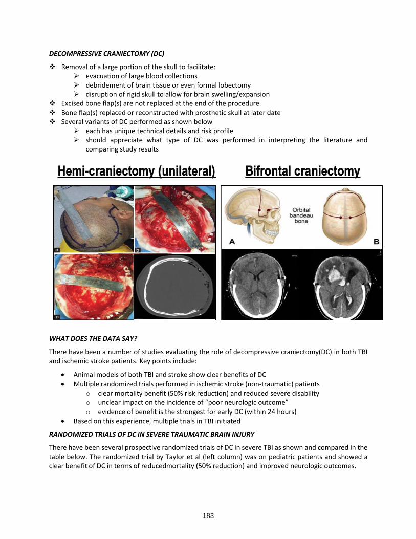
DECOMPRESSIVE CRANIECTOMY (DC)
❖ Removal of a large portion of the skull to facilitate: ➢ evacuation of large blood collections ➢ debridement of brain tissue or even formal lobectomy ➢ disruption of rigid skull to allow for brain swelling/expansion
❖ Excised bone flap(s) are not replaced at the end of the procedure ❖ Bone flap(s) replaced or reconstructed with prosthetic skull at later date ❖ Several variants of DC performed as shown below
➢ each has unique technical details and risk profile ➢ should appreciate what type of DC was performed in interpreting the literature and
comparing study results
WHAT DOES THE DATA SAY?
There have been a number of studies evaluating the role of decompressive craniectomy (DC) in both TBI and ischemic stroke patients. Key points include:
• Animal models of both TBI and stroke show clear benefits of DC
• Multiple randomized trials performed in ischemic stroke (non-traumatic) patients o clear mortality benefit (50% risk reduction) and reduced severe disability o unclear impact on the incidence of “poor neurologic outcome” o evidence of benefit is the strongest for early DC (within 24 hours)
• Based on this experience, multiple trials in TBI initiated
RANDOMIZED TRIALS OF DC IN SEVERE TRAUMATIC BRAIN INJURY
There have been several prospective randomized trials of DC in severe TBI as shown and compared in the table below. The randomized trial by Taylor et al (left column) was on pediatric patients and showed a clear benefit of DC in terms of reduced mortality (50% reduction) and improved neurologic outcomes.
183

For adult TBI patients, the 2 landmark trials summarized in the Table above are the DECRA Trial and the RESCUE-ICP Trial. Unlike in the pediatric trial, these two studies generated somewhat conflicting results, and also resulted in significant debate about the study designs and execution. As shown below, the DECRA Trial found no benefit of DC in terms of mortality and had an increased incidence of poor outcomes with DC. A second publication reported the 12-month outcomes for DECRA and although the overall differences in unfavorable outcomes were equivalent between groups, the DC group still had higher rates of persistent vegetative states and lower rates of "good" outcomes. It is important to note that this study included only patients with diffuse brain injury/edema and not focal mass lesions, that it utilized DC earlier in the management scheme, and that it only utilized bifrontal craniectomy as the operation of choice. This trial was largely interpreted as evidence against the use of DC for diffuse TBI. The RESCUE-ICP Trial was a subsequent randomized study that used DC later in the management scheme (only after ICP>25 and tier 1 and 2 medical therapies had failed), and it included both diffuse TBI as well as acute hemorrhage with mass effect. It also included both bifrontal craniectomy and hemi-craniectomy as surgical options. Consistent with much of the existing literature on DC and in contrast to DECRA, there was a mortality benefit in the DC group as well as improved ICP control. However, among the survivors there was a higher incidence of persistent vegetative state or severe disability after DC versus medical therapy. Somewhat confusingly, when the 12-month neurologic outcomes were grouped in categories, there was a lower rate of “poor neurologic outcomes” at 12 months in the DC group versus medical therapy. Overall it is estimated that for every 100 patients treated with decompressive craniectomy rather than medical intent, there were 22 more survivors; of these 22 patients, 6 were in a vegetative state (27%), 8 were categorized as having lower severe disability (36%), and 8 were categorized as having upper severe disability or better (36%). Of note, 37% of patients in the “medical” group ended up crossing over and having DC performed. The interpretations of this study have varied widely, from some claiming that it reinforces the DECRA findings and others concluding that it supports the use of DC in a more selective fashion as a rescue therapy for refractory ICP elevation despite maximal medical management.
A second randomized trial by this group (RESCUE-ASDH) completed accrual in 2019. This study evaluates DC in patients with acute large subdural hematomas requiring evacuation, and initial results should be reported in 2022. In 2017 Zhang et al. performed a systematic review and meta-analysis on DC for refractory elevated ICP after TBI that included 10 studies (4 randomized trials). They concluded that DC
184

clearly was associated with reduced mortality, better reduction in ICP, reduced hospital and ICU length of stay, but increased complication rates. However, there were no significant differences in neurologic outcomes at 6 weeks compared to medical therapy, with a possible benefit in the subgroup of early DC (less than 24 hours).
MILITARY EXPERIENCE WITH DECOMPRESSIVE CRANIECTOMY
With the prolonged recent combat operations in Iraq, Afghanistan, and Syria, the U.S. (and NATO allies) medical forces have gained significant experience with a high volume of patients with severe TBI due to penetrating and blast mechanisms. Given the relatively young age and excellent health of the typical battlefield trauma patient (compared to civilian trauma), as well as the high incidence of penetrating injuries, the U.S. military medical system and trauma/neurosurgical personnel adopted an aggressive stance in favor of early and liberal use of DC. This was also important due to concerns about secondary brain injury from hypoxia/edema/rebleeding during the aeromedical evacuation process.
Although randomized trials are currently not feasible in the deployed environment, there have been multiple published cohort and case-control studies examining outcomes among this population and with the use of DC. In a series of 188 DCs performed over a 5-year period there were noted to be significant and persistent improvements in neurologic outcome scores over time, with 83% achieving a Glasgow Outcome Scale score >3 at one year. Other studies have shown particularly improved outcomes when DC was done early as shown in the Figure below (within the first 5 hours after injury).
Similar good results have been reported by the U.K., with 10 survivors out of 14 DC who had an injury severity score of 75 (usually designated as “fatal). Finally, an analysis of 137 patients with battlefield penetrating TBI demonstrated a low mortality of 5.8% despite high average injury severity scores and 80% undergoing DC. Most remarkable were the excellent rates of achieving functional independence at 2-year follow-up. When categorized by admission GCS, the functional independence rates were 32% for those with admission GCS 3-5, 63% for admission GCS 6-8, 74% for admission GCS 9-11, and 100% for admission GCS 12-15. These are significantly better rates than would be expected from review of the civilian literature, and likely reflect a combination of the use of aggressive interventions including DC in an optimal patient population (young, healthy, penetrating brain injury). A recent analysis by Demetriades and colleagues matched and compared outcomes between military and civilian cohorts with severe TBI. Even after adjusting for differences in age and injury mechanisms, they demonstrated significantly better outcomes in the military cohort which they partially attribute to an aggressive use of early decompressive craniectomy.
In addition to more liberal use of decompressive craniectomy in the military battlefield setting, the role of the timing of craniectomy (early versus late) likely plays a role in the superior outcomes. In the study below, the authors demonstrated significantly improved outcomes when DC was performed early after injury (<5.33 hours) vs late.
185

VENOUS THROMBOEMBOLISM PROPHYLAXIS AND DECOMPRESSIVE CRANIECTOMY
The importance of early chemical VTE prophylaxis after trauma has become increasingly appreciated over the past decade. Although this practice has been widely accepted, there remains debate about the timing and safety in patients with severe TBI, and particularly patients who undergo neurosurgical interventions. A large body of lower- quality literature has generally found starting VTE prophylaxis within 48-72 hours in TBI patients with stable ICH is safe, although adoption of these practices varies widely.
186

However, a recent analysis of TQIP data (below)
2020 UPDATED BRAIN TRAUMA FOUNDATION GUIDELINES: DECOMPRESSIVE CRANIECTOMY
Level IIA–to improve mortality and overall outcomes 1. NEW–Secondary DC performed for late refractory ICP elevation is recommended to improve
mortality and favorable outcomes. 2. NEW–Secondary DC performed for early refractory ICP elevation not recommended to improve
mortality and favorable outcomes. 3. A large frontotemporoparietal DC is recommended over a smaller DC for reduced mortality and
improved neurological outcomes in patients with severe TBI. Level IIA–for ICP control
4. NEW–Secondary DC, performed as a treatment for either early or late refractory ICP elevation, is suggested to reduce ICP and duration of intensive care, though the relationship between these effects and favorable outcome is uncertain.
SUMMARY AND TAKE-HOME POINTS
• DC is a highly invasive surgical procedure for cerebral decompression
• Typically, it should be used for refractory ICP elevation despite standard medical therapy, or for high risk acute mass lesions (bleeds)
• Clearly associated with decreased mortality and superior ICP control vs medical management
• Many resultant survivors will have significant neurologic deficits, but there is a subgroup that will have good to excellent functional recovery
• Results with RESCUE-ICP approach (and hemicraniectomy) appear to be better than with DECRA approach (and bifrontal craniectomy)
• Military data supports excellent outcomes with DC in highly select population
• Patient selection is critical! See the following page for risk calculator aid
187

• Awaiting results of RESCUE-ASDH Trial, expect initial report in 2021
• Remains a KEY option in the full armamentarium for severe TBI!
IMPACT-TBI RISK CALCULATOR
The IMPACT-TBI risk calculator can be a helpful tool for decision making on major interventions such as a decompressive craniectomy, and for patient/family counseling regarding risk of death and risk of favorable versus unfavorable outcomes. However, one must remember that these are population-based risk estimates and cannot determine the exact risk of an individual patient.
• Sample calculation shown below for a 25-year-old with flexor posturing, nonreactive pupils, and an intracranial mass lesion on CT scan
• Risk of mortality 45% based on clinical variables, 35% based on clinical variables + CT scan results
• Risk of unfavorable outcome 68% and 62% respectively
188

BEYOND ICP: ADVANCED NEUROMONITORING MODALITIES FOR TBI
Carlos V.R. Brown, MD, FACS
Professor of Surgery Chief, Division of Acute Care Surgery Dell Medical School University of Texas at Austin Austin, TX
Most patients with severe traumatic brain injury (TBI) require ICU admission, endotracheal intubation with mechanical ventilation, and prevention of secondary brain injury by avoiding hypoxia and hypotension. Some patients will also require a craniotomy/craniectomy with hematoma evacuation or an intracranial pressure (ICP) monitor in order to diagnose and treat elevated intracranial pressure. Patients with sustained ICP elevation (> 22 mm Hg) are treated with a teared approach to lowering ICP. Basic maneuvers to treat elevated ICP include head of bed elevation, optimizing jugular venous return (e.g., loosen or remove cervical collar), and analgesia/sedation. If ICP remains elevated, then first tier of treatment includes additional analgesia/sedation as well as CSF drainage via ventriculostomy. If ICP elevation persists, further treatment includes hyperosmolar therapy (hypertonic saline or mannitol) and neuromuscular blockade. For ongoing ICP elevation, high dose barbiturates, propofol, or hypothermia can be considered. Finally, a decompressive craniectomy for refractory ICP elevation can be considered. In addition to standard ICP monitoring and treatment, some centers may utilize additional advanced neuromonitoring for TBI including partial pressure of oxygen in interstitial brain tissue (PbtO2), the jugular bulb venous oxygen saturation (SjvO2), cerebral microdialysis, thermal diffusion, and transcranial doppler.
PARTIAL PRESSURE OF OXYGEN IN INTERSTITIAL BRAIN TISSUE
Partial pressure of oxygen in interstitial brain tissue (PbtO2) is a parenchymal monitor, placed in a similar fashion as an ICP monitor, that provides continuous measurement of regional tissue oxygenation of the adjacent white matter (within 5mm of probe). Normal values of PbtO2 are in the range of 20-35 mm Hg. Low levels of PbtO2 (< 10 mm Hg) have been associated with poor outcomes in patients with severe TBI, and treatment can be considered at PbtO2 levels < 20 mm Hg. A randomized trial that was designed to demonstrate feasibility and safety of a treatment protocol with PbtO2 monitoring was published in 2017, “Brain tissue oxygen monitoring and management in severe traumatic brain injury (BOOST-2): a phase II randomized trial”. This study compared patients with severe TBI who were managed with ICP and PbtO2 monitoring vs. ICP monitoring alone. PbtO2 monitoring in severe TBI was found to be safe and feasible, and there was a trend towards better long-term outcomes in the PbtO2 monitoring group. A follow up prospective randomized trial (BOOST-3) in underway to determine the clinical effectiveness and outcomes of PbtO2 monitoring in severe TBI.
JUGULAR VENOUS OXYGEN SATURATION
Jugular venous oxygen saturation (SjvO2) monitoring is another technique to measure brain tissue oxygenation. As opposed to PbtO2, which measures regional oxygenation, SjvO2 provides a more global measurement of brain oxygenation. In order to measure SjvO2, a central venous catheter is placed into the internal jugular vein and aimed superiorly into the jugular bulb. From this point, intermittent samples of blood can be drawn to measure SjvO2 or a fiberoptic catheter can be inserted to for continuous SjvO2 measurement. Normal SjvO2 levels are around 60%, and sustained levels <50% are indicative of global
189

cerebral hypoxia. Though low levels of SjvO2 are associated with worse outcomes, there is no evidence that therapy targeting low SjvO2 levels is associated with improved outcomes.
CEREBRAL MICRODIALYSIS
Cerebral microdialysis (CMD) allows measurement of extracellular makers of brain metabolism. A catheter is placed into the intraparenchymal portion of the brain, and a microdialysis probe is placed through the catheter into the parenchyma as well. The microdialysis catheter consists of two concentric tubes, the outer tube is connected to a syringe pump that delivers perfusion fluid. The perfusate flows through the external tube down to the tip, where the final 10 mm of the catheter consists of a semipermeable dialysis membrane allowing bidirectional diffusion between the perfusate and the extracellular fluid of the brain parenchyma. The diffusion rate is driven by the chemical gradient across the dialysis membrane. Biochemical markers such as glucose, pyruvate, lactate, glycerol, and glutamate can be measured from the dialysate of the extracellular fluid. The most sensitive marker for cerebral ischemia is the lactate to pyruvate ratio, and a ratio > 40 as well as low brain glucose levels are associate with poor neurologic outcomes in severe TBI.
TRANSCRANIAL DOPPLER
Transcranial doppler (TCD) is a noninvasive evaluation of the cerebral vasculature that can be used to estimate ICP using the pulsatility index (PI) of the middle cerebral artery (MCA) and monitor for vasospasm after traumatic subarachnoid hemorrhage. The transtemporal window provides the best view of the MCA. PI is calculated using peak systolic velocity (PSV), end-diastolic velocity (EDV), and mean velocity (MV) of the MCA: PI = (PSV-EDV)/MV. A formula has been developed to convert PI to ICP: ICP = (10.93 x PI) – 1.28. A normal PI is ~1.2, correlating with an ICP~12, and an elevated PI is >2.13, correlating with an ICP > 22 mm Hg. Though TCD is most often used to evaluate for vasospasm in aneurysmal SAH, it can also be applied in traumatic SAH. Radiographic vasospasm may be identified by measuring MCA velocity. Normal MCA mean velocity is 80 cm/s, mild vasospasm mean velocities are 120–159 cm/s, moderate vasospasm is 160–199 cm/s, and severe vasospasm is > 200 cm/s. The Lindegaard ratio (MCA mean velocity/ipsilateral extracranial internal carotid mean velocity) may also be used to quantify vasospasm. A Lindegaard ratio > 3 indicates vasospasm, 3-5 mild/moderate vasospasm, and >/= 6 would indicate severe vasospasm.
REFERENCES
1. Alam HB, Vercruysse G, Martin M, Brown CVR, et al. Western trauma association critical decisions in trauma: Management of intracranial hypertension in patients with severe traumatic brain injury. J Trauma Acute Care Surg. 2020 February;88(2):345-351.
2. Bhardwaj A, Bhagat H, Grover VK. Jugular venous oximetry. Journal of Neuroanesthesiolgy and critical care. Sep-Dec 2015;2(3):225-231.
3. Fugate FE. Essentials of multimodal brain monitoring, Mayo Clinic critical and neurocritical care board review, eds Wijdicks EFM, Freeman WD, Findlay JY, Sen A. Oxford University Press. September 2019.
4. Lau VI, Arntfield RT. Point-of-care transcranial doppler by intensivists. Crit Ultrasound. 2018;9(21):1-11.
5. Oddo M, Hutchinson PJ. Understanding and monitoring brain injury: the role of cerebral microdialysis. Intensive Care Med. 2018;44:1945-1948.
6. Okonkwo DO, Shutter LA, Moore C, et al. Brain tissue oxygenation monitoring and management in severe traumatic brain injury (BOOST-II): a phase II randomized trial. Crit Care Med. 2017 November;45(11):1907-1914.
190

SESSION 7
MEET THE MASTERS TCC & ACS EXCITING OPPORTUNITIES
Moderator: Kenneth L. Mattox, MD, FACS, MAMSE
Monday, March 28, 2022
5:15 p.m. - 6:30 p.m.
Palace Ballroom 1-2
Palace Tower, Emperors Level
• Required focused or additional training or certification
• Practice opportunities
• Satisfaction levels for this area of surgery
• Research opportunities
• Ongoing education opportunities
• New trends in the field
191

192

SESSION 8
CRITICAL TRAUMA/CRITICAL CARE TREATMENT FOCUS ON DOGMA VS DATA IN THE ICU
Moderator: Mark J. Kaplan, MD, FACS
Tuesday, March 29, 2022
Palace Ballroom 1-2
Palace Tower, Emperors Level
7:30 - 7:42
Hungry for Data: ICU Nutrition Myths and Pearls Stephen L. Barnes, MD, FACS
7:42 - 7:54
Hemodynamic Monitoring - Now You See It; Now you Don't Andre’ R. Campbell, MD, FACS, FACP, FCCM, MAMSE
7:54 - 8:06
Sedation - To Sleep, Perchance to Dream Chadwick P. Smith MD, FACS
8:06- 8:18
Bugs and Drugs Dennis Y. Kim, MD, FACS, FRCSC, FACS, FCCP
8:18 - 8:30
Dazed and Confused - Delirium in the ICU Alan H. Tyroch, MD, FACS, FCCM
8:30 - 8:42
The Tortoise and the Hare - Ambulating Your Critical Care Patients Chris Cribari, MD, FACS
8:42 - 8:54
GI Bleeding and Prophylaxis Practices: Lifesaving or Pneumonia Generating? Meghan R. Lewis, MD, FACS
8:54 - 9:06
Vents - More Than All You Need to Know Jay A. Johannigman, MD, FACS
9:06 - 9:18
Be a Quitter: Stop Low-value Practices in the ICU Ali Salim, MD, FACS
9:18 - 9:30
ICU Fluids: Crystalloids, Colloids, or Heplock? Jason W. Smith, MD, PhD, FACS
9:30 - 10:00 Panel Discussion
193

HUNGRY FOR DATA: ICU NUTRITION MYTHS AND PEARLS
Stephen L. Barnes, MD FACS
Professor and Hugh E. Stephenson Endowed Chair Department of Surgery University of Missouri School of Medicine Columbia, MO
Nutritional support is a key component of care for the acutely ill and injured patient and may improve outcomes and decrease complications. Data on best practice, however, is limited and at times contradictory and specific recommendations are based on expert opinion in many cases. Trauma surgeons have been leaders in advancing the science of nutritional support through work done on the metabolic response to injury and the clinical impact of nutrition on outcomes.1 Appropriately timed and delivered nutritional support will improve outcomes following surgical illness and injury.
NUTRITIONAL RISK AND ASSESSMENT
All patients upon admission to the ICU should be assessed for nutritional risk through a nutritional assessment.2 This is best accomplished through a formal evaluation of nutritional status by a trained healthcare professional, most commonly a dietitian and results in identification of nutritional risk and any nutrition related diagnosis. Numerous methods of nutritional assessment have been proposed and are in use today. Skinfold assessment, GLIM (Global Leadership Initiative on Malnutrition), NUTRIC (Nutrition Risk in Critically Ill), Modified NUTRIC (NUTRIC scoring without the use of IL-6), and SGA (Subjective Global Assessment) are some of the more commonly used assessment tools. These scoring systems allow for assessment of nutritional risk through evaluation of variables such as age, APACHE II and SOFA scores, co-morbidities, days in the hospital, dietary intake, gastrointestinal symptoms, functional status, etc. which then guides best practice for nutritional support therapy in the critically ill and injured.
194
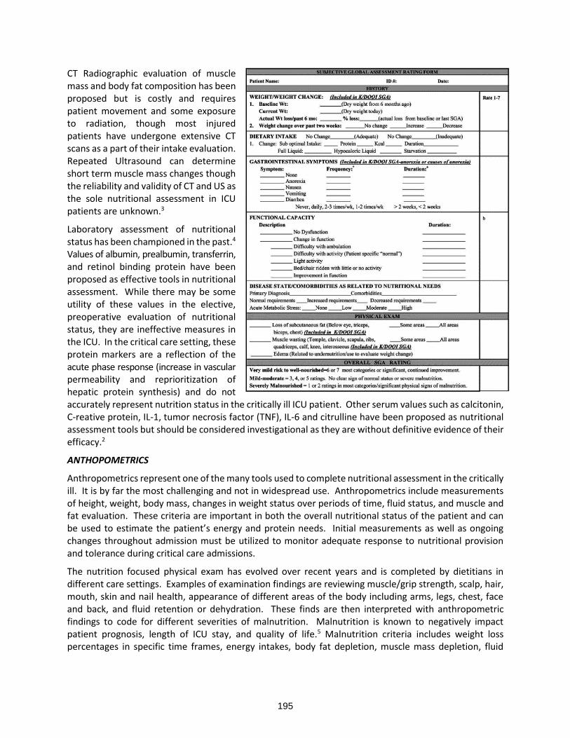
CT Radiographic evaluation of muscle mass and body fat composition has been proposed but is costly and requires patient movement and some exposure to radiation, though most injured patients have undergone extensive CT scans as a part of their intake evaluation. Repeated Ultrasound can determine short term muscle mass changes though the reliability and validity of CT and US as the sole nutritional assessment in ICU patients are unknown.3
Laboratory assessment of nutritional status has been championed in the past.4 Values of albumin, prealbumin, transferrin, and retinol binding protein have been proposed as effective tools in nutritional assessment. While there may be some utility of these values in the elective, preoperative evaluation of nutritional status, they are ineffective measures in the ICU. In the critical care setting, these protein markers are a reflection of the acute phase response (increase in vascular permeability and reprioritization of hepatic protein synthesis) and do not accurately represent nutrition status in the critically ill ICU patient. Other serum values such as calcitonin, C-reative protein, IL-1, tumor necrosis factor (TNF), IL-6 and citrulline have been proposed as nutritional assessment tools but should be considered investigational as they are without definitive evidence of their efficacy.2
ANTHOPOMETRICS
Anthropometrics represent one of the many tools used to complete nutritional assessment in the critically ill. It is by far the most challenging and not in widespread use. Anthropometrics include measurements of height, weight, body mass, changes in weight status over periods of time, fluid status, and muscle and fat evaluation. These criteria are important in both the overall nutritional status of the patient and can be used to estimate the patient’s energy and protein needs. Initial measurements as well as ongoing changes throughout admission must be utilized to monitor adequate response to nutritional provision and tolerance during critical care admissions.
The nutrition focused physical exam has evolved over recent years and is completed by dietitians in different care settings. Examples of examination findings are reviewing muscle/grip strength, scalp, hair, mouth, skin and nail health, appearance of different areas of the body including arms, legs, chest, face and back, and fluid retention or dehydration. These finds are then interpreted with anthropometric findings to code for different severities of malnutrition. Malnutrition is known to negatively impact patient prognosis, length of ICU stay, and quality of life.5 Malnutrition criteria includes weight loss percentages in specific time frames, energy intakes, body fat depletion, muscle mass depletion, fluid
195

accumulation, and grip strength.6 Per the joint ASPEN/AND guidelines, malnutrition diagnosis requires two or more characteristics for a malnutrition diagnosis.
Myth: Laboratory values of albumin, prealbumin, transferrin and retinol binding protein are effective nutritional assessment tools
Pearl: Nutritional Assessment by a standardized tool should be performed on all ICU Admissions.
DETERMINING ENERGY & PROTEIN NEEDS
Indirect Calorimetry remains the gold standard and if available is recommended for assessment of nutritional needs. It remains the only true means to obtain caloric requirements and set an accurate goal for nutrition therapy.7 This holds true for adult, geriatric and pediatric patient cohorts and is especially
true in the trauma population who experience great fluctuations in energy requirements based on extent and type of injury. Assessment of nutritional needs utilizing most standard equations demonstrate an average of 10% variation from Indirect Calorimetry, with some variation greater than others. As indirect calorimetry is not widely available and some clinical conditions limit its practical and accurate use (evolving lung injury, pneumothorax with air leak, etc.) standardized formulas provide the most widely available tool for assessment of energy need.
Predictive energy equations can be effectively utilized in the ICU setting with variations in recommendations based on specific patient populations. These predictive equations can be as simple as a kcal/kg estimate to more detailed
critical care assessments including minute ventilation, maximum 24hr temperature, and factors for trauma and burn to provide the most accurate estimates available. When indirect calorimetry is not a feasible option due to availability, expense, hemodynamic stability, ventilation mode, presence of chest tube with air leaks, hemodialysis, and staff training, predictive equations are commonly used in the critical care setting to determine estimated energy and protein requirements for various populations, including pediatric, adult, geriatric, and obesity.
Pearl: Indirect Calorimetry remains the gold standard for evaluation of caloric needs, though standardized nutritional formulas perform well and are more practical in the day-to-day provision of nutritional support.
196

ENERGY REQUIREMENTS/CALORIC NEEDS ESTIMATION IN THE ICU
Adequate provision of protein and energy requirements during critical illness is a vital part of preventing malnutrition and ensuring the best outcomes for patients. The goal of enteral nutrition in the acute phase of injury should be to deliver some caloric intake early as opposed to no nutrition at all or aggressive feeding resulting in intolerance and/or complications. Aggressive advancement of tube feeds in an attempt to achieve 100% of assessed energy needs does not improve outcomes over a goal of achieving 60% of calculated needs during the initial phase of care in the first 5 days in the ICU. Goal energy needs can be achieved as the patient begins to recover from the acute phase of injury or illness. No differences were seen in mortality, length of stay or infection rates between high energy vs low energy delivery in the first 5 days of ICU care. An approach that delivers some of the calculated energy needs vs all is effective in improving outcomes and supporting patients during the acute phase of care.8
Pearl: Targeting 60% of estimated energy needs over during the acute phase of care is appropriate.
PEDIATRIC PATIENTS
Indirect calorimetry is the suggested measure for energy expenditure estimation in children. The use of Harris-Benedict equations and RDAs are not recommended for use in critically ill children. The Schofield, World Health Organizations, or United Nations University Equations are the recommended equations for pediatric patient ages < 18 years old. Target goal for provision is 2/3 prescribed goal by the end of the first week in the pediatric intensive care units.9
Estimating Pediatric Energy Needs in Critical Illness
Age (years) Gender Resting Energy Expenditure (REE) or Basal Metabolic Rate (BMR) Equations (Kcal/day)
WHO Schofield
0-3 Male (60.9 x Wt) – 54 (0.167 x Wt) + (15.174 x Ht) – 617.6
Female (61 x Wt) – 51 (16.252 x Wt) + (10.232 x Ht) – 413.5
3-10 Male (22.7 x Wt) + 495 (19.59 x Wt) + (1.303 x Ht) + 414.9
Female (22.5 x Wt) + 499 (16.97 x Wt) + (1.618 x Ht) + 371.2
10-18 Male (17.5 x Wt) + 651 (16.25 x Wt) + (1.372 x Ht) + 515.5
Female (12.2 x Wt) + 746 (8.365 x Wt) + (4.65 x Ht) + 200
Wt = weight in kg; Ht = height in cm Source: Adapted from Pocket Guide to Pediatric Nutrition Assessment (Leonberg, 2013).
Pearl: Target nutritional provision of pediatric patients is to achieve 2/3 of the prescribed goal by day 7 in the pediatric ICU.
ADULT PATIENTS
For mechanically ventilated patients, the Ireton-Jones and Penn State equations are the most widely utilized and most accurate in estimating energy needs, though Indirect Calorimetry remains the gold standard. They include factors for not only height, weight, and age, but also intensive care status, minute ventilation, Tmax for past 24 hours, and factors for trauma and burn. Per current guidelines (ASPEN 2016 and 2021), the use of 25-30 kcal/kg may also be used for adults of non-obese weight status.
Ireton-Jones
RMR = 1784 – (11 x A) + (5 x W) + (244 x sex) + (239 x T) + (804 x B)
RMR = Resting Metabolic Rate W = weight in kg
A = age in years T = Trauma Present = 1 ; Absent = 0
Sex Male = 1 ; Female = 0 B = Burn Present = 1 ; Absent = 0
197

Penn State
RMR = (Mifflin-St. Jeor x 0.96) + (VE x 32) + Tmax x 167) – 6212
VE = minute ventilation (L/min) Tmax = maximum body temp previous 24 hr (C)
Mifflin-St Jeor
Men = 5 + (10 x W) + (6.25 x H) – (5 – A) Women = -161 + (10 x W) + (6.25 x H) – (5 x A)
W = weight (kg) W = weight (kg)
H = height (cm) H = height (cm)
A = age (yr) A = age (yr)
GERIATRIC PATIENTS
The geriatric population has a high variability in needs and predictive equations are not as accurate as in non-geriatric adults. If indirect calorimetry is unavailable, there has been no significant difference found between the different equations and measured resting energy expenditure with any statistically significance. The Penn State and Ireton-Jones equations for elderly patients with trauma to account for age, ventilation status, and injury severity are recommended.2,10-11
MORBIDLY OBESE PATIENTS
The use of weight- based or predictive equations to determine energy requirements are utilized including the practice of underfeeding in obesity with the provision of increased protein to induce lipolysis for additional caloric provision to prevent overfeeding in the critically ill obese patient.2
Current energy recommendations for obesity are dependent upon the patient’s body mass index.
• BMI 30-50: 11-14 kcal/kg using patient actual body weight
• BMI >50: 22-25 kcal/kg ideal body weight
These parameters must be used with higher protein recommendations to ensure adequate provision to prevent lean body mass catabolism
• BMI 30-40: 2gm/kg protein using ideal body weight
• BMI >40: 2.5gm/kg protein using ideal body weight
PROTEIN SUPPLEMENTATION
Critical illness is a highly catabolic state involving alterations in carbohdyrate, protein, and lipid metabolism. Lean body mass loss is inevitable in rapid protein breakdown when used for gluconeogenesis during critical illness. Immobility and starvation can increase this lean body mass wasting. After injury, protein turnover rates increase in agreement with the severity of the patient injury. When determining protein needs for critical illness, the patient current and prior nutrition status must be considered.12
Timing of protein intake and provision in acute illness remains controversial. Conflicting results exist regarding provision recommendations in the first week of ICU stay. Retrospective review of mechanically ventilated adult ICU patients suggested overall low protein intake was associated with the highest ICU, in-hospital, and 6 months mortality but no difference in length of stay, need for renal replacement therapy, or ventilation duration. 13 Another study reviewed the need for earlier protein provision in critically ill ICU patients with low skeletal mass.14 This retrospective study reviewed patients with low and normal skeletal muscle area. In the group with low skeletal muscle area, higher protein intakes were associated with lower 60-day mortality and lower 6-moth mortality suggesting earlier high protein intakes is associated with lower mortality.
198

Current recommendations for critically ill protein intakes is 1.2-2gm/kg /day, with higher dosing in cases of obesity, burns and trauma. Overall determination of needs continues to be difficult to accurately achieve with multiple factors to consider. Ongoing re-assessment of patient adequacy through daily review of patient laboratory data, weight status, fluid status, wound healing, pressure injury prevention, and overall improvement in critical care status should be standard practice.2 At the university of Missouri we utilize a minimum of 1.5-2gm/kg for all trauma and TBI patients, with burn needs as high as 2.5gm/kg for burns over 20% TBSA.
Pearl: Critically ill patients have increased protein needs with optimal provision of 1.2-2gm/kg/day. Provision will be higher in obesity, burns and trauma based on extent of injury or BMI.
IMMUNO-NUTRITION
Recommendations for immune-modulating formulas containing both arginine and fish oils continue to be in place for perioperative and injured patients. These recommendations have been extended to many enhanced recovery after surgery programs with pre-habilitative immune enhancing nutrition in the elective pre-operative phase of care. The rationale for this recommendation is based upon the fact that in the post-operative or post-injury phase of care, specialized immune myeloid suppressor cells increase the level of arginase 1, resulting in a relative arginine depletion. This conditional inadequacy of arginine adversely affects T-cell function and can result in immune suppression. The arginine deficiency may be severe enough in some patients to impact production of nitric oxide and negatively affect microcirculation, a component much needed in the acute phase of recovery from major surgery or injury. Formulas containing arginine and omega-3 fatty acids appear to overcome the regulatory effect of myeloid suppressor cells through displacement of omega-6 fatty acids from the cell membranes of immune cells, reducing systemic inflammation through the production of less-active prostaglandins and leukotrienes. Downregulation of the expression of nuclear factor-kappa B, intracellular adhesion molecule 1, and E -selectin is also seen, and decreases neutrophil attachment and transepithelial migration, thus favorably modulating systemic and local inflammation. Stabilization of the myocardium has also been reported with lower incidence of cardiac arrhythmias as have reduced rates of perioperative and peri-injury ARDS and rates of sepsis.2
Current provision of immune enhanced formula is specific to surgical, trauma, and burn patients. The hemodynamic effects of arginine conversion to nitric oxide causing vasodilation is a contraindication for use in patients with sepsis. Immune enhancing formulas should not be routinely used on medical intensive care patients as they have shown no benefit. The greatest benefit in immune enhancing formula is seen in first 5-7 days of acute care admission. These formulas should be initiated within 24-48 hour of admission and continue for 7-10 days, with transition to standard polymeric formula after completion. If a patient is unable to tolerate enteral nutrition until after this 5–7-day window, a standard polymeric formula may be used.
In the case of parenteral nutrition utilization, parenteral arginine is not recommended at this time. However new lipid enhanced formulas include the use of mixed oil lipids including fish oils to aide in these inflammatory and immunomodulatory effects. These can be utilized within the first week of ICU admission. 2,8
Pearl: Immune enhancing diets are recommended for surgical and trauma patients for 5-7 days.
WHEN, WHAT AND HOW AND HOW TO FEED THE CRITICALLY ILL
Nutritional support early in the care of the acutely ill and injured improves mortality and reduces infection rates. Maintenance of gut mucosal integrity likely plays a significant role in reduction in infectious complications. Nutritional support should begin ideally in the first 24-48 hours following admission
199

following a nutritional assessment and calculation of energy requirements by either standardized formula or indirect calorimetry.
Once a nutritional assessment is complete and energy requirements determined the next step in the decision-making tree is how to provide the nutritional support. If the gut works, it should be utilized. The overwhelming majority of patients can be supported via the enteral route through the naso/oro gastric approach. Once thought to increase risk of pneumonia, gastric feedings are well tolerated by a majority of patients and should be the initial route of enteral feeding in the majority of patients. If determined to be a prohibitive aspiration risk or if unable to tolerate gastric feeding, nasoenteral feeding via a manually, endoscopic, or radiographically placed tube should proceed.
ENTERAL VS PARENTAL NUTRITION IN THE ICU
Enteral Nutrition should always be a first line of therapy for nutrition support when a patient has functioning gut. Per the 2016 ASPEN guidelines, a patient not malnourished prior to admission, would consider parenteral nutrition support after 7-10 days of the inability to tolerated enteral nutrition, or tolerating less than 60% goal protein/energy requirements. No significant difference in outcomes or benefits have been seen in providing parenteral nutrition supplementation early in an ICU admission for patients who are not malnourished on arrival. Patients who are malnourished on admission and expected to remain NPO, exclusive parenteral nutrition should be initiated as soon as possible following ICU admission. When initiating parenteral nutrition, its recommended to achieve <20kcal/kg/day or 80% of estimated energy needs with adequate protein intake >1.2gm/kg/day over the first week of hospitalization in the ICU.2,8
Pearl: Parenteral Nutrition should only be utilized early in the ICU phase of care for those patients determined to be malnourished on arrival by nutritional assessment.
GASTRIC VS POST-PYLORIC
When obtaining access in the intensive care unit, gastric access is typically obtained on intubation for decompression. This access can also be used for provision of enteral nutrition support. Placement of post-pyloric tubes does not decrease the risk of aspiration in most patients and can cause a delay in provision of nutrition support waiting on placement. Gastric access is appropriate for patients free of delayed gastric emptying. The use of promotility agents like metoclopramide and erythromycin can also be used when clinically feasible in patients with delayed gastric emptying.15
CONTINUOUS VS BOLUS
Aspiration while receiving enteral nutrition is always a concern. Current recommendations to reduce risk aspiration include the head of bed at 30-45 degrees, verify correct placement of feedings tube, and continuous feeds instead of bolus feeds for critically ill patients. Aggressive bolus feeds can cause potential harm and increase risk of aspiration. Gastric residual volumes (GRV) have been completed in the past as a measure of tolerance. These are no longer recommended as part of routine intensive care for patients on enteral nutrition. For those that they are utilized, holding for GRV <500ml in absence of other signs of intolerance is not recommended.2,15,16
FULL VS TROPHIC
Nutritional support is an integral part of patient recovering in all aspects of critical care. When does this goal of energy expenditure need to be achieved? Recently updated ASPEN recommendations state starting feeds early at slower rates and advancing feeds over the course of a patients first 7-10 days of ICU stay showed no significant difference in outcome for different levels of intakes on risk of pneumonia, infection, ICU length of stay, hospital length of stay, mean ventilator days, ICU mortality, hospital
200

mortality, or 28–90-day mortality. The first 7-10 days obtaining a goal of 12-25 kcal/kg/day is recommended. More research is needed to address the long-term post-hospital outcomes.8
Pearl: There is no significant clinical outcome differences between higher vs lower energy intake in adult ICU patients. Feeding early and achieving some nutritional support is equivalent to reaching calculated goals.
STANDARD VS SPECIALIZED FORMULAS
Many different types of specialized formulas have been developed for different disease states including but not limited to pulmonary failure, diabetes, renal failure, and hepatic failure. Current recommendations recommend the use of standard polymeric formula when initializing enteral nutrition and avoid routine use of specialty formulas in critically ill MICU and disease specific SICU patients. There has not been a clear benefit identified in the use of these disease specific formulas or semi-elemental and elemental formulas at this time. Immune modulating formulas should be reserved for the SICU setting.2,17
Myth: Special disease process needs special enteral formulas
ASSESSING TOLERANCE TO NUTRITIONAL SUPPORT
Tolerance of enteral nutrition is best assessed by physical exam, bowel function, radiologic evaluations, and absence of patient complaints such as pain and abdominal distention. Intolerance can be defined by vomiting, abdominal distention, high NG output, high residual volumes (when considered with other symptoms), diarrhea, constipation, or abnormal abdominal radiographs.
In cases of delayed gastric emptying the use of prokinetic drugs or narcotic antagonists can be used to aide in motility. Metoclopramide or erythromycin improve gastric emptying and intestinal tolerance. Ensuring patients have an adequate bowel regimen is also important. If these measures do not improve emptying, transition to a post-pyloric feeding may be needed. Positioning of these tubes in duodenal versus jejunal achieves similar overall results.2,18
Diarrhea and constipation are the most common issues with GI intolerance. The Definition of diarrhea varies but is frequently defined as bowel movements of more than 3-5 times per day. If diarrhea occurs, determining the cause is important. Most enteral nutrition formulas are lactose and gluten free, however they do contain milk proteins. One frequent cause of diarrhea in the intensive care is use of medications containing hyperosmotic agents like sorbitol (i.e. liquid Tylenol), or are hyper osmolar themselves (i.e. oral electrolyte replacement). The incidence of diarrhea does not appear to be worsened by the route of feeding, but more likely to occur with hyperosmolar formula selection.
Once medications have been ruled out, infectious source of diarrhea must be considered. Frequent administration of antibiotics increases the risk of clostridium difficile. Once an infectious source has been ruled out, use of antimotility agents may be used. The use of water-soluble fibers may be used to aide in preventing and treating diarrhea over insoluble fibers used to treat constipation.2.18
INTERRUPTING NUTRITONAL SUPPORT FOR TESTING/OPERATIVE INTERVENTIONS
Fasting period prior to anesthesia has been a long-time battle fought by surgeons in the intensive care unit. Avoidance of undesirable interruptions in enteral nutrition is necessary to obtain optimal nutrional results. Anesthesia Practice Guidelines for Preoperative Fasting state that clear liquids can be provided to patients up to two hours prior, a light meal 6 hours prior, and non-human milk formula should be held for 6 hour prior to elective surgical procedures. These guidelines do not however include patients who are intubated and have cuffed endotracheal tubes in place prior to surgery. A review by Douglas and Ciraulo, demonstrated variability in these fasting policies can cause increased risk of malnutrition and does not affect aspiration risk of an intubated critically ill patient. Volume based protocols have been
201
recommended by some for use in surgical intensive care unit where overall 24 hour volumes are targeted increase feeding rates to make up for holds in enteral nutrition. However current staffing and logistics of completing such protocols have shown to be difficult to execute.
Standard and agreed upon practice and the University of Missouri includes a 6-hour hold time for non-intubated patients, patients with uncuffed tracheostomy in place, or patients having a gastric related procedure or tracheostomy creation. If a patient has a cuffed endotracheal tube or tracheostomy with gastric access, enteral nutrition is held 2 hours prior to Operative intervention. In patients with small bowel access, tube feeds are not stopped until the patient is wheeled to the Operating Room. 2,19,20,21
Myth: Anesthesia data support holding all forms of feeding at midnight prior to all procedures. Pearl: 2 hours is an appropriate time frame by which to hold gastric enteral feedings in most cases.
DISEASE STATES AND CLINICAL SITUATIONS
Trauma
Trauma causes a systemic inflammatory response that can be reduced with early initiation of enteral nutrition support. Nutrition needs do not have to be met initially, slow rate feedings in the early resuscitative phase can address protein-energy malnutrition and modulation of immune function, then increasing feeds during the rehabilitation phase. Estimating needs during this dynamic time can be challenging and no one equations has shown to be more accurate than others in trauma. Goal intakes of approximately 20-35 kcal/kg/day are recommended by most. Immune-modulating formula with arginine and fish oil should be considered, though definitive data is lacking. Protein needs should be aimed from 1.2-2gm/kg, up to 2.5gm/kg with the higher end of the needs with more severe insults including burns. 2,22
Open abdomen/Damage Control Surgery
Patients with an open abdomen and absence of bowel injury have benefit from early enteral nutrition in 24-48hr post injury. Initial feeding is trophic, advancing as patients tolerate over the first week of injury. Patients receiving enteral nutrition demonstrate earlier and higher primary facial closure rates and decreased fistula formation. Goal energy expenditure of 25-30kcal/kg is recommended. Protein needs are like trauma with goals of 1.5-2gm/kg. Additional protein needs should be assessed with exudate lost with open abdomen. An additional 15-30gm/L protein should be added to previous recommendation to meet adequate protein needs.2,22
Pearl: Damage control patients with an open abdomen are more likely to close, in a shorter period of time and less likely to suffer a fistula is fed early.
TBI
Traumatic Brain Injury care constitutes a large volume of the care at our trauma centers. The Brain Trauma Foundation recommends early tube feeds at 24-48 hours following injury through continuous gastric feeding unless intolerance is encountered. Energy needs should be attained by days 5-7 after injury to improve outcomes. Immune-modulating formulas are again suggested for patients with TBI with protein goals set at 1.5-2gm/kg/day and energy provision as high as 200% measured energy expenditure. Gastric feeding is recommended. Close monitoring of intakes and prevention of weight loss due to hypermetabolism is crucial.2,23
Burn/Thermal Injury
In the Burn patient population early enteral feeds, with some recommendations as soon as 4 hours following injury, are the standard of care.24 By achieving this goal we see decreased rates of pneumonia,
202

wound complications, and sepsis in this high risk for infection and complication group. Indirect calorimetry plays an increasing role as current predictive equations are ineffective at determining energy goals in the hypermetabolic state of burn injury and high-quality burn care should include indirect calorimetry utilization. In the absence of indirect calorimetry, energy needs should be estimated using burn specific formula including the Curreri (Galveston), XIE, and Ireton Jones energy predictive equations. The Curreri is well known for over-estimation of energy needs. With estimated protein requirements of 1.5-2, up to 2.5 gm/kg/day. Protein metabolism can be estimated using the 24-hour urinary urea nitrogen.2,22,,25
XIE (1000 kcal x body surface area (m2)) + 25(TBSA burn)
age 16-59 Curreri (Galveston) 25kcal/kg wt + (40 * TBSA)
age >60 Curreri (Galveston) 20kcal/kg wt + (65 * TBSA)
Ireton Jones Ventilated: 1784 − 11 (age in years) + 5 (weight in kg) + (244 if male) + (239 if trauma) + (804 if burn)
Non-ventilated patient: 629 − 11 (age in years) + 25 (weight in kg) − (609 if obese)
Post operative/Ileus
A common occurrence in the post operative or post injury care is ileus. Early initiation of enteral nutrition within 24 hours of surgery will improve outcomes and can help reduce the rates of Ileus, improve motility, and reduce bowel edema. Routine use of immune-modulating formula with arginine and fish oils in the SICU are recommended.2
Pearl: Early enteral nutrition improves outcomes and decreases rates of ileus.
Sepsis
Sepsis can cause metabolic alterations to the body including hyperglycemia, decreased glucose uptake, insulin resistance, protein breakdown of peripheral muscle protein and diminished amino acid uptake by muscle leading to influx of amino acids away from the periphery to the liver. Nitrogen losses in severe sepsis may exceed 30gm/day. Energy expenditure progressively increases over the first 7 days and can remain elevated for up to 21 days even when sepsis has been treated. Energy expenditure is increased 20-60% from basal energy expenditure and is associated with significant protein catabolism.
Early enteral administration has shown some benefit. When initiating enteral feeds, beginning slowly with trophic feeds of 10-20ml/hr for initial phase and increase after 24-48hr to goal of >80% target energy needs over the first week. Protein needs should be targeted at 1.2-25gm/kg. Immune- modulating formulas should not be utilized in this population as arginine is upregulated as nitric oxide and can worsen hemodynamic instability and organ dysfunction.2,26
SPECIAL POPULATIONS
Inotropic support
One of the most common encounters in the ICU is when to feed a patient requiring inotropic support. Many have looked at this group of patients and to date recommendations remain less than ideal with recommendations such as “when fully resuscitated”, “when doses are less than ….” which leaves much to the reader’s interpretation.2,8 Clearly the patient in the immediate acute phase of care with escalating needs of inotropes who remain acidotic and critically ill are not candidates for early enteral nutrition as
203

the concern of bowel ischemia is high with the increase in oxygen delivery required to support digestion. Once acidosis is resolving and the patient is no longer requiring escalating levels of care, when is it safe to initiate nutritional support. Best evidence to date would suggest that a dose dependent and agent dependent relationship may exist. Enteral nutrition can be utilized in patients on inotropic support, which is not escalating and at rates less the 12.5-14 mg/min of norepinephrine or equivalent.27,28 Tolerance to tube feeds varied based on the agent in question (epinephrine, dopamine, norepinephrine, phenylephrine) with greater tolerance at lower and stable doses of norepinephrine, epinephrine and phenylephrine and higher intolerance with dopamine. Overall, enteral nutrition is tolerated well on vasopressor therapy with rising levels of intolerance at higher doses. Patients must be advanced slowly and monitored closely for intolerance. Doing so and delivering earlier enteral nutrition will improve outcomes.
Pearl: Enteral nutrition can be delivered slowly and with careful reassessment in the patient requiring inotropic support, once the acute phase of resuscitation is complete and acidosis is improving.
Prone positioning
Prone positioning has been a common practice in trauma for decades. The emergency of COVID-19 has escalated its use and new guidelines have emerged regarding prone positioning and nutrition support. These guidelines follow the same considerations as feeding a patient in the supine position. Most patients tolerate enteral nutrition support via gastric route, however if not tolerated, a post-pyloric tube can be obtained. Early feeing in 24-36 hours after ICU admission via continuous feeding is recommended. Initiate feeds slowly (10-20ml/hr) and advance as tolerated over the first week. Gastric residual volumes do not need to be checked routinely. The head of bed should be maintained at 10-25 degrees to decrease aspiration risk.2,16,26,29,30
ECMO
Extracorporeal membrane oxygenation (ECMO) use has become more available in recent years. As one of the highest severities of illness in intensive care, patients requiring ECMO require optimal nutritional support. Unfortunately, there is limited data regarding timing, advancement, and type of nutrition therapy to be provided. Current recommendations begin feeds slowly and advance to goal over the first week of illness. Indirect Calorimetry is difficult and must be adjusted to account for gas exchange, therefore predictive equations are most frequently utilized in needs estimates. Energy needs can vary from 20-30 kcal/kg and protein needs with a minimum of 1.2gm/kg/day and as high as 2.5gm/kg/day. Enteral nutrition is the preferred route of nutrition support. Some data is suggestive of lipid infiltration into the oxygenator limiting the use of intralipid in parenteral nutrition as well as sedation like propofol. Further research is needed in this area.31,32
CRRT
Many patients in the intensive care unit experience renal failure and require use of dialysis. Due to increased risk of malnutrition with protein restriction, patients receiving hemodialysis or continuous renal replacement therapy (CRRT), are recommended to not be restricted on protein provision. Patients on CRRT require up to 2-2.5gm/kg/day protein to overcome losses from the dialysate. 2,22,33
Pancreatitis
Pancreatitis can come from many different causes including alcohol use, gallstones, idiopathic causes, autoimmune, traumatic, or hereditary. If patients are unable to tolerate oral diet, enteral nutrition has shown to be superior to parenteral nutrition in severe acute pancreatitis. Current enteral nutrition recommendations start with gastric feeds of standard polymeric enteral formula. Patients demonstrating
204
signs of malabsorption may benefit from post-pyloric tube placement or changing to a semi-elemental or elemental formula or both, however most will tolerate standard formulas.2,34
Hepatic Failure
In the past it was thought that protein restriction was required in the case of hepatic failure to help treat and prevent encephalopathy. This recommendation has shown to be a myth in practice and current recommendations avoid restriction of protein in patients with liver failure and apply the same critical care recommendations as all other patients. Malnutrition risk is high in cirrhosis and contributes to morbidity and mortality, restriction of protein will contribute to this risk by increasing risk of lean mass loss, worsen nutritional status, and decrease overall ammonia removal worsening the risk of encephalopathy.
In critical illness, protein should be targeted at 1.2-1.5gm/kg/day to meet patient catabolic needs. Some reports of need for zinc or vitmain E supplementation due to depletion in liver failure have been discussed, however these have yet to be validated with specific recommendations. Patients should receive a standard polymeric formula when initiating enteral nutrition. Use of specialty formulas including branch chain amino acids for encephalopathy have slow little to no benefit and are not recommended.2,35
CLOSING
Appropriate timing and delivery of nutrition to the critically ill improves outcomes. Dogma, local practice, and culture at times limit the opportunity to deliver optimal nutritional support. A standardized approach to the provision of nutritional support in conjunction with a nutritional specialist will improve outcomes and ease of patient care delivery.
REFERENCES
1. Hartwell et al. Nutrition therapy in the critically injured adult patient: a Western trauma association critical decisions algorithm J trauma Acute Care Surg Vol 91: 5 909-915, 2021.
2. Taylor BE, McClave SA, Martindale RG, Warren MM, Johnson DR, Braunschweig C, et al. Guidelines for the provision and assessment of nutrition support therapy in adult critically ill patient: Society of Critical Care Medicine (SCCM) and American Society of Parenteral and Enteral Nutrition (A.S.P.E.N.). JPEN J Parenter Entearl Nutr.2016; 40(2): 159-211.
3. Rodrigues C, Henrique J, Ferreira A, Correia M. Ultrasonography and other nutrition assessment methods to monitor the nutrition status of critically ill patients. JPEN J Parenter and Enteral Nutr. 2021;45:982-990.
4. Nataloni S, Gentili P Marini B, Guidi A, Marconi P, Busco F Pelia P, Nutritional assessment in head injured patients through the study of rapid turnover visceral proteins. Clinical Nutrition 1999: Vol 18:4, 247-251.)
5. Malone A, Mogensen K. Key approaches to diagnosing malnutrition in adults. Nutr Clin Pract. 2021; 1-12.
6. White JV, Guenter P, Jensen G, Malone A, Schofield M, et al. Consensus statement: academy of nutrition and dietetics and American Society for Parenteral and Enteral Nutrition: characteristics recommended for the identification and documentation of adult malnutrition (undernutrition). JPEN J Parenter Enteral Nutr. 2012; 36:275-283.
7. McClave SA, Marindale RG, Lazzlo Kiraly. The use of indirect calorimetry in the intensive care unit. Curr Opin Clin Nutr Metab Care 2013 16:202-208.
8. Compher C, Bingham AL, McCall M, Patel J, Rice TW, Braunshceig C, et al. Guidelines for the provision of nutrition support therapy in the adult critically ill patient: American Society of Parenteral and Enteral Nutrition. JPEN J Parenter Entearl Nutr. 2021; Doi:10.1902/jpen.2267.
9. Mehta NM, Skillman HE, Irving SY, Coss-Bu JA, Vermilyea S, Farrington EA, et al. Guidelines for the provision and assessment of nutrition support therapy in the pediatric critically ill patient: Society
205

of Critical Care Medicine and American Society for Parenteral and Enteral Nutrition. JPEN J Parenter Enteral Nutr. 2017;41:706-742.
10. Hsu PH, Lee CH, Kuo LK, Kung YC, Chen WJ, TzengMS. Determination of the energy requirements in mechanically ventilated critically ill elderly patients in different BMI groups using the Harris-Benedict equation.J Formosan Medical Assoc.2018;117:301-307. http://doi.org/10/1016/ j.jfma.2017.12.010. Accessed December 14, 2021.
11. SegadilhaN, Rocha E, Tanaka L, Gomes K, Espinoza R, Peres W. Energy expenditure in critically ill elderly patients: Indirect calorimetry vs predictive equations. JPEN JParenter Enteral Nutr.2017;41:776-784.
12. Young L, Kim DW. Protein. The ASPEN Adult Nutrition Support Core Curriculum. Edited by Charles M Mueller, American Society of Parenteral and Enteral Nutrition, 97-112.
13. Koekkoek W, van Setten C, Olthof L, Kars J, van Zanten A. Timing of PROTein Intake and clnical outcomes of adult critically ill patients on prolonged mechanical VENTilation: the PROTINVENT retrospective study. Clin Nutr. 2019; 38:883-890.
14. Looijaard W, Dekker I, Beishuizen A, Fires A, Oudemansvan Straaten H, Weij P. Early high protein intake and mortaility in critically ill ICU patients with low skeletal muscle are and -density. Clin Nutr. 2020; 39: 2192-2201.
15. Boullata J, Carrera A, Harvy L, Hudson L, McGinnis C, Wessel J, et al. ASPEN safe practices for enteral nutrition therapy. JPEN J Parenter and Enteral Nutr. 2017;41:15-103.
16. Reignier J, Mercier E, Le Gouge A, et al. Effect of not monitoring resual gastric volume on risk of ventilator-associated pneumonia in adults receiving mechanical ventilation and early enteral feeding: a randomized controlled trial. JAMA. 2013; 309(3):249-56
17. Mueller C. The ASPEN Adult Nutrition Support Core Curriculum. Edited by Charles M Mueller, American Society of Parenteral and Enteral Nutrition
18. Tatsumi H. Enteral tolerance in critically ill patients. Journal of Intensive Care. 2019; 7:30. https://doi.org/10.1186/s40560-019-0378-0.
19. Practice Guidelines for preoperative fasting and use of pharmacologic agents to reduce the risk of pulmonary aspiration: application to healthy patients undergoing procedures. Anes. 2017; 126:376-93.
20. Douglas M, Ciraulo D. Variability in perioperative fasting practices negatively impacts nutritional support of crticially ill intubated patients. The American Surgeon. 2017; 83: 895-900.
21. University of Missouri Practice management guidelines, 2021. 22. Evans, D, Collier B. Trauma, Surgery, and Burns. The ASPEN Adult Nutrition Support Core
Curriculum. Edited by Charles M Mueller, American Society of Parenteral and Enteral Nutrition, 473-488.
23. Guidelines for the management of severe traumatic brain injury: 4th ed. Brain Trauma Foundation. 2016
24. Clark A, Imran J, Madni T, Wolf S. Nutrition and metabolism in burn patients. Burns & Trauma. 2017; 5: 11.
25. Leung J, Ridley EJ, Cleland H, Ihle JF, Paul E, King SJ. Predictive energy equations are inaccurate for determining energy expenditure in adult burn injury: a retrospective observational study. ANZ J Surg. 2019; 89:578-583.
26. Martindale R, Patel J, Herron T, Codner P. Sepsis and Critical Illness. The ASPEN Adult Nutrition Support Core Curriculum. Edited by Charles M Mueller, American Society of Parenteral and Enteral Nutrition, 457-472.
27. Mancl E, Muzevich K. Tolerability and safety of enteral nutrition in critically ill patients receiving intravenous vasopressor therapy. JPEN J Parenter Enteral Nutr. 2013;37:641-651.
206

28. Covello L, Gava-Brandolis M, Castro M, Dos Santos Netos M, Manzanares W, Toledo D. Vasopressors and nutrition therapy: safe dose for the outset of enteral nutrition? Critical Care Research and Practice. 2020; https://doi.org/10.1155/2020/1095693.
29. Patel JJ, Rice T, Heyland DK. Safety and outcomes of early enteral nutrition in circulatory shock. JPEN J Parenter Enteral Nutr. 2020; 44(5): 779-784.
30. Saez de la Fuente l, Saez de la Fuente J, Quintana Estelles MD, et al. Enteral nutrition in patients receiving mechanical ventilation in a prone position. JPEN J Parenter Enteral Nutr. 2016; 40(2):250-5.
31. Reignier J, Dimet J, Martin-Lefevre L, et al. Before-after study of standardized ICU protocol for early enteral feeding in patients turned in the prone position. Clin Nutr. 2010; 29:210-216.
32. Bear D, Smith E, Barrett N. Nutrition Support in adult patients receiving extracorporeal membrane oxygenation. Nutr Clin Pract. 2018;33:738-746.
33. Sarav M, Kovesdy C. Renal Disease. The ASPEN Adult Nutrition Support Core Curriculum. Edited by Charles M Mueller, American Society of Parenteral and Enteral Nutrition, 565-586.
34. Krueger K. McClave S, Martindale R. Pancreatitis. The ASPEN Adult Nutrition Support Core Curriculum. Edited by Charles M Mueller, American Society of Parenteral and Enteral Nutrition, 549-564.
35. Medici V, Mendoza M, Kappus M. Liver Disease. The ASPEN Adult Nutrition Support Core Curriculum. Edited by Charles M Mueller, American Society of Parenteral and Enteral Nutrition, 525-547
207

HEMODYNAMIC MONITORING – NOW YOU SEE IT; NOW YOU DON’T
Andre’ R. Campbell, MD, FACS, FACP, FCCM, MAMSE
Professor and Vice Chair of Surgery UC San Francisco Attending Surgeon Zuckerberg San Francisco General Hospital and Trauma Center San Francisco, CA
Over the years there have been many devices introduced to the ICU to help facilitate the care of the patient. Many have been used extensively over time and have now become less useful for our patients. The basic devices for monitoring like arterial lines, central line, noninvasive blood pressure devices, pulse oximeter and capnometry devices are still being used in widespread fashion in ICUs around the world. While these devices are still useful, many other devices have been used and tested but are not now used as widely as in the past. The pulmonary catheter or swan-ganz line was widely used and studied for many years, but is not now widely used.
Over fifty years after the first publication by Swan and Ganz in 1970 on use of PA lines in patients, there continues to be a raging debate regarding the indication and whether these lines make a difference in patient outcomes. There have been increasing numbers of reports in the medical literature that have failed to demonstrate any difference in patient outcomes. Many practitioners in the critical care and trauma community have expressed concern about the immense cost of PA line monitoring. The estimated cost in the US alone is $32 billion dollars when widely used. To date, there are no large prospective studies assessing the use of PA lines in patients. However, some observational studies have been widely quoted as showing no benefit, despite attending physician bias. Many recent reports utilized non-surgical patients with chronic medical conditions. The numbers that are considered optimal are also included. DO2 is calculated by multiplying the CaO2 times the cardiac output (DO2=CaO2 x Q) and Oxygen consumption is the CaO2-CvO2 multiplied by the cardiac output (VO2= Q x CaO2 – CvO2).
LEFT AND RIGHT STROKE WORK INDEX
One of the best indicators of left ventricular contractility is the left ventricular stroke work index relative to the pulmonary capillary wedge pressure and the left ventricular ejection fraction.
LVSWI = mBP x SVI x .0136 (constant)
For example, if mBP= 100, SV = 70 ml, and BSA= 1.72 m2 then the calculated LVSWI is: LV SWI= 100 x 70/1.72 x .0136 = 55.3 gm-m/m2. The normal range for this value is 44-68 gm-m/m2.
The right ventricular stroke work index (RVSWI) is calculated in a similar fashion using the mean pulmonary artery pressures and stroke work index. If mean pulmonary artery, pressure is 10, stroke volume is 80 and BSA is 1.72 the RVSWI would be calculated using the following equation:
RVSWI= 10 x 80/1.72 x 0.0136 = 6.3 gm-m/m2
The normal range of RVSWI is 4-8 gm-m/m2 or roughly 1/8 to 1/10 of the left ventricular stroke work index.
It is estimated that greater than one million PA catheters are placed in United States annually with a relatively low incidence of complications. Overall, complications can be grouped into three categories,
208

those associated with insertion, maintenance to the line, and removal. Complications of line insertion related to physician experience. The incidence of PTX tends to be higher with placement of lines in the subclavian position. Hemothorax, hematoma or arterial injury have all been reported in patients. Arrythmias are probably the most common reported complication related to PA line insertion. From 13-78% of lines inserted have some atrial or ventricular ectopy. Transient right bundle branch block has been reported, but < 3% of people need therapy. Use of lidocaine prophylactically has been advocated by some, but this practice has yet to show any impact on patient care. Misplaced catheters have been reported in the literature in any location in the chest and abdominal cavity.
Complications related to catheter maintenance include catheter infection. The incidence has been reported to be in the range of 1-6% for systemic infection and as high as 17% for local infections. Pulmonary artery rupture is the most feared complication. The overall incidence 0.06-0.2%. Other catheter related complications include embolism, thrombosis, balloon rupture, pulmonary infarction, cardiac chamber rupture. Problems related to catheter interpretation among critical care physicians’ nurses have received increasing scrutiny. One survey of physicians caring for ICU patients reported a 56.8% correct answer relating to interpreting basic hemodynamic monitoring issues. In that same study, critical care nurses interpreted the data correctly only 48.5% of the time. Complications related to removal include knotting of the catheter, cardiac arrythmias, and structure damage to the heart, but data is limited. One of the most common causes of structural damage is withdrawing the catheter with the balloon inflated. When changing catheters, there have been reports of catheter entanglement. All patients admitted to the ICU are monitored with blood pressure cuffs. The arterial line can be inserted in several different positions in the arterial tree, the most common location being radial arterial. Prior to insertion, a vascular assessment of perfusion should be done routinely to minimize the possibility of upper extremity compromise. The presence of an arterial line allows the clinician to assess the ABG regularly and continues to be a mainstay of the technology in the ICU. The line also allows you to assess the outcomes of interventions, particularly when the patient is on vasopressors. Although the initial insertion of an arterial line is typically uncomplicated, there is significant morbidity associated with the presence of an indwelling arterial line. Line infection or sepsis is 1% overall, but in patients who have a cutdown, the incidence is several times that. In addition, bleeding has been reported after line insertion. Another more common complication is vascular thrombosis of the artery. Thrombotic complications occur at rates of 25%, but radial arterial lines can sometimes recanalize. This type of complication runs from partial to complete occlusion of the line. On occasion, patients develop ischemia to the digits after arterial line placement. Prior to insertion of the arterial line, an Allen test to assess blood flow to the ulnar artery should be performed prior to vascular canulation. Rigid adherence to sterile technique is important, as well as adherence to local care of the line site by the nursing staff. Controversy exists over the length of time that the indwelling catheter can remain, but the presence of frank pus or redness at the site is indication for changing the site.
Monitoring respiratory status in the ICU is important, particularly if the patient is not intubated in the ICU. Patients pulse oximetry and respiratory rate are important monitors. Transthoracic impedance is a method for measuring respiratory rate in the ICU and is typically done with telemetry leads. A small current is passed from the leads on the right to the lead on the left side. There is a voltage change with each breath. The resulting rate of voltage change determines the patient’s respiratory rate.
There are a number of devices used to assess cardiac output in ICU patients. Many have been tested and used but the one that I will discuss is the FloTrac/Vigileo system. It is easier to setup than the PA line, which is why there was a push for its use in the ICU. The device is composed of a Flotrac sensor and a Vigileo monitor. This device is different that other arterial pressure devices in which CO is derived from area under the curve calculation. This device, using a proprietary algorithm, changes the equation of
209

CO=HRxSV to CO=PRxSV. The pulse rate is detected from the up slope from the effective waveform which is different than the heart rate. The date is collected and entered an algorithm with demographic data and corrected with the physiological data that is collected from the monitor. 2000 data points are collected every 20s to produce hemodynamic parameters. This device has been in the ICU for several years but has not been used widely, and studies continue to document it usefulness.
Near-Infrared Spectroscopy (NIRS) was first described in 1937 by Millikan. At the time, he developed a dual-wavelength oximeter for muscle. After a period where it was not used clinically in 1970s, it was introduced as a noninvasive means of average hemoglobin oxyhemoglobin equilibrium by Jobsis. He measures the absorption spectrum of near-infrared transillumination through a neonate’s head in 1977. In 987 Ferrari measured oxygen saturation in an adult cortex using NIRS light that was reflected through the scalp. It still is not used widely in ICUs around the world. More studies need to be done to assess effectiveness.
Pulse oximetry is now routinely used to assess the degree of oxygen saturation in the prehospital setting, emergency department, OR and ICU. The microprocessor is attached to the finger, toe, or the ear to determine the degree of oxygen saturation. There are several situations that make the oximetry unreliable, including hypoperfusion or shock, and elevated levels of oxygen may throw off the reading. Methemoglobin lowers the reading, whereas carboxyhemoglobin and fetal hemoglobin increase the saturation. Capnometry is another way to evaluate patients in the ICU. When a patient is intubated, this device helps establish airway patency with other confirmatory measures, like examining the lungs for breath sounds. Continuous capnography measures the amount of exhaled CO2 with each breath. It is displayed on the ventilator as a square wave. Assessment of end tidal PaCO2 with the use of the capnometer is a routine practice in most trauma centers. This important measurement allows on-line determination of adequacy of carbon dioxide production, respiratory gas exchange, and cardiovascular status. Once the patient is intubated, physician or EMT can verify endotracheal tube placement with the exchange of carbon dioxide. End tidal CO2 typically underestimates the PaCO2 by 1-2 mmHg. This difference typically constant, provided that the dead space/tidal volume (Vd/Vt) ratio is constant, airway resistance and carbohydrate metabolism is unchanged. It is important to follow the trend for each patient once arterial blood gas monitoring and capnometry are compared. If both are available, then the alveolar-arterial PCO2 difference can be measured, normally this number is zero. A change in dead space i.e., pulmonary embolism, decreased cardiac output or COPD can increase this difference. Several conditions can affect end tidal CO2 measurement including difficulty with the endotracheal tube, alveolar hypoventilation or increase in dead space in the circuit. If a sudden change occurs the capnometry tracing this should be evaluated immediately. Any sudden rise in the alveolar-arterial PCO2 difference that is not related to a fall in cardiac outputs is a pulmonary embolus, unless proven otherwise. If end tidal CO2 changes gradually over time, this may reflect underlying pathology of the lung.
One device that has disappeared is the gastric tonometer. This device was inserted into the stomach. On the end of tube was a semipermeable membrane that measured mucosal carbon dioxide. It was attached to a machine to measure the partial pressure of carbon dioxide and would estimate the extent of mucosal ischemia.
For the past thirty years base deficit obtained with routine blood gas assessment has been utilized as an endpoint of resuscitation. The base deficit is the stoichiometric equivalent of base that must be added to the pH to return the patient to a normal pH of 7.40. It has been shown to be a much better indicator of the degree of shock than physiologic parameters like blood pressure, pulse, and urine output. A rising base deficit in the setting of a resuscitation indicates increasing metabolic acidosis and has correlated with amount of blood transfused, occult abdominal injury, and may stratify mortality in patient after major trauma. Ethanol, cocaine methamphetamine, and seizures all can increase base deficit and make
210

interpretation of an abnormal result difficult. Clinicians caring for patients must factor the presence of these substances in trauma patients into treatment protocols. Other investigators rely more heavily on lactate clearance as an endpoint of resuscitative efforts.
Over the last several decades, ultrasound has become an extension of the daily assessment of critical ill patients. The ease of use of ready availability has helped ICU clinicians care for patients with more knowledge of their pathology. Training is essential to making ultrasound useful for residents, fellows, and faculty. USG is used for vascular access procedures, characterization and treatment of respiratory failure, shock, and evaluation of patients with blunt trauma. The most common use of USG is placement of central lines. Prior to the USG era, central line placement was done blindly, resulting in complications like pneumothorax and injury to vascular structures. Data suggests the use of this technique has caused fewer complications. The USG has also been used in the ICU to help diagnosis DVTs in patients. USG can be used to assess the volume status of patients in the ICU. A collapsed vena cava indicates the patient needs more volume.
The bedside transthoracic, and if necessary, transesophageal echocardiogram have been used to help assess cardiac function in ICU patients. A bedside echocardiogram can provide good information on current cardiac function. It is noninvasive and can be done by the ICU team. USG machines can help diagnosis pneumothorax in patients as well as intrathoracic pleural fluid collections. FAST can help diagnose the presence of blood in the abdomen. Emerging uses include in neurocritical care to evaluate patients with elevated ICP, nutritional status can be assessed with serial quadriceps femoris muscle size measurement. Pocket devices makes all these measurements easier, with a growing role for remote assessments of images.
Monitoring of head injured patients in the ICU has become a gold standard in 2022. Measures used are geared to minimizing secondary brain injury, assuring patients do not develop hypoxia hypotension, which is critical in initial treatment. ICU monitoring includes both noninvasive and invasive forms. If the patient is intubated and in coma and question is raised regarding whether they have seizures, an EEG can be collected in raw form to determine if the patient is having a seizure. This can be done real time or in processed form and read out by a neurologist. Cerebral oxygenation can be accomplished non intrusively with near infrared spectroscopy. Though currently not used as much, it can give information to the ICU team. The assessment of cerebral blood flow is important, and several noninvasive methods can be used, including tissue oximetry, transcranial Doppler/ultrasound, and thermal diffusion flowmetry laser doppler flowmetry. No one method has dominated. Monitoring ICP is a common practice in the ICU, and several types of monitors that can be used. A bolt is used to look a ICP and measure pressures so that CPP can be assessed. MAP-ICP is the CPP in the monitored patient. Frequently, vasopressors are used to increase the CPP when the patient has brain injury. The EVD catheter can be placed in the ventricle to help drain CSF when the ICP increases. Micro-dialysis catheter has also been used to sample CSF in injured patients. The fluid is removed and studied for lactate and pyruvate in the injured area. This helps assess cerebral ischemia. Glutamate can assess excitotoxicity, glycerol for cell death and glucose. These are currently being studied for efficacy in these patients. Data is being collected on the outcome impact of invasive monitoring.
Intubated patients should have gas exchange evaluated with an arterial blood gas examination. As part of the blood gas assessment, PaO2, PCO2, pH, base deficit or excess and bicarbonate are measured. If part of the assessment of adequate alveolar ventilation the highest acceptable PCO2 can be exactly determined by using the following formula: PCO2 = (HCO3 X 1.5) + 8. This is a useful guide for gauging the acceptability of the PCO2 given a known bicarbonate concentration. The A-a gradient and shunt fraction can also be assessed to help determine the degree of respiratory compromise.
211

When evaluating the adequacy of oxygenation, it is necessary to know the concentration of inspired oxygen. The ratio of PaO2 to FIO2 can determine the severity of defect in oxygenation as well as to help estimate the shunt fraction (Qsp/Qt). For example, assuming that a patient is on an inspired FIO2 of .40 and the PaO2 is 240 then the PaO2/FIO2 ratio is 600 indicating the patient has normal gas exchange. This determination can be made at any level of oxygen.
The alveolar gas equation can be used to calculate alveolar PO2(PAO2) is: PAO2=(PB-PH20)(FIO2)-(PaCO2)(CF) The correction factor in the formula (CF) varies according to the patient RQ. If the RQ is 0.8, then the correction factor is 1.25, and if the RQ is 1.0, then the correction factor is 1.0. If the barometric pressure (PB) is 760 mmHg at sea level and the pressure of water vapor in the lungs (PH20) is 47 mmHg and the RQ is 0.8, then the alveolar oxygenation is as follows:
PAO2 = (760-47) (0.4) – (40) (1.25)
= 285 - 50 = 235 mmHg
The difference between the calculated alveolar oxygen and the arterial oxygen is the P(A-a) O2 gradient. Normally, on room air the P(A-a) O2 is less than 20 mmHg, and a gradient of 35- 55 mmHg indicates moderate pulmonary dysfunction.
The shunt fraction is the amount of blood that goes through the lungs that does not get oxygenated during a normal cardiac cycle. In the normal individual the shunt fraction is between 3-5% of the cardiac output. As the gas exchange deteriorates and the patient becomes progressively ill, the shunt fraction increases. A shunt fraction of 30% or greater is generally considered severe. The sampling of mixed venous gases allows for calculation of shunt fraction.
Assuming the patient is on a FIO2 of .30, the shunt fraction can be calculated using the following equation:
Qspa = CcO2 – CaO2
Qt CcO2 – CvO2
Each calculation requires a determination of the concentration of oxygen.
CcO2 = (Hb) (1.34) + (PcO2) (0.003)
= pulmonary capillary O2 content
CaO2 = (Hb) (1.34) (SaO2) + (PvO2) (0.003) 100
= arterial O2 content
CvO2 = (Hb) (1.34) (SaO2) + (PvO2) (0.003) 100
= mixed venous O2 content
At 40% FIO2, the PcO2 or the pulmonary capillary oxygen level is estimated by multiplying six times 40 or 240 mmHg. The one situation where the shunt fraction may not be accurate is when patients are hyperdynamic or have elevated cardiac output. To compensate for this, the cardiac index is divided by the shunt fraction and hence the shunt index is used.
Chest radiographs are routinely obtained on ventilated patients to assess placement of endotracheal tubes and lines in the ICU. The endotracheal tube can migrate and cause airway compromise if not monitored, particularly in agitated, unstable patients. Line placement should also be noted in patients with central venous monitoring in place or if they have a PA line. If a CVP line migrates into the atrium, it
212

can cause rupture of the cardiac chamber wall. If the patient is chronically maintained in the ICU o have a tracheostomy tube and is stable, daily films probably are not indicated.
Many ICU monitoring techniques have come and gone since the early 1970s. The movement over the years has been toward less invasive cardiac monitoring like USG. The use of USG has revolutionized care, in that it is easy to use with information obtained by trained individuals in a short period of time.
REFERENCES
1. Massari D, Keijzer IN, Scheeren WL. Cerebral monitoring in ICU patients. Curr Opin Critical Care 2021, 27;701-708
2. Johnson GRJ, Kirkpatrick AW, Gillman LM. Ultrasound in the surgical ICU: uses, abuses, and pitfalls. Curr Opin Crit Care 2019, 25:675-687.
3. Neideen T. Monitoring Devices in the Intensive Care Unit. Surg Clin N Am 92 (2012) 1387-1402. 4. Green MS, Sehgal S. Tariq R. Near-infrared spectroscopy: The new must have tool in the intensive
care unit? Seminar in Cardiothoracic and Vascular Anesthesia 2016, Vol 20(3) 313-224. 5. Teboul JL, Sauget B, Cecconi M et al. Less invasive hemodynamic monitoring in critically ill
patients. 6. Tsai YF, Liu FC, Yu HP. FloTrac/Vigileo system monitoring in acute-care surgery: current and future
trends. Expert Rev. Med Devices 10(6), 717-728 (2013) 7. Wong A, Galarza L Duska F. Critical care ultrasound: a systemic review of internal training
competencies and program. Crit Care Med 2019;47;e256-e262. 8. Schmidt GA, Blaivas M, Conrad SA et al. Ultrasound guided vascular access in critical illness.
Intensive Care Med 2019; 45:434-446. 9. O’horo JC, Maki DG, Krupp AE, Safdar N. Arterial catheters as a source of blood stream infection
am system review and meta-analysis. Crit Care Med 2014;42;1334-1339. 10. Hadian M, Kim H, Sprung CL, Pinsky MR. Cross-comparison of cardiac output trending accuracy
of LIDCO,PiCCO FloTrac and pulmonary artery catheters. Crit Care (2010) 14;R212.
213

SEDATION: TO SLEEP - PERCHANCE TO DREAM
Chadwick P. Smith MD, FACS
Director, Surgical Intensive Care Units Program Director, Surgical Critical Care Orlando Regional Medical Center Orlando, FL
PADIS GUIDELINES
• 2018 update to 2013 SCCM Guidelines o 32 Experts o 4 methodologist o 4 Critical Illness Survivors o Experts in 5 sections
▪ Pain ▪ Agitation/Sedation ▪ Delirium ▪ Immobility ▪ Sleep
• Difficult to separate aspects clinically o Significant overlap o Large interplay between therapies o Sedation o Delirium o Pain o “treat the whole patient approach” o 35 recommendations o 2 upgraded ”good practice” statements
• Light Sedation o Preferred over deep sedation in critically ill adults on mechanical ventilation
▪ 2013 previously ungraded statement ▪ Richmond agitation sedation scale > -2
o Felt too deep than required by 2018 group
214

PADIS SEDATION RECOMMENDATIONS
• Light vs. Deep Sedation o No difference in 90-day mortality
▪ RR 1.01 95% CI 0.8 -1.27 o Time to extubation
▪ MD -0.77d 95% CI -2.04 to -0.50 o Reduced tracheostomy rate
▪ RR 0.57 95% CI 0.41 -0.80 • Light vs. Deep Sedation
o No Difference ▪ Delirium ▪ PTSD ▪ Depression ▪ Self-extubation
• Light Sedation Evidence Gaps o No consensus of light, moderate, deep sedation o Changing levels of sedation over length of ICU stay o Effect of depth on outcomes not well evaluated in RCT’s o Choice of agent
▪ Paucity of information • Patient factors etc.
o Sedation level ▪ Evaluation of pain, delirium, sleep
DAILY SEDATION INTERRUPTION
• Spontaneous Awakening Trials vs. Nursing Protocolized Sedation ▪ SAT vs. NPS
o Ungraded Statement o Either SAT or NPS can achieve and maintain light sedation
• 5 studies – 739 patients o 2 studies No difference between SAT and NPS o Remaining studies contradictory
• Conclusion
215

o Either SAT or NPS are safe and can achieve light sedation • Caveats
o Studies used benzodiazepines ▪ No longer recommended
o SAT associated with increased nurse workload o Brief SAT should not justify deep sedation the remaining day
• Light sedation should be the goal
DAILY SEDATION INTERRUPTION
• Evidence gaps • Variability in nursing sedation assessment • Modality of sedative administration • Sedative choice • Patient and family preferences
SEDATIVES
• Choice of Sedative o 2013 guidelines
▪ Non-benzodiazepine sedatives preferable o Improvement
▪ ICU LOS ▪ Duration of Mechanical ventilation ▪ Delirium
o 2018 guidelines ▪ Short term outcomes
• Time to extubation • Time to light sedation • Delirium
▪ Long term outcomes • 90-day mortality • Cognitive and physical function • Institutionalization • Psychologic dysfunction
o Cardiac Surgery ▪ Propofol should be used rather than benzodiazepine ▪ 8 RCT’s ▪ Shorter time to extubation ▪ Shorter time to light sedation
o Medical and Surgical ICU patients ▪ Propofol versus benzodiazepines
• Time to light sedation o 7 trials (357 patients) o -7.2 hr 95% CI -8.9 to -5.5
• Time to extubation o 9 trials (423 patients) o -11 hr 95% CI -15.6 to -7.6
▪ No difference in delirium ▪ Increased risk of self extubation
216

• Recommendation : Propofol preferred over benzodiazepines o Low quality
CHOICE OF SEDATIVE
• Dexmedetomidine vs. Benzodiazepines o SEDCOM study
▪ Time to extubation • -1.9 d 95% CI -2.32 to -1.48
▪ Delirium • RR 0.71 95% CI 0.61-0.83
o Pooled Analysis => no significant benefit ▪ Duration of mechanical ventilation ▪ ICU LOS ▪ Delirium risk
o Adverse events ▪ SEDCOM and MENDS ▪ Bradycardia with dexmedetomidine
• No therapy required • Recommendation: Favors Dexmedetomidine over Benzodiazepines • Propofol vs. Dexmedetomidine
o 3 RCT’s ▪ No difference in time to extubation
o Single Study ▪ Less delirium at 48 hours with dexmedetomidine
Harm Events
o No Difference • Recommendation : either agent may be used for light sedation
MOC
• Which medication has been recommended as a possible adjunct in the treatment of delirium according to the PADIS guidelines?
o A: Quetiapine o B: Haloperidol o C: Valproic Acid o D: Dexmedetomidine
• The PADIS guidelines suggest using Dexmedetomidine for patients with delirium when agitation is precluding extubation.
OBJECTIVE SEDATION MONITORING
• Ungraded statement • Bispectral Index Monitoring (BIS)
o Best suited for titration during deep sedation ▪ Improved versus RASS and SAS when at “bottom of the scale”
o May offer benefits of light sedation
217

PHYSICAL RESTRAINTS
• No RCT’s on restraints • Varies widely
o 0% in some European countries o ~75% in North America
• Risk for use o Older age o Delirium / Non-coma arousal o Nurse to patient ratio o Mechanical ventilation o Invasive devices
• Early mobility o Can reduce restraint usage
• Patient perception o Strong emotional response o Persists after ICU stay
• Statement: physical restraints are frequently used although prevalence rates vary
MOC
• Which medication used for pain/sedation in ICU patients is least preferred? o A: Midazolam o B: Propofol o C: Acetaminophen o D: Dexmedetomidine
• Benzodiazepines as a class have been shown to have higher rates of delirium and longer ventilator days compared with propofol and Dexmedetomidine
PHARMACOLOGIC CONSIDERATIONS
• 4 questions left “unanswered” by PADIS o Propofol vs. Dexmedetomidine for sedation o Pharmacologic prevention of delirium o Pharmacologic treatment of delirium o Pharmacologic strategies to improve sleep
Propofol vs. Dexmedetomidine
• 11 trials evaluated o 9 Surgical o 2 Mixed
• Time to extubation o One study 4.4 hrs vs 5.4 hrs p < 0.001 favoring dexmedetomidine
• Ventilator Asynchrony index o 2.7% vs. 9.1 % p < 0.05 favoring dexmedetomidine
• No other differences found between them • Delirium Incidence
o 2 RCTs o Maldonado et al
▪ 3% vs. 50% p<0.001 favoring dexmedetomidine
218

▪ Question low incidence in this study o Djaiani et al
▪ 18% vs. 32% p=0.028 favoring dexmedetomidine • Cost
o Conflicting results o Varied availability o Varied cost o Likely localized factors affect cost o Not one global agreement on cost
• Hypotension o 2 studies o Nelson et al => RASS -2 to 0
▪ Propofol 31% vs. Dexmedetomidine 30% p=0.99 o Morelli et al => RASS -4 to -3
▪ Increased Norepinephrine requirements with propofol • 0.3 mcg/kg/min vs. 0.42 mcg/kg/min p<0.005
o No difference in bradycardia • Analgosedation
o Opioids titrated to light sedation score ▪ Decrease risk of bradycardia / hypotension
• Deep Sedation (chemical paralysis) o Propofol to attain level of sedation required
▪ Not attainable with dexmedetomidine • Agitation
o Dexmedetomidine use to improve ability to extubate Propofol vs. Dexmedetomidine
• Propofol infusion syndrome o Prolonged or High dose
▪ 48 hrs or doses > 60mcg/kg/min o Metabolic acidosis o Hyperkalemia o Rhabdomyolysis o Increased CPK o Heart failure
Pharmacologic prevention of delirium
• PADIS o Suggest not using agents to prevent delirium o No FDA approved agent
• 16 studies reviewed to address this topic o 6 post cardiac surgery o 10 mixed ICU patients
• 6 cardiac studies o Risperidone, ketamine, dexmedetomidine o Compared to placebo
219

o 83% found benefit to decrease post op delirium • 3 mixed patient studies
o Dexmedetomidine, haloperidol, ketamine o 66% found benefit compared to placebo
• Small sample size o Difficult to generalize
• 7 RCT’s • Varied primary outcomes
o Days alive without delirium o Incidence of delirium o 28-day survival o Proportion of delirium free patients
• 2 trials found difference in primary outcome o Abdelagel et al => delirium incidence
▪ Non-invasive ventilated population (difficult to extrapolate) ▪ Dexmedetomidine, haloperidol, or placebo ▪ Dexmedetomidine less than either Haldol or placebo
• p=0.014 • 2 trials found difference in primary outcome
o Skrobik et al=> did not develop delirium ▪ Dexmedetomidine compared to placebo overnight dosage ▪ Dexmedetomidine 80% vs. placebo 54% p=0.006 ▪ No report of altered sleep quality
• Author conclusion: overnight dexmedetomidine is effective and safe in delirium prevention EVIDENCE GAPS
• Wide Variability o Patient populations o Protocols o Study design o Assessment tools o Severity
• Difficult to apply results globally TREATMENT OF DELIRIUM
• PADIS o Not using typical or atypical antipsychotic o Not using HMG-CoA reductase inhibitor
• 8 articles evaluated o Typical or atypical antipsychotics 3 trials o HMG-CoA reductase inhibitors 2 trials o Dexmedetomidine o Rivastigmine o Ondansetron/dexmedetomidine
• Skrobik et al o No outcome differences
▪ Haloperidol or olanzapine groups
220

• IV haloperidol vs. placebo o no difference in days of delirium OR coma free days o Less agitation in haloperidol group
▪ RASS ≥ 2 13 % vs. 20% p=0.008 • Devlin et al
o Quetiapine to placebo o Shorter time to resolution
▪ p=0.001 o Fewer hours of agitation
▪ 6 hours vs. 36 hours p=0.02 • MIND-USA
o Large multi-center trial o 566 patients randomized o Haloperidol or ziprasidone compared to placebo o 89% patients hypoactive 11% hyperactive o No difference p=0.26 o Strong evidence
▪ Lack of benefit for haloperidol or ziprasidone for Hypoactive delirium ▪ Role in hyperactive delirium unclear
• Reade et al o Dexmedetomidine added to “standard of care” o Ventilator free hours
▪ 144.8 vs. 127.5 p=0.04 o Faster resolution of delirium p=0.01 o Decreased percentage of ICU stay delirious p=0.05
• Dexmedetomidine may be beneficial in patients with agitation for assistance in ventilator weaning and extubation
• HMG-CoA reductase inhibitors o 2 RCT’s o No difference
• Summative points o Haloperidol may be useful in the agitated patient
▪ Symptom management o Atypical agents may be beneficial when haloperidol is contraindicated
Pharmacologic Strategies to Improve Sleep
• PADIS o No recommendations for use
▪ Melatonin ▪ Dexmedetomidine
o Recommend DO NOT use ▪ Propofol
PHARMACOLOGIC STRATEGIES TO IMPROVE SLEEP
• 10 articles o 7 RCTs for sleep dysfunction management
▪ 3 melatonin
221

▪ 3 propofol ▪ 1 dexmedetomidine
o 1 RCT melatonin effect on sedation requirements o 2 RCTs ramelteon effects on delirium
• Evidence controversial o Small patient population o Variability in methodology
• Melatonin trials • Shilo et al
o Actigraphy to measure sleep o No difference
• Melatonin trials • Ibrahim et al
o No difference with melatonin vs. placebo o No formal sleep assessment
• Bourne et al o 7% reduction in BIS area under the curve
▪ --54.2 (-104.5 to -4.0) p=0.04 ▪ Suggesting improved sleep ▪ BIS cannot assess sleep stages
• Ramelteon o Delirium lower in total population
▪ 3% vs. 32% p=0.003 ▪ ICU subgroup
• 0% vs. 43% p=0.02 o Nishikimi et al ramelteon vs. placebo
▪ Delirium 24% vs. 47% p=0.044 o Limitation on methods assessing sleep and how decreasing delirium relates to sleep
• Propofol o 2 RCT’s compared to benzodiazepines o 1 RCT compared to a control o Varying results
▪ Dependent upon sleep assessment used o BIS
▪ Propofol decreases BIS scores ▪ 74 vs. 79 p=0.016
• Propofol o Number of arousals
▪ 2 vs. 3 p<0.001 o Less total percentage of REM sleep
▪ p=0.04 • Conclusion: propofol NOT recommended to improve sleep duration or quality
o Consistent with PADIS • Dexmedetomidine
o 2 RCTs ▪ One on delirium ▪ One on sleep quality
• Nocturnal Dexmedetomidine
222

o Increased non-REM stage 1 and 2 sleep o No impact on deeper stages
• No preponderance of evidence for a single agent • Weak evidence for melatonin or ramelteon
o No FDA regulation of melatonin o Low side effect profile of melatonin o Reasonable option to try
• Propofol o Airway compromise o Hypotension o Respiratory depression o NOT recommended
• Dexmedetomidine o Valid option in patients without enteral route
▪ Patients unresponsive to melatonin SUMMARY
Avoid Benzodiazepines
• Dexmedetomidine o Light sedation o Difficult ventilator wean due to agitation o May help with sleep o Role in patients on vasopressors
▪ Less hypotension o May have a role in delirium prevention
• Propofol • Light or Deep Sedation
o May cause hypotension o Propofol infusion syndrome
• DO NOT USE FOR SLEEP REFERENCES
1. Devlin, JW, Skrobik, Y, et al. Clinical Practice Guidelines for the Prevention and Management of Pain, Agitation/Sedation, Delirium, Immobility, and Sleep Disruption in Adult Patients in the ICU. Crit Care Med. 2018 Sep;46(9):e825-e873.
2. Smithburger, PL, et al. Pharmacologic Considerations Surrounding Sedation, Delirium, and Sleep in Critically Ill Adults: A Narrative Review. Jour Pharm Pact Vol 32(3) 271-291
3. Maldonado, JR, Wysong, A, et al. Dexmedetomidine and the reduction of postoperative delirium after cardiac surgery. Psychosomatics 2009; 50(3): 206-217
4. Dajiani, G, Silverton, N, et al Dexmedetomidines versus propofol sedation reduces delirium after cardiac surgery: a randomized controlled trial. Anesthesiology 2016; 124(2):363-368
5. Nelson, KM, Patel, GP et al. Effect from continuous infusion of dexmedetomidine and propofol on hemodynamic stability in critically ill adult patients with septic shock. J Intensive Care Med 2018
6. Morelli, A, Sandilippo, F, et al. The effect of propofol and dexmedetomidine sedation on norepinephrine requirements in septic shock: a crossover trial. Crit Care Med. 2019; 47(2): e89-95
223

7. Abdelagel, EF, et al. Dexmedetomidine versus haloperidol for prevention of delirium during non-invasive mechanical ventilation. Egypt J Anesth. 2016;32(4): 472-481
8. Skrobik, Y, Duprey, MS, et al. Low-dose nocturnal dexmedetomidine prevents ICU delirium: a randomized, placebo-controlled trial. Am J Resp Care Med. 2018;197(9):1147-1156
9. Skrobik, Y, Bergeron, N, et al. Olanzapine vs. haloperidol: treating delirium in a critical care setting. Int Care Med. 2004;30(3):444-449
10. Devlin, JW, Roberts, RJ, et al. Efficacy and safety of quetiapine in critically ill patients with delirium: a prospective, multicenter, randomized, double-blind, placebo-controlled pilot study. Crit Care Med. 2010; 38(2): 419-427
11. Girard, TD, Pandhariprande, PP, et al. Feasibility, efficacy, and safety of antipsychotics for intensive care unit delirium: The MIND randomized, placebo-controlled trial. Crit Care Med. 2010;38(2):428-437
12. Reade, MC, Eastwood, GM, et al. Effect of dexmedetomidine added to standard care on ventilator-free time in patients with agitated delirium: a randomized clinical trial, JAMA. 2016; 315(14): 1460-1468
13. Shilo, L, Dagan, Y, et al. Effect of melatonin on sleep quality of COPD intensive care patients: a pilot study. Chronobiol Int. 2000;17(1):71-76
14. Ibrahim, MG, Bellomo, R, et al. A double-blind placebo-controlled randomized pilot study of nocturnal melatonin in tracheostomized patients. Crit Care Resusc. 2006;8(3):187-191
15. Bourne, RS, Mills, GH, et al. Melatonin therapy to improve nocturnal sleep in critically ill patients: encouraging results from a small randomized controlled trial. Crit Care. 2008; 12(2):R52
16. Nishikimi, M, Numaguchi, A, et al. Effect of administration of Ramelteon, a melatonin receptor agonist, on the duration of stay in the ICU: a single-center randomized placebo-controlled trial. Crit Care Med. 2018;46(7):1099-1105
224
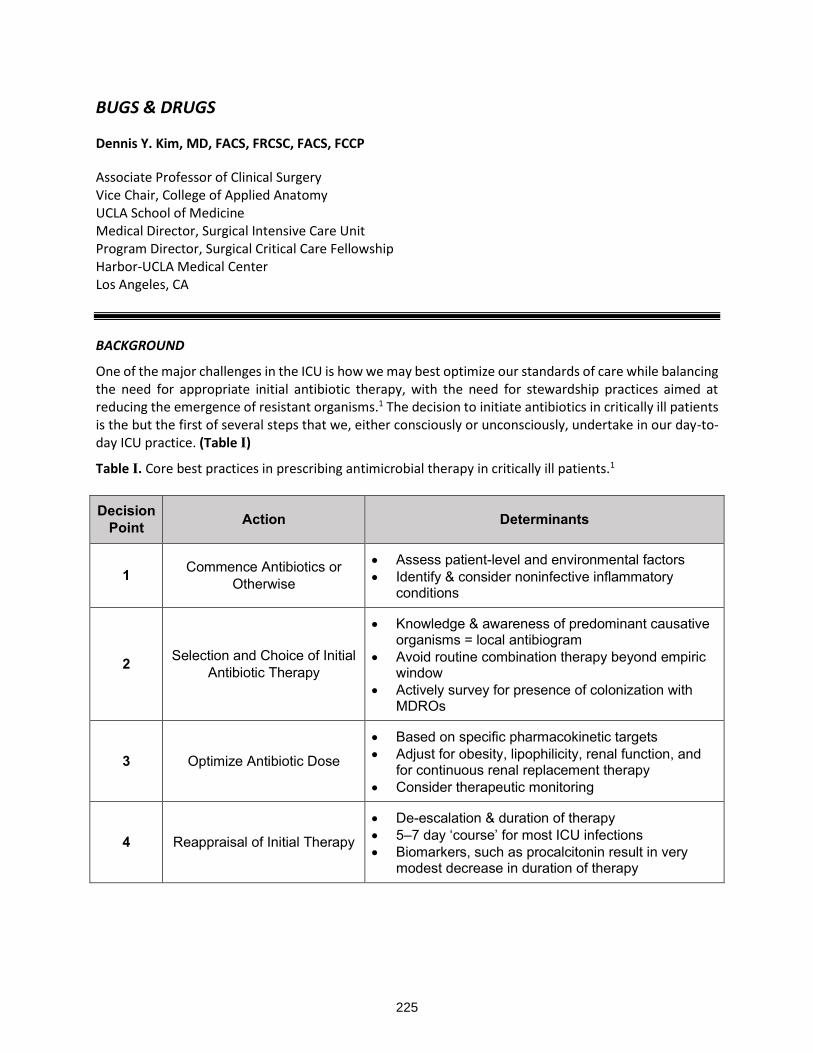
BUGS & DRUGS
Dennis Y. Kim, MD, FACS, FRCSC, FACS, FCCP
Associate Professor of Clinical Surgery Vice Chair, College of Applied Anatomy UCLA School of Medicine Medical Director, Surgical Intensive Care Unit Program Director, Surgical Critical Care Fellowship Harbor-UCLA Medical Center Los Angeles, CA
BACKGROUND
One of the major challenges in the ICU is how we may best optimize our standards of care while balancing the need for appropriate initial antibiotic therapy, with the need for stewardship practices aimed at reducing the emergence of resistant organisms.1 The decision to initiate antibiotics in critically ill patients is the but the first of several steps that we, either consciously or unconsciously, undertake in our day-to-day ICU practice. (Table I)
Table I. Core best practices in prescribing antimicrobial therapy in critically ill patients.1
Decision
Point Action Determinants
1 Commence Antibiotics or
Otherwise
• Assess patient-level and environmental factors
• Identify & consider noninfective inflammatory conditions
2 Selection and Choice of Initial
Antibiotic Therapy
• Knowledge & awareness of predominant causative organisms = local antibiogram
• Avoid routine combination therapy beyond empiric window
• Actively survey for presence of colonization with MDROs
3 Optimize Antibiotic Dose
• Based on specific pharmacokinetic targets
• Adjust for obesity, lipophilicity, renal function, and for continuous renal replacement therapy
• Consider therapeutic monitoring
4 Reappraisal of Initial Therapy
• De-escalation & duration of therapy
• 5–7 day ‘course’ for most ICU infections
• Biomarkers, such as procalcitonin result in very modest decrease in duration of therapy
225

Further, several sequential steps are required to optimize antimicrobial pharmacokinetics in critically ill patients. (Figure 1)
Figure 1. Sequential optimization of antimicrobial pharmacokinetics in critically ill patients.1
In addition to these common antimicrobial stewardship considerations, critically injured and ill patients present a unique challenge insofar as they may present with a wide range of complex injury patterns, frequently require invasive procedures, and may have prolonged ICU stays which predispose them to invasive infections, with or without MDROs.
A recent survey published by the Surgical Infection Society (SIS) identified the top three topics for further research which included non-antimicrobial treatments, optimal treatment duration for bacteremia, and treatment duration for necrotizing soft tissue infections.2 This abstract addresses the last topic, in addition to the utility of procalcitonin in the ICU and the role of antimicrobial prophylaxis for neurotrauma patients with external ventricular drain (EVD) catheters, as well as trauma patients requiring chest tube thoracostomy,
PROCALCITONIN IN THE ICU: FRIEND OR FOE?
Procalcitonin (PCT), produced normally in the C-cells of the thyroid gland, increases rapidly during severe infections, and has a sensitivity of 94% as an indicator for sepsis.3,4 Although potentially useful in the diagnosis and management of acute respiratory infections (ARTIs), its use in terms of guiding antimicrobial therapy duration and impact on outcomes in the ICU environment are unclear. Despite the heterogeneity across studies, recent systematic reviews and meta-analyses do suggest a reduction in antibiotic duration by a median of 1.5 days.5 (Figure 2)
226

Figure 2. Forest plot demonstrating decreased duration of antibiotics using PCT vs. standard care.
DURATION OF ANTIBIOTICS FOR NSTIS: IS LONGER BETTER?
NSTIs continue to be associated with significant morbidity and mortality. Patients often require multiple takebacks to the operating room and it has been well-established that delays to definitive care result in increased mortality and complications.6 Recently, use of a novel immune modulator, AB 103, has demonstrated promise in improving resolution of organ dysfunction.7 There are little data, however, guiding the optimal duration of antibiotics therapy for patients with NSTIs. A recently published single institutional study found that provided adequate surgical debridement is achieved, antibiotic courses of 7 days or less are equally safe compared to longer courses.8
SYSTEMIC ANTIMICROBIAL PROPHYLAXIS & ANTIMICROBIAL-COATED EVD CATHETERS: WERE THE NEUROSURGEONS RIGHT ALL ALONG?
Ventriculostomy-related infections (VRIs) are reported to occur in 10% of patients in whom these invasive devices are inserted and can have profound consequences.9 The literature regarding the benefits and risks of both systemic antimicrobial prophylaxis and the routine use of antimicrobial-coated catheters is mixed and there are few high-quality studies to guide management. Results of a recently published meta-analysis of over 5000 ventriculostomies, demonstrated that management with both extended systemic antibiotics and antibiotic coated EVDs could lower VRI risk in ventriculostomy patients.10 These findings are partially supported by a recent Evidence-Based Consensus Statement on the Insertion and Management of External Ventricular Drains published by the Neurocritical Care Society.9
PROPHYLACTIC ANTIBIOTICS FOR CHEST TUBE INSERTION: TOLD YOU SO
The vast majority of potentially lethal thoracic injuries can be successfully managed via chest tube thoracostomy. Although a relatively straightforward procedure, complications are common can include tube malposition, as well as infectious complications such as empyema. The trauma literature is divided regarding the benefits of prophylactic antibiotics prior to chest tube insertion. However, a recently published systematic review and meta-analysis may shed some light on the topic both in the setting of blunt and penetrating thoracic trauma.11 As demonstrated below, prophylactic antibiotic administration in patients with penetrating and blunt chest injuries requiring the insertion of a chest drain was associated with a reduced risk for post-traumatic empyema. (Figure 3)
227

Figure 3. Meta-analysis comparing antibiotics to no antibiotics for empyema.
SUMMARY
Bugs & drugs. Seed & the soil. Whichever way you look at it, infectious disease considerations and complications are common in the surgical ICU. In an effort to encourage and support antimicrobial stewardship with an eye to decrease the risk for emerging MDROs, it is incumbent upon us as ICU providers to ensure not just timely, but effective, thoughtful, and evidence-based care which utilizes real-time local data to guide best practices.
REFERENCES
1. Brink AJ, Richards G. Best practice: antibiotic decision-making in ICUs. Curr Opin Crit Care. 2020 Oct;26(5):478-488.
2. Delaplain PT, Kaafarani HMA, Benedict LAO, Guidry CA, Kim D, Loor MM, Machado-Aranda D, Mele TS, Mendoza AE, et al. Scientific Studies Committee of the Surgical Infection Society. Surgical Infections and the Future of Research: Re-Defining the Research Agenda for the Surgical Infection Society. Surg Infect (Larchmt). 2021 Dec;22(10):1014-1020.
3. Iankova I, Thompson-Leduc P, Kirson NY, Rice B, Hey J, Krause A, Schonfeld SA, DeBrase CR, Bozzette S, Schuetz P. Efficacy and Safety of Procalcitonin Guidance in Patients With Suspected or Confirmed Sepsis: A Systematic Review and Meta-Analysis. Crit Care Med. 2018 May;46(5):691-698.
4. Andriolo BNG, Andriolo RB, Salomão R, Atallah AN. Effectiveness and safety of procalcitonin evaluation for reducing mortality in adults with sepsis, severe sepsis or septic shock. Cochrane Database of Systematic Reviews 2017, Issue 1. Art. No.: CD010959.
5. Wirz Y, Meier MA, Bouadma L, Luyt CE, Wolff M, Chastre J, Tubach F, Schroeder S, Nobre V, Annane D, et al. Effect of procalcitonin-guided antibiotic treatment on clinical outcomes in
228

intensive care unit patients with infection and sepsis patients: a patient-level meta-analysis of randomized trials. Crit Care. 2018 Aug 15;22(1):191.
6. Kobayashi L, Konstantinidis A, Shackelford S, Chan LS, Talving P, Inaba K, Demetriades D. Necrotizing soft tissue infections: delayed surgical treatment is associated with increased number of surgical debridements and morbidity. J Trauma. 2011 Nov;71(5):1400-5.
7. Bulger EM, May AK, Robinson BRH, Evans DC, Henry S, Green JM, Toschlog E, Sperry JL, Fagenholz P, Martin ND, et al; ACCUTE Study Investigators. A Novel Immune Modulator for Patients With Necrotizing Soft Tissue Infections (NSTI): Results of a Multicenter, Phase 3 Randomized Controlled Trial of Reltecimod (AB 103). Ann Surg. 2020 Sep 1;272(3):469-478. doi: 10.1097/SLA.0000000000004102. PMID: 32657946.
8. Valadez MG, Patel N, Chong V, Putnam BA, Moazzez A, Saltzman D, Kim DY. Short Courses of Antibiotics Are Safe in Necrotizing Soft Tissue Infections. Am Surg. 2021 Dec;87(10):1666-1671.
9. Fried HI, Nathan BR, Rowe AS, Zabramski JM, Andaluz N, Bhimraj A, Guanci MM, Seder DB, Singh JM. The Insertion and Management of External Ventricular Drains: An Evidence-Based Consensus Statement : A Statement for Healthcare Professionals from the Neurocritical Care Society. Neurocrit Care. 2016 Feb;24(1):61-81.
10. Sheppard JP, Ong V, Lagman C, Udawatta M, Duong C, Nguyen T, Prashant GN, Plurad DS, Kim DY, Yang I. Systemic Antimicrobial Prophylaxis and Antimicrobial-Coated External Ventricular Drain Catheters for Preventing Ventriculostomy-Related Infections: A Meta-Analysis of 5242 Cases. Neurosurgery. 2020 Jan 1;86(1):19-29.
11. Ayoub F, Quirke M, Frith D. Use of prophylactic antibiotic in preventing complications for blunt and penetrating chest trauma requiring chest drain insertion: a systematic review and meta-analysis. Trauma Surgery & Acute Care Open 2019;4:e000246. doi:10.1136/tsaco-2018-000246
229

DAZED AND CONFUSED - DELIRIUM IN THE ICU
Alan H. Tyroch, MD, FACS, FCCM
Professor & Chair of Surgery Trauma Medical Director General Surgery, Trauma/Surgical Critical Care Texas Tech University Health Sciences Center El Paso, TX
Delirium is a disturbance of consciousness characterized by acute onset and fluctuating course of inattention accompanied by either a change in cognition or a perceptual disturbance so that a patient’s ability to receive, process, store and recall information is impaired.
The fifth edition of the Diagnostic and Statistical Manual of Mental Disorders (DSM-V) requires the following criteria to be met:
• Disturbance of attention and awareness.
• Disturbance develops acutely and tends to fluctuate in severity.
• At least one additional disturbance in cognition.
• Disturbances are not better explained by a preexisting dementia.
• Disturbances do not occur in the context of a severely reduced level of arousal or coma.
• Evidence of an underlying organic cause or causes.
Delirium is also known as ICU psychosis, ICU syndrome, acute confusional state, encephalopathy and acute brain failure. Delirium is not to be confused with dementia.
Delirium is common in the ICU setting (40% - 60%) and even greater for ventilated patients (60% - 80%). On the other hand, delirium is frequently under-diagnosed.
Delirium is a predictor of:
• Increase in mortality.
• Increase in ventilator days and need for reintubation.
• Longer length of stay in the ICU and hospital.
• Greater hospital expense.
• Long-term cognitive impairment.
• Increase likelihood in discharge to a nursing home or long term acute care facility.
There are three subtypes of delirium.
Hypoactive Lethargy, decreased responsiveness and slowed motor skills. 44%
Mixed Fluctuation between hypoactive and hyperactive activity 55%
Hyperactive Agitation, restlessness, emotional liability and hallucinations < 2%
230

Risk Factors for delirium
Predisposing Factors (patient factors)
Precipitating Factors (events that may occur during hospital stay)
Older age (> age 65) Restraints and/or catheters
Alcoholism Prolonged pain
Smoking Psychoactive medications
Hypertension Sleep deprivation
Respiratory disease Iatrogenic events
Cognitive impairment Severity of illness or injury
Depression Hypoxemia
Vision and/or hearing impairment Severe sepsis
Apolipoprotein E4 polymorphism Dehydration or hypotension
High APACHE or ASA score Constipation
Laboratory test abnormalities
Anemia
Delirium = Brain Organ Dysfunction.
PATHOPHYSIOLOGY
Delirium may be due to neurotransmitter imbalances such as abnormal levels of serotonin, a decrease in acetylcholine or an excess dopamine level.
ASSESSMENT, DIAGNOSIS AND MONITORING
Early detection leads to prompt identification and correction of precipitating factor(s), assurance to patients experiencing symptoms and appropriate treatment.
Delirium is a multifactorial syndrome and patients typically have multiple risk factors.
There are no imaging or laboratory tests to diagnose delirium. It is a diagnosis of exclusion.
The Society of Critical Care Medicine and other organizations recommend regular daily assessment using a valid tool. Two excellent screening tools are the Confusion Assessment Method (CAM-ICU) or the Intensive Care Delirium Screening Checklist (ICDSC).
The CAM-ICU is the most frequently used tool with excellent sensitivity (94% -100%) and specificity (90% - 95%) as well as high inter-rater reliability. Sensitivity and specificity remain high even for ventilated patients. The tool is also easy and quick to perform. The score is either positive or negative for delirium.
231

The Richmond Agitation-Sedation Scale (RASS) should be used in conjunction with the CAMI-ICU. The RASS is used to assess levels of sedation and agitation. It is comprised of a 10-point scale with four levels of anxiety or agitation, one level to denote a calm and alert state, and five levels of sedation.
232

PREVENTION AND TREATMENT
There should not be an automatic reflex to treat delirium with pharmacologic agents. This includes refraining from using benzodiazepines or antipsychotics.
Immediate priority should be to identify and remove the causative factor(s).
Multicomponent therapy is preferred to include the ABCDEF bundle that aims to:
A. A: Assess, prevent and manage pain. B. B: Early ventilator liberation with daily SATs and SBTs C. C: Choice of sedation. D. D: Delirium assessment, prevention and management. E. E: Early mobility and exercise. F. F: Family engagement and empowerment
The 2018 edition of the SCCM Clinical Practice Guidelines for the Prevention and Management of Pain, Agitation/Sedation, Delirium, Immobility and Sleep Disruption in Adult ICU Patients recommends:
• To use multicomponent, nonpharmacologic intervention to reduce modifiable risk factors for delirium, improve cognition and optimize sleep, increase mobility and improve hearing and vision in critically ill adults (conditional recommendation; low quality of evidence).
• To not use haloperidol, atypical antipsychotics (clozapine, risperidone, olanzapine, quetiapine, ziprasidone or aripiprazole), dexmedetomidine, statin or ketamine to prevent delirium in critically ill adults (conditional recommendation; very low-to-low quality of evidence).
• To not routinely use haloperidol and atypical antipsychotics to treat delirium (conditional recommendation; low quality of evidence).
• To use dexmedetomidine for delirium in mechanically ventilated adults where agitation is precluding weaning/extubation (conditional recommendation; low quality of evidence).
• To not use bright light therapy to reduce delirium (conditional recommendation; moderate quality of evidence).
REFERENCES
1. Pun BT. Caring for critically ill patients with the ABCDEF bundle: Results of the ICU liberation collaborative in over 15,000 adults. Crit Care Med, Jan; 47(1): 3-14, 2019.
2. Devlin JW et al. Clinical practice guidelines for the prevention and management of pain, agitation/sedation, delirium, immobility and sleep disruption in adult patients in the ICU. Crit Care Med, Sep; 46(9): e825-e873, 2018.
3. Hayhurst CJ. Intensive care unit delirium: A review of diagnosis, prevention and treatment. Anesthesiology, 125: 1229-1241, 2016.
4. Trogrlic et al. A systematic review of implementation strategies for assessment, prevention and management of ICU delirium and their effect on clinical outcomes. Critical Care, 19: 157, 2015.
5. Girard TD et al. Delirium in the intensive care unit. Critical Care, Vol.12, (Suppl 3), 2008. 6. Morandi A et al. Delirium in the intensive care unit. Int Rev of Psych, 21 (1), 43-58, 2009 7. Gusmao-Flores et al. The confusion assessment method for the intensive care unit (CAM-ICU) and
intensive care delirium screening checklist (ICDSC) for the diagnosis of delirium: A systematic review and meta-analysis of clinical studies. Critical Care, 16:R115, 2012.
8. Sessler CN et al. The Richmond Agitation-Sedation Scale: Validity and reliability in adult intensive care unit patients. Am J Respir Crit Care Med, Vol. 166: 338-1344, 2002
233

THE TORTOISE AND THE HARE – AMBULATING YOUR CRITICAL CARE PATIENTS
Chris Cribari, MD, FACS
Medical Director of Acute Care Surgery University of Colorado Health System Associate Clinical Professor of Surgery University of Colorado School of Medicine Ft. Collins, CO
The evolution of critical care and life-saving interventions has led to increasing numbers of critically ill and injured patients surviving. However, many are left unable to return to normal activities of daily living. Many studies have verified that physical and psychological recovery after a period of critical illness or injury is slow and often incomplete. Post intensive care syndrome (PICS) encompasses new or worsening impairments in physical, cognitive, or mental health status arising after critical illness and persist beyond acute care hospitalization. Since the description of PICS in 2010, observational studies have evaluated the independent factors associated with this syndrome; however, few intervention studies have targeted the prevention of PICS.
The Society of Critical Care Medicine (SCCM) established a Task Force that identified major areas of focus requiring multidisciplinary action to improve long-term outcomes after discharge from an ICU through prevention. Their initial work resulted in a Pain Agitation and Delirium (PAD) guideline. As providers gained a better appreciation for the importance of pain control, the need to avoid over sedation, the deleterious effects of delirium, and the value of spontaneous breathing trials, the concepts of early mobilization and family engagement and empowerment were added to become the ABCDEF Bundle. These efforts resulted in the current Liberation ICU campaign promoting a shift of the ICU culture from the harmful inertia of sedation and restraints to a goal of a more animated ICU filled with patients who are awake, cognitively engaged, and mobile, with family members engaged as partners with the ICU team at the bedside.
SCCM recently completed a 20-month national multicenter quality initiative on the implementation of the ABCDEF bundle and ICU Liberation campaign. The bundle’s “E” addresses the early mobilization and exercise aspect of care. Immobility in the supine position has been shown to reduce lung volumes, decrease mucociliary clearance, and increase the incidence of ventilator-associated pneumonia (VAP), as well as increasing the risk of venous thromboembolic events, pressure sores, joint contractures, and bone demineralization. This growing body of evidence confirms the need to rethink the persisting culture of sedation and immobility in critical care units.
234

Historically, bed rest orders were used to treat various medical conditions, including low back pain, labor pain, peptic ulcer disease, myocardial infarctions, surgical incisions, fractures, acute rheumatoid arthritis, and hepatitis, to name a few. Ambulation of hospitalized patients was first introduced late during World War II to accelerate the recovery of soldiers for return to the battlefield. The benefits realized from these early studies included improved morale, better general health and strength, and a more rapid recovery. Getting the patient out of bed and ambulating was and is an essential part of postoperative and post-injury care. Mobility contributes to the proper functioning and conditioning of each individual body system. Beyond the muscles and joints, the pulmonary, cardiac, gastrointestinal, and cognitive functions rely on physical activity to stay intact. When function decreases, weakness develops which results in a further reduction of physical activity. A typical elective postoperative patient’s mobility program starts immediately with sitting at the edge of bed and dangling their legs on the first postoperative night. On subsequent days, they progress to standing at the bedside and sitting in a chair, to walking in the room, and eventually in the hall prior to discharge home. All patients should be encouraged to push themselves during the ambulation phase of recovery and educated on its importance. Patients are generally encouraged to walk a minimum of 3 times a day.
Many critically ill and injured patients receive an initial period of sedation and immobility until they stabilize, and, consequently, many develop profound muscle weakness and become deconditioned. In addition to the catabolic nature of critical injury and illness, the loss of mechanical loading due to physical inactivity, bed rest, or immobilization contributes further to the profound weakness we see in our survivors. Gao and Gordon describe how mechanical unloading stimulates a complex adaptive response resulting in muscle atrophy and loss of strength via a slowing of protein synthesis and accelerated protein degradation. The importance of early nutritional support is well recognized; however, the other key countermeasures to lessen the effects of unloading are physical activity and resistance exercises.
The degree of mobilization and exercise in the ICU depends on the patient’s condition, stability, types of ongoing treatments, and the number and type of devices that must remain attached to the patient; i.e., ventilator, central lines, dialysis machines, ECMO, external fixators, chest tubes, and drains. Support devices are often the initial safety concern raised when starting an early mobility program. In 2007, Bailey, et. al., studied the feasibility and safety associated with mobilizing respiratory failure in 103 critically ill patients who were on mechanical ventilation for more than 4 days. Patients participated in 1449 activities that included sitting at the side of the bed (233 or 16%), sitting in a chair (454 or 31%), standing at bedside, and ambulating (762 or 53%). Only six activity adverse events were noted including assisted fall to the knee, inadvertent tube removal, elevated blood pressure, hypotension, oxygen desaturation, and unplanned extubation.
In another study, Morris et al., compared 165 patients with acute respiratory failure who were treated with a physical therapy (PT) mobility protocol with a matched cohort who received the standard care. Outcomes were evaluated in the 165 study patients who survived to hospital discharge. No reported adverse events occurred during mobility activities. The authors reported that patients placed on the PT protocol were out of bed 6 days earlier and received more physical therapy (78%). In addition, the PT patients had a 1.4-day shorter length of stay in the ICU and a 3.3-day shorter hospital length of stay.
In 2009, a landmark randomized controlled trial by Schweickert, et. al., concluded that early physical and occupational therapy in mechanically ventilated patients in the medical ICU was safe and well tolerated, and resulted in better functional outcomes at hospital discharge, a shorter duration of delirium, and more ventilator-free days compared with standard care.
In 2010, the Society of Critical Care Medicine (SCCM) invited key stakeholder groups, including the American Physical Therapy Association (APTA), to identify strategies to improve early mobilization in the ICU and integration of the physical therapist as a member of the ICU team. This model has been
235

successfully implemented in several institutions; however, there remains significant variability in the implementation of early mobilization of critical care patients among institutions across the country and world. Despite previous studies showing early mobilization is safe and improves outcomes in the medical intensive care unit (ICU), there is reluctance and fear that surgical patients could be harmed by premature mobilization. Trauma patients often have the added burden of multiple sequential operative procedures, wounds, activity restrictions, and immobilization devices that have contributed to an embedded culture of fear of early mobilization. Despite some ongoing resistance, several studies on early mobilization and exercise interventions delivered in surgical intensive care units are sufficient to base a clinical best practice.
Radu, et. al., reported that safe early mobility in the SICU is possible, requires a multidisciplinary approach, and can be provided even with a limited budget. Fifty patients were reviewed, 62% were older than 65 years, and 52% of patients were mechanically ventilated. The mean length of stay in the SICU was 10.5 days. The activity level ranged from passive range of motion, postural, truncal, upper, and lower extremity exercises, to ambulation. A total of 54% tolerated full ambulation in the SICU, with 42% ambulating more than 10 feet; 16% were not stable to undergo mobility sessions. No adverse effects were noted. They also found that mobility efforts enhanced patient and family satisfaction, and suggested the potential decrease in postoperative cognitive dysfunction, length of stay, morbidity, cost, and adverse events post discharge, including need for readmission. Mah et al reported a team-based, resource-efficient approach to early mobilization is feasible and effective in the ICU.
Schaller, et. al., in their 2016 multicenter trial in the surgical ICU found that early, goal-directed mobilization shortened patient length of stay and improved patients’ functional mobility at hospital discharge. Duration of immobilization seems to predict adverse events in patients in the surgical intensive care unit. Although fast-track cardiac and non-cardiac surgery with early ambulation has been found to be safe and reduces hospital length of stay, it does not alter postoperative mortality. Up to 25% of patients can be safely mobilized within 72 hours of ICU admission. This therapy may reduce hospital and ICU length of stay, shorten duration of mechanical ventilation, and improve muscle strength and functional independence scores.
In the five randomized control trials, improvement in delirium when implementing early mobility was noted. In the study performed by Schweickert, et. al., median duration of ICU-associated delirium was decreased by nearly half in the intervention group that received early exercise and mobilization during periods of daily interruption of sedation than in the control group (p = 0.02). Similarly, in the SICU population of Schaller et al., the intervention group was noted to be free from delirium for longer than control group (p=0.016)
The mobility protocol should also encompass therapeutic positioning and range of motion exercises for the patient not yet ready for mobilization as well as, outline the competency progression from dangling, standing, sitting, and ambulating.
THERAPEUTIC POSITIONING OF THE PATIENT
Sustained supine position (0◦ head-of-bed (HOB) elevation) increases gastroesophageal reflux and the possibility of aspiration. In addition, when patients are placed in the supine position ventilation/ perfusion (V/Q) are greater in the dependent areas of the lung. In healthy patients, V/Q matching is achieved, but in diseased lungs prolonged placement in the supine position can contribute to V/Q mismatch. Thus, elevating the head of bed to an angle of 30◦ to 45◦, unless contraindicated, is generally recommended to prevent reflux and improved ventilation. In 2011, a systematic review by Niel Weise, et. al., on the benefits and disadvantages of semi-upright position in ventilated patients was published. Three trials (337 patients) were included in the review. Based on the analysis, it was unclear whether a 45◦ head of bed
236

elevation was effective or harmful regarding the occurrence of clinically suspected ventilator-associated pneumonia, microbiologically confirmed ventilator associated pneumonia, decubitus ulcer formation, and mortality. There was concern that 45◦ elevation for 24 hours a day could increase the risk for thromboembolism or hemodynamic instability. The review failed to prove clinical benefits of 45◦ head of bed elevation over 30◦. To date, the recommended degree for head of bed (HOB) elevation is 30◦ to prevent pneumonia and aspiration. Part of therapeutic positioning includes turning to help prevent pressure induced skin breakdown. The frequency in which these turns get done has also been evaluated. In 2005, Winkelman et al completed a prospective longitudinal observational study of a critical care unit which found that over an 8-hour period, only 2.7% of observed patients experienced position changes every 2 hours, and more than half were supine for 4 to 8 hours. Studies by Vanderwee et al and Defloor et al compared turning frequencies among patients. They found no significant difference between 2-, 3-, and 4-hour turning schedules on pressure ulcer formation, but recommended that at a minimum, turning should occur at least every 4 hours. Each institution should set their ICU’s standard by defining the frequency of turns and then study their compliance and incidence of skin breakdown and make changes based on their unit’s results as part of their performance improvement effort.
RANGE OF MOTION
Bed rest promotes immobilization of muscle fibers resulting in reduced motion of the effected extremity. The muscle fibers and connective tissues adapt to the shortened length by contraction of collagen fibers and decrease in muscle fiber sarcomeres in just one day of bed rest. Morris, et. al. and Harvey, et. al., recommend range of motion (ROM) exercises three times a day to all upper and lower extremities, with a minimum of five repetitions per exercise. The protocol should include the expected detail of these ROM exercises, with upper extremity ROM exercises including finger, wrist, elbow flexion and extension, shoulder adduction, abduction, and internal and external rotation. Lower extremity ROM exercises include toe and ankle flexion and extension, hip flexion, abduction, and internal and external rotation. These ROM exercises are an excellent means to engage family members who are often searching for the best way to assist their loved ones.
DANGLING AND WALKING
Progression to this level of activity is not linear for all patients. Poor tolerance during one episode does not predict future intolerance. Every patient should be evaluated for progression in early mobility daily. Remember that small efforts can yield large results.
At least eight international guidelines have recommended ICU early mobilization and exercise. Despite all the ongoing research and efforts, barriers to implementation remain. The frequently identified and reported barriers can be divided into patient related and facility related.
Patient related Facility related
Patient’s pain and discomfort Unit culture and attitude
Cardiopulmonary instability Lack of knowledge and training
Depressed level of consciousness Sedation practices
Safety of tubes, catheters, and wires Lack of personnel and equipment resources
Lack of physician buy in and leadership
Patient’s size Time, valuing, and priority of mobilization
Watenabe, et. al., found the barrier to mobilization on days 1 and 2 was circulatory status, level of consciousness on days 3 to 5, and medical staff factors on days 6 and 7. Multivariate analysis showed that consciousness level (OR: 0.38, p=0.01), and medical staff factors (OR: 0.49, p=0.01) were significantly
237

associated with successful mobilization. They reported that by hospital discharge 125 patients (71%) of their patients could walk independently. Level of consciousness was associated with walking independence (OR: 0.52, p=0.04). In this study, over half of patients could achieve mobilization within the first 7 days. They concluded that barriers to mobilization in the ICU change over time and that the level of consciousness is significantly associated with both mobilization and independent walking at discharge.
Getting the team to realize that it is safe to mobilize intubated patients is part of the cultural change that is paramount to successful implementation. Multimodal staff education, which incorporates lecture, online education, just-in-time education, and discussion during unit briefs, plays an integral part in decreasing staff bias against early mobilization of SICU patients. This was elucidated in the findings reported by Morris et al, who used multimodal education to eliminate barriers to mobilization in ICUs. The progressive early mobility activity protocol used by Morris et al resulted in a significant reduction in the length of stay for the mobility protocol group (5.5 days) than for the usual-care group (6.9 days) in that study. The early mobility activity protocol included 4 levels of activity therapy. The first level was designed for unconscious patients where passive range-of-motion exercises were performed on patients’ upper and lower extremities 3 times a day. The second level was designed for patients who responded and followed commands where progressive active-assistive and active range-of-motion exercises were administered. The third and fourth levels were designed for patients who were alert and able to participate actively. A key feature of mobilization strategies in the critically ill for the fourth level was to have the patient demonstrate sequential competencies: dangle legs at side of bed, get out of bed and into a chair, stand at the bedside, and finally ambulation with assistance.
There are a number of ways institutions have gone about developing an early mobility and ambulation program. One successful approach starts with forming a SICU mobilization work group, consisting of physicians, midlevel providers, respiratory therapists, physical therapists, and staff nurses. Getting all the disciplines together on the same page is crucial in the planning, development, and implementation of an early mobilization protocol. Physician champions are needed to facilitate cultural change and will be required to reorganize and manage current practices that have the potential to interfere with mobility. They should create a strategy to improve the level of teamwork and provide leadership throughout the process. Once the SICU team has gained experience and see the results of their efforts, they will quickly become zealots rather than doubters. This change in the mindset is further reinforced at monthly quality meetings of the SICU service, where quality data such as length of stay and frequency of ventilator-associated pneumonia, deep vein thrombosis, delirium, and skin breakdown are presented and analyzed. In addition, display of longitudinal quality data in the nurses’ lounge is a great way to better disseminate these results and change culture. This is consistent with the results reported by Balas et al. on mobilizing patients in ICUs. Early ambulation of their patients receiving mechanical ventilation resulted in a decrease in the length of stay, ventilator-associated pneumonia, deep vein thrombosis, and skin breakdown.
The work group is tasked to develop and implement a structured process to get patients moving using a standardized mobility protocol. This must include the establishment of inclusion and exclusion criteria for early mobilization. Inclusion should include screening of all patients. The reviewed literature is in general agreement to withhold early mobility for patients with hypoxia, hemodynamic instability (escalation of vasopressors in the last 12 h), intracranial pressure monitoring, unstable cardiac rhythm (life-threatening rhythm that compromised blood pressure in past 24 h), a new cardiac arrhythmia, an open chest or abdomen, an epidural catheter with dense regional anesthesia, and ventilated patients with critical airways or known difficult airways or those requiring FiO2 > 60% or PEEP > 10. Special considerations that need to be addressed include femoral arterial access, spinal clearance, orthopedic injuries, and precautions after newly placed skin grafts prior to starting ROM. Linke et al. emphasized that all ICU patients should be screened daily for initiation of the mobility protocol unless they met exclusion
238
parameters. The exclusion criteria are broken down into two categories. Patients with “hard stops” are due to clinical instability and “yield” criteria, which require consultation with the care team before proceeding. The “yield” criteria encourage shared decision-making in the context of the complete clinical picture. This was intended to avoid summary exclusion of patients because of therapies such as elevated levels of supplemental oxygen and low dose vasopressors, which may represent Interventions their baseline clinical condition.
Resource scarcity remains a significant issue in the face of the COVID pandemic and a nationwide shortage of nursing staff, physical therapists, and respiratory therapists. Dedication of additional personnel definitely facilitates the implementation of an early mobility program, however when adding staff is not an option due to existing staff shortages and budgetary restrictions, implementation is still possible. As an alternative, resolving inefficiencies in the existing process should be targeted to free up the resources required. Workflow for each discipline must be examined, focusing on optimization and coordination of standard tasks and optimal timing for labor-intensive mobility sessions. A formalized communication process is required with daily planning and discussion of patients requiring the assistance of multiple disciplines to mobilize. The respiratory, physical, and occupational therapists, and bedside nurses assigned to the ICU meet each morning to discuss timing of the mobility session and the equipment required. The bedside nurse should also review the other planned tests or treatments to prevent the mobility session from coinciding and getting cancelled. This also allows the bedside nurse to plan to have the patient ready on time for the mobility session and be available to assist. This coordination streamlines planning, decreases the amount of staff downtime experienced during preparation for each mobility session, ensures all required staff are present, and allows for equitable allocation of mobility sessions if the number of patients exceeded the available resources.
The value of the “E” in the “ABCDEF bundle” cannot be ignored and as surgical leaders we each need to figure out how to best implement an early mobility protocol into our SICU’s.
An example of a completed protocol from Minnesota Health published by Linke, et. al.
239

240

REFERENCES
1. Bailey PP, Miller RR 3rd, Clemmer TP. Culture of early mobility in mechanically ventilated patients. Crit Care Med. 2009 Oct;37(10 Suppl): S429-35.
2. Barr J, Fraser GL, Puntillo K, Ely EW, Gélinas C, Dasta JF, Davidson JE, et al.; American College of Critical Care Medicine. Clinical practice guidelines for the management of pain, agitation, and delirium in adult patients in the intensive care unit. Crit Care Med. 2013 Jan;41(1):263-306
3. Barr J, Paulson SS, Kamdar B, Ervin JN, Lane-Fall M, Liu V, Kleinpell R. The Coming of Age of Implementation Science and Research in Critical Care Medicine. Crit Care Med. 2021 Aug 1;49(8):1254-1275.
4. Chambers MA, Moylan JS, Reid MB. Physical inactivity and muscle weakness in the critically ill. Crit Care Med. 2009 Oct;37(10 Suppl): S337-46.
5. Dammeyer J, Dickinson S, Packard D, Baldwin N, Ricklemann C. Building a protocol to guide mobility in the ICU. Crit Care Nurs Q. 2013 Jan-Mar;36(1):37-49.
6. Defloor T, De Bacquer D, Grypdonck MH. The effect of various combinations of turning and pressure reducing devices on the incidence of pressure ulcers. Int J Nurs Stud. 2005 Jan;42(1):37-46.
7. Dubb R, Nydahl P, Hermes C, Schwabbauer N, Toonstra A, Parker AM, Kaltwasser A, Needham DM. Barriers and Strategies for Early Mobilization of Patients in Intensive Care Units. Ann Am Thorac Soc. 2016 May;13(5):724-30
8. Fuke R, Hifumi T, Kondo Y, Hatakeyama J, Takei T, Yamakawa K, Inoue S, Nishida O. Early rehabilitation to prevent postintensive care syndrome in patients with critical illness: a systematic review and meta-analysis. BMJ Open. 2018 May 5;8(5): e019998
9. Gao Y, Arfat Y, Wang H, Goswami N. Muscle Atrophy Induced by Mechanical Unloading: Mechanisms and Potential Countermeasures. Front Physiol. 2018 Mar 20; 9:235
10. Gordon BS, Kelleher AR, Kimball SR. Regulation of muscle protein synthesis and the effects of catabolic states. Int J Biochem Cell Biol. 2013 Oct;45(10):2147-57.
11. Hodgson CL, Schaller SJ, Nydahl P, Timenetsky KT, Needham DM. Ten strategies to optimize early mobilization and rehabilitation in intensive care. Crit Care. 2021 Sep 3;25(1):324.
12. Hopkins, Ramona O., Vicki J. Spuhler, and George E. Thomsen. "Transforming ICU culture to facilitate early mobility." Critical care clinics 23, no. 1 (2007): 81-96.
13. Iwashyna TJ, Hodgson CL. Early mobilization in ICU is far more than just exercise. Lancet. 2016 Oct 1;388(10052):1351-1352
14. Kress JP. Clinical trials of early mobilization of critically ill patients. Crit Care Med. 2009 Oct;37(10 Suppl): S442-7.
15. Linke CA, Chapman LB, Berger LJ, Kelly TL, Korpela CA, Petty MG. Early Mobilization in the ICU: A Collaborative, Integrated Approach. Crit Care Explor. 2020 Apr 29;2(4): e0090.
16. Mah, John W., Ilene Staff, David Fichandler, and Karyn L. Butler. "Resource-efficient mobilization programs in the intensive care unit: who stands to win?" The American Journal of Surgery 206, no. 4 (2013): 488-493.
17. Morris PE. Moving our critically ill patients: mobility barriers and benefits. Crit Care Clin. 2007 Jan;23(1):1-20.
18. Niël-Weise, Barbara & Gastmeier, Petra & Kola, Axel & Vonberg, Ralf-Peter & Wille, Jan & Broek, Pieter. (2011). An evidence-based recommendation on bed head elevation for mechanically ventilated patients. Critical care (London, England). 15. R111. 10.1186/cc10135.
19. Pun BT, Balas MC, Barnes-Daly MA, Thompson JL, Aldrich JM, Barr J, et al. Caring for Critically Ill Patients with the ABCDEF Bundle: Results of the ICU Liberation Collaborative in Over 15,000 Adults. Crit Care Med. 2019 Jan;47(1):3-14
241

20. Radu F., Amulya C. Alapati, Martine A. Louis, Dmitriy Kim, et al. Early Mobility in the Surgical ICU: A Community Hospital Experience, Journal of the American College of Surgeons, Volume 231, Issue 4, Supplement 1, 2020, Page S313
21. Saxton RA, Sabatini DM. mTOR signaling in growth, metabolism, and disease. Cell 2017168:960–976
22. Schaller SJ, Anstey M, Blobner M, et al. Early, goal-directed mobilization in the surgical intensive care unit: a randomized controlled trial. Lancet 2016;388: 1377–88. 10.1016/S0140-6736(16)31637-3
23. Schweickert WD, Pohlman MC, Pohlman AS, et al. Early physical and occupational therapy in mechanically ventilated, critically ill patients: a randomized controlled trial. Lancet. 2009 May 30;373(9678):1874-82.
24. Waldauf P, Jiroutková K, Krajčová A, Puthucheary Z, Duška F. Effects of Rehabilitation Interventions on Clinical Outcomes in Critically Ill Patients: Systematic Review and Meta-Analysis of Randomized Controlled Trials. Crit Care Med. 2020 Jul;48(7):1055-1065
25. Watanabe S, Liu K, Morita Y, Kanaya T, Naito Y, Arakawa R, et al. Changes in barriers to implementing early mobilization in the intensive care unit: a single center retrospective cohort study. Nagoya J Med Sci. 2021 Aug;83(3):443-464
26. Winkelman C, Higgins PA, Chen YJ. Activity in the chronically critically ill. Dimens Crit Care Nurs. 2005 Nov-Dec;24(6):281-90.
27. Wright SE, Thomas K, Watson G, Baker C, Bryant A, et al. Intensive versus standard physical rehabilitation therapy in the critically ill (EPICC): a multicenter, parallel-group, randomized controlled trial. Thorax. 2018 Mar;73(3):213-221.
28. Vanderwee K, Grypdonck MH, De Bacquer D, Defloor T. Effectiveness of turning with unequal time intervals on the incidence of pressure ulcer lesions. J Adv Nurs. 2007 Jan;57(1):59-68
29. Yoshida T, Delafontaine P. Mechanisms of IGF-1-Mediated Regulation of Skeletal Muscle Hypertrophy and Atrophy. Cells. 2020 Aug 26;9(9):1970.
242

GI BLEEDING AND PROPHYLAXIS PRACTICES: LIFESAVING OR PNEUMONIA GENERATING?
Meghan R. Lewis, MD, FACS
Assistant Professor of Clinical Surgery LAC+USC Medical Center & University of Southern California – Los Angeles Los Angeles, CA
BACKGROUND
Pathophysiology of stress ulcers
In critical illness, splanchnic hypoperfusion and mesenteric ischemia can lead to stress-related mucosal damage, which is a precursor to stress gastropathy. In a small percentage of patients, stress gastropathy can result in clinically significant bleeding, which has a profound impact on morbidity and mortality. Clinical importance has classically been described as obvious physiologic decline, the requirement of operative or endoscopic intervention, or transfusion requirement.
Evolution of care
In 1978, a randomized controlled trial of 100 critically ill patients at risk for developing acute gastrointestinal ulceration and bleeding was published in the NEJM1. Patients were divided into two groups: 51 received antacid prophylaxis, and 49 received no prophylaxis. Hourly antacid titration kept the pH of the gastric contents above 3.5. Two of the 51 patients who received antacid prophylaxis had gastrointestinal bleeding, compared to 12 of the 49 control patients (P < 0.005). Of the 12 patients in the control group who bled, 7 were placed on antacid medication and bleeding then resolved.
Figure 1.
243

Two meta-analyses subsequently published by Cook et al demonstrated that both histamine-2 receptor antagonists (H2RAs) and sucralfate decreased the risk of bleeding from stress ulceration when compared to a placebo.2-3 Sucralfate exerts its topical effect by binding to proteins of the ulcer site, as opposed to lowering gastric pH. It must be administered into the stomach and has the potential to bind other medications.
Stress ulcer prophylaxis (SUP) became part of ICU standard of care in the 1990s. SUP constituted a major component of protocols recommended for best practice for all critically ill patients. For example, a “bundle” promoted in 2005 by the Institute for Healthcare Improvement for reduction of VAP consisted of only four elements: head of bed elevation, daily sedation vacation, deep-venous thrombosis prophylaxis, and SUP.4 Though SUP was initially achieved by administration of sucralfate or histamine receptor blockers (H2RAs), overtime, proton pump inhibitors (PPIs) became the most common class used.
RISK FACTORS
Cook et al performed a prospective multicenter cohort study to determine specific risk factors for clinically significant bleeding due to stress ulcers.5 Though SUP was already commonplace, the authors encouraged physicians to withhold prophylaxis in all patients except those with:
• head injury,
• burns to 30% of the body-surface area or greater,
• organ transplants
• previous diagnosis of peptic ulcer, gastritis, or upper gastrointestinal bleeding within 6 weeks before admission.
Of 2252 patients, 33 (1.5%) had clinically important bleeding. Two strong independent risk factors for bleeding were identified: respiratory failure (odds ratio, 15.6) and coagulopathy (odds ratio, 4.3). Of 847 patients who had one or both of these risk factors, 31 (3.7%) had clinically important bleeding. In contrast, of 1405 patients without either of these risk factors, only 2 (0.1%) had clinically important bleeding. The mortality rate was 48.5% in the group with bleeding and 9.1% in the group without bleeding (P<0.001).
Other potential risk factors evaluated that were not determined to be statistically significant included:
• Sepsis
• Hepatic failure
• Renal failure
• Enteral feeding
• Glucocorticoid administration
The authors concluded that patients without coagulopathy or respiratory failure, who made up 62% of the study group, were at extremely low risk for clinically important bleeding. In patients who either had a coagulopathy or who underwent mechanical ventilation for more than 48 hours, the risk of bleeding was 3.7 percent, even though more than half received prophylactic therapy of some type. The authors assumed an approximately 50% reduction in risk of clinically important bleeding with prophylaxis (per the literature), and therefore estimated that only about 30 of these high-risk patients would have to receive SUP in order to prevent one episode of clinically important bleeding. They concluded that SUP should be administered to all patients with coagulopathy or requiring mechanical ventilation >48 hours for respiratory failure.
TRAUMA/CRITICAL CARE
Simons et al evaluated stress ulceration in 33,637 major trauma patients treated between 1985 and 1991 using a combination of registry data and chart review.6 Clinical stress ulceration developed in 57 patients
244

(0.17%), despite prophylaxis in the majority. Eighteen patients (0.05%) developed severe ulceration, resulting in gastroduodenal perforation in 3 patients and greater than 2 units of blood transfusion in 16 patients. Regression analysis was used to determine independent risk factors for stress ulceration, which included Injury Severity Score (ISS) greater than or equal to 16 (OR = 12.6), spinal cord injury (OR = 2.0), and age > 55 (OR = 2.4). Other serious complications, including pneumonia, sepsis, and organ failure were also associated with stress ulceration.
In 2008, the Eastern Association for the Surgery of Trauma (EAST) published guidelines for trauma/critical care surgeons.7 These guidelines advocated for the use of SUP in critically ill patients (justified by its historic 50% decrease in clinically significant hemorrhage). Strength of the recommendation was determined based on the class of data from which it was derived:
Level 1 recommendation was made for SUP for patients with mechanical ventilation, coagulopathy, traumatic brain injury, or major burn injury
Level 2 recommendation was made for SUP for ICU patients with multi-trauma, sepsis, or acute renal failure
Level 3 recommendation was made for SUP for all ICU patients with ISS>15 or requirement of high-dose steroids (>250 mg hydrocortisone or equivalent per day).
COMPLICATIONS
With widespread prophylactic use of SUP, several associated complications have been observed. The majority of these complications are infectious in nature, including pneumonias and Clostridium Difficile diarrhea. Both PPIs and H2RAs are known to have a suppressive effect on immune function. This effect has the potential to increase the incidence of a variety of different postoperative and/or nosocomial infections.8-12
Pneumonias
Acid suppressive therapy is associated with increased colonization of the upper gastrointestinal tract with potentially pathogenic organisms. Both H2RAs and PPIs increase the pH of gastric contents. The altered gastric pH, in combination with pulmonary micro aspiration, is believed to be associated with the development of bacteria in the respiratory tract.13 These factors are in addition to the known potential to impair immune function, as previously described.
Prod’hom et al published a randomized controlled trial in 1994 of SUP for 244 mechanically ventilated patients with nasogastric tubes.14 Macroscopic gastric bleeding was observed in 10%, 4%, and 6% of patients assigned to receive sucralfate, antacid, and ranitidine, respectively (P > 0.2). The incidence of early-onset pneumonia was not statistically different among the three groups (P > 0.2). However, in a subgroup of 213 patients observed for more than 4 days, late-onset pneumonia was observed in 5% of the patients who received sucralfate compared with 16% and 21% of the patients who received antacid or ranitidine, respectively (P= 0.022). Patients who received sucralfate were observed to have a lower gastric pH (P < 0.001) and less frequent gastric colonization compared with the other groups (P = 0.015).
Grindlinger and colleagues evaluated 504 patients who received either antacid or sucralfate for SUP.15 There were 33 ventilator associated pneumonias (VAP) in the PPI/H2RA group compared to only 12 in the Sucralfate group (P < 0.01). Furthermore, culprit bacteria were found to be Pseudomonas, gram-negative bacilli, and methicillin-resistant Staphylococcus aureus in the antacid patients, compared with oropharyngeal flora in the Sucralfate patients.
In 2007, Herzig et al evaluated a cohort of non-ICU patients admitted to a large academic medical center from 2004 through 2007.16 Data were collected prospectively from electronic databases maintained at
245
the medical center. Patients were included if they had a minimum of 3-day hospital stay. ICD-9 codes were used to identify patients who developed a bacterial pneumonia as a secondary diagnosis during hospitalization. Acid-suppressive medication use was associated with 30% increased odds of hospital-acquired pneumonia. This association was noted to be higher for aspiration pneumonia than for non-aspiration pneumonia. In a subset analyses, a statistically significant risk was demonstrated for PPI use, but not for H2RAs, however, the number of patients who received H2RAs was much smaller.
Clostridium Difficile Infections
In 2004, Dial et al performed a multivariate analysis of a cohort of 1187 patients who received antibiotics in the hospital, of whom 81 (6.8%) developed C. Difficile diarrhea. The authors determined that C. Difficile diarrhea was significantly associated with the use of PPIs (adjusted odds ratio [OR] 2). At the same time, the authors reported a case–control study where C. Difficile diarrhea was also associated with PPIs (adjusted OR 2.7).17
In 2008, Aseeri and colleagues performed a case-control study of 94 patients with C. Difficile diarrhea matched with control patients.18 They discovered that the odds of C Difficile diarrhea diagnosis significantly increased with PPI usage, after controlling for date of hospital admission, antibiotic use, gender, age, patient location, and room type. Overall, inpatients receiving PPIs were 3.6 times more likely to develop C Difficile diarrhea.
ROLE OF ENTERAL NUTRITION
In 2010, Marik et al noted that stress ulcers appeared increasingly uncommon in contemporary ICU practice, and they hypothesized that early enteral feeding was potentially to credit.19 Enteral nutrients could potentially buffer acid, be cytoprotective for mucosal cells, and/or could improve mucosal blood flow and immunity. They performed a meta-analysis of randomized, controlled studies that evaluated the association between SUP and gastrointestinal bleeding. For homogeneity, they specifically selected studies that compared H2RAs with placebo. Seventeen studies (1836 total patients) were included. Overall, SUP with a H2RA reduced the risk of gastrointestinal bleeding (odds ratio 0.47; p < .002); however, the treatment effect was noted only in the subgroup of patients who did not receive enteral nutrition. In those patients who were fed enterally, SUP did not alter the risk of gastrointestinal bleeding (odds ratio 1.26). Overall, H2RAs did not increase the risk of hospital-acquired pneumonia (odds ratio 1.53; p = .12); however, this complication was increased in the subgroup of patients who were fed enterally (odds ratio 2.81; p = .02). Overall, SUP had no effect on hospital mortality (odds ratio 1.03; p = .82). However, the hospital mortality was higher in the 2 studies in which patients were fed enterally and received a H2RA (odds ratio 1.89; p = .04). The authors concluded that in patients receiving enteral nutrition, SUP may not be required and, worse, had the potential to increase the risk for pneumonia and death.
The necessity for SUP in enterally fed surgical and trauma ICU patients has also been specifically studied.20 Palm et al retrospectively evaluated 200 mechanically ventilated patients in a surgical-trauma ICU at a level 1 trauma center from 2008-2013. Acid suppression was discontinued for patients once tolerating full enteral feeds. They reported a 0.5% incidence of clinically significant GI bleeding; 0.68% in the subgroup of TBI patients. Complications were all low, including ventilator associated pneumonia (1/1000 days), CDI (0.2/1000 days) and mortality (2%).
CHOICE OF SUP AGENT
Though initial studies evaluated sucralfate and H2RAs for SUP, PPIs rapidly became more popular choices. Kantarova et al performed a single-center randomized, placebo-controlled study of 287 high-risk patients (>48 h mechanical ventilation, coagulopathy).21 The authors compared PPIs, H2RAs, sucralfate, and placebo. Clinically important bleeding occurred in 1%, 3%, 4%, and 1% respectively (p>0.28). Nosocomial
246

pneumonia occurred in 11% of the PPI group, 10% of the H2RA group, 9% of sucralfate patients, and in 7% of placebo patients (p>0.34).
Multiple randomized-controlled trials and meta-analyses have been performed comparing SUP agents, particularly H2RAs and PPIs. Lin et al published a meta-analysis of 7 randomized controlled trials (936 total patients) comparing PPIs and H2RAs.22 They found no difference with regard to upper GI bleeding, pneumonia risk, and intensive care unit mortality.
Barkun et al performed an analysis of 8 randomized controlled trials and 5 abstracts.23 Their results demonstrated that PPI administration significantly decreased the incidence of clinically important GI bleeding compared to H2RA (OR = 0.30). No statistical differences were noted for the development of nosocomial pneumonia (OR = 1.05) or mortality (OR = 1.19).
MacLaren et al evaluated occurrence and risk factors for GI hemorrhage, pneumonia, and CDI in 35,312 critically ill patients requiring mechanical ventilation who were administered H2RAs or PPIs at 71 hospitals.24 Gastrointestinal hemorrhage (2.1% vs 5.9%), pneumonia (27% vs 38.6%), and CDI (2.2% vs 3.8%) occurred less frequently in the H2RA group. After adjusting for confounding factors, odds ratios of GI hemorrhage (2.24), pneumonia (1.2), and CDI (1.29) were greater with PPIs.
In 2016, Alshamsi et al performed another meta-analysis to compare PPIs with H2RAs for SUP. 19 trials were included, with 2117 total patients.25 PPIs were more effective than H2RAs in reducing the risk of clinically important GI bleeding (RR 0.39; P = 0.002) and overt GI bleeding (RR 0.48; P < 0.0001). PPI use did not significantly affect the risk of pneumonia (RR 1.12; P = 0.39), mortality (RR 1.05; P = 0.61), or ICU length of stay (mean difference (MD), -0.38 days; P = 0.51). None of the randomized controlled trials included reported Clostridium Difficile infection.
Due to ongoing concerns relating to the risk-benefit of SUP, specifically with PPIs, Krag et al published the SUP ICU study. The trial was a multicenter, randomized blinded trial of PPI versus placebo, with primary outcome of mortality by 90 days. Out of 1645 patients who received PPI, 2.5% had clinically significant bleeding, compared to 4.2% of those who received placebo. There was no difference between groups with regard to infections, “clinically important events” (the composite of clinically important gastrointestinal bleeding, pneumonia, Clostridium difficile infection, or myocardial ischemia), or mortality.26
Finally, in 2020, The PEPTIC Randomized Clinical Trial comparing PPIs to H2RAs was published. This was a large, multi-center trial of 26,982 patients in 50 ICUs across 5 countries.27 All patients required invasive mechanical ventilation within 24 hours of ICU admission. Preferential use of PPIs for 6 months was compared to preferential use of H2RAs for 6 months at each center. 18.3% in the PPI group died at the hospital by day 90 compared to 17.5% in the H2RA group (P = .054), while clinically important upper gastrointestinal bleeding occurred in 1.3% of the PPI group and 1.8% of the H2RA group (P = .009). However, there was significant crossover between groups (4.1% of patients randomized by ICU site to PPIs actually received H2RAs and 20.1% of patients randomized to H2RAs actually received PPIs.) Rates of C. Difficile infection and ICU and hospital lengths of stay were not significantly different between groups.
CONCLUSIONS
• Though stress gastropathy is common, the incidence of clinically important bleeding in the contemporary ICU is rare (0.3-3.5% of patients)
• The benefits of SUP likely outweigh the risks for coagulopathic patients
• The benefits of SUP likely outweigh the risks for patients mechanically ventilated >48 hours
• Acid suppression can probably be safely discontinued in most mechanically ventilated patients once tolerating full enteral feeds
247

• The benefits of SUP likely outweigh the risks in patients with traumatic brain injury, acute spinal cord injury, or extensive burn injuries.
• ICU patients without specific risk factors should not be administered SUP.
• The potential benefits of SUP in patients with sepsis, organ transplantation, liver dysfunction, renal failure, and high-dose steroid use should be weighed against the potential for increased complications, including pneumonia or C. Difficile diarrhea
• Both H2RAs and PPIs are acceptable agents for SUP. PPIs may be slightly more effective than H2RAs at preventing clinically important bleeding, and accordingly, may be associated with a slightly higher risk for infectious complications. However, the evidence comparing the two is conflicting.
• Sucralfate is an alternative SUP agent appropriate for select patients.
MANAGEMENT OF GI BLEEDS IN HOSPITALIZED TRAUMA PATIENTS28-30
1. Risk assessment
• Hemodynamic instability, ongoing bleeding, comorbidities, advanced age, etc.
2. Resuscitation
• Establish ABCs, IV access, monitored setting
• Crystalloids, blood products
• Labs: including type and cross, Hgb, electrolytes, coags, +/-TEG
• Consider anticoagulation reversal
3. Manage/exclude upper GI source
• If hemodynamically unstable or hematemesis/bloody NGT output→ EGD
• Initiate high-dose proton pump inhibitor +/- IV erythromycin while awaiting EGD
4. Angiography vs. colonoscopy vs. OR
• Differential diagnoses (e.g., anastomoses? Traumatic pseudoaneurysms? Stress gastropathy?)
• Consider hemodynamic stability
• CTA to localize bleeding first if hemodynamically stable
• Consider patient risk factors
• *No role for unprepped colonoscopy
248

Figure 3: Algorithm for Management of Lower GI Bleed30
Figure 2: Algorithm for Management of Upper GI Bleed27
249

REFERENCES
1. Hastings PR, Skillman JJ, Bushnell LS, et al: Antacid titration in the prevention of acute gastrointestinal bleeding: A controlled, randomized trial in 100 critically ill patients. N Engl J Med 1978; 298:1041-1045
2. Cook D, Witt LG, Cook RJ, Guyatt GH. Stress ulcer prophylaxis in the critically ill: a meta-analysis. AM J Med. 1991;91(5):519-527.
3. Cook DJ, Reeve BK, Guyatt GH, et al: Stress ulcer prophylaxis in critically ill patients. Resolving discordant meta-analyses. JAMA 1996; 275:308–314
4. Rosenthal V.D., Rodrigues C., Alvarez-Moreno C., et. al.: Effectiveness of a multidimensional approach for prevention of ventilator-associated pneumonia in adult intensive care units from 14 developing countries of four continents: findings of the International Nosocomial Infection Control Consortium. Crit Care Med 2012; 40: pp. 3121-3128
5. Cook DJ, Fuller HD, Guyatt GH, et al. Risk factors for gastrointestinal bleeding in critically ill patients. Canadian Critical Care Trials Group. N Engl J Med 1994;330(6):377-381.
6. Simons RK, Hoyt DB, Winchell RJ, Holbrook T, Eastman AB. A risk analysis of stress ulceration after trauma. J Trauma. 1995;39(2):289-293; discussion 293-284.
7. Guillamondegui OD, Gunter OL, Bonadies JA, et al. Practice management guidelines for stress ulcer prophylaxis. Eastern Association for the Surgery of Trauma. 2008. Available at: http://www.east.org/resources/treatment‐guidelines
8. Aybay C, Imir T, Okur H. The effect of omeprazole on human natural killer cell activity. Gen Pharmacol. 1995;26(6):1413-1418
9. Scaringi L, Cornacchione P, Fettucciari K, et al. Activity inhibition of cytolytic lymphocytes by omeprazole. Scand J Immunol. 1996;44(3):204-214
10. Capodicasa E, De Bellis F, Pelli MA. Effect of lansoprazole on human leukocyte function. Immunopharmacol Immunotoxicol. 1999;21(2):357-377
11. Mikawa K, Akamatsu H, Nishina K, et al. The effects of cimetidine, ranitidine, and famotidine on human neutrophil functions. Anesth Analg. 1999;89(1):218-224
12. Zedtwitz-Liebenstein K, Wenisch C, Patruta S, Parschalk B, Daxbock F, Graninger W. Omeprazole treatment diminishes intra- and extracellular neutrophil reactive oxygen production and bactericidal activity. Crit Care Med. 2002;30(5):1118-1122
13. Fisher L, Fisher A. Acid-Suppressive Therapy and Risk of Infections: Pros and Cons. Clin Drug Investig. 2017 Jul;37(7):587-624.]
14. Prod’hom G, Leuenberger P, Koerfer J, et al. Nosocomial pneumonia in mechanically ventilated patients receiving antacid, ranitidine, or sucralfate as prophylaxis for stress ulcer: a randomized controlled trial. Ann Intern Med. 1994;120(8):653-6628135449
15. Grindlinger GA, Cairo SB, Duperre CB. Pneumonia prevention in intubated patients given sucralfate versus proton-pump inhibitors and/or histamine II receptor blockers. J Surg Res. 2016 Dec;206(2):398-404
16. Herzig SJ, Howell MD, Ngo LH, et al: Acid suppressive medication use and the risk for hospital-acquired pneumonia. JAMA 2009; 301:2120–2128)
17. Dial S, Alrasadi K, Manoukian C, et al: Risk of Clostridium difficile diarrhea among hospital inpatients prescribed proton pump inhibitors: Cohort and case-control studies. Can Med Assoc J 2004; 171:33–38
18. Aseeri M, Schroeder T, Kramer J, Zackula R. Gastric acid suppression by proton pump inhibitors as a risk factor for clostridium difficile–associated diarrhea in hospitalized patients. Am J Gastroenterol. 2008;103(9):2308-231318702653
250

19. Marik PE, Vasu T, Hirani A, Pachinburavan M. Stress ulcer prophylaxis in the new millennium: a systematic review and meta‐analysis. Crit Care Med. 2010;38:2222‐2228.
20. Palm NM, McKinzie B, Ferguson PL, Chapman E, Dorlon M, Eriksson EA, Jewett B, Leon SM, Privette AR, Fakhry SM. Pharmacologic Stress Gastropathy Prophylaxis May Not Be Necessary in At-Risk Surgical Trauma ICU Patients Tolerating Enteral Nutrition. J Intensive Care Med. 2018 Jul;33(7):424-429.
21. Kantorova I, Svoboda P, Scheer P, et al. Stress ulcer prophylaxis in critically ill patients: a randomized controlled trial. Hepatogastroenterology. 2004;51(57):757-761.
22. Lin PC, Chang CH, Hsu PI, Tseng PL, Huang YB. The efficacy and safety of proton pump inhibitors vs histamine-2 receptor antagonists for stress ulcer bleeding prophylaxis among critical care patients: a meta-analysis. Crit Care Med. 2010;38(4):1197-1205.
23. Barkun AN, Bardou M, Pham CQ, Martel M. Proton pump inhibitors vs. histamine 2 receptor antagonists for stress-related mucosal bleeding prophylaxis in critically ill patients: a meta-analysis. The American journal of gastroenterology. 2012;107(4):507-520; quiz 521.
24. MacLaren R, Reynolds PM, Allen RR. Histamine-2 receptor antagonists vs proton pump inhibitors on gastrointestinal tract hemorrhage and infectious complications in the intensive care unit. JAMA Intern Med. 2014;174(4):564-574.
25. Alshami F, Belley-Cote E, Cook D, Almenawer SA, Alqahtani Z, Perri D, Thabane L, Al-Omari A, Lewis K, Guyatt G, Alhazzani W. Efficacy and safety of proton pump inhibitors for stress ulcer prophylaxis in critically ill patients: a systematic review and meta-analysis of randomized trials. Crit Care. 2016 May 4;20(1):120.)
26. Krag M, Marker S, Perner A, Wetterslev J, Wise MP, Schefold JC, Keus F, Guttormsen AB, Bendel S, Borthwick M, Lange T, Rasmussen BS, Siegemund M, Bundgaard H, Elkmann T, Jensen JV, Nielsen RD, Liboriussen L, Bestle MH, Elkjær JM, Palmqvist DF, Bäcklund M, Laake JH, Bådstøløkken PM, Grönlund J, Breum O, Walli A, Winding R, Iversen S, Jarnvig IL, White JO, Brand B, Madsen MB, Quist L, Thornberg KJ, Møller A, Wiis J, Granholm A, Anthon CT, Meyhoff TS, Hjortrup PB, Aagaard SR, Andreasen JB, Sørensen CA, Haure P, Hauge J, Hollinger A, Scheuzger J, Tuchscherer D, Vuilliomenet T, Takala J, Jakob SM, Vang ML, Pælestik KB, Andersen KLD, van der Horst ICC, Dieperink W, Fjølner J, Kjer CKW, Sølling C, Sølling CG, Karttunen J, Morgan MPG, Sjøbø B, Engstrøm J, Agerholm-Larsen B, Møller MH; SUP-ICU trial group. Pantoprazole in Patients at Risk for Gastrointestinal Bleeding in the ICU. N Engl J Med. 2018 Dec 6;379(23):2199-2208.
27. PEPTIC Investigators for the Australian and New Zealand Intensive Care Society Clinical Trials Group, Alberta Health Services Critical Care Strategic Clinical Network, and the Irish Critical Care Trials Group, Young PJ, Bagshaw SM, Forbes AB, Nichol AD, Wright SE, Bailey M, Bellomo R, Beasley R, Brickell K, Eastwood GM, Gattas DJ, van Haren F, Litton E, Mackle DM, McArthur CJ, McGuinness SP, Mouncey PR, Navarra L, Opgenorth D, Pilcher D, Saxena MK, Webb SA, Wiley D, Rowan KM. Effect of Stress Ulcer Prophylaxis With Proton Pump Inhibitors vs Histamine-2 Receptor Blockers on In-Hospital Mortality Among ICU Patients Receiving Invasive Mechanical Ventilation: The PEPTIC Randomized Clinical Trial. JAMA. 2020 Feb 18;323(7):616-626.
28. Gralnek IM, Stanley AJ, Morris AJ, Camus M, Lau J, Lanas A, Laursen SB, Radaelli F, Papanikolaou IS, Cúrdia Gonçalves T, Dinis-Ribeiro M, Awadie H, Braun G, de Groot N, Udd M, Sanchez-Yague A, Neeman Z, van Hooft JE. Endoscopic diagnosis and management of nonvariceal upper gastrointestinal hemorrhage (NVUGIH): European Society of Gastrointestinal Endoscopy (ESGE) Guideline - Update 2021. Endoscopy. 2021 Mar;53(3):300-332. doi: 10.1055/a-1369-5274. Epub 2021 Feb 10.
29. Wong SH, Sung JJ. Management of GI emergencies: peptic ulcer acute bleeding. Best Pract Res Clin Gastroenterol. 2013 Oct;27(5):639-47.
30. Strate LL, Gralnek IM. ACG Clinical Guideline: Management of Patients With Acute Lower
Gastrointestinal Bleeding. Am J Gastroenterol. 2016 Apr;111(4):459-74.
251

VENTS – MORE THAN YOU NEED TO KNOW OR DID I MISS ANYTHING WHILE THE WORLD WAS AWAY WITH COVID?
Jay A. Johannigman MD FACS
Brooke Army Medical Center Professor of Surgery Uniformed Services University of the Health Sciences Fort Sam Houston, TX 78234
My topic for today is to discuss recent literature and updates as they pertain to the provision of mechanical ventilatory support for the surgical patient. The podium presentation will be part of this year’s “lightning round” session so I will be moving quickly to keep under the Boss’s time limits. The purpose of this syllabus is to afford the interested practitioner with a bit deeper detail and appropriate references for further discovery. I have also included a few brief topics in this syllabus that I will not have time to present but I hope you find them of interest. If I keep moving quickly enough, I hope to touch upon the following topics
• Non-invasive ventilation and what we have learned during COVID
• ARDS updates
• The use of Driving pressure rather than Plateau pressure
• Ventilator Associated Pneumonia
• Patient-ventilator asynchrony
A bit of a disclaimer before we get to the rest of the syllabus. The longer I am afforded the privilege of practicing critical care medicine; the more certain I am of how unreasonable and physiologically disruptive it is to ventilate the lung by using pneumatic pressure to blow air through a small tube in the trachea and hope to drive oxygen to the alveoli while simultaneously carrying away carbon dioxide. The mammalian respiratory system evolved over millions of years to produce a beautiful work of physiology that is based upon the premise of the creation of a negative pressure gradient. This pressure gradient emanates from the pleural space thereby “pulling” air into the lung while simultaneously creating the appropriate pressure gradients to expand the alveolar bed. When medical necessity results in the initiation of mechanical ventilation this physiology is turned upside down. Rather than pulling air into the low-pressure and expanded alveolar spaces, the ventilator pneumatically drives air under positive pressure into the central compartment of the lung with the hope of somehow opening the peripherally located collapsed alveoli. It is little wonder as to why this process continues to represent a rather morbid and complication ridden procedure. That is perhaps why the first attempts to develop mechanical ventilation during the polio epidemic utilized the Iron Lung which externally reproduced an enveloping negative pressure that allowed air to enter the pulmonary bed via negative pressure gradients. If this “standing nature on its head” weren’t bad enough for the lungs, one must also remember what mechanical ventilation does to the cardiovascular system by way of creating positive intrathoracic pressure and defeating the venous return of blood to the right heart. Most have witnessed the rather profound swing in hemodynamics (hypotension) which invariably accompanies intubation. With these considerations in hand, the reader might forgive the prejudice that the best way to avoid ventilator induced lung injury is to AVOID INTUBATING the patient.
252

NON-INVASIVE VENTILATION - LESS IS MORE
When I presented a similar lecture at TCCACS in 2019 the topic of non-invasive ventilation (NIV) was already well established. The initial experience with COVID rapidly led to the conclusion that intubation did not improve the disease progression and in fact, was highly lethal. This led to increasingly widespread use with the various techniques of NIV to be discussed below.
In today’s ICU several adjuncts and tools are available to provide ventilatory support without invasive mechanical ventilation. These tools include
• conventional oxygen therapy (COT)
• high flow nasal cannula (HFNC) and
• noninvasive positive pressure ventilation (NIPPV)
Conventional oxygen therapy utilizes the traditional nasal cannula at a flow rate of 1-10 liters per minute. Well established literature has documented that this technique is limited in its ability to supplement the inspired oxygen content delivered to the lungs thirty-five to forty percent.
High flow nasal cannula (HFNC) provides warmed, humidified oxygen at an adjustable flow rate (and therefore an adjustable FiO2). The maximum flow rate of this device is sixty liters per minutes and the resultant inspiratory oxygen level may be titrated between twenty-one and one hundred percent. The
nasal prongs that deliver the flow are larger and provide an occlusive fit at the nasal orifices. This design feature, along with a higher flow results in the generation of PEEP across the upper airway and respiratory tree (if the patient maintains a closed mouth). This feature may promote a degree of improved oxygenation. The high flow levels of oxygen fill the oropharyngeal and nasopharyngeal spaces with an enriched oxygen level thereby serving as a ready inspiratory reservoir of enhanced oxygen for each subsequent breath. This same high flow also “washes out” CO2 thereby functionally decreasing dead space and improving CO2 elimination. HFNC is also much more readily tolerated by patients as opposed to the face masks of COT and the pressure compression seal of the usual NIPPV devices.
A meta-analysis completed in 2017 demonstrated the safety and efficacy of HFNC in reducing the rate of endotracheal intubation in adult patients with acute respiratory failure. HFNC was associated with a lower rate of endotracheal intubation when compared to COT. (odd ratio of 0.47 and a confidence level of 0.27 to 0.84). This same study suggested that HFNC was equivalent to NIPPV with respect to avoidance of intubation. There have been several small series during the COVID pandemic which are limited in numbers but came to a similar conclusion.
Figure 1. High Flow Nasal Cannula
253

Other non-invasive ventilation techniques fall under the category of NIPPV and are intended to deliver positive pressure to pulmonary bed without introduction of an endotracheal tube. The fundamental design feature of NIPPV is the provision of some form of a seal across the patient’s mouth and nose. Bi-PAP and the other variants of NIPPV demonstrate an advantage over conventional oxygen therapy (COT) and an equivalent beneficial effect as compared to HFNC. The greatest obstacle in the delivery of NIPPV is one of patient tolerance. The necessity of provision of a facial mask pressure seal of enough force to impart positive pressure to the upper airway results in a constricting and sometimes uncomfortable therapeutic device. Patients often complain of feeling “claustrophobic” or being unable to tolerate the pressure and discomfort over prolonged periods of time. This is exacerbated in some patients by the attendant anxiety and restlessness that often accompanies acute hypoxemia and respiratory failure.
While reviewing the recent COVID updates for NIPPV I reviewed a randomized trial comparing “Helmet therapy” vs face mask therapy as a means of delivering NIPPV. Helmet therapy is just as it sounds; utilizing a modified clear helmet and a shoulder interface (almost like an astronaut's space helmet) to deliver positive pressure (PEEP) to the entire head and neck regions. This group from the University of Chicago randomized 83 patients with moderate ARDS to receive either face mask or helmet NIPPV. The trial was stopped early based on significant efficacy benefit in the helmet group. The rate of endotracheal
intubation was 61% in the face mask group and only 18 % in the helmet group. The mortality in the helmet group was also statistically significantly decreased as compared to the face mask. The paper is well done and suggests that helmet therapy may be a promising tool in the efforts to avoid endotracheal intubation. A more recent paper during the COVID pandemic was unable to demonstrate a similar advantage to helmet therapy.
Helmet oxygen therapy can be implemented in a simple fashion utilizing inexpensive materials/ tubing and PEEP exhalation valves. The simplicity and minimal expense suggest that this technique may have utility in third world countries where more expensive modalities, masks and flow generators are not available.
ARDS UPDATE – DID COVID TEACH US ANYTHING? The short answer to this question is – probably not. As discussed above, the initial strategy of early endotracheal intubation to provide support for COVID lung was demonstrated not to improve outcome; in fact, most series suggest the opposite. Except for the concept of Driving Pressure (discussed below) our understanding of ARDS and its therapy has not advanced significantly over the last decade and the COVID experience. With this caveat in mind let me briefly review my personal strategy and interpretation of the best management strategies for ARDS in today’s ICU.
Figure 2. Ttypical Bi-PAP mask configuration
Figure 3. An example of a helmet NIPPV system
254

Prone positioning
Prone positioning is perhaps the most valuable yet least utilized tool in the management of ARDS. The biomechanics and physical derangements that occur with ARDS are targeted by this maneuver which has been described and studied for at least thirty years. Perhaps to some extent the prone positioning bandwagon has never been fully loaded because previous studies and randomized controlled trials failed to reach statistical significance. This criticism ended with the publication of the PROSEVA study group.
The PROSEVA group published a multicenter, prospective randomized, controlled trial of 466 patients who were managed with either conventional ventilation or a daily, sixteen-hour period of prone positioning. This study demonstrated a significant mortality benefit at both 28 and 90 days (16 % vs 32% and 23% vs 41%) for the prone positioning group. What are the key takeaways from this trial?
1. The patients were enrolled and proned within thirty hours of the first PaO2/FiO2 ratio < 150. This again emphasizes the fact that successful management of ARDS requires early interventions while the lung is still in the “early ARDS” stage when the lung is malleable, and alveoli are recruitable).
2. Other previous studies demonstrated benefits of prone positioning but failed to demonstrate a mortality advantage. This study selected for severe ARDS (PaO2/FiO2 < 150) and is the largest RCT to date
3. The period of prone positioning was 16 hours, longer than most previous studies and descriptions 4. There was concomitant utilization of neuromuscular blockade in a very high percentage of both
groups (82% and 91%). This again points to aggressive early maneuvers to recruit lung before the changes of interstitial edema and fibrosis become fixed.
Paralysis- Neuromuscular blockade-
Neuromuscular blocking agents (NMBA) are used in a large but highly variable proportion of patients with ARDS. Current guidelines indicate that neuromuscular blocking agents are appropriate for facilitating mechanical ventilation when sedation alone is inadequate, most notably in patients with severe gas-exchange impairments. Interest in the use of NMBA has received increasing attention over recent years following the publication of a study that evaluated the efficacy of early use of 48 hours of paralytic therapy in patient with severe ARDS. In this multi-center, double blind trial, 340 patients presenting to the ICU with severe ARDS within the previous 48 hours were randomly assigned to either NMBA with Cisatricurium or placebo. The patients were managed with a ventilatory strategy following the ARDSNET low PEEP table and a tidal volume of 6-8 ml/kg of Idealized Body Weight. Primary outcome was the proportion of patients who died before hospital discharge or within ninety days. The study described a mortality advantage for those patients who received NMBA for 48 hours early in their course of ARDS. The hazard ratio for death at ninety days in the NMBA group was 0.68 (95% CI 0.48-0.98; p=0.04).
My strategy at the bedside in this setting is to eliminate as many variables as possible to delineate how sick the lungs really are. Spontaneous respiratory effort and tachypnea are two significant impediments to understanding and measuring pulmonary dysfunction and compliance changes in the evolution of ARDS. The use of NMBA’s in early ARDS gives the clinician the opportunity to limit variables and concentrate on gaining control and optimizing pulmonary function amid a very fluid and dynamic process. The use of paralytics also allows the clinician to set PEEP based on Pressure Volume curves and the inflection point. This is a discussion for another day—and a technique that most have forgotten—but still very useful.
PEEP therapy—not random number generation
One of my pet peeves in the ICU is the continued failure of standardization of the use of PEEP in the SICU. PEEP functions to recruit and retain the alveolar space. The preponderance of literature has demonstrated
255

that PEEP is a significant tool in the armamentarium of the management of patients with ARDS. Over twenty years ago the ARDSNET trial group published two classic papers investigating the utility of a low versus higher PEEP strategy. The results of these two trials failed to demonstrate a statistically significant mortality advantage to either of the two approaches. However, comparison of either PEEP trial to patient managed without elevation of PEEP clearly demonstrates that PEEP plays a role in the recruitment of collapsed alveoli. I strongly advocate the use of an objective PEEP/FiO2 escalation strategy. The simplest way to implement this is to publish the PEEP/FiO2 table from either the low PEEP or high PEEP ARDS NET trial in your ICU- depending on your institutional and personal bias. My review and experience suggest (but cannot defend with literature) the use of the HIGH PEEP table. The use of the PEEP tables in your ICU provides an objective and step wise approach that minimizes the hazards of both excessive FiO2 and PEEP in a literature supported fashion. This standardization also promotes a more complete understanding of how severe the pulmonary dysfunction/ARDS is for a particular patient.
DRIVING PRESSURE Mechanical ventilation strategies that employ lower airway pressures, lower tidal volumes and elevated PEEP are collectively referred to as lung protective strategies and have been shown to be of benefit in the management of patients with ARDS. Respiratory system compliance (change in volume/change in pressure) is directly related to the volume of aerated, functional lung that can participate in gas exchange. Progressive alveolar collapse is a hallmark of the evolution of ARDS and is accompanied by a progressive decrement in compliance. Patients with ARDS are recognized to have a (significantly) reduced volume of lung which may accept ventilation – the so called “baby lung”. Recent reports have demonstrated that driving pressure (Plateau pressure – PEEP) may be a more valuable index to monitor and manipulate in the management of ARDS. The formula for Driving Pressure is represented below.
Driving Pressure = Pplat – Peep
ΔPL + ΔPcw
Vt/ Lung Compliance (CRS)
Vt/ Functional Residual Capacity
Driving pressure appears to the single variable which most accurately predicts outcome and assists in the setting of other ventilatory parameters. It also appears to be much more predictive than the use of Plateau pressures which do not account for baseline airway pressure (PEEP) which is required to recruit and retain alveoli.
Amato and colleagues recently published an insightful article in the NEJM describing the utility of driving pressure in ARDS. This group employed a statistical tool known as multilevel mediation analysis to analyze individual data from 3562 patients with ARDS enrolled in nine previously reported randomized trials. They examined driving pressure as an independent variable associated with survival. In their mediation analysis they estimated the isolated effects of changes in driving pressure resulting from randomized ventilator settings while minimizing variability due to the baseline severity of the lung disease. This group found that driving pressure was the single variable (as opposed to PEEP, Tidal volume, and FiO2) that was most strongly associated with survival. A one standard deviation increase in driving pressure was associated with an increased morality risk (relative risk, 1.4; 95% confidence interval 1.3 to 1.5), even in patients receiving protective plateau pressures and tidal volumes. Individual changes in tidal volume or PEEP were
Figure 4. Relative risk of death versus driving pressure
256

not independently associated with survival. PEEP and tidal volume were only associated with survival if they were among changes that led to reducing driving pressure.
This group recognized that driving pressure is a useful tool as it reflects the pressure required to achieve ventilation of the amount of lung that is available. The lung protective strategy is based on Idealized body weight but assumes that the calculated tidal volume is that associated with a normally recruited lung. By incorporating compliance (which is part of the driving pressure equation) the additional volume loss effected by alveolar collapse in ARDS is accounted for and may be estimated. The higher the driving pressure the greater the loss of alveolar volume from the starting point of the idealized body weight lung. Figure 4 from this paper demonstrates high increases in driving pressure are strongly associated with a linear increase in mortality for patients with ARDS. This paper has changed my approach in the management of ARDS to the extent that I now work to decrease driving pressure while observing lung protective tidal volume and optimizing PEEP.
VENTILATOR ASSOCIATED PNEUMONIA
In the process of literature review in PUBMED I was struck by the diminishing interest in the topic of the diagnosis of Ventilator Associated Pneumonia (VAP). It has been my professional practice to follow the CPG pathway of utilization of diagnostic Bronchoalveolar Lavage (BAL) with quantitative culture results. This pathway is the result of almost three decades of published research from the University of Tennessee Medical Center and Dr Timothy Fabian and Martin Croce. I have been privileged over the course of my career to attend presentations at national forums by this group and I consider them to be the premier experts in the discussion of the topic of VAP in the Surgical ICU patient population.
When reviewing the wealth of data in PUBMED regarding the topic of VAP I note that most of the literature comes from the medical community, it largely does not follow the same algorithm or standards outlined by the Memphis group and does not reflect the type of patients normally encountered in a surgical/trauma/acute care surgery practice. I have provided key references from the Memphis group and commend them to the interested reader as a clinical practice guideline for the SICU.
In summary the Memphis approach is outlined in the algorithm depicted below and consists of the following
• Identification of clinical indicators of the possible presence of VAP\ o Temperature > 38° or < 36° o Leukocytosis > 10 K or < 4 K o Visibly purulent sputum o New infiltrate on CXR
The presence of at least three of these six indicators is required to consider the diagnosis. Once established a protected BAL is obtained and sent for culture. The initial and final culture results are interpreted as the algorithm (below) demonstrates. The important feature of this work is to note that the cutoff for the diagnosis of VAP is a culture result demonstrating a cfu equal to, or greater than, 105 cfu of organisms. If culture results are less than 105 antibiotics are discontinued at 72 hours. If culture results are equal to or greater than 105 the patient is considered to have a true VAP and antibiotics are tailored to the specific pathogen and continued for a total of only seven days. This group has published for almost thirty years and have convincingly demonstrated that this pathway is effective, eliminates excess antibiotic usage by almost sixty percent, and with a very high sensitivity and specificity. I find this group’s work particularly useful because it has been completed in surgical/trauma ICU and has been validated over decades of published works. Most of the literature available on this topic on PUBMED comes from the medical literature across both the United States and European arenas. I wonder if the divergence of
257

opinion on this topic with my medical colleagues reflects this variability or, alternatively, is indicative of a true inherent difference in patient populations and pathogenicity of VAP in our patient populations.
Figure 5. The Memphis Algorithm for the diagnosis of VAP in the Trauma patient
AEROSOLIZED ANTIBIOTICS
Over fifteen years ago I was introduced to the concept of aerosolized antibiotics in the treatment of Ventilator Associated Pneumonia. The logic of this strategy is that it provides the opportunity to deliver high alveolar concentrations of therapy to the alveolar interface without crossing into the systemic circulation. This provides the potential for therapeutic efficacy while avoiding systemic complications such as nephrotoxicity. The literature regarding the efficacy of this technique is still rather sparse but suggests a potential benefit with little downside of morbidity. I have included a recent review of this topic (again from Drs. Fabian and Croce and the Memphis group) for consideration.
AND FINALLY – GET TO KNOW YOUR VENTILATOR AND HOW TO SPOT TROUBLE
TCCACS 2022 marks my 18th year of participation in this wonderful conference. With this noted, there is no denying that I am entering into the older, curmudgeon population. As I continue to enjoy the role of teaching new generations of surgical house staff, I am struck by how little most seem to know, or have a comfort with, the ventilator. Today’s ventilators are distinguished by markedly improved waveform graphics and graphic user interfaces (GUI’s)--- but underneath this veneer there have been very few significant advances in how the pneumatic breath is delivered to the patient’s lungs. I am also struck but how the current generation of surgical intensivists have largely abdicated their role with the ventilator to
258

the respiratory therapist professional. I would urge those of you who work in the ICU to become familiar and comfortable with the ventilator and how to spot when your patient is in trouble, rather than deferring to the therapist or sedating the patient into ventilatory submission at 2 am in the morning when the patient is “fighting the vent”.
To illustrate the utility of this process I will briefly touch upon how to spot the most common issue I am called about in the ICU in the middle of the night; namely ventilatory asynchrony – or the dreaded “your patient is fighting the ventilator.
Patient’s do not fight the ventilator - rather most of these calls reflect a situation where the patient is attempting to breathe, and the ventilator is unable to synchronize its support with the patient efforts. In my humble opinion, most ventilator issues in the SICU patient are the result of the use of the Assist control modality as well as the failure to recognize asynchrony. A very common problem occurs when there is inadequate time for exhalation and the patient attempts to initiate a new breath (breath stacking, Auto-PEEP, Intrinsic PEEP, bronchoconstriction, etc.). This situation makes the subsequent triggering of the next breath more difficult and creates a time delay and the necessity of increased inspiratory effort by the patient. The assist control mode can rapidly accelerate the asynchrony by delivering a fully mandated breath as the patient becomes increasingly tachypneic.
Don’t be afraid to ask a colleague or a therapist how to interpret ventilator waveforms at the bedside. Your patients will benefit from this added knowledge and your ability to spot readily remediable issues.
REFERENCES BY TOPIC
• Review papers -
1. Marini JJ; Advances in the support of respiratory failure: putting all the evidence together. Critical Care 2015, 19/S3/S4
2. Pham T, Brochard LJ, Slutsky AS; Mechanical Ventilation: State of the Art. Mayo Clin Proc, September 2017:92(9):1382-1400
3. Fan E, Brodie D, Slutsky AS; Acute Respiratory Distress Syndrome Advances in Diagnosis and Treatment. JAMA, 2018; 319(7):698-710 (this is a very good review and has a
Figure 6. Breath stacking or Auto-PEEP
259

comprehensive algorithm for step wise advancement of therapy before progressing to ECMO).
• Non-Invasive Ventilation
4. Ni YN, Luo J, Yu H, et al. Can high flow nasal cannula reduce the rate of reintubation in adult patients after extubation? A meta-analysis. BMC Pulm Med 2017 Nov 17:17(1)
5. Patel BK, Wolfe KS, Pohlman AS, et al; Effect of Noninvasive ventilation Delivered by Helmet vs Face Mask on the Rate of Endotracheal Intubation in Patients with Acute Respiratory Distress Syndrome A Randomized Trial JAMA 2016;315(22):2435-2441
• PEEP and high versus low PEEP tables
6. Walkey AJ, Del Sorbo L, Hodgson CL, et al. Higher PEEP versus Lower PEEP Strategies for Patients with Acute Respiratory Distress Syndrome. A Systematic Review and Meta-Analysis. AnnalsATS.201704-338OT
7. Cortes-Puentes GA, Gard KE, Adams AB, et al. Value and limitations of Transpulmonary Pressure Calculations During Intra-Abdominal Hypertension. Critical Care Medicine journal. August 2013/Volume 41/Number 8.
• Driving Pressure
8. Amato MB, Meade MO, Slutsky AS, Brochard L, et al. Driving Pressure and Survival in the Acute Respiratory Distress Syndrome. NEJM 372;8. Februarly 19, 2015 747-755
9. Williams EC, Motta-Ribeiro GC, Vidal Melo MF. Driving Pressure and Transpulmonary Pressure: How do we guide safe mechanical ventilation Anesthesiology 2019 July ; 131(1) 155-163
10. Grieco DL, Chen L, Dres M, Brochard L. Should we use driving pressure to set tidal volume? CurrO pin Crit Care 2017, 23:38-44
• Ventilator Associated Pneumonia
11. Swanson JM, Wood GC, Croce MA et al. Utility of Preliminary Bronchoalveolar Lvage Results in Suspected Ventilator Associated Pneumonia. J of Trauma; Vol 65/6 1271 -1277
12. Sharpe JP, Magnotti LJ, Weinberg JA, et al. Adherence to an established diagnostic threshold for ventilator-associated pneumonia contributes to low false-negative rates in trauma patients. J of Trauma; Volume 78/3 468- 474
• Aerosolized Antibiotics
13. Gibson BH, Sharpe JP, Lewis RH, et al. Use of Aerosolized Antibiotics in Gram-Negative Ventilator-Associated Pneumonia in Trauma Patients. The American Surgeon December 2018; Vol 84 1906-1912
260

BE A QUITTER: STOP LOW-VALUE PRACTICES IN THE ICU
Ali Salim, MD, FACS
Division Chief, Trauma, Burns, and Surgical Critical Care Brigham and Woman’s Hospital Professor of Surgery Harvard Medical School Boston, MA
Rapid innovation in imaging and therapeutic techniques has led to an exponential rise in the use of tests and treatments that are not necessarily supported by evidence and could actually expose patients to unnecessary harm. This concept is what defines low-value clinical practices. Low-value care can also be defined as “services that provide little or no benefit to patients, have potential to cause harm, incur unnecessary cost to patients, or waste limited healthcare resources.” It has been estimated that low value clinical practices contribute to over $345 billion annually in wasteful health spending
and consume up to 30% of health care resources. Low value clinical practices have multiple negative consequences. From a health care system perspective, they strain health care budgets and decrease the availability of resources. From a patient and caregiver perspective, they expose patients to physical and psychological harm, delay effective treatment, and increase direct and indirect expenses. From a societal perspective, low value clinical practices threaten the sustainability of affordable, accessible health care. Interventions targeting de-implementation of low value clinical practices have the potential to reduce waste and improve patient outcomes.
There are two main categories of low value critical care: 1) the allocation of intensive care unit beds to patients who will not benefit over admission to a ward and 2) the provision of excessive critical care resources to patients who appropriately gain entry into the ICU. While both are important, avoiding low-value ICU admissions portends generally greater cost reductions than avoiding waste from low-value services delivered to patients already admitted to the ICU.
WHO NEEDS TO BE IN THE ICU?
A high value ICU will strive to admit only patients who will benefit from critical care services and will maximize the value of care delivery to those who are admitted. ICU’s are designed to look after patients who need ventilators, medications to support blood pressure, high-tech treatments and close monitoring from physicians and nurses trained in critical care in order to survive. However, some institutions unnecessarily utilize ICU care for patients with certain medical conditions who can likely be treated in non-ICU settings, resulting in increased costs. Furthermore, overutilization of ICU care can lead to a higher likelihood of performing invasive procedures while not improving hospital mortality.
One large retrospective study examined whether hospitals had consistent patterns of ICU utilization across 4 common medical conditions. They looked at ICU admission rates for diabetic ketoacidosis (DKA), pulmonary embolism (PE), upper gastrointestinal bleeding (UGIB) and congestive heart failure (CHF). ICU admissions ranged from 16% to 81% for DKA, 5% to 44% for PE, 11% to 51% for UGIB, and 4% to 49% for CHF. There was no association between hospital ICU utilization rate and hospital mortality for each of these medical conditions; however, costs of hospitalization and use of invasive procedures were higher in the institutions with greater ICU utilization. They concluded that hospitals that utilized ICU care more
261
frequently for DKA, PE, UGIB, and CHF were more likely to perform invasive studies and have higher hospital costs with no improvements in mortality compared to lower ICU utilization institutions.
Other studies examining other conditions including pneumonia have documented similar findings. In addition, other studies suggest that there are patients who are admitted to ICU’s when aggressive care will be insufficient to meaningfully forestall death. Taken together there are a number of studies that suggest that patients who may not benefit from ICU admission are nonetheless commonly admitted to ICU’s. These findings suggest optimizing ICU utilization may improve quality and value of ICU care. This concept of optimizing triage decisions for potential ICU admissions is an emerging area of critical care research.
PROVISION OF EXCESSIVE CRITICAL CARE RESOURCES
Hospitalization in an intensive care unit requires extensive diagnostic testing and procedures as a component of the management of critical illness. Daily laboratory and x-ray testing are commonly performed for diagnosis and/or monitoring of critically ill patients as well as assessing their response to treatment. Critical care physicians fear missing important clinical changes in patient status. Physicians commonly react by ordering frequent testing, hoping to improve detection of subtle but important physiological changes. It has been demonstrated that up to 48% of laboratory tests performed routinely in the ICU have normal results. Greater awareness of the lack of outcome benefit and increased costs associated with obtaining daily laboratory tests and chest x-rays in the ICU has prompted clinicians to be more mindful of ordering tests and procedures in order to reduce unnecessary testing.
To promote the use of judicious testing and decrease unnecessary treatment measures, the American Board of Internal Medicine Foundation established the Choosing Wisely campaign in 2012. The campaign tasked professional societies to develop lists of the top five medical services (test, procedures and treatments) commonly used but whose necessity should be questioned. The Critical Care Societies Collaborative, composed of the American Thoracic Society, the American College of Chest Physicians, the American Association of Critical care Nurses and the Society of Critical care Medicine developed the following ICU specific recommendations:
1. Don’t order diagnostic tests at regular intervals (such as everyday), but rather in response to specific clinical questions. Many diagnostic tests (chest x-rays, ABG’s, blood chemistries and counts and electrocardiograms) are ordered at regular intervals (ie daily). Compared with a practice of ordering tests only to answer clinical questions, or when doing so will affect management, the routine ordering of tests increases health care costs, does not benefit patients and may in fact cause harm. Potential harm include anemia due to unnecessary phlebotomy, which may necessitate risky and costly transfusion, and the aggressive workup of incidental and non-pathological results found on routine studies.
2. Don’t transfuse red blood cells in hemodynamically stable, non-bleeding ICU patients with a hemoglobin concentration greater than 7 g/dl. Most RBC transfusions in the ICU are for benign anemia rather than acute bleeding that causes hemodynamic compromise. For all patient populations in which it has been studied, transfusing RBC at a threshold of 7 g/dl is associated with similar or improved survival, fewer complications and reduced costs compared to higher transfusion triggers. It is possible that different thresholds may be appropriate in patients with acute coronary syndromes, although most observational studies suggest harm with aggressive transfusion.
3. Don’t use parenteral nutrition in adequately nourished critically ill patients within the first seven days of an ICU stay. In patients who are adequately nourished prior to ICU admission, parenteral nutrition initiated within the first 7 days of an ICU stay has been associated with harm, or at best no benefit, in terms of survival and ICU length of stay. Early parenteral nutrition is also
262

associated with unnecessary costs. These findings are true even among patients who cannot tolerate enteral nutrition. For patients who are severely malnourished prior to their ICU admission, there may be benefits to earlier parenteral nutrition.
4. Don’t deeply sedate mechanically ventilated patients without a specific indication and without daily attempts to lighten sedation. Many mechanically ventilated patients are deeply sedated as a routine practice despite evidence that using less sedation reduces the duration of mechanical ventilation and ICU and hospital length of stay. Protocol based approaches can safely limit deep sedation, including the titration of sedation to the lightest effective level, the preferential administration of analgesic medications prior to initiating anxiolytics and the performance of daily interruptions of sedation in appropriately selected patients receiving continuous sedation.
5. Don’t continue life support for patients at high risk for death or severely impaired functional recovery without offering patients and their families the alternative of care focused entirely on comfort. Patients and their families often value the avoidance of prolonged dependence and life support. However, many of these patients receive aggressive life-sustaining therapies, in part due to clinicians failures to elicit patients values and goals, and to provide patient-centered recommendations. Routinely engaging high-risk patients and their surrogate’s decision makers in discussions about the option of foregoing life sustaining therapies may promote patients and families’ values, improve the quality of dying and reduce family distress and bereavement. Even among patients pursuing life-sustaining therapy, initiating palliative care simultaneously with ongoing disease-focused therapy may be beneficial.
After the recommendations were published, a number of strategies have been utilized for implementation with the goal of reducing unnecessary testing.
These specific strategies include:
• Discontinuing daily lab and x-ray test ordering
• Daily review of the necessity of laboratory tests and chest x-rays for each ICU patient
• Raising clinician awareness of the impact of unnecessary testing in the ICU
• Using audit and feedback to promote appropriate ordering of diagnostic tests
• Computerized clinician decision support
• Individual performance feedback
• Provider report cards
• Pay-for-performance, insurer restrictions and risk-sharing contracts to reduce the use of low-value care
Studies have attempted to identify the most successful strategies with variable findings. Strategies implemented to prevent long-term standing orders for routine biochemistry and hematology blood tests have demonstrated less iatrogenic anemia and substantial cost savings without compromising patient outcomes. A recent systematic review on interventions aimed at reducing low-value care identified that the use of clinician feedback, along with educational materials was effective in reducing laboratory testing, transfusions and imaging studies. Multicomponent interventions including the use of clinical decision support and performance feedback were found to have the greatest potential to reduce low-value care.
Although most members of critical care societies are familiar with the original recommendations, there has been variable implementation across practice sites. A recent survey conducted by the Society of Critical Care Medicine found that approximately 75% of respondents indicated that they were familiar with the campaign and that a number of a specific institutional initiatives targeting at least one if not more of the recommendations had been implemented.
263
CHOOSING WISELY: THE NEXT 5
The Society of Critical Care Medicine established a taskforce to examine articles published subsequent to the initial five recommendations to inform the next five “choosing wisely” for critical care recommendations. It was thought that there were additional opportunities to align new evidence-based recommendations with how critical care and its focus have evolved. These next five recommendations include:
1. Don’t retain catheters and drains in place without a clear indication. Patients in intensive care units typically require insertion of catheters and drains for fluid and medication delivery, pressure and flow monitoring, and fluid and gas evacuation. The majority of hospital-acquired infections and unintended safety events are associated with such devices. Daily assessment of need for invasive devices should be an essential element of critical care workflow, to reduce time of exposure by identifying the earliest opportunity for their discontinuation.
2. Don’t delay liberation from mechanical ventilation. Although mechanical ventilation is frequently lifesaving, it is also associated with numerous complications. Discontinuation of mechanical ventilation support is frequently the rate limiting step in ICU discharge. Current guidelines recommend removing patients from mechanical ventilation support as soon as possible, utilizing mechanical ventilation liberation and sedation interruption protocols in concert with structured multidisciplinary rounds.
3. Don’t continue antibiotic therapy without evidence of need. In addition to employing microbe-directed therapy, a core principle of antibiotic stewardship is limiting antimicrobial therapy to the shortest effective duration. As a general rule, antimicrobials should be discontinued when the condition for which they were prescribed has been adequately treated, as one strategy to ensure access to effective antimicrobials, at a time when increased antimicrobial resistance represents a global healthcare challenge.
4. Don’t delay mobilizing ICU patients. Patients can develop significant muscle weakness and atrophy (including the diaphragm) during their ICU stay due to immobilization. However, multidisciplinary facilitated early mobilization has been shown to be safe in the ICU setting. Numerous, patient-centered, clinically meaningful outcomes are supported by early mobilization of critically ill patients.
5. Don’t provide care that is discordant with the patient’s goals and values. The condition of ICU patients is often uncertain and dynamic, which generates stress for ICU families and the care staff. Accordingly, eliciting and documenting desired care preferences helps ensure the provision of goal-concordant care. Patients, families and providers may participate as partners in shared decision-making to ensure that goals and values align with care that is offered and provided.
LOW-VALUE CARE IN TRAUMA
There have been efforts to identify low value clinical practices in injury care. The Canadian Traumatic Brain Injury Research Consortium performed a scoping review followed by a web-based survey consultation with clinical experts to identify potential low value clinical practices, similar to the choosing wisely campaign. They identified 63 clinical practices that met criteria for low-value intrahospital injury care, 15 of which were practices in the intensive care unit. These practices include:
• ICU admission in adults with acute mild complicated TBI who are not on irreversible anticoagulation
• Neurosurgical consultation in adults with acute mild TBI and a negative CT
264

• Inferior vena cava filter for prevention of PE in acute spinal cord injury without DVT and no contraindications for low-molecular-weight heparin
• Intermittent pneumatic devices for thrombophrophylaxis in nonambulatory adults admitted to the trauma service with no contraindications for low-molecular-weight heparin
• Chest X-ray after chest tube removal in patients with thoracic trauma who are not mechanically ventilated and have appropriate mental status to communicate new symptoms
• Antibiotic prophylaxis in basal skull fractures without evidence of CSF leakage
• High-dose corticosteroids in spinal cord injury
• High-dose corticosteroids in adults with TBI
• Antiseizure prophylaxis >1 week in adults with severe TBI
• Albumin in severe TBI
• Synthetic colloids (dextran, gelatin, hydroxyethyl starch) in trauma patients
• Platelet transfusion in adults with TBI on antiplatelet therapy
• RBC transfusion in adult trauma patients above the transfusion threshold (Hemoglobin >7 g/dL) with no ongoing or suspected uncontrolled bleeding, no TBI and no coronary heart disease
• Therapeutic hypothermia in adults with TBI and ICP responding to other stage 2 treatments
• Prophylactic hyperventilation in adults with severe TBI
CONCLUSIONS
Optimizing the utilization of intensive care units is an important health care priority. US critical care services consume 13.4% of total hospital costs and 4.1% of national health expenditures. As previously noted, a high value ICU will strive to admit only patients who will benefit from critical care services and will maximize the value of care delivery to those who are admitted. Implementing best practices in the ICU that avoids wasteful interventions such as unnecessary tests, reduces variation in ICU admission practices that do not translate to improved clinical outcomes and instead emphasizes value-added, evidence-based care can have positive outcomes for patients and decrease healthcare costs.
REFERENCES
1. Anesi GL, Wagner J, Halpern SD. Intensive care medicine in 2050: Toward an intensive care unit without waste. Intensive Care Med. 2018;43(4):554-56.
2. Chalmers K, Smith P, Garber J, Gopinath V, Brownlee S, Schwartz AL, et al. Assesment of overuse of medical tests and treatments at US hospitals using medicare claims. JAMA Network Open. 2021;4(4):1-13.
3. Chang DW, Shapiro MF. Association between intensive care unit utilization during hospitalization and costs, use of invasive procedures, and mortality. JAMA Intern Med. 2016;176(10)1492-99.
4. Conroy M, Homsy E, Johns J, Patterson K, Singha A, Story R, et al. Reducing unneccessary laboratory utilization in the medical ICU: A fellow-driven quality improvement initiative. Critical Care Explorations. 2021;3(7):1-7.
5. Hendrikse KA, Gratama JWC, Hove WT, Rommes JH, Schultz MJ, Spronk PE. Low value of routine chest radiographs in a mixed medical-surgical ICU. Chest. 2007;132(3):823-28.
6. Kleinpell RM, Farmer JC, Pastores SM. Reducing unnecessary testing in the intensive care unit by Choosing Wisely. Acute and Critical Care. 2018;22(1):1-6.
7. Moore L, Lauzier F, Tardif PA, Boukar KM, Farhat I, Archambault P. Low-value clinical practices in injury care: A scoping review and expert consultation survey. J Trauma Acute Care Surg. 2019;86(6):983-93.
265

8. Sadowski BW, Lane AB, Wood SM, Robinson SL, Kim CH. High-value, cost-conscious care: Iterative systems-based interventions to reduce unnecessary laboratory testing. American Journal of Medicine. 2017;130(9):1112.e1-1112.e7.
9. Tardif PA, Moore L, Lauzier F, Farhat I, Archambault P, Lamontagne F, et al. Low-value clinical practices in adult traumatic brain injury: An umbrella review protocol. BMJ Open. 2019;9:1-6.
10. Trumbo SP, Iams WT, Limper HM, Goggins K, Gibson J, Oliver L, et al. Deimplementation of routine chest X-rays in adult intensive care units. Journal of Hospital Medicine. 2019;14(2):83-89.
11. Zimmerman JJ, Harmon LA, Smithburger PL, Chaykosky D, Heffner AC, Hravnak M, et al. Choosing Wisely for critical care: The Next Five. Critical Care Medicine. 2021;49(3):472-81.
266

ICU FLUIDS: CRYSTALLOIDS, COLLOIDS, OR HEPLOCK?
Jason W. Smith, MD, PhD, FACS
Berel L. Abrams MD Endowed Professor Director, Division of General Surgery University of Louisville Hospital Louisville, KY
BLUF
There is no definitive answer as to the preference of the use of either crystalloid or colloid for the resuscitation of a patient following sepsis; however, a balanced salt solution may be preferable for acute resuscitation. A multifactorial approach must be undertaken to assess patient volume status and vascular reactivity to discern the appropriate amount of fluid to give a suffering patient. Albumin infusion may be beneficial in a subgroup of septic patients in addition to standard balanced slat solution resuscitation.
INTRODUCTION
The question regarding the use of colloids vs. crystalloids in the resuscitation of ICU patients suffering from shock remains a significant clinical conundrum for surgeons and physicians. Aside from the conclusion that fluid resuscitation for hypovolemia is integral to the acute management of critically ill patients, little definitive headway has been made in settling this argument. Studies dating back to the late 1990s demonstrated that early resuscitation of hypovolemia provides better outcomes than late resuscitation.1 When it comes to selecting the resuscitation fluid, surgeons and physicians are faced with a range of options, and many studies tout the superiority towards whatever side of the debate the author happens to lean.2
THE GOLDEN QUESTION: VOLUME STATUS AND THE LIMITATIONS OF THE DATA
The general and acute care surgeon is faced daily with several practical problems when resuscitating a patient from shock. First, shock statuses rarely happen in isolation, as multiple injuries and the inflammatory response can cause additive hemorrhagic and distributive shock. A patient suffering abdominal sepsis requiring surgery may need blood product administration because of blood loss due to coagulopathic bleeding during surgery. In addition, the exact endpoint of resuscitation remains unclear, and the many valid theoretical targets cannot be measured via routine clinical measures. Following shock, the underlying principle goal of fluid resuscitation is to optimize cardiac preload to deliver the best stroke volume possible to end organs during the cardiac cycle. Importantly, optimizing does not necessarily mean maximizing, despite frequently being interpreted in this way.3 While theoretically ideal, identifying and measuring changes in the circulating part of the blood volume have now been shown to miss a considerable noncirculating portion of the plasma.
Additionally, these measurements are not routinely available, and thus they remain impractical in everyday use. Measuring volume responsiveness, occasionally referred to as a “goal-directed” approach, seems at first to be an exciting alternative to directly measuring blood volume, but it has several limitations.4 First, there is little to no proof that this circulatory surrogate, enabling the clinician to maximize stroke volume, really achieves the optimum. Second, the two still most applied measures in this context, i.e., pulmonary capillary wedge pressure and central venous pressure, do not at all predict volume responsiveness, in clear contrast to the standard assumptions. 5 Systolic pressure and pulse pressure variation, on the contrary, predict volume responsiveness but do not improve patient outcome.5
267

Stroke volume maximization via esophageal Doppler-guided fluid boluses (a technique embraced by our cardiac anesthesiology colleagues) seems to improve outcomes in numerous studies since the mid-2000s, however, it cannot be performed everywhere and in every patient for practical and financial reasons. Moreover, most studies have only compared esophageal U/S guided therapy with standard fluid handling or formulaic resuscitation. Accordingly, assuming the worst case, the actual message behind these data could also be that esophageal Doppler-guided fluid overload is simply superior to uncontrolled fluid overload. Thus, using a discerning eye to evaluate fluid resuscitation research, it is easy to identify that the analysis suffers from an almost unascertainable result and traditionally from a lack of standardization, complicating the design of control and study groups. Investigators have typically named their traditional regimen the standard/control group and compared it with their own restrictive or liberal resuscitation ideas. Consequently, a restrictive regimen in one study is often designated as liberal in another Unfortunately, this shortcoming often prevents even promising results from impacting daily clinical routine and makes any pooling of the data impossible.
TOO MUCH, TOO LITTLE, OR JUST RIGHT: A GESTALT APPROACH TO GUIDE THERAPY
When considering fluid therapy and administration, four essential questions need to be taken into account: 1) it is essential to know when to give fluids (what are the benefits of fluid administration), 2) when to stop giving fluids (what are the risks of ongoing fluid administration), 3) when to remove fluids (what are the benefits of fluid removal), 4) and finally, when to stop fluid removal (what are the risks of removing too much fluid). More recent literature shows that a negative fluid balance increases survival in patients with septic shock.6 Patients managed with a conservative fluid strategy also seem to have improved lung function, shorter duration of mechanical ventilation, and intensive care stay without increasing non-pulmonary organ failure.7 Additionally, hospital mortality was reduced in those patients who received adequate fluid resuscitation initially followed by conservative post-resuscitation fluid management (defined as having two consecutive negative daily fluid balances within the first seven days of ICU stay). In a meta-analysis of 47 studies that included almost 20,000 patients, the mean cumulative fluid balance was much lower in survivors after one week than nonsurvivors: 2449 ml vs. 6983 ml.8 Recent advances in patient monitoring have brought the measurement of extravascular lung water by CT, MRI, and U/S to the clinical realm in order to identify those that have been over resuscitated and may need diuresis; however, this only identifies the patient that has gone too far and needs correction and does not help guide therapy.9
In summary of multiple manuscripts, there is no single metric that can identify all the needs of patient fluid management, and it is the gestalt of the patient condition and the hemodynamic indices that help provide the information needed for management. The following list can identify helpful metrics for each phase of fluid resuscitation therapy.
• When to give fluids (low EF/EDV, high pulse pressure variation, and positive passive leg raise, increased lactate)
• When to stop giving fluids (high EF/EDV, low pulse pressure variation, negative passive leg raise, normalized lactate)
• When to remove fluids (high extravascular lung water index, high Pulmonary vascular permeability index, raised intra-abdominal pressure (IAP), low abdominal perfusion (APP) defined as MAP minus IAP, positive cumulative fluid balance)
• When to stop fluid removal (low APP, low ScvO2, neutral cumulative fluid balance)
268

THE CASE FOR BALANCED SALT SOLUTIONS (CRYSTALLOIDS)
While ubiquitous in use and inexpensive, there are drawbacks to using crystalloids for resuscitation. Aside from the derangement in total body water seen with significant crystalloid resuscitation volumes, physiologic derangements can occur with minimal resuscitation volumes. First, chloride-rich NaCl 0.9% causes a higher dose-dependent degree of acidosis and hyperchloremia, which favors the contraction of vascular smooth muscles, potentially leading to reduced renal perfusion.10 Second, when healthy volunteers received 2 liters of either saline or balanced salt solution over one hour, the saline group had significantly decreased renal artery blood velocity, renal cortical tissue perfusion, decreased urine output, and increased extravascular fluid accumulation compared with a balanced salt solution. 11 Clinically, a large-scale propensity-matched observational analysis of US insurance data showed that using a balanced salt solution versus NaCl 0.9% on the first day of major abdominal surgery led to significantly less renal failure requiring dialysis.12 Additionally, NaCl 0.9%, being slightly hypertonic, likely causes an increased arginine vasopressin secretion. These two effects can conceivably contribute to the slower renal excretion of NaCl 0.9% as compared to balanced solutions.
More recent data come from two significant randomized controlled trials comparing balanced solutions and normal saline that have been published in the last few years. The SPLIT study was the first multi-center double-blind, randomized controlled trial performed on 2092 patients, comparing balanced and unbalanced fluids in intensive care units. It showed no significant difference in the primary outcome, i.e., the incidence of acute kidney injury. The SMART trial was a large study performed in five intensive care units of a single academic center.13 A total of 15,802 patients were randomized to receive either NaCl 0.9% or a balanced solution (Plasma-Lyte A or Lactated Ringer’s). Patients received a minimal amount of fluids in both groups: a median of 1 L from admission to day 30 or discharge, whichever came first. Despite the unexpectedly low volume of crystalloids, the authors found a slight difference in the primary outcome, i.e., the incidence of major adverse kidney events within 30 days (composite of death, new renal replacement therapy, or persistent renal dysfunction) in favor of balanced solutions. It is important to emphasize that there was no reduction of in-hospital mortality and that neither the incidence of renal replacement therapy (2.5% vs. 2.9%, p = 0.08) nor the incidence of persistent renal dysfunction (6.4% vs. 6.6%, p = 0.60) was statistically significant. A similar study performed by the same authors and published in the same issue of the New England Journal of Medicine, the SALT-ED trial, found a similar difference in the incidence of major adverse kidney events in non-critically ill adults.14 In summary, there is increasing evidence that an excessive chloride administration may have a detrimental effect on renal function, even at low doses. Using balanced salt solutions for resuscitation may alleviate this concern.
THE CASE FOR ALBUMIN (COLLOIDS)
The ALBIOS study, a randomized controlled trial about colloid infusion, evaluated whether albumin administration improves severe sepsis and septic shock outcomes.15 Patients with severe sepsis were randomized to receive 20% albumin and crystalloids or crystalloids alone after initial early goal-directed resuscitation. In patients randomized to albumin treatment, albumin was supplemented for 28 days to maintain a serum albumin concentration ≥ 30 g/L. Despite some beneficial physiologic effects (lower heart rates, higher mean arterial pressure, and lower daily net positive fluid balance over the first seven days), no difference was observed in mortality at 90 days (41.1% vs. 43.6%). However, when analyzing the results according to disease severity, patients with septic shock randomized to the albumin supplementation arm showed a lower risk of death (relative risk 0.87; 95% confidence interval—CI 0.77–0.99) compared to those receiving only crystalloids alone. It is worth mentioning that this trial did not utilize albumin as a resuscitation fluid but as a drug to correct hypoalbuminemia. However, despite the solid physiologic rationale and significant scientific effort, to date, no randomized controlled trial has shown any significant benefit of fluid resuscitation using albumin over other types of fluids, including crystalloids.16
269

Some reports have even suggested that albumin administration in the setting of cardiac surgery may be associated with the development of acute kidney injury.17 As stated previously, one of the most extensive albumin trials to date, the ALBIOS study, reported a reduction in 90-day mortality in a subgroup of patients with septic shock. However, this result was based on a post-hoc rather than predefined analysis and should, therefore, be interpreted with caution. The results of an ongoing trial, the Albumin Replacement Therapy in Septic Shock (ARISS), may provide some answers to the issues mentioned above. Finally, the high cost and the availability of equally effective low-cost alternatives do not favor albumin. The theoretical benefits of albumin are not supported by sound clinical evidence, and the case for albumin remains controversial.
CONCLUSION
There is little supportive data to guide the overall choice of resuscitation fluid in the ICU. The physician would be best served by using a gestalt approach to determining the patient fluid balance and striving to maintain “adequate” intravascular volume. Albumin may be beneficial when used not for resuscitation but as an adjunct to a balanced salt solution-based resuscitation in patients suffering from septic shock. Finally, as a surgeon, remember that if the patient does not need fluids, do not give them, and remember that the best fluid may be the one that has not been given to the patient.
REFERENCES
1. Schierhout G, Roberts I. Fluid resuscitation with colloid or crystalloid solutions in critically ill patients: a systematic review of randomized trials. BMJ. 1998;316(7136):961-964.
2. Vermeulen LC Jr, Ratko TA, Erstad BL, Brecher ME, Matuszewski KA. A paradigm for consensus. The University Hospital Consortium guidelines for the use of albumin, nonprotein colloid, and crystalloid solutions. Arch Intern Med. 1995 Feb 27;155(4):373-9.
3. Noblett SE, Snowden CP, Shenton BK, Horgan AF: Randomized clinical trial assessing the effect of Doppler-optimized fluid management on outcome after elective colorectal resection. Br J Surg 2006; 93:1069–76
4. Bendjelid K, Romand JA: Fluid responsiveness in mechanically ventilated patients: A review of indices used in intensive care. Intensive Care Med 2003; 29:352–60
5. Tavernier B, Makhotine O, Lebuffe G, Dupont J, Scherpereel P: Systolic pressure variation as a guide to fluid therapy in patients with sepsis-induced hypotension. Anesthesiology 1998; 89:1313–21
6. Murphy CV, Schramm GE, Doherty JA, Reichley RM, Gajic O, Afessa B, Micek ST, Kollef MH: The importance of fluid management in acute lung injury secondary to septic shock. Chest 2009, 136(1):102-109
7. Malbrain MLNG, Van Regenmortel N: Fluid overload is not only of cosmetic concern (Part I): Exploring a new hypothesis. ICU Management 2012, 12(1):30-33.
8. Malbrain ML, Marik PE, Witters I, Cordemans C, Kirkpatrick AW, Roberts DJ, Van Regenmortel N: Fluid overload, de-resuscitation, and outcomes in critically ill or injured patients: a systematic review with suggestions for clinical practice. Anaesthesiol Intensive Ther 2014, 46(5):361-380.
9. Saugel B, Ringmaier S, Holzapfel K, et al. Physical examination, central venous pressure, and chest radiography for the prediction of transpulmonary thermodilution-derived hemodynamic parameters in critically ill patients: a prospective trial. J Crit Care. 2011;26:402–10.
10. Wilcox CS. Regulation of renal blood flow by plasma chloride. J Clin Investig. 1983;71(3):726–35. 11. Pfortmueller CA, Fleischmann E. Acetate-buffered crystalloid fluids: current knowledge, a
systematic review. J Crit Care. 2016;35:96–104
270

12. Shaw AD, Bagshaw SM, Goldstein SL, Scherer LA, Duan M, Schermer CR, et al. Major complications, mortality, and resource utilization after open abdominal surgery: 0.9% saline compared to Plasma-Lyte. Ann Surg. 2012;255(5):821–9.
13. Semler MW, Self WH, Wanderer JP, Ehrenfeld JM, Wang L, Byrne DW, et al. Balanced crystalloids versus saline in critically ill adults. N Engl J Med. 2018;378(9):829–39.
14. Self WH, Semler MW, Wanderer JP, Wang L, Byrne DW, Collins SP, et al. Balanced crystalloids versus saline in non-critically ill adults. N Engl J Med. 2018;378(9):819–28.
15. Caironi P, Tognoni G, Masson S, Fumagalli R, Pesenti A, Romero M, et al. Albumin replacement in patients with severe sepsis or septic shock. N Engl J Med. 2014;370(15):1412–21.
16. Lewis SR, Pritchard MW, Evans DJ, Butler AR, Alderson P, Smith AF, et al. Colloids versus crystalloids for fluid resuscitation in critically ill people. Cochrane Database Syst Rev. 2018;8:CD000567.
17. Frenette AJ, Bouchard J, Bernier P, Charbonneau A, Nguyen LT, Rioux JP, et al. Albumin administration is associated with acute kidney injury in cardiac surgery: a propensity score analysis. Crit Care. 2014;18(6):602.
271

272

SESSION 9
THE HOUDINI SESSION
Moderator: Elizabeth R. Benjamin, MD, PhD, FACS
Tuesday, March 29, 2022
Palace Ballroom 1-2
Palace Tower, Emperors Level
10:30 - 10:45
Navigating the Hostile Abdomen Andrew C. Bernard, MD, FACS
10:45 - 11:00
Colorectal Cancer Emergencies: Managing Obstruction, Perforation, and Advanced Disease Scott R. Steele, MD, MBA, FACS, FASCRS
11:00 - 11:15
Disaster Gallbladder Management 101 Rachael A. Callcut, MD, MSPH, FACS
11:15 - 11:30
Hard to Swallow: Managing Major Esophageal Injury Kenji Inaba, MD, FRCSC, FACS
11:30 - 11:45
Stand and Deliver: Pregnant Patient Emergencies Carlos V.R. Brown, MD, FACS
11:45 - 12:00
Nightmare Hernia - Dream Outcomes Chadwick P. Smith MD, FACS
12:00 - 12:15
Tubes, Drains, and Catheters – Managing Complications Andre’ R. Campbell, MD, FACS, FACP, FCCM, MAMSE
12:15 - 12:30 Panel Discussion
273

NAVIGATING THE "HOSTILE" ABDOMEN
Andrew C. Bernard, MD, FACS
Paul A. Kearney, MD Endowed Chair of Trauma Surgery Chief, Section of Trauma and Acute Care Surgery Trauma Medical Director University of Kentucky Lexington, KY
Definition of ‘Hostile Abdomen’ (Leppaniemi 2008)
• Abdominal cavity is often open
• Scarred
• Viscera in a solid mass
• Fragile, adherent small bowel loops
• Often with enterocutaneous or “entero-atmospheric” fistulae
• Retraction of the abdominal wall edges
Figure 1
What’s the context? (Coccolini 2018)
• True incidence of ‘hostile’ abdomen unknown
• Open abdomen (risk factor) may occur in: o Trauma o Peritonitis o Pancreatitis o Ruptured AAA
274

What predicts enteroatmospheric fistula? (Cristaudo 2017)
• Failed fascial closure
• Large bowel resection
• >5 to 10 L of IVF in <48 hours
What are the treatment priorities? (Polk 2012)
Phase 1: Recognition, Stabilization and Initial Nutrition • Aggressive source control/control of enteric contents
• Early and wide drainage
• Management of sepsis
• Antibiotics
• Resuscitation/critical care
• Management and protection of the skin envelope/wound
• Fluid and electrolyte management
• nutrition TPN
Phase 2: Definition of anatomy, drainage, nutritional assessment, feeding access (Polk 2012)
• further diagnosis
• control and drainage of effluent
• gaining feeding access
• nutritional assessment and early nutritional support
• determine how much functional contiguous bowel is available for enteric feeding o mapping of GI tract (cross-sectional imaging with CT or MRE, fistulography, ingestion of
dye or charcoal, using enteric tubes as ‘guideposts’ during subsequent reimaging)
• Patients with less than 4 feet (120 cm) of usable small bowel are at risk for short bowel syndrome
• 75 cm of small bowel is required for successful enteral feeding
• Serum citrulline (produced almost exclusively by enterocytes) < 20umol/L predicts permanent intestinal failure
• Considerations: o Can we feed enterally? o Will TPN be needed in addition? o Should we refeed effluent?
• Supportive priorities: o Mitigation of ICU complications (delirium management, ventilator separation) o mobility o skin protection o patient comfort o containment of drainage and odor o accurate volume and content measurement of effluent o patient/family counseling
How do I manage the wound? (Coccolini 2018)
• Ideal: Early fascial and/or abdominal definitive closure should be the strategy for management of the open abdomen once any requirements for on-going resuscitation have ceased, the source control has been definitively reached, no concern regarding intestinal viability persist, no further surgical re-exploration is needed and there are no concerns for abdominal compartment syndrome
275

• Intra-abdominal pressure measurement is essential in critically ill patients at risk for intra-abdominal hypertension/abdominal compartment syndrome
• Avoid over-resuscitation or crystalloid excess
• If fistulas are present: (Polk 2012) o drainage and collection of effluent from fistulas to prevent skin breakdown and wound
complications o skin barriers and protectants o drains o enteric tubes o negative pressure dressings (perhaps better with granulation beds or fresh skin grafts) o stoma appliances +/- secondary drainage bag or suction device can be o catheter can be passed through the wall of the stoma bag into the distal fistula limb for
enteral feeding access o experienced enterostomal/wound care therapist is critically important
• Resist the urge to definitively repair or extensively manipulate the intestine when ‘frozen’
• Specific wound management devices/strategies: o VAC o Wound manager/stoma appliance o ‘Laparostomy’
Phase 3: How do I manage metabolic and nutritional support? (Polk 2012)
A. Early enteral nutrition should be started as soon as possible in presence of viable and functional gastrointestinal tract
B. If fistulas are present:
o Harris-Benedict equation for basal expenditure o Additional calories for sepsis o indirect calorimetry using a metabolic cart may be necessary o Start: 20–30 kcal/kg/day of nonprotein calories and 1.5–2.5 g/kg/day of protein o High-output fistulas will likely require 1.5–2 x o zinc supplementation o 2x recommended vitamins and trace elements
276

o 5-10x recommended vitamin C o Additional supplemental copper, folic acid, and vitamin B12 o Achieve positive nitrogen balance
▪ 24-h urinary urea nitrogen level weekly ▪ Consider additional occult protein losses in fistula ▪ Recommendation: 2 g of nitrogen per liter of abdominal fluid loss (Cheatham)
When and how do I use parenteral nutrition?
• TPN to start
• TPN may be the only option
• Most cases: TPN as a bridge to or supplement enteral feeds
What are the nuances of enteral nutrition (EN) in the hostile abdomen? (Polk 2012)
• Benefits of feeding patients with open abdomen (even below target rate):
• (Collier 2007, Burlew 2012) o earlier fascial closure o fewer pneumonias o lower rates of fistula formation o mucosal integrity o immunologic and hormonal function o hepatic protein synthesis o lower cost
• Contraindications: o intestinal discontinuity o insufficient usable bowel length o inability to achieve or maintain reliable enteral feeding access o intolerance of enteral feeds o dramatic increases in fistula output that result in skin breakdown or fluid/electrolyte
imbalance
• 85% of ECF patients may be able to transition to EN exclusively (Levy 1989)
• Access options: o nasojejunal o percutaneous G, J or G/J o fistuloclysis (direct instillation of feeds into the distal limb of a fistula)
How do I manage fistuloclysis? (Polk 2012)
• Fistuloclysis definition: using the distal limb of the fistula for enteral access and infusion of foodstuffs, formula, or GI secretions
• Refeeding GI secretions has inhibitory effect on GI secretion, helping fluid and electrolyte balance
• Unnecessary in most cases
• Enteral at least ¼ the cost of TPN
• Need experienced enterostomal or wound care specialist
• Hazards to avoid: o Skin corrosion o Tube dislodgement
• Supplies: (Figures) o Stomadhesive
277

o Skin protectant o Ostomy bag or wound manager (over fistula) o Gastrostomy tube 16 Fr/20 ml balloon (or similar urinary catheter) for feeding o Enteral feeding tube bag and tubing (for feeding) o Cone adapter (Universal Catheter Access Port, Hollister, Product ref: 9779) or small bottle
feeding nipple (to transition tube thru wall of stoma bag)
278

Can also be used with a VAC dressing:
What do I feed my ECF/EAF patients?
• Most patients: Standard polymeric enteral formula
• Short bowel or intolerance: Elemental or semi-elemental (partially hydrolyzed)
• High output fistulas: elemental
How can pharmacy help? (Polk 2012)
• Reduced fistula output: 1. may benefit spontaneous closure 2. simplifies management
• 4 categories: 1. antimotility (loperamide, codeine, tincture
of opium) ▪ Decreasing motility increases transit
time AND absorption ▪ Increase water/Na absorption by
20–30% 2. antisecretory
▪ PPI preferred to H2RA ▪ Sucralfate increases gastric pH and
constipates ▪ Clonidine decreases stoma output ▪ Octreotide/somatostatin
• studied extensively
• inhibits gastrin, cholecystokinin, secretin, insulin, glucagon, and vasoactive peptide, gastric acid
• reduced fistula output, time to closure
279

• probably no effect on mortality or overall closure rates • 100–250 mcg subcutaneously TID • BUT:
o Decreases splanchnic blood flow o Expensive $: ≈$300 for a 5mL/5000 mcg vial
3. bulking agents (dietary fiber, soluble vs. insoluble) ▪ Especially helpful for distal fistulae
4. digestive supplements (bile salts, pancreatic enzymes) ▪ pancreatic enzymes can help fatty stools
What’s the end game?
• Definitive repair of fistulas o resection and anastomosis or stoma preferred (recurrence 43% vs 86% with suture repair)
(Rasilainen 2016)
• Delayed abdominal closure/hernia repair
What’s my approach when I go back to close these fistulas? (Fafaj 2021)
• Fistula o Resection without diversion (SB or colon) o Resection with diversion (SB or colon) o Combined SB and colon o GC fistula closure
• Abdominal wall o Fascial apposition preferred, avoid bridging mesh o Mesh choice (2/3)
▪ Biologic 30% ▪ Permanent synthetic 20% ▪ Absorbable synthetic 15%
o Mesh position ▪ Onlay 11 6% ▪ Inlay 12% ▪ Sublay 49% ▪ Intraperitoneal 16% ▪ Retromuscular/preperitoneal 32%
What are the outcomes?
• At operation o Enterotomy 10% o Hemorrhage requiring transfusion 1% o Gastric injury 1% o Bladder injury 1%
• Long term: Hernia recurrence 50%
280

REFERENCES
Bradley MJ, DuBose JJ, Scalea TM, et al. Independent Predictors of Enteric Fistula and Abdominal Sepsis After Damage Control Laparotomy-Results From the Prospective AAST Open Abdomen Registry. JAMA Surg. 2013;148(10):947-955.
Burlew CC, Moore EE, Cuschieri J, Jurkovich GJ, et al, WTA Study Group. Who should we feed? Western Trauma Association multi-institutional study of enteral nutrition in the open abdomen after injury J Trauma Acute Care Surg. 2012 Dec;73(6):1380-7; discussion 1387-8. Coccolini F, Roberts D, AnsaloniL et al. The open abdomen in trauma and non-trauma patients: WSES guidelines. World Journal of Emergency Surgery (2018) 13:7.
Collier B, Guillamondegui O, Cotton B, et al. Feeding the open abdomen. JPEN J Parenter Enteral Nutr. 2007 Sep-Oct;31(5):410-5. doi: 10.1177/0148607107031005410. Cristaudo AT, Jennings SB, Hitos K, et al. Treatments and other prognostic factors in the management of the open abdomen: A systematic review. J Trauma Acute Care Surg 2017 Feb;82(2):407-418. Fafaj A, Tastaldi L, AlkhatibH, et al. Management of ventral hernia defect during enterocutaneous fistula takedown: practice patterns and short-term outcomes from the Abdominal Core Health Quality Collaborative. Hernia (2021) 25:1013–1020. Leppaniemi A. The hostile abdomen – a systematic approach to a complex problem. Scandinavian Journal of Surgery 97: 218–219, 2008.
Levy E, Fileux P, Cugnenc PH et al (1989) High output external fistulae of the small bowel: management with continuous enteral nutrition. Br J Surg 76:676–679. Polk TM, Schwab CW. Metabolic and Nutritional Support of the Enterocutaneous Fistula Patient: A Three-Phase Approach. World J Surg (2012) 36:524–533. Rasilainen SK et al. Enteroatmospheric fistulae in open abdomen: Management and outcome – Single center experience. International Journal of Surgery Open 5 (2016) 44–49.
281

COLORECTAL CANCER EMERGENCIES: MANAGING OBSTRUCTION, PERFORATION, AND ADVANCED DISEASE
Scott R. Steele, MD, MBA, FACS, FASCRS
Chairman, Department of Colorectal Surgery Rupert B. Turnbull, MD Endowed Chair in Colorectal Surgery Cleveland Clinic Professor of Surgery Cleveland Clinic Lerner College of Medicine of Case Western Reserve University Cleveland Clinic Cleveland, OH
1. SBO:LBO 75%:25%. 2. Think about bowel obstructions as operative and non-operative vs. complete vs.
incomplete. 3. Endoscopy can a major role in the diagnosis and management of malignant LBO. 4. Resect, if possible, but consider the presence of locally advanced lesions that will
benefit from and need proximal diversion and neoadjuvant therapy. 5. Perforated cancers in unstable patients need surgery. 6. Stable perforations – consider if drainage would be beneficial up front.
LBO CLASSIFICATION
1. #1 cause of benign LBO is volvulus (sigmoid is most common)
2. #1 cause of LBO leading to surgery is malignancy
282

ENDOSCOPY IN MALIGNANT LBO
1. Endoscopy: Lots of different things you can do but really looking to diagnose and stent for malignant LBO
a. Stenting i. Bridge to surgery or palliation
ii. Technical success > 90% iii. Risks of migration, perforation, re-obstruction iv. Similar outcomes as emergency surgery v. Side
1. Right- sided – resect and don’t stent 2. Left-sided – better option to stent 3. Rectal- cannot really stent distal lesions, but can more proximal
2. Detorsion – this is performed for volvulus, not cancer 3. DX: Biopsy – e.g. rectosigmoid lesion
SURGERY IN MALIGNANT LBO
1. Resection and anastomosis – reported leak rate is 2-7% 2. Resection and ostomy – this could be definitive or simply a prelude to eventual takedown 3. Resection, anastomosis and proximal diversion – typically for left-sided lesions 4. Proximal diversion alone –
a. This may be useful in malignant rectal lesions to allow for radiation and chemotherapy / staging and (hopefully) eventual resection.
b. In the setting of obstructing malignant disease with metastases, chemotherapy may be the primary modality and diversion can alleviate the acute obstruction and allow for early post-operative resumption of chemotherapy.
5. Emergency surgery can have up to 50% complication rate.
283

MANAGEMENT OF MALIGNANT PERFORATION
• Differentiate between sepsis (to OR) and stable (have time)
• Similar to other perforations, NPO, resuscitate, IV Abx, correct electrolyte abnormalities
• Consider drainage and diversion for rectal cancers
• Is the perforation at the site of tumor or is it proximal (e.g., obstructed left-sided tumor and cecal perforation)?
o Site of tumor
• Colon – resect most commonly
• Rectal – drain, divert, IV abx, stage, ? multidisciplinary treatment
o Proximal
• Subtotal colectomy vs segmental(s)
284

Perforated Colon Cancer Drain in Place (Proximal Diversion Not Shown)
• Malignant LBO - need to balance the acute situation with the options of stent vs. surgery
• A non-operative initial approach with antibiotics, drain and diversion is often for rectal in stable patients.
• Colon perforations typically may require resection, although large abscesses and stable can be considered to have drainage
• Colon tumors that are locally advanced and into other organs (duodenum) or the retroperitoneum may benefit from diversion and chemotherapy with downstaging.
REFERENCES
1. Steele SR, Hull TL, Read TE, Saclarides TJ, Senagore AJ, Whitlow CB, eds. The ASCRS Textbook of Colon and Rectal Surgery. 3rd ed. New York, NY: Springer; 2016.
2. Allievi N, et al. Endoscopic stenting as Bridge to Surgery versus Emergency Resection for Left-sided Malignant Colorectal Obstruction: An Updated Meta-Analysis. Int J Surg Oncol 2017; 2017:2863272.
3. Park J, Helewa RM. Surgery for Locally Advanced T4 Rectal Cancer: Strategy and Techniques. Clin Colon Rectal Surgery. 2016; 29(2): 106-113.
4. Dehal A, Graff-Baker AN, Vuong B, et al. Neoadjuvant Chemotherapy Improves Survival in Patients with Clinical T4b Colon Cancer. J Gastrointes Surg; 2018: 22(2): 242-249.
FINAL THOUGHTS & TAKE-HOME POINTS
285
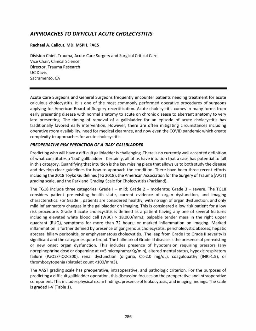
APPROACHES TO DIFFICULT ACUTE CHOLECYSTITIS
Rachael A. Callcut, MD, MSPH, FACS
Division Chief, Trauma, Acute Care Surgery and Surgical Critical Care Vice Chair, Clinical Science Director, Trauma Research UC Davis Sacramento, CA
Acute Care Surgeons and General Surgeons frequently encounter patients needing treatment for acute calculous cholecystitis. It is one of the most commonly performed operative procedures of surgeons applying for American Board of Surgery recertification. Acute cholecystitis comes in many forms from early presenting disease with normal anatomy to acute on chronic disease to aberrant anatomy to very late presenting. The timing of removal of a gallbladder for an episode of acute cholecystitis has traditionally favored early intervention. However, there are often mitigating circumstances including operative room availability, need for medical clearance, and now even the COVID pandemic which create complexity to approaches for acute cholecystitis.
PREOPERATIVE RISK PREDICTION OF A ‘BAD’ GALLBLADDER
Predicting who will have a difficult gallbladder is challenging. There is no currently well accepted definition of what constitutes a ‘bad’ gallbladder. Certainly, all of us have intuition that a case has potential to fall in this category. Quantifying that intuition is the key missing piece that allows us to both study the disease and develop clear guidelines for how to approach the condition. There have been three recent efforts including the 2018 Toyko Guidelines (TG 2018), the American Association for the Surgery of Trauma (AAST) grading scale, and the Parkland Grading Scale for Cholecystitis (Parkland).
The TG18 include three categories: Grade I – mild; Grade 2 – moderate; Grade 3 – severe. The TG18 considers patient pre-existing health state, current evidence of organ dysfunction, and imaging characteristics. For Grade I, patients are considered healthy, with no sign of organ dysfunction, and only mild inflammatory changes in the gallbladder on imaging. This is considered a low risk patient for a low risk procedure. Grade II acute cholecystitis is defined as a patient having any one of several features including elevated white blood cell (WBC) > 18,000/mm3; palpable tender mass in the right upper quadrant (RUQ), symptoms for more than 72 hours; or marked inflammation on imaging. Marked inflammation is further defined by presence of gangrenous cholecystitis, pericholecystic abscess, hepatic abscess, biliary peritonitis, or emphysematous cholecystitis. The leap from Grade I to Grade II severity is significant and the categories quite broad. The hallmark of Grade III disease is the presence of pre-existing or new onset organ dysfunction. This includes presence of hypotension requiring pressors (any norepinephrine dose or dopamine at >=5 micrograms/Kg/min), altered mental status, hypoxic respiratory failure (PaO2/FiO2<300), renal dysfunction (oliguria, Cr>2.0 mg/dL), coagulopathy (INR>1.5), or thrombocytopenia (platelet count <100/mm3).
The AAST grading scale has preoperative, intraoperative, and pathologic criterion. For the purposes of predicting a difficult gallbladder operation, this discussion focuses on the preoperative and intraoperative component. This includes physical exam findings, presence of leukocytosis, and imaging findings. The scale is graded I-V (Table 1).
286

Table I. AAST Preoperative Grading Scale – Acute Cholecystitis
Grade Description Clinical Criteria Imaging Criteria
(CT/US/HIDA findings)
I Acute cholecystitis Right upper quadrant (RUQ) or epigastric pain, Murphy’s Sign, Leukocytosis
Wall thickening; distention; gallstones or sludge; pericholecystic fluid; non-visualization of gallbladder (GB) on hepatobiliary iminodiacetic acid (HIDA) scan
II Gangrenous, empyema, or emphysematous cholecystitis
RUQ or epigastric pain, Murphy’s Sign, Leukocytosis
Above, plus air in GB lumen, wall or in the biliary tree; focal mucosal defects without frank perforation
III Perforation with local contamination
Localized peritonitis in RUQ HIDA with focal transmural defect, extraluminal fluid collection or radiotracer but limited to RUQ
IV Perforation with perichole-cystic abscess or gastrointestinal fistula
Localized peritonitis at multiple locations, abdominal distention with symptoms of bowel obstruction
Abscess in RUQ outside GB; bilio-enteric fistula; gallstone ileus
V Perforation with generalized peritonitis
Above, with generalized peritonitis
Free intra-peritoneal bile
In 2021, a multicenter trial was reported by Schuster et al. demonstrating similar performance to the TG18. Higher grades were associated with longer operative times, but the discriminatory ability between grades was only modest. Most of the 861 patients in the trial were low grade (median 2, IQR 1-2). Overall, the AAST and TG18 scales did not perform as well as the Parkland grading scale for prediction of non-fatal outcomes, complications, conversion to open procedures, and need for subtotal cholecystectomy or fenestrated cholecystectomy or cholecystomy. Although the data suggests that Parkland is superior, this could reflect the relatively low number of patients in the study that had higher grade disease.
Unlike the TG18 and AAST scales, the Parkland Grading Scale was designed to assess only intraoperative features. Thus, its utility is limited for preoperative planning, but can provide guidance for intraoperative decision making when one is faced with a challenging situation intraoperatively.
287

Table II. Comparison of the Intraoperative Grading of the AAST and Parkland Severity Scales
TIMING OF CHOLECYSTECTOMY
Although debated for many years, there have been a number of studies supporting earlier cholecystectomy as superior to delayed operative intervention. However, the definition of early differs in studies. Most often this is quantified as undergoing cholecystectomy in 72 hours or less from onset of symptoms. Reaching that benchmark can actually be unrealistic at times when combining factors including delayed patient presentations, operative availability for urgent, but not emergent cases, and need for preoperative optimization. Operating after 72 hours is not atypical and can be performed safely. In fact, albeit a single center study, there has been one randomized, controlled trial that compared delayed cholecystectomy after 6 weeks of antibiotics to cholecystectomy on or after 72 hours. The cholecystectomy during the incident hospitalization was still superior.
DECISION TO OPERATE
Although removal of the gallbladder is the standard recommendation for management of calculous cholecystitis, there are circumstances where antibiotics or cholecystostomy tube are the best alternatives. It is quite common now to see patients suffering acute cholecystitis who have one or more significant underlying comorbidities. This has become even more common as with the growth in the population to over 46 million aged 65 years and older. This population is expected to more than double over the next several decades. Additionally, the rate of obesity in the US has skyrocketed with more than 40% of the population with BMIs above 30. Obesity clusters with a multitude of associated comorbidities which can complicate perioperative care.
Historically, patients needing source control who were not felt to be well enough to undergo an operation have been treated with cholecystostomy tubes as initial therapy. The procedure is safe, easy to perform, and can even been done bedside in critically ill patients. It is well known in the literature that cholecystostomy tubes can be associated with need for readmission, repeat procedures for tube dislodgement, and can make subsequent cholecystectomy more challenging. However, for patients who are critically ill, suffering from
Grade Description AAST Intraoperative Criteria Parkland Intraoperative Criteria
I Acute cholecystitis Inflammatory changes localized to GB; wall thickening; distention; gallstones
Normal appearing gallbladder without adhesions present
II Gangrenous, empyema, or emphysematous cholecystitis
Distended GB with pus or hydrops; necrosis or gangrene of wall; not perforated
Minor adhesions at neck, otherwise normal gallbladder.
III Perforation with local contamination
Perforated GB wall (non-iatrogenic) with bile outside the GB but limited to RUQ
Presence of any of the following: hyperemia, pericholecystic fluid, adhesions to the body, distended gallbladder
IV Perforation with perichole-cystic abscess or gastrointestinal fistula
Pericholecystic abscess; bilio-enteric fistula; gallstone ileus
Presence of any of the following: adhesions obscuring majority of gallbladder, grade I-III with abnormal liver anatomy, intrahepatic gallbladder or impacted stone (Mirrizi)
V Perforation with generalized peritonitis
Above, plus generalized peritonitis
Presence of any of the following: perforation, necrosis, inability to visualize the gallbladder due to adhesions
288

severe congestive heart failure, multi-organ failure, cirrhosis, or other unrelated illness increasing their fraility, this may be the best immediate approach to attempt source control.
Aroori et al. in 2019 study 53 patients who were considered high risk either due to comorbidities or were judged too unstable for surgery. These patients underwent percutaneous cholecystostomy placement. Consistent with the prior literature, readmission rate was about 18%. Only 45% of the patients ultimately received an operative procedure less than half of those being complete laparoscopically. The remainder were either lap converted to open procedures or open procedures. Ostapenko et al. reported in 2021 that from 2014-2019 the rate of percutaneous cholecystostomy had increased significantly.
The choice between percutaneous cholecystostomy and emergent cholecystectomy can be challenging. A recent meta-analysis by Huang et al. examined 8960 patients from 6 studies including high-risk surgical candidates. They concluded that emergent cholecystectomy appeared superior to percutaneous cholecystectomy, however, all studies have significant treatment bias. It is well known that a fairly significant portion of patients undergoing percutaneous cholecystostomy never subsequently have a cholecystectomy. Upwards of 10-20% of the patients do not survive initial hospitalization who receive a percutaneous cholecystectomy, but this likely reflects the selection bias in those that were far too ill to even be offered a surgical procedure. Recent work by D’Acapito et al. published in 2021 advocated for the use of the American College of Surgeons National Surgical Quality Improvement Program (ACS-NSQIP) risk calculator to assist in decision making in octogenarian patients. In general, the ACS-NSQIP calculator is an important tool that can help to quantify the potential outcomes given underlying comorbidities to assist in a decision regarding immediate operative therapy versus a percutaneous treatment.
OPERATIVE APPROACH
An initial approach laparoscopically is recommended in almost all cases where there is preoperative concern regarding a potentially difficult cholecystectomy. Cholecystectomy can be challenging either due to anatomic variants, profound inflammation, reoperative surgery with significant adhesive disease, or factors like coagulopathy due to underlying liver disease or systemic illness. Each difficult gallbladder encountered operatively requires careful decision making to insure a safe outcome. In addition to the well accepted option of intraoperative cholangiograms to define anatomy, conversion to an open procedure should not be viewed as a complication. There are also occasions that performing only a lap assisted cholecystostomy tube or determining it is not safe to proceed is necessary. Never lose sight of the overall goal of the operation to do what is safest for the patient.
Buhavac et al. have advocated that a ‘bailout’ procedure be considered when the critical view of safety is not achievable. They favor a laparoscopic subtotal cholecystectomy citing a shorter length of stay and lower morbidity rate than conversion to an open procedure. They also note an infrequent rate of recurrent symptoms given the bulk of the gallbladder is removed. Data supporting this include several recent reports including one of 168 patients undergoing a subtotal approach that demonstrated a 7% postop complication rate and a relatively short length of stay of 4 days. They argue further support has come from a meta-analysis by Elshaer et al. that the morbidity of subtotal cholecystectomy was equivalent to total cholecystectomy. Finally, Ibrahim et al. just reported in late 2021 on a retrospective series of 5664 patients undergoing cholecystectomy at a single institution. Subtotal cholecystectomy was needed in 1.7% and 49% of these cases required conversion to open. There was a high postoperative biliary leak rate of 19.6%, but no common bile duct injuries. Readmissions were also seen in 8% of the patients. Importantly, in these studies, common bile duct injury was not found.
SUBTOTAL CHOLECYSTECTOMY (SC)
There are two options for performing a subtotal cholecystectomy. One is a nearly complete removal of the gallbladder to the level of just above the infundibulum. This is known as a reconstituted SC and
289

requires circumferential dissection in a safe plane. The gallbladder is then transected most commonly with an endoGIA stapler. This avoids a deep dissection into the hepaticocystic triangle preventing iatrogenic injury to the common bile duct or hepatic artery. This strategy works particularly well when there are dense adhesions and severe inflammation at the infundibulum of the gallbladder.
The other type is a fenestrated SC. The gallbladder is typically opened, emptied of debris, and some of the posterior wall left adherent to the gallbladder fossa. The ideal dissection is down to the level of the infundibulum leaving just a small rim of the tissue posteriorly. Care should be taken to retrieve any spilled stones. This works well in cases where the gallbladder/gallbladder fossa plan is densely adherent or when there is aberrant anatomy coursing close to the back side of the gallbladder. Several authors describe placing a free omental patch on the cystic duct/infundibulum to close the cystic duct opening. It is also recommended to leave a drain for a fenestrated SC as biliary leak is a common occurrence. Often these patients require a postoperative endoscopic retrograde cholangiopancreatography and stent.
REFERENCES
1. Aroori S, Mangan C, Reza L, Gafoor N. Percutaneous Cholecystostomy for Severe Acute Cholecystitis: A Useful Procedure in High-Risk Surgical Patients for Surgery. Scan J Surg. 2019 Jun. 108(2):124-129. PMID 30227774
2. Buhavac M, Elsaadi A, Dissanaike S. The Bad Gallbladder. Surg Clin N Am. 101 (2021) 1053-1065. 3. D’Acapito F, Cucchetti A, Di Pietrantonio D, Tauceri F, Mirarchi MT, et al. Forecasting outcomes
after cholecystectomy in octogenarian patients. Surg Endoscopy. (2021) Oct 25. Doi: 10.1007/s00464-021-08752-z. Online ahead of print. PMID 3465492
4. Hadaya J, Sanaiha Y, Juillard C, Benharash P. Impact of frailty on clinical outcomes and resource use following emergency general surgery in the United States. PLoS One. 2021 Jul 23; 16(7):e0255122. PMID 34297772
5. Huan H, Zhang H, Yang D, Wang W, Zhang X. Percutaneous cholecystectomy versus emergency cholecystectomy for the treatment of acute calculous choleycystitis in high-risk surgical patients: a meta-analysis and systematic review. Updates Surg. 2021 May 15. Online ahead of print. PMID 33991327
6. Huang R, Patel DC, Kallini JR, Wachsman AM, Van Allan RJ, Marguilles DR, Phillips EH, Barmparas G. J Surg Research. 2022 Feb; 270:405-412. PMID 34749121
7. Ibrahim R, Abdalkoddus M, Mahendran B, Mownah OA, Nawara H, Aroori S. Subtotal cholecystectomy: is it a safe option for difficult gall bladders? Ann R Coll Surg Engl. 2021 Nov 25. Online ahead of print. PMID 35821508
8. Strasberg SM, Pucci MJ, Brunt LM, Deziel DJ. Subtotal Cholecystectomy – “Fenestrating” vs. “Reconstituting” Subtypes and the Prevention of Bile Duct Injury. JACS. 2016. 222(1):89-96.
9. Ostapenko A, Liechty S, Manuia E, Stroever S, Casasanta M, Kleiner D. The Rise and Fall of
Percutaneous Cholecystostomy in a Community Hospital: Is it Possible to Turn the Tide of History?
J Gastrointest Surg. 2021 Sep 20. Online ahead of print. PMID 34545546
290

ESOPHAGEAL INJURIES
Kenji Inaba, MD, FRCSC, FACS
Professor and Vice Chair of Surgery Director, General Surgery Program Chief, Trauma, Emergency Surgery and Surgical Critical Care LAC+USC Medical Center & University of Southern California – Los Angeles Los Angeles, CA
The esophagus can be divided into three functional areas, the cervical esophagus, the thoracic segment, and the GE junction. The primary focus of this talk will be on injuries to the intrathoracic portion of the esophagus. The cervical esophagus is best placed in context with the diagnostic workup and management of injuries to the neck. Injuries to the GE junction are generally worked up and treated as part of the intra-abdominal contents.
Injuries to the esophagus are relatively rare, due to its protected location within the thoracic cavity. The majority of injuries occur secondary to penetrating trauma, and, again, due to its location, predominately because of ballistic injury. Lying posterior to the trachea, on the thoracic spine, it runs the length of the chest starting at the level of C6, descending to the right of the aorta until it moves anterior to the aorta as it enters the diaphragmatic hiatus at T11. The azygos vein and hemiazygos vein are in close proximity to the esophagus, with the former lying to the right of the esophagus and the latter crossing from left to right to drain into the azygous vein. The thoracic duct is found between the aorta and esophagus, enroute to drain into the left subclavian vein. The esophagus itself is a muscular tube, without a serosal layer.
DIAGNOSIS
The diagnosis of an acute injury to the esophagus is typically made on cross sectional imaging during the initial diagnostic workup of the patient. On the CT scan, the trajectory of the ballistic injury can be traced through the chest wall, soft tissues, and across the mediastinal structures. While the actual disruption of the esophageal wall may not be clearly seen, the injury can be inferred by the proximity of the missile tract to the esophagus. Confirmation of the injury can be achieved using contrast, starting with a water soluble medium, or, with EGD. Delineation of the level of injury is important for pre-operative planning, and the laterality of the surgical approach. For patients who went directly to the operating room prior to obtaining imaging, undergoing thoracotomy for other indications such as bleeding, careful inspection of the pleura overlying the esophagus and mediastinal structures is critical. Any violation or collateral evidence of injury in the form of bruising or a hematoma requires that the underlying structures are exposed in order to rule out injury.
MANAGEMENT
Esophageal injuries require operative repair in general. As there is no serosal layer to help contain the esophageal contents, and with a segmental blood supply without a standard mesentery, perforation usually results in the rapid leak of contents into the mediastinum and pleural cavity. The role of non-operative options such as esophageal stenting remains highly controversial for acute injuries. This treatment option has grown in popularity for non-traumatic perforation, however, there is insufficient evidence to support its routine use in trauma. This may be a viable option for patients who are at high
291

operative risk, when combined with drainage. In general, for the otherwise healthy patient with an acute injury, open repair remains the current standard of care.
The patient will require general anesthetic with single lung ventilation. For upper and mid-esophageal injuries, access will be through a right thoracotomy at the fifth or sixth intercostal space and for lower injuries, a left thoracotomy at the seventh or eighth intercostal space.
Once the thoracotomy has been performed, and the lung has been medialized, the mediastinal pleura should be inspected carefully for violation or underlying hematoma to localize the injury. the azygos vein may be ligated if required. To facilitate characterization of the injury, including any back wall injuries, a sufficient length of peritoneum should be incised, and the esophagus encircled with a Penrose drain. Care must be taken to not devitalize the esophagus. Once the hole(s) has been found, the muscle fibers may need to be opened longitudinally to discern the mucosal edges, which may have retracted. Any devitalized tissue, especially after ballistic injury should be debrided. In general, for acute traumatic injuries, primary repair of the injury should be feasible without the risk of narrowing the lumen. The mucosal layer should be closed with absorbable sutures, with an overlying muscle layer closure on top of this. While not routinely required, an esophageal bougie may be used to ensure there is no narrowing of the lumen. When possible, especially with a destructive injury or in cases where there is an associated tracheal injury, a buttress of tissue from the pleura or pericardium or intercostal muscle may be used to cover the repair.
For acute injuries, especially penetrating injuries diagnosed and repaired immediately, surgical feeding access distal to the repair should not be required. A nasogastric tube may be inserted past the injury for temporary drainage in the immediate post-operative period. The pleural cavity should be drained with chest tubes. Likewise, in general, there is no need for routine post-operative imaging prior to the initiation of oral feeding.
In the damage control setting, for most small holes, closure over an externally drained t-tube will temporize the injury. In the extremely rare situation where there is complete transection, or massive disruption of the esophagus, temporizing with tube drainage would be the preferred option, to not sacrifice additional esophageal length, allowing for all the surgical options for achieving continuity once the patient has been adequately resuscitated.
CONCLUSIONS
Esophageal injuries remain rare. They are often the result of ballistic injury, with the diagnosis made on CT. Defining the location of injury is a key component of the pre-operative planning, and the surgical approach. The principles of repair include a mucosal and muscular closure, with a tissue buttress when appropriate. Care must be taken to ensure all injuries are detected and repaired.
REFERENCES
1. Aiolfi, Alberto et al.; Non-iatrogenic esophageal injury: a retrospective analysis from the National 2. Trauma Data Bank. World Journal of Emergency Surgery (2017) 12(19):1 – 7. 3. Ali, J.T. et al.; Perforated esophageal intervention focus (PERF) study: a multi-center examination
of contemporary treatment. Diseases of the Esophagus (2017) 30:1–8. 4. Biffl, Walter L. et al.; Western Trauma Association Critical Decisions in Trauma: Diagnosis and
management of esophageal injuries. J Trauma Acute Care Surgery (2015) 79(6):1089 – 1095. 5. Bryant, Ayesha S. et al.; Esophageal Trauma. Thoracic Surgery Clinics (2007) 17:63 – 72. 6. Castater, Christine et al.; Approaches to Repair of Penetrating Injuries of the Proximal, Mid, and
Distal 7. Esophagus. The American Surgeon (2021) 0(0):1–3.
292

8. Patel, Madkukar S. et al.; Penetrating oesophageal injury: A contemporary analysis of the National Trauma Data Bank. Injury (2013) 44:48-55.
9. Petrone, Patrizio et al.; Management of esophageal injuries secondary to trauma. Injury (2017) 48:1735-1742.
10. Raff, Lauren A. et al.; Contemporary management of traumatic cervical and thoracic esophageal perforation: The results of an Eastern Association for the Surgery of Trauma multi-institutional study. J Trauma Acute Care Surg (2020) 89(4):691 – 697.
11. Schellenberg, Morgan et al.; Defining the gastroesophageal junction in trauma: Epidemiology and management of a challenging injury. J Trauma Acute Care Surgery (2017) 83(5):798 – 802.
293

STAND AND DELIVER: PREGNANT PATIENT EMERGENCIES
Carlos V.R. Brown, MD, FACS
Professor of Surgery Chief, Division of Acute Care Surgery Dell Medical School University of Texas at Austin Austin, TX
The physiologic changes that comprise maternal adaptation to pregnancy involve almost every organ system. The plasma volume in pregnancy increases by almost 50% whereas the red cell mass increases by only 20%, resulting in the physiologic anemia of pregnancy. It is not uncommon to see a baseline hematocrit of 31%-33%. The increased plasma volume allows the pregnant patient to withstand a significant amount of blood loss before any overt manifestations of shock appear. Cardiac output increases beginning in the second trimester. Uterine blood flow also increases as the fetus grows, comprising 20% of cardiac output by term. This system is highly regulated and extremely sensitive to external agents such as catecholamine and maternal intravascular volume loss. Maternal hemorrhage can be compensated by decreased uterine flow. Maternal hypovolemia may be marked by fetal distress before any evidence of maternal tachycardia or hypotension is present.
Oxygen consumption and resting ventilation increase in pregnancy as a result of an increase in tidal volume caused by rising progesterone levels. This results in a respiratory alkalosis with a pC02 of approximately 30 mm Hg and a metabolic compensation with bicarbonate levels in the 19-20 mEq/L range. Gastrointestinal motility is decreased, and in addition to the reduction in resting lower esophageal pressure, pregnant patients are more likely to experience gastroesophageal reflux and have an increased risk of aspiration with general anesthesia and trauma.
Anatomically, the uterus becomes an intra-abdominal organ at approximately 12 weeks of gestation. At 20 weeks the uterus can be palpated at the umbilicus and by 36 weeks the uterus reaches the costal margin. The growing uterus can complicate invasive procedures such as laparoscopic access of the peritoneum. As the uterus enlarges, maternal organs are displaced upwards; in the late stages of pregnancy the majority of the gastrointestinal tract may be found above the inferior costal margins, actually protecting these organs with respect to penetrating injuries. The diaphragm may also be elevated by as much as 4 cm. Finally, as the pregnancy progresses, uterine compression of the vena cava decreases venous return, resulting in a 30% drop in cardiac output. This “supine hypotensive syndrome” can be alleviated by displacing the uterus from the vena cava by positioning the patient in the left lateral decubitus position. In a patient with suspected spinal injury, manual displacement of the uterus to the
left or placement of the spinal board at a 15 angle are alternatives.
TRAUMA
Trauma occurs in approximately 5% to 7% of all pregnancies. The most common mechanism involves motor vehicle collisions followed by falls and then assaults. The majority of pregnant patients involved in trauma are either uninjured or minimally injured and are often admitted only because of the pregnancy. Despite the lack of injury severity, fetal outcomes are worsened in the presence of any trauma. Pregnant patients who present in maternal or fetal distress need prompt intervention to ensure optimal outcomes.
294

The pregnant trauma patient should be managed following the same priorities as any trauma patient. This includes performing a primary and secondary survey and performing adjunctive procedures and diagnostic tests as indicated. However, the injured pregnant patient presents a more challenging situation due to changes in physiology and the complexities involved with the maternal-fetal relationship.
Evaluation and management of the pregnant patient who has sustained blunt trauma should follow the guidelines given for non-pregnant patients. Hemodynamically unstable patients with evidence if intra-abdominal injury and patients with signs of peritonitis must undergo prompt laparotomy. For hemodynamically stable patients, when deciding on the appropriate workup for any pregnant patient, always remember the principle that a pregnant patient should not be penalized for being pregnant. In other words, pregnant patients should have all the necessary tests and procedures performed. The American College of Obstetricians and Gynecologists states that exposure of less than 5 rads has not been associated with an increase in fetal anomalies or pregnancy loss. There are concerns about exposure in the 5 to 10 rads range; however, serious risk to the fetus is not known to occur until the absorbed dose reaches 10 rads. A typical “PanScan” (helical CT of the head, c-spine, chest, abdomen and pelvis) delivers approximately 5 rads. An abdominal CT scan can be performed to evaluate abdominal pathology with only 0.3 rads. Fetal monitoring should be performed for all cases after 24 weeks gestation. If the mother has no indication for surgery, and the fetus has evidence of distress, then the patient should undergo a prompt cesarean section again to ensure optimal outcomes for the fetus. Salvageable infants, defined by the presence of fetal heart tones and a gestational age of 26 weeks, have a survival rate of 75% after cesarean sections after trauma.
Penetrating injuries in the pregnant trauma patient are managed in the same manner as in non-pregnant patients. Since the abdominal organs tend to be displaced in the pregnant patient, the fetus may be more likely to sustain significant injury than the mother with penetrating injuries. The indications for laparotomy remain the same for pregnant patients as for non-pregnant patients. The decision for cesarean section follows that of blunt trauma mentioned above.
Patients presenting in the peri-mortem state with a viable fetus are rare. In one of the largest series of traumatic cesarean sections, only 3 peri-mortem sections were identified out of 114,952 trauma admissions. In a review of 61 children who survived peri-mortem cesarean section, the best maternal and fetal outcomes were obtained if section was performed within 4 minutes of maternal cardiac arrest. Other series report that the best outcomes are achieved if cesarean section is performed within 5 minutes of cessation of maternal circulation. There are case reports that document fetal survival following 45 minutes of maternal cardiopulmonary resuscitation. Interestingly, there are reports of maternal survival of cardiac arrest after emergent cesarean section. It has been suggested that the increased venous return and elimination of the low resistance utero-placental circulation may be responsible for the maternal survival. It has also been suggested that peri-mortem cesarean section should be considered as a component of maternal resuscitation that may optimize outcomes in both mother and fetus.
APPENDICITIS
Acute appendicitis is the most common general surgical disease during pregnancy and can affect 0.5-2 out of every 1000 pregnant patients. Classic history and physical exam findings may be obscured due to pregnancy. Pain and point tenderness in the right lower quadrant are the most common findings in the non-pregnant patient. However, as pregnancy progresses and the uterus displaces the normal location of the appendix, this pain and tenderness may migrate to the right flank, right upper quadrant, or even the back. Anorexia and vomiting may still occur in the pregnant patient with appendicitis, but these are also common complaints in the pregnant patient without appendicitis, further confusing the presentation. The most reliable lab test in acute appendicitis is leukocytosis, but this is also unreliable in the pregnant patient due to the leukocytosis of pregnancy.
295

Just as in the non-pregnant female with appendicitis, additional imaging is needed to confirm or rule out appendicitis in the pregnant patient. However, while CT scan is the test of choice in the non-pregnant patient, ultrasound and MRI are the first line imaging studies in pregnancy to avoid unnecessary radiation to the fetus. During the first two trimesters, ultrasound may be used to diagnose appendicitis. In the last trimester, or if ultrasound is non-diagnostic, MRI should be used for definitive diagnosis as it is highly sensitive and specific.
Once the diagnosis of appendicitis is made in the pregnant patient, the treatment is antibiotics and appendectomy. The is no role for nonoperative management of appendicitis in pregnancy. Laparoscopy is safe in all trimesters of pregnancy, and laparoscopic appendectomy is the preferred approach for the pregnant patient. Port placement may be altered with the enlarging uterus, and peritoneal access should be performed in an open fashion. If for some reason an open appendectomy is necessary, the location of the appendix may change due to uterine displacement later in pregnancy, so the open incision should be centered over the area of greatest tenderness.
BILIARY DISEASE
Biliary disease is the second most common general surgical problem presenting during pregnancy. Pregnant patients should present with history and physical exam findings similar to their non-pregnant counterparts. With the exception of leukocytosis, laboratory evaluation for the pregnant patient with biliary disease is similar to the non-pregnant patient, with electrolytes and liver function tests serving as the initial tests of choice. Ultrasound is likewise the diagnostic test of choice for patients with biliary disease in the pregnant and non-pregnant patient.
Patients with a single episode of biliary colic may be observed and cholecystectomy deferred until after delivery. However, if symptoms are persistent or severe, laparoscopic cholecystectomy should be performed. Any patients with a diagnosis of acute cholecystitis should undergo laparoscopic cholecystectomy, regardless of trimester. Patients with choledocholithiasis can be managed in the same way as the non-pregnant patient, with either laparoscopic cholecystectomy followed by ERCP or ERCP followed by cholecystectomy. The fetus should be shielded during cholangiography or ERCP. Patients with gallstone pancreatitis should be admitted to the hospital and should undergo laparoscopic cholecystectomy during the same hospitalization, once the abdominal pain has resolved.
REFERENCES
1. Brooks DC. Gallstone disease in pregnancy. UpToDate, eds Ashley SW, Berghella V. Accessed January 2021.
2. Kirkpatrick SJ. Initial evaluation and management of major trauma in pregnancy. UpToDate, eds Loockwood CJ, Manaker S, Moreira ME. Accessed January 2021.
3. Nirula R, Buczek R, Buhavac M. Non-obstetric emergency surgery in the pregnant patient. In Emergency General Surgery: A practical approach. Eds Brown CVR, Inaba K, Martin M, Salim A. Springer international publishing 2019.
4. Rebarber A, Jacob BP. Acute appendicitis in pregnancy. UpToDate, eds Lockwood CJ, Levine D, Weiser M. Accessed January 2021.
296

NIGHTMARE HERNIA - DREAM OUTCOMES
Chadwick P. Smith MD, FACS
Director, Surgical Intensive Care Units Program Director, Surgical Critical Care Orlando Regional Medical Center Orlando, FL
VENTRAL HERNIA / COMPLEX ABDOMINAL WALL DEFECTS
• Significant Challenge to the General Surgeon • Progressive Worsening Quality of Life • Substantial Complications • Psychological Effects
o Lower body self-Image o Detriment to mental health o Less sexually active
• Incisional Hernias o Most common complication o 4 million Laparotomies in US
• Abdominal Wall Defects o Trauma o Surgery o Congenital
• 65% Experts Surveyed • Hernia Volumes > 30% of abdominal contents • Loss of Domain • Size not the only factor • Location • Depth • Tissue condition • Physiologic factors • Evolution of Management
o Primary repair ▪ Initial therapy ▪ Up to 50% recurrence rate
o Tension Free repair ▪ Prosthetic Mesh ▪ 1950s ▪ Became the gold standard
• Mesh utilization o Improved recurrence rates o Mesh related complications
• Laparoscopy o Initially in 1993 o Reduced stay
297

o Faster recovery o Decreased recurrence rates
VENTRAL HERNIA / COMPLEX ABDOMINAL WALL DEFECTS
• Characteristics o Loss of domain o Previous mesh placement o Extensive surgical history o Expansion of open primary repair o Myofascial advancement o “Component Separation” o Ramirez, et al 1990
• Preoperative Evaluation o Goal Alignment o Patient’s and Surgeon’s o Physiologic Optimization o Thorough H&P o Size, Location o Skin Changes o Prior incisions o Standing and Supine o Valsalva
• Examination o Inability to Reduce
▪ Suspect Loss of Domain • Surgical History
o Previous hernia repair ▪ Independent risk factor
o Indwelling mesh
PREOPERATIVE OPTIMIZATION
• Risk Factors o Coronary Disease o COPD o Steroid Use o Low Preop Albumin levels o Smoking => 4 Weeks o Glycemic Control
▪ Hb A1 C < 7.0% ▪ Glucose < 110mg/dL
298

Condition Recommendation
Diabetes Mellitus Hb A1C < 7% glucose 140-160mg/dL
Smoking Cessation 4 weeks Prior
Obesity BMI > 50kg Not recommended BMI> 45 consider Bariatric Referral BMI > 30 Weight loss and diet counseling
Malnutrition Albumin > 3gm/dL , Nutritional Beverage
COPD Bronchodilator / Pulmonary Consultation
MRSA Preop Screening
Cardiac History Cardiology Consultation / OSA Screening
IMAGING
• Computed Tomography o Define musculature integrity o Assess defect relationship o Ideal approach o Morbidly obese
▪ Limited examination o Loss of domain o Location near bony structures o Fluid cavities / Inflammation o Occult hernias
REPAIR OPTIONS
• Rives-Stoppa – 1980s o Retro-rectus dissection plane o Avoids subcutaneous flaps o Mesh placed retro-muscularly o Recurrence Rates - 3-6% o Maintains functional integrity o Limited lateral dissection
▪ Non-midline defects ▪ Large defects
299

• Anterior Component Separation (1990) o Dissection anterior to the rectus o Subcutaneous Plane o Incision of external oblique fascia
▪ Just lateral to rectus muscle ▪ May extend to anterior axillary line
o Extensive medial mobilization o High Wound complications
▪ Ischemic tissue ▪ Dead space
300
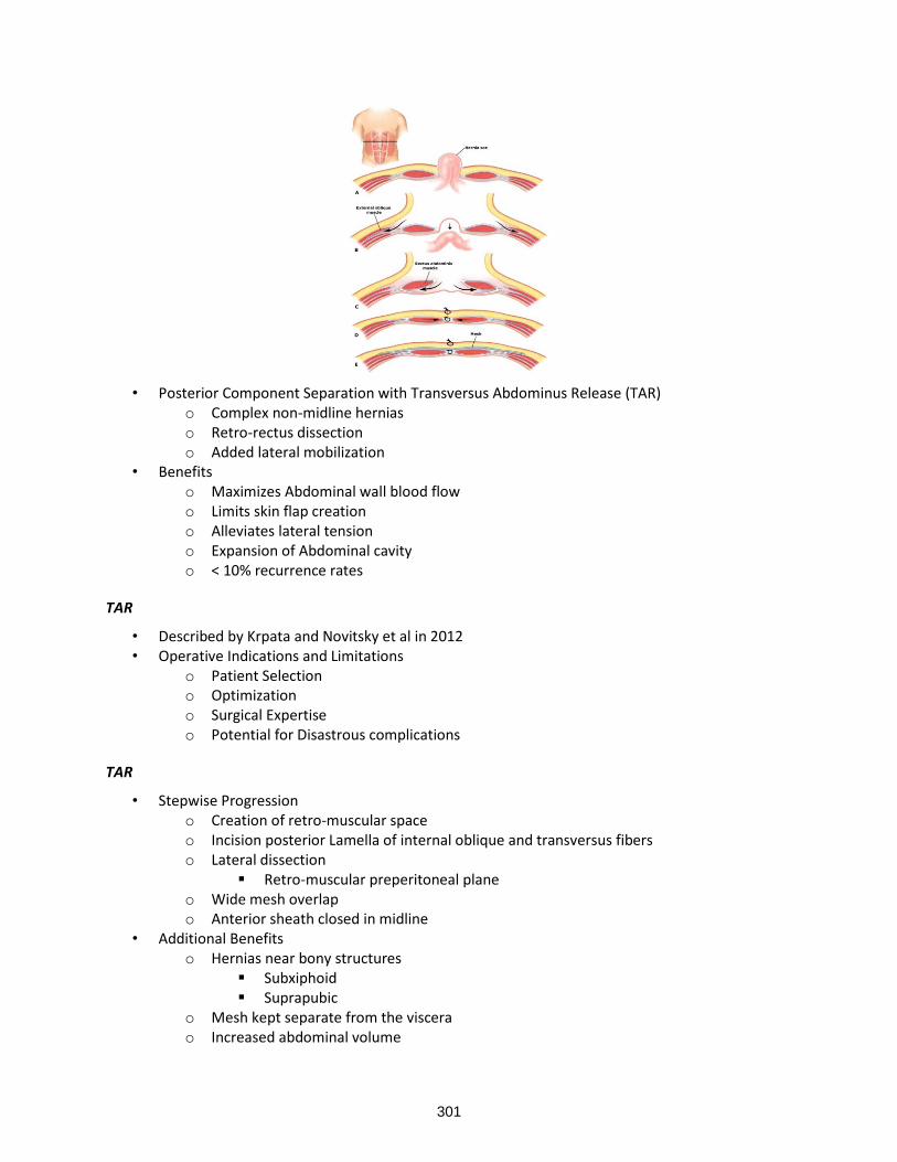
• Posterior Component Separation with Transversus Abdominus Release (TAR) o Complex non-midline hernias o Retro-rectus dissection o Added lateral mobilization
• Benefits o Maximizes Abdominal wall blood flow o Limits skin flap creation o Alleviates lateral tension o Expansion of Abdominal cavity o < 10% recurrence rates
TAR
• Described by Krpata and Novitsky et al in 2012 • Operative Indications and Limitations
o Patient Selection o Optimization o Surgical Expertise o Potential for Disastrous complications
TAR
• Stepwise Progression o Creation of retro-muscular space o Incision posterior Lamella of internal oblique and transversus fibers o Lateral dissection
▪ Retro-muscular preperitoneal plane o Wide mesh overlap o Anterior sheath closed in midline
• Additional Benefits o Hernias near bony structures
▪ Subxiphoid ▪ Suprapubic
o Mesh kept separate from the viscera o Increased abdominal volume
301
• Who needs one??? o Ventral Hernias with > 10cm cross sectional width o Smaller Defects o Chronicity o Rectus retraction o Retro-rectus closure under tension o Lateral defects o Prior subcostal incisions
• Who doesn’t need a TAR ??? o Defects < 10 cm with anticipated ability to close fascia o High risk for wound morbidity
▪ Deferral to another time ▪ EC fistula / ostomy closure
o Malnutrition o Non-elective cases
▪ 25% wound morbidity • Compared with 15-17%
TAR – ERROR TRAPS
• Abdominal Entry o Unsafe access o Inadequate adhesiolysis o Transversalis and peritoneal injury
• Retro-rectus dissection o Linea alba disruption
▪ “stay in the middle!!” o Linea alba misidentification o Inferior epigastric injury
▪ Ischemia/atrophy o Intercostal neurovascular injury
▪ Loss of domain o Inferior epigastric origin injury
▪ Hemorrhage • Transversus Abdominus Release
o Improper starting location ▪ Start in upper third of abdomen
o Neurovascular injury o Linea semilunaris injury
▪ Iatrogenic spigelian hernia o Improper traction/countertraction o Inadequate release
▪ Extend to psoas if needed • Posterior layer reconstruction
o Failure to close fenestrations • Intraoperative TAP Block
o Hypotension can occur o Ensure anesthesia is aware of TAP performance
302

• Mesh Placement o Wrinkled mesh
▪ Ingrowth restriction ▪ Seromas
• Anterior Closure o Failure to place drains
• Potential pitfalls o Deep surgical site infections o Mesh Fractures o Linea semilunaris injuries o Posterior sheath breakdown
• Linea Semilunaris Injury o Devastating injury o Failure to recognize neurovascular bundles o Medial retraction of the linea semilunaris
▪ Improper incision line o Iatrogenic Spigelian hernia o NEVER able to achieve “normal” abdominal contour
• TAR Posterior Sheath breakdown o Tension on the posterior sheath o Bridge o Hernia Sac o Omentum o Vicryl Mesh
Mesh Selection
• Surgeons Choice • Light or medium weight polypropylene • Macroporous polypropylene • Biologic biosynthetic not typically used • Technique
o Transfascial fixation o Perimeter of mesh o Overlap o 5 cm? At least
OUTCOMES
ACS VS. TAR
• Americas Hernia Society Quality Collaborative (AHSQC) o Database query o 3610 Patients o 501 External oblique release (EOR)
▪ 70 surgeons - 50 institutions o 3109 TAR
▪ 124 surgeons - 89 institutions
303

ACS VS. TAR
• Post op results o No difference in post -op complications o No difference in recurrence o No difference in quality of life o No mortality difference o No 30-day SSI rate difference o SSO TAR 13% EOR 20% (p < 0.05)
▪ No increased procedure intervention • Author Conclusions
o Equivalent outcomes can be achieved using EOR or TAR in open repair of incisional hernias • Meta-Analysis
o 7 Studies 281 cases of TAR o 6 Studies 285 cases ACS o Recurrence
▪ TAR 5.7% vs. ACS 9.5% p=0.23 o Bridging Mesh
▪ TAR 3.1% vs. ACS 7.5% p=0.22 o Wound Complication
▪ Superficial TAR 10.9% vs. ACS 21.6% p=0.15 ▪ Deep TAR 9.5% vs. ACS 12.7% p=0.53
• Author Conclusions o TAR and ACS have comparable outcomes
• Posterior and open anterior components separations: a comparative analysis o 111 patients o 56 ACS vs. 55 TAR o Mean Defect size
▪ ACS 472 cm2 vs. TAR 531 cm2 p = 0.28 o Wound complications
▪ ACS 48.2 % vs. TAR 25.5% p = 0.01 o Recurrence Rate
▪ ACS 14.3 % vs. 3.6% p = 0.09 o Conclusions
▪ TAR provides equivalent myofascial advancement with significantly less wound morbidity when compared with ACS.
304

MOC
• Of the Choices below, which is NOT associated with transversus abdominus release and mesh implantation with complex hernia repair.
o A: increased subcutaneous flap wound complications o B: lower risk of hernia recurrence o C: increased ability to close larger hernia defects o D: advantages in repairing non midline defects
Minimally Invasive Surgery
• Single Dock Technique o Extended totally
extra-peritoneal plane o ETEP o Cross midline
extra-peritoneally o Mesh totally extraperitoneal
ROBOTIC VS. OPEN RETRO-MUSCULAR REPAIR BITTNER ET AL
• Comparative Analysis – Single Center o o-TAR 76 patients o r-TAR 26 patients o Similar demographics
▪ Diabetes o-TAR 23% r-TAR 0 % p=0.01 o Hernia Size oTAR 260 cm2 r –TAR 235 cm2 p=0.55 o Midline defect and recurrence similar o Operative Time
▪ o-TAR 287 min vs r-TAR 365 min p<0.01 o Morbidity
▪ o-TAR 39.2% vs r-TAR 19.2% p=0.09 o Readmission
▪ o-TAR 6.6% vs. r-TAR 7.7% p=1.00
305

o Length of Stay ▪ o-TAR 6 days vs. r-TAR 3 days 95% CI 3.2-4.3
• Author Conclusions o Robotic TAR is a safe minimally invasive option for complex abdominal wall
reconstruction o Short term benefits of low morbidity and reduced length of stay
ROBOTIC VS. OPEN RETRO-MUSCULAR REPAIR NOVITSKY ET AL
• Comparative analysis o 38 r-TAR o 76 o-TAR o Similar demographics o Operative Time
▪ r-TAR 299 min vs. o-TAR 211 min p < 0.001 o Blood loss
▪ r-TAR 49ml vs. o-TAR 139ml p<0.001 o Systemic Complications
▪ r-TAR 0 vs. O-TAR 17.1% p=0.026 o Hospital LOS
▪ r-TAR 1.3 days vs. 6.0 days p<0.001 • Author Conclusions
o r-TAR associated with longer OR times and offers decreased blood loss, fewer systemic complications, shorter LOS, and eliminated readmissions.
ROBOTIC VS. OPEN REPAIR TRENDS IN DATA
• Systematic Review • 25 studies
o 3 RCT’s o 22 observational studies
• Robotic VHR o Longer OR times o Less transfusions o Lower systemic complications o Shorter LOS o Similar cost to open repair
COST OF ROBOTIC ASSISTED REPAIR
• Comparison o 26 patients undergoing TAR
▪ 10 open ▪ 16 robotic
o Similar demographics o OR time rTAR 253 min vs. oTAR 211.5 min p=0.0322 o LOS rTAR 4.5 days vs oTAR 12.5 days p<0.005 o Procedure costs
▪ 2.7 X higher with robotic repair (€ 5397 vs. € 1989) o Inpatient stay
306

▪ 60% lower € 2715 vs. € 6663) o Profit (revenue for national insurance account € 9577)
▪ Robotic € 1465 vs. Open € 925
MIST
• Retro-rectus repair o Low recurrence rates o Closure of larger defects
▪ Addition of TAR allows even larger closure o Non-midline defects o Robotic repair feasible o Steep learning curve
• Requires dedication and interest in this niche
CHEMICAL COMPONENT SEPARATION
• Botulinum A toxin o Clostridium botulinum o Functional denervation within 2 days => 4-6 weeks
• 2009 Ibarra-Hurtado o Temporary paralysis lateral abdominal wall musculature
• 300 – 500 U BTA o Divided bilaterally o 3 – 5 injections per side o Ultrasound guidance
▪ Injection into muscle bellies • External, Internal Obliques, Transversalis
• Effects o Decrease in hernia size o Thinning of abdominal wall musculature o Lengthening of musculature o Improvement of Hernia volume to Abdominal volume ratio
▪ > 20% considered significant loss of domain o Adverse effects
▪ Weak cough or sneeze ▪ Bloating ▪ Universally resolve
307

ORLANDO HEALTH PROCESS
• Consultation with Surgeon o History and Physical o Image review o Determination of surgical plan
• Referral to Interventional Radiology o Image guided BTA administration
• Surgical Scheduling 4-6 weeks
MOC • Ideal timing of Botulinum Toxin for abdominal wall muscular “chemical component separation” is
o A: Day of procedure o B: 5-7 days preoperatively o C: 3-4 Weeks Preoperatively o D: 12 Weeks Preoperatively
POSTOPERATIVE CARE ERAS PROTOCOL
• Standardized post op regimens o Decrease LOS o Decrease time to diet o Decrease time to return of bowel function o Reduced 90-day readmission rate o Based on pain management and acceleration of intestinal recovery
The Orlando Health Corporate Surgical Collaborative Committee is committed to improving patient care
and outcome. Evidence-based Early Recovery After Surgery (ERAS) pathways have been implemented by
many hospitals to improve patient outcome, reduce hospital length of stay, reduce cost, and improve
patient satisfaction following surgery. The following protocol outlines interventions that have been
demonstrated to improve patient outcome and efficiency following abdominal surgery.
308

Strategy Action P
RE-
OP
ERA
TIV
E
Patient education
• Patients should receive detailed explanation of the anticipated procedure
• Patients should receive verbal and written information on the ERAS process with specific daily targets for the perioperative period including eating, pulmonary rehabilitation, ambulation, pain control, and discharge
Smoking and
alcohol
consumption
• Alcohol users should abstain from use for 30 days before surgery
• Smokers should not smoke for 30 days before surgery
Pulmonary toilet • Preoperative pulmonary rehabilitation is advised
• Incentive spirometry instruction will be provided to patient in Pre-Admission Testing
Exercise • Patients should walk a minimum of 15 minutes per day preoperatively,
progressively increasing their exercise to 30 minutes per day prior to surgery
Nutrition
• Patients should eat a healthy, high-protein diet in the month prior to surgery
• Significantly malnourished patients should be optimized with oral supplements or enteral nutrition before surgery
• Patients should receive a high carbohydrate drink (16 oz) 2 hours prior to surgery
Bowel prep • Mechanical bowel preparation should be utilized at the discretion of the
surgeon
Hyperglycemic
control
• Check HgbA1C level in patients at risk; postpone surgery if HgbA1C > 9 and refer for medical optimization
Fasting • Preoperative fasting should be limited to 2 hours for clear liquids and 8
hours for solids
Home medications
• Aspirin (325 mg) / Clopidogrel should be held for seven (7) days preoperatively
• Baby aspirin (81 mg) should not be held preoperatively
• Coumadin should be held for 4-7 days preoperatively; perioperative heparin infusions may be necessary in patients with prosthetic heart valves
• Rivaroxaban (Xarelto) should be held for 24 hours preoperatively
Consent • The surgeon performing the procedure should complete the written
Informed Consent.
309

P
ERIO
PER
ATI
VE
Antimicrobial
prophylaxis
• Antimicrobial prophylaxis should be administered in a single-dose manner within 1 hour of skin incision
• Repeat intraoperative doses may be necessary depending upon the half-life of the drug and duration of the procedure
Deep venous
thrombosis
prophylaxis
• Appropriate pharmacologic deep venous thrombosis prophylaxis should be administered perioperatively
• Mechanical prophylaxis should be added to patients at high risk
Tubes / catheters • Nasogastric decompression should not be used unless clinically indicated
• Abdominal drains should be avoided unless clinically indicated
Postoperative
nausea and
vomiting (PONV)
• Obtain a preoperative PONV history from each patient
• In susceptible patients, multimodal intervention during and after surgery is indicated
o Ondansetron o Phenergan o Transdermal scopolamine o Dexamethasone
Hypothermia • Intraoperative hypothermia should be avoided by using cutaneous
warming, i.e., forced-air or circulating-water garment systems
Hyperglycemic
control
• Hyperglycemia (blood glucose >200 mg/dL) should be aggressively avoided.
• Patients with consistent postoperative glucose measurements above 250 mg/dL should receive intravenous insulin therapy
310

P
OST
OP
ERA
TIV
E
Pain
management
• Avoid systemic opiate-based analgesia where possible (especially in patients > 70 years of age or with history of confusion / dementia)
• Early use of oral narcotic pain regimens should be implemented
• Use non-narcotic adjuncts such as: o Ketorolac 15 mg QID x 48 hrs o Ibuprofen 600-800 mg QID x 48 hrs o Acetaminophen 1000 mg QID x 48 hrs (IV for first dose then PO) o Gabapentin 300-600 mg TID x 48 hrs o Exparel
Sedation • Avoid the use of benzodiazepines, especially in the elderly
Postoperative
nausea and
vomiting
(PONV)
• Obtain a preoperative PONV history from each patient
• In susceptible patients, multimodal intervention during and after surgery is indicated
o Ondansetron o Phenergan o Transdermal scopolamine o Dexamethasone
Fluid
management
• Balanced crystalloids (such as Lactated Ringer’s solution) are preferred to 0.9% normal saline
• Consider 5% albumin in selected patients
• Near-zero fluid balance, avoiding overload of salt and water, should be the goal
• Perioperative monitoring and goal-directed fluid therapy should be implemented in patients at risk
• Heplock IV following two meals
Bowel motility • Alvimopan 12 mg BID until return of bowel function (avoid if chronic opioid user)
• Gum chewing for 30 minutes TID beginning on day of surgery
Deep venous
thrombosis
prophylaxis
• Appropriate pharmacologic deep venous thrombosis prophylaxis should be restarted within 24 hours postoperatively
• Mechanical prophylaxis should be added to patients at high risk
Urinary
catheters • Transurethral catheters should be removed by postoperative day 1 unless
indicated otherwise
Early
mobilization
• Patients should be mobilized out of bed beginning on the day of surgery
• Patients should be progressively ambulated and out of bed to a chair QID and encouraged to meet daily targets for mobilization
• Physical Therapy should be consulted as needed
Hyperglycemic
control
• Hyperglycemia (blood glucose >200 mg/dL) should be aggressively avoided
• Patients with consistent postoperative glucose measurements above 250 mg/dL should receive intravenous insulin therapy
Post-discharge
follow-up • Patients should be contacted within 24 hours of discharge to confirm compliance
and identify issues that might result in harm
Discharge • Begin planning preoperatively and also on postoperative day 1
311
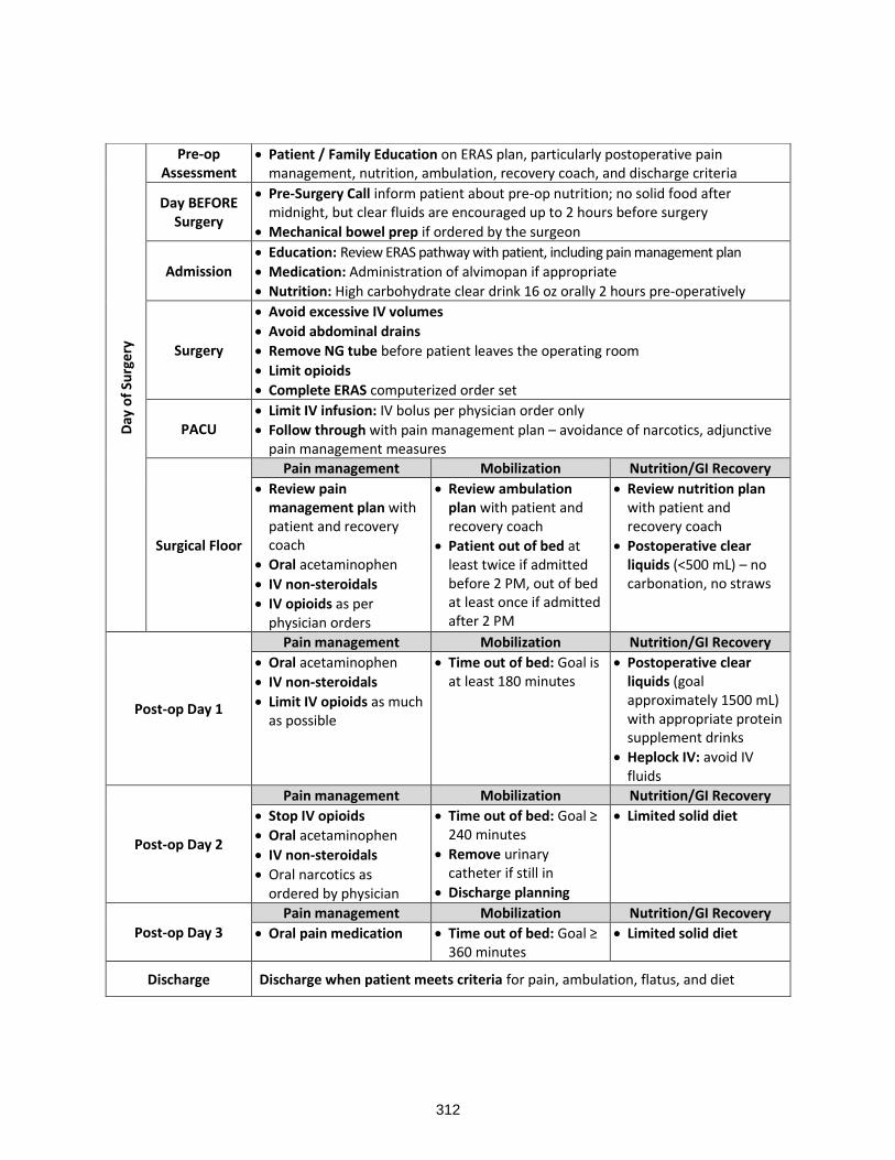
Day
of
Surg
ery
Pre-op Assessment
• Patient / Family Education on ERAS plan, particularly postoperative pain management, nutrition, ambulation, recovery coach, and discharge criteria
Day BEFORE Surgery
• Pre-Surgery Call inform patient about pre-op nutrition; no solid food after midnight, but clear fluids are encouraged up to 2 hours before surgery
• Mechanical bowel prep if ordered by the surgeon
Admission • Education: Review ERAS pathway with patient, including pain management plan
• Medication: Administration of alvimopan if appropriate
• Nutrition: High carbohydrate clear drink 16 oz orally 2 hours pre-operatively
Surgery
• Avoid excessive IV volumes
• Avoid abdominal drains
• Remove NG tube before patient leaves the operating room
• Limit opioids
• Complete ERAS computerized order set
PACU • Limit IV infusion: IV bolus per physician order only
• Follow through with pain management plan – avoidance of narcotics, adjunctive pain management measures
Surgical Floor
Pain management Mobilization Nutrition/GI Recovery
• Review pain management plan with patient and recovery coach
• Oral acetaminophen
• IV non-steroidals
• IV opioids as per physician orders
• Review ambulation plan with patient and recovery coach
• Patient out of bed at least twice if admitted before 2 PM, out of bed at least once if admitted after 2 PM
• Review nutrition plan with patient and recovery coach
• Postoperative clear liquids (<500 mL) – no carbonation, no straws
Post-op Day 1
Pain management Mobilization Nutrition/GI Recovery
• Oral acetaminophen
• IV non-steroidals
• Limit IV opioids as much as possible
• Time out of bed: Goal is at least 180 minutes
• Postoperative clear liquids (goal approximately 1500 mL) with appropriate protein supplement drinks
• Heplock IV: avoid IV fluids
Post-op Day 2
Pain management Mobilization Nutrition/GI Recovery
• Stop IV opioids
• Oral acetaminophen
• IV non-steroidals
• Oral narcotics as ordered by physician
• Time out of bed: Goal ≥ 240 minutes
• Remove urinary catheter if still in
• Discharge planning
• Limited solid diet
Post-op Day 3
Pain management Mobilization Nutrition/GI Recovery
• Oral pain medication • Time out of bed: Goal ≥ 360 minutes
• Limited solid diet
Discharge Discharge when patient meets criteria for pain, ambulation, flatus, and diet
312

REFERENCES
1. van Ramhorst, GH, Eker, HH, et al. Impact of incisional hernia on health-related quality of life and body image: A prospective cohort study. Am J Surg. 2012 Aug;204(2):144-50
2. Trujillo, C, Fowler, A, Complex Ventral Hernias: A review of past to present. Perm J. 2018;22:17-015
3. Holihan, JL, Alawadi, Z, et al. Adverse events after ventral hernia repair: The vicious cycle of complications. Am J Coll Surg. 2015 Aug;221(2): 478-85
4. Rosen, MJ, Atlas of Abdominal Wall Reconstruction. Philadelphia, PA: Elsevier Saunders; 2012 5. Ramirez, OM, Ruas, E, et al. “Components separation” method for closure of abdominal-wall
defects: An anatomic and clinical study. Plast Recon Surg. 1990 Sep;86(3) 519-26 6. Krpata, DM, Novitsky, YW, et al. Posterior and open anterior components separations: A
comparative analysis. Am J Surg. 2012 Mar;203(3):318-22 7. Zolin, SJ, Fafaj, A, et al. Transversus abdominus release (TAR): what are the real indications and
where is the limit? Hernia. 2020 (24):333-340 8. Novitsky, YW, Elliott, HL, et al. Transversus abdominis muscle release: a novel approach to
posterior component separation during complex abdominal wall reconstruction. Am J Surgery 2012; 204(5): 709-716
9. Kushner, B, Holden, S, et al. Surgical “error traps” of open posterior component separation-transversus abdominus release. Hernia 2021 (25):1703-1714
10. Bilezikan, JA, Tenzel, PL, et al. Comparing the outcomes of external oblique and transversus abdominus release using the AHSQC database. Hernia 2021 (25):365-373
11. Hodgkinson, JD, Leo, CA, A meta-analysis comparing open anterior component separation with posterior component separation and transversus abdominis release in the repair of midline ventral hernias. Hernia 2018 (22):617-626
12. Warren, JA, Cobb, WS, et al. Standard laparoscopic versus robotic retro-muscular ventral hernia repair. Surg Endosc 2017 Jan;31(1):324-332
13. Santos, DA, Limmer, AR, et al. The current state of robotic retromuscular repairs-a qualitative review of the literature. Surgical Endoscopy 2021(35):456-466
14. Belyansky, I, Reza Zahiri, H, et al. Early operative outcomes of endoscopic (eTEP access) robotic-assisted retromuscular abdominal wall hernia repair. Hernia 2018(22):837-847
15. Bittner, JG, Alrefai, S, et al. Comparative analysis of open and robotic transversus abdominis release for ventral hernia repair. Surg Endosc 2018 (32):727-734
16. Martin-del-Campo, LA, Weltz, AS, et al. Comparative analysis of perioperative outcomes of robotic open versus transversus abdominis release. Surg Endosc. 2018 (32):840-845
17. Ye, L, Childers, CP, et al. Clinical outcomes and cost of robotic ventral hernia repair: systematic review. BJS Open (2021) 0: 1-15
18. Dauser, B, Hartig, N, et al. Robotic-assisted repair of complex ventral hernia: can it pay off? Jour R. Surg (2021) 15:45-52
19. Motz, BM, Heniford, BT, et al. Chemical components separation: Concepts, Evidence, and Outcomes. PRS 2018 (142) 3S: 58-63
20. Majumder, A, Fayezizadeh, M, et al. Benefits of Multimodal Enhanced Recovery Pathway in Patients Undergoing Open Ventral Hernia Repair. J Am Coll Surgeons 2016 Jun;222(6):1106-15
313

TUBES, DRAINS AND CATHETER MANAGING COMPLICATIONS
Andre’ R. Campbell, MD, FACS, FACP, FCCM, MAMSE
Professor and Vice Chair of Surgery UC San Francisco Attending Surgeon Zuckerberg San Francisco General Hospital and Trauma Center San Francisco, CA
Drains have been widely used in surgery after an operation for many years. There remains controversy over how and when drains are used after a procedure. The idea behind the use of prophylactic drainage is to detect the presence of an intra-abdominal infection early after a procedure. The practice of draining is pervasive in surgery, and we still drain in many different settings. Intra-abdominal drains are used for several different procedures. They are particularly common in the setting of perforated viscus. Drains commonly used after many different types of procedures. One of the more common indications after general surgical procedure is acute appendicitis with perforation. In addition, drains are used after patients have had perforated diverticulitis. They are also used in other intra-abdominal infections like acute cholecystitis. Drains are commonly used after pancreatic surgery, colorectal surgery, and other cancer operation. The purpose of this session is to discuss the use of drains in surgery and complication that may occur in that setting.
Historically drains have used for many years in surgical procedures. There are even reports of the use of drains during the time of Hippocrates. There are reports of his use of drains for patients with empyema and with ascites in ancient times. More recently prophylactic drainage has been reported in the 1800s after gastrointestinal surgery. Lawson Tait a surgeon from the 19th century is credited with the expression “When in doubt drain”. This is a dictum that has been used by many surgeons even in modern times. The most important function of the drain is for operating surgeon to know early if there is a bleeding, anastomotic leak, or abscess formation. Sims a gynecological surgeon was the first to use drains after his operative procedures. Theodor Biliroth the famous Austrian surgeons was convinced that drains saved the lives of his patients during the complex gastrointestinal procedures that he completed.
There are two major types of drains: one is open, and the other is closed. The typical open drain that is used is a penrose drain. They can be used in the skin or soft tissues or in the abdomen. The main issue with the open drain is that bacteria can drain out of the wound as well as enter the wound. The jackson pratt, blake, hemovac drains are close suction drains used in surgery. The hemovac are used more for orthopedic surgery and they have a higher capacity. These drains come in different sizes and work well to remove fluid, blood for the wound. There are standard sized chest tubes and pigtails that are used to evacuate blood from the chest. More recently the data has suggested that smaller tubes work as effectively as larger tube to drain blood from the chest. Foley catheters are used commonly for procedures. They come in different sizes and types to be use for urinary drainage. In addition, gastrostomy tubes and jejunostomy tubes as used frequently in surgery. They are typically for feeding in patients in the ICU. These tubes can be placed percutaneously, laparoscopically, or during open surgery. Tracheostomy tubes are used commonly in the ICU and can be placed percutaneous or in an open setting.
Any time you use an invasive device there can be complication associated with an intervention can have complications. Open drains if they are left in to long can lead to more resistant infections developing in the wound over time. If used for a short period they usually are associated with problems. Close suction
314

drains are also in generally safe to use for patients. The round drains are easier to remove and do not have a lot of complications associated with removal. It is possible if you use the bigger drains in smaller individual and children that omentum can get entangled in the tubing and get pulled outside the abdomen when the tube is removed. Other complications can happen if the wider part of the drain gets stuck in the abdomen when they are pulled out of the abdomen. In the past an operation was required to remove the drain but more recently IR has been able to remove these drains. Fistulas secondary to operative drains have been less of a problem as they have become softer. The softer drains are associate with fewer complication and are safe for the patients
Chest tube are use commonly for the drainage of pneumothorax and hemothorax after trauma. The complication can be related to insertion when they are indwelling or on removal. Chest tubes have been inserted in the abdomen, major vessels, heart, lung, chest wall and subcutaneous tissues. They all should be inserted in a sterile fashion and this technique should be maintained throughout the procedure. Sterile gowns, gloves, masks, and drapes over the incision. Going over the rib is key so not blood vessels are injuries. Finger exploration of the lung is useful to insure you are in the chest. The tube should be angled up with the plan apical posterior location for optimal drainage o of blood or fluid. Pigtails are placed using the seldinger technique and place in the thoracic cavity. If the patient has scarring of the pleura placement can be even more difficult to insert the tube into the chest. Care must be taken not to injure the lung. A larger incision or a new location of the tube placement maybe needed in that case. The tubes can also be place in the fissure which is not optional, or they can be place intraparenchymal and cause to bronchopleural fistula. If there are broken ribs the physician who is doing the procedure can be injured on insertion of the tube. Rarely patients can develop an empyema from chest tube insertion. If you work in a facility with trainees, it is necessary to make sure they are supervised prior to allowing them the do procedures independently. More recently the pigtail approach has gained traction over the placement of regular chest tubes. Chest tubes are now also being placed are smaller in general with the same rate of success over larger tubes.
The complications associated with foley placement in the trauma setting is usually a false passage on insertion. Care must be taken to insure it is done sterilely and that no trauma happened to the urethra. If the patient has blood at the meatus, then it is necessary to perform a retrograded urethrogram to rule trauma to the prostatic or membranous urethra. It is uncommon to have an injury to the urethra in a woman.
Gastrostomy and jejunostomy tubes are used in surgical patients commonly. The open approach has been used less more recently and the PEG with endoscopy assistance or IR approach is used more commonly. The placement of the PEG is dependent on favorable anatomy in the abdomen. Care must be taken not enter the colon or small bowel when placing the catheter. Placing the catheter through another structure has been reported. It can also happen with placement of the tube done by IR. The same issue occurs when the tube is place by IR. The issues are similar with placement of the jejunal tube. Complications of placement of these tubes can also involve a local skin infection or a leak if the tube is not placed flush against the abdominal wall. This type of leak can result in an abdominal catastrophe. This particularly difficult since many of these patients do not have a normal mental status and are not able to react normally to peritoneal irritation.
The tracheostomy tube is another area where problems can occur with access to the airway. In injured patient with massive facial injuries an emergency airway may be needed. Surgical access with a rapid cricothyrotomy is essential is lifesaving. If anatomical structures are not correctly identified the airway can be injured when an endotracheal tube or tracheostomy tube is inserted in the wrong location. This is typically a difficult setting and care must be taken to identify the anatomy correctly. The classical tracheostomy tube is inserted in the 2nd trachea ring and the tube is sutured. Complication includes loss
315

of airway, bleeding, tracheal damage. When done open care must be taken to identify the thyroid isthmus and divide it if necessary. The percutaneous approach should always be done with a fiberoptic bronchoscope to injury the tube does not injury the esophagus or other structures. Other issues with the percutaneous approach include not opening the skin widely enough to fit the dilator, bleeding, and tracheal injury.
Another type of drain commonly used in trauma is the wound vac or vacuum suction drain system. This device was first described by Barker when they used chest tube or active drainage of secretions from the abdominal cavity after damage control surgery. There are many variations of this system including using chest tubes or active suction drains. In the late 1990s Louis Argenta worked on using high level wound suction in animal wounds and found it helped with wound healing. This concept rapidly became commonly used in the form the KCI wound vac system for open abdomens. There are now several types of wound vacs that in practice which is setup to help collecting secretions and helping to close the abdomen down and promote wound healing in patient who are injured. The wound vac utilizes 125 mm Hg to apply suction the wound and it is type of tube and drain we commonly use in operative surgery.
Many studies have been published on the complications of chest tube placement. On recent study by Platnick et al examined the issue of problems and complications in chest tube placement. In a retrospective study over three years in patients who require CT placement at their institution. The tubes were placed for pneumothorax, hemopneumothorax, hemothorax, and effusions. Other indications included traumatic cardiac arrest and hypotension. They enrolled 451 patients in the study. They performed univariate and multivariate analysis on the patients. The factors associated with tube malposition was placement in the emergency department, placement by an emergency physician, and body mass index >30Kg/m2. This study was interesting from the standpoint that in busy level 1 trauma center a surgeon had a reduced rate of complications in chest tube insertion. They are now working on methods to ensure that ED physicians and surgeons have similar outcomes in chest tube placement.
When a patient has perforated appendicitis with a frank abscess, and they have surgery by laparoscopic or open techniques the issue of drain placement is discussed in the OR. This has been studied in a Cochrane Collaboration group that examined at the use of drains in open surgery. They found that there was no difference in outcome in open appendectomies and the use of drains in several studies that were reviewed. This is interesting, in that many surgeons will still placed drains in this setting to try to reduce the chance of abscess formation along antibiotics to reduce the chance of recurrence. Petrovsky et al reviewed several studies and found there were no difference in outcome older studies. More recent studies have confirmed these findings.
There are other surgical procedures where the use of drains has been debated that includes after pancreatic trauma and colon surgery. In the rare case that a patient had penetrating pancreatic trauma and a distal pancreatectomy is required surgeons will typically use a drain. Recent studies by Kang et al quoted an incidence of 38% for post-operative pancreatic leak and fistula although other authors have quoted a lower rate. Drains are routinely used in that setting. In low rectal anastomosis the use of drains has been routine event though more recent studies have showed that is does not reduce the incidence of leak after colon trauma.
In conclusion, drain, tubes and catheters have been used in many settings in surgery over many years. There are controversies over the current use of drains in several settings, but surgeon continue to use them. It is important that if they are used to have a clear reason and make sure to remove them at the early possible time. Many other tubes are used in surgery including chest tubes, feeding tubes, tracheostomy tubes, foleys. Each of these devices have risk associate with their use.
316

REFERENCES
1. Durai, R, Mownah A, Ng PC. Use of drains in surgery: a review. J Perioper Pract 2009:19(6):180-186.
2. Kumar M, Yang SB et al. Is prophylactic drains necessary after subtotal gastrectomy? World J Gastroenterol 2007;13927);3738-3741.
3. Platnick C, Witt CE, Pieracci FM, et al. Beyond the tube: Can we reduce chest tube complications in trauma patients? The Amer Jour of Surg 222 (2021) 1023-1028.
4. Sethuraman KN, Duong D, Mehta S et al. Complications of tube thoracostomy placement in the emergency department. J Emerg Med 2011;40(1);14-20.
5. Benns MV, Egger ME, Harbrecht BG et al. Does chest tube location matter? An analysis of chest tube position and the need for secondary interventions. J Trauma Acute Care Surg. 2015;78(2): 386-390.
6. Kugler NW, Carver TW, Knechtges P. et al. Thoracostomy tube function not trajectory dictates reintervention. J Surg Res. 2016;206(2);380-385.
7. Li Z, Zhao L, Cheng Y, Cheng N, Deng Y. Abdominal drainage to prevent intra-peritoneal abscess after appendectomy for complicated appendicitis. Cochrane Database of Systemic Reviews 2021, Issue 8, Art No: CD010168. DOI:10.1002/14651858.CDO10168pub4.
8. Durai R, Ng, PCH. Surgical Vacuum Drains: Types, Uses, and Complications. AORNJ 91 9. (February 2010) 266-271. 10. Durai R, Mownah A, Ng PC. Use of drains in surgery: a review J Perioper Practice 2009;19(6);180-
186. 11. Cecka F, Lovecek M, Bohumi J et al. Intra-abdominal drains following pancreatic resection as
systemic review. World J Gastroenterol 2015 October 28,; 21(40):11458-11468.
317

318

SESSION 10
CAPSULE COMMENTARIES - BECAUSE YOU ASKED
Moderator: Todd W. Costantini, MD, FACS
Tuesday, March 29, 2022
Palace Ballroom 1-2
Palace Tower, Emperors Level
2:00 - 2:08
Tips and Tricks Using Balloon Catheters Sydney J. Vail, MD, FACS
2:08 - 2:16
The Ostomy "Won't Reach": Appropriate Site Selection for Stomas Scott R. Steele, MD, MBA, FACS, FASCRS
2:16 - 2:24
Difficult Decisions in the Pediatric Patient Robert W. Letton, MD, FACS
2:24- 2:32
Management of Less Lethal Weapon Injuries Jayson Aydelotte, MD, FACS
2:32 - 2:40
Limb replantation Elizabeth R. Benjamin, MD, PhD, FACS
2:40 - 2:48
Minor TBI - Neurosurgeons vs Trauma Surgeons Bellal A. Joseph, MD, FACS
2:48 - 2:56
Prehospital Blood Products Ali Salim, MD, FACS
319

TIP & TRICKS USING BALLOONS CATHETERS
Sydney J. Vail, MD, FACS
Chairman, Department of Surgery Division of Trauma, Surgical Critical Care and ACS Valleywise Health Medical Center Associate Professor of Surgery Creighton University School of Medicine Lt. Col, MD, US Army Reserve Phoenix, AZ
This presentation and syllabus will focus on the use of different balloon catheters to assist us in slowing or stopping hemorrhage in different areas of the body. This is not a discussion about comparing open vs. balloon control of bleeding. Some of the presented techniques have solid literature support, others just case series and some, just make it up as you go out of necessity for the situation you are presented with. An educational foundation, from residency, fellowship, mentorship or courses such as this one should provide the educational and hands on exposure to the use of balloons to control hemorrhage. Trying your first one having never performed or adequately learned or prepared for this procedure/maneuver can have potential negative consequences. Having some of us ‘senior/gray haired (wisdom highlights!) guys’ as a resource pays dividends.
“You just bought yourself time, not success” is a statement I make to surgical residents in training, as well as fellows and junior faculty involved in attempting and achieving hemorrhage control anywhere in the body. Balloons are an adjunct, a tool that we use to obtain control of bleeding in difficult situations or where your fingers/clamps or other means of vascular control will not be fast enough or able to adequately or initially control a site of hemorrhage.
Balloon catheters can be used in 2 ways, to tamponade a site of hemorrhage within a ‘confined’ space or intravascular placement to fully arrest bleeding or to act as a shunt to allow continued blood flow around an injured vessel. The following is a list of balloon catheters that we have at our disposal for use either as a shunt or to attempt control of hemorrhage:
NOTE: the use and success of a balloon catheter is 100% user dependent. As in many areas of surgery and Trauma, the level of your preparedness can improve the odds of success.
Practicing the P.A.C.E. (P = primary, A = alternative, C = contingency, E = emergency) methodology for everything we do can mean the difference between success and failure; having 4 established steps in your algorhythm means you’ll always have more than 1 or 2 means of controlling any hemorrhage or having more ways to accomplish any goals in surgery.
You must know the catheter’s ability to succeed at the indication it’s being used for, know the indications for its use and be able to determine success or failure, know the potential to cause further vascular injury,
be able to troubleshoot the catheter for appropriate function, know the limits as well as potential negative consequences of its use.
1. Resuscitative endovascular balloon occlusion of the aorta (REBOA) is used only in the aorta. (expanded venous uses are being developed). The balloon maximum capacity is 24 mL but it typically takes less to occlude the vessel but is location/aortic diameter dependent on the volume
320

necessary to obtain occlusion to forward flow. This catheter is used only for control of bleeding, not as a shunt. COST ~$1500. [https://prytimemedical.com/product/er-reboa-plus-catheter/]
Fogarty Catheters (FC) are a smaller diameter balloon that can be used in smaller and peripheral arterial vessels. They come in several sizes (2-7F) and include catheters that have either an inflatable balloon and solid catheter (Fogarty® arterial embolectomy catheter) or one that a wire can be passed through or fluid can be infused through (Fogarty® thru-lumen embolectomy catheter). They also have a catheter specifically for use in veins (Fogarty® venous thrombectomy catheter designed for removing thrombi in the venous system with a long, soft tip permitting advancement past the venous valves without undue valve trauma). You must be aware of maximum balloon size and maximum liquid capacity of the balloon you are using. Please refer to the website listed below for that information. These catheters are for control of bleeding, embolectomies and not used as a shunt. COST ~$250-$450. [https://edwardsprod.blob.core.windows.net/media/Default/devices/catheters/clot%20management/ fogartyclotmanagement.pdf] Foley Catheters are another balloon catheter type used for the control of bleeding in different areas of the body. These are typically used as a means of providing a tamponade effect on bleeding within a confined space or to provide traction across a structure that can compress a bleeding vessel. It is also a useful tool to act as a vascular shunt. I have used one from the abdominal IVC as a retrograde Schrock shunt, as an abdominal IVC shunt, as an aortic shunt as well. NOTE: use largest diameter available! Can also loop it as to not have shorter length issues. COST $5-$7
321

Endotracheal tubes have been described for use as an atriocaval (Schrock) shunt as well rather than a chest tube to provide bypass venous return to the heart from the proximal IVC typically for a retrohepatic caval injury
COST $3-$5
322

Tracheostomy tubes are not a typical adjunct used to control bleeding as they are short and most are rigid. PEG or other balloon tipped feeding catheters have utility for somewhat more superficial wounds, 2-5” depth, that are bleeding. These tubes/catheters are limited by their length as to being adequate for the injury at hand.
There are other balloon catheters on the market but they are beyond the scope of this presentation; the focus is on the ones listed above. Self made: red rubber, smaller diameter chest tube or other hollow catheter with a penrose drain over the catheter and tied on each end to allow inflation via side holes of catheter. Can be used through/across longer wound tracts in liver or lung tissue as well as deep buttock wounds (I had a machete wound across both buttocks close to ischium that was hemorrhaging and would take time to control from an surgical or interventional radiology approach; used two 20F chest tubes with penrose drains tied to them and inserted one from each side which arrested the bleeding. The stiffness of the chest tube was necessary to traverse the longer unseen tract.)
323

Typical locations of use:
• Neck, introduced via a penetrating wound
• Chest, introduced via a penetrating wound that the likely bleeding vessel is traveling on or with proximity to the chest wall (intercostals, subclavians) or via thoracotomy: heart, aorta (shunt/occlusion), vena cava (shunt/occlusion), atriocaval shunt
• Abdomen: aorta, vena cava, liver, laparoscopic/robotic port sites, abd wall injuries, uterus, kidney (?), esophagus and or gastric varicies, liver (SB tube?)
• Pelvis: iliacs, retroperitoneal (?)
• Extremities: anywhere a TQ isn’t used, axilla, high femoral/pelvis
• Intravascular major vessels
PEARLS
• Be familiar with the variety of balloon catheters stocked in your facility; they are a useful adjunct for hemorrhage control/shunting and should be utilized when and where appropriate
o ALWAYS place an intravascular catheter that is smaller in diameter than the vessel it is used for
• When inflating any balloon, it can contain fluid or air; the diameter of the balloon is a key issue to prevent vascular injury and it is better controlled by the operator with fluid. The pressure within the balloon is less concerning than the diameter. Fluid being more incompressible is more easily controlled to dynamically change the balloons diameter. A gas filled balloon may not uniformly maintain an even shape which can cause stresses in different non-contiguous areas, i.e., less reliability of maintaining the balloons uniform diameter. (J Vasc Surg 1988 Nov;8(5):650-651)
• The goal should be to initially slow the bleeding then stop it; gradual inflation is typically superior and safer than rapid bolus inflation. If you always think about acutely stopping the hemorrhage with an intravascular catheter placement; you may cause local or vascular injury or rupture by overinflating a balloon beyond the anatomic boundaries it is trying to fill/occlude
• When resistance during balloon inflation is appreciated, stop inflating, you are likely at the limits of the cavity or vessel
• Know the volume limits of the balloons you are using and think in 3-D to appreciate the anatomy and size of the cavity or space you are trying to occupy with the balloon
• For tamponade uses, may need to keep catheter under tension so use a wide based jaws clamp (thin jaws can lacerate the catheter and could lead to balloon or catheter leakage) across catheter, at the skin level, AFTER pulling up on the catheter to create tension on the bleeding area from the balloon. The clamp will maintain your traction on the balloon to free up your hands.
o prefer a tube or vascular clamp that won’t cut into the catheter
• Balloon catheters, i.e., foley can be used in a more rapid fashion to avoid a second isolation of a vessel inorder to secure a shunt or occlusive balloon in place, i.e., vessel loops, vascular clamps, temporary vascular clamps can be made using vessel-loops or umbilical tape and a piece of rubber tubing
324

A product on the market that had received little press is the TourniCath. I have never used or seen one but found this during my research:
http://www.cardiocommand.com/hemorrhage.html
“Once the TourniCath has been inserted into the wound track (through either the entry or the exit wounds) and the outer sheath removed, the balloon is then inflated to a pressure sufficient to tamponade internal bleeding (90-150 mmHg suggested). The maximum potential inflation volume of the cylindrical balloon is 400 ML and the dimensions at maximum volume are 8" in length x 2" in diameter. Thus, TourniCath provides sufficient length and volume for successful hemostasis of most wound tracks in the specified anatomical regions of the body. TourniCath is not indicated for abdominal or thoracic wounds.”
Temporary vascular clamp using a
tape and cut hollow catheter
Double and single vessel loop use
325

AND FOR THOSE CARING FOR LAW ENFORCEMENT OR MILITARY CANINES
The use of a Foley balloon catheter to control junctional hemorrhage in a dog with severe vascular injury secondary to penetrating trauma
Ryan T Wheeler 1, Jan P Kovacic 2
Case Reports J Vet Emerg Crit Care (San Antonio) 2021 Aug 27. Online ahead of print.
ABSTRACT
Introduction: Penetrating trauma is commonly seen in dogs. The severity depends on the site of injury and tissue involved. Junctional hemorrhage can be especially challenging to control given the inaccessibility of the damaged vasculature. Methods described to control life-threatening hemorrhage in dogs include direct pressure, hemostatic gauze, hemostatic powder or granules, wound packing, tourniquets, and direct clamping of the vasculature. Foley balloon catheters (FBC) are commonly used to tamponade deep vascular hemorrhage in people, but the technique has not been previously described in the veterinary literature.
Objective: To present a case of penetrating trauma (bite wound) in a dog with a transected left femoral artery and vein in which the life-threatening hemorrhage was initially controlled with tamponade using an FBC.
Case: A 7-year-old neutered male Terrier mix presented in hemorrhagic shock with an Animal Trauma Triage (ATT) of 7 and modifed Glasgow coma scale (MGCS) of 17 forty-five minutes after being attacked by another dog. The dog had sustained a deep penetrating wound to the left groin. Direct pressure and gauze packing at the site of injury were not successful at slowing the hemorrhage. A 10-Fr, 55-cm Foley catheter with a 5-mL balloon was inserted into the wound tract, and the balloon was inflated with 7.5 mL of sterile saline. Hemorrhage was controlled after inflation of the Foley balloon. CBC, blood biochemistries, abdominal point-of-care ultrasound, radiographs, prothrombin time, partial thromboplastin time, and whole blood viscoelastic testing were performed. Stabilization included fluid resuscitation, analgesics, antimicrobials, and epsilon aminocaproic acid. The dog was then anesthetized to definitively identify and control the hemorrhage. Transection of the left femoral artery and vein where identified and ligated. The dog fully recovered and was discharged 32 hours later.
326

New and unique information: FBCs may be useful as an alternative technique for temporary control of life-threatening hemorrhage secondary to penetrating injuries in both the emergency department and prehospital settings.
REFERENCES
1. Gilroy D, Lakhoo M, Charalambides D, Demetriades D. Control of life-threatening haemorrhage from the neck: a new indication for balloon tamponade. Injury. 1992;23(8):557-9. PMID: 1286913.
2. Navsaria P, Thoma M, Nicol A. Foley catheter balloon tamponade for life-threatening hemorrhage in penetrating neck trauma. World J Surg. 2006 Jul;30(7):1265-8. \\ PMID: 16830215.
3. Weppner, J. Improved mortality from penetrating neck and maxillofacial trauma using Foley catheter balloon tamponade in combat. J Trauma & ACS, Journal of Trauma and Acute Care Surgery: August 2013;Volume 75: 220-224
4. Poggetti RS, Moore EE, Moore FA, Mitchell MB, Read RA. Balloon tamponade for bilobar transfixing hepatic gunshot wounds. J Trauma. 1992;33(5):694-697.
5. Thomas SV, Dulchavsky SA, Diebel LN. Balloon tamponade for liver injuries: case report. J Trauma. 1993;34(3):448-449.
6. Demetriades D. Balloon tamponade for bleeding control in penetrating liver injuries. J Trauma. 1998;44(3):538-539.
7. DiGiacomo, CJ; Rotondo, MF, Schwab, CW. Transcutaneous balloon catheter tamponade for definitive control of subclavian venous injuries: case reports. J Trauma. July 1994;Volume 37: 111-113
8. Feliciano DV. Balloons are not just for children. Trauma Surg Acute Care Open 2021;6:e000808. 9. Fabian TC, Bee TK. Liver and biliary tract. In: Feliciano DV, Mattox KL, Moore EE, eds. Trauma. 6th
ed. New York: McGraw-Hill Medical; 2008:637-660. 10. https://jts.amedd.army.mil/assets/docs/presentations/REBOA_CPG_Training_ID38.pdf
327

THE OSTOMY WON’T REACH”
Scott R. Steele, MD, MBA, FACS, FASCRS
Chairman, Department of Colorectal Surgery Rupert B. Turnbull, MD Endowed Chair in Colorectal Surgery Cleveland Clinic Professor of Surgery Cleveland Clinic Lerner College of Medicine of Case Western Reserve University Cleveland Clinic Cleveland, OH
1. Site the stoma properly the first time! Use ETS to mark – even when you don’t think you need them
a. Patients can alleviate their fear of stomas ahead of time b. It will save your patient (and you) a lot of headaches c. This can still be done in an emergency setting
Before you make the stoma, ask yourself if you need it Length is key, but depends on maintaining the viability of the bowel
d. Don’t sacrifice anything (blood vessel-wise) prior to checking collaterals e. Mobilize and then mobilize some more f. Technology like ICG can help
You can also focus on the abdominal wall to help with length in the obese by various contouring procedures
KEY POINTS
328

WHERE IS THE BEST PLACE TO PUT A STOMA?
Siting Tips
Disabled Mark how they spend the bulk of their time Brace Mark with it on Radiation Try to go outside the fields Two stomas Stoplight or across Burns Think of garments and dressing requirements
o
o ASIS
o Public Symphysis
•
•
329

330

Abdominal Wall Contouring
• Removal of the fatty tissue / pannus
• Can remove around the stoma itself
• Remove the SQ fat
• Can temporarily bring the abdominal wall together with a wound protector to pull the ostomy through and then remove it.
An external file that holds a picture, illustration, etc. Object name is 10-1055-s-0036-1580628-i00730-7.jpg
Division of the inferior mesenteric vessels and left colic vessels to add mobility and length for a descending colostomy.
331

If all else fails…
• Bring up whatever you can
• Tack at the level of the fascia – intubate with drain and leave intra-abdominal drain
• Deliver by Stages
• Stomas can be tough – especially in leaks, takebacks, shortened mesentery and high BMI
• Length is key – make sure you preserve vascular flow
• Upper abdomen may be easier to bring this up through
• Make the trephine big enough (no matter how big) – worrry about the hernia later
REFERENCES
1. Steele SR, Hull T, Read TE, Saclarides T, Senagore A, Whitlow C. (Eds.) The ASCRS Textbook of Colon and Rectal Surgery (3ed). Springer Publishing. New York, NY: 2016; ISBN978-3-319-25970-3.-Intestinal Stomas (McGee and Cataldo; pp 971-1014).
FINAL THOUGHTS & TAKE-HOME POINTS
332
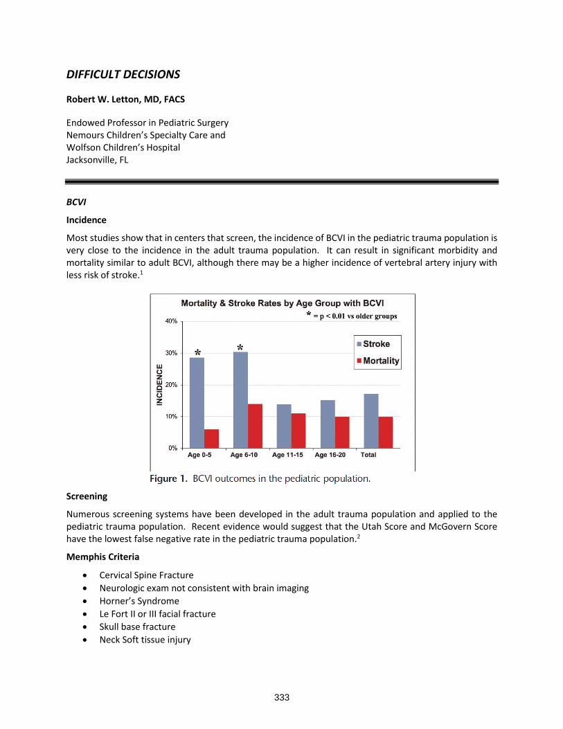
DIFFICULT DECISIONS
Robert W. Letton, MD, FACS
Endowed Professor in Pediatric Surgery Nemours Children’s Specialty Care and Wolfson Children’s Hospital Jacksonville, FL
BCVI
Incidence
Most studies show that in centers that screen, the incidence of BCVI in the pediatric trauma population is very close to the incidence in the adult trauma population. It can result in significant morbidity and mortality similar to adult BCVI, although there may be a higher incidence of vertebral artery injury with less risk of stroke.1
Screening
Numerous screening systems have been developed in the adult trauma population and applied to the pediatric trauma population. Recent evidence would suggest that the Utah Score and McGovern Score have the lowest false negative rate in the pediatric trauma population.2
Memphis Criteria
• Cervical Spine Fracture
• Neurologic exam not consistent with brain imaging
• Horner’s Syndrome
• Le Fort II or III facial fracture
• Skull base fracture
• Neck Soft tissue injury
333
Denver Criteria
• Injury Mechanism o Severe cervical hyperextension/rotation or hyperflexion o Displaced midface or complex mandibular fracture o Closed head injury consistent with DAI o Near hanging
• Physical Signs o Seatbelt abrasion of soft tissue injury to anterior neck with significant swelling or altered
mental status
• Fracture in proximity to internal carotid or vertebral artery o Basilar skull fracture involving the carotid canal o Cervical Vertebral Body Fracture
Utah Score
• GCS < 8 1
• Focal neurologic deficit 2
• Carotid Canal Fracture 2
• Petrous temporal bone fracture 3
• Cerebral infarct on CT 3 o Score < 2 = 7.9% risk o Score > 3 = 39.3% risk
McGovern Score
• GCS < 8 1
• Focal neurologic deficit 2
• Carotid Canal fracture 2
• MVA or ped struck 2
• Petrous Temporal Bone Fracture 3
• Cerebral infarction on CT 3 o Score > 3 = high risk and imaging indicated
Angiography would remain the Gold Standard for screening but is difficult to perform in children. CTA and MRI both equivalent. MRI is desirable due to less radiation exposure, however, often would require general anesthesia to accomplish in this patient population and therefore CTA the most common and cost effective.3
TREATMENT
Treatment would depend on degree of injury.
Denver Grading System:
• G I: irregular vessel wall or dissection/intraluminal hematoma with < 25% stenosis
• G II: intraluminal thrombus or raised intimal flap is visualized, or dissection/intraluminal hematoma with > 25% stenosis
• G III: pseudoaneurysm
• G IV: vessel occlusion
• G V: vessel transection
334

Treatment is somewhat controversial and often, injuries are observed and not treated. Lower grade injuries when treated are managed with anti-platelet therapy or anti-coagulation with low molecular weight heparin. Higher grade injuries may require interventional procedures and stenting. Untreated carotid injuries have stroke rate of 30 - 64%. Vertebral injuries left untreated have a 10 - 50% stroke rate, and in pediatric series, overall risk of stroke is 26-38% if untreated. Most children (~70%) are not treated for BCVI once the injury is identified.4
Key Points
• BCVI is rare in children but carries significant mortality and morbidity (~25%)
• Screening is imperative to early treatment and stroke prevention
• Pediatric specific screening tools are based on low rates of screening and still fail to identify all injuries
• Imaging modalities that decrease radiation exposure while accurately assessing for injury are needed and MUST image the head and neck
BLUNT PANCREAS
Incidence
Although extremely rare, the morbidity associated with blunt pancreatic injury, especially higher-grade injuries where there is significant controversy over how to accurately grade the injury as well as how to manage the injury. What would you do with this innocent appearing handlebar injury on the outside with the included CT of the inside?
335

Screening
Even in adults, CT scanning insufficiently sensitive enough to identify ductal injury.5 ERCP can be useful to diagnose duct injury to help determine the Grade of injury, especially whether there is ductal involvement.6
336

Treatment
Clearly in low grade injury (Grade I-II) regardless of adult versus pediatric conservative management should be attempted7. As well, in high grade Iv/V injuries where a Whipple might be necessary, attempted drainage and non-operative management is often indicated. But in the pediatric trauma patient with a presumed Grade III/IV, what is the best course of management. Laparoscopic spleen preserving distal pancreatectomy? Or non-operative management with close observation and management of secondary sequelae of the injury?
In children with blunt pancreatic injury, ERCP can be useful to diagnose duct injury to help determine the Grade of injury, and for management of late complications such as stricture and fistula. However, early endoscopic intervention for pancreatic duct disruption with stenting may not improve outcome or expedite recovery.6
Figure 5. Comparison of outcomes in patients with Early ERCP with intervention vs observation only (all
patients had nonoperative management); *p < 0.05, IQR ranges shown
Laparoscopic spleen preserving distal pancreatectomy is well tolerated in the pediatric patient when ductal injury is confirmed, however, those who advocate non-operative management express concerns for an increased risk for diabetes when the patient is older8. Those who believe in aggressive operative management express that this risk is rather low and has not been proven in the literature9. Operative
I
337

management has shown to decrease complications, but not necessarily days of hospitalization or resource utilization10. Recent efforts have been directed at non-operative management pathways that might reduce the complication rate as well as hospitalization and resource utilization11
Patients from 20 pediatric trauma centers were enrolled. Median age was 9 years (range, 1–18 years). The majority (73%) of injuries were American Association of Surgeons for Trauma grade III, 24%were grade IV, and 3%were grade V. All patients had computed tomography scan and serum pancreatic enzyme levels at presentation, but serial enzyme level monitoring was variable. Pancreatic enzyme levels did not correlate with injury grade or pseudocyst development. Parenteral nutrition was used in 68% and jejunal feeds in 31%. Endoscopic retrograde cholangiopancreatogram was obtained in 25%. An organized peripancreatic fluid collection present for at least 7 days after injury was identified in 59% (42 of 71). Initial management of these included: observation 64%, percutaneous drain 24%, and endoscopic drainage 10%
338

and needle aspiration 2%. Clear liquids were started at a median of 6 days and regular diet at a median of 8 days. Median hospitalization length was 13 days. Injury grade did not account for prolonged time to initiating oral diet or hospital length; indicating that the variability in these outcomes was largely due to different surgeon preferences.11
Bottom line, both methods are acceptable. Surgeons who are comfortable operating on the pancreas and have good success can often get these children home in less than a week, and once they are healed there is very little chance of a pseudocyst of other complication down the road. Stenting is an option, however, very few centers have that capability in smaller children. Non-operative management is likely to lead to a complication that may not be operative but still needs intervention.
Key Points
• Organized fluid collection in 35% patients with non-operative management
• Exocrine pancreatic insufficiency in 1 patient (13%)
• No fistula, severe sepsis or mortality
• No pain at 2-week follow-up in 82% patients, and o chronic pain in 100% patients with long-term follow-up
• Spleen preserving distal pancreatectomy remains an excellent option in appropriate patients.
• Currently a prospective head-to-head trial of operative versus nonoperative management
REFERENCES
1. Christopher W. Marenco, MD, Woo S. Do, MD, Daniel T. Lammers, MD, Kaitlin R. Morte, MD, Matthew J. Eckert, MD, Carly M. Eckert, MD, MPH, Denis Bensard, MD, and Matthew J. Martin, MD, FACS. Big problems in little patients: Nationwide blunt cerebrovascular injury outcomes in the pediatric population. J Trauma Acute Care Surg. 2019;87: 1088–1095.
2. Areg Grigorian, MD,Matthew Dolich,MD, Michael Lekawa,MD, Roy M. Fujitani,MD, Nii-Kabu Kabutey,MD, Catherine M. Kuza, MD, Rame Bashir, and Jeffry Nahmias, MD, MHPE. Analysis of blunt cerebrovascular injury in pediatric trauma. J Trauma Acute Care Surg. 2019;87: 1354–1359.
3. Ajay Malhotra, Xiao Wu, Vivek B. Kalra, Thomas R. Goodman, Joseph Schindler, and Howard P. Forman. Screening for Pediatric Blunt Cerebrovascular Injury: Review of Literature and a Cost-Effectiveness Analysis. Journal of Pediatric Surgery 50 (2015) 1751–1757.
4. Nima Azarakhsh, MD, Sandra Grimes, BSN, RN, David M. Notrica, MD, Alexander Raines, MD, Nilda M. Garcia, MD, David W. Tuggle, MD, Robert Todd Maxson, MD, Adam C. Alder, MD, John Recicar, RN, Pamela Garcia-Filion, PhD, MPH, Cynthia Greenwell, RN, BSN, CCRC, Karla A. Lawson, PhD, Jim Y. Wan, PhD, MPhil, and James Wallace Eubanks III, MD. Blunt cerebrovascular injury in children: Underreported or underrecognized? A multicenter ATOMAC study. J Trauma Acute Care Surg. 2013;75: 1006 – 1012.
5. Nickolas Byrge, MD, Marta Heilbrun, MD, Nicole Winkler, MD, Daniel Sommers, MD, Heather Evans, MD, Lindsay M. Cattin, MD, Tom Scalea, MD, Deborah M. Stein, MD, Todd Neideen, MD, Pamela Walsh, Carrie A. Sims, MD, Tejal S. Brahmbhatt, MD, Joseph M. Galante, MD, Ho H. Phan, MD, Ajai Malhotra, MD, Robert T. Stovall, MD, Gregory J. Jurkovich, MD, Raul Coimbra, MD, Allison E. Berndtson, MD, Thomas A. O’Callaghan, MD, Scott F. Gaspard, MD, Martin A. Schreiber, MD, Mackenzie R. Cook, MD, Demetrios Demetriades, MD, Omar Rivera, MD, George C. Velmahos, MD, PHD, Ting Zhao, MD, Pauline K. Park, MD, David Machado-Aranda, MD, Salman Ahmad, MD, Julie Lewis, RN, William S. Hoff, MD, Ghada Suleiman, RN, Jason Sperry, MD, Samuel Zolin, MD, Matthew M. Carrick, MD, Gina R. Mallory, RN, Jade Nunez, MD, Alexander Colonna, MD, Toby Enniss, MD, and Ram Nirula, MD, MPH. An AAST-MITC analysis of pancreatic trauma: Staple or sew? Resect or drain? J Trauma Acute Care Surg. 2018;85: 435–443.
339

6. Eric H. Rosenfeld, Adam M. Vogel, Denise B. Klinkner, Mauricio Escobar, Barbara Gaines, Robert Russell, Brendan Campbell, Hale Wills, Anthony Stallion, David Juang, Rajan K. Thakkar, Jeffrey Upperman, Mubeen Jafri, Randall Burd, and Bindi Naik-Mathuria. The utility of ERCP in pediatric pancreatic trauma. Journal of Pediatric Surgery 53 (2018) 146–151.
7. Walter L. Biffl, MD, Chad G. Ball, MD, Ernest E. Moore, MD, Jason Lees, MD, S. Rob Todd, MD, Salina Wydo, MD, Alicia Privette, MD, Jessica L. Weaver, MD, PhD, and Samantha M. Koenig, MD. Don’t mess with the pancreas! A multicenter analysis of the management of low-grade pancreatic injuries. J Trauma Acute Care Surg. 2021;91: 820–828.
8. Mehrdad Nikfarjam, Michael Rosen, and Todd Ponsky. Early management of traumatic pancreatic transection by spleen-preserving laparoscopic distal pancreatectomy. Journal of Pediatric Surgery (2009) 44, 455–458.
9. Mary J. Edwards, David F. Crudo, Terri L. Carlson, Anita M. Pedersen, and Laura Keller. Pancreatic atrophy and diabetes mellitus following blunt abdominal trauma. Journal of Pediatric Surgery (2013) 48, 432–435.
10. James H. Wood, David A. Partrick, Jennifer L. Bruny, Angela Sauaia, and Steven L. Moulton. Operative vs nonoperative management of blunt pancreatic trauma in children. Journal of Pediatric Surgery (2010) 45, 401–406.
11. Bindi J. Naik-Mathuria, MD, Eric H. Rosenfeld, MD, Ankush Gosain, MD, PhD, Randall Burd, MD, PhD, Richard A. Falcone, Jr., MD, Rajan Thakkar, MD, Barbara Gaines, MD, David Mooney, MD, Mauricio Escobar, MD, Mubeen Jafri, MD, Anthony Stallion, Denise B. Klinkner, MD, Robert Russell, MD, Brendan Campbell, MD, Rita V. Burke, PhD, Jeffrey Upperman, MD, David Juang, MD, Shawn St. Peter, MD, Stephon J. Fenton, MD, Marianne Beaudin, MD, Hale Wills, MD, Adam Vogel, MD, and the Pancreatic Trauma Study Group (PTSG) Collaborators: Stephanie Polites, Adam Pattyn, MD, Christine Leeper, MD, Laura V. Veras, MD, Ilan Maizlin, MD, Shefali Thaker, MD, Alexis Smith, MD, Megan Waddell, MD, Joseph Drews, MD, James Gilmore, MD, Lindsey Armstrong, MD, Alexis Sandler, MD, Suzanne Moody, Brandon Behrens, MD, and Laurence Carmant. Proposed clinical pathway for nonoperative management of high-grade pediatric pancreatic injuries based on a multicenter analysis: A pediatric trauma society collaborative. J Trauma Acute Care Surg. 2017;83: 589–596.
340

MANAGEMENT OF LESS THAN LETHAL WEAPON INJURIES
Jayson Aydelotte, MD, FACS
Associate Professor of Surgery Dell Medical School The University of Texas at Austin Austin, TX
Less than lethal weapon rounds are typically the moniker that replaced “Non-Lethal” or “Less Lethal” rounds. There are over 75 different kinds of these rounds manufactured around the globe. The most common types used in the US are “rubber bullets” or “bean bag rounds” also collectively termed Kinetic Impact Projectiles (KIPs). Both types of weapons fire out of what appears to be akin to a grenade launcher or common 12 gauge shotgun. But instead of free shot in front of a wadding there is either a rubber, plastic, or nylon bag containing lead shot.
KIPs are fired from a weapon and are quite large. They are designed to slow down rather quickly but because of this they become very unpredictable from an accuracy perspective. They are not designed to penetrate the skin but if the weapon is fired at very close range the KIP is moving at the same speed a “lethal” round would be.
341

In the summer of 2020 there were many protests around the country where KIPs were used to subdue
the crowd. We experienced one such set of protests in Austin where 19 casualties were treated at our
hospital. All rounds used in these cases were bean bag rounds. Eight of the nineteen patients required
admission, seven of which required operative intervention, 2 requiring ICU admission.
The most common reasons to go to the operating room were to remove the projectile and repair underlying fractures. Two patients underwent craniotomy/craniectomy and subsequent cranioplasty.
342

Reports from other areas of the country during this time report similar injuries from different KIPs. It is unclear from the data available just which KIPs are more or less dangerous. What is clear is that some of these devices have less accuracy than what many of the operators know and firing them at close range can cause serious harm requiring hospital admission, surgery, and even risk death.
REFERENCES
1. de Brito D, Challoner KR, Sehgal A, Mallon W. The injury pattern of a new law enforcement weapon: the police bean bag. Ann Emerg Med 2001;38:383-90.
2. Haar RJ, Iacopino V, Ranadive N, Dandu M, Weiser SD. Death, injury and disability from kinetic impact projectiles in crowd-control settings: a systematic review. BMJ Open 2017; 7(12):e018154.
3. Wehrmann D, Antisdel J, Walen S, Brunworth J. Penetrating ocular trauma from a bean bag gun: a case report and review of less-lethal force and their consequences. Mo Med 2017;114:308- 10.
4. Guenther TM, Gustafson JD, Wozniak CJ, Zakaluzny SA, Utter GH. Penetrating thoracic injury from a bean bag round complicated by development of post-operative empyema. J Surg Case Rep 2020;2020(4):rjaa078.
5. Drazin D, Shirzadi A, Hanna G, Jeswani S, Ozgur B. Epidural hematoma and surgical evacuation from a bean bag weapon. Am Surg 2012;78(1):E33-E35.
343

LIMB REPLANTATION
Elizabeth R. Benjamin, MD, PhD, FACS
Associate Professor of Surgery Emory University Trauma Medical Director Grady Memorial Hospital Atlanta, GA
Mangled or severed digits and extremities, in the setting of early hemorrhage control with a tourniquet or direct pressure, should rarely be life threatening in the immediate post injury phase. How these injuries are managed, however, can have significant impact on the morbidity and mortality of the patient over the subsequent days, weeks, months, and years. Crush injuries, or those with significant soft tissue destruction can result in severe rhabdomyolysis and kidney injury, reperfusion can lead to significant lactic acidosis and compartment syndrome, and devitalized tissue can lead to severe sepsis and death. Lacerations to vascular structures can lead to exsanguination in the absence of adequate hemorrhage control, and reconstructed vasculature can have long term complications. Associated nerve injury can impact function and lead to chronic pain. How these injuries are managed in the acute setting – the decision to amputate or to salvage – and how the injuries are debrided, reconstructed, reimplanted, and by whom, can have significant impact on overall outcome.
T=0: Time of Injury
The Emergency Medical Services provider is typically the first responder and has the opportunity to impact outcome both with the nature and timeliness of the decisions made on scene and en route.
Tenet #1: Stop the Bleed
Hemorrhage control is the primary aim; life over limb. Direct pressure and/or tourniquet placement is critical to halt ongoing blood loss. Hemorrhagic shock and the massive resuscitation required to reverse the hemorrhage induced coagulopathy negatively impact the outcome of a salvaged or reimplanted limb. Once a tourniquet is applied, careful documentation of injury time and tourniquet time are important. Typically, irreversible nerve damage starts to occur after approximately 2 hours, muscle damage after approximately 6 hours, and amputated digits rarely survive after 12-24 hours.
Tenet #2: Leave no Part Behind
Tissue that is no longer attached is not forever divorced from its’ owner. Although the priority is to control hemorrhage and transport the patient to a controlled environment, if digits or limbs are severed but retrievable, it is of benefit to transport the patient with all available parts. Detached digits and extremities should be stored in a plastic bag and that bag should be placed on ice or in an ice water bath. The tissue will survive longer with cold rather than warm ischemia, and it is imperative for the tissue to not be directly placed in the water or on the ice.
Tenet #3: Set the Patient up for Success
If the patient has a mangled or amputated extremity, they will benefit from transport to a level 1 trauma center and, if possible, one that is also a replant center. Although this is ideal, replant centers are not always within transport distance and if this is the case, the nearest trauma center should be the target.
344

Accurate communication with the transfer center and receiving facility will be crucial for the operating surgeon to properly prepare and to adequately assess progression on arrival.
Injury location: The language of extremity descriptors is critical. Convey the proximal most extent of the injury relative to the axilla, antecubital fossa, wrist, digits, the time of injury with any interventions that have occurred including tourniquet and tourniquet time. Forearm descriptors include volar and dorsal, upper arm descriptors include lateral and medial. A complete motor and sensory exam are imperative.
ARRIVAL AT THE RECEIVING HOSPITAL
Upon arrival to the trauma center, the first step is assure hemostasis and address any life threatening injuries. Once the patient is stabilized, a full assessment of the injured extremity should be performed. If there is an opportunity for limb salvage or reimplant and these services are not available at the current facility, it is important to identify this early and initiate transfer. If a replant is needed, the receiving center will need availability of a microscope and a microvascular surgeon with experience in limb replant. The time to reimplant is critical and the more time passes, the less likely replant is to be successful.
OPERATING ROOM TIPS IN THE ABSENCE OF A REPLANT SURGEON
In the absence of a microvascular or replant surgeon, the trauma surgeon can take the patient to the operating room for debridement, revascularization, stabilization and evaluation.
PRINCIPLES OF LIMB STABILIZATION
1. Restore blood flow: Hemostasis is the initial goal, however, once the assessment is made that the team will proceed with or prepare the patient for limb salvage, restoration of blood flow is imperative. This can be achieved with thrombectomy, vascular reconstruction, or shunt. In the acute setting, shunt placement while resources are identified, is frequently a safe option to maintain all operative options.
2. Place boney elements in their anatomical position: reduce fractures and stabilize the limb. This is most frequently achieved with external fixation in the damage control setting but if adequate tissue coverage exists and the patient’s physiology will tolerate, definitive fixation can occur on initial presentation.
3. Assessment of the soft tissue injury: Injuries should be extensively irrigated and devitalized tissue excised. Intravenous antibiotics should be given with broad coverage to include soil and spores when appropriate. A negative pressure therapy device may be applied after debridement.
4. Avoid pressors: patients will naturally have vasospasm after traumatic injury and all efforts should be made to maximize blood flow and minimize spasm.
5. Definitive management is not necessary at the index operation if the operating surgeon does not have the skillset. Preserve as much tissue as possible and transfer to the patient to a replant center.
IF SALVAGE IS NOT POSSIBLE
In many cases, it is clear that limb salvage is not practical or will cause a detrimental effect on the overall physiology. Should an amputation be needed, tissue preservation is key. Preserve as much soft tissue and length as possible for stump coverage. Given the advances in myoelectric prosthetics, it is suggested to preserve nerve length. The nerve should be buried in existing musculature in case it may be salvaged for prosthetic use. Although definitive amputation may be indicated in the stable patient, washout, debridement, and negative pressure therapy placement in preparation for definitive amputation is also an acceptable option.
345

PRINCIPLES OF LIMB SALVAGE
Several scoring systems have been established to assist the provider in determining the likelihood that limb salvage will be successful. Although these scoring systems can provide context for decision making, they are not absolute and provide only guidance for evaluation of the limb. The scoring systems aim to use some combination of age, comorbidities or baseline physiology, and characteristics of the boney, soft tissue, and vascular injury including ischemia time to determine the likelihood that the limb will be successfully salvaged. They assist in categorizing injury severity and can be used as global indicators of outcome and prognosis. They may be useful, as well, as a communication tool for family and patient discussions when considering options for amputation and limb salvage.
The ability to salvage or reimplant a limb or digit is dependent on several basic elements
NATURE OF THE INJURY
Injuries can be categorized along a continuum of sharp laceration, such as a knife injury to blast or crush injury, such as firecrackers or pinning. Trauma that causes ripping injuries, such as a table saw are particularly complicated in that they have components of both laceration and torque or crush mechanism. Injuries with significant avulsion, blast, or crush are typically the least salvageable while those with a
clean laceration are often the most salvageable. Limbs that have been stripped, with long
strings of tissue detached are typically difficult to reimplant given the long zone of injury that will require soft tissue coverage and the likely severe nerve damage.
LOCATION OF THE INJURY
Typically, for the upper extremity, the more proximal the injury, the less ischemia time is tolerated. Thumb replants are almost always attempted due to the high functional need for the thumb for hand
mechanics. The ulnar digital artery is typically the dominant blood supply and used for reanastomosis. Even if full range of motion is not expected, replant of the thumb as a post can
improve function outcome, providing apposition. Similarly, if multiple digits are amputated or a through palm amputation has occurred, the threshold for attempting salvage is higher due to the potential increased gain in functionality should it be successful. Ring finger avulsions and fingertips are rarely reimplanted given poor outcome. Multilevel injuries have a poorer prognosis and patients that have had prolonged proximal muscle ischemia or limb detachment for > 12 hours are unlikely to have a positive outcome.
While lower extremity limb salvage is frequently attempted, lower limb replant is rarely indicated. With modern advancements in lower extremity prosthetics, functional outcome of a replant will often not exceed that of a prosthetic.
PATIENT FACTORS
As with most major operations, patients that are young and healthy are the most likely to have a successful outcome. A history of smoking or vascular disease negatively impact outcome and severe associated injuries can provide competing priorities in the early post operative period. A history of psychiatric illness can influence outcome, particularly functional outcome, as compliance both short term with wound and limb care, and long term with physical therapy and conditioning is critical.
346

REFERENCES
1. Replantation and revascularization of amputated upper limb appendages outcome and predicting the factors influencing the success rates of these procedures in a tertiary hospital: An 8-year retrospective, cross-sectional study. Kamarul T, Mansor A, Robson N, Albusaidi SH, Suhaeb AM, Samsudin EZ.J Orthop Surg (Hong Kong). 2018
2. Predictors of Digit Survival following Replantation: Quantitative Review and Meta-Analysis. Shaterian A, Rajaii R, Kanack M, Evans GRD, Leis A.J Hand Microsurg. 2018
3. Indications for Replantation and Revascularization in the Hand. Pet MA, Ko JH.Hand Clin. 2019 May;35(2):119-130.
4. Variation in Surgeon Proficiency Scores and Association With Digit Replantation Outcomes. Yoon AP, Kane RL, Wang L, Wang L, Chung KC. JAMA Netw Open. 2021 Oct 1;4(10)
347

MINOR TRAUMATIC BRAIN INJURY – NEUROSURGEONS VS. TRAUMA SURGEONS
Bellal A. Joseph, MD, FACS
Professor of Surgery University of Arizona Medical Director, Southern Arizona Telemedicine and Telepresence (SATT) Prog Tucson, AZ
Traumatic brain injury (TBI) remains one of the most common causes of death among trauma patients. According to recent data from the CDC, in 2018, there were approximately 223,050 TBI-related hospitalizations, and 60,611 TBI-related deaths in 2019; this represents more than 610 TBI-related hospitalizations and 166 TBI-related deaths per day. In addition, people aged 75 years and older had the highest numbers and rates of TBI-related hospitalizations and deaths. However, these numbers don’t consider all the patients who were not hospitalized and treated in the emergency department (ED)(1, 2)
In higher-income countries, traffic safety laws and preventive measures have reduced the incidence of TBI due to traffic accidents, where the incidence of TBI caused by falls is increasing as the population ages, leading to a rise in the median age of TBI populations, and as expected, this changes the type of brain damage currently seen, and contusions (depicted as falls in elderly patients) are becoming more frequent than diffuse injuries (high-velocity motor vehicle collision accidents in younger adults)(3, 4) The changing patterns of TBI and management approach have challenged current concepts of classification.
MECHANISMS OF BRAIN DAMAGE
Primary brain damage
TBI presentation can occur in different forms, all resulting from external forces due to direct impact, rapid acceleration or deceleration, a penetrating object, or blast energy from an explosion. To better understand the consequences of traumatic injury in the nervous tissue, they can be divided in the macroscopic and microscopic level.
Injury in the macroscopic level includes shearing of white-matter tracts, focal contusions, haematomas and diffuse swelling (Figure). On the microscopic level, early neurotrauma events (within minutes to hours after initial injury) include microporation of membranes, leaky ion channels, and stearic conformational changes in proteins. With higher shear energy, blood vessels can be disrupted, with resulting microhemorrhages.
Secondary damage
Secondary mechanisms of tissue damage develop over hours and days and include neurotransmitter release, free-radical injury, calcium-mediated damage, gene upregulation, mitochondrial dysfunction, and inflammation. In addition, the role of vasospasm has been reported in previous studies, with a prevalence of 35% in TBI patients.(5)
A constellation of macroscopic and microscopic tissue changes result from traumatic brain injury, and the result is a mixture of clinical and neuroimaging signs that the healthcare provider should evaluate to prioritize patient management and hospital resources, especially when it comes to consults, transfers, and acuity of care.
348

Mechanisms of primary and secondary brain damage after trauma
TBI APPROACH, DIAGNOSIS, AND MANAGEMENT
When examining a patient with suspected TBI, the clinician should remember that not every head injury implicates TBI. Traditionally, the TBI has been classified by mechanism (closed vs penetrating), by clinical severity (Glasgow Coma Scale [GCS]), and by assessment of structural damage (neuroimaging). For assessment of severity in individual patients, the three components should be reported separately.(6, 7)
Previous work has categorized the approach to the patient with TBI according to the severity of the injury. TBI is classified as mild, moderate, or severe according to the Glasgow Coma Scale (GCS) score at the moment of presentation. In most cases, mild TBI (GCS 13-15) is a concussion, with full neurological recovery and some patients developing short-term memory and concentration difficulties [5]. In moderate TBI (GCS 8-12), the patient presents stuporous, and in severe TBI (GCS 3-8), the clinical scenario is a comatose individual without the ability to open the eyes or follow commands.[6]
Mild TBI is an entity that has been previously associated with sport-related injury and now is linked to other situations, including blast-related injuries and motor vehicle accidents,(8-10) with an incidence of 70-80% among all TBI according to previous literature.(11, 12) Intracranial complications related to mild TBI are infrequent (10%), requiring neurosurgical intervention in a minority of cases (1%). As expected, an actual health management problem occurs because of the need to exclude the small proportion of patients at risk of a life-threatening complication who would benefit from a neurosurgical consultation and possible surgical intervention.(13)
Neurosurgical intervention is encouraged in severe TBI with associated parenchymal bleeding. Still, most of the time, mild TBI is an entity that is managed in a non-operative fashion, with no need for a neurosurgical consult; this raises the question about the role of the trauma and acute care surgeon in the treatment of these selected patients. However, trauma and acute care surgeons are often the first point of contact in the healthcare system for TBI patients. Therefore, it is expected that many of these health professionals will be expected to manage patients with concomitant mild TBI.
CT SCAN TECHNOLOGY IMPROVEMENT OVER TIME AND ITS IMPACT ON TBI MANAGEMENT
When approaching a patient with TBI, the healthcare provider faces a challenging clinical situation where proper evaluation should be done to prioritize the acute care of the patient; and the use of diagnostic imaging such as CT scan is an important diagnostic tool that can aid in the process of triaging and decision-
349

making of these patients. The use of CT scan technology has improved dramatically over the years; the development of more advanced technology now allows for faster processing of the images, and at the same time, obtaining a higher quantity of slices. This is a result of better detectors and improved software. With the implementation of these changes in diagnostic radiology, the result is an increased specificity for even minuscule intracranial hemorrhages.
Computed tomography is a diagnostic technology that used x-rays to measure the projection of an object from all directions, and from the data reconstructs the linear attenuation coefficient throught the object. An important innovation over the last two decades is the development of helical scanning and multi-detector row CT. These advances have led to a tremendous improvemengt in the speed with which the 3-dimensional volume can be imaged, and much better routine spatial resolution in the slice direction (14).
A comparison of a CT head scan from 1971(a) when compared to a CT head from 2018 (b).
The improvement of diagnostic imaging is evident, and with these resources available for the personnel in charge for the treatment of the TBI patients, managing protocols that include the use of imaging technologies that can detect hemorrhage within millimeters of brain tissue, can assist in the decision making and triaging of the patients. Trained medical doctors can assess the results from these imaging modalities and decide the best therapeutic modality available for a specific case. Improved CT scan technology may have resulted in the identification of even miniscule intracranial post-injury findings. However, these miniscule findings may have contributed to the current situation of reflex healthcare resource utilization such as neurosurgical consultation for injuries that may have otherwise benefited from non-operative and conservative management. This has meant that the identification of appropriate clinical care pathways and defining the appropriate personnel for the management of such mildly injured patients has become even more important to not only conserve the exceedingly sparse healthcare resources, but also to prevent harm to the patient from undergoing unnecessary expense, radiation, and operative intervention.
THE ROLE OF THE TRAUMA AND ACUTE CARE SURGEON IN THE MANAGEMENT OF TBI PATIENTS
According to estimates from the American College of Surgeons, as for 2011, 25% of the US population was living in a county without a neurosurgeon, and 3,689 neurosurgeons are serving a population of 318 million; this contrasts with the high incidence of TBI, creating a scenario with high demand and limited resources. A 2021 study by Rahman et al. also demonstrated that there are significant socioeconomic and
a) b)
350

geographic disparities in access to neurosurgeons, and that as of 2016, the median number of neurosurgeons per 100,000 population was 1.47.(15) Although this number is above the generally accepted recommendation of a neurosurgeon per capita workforce of 1 per 100,000, that was a recommendation proposed in a 1977 report from the Study on Surgical Services for the United States. Subsequent studies have demonstrated that this recommendation needs urgent re-evaluation in light of the significant workforce shortages experienced across the country.(16) Furthermore, the recent COVID-19 pandemic has demonstrably stressed the US healthcare system, but even more especially so in the case of the neurosurgical workforce. The lack of personal protective equipment, the difficulties imposed on the care of elective cases like those of most neurosurgical patients, and the personal risk incurred by neurosurgeons in caring for patients with COVID-19, has meant that the already understaffed workforce has seen even greater workload and stress than usual. When the neurosurgery workforce is unable to manage the care of emergency cases, especially in the management of TBI, it becomes imperative to find solutions through collaboration and pathway development that may lead to a more equitable utilization of healthcare resources. In stark contrast, nowadays, most injured patients are transferred to a trauma center for further management, and thanks to the development of the US trauma system, most trauma patients have adequate access to trauma services. Therefore, incorporating the trauma and acute care surgeon could imply an essential role in those TBI patients with no need for NSC.(16, 17)
Several factors influence the potential for incorporation of the trauma surgeon in the management of the TBI patient; it is well known from previous data about the limited availability of neurosurgeons for the care of trauma patients, particularly in rural areas, the perceived medicolegal risk and disproportionate reimbursement with regard to work effort in comparison to elective cases, and the lack of difference in outcomes of TBI patients who are not consulted by a neurosurgeon and when no craniotomy or intracranial pressure monitoring is needed, when compared to those who get a neurosurgical evaluation. Therefore, incorporating the trauma and acute care surgeon in the management of this cohort of patients seems feasible, and the healthcare workers involved in this protocol should be trained to detect early criteria for referral when a neurosurgical procedure has to be implemented.(18, 19)
351

The first work addressing the role of the trauma and acute care surgeon in the management of traumatic brain injury was published in 2005; Esposito et al. perceived the increasing sparcity of neurosurgeons to care for trauma patients. After retrospectively examining the profile of TBI patients and their need for NSC, they concluded that these patients rarely required explicit expertise and immediate presence of a neurosurgeon, with 95% requiring non-operative management alone.(18)
With the advancement of the quality of care of the trauma patient, the non-operative management of the injured patient emerged as a safe possibility. Ditty et al. performed a retrospective review of 500 patients with mild TBI and subarachnoid hemorrhage (SAH) and/or intraparenchymal hemorrhage (IPH) admitted to a level I trauma center in Alabama between May 2003 and May 2013. They reviewed their medical records to determine neurological condition at admission, episodes of neurological decline or brain injury-related complications. They found no patients with neurological decline during their hospital course, concluding that patients with SAH and/or IPH and mild TBI do not require neurosurgical consultation, and that solely these criteria were not enough to justify transfer to tertiary referral centers.(20)
THE OUTCOMES OF TBI PATIENTS OPERATED ON BY THE GENERAL SURGEON
There is evidence addressing the role of the trauma and acute care surgeon in the management of the mild TBI patient, establishing how the outcomes of these patients are not different when compared to those treated by a neurosurgery specialist, however, most of these studies have only researched non-operatively managed patients. Information about the outcomes of mild-moderate TBI patients with need for surgical intervention and treated by a general surgeon would add more evidence to the actual growing literature. With this purpose, Hewitt et al. did an extensive review including observational studies reporting outcomes after emergency neurosurgery performed by a general surgeon, aiming to report data about operation undertaken, mortality rates, and complications. After analyzing 14 retrospective observational studies, with a total of 1,988 reported interventions from hospitals located in Australia, the USA, and other countries, they observed that the most common operations performed by the general surgeons were decompressive surgery with burr holes or craniectomy for head trauma, and insertion of intracranial pressure monitors. In addition, the most common setting were rural hospitals, with very heterogeneous mortality rates ranging from 5% for evacuation of chronic subdural haematoma in Kenya, to 81% in head injuries in a Hong Kong study.(21)
352
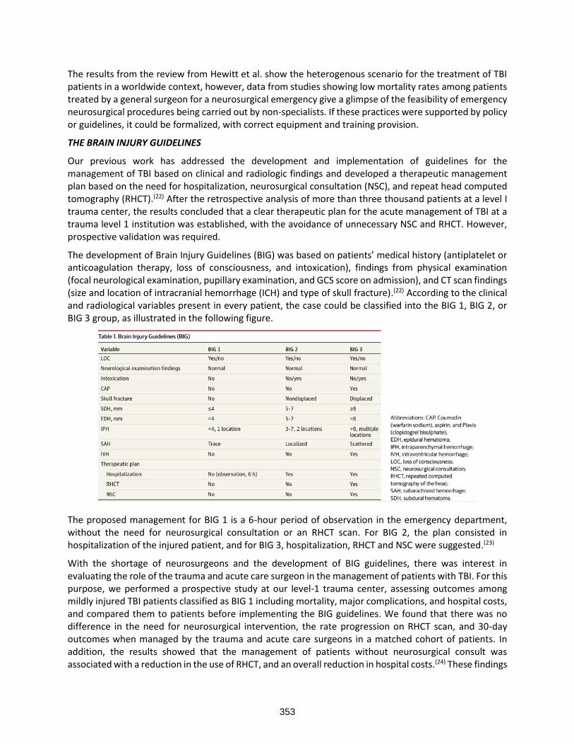
The results from the review from Hewitt et al. show the heterogenous scenario for the treatment of TBI patients in a worldwide context, however, data from studies showing low mortality rates among patients treated by a general surgeon for a neurosurgical emergency give a glimpse of the feasibility of emergency neurosurgical procedures being carried out by non-specialists. If these practices were supported by policy or guidelines, it could be formalized, with correct equipment and training provision.
THE BRAIN INJURY GUIDELINES
Our previous work has addressed the development and implementation of guidelines for the management of TBI based on clinical and radiologic findings and developed a therapeutic management plan based on the need for hospitalization, neurosurgical consultation (NSC), and repeat head computed tomography (RHCT).(22) After the retrospective analysis of more than three thousand patients at a level I trauma center, the results concluded that a clear therapeutic plan for the acute management of TBI at a trauma level 1 institution was established, with the avoidance of unnecessary NSC and RHCT. However, prospective validation was required.
The development of Brain Injury Guidelines (BIG) was based on patients’ medical history (antiplatelet or anticoagulation therapy, loss of consciousness, and intoxication), findings from physical examination (focal neurological examination, pupillary examination, and GCS score on admission), and CT scan findings (size and location of intracranial hemorrhage (ICH) and type of skull fracture).(22) According to the clinical and radiological variables present in every patient, the case could be classified into the BIG 1, BIG 2, or BIG 3 group, as illustrated in the following figure.
The proposed management for BIG 1 is a 6-hour period of observation in the emergency department, without the need for neurosurgical consultation or an RHCT scan. For BIG 2, the plan consisted in hospitalization of the injured patient, and for BIG 3, hospitalization, RHCT and NSC were suggested.(23)
With the shortage of neurosurgeons and the development of BIG guidelines, there was interest in evaluating the role of the trauma and acute care surgeon in the management of patients with TBI. For this purpose, we performed a prospective study at our level-1 trauma center, assessing outcomes among mildly injured TBI patients classified as BIG 1 including mortality, major complications, and hospital costs, and compared them to patients before implementing the BIG guidelines. We found that there was no difference in the need for neurosurgical intervention, the rate progression on RHCT scan, and 30-day outcomes when managed by the trauma and acute care surgeons in a matched cohort of patients. In addition, the results showed that the management of patients without neurosurgical consult was associated with a reduction in the use of RHCT, and an overall reduction in hospital costs.(24) These findings
353

demonstrated that the BIG were both safe and effective in the management of TBI, and that the role of the trauma surgeon in the management of minor TBI could be a prominent one. These results are also supported by Overton et al., who did a retrospective study of 171 mild TBI patients with an intracranial hemorrhage of 1 cm or less and a GCS of 13 or greater. They compared their outcomes before and after implementing a protocol of selective neurosurgical consultation. The results showed that the neurosurgical policy significantly reduced the number of such consults from 94% to 65%, and there was no association between neurosurgical consultation and neurological outcomes in this cohort of patients.(25)
With the emergence of data supporting the no difference in outcomes between TBI patients managed by trauma and acute care surgeons and neurosurgical consulted patients, there is increased interest in incorporating healthcare personal not from the neurosurgery arena in treating mild TBI patients. Zhao et al. retrospectively analyzed a case series at an academic level I trauma center, where isolated non-operative mild head injuries were admitted to three different services on a weekly basis (Trauma Surgery, Neurosurgery, and Neurology). They found no difference in survival, discharge disposition, hospital length of stay, in-hospital complications, and readmissions within 30 days among isolated non-operative mild head injuries managed by the three different services.(26) This highlights the possibility of incorporating a rotational policy of admitting mild TBI patients as a feasible option, and the opportunity for a better distribution of hospital resources and healthcare workforce.
The incorporation of the trauma and acute care surgeon in the management of mild TBI patients implies an alternative for the obvious paucity of neurosurgeons available for the treatment of these cases, however, attention must be given to possible challenges with the implementation of this policy; the healthcare providers from the trauma service must be familiarized with the detection of progression of the disease before clinical deterioration. In that way, those patients that could benefit from an earlier neurosurgical consult and possible intervention. The implementation of the previously described BIG guidelines could guide the management pathway in these circumstances.
THE OPTIMIZATION OF RESOURCES WITH BIG IMPLEMENTATION
After the development and implementation of the BIG for the managing of the TBI patient, we hypothesized that the treatment of mild TBI classified as BIG 1 patients by a trauma and acute care surgeon would not be different when compared to those patients where neurosurgery consultation was solicited, and in addition, the hospital resources would be better distributed, and the scarcity of neurosurgeons available for the acute care of brain injury patients would be addressed. With that in mind, we conducted a 2-year analysis of our prospectively maintained database of all patients with TBI who presented to our level I trauma center. We found that we could reduce unnecessary neurosurgical consultations, hospital admissions, and RHCT scans with BIG implementation. In addition, there was no difference in the outcomes, including rates of neurosurgical intervention, mortality, and post-discharge TBI-related ED visits and readmissions of patients who were treated before and after the BIG implementation. These results highlighted the safety and cost-effectiveness of BIG use for managing the TBI patients.(23)
Other studies have addressed the impact of selective management of TBI patients, without initial neurosurgery consult, similar to the BIG principle. We previously performed an analysis of our 5-year prospective database on all patients with TBI, and looked for imaging results, and hospital outcomes. The results showed that the management of trauma care in general and TBI in specific has changed significantly along the observed years, with a more selective use of RHCT scans, neurosurgical consultation, and ICU admission. On the other hand, trauma and acute care surgeons assumed a leading role in the management of non-operative mild to moderate TBI, decreasing the burden on overwhelmed neurosurgical services. The decreased trends of these practices were also associated with expedited
354

patient discharge and reduced hospital costs over time, however, no change in mortality rate or discharge GCS was observed.(27) The described trends can be observed in the following pictures.
Trends in repeat head computed tomographic scans after implication of BIG guidelines.
THE IMPACT OF BIG GUIDELINES
After all the evidence describing the positive impact in the distribution of hospital resources and diminish of burden to the neurosurgery service, the use of BIG for the TBI patients was extended to other areas, such as the pediatric population. With that purpose, we prospectively identified and enrolled BIG 1 pediatric patients who were managed without neurosurgical consult and compared them to those who received neurosurgical consultation. As a result, we found that trauma and acute care surgeons could safely and independently manage mild TBI pediatric patients, and that BIG use was associated with a decrease in repeat head CT scans without worsening outcomes.(28) With these results, there was evidence for the benefit of using the guidelines among different age populations, including the younger patients.
However, not all traumatic injured patients will be able to access a level I trauma center for emergency care in the actual world. Therefore, the feasibility of BIG implementation in other trauma centers needed to be addressed. To explore this, an extensive review was performed at a level III trauma center, where TBI patients were analyzed and stratified according to an updated version of BIG to fit better with the methodology of this analysis, with the replacement of the recommendation for neurosurgery consult with the need for transfer to a higher level of care. The results showed that BIG application in these circumstances was safe, without significant increases in the progression of hemorrhage on head CT scan, need for neurosurgical intervention, or mortality among the mildly injured BIG 1 and 2 patients who were allocated to staying at the Level III trauma center instead of being transferred to higher levels of care.(29) These findings highlight that trauma and acute care surgeons are reliably able to manage most TBI patients, especially minor TBI, and that this is independent of the level of resources that they have access to. The management of minor TBI by trauma and acute care surgeons is safe, while also being effective, and less resource-intensive as compared to management by the already-dwindling neurosurgical workforce.
355

Trends in neurosurgical consultations after implication of BIG guidelines.
NOT ALL INTRACRANIAL HEMORRHAGE REQUIRES NEUROSURGICAL CARE
When addressing the role of the trauma and acute care surgeon in the management of mild TBI, most of the time, we think of a trauma patient with concussion with no need for neurosurgical intervention. However, a concern arises from TBI with intracranial hemorrhage, such as subarachnoid hemorrhage or intraparenchymal haematoma. Ditty et al. did a retrospective analysis of 500 consecutively treated patients with mild TBI and subarachnoid hemorrhage and/or intraparenchymal hemorrhage admitted to a level I trauma center. Particular emphasis was made on diagnostic confirmation, neurological condition at admission, the occurrence of neurological decline episodes, or brain injury-related complications. However, after extensive analysis, they did not identify any patient who experienced neurological worsening during their hospital course, with only two episodes of hyponatremia reported.(20)
Repeat imaging is commonly performed in patients with mild intracranial hemorrhage; however, previous reports demonstrate that repeat imaging rarely reveals lesions requiring a change in clinical management, with lack of statistical evidence supporting repeat imaging in clinically stable or improving patients,(30) even with the presence of hemorrhagic findings on initial head CT scan. On the other hand, Sadrameli et al. conducted a retrospective analysis of 191 mild-moderate TBI patients over a 5-year period to examine whether follow-up CT initiated a change in management. They concluded that there was limited clinical value in repeat CT scans for patients with mild TBI, and most patients with traumatic SAH, contusions, or asymptomatic patients should not have repeat imaging, as their results revealed that only 2% of patients had a positive CT finding and 0.6% required surgical intervention.(31)
All this compelling information indicates that a proper selection of patients with mild-moderate TBI can be made for RHCT scan and neurosurgical consultation without worsening patient outcomes, even despite the presence of intracranial hemorrhage. However, there is concern about the external validity of data supporting the implementation of these patient management principles, which is addressed with a multi-institutional trial regarding the use of the BIG.
THE VALIDATION OF BIG AT THE MULTI-INSTITUTIONAL LEVEL
As previously shown, the BIG have been validated at a single level I Trauma Center, for the management of the TBI patient by the trauma and acute care surgery service. However, there was concern about the external validity of this management approach, as level I trauma centers have multiple resources and a
356

healthcare workforce with high expertise in treating trauma patients. To clarify this, we conducted a prospective multi-institutional trial, aiming to validate the guidelines at multiple institutions, with the hypothesis that BIG would reliably predict requirement of neurosurgical intervention, neurological examination worsening, progression on repeat head CT, post-discharge ED visits, and 30-days readmissions among patients presenting with TBI to one of ten participating level I and level II trauma centers.
After including all the TBI patients with positive head CT scan findings who met the inclusion criteria and stratifying them according to the BIG categories, we found no BIG 1 or BIG 2 patients requiring neurosurgical interventions, and only seven BIG 2 patients requiring upgrade to the BIG 3 category. If the BIG had been implemented in our study cohort, 425 RHCT, 401 prolonged hospitalizations, and 511 neurosurgical consultations would have been avoided in a study cohort of about two thousand patients. With the data provided by the multi-institutional validation of BIG, we can conclude that the guidelines establish a clear therapeutic plan for the management of TBI patients while avoiding unnecessary utilization of healthcare resources. The BIG are safe and effectively define the management of TBI by trauma and acute care surgeons and also appropriately predict when neurosurgical services should be involved.
CONCLUSIONS
Mild TBI is an entity that represents an area where the trauma and acute care surgeon can safely lead the management of the patients. The healthcare personnel should emphasize the importance of early detection of injury worsening for early imaging and neurosurgical consultations. The implementation of TBI guidelines, like BIG, standardizes the quality of care of these trauma patients, without worse outcomes and leading to improvement of resource utilization. The management of minor TBI by trauma and acute care surgeons is safe, while also being effective, and less resource-intensive as compared to management by the already-dwindling neurosurgical workforce.
REFERENCES
1. Prevention. CfDCa. National Center for Health Statistics: Mortality Data on CDC WONDER. [cited 2021 12/21/2021]. Available from: https://wonder.cdc.gov/mcd.html.
2. Bell JM, Breiding MJ, DePadilla L. CDC's efforts to improve traumatic brain injury surveillance. Journal of safety research. 2017;62:253-6.
3. Redelmeier DA, Tibshirani RJ, Evans L. Traffic-law enforcement and risk of death from motor-vehicle crashes: case-crossover study. The Lancet. 2003;361(9376):2177-82.
4. Maas AI, Stocchetti N, Bullock R. Moderate and severe traumatic brain injury in adults. The Lancet Neurology. 2008;7(8):728-41.
5. Oertel M, Boscardin WJ, Obrist WD, Glenn TC, McArthur DL, Gravori T, Lee JH, Martin NA. Posttraumatic vasospasm: the epidemiology, severity, and time course of an underestimated phenomenon: a prospective study performed in 299 patients. Journal of neurosurgery. 2005;103(5):812-24.
6. Teasdale G, Jennett B. Assessment of coma and impaired consciousness: a practical scale. The Lancet. 1974;304(7872):81-4.
7. Marshall LF, Marshall SB, Klauber MR, van Berkum Clark M, Eisenberg HM, Jane JA, Luerssen TG, Marmarou A, Foulkes MA. A new classification of head injury based on computerized tomography. Journal of neurosurgery. 1991;75(Supplement):S14-S20.
8. Ellemberg D, Henry LC, Macciocchi SN, Guskiewicz KM, Broglio SP. Advances in sport concussion assessment: from behavioral to brain imaging measures. Journal of neurotrauma. 2009;26(12):2365-82.
357

9. Jaffee MS, Helmick KM, Girard PD, Meyer KS, Dinegar K, George K. Acute clinical care and care coordination for traumatic brain injury within Department of Defense. Journal of Rehabilitation Research & Development. 2009;46(6).
10. Wright CJK, Zant E, Groom K, Schlegel RE, Gilliland K. Case report: Treatment of mild traumatic brain injury with hyperbaric oxygen. Undersea & Hyperbaric Medicine. 2009;36(6):391.
11. Prevention NCfI, Control. Report to Congress on mild traumatic brain injury in the United States: Steps to prevent a serious public health problem: Centers for Disease Control and Prevention; 2003.
12. Thurman D, Guerrero J. Trends in hospitalization associated with traumatic brain injury. Jama. 1999;282(10):954-7.
13. Vos PE, Alekseenko Y, Battistin L, Ehler E, Gerstenbrand F, Muresanu DF, Potapov A, Stepan CA, Traubner P, Vécsei L. Mild traumatic brain injury. European journal of neurology. 2012;19(2):191-8.
14. Pelc NJ. Recent and future directions in CT imaging. Annals of biomedical engineering. 2014;42(2):260-8.
15. Rahman S, McCarty JC, Gadkaree S, Semco RS, Bi WL, Dhand A, Jarman MP, Ortega G, Uribe-Leitz T, Bergmark RW. Disparities in the Geographic Distribution of Neurosurgeons in the United States: A Geospatial Analysis. World Neurosurgery. 2021.
16. Rosman J, Slane S, Dery B, Vogelbaum MA, Cohen-Gadol AA, Couldwell WT. Is there a shortage of neurosurgeons in the United States? Neurosurgery. 2013;73(2):354-66.
17. American College of Surgeons Health Policy Research Institute . Neurosurgery Subspecialty Supply Map. [Internet]. 2012 [cited January 2nd 2021].
18. Esposito TJ, Reed RL, II RLG, Luchette FA. Neurosurgical coverage: essential, desired, or irrelevant for good patient care and trauma center status. Annals of surgery. 2005;242(3):364.
19. BISHOFBERGER TE, DALEY BJ, SNYDER JR WE, ENDERSON BL. REPEAT HEAD CT AND NEUROSURGICAL CONSULTATION FOR MINOR TRAUMATIC HEAD INJURY IS NOT INDICATED IN NEUROLOGICALLY STABLE PATIENTS WITH GCS OF 14 OR 15. Journal of Trauma and Acute Care Surgery. 2004;57(2):450.
20. Ditty BJ, Omar NB, Foreman PM, Patel DM, Pritchard PR, Okor MO. The nonsurgical nature of patients with subarachnoid or intraparenchymal hemorrhage associated with mild traumatic brain injury. Journal of neurosurgery. 2015;123(3):649-53.
21. Hewitt JN, Ovenden CD, Glynatsis JM, Sabab A, Gupta AK, Kovoor JG, Wells AJ, Maddern GJ. Emergency Neurosurgery Performed by General Surgeons: A Systematic Review. World journal of surgery. 2021:1-9.
22. Joseph B, Friese RS, Sadoun M, Aziz H, Kulvatunyou N, Pandit V, Wynne J, Tang A, O’Keeffe T, Rhee P. The BIG (brain injury guidelines) project: defining the management of traumatic brain injury by acute care surgeons. Journal of Trauma and Acute Care Surgery. 2014;76(4):965-9.
23. Joseph B, Pandit V, Haider AA, Kulvatunyou N, Zangbar B, Tang A, Aziz H, Vercruysse G, O’Keeffe T, Freise RS. Improving hospital quality and costs in nonoperative traumatic brain injury: the role of acute care surgeons. JAMA surgery. 2015;150(9):866-72.
24. Joseph B, Aziz H, Pandit V, Kulvatunyou N, Sadoun M, Tang A, O’Keeffe T, Gries L, Green DJ, Friese RS. Prospective validation of the brain injury guidelines: managing traumatic brain injury without neurosurgical consultation. Journal of Trauma and Acute Care Surgery. 2014;77(6):984-8.
25. Overton TL, Shafi S, Cravens GF, Gandhi RR. Can trauma surgeons manage mild traumatic brain injuries? The American Journal of Surgery. 2014;208(5):806-10.
26. Zhao T, Mejaddam AY, Chang Y, DeMoya MA, King DR, Yeh DD, Kaafarani HM, Alam HB, Velmahos GC. Admissions for isolated nonoperative mild head injuries: sharing the burden among trauma
358

surgery, neurosurgery, and neurology. Journal of Trauma and Acute Care Surgery. 2016;81(4):743-7.
27. Joseph B, Haider AA, Pandit V, Tang A, Kulvatunyou N, O'Keeffe T, Rhee P. Changing paradigms in the management of 2184 patients with traumatic brain injury. Annals of surgery. 2015;262(3):440-8.
28. Azim A, Jehan FS, Rhee P, O’Keeffe T, Tang A, Vercruysse G, Kulvatunyou N, Latifi R, Joseph B. Big for small: Validating brain injury guidelines in pediatric traumatic brain injury. Journal of trauma and acute care surgery. 2017;83(6):1200-4.
29. Martin GE, Carroll CP, Plummer ZJ, Millar DA, Pritts TA, Makley AT, Joseph BA, Ngwenya LB, Goodman MD. Safety and efficacy of brain injury guidelines at a Level III trauma center. Journal of trauma and acute care surgery. 2018;84(3):483-9.
30. Almenawer SA, Bogza I, Yarascavitch B, Sne N, Farrokhyar F, Murty N, Reddy K. The value of scheduled repeat cranial computed tomography after mild head injury: single-center series and meta-analysis. Neurosurgery. 2013;72(1):56-64.
31. Sadrameli SS, Davidov V, Sulhan S, Vaziri S, Hartman CJ, Hooten KG, Murad GJ. The utility of routine post-hospitalization CT imaging in patients with non-operative mild to moderate traumatic brain injury. Brain Injury. 2021:1-5.
359

PREHOSPITAL BLOOD PRODUCTS
Ali Salim, MD, FACS
Division Chief, Trauma, Burns, and Surgical Critical Care Brigham and Woman’s Hospital Professor of Surgery Harvard Medical School Boston, MA
Hemorrhage is the leading cause of death after injury, with the mean time to hemorrhagic death at less than 3 hours after admission to a trauma center. Upon arrival at a trauma center at least 25% of severely injured patients are already coagulopathic. Damage control resuscitation has become the standard for which bleeding patients are managed. Damage control resuscitation focuses on rapid definitive hemorrhage control, permissive hypotension, minimization of crystalloid use, and immediate transfusion of high ratios of plasma and platelets to red blood cells. Use of balanced transfusion may lessen trauma induced coagulopathy and endothelial injury. Plasma transfusion in particular mitigates the coagulopathy that can complicate traumatic hemorrhage, alters the inflammatory response after injury, and reduces the permeability of endothelial cells after hemorrhagic shock. These benefits may be magnified when plasma is provided close to the time of injury. With earlier use of plasma to achieve higher ratios, there have been reports of lower overall blood product and decreased inflammatory consequences of shock. These outcomes appear to be closely correlated with the early achievement of higher ratios of plasma: RBCs.
Since the majority of deaths from traumatic hemorrhage continue to occur in the first hours after arrival at the trauma center, the initiation of damage control resuscitation in the prehospital environment has significant merit. It has the potential to reduce downstream complications attributable to hemorrhage by intervening close to the time of injury, before the development of coagulopathy, irreversible shock, and the ensuing inflammatory response.
PREHOSPITAL TRANSFUSION
Prehospital blood product resuscitation is not a new concept. The administration of plasma in the field was introduced in World War II. Since then, the military has led investigations of prehospital blood product administration. Both UK and US medevac platforms have developed PRBC transfusion capabilities. Outcomes of its use has demonstrated improved mortality in combat settings such that the Tactical Combat casualty Care (TCCC) now recommends whole blood, PRBC and plasma, and plasma or PRBC in order of preference when available for field and tactical evacuation resuscitation. As PRBC administration became more practical, it was adopted by many air medical transport agencies. Early reports demonstrated that use of prehospital PRBC was safe and feasible among air transport providers. Preliminary work demonstrated a reduction in mortality, coagulopathy, PRBC requirements at the trauma center and lower risk of shock. With this preliminary evidence and the growing acceptance of damage control resuscitation, some air transport programs began to develop prehospital plasma transfusion programs in addition to PRBC capabilities. Further preliminary studies demonstrated lower 6-hour mortality, lower transfusion requirement and less shock as well as improved coagulation parameters. These same reports demonstrated that of the patients transfused prehospital, 85% continued to receive transfusion in the hospital, suggesting minimal overuse of blood products. Of note, despite the concern that placing blood products in the prehospital setting would lead to blood waste, the data suggests that
360

only less than 2% of units are lost to expiration. An early systemic review evaluating prehospital blood product resuscitation did not demonstrate improved outcomes but found the literature to be limited and of poor quality, recommending that randomized prospective trials be performed. In response to all of this, the Department of Defense issued the Prehospital Use of Plasma for Traumatic Hemorrhage program announcement, which resulted in the PAMPer and COMBAT Trials discussed below.
PAMPER TRIAL
The Prehospital Air Medical Plasma (PAMPer) trial was designed to determine the efficacy and safety of prehospital plasma resuscitation as compared with standard-care resuscitation in severely injured patients at risk for hemorrhagic shock. The trial was a pragmatic, multicenter, cluster-randomized, phase 3 trial involving injured patients who were at risk for hemorrhagic shock during air medical transport to a trauma center. Outcomes in patients who received 2 units of thawed plasma (either group AB or group A with low anti-B antibody titer) were compared with outcomes in those who received standard care resuscitation. No other aspect of treatment was altered other than the administration of plasma. The trial included injured patients transported from the scene or were transferred from an outside referral emergency department. Patients were eligible for enrollment if they had at least one episode of hypotension (SBP < 90 mm Hg) and tachycardia (HR > 108) or if they had any severe hypotension (SBP < 70) either before the arrival of air medical transport or any time before arrival at the trauma center. The intervention consisted of the administration of 2 units of thawed plasma once a patient met inclusion criteria. Patients that received standard care resuscitation included the infusion of crystalloids. Of note, half of the air transport teams also carried 2 units of universal donor red cells on all their flights and the indications for transfusion followed local protocols.
A total of 501 patients were included: 230 assigned to the plasma group and 271 assigned to the standard care group. The median prehospital transport time was 42 minutes in the plasma group and 40 minutes in the standard care group. The mortality at 30 days was lower in the plasma group compared to the standard care group (23% vs. 33%, p=0.03). The patients in the plasma group also had a lower median prothrombin-time ratio while no differences were observed between the two groups with respect to multiorgan failure, acute lung injury-acute respiratory distress syndrome, nosocomial infections, or allergic or transfusion-related reactions.
COMBAT TRIAL
The Control of Major Bleeding After Trauma Trial (COMBAT) assessed the use of prehospital plasma during short ground transportation to an urban trauma center. In the COMBAT trial, the investigators investigated whether plasma first resuscitation affected trauma-induced coagulopathy and adverse outcomes after injury in patients with hemorrhagic shock. COMBAT was a pragmatic, randomized, placebo-controlled, clinical trial. Eligible patients were injured adults (age > 18), with SBP ≤ 70 mm Hg or 71-90 mm Hg and HR > 108 thought to be due to acute blood loss. All ambulances that participated in the trial transported all of their patients to one single Level I trauma center. Patients were randomized to receive 2 units of AB plasma or normal saline per standard of care. A total of 144 patients were randomly assigned but the analysis included 125 patients (65 in the plasma group and 60 in the control group). The median transport time was 19 minutes in the plasma group and 16 minutes in the control group. The time from injury to transfusion of first plasma unit was 59 minutes in the control group and 24 minutes in the plasma group. The mortality at 28 days did not differ between the two groups (15% in the plasma group vs. 10% in the control group, p=0.37). Coagulation factors, transfusion requirements and safety outcomes (acute lung injury, multiorgan failure, and other complications) were similar in the two groups. The time from injury to first red blood cell transfusion was longer in the plasma group. The authors concluded that plasma does not improve outcomes after injury when given within 30 minutes during rapid ground transportation to a mature level I trauma center.
361

PAMPER AND COMBAT
Although these studies had conflicting results, they did demonstrate that the pre-hospital administration of plasma to trauma patients is technically/logistically feasible both in air and ground EMS. The conflicting results obviously raised more questions. Further analyses were subsequently undertaken to gain more clarity regarding the role of prehospital plasma transfusion.
Using the PAMPer study, the investigators performed a secondary analysis to determine whether prehospital blood product resuscitation reduces 30-day mortality in patients at risk for hemorrhagic shock compared with crystalloid only resuscitation. In the original PAMPer trial, half of the helicopter emergency medical services included transfusion of up to 2 units of type O-PRBC as part of their resuscitation protocol. The authors found that there was a significant reduction of 30-day mortality per unit of prehospital PRBC and per unit of prehospital plasma. When evaluating only patients who received a prehospital blood product, each liter of crystalloid was associated with a 65% increase in 30-day mortality. Any blood product resuscitation was associated with lower mortality than crystalloid only resuscitation. PRBC and plasma had similar reductions in mortality; however, PRBC + plasma had a much greater reduction in mortality than either PRBC or plasma alone.
The authors concluded that patients with signs of shock should receive prehospital blood products whenever available. Crystalloids alone appear to be inferior to blood products and has a dose response increase in this setting (crystalloid volume was associated with increased mortality among patients receiving blood products). Overall patients receiving PRBC and plasma had the greatest benefit, followed by plasma alone, and PRBC respectively compared to crystalloid only resuscitation. If both PRBC and plasma are available, patients should receive both as this was associated with the greatest mortality reduction.
The additive benefit of PRBC + plasma suggests that there may be benefit to the use of whole blood in the prehospital setting. Whole blood has been shown to be safe and feasible. There is a current trial evaluating the outcomes of prehospital whole blood compared to fractionated blood products (Pragmatic Prehospital Group O Whole Blood Early Resuscitation [PPOWER] trial).
IMPACT OF PREHOSPITAL TIME
Evidence generally supports early plasma transfusion after injury, but how early after injury plasma resuscitation is beneficial remains unanswered. As the COMBAT trial demonstrated, the effort to thaw and transfuse plasma in urban areas with short transport times to trauma centers might outweigh any benefits.
While reasons for the conflicting results between the PAMPER and COMBAT trials are unclear, one thought is that the very short prehospital transport times in the ground ambulance study may have eliminated the potential for prehospital plasma to improve survival because in-hospital transfusion was not delayed significantly by transport. To address this, the data sets were combined to address the post hoc hypothesis that the benefits of prehospital administration of plasma are influenced by prehospital transport time.
The clinical trials were harmonized to enable combined per-patient analysis to address questions that could not be answered by either trial individually. The 28-day mortality was lower in the plasma group than the standard care group (20.5% vs. 28.6%, p=0.02). Most deaths in both groups occurred within the first 6 hours after injury. A regression model showed that in addition to the treatment group, survival was influenced by ISS, age, and prehospital transport time. Sensitivity analysis revealed that a change in response was evident for prehospital times longer than 20 minutes. In patients who received standard care, rate and likelihood of mortality were significantly increased by 2-fold with transport times greater
362

than 20 minutes (HR 2.12; 95% CI: 1.05-4.30, p=0.04). Among patients who received prehospital plasma, this association with transport time was eliminated. Among patients with short transport times (< 20 minutes), survival in the plasma group and the standard care group did not differ. Among patients with longer transport times (> 20 minutes), survival was improved in the plasma group (HR 0.56; 95% CI: 0.40-0.80; p=0.001).
Patients who received prehospital plasma were 47% less likely to present in the ED with coagulopathy (INR > 1.3) compared with those who received standard care. And this association was isolated to the group with transport times longer than 20 minutes. Patients who received plasma during longer transport required less in hospital transfusions of plasma, RBC, and of platelets at 6 hours and 24 hours after ED admission.
The authors concluded that prehospital transport time influenced the response to prehospital plasma. Prehospital plasma is associated with a survival benefit when transport times are longer than 20 minutes and the benefit-risk ratio favors prehospital plasma.
THE MASSACHUSETTS EXPERIENCE
The helicopter programs in Massachusetts have a fairly recent history in carrying blood. Our Boston specific air medical transport system started a blood protocol in November 2019. They carry 2 units O positive PRBC (this was a switch from O neg when the pandemic started), 1 unit A Liquid, never frozen, Plasma in each vehicle (helicopter, ground, and airplane). Since starting the program, more plasma has been used than PRBC due to the high number of anti-coagulated patients coming from hospitals without rapid access to FFP or other reversal agents along with the push for plasma first in acute trauma related hemorrhage.
CONCLUSIONS
Early transfusion with balanced ratios has been incorporated into military and civilian clinical practice guidelines. Based on data suggesting significant benefits from prehospital transfusion – PRBC administration has become the standard of care when available for wounded casualties. Prehospital blood products (defined as PRBC, plasma, or both PRBC and plasma) are increasingly available through civilian helicopter emergency medical services. Although logistical and cost constraints may limit feasibility, thawed plasma is a viable option for helicopter ambulance systems but is more challenging for ground ambulances with short transport times. Whole blood could overcome the logistical challenges of storing both PRBC and plasma. Other developments such as freeze-dried plasma may also mitigate the costs and storage challenges of traditional cold storage blood products.
REFERENCES
1. Brown JB, Cohen MJ, Minei JP, Maier RV, West MA, Billiar TR, et al. Pretrauma center red blood cell transfusion is associated with reduced mortality and coagulapathy in severely injured patients with blunt trauma. Annals of Surgery. 2015;261(5):997-1005.
2. Brown JB, Sperry JL, Fombona A, Billiar T, Peitzman AB, Guyette FX. Pre-trauma center red blood cell transfusion is associated with improved early outcomes in air medical trauma patients. J Am Coll Surg. 2015;220(5):797-808.
3. Fenger-Eriksen C, Fries D, David JS, Bouzat P, Lance MD, Grottke O, et al. Pre-hospital plasma transfusion: A valuable coagulation support or an expensive fluid therapy? Critical Care. 2019;23(238):1-
4. Gruen DS, Guyette FX, Brown JB, Okonkwo DO, Puccio AM, Campwala IK, et al. Association of prehospital plasma with survival in patients with traumatic brain injury: A secondary analysis of the PAMPer cluster randomized clinical trial. JAMA Network Open. 2020;3(10):1-15.
363

5. Guyette FX, Sperry JL, Peitzman AB, Billiar TR, Daley BJ, Miller RS, et al. Prehospital blood product and crystalloid resuscitation in the severely injured patient: A secondary analysis of the prehospital air medical plasma trial. Annals of Surgery. 2021;273(2):358-64.
6. Holcomb JB, Donathan DP, Cotton BA, del Junco DJ, Brown G, von Wenckstern T, et al. Prehospital transfusion of plamsa and red blood cells in trauma patients. Prehospital Emergency Care. 2015;19(1):1-9.
7. Kim BD, Zielinski MD, Jenkins DH, Schiller HJ, Berns KS, Zietlow SP. The effects of prehospital plamsa on patients with injury: A prehospital plasma resuscitation. J Trauma Acute Care Surg. 2012;72(2):S49-53.
8. Makris M, Iorio A. Prehospital fresh frozen plasma: Universal life saver or treatment in search of a target population? Res Pract Thromb Haemost. 2019;3(12):12-14.
9. Moore HB, Moore EE, Chapman MP, McVaney K, Bryskiewicz G, Blechar R, et al. Plasma-first resuscitation to treat haemorrhagic shock during emergency ground transportation in an urban area: A randomised trial. The Lancet. 2018;392:283-91.
10. Powell EK, Hinckley WR, Gottula A, Hart KW, Lindsell CJ, McMullan JT. Shorter times to packed red blood cell transfusion are associated with decreased risk of death in traumatically injured patients. J Trauma Acute Care Surg. 2016;81(3):458-62.
11. Pusateri AE, Moore EE, Moore HB, Le TD, Guyette FX, Chapman MP, et al. Association of prehospital plasma transfusion with survival in trauma patients with hemorrhagic shock when transport times are longer than 20 minutes: A post hoc analysis of the PAMPer and COMBAT clinical trials. JAMA Surg. 2020;155(2):1-10.
12. Smith IM, James RH, Dretzke J, Midwinter MJ. Prehospital blood product resuscitation for trauma: A systematic review. Shock. 2016;46(1):3-16.
13. Sperry JL, Guyette FX, Brown JB, Yazer MH, Triulzi DJ, Early-Young BJ, et al. Prehospital plasma during air medical transport in trauma patients at risk for hemorrhagic shock. N Engl J Med. 2018;379(4):315-26.
14. Zielinski MD, Stubbs JR, Berns KS, Glassberg E, Murdock AD, Shinar E, et al. Prehospital blood transfusion programs: Capabilities and lessons learned. J Trauma Acute Care Surg. 2017;82(6):S70-78.
364

SESSION 11
CASE MANAGEMENT “Strictly Rural”
Moderator: Richard A. Sidwell, MD, FACS
Tuesday, March 29, 2022
3:25 p.m. - 4:25 p.m.
Palace Ballroom 1-2
Palace Tower, Emperors Level
Panelists: Stephen L. Barnes Andrew C. Bernard Jeffrey J. Skubic Dustin L. Smoot Alison Wilson
365

366

SESSION 12
COMPLICATIONS OF TRAUMA & ACUTE CARE SURGERY
Moderator: Kenji Inaba, MD, FRCSC, FACS
Tuesday, March 29, 2022
Palace Ballroom 1-2
Palace Tower, Emperors Level
4:45 - 5:00
Tourniquets: The Good, The Bad, The Ugly Alexander L. Eastman, MD, MPH, FACS, FAEMS
5:00 - 5:15
Geriatric Trauma Complications - Pointing the Finger of Blame Bellal A. Joseph, MD, FACS
5:15 - 5:30
Iatrogenic Time Management Complications Alan H. Tyroch, MD, FACS, FCCM
5:30 - 5:45
Incision and Exposure Choices Can Lead to Complications Demetrios Demetriades, MD, MPH, FACS
5:45 - 6:00
The Great Contrast Conspiracy: Shattering Myths Dennis Y. Kim, MD, FACS, FRCSC, FACS, FCCP
6:00 - 6:30
Panel Discussion
367

PREHOSPITAL TOURNIQUETS: THE GOOD, THE BAD AND THE UGLY
Alexander L. Eastman, MD, MPH, FACS, FAEMS
Senior Medical Officer – Operations US Department of Homeland Security Lieutenant and Chief Medical Officer Dallas Police Department Dallas, TX
BLUF
Prehospital tourniquets save lives. Period. However, like any tool used in medicine, and particularly those applied outside the protective confines of the hospital, the extremity tourniquet has both advantages and limitations that must be clearly articulated and understood not just by the individual providers utilizing them but also by EMS system medical directors, receiving trauma surgeons and emergency physicians.
BACKGROUND
With reported use as far back as least as ancient Rome, tourniquet use has waxed and waned over the history of prehospital care. While the fundamental design of the extremity tourniquet has not changed dramatically, both their use and the systems in which we find them have evolved to show them to be an invaluable tool in the armamentarium of the modern prehospital provider. After exceedingly poor outcomes during the US Civil War and first World War, prehospital tourniquet use became nearly a verboten entity.1 However, after the now famous Battle of the Black Sea in Mogadishu, Somalia, and the subsequent development of Tactical Combat Casualty Care (TCCC), the tourniquet has been proven to be relatively easy to use, exceedingly safe when used properly and to be perhaps the singularly most effective prehospital tool fielded in the modern era of EMS.2
TOURNIQUETS: THE EARLY ERA
As noted in the introduction, the tourniquet as a tool hasn’t changed demonstratively since its inception. Sure, improvements in design and function have followed need and are beyond the scope of this review, however in discussing the modern resurgence of the prehospital tourniquet, it is impossible to ignore the improvements in systems of care that have come by virtue of the development of Tactical Casualty Combat Care. After the preventable deaths of several special operators and soldiers, the Department of Defense turned to a retired Navy SEAL turned ophthalmologist to develop a system of care subsequently replicated worldwide. TCCC, now a part of the Joint Trauma System, divided care into easily understood phases (Care Under Fire, Tactical Field Care and Tactical Evacuation Care) and placed an emphasis on evidence-based interventions proven in military settings to save lives.3
Even in this specialized setting, the earliest military studies of modern prehospital tourniquet use failed to show a clear survival benefit. However, Beekley et al, who published the initial study in this era showed that in early experience at war, even with tourniquet use less widespread that it ultimately became, in the first 67 patients, prehospital tourniquet use showed improvements in hemorrhage control compared to 98 patients treated without tourniquets.4 While there were no clear adverse effects associated with tourniquet use, the medical record system in place today was still under construction and hence, data with which to base longer-term follow up wasn’t available. While small and retrospective in design, perhaps the true landmark finding from this early study was the fact that few of these tourniquets were
368

applied by medical providers and hence it truly formed the foundations of what would eventually become widespread, public access civilian hemorrhage control programs.
Once the Joint Trauma System trauma registry project was completed, accurate data from a relatively homogenous population of injured US servicemen and women allowed for further study of the use of prehospital tourniquets and the real transition to the modern era of prehospital tourniquet research was launched. In perhaps the largest and most comprehensive prospective study at the time,5 data was gathered from the Combat Support Hospital in Baghdad over a seven-month period. 2,838 patients with major limb injuries, 232 (8%) of which had 428 tourniquets placed on 309 limbs. A second tourniquet was used on occasions where one was insufficient to stop hemorrhage. No patient suffered a major complication with only a single nerve palsy still present at more than a week out. An interesting note from this study was that more than 97% of these tourniquets were deemed to be properly used and positioned.
TOURNIQUETS: THE MODERN ERA
In 2013, a group of public safety personnel from fire, law enforcement, pre-hospital care, trauma care and the military convened in Hartford, Connecticut, to develop consensus regarding strategies to increase survivability in mass public shootings.6 With a push from the White House’s National Security Council staff, these thought leaders from across the response continuum developed a novel strategy in increase civilian tourniquet use across several settings…most notably in law enforcement, prehospital EMS (where uptake had been slow to this point) and in the wider, public sector. Applying lessons learned from the military, the group of experts developed the acronym THREAT to address casualty management during high- threat tactical and rescue operations. Figure 1 described THREAT and its applications. While reasonably simple, the Hartford Consensus and THREAT were exactly what many in the law enforcement community needed to push hard on the use of prehospital tourniquets applied by nontraditional medical providers—exactly the hard-earned lessons learned from the US military in the relatively early years of the resurgence of prehospital tourniquet use.
Figure 1. THREAT. Adapted courtesy of The Hartford Consensus
Law enforcement responded very well to the Hartford Consensus, and now, hemorrhage control by police officers (who are often on-scene at violent incidents well ahead of more traditional rescuers), often termed “self-aid/buddy-aid” or SABA, has become as core a law enforcement skill as de-escalation, driving, interviewing and firearm use. Interestingly, quickly and with the science only following later, an unintended consequence of the Hartford Consensus was the spawning of a new, cottage industry in hemorrhage control products and training pointed at this market.
369
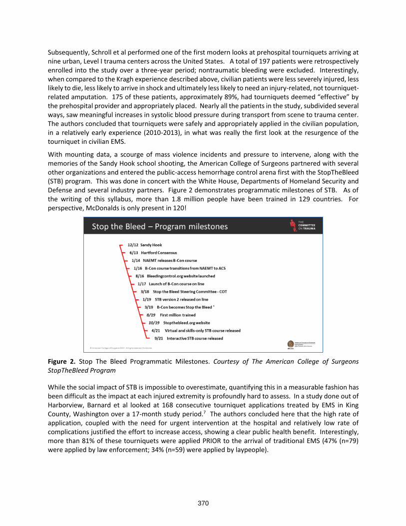
Subsequently, Schroll et al performed one of the first modern looks at prehospital tourniquets arriving at nine urban, Level I trauma centers across the United States. A total of 197 patients were retrospectively enrolled into the study over a three-year period; nontraumatic bleeding were excluded. Interestingly, when compared to the Kragh experience described above, civilian patients were less severely injured, less likely to die, less likely to arrive in shock and ultimately less likely to need an injury-related, not tourniquet-related amputation. 175 of these patients, approximately 89%, had tourniquets deemed “effective” by the prehospital provider and appropriately placed. Nearly all the patients in the study, subdivided several ways, saw meaningful increases in systolic blood pressure during transport from scene to trauma center. The authors concluded that tourniquets were safely and appropriately applied in the civilian population, in a relatively early experience (2010-2013), in what was really the first look at the resurgence of the tourniquet in civilian EMS.
With mounting data, a scourge of mass violence incidents and pressure to intervene, along with the memories of the Sandy Hook school shooting, the American College of Surgeons partnered with several other organizations and entered the public-access hemorrhage control arena first with the StopTheBleed (STB) program. This was done in concert with the White House, Departments of Homeland Security and Defense and several industry partners. Figure 2 demonstrates programmatic milestones of STB. As of the writing of this syllabus, more than 1.8 million people have been trained in 129 countries. For perspective, McDonalds is only present in 120!
Figure 2. Stop The Bleed Programmatic Milestones. Courtesy of The American College of Surgeons StopTheBleed Program
While the social impact of STB is impossible to overestimate, quantifying this in a measurable fashion has been difficult as the impact at each injured extremity is profoundly hard to assess. In a study done out of Harborview, Barnard et al looked at 168 consecutive tourniquet applications treated by EMS in King County, Washington over a 17-month study period.7 The authors concluded here that the high rate of application, coupled with the need for urgent intervention at the hospital and relatively low rate of complications justified the effort to increase access, showing a clear public health benefit. Interestingly, more than 81% of these tourniquets were applied PRIOR to the arrival of traditional EMS (47% (n=79) were applied by law enforcement; 34% (n=59) were applied by laypeople).
370
TOURNIQUETS: THE NAYSAYERS
Prehospital tourniquet application, and particularly the large expansion along the spectrum from law enforcement application to public access hemorrhage control programs like STB, are not without their detractors. There are several case reports of inappropriate and clinically unindicated applications of prehospital tourniquets leading to everything from mild, transient nerve palsy up to and including limb loss. Mikdad and colleagues from the Massachusetts General Hospital examined a series of 147 patients over five years.8 They noted an increasing percentage of nonemergency medical services personnel over the study period and that nearly half of the tourniquets placed lacked a clinical indication for placement. An additional study, also from Boston, documented several cases of inappropriate extremity tourniquet use at the Boston Marathon bombing.9 While not a cause for alarm, nor reason enough to slow the inertia inherent in current efforts to expand programs, their results were certainly indicative of the need to take a close look at the regional differences in training programs and equipment placement.
CONCLUSIONS
Despite a rocky course from their inception, the prehospital tourniquet has proven itself as a valuable tool in the armamentarium of prehospital trauma care, whether fielded by the soldier, layperson, law enforcement officer or professional rescuer. In addition to countless lives saved on the field of battle, many have been saved by the judicious use of tourniquets all over the world. Reports of inappropriate applications and scant complications when used properly should prompt further investigation into optimal training and placement, rather than abandonment.
REFERENCES
1. Doyle GS and Taillac PP. Tourniquets: A review of current use with proposals for expanded prehospital use. Prehospital Emergency Care. 2008; 12: 241-256.
2. Beekley AC, Sebesta JA, Blackbourne LH, et al. Prehospital tourniquet use in Operation Iraqi Freedom: Effect on hemorrhage control and outcomes. Journal of Trauma. 2008; 64: S28-S37.
3. Butler, F. K.; Hagmann, J.; Butler, E. G. (1996-08-01). "Tactical combat casualty care in special operations". Military Medicine. 161 Suppl: 3–16. doi:10.1007/978-3-319-56780-8
4. Beekley AC, Sebesta JA, Blackbourne LH, et al. Prehospital tourniquet use in Operation Iraqi Freedom: Effect on hemorrhage control and outcomes. Journal of Trauma. 2008; 64: S28-S37.
5. Kragh JF, Walters TJ, Baer DG, et al. Practical use of emergency tourniquets to stop bleeding in major limb trauma. Journal of Trauma. 2008;64: S38-S50.
6. American College of Surgeons “The Hartford Consensus.” (2013). Multiple JACS articles. See also the ACS Bulletin Supplement.
7. Leslie M. Barnard LM, Guan S and Zarmer L. et al. Prehospital tourniquet use: An evaluation of community application and outcome. J Trauma Acute Care Surg. 2021;90: 1040–1047.
8. Mikdad S, Mokhtari AK, Luckhurst CM et al. Implications of the national Stop the Bleed campaign: The swinging pendulum of prehospital tourniquet application in civilian limb trauma. J Trauma Acute Care Surg. 2021;91: 352–360.
9. King DR, Larentzakis A, Ramly EP, Boston Trauma Collaborative. Tourniquet use at the Boston Marathon Bombing: Lost in Translation J Trauma Acute Care Surg 2015;78:594–9.
371

GERIATRIC TRAUMA COMPLICATIONS – POINTING THE FINGERS OF BLAME
Bellal A. Joseph, MD, FACS
Professor of Surgery University of Arizona Medical Director, Southern Arizona Telemedicine and Telepresence (SATT) Prog Tucson, AZ
Currently, in the United States, the fastest growing demographic is considered to be geriatric (age >65 years). In 2014, 15% of the population was geriatric, and by 2030, it will grow to 21%. Deaths from unintentional injuries are the seventh leading cause of death among older adults and as the US population ages, there will be an increasing volume of geriatric trauma patients (GTPs). Geriatric trauma is increasing both in absolute number and as a proportion of annual volume presenting to trauma centers. Based on data from the National Trauma Data Bank, the proportion of trauma patients aged 65 years or older in Level I and II trauma centers increased from 23% in 2003 to 30% in 2009. This is likely a significant underestimate because most GTPs are treated at lower-level or non-trauma centers. Conversely, cancer and heart disease death rates have decreased since 2000. The changing epidemiology of trauma mortality must be a focus of robust future investigation to make strides in preventing and treating trauma, an ever-increasing epidemic of morbidity and mortality in our era.
Figure 1: Cumulative trauma deaths by age in 2000 and 2010. A higher peak and a right shift in trauma deaths were identified over time. Source:https://journals.lww.com/annalsofsurgery/Fulltext/2014/07000/Increasing_Trauma_Deaths_in_the_United_States.5.aspx
It has been repeatedly demonstrated that GTPs have outcomes worse than those of younger trauma patients. Older age was associated with higher in-hospital mortality, longer hospital stay and greater admission to nursing homes compared with patients younger than 65. Elderly patients have been found to have higher mortality after trauma as well as higher complication rates, and a higher associated mortality rate for all types of complications when compared to younger patients. The hospital stay for elderly is twice as long as younger patients. During the first month of hospitalization the death rate for
372

the elderly is three times higher and the difference was even greater if the patient survived more than 30 days, indicating that older patients are more likely to succumb to a complication of the initial injury when compared to younger patients. The most common complications are infections. Respiratory failure and cardiac complications are also common.
Figure 2: Complication rates among severely injured geriatric populations. Source:https://journals.lww.com/jtrauma/fulltext/2011/12000/Severely_Injured_Geriatric_Population__Morbidity,.68.aspx
Care of GTPs is also more resource intensive than caring for younger patients with similar injuries. The elderly contribute to 80% of the total days of hospitalization, which is a much higher percentage than the proportion of the elderly trauma population itself. This does not take into account the costs of rehabilitation, nursing home, and indirect costs.
The reasons behind these distinct patterns and high rates of complications and worse outcomes following trauma among GTPs are likely multifactorial and can be roughly divided into patient-related and hospital/system-related factors. The aim of this review is to identify and describe these factors and their contribution towards the outcomes of GTPs.
FACTORS LEADING TO POOR OUTCOMES IN GERIATRIC TRAUMA PATIENTS (GTPS)
Definition of the Geriatric Trauma Patient (GTP)
Published reports on the care of the elderly are inconsistent in the age definition of what constitutes the GTP. This lack of clarity introduces a problem with generalizing the findings of such studies. The most commonly used age is 65 years and older, which is the definition that is utilized in this review. There is also a problem with delineating what constitutes trauma for the elderly, particularly as related to ground-level and nonmechanical falls and the resultant injury, such as a hip fracture. Hip fractures are included in some reports and not in others as an injury pattern defining geriatric trauma, which obfuscates the reporting of complications and mortality in the GTP. Further, the definition of what constitutes as a major complication in the GTP population has not been clearly elucidated, leading to differences in reporting that may lead to under- or over-reporting of complications and associated factors. Despite this lack of clarity in definitions for GTPs, a compilation of patient-related and hospital/system related factors that
373

are associated with outcomes of GTPs identified from the prevailing literature on the subject has been provided below.
Patient-related Factors
Frailty
Over the last few decades, multiple healthcare disciplines, including the field of trauma surgery, have focused on the concept of frailty to identify the subset of the geriatric population at high risk for poor outcomes following illness. Frailty, an indicator of senescence, is clinically distinct from age, comorbidity, and functional disability. The frailty syndrome is broadly considered as decreased physiologic reserve across multiple organ systems leading to an impaired ability to withstand physiologic stress. The prevalence of frailty in the GTP population is high, and understanding it is relevant for trauma surgeons because frailty is associated with injury following falls, frail trauma patients are more likely to develop in-hospital complications, and more likely to have adverse discharge disposition than non-frail patients.
Recent data show that frailty is more predictive of in-hospital complications and adverse discharge disposition than age alone in geriatric trauma patients. This trend remains true of both short-term in-hospital complications such as rates of delirium, infectious, respiratory, and cardiac complications, and in-hospital mortality, as well as long-term outcomes, including long-term functional independence, trauma-related readmissions and long-term mortality, and health-related quality of life. Multiple studies have identified frailty as an independent predictor of worse outcomes among the elderly, and especially within the GTP population. It is imperative then, to accurately identify this high-risk patient population early in the course of management, to tailor care specifically towards preventing complications and achieving optimal short- and long-term outcomes. Indeed, simply adhering to routine frailty measurement in the care of GTPs has been found to be accompanied by a significant decline in overall in-hospital complications and mortality. Hence, early assessment and identification of these vulnerable patients is critical in optimizing outcomes in geriatric trauma patients.
There are multiple models for defining frailty. Two popular models are the deficit accumulation model, which considers frailty as a reflection of health deficits across several domains (disabilities, comorbidities, symptoms, signs, and laboratory data) and the phenotypic model of frailty, which is based on the concepts of physical disability and energy depletion as predictors of worse outcomes. Based on the many different models defining frailty syndrome, many measurement tools have been developed to measure frailty, each with varying degrees of success in defining the full spectrum of this condition in geriatric patients. Examples include the Rockwood and Fried frailty indices, and the American College of Surgeons Frailty calculator. However, these models lack feasibility in the GTP because they require assessment of up to 30 to 70 variables, many of which (i.e., gait speed and hand grip strength) cannot be performed on the GTP.
Limitations of the existing frailty measures prompted the development of the modified 15-variable Trauma-Specific Frailty Index (TSFI), a tool designed to be specific to the GTP population to accurately predict worse outcomes including major complications. The TSFI has been validated as an independent predictor of unfavorable discharge disposition in geriatric trauma patients. The TSFI is an effective tool that can aid clinicians in identifying high-risk patients and planning care and discharge disposition of vulnerable GTPs. The 15-variable TSFI is an equally effective predictor of mortality, in-hospital complications, adverse discharge disposition and 30-day readmission compared to the more comprehensive 50-variable Rockwood frailty score. However, the TSFI was also found to be a stronger and better predictor of worse outcomes compared to the modified frailty index (mFI) and frailty scale (FS) in trauma patients. Additionally, the TSFI only requires the assessment of 15 variables, which has been proven to be more practical in assessing GTPs.
374

Table I: Fifteen Variable Trauma Specific Frailty Index (TSFI) Source:https://www.sciencedirect.com/science/article/pii/S1072751514002609
Age-Specific Alterations in Response to Injury
Providers caring for GTPs should be aware of the impact of aging on specific organ functions and how this might increase the likelihood of complications and affect common interventions. For those older than 50 years, renal mass is lost, decreasing glomerular filtration rate and increasing the risk of acute kidney injury. It is important to avoid unnecessary contrast exposure, adjust medication dosing based on renal function, and insure euvolemia. Respiratory function is also commonly compromised. There is an observed loss of the lung elastic recoil and significant reduction of the vital capacity. Decreased ventilation-perfusion mismatch with age decreases baseline arterial oxygen tension. Alterations in compliance and airway resistance result in an increased work of breathing. The combination of these factors increases the need for mechanical ventilation and difficulty in subsequent weaning.
Shock in the elderly may not be readily recognized. The aging myocardium is less able to respond to circulating catecholamines. Therefore, the elderly patient may not develop tachycardia in the presence of hypovolemia. As many elderly patients have hypertension, a “normal” or borderline blood pressure should be treated with suspicion. A systolic blood pressure less than 110 mm Hg may indicate a relative hypotension in the GTP. Occult hypoperfusion may be detected by monitoring tissue hemoglobin oxygen saturation (StO2) or by measuring a base deficit or lactate. However, GTPs do not develop the magnitude of base deficit or lactate as younger patients, so these parameters must be evaluated with great care. Additionally, super-elderly patients (≥80 years old) have significantly greater risk-adjusted in-hospital mortality and complication rates than elderly patients (60–79 years old) after injury, and are more likely to require non-routine discharge if they survive such injury.
Comorbidities
Older trauma patients with chronic medical conditions who present with minor injuries should be considered to have an increased risk of morbidity and mortality when compared with their non chronically ill counterparts. Pre-existing medical comorbidities, such as cerebrovascular disease, coronary artery disease, chronic obstructive pulmonary disease, or sepsis impair the age-dependent physiologic reserve leading to negative outcomes and increased mortality. GTPs with significant comorbidities should be
375
admitted for monitoring. The trauma team, as part of the tertiary survey, should clarify home medications, comorbidities, baseline functional and cognitive impairments, and degree of frailty to facilitate hospital care and discharge planning.
To add on, in-hospital delirium in GTPs is common and is associated with increased morbidity and mortality in GTP and may even precipitate the injury to begin with. Delirium is significantly underdiagnosed, especially in the elderly and more importantly, delirium is a predictor of a threefold higher mortality and a higher cost of care. The presence of delirium should be routinely monitored and aggressively treated with a standard protocol to achieve optimal outcomes. Additionally, there is also significant interplay between patient-related factors in their contribution towards worsened outcomes in GTPs. For example, frailty is independently associated with both worsened outcomes as well as with development of delirium, which is in turn linked to worsened outcomes on its own.
Medications
The elderly are using more and more chronic medications. Recent studies estimated that every year the number and proportion of prescription medications increased for the elderly. These medications affect many aspects of patient physiology and impact a patient's response to injury and subsequently are significantly associated with complications after geriatric trauma.
Anticoagulants
With the continually aging population and various indications for the use of anticoagulation, physicians are commonly faced with patients on preinjury anticoagulation. Recent studies have demonstrated that 11% to 20% of trauma patients 65 years or older were being treated with warfarin at the time of injury. While chronic anticoagulation may be beneficial in management of some medical conditions, it is a significant liability in trauma that is associated with increased morbidity and mortality. These patients are at much higher risk of life-threatening bleeding complications secondary to injury and should be aggressively evaluated for the presence of intracranial, intraabdominal, and intrathoracic hemorrhage after major trauma. In addition to warfarin, there are newer anticoagulants, such as direct thrombin inhibitors and factor Xa inhibitors which have the disadvantage of having no or limited reversal agents.
Trauma patients who are anticoagulated at the time of their injuries are clearly a different subgroup of injured patients, and there should be a lower threshold for more complete diagnostic evaluations on their arrival to the emergency department. Thus, CT scanning should be used to identify sources of hemorrhage and occult injuries. Rapid confirmation of intracranial hemorrhage with expedited head CT scan, combined with prompt diagnosis of coagulopathy using point-of-care clotting profile testing, rapid reversal of anticoagulation when indicated, has been shown to decrease progression of intracranial hemorrhage and reduce mortality; however, the correct protocols, role of newer reversal agents including prothrombin complex concentrate, andexanet alpha, and new monoclonal antibody reversal agents, and management of newer oral anticoagulants in the face of trauma remain unresolved significant issues, especially among GTPs.
Other Medications
Patients or their families should be queried about other medications that may have contributed to the injury, such as antihypertensives, sedatives, and antidepressants, or those that may confound the diagnosis of shock (e.g., β-blockers). The elderly are particularly sensitive to sedatives and analgesics. Benzodiazepines should be avoided, and narcotic use should be closely monitored. All of these examples and more have been long-known to be associated with increased complication rates. Medications are a frequent cause of delirium which is a predictor of a threefold higher mortality and a higher cost of care. Additionally, the medications that patients are prescribed during their hospital course also bear important
376

implications for patients’ outcomes. Consider early implementation of patient-controlled analgesia (with elderly-appropriate medications and dosing) with an emphasis on non-narcotic use. For instance, rib fractures are quite common in the elderly, and a rib fracture protocol that emphasizes early effective pain management (with epidural or paravertebral catheters) coupled with aggressive respiratory therapy and mobilization has been shown to reduce pneumonia, in-hospital delirium, and mortality rates.
Prescribed medications can be a serious cause of morbidity and mortality in the elderly; 30% of hospital admissions in the elderly are linked to drug-related problems or toxicity. Medications and drug-related problems now rank as one of the leading causes of death and disability-adjusted life-years lost in the United States. Dr. M.H. Beers authored a landmark article in 1991 on the criteria for safe medication use in adults 65 years and older. The Beers criteria are well-known and universally consulted by the geriatric care community and are available on the Web site of the American Geriatric Society.
MECHANISMS OF INJURY IN THE GTP
The elderly also have distinct mechanisms of injury and associated complication rates as compared to the adult trauma population. Complications in the GTP population were mostly limited to patients who had suffered falls or been victims of motor vehicle collisions (MVCs). Those patients who suffered injury by machinery, burns, natural accidents or suicide attempts experienced relatively few complications.
Figure 3: Complications associated with injury mechanisms among GTPs. MVC = motor vehicle collision Source: https://www.ncbi.nlm.nih.gov/pmc/articles/PMC2386230/
Falls
Falls are the leading cause of injury and deaths caused by injury in GTPs. One in three older adults falls annually. The cost of falls in 2012 was estimated to total 30 billion dollars. Falls are the most common cause of traumatic brain injury in the elderly, and the incidence of older adult deaths from falls increased 31% from 2007 to 2016 (3.0% per year). Low velocity falls (LVF) are a frequent cause of admission for trauma in the elderly. Despite their seemingly innocuous mechanism, LVFs can result in disproportionately severe injury and even death, compared to younger patients. A different pattern of injury between older and younger fall patients also exists. Older patients more frequently sustain injuries in the head/neck region, chest region, and pelvic/extremity region, which also tend to be more severe, compared to younger patients. A high index of suspicion for potential serious injury even after simple same-level falls is necessary to diagnose and treat geriatric patients in a timely fashion.
Health care providers should be aware that deaths from falls are increasing nationally among older adults but that falls are preventable. Falls and fall prevention should be discussed during annual wellness visits, when health care providers can assess fall risk, educate patients about falls, and select appropriate interventions. Factors that influence gait and reaction times (such as sarcopenia, arthritis, osteoporosis,
377

and altered sensory input) predispose the elderly to falls. Osteoporosis and the tendency to fall increase the risk for hip fractures, which is the most frequent injury in elderly patients. Environmental factors and medications also contribute to falls. Interventions have included nutritional supplements, vision correction, exercise, physical therapy, and environmental modification.
Motor Vehicle Collisions
The number of elderly drivers involved in fatal motor vehicle collisions (MVC) is increasing. The elderly have also been estimated to constitute one-third of pedestrian deaths. Even at lower speeds, the elderly are significantly more likely to experience a serious injury. Interventions used for falls, such as vision and hearing testing, exercise, and medication review, can also be used to prevent injury from MVCs and pedestrian injuries. Seat belt use, extra following distance, and trip planning to avoid challenging driving conditions are recommended by the CDC. Health care providers should screen for sensory and cognitive impairment and inquire about medications that could impair driving. Trauma professionals should review licensing requirements for elders and discuss these with GTPs injured in MVC.
HOSPITAL/SYSTEM-RELATED FACTORS
Who Should Care for the Injured Elderly?
Triage, Trauma Activation, and Trauma Center Care
The excess mortality of the GTP, even with minor injuries, would seem to justify triage to trauma centers and trauma team activation. However, undertriage of GTPs is common. For example, a study demonstrated 18% and 15% undertriage rates of elderly men and women in New Jersey, respectively. Contrary to the field triage guidelines, older age was associated with transport to the lower level of trauma care. Furthermore, older age was associated with not being transferred to the higher level of trauma care. Unconscious age bias, among both EMS in the field and receiving trauma center personnel, is a possible cause.
Importantly, undertriage of GTPs increases morbidity and mortality. Multiple studies showed that triage to a trauma center is associated with decreased morbidity and mortality compared with non-trauma center care; and this benefit seems to increase with age. Taken together, these studies reinforce the concept that recognition of the special needs in elderly trauma patients and early aggressive management strategies have the potential to significantly affect the historically poor outcomes seen in these patients.
Specific physiologic triage criteria that are valid in younger patients may not be valid in GTPs. For example, a systolic blood pressure of 90 mm Hg or less in younger patients may be too low for the GTP, and there is data to support raising it to as high as 120. There are significant clusters of geriatric undertriage within mature trauma systems. Increased emphasis needs to be placed on identifying the severely injured geriatric patient in the prehospital setting including specific geriatric triage protocols.
Surgeon Volume
Mehta et al. assessed how surgeon and hospital volumes affected mortality and morbidity outcomes in geriatric patients undergoing emergency general surgery (EGS). The study concluded that; relative to their higher-volume counterparts, surgeons performing eight or fewer geriatric-EGS procedures annually were associated with an 86% higher odds of death and 74% higher odds of failure-to-rescue in this elderly EGS patient population.
Over the past decade, there have been substantial improvements in the care of geriatric patients and the development of training programs specific to their treatment. The American College of Surgeons developed Best Practice Guidelines for optimizing both preoperative and perioperative care for the geriatric patient and have recently started their Geriatric Surgery Verification and Quality Improvement
378

Program to “define the processes, resources, and infrastructures necessary for the optimal care of the older adult surgical patient.” The creation of geriatric surgery fellowships directly encourages the development of high geriatric-volume surgeons, which benefits elderly patients undergoing emergency surgical procedures. Similar results may be expected for trauma surgeons caring for geriatric trauma patients.
Hospital Volume
Moreover, the volume of GTPs treated at the trauma center and hospital level is linked to improved outcomes. In Pennsylvania, a larger hospital annual volume of GTPs was significantly associated with lower in-hospital mortality, major complications, and failure to prevent a clinically important deterioration, such as death or permanent disability. Even more importantly, the annual hospital proportion of geriatric trauma patients among all trauma patients was found to be an even stronger predictor of improved outcomes compared to annual GTP volume alone.
These findings underscore the need for focused care of elderly surgical trauma patients. Further initiatives are needed to aid all surgeons in caring for the geriatric patient. Accordingly, some have argued for the development of Geriatric Trauma Centers of excellence and the regionalization of geriatric trauma care, which would “house multidisciplinary teams of physicians who provide focused consultations'' and “ancillary services specialized in the care of at-risk elderly patients, aggressive rehabilitation, and palliative care consultation.”
Multidisciplinary Care
The sheer volume of GTPs demands that those caring for them, including trauma surgeons, become familiar with their special needs and requirements to provide optimal care. A multidisciplinary team approach (geriatricians, social workers, pharmacists, nursing, etc.) to the care of the hospitalized elderly patient has been shown in the geriatric literature to work best. Multidisciplinary care improves quality of care because it addresses the associated comorbidities, improves processes and outcomes for geriatric syndromes, and provides value for the health care system.
Geriatric consultation improves trauma care by identifying additional diagnoses not readily assessed by the trauma service, assisting with advanced care planning, managing medication changes, improving pain management, decreasing length of stay, and reducing discharges to long-term care. Any significantly injured patient should be admitted by the trauma surgeon with appropriate consultation and multidisciplinary input as the initiation of mandatory geriatric consults by the trauma service is associated with improved advance care planning. Ensuring involvement of geriatricians aids in reducing adverse outcomes among geriatric trauma patients. Mandatory geriatric consultation was found to be associated with reduction in ICU admissions, delirium episodes, falls, and hospital length of stay with reduced in-hospital and 30-day mortality among GTPs. Additionally, geriatric nursing, using an acute care elderly unit model, has also led to improved care. Acute care elderly units incorporate a patient-centered, homelike environment that includes plans for preventing disability and iatrogenic illness as well as providing comprehensive discharge planning and management.
Nonetheless, geriatricians account for only a very small portion of the total physician workforce. There is only one geriatrician for every 2,546 older Americans, far short of the total predicted need. Some argue that there could be a net decrease in geriatricians because of the decreasing number of physicians entering training programs and the decreasing number of geriatricians recertifying. To meet the coming demand, more geriatric specialists are needed.
379

GERIATRIC TRAUMA SPECIFIC INITIATIVES
In response to the increased volume of injured geriatric patients presenting to trauma centers, the American Association for Surgery of Trauma (AAST) established the Ad Hoc Geriatric Trauma Committee. The Committee was tasked to understand the current conditions under which geriatric trauma patients (GTPs) are receiving care, to identify the major problems associated with providing that care, and to advance the care of elderly patients with acute surgical illness and injury through research, education, and advocacy. The current care of the injured geriatric patient has been outlined, with the identification of problems unique or specific to the elderly. Table II summarizes the Geriatric Trauma Committee’s recommendations for potential solutions to these problem areas.
Table II: Problems in the care of the GTP and proposed solutions Source: https://www.ncbi.nlm.nih.gov/pmc/articles/PMC4976060/#R4
It is clear that without targeted research on this vulnerable patient population, our efforts to improve care will be limited. We have large and dedicated trauma research networks, but nothing specifically devoted to geriatric trauma. The Geriatric Trauma Committee has developed a research agenda that supports this mission, with multiple projects already ongoing such as the Geriatric Traumatic Brain Injury, Geriatric Frailty, Geriatric Nutrition, Geriatric Interpersonal Violence/Elder Abuse, and Geriatric Rib Fractures multi-institutional trials. Moreover, the Geriatric Trauma Research Network (GTRN) grant is currently being developed. The nationwide geriatric trauma research network is being proposed to identify the important research needs for this population through multidisciplinary expert panels, and how to tackle those problems.
Studies are needed to update and refine geriatric trauma care concepts. Examples include the appropriateness of age alone as a trigger for prehospital triage or hospital trauma activation; the optimal resuscitation strategy for the injured elderly; the role for β-blockers or statins in the injured elderly; whether surgical indications differ between elderly and younger adult patients (i.e., cervical spine fixation); whether there are post-discharge interventions that can improve survival and functional outcomes; and whether we need discrete performance improvement processes for the GTP population. Few data exist on effective and acceptable rehabilitation strategies for the older patient. Moreover, which interventions decrease the incidence of undesirable outcomes such as delirium, posttraumatic stress
380

disorder, and depression in the geriatric trauma population is not well understood. Lastly, evidence-based guidelines and protocols to standardize care of the geriatric patient are needed, particularly in the area of anticoagulation reversal.
Figure 8: Research funding, in millions of dollars, for cancer, heart disease, HIV/AIDS, and trauma by the National Institutes of Health (NIH) in 2012. Source:https://journals.lww.com/annalsofsurgery/Fulltext/2014/07000/Increasing_Trauma_Deaths_in_the_United_States.5.aspx
GERIATRIC TRAUMA CENTERS OF EXCELLENCE
America’s trauma system has achieved dramatic advances over the past 50 years. There are nearly 2,000 trauma centers in the United States: 213 Level I, 313 Level II, 470 Level III, and 916 Level IV or V centers. Inclusive, regionalized trauma systems have been shown to be cost effective, save lives and improve the lives of survivors. We have an inclusive, regionalized adult and pediatric trauma care system that incorporates every community health care facility and matches the needs of the injured to the appropriate levels of care. On the contrary, our trauma system lacks an established, regionalized geriatric trauma system that is well-equipped for the current epidemic of geriatric trauma.
Mangram et al. organized a geriatric trauma unit at a Level II trauma facility, called the G-60 unit, specifically designed with a multidisciplinary approach to take a more aggressive stance to the care of the geriatric trauma patient. It was demonstrated that a dedicated geriatric trauma service has led to a more streamlined hospital stay and a reduction in morbidity. The AAST responded to the challenges in the care and treatment of GTPs by funding and facilitating the genesis of the Geriatric Trauma Coalition (GeriTraC). The Geriatric Trauma Coalition, composed of specialists in Acute Care and Trauma Surgery, Trauma Nursing, and Geriatrics and Gerontology, serves as an interdisciplinary model to improve outcomes in GTPs by involving multiple stakeholders. Among the goals and priorities of GeriTraC is establishing Geriatric Trauma Centers of Excellence with domains for research, education, and quality.
There is a necessity for specialized care for geriatric patients in trauma centers (TCs). Currently, there is an establishment of geriatric centers of excellence regarding internal medicine. However, there has not
381

yet been a categorization specifically of TCs of excellence in the care of the geriatric trauma patient. As the baby boomer population ages, the demand for geriatric TCs of excellence will undoubtedly increase.
END-OF-LIFE ISSUES FOR THE GERIATRIC TRAUMA PATIENT
Epidemiology of Death in the GTP
The epidemiology and demographics of traumatic death in the elderly have not been specifically examined. Data are available on the demographics of all-cause mortality in seniors, but specifics regarding the cause of death following injury are sparse. There are major impediments to obtaining accurate data on mortality following injury in seniors. There is disagreement regarding whether transfers to hospice care are counted as mortalities or survivors. Finally, the convention of assessing quality and outcome based on the index hospitalization alone does not capture all GTP deaths that are trauma related, many of which occur after discharge. Consideration should be given to including post-discharge mortality data for the GTP. Compared with age- and sex-matched cohorts, GTP’s mortality rate following injury does not stabilize until 60 days after discharge from the index hospitalization, suggesting that in-hospital mortality should not be used as a quality indicator for seniors.
Complications such as adult respiratory distress syndrome, pneumonia, sepsis, and gastrointestinal complications are significant risk factors for mortality. About one-half of the admitted GTPs develop complications. The high proportion of deaths that occurred among patients who suffered complications emphasizes the point that these patients are physiologically challenged to meet the demands of their injuries. The most common complications (ie, aspiration pneumonia and urinary tract infections) were preventable among elderly trauma victims. These facts accentuate the need for close monitoring for these preventable complications and deaths.
Goals of Care
One of the complexities of caring for elderly trauma patients is a failure of appreciation of their treatment goals. Early discussion of goals of care, with the patient or their surrogate, provides the opportunity to clarify desired treatments and avoid futile, unwanted and perhaps invasive care. Addressing whether the patient has an advanced directive, health care power of attorney, or living will on admission is the best practice. Ongoing discussion is also important, as patient and/or surrogate desires may change based on changes in clinical status. Routine family meetings allow this ongoing discussion and will limit miscommunication. All of these discussions should be documented in the medical record.
Futility
Medical futility refers to the “appropriateness” of medical treatment. There are two types as follow: quantitative utility may occur when the treatment has a small (typically <1%) chance of success; qualitative utility may occur when the perceived quality of the benefit will be exceedingly poor for the individual patient. Texas and California currently have futility policies, while other states rely on individual institutional policies. For example, nationwide studies have demonstrated that ED thoracotomy and continued transfusions beyond 40 units of product are futile in anyone over the age of 65. REBOA is futile for anyone over the age of 85. Resuscitation is futile in all super-elderly patients (≥85 years old) with prehospital cardiac arrest or an in-hospital episode of profound hypotension. Geriatric trauma patients with severe chest and/or abdominal trauma with moderate shock and mild to moderate head injury have an exceedingly low probability of survival. Consideration of early withdrawal of care can potentially avoid inappropriate therapies. Attempting to predict who will or should receive quantitatively futile care is difficult, but perhaps unreasonable costs can help guide that decision. Nirula et al. examined the National Trauma Data Bank to “identify injury and physiologic parameters that would indicate a high probability of
382

futile resuscitation among geriatric patients.” They found that to save 1 life and get that patient to discharge would require treatment of 20 “futile” patients, at a total cost of $764,478.
Improving Quality of Care at End of Life
The World Health Organization defines palliative care as, “an approach that improves the quality of life of patients and their families facing the problem associated with life-threatening illness, through the prevention and relief of suffering by means of early identification and impeccable assessment and treatment of pain and other problems, physical, psychosocial and spiritual.” It is distinct from hospice care, which specifically refers to care given to people in the final phase of a terminal illness, focusing on comfort and quality of life, rather than cure. Palliative care consultation for geriatric emergency general surgery patients leads to improved quality of life and end-of-life care, and patients are more likely to survive to hospice discharge instead of pursuing all measures in-hospital and succumbing to their illness in-hospital. The use of palliative care is associated with significant cost savings both for hospitals and payers.
There is a deficit in provision of quality palliative care to geriatric patients. Numerous studies have reported poor pain management, lack of treatment for depression, and failure to address important emotional issues. Experts in the field have recently listed geriatric hip fracture as a condition in which the palliative care approach may be beneficial because of the staggering morbidity and mortality of this injury. Palliative care begins with symptom assessment. There are a number of age-specific validated tools that can be used to assess symptoms, but none are specifically designed for GTPs. Once identified, symptom management is of paramount importance, with particular attention paid to pain. Mood disturbances, such as depression, should also be aggressively managed.
The Institute of Medicine defined a good death as, “one that is free from avoidable distress and suffering for patients, families, and caregivers, in general accord with patients’ families’ wishes, and reasonably consistent with clinical, cultural, and ethical standards.” Providers must remember, however, that the definition of good death is highly individualized, culturally based, and changes over time. Dying with dignity is a major concern of elderly patients, which often goes unmet.
CONCLUSION
The geriatric population of the US is growing rapidly, and there is an ongoing and ever-increasing epidemic of geriatric trauma. Geriatric trauma patients are at higher risk of developing in-hospital complications and poor outcomes compared to their younger counterparts. The reasons behind the observed poor outcomes are likely multifactorial and include both patient-related (frailty, age-related organ decline, comorbidities, and chronic medications, etc.) and system-related factors (under- and over-triage, hospital and surgeon geriatric-specific experience, multidisciplinary care, and lack of regionalized geriatric trauma care, etc.). The growing population of elderly injured patients are medically underserved in terms of limited trauma center access, age-related treatment biases, and as a result, deprived of many of the recent advances in modern trauma care. To specifically address these inequalities we need to initiate discussion, stimulate research, and to ultimately result in evidence-based guidelines that will better serve this “underserved” segment of our population.
REFERENCES
1. ( Federal Interagency Forum on Aging-Related Statistics. Older Americans 2016: Key Indicators of Well-Being. Washington, DC: U.S. Government Printing Office; 2016)
2. Centers for Disease Control and Prevention. Injury Prevention & Control: Data & Statistics (WISQARSTM)
383

3. Gage AM, Traven N, Rivara FP, Jurkovich GJ, Arbabi S. Compliance with Centers for Disease Control and Prevention field triage guidelines in an established trauma system. J Am Coll Surg. 2012;215(1):146–148.
4. Garwe T, Cowan LD, Neas BR, Sacra JC, Albrecht RM, Rich KM. A propensity score analysis of prehospital factors and directness of transport of major trauma patients to a level I trauma center. J Trauma. 2011;70(1):120–129.
5. Rhee P, Joseph B, Pandit V, Aziz H, Vercruysse G, Kulvatunyou N, Friese RS. Increasing trauma deaths in the United States. Annals of surgery. 2014 Jul 1;260(1):13-21
6. Bergeron E, Clement J, Lavoie A, Ratte S, Bamvita JM, Aumont F, Clas D. A simple fall in the elderly: not so simple. Journal of Trauma and Acute Care Surgery. 2006 Feb 1;60(2):268-73.
7. Davidson GH, Hamlat CA, Rivara FP, Koepsell TD, Jurkovich GJ, Arbabi S. Long-term survival of adult trauma patients. Jama. 2011 Mar 9;305(10):1001-7.
8. Finelli FC, Jonsson J, Champion HR, Morelli S, Fouty WJ. A case control study for major trauma in geriatric patients. The Journal of trauma. 1989 May 1;29(5):541-8.
9. Scuffham P, Chaplin S, Legood R. Incidence and costs of unintentional falls in older people in the United Kingdom. J Epidemiol Community Health. 2003;57:740–744.
10. Gowing R, Jain MK. Injury patterns and outcomes associated with elderly trauma victims in Kingston, Ontario. Canadian journal of surgery. 2007 Dec;50(6):437.
11. Taylor MD, Tracy JK, Meyer W, Pasquale M, Napolitano LM. Trauma in the elderly: intensive care unit resource use and outcome. Journal of Trauma and Acute Care Surgery. 2002 Sep 1;53(3):407-14
12. Adams SD, Cotton BA, McGuire MF, Dipasupil E, Podbielski JM, Zaharia A, Ware DN, Gill BS, Albarado R, Kozar RA, Duke JR. The unique pattern of complications in elderly trauma patients at a Level I trauma center. The journal of trauma and acute care surgery. 2012 Jan;72(1).
13. Davidson GH, Hamlat CA, Rivara FP, Koepsell TD, Jurkovich GJ, Arbabi S. Long-term survival of adult trauma patients. JAMA. 2011;305(10):1001–1007.
14. Sise MJ, Sise CB, Thorndike JF, Kahl JE, Calvo RY, Shackford SR. Withdrawal of care: a 10-year perspective at a level I trauma center. J Trauma Acute Care Surg. 2012;72(5):1186–1193.
15. Fleischman RJ, Adams AL, Hedges JR, Ma OJ, Mullins RJ, Newgard CD. The optimum follow-up period for assessing mortality outcomes in injured older adults. J Am Geriatr Soc. 2010;58(10):1843–1849.
16. Centers for Disease Control and Prevention. 17. Sterling DA, O’connor JA, Bonadies J. Geriatric falls: injury severity is high and disproportionate
to mechanism. Journal of Trauma and Acute Care Surgery. 2001 Jan 1;50(1):116-9. 18. International Research Council on Biometrics of Injury 19. NYC Department of Aging. 20. Rowe J, Conwell Y, Schulberg H, Bruce M. Social support and suicidal ideation in older adults
using home healthcare services. Am J Geriatr Psychiatry. 2006;14:758–766. 21. Clegg A, Trust DM. The frailty syndrome. Clin Med. 2011;11(1):72–75. 22. Fried LP, Tangen CM, Walston J, Newman AB, Hirsch C, Gottdiener J, Seeman T, Tracy R, Kop WJ,
Burke G, et al. Frailty in older adults: evidence for a phenotype. J Gerontol A Biol Sci Med Sci. 2001;56(3):M146–M156.
23. Syddall H, Roberts HC, Evandrou M, Cooper C, Bergman H, Sayer AA. Prevalence and correlates of frailty among community-dwelling older men and women: findings from the Hertfordshire Cohort Study. Age Ageing. 2009;39:197–203.
24. Ensrud KE, Ewing SK, Taylor BC, Fink HA, Stone KL, Cauley JA, Tracy JK, Hochberg MC, Rodondi N, Cawthon PM. Frailty and risk of falls, fracture, and mortality in older women: the study of osteoporotic fractures. J Gerontol A Biol Sci Med Sci. 2007;62:744–751.
384

25. Joseph B, Pandit V, Zangbar B, Kulvatunyou N, Hashmi A, Green DJ, O’Keeffe T, Tang A, Vercruysse G, Fain MJ, et al. Superiority of frailty over age in predicting outcomes among geriatric trauma patients: a prospective analysis. JAMA Surg. 2014;149:766–772.
26. Joseph B, Pandit V, Sadoun M, Zangbar B, Fain MJ, Friese RS, Rhee P. Frailty in surgery. J Trauma Acute Care Surg. 2014;76(4):1151–1156.
27. Rockwood K, Mitnitski A. Frailty in relation to the accumulation of deficits. J Gerontol A Biol Sci Med Sci. 2007;62:722–727.
28. Fried LP, Tangen CM, Walston J, Newman AB, Hirsch C, Gottdiener J, Seeman T, Tracy R, Kop WJ, Burke G, et al. Frailty in older adults: evidence for a phenotype. J Gerontol A Biol Sci Med Sci. 2001;56(3):M146–M156.
29. Kapadia M, Obaid O, Nelson A, Hammad A, Kitts DJ, Anand T, Ditillo M, Douglas M, Joseph B. Evaluation of Frailty Assessment Compliance in Acute Care Surgery: Changing Trends, Lessons Learned. Journal of Surgical Research. 2022 Feb 1;270:236-44.
30. Cohn SM, Nathens AB, Moore FA, Rhee P, Puyana JC, Moore EE, Beilman GJ. Tissue oxygen saturation predicts the development of organ dysfunction during traumatic shock resuscitation. J Trauma. 2007;62(1):44–54.
31. Davis J, Kaups K. Base deficit in the elderly: a marker of severe injury and death. J Trauma. 1998;45:873–877.
32. Kim SW, Han HS, Jung HW, Kim KI, Hwang DW, Kang SB, Kim CH. Multidimensional frailty score for the prediction of postoperative mortality risk. JAMA Surg. 2014;149(7):633–640.
33. Pandharipande P, Cotton BA, Shintani A, Thompson J, Pun BT, Morris JA, Jr, Dittus R, Ely EW. Prevalence and risk factors for development of delirium in surgical and trauma intensive care unit patients. J Trauma. 2008;65(1):34–41.
34. Ely EW, Inouye SK, Bernard GR, Gordon S, Francis J, May L, Truman B, Speroff T, Gautam S, Margolin R, et al. Delerium in mechanically ventilated patients: validity and reliability of the confusion assessment method for the intensive care unit. JAMA. 2001;286:2703–2710.
35. Milbrandt E, Deppen S, Harrison P, et al. Costs associated with delerium in mechanically ventilated patients. Crit Care Med. 2004;32:955–962.
36. Alvarez Fernandez B, Formiga F, Gomez R. Delirium in hospitalized older patients: a review. J Nutr Heal Aging. 2008;12:246–251.
37. Franko J, Kish KJ, O’Connell BG, Subramanian S, Yuschak JV. Advanced age and preinjury warfarin anticoagulation increase the risk of mortality after head trauma. J Trauma. 2006;61(1):107–110.
38. Ferrera PC, Bartfield JM. Outcomes of anticoagulated trauma patients. The American journal of emergency medicine. 1999 Mar 1;17(2):154-6.
39. Ivascu FA, Howells GA, Junn FS, Bair HA, Bendick PJ, Janczyk RJ. Rapid warfarin reversal in anticoagulated patients with traumatic intracranial hemorrhage reduces hemorrhage progression and mortality. J Trauma. 2005;59(5):1131–1139.
40. Todd SR, McNally MM, Holcomb JB, Kozar RA, Kao LS, Gonzalez EA, Cocanour CS, Vercruysse GA, Lygas MH, Brasseaux BK, et al. A multidisciplinary clinical pathway decreases rib fracture-associated infectious morbidity and mortality in high-risk trauma patients. Am J Surg. 2006;192(6):806–811.
41. Gage A, Rivara F, Wang J, Jurkovich GJ, Arbabi S. The effect of epidural placement in patients after blunt thoracic trauma. J Trauma Acute Care Surg. 2014;76(1):36–39.
42. Hanlon J, Schmader KE, Koronkowski MJ, Weinberger M, Landsman PB, Samsa GP, Lewis IK. Adverse drug events in high risk older out-patients. J Am Geriatr Soc Am Geriatrc. 1997;45:945.
43. Murray CJ, Atkinson C, Bhalla K, Birbeck G, Burstein R, Chou D, Dellavalle R, Danaei G, Ezzati M, Fahimi A, et al. The state of US heath, 1990–2010: burden of diseases, injuries, and risk factors. JAMA. 2013;310:591–608.
385

44. The American Geriatrics Society. AGS Guidelines & Recommendations. 45. Lehmann R, Beekley A, Casey L, Salim A, Martin M. The impact of advanced age on trauma triage
decisions and outcomes. Am J Surg. 2009;197:571–574. 46. asser SM, Hunt RC, Faul M, Sugerman D, Pearson WS, Dulski T, Wald MM, Jurkovich GJ, Newgard
CD, Lerner EB. Guidelines for field triage of injured patients: recommendations of the National Expert Panel on Field Triage, 2011. MMWR Recomm Rep. 2012;61(RR-1):1–20.
47. Chang DC, Bass RR, Cornwell EE, Mackenzie EJ. Undertriage of elderly trauma patients to state-designated trauma centers. Arch Surg. 2008;143(8):776–781.
48. Gage AM, Traven N, Rivara FP, Jurkovich GJ, Arbabi S. Compliance with Centers for Disease Control and Prevention field triage guidelines in an established trauma system. J Am Coll Surg. 2012;215(1):146–148.
49. Tepas JJ, 3rd, Veldenz HC, Lottenberg L, Romig LA, Pearman A, Hamilton B, Slevinski RS, Villani DJ. Elderly injury: a profile of trauma experience in the Sunshine (Retirement) State. J Trauma. 2000;48(4):581–586.
50. Meldon SW, Reilly M, Drew B, Mancusco C, Fallon W. Trauma in the very elderly: a community-based study of outcomes at trauma and non-trauma centers. Acad Emerg Med. 2000;7(10):1166.
51. Demetriades D, Sava J, Alo K, et al. Old age as a criterion for trauma team activation. J Trauma. 2001;51(4):754–756.
52. Oyetunji T, Chang DC, Crompton JG, Greene WR, Efron DT, Haut ER, Cornwell EE, 3rd, Haider AH. Redfining hypotension in the elderly: normotension is not reassuring. Arch Surg. 2011;146:865–869.
53. Mehta A, Dultz LA, Joseph B, Canner JK, Stevens K, Jones C, Haut ER, Efron DT, Sakran JV. Emergency general surgery in geriatric patients: a statewide analysis of surgeon and hospital volume with outcomes. Journal of Trauma and Acute Care Surgery. 2018 Jun 1;84(6):864-75
54. (Zenilman M. Geriatric surgery: past, present, and future. Arch Surg. 2012;147(1):10.) 55. Anantha RV, Brackstone M, Parry N, Leslie K. An acute care surgery service expedites the
treatment of emergency colorectal cancer: a retrospective case–control study. World J Emerg Surg. 2014;9(1):19.
56. Kalina M. Implementation of an acute care surgery service in a community hospital: impact on hospital efficiency and patient outcomes. Am Surg. 2016;82(1):79–84.
57. Anantha RV, Parry N, Vogt K, Jain V, Crawford S, Leslie K. Implementation of an acute care emergency surgical service: a cost analysis from the surgeon’s perspective. Can J Surg. 2014;57(2):E9–E14.
58. Chow W, Ko C, Rosenthal R, Esnaola N. ACS NSQIP/AGS best practice guidelines: optimal preoperative assessment of the geriatric surgical patient. Am Coll Surg. 2012.
59. Mohanty S, Rosenthal RA, Russell MM, Neuman MD, Ko CY, Esnaola NF. Optimal perioperative management of the geriatric patient: a best practices guideline from the American College of Surgeons NSQIP and the American Geriatrics Society. J Am Coll Surg. 2016;222(5):930–947.
60. Cooper Z, Maxwell CA, Fakhry SM, Joseph B, Lundebjberg N, Burke P, Baracco R. A position paper: the convergence of aging and injury and the need for a Geriatric Trauma Coalition (GeriTraC). J Trauma Acute Care Surg. 2017;82(2):419–422.
61. Mangram AJ, Mitchell CD, Shifflette VK, Lorenzo M, Truitt MS, Goel A, Lyons MA, Nichols DJ, Dunn EL. Geriatric trauma service: a one-year experience. Journal of Trauma and Acute Care Surgery. 2012 Jan 1;72(1):119-22
62. Fallon WF, Jr, Rader E, Zyzanski S, Mancuso C, Martin B, Breedlove L, DeGolia P, Allen K, Campbell J. Geriatric outcomes are improved by a geriatric trauma consultation service. J Trauma. 2006;61(5):1040–1046.
386

63. Mangram AJ, Mitchell CD, Shifflette VK, Lorenzo M, Truitt MS, Goel A, Lyons MA, Nichols DJ, Dunn EL. Geriatric trauma service: a one-year experience. J Trauma Acute Care Surg. 2012;72(1):119–122.
64. Lenartowicz M, Parkovnick M, McFarlan A, Haas B, Straus SE, Nathens AB, Wong CL. An evaluation of a proactive geriatric trauma consultation service. Ann Surg. 2012;256:1098–1101.
65. Olufajo OA, Tulebaev S, Javedan H, Gates J, Wang J, Duarte M, Kelly E, Lilley E, Salim A, Cooper Z. Integrating geriatric consults into routine care of older trauma patients: one-year experience of a level I trauma center. Journal of the American College of Surgeons. 2016 Jun 1;222(6):1029-35.
66. Mion L, Odegard P, Resnick B, Segal-Galan F. Interdisciplinary care for older adults with complex needs: American Geriatrics Society position statement. J Am Geriatr Soc. 2006;54:849–851.
67. Group IA. Position statement on interdisciplinary care team training in geriatrics: an essential component of quality health care for older adults. J Am Geriatr Soc. 2014;62:961–965.
68. Association of Directors of Geriatric Academic Programs. Fellows in geriatric medicine and geriatric psychiatry programs. Training and Practice Update. 2007;5(2):1–7.
69. Gawandi A. The way we age now: medicine has increased the ranks of the elderly. Can it make old age easier? New Yorker. 2007;30:50–59.
70. Reuben, DB, Kaplan, DB, van der Willik, O, Brien-Suric, NO. John A. Hartford foundation centers of excellence program: History, impact, and legacy. J Am Geriatr Soc. 2017;65(7):1396-1400.
71. NHTSA (National Highway Traffic Safety Administration). Trauma system agenda for the future. Washington, DC: NHTSA.
72. Eastman, A. B., E. J. MacKenzie, and A. B. Nathens. 2013. Sustaining a coordinated, regional approach to trauma and emergency care is critical to patient health care needs. Health Affairs 32(12):2091-2098.
73. Rothschild JM, Bates DW, Leape LL. Preventable medical injuries in older patients. Arch Intern Med 2000;160:2717-28.
74. Tornetta p III, Mostafavi H, Riina J, et al. Morbidity and mortality in elderly trauma patients. J Trauma 1999;46:702-6.
75. Kozar RA, Holcomb JB, Xiong W, Nathens AB. Are all deaths recorded equally? The impact of hospice care on risk-adjusted mortality. J Trauma Acute Care Surg. 2014;76(3):634–639.
76. Fleischman RJ, Adams AL, Hedges JR, Ma OJ, Mullins RJ, Newgard CD. The optimum follow-up period for assessing mortality outcomes in injured older adults. J Am Geriatr Soc. 2010;58(10):1843–1849.
77. Trunkey DD, Cahn RM, Lenfesty B, Mullins RJ. Management of the geriatric trauma patient at risk of death: therapy withdrawal decision making. Arch Surg. 2001;136:165–168.
78. Nirula R, Gentilello LM. Futility of resuscitation criteria for the “young” old and the “old” old trauma patient: a National Trauma Data Bank analysis. J Trauma. 2004;57(1):37–41.
79. Mackersie RC. For the care of the underserved. Journal of Trauma and Acute Care Surgery. 2014 Nov 1;77(5):653-9.
80. Morrison S, Penrod J, Cassal J, Caust-Ellenbogen M, Spragens L, Meier D. Cost savings associated with US hospital palliative care consultation programs. Arch Intern Med. 2008;168:1783–1790.
81. Jeraant AF, Azari RS, Nesbitt TS, Meyers FJ. The TLC model of palliative care in the elderly: preliminary application in the assisted living setting. Ann Fam Med. 2004;2:54–60.
82. Ko FC, Morrison RS. Hip fracture: a trigger for palliative care in vulnerable older adults. JAMA Int Med. 2014;174(8):1281–1282.
83. Amella EJ. Geriatrics and palliative care: collaboration for quality of life until death. J Hosp Palliat Nurs. 2003;5(1):40–48.
84. Field M, Cassell C. Approaching D: Improving Care at End of Life (IOM Report) Washington, DC: National Academy Press; 1997.
387

IATROGENIC TIME MANAGEMENT COMPLICATIONS
Alan H. Tyroch, MD, FACS, FCCM
Professor & Chair of Surgery Trauma Medical Director General Surgery, Trauma/Surgical Critical Care Texas Tech University Health Sciences Center El Paso, TX
The following practices or procedures potentially adversely affect care or at the very least are inefficient and not cost-effective.
The practices and procedures are divided into these phases:
• Prehospital: Resuscitative Endovascular Balloon Occlusion of the Aorta in the Field.
• Acute Neurosurgical Care: Timeliness to Craniotomy (“how to speed up process”).
• Critical Care Management: o Restrictive ordering of daily CXRs in the critical care setting. o Emphasis on gastric residuals for patients receiving enteral feeds.
PREHOSPITAL RESUSCITATIVE ENDOVASCULAR BALLOON OCCLUSION OF THE AORTA (REBOA)
Hemorrhage is the second leading cause of preventable death in civilian trauma patients. Thirty-three percent to 56% of civilian-related mortality occurs before patients arrive to a trauma center.
REBOA is a relatively minimally invasive technique that uses a balloon catheter via the femoral artery to temporarily occlude the abdominal aorta until definitive vascular control is obtained.
REBOA is not without risk or controversy. Risks include technical complications, tissue ischemia with reperfusion injury, organ dysfunction and cardiovascular collapse.
The American College of Surgeons Committee on Trauma, the American College of Emergency Physicians, the National Association of Emergency Medical Services Physicians, and the National Association of Emergency Medical Technicians published a joint statement in 2019 regarding the rapidly evolving use of REBOA in United States trauma centers. These are key points from the document:
• No high-grade evidence that REBOA improves outcomes or survival.
• No high-grade evidence defining the specific indications for REBOA.
• The majority of U.S. trauma centers use this procedure infrequently or at all.
• Aortic occlusion is a time-sensitive intervention and aortic occlusion should never be undertaken without expedient access to definitive hemorrhage control.
• REBOA carries significant risk of life-threatening and limb-threatening complications.
• Inter-facility transfer of patients with REBOA is NOT recommended.
Specific to Prehospital REBOA use:
• Due to limited evidence to support the safe duration of aortic occlusion, the difficulty in identifying in the prehospital environment the appropriate patient for REBOA, and the uncertainty of the safety of prehospital placement for both the patient and the care team, prehospital REBOA placement is NOT recommended.
388

• The limited existing experience with prehospital REBOA (case reports only) involves systems with physician-led teams outside of the U.S. and does not directly translate to the majority of U.S. EMS systems.
• Prehospital REBOA should only be considered in the extremely rare circumstances in which a physician experienced in REBOA placement is on scene and the EMS system in partnership with the trauma system can meet the recommended time windows from aortic occlusion to the initiation of an in-hospital hemorrhage control. (Zone 1 < 15 min., Zone 3 < 30 min.). Ideally, this should only occur as part of a clinical trial where such patients would be entered into a database to capture time to definitive treatment and outcomes.
• Although the military experiences have helped to guide REBOA development, the military experience cannot be directly translated to the civilian environment.
Using the Delphi method, investigators attempted to establish consensus on the indications and contraindications for the use of REBOA by convening a panel of 52 individuals with expertise in treating major trauma in the civilian and military setting. Consensus was reached on indications, contraindications, physiological parameters for patient selection and early femoral access. The panel did NOT reach consensus on the use of REBOA in patients in the prehospital setting, patients in extremis and in patients with two major bleeding sites.
A recent publication in The American Surgeon in which investigators conducted a survey of active trauma surgeon members of the Eastern Association for the Surgery of Trauma had the following conclusions:
• Frequency of REBOA use was low.
• Knowledge of clear indications for use was low.
• Current REBOA usage among respondents appeared to model current guidelines.
• Overall opinion of REBOA: o Favorable: 37% o Unfavorable: 12% o Undecided: 50%
The first report of prehospital REBOA was by the London Air Ambulance Physician-Paramedic team in the United Kingdom in 2016. The patient sustained a hemodynamically unstable pelvic fracture after a fall. His vitals signs improved after REBOA deployment before transport to the trauma center for angioembolization. He made a complete recovery after a 52-day hospital stay.
Subsequent to this report, the group attempted prehospital REBOA in 21 patients. Sixty-two percent of the patients in which REBOA was successfully used survived to discharge.
Note: I could not find any case reports, retrospective studies or prospective studies published from United States trauma centers with respect to prehospital REBOA use .
Conclusion: Prehospital REBOA is NOT indicated in the U.S. trauma system.
TIME SAVINGS FOR EMERGENT CRANIOTOMIES
The “golden hour”, a longstanding trauma dictum, suggests that time from injury to definitive management is critically important. With respect to neuorotrauma, the Brain Trauma Foundation surgical guidelines recommend prompt evacuation of epidural and subdural hematomas.
Seeling et al: The earlier the evacuation of a SDH after the injury, the lower the mortality rate and the higher the chances of good recovery. Good recovery was achieved in 73% of the patients who had surgery within two hours of injury but was reduced to only 5% if surgery was more than six hours after the event.
389

Overall mortality was 30% if surgery was performed within 4 hours of injury, compared to 90% when after 4 hours.
Haselberger et al: For SDH, a delay less than 2 hours between the onset of coma and craniotomy was associated with a mortality rate of 47% with good outcome in 32%, compared with an 80% mortality rate and only 4% with good outcome when delay exceeded 2 hours.
Jamieson et al: For EDH, mortality rate was only 3% when patients were conscious at the time of surgery in contrast to 29% for those that became unconscious prior to surgery.
Haselberger et al: For EDH, a delay of less than hours between the onset of coma and craniotomy was associated with a 17% mortality rate and good recovery in 67%, compared with 65% and 13%, respectively, when the delay was more than 2 hours.
Marcoux et al: The investigators reviewed where delays in intrahospital care occurred in patients undergoing emergent craniotomies at their trauma center.
Goals were three-fold:
• Characterize the performance of a level I trauma center in terms of delay to emergent craniotomy.
• Review systematically where delays occurred.
• Propose ways to improve performance.
Results:
• Median time from emergency department to CT scan: 54 minutes
• Median time from CT scan to OR: 57 minutes
Conclusions:
• Long delays until craniotomy exist for patients requiring emergent hematoma evacuation.
• Many factors contribute to the delay including performing CT scans and transfer to the operating and OR preparation before skin incision.
• Strategies can be implemented to expedite and improve care.
Recommendations:
• Develop an audit filter to measure the time of patient arrival to trauma center until time of incision (similar to stroke guidelines for thrombolytic revascularization: “time is brain”).
• Individually evaluate the three components of the audit filter (“Hawthorne effect”): o Time from patient arrival to CT scan. o Time from CT scan to OR. o Time from patient arrival in OR to skin incision.
• Early notification (before patient arrives) of neurosurgeon by trauma surgeon for patients accepted in transfer that may require craniotomy.
• Trauma surgeon and neurosurgeon need the capability to electronically receive CT scan images for patients accepted in transfer.
• Defer complete body imaging in patients with large hematomas that have significant mass-effect and/or impending herniation (Rely on the FAST, CXR & Pelvis films).
• Forego central lines (No need for a central line for hypertonic saline or vasopressors).
• Use ROTEM and point of care testing for labs.
390

• Trauma team transports patient to the operating room and assists with preparation (e.g.: hair clipping, arterial line placement, antibiotic and anticonvulsant administration) as opposed to waiting for the neurosurgeon’s arrival.
• Prompt and clear communication between neurosurgeon, trauma attending and OR team.
VALUE OF DAILY CHEST X-RAYS
Routing daily chest X-ray (CXR) has been a longstanding tradition in most critical care units for mechanically ventilated patients. Rationale: To evaluate for endotracheal tube or gastric tube malposition, central line migration and to identify previously undetected conditions such as pneumonia, effusion or pneumothorax.
Studies have demonstrated low detection rates for clinically significant findings and daily CXRs may not be indicated.
Graat et al: Only 5.8% of routine daily CXRs revealed unexpected findings and only 2.2% led to a change in management.
Others have demonstrated a low diagnostic yield and therapeutic utility of daily CXRs and foregoing daily CXRs had no adverse affect to patients.
Two meta-analyses confirmed the safety of a restrictive policy based on clinical indications.
American College of Radiology’s position statement: Routine daily CXRs in the ICU are not needed and should only be obtained for specific clinical indications.
IS MONITORING GASTRIC RESIDUALS NECESSARY?
Gastric residual monitoring is not necessary as part of routine care.
Residuals do NOT correlate with:
• Pneumonia
• Regurgitation
• Aspiration
Routinely checking gastric residuals in the ICU increases risk of harm by decreasing nutrient provision with no demonstrated benefit
SCCM/ASPEN: Do not check residuals. ESPEN: Suggest hold EN if > 500cc/6hrs
If you must check gastric residuals, hold enteral nutrition if gastric residual is > 500cc in the absence of other signs of intolerance.
REFERENCES
1. Buler EM et al. Clinical use of resuscitative endovascular balloon occlusion of the aorta (REBOA) in civilian trauma systems in the USA, 2019: A joint statement from the American College of Surgeons Committee on Trauma, the American College of Emergency Physicians, the National Association of Emergency Medical Services Physicians, the National Association of Emergency Medical Services Physicians and the National Association of Emergency Medical Technicians. Trauma Surg Acute Care Open, 4(1): 3000376, 2019.
2. Van der Burg B et al. Consensus on resuscitative endovascular balloon occlusion of the aorta: A first consensus paper using a Delphi method. Injury, 50: 1186 – 1191, 2019.
391

3. Sutherland M et al. REBOA use, practices, characteristics and implementations across various U.S. trauma centers. The Am Surg, Vol 0 (0): 1-7; 2021.
4. Sadek S et al. Resuscitative endovascular balloon occlusion of the aorta (REBOA) in the prehospital setting: An additional resuscitation option for uncontrolled catastrophic hemorrhage. Resuscitation, 107: 135 – 138, 2016
5. Martinelli T el al. Intra-aortic balloon occlusion to salvage patients with life-threatening hemorrhagic shock from pelvic fractures. J Trauma, 68(4); 942 – 948, 2010.
6. Perkins ZB et al. Resuscitative endovascular balloon occlusion of the aorta: Promise, practice and progress? Curr Opin Crit Care, 22(6): 563 – 571, 2016
7. Carney N et al. Guidelines for the management of sever traumatic brain injury, Fourth edition. Neurosurgery, 0: 1 – 10, 2016
8. Seeling JM et al. Traumatic acute subdural hematoma: Major mortality reduction in comatose patients treated within four hours. N Engl J Med, 304: 1511 – 1518, 1981.
9. Haselberger K. Prognosis after acute subdural hematoma or epidural hemorrhage. Acta Neurochir, 90: 111 – 116, 1988.
10. Jamieson KG et al. Extradural hematoma. Report of 167 cases. J Neurosurg, 19: 13 – 23, 1968. 11. Keveson B et al. Adding value to daily chest X-rays in the ICU through education, restricted daily
orders and indication-based prompting. BMJ Open Quality, e000072, 2017. 12. Graat ME et al: The clinical value of daily routine chest radiographs in a mixed medical-surgical
intensive care unit is low. Crit Care, 10 :(R11), 2006. 13. Rubinowitz AN et al: Thoracic imaging in the ICU. Crit Care Clin, 23: 539 – 573, 2007. 14. Amorosa JK. ACR appropriateness criteria routine chest radiographs in intensive care unit
patients. J Am Coll Radiol, 10: 170 – 174, 2013. 15. McClave SA et al. Guidelines for the provision and assessment of nutrition support therapy in the
adult critically ill patient: Society of Critical Care Medicine (SCCM) and American Society for Parenteral and Enteral Nutrition (ASPEN). Journal of Parenteral and Enteral Nutrition, 40(2): 159 -211, 1016.
16. Singer P et al. ESPEN guideline on clinical nutrition in the intensive care unit. Clinical Nutrition, 38: 48 – 79, 2019.
392

INCISION AND EXPOSURE CHOICES CAN LEAD TO COMPLICATIONS
Demetrios Demetriades, MD, MPH, FACS
Professor of Surgery Director, Acute Care Surgery (Trauma, Emergency Surgery & Surgical Intensive Care) LAC+USC Medical Center & University of Southern California – Los Angeles Los Angeles, CA
Emergency trauma operations are different from elective surgery. In trauma, often there are multiple injuries in a body area, sometimes there are significant injuries in two or more anatomical body areas, and in the presence of hemorrhage the surgeon races against time. In these situations, appropriate incision and exposure and sequencing of multiple operations can influence outcomes.
This presentation reviews some common trauma procedures, in which inappropriate incisions and exposures or sequencing of operations are common and make the procedure more difficult or even result in preventable complications.
CRICOTHYROIDOTOMY
Emergency cricothyroidotomy is one of the most dramatic procedures in trauma. Inappropriate incision can lead to dangerous delays or even failure to establish a secured airway and can have serious complications, including death. The placement of the incision is based on surface landmarks, which identify the cricothyroid space. In most patients, the thyroid notch is easily visualized or palpated in the anterior midline. Palpation immediately caudal to the thyroid cartilage identifies the cricothyroid space, which is the site for cricothyroidotomy. The incision is centered over this space, the cricothyroid membrane incised in a transverse orientation and the tracheostomy tube, with or without the help of a Bougie, is then introduced directly into the airway. The whole procedure can be easy and quick in patients with a thin neck and palpable external landmarks.
However, identifying these external landmarks may be difficult in patients with a short and thick neck or in the presence of a large neck hematoma due to trauma and can challenge the skills of the even most experienced trauma surgeon. Under these circumstances, the skin incision and entry into the airway is often placed too high or too low, resulting in failure to identify correctly the cricothyroid membrane, causing bleeding or insertion of the endotracheal tube in the wrong place.
Common errors with the placement of the cricothyroidotomy incision and exposure include:
1.Low incision: An incision below the cricothyroid membrane (at the site of the standard tracheostomy) is the most common error. At this level, the surgeon usually encounters the thyroid gland isthmus and bleeding, making the procedure longer and increasing the risk of hypoxic problems.
2.High incision: The surgeon may palpate the thyroid-hyoid space and mistaken it for the cricothyroid space. This error is most likely to occur in patients with a short and thick neck.
393

To avoid the above errors, rapid and safe identification of the cricothyroid membrane can be performed with the four-finger technique. With the operator’s four fingers extended side by side, the small finger of the hand is placed against the patient's sternal notch. The surgeon's index finger is then pointing at the cricothyroid membrane.
3.Vertical versus horizontal skin incision: In the hands of an experienced surgeon, especially in patients with a thin neck, the choice of vertical or transverse incision does not matter! However, in patients with a thick neck, especially in inexperienced hands, with a transverse incision there is a significant risk of injury to the anterior jugular veins. The bleeding makes the procedure difficult and may result in blood aspiration!
To avoid this complication, a vertical incision is recommended in patients with a thick neck, especially if performed by relatively inexperienced surgeons.
4. Inadvertent pretracheal tube placement is a disastrous, potentially lethal error, often associated with inadequate exposure of the trachea. It almost always occurs in patients with a thick neck, and it is the result of poor exposure due to a small skin incision and immobilization of the trachea.
To avoid this complication, never hesitate to enlarge the skin incision! A larger skin incision may be cosmetically not pleasing but provides safe identification of relevant anatomy or airway tube advancement. Another key maneuver to improve exposure of the cricothyroid space, is placement of a trachea hook at the superior border of the cricothyroid space and retract the thyroid cartilage cephalad and upwards. This maneuver exposes and immobilizes the trachea and keeps the cricothyrodotomy open. A third maneuver is the use of a Bougie to help guide the tube into the airway. Position within the airway should always be confirmed by auscultation and CO2 return
RESUSCITATIVE THORACOTOMY
The resuscitative thoracotomy incision is performed through the 4th–5th intercostal space, just below the nipple in males or infra-mammary fold in females. It starts at the left parasternal border and ends at the posterior axillary line. The incision should follow the curve of the ribs, by aiming towards the axilla.
Common errors with the placement of thoracotomy incision and exposure include:
1. The most common error during a resuscitative thoracotomy incision is an “..incision below the nipple and extended straight down to the gurney…”, as described by some textbooks. However, this type of incision does not follow the curve of the ribs and makes the entry into the chest difficult and bloody and the exposure of the heart suboptimal.
2.Another common error, is a low-placed incision, which increases the risk of injury to an elevated left diaphragm and in addition, it provides a poor exposure of the upper part of the heart.
To avoid these complications, the incision should be performed through the 4th–5th intercostal space, just below the nipple in males or infra-mammary fold in females, starting at the left parasternal border and ending at the posterior axillary line, aiming towards the axilla.
394

THORACOSTOMY TUBE PLACEMENT
The site of the insertion of the thoracostomy tube is the same for both hemothorax and pneumothorax, at the 4th or 5th intercostal space, mid-axillary line. The external landmark is at the nipple level in males or in the inframammary crease in females. At this site, the chest wall is thin (only skin and intercostal muscles) and is also at a safe distance from the diaphragm, which during expiration can easily reach the 6th intercostal space.
In addition, in patients who are bag-valved, the stomach becomes distended and elevates the left diaphragm. A common mistake is placing the incision for the insertion of the thoracostomy tube too low. This can result in iatrogenic injuries to the diaphragm, liver, or the spleen.
THORACIC ESOPHAGUS
The lower thoracic esophagus is adequately exposed through a left posterolateral thoracotomy, in the 7th or 8th intercostal space. However, the middle thoracic esophagus cannot be exposed through a left thoracotomy. This part of esophagus can be exposed through a right posterolateral thoracotomy in the 5th or 6th intercostal space. The skin incision extends from the anterior axillary line, coursing about 1-2 fingerbreadths below the tip of the scapula and extends posteriorly and cephalad midway between the spine and the medial border of the scapula.
EXPOSURE OF THE DUODENUM
The 1st, 2nd, and proximal 3rd portions of the duodenum can be exposed by a standard Kocher maneuver. However, attempts to expose the distal duodenum through a Kocher maneuver is very difficult and risks injury of the superior mesenteric vessels.
Complete exposure of the 3rd and 4th portions of the duodenum and retroperitoneal vessels, can be achieved with a right medial visceral rotation of the right colon, hepatic colon flexure and small bowel (Cattell-Braasch maneuver). With this maneuver the superior mesenteric vessels are retracted with the small bowel, towards the patient’s head and left side, and are no longer crossing the duodenum.
EXPOSURE OF THE ILIAC VESSELS
Exposure of the iliac veins is technically much more challenging than the iliac arteries, because of their position medial and posterior to the arteries. Some authors suggest transection of the artery to gain adequate access to the underlying vein. This exposure should never be considered, because it adds an extra trauma insult to an already physiologically compromised patient and increases the risk of local and systemic complications! Satisfactory exposure of the vein can be achieved with mobilization and retraction of the iliac artery.
All proximal iliac vascular injuries can be adequately exposed and managed through an extended midline laparotomy incision. However, attempting exposure of the distal iliac vessels through this incision is usually futile and can affect outcomes due to exsanguination. Extension
Low TT insertion site with liver injury
Exposure of all parts of the duodenum after Cattell-Braasch
Satisfactory exposure of the left common iliac vein by dissecting and retracting the external iliac artery.
395

of the midline incision by adding a longitudinal incision over the groin and if necessary, division of the inguinal ligament may be necessary and provides excellent exposure.
THORACOABDOMINAL INJURIES
In penetrating thoracoabdominal injuries, wrong cavitary exploration or incorrect sequencing of cavitary intervention are common. This can result in delayed injury recognition and management and is associated with significantly increased morbidity and mortality! In addition, nontherapeutic operations are also detrimental, prolonging operative time and increasing risk of hypothermia and cross-cavitary contamination. In a study of 82 patients with penetrating thoracoabdominal injuries, from Baylor, 22% of laparotomies and 11 % of thoracotomies were negative, inappropriate sequencing occurred in 23%, and reoperation within 24 hours was performed in 15%. In another similar study by Clarke et al, incorrect sequencing was reported in 23%.
Similar challenges are encountered in blunt thoracoabdominal trauma. In patients with severe hemodynamic instability, where decisions may be made without CT scan evaluation, planning a cavitary operation or the sequencing of intervention in both cavities, is a major challenge. In these cases, the available evidence supports, that if in doubt, the surgeon should perform abdominal exploration because it is highly unlikely that the chest trauma will need operative intervention! In a study of 1661 patients with blunt thoracoabdominal trauma (defined as AIS > 2 in both the chest and abdomen) Berg et al reported that 70% did not require any thoracic or abdominal operation, 28% required laparotomy and only 2% required thoracotomy. Combined laparotomy and thoracotomy we reported in 1.4%
REFERENCES
1. Demetriades, Inaba, Velmahos. Atlas of Surgical Techniques in Trauma, 2nd edition, Cambridge University Press, 2020.
2. Hirshberg A, Wall MJ Jr, Allen MK, Mattox KL. Double jeopardy: thoracoabdominal injuries requiring surgical intervention in both chest and abdomen. J Trauma. 1995 Aug;39(2):225-9; discussion 229-31.
3. Clarke DL, Gall TM, Thomson SR. Double jeopardy revisited: clinical decision making in unstable patients with, thoraco-abdominal stab wounds and, potential injuries in multiple body cavities. Injury. 2011 May;42(5):478-81
4. Berg RJ, Okoye O, Teixeira PG, Inaba K, Demetriades D. The double jeopardy of blunt thoracoabdominal trauma. Arch Surg. 2012 Jun;147(6):498-504
Extending the midline incision by adding a longitutinal incision over the groin, improves the exposure of the distal external iliac vessels.
396

THE GREAT CONTRAST CONSPIRACY: SHATTERING MYTHS
Dennis Y. Kim, MD, FACS, FRCSC, FACS, FCCP
Associate Professor of Clinical Surgery Vice Chair, College of Applied Anatomy UCLA School of Medicine Medical Director, Surgical Intensive Care Unit Program Director, Surgical Critical Care Fellowship Harbor-UCLA Medical Center Los Angeles, CA
BACKGROUND
The use of computed tomography (CT) and magnetic resonance imaging (MRI) have increased rapidly over the last 2 decades with many trauma centers moving towards a whole-body CT screening protocol.1,2 Intravenous (IV) contrast agents, specifically iodine-based contrast media and gadolinium-based contrast agents (GBCAs), are routinely administered at the time that CT and MRI are performed, respectively. Much concern has been raised in the literature regarding the potential harms of contrast administration, specifically contrast-induced acute kidney injury (CI-AKI) and nephrogenic systemic fibrosis (NSF).3-6 These concerns have become so deeply entrenched within the trauma & critical care community, that many of us may have, at one time or other, failed to perform the requisite, best, and correct study, for fear of harmful sequelae.
PATHOPHYSIOLOGY & CLINICAL PRESENTATION OF CONTRAST MEDIATED INJURY
CA-AKI
The risk of acute kidney injury after the administration of iodinated IV contrast is influenced by patient- and procedure-related factors. Further, direct and indirect mechanisms have been described. Iodinated contrast agents are directly toxic to tubular epithelial cells, leading to loss of function and both apoptosis and necrosis.7 Indirect mechanisms are related to ischemic injury due to vasomotor changes mediated by vasoactive substances such as endothelin, nitric oxide, and prostaglandins. (Figure 1)
397

Figure 1. Proposed mechanisms of CA-AKI.
One of the key procedural factors that have been found to increase the risk for CA-AKI is the type and volume of contrast used. Newer low- and iso-osmolality agents are associated with a lower risk of AKI compared to high-osmolality agents, which are no longer recommended for use. (Figure 2)
398

Figure 2. Classification of IV iodinated contrast based on osmolality.
The diagnosis of CA-AKI is based on the Kidney Disease Improving Global Outcomes (KDIGO) working group definition. (Table I)
Table I. KDIGO criteria for the diagnosis of CI-AKI.
Variable Findings
Plasma creatinine 1.5x baseline within 7 days after exposure OR
Plasma creatinine by at least 0.3mg/dL over baseline within 48 hours of exposure OR
Urine output 0.5cc/kg/hr persisting for at least 6 hrs. after exposure
NSF
The 2 key variables implicated in the development of NSF are: 1) the degree of stability of the linkage of gadolinium to its chelating ligand; and 2) the host’s degree of kidney impairment. (Figure 3) It should be emphasized that this disorder is only seen in patients with advanced kidney disease. In 2007, the FDA released warnings regarding the association between GBCAs and the development of NSF. The American College of Radiology (ACR) classifies GBCAs into 3 categories based on the likelihood of causing NSF9:
• Group I - most likely to cause NSF
• Group II - least likely to cause NSF
• Group III - limited data suggestive of low risk of NSF
399

Figure 3. Pathophysiology of NSF.
The diagnosis of NSF is made on clinical grounds in patients with kidney disease with a history of exposure to gadolinium presenting with classic skin and joint findings. Although NSF is characterized by skin involvement in all patients, some patients may present with systemic involvement of muscle and internal organs. Histopathology confirms the diagnosis. Of note, Health care providers are encouraged to report cases and include information about prior exposures, treatments offered, and outcome of treatment to the International NSF Registry.10
PREVENTIVE TECHNIQUES & THERAPIES
CA-AKI:
Several preventive techniques have been advocated for and studied since CA-AKI was first recognized in the 1950s. These include:
Intravascular volume expansion N-acetylcysteine – no role Statins – questionable benefit Other – vitamin C, NaHCO3, high-flow O2, ischemic preconditioning – no role
Volume expansion with isotonic saline appears to be the only intervention with a possible benefit in preventing CA-AKI. Other preventive measures include using low- or iso-osmolar contrast media with the lowest necessary total dose. (Figure 4)
400

Figure 4. Approach to prevention of CA-AKI.
NSF
The best method to prevent NSF is, whenever possible, to avoid gadolinium in patients with CKD, and possibly, AKI. If gadolinium is deemed to be required, then Group I or III agents should be avoided. Once administered, hemodialysis may be performed for its removal. This practice remains controversial and the ability of this post-gadolinium hemodialysis session to prevent NSF is unproven.
TAKE HOME POINTS
Contrast agents are commonly used among critically ill and injured patients undergoing CT and MRI. An improved understanding of the pathophysiology and risk factors for the development of CA-AKI and NSF, together with safer contrast agents, have led to a reduction in contrast associated complications. Further, in the case of CA-AKI, severe AKI characterized by a clinically significant decline in renal function, the need for renal replacement therapy, or both appears to be quite uncommon. With few exceptions, contrast medium should not be withheld in critically injured or ill patients requiring CT or MR imaging for fear of CA-AKI or NSF.
REFERENCES
1. Smith-Bindman R, Kwan ML, Marlow EC, et al. Trends in Use of Medical Imaging in US Health Care Systems and in Ontario, Canada, 2000-2016. JAMA. 2019;322(9):843–856. doi:10.1001/jama.2019.11456
2. Caputo ND, Stahmer C, Lim G, Shah K. Whole-body computed tomographic scanning leads to better survival as opposed to selective scanning in trauma patients: a systematic review and meta-analysis. J Trauma Acute Care Surg. 2014 Oct;77(4):534-9.
401

3. McCullough PA, Choi JP, Feghali GA, Schussler JM, Stoler RM, Vallabahn RC, Mehta A. Contrast-Induced Acute Kidney Injury. J Am Coll Cardiol. 2016 Sep 27;68(13):1465-1473
4. Parfrey PS, Griffiths SM, Barrett BJ, Paul MD, Genge M, Withers J, Farid N, McManamon PJ. Contrast material-induced renal failure in patients with diabetes mellitus, renal insufficiency, or both. A prospective controlled study. N Engl J Med. 1989 Jan 19;320(3):143-9.
5. Endrikat J, Dohanish S, Schleyer N, Schwenke S, Agarwal S, Balzer T. 10 Years of Nephrogenic Systemic Fibrosis: A Comprehensive Analysis of Nephrogenic Systemic Fibrosis Reports Received by a Pharmaceutical Company from 2006 to 2016. Invest Radiol. 2018 Sep;53(9):541-550.
6. Lunyera J, Mohottige D, Alexopoulos AS, Campbell H, Cameron CB, Sagalla N, Amrhein TJ, Crowley MJ, Dietch JR, Gordon AM, Kosinski AS, Cantrell S, Williams JW Jr, Gierisch JM, Ear B, Goldstein KM. Risk for Nephrogenic Systemic Fibrosis After Exposure to Newer Gadolinium Agents: A Systematic Review. Ann Intern Med. 2020 Jul 21;173(2):110-119.
7. Mehran R, Dangas GD, Weisbord SD. Contrast-Associated Acute Kidney Injury. N Engl J Med. 2019 May 30;380(22):2146-2155.
8. Ramalho J, Semelka RC, Ramalho M, Nunes RH, AlObaidy M, Castillo M. Gadolinium-Based Contrast Agent Accumulation and Toxicity: An Update. AJNR Am J Neuroradiol. 2016 Jul;37(7):1192-8.
9. American College of Radiology. ACR manual on contrast media v10.3. Nephrogenic Systemic Fibrosis.https://Www.acr.org/~/media/ACR/documents/PDF/QualitySafety/resources/contrast-manual/Contrast_Media.pdf/#page=88 (Accessed on December 03, 2019).
10. The International Center for Nephrogenic Fibrosing Dermopathy Research http://www.icnfdr.org (Accessed on December 29, 2021).
402

SESSION 13
MEET THE PROFESSOR
Moderator: Kenneth L. Mattox, MD, FACS, MAMSE
Tuesday, March 29, 2022
7:00 p.m. – 9:30 p.m.
Augustus Ballroom
Palace Tower, Emperors Level
Reception/Dance
403

404

SESSION 14
HENRY C. CLEVELAND FORUM ON CONTEMPORARY ISSUES IN TRAUMA, CRITICAL CARE & ACUTE CARE SURGERY
Moderator: Michael J. Sise, MD, FACS
Wednesday, March 30, 2022
Palace Ballroom 1-2
Palace Tower, Emperors Level
7:00 - 7:15
Quality and Peer Review - Benefits & Dangers Mark J. Kaplan, MD, FACS
7:15 - 7:30
An EPIC Disaster: Working in the Dark after a Hospital Ransomware Attack Matthew J. Martin, MD, FACS
7:30 - 7:45
Building a System Wide Whole Blood Program Martin A. Schreiber, MD, FACS
7:45 - 8:00
The Acute Care Surgery Team: Evolving Practice Patterns Hasan B. Alam, MD, FACS
8:00 - 8:30
Panel Discussion
405
QUALITY AND PEER REVIEW- BENEFITS AND DANGERS
Mark J. Kaplan, MD, FACS
Associate Chair, Department of Surgery Chair, Division of Trauma/SICU Einstein Medical Center Professor of Surgery Jefferson School of Medicine Philadelphia, PA
The purpose of this paper is to educate trauma surgeons to the basics of Quality Improvement Programs (QIP) to understand the benefits and the pitfalls of Quality Improvement (QI) and Patient Safety Initiatives (PSI). While the goals of these programs are to improve patient safety and overall care, there are many flaws in the system. There are many traps surgeons can get caught in because of misunderstandings of how the system works and the underlying forces that could be detrimental.
Medical errors have been reported as potentially the third leading cause of death in the United States. There are estimates that medical errors cost between $17 billion-$29 billion per year that includes lost income, lost household production, disability, and additional healthcare costs. Effective quality programs in Surgery have been shown to improve the quality of care, minimize complications, stabilize systems, and reduce overall costs. Effective quality initiatives have the potential to lower complication rates, decrease readmissions, and improve delays in care. The American College of Surgeons (ACS) recognized the importance of patient safety and developed QIP, starting in 1933. The ACS is dedicated to improving the quality of care for surgical patients by setting high standards and quality programs. The ACS has developed programs that have definable goals and is data driven.
Trauma centers have incorporated a philosophy of improving the care of surgical patients by developing effective Quality Improvement Programs (QIP) to improve patient safety and outcomes. Quality programs and patient safety has been incorporated into all trauma programs and is an integral part of trauma center accreditation. QIP have been shown to measurably improve the quality of care, prevent complications, and stabilize programs. These principles are based on standards using the appropriate data driven outcomes. All trauma centers incorporate and use extensive trauma data bases and use the data to initiate numerous programs that evaluate and tract quality issues. Most hospital wide systems do not have similar systems to tract and identify quality and patient safety issues which leads to limited data and ineffective programs. The American College of surgeons, in 2009, initiated the Trauma Quality Improvement Program (TQIP) to focus on improving trauma care by providing robust validated data, risk adjusted outcomes and benchmarking for trauma centers. TQIP has become the major component of most trauma centers’ PI program and allows for abstraction of data to identify of significant issues for review. This allows for a comprehensive evaluation of outcomes from trauma data to identify best practices and identify errors that lead to opportunities for improvement. However, most QI programs, except for TQIP and NSQIP programs, have not incorporated a comprehensive quality database and standardized evaluation tools and are not very effective in the QI process.
PATIENT SAFETY PROGRAMS
The past decade has witnessed an unprecedented interest in patient safety. To improve patient safety, experts have argued that major cultural changes in quality care and patient safety are essential. The
406

concept of patient safety is the backbone of quality assurance programs. The goal is freedom from accidental injury in patient care. There is an increasing awareness that medical errors are relatively common and are the leading cause of morbidity and mortality in surgical patients. However, there is no uniformed national reporting system or standards of QI. The healthcare sector has focused much of its efforts on retrospective reporting of patient safety. There is a lack of real-time measurement and tracking of events, with most evaluations being retrospective, without reporting standards. There are many factors that limit patient safety reporting and effectiveness, including differences in definitions and concepts of adverse events; lack of meaningful framework for patient safety; infrequent reporting of events; inconsistencies with data elements requested by healthcare systems; lack of scientific measures to evaluate these processes. Most evaluations are not based on reproducible and measurable quality events. Many hospitals do not have a system of medical chart review and data collection which is the gold standard. This has been difficult to accomplish because of the variability of medical records, lack of staff training, experience in QIOP, documentation patient's chart and systems failures. Furthermore, no established databases have been effective in identifying these issues except in the case TQIP for trauma patients and NISQIP for general surgical patients. Many patient safety programs have been based on arbitrary and mandatory incident reporting systems that gather retrospective information on safety issues and relied on by self-report by providers. However, there are no uniform standards for reporting, and systems differ from the type of the events that are reported. This prevents accurate measurement of safety events, quite untenable.
DEVELOPMENT OF PATIENT SAFETY AND QUALITY PROGRAMS
An accurate estimate of medical errors in the United States is unknown and dependent on how medical errors are identified and collected. In 1999, the Institute of Medicine (IOM) published a very controversial paper entitled "To Err is Human”. The study estimated that between 44,000 80,000 deaths in the U.S. could be attributed to medical errors. The IOM extrapolated their conclusions from one large study in New York and two large studies from Colorado and Utah. The study reported adverse events occurring in 13% of hospitalized patients in New York, and in Colorado and Utah the rate was 6.6%. The paper estimated that over half of these errors were preventable. The major problem sited in this study was that the reported rates of 44,000-88,000 deaths due to medical errors was an extrapolation of the rates from the three states and overall admissions to U.S. hospitals. The studies methodology has been widely criticized and many experts doubt the accuracy. Most experts said the IOM article added nothing new to the scientific literature and was a mere extrapolation that did not reflect accurate deaths from medical errors in the country. However, the paper represents a major wakeup call that there needs to be major revisions to quality programs with an emphasis on patient safety initiatives. The essence of the study was to encourage further investigation and to develop a culture safety in hospitals to improve the safety of patients. Some experts have stated that there could be the even higher rates of death from medical errors. However, without a national database or standardized reporting it will be essentially impossible to really define just what the overall rates of death and injury are due to medical errors.
Most patient safety initiatives have had marginal success in improving patient safety. A major issue identified from the IOM study studies and other reports is that you cannot improve what you cannot measure. Mild improvements have been made in patient safety but there is still an excessively high frequency of patient harm events that are missed within hospitals. Many challenges remain in patient safety with implementation of programs that enable organizations to measure to and reduce harm. However, the disappointing fact is that the tools that have been employed to improve patient safety has been ineffective due to the lack of accurate data collection, lack of accurate taxonomy to define and the lack of scientific integrity.
407

MEDICAL ERRORS
There is no clear definition of medical error allowing setting a base line for study of errors and consequences of errors and impact on patient safety. Without a clear definition the reporting of errors can be arbitrary and many times clinicians are wrongly accused and “punished” for events out of their immediate control. A medical error can be defined as an act of omission or commission in planning or execution that contributes or could contribute to an unintended result. This incorporates the key domains of causation and omission and captures a faulty process that could potentially have an adverse outcome. Another way to define medical errors is a failure of a planned action to be completed as intended (an error of execution) with the use of a wrong plan to achieve an aim (an error of planning). Errors lead to adverse patient outcomes. Most errors do not result in injury to patients because the error was identified in time and mitigated; because the patient was resilient; or just because of good luck. Injuries will occur when there are flaws in individual layers of protection. Errors without adverse effects (near misses) should still be reported. The majority of errors are systems failure that cause an adverse outcome. Therefore, preventing errors and improving safety requires a systems approach to design a process that includes testing, training, and modifications of the conditions that contribute to adverse events. To commit to a safe environment and reduction of errors there needs to be a thorough knowledge of the technical process of care and understanding of life resources to effectively reduce errors. However, we cannot significantly reduce errors until better systems are designed. Strategies to reduce error in medical care should include three important steps: making errors more visible when they occur so their effect can be intercepted and evaluated; have remedies at hand to rescue patients; making errors less frequent by following principles the take in human limitations. Errors are the cause of adverse events defined as unintended injuries to patients caused by medical management, and not related to the patient’s underlying condition or co-morbidities that leads to measurable disability, prolonged hospitalization or death. IOM recognized that patient harm was not due to lack of a healthcare professional’s competence, good intentions, or lack of hard work. Understanding errors and preventability are the keys to improving patient safety.
ERROR REPORTING
Overall, the evidence regarding the benefits of most patient safety reporting systems is anecdotal but The Joint Commission requires that hospitals report mistakes and develop patient safety programs. Current approaches to mitigate risks are probably neither efficient nor effective. Should the goal to improve patient safety or ensure individual institutional accountability? However, little attention has been given to analyzing reports and assessing how to use the data to improve patient safety. Healthcare organization struggled to prioritize improvement efforts and evaluate whether patient safety has improved. Therefore, to improve patient safety, without effective data driven systems, incident reporting systems have been initiated to obtain information about patient safety which then can be translated into individual and organizational learning. These reports include adverse events or near misses that will enable organizations to identify patient safety issues. However, most of the reporting is voluntary, and are underreported with significant biases. Most reports provide little meaningful value about the usefulness of safety systems. There have been several electronic databases developed to collect patient safety issues. To date, there is no clear evidence that shows that that incident reporting systems performed better than other forms of reporting. There is very little evidence that they ultimately improve outcomes or enable cultural changes. However, they have become the backbone of most quality programs.
Event reports are subject to selection bias due to the voluntary nature. Event reports capture only a fraction of events and may not reliably identify serious events. Studies have shown that physicians generally do not utilize voluntary event reporting systems. Barriers include: no feedback on the incident, forms were too long, lack of definition of an “event” and evaluations of the event were not made by
408

trained experts on patient safety. Physicians were too busy to report the incident and have trivialized the need for reporting. Also, most hospital do not maintain effective event reporting systems. While event reports may highlight specific concerns that are worthy of attention, they did not provide insights into the epidemiology of safety problems. Incident reports are only a snapshot of safety issues. A study in 2016 demonstrated that event reporting systems place too much emphasis on collecting reports instead of learning from the events that have been reported. Many hospitals do not have a framework for incorporating voluntary event reports into a cohesive plan for improving safety. There is limited evidence to demonstrate that incident reporting systems have lived up to their expectations for making care safer. In one hospital system, approximately 7% of all adverse events were reported to an incident reporting system. Barriers to incident reporting include minimal amount of training about patient safety reporting and differentiating between medical errors and behavioral issues. Incident reporting systems have become administrative report systems, rather than tools for changes in practice, improve quality of care, and improve patient safety. Also, there is confusion in classification of analyzing the patient safety event and its impact on the quality of patient care. Cases are reported according to the reporter’s personal judgment without the use of appropriate text to describe the incident. One of the major flaws in the system is a lack of feedback to the reporters and respondents. Many times, reports are evaluated by members of the medical staff with no training or understanding quality measures and evaluation of errors in patient care. This leads to confusion and the lack of participation of care providers to report adverse events. Medical chart review remains the gold standard for identifying adverse events in many patients safety studies. Incident reporting systems do not generate in-depth analyses to resolve and initiate interventions to reduce risk. Most of all they cannot be used to measure safety.
PITFALLS IN THE QUALITY IMPROVEMENT PROCESSES
Health organizations struggle to improve patient safety programs. There is a lack of robust QI data bases to produce in depth analysis of patient safety issues. The main tool to track patient safety uses incident reporting systems or Patent Safety Network (PSN) reports. There is lack of clarity on what safety programs are trying to accomplish. To date, there have been marginal improvements in outcomes from a nationwide standpoint with the use of PSN. Furthermore, there is no distinction between true patient safety issues and behavioral issues that have no bearing on patient safety problems. Many times, incident reports have been channeled to Human Resources and will not have the appropriate professionals evaluate the issue from a patient safety standpoint. This leads to a lost opportunities for corrective action. A major barrier to establishing effective patient safety programs is a concept of assigning “blame” to an event and not recognizing defects in systems issues. A culture of blame and fear of retribution have been introduced in incident reporting systems. A high frequency of blame can lead to retribution rather than identifying areas for learning and improvement. Patient safety is based on the premise that patient safety incidents are largely the result of poorly designed systems. However, many incident reports will focus on the shortcomings of individuals without considering system failings. There are situations where there is willful misconduct or negligence and appropriate individual accountability should be reported but by a different method. Fear of retribution is thought to explain much reluctance of frontline healthcare staff for failure to use incident reporting systems to communicate patient safety concerns. Assigning blame may even hinder attempts at system change. Healthcare providers are typically so devastated and embarrassed by the mistakes they have made, they will attempt to conceal them and become defensive by shifting the blame to someone or something else. When there is blame and finger pointing, the opportunity for real solutions for serious incidences is lost.
"I have been PSN-ed" has been a common complaint of many healthcare professionals. Healthcare reporting has the potential of becoming weaponized, especially when the “reporter” cannot differentiate a behavioral issue from an actual patient safety issue. A study performed in April 2021 by Feeser, et al,
409

demonstrated 25% of patient safety network reports were punitive, and 38% of those reports were based on practitioner behavior. Only 17% were patient assessment issues. A punitive report is one that describes a safety event with language that is intended to inflict harm or invoke punishment. The focus was not on system-based concerns but reflective of specific individual actions during the event. The perception is that the reporter was trying "to get these practitioners in trouble".
The other consideration in a culture of blame, and poorly designed processes can have devastating effects on health professionals. Scott, et al, in 2009 reported on a growing problem of the health professional as the “second victim” in an adverse patient event. One in seven medical staff reported a patient safety event that caused anxiety, depression, or concerns about ability to perform. In addition, 68% reported that they did not receive institutional support to assist with stress. This has been described as “PTSD” for medical professionals. Clinicians involved in an unanticipated medical error have the potential for significant emotional and professional stresses. Many do not recover fully and leave medicine. This can be exacerbated by PSN reports that do not reflect an accurate account of an error and allow personal attacks through reporting systems.
Encouraging providers to deal with the situations in the moment, either by finding a solution or by using the chain of command, may reduce the probability that submitted reports will be seen as punitive. There needs to be a reinforcement of the intent of a PSN as a mechanism to identify systems issues that affect safety of patients. The PSN was designed to identify patient safety and should encourage reports of “why” rather than “who.” There needs to be clear ground rules on reporting PSN's. It is not about the who that was perceived to harm the patient but why the event or situation happened. The most important question is not who “blundered,” but how and why did the system fail. Humans are susceptible to attribution error and will tend to identify systems failures when applied to their own actions but focus on others behavior as a cause for harm. Parker and Davies in 2020, stated from a philosophical standpoint that blame is unsafe. A Blame Culture should be replaced with a culture of responsibility. It is quite possible to hold a clinician responsible without blaming them. This may increase self-reporting and encourage the clinician to explore the reasons for the error and not become defensive, but rather, receptive to finding solutions to prevent the error. Behavior issues are reported that are not related to safety but will cloud many safety issues. There is no real effort to separate the two and hinders the identification and resolution of true patient safety issues.
CONCLUSIONS
While much has been learned from the publishing of To Err is Human there have been minimal improvements in overall patient safety. There have been some improvements in hospital acquired infections, but there remains substantial opportunity for improvement. Never events such as wrong site surgery still occur with alarming rates. Harm events overall have had some marginal improvement. Reasons for marginal improvement include lack of data bases to accurately categorize and trend patient safety events. The use of an incident reporting system as the main component of QI/PS program will only lead to marginal resolution of problems. Incident reporting cannot be tracked, quantified, or represent a true measure of a problem. Hospital wide data bases with analytic properties such as TIQP is one solution. This allows for tracking and identification of significant issues that can be used for comprehensive analysis and correction of systems based issues.
Recognition that the vast majority of patient safety issues are system related is needed. Taxonomies need to be developed that standardize how we identify issues and develop tools to correct them. Attribution and assigning of blame is counterproductive. Individual practitioners that have had complications, for the most part, are remorseful and, at times, may suffer irreversible psychological problems. Blame needs to be replaced with acceptance of responsibility to developed effective modifications. Steps must be taken
410

when a practioner’s behavior may have caused harm. However, this process has no place in the HR department for resolution.
Most trauma surgeons understand the need for effective QI/PS programs. For many of us, it almost genetic. These principles and this dedication need to transmitted to hospitals to help improve patient care.
REFERENCES
1. Bates D, Singh H. Two decades since to err is human: An assessment of progress and emerging priorities in patient safety. Health Affairs.2018;37:11.
2. Gorski D. Are medical errors really the third most common cause of death in the U.S.?. Science-Based Medicine. 2016;1-31.
3. Gong Y, Song H, Wu X, et al. Safety in Health.2015;1-7. 4. Ramirez E, Martin A, Villan Y, et al. Effectiveness and limitations of an incident-reporting system
analyzed by l local clinical safety leaders in a tertiary hospital.Medicine. 2018;97:38(e12509). 5. Stavropoulou C, Doherty C, Tosey P. How effective are incident-reporting systems for improving
patient safety? A systematic literature review. The Milbank Quarterly.2015;93:4. 6. Grober E, Bohnen J. Defining medical error .Can J Sure. 2005;48:1. 7. Cooper J, Edwards A, Williams H, et al. Nature of blame in patient safety incident reports: Mixed
methods analysis of a national database. Annals of Family Medicine. 2017;15:5. 8. Tigard D. Taking the blame: appropriate response to medical error. Med Ethics.2019;45:101-105. 9. Hirschinger S, Cox K, McCoig M, et al. The natural history of recovery for the healthcare provider
“second victim” after adverse patient events. Qual Saf Health Care. 2009;18:325-330. 10. Waterman A, Garbutt J, Hazel E, et al. The emotional impact of medical errors on practicing
physicians in the United States and Canada. Science Direct.2007. 11. Parker J, Davies B. No blame no gain? From a no blame culture to a responsibility culture in
medicine. Journal of Applied Philosophy. 2020;37:4. 12. Pronovost P, Morlock L, Sexton B, et al. Improving the value of patient safety reporting systems.
Risk Management and Healthcare Policy. 2011;4. 13. Sheetz K, Dimick J, Englesbe M, et al. Hospital-acquired condition reduction program is not
associated with additional patient safety improvements. Health Affairs. 2019;38:11. 14. Oracknows. Do medical errors really kill a quarter of a million people a year in the US?.
Scienceblogs.com. 2016;5:16. 15. Feeser V, Jackson A, Savage N, et al. When safety event reporting is seen as punitive “I’ve been
psn-ed!”. Annals of Emergency Medicine. 2021;77:4.
411

AN EPIC DISASTER: WORKING IN THE DARK AFTER A HOSPITAL CYBER-RANSOM ATTACK
Matthew J. Martin, MD, FACS
Associate Director of Trauma Research Scripps Mercy Hospital Professor of Surgery Uniformed Services University of the Health Sciences San Diego, CA
“If you spend more on coffee than on IT security, you will be hacked. What's more, you deserve to be hacked” Richard Clarke
BLUF (BOTTOM LINE UP FRONT)
1. All healthcare facilities and systems are at high risk of cyberattacks and compromised information security. They are now among the most common sector targeted in these events
2. Assume that it is not a matter of “if” I will be hacked/attached, it is simply a matter of “when” 3. Modern hospitals are frequently a giant conglomeration of various IT systems that include
modern components but also much older “legacy” systems that can be easily exploited as points of access
4. Common points of entry are through individual users responding to phishing emails or links/downloads, networked devices with outdated software or security patches, or vendor systems that access the healthcare facility
5. Cybersecurity and information/systems protection needs to be a priority focus of every hospital or healthcare system C-suite, and not treated as an afterthought or a delegated responsibility
6. Cybersecurity and IT protection training should be mandatory for every employee or affiliate, and should be focused, robust, and responsive to the latest changes and techniques being employed to compromise your systems
7. EQUALLY IMPORTANT, and consistent with item #2 above, there must be robust preparation and training for a successful cyberattack that takes down all critical hospital information and communication systems simultaneously.
8. In addition to the hospital IT systems and data, assume that any personal/professional data on your individual work computer or other devices will be lost. Have a robust backup/duplication system to protect against this event.
9. A successful attack can take down ALL of your systems and access, not just the EMR. You may lose access to medication dispensing, blood product dispensing/tracking, telemetry, pager systems, radiology systems, wired or wireless internet, and even the ability to log on to any hospital computer
412

10. Preparation must include maintaining a large supply of paper charts, order sets, backup access to medication and blood product dispensers, and billing/coding sheets. Do not count on having these stored on a computer to be printed when needed as you may lose the ability to log on or connect to any networked printer.
• The healthcare industry is at a particularly high risk
o The mandate to move to electronic records o The sensitive nature of health care data o The immaturity of the information security practices that exist in the health care industry
today o The cost of compromise could range from an inconvenience to loss of life
FOLLOW THE MONEY $$$
WHAT IS RANSOMWARE?
Ransomware is a specific form of malware that can assume control of an individual computer, computer network, and/or various peripherally networked devices and encrypt some or all personal information, documents, applications, and other items. Once successfully deployed, the users will no longer be able to access or unencrypt any of the affected folders, files, programs, or other involved items. In some cases, they may even lose the ability to log on to the affected device.
With the efficacy of modern ransomware encryption that is utilized, there is little to no chance of breaking the encryption or restoring the affected files, even by professional or government cybersecurity personnel. The only way to re-establish access and restore all affected files is to obtain the appropriate
413

decryption key or program from the persons responsible for deploying the ransomware. The initial ransomware attack typically includes a file or a popup message that contains the “ransom note” including a demand for payment in exchange for the encryption key. It is important to note that there is no guarantee that the hackers will provide the encryption key even after payment of the demanded ransom.
Common ransomware demands are for payment of a specified sum to the responsible party or a third-party middleman, usually in the form of a secure cryptocurrency such as bitcoin (Figure below shows the NoCry ransomware demand). Common threats include permanent inability to access the data, deletion of the data, or public disclosure or posting of highly sensitive patient or corporate data.
ANATOMY OF A RANSOMWARE ATTACK
There are a number of ways the ransomware code can gain initial entry to an individual device or a large network. The most common of these is via various types of phishing attacks involving infected emails, text messages, hyperlinks, malicious websites, or download files. These are often made to look like official messages, links, or files from well-known companies or websites, or even disguised as internal work-related business from executive leadership or Information Technology personnel. This has been a prime focus of awareness and prevention training. Alternate methods that may also be utilized, particularly when accessing large healthcare or other corporate systems involve exploiting weaknesses in existing systems or software to gain access to one node on the network. This may often be easiest via older or “legacy” systems that do not have the most up to date firewall and protections installed or that may not have maintained updated hardware or software patches. Files with ransomware or other malware may also be introduced via an infected device such as a USB flash drive, CD or DVD, or other data storage device. Thus, even a device that is not networked or even connected to an intranet or the internet can be infected with ransomware via this method. It can also be spread via a compromised ethernet or wifi system to any device that connects to that network.
At some point after gaining access to the targeted system(s), the ransomware will encrypt all targeted information, data, and files. This essentially converts them to unreadable code or text which cannot be opened, or which appear to be gibberish when opened. As shown in the figure below, the encryption process requires an encryption key, and the only reliable method to restore affected files is via the specific decryption key that is known only to the attackers.
414

Encryption is defined as “the conversion of data from a readable format into an encoded format that can only be read or processed after it's been decrypted”. Encryption is a key component of data protection and security that is widely used throughout the world by both individuals and business entities. However, the positive security features that make encryption a key component of data security can similarly be utilized by hackers to capture and control the victim’s data.
The majority of ransomware utilizes asymmetric encryption methods. As shown in the figure below, this method utilizes both a public key and a private key that are mathematically related, and that are used to encrypt and then decrypt the data respectively. This has several security advantages over the symmetric encryption method in which the sender and receiver both have to have the same private key, and thus data security is dependent on the ability to securely store or send the decryption key.
One of the most important aspects of a ransomware cyberattack against a medical facility or healthcare system to understand is that the malware typically does not immediately activate and start to encrypt files. This would often result in it being quickly recognized and contained, thus limiting the scope and impact of the attack. The malware will take an inventory of the local files and folders to be attacked, but also will begin mapping the network and spreading to connected devices, servers, and peripherals. This can occur over weeks to months. It maximizes the effect and impact of the ransomware once it is deployed, ensures complete or near-complete control of the targeted network, and minimizes opportunities for the targeted center or system to make an effective early response or regain control of the network and critical data.
415
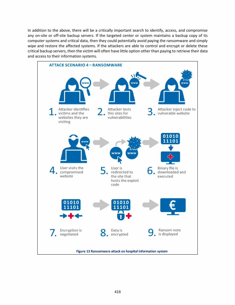
In addition to the above, there will be a critically important search to identify, access, and compromise any on-site or off-site backup servers. If the targeted center or system maintains a backup copy of its computer systems and critical data, then they could potentially avoid paying the ransomware and simply wipe and restore the affected systems. If the attackers are able to control and encrypt or delete these critical backup servers, then the victim will often have little option other than paying to retrieve their data and access to their information systems.
416

TYPES OF CYBERSECURITY AND RANSOMWARE ATTACKS
As shown in the figure above, ransomware is just one of the many types of cybersecurity attacks or threats that all healthcare organizations face in the modern era of information systems and connectivity. It is also important to note that these threats are often related and combined to achieve the desired effect. For ransomware, the point of entry is often obtained via other methods on the list including phishing, spam email, malware, or identity theft. Phishing attacks and information leakage (or identify theft) are the two most methods employed to initiate a ransomware attack.
Ransomware can be broadly grouped into two categories as shown below with their definitions. The non-encrypting or “locker” version is more commonly seen with attacks on individuals, and the encrypting or “crypto” type is by far the more common method utilized when attacking hospitals, healthcare systems, and any corporate or industry target. In an estimate from 2018 there were 350 different variants of ransomware described, representing a 150% increase from the year prior. These numbers have only continued to increase at a significant rate, and the variants continue to become more technically sophisticated over time. Another recent trend in the types of cybersecurity and ransomware attacks is the move to cloud-based solutions for data storage and backup by many healthcare organizations. This has predictably resulted in an increase in ransomware designed to exploit these cloud-based platforms in addition to in-house information systems and data backup/storage devices. Of great concern, 25% of healthcare institutions are not encrypting their data during transfer to the cloud, and 36% with data in a multi-cloud environment are not using encrypted technology.
417

HOW TO PREVENT SUCCESSFUL RANSOMWARE ATTACKS
Protection of IT systems and prevention of the deployment of ransomware or other malware is obviously far preferable to responding to a successful attack that has stolen and/or encrypted critical data and data systems. Although there are numerous specific technological practices and programs that should be implemented to prevent access to a center’s IT systems by malicious actors or organizations, the most important preventive measures are arguably related to the institutional leadership and culture. The healthcare industry has been identified as one of the common sectors that continues to have lax or inadequate cybersecurity awareness, policies, training, and leadership. Even the most secure firewall and other technologic factors cannot overcome the “human-factor” of lax or non-existent security practices or even willful misconduct of employees that compromise the IT security.
Adoption of a universal posture of cybersecurity diligence from the top leadership through all levels of the organization and any affiliated personnel, vendors, and collaborative partners is the first and most important step to preventing successful cyberattacks and to limiting the damage or extent of any executed incursions. The majority of successful cyberattacks arise from compromise of security by an employee or affiliate, either unintentionally (via malware, identity theft) or intentional.
Some sobering statistics related to healthcare IT and cybersecurity:
• 42% leave cybersecurity in the hands of a vice president or C-level official
• 39% report biggest challenge is the lack of qualified IT employees
• 40% of small and medium organizations do not have dedicated staff to deal with cloud-based issues
• 61% cited senior-level executives as a high security threat due to their level of access privileges and lack of stringent cybersecurity practices and knowledge
11 Steps to Defend Against the Top Healthcare Cyberthreats (from Dobran B, https://phoenixnap.com/blog/healthcare-cybersecurity-threats)
1. Consider threat entry points and vulnerabilities 2. Learn about ransomware attacks and likely targets 3. Create a ransomware policy and train for immediate response 4. Focus on employee cybersecurity and best practices training 5. Create or expand security measure risk levels and restrict access as appropriate 6. Go beyond employee access and factor in patient concerns and data access issues 7. Protect health data on ALL “smart” equipment and not just computers 8. Consider cloud migration for data storage and backup, transition to multi-cloud approach (Figure
below) as appropriate 9. Ensure vendors are compliant with access, updated security, and threat detection 10. Educate employees on HIPAA regulations and compliance for data protection 11. Push a top-down security program and conduct regular risk-assessments
418

ZERO TRUST SECURITY APPROACH
With the recognition that the majority of cyberattacks involve compromise of employee credentials, logon information, malware introduction, or purposeful sabotage, the Zero Trust Security approach has been developed. This involves a posture of never assuming that persons or devices attempting to access protected information are legitimate and implementing numerous checks and balances to confirm all identities, partitioning data and data-access, and continuously monitoring the network for any atypical or suspicious activity. This “default deny” posture results in systems that are hardened and isolated until a verified level of trust is established.
The Figure below shows the seven principles of implementation of a zero-trust model. This includes zero assumed trust in five areas: 1) employees or other people accessing the system, 2) devices and 3) networks that access or interact with the system, 4) data and data sharing, and 5) workloads (particularly running in the public cloud). The final two principles are visibility and analytics to continuously monitor and analyze every activity on the network, and automation and orchestration to enable automatic and coordinated incident response and task delegations during any cyberattack attempt or successful infiltration of the protected network.
Figure 1. The Forrester Zero Trust Extended Security Model
419

IMMEDIATE AND DELAYED RESPONSE TO RANSOMWARE
The immediate and delayed responses to ransomware are critical to preserve as much data and system operability as possible in the face of the cyberattack. The two immediate and simultaneous priorities should be disconnection of the device from the network and notification of the appropriate IT or cybersecurity personnel. The figure and expanded explanation table below highlights eight immediate and delayed actions that should be undertaken upon identification of an impending or executed cyberattack.
420

421

422

BUILDING A SYSTEM WIDE WHOLE BLOOD PROGRAM
Martin A. Schreiber, MD, FACS
Professor and Chief Division of Trauma and Critical Care Oregon Health & Science University Portland, OR
Whole blood is being utilized with increasing prevalence across the nation. The use of whole blood must be executed within existing systems of care or new systems must be developed that allow its use without negatively impacting other areas of blood banking. Solutions may be developed on a local, regional, national or international scale. The Department of Defense system has been in existence for the longest, but solutions are being developed on regional and local levels. Any system utilizing whole blood must develop some consistency with respect to a supply of safe donors, preparation of the product, distribution of the product cost effectively and widely to the areas of need and without undo waste. Current systems are developing novel solutions to these problems.
ARMED SERVICES BLOOD PROGRAM
The Armed services blood program (ASBP) is responsible for providing blood products to servicemembers, their family members and veterans in the continental United States or abroad and during peacetime or war. This is the most comprehensive system of care to provide timely and safe blood products potentially anywhere in the world. The ASBP is currently supplying low-titer O whole blood (LTOWB) to military treatment facilities and deployed settings throughout the world. This supply chain ranges from a Level 1 trauma center in San Antonio to the back packs of deployed special forces medics. The ASBP has elected to store its blood in CPDA-1 solution which allows it to be stored for 35 days. They have also elected to not leukoreduce the blood which means it cannot be converted to packed red blood cells prior to expiration. This has the benefit of not reducing platelet number or function, but it could result in greater waste.1
Decisions concerning the storage solution chosen as well as whether or not to leukoreduce are critical decisions within a system. Although the addition of adenosine to CPD solution allows LTOWB to be stored for 35 days, there are important issues concerning the storage lesion with respect to coagulation capacity of the product over that time period.2 Furthermore, while the decision to not leukoreduce whole blood results in an improved initial product it does not allow early conversion to packed red blood cells which effects overall utilization of the donated blood.
AMERICAN RED CROSS
The American Red Cross (ARC) is the largest supplier of blood products in the United States, and it recently added LTOWB to its repertoire making it available throughout the nation. Whole blood provided by the ARC is stored in CPD solution and it is leukoreduced. This is uniform for the ARC and any special request to alter this preparation results in significant additional cost. Oregon Health & Science University receives LTOWB from the ARC. There is a standing order for 20 units per week. During the pandemic, this order has not always been met due to system shortages. LTOWB is stored for 14 days and if it’s not used, it is converted to packed red blood cells in our blood bank.
In light of the fact that LTOWB undergoes a storage lesion, we chose to test our 14-day old whole blood from the ARC by performing complete blood counts and thrombelatography before and after running the
423

blood through the Belmont infuser which is routinely used for our massive transfusions. This whole blood had a hematocrit of around 40 and a platelet count of 75 which decreased to around 60 after going through the Belmont. The maximum amplitude on TEG, which primarily measures platelet function was also reduced. This highlights the importance of situational awareness concerning the quality of a product that is being utilized at the center level. The quality of whole blood used to correct coagulopathy will be primarily determined by whether or not it has been leukoreduced and its age. It is critical for users of whole blood to be aware of these variables and to act accordingly with respect to the use of additional components.
REGIONAL SOLUTIONS
The Southwest Texas Regional Advisory Council has created a unique regional system for the distribution of LTOWB throughout southwest Texas.3 The regional system is summarized in Figure 1. The system is predicated on a reliable source of blood donations which they have called “Brothers in Arms”. Brothers in Arms are an identified population of men with a very high donation rate and low titers to anti-A and anti-B. This group of donors creates a relatively stable blood supply. Blood is stored in CPDA-1, formally tested for infectious agents and it is not leukoreduced similar to the ASBP. After collection, the blood is first rotated to low use areas such as air medical providers, ground EMS providers and Level IV trauma centers. If this blood is not transfused by day 14, it is rotated to the Level 1 trauma centers. This rotational scheme has resulted in a 1-2% wastage rate and there have been no reported transfusion related complications. This group has also shown that early survival after trauma is improved with the use of prehospital whole blood.4 It is likely that additional regional distribution schemes for whole blood will be developed in the United States, but they are dependent on the availability of a reliable, independent and safe donor pool.
424

Figure 1. Southwest Texas Regional Whole Blood Program
425

LOCAL SOLUTIONS
Hospital systems have the option of creating their own blood donation systems. One example of this is the Stanford system. This requires the identification of a blood donation center as well as a relatively stable donor pool. Blood donation would need to comply with all American Association of Blood Bank and FDA requirements. It is also possible to create a hybrid model where a local solution is created and supplemented by regional and national systems. The creation of a local blood donation system allows for tremendous flexibility and tailoring of blood product needs to the institutions served. The availability of a blood donation center and known donors who are routinely screened for their low-titer status as well as infectious diseases also facilitates the development of a walking blood bank for emergencies.
THE COVID PANDEMIC AND BLOOD SHORTAGES
It has been estimated that the pandemic has resulted in a 19% reduction in available blood donors and a roughly equal decrease in the blood supply.5 This has resulted in an acute blood shortage in the United States exacerbating an already tenuous supply. Platelets that are stored for 5 days at room temperature are the most affected, but shortages of all components have been reported. Utilization of donated blood for use as LTOWB can potentially exacerbate these shortages and any system created with the purpose of distributing whole blood will need to take this into consideration.
REFERENCES
1. Remy KE, Yazer MH, Saini A, et al. Effects of Platelet-sparing Leukocyte Reduction and Agitation Methods on In Vitro Measures of Hemostatic Function in Cold-stored Whole Blood. 2018;84:S104-S114.
2. Strandenes G, Austlid I, Apeseth TO, et al. Coagulation Function of Stored Whole Blood is Preserved for 14 Days in Austere Conditions: A ROTEM Feasibility Study During a Norwegion Antipiracy Mission and Comparison to Equal Ratio Reconstituted Blood. Journal of Trauma and Acute Care Surgery. 2015;78:S31-S38.
3. Schaefer R, Long T, Wampler D, et al. Operationalizing the Deployment of Low-Titer O-Positive Whole Blood Within a Regional Trauma System. Military Medicine. 2021;186:S1,391-399.
4. Braverman MA, Smith A, Pokorny D, et al. Prehospital Whole Blood Reduces Early Mortality in Patients with Hemorrhagic Shock. 2021;61:S15-S21.
5. Riley W, Love K, McCulough J. Public Policy Impact of the COVID-19 Pandemic on Blood Supply in the United States. American Journal of Public Health. 2021;11:No. 5.
426

ACUTE CARE SURGERY TEAM: EVOLVING PRACTICE PATTERNS, CHALLENGES AND OPPORTUNITIES
Hasan B. Alam, MD, FACS
Loyal and Edith Davis Professor and Chair Department of Surgery Surgeon-in-Chief, Northwestern Memorial Hospital Chicago, IL
Disclosure: The author reports no proprietary or commercial interest in any product mentioned or concept discussed in this article.
Let me start by providing a quick background for this rather atypical topic. In August 2021, on a WhatsApp discussion group that was being used by ~170 surgical intensivists and leaders for sharing experiences and ideas related to the Covid-19 pandemic, a vigorous debate broke out about the present and the future of Acute Care Surgery (ACS) in the USA. Maybe because we were going through a relative lull in the number of new cases, or maybe because we were exhausted talking about Covid, this discussion had not much to do with the pandemic. I do remember that the exchange was very intense, highly charged, and went on for a couple of weeks. The emotional intensity probably also reflected a degree of burnout that almost everyone on the discussion group was experiencing due to relentless work and stress over the past 18 months. Essentially, a number of individuals on the forum shared that they felt disenfranchised, underappreciated, undercompensated, overworked and generally unhappy with their jobs, and assigned the blame largely to the healthcare leadership and hospital administration, whereas a few expressed a much more optimistic outlook. Although no conclusions were reached, this exchange prompted Dr. Mattox (an active member of this discussion group) to add the topic to this meeting.
While preparing for this talk, I have gone back through the exchanges on the WhatsApp chat, and the key thematic issues that generated most of the concerns can be summarized as below:
1. Salary and compensation issues 2. Scope of practice 3. Increasing workload, especially non-clinical/administrative duties 4. Decreasing autonomy and control 5. Surgical workforce culture 6. Wellness and burnout problems
During this presentation I will try to present some data on these topics, but mostly I will share my thoughts and experiences as someone who came to this wonderful country as an immigrant >30 years ago to join a community residency program as a preliminary resident. Since then, I have had the privilege to be on faculty at 5 universities, including full professorships at the Harvard Medical School, University of Michigan and Northwestern University, and hold multiple leadership positions. In fact, I am truly living the American Dream because the way my surgical career evolved would have been impossible anyplace else in the world. During this period, I have witnessed significant changes in our training programs with work hour restrictions, evolution of ACS as a sub-specialty with establishment of separate fellowships, and an impressive growth in our field. At the same time, there have been dramatic changes in healthcare finances, adoption of electronic medical records, physician compensation, billing and collection, medico legal issues, scope of practice, and healthcare reforms at the Federal and State levels. In short, things have
427

changed a lot over a relatively short period of time, and change inevitable creates anxiety, uncertainty and stress.
LOOKING AT ISSUES THROUGH THE RIGHT LENS
During stressful periods, it helps to intentionally focus on the rewarding aspects of our careers and personal lives. It should be emphasized that despite recent economic stresses, physicians and especially surgeons remain extremely well compensated. Their salaries place them in the top few percentiles of the US socioeconomic strata. They also have the unique privilege of positively impacting the lives of their patients consistently through their daily work, which few other professions can claim. Another possible approach has been very well described by Stephen Covey in his highly influential book, 7 Habits of Highly Effective People (1998). Covey introduces the concept of paradigm shift and helps the reader understand that different perspectives exist, i.e., that two people can see the same thing and yet react very differently. The first habit that he recommends is to “Be Proactive”, which emphasizes taking responsibility for your life - “Instead of reacting to or worrying about conditions over which they have little or no control, proactive people focus their time and energy on things they can actually control.1 The problems, challenges, and opportunities we face fall into two areas - Circle of Concern and Circle of Influence. Proactive people focus their efforts on their Circle of Influence. They work on the things they can do something about (Circle of Influence): e.g. adopting a healthy lifestyle, better time management, setting the right priorities, family, children, optimal patient care/professional excellence. In contrast, reactive people focus their efforts on things over which they have little or no direct control (Circle of Concern): e.g. the national debt, tort reform, terrorism, violent crimes, national health care policies, etc. Gaining an awareness of the areas in which we chose to expend our energies is a major step in becoming proactive. How we respond to any stimulus is our greatest strength, and also our biggest vulnerability, or as Covey described it, “I am not a product of my circumstances. I am a product of my decisions.”
It is also important to understand what motivates us. Daniel Pink in his book Drive: The Surprising Truth about What Motivates Us, and a TED talk that has been watched >25 Million times, argues that human motivation is largely intrinsic and that the aspects of this motivation can be divided into autonomy, mastery, and purpose.2 Once again, surgeons are in an enviable position of being the practitioners of a highly sophisticated healing art that builds upon all of these key drivers of motivation. Interestingly, what makes us happy in the short run often has no bearing on long-term happiness and satisfaction. In many articles and a hugely popular TED talk, Daniel Gilbert describes how people who win a lottery are no happier a year later, compared to people who became paraplegic.3 Both of these groups essentially reverted to their baseline levels of happiness following the acute “life-changing” episode.
In life, there will always be things that disturb our equilibrium. However, the key is to keep the focus on things that give meaning to our lives: find mentors, build relationships, be there for your patients but also connect better with your family, develop your teams and watch your mentee thrive, hug your kids, go for a run, pick up a new hobby, enjoy good food, give to your favorite charity, or do a difficult operation that tests your surgical skills. None of these will fix the problems of the world, but all of them are highly likely to make you happier, feel more fulfilled, and possibly less stressed.
COMPENSATION: FAIR PAY FOR FAIR WORK
At its core, compensation is really about fairness. For most of us, work is more than a paycheck, but we also deserve to be paid fairly based upon the market benchmarks. Let’s start by pointing out the obvious fact- surgeons are some of the highest paid professionals in the world. In almost every survey of the best paying professions, surgeons are near the top of the list.4 How does this compare to everyone else in the society? According to Economic Policy Institute, 2018 annual wages for the top 1% earners in the USA were $737,697, and the top 5% were $309,348.5 It would be fairly safe to assume that most of the
428

surgeons (general and/or acute care) attending this course are among the top earners in the country (and the world).
When considering a potential job, a more reasonable approach would be to not look at the highest salary that you can get, but to search for an opportunity that provides meaningful work while offering a great culture, supportive colleagues, and avenues for future growth. A common problem is that there is a misalignment between what you want to you with your life (goals, ambitions, amount/ type of work etc) and what the employer needs. Once you have found a position that is a good match, then you are ready to negotiate a fair salary for your efforts. What exactly is fair compensation deserves a bit more discussion. We live in a market driven capitalistic system. Our fair market value compensation depends upon many variables: uniqueness of your skillset, supply and demand, geographical location, revenue generated (directly or indirectly) by the work, other alternative options (e.g. can someone else do it cheaper?) etc. In addition to the market value as a surgeon, if you have some special talents then that may also change the compensation. For example, if you bring to the table a well-funded research portfolio, national fame, business or administrative experience etc, then you may (or may not) be worth more for an institution, depending upon their specific needs. From an institutional/employer standpoint the equation is simple (value = quality/cost). Thus, unless there is a clear and verifiable quality benefit, a lower cost option (or employee) offers better value. This is very similar to any other commodity in the market. For example, I may think my house is worth $1M, but at the end of the day the housing market, economy, ZIP code, and specifications of the house will dictate the fair market price. Thus, it is critically important for everyone to know the total cash compensation (TCC) for a typical surgeon in their field of specialty, who works in that geographical area in a similar practice setting, and how this TCC correlates to the amount of work as measured by RUVs. In my experience, surgeons often are wildly off target in terms of TCC, RVU targets, or both, which sets them up for frustration and disappointment. Contemporary TCC benchmarking data reflecting various variables such as experience, RVU, location, practice setting is compiled by many organizations, and it can serve as a starting point for the negotiations6, 7. If you have any unique skills, talents, or “super powers” that sets you apart, you should bring them up during the discussion to justify TCC above the market benchmarks. Don’t settle for a job that is either misaligned with your goals and priorities, has a poor culture, or fails to pay a fair wage. You either fix the situation, or look for better options elsewhere.
EVOLVING PRACTICE PATTERNS: HOW TO STAY RELEVANT
Acute Care Surgery (ACS) today encompasses trauma, surgical critical care (SCC) and Emergency General Surgery (EGS), but this was not always the case. The blossoming of ACS represents many changes in the healthcare landscape as well as the field of trauma surgery. By most estimates, the economic forecasts are favorable, and ACS surgeons are positioned to significantly influence the future of healthcare in United States. 8,9 As pointed out by these experts, there are a number of macroeconomic trends impacting ACS:
• ACS-related conditions constitute as much as 20% to 30% of all inpatient hospital costs in the USA.
• Our population is aging, and approximately 30% of all trauma-related expenditures are due to injuries in the elderly.
• Shifts in payments toward government and household payers are likely to contribute to further reshaping of the economic landscape for ACS in the future.
For details, I urge you to read these papers, but the key concepts impacting the economics of ACS include the fact that the ACS model has been shown to reduce cost and improve quality (i.e. better value); SCC makes up 25% of the overall productivity for ACS surgeons, while EGS is a rapidly enlarging part of this pie; and there is a national shortage of general surgeon that is unequally distributed. It is important to point out that the financial success of ACS services has been heavily dependent on cognitive work
429

(evaluation and management including critical care services) in addition to procedural work (procedures and operations). In many places, half of the RVUs are derived from the critical care codes. A potential threat to this revenue stream is the possibility that the Centers for Medicare & Medicaid Services (CMS) will consider delivery of critical care as part of the RVUs for the surgical procedure, and decline separate critical care billing. CMS FY2022 Medicare Physician Fee Schedule prohibits critical care visits from being reported during the same time period as a procedure with a global surgical period and would bundle payment for such visits with global surgery procedure codes. This could force the various providers (surgeons and intensivists) to divide the professional revenues rather than getting paid separately. Many professional organizations representing ACS surgeons are actively engaged in these discussions to ensure fair professional payments.
Another threat to the specialty is that the volume and scope of operations that the ACS surgeons are performing has steadily declined. A fantastic paper including Fitts Oration notes by Dr. JD Richardson (who unfortunately passed away shortly before giving the presentation at the 2021 AAST meeting) has just been published.10 This is a must read for those who want to understand how the specialty of ACS evolved from 1980s to now, and what challenges lie ahead. Personally, I found it highly educational to see the landscape through the eyes of a giant in our field to fully appreciate how much has changed. Historically, trauma care was provided as a service to the community by a few surgeons, who rarely generated any revenue from this clinical work. Thus, they routinely had a “paying job” of elective surgical practice. As described by Dr. Richardson, “When I started my surgical career at the University of Louisville in 1976, virtually no one in American Surgery regarded themselves as a “trauma surgeon”. Almost universally those who provided care for the injured were broad based surgeons with a diverse practice characteristic of the age before narrow specialty focus. I had a broad- based practice that included routine and complex general surgery, as well as vascular and non-cardiac thoracic surgery. An average month of my practice included complex and “bread and butter” general surgery (Whipple, hepatic resection, esophageal resection, thyroidectomy, mastectomy, cholecystectomy), thoracotomy and lung resection, and vascular cases (carotid endarterectomy, femoral distal bypass, open AAA repair). During this time, a few select colleagues provided attending coverage for busy emergency trauma and general surgery services. In a typical month covering the trauma service, my operative caseload would include several trauma laparotomies, a neck exploration, vascular injury repair, VATS, trauma thoracotomy for hemorrhage control, and repair of an esophageal injury. My first colleague, Dr. Lewis Flint, and I shared this call essentially every other night until he departed for the Chair in Buffalo, New York, in 1983. In contradistinction to a large number of trauma attendings in most programs today, we often had as few as two committed attendings to this type of care and rarely as many as four such surgeons the monies collected from trauma care did not go to the faculty but were pooled and funded resident educational opportunities”. Clearly, things have changed. We have gone from a relatively few, widely dispersed trauma centers located primarily in large urban centers to a system that contains many levels of trauma care (~ 650 trauma centers) that span the country. Our profession has evolved from a few poorly compensated surgeons providing transient care to the injured, to large teams of dedicated surgeons and allied professionals that are financially supported by the hospitals, who are considered absolutely vital for the delivery of essential surgical care. At the same time, the scope of practice has clearly narrowed dramatically. Many of the procedures listed by Dr. Richardson (also performed by many of him contemporary trauma surgeons) are now almost exclusively performed by sub-specialized surgeons, and this is unlikely to change. As trauma care has become more non-operative, the SCC and EGS components have continued to thrive, which has kept the service financially viable and created a win-win situation for the surgeons, patients, and the institutions. For some time, the idea of ACS surgeons getting trained to perform orthopedic and neurosurgical procedures was advocated by leaders in the field, but it never really had a chance to succeed in our health system.
430

My major concern for the future is that unless ACS surgeons aggressively maintain their surgical skills through active clinical practice, they will lose their identity as master surgeons. We are already seeing that at many hospitals ACS surgeons have become gallbladder, abscess and appendix surgeons. For all major trauma operations, they may do the initial damage control, but then have to call in a more skilled subspecialty surgeon, to fix the underlying injuries. I agree with the sentiments expressed by Dr. Richardson, “What distinguishes us from emergency medicine physicians, non-surgical intensivists, and others who care for the injured is our ability to operate…. We must continue to fulfill the dream of Acute Care Surgery by expanding and defending a broad scope of practice!”
BALANCE AND PURPOSE IN LIFE
Aus der Kriegsschule des Lebens. — Was mich nicht umbringt, macht mich stärker.
Out of life’s school of war — What doesn’t kill me, makes me stronger.
Friedrich Nietzsche (1888)
This aphorism from the "Maxims and Arrows" section of the German philosopher Friedrich Nietzsche's Twilight of the Idols may just as well be describing the traditional surgical training mantra. In our minds, and the imagination of the public, surgeons are brilliant, highly skilled, iron-willed individuals who calmly make life and death decisions. They are mentally and physically tough, emotionally inexhaustible, highly dexterous, and their superhuman personalities have been forged in a field where only the fittest can survive. The reality is a lot more nuanced. In fact, physicians in general, and surgeons in particular (including trainees), seem to be suffering from burnout at truly alarming rates.
Burnout appears to be very common among surgeons, and it may be the single greatest predictor of surgeons' satisfaction with career and specialty choice. In one survey, out of the 7905 surgeons who responded, 40% were burned out, and 30% screened positive for symptoms of depression.11 Factors independently associated with burnout included younger age, having children, area of specialization (trauma surgeons were more commonly affected), number of nights on call per week, hours worked per week and having compensation determined entirely based on billing. On a positive note, despite the high burnout rates, surgeons continue to find their work personally meaningful and rewarding, as 75% would become a surgeon again if given a choice. The fact that half of our surgical trainees and practicing surgeons are reporting burnout represents a public health crisis, with negative impacts on individual physicians, patients, and healthcare organizations and systems. Drivers of this epidemic are largely rooted within the healthcare systems and include excessive workloads, inefficient work processes, clerical burdens, work-home conflicts, lack of input or control for physicians for issues affecting their work lives, organizational support structures, and leadership culture. Individual physician-level factors also play a role, with higher rates of burnout commonly reported in female and younger/mid-career physicians. An even more important issue is the surgical work environment, which has “traditionally” been hierarchical, lacked diversity and was perceived as non-nurturing. This environment seems to negatively affect the more junior individuals and women disproportionately.
Effective solutions should align with these drivers. For example, organizational efforts such as locally developed practice modifications and increased support for clinical work have demonstrated benefits in reducing burnout. Individually focused solutions such as mindfulness-based stress reduction and small-group activities to promote community, connectedness, and meaning have also been shown to be effective. Cognitive, behavioral, and mindfulness-based approaches are effective in reducing stress and may also contribute to lower levels of burnout.12 Regardless of the specific approach taken, the problem of physician burnout is best addressed when viewed as a shared responsibility of both healthcare systems and the individual physicians.13,14 It is gratifying to note that many professional organizations are now addressing this issue head-on. For example, the American College of Surgeons is not only acknowledging
431

the magnitude of the problem but has made Surgeon Well-Being a high-priority area.15 The good news is that the increased focus on physician burnout may finally be having the desired effect. Burnout and satisfaction with work-life integration among US physicians have shown some improvement between 2014 and 2017, with burnout dropping down to the 2011 levels.16 To sustain this positive trend would require buy in from the leadership, physician involvement, organizational science/learning, metrics, structured interventions, open communication, and cultural change. The current pandemic has made this issue even more relevant.
With multiple competing priorities, another important area to address is time management for busy surgeons. To run a clinical practice or pursue an academic interest, while trying to have work-life balance, and fulfil family obligations is challenging. Countless books have been written about time management, and about ways to enhance your effectiveness. Some of these are very good.17,18 However, there is no substitute for learning from those who do it well. The ones around you who have figured out how to select the correct priorities, maintain a healthy (not all-consuming) clinical practice, find time to teach, be effective mentors, and somehow still find time for important family events. This is never easy, and there is no magic formula, but it is important to make sure not only that there is no time wastage, but that your time is allocated appropriately to high-priority areas. Defining what “high priority” is uniquely personal, and often changes as one goes through different phases of life and academic career. However, this is an area that needs your utmost attention. Don’t leave it to others to decide where your time should be spent. Time as this is the only non-renewable resource in our lives. Simply stated- you can always make more money but wasted time is gone forever. There are 168 hours in each week - you should be able to account for all of it and be in control of its proper allocation. You must set aside time for things that are important for your wellness, happiness, family, and personal growth. Remember that you will be in this field for 30 years, and nobody can be successful over such a long time, with self-deprivation as their main strategy. As mentioned above, mid-career burnout is a real threat for surgeons.19 Make time for hobbies, family, exercise, travel, vacations, and other things that make life worth living. Our goal should not be to achieve professional success at any cost, but to do so while leading a meaningful, productive, and fulfilling life.
REFERENCES
1. Covey, Stephen R. The 7 habits of highly effective people: Powerful lessons in personal change. Simon and Schuster, 2004.
2. Pink, Dan. "The puzzle of motivation." TEDGlobal 2009 (2009). 3. Gilbert, Daniel. "The surprising science of happiness." TED Talks. Available at: http://www. ted.
com/talks/dan_gilbert_asks_why_are_we_happy (2004). 4. US Bureau of Labor Statistics. Highest paying occupations. https://www.bls.gov/ooh/highest-
paying.htm (Last accessed 01.05.22) 5. Investopedia. How Much Income Puts You in the Top 1%, 5%, 10%?
https://www.investopedia.com/personal-finance/how-much-income-puts-you-top-1-5-10/ (01.05.22)
6. Medical Group Management Association. Benchmarking Data. https://www.mgma.com/data (last accessed 01.05.22)
7. Association of American Medical Colleges (AAMC) Faculty Salary Report. https://www.aamc.org/data-reports/workforce/report/aamc-faculty-salary-report (Last accessed 01.05.22)
8. Staudenmayer K, Bernard A, Davis KA, Doucet J, Haider A, Tres Scherer LR 3rd, Minei JP. The current and future economic state of acute care surgery. J Trauma Acute Care Surg. 2019 Aug;87(2):413-419. doi: 10.1097/TA.0000000000002334. PMID: 31033894.
432

9. Bernard A, Staudenmayer K, Minei JP, Doucet J, Haider A, Scherer T, Davis KA. Macroeconomic trends and practice models impacting acute care surgery. Trauma Surg Acute Care Open. 2019 Apr 11;4(1):e000295. doi: 10.1136/tsaco-2018-000295. PMID: 31058241; PMCID: PMC6461137.
10. Richardson JD, Spain DA, Livingston DH. Trauma and Acute Care Surgery: The Evolution of a Specialty. J Trauma Acute Care Surg. 2021 Nov 2. doi: 10.1097/TA.0000000000003456. Epub ahead of print. PMID: 34739005.
11. Shanafelt, Tait D., Charles M. Balch, Gerald J. Bechamps, Thomas Russell, Lotte Dyrbye, Daniel Satele, Paul Collicott, Paul J. Novotny, Jeff Sloan, and Julie A. Freischlag. "Burnout and career satisfaction among American surgeons." Annals of surgery 250, no. 3 (2009): 463-471.
12. Regehr, Cheryl, Dylan Glancy, Annabel Pitts, and Vicki R. LeBlanc. "Interventions to reduce the consequences of stress in physicians: a review and meta-analysis." The Journal of nervous and mental disease 202, no. 5 (2014): 353-359.
13. Ruotsalainen, Jani H., Jos H. Verbeek, Albert Mariné, and Consol Serra. "Preventing occupational stress in healthcare workers." Cochrane Database of Systematic Reviews 11 (2014).
14. Panagioti, Maria, Efharis Panagopoulou, Peter Bower, George Lewith, Evangelos Kontopantelis, Carolyn Chew-Graham, Shoba Dawson, Harm Van Marwijk, Keith Geraghty, and Aneez Esmail. "Controlled interventions to reduce burnout in physicians: a systematic review and meta- analysis." JAMA internal medicine 177, no. 2 (2017): 195-205.
15. American College of Surgeons. Surgeon Well Being. Accessed Feb 10, 2021. https://www.facs.org/member-services/surgeon-wellbeing
16. Shanafelt, Tait D., Colin P. West, Christine Sinsky, Mickey Trockel, Michael Tutty, Daniel V. Satele, Lindsey E. Carlasare, and Lotte N. Dyrbye. "Changes in burnout and satisfaction with work-life integration in physicians and the general US working population between 2011 and 2017." In Mayo Clinic Proceedings, vol. 94, no. 9, pp. 1681-1694. Elsevier, 2019.
17. Covey, Stephen R. The 7 habits of highly effective people: Powerful lessons in personal change. Simon and Schuster, 2004.
18. Mackenzie, Alec, and Pat Nickerson. The time trap: The classic book on time management. Amacom, 2009.
19. Spickard Jr, Anderson, Steven G. Gabbe, and John F. Christensen. "Mid-career burnout in generalist and specialist physicians." JAMA 288, no. 12 (2002): 1447-1450.
433

434
SESSION 15
ANNUAL ACS DEBATE
Moderator: Martin A. Schreiber, MD, FACS
Wednesday, March 30, 2022
8:30 a.m. – 9:30 a.m.
Palace Ballroom 1-2
Palace Tower, Emperors Level
Resolved: Optimal Outcomes for Vascular Injuries are Best Achieved with Percutaneous Repair
Pro: Joseph J. DuBose, MD, FACS, FCCM
Con: Alison Wilson, MD, FACS
435

OPTIMAL OUTCOMES FOR VASCULAR INJURIES ARE BEST ACHIEVED WITH PERCUTANEOUS REPAIR
Joseph J. DuBose, MD, FACS, FCCM (Pro Position)
Professor of Surgery Dell Medical School The University of Texas at Austin Austin, TX
The recent emergence of endovascular technologies has expanded the modalities available for use in the treatment of vascular injury. Initially employed in the treatment of atherosclerotic vascular disease, endovascular modalities have increasingly been utilized for specific vascular trauma indications. The position of endovascular approaches in algorithms for vascular injury treatment, however, continues to evolve. To date, these tools have most commonly been employed at challenging vascular injury locations or for those injuries traditionally associated with poor outcomes when approached via open modalities. This chapter will outline the published results from these experiences. We will also discuss the potential for future applications of endovascular treatment, as well as the present challenges to increased utilization of these technologies.
ENDOVASCULAR EXPERIENCE WITH MANAGEMENT OF THORACIC AORTIC INJURIES
Thoracic endovascular aortic repair (TEVAR) has, in recent years, emerged as the standard of care for a significant portion of traumatic aortic injuries that were traditionally treated by open surgical means.The largest examinations on the topic were conducted by the Demetriades and colleagues of the American Association for the Surgery of Trauma (AAST) Thoracic Aortic Injury Study Group in a large prospective observational trial of blunt thoracic aortic injuries.6,7 In the first of these reports, the investigators noted that 65% of the 193 patients with thoracic aortic injury (TAI) underwent endovascular stent graft repair (TEVAR), with the remaining patients undergoing open repair. After risk adjustment for mortality, the researchers noted that the TEVAR group required significantly fewer blood transfusions and had a significantly lower mortality than open surgical counterparts (adjusted odds ratio: 8.42; 95% CI: [2.76-25.69]; adjusted p value < 0.001). They did, however, note that 20% of TEVAR patients developed some form of device-related complications; but that a lower rate of these complications occurred in centers with higher volumes of TEVAR utilization.
The same group of researchers also reported on the trends in utilization of TEVAR between two phases of study separated by a decade (AAST1, competed in 1997 and spanning 30 months vs. AAST2, completed in 2007 and lasting 26 months). The group noted a profound trend in the utilization of TEVAR when comparing these two periods. During the period of AAST1 all TAI were repaired utilizing open means. In contrast, by the time of the 2007 study conclusion, only 35.2% were managed with open surgical intervention – the remaining undergoing TEVAR. The authors also noted both a significant decrease in TAI-associated mortality (22% vs. 13%, p = 0.02) and incidence of procedure-related paraplegia (8.7% vs. 1.6%, p = 0.001) over time.
Despite the emergence of TEVAR, several key elements of endovascular treatment of thoracic aortic injury require additional examination. Initially utilized devices were often improvised, or were designed for the treatment of abdominal aneurysmal disease. Many subsequent complications in stent deployment were, accordingly, related to issues in failure of the device to adequately conform safely to the contour of the
436

thoracic aorta. Additionally, the diagnosis and treatment of inadvertent or intentional coverage of the subclavian or other arch vessels was poorly understood. Although the evolution in endovascular technologies and improved management algorithms has mitigated much of these risks, further research is required. In particular, long-term follow-up of these devices is required. Given the relative younger age of many trauma patients, and associated smaller diameter of the thoracic aorta, the potential for stent malfunction, migration or other complications over time has not been well documented.
ENDOVASCULAR EXPERIENCE WITH MANAGEMENT OF CEREBROVASCULAR INJURIES
The treatment and outcome of cerebrovascular injuries is influenced by many factors; including the mechanism, type of injury, and associated neurologic function. These injuries may be associated with considerable mortality and high rates of neurologic impairment. Several types of injuries may result from cerebrovascular trauma, regardless of mechanism. Those that do not commonly result in the operative indications of hemorrhage or expanding hematoma include intimal flaps, dissections and pseudoaneurysms. The natural history and appropriate management of these injuries remains ill defined. Anticoagulation following blunt carotid injury is known to be associated with improved outcome following blunt trauma, but some types of injuries are more likely to fail conservative therapy. It has been reported that up to 40% of patients in the modern era may require endovascular intervention for these injuries.
Borrowing on the expanding experience with the use of endovascular stents for cerebrovascular disease, carotid stenting has been utilized for high extracranial internal carotid lesions. These types of interventions are ideally suited for this region, where surgical approaches are most difficult, and are associated with a high rate of local and cerebrovascular complications. An endovascular approach may also prove particularly useful in the treatment of select types internal carotid injuries, as surgical resection or repair of internal carotid pseudoaneurysms in particular, are associated with a high mortality rate and high incidence of cerebral complications.
In the largest review on this topic to date, DuBose and colleagues identified experiences from 113 patients undergoing carotid stenting that were reported in the literature from 1994 to 2008. The injury types treated by stenting included pseudoaneurysm (60.2%), arteriovenous fistula (16.8%) dissection (14.2%), partial transection (4.4%), occlusion (2.7%), intimal flap (0.9%) and aneurysm (0.9%). Over radiographic and clinical follow-up periods ranging from 2 weeks to 2 years, a follow-up patency of 79.6% was documented. New neurologic deficits after stent placement occurred in only 3.5% of patients, improved over historical experiences with open repair after trauma. Although these initial experiences are promising, no large prospective study on this topic has yet been conducted and further research is warranted.
ENDOVASCULAR EXPERIENCE WITH MANAGEMENT OF AXILLO-SUBCLAVIAN INJURIES
Injuries to the subclavian and axillary arteries continue to be associated with high rates of morbidity and mortality. In one of the landmark series on these injuries, Demetriades and colleagues at Los Angeles County + University of Southern California Hospital examined penetrating subclavian and axillary injuries, identifying 79 patients with these injuries over approximately 4 years. The associated overall mortality was 34.2%. Even after excluding those patients in extremis requiring resuscitative thoracotomies, mortality remained considerable, at 14.8%. These experiences, combined with the confined anatomical relationships of the thoracic inlet, contributes to the complexity attributed to open surgical treatment of subclavian / axillary trauma. These challenges have made injuries at this location an attractive target for study of the utilization of endovascular technologies.
In the largest review of this topic, conducted by DuBose et al., investigators identified 32 published reports of endovascular treatment of subclavian or axillary artery injuries with sufficient information for review. These reports described 160 patients (150 subclavian; 10 axillary), the majority of which (56.3%) were due
437

to penetrating injuries. Lesions treated included pseudoaneurysm, arteriovenous fistula, perforation, occlusion, partial or complete transection and dissection. Only six procedure-related complications were reported. The only reported peri-procedural mortality reported was a death occurring in the angiography suite after successful deployment of an endovascular stent had been completed. Overall, radiographic and clinical follow-up periods ranging from hospital discharge to 70 months were reported, with no device-related infections, migrations or acute limb threatening ischemic events reported among the 160 reported cases. The authors documented an asymptomatic patency rate of 84.4% for the duration of available follow-up. This group concluded that, despite uncertainties in patient selection and optimal management algorithms, early results of endovascular treatment for properly selected subclavian and axillary injuries are promising.
ENDOVASCULAR EXPERIENCE WITH MANAGEMENT VASCULAR INJURY AT OTHER LOCATIONS
Endovascular management of vascular injury at other anatomical locations has also been proposed. Treatment of visceral, iliac and even peripheral artery injuries has been described. To date, however, there have been no large prospective studies in these settings and availability of data is limited to case reports or small case series. Defining the optimal utilization of endovascular therapy for injuries at more anatomically accessible sites will require additional study.
ENDOVASCULAR VASCULAR CONTROL AS AN ADJUNCT TO OPEN SURGICAL INTERVENTION
As the exploration of endovascular application for trauma has continued to evolve, so to has the potential application of this technology. Many early studies have focused on direct comparisons of open versus endovascular interventions. Increasingly, however, awareness that these two approaches may work in compliment with one another has arisen. So called “hybrid approaches” may hold considerable promise in the treatment of vascular injury. Endovascular balloons have been effectively utilized for proximal occlusion, dilation and stent deployment in the treatment of atherosclerotic disease. Increasingly, their use in hybrid vascular trauma applications has also been examined.
The use of endovascular occlusion for trauma indications is not an entirely new concept. Scalea and Scalafani from Kings County Hospital of Brooklyn New York first described this technique in 1991.18 In recent years, however, several groups of investigators have subjected the utilization of this practice to greater scrutiny using animal models.19,20,21 Early results suggest that the application of endovascular technologies may provide rapid control of proximal arterial hemorrhage, facilitating a more controlled effort at exposure and operative repair. Future study on these types of hybrid approaches is likely to demonstrate the complimentary nature of endovascular and open surgical interventions for vascular trauma.
POTENTIAL COMPLICATIONS AND PRESENT LIMITATIONS OF ENDOVASCULAR MANAGEMENT
Despite recent advances of endovascular trauma applications, it is paramount that trauma providers understand the specific complications unique to these technologies. These begin with the skill sets required to adequately utilize these innovations. At present the techniques and tools of endovascular intervention largely belong to those specialists formally trained in interventional radiology or vascular surgery. As hybrid approaches become more common, however, these conditions may change. Several studies, most notably the aforementioned AAST TEVAR experience, have also demonstrated the relationship between volume of procedures performed and subsequent outcomes. These data suggest that endovascular trauma management is best accomplished at high volume trauma centers with experienced providers.
Endovascular approaches also have specific potential complications that may require specialist support to manage effectively. This may be particularly true for stent coverage of vascular injuries. Endoleaks, stent
438

migration and prosthetic failure are all potential management dilemmas that will require capable subspecialty support to effectively treat.
Access site complications may also be common. Many of the present devices utilized for trauma endovascular intervention require fairly sizeable deployment systems for device introduction into peripheral arteries. Particularly for young trauma victims with smaller diameter arteries, these device sizes are presently problematic. Continued evolution of endovascular technologies may someday ameliorate this risk but, at present, endovascular device companies have been slower to embrace these special concerns for trauma applications.
Perhaps the greatest present limitation of endovascular technologies for trauma is the lack of adequate follow-up data. As the ease of utilization improves, so to must the ability of trauma providers to determine the durability of endovascular intervention for injury. The recently initiated PROspective Observational Vascular Injury Trial (PROOVIT) study of the American Association for the Surgery of Trauma (AAST) is one modality that holds promise in this regard. This study is designed to capture initial endovascular and open vascular injury treatments at a wide variety of anatomical locations – as well as long-term follow-up data. It is the hope of the study group that this information will help to define the optimal utilization of endovascular technologies for trauma.
CONCLUSION
The role of endovascular management for vascular injury continues to evolve. These technologies, both in isolation and in hybrid coordination with open surgical intervention, hold considerable promise for the future. Well established already for specific indications, further study is needed to determine the optimal role of endovascular techniques in vascular injury management algorithms.
439

OPTIMAL OUTCOMES FOR VASCULAR INJURIES ARE BEST ACHIEVED WITH PERCUTANEOUS REPAIR
Alison Wilson, MD, FACS (Con Position)
Vice-Chair and Professor WVU Department of Surgery Skewes Family Chair for Trauma Surgery Director, WVU Critical Care and Trauma Institute Morgantown, WV
OVERVIEW
The concept of using an endovascular approach for proximal control of bleeding is not new. In the realm of pelvic injuries and vascular control, endovascular intervention was described in 1972.1 As technology and innovation have exploded so has the world of medical devices. With this has come exciting new options for interventions and therapies that have never been seen in previous generations. This growth has been seen particularly in robotic and catheter-based treatments. Coupled with the advances in imaging, surgeons and radiologists are able to visualize at a detail that has not been imaged before. Use of CTA to diagnose vascular injuries has become common place and often correlates with type of intervention.2 With this has come an explosion in the number and types of stent grafts and the rate in which new ones get to market. It is imperative that we are thoughtful and purposeful in the implementation and utilization of these technologies. The desire to be “cutting edge” must also serve a purpose that benefits the patient. In an age when we hold steadfast that we must practice evidence-based medicine, it is incumbent upon us to ensure these technologies have superior outcomes over the traditional options and are utilized for appropriate indications.
CENTRAL AND JUNCTIONAL INJURIES
Aortic Transection
Certainly, stenting options for aortic transection have opened opportunities and facilitated care for the multiply injured patient.3,4,5 The ability to continue resuscitation, avoid the strain of single lung ventilation that is required for an open repair, and some would argue the reduction in operative time may decrease the physiologic stress placed upon the acutely injured patient. There are several factors that we must not loose sight of including indication, sizing, placement, durability, and long term follow up and outcomes.6,7 As imaging via CTA has gained availability and detail due to the modern scanners, we are seeing abnormalities after injury that we may not have known existed in the past. Clearly defining what benefits from placement of a permanent device will need to be monitored, studied, and refined. As the majority of the injured patients with these injuries are young, ensuring the appropriate sizing of the graft can be a challenge, particularly in young adolescents and small females. Additional challenges in the adolescent or small individual can be the access vessel as the femoral artery may be too small for the deployment devices requiring alternative approaches.8
Placement, durability, and long term follow up and outcomes are probably the greatest challenges9,10,11,12. Commonly, the left subclavian artery is covered when placing a stent graft in the setting of an aortic transection. If acute, significant arm ischemia occurs, certainly a carotid-subclavian arterial bypass is performed. However, bypass or not, long term implications of this in young people is not known. The durability of the stents is also largely unknown. Endoleak, migration, ruptured attachments and occlusion
440

can be found with variable consequences, but certainly some have been catastrophic resulting in complete occlusion and death6. Fistula to the esophagus and sepsis due to infection of the endograft are also known complications.13This does not mean that use should be halted but does call the clinician to be sure that the indication for placement is valid and all attempts at long term follow up should be employed. It should also be noted that the potential need for reintervention after use of a TEVAR is not insignificant. Reports vary with a 2-30% need for reintervention or even explantation of device required in the first 5 years.6,9,11 Optimal follow up algorithms that include use of ionizing radiation are unclear, leaving the question of risk of cumulative radiation impact for surveillance scans. Finally, medical sequela such as the development of difficult to control hypertension have been reported and may leave a young person with lifelong effects on other organ systems.
Junctional Injuries
We know from civilian and wartime experience that there is increased mortality when the injury occurs at a junctional area.14 A key contributor to this is that one of the most difficult areas to control and access are vascular injuries are those that occur in the junctional regions of the body. Gaining proximal vascular control in the emergent setting when the patient is in a shock state and continuing to experience substantial blood loss can be a challenge for even experienced surgeons. There is no doubt that advances made in endovascular techniques for injuries such as the subclavian artery, have made positive contributions to outcomes. The opportunity to avoid the steps involved in open techniques to access this vessel and gain proximal control do decrease the morbidity of the repair. The ability to use an endovascular balloon to provide the proximal control and stop ongoing massive blood loss is fast, once the patient arrives in the operative suite, and certainly aid in the ability to stabilize and resuscitate the patient.15,16,17
There are several key factors to keep in mind when evaluating endovascular versus open repairs for these injuries. The influence of hemodynamics, associated injuries that also require open operative repair, and the degree of vessel injury have been shown to be factors in operative approach. For hemodynamically unstable patients, delay of hemorrhage control to obtain a CTA or other pre-operative work up could be fatal. In a large review it was shown that most open repairs were performed in hemodynamically unstable patients and/or injuries with greater than 50% vessel wall laceration or complete transection while endovascular repairs were selected in stable patients with pseudoaneurysms or intimal injuries.18 It must also be emphasized that the injury must allow adequate proximal landing site for placement of the stent. Durability and long term follow up also remain questions. The need for re-intervention for endoleak and stent thrombosis remain challenges.
PERIPHERAL ARTERIAL INJURIES
Perhaps one of the most controversial issues is the use of stents for peripheral injuries. The argument has been made that the endovascular approach allows less dissection of surrounding tissue planes and that it can be performed more quickly. However, there are multiple issues that must be taken into consideration. Particularly in stretch and blast injuries, the zone of injury may not be adequately visualized on imaging. The need for heparinization in the multiply injured patient is also a significant concern, especially in the setting of a traumatic brain injury. Placement of a stent frequently requires anti-platelet therapy. This can pose a substantial challenge in the multi-system trauma patient. Long term use of anti-platelet therapy in young patients and the contribution of that to bleeding risk for the long-term period has not been quantified or well-studied. As stents become increasingly popular, more reports are emerging with use of stents in more distal vessels such as popliteal.19,20 The long term outcomes of these stents remains unclear while there is significant data from military and civilian centers spanning years that report high patency rates of open approaches involving direct repair, resection and anastomosis and interposition
441

grafts.21,22 It is the responsibility of the academic surgical world to follow and report the long term outcomes as stents are more frequently used.
As the popular trend becomes use of stents for a multitude of reasons, access to IR or hybrid surgical suites must be considered before stating this should be the first approach, especially in distal peripheral vessels. Though in most major medical centers, hybrid rooms, stents and a trauma or vascular surgeon are available in a reasonable amount of time, this is not the case in many rural or even small community settings. This also is a challenge in the international community. Recommended standards of care should be able to be applied to all patients, not simply ones who live in an urban setting in a developed nation. We must maintain training and availability of foundational vascular skills.
BETTER OR REPLACEMENT FOR LOST SKILL SETS ??
General Surgery residency programs have changed dramatically in the last 20 years. The change in injury management to a non-operative approach for a variety of injuries, especially solid organ injuries has resulted in a decline in graduating resident case numbers 23 With the advent of the 80-hour work week, operative experience for graduating residents has decreased significantly. Since 2008, there has been a substantial decline in number of operative cases versus non-operative trauma cases per graduating resident. Experience in thoracic, abdominal and extremity vascular trauma operative management have all been substantially impacted.
With the evolution and growth in vascular fellowships, there has been even further impact on general surgery resident experience. One study showed over a 10-year period a 34% reduction in general surgery resident total vascular cases, but a 78% increase in total cases for vascular fellows.24 Data continues to emerge from general surgical training and vascular fellowships showing decrease access and training opportunities for open vascular cases. In the 1990’s, it was common for a PGY 4 or 5 to perform an open aortic bifemoral graft with an appropriate faculty. With increasing frequency, large open cases are referred to a specialty center or clustered to a particular surgeon. For general surgery residents the impact has been the greatest, with a decline in experience in carotid endarterectomy (decrease of 26%), peripheral artery bypass (decrease of 45%) and open infrarenal AAA (decrease of 52%). At the same time there has not been a significant increase in endovascular cases meaning that residents are not gaining an additional skill set. Experience for the vascular surgery fellow in open techniques has also declined (carotid endarterectomy decrease of 23%, peripheral artery bypass decreases 37% and open AAA decrease 31%).24 Fellows have seen an increase in endovascular cases over time which will naturally leave those graduates feeling much more comfortable with this approach. This means that fewer trainees have adequate exposure and experience with these cases. As comfort level for a surgeon can play an important role in technique selection, this raises the question of whether or not the endovascular option chosen by an individual surgeon is because it is truly a better option for the patient or because the surgeon is not comfortable with the anatomy, exposure or technique. It is imperative that we ensure we teach and maintain open vascular skills.
REFERENCES
1. Margolies MN, Ring EJ, Waltman AC. Arteriography in the Management of Haemorrhage from Pelvic Fractures. NEJM. 1972; 287: 317- 321.
2. Romagnoli AN, DuBose J, Dua A et al. Hard Signs Gone Soft: A critical evaluation of presenting signs of extremity vascular injury. J Trauma Acute Care; 2021; 90 (1); 1 – 10.
3. Mouawad N, Paulisin J, Hofmeister S, Thomas M. Bluth thoracic aortic injury-concepts and management. J Cardiothor Surg 2020;15(1):1-8
4. Hundersmarck D, Van der Vliet Q, Winterink L, et al. Blunt thoracic aortic injury and TEVAR: long termoutcomes and health related quality of life. Eur J Trauma Emerg Surg. 2020; 1-13.
442

5. Patel H, Azizzadeh A, Matsumoto A, et al. Five-year outcomes from the United States Pivotal Trial of Valiant Captivia Stent Graft for Blunt Aortic Injury. A Thor Surg 2020;110(3)815-820.
6. Nienaber CA, Kische S, Ince H. Thoracic Aortic Stent Graft Devices: Problems, Failure Modes and Applicability. Sem in Vasc Surg. 2007; 20(2) 81-89.
7. Alslaim H, Chung J, Arthur M, et al. Challenges in Appropriate Sizing of Thoracic Endografts for Thoracic Aortic Transection. An Vasc Surg. 2021; 77:333-334.
8. Milas Z, Milner R, Chaikoff E, et al. Endograft Stenting in the Adolescent Population for Traumatic Aoritc Injuries. J Ped Surg. 2006; 41:E27-30.
9. Steuer J, Wanhainen A, Thelin S, et al. Outcome for endovascular treatment of traumatic aortic transection. J Vas Surg. 2012; 56(4): 973-978
10. Azizzadeh A, Hunter M, Dubose J, et al. Outcomes for endovascular repair for patients with blunt traumatic aortic injury. J Trauma Acute Care. 2014; 76(2): 510-516
11. Miller D. Through the looking glass: The first 20 years of thoracic aortic stent grafting. J Thorac Cardiovasc Surg. 2013. S142-S146.
12. Spiliotopoulos K, Kokotsakis J, Argiriou M, et al. Endovascular repair for blunt thoracic aortic injury: 11-year outcomes and postoperative surveillance experience. J Thorac Cardiovasc Surg 2014; 148: 2956-61
13. Hance K, Hsu J, Eskew T, et al. Secondary aortoesophageal fistula after endoluminal exclusion because of thoracic aortic transection. J Vasc Surg 2003; 37: 886-888
14. Eastridge, Brian J. MD; Mabry, Robert L. MD; Seguin, Peter MD; Cantrell, Joyce MD; Tops, Terrill MD; Uribe, Paul MD; Mallett, Olga; Zubko, Tamara; Oetjen-Gerdes, Lynne; Rasmussen, Todd E. MD; Butler, Frank K. MD; Kotwal, Russell S. MD; Holcomb, John B. MD; Wade, Charles PhD; Champion, Howard MD; Lawnick, Mimi; Moores, Leon MD; Blackbourne, Lorne H. MD Death on the battlefield (2001–2011), Journal of Trauma and Acute Care Surgery: December 2012 - Volume 73 - Issue 6 - p S431-S437
15. Branco B, Boutrous M, DuBose J, et al. Outcome comparison between open and endovascular management of axillosubclavian arterial injuries. J VAsc Surg 2016; 63:702-709.
16. Elkbuli A, Shaikh S, McKenney M, Boneva D. Successful management with endovascular stnet graft repair following gunshot wound to the subclavian artery: case report and literature review. Inter J Surg Case Rep 2019; 64:75-79
17. Branco B, DuBose J Endovascular solutions for the management of penetrating trauma: an update on REBOA and axillo-subclavian injuries. Eur J Trauma Emerg Surg 2016; 42:687-694.
18. Waller C, Cogbill T, Kallies K, et al. Contemporary management of subclavian and axillary artery injuries- A Western Trauma Association multicenter review. J Trauma Acute Care Surg 2017; 83(6):1023-1031.
19. Johnson C. Endoivascular management of Peripheral vascular trauma. Semin Intervent Radiol; 27: 38-43. 20. Branco B, DuBose J, Zhan L et al. Trends and outcomes of endovascular therapy in the
management of civilian vascular injuries. J Vasc Surg 2014; 60:1297-1307. 21. Feliciano D, Shackford S. Vascular Injury: 50th Anniversary year review article of the Journal of
Trauma. J Trauma 2010;68(4):1009-1013 22. Rozycki G, Tremblay L, Feliciano D, McClelland W. Blunt Vascular trauma in the Extremity:
diagnosis, management, and outcome. J Trauma 2003; 55:814-824 23. Strumwasser A, Grabo D, Inaba K, et al. Is your graduating general surgery resident qualified to
take trauma call? A 15-year appraisal of the changes in general surgery education for trauma. J Trauma Acute Care Surg 2017; 82:470-480
24. Grabo D, DiMuzio P, Kairys J, et al. Have endovascular procedures negatively impacted general surgery training. Ann Surg 2007; 246:472-480.
443

444

SESSION 16
WE HAVE MET THE ENEMY…
Moderator: Meghan R. Lewis, MD, FACS
Wednesday, March 30, 2022
Palace Ballroom 1-2
Palace Tower, Emperors Level
10:00 - 10:15
Child Abuse and Exploitation: Recognizing the Signs and Symptoms Robert W. Letton, MD, FACS
10:15 - 10:30
Death & Dying in the Trauma Bay - Communicating with Patients, Families, Treatment Team Melissa “Red” Hoffman, MD, FACS
10:30 - 10:45
Military & Civilian Collaborations - Do They Work? Rachael A. Callcut, MD, MSPH, FACS
10:45 - 11:00
Mitigating Deficiencies in Surgical Education Hasan B. Alam, MD, FACS
11:00 - 11:25
Panel Discussion
445

CHILD ABUSE AND EXPLOITATION
Robert W. Letton, MD, FACS
Endowed Professor in Pediatric Surgery Nemours Children’s Specialty Care and Wolfson Children’s Hospital Jacksonville, FL
CHILD PHYSICAL ABUSE/NONACCIDENTAL TRAUMA
Child abuse of all forms remains a significant preventable public health problem affecting more than 600,000 children annually in United States, according to the most recent report from the National Child Abuse and Neglect Data System. An estimated 9 children per 1,000 suffer at least 1 form of maltreatment, with somewhere between 10% and 33% of these patients sustaining child physical abuse (CPA). Although there are data to suggest that this may dramatically underrepresent the problem's true extent. Identifying victims of CPA can be a significant challenge because health care is usually predicated on trusting the historian. Screening for child abuse is further biased by race and socioeconomic status, as evidenced by an increased rate of screening and reporting of abusive head trauma (AHT) in minority race and ethnicity patients compared with White non-Latin patients.1
APSA Position Statement on Child Physical Abuse2
• APSA endorses the concept of child physical abuse as a traumatic disease that justifies the resource utilization of a trauma system to assess, stabilize and manage this patient population in a standardized fashion including evaluation by pediatric surgeons.
• APSA encourages the admission of a suspected child physical abuse patient to a surgical trauma service because of the potential for polytrauma and increased severity of injury and to provide reliable coordination of services, including mandatory reporting to CPS.
• APSA recognizes the need for pediatric surgeons to participate in a multidisciplinary team and to be involved in the screening, evaluation, and management of patients with suspected child physical abuse at their respective hospitals.
• APSA endorses the requirement for the creation of a child abuse team for the Children's Surgery Verification Program as outlined in The Optimal Resources for Children's Surgical Care.
• APSA endorses the requirement for the systematic evaluation of child physical abuse and admission to a surgical service of acutely injured children for Level I or Level II Pediatric Trauma Centers as outlined in Resources for Optimal Care of the Injured Patient.
• APSA recognizes that if a pediatric surgeon has a reasonable suspicion that a child has been abused, a report to CPS for further investigation is mandated by law. If a pediatric surgeon is suspicious that the patient was maltreated, transferring the child to another physician or facility for medical care does not relieve the pediatric surgeon of his or her responsibility as a mandated reporter of suspected abuse.
• APSA recommends the implementation of a standardized tool to screen for child physical abuse at all Trauma verified or designated hospitals and Children's Surgery verified hospitals. Further, APSA supports universal application of the screening tool regardless of socio-economic, racial, ethnic, or gender status.
446

• APSA supports the concept of accrual of data on child physical abuse screening and diagnosis as defined by the Centers for Disease Control and Prevention into a trauma registry and reporting to the National Trauma Databank and TQIP-P for benchmarking purposes and quality improvement.
• APSA recognizes the existence of abusive head trauma (AHT) as sequelae of child physical abuse which can result in often permanent and significant brain damage and understands that pediatric surgeons are frontline providers caring for this group of injured infants and children.
In a recent multi-institutional retrospective review, CPA accounted for 10-15% of all trauma admissions, however, accounted for 50% of the total trauma deaths. Nonaccidental trauma patients have higher mortality and impaired function at a given ISS/head AIS than accidental pediatric trauma patients. Conventional ISS thresholds may underestimate risk and head injury is a more important predictor of mortality in the NAT population. These findings should be considered in system performance improvement and benchmarking efforts that rely on ISS for injury characterization.3
Figure 1.
Figure 2.
447

In spite of screening guideline being disseminated, there are still significant disparities between morbidity and mortality in the child abuse population. Despite attempts to control for the clinical presentation and injury severity of abused children, significant differences in mortality persist between African American and white children.4
Table II. Table I.
Figure 3.
448
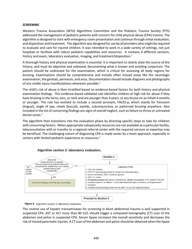
SCREENING
Western Trauma Association (WTA) Algorithms Committee and the Pediatric Trauma Society (PTS) addressed the management of pediatric patients with concern for child physical abuse (CPA) trauma. The algorithm is designed to start with emergency room presentation and continue through initial evaluation, and disposition and treatment. The algorithm was designed for use by all providers who might be required to evaluate and care for injured children. It was intended to work in a wide variety of settings, not just hospitals or facilities with robust pediatric capabilities and resources. It contains 4 different sections, history and exam, laboratory evaluation, imaging, and treatment/disposition.1
A thorough history and physical examination is essential. It is important to clearly state the source of the history, and must be objective and unbiased, documenting what is known and avoiding conjecture. The patient should be undressed for the examination, which is critical for assessing all body regions for bruising. Examinations should be comprehensive and include often missed areas like the neurologic examination, the genitals, perineum, and anus. Documentation should include diagrams and photographs of any visible injury manifestations whenever possible.1
The child's risk of abuse is then stratified based on evidence-based factors for both history and physical examination findings. This evidence-based validated rule identifies children at high risk for abuse if they have bruising in the torso, ears, or neck and are younger than 4 years, or any bruise on an infant 4 months or younger. The rule has evolved to include a second acronym, FACES-p, which stands for frenulum (lingual), angle of jaw, cheek (buccal), eyelids, subconjunctiva, or patterned bruising anywhere. Also included in the list of concerning findings are signs of overall neglect, such as failure to thrive or untreated dental caries.1
The algorithm then transitions into the evaluation phase by directing specific steps to take for children with concerning factors. When appropriate subspecialty resources are not available at a particular facility, teleconsultation with or transfer to a regional referral center with the required services or expertise may be beneficial. The challenging nature of diagnosing CPA is made easier by a team approach, especially in centers with limited pediatric experience or resources.1
The routine use of hepatic transaminases for screening in blunt abdominal trauma is well supported in suspected CPA. AST or ALT more than 80 IU/L should trigger a computed tomography (CT) scan of the abdomen and pelvis in suspected CPA. Serum lipase increases the overall sensitivity and decreases the risk of missed pancreatic injuries. A CT scan of the abdomen and pelvis should be obtained when the lipase
Figure 4.
449

elevation is more than 100 U/L. Coagulation profiles are useful to help determine if a patient has a coagulopathy that could account for bruising felt to be inconsistent with the stated mechanism of injury. Serum calcium, phosphorous, alkaline phosphatase, parathyroid hormone, and vitamin D levels may be useful in fracture patients. Troponin I level may help assess for blunt cardiac injury in children with suspected CPA.1
Imaging decisions are guided by American College of Radiology criteria and are stratified by age, with children younger than 6 months getting a mandatory head CT scan and children of other ages getting head imaging based on clinical suspicion, facial injury, abnormal neurologic examination, or concussive
Figure 5.
450

symptoms. Children younger than 24 months with suspicious findings should receive a skeletal survey, which is suggested for children up to age 5 years if suspicion is high. Although studies have shown that the positivity rate of a skeletal survey with an isolated skull fracture and no intra-cranial hemorrhage is about 5%.5
Abdominal imaging is based on clinical suspicion or laboratory abnormalities. A conservatively low threshold (aspartate transaminase/alanine transaminase, ≥80 IU/L, or lipase, ≥100 IU/L) is used for abdominal imaging in order to increase sensitivity and ideally capture even minor injuries that may not require significant treatment but could have enormous relevance in making the diagnosis. Further imaging is injury specific or determined by clinical concern. There is significant correlation of CPA and isolated humerus or femur fracture in a child younger than 18 months.1
Age related fracture patterns associated with child abuse. By age, infants had the highest rate of multiple fractures (33% vs 16% 1–4 years), and the highest rate of closed skull fractures (33% vs 21% ages 1–4), while adolescents had more facial fractures (43% vs 11% ages 9–12), all p < 0.001. Multiple rib fractures were more commonly seen in infants (28% vs 8% ages 1–4), while children 5–8 years had the highest rates of clavicular fractures (7% vs 3% in infants), all p < 0.001.6
Figure 6.
451
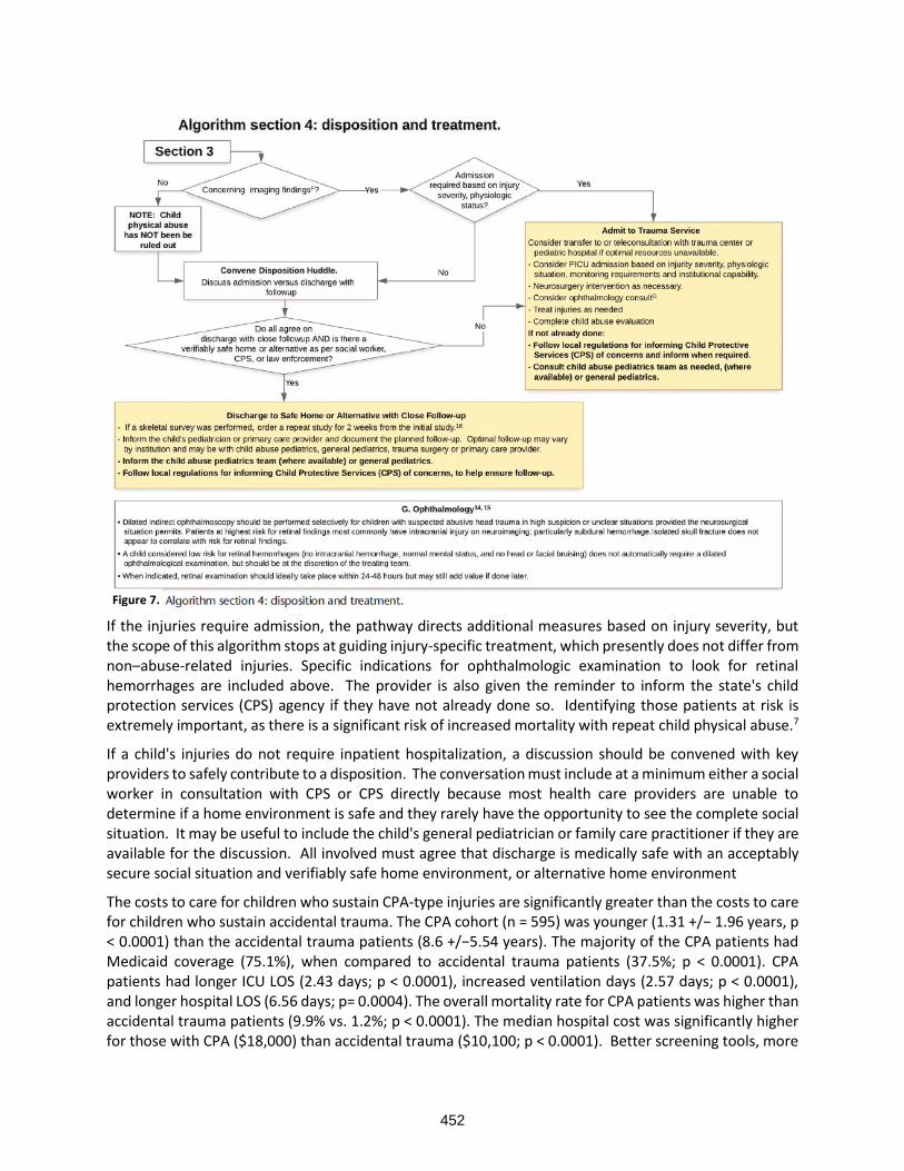
If the injuries require admission, the pathway directs additional measures based on injury severity, but the scope of this algorithm stops at guiding injury-specific treatment, which presently does not differ from non–abuse-related injuries. Specific indications for ophthalmologic examination to look for retinal hemorrhages are included above. The provider is also given the reminder to inform the state's child protection services (CPS) agency if they have not already done so. Identifying those patients at risk is extremely important, as there is a significant risk of increased mortality with repeat child physical abuse.7
If a child's injuries do not require inpatient hospitalization, a discussion should be convened with key providers to safely contribute to a disposition. The conversation must include at a minimum either a social worker in consultation with CPS or CPS directly because most health care providers are unable to determine if a home environment is safe and they rarely have the opportunity to see the complete social situation. It may be useful to include the child's general pediatrician or family care practitioner if they are available for the discussion. All involved must agree that discharge is medically safe with an acceptably secure social situation and verifiably safe home environment, or alternative home environment
The costs to care for children who sustain CPA-type injuries are significantly greater than the costs to care for children who sustain accidental trauma. The CPA cohort (n = 595) was younger (1.31 +/− 1.96 years, p < 0.0001) than the accidental trauma patients (8.6 +/−5.54 years). The majority of the CPA patients had Medicaid coverage (75.1%), when compared to accidental trauma patients (37.5%; p < 0.0001). CPA patients had longer ICU LOS (2.43 days; p < 0.0001), increased ventilation days (2.57 days; p < 0.0001), and longer hospital LOS (6.56 days; p= 0.0004). The overall mortality rate for CPA patients was higher than accidental trauma patients (9.9% vs. 1.2%; p < 0.0001). The median hospital cost was significantly higher for those with CPA ($18,000) than accidental trauma ($10,100; p < 0.0001). Better screening tools, more
Figure 7.
452

provider education and broader community outreach efforts are needed to reduce the societal and economic costs associated with child physical abuse.8
KEY POINTS
• The Pediatric Trauma Patient is at high risk for being a victim of nonaccidental trauma as well as a victim of human trafficking
• Screening for nonaccidental trauma has improved significantly, but there are still socioeconomic and racial barriers to over come
• Even “witnessed” falls in a 1–2-year-old with worrisome injuries should be considered NAT until the sources are verified as legitimate and true.
• Isolated hollow viscus injuries, especially to the duodenum, are often associated with NAT/CPA
TRAFFICKING
It is difficult to assess the actual association between human trafficking and trauma, however, this is clearly an at-risk population. Health care providers can be hesitant to screen for potential trafficking. A recent publication out of Children’s Hospital of Philadelphia interviewed adults who had experienced trafficking as a child or adolescent and came up with common themes to assist with the screening process.9
• Fear is a significant barrier
• Participants do want PEM providers to ask about trafficking, and it is not harmful to do so
• PEM providers should address fear through emphasizing confidentiality and privacy and encouraging agency
• PEM providers should approach the patient in a direct, sensitive, and nonjudgmental manner
• Changes to the ED environment may facilitate the conversation.
When interviewing potential trafficking victims
• Show genuine concern about their emotional and mental state
• Reassure the patient that you are with them in the situation
• Ask the patient for permission to hear more about what is going on
• Empower the patient by referring to them in encouraging terms
• Reiterate that you will not break their confidentiality unnecessarily
• Provide encouragement to the patient
• Use the patient’s symptoms to begin the conversation.
• Check with the patient to see if they feel safe to share at the moment before proceeding
• Explicitly assure the patient they are in a safe place at the moment
• Validate the patient’s feelings
• Ask direct and concrete questions regarding trauma, stress, and abuse
• Ask if the patient is hoping for change in circumstances or has future hopes
American Academy of Pediatrics is dedicated to screening for trafficking. Although researcher suggests that victims of sex trafficking in the United States are likely to seek medical attention at some point during their period of exploitation, trafficked immigrant children detained at the United States border may receive insufficient care, and those trafficked abroad may receive little or no care at all.10
Possible Indicators for Labor Trafficking
• Recent immigration history (especially if patient or family lack access to immigration documentation)
453

• Unfamiliarity with city or town
• Apparent intimidation by person accompanying the child or family
• Inconsistencies in information provided
• Report of excessive, hazardous, or other inappropriate work conditions
• Work-related (typically preventable) injuries (eg, chemical burns, irritation from toxic gases)
• Delay in care (far-advanced medical conditions or untreated injuries)
• Malnutrition or dehydration
• Poor hygiene
• Report of crowded, unhygienic, or otherwise inappropriate living conditions
The AAP further advocates that all medical professionals should be better educated with respect to human trafficking. Many states such as Florida, where I now reside, require mandatory annual CME on Trafficking to renew medical licensure. Other recommendations from the AAP include10:
• Advocate for training of health care professionals on human trafficking issues, including recognition, assessment, treatment, and referrals for services by using a trauma informed, culturally sensitive, rights-based approach. Training should emphasize both sex and labor trafficking, the possibility of encountering parents who are victims of trafficking, and specific issues related to immigration for foreign victims and their families.
• Advocate for timely medical education on human trafficking at the trainee level by encouraging the American Board of Pediatrics to include child trafficking, child rights, and trauma-informed care in its content specifications, as these guide residency curricula
• Encourage medical education curricula to include strategies for addressing social determinants of health, in particular including questions about adverse childhood events in the patient assessment, connecting to community resources, and building community resilience
• Advocate for financial support and resources for development and global dissemination of culturally appropriate, trauma informed curricula for health care professionals addressing human trafficking.
Sexual trafficking in the pediatric trauma population is also a known problem with poorly organized recognition and screening. The number of victims is unknown but is likely in the millions. Potential indicators and risk factors to be aware of in the sexual exploitation of the pediatric trauma patient11:
INITIAL PRESENTATION SEXUAL TRAFFICKING
• Child accompanied by domineering adult who does not allow child to answer questions
• Child accompanied by unrelated adult
• Child accompanied by other children and only one adult
• Child provides changing information regarding demographics
• Chief complaint is acute sexual assault or acute physical assault
• Chief complaint is suicide attempt
• Child is poor historian or disoriented from sleep deprivation or drug intoxication
HISTORICAL FACTORS SEXUAL TRAFFICKING
• Multiple sexually transmitted infections (STIs)
• Previous pregnancy/abortion
• Frequent visits for emergency contraception
• Chronic runaway behavior
• Chronic truancy or problems in school
• History of sexual abuse/physical abuse/neglect
454

• Involvement of child protective services (especially foster care/group home)
• Involvement with department of juvenile justice
• Significantly older boyfriend
• Frequent substance use/misuse
• Lack of medical home and/or frequent emergency department visits
PHYSCIAL FINDINGS SEXUAL TRAFFICKING
• Evidence suggestive of inflicted injury
• Tattoos (sexually explicit, of man’s name, gang affiliation)
• Child withdrawn, fearful
• Signs of substance misuse
• Expensive items, clothing, hotel keys
• Large amounts of cash
• Poor dentition or obvious chronic lack of care
Physicians are mandated to report suspected child abuse and neglect. In states where commercial sexual exploitation of children and sex trafficking are considered a form of abuse, the physician must make a formal report of suspected exploitation to law enforcement and to Child Protective Services as well.
KEY POINTS
• The Pediatric Trauma Patient is at high risk for being a victim of nonaccidental trauma as well as a victim of human trafficking
• Fear is a key barrier for victims of trafficking and most victims want to be asked if they feel they are in a safe environment
• Some states consider trafficking a form of child physical abuse and requires similar reporting to CPS as NAT, others do not.
REFERENCES
1. Nelson G. Rosen, MD, Mauricio A. Escobar, Jr., MD, Carlos V. Brown, MD, Ernest E. Moore, MD, Jack A. Sava, MD, Kimberly Peck, MD, David J. Ciesla, MD, Jason L. Sperry, MPH, MD, Anne G. Rizzo, MS, MD, Eric J. Ley, MD, Karen J. Brasel, MPH, MD, Rosemary Kozar, MD, PhD, Kenji Inaba, MD, Jamie L. Hoffman-Rosenfeld, MD, David M. Notrica, MD, Lois W. Sayrs, PhD, Todd Nickoles, BSN, MBA, Robert W. Letton, Jr., MD, Richard A. Falcone, Jr., MD, Ian C. Mitchell, MD, and Matthew J. Martin, MD. Child physical abuse trauma evaluation and management: A Western Trauma Association and Pediatric Trauma Society critical decisions algorithm. J Trauma Acute Care Surg, Volume 90, Number 4: 641-651.
2. Mauricio A. Escobar Jr., Kim G. Wallenstein, Emily R. Christison-Lagay, Jessica A. Naiditch, and John K. Petty. Child abuse and the pediatric surgeon: A position statement from the Trauma Committee, the Board of Governors and the Membership of the American Pediatric Surgical Association. Journal of Pediatric Surgery 54 (2019) 1277–1285.
3. Joshua B. Brown, MD, Mark L. Gestring, MD, Christine M. Leeper, MD, Jason L. Sperry, MD, Andrew B. Peitzman, MD, Timothy R. Billiar, MD, and Barbara A. Gaines, MD. Characterizing injury severity in nonaccidental trauma: Does Injury Severity Score miss the mark? J Trauma Acute Care Surg. 2018;85: 668–673.
4. Richard A. Falcone Jr, Rebeccah L. Brown, and Victor F. Garcia. Disparities in child abuse mortality are not explained by injury severity. Journal of Pediatric Surgery (2007) 42, 1031–1037.
455

5. Katherine P. Deye, MD, Rachel P. Berger, MD, MPH, and Daniel M. Lindberg, MD. Occult abusive injuries in infants with apparently isolated skull fractures. J Trauma Acute Care Surg. 2013;74: 1553-1558.
6. Hallie J. Quiroz, Jason J. Yoo, Liann C. Casey, Brent A.Willobee, Anthony R. Ferrantella, Chad M.Thorson, Eduardo A. Perez, and Juan E. Sola. Can we increase detection? A nationwide analysis of age-related fractures in child abuse. Journal of Pediatric Surgery 56 (2021) 153–158.
7. Katherine J. Deans, MD, MHSc, Jonathan Thackeray, MD, Johanna R. Askegard-Giesmann, MD, Elizabeth Earley, MPH, Jonathan I. Groner, MD, and Peter C. Minneci, MD, MHSc. Mortality increases with recurrent episodes of nonaccidental trauma in children. J Trauma Acute Care Surg. 2013;75: 161-165.
8. Niti Shahi, Ryan Phillips, Maxene Meier, Dwayne Smith, David Leopold, John Recicar, and Steven Moulton. The true cost of child abuse at a level 1 pediatric trauma center. Journal of Pediatric Surgery 55 (2020) 335–340.
9. Carmelle Wallace, MD, MPH, Yvette Schein, BA, Gina Carabelli, CRNP, Heta Patel, Needhi Mehta, BA, Nadia Dowshen, MD, Nancy Kassam-Adams, PhD, Kenneth Ginsburg, MD, MSE, and Cynthia Mollen, MD, MSCE. A Survivor-Derived Approach to Addressing Trafficking in the Pediatric ED. Pediatrics. 2021;147(1)
10. Jordan Greenbaum, MD, Nia Bodrick, MD, MPH, FAAP, COMMITTEE ON CHILD ABUSE AND NEGLECT, SECTION ON INTERNATIONAL CHILD HEALTH. Global Human Trafficking and Child Victimization. Pediatrics. 2017;140(6)
11. Jordan Greenbaum, MD, James E. Crawford-Jakubiak, MD, FAAP, COMMITTEE ON CHILD ABUSE AND NEGLECT. Child Sex Trafficking and Commercial Sexual Exploitation: Health Care Needs of Victims. PEDIATRICS Volume 135, number 3, March 2015.
456

DEATH IN THE TRAUMA BAY: THERE IS NEVER NOTHING LEFT TO DO
Melissa “Red” Hoffman, MD, FACS
Clinical Assistant Professor University of North Carolina at Chapel Hill School of Medicine Chapel Hill, NC
TOP 10 THOUGHTS ON DEATH IN THE TRAUMA BAY
1. Providing high-quality end-of-life care requires experience, skill and some degree of emotional intelligence. If at all possible, it should NOT be relegated to the most junior member of the physician or nursing team
2. While the patient is the primary focus of care, care should also be provided to the family (if present), the team and the surgeon (self-care).
3. Aggressive symptom management is often necessary at the end-of-life; multiple doses of multiple drugs of different classes (opioids, benzodiazepines, antipsychotics) may be needed at the same time. Such dosing may raise the concern of hastening death or euthanasia. One way to counter this concern is to utilize OBJECTIVE data (heart rate, blood pressure, nonverbal signs such as grimacing) to guide medication administration.
4. When the intention for using morphine is to relive pain or dyspnea, the treatment is ethical, the risk of potentially dangerous adverse secondary effects which may lead to hastening death is minimal and the risk of respiratory depression is over-estimated.
5. Communication regarding death and dying also requires experience, skill and emotional intelligence. If at all possible, this communication should also NOT be relegated to the most junior member of the team. That being said, learners should always be invited to witness these discussions so that they may perform them independently in the future.
6. Providers are not ethically obligated to offer treatments which are futile. Further, one way we can care for the family is to relieve them of decisional burden by NOT asking them to choose whether to pursue futile treatments.
7. Unexpected or violent deaths increase the risk of prolonged grief disorder. Preparing a handout which contains information about this disorder, along with contact information for local trauma-informed therapists, the Trauma Survivors Network and your contact information in case families have questions is another way to support families during this difficult time.
8. As team leaders, not only are we ultimately responsible for the well-being of the patient, we are also responsible for the well-being of the team. One way to honor both the patient and the team is to utilize The Pause, a 15- to 30-second intentional moment of silence meant to provide closure to the relationship between the patient and the team.
9. Due to culture of surgery and trauma, team members may be at risk for disenfranchised grief, defined as the grief that persons experience when they incur a loss that is not or cannot be openly acknowledged, publicly mourned, or socially supported. Debriefing with colleagues can help to mitigate this risk.
10. The importance of self-care cannot be overstated. The importance of modeling self-care (whatever that looks like for you) for our learners cannot be overstated as well.
457

INTRODUCTION/OVERVIEW
While less than ten percent of trauma patients die in the trauma bay, these are often some of the most memorable, dramatic and traumatic experiences for all involved. Death often presents in one of three ways: the patient arrives with CPR in progress and is declared dead on arrival; the patient arrives in extremis and undergoes resuscitation (up to and including ED thoracotomy) which ultimately proves futile; or the patient is deemed to have a nonsurvivable or devastating injury (most often a head injury or a large burn) and the decision is made to focus on comfort. In all three scenarios, the patient is obviously the primary focus of care. But each scenario also offers various opportunities to provide care for the family, the care team and the self.
TWO PRIMARY GOALS: ABSENCE (OF SUFFERING) AND (RADICAL) PRESENCE
When an injured patient arrives in the trauma bay, the relief of suffering (i.e., the treatment of pain, anxiety and agitation) is sometimes overshadowed by the need for emergent and life-saving procedures. However, once it becomes clear that the patient is going to die (either despite our best efforts or because we recognize that interventions will be futile), the goals of care can shift to Absence and Presence.
The first goal is Absence of Suffering. As stated above, depending on the injury, trauma patients at the end of life may require high doses of various medications, often administered multiple times in rapid succession. This type of care requires skill, experience, a willingness to communicate openly with team members (including the pharmacist pulling the medications and the nurse administering the medications) and, hopefully, a pre-existing order set (see example below).
458

From Rush RM et al. Frontline Surgery: A Practical Approach.
Several issues may arise. One, team members may question (either out loud or to themselves) the ethics involved in the dosing regimen. Two, team members may express (or may feel but not express) discomfort in administering high doses of medications. Three, the patient may continue to suffer from either pain, dyspnea, anxiety or agitation despite our medications.
Perhaps the best way to address these issues is to have the attending surgeon, trauma fellow or chief resident remain at the bedside. The surgeon may ask if anyone has any questions or concerns about transitioning to a comfort-focused approach. Rather than leaving it to the nurse to follow a pre-existing order set (if one exists), the surgeon can work closely with the bedside nurse, using objective data such as heart rate, respiration rate, facial grimace or agitated body movements to guide the administration of medications. Lastly, if the patient continues to suffer, the surgeon is readily available to change the plan of care.
Much has been written about the idea of “double effect” and the actions of morphine at the end of life. However, Dr. Charles von Gunten, a pioneer in the field of palliative care, has written extensively about this topic and notes that the adverse effects of morphine (including respiratory depression) are actually over-estimated. He notes that the principle of double effect actually refers to the ethical construct where a physician uses a treatment where the potential outcome is good, knowing that there will certainly be an undesired secondary outcome (he uses the separation of conjoined twins as an example, when the physician knows that one will leave and one will do). Instead, he states that the potential side effects of morphine can be considered unintended consequences, which, again, are over-estimated.
If the patient is conscious, they may have a sense that they are dying and may, in fact, ask you. As a physician and as a human being, we should answer this question compassionately and truthfully. While an honest answer may lead to more suffering (particularly emotional and spiritual), it will also offer the patient an opportunity for closure. In order to address any emotional or spiritual suffering, we should also ask the patient if they would like a chaplain to be called.
The second goal during this time is Presence. Dr. Balfour Mount, a urologic oncologist who coined the term “palliative care” and who is considered the Father of Palliative Care in North America, has spoken and written about the idea of Radical Presence, the willingness and ability to be full present with another being in their suffering and in their dying. While this concept may sound like new-age mumbo jumbo, I
459

would counter that there is nothing more “hard core” than holding space for someone as they contemplate their mortality and take their last breaths and nothing more “hard core” than sitting still and just experiencing the moment instead of trying to fix or to change the ultimate outcome.
CARING FOR THE FAMILY
The first step in caring for the family is to ensure that family members receive a timely, accurate update from the trauma team, ideally by a senior member of the team who has experience in communicating serious news. As stated earlier, learners should always be invited to witness these discussions so that they may perform them independently in the future.
While the trauma team is often very busy, it is worthwhile to take a few moments to plan the interaction, checking to see if any other specialist wants to be available (for example, the neurosurgeon in the setting of a devastating brain injury) and making certain to find a quiet place for families to sit (with tissues available). While this update is occurring, other members of the team should be cleaning the patient and the room so that family members are not unnecessarily exposed to traumatic sights. It is often helpful to start the conversation by asking the family what they know and then firing a “warning shot” (for example, “I am sorry that I have bad news” or “I am afraid that I have some serious news”). When discussing death or an impending death, it is best to use the D word (died, dying, death) so that there is no confusion on the part of any family members.
As a reminder, physicians are not ethically obligated to offer treatments or perform procedures which they consider futile. Quantitative futility refers to an intervention that is unlikely to produce or is physiologically incapable of producing a certain result. For example, performing CPR on a patient who has begun to herniate will not save his life. In contrast, qualitative futility refers to an intervention that would achieve a certain result but would not change the overall outcome. For example, starting dialysis on a patient with widely metastatic pancreatic cancer may slightly increase survival but will not prevent the inevitable death from the underlying disease. Another way to care for the family is to avoid offering nonbeneficial or futile treatments. This helps families avoid the burden of making a decision that will ultimately not alter the trajectory of the disease process (but may cause a great deal of angst for family members for years to come).
Once the family is updated, they should be invited back to the trauma bay. The vast majority of family members will have little to no experience with death and dying. If time allows, a member of the trauma team should be available to curate the experience. Even in the emergency room, the ideas of set (preparation and expectation of the person or people having the experience) and setting (the physical and cultural environment in which the experience takes place) often discussed in psychedelic culture, are relevant. As far as set, family members should be quickly educated about the signs and symptoms of imminent death. In terms of setting, the trauma team member may choose to dim the lights, play the patient’s favorite music, encourage the family members to touch and talk to the patient, invite the family members to share stories about the patient, and even ask, if it seems appropriate, who may be waiting to welcome the patient on the other side. Allowing family members to take an active role during the patient’s death may assist with the grieving process.
That being said, we know that unexpected or violent deaths can increase the risk of Prolonged Grief Disorder (sometimes referred to as complicated grief). Symptoms of the disorder include: intense sorrow, pain and rumination of the deceased; an inability to focus on little else but the death; extreme focus on or avoidance of reminders of the deceased; problems accepting the death; and an inability to enjoy life or think back on positive experiences with the deceased. Further, in his book “Retelling Violent Death,” Edward Rynearson addresses the two distinct, but intertwined, distresses that survivors of violent loss experience: traumatic distress and separation distress (see Figure below). He notes that “the
460

thoughts, feelings and behaviors of simultaneous traumatic and separation distress are not harmonious” and that their contradictory nature makes them difficult to process. Preparing a handout which contains information about complicated grief and the distinct difficulties of violent loss, along with contact information for local trauma-informed therapists, the Trauma Survivors Network and your contact information in case families have questions is another way to support families during this challenging time.
From: Rynearson, EK. Retelling Violent Death.
CARING FOR THE TEAM
While trauma surgery is a team sport, the trauma surgeon is the “captain of the ship” and bears the ultimate responsibility for the care of both the patient and of the team. Deaths in the trauma bay can be exceedingly difficult to process, particularly after a long, ultimately unsuccessful resuscitation. Because resuscitation is a team effort, it is standard of care to ask all team members whether there are any objections to stopping resuscitation. If the patient is not expected to die immediately, it is important to clearly communicate your goals and your plan of care with the team.
As stated earlier, one way to care for the team is to make certain that a senior team member is present or immediately available during the dying process. Once the death occurs, Jonathan Bartels (a former trauma nurse and now a palliative care nurse) suggests performing The Pause (see Figure below), an intentional 15- to 30- second moment of silence meant to honor the life of the patient and the work of the trauma team as well as to provide closure to the relationship between the patient and the team.
All team members, including the attending surgeon, may be at risk for disenfranchised grief, defined as “the grief that persons experience when they incur a loss that is not or cannot be openly acknowledged, publicly mourned, or socially supported.” The best way to counter this risk is to acknowledge one’s feelings in a safe space, and to encourage others to do the same.
461
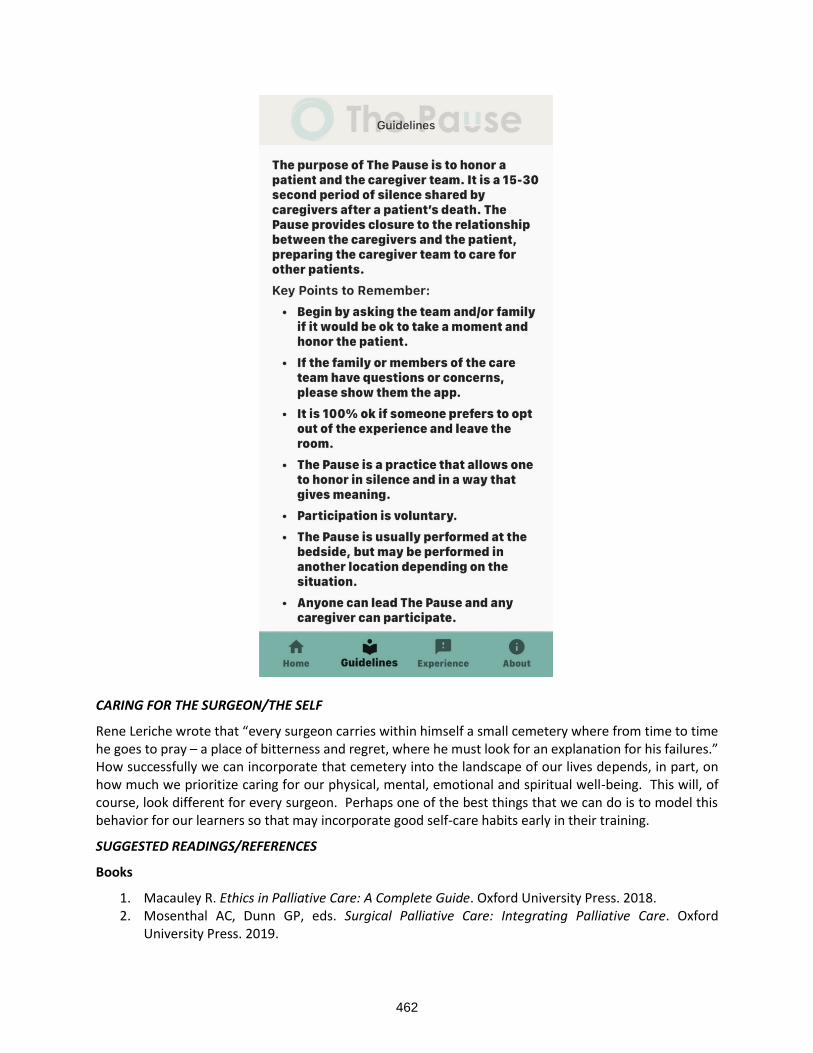
CARING FOR THE SURGEON/THE SELF
Rene Leriche wrote that “every surgeon carries within himself a small cemetery where from time to time he goes to pray – a place of bitterness and regret, where he must look for an explanation for his failures.” How successfully we can incorporate that cemetery into the landscape of our lives depends, in part, on how much we prioritize caring for our physical, mental, emotional and spiritual well-being. This will, of course, look different for every surgeon. Perhaps one of the best things that we can do is to model this behavior for our learners so that may incorporate good self-care habits early in their training.
SUGGESTED READINGS/REFERENCES
Books
1. Macauley R. Ethics in Palliative Care: A Complete Guide. Oxford University Press. 2018. 2. Mosenthal AC, Dunn GP, eds. Surgical Palliative Care: Integrating Palliative Care. Oxford
University Press. 2019.
462
3. Rush RM, Martin MJ, Cocanour CS. Expectant and end of life care in a combat zone. In: Frontline Surgery: A Practical Approach. Springer;2017:749-760.
4. Rynearson, EK. Retelling Violent Death. Routledge. 2001. 5. Rynearson, EK. ed. Violent Death: Resilience and Intervention Beyond the Crisis. Routledge. 2014.
References
1. Bartells, J. The Pause. 2015. Available at: https://thepause.me/ 2. Elkbuli A, Kinslow K, Dessy B et al. Addressing burnout among trauma surgeons. Am Surg.
2020:86(9)1175-1177. 3. Fiorentino M, Mosenthal AC, Bryczkowski S, Lamba S. Teaching residents communication skills
around death and dying in the trauma bay. J Pall Med. 2021:24(1)77-82. 4. Lamba S, Bryczkowski S, Tyrie L et al. Fast facts and concepts #305: death disclosure and delivering
of difficult news in trauma. October 2015. Available at: https://www.mypcnow.org/wp-content/uploads/2019/03/FF-305-DeliverBadNewsTrauma1.pdf.
5. Romero, K. Work grief: a primer for emergency medicine providers. 2020. Available at: https://www.aliem.com/work-grief-primer/.
6. Traylor, M. Should family be permitted in a trauma bay? AMA J Ethics. 2018:20(5)455-463. 7. von Gunten CF. Fast facts and concepts #8: morphine and hastened death. May 2015. Available
at: https://www.mypcnow.org/fast-fact/morphine-and-hastened-death/. 8. Warm EJ, Weissman DE. Fast facts and concepts #63: the legal liability of under-treatment of pain.
April 2009. Available at: https://www.mypcnow.org/fast-fact/the-legal-liability-of-under-treatment-of-pain/.
463

MILITARY & CIVILIAN COLLABORATION – DO THEY WORK?
Alison Wilson, MD, FACS
Vice-Chair and Professor WVU Department of Surgery Skewes Family Chair for Trauma Surgery Director, WVU Critical Care and Trauma Institute Morgantown, WV
INTRODUCTION
Military Civilian partnerships have ebbed and flowed throughout the history of the US depending on the combat tempo and needs of the military and the nation. Examples include the conversion of the historic Greenbriar hotel into a military hospital during WWII and the use of civilian hospitals as American Red Cross Army Base hospitals. What is recently established is a formalized path to maintain and structure these partnerships independent of times of conflict. It has been recognized that the ability to maintain needed skills and readiness to deploy can be challenging at military facilities during times of peace. Recently, a more formalized path including congressional legislation have been passed to develop and maintain these important collaborations.
MILITARY CIVILIAN PARTNERSHIPS AND DEVELOPMENT OF GUIDELINES
The National Defense Authorization Act (NDAA) for Fiscal Year 2017 required the development of a Joint Trauma Education and Training Director to ensure adequate readiness of military medical personnel. This set the stage for formal military civilian partnerships and established some methods of funding. A
partnership between the DoD and the American College of Surgeons (MHSSPACS) is working to set standards and criteria for centers who will participate in these formal programs and partnerships. Beginning in 1999, a joint trauma training center was established at Ben Taub Hospital in Houston, TX. In the early 2000s, each military branch developed individual collaborations with other civilian centers to better tailor experiences to their needs. These centers remain established and functional. However, there is an increased need for additional partnerships. The MHSSPACS has developed a document that has become known as the “Blue Book” that establishes guidelines and criteria for the establishment of such partnerships. Such criteria cover institutional commitment, governance/administration, human resources, and physical resources. It should be noted that the Blue Book is primarily focused upon military surgical and resuscitation teams. It should be noted that there are a multitude of other military medical assets also in need of continued training and readiness.
There are three primary partnerships envisioned for the sustainment of these surgical and resuscitation teams. First is having military personnel permanently embedded in a civilian trauma institution. Second,
464

rotating military trainees at a civilian institution and third is having military personnel take call at a civilian site. Common challenges with executing these agreements include state licensing, malpractice coverage, billing, and credentialling and privileging. It is critical that military personnel be able to gain direct experience and practice to the scope of what will be expected when they are deployed. For physicians and nurses, this is more easily overcome as there are recognized scopes and practice equivalents. This becomes more of a challenge for the methods of service where there is not a civilian equivalent, such as special operations medics.
Key factors for success at a civilian center include having a local surgical and administrative advocate who understands the various military roles and needs for experiences and can function as a translator of sorts to the civilian administration. This person also must have a strong relationship and understanding of the local constraint on educators and learners. Programs can be hampered if animosity develops between civilian learners and the military learners due to a competitive learning environment. This becomes particularly true when it comes to many procedural skills. Mandatory to success is the commitment from the civilian site administration. These programs must be viewed as a method of service and not a funding avenue or profit center. Programs will require dedicated time, faculty, and have other costs. To show value, a civilian center should consider how they will measure competency metrics. Simply providing for hands on experience is inadequate to truly meet the definition of readiness. Dedication of the faculty to coordinate local and military needs are essential. For potential centers, an honest evaluation of capacity to take on additional personnel and learners should be completed with the goal of identifying the optimum number of participants at any given time. Finally, the mission and setting of the civilian institution should be aligned with the mission and skill set of the military partner.
EXPANSION OF CONCEPT BEYOND SURGICAL AND RESUSCITATION TEAMS
There are additional needs of the military and for opportunities for civilian centers to support and be involved. West Virginia University has had the unique pleasure and opportunity to be involved in the clinical training of Special Operations medic clinical training. This has resulted in a strong symbiotic relationship that has yielded benefits beyond the original program.
Background
While deployed, an 18 Delta (Green Beret medic) must be able to operate as an independent medical provider in an austere environment with no immediate support and often with prolonged evacuation times. In addition to caring for the combat injured, the 18D oversees monitoring the health and well being of the other team members, providing primary care for the indigenous community, including dental care, and even assisting with veterinary care. This unique medic must have a background not only in trauma care, but clinical medicine and prolonged field care. The training pipeline for the 18 D is quite lengthy and intense. They must master and pass a myriad of classes such as pharmacology, microbiology, anatomy/physiology, etc. but in a very time compressed environment. In addition to the didactic and lab courses that are completed at the Special Warfare Medical Group, they must complete a 24-day clinical rotation, known as the SOCT (Special Operations Clinical Training). This rotation must provide clinical experiences in a variety of non-trauma medical settings. In order to maintain credentials, an 18 D must complete a multitude of sustainment training. Included in this is a clinical rotation that must be done for 2 weeks every two years or 4 weeks every four years, known as a MPT (Medical Proficiency Training). This is the setting of the WVU-SWMG (Special Warfare Medical Group) partnership.
SOCT Rotation
The SOCT rotation is a 24-day rotation and is broad based in the clinical settings. The rotations include pediatrics, anesthesia, urgent care, emergency care, dental, family medicine, infectious disease, ophthalmology, wound care, veterinary medicine, large animal handling. The SOCT medic student is
465

intricately involved in the direct care on each of these modules and is not there as a shadower. This allows the student to have direct interaction with patients under the immediate supervision of a faculty. Each unique module has objectives, cognitive and technical that the student medic must complete. Faculty have been provided with background on how the medic must be able to function when deployed and the austere types of settings with limited resources. The goal is not to teach the medic student how do conduct medical evaluations as would be done in a mature hospital, but how to evaluate patients and physiology and mimic the delivery of care in a resource limited environment. The student medics must complete a pretest, posttest and participate in two cadaver labs to practice and demonstrate skills, critical thinking, and clinical management. Objective data and training metrics have been developed and are monitored. The academic faculty are committed to ensuring that not only is it a beneficial experience, but that objective measures can be provided to military command to show performance and benefit.
MPT Rotation
This rotation allows the experienced 18 D to return and refresh direct clinical skills. Rotations are custom build in conjunction with the soldier to meet the level of experience, area of operations, interests, and collaborative evaluation of deficits. The medic has more in-depth clinical experiences and faculty are informed of the medic’s level of training. Opportunities for advanced technical skills as well as more in-depth exposure to critical care are provided. If scheduling allows experience in the Fresh Tissue Training lab are also incorporated.
Symbiotic Relationship
Though there is no financial benefit to WVU, there have been numerous advances and innovative programs that have developed as a result of this relationship. The concept of the Virtual ICU was born from faculty understanding how the medics work in resource limited, austere environment and yet perform effective delivery of care. Modeling this, the ICU faculty began daily rounding with hospitalists at several critical access hospitals which allowed the system to keep more complex patients at smaller institutions safely and effectively. This has been critical throughout the pandemic and has enhanced the delivery of care in a cost-effective manner.
As the student medics and 18 D interact with a broad base of the faculty and are not limited to the emergency department or trauma care. These learners are highly motivated, invested and are accustomed to achieving high expectations to define success. That enthusiasm and preparedness have re-energized many of the faculty in the clinical teaching which has translated to all students. Additionally, it has been observed that many students and residents seem to be more motivated by their presence.
The opportunity to work with the student medics and 18D increased awareness of the medical school of some of the short comings of the medical school application screening rubrics. A dedicated program Special Operation Medic Pipeline (SOM-P) was developed to improve awareness in the Admissions Committee of these individuals’ medical experiences and academic accomplishments, albeit through the traditional routes. It provides support to those medics at the end of their military career with an avenue for evaluation of a career in medicine and support through the application process. In the first two years of the program, the success rate for these medics applying to medical school has been 100%. The program is provided free to the medic with the only stipulation that WVU is one of the medical schools applied to. The philosophy of our leadership is that we wish to support these medics in finding the best match for their success. A second program that has developed is the Special Operations Medic- Med Student Integration Program (SOM-MSIP). Upon matriculation, the medic is assigned a student or resident mentor who is previous SOF or military with SOF exposure. The purpose is to provide a routine connection to assist with the navigation of the local area, classes, and transition to civilian life. The medic is also paired with a faculty mentor to meet routinely and ensure academic success and construction of a plan to
466

develop a successful career. Part of this program also included development of a research project with support to attend a meeting if the project is accepted. These programs have developed a local community and comradery that has benefited the students, faculty, residents, and families. These medical students routinely step forward to assist in the training of others on the SOCT, MPT and other military courses on site, thus continuing the cycle of service and giving value added to the military programs.
CONCLUSION
Military Civilian Partnerships can occur in a variety of settings and are not limited to urban trauma centers. Both partners should work to maximize the strengths and match the needs of the other. The programs not only provide benefit to maintain the readiness of the military partner but can also enhance care delivery and performance in the civilian center.
467

MITIGATING DEFICIENCIES IN SURGICAL EDUCATION
Hasan B. Alam, MD, FACS
Loyal and Edith Davis Professor and Chair Department of Surgery Surgeon-in-Chief, Northwestern Memorial Hospital Chicago, IL
Disclosure: The author reports no proprietary or commercial interest in any product mentioned or concept discussed in this article.
The practice of surgery has undergone rapid and significant changes over the past 2 decades.
Outcomes have improved across the board for nearly every surgical problem, and the surgeons are now able to technically do operations that were unthinkable just a generation ago. This has been accompanied by an explosion in the availability of new tools and techniques, as well as an improvement in our knowledge, which allows us to take care of increasingly complex patients. This chapter aims to highlight just a few aspects of how this changing environment is affecting our surgical training programs.
DECREASING AUTONOMY
Over the last 2 decades, there has been a significant decrease in autonomy of the surgical residents. The opportunities to make independent decisions during training, and to learn from these mistakes have rapidly disappeared. In fact, the potential medico legal threat of any mistake, preventable or not, has created an environment where the attending surgeons are reluctant to relinquish control for operative as well as many of the non-operative aspects of clinical care.
The history of decreasing resident autonomy can be traced to several events1. The death of Libby Zion in 1984 highlighted the relationship between trainee autonomy and medical errors, and the subsequent Bell Commission Report of 1989 provided the impetus for the ACGME to restrict the resident duty hours. As a result, there was more attending supervision, and less time for the residents to spend in the hospital and the operating rooms (OR). The Omnibus Budget Reconciliation act of 1989 exerted pressure to expedite operative times and increase throughput, which once again created a disincentive for any activity in the OR that may slow things down (e.g. teaching or allowing a resident to operate at a slower pace). Furthermore, a 2002 mandate from the Center for Medicare & Medicaid Services (CMS) mandated that the attending surgeon be present during “critical portions” of the procedures, which once again decreased the opportunities for independent decision making and autonomy for the trainees. There have been some undeniable benefits for the patients as a result of these changes. The old mantra of “see one, do one, teach one” may have been good for the trainees but it clearly subjected generations of patients to receiving potentially suboptimal care from partially trained surgeons. In addition, there was often a not so hidden socioeconomic and racial bias involved in how differential care was provided to the “private” and “teaching” cases at many hospital, especially urban safety net institutions. The challenge now, and going forward, is how to best balance resident autonomy and attending supervision in a way that ensures patient’s safety while creating an environment where the trainees can make clinical decisions that allows for progressive growth and maturation.
468

CHALLENGES WITH ACQUIRING ADEQUATE SKILLSET DURING RESIDENCY
While increased supervision may have made the delivery of surgical care safer and more efficient, the negative impact of these changes on the competency of our graduating residents is undeniable. There is clearly an increasing concern that many General Surgery (GS) residents are not ready to enter independent practice by the end of their residency training2-4. National surveys have demonstrated that these concerns are widespread among the surgical leaders5-6, and in fact, even the residents themselves sometimes feel less than fully confident, which may explain the rising rates of graduates seeking additional fellowship training7-9. As a result, greater than 80% of the residents graduating from General Surgery training programs are not pursuing additional fellowship training.
The ACGME has made a number of changes over the last decade to place more emphasis on acquiring competency rather than fulfilling time-based criteria (e.g. spending a certain amount of time on a given rotation). There have been some very creative efforts to address surgical education using objective tools. For example, a team led by Dr. Brian George as part of the Procedural Learning and Safety Collaborative (PLSC), trained attendings in 14 GS programs to use a 5-level System [Improving and Measuring Procedural Learning (SIMPL) Performance scale]10 to assess resident readiness for independent practice. They used the Zwisch Scale to measure the level of guidance (i.e. autonomy) the attendings provided to their residents during specific procedures. A total of 444 attending surgeons rated 536 categorical residents after 10,130 procedures to generate the data12. The investigators reported that there was a 90% probability that a graduating resident would be deemed competent to perform an average core procedure on an average complexity patient, but this went down to 80% for a more complex core procedure. A very reasonable question is whether 80 or 90% probability is good enough? In reality, there was a large variability in the acquisition of autonomy, and this study really highlighted the need for using data-driven, objective tools to track competency, and provide ongoing feedback to the trainees.
Another way to judge the effectiveness of the GS training programs would be to see how the graduates perform at the start of their fellowships. The assessment of the subspecialty program directors, when asked to evaluate the graduates of GS programs joining their fellowships, is quite concerning5. In this study, out of 145 fellowship directors, 91 responded to the survey (63% response rate): 21% felt that new fellows arrived unprepared for the operating room, 38% demonstrated lack of patient ownership, 30% could not independently perform a laparoscopic cholecystectomy, 56% could not suture laparoscopically, and 66% were deemed unable to operate for 30 minutes without supervision during a major procedure. The program directors also felt that the majority of new fellows were unable to conceive, design, and conduct research/academic projects. Thematic clustering of qualitative data revealed deficits in domains of operative autonomy, progressive responsibility, longitudinal follow-up, and scholarly focus (~70% of the comments were related to a general lack of interest among fellows in academic pursuits or scholarship activities). This creates challenges for the fellowship programs to develop mitigation strategies to ensure that the trainees reliably “catch up” before finishing the training to join the workforce.
SURGICAL CULTURE AND A NEED FOR IMMEDIATE CHANGE
Today, generations X and Y compose most of the surgical workforce, with the millennials soon to follow. In contrast, a large number of the leadership positions are still held by the Baby Boom generation. Although generalizations are imperfect, much has been written about how these generations differ in their value systems and priorities, which can create misalignments and worsen job satisfaction. Physicians between the ages of 40-54 years seem to be more vulnerable to burnout than younger or older physicians. Nearly half of generation X physicians reported feeling burned out compared with 38% of the millennials (ages 25-39) and 39% of the baby boomer (55-73 years), with roughly half of the surveyed willing to take a substantial pay cut to achieve a better work-life balance13.
469

There is a desire on part of the newer generations (Millennials and Gen Z, etc) to have a work-life balance that is likely to be quite different from the past generations. Also, more than half of the medical graduates are women, and an increasing number of them are going into surgical specialties. Despite the increasing number of women in the surgical workforce, they face disparities and biases in compensation, mentorship, and advancement in leadership positions14-17. This is a multi-faceted, and complex problem that requires a comprehensive and thoughtful approach. Nevertheless, as the global agenda towards equality progresses, this pressing issue must be addressed head-on by our surgical leaders 18.
An even more important area that requires immediate attention is the impact of the surgical culture on our trainees. In a recent landmark study, Dr. Yue-Yung Hu from Northwestern University, and a multi-institutional team (including American College of Surgeons, American Board of Surgery, and ACGME) looked at the prevalence of discrimination, abuse, harassment, and burnout in surgical residency training programs across the country19. Among 7409 residents (99.3% of the eligible residents) from all 262 surgical residency programs, 31.9% reported discrimination based on their self- identified gender, 16.6% reported racial discrimination, 30.3% reported verbal or physical abuse (or both), and 10.3% reported sexual harassment. Rates of all mistreatment measures were higher among women; 65.1% of the women reported gender discrimination and 19.9% reported sexual harassment.
Attending surgeons were the most frequent sources of sexual harassment (27.2%) and abuse (51.9%). Weekly burnout symptoms were reported by 38.5% of residents, and 4.5% reported having had suicidal thoughts during the past year. This team has concluded that “Residency culture was characterized by poor resident wellness and frequent negative exposures”20. There also appears to be an under appreciation of how frequent sex, racial and ethnic discrimination is in our training programs. On an anonymous survey, very high rates of gender decimation (80% females vs. 17% males, p<0.001) and sexual harassment (43% females vs. 22% males; p<0.001) have been reported21. In another nationwide study (301 programs), of the 5679 who responded to the relevant questions, 1346 (23.7%) reported experiencing discrimination based on race/ethnicity or religion22. Residents were more likely to report discrimination if female, and nonwhite, and not surprisingly, the residents who experienced discrimination had higher rates of burnout (51.6% vs 40.0%; P < .001), thoughts of attrition (16.2% vs 10.1%; P < .001), and suicidal thoughts (6.5%vs 3.8%; P < .001). These, and many similar recent studies, have identified serious flaws in the traditional surgical culture that are simply unacceptable, and require an immediate, forceful, and sustained mitigation effort by all of us, at a personal and institutional level. THE SECOND TRIAL (Surgical Education Culture Optimization through targeted interventions based on National comparative Data) that includes 200 institutions across the country is one such effort23.
TRAINING THE LEADERS FOR TOMORROW
In addition to competent surgeons, we have to train the leaders that can effectively guide our profession. Leadership is not about titles, seniority, or one’s position in the hierarchy of an organization. In reality, it is a process of social influence, which maximizes the efforts of others, towards the achievement of a goal24. Successful leaders are good at predicting future needs, and in successfully aligning the efforts of others towards the achievement of meaningful goals. Delivery of healthcare is a complex process where leaders have to deal with numerous competing priorities. To cope with the new challenges, surgical leaders of the future will have to master new skillsets 25. Their key responsibilities will still include setting a compelling and appealing direction for the team (vision), selecting the right people (talent), and creating and embracing the right set of guiding core values (culture), but now they will also need to focus on a much longer list of characteristics:26
1. Professionalism 2. Technical competence 3. Motivation
470

4. Innovation 5. Teamwork 6. Communication skills 7. Decision-making 8. Business acumen 9. Emotional competence 10. Resilience 11. Effective teaching
Not surprisingly, leadership and its various conceptual models are among the most studied sociological concepts. Leadership, at its core, is the accomplishment of goals through the coordinated efforts of the team members. The person who successfully marshals their collaborators to achieve particular ends is a leader; and a great leader is one who can do so day after day, and year after year, in a wide variety of circumstances27. While some theorists have argued that great leaders are born with innate natural leadership skills, most contemporary experts agree that leadership consists of a series of well-defined skills and behaviors, that can be learned and taught, and refined through practice, experience, feedback, and coaching. In other words, most of us who want to become better leaders can do so with the help of appropriate training, expert coaching, effective mentoring, and ongoing learning/feedback. It is also important to better understand the relationship of leaders to their followers (Table 1)27, and how this has evolved. Historically, surgical leaders were chosen based upon quantitative measures of their achievements, but nowadays skills related to team building and how to best develop others are much more critical. To be successful in the 21st century, the level of emotional intelligence that surgical leaders possess will be more relevant than their academic accomplishments.
The Institute of Medicine, in their landmark report “Crossing the Quality Chasm: A New Health System for the 21st Century”, outlined six areas where the leaders in the healthcare system should be held accountable 28. These six domains can act as a guide for training appropriate leaders for tomorrow:
1. Effective: Avoiding overuse, underuse, and wrong services 2. Safe: Avoiding injuries to patients 3. Patient-centered: Respects the need of the individual 4. Timely: Reducing harmful waits and delays 5. Efficient: Avoiding waste 6. Equitable: Gender, ethnicity, geographic, and socioeconomic status
Historically, many of these domains have not been high priority areas for the surgical leaders.
However, to succeed in the 21st century, and to remain relevant in the new and interconnected healthcare system, the next generation of surgical leaders have to acquire skills that would allow them to be effective in these new areas. As opposed to the traditional “command and control” style of the traditional department of surgery chair, the next generation of leaders may or may not be chair, and their approach is likely to be more about “adding value” to the institutional mission, rather than focusing on what they can control. If we as a profession want to thrive, it is incumbent upon us to include formal leadership training in our educational programs (medical schools, residency and fellowships) to equip our next generation with the right attitude and skillset.
FUTURE TRENDS
Surgical training programs in the US during the post WWII era have been the envy of the entire world, and American surgeons have led the profession for most of the 20th Century. Credit for the establishment and/or expansion of most major new surgical fields (e.g. cardiovascular surgery, transplant surgery, integrated trauma systems, etc) over the last few generations rightfully belongs to American Surgery.
471
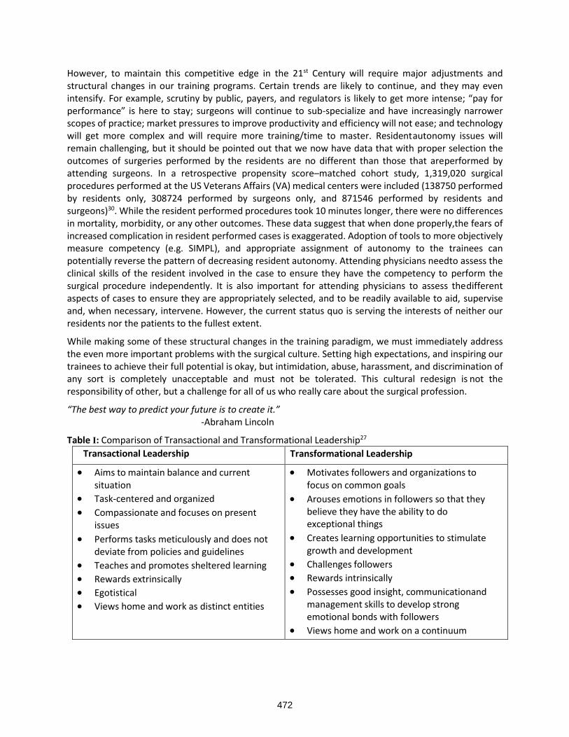
However, to maintain this competitive edge in the 21st Century will require major adjustments and structural changes in our training programs. Certain trends are likely to continue, and they may even intensify. For example, scrutiny by public, payers, and regulators is likely to get more intense; “pay for performance” is here to stay; surgeons will continue to sub-specialize and have increasingly narrower scopes of practice; market pressures to improve productivity and efficiency will not ease; and technology will get more complex and will require more training/time to master. Resident autonomy issues will remain challenging, but it should be pointed out that we now have data that with proper selection the outcomes of surgeries performed by the residents are no different than those that are performed by attending surgeons. In a retrospective propensity score–matched cohort study, 1,319,020 surgical procedures performed at the US Veterans Affairs (VA) medical centers were included (138750 performed by residents only, 308724 performed by surgeons only, and 871546 performed by residents and surgeons)30. While the resident performed procedures took 10 minutes longer, there were no differences in mortality, morbidity, or any other outcomes. These data suggest that when done properly, the fears of increased complication in resident performed cases is exaggerated. Adoption of tools to more objectively measure competency (e.g. SIMPL), and appropriate assignment of autonomy to the trainees can potentially reverse the pattern of decreasing resident autonomy. Attending physicians need to assess the clinical skills of the resident involved in the case to ensure they have the competency to perform the surgical procedure independently. It is also important for attending physicians to assess the different aspects of cases to ensure they are appropriately selected, and to be readily available to aid, supervise and, when necessary, intervene. However, the current status quo is serving the interests of neither our residents nor the patients to the fullest extent.
While making some of these structural changes in the training paradigm, we must immediately address the even more important problems with the surgical culture. Setting high expectations, and inspiring our trainees to achieve their full potential is okay, but intimidation, abuse, harassment, and discrimination of any sort is completely unacceptable and must not be tolerated. This cultural redesign is not the responsibility of other, but a challenge for all of us who really care about the surgical profession.
“The best way to predict your future is to create it.” -Abraham Lincoln
Table I: Comparison of Transactional and Transformational Leadership27
Transactional Leadership Transformational Leadership
• Aims to maintain balance and current situation
• Task-centered and organized
• Compassionate and focuses on present issues
• Performs tasks meticulously and does not deviate from policies and guidelines
• Teaches and promotes sheltered learning
• Rewards extrinsically
• Egotistical
• Views home and work as distinct entities
• Motivates followers and organizations to focus on common goals
• Arouses emotions in followers so that they believe they have the ability to do exceptional things
• Creates learning opportunities to stimulate growth and development
• Challenges followers
• Rewards intrinsically
• Possesses good insight, communication and management skills to develop strong emotional bonds with followers
• Views home and work on a continuum
472

REFERENCES
1. Coleman JR, Tracy BM, Stadeli KM, Chotai P, Howard J. The autonomy crisis: a call to action for resident advocacy. Bulletin of the American College of Surgeons. August 1, 2018. https://bulletin.facs.org/2018/08/the-autonomy-crisis-a-call-to-action-for-resident-advocacy/ (Last accessed 1.07.22)
2. Bell RH. Why Johnny cannot operate. Surgery. 2009;146:533–542. 3. Lewis FR, Klingensmith ME. Issues in general surgery residency training—2012. Ann Surg.
2012;256:553–559. 4. Elfenbein DM. Confidence crisis among general surgery residents: a systematic review and
qualitative discourse analysis. JAMA Surg. 2016;151:1166–1175. 5. Mattar SG, Alseidi AA, Jones DB, et al. General surgery residency inadequately prepares trainees
for fellowship: results of a survey of fellowship program directors. Ann Surg. 2013;258:440–449. 6. Napolitano LM, Savarise M, Paramo JC, et al. Are general surgery residents ready to practice? A
survey of the American College of Surgeons board of governors and young fellows association. J Am Coll Surg. 2014;218:1063–1072.
7. Borman KR, Vick LR, Biester TW, et al. Changing demographics of residents choosing fellowships: Longterm data from the American Board of Surgery. J Am Coll Surg. 2008;206:782–788.
8. Coleman JJ, Esposito TJ, Rozycki GS, et al. Early subspecialization and perceived competence in surgical training: are residents ready? J Am Coll Surg. 2013;216:764–771.
9. Fronza JS, Prystowsky JP, DaRosa D, et al. Surgical residents’ perception of competence and relevance of the clinical curriculum to future practice. J Surg Educ. 2012;69:792–797.
10. The Society for Improving Medical Professional Learning https://www.simpl.org/ 11. George BC, Teitelbaum EN, Meyerson SL, et al. Reliability, validity, and feasibility of the Zwisch
scale for the assessment of intraoperative performance. J Surg Educ. 2014;71:90–96. 12. George BC, Bohnen JD, Williams RG, Meyerson SL, Schuller MC, Clark MJ, Meier AH, Torbeck L,
Mandell SP, Mullen JT, Smink DS, Scully RE, Chipman JG, Auyang ED, Terhune KP, Wise PE, Choi JN, Foley EF, Dimick JB, Choti MA, Soper NJ, Lillemoe KD, Zwischenberger JB, Dunnington GL, DaRosa DA, Fryer JP; Procedural Learning and Safety Collaborative (PLSC). Readiness of US General Surgery Residents for Independent Practice. Ann Surg. 2017 Oct;266(4):582-594. doi: 10.1097/SLA.0000000000002414. Erratum in: Ann Surg. 2018 Mar;267(3):e63. PMID: 28742711.
13. The Wall Street Journal. “Physician burnout widespread, especially among midcareer” Accessed Feb 14, 202. https://www.wsj.com/articles/physician-burnout-widespread-especially-among- those-midcareer-report-says-11579086008
14. Sorouri K, Khan S, Bowden S, Searle S, Carr L, Simpson JS. The Glaring Gender Bias in the Operating Room: A Qualitative Study of Factors Influencing Career Selection for First-Year Medical Students. J Surg Educ. 2021 Sep-Oct;78(5):1516-1523. doi: 10.1016/j.jsurg.2021.01.014. Epub 2021 Feb 9. PMID: 33579653.
15. Morris M, Chen H, Heslin MJ, Krontiras H. A Structured Compensation Plan Improves But Does Not Erase the Sex Pay Gap in Surgery. Ann Surg. 2018 Sep;268(3):442-448. doi: 10.1097/SLA.0000000000002928. PMID: 29979249.
16. Singletary, SE. "Mentoring surgeons for the 21st century." Annals of Surgical Oncology 12, no. 11 (2005): 848-860.
17. Thompson-Burdine JA, Telem DA, Waljee JF, Newman EA, Coleman DM, Stoll HI, Sandhu G. Defining Barriers and Facilitators to Advancement for Women in Academic Surgery. JAMA Netw Open. 2019 Aug 2;2(8):e1910228. doi: 10.1001/jamanetworkopen.2019.10228. PMID: 31469392; PMCID: PMC6724152.
473

18. Lim WH, Wong C, Jain SR, Ng CH, Tai CH, Devi MK, Samarasekera DD, Iyer SG, Chong CS. The unspoken reality of gender bias in surgery: A qualitative systematic review. PLoS One. 2021 Feb 2;16(2):e0246420. doi: 10.1371/journal.pone.0246420. PMID: 33529257; PMCID: PMC7853521.
19. Hu YY, Ellis RJ, Hewitt DB, Yang AD, Cheung EO, Moskowitz JT, Potts JR 3rd, Buyske J, Hoyt DB, Nasca TJ, Bilimoria KY. Discrimination, Abuse, Harassment, and Burnout in Surgical Residency Training. N Engl J Med. 2019 Oct 31;381(18):1741-1752. doi: 10.1056/NEJMsa1903759. Epub 2019 Oct 28. PMID: 31657887; PMCID: PMC6907686.
20. Ellis RJ, Hewitt DB, Hu YY, Johnson JK, Merkow RP, Yang AD, Potts JR 3rd, Hoyt DB, Buyske J, Bilimoria KY. An Empirical National Assessment of the Learning Environment and Factors Associated With Program Culture. Ann Surg. 2019 Oct;270(4):585-592. doi: 10.109
21. Schlick CJR, Ellis RJ, Etkin CD, Greenberg CC, Greenberg JA, Turner PL, Buyske J, Hoyt DB, Nasca TJ, Bilimoria KY, Hu YY. Experiences of Gender Discrimination and Sexual Harassment Among Residents in General Surgery Programs Across the US. JAMA Surg. 2021 Oct 1;156(10):942-952. doi: 10.1001/jamasurg.2021.3195. PMID: 34319377; PMCID: PMC8319819. 7/SLA.0000000000003545. PMID: 31425291; PMCID: PMC6896212.
22. Yuce TK, Turner PL, Glass C, Hoyt DB, Nasca T, Bilimoria KY, Hu YY. National Evaluation of Racial/Ethnic Discrimination in US Surgical Residency Programs. JAMA Surg. 2020 Jun 1;155(6):526-528. doi: 10.1001/jamasurg.2020.0260. PMID: 32293655; PMCID: PMC7160741.
23. The SECOND trial. http://thesecondtrial.org/Home/Index (last accessed 01.05.22) 24. Kruse, Kevin. "What is leadership? Forbes." Forbes Media 9 (2013). 25. Souba, Wiley W. "The new leader: new demands in a changing, turbulent environment." Journal
of the American College of Surgeons 197, no. 1 (2003): 79-87. 26. Patel, Vanash M., Oliver Warren, Penny Humphris, Kamran Ahmed, Hutan Ashrafian, Christopher
Rao, Thanos Athanasiou, and Ara Darzi. "What does leadership in surgery entail?." ANZ journal of surgery 80, no. 12 (2010): 876-883.
27. Prentice, W. C. H. "Understanding Leadership. 1961." Harvard Business Review 82, no. 1 (2004): 102-109.
28. Gopee N GJ. Leadership and Management in Healthcare. Sage, London. 2009. 29. Wolfe, A. "Institute of Medicine Report: crossing the quality chasm: a new health care system for
the 21st century." Policy, Politics, & Nursing Practice 2, no. 3 (2001): 233-235. 30. Oliver JB, Kunac A, McFarlane JL, Anjaria DJ. Association Between Operative Autonomy of Surgical
Residents and Patient Outcomes. JAMA Surg. 2021 Dec 22. doi: 10.1001/jamasurg.2021.6444. Epub ahead of print. PMID: 34935855.
474

SESSION 17
COMMENTARY ON LESSONS LEARNED
Moderator: Kenneth L. Mattox, MD, FACS, MAMSE
Wednesday, March 30, 2022
11:25 a.m. –1:00 p.m.
Palace Ballroom 1-2
Palace Tower, Emperors Level
475

476

ADVANCE REGISTRATION FORM
Call (713) 798-4557 or e-mail ([email protected]) if you have questions about either conference.
Monday - Wednesday • March 27-29, 2023CAESARS PALACE — LAS VEGAS, NV
Name _________________________________________________________________ Degree _________________________________
Specialty: ______________________________________________________________ If RN, Certification # _____________________
Address ________________________________________________________________________________________________________
City __________________________________________________ State ___________________________ Zip ___________________
Telephone ____________________________________________ Fax ____________________________________________________
E-mail ________________________________________________ Check here if ADA (Americans with Disabilities Act) is desired. You will be contacted. Telephone:_____________________
Visit our website for program details as they develop!www.trauma-criticalcare.com
MATTOX VEGAS TCCACSTM 2023
MAIL THIS COMPLETED FORM TO:Mary Allen, Program Coordinator
Trauma & Critical Care Foundation6300 West Loop South, Suite 655
Bellaire, TX 77401
477