2013 Grade Dependent Expression of Growth Factor Receptors and Signaling Molecules in Breast Cancer
11
Journal of Cancer Therapy, 2013, 4, 21-31 doi:10.4236/jct.2013.47A005 Published Online August 2013 (http://www.scirp.org/journal/jct) 21 Grade Dependent Expression of Growth Factor Receptors and Signaling Molecules in Breast Cancer * Chellakkan Selvanesan Benson 1 , Somasundaram Dinesh Babu 1 , Selvi Radhakrishna 2 , Nagarajan Selvamurugan 3 , Bhaskaran Ravi Sankar 1# 1 Department of Endocrinology, Dr. ALM Post Graduate Institute of Basic Medical Sciences, University of Madras, Taramani Cam- pus, Chennai, India; 2 Chennai Breast Centre, Chennai, India; 3 Department of Biotechnology, School of Bioengineering, SRM Uni- versity, Kattankulathur, India. Email: # [email protected] Received May 16 th , 2013; revised June 18 th , 2013; accepted June 25 th , 2013 Copyright © 2013 Chellakkan Selvanesan Benson et al. This is an open access article distributed under the Creative Commons At- tribution License, which permits unrestricted use, distribution, and reproduction in any medium, provided the original work is prop- erly cited. ABSTRACT Growth factor signaling plays a key role in the growth and development of breast. Aberrant expression and activation of growth factors like insulin like growth factor-I (IGF-I) and epidermal growth factor (EGF) and their downstream sig- naling has been implicated in breast cancer. The deregulation of growth factor signaling is associated with increased proliferation and cell survival, decreased apoptosis, invasion, angiogenesis and metastasis. The aim of the present study is to survey the different signaling molecules involved in the IGF and EGF signaling pathways, and to find if there are any relationship between breast cancer and their levels and activation. Thirty-nine samples of breast cancer tissues (24 Grade II and 15 Grade III tumours) and sixteen normal breast tissue samples were collected. The expression of the re- ceptors and signaling molecules were investigated using Western blot. IGF-IRβ, AR, pAkt, IKK-α and p38 are upregu- lated in cancer tissues in a grade depended manner. Further, Akt and β-catenin were also upregulated in cancer samples. Correlation analysis of signaling molecules revealed a disruption in their expression in cancer tissues. The present study shows that various signaling molecules are upregulated or activated in cancer tissues involving IGF-IR and Akt path- way. The expression of signaling molecules in the cancer tissues were deregulated when compared to the control sam- ples. Thus, flawed expression and over activation of Akt pathway is seen in the breast cancer tissues. Keywords: Breast Cancer; Growth Factors; IGF-IR; EGF-R; Cancer Grade; Steroid Receptor 1. Introduction Breast cancer is a worldwide health concern for women. Growth factors stimulate cellular growth, proliferation and differentiation and are vital for the normal develop- ment and function of the breast [1]. In breast cancer, in- sulin-like growth factor (IGF) and epidermal growth factor (EGF) signaling systems are affected leading to abnormal mitogenicity and cell survival. Further, estro- gen act synergistically with growth factors to enhance the mitogenic effect of growth factors by inducing expres- sion of several members of the IGF and EGF family [2]. IGF signaling system plays a critical role in the growth and development of many tissues. However, IGF system is also implicated in various pathophysiological condi- tions and is thought to play a prominent role in tumori- genesis [3]. High serum IGF-I levels predict an increased risk of breast cancer [4]. Both experimental and clinical studies have demonstrated that IGF-IR is overexpressed in cancer cells compared with normal tissues [5]. EGF receptor (EGFR) has been considered a nodal point which converges many cytokine and hormone-induced signals to lead to MAPK activation [6]. EGFR signaling can induce epithelial to mesenchymal transition, invasion, and metastasis in different cancer cell types, including human breast cancer cells [7]. Downstream signaling of growth factors involves Akt and ERK pathways which consist of an array of signaling molecules. Akt signaling pathway regulates diverse bio- logical functions, including cellular proliferation, sur- vival, and motility in cancer cells. Glycogen synthase * The work was supported by Department of Science and Technology, INSPIRE, IF 10052. # Corresponding author. Copyright © 2013 SciRes. JCT
Transcript of 2013 Grade Dependent Expression of Growth Factor Receptors and Signaling Molecules in Breast Cancer

Journal of Cancer Therapy, 2013, 4, 21-31 doi:10.4236/jct.2013.47A005 Published Online August 2013 (http://www.scirp.org/journal/jct)
21
Grade Dependent Expression of Growth Factor Receptors and Signaling Molecules in Breast Cancer*
Chellakkan Selvanesan Benson1, Somasundaram Dinesh Babu1, Selvi Radhakrishna2, Nagarajan Selvamurugan3, Bhaskaran Ravi Sankar1#
1Department of Endocrinology, Dr. ALM Post Graduate Institute of Basic Medical Sciences, University of Madras, Taramani Cam- pus, Chennai, India; 2Chennai Breast Centre, Chennai, India; 3Department of Biotechnology, School of Bioengineering, SRM Uni- versity, Kattankulathur, India. Email: #[email protected] Received May 16th, 2013; revised June 18th, 2013; accepted June 25th, 2013 Copyright © 2013 Chellakkan Selvanesan Benson et al. This is an open access article distributed under the Creative Commons At-tribution License, which permits unrestricted use, distribution, and reproduction in any medium, provided the original work is prop-erly cited.
ABSTRACT
Growth factor signaling plays a key role in the growth and development of breast. Aberrant expression and activation of growth factors like insulin like growth factor-I (IGF-I) and epidermal growth factor (EGF) and their downstream sig- naling has been implicated in breast cancer. The deregulation of growth factor signaling is associated with increased proliferation and cell survival, decreased apoptosis, invasion, angiogenesis and metastasis. The aim of the present study is to survey the different signaling molecules involved in the IGF and EGF signaling pathways, and to find if there are any relationship between breast cancer and their levels and activation. Thirty-nine samples of breast cancer tissues (24 Grade II and 15 Grade III tumours) and sixteen normal breast tissue samples were collected. The expression of the re- ceptors and signaling molecules were investigated using Western blot. IGF-IRβ, AR, pAkt, IKK-α and p38 are upregu- lated in cancer tissues in a grade depended manner. Further, Akt and β-catenin were also upregulated in cancer samples. Correlation analysis of signaling molecules revealed a disruption in their expression in cancer tissues. The present study shows that various signaling molecules are upregulated or activated in cancer tissues involving IGF-IR and Akt path- way. The expression of signaling molecules in the cancer tissues were deregulated when compared to the control sam- ples. Thus, flawed expression and over activation of Akt pathway is seen in the breast cancer tissues. Keywords: Breast Cancer; Growth Factors; IGF-IR; EGF-R; Cancer Grade; Steroid Receptor
1. Introduction
Breast cancer is a worldwide health concern for women. Growth factors stimulate cellular growth, proliferation and differentiation and are vital for the normal develop- ment and function of the breast [1]. In breast cancer, in- sulin-like growth factor (IGF) and epidermal growth factor (EGF) signaling systems are affected leading to abnormal mitogenicity and cell survival. Further, estro- gen act synergistically with growth factors to enhance the mitogenic effect of growth factors by inducing expres- sion of several members of the IGF and EGF family [2].
IGF signaling system plays a critical role in the growth and development of many tissues. However, IGF system
is also implicated in various pathophysiological condi- tions and is thought to play a prominent role in tumori- genesis [3]. High serum IGF-I levels predict an increased risk of breast cancer [4]. Both experimental and clinical studies have demonstrated that IGF-IR is overexpressed in cancer cells compared with normal tissues [5]. EGF receptor (EGFR) has been considered a nodal point which converges many cytokine and hormone-induced signals to lead to MAPK activation [6]. EGFR signaling can induce epithelial to mesenchymal transition, invasion, and metastasis in different cancer cell types, including human breast cancer cells [7].
Downstream signaling of growth factors involves Akt and ERK pathways which consist of an array of signaling molecules. Akt signaling pathway regulates diverse bio- logical functions, including cellular proliferation, sur- vival, and motility in cancer cells. Glycogen synthase
*The work was supported by Department of Science and Technology, INSPIRE, IF 10052. #Corresponding author.
Copyright © 2013 SciRes. JCT
Grade Dependent Expression of Growth Factor Receptors and Signaling Molecules in Breast Cancer 22
kinase 3β (GSK-3β), a downstream molecule of Akt, is a key target of PI 3-kinase signaling leading to prevention of apoptosis [8]. β-catenin was a poor prognostic marker in human cancer and was implicated in human breast cancer. There is a strong correlation between β-catenin activity and cyclin D1 expression in both breast cancer cell lines and breast patient tissue samples and it also known to activate MMP-7 during cancer [9]. ERK is largely activated by growth factor signals [10]. Elevated MAPK activation was found in breast carcinoma com- pared with benign breast tissue [11]. The p38 MAPKs have also been shown to play roles in cell proliferation and survival. p38 MAPK also plays a role in the down- stream signaling of VEGF leading to angiogenesis [12]. NF-κB has also been shown to be involved in the devel- opment of carcinomas, cancers of epithelial origin, such as breast cancer. The activation of NF-κB is controlled by IKK-α and IKK-β, by canonical pathway, NF-κB plays a major role in inflammation, cell survival, trans- formation, and oncogenesis in breast cancer [13].
Breast cells are also under steroid hormone regulation with estrogen and progesterone controlling the rate of mitosis [14]. Estrogen receptors (ERs) belong to the ligand regulated transcription factors that transduce hor- mone signals into a large variety of physiological re- sponses in various organs including breast. Both the ge- nomic and non-genomic actions of estrogen play pivotal roles in E2-induced cancer cell proliferation and survival [15]. The role of the androgen receptor (AR) in breast carcinomas has drawn great attention in recent years, especially due to its expression in ER and PR negative breast carcinomas [16]. Epidemiologic studies showed that increased serum androgen level was associated with an increased risk for breast cancer in postmenopausal patients [17]. Hanley et al. investigated the potential role of AR in relation to breast tumor progression and showed that 93% of 43 high-grade ductal carcinoma in situ cases expressed AR, whereas only 55% of 44 high grade inva- sive ductal carcinomas showed AR expression showing a grade dependent upregulation [18].
Thus several growth factor pathways and steroid re- ceptors play various roles in cancer progression. Our objective for this study was is to survey the protein levels of the signaling molecules involved in the IGF and EGF signaling pathways along with ER and AR, and to find if there are any relationship between the breast cancer and the levels and activation of key signaling molecules.
2. Materials and Methods
2.1. Tissue Samples
The study was performed with approval of the Ethics Committee of the University of Madras, India, (Ref No: PGIBMS/CO/Human Ethical/2009-10/353) and was car-
ried out in accordance with the Helsinki declaration of 2000 of the World Medical Association. Breast tumor removal surgeries were performed by a trained breast cancer surgeon (S.R) at Chennai Breast Centre, Chennai, India. Thirty-nine samples of breast cancer tissue (24 Grade II and 15 Grade III tumours) and sixteen normal breast tissue samples were obtained. Normal breast tis- sues were obtained from outside the tumor margin and these tissues were analysed histologically to exclude them from any forms of malignancy or other pathological findings (Data not shown).
2.2. Western Blotting
For protein extraction, 50 mg of tissue samples were lysed in pre-cooled RIPA-buffer containing phosphatase inhibitors (Pierce Biotechnology Inc, USA), proteinase inhibitors (Roche, Germany). Equal amount of total pro- tein (35 μg) was mixed with 2X sample buffer and boiled for 5 min. The protein was separated on 10% SDS-PAGE and electrotransfered onto a PVDF membrane (Bio-Rad, USA). To avoid non-specific binding, membranes were blocked with 5% non-fat milk protein in PBS/Tween at RT for 3 hours. After blocking, membranes were incu- bated with respective rabbit polyclonal antibodies (Cell signaling Technologies, USA) pAkt (#9271S), Akt (#4685), pERk (#1972), Erk (#9102), pGSK (#9336), GSK (#9315), IKK-α (#2682), IKK-β (#2684), β-catanin (#9562), p38 (#9212), IGF-IRβ (#3027), EGRF (#4267), ERα (sc-543), and AR (sc-815), in 1:2000 dilution for overnight at 4˚C. For mouse monoclonal β-actin antibody in 1:5000 dilutions. Finally, signals were visualized using Enhanced Chemiluminescent System (Pierce Biotechno- logy Inc., USA) and the signals were captured by Chemi Doc XRS system (Bio Rad, USA) and the intensity of the bands were quantified by Quantity One software (Bio Rad, USA).
2.3. Data Analysis and Statistics
Protein expression data from normal (control) and cancer tissues were analysed by Kruskal-Wallis test followed by Mann-Whitney test. Protein expression data was further subjected to Pearson correlation analysis to identify the correlations between the signaling molecules within the control and within the cancer samples respectively. Cor- relation analysis for cancer samples were performed by pooling data from Grade II and Grade III. To analyse if there are any differences in the correlation coefficients between control and cancer tissues Fisher transformation analysis was performed for correlation coefficients with r > 0.8. SPSS 17.0 software package was used for data ana- lysis and Graph Pad prism 5.0 software was used to draw graphs. Fisher transformation analysis was done using
Copyright © 2013 SciRes. JCT

Grade Dependent Expression of Growth Factor Receptors and Signaling Molecules in Breast Cancer 23
MedCalc software. Data was considered statistically sig- nificant when p < 0.05.
3. Results
3.1. Patients Data
In the present study, we analysed 24 Grade II samples and 15 Grade III samples from patients of age range from 29 to 85 with a mean age of 56 years. The patient data is summarized in Table 1. The patients with Grade II tu-mour had bigger tumour size and the nodal status had no difference between the groups. More patients in Grade III tumour had lymph node metastasis at the time of diagno-sis. The distribution of the estrogen receptor (ER), pro-gesterone receptor (PR) and human epidermal growth factor receptor 2 (HER2) statuses is shown in Figures 1(a) and (b). The ER, PR and HER2 status was done by immunohistochemistry.
3.2. IGF-IRβ and EGF-R Levels in Control and Breast Cancer Samples
IGF-IRβ is highly expressed in cancer and its protein level increased in Grade II (p < 0.001) and Grade III (p < 0.0001) when compared to control. There was no dif-ference in the IGF-IRβ levels between Grade II and Grade III cancer samples (Figures 2 and 3(a)). Our data also show that IGF-IRβ levels where upregulated in 87.2% of the cancer samples (Table 2). EGF-R protein levels are upregulated in cancer grade III (p < 0.003) when compared to Grade II but did not show any dif- ference when compared to the controls (Figures 2 Table 1. G2: moderately differentiated tumor; G3: poorly differentiated; T: tumor grade; N: nodal status; L: inva- sion of lymphatic vessels.
Age Grade II Grade III
Minimum Maximum
Mean
29 85 56
T (Tumor Size) 1 2 3 4
Not Known
3 18 1 2 0
1 12 0 1 1
N (Nodal Status)
0 1 2 3 X
Not Known
11 5 5 2 1 0
5 3 1 4 1 1
L (Invasion of Lymphatic Vessel) 0 1 X
Not Known
10 8 6 0
7 6 1 1
and 3(b)). However, it is interesting to note that 59% of the cancer patients had elevated levels of EGF-R when compared to the controls (Table 2) and 80% of Grade III patients have elevated levels of EGF-R (Ta- ble 2).
3.3. Expression of Signaling Molecules Downstream to IGF and EGF
The Akt protein level increased in Grade II (p < 0.003) when compared with control whereas pAkt levels showed increase in Grade III (p < 0.001) when compared to the control and showed a grade dependent increasing tendency (Figures 2,3(c) and (d)). Akt and pAkt levels were more than the control levels in 71.8% and 69.2 % of the cancer patients respectively (Table 2). Neither ERK nor its phosphorylation levels show any difference be- tween the control and the cancer tissue (Figures 2,3(e) and (f)). Similar observations were seen in the levels of Table 2. Percentage of up and down regulation of signaling molecules and receptors in cancer samples when compared with control.
Signaling Molecules Grade II Grade III Grade II and
Grade III
16.7% ↓ 6.7% ↓ 12.8% ↓ IGF-R
83.3% ↑ 93.3% ↑ 87.2% ↑
54.2% ↓ 20.0% ↓ 41.0% ↓ EGF-R
45.8% ↑ 80.0% ↑ 59.0% ↑
41.7% ↓ 13.3% ↓ 30.8% ↓ pAkt
58.3 % ↑ 86.7% ↑ 69.2% ↑
20.8% ↓ 40% ↓ 28.2% ↓ Akt
79.2% ↑ 60% ↑ 71.8% ↑
65% ↓ 71.4% ↓ 67.6% ↓ pERK
35% ↑ 28.6% ↑ 32.4 % ↑
29.2% ↓ 33.3% ↓ 30.8% ↓ ERK
70.8% ↑ 66.7% ↑ 69.2% ↑
83.3% ↓ 46.7% ↓ 69.2% ↓ pGSK
16.7% ↑ 53.3% ↑ 30.8% ↑
79.2% ↓ 60% ↓ 71.8% ↓ GSK
20.8% ↑ 40% ↑ 28.2% ↑
16.7% ↓ 6.7% ↓ 12.8% ↓ IKK-α
83.3% ↑ 93.3% ↑ 87.2% ↑
62.5% ↓ 46.7% ↓ 56.4% ↓ IKK-β
37.5% ↑ 53.3% ↑ 43.6% ↑
25% ↓ 13.3% ↓ 20.5% ↓ p38
75% ↑ 86.7% ↑ 79.5% ↑
16.7% ↓ 13.3% ↓ 15.4% ↓ β-catenin
83.3 % ↑ 86.7% ↑ 84.6% ↑
20.8% ↓ 6.7% ↓ 15.4% ↓ ERα
79.2% ↑ 93.3% ↑ 84.6% ↑
25% ↓ 0% ↓ 15.4% ↓ AR
75% ↑ 100% ↑ 84.6% ↑
Copyright © 2013 SciRes. JCT
Grade Dependent Expression of Growth Factor Receptors and Signaling Molecules in Breast Cancer
Copyright © 2013 SciRes. JCT
24
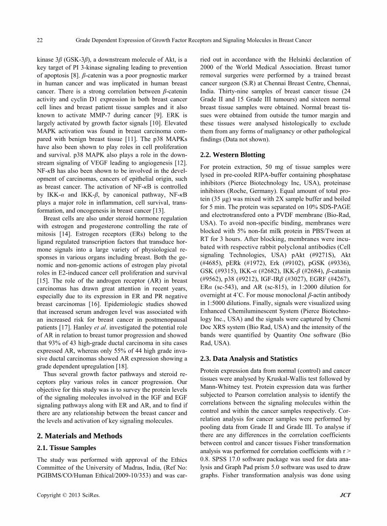
Figure 1. Venn diagram shows the presence of ER, PR and HER2 and their distribution among the samples in Grade II (Fig- ure 1(a)) and Grade II (Figure 1(b)) breast cancer tissues.
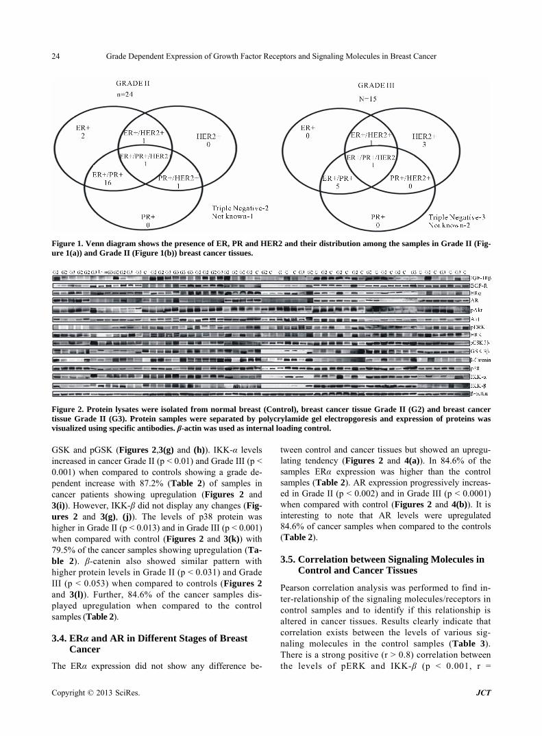
Figure 2. Protein lysates were isolated from normal breast (Control), breast cancer tissue Grade II (G2) and breast cancer tissue Grade II (G3). Protein samples were separated by polycrylamide gel electropgoresis and expression of proteins was visualized using specific antibodies. β-actin was used as internal loading control. GSK and pGSK (Figures 2,3(g) and (h)). IKK-α levels increased in cancer Grade II (p < 0.01) and Grade III (p < 0.001) when compared to controls showing a grade de-pendent increase with 87.2% (Table 2) of samples in cancer patients showing upregulation (Figures 2 and 3(i)). However, IKK-β did not display any changes (Fig- ures 2 and 3(g), (j)). The levels of p38 protein was higher in Grade II (p < 0.013) and in Grade III (p < 0.001) when compared with control (Figures 2 and 3(k)) with 79.5% of the cancer samples showing upregulation (Ta-ble 2). β-catenin also showed similar pattern with higher protein levels in Grade II (p < 0.031) and Grade III (p < 0.053) when compared to controls (Figures 2 and 3(l)). Further, 84.6% of the cancer samples dis-played upregulation when compared to the control samples (Table 2).
3.4. ERα and AR in Different Stages of Breast Cancer
The ERα expression did not show any difference be-
tween control and cancer tissues but showed an upregu- lating tendency (Figures 2 and 4(a)). In 84.6% of the samples ERα expression was higher than the control samples (Table 2). AR expression progressively increas- ed in Grade II (p < 0.002) and in Grade III (p < 0.0001) when compared with control (Figures 2 and 4(b)). It is interesting to note that AR levels were upregulated 84.6% of cancer samples when compared to the controls (Table 2).
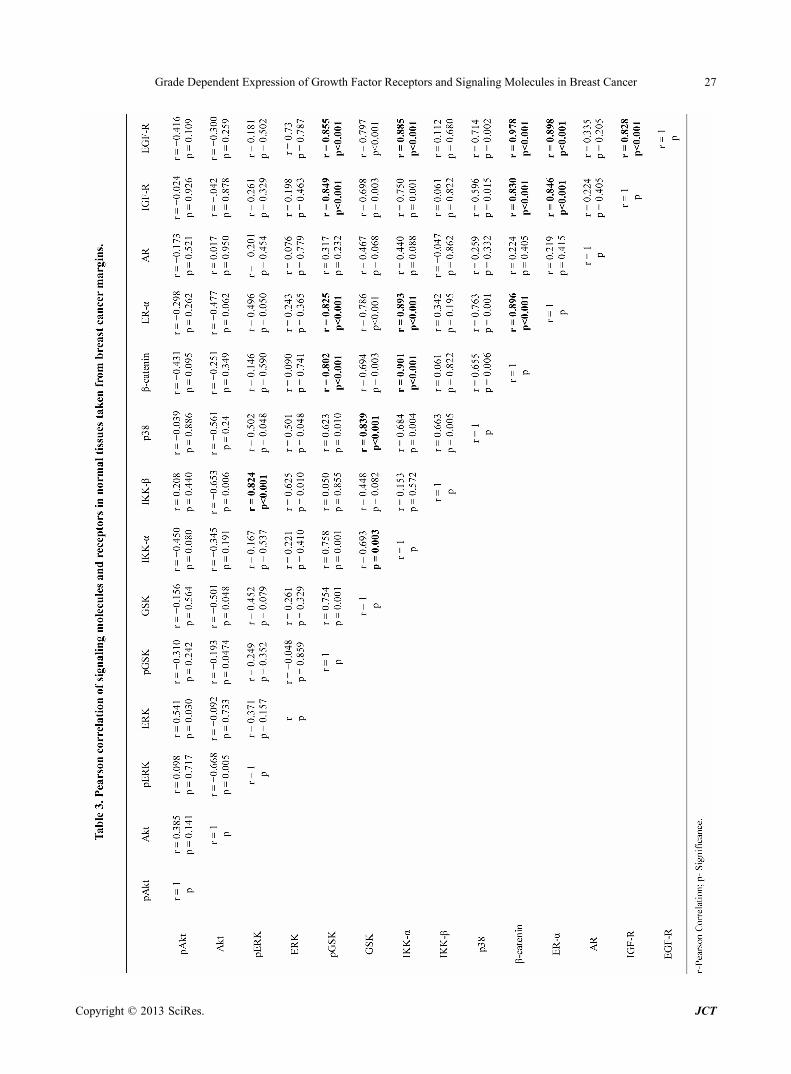
3.5. Correlation between Signaling Molecules in Control and Cancer Tissues
Pearson correlation analysis was performed to find in- ter-relationship of the signaling molecules/receptors in control samples and to identify if this relationship is altered in cancer tissues. Results clearly indicate that correlation exists between the levels of various sig- naling molecules in the control samples (Table 3). There is a strong positive (r > 0.8) correlation between the levels of pERK and IKK-β (p < 0.001, r =

Grade Dependent Expression of Growth Factor Receptors and Signaling Molecules in Breast Cancer 25
Figure 3. Dot plot analysis of densitometrically quantified expression of IGF-IRβ(a), EGF-R(b), pAk, Akt(d), pERK(e), ERK(f), pGSK3β(g), GSK3β(h), IKK-α(i), IKK-α(j), β-catenin(k), p38(l). Protein levels were normalized to the corresponding expression of β-actin. For each protein three dotplots are mapped, Control, Grade II and Grade III. The line within the dot plot corresponds to the median value, and bars indicate the smallest and largest observations. Comparison of expression val- ues between the groups was performed by Kruskal-Wallis test followed by Mann-Whitney test. p values <0.05 were consid- ered as statistically significant.
Copyright © 2013 SciRes. JCT

Grade Dependent Expression of Growth Factor Receptors and Signaling Molecules in Breast Cancer 26
Figure 4. Dot plot analysis of densitometrically quantified expression of AR and ER-α. Protein levels were normalized to the corresponding expression of β-actin. For each protein three dot plots are mapped, Control, Grade II and Grade III. The line within the dot plot corresponds to the median value, and bars indicate the smallest and largest observations. Comparison of expression values between the groups was performed by Kruskal-Wallis test followed by Mann-Whitney test. p values < 0.05 were considered as statistically significant. 0.824), pGSK and β-catenin (p < 0.001, r = 0.802), pGSK and ERα (p < 0.001, r = 0.825), pGSK and IGF-R (p < 0.001, r = 0.849), pGSK and EGF-R (p < 0.001, r = 0.855), GSK and p38 (p < 0.001, r = 0.839), IKK-α and β-catenin (p < 0.001, r = 0.901), IKK-α and ERα (p < 0.001, r = 0.893), IKK-α and EGF-R (p < 0.001, r = 0.885), β-catenin and ERα (p < 0.001, r = 0.896), β-catenin and IGF-R (p < 0.001, r = 0.830), β-catenin and EGF-R (p < 0.001, r = 0.978), ERα and IGF-R (p < 0.001, r = 0.846), ERα and IGF-R (p < 0.001, r = 0.898) and IGF-R and EGF-R (p < 0.001, r = 0.828) in the cancer free control tissue. Interestingly, a new correla- tion emerged between IKK-α and IKK-β (p < 0.001, r = 0.850), (Table 4). Further, comparison of correspond- ing correlation coefficients between control and cancer samples reveal that almost all except the correlations seen between IKKα vs. ERα, and β-catenin vs. IGF-IR in the control samples are lost in the cancer tissues (Table 5).
4. Discussion
Growth factor signaling plays a vital role in the cancer progression cellular proliferation and metastasis. Sub- stantial evidences implicate IGF signaling in the devel- opment and progression of many cancers, including breast cancer [19] and upregulation of IGF-I is often as- sociated with poor prognosis [4]. The present study shows a grade dependent upregulation of IGF-IR in can- cer tissues. Similar IGF-IR upregulation in cancer tissues were previously reported in Canadian population. Further, correlation analysis within the control and cancer sam- ples show that in normal breast tissues IGF-IR has a positive correlation with GSK, β-catenin and ERα, but the correlation between IGF-IR and GSK, and IGF-IR and ERα was lost in cancer tissues indicating a perturbed
expression of signaling molecules in cancer cells. Over expression of EGF-R in breast cancer is associ-
ated with large tumor size, poor differentiation and poor clinical outcomes. Further, EGF-R over expression has been associated with higher grade and extensive forms of ductal carcinoma in situ [20]. Although in the present study EGF-R did not increase in cancer tissues, it is im- portant to note that its levels were upregulated in 80% of the Grade III patients. Similar upregulation of EGF-R inhigher grade cancer was has been reported [20]. Cor- relation analysis shows that in control samples EGF-R is positively correlated with pGSK, IKK-α, β-catenin, ERα and IGF-R, but in cancer tissues EGF-R lost all these correlations suggesting abnormal expression of these molecules which could lead to flawed signaling.
Our study also shows that the expressions of various downstream signaling molecules to the growth factor receptors are affected in cancer tissues. Akt pathway is an important regulator of cell proliferation and survival and is deregulated in breast cancer. In the present study pAkt shows a grade depended increase in cancer, sug- gesting hyper-activation of Akt molecules in cancer tis- sues. However, ERK expression and its phosphorylation did not show any increase in cancer tissues and pERK was lower in 67.6% cancer samples when compared to controls. Thus our data shows an apparent domination of Akt pathway over ERK pathway in cancer tissues. West- ern blot data shows that downstream signaling molecules of Akt are upregulated in cancer samples. IKK-α is a regulatory molecule of NF-κB, pAkt activates the IKK-α by phosphorylating it, and thereby activating the NF-κB [21]. Our data shows that there is an upregulation in the overall IKK-α expression and is increased in 87.2% of the cancer samples indicating an Akt induced IKK-α ac- tivation. Activated IKK-α could promote NF-κB medi- ated transcription [22]. GSK, a downstream signaling
Copyright © 2013 SciRes. JCT
Grade Dependent Expression of Growth Factor Receptors and Signaling Molecules in Breast Cancer
Copyright © 2013 SciRes. JCT
27

Grade Dependent Expression of Growth Factor Receptors and Signaling Molecules in Breast Cancer 28
Copyright © 2013 SciRes. JCT

Grade Dependent Expression of Growth Factor Receptors and Signaling Molecules in Breast Cancer
Copyright © 2013 SciRes. JCT
29
Table 5. Comparison of correlation co-efficients within control and breast cancer tissues (Fisher transformation analysis).
Control
S No Group r = Values Correlation co-efficient
Control pERK vs IKKβ r = 0.824 1
Cancer pERK vs IKKβ r = 0.430 p = 0.028* z = 2.1919
Control GSK vs p38 r = 0.839 2
Cancer GSK vs p38 r = 0.119 p = 0.0007* z = 3.3940
Control pGSK vs β-catenin r = 0.802 3
Cancer pGSK vs β-catenin r = 0.293 p = 0.0132* z = 2.4797
Control IKK-α vs β-catenin r = 0.901 4
Cancer IKK-α vs β-catenin r = 0.005 p = 0.0001* z = 4.5508
Control pGSK vs ER-α r = 0.825 5
Cancer pGSK vs ER-α r = 0.004 p = 0.0003* z = 3.6105
Control IKK-α vs ER-α r = 0.893 6
Cancer IKK-α vs ER-α r = 0.777 p = 0.2037 z = 1.2711
Control β-catenin vs ER-α r = 0.896 7
Cancer β-catenin vs ER-α r = 0.011 p ≤ 0.001* z = 4.4520
Control pGSK vs IGF-IR r = 0.849 8
Cancer pGSK vs IGF-IR r = 0.279 p = 0.0028* z = 2.9853
Control β-catenin vs IGF-IR r = 0.830 9
Cancer β-catenin vs IGF-IR r = 0.660 p = 0.2218 z = 1.2217
Control ER-α vs IGF-IR r = 0.846 10
Cancer ER-α vs IGF-IR r = 0.008 p = 0.001* z = 3.8134
Control pGSK vs EGF-R r = 0.855 11
Cancer pGSK vs EGF-R r = 0.625 p = 0.0944* z = 1.6728
Control IKK-α vs EGF-R r = 0.885 12
Cancer IKK-α vs EGF-R r = 0.356 p = 0.0015* z = 3.1711
Control β-catenin vs EGF-R r = 0.978 13
Cancer β-catenin vs EGF-R r = 0.524 p ≤ 0.0001* z = 5.1536
Control ER-α vs EGF-R r = 0.898 14
Cancer ER-α vs EGF-R r = 0.177 p = 0.0001* z = 3.964
Control IGF-IR vs EGF-R r = 0.850 15
Cancer IGF-IR vs EGF-R r = 0.153 p = 0.0426* z = 2.027
Breast Cancer
Cancer IKK-α vs IKK-β r = 0.850 1
Control IKK-α vs IKK-β r = 0.153
p = 0.0007* z = 3.4055
r: Pearson correlation; p: Significance; z: Correlation co-efficient; *Significant values.
molecule of Akt did not show any change in its expres- sion or phosphorylation. GSK is deactivated by phos- phorylation [23]. The absence of increase in pGSK sug- gests that GSK is active even though its levels did not change. However, β-catenin which is downstream to GSK is highly expressed in cancer samples. β-catenin increases proliferation in ER positive breast cancer cells by activating cyclin D1 ([9]. A previous study shows that higher expression of β-catenin along with p53 is corre- lated with worse survival [24]. Thus, signaling molecules downstream to Akt are highly expressed and activated in breast cancer tissues indicating abnormal activation of this pathway.
The role of p38 MAP kinase in cancer has been thought to occur through negative regulation of the cell cycle and senescence, suggesting that p38 MAPK is a tumor sup- pressor gene [25]. However, a recent study in MCF-7 breast cancer cell line shows that EGF-R phosphorylation leads to the activation of p38 mediated cell survival [26]. Previous study suggests that p38 activity was found to be upregulated in various carcinomas including that of the breast [27]. In the present study p38 is highly expressed in cancer tissue when compared with control indicating its role in breast cancer.
ER is often overexpressed in majority of in breast cancer along with IGF-IR. In this study, although ERα

Grade Dependent Expression of Growth Factor Receptors and Signaling Molecules in Breast Cancer 30
levels were not significantly upregulated, it was elevated in 84.6% of the samples when compared with normal tissues. Previous study shows that ERα binds in an es- trogen depended manner to the p85α subunit of the PI3K, leading to the activation of Akt [28]. It is likely that in the ER positive patients Akt is activated via ERα but additional evidences are needed to confirm this possibil- ity. Interestingly, AR was upregulated in a grade de- pendent manner with 75% of grade II and all of grade III samples showing upregulation.
AR upregulation has been shown in several earlier stu- dies [18]. The upregulated AR expression may activate the p21 [29], which is a positive regulator of cell cycle. Previous study also suggests that p21 overexpression is associated with tumor metastasis in canine mammary tumours [30]. By this mechanism AR can insert the no genomic action through this pathway.
5. Conclusion
In conclusion, the present study shows that several sig- naling molecules are upregulated/activated in cancer tis- sues in a grade depended manner, and the pathway in- volving IGF-IR and Akt seems to be actively involved in breast cancer tissues. Signaling pathways important for cell survival involving Akt are highly activated in cancer samples. Further, there is a deregulation in the expression of signaling molecules in the cancer tissues when com- pared to the control samples.
REFERENCES [1] E. Marshman and C. H. Streuli, “Insulin-Like Growth
Factors and Insulin-Like Growth Factor Binding Proteins in Mammary Gland Function,” Breast Cancer Research: BCR, Vol. 4, 2002, pp. 231-239. doi:10.1186/bcr535
[2] W. Ruan, V. Catanese, R. Wieczorek, M. Feldman and D. L. Kleinberg, “Estradiol Enhances the Stimulatory Effect of Insulin-Like Growth Factor-I (IGF-I) on Mammary Development and Growth Hormone-Induced IGF-I Mes- senger Ribonucleic Acid,” Endocrinology, Vol. 136, No. 3, 1995, pp. 1296-302. doi:10.1210/en.136.3.1296
[3] H. M. Khandwala, I. E. McCutcheon, A. Flyvbjerg and K. E. Friend, “The Effects of Insulin-Like Growth Factors on Tumorigenesis and Neoplastic Growth,” Endocrine Re- Views, Vol. 21, No. 3, 2000, pp. 215-244. doi:10.1210/er.21.3.215
[4] M. Pollak, “Insulin-Like Growth Factor Physiology and Cancer Risk,” European Journal of Cancer, Vol. 36, 2000, pp. 1224-1228. doi:10.1016/S0959-8049(00)00102-7
[5] A. Ouban, P. Muraca, T. Yeatman and D. Coppola, “Ex- pression and Distribution of Insulin-Like Growth Fac- tor-1 Receptor in Human Carcinomas,” Human Pathology, Vol. 34, No. 8, 2003, pp. 803-808. doi:10.1016/S0046-8177(03)00291-0
[6] P. O. Hackel, E. Zwick, N. Prenzel and A. Ullrich, “Epi-
dermal Growth Factor Receptors: Critical Mediators of Multiple Receptor Pathways,” Current Opinion in Cell Biology, Vol. 11, No. 2, 1999, pp. 184-189. doi:10.1016/S0955-0674(99)80024-6
[7] H. W. Lo, S. C. Hsu, W. Xia, X. Cao, J. Y. Shih, et al., “Epidermal Growth Factor Receptor Cooperates with Sig- nal Transducer and Activator of Transcription 3 to Induce Epithelial-Mesenchymal Transition in Cancer Cells via Up-Regulation of TWIST Gene Expression,” Cancer Re- Search, Vol. 67, No. 19, 2007, pp. 9066-9076. doi:10.1158/0008-5472.CAN-07-0575
[8] Q. Ding, X. He, W. Xia, J. M. Hsu, C. T. Chen, et al., “My- eloid Cell Leukemia-1 Inversely Correlates with Glyco- gen Synthase Kinase-3Beta Activity and Associates with Poor Prognosis in Human Breast Cancer,” Cancer Re- search, Vol. 67, No. 10, 2007, pp. 4564-4571. doi:10.1158/0008-5472.CAN-06-1788
[9] S. Y. Lin, W. Xia, J. C. Wang, K. Y. Kwong, B. Spohn, et al., “Beta-Catenin, a Novel Prognostic Marker for Breast Cancer: Its Roles in Cyclin D1 Expression and Cancer Progression,” Proceedings of the National Acad- emy of Sciences of the United States of America, Vol. 97, No. 8, 2000, pp. 4262-4266. doi:10.1073/pnas.060025397
[10] T. P. Garrington and G. L. Johnson, “Organization and Regulation of Mitogen-Activated Protein Kinase Signal- ing Pathways,” Current Opinion in Cell Biology, Vol. 11, No. 2, 1999, pp. 211-218. doi:10.1016/S0955-0674(99)80028-3
[11] V. S. Sivaraman, H. Wang, G. J. Nuovo and C. C. Mal- bon, “Hyperexpression of Mitogen-Activated Protein Ki- nase in Human Breast Cancer,” The Journal of Clinical Investigation, Vol. 99, No. 7, 1997, pp. 1478-1483. doi:10.1172/JCI119309
[12] F. Houle and J. Huot, “Dysregulation of the Endothelial Cellular Response to Oxidative Stress in Cancer,” Mo- lecular Carcinogenesis, Vol. 45, No. 6, 2006, pp. 362- 367. doi:10.1002/mc.20218
[13] Y. Cao and M. Karin, “NF-KappaB in Mammary Gland Development and Breast Cancer,” Journal of Mammary Gland Biology and Neoplasia, Vol. 8, No. 2, 2003, pp. 215-223. doi:10.1023/A:1025905008934
[14] O. J. Finn, “Cancer Immunology,” The New England Journal of Medicine, Vol. 358, No. 25, 2008, pp. 2704- 2715. doi:10.1056/NEJMra072739
[15] D. Yee and A. V. Lee, “Crosstalk between the Insulin- Like Growth Factors and Estrogens in Breast Cancer,” Journal of Mammary Gland Biology and Neoplasia, Vol. 5, No. 1, 2000, pp. 107-115. doi:10.1023/A:1009575518338
[16] W. Schippinger, P. Regitnig, N. Dandachi, K. D. Wer- necke, T. Bauernhofer, et al., “Evaluation of the Prognos- tic Significance of Androgen Receptor Expression in Me- tastatic Breast Cancer,” Virchows Archiv: An Interna- tional Journal of Pathology, Vol. 449, No. 1, 2006, pp. 24-30.
[17] E. O. Lillie, L. Bernstein and G. Ursin, “The Role of An- drogens and Polymorphisms in the Androgen Receptor in the Epidemiology of Breast Cancer,” Breast Cancer Re- Search: BCR, Vol. 5, No. 3, 2003, pp. 164-173.
Copyright © 2013 SciRes. JCT
Grade Dependent Expression of Growth Factor Receptors and Signaling Molecules in Breast Cancer
Copyright © 2013 SciRes. JCT
31
doi:10.1186/bcr593
[18] K. Hanley, J. Wang, P. Bourne, Q. Yang, A. C. Gao, et al., “Lack of Expression of Androgen Receptor May Play a Critical Role in Transformation from in Situ to Invasive Basal Subtype of High-Grade Ductal Carcinoma of the Breast,” Human Pathology, Vol. 39, 2008, pp. 386-392. doi:10.1016/j.humpath.2007.07.007
[19] M. N. Pollak, E. S. Schernhammer and S. E. Hankinson, “Insulin-Like Growth Factors and Neoplasia,” Nature Reviews. Cancer, Vol. 4, No. 7, 2004, pp. 505-518. doi:10.1038/nrc1387
[20] L. Mack, N. Kerkvliet, G. Doig and F. P. O’Malley, “Re- lationship of a New Histological Categorization of Ductal Carcinoma in Situ of the Breast with Size and the Immu- nohistochemical Expression of p53, c-erb B2, bcl-2, and ki-67,” Human Pathology, Vol. 28, No. 8, 1997, pp. 974- 979. doi:10.1016/S0046-8177(97)90014-9
[21] L. P. Kane, M. N. Mollenauer, Z. Xu, C. W. Turck and A. Weiss, “Akt-Dependent Phosphorylation Specifically Re- gulates Cot Induction of NF-Kappa B-Dependent Tran- scription,” Molecular and Cellular Biology, Vol. 22, No. 16, 2002, pp. 5962-5974. doi:10.1128/MCB.22.16.5962-5974.2002
[22] G. Gasparini, P. Boracchi, P. Bevilacqua, M. Mezzetti, F. Pozza and N. Weidner, “A Multiparametric Study on the Prognostic Value of Epidermal Growth Factor Receptor in Operable Breast Carcinom A,” Breast Cancer Re- search and Treatment, Vol. 29, No. 1, 1994, pp. 59-71. doi:10.1007/BF00666182
[23] S. Frame and P. Cohen, “GSK3 Takes Centre Stage More than 20 Years after Its Discovery,” The Biochemical Journal, Vol. 359, No. 1, 2001, pp. 1-16. doi:10.1042/0264-6021:3590001
[24] G. G. Chung, M. P. Zerkowski, I. T. Ocal, M. Dolled-
Filhart, J. Y. Kang, et al., “Beta-Catenin and p53 Analy- ses of a Breast Carcinoma Tissue Microarray,” Cancer, Vol. 100, No. 10, 2004, pp. 2084-2092. doi:10.1002/cncr.20232
[25] T. M. Thornton and M. Rincon, “Non-Classical p38 Map Kinase Functions: Cell Cycle Checkpoints and Survival,” International Journal of Biological Sciences, Vol. 5, No. 1, 2009, pp. 44-51.
[26] K. L. Mueller, K. Powell, J. M. Madden, S. T. Eblen and J. L. Boerner, “EGFR Tyrosine 845 Phosphorylation-De- pendent Proliferation and Transformation of Breast Can- cer Cells Require Activation of p38 MAPK,” Transla- tional Oncology, Vol. 5, No. 5, 2012, pp. 327-334.
[27] E. F. Wagner and A. R. Nebreda, “Signal Integration by JNK and p38 MAPK Pathways in Cancer Development,” Nature Reviews. Cancer, Vol. 9, No. 8, 2009, pp. 537-549. doi:10.1038/nrc2694
[28] J. M. Renoir, V. Marsaud and G. Lazennec, “Estrogen Receptor Signaling as a Target for Novel Breast Cancer Therapeutics,” Biochemical Pharmacology, Vol. 85, No. 4, 2013, pp. 449-465.
[29] J. P. Garay, B. Karakas, A. M. Abukhdeir, D. P. Cos- grove, J. P. Gustin, et al., “The Growth Response to An- drogen Receptor Signaling in ERalpha-Negative Human Breast Cells Is Dependent on p21 and Mediated by MAPK Activation,” Breast Cancer Research: BCR, Vol. 14, No. 1, 2012, p. R27. doi:10.1186/bcr3112
[30] R. Klopfleisch and A. D. Gruber, “Differential Expres- sion of Cell Cycle Regulators p21, p27 and p53 in Me- tastasizing Canine Mammary Adenocarcinomas versus Normal Mammary Glands,” Research in Veterinary Sci- ence, Vol. 87, No. 1, 2009, pp. 91-96. doi:10.1016/j.rvsc.2008.12.010