2-Hexadecynoic acid inhibits plasmodial FAS-II enzymes and arrests erythrocytic and liver stage...
25
2-Hexadecynoic Acid Inhibits Plasmodial FAS-II Enzymes and Arrest Erythrocytic and Liver Stage Plasmodium Infections Deniz Tasdemir a,* , David Sanabria b , Ina L. Lauinger a , Alice Tarun c,d , Rob Herman c , Remo Perozzo e , Mire Zloh a , Stefan H. Kappe c , Reto Brun f , and Néstor M. Carballeira b,* a Department of Pharmaceutical and Biological Chemistry, School of Pharmacy, University of London, 29-39 Brunswick Square, London WC1N 1AX, United Kingdom b Department of Chemistry, University of Puerto Rico, PO Box 70377, San Juan, Puerto Rico 00936-8377 c Seattle Biomedical Research Institute, 307 Westlake Avenue N, Suite 500, Seattle, WA 98109-5219, USA d Department of Infectious Diseases and Microbiology, University of Pittsburgh, 130 Desoto St, Pittsburgh PA 15261 e School of Pharmaceutical Sciences, University of Geneva, Quai Ernest-Ansermet 30, 1211 Geneva 4, Switzerland f Department of Medical Parasitology and Infection Biology, Swiss Tropical Institute, Socinstr. 57, Basel 4002, Switzerland Abstract Acetylenic fatty acids are known to display several biological activities, but their antimalarial activity has remained unexplored. In this study, we synthesized the 2-, 5-, 6-, and 9-hexadecynoic acids (HDAs) and evaluated their in vitro activity against erythrocytic (blood) stages of Plasmodium falciparum and liver stages of P. yoelii infections. Since the type II fatty acid biosynthesis pathway (PfFAS-II) has recently been shown to be indispensable for liver stage malaria parasites, the inhibitory potential of the HDAs against multiple P. falciparum FAS-II (PfFAS-II) elongation enzymes was also evaluated. The highest antiplasmodial activity against blood stages of P. falciparum was displayed by 5-HDA (IC 50 value 6.6. μg/ml), whereas the 2-HDA was the only acid arresting the growth of liver stage P. yoelii infection, in both flow cytometric assay (IC 50 value 2- HDA 15.3 μg/ml, control drug atovaquone 2.5 ng/ml) and immunofluorescense analysis (IC 50 2- HDA 4.88 μg/ml, control drug atovaquone 0.37 ng/ml). 2-HDA showed the best inhibitory against the PfFAS-II enzymes PfFabI and PfFabZ with IC 50 values of 0.38 and 0.58 μg/ml (IC 50 control drugs 14 and 30 ng/ml) respectively. Enzyme kinetics and molecular modeling studies revealed valuable insights into the binding mechanism of 2-HDA on the target enzymes. All HDAs showed in vitro activity against Trypanosoma brucei rhodesiense (IC 50 values 3.7–31.7 μg/ml), Trypanosoma cruzi (only 2-HDA, IC 50 20.2 μg/ml), and Leishmania donovani (IC 50 values 4.1–13.4 μg/ml) with generally low or no significant toxicity on mammalian cells. This is the first study to indicate therapeutic potential of HDAs against various parasitic protozoa. It also points out that the malarial liver stage growth inhibitory effect of the 2-HDA may be promoted via PfFAS-II enzymes. The lack of cytotoxicity, lipophilic nature and calculated pharmacokinetic properties suggest that 2- HDA could be a useful compound to study the interaction of fatty acids with these key P. falciparum enzymes. * Corresponding authors. Phone: +44-20-77535845. Fax: +44-20-77535909. [email protected]. (D. Tasdemir). Phone: (787)-764-0000 ext. 4791. Fax: (787)-756-8242. [email protected] (N.M. Carballeira). Publisher's Disclaimer: This is a PDF file of an unedited manuscript that has been accepted for publication. As a service to our customers we are providing this early version of the manuscript. The manuscript will undergo copyediting, typesetting, and review of the resulting proof before it is published in its final citable form. Please note that during the production process errors may be discovered which could affect the content, and all legal disclaimers that apply to the journal pertain. NIH Public Access Author Manuscript Bioorg Med Chem. Author manuscript; available in PMC 2011 November 1. Published in final edited form as: Bioorg Med Chem. 2010 November 1; 18(21): 7475–7485. doi:10.1016/j.bmc.2010.08.055. NIH-PA Author Manuscript NIH-PA Author Manuscript NIH-PA Author Manuscript
-
Upload
alfredstate -
Category
Documents
-
view
1 -
download
0
Transcript of 2-Hexadecynoic acid inhibits plasmodial FAS-II enzymes and arrests erythrocytic and liver stage...

2-Hexadecynoic Acid Inhibits Plasmodial FAS-II Enzymes andArrest Erythrocytic and Liver Stage Plasmodium Infections
Deniz Tasdemira,*, David Sanabriab, Ina L. Lauingera, Alice Tarunc,d, Rob Hermanc, RemoPerozzoe, Mire Zloha, Stefan H. Kappec, Reto Brunf, and Néstor M. Carballeirab,*
a Department of Pharmaceutical and Biological Chemistry, School of Pharmacy, University ofLondon, 29-39 Brunswick Square, London WC1N 1AX, United Kingdom b Department of Chemistry,University of Puerto Rico, PO Box 70377, San Juan, Puerto Rico 00936-8377 c Seattle BiomedicalResearch Institute, 307 Westlake Avenue N, Suite 500, Seattle, WA 98109-5219, USA d Departmentof Infectious Diseases and Microbiology, University of Pittsburgh, 130 Desoto St, Pittsburgh PA15261 e School of Pharmaceutical Sciences, University of Geneva, Quai Ernest-Ansermet 30, 1211Geneva 4, Switzerland f Department of Medical Parasitology and Infection Biology, Swiss TropicalInstitute, Socinstr. 57, Basel 4002, Switzerland
AbstractAcetylenic fatty acids are known to display several biological activities, but their antimalarial activityhas remained unexplored. In this study, we synthesized the 2-, 5-, 6-, and 9-hexadecynoic acids(HDAs) and evaluated their in vitro activity against erythrocytic (blood) stages of Plasmodiumfalciparum and liver stages of P. yoelii infections. Since the type II fatty acid biosynthesis pathway(PfFAS-II) has recently been shown to be indispensable for liver stage malaria parasites, theinhibitory potential of the HDAs against multiple P. falciparum FAS-II (PfFAS-II) elongationenzymes was also evaluated. The highest antiplasmodial activity against blood stages of P.falciparum was displayed by 5-HDA (IC50 value 6.6. μg/ml), whereas the 2-HDA was the only acidarresting the growth of liver stage P. yoelii infection, in both flow cytometric assay (IC50 value 2-HDA 15.3 μg/ml, control drug atovaquone 2.5 ng/ml) and immunofluorescense analysis (IC50 2-HDA 4.88 μg/ml, control drug atovaquone 0.37 ng/ml). 2-HDA showed the best inhibitory againstthe PfFAS-II enzymes PfFabI and PfFabZ with IC50 values of 0.38 and 0.58 μg/ml (IC50 controldrugs 14 and 30 ng/ml) respectively. Enzyme kinetics and molecular modeling studies revealedvaluable insights into the binding mechanism of 2-HDA on the target enzymes. All HDAs showedin vitro activity against Trypanosoma brucei rhodesiense (IC50 values 3.7–31.7 μg/ml),Trypanosoma cruzi (only 2-HDA, IC50 20.2 μg/ml), and Leishmania donovani (IC50 values 4.1–13.4μg/ml) with generally low or no significant toxicity on mammalian cells. This is the first study toindicate therapeutic potential of HDAs against various parasitic protozoa. It also points out that themalarial liver stage growth inhibitory effect of the 2-HDA may be promoted via PfFAS-II enzymes.The lack of cytotoxicity, lipophilic nature and calculated pharmacokinetic properties suggest that 2-HDA could be a useful compound to study the interaction of fatty acids with these key P.falciparum enzymes.
*Corresponding authors. Phone: +44-20-77535845. Fax: +44-20-77535909. [email protected]. (D. Tasdemir). Phone:(787)-764-0000 ext. 4791. Fax: (787)-756-8242. [email protected] (N.M. Carballeira).Publisher's Disclaimer: This is a PDF file of an unedited manuscript that has been accepted for publication. As a service to our customerswe are providing this early version of the manuscript. The manuscript will undergo copyediting, typesetting, and review of the resultingproof before it is published in its final citable form. Please note that during the production process errors may be discovered which couldaffect the content, and all legal disclaimers that apply to the journal pertain.
NIH Public AccessAuthor ManuscriptBioorg Med Chem. Author manuscript; available in PMC 2011 November 1.
Published in final edited form as:Bioorg Med Chem. 2010 November 1; 18(21): 7475–7485. doi:10.1016/j.bmc.2010.08.055.
NIH
-PA Author Manuscript
NIH
-PA Author Manuscript
NIH
-PA Author Manuscript

KeywordsHexadecynoic acid; Malaria; Liver stage; Blood stage; Protozoa; Type II fatty acid biosynthesis
1. IntroductionMalaria continues to be a cause of concern in underdeveloped areas of the world. The WorldHealth Organization (WHO) reports around 300 million new cases, and over one million deathsdue to malaria each year.1 Malaria parasite Plasmodium has a complex life cycle involvingtwo hosts. The infection is initiated when Plasmodium sporozoites enter the human (host 1)through the bite of an infected Anopheles mosquito (host 2). The sporozoites inoculated underthe skin of the host migrate to the liver, where they infect hepatocytes and begin to developinto merozoites. This so-called liver-stage (LS) or exo- erythrocytic forms takes 2–16 days,depending on the Plasmodium species, then thousands of LS merozoites are released into thebloodstream, where they invade red blood cells and start multiple rounds of the asexual bloodstages (BS). The entire asexual BS cycle is completed within 1–2 days, again depending onthe Plasmodium species, producing large numbers of infected erythrocytes (> 1012 per host).2 During the BS, some merozoites transform into the sexual stages, the male and femalegametocytes, which can be taken up by mosquitoes during blood meals. Gametocytes undergofertilization in the mosquito midgut, producing oocyst sporozoites that migrate to the salivaryglands, ready to initiate a new round of infection.
Past and current malaria drug discovery has been primarily directed against the easy-to-growasexual BS, which is responsible for the clinical symptoms as well as mortality and morbidityof the disease. Mainly due to technical challenges and high costs, LS has been little exploited,despite its longer life span (6–7 days in P. falciparum) than the BS (2 days in P. falciparum)and generation of much smaller number of parasites. Recently, a serious consideration has beengiven to asymptomatic LS parasites as its full inhibition provides true causal malariaprophylaxis, i.e. prevents the blood stage infection and its clinical manifestations. Thedevelopment of molecules inhibiting the growth of Plasmodium hepatic forms could be usefulin malaria prevention for people living in malaria endemic areas, as well as for refugees andtravelers who are exposed to malaria risk for a limited time. Inhibition of LS also reduces therisk of transmission because the generation of the gametocytes will be interrupted.3Furthermore, the low parasitic load with limited multiplication substantially reduces thelikelihood for drug-resistant forms to emerge. Hepatic stage parasites represent furthercomplication for P. vivax and P. ovale infections, as some of the parasites in the hepatocytestransform into hypnozoites, which can stay dormant up to several years and cause relapse.4 Afew drugs, e.g. atovaquone and 8-aminoquinolines primaquine and tafenoquine are effectiveagainst LS, but the primaquine is the only FDA licensed drug. However, its use is restricted,particularly in Africa because of the frequency of genetic glucose-6-phosphate 1-dehydrogenase (G6PD) defficiency. Primaquine is also toxic and has a very short half-life.4Many other non-8-aminoquinolines lack oral bioavailability, and a few natural products withanti-LS activity have low selectivity.5,6 Hence, the search for new natural or synthtetic drugstargeting the LS of the malaria parasite is timely and necessary.
Due to inherent technical difficulties in studying the LS Plasmodium parasites, little progresshas been made in the identification of new LS biological targets for drug discovery and design.Very recent studies7,8 indicate that LS malaria parasites exhibit an absolute requirement forde novo type II fatty acid biosynthesis (FAS-II), which was previously thought to operate inblood stage.9 The FAS-II pathway appears to be essential only for late hepatic stages anddeletion of critical elongation enzymes such as FabB/F (β-ketoacyl-ACP synthase) and FabZ(β-hydroxyacyl-ACP dehydratase) in P. yoelii cause a failure to generate exoerythrocytic
Tasdemir et al. Page 2
Bioorg Med Chem. Author manuscript; available in PMC 2011 November 1.
NIH
-PA Author Manuscript
NIH
-PA Author Manuscript
NIH
-PA Author Manuscript
merozoites, i.e. unability to cause a BS infection.7 Similarly, FabI (enoyl-ACP reductase)-deficient P. berghei sporozoites were much less infective in mice and failed to complete liverstage development.8 This data renders the plasmodial FAS-II pathway an attractive target formalaria prophylaxis.
Fatty acids have shown antimalarial activity10,11,12 but literature reports have been scarce andthere is not a consensus as to what structural characteristics (i.e., unsaturation level, positionor chain length) favor the best antimalarial fatty acids. We believed that a systematic study ofthe antimalarial activity of a series of isomeric C16 acetylenic fatty acids could shed light onthe structural properties required for antimalarial activity, in particular how the antimalarialactivity depends on the position of the triple bond in a C16 acyl chain. For this purpose, wechose an isomeric series of hexadecynoic acids (HDA), i.e., the 2-, 5-, 6-, and 9-HDAs, someof which were shown to be antibacterial, antifungal and antimycobacterial,13,14,15,16 but neverinvestigated for antimalarial potential, and synthesized them. Another reason for choosing C16acetylenic acids, and not longer or shorter fatty acids (FAs), was because earlier studiesindicated that 2-HDA inhibited fatty acid elongation.17,18 The 2-HDA has recently been shownto inhibit InhA, the enoyl-ACP reductase (FabI) analogue enzyme found in the FAS-II pathwayof the tubercle bacillus, Mycobacterium tuberculosis.16 In a recent publication, we reportedunsaturated fatty acids inhibiting enoyl-ACP reductases of P. falciparum (Pf) and M.tuberculosis.19 These facts suggested that C-16 alkynoic acids, especially 2-HDA could targetthe PfFabI (and potentially other elongation enzymes from PfFAS-II system) and lead to deathof LS malaria parasites. Herein we report a) the synthesis of four HDAs b) their in vitro growthinhibitory potential against BS and LS malaria parasites c) inhibitory activity against multiplePfFAS-II elongation enzymes, PfFabI, PfFabZ and PfFabG (β-ketoacyl-ACP reductase) d) theenzyme kinetics, docking studies and calculated pharmacokinetic properties e) in vitro activityagainst other parasitic protozoa, i.e. Trypanosoma brucei rhodesiense, T. cruzi and Leishmaniadonovani and f) Selective toxicity towards primary mammalian and hepatoma cells. Palmiticacid (PA), the parent compound lacking the triple bond was also tested in each assay forcomparison.
2. Results2.1. Synthesis of hexadecynoic acid derivatives
We developed a short and versatile procedure to prepare the 5-, 6-, and 9-hexadecynoic acidsas outlined in Fig. 1. In general, the appropriate bromo alcohol was protected with tert-butyldimethylsilyl chloride (TBSCl) yielding the TBS protected bromo alcohols in 80–99%yields. Alkyne coupling with the required chain length of the 1-alkynes in the presence of n-BuLi in tetrahydrofuran-hexamethylphosphoramide (THF-HMPA) produced the desired TBSprotected alkynols in 23–32% yields. Deprotection of the TBS group with tetrabutylammoniumfluoride (TBAF) in THF followed by oxidation of the resulting alcohols with pyridiniumchloride (PDC) in dimethylformamide (DMF) afforded the desired hexadecynoic acids in 40–70% yields. The synthesis of the 2-HDA followed an already published procedure18 whereincommercially available 1-pentadecyne was reacted with n-BuLi in THF followed by quenchingwith CO2 and final protonation with NH4Cl (Fig. 1). The purity of the synthesized compoundswere determined to be >95% by capillary GC-MS and 13C-NMR. The spectral data of thesynthesized HDAs (sections 4.2–4.3) were in agreement with those published.13,20,21
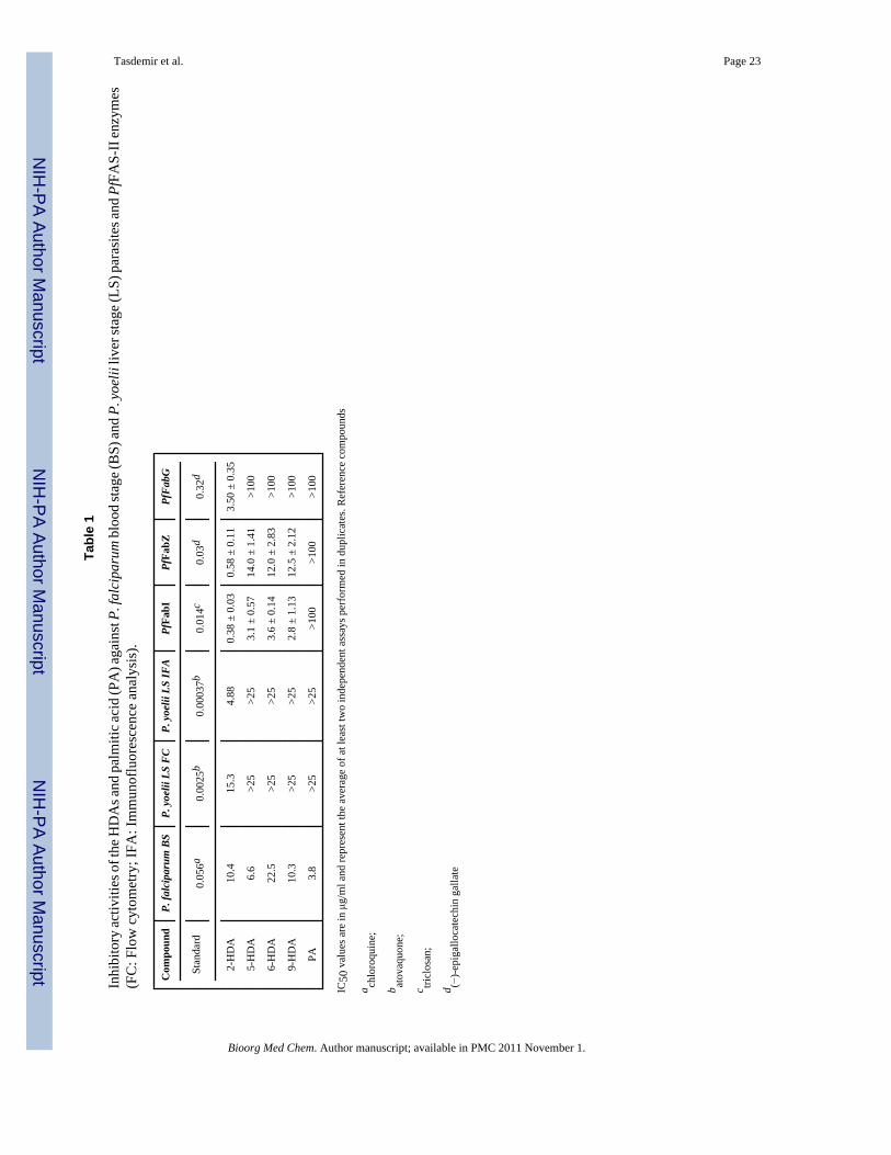
2.2. In vitro antimalarial activity towards P. falciparum blood stagesThe in vitro antiplasmodial activity of the HDAs against multidrug-resistant P. falciparum K1strain was determined by using the established 3H-hypoxanthine method. As shown in Table1, 5-HDA showed the highest antiplasmodial activity among the alkynoic acids with an IC50value of 6.6 μg/ml, followed by the equipotent 2-HDA and 9-HDAs (IC50s 10.4 and 10.3 μg/
Tasdemir et al. Page 3
Bioorg Med Chem. Author manuscript; available in PMC 2011 November 1.
NIH
-PA Author Manuscript
NIH
-PA Author Manuscript
NIH
-PA Author Manuscript

ml, respectively) and finally 6-HDA (IC50 22.5 μg/ml). Interestingly, the parent compound PAwas more active (IC50 3.8 μg/ml) than all acetylenic fatty acids, indicating that a triple bondis not favored for antimalarial activity towards blood stage parasites.
2.3. In vitro activity towards P. yoelii liver stagesNext, we wanted to assess life stage selectivity of the HDAs and investigated their growthinhibitory activity against LS malaria parasites. Hepatic stage P. falciparum infection inhumans is experimentally intractable for practical purposes, hence has remained poorlyunderstood. In contrast, the LS of the disease in the rodent malaria model is amenable to directexperimental interrogation.2 Thus, we have employed a medium throughput LS growthinhibition assay using rodent P. yoelii parasites, which have conserved metabolic and antigenicprofile as P. falciparum,22 to infect HepG2:CD81 cell line. Infection was assayed by both flowcytometric (FC) and immunofluorescense analysis (IFA) methods, using atovaquone asreference (Table 1). In the FC assay, the HepG2:CD81 cells are infected with transgenic P.yoelii sporozoites that express green fluorescent protein (PyGFP), exposed to compound, andallowed to develop for 43 h to measure infection rates. To verify the results of the FC-basedassay and to determine the effects of the drugs on the morphology and development of the LSparasites, IFAs of P. yoelii-infected HepG2:CD81 cells were done after drug treatment. In thisassay, the LS parasites were detected using a mouse monoclonal antibody against P. bergheiHSP70 that cross-reacts with P. yoelii HSP7023 and polyclonal antibody against the P. yoeliiUIS4 protein.24 The results of both FC and IFA assays are shown in Table 1. In the FC assay,only 2-HDA appeared to be active with an IC50 value of 15.3 μg/ml. Atovaquone, a potentinhibitor of LS development, had an IC50 value of 2.5 ng/ml. In the IFA, atovaquone wasobserved to potently inhibit LS development resulting in much smaller parasites with anIC50 value of 0.37 ng/ml (Table 1), similar to what has been described before.25 Smaller LSparasites compared to the control were observed in the IFA after incubation with 2-HDA withan IC50 value of 4.88 μg/ml, whereas other alkynoic and palmitic acids had no effect, even atthe highest test concentrations. Figure 2 (20X objective magnification) shows an IFA of thelate liver stage-infected HepG2:CD81 cells (43 h post infection) treated with DMSO, PA andthe HDAs. The DAPI staining of infected cells (Fig. 2) indicated none of the HDAs to exerttoxicity towards the liver cells. Figure 3 displays the close up fluorescence microscopy images(100X objective magnification) of P. yoelii liver stage schizonts detected by antibody againstHSP70 protein at 43 h post infection treated with different concentrations of 2-HDA, as wellas DMSO and atovaquone. 2-HDA was non-toxic against hepatoma cells in FC or IFA (seesection 4.6) or against primary mammalian cells (see sections 2.6. and 4.12) indicating itsselective toxicity against the LS parasites.
2.4. PfFAS-II enzyme inhibition studiesIn an attempt to understand the mechanism of action of the HDAs against LS parasites, wedetermined their inhibitory effect against three key enzymes of the fatty acid biosynthesis ofP. falciparum, i.e., PfFabI, PfFabZ and PfFabG, all of which were recombinantly prepared andassayed following an established procedure.26 Essentially, the HDAs were inhibitory towardsthe studied PfFab enzymes following the order PfFabI > PfFabZ > PfFabG (Table 1). Fromthe data obtained, it became clear that among the studied HDAs, the 2-HDA displayed the bestoverall inhibitory activity against PfFabI (IC50 0.38±0.03 μg/ml) and PfFabZ (IC50 0.58±0.11μg/ml), and it was the only inhibitory acetylenic acid against PfFabG (IC50 3.5±0.35 μg/ml).The 5-, 6- and 9-HDAs were not inhibitory towards PfFabG (IC50 >100 μg/ml), and theirinhibitory potential against two other target enzymes were 10-fold (PfFabI) to 50-fold (PfFabZ)less. Notably, the parent compound PA was inactive against all enzymes tested even at thehighest concentrations (IC50 > 100 μg/ml). This data indicates that the hepatic stage growthinhibitory activity of 2-HDA may be due the inhibition of the PfFAS-II enzymes.
Tasdemir et al. Page 4
Bioorg Med Chem. Author manuscript; available in PMC 2011 November 1.
NIH
-PA Author Manuscript
NIH
-PA Author Manuscript
NIH
-PA Author Manuscript

2.5. Enzyme kinetics, docking studies and pharmacokinetic calculationsIn order to shed more light into the inhibitory mechanism of 2-HDA, in vitro enzyme kineticstudies, as well as molecular modeling (docking) studies were performed. Kinetic studiesindicated that 2-HDA inhibits the PfFabI enzyme in a non-competitive manner with respect tocrotonyl-CoA and the cofactor NADH (Fig. 4). This implies that 2-HDA binds to the PfFabIenzyme at a site distinct of the substrate or the cofactor site. The calculated Ki values are 0.73μg/ml (= 2.90 μM, substrate varied) and 0.65 μg/ml (= 2.58 μM, cofactor varied). The inhibitiontype against the PfFabZ, a dehydratase, which does not require a co-factor, is competitive(Ki = 1.2 μg/ml = 4.8 μM) with respect to the substrate (Fig. 5). As shown in Fig. 6, 2-HDAinhibits the PfFabG enzyme in a competitive manner when the substrate acetoacetyl-CoAvaried (Ki 3.6 μg/ml = 14.3 μM) and non-competitive when the cofactor NADPH is varied(Ki 1.5 μg/ml = 6 μM).
Molecular modeling studies supported the predicted binding mode of the acetylenic acidsdetermined by the kinetic studies and revealed insights for understanding the superior enzymeinhibitory activity of 2-HDA over the other HDAs. The molecular docking of these ligandsinto the binding site of FabZ27,28 was carried out using the crystal structure given in pdb entry1z6b. The best fit of ligands and their binding energies predicted by Autodock indicate that all4 ligands interact favourably with the protein, however the 2-HDA binds most strongly (Fig.7a). Although there is no direct correlation between activity and binding energy, a trend wasobserved for energy of interaction (E2-HDA< E9-HAD< E5-HDA<E6-HDA), which correspondsto the trend of activity. Docking studies indicate that only 2-HDA form a hydrogen bond withthe backbone NH atoms of Gly142 and Val143 and the electron density of the triple bond of2-HDA interacts with the active site residue Glu147. These differences most likely explainwhy the 2-HDA is the most active molecule against PfFabZ.
The docking of ligands into PfFabG site has indicated that all ligands bind closely to thecofactor site, and again the 2-HDA binds with most favourable energy of interactions. Theorientation of ligands are different too (results not shown), but further studies would be neededto understand the reasons for such significant difference between activities of 2-HDA and otherligands. The interaction of ligands and the X-ray structure of PfFabI complexed with the NADH(pdb entry 1v35) was used to investigate the non-competitive inhibition of PfFabI activity. Themolecular interaction fields were generated using GRID 22A software (Molecular Discovery)and the energy of binding of ligands to the protein complex was estimated using GLUE moduleof this software.29,30 The whole surface of the protein was target for the interaction and allligands exhibited multiple binding positions on the surface (Fig. 7b), with similar preferences.Importantly, ligands appear to protrude into the protein surface and to make close contacts withthe protein substrate. These interactions could possibly affect the protein conformation andtherefore inhibiting the protein activity. The 2-HDA protrudes the most of all ligands due tothe extended structure around the carboxylic group and possibly has the strongest impact onprotein conformation and flexibility.
Molecular properties of HDAs and PA were calculated using ChemSilico and Vega ZZ, whichindicated the compliance of 2-HDA with Lipinski’s rules.31 As shown in Table 3, the predictedlogP values by CS and Vega ZZ of 2-HDA are 5.6 and 5.47, respectively, while the predictedlogP of PA are 5.4 and 6.04. The predicted PSA/SA (polar surface areas/surface areas) valuesand human intestinal absorptions (HIA) of 2-HDA are very similar to those of PA. Since thePA readily crosses the blood brain barrier (BBB),32 and the PSA correlates with the ability ofmolecules to cross the BBB,33 it is likely that the 2-HDA has a good penetration ability throughcell membranes, and will even diffuse through the BBB.
Tasdemir et al. Page 5
Bioorg Med Chem. Author manuscript; available in PMC 2011 November 1.
NIH
-PA Author Manuscript
NIH
-PA Author Manuscript
NIH
-PA Author Manuscript

2.6. Inhibitory activity against other parasitic protozoa and primary L6 cellsFinally, the species-specific antiprotozoal activity of the HDAs against Trypanosoma bruceirhodesiense (bloodstream forms), Trypanosoma cruzi (intracellular amastigotes in L6 ratskeletal myoblasts), and Leishmania donovani (axenic amastigotes) was determined. As shownin Table 2, the studied HDAs displayed the best overall antiprotozoal activities against L.donovani, with 2-HDA and 9-HDA being equally active (IC50s 4.5 and 4.1 μg/ml). 6-HDAdisplayed a two-fold less activity (IC50 9.8 μg/ml), whereas 5-HDA and PA were the leastactive ones (IC50 13.4 and 14.5 μg/ml). The 9-HDA displayed the best activity towards T. b.rhodesiense with an IC50 value of 3.7 μg/ml, whilst the remaining compounds were 6- to 10-fold less active. The 2-HDA was the only fatty acid with moderate T. cruzi activity (IC50 20.2μg/ml). Overall, the 9-HDA showed the broadest spectrum antiprotozoal activity. An earlystudy34 has pointed out the cidal effect of palmitic (hexadecanoic) acid against the promastigotestages of L. donovani, L. tropica and the epimastigote and trypomastigote stages of T. cruzi.To our knowledge, this is the first study assessing trypanocidal and leishmanicidal effects ofthe four HDAs.
Selective toxicity of all fatty acids were determined against L6 cells, a primary cell line derivedfrom rat skeletal myoblasts. As displayed on Table 2, the 2-HDA shows negligible toxicityand 5-HDA is completely non-toxic at the highest test concentrations (90 μg/ml). Only 6-HDAand PA bear some moderate cytotoxic effects against L6 cells.
3. DiscussionIn contrast to easily cultivable BS, working with LS malaria parasites is difficult and costlysince primary hepatocytes or hepatoma cells need to be infected with live sporozoites fromPlasmodium-infected mosquitoes. This requires continous supply of blood containinginfectious gametocytes and a mosquito breeding facility to produce viable, uncontaminatedand infective sporozoites. LS infection has long been unamenable to standard biochemicalassays because of generally low infection rates of hepatocytes and hepatoma cells.35 However,technological developments in the field of microscopy and the generation of parasites withspecific cell types expressing fluorescent proteins permitted fascinating live imaging of LSinfection. Nowadays, automated microscopy, infrared fluorescence scanning, flow cytometryand immunofluorescense techniques enable medium to high throughput screening in LSparasites. The current study is the first to investigate the LS malaria parasite growth inhibitoryeffect of 2-, 5-, 6- and 9-HDAs, by using rodent P. yoelii parasites to infect HepG2:CD81cellline. Infection was assayed by two medium throughput LS growth inhibition assays, FC andIFA. In the FC assay, the HepG2:CD81 cells that are infected with GFP expressing P. yoeliisporozoites are exposed to compound for 43 h. The number of GFP-positive hepatoma cells isdetermined by FC and is used to measure infection rates. One disadvantage of the FC assay isthat it only provides information on the LS infection rates after 43 h of post-infection. In orderto determine the effects of the drugs on the morphology and development of the LS parasites,we undertook IFAs of P. yoelii-infected HepG2:CD81 cells after drug treatment. IFA does notonly provide data on morphology and the size of LS parasites, but also gives a visual indicationof host cytotoxicity. The LS parasites were detected using a mouse monoclonal antibodyagainst P. berghei HSP70 that cross-reacts with P. yoelii HSP70 and polyclonal antibodyagainst the P. yoelii UIS4 protein. While infection rates are difficult to assess from the IFA(because host cell numbers were not counted), the reduction in LS sizes is a more sensitiveindicator for the inhibitory effect of a drug on LS development compared to the FC-basedassay. As shown in Figures 2 and 3, the 2-HDA was the only acid inhibiting the growth of P.yoelii LS parasites in both assays. The IC50 values of 2-HDA and the standard, atovaquone,are much smaller in the IFA, showing the sensitivity of the assay.
Tasdemir et al. Page 6
Bioorg Med Chem. Author manuscript; available in PMC 2011 November 1.
NIH
-PA Author Manuscript
NIH
-PA Author Manuscript
NIH
-PA Author Manuscript
Most of the current antimalarial agents target the parasite’s blood stages. Only a fewcompounds, such as primaquine, tafenoquine and atovaquone have activity against the LSparasites, however their use is limited due hematological toxicity in individuals with G6PD-deficiency and high costs (atovaquone). Although discussed controversially, it is logical anddesirable to have one drug with a dual effect on both LS and BS parasites, i.e. both prophylacticand therapeutic. Quinine, chloroquine, mefloquine and artemisinins have little or no efficacyagainst the hepatic parasites, contributing to the relapse of the disease, particularly in P.vivax, which develops liver hypnozoites. An earlier study20 reported 2-HDA to inhibit thegrowth of hepatoma cells, however, we observed no toxicity even at the highest testconcentrations towards HepG2:CD81 cells by any HDA compounds in any of the assayemployed. The HDAs generally lacked toxicity against mammalian L6 cells, indicating areasonable therapeutic index. Hence, 2-HDA appears to be a promising small molecule for itsinhibitory activity on both LS and BS malaria parasites, with good cell viability on hepaticcells. The inhibition of LS by a compound, such as 2-HAD, will also reduce the risk oftransmission by interrupting the formation of the gametocytes in late BS. The next logical stepwould be to test HDAs on P. vivax and P. ovale hypnozoites. However, none of the rodentPlasmodium species including P. yoelii produces dormant hepatic stages in laboratoryanimals3 hence this could not be performed in our laboratories.
Due to the inherent difficulty in studying the LS parasites, only little progress has been madein the identification of new LS drug targets for rational drug design. Recent studies show theimportance of type II FAS in the LS, particularly in the transition point of parasites from liverto blood. As FAS-II deficiency leads to loss of infective capability of red blood cells in vitroand in vivo,7,8 the plasmodial FAS-II system appears to be a promising drug target for theclinically silent LS infection. So far, only hexachlorophene, a PfFabG inhibitor, was shown toinhibit the LS development in vitro in a dose-dependent mannner with an IC50 value of 4.8μM (=1.95 μg/ml).36 Recently, the potent PfFabI inhibitor triclosan, which was previouslyreported to inhibit the blood-stage P. falciparum infection, has been reported to be cidal at thelate liver phases of P. berghei with an IC50 value of 39.4 μM (=11.4 μg/ml).37 Thus, 2-HDAappears to be the third PfFAS-II inhibitor with activity against LS malaria parasites. Theinhibition of multiple PfFAS-II enzymes by 2-HDA is tantalizing, as it bears lower risk todevelop resistance. The remaining fatty acids also inhibit PfFabI and PfFabZ moderately andin the FC assay, they cause a 10% decrease in infection (data not shown) at the highest testconcentration, 25 μg/ml (data not shown). This might imply that they possibly have someactivity at higher concentrations, however, due to toxicity of DMSO against hepatic cells, wewere unable to test higher doses. On the other hand, since PfFabZ and PfFabI are inhibited byall compounds, one can suggest that the lack of LS activity of the other HDAs stems from theirlow penetration into the infected hepatocytes. However, the calculated pharmacokineticproperties of the remaining HDAs were very similar to those of 2-HDA and PA (Table 3),ruling out the permeability as a problem. The good drug-like properties, especially their abilityto cross membranes, including the BBB, of HDAs could make them very useful for thetreatment of advanced phases of cerebral malaria and sleeping sickness caused byTrypanosoma brucei rhodesiense.
It has been reported that 2-HDA competitively inhibits the mycobacterial InhA (MtFabI)enzyme16 with a similar potency (Ki value 2.1±0.6 μM) to that obtained with PfFabI. Althoughthe exact binding mode of 2-HDA on PfFabI is not completely clear, it is obvious that thesignificant enzyme inhibitory activity of 2-HDA is not a simple, non-specific detergent effect.Again, similar to its antimycobacterial (and probably antifungal) activity, the hepatic stageparasite activity of 2-HDA does not seem to be due to non-specific fatty acid mediated lysis.On the other hand, 2-HDA acts as a prodrug and requires metabolization in Mycobacteriumspecies.16 Whether this same phenomenon occurs in Plasmodium species remains unclear andwarrants further studies.
Tasdemir et al. Page 7
Bioorg Med Chem. Author manuscript; available in PMC 2011 November 1.
NIH
-PA Author Manuscript
NIH
-PA Author Manuscript
NIH
-PA Author Manuscript

Our findings concerning LS development were generated with the rodent malaria parasite P.yoelii, which has a much shorter LS cycle (50–60 hrs), but the high conservation of FAS-IIamong Plasmodium species22 may suggest similar results on P. falciparum and P. vivax hepaticstage development. In a recent paper, Yu et al. (2008)8 compared FA profiles of wild-type andFabI-mutant parasites in the blood stage of P. falciparum and P. berghei. The spectrum ofsynthesized FA’s was different in P. falciparum where C-16 and C-18 FAs were detected whilein P. berghei C-12 to C-24 FAs were predominant.8 The 14C-acetate (radiolabeled FAprecursor) isotope-incorporation studies showed no difference in the FA profiles in the FabImutant and wild type blood stages in both P. falciparum and P. berghei indicating clearly thatFAS-II is not involved in the synthesis of these FA species. Thus, the different FAs in the bloodstage might be formed by fatty acid elongases and the differences in FA synthesis in the twoPlasmodium species might be due to the number of involved elongases detected in their genome(three for P. falciparum, four for P. berghei).8 A recent paper by Déchamps et al. (2010)38
showed that rodent and mammalian Plasmodium blood stage parasites differ in theirphospholipid pathways. Hence it is possible that lipid metabolic pathways are different in therodent and human Plasmodium and yet the parasite FAS-II pathway will be essentially similar.Since the FAS-II pathway does not appear to be operational in blood stages7,8 one cannot saythat the observed effects by the HDA and/or palmitic acid in the blood and liver stages wouldbe through the same mechanisms.
3. ConclusionsIn this study, we report the rapid synthesis and broad antiprotozoal activity of four HDAs. Ofthe four HDAs studied here, only the 6-HDA is a natural compound, recently isolated from theplant Sommera sabiceoides.15 Many unsaturated fatty acids, including the natural productscleropyric acid, a C-17 fatty acid with a double and triple bond have been shown to haveantiplasmodial activity,12 but to our knowledge, this is the first report of antimalarial activityof the 2-, 5-, 6- and 9-HDAs against the BS of P. falciparum parasites. The data presented inthis manuscript suggest that the presence and the position of the triple bond in the acyl chainto be of paramount importance for determining the antiprotozoal activity of acetylenic C16fatty acids. Considering that the PA was the most active compound against BS P. falciparumparasites, it seems that the absence or a more distal position (C-5) of a triple bond is favoredfor BS activity. However, the presence of an acetylenic function at C-2 appears to be a criticalrequirement for LS parasite growth arrest as PA and all other HDAs are inactive. The presenceof a triple bond next to the carbonyl group, as found in 2-HDA, seems to increase theconformational flexibility and dipole moment of 2-HDA, thus enabling it to interact withPfFAS-II target enzymes more efficiently. The presence of a single acetylenic function at C-9however, increases the trypanocidal and leishmanicidal activity of the C16 acids significantly.At this point, a relevant question emerges concerning the impact of the chain length, as wellthe number of triple bonds within the acetylenic acid on their biological activity. We performeda docking study calculating the binding energies of the 2-tetradecynoic acid (2-TDA, C14),the 2-octadecynoic acid (2-ODA, C-18) and the 2,5-hexadecadiynoic acid (2,5-HDDA). Thisstudy showed that 2-HDA was still the best binder towards the two target PfFAS-II enzymes,PfFabG and PfFabZ (data not shown). The strength or mode of binding of 2-TDA and the 2-ODA against PfFabI was different, which may indicate a weaker or absence of activity. The2,5-HDDA had close binding energies to 2-HDA towards PfFabG and PfFabZ, but betterbinding energy towards PfFabI. However, the orientation of the 2,5-HDDA molecule in thebinding region is different due to steric constraints of triple bond combination, thus it mightnot have the same inhibitory activity as the 2-HDA. These calculations confirm that the C-16HDAs were a good choice for this study and 2-HDA had better activity against a wider rangeof target enzymes. The in vitro activity of 2-TDA, 2-ODA and 2,5-HDDA, however, needs tobe assessed in whole cell parasite assays, following their successful synthesis.
Tasdemir et al. Page 8
Bioorg Med Chem. Author manuscript; available in PMC 2011 November 1.
NIH
-PA Author Manuscript
NIH
-PA Author Manuscript
NIH
-PA Author Manuscript

In conclusion, the current study has identified acetylenic C16 acids as novel class ofantiprotozoal agents. The discovery of these easily-synthesizable fatty acids with promisingantimalarial and malaria prophylactic effects may represent an opportunity for the developmentof new agents, particularly those derived from 2-HDA, for the control and potentiallyeradication of malaria. Taking into account their synthetic accessibilities, antiprotozoalpotencies, no apparent toxicity on hepatocytes at active concentrations, ability to affect bothBS and LS malaria parasites, as well as good pharmaceutical (lipophilic) and pharmacokineticproperties, the acetylenic acids, in particular 2-HDA, might be useful tools to understand thepotential of fatty acids as drug candidates. Our future work will involve the synthesis, in vitroand in vivo evaluation of more potent acetylenic fatty acids with multiple triple and doublebonds.
4. Experimental4.1. General
(Trimethylsilyl)-acetylene and other reagents used were purchased from Aldrich, TCIAmerica, and Alfa Aesar. All synthesis reactions were analyzed by 1H and 13C NuclearMagnetic Resonance (NMR) using a Bruker Avance DRX-500 or a Bruker Avance DPX-300.The samples were dissolved in chloroform-d (CDCl3) and the solvent signals at 7.26 and 77.0ppm were used as internal standard for proton and carbon, respectively. Mass spectral data wasacquired on a GC-MS (Hewlett-Packard 5972A MS Chem Station) instrument at 70 eV,equipped with a 30 m x 0.25 mm special performance capillary column (HP-5MS) ofpolymethylsiloxane cross-linked with 5% phenyl methylpolysiloxane. The GC-MS analysisconditions were identical to those previously described.39 Infrared (IR) spectra were recordedon a Nicolet 600 FTIR spectrophotometer. Melting points (°C, uncorrected) were determinedin a MEL-TEMP capillary melting point apparatus. Thin layer chromatography was carriedout on aluminium TLC plates pre-coated with silica gel 60 F254 (Merck, 0.25 mm).
4.2. Synthesis and structure elucidation of the 5-, 6-, and 9-HDAs4.2.1. Alkyne couplings with the bromoalkyloxy-tert-butyldimethylsilanes—To astirred solution of the alkyne (3.74–4.36 mmol), prepared from the bromoalcohol and tert-butyldimethylsilylchloride, in dry THF (5.0–15.0 ml), n-Buli (2.5 M, 10.90–11.22 mmol) indry hexane was added dropwise while keeping the temperature at −60 °C. After 45 min, HMPA(1.12–10.0 ml) and the bromoalkyloxy-tert-butyldimethylsilane (3.74–4.36 mmol) were addeddropwise to the reaction mixture at −60 °C. After 24h, the reaction mixture was worked up bypouring into a large volume of water, and extracting with diethyl ether (2 × 15 ml). The organiclayer was washed with brine (1 × 15 ml) before drying (MgSO4). Filtration, rotoevaporationof the solvent, and fractional distillation afforded the tert-butyldimethylsilyloxy-alkynes in 23–32% yields after purification by Kugelrohr distillation (130–137 °C/3 mmHg) of the impurities.
4.2.2. Oxidation to the carboxylic acids—To a stirred solution of the alkynol (0.2–1.3mmol), after deprotection with tert-butylammonium fluoride (TBAF), in 3.0–7.0 ml of DMFwas slowly added pyridinium dichromate (0.8–7.9 mmol) at rt. After 24–48 h, the reactionmixture was worked up by pouring 10 ml of water and extracting with hexane (3 × 12 ml).Once the solvent was evaporated and dried in vacuo, the acetylenic fatty acids were obtainedin 40–70% yields.
4.2.2.1. 5-Hexadecynoic acid (5-HDA): Was obtained in a 75% yield (249 mg) from thereaction of 313 mg (1.3 mmol) of 5-hexadecyn-1-ol with 3.0 g (7.9 mmol) of pyridiniumdichromate. IR (neat) νmax 3105–2900, 2926, 2854, 2206, 1711, 1465, 1261, 1207, 1090, 920,803, 722 cm−1. 1H NMR (500 MHz, CDCl3) ΔH 2.48 (2H, t, J = 7.5 Hz, H-2), 2.12 (4H, m,H-4, H-7), 1.80 (2H, quintet, J = 7.2 Hz, H-3), 1.46 (2H, quintet, J = 7.3 Hz, H-8), 1.35–1.25
Tasdemir et al. Page 9
Bioorg Med Chem. Author manuscript; available in PMC 2011 November 1.
NIH
-PA Author Manuscript
NIH
-PA Author Manuscript
NIH
-PA Author Manuscript
(14H, m, -CH2-), 0.88 (3H, t, J = 6.9 Hz, H-16). 13C NMR (125 MHz, CDCl3) ΔC 178.5 (s,C-1), 81.5 (s), 78.5 (s), 32.6 (t, C-2), 31.9 (t, C-14), 29.6 (t), 29.5 (t), 29.3 (t), 29.1 (t), 29.0 (t),28.9 (t), 24.0 (t), 22.7 (t), 18.7 (t, C-7), 18.3 (t, C-4), 14.1 (q, C-16).
4.2.2.2. 6-Hexadecynoic acid (6-HDA): Was obtained in a 42% yield (87 mg) from thereaction of 195 mg (0.8 mmol) of 6-hexadecyn-1-ol with 1.2 g (3.3 mmol) of pyridiniumdichromate. IR (neat) νmax 3105-2900, 2927, 2851, 1709, 1469, 1260, 1207, 1077, 899, 797,718 cm−1. 1H NMR (300 MHz, CDCl3) ΔH 2.37 (2H, t, J = 7.4 Hz, H-2), 2.15 (4H, m, H-5,H-8), 1.78–1.26 (19H, m, -CH2-), 0.87 (3H, t, J = 6.7 Hz, H-16). 13C NMR (75 MHz,CDCl3) ΔC 179.9 (s, C-1), 80.8 (s), 79.3 (s), 33.4 (t, C-2), 31.9 (t), 29.5 (t), 29.3 (t), 29.15 (t),29.10 (t), 28.9 (t), 28.4 (t), 23.8 (t), 22.7 (t), 18.7 (t), 18.4 (t), 14.1 (q, C-16).
4.2.2.3. 9-Hexadecynoic acid (9-HDA): Was obtained in an 80% yield (60 mg) from thereaction of 70 mg (0.25 mmol) of 9-hexadecynal with a NaClO2-NaH2PO4 buffer solution. IR(neat) νmax 3313-2900, 2954, 2929, 2852, 1690, 1465, 1441, 1414, 1335, 1277, 1263, 1094,1015, 918, 726 cm−1. 1H NMR (300 MHz, CDCl3) ΔH 2.34 (2H, t, J = 7.5 Hz, H-2), 2.13 (4H,m, H-8, H-11), 1.63 (2H, quintet, J = 7.0 Hz, H-3), 1.46 (4H, quintet, J = 6.4 Hz, H-7, H-12),1.30 (12 H, m, -CH2-), 0.87 (3H, t, J = 6.5 Hz, H-16). 13C NMR (75 MHz, CDCl3) ΔC 179.9(s, C-1), 80.4 (s), 80.0 (s), 34.0 (t, C-2), 31.5 (t), 29.7 (t), 29.1 (t), 29.0 (t), 28.9 (t), 28.8 (t),28.6 (t), 28.5 (t), 24.6 (t), 22.6 (t), 18.75 (t), 18.71 (t), 14.1 (q, C-16).
4.3. 2-Hexadecynoic acid (2-HDA)Was obtained as a white solid in a 10% yield from the reaction of 1-pentadecyne (1.00 g, 4.8mmol) and excess dry CO2. Mp 49–51 °C. IR (neat) νmax 3400-2900 (br), 2914, 2848, 2235,1679, 1467, 1409, 1277, 1077 cm−1. 1H NMR (500 MHz, CDCl3) ΔH 2.35 (2H, t, J = 7.2 Hz,H-4), 1.59 (2H, quintet, J = 7.4 Hz, H-5), 1.41–1.22 (20H, m, -CH2-), 0.88 (3H, t, J = 7.0 Hz,H-16). 13C NMR (125 MHz, CDCl3) ΔC 157.2 (s, C-1), 92.6 (s, C-3), 72.5 (s, C-2), 31.9 (t,C-14), 29.65 (t), 29.63 (t), 29.6 (t), 29.4 (t), 29.3 (t), 29.0 (t), 28.8 (t), 27.4 (t), 22.7 (t, C-15),18.8 (t, C-14), 14.1 (q, C-16).
4.4. Antimalarial activity against blood stage (BS) P. falciparumIn vitro activity against erythrocytic stages of P. falciparum was determined by a modified[3H]-hypoxanthine incorporation assay,40 using the chloroquine- and pyrimethamine-resistantK1 strain and the standard drug chloroquine. Briefly, parasite cultures incubated in RPMI 1640medium with 5% Albumax (without hypoxanthine) were exposed to serial drug dilutions inmicrotiter plates. After 48 h of incubation at 37°C in a reduced oxygen atmosphere, 0.5μCi 3H-hypoxanthine was added to each well. Cultures were incubated for a further 24 h beforethey were harvested onto glass-fiber filters and washed with distilled water. The radioactivitywas counted using a Betaplate™ liquid scintillation counter (Wallac, Zurich, Switzerland). Theresults were recorded as counts per minute (CPM) per well at each drug concentration andexpressed as percentage of the untreated controls. IC50 values were calculated from graphicallyplotted dose-response curves.
4.5. Antimalarial activity against liver stage (LS) P. yoeliiTo assay for inhibitory activity against Plasmodium liver stages, two assays based on in vitroinfections of P. yoelii in the hepatoma cell line HepG2:CD81 were developed. HepG2:CD81cells stably express the CD81 protein that is necessary and sufficient to give high infectionrates with P. yoelii sporozoites in vitro.41 P. yoelii wild-type and transgenic P. yoeliisporozoites that express green fluorescent protein (PyGFP)42 parasites were cycled betweenAnopheles stephensi mosquitoes and swiss-webster mice (Harlan). Infected mosquitoes weremaintained at 24 °C and 70% humidity on sugar water (dextrose). The first assay is a flow
Tasdemir et al. Page 10
Bioorg Med Chem. Author manuscript; available in PMC 2011 November 1.
NIH
-PA Author Manuscript
NIH
-PA Author Manuscript
NIH
-PA Author Manuscript

cytometry-based analysis using the BD-LSRII (BD Biosciences) flow cytometer. Briefly,100,000 HepG2:CD81 cells grown in complete media (advanced DMEM/F12 supplementedwith 10 FBS, 2% penicillin/streptomycin, 2% glutamine and 1% amphotericin) were seededin 48 well plates previously coated with 20 μg/ml ECL attachment matrix (Upstate Labs). Theday after the cells were seeded, they were infected with 50,000 PyGFP sporozoites (in RPMImedia at a volume of 100 μl/well) that were isolated from manually dissected PyGFP infectedmosquitoes 14 days post blood meal. Two hours after the sporozoites were added, media ineach well was removed and replaced with fresh complete media containing variousconcentrations of compounds. The HepG2:CD81 infected cells were continously exposed tothe compounds with a media change 24 h after infection and allowed to develop for a further43 h at 37 °C in a CO2 incubator. The cells were trypsinized to prepare a single cell suspension,centrifuged, and resuspended in 100 μl complete medium containing 0.1% 7-amino-actinomycin D (7-AAD, Invitrogen), an impermeant nucleic acid dye that could be used tostain membrane-compromised cells (i.e. dead cells). After transfer to 96 well v-bottom plates,the number of GFP-positive hepatoma cells is determined by flow cytometry using the BD-LSRII HTS system. In most cases the BD-LSRII can count 50,000–100,000 events from eachwell with 1.5 % to 2.0% GFP positive cells in wells treated with 0.5% or 1.0 % DMSO(untreated control). Compounds were prepared as 10 mM solutions in DMSO and tested atconcentrations of 10 to 100 μM in complete media. Atovaquone, a drug with known LSinhibitory activity,25 was used as a positive control.
To verify the results of the FC assay and to determine the effect of the compounds on themorphology and development of the LS parasites, immunofluorescence analysis (IFAs) of P.yoelii-infected HepG2:CD81 cells after compound treatment was also carried out.Subconfluent HepG2:CD81 cells that were seeded in 8-well chambered slides (Nunc) andmaintained at 37 °C in 5% CO2 and subsequently treated with the compounds. After 43 h ofincubation, the infected cells were fixed with 10% neutral buffered formalin. IFA was carriedout using a mouse monoclonal antibody against the Plasmodium HSP70 protein together witha rabbit polyclonal antibody raised against either the P. yoelii UIS4 or FabI proteins. The secondantibody was used to confirm the presence of the parasite liver stage parasite. The slides werescanned with a fluorescence microscope and images were captured using a 20X objective. Theresulting images were analyzed with the MetaMorph program (Molecular Devices) whichallows for automated measurements of liver stage parasites stained with the HSP70 antibody(Fig. 2). Approximately 20–25 liver stage parasites were measured for each compoundtreatment and the average sizes were compared with DMSO treated controls. The normalizedreduction in liver stages from infections treated with the compounds compared with DMSOtreated controls was calculated and used for IC50 estimation using the ICEstimator program(http://bichat.inserm.fr/equipes/Emi0357/Palu/index.htm). Compounds were dissolved inDMSO and tested at concentrations up to 25 μg/ml in complete media. Atovaquone was usedas a positive control. An 100X oil immersion objective was used for the close-up images (Fig.3).
4.6. Toxicity assessments against liver stage (LS) P. yoeliiToxicity of the compounds to HepG2:CD81 cells was determined by using the non-membranepermeant nuclear stain 7-aminoactinomycin D (7-AAD). This stain is generally excluded fromlive cells and thus generally stains only dead cells. The proportion of 7-AAD positive cellsdetected in the flow assay is used an indicator for dead cells in the HepG2:CD81 cell cultureand thus when compared to %7-AAD-positive cells in DMSO-treated cells can be used as anindirect measure for toxicity of the compounds in the treated cultures. In the IFA, toxicity ofthe compounds was assessed by comparing the DAPI staining of infected HepG2:CD81 cellstreated with DMSO alone or with various concentrations of the compounds. Toxic
Tasdemir et al. Page 11
Bioorg Med Chem. Author manuscript; available in PMC 2011 November 1.
NIH
-PA Author Manuscript
NIH
-PA Author Manuscript
NIH
-PA Author Manuscript

concentrations of drugs such as high concentrations of atovaquone result in cell death anddetachment from the culture slides which result in less DAPI-stained cells.
4.7. Plasmodial FAS-II enzyme inhibition assays and enzyme kineticsExpression and purification of the PfFab enzymes as well as the assays were performed asdescribed.26 Briefly, assays were monitored using an Uvikon 941 Plus spectrophotometer(Kontron Instruments) in 1.0 ml of 20 mM HEPES pH 7.4, and 150 mM NaCl. Compoundswere dissolved in DMSO (max. final concentration is 1%). For PfFabI 100 μM NADH(cofactor, Sigma) was added to 1 μg enzyme and the reaction was started by addition of 50μM crotonyl-CoA (substrate, Sigma). The change of absorbance of the mixture was recordedspectrophotometrically at 340 nm during 1 min. Triclosan was used as a positive control andwas analyzed the same way. For PfFabG different cofactor (NADPH, Fluka) and substrate(acetoacetyl-CoA, Sigma) were used. PfFabZ was measured at 263 nm for 2 min in the presenceof 25 μM crotonoyl-CoA and 0.5 μg enzyme. Reference compound for the latter two enzymeswas (−)-epigallocatechin gallate (Sigma). IC50 values were estimated from graphically plotteddose-response curves.26
The inhibition mechanism for 2-HDA was determined under Michaelis-Menten steady-stateconditions. For PfFabI, with respect to the substrate, 1 μg enzyme was incubated with a fixedNADH concentration (100 μM) and increasing inhibitor concentrations (0–1 μg/ml). Thereaction was initiated by the addition of crotonyl-CoA (10–50 μM). The inhibition mechanismwith respect to NADH was determined in a similar way. For this, PfFabI was incubated withvarying NADH concentrations (10–50 μM) and different inhibitor concentrations (0–1 μg/ml).The reaction was initiated by the addition of 50 μM crotonyl-CoA. Kinetic studies for PfFabGwere performed in a similar way, using NADPH as cofactor and acetoacetyl-CoA as substrate.For PfFabZ, 0.5 μg enzyme was incubated with increasing inhibitor concentrations (0–1 μg/ml) and the reaction started by the addition of crotonyl-CoA (10–50 μM). Ki values wereobtained from Dixon and secondary plots. The reported values represent means of twoindependent experiments.
4.8. Molecular modeling and pharmacokinetic calculationsThe 3D structure of ligands used for the docking were built by Maestro GUI v8.5 (SchrodingerLLC) and initial conformations with the correct protonation states were generated usingLigPrep (Schrodinger LLC) and OPLS-2005 force field (9). The protein structures FabZ (id:1z6b), FabI (id:1v35) and FabG (id:2c07) were downloaded from the RCSB Protein Data Bank(PDB) and prepared for docking by Rebol script “Receptor.r” implemented in VegaZZ.43 Thecalculations of ligands docking to the binding site of PfFabZ and PfFabG were carried outusing Autodock and Gridock and VegaZZ as graphic user interface. Binding site was centredon the residues of the active site a cube with an edge length of 32Å was defined as a boundingbox in the protein. The best pose generated for each ligand was selected based on lowest energyvalue of the Autodock scoring function. The docking calculations of ligands into the PfFabIwere carried out using GLUE program (GRID package from MolDiscovery Ltd). The surfaceof the whole protein was selected to generate GRID maps for the probes that best simulateligands. The docking was carried out taking into account electrostatic contributions andflexibility of ligands at default values of GLUE settings. Interaction energies scores (kcal/mol)were used to rank the interaction between ligand and a protein, but all solutions were consideredin the analysis of the results. The pharmacokinetics and estimated molecular properties ofligands were estimated using Vega ZZ software and ChemSilico online server(http://www.chemsilico.com).
Tasdemir et al. Page 12
Bioorg Med Chem. Author manuscript; available in PMC 2011 November 1.
NIH
-PA Author Manuscript
NIH
-PA Author Manuscript
NIH
-PA Author Manuscript

4.9. Trypanocidal activity against Trypanosoma brucei rhodesienseSTIB 900 strain of T. b. rhodesiense and the standard drug melarsoprol were used for the assay.This stock was isolated in 1982 from a human patient in Tanzania and after several mousepassages cloned and adapted to axenic culture conditions.44 Minimum Essential Medium (50μl) supplemented with 25 mM HEPES, 1g/l additional glucose, 1% MEM non-essential aminoacids (100x), 0.2 mM 2-mercaptoethanol, 1mM Na-pyruvate and 15% heat inactivated horseserum was added to each well of a 96-well microtiter plate.45 Serial drug dilutions of seven 3-fold dilution steps covering a range from 90 to 0.123 μg/ml were prepared. Then 104
bloodstream forms of T. b. rhodesiense STIB 900 in 50 μl was added to each well and the plateincubated at 37 °C under a 5 % CO2 atmosphere for 72 h. 10 μl of a resazurin solution (12.5mg resazurin dissolved in 100 ml double-distilled water) was then added to each well andincubation continued for a further 2–4 h. Then the plates were read in a Spectramax GeminiXS microplate fluorometer (Molecular Devices Cooperation, Sunnyvale, CA, USA) using anexcitation wavelength of 536 nm and an emission wavelength of 588 nm. Data were analyzedusing the microplate reader software Softmax Pro (Molecular Devices Cooperation, CA, USA).
4.10. Trypanocidal activity against Trypanosoma cruziRat skeletal myoblasts (L6 cells) were seeded in 96-well microtitre plates at 2000 cells/well in100 μL RPMI 1640 medium with 10% FBS and 2 mM l-glutamine. After 24 h the mediumwas removed and replaced by 100 μl per well containing 5000 trypomastigote forms of T.cruzi Tulahuen strain C2C4 containing the β-galactosidase (Lac Z) gene.46 After 48 h, themedium was removed from the wells and replaced by 100 μl fresh medium with or without aserial drug dilution of seven 3-fold dilution steps covering a range from 90 to 0.123 μg/ml.After 96 h of incubation the plates were inspected under an inverted microscope to assuregrowth of the controls and sterility. Then the substrate CPRG/Nonidet (50 μl) was added to allwells. A color reaction developed within 2–6 h and could be read photometrically at 540 nm.Data were transferred into the graphic programme Softmax Pro (Molecular Devices), whichcalculated IC50 values. Benznidazole was the standard drug used.
4.11. Leishmanicidal activity against Leishmania donovaniAmastigotes of L. donovani strain MHOM/ET/67/L82 were grown in axenic culture at 37 °Cin SM medium at pH 5.4 supplemented with 10% heat-inactivated fetal bovine serum underan atmosphere of 5% CO2 in air. One hundred μl of culture medium with 105 amastigotes fromaxenic culture with or without a serial drug dilution were seeded in 96-well microtitre plates.Serial drug dilutions covering a range from 90 to 0.123 μg/ml were prepared. After 72 h ofincubation the plates were inspected under an inverted microscope to assure growth of thecontrols and sterile conditions. 10 μl of a resazurin solution (12.5 mg resazurin dissolved in100 ml double-distilled water)47 was then added to each well and the plates incubated foranother 2 h. Then the plates were read in a Spectramax Gemini XS microplate fluorometerusing an excitation wavelength of 536 nm and an emission wavelength of 588 nm. Data wereanalyzed using the software Softmax Pro. Decrease of fluorescence (= inhibition) wasexpressed as percentage of the fluorescence of control cultures and plotted against the drugconcentrations. From the sigmoidal inhibition curves the IC50 values were calculated.Miltefosine was used as a reference drug.
4.12. Cytotoxicity against L6 cellsAssays were performed in 96-well microtiter plates, each well containing 100 μl of RPMI 1640medium supplemented with 1% L-glutamine (200 mM) and 10% fetal bovine serum, and 4 ×10⁴ L6 cells (a primary cell line derived from rat skeletal myoblasts). Serial drug dilutions ofseven 3-fold dilution steps covering a range from 90 to 0.123 μg/ml were prepared. After 72h of incubation the plates were inspected under an inverted microscope to assure growth of the
Tasdemir et al. Page 13
Bioorg Med Chem. Author manuscript; available in PMC 2011 November 1.
NIH
-PA Author Manuscript
NIH
-PA Author Manuscript
NIH
-PA Author Manuscript

controls and sterile conditions. 10 μl of a resazurin solution (12.5 mg resazurin dissolved in100 ml distilled water) was then added to each well and the plates incubated for another 2 h.Then the plates were read with a Spectramax Gemini XS microplate fluorometer using anexcitation wavelength of 536 nm and an emission wavelength of 588 nm. Data were analysedusing the microplate reader software Softmax Pro. Podophyllotoxin was the standard drugused.
AcknowledgmentsPart of the project described herein was supported by Award Number SC1GM084708 from the National Institute ofGeneral Medical Sciences (N.M.C) and partly by grants from the Bill and Melinda Gates Foundation and Medicinesfor Malaria Ventures (S.H.K.). The content is solely the responsibility of the authors and does not necessarily representthe official views of the National Institute of General Medical Sciences or the National Institutes of Health. Ina L.Lauinger thanks the School of Pharmacy for the PhD scholarship.
References1. World Health Organization. World malaria report 2008. Geneva, Switzerland: 2008.
http://malaria.who.int/wmr2008/malaria2008.pdf2. Greenwood BM, Fidock DA, Kyle DE, Kappe SHI, Alonso PL, Collins FH, Duffy PE. J Clin Invest
2008;118:1266. [PubMed: 18382739]3. Mazier D, Rénia L, Snounou G. Nat Rev Drug Discov 2009;8:854. [PubMed: 19876040]4. Wells TN, Burrows JN, Baird JK. Trends Parasitol 2010;26:145. [PubMed: 20133198]5. Carraz M, Jossang A, Franetich JF, Siau A, Ciceron L, Hannoun L, Sauerwein R, Frappier F,
Rasoanaivo P, Snounou G, Mazier D. PLoS Med 2006;3:e513. [PubMed: 17194195]6. Mahmoudi N, Garcia-Domenech R, Galvez J, Farhati K, Franetich JF, Sauerwein R, Hannoun L,
Derouin F, Danis M, Mazier D. Antimicrob Agents Chemother 2008;52:1215. [PubMed: 18212104]7. Vaughan AM, O’Neill MT, Tarun AS, Camargo N, Phuong TM, Aly AS, Cowman AF, Kappe SH.
Cell Microbiol 2009;11:506. [PubMed: 19068099]8. Yu M, Kumar TR, Nkrumah LJ, Coppi A, Retzlaff S, Li CD, Kelly BJ, Moura PA, Lakshmanan V,
Freundlich JS, Valderramos JC, Vilcheze C, Siedner M, Tsai JH, Falkard B, Sidhu AB, Purcell LA,Gratraud P, Kremer L, Waters AP, Schiehser G, Jacobus DP, Janse CJ, Ager A, Jacobs WR Jr,Sacchettini JC, Heussler V, Sinnis P, Fidock DA. Cell Host Microbe 2008;4:567. [PubMed: 19064257]
9. Surolia N, Surolia A. Nat Med 2001;7:167. [PubMed: 11175846]10. Krugliak M, Deharo E, Shalmiev G, Sauvain M, Moretti C, Ginsburg H. Exp Parasitol 1995;81:97.
[PubMed: 7628573]11. Kumaratilake LM, Robinson BS, Ferrante A, Poulos A. J Clin Invest 1992;89:961. [PubMed:
1541684]12. Suksamrarn A, Buaprom M, Udtip S, Nuntawong N, Haritakun R, Kanokmedhakul S. Chem Pharm
Bull 2005;53:1327. [PubMed: 16204994]13. Suksamrarn A, Shanks L. Can J Microbiol 1978;24:593. [PubMed: 26458]14. Konthikamee W, Gilbertson JR, Langkamp H, Gershon H. Antimicrob Agents Chemother
1982;22:805. [PubMed: 7181490]15. Li XC, Jacob MR, Khan SI, Ashfaq MK, Babu KS, Agarwal AK, ElSohly HN, Manly SP, Clark AM.
Antimicrob Agents Chemother 2008;52:2442. [PubMed: 18458131]16. Morbidoni HR, Vilcheze C, Kremer L, Bittman R, Sacchettini JC, Jacobs WR Jr. Chem Biol
2006;13:297. [PubMed: 16638535]17. Wood R, Lee T, Gershon H. Lipids 1980;15:141. [PubMed: 7374364]18. Wood R, Lee T. J Biol Chem 1981;256:12379. [PubMed: 7298663]19. Tasdemir D, Topaloglu B, Perozzo R, Brun R, O’Neill R, Carballeira NM, Zhang X, Tonge PJ, Linden
A, Rüedi P. Bioorg Med Chem 2007;15:6834. [PubMed: 17765547]20. Upreti GC, Matocha M, Wood R. Lipids 1981;16:315. [PubMed: 7253840]21. Ames DE, Covell AN. J Chem Soc 1963:775.
Tasdemir et al. Page 14
Bioorg Med Chem. Author manuscript; available in PMC 2011 November 1.
NIH
-PA Author Manuscript
NIH
-PA Author Manuscript
NIH
-PA Author Manuscript
22. Carlton JM, Angiuoli SV, Suh BB, Kooij TW, Pertea M, Silva JC, Ermolaeva MD, Allen JE, SelengutJD, Koo HL, Peterson JD, Pop M, Kosack DS, Shumway MF, Bidwell SL, Shallom SJ, van AkenSE, Riedmuller SB, Feldblyum TV, Cho JK, Quackenbush J, Sedegah M, Shoaibi A, Cummings LM,Florens L, Yates JR, Raine JD, Sinden RE, Harris MA, Cunningham DA, Preiser PR, Bergman LW,Vaidya AB, van Lin LH, Janse CJ, Waters AP, Smith HO, White OR, Salzberg SL, Venter JC, FraserCM, Hoffman SL, Gardner MJ, Carucci DJ. Nature 2002;419:512. [PubMed: 12368865]
23. Tsuji M, Mattei D, Nussenzweig RS, Eichinger D, Zavala F. Parasitol Res 1994;80:16. [PubMed:8153120]
24. Mueller AK, Camargo N, Kaiser K, Andorfer C, Frevert U, Matuschewski K, Kappe SH. Proc NatlAcad Sci U S A 2005;102:3022. [PubMed: 15699336]
25. Davies CS, Pudney M, Nicholas JC, Sinden RE. Parasitology 1993;106:1. [PubMed: 8479795]26. Tasdemir D, Lack G, Brun R, Rüedi P, Scapozza L, Perozzo R. J Med Chem 2006;49:3345. [PubMed:
16722653]27. Kimber MS, Martin F, Lu Y, Houston S, Vedadi M, Dharamsi A, Fiebig KM, Schmid M, Rock CO.
J Biol Chem 2004;279:52593. [PubMed: 15371447]28. Swarnamukhi PL, Sharma SK, Bajaj P, Surolia N, Surolia A, Suguna K. FEBS Lett 2006;580:2653.
[PubMed: 16643907]29. Goodford PJ. J Med Chem 1985;28:849. [PubMed: 3892003]30. Pidugu LS, Kapoor M, Surolia N, Surolia A, Suguna K. J Mol Biol 2004;343:147. [PubMed:
15381426]31. Lipinski CA, Lombardo F, Dominy BW, Feeney PJ. Adv Drug Delivery Rev 1997;23:3.32. Ouellet M, Emond V, Chen CT, Julien C, Bourasset F, Oddo S, LaFerla F, Bazinet RP, Calon F.
Neurochem Int 2009;55:476. [PubMed: 19442696]33. Fu XC, Song ZF, Fu CY, Liang WQ. Pharmazie 2005;60:354. [PubMed: 15918585]34. Cunningham LV, Kazan BH, Kuwahara SS. J Gen Microbiol 1972;70:491. [PubMed: 4556254]35. Rankin KE, Graewe S, Heussler VT, Stanway RR. Cell Microbiol 2010;12:569. [PubMed: 20180802]36. Tarun AS, Peng X, Dumpit RF, Ogata Y, Silva-Rivera H, Camargo N, Daly TM, Bergman LW, Kappe
SH. Proc Natl Acad Sci USA 2008;8:305. [PubMed: 18172196]37. Singh AP, Surolia N, Surolia A. IUBMB Life 2009;61:923. [PubMed: 19701949]38. Déchamps S, Maynadier M, Wein S, Gannoun-Zaki L, Maréchal E, Vial HJ. J Lipid Res 2010;51:81.
[PubMed: 19561325]39. Carballeira NM, Cartagena MM, Fernández Prada C, Fernández Rubio C, Balaña-Fouce R. Lipids
2009;44:953. [PubMed: 19789903]40. Matile, H.; Pink, JRL. Immunological Methods. Lefkovits, I.; Pernis, B., editors. Academic Press;
San Diego: 1990. p. 221-234.41. Silvie O, Rubinstein E, Franetich JF, Prenant M, Belnoue E, Renia L, Hannoun L, Eling W, Levy S,
Boucheix C, Mazier D. Nat Med 2003;9:93. [PubMed: 12483205]42. Tarun AS, Baer K, Dumpit RF, Gray S, Lejarcegui N, Frevert U, Kappe SH. Int J Parasitol
2006;36:1283. [PubMed: 16890231]43. Pedretti A, Villa L, Vistoli G. J C A M D 2004;18:167.44. Baltz T, Baltz D, Giroud C, Crockett J. EMBO J 1985;4:1273. [PubMed: 4006919]45. Thuita JK, Karanja SM, Wenzler T, Mdachi RE, Ngotho JM, Kagira JM, Tidwell R, Brun R. Acta
Trop 2008;108:6. [PubMed: 18722336]46. Buckner FS, Verlinde CL, La Flamme AC, Van Voorhis WC. Antimicrob Agents Chemother
1996;40:2592. [PubMed: 8913471]47. Mikus J, Steverding D. Parasitol Int 2000;48:265. [PubMed: 11227767]
Tasdemir et al. Page 15
Bioorg Med Chem. Author manuscript; available in PMC 2011 November 1.
NIH
-PA Author Manuscript
NIH
-PA Author Manuscript
NIH
-PA Author Manuscript

Figure 1.Synthesis of the hexadecynoic acids
Tasdemir et al. Page 16
Bioorg Med Chem. Author manuscript; available in PMC 2011 November 1.
NIH
-PA Author Manuscript
NIH
-PA Author Manuscript
NIH
-PA Author Manuscript

Figure 2.Immunofluorescence microscopy images of P. yoelii-infected HepG2:CD81 cells at 43 h postinfection treated with 1% DMSO or various compounds taken at 20X objective magnification.The top images for each set of pictures show the liver stage parasites detected by antibodyagainst HSP70 protein (FITC, green) while the bottom images show the merged images of thesame section stained with the nuclear stain DAPI (blue) and the liver stage parasites that weredoubly stained with antibodies against HSP70 (FITC, green) and UIS4 protein (Texas Red).
Tasdemir et al. Page 17
Bioorg Med Chem. Author manuscript; available in PMC 2011 November 1.
NIH
-PA Author Manuscript
NIH
-PA Author Manuscript
NIH
-PA Author Manuscript

Figure 3.Detailed fluorescence microscopy images of P. yoelii liver stage schizonts detected by antibodyagainst HSP70 protein at 43 h post infection treated with different concentrations of 2-HDA,as well as DMSO and atovaquone (100X objective magnification).
Tasdemir et al. Page 18
Bioorg Med Chem. Author manuscript; available in PMC 2011 November 1.
NIH
-PA Author Manuscript
NIH
-PA Author Manuscript
NIH
-PA Author Manuscript

Figure 4.Kinetic plots of the binding of 2-HDA to PfFabI showing a non-competitive inhibition typewhen the substrate crotonyl-CoA (a) and the cofactor NADH (b) was varied.
Tasdemir et al. Page 19
Bioorg Med Chem. Author manuscript; available in PMC 2011 November 1.
NIH
-PA Author Manuscript
NIH
-PA Author Manuscript
NIH
-PA Author Manuscript

Figure 5.Kinetic plots of the binding of 2-HDA to PfFabZ showing a competitive inhibition type withrespect to the substrate crotonyl-CoA.
Tasdemir et al. Page 20
Bioorg Med Chem. Author manuscript; available in PMC 2011 November 1.
NIH
-PA Author Manuscript
NIH
-PA Author Manuscript
NIH
-PA Author Manuscript

Figure 6.Kinetic plots of the binding of 2-HDA to PfFabG when the substrate acetoacetyl-CoA (a) andthe cofactor NADPH (b) was varied.
Tasdemir et al. Page 21
Bioorg Med Chem. Author manuscript; available in PMC 2011 November 1.
NIH
-PA Author Manuscript
NIH
-PA Author Manuscript
NIH
-PA Author Manuscript

Figure 7.Interaction of acetylenic ligands with PfFabZ and PfFabI predicted by molecular docking a)interaction of ligands within the binding site of PfFabZ represented as surface (2-HDArepresented as thick sticks inside the surface, green lines depict hydrogen bonds), b) surfacemap of electrostatic potentials of the PfFabI with ligands bound to the multiple binding sites(2-HDA - green; 5-HDA - orange; 6-HDA - cyan) and c) detailed view of the ligands boundto the protein in respect to the substrate at the site encircled in b).
Tasdemir et al. Page 22
Bioorg Med Chem. Author manuscript; available in PMC 2011 November 1.
NIH
-PA Author Manuscript
NIH
-PA Author Manuscript
NIH
-PA Author Manuscript
NIH
-PA Author Manuscript
NIH
-PA Author Manuscript
NIH
-PA Author Manuscript
Tasdemir et al. Page 23
Tabl
e 1
Inhi
bito
ry ac
tiviti
es o
f the
HD
As a
nd p
alm
itic a
cid
(PA
) aga
inst
P. f
alci
paru
m b
lood
stag
e (B
S) an
d P.
yoel
ii liv
er st
age (
LS) p
aras
ites a
nd P
fFA
S-II
enzy
mes
(FC
: Flo
w c
ytom
etry
; IFA
: Im
mun
oflu
ores
cenc
e an
alys
is).
Com
poun
dP.
falc
ipar
um B
SP.
yoe
lii L
S FC
P. y
oelii
LS
IFA
PfFa
bIPf
FabZ
PfFa
bG
Stan
dard
0.05
6a0.
0025
b0.
0003
7b0.
014c
0.03
d0.
32d
2-H
DA
10.4
15.3
4.88
0.38
± 0
.03
0.58
± 0
.11
3.50
± 0
.35
5-H
DA
6.6
>25
>25
3.1
± 0.
5714
.0 ±
1.4
1>1
00
6-H
DA
22.5
>25
>25
3.6
± 0.
1412
.0 ±
2.8
3>1
00
9-H
DA
10.3
>25
>25
2.8
± 1.
1312
.5 ±
2.1
2>1
00
PA3.
8>2
5>2
5>1
00>1
00>1
00
IC50
val
ues a
re in
μg/
ml a
nd re
pres
ent t
he a
vera
ge o
f at l
east
two
inde
pend
ent a
ssay
s per
form
ed in
dup
licat
es. R
efer
ence
com
poun
ds
a chlo
roqu
ine;
b atov
aquo
ne;
c tricl
osan
;
d (−)-
epig
allo
cate
chin
gal
late
Bioorg Med Chem. Author manuscript; available in PMC 2011 November 1.

NIH
-PA Author Manuscript
NIH
-PA Author Manuscript
NIH
-PA Author Manuscript
Tasdemir et al. Page 24
Table 2
Trypanocidal and leishmanicidal activities of the HDAs and PA
Compound T. brucei rhodesiense Trypanosoma cruzi Leishmania donovani L6 cell cytotoxicity
Standard 0.003a 0.489b 0.26c 0.004d
2-HDA 21.1 20.2 4.5 85.9
5-HDA 25.5 >30 13.4 >90
6-HDA 31.7 >30 9.8 30.3
9-HDA 3.7 >30 4.1 80.8
PA 30.4 >30 14.5 52.7
IC50 values are in μg/ml and represent the average of at least two independent assays performed in duplicates. Reference compounds
amelarsoprol;
bbenznidazole;
cmiltefosine;
dpodophyllotoxin
Bioorg Med Chem. Author manuscript; available in PMC 2011 November 1.

NIH
-PA Author Manuscript
NIH
-PA Author Manuscript
NIH
-PA Author Manuscript
Tasdemir et al. Page 25
Tabl
e 3
Cal
cula
ted
phar
mac
okin
etic
pro
perti
es o
f HD
As a
nd P
A
Fatty
aci
dL
ogPa
Log
PbH
IAa
PSA
bSA
bPS
A/S
A (%
)b
2-H
DA
5.60
5.47
89.1
101.
959
9.9
17.0
5-H
DA
5.40
5.47
89.1
92.6
635.
714
.6
6-H
DA
5.38
5.47
89.1
89.9
640.
314
.0
9-H
DA
5.36
5.47
89.1
88.6
632.
214
.0
PA5.
406.
0488
.891
.361
5.2
14.8
a calc
ulat
ed b
y C
hem
Silic
o (C
S) so
ftwar
e;
b calc
ulat
ed b
y b V
ega
ZZ so
ftwar
e Lo
gP: o
ctan
ol/w
ater
par
titio
n co
effic
ient
; HIA
: Hum
an in
test
inal
abs
orpt
ion;
PSA
: Pol
ar su
rfac
e ar
ea; S
A: S
urfa
ce A
rea.
Bioorg Med Chem. Author manuscript; available in PMC 2011 November 1.