2 CT and MR Anatomy of Paranasal Sinuses: Key Elements
19
2 CT and MR Anatomy of Paranasal Sinuses: Key Elements Roberto Maroldi, Andrea Borghesi, Patrizia Maculotti R. Maroldi, MD Professor, Department of Radiology, University of Brescia, Piazzale Spedali Civili 1, Brescia, BS, 25123, Italy A. Borghesi, MD; P. Maculotti, MD Department of Radiology, University of Brescia, Piazzale Spedali Civili 1, Brescia, BS, 25123, Italy 2.1 Introduction A conceptual understanding of the anatomic and functional relationships between the nasal cavity and paranasal sinuses is of the utmost importance, particularly when dealing with chronic inflammatory diseases. According to their drainage pathway, the sinusal cavities may be functionally classified into two sub- groups. The first one includes the anterior ethmoid cells, the frontal sinus, and the maxillary sinus; all these cavities drain mucus into the middle meatus. The second group encompasses the posterior eth- moid cells and the sphenoid sinus, both draining into the superior meatus. 2.1.1 Nasal Cavity and Lateral Nasal Wall Nasal cavities are located in the midface, separated by a median septum; they communicate posteriorly with the nasopharynx through the choanae. The floor of nasal fossae is the hard palate, the cribriform plate and the planum ethmoidalis compose its roof, that separate nasal cavities from the anterior cranial fossa. The nasal septum is made of a “hard” portion (per- pendicular lamina of the ethmoid, vomer), and of a “soft” part (septal cartilage). The lateral nasal wall has a more difficult anatomy, an understanding of it being critical for endonasal surgery planning. The complex anatomical arrangement of the lateral nasal wall can be more easily understood if related to the embryologic development of turbinates and eth- moid (Wolf et al. 1993). The ethmoid turbinates originate from ridges in the lateral nasal wall of the fetus. Each of the five ridges (ethmoidoturbinals) has an ascending (more verti- cal) and descending (more horizontal) portion, be- ing – therefore – similar to the turbinate in the adult. Between the fetal ridges are six grooves. Some ridges and grooves will fuse or disappear – partially or to- tally – during fetal development to ultimately result in the nasal turbinates of the adult. Key points are: The first ethmoidoturbinal regresses, i.e. will not develop into a turbinate. The agger nasi is considered a residual of the ascending portion, while the uncinate process is presumably a remnant of the descending portion. The descending part of the first groove (located between the first and second ethmoidoturbinals) be- comes the ethmoidal infundibulum and middle me- atus, whereas the ascending part becomes the frontal recess. The second ethmoidoturbinal gives rise to the bulla lamella, a lamina attached to the lateral nasal wall, a structure that can be observed in few patients because it is usually pneumatized, therefore appearing as the bulla ethmoidalis. CONTENTS 2.1 Introduction 9 2.1.1 Nasal Cavity and Lateral Nasal Wall 9 2.1.2 Ethmoid Bone 10 2.1.3 Frontal Sinus and Frontal Recess 12 2.1.4 Maxillary Sinus, Uncinate Process and Ethmoidal Infundibulum 17 2.1.5 Sphenoid Sinus 18 2.2 Development of Nasosinusal Cavities 18 2.3 Surgical Anatomy 18 2.4 Anatomic Variants 19 2.4.1 Nasal Septum and Middle Turbinate 19 2.4.2 Uncinate Process 20 2.4.3 Anterior Ethmoid Cells 20 2.4.4 Onodi Cells and Sphenoid Sinus 21 2.4.5 Asymmetry of Ethmoid Roof 22 2.5 Pterygopalatine Fossa and Pterygoid Process 23 2.6 Nasal Septum 26 References 26
-
Upload
khangminh22 -
Category
Documents
-
view
2 -
download
0
Transcript of 2 CT and MR Anatomy of Paranasal Sinuses: Key Elements
CT and MR Anatomy of Paranasal Sinuses: Key Elements 9
2 CT and MR Anatomy of Paranasal Sinuses: Key Elements
Roberto Maroldi, Andrea Borghesi, Patrizia Maculotti
R. Maroldi, MDProfessor, Department of Radiology, University of Brescia, Piazzale Spedali Civili 1, Brescia, BS, 25123, ItalyA. Borghesi, MD; P. Maculotti, MDDepartment of Radiology, University of Brescia, Piazzale Spedali Civili 1, Brescia, BS, 25123, Italy
2.1 Introduction
A conceptual understanding of the anatomic and functional relationships between the nasal cavity and paranasal sinuses is of the utmost importance, particularly when dealing with chronic infl ammatory diseases.
According to their drainage pathway, the sinusal cavities may be functionally classifi ed into two sub-groups. The fi rst one includes the anterior ethmoid cells, the frontal sinus, and the maxillary sinus; all these cavities drain mucus into the middle meatus. The second group encompasses the posterior eth-moid cells and the sphenoid sinus, both draining into the superior meatus.
2.1.1 Nasal Cavity and Lateral Nasal Wall
Nasal cavities are located in the midface, separated by a median septum; they communicate posteriorly with the nasopharynx through the choanae. The fl oor of nasal fossae is the hard palate, the cribriform plate and the planum ethmoidalis compose its roof, that separate nasal cavities from the anterior cranial fossa. The nasal septum is made of a “hard” portion (per-pendicular lamina of the ethmoid, vomer), and of a “soft” part (septal cartilage).
The lateral nasal wall has a more diffi cult anatomy, an understanding of it being critical for endonasal surgery planning.
The complex anatomical arrangement of the lateral nasal wall can be more easily understood if related to the embryologic development of turbinates and eth-moid (Wolf et al. 1993).
The ethmoid turbinates originate from ridges in the lateral nasal wall of the fetus. Each of the fi ve ridges (ethmoidoturbinals) has an ascending (more verti-cal) and descending (more horizontal) portion, be-ing – therefore – similar to the turbinate in the adult. Between the fetal ridges are six grooves. Some ridges and grooves will fuse or disappear – partially or to-tally – during fetal development to ultimately result in the nasal turbinates of the adult. Key points are:
The fi rst ethmoidoturbinal regresses, i.e. will not develop into a turbinate. The agger nasi is considered a residual of the ascending portion, while the uncinate process is presumably a remnant of the descending portion.
The descending part of the fi rst groove (located between the fi rst and second ethmoidoturbinals) be-comes the ethmoidal infundibulum and middle me-atus, whereas the ascending part becomes the frontal recess.
The second ethmoidoturbinal gives rise to the bulla lamella, a lamina attached to the lateral nasal wall, a structure that can be observed in few patients because it is usually pneumatized, therefore appearing as the bulla ethmoidalis.
CONTENTS
2.1 Introduction 92.1.1 Nasal Cavity and Lateral Nasal Wall 92.1.2 Ethmoid Bone 102.1.3 Frontal Sinus and Frontal Recess 122.1.4 Maxillary Sinus, Uncinate Process and Ethmoidal Infundibulum 172.1.5 Sphenoid Sinus 182.2 Development of Nasosinusal Cavities 182.3 Surgical Anatomy 182.4 Anatomic Variants 192.4.1 Nasal Septum and Middle Turbinate 192.4.2 Uncinate Process 202.4.3 Anterior Ethmoid Cells 202.4.4 Onodi Cells and Sphenoid Sinus 212.4.5 Asymmetry of Ethmoid Roof 222.5 Pterygopalatine Fossa and Pterygoid Process 232.6 Nasal Septum 26 References 26
10 R. Maroldi et al.
The third and fourth ethmoidoturbinals develop, respectively, into the permanent middle and superior turbinates; and the fi fth ethmoidoturbinals become the supreme (uppermost) turbinate.
The superior and uppermost meatus develop from third and fourth fetal grooves.
As a result, ridges fully develop into thin laminae crossing the entire ethmoid to project into the nasal cav-ity (turbinates) or give rise to incomplete laminae, like the uncinate process. Each bony lamina has a constant portion inserting into the lateral nasal wall (ground lamella) and additional attachments to the lamina cribrosa or to the fovea ethmoidalis (superiorly).
In most subjects, four ground lamellae are usually present, almost each one obliquely oriented, somewhat parallel to one another. The uncinate process – an in-completely developed lamella – is the fi rst one, followed by the bulla lamella. If the latter extends vertically up to the ethmoid roof, the frontal recess becomes separated from the rest of the ethmoid. Pneumatization of this la-mella results in the development of the ethmoidal bulla. The third, more constant and complete ground lamella consists of the lateral insertion of the middle turbinate on the lamina papyracea. This lamella separates ante-rior from posterior ethmoidal cells. A fourth lamella is made by the lateral attachment of the superior turbinate. Occasionally, a fi fth lamella may be observed when the supreme turbinate does not regress (KIM et al. 2001).
The spaces among the ground lamellae (interturbi-nal meatus) are further subdivided by transverse bony septa, resulting in several cells that communicate with the interturbinal meatus only through a small ostium. Rarely these septa are absent. In this latter condition, the original framework of the labyrinth organized into single large cells – corresponding to the intertur-binal meatus – can be observed.
Variations or anomalies in the development of ground lamellae and septa will result in a great vari-ability of number, size and morphology of the single ethmoid cells and may additionally refl ect on the ratio between the volume of anterior versus posterior cells.
As mentioned above, the superior meatus is the drainage pathway of posterior ethmoid cells and sphenoid sinus (the latter through the sphenoeth-moid recess).
The middle meatus plays a crucial functional role, as in this area the secretions of several sinuses are col-lected, namely:� The ethmoid bulla (roof of the meatus)� The anterior ethmoid cells and maxillary sinus,
both through the hiatus semilunaris, a subtle fi s-sure located in front and below the ethmoid bulla
� The frontal sinus, through the frontal recess
The inferior meatus, although the largest, has a less relevant functional role: in this space only the distal opening of the nasolacrimal duct is found.
2.1.2 Ethmoid Bone
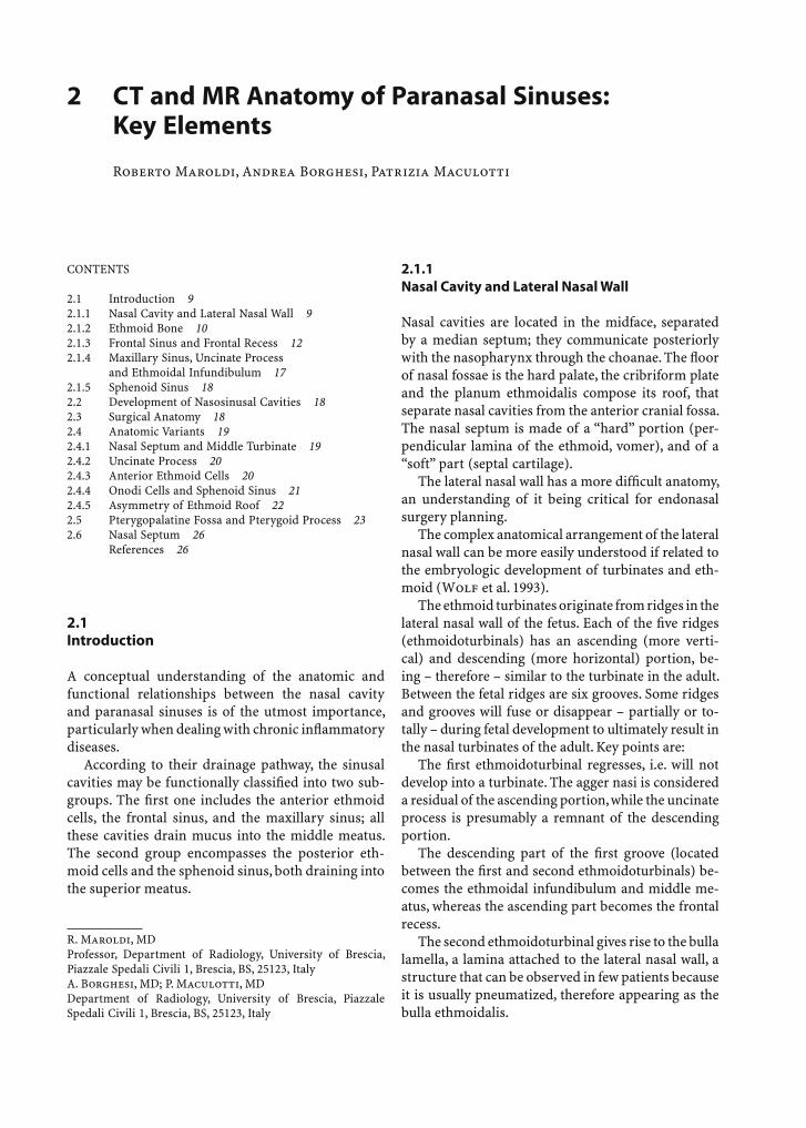
From a practical standpoint, the ethmoid bone can be divided into four structures: two lateral masses, a sagittal midline lamina, and a horizontal plate (Fig. 2.1). The latter (cribriform plate) is the central part of the fl oor of the anterior cranial fossa. Several microscopic foramina perforate its thin structure, through which course the olfactory nerve fi laments with their perineural investment.
Fig. 2.1. The ethmoid plates and fovea ethmoidalis. A midline (perpendicular plate, PP) and two lateral vertical laminae: the medial one made by the vertical lamella of the middle turbi-nate (vlMT) on the top of which is the vertical lamella of the cribriform plate (vlCP), the lateral one is the lamina papyracea (LP). Horizontal lamella of the cribriform plate (hlCP), hori-zontal (hGL) and vertical (vGL) – ascending part – of ground lamella of the middle turbinate, crista galli (CG), fovea eth-madalis (FE)
On the upper part of the sagittal midline lamina (crista galli) – located just above the cribriform plate – inserts the cerebral falx; the inferior part, below the cribriform plate, being referred to as the perpendicular lamina, a component of the nasal septum (Fig. 2.2–2.4).
The lateral masses (ethmoid labyrinth) are made of a variable number (3–18) of pneumatized cells separated by thin bony walls.
Each ethmoid labyrinth is closed by bony mar-gins on its external and internal surfaces only. The
CT and MR Anatomy of Paranasal Sinuses: Key Elements 11
external surface is made by the lamina papyracea that separates the cells from the orbit. Dehiscences may be present. As a result, the periosteum of the ethmoid cell comes in contact with the periosteum investing the orbital wall (periorbita). Medially,
the labyrinth is bordered by two constant laminae hanging from the horizontal plate: the middle and superior turbinates. An uppermost small third eth-moid turbinate (supreme turbinate) can seldom be observed.
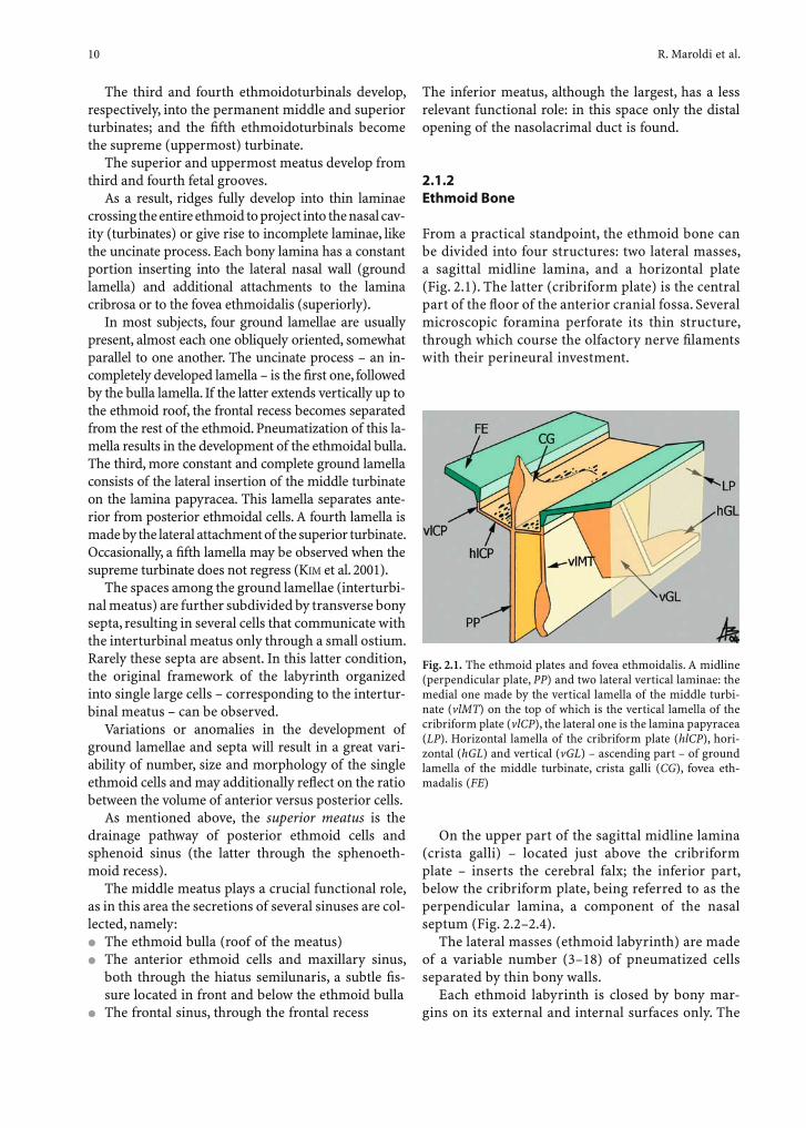
Fig. 2.2a–c. CT coronal reconstructions through the ethmoid labyrinth. a Anterior nasal fossa, level of the uppermost insertion of both uncinate processes on the vertical lamellae of the middle turbinates – type 4 according to Landsberg and Friedman (2001) (white arrows on left side). b Bilateral pneumatization of the horizontal (non-attached) free portion of the uncinate pro-cesses (white arrows on left side). c The horizontal portion of the uncinate process (white arrows on left side) and the inferior surface of the ethmoid bulla (B) limit the oblique and narrow ethmoid infundibulum. The maxillary sinus ostium is detectable at the bottom of the infundibulum (white ellipse). Curved white arrows show the path along the ostium and the infundibulum
a cb
Fig. 2.3a–c. CT coronal reconstructions through the ethmoid labyrinth. a Middle nasal fossa at the level of the short horizontal portion of the uncinate process posterior to the ethmoid infundibulum (white arrowheads on left side). A large ethmoid bulla (B) impinges the left middle turbinate on left side. b The CT section cuts the posterior part of the bulla (B). The horizontal portion of the cribriform plates (arrowheads) appears thinner than the bone of the fovea ethmoidalis (FE). Bilateral pneumatization of the superior turbinate (ST) is present. c Lateral attachment of the middle turbinate – the ground lamella (opposite arrows) – onto the lateral nasal wall. Posterior ethmoid cells (PEC) extend between the lamina papyracea and the supreme turbinate (SuT)
a cb
12 R. Maroldi et al.
The middle turbinate – the largest ethmoid lam-ina – separates the labyrinth into anterior and pos-terior cells by means of its middle portion inserting on the lateral nasal wall (ground lamella) (Fig. 2.5).
All the other surfaces of the labyrinth are open, lined only by adjacent structures. Anteriorly, cells open into a narrow cleft – the infundibulum ethmo-idalis – that empties into the nasal cavity through the middle meatus. On the opposite surface, the an-terior aspect of the sphenoid sinus borders the pos-terior ethmoid cells, that empty into another narrow cleft – the sphenoethmoidal recess – draining into the superior meatus and fi nally into the choanae.
The orbital plate of the frontal bone (fovea eth-moidalis) provides the bony roof of the ethmoid as the cells within the labyrinth extend above the plane of the cribriform plate (Lebowitz et al. 2001). The transition between the thicker frontal bone and the thinner, medially located, lateral lamella of the lamina cribrosa can be easily demonstrated on coronal CT planes (Fig. 2.2). The lateral lamella and the lamina cribrosa provide, respectively, the lateral border and the fl oor of the olfactory fossa. It is im-portant to note that the variability of pneumatiza-tion of the labyrinth refl ects not only on the height of the lateral lamella – therefore the relative depth of the olfactory fossa into the nasal cavity – but also on its obliquity and on the transverse size of the lamina cribrosa. Relevant side to side variations are frequently observed.
Moreover, the weakest area of the whole anterior skull base is located where the anterior ethmoidal artery enters the lateral lamella after having crossed the lamina papyracea and the ethmoidal labyrinth. On CT, this area can be detected as a focal, some-times symmetrical, dehiscence on the lateral lamella of the lamina cribrosa.
2.1.3 Frontal Sinus and Frontal Recess
Anterior pneumatization of the frontal recess into the frontal bone gives rise to the frontal sinus (Fig. 2.6). In the sagittal plane its ostium can be identifi ed as the narrowest part of an hourglass space, the upper part widening into the frontal sinus, and the lower empty-ing into the middle meatus through the frontal recess. The latter is not a true tubular structure, as the term “nasofrontal duct” might indicate. In effect, the size and shape of the frontal recess are largely dictated by the adjacent structures: the agger nasi cells anteriorly, the ethmoidal bulla posteriorly, the vertical portion of the uncinate process and the middle turbinate on medial and lateral aspects (Fig. 2.7).
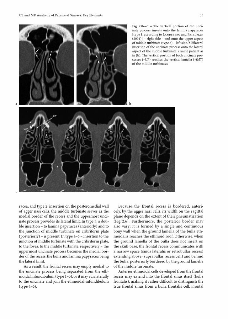
Particularly, the medial and lateral borders of the frontal recess depend on the variable type of the supe-rior attachment of the uncinate process. Six variations have been identifi ed by Landsberg and Friedman (2001) (Fig. 2.8). In type 1, insertion on lamina papy-
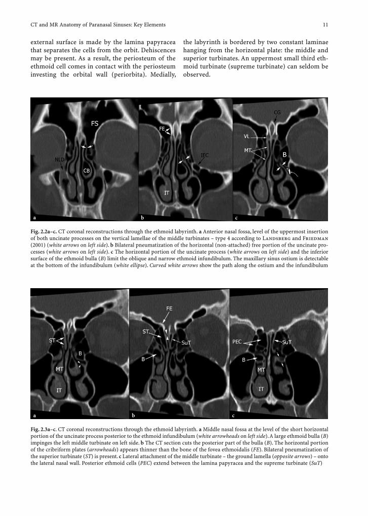
Fig. 2.4a–c. CT coronal reconstructions through the posterior ethmoid labyrinth and the sphenoid sinus. a Level of the sphe-noethmoid recess (asterisks). Lateral attachment of all four turbinates (IT, MT, ST, and SuT) is shown. Anterior aspect of the sphenoid sinus (SS) and posterior ethmoid cells (PEC) are demonstrated on the same level. A posterior ethmoid cell extends over the right sphenoid sinus – Onodi cell (onC) on (b) and (c). Inferior orbital fi ssure (IOF), superior orbital fi ssure (SOF), sphenopalatine foramen (SPF), greater palatine canal (GPC)
a cb
CT and MR Anatomy of Paranasal Sinuses: Key Elements 13
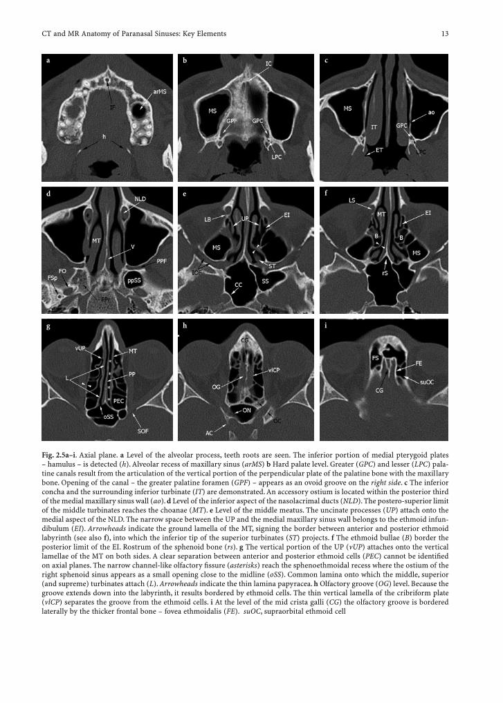
Fig. 2.5a–i. Axial plane. a Level of the alveolar process, teeth roots are seen. The inferior portion of medial pterygoid plates – hamulus – is detected (h). Alveolar recess of maxillary sinus (arMS) b Hard palate level. Greater (GPC) and lesser (LPC) pala-tine canals result from the articulation of the vertical portion of the perpendicular plate of the palatine bone with the maxillary bone. Opening of the canal – the greater palatine foramen (GPF) – appears as an ovoid groove on the right side. c The inferior concha and the surrounding inferior turbinate (IT) are demonstrated. An accessory ostium is located within the posterior third of the medial maxillary sinus wall (ao). d Level of the inferior aspect of the nasolacrimal ducts (NLD). The postero-superior limit of the middle turbinates reaches the choanae (MT). e Level of the middle meatus. The uncinate processes (UP) attach onto the medial aspect of the NLD. The narrow space between the UP and the medial maxillary sinus wall belongs to the ethmoid infun-dibulum (EI). Arrowheads indicate the ground lamella of the MT, signing the border between anterior and posterior ethmoid labyrinth (see also f), into which the inferior tip of the superior turbinates (ST) projects. f The ethmoid bullae (B) border the posterior limit of the EI. Rostrum of the sphenoid bone (rs). g The vertical portion of the UP (vUP) attaches onto the vertical lamellae of the MT on both sides. A clear separation between anterior and posterior ethmoid cells (PEC) cannot be identifi ed on axial planes. The narrow channel-like olfactory fi ssure (asterisks) reach the sphenoethmoidal recess where the ostium of the right sphenoid sinus appears as a small opening close to the midline (oSS). Common lamina onto which the middle, superior (and supreme) turbinates attach (L). Arrowheads indicate the thin lamina papyracea. h Olfactory groove (OG) level. Because the groove extends down into the labyrinth, it results bordered by ethmoid cells. The thin vertical lamella of the cribriform plate (vlCP) separates the groove from the ethmoid cells. i At the level of the mid crista galli (CG) the olfactory groove is bordered laterally by the thicker frontal bone – fovea ethmoidalis (FE). suOC, supraorbital ethmoid cell
a
ihg
fed
b c
14 R. Maroldi et al.
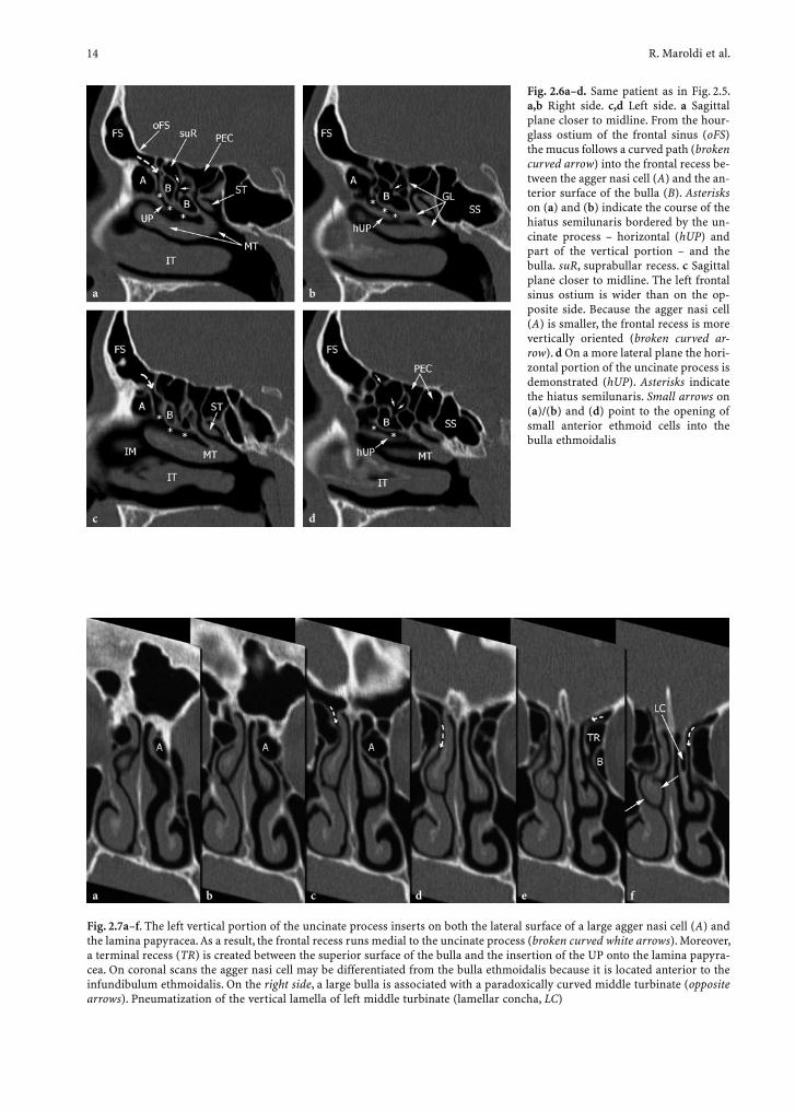
Fig. 2.7a–f. The left vertical portion of the uncinate process inserts on both the lateral surface of a large agger nasi cell (A) and the lamina papyracea. As a result, the frontal recess runs medial to the uncinate process (broken curved white arrows). Moreover, a terminal recess (TR) is created between the superior surface of the bulla and the insertion of the UP onto the lamina papyra-cea. On coronal scans the agger nasi cell may be differentiated from the bulla ethmoidalis because it is located anterior to the infundibulum ethmoidalis. On the right side, a large bulla is associated with a paradoxically curved middle turbinate (opposite arrows). Pneumatization of the vertical lamella of left middle turbinate (lamellar concha, LC)
a c eb d f
Fig. 2.6a–d. Same patient as in Fig. 2.5. a,b Right side. c,d Left side. a Sagittal plane closer to midline. From the hour-glass ostium of the frontal sinus (oFS) the mucus follows a curved path (broken curved arrow) into the frontal recess be-tween the agger nasi cell (A) and the an-terior surface of the bulla (B). Asterisks on (a) and (b) indicate the course of the hiatus semilunaris bordered by the un-cinate process – horizontal (hUP) and part of the vertical portion – and the bulla. suR, suprabullar recess. c Sagittal plane closer to midline. The left frontal sinus ostium is wider than on the op-posite side. Because the agger nasi cell (A) is smaller, the frontal recess is more vertically oriented (broken curved ar-row). d On a more lateral plane the hori-zontal portion of the uncinate process is demonstrated (hUP). Asterisks indicate the hiatus semilunaris. Small arrows on (a)/(b) and (d) point to the opening of small anterior ethmoid cells into the bulla ethmoidalis
a
c
b
d
CT and MR Anatomy of Paranasal Sinuses: Key Elements 15
racea, and type 2, insertion on the posteromedial wall of agger nasi cells, the middle turbinate serves as the medial border of the recess and the uppermost unci-nate process provides its lateral limit. In type 3, a dou-ble insertion – to lamina papyracea (anteriorly) and to the junction of middle turbinate on cribriform plate (posteriorly) – is present. In type 4–6 – insertion to the junction of middle turbinate with the cribriform plate, to the fovea, to the middle turbinate, respectively – the uppermost uncinate process becomes the medial bor-der of the recess, the bulla and lamina papyracea being the lateral limit.
As a result, the frontal recess may empty medial to the uncinate process being separated from the eth-moidal infundibulum (type 1–3), or it may run laterally to the uncinate and join the ethmoidal infundibulum (type 4–6).
Because the frontal recess is bordered, anteri-orly, by the agger nasi cells, its width on the sagittal plane depends on the extent of their pneumatization (Fig. 2.6). Furthermore, the posterior border may also vary: it is formed by a single and continuous bony wall when the ground lamella of the bulla eth-moidalis reaches the ethmoid roof. Otherwise, when the ground lamella of the bulla does not insert on the skull base, the frontal recess communicates with a narrow space (sinus lateralis or retrobullar recess) extending above (suprabullar recess cell) and behind the bulla, posteriorly bordered by the ground lamella of the middle turbinate.
Anterior ethmoidal cells developed from the frontal recess may extend into the frontal sinus itself (bulla frontalis), making it rather diffi cult to distinguish the true frontal sinus from a bulla frontalis cell. Frontal
Fig. 2.8a–c. a The vertical portion of the unci-nate process inserts onto the lamina papyracea [type 1, according to Landsberg and Friedman (2001)] – right side – and onto the upper aspect of middle turbinate (type 6) – left side. b Bilateral insertion of the uncinate process onto the lateral aspect of the middle turbinate. c Same patient as in (b). The vertical portion of both uncinate pro-cesses (vUP) reaches the vertical lamella (vlMT) of the middle turbinates
a
c
b
16 R. Maroldi et al.
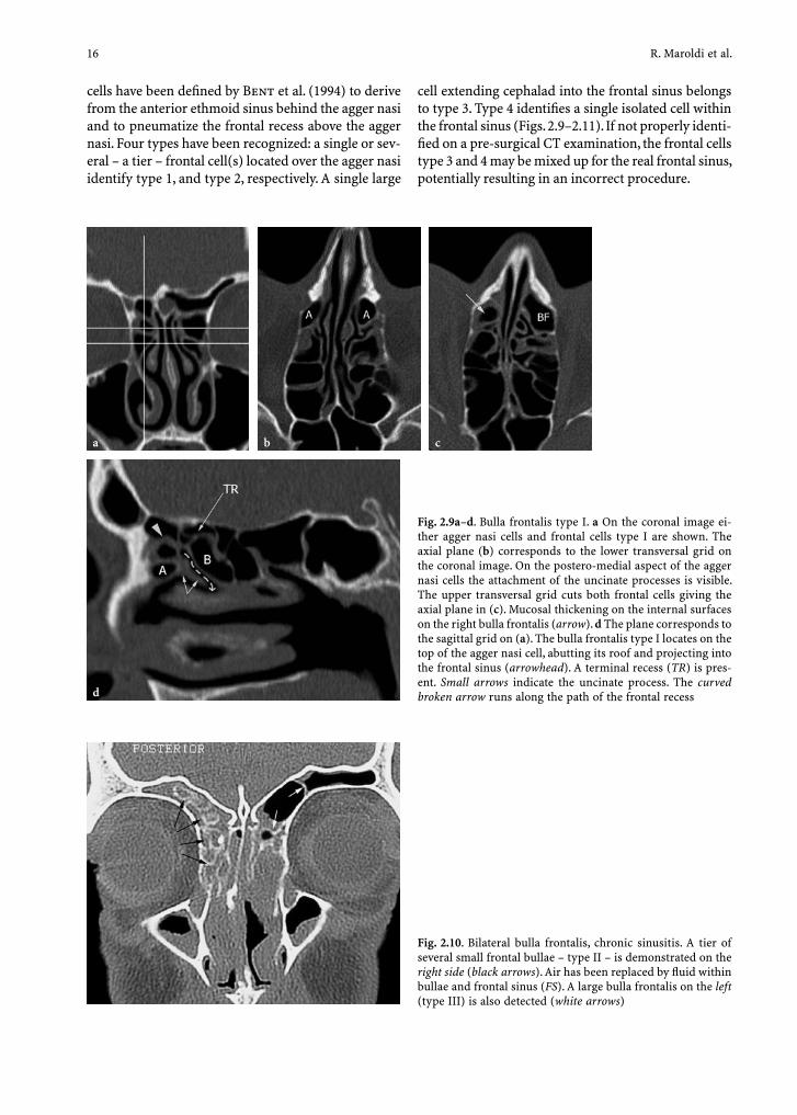
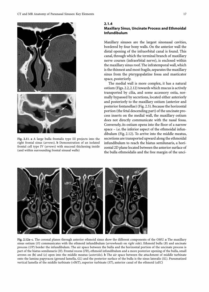
cells have been defi ned by Bent et al. (1994) to derive from the anterior ethmoid sinus behind the agger nasi and to pneumatize the frontal recess above the agger nasi. Four types have been recognized: a single or sev-eral – a tier – frontal cell(s) located over the agger nasi identify type 1, and type 2, respectively. A single large
Fig. 2.10. Bilateral bulla frontalis, chronic sinusitis. A tier of several small frontal bullae – type II – is demonstrated on the right side (black arrows). Air has been replaced by fl uid within bullae and frontal sinus (FS). A large bulla frontalis on the left (type III) is also detected (white arrows)
cell extending cephalad into the frontal sinus belongs to type 3. Type 4 identifi es a single isolated cell within the frontal sinus (Figs. 2.9–2.11). If not properly identi-fi ed on a pre-surgical CT examination, the frontal cells type 3 and 4 may be mixed up for the real frontal sinus, potentially resulting in an incorrect procedure.
Fig. 2.9a–d. Bulla frontalis type I. a On the coronal image ei-ther agger nasi cells and frontal cells type I are shown. The axial plane (b) corresponds to the lower transversal grid on the coronal image. On the postero-medial aspect of the agger nasi cells the attachment of the uncinate processes is visible. The upper transversal grid cuts both frontal cells giving the axial plane in (c). Mucosal thickening on the internal surfaces on the right bulla frontalis (arrow). d The plane corresponds to the sagittal grid on (a). The bulla frontalis type I locates on the top of the agger nasi cell, abutting its roof and projecting into the frontal sinus (arrowhead). A terminal recess (TR) is pres-ent. Small arrows indicate the uncinate process. The curved broken arrow runs along the path of the frontal recess
a cb
d
CT and MR Anatomy of Paranasal Sinuses: Key Elements 17
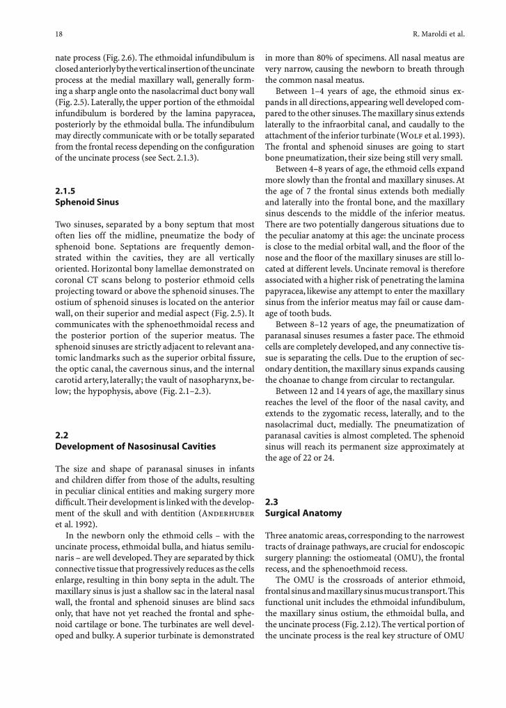
2.1.4 Maxillary Sinus, Uncinate Process and Ethmoidal Infundibulum
Maxillary sinuses are the largest sinonasal cavities, bordered by four bony walls. On the anterior wall the distal opening of the infraorbital canal is found. This canal, through which the terminal branch of maxillary nerve courses (infraorbital nerve), is enclosed within the maxillary sinus roof. The infratemporal wall, which is the thinnest and most fragile, separates the maxillary sinus from the pterygopalatine fossa and masticator space, posteriorly.
The medial wall is more complex, it has a natural ostium (Figs. 2.2, 2.12) towards which mucus is actively transported by cilia, and some accessory ostia, nor-mally bypassed by secretions, located either anteriorly and posteriorly to the maxillary ostium (anterior and posterior fontanellae) (Fig. 2.5). Because the horizontal portion (the fetal descending part) of the uncinate pro-cess inserts on the medial wall, the maxillary ostium does not directly communicate with the nasal fossa. Conversely, its ostium opens into the fl oor of a narrow space – i.e. the inferior aspect of the ethmoidal infun-dibulum (Fig. 2.12). To arrive into the middle meatus, secretions are transported upward along the ethmoidal infundibulum to reach the hiatus semilunaris, a hori-zontal 2D plane located between the anterior surface of the bulla ethmoidalis and the free margin of the unci-
Fig. 2.11. a A large bulla frontalis type III projects into the right frontal sinus (arrows). b Demonstration of an isolated frontal cell type IV (arrows) with mucosal thickening inside (and within surrounding frontal sinusal walls)
a
b
Fig. 2.12a–c. The coronal planes through anterior ethmoid sinus show the different components of the OMU. a The maxillary sinus ostium (O) communicates with the ethmoid infundibulum (arrowheads on right side). Ethmoid bulla (B) and uncinate process (UP) border the infundibulum. The air space between the bulla and the horizontal portion of the uncinate process is part of the hiatus semilunaris (H). Frontal recess (FR), ethmoid infundibulum and a more posterior opening of the bulla, small arrows on (b) and (c) open into the middle meatus (asterisks). b The air space between the attachment of middle turbinate onto the lamina papyracea (ground lamella, GL) and the posterior surface of the bulla is the sinus lateralis (SL). Pneumatized vertical lamella of the middle turbinate (vlMT), superior turbinate (ST), anterior canal of the ethmoid (aEC)
a cb
18 R. Maroldi et al.
nate process (Fig. 2.6). The ethmoidal infundibulum is closed anteriorly by the vertical insertion of the uncinate process at the medial maxillary wall, generally form-ing a sharp angle onto the nasolacrimal duct bony wall (Fig. 2.5). Laterally, the upper portion of the ethmoidal infundibulum is bordered by the lamina papyracea, posteriorly by the ethmoidal bulla. The infundibulum may directly communicate with or be totally separated from the frontal recess depending on the confi guration of the uncinate process (see Sect. 2.1.3).
2.1.5 Sphenoid Sinus
Two sinuses, separated by a bony septum that most often lies off the midline, pneumatize the body of sphenoid bone. Septations are frequently demon-strated within the cavities, they are all vertically oriented. Horizontal bony lamellae demonstrated on coronal CT scans belong to posterior ethmoid cells projecting toward or above the sphenoid sinuses. The ostium of sphenoid sinuses is located on the anterior wall, on their superior and medial aspect (Fig. 2.5). It communicates with the sphenoethmoidal recess and the posterior portion of the superior meatus. The sphenoid sinuses are strictly adjacent to relevant ana-tomic landmarks such as the superior orbital fi ssure, the optic canal, the cavernous sinus, and the internal carotid artery, laterally; the vault of nasopharynx, be-low; the hypophysis, above (Fig. 2.1–2.3).
2.2 Development of Nasosinusal Cavities
The size and shape of paranasal sinuses in infants and children differ from those of the adults, resulting in peculiar clinical entities and making surgery more diffi cult. Their development is linked with the develop-ment of the skull and with dentition (Anderhuber et al. 1992).
In the newborn only the ethmoid cells – with the uncinate process, ethmoidal bulla, and hiatus semilu-naris – are well developed. They are separated by thick connective tissue that progressively reduces as the cells enlarge, resulting in thin bony septa in the adult. The maxillary sinus is just a shallow sac in the lateral nasal wall, the frontal and sphenoid sinuses are blind sacs only, that have not yet reached the frontal and sphe-noid cartilage or bone. The turbinates are well devel-oped and bulky. A superior turbinate is demonstrated
in more than 80% of specimens. All nasal meatus are very narrow, causing the newborn to breath through the common nasal meatus.
Between 1–4 years of age, the ethmoid sinus ex-pands in all directions, appearing well developed com-pared to the other sinuses. The maxillary sinus extends laterally to the infraorbital canal, and caudally to the attachment of the inferior turbinate (Wolf et al. 1993). The frontal and sphenoid sinuses are going to start bone pneumatization, their size being still very small.
Between 4–8 years of age, the ethmoid cells expand more slowly than the frontal and maxillary sinuses. At the age of 7 the frontal sinus extends both medially and laterally into the frontal bone, and the maxillary sinus descends to the middle of the inferior meatus. There are two potentially dangerous situations due to the peculiar anatomy at this age: the uncinate process is close to the medial orbital wall, and the fl oor of the nose and the fl oor of the maxillary sinuses are still lo-cated at different levels. Uncinate removal is therefore associated with a higher risk of penetrating the lamina papyracea, likewise any attempt to enter the maxillary sinus from the inferior meatus may fail or cause dam-age of tooth buds.
Between 8–12 years of age, the pneumatization of paranasal sinuses resumes a faster pace. The ethmoid cells are completely developed, and any connective tis-sue is separating the cells. Due to the eruption of sec-ondary dentition, the maxillary sinus expands causing the choanae to change from circular to rectangular.
Between 12 and 14 years of age, the maxillary sinus reaches the level of the fl oor of the nasal cavity, and extends to the zygomatic recess, laterally, and to the nasolacrimal duct, medially. The pneumatization of paranasal cavities is almost completed. The sphenoid sinus will reach its permanent size approximately at the age of 22 or 24.
2.3 Surgical Anatomy
Three anatomic areas, corresponding to the narrowest tracts of drainage pathways, are crucial for endoscopic surgery planning: the ostiomeatal (OMU), the frontal recess, and the sphenoethmoid recess.
The OMU is the crossroads of anterior ethmoid, frontal sinus and maxillary sinus mucus transport. This functional unit includes the ethmoidal infundibulum, the maxillary sinus ostium, the ethmoidal bulla, and the uncinate process (Fig. 2.12). The vertical portion of the uncinate process is the real key structure of OMU
CT and MR Anatomy of Paranasal Sinuses: Key Elements 19
because its shape and upper attachment determine the morphology of both the ethmoidal infundibulum and the frontal recess.
The ethmoidal infundibulum is the air passage that connects the maxillary sinus ostium to the middle me-atus. To simplify, it can be subdivided into two differ-ent portions. The anterior part is the air space located between the vertical portion of the uncinate and the anterior surface of the bulla; whereas the posterior part is the air space situated between the horizontal portion (free, unattached portion) of the uncinate pro-cess and the inferomedial surface of the bulla or of the orbit, if the sinus lateralis is present. The gap between the ethmoid bulla and the free edge of the uncinate process is the hiatus semilunaris (Fig. 2.6).
The ethmoid bulla is an important surgical land-mark: this is actually the most posterior cell in the an-terior ethmoid, protruding in the middle meatus. Its size and morphology are highly variable. The middle turbinate is the medial border of the OMU. This sub-tle, curved bony structure describes a lateral concav-ity; it has a cranial anchorage on the cribriform plate – through its vertical lamina – and a lateral insertion on the posterior part of lamina papyracea – through its fan-shaped ground lamella.
The width, path, and morphology of the frontal re-cess are largely determined by the shape and relation-ships of the upper part of the uncinate process (type 1–6, see Sect. 2.1.3) and by the variable size and pneu-matization of adjacent structures, particularly the eth-moid bulla and agger nasi cells.
Moreover, even the confi guration of the ethmoidal infundibulum – and its relationship to the frontal re-cess depend on the anatomy of the uncinate process (Landsberg and Friedman 2001). In most subjects (up to 52%) its lateral insertion on the lamina papyra-cea (type 1) creates a “roof” that closes the ethmoidal infundibulum in a blind upper pouch (terminal recess) (Fig. 2.7). If the uncinate process attaches anteriorly to the agger nasi cells (type 2, 18%) the ethmoidal infun-dibulum is closed superiorly by the fl oor of the agger nasi. In both confi gurations, the frontal recess and the ethmoidal infundibulum are separated, and the opening of the frontal recess into the middle meatus is medial to the ethmoidal infundibulum, between the uncinate process and the middle turbinate (Fig. 2.7). As in about 17% of subjects (type 3) the upper part of the uncinate process inserts on both the lamina papy-racea and the junction of the middle turbinate with the lamina cribrosa, the frontal sinus opens into the middle meatus in approximately 88% of cases (type 1–3). Conversely, if the uncinate process maintains its complete and longest fetal attachment on the ethmoid
roof or medially on the middle turbinate, the frontal recess empties directly into the ethmoid infundibu-lum, therefore laterally to the uncinate process (Figs. 2.2, 2.8).
From a surgical standpoint, the correct assessment of the frontal recess opening is essential in planning the proper endonasal approach to the frontal recess and the adequate exposure of the frontal sinus. In fact, if the frontal recess opens laterally to the uncinate pro-cess, an attempt to fi nd the recess medially to the unci-nate would potentially lead toward the olfactory fossa or the frontal lobe.
Because the anterior and uppermost segment of the uncinate process runs obliquely from posterior-inferior to anterior-superior, direct coronal CT scans – obtained perpendicularly to the hard palate – usu-ally do not demonstrate its full course and upper in-sertion. Multislice CT reconstructions obtained on an oblique plane running along the recess’s axis enable a depiction of the actual extent (and insertion) of the unainale process.
The sphenoethmoid recess is outlined by the ante-rior sphenoid wall and by the posterior wall of pos-terior ethmoid cells. It conveys sphenoid sinus secre-tions in the superior meatus (Fig. 2.5).
2.4 Anatomic Variants
Anatomic variants of the OMU structures are rela-tively frequent; they can be classifi ed as anomalies of size, shape, orientation and entity of pneumatization. In some cases they may impair the physiologic mucous drainage and condition the endoscopic approach. Nevertheless, it must be emphasized that the pres-ence of an anatomic variant does not necessarily imply an increased risk of infl ammatory lesion: actually, no direct correlation between anatomic anomalies and nasal obstruction has been clearly demonstrated.
2.4.1 Nasal Septum and Middle Turbinate
Nasal septum deviation is quite a common condition, with its prevalence in asymptomatic subjects reported to range from 20% to 30%; it is usually congenital but may be post-traumatic (Wanamaker 1996). In most cases it is associated with the presence of a bone spur protruding in the nasal fossa, confl icting with the middle turbinate (less frequently with the inferior),
20 R. Maroldi et al.
and therefore reducing the diameter of the middle meatus (Fig. 2.13).
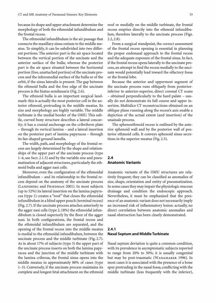
Middle turbinate describing a lateral convexity to-ward the lateral sinusal wall is classifi ed as paradoxi-cal: this variant may hamper an endoscopic approach (Fig. 2.7f). When the bulbous segment is pneumatized, the middle turbinate is referred to as concha bullosa (Figs. 2.2, 2.8). If only the attachment portion of the middle turbinate is pneumatized – with no involve-ment of the bulbous segment – it is defi ned lamellar concha (Fig. 2.7f). According to the extent of pneumati-zation (vertical lamina, bulbous segment, or both) and its entity, this anomaly may narrow or obstruct the eth-moidal infundibulum as well as the middle meatus, and may interfere with nasal airfl ow, particularly if associ-ated with other anomalies that may obstruct the OMU, such as a large ethmoidal bulla (Joe et al. 2000) (Fig. 2.13). The prevalence of middle turbinate pneumati-zation on CT studies varies from 14% to 53% (Lloyd 1990; Perez et al. 2000). Concha bullosa is often bilat-eral (Fig. 2.12) and associated with septal deviation.
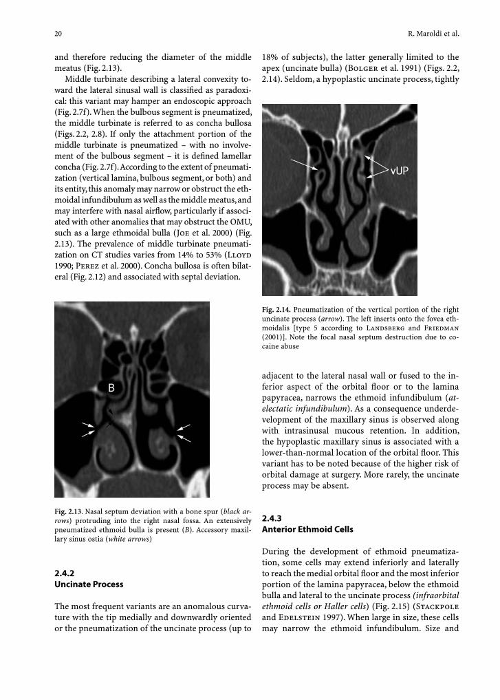
18% of subjects), the latter generally limited to the apex (uncinate bulla) (Bolger et al. 1991) (Figs. 2.2, 2.14). Seldom, a hypoplastic uncinate process, tightly
Fig. 2.13. Nasal septum deviation with a bone spur (black ar-rows) protruding into the right nasal fossa. An extensively pneumatized ethmoid bulla is present (B). Accessory maxil-lary sinus ostia (white arrows)
2.4.2 Uncinate Process
The most frequent variants are an anomalous curva-ture with the tip medially and downwardly oriented or the pneumatization of the uncinate process (up to
adjacent to the lateral nasal wall or fused to the in-ferior aspect of the orbital fl oor or to the lamina papyracea, narrows the ethmoid infundibulum (at-electatic infundibulum). As a consequence underde-velopment of the maxillary sinus is observed along with intrasinusal mucous retention. In addition, the hypoplastic maxillary sinus is associated with a lower-than-normal location of the orbital fl oor. This variant has to be noted because of the higher risk of orbital damage at surgery. More rarely, the uncinate process may be absent.
2.4.3 Anterior Ethmoid Cells
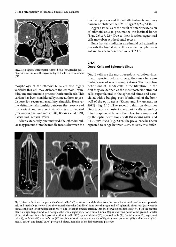
During the development of ethmoid pneumatiza-tion, some cells may extend inferiorly and laterally to reach the medial orbital fl oor and the most inferior portion of the lamina papyracea, below the ethmoid bulla and lateral to the uncinate process (infraorbital ethmoid cells or Haller cells) (Fig. 2.15) (Stackpole and Edelstein 1997). When large in size, these cells may narrow the ethmoid infundibulum. Size and
Fig. 2.14. Pneumatization of the vertical portion of the right uncinate process (arrow). The left inserts onto the fovea eth-moidalis [type 5 according to Landsberg and Friedman (2001)]. Note the focal nasal septum destruction due to co-caine abuse
CT and MR Anatomy of Paranasal Sinuses: Key Elements 21
uncinate process and the middle turbinate and may narrow or obstruct the OMU (Figs. 2.3, 2.9, 2.13).
Agger nasi cells are the result of anterior extension of ethmoid cells to pneumatize the lacrimal bones (Figs. 2.6, 2.7, 2.9). Due to their location, agger nasi cells may obstruct the frontal recess.
Bulla frontalis indicates an ethmoid cell extending towards the frontal sinus. It is a rather complex vari-ant and has been described in Sect. 2.1.3
2.4.4 Onodi Cells and Sphenoid Sinus
Onodi cells are the most hazardous variation since, if not reported before surgery, they may be a po-tential cause of severe complications. There are two defi nitions of Onodi cells in the literature. In the fi rst they are defi ned as the most posterior ethmoid cells, superolateral to the sphenoid sinus and asso-ciated with a bulging, even if minimal, of the bony wall of the optic nerve (Kainz and Stammberger 1992) (Fig. 2.16). The second defi nition describes Onodi cells as posterior ethmoid cells extending into the sphenoid bone, either close to or impressed by the optic nerve bony wall (Stammberger and Kennedy 1995) (Fig. 2.17). The prevalence has been reported to range between 3.4% to 51%, this differ-
Fig. 2.15. Bilateral infraorbital ethmoid cells (IEC; Haller cells). Black arrows indicate the asymmetry of the fovea ethmoidalis (FE)
morphology of the ethmoid bulla are also highly variable: this cell may dislocate the ethmoid infun-dibulum and uncinate process (horizontalized). This variant has been considered by some authors to pre-dispose for recurrent maxillary sinusitis. However, the defi nitive relationship between the presence of this variant and recurrent sinusitis is still debated (Stammberger and Wolf 1988; Bolger et al. 1991; Laine and Smoker 1992).
When extensively pneumatized, the ethmoid bul-lae may protrude into the middle meatus between the
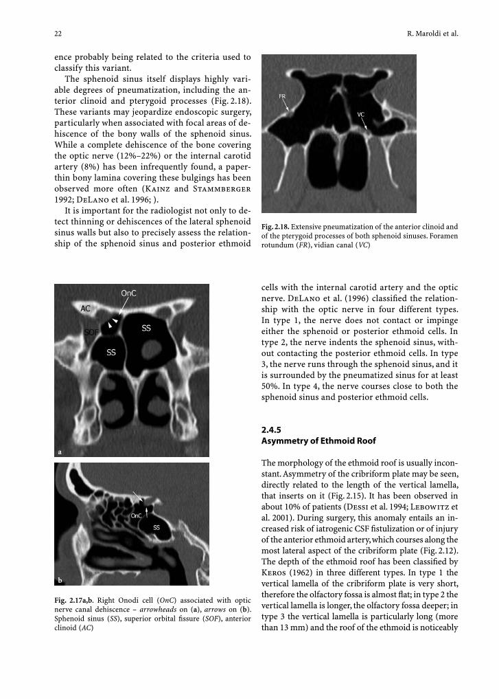
Fig. 2.16a–c. a On the axial plane the Onodi cell (OnC) arises on the right side from the posterior ethmoid and extends posteri-orly and medially (arrows). b On the coronal plane the Onodi cell runs over the right and left sphenoid sinus roof (arrowheads indicate the thin left sphenoid sinus roof). The left sinus extends laterally into the pterygoid process (arrows). c On the sagittal plane a single large Onodi cell occupies the whole right posterior ethmoid sinus. Opposite arrows point to the ground lamella of the middle turbinate. Left posterior ethmoid cell (PEC), sphenoid sinus (SS), ethmoid bulla (B), frontal sinus (FR), agger nasi cell (A), middle (MT) and inferior (IT) turbinates, optic nerve and canals (ON), foramen rotundum (FS), vidian canal (VC), medial (MPP) and lateral (LPP) pterygoid plates, hamulus of medial pterygoid plate (h)
a cb
22 R. Maroldi et al.
ence probably being related to the criteria used to classify this variant.
The sphenoid sinus itself displays highly vari-able degrees of pneumatization, including the an-terior clinoid and pterygoid processes (Fig. 2.18). These variants may jeopardize endoscopic surgery, particularly when associated with focal areas of de-hiscence of the bony walls of the sphenoid sinus. While a complete dehiscence of the bone covering the optic nerve (12%–22%) or the internal carotid artery (8%) has been infrequently found, a paper-thin bony lamina covering these bulgings has been observed more often (Kainz and Stammberger 1992; DeLano et al. 1996; ).
It is important for the radiologist not only to de-tect thinning or dehiscences of the lateral sphenoid sinus walls but also to precisely assess the relation-ship of the sphenoid sinus and posterior ethmoid
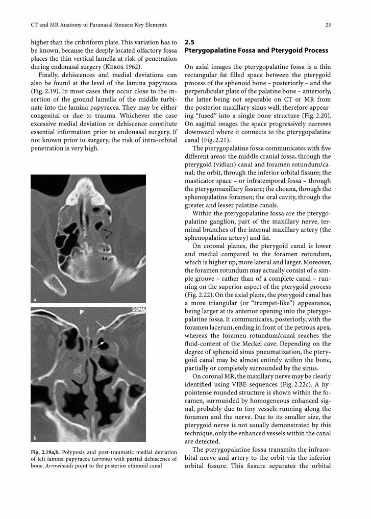
Fig. 2.18. Extensive pneumatization of the anterior clinoid and of the pterygoid processes of both sphenoid sinuses. Foramen rotundum (FR), vidian canal (VC)
cells with the internal carotid artery and the optic nerve. DeLano et al. (1996) classifi ed the relation-ship with the optic nerve in four different types. In type 1, the nerve does not contact or impinge either the sphenoid or posterior ethmoid cells. In type 2, the nerve indents the sphenoid sinus, with-out contacting the posterior ethmoid cells. In type 3, the nerve runs through the sphenoid sinus, and it is surrounded by the pneumatized sinus for at least 50%. In type 4, the nerve courses close to both the sphenoid sinus and posterior ethmoid cells.
2.4.5 Asymmetry of Ethmoid Roof
The morphology of the ethmoid roof is usually incon-stant. Asymmetry of the cribriform plate may be seen, directly related to the length of the vertical lamella, that inserts on it (Fig. 2.15). It has been observed in about 10% of patients (Dessi et al. 1994; Lebowitz et al. 2001). During surgery, this anomaly entails an in-creased risk of iatrogenic CSF fi stulization or of injury of the anterior ethmoid artery, which courses along the most lateral aspect of the cribriform plate (Fig. 2.12). The depth of the ethmoid roof has been classifi ed by Keros (1962) in three different types. In type 1 the vertical lamella of the cribriform plate is very short, therefore the olfactory fossa is almost fl at; in type 2 the vertical lamella is longer, the olfactory fossa deeper; in type 3 the vertical lamella is particularly long (more than 13 mm) and the roof of the ethmoid is noticeably
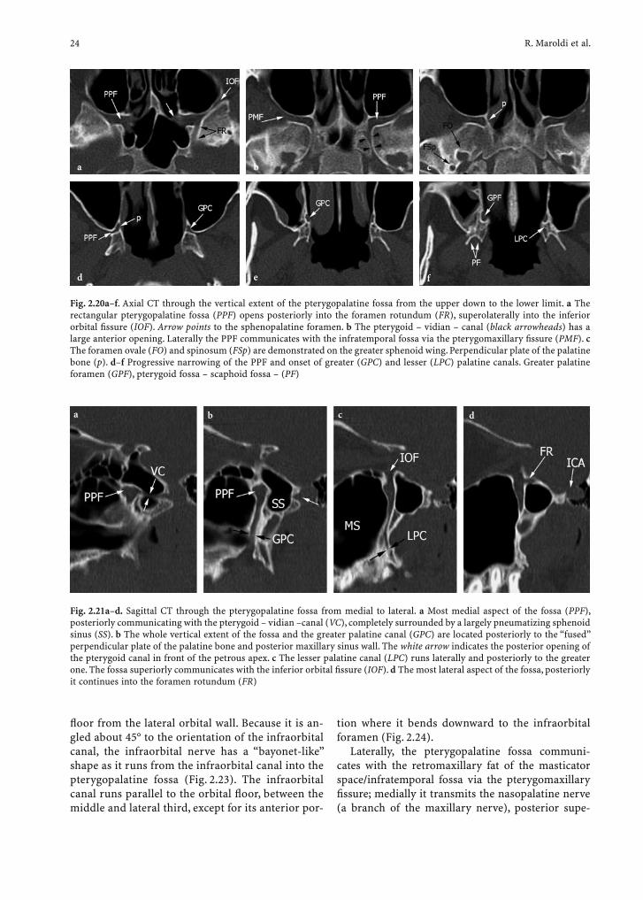
Fig. 2.17a,b. Right Onodi cell (OnC) associated with optic nerve canal dehiscence – arrowheads on (a), arrows on (b). Sphenoid sinus (SS), superior orbital fi ssure (SOF), anterior clinoid (AC)
a
b
CT and MR Anatomy of Paranasal Sinuses: Key Elements 23
higher than the cribriform plate. This variation has to be known, because the deeply located olfactory fossa places the thin vertical lamella at risk of penetration during endonasal surgery (Keros 1962).
Finally, dehiscences and medial deviations can also be found at the level of the lamina papyracea (Fig. 2.19). In most cases they occur close to the in-sertion of the ground lamella of the middle turbi-nate into the lamina papyracea. They may be either congenital or due to trauma. Whichever the case excessive medial deviation or dehiscence constitute essential information prior to endonasal surgery. If not known prior to surgery, the risk of intra-orbital penetration is very high.
2.5 Pterygopalatine Fossa and Pterygoid Process
On axial images the pterygopalatine fossa is a thin rectangular fat fi lled space between the pterygoid process of the sphenoid bone – posteriorly – and the perpendicular plate of the palatine bone – anteriorly, the latter being not separable on CT or MR from the posterior maxillary sinus wall, therefore appear-ing “fused” into a single bone structure (Fig. 2.20). On sagittal images the space progressively narrows downward where it connects to the pterygopalatine canal (Fig. 2.21).
The pterygopalatine fossa communicates with fi ve different areas: the middle cranial fossa, through the pterygoid (vidian) canal and foramen rotundum/ca-nal; the orbit, through the inferior orbital fi ssure; the masticator space – or infratemporal fossa – through the pterygomaxillary fi ssure; the choana, through the sphenopalatine foramen; the oral cavity, through the greater and lesser palatine canals.
Within the pterygopalatine fossa are the pterygo-palatine ganglion, part of the maxillary nerve, ter-minal branches of the internal maxillary artery (the sphenopalatine artery) and fat.
On coronal planes, the pterygoid canal is lower and medial compared to the foramen rotundum, which is higher up, more lateral and larger. Moreover, the foramen rotundum may actually consist of a sim-ple groove – rather than of a complete canal – run-ning on the superior aspect of the pterygoid process (Fig. 2.22). On the axial plane, the pterygoid canal has a more triangular (or “trumpet-like”) appearance, being larger at its anterior opening into the pterygo-palatine fossa. It communicates, posteriorly, with the foramen lacerum, ending in front of the petrous apex, whereas the foramen rotundum/canal reaches the fl uid-content of the Meckel cave. Depending on the degree of sphenoid sinus pneumatization, the ptery-goid canal may be almost entirely within the bone, partially or completely surrounded by the sinus.
On coronal MR, the maxillary nerve may be clearly identifi ed using VIBE sequences (Fig. 2.22c). A hy-pointense rounded structure is shown within the fo-ramen, surrounded by homogeneous enhanced sig-nal, probably due to tiny vessels running along the foramen and the nerve. Due to its smaller size, the pterygoid nerve is not usually demonstrated by this technique, only the enhanced vessels within the canal are detected.
The pterygopalatine fossa transmits the infraor-bital nerve and artery to the orbit via the inferior orbital fi ssure. This fi ssure separates the orbital
Fig. 2.19a,b. Polyposis and post-traumatic medial deviation of left lamina papyracea (arrows) with partial dehiscence of bone. Arrowheads point to the posterior ethmoid canal
a
b
24 R. Maroldi et al.
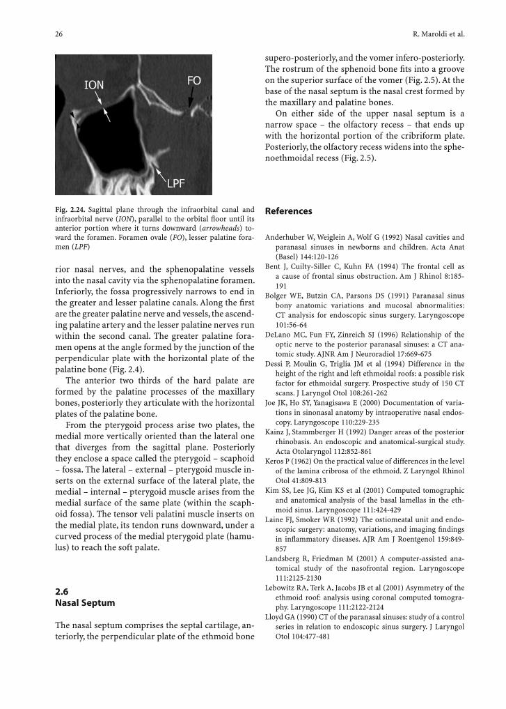
fl oor from the lateral orbital wall. Because it is an-gled about 45° to the orientation of the infraorbital canal, the infraorbital nerve has a “bayonet-like” shape as it runs from the infraorbital canal into the pterygopalatine fossa (Fig. 2.23). The infraorbital canal runs parallel to the orbital fl oor, between the middle and lateral third, except for its anterior por-
tion where it bends downward to the infraorbital foramen (Fig. 2.24).
Laterally, the pterygopalatine fossa communi-cates with the retromaxillary fat of the masticator space/infratemporal fossa via the pterygomaxillary fi ssure; medially it transmits the nasopalatine nerve (a branch of the maxillary nerve), posterior supe-
Fig. 2.21a–d. Sagittal CT through the pterygopalatine fossa from medial to lateral. a Most medial aspect of the fossa (PPF), posteriorly communicating with the pterygoid – vidian –canal (VC), completely surrounded by a largely pneumatizing sphenoid sinus (SS). b The whole vertical extent of the fossa and the greater palatine canal (GPC) are located posteriorly to the “fused” perpendicular plate of the palatine bone and posterior maxillary sinus wall. The white arrow indicates the posterior opening of the pterygoid canal in front of the petrous apex. c The lesser palatine canal (LPC) runs laterally and posteriorly to the greater one. The fossa superiorly communicates with the inferior orbital fi ssure (IOF). d The most lateral aspect of the fossa, posteriorly it continues into the foramen rotundum (FR)
a cb d
Fig. 2.20a–f. Axial CT through the vertical extent of the pterygopalatine fossa from the upper down to the lower limit. a The rectangular pterygopalatine fossa (PPF) opens posteriorly into the foramen rotundum (FR), superolaterally into the inferior orbital fi ssure (IOF). Arrow points to the sphenopalatine foramen. b The pterygoid – vidian – canal (black arrowheads) has a large anterior opening. Laterally the PPF communicates with the infratemporal fossa via the pterygomaxillary fi ssure (PMF). c The foramen ovale (FO) and spinosum (FSp) are demonstrated on the greater sphenoid wing. Perpendicular plate of the palatine bone (p). d–f Progressive narrowing of the PPF and onset of greater (GPC) and lesser (LPC) palatine canals. Greater palatine foramen (GPF), pterygoid fossa – scaphoid fossa – (PF)
a b c
d e f
CT and MR Anatomy of Paranasal Sinuses: Key Elements 25
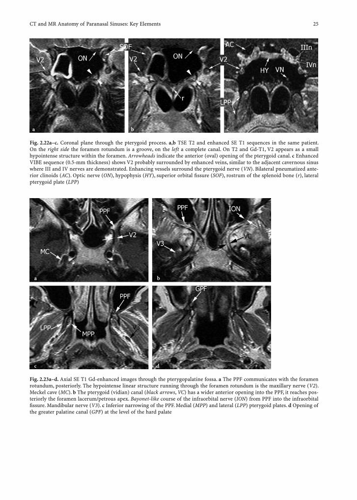
Fig. 2.22a–c. Coronal plane through the pterygoid process. a,b TSE T2 and enhanced SE T1 sequences in the same patient. On the right side the foramen rotundum is a groove, on the left a complete canal. On T2 and Gd-T1, V2 appears as a small hypointense structure within the foramen. Arrowheads indicate the anterior (oval) opening of the pterygoid canal. c Enhanced VIBE sequence (0.5-mm thickness) shows V2 probably surrounded by enhanced veins, similar to the adjacent cavernous sinus where III and IV nerves are demonstrated. Enhancing vessels surround the pterygoid nerve (VN). Bilateral pneumatized ante-rior clinoids (AC). Optic nerve (ON), hypophysis (HY), superior orbital fi ssure (SOF), rostrum of the splenoid bone (r), lateral pterygoid plate (LPP)
a cb
Fig. 2.23a–d. Axial SE T1 Gd-enhanced images through the pterygopalatine fossa. a The PPF communicates with the foramen rotundum, posteriorly. The hypointense linear structure running through the foramen rotundum is the maxillary nerve (V2). Meckel cave (MC). b The pterygoid (vidian) canal (black arrows, VC) has a wider anterior opening into the PPF, it reaches pos-teriorly the foramen lacerum/petrous apex. Bayonet-like course of the infraorbital nerve (ION) from PPF into the infraorbital fi ssure. Mandibular nerve (V3). c Inferior narrowing of the PPF. Medial (MPP) and lateral (LPP) pterygoid plates. d Opening of the greater palatine canal (GPF) at the level of the hard palate
a
c
b
d
26 R. Maroldi et al.
rior nasal nerves, and the sphenopalatine vessels into the nasal cavity via the sphenopalatine foramen. Inferiorly, the fossa progressively narrows to end in the greater and lesser palatine canals. Along the fi rst are the greater palatine nerve and vessels, the ascend-ing palatine artery and the lesser palatine nerves run within the second canal. The greater palatine fora-men opens at the angle formed by the junction of the perpendicular plate with the horizontal plate of the palatine bone (Fig. 2.4).
The anterior two thirds of the hard palate are formed by the palatine processes of the maxillary bones, posteriorly they articulate with the horizontal plates of the palatine bone.
From the pterygoid process arise two plates, the medial more vertically oriented than the lateral one that diverges from the sagittal plane. Posteriorly they enclose a space called the pterygoid – scaphoid – fossa. The lateral – external – pterygoid muscle in-serts on the external surface of the lateral plate, the medial – internal – pterygoid muscle arises from the medial surface of the same plate (within the scaph-oid fossa). The tensor veli palatini muscle inserts on the medial plate, its tendon runs downward, under a curved process of the medial pterygoid plate (hamu-lus) to reach the soft palate.
2.6 Nasal Septum
The nasal septum comprises the septal cartilage, an-teriorly, the perpendicular plate of the ethmoid bone
supero-posteriorly, and the vomer infero-posteriorly. The rostrum of the sphenoid bone fi ts into a groove on the superior surface of the vomer (Fig. 2.5). At the base of the nasal septum is the nasal crest formed by the maxillary and palatine bones.
On either side of the upper nasal septum is a narrow space – the olfactory recess – that ends up with the horizontal portion of the cribriform plate. Posteriorly, the olfactory recess widens into the sphe-noethmoidal recess (Fig. 2.5).
References
Anderhuber W, Weiglein A, Wolf G (1992) Nasal cavities and paranasal sinuses in newborns and children. Acta Anat (Basel) 144:120-126
Bent J, Cuilty-Siller C, Kuhn FA (1994) The frontal cell as a cause of frontal sinus obstruction. Am J Rhinol 8:185-191
Bolger WE, Butzin CA, Parsons DS (1991) Paranasal sinus bony anatomic variations and mucosal abnormalities: CT analysis for endoscopic sinus surgery. Laryngoscope 101:56-64
DeLano MC, Fun FY, Zinreich SJ (1996) Relationship of the optic nerve to the posterior paranasal sinuses: a CT ana-tomic study. AJNR Am J Neuroradiol 17:669-675
Dessi P, Moulin G, Triglia JM et al (1994) Difference in the height of the right and left ethmoidal roofs: a possible risk factor for ethmoidal surgery. Prospective study of 150 CT scans. J Laryngol Otol 108:261-262
Joe JK, Ho SY, Yanagisawa E (2000) Documentation of varia-tions in sinonasal anatomy by intraoperative nasal endos-copy. Laryngoscope 110:229-235
Kainz J, Stammberger H (1992) Danger areas of the posterior rhinobasis. An endoscopic and anatomical-surgical study. Acta Otolaryngol 112:852-861
Keros P (1962) On the practical value of differences in the level of the lamina cribrosa of the ethmoid. Z Laryngol Rhinol Otol 41:809-813
Kim SS, Lee JG, Kim KS et al (2001) Computed tomographic and anatomical analysis of the basal lamellas in the eth-moid sinus. Laryngoscope 111:424-429
Laine FJ, Smoker WR (1992) The ostiomeatal unit and endo-scopic surgery: anatomy, variations, and imaging fi ndings in infl ammatory diseases. AJR Am J Roentgenol 159:849-857
Landsberg R, Friedman M (2001) A computer-assisted ana-tomical study of the nasofrontal region. Laryngoscope 111:2125-2130
Lebowitz RA, Terk A, Jacobs JB et al (2001) Asymmetry of the ethmoid roof: analysis using coronal computed tomogra-phy. Laryngoscope 111:2122-2124
Lloyd GA (1990) CT of the paranasal sinuses: study of a control series in relation to endoscopic sinus surgery. J Laryngol Otol 104:477-481
Fig. 2.24. Sagittal plane through the infraorbital canal and infraorbital nerve (ION), parallel to the orbital fl oor until its anterior portion where it turns downward (arrowheads) to-ward the foramen. Foramen ovale (FO), lesser palatine fora-men (LPF)
CT and MR Anatomy of Paranasal Sinuses: Key Elements 27
Perez P, Sabate J, Carmona A et al (2000) Anatomical variations in the human paranasal sinus region studied by CT. J Anat 197(Pt 2):221-227
Stackpole SA, Edelstein DR (1997) The anatomic relevance of the Haller cell in sinusitis. Am J Rhinol 11:219-223
Stammberger H, Kennedy DW (1995) Paranasal sinuses:anatomic terminology and nomenclature. The Anatomic Terminology Group. Ann Otol Rhinol Laryngol Suppl 167:7-16
Stammberger H, Wolf G (1988) Headaches and sinus disease: the endoscopic approach. Ann Otol Rhinol Laryngol Suppl 134:3-23
Wanamaker HH (1996) Role of Haller’s cell in headache and sinus disease: a case report. Otolaryngol Head Neck Surg 114:324-327
Wolf G, Anderhuber W, Kuhn F (1993) Development of the paranasal sinuses in children: implications for paranasal sinus surgery. Ann Otol Rhinol Laryngol 102:705-711