118 2 February 2022 - ABC Cardiol
184
Brazilian Society of Cardiology ISSN-0066-782X Volume Number 118 2 February 2022 Chief Editor Carlos Rochitte Internacional Coeditor João Lima Editors Alexandre Colafranceschi Gláucia Moraes Ieda Jatene Marcio Bittencourt Marina Okoshi Mauricio Scanavacca Nuno Bettencourt Paulo Jardim Pedro Lemos Ricardo Stein Ruhong Jiang Tiago Senra Vitor Guerra Prevention of Cardiovascular Disease in Women Hydroxychloroquine in outpatients with COVID-19 Eating behavior and diabetes in older adults Interleukin-35 in Coronary Artery Disease Functional Capacity in Congenital Heart Disease Amyloidosis: Experience in a Brazilian Center Pterostilbene and Myocardial Redox State CFD and Future Risk of AsAA Calcium Dynamics in Decompensated Hypertrophy Coronary Artery Disease and Lung Cancer Figure 1, page 449
-
Upload
khangminh22 -
Category
Documents
-
view
0 -
download
0
Transcript of 118 2 February 2022 - ABC Cardiol

Brazilian Society of Cardiology ISSN-0066-782X
Volume Number
118 2February 2022
Chief EditorCarlos Rochitte
Internacional CoeditorJoão Lima
EditorsAlexandre Colafranceschi Gláucia Moraes Ieda Jatene Marcio Bittencourt Marina Okoshi Mauricio Scanavacca Nuno BettencourtPaulo Jardim Pedro Lemos Ricardo Stein Ruhong JiangTiago Senra Vitor Guerra
Prevention of Cardiovascular Disease in Women
Hydroxychloroquine in outpatients with COVID-19
Eating behavior and diabetes in older adults
Interleukin-35 in Coronary Artery Disease
Functional Capacity in Congenital Heart Disease
Amyloidosis: Experience in a Brazilian Center
Pterostilbene and Myocardial Redox State
CFD and Future Risk of AsAA
Calcium Dynamics in Decompensated Hypertrophy
Coronary Artery Disease and Lung Cancer
Figure 1, page 449

Arquivos Brasileiros de Cardiologia - Volume 118, Nº 2, February 2022
Contents
Editorial
Special Considerations in the Prevention of Cardiovascular Disease in WomenGláucia Maria Moraes de Oliveira e Nanette Kasss Wenger..................................................................................................................................................................página 374
Original Article
Rationale and Design of the COVID-19 Outpatient Prevention Evaluation (COPE - Coalition V) Randomized Clinical Trial: Hydroxychloroquine vs. Placebo in Non-Hospitalized PatientsHaliton Alves de Oliveira Junior, Cleusa P. Ferri,Icaro Boszczowski,Gustavo B. F. Oliveira, Alexandre B. Cavalcanti, Regis G. Rosa, Renato D. Lopes, Luciano C. P. Azevedo,Viviane C. Veiga,Otavio Berwanger, Álvaro Avezum, on behalf of COPE - COALITION V COVID-19 trial investigators..................................................................................................................................................................página 378
Eating Behavior of Older Adults with and Without Diabetes: The Vigitel Survey, Brazil, 2016Daniela de Assumpção, Ana Maria Pita Ruiz, Flavia Silva Arbex Borim, Anita Liberalesso Neri, Deborah Carvalho Malta, Priscila Maria Stolses Bergamo Francisco..................................................................................................................................................................página 388
Short Editorial
Food Intake among the Diabetic and Non-Diabetic Elderly Population in BrazilMariana de Souza Dorna..................................................................................................................................................................página 398
Original Article
Interleukin-35 Levels in Patients with Stable Coronary Artery DiseaseErsan Oflar, Mustafa Hakan Sahin, Bulent Demir, Abdulcelil Sait Ertugrul, Didem Melis Oztas, Metin Onur Beyaz, Murat Ugurlucan, Fatma Nihan Turhan Caglar..................................................................................................................................................................página 400
Short Editorial
Interleukin 35 (IL35): A New Biomarker for Coronary Artery Disease?Gilson Soares Feitosa..................................................................................................................................................................página 409
JOURNAL OF BRAZILIAN SOCIETY OF CARDIOLOGY - Published since 1943

Arquivos Brasileiros de Cardiologia - Volume 118, Nº 2, February 2022
Original Article
Impact of Preoperative Functional Capacity on Postoperative Outcomes in Congenital Heart Surgery: An Observational and Prospective StudyAngela Sachiko Inoue, Antônio Augusto Barbosa Lopes, Ana Cristina Sayuri Tanaka, Maria Ignêz Zanetti Feltrim, Filomena R.B.G. Galas, Juliano Pinheiro Almeida, Ludhmila Abrahão Hajjar, Emilia Nozawa...................................................................................................................................................... página 411
Short Editorial
Treatment Evolution and the Impact of Pre-Surgical Predictors on Outcomes of Patients with Congenital Heart DiseaseMaurice Zanini...................................................................................................................................................... página 420
Original Article
Clinical, Laboratory, and Imaging Profile in Patients with Systemic Amyloidosis in a Brazilian Cardiology Referral CenterFábio Fernandes, Aristóteles Comte de Alencar Neto, Bruno Vaz Kerges Bueno, Caio Rebouças Fonseca Cafezeiro, João Henrique Rissato, Roberta Shcolnik Szor, Mariana Lombardi Peres de Carvalho, Wilson Mathias Júnior, Angelina Maria Martins Lino, Jussara Bianchi Castelli, Evandro de Oliveira Souza,Félix José Alvarez Ramires, Viviane Tiemi Hotta, José Soares Júnior, Caio de Assis Moura Tavares, José Eduardo Krieger, Carlos Eduardo Rochitte, André Dabarian, Ludhmila Abrahão Hajjar, Roberto Kalil Filho, Charles Mady ...................................................................................................................................................... página 422
Short Editorial
Amyloidosis for CardiologistsRoberto Coury Pedrosa...................................................................................................................................................... página 433
Original Article
Pterostilbene Reduces Experimental Myocardial Infarction-Induced Oxidative Stress in Lung and Right VentricleSilvio Tasca, Cristina Campos, Denise Lacerda, Vanessa D. Ortiz, Patrick Turck, Sara E. Bianchi, Alexandre L. de Castro, Adriane Belló-Klein, Valquiria Bassani, Alex Sander da Rosa Araújo...................................................................................................................................................... página 435
Short EditorialPterostilbene after Acute Myocardial Infarction: Effect on Heart and Lung TissuesBruna Paola Murino Rafacho...................................................................................................................................................... página 446
Original Article
Computational Fluid Dynamics to Assess the Future Risk of Ascending Aortic AneurysmsGabriela de C. Almeida, Bruno Alvares de Azevedo Gomes, Fabiula Schwartz de Azevedo, Karim Kalaun, Ivan Ibanez, Pedro S. Teixeira, Ilan Gottlieb, Marcelo M. Melo, Glaucia Maria Moraes de Oliveira, Angela O. Nieckele..................................................................................................................................................................página 448

Arquivos Brasileiros de Cardiologia - Volume 118, Nº 2, February 2022
Short Editorial
Computational Fluid Dynamics (CFD) For Predicting Pathological Changes In The Aorta: Is It Ready For Clinical Use?Dominik Obrist and Hendrik von Tengg-Kobligk..................................................................................................................................................................página 461
Original Article
The Dysfunctional Scenario of the Major Components Responsible for Myocardial Calcium Balance in Heart Failure Induced by Aortic StenosisVitor Loureiro da Silva, Sérgio Luiz Borges de Souza, Gustavo Augusto Ferreira Mota, Dijon H. S. Campos, Alexandre Barroso Melo,Danielle Fernandes Vileigas,Paula Grippa Sant’Ana,Priscila Murucci Coelho,Silméia Garcia Zanati Bazan, André Soares Leopoldo, Antônio Carlos Cicogna..................................................................................................................................................................página 463
Short Editorial
The Importance of Time-Course Studies Using Experimental Models of Cardiac DiseasesDiego Santos Souza and Danilo Roman-Campos..................................................................................................................................................................página 476
Original Article
Association between the Severity of Coronary Artery Disease and Lung Cancer: A Pilot Cross-Sectional StudyMingzhuang Sun, Qian Yang, Meng Li, Jing Jing, Hao Zhou, Yundai Chen,Shunying Hu..................................................................................................................................................................página 478
Short Editorial
Neoplasms and the Evaluation of Risk of Cardiovascular DiseaseCristina Salvadori Bittar..................................................................................................................................................................página 486
Original Article
Ventricular Synchrony in Para-Hisian Cardiac Pacing as an Alternative for Physiological Cardiac Activation (Indirect Recruitment of the His Bundle?)Andres Di Leoni Ferrari, Guilherme Ferreira Gazzoni, Luis Manuel Ley Domingues, Jessica Caroline Feltrin Willes, Gustavo Chiari Cabral, Flavio Vinicius Costa Ferreira, Laura Orlandini Lodi, Gustavo Reis..................................................................................................................................................................página 488
Short Editorial
Parahissian Cardiac Stimulation – New Alternative for More Physiological Stimulation of the Heart?Rodrigo M. Kulchetscki and Mauricio Scanavacca..................................................................................................................................................................página 503
Original Article
Left Bundle Branch Pacing of His-Purkinje Conduction System: Initial ExperienceAlexander Romeno Janner Dal Forno, Caique M. P. Ternes, João Vítor Ternes Rech, Helcio Garcia Nascimento, Andrei Lewandowski, Grazyelle Damasceno, Andre d’Avila..................................................................................................................................................................página 505

Arquivos Brasileiros de Cardiologia - Volume 118, Nº 2, February 2022
Short Editorial
Left Bundle Pacing: Has Cardiac Pacing Changed Forever?José Carlos M. Pachon, Juan Carlos Pachón Mateos, Carlos Thiene Pachón..................................................................................................................................................................página 517
Brief Communication
Risk Stratification and Cardiac Sympathetic Activity Assessment Using Myocardial [123I] MIBG Imaging in Renal DenervationJoana Delgado-Silva, Ana Paula Moreira, Gracinda Costa,Lino Gonçalves..................................................................................................................................................................página 519
Research Letter
A Rare Presentation of COVID-19 with Pulmonary EmbolismÖzgenur Günçkan, Önder Öztürk, Veysel Atila Ayyıldız, Volkan Bağlan, Münire Çakır, Ahmet Akkaya..................................................................................................................................................................página 525
Research Letter
A Step-By-Step Fight for Life in a Young Woman with High-Risk Pulmonary Embolism and Bilateral Renal Artery OcclusionMiruna Stefan, Roxana Cristina Rimbas, Rozina Vornicu, Ruxandra Da neţ, Vlad Damian Vintila , Bogdan Doroba ţ, Alexandra Carp, Vinereanu Dragos..................................................................................................................................................................página 530
Atualização - Update
Update of the Brazilian Society of Cardiology’s Perioperative Cardiovascular Assessment Guideline: Focus on Managing Patients with Percutaneous Coronary Intervention – 2022Daniela Calderaro, Luciana Dornfeld Bichuette, Pamela Camara Maciel, Francisco Akira Malta Cardozo, Henrique Barbosa Ribeiro, Danielle Menosi Gualandro, Luciano Moreira Baracioli..................................................................................................................................................................página 536

Arquivos Brasileiros de Cardiologia - Volume 118, Nº 2, February 2022

REVISTA DA SOCIEDADE BRASILEIRA DE CARDIOLOGIA - Publicada desde 1948
Editorial Board
Editorial Board
Chief EditorCarlos Eduardo Rochitte
International Co-editorJoão Lima
Social Media EditorTiago Senra
Chinese Consulting EditorRuhong Jiang
Associated Editors
Clinical CardiologyGláucia Maria Moraes de Oliveira
Surgical CardiologyAlexandre Siciliano Colafranceschi
Interventionist CardiologyPedro A. Lemos
Pediatric/Congenital CardiologyIeda Biscegli Jatene
Vitor C. Guerra
Arrhythmias/PacemakerMauricio Scanavacca
Non-Invasive Diagnostic Methods Nuno Bettencourt
Basic or Experimental ResearchMarina Politi Okoshi
Epidemiology/StatisticsMarcio Sommer Bittencourt
Arterial HypertensionPaulo Cesar B. V. Jardim
Ergometrics, Exercise and Cardiac RehabilitationRicardo Stein
First Editor (1948-1953)† Jairo Ramos
Brazil
Aguinaldo Figueiredo de Freitas Junior – Universidade Federal de Goiás (UFG), Goiânia GO – Brazil
Alfredo José Mansur – Faculdade de Medicina da Universidade de São Paulo (FMUSP), São Paulo, SP – Brazil
Aloir Queiroz de Araújo Sobrinho – Instituto de Cardiologia do Espírito Santo, Vitória, ES – Brazil
Amanda Guerra de Moraes Rego Sousa Instituto Dante Pazzanese de Cardiologia/Fundação Adib Jatene (IDPC/FAJ), São Paulo, SP – Brazil
Ana Clara Tude Rodrigues – Hospital das Clínicas da Universidade de São Paulo (HCFMUSP), São Paulo, SP – Brazil
André Labrunie – Hospital do Coração de Londrina (HCL), Londrina, PR – Brazil
Andrei Carvalho Sposito – Universidade Estadual de Campinas (UNICAMP), Campinas, SP – Brazil
Angelo Amato Vincenzo de Paola Universidade Federal de São Paulo (UNIFESP), São Paulo, SP – Brazil
Antonio Augusto Barbosa Lopes – Instituto do Coração Incor HCFMUSP (INCOR), São Paulo, SP – Brazil
Antonio Carlos de Camargo Carvalho – Universidade Federal de São Paulo (UNIFESP), São Paulo, SP – Brazil
Antônio Carlos Palandri Chagas – Universidade de São Paulo (USP), São Paulo, SP – Brazil
Antonio Carlos Pereira Barretto – Universidade de São Paulo (USP), São Paulo, SP – Brazil
Antonio Cláudio Lucas da Nóbrega – Universidade Federal Fluminense (UFF), Rio de Janeiro, RJ – Brazil
Antonio de Padua Mansur – Faculdade de Medicina da Universidade de São Paulo (FMUSP), São Paulo, SP – Brazil
Ari Timerman (SP) – Instituto Dante Pazzanese de Cardiologia (IDPC), São Paulo, SP – Brazil
Ayrton Pires Brandão – Universidade do Estado do Rio de Janeiro (UERJ), Rio de Janeiro, RJ – Brazil
Beatriz Matsubara – Universidade Estadual Paulista Júlio de Mesquita Filho (UNESP), São Paulo, SP – Brazil
Brivaldo Markman Filho – Universidade Federal de Pernambuco (UFPE), Recife, PE – Brazil
Bruno Caramelli – Universidade de São Paulo (USP), São Paulo, SP – Brazil
Carísi A. Polanczyk – Universidade Federal do Rio Grande do Sul (UFRGS), Porto Alegre, RS – Brazil
Carlos Eduardo Rochitte Instituto do Coração do Hospital das Clínicas da Faculdade de Medicina (INCOR HCFMUSP), São Paulo, SP – Brazil
Carlos Eduardo Suaide Silva – Universidade de São Paulo (USP), São Paulo, SP – Brazil
Carlos Vicente Serrano Júnior – Instituto do Coração (Incor HCFMUSP), São Paulo, SP – Brazil
Celso Amodeo – Instituto Dante Pazzanese de Cardiologia/Fundação Adib Jatene (IDPC/FAJ), São Paulo, SP – Brazil
Charles Mady – Universidade de São Paulo (USP), São Paulo, SP – Brazil
Claudio Gil Soares de Araujo – Universidade Federal do Rio de Janeiro (UFRJ), Rio de Janeiro, RJ – Brazil
Cláudio Tinoco Mesquita – Universidade Federal Fluminense (UFF), Rio de Janeiro, RJ – Brazil
Cleonice Carvalho C. Mota – Universidade Federal de Minas Gerais (UFMG), Belo Horizonte, MG – Brazil
Clerio Francisco de Azevedo Filho – Universidade do Estado do Rio de Janeiro (UERJ), Rio de Janeiro, RJ – Brazil
Dalton Bertolim Précoma – Pontifícia Universidade Católica do Paraná (PUC/PR), Curitiba, PR – Brazil
Dário C. Sobral Filho – Universidade de Pernambuco (UPE), Recife, PE – Brazil
Décio Mion Junior – Hospital das Clínicas da Faculdade de Medicina da Universidade de São Paulo (HCFMUSP), São Paulo, SP – Brazil
Denilson Campos de Albuquerque – Universidade do Estado do Rio de Janeiro (UERJ), Rio de Janeiro, RJ – Brazil
Djair Brindeiro Filho – Universidade Federal de Pernambuco (UFPE), Recife, PE – Brazil
Edmar Atik – Hospital Sírio Libanês (HSL), São Paulo, SP – Brazil
Emilio Hideyuki Moriguchi – Universidade Federal do Rio Grande do Sul (UFRGS) Porto Alegre, RS – Brazil
Enio Buffolo – Universidade Federal de São Paulo (UNIFESP), São Paulo, SP – Brazil

Eulógio E. Martinez Filho – Instituto do Coração (Incor), São Paulo, SP – Brazil
Evandro Tinoco Mesquita – Universidade Federal Fluminense (UFF), Rio de Janeiro, RJ – Brazil
Expedito E. Ribeiro da Silva – Universidade de São Paulo (USP), São Paulo, SP – Brazil
Fábio Vilas Boas Pinto – Secretaria Estadual da Saúde da Bahia (SESAB), Salvador, BA – Brazil
Fernando Bacal – Universidade de São Paulo (USP), São Paulo, SP – Brazil
Flávio D. Fuchs – Universidade Federal do Rio Grande do Sul (UFRGS), Porto Alegre, RS – Brazil
Francisco Antonio Helfenstein Fonseca – Universidade Federal de São Paulo (UNIFESP), São Paulo, SP – Brazil
Gilson Soares Feitosa – Escola Bahiana de Medicina e Saúde Pública (EBMSP), Salvador, BA – Brazil
Glaucia Maria M. de Oliveira – Universidade Federal do Rio de Janeiro (UFRJ), Rio de Janeiro, RJ – Brazil
Hans Fernando R. Dohmann, AMIL – Assist. Medica Internacional LTDA., Rio de Janeiro, RJ – Brazil
Humberto Villacorta Junior – Universidade Federal Fluminense (UFF), Rio de Janeiro, RJ – Brazil
Ines Lessa – Universidade Federal da Bahia (UFBA), Salvador, BA – Brazil
Iran Castro – Instituto de Cardiologia do Rio Grande do Sul (IC/FUC), Porto Alegre, RS – Brazil
Jarbas Jakson Dinkhuysen – Instituto Dante Pazzanese de Cardiologia/Fundação Adib Jatene (IDPC/FAJ), São Paulo, SP – Brazil
João Pimenta – Instituto de Assistência Médica ao Servidor Público Estadual (IAMSPE), São Paulo, SP – Brazil
Jorge Ilha Guimarães – Fundação Universitária de Cardiologia (IC FUC), Porto Alegre, RS – Brazil
José Antonio Franchini Ramires – Instituto do Coração Incor HCFMUSP (INCOR), São Paulo, SP – Brazil
José Augusto Soares Barreto Filho – Universidade Federal de Sergipe, Aracaju, SE – Brazil
José Carlos Nicolau – Instituto do Coração (Incor), São Paulo, SP – Brazil
José Lázaro de Andrade – Hospital Sírio Libanês, São Paulo, SP – Brazil
José Péricles Esteves – Hospital Português, Salvador, BA – Brazil
Leonardo A. M. Zornoff – Faculdade de Medicina de Botucatu Universidade Estadual Paulista Júlio de Mesquita Filho (UNESP), Botucatu, SP – Brazil
Leopoldo Soares Piegas – Instituto Dante Pazzanese de Cardiologia/Fundação Adib Jatene (IDPC/FAJ) São Paulo, SP – Brazil
Lucia Campos Pellanda – Fundação Universidade Federal de Ciências da Saúde de Porto Alegre (UFCSPA), Porto Alegre, RS – Brazil
Luís Eduardo Paim Rohde – Universidade Federal do Rio Grande do Sul (UFRGS), Porto Alegre, RS – Brazil
Luís Cláudio Lemos Correia – Escola Bahiana de Medicina e Saúde Pública (EBMSP), Salvador, BA – Brazil
Luiz A. Machado César – Fundação Universidade Regional de Blumenau (FURB), Blumenau, SC – Brazil
Luiz Alberto Piva e Mattos – Instituto Dante Pazzanese de Cardiologia (IDPC), São Paulo, SP – Brazil
Marcia Melo Barbosa – Hospital Socor, Belo Horizonte, MG – Brazil
Marcus Vinícius Bolívar Malachias – Faculdade Ciências Médicas MG (FCMMG), Belo Horizonte, MG – Brazil
Maria da Consolação V. Moreira – Universidade Federal de Minas Gerais (UFMG), Belo Horizonte, MG – Brazil
Mario S. S. de Azeredo Coutinho – Universidade Federal de Santa Catarina (UFSC), Florianópolis, SC – Brazil
Maurício Ibrahim Scanavacca – Universidade de São Paulo (USP), São Paulo, SP – Brazil
Max Grinberg – Instituto do Coração do HCFMUSP (INCOR), São Paulo, SP – Brazil
Michel Batlouni – Instituto Dante Pazzanese de Cardiologia (IDPC), São Paulo, SP – Brazil
Murilo Foppa – Hospital de Clínicas de Porto Alegre (HCPA), Porto Alegre, RS – Brazil
Nadine O. Clausell – Universidade Federal do Rio Grande do Sul (UFRGS), Porto Alegre, RS – Brazil
Orlando Campos Filho – Universidade Federal de São Paulo (UNIFESP), São Paulo, SP – Brazil
Otávio Rizzi Coelho – Universidade Estadual de Campinas (UNICAMP), Campinas, SP – Brazil
Otoni Moreira Gomes – Universidade Federal de Minas Gerais (UFMG), Belo
Horizonte, MG – Brazil
Paulo Andrade Lotufo – Universidade de São Paulo (USP), São Paulo, SP – Brazil
Paulo Cesar B. V. Jardim – Universidade Federal de Goiás (UFG), Brasília, DF – Brazil
Paulo J. F. Tucci – Universidade Federal de São Paulo (UNIFESP), São Paulo, SP – Brazil
Paulo R. A. Caramori – Pontifícia Universidade Católica do Rio Grande do Sul (PUCRS), Porto Alegre, RS – Brazil
Paulo Roberto B. Évora – Universidade de São Paulo (USP), São Paulo, SP – Brazil
Paulo Roberto S. Brofman – Pontifícia Universidade Católica do Paraná (PUCPR), Curitiba, PR – Brazil
Pedro A. Lemos – Hospital das Clínicas da Faculdade de Medicina da USP (HCFMUSP), São Paulo, SP – Brazil
Protásio Lemos da Luz – Instituto do Coração do HCFMUSP (INCOR), São Paulo, SP – Brazil
Reinaldo B. Bestetti – Universidade de Ribeirão Preto (UNAERP), Ribeirão Preto, SP – Brazil
Renato A. K. Kalil – Instituto de Cardiologia do Rio Grande do Sul (IC/FUC), Porto Alegre, RS – Brazil
Ricardo Stein – Universidade Federal do Rio Grande do Sul (UFRS), Porto Alegre, RS – Brazil
Salvador Rassi – Faculdade de Medicina da Universidade Federal de Goiás (FM/GO), Goiânia, GO – Brazil
Sandra da Silva Mattos – Real Hospital Português de Beneficência em Pernambuco, Recife, PE – Brazil
Sandra Fuchs – Universidade Federal do Rio Grande do Sul (UFRGS), Porto Alegre, RS – Brazil
Sergio Timerman – Hospital das Clínicas da Faculdade de Medicina da USP (INCOR HCFMUSP), São Paulo, SP – Brazil
Silvio Henrique Barberato – Cardioeco Centro de Diagnóstico Cardiovascular (CARDIOECO), Curitiba, PR – Brazil
Tales de Carvalho – Universidade do Estado de Santa Catarina (UDESC), Florianópolis, SC – Brazil
Vera D. Aiello – Instituto do Coração do Hospital das Clínicas da (FMUSP, INCOR), São Paulo, SP – Brazil
Walter José Gomes – Universidade Federal de São Paulo (UNIFESP), São Paulo, SP – Brazil
Weimar K. S. B. de Souza – Faculdade de Medicina da Universidade Federal de Goiás (FMUFG), Goiânia, GO – Brazil
William Azem Chalela – Instituto do Coração (INCOR HCFMUSP), São Paulo, SP – Brazil
Wilson Mathias Junior – Instituto do Coração (Incor) do Hospital das Clínicas da Faculdade de Medicina da Universidade de São Paulo (HCFMUSP), São Paulo, SP – Brazil
Exterior
Adelino F. Leite-Moreira – Universidade do Porto, Porto – Portugal
Alan Maisel – Long Island University, Nova York – USA
Aldo P. Maggioni – ANMCO Research Center, Florença – Italy
Ana Isabel Venâncio Oliveira Galrinho – Hospital Santa Marta, Lisboa – Portugal
Ana Maria Ferreira Neves Abreu – Hospital Santa Marta, Lisboa – Portugal
Ana Teresa Timóteo – Hospital Santa Marta, Lisboa – Portugal
Ana Teresa Timóteo – Hospital Santa Marta, Lisboa – Portugal
Fausto Pinto – Universidade de Lisboa, Lisboa – Portugal
Hugo Grancelli – Instituto de Cardiología del Hospital Español de Buenos Aires – Argentina
James de Lemos – Parkland Memorial Hospital, Texas – USA
João A. Lima – Johns Hopkins Hospital, Baltimore – USA
John G. F. – Cleland Imperial College London, Londres – England
Jorge Ferreira – Hospital de Santa Cruz, Carnaxide – Portugal
Manuel de Jesus Antunes – Centro Hospitalar de Coimbra, Coimbra – Portugal
Marco Alves da Costa – Centro Hospitalar de Coimbra, Coimbra – Portugal
Maria João Soares Vidigal Teixeira Ferreira – Universidade de Coimbra, Coimbra – Portugal
Maria Pilar Tornos – Hospital Quirónsalud Barcelona, Barcelona – Spain
Nuno Bettencourt – Universidade do Porto, Porto – Portugal
Pedro Brugada – Universiteit Brussel, Brussels – Belgium
Peter A. McCullough – Baylor Heart and Vascular Institute, Texas – USA
Peter Libby – Brigham and Women’s Hospital, Boston – USA
Roberto José Palma dos Reis – Hospital Polido Valente, Lisboa – Portugal

Administrative Council – Mandate 2022 (Brazilian Society of Cardiology)
Presidents of State and Regional Brazilian Societies of Cardiology
Presidents of the Specialized Departaments and Study Groups
North/Northeast RegionNivaldo Menezes Filgueiras Filho (BA)Sérgio Tavares Montenegro (PE)
Eastern RegionDenilson Campos de Albuquerque (RJ)Andréa Araujo Brandão (RJ) – Vice-presidente do Conselho Administrativo
Região PaulistaCelso Amodeo (SP)João Fernando Monteiro Ferreira (SP) – Presidente do Conselho Administrativo
Central RegionCarlos Eduardo de Souza Miranda (MG)Weimar Kunz Sebba Barroso de Souza (GO)
South RegionPaulo Ricardo Avancini Caramori (RS)Gerson Luiz Bredt Júnior (PR)
Editor-in-Chief of the ABC Cardiol (2022-2025)Carlos Eduardo Rochitte
Editor-in-Chief of the IJCS (2022-2025)Claudio Tinoco Mesquita
SBC/AL – Pedro Henrique Oliveira de Albuquerque
SBC/BA – Joberto Pinheiro Sena
SBC/DF – Fausto Stauffer Junqueira de Souza
SBC/ES – Tatiane Mascarenhas Santiago Emerich
SBC/GO – Humberto Graner Moreira
SBC/MA – Francisco de Assis Amorim de Aguiar Filho
SBC/MG – Antônio Fernandino de Castro Bahia Neto
SBC/MS – Mauro Rogério de Barros Wanderley Júnior
SBC/NNE – José Albuquerque de Figueiredo Neto
SBC/PB – Guilherme Veras Mascena
SBC/PE – Carlos Japhet Da Matta Albuquerque
SBC/PI – Jônatas Melo Neto
SBC/PR – Olímpio R. França Neto
SOCERJ – Ronaldo de Souza Leão Lima
SBC/RN – Antônio Amorim de Araújo Filho
SOCERGS – Fábio Cañellas Moreira
SOCESP – Ieda Biscegli Jatene
SBC/DA – Marcelo Heitor Vieira Assad
SBC/DCC – Bruno Caramelli
SBC/DCC/CP – Cristiane Nunes Martins
SBC/DCM – Maria Cristina Costa de Almeida
SBC/DECAGE – José Carlos da Costa Zanon
SBC/DEIC – Mucio Tavares de Oliveira Junior
SBC/DEMCA – Álvaro Avezum Junior
SBC/DERC – Ricardo Quental Coutinho
SBC/DFCVR – Elmiro Santos Resende
SBC/DHA – Lucélia Batista Neves Cunha Magalhães
SBC/DIC – André Luiz Cerqueira de Almeida
SBCCV – João Carlos Ferreira Leal
SOBRAC – Fatima Dumas Cintra
SBHCI – Ricardo Alves da Costa
DCC/GECIP – Marcelo Luiz da Silva Bandeira
DCC/GECOP – Maria Verônica Câmara dos Santos
DCC/GEPREVIA – Isabel Cristina Britto Guimarães
DCC/GAPO – Luciana Savoy Fornari
DCC/GEAT – Carlos Vicente Serrano Junior
DCC/GECETI – João Luiz Fernandes Petriz
DCC/GEDORAC – Sandra Marques e Silva
DCC/GEECG – Nelson Samesima
DCC/GERTC – Adriano Camargo de Castro Carneiro
DEIC/GEICPED – Estela Azeka
DEIC/GEMIC – Marcus Vinicius Simões
DEIC/GETAC – Silvia Moreira Ayub Ferreira
DERC/GECESP – Marconi Gomes da Silva
DERC/GECN – Lara Cristiane Terra Ferreira Carreira
DERC/GERCPM – Pablo Marino Corrêa Nascimento

Arquivos Brasileiros de Cardiologia
Volume 118, Nº 2, February 2022
Indexing: ISI (Thomson Scientific), Cumulated Index Medicus (NLM), SCOPUS, MEDLINE, EMBASE, LILACS, SciELO, PubMed
Commercial Department TPhone: (11) 3411-5500
E-mail: [email protected]
Editorial Production
SBC - Scientific Department
Graphic Design and Diagramming
SBC - Communication and Events Department
Address: Av. Marechal Câmara, 160 - 3º andar - Sala 330 20020-907 • Centro • Rio de Janeiro, RJ • Brasil
Phone.: (21) 3478-2700
E-mail: [email protected]
http://abccardiol.org//
SciELO: www.scielo.br
The ads showed in this issue are of the sole responsibility of advertisers, as well as the concepts expressed in signed articles are of the sole responsibility of their authors and do not necessarily reflect the views of SBC.
This material is for exclusive distribution to the medical profession. The Brazilian Archives of Cardiology are not responsible for unauthorized access to its contents and that is not in agreement with the determination in compliance with the Collegiate Board Resolution (DRC)
N. 96/08 of the National Sanitary Surveillance Agency (ANVISA), which updates the technical regulation on Drug Publicity, Advertising, Promotion and Information. According to Article 27 of the insignia, “the advertisement or publicity of prescription drugs should be restricted
solely and exclusively to health professionals qualified to prescribe or dispense such products (...)”.
To ensure universal access, the scientific content of the journal is still available for full and free access to all interested parties at: www.arquivosonline.com.br.

Arq Bras Cardiol. 2022; 118(2):374-377
Editorial
Special Considerations in the Prevention of Cardiovascular Disease in WomenGláucia Maria Moraes de Oliveira1 and Nanette Kasss Wenger2
Universidade Federal do Rio de Janeiro – Cardiologia,1 Rio de Janeiro, RJ – BrazilEmory University School of Medicine,2 Atlanta, Georgia – USA
Mailing Address: Gláucia Maria Moraes de Oliveira •Rua Visconde de Pirajá, 330 Sala 1114. Postal Code 21941-901, Rio de Janeiro, RJ – BrazilE-mail: [email protected]
KeywordsWomen; Prevention; Cardiovascular Disease.
Cardiovascular Diseases (CVD) are the major cause of death and disability in Brazil, in women and men. According to the GBD 2019 Study estimates, among CVDs, IHD was the number 1 cause of death in Brazil, followed by Stroke. IHD was responsible for 12.03% (II95 10.66%-12.88%) and 12.2% (II95 11 .5%-12.77%) , of deaths and 4.78 %(II95 4.08%-5.47%) and 6.48% (II95 5.92%-7.05%) of Disability Adjusted Life Years (DALYs) , in women and men, respectively. Deaths and DALYs from Stroke were higher in women than in men, 10.39% (II95 9.25-11.11%) and 8.41% (II95 7.84%-8.83%) of the deaths and 4.62% (II 4.01%-5.18%) and 4.19% of DALYs (3.82%-4.53%), respectively.1
In 2019, in Brazil, the incidence rate of IHD (mainly myocardial infarction) standardized by age was 78 (II 95%, 69-88) per 100,000 in women and 148 (II 95%, 130-166) per 100,000 in men. Regarding chronic IHD (previous AMI, stable angina or ischemic heart failure), the age-standardized prevalence rate was 1,046 (II 95%, 905-1,209) per 100,000 women and 2,534 (II 95%, 2,170-2,975) per 100,000 men.2 The PNS 2013, a household-based epidemiological survey with nationally representative interviews, using the “WHO/Rose Angina Questionnaire,” reported that the prevalence of mild angina (grade I) was 9.1% (95%CI 8.5 - 9.7) and 5.9% (5.3 - 6.4), in women and men respectively.3 Regarding moderate/severe angina (grade II), in the 2019 PNS, it was also 5.5% more frequent in women than in men. 3.3%.4
Among the risk factors (RF) for CVD in Brazilian women, systemic arterial hypertension, dietary risks, obesity, increased serum cholesterol, and high fasting glucose stand out (Figure 1).1 The RF that increased the most in Brazil, from 1990 to 2019, was the high BMI, causing metabolic changes that will lead to arterial hypertension, diabetes, and dyslipidemia, increasing the individual’s risk, especially for women.5 Sex-specific stroke risk factors in women include pregnancy, pre-eclampsia, gestational diabetes, oral contraception use, menopausal hormone use, and changes in hormonal status.6
The prevalence of self-reported hypertension in Brazil was 23.9%, and was higher in females than in males (26.4% versus 21.1%, respectively),4 and CV mortality attributed
to arterial hypertension is higher in women aged 65 to 79 years old and in younger age groups in men, 50 to 79 years old.2 In the USA, although fewer women have hypertension before age 55, the percentage of women with hypertension is higher between ages 55-74, and far more women than men have hypertension after age 75.6 It is important to note that in multiple randomized controlled trials of antihypertensive treatment, the risk for adverse outcomes was significantly reduced by pharmacologic treatment and was comparable for women and men.7
Dietary risks were the second most crucial RF for CVD in 2019, accounting for 5.0 and 5.7% of deaths from IHD and 2.6 and 2.4% of deaths from stroke in women and men, respectively. Physical inactivity, another behavioral RF, increased from 1990 to 2019 in Brazil, with a predominance of women, 4.7%, compared to men, 3.1%.5 Efficacy of counseling for encouraging physical activity indicated significant gender differences with women requiring more substantive follow-up than men to induce behavioral changes and sedentary lifestyle reversal.8
According to data from IBGE, in Brazil, the percentages of adults (age ≥18 years) with excess weight and obesity in 2019 were, respectively, 62.6% (95% CI, 59.1 – 66.0) and 29.5% (95% CI, 25.4 – 34.0) for women, and 57.5% (95% CI, 54.8 – 60.2) and 21.8 % (95% CI, 19.2 – 24.7) for men. Progressive increase of obesity was observed with age increase, ranging from 10.7% (95% CI, 7.7 – 14.7) [female: 13.5% (95% CI, 8.8 – 20.4); male: 7.9% (95% CI, 4.8 – 12.8);] in the age group of 18-24 years to 34.4% (95% CI, 29.7 – 39.4) [female: 38.0% (95% CI, 32.3 - 44.0); male: 30.2% (95% CI, 24.8 – 36.3)] in the age group of 40-59 years The higher prevalence of excess weight and obesity in females is worth noting for all age groups.2 In the US, obesity has increased substantially. from the 1960s to the present, with obesity more common in women than men.6
Obesity, diet pattern, physical inactivity and sedentary lifestyle are well-known risk factors for the development of type 2 diabetes. The prevalence of diabetes clearly increases as the prevalence of obesity increases.2 Data from the PNS (2014 to 2015), in Brazil, have shown the prevalence was higher in women, individuals aged over 30 years, and among those with overweight or obesity.4 Diabetes is a more powerful coronary risk factor for women than men, negating their gender protective effect even among premenopausal women.9,10
It is important to note that women are twice as likely to have depression scores following myocardial infarction. In the Women’s Health Initiative, depressive symptoms significantly increased the risk of cardiovascular death and all-cause mortality.11 Using data from the 2013 Brazilian National Health Survey, with 31,847 women, the major depressive episodes and suicidal ideation were evaluated DOI: https://doi.org/10.36660/abc.20220028
374

Arq Bras Cardiol. 2022; 118(2):374-377
Editorial
Oliveira and WengerPrevention of Cardiovascular Disease in Women
with the Patient Health Questionnaire. Violence victimization and other sociodemographic variables were self-reported. Women had higher prevalences of major depressive episode (OR = 2.36; 95%CI 2.03-2.74), suicidal ideation (OR = 2.02; 95%CI 1.73-2.36) and violence victimization (OR = 1.73; 95%CI 1.45-2.06.12 The authors discussed biological theories of depression involve hormonal function, social adversity, including maltreatment, gender roles, and violence, which is higher in women, and its psychological impact can be very significant for both depressive disorders and suicidal ideation, and probably with more CVD in these women.12
For females, another cardiovascular RF is of crucial importance and unique, the factors inherent to tgender that will affect the occurrence of CVD throughout their life course. Hypertensive disorders are the most prevalent CV disorder in pregnancy, occurring in 5-10% of US pregnancies. Gestational hypertension occurs in 6-7% of pregnancies and pre-eclampsia/eclampsia in up to 10% of pregnancies. Pre-eclampsia in the US has increased by 25% in the last two decades and is among the major causes of maternal/perinatal morbidity and mortality disproportionately affecting African American women.13,14 A multicentre cross-sectional study, with
Figure 1 – Ranking of age-standardized mortality and DALY rates for cardiovascular diseases attributable to risk factors in 2019 in Brazil, for females (A) and males (B).1 DALYs: Disability Adjusted Life Years; CVD: Cardiovascular Diseases; GBD: GBD: Global Burden of Disease; LDL: low density lipoprotein.
Metabolic risks Environmental/occupational risks Behavorial risks
Age-standardized mortality rates for CVD attributable to risk, GBD 2019
Age-standardized DALYs rates for CVD attributable to risk, GBD 2019
A- Mortality Rate – Female
Rank Rate Risk Factor
1 76.8 High systolic blood pressure
2 38.4 Dietary risks
3 36.2 High body-mass index
4 33.8 High LDL cholesterol
5 27.2 High fasting plasma glucos
6 20.0 Tobacco
7 12.3 Low physical activity
8 11.0 Kidney dysfunction
9 10.8 Air pollution
10 3.9 Other environmental risks
11 3.1 Non-optimal temperature
12 0.2 Alchohol use
A- DALYs Rate – Female
Rank Rate Risk Factor
1 1552 High systolic blood pressure
2 924 High body-mass index
3 831 Dietary risks
4 692 high LDL cholesterol
5 524 Tobacco
6 484 High fasting plasma glucose
7 262 Air pollution
8 204 Kidney dysfunction
9 197 Low physical activity
10 69 Other environmental risks
11 55 Non-optimal temperature
12 15 Alchohol use
B- Mortality rate – Male
Rank Rate Risk Factor
1 113.0 High systolic blood pressure
2 65.7 Dietary risks
3 54.2 High LDL cholesterol
4 47.9 High body-mass index
5 47.1 High fasting plasma glucose
6 36.9 Tobacco
7 16.8 Kidney dysfunction
8 16.6 Air pollution
9 15.4 Low physical activity
10 7.8 Other environmental risks
11 6.5 Alcohol use
12 4.4 Non-optimal temperature
B- DALYs Rate – Male
Rank Rate Risk Factor
1 2561 High systolic blood pressure
2 1546 Dietary risks
3 1315 High body-mass index
4 1311 High LDL cholesterol
5 972 Tobacco
6 906 High fasting plasma glucose
7 421 Air pollution
8 352 Kidney dysfunction
9 276 Low physical activity
10 183 Alcohol use
11 150 Other environmental risks
12 88 Non-optimal temperature
375

Arq Bras Cardiol. 2022; 118(2):374-377
Editorial
Oliveira and WengerPrevention of Cardiovascular Disease in Women
27 referral maternity hospitals from all regions of Brazil, related to 82 388 delivering women over one year, identified 9555 cases of severe maternal morbidity. There were 140 deaths and 770 cases of maternal near-miss. The leading determining cause of maternal complication was hypertensive disease.15
Pre-eclampsia, gestational diabetes, pregnancy-induced hypertension, preterm delivery, small for gestational age baby are all early indicators of increased CV risk. For example, pre-eclampsia is associated with a 3-6 fold increase in subsequent chronic hypertension, a 2-fold increase in ischemic heart disease and stroke, a 4-fold increase in heart failure, and a doubled increase in CV death. Also, pre-eclampsia is associated with residual endothelial dysfunction post-partum and associated with an increase in coronary artery calcium; a detailed history of pregnancy complications is an intrinsic component of CV risk assessment for women.13,14 We recommend that our OB-GYN colleagues address CV risk and risk factors in women with these pregnancy complications.
It is essential to point out that, in the early 1970s in developed countries and the 1980s in Brazil, there was a significant decrease in mortality from CVD. This phenomenon was probably associated with RF control (e.g., reduced tobacco consumption, hypertension treatment, and control), treatment of high-risk CV patients (widespread statin use, thrombolysis, and PCI/stents for ACS, improved treatment of heart failure), and improvement of social determinants. However, stroke mortality rates and DALYs are still high in women. In addition, recent evidence that the rate of decline may have diminished and early signs of reversal in some population groups, as young adults, especially women. This trend was observed in the USA about five years before Brazil. It is probably associated with the gaps in CVD treatment in women and the increase in overweight and obesity, diabetes, stress, and the depressive/anxcety syndrome in young women.2,6,7,14
Although CVD is increasing in young women, systematic CVD risk assessment in women <50 years of age and men <40 years of age with no known CVD factors is not recommended in guidelines. Given the increase in CVD in young adults, we suggest that younger age thresholds may be warranted. Evidence suggests that lifetime BP evolution differs in women compared to men, potentially resulting in an increased CVD risk at lower BP thresholds. Also, prolonged smoking is more hazardous for women than men, and women with type 2 DM, and atrial fibrillation appear to have a particularly higher risk for stroke.16,17
Sex-related risk factors require special considerations that are summarized in Table 1. A history of adverse pregnancy outcomes may be most helpful in younger women, before the development of conventional RF, and essential for women counseling about risk prevention. At this time, here is no role for menopausal hormone therapy for CVD prevention. Considering sex-specific RF; the statins are recommended for Secondary Prevention, Primary Hyperlipidemia (LDL-C ≥190 mg/dl), Diabetes mellitus, and Primary Prevention in age 40-75 years and high risk (≥20%) or intermediate risk (≥7.5% to < 20%) with risk enhancers (premature menopause, pregnancy-associated conditions that increase CVD risk). The use of aspirin is indicated only in secondary Prevention (coronary heart disease, prior TIA/Stroke, peripheral artery disease).14
Future guidelines should avoid integration of historical, unsubstantiated perspectives that impede improvements in women’s health during pregnancy and throughout women’s reproductive lives.18 It is fundamental to promote initiatives to increase knowledge about the importance of cardiovascular health across a woman’s lifespan. Furthermore, it is crucial to understand better local disparities in women’s cardiovascular health to define public policy and health care, reduce gaps, and promote gender equity in Brazilian health care.
Table 1– Recommendations for Primary Prevention of Sex-related risk factors Cardiovascular Disease in Women14
Sex-related risk factors Standard recommendations Additional recommendations
* Hypertensive disorders of pregnancy (chronic hypertension, gestational hypertension, preeclampsia, eclampsia, HELLP syndrome)* Gestational diabetes mellitus* Intrauterine growth retardation* Preterm birth (idiopathic/spontaneous)* Placental abruption* Obesity/excessive pregnancy weight gain/post- partum weight retention* Sleep disorders; moderate-to-severe obstructive sleep apnea* Maternal age older than 40 years
Cardiovascular risk screening within 3 months post-partum
• Medical History
• Physical Examination
• Laboratory testing
-The American College of Obstetrics and Gynecology currently recommends initiation of lowdose aspirin in women with at least 1 high risk factor (history of pre-eclampsia, multifetal pregnancy,
chronic hypertension, diabetes mellitus I or II, chronic kidney disease, or autoimmune disorder) or
at least 2 moderate risk factors (nulliparity, obesity,family history of pre-eclampsia, socioeconomic factors,
age >35 years, or personal history factors) to reduce the risk of pre-eclampsia
- Low-dose aspirin started inearly pregnancy may prevent Intrauterine growth retardation in
certain patients
PCOS• Medical History
• Physical Examination
• Laboratory testing
-All women with PCOS should bescreened for CVD risk, including
at least annual blood pressure check, fasting lipid panel, screen for glycemic control, and assessments for smoking and physical
activity
Premature menopauseThiazide diuretic reduce calcium excretion and prevent
osteoporosis in old ages
PCOS: Polycistic ovariansyndrome; CVD: Cardiovascular Diseases.
376

Arq Bras Cardiol. 2022; 118(2):374-377
Editorial
Oliveira and WengerPrevention of Cardiovascular Disease in Women
1. Global Burden of Disease Study 2019 (GBD 2019) results. Global Health Data Exchange website [Internet]. Seattle, WA: Institute for Health Metrics and Evaluation (IHME), University of Washington; 2019 [cited 2022 Jan 6]. Available from: http:// ghdx.healthdata.org/gbd-results-tool.
2. Oliveira GMM, Brant LCC, Polanczyk CA, Malta DC, Biolo A, Nascimento BR, et al. Cardiovascular Statistics - Brazil 2021. Arq Bras Cardiol. 2022; 118(1):115-373 doi.org/10.36660/abc.20211012.
3. Lotufo PA, Malta DC, Szwarcwald CL, Stopa SR, Vieira ML, Bensenor IM. Prevalence of Angina Pectoris in the Brazilian Population from the Rose Questionnaire: Analysis of the National Health Survey, 2013. Rev Bras Epidemiol. 2015;18 Suppl 2:123-31. doi: 10.1590/1980-5497201500060011.
4. Brasil. Ministério da Saúde. Pesquisa Nacional de Saúde: 2019: Informações Sobre Domicílios, Acesso e Utilização dos Serviços de Saúde – Brasil, Grandes Regiões e Unidades da Federação. Brasília: IBGE; 2020.
5. Brant LCC, Nascimento BR, Veloso GA, Gomes CS, Polanczyk CA, Oliveira GMM, Ribeiro ALP, Malta DC, et al. Burden of Cardiovascular Diseases Attributable to Risk Factors in Brazil: Data From the Global Burden of Disease 2019. Rev Soc Bras Med Trop. 2021(54). Epub ahead of print.
6. Virani SS, Alonso A, Aparicio HJ, Benjamin EJ, Bittencourt MS, Callaway CW, et al. Heart Disease and Stroke Statistics-2021 Update: A Report From the American Heart Association. Circulation. 2021;143(8):254-743. doi: 10.1161/CIR.0000000000000950.
7. Mensah GA, Wei GS, Sorlie PD, Fine LJ, Rosenberg Y, Kaufmann PG, et al. Decline in Cardiovascular Mortality: Possible Causes and Implications. Circ Res. 2017 20;120(2):366-80. doi: 10.1161/CIRCRESAHA.116.309115.
8. Lloyd-Jones DM, Hong Y, Labarthe D, Mozaffarian D, Appel LJ, van Horn L, et al. Defining and setting national goals for cardiovascular health promotion and disease reduction: the American Heart Association’s strategic Impact Goal through 2020 and beyond. Circulation. 2010 Feb 2;121(4):586-613. doi: 10.1161/CIRCULATIONAHA.109.12703.
9. Vogel B, Acevedo M, Appelman Y, Merz CNB, Chieffo A, Figtree GA, et al. The Lancet Women and Cardiovascular Disease Commission: Reducing the Global Burden by 2030. Lancet. 2021;397(10292):2385-438. doi: 10.1016/S0140- 6736(21)00684-X.
10. Roth GA, Mensah GA, Johnson CO, Addolorato G, Ammirati E, Baddour LM, et al. Global Burden of Cardiovascular Diseases and Risk Factors, 1990-2019: Update From the GBD 2019 Study. J Am Coll Cardiol. 2020 22;76(25):2982-3021. doi: 10.1016/j.jacc.2020.11.010.
11. Jones SM, Weitlauf J, Danhauer SC, Qi L, Zaslavsky O, Wassertheil-Smoller S, et al. Prospective data From the Women’s Health Initiative on depressive symptoms, stress, and inflammation. J Health Psychol. 2017;22(4):457-64. doi: 10.1177/1359105315603701.
12. Carpena MX, Costa FDS, Martins-Silva T, Xavier MO, Loret de Mola C. Why Brazilian Women Suffer More From Depression and Suicidal Ideation: A Mediation Analysis of the Role of Violence. Braz J Psychiatry. 2020;42(5):469-74. doi: 10.1590/1516-4446-2019-0572.
13. Perak AM, Ning H, Khan SS, van Horn LV, Grobman WA, Lloyd-Jones DM. Cardiovascular Health Among Pregnant Women, Aged 20 to 44 Years, in the United States. J Am Heart Assoc. 2020;9(4):e015123. doi: 10.1161/JAHA.119.015123.
14. Cho L, Davis M, Elgendy I, Epps K, Lindley KJ, Mehta PK, et al. Summary of Updated Recommendations for Primary Prevention of Cardiovascular Disease in Women: JACC State-of-the-Art Review. J Am Coll Cardiol. 2020;75(20):2602-18. doi: 10.1016/j.jacc.2020.03.060.
15. Cecatti JG, Costa ML, Haddad SM, Parpinelli MA, Souza JP, Sousa MH, et al. Network for Surveillance of Severe Maternal Morbidity: A Powerful National Collaboration Generating Data on Maternal Health Outcomes and Care. BJOG. 2016;123(6):946-53. doi: 10.1111/1471-0528.13614.
16. Visseren FLJ, Mach F, Smulders YM, Carballo D, Koskinas KC, Bäck M, et al. 2021 ESC Guidelines on Cardiovascular Disease Prevention in Clinical Practice. Eur Heart J. 2021;42(34):3227-37. doi: 10.1093/eurheartj/ehab484.
17. Précoma DB, Oliveira GMM, Simão AF, Dutra OP, Coelho OR, Izar MCO, et al. Updated Cardiovascular Prevention Guideline of the Brazilian Society of Cardiology - 2019. Arq Bras Cardiol. 2019 4;113(4):787-891. doi: 10.5935/abc.20190204.
18. Garovic VD, Dechend R, Easterling T, Karumanchi SA, Baird SM, Magee LA, et al. Hypertension in Pregnancy: Diagnosis, Blood Pressure Goals, and Pharmacotherapy: A Scientific Statement From the American Heart Association. Hypertension. 2022;79(2):21-41. doi: 10.1161/HYP.0000000000000208.
References
This is an open-access article distributed under the terms of the Creative Commons Attribution License
377
Arq Bras Cardiol. 2022; 118(2):378-387
Original Article
Rationale and Design of the COVID-19 Outpatient Prevention Evaluation (COPE - Coalition V) Randomized Clinical Trial: Hydroxychloroquine vs. Placebo in Non-Hospitalized PatientsHaliton Alves de Oliveira Junior,1 Cleusa P. Ferri,1 Icaro Boszczowski,1 Gustavo B. F. Oliveira,1 Alexandre B. Cavalcanti,2,3 Regis G. Rosa,3,4 Renato D. Lopes, 5,6 Luciano C. P. Azevedo,3,7 Viviane C. Veiga,3,8 Otavio Berwanger,9 Álvaro Avezum,1 on behalf of COPE - COALITION V COVID-19 trial investigatorsCentro Internacional de Pesquisa, Hospital Alemão Oswaldo Cruz,1 São Paulo, SP – BrazilInstituto de Pesquisa HCor,2 São Paulo, SP – BrazilBrazilian Research in Intensive Care Network (BRICnet),3 São Paulo, SP – BrazilHospital Moinhos de Vento,4 Porto Alegre, PR – BrazilBrazilian Clinical Research Institute (BCRI),5 São Paulo, SP – BrazilDuke University Medical Center – Duke Clinical Research Institute,6 Durham – USAInstituto de Ensino e Pesquisa do Hospital Sírio-Libanês,7 São Paulo, SP – BrazilBeneficência Portuguesa de São Paulo,8 São Paulo, SP – BrazilAcademic Research Organization, Hospital Israelita Albert Einstein,9 São Paulo, SP – Brazil
Mailing Address: Álvaro Avezum •Hospital Alemão Oswaldo Cruz – International Research Center – Rua João Julião, 331. CEP 01323-903, Sao Paulo, SP – BrazilE-mail: [email protected] Manuscript received September 28, 2021, revised manuscript November 12, 2021, accepted November 24, 2021
DOI: https://doi.org/10.36660/abc.20210832
Abstract
Background: Despite the need for targeting specific therapeutic options for coronavirus disease 2019 (COVID-19), there has been no evidence of effectiveness of any specific treatment for the outpatient clinical setting. There are few randomized studies evaluating hydroxychloroquine (HCQ) in non-hospitalized patients. These studies indicate no benefit from the use of HCQ, but they assessed different primary outcomes and presented important biases for outcome evaluation.
Objective: To evaluate if HCQ may prevent hospitalization due to COVID-19 compared to a matching placebo.
Methods: The COVID-19 Outpatient Prevention Evaluation (COPE) study is a pragmatic, randomized, double-blind, placebo-controlled clinical trial evaluating the use of HCQ (800 mg on day 1 and 400 mg from day 2 to day 7) or matching placebo for the prevention of hospitalization due to COVID-19 in early non-hospitalized confirmed or suspected cases. Inclusion criteria are adults (≥ 18 years) seeking medical care with mild symptoms of COVID-19, with randomization ≤ 7 days after symptom onset, without indication of hospitalization at study screening, and with at least one risk factor for complication (> 65 years; hypertension; diabetes mellitus; asthma; chronic obstructive pulmonary disease or other chronic lung diseases; smoking; immunosuppression; or obesity). All hypothesis tests will be two-sided. A p-value < 0.05 will be considered statistically significant in all analyses. Clinicaltrials.gov: NCT04466540.
Results: Clinical outcomes will be centrally adjudicated by an independent clinical event committee blinded to the assigned treatment groups. The primary efficacy endpoint will be assessed following the intention-to-treat principle.
Conclusion: This study has the potential to reliably answer the scientific question of HCQ use in outpatients with COVID-19. To our knowledge, this is the largest trial evaluating HCQ in non-hospitalized individuals with COVID-19.
Keywords: COVID-19; SARS-CoV-2; Hydroxychloroquine; Randomized Controlled Trial.
IntroductionIn December 2019, a group of patients with pneumonia
of unknown cause was identified in Wuhan, in the Hubei province, China.1 High-Throughput sequencing from lower
respiratory tract samples indicated a novel coronavirus, named 2019 novel coronavirus (2019-nCoV) or, more recently, severe acute respiratory syndrome coronavirus 2 (SARS-CoV-2), causing a complicated clinical condition affecting lung function (named coronavirus disease 2019, or COVID-19), which had not been previously detected in humans or animals.1–4
Despite the need for specific therapeutic options for COVID-19, there is no clear evidence of effectiveness for any treatment in the outpatient setting. Therefore, it is essential to evaluate therapeutic options such as pharmacological agents with antiviral effects to reduce the
378
Arq Bras Cardiol. 2022; 118(2):378-387
Original Article
Oliveira Junior et al.Hydroxychloroquine in outpatients with COVID-19
risk of clinical deterioration, hospitalization, mechanical ventilation requirement, and death, specifically in an early phase of COVID-19 in the outpatient setting. Currently, there are some randomized controlled trials (RCTs) evaluating chloroquine/hydroxychloroquine (HCQ) in non-hospitalized patients. In the context of pre/post-exposure prophylaxis, clinical studies have indicated a lack of benefit in terms of COVID-19 infection rate5–9 while others have found a higher occurrence of adverse events in patients receiving chloroquine/HCQ.6,7 Nevertheless, it is worth mentioning that those studies presented important biases and, when considered together, they have significant heterogeneity in the results due to different dosing regimens, inclusion criteria, and primary endpoints.
Considering non-hospitalized COVID-19 cases, RCTs have found no significant difference in hospitalization rate when HCQ was compared either to placebo10 or usual care.11 Furthermore, some RCTs have revealed no benefit in virological cure or reduction in viral load when HCQ was compared either to placebo12 or usual care.10 Some trials have even reported an increased occurrence of adverse events in patients receiving HCQ.10,11 Therefore, larger studies with greater methodological rigor are needed.
The main objective of this trial is to assess whether early treatment with HCQ will decrease the risk of hospitalization
(primary efficacy endpoint) due to a COVID-19-related clinical reason within 30 days of randomization.
Methods
Study DesignThis is a pragmatic, multicenter, double-blind, randomized,
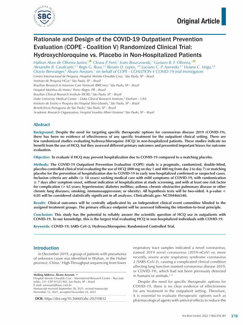
placebo-controlled clinical trial with an allocation ratio of 1:1. The study will evaluate the potential antiviral effects of an early treatment with HCQ (800 mg PO on day 1 and 400 mg PO from day 2 to day 7) vs. matching placebo for the prevention of hospitalization due to COVID-19-related complications in non-hospitalized confirmed or suspected COVID-19 cases. The planned study workflow is shown in Figure 1. This protocol follows the Standard Protocol Items: Recommendations for Interventional Trials (SPIRIT) statement (Supplementary Table 1).
Study SettingThe study will be conducted in 56 centers across the
Brazilian geographical regions. Centers are both private and public outpatient health care services which have been approved to participate after favorable feasibility assessment, compliance with good clinical practices, and ethics approval.
Figure 1 - CONSORT diagram showing the study workflow and planned recruitment.
379

Arq Bras Cardiol. 2022; 118(2):378-387
Original Article
Oliveira Junior et al.Hydroxychloroquine in outpatients with COVID-19
Primary ObjectiveWe aim to assess whether treatment with HCQ is associated
with reduced requirement for hospitalization due to a clinical reason related to confirmed or suspected COVID-19 within 30 days of randomization in the outpatient setting. Hospitalization is defined as hospital stay for a period ≥ 24h or at least one additional day of adjudicated hospitalization. The hospitalization criteria will follow local clinical practice of each participating site.
Secondary ObjectivesTo assess the effect of treatment with HCQ compared to
placebo in outpatients with suspected or confirmed COVID-19 on the following outcomes at 30 days of follow-up:
1. Uncontrolled asthma after ≥ 5 days of starting study medication: Affirmative answer in three or four items of the Global Initiative for Asthma (GINA) questionnaire, described in Supplementary Table 2;
2. Pneumonia: Defined by clinical-radiological criteria, which include a history of cough and one or more of the following signs/symptoms: sputum, dyspnea, chest pain, sweating or fever (> 37.8o C) + chest computed tomography scan showing unilateral or bilateral ground-glass opacity, focal consolidations, or mixed opacities (including reverse halo sign);
3. Otitis media: Defined by clinical criteria of fever (> 37.8o C) and otalgia + bulging of the tympanic membrane;
4. Fever resolution time: Day 0 of fever resolution will be defined as the first afebrile day (< 37.5o C) after enrolment into the study followed by at least two consecutive days. Temperature will be obtained through participant report in the patient’s diary;
5. Time to improvement of respiratory symptoms (cough, runny nose);
6. Hospitalization in an intensive care unit (ICU): Admission to ICU due to clinical reasons related to COVID-19;
7. Need for orotracheal intubation: Clinical need as assessed by the attending physician;
8. Mechanical ventilation time: Number of days on mechanical ventilation until extubation or death;
9. Mortality: Death due to any cause that occurs within 30 days of randomization;
We will also assess clinical safety within 30 days of randomization:
1. Hypoglycemia: Change in the frequency of hypoglycemic episodes in diabetic patients using hypoglycemic medication, perceived by clinical signs or symptoms or measured in a capillary or venous blood glucose device;
2. Palpitations: Patient perception of heartbeats that can possibly be diagnosed as arrhythmias in patients without known history of prolonged QTc interval or pre-existing heart disease;
3. Reduced visual acuity: Change in visual acuity or new diagnosis of retinal disease not previously documented;
4. Diarrhea: Change in bowel habit greater than three diarrheal episodes per day during HCQ use and three days after the end of treatment;
5. Anorexia: Change in appetite during HCQ use and three days after the end of treatment;
6. Emotional lability: Perception of change in emotional lability (mood swings) during HCQ use and three days after the end of treatment.
Exploratory objectivesTo assess the effect of treatment with HCQ compared to
placebo in outpatients with suspected or confirmed COVID-19 on the following outcomes:
1. Time to hospitalization after randomization;
2. Assessment of patient’s clinical status at the time of hospitalization.
Eligibility CriteriaCOVID-19 case definition:
• The initial clinical evaluation of patients and their study eligibility screening will be assessed based on the classification of confirmed and suspected cases, developed according to the Brazilian Ministry of Health Guidelines and World Health Organization recommendations13,14 on the definition of cases, modified to the outpatient setting (Table 1). Inclusion and exclusion criteria are described in Table 2, including those related to cardiovascular safety as HCQ may increase the QTc interval.15
Randomization Method and Allocation Concealment Randomization (1:1) will be generated by a web-
based software and performed in permuted blocks of size 8. Concealment of the randomization list is maintained through a 24-hour, centralized, automated, internet-based randomization system.
BlindingPatients, investigators, and health care providers will be
blinded to allocation of study drugs. Clinical outcomes will be assessed under blinded fashion by the Clinical Event Adjudication Committee.
Trial InterventionsBoth arms will receive usual care according to local
practice, which basically comprises general advice and medications for symptom relief. Standard support and care measures are defined as any treatments other than the study medications that are necessary for the care of the patient with COVID-19, at the attending physician’s discretion.
Patients in the HCQ group will receive a dose of 400 mg BID on day 1 and a dose of 400 mg OD from day 2 to day 7. Patients in the matching placebo group will follow the same regimen of administration.
380

Arq Bras Cardiol. 2022; 118(2):378-387
Original Article
Oliveira Junior et al.Hydroxychloroquine in outpatients with COVID-19
Table 1 - COVID-19 case definition for the COPE study (COALITION V) adapted according to Brazilian Ministry of Health and World Health Organization guidelines
COVID-19 status Definition
Confirmed
• Individual with laboratory confirmation of COVID-19 (RT-PCR SARS-CoV-2 virus detection), preferably collected between the 4th and 7th days of symptom onset by nasopharyngeal/oropharyngeal swabs, regardless of signs and symptoms.
• Immunological test (rapid test or classic serology to detect IgM/IgG antibodies) in a sample collected after the 7th day of symptom onset, analyzed by a validated method.
Suspected
Patient who meets at least one of the following criteria*:
• Patient with acute respiratory disease (fever AND at least one sign/symptom of respiratory disease, for example, cough or dyspnea) AND a travel history or residing in a location that reports community transmission of COVID-19 during the 14 days prior to the onset of symptoms;
• Patient with acute respiratory disease AND having been in contact with a confirmed or probable case (with acute respiratory disease without laboratory confirmation) of COVID-19 in the 14 days prior to the onset of symptoms.
* Depending on patient’s clinical status, these criteria can be complemented by radiological findings (interstitial infiltration in chest radiograph and/or ground-glass opacity in lung computed tomography scan). It should be noted that we will mostly treat patients with mild symptoms who do not have a clinical indication for an imaging test.
Outcome Assessment and Follow-upThere will be two telephone contacts with the participants
(7 and 30 days) to evaluate adherence, symptoms, and the need to seek medical attention in order to detect possible disease progression or adverse events resulting from the treatment.
Treatment InterruptionIn patients with suspected infection, the following
procedure will be done if there is confirmation of negative COVID-19 tests:
• When testing for SARS-CoV-2 (reverse transcription polymerase chain reaction, RT-PCR) is performed at the hospital where the patient is randomized, the center’s principal investigator will obtain this information and send it to the coordinating center (Hospital Alemão Oswaldo Cruz International Research Center). The patient will be advised to stop treatment and will continue to be monitored until the end of follow-up at 30 days.
• When testing for SARS-Cov-2 (RT-PCR) is performed in another laboratory, the study participant will contact the center where the patient is randomized, which will then inform the coordinating center. The patient will be advised to stop treatment or not accordingly, with follow-up being continued for up to 30 days.
The study will be interrupted in case of clear benefit of the intervention for the primary outcome or in case of an increase in the frequency of serious adverse events. The Data and Safety Monitoring Board (DSMB) will closely monitor any occurrence of unforeseen adverse events and all serious
adverse events to, if necessary, recommend the study end to ensure the safety of the patients.
Adverse Event Report and ManagementAdverse events are not considered to be a study endpoint
in this protocol, except for adverse events classified as serious (hospitalization due to COVID-19 and death).
Adverse events will be actively collected from the moment the study participant signs the informed consent form. The information collected should include data from the patient’s clinical history and comorbidities, diagnosis of the event (based on signs and symptoms), classification of severity, start date, definition of the likelihood of causal relationship, as well as cause of the event according to the investigator, medical decision, patient evolution regarding adverse outcomes, criteria used to classify the severity of the event, and end date.
Endpoint Reporting and AdjudicationPrimary endpoint will be assessed by research physicians
with previous and current experience in validating clinical events using international standards. Hospitalizations within 30 days due to COVID-19-related causes will be documented by the medical team. The information will be collected for analysis by the Clinical Event Adjudication Committee under confidential allocation (blinded fashion for clinical event assessment) following standardized criteria. The DSMB will assess the effects of HCQ compared to placebo treatment for the primary outcome measure (hospitalization within 30 days) and adverse events (occurring up to 7 and 30 days) requiring medical attention and/or hospitalization.
381

Arq Bras Cardiol. 2022; 118(2):378-387
Original Article
Oliveira Junior et al.Hydroxychloroquine in outpatients with COVID-19
Table 2 - Inclusion and exclusion criteria
General Criteria
Adults (> 18 years) seeking medical care with suspected or confirmed COVID-19, with ≤ 7 days from symptom onset, presenting with mild symptoms without clear indication for hospitalization and at least one of the following risk factors for clinical complications:
Inclusion Criteria
1. Age > 65 years;
2. Hypertension;
3. Diabetes mellitus;
4. Asthma;
5. Chronic obstructive pulmonary disease or other chronic lung diseases;
6. Smoking;
7. Immunosuppression;
8. Obesity (defined as body mass index ≥ 30 kg/m2).
Exclusion Criteria
1. Immediate hospitalization after first medical care;
2. Positive test for influenza at the first medical care;
3. Known hypersensitivity to hydroxychloroquine/chloroquine;
4. Previous diagnosis of retinopathy or macular degeneration;
5. Previous diagnosis of long QT syndrome, history of sudden death in close family members (parents and siblings), decompensated heart failure, unstable coronary artery disease, use of antiarrhythmic drugs or other drugs that can increase the bioavailability of hydroxychloroquine or enhance its effect;
6. Evidence of known liver disease, reported by the patient;
7. Evidence of known chronic kidney disease, reported by the patient;
8. Patients with pancreatitis;
9. Baseline electrocardiogram with QTc interval ≥ 480 ms;
10. Chronic use of hydroxychloroquine/chloroquine for other reasons;
11. Pregnancy.
Data Collection and Management
Data will be collected through an electronic case report form (eCRF) by Internet and will be entered into the eCRF by each participating site. Training and support for using the system will be made available to researchers by the coordinating center.
Data will be collected directly from the patient and/or family. We will apply several procedures to ensure data quality (Table 3). Data to be collected during study visits include:
1. Admission (baseline):
a. Age, sex, marital status, ethnicity, educational level, family income, and comorbidities;
b. Results of molecular or serology tests for COVID-19 (depending on time from symptom onset/clinical diagnosis);
c. Use of concomitant medications at baseline;
d. Duration of symptoms;
2. At 7 days post-randomization:a. Safety assessment (adverse event monitoring);b. Medication adherence;
3. At 30 days post-randomization:a. Efficacy assessment (need for hospitalization);b. Safety assessment (adverse event monitoring);
The scheme for data collection and participant follow-up is shown in Figure 2.
Statistical AnalysisSample Size CalculationWe will assume that the primary outcome (hospitalization)
will occur in 20% of individuals in the placebo group and 14% of individuals in the HCQ group, which corresponds to a relative risk reduction of 30%. This assumption was based on initial clinical experience locally in the first months of the pandemic, in which 20% of individuals without any intervention were hospitalized after initial medical care. The
382

Arq Bras Cardiol. 2022; 118(2):378-387
Original Article
Oliveira Junior et al.Hydroxychloroquine in outpatients with COVID-19
choice of treatment effect (a relative risk reduction of 30%) was based on reasonable plausibility, with most benefits consisting of moderate treatment effects (20-30%). It was estimated that a sample of 1230 individuals (615 per group) would provide 80% statistical power to detect this reduction at a significance level of 5% using the chi-square test, assuming a two-sided significance hypothesis, and considering a 1:1 allocation. The estimated dropout rate was 5% in each group, which would result in 1296 individuals (648 per group). Therefore, we decided that 1300 individuals (650 per group) would be randomized into the study. The sample size was calculated using SAS 9.4 (PROC POWER procedure).16,17
Study Populations
The assessment of the primary outcome (hospitalization within 30 days) will be performed following the intention-to-treat (ITT) principle, which will consist of all randomized cases. A modified intention-to-treat (mITT) analysis will also be performed after exclusion of COVID-19 negative-tested cases.
General Descriptive Statistics
Statistical analyses will be performed after resolving all inconsistencies, making data quality control, and locking the database. Baseline demographic and clinical characteristics will be expressed as count and percentage, mean and standard
Table 3 - Steps to ensure quality on data collection and management
Item Definition
Initiation visitAll researchers will participate in a site initiation visit (web-based training) before the start of the study to ensure consistency of study procedures, including collection of the data.
Contact Researchers will be able to call the Study Coordinating Center to solve issues or problems that may arise.
Data cleaningData cleaning to identify inconsistencies will be conducted periodically (approximately every 15 days). Centers will be notified about inconsistencies to provide correction.
Statistical validationStatistical techniques for identifying errors will be carried out during the study. These analyses will include identification of missing data, inconsistent data, protocol deviations, adverse events reported incorrectly (data considered inconsistent with centralized medical review), and systematic error assessment.
Data reviewThe Coordinating/Sponsoring Center will review detailed reports monthly on screening, inclusion, follow-up, consistencies, and completeness of the data, and will take immediate action to solve any problems.
Figure 2 - Data gathering and participant follow-up scheme.
383

Arq Bras Cardiol. 2022; 118(2):378-387
Original Article
Oliveira Junior et al.Hydroxychloroquine in outpatients with COVID-19
deviation, or median and interquartile range, whenever appropriate. Safety analyses will consider the safety outcomes, and the study participants will be classified in treatment groups according to the medication they actually used. The reasons for discontinuing the study will be listed for each treatment group.
The incidence of adverse events will be summarized and presented by treatment group. Adverse events will be summarized according to severity and intensity by treatment group. Serious adverse events or events that lead to treatment interruption will be listed by study participant. A comparison of adverse events between the two treatment groups will be performed using chi-square test or Fisher exact test.
Primary Outcome AnalysisThe effect of the intervention on the primary outcome and
on binary secondary outcomes will be estimated with risk ratio (RR) and 95% confidence interval (CI). Chi-square test or Fisher exact test will be employed for hypothesis testing. The same will be done for the secondary outcomes defined by proportions.
Primary outcome will be also assessed with a mixed logistic regression model using a mixed-effects model with randomized group as fixed effect and site as random effect. Odds ratio with 95% CI will be reported.
Secondary Outcome AnalysisSecondary outcomes defined by quantitative variables of
normal and asymmetric distribution will be compared between the two groups (HCQ vs. placebo) using unpaired Student t-test or Mann-Whitney test for non-normally distributed variables.
The effect of the intervention on mortality at 30 days will be evaluated using the Cox regression model. If the phenomenon of monotone likelihood occurs or a rare number of events is observed, Firth penalized partial likelihood approach in a univariate Cox regression model will be applied.
The effect of intervention on hospitalization-free survival at 30 days will be assessed by applying the univariate Cox regression model. Hospitalization-free survival at 30 days will be constructed using the Kaplan-Meier method, and the log-rank test will be used to assess differences between curves. Hazard ratio with 95% CI will be reported.
Proportional hazard assumptions will be checked using cumulative sums of Martingale residuals and a Kolmogorov-type supremum test based on a sample of 1000 simulated residual patterns18.
Interim AnalysesThree interim analyses to assess safety and effectiveness
wil be performed when the sample size reaches 25% (325 individuals), 50% (650 individuals), and 75% (975 individuals) using the Haybittle–Peto approach.19 Safety will be assessed at 7 and 30 days, and effectiveness at 30 days, in separate blocks. As for the decision rule, in the safety assessment, the study may be interrupted, according to the Haybittle–Peto method, if there is a sign of harm (severe
cardiac arrhythmia, sudden death, retinopathy at 7 days) with a p < 0.01 (in each interim analysis). The percentage of patients to be analyzed in the safety assessment at 7 days is 25%, which corresponds to 325 individuals. The first interim analysis is foreseen at this point, as described above. In the evaluation of effectiveness, the study may be interrupted, according to the Haybittle–Peto method, if there is a sign of benefit (primary outcome within 30 days of randomization) with a p < 0.001 (in each interim analysis).
The Haybittle–Peto boundary is a conservative stopping rule at interim analysis that has minimal impact on increasing type I error in two-arm trials.20 There will be no adjustments in the final threshold for statistical significance for sequential analysis.
We predict that, during three interim analyses, the proportion of confirmed negative-tested cases and those who have not been tested will be evaluated to estimate the need for sample recalculation to ensure adequate statistical power. According to the proportion of non-positive COVID-19 cases, sampling replacement will be considered in order to guarantee 80% statistical power in the sample effectively analyzed for the primary endpoint.
Analyses will be conducted using complete data. Additionally, the proportion of untested individuals in each group will be reported.
All of the prespecified interim analyses were carried out by an independent DSMB, which recommended the study continuation as planned after formal confidential reviews and official letters to the COPE Study Steering Committee.
Sensitivity and Subgroup AnalysisExploratory analyses for the primary outcome will be done
considering effect of the intervention within prespecified subgroups using stratified analyses and interaction tests. These interaction tests will be based on binary logistic regression models that include the treatment effect, the factor of interest, and an interaction term between the two variables, with reporting of the p-value for the interaction term.
Analyses will be conducted using the complete-case analysis principle. Additionally, for the primary outcome, if the proportion of missing data is greater than 5%, a sensitivity analysis will be conducted using a multiple data imputation technique.21,22
All hypothesis tests will be two-sided. A p-value < 0.05 will be considered statistically significant in all analyses. Analyses will be done using SAS, version 9.4 (SAS Institute Inc, Cary, NC, USA).
Database LockDatabase lock will be carried out after completion of
the 30-day follow-up for all patients, and data cleaning will be done after clinical monitoring. All interim analyses will be made available to local regulatory agencies in Brazil. Database access will be granted only to the Steering Committee members and statisticians before the main results are published.
384

Arq Bras Cardiol. 2022; 118(2):378-387
Original Article
Oliveira Junior et al.Hydroxychloroquine in outpatients with COVID-19
Trial OversightThe Executive/Steering Committee is responsible for the
general supervision of the study, development of the study protocol, and manuscript writing. All other study committees report to the Executive/Steering Committee.
The DSMB will assess the effects of HCQ compared to usual care on the primary endpoint (hospitalization within 30 days) and adverse events (within 7 and 30 days) requiring medical care and/or hospitalization. The rules for early discontinuation of the trial will be applied to the primary objective (efficacy) as well as to serious adverse events (safety). The Committee will monitor any serious adverse event and recommend treatment interruption, if necessary, to ensure patient safety.
Ethics and DisseminationRecords of all study participants will be kept confidential
and will be accessed in a restricted way only by people formally related to the study, who will transfer the clinical information to specific forms (which do not contain information that can identify individuals) and verify whether the study is being conducted properly. The electronic data collection form will contain the patient study ID and the corresponding site ID. Each study patient or legal representative must provide written consent in accordance with local requirements, after the risks/benefits and study procedures have been fully explained.
This study was approved by the Brazilian National Research Ethics Commission (CONEP) and National Health Surveillance Agency (ANVISA). It is registered with the Brazilian Clinical Trial Network (REBEC) with registration number RBR-3cbs3w and in clinicaltrial.gov with identification number NCT04466540. All amendments to the protocol must be approved by the institutional review board/CONEP system before implementation by the participating sites.
The site investigator and study team will conduct the study in accordance with the principles of the Helsinki Declaration, following the principles of good clinical practice as well as all applicable regulatory and internal policies and procedures.
This study will be submitted for publication regardless of its results after completion. It will also be disseminated as requested by local authorities.
Author ContributionsConception and design of the research: Oliveira Junior HA,
Ferri CP; Boszczowski I, Oliveira GBF, Rosa RG, Lopes RD, Veiga VC, Berwanger O, Avezum A, Cavalcanti AB; Acquisition of data: Oliveira Junior HA, Boszczowski I, Oliveira GBF, Avezum A; Analysis and interpretation of the data: Oliveira Junior HA, Oliveira GBF, Avezum A; Obtaining financing: Oliveira Junior HA, Oliveira GBF, Avezum A; Writing of the manuscript: Oliveira Junior HA, Ferri CP, Oliveira GBF, Avezum A; Critical revision of the manuscript for intellectual content: Oliveira Junior HA, Ferri CP, Oliveira GBF, Rosa RG, Lopes RD, Veiga VC, Berwanger O, Avezum A, Cavalcanti AB.
Potential Conflict of Interest No potential conflict of interest relevant to this article was
reported.
Sources of Funding This study was partially funded by E.M.S Farmacêutica.
Study Association This study is not associated with any thesis or dissertation
work.
1. Xu XW, Wu XX, Jiang XG, Xu KJ, Ying LJ, Ma CL, et al. Clinical Findings in a Group of Patients Infected with the 2019 Novel Coronavirus (SARS-Cov-2) Outside of Wuhan, China: Retrospective Case Series. BMJ. 2020;368:m606. doi: 10.1136/bmj.m606.
2. World Health Organization. Clinical Management of Severe Acute Respiratory Infection When Novel Coronavirus (nCoV) Infection is Suspected. Geneva: WHO Library; 2020.
3. Huang C, Wang Y, Li X, Ren L, Zhao J, Hu Y, et al. Clinical Features of Patients Infected with 2019 Novel Coronavirus in Wuhan, China. Lancet. 2020;395(10223):497-506. doi: 10.1016/S0140-6736(20)30183-5.
4. Li B, Yang J, Zhao F, Zhi L, Wang X, Liu L, et al. Prevalence and Impact of Cardiovascular Metabolic Diseases on COVID-19 in China. Clin Res Cardiol. 2020;109(5):531-38. doi: 10.1007/s00392-020-01626-9.
5. Abella BS, Jolkovsky EL, Biney BT, Uspal JE, Hyman MC, Frank I, et al. Efficacy and Safety of Hydroxychloroquine vs Placebo for Pre-Exposure SARS-CoV-2 Prophylaxis Among Health Care Workers: A Randomized Clinical Trial. JAMA Intern Med. 2021;181(2):195-202. doi: 10.1001/jamainternmed.2020.6319.
6. Rajasingham R, Bangdiwala AS, Nicol MR, Skipper CP, Pastick KA, Axelrod ML, et al. Hydroxychloroquine as Pre-exposure Prophylaxis for Coronavirus Disease 2019 (COVID-19) in Healthcare Workers: A Randomized Trial. Clin Infect Dis. 2021;72(11):835-43. doi: 10.1093/cid/ciaa1571.
7. Barnabas RV, Brown ER, Bershteyn A, Stankiewicz Karita HC, Johnston C, et al. Hydroxychloroquine as Postexposure Prophylaxis to Prevent Severe Acute Respiratory Syndrome Coronavirus 2 Infection: A Randomized Trial. Ann Intern Med. 2021 Mar;174(3):344-52. doi: 10.7326/M20-6519.
8. Boulware DR, Pullen MF, Bangdiwala AS, Pastick KA, Lofgren SM, Okafor EC, et al. A Randomized Trial of Hydroxychloroquine as Postexposure Prophylaxis for Covid-19. N Engl J Med. 2020;383(6):517-25. doi: 10.1056/NEJMoa2016638.
9. Mitjà O, Corbacho-Monné M, Ubals M, Alemany A, Suñer C, Tebé C, et al. A Cluster-Randomized Trial of Hydroxychloroquine for Prevention of Covid-19. N Engl J Med. 2021;384(5):417-27. doi: 10.1056/NEJMoa2021801.
10. Mitjà O, Corbacho-Monné M, Ubals M, Tebé C, Peñafiel J, Tobias A, et al. Hydroxychloroquine for Early Treatment of Adults With Mild Coronavirus Disease 2019: A Randomized, Controlled Trial. Clin Infect Dis. 2021;73(11):4073-81. doi: 10.1093/cid/ciaa1009.
11. Skipper CP, Pastick KA, Engen NW, Bangdiwala AS, Abassi M, Lofgren SM, et al. Hydroxychloroquine in Nonhospitalized Adults with Early COVID-19: A Randomized Trial. Ann Intern Med. 2020;173(8):623-31. doi: 10.7326/M20-4207.
References
385

Arq Bras Cardiol. 2022; 118(2):378-387
Original Article
Oliveira Junior et al.Hydroxychloroquine in outpatients with COVID-19
12. Omrani AS, Pathan SA, Thomas SA, Harris TRE, Coyle PV, Thomas CE, et al. Randomized Double-Blinded Placebo-Controlled Trial of Hydroxychloroquine with or without Azithromycin for Virologic Cure of Non-Severe COVID-19. EClinicalMedicine. 2020;29:100645. doi: 10.1016/j.eclinm.2020.100645.
13. Brasil. Ministério da Saúde. Diretrizes para Diagnóstico e Tratamento da COVID-19 (Clinical Guideline Covid-19) [Internet]. Brasília: Ministério da Saúde. 2020 [cited 2021 Mar 18]. Available from: https://portaldeboaspraticas.iff.fiocruz.br/atencao-mulher/diretrizes-para-diagnostico-e-tratamento-da-covid-19-ms/.
14. World Health Organization. Country & Technical Guidance - Coronavirus Disease (COVID-19) [Internet]. Geneva: WHO. 2021 [cited 2021 Mar 18]. Available from: https://www.who.int/emergencies/diseases/novel-coronavirus-2019/technical-guidance.
15. Costa IBSDS, Bittar CS, Rizk SI, Araújo Filho AE, Santos KAQ, Machado TIV, et al. The Heart and COVID-19: What Cardiologists Need to Know. Arq Bras Cardiol. 2020;114(5):805-16. doi: 10.36660/abc.20200279.
16. Ryan TP. Sample Size Determination and Power. New Jersey: Wiley; 2013.
17. Shein-Chung C, Wang JSH. Sample Size Calculations in Clinical Research, Second Edition (Chapman & Hall/CRC Biostatistics Series). 2nd ed. Boca Raton: Chapman & Hall/CRC Biostatistics Series; 2008.
18. Lachin JM, Matts JP, Wei LJ. Randomization in Clinical Trials: Conclusions and Recommendations. Control Clin Trials. 1988;9(4):365-74. doi: 10.1016/0197-2456(88)90049-9.
19. Haybittle JL. Repeated Assessment of Results in Clinical Trials of Cancer Treatment. Br J Radiol. 1971;44(526):793-7. doi: 10.1259/0007-1285-44-526-793.
20. Blenkinsop A, Parmar MK, Choodari-Oskooei B. Assessing the Impact of Efficacy Stopping Rules on the Error Rates Under the Multi-Arm Multi-Stage Framework. Clin Trials. 2019;16(2):132-41. doi: 10.1177/1740774518823551.
21. Raghunathan T, Berglund PA, Solenberger PW. Multiple Imputation in Practice: With Examples Using IVEware. Milton Park: Taylor & Francis; 2018.
22. Patricia Berglund SGH. Multiple Imputation of Missing Data Using SAS. Cary: SAS Institute; 2014.
*Supplemental MaterialsFor additional information, please click here.
386

Arq Bras Cardiol. 2022; 118(2):378-387
Original Article
Oliveira Junior et al.Hydroxychloroquine in outpatients with COVID-19
This is an open-access article distributed under the terms of the Creative Commons Attribution License
387

Arq Bras Cardiol. 2022; 118(2):388-397
Original Article
Eating Behavior of Older Adults with and Without Diabetes: The Vigitel Survey, Brazil, 2016Daniela de Assumpção,1 Ana Maria Pita Ruiz,1 Flavia Silva Arbex Borim,1,2 Anita Liberalesso Neri,1 Deborah Carvalho Malta,3 Priscila Maria Stolses Bergamo Francisco1
Universidade Estadual de Campinas (Unicamp) - Programa de Pós-Graduação em Gerontologia,1 Campinas, SP - BrazilUniversidade de Brasília (UnB) - Departamento de Saúde Coletiva - Escola de Ciências da Saúde,2 Brasília, DF - BrazilUniversidade Federal de Minas Gerais (UFMG) - Departamento de Enfermagem Materno-Infantil e Saúde Pública,3 Belo Horizonte, MG - Brazil
Mailing Address: Daniela de Assumpção •Rua Tessália Vieira de Camargo, 126. Postal Code 13083-887, Cidade Universitária, Zeferino Vaz, Campinas, SP - BrazilE-mail: [email protected] received November 11, 2020, revised manuscript February 10, 2021, accepted March 24, 2021
DOI: https://doi.org/10.36660/abc.20201204
Abstract
Background: A healthy diet is a protection factor against type 2 diabetes and plays an important role in the treatment of the disease, as well as associated comorbidities.
Objective: Characterize the eating habits of older adults (≥ 65 years) with and without diabetes residing in capital cities and the Federal District of Brazil.
Methods: A cross-sectional study was conducted using data from the Surveillance of Risk and Protection Factors for Chronic Diseases Through a Telephone Survey (Vigitel, 2016). The prevalence of diabetes mellitus was estimated according to sociodemographic variables, physical inactivity level, self-rated health status and body mass index. Dietary habits were assessed based on the frequency (weekly and daily) of consumption of healthy and unhealthy foods and the replacement of food by snacks. Differences were determined using Pearson’s chi-square test (Rao-Scott), with the significance level set at 5%.
Results: A total of 13,649 older adults were interviewed. The prevalence of self-reported diabetes was 27.2% (95% CI: 25.5; 29.0). Compared to non-diabetics, diabetic individuals had a higher consumption of raw vegetables (32.1% vs. 26.5%/3-4 days/week) and lower consumption of chicken (3.8% vs. 6.4%/hardly ever/never), fruit juice (24.0% vs. 29.6%) and sweets (6.8% vs. 16.2%) ≥ 5 days/week. Compared to non-diabetics, diabetic individuals consumed more skim milk (51.5% vs. 44.6%) and diet soda (60.0% vs. 17.3%) ≥ 5 days/week, raw vegetables (9.1% vs. 2.5%/at dinner) and sweets (37.7% vs. 20.5%/twice/day) 3-4 days/week.
Conclusion: The observed differences emphasize the need for healthy eating interventions for all older adults, as well as specific counseling for those with diabetes.
Keywords: Aged; Diabetes Mellitus; Eating; Health Surveys.
Introduction On the global scale, the number of individuals with diabetes
increased from 108 million in 1980 to 422 million in 2014. Taking into account the increase and aging of the population, this represents an increase of nearly 40%.1 In 2019, Brazil occupied the 5th position (16.8 million) among the 10 countries with the highest number of diabetic individuals and 3rd position among those that spent most on the treatment of the disease (52.3 billion US dollars).2
The 2013 National Health Survey estimated the prevalence of diabetes in the adult population (≥ 18 years) in Brazil to be
6.2%, with the disease exerting a greater effect on individuals with no schooling or an incomplete elementary school education, as well as those with excess weight, hypertension and high cholesterol/triglyceride levels.3 The prevalence among older adults (≥ 65 years) was 19.8%,3 which indicates the impact of the disease on direct and indirect costs for healthcare services, society and affected invididuals.4
A diet based on whole grains, leafy vegetables, fruits, legumes, seeds/vegetable oils rich in omega-6 and dairy products with a low fat content, as well as restricted quantities of red/processed meats, refined grains, sweets and sweetened beverages plays an important role in the prevention and management of diabetes.5 Evidence indicates a lower incidence of diabetes with the increase in the consumption of whole grains and bran and a greater incidence with the increase in the consumption of red/processed meats, bacon and sweetened beverages.6 Results from the 2008-2010 and 2012-2014 Brazilian Longitudinal Study of Adult Health] [Estudo Longitudinal de Saúde do Adulto (ELSA)] show that the high consumption of processed meats increased the likelihood of new cases of insulin resistance by 68% in men
388

Arq Bras Cardiol. 2022; 118(2):388-397
Original Article
Assumpção et al.Eating behavior and diabetes in older adults
(> 27.1 g/day) and 23% in women (> 20.7 g/day) and the high consumption of red meat (> 101.9 g/day) increased the risk of diabetes by 40% in men.7
An analysis of factors associated with diabetes in Brazilian adults (≥ 18 years) revealed no differences in dietary habits (consumption of fatty red meats and recommended intake of fruits and vegetables) between those who reported having or not having the disease.3 At a reference institution for the treatment of diabetes that received individuals from the public healthcare system, the “traditional Brazilian” diet, which is characterized by the consumption of rice, beans, chicken and regional foods, was negatively correlated with blood sugar levels.8
As healthy eating is a protection factor against chronic noncommunicable diseases, including type 2 diabetes, and plays an important role in the treatment of diabetes, as well as associated comorbidities (e.g., hypertension, obesity and dyslipidemia), the aim of the present study was to characterize the eating habits of older adults (≥ 65 years) with and without diabetes residing in capital cities and the Federal District of Brazil.
Methods
Study designA population-based cross-sectional study was conducted
using data from telephone interviews held in 2016.
ContextThe Surveillance of Risk and Protection Factors for Chronic
Diseases Through a Telephone Survey (Vigitel) has occurred annually since 2006 in state capitals and the Federal District of Brazil. The aim of the Vigitel survey is to monitor the frequency and distribution of the main factors that determine chronic noncommunicable diseases in the country.9
ParticipantsThe present study involved older adults (≥ 65 years)
interviewed during the 2016 Vigitel survey. The choice of this age group was based on the increase in life expectancy, sociodemographic and political changes in Brazil since the decree of the Older Adult Statute in 2003 and the possibility of comparing the results with those of international studies.10
The participants in the 2016 Vigitel survey were selected through probabilistic sampling of adults (≥ 18 years) residing in households with at least one residential telephone line in 2016. The two-stage sampling was performed. The first stage consisted of a systematic drawing (stratified by postal code) of at least five thousand telephone lines in each capital city using the electronic records of landlines of telephone companies. The selected lines were submitted to another drawing and divided into replicates of 200 lines, with each replicate reproducing the same proportion of lines per postal code of the original records. The second stage consisted of the drawing of one of the adults residing in the selected household after the identification of the eligible active residential lines.9
Vigitel conducts approximately two thousand interviews in each city, which enables estimating the frequency of the main risk factors for chronic diseases in the adult population with a 95% confidence interval and maximum error of two percentage points. Each interviewee receives a post-stratification weight to enable the statistical inference of the results for the adult population in each city. Weighting ensures the linking of the sociodemographic composition estimated for the adult population with a telephone line in each capital city to that estimated for the total adult population of the same city in terms of gender, age group and level of schooling.9
VariablesThe following characteristics were selected to characterize
the sample: a) Sociodemographic: gender (male, female); age (in years);
level of schooling (0-4, 5-8 and ≥ 9 years of study);b) Physical inactivity level at leisure and work and when
commuting to work (yes, no);c) Self-rated health status (very good/good, fair and poor/
very poor);d) Body mass index (BMI = weight [kg]/height [m]2), calculated
from thr reported information and classified as underweight (< 22 kg/m2), ideal weight range (22 to ≤ 27 kg/m2) and excess weight (BMI > 27 kg/m2) based on the criteria of the Nutrition Screening Initiative.11
Data source and measurement Older adults who answered yes to the following question
were considered to be diabetic: “Has any physician ever told you that you have diabetes?” (yes or no). Dietary habits were assessed based on the frequency of the consumption of healthy and unhealthy foods, as well as the replacement of food for by snacks. The consumption of vegetables (raw and cooked), fruits, natural fruit juice, beans, milk and chicken was considered healthy. The consumption of red meat (beef, pork and goat), fatty meats (red meat with visible fat and chicken with skin), whole milk, sweets, sweetened beverages (soda and artificial juice) and the habit of replacing a main meal (lunch or dinner) by snacks were considered unhealthy. Food intake frequencies were categorized as hardly ever/never, 1-2, 3-4 and ≥ 5 days per week. Regular consumption corresponded to ≥ 5 days/week.
Data analysis The prevalence of Diabetes mellitus was estimated with
its respective 95% confidence intervals (CI) according to the variables selected for sample characterization. The analyses were stratified by level of schooling and age to determine the influence of these characteristics on dietary habits. The distribution of relative frequencies of food intake, as well as of weekly consumption according to the type of food, daily intake and the habit of consuming fatty meats were presented for older adults with and without diabetes. The differences between proportions were determined using Pearson’s chi-square test (Rao-Scott) with the significance level set at 5% (p > 0.05). The statistical analyses were performed with the aid
389

Arq Bras Cardiol. 2022; 118(2):388-397
Original Article
Assumpção et al.Eating behavior and diabetes in older adults
of the Stata program, version 15.1, considering the complex sampling design.
Ethical considerationsThe participants received clarifications regarding the
objectives of the Vigitel survey during telephone contact. Written consent was substituted by verbal consent. This study received approval from the National Human Research Ethics Committee of the Health Ministry (certificate number355.590, of June 26, 2013).
Results Among of 18,854 older adults (≥ 60 years of age)
interviewed, 5,205 were excluded from the present study for being younger than 65 years of age. Thus, data from 13,649 older adults were analyzed, 3,349 of whom were diabetic and 10,300 were not diabetic (Figure 1). The prevalence of self-reported diabetes was 27.2% (95%CI : 25.5 to 29.0). Mean age was 73.6 years (95%CI: 73.2 to 74.1) among those with diabetes and 73.8 years (95%CI: 73.5 to 74.0) among those without diabetes. Compared to non-diabetics, individuals with diabetes had greater proportions of physical inactivity (p = 0.015), worse self-rated health status and excess weight (p < 0.001). The diabetics also had a lower proportion of schooling with nine or more years of study (p < 0.001) (Table 1).
The results of the analysis of regular food intake stratified by schooling (0-8 and ≥ 9 years) in diabetic and non-diabetic older adults are displayed in Table 2. Lower regular consumption of reduced-fat/skim milk and greater consumption of raw vegetables at lunch and dinner were found among diabetics with low level of schooling. Among non-diabetics, a higher proportion of those with up to eight years of schooling consumed whole milk, fatty red meat and raw vegetables at dinner five or more days/week (Table 2).
Significant differences between diabetics and non-diabetics were found for the intake frequency of raw vegetables, chicken, natural fruit juice and sweets (Table 3). The percentage of diabetics that hardly ever/never consumed chicken was lower than that found in the non-diabetics. Milk intake (1-2 days/week) was lower among diabetics. The consumption of natural fruit juice and sweets (≥ 5 days/week) was lower among diabetics. The majority of diabetics did not consume sweets, but nearly 15.0% included these foods in their diet (≥ 3 days/week). No significant difference was found regarding the replacement of meals by snacks, but approximately 20.0% of the diabetics ate snacks for dinner.
Table 4 displays the weekly intake frequencies in the diabetic and non-diabetic groups according to the characteristics of the foods and daily intake frequency. Diabetics had lower percentages of the consumption of whole milk and higher percentages of skim milk consumption compared to non-diabetics. The consumption of diet soda was higher among the diabetics. Considering a weekly frequency of 3-4 times, greater intakes of raw vegetables (dinner) and sweets (twice per day) were found in the diabetics.
DiscussionThe present study sought to characterize the eating habits
of older adults with and without diabetes residing in state capitals and the Federal District of Brazil. Among the findings, diabetic individuals with low level of schooling had a lower proportion of the regular consumption of reduced-fat/skim milk and a higher proportion of the regular consumption of raw vegetables at lunch and dinner.
A cross-sectional study conducted in 2010 in the city of Pelotas (southern Brazil) found that 67.6% of older adults consumed whole milk and 32.4% consumed skim or reduced-fat milk. In the overall analyzed population, lower frequencies of skim or reduced-fat milk intake were found
Figure 1 – Sample composition process. Vigitel, Brazil, 2016.
Interviews held with individuals aged ≥60 years (n = 18,854)
Exclusion of individuals aged 60 to 64 years (n = 5,205)
Interviews held with individuals aged ≥65 years (n = 13,649)
Diabeties (n = 3,349)
Non-diabeties (n = 10,300)
390

Arq Bras Cardiol. 2022; 118(2):388-397
Original Article
Assumpção et al.Eating behavior and diabetes in older adults
aming individuals with lower levels of schooling (< 12 years of study).12 The concentration of fat is around 3.7% in whole milk, between 0.6% and 2.9% in reduced-fat milk and a maximum of 0.5% in skim milk. Although skim milk is recommended for diabetics,4 the design of a dietary plan should respect the preferences and living situation of individuals, considering that skim milk is generally not well accepted by older adults and the removal of the fat reduces the energy content and quantity of liposoluble vitamins.13,14
In families with low income and level of schooling, the daily menu consists of rice, beans and a source of animal protein, such as meat, or vegetables at lunch and dinner,15 which may partially explain the greater consumption of raw vegetables among diabetics with a low level of schooling. In a survey conducted in 2008-2009 in the city of Campinas (southeastern Brazil), diet quality (assessed by the consumption of 12 dietary components) in older adults was significantly better among the oldest individuals (≥ 80 years versus 60-69 years) and diabetics, which may stem from health concerns, leading individuals to adopt the recommendations of healthcare providers for a healthier diet.16 Healthy eating is determined by one’s socioeconomic position, as demonstrated in a study using data from the National Health Survey, in which individuals with higher levels of schooling and income had higher frequencies of the consumption of vegetables, fruits, fruit juices, fish, reduced-fat/skim milk and meat without visible fat.17
The frequency of the consumption of raw vegetables, chicken, natural fruit juice and sweets differed between the diabetics and non-diabetics. Moreover, differences were found regarding the type of milk and soda consumed, as well as the daily intake of raw vegetables and sweets. Changes in lifestyle, including the adoption of healthy eating habits, are fundamental in the prevention and treatment of type 2 diabetes, as well as the reduction in the risk of disease complications.2,4,18
The proportion of diabetics who hardly ever/never consumed chicken was lower compared to that found in the non-diabetics. A meta-analysis of three cohort studies conducted in 1986-2006, 1980-2008 and 1991-2005 involving American adults revealed that the daily replacement of a 85-g portion of red meat and processed meat by chicken or fish reduced the risk of type 2 diabetes by 10%.19 The association between the consumption of red and processed meats and the incidence of diabetes has been explained by several factors, including the excess of iron in the organism, the increase in oxidative stress, the saturated fat content and the presence of sodium and preservatives in processed meats.19
A proportion of milk intake lower than 1-2 times a week and a higher proportion of the consumption of skim or reduced-fat milk were found in the individuals with diabetes. A systematic review of 53 studies conducted between 2007 and 2018 found no significant association between the risk of
Table 1 – Distribution of diabetic (n = 3349) and non-diabetic (n = 10,300) older adults according to sociodemographic characteristics, physical inactivity, self-rated health status and body mass index. Vigitel, Brazil, 2016
VariablesDiabetics Non-diabetics
p-valuec
na %b (95%CI) na %b (95%CI)
Gender
Male 1081 35.2 (31.7;38.9) 3252 37.7 (35.5;39.9)0.254
Female 2268 64.8 (61.1;68.3) 7048 62.3 (60.1;64.4)
Level of schooling (years of study)
0-4 1165 51.0 (47.3;54.8) 3039 45.9 (43.7;48.1)
0.0155-8 669 24.8 (21.6;28.2) 1992 24.8 (22.9;26.7)
≥ 9 1515 24.2 (21.7;26.9) 5269 29.3 (27.7;30.9)
Physical inactivity in 3 domainsd
No 2046 55.4 (51.6;59.2) 7125 67.0 (65.0;69.0)< 0.001
Yes 1303 44.6 (40.8;48.4) 3175 32.9 (31.0;35.0)
Self-rated health status
Very good/good 1300 40.1 (36.4;43.9) 6148 59.9 (57.8;62.0) < 0.001
Fair 1629 47.6 (43.8;51.4) 3505 34.2 (32.2;36.2)
Poor/very poor 380 12.3 (10.1;15.0) 537 5.9 (4.9;7.0)
BMI categoriese
Underweight 257 8.0 (6.3;10.2) 1351 15.6 (14.1;17.3)
< 0.001Ideal weight range 1113 39.3 (35.3;43.5) 4226 48.0 (45.7;50.4)
Excess weight 1532 52.6 (48.5;56.7) 3339 36.3 (34.2;38.6)
a) n: number of individuals in the unweighted sample; b) %: percentage weighted to adjust sociodemographic distribution of Vigitel sample to distribution of the adult population in each city projected for 2016; CI: confidence interval; c) p-value of Pearson’s chi-square test; d) Physical activity domains: leisure, work and commuting to work; e) BMI: body mass index.
391

Arq Bras Cardiol. 2022; 118(2):388-397
Original Article
Assumpção et al.Eating behavior and diabetes in older adults
Table 2 – Frequency of regular consumption (≥ 5 days/week) of foods according to the level of schooling (in years) in diabetic and non-diabetic older adults. Vigitel, Brazil, 2016
Variables
Diabetics
pa
Non-Diabetics
paLevel of schooling(years)
Level of schooling(years)
0-8 9 or + 0-8 9 or +
Type of milk 0.009 <0.001
Whole 45.7 34.8 53.1 40.2
Skim/reduced-fat 48.9 60.7 40.8 53.7
Both 5.5 4.5 6.1 6.1
How do you usually eat red meat 0.813 0.008
Removes fat 71.6 71.2 66.6 77.8
Eats with fat 22.8 25.0 30.1 19.1
Does not eat meat with much fat 5.6 3.8 3.3 3.1
How do you usually eat chicken 0.155 0.084
Removes skin 88.9 78.3 83.1 90.4
Eats with skin 10.6 19.5 13.0 5.6
Does not eat chicken with skin 0.5 2.2 3.9 4.0
Type of soda 0.544 0.089
Normal 34.9 35.5 75.4 63.5
Diet/light/zero 61.6 55.7 13.8 25.5
Both 3.6 8.8 10.8 11.0
Raw vegetables 0.010 0.002
Lunch 63.1 73.7 70.0 74.3
Dinner 4.0 10.6 5.9 1.4
Both 32.9 15.7 24.1 24.3
Cooked vegetables 0.487 0.893
Lunch 53.4 60.0 63.6 64.0
Dinner 8.1 4.5 4.4 5.0
Both 38.5 35.5 32.0 30.9
Fruits 0.322 0.369
Once a day 37.4 37.8 41.2 38.5
Twice a day 39.7 34.5 38.4 38.6
≥ 3 times a day 22.9 27.7 20.3 22.9
Natural fruit juice 0.143 0.191
1 glass 57.2 46.5 53.1 52.2
2 glasses 24.1 34.0 27.8 32.3
≥ 3 glasses 18.7 19.4 19.1 15.4
Soda 0.453 0.817
1 to 2 glasses/cans a day 67.9 76.8 80.0 81.2
≥ 3 glasses/cans a day 32.1 23.2 20.0 18.8
Sweets 0.097 0.971
Once a day 44.9 60.5 64.6 63.8
Twice a day 22.0 28.7 23.7 24.7
≥ 3 times a day 33.1 10.8 11.7 11.5
a p-value of Pearson’s chi-square test (Rao-Scott). Values in bold type indicate statistically significant differences.
392

Arq Bras Cardiol. 2022; 118(2):388-397
Original Article
Assumpção et al.Eating behavior and diabetes in older adults
diabetes and the consumption of milk with high fat (relative risk [RR] = 0.99; 95%CI: 0.88 to 1.11) or low fat content (RR = 1.01; 95%CI: 0.98 to 1.05), considering an increase of 200 g/day; the same was true for yogurt (increase of 50 g/day, RR = 0.94; 95%CI: 0.91 to 0.98) and other dairy products (200 g/day, RR = 0.96; 95%CI: 0.94 to 0.99).6 A study analyzing data from three prospective cohorts involving healthcare providers (1986-2012, 1984-2012 and 1991-2013) found no association between the consumption of dairy products with fat and the risk of diabetes compared to calories from carbohydrates; moreover, the replacement of 5% of calories from dairy fat by fat from an animal source increased the risk of diabetes by 17%.20
The older adults with diabetes had higher proportions of the consumption of raw vegetables (3-4 times/week). A meta-analysis of prospective studies published between 1997 and 2014 found that a greater intake of leafy vegetables reduced the risk of diabetes.5 A European cohort study (1993-2004) involving 3,704 participants and information on seven foods consumed daily detected an inverse association between the incidence of diabetes and a greater quantity (median = 2.6 portions/day of 80 g) and variety (mean = 11.4 items/week) of vegetables.21 Vegetables are normally consumed with rice, beans and other foods that constitute lunch and dinner, which may lead to the lower consumption of foods with high energy and low nutritional density. A diet rich in vegetables, especially leafy greens, provides a variety of bioactive compounds that contribute to the disease prevention.21 This finding underscores the importance of counseling for the promotion of the regular consumption of vegetables at lunch and dinner by all older people. However, one should bear in mind that oral health problems (e.g., dental caries, periodontal disease and edentulism) can hinder the chewing of more fibrous vegetables. It is therefore necessary to employ
cooking techniques that are adequate for the texture of each type of vegetable.
Nearly 46% of the diabetics regularly consumed two or more glasses of juice per day. A study with data from American cohorts (1984-2008, 1991-2009 and 1986-2008) found that the consumption of fruit juices (≥ 1 portion/day) increased the risk of developing diabetes by 21% and also found that the replacement of juice by the whole fruit reduced the risk of the disease by 7%, especially blueberries (-33%), grapes/raisins (-19%), prunes (-18%), apples/pears (-14%) and bananas (-13%).22 Factors, such as the glycemic load, fiber and nutrient content and the liquid state may explain the association between fruit juices and the occurrence of diabetes.22 Moreover, preparing juice lowers the content of fiber and other nutrients and favors the fast absorption of glucose.23
No significant difference was found regarding the habit of eating fatty red meat between the individuals with and without diabetes, but approximately one-quarter of the diabetics regularly consumed meat with excess fat. Red meat has important quantities of saturated fatty acids and cholesterol and processed meats have a high content of sodium and preservatives, such as nitrates and nitrites, which are associated with a greater risk of diabetes.5,18,19 A meta-analysis with six prospective studies revealed that the daily intake of 100 g of red meat and 50 g of processed meat increased the incidence of diabetes by 19% and 51%, respectively.19
The quality of carbohydrates and fat is more important than the consumed amount, as carbohydrates with a low glycemic index/glycemic load, omega-6 fatty acids and predominantly plant-based diets play an important role in the prevention and control of diabetes.5,18 Moreover, it is important to have the adequate balance of omega-6 and omega-3 fatty acids.24,25 The diet of contemporary society is characterized by excessive intake of omega-6 fatty acids, which are mainly found in vegetable oils rich in linoleic acid (e.g., corn, soybean and
Table 3 – Distribution of the weekly frequency of healthy and unhealthy food consumption and other dietary habits in diabetic and non-diabetic older people. Vigitel, Brazil, 2016 (n = 13,649)
Food intake variables
Diabetics Non-Diabetics
p-valuebHE/Na 1-2 3-4 ≥ 5 HE/Na 1-2 3-4 ≥ 5
Days per week Days per week
Beans 4.6 15.3 17.7 62.4 4.6 16.0 19.9 59.5 0.496
Raw vegetables 7.7 22.2 32.1 38.0 8.9 25.5 26.5 39.1 0.029
Cooked vegetables 4.2 32.1 35.4 28.3 4.9 33.0 33.1 29.0 0.665
Fruits 2.5 11.4 17.5 68.6 3.8 12.5 19.1 64.6 0.144
Milk 17.8 6.6 7.4 68.2 16.7 9.8 6.6 66.9 0.094
Red meat 8.9 40.9 31.7 18.5 11.8 36.3 33.0 18.9 0.062
Chicken 3.8 41.2 41.4 13.6 6.4 40.5 38.2 14.9 0.034
Natural fruit juice 31.1 26.6 18.3 24.0 28.4 24.1 17.9 29.6 0.039
Soda 54.7 27.9 7.1 10.3 52.8 29.0 8.4 9.8 0.632
Sweets 53.9 31.2 8.1 6.8 33.3 36.6 13.9 16.2 < 0.001
Replacement of lunch by snacks 83.5 13.8 2.4 0.3 85.7 12.0 1.8 0.5 0.365
Replacement of dinner by snacks 45.4 23.9 10.2 20.5 45.4 21.3 8.6 24.7 0.088
a hardly ever or never; b p-value of Pearson’s chi-square test; Values in bold type indicate statistically significant differences.
393

Arq Bras Cardiol. 2022; 118(2):388-397
Original Article
Assumpção et al.Eating behavior and diabetes in older adults
Table 4 – Distribution of the weekly frequency of food intake in diabetic and non-diabetic older adults according to type, daily intake frequency and habit of eating meat with excess fat. Vigitel, Brazil, 2016
Variables
Diabetics Non-Diabeticspa
1-2pa
3-4pa
≥ 51-2 3-4 ≥ 5 1-2 3-4 ≥ 5
Days per week Days per week
Type of milk 0.002 0.839 0.035
Whole 47.2 62.8 43.3 68.7 60.6 49.3
Skim/reduced-fat 48.5 33.8 51.5 28.4 35.1 44.6
Both 4.3 3.4 5.2 2.9 4.3 6.1
How do you usually eat red meat 0.826 0.844 0.515
Removes fat 84.4 82.2 71.5 83.6 80.5 70.0
Eats with fat 10.4 13.4 23.3 11.7 14.8 26.7
Does not eat meat with much fat 5.2 4.4 5.2 4.7 4.7 3.3
How do you usually eat chicken 0.191 0.516 0.194
Removes skin 83.8 85.8 86.3 80.9 88.4 84.9
Eats with skin 15.5 12.3 12.8 17.3 9.8 11.2
Does not eat chicken with skin 0.7 1.9 0.9 1.8 1.8 3.9
Type of soda <0.001 0.001 <0.001
Normal 39.4 39.4 35.0 71.0 70.4 71.9
Diet/light/zero 50.1 49.1 60.0 21.2 23.6 17.3
Both 10.5 11.5 5.0 7.8 6.0 10.8
Raw vegetables 0.312 0.002 0.334
Lunch 83.0 75.3 66.4 83.1 81.5 71.7
Dinner 2.7 9.1 6.0 5.4 2.5 4.1
Both 14.3 15.6 27.6 11.5 16.0 24.2
Cooked vegetables 0.144 0.868 0.134
Lunch 67.6 67.5 55.3 73.2 65.4 63.8
Dinner 9.0 8.1 7.1 9.7 8.4 4.7
Both 23.4 24.4 37.6 17.1 26.2 31.5
Fruits 0.566 0.627 0.359
Once a day 69.0 54.6 37.5 65.3 52.1 40.3
Twice a day 23.0 31.3 38.3 28.3 30.0 38.5
≥ 3 times a day 8.0 14.1 24.2 6.4 17.9 21.2
Natural fruit juice 0.989 0.354 0.758
1 glass 63.9 60.2 54.2 64.3 55.6 52.8
2 glasses 27.6 29.8 27.0 27.4 30.8 29.4
≥ 3 glasses 8.5 10.0 18.8 8.3 13.6 17.8
Soda 0.134 0.597 0.144
1 to 2 glasses/cans a day 97.4 80.4 70.4 95.0 84.5 80.4
≥ 3 glasses/cans a day 2.6 19.6 29.6 5.0 15.5 19.6
Sweets 0.355 0.020 0.086
Once a day 80.1 57.3 51.9 78.2 72.1 64.3
Twice a day 17.7 37.7 25.0 17.4 20.5 24.1
≥ 3 times a day 2.2 5.0 23.1 4.4 7.4 11.6
a p-value of Pearson’s chi-square test of the comparison between diabetics and non-diabetics; significant differences (p < 005) in bold type.
394

Arq Bras Cardiol. 2022; 118(2):388-397
Original Article
Assumpção et al.Eating behavior and diabetes in older adults
sunflower seed oil) commonly employed in the manufacturing of processed and ultraprocessed foods, and insufficient omega-3 intake, which is found in fish, fish oils, chia and flax seedoils, and dark green vegetables rich in α-linolenic acid.24-
26 The excess of omega-6 fatty acids in the diet provokes an inflammatory state, which is related to the development of several chronic diseases, including cardiovascular disease.24,25
In the present study, the consumption of sweets (twice a day) and diet soda was greater in the diabetics. Sweets/desserts have a high energy content and added simple carbohydrates, which are dispensable in a healthy diet. However, these foods are part of the eating culture and are not prohibited for diabetics, but it is necessary to control consumption in terms of frequency and portion.4 The consumption of soda should be avoided, regardless of the type. A multicenter cohort study with women 50 to 79 years of age found that the greater frequency of diet soda intake (≥ twice/day) raised the risk of stroke and mortality.27 Results from other studies showed that the consumption of soda increases the risk of non-alcoholic fatty liver disease through mechanisms related to the metabolism of fructose.28,29 In general, the dietary plan for diabetics should meet the energy and nutritional recommendations for the stage of life and should be based on whole foods and minimally processed foods, such as legumes, grains, fruits and vegetables, with the avoidance of foods of low nutritional quality, such as ultra-processed products.4,5 Moreover, one should alternate vegetable oils used in the preparation of foods and make blends with olive oil to obtain more adequate proportions of linoleic and α-linolenic acids.
The percentages of diabetic older adults who consumed sweetened beverages and sweets were high. The added sugar in sugar-sweetened soft drinks and sweets induces insulin resistance and hyperinsulinemia and is a risk factor for the development of cardiovascular disease and type 2 diabetes.18,22,30 Therefore, the recommendation to avoid or limit the consumption of red/processed meats and added sugar is justified by the benefits in the control of cardiometabolic risk factors related to diabetes, such as excess weight, arterial hypertension and dyslipidemia.4,18,19
The diabetics’ adherence f to nutritional counseling partially depends on socioeconomic, cultural and familial characteristics, as well as individual food preferences. A qualitative study involving individuals with type 2 diabetes (without chronic complications of the disease and not taking insulin) in the primary care setting in a city in the state of São Paulo, Brazil, found that the nutritional prescription is recognized as essential for diabetes control, but the meaning of diet control is unique, resulting from adjustments in eating habits that consider one’s tastes and social life.31 At gatherings, an individual may choose to consume foods and beverages not recommended by healthcare providers as to not affect sociability.31,32
The results of this study emphasize the considerable challenges in promoting and ensuring access to a healthy diet for older adults in general. The assessment of the Strategic Action Plan for Combating Chronic Noncommunicable Diseases in Brazil has recognized advances in goals related to a reduction in the regular consumption of soda and an increase in the consumption of fruits and vegetables,33 as well as the updating of the Dietary Guide for the Brazilian Population and the approval of new nutritional labeling rules.26,33,34 Despite important advances in the field of diet and nutrition, no regulatory measures have been implemented
by the government, such as taxation on ultraprocessed foods and beverages and fiscal incentives to stimulate the production, sales and consumption of fruits and vegetables.
The present study has limitations that should be considered. One limitation regards selection bias, as the sample comprised individuals with a residential telephone line only. However, the use of weighting factors minimizes differences in populations with and without a telephone line and post-stratification weighting enables the estimates to be extrapolated to the totality of individuals (with and without a residential telephone line).9 Another limitation regards a possible recall bias, especially regarding the frequency of food consumption. The cross-sectional design of the Vigitel survey prevents the determination of temporality relations in the associations found between diabetes and food intake, meaning that it is not possible to determine whether the diagnosis of the disease and counseling resulted in changes in eating habits, as found in relation to the consumption of diet soda. Telephone surveys are quick and produce reliable data at low cost for the monitoring the prevalence of risk factors and health conditions in populations.
Conclusion The results of the present study indicate differences in the
eating habits of older adults with and without diabetes in terms of the consumption of raw vegetables, milk, chicken, natural fruit juice, soda and sweets. It is necessary to encourage adequate, healthy eating habits in this population, with counseling provided by nutritionists from the multidisciplinary teams in primary care that is adequate to the living conditions of older people.
AcknowledgmentThe authors are grateful for the financial support of the
Brazilian fostering agency Coordenação de Aperfeiçoamento de Pessoal de Nível Superior (CAPES [Coordination for the Advancement of Higher Education Personnel], Process n. 02-P-4532/2017) for the post-doctoral grant awarded to Assumpção D and the doctoral grant awarded to Ruiz AMP.
Author ContributionsConception and design of the research, Statistical analysis
and Writing of the manuscript: Assumpção D, Francisco PMSB; Analysis and interpretation of the data: Assumpção D, Malta DC, Francisco PMSB; Critical revision of the manuscript for intellectual content: Assumpção D, Ruiz AMP, Borim FSA, Neri AL, Malta DC, Francisco PMSB.
Potential Conflict of InterestNo potential conflict of interest relevant to this article
was reported.
Sources of FundingThere were no external funding sources for this study.
Study AssociationThis study is not associated with any thesis or dissertation work.
395

Arq Bras Cardiol. 2022; 118(2):388-397
Original Article
Assumpção et al.Eating behavior and diabetes in older adults
1. Zhou B, Lu Y, Hajifathalian K, Bentham J, Di Cesare M, Danaei G, et al. Worldwide Trends in Diabetes Since 1980: A Pooled Analysis of 751 Population-Based Studies with 4.4 million Participants. Lancet. 2016;387(10027):1513-30. doi: 10.1016/S0140-6736(16)00618-8.
2. International Diabetes Federation. IDF Diabetes Atlas. 9th ed. Brussels: International Diabetes Federation; 2019.
3. Malta DC, Bernal RTI, Iser BPM, Szwarcwald CL, Duncan BB, Schmidt MI. Fatores Associados ao Diabetes Autorreferido segundo a Pesquisa Nacional de Saúde, 2013. Rev Saúde Pública. 2017;51(Supl 1):1-12. doi: 10.1590/s1518-8787.2017051000011.
4. Sociedade Brasileira de Diabetes. Tratamento do Diabetes Mellitus: Medidas de Estilo de Vida. In: Forti AC, Pires AC, Pittito BA, Gerchman F, Oliveira JEP, Zajdenverg L, et al. Diretrizes da Sociedade Brasileira de Diabetes 2019-2020. São Paulo: Editora Clannad; 2019.
5. Ley SH, Hamdy O, Mohan V, Hu FB. Prevention and Management of Type 2 Diabetes: Dietary Components and Nutritional Strategies. Lancet. 2014;383(9933):1999-2007. doi: 10.1016/S0140-6736(14)60613-9.
6. Neuenschwander M, Ballon A, Weber KS, Norat T, Aune D, Schwingshackl L, et al. Role of Diet in type 2 Diabetes Incidence: Umbrella Review of Meta-Analyses of Prospective Observational Studies. BMJ. 2019;366:l2368. doi: 10.1136/bmj.l2368.
7. Aprelini CMO, Luft VC, Meléndez GV, Schmidt MI, Mill JG, Molina MDCB. Consumption of red and processed meat, insulin resistance, and diabetes in the Brazilian Longitudinal Study of Adult Health (ELSA-Brasil). Rev Panam Salud Publica. 2019;43:40. doi: 10.26633/RPSP.2019.40.
8. Studart EPM, Arruda SPM, Sampaio HAC, Passos TU, Carioca AAF. Dietary Patterns and Glycemic Indexes in type 2 Diabetes Patients. Rev Nutr. 2018; 31(1):1-12. doi: 10.1590/1678-98652018000100001.
9. Brasil. Ministério da Saúde. Vigitel Brasil 2016 - Vigilância de Fatores de Risco e Proteção para Doenças Crônicas por Inquérito Telefônico: Estimativas sobre Frequência e Distribuição Sociodemográfica de Fatores de Risco e Proteção para Doenças Crônicas nas Capitais dos 26 Estados Brasileiros e no Distrito Federal em 2016. Brasília, DF: Ministério da Saúde, 2017.
10. Lima KC, Mendes TCO. Qual o Limite Etário Ideal para uma Pessoa ser Considerada Idosa na Atualidade? Rev Bras Geriatr Gerontol. 2019; 22(5):e190298. doi: 10.1590/1981-22562019022.190298.
11. Lipschitz DA. Screening for Nutritional Status in the Elderly. Prim Care. 1994;21(1):55-67.
12. Muniz LC, Madruga SW, Araújo CL. Consumption of Dairy Products by Adults and the Elderly in the South of Brazil: A Population-Based Study. Cien Saude Colet. 2013;18(12):3515-22. doi: 10.1590/s1413-81232013001200008.
13. Domene SMA. Técnica Dietética: Teoria e Aplicações. Rio de Janeiro: Guanabara Koogan; 2014. p. 159-69.
14. França MCCC, Siviero J, Guterres LS. Da banha de Porco ao Leite Desnatado: Um Estudo Antropológico sobre Percursos e Práticas Alimentares entre Idosos de um Grupo de Terceira Idade. Estud Interdiscipl Envelhec. 2010;15(1):11-31. doi: 10.22456/2316-2171.18580.
15. Diez-Garcia RW, Castro IR. Culinary as an Object of Study and Intervention in the Field of Food and Nutrition. Cien Saude Colet. 2011;16(1):91-8. doi: 10.1590/s1413-81232011000100013.
16. Assumpção D, Domene SM, Fisberg RM, Barros MB. Diet Quality and Associated Factors Among the Elderly: A Population-Based Study in Campinas, São Paulo State, Brazil. Cad Saude Publica. 2014;30(8):1680-94. doi: 10.1590/0102-311x00009113.
17. Medina LPB, Barros MBA, Sousa NFDS, Bastos TF, Lima MG, Szwarcwald CL. Social Inequalities in the Food Consumption Profile of the Brazilian Population: National Health Survey, 2013. Rev Bras Epidemiol. 2019;22(Suppl 02):E190011.SUPL.2. doi: 10.1590/1980-549720190011.supl.2.
18. American Diabetes Association. 5. Prevention or Delay of Type 2 Diabetes. Diabetes Care. 2017;40(Suppl 1):S44-S47. doi: 10.2337/dc17-S008.
19. Pan A, Sun Q, Bernstein AM, Schulze MB, Manson JE, Willett WC, et al. Red Meat Consumption and Risk of Type 2 Diabetes: 3 Cohorts of US Adults and an Updated Meta-Analysis. Am J Clin Nutr. 2011;94(4):1088-96. doi: 10.3945/ajcn.111.018978.
20. Korat AVA, Li Y, Sacks F, Rosner B, Willett WC, Hu FB, et al. Dairy Fat Intake and Risk of Type 2 Diabetes in 3 Cohorts of US Men and Women. Am J Clin Nutr. 2019;110(5):1192-200. doi: 10.1093/ajcn/nqz176.
21. Cooper AJ, Sharp SJ, Lentjes MA, Luben RN, Khaw KT, Wareham NJ, et al. A Prospective Study of the Association Between Quantity and Variety of Fruit and Vegetable Intake and Incident Type 2 Diabetes. Diabetes Care. 2012;35(6):1293-300. doi: 10.2337/dc11-2388.
22. Muraki I, Imamura F, Manson JE, Hu FB, Willett WC, van Dam RM, et al. Fruit Consumption and Risk of Type 2 diabetes: Results from three Prospective Longitudinal Cohort Studies. BMJ. 2013;347:f5001. doi: 10.1136/bmj.f5001.
23. Brasil. Ministério da Saúde. Desmistificando Dúvidas sobre Alimentação e Nutrição: Material de Apoio para Profissionais de Saúde. Brasília, DF: Ministério da Saúde, 2016.
24. Simopoulos AP, DiNicolantonio JJ. The Importance of a Balanced ω-6 to ω-3 Ratio in the Prevention and Management of Obesity. Open Heart. 2016;3(2):e000385. doi: 10.1136/openhrt-2015-000385.
25. Saini RK, Keum YS. Omega-3 and Omega-6 Polyunsaturated Fatty Acids: Dietary Sources, Metabolism, and Significance - A review. Life Sci. 2018;203:255-67. doi: 10.1016/j.lfs.2018.04.049.
26. Brasil. Ministério da Saúde. Secretaria de Atenção à Saúde. Departamento de Atenção Básica. Guia Alimentar para a População Brasileira. 2nd ed. Brasília, DF: Ministério da Saúde; 2014.
27. Mossavar-Rahmani Y, Kamensky V, Manson JE, Silver B, Rapp SR, Haring B, et al. Artificially Sweetened Beverages and Stroke, Coronary Heart Disease, and All-Cause Mortality in the Women’s Health Initiative. Stroke. 2019;50(3):555-62. doi: 10.1161/STROKEAHA.118.023100.
28. Ma J, Fox CS, Jacques PF, Speliotes EK, Hoffmann U, Smith CE, et al. Sugar-Sweetened Beverage, Diet Soda, and Fatty Liver Disease in the Framingham Heart Study Cohorts. J Hepatol. 2015;63(2):462-9. doi: 10.1016/j.jhep.2015.03.032.
29. Nseir W, Nassar F, Assy N. Soft Drinks Consumption and Nonalcoholic Fatty Liver Disease. World J Gastroenterol. 2010;16(21):2579-88. doi: 10.3748/wjg.v16.i21.2579.
30. DiNicolantonio JJ, OKeefe JH. Added Sugars Drive Coronary Heart Disease Via Insulin Resistance and Hyperinsulinaemia: A New Paradigm. Open Heart. 2017;4(2):e000729. doi: 10.1136/openhrt-2017-000729.
31. Barsaglini RA, Canesqui AM. A Alimentação e a Dieta Alimentar no Gerenciamento da Condição Crônica do Diabetes. Saúde Soc. 2010;19(4):919-32. doi:10.1590/S0104-12902010000400018.
32. Lopes AAF. O Gênero do Cuidado de Si: As Implicações da Dieta Alimentar na Comensalidade de Diabéticos. Cad Pagu. 2011;36:345-74. doi: 10.1590/S0104-83332011000100013.
33. Malta DC, Oliveira TP, Santos MA, Andrade SS, Silva MM. Progress with the Strategic Action Plan for Tackling Chronic Non-Communicable Diseases in Brazil, 2011-2015. Epidemiol Serv Saude. 2016;25(2):373-390. doi: 10.5123/S1679-49742016000200016.
34. Agência Nacional de Vigilância Sanitária. Anvisa Aprova Norma sobre Rotulagem Nutricional [Internet]. Brasília: Ministério da Saúde; 2021 [cited 2021 Feb 8]. Available from: https://www.gov.br/anvisa/pt-br/assuntos/noticias-anvisa/2020/aprovada-norma-sobre-rotulagem-nutricional.
References
396

Arq Bras Cardiol. 2022; 118(2):388-397
Original Article
Assumpção et al.Eating behavior and diabetes in older adults
This is an open-access article distributed under the terms of the Creative Commons Attribution License
397

Arq Bras Cardiol. 2022; 118(2):398-399
Short Editorial
Food Intake among the Diabetic and Non-Diabetic Elderly Population in BrazilMariana de Souza Dorna1
Universidade Estadual Paulista Júlio de Mesquita Filho – Medicina Interna,1 São Paulo, SP - BrazilShort Editorial related to the article: Eating Behavior of Older Adults with and Without Diabetes: The Vigitel Survey, Brazil, 2016
Mailing Address: Mariana de Souza Dorna •Universidade Estadual Paulista Júlio de Mesquita Filho – Medicina Interna – Av. Prof. Mario Rubens Guimarães Montenegro, s/n. Postal Code 01049-010, São Paulo, SP – BrazilE-mail: [email protected]
KeywordsFeeding; Aged; Elderly Population; Diabetes Mellitus;
Sedentarism; Epidemiology; Diet Mediterranean, Risk Factors; Fruits; Vegetables.
With the growth in the general population and, consequently, in the elderly population, concerns about mental health and the quality of life of these individuals have also risen. Data from the International Diabetes Federation (IDF) suggest that, in 2019, approximately 463 million adults (20-79 years of age) have received a diabetes mellitus (DM) diagnosis. Among them, 1 in every 5 are elderly individuals.1-3 According to the IDF, Brazil ranks 5th in relation to the number of people diagnosed with DM.
A sedentary lifestyle and an unbalanced diet are among the main factors for the development of DM. Nonetheless, epidemiological studies, which search for associations between food consumption and DM, are limited.4 By contrast, eating standards based on the consumption of ultraprocessed foods and the greater intake of fresh fruits and vegetables has proven to be effective in both the prevention and management of chronic non-communicable diseases (NCDs).5 This diet standard, known as “Mediterranean”, is gaining ground, as it promotes healthy lifestyles and a high regard for seasonal foods.6
The Brazilian Longitudinal Study of Adult Health (ELSA-Brasil) has shown results related to the high consumption of processed meats, which point to an increased risk for insulin resistance and a 40% higher risk of developing DM in men.7 However, epidemiological studies about food consumption are scarce in the literature, and their results are often highly inconsistent.4,8
One of the challenges to conduct these studies in Brazil is the large territorial size of the country, associated with the population’s heterogeneity, as well as its differences in culture and regional eating habits. Some studies are based on data compiled by specific research projects, such as the Household Budget Survey (HBS), the National Diet and Nutrition Survey,
and the Surveillance System of Risk and Protection Factors for Chronic Diseases by Telephone Inquiry (VIGITEL).4,9,10
In this edition, Francisco et al.11 used Vigitel 2016 as a tool to map the food consumption of the diabetic and non-diabetic elderly population in Brazilian capitals and the Federal District of Brasilia (4). This study presents an expressive sample size, with a little over 13,000 interviews conducted with elderly individuals. Results showed a prevalence of approximately 27% of the individuals diagnosed with DM. In the group of elderly individuals with DM, what was found was a greater frequency of the consumption of raw vegetables, an important cause for the rise in the intake of fibers and their well-known role in the maintenance of blood glucose levels. However, in this same group, what was also found was a greater consumption of fatty meats, as well as sweets and sugary drinks, which not only hinder the maintenance of one’s blood glucose levels, but also increase the cardiovascular risks in these individuals.
This study sheds light on the methodological difficulties to conduct population eating habit surveys in Brazil. Even though telephone inquiries generate reliable data, the data present selection bias, as pointed out by the authors, since only individuals with in-house telephones were included. This factor reveals one of the major difficulties in population studies in a continental-sized country. Moreover, the cross-sectional cut of the study enables a broader overview of the eating habits of this population; however, it does not show the path to be taken regarding a safe and high-quality food accessibility for the population. Therefore, this study does not allow one to establish connections between food consumption and adherence to nutritional advice and the diagnosis of DM. Finally, it is exactly the difficulties encountered in this study that make it extremely relevant to the field of study concerning population eating habits.
DOI: https://doi.org/10.36660/abc.20211001
398

Arq Bras Cardiol. 2022; 118(2):398-399
Short Editorial
DornaFood Intake among elderly population with and without DM in Brazil
1. Fittipaldi EOS, Andrade AD, Santos ACO, Campos S, Fernandes J, Castanho MTJA. Sintomas depressivos estão associados a níveis séricos elevados de colesterol de proteína de baixa densidade em idosos com diabetes mellitus tipo 2. Arq Bras Cardiol. 2020; 115(3):462-7. doi.org/a0.36660/abc.20190404
2. Vivian L, Bruscato NM, Werle BM, Carli N, Soares RAG, Santos PCJL, et al. Associação de fatores de risco cardiovascular e polimorfismo APOE com mortalidade em idosos longevos: uma coorte de 21 anos. Arq Bras de Cardiol. 2020; 115(5): 873-81. Doi:10.36660/abc.190263
3. International Diabetes Federation. ( IDF). Diabetes Atlas, 10th ed. Brussels; 2019. Disponível em: https://diabetesatlas.org/ Acessado em: 24 de novembro de 2021.
4. Horikama C, Ainda R, Tanaka S, Kamada C, Tanaka S, Yoshimura Y, et al. Sodium intake and incidence of diabetes complications in elderly patients with type 2 diabetes : analysis od data from de Japanese elderly diabetes intervention study (J-EDIT). Nutrients. 2021;13(2):689. Doi:.org/10.3390/nu13020689
5. Milenkovic T, Bozhinovka N, Macut D, Bjekic-Macut J, Rahelic D, Assini ZV et al. Mediterranean diet and type 2 diabetes mellitus: a perpetual inspiration to the scientific world. A review. Nutrients. 2021; 13(4): 1307.doi:10.3390/nu13041307
6. Denini S. The erosion and renaissance of the Mediterranean diet: a sustainable cultured resource. Quad Mediterr. 2011; 16:75-82.
7. Aprelini CMO, Luft VC, Meléndez GV, Schmidt MI, Mill JG, Molina MCB. Consumo de carne vermelha e processada, resistência insulínica e diabetes no Estudo Longitudinal de Saúde do Adulto (ELSA- Brasil). Rev Panam Salud Publica. 2019;43e40. Doi.org/10.26633/RPSP.2019.40
8. Saulnier P, Grand E, Ragot S, Bankir L, Piguel X, Fumeron F, et al. Urinary sodium concentration is na independent predictor of all-cause and cardiovascular mortality in a type 2 diabetes cohort population. J Diabetes Res. 2017:5327352. Rev/doi:1155/5327352
9. Instituto Brasieiro de Geografia e Estatística.(IBGE). Pesquisa de orçamentos familiares 2008-2009: análise do consumo alimentar pessoal no Brasil. Rio de Janeiro:Coordenação de Trabalhoi e Rendimento;2011. ISBN:978-85-240-4198-3. Disponível em: https://biblioteca.ibge.gov.br/visualizacao/livros/liv47310.pdf. Acesso em 26 de novembro de 2021.
10. Carvalho AM, Selem SSC, Miranda AM, Marchioni DM. Excessive red and processed meat intake: relations with health and environment in Brazil. Br J Nutr. 2016;115(11):2011-6.
11. Assumpção D, Ruiz AMP, Borim FSA, Neri AL, Malta DC, Francisco PMSB. Hábito alimentar de idosos diabéticos e não diabéticos: Vigitel, Brasil, 2016. Arq Bras Cardiol. 2022; 118(2):388-397.
References
This is an open-access article distributed under the terms of the Creative Commons Attribution License
399

Arq Bras Cardiol. 2022; 118(2):400-408
Original Article
Interleukin-35 Levels in Patients with Stable Coronary Artery DiseaseErsan Oflar,1 Mustafa Hakan Sahin,2 Bulent Demir,3 Abdulcelil Sait Ertugrul,1 Didem Melis Oztas,4 Metin Onur Beyaz,5 Murat Ugurlucan,5 Fatma Nihan Turhan Caglar1
Bakirkoy Dr Sadi Konuk Training and Research Hospital, Department of Cardiology,1 Istambul - TurkeyAkdeniz Saglik Yasam Vakfi, Department of Cardiology,2 Antalya - TurkeyTurkish Hospital, Department of Cardiology,3 Doha - QatarBagcilar Training and Research Hospital, Department of Cardiovascular Surgery,4 Istambul - TurkeyIstanbul Medipol University Faculty of Medicine, Department of Cardiovascular Surgery,5 Istambul - Turkey
Mailing Address: Fatma Nihan Turhan Caglar •Bakirkoy Dr. Sadi Konuk Education and Research Hospital, Bakirkoy, Istambul, TurkeyE-mail: [email protected] received September 10, 2020, revised manuscript January 29, 2021, accepted February 24, 2021
DOI: https://doi.org/10.36660/abc.20200945
Abstract
Background: It has been shown that interleukin-35 (IL-35) subunits are strongly expressed in atherosclerotic plaques in humans. Therefore, it is considered to play a role in atherosclerosis.
Objectives: In this study, IL-35 levels were compared with the control group in patients with stable coronary artery disease (CAD), and the association between IL-35 levels and the lesion type, lesion severity and extension was investigated with the Gensini score (GS) and the Syntax score (SS) in the patient group.
Methods: Sixty patients (18 female and 42 male) with CAD diagnosed by coronary angiography, who presented with typical chest pain and positive noninvasive cardiac stress test, and 46 patients (18 female and 28 male) with normal coronary lumenogram, were included in this study. Gensini and Syntax scores were calculated in the patient group, and these values were compared with IL-35 levels. Non-normally distributed variables were analyzed by the Mann-Whitney U test, whereas normally distributed parameters were assessed by Student’s t-test. The difference between categorical variables were evaluated by the Chi-square or Fisher test. P-values<0.05 were considered as statistically significant.
Results: No significant differences were observed between patients and the control group in terms of demographic characteristics and laboratory findings. Compared to the control group, IL-35 levels of the CAD group were considerably lower (36.9±63.9 ng/ml vs. 33.2±13.2 ng/ml, p<0.008). Although not statistically significant, IL-35 levels were higher in patients with low SS than among those with high SS (33.2±13.7 vs. 31.8±8.9, p=0.51). The IL-35 values of the patients with high GS were significantly lower than in patients with low GS (35±17.4 vs. 30.7±8.6, p=0.043).
Conclusion: It has been shown that IL-35 levels can be a new biomarker for stable CAD, and IL-35 is associated with the extension of CAD.
Keywords: Coronary artery disease; Atherosclerosis; Interleukin-35; Gensini score; Syntax score.
IntroductionCoronary artery disease (CAD) is a leading cause of death
worldwide, although the prevalence of CAD mortality has recently decreased in Europe and in the United States.1,2 CAD is a progressive and systemic disorder mainly caused by atherosclerosis.3
Although inflammation plays a major role in the development of atherosclerosis, the precise underlying mechanism is still not clear.4 Anti-inflammatory cytokines such as interleukin-10 (IL-10) and transforming growth factor-1 (TGF-1) are widely evaluated in atherosclerosis trials.5-11 Recently published
studies have demonstrated that while low IL-10 and TGF-1 levels are associated with the progression of atherosclerosis and the development of acute coronary syndrome, high IL-10 and TGF-1 levels are correlated with good prognosis in CAD.5-11
Interleukin-35 (IL-35), a recently defined anti-inflammatory cytokine, supresses the CD4+T cell activity, induces the production of regulatory T cells, and reduces the progression of inflammatory and autoimmune diseases; hence, it also plays a role in atherosclerosis.12-15
The aim of this study was to evaluate IL-35 plasma levels in patients with stable CAD, and the association between IL-35 and CAD severity and extension using the Syntax (SS) and Gensini (GS).
MethodsThis is a cross-sectional, observational study carried out in
a tertiary referral center. One hundred and six consecutive patients who underwent diagnostic coronary angiography in Bakırköy Dr. Sadi Konuk Research and Training Hospital
400
Arq Bras Cardiol. 2022; 118(2):400-408
Original Article
Oflar et al.Interleukin-35 in Coronary Artery Disease
Cardiology Clinic between July 2018 and May 2019 were included in the study. The patient group consisted of 60 patients (42 male, 18 female) with stable CAD who presented with epicardial coronary artery stenosis of more than 50% in the coronary angiogram (CAG). The control group consisted of 46 patients (28 male, 18 female) who had chest pain, but normal CAG. All of the patients had undergone modified objective ischemia assessment, and they all had positive test results in terms of ischemia. The required number of patients was decided based on previous studies.7 The patients who were appropriate for the study according to inclusion criteria were included until we reached the necessary number of patients between July 2018 and May 2019.
A previous diagnosis of diabetes mellitus (DM), use of anti-diabetic medicines, or a fasting glucose level of 126 mg/dl on two occasions in previously untreated patients were required to diagnose DM. Hypertension (HT) was defined based on the previous use of antihypertensive medications, systolic pressure higher than 140 mmHg, or diastolic pressure higher than 90mm Hg in at least two separate measurements. The glomerular filtration rate (GFR) was estimated by using the Modification of Diet in Renal Disease (MDRD) equation at admission. Body mass index (BMI) was calculated according to the World Health Organization (WHO) criteria. Hyperlipidemia (HL) was defined based on the previous use of antilipidemics in the 6 previous months, or high serum levels of lipids measured after 8 hours of fasting [low-density lipoprotein (LDL) >160 mg/dl, total cholesterol (TC) >240 mg/dl, or triglycerides (TG) >160 mg/dl)].
All patients signed an informed consent and this study is in accordance with the Declaration of Helsinki. Our study was approved by the local Institutional Review Board and Ethics Commitee.
Patients with known CAD, estimated glomerular filtration rate (eGFR) <60ml/min, those with valvular disease, individuals with systolic blood pressure higher than 140 mmHg and diastolic blood pressure higher than 90 mmHg despite the treatment, heart failure, liver failure, acute/chronic infection, fever, muscle aches, headache, the ones receiving antibiotic therapy, individuals with immunoproliferative disease, rheumatic disease, cancer, osteoporosis and the ones who were older than 75 years were excluded.
Objective ischemia assessmentAll of the patients had undergone a non-invasive stress
test for ischemia assessment. Mostly, the modified Bruce exercise test was performed. At least 1 mm horizontal or downsloping ST segment depression in ≥2 derivations after 60*80 msec from the J point during exercise was considered as an abnormal test. The Duke treadmill score was used for risk stratification.11 CAG was performed in patients who had medium or high Duke treadmill score. Myocardial perfussion scintigraphy (MPS) was used to evaluate ischemia in inpatients who had left the bundle branch block or ≥1mm ST depression in the at rest electrocardiogram (ECG), nondiagnostic exercise test, or wth poor exercise capacity. CAG was performed in patients who had medium or high ischemia level on MPS.
Biomarker measurementsAll of the patients’ laboratory data, such as cardiac
troponin-T (cTr-T), creatinine, white blood cell count (WBC), high sensitive C-reactive protein (hsCRP) etc, were documented.
Blood samples for IL-35 were drawn in the catheterization laboratory before coronary angiography in all of the participants. Blood samples were obtained by venipuncture in ethylenediaminetetraacetic acid (EDTA) blood collection tubes without additives, and immediately centrifuged at 4000 rpm for 10 minutes. The sample was collected aftercentrifugation and stored at -80 ºC until analysis (no longer than 6months). The samples were thawed only once. The biotin-based double antibody sandwich technique using the enzyme-linked immune sorbent assay (ELISA) kit was used for the analysis of IL-35 (Human Interleukin 35: Yehua Biological Technology; Cat No:YHB1739Hu). IL-35 was added to wells that were precoated with IL-35 monoclonal antibody and, then, incubated. After incubation, anti-IL-35 antibodies labeled with biotin were added to the unit with streptavidin-HRP to form the immune complex. After washing, unbound enzymes were removed, then added to substrates A and B. The solution turned blue and changed to yellow with the effect of acid. The shades of the solution and the concentration of IL-35 were positively correlated. Results were expressed as ng/ml. Intra-assay and inter-assay coefficients of variation (CV) of the analysis were <10% and <12%, respectively.
Coronary angiogramAll CAG procedures were performed using the standard
Judkins method with a cineangiography device (Axiom Artis, Siemens, Germany). All of the angiograms were recorded in compact discs, in DICOM format, and visually examined by two experienced interventional cardiologists blinded to the study. The severity and extent of CAD were evaluated according to the SS and GS. The degree of lumen narrowing, concentricity and eccentricity of the plaques were evaluated. According to the Gensini Score system, 1 point is given for 1–25% stenosis; 2 points for 26–50%; 4 points for 51–75%; 8 points for 76–90%; 16 points for 91–99%; and 32 points for 100% stenosis. Then, the number of points for each lesion is multiplied by the coefficient which is given for each main vascular segment according to the functional significance of the vessel (left main coronary artery × 5; proximal segment of the left anterior descending coronary artery (LAD) × 2.5; proximal segment of the circumflex artery × 2.5; mid-segment of LAD × 1.5; right coronary artery, distal segment of LAD, posterolateral artery and the obtuse marginal artery × 1; and others × 0.5), and the sum of all points constitutes the total score.16 Scoring was performed together with two observers and the average value. GS < 20 was considered as mild CAD (group 1), and GS ≥ 20 was accepted as severe CAD (group 2). The SS corresponding to the lesion complexity was measured by the coronary tree characteristics, as well as the lesion locations and specifics.17 The score is measured using the openly accessible web based score calculator (www.syntaxscore.com). Scorings were performed and averaged by two observers who were blinded to the study groups.
401

Arq Bras Cardiol. 2022; 118(2):400-408
Original Article
Oflar et al.Interleukin-35 in Coronary Artery Disease
Statistical AnalysisStatistical analysis was performed using the software SPSS,
version 16. The normal distribution of continuous variables was analyzed by visual (histogram) and analitic methods (Kolmogorov-Smirnov). Continuous variables with normal distribution were shown as mean±standard deviation (SD). Continuous variables with non-normal distribution were demonstrated as median and interquartile range. Non-normally distributed variables were analyzed by the Mann-Whitney U test, whereas normally distributed parameters were assessed by the unpaired Student’s t-test. The differences between categorical variables were evaluated by Chi-square or Fisher test. The association between non-normally distributed variables were analyzed by the Spearman test; on the other hand, the Pearson test was used for the correlation between normally distributed variables. The chi-square test was used for sensitivity, specificity, negative, and positive predictive values. The logistic regression analysis was performed to demonstrate IL-35 as an independent CAD risk factor among traditional CAD risk factors. The effectiveness and compatibility of the created model was tested by the Hosmer-Lemeshow test. P-values <0.05 were considered as statistically significant.
ResultsThe demographic features of the patient and control groups
are shown in Table 1. Accordingly, the mean age of the patient group was higher than that in the control group. Gender, smoking, DM, HT, HL, and BMI were similar between groups (Table 1). Laboratory findings of the groups are demonstrated in Table 2. IL-35 levels were significantly lower in the patient group than in the control group (p=0.008) (Figure 1). Besides, while leucocyte count, total cholesterol, and low density lipoprotein (LDL) levels were higher in the patient group, thrombocyte count was lower in the patient group compared to the control
group (Table 2). Table 3 illustrates IL-35 levels according to main demographic and laboratory features in the patient group. Likewise, IL-35 levels were significantly lower in individuals with diabetes in the patient group (p=0.042). We further analyzed IL-35 levels in both groups, in patients with and without diabetes, in order to demonstrate if the low IL-35 levels in patients with CAD were owed to diabetes or not (Table 4). IL-35 levels were not associated with the presence of diabetes (p=0.18). Although IL-35 levels were similar among CAD patients with low (<22) and high SS (≥22), it was lower in CAD patients with high (≥20) than among those with low (<20) GS (p=0.51 and p=0.043, respectively) (Table 5). The correlation analysis between IL-35 and other parameters is shown in Table 6. IL-35 levels had a mild negative correlation with the SS and total cholesterol levels (p= 0.036) (Figure 2) (Table 6). Finally, in the logistic regression analysis that included IL-35, HM, gender, age, HT, type 2 DM, and smoking, only type 2 DM (p= 0.049, RR= 3.44, CI: 1.004-11.8), smoking (p <0.001, RR= 11.27, CI: 3.45-36.83) and IL-35 levels (p= 0.017, RR= 1.02, GA: 1.005 -1.053) were found to have an independent effect on the presence of CAD (Table 7). The cut-off point for IL-35 levels for detecting CAD was evaluated by the ROC analysis (Figure 3) (Table 8).
DiscussionIt is well-known that atherosclerosis is multifactorial and
closely related with inflammation.4 The balance between proinflammatory and antiinflammatory cytokines is associated with plaque stabilisation and atherosclerosis progression.4,5 Low IL-35 levels are indicators of both insufficient antiinflammatory response and the amount of inflammation in CAD.18,19 Normally, IL-35 is only induced with inflammation, and then detected in peripheral blood.19 In a study conducted with mice, IL-35 was not detected in the tissue profile of healthy subjects; however, it was increased in tissue samples of mice with externally formed inflammatory response.20
Table 1 – Demographic features of the patient and control groups
Patient, n(%) Control, n(%) p-value
Total 60(100) 46(100)
Age (mean±SD) 59±9.1 54.5(8.9) 0.013*
Sex
Male 42(70) 28(60.9) 0.32
Female 18(30) 18(39.1)
Smoking 38(63.3) 12(26.1) <0.001
Diabetes 22(36.7) 10(21.7) 0.09
Hypertension 38(63.3) 21(45.7) 0.07
Hyperlipidemia 16(26.7) 9(19.6) 0.008
BMI (mean±SD) 26.3±4.4 28.2±3.8 0.07*
Chi-square test, *Student’s t- test; BMI:body mass index.
402

Arq Bras Cardiol. 2022; 118(2):400-408
Original Article
Oflar et al.Interleukin-35 in Coronary Artery Disease
Table 2 – Laboratory findings of the patient and control groups
Patient group Control group p-value
IL-35 (pg/ml)Median: 33.2
Range: 23.39-172.6Median: 36.9
Range: 23.39-238.10.008
Creatinine (mg/dl)Median: 0.8
Range: 0.35-1.1Median: 0.8
Range: 0.42-1.20.43
LDL cholesterol (mg/dl)(mean+SD) 149±39.3 116.5±35.9 <0.001*
Total cholesterol (mg/dl) (mean+SD) 229.5±47.1 190.5±46.3 <0.001*
HDL cholesterol (mg/dl)Median: 44
Range: 25-72Median: 45
Range: 27-720.98
Triglyceride (mg/dl) (mean+SD) 150.5±81 115.5±83.2 0.19*
Leucocyte count (103/mm3) (mean+SD)
8250±2670 6550±2030 0.006*
Hematocrit (%)Median: 40.8
Range: 29.7-52.1Median: 40.9
Range: 28.6-50.20.93
Hemoglobin (g/dl)(mean+SD)
13.9±1.7 13.4±1.8 0.37*
Thrombocyte count(103/mm3) (mean+SD)
248±104 270±64.7 0.015*
hs-CRP (mg/L)Median: 4.1
Range: 0.32-18.2Median: 2.6
Range: 0.2-17.80.09
HbA1C(%)Median: 5.9
Range: 4.0-13.2Median: 5.8
Range: 4.5-7.60.45
IL-35: Interleukin 35; LDL:low density lipoprotein; HDL:high density lipoprotein; hs-CRP: high sensitive C-reactive protein; HbA1C:Hemoglobin A1C. Mann-Whitney U-test, *Student’s t-test.
Figure 1 – IL-35 levels in the patient and control groups (Box-plot).
Coronary Artery Disease Group
220,00
200,00
180,00
160,00
140,00
120,00
100,00
80,00
60,00
40,00
20,00
0,00
Control Group
p = 0,008
IL-3
5
403

Arq Bras Cardiol. 2022; 118(2):400-408
Original Article
Oflar et al.Interleukin-35 in Coronary Artery Disease
Table 3 – IL-35 levels according to the main demographic and laboratory features in the patient group
Median/Interquartile Range p-value
Sex 0.55
MaleMedian: 34.7
Range: 23.39-103.9
FemaleMedian: 34.3
Range: 26.5-172.6
Age 0.12
<50Median: 38.1
Range: 27.2-172.6
≥50Median: 34.7
Range: 23.9-133.3
Hypertension(+)
Median: 34Range: 23.39-172.6
0.11
(-)Median: 35.8
Range: 25.4-80.61
Diabetes(+)
Median: 30.4Range: 23.9-172.6
0.042
(-)Median: 35.0
Range: 24.16-133.3
Smoking(+)
Median: 35.0Range: 27.11-133.3
0.5
(-)Median: 34.1
Range: 23.39-172.6
Hyperlipidemia(+)
Median: 34.6Range: 25.4-133.3
0.56
(-)Median: 34.1
Range: 23.39-172.6
BMI 0.71
BMI<25Median: 34.8
Range: 24.16-56.12
BMI≥25Median: 34.2
Range: 23.9-172.6
BMI 0.13
BMI<30Median: 35
Range: 25.3-172.6
BMI≥30Median: 30.7
Range: 23.9-87.6
hs-CRP 0.61
<5mg/LMedian: 34.6
Range: 24.16-87.51
≥5mg/LMedian: 33.4
Range: 23.9-172.6
BMI: body mass index; hs-CRP: high sensitive C-reactive protein.
Table 4 – IL-35 levels according to history of diabetes in both groups
Variables CAD(+) CAD(-) p
DM(-) IL35Median: 34.4
Range: 24.16-133.3Median: 40.6
Range: 24.59-238.10.021
DM(+) IL35Median: 29.6
Range: 23.39-172.6Median: 33.4
Range: 25.09-199.10.18
CAD: Coronary Artery Disease
404

Arq Bras Cardiol. 2022; 118(2):400-408
Original Article
Oflar et al.Interleukin-35 in Coronary Artery Disease
Table 5 – IL-35 levels in the patient group according to the Gensini and Syntax scores
Patient number (%) IL-35(mean±SD) p-value
Gensini score <20 23(38.3) 35±17.4 0.043*
≥20 37(61.7) 30.7±8.6
Syntax score <22 52(86.7) 33.2±13.7 0.51¥
≥22 8(13.3) 31.8±8.9
* Student’s t-test, ¥ Mann Whitney U-test.
Table 6 – Correlation analysis between IL-35 and other parameters
Variables Rho p- value Variables Rho p- value
IL35 Total cholesterol -0.204 0.036 IL35 Gensini score -0.208 0.11
IL35 Leucocyte 0.12 0.2 IL35 Syntax score -0.293 0.023
IL35 hs-CRP -0.03 0.75
hs-CRP: high sensitive C-reactive protein; Rho: Spearman’s rank correlation coefficient or Spearman’s ρ.
Table 7 – Logistic regression analysis of main parameters in CAD prediction
Parameters RR %95CI p-value
Hyperlipidemia 1.22 0.34-4.34 0.75
Sex 0.9 0.27-2.98 0.87
Age 0.96 0.91-1.02 0.23
Hypertension 17 0.56-5.15 0.34
Diabetes 3.44 1.004-11.8 0.049
Smoking 11.27 3.45-36.83 <0.001
IL-35 1.02 1.005-1.053 0.017
RR: risk ratio; CI: confidence interval; CAD: Coronary Artery Disease.
Figure 2 – The association between IL-35 levels and Syntax score.
Syntax Score
405

Arq Bras Cardiol. 2022; 118(2):400-408
Original Article
Oflar et al.Interleukin-35 in Coronary Artery Disease
IL-35 expression and blood levels have been evaluated by many researchers in different clinical scenarios.20-23 Kempe et al.21 demonstrated strong expression of IL-35 in endothelial cells, vascular smooth muscle cells, and macrophages of symptomatic patients with carotid plaques, whereas they could not detect IL-35 expression in healthy carotid intima.21
On the other hand, there is a linear relationship between the severity of inflammation and low IL-35 in CAD.21 Inadequate antiinflammatory response in atherosclerosis leads to excessive inflammatory response in the plaque.21 In other words, the lower the IL-35 levels, the higher the atherosclerotic burden in the coronary arteries.22 Similarly, Yanmei et al. demonstrated a correlation between decreasing IL-35 levels and the severity of inflammatory bowel disease.22 These data suggest that, first, IL-35 rises secondary to the inflammation, and then declines as the inflammation gets severe.22
In the patient group, IL-35 levels were significantly lower in patients with high GS in comparison with those with low GS (35±17.4 vs . 30.7±8.6 , p= 0.043). Conversely,
IL-35 levels were similar among CAD patients with low and high SS (31.8±8.9 vs. 33.2±13.7, p=0.51); however, in our opinion, the number of patients with high SS was too low for a powerful analysis. A reason for having few patients with high SS might be because we only enrolled patients with stable CAD, and not individuals with acute coronary syndrome nor those with a history of known CAD. Our findings are unique because, to our knowledge, this study is the first that revealed a correlation between CAD severity and IL-35 levels. Recently, Lin et al.18 showed lower IL-35 levels in patients with stable CAD and acute myocardial infarction (AMI) in comparison to the control group; however, they did not evaluate the effect of CAD extent and severity.18 The lowest IL-35 levels were in the AMI group in Li’s study.18 Hence, it it has been speculated that IL-35 may be used as a clinical outcome predictor in atherosclerosis.19
An hs-CRP test is a well-known inflammatory marker in atherosclerosis, and is considered as an independent predictor
Figure 3 – Diagnostic power and potential cut-off values of IL-35 and CRP values in terms of the presence of CAD (ROC analysis).
Table 8 – Area Under Curve (AUC) values for IL-35 and surrogate cut-off values
AUC SE 95%CI p
IL35 0.354 0.056 0.24-0.46 0.011
Cut-off value=29.1 0.431 0.056 0.32-0.54 0.22
Cut-off value=33.3 0.446 0.056 0.33-0.55 0.33
Cut-off value=34.8 0.412 0.056 0.3-0.52 0.14
Cut-off value=37.5 0.414 0.056 0.3-0.52 0.11
406

Arq Bras Cardiol. 2022; 118(2):400-408
Original Article
Oflar et al.Interleukin-35 in Coronary Artery Disease
of CAD.23 Although hs-CRP levels were higher in the patient group than among controls, it was not statistically significant in our study. Furthermore, there was no correlation between hs-CRP and IL-35. This finding may seem incompatible with the literature, however, in our opinion, it may be because hs-CRP is more related with plaque instability and its associated cardiovascular events, rather than the extent of atherosclerosis. Therefore, IL-35 might be a better marker than hs-CRP in stable CAD.
Moreover, Wang et al.19 conducted an in vitro animal study, and made regulatory T cells’ of mice secrete IL-35 to treat inflammatory bowel disease and collagen-induced arthritis, which are known to be related with chronic inflammation.19 Thus, the cellular regulation of IL-35 expression may be a new treatment target in CAD.
The main limitation of our study was its single-center design and relatively low number of participants. Secondly, we only evaluated CAD by CAG; however, other methods, such as intravascular ultrasound and optical coherence tomography, which are capable of determining plaque morphology, would increase the power of this study. Lastly, the study would be more thorough if we could measure other inflammatory cytokines, like tumor growth factor beta and interleukin-10; yet, we had a limited budget.
ConclusionIL-35 is a new cytokine which has immunosuppressive
and strong antiinflammatory effects.24 The main findings of this study are low IL-35 levels in patients with stable CAD, especially with high GS, and the negative correlation between IL-35 and SS. These findings suggest that low IL-35 levels are associated with the extent and severity of CAD. Additionally, externally induced IL-35 secretion might be
used in atherosclerosis treatment. Further studies conducted in larger populations are required.
Author ContributionsConception and design of the research: Oflar E, Sahin MH,
Demir B, Ertugrul AS, Caglar FNT; Acquisition of data: Oflar E; Analysis and interpretation of the data: Oflar E, Caglar FNT; Writing of the manuscript: Oflar E, Oztas DM, Beyaz MO, Ugurlucan M, Caglar FNT; Critical revision of the manuscript for intellectual content: Oflar E, Sahin MH, Demir B, Ertugrul AS, Oztas DM, Beyaz MO, Ugurlucan M, Caglar FNT.
Potential Conflict of Interest No potential conflict of interest relevant to this article
was reported.
Sources of Funding There were no external funding sources for this study.
Study Association This article is part of the thesis of doctoral submitted
by Ersan Oflar, from Bakirkoy Dr Sadi Konuk Training and Research Hospital.
Ethics approval and consent to participate This study was approved by the Ethics Committee of the
Bakirkoy Dr Sadi Konuk Training and Research Hospital under the protocol number 2014/03. All the procedures in this study were in accordance with the 1975 Helsinki Declaration, updated in 2013. Informed consent was obtained from all participants included in the study.
1. Whi te HD, Chew DP. Acute myocard ia l in fa rc t ion . Lancet . 2008;372(9638):570-84.
2. Libby P. Current concepts of the pathogenesis of the acute coronary syndromes. Circulation 2001;104(3):365-72.
3. Lusis A J. Atherosclerosis. Nature. 2000;(6801):233-41.
4. Libby P. Molecular bases of the acute coronary syndromes. Circulation. 1995;91(11):2844-50.
5. Smith DA, Irving SD, Sheldon J, Cole D, Kaski JC. Serum levels of the antiinflammatory cytokine interleukin-10 are decreased in patients with unstable angina. Circulation. 2001;104(7):746-9.
6. Mazzone A, De Servi S, Vezzoli M, Fossati G, Mazzucchelli I, Gritti D, et al. Plasma levels of interleukin 2, 6, 10 and phenotypic characterization of circulating T lymphocytes in ischemic heart disease. Atherosclerosis. 1999;145(2):369-74.
7. Anguera I, Miranda-Guardiola F, Bosch X, Filella X, Sitges M, Marin JL, et al. Elevation of serum levels of the anti-inflammatory cytokine interleukin-10 and decreased risk of coronary events in patients with unstable angina. Am Heart J. 2002;144(5):811–7.
8. Kilic T, Ural D, Ural E, Yumuk Z, Agacdiken A,Sahin T, et al. Relation between proinflammatory to anti-inflammatory cytokine ratios and long-term
prognosis in patients with non-ST elevation acute coronary syndrome. Heart. 2006;92(8):1041-6.
9. Heeschen C, Dimmeler S, Hamm CW, Fichtlscherer S, Boersma E, Simmons ML, et al. Serum level of the antiinflammatory cytokine interleukin-10 is an important prognostic determinant in patients with acute coronary syndromes. Circulation. 2003;107(16):2109–14.
10. Lebastchi AH, Qin L, Khan SF, Zhou J, Geirsson A, Kim RW, et al. Activation of human vascular cells decreases their expression of transforming growth factor-beta. Atherosclerosis. 2011;219(2):417-24.
11. Ji QW, Guo M, Zheng JS, Mao XB, Peng YD, Li SN, et al. Downregulation of T helper cell type 3 in patients with acute coronary syndrome. Arch Med Res. 2009;40(4):285–93.
12. Niedbala W, Wei XQ, Cai B, Hueber AJ, Leung BP, et al. IL-35 is a novel cytokine with therapeutic effects against collagen-induced arthritis through the expansion of regulatory T cells and suppression of Th17 cells. Eur J Immunol. 2007;37(11):3021–9.
13. Collison LW, Workman CJ, Kuo TT, Boyd K, Wang Y, et al. The inhibitory cytokine IL-35 contributes to regulatory T-cell function. Nature. 2007;450(7169):566–9.
14. Devergne O, Hummel M, Koeppen H, Le Beau MM, Nathanson EC,Kieff E, et al. A novel interleukin-12 p40-related protein induced
References
407

Arq Bras Cardiol. 2022; 118(2):400-408
Original Article
Oflar et al.Interleukin-35 in Coronary Artery Disease
This is an open-access article distributed under the terms of the Creative Commons Attribution License
by latent Epstein-Barr virus infection in B lymphocytes. J Virol. 1996;70(2):1143–5.
15. Devergne O, Birkenbach M, Kieff E. Epstein-Barr virus-induced gene 3 and the p35 subunit of interleukin 12 form a novel heterodimeric hematopoietin. Proc Natl Acad Sci. 1997;94(22):12041–6.
16. Gensini GG. A more meaningful scoring system for determining the severity of coronary heart disease. Am J Cardiol. 1983;51(3):606.
17. Serruys PW, Morice MC, Kappetein AP, Colombo A, Holmes DR, Mack MJ, et al. Percutaneous coronary intervention versus coronary-artery bypass grafting for severe coronary arterydisease. N Engl J Med. 2009;360(10):961-72.
18. Lin Y, Huang Y,Lu Z, Luo C, Shi Y, Zeng K, et al. Decreased plasma IL-35 levels are related to the left ventricular ejection fraction in coronary artery diseases. PLoS One. 2012;7(12):e52490.
19. Wang B, Dai S The modulation of endoplasmic reticulum stress by chemical chaperone upregulates immune negative cytokine
IL-35 in apolipoprotein E-deficient mice. PLoS One. 2014 Jan 30;9(1):e87787.
20. Bettini M, Castellaw AH, Lennon GP, Burton AR, Vignali DA (2012). Prevention of Autoimmune Diabetes by Ectopic Pancreatic -Cell Expression of Interleukin-35. Diabetes.2012;61(6):1519-26.
21. Kempe S, Heinz P, Kokai E, Devergne O, Marx N, Wirth T. Epstein-barr virus-induced gene-3 is expressed in human atheroma plaques. Am J Pathol. 2009;175(1):440–7.
22. Li Y, Wang Y, Liu Y, Wang Y, Zuo X, Lu X. The possible role of the novel cytokines il-35 and il-37 in inflammatory bowel disease. Mediators Inflamm. 2014;2014:136329.
23. Kuller LH, Tracy RP, Shaten J, et al. For the MRFIT Research Group: Relation of Creactive protein and coronary heart disease in the MRFIT nested case-control study. Am J Epidemiol. 1996;144(6):537-47.
24. Xinyuan L, Jietang M, Anthony V, Ying Y. IL-35 Is a Novel Responsive Anti-inflammatory Cytokine — A New System of Categorizing Anti-inflammatory Cytokines. PLoS One. 2012;7(3):e33628.
408

Arq Bras Cardiol. 2022; 118(2):409-410
Short Editorial
Interleukin 35 (IL35): A New Biomarker for Coronary Artery Disease?Gilson Soares Feitosa1,2
Escola Bahiana de Medicina e Saúde Pública,1 Salvador, BA – BrazilHospital Santa Izabel da Santa Casa da Bahia,2 Salvador, BA – BrazilShort Editorial related to the article: Interleukin-35 Levels in Patients with Stable Coronary Artery Disease
Mailing Address: Gilson Soares Feitosa • Hospital Santa Izabel – Rua Florida, 211/302. Ed El Prado. Postal Code 40150-480, Bairro Graça, Salvador, BA - BrazilE-mail: [email protected]
KeywordsCoronary Artery Disease; Interleukin 35; Biomarkers;
Diagnostic Imaging; Cytokines; Troponin
In general terms, biomarkers are biologic parameters associated with an il lness’s presence and severity. Biomarkers can be detected by several means, including the physical exam and evaluation by laboratory tests or by image.1
A more recent and objectively directed definition restricts its realms to a proteic substance – enzyme, hormone or without known function- that relates to health status, indicating a good status of health or disease, thus allowing for diagnosing a given disease, or its severity, or else being a therapeutic target, or serving the purpose of therapeutic guidance, that can be obtained from the blood, urine, saliva or other body fluids or tissues.2
In Study published in this issue of the Arq Bras Cardiol. 2021, Oflar et al.3 present the results of a transversal, case-control evaluation of the findings of interleukin 35 (IL35) between a group of 60 angiographically proven stable coronary artery disease patients and a group of 46 individuals, also angiographically proven, devoid of coronary artery disease.
They claim to have demonstrated an inverse relationship between IL35 serum level and the presence and severity of coronary atherosclerotic disease. The Gensini and the Syntax Score measure the extension.
Thus, they suggest that this antiinflammatory cytokine seems to be reduced in patients with coronary artery disease, mainly with the most extensive disease.
The development of a biomarker must necessarily follow several steps, which start by identifying a potential candidate in a pre-clinical phase, followed by its clinical characterization – usually testing in a small number of individuals, where there is the establishment of reference values. A third stage, usually with a control-group,
retrospective, modest number of participants, serves to visualize its predictive capability. Then comes the fourth phase, prospective, with a considerably larger number of patients, where the efficacy of the test is evaluated, and ROC curves are built. And finally, comes phase five, longitudinal, with an even larger number of participants, when the effectiveness and applicability of the test are established.4
This biomarker still has to display other required character is t ics: a l low for repeated and accurate measurements, preferably obtained quickly and at a reasonable cost.
They must also provide information and what is possible to obtain by the clinical exam, being considered for establishment of clinical decisions.
Many potential biomarkers are being evaluated at the present moment, including the perspective of its acceleration of development because of the progressive utilization of artificial intelligence.5
In coronary artery disease, traditional biomarkers have been intensively utilized in distinct groups6,7 such as troponin and hs-CRP, and those with heart failure, the BNP.2
The present study is relevant because it adds to the idea of the potential role of IL35 in this context, as it has been considered by others,8 albeit in a small number of publications.
We look forward to seeing subsequent studies that will test this potential biomarker more definitively in a proper, prospective, longitudinal study, with a larger population, and hopefully in distinct geographical and ethnic groups.
DOI: https://doi.org/10.36660/abc.20210991
409

Arq Bras Cardiol. 2022; 118(2):409-410
Short Editorial
FeitosaInterleukin 35 and Coronary Artery Disease
References
This is an open-access article distributed under the terms of the Creative Commons Attribution License
1. Group BDW. Biomarkers and surrogate endpoints: preferred definitions and conceptual framework 2001;69(3):89 –95.doi: 10.1067/mcp.2001.113989.
2. Ibrahim NE, Gaggin HK, Konstan MA and Januzzi Jr JL. Established and Emerging Roles of Biomarkers in Heart Failure Clinical Trials. Ciculation:Heart Falure; 2016;9:e002528. doi: http://doi.org/10.1161/CIRCHEARTFAILURE.115.0002528.
3. Oflar E, Sahin MH, Demir B, Ertugrul AS, Oztas DM, Beyaz MO, Ugurlucan M, et al. Interleukin-35 Levels in Patients with Stable Coronary Artery Disease. Arq Bras Cardiol. 2022; 118(2):400-408.
4. Teutsch SM, Bradley LA, Palomaki GE, Hardow P. The Evaluation of Genomic Applications in Practice and Prevention (EGAPP™) initiative: methods of the EGAPP™ Working Group external icon.Genetics in Medicine.2009;11(1):3-14.
5. Westerlund AM, Hawe JS, Heinig M Schunkert H. Risk Prediction of Cardiovascular Events by Exploration of Molecular Data with Explainable
Artificial Intelligence. Int J Mol Sci. 2021;22: 10291. https://doi.org/10.3390/ijms221910291.
6. Lima TR, DAS, Giehl MWC, D’Orsi E and DA G-C. Agrupamentos de Fatores de Risco Cardiometabólicos e sua Associação com Aterosclerose e Inflamação Crônica em Adultos e Idosos em Florianópolis, Sul do Brasil. 2021;117(1):39-48. doi: 10.36660/abc.20200230
7. Teixeira MAF, Vitorino PVO, Amodeo C, Martinez T, Brandão AA, Dante EC. Fatores de Risco Cardiovascular em Cardiologistas Especialistas pela Sociedade Brasileira de Cardiologia. Arq Bras Cardiol. 2021;116(4):774-81.https://doi.org/10:36660/abc.20200125
8. Liu X, Zhang R, Hou J, Wu J, Zhang M, Fang S.,et al. Interleukin-35 promotes early endothelialization after stent implantation by regulating macrophage activation. . Clin Sci (Lond).2019;133(7):869-84. doi: 10.1042/CS20180879.
410

Arq Bras Cardiol. 2022; 118(2):411-419
Original Article
Impact of Preoperative Functional Capacity on Postoperative Outcomes in Congenital Heart Surgery: An Observational and Prospective StudyAngela Sachiko Inoue,1 Antônio Augusto Barbosa Lopes,1 Ana Cristina Sayuri Tanaka,1 Maria Ignêz Zanetti Feltrim,1 Filomena R.B.G. Galas,1 Juliano Pinheiro Almeida,1 Ludhmila Abrahão Hajjar,1 Emilia Nozawa1
Universidade de São Paulo Hospital das Clínicas,1 São Paulo, SP – Brazil
Mailing Address: Angela Sachiko Inoue •Universidade de São Paulo Hospital das Clínicas – fisioterapia - Av. Dr. Enéas de Carvalho Aguiar, 44. Postal Code 05403-000, São Paulo, SP – BrazilE-mail: [email protected], [email protected] received October 22, 2020, revised manusript February 11, 2021, accepted March 24, 2021
DOI: https://doi.org/10.36660/abc.20201137
Abstract
Background: Despite advances in surgical technique and postoperative care in congenital heart disease, cardiovascular morbidity is still high.
Objective: To evaluate the association between preoperative cardiovascular fitness of children and adolescents, measured by the 6-minute walk test (6MWT) and Heart Rate Variability (HRV), and the occurrence of cardiogenic, septic shock and death in the postoperative period.
Methods: Prospective, observational clinic study including 81 patients aged from 8 to 18 years. In the preoperative period, the 6MWT (distance walked and SpO2) and HRV were performed. The adjusted risk score for surgeries for congenital heart disease (RACHS-1) was applied to predict the surgical risk factor for mortality. The occurrence of at least one of the listed complications was considered as a combined event. P values < 0.05 were considered as significant.
Results: Of the patients, 59% were male, with mean age of 12 years; 33% were cyanotic; and 72% had undergone previous cardiac surgery. Cardiogenic shock was the most common complication, and 31% had a combined event. Prior to surgery, type of current heart disease, RACHS-1, SpO2 at rest, during the 6MWT and recovery were selected for the multivariate analysis. The SpO2 at recovery by the 6MWT remained as an independent risk factor (OR 0.93, 95%CI [0.88 - 0.99], p=0.02) for the increasing occurrence of combined events.
Conclusion: SpO2 after the application of the 6MWT in the preoperative period was an independent predictor of prognosis in children and adolescents undergoing surgical correction; the walked distance and the HRV did not present this association.
Keywords: Heart Defects, Congenital; Walk Test; Heart Rate; Physical Functional Performance; Postoperative Complications.
To assess overall cardiovascular function, including physical capacity, exercise tests are proposed to identify risk factors for the occurrence of events in various clinical situations, such as chronic obstructive pulmonary disease4 and heart failure.5 Many tests are available to evaluate this ability, but their use in children and adolescents may provide different results from those obtained among adults due to different physiological and metabolic responses to stress.2,3,6
One of the methods used for the clinical assessment of aerobic fitness is the 6-minute walk test (6MWT), which is a simple test used to verify the degree of functional limitation and prognostic stratification in both adults and children.7,8 The test has been used to evaluate outcomes at different treatment stages of several conditions, and has demonstrated a strong association with oxyhemoglobin desaturation in chronic heart disease.
Abnormalities in the autonomic nervous system modulation, as measured by the heart rate variability (HRV), have been associated with increased cardiovascular mortality, with worse prognosis for heart disease and postoperative cardiac events.9
The objective of this study was to evaluate the association between the preoperative cardiovascular fitness status of
IntroductionIn recent decades, patients with congenital heart disease
have undergone more complex surgeries, and despite significant advances in surgical techniques and postoperative care, the rate of complications is still high, including cardiovascular morbidity.1,2 The possible mechanisms underlying worse postoperative outcomes in these patients seem to be the impairment of previous functional performance with reduced aerobic capacity, associated with generalized muscle weakness and autonomic nervous system disorders. Often, the presence of a residual heart defect after surgery may be partially responsible for the reduced physical capacity.3
411

Arq Bras Cardiol. 2022; 118(2):411-419
Original Article
Inoue et al.Functional Capacity in Congenital Heart Disease
children and adolescents, measured by the 6MWT and the HRV, and the occurrence of cardiogenic, septic shock and death in the postoperative period of congenital heart surgery.
Methods
Study design and patientsThis was a prospective and observational clinical study
conducted from January 2009 to March 2012, which included children and adolescents aged from 8 to 18 years with congenital heart disease, undergoing corrective or palliative surgical treatment at the Heart Institute of Hospital das Clínicas, Medical School of Universidade de São Paulo. This study was approved by the Research Ethics Committee of Universidade de São Paulo, Medical School, Hospital das Clínicas (number 0625/08). The informed consent was obtained for all patients from their parents or respective tutors.
The study included patients admitted for the surgical procedure who were stable. These patients were not taking inotropic or vasoactive drugs, without any potentially serious or complex arrhythmias, such as atrial / ventricular fibrillation, without implanted pacemakers, without cardiomyopathy or acquired valvular heart disease and without associated syndromes, pulmonary, neurological or orthopedic limitations. Patients whose consent was not obtained or those who did not undergo the surgical procedure were excluded.
Data collectionThe data were collected from medical records during the
period of hospitalization. Once surgery was indicated, the assessment of HRV and the 6MWT were performed on the same day, in this order.
In the preoperative period, the variables age, gender, body mass index (BMI), clinical diagnosis (with greater clinical impact) and the occurrence of previous surgeries were collected. The adjusted risk score for surgeries for congenital heart disease (RACHS-1)10 was also applied to predict the surgical risk factor for mortality. For the use of this score, cases of congenital heart surgery are allocated to one of six risk categories, based on the presence or absence of specific diagnoses; category 1 had the lowest risk, and category 6, the highest. Regarding intraoperative and procedural variables, data were collected on the type of surgery, time of cardiopulmonary bypass (CPB), time of mechanical ventilation (MV), and use of vasoactive and / or inotropic drugs. In the postoperative period, the occurrence of death and complications until the patient’s hospital discharge was analyzed. Postoperative complications considered in the study were: death, cardiogenic shock (persistent bleeding requiring blood transfusion, need for extracorporeal membrane oxygenation (ECMO)), suspected cardiac tamponade and surgical reexploration, refractory shock, requiring inotropic support to maintain mean arterial pressure of ≥ 60mmHg (for more than 72 hours), cardiopulmonary arrest, significant arrhythmias (including atrial fibrillation, ventricular tachycardia, atrioventricular block) and septic shock (confirmation of infectious site requiring antibiotic therapy, confirmed by Infection, which develops persistent fever, respiratory failure
requiring prolonged MV or use of noninvasive ventilation, persistent hypotension, and leukocytosis or leukopenia). The occurrence of at least one of the listed complications was considered as a combined event.
6-minute walk test
The 6-minute walk test was performed preoperatively, according to the standardized technique proposed by the American Thoracic Society7 in a 30-meter corridor with only one repetition. In addition to the covered distance, heart rate, oxygen saturation (SpO2) (Ohmeda®), blood pressure (Philips® digital sphygmomanometer), respiratory rate and subjective sensation of dyspnea and lower limb fatigue were measured using the modified Borg Rating Scale of Perceived Exertion11 at rest, immediately after the test, and three minutes after recovery. Standard encouragement phrases were used every minute during the test. No patient required oxygen during the test.
Heart rate variabilityA heart rate monitor (Polar s810i®) was used, and electrical
signals from the heart were transmitted to a monitor by an electrode strap placed around the patient’s chest. Heart rate variability (HRV) was assessed preoperatively at rest for 15 minutes, with the patient in bed at a 45 degree inclination. The first 5 minutes were used for acclimation, and the last 10 minutes, for analysis. The signal was received and sent to the Polar Precision Performance software.12 The artifact removal was performed with the same software, and manually, by visual inspection of R-R intervals (oscillations in the intervals between consecutive heart beats) and exclusion of abnormal intervals. Samples that presented more than 85% of sinus beats were included. The HRV analysis using linear methods was performed using Kubios HRV version 2.0 (Biosignal Analysis and Medical Imaging Group). The analyzed time domain variables were SDNN (standard deviation of normal-to-normal RR intervals recorded in a time interval); rMSSD (root mean square of the successive differences); and pNN50 (percentage of successive RR intervals that differ by more than 50 ms). The variables analyzed in the frequency domain (spectral analysis) were the high frequency (HF) component (0.15 to 0.4 Hz); the low frequency (LF) component (0.04 and 0.15Hz); the LF / HF ratio; and data normalization (n.u.) of spectral analysis to minimize the effects of other bands, such as a very low frequency component. The SDNN, rMSSD, pNN50 and HF variables were associated with parasympathetic modulation, whereas LF was associated with both sympathetic and parasympathetic modulation.13
Statistical analysisQuantitative variables with normal distribution were
presented as mean and standard deviation. Quantitative variables without normal distribution were presented as median and interquartile range (IQR; 25th and 75th percentiles). Categorical variables are presented as frequency and percentage rates. The Shapiro-Wilk test was used to evaluate the distribution of quantitative variables. The sample size calculation was based on a pilot sample in preoperative
412

Arq Bras Cardiol. 2022; 118(2):411-419
Original Article
Inoue et al.Functional Capacity in Congenital Heart Disease
patients; for studying the prognostic through tests with 90% power and 5% significance level, the minimum prediction was of 75 cases. For the univariate analysis, regarding the combined event, the unpaired Student’s t-test or the Mann-Whitney test for quantitative variables and the chi-square or likelihood ratio test were used for categorical variables. For the multivariate analysis, variables with p<0.10 were used in the multiple logistic regression model to assess prognostic factors of death and morbidity. The probability p value <0.05 was used for the criteria of statistical significance. All analyses were performed using the SPSS 15.0 for Windows statistical package.
ResultsNinety seven children and adolescents were evaluated,
of whom 15 did not undergo surgery and one interrupted the protocol, withdrawing their consent, and therefore being excluded from the study (Figure 1). Of the 81 patients in this study, 59% were male, with mean age of 12 years; 33% were cyanotic; and 72% had undergone previous heart surgery. Cardiogenic shock was the most frequent complication, and 31% had an event combined with the occurrence of at least one of the other complications. The mortality in this study was 6.2% (Table 1).
The factors: RACHS-1 and SpO2 recovery were significantly associated with the occurrence of combined outcomes and are presented in Table 2. In the postoperative period, the times of CPB, MV, ICU stay, hospital discharge and surgical procedure were significantly associated with the occurrence of combined outcomes (Table 3).
When SpO2 values were divided into groups of cyanotic and non-cyanotic heart disease, the groups showed a significant difference in relation to SpO2 at rest, 6MWT and recovery. The non-cyanotic group has significantly higher SpO2 values when compared to the cyanotic group (Table 4).
The variables prior surgery, types of current heart disease, RACHS-1, preoperative SpO2 at rest, during 6MWT and recovery, and CPB time were selected to compose the multivariate analysis. The preoperative SpO2 during recovery remained as an independent risk factor (OR 0.93, 95%CI [0.88 - 0.99], p = 0.02), for increased combined events (Figure 2).
It was observed that the higher the SpO2 after recovery time, the lower the probability of combined events. Through the ROC curve, we observed that the cutoff point for SpO2 is 96% (OR 3.28, 95%CI [1.21 - 8.90], p = 0.02). This point provides 68.0% sensitivity, 60.7% specificity and 63.0% accuracy. The chances of a patient with SpO2 of less than 96% at the time of recovery to present combined events is three times higher than for patients whose SpO2 is higher than 96%.
We observed that in the comparison between SpO2 values in the three moments (at rest, 6MWT and recovery), in groups with and without a combined event, SpO2 was lower, with a statistically significant difference, in the recovery period in the combined event group (Figure 3).
DiscussionThe present study identified that the preoperative SpO2
variable after the 6MWT was the only independent predictor of
association with the occurrence of postoperative complications in children and adolescents undergoing surgical correction of congenital heart disease, and that HRV was not capable of predicting the same association. These data suggest that measuring SpO2 may be an important tool for postoperative prognosis. By observing low preoperative SpO2 values, clinical actions can be taken to optimize cardiorespiratory function and, thus, possibly decrease combined events in this patient population.
In children, the 6MWT was the test of choice in relation to cardiopulmonary tests on treadmills or cycle ergometers for being easy to apply, safe, and not expensive, as it does not require expensive equipment nor highly trained professionals. Studies highlight that the lack of encouragement and overprotection in this patient population have negative impacts on their physical capacity and increase the risk of developing complications over time.
The assessment of the walking distance presented lower values than those presented by Geiger et al.14 and Priesnitz et al.15 in healthy children aged more than 8 years, indicating that children with congenital heart disease have lower physical capacity than those in the healthy population. However, the present study observed that the mean walking distance was similar between the patients who presented the occurrence of a combined event and those who progressed without complications.
Monitoring pulse oximetry during the 6MWT is not a standardized procedure; however, it may offer a better estimate of gas exchange during exercise, thus showing a better correlation with the prognosis. The mechanism of oxygen desaturation may be directly related to the cardiac defect that leads to increased vascular resistance, ventricular overload, especially of the right ventricle, resulting in reduced cardiac output. Oxygen desaturation during the 6MWT is well described in patients with Chronic obstructive pulmonary disease (COPD),4,16 and interstitial lung disease,17,18 but these data, to our knowledge, are not well described among chldren to determine prognosis in the postoperative period, and this study may encourage other studies.
Schaan et al.19 evaluated the functional capacity of children and adolescents with congenital heart disease in a systematic review and meta-analyses, and found that the maximal oxygen consumption (VO2max) was the variable associated with low functional capacity, possibly being influenced by impaired chronotropic response. No measurement of pulse oximetry was reported in the presented studies.
These anatomical and pathophysiological changes may also be associated with decreased chronotropic response in this patient population. The need for reintervention is frequent and, consequently, the chances of desensitization of the β-adrenergic receptor may be directly related to altered autonomic regulation.9,20 In the present study, 72% of the patients had already undergone previous cardiac surgery; however, no statistically significant differences were observed between the patients who did or did not present with postoperative complications.
In a study by Hami et al.,21 in which HRV was evaluated in surgeries requiring atriotomy, the study was unable to demonstrate the influence of the surgical procedure associated with its reduction. This study observed that the Fontan
413

Arq Bras Cardiol. 2022; 118(2):411-419
Original Article
Inoue et al.Functional Capacity in Congenital Heart Disease
procedure was the most frequent type of surgery, in 19.8% of the evaluated cases. Studies with these patients have shown a decline in physical capacity over time, attributed to reduced HRV.22 The use of different techniques (atriopulmonary with lateral tunnel or extracardiac tube) for this procedure does not seem to interfere, at first, with the reduction in HRV. However, the technique using the extracardiac tube seems to preserve the sinoatrial node, reducing the risk of arrhythmias. Although
the fenestration technique has reduced the occurrence of postoperative complications,23 the reduction in SpO2 remains a point of concern, as identified in our study.
The mortality rate in the present study was 6.2%, corroborating with other studies that showed an incidence between 3.6% and 15%. Approximately 66% of the patients were classified in Category 3 of RACHS-1, which, according to Jenkins et al.,10 has a mortality rate of around 9.5%, confirming
Figure 1 – Study flowchart.
N = 56No postoperative complications
N = 25Combined events
N = 155Not included
- 107 associated diseases 3 pulmonary disease 5 neurologic disease
21 associated syndromes 18 cardiomiopathies
4 reumathologic disease 56 non-cardiac surgery
- 21 preoperative vasoactive drug use- 27 did not sign a consent form
N = 16Excluded
- 15 did not undergo surgery- 1 withdrew consent
N = 1132Congenital Heart surgery
N = 252Children and adolescents (8 – 18 years)
N = 97Included
N = 81Final population
Table 1 – Postoperative complications of children and adolescents undergoing congenital heart surgery
Postoperative complications n (%)
Cardiac shock 24 (29.6)
Septic shock 5 (6.2)
Death 5 (6.2)
Combined event 25 (30.9)
414

Arq Bras Cardiol. 2022; 118(2):411-419
Original Article
Inoue et al.Functional Capacity in Congenital Heart Disease
Table 2 – Descriptive values of preoperative variables according to the group of combined event occurrence in children and adolescents undergoing congenital heart surgery
Variable
Combined event
pOverall No Yes
n=81 n=56 (69.1%) n=25 (30.9%)
Age (years)* 12 ± 3 12 ± 3 13 ± 2 0.190
Sex† 0.204
Female 31 (37.5%) 24 (42.9%) 7 (28.0%)
Male 50 (62.5%) 32 (57.1%) 18 (72.0%)
BMI (kg/m2)Δ 17 (12 – 27) 17(15 - 21) 17 (15 - 20) 0.581
Prior surgery† 0.098
No 23 (28%) 19 (33.9%) 4 (16.0%)
Yes 58 (72%) 37 (66.1%) 21 (84.0%)
N prior surgery‡ 0.227
0 23 (28.40%) 19 (33.9%) 4 (16.0%)
1 25 (30.87%) 18 (32.1%) 7 (28.0%)
2 29 (35.81%) 17 (30.4%) 12 (48.0%)
3 4 (4.92%) 2 (3.6%) 2 (8.0%)
Current heart diseaseϮ 0.089
Non-cianotic 53 (65,43%) 40 (71.4%) 13 (52.0%)
Cianotic 28 (34.57%) 16 (28.6%) 12 (48.0%)
Rachs-1‡ 0.018
1 6 (7.41%) 6 (10.8%) 0 (0.0%)
2 22 (27.16%) 18 (32.1%) 4 (16.0%)
3 53 (65.43%) 32 (57.1%) 21 (84.0%)
Distance walked (meter)* 521 ± 99 517 ± 89 502 ± 94 0.480
HR rest (bpm)* 86 ± 14 87 ± 15 84 ± 10 0.326
HR 6MWT(bpm)* 127 ± 17 123 ± 20 127 ± 22 0.423
HR recovery (bpm)* 92 ± 14 92 ± 14 90 ± 11 0.372
SpO2 rest (%)Δ 96 (84 – 97) 96 (85 - 98) 89 (80 - 97) 0.076
SpO2 6MWT (%)Δ 94 (74 – 91) 94 (74 - 97) 83 (65 - 96) 0.050
SpO2 recovery (%)Δ 96 (85 – 97) 97 (87 - 97) 87 (81 - 96) 0.009
SDNN (ms) Δ 37 (23 – 59) 37 (26 - 50) 33 (22 - 41) 0.184
rMSSD (ms) Δ 26 (13 – 46) 29 (14 - 46) 20 (14 - 28) 0.157
pNN50 (%)Δ 3.8 (0.2 – 26) 7.3 (0.3 – 25.6) 2.2 (0.3 – 5.8) 0.222
LF (Hz) Δ 402 (86 – 816) 318 (148 - 812) 312 (94 - 445) 0.225
HF (Hz) Δ 216 (46 – 479) 229 (39 - 526) 103 (46 - 254) 0.189
LFn.u (%)Δ 35 ( 50 – 79) 66 (50 - 80) 63 (53 - 80) 0.971
HFn.u (%)Δ 35 (21 – 55) 34 (21 - 52) 37 (20 - 47) 0.905
LF/HF Δ 2 (1 – 3.9) 2 (1 – 3.9) 1.7 (1.1 - 4) 0.967
*: t-Student unpaired; †: chi-square test; Δ: Mann-Whitney; ‡: Likelihood test; n(%): number (%), or mean±standard deviation or median (interquartile range); BMI: body mass index; Rachs 1: Risk adjustment for congenital heart disease; HR: heart rate; bpm: heart beats per minute; SpO2: peripheral oxygen saturation; 6MWT: six-minute walk test; SDNN: standard deviation of normal-to-normal RR intervals recorded in a time interval; rMSSD: root mean square of the successive differences; pNN50: percentage of successive RR intervals that differ by more than 50 ms; LF: low frequency component ranging from 0.04 to 0.15Hz; HF: high frequency component with frequency range between 0.15 to 0.4 Hz; n. u.: normality unit.
415

Arq Bras Cardiol. 2022; 118(2):411-419
Original Article
Inoue et al.Functional Capacity in Congenital Heart Disease
the complexity of heart diseases. All deaths that occurred in the ICU evolved from postoperative heart failure.
Finally, in this study, other variables were associated with postoperative complications. We identified that times of CPB, MV, ICU stay, hospital discharge and surgical procedure were significantly associated with the occurrence of combined
outcomes. Giamberti et al.24 described that severe morbidity is relatively frequent, and generally associated with the preoperative (high level of hematocrits due to cyanosis, congestive heart failure, and the number of previous operations) and operative (Fontan procedure/conversion and cardiopulmonary bypass duration) conditions of the patient. In fact, 84% of the patients
Table 3 – Descriptive values of postoperative variables according to the group of occurrence of combined events in children and adolescents undergoing congenital heart surgery
Variable
Combined event
pOverall No Yes
n = 81 n=56 (69.1%) n=25 (30.9%)
Time of CPB (min)* 110 ± 70 92 ± 57 156 ± 76 <0.001
Time of MV (hours)Δ 11 ± 20 5.5 (3.3 - 8) 13.8 (7.8 - 19.2) <0.001
ICU stay (day)Δ 8 ± 5 6 (4 - 8) 10 (7 - 16) 0.001
Hospital discharge (day)Δ 17 ± 12 11 (8 - 17) 21 (14 - 27) 0.001
Surgical procedure† 0.023
Atrial septum correction 6 (7.4%) 6 (10.7%) 0 (0.0%)
Ventricular septum correction 7 (8.6%) 5 (8.9%) 2 (8.0%)
RV-PA tube procedure 13 (16.1%) 7 (12.5%) 6 (24.0%)
Fontan procedure 16 (19.8%) 9 (16.1%) 7 (28.0%)
Mitral valve replacement 1 (1.2%) 0 (0.0%) 1 (4.0%)
Tricuspide valve replacement 2 (2.5%) 2 (3.6%) 0 (0.0%)
Glenn procedure 2 (2.5%) 1 (1.8%) 1 (4.0%)
Isthmoplast 9 (11.1%) 9 (16.1%) 0 (0.0%)
Arterial duct ligature 1 (1.2%) 1 (1.8%) 0 (0.0%)
Pulmonary Artery Enlargement 6 (7.4%) 5 (8.9%) 1 (4.0%)
Pulmonary valve replacement 3 (3.7%) 1 (1.8%) 2 (8.0%)
TGA correction 2 (2.5%) 1 (1.8%) 1 (4.0%)
Fallot tetralogy correction 2 (2.5%) 0 (0.0%) 2 (8.0%)
Ebstein correction 3 (3.7%) 2 (3.6%) 1 (4.0%)
Blalock-Taussig surgery 1 (1.2%) 1 (1.8%) 0 (0.0%)
Anomalous correction of pulmonar veins 1 (1.2%) 1 (1.8%) 0 (0.0%)
Aortic valve replacement 4 (4.9%) 4 (7.1%) 0 (0.0%)
Others 2 (2.5%) 1 (1.8%) 1 (4.0%)
*: t-Student unpaired; Δ: Mann-Whitney; †: chi-square test; n(%); number (%), or mean±standard deviation or median (interquartile range); CPB: cardiopulmonary bypass; MV: mechanical ventilation; ICU: intensive unit care; RV-PA: right ventricle to pulmonary artery trunk; TGA: transposition of great arteries.
Table 4 – SpO2 values in non-cyanotic and cyanotic heart disease in different moments of the 6MWT
Variable
Current heart disease
p ΔNon-cyanotic Cyanotic
n=53 n=28
SpO2 at rest (%)Δ 97 (93 - 98) 78 (75 -83) <0.001
SpO2 6MWT (%)Δ 95 (96 - 97) 63 (56.5 – 68.8) <0.001
SpO2 recovery (%)Δ 97 (93 – 97.5) 80.5 (77 – 83.8) <0.001
Δ = Mann-Whitney; median (IQR).
416

Arq Bras Cardiol. 2022; 118(2):411-419
Original Article
Inoue et al.Functional Capacity in Congenital Heart Disease
Figure 2 – Probabilities estimated by the logistic regression model for SpO2 recovery.
SpO2 recovery (%)
Estim
ated
e pr
obab
ility
of o
curr
ence
of
com
bine
d ev
ents
with complications had undergone previous surgery and had more time of CPB, MV and length of hospital stay.
Our study has some potential limitations, such as including a heterogeneous sample and the non-inclusion of other variables, like nutritional status and cardiac function, which could explain the overall functional status of patients. In addition, the results cannot be generalized to other populations, since they were obtained at a single center.
ConclusionIn conclusion, the peripheral oxygen desaturation after the
application of the 6MWT in the preoperative period seems to be an independent predictor of prognosis in children and adolescents undergoing surgical correction of congenital heart disease. The walked distance and the variables of heart rate variability did not present the same association.
Figure 3 – Box-plot with comparison of 6MWT oxygen saturation at rest, during the test and at recovery in groups with and without combined events.
Without combined event
6MWTRest recovery
p = 0.009SpO
2 (%
)
With combined evento
417

Arq Bras Cardiol. 2022; 118(2):411-419
Original Article
Inoue et al.Functional Capacity in Congenital Heart Disease
AcknowledgmentsThe authors would like to thank Fundação Amparo à Ciência
e Tecnologia do Estado de São Paulo (FAPESP n. 2008/52902-0) for financial support.
Author ContributionsConception and design of the research: Inoue AS, Lopes AAB,
Tanaka ACS, Galas FRBG, Nozawa E; Acquisition of data: Inoue AS, Tanaka ACS; Analysis and interpretation of the data: Inoue AS, Lopes AAB, Galas FRBG, Almeida JP, Hajjar LA, Nozawa E; Statistical analysis: Almeida JP; Obtaining financing: Inoue AS, Nozawa E; Writing of the manuscript: Inoue AS, Tanaka ACS, Almeida JP, Nozawa E; Critical revision of the manuscript for intellectual content: Feltrim MIZ, Almeida JP, Hajjar LA, Nozawa E.
Potential Conflict of Interest
No potential conflict of interest relevant to this article was reported.
Sources of Funding
This study was partially funded by FAPESP.
Study Association
This article is part of the thesis of Doctoral submitted by Angela Sachiko Inoue, from Programa de pós-graduação do Instituto do Coração, Hospital das Clínicas, Faculdade de Medicina, Universidade de São Paulo.
1. Nixon PA, Joswiak ML, Fricker FJ. A Six-Minute Walk Test for Assessing Exercise Tolerance in Severely ill Children. J Pediatr. 1996;129(3):362-6. doi: 10.1016/s0022-3476(96)70067-7.
2. McCrindle BW, Williams RV, Mitchell PD, Hsu DT, Paridon SM, Atz AM, et al. Relationship of Patient and Medical Characteristics to Health Status in Children and Adolescents after the Fontan Procedure. Circulation. 2006;113(8):1123-9. doi: 10.1161/CIRCULATIONAHA.
3. Norozi K, Gravenhorst V, Hobbiebrunken E, Wessel A. Normality of Cardiopulmonary Capacity in Children Operated on to Correct Congenital Heart Defects. Arch Pediatr Adolesc Med. 2005;159(11):1063-8. doi: 10.1001/archpedi.159.11.1063.
4. Golpe R, Pérez-de-Llano LA, Méndez-Marote L, Veres-Racamonde A. Prognostic Value of Walk Distance, Work, Oxygen Saturation, and Dyspnea during 6-Minute Walk Test in COPD Patients. Respir Care. 2013;58(8):1329-34. doi: 10.4187/respcare.02290.
5. Rubim VS, Drumond Neto C, Romeo JL, Montera MW. Prognostic value of the Six-Minute Walk Test in heart failure. Arq Bras Cardiol. 2006;86(2):120-5. doi: 10.1590/s0066-782x2006000200007.
6. Moalla W, Gauthier R, Maingourd Y, Ahmaidi S. Six-Minute Walking Test to Assess Exercise Tolerance and Cardiorespiratory Responses During Training Program in Children with Congenital Heart Disease. Int J Sports Med. 2005;26(9):756-62. doi: 10.1055/s-2004-830558.
7. ATS Committee on Proficiency Standards for Clinical Pulmonary Function Laboratories. ATS Statement: Guidelines for the Six-Minute Walk Test. Am J Respir Crit Care Med. 2002;166(1):111-7. doi: 10.1164/ajrccm.166.1.at1102.
8. Kehmeier ES, Sommer MH, Galonska A, Zeus T, Verde P, Kelm M. Diagnostic Value of the Six-Minute Walk Test (6MWT) in Grown-up Congenital Heart Disease (GUCH): Comparison with Clinical Status and Functional Exercise Capacity. Int J Cardiol. 2016;203:90-7. doi: 10.1016/j.ijcard.2015.10.074.
9. Ohuchi H, Watanabe K, Kishiki K, Wakisaka Y, Echigo S. Heart Rate Dynamics During and After Exercise in Postoperative Congenital Heart Disease Patients. Their Relation to Cardiac Autonomic Nervous Activity and Intrinsic Sinus Node Dysfunction. Am Heart J. 2007;154(1):165-71. doi: 10.1016/j.ahj.2007.03.031.
10. Jenkins KJ, Gauvreau K, Newburger JW, Spray TL, Moller JH, Iezzoni LI. Consensus-Based Method for Risk Adjustment for Surgery for Congenital Heart Disease. J Thorac Cardiovasc Surg. 2002;123(1):110-8. doi: 10.1067/mtc.2002.119064.
11. Borg GA. Psychophysical Bases of Perceived Exertion. Med Sci Sports Exerc. 1982;14(5):377-81.
12. Gamelin FX, Baquet G, Berthoin S, Bosquet L. Validity of the Polar S810 to Measure R-R Intervals in Children. Int J Sports Med. 2008;29(2):134-8. doi: 10.1055/s-2007-964995.
13. Heart Rate Variability: Standards of Measurement, Physiological Interpretation and Clinical Use. Task Force of the European Society of Cardiology and the North American Society of Pacing and Electrophysiology. Circulation. 1996;93(5):1043-65.
14. Geiger R, Strasak A, Treml B, Gasser K, Kleinsasser A, Fischer V, et al. Six-Minute Walk Test in Children and Adolescents. J Pediatr. 2007;150(4):395-9. doi: 10.1016/j.jpeds.2006.12.052.
15. Priesnitz CV, Rodrigues GH, Stumpf CS, Viapiana G, Cabral CP, Stein RT, et al. Reference Values for the 6-min Walk Test in Healthy Children Aged 6-12 years. Pediatr Pulmonol. 2009;44(12):1174-9. doi: 10.1002/ppul.21062.
16. Roberts MM, Cho JG, Sandoz JS, Wheatley JR. Oxygen Desaturation and Adverse Events During 6-min walk Testing in Patients with COPD. Respirology. 2015;20(3):419-25. doi: 10.1111/resp.12471.
17. Ora J, Calzetta L, Pezzuto G, Senis L, Paone G, Mari A, et al. A 6MWT Index to Predict O2 Flow Correcting Exercise Induced SpO2 Desaturation in ILD. Respir Med. 2013;107(12):2014-21. doi: 10.1016/j.rmed.2013.10.002.
18. Briand J, Behal H, Chenivesse C, Wémeau-Stervinou L, Wallaert B. The 1-minute Sit-to-Stand Test to Detect Exercise-Induced Oxygen Desaturation in Patients with Interstitial Lung Disease. Ther Adv Respir Dis. 2018;12:1753466618793028. doi: 10.1177/1753466618793028.
19. Schaan CW, Macedo ACP, Sbruzzi G, Umpierre D, Schaan BD, Pellanda LC. Functional Capacity in Congenital Heart Disease: A Systematic Review and Meta-Analysis. Arq Bras Cardiol. 2017;109(4):357-67. doi: 10.5935/abc.20170125.
20. Sen S, Bandyopadhyay B, Eriksson P, Chattopadhyay A. Functional Capacity Following Univentricular Repair-Midterm Outcome. Congenit Heart Dis. 2012;7(5):423-32. doi: 10.1111/j.1747-0803.2012.00640.x.
21. Hami, K, Corcia MCG. Heart Rate Variability Modifications after Surgery for Congenital Heart Disease in Young Patients. Evid Based Med Pract, 2017;3(1):1-3. doi: 10.4172/2471-9919.1000113
22. Elder RW, McCabe NM, Veledar E, Kogon BE, Jokhadar M, Rodriguez FH 3rd, et al. Risk Factors for Major Adverse Events Late After Fontan Palliation. Congenit Heart Dis. 2015;10(2):159-68. doi: 10.1111/chd.12212.
23. Gewillig M, Goldberg DJ. Failure of the Fontan Circulation. Heart Fail Clin. 2014;10(1):105-16. doi: 10.1016/j.hfc.2013.09.010.
24. Giamberti A, Chessa M, Abella R, Butera G, Carlucci C, Nuri H, et al. Morbidity and Mortality Risk Factors in Adults with Congenital Heart Disease Undergoing Cardiac Reoperations. Ann Thorac Surg. 2009;88(4):1284-9. doi: 10.1016/j.athoracsur.2009.05.060.
References
418

Arq Bras Cardiol. 2022; 118(2):411-419
Original Article
Inoue et al.Functional Capacity in Congenital Heart Disease
This is an open-access article distributed under the terms of the Creative Commons Attribution License
419

Arq Bras Cardiol. 2022; 118(2):420-421
Short Editorial
Treatment Evolution and the Impact of Pre-Surgical Predictors on Outcomes of Patients with Congenital Heart DiseaseMaurice Zanini1Universidade Federal do Rio Grande do Sul,1 Porto Alegre, RS – BrazilShort Editorial related to the article: Impact of Preoperative Functional Capacity on Postoperative Outcomes in Congenital Heart Surgery: An Observational and Prospective Study
Mailing Address: Maurice Zanini •Universidade Federal do Rio Grande do Sul – Rua Ramiro Barcelos,2400. Postal Code 90040-060, Porto Alegre, RS – BrazilE-mail: [email protected]
KeywordsHeart Defects,Congenital/surgery; Pre-Operative Period;
Risk Assessment; Retrospective Studies; Risk Factors; Survival.
Congenital heart disease (CHD) is the most common class among malformations. These are important malformations that can compromise the patient’s survival and quality of life. Although there is slight variation among many population-based studies, CHD occurs in approximately 1% of live births (data range from 4 to 10 per 1000 live births), with a similar prevalence worldwide. It is also the main cause of mortality from birth, and its incidence is considered high given its severity.1-4
Critical CHD is often lethal in the absence of treatment. Since the first repair using cardiopulmonary bypass in 1953, accurate diagnosis and effective treatment have become feasible, even in the case of more complex congenital heart lesions. Effective surgical therapies have increased life expectancy.1
According to a Swedish study, the survival rate in children with congenital heart disease has increased substantially since the 1980s. Currently, more than 97% of children with CHD can reach adulthood.5
Despite the improvement in the survival of these patients in recent decades, their management is still complex due to the interdependent cardiac and pulmonary physiology. Pulmonary complications of CHD can be structural due to compression or disruption of Starling forces. In certain types of CHD, these structural changes damage the alveolar-capillary membrane and pulmonary edema. In turn, such damage results in low-compliance lungs and a restrictive pattern of function that can deteriorate and progress to hypoxemia. Under such circumstances, the heart’s ability to increase systemic and/or pulmonary blood flow is often limited. Shunt injuries can decrease partial pressure in arterial oxygen, and oxygen delivery cannot meet tissue needs. Often, the circulatory disturbance also puts pressure on the respiratory system itself. Pathologies in both systems often coexist and impact each other, making diagnosis and patient management more challenging.6
Still, in this context, we have congenital heart diseases that can be classified as cyanotic and acyanotic. Acyanotic CHDs are still subclassified into shunt lesions and obstructive lesions.7 Central cyanosis affects a small proportion of all newborns and usually points to a serious underlying disorder that may require emergency treatment.8
In Brazil, observational findings showed that the profile of patients with congenital heart disease was infants, preschoolers and schoolchildren, with no predominance of gender. A higher prevalence of acyanotic congenital heart disease was observed. On the other hand, most deaths were among cyanotic heart diseases.9
Despite significant improvements in early survival after congenital heart surgery (CHS), patients still have significant short- and long-term morbidity and mortality. To develop more individualized strategies, we must deepen our knowledge of the predictors that put patients at risk after CHS. Data suggest that predictors not traditionally incorporated into risk models can have an important impact on outcomes. According to Pasquali et al.,10 these factors explain a relatively small proportion of variation in total mortality. This unexplained variation highlights the need to explore new predictors in patients requiring CHS.
In this issue of Arquivos Brasileiros de Cardiologia, Inoue et al.11 present information on predictive factors, such as the impact of preoperative functional capacity on postoperative CHS results. This observational study evaluated the association of preoperative conditioning in children and adolescents with heart disease, through the 6-minute walk test (6MWT) and heart rate variability (HRV), with the occurrence of cardiogenic shock septic and death in the postoperative period. The findings suggest that oxygen desaturation after applying the 6MWT preoperatively appears to be an independent predictor of prognosis. However, the distance covered in the 6MWT and the HRV variables did not show the same association.
It is important to highlight that peripheral oxygen saturation (SpO2) values identified as an independent predictor of prognosis are a specific measure after exertion at the end of the 6MWT. Resting SpO2 was not significantly associated with postoperative outcomes. Studies that report SpO2 as a possible predictor of adverse events in these patients have measured SpO2 at rest.12,13 The difference that we identified in the present study by Inoue et al.11 is the performance in SpO2 at the time of recovery to the 6MWT. This variable may prove to be a new relevant measure in evaluating this population studied.
There are still few studies on the assessment of mortality risk in the postoperative follow-up in the pediatric population, DOI: https://doi.org/10.36660/abc.20220020
420

Arq Bras Cardiol. 2022; 118(2):420-421
Short Editorial
ZaniniPre-surgical predictors in congenital heart disease
considering preoperative data. Using Artificial Intelligence, researchers developed and validated a model for predicting the risk of preoperative death in CHD patients undergoing surgery. The most relevant predictor variables included: arterial oxygen saturation, previous ICU admission, diagnostic group, patient height, hypoplastic left heart syndrome, body mass, and pulmonary atresia. These combined predictor variables account for 67.8% of the importance of mortality risk in the Random Forest algorithm. According to the authors, one of the biggest challenges in developing predictors of CHS-related death is the heterogeneity of congenital cardiac anomalies. Unlike adult cardiac surgery (where there are a limited number of surgical procedures and a very large number of patients undergoing such procedures), the opposite applies to
pediatric cardiac surgery. There are several techniques and a small number of patients undergoing each type of procedure in the latter. Thus, to build a useful predictive risk model, the experience of sites with many patients should be analyzed.13
Finally, we can consider that innovative approaches to procedures, patient management and clinical research have driven the field of pediatric cardiovascular medicine since its inception. The evolution in the diagnosis and treatment of anatomical cardiovascular defects in a hospital environment presents the potential for developing new models of care. Given the individual risk profile, these models will be patient-centered and increasingly personalized, with more assertive approaches, thus avoiding adverse events optimizing patient survival, functionality and quality of life.1
1. Triedman JK, Newburger JW. Trends in Congenital Heart Disease. Circulation. 2016 Jun; 133(25):2716-2733. doi:10.1161/CIRCULATIONAHA.116.023544
2. Puri K, Allen HD, Qureshi AM. Congenital Heart Disease. Pediatr Rev. 2017;Oct;38(10):471-86. doi: 10.1542/pir.2017-0032.
3. Miranović V. The Incidence of Congenital Heart Disease: Previous Findings and Perspectives. Srp Arh Celok Lek. 2014; 142(3-4):243-8. doi: 10.2298/sarh1404243m.
4. Pinto Júnior VC, Branco KMPC, Cavalcante RC, Carvalho Junior W, Lima JRC, Freitas SM, et al. Epidemiology of congenital heart disease in Brazil. Braz J Cardiovasc Surg. 2015;30(2):219-24. doi: 10.5935/1678-9741.20150018.
5. Mandalenakis Z, Giang KW, Eriksson P, Liden H, Synnergren M, Wåhlander H, et al. Survival in Children With Congenital Heart Disease: have we reached a peak at 97%? J Am Heart Assoc. 2020;9(22):e017704. doi: 10.1161/JAHA.120.017704.
6. Healy F, Hanna BD, Zinman R. Pulmonary complications of congenital heart disease. Paediatr Respir Rev. 2012 Mar;13(1):10-5. doi: 10.1016/j.prrv.2011.01.007.
7. Rohit M, Shrivastava S. Acyanotic and Cyanotic Congenital Heart Diseases. Indian J Pediatr. 2018;85(6):454-60. doi: 10.1007/s12098-017-2454-6.
8. Rohit M, Rajan P. Approach to Cyanotic Congenital Heart Disease in Children. Indian J Pediatr. 2020;87(5):372-80. doi: 10.1007/s12098-020-03274-3.
9. Aragão JA, Mendonça MP, Silva MS, Moreira NA, Aragão MECS, Reis FP. O Perfil Epidemiológico dos Pacientes com Cardiopatias Congênitas Submetidos à Cirurgia no Hospital do Coração. Revista Brasileira de Ciência de Saúde. 2013; 17(3):263-8. doi:10.4034/RBCS.2013.17.03.08.
10. Pasquali SK, Gaies M, Banerjee M, Zhang W, Donohue J, Russell M, et al. The Quest for Precision Medicine: Unmeasured Patient Factors and Mortality After Congenital Heart Surgery. Ann Thorac Surg . 2019;108)6):1889-94. doi:10.1016/j.athoracsur.2019.06.03.
11. Inoue AS, Lopes AAB, Tanaka AC, Feltrim MI, Calas FR, Almeida JT, et al. Impacto da Capacidade Funcional Pré-operatória nos Resultados Pós-Operatórios de Cirurgia de Cardiopatia Congênita: Estudo Observacional e Prospectivo. Arq Bras Cardiol. 2022; 118(2):411-419.
12. Yang Y, Zhang W, Liu W, Li G, Zhang H, Fan X,et al. Preoperative percutaneous oxygen saturation is a predictor of postoperative adverse events after Ebstein’s anomaly reconstruction. J Card Surg. 2021;36(3):1012-7. doi: 10.1111/jocs.15373.
13. Chang Jr J, Binuesa F, Caneo LF, Turquetto ALR, Arita ECTC, Barbosa AC, et al. Improving preoperative risk-of-death prediction in surgery congenital heart defects using artificial intelligence model: A pilot study. PLoS One. 2020 Sep 4;15(9):e0238199. doi: 10.1371/journal.pone.0238199.
References
This is an open-access article distributed under the terms of the Creative Commons Attribution License
421

Arq Bras Cardiol. 2022; 118(2):422-432
Original Article
Clinical, Laboratory, and Imaging Profile in Patients with Systemic Amyloidosis in a Brazilian Cardiology Referral CenterFábio Fernandes,1 Aristóteles Comte de Alencar Neto,1 Bruno Vaz Kerges Bueno,1 Caio Rebouças Fonseca Cafezeiro,1 João Henrique Rissato,1 Roberta Shcolnik Szor,3 Mariana Lombardi Peres de Carvalho,1 Wilson Mathias Júnior,1 Angelina Maria Martins Lino,2 Jussara Bianchi Castelli,1 Evandro de Oliveira Souza,2 Félix José Alvarez Ramires,1 Viviane Tiemi Hotta,1 José Soares Júnior,1 Caio de Assis Moura Tavares,1 José Eduardo Krieger,1 Carlos Eduardo Rochitte,1 André Dabarian,1 Ludhmila Abrahão Hajjar,1 Roberto Kalil Filho,1 Charles Mady1 Instituto do Coração do Hospital das Clínicas da Faculdade de Medicina da Universidade de São Paulo,1 São Paulo, SP – BrazilHospital das Clínicas da Faculdade de Medicina da Universidade de São Paulo,2 São Paulo, SP – BrazilUniversidade de São Paulo - Instituto do Câncer de São Paulo Octavio Frias de Oliveira,3 São Paulo, SP – Brazil
Mailing Address: Fábio Fernandes •Instituto do Coração do Hospital das Clínicas da Faculdade de Medicina da Universidade de São Paulo - Av. Dr. Enéas Carvalho de Aguiar, 44 - Cerqueira César. Postal Code 05403-900, São Paulo, SP – BrazilE-mail: [email protected] received September 09, 2020, revised manuscript February 19, 2021, accepted March 24, 2021
DOI: https://doi.org/10.36660/abc.20201003
Abstract
Background: Systemic amyloidosis is a disease with heterogeneous clinical manifestations. Diagnosis depends on clinical suspicion combined with specific complementary methods.
Objective: To describe the clinical, laboratory, electrocardiographic, and imaging profile in patients with systemic amyloidosis with cardiac involvement.
Methods: This study was conducted with a convenience sample, analyzing clinical, laboratory, electrocardiographic, echocardiographic, nuclear medicine, and magnetic resonance data. Statistical significance was set at p < 0.05.
Results: A total of 105 patients were evaluated (median age of 66 years), 62 of whom were male. Of all patients, 83 had transthyretin (ATTR) amyloidosis, and 22 had light chain (AL) amyloidosis. With respect to ATTR cases, 68.7% were the hereditary form (ATTRh), and 31.3% were wild type (ATTRw). The most prevalent mutations were Val142Ile (45.6%) and Val50Met (40.3%). Time from onset of symptoms to diagnosis was 0.54 and 2.15 years, in the AL and ATTR forms, respectively (p < 0.001). Cardiac involvement was observed in 77.9% of patients with ATTR and in 90.9% of those with AL. Alterations were observed in atrioventricular and intraventricular conduction in 20% and 27.6% of patients, respectively, with 33.7% in ATTR and 4.5% in AL (p = 0.006). In the ATTRw form, there were more atrial arrhythmias than in ATTRh (61.5% versus 22.8%; p = 0.001). On echocardiogram, median septum thickness in ATTRw, ATTRh, and AL was 15 mm, 12 mm, and 11 mm, respectively (p = 0.193). Elevated BNP was observed in 89.5% of patients (median 249, ICR 597.7), and elevated troponin was observed in 43.2%.
Conclusion: In this setting, it was possible to characterize cardiac involvement in systemic amyloidosis in its different subtypes by means of clinical history and the diagnostic methods described.
Keywords: Amyloidosis; Immunoglobulin Light Chains; Heart Failure; Prealbumin; Hypertrophic Cardiomyopathy; Diagnosis, Imaging.
IntroductionAmyloidosis refers to a set of rare diseases where
protein fragments, folded in highly stable aggregates (beta-pleated sheets), are pathogenically deposited in the extracellular space of organs and tissues, in the form of insoluble fibrils. Depending on the type of subunit
polymers (monomers) of protein deposits, there are different disease subtypes.
Cardiac involvement is frequent; it manifests with thickening and structural breakdown, and it can cause diastolic and systolic dysfunction, heart failure, conduction disorders, and atrial and ventricular arrhythmias, with high morbidity and mortality. Of all known forms of amyloidosis (36 to date), the majority of cases of cardiac amyloidosis are due to deposition of 2 proteins: light chains (AL) or transthyretin (ATTR).1-3
The most common systemic form is caused by deposits of light chains, which refer to clones of these chains associated with antibodies formed by plasma cell clones in bone marrow (AL amyloidosis). Transthyretin, on the other hand, is a protein produced in the liver, and it carries thyroxine and retinol. Its monomeric form is more prone to misfolding and depositing in tissues, thus generating amyloidosis.
422

Arq Bras Cardiol. 2022; 118(2):422-432
Original Article
Fernandes et al.Amyloidosis: Experience in a Brazilian Center
There are 2 main subtypes of ATTR amyloidosis: wild-type amyloidosis (ATTRw), formerly known as “senile,” where, over the years, fragments of a normally produced protein accumulate in the tissues; and mutant or hereditary ATTR (ATTRh), were patients carry pathological mutations in the transthyretin gene that predispose them to accelerated deposition of these proteins, with more than 120 mutations described to date.1,3
Establishing diagnosis of cardiac amyloidosis is difficult, and it requires a high degree of clinical suspicion. In this context, imaging exams, especially echocardiography, scintigraphy, and cardiac magnetic resonance, have increasingly contributed to the recognition of amyloid deposits.
Correct diagnosis is necessary, because the usual treatment for heart failure cannot always be applied in amyloidotic cardiomyopathy; prognosis is different depending on the etiology; the evolution and treatment are different from the other hypertrophic cardiomyopathies, and there are currently specific therapeutic possibilities, which can modify the natural history of the disease. Moreover, it is important to note that diagnosis of AL amyloidosis constitutes a cardiac emergency.
Studies have shown that diagnosis of this systemic disease generally occurs in a delayed fashion, with the time from onset of symptoms until establishment of diagnosis taking 2 years in the AL form and up to 4 years in the ATTR form. Approximately 32% of patients were attended by up to 5 different doctors before receiving diagnosis, and 39% already presented limiting neuropathy that compromised treatment of the disease.4,5 There are, however, no specific data regarding time of onset of cardiovascular symptoms and disease diagnosis in the Brazilian population.
ObjectiveTo describe clinical, laboratory, electrocardiographic,
and imaging profile of a group of patients with systemic amyloidosis referred to a cardiology referral center, in order to better understand the characteristics of the disease in Brazil, promoting the creation of new management strategies.
Methods
Design and Study PopulationThis study included patients followed up at our service,
who had confirmed diagnosis of systemic amyloidosis with cardiomyopathy, and individuals referred from other health units of different specialties (neurology, hematology, nephrology, and gastroenterology) for evaluation of cardiac involvement (amyloidotic cardiomyopathy), after the disease had already been confirmed in other organs and systems. During outpatient follow-up, patients underwent investigation of amyloidosis, in accordance with the current, adopted, international flowchart (Figure 1),6 carrying out electrocardiogram (ECG), echocardiogram, bone scintigraphy with PYP-99mTc, cardiac magnetic resonance, protein electrophoresis with immunofixation, free light chain assay, and genetic investigation for transthyretin mutations. Genotype screening of family members of patients with hereditary amyloidosis was carried out.
Data were collected during the period from January 2018 to May 2020.
Inclusion and Exclusion CriteriaThis sample included patients with clinical, laboratory, and
imaging phenotype, with suspicion of amyloidosis, who were referred for evaluation or followed up at our service.
The following were excluded: patients without proven amyloidosis, using the methods described; patients awaiting supporting diagnostic tests; and patients with other forms of heart disease, such as hypertensive, ischemic, Chagas, or concomitant valve disease (aortic stenosis).
Diagnostic CriteriaCases of transthyretin amyloidosis were defined by
the absence of monoclonal component identified by electrophoresis with immunofixation of serum and urinary proteins, with normal serum free light chain assay and ratios (kappa and lambda), as well as the presence of Perugini grade II or III myocardial uptake on myocardial scintigraphy using pyrophosphate labeled with metastable technetium-99 (PYP-99mTc).7 In cases of the AL form, diagnosis of amyloidosis was confirmed histologically by biopsy of abdominal fat, bone marrow, gingiva, or myocardium, once the monoclonal component was initially proven by one of the methods described above, in addition to absent myocardial uptake or grade I on scintigraphy.
Imaging MethodsEchocardiography was performed using GE Healthcare
Vivid E95 equipment (Waukesha, Wisconsin, USA), with evaluations in 2- and 3-dimensional mode in real time, with analysis of cardiac flows by Doppler echocardiography, tissue Doppler analysis, 2-dimensional speckle tracking analysis of left ventricular global longitudinal strain, and right ventricular strain analysis.
Images were acquired in parasternal longitudinal projections of left chambers, transverse, and apical 2-, 3-, and 4-chamber views, in accordance with the standards of the American Society of Echocardiography. Real-time 3-dimensional images were acquired during expiratory apnea with observation of the cardiac cycle based on the ECG record.
Myocardial scintigraphy with PYP-99mTc was conducted with images obtained 1 and 3 hours after tracer injection, using a low-energy, high-resolution collimator. Anterior projection (1 hour and 3 hours) and static anterior, left lateral and left anterior oblique projections (3 hours) were used. When there was uptake in the cardiac area, SPECT images of the chest were performed, 3 hours after injection, in order to better identify the cardiac limits and prevent the “pool” from interfering with image interpretation, which would result in false positive results. Two image interpretation criteria were used, namely, visual and semi-quantitative analysis. The visual criterion was based on the Perugini scale, performing qualitative analysis of the 3-hour images by comparing the degree of cardiac concentration of the tracer with the degree of bone uptake in the ribcage. In grade 2, concentration is
423

Arq Bras Cardiol. 2022; 118(2):422-432
Original Article
Fernandes et al.Amyloidosis: Experience in a Brazilian Center
similar in both points evaluated, whereas, in grade 3, cardiac uptake is greater than the uptake observed in the ribs; both grades are strongly associated with the diagnosis of cardiac transthyretin amyloidosis. In the absence of uptake (grade 0), it was considered that there was no cardiac amyloid deposition; in the event of mild cardiac uptake that was lower than in the ribs (grade 1), the possibilities of amyloid deposition of light chains (AL) or initial phase of transthyretin deposition were considered. Semi-quantitative analyses of the degree of radiotracer uptake were performed, comparing the regions of interest in the cardiac area and the mirror area in the right hemithorax, in the previous image. Heart-to-contralateral ratio values of ≥ 1.5 on 1-hour images and ≥ 1.3 on 3-hour images were considered strongly suggestive of transthyretin amyloidosis. The criteria used are in accordance with the protocol of the American Society of Nuclear Cardiology and the guidelines for scintigraphy with bone markers of the Brazilian Society of Nuclear Medicine.8,9
Cardiac magnetic resonance was carried out using 1.5 Tesla devices (Achieva, Philips Medical Systems, Best, The Netherlands Signa CV/I; GE Medical Systems, Waukesha, WI, USA; and Avanto, Siemens Medical Solutions, Erlangen, Germany), with images obtained in short and long axes during apnea, with pulse sequences synchronized with the ECG. The first gradient-echo sequence was a steady-state free precession
to evaluate left and right ventricular morphology and functions. The second sequence was a segmented gradient-echo with an inversion-recovery pulse to obtain delayed myocardial enhancement 10 to 20 minutes after intravenous administration of 0.2 mmol/kg of gadolinium-based contrast agent (Dotarem®, gadoteric acid, Gd-DOTA, Guerbet Aulnay-Sous-Bois, France). For cine imaging, using steady-state free precession sequences, the following parameters were applied: repetition time, 3.4 ms; echo time, 2.0 ms; deflection angle, 45º; matrix, 256 × 160; cardiac phases,20 views per segment, 8 to 16 to obtain temporal resolution of 55 ms or less; slice thickness, 8 mm; interslice gap, 2 mm; and field of view, 36 to 40 cm. With respect to pulse sequence of delayed myocardial enhancement, the following parameters were applied in the short and long axes: repetition time, 7.3 ms; echo time, 3.2 ms; deflection angle, 25º; matrix, 256 × 196; slice thickness, 8 mm; interslice gap, 2 mm; field of view, 36 to 40 cm; inversion time, 200 ms to 300 ms; receiving bandwidth, 32.5 kHz; every RR acquisition and number of excitations, 2. Short-axis views were set from the base to the apex (in general 8 to 12 cine-slices/heart) perpendicular to the long ventricular axis, covering the entire left ventricle. It is important to note that slice locations were the exact same for both pulse sequences, making it possible to compare function and morphology with the tissue characterization provided by delayed myocardial enhancement.
Figure 1 – Flowchart for diagnosis of cardiac amyloidosis (adapted from reference 8). AL: light chain amyloidosis; ATTR: transthyretin amyloidosis; ATTRh: transthyretin amyloidosis, hereditary form; ATTRw: transthyretin amyloidosis, wild type; ECG: electrocardiogram.
424

Arq Bras Cardiol. 2022; 118(2):422-432
Original Article
Fernandes et al.Amyloidosis: Experience in a Brazilian Center
Variables AnalyzedThis study evaluated the following: age; sex; time from onset
of symptoms to confirmation of diagnosis; initially affected system (cardiological, neurological, both, or others); ECG and/or 24-hour Holter alterations (heart rate, low voltage, chamber overloads, atrioventricular or intraventricular conduction disorders, arrhythmias); transthoracic echocardiogram data (interventricular septum thickness, left ventricular ejection fraction measured by Simpson’s method, diastole pattern, presence or absence of valve alteration, apical sparing pattern); cardiac magnetic resonance data; data from scintigraphy with PYP-99mTc (degree of marker uptake and ratio of uptake between the cardiac region and the right thoracic region after 1 and 3 hours); BNP or NT-pro-BNP values; elevated serum troponin; presence or absence of monoclonal components in immunofixation of serum and urinary proteins; serum free light chain assay; and the ratio between the former 2 variables.
The following cardiovascular symptoms were considered: palpitations, chest pain, symptomatic hypotension, orthostatic hypotension, syncope, dyspnea on exertion, and orthopnea associated with lower limb edema; heart failure was defined when the following were observed: pathological jugular turgescence, hepatic-jugular reflux, paroxysmal nocturnal dyspnea, pulmonary rales, third heart sound, bilateral lower limb edema, and dyspnea on exertion.
Statistical analysisThe sample size used in this study was by convenience
sampling. Continuous variables with normal distribution were described as mean and standard deviation, and continuous variables without normal distribution were described only as median and interquartile range.
The Mann-Whitney test was used for comparison between the quantitative variables of age, duration of clinical disease, septum thickness on echocardiogram, left ventricular ejection fraction, uptake values on scintigraphy with PYP-99mTc, and serum levels of BNP and NT-pro-BNP. We tested the normality of the quantitative main outcome variables using the Kolmogorov-Smirnov test, and we concluded that there was no guaranteed distribution of normality.
The chi-square test was applied for analysis of statistical dependence and frequency distribution of the qualitative variables of sex; ECG and 24-hour Holter alterations; presence or absence of monoclonal peaks on immunofixation; alterations in valves, septum thickness, and diastole on echocardiogram; presence or absence of late enhancement on resonance; and troponin elevation.
In order to compare the proportion of responses between 2 variables, the two-proportion equality test was used, and the comparison between the AL, ATTRh, and ATTRw groups for quantitative variables was performed using the Kruskal-Wallis test.
The statistical software SPSS, version 20 (IBM Corp., Armonk, NY, USA) and Minitab 16 (Minitab, LLC) were used for analyses. All tests were considered statistically significant if a p value < 0.05 was found.
The project received approval from the Scientific and Ethics Committee of the Heart Institute of the Hospital das Clínicas of the Faculty of Medicine of the University of São Paulo (InCor, HC-FMUSP) and the Ethics Committee for Analysis of Research Projects (CAPPesq), of the Clinical Board of the Hospital das Clínicas of the University of São Paulo (CAAE number 27437019.5.0000.0068.)
Results
Epidemiological ProfileThis study evaluated 105 patients, with median age of
66 years. Median age was 64 years in patients with the ATTR form and 66 years in those with the AL form. When evaluating subtypes of transthyretin amyloidosis, median age in the ATTRh form was 56 years, while it was 79 years in the ATTRw form.
In our study, 62 patients (59%) were male, and 54 of them had the ATTR form (34 ATTRh and 20 ATTRw), while 8 had the AL form. Of the 43 female patients, 29 had the ATTR form (23 ATTRh and 6 ATTRw), and 14 had the AL form.
Types of amyloidosisThe most prevalent subtype of amyloidosis was secondary
to transthyretin mutation, with the hereditary form (ATTRh) being the most common. Five different mutations were observed; Val142Ile (replacement of the amino acid valine at position 142 by isoleucine) was the most frequent, followed by Val50Met (replacement of the amino acid valine at position 50 by methionine) (Figure 2).
Time to diagnosisAverage time from onset of symptoms to diagnosis was 0.54
± 1.94 years in the AL form and 2.15 ± 2.43 years in the ATTR form (p < 0.001). In the ATTRh subtype, average time was 16 months, and, in ATTRw, it was 37 months (p < 0.049).
Clinical PresentationClinical presentation at the first consultation consisted
mainly of cardiological symptoms, either alone or associated with neurological symptoms (peripheral polyneuropathy, altered gastrointestinal habits, carpal tunnel syndrome, bladder alterations), regardless of the form of amyloidosis (Table 1)
It was observed that 31% of our patients had an association of painful neuropathy, gait disturbances, bilateral carpal tunnel syndrome, or autonomic dysfunction with ventricular hypertrophy and/or heart failure with preserved ejection fraction.
In our study, 17 patients were asymptomatic. This occurred due to screening of family members of probands with ATTRh. The most prevalent symptom was dyspnea alone (38%) or dyspnea associated with other symptoms (urinary incontinence, polyneuropathy, dizziness, autonomic dysfunction, syncope) in 16%. In 5% of cases, the first symptom was palpitations, which was generally related to subsequent diagnosis of cardiac arrhythmia.
425

Arq Bras Cardiol. 2022; 118(2):422-432
Original Article
Fernandes et al.Amyloidosis: Experience in a Brazilian Center
BiomarkersIncreased serum BNP levels were observed in 94 patients
(89.5%) (median 249 ng/ml, IQR 597.7), and the increase was greater in patients with ATTR than in those with AL.
Serum troponin levels were above the upper limit of normal in 43.2% of the patients studied.
Electrophoresis of serum or urinary proteins showed monoclonal peaks in only 3 patients with ATTR, which were considered as gammopathy of uncertain value, after they were subjected to hematological evaluation and bone marrow biopsy.
Figure 2 – Distribution of mutations found in ATTRh. Ala39Asp: substitution of alanine by aspartate at position 39; Phe84Leu: substitution of phenylalanine by leucine at position 84; Thr80Al: substitution of threonine by alanine at position 80; Val142Ile: substitution of valine by isoleucine at position 142; Val50Met: substitution of valine by methionine at position 50.
Mutations Val142Ile Val50Met Thr80Ala Phe84Leu Ala39Asp
n 26 23 6 1 1
% 45.6 40.3 10.5 1.8 1.8
10.5
1.81.8
40.3
45.6
Table 1 – Clinical presentation in different subtypes of amyloidosis
Systems affectedAL ATTR Total
N % N % N %
Asymptomatic 3 15.80% 14 18.70% 17 18.10%
Cardiovascular 15 78.90% 41 54.70% 56 59.60%
Neurological 1 5.30% 15 20.00% 16 17.00%
Mixed 0 0.00% 5 6.70% 5 5.30%
Total number of patients: 105; missing initial data: 11; p = 0.189; AL: light chain amyloidosis; ATTR: transthyretin amyloidosis
Twelve-lead Electrocardiogram and 24-hour Holter
More than half of the patients showed some type of ECG abnormality. It was observed that one fifth of the patients had non-sinus rhythm on ECG, atrial fibrillation being the most commonly found. The other rhythms observed were atrial flutter, atrial ectopic rhythm, and pacemaker rhythm (Tables 2 and 3).
In addition to sustained arrhythmias, atrial extrasystole and paroxysmal atrial tachycardia were also observed on Holter in 12 patients, and ventricular extrasystole, accelerated
426

Arq Bras Cardiol. 2022; 118(2):422-432
Original Article
Fernandes et al.Amyloidosis: Experience in a Brazilian Center
(20%) had a pseudo-infarct pattern on ECG (defined as the presence of pathological Q waves in at least two contiguous ECG leads, without obstructive coronary disease).
EchocardiogramAbsolute values of left ventricular ejection fraction were
higher in patients with the AL form than in those with the ATTR form, but the difference between the types was not statistically significant (Table 4).
Half of the patients showed thickening of the interventricular septum, and the pattern of asymmetric and symmetrical hypertrophy was found equally. When analyzing by amyloidosis subtype, more cases of asymmetric hypertrophy
idioventricular rhythm, and nonsustained ventricular tachycardia were observed in 14 patients, with overlapping arrhythmia in some.
Conduction disorders were found in almost half of the patients. The most commonly found alteration in atrioventricular conduction was first-degree block, and 5 patients required cardiac pacemaker implantation. When evaluating intraventricular conduction (bundle-branch blocks and divisional blocks), it was observed that conduction disorder was much more frequent in the ATTR form of amyloidosis than in the AL form (Tables 2 and 3). In 20 patients (19.1%), low voltage was observed (QRS amplitude < 5 mm in the limb leads or < 10 mm in the precordial leads), and 21
Table 2 – ECG alterations in different types of amyloidosis
Findings Altered ECG AVB IVB Atrial arrhythmia Ventricular arrhythmia Atrial fibrillation
Total 72 (68.6%)21
(20%)29 (27.6%)
36 (34.3%)
11 (10.5%)
16 (15.2%)
AL 14 (66.7%)2
(9.1%)1
(4.5%)7
(31.8%)3
(13.6%)2
(9.1%)
ATTR 58 (80.6%)19
(22.9%)28 (33.7%)
29 (34.9%)
8 (9.6%)
14 (16.9%)
p 0.180 0.150 0.006 0.784 0.586 0.719
AL: light chain amyloidosis; ATTR: transthyretin amyloidosis; AVB: atrioventricular block; ECG: electrocardiogram; IVB: intraventricular block. Test method: chi-square.
Table 3 – ECG alterations in subtypes of ATTR
Findings Altered ECG n (%)
AVBn (%)
IVBn (%)
Atrial arrhythmian (%)
Ventricular arrhythmia
n (%)
Atrial fibrillationn (%)
ATTR 58 (80.6) 19 (22.9) 28 (33.7) 29 (34.9) 8 (9.6) 14 (16.9)
ATTRh 36 (76.6) 11 (19.3) 17 (29.8) 13 (22.8) 4 (7) 4 (7)
ATTRw 22 (88) 8 (30.8) 11 (42.3) 16 (61.5) 4 (15.4) 10 (38.5)
p 0.244 0.249 0.265 0.001 0.231 0.003
ATTR: transthyretin amyloidosis; ATTRh: transthyretin amyloidosis, hereditary form; ATTRw: transthyretin amyloidosis, wild type; AVB: atrioventricular block; ECG: electrocardiogram; IVB: intraventricular block. Test method: chi-square.
Table 4 – Echocardiographic parameters in subtypes of amyloidosis
Parameters ATTRhMedian (IQR)
ATTRwMedian (IQR)
ALMedian (IQR)
p
Septum (mm) 12 (7.3) 15 (6.5) 11 (4.0) 0.193
Posterior wall (mm) 11 (5.8) 12 (4.5) 11 (2.0) 0.531
Diastolic diameter (mm) 45 (5.5) 49.5 (13) 44 (8.0) 0.012
Systolic diameter (mm) 30 (6.0) 32 (14) 29 (7.0) 0.055
LVEF (%) 60.5 (26) 58.5 (19.3) 62 (16) 0.230
Left atrial diameter (mm) 42 (9.0) 45 (7.8) 38.5 (9.3) 0.001
PASP (mmHg) 32 (11.5) 34 (15) 33 (10.8) 0.813
AL: light chain amyloidosis; ATTRh: transthyretin amyloidosis, hereditary form; ATTRw: transthyretin amyloidosis, wild type; IQR: interquartile range; LVEF: left ventricular ejection fraction; PASP: pulmonary artery systolic pressure. Analyses by Mann-Whitney test.
427

Arq Bras Cardiol. 2022; 118(2):422-432
Original Article
Fernandes et al.Amyloidosis: Experience in a Brazilian Center
were observed in the ATTR form than in the AL form, but the difference was not statistically significant.
Both left ventricular diastolic diameter and left atrial diameter measurements were greater in patients with ATTRw than in those with ATTRh and AL, and the difference was statistically significant (Table 4). There was no difference regarding interventricular septum thickness, left ventricular systolic diameter, or pulmonary artery systolic pressure.
In the patients who underwent strain analysis (n = 22), apical sparing pattern was observed in 81.8%.
Approximately 53% of cases manifested symptoms of heart failure with preserved left ventricular ejection fraction.
Of the 16 patients who had exclusively neurological symptoms at the beginning of evaluation, 5 showed ventricular hypertrophy on echocardiogram.
Magnetic ResonanceOf the 105 patients, 58 underwent magnetic resonance,
and 24 (41%) presented left ventricular hypertrophy, with 17 cases in the ATTR form and 7 in the AL form. In the ATTR form, 10 were ATTRh, and 7 were ATTRw. The presence of late enhancement in a non-ischemic pattern was observed in 38 of these patients (68%), regardless of the presence of ventricular hypertrophy. Quantification of extracellular volume and T1 mapping were not carried out on a routine basis, it not being possible to conduct an analysis.
Nuclear MedicineScintigraphy with PYP-99mTc was performed in 66 patients.
It was considered positive in 40 of them (60%), in accordance with the previously described criteria. It was observed that, in analysis after 3 hours, degree of uptake was significantly higher in patients with the ATTR form (median 1.54, IQR 0.42) than in those with the AL form (median 1.18, IQR 0.02) (p = 0.028). In the ATTRw subtype, median uptake was 1.6 (IQR 0.29), which was higher than in ATTRh, with median of 1.27 (IQR 0.6); however, this was not statistically significant (p = 0.044). In 12 patients with the ATTRh form, there was no uptake or only mild uptake, and, in 3 patients, the exam was considered inconclusive, because there was a discordant pattern between visual and semi-quantitative analyses.
Taking all of the evaluated cardiac variables into consideration, only 18 patients showed no alterations, namely, 2 patients with the AL form and 16 with the ATTRh form; the majority of these patients were evaluated because they were family members of index cases.
DiscussionIn our sample, we analyzed data acquired during the last
2 years, and we characterized 105 patients with amyloidosis. Cardiac involvement was present in 83% of these cases, even in the hereditary forms that do not affect the heart as a main point of focus (Val50Met, for example), corroborating data that demonstrate that the heart is a target organ of the disease.
According to a review of data in the literature, this is the Brazilian study with the largest sample of cardiology patients to date.
The largest published sample in Brazil, by Cruz et al., included 160 patients, but involvement was predominantly neurological, with 35.2% of the sample having cardiac involvement.10
In our center, the ATTRh form was the most common, followed by the wild type (ATTRw) and AL.11 Our sample differs from the international literature, where the most common form is AL. This finding may be explained by the fact that our center is located in a cardiology referral hospital,12 while patients with the AL form are referred for hematological follow-up in another institute within the same hospital complex.
Of all the mutations described in ATTRh to date, Val142Ile is the most common in the United States, where it is found in 3% to 4% of people who are African-American. It usually presents with symptoms of heart failure and ventricular hypertrophy around the seventh decade of life.13 In our study, this was also the most prevalent mutation, probably due to the fact that we are a cardiology referral service, demonstrating that it is more common than the late form of Val50Met.
Regarding cases of ATTR, it is currently known that the wild form is the most prevalent in the world, and it is commonly underdiagnosed.12 We believe that, despite advances in diagnosis of ATTR in our service, there are still a certain number of hidden cases, for instance, among patients diagnosed with aortic stenosis or hypertrophic cardiomyopathy. Two patients with the Val142Ile mutation were in the cohort of patients diagnosed with hypertrophic cardiomyopathy, and 1 of them had a homozygous mutation.
In Brazil and worldwide, the Val50Met mutation is the most prevalent.14,15 It sometimes has a bimodal pattern in some countries, and it presents a primarily neurological pattern when it affects younger patients, from 30 years of age, and a mixed neurological and cardiac pattern during the second peak in age, from 50 years of age.14 As we are a tertiary service, the pattern that we observed in patients with this mutation, the second most frequent in our study, was that of mixed involvement, from age 50 years.
The third most frequently observed mutation was Thr80Ala, which has been described in British and Irish patients, with onset of symptoms at around 60 years of age, showing predominantly cardiac and autonomic nerve phenotype, with less involvement of peripheral neuropathy and worse prognosis when compared to Val50Met.16
It is known that the wild form generally occurs later than the hereditary form. In our study, the mean age of ATTRw was 22 years greater than the hereditary form, denoting more elderly patients with more comorbidities, where, in general, other etiologies for cardiovascular symptoms or structural or electrophysiological alterations that are present are concluded in an early manner, without considering amyloidosis as an etiological possibility.
Time from symptom onset to diagnosis was longer in the ATTR form than in the AL form, possibly because the latter has a more pronounced clinical presentation, given that immunoglobulins present direct toxicity to cardiac tissue.5 When comparing the ATTR subtypes, ATTRw had a longer duration of disease until diagnosis than ATTRh, which can
428

Arq Bras Cardiol. 2022; 118(2):422-432
Original Article
Fernandes et al.Amyloidosis: Experience in a Brazilian Center
be explained by the fact that the hereditary form occurs in younger patients, and sometimes there has already been a diagnosed index case, which leads to family screening.
In patients with clinical complaints of heart failure, a little over half presented a pattern of heart failure with preserved ejection fraction, which is a frequent entity in clinical practice, especially in elderly patients, and it is usually considered only as a diastolic disorder related to age and associated comorbidities. This should be a warning sign for considering the disease, especially when it is associated with elevated levels of biomarkers.
Other signs and symptoms that should lead to diagnostic suspicion are the presence of sensory-motor polyneuropathy (painful neuropathy, gait disturbances, and bilateral carpal tunnel syndrome) or autonomic dysfunction in patients with ventricular hypertrophy and/or heart failure with preserved ejection fraction, given that 31% of our patients presented this association, which is a finding that has been previously described in the literature.13
The biomarkers BNP and troponin are important for demonstrating and quantifying myocardial aggression in patients with amyloidosis. Moreover, these laboratory tests are also used for prognostic assessment of these patients, and they have been observed in part of our patients, denoting both the severity of the disease and the character of its continuous active aggression. Our data corroborate the literature, which identifies BNP, NT-pro-BNP, and troponin as potential diagnostic and prognostic tests in suspected or confirmed cases of amyloidosis. When associated with symptoms of heart failure or polyneuropathy, altered troponin should be considered a warning sign for systemic amyloidosis in the absence of an acute ischemic or inflammatory condition or of other diseases such as atrial fibrillation and chronic kidney disease.10,12
ECG alterations usually occur late, once there is already severe cardiac impairment, but, when present, they assist in diagnosis. ECG suggests diagnosis when signs of left atrial overload, first-degree atrioventricular block, pseudo-inactive anteroseptal area pattern, diminished septal strength, and low voltage are concomitantly observed, especially when this is discordant with the myocardial hypertrophy found on the echocardiogram or cardiac resonance.17
ECG and 24-hour Holter showed evidence of atrioventricular and intraventricular conduction disorders in nearly half of patients (Tables 2 and 3), followed by low voltage and inactive area. In our study, we observed a higher incidence of ECG alterations than in another Brazilian study, where 56% of patients presented these findings, probably due to the fact that there were more patients with a mutation whose phenotypic presentation is predominantly neurological (Val50Met, early form). Nevertheless, in both studies, the incidence of conduction disorders was similar in affected patients.18
As it is a deposition, infiltrative, and also inflammatory disease, conduction disorders are commonly found. The data in the literature suggest that atrioventricular blocks are among the most frequent alterations, occurring in 38% of patients.20 Nevertheless, in our study, we did not identify this prevalence, possibly due to the fact that we included patients with earlier forms, by actively screening asymptomatic carriers of
mutations, family members of patients with confirmed diagnosis, and due to different mutations included in different studies. In comparison with another Brazilian study, by Queiroz et al., with 51 patients, their data were more similar to the data we found, with approximately 13.7% of patients with atrioventricular blocks and 19.6% with intraventricular blocks.20
The prevalence of atrial fibrillation grows exponentially with age; there are epidemiological studies indicating that it is 3.7% to 4.2% in those between the ages of 60 and 70 years and around 7% to 8% at 75 years of age.20,21 It was, therefore, expected that, in our sample, the incidence of fibrillation in patients with wild-type cardiac amyloidosis would be higher than in the hereditary form, given the difference in age at the time of disease diagnosis in both groups. Indeed, prevalence was much higher than the global population data, as 7% of patients with the ATTRh form and 38.5% of patients with the ATTRw form had this arrhythmia. Even so, our prevalence was lower than that found in the sample of Donnellan et al.,22 who observed atrial fibrillation in 69% of patients with transthyretin cardiac amyloidosis. These findings suggest that fibrillation can be a marker of the disease, especially in patients with no apparent cause that would explain this arrhythmia.
In the same manner as atrial fibrillation, frequent supraventricular ectopic beats (defined as > 30 ectopics per hour) and atrial tachycardias are more prevalent with increasing age; this was also shown in our sample, where 61.5% of patients with the ATTRw form had one or more of these forms of arrhythmias, alone or concomitantly.
While they are less observed than atrial arrhythmias in this population, ventricular arrhythmias are also found, with a spectrum ranging from rare ectopic beats (defined as < 10 ectopics per hour) to episodes of ventricular tachycardia, which are nonsustained. In one study, complex ventricular arrhythmia was observed in 17% of patients with ATTR amyloidosis and in 27% of those with AL amyloidosis. In our study, we also observed a higher percentage incidence of ventricular arrhythmia in the AL form (13.6%) than in the ATTR form (9.6%), but the difference was not statistically significant, and both had a lower prevalence than in the aforementioned study.23 Accordingly, amyloidosis should also be considered as a differential diagnosis in patients who have cardiac arrhythmias without an apparent cause.
Imaging exams contribute to the recognition of cardiac amyloid infiltration, as they assess the presence and severity of ventricular hypertrophy, as well as systolic and diastolic dysfunction.24-26 Nonetheless, the most typical morphological and functional alterations are observed at a more advanced stage of the disease, and they are correlated with the amount of systemic amyloid deposition and worsening of clinical signs and symptoms.27,28
Regarding the echocardiography findings in our sample, we observed that patients with ATTRw had a higher degree of diastolic dysfunction than those with ATTRh, which can be explained by the longer disease duration and the higher prevalence of symptoms.
When comparing forms of cardiac amyloidosis, it has been reported that patients with the ATTRw form are characterized by greater left ventricular hypertrophy, lower ejection fraction,
429

Arq Bras Cardiol. 2022; 118(2):422-432
Original Article
Fernandes et al.Amyloidosis: Experience in a Brazilian Center
and lower longitudinal strain in the ATTRw and AL forms than in the ATTRh form.2 In our sample, we did not find significant differences between types in terms of hypertrophy or function. Patients with ATTRw with longer disease duration had larger left ventricular and left atrial diastolic diameters (p = 0.012 and 0.001 respectively).
More advanced echocardiography techniques, such as strain and strain rate derived from speckle tracking, can assist in assessment of torsional motion of the heart and facilitate in differentiation between cardiac amyloidosis and hypertrophic cardiomyopathy, but they are not routinely applied in clinical practice.24 This finding suggests that there is a need for diagnostic complementation with other imaging methods, such as cardiac magnetic resonance and bone scintigraphy.
Cardiac magnetic resonance is another method of imaging evaluation that provides information about cardiac function and morphology in patients with amyloidosis.25,26 The most specific delayed enhancement pattern is diffuse, circumferential left ventricular subendocardial enhancement.29,30 In our study, we obtained information regarding the presence of delayed enhancement; however, due to the lack of standardization, it was not possible to conduct segmental evaluation. Nonetheless, of the 55 patients with this information, 71% had late enhancement in a non-ischemic pattern.
The asymmetric pattern of myocardial hypertrophy in patients with ATTR amyloidosis differs from that in patients with the AL form, which is generally symmetrical. In a study of 263 patients with ATTR amyloidosis confirmed by scintigraphy with PYP-99mTc and compared with 50 patients with the AL form, the presence of asymmetric hypertrophy was observed in 79% of cases in the ATTR form; it was symmetric in 18% and absent in 3%. The delayed enhancement pattern was 29% subendocardial and 71% transmural.27 In our sample, we observed a lower incidence of asymmetric hypertrophy in the ATTR form, as well as a greater incidence than expected in the AL form.
In 2016, Gillmore et al. demonstrated that myocardial scintigraphy with PYP-99mTc allows for reliable diagnosis of ATTR amyloidosis without the need for histological confirmation, or be it, without requiring cardiac biopsy, in patients without monoclonal peaks.31 In our sample, data on cardiac scintigraphy were limited due to the heterogeneity of the protocol inherent in the implementation of the method. Even so, it was possible for us to observe that, as in Gilmore’s work, the AL form presented uptake at 3 hours that was significantly lower than the ATTR form. This is currently the validated method with best sensitivity and specificity for transthyretin amyloidosis, once monoclonal gammopathies have been ruled out.32
As previously described by other authors, we observed cardiac amyloidosis as a great mimicker of other forms of heart disease, manifesting in various manners, including the following: heart failure, with both reduced (more advanced conditions) and preserved ejection fraction; hypertrophic cardiomyopathy (both symmetric and asymmetric); atrial and ventricular arrhythmias; and conduction system disorders, thus simulating other diseases and making it difficult to diagnose.11,12
It was possible to observe important data, such as the finding that the most prevalent mutation associated with cardiac involvement in Brazil was Val142Ile, in spite of what might have been expected given that Brazil was colonized by Portugal, where Val50Met predominates, as well as the longer time to diagnosis of the ATTRw form in Brazil. This study also underscores the importance of recognizing warning signs to detect cardiac involvement in amyloidosis, such as biomarker levels; the presence of polyneuropathy in patients with heart failure with preserved ejection fraction and/or hypertrophic cardiomyopathy; the range of possible ECG changes; and the high prevalence of arrhythmias in this population (especially atrial fibrillation, with all its implications). Moreover, we observed the importance of imaging methods as markers of cardiac involvement by means of left ventricular global longitudinal strain, uptake after 3 hours in scintigraphy with pyrophosphate, and presence of late enhancement in cardiac magnetic resonance.
LimitationsThis was a retrospective single-center study, with a
convenience sample, conducted in a cardiology referral center. Some of the patients were forwarded from other services, which did not always have the same standards for performing some exams.
ConclusionsAmyloidosis is a disease with a heterogeneous phenotypic
presentation. Early diagnosis requires a high degree of clinical suspicion, and there is a long interval between onset of symptoms and diagnosis. Biomarkers, ECG, and imaging methods are fundamental to investigation, especially when they are associated with suggestive clinical history, such as polyneuropathy concomitant to heart failure with preserved ejection fraction, and specific genetic investigation.
Author ContributionsConception and design of the research: Fernandes F, Comte
Neto A, Castelli JB, Carvalho MLP, Tavares CAM, Kalil Filho R, Mady C; Acquisition of data: Fernandes F, Comte Neto A, Bueno BVK, Cafezeiro CRF, Rissato JH, Szor RS, Lino AMM, Soares Júnior J, Dabarian A, Mathias Júnior W, Krieger JE, Tavares CAM, Ramires F, Hotta VT, Rochitte CE, Hajjar LA; Analysis and interpretation of the data: Fernandes F, Comte Neto A, Mady C; Statistical analysis: Bueno BVK, Cafezeiro CRF, Mathias Júnior W, Carvalho MLP; Writing of the manuscript: Fernandes F, Comte Neto A, Szor RS, Soares Júnior J, Mathias Júnior W, Ramires F, Mady C; Critical revision of the manuscript for intellectual content: Fernandes F, Comte Neto A, Szor RS, Mady C.
Potential Conflict of Interest No potential conflict of interest relevant to this article was
reported.
Sources of Funding There were no external funding sources for this study.
Study Association
This study is not associated with any thesis or dissertation work.
430

Arq Bras Cardiol. 2022; 118(2):422-432
Original Article
Fernandes et al.Amyloidosis: Experience in a Brazilian Center
1. Benson MD, Buxbaum JN, Eisenberg DS, Merlini G, Saraiva MJM, Sekijima Y, et al. Amyloid nomenclature 2018: recommendations by the International Society of Amyloidosis (ISA) nomenclature committee. Amyloid. 2018;25(4):215-9.
2. Quarta CC, Solomon SD, Uraizee I, Kruger J, Longhi S, Ferlito M, et al. Left ventricular structure and function in transthyretin-related versus light-chain cardiac amyloidosis. Circulation. 2014;129(18):1840-9.
3. Pereira NL, Grogan M, Dec GW. Spectrum of restrictive and infiltrative cardiomyopathies: part 1 of a 2-part series. J Am Coll Cardiol. 2018;71(10):1130-48.
4. Ando Y, Coelho T, Berk JL, Cruz MW, Ericzon BG, Ikeda S, et al. Guideline of transthyretin-related hereditary amyloidosis for clinicians. Orphanet J Rare Dis. 2013 Feb 20;8(1):1-18.
5. Lousada I, Comenzo RL, Landau H, Guthrie S, Merlini G. Light chain amyloidosis: patient experience survey from the Amyloidosis Research Consortium. Adv Ther. 2015;32(10):920-8.
6. Falk RH, Alexander KM, Liao R, Dorbala S. AL (light-chain) cardiac amyloidosis: a review of diagnosis and therapy. J Am Coll Cardiol. 2016;68(12):1323-41.
7. Perugini E, Guidalotti PL, Salvi F, Cooke RMT, Pettinato C, Riva L, et al. Noninvasive etiologic diagnosis of cardiac amyloidosis using 99mTc-3, 3-diphosphono-1, 2-propanodicarboxylic acid scintigraphy. J Am Coll Cardiol. 2005;46(6):1076-84.
8. Dorbala S, Ando Y, Bokhari S, Dispenzieri A, Falk RH, Ferrari VA, et al. ASNC/AHA/ASE/EANM/HFSA/ISA/SCMR/SNMMI expert consensus recommendations for multimodality imaging in cardiac amyloidosis: Part 1 of 2 – evidence base and standardized methods of imaging. J Nucl Cardiol. 2019;26(6):2065-123.
9. Brandão SCS, Quagliato PC, Lopes RW, Matushita CS, Amorin BJ, Mesquita CT. Guideline de cintilografia com marcadores ósseos para pesquisa de amiloidose cardíaca por transtirretina [citado 5 maio 2021]. Disponível em: https://sbmn.org.br/wp-content/uploads/2019/10/Guideline-Cintilografia-com-Pirofosfato-Tc99m-Amiloidose-10.2019.pdf.
10. Cruz MW, Pinto MV, Pinto LF, Gervais R, Dias M, Perez C, et al. Baseline disease characteristics in Brazilian patients enrolled in transthyretin amyloidosis outcome survey (THAOS). Arq NeuroPsiquiatr. 2019;77(2):96-100.
11. Pagourelias ED, Mirea O, Duchenne J, Cleemput JV, Delforge M, Bogaert J, et al. Echo parameters for differential diagnosis in cardiac amyloidosis: a head-to-head comparison of deformation and nondeformation parameters. Circ Cardiovasc Imaging. 2017;10(3):e005588.
12. Ruberg FL, Grogan M, Hanna M, Kelly JW, Maurer MS. Transthyretin amyloid cardiomyopathy: JACC state-of-the-art review. J Am Coll Cardiol. 2019;73(22):2872-91.
13. Nagueh SF, Appleton CP, Gillebert TC, Marino PN, Oh JK, Smiseth OA et al. Recommendations for the evaluation of left ventricular diastolic function by echocardiography. Eur J Echocardiogr. 2009;10(2):165-93.
14. Sekijima Y, Ueda M, Koike H, Misawa S, Ishii T, Ando Y. Diagnosis and management of transthyretin familial amyloid polyneuropathy in Japan: red-flag symptom clusters and treatment algorithm. Orphanet J Rare Dis. 2018;13(1):6.
15. Queiroz MCC, Pedrosa RC, Berensztejn AC, Pereira BB, Nascimento EM, Duarte MMT, et al. Frequency of cardiovascular involvement in familial amyloidotic polyneuropathy in brazilian patients. Arq Bras Cardiol. 2015;105(5):503-9.
16. Sattianayagam PT, Hahn AF, Whelan CJ, Gibbs SDJ, Pinney JH, Stangou AJ, et al. Cardiac phenotype and clinical outcome of familial amyloid polyneuropathy associated with transthyretin alanine 60 variant. Eur Heart J. 2012;33(9):1120-7.
17. Maurer MS, Elliott P, Comenzo R, Semigran M, Rapezzi C. Addressing common questions encountered in the diagnosis and management of cardiac amyloidosis. Circulation. 2017;135(14):1357-77.
18. Cruz MW, Foguel D, Berensztejn A, Pedrosa R, Silva PF. The phenotypical expression of an European inherited TTR amyloidosis in Brazil. Orphanet J Rare Dis. 2015;10(Suppl 1):O7.
19. Cheung CC, Roston TM, Andrade JG, Bennett MT, Davis MK. Arrhythmias in cardiac amyloidosis: challenges in risk stratification and treatment. Can J Cardiol. 2020;36(3):416-23.
20. Zoni-Berisso M, Lercari F, Carazza T, Domenicucci S. Epidemiology of atrial fibrillation: European perspective. Clin Epidemiol. 2014 Jun 16;6:213-20.
21. Lippi G, Sanchis-Gomar F, Cervellin G. Global epidemiology of atrial fibrillation: an increasing epidemic and public health challenge. Int J Stroke. 2021;16(2):217-21.
22. Donnellan E, Wazni OM, Hanna M, Elshazly MB, Puri R, Saliba W, et al. Atrial fibrillation in transthyretin cardiac amyloidosis: predictors, prevalence and efficacy of rhythm control strategies. JACC Clin Electrophysiol. 2020;6(9):1118-27.
23. Khanna S, Lo P, Cho K, Subbiah R. Ventricular arrhythmias in cardiac amyloidosis: a review of current literature. Clin Med Insights Cardiol. 2020 Sep 29;14:1179546820963055.
24. Phelan D, Collier P, Thavendiranathan P, Popovic ZB, Hanna M, Plana JC, et al. Relative apical sparing of longitudinal strain using two-dimensional speckle-tracking echocardiography is both sensitive and specific for the diagnosis of cardiac amyloidosis. Heart. 2012;98(19):1442-8.
25. Fontana M, Pica S, Reant P, Abdel-Gadir A, Treibel TA, Banypersad SM, et al. Prognostic value of late gadolinium enhancement cardiovascular magnetic resonance in cardiac amyloidosis. Circulation. 2015;132(16):1570-9.
26. Martinez-Naharro A, Treibel TA, Abdel-Gadir A, Bulluck H, Zumbo G, Knight DS, et al. Magnetic resonance in transthyretin cardiac amyloidosis. J Am Coll Cardiol. 2017;70(4):466-77.
27. Clesham GJ, Vigushin DM, Hawkins PN, Pepys MB, Oakley CM, Nihoyannopoulos P. Echocardiographic assessment of cardiac involvement in systemic AL amyloidosis in relation to whole body amyloid load measured by serum amyloid P component (SAP) clearance. Am J Cardiol. 1997;80(8):1104-8.
28. Hongo M, Ikeda S. Echocardiographic assessment of the evolution of amyloid heart disease: a study with familial amyloid polyneuropathy. Circulation. 1986;73(2):249-56.
29. Syed IS, Glockner JF, Feng D, Araoz PA, Martinez MW, Edwards WD, et al. Role of cardiac magnetic resonance imaging in the detection of cardiac amyloidosis. JACC Cardiovasc Imaging. 2010;3(2):155-64.
30. Baroni M, Nava S, Quattrocchi G, Milazzo A, Giannattasio C, Roghi A, et al. Role of cardiovascular magnetic resonance in suspected cardiac amyloidosis: late gadolinium enhancement pattern as mortality predictor. Neth Heart J. 2018;26(1):34-40.
31. Gillmore JD, Maurer MS, Falk RH, Merlini G, Damy T, Dispenzieri A, et al. Nonbiopsy diagnosis of cardiac transthyretin amyloidosis. Circulation. 2016;133(24):2404-12.
32. Sperry BW, Reyes BA, Ikram A, Donnely JP, Phelan D, Jaber WA, et al. Tenosynovial and cardiac amyloidosis in patients undergoing carpal tunnel release. J Am Coll Cardiol. 2018;72(17):2040-50.
References
431

Arq Bras Cardiol. 2022; 118(2):422-432
Original Article
Fernandes et al.Amyloidosis: Experience in a Brazilian Center
This is an open-access article distributed under the terms of the Creative Commons Attribution License
432

Arq Bras Cardiol. 2022; 118(2):433-434
Short Editorial
Amyloidosis for CardiologistsRoberto Coury Pedrosa1
Departamento de Cardiologia do Hospital Universitário Clementino Fraga Filho / Instituto do Coração Edson Saad / CEPARM. Centro Nacional de Referência em Amiloidose Brasileira – Universidade Federal do Rio de Janeiro,1 Rio de Janeiro, RJ – BrazilShort Editorial related to the article: Clinical, Laboratory, and Imaging Profile in Patients with Systemic Amyloidosis in a Brazilian Cardiology Referral Center
Mailing Address: Roberto Coury Pedrosa •Departamento de Cardiologia do Hospital Universitário Clementino Fraga Filho / Instituto do Coração Edson Saad / CEPARM. Centro Nacional de Referência em Amiloidose Brasileira – Universidade Federal do Rio de Janeiro – R. Prof. Rodolpho Paulo Rocco, 255. Postal Code 21941-913, Cidade Universitária, Rio de Janeiro, RJ – BrazilE-mail: [email protected]
KeywordsAmyloidosis Cardiomyopathy
We now know that cardiac amyloidosis (CA) is more frequent than traditionally considered and that it is particularly relevant in patients over 65 years with heart failure or aortic stenosis.1 Nowadays, given the relevance of CA for cardiologists, its prevalence is still a problem, and efforts must be made to speed up diagnosis and maximize opportunities for new disease-modifying treatments.1-3 In Brazil, it is estimated that there are more than 5,000 patients with the transthyretin amyloidosis variant (ATTRv) with polyneuropathy,4 where cardiac involvement also plays an important role. We found that 26% of patients with V30M ATTRv-PN from our referral center registered in THAOS are late-onset cases (LO).5 In these patients, we found interventricular septum hypertrophy in almost 70% and abnormal ECG in almost 90%. Interestingly, 78% of the patients with LO-V30M and cardiomyopathy did not have symptoms of heart failure.5
In our country, there are neither public nor private health policies specifically designed to monitor and follow-up patients with CA. Similarly, very little is known about the repercussions of this disease on mortality rates.4-8
Many structural barriers are difficult to overcome, such as lack of implementation of screening programs and lack of validated diagnostic tests, particularly in rural areas. There is no entity that monitors these processes, and positive cases may not receive confirmation or access to treatment. The genetic sequencing of the TTR gene is a new tool, but it is not yet available in general laboratories; it is restricted to clinical research in hospitals or universities. We believe that many patients with CA die from this disease due to lack of appropriate and adequate medical care.4,7 We consider it urgent to adopt measures that can improve this situation.
Clinical guidelines of the medical societies have been updated. Recently, the Brazilian Society of Cardiology,9
the European Society of Cardiology,10 and the American
Heart Association11 issued position statements regarding CA, documents with updated information including different aspects of the disease, some identifying barriers, and potential solutions for every step of the disease. Unfortunately, these guidelines have not yet been fully disseminated or implemented.
One of the challenging aspects of managing CA is the identification of patients who have this condition. It is necessary to ensure that all clinicians who may encounter these patients know what to look for, not only in the patients’ history but also in echocardiograms, magnetic resonance imaging studies, and pyrophosphate scans. In this edition, Fernandes et al.12 presented a Brazilian study that contributes to our understanding of an important feature in systemic amyloidosis, i.e., lack of data in the Brazilian population regarding the prevalence and severity of cardiac involvement. To answer this question, the authors performed a retrospective study of a convenience sample of patients monitored at a reference cardiology center in a tertiary hospital in Brazil, with confirmed diagnosis of systemic amyloidosis with cardiomyopathy, and individuals from other health units and different specialties (neurology, hematology, nephrology, and gastroenterology) for evaluation of cardiac involvement (amyloidosis cardiomyopathy) of the disease already confirmed in other organs and systems. The objective was to describe the clinical, laboratory, electrocardiographic, and imaging profile of patients with CA.
A total of 105 patients were evaluated (median age of 66 years); 83 had transthyretin (ATTR) amyloidosis, and 22 had light chain (AL) amyloidosis. With respect to ATTR cases, 68.7% were the hereditary form (ATTRv), and 31.3% were wild type (ATTRw). The most prevalent mutations were V142Ile (45.6%) and V30M (40.3%). Time from onset of symptoms to diagnosis was 0.54 and 2.15 years, in the AL and ATTR forms, respectively (p < 0.001). Cardiac involvement was observed in 77.9% of patients with ATTR and in 90.9% of those with AL.
When interpreting these results, it is necessary to consider the design of the study (convenience sample), with retrospective data collection, which represents a partial sample of the population of patients assisted in this single center for CA, without sample size calculation or matching for important variables such as age and sex. It comprises a subpopulation who underwent cardiac imaging exams; therefore, it does not represent the real scenario of the population with CA, but rather a subgroup selected with the best prognosis for which the attending physician indicated the performing cardiac imaging exams. The authors acknowledge these limitations.DOI: https://doi.org/10.36660/abc.20210959
433

Arq Bras Cardiol. 2022; 118(2):433-434
Short Editorial
PedrosaAmyloidosis for Cardiologists
It is important to emphasize that, in the dynamic process of cardiac involvement in patients with suspected and/or confirmed CA, it is extremely important to identify, with few resources, patients with CA at higher risk for the occurrence of death or recurrent cardiac events. These translate into heart rate monitoring and ventricular remodeling.
More importantly, cardiovascular complications of CA over time are unavoidable, and we, as cardiologists, must consider that all patients with CA benefit from guideline-directed medical therapy for LV systolic dysfunction. The responsibility of healthcare professionals, medical societies, patient organizations, and policymakers is to work together over the long term to change the concept that CA is an intractable disease.
This is an open-access article distributed under the terms of the Creative Commons Attribution License
1. Garcia-Pavia P, Domínguez F, Gonzalez-Lopez E. Transthyretin Amyloid Cardiomyopathy. Med Clin (Barc). 2021;156(3):126-34. doi: 10.1016/j.medcli.2020.06.064.
2. Maurer MS, Elliott P, Comenzo R, Semigran M, Rapezzi C. Addressing Common Questions Encountered in the Diagnosis and Management of Cardiac Amyloidosis. Circulation. 2017;135(14):1357-77. doi: 10.1161/CIRCULATIONAHA.116.024438.
3. Ruberg FL, Grogan M, Hanna M, Kelly JW, Maurer MS. Transthyretin Amyloid Cardiomyopathy: JACC State-of-the-Art Review. J Am Coll Cardiol. 2019;73(22):2872-91. doi: 10.1016/j.jacc.2019.04.003.
4. Cruz MW, Pinto MV, Pinto LF, Gervais R, Dias M, Perez C, et al. Baseline Disease Characteristics in Brazilian Patients Enrolled in Transthyretin Amyloidosis Outcome Survey (THAOS). Arq Neuropsiquiatr. 2019;77(2):96-100. doi: 10.1590/0004-282X20180156.
5. Pinto MV, Pinto LF, Dias M, Rosa RS, Mundayat R, Pedrosa RC, et al. Late-Onset Hereditary ATTR V30M Amyloidosis with Polyneuropathy: Characterization of Brazilian Subjects from the THAOS Registry. J Neurol Sci. 2019;403:1-6. doi: 10.1016/j.jns.2019.05.030.
6. Sequeira VCC, Penetra MA, Duarte L, Azevedo FR, Sayegh RSR, Pedrosa RC, et al. Hereditary Transthyretin-Mediated Amyloidosis with Polyneuropathy: Baseline Anthropometric, Demographic and Disease Characteristics of Patients from a Reference Center. Arq Neuropsiquiatr. 2021:S0004-282X2021005021201. doi: 10.1590/0004-282X-ANP-2020-0590.
7. Queiroz MC, Pedrosa RC, Berensztejn AC, Pereira BB, Nascimento EM, Duarte MM, et al. Frequency of Cardiovascular Involvement in Familial Amyloidotic Polyneuropathy in Brazilian Patients. Arq Bras Cardiol. 2015;105(5):503-9. doi: 10.5935/abc.20150112.
8. Cruz MW, Foguel D, Berensztejn A, Pedrosa RC, Silva PF. The Phenotypical Expression of an European Inherited TTR Amyloidosis in Brazil. Orphanet J Rare Dis. 2015; 10(Suppl 1):07. doi: 10.1186/1750-1172-10-S1-O7.
9. Simões MV, Fernandes F, Marcondes-Braga FG, Scheinberg P, Correia EB, Rohde LEP, et al. Position Statement on Diagnosis and Treatment of Cardiac Amyloidosis - 2021. Arq Bras Cardiol. 2021;117(3):561-98. doi: 10.36660/abc.20210718.
10. Garcia-Pavia P, Rapezzi C, Adler Y, Arad M, Basso C, Brucato A, et al. Diagnosis and Treatment of Cardiac Amyloidosis: A Position Statement of the ESC Working Group on Myocardial and Pericardial Diseases. Eur Heart J. 2021;42(16):1554-68. doi: 10.1093/eurheartj/ehab072.
11. Kittleson MM, Maurer MS, Ambardekar AV, Bullock-Palmer RP, Chang PP, Eisen HJ, et al. Cardiac Amyloidosis: Evolving Diagnosis and Management: A Scientific Statement from the American Heart Association. Circulation. 2020;142(1):7-22. doi: 10.1161/CIR.0000000000000792.
12. Fernandes F, Alencar Neto AC, Bueno BVK, Cafezeiro CRF, Rissato JH, Szor RS, et al. Perfil Clínico, Laboratorial e de Métodos de Imagem na Amiloidose Sistêmica em um Centro de Referência Cardiológico Brasileiro. Arq Bras Cardiol. 2022; 118(2):422-432.
References
434

Arq Bras Cardiol. 2022; 118(2):435-445
Original Article
IntroductionMyocardial infarction (MI), an acute event that occurs
when coronary blood flow is interrupted, culminates in hemodynamic, neuro-humoral, and metabolic alterations, which can negatively impact pulmonary function.1,2 The adverse post-MI cardiac remodeling induces modification of ventricular geometry and shifting of mitral valve leaflets which impairs its closing process, causing detrimental modifications that affect both ventricles. In fact, left ventricular MI with mitral regurgitation can lead to hemodynamic changes in the pulmonary vessels, ultimately reflecting in increased pulmonary arterial pressure. All these disturbances can trigger pulmonary hypertension secondary to left heart disease.3 In this scenario, the increased pulmonary vascular resistance (PVR) compromises the right heart, as a result of elevated right ventricle (RV) afterload, leading to increased wall thickness and decreased contractility of this chamber. These changes culminate in poor adaptive response, characterized by right ventricular dilation, dysfunction and failure.4,5
Important mediators associated with the infarction-induced cardiopulmonary damage are the reactive oxygen species (ROS), whose main sources are NADPH oxidases, xanthine oxidase, and mitochondria.6-8 In this sense, the antioxidant enzymatic system, constituted mainly by superoxide dismutase (SOD),catalase (CAT), and glutathione peroxidase (GPx), represents the pivotal mechanism of defense against ROS-induced cellular damage.9 In addition to antioxidant enzymatic system, tissues can also recruit non-enzymatic antioxidants, such as reduced glutathione.10,11 However, antioxidant response of RV has been reported to be reduced after MI.12 In this situation, the counter regulatory response against redox homeostasis disruption may be coordinated by the antioxidant transcription factor known as nuclear factor 2 related to erythroid factor (Nrf2).13 Indeed, Nrf2 regulates the expression of several redox proteins through induction of the antioxidant response elements (ARE), mainly in oxidative stress conditions14 and can be activated by natural antioxidants, such as pterostilbene (PS).15
PS is a compound found in a wide variety of berries, such as blueberries (Vacciniumspp) and grapes (Vitisspp). Chemically it corresponds to dimethylated resveratrol (trans-3,5-dimethoxy-4’-hydroxy-stilbene), differing from it in terms of its higher lipophilicity.15 The mechanism of action of stilbenes has been related to the reduction of ROS levels, such as hydrogen peroxide and superoxide anions, as well as the increase in
Pterostilbene Reduces Experimental Myocardial Infarction-Induced Oxidative Stress in Lung and Right VentricleSilvio Tasca,1 Cristina Campos,1 Denise Lacerda,1 Vanessa D. Ortiz,1 Patrick Turck,1 Sara E. Bianchi,2 Alexandre L. de Castro,1 Adriane Belló-Klein,1 Valquiria Bassani,2 Alex Sander da Rosa Araújo1
Programa de Pós-Graduação em Ciências Biológicas: Fisiologia, Instituto de Ciências Básicas da Saúde, Universidade Federal do Rio Grande do Sul (UFRGS),1 Porto Alegre, RS - Brasil Programa de Pós-Graduação em Ciências Farmacêuticas, Universidade Federal do Rio Grande do Sul (UFRGS),2 Porto Alegre, RS - Brazil
DOI: https://doi.org/10.36660/abc.20201155
Abstract
Background: Pterostilbene (PS), a natural and antioxidant polyphenolic compound emerges as a promising intervention in improving the myocardial infarction (MI) damages.
Objetives: This study aimed to evaluate PS actions in promoting redox homeostasis in lungs and right ventricle (RV) of infarcted animals.
Methods: Male Wistar rats (60 day-old) were randomized into three groups: SHAM, MI (infarcted), and MI+PS (MI+pterostilbene). Seven days after MI procedure, rats were treated with PS (100 mg/kg/day) via gavage for eight days. Animals were euthanized and the lungs and RV were harvested for analyses of redox balance (Differences were considered significant when p<0.05).
Results: Our results show that MI triggers a redox disruption scenario in RV and lungs, which can contribute to MI-induced damage on these organs. Consistently, PS mitigated oxidative stress and restored antioxidant defenses (GSH in lungs: SHAM= 0.79±0.07; MI=0.67±0.05; MI+PS=0.86±0.14; p<0.05), indicating its protective role in this scenario.
Conclusions: Our work evidences the PS potential use as an adjuvant therapeutic approach after MI focusing on protecting pulmonary and right-sided heart tissues.
Keywords: Antioxidants; Oxidative Stress, Nitric Oxidase Synthase; Homeostasis; Hormesis; Pterostilbene; NADPH Oxidases; Myocardial Infarction; Rats.
Mailing Address: Alex Sander da Rosa Araújo •Universidade Federal do Rio Grande do Sul - Sarmento Leite, 500. Postal Code 90040-060, Porto Alegre, RS – BrazilE-mail: [email protected] received October 28, 2020, revised manuscript February 21, 2021, accepted March 24, 2021
435

Arq Bras Cardiol. 2022; 118(2):435-445
Original Article
Tasca et al.Pterostilbene and Myocardial Redox State
intracellular availability of enzymatic and non-enzymatic antioxidants.16 Our group has reported improvement in left ventricle morphological parameters, as well as in oxidative stress markers, in infarcted rats treated with PS (100 mg kg/day).15 However, there are no studies evaluating whether this compound could cause an attenuation of infarction-induced damage in the lungs and RV. In view of that, the objective of this study was to evaluate the impact of myocardial infarction on oxidative stress in the lung tissue and RV and explore whether PS administration could improve redox homeostasis in these organs.
Methods
ChemicalsPS was purchased from Changsha Organic Herb
(Changsha, China). Hydroxypropyl--cyclodextrin (HPCD) was supplied by Roquette Frères (Lestrem, France). PS preparation and complexation with HPCDin order to enhance its water solubility was conducted as previously described.15
Ethical Wistar male rats (60 day-old) were obtained from the
Center for Reproduction and Experimentation of Laboratory Animals of the Federal University of Rio Grande do Sul. The animals were allocated in polypropylene boxes (340 x 200 x 410 mm) with three/four animals per cage. Animals were kept under standard conditions: temperature (20–25°C), light–dark cycles of 12 hours and relative humidity of 70%. Water and commercial feed were offeredad libitum. The experimental protocol was carried out in accordance with the International Guidelines for Use and Care of Laboratory Animals and National Council for Control of Animal Experimentation. The protocol only started after the University’s Ethical Committee for Animal Experimentation had approved it (#35451).
Experimental designIn the beginning of the experimental protocol, the animals
were randomly divided and all assessments were performed blindly.15 Myocardial infarction surgery and PS administration were performed according to previous studies from our group.17 The rate of mortality during the infarction surgery procedure was 10%. After the surgery, 17 rats were allocated into the following groups: Sham (n=6), infarcted group (MI) (n=5), and infarcted group treated with PS (MI+PS) (n=6). The evaluation of myocardial infarction area size was performed using echocardiography (Philips HD7 XE Ultrasound System with an L2-13 MHz transducer), at the day 7 of the experimental protocol. After this evaluation, PS (100mg/kg/day, via gavage) for MI+PS group, and vehicle (aqueous solution via gavage) administrations for SHAM and MI groups, were started for 8 days. After the treatment period, animals were submitted to euthanasia; the lungs and right ventricles (RVs) were collected for morphometric and biochemistry analyses.
Echocardiographic evaluation The echocardiographic analyses (14 days after infarction)
were performed using the EnVisor Philips system (Andover, MA, USA) with a high-frequency and high-resolution transducer (12-3 MHz), and conducted by a trained operator with experience in rat’s echocardiography and unaware of treatment.18 The animals were anesthetized (ketamine 90 mg/kg; xylazine 20 mg/kg, i.p.) and placed in lateral decubitus to obtain the images. Left ventricle (LV) images were assessed in three planes: basal, middle and apical. LV fractional shortening (FS) values were obtained by using the following equation: LVFS = DD − SD/DD × 100 (diastolic diameter — DD; systolic diameter — SD). LV end-systolic (ESV) and end-diastolic volumes (EDV) were measured as previously described.18 Systolic output (SO) was calculated as SO = EDV – ESV.19 On each echocardiographic transverse plane (basal, middle and apical) the arch corresponding to the segments with infarction (regions or segments of the myocardium showing one of the following changes in myocardial kinetics: systolic movement akinesis and/or hypokinesis region — AHR) were measured to indicate infarcted perimeter.18,19 The evaluation of infarcted perimeter was used to estimate myocardial infarction size.
Morphometric analysis of left and right ventricles and lungs Euthanasia was performed through anesthetic overload
(ketamine 90 mg/kg and xylazine 10 mg/ kg, intraperitonially) and confirmed by cervical dislocation. After euthanasia, the lungs, left and right ventricle were used for morphometric and biochemical measurements. The left lung was used for the determination of the lung/body weight ratio, in order to evaluate lung congestion. In order to perform the hypertrophy indexes of the right and left ventricles, the ventricle/body weight and ventricle/tibia length ratios were calculated.20
Preparation of lung and right ventricle homogenatesThe right lung was prepared for the following oxidative
stress analyses: total ROS, lipid peroxidation, total glutathione, reduced glutathione, thiol concentration, antioxidant enzyme activities, and Nrf2 protein expression. The RVwas homogenized to assay NADPH oxidases and nitric oxide synthase activities, sulfhydryl levels, and xanthine oxidase immunocontent. Lung and RV homogenization was performed for 40 seconds with Ultra-Turrax (OMNI Tissue Homogeneizer, OMNI International, USA) in the presence of 1.15% KCl (5 mL/g tissue) and 100 mmol/L phenyl methyl sulfonyl fluoride. Samples were centrifuged (20 minutes at 10000 x g at 4°C), and the supernatant was collected and stored at 80°C until the analyses.21 The biochemistry analyses were performed by researcher unaware of treatment.
Oxidative stress evaluation In pulmonary tissue, total ROS concentration was
determined by the fluorescence method through reaction with dichlorofluorescein diacetate (DCFH-DA) (Sigma-Aldrich, USA). Data were expressed as pmol/mg protein.22 Lipid peroxidation was measured by the reaction of oxidation products with the thiobarbituric acid reactive substances
436

Arq Bras Cardiol. 2022; 118(2):435-445
Original Article
Tasca et al.Pterostilbene and Myocardial Redox State
(TBARS) and the results were represented as nmol/ mg protein.23 Total glutathione (GSH Total) and glutathione disulfide (GSSG) levels were determined by the reduction of 5,5-dithiobis (2-nitrobenzoic acid) (DTNB) by nicotinamide adenine dinucleotide phosphate (NADPH) catalyzed by glutathione reductase. Data were expressed as µmol/min/mg tissue.24
NADPH Oxidase ActivityThe activity of the NADPH oxidase enzyme was determined
through the evaluation of NADPH consumption at 340 nm. The results were expressed as nanomoles of NADPH per minute per milligram of protein (nmol / min / mg protein).25
Determination of enzymatic and non-enzymatic antioxidantsSuperoxide dismutase (SOD) activity was determined
through inhibition of pyrogallol auto-oxidation, and the results were expressed as units SOD/mg protein.26 Catalase (CAT) activity evaluation was based on the hydrogen peroxide consumption monitoring the absorbance decay at 240 nm Results were expressed as pmol/min/mg protein.27 Glutathione peroxidase (GPx) activity was estimated from the NADPH oxidation, which was coupled to recycling reaction from GSSG to GSH, evaluated at 340 nm. Results were expressed as nmol/mg protein.28 The total amount of sulfhydryl (SH) groups, in the lung tissue, was determined through thiol groups reaction with DTNB. The concentration of total sulfhydryl groups was expressed as nmol TNB/ mg protein.29 The protein concentration was measured by the method of Lowry.30
Nitric oxide synthase enzyme activityThe activity of the nitric oxide synthase enzyme was
evaluated by measuring the conversion of oxyhemoglobin (HbO2) to methemoglobin, induced by the presence of nitric oxide, as previously described. The values were expressed as nmol NO/min/mg protein.31
Evaluation of Nrf2 and Xanthine oxidase immunocontentThe Nrf2 and xanthine oxidase immunocontents were
determined by Western blot as previously described.32 Nrf2 and xanthine oxidase antibodies were used as primary
antibodies (Santa Cruz Biotechnology, Santa Cruz, CA, USA). Primary antibodies were detected using anti-rabbit horseradish peroxidase-conjugate secondary antibodies. The membranes were developed using chemiluminescence reagents. The autoradiographs generated were scanned and bands were measured using a densitometer software (Imagemaster VDS CI, Amersham Biosciences Europe, IT). The Nrf2 and xanthine oxidase molecular weights bands were determined by reference to a standard molecular weight marker (RPN 800 rainbow full range Bio-Rad, CA, USA). The results were normalized by Ponceau red method.33
Statistical analysisThe calculation of sample size were considered probability
of error = 0.05 and test the statistical power (1- error probability) = 0.90. The distribution of data was evaluated by the Shapiro–Wilk test. Since the data presented normal distribution, the results were analyzed using one-way ANOVA with the Student-Newman-Keuls post-hoc test to detect differences between groups, and results were expressed as mean ± standard deviation (SD). Differences were considered significant when p<0.05. Data were analyzed using the Sigma Plot software (Jandel, Scientific Co, v. 11.0, San Jose, CA, USA).
Results
Morphometric resultsThe infarcted rats of both groups (MI and MI+PS) presented
a similar infarction perimeter. This result indicates that there were no differences between the infarcted groups in terms of infarction size. Lung congestion had no difference among the experimental groups. In the same way, there was no change in the right ventricle hypertrophy indexes, calculated by right ventricle/body weight and right ventricle/tibia length ratios, as well as right ventricle weight among the groups (Table 1).
Echocardiographic parameters Evaluating morphologic parameters of the left ventricle,
both MI and MI+PS groups presented an increase in end-systolic and end-diastolic volumes in comparison with SHAM animals, indicating ventricular dilatation. However, MI+PS
Table 1 - Morphometric results of lung and left and right ventricles
SHAM (n=5) MI (n=5) MI+PS (n=6)
Lung/body weight (g/g) 4.01±0.99 5.99±0.44a 5.24±1.21
RV (g) 0.17±0.037 0.20±0.02 0.24±0.07
RV/tibia length (g/cm) 0.48±0.09 0.57±0.07 0.66±0.19
RV/body weight (mg/g) 0.50±0.11 0.60±0.04 0.70±0.23
LV (g) 0.75±0.06 0.75±0.08 0.73±0.07
LV/tibia length (g/cm) 2.05±0.19 2.09±0.22 2.00±0.18
LV/body weight (mg/g) 2.15±0.24 2.21±0.22 2.11±0.15
Data are shown as mean ± SD. One-way ANOVA with the Student-Newman-Keuls post-hoc test. aP<0.05 vs SHAM. RV: Right ventricle; LV: Left ventricle; SHAM: Control group; MI: myocardial infarction group; MI + PS: myocardial infarction + pterostilbene.
437

Arq Bras Cardiol. 2022; 118(2):435-445
Original Article
Tasca et al.Pterostilbene and Myocardial Redox State
animals presented a lower increase in end-systolic volume in relation to MI. Systolic output and heart rate were not different among the groups. Left ventricle fractional shortening, which indicates its contractility, was decreased in both MI and MI+PS groups in relation to SHAM animals, indicating a worsening in the systolic function of this chamber, and PS administration was not effective in improving this parameter. In relation to infarcted perimeter, there was no difference between MI and MI+PS groups (Table 2).
ROS levels, lipid peroxidation, and antioxidant response in lung tissue
Oxidative stress was measured via dichlorofluorescein (DCF) production (an indicator of total ROS levels) and TBARS (an indicator of lipid peroxidation) in the lung tissue. Regarding total ROS, MI+PS group showed increased levels as compared to both SHAM and MI (p<0.05) (Figure 1A). However, it was not observed any difference between MI and SHAM groups. Although reactive species levels were increased in MI+PS group, lipid peroxidation was
decreased in the lung tissue of these animals compared to MI group (p<0.05). Besides that, lipid peroxidation in MI+PS group was not different compared to SHAM group, indicating a reduction of oxidative damage promoted by PS administration (Figure 1B).
In terms of non-enzymatic antioxidant defenses, there were no significant changes in GSH levels in MI rats compared to SHAM. However, in relation to this parameter, PS treatment demonstrated a positive effect in the lungs of MI+PS animals, since GSH levels were increased in this group when compared to SHAM and MI (p< 0.05) (Table 3). Nevertheless, GSSG levels, GSH/Total glutathione and GSSG/Total glutathione ratios did not present differences among the groups.
Besides GSH, PS administration also seems to improve the enzymatic antioxidant defenses in the lungs. SOD activity was reduced in MI group compared to SHAM. However, such enzymatic activity was recovered by PS (p<0.05) (Figure 2A). In addition, catalase presented an increased activity in MI+PS group in relation to both SHAM and MI groups (p<0.05) (Figure 2B). Both GPx activity and total –SH,
Table 2 - Echocardiographic evaluation of left ventricle
SHAM (n=6) MI (n=5) MI+PS (n=6)
LVESV (mL) 0.08±0.05 0.46±0.15 a 0.32±0.06 ab
LVEDV (mL) 0.29±0.09 0.68±0.14 a 0.57±0.06 a
Systolic output (mL) 0.21±0.04 0.22±0.03 0.24±0.04
Heart rate (bpm) 249±16 242±15 237±27
Shortening Fraction (%) 51.5±5.4 15.8±1.7a 17.0±2.9a
Infarcted perimeter (cm) ------------ 1.81±0.45 1.60±0.22
Data are shown as mean ± SD. One-way ANOVA with the Student-Newman-Keuls post-hoc test. aP<0.05 vs SHAM; bP<0.05 vs MI. LVESV: Left ventricle end-systolic volume; LVEDV: Left ventricle end-diastolic volume; RV: Right ventricle; LV: Left ventricle; SHAM: Control group; MI: myocardial infarction group; MI + PS: myocardial infarction + pterostilbene. Infarcted perimeter is an echocardiographic parameter that can measure infarction size.
Figure 1 - Lung oxidative stress A) Total reactive oxygen species concentration; B) Thiobarbituric acid reactive substances. Data are expressed as mean ± SD. One-way ANOVA with the Student-Newman-Keuls post-hoc test. a P<0.05 vs SHAM; b P<0.05 vs MI. SHAM: Control group; MI: myocardial infarction group; MI + PS: myocardial infarction + pterostilbene.
a b
0
0.1
0.2
0.3
0.4
0.5
0.6
0.7
0.8
A
Tota
l RO
S (
pm
ol/m
g p
rote
in)
b
0.0
0.1
0.2
0.3
0.4
0.5
0.6
0.7
0.8
B
TB
AR
S (
nmol
/mg
pro
tein
)
Sham MI MI+PSSHAM MI MI+PS
438

Arq Bras Cardiol. 2022; 118(2):435-445
Original Article
Tasca et al.Pterostilbene and Myocardial Redox State
however, did not change among the groups (Figures 2C and 2D, respectively).
Nrf2 protein expression in the lung tissue
Nrf2 expression may be involved with the antioxidant effects of PS. Nrf2 is a protein related with the regulation of
antioxidant enzymes transcription and can be stimulated by molecules such as phenolic compounds. Indeed, our data showed that PS treatment promoted a significantly increase in Nrf2 protein expression in the MI+PS group compared to MI group (p<0.05). There was no difference in Nrf2 expression between SHAM and MI groups (Figure 3).
Table 3 - Redox parameters in the lungs
SHAM (n=6) MI (n=5) MI+PS (n=6)
GSH (µmol/min/mg tissue) 0.79±0.07 0.67±0.05 0.86±0.14b
GSSG (µmol/min/ mg tissue) 0.24±0.09 0.46±0.20 0.36±0.14
Total glutathione (µmol/min/mg tissue) 1.27±0.13 1.40±0.22 1.55±0.33
GSH/ Total glutathione 0.63±0.11 0.49±0.12 0.57±0.15
GSSG/ Total glutathione 0.18±0.05 0.29±0.17 0.19±0.08
Data are shown as mean ± SD. One-way ANOVA with the Student-Newman-Keuls post-hoc test b P<0.05 vs MI. Control group = SHAM; myocardial infarction group = MI, myocardial infarction + pterostilbene = MI+PS. GSH = reduced glutathione; GSSG = oxidized glutathione (unit in µmol/min/mg tissue).
Figure 2 - Lung antioxidant measurements. A) Superoxide dismutase activity; B) Catalase activity; C) Glutathione peroxidase activity; D) Total sulfhydryl groups. Data are expressed as mean ± SD. One-way ANOVA with the Student-Newman-Keuls post-hoc test. a P<0.05 vs SHAM; b P<0.05 vs MI. SHAM: Control group; MI: myocardial infarction group; MI + PS: myocardial infarction + pterostilbene.
a b
0
2
4
6
8
CA
T (
pm
ol/m
g p
rote
in)
10
12
B
Sham MI MI+PS
b
a
0
5
10
15
20
25
SO
D (
U S
OD
/mg
pro
tein
)
30
35
A
Sham MI MI+PS
0
10
20
30
40
Gpx
(nm
ol/m
in/m
g p
rote
in)
50
60
70
90
80
C
SHAM MI MI+PS0
2
4
6
8
10
-SH
(nm
ol T
NB
/mg
pro
tein
)
12
14
16
18
D
SHAM MI MI+PS
439

Arq Bras Cardiol. 2022; 118(2):435-445
Original Article
Tasca et al.Pterostilbene and Myocardial Redox State
Evaluation of xanthine oxidase and NADPH oxidase in the RVMyocardial infarction stimulated pro-oxidant enzymes,
which were attenuated by PS administration. In relation to this, xanthine oxidase protein expression was augmented in the RV of MI group in comparison to the other groups (p<0.05).
However, in MI+PS group, xanthine oxidase levels were not different from SHAM group, indicating an attenuation of this pro-oxidant enzyme in the infarcted animals treated with PS. In the same way, there was an increase in NADPH oxidase activity in MI group compared to SHAM, which seemed reduced in MI+PS group (p<0.05), showing the contribution of PS treatment to reduce the superoxide anion radical production in RV (Figure 4A and 4B).
Sulfhydryl concentration and NOS activity in RV Sulfhydryl concentration (thiol groups relevant as non-
enzymatic antioxidants) was decreased in MI group compared to SHAM (p<0.05); however, MI+PS group reestablished the sulfhydryl levels (p<0.05). Besides that, infarcted non-treated rats showed reduced NOS activity when compared to SHAM animals, while treatment with PS recovered this enzyme activity (p<0.05) (Figure 5A and 5B).
DiscussionThe main finding of this study was to demonstrate that
treatment of infarcted rats with PS promoted beneficial effects in the lungs and in the RV. Evaluating the left ventricle morphology and function after infarction, both infarcted groups showed cardiac dilatation, as demonstrated by the increase in cardiac volumes, and contractility impairment, as shown by the decrease in fractional shortening. PS administration, however, attenuated the increase in end-systolic volume, which seems to be a positive result, since the increase in end-systolic volume can be related to the development of lung congestion. In fact, PS treatment prevented lung congestion, evaluated by lung/ body weight ratio. The infarcted perimeter was not different between MI and MI+PS rats, demonstrating
Figure 3 - Lung’s western blot analysis of Nrf2 expression. A representative gel showing 1 band for each experimental group is provided. Data are expressed as mean ± SD. One-way ANOVA with the Student-Newman-Keuls post-hoc test. b P<0.05 vs MI. SHAM: Control group; MI: myocardial infarction group; MI + PS: myocardial infarction + pterostilbene.
0
20
40
60
80
NR
F2 d
ensi
ty n
orm
aliz
ed
by p
once
au r
ed
100
120
140
180
160b
MI+PTSSHAM
SHAM MI MI+PS
MI
57KDaNRF2
Ponceau red
0
20
40
60
80
Xan
tine
oxid
ase
den
sity
nor
mal
ized
by
pon
ceau
red
100
120
160
140
MI+PSSHAM
SHAM MI MI+PS
MI
150 kDaXanthina oxidase
Ponceau read
A
0
1
NA
DP
H o
xid
ase
activ
ity
(um
ol/m
in.m
g p
rote
in)
2
3
SHAM MI MI+PS
B
Figure 4 - Right ventricle oxidative stress. A) Western blot analysis of xanthine oxidase expression. A representative gel showing 1 band for each experimental group is provided; B) NADPH oxidases activity. Data are expressed as mean ± SD. One-way ANOVA with the Student-Newman-Keuls post-hoc test. a P<0.05 vs SHAM; b P<0.05 vs MI. SHAM: Control group; MI: myocardial infarction group; MI + PS: myocardial infarction + pterostilbene.
440

Arq Bras Cardiol. 2022; 118(2):435-445
Original Article
Tasca et al.Pterostilbene and Myocardial Redox State
the homogeneity of the cardiac injury between these groups. In terms of morphometric parameters, neither the right nor the left ventricle of the infarcted groups presented hypertrophy. These results could be related with the fact that the animals were evaluated only 14 days after infarction. In this time point, a previous study from our group also did not find any difference in these parameters.32
The lungs are the organs most affected by heart failure, and pulmonary dysfunction are a key factor for poor clinical outcomes in infarcted patients.34 Nevertheless, our study found no changes in total ROS levels in the lungs of infarcted rats. On the other hand, the lungs of MI+PS group showed increased ROS levels, which is in accordance with the reported role of stilbenes in inducing ROS production in vitro.16 MI group showed an increased in the lipid peroxidation, indicating oxidative damage in the lungs. MI+PS group, however, showed a reduction in lipid peroxidation-induced pulmonary injury, since there was a decrease in TBARS levels. In view of that, a possible hypothesis is that this increase in ROS levels caused by PS administration could represent a hormetic mechanism,35 which leads to an increase in the antioxidant defenses, preventing lipid peroxidation. In fact, previous studies with other natural compounds that also present a pro-oxidant effect, such as sulforaphane, have already described this protective mechanism caused by antioxidant system stimulation.36 Indeed, PS treatment may provoke an adaptation against increased ROS levels through cellular oxidative changes in SOD and CAT, which are two important enzymes that belong to the first line of defense against oxidative stress.9 In our study, reduced SOD activity in the lungs of MI group suggests a deficient protection against anion superoxide radical, which could cause an increase in oxidative stress in later stages of MI.17 On the other hand, PS treatment recovered SOD activity in MI+PS group, demonstrating its protective effect in redox homeostasis. Our results showed increased CAT activity in the pulmonary
tissue of MI+PS group. Since hydrogen peroxide may react with metals, such as iron, and produce hydroxyl radical,37 this increased CAT activity in MI+PS groups arises as an important defense against the production of this radical in the lungs. In terms of non-enzymatic defenses, it was found an increase in GSH concentration in the lungs of MI+PS group. GSH is the most prevalent low-molecular-weight antioxidant peptide38 and participates in redox regulation and homeostasis.39 In the present study, increased GSH concentration could have contributed to reduced TBARS levels in MI+PS group, decreasing oxidative stress in these animals. Besides enzymatic and non-enzymatic defenses stimulation, PS could also induce improvement in antioxidant profile in the pulmonary tissue through stimulation of cytoprotective proteins, such as Nrf2.40
In this context, Nrf2 acts as a redox sensitive transcription factor, playing a key role in pulmonary antioxidant response. In situations of redox balance, Nrf2 is anchored to Kelch-like ECH-associated protein 1 (Keap1). However, when there is a redox homeostasis disruption, Nrf2-Keap1 complex dissociates and release Nrf2, which can translocate into the nucleus, and initiate the transcription of antioxidant molecules.40 Indeed, studies in the literature have demonstrated that Nrf2 plays an important role in the synthesis of endogenous antioxidant enzymes.41 Moreover, according to Lacerda et al.15 PS up-regulates the expression of Nrf2, which increases cellular GSH and mitigates oxidative damage.15 In our study, Nrf2 immunocontent levels was increased in the lungs of MI+PS rats, suggesting that PS induces Nrf2 activation, which could be an explanation for the improvement in the antioxidant defenses and the reduction in lipid peroxidation. In view of these data, PS showed to be protective to the lungs after infarction, since prevented pulmonary congestion, increased SOD and CAT activities, increased GSH levels and prevented lipid peroxidation, as well as induced Nrf2, which is a cytoprotective protein.
Figure 5 - A) Right ventricle total sulfhydryl groups; B) Right ventricle nitric oxide synthase activity. Data are expressed as mean ± SD. One-way ANOVA with the Student-Newman-Keuls post-hoc test. a P<0.05 vs SHAM; b P<0.05 vs MI. SHAM: Control group; MI: myocardial infarction group; MI + PS: myocardial infarction + pterostilbene.
0
1
NO
S a
ctiv
ity (
nmol
NO
/mg.
min
)
2
SHAM MI MI+PS
B
a
0
2
4
6
8
-SH
(nm
ol T
NB
/mg
pro
tein
)
10
12
16
14
SHAM MI MI+PTS
A
a
b
441

Arq Bras Cardiol. 2022; 118(2):435-445
Original Article
Tasca et al.Pterostilbene and Myocardial Redox State
Regarding the RV, the pro-oxidative scenario of myocardial infarction affects significantly this chamber.20 Our results showed that myocardial infarction leads to elevated xanthine oxidase expression in the RV. Wang et al.,42 showed increased xanthine oxidase levels in the heart 12 weeks post-infarction, associated with lipid peroxidation and cardiac dysfunction.42
Besides that, NADPH oxidase activity, which is an important source of ROS, was also evaluated in our study. This enzyme activity was elevated in MI rats, and PS administration was
capable of prevent this increase. The elevated xanthine oxidase levels and NADPH oxidases activities observed predispose the RV to increased superoxide anion concentration, and consequently antioxidant reserve depletion. Corroborating these results, we also found reduced sulfhydryl levels in MI group. MI+PS group, however, showed increased thiol groups content. In the MI group, the elevated superoxide anion concentration, produced by NADPH oxidases and xanthine oxidase, may interfere unfavorably in the ROS/NO balance
Sulfhydryl Content
NOS Activity
NAPDH OxidasesExpression
Xanthine OxidasesExpression
Sulfhydryl Content
NOS Activity
NAPDH OxidasesExpression
Xanthine OxidasesExpression
Lipid Peroxidation
SOD Activity
Catalase Activity
Nrf2 Expression
Gpx Activity
Sulfhydryl Content
Lipid Peroxidation
SOD Activity
Catalase Activity
Nrf2 Expression
Gpx Activity
Sulfhydryl Content
Oxidative Stress
Right VentriclePressure OverloadIncreased Pulmonary
Vascular Resistance
Detrimental Changesin Pulmonary Vessels
Right VentricleDilatation and Failure
Treatment withPterostilbene
Myocardial InfarctSurgery
Figure 6 - Graphical abstract.
442

Arq Bras Cardiol. 2022; 118(2):435-445
Original Article
Tasca et al.Pterostilbene and Myocardial Redox State
in the RV. In fact, our results showed reduced NOS activity in the right chamber of MI group. NOS is a relevant enzyme in NO production, which plays critical role in cardioprotection.43 In the present study, PS administration prevented NOS activity reduction, which occurred in MI group. Nevertheless, considering the effect of this compound in blunting pro-oxidant enzymes, such as NADPH oxidase and xanthine oxidase, concomitant with partial NOS activity retrieval, PS seems to contribute in the maintenance of a cardioprotective ROS/NO balance in the RV.
ConclusionsIn conclusion, PS administration promoted beneficial
effects in the lungs of infarcted animals, decreasing lipid peroxidation and increasing antioxidant defenses, such as SOD and catalase activities and GSH levels. Besides that, this compound prevented the increase in NADPH oxidase activity and in xanthine oxidase expression in the RV of infarcted animals. These results were probably related with an improvement in ROS/NO balance in this chamber. In view of that, our findings suggest that PS effectively presents protective effects in the lungs and RV after myocardial infarction.
AcknowledgementsThis work was funded by the Brazilian National
Council for Scientific Development (CNPq) and Brazilian
Coordination for the Improvement of Higher Education Personnel (CAPES).
Author ContributionsConception and design of the research: Tasca S, Lacerda D,
Castro AL, Araújo ASR; Acquisition of data: Tasca S, Campos C, Bianchi SE; Analysis and interpretation of the data: Tasca S, Campos C, Lacerda D, Turck P; Statistical analysis: Tasca S, Ortiz VD, Turck P; Obtaining financing: Belló-Klein A, Bassani V, Araújo ASR; Writing of the manuscript: Tasca S; Critical revision of the manuscript for intellectual content: Castro AL, Belló-Klein A, Bassani V, Araújo ASR.
Potential Conflict of Interest No potential conflict of interest relevant to this article was
reported.
Sources of Funding There were no external funding sources for this study.
Study Association This article is part of the thesis of master submitted by
Alex Sander da Rosa Araujo, from Universidade Federal do Rio Grande do Sul.
References1. Bax JJ, Di Carli M, Narula J, Delgado V. Multimodality imaging in ischaemic
heart failure. Lancet. 2019;393(10175):1056-70.
2. Chien CW, Wang CH, Chao ZH, Huang SK, Chen PE, Tung TH. Different treatments for acute myocardial infarction patients via outpatient clinics and emergency department. Medicine. 2019;98(2):e13883.
3. Jankowich M, Choudhary G. Endothelin-1 levels and cardiovascular events. Trends Cardiovasc Med 2019;30(1):1-8.
4. Greyson CR, Schwartz GG, Lu L, Ye S, Helmke S, Xu Y. Calpain inhibition attenuates right ventricular contractile dysfunction after acute pressure overload. J Mol Cell Cardiol. 2008;44(1):59-68.
5. Mann DL, Bristow MR. Mechanisms and models in heart failure: The biomechanical model and beyond. Circulation. 2005;111(21):2837-49.
6. Aydin S, Ugur K, Aydin S, Sahin I, Yardim M. Biomarkers in acute myocardial infarction: Current perspectives. Vasc Health Risk Manag.;31(12) 2019;15:1-10
7. Zemskov E, Lu Q, Wojciech Ornatowski W, Klinger CN, Desai AA, Maltepe E, et al. Biomechanical forces and oxidative stress: Implications for pulmonary vascular disease. Antioxidants & redox signaling. 2019;31(12):doi:10.1089/ars.2018.1720.
8. Wang X, Shults NV, Suzuki YJ. Oxidative profiling of the failing right heart in rats with pulmonary hypertension. PloS one. 2017;12(5):e0176887.
9. Halliwell B, Gutteridge JM. The definition and measurement of antioxidants in biological systems. Free Radic Biol Med. 1995;18:125-6. doi:10.1016/0891-5849(95)-94457-3.
10. Comhair SA, Erzurum SC. Antioxidant responses to oxidant-mediated lung diseases. Am J Physiol Lung Cell Mol Physiol. 2002;283(2):L246-255.
11. Roy J, Galano JM, Durand T, Le Guennec JY, Lee JC. Physiological role of reactive oxygen species as promoters of natural defenses. FASEB journal :
official publication of the Federation of American Societies for Experimental Biology. 2017;31(9):3729-45.
12. Hill MF, Singal PK. Right and left myocardial antioxidant responses during heart failure subsequent to myocardial infarction. Circulation. 1997;96(7):2414-20.
13. Shaw P, Chattopadhyay A. Nrf2-are signaling in cellular protection: Mechanism of action and the regulatory mechanisms. J Cell Physiol. 2020;235(4):3119-30.
14. Walters DM, Cho HY, Kleeberger SR. Oxidative stress and antioxidants in the pathogenesis of pulmonary fibrosis: A potential role for nrf2. Antioxidants & redox signaling. 2008;10(2):321-32.
15. Lacerda D, Ortiz V, Turck P, Campos-Carraro C, Zimmer A, Teixeira R, et al. Stilbenoid pterostilbene complexed with cyclodextrin preserves left ventricular function after myocardial infarction in rats: Possible involvement of thiol proteins and modulation of phosphorylated gsk-3beta. Free Radic Res. 2018;52(9):988-99.
16. Treml J, Lelakova V, Smejkal K, Paulickova T, Labuda S, Granica S, et al. Antioxidant activity of selected stilbenoid derivatives in a cellular model system. Biomolecules. 2019;9:468.
17. Schenkel PC, Tavares AM, Fernandes RO, Diniz GP, Ludke AR, Ribeiro MF, et al. Time course of hydrogen peroxide-thioredoxin balance and its influence on the intracellular signalling in myocardial infarction. Exp Physiol. 2012;97(6):741-9.
18. de Castro AL, Tavares AV, Campos C, Fernandes RO, Siqueira R, Conzatti A, et al. Cardioprotective effects of thyroid hormones in a rat model of myocardial infarction are associated with oxidative stress reduction. Mol Cell Endocrnol.2014;391(1-2):22-9.
19. Nozawa E, Kanashiro RM, Murad N, Carvalho AC, Cravo SL, Campos O, et al. Performance of two-dimensional doppler echocardiography for the
443

Arq Bras Cardiol. 2022; 118(2):435-445
Original Article
Tasca et al.Pterostilbene and Myocardial Redox State
assessment of infarct size and left ventricular function in rats. Braz J MedBiol Res. 2006;39(5):687-95.
20. Corssac GB, de Castro AL, Tavares AV, Campos C, Fernandes RO, Ortiz VD, et al. Thyroid hormones effects on oxidative stress and cardiac remodeling in the right ventricle of infarcted rats. Life Sci. 2016;146:109-16.
21. Llesuy SF, Milei J, Molina H, Boveris A, Milei S. Comparison of lipid peroxidation and myocardial damage induced by adriamycin and 4’-epiadriamycin in mice. Tumori. 1985;71(3):241-9.
22. LeBel CP, Ischiropoulos H, Bondy SC. Evaluation of the probe 2’,7’-dichlorofluorescin as an indicator of reactive oxygen species formation and oxidative stress. Chem Res Toxicol. 1992;5(2): 227-31.
23. Esterbauer H, Cheeseman KH. Determination of aldehydic lipid peroxidation products: Malonaldehyde and 4-hydroxynonenal. Meth Enzymol .1990;186(2): enzymology. 1990;186(2):407-21.
24. Akerboom TP, Sies H. Assay of glutathione, glutathione disulfide, and glutathione mixed disulfides in biological samples. Meth Enzymol. 1981;77:373-82.
25. Wei Y, Sowers JR, Nistala R, Gong H, Uptergrove GM, Clark SE, et al. Angiotensin ii-induced nadph oxidase activation impairs insulin signaling in skeletal muscle cells. J Biol Chem.2006;281(46):35137-46.
26. Marklund SL. Superoxide dismutase isoenzymes in tissues and plasma from new zealand black mice, nude mice and normal balb/c mice. Mutation Res. 1985;148(1-2):129-34.
27. Aebi H. Catalase in vitro. Meth Enzymol. 1984;105:121-6.
28. Flohe L, Gunzler WA. Assays of glutathione peroxidase. Meth Enzymol. 1984;105:114-21.
29. Sedlak J, Lindsay RH. Estimation of total, protein-bound, and nonprotein sulfhydryl groups in tissue with ellman’s reagent. Anal Biochem. 1968;25(1):192-205.
30. Lowry OH, Rosebrough NJ, Farr AL, Randall RJ. Protein measurement with the folin phenol reagent. J Biol Chem; 1951;193(1):265-75.
31. Valdez LB, Zaobornyj T, Boveris A. Functional activity of mitochondrial nitric oxide synthase. Meth Enzymol. 2005;396:444-55.
32. de Castro AL, Fernandes RO, Ortiz VD, Campos C, Bonetto JH, Fernandes TR, et al. Thyroid hormones improve cardiac function and decrease expression of pro-apoptotic proteins in the heart of rats 14 days after infarction. Apoptosis. 2016;21(2):184-94.
33. Klein D, Kern RM, Sokol RZ. A method for quantification and correction of proteins after transfer to immobilization membranes. Biochem Mol Biol Int.. 1995;36:(1)59-66.
34. Yuan G, Han A, Wu J, Lu Y, Zhang D, Sun Y, et al. Bao yuan decoction and tao hong si wu decoction improve lung structural remodeling in a rat model of myocardial infarction: Possible involvement of suppression of inflammation and fibrosis and regulation of the tgf-beta1/smad3 and nf-kappab pathways. BioscTrends. 2018;12(5):491-501.
35. Mattson MP. Hormesis defined. Ageing Res Rev. 2008;7(1): 1-7.
36. Fernandes RO, De Castro AL, Bonetto JH, Ortiz VD, Muller DD, Campos-Carraro C, et al. Sulforaphane effects on postinfarction cardiac remodeling in rats: Modulation of redox-sensitive prosurvival and proapoptotic proteins. J Nutr Byochem. 2016;34:106-17.
37. Lee JC, Son YO, Choi KC, Jang YS. Hydrogen peroxide induces apoptosis of bjab cells due to formation of hydroxyl radicals via intracellular iron-mediated fenton chemistry in glucose oxidase-mediated oxidative stress. Mol Cells. 2006;22(1):21-9.
38. Dincer Y, Alademir Z, Ilkova H, Akcay T. Susceptibility of glutatione and glutathione-related antioxidant activity to hydrogen peroxide in patients with type 2 diabetes: Effect of glycemic control. Clin Biochem.. 2002;35(4):297-301.
39. Mieyal J J, Gal logly MM, Qanungo S, Sabens EA, Shelton MD. Molecular mechanisms and cl inical implications of reversible protein s - g lutathionylat ion. Ant ioxidants & redox s ignal ing . 2008;10(11):1941-88.
40. Trachootham D, Lu W, Ogasawara MA, Nilsa RD, Huang P. Redox regulation of cell survival. Antioxidants & redox signaling. 2008;10(8):1343-74.
41. Campos C, de Castro AL, Tavares AM, Fernandes RO, Ortiz VD, Barboza TE, et al. Effect of free and nanoencapsulated copaiba oil on monocrotaline-induced pulmonary arterial hypertension. J Cardiovasc Pharmacol. 2017;69(2):79-85
42. Wang Z, Ding J, Luo X, Zhang S, Yang G, Zhu Q, et al. Effect of allopurinol on myocardial energy metabolism in chronic heart failure rats after myocardial infarct. Int Heart J. 2016;57(6):753-9.
43. de Castro AL, Tavares AV, Fernandes RO, Campos C, Conzatti A, Siqueira R, et al. T3 and t4 decrease ros levels and increase endothelial nitric oxide synthase expression in the myocardium of infarcted rats. Mol Cell Biochem. 2015;408(1-2):235-43.
444

Arq Bras Cardiol. 2022; 118(2):435-445
Original Article
Tasca et al.Pterostilbene and Myocardial Redox State
This is an open-access article distributed under the terms of the Creative Commons Attribution License
445

Arq Bras Cardiol. 2022; 118(2):446-447
Short Editorial
Pterostilbene after Acute Myocardial Infarction: Effect on Heart and Lung TissuesBruna Paola Murino Rafacho1
Universidade Federal de Mato Grosso do Sul – Faculdade de Ciências Farmacêuticas, Alimentos e Nutrição,1 Campo Grande, MS – BrazilShort Editorial related to the article: Pterostilbene Reduces Experimental Myocardial Infarction-Induced Oxidative Stress in Lung and Right Ventricle
Mailing Address: Bruna Paola Murino Rafacho •Faculdade de Ciências Farmacêuticas, Alimentos e Nutrição (FACFAN) – Bloco 19, Av. Costa e Silva, s/n. Postal Code 79070-900, Cidade Universitária, Bairro Universitário, Campo Grande, MS – BrazilE-mail: [email protected], [email protected]
KeywordsPterostilbene; Acute Myocardial Infarction; Oxidative Stress
Cardiovascular diseases are the main cause of death worldwide.1,2 In Brazil, ischemic heart diseases are the leading cause of cardiovascular death,3 and acute myocardial infarction presented a mortality rate of 9.06% until October 2021.4
Myocardial ischemia generates reactive oxygen species (ROS) and reactive nitrogen species (RNS), which, in turn, increases pro-inflammatory mediators that might activate different pathways to promote repairing of the infarcted region, a process known as cardiac remodeling.5-7 However, when there is prolonged production of reactive species, incapacity of the antioxidant system and amplification of inflammatory and metabolic changes in the heart, non-infarcted region is impaired, leading to progressive changes in ventricular geometry.8 Progressively, changes can occur in the left and right ventricles associated with hemodynamic changes in the pulmonary vessels and consequent pulmonary hypertension.5,9 In this context, the search for treatments that might attenuate cardiac and pulmonary changes focusing on redox balance stand out.
Regarding cardiomyocytes, reactive species can be originated from nicotinamide adenine dinucleotide (NADPH) oxidases, xanthine oxidase and uncoupled nitric oxide synthase (NOS) enzymes.10 Reactive species production generated in tissues can be blocked through enzymatic antioxidant system, including glutathione peroxidase (GPx), superoxide dismutase (SOD) and catalase (CAT), or through non-enzymatic systems, which include glutathione, vitamin C and E.6 Recently, the use of antioxidant components derived from foods has been highlighted in different models.
Polyphenols are a group of molecules found in plant foods and have been associated with beneficial effects in prevention and treatment of various clinical conditions.7 Pterostilbene (PS) is a resveratrol derivative found in red fruits and grapes and it has been exhibited antioxidant effect in different disease models.5,11,12
In the present issue of Arquivos Brasileiros de Cardiologia, Tasca et al. show the protective effect of this compound on cardiac and pulmonary tissues in an experimental model of acute myocardial infarction (AMI) in Wistar rats. AMI promoted cardiac functional alterations, accompanied by an increase in oxidizing enzymes (greater expression of xanthine oxidase, greater NADPH oxidase activity) and lower concentration of antioxidants (sulfhydryls and NOS) in the right ventricle (RV). In lung tissue, reduction in the concentration of SOD 14 days after the ischemic event in animals that did not receive the antioxidant compound was observed. Regarding PS effects, it reversed the changes found in antioxidant enzymes of the post-infarction RV and increased the concentration of glutathione, SOD and CAT in the lung tissue, confirming the antioxidant effect. Interestingly, there was an increase in the expression of nuclear factor 2 related to erythroid 2 (Nrf2), a key regulator of the antioxidant response, indicating a potential mechanism of action for PS.13
Thus, it is noted that the assessment of lung tissue and the RV after AMI is a differential of the present research, as well as the characterization of the antioxidant effect of PS in these tissues, opening paths for a better understanding of antioxidant compounds action in post-infarction changes and the use of PS in different times of intervention and other models.
DOI: https://doi.org/10.36660/abc.20211017
446

Arq Bras Cardiol. 2022; 118(2):446-447
Short Editorial
RafachoPterostilbene after Acute Myocardial Infarction
1. Sociedade Brasileira de Cardiologia. Cardiômetro: Mortes por Doenças Cardiovasculares no Brasil [Internet]. Rio de Janeiro: Sociedade Brasileira de Cardiologia; 2021 [cited 2021 dec. 09]. Available from: http://www.cardiometro.com.br/grafico.asp.
2. World Health Organization. Cardiovascular Diseases (CVDs). Geneva: World Health Organization; 2021 [cited 2021 dec. 11]. Available from: https://www.who.int/news-room/fact-sheets/detail/cardiovascular-diseases-(cvds).
3. Série Histórica dos Óbitos e Taxas por Doenças Cardiovasculares 2000 a 2018 [Internet]. Rio de Janeiro: Sociedade Brasileira de Cardiologia. c2021 [cited 2021 dec. 08]. Available from: http://www.cardiometro.com.br/grafico.asp.
4. Sociedade Brasileira de Cardiologia. Taxa de Mortalidade por Infarto Agudo do Miocárdio no Brasil em 2021. [Internet]. Brasília: Ministério da Saúde. 2021 [cited 2021 dec. 2021]. Available from: http://tabnet.datasus.gov.br/cgi/tabcgi.exe?sih/cnv/nruf.def.
5. Lacerda DS, Türck P, Lima-Seolin BG, Colombo R, Ortiz VD, Bonetto JHP, et al. Pterostilbene Reduces Oxidative Stress, Prevents Hypertrophy and Preserves Systolic Function of Right Ventricle in Cor Pulmonale Model. Br J Pharmacol. 2017;174(19):3302-14. doi: 10.1111/bph.13948.
6. Dubois-Deruy E, Peugnet V, Turkieh A, Pinet F. Oxidative Stress in Cardiovascular Diseases. Antioxidants (Basel). 2020;9(9):864. doi: 10.3390/antiox9090864.
7. Oliveira BC, Santos PP, Figueiredo AM, Rafacho BPM, Ishikawa L, Zanati SG, et al. Influence of Consumption of Orange Juice (Citrus Sinensis) on Cardiac Remodeling of Rats Submitted to Myocardial Infarction. Arq Bras Cardiol. 2021;116(6):1127-36. doi: 10.36660/abc.20190397.
8. Martinez PF, Carvalho MR, Mendonça MLM, Okoshi MP, Oliveira-Junior SA. Antioxidant and Anti-Inflammatory Effects of Orange Juice. Arq Bras Cardiol. 2021;116(6):1137-38. doi: 10.36660/abc.20210418.
9. Fernandes CJ, Calderaro D, Assad APL, Salibe-Filho W, Kato-Morinaga LT, Hoette S, et al. Update on the Treatment of Pulmonary Arterial Hypertension. Arq Bras Cardiol. 2021;117(4):750-64. doi: 10.36660/abc.20200702.
10. Ramachandra CJA, Cong S, Chan X, Yap EP, Yu F, Hausenloy DJ. Oxidative Stress in Cardiac Hypertrophy: From Molecular Mechanisms to Novel Therapeutic Targets. Free Radic Biol Med. 2021;166:297-312. doi: 10.1016/j.freeradbiomed.2021.02.040.
11. Park J, Chen Y, Zheng M, Ryu J, Cho GJ, Surh YJ, et al. Pterostilbene 4’-β-Glucoside Attenuates LPS-Induced Acute Lung Injury via Induction of Heme Oxygenase-1. Oxid Med Cell Longev. 2018;2018:2747018. doi: 10.1155/2018/2747018.
12. Yang H, Hua C, Yang X, Fan X, Song H, Peng L, et al. Pterostilbene Prevents LPS-Induced Early Pulmonary Fibrosis by Suppressing Oxidative Stress, Inflammation and Apoptosis in Vivo. Food Funct. 2020;11(5):4471-84. doi: 10.1039/c9fo02521a.
13. Tasca S, Campos C, Lacerda D, Ortiz VD, Turck P, Bianchi SE, et al. Pterostilbeno Reduz o Estresse Oxidativo no Pulmão e no Ventrículo Direito Induzido por Infarto do Miocárdio Experimental. Arq Bras Cardiol. 2022; 118(2):435-445.
References
This is an open-access article distributed under the terms of the Creative Commons Attribution License
447

Arq Bras Cardiol. 2022; 118(2):448-460
Original Article
Computational Fluid Dynamics to Assess the Future Risk of Ascending Aortic AneurysmsGabriela de C. Almeida,1 Bruno Alvares de Azevedo Gomes,1,2,5 Fabiula Schwartz de Azevedo,2,5 Karim Kalaun,¹ Ivan Ibanez,1 Pedro S. Teixeira,3 Ilan Gottlieb,4 Marcelo M. Melo,5 Glaucia Maria Moraes de Oliveira,2 Angela O. Nieckele1
Departamento de Engenharia Mecânica, Pontifícia Universidade Católica do Rio de Janeiro,1 Rio de Janeiro, RJ – BrazilDepartamento de Cardiologia, Universidade Federal do Rio de Janeiro,2 Rio de Janeiro, RJ – BrazilFIT Center – Clínica de Performance Humana,3 Niterói, RJ – BrazilCasa de Saúde São José,4 Rio de Janeiro, RJ – BrazilInstituto Nacional de Cardiologia,5 Rio de Janeiro, RJ – Brazil
Mailing Address: Glaucia Maria Moraes de Oliveira •Universidade Federal do Rio de Janeiro, Rio de Janeiro, RJ – BrazilE-mail: [email protected] Manuscript received August 20, 2020, revised manuscript January 21, 2021, accepted February 24, 2021
DOI: https://doi.org/10.36660/abc.20200926
Abstract
Background: A methodology to identify patients with ascending aortic aneurysm (AsAA) under high risk for aortic growth is not completely defined
Objetive: This research seeks to numerically characterize the aortic blood flow by relating the resulting mechanical stress distribution with AsAA growth.
Methods: Analytical, observational, single-center study in which a computational fluid dynamics (CFD) protocol was applied to aortic computed tomography angiogram (CTA) images of patients with AsAA. Two CTA exams taken at a minimum interval of one year were obtained. From the CTA-gathered images, three-dimensional models were built, and clinical data were registered. Study of velocity field and coherent structures (vortices) was performed aiming to relate them to the presence or absence of aneurysm growth, as well as comparing them to the patients’ clinical data. The Kolmogorov-Smirnov test was used to evaluate the normality of the distribution, and the non-parametric Wilcoxon signed-rank test, for non-normal distribution, was used to compare the paired data of the aortic angles. Statistical significance was set at 5%.
Results: The incident jet in the aortic wall generated recirculation areas in the posterior region of the jet, inducing complex vortices formation in the group with aneurysm growth, leading to an average pressure increase in the ascending aortic wall between exams. In the group without aneurysm growth, the average pressure decreased.
Conclusion: This pilot study showed that CFD based on CTA may in the near future be a tool to help identify flow patterns associated with AsAA remodeling process.
Keywords: Hemodynamics; Aortic Aneurysm; Aortic Aneurysm; Thoracic; Patient-Specific Modeling.
IntroductionAscending aortic aneurysm (AsAA) is usually asymptomatic
and progresses imperceptibly.1,2 However, its complications, such as rupture and dissection, are catastrophic events.3 Some aneurysms do not grow, while others increase significantly in size in a short period. The variables related to the AsAA growth are not known precisely.2,4
The AsAA formation is a multifactorial degenerative process, resulting from hemodynamic factors and biological processes.5 According to Hope et al.,4 changes in the flow pattern along the ascending aorta are related to the remodeling process, and, thus, may influence aneurysm growth.
Computational Fluid Dynamics (CFD) has gained great interest as a complementary tool to improve the understanding of cardiovascular disease pathogenesis and progression, in addition to proving itself suitable for minimally invasive assessments.6,7 As highlighted by Morris et al.7 CFD combined with population-scale numerical models have the potential to reduce the risks, costs, and time associated with clinical trials.
Through CFD studies of aortic flow, regions with high rates of wall shear stress and pressure can be identified,8 suggesting an association of these variables with aneurysmal dilatation.9 These observations were discussed by Gülan et al.,10 who found large rotational regions formed during the systolic phase, showing qualitative similarity with the vortex ring formation, i.e., a central jet surrounded by two large vortices. From the fluid mechanics viewpoint, the flow through an abrupt expansion, such as that between the aortic annulus and the dilated ascending portion, leads to separation of the boundary layer and formation of a separation zone. Hence, there might be a correlation between flow separation, turbulence, pressure loss, and aneurysm growth.10
448

Arq Bras Cardiol. 2022; 118(2):448-460
Original Article
Almeida et al.CFD and Future Risk of AsAA
Figure 1 – Main area of interest of the ascending aorta, with main flow variables.
In addition, the presence of coherent structures inside the aorta can be applied to explain the formation of high shear stress and pressure regions, which can contribute to the aortic remodeling process.11 Figure 1 shows the ascending aorta region of interest with the main flow variables, which is the central illustration of the present study. The goal of this pilot study is to investigate the blood flow behavior along the ascending aorta aiming to identify some flow patterns that might be associated with AsAA growth.
Methods
Study guidelinesThe present work is an analytical, observational, single-center
study, in which a CFD protocol was applied to aortic computed tomography angiogram (CTA) images. This study was registered in the National Research Ethics Committee of the Health Ministry and approved by the local Research Ethics Committee (CAAE 86716318.3.0000.5272, Number: 2.750.919). All participants provided written informed consent in accordance with Resolution 466/2012 of the National Health Board.
PatientsThis study assessed nine patients with AsAA selected from
100 consecutive patients from the aortic disease outpatient clinic of a tertiary health center. Patients with AsAA diagnosis were included, and all of them had stable disease. Patients with collagen pathologies and previous aortic and cardiac surgery were excluded. All patients had two available images of CTA
scan with a minimum interval of one-year between the exams. The images were obtained due to clinical indications for follow-up, not specifically for the present study. Patients were under medical treatment according to recent guidelines on AsAA.12
Geometry: segmentation and the three-dimensional modelThe aorta geometry was built based on a CTA scan obtained
with a 64-slice scanner SOMATOM Sensation 64 (Siemens, Germany). The selected CTA slices were spanned from the aortic annulus to the thoracic aorta. Information, such as pixel size and distance slice, was obtained to further adjust the three-dimensional (3D) aortic model to its actual size. The image segmentation was performed with the software FIJI, an open-source image processing software based on ImageJ, focused on biological-image analysis.13
Two 3D aortic models for each patient were super-imposed to provide a spatial reference for comparison between the exams, aiming to overlap the images of the beginning of the brachiocephalic trunk and the right coronary arteries (Figure 2a). The aorta corresponding to the first year is gray, and the one corresponding to the second-year exam is blue. Once the arteries were overlapping, the aortic valve and the descending part were sectioned (Figure 2a), ensuring that the inflow and outflow sections had the same spatial reference. Figure 2b shows the study’s main area of interest, which is the ascending aorta. The entry flow plane was positioned in the centroid of the aortic annulus, so that the x-axis crosses the centroid of the left main coronary artery, pointing towards the anterior aortic wall, and the y-axis pointing to the right coronary artery.
449
Arq Bras Cardiol. 2022; 118(2):448-460
Original Article
Almeida et al.CFD and Future Risk of AsAA
Since the flow field depends directly on the aortic shape, two angles (θI and θII) were defined between the inflow plane and the brachiocephalic trunk outlet, aiming to relate it with the flow pattern that can induce aneurysm growth (Figure 2c). θI was defined as the angle between the line connecting the brachiocephalic trunk centroid with the left main coronary artery centroid and the x-axis centered in the inflow plane. θII was formed by the angle between the line that connects the brachiocephalic trunk centroid with a point on the extreme position on the x-axis of the inflow plane, and the line connecting this point with the extreme position on the y-axis of the inflow plane. Figure 2d illustrates the valve inlet and the four outlet sections.
Flow modelingTo examine the correspondence of the flow field with
the patient’s pathology, we focused on the ascending aortic portion during ventricular systole, when the aortic walls are distended and provide maximum diameter. During systolic peak, the aortic diameter is maximum and compliance is small, allowing considering the aortic wall stiff.14 In addition, the present analysis was performed considering the most critical situation during the systole, corresponding to the maximum physiological flow rate, Qin = 25 l/min,15 flowing in time-averaged steady turbulent manner.16 To model the turbulence, the time averaged two-equation κ−ω SST model17 was selected based on the recommendation by Celis et al.18, who performed a numerical comparison with the experimental data by Gomes et al.19 on similar aortic anatomy. The effects
of gravity were also neglected since the pressure variations are dominant over the force of gravity.
Blood was modeled as a Newtonian fluid since high shear rates20 are found in patients with ascending aortic aneurysm (deformation rate above 50 s-1).21 In addition, under normal conditions, blood can be considered an incompressible fluid.22
Density was set at ρ = 1054 kg/m3.23 The dynamic viscosity was defined as μ = 7.2 cP to allow a direct comparison with in vitro experimental data.18,19,23 Additionally, a few tests were performed and the results obtained with viscosities equal to 3.5 cP and 7.2 cP provided equivalent results, with pressure differences inferior to 0.01 mm Hg in the region of interest of the ascending aorta. These tests corroborate the findings by Becsek et al.,24 who evaluated viscosities equal to 4 cP, 6 cP, and 8 cP and showed negligible effect on the mean flow field and a very small effect on the location of the turbulent breakdown.
For all cases, the same outlet flow-rate distributions (inflow percentage) were imposed based on average values in the human body, according to Alastruey et al.,15 as shown in Figure 2d: descending aorta, 69.1%; brachiocephalic trunk, 19.3%; left carotid artery, 5.2%; and left subclavian artery, 6.4%.
Numerical simulationThe present analyses were performed using ANSYS Fluent
v18.1 software, which solves the conservation equations discretized based on the finite volume method.25 Simulations were post-processed with the ANSYS CFD-Post tool26 and with the open-source Paraview.27
Figure 2 – (a) Comparison criteria between aortas of the same patient in different years. The yellow arrow indicates superposition of the brachiocephalic trunk of the aortas, with the dotted line indicating its beginning, and overlapping of the right coronary arteries, with the aortic valve and descending aorta portion sectioned; (b) the main area of interest of the aortas; (c) Angle I (θI) and Angle II (θII); (d) inlet and outlet conditions.
450

Arq Bras Cardiol. 2022; 118(2):448-460
Original Article
Almeida et al.CFD and Future Risk of AsAA
The discretized equations were obtained with the “Power Law scheme”.25 The pressure-velocity coupling was handled with the SIMPLE algorithm,25 and the maximum convergence residual error was set as 10-6 for all equations.
A mesh with 400 000 nodes was applied to all cases studied, which were determined based on a mesh independent test, by guaranteeing a pressure drop variation smaller than 0.3% at the main area of interest (ascending aorta), when the mesh size was doubled.
Statistical analysisThe anatomic shape of each patient’s aorta can be inferred
by the reference angles θI and θII (Figure 2c), which were measured by two observers independently. Continuous variables with normal distribution were presented as mean and standard deviation. Continuous variables without normal distribution were presented as median and interquartile range. Inter-observer reproducibility was calculated by use of the intraclass correlation coefficient (ICC) for θI and θII measurements. The following criteria for reliability28 was applied: poor (ICC ≤ 0.50); moderate (0.50 < ICC ≤ 0.75); good (0.75 < ICC ≤ 0.90); excellent (ICC > 0.90).
The Kolmogorov-Smirnov normality test was used to examine if variables had a normal distribution. Due to non-normal distribution, the Wilcoxon signed-rank test, a non-parametric test, was applied for comparison of paired data (dependent samples) between the θI and θII measurements.
Statistical significance was set at 5%. All statistical analyses were conducted using IBM SPSS software, version 26.29
Results
Aortic shape and anglesPatients were divided into two groups based on the area
of interest volume.30 The aneurysm was considered as having grown when the difference between the volume of the area of interest increased by 10% or more, comparing the two CTAs for each patient. Table 1 presents the percentage of the volume difference and the time interval between the CTA scans of each patient. There was no statistical difference between the measurements of two independent observers. The resulting ICC was 0.99, considered excellent.
As shown in Table 1, the volume variation of patients 6, 8, and 9 is negligible. Based on the difference between the aortic volume measurements, a volume variation equal or superior to 10% (patients 1 to 5) was considered as aneurysm growth, while a volume variation below that threshold value (patients 6 to 9) was considered as no aneurysm growth. The median and interquartile range of the volume variation in patients with aneurysm growth were 12.5 [10.25 – 16.8]%; without "aneurysm growth, (-0.16) [(-2.35) –(0.18)] %; and in the total population, 10 [(-0.16) –( 14.05)] %."
Table 1 – Patient classification due to aneurysm growth
Patient Volume variation (%)
Interval between scans
(years)Sex Age (years) Year Angle 1
(º)Angle 2
(º)
Aneu
rysm
gro
wth
1 18.0 2 M 77 1 138.80 63.56
2 144.85 56.61
2 10.0 1 F 60 1 116.64 51.34
2 94.74 42.69
3 15.6 2 M 70 1 116.51 42.61
2 126.91 46.07
4 10.5 3 M 63 1 124.14 51.71
2 128.03 51.59
5 12.5 2 M 58 1 135.79 51.30
2 134.74 67.86
No
aneu
rysm
gro
wth
6 0.50 1 F 63 1 94.90 78.84
2 88.33 91.39
7 -4.50 2 F 52 1 77.19 70.86
2 94.20 71.99
8 -0.13 2 M 74 1 119.22 64.07
2 121.53 56.89
9 -0.20 3 M 69 1 136.41 66.14
2 137.09 58.99
M: male; F: female.
451

Arq Bras Cardiol. 2022; 118(2):448-460
Original Article
Almeida et al.CFD and Future Risk of AsAA
Table 1 shows the measurements corresponding to the first and second years for all patients. All patients identified as having aneurysm growth presented large Angle I (θI ≥ 94o) and small Angle II (51o ≤ θII ≤ 68o). However, when examining the angles corresponding to the patients without aneurysm growth, a clear tendency was not observed. There were patients with large and small θI and θII. Patients 6 and 7 clearly presented a different range of angles, but the same was not true for patients 8 and 9. The θI measurement in patients with aneurysm growth was 127.47 [116.64 – 135.79]°; without aneurysm growth, 119.22 [91.26 – 128.97]°; and in total population, 121.53 [94.9 – 135.79]°. The θII measurement in patients with aneurysm growth was 51.46 [46.07 – 56.61]°; without aneurysm growth, 68.5 [61.53 – 75.41]°; and in the total population, 57.94 [51.34 – 67.86]°.
Flow characteristicsTo visualize the flow field inside the aorta, using frontal and
lateral aortic views, a grey color axial velocity iso-surface (w) equal to 50% of inlet axial velocity (win) was drawn (Figure 3 and Figure 4 correspond to patients with and without aneurysm
growth, respectively). This variable allowed for the identification of the shape of the inlet jet. Velocity streamlines were also included in the figures to aid in the identification of flow recirculation. To evaluate the turbulence level, those lines were colored according to the turbulent kinetic energy (κ). For this analysis, two angles of visualization were defined, corresponding to the frontal and lateral views of the aorta. Analyzing Figure 3, for all five patients with aneurysm growth, the inlet jet is directed towards the anterior aortic wall. In addition, large rotational regions can be seen, except for patient 4, who, as will be shown later, had a substantial increase in the pressure level at the ascending aortic wall. These results corroborated remarks by Gülan et al.10, who suggested that the presence of rotational regions might be linked with aneurysm growth. For patients 6 and 7, whose aneurysms did not grow (Figure 4), since θI (Table 1) is smaller, the resulting flow field extends through the aorta, since the jet does not impinge on the vessel wall. Although longer inlet jets can also be observed for patients 8 and 9 concerning patients 1 to 5, due to their large θI, the inlet jet also impinges on the anterior wall, but no recirculation region is observed, i.e., the streamlines follow a smooth path
Figure 3 – Axial-velocity iso-surface (w/win = 0.5) and streamlines colored according to the turbulent kinetic energy scale. Patients with aneurysm growth: (a) frontal and (b) lateral view of the first year; (c) frontal and (d) lateral view of the second year.
Figure 4 – Axial-velocity iso-surface (w/win = 0.5) and streamlines colored according to the turbulent kinetic energy scale. Patients without aneurysm growth: (a) frontal and (b) lateral view of the first year; (c) frontal and (d) lateral view of the second year.
452

Arq Bras Cardiol. 2022; 118(2):448-460
Original Article
Almeida et al.CFD and Future Risk of AsAA
along the aorta. Note that the flow field of patient 6, whose aneurysm did not grow, shows recirculation regions, but it is located further up in the ascending region. With regards to the turbulence level, no significant differences were observed for both groups. For all cases, 5% turbulence intensity was imposed at the valve inlet and, for both groups, an average turbulence intensity between 1% and 2% was obtained.
According to Weigang et al.31 some cases with aneurysm growth present the formation of large rotational regions, with a vortex ring surrounding a central jet with two large vortices. Thus, to identify coherent flow structures, we employed the Q-criteria,32 defined as
Q = (ΩijΩij _ SijSij); Sij = 1 (∂ui + ∂uj) ; Ωij = 1 (∂ui _ ∂uj) (1) 2 ∂xj ∂xi 2 ∂xj ∂xi
where a positive Q means domination of the vorticity magnitude Ω over the strain rate S. Further, Q is related to regions of low pressure, which can also be associated with the presence of a coherent structure. Additionally, the normalized helicity33
H = ξi ui / [√ξk ξk √uℓ uℓ ] ; ξi = ϵ ijk ∂uj /∂ui (2)
is also examined, where ξi is the vorticity, and ϵ ijk is the Levi-Civita operator. Helicity assesses the flow propensity to form coherent vortices, representing the amount of linkage of flow vortex lines.34 According to Garcia et al.,35 helical flow might promote aortic dilatation, therefore normalized helicity might aid in the characterization of aortic and heart valve diseases. In addition, when the directions of the vorticity and velocity vector are the same, normalized helicity is H = 1; when they are opposite, H = –1 and for orthogonal vectors H = 0. Thus, helicity is a quantitative indication of the helical flow around the principal flow direction in the aortic region.35
Helicity values of 1 or −1 represent the vortex core, which is characterized by strong vortex roll-up. Gallo et al.36 have indicated that an absolute normalized helicity value higher than 0.6 represents the possibility of arterial remodeling, but Garcia et al.35 have indicated a different threshold of 0.8.
A Q iso-surface for each patient, normalized by the inlet velocity and effective diameter, is plotted in Figure 5 and Figure 6, corresponding to the two groups. Each Q iso-surface was colored with the dimensionless helicity H, on a scale represented on the bottom of the figures.
Analyzing Figure 5 for patient 1, who presented the largest aortic dilatation, a large vortex structure similar in shape to a hairpin vortex can be seen reaching the distal wall, in both years. The angular curvature of patient 1 may have caused the formation of a detached vortex. Patient 2 (smallest dilatation of the group) presented a toroidal vortex near the aortic root, with a longitudinal vortex along the distal wall. The same phenomenon occurs for patients 3 and 5. Examining the Q iso-surface for patient 4, a hairpin-like structure can be seen close to the wall, as seen for patient 1. Those patients showed that the flow remained attached to the aortic walls during the systolic period.
For all patients whose aneurysm did not grow (Figure 6), similar structures can be seen for both years, i.e., coherent structures in a toroidal format are present around the inlet jet, detached from the aortic wall. Note that patient 9 (large Angle I) presents a toroidal structure near the aortic root and a large, compact structure approaching the distal wall, which increased in size in the second year.
Examining the dimensionless helicity in Figure 5 and Figure 6, regions with absolute normalized helicity values above 0.6 (or even 0.8) can be seen in all cases, and this is consequent to the fact that all evaluated aortas present vascular remodeling regions. However, a closer look into these results shows more complex structures for the group with aneurysm growth, with counter-rotating vortex indicated by the opposite sign of the helicity. For the patients without aneurysm growth, the helicity has predominantly one sign, indicating only one direction of rotation.
Comparing the coherent structures and helicity of both groups, there is not a clear difference between them, although, a slightly more complex flow structure was observed for the group with aneurysm growth. For this group, vertical structures are stretched, ending up forming a loop, with a strong counter-rotating vortex, while, for the second group, the toroidal vortex remains close to the aortic root, predominantly rotating in one direction. The stronger vortex might lead to different stress levels at the aortic endothelium.
Figure 7 and Figure 8 illustrate the pressure distribution at the aortic wall for patients with and without aneurysm growth, respectively, for both years. In these figures, only pressures above 100 Pa in relation to the inlet pressure are shown, to illustrate the critical region at the aortic wall, i.e., the region under high pressures. In these figures, the maximum and average aortic wall pressure (i.e., Pmáx and P̄wall) were also included, as well as the percent area of the ascending aorta under high pressure (%Acr). Analyzing these figures, one can associate the inlet jet, shown in Figure 3 and Figure 4, with its impingement position, corresponding to the pressure peak region at the anterior aortic wall. One can also observe its amplification in the second year in 4 out of 5 patients in the growing aneurysm group. Interestingly, patient 4 has the largest increase in maximal pressure between the first and second years, reaching values > 600 Pa, however with a reduction in the size of the high-pressure area, indicating high-pressure concentration in the impingement region.
Notably, there is a tendency towards larger areas of increased pressures in the growing aneurysm group, with elevated maximum pressures above 200 Pa, compared to the non-growing aneurysm group, which demonstrated lower maximum pressures (below 200 Pa), except for patient 9 in the non-growing aneurysm group, who presented a significant reduction in the average pressure, which might explain the lack of aneurysm growth. Further, there was an increase in the average wall pressure in the growing aneurysm group and a reduction in the other group, except for patient 7, whose maximum pressure was quite low, which could also explain the lack of aneurysm growth.
453

Arq Bras Cardiol. 2022; 118(2):448-460
Original Article
Almeida et al.CFD and Future Risk of AsAA
Clinical profile
The clinical profile of the population studied is shown in Table 2. Clinical data of two patients were not available, so they were excluded from the table. In these two cases, a 60-year-old female and a 70-year-old male, there was aneurysm growth. All seven patients had high blood pressure. Most were current or ex-smokers, had dyslipidemia, preserved left ventricular ejection fraction, and desirable systolic and diastolic blood pressure levels.
DiscussionHemodynamics simulation has been a promising area
of translational scientific research15 for possible clinical applications in the near future. In this study, patients were classified as AsAA with growth or without growth according to the volume of the area of interest. Raghavan et al.30 have shown a better relationship of possible aneurysm rupture with the ascending aorta volume and high wall stresses, rather than the ascending aorta diameter.
As shown in Table 1, all patients identified as having aneurysm growth presented large θI and small θII (Figure 2c). On the other hand, among patients without aneurysm growth (Table 1), no tendency was noted. A correlation of aneurysm growth with the angle between the aortic inlet and the brachiocephalic trunk may be suggested, because for patients with angulation greater than 94°, aneurysm growth was present over the years. Due to the aortic shape, the incoming jet strongly impinges on the anterior wall, raising the pressure levels and shear stress, inducing the formation of hairpin-like structures with strong recirculation. These phenomena were observed in the group with aortic aneurysm growth, which might therefore contribute to the aortic remodeling process.
Angles I and II (Figure 2c), listed in Table 1, corresponding to each group, were measured by two independent observers as described in the Methods section. Based on manual angle measurement, the ICC between the two independent observers was 0.99, which was considered excellent,28 showing the high reproducibility of the method. No statistical difference was found in both angles regarding the presence or absence of aneurysm growth, and a sample
Figure 5 – Q iso-surface colored according to normalized helicity scale. Patients with aneurysm growth: (a) first year; (b) second year.
Figure 6 – Q iso-surface colored according to normalized helicity scale. Patients without aneurysm growth: (a) first year; (b) second year.
454
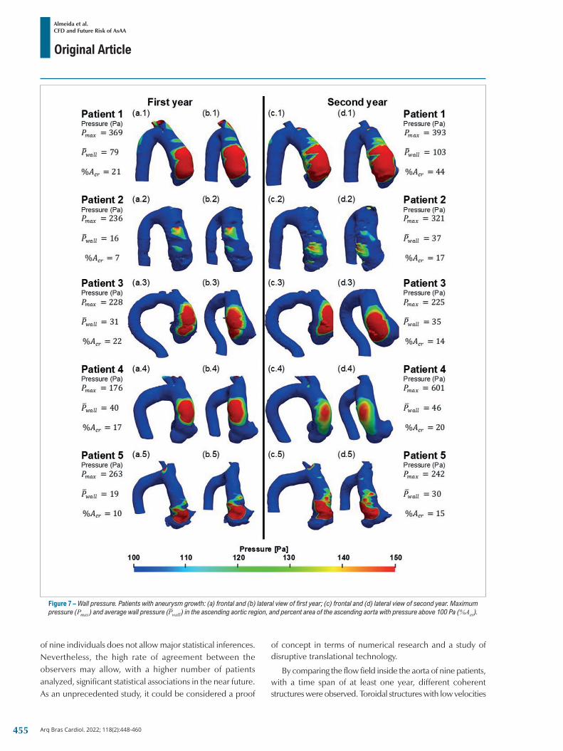
Arq Bras Cardiol. 2022; 118(2):448-460
Original Article
Almeida et al.CFD and Future Risk of AsAA
of nine individuals does not allow major statistical inferences. Nevertheless, the high rate of agreement between the observers may allow, with a higher number of patients analyzed, significant statistical associations in the near future. As an unprecedented study, it could be considered a proof
of concept in terms of numerical research and a study of disruptive translational technology.
By comparing the flow field inside the aorta of nine patients, with a time span of at least one year, different coherent structures were observed. Toroidal structures with low velocities
Figure 7 – Wall pressure. Patients with aneurysm growth: (a) frontal and (b) lateral view of first year; (c) frontal and (d) lateral view of second year. Maximum pressure (Pmax) and average wall pressure (P̄wall) in the ascending aortic region, and percent area of the ascending aorta with pressure above 100 Pa (%Aer).
455
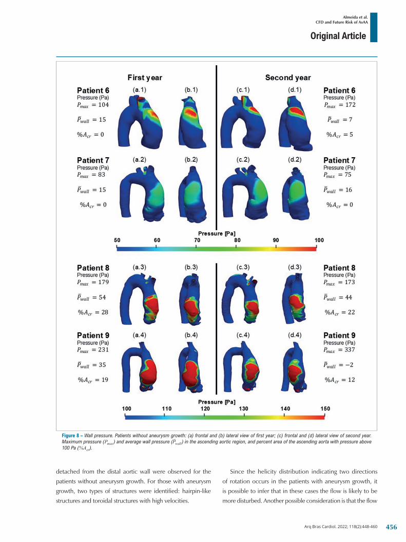
Arq Bras Cardiol. 2022; 118(2):448-460
Original Article
Almeida et al.CFD and Future Risk of AsAA
detached from the distal aortic wall were observed for the
patients without aneurysm growth. For those with aneurysm
growth, two types of structures were identified: hairpin-like
structures and toroidal structures with high velocities.
Since the helicity distribution indicating two directions
of rotation occurs in the patients with aneurysm growth, it
is possible to infer that in these cases the flow is likely to be
more disturbed. Another possible consideration is that the flow
Figure 8 – Wall pressure. Patients without aneurysm growth: (a) frontal and (b) lateral view of first year; (c) frontal and (d) lateral view of second year. Maximum pressure (Pmax) and average wall pressure (P̄wall) in the ascending aortic region, and percent area of the ascending aorta with pressure above 100 Pa (%Aer).
456

Arq Bras Cardiol. 2022; 118(2):448-460
Original Article
Almeida et al.CFD and Future Risk of AsAA
inside the aneurysm contains regions of strong recirculation, which are responsible for the formation of coherent structures in the aorta. Michel et al.37 have discussed that alterations in blood flow patterns can induce aneurysmal development on the outer curvature of the ascending aorta, due to high transverse wall shear stress and mechanical impedance on the convex side.
Biasetti et al.38 have correlated the presence of vortical structures with high shear stress in patients with abdominal aortic aneurysms. Here, vertical coherent structures can also be seen near the distal aortic wall, evolving in some cases to
a hairpin-like structure. These differences in the flow structure of patients in the two different groups (with and without aneurysm growth) may indicate the possibility of an aortic aneurysm remodeling process.
One hypothesis to explain these findings is the transfer of stress to adventitia due to loss of elastin and collagen deposition, promoting continuous adaptation because of stress distribution and, consequently, changes in the shape of AsAA.39
There are some limitations to this research. Firstly, the CTA scans were performed by different physicians. As a result, the quality of the exams was not the same. This fact may have
Table 2 – Clinical profile of the population
Population* (n=7) Aneurysm growth (n=3) No aneurysm growth (n=4)
Age (years ± SD)
65.1 ± 8.1 66 ± 8 64.5 ± 8.2
Gender, male (n, %)
5 (71.4) 3 (42.9) 2 (28.6)
Hypertension (n, %)
7 (100) 3 (42.9) 4 (57.1)
Diabetes (n, %)
3 (42.9) 1 (14.3) 2 (28.6)
Dyslipidemia (n, %)
5 (71.4) 2 (28.6) 3 (42.9)
Chronic kidney disease (n, %)
2 (28.6) 1 (14.3) 1 (14.3)
Atrial fibrillation (n, %)
2 (28.6) 1 (14.3) 1 (14.3)
Neurological disease (n, %)
1 (14.3) 0 (0) 1 (14.3)
Current or past smoking (n, %)
4 (57.1) 2 (28.6) 2 (28.6)
Obstructive coronary atherosclerotic disease (n, %)
1 (14.3) 1 (14.3) 0 (0)
Cerebrovascular disease (n, %)
1 (14.3) 0 (0) 1 (14.3)
Functional class I and II NYHA (n, %)
6 (85.7) 3 (42.9) 3 (42.9)
Angina (n, %)
2 (28.6) 1 (14.3) 1 (14.3)
Syncope or lipothymia (n, %)
0 (0) 0 (0) 0 (0)
Body mass index (kg/m² ± SD)
29.5 ± 2 28.3 ± 2.1 30.3 ± 1.8
Left ventricle ejection fraction (Teicholz % ± SD)
62.1 ± 8.7 56.6 ± 9.8 66.2 ± 4.7
Systolic blood pressure ≤ 130 mm Hg (n, %)
5 (71.4) 2 (28.6) 3 (42.9)
Diastolic blood pressure ≤ 85 mm Hg (n, %)
6 (85.7) 3 (42.9) 3 (42.9)
Heart rate < 70 bpm (n, %)
5 (71.4) 2 (28.6) 3 (42.9)
Clinical profile of 7 patients of the population* (clinical data of 2 patients were not available); SD: standard deviation; NYHA: New York Heart Association functional class I and II means no limitation or mild symptoms with slight limitation in ordinary activity; bpm: beats per minute.
457

Arq Bras Cardiol. 2022; 118(2):448-460
Original Article
Almeida et al.CFD and Future Risk of AsAA
1. Standring S. Gray’s Anatomy E-Book: The Anatomical Basis of Clinical Practice. 41st ed. Amsterdã: Elsevier; 2015.
2. Kuzmik GA, Sang AX, Elefteriades JA. Natural History of Thoracic Aortic Aneurysms. J Vasc Surg. 2012;56(2):565-71. doi: 10.1016/j.jvs.2012.04.053.
3. Elefteriades JA. Natural History of Thoracic Aortic Aneurysms: Indications for Surgery, and Surgical Versus Nonsurgical Risks. Ann Thorac Surg. 2002;74(5):1877-80. doi: 10.1016/s0003-4975(02)04147-4.
4. Hope TA, Markl M, Wigström L, Alley MT, Miller DC, Herfkens RJ. Comparison of Flow Patterns in Ascending Aortic Aneurysms and Volunteers Using Four-Dimensional Magnetic Resonance Velocity Mapping. J Magn Reson Imaging. 2007;26(6):1471-9. doi: 10.1002/jmri.21082.
5. Bäck M, Gasser TC, Michel JB, Caligiuri G. Biomechanical Factors in the Biology of Aortic Wall and Aortic Valve Diseases. Cardiovasc Res. 2013;99(2):232-41. doi: 10.1093/cvr/cvt040.
6. Sun Z, Chaichana T. A Systematic Review of Computational Fluid Dynamics in Type B Aortic Dissection. Int J Cardiol. 2016;210:28-31. doi: 10.1016/j.ijcard.2016.02.099.
7. Morris PD, Narracott A, von Tengg-Kobligk H, Soto DAS, Hsiao S, Lungu A, et al. Computational Fluid Dynamics Modelling in Cardiovascular Medicine. Heart. 2016;102(1):18-28. doi: 10.1136/heartjnl-2015-308044.
8. Peng L, Qiu Y, Yang Z, Yuan D, Dai C, Li D, et al. Patient-Specific Computational Hemodynamic Analysis for Interrupted Aortic Arch in an
References
affected the accuracy of measurements and definition of the aortic shape. Secondly, the small size of the population studied limits any correlation between clinical patterns and AsAA growth. Thirdly, clinical data of two patients were not available, so they were not included in Table 2. In addition, the present analysis was performed considering the most critical situation during the systole corresponding to the maximum flow rate in a cardiac cycle. Thus, the aorta was considered static at systolic peak and its elasticity was not considered.
Clearly, there are several factors that must be considered to improve the prognosis of patients with regards to aortic aneurysm growth, such as their age, previous diseases, and medicine intake. The angle between the aortic inlet and the brachiocephalic trunk could be an important feature to be considered, as well as the flow structure and the pressure distribution on the anterior wall, which can be numerically determined.
By the pressure distribution in the aortic wall, the mean pressure was observed to increase over 13% in all patients with aneurysm growth, while for those without pathological growth, the mean pressure decreased over 18% for all patients, except for one. This result indicates that this variable might influence the growth of the AsAA.
Future studies with a larger number of patients and longer follow-up are needed to precisely define the relationship between blood flow pattern and aortic remodeling process. The CFD could be a completely non-invasive tool to be used from CTA scans to estimate the growth trend of AsAA. The identification of different risk profiles among patients with AsAA could open the way for different medical care for these patients. All the above can lead to a step forward in personalized medicine and help to decipher the pathophysiology of this severe disease.
Considering that some flow characteristics could be associated with aneurysm growth, the study of the flow pattern in the ascending aorta could help predict the remodeling process of AsAA.
Conclusions This pilot study showed that CFD based on CTA may in the
near future be a tool to help identify patterns of flow behavior
associated with the AsAA remodeling process. Vortices were formed in the posterior region of the incident jet in the aortic wall, generating more complex structures for the aneurysm growth group. Consequently, there was an increase in the mean pressure in the anterior aortic wall between the CTA exams for the patients with aneurysm growth, while for those without aneurysm growth, this variable decreased.
AcknowledgmentThis study was supported by the Cardiovascular Engineering
Laboratory at PUC-Rio. The authors would like to thank Gustavo M. Geraldo and Gabriel C. Camargo for their contributions to this research. PUC-Rio members also acknowledge the continued support of Brazilian government agencies CAPES and CNPq.
Author ContributionsConception and design of the research, Writing of the
manuscript and Critical revision of the manuscript for intellectual content: Almeida GC, Gomes BAA, Azevedo FS, Ibanez I, Teixeira PS, Oliveira GMM, Nieckele AO; Acquisition of data: Almeida GC, Kalaoun K, Ibanez I, Gottlieb I, Melo MM, Nieckele AO; Analysis and interpretation of the data: Almeida GC, Gomes BAA, Azevedo FS, Kalaoun K, Ibanez I, Teixeira PS, Oliveira GMM, Nieckele AO; Statistical analysis: Gomes BAA, Azevedo FS, Oliveira GMM.
Potential Conflict of Interest No potential conflict of interest relevant to this article was
reported.
Sources of Funding There were no external funding sources for this study. Study Association This article is part of the thesis of master submitted by
Gabriela de Castro Almeida, from Programa de Pós-graduação em Engenharia Mecânica da Pontifícia Universidade Católica do Rio de Janeiro.
458

Arq Bras Cardiol. 2022; 118(2):448-460
Original Article
Almeida et al.CFD and Future Risk of AsAA
Adult: Implications for Aortic Dissection Initiation. Sci Rep. 2019;9(1):8600. doi: 10.1038/s41598-019-45097-z.
9. Bürk J, Blanke P, Stankovic Z, Barker A, Russe M, Geiger J, et al. Evaluation of 3D Blood Flow Patterns and Wall Shear Stress in the Normal and Dilated Thoracic Aorta Using Flow-Sensitive 4D CMR. J Cardiovasc Magn Reson. 2012;14(1):84. doi: 10.1186/1532-429X-14-84.
10. Gülan U, Calen C, Duru F, Holzner M. Blood Flow Patterns and Pressure Loss in the Ascending Aorta: A Comparative Study on Physiological and Aneurysmal Conditions. J Biomech. 2018;76:152-9. doi: 10.1016/j.jbiomech.2018.05.033.
11. Adrian RJ. Hairpin Vortex Organization in Wall Turbulence. Phys. Fluids. 2007;19(4):041301. doi: 10.1063/1.2717527
12. Erbel R, Aboyans V, Boileau C, Bossone E, Bartolomeo RD, Eggebrecht H, et al. 2014 ESC Guidelines on the Diagnosis and Treatment of Aortic Diseases: Document Covering Acute and Chronic Aortic Diseases of the Thoracic and Abdominal Aorta of the Adult. The Task Force for the Diagnosis and Treatment of Aortic Diseases of the European Society of Cardiology (ESC). Eur Heart J. 2014;35(41):2873-926. doi: 10.1093/eurheartj/ehu281.
13. Schindelin J, Arganda-Carreras I, Frise E, Kaynig V, Longair M, Pietzsch T, et al. Fiji: An Open-Source Platform for Biological-Image Analysis. Nat Methods. 2012;9(7):676-82. doi: 10.1038/nmeth.2019.
14. Faggiano E, Antiga L, Puppini G, Quarteroni A, Luciani GB, Vergara C. Helical Flows and Asymmetry of Blood Jet in Dilated Ascending Aorta with Normally Functioning Bicuspid Valve. Biomech Model Mechanobiol. 2013;12(4):801-13. doi: 10.1007/s10237-012-0444-1.
15. Alastruey J, Xiao N, Fok H, Schaeffter T, Figueroa CA. On the Impact of Modelling Assumptions in Multi-Scale, Subject-Specific Models of Aortic Haemodynamics. J R Soc Interface. 2016;13(119):20160073. doi: 10.1098/rsif.2016.0073.
16. Pope SB. Turbulent Flows. Cambridge: Cambridge University Press; 2000.
17. Menter FR. Two-Equation Eddy-Viscosity Turbulence Models for Engineering Applications. AIAA journal. 1994;32(8):1598-605. doi: 10.2514/3.12149.
18. Celis D, Gomes BAA, Ibanez I, Azevedo PN, Teixeira PS, Nieckele AO. Prediction of Stress Map in Ascending Aorta - Optimization of the Coaxial Position in Transcatheter Aortic Valve Replacement. Arq Bras Cardiol. 2020;115(4):680-7. doi: 10.36660/abc.20190385.
19. Gomes BAA, Camargo GC, Santos JRLD, Azevedo LFA, Nieckele AO, Siqueira-Filho AG, et al. Influence of the Tilt Angle of Percutaneous Aortic Prosthesis on Velocity and Shear Stress Fields. Arq Bras Cardiol. 2017;109(3):231-40. doi: 10.5935/abc.20170115.
20. Deutsch S, Tarbell JM, Manning KB, Rosenberg G, Fontaine AA. Experimental Fluid Mechanics of Pulsatile Artificial Blood Pumps. Annu Rev Fluid Mech. 2006;38(1):65-86. doi: 10.1146/annurev.fluid.38.050304.092022
21. Stuart J, Kenny MW. Blood Rheology. J Clin Pathol. 1980;33(5):417-29. doi: 10.1136/jcp.33.5.417.
22. Garbey M, Pacull F. A Versatile Incompressible Navier–Stokes Solver for Blood Flow Application. Int. J. Numer. Methods Fluids. 2007;54(5):473-96. https://doi.org/10.1002/fld.1405.
23. Fernandes LS, Bessa GM, Gomes BAA, Azevedo LFA. Stereoscopic PIV Study of the Influence of Aortic Valve Tilt Angle on the Flow Pattern in the Ascending Aorta Region. In: 13th International Symposium on Particle Image Velocimetry, 2019. Munich: ISPIV; 2019.
24. Becsek B, Pietrasanta L, Obrist D. Turbulent Systolic Flow Downstream of a Bioprosthetic Aortic Valve: Velocity Spectra, Wall Shear Stresses, and Turbulent Dissipation Rates. Front Physiol. 2020;11:577188. doi: 10.3389/fphys.2020.577188.
25. Patankar, S. Numerical Heat Transfer and Fluid Flow. Boca Raton: CRC Press; 1980.
26. ANSYS Inc. ANSYS CFD-Post Version 18. Canonsburg: ANSYS Inc.; 2018.
27. Ahrens J, Geveci B, Law C. Paraview: An End-User Tool for Large Data Visualization. Amsterdã: Elsevier: 2005.
28. Koo TK, Li MY. A Guideline of Selecting and Reporting Intraclass Correlation Coefficients for Reliability Research. J Chiropr Med. 2016;15(2):155-63. doi: 10.1016/j.jcm.2016.02.012.
29. IBM Corp. Released. IBM SPSS Statistics for Windows, Version 20.0. Armonk: IBM Corp; 2011.
30. Raghavan ML, Vorp DA, Federle MP, Makaroun MS, Webster MW. Wall Stress Distribution on Three-Dimensionally Reconstructed Models of Human Abdominal Aortic Aneurysm. J Vasc Surg. 2000;31(4):760-9. doi: 10.1067/mva.2000.103971.
31. Weigang E, Kari FA, Beyersdorf F, Luehr M, Etz CD, Frydrychowicz A, et al. Flow-Sensitive Four-Dimensional Magnetic Resonance Imaging: Flow Patterns in Ascending Aortic Aneurysms. Eur J Cardiothorac Surg. 2008;34(1):11-6. doi: 10.1016/j.ejcts.2008.03.047.
32. Hunt JC, Wray AA, Moin P. Eddies, Streams, and Convergence Zones in Turbulent Flows. In: Proceedings of the 1988 Summer Program. Washington D.C: NASA; 1988.
33. Levy Y, Degani D, Seginer A. Graphical Visualization of Vortical Flows by Means of Helicity. AIAA Journal. 1990;28(8):1347-52. doi: 10.2514/3.25224.
34. Moffatt HK. Helicity and Singular Structures in Fluid Dynamics. Proc Natl Acad Sci U S A. 2014;111(10):3663-70. doi: 10.1073/pnas.1400277111.
35. Garcia J, Barker AJ, Collins JD, Carr JC, Markl M. Volumetric Quantification of Absolute Local Normalized Helicity in Patients with Bicuspid Aortic Valve and Aortic Dilatation. Magn Reson Med. 2017;78(2):689-701. doi: 10.1002/mrm.26387.
36. Gallo D, Steinman DA, Bijari PB, Morbiducci U. Helical Flow in Carotid Bifurcation as Surrogate Marker of Exposure to Disturbed Shear. J Biomech. 2012;45(14):2398-404. doi: 10.1016/j.jbiomech.2012.07.007.
37. Michel JB, Jondeau G, Milewicz DM. From Genetics to Response to Injury: Vascular Smooth Muscle Cells in Aneurysms and Dissections of the Ascending Aorta. Cardiovasc Res. 2018;114(4):578-89. doi: 10.1093/cvr/cvy006.
38. Biasetti J, Hussain F, Gasser TC. Blood Flow and Coherent Vortices in the Normal and Aneurysmatic Aortas: A Fluid Dynamical Approach to Intra-Luminal Thrombus Formation. J R Soc Interface. 2011;8(63):1449-61. doi: 10.1098/rsif.2011.0041.
39. Mousavi SJ, Farzaneh S, Avril S. Patient-Specific Predictions of Aneurysm Growth and Remodeling in the Ascending Thoracic Aorta Using the Homogenized Constrained Mixture Model. Biomech Model Mechanobiol. 2019;18(6):1895-913. doi: 10.1007/s10237-019-01184-8.
459

Arq Bras Cardiol. 2022; 118(2):448-460
Original Article
Almeida et al.CFD and Future Risk of AsAA
This is an open-access article distributed under the terms of the Creative Commons Attribution License
460

Arq Bras Cardiol. 2022; 118(2):461-462
Short Editorial
Computational Fluid Dynamics (CFD) For Predicting Pathological Changes In The Aorta: Is It Ready For Clinical Use?Dominik Obrist1 and Hendrik von Tengg-Kobligk2
ARTORG Center for Biomedical Engineering Research, University of Bern,1 Bern – SwitzerlandInstitute for Diagnostic, Interventional and Pediatric Radiology (DIPR), University of Bern,2 Bern – SwitzerlandShort Editorial related to the article: Computational Fluid Dynamics to Assess the Future Risk of Ascending Aortic Aneurysms
Mailing Address: Dominik Obrist •Universität Bern - Freiburgstrasse 3, Bern, 3010 – SwitzerlandE-mail: [email protected]
KeywordsCardiovascular Diseases/phyiopathology; Cardiovascular
Diseases/diagnosis; Computer Simulation; Image Processing; Computer Assisted; Prosthesis Design; Prosthesis Implantation; Biomedical Engineering.
Computational modelling of complex flow problems profits from a mature basis of Computational Fluid Dynamics (CFD) software developed over the past decades. Early use of CFD was limited to academic research. Nowadays, it is a fully established tool in many industrial applications (e.g., automotive, aerospace), although CFD remains to be one of the most demanding computational tasks and the study of complex flow problems is often limited by the available computational power. The continuous exponential increase in computational power and the improved accessibility of high-performance computing infrastructure was an enabler for the ever-growing use of CFD.
Under the light of this success, it is surprising that CFD can hardly be found in clinical practice. Despite remarkable progress in modelling complex blood flow and in identifying quantitative markers for pathological flow patterns,1 most biomedical applications of CFD remain at the level of academic research and single-patient cases. The study by Almeida et al.2 is one of the few CFD studies using longitudinal radiological data from patient cohorts. The proposed prediction of pathological changes in the aorta based on CFD illustrates the potential of this technology to become an established diagnostic modality. Some of the challenges successfully addressed in this study are exemplary for the reason why CFD has not yet found its place in clinical routine. We can identify four problem fields: a) The difficulty of efficiently generating patient-specific CFD models; b) The need for reduction of data complexity to make CFD results accessible to the clinician; c) The missing IT infrastructure which integrates CFD tools smoothly into existing clinical data workflows; d) The lack of experts in the clinical environment, i.e. a clinical engineer supporting the caring physicians. In the following, we will discuss these problems and indicate possible measures to mitigate them.
Patient-specific modellingPatient-specific models3 have been successfully deployed
by many researchers. Nevertheless, the translation of high-resolution radiological data into geometrical vascular models for CFD remains a time-intensive task which is often cumbersome and requires trained specialists. There is a lack of automated segmentation and meshing tools for biomedical tasks. Recent developments based on deep neural networks may provide acceptable solutions for clinical use.4
The setting of boundary conditions (e.g., velocity profiles at inflow/outflow) requires great care, because boundary conditions have a strong effect on the quality of the results. Therefore, it is useful to include flow measurements at well-defined locations (e.g., 2D+time or 3D+time blood flow MRI) into radiologic protocols.5 Next to the adequate choice of the modality and body location, this requires appropriate temporal and spatial resolution of the scans.
Furthermore, we need a better understanding of diseased tissue biomechanics to correctly interpret a remodeled or dissected aorta or the plaque on a vessel wall. Next to continued classical biomechanical research, this requires studies with large cohorts including healthy volunteers to understand the clinical relevance of biomechanical models.
Diagnostic markersAnalysis of CFD results is challenging even for experts in
fluid dynamics. This is partly due to the difficulty of visualizing three-dimensional, time-dependent flow fields featuring a wealth of flow phenomena which may be relevant for the clinical interpretation of the results (e.g., vortices, flow instabilities, turbulence, flow separation, re-attachment, impingement).
This data complexity can be reduced by data-driven modelling6-8 and by anomaly detection algorithms (used widely in ECG analysis9) which use deep neural networks to localize and highlight outliers in the patient-specific flow data to guide the clinician toward potential anomalies.
Almeida et al.2 addressed the problems by visualizing Lagrangian coherent structures10 in the flow field and by computing single scalar values that characterize specific aspects of the flow field (e.g. helicity index). Others proposed metrics to characterize the biomechanical interaction between blood flow and arterial walls11,12 or the effect of shear flow on blood trauma and thrombogenicity.13,14 It is of paramount importance to establish the clinical value of such metrics and to establish them as diagnostic markers DOI: https://doi.org/10.36660/abc.20220040
461

Arq Bras Cardiol. 2022; 118(2):461-462
Short Editorial
Obrist & Tengg-KobligkCFD For Predicting Pathological Changes In The Aorta
or clinical scores. Only with such an approach we will enable efficient and standardized interpretation of CFD data in clinical routine.
Integrated IT infrastructureFull integration of CFD into the clinical workflow requires
easy-to-use data transfer interfaces between clinical patient databases, imaging systems and computational platforms to perform CFD. In complex cases with higher demand for computational power, imaging data may have to be transferred to centralized computational infrastructure which may be outside of the hospital’s IT perimeter. This raises questions of data privacy and security which must be addressed by establishing appropriate encryption technology and dedicated connections. External services must also consider regulatory aspects of transferring
patient data over the internet. It will be worthwhile to look at solutions used in already established computational applications (e.g. FFRCT
15).
Clinical Engineer
Successful tackling of these problems requires establishing the role of a clinical engineer who is part of the clinical radiology team and fully integrated in the workflow. Only then, CFD has the potential to find its place as a sustainable diagnostic resource in clinical routine.
With these proposed measures, CFD will eventually become just another modality within an integrated multi-modal radiologic ecosystem providing additional cues to the clinical expert to arrive at a better diagnosis and prediction including individually adapted risk stratification.
1. Morris P D, Narracott A H, von Tengg-Kobligk D A, Silva Soto S, Hsiao A. Lungu P,et al. Computational fluid dynamics modelling in cardiovascular medicine. Heart.2016;102(1):18-28. doi: 10.1136/heartjnl-2015-308044.
2. Almeida G. Computational Fluid Dynamics to Assess the Future Risk of Ascending Aortic Aneurysms. Arq Bras Cardiol. 2022; 118(2):448-460.
3. Gray RA, Pathmanathan P. Patient-Specific Cardiovascular Computational Modeling: Diversity of Personalization and Challenges. J Cardiovasc Transl Res. 2018;11:80–8. doi: 10.1007/s12265-018-9792-2. Epub 2018 Mar 6.
4. Chaitanya K, Karani N Baumgartner CF, Becker A, Donati O, Konukoglu E. Semi-supervised and task-driven data augmentation. Med Imag Anal.2021; 68:101934. doi: 10.1016/j.media.2020.101934.
5. Bonfanti M, Franzetti G, Maritati G, Homer-Vanniasinkam S, Balabani S, Díaz-Zuccarini V. Patient-specific haemodynamic simulations of complex aortic dissections informed by commonly available clinical datasets. Med Eng Phys. 2019;71:45–55 DOI: 10.1016/j.medengphy.2019.06.012
6. Arzani A, Dawson STM. Data-driven cardiovascular flow modelling: examples and opportunities. JR Soc Interface.2021;18:20200802. doi: 10.1098/rsif.2020.0802. Epub 2021 Feb 10.
7. Habibi M, D’Souza, S, Dawson M, Arzani A. Integrating multi-fidelity blood flow data with reduced-order data assimilation. Comput Biol Med.2021;135:104566 doi: 10.1016/j.compbiomed.2021.104566
8. Liang L, Mao W, Sun W. A feasibility study of deep learning for predicting hemodynamics of human thoracic aorta. J Biomech.2020;99:109544.
9. Li H, Boulanger P. A survey of heart anomaly detection using ambulatory Electrocardiogram (ECG).Sensors(Basel).2020;20(5):1461. doi: 10.1016/j.compbiomed.2021.104566
10. Darwish A, Norouzi S, Di Labbio G, Kadem L. Extracting Lagrangian coherent structures in cardiovascular flows using Lagrangian descriptors. Phys Fluids .2021;33:111707.
11. Arzani, A, Shadden SC. Wall shear stress fixed points in cardiovascular f luid mechanics. J Biomech.2018;73:145-52. doi: 10.1016/j.jbiomech.2018.03.034.
12. Morbiducci U, Mazzi V, Domanin M, De Nisco G,Vergara C, Steinman DA, et al., Wall shear stress topological skeleton independently predicts long-term restenosis after carotid bifurcation endarterectomy. Ann Biomed Eng.2020;48(12):2936-49. doi: 10.1007/s10439-020-02607-9.
13. Taylor J O, Meyer RS, Deutsch S, Manning KB. Development of a computational model for macroscopic predictions of device-induced thrombosis. Biomech Model Mechanobiol.2016;15(6):1713-31. doi: 10.1007/s10237-016-0793-2. Epub 2016 May 12.
14. Yazdani A, Zhang P, Sheriff J, Slepian MJ, Deng Y, Bluestein D. Multiscale modeling of blood flow-mediated platelet thrombosis. Handb Mater Model Appl Curr Emerg Mater.2020;2667-98.
15. Min J K, Taylor CA, Stephan A, Kwon KB, Jonathon L, Bernard DB. Noninvasive Fractional Flow Reserve Derived From Coronary CT Angiography. JACC Cardiovasc Imaging.2015;8(10):1209-22. doi: 10.1016/j.jcmg.2015.08.006.
References
This is an open-access article distributed under the terms of the Creative Commons Attribution License
462

Arq Bras Cardiol. 2022; 118(2):464-475
Original Article
The Dysfunctional Scenario of the Major Components Responsible for Myocardial Calcium Balance in Heart Failure Induced by Aortic StenosisVitor Loureiro da Silva,1,2 Sérgio Luiz Borges de Souza,1 Gustavo Augusto Ferreira Mota,1 Dijon H. S. Campos,1 Alexandre Barroso Melo,3 Danielle Fernandes Vileigas,1 Paula Grippa Sant’Ana,1 Priscila Murucci Coelho,3 Silméia Garcia Zanati Bazan,1 André Soares Leopoldo,3 Antônio Carlos Cicogna1
Universidade de São Paulo - Departamento de Medicina Interna,1 Botucatu, SP – BrazilUniversidade Estadual Paulista - Clínica Médica,2 São Paulo, SP – BrazilUniversidade Federal do Espírito Santo - Departamento de Desportos,3 Vitória, ES – Brazil
Mailing Address: Vitor Loureiro da Silva •Universidade de São Paulo - Departamento de Medicina Interna - Jardim Dona Marta (Rubião Junior). Postal Code 18618-970, Botucatu, SP - BrazilE-mail: [email protected] received June 08, 2020, revised manuscript January 26, 2021, accepted February 24, 2021
DOI: https://doi.org/10.36660/abc.20200618
Abstract
Background: Maladaptive cardiac remodelling is characterized by diastolic and systolic dysfunction, culminating in heart failure. In this context, the dysfunctional scenario of cardiac calcium (Ca2+) handling has been poorly studied. An experimental model of aortic stenosis has been extensively used to improve knowledge about the key mechanisms of cardiac pathologic remodelling.
Objective: To understand the dysfunctional process of the major components responsible for Ca2+ balance and its influence on cardiac function in heart failure induced by aortic stenosis.
Methods: Male 21-day-old Wistar rats were distributed into two groups: control (sham; n= 28) and aortic stenosis (AoS; n= 18). Cardiac function was analysed by echocardiogram, isolated papillary muscle, and isolated cardiomyocytes. In the papillary muscle assay, SERCA2a and L-type Ca2+ channel activity was evaluated. The isolated cardiomyocyte assay evaluated Ca2+ handling. Ca2+ handling protein expression was analysed by western blot. Statistical significance was set at p <0.05.
Results: Papillary muscles and cardiomyocytes from AoS hearts displayed mechanical malfunction. AoS rats presented a slower time to the Ca2+ peak, reduced Ca2+ myofilament sensitivity, impaired sarcoplasmic reticulum Ca2+ influx and reuptake ability, and SERCA2a and L-type calcium channel (LTCC) dysfunction. Moreover, AoS animals presented increased expression of SERCA2a, LTCCs, and the Na+/Ca2+ exchanger.
Conclusion: Systolic and diastolic heart failure due to supravalvular aortic stenosis was paralleled by impairment of cellular Ca2+ influx and inhibition of sarcoplasmic reticulum Ca2+ reuptake due to LTCC and SERCA2a dysfunction, as well as changes in Ca2+ handling and expression of the major proteins responsible for cellular Ca2+ homeostasis.
Keywords: aortic stenosis; heart failure; papillary muscle; isolated cardiomyocytes; calcium handling proteins.
Researchers have reported changes in the expression and function of regulatory proteins of Ca2+ handling in a variety of cardiovascular diseases.5–23 In pathological remodelling induced by aortic stenosis, studies have identified, using different methods of surgical induction and periods of the disease, several distinct changes in the regulatory elements of Ca2+.11,12,14–22 Initial studies have proposed that changes in sarcoplasmic reticulum Ca2+ outflow and intake are related to cardiac dysfunction due to aortic stenosis.11,12 Our previous studies evaluated rats with diastolic dysfunction after six and twelve weeks of aortic stenosis.16,17 After six weeks, impairment in sarco/endoplasmic reticulum calcium ATPase (SERCA2a) activity without protein expression alteration was observed;16
after twelve weeks, increased phosphorylation of the Ser(16) residue of PLB and reduced SERCA2a protein expression were observed.17 Furthermore, in animals with HF after aortic obstruction,19,21,24 the authors detected reduced Ca2+ current (ICa),
21 inefficiency of the coupling of the LTCCs with the RyR receptors,20 and alterations in calcium handling proteins.19–22
IntroductionHeart failure (HF) is characterized by the heart’s inability
to perfuse tissues with oxygen and nutrients required by the body’s metabolic demand;1 major outcomes include exercise intolerance and water retention.2 The process of HF is the product of maladaptive remodelling, which may occur due to several types of damage to the heart, including myocardial ischaemia and volume and pressure overload.3 Among others, impaired calcium (Ca2+) handling is a crucial mechanism of the progressive deterioration of contractile function in HF.4
463

Arq Bras Cardiol. 2022; 118(2):464-475
Original Article
Silva et al.Calcium Dynamics in Decompensated Hypertrophy
Because these investigations report scattered data regarding the structural and functional degree of cardiac remodelling and the corresponding adaptations of myocardial Ca2+ dynamics, this study aimed to characterize the dysfunctional process of the major components responsible for Ca2+ balance and its influence on cardiac function in HF induced by aortic stenosis. For this purpose, unlike previous studies, we performed a global cardiac evaluation of the animals after 28 weeks of aortic stenosis. This study performed cardiac function analysis at the cellular, tissue, and chamber levels, as well as examining Ca2+ handling and the proteins responsible for cytosolic Ca2+ balance, presenting divergent and surprising results compared to the currently available literature.
Methods
Study design1) A group of twenty one-day-old male Wistar rats was
submitted to simulated (Sham, n= 22) or aortic stenosis induction (AoS, n= 12) surgery. After 28 weeks of the experimental protocol, cardiac function was evaluated by echocardiogram and isolated papillary muscle. SERCA2a and L-type calcium channel activity was analysed during postrest contraction and calcium elevation, respectively, and by the cumulative administration of extracellular Ca2+ in the presence of SERCA2a- or L-type calcium channel-specific blockers in the isolated papillary muscle assay. Expression of Ca2+-handling regulatory proteins was measured by western blotting (sham, n= 7; AoS, n= 7). Five AoS animals were excluded from the isolated papillary muscle experiment due to the having a cross-sectional area of papillary muscle greater than 1.5 mm2.
2) Another group of twenty one-day-old male Wistar rats underwent simulated (Sham, n= 6) or aortic stenosis induction (AoS, n= 6) surgery. In these animals, electrocardiogram and was performed and cardiomyocytes were isolated. Analysis of the isolated cardiomyocytes was performed to assess the mechanical function of the cardiomyocytes and calcium handling.
As noted, echocardiogram evaluation was performed on all study animals (Sham, n= 28; AoS, n= 18).
AnimalsWistar rats obtained from the Animal Center of Botucatu
Medical School (Botucatu, São Paulo, Brazil) were allocated to collective cages in a 23°C room temperature with a 12-h light/dark cycle, a relative humidity of 60%, and water ad libitum. This research was approved by the “Committee for Experimental Research Ethics of the Faculty of Medicine in Botucatu – UNESP” and by the “Guide for the Care and Use of Laboratory Animals” (protocol 1138/2015).
Aortic stenosis surgeryAoS was surgically induced as previously described.14–17
Rats were anaesthetized using a mixture of ketamine (50 mg/kg, IM) and xylazine (1 mg/kg, IM), and the heart was exposed via a median thoracotomy. A silver clip (0.62 mm internal diameter) was then placed on the ascending aorta approximately 3 mm from its root, constituting the AoS group
(n= 17). Control rats were submitted to the same surgery but without aortic banding (Sham, n= 19).
Cardiac function
EchocardiogramData from the cardiac structure and function analysis are
expressed utilizing echocardiogram variables after 28 weeks of aortic stenosis. Commercially available echocardiography (General Electric Medical Systems, Vivid S6, Tirat Carmel, Israel) equipped with a 5-11.5 MHz multifrequency probe was used as previously described.17,25,26 Rats were anaesthetized via intraperitoneal injection with a mixture of ketamine (50 mg/kg) and xylazine (0.5 mg/kg). The following variables were used to evaluate cardiac structure: LA normalized to the aortic diameter (LA/Ao), left ventricle diastolic diameter (LVDD), left ventricular systolic diameter (LVSD), posterior wall diastolic thickness (PWDT), interventricular septum diastolic thickness (ISDT), and relative wall thickness (RWT). The following parameters were used to assess ventricular function: heart rate (HR), mid-wall fraction shortening (FS); ejection fraction (EF); posterior wall systolic velocity (PWSV), early diastolic mitral inflow velocity (E wave), the ratio between E wave and atrial contraction flow peak (A wave), the velocity of the mitral annulus during early ventricular filling (E’), mitral velocity annulus during atrial contraction (A’), and the ratio between filling flow peak and mitral annulus velocity during early ventricular filling (E/E’).
Heart failure signsThe same investigator analysed clinical and pathological
signs of HF (tachypnoea, ascites, pleural effusion, left atrium thrombi, and right ventricular hypertrophy) and was blinded to the experimental groups.
Isolated papillary muscle assayCardiac contractile performance was evaluated by
examining isolated papillary muscles from the LV, as previously described.14,17,25 The papillary muscles were stimulated 12 times per minute (0.2 Hz) using platinum needle-type electrodes positioned parallel to the muscles’ longitudinal axis. The electrodes were coupled to an electrical stimulator (LE12406 - Stimulator, PanLab - Harvard Apparatus, Cornella, Barcelona, Spain) that emits 5 ms square wave stimuli. The stimulus voltage used was 12 to 15 volts, approximately 10% above the minimum value necessary to provoke the muscle’s maximum mechanical response. In the experiment, Krebs-Henseleit solution was used according to the following composition in mM: 118.5 NaCl; 4.69 KCl; 2.5 CaCl2; 1.16 MgSO4; 1.18 KH2PO4; 5.50 glucose and 24.88 NaHCO3. The solution was aerated for 10 minutes with 95% oxygen (O2) and 5% carbon dioxide (CO2) and kept at 28°C. The following mechanical parameters were measured during isometric contraction: maximum developed tension (DT; g/mm2), resting tension (RT; g/mm2), maximum rate of tension development (+dT/dt; g/mm2/s) and decline (-dT/dt; g/mm2/s), and time-to-peak tension (TPT; ms). Regulatory mechanisms of
464

Arq Bras Cardiol. 2022; 118(2):464-475
Original Article
Silva et al.Calcium Dynamics in Decompensated Hypertrophy
Ca2+ influx and L-type calcium channel activity were analysed by the Ca2+ concentration extracellular elevation manoeuvre and elevation of extracellular Ca2+ concentrations (0.5, 1.5, 2.5, and 3.5 mM) in the presence and absence of diltiazem (10-5 M), a specific blocker of L-type calcium channels. A postrest contraction manoeuvre (the stimulus was paused for 10, 30, and 60 s before restarting the stimulation) and elevation of extracellular Ca2+ concentrations (0.5, 1.5, 2.5, and 3.5 mM) in the presence and absence of cyclopiazonic acid (CPA, 30 mM), a highly specific blocker of SERCA2a, were performed to assess the potential of SERCA2a function. L-channel and SERCA2a calcium channel blocking assays were analysed from the percentage of inhibition, calculated as Δ(%)= (M2-M1)/M1x100, so that M1 is the value of the variable in the extracellular calcium concentration in the absence of the blocker, and M2 is the value of the same variable in response to the blockers. Tests of extracellular calcium elevation and postrest contraction were analysed by the percent response compared to baseline, calculated as Δ(%)= (M0-Mx)/M0×100, so that M0 is the value in the baseline condition, and Mx is the absolute value in response to the manoeuvre (increased calcium concentration or paralysis of the electrical stimulus). All variables were normalized per cross-sectional area (CSA) of the papillary muscle. Papillary muscles with CSA >1.5 mm2 were excluded from analysis because they can exhibit central core hypoxia and impaired functional performance.16,17
Isolated cardiomyocyte assay
Cardiomyocyte preparationUnder anaesthesia, rats from each group were euthanized.
Hearts were quickly removed by median thoracotomy and enzymatically isolated, as previously described.27 Briefly, the hearts were cannulated. Retrograde perfusion of the aorta was performed on a Langendorff system (37°C) with a modified isolation digestion buffer solution (DB), a calcium-free solution containing 0.1 mM ethylene glycol-bis (ß-aminoethyl ether)-N, N, N’, N’-tetraacetic acid (EGTA), and N-[2-hydro-ethyl]-piperazine-N’-[2-ethanesulfonic acid] (HEPES), which were equilibrated. The composition of DB solution was as follows (mM): 130 NaCl, 1.4 MgCl2, 5.4 KCl, 25 HEPES, 22 glucose, 0.33 NAH2PO4, and pH 7.39. Afterward, the hearts were perfused for 15-20 minutes with a DB solution containing 1 mg/ml collagenase type II (Worthington Biochemical Corporation, UK) and Ca2+
(1 mM). Digested hearts were then removed from the cannula, minced into small pieces, and placed into small conical flasks with DB solution containing collagenase supplemented with 0.1% bovine serum albumin and Ca2+
(1 mM). Next, this process was performed two more times without collagenase and with the addition of 1.6 and 3.12 µL of 1.0 mM CaCl2 stock solution. Each stage containing cells and solutions was incubated for approximately 10 minutes. Then, the supernatant was removed, and the myocytes were resuspended in Tyrode’s buffer containing the following (in mM): 140 NaCl, 10 HEPES, 0.33 NaH2PO4, 1 MgCl2, 5 KCl, 1.8 CaCl2, 10 glucose. Only calcium-tolerant, quiescent, rod-shaped cardiomyocytes showing clear cross-striations
were examined. The isolated cardiomyocytes were used within 2-3 h of isolation.
Cardiomyocyte contractilityBriefly, isolated cells were placed in an experimental
chamber with a glass coverslip base mounted on the stage of an inverted microscope (Ion Optix, Milton, MA, USA) edge detection system with a 40x objective lens (Nikon Eclipse – TS100, USA). Cells were immersed in Tyrode’s solution and field stimulated at 1 Hz (20 V, 5 ms duration square pulses). Cell shortening in response to electrical stimulation was measured using a video-edge detection system at a 240-Hz frame rate (Ionwizard, Ion Optix, Milton, MA, USA), and the contractile parameters were evaluated. Sarcomere length, fractional shortening (expressed as a percentage of resting cell length), maximum shortening velocity, maximum relaxation velocity, and time to 50% shortening (time to 50% peak) and 50% relaxation (time for 50% relaxation) were measured in 6 cells per animal in each experimental group.
Intracellular Ca2+ measurementsSubsequently, cardiomyocytes were stimulated at 1 Hz
(Myopacer 100, Ion Optix Inc.), and fluorescence images were obtained with alternating excitation at 340 to 380 nm wavelengths using a Hyper Switch system (IonOptix, Milton, MA). Background-subtracted fluorescence emission was obtained, and the Fura 2 AM ratios were used as an index of intracellular [Ca2+]i transient, which was detected at approximately 510 nm. The Ca2+ transient amplitude was reported as F/F0. F is the maximal fluorescence intensity average measured at the peak of [Ca2+]i transients, and F0 is the baseline fluorescence intensity measured at the diastolic phase of [Ca2+]i transients. The time to Ca2+ peak and time to 50% Ca2+ decay were also analysed.
Expression of calcium handling proteinsWestern blot analysis was used to evaluate protein
expression of the regulatory components of Ca2+ handling. Fragments of the LV were frozen in liquid nitrogen and stored at -80°C. Frozen samples were subsequently homogenized in RIPA buffer containing protease (Sigma-Aldrich, St. Louis, MO, USA) and phosphatase (Roche Diagnostics, Indianapolis, IN, USA) inhibitors using a bead beater homogenizer (Bullet Blender®, Next Advance, Inc., NY, USA). The homogenized product was centrifuged (5804R Eppendorf, Hamburg, Germany) at 12,000 rpm for 20 minutes at 4°C, and the supernatant was transferred to Eppendorf tubes and stored at -80°C. Protein concentration was determined using the Pierce BCA Protein Assay Kit. SDS-PAGE was used to resolve a total of 25 µg protein lysate from each sample. Electrophoresis was performed with biphasic gel stacking (240 mm Tris-HCl pH 6.8, 30% polyacrylamide, APS and TEMED) and resolving (240 mm Tris-HCl pH 8.8, 30% polyacrylamide, APS and TEMED) at a concentration of 6 to 10%, depending on the molecular weight of the analysed protein. The Kaleidoscope Prestained Standard (Bio-Rad, Hercules, CA, USA) was used to identify band sizes. Electrophoresis was performed at
465

Arq Bras Cardiol. 2022; 118(2):464-475
Original Article
Silva et al.Calcium Dynamics in Decompensated Hypertrophy
120 V (Power Pac HC 3.0 A, Bio-Rad, Hercules, CA, USA) for 3 h with running buffer (0.25 M Tris, 192 mM glycine, and 1% SDS). Proteins were transferred to a nitrocellulose membrane (Armsham Biosciences, Piscataway, NJ, USA) using a Mini Trans-Blot (Bio-Rad, Hercules, CA, USA) system with transfer buffer (25 mM Tris, 192 mM glycine, 20% methanol, and 0,1% SDS). Membranes were blocked with 5% non-fat dry milk in TBS-T buffer (20 mM Tris-HCl pH 7.4, 137 mM NaCl, and 0.1% Tween 20) for 120 minutes at room temperature under constant agitation. The membrane was washed three times with TBS-T and incubated for 12 h at 4 - 8°C under constant agitation with the following primary antibodies: Serca2 ATPase (1:2500; ABR, Affinity BioReagents, Golden, CO, USA), Phospholamban (1:5000; ABR), Phospho-Phospholamban (Ser16) (1:5000; Badrilla, Leeds, West Yorkshire, UK), Phospho–Phospholamban (Thr17) (1:5000; Badrilla), Exchanger Na+/Ca2+ (1:2000; Upstate, Lake Placid, NY, USA), Calcium Channel, Voltage-Gated Alpha 1C (1:100; Chemicon International, Temecula, CA, USA), Ryanodine Receptor (1:5000; ABR, Affinity Bioreagents, Golden, CO, USA), and GAPDH (1:1000; Santa Cruz Biotechonology Inc., CA, USA). After incubation with the primary antibody, membranes were washed three times in TBS-T and incubated with peroxidase-conjugated secondary antibodies (anti-rabbit or anti-mouse IgG; 1: 5,000- 1: 10,000; Abcam) for 2 h under constant agitation. Membranes were then washed three times with TBS-T to remove excess secondary antibody. Blots were incubated with ECL (Enhanced Chemi-Luminescence, Amersham Biosciences, Piscataway, NJ) for chemiluminescence detection by ImageQuant™ LAS 4000 (GE Healthcare). Quantification analysis of blots was performed using Scion Image software (Scion Corporation, Frederick, MD, USA). The immunoblots were quantified by densitometry using ImageJ Analysis software (NIH), and target band results were normalized to the expression of cardiac GAPDH.14 It was not possible to analyse GAPDH (37 kDa) as a normalizer on the same gel as the ryanodine receptor (565 kDa) due to the difference in molecular weight between the two proteins. Due to this, the ryanodine receptor is expressed without normalization.
Statistical analysisStatistical analysis was performed using Sigma Stat 3.5
software (SYSTAT Software Inc., San Jose, CA, USA). The distribution of variables was assessed using the Kolmogorov-Smirnov test for normality. According to the normality of the data, the results are reported as the means ± standard deviation (SD) or median (25th percentile; 75th percentile). Comparisons between groups were performed using two-tailed Student’s t-test for independent samples or Mann-Whitney test or repeated-measures two-way analysis of variance (ANOVA) when appropriate. The level of significance considered was 5%.
Sample size (n) was estimated using the equation for comparison between groups: n = 2SD2(Zα/2 + Zβ)2/d, where n is the sample size, SD = 0.02 from previous studies, Zα/2 = 1.96 (from Z table) at type 1 error of 5%, Zβ = 0.842 (From Z table) at 80% power and d = 0.02 (effect size - minimal
difference between mean values).28 The sample size needed to detect a significant difference between groups was 16 rats per group; however, we decided to use 22 simulated (sham) and 18 aortic stenosis induction (AoS) animals per group for the study design.
Results
Echocardiographic evaluation and heart failure signsEchocardiogram data revealed that aortic stenosis resulted
in predominantly concentric cardiac hypertrophy (↑RWT, ↑PWDT, ↑ISDT, and ↑LVDD), left atrium dilation (↑LA/Ao), and diastolic (↑E wave, ↑E/A, ↑E/E’, ↓E’, and ↓A’) and systolic dysfunction (↑LVSD, ↓PWSV, ↓FS, and ↓EF) 28 weeks after surgery (Table 1). The following clinical and pathological signs of HF were detected: ascites (30%), left atrium thrombi (48%), pleural effusion (68%), tachypnoea (79%), and right ventricular hypertrophy (100%) (Table 1).
Isolated papillary muscle evaluation
Baseline dataAortic stenosis impaired myocardial contractile and
relaxation function by reducing developed tension and the maximum rate of tension development and increasing rest tension and time-to-peak tension (Table 2).
Isolated papillary muscle manoeuvresFigure 1A-C presents the papillary muscle response
percentage to postrest contraction (10, 30, and 60 s). Animals with aortic stenosis exhibited a poor response to the postrest contraction manoeuvre compared to sham animals for all evaluated time points and variables. There was no significant difference between groups at any time point. Figure 1D-F presents the percent papillary muscle response to increase extracellular calcium concentration (1.5, 2.5, and 3.5 mM). Figure 2A-C shows papillary muscle responses to SERCA2a inhibition and an increase in calcium concentration. After SERCA2a inhibition by cyclopiazonic acid, there was a significant difference between groups in calcium concentration at 0.5 mM for maximum developed tension (Figure 2A). The maximum rate of tension development and decline did not exhibit a difference between the AoS and sham groups. Figure 2D-F presents the papillary muscle response to L-type calcium channel inhibition, demonstrating an increase in calcium concentration. Animals with aortic stenosis had worse performance in response to calcium elevation after L-type calcium channel blockage than sham animals for all evaluated time points and variables.
Isolated cardiomyocytes
Mechanical function and calcium handling analysisFigure 3A-F shows cardiomyocyte mechanical function.
Aortic stenosis impaired shortening maximum velocity (Figure 3B) and times to achieve 50% cardiomyocyte contraction
466

Arq Bras Cardiol. 2022; 118(2):464-475
Original Article
Silva et al.Calcium Dynamics in Decompensated Hypertrophy
(Figure 3E) and relaxation peak (Figure 3F). Figure 3G-J summarizes calcium handling 28 weeks after aortic stenosis. AoS animals presented alterations in both the time to achieve Ca2+ peak and time to 50% Ca2+ decay.
Expression of calcium handling proteins
Data regarding protein expression of calcium handling regulator elements are shown in Figure 4A-E. Aortic stenosis
increased L-type calcium channel, SERCA2a, and Na+/Ca2+ exchanger (NCX) protein expression and decreased phosphorylation of the Thr(17) residue of PLB.
DiscussionIn the experimental model of supravalvular aortic
stenosis, studies investigating pathologic remodelling and HF, focusing on specific Ca2+ handling alterations and their
Table 1 – Echocardiogram data and heart failure signs
Sham AoS p-value
HR (bpm) 302 ± 40 298 ± 40 0.857
LVDD (mm)* 7.55 (7.15; 7.66) 8.43 (7.27; 9.20) <0.001
LVSD (mm)* 3.20 (2.81; 3.32) 3.83 (3.32; 5.62) <0.001
PWDT (mm)* 1.53 (1.53; 1.65) 2.81 (2.55; 3.07) <0.001
ISDT (mm)* 1.65 (1.53; 1.70) 3.07 (2.84; 3.26) <0.001
RWT 0.43 ± 0.03 0.69 ± 0.16 <0.001
LA/Ao 1.22 ± 0.09 1.91 ± 0.18 <0.001
E wave (cm/s) 85 ± 7 132 ± 18 <0.001
E/A 1.49 ± 0.18 5.13 ± 1.40 <0.001
E’ (cm/s) 6.20 ± 0.78 5.32 ± 0.89 <0.001
A’ (cm/s) 4.28 ± 0.67 3.12 ± 1.18 <0.001
E/E’ 13.9 ± 2.22 25.2 ± 4.66 <0.001
PWSV (cm/s) 68 ± 9 37 ± 9 <0.001
FS (%) 26.1 ± 3.42 23.3 ± 4.69 0.028
EF (%)* 93 (92; 94) 89 (79; 92) <0.001
Ascites (%) 0 30 -
LAT (%) 0 48 -
PE (%) 0 68 -
Tachypnea (%) 0 79 -
RVH (%) 0 100 -
Data are expressed as means ± SD or median (25 percentile; 75 percentile)*. Sham: animals submitted to simulated surgery (n= 28); AoS: animals submitted to aortic stenosis surgery (n= 18). HR: heart rate; LVDD: left ventricle diastolic diameter; LVSD: left ventricle systolic diameter; PWDT: posterior wall diastolic thickness; ISDT: interventricular septum diastolic thickness; RWT: left ventricle relative wall thickness; LA: left atrium; AO: aorta diameter; E/A: ratio between filling flow peak (E wave) and atrial contraction flow peak (A wave); EF: ejection fraction; FS: midwall fraction shortening; PWSV: posterior wall systolic velocity; E’: velocity of the mitral annulus during early ventricular filling; A’: mitral velocity annulus during atrial contraction; E/E’: ratio between filling flow peak and mitral annulus velocity during early ventricular filling. LAT: left atrium thrombi; PE: pleural effusion; RVH: right ventricular hypertrophy. Student’s t-test or Mann-Whitney test. *p < 0.05.
Table 2 – Baseline data
Sham AoS p-value
CSA (mm2) 1.15 ± 0.16 1.18 ± 0,20 0.589
DT (g/mm2) 6.26 ± 1,58 5.18 ± 0.93 0.039
RT (g/mm2) 0.60 ± 0.20 0.80 ± 0.24 0.010
+dT/dt (g/mm2/s) 66.6 ± 17.7 46.9 ± 10.3 0.001
-dT/dt (g/mm2/s) 22.1 ± 5.24 23.9 ± 5.40 0.346
TPT (ms)# 180 (180; 185) 200 (180; 217) 0.007
Data are expressed as means ± SD or median (25 percentile; 75 percentile)#. Sham: animals submitted to simulated surgery (n= 22); AoS: animals submitted to aortic stenosis surgery (n= 12). CSA: papillary cross-sectional area; DT: maximum developed tension; RT: resting tension; +dT/dt: maximum rate of tension development; -dT/dt: maximum rate of tension decline; TPT: time-to-peak tension. Student’s t-test or Mann-Whitney test. p< 0.05.
467

Arq Bras Cardiol. 2022; 118(2):464-475
Original Article
Silva et al.Calcium Dynamics in Decompensated Hypertrophy
regulatory elements, have presented scattered data and a poor mechanistic reflection. As such, this work performed an overall evaluation of cardiac function, Ca2+ cellular dynamics, and Ca2+ regulatory elements to elucidate the dysfunctional process of the major components responsible for Ca2+ balance and its influence on cardiac function in HF induced by aortic stenosis.
In this study, aortic stenosis promoted structural changes and ventricular dysfunction, both diastolic and systolic, as assessed by echocardiogram, similar to previous studies.14,15,17,29–33 AoS animals developed concentric left ventricular hypertrophy and left atrial dilation, striking characteristics in this experimental
cardiac pressure overload model.1,14,15,17,29–33 Based on Laplace’s law (Stress = Pressure x Radius/2 x Thickness), the increase in the left ventricular relative wall thickness was intended to normalize systolic parietal stress due to mechanical aortic obstruction.1,34,35 However, the decrease in systolic function suggests that even after the hypertrophic process, the systolic parietal stress had likely normalized, and the decrease in contractile capacity was responsible for the depression of systolic performance. The mechanical function of the papillary muscles and cardiomyocytes reproduced similar responses to the echocardiographic examination. AoS animals presented a decreased and slower ability to develop force (↓DT and +dT/dt), to shorten (↓SMV and ↑TS50%) and to relax (↑RT and
Figure 1 – Response percentage to post-rest contraction (1A, B, and C) and elevation of extracellular calcium concentration (1D, E, and F) from baseline (Ca2+ concentration: 0.5 mM). DT: maximum developed tension; +dT/dt: maximum rate of tension development; -dT/dt: maximum rate of tension decline. Data are expressed as means ± SD of maneuver response percentage. Sham: animals submitted to simulated surgery (n= 22); AoS: animals submitted to aortic stenosis surgery (n= 12). Analysis of variance for repeated measures and Bonferroni post-hoc test. *p< 0.05 vs Sham; †p< 0.05 vs. 10 seconds; ‡p< 0.05 vs. 10 seconds and 30 seconds (1A-C); †p< 0.05 vs. 1.5 Ca2+; ‡p< 0.05 vs. 1.5 and 2.5 Ca2+ (1D-F).
Sham
Post-rest contraction
A
B
C
D
E
F
DT (r
espo
nse
perc
enta
ge)
+dT/
dt (r
espo
nse
perc
enta
ge)
+dT/
dt (r
espo
nse
perc
enta
ge)
–dT/
dt (r
espo
nse
perc
enta
ge)
–dT/
dt (r
espo
nse
perc
enta
ge)
DT (r
espo
nse
perc
enta
ge)
Elevation of extracelular calcium
AoS
Post-rest contraction (s)
Post-rest contraction (s)
Post-rest contraction (s)
calcium concentration (mM)
50
50
50
40
40
40
30
30
30
20
20
20
30
30
30
60
60
60
10
10
10
10
10 1.5
1.5
1.5
2.5
2.5
2.5
‡
‡‡
‡
‡
‡
‡†
*
*
*
*
*
*
**
††
†† †
†††
†
3.5
3.5
3.510
0 0
0
0
20
20
20
40
40
40
50
60
60
30
80
10
0
0
calcium concentration (mM)
calcium concentration (mM)
468

Arq Bras Cardiol. 2022; 118(2):464-475
Original Article
Silva et al.Calcium Dynamics in Decompensated Hypertrophy
TR50%). This functional damage to the isolated papillary muscle in the late stage of aortic stenosis is in line with our previous study, which analysed heart disease six weeks after surgery.16 In addition, in agreement with the isolated cardiomyocyte results, data from the literature show that there is a decrease in the shortening velocity of cardiomyocytes.18 In our animals, functional cardiac depression resulted in severe HF, expressed by the following clinical and pathological signs: altered breathing pattern, ascites, pleural effusion, and atrial thrombus.
In this experimental model, the pathological process of maladaptive remodelling may induce an oxygen deficit as a starting point. Myocardial capillary rarefaction,22 a marked
ventricular wall hypertrophy product, may represent the origin of pathology. Several mechanisms may be enacted and hyperactivated to readjust the cardiac structural and functional pattern, including sympathetic tonus, the renin-angiotensin-aldosterone system, inflammatory mediators, oxidative stress, and the regulation of myocardial gene expression via microRNAs.36–38 However, the disharmonic activation of these microsystems due to cardiac and body demands generates numerous pathophysiological responses, including impairments in the calcium handling of cardiomyocytes.1,34–42
Mismatch of cytosolic Ca2+ in cardiomyocytes is one of the key mechanisms for cardiac malfunction in response to
Figure 2 – Inhibition percentage of DT (maximum developed tension), +dT/dt (maximum rate of tension development), and –dT/dt (maximum rate of tension decline) to cyclopiazonic acid (SERCA2a blocker; figure 2A, B, and C) and diltiazem (L-type calcium channels blocker; figure 2D, E, and F) plus incremental calcium concentration. Data are expressed as means ± SD of maneuver response percentage. Sham: animals submitted to simulated surgery (n= 22); AoS: animals submitted to aortic stenosis surgery (n= 12). Analysis of variance for repeated measures and Bonferroni post-hoc test. *p< 0.05 vs Sham; †p< 0.05 vs. 0.5 Ca2+; ‡p< 0.05 vs. 0.5, and 1.5 Ca2+; §p< 0.05 vs. 0.5, 1.5, and 2.5 Ca2+.
469

Arq Bras Cardiol. 2022; 118(2):464-475
Original Article
Silva et al.Calcium Dynamics in Decompensated Hypertrophy
Figure 3 – Cardiomyocytes mechanical function and cardiomyocyte calcium handling. SMV: shortening maximum velocity; RMV: relaxation maximum velocity; TS50%: time to 50% shortening; TR50%: time to 50% relaxation; TCD50%: time to 50% of Ca2+ decay. Data are expressed as means ± SD or median (25 percentile; 75 percentile). Sham: animals submitted to simulated surgery (n= 6; number of cells= 36); AoS: animals submitted to aortic stenosis surgery (n= 6; number of cells= 36). Student’s t-test. *p< 0.05.
Sarc
ome
Leng
th (
µm)
Sham
3 400
400400
80
1,0
15
10
5
0
200
200
200
200
20040
0,6
100
100
100
100
1,75
1,70
1
5
5 5
510
10 10
10
1,65
0
1,60
1,55
150
50
10020
0,4
0,2
0
0
00
00
0,0
300
300
300
30060
0,8
2
1
0
A
D
G
B
E
*
*
*
*H
C
F
I
Sham
Sham
Sham
Sham
time (seconds)
time (seconds)
time (seconds)
time (seconds)
Sham Sham
Sham ShamAoS
AoS
AoS
AoS
AoS
AoS AoS
AoS AoS
SVM
(µm
/s)
RMV
(µm
/s)
Ca2+
tran
sien
te a
mpl
itude
(F/F
0)
Tim
e do
Ca2+
pea
k (m
s)
TCD
50% (
ms)
TR50
% (
ms)
Frac
tiona
l sho
rten
ing
(%)
Representative traces:
Sarc
ome
Leng
th (µ
m)
Arbi
trar
y un
it (a
.u.)
Sham AoS
TS50
% (
ms)
470

Arq Bras Cardiol. 2022; 118(2):464-475
Original Article
Silva et al.Calcium Dynamics in Decompensated Hypertrophy
Figure 4 – Expression of calcium handling protein. Data are expressed as means ± SD. Sham: animals submitted to simulated surgery (n= 7); AoS: animals submitted to aortic stenosis surgery (n= 7). LTCC’s: L-type calcium channels; SERCA2a: Sarco/endoplasmic reticulum Ca2+; PLB: Phospholamban; PLBser16: phosphorylated phospholamban at serine 16; PLBthr17: phosphorylated phospholamban at threonine 17; RyR: Ryanodine; NCX: Na+/Ca2+ exchanger. Ryanodine is expressed without normalization. Student’s t-test. *p< 0.05.
various injury types.1,4,39 In HF models, including experimental aortic stenosis, researchers have characterized changes in transmembrane expression and function, as well as the intracellular proteins that regulate Ca2+ handling.18,20,21,43 In the present study, the increase in SERCA2a and NCX protein expression may have been an adaptive response to decrease or avoid cytosolic Ca2+ overload at the end of diastole. This response was partially efficient since there was impairment of time for cytosolic calcium decay in the isolated cardiomyocytes; in addition, even with the molecular compensatory pattern, diastolic functional impairment was verified a reduced 50% relaxation time (TR50%). As in this study, the literature shows an increase in NCX protein expression in HF.18,44 However, our results differ
with respect to SERCA2a,16,17 which, generally, is unchanged or decreased in this pathological scenario.18,44,45 It is important to note that slow Ca2+ reuptake may decrease the concentration of this ion in the sarcoplasmic reticulum (SR) over time.
Consequently, there is a decrease in the amount available for release,4,44 via RyR, during systole in the Ca2+ release mechanism induced by Ca2+ from LTCC. In the present study, we observed increased LTCC protein expression in animals with heart disease. However, this adaptive process seems to have been inefficient since the animals’ cardiomyocytes slowed the time to achieve peak Ca2+ with consequent reduction of the shortening maximum velocity and increase in the time to reach 50% of the shortening. Converging with the discussion above, Szymanska et al.12
Sham
LTTC´s
PLBser 16
NCX PLBser 16 PLBthr 17
PLBthr 17 RyR
GAPDH
GAPDH PLB PLB
GAPDH
SERCA2a PLB205 kDa 110 kDa
25 kDa25 kDa
120 kDa 25 kDa 25 kDa
25 kDa
GAPDH GAPDH GAPDH37 kDa 37 kDa
37 kDa37 kDa
37 kDa 25 kDa 25 kDa
37 kDa
565 kDa
LTCC
´s/G
APD
H(a
rbitr
ary
unit)
SERC
A2a/
GAP
DH
(arb
itrar
y un
it)
PLB/
GAP
DH
(arb
itrar
y un
it)
PLBs
er/G
APD
H(a
rbitr
ary
unit)
NCX
/GAP
DH
(arb
itrar
y un
it)
PLBs
er16
/PLB
(arb
itrar
y un
it)
PLBt
hr17
/PLB
(arb
itrar
y un
it)
PLBt
hr17
/GAP
DH
(arb
itrar
y un
it)
RyR
(arb
itrar
y un
it)
Sham
Sham
Sham
Sham
Sham
Sham
Sham
Sham
Sham
Sham
Sham
Sham
Sham
Sham
Sham
Sham
Sham
Sham
Sham
Sham Sham Sham
Sham Sham
Sham Sham
1.52.02.0
2.0 2.0
2.02.0
2.52.5
1.51.5
1.5 1.5
1.51.5
1.5
1.5
0.50.50.5
0.5 0.5
0.50.5
0.5
0.5
0.00.00.0
0.0 0.0
0.00.0
0.0
0.0
1.0
1.01.0
1.0 1.0
1.01.0
1.0
1.0
**
*
*
*
A
D
G
B
E
H
C
F
I
AoS
AoS
AoS
AoS
AoS
AoS
AoS
AoS
AoS
AoS
AoS
AoS
AoS
AoS
AoS
AoS
AoS
AoS
AoS
AoS
AoS AoS AoS
AoS AoS
AoS AoS
471

Arq Bras Cardiol. 2022; 118(2):464-475
Original Article
Silva et al.Calcium Dynamics in Decompensated Hypertrophy
proposed that in HF due to aortic stenosis, there are changes in both the uptake and release of Ca2+ by the SR and that these factors may contribute to deterioration of the processes of cardiac contraction and relaxation.
The manoeuvres in the isolated papillary muscle assay were performed to ascertain the physiological damage of two of the main elements of Ca2+ dynamics in this pathological process, SERCA2a and LTCC. Cyclopiazonic acid (CPA) blockade and postrest contraction was used to evaluate the potential for Ca2+ reuptake and the functional capacity of SERCA2a. There was a difference in response between the two manoeuvres to analyse the function of SERCA2a. Post-rest contraction showed that the potential for Ca2+ reuptake was impaired by aortic stenosis. However, after SERCA2a blockade by CPA, the sham and AoS groups showed similar responses to the variables studied in the analysis. Considering that the cardiopathy animals exhibited higher expression of the referenced protein, the blocking percentage must have been higher in the sham group due to having a lower number of SERCA2a than the AoS group. However, as the number of unblocked proteins in the AoS group was higher than in the sham group, it is possible to infer that this remaining group of SERCA2a postblock AoS animals demonstrated functional impairment.
Our data agree with previous studies that suggest aortic stenosis as an inducer of SERCA2a functional deterioration.11,12,16 Furthermore, our findings show that increased expression of SERCA2a, the primary maintainer of cytosolic Ca2+ homeostasis, was not sufficient to compensate for the decrease in intrinsic activity of this protein; this hypothesis is reinforced by the results of this study, which demonstrated a diminished time to achieve 50% calcium decay in cardiomyocytes. Because SERCA2a is an ATPase, under conditions of low ATP, the intrinsic activity of this protein could be impaired, causing disrupted Ca2+ reuptake by the SR.45 In favour of this hypothesis, a previous study by our group (these results are not published) showed that AoS animals, after two weeks of surgery, present an increase in hypoxia-inducible factor- alpha (HIF-1α), the most important indicator of tissue oxygen deficit, which may indicate a reduction in the production of adenosine triphosphate (ATP).
In addition to the alterations mentioned above, this study identified a decrease in phosphorylation of phospholambam at threonine 17, suggesting that the impairment of Ca2+ reuptake may not be attributed only to the intrinsic functional impairment of SERCA2a but also to the increased blockage of this protein by phospholambam.
Diltiazem blockade and elevation of extracellular Ca2+ in the papillary muscle showed that animals with heart disease had impaired LTCC function. Although there was an increase in the protein expression of these channels, there was an increase in the time to achieve the Ca2+ peak in the isolated cardiomyocytes, a reduction in SMV and an increase in TS50%. These findings agree with the body of evidence in the literature, which shows that cardiac injury by AoS generates a foetal splicing variant (Cav1.2e21+22) that reduces the expression and activity of these channels and increases the ubiquitination of LTCCs via proteasomal degradation.21 Furthermore, in pathological remodelling by AoS coupled with decreased ICa, there is inefficiency in the coupling of LTCCs with RyR receptors, both due to the degradation of T tubules and the decrease
in junctophillin-2, the protein responsible for anchoring the sarcoplasmic reticulum to the cell membrane.20
Limitations of the studyIn this study, cardiomyocytes were not isolated only from
the left ventricle. Thus, the results of cardiomyocytes from both the left and right ventricles were analysed and discussed. It is important to highlight that a proper understanding of cardiac physiology in cellular studies requires ventricular contractility knowledge since the right and left ventricles have distinct functional properties. In this regard, it would also be relevant to perform molecular analysis in both ventricles; however, the expression of calcium handling proteins was only performed in the LV.
ConclusionsOur study sought to clarify and facilitate understanding of
the pathophysiological events in Ca2+ handling and changes in its primary regulatory agents in the cardiac pathological process due to aortic stenosis. According to our results, in this HF experimental model, there are essential changes in calcium dynamics due to alterations in NCX and SERCA2a expression, L-type calcium channel protein expression, and reduced phosphorylation of the Thr(17) residue of PLB. Furthermore, SERCA2a and L-type calcium channel functional impairment were fundamental for contractile and relaxation deterioration. Therefore, it is important to develop therapies that focus not only on SERCA2a and L-type calcium channels but also on understanding all pathological processes to rebalance intracellular calcium flow and cardiac function.
Author ContributionsConception and design of the research: Silva VL, Campos
DHS, Leopoldo A, Cicogna AC; Acquisition of data: Silva VL, Souza SLB, Mota GAF, Campos DHS, Melo AB, Vileigas DF, Coelho PM, Sant’Ana PG, Bazan SGZ, Leopoldo A, Cicogna AC; Analysis and interpretation of the data: Silva VL, Souza SLB, Mota GAF, Campos DHS, Sant’Ana PG, Bazan SGZ, Leopoldo A, Cicogna AC; Statistical analysis: Silva VL, Melo AB, Vileigas DF, Coelho PM, Leopoldo A, Cicogna AC; Obtaining financing: Silva VL, Campos DHS, Cicogna AC; Writing of the manuscript: Silva VL, Souza SLB, Mota GAF, Melo AB, Cicogna AC; Critical revision of the manuscript for intellectual content: Silva VL, Souza SLB, Mota GAF, Cicogna AC.
Potential Conflict of Interest No potential conflict of interest relevant to this article was
reported.
Sources of Funding This study was partially funded by FAPESP (2015/20013-5).
Study Association This article is part of the thesis of master submitted by Vitor
Loureiro da Silva, from Universidade de São Paulo.
472

Arq Bras Cardiol. 2022; 118(2):464-475
Original Article
Silva et al.Calcium Dynamics in Decompensated Hypertrophy
1. Katz AM. Heart Failure. In: Katz AM, editor. Physiology of the Heart. 5th ed. Philadelphia: Lippincott Williams & Wilkins; 2011. p. 510-47.
2. Opie LH. Myocardial Contraction and Relaxation. In: Opie LH, editor. The Heart. Physiology from Cell to Circulation. 3rd ed. Philadelphia: Lippincott-Raven; 1998. p. 209-31.
3. Burchfield JS, Xie M, Hill JA. Pathological Ventricular Remodeling: Mechanisms: Part 1 of 2. Circulation. 2013;128(4):388-400. doi: 10.1161/CIRCULATIONAHA.113.001878.
4. Nabeebaccus A, Sag CA, Webb I, Shah AM. Cellular Basis for Heart Failure. In: Mann DL, Felker GM, editors. Heart Failure: A Companion to Braunwald’s Heart Disease. 3rd ed. Amsterdam: Saunders; 2016. p. 28-41.
5. Bers DM, Eisner DA, Valdivia HH. Sarcoplasmic Reticulum Ca2+ and Heart Failure: Roles of Diastolic Leak and Ca2+ Transport. Circ Res. 2003;93(6):487-90. doi: 10.1161/01.RES.0000091871.54907.6B.
6. Roos KP, Jordan MC, Fishbein MC, Ritter MR, Friedlander M, Chang HC, et al. Hypertrophy and Heart Failure in Mice Overexpressing the Cardiac Sodium-Calcium Exchanger. J Card Fail. 2007;13(4):318-29. doi: 10.1016/j.cardfail.2007.01.004.
7. Høydal MA, Kirkeby-Garstad I, Karevold A, Wiseth R, Haaverstad R, Wahba A, et al. Human Cardiomyocyte Calcium Handling and Transverse Tubules in Mid-Stage of Post-Myocardial-Infarction Heart Failure. ESC Heart Fail. 2018;5(3):332-42. doi: 10.1002/ehf2.12271.
8. Bing OH, Brooks WW, Conrad CH, Sen S, Perreault CL, Morgan JP. Intracellular Calcium Transients in Myocardium from Spontaneously Hypertensive Rats During the Transition to Heart Failure. Circ Res. 1991;68(5):1390-400.
9. Mishra S, Sabbah HN, Rastogi S, Imai M, Gupta RC. Reduced Sarcoplasmic Reticulum Ca2+ Uptake and Increased Na+-Ca2+ Exchanger Expression in Left Ventricle Myocardium of Dogs with Progression of Heart Failure. Heart Vessels. 2005;20(1):23-32. doi: 10.1007/s00380-004-0792-6.
10. Mørk HK, Sjaastad I, Sejersted OM, Louch WE. Slowing of Cardiomyocyte Ca2+ Release and Contraction During Heart Failure Progression in Postinfarction Mice. Am J Physiol Heart Circ Physiol. 2009;296(4):1069-79. doi: 10.1152/ajpheart.01009.2008.
11. Schouten VJ, Vliegen HW, van der Laarse A, Huysmans HA. Altered Calcium Handling at Normal Contractility in Hypertrophied Rat Heart. J Mol Cell Cardiol. 1990;22(9):987-98. doi: 10.1016/0022-2828(90)91038-9.
12. Szymanska G, Strömer H, Kim DH, Lorell BH, Morgan JP. Dynamic Changes in Sarcoplasmic Reticulum Function in Cardiac Hypertrophy and Failure. Pflugers Arch. 2000;439(3):339-48. doi: 10.1007/s004249900097.
13. Wang Z, Nolan B, Kutschke W, Hill JA. Na+-Ca2+ Exchanger Remodeling in Pressure Overload Cardiac Hypertrophy. J Biol Chem. 2001;276(21):17706-11. doi: 10.1074/jbc.M100544200.
14. Souza SLB, Mota GAF, Silva VL, Sant’Ana PG, Vileigas DF, Campos DHS, et al. Adjustments in β-Adrenergic Signaling Contribute to the Amelioration of Cardiac Dysfunction by Exercise Training in Supravalvular Aortic Stenosis. Cell Physiol Biochem. 2020;54(4):665-81. doi: 10.33594/000000247.
15. Mota GAF, Souza SLB, Silva VL, Gatto M, Campos DHS, Sant’Ana PG, et al. Cardioprotection Generated by Aerobic Exercise Training is not Related to the Proliferation of Cardiomyocytes and Angiotensin-(1-7) Levels in the Hearts of Rats with Supravalvar Aortic Stenosis. Cell Physiol Biochem. 2020;54(4):719-35. doi: 10.33594/000000251.
16. Silveira CFSMP, Campos DHS, Freire PP, Deus AF, Okoshi K, Padovani CR, et al. Importance of SERCA2a on Early Isolated Diastolic Dysfunction Induced by Supravalvular Aortic Stenosis in Rats. Braz J Med Biol Res. 2017;50(5):5742. doi: 10.1590/1414-431X20175742.
17. De Tomasi LC, Campos DHS, Sant’Ana PG, Okoshi K, Padovani CR, Murata GM, et al. Pathological Hypertrophy and Cardiac Dysfunction are Linked to Aberrant Endogenous Unsaturated Fatty Acid Metabolism. PLoS One. 2018;13(3):0193553. doi: 10.1371/journal.pone.0193553.
18. Hiemstra JA, Veteto AB, Lambert MD, Olver TD, Ferguson BS, McDonald KS, et al. Chronic Low-Intensity Exercise Attenuates Cardiomyocyte Contractile Dysfunction and Impaired Adrenergic Responsiveness in Aortic-Banded Mini-Swine. J Appl Physiol. 2018;124(4):1034-44. doi: 10.1152/japplphysiol.00840.2017.
19. Lu YM, Huang J, Shioda N, Fukunaga K, Shirasaki Y, Li XM, et al. CaMKIIδB Mediates Aberrant NCX1 Expression and the Imbalance of NCX1/SERCA in Transverse Aortic Constriction-Induced Failing Heart. PLoS One. 2011;6(9):24724. doi: 10.1371/journal.pone.0024724.
20. Xu M, Zhou P, Xu SM, Liu Y, Feng X, Bai SH, et al. Intermolecular Failure of L-type Ca2+ Channel and Ryanodine Receptor Signaling in Hypertrophy. PLoS Biol. 2007;5(2):21. doi: 10.1371/journal.pbio.0050021.
21. Hu Z, Wang JW, Yu D, Soon JL, de Kleijn DP, Foo R, et al. Aberrant Splicing Promotes Proteasomal Degradation of L-type CaV1.2 Calcium Channels by Competitive Binding for CaVβ Subunits in Cardiac Hypertrophy. Sci Rep. 2016;6:35247. doi: 10.1038/srep35247.
22. van Deel ED, de Boer M, Kuster DW, Boontje NM, Holemans P, Sipido KR, et al. Exercise Training Does Not Improve Cardiac Function in Compensated or Decompensated Left Ventricular Hypertrophy Induced by Aortic Stenosis. J Mol Cell Cardiol. 2011;50(6):1017-25. doi: 10.1016/j.yjmcc.2011.01.016.
23. Bentivegna LA, Ablin LW, Kihara Y, Morgan JP. Altered Calcium Handling in Left Ventricular Pressure-Overload Hypertrophy as Detected with Aequorin in the Isolated, Perfused Ferret Heart. Circ Res. 1991;69(6):1538-45. doi: 10.1161/01.res.69.6.1538.
24. van Deel ED, Octavia Y, de Waard MC, de Boer M, Duncker DJ. Exercise Training has Contrasting Effects in Myocardial Infarction and Pressure Overload Due to Divergent Endothelial Nitric Oxide Synthase Regulation. Int J Mol Sci. 2018;19(7):1968. doi: 10.3390/ijms19071968.
25. Vileigas DF, Harman VM, Freire PP, Marciano CLC, Sant’Ana PG, Souza SLB, et al. Landscape of Heart Proteome Changes in a Diet-Induced Obesity Model. Sci Rep. 2019;9(1):18050. doi: 10.1038/s41598-019-54522-2.
26. Bregagnollo EA, Zornoff LA, Okoshi K, Sugizaki M, Mestrinel MA, Padovani CR, ET AL. Myocardial Contractile Dysfunction Contributes to the Development of Heart Failure in Rats with Aortic Stenosis. Int J Cardiol. 2007;117(1):109-14. doi: 10.1016/j.ijcard.2006.06.006.
27. Louch WE, Sheehan KA, Wolska BM. Methods in Cardiomyocyte Isolation, Culture, and Gene Transfer. J Mol Cell Cardiol. 2011;51(3):288-98. doi: 10.1016/j.yjmcc.2011.06.012.
28. Charan J, Biswas T. How to Calculate Sample Size for Different Study Designs in Medical Research? Indian J Psychol Med. 2013;35(2):121-6. doi: 10.4103/0253-7176.116232.
29. Pacagnelli FL, Okoshi K, Campos DHS, Souza RWA, Padovani CR, Carvalho RF, et al. Physical Training Attenuates Cardiac Remodeling in Rats with Supra-Aortic Stenosis. J. Clin. Exp. Cardiol. 2014;20(8):1–17.
30. Gomes MJ, Martinez PF, Campos DH, Pagan LU, Bonomo C, Lima AR, et al. Beneficial Effects of Physical Exercise on Functional Capacity and Skeletal Muscle Oxidative Stress in Rats with Aortic Stenosis-Induced Heart Failure. Oxid Med Cell Longev. 2016;2016:8695716. doi: 10.1155/2016/8695716.
31. Souza RW, Fernandez GJ, Cunha JP, Piedade WP, Soares LC, Souza PA, et al. Regulation of Cardiac microRNAs Induced by Aerobic Exercise Training During Heart Failure. Am J Physiol Heart Circ Physiol. 2015;309(10):1629-41. doi: 10.1152/ajpheart.00941.2014.
32. Souza PA, Souza RW, Soares LC, Piedade WP, Campos DH, Carvalho RF, et al. Aerobic Training Attenuates Nicotinic Acethylcholine Receptor Changes in the Diaphragm Muscle During Heart Failure. Histol Histopathol. 2015;30(7):801-11. doi: 10.14670/HH-11-581.
33. Souza RW, Piedade WP, Soares LC, Souza PA, Aguiar AF, Vechetti-Júnior IJ, et al. Aerobic Exercise Training Prevents Heart Failure-Induced Skeletal Muscle Atrophy by Anti-Catabolic, but not Anabolic Actions. PLoS One. 2014;9(10):e110020. doi: 10.1371/journal.pone.0110020.
References
473

Arq Bras Cardiol. 2022; 118(2):464-475
Original Article
Silva et al.Calcium Dynamics in Decompensated Hypertrophy
34. Plitt GD, Spring JT, Moulton MJ, Agrawal DK. Mechanisms, Diagnosis, and Treatment of Heart Failure with Preserved Ejection Fraction and Diastolic Dysfunction. Expert Rev Cardiovasc Ther. 2018;16(8):579-89. doi: 10.1080/14779072.2018.1497485.
35. Nakamura M, Sadoshima J. Mechanisms of Physiological and Pathological Cardiac Hypertrophy. Nat Rev Cardiol. 2018;15(7):387-407. doi: 10.1038/s41569-018-0007-y.
36. Diwan A, Hill JA, Force TL. Molecular Basis for Heart Failure. In: Mann DL, Felker GM, editors. Heart Failure: A Companion to Braunwald’s Heart Disease. 3rd ed. Amsterdam: Saunders; 2016. p. 1-27.
37. Schirone L, Forte M, Palmerio S, Yee D, Nocella C, Angelini F, et al. A Review of the Molecular Mechanisms Underlying the Development and Progression of Cardiac Remodeling. Oxid Med Cell Longev. 2017;2017:3920195. doi: 10.1155/2017/3920195.
38. Maillet M, van Berlo JH, Molkentin JD. Molecular Basis of Physiological Heart Growth: Fundamental Concepts and New Players. Nat Rev Mol Cell Biol. 2013;14(1):38-48. doi: 10.1038/nrm3495.
39. Mann DL. Pathophysiology of Heart Failure. In: Mann DL, Zipes DP, Libby P, editors. Braunwald’s Heart Disease: A Textbook of Cardiovascular Medicine. Amsterdam: Saunders; 2012. p. 487–504.
40. van Heerebeek L, Paulus WJ. Alterations in Ventricular Function: Diastolic Heart Failure. In: Mann DL, Felker GM, editors. Heart Failure: A Companion to Braunwald’s Heart Disease. 3rd ed. Amsterdam: Saunders; 2016. p. 156-76.
41. Gu J, Zhao F, Wang Y, Gao J, Wang X, Xue J, et al. The Molecular Mechanism of Diastolic Heart Failure. Integr Med Int. 2015;2:143-8. doi: 10.1159/000441223.
42. Souders CA, Borg TK, Banerjee I, Baudino TA. Pressure Overload Induces Early Morphological Changes in the Heart. Am J Pathol. 2012;181(4):1226-35. doi: 10.1016/j.ajpath.2012.06.015.
43. Maki T, Gruver EJ, Davidoff AJ, Izzo N, Toupin D, Colucci W, et al. Regulation of Calcium Channel Expression in Neonatal Myocytes by Catecholamines. J Clin Invest. 1996;97(3):656-63. doi: 10.1172/JCI118462.
44. Hasenfuss G, Mann DL. Pathophysiology of Heart Failure. In: Libby P, Zipes DP, Bonow RO, Mann DL, Tomaselli GF, editors. Braunwald’s Heart Disease: A Textbook of Cardiovascular Medicine. 11th ed. Amsterdam: Elsevier; 2019. p. 442–460.
45. Periasamy M, Bhupathy P, Babu GJ. Regulation of Sarcoplasmic Reticulum Ca2+ ATPase Pump Expression and its Relevance to Cardiac Muscle Physiology and Pathology. Cardiovasc Res. 2008;77(2):265-73. doi: 10.1093/cvr/cvm056.
474

Arq Bras Cardiol. 2022; 118(2):464-475
Original Article
Silva et al.Calcium Dynamics in Decompensated Hypertrophy
This is an open-access article distributed under the terms of the Creative Commons Attribution License
475

Arq Bras Cardiol. 2022; 118(2):476-477
Short Editorial
The Importance of Time-Course Studies Using Experimental Models of Cardiac DiseasesDiego Santos Souza1 and Danilo Roman-Campos1
Laboratório de CardioBiologia, Departamento de Biofísica, Escola Paulista de Medicina, Universidade Federal de São Paulo,1 São Paulo, SP – Brazil Short Editorial related to the article: The Dysfunctional Scenario of the Major Components Responsible for Myocardial Calcium Balance in Heart Failure Induced by Aortic Stenosis
Mailing Address: Danilo Roman-Campos •Departamento de Biofísica, Escola Paulista de Medicina, UNIFESP – Rua Botucatu, 862, BCE, 2 Andar. Postal Code 04023-062, Vila Clementino, São Paulo, SP – BrazilEmail:[email protected]
KeywordsCardiovascular Diseases; Heart Failure; Mortality; Health
Care Costs; Ventricular Remodeling; Myoctes Cardiac; Rats.
According to the American College of Cardiology, Heart Failure (HF) Syndrome is defined as a complex clinical syndrome that results from any structural or functional impairment of ventricular filling or ejection of blood.1 The etiological agent of the clinical syndrome of HF may result from different causes, which impact the prognosis and impose the necessity for specific treatment strategies, underlining the relevance of specific data elements for emphasizing these differences in HF, including the time course evolution of HF.2 This view is similar to the current Brazilian guideline for HF.3 Despite improvement in HF treatment and management, with a reduction in overall mortality rate over time,4 the death number and economic costs are still high. In 2014 a deep revision including 197 countries showed that HF’s overall global economic cost in 2012 was estimated at $108 billion per annum.5 Importantly, HF spending was largely different in high-income, middle, and low-income countries.5 For example, between 2014-2020, the annual median total medical costs for heart failure care were estimated at $24,383 per patient in the United States of America,6 meanwhile in Brazil in 2015, the average cost per patient with HF was 1,569 US$, taking into account the exchange rate in 2015.7 The high cost per patient is probably related to the lack of effective therapies to improve human health during HF. Then unveiling the molecular basis of cardiac remodeling found in the disease is one of the main challenges in cardiovascular medicine.
In the last years, significant advances in understanding molecular mechanisms of adaptive and maladaptive hypertrophy and heart failure in response to stress signals have shown that many extracellular factors and signaling pathways are involved in the remodeling of cardiomyocytes, the working cells found in the heart tissue. Since the time course of HF is etiological-dependent, it is fundamental to understand how the HF syndrome develops. For example, the intracellular dynamics of Ca2+ are modified in the different etiologies of HF with reduced (HFrEF) or preserved (HFpEF) ejection fraction. In animal models of diabetic or hypertensive HFpEF and HFrEF, studies have shown that
magnitude and kinetics of Ca2+ release remain unchanged in both HFpEF models but impaired in HFrEF. Although Sarco/endoplasmic reticulum Ca2+ adenosine triphosphatase-2a (SERCA) protein density was reduced in these HFrEF and HFpEF models, in the HFpEF model for hypertension, this is offset by increased PLB phosphorylation (Phospholamban is a protein able to modulate SERCA2A), but not in diabetic HFrEF and HFpEF resulting in delay intracellular Ca2+ reuptake by SERCA.8
One of the most used animal models to study HF relies on placing a constricting band of variable diameters around the ascending aorta.9 It is important to highlight that animal age, the diameter of the band, and positioning of the constriction impact the outcome of the disease. It probably explains the variety of phenotypes found in basic studies available in the literature. One of the first studies using this model described early morphological changes in the heart, which was remarkable 2 months after aortic banding with 80% of left ventricular mass weight increment. Following, the literature explored several aspects of HF syndrome using this animal model. One of the most studied models in this setting is the rat model using a silver band (0.6–0.7 mm internal diameter) placed 3 mm around the root of ascending aorta. Previous reports had found structural changes in the heart as early as 3 weeks after the surgery procedure,10 and the severity of heart remodeling increased over time. However, the authors did not explore the structural heart changes at a later time in their seminal study. Since the end stage of HF syndrome may vary from the initial stages, it is important to understand the underlying molecular mechanisms involved in the chronic phase of HF induced by aortic stenosis.
In this regard, the recent study by da Silva et al.11 advanced in that field. Using the same rat model previously described, they reported that 28 weeks after aortic stenosis in rats was found in the echocardiographic evaluation, significant attenuation of fractional shortening and ejection fraction of the left ventricle, along with the enhanced left ventricular diastolic diameter, a common finding in several animal models of HF.12 Indeed, using isolated left ventricular papillary muscle assay, they found in the classical post rest contraction assay that papillary muscle had reduced mechanical capacity compared to the control group using low extracellular calcium concentration. Once the calcium levels were enhanced, the values were like the control group, suggesting impairment in Ca2+ dynamics in the experimental group, as expected during HF. Intriguing, once the expression level of the L-type calcium channel was evaluated, it was found enhanced in the experimental group, which is not in accordance with papillary muscle assay. However, it is important to highlight that additional auxiliary proteins give rise to the function L-type DOI: https://doi.org/10.36660/abc.20210997
476

Arq Bras Cardiol. 2022; 118(2):476-477
Short Editorial
Souza & Roman-CamposThe Importance of Time-Course Studies
calcium channel in vivo, and these proteins can modulate the channel function.13 Thus, functional studies using the patch-clamp technique could uncover this apparently contradictory result. Also, the authors found that the expression level of the main cytoplasmic Ca2+ extrusion pump found in cardiomyocytes, SERCA2A, was increased. However, the phospholamban (PLB) phosphorylated ratio at threonine-17 over the total PLB level was attenuated. PLB phosphorylated at threonine-17 can enhance SERCA2A activity. Thus, the result indicates impaired extrusion of cytoplasmic calcium in cardiomyocytes, as found in other HF models.12,14,15 Indeed, the result is well-aligned with increased time to Ca2+ decay
evaluated in isolated cardiomyocytes loaded with Fura-2AM experiments. Therefore, regardless of the etiology of heart failure, ventricular arrhythmias are frequently observed and reflected in a uniform disturbance of Ca2+ homeostasis. The observed biphasic effect of SERCA activity on the propensity of arrhythmogenic Ca2+ waves add important details for a better understanding of Ca2+ homeostasis in different heart models.8
Thus, future investigation studying the animal model later in the time course of the disease could significantly contribute to the understanding of the molecular mechanisms involved in the development of heart failure.
1. Hollenberg SM, Warner Stevenson L, Ahmad T, Amin VJ, Bozkurt B, Butler J, et al. 2019 ACC Expert Consensus Decision Pathway on Risk Assessment, Management, and Clinical Trajectory of Patients Hospitalized With Heart Failure: A Report of the American College of Cardiology Solution Set Oversight Committee. J Am Coll Cardiol.2019;74(15):1966-2011. doi: 10.1016/j.jacc.2019.08.001
2. Bozkurt B, Hershberger RE, Butler J, Grady KL, Heidenreich PA, Isler ML, et al. 2021 ACC/AHA Key Data Elements and Definitions for Heart Failure: A Report of the American College of Cardiology/American Heart Association Task Force on Clinical Data Standards (Writing Committee to Develop Clinical Data Standards for Heart Failure).Circ Cardiovasc Qual Outcomes. 2021;14(4):e000102. doi:10.1161/HCQ0000000000000102
3. Marcondes-Braga FG, Moura LAZ, Issa VS, Vieira JL, Rohde LE, Simões MV, et al. Emerging Topics Update of the Brazilian Heart Failure Guideline. Arq Bras Cardiol.2021;116(6):1174-212. doi:10.36660/abc.20210367
4. Santos SC, Villela PB, Oliveira GMM. Mortality Due to Heart Failure and Socioeconomic Development in Brazil between 1980 and 2018. Arq Bras Cardiol.2021;117(5):944-51. doi:10.36660/abc20200902
5. Cook C, Cole G, Asaria P, Jabbour R, Francis DP. The annual global economic burden of heart failure. Int J Cardiol.2014;171(3):368-76. doi:10.1016/j.ijcard.2013-12.028
6. Urbich M, Globe G, Pantiri K, Heisen M, Bennison C, Wirtz HS, et al. A Systematic Review of Medical Costs Associated with Heart Failure in the USA (2014-2020). PharmacoEconomics. 2020;38(11):1219-36. doi:10.1007/s40273-020-00952
7. Stevens B, Pezzullo L, Verdian L, Tomlinson J, George A, Bacal F. The Economic Burden of Heart Conditions in Brazil. Arq Bras Cardiol. 2018;111(1):29-36. doi:10.5935/abc.20180104
8. Frisk M, Le C , Shen X , Røe AT, Hou Y, Manfra O, et al. Etiology-Dependent Impairment of Diastolic Cardiomyocyte Calcium Homeostasis
in Heart Fai lure With Preserved Eject ion Fract ion. J Am Col l Cardiol. 2021;77(4):405-19. doi:101016/j.jacc.2020.11.044
9. Bugaisky LB, Siegel E, Whalen RG. Myosin isozyme changes in the heart following constriction of the ascending aorta of a 25-day old rat. FEBS Lett. 1983;161(2):230-4. doi:10.1016/0014-5793(83)81014-X
10. Mendes O C, Campos DH, Damatto RL, Sugizaki MM, Padovani CR, Okoshi K, et a l . [Cardiac remodel ing: ser ia l analys is and indexes for early detection of ventricular dysfunction]. Arq Bras Cardiol.2010;94(1):62-70. doi:10.1590/s0066-782x2010 000100011
11. Silva VL da, Souza SLB de, Mota GAF, Campos DHS, Melo AB, Vileigas DF, et al. Cenário Disfuncional dos Principais Componentes Responsáveis pelo Equilíbrio do Trânsito de Cálcio Miocárdico na Insuficiência Cardíaca Induzida por Estenose Aórtica. Arq Bras Cardiol. 2022; 118(2):463-475.
12. Roman-Campos D, Sales-Junior P, Duarte HL, Gomes ER, Lara A, Campos P, et al. Novel insights into the development of chagasic cardiomyopathy: role of PI3Kinase/NO axis. Int J Cardiol.2013;167(6):3011-20. doi: 10.1016/ijcard.2012.09.020
13. Hullin R, Khan IF, Wirtz S, Mohacsi P, Varadi G, Schwartz A, et al. Cardiac L-type calcium channel beta-subunits expressed in human heart have differential effects on single channel characteristics. J Biol Chem.2003;278(24):1623-30. doi: 10.1074/jbc.M211164200
14. Roman-Campos D, Sales-Júnior P, Duarte HL, Gomes ER, Guatimosim S, Ropert C, Gazzinelli RT, Cruz JS. Cardiomyocyte dysfunction during the chronic phase of Chagas disease. Mem Inst Oswaldo Cruz.2013;108(2):243-5. doi;10.1590/0074-0276108022013019
15. Cruz JS, Santos-Miranda A, Sales-Junior P, Monti-Rocha R, Campos PP, Machado FS, Roman-Campos D. Altered Cardiomyocyte Function and Trypanosoma cruzi Persistence in Chagas Disease. Am J Trop Med Hyg.2016;94(5):1028-33. doi: 10.429/ajtmh.15-0255
References
This is an open-access article distributed under the terms of the Creative Commons Attribution License
477

Arq Bras Cardiol. 2022; 118(2):478-485
Original Article
Association between the Severity of Coronary Artery Disease and Lung Cancer: A Pilot Cross-Sectional StudyMingzhuang Sun,*1 Qian Yang,*1 Meng Li,2 Jing Jing,1 Hao Zhou,1 Yundai Chen,1 Shunying Hu1
Chinese PLA General Hospital - Department of Cardiology,1 Beijing – ChinaChinese PLA General Hospital - Department of Medical Records Management,2 Beijing – China* The authors equally contributed to this study
Mailing Address: Shunying Hu •Department of Cardiology, Chinese PLA General Hospital - 28 Fuxing Road Beijing 100853 – ChinaE-mail: [email protected] received May 15, 2020, revised manuscript January 29, 2021, accepted February 24, 2021
DOI: https://doi.org/10.36660/abc.20200478
AbstractBackground: The direct relationship between coronary artery disease (CAD) and lung cancer is not well known.
Objective: To investigate the association between the anatomical severity of CAD and lung cancer.
Methods: Three-hundred study patients, including 75 recently diagnosed lung cancer patients and 225 matched non-cancer patients, underwent coronary angiography during hospitalization without previous percutaneous coronary intervention (PCI) or coronary artery bypass grafting (CABG). The SYNTAX score (SXscore) was used to assess the severity of CAD. A high SXscore (SXhigh) grade was defined as SXscore > 15 (the highest quartile of the SXscore). The Cochran-Armitage test for trend was used to assess the distribution of patients’ SXscores. Logistic regression analysis was used to assess the association between the severity of CAD and lung cancer. P-values were set when significance level was 5%.
Results: The distribution trend of patients’ SXscore by quartiles was different between lung cancer patients and control patients (from the lowest to the highest quartile: 20.0%, 20.0%, 24.0%, 36.0% vs. 26.7%, 26.2%, 25.8%, 21.3%, p=0.022). The SX high rate was higher in lung cancer patients than in control patients (36.0% vs. 21.3%, p=0.011).The highest quartile of the SXscore showed higher risk of lung cancer in comparison to the lowest quartile (OR: 2,250, 95%CI: 1,077 to 4,699 ; P-trend= 0.016). After adjustment, patients in the highest quartile of the SXscore had higher risk of lung cancer (OR: 2,149, 95%CI: 1,008 to 4,584; P-trend= 0.028). Patients with high SXscore (> 15) had 1,985 times more chances of having lung cancer (95%CI: 1,105–3,563, P= 0.022).
Conclusions: The anatomical severity of CAD is associated with the risk of lung cancer, which indicates that a thorough lung cancer screening may be significant among severe CAD patients.
Keywords: Coronary Artery Disease/complications;Lung Neoplasms/complications; Coronary Angiography/methods; Severity of Illness Index; Percutaneous Coronary Intervention/methods.
IntroductionBoth cancer and heart diseases are critical health problems
for human beings worldwide.1,2 Anticancer therapy-induced cardiovascular toxicity has led to the new interdisciplinary field of cardio-oncology.2-5 At present, oncologists and cardiologists mostly pay more attention to anticancer therapy-related heart diseases among cancer survivors.6,7 Some studies show there is a direct interactional relationship between heart diseases and cancer.8
Coronary artery disease (CAD) and cancer share common risk factors and pathophysiological mechanisms.9-11 There is increasing evidence showing that cancer patients were at increased risk of cardiovascular diseases. A Swedish study showed that cancers were associated with an increased risk of CAD; however, in that case, the study patients had been
administered anticancer therapies, so the study could not demonstrate whether or not there was a direct association between CAD and cancer.2 Whether or not the severity of CAD is directly associated with cancer has been rarely reported until now. It is essential to elucidate if there is a potential direct link between CAD and cancer in order to provide better understanding and better management for these two important diseases.
Lung cancer is the most common cancer and the leading cause of cancer death.12 It is worth investigating the association between CAD and lung cancer. A meta-analysis showed that lung cancer was associated with significantly increased risk of CAD during follow-up compared with non-lung cancer.13 In the study, the included lung cancer patients were followed-up for more than one year after being diagnosed, so they were more likely to have received anticancer therapies, which could not exclude the effect of anticancer treatments on CAD. The direct relationship between CAD and lung cancer is not well understood until the present time. The hypothesis that the severity of CAD is associated with lung cancer has not been reported.
We conducted a cross-sectional study to investigate the direct association between the anatomical severity of CAD and lung cancer. All of the lung cancer patients had been recently diagnosed and did not receive any anticancer treatments.
478

Arq Bras Cardiol. 2022; 118(2):478-485
Original Article
Sun et al.Coronary Artery Disease and Lung Cancer
The control patients were non-lung cancer patients enrolled by propensity score matching to lung cancer patients. All of the study patients underwent coronary angiography (CAG) during index hospitalization. The anatomical severity of CAD was assessed using the SYNTAX score (SXscore) based on coronary angiograms.14 The objective of the present study was to determine whether or not the anatomical severity of CAD is related to the risk of lung cancer.
Patients and Methods
Study patientsIn Chinese PLA General Hospital, all data of inpatients
are stored in the medical record system, including coronary angiograms. There were 173 patients with lung cancer (ICD-10 code 34), and 48,968 without any cancers who underwent CAG (ICD-9-CM codes 88.5, 88.55, 88.56, and 88.57) in the department of cardiology from January 1st, 2009, to July 31, 2019.
Lung cancer diagnoses were validated according to the pathological diagnosis. Of the 173 lung cancer patients, 75 who had been recently diagnosed without being administered any anticancer treatments were enrolled in this study, excluding 31 with a history of anticancer therapy and 67 who were previously undergoing percutaneous coronary intervention (PCI) or coronary artery bypass grafting (CABG). Among the 48,968 non-cancer patients, 225 were enrolled as control patients by propensity score matching to the lung cancer patients (1:3 ratio), according to age, gender, family history of CAD, body mass index (BMI), smoking, hypertension, diabetes, and hyperlipidemia. None of the included patients had a history of connective tissue or other inflammatory diseases, nor of PCI or CABG before the index hospitalization. A flowchart of the study patient enrollment process is shown in Figure 1.
We calculated sample size using the PASS software. According to parameters including α= 0.05, b = 0.20, OR= 2 and the case-control ratio= 1:3, the sample size of the lung cancer group was 74, and the sample size of the non-lung cancer group was 222. In this study, we enrolled 300 patients (75 lung cancer patients and 225 non-lung cancer patients) who met the sample size requirement.
SXscores of CAD based on CAGThe severity of CAD was evaluated using the SXscore
algorithm (described in full elsewhere).14,15 All of the angiographic variables related to the SXscore calculation were computed by two blinded experienced interventional cardiologists. When the SXscore of each patient was different between the two cardiologists, they would discuss the angiograms and reach a common SXscore. Final SXscores were calculated per patient and saved in a dedicated database. The SXscore of 15 was the highest quartile in the study. A low SXscore (SXlow) was defined as SXscore ≤15, and a high SXscore (SXhigh) as SXscore >15. In the logistic regression analysis, we defined SXhigh (> 15) as positive.
Statistical analysis Descriptive statistics are presented as frequency and
percentage rates for categorical variables, and mean ± standard deviation (SD) and medians [interquartile range (IQR)] for continuous variables, according to normality of the data. We assessed the normality of the data using the Skewness and Kurtosis normality tests. We used Independent-Samples t-test to compare means between groups when variables were normally distributed. The Mann-Whitney U non-parametric statistical test was used for continuous variables without normal distribution, including triglycerides, low-density lipoprotein cholesterol, high density lipoprotein cholesterol, fasting blood-glucose, alanine aminotransferase, aspartate aminotransferase, serum creatinine and uric acid. The chi-square or Fisher exact tests were used to examine differences for categorical measures.
The SXscore was divided into quartiles based on the distribution of the score in all of the study subjects, being the first quartile used as a reference. SXscore values for quartiles 1, 2, 3, and 4 were <4.0, 4.0–9.0, 9.0–15.0, and >15.0, respectively. The Cochran-Armitage test for trend was used to assess the distribution of the patients’ SXscores. We assessed the relationship between CAD severity (SXscore stratified by quartiles) and lung cancer by using the logistic-regression analysis, adjusting for common related risk factors, including age, BMI, gender, and smoking. Odds ratio (OR) and 95% confidence intervals (CI) were calculated. P-values were two-tailed, and we set the level of significance at 5%. Statistical analyses were performed using the SPSS software, version 23 (SPSS, Inc. Chicago, IL, USA), and the SAS software (SAS Institute, Cary, Carolina do Norte).
Results
Patients’ characteristicsThe study patients were a specific population from the
Chinese PLA general hospital. The mean age of the 300 study patients was 63.5±9.7 years, and 70 (23.3%) were female. All of the included 75 lung cancer patients had pathological evidence of cancer; 69 (92%) were diagnosed with non-small cell lung cancer; and the other 6 patients (8%), with small cell lung cancer. Forty-eight patients (75%) were at stage I or II; 12 patients (16%) were at stage III; 4 patients (5.3%) were at stage IV; and it was not possible to confirm the disease stage for the other 11 patients (14.7%). Four patients (5.3%) were metastatic. Lung cancer patients had not received any anticancer treatments before the index hospitalization.
There was no significant difference for age, gender, smoking (stratified as never-smokers and ever-smokers), BMI, family history of CAD, hypertension, diabetes, and hyperlipidemia between lung cancer patients and non-lung cancer patients. However, history of smoking is different between the lung cancer group and the control groups when history of smoking was stratified as never-smokers, former-smokers and current-smokers, which was adjusted in the logistic regression analysis. Ejection fraction (EF) and laboratory data
479

Arq Bras Cardiol. 2022; 118(2):478-485
Original Article
Sun et al.Coronary Artery Disease and Lung Cancer
were comparable between lung cancer patients and control patients (Table 1).
Overall SXscores ranged from 0 to 40, with a median of 10 [IQR (4.0–15.0)]. In lung cancer patients, there were 20.0%, 20.0%, 24.0%, and 36.0% of patients in the lowest, lower-middle, upper-middle, and highest quartile, respectively. In control patients, the percentage of patients was 26.7%, 26.2%, 25.8%, and 21.3% from the lowest to the highest quartile, respectively. The Cochran-Armitage test for trend showed that the distribution trend of the patients’ SXscore was different between lung cancer and control patients, and the lung cancer group had a significantly higher percentage of patients with higher SXscore. According to the definition of SXhigh and SXlow by the highest quartile (SXscore=15), of SXscores as the cut-off value, 75 patients presented with SXhigh (>15), and 225 patients with SXlow (≤15). The rate of SXhigh was higher in lung cancer patients than in control patients (Table 1).
Association between severity of CAD and lung cancer The association between the SXscore and lung cancer was
analyzed by quartiles of SXscore. Significant associations were shown between the increasing SXscore and the risk of lung cancer (P-trend=0.016). After different adjustments for age, BMI, gender and smoking (never smokers, former smokers and current smokers), the SXscore was correlated with the risk of lung cancer (P-trend=0.024; P-trend=0.024; P-trend=0.029; P-trend=0.028, respectively). The OR for lung cancer (vs. the first quartile of the SXscore) was 1,017 (95%CI: 0,457
to 2,265) for the second quartile of SXscore, and was 1.241 (95% CI: 0.572 to 2.693) for the third quartile of the SXscore. The highest quartile of the SXscore showed significantly higher risk of lung cancer compared to the lowest quartile (OR: 2.250; 95%CI: 1,077 to 4,699). After adjusting for age, BMI, gender and smoking, patients in the highest quartile of the SXscore had higher risk of lung cancer than those in the lowest quartile of the SXscore (OR: 2,149, 95%CI: 1,008 to 4,584 ) (Table 2).
Then, a logistic regression analysis showed higher risk of SXhigh for lung cancer compared with SXlow. Univariate logistic regression analysis showed that SXhigh increased the risk of lung cancer by 2,074 times (95%CI: 1,174–3,665). Adjusting for age, the multivariate logistic regression analysis showed that patients with SXhigh had 1,994 more chances of developing lung cancer (95%CI: 1,119–3,551). Then, adjusting for age, BMI, gender and smoking (never smokers, former smokers and current smokers), patients with SXhigh were more likely to have lung cancer by 1,985 times (95%CI: 1,105–3,563) compared to patients with SXlow (Table 3).
DiscussionBoth CAD and lung cancer are diseases that usually affect
human health. Age standardized ischemic heart disease and lung cancer were the second and third leading causes of years of life lost in China in 2017.16 CAD is rarely directly associated with recently-diagnosed lung cancer. To the best of our knowledge, the current study is the first to investigate
Figure 1 – Flowchart of study patients.
Patients who underwent CAG between January, 2009 and July, 2019 (n=49,141)
Patients with history of lung cancers (n=173)
ExcludedReceived anti-cancer therapy (n=31)
Received previous PCI or CABG (n=67)
Lung cancer group (n=75) Non-cancer group (n=225)
Propensity score matching (3:1) to included lung cancer patients according to cardiovascular risk
factors, excluding patients with previous PCI or CABG
Patients without any cancers (n=48 ,968)
480

Arq Bras Cardiol. 2022; 118(2):478-485
Original Article
Sun et al.Coronary Artery Disease and Lung Cancer
Table 1 – Patients’ characteristics
Characteristic Lung cancer group (n = 75)
Non-cancer group (n = 225) p-value
Gender, n (%)MaleFemale
59 (78.7%)16 (21.3%)
171 (76.0%)54 (24.0%)
0.636a
Age (years)<65≥65
34(45.3%)41(54.7%)
120 (53.3%)105 (46.7%)
0.230a
BMI, n (%)≤24>24
25 (33.30%)50 (66.70%)
73 (32.4%)152 (67.6%)
0.887a
Smoking, n (%)Never-smokersEver-smokers
27(36.0%)48(64.0%)
97 (43.1%)128 (56.9%)
0.279a
Smoking, n (%)Never-smokersFormer-smokersCurrent-smokers
27 (36.0%)21 (28.0%)27 (36.0%)
97 (43.1%)30 (13.3%)98 (43.6%)
0.014a
Family history of CAD, n (%)NoYes
69 (92.00%)6 (8.00%)
196 (87.1%)29 (12.9%)
0.253a
Hypertension, n (%)NoYes
29 (38.7%)46 (61.3%)
84 (37.3%)141 (62.7%)
0.836a
Diabetes, n (%) NoYes
40 (53.3%)35 (46.7%)
131 (58.2%)94 (41.8%)
0.459a
Hyperlipidemia, n (%) NoYes
25(33.3%)50(66.7%)
95 (42.2%)130 (57.8%)
0.174a
Ejection fraction (%), n (%)<50≥50
5(7.0%)66(93.0%)
21 (11.7%)159 (88.3%)
0.279a
Laboratory dataTotal cholesterolTriglyceride, mmol/LLDL-C , mmol/LHDL-C, mmol/LFBG, mmol/LALT, U/LAST, U/LAlbumin, g/LSerum creatinine, µmol/LUric acid, mmol/LHemoglobin, g/L
4.035±0.9501.420 (IQR [1.055–1.780])2.545 (IQR [1.838–3.195])1.070 (IQR [0.833–1.265])5.820 (IQR [4.980–7.720])20.50 (IQR [11.70–27.20])17.20 (IQR [13.70–25.10])
40.589±4.05676.70 (IQR [66.80–84.80])
328.10 (IQR [266.00–396.70])135.76±16.647
4.029±1.0151.360 (IQR [1.020–1.860])2.340 (IQR [1.850–3.058])0.980 (IQR [0.830–1185])5.710 (IQR [4.895–7.725])21.40 (IQR [15.20–30.70])18.35 (IQR [14.70–24.55])
40.640±6.45975.50 (IQR [64.90–89.25])
324.95 (IQR [268.28–358.13])139.55±16.059
0.968b
0.854c
0.497c
0.096c
0.598c
0.049c
0.355c
0.949b
0.910c
0.984c
0.088b
SXscore grades by quartilesQuartile 1 (≤ 4.0)Quartile 2 (4.0-9.0)Quartile 3 (9.0-15.0)Quartile 4 (>15.0)
15 (20.0%)15 (20.0%)18 (24.0%)27 (36.0%)
60 (26.7%)59 (26.2%)58 (25.8%)48 (21.3%)
0.022d
SXscore grades by highest quartileSXlow (≤15)SXhigh (>15)
48 (64.0%)27 (36.0%)
177 (78.7%)48 (21.3%)
0.011a
BMI: body mass index; LDL-C:low-density lipoprotein cholesterol; HDL-C: high density lipoprotein cholesterol; FBG: fasting blood-glucose; ALT: alanine aminotransferase; AST: aspartate aminotransferase; IQR: interquartile range; Quartile1-4 = lowest quartile to highest quartile; aChi-squared test; bIndependent-Samples t test; cMann-Whitney U non-parametric statistical test; dCochran–Armitage trend test.
481

Arq Bras Cardiol. 2022; 118(2):478-485
Original Article
Sun et al.Coronary Artery Disease and Lung Cancer
Table 2 – ORs and 95%CIs for lung cancer by quartiles of the SXscore
Quartile of SXscore
Quartile 1 (Reference)
(≤ 4.0)
Quartile 2(4.0-9.0)
Quartile 3 (9.0-15.0)
Quartile 4(>15.0) P for trend*
Median of SXscore 2.0 7.0 12.0 22.5
No. of lung cancer/control (Total) 15/60 15/59 18/58 27/48
Model 1: OR (95% CI) 1 1.017 (0.457-2.265) 1.241 (0.572-2.693) 2.250 (1.077-4.699) 0.016
Model 2: OR (95% CI) 1 1.007 (0.451-2.245) 1.233 (0.568-2.677) 2.151 (1.021-4.530) 0.024
Model 3: OR (95% CI) 1 1.011 (0.453-2.258) 1.230 (0.566-2.672) 2.165 (1.024-4.545) 0.024
Model 4: OR (95% CI) 1 1.010 (0.452-2.255) 1.215 (0.557-2.653) 2.133(1.002-4.540) 0.029
Model 5: OR (95% CI) 1 1.019 (0.455-2.279) 1.227 (0.561-2.285) 2.149 (1.008-4.584) 0.028
Model 1: crude model; Model 2: adjusted for age; Model 3: adjusted for age, BMI; Model 4: adjusted for age, BMI, gender; Model 5: adjusted for age, BMI, gender, smoking (never smokers, former smokers and current smokers). Quartile 1-4 = lowest quartile to highest quartile. *Test for trend based on variable containing median value for each quartile.
Table 3 – ORs and 95%CIs of SXhigh for lung cancer
Model OR 95%CI p-value
Model 1 2.074 1.174-3.665 0.012
Model 2 1.994 1.119-3.551 0.019
Model 3 2.007 1.123-3.588 0.019
Model 4 1.983 1.105-3.558 0.022
Model 5 1.985 1.105-3.563 0.022
Model 1: crude model; Model 2: adjusted for age; Model 3: adjusted for age, BMI; Model 4: adjusted for age, BMI, gender; Model 5: adjusted for age, BMI, gender, smoking (never smokers, former smokers and current smokers).
the direct association between the anatomical severity of CAD and lung cancer. The results showed that the severity of CAD is associated with increased risk of lung cancer. After different adjustments, severe CAD was still significantly associated with the risk of lung cancer. Given the importance of CAD and lung cancer to human health, the results of this study are of great significance.
Cardio-oncology is an emerging medical field that focuses on cardiotoxicity induced by anticancer therapy.3,17,18 However, the direct relationship between cancer and heart disease remains largely unknown. In this study, we evaluated the severity of CAD using the SXscore based on coronary angiograms of lung cancer patients and non-lung cancer patients. The results showed there were significant associations between increasing SXscore and the risk of lung cancer. CAD and lung cancer are considered as significant public health problems, growing in importance, globally. The results of the present study demonstrated that CAD patients with high SXscore were at higher risk of lung cancer compared with patients with low SXscore, which indicates that it would be preferable to screen lung cancer among severe CAD patients. It should be pointed out that the fact that both diseases are in a cause-effect relationship or that they coexist in a common environment could not be determined in the current study. It is necessary to further clarify the correlation between CAD and lung cancer in future analyses.
The mechanism underlying the association between CAD and cancer is not well known, and that may be owed to several possible aspects: shared risk factors, inflammatory status and shared pathophysiological pathways.9,10,19 This study shows that the association between CAD and lung cancer is independent from common cardiovascular and cancer risk factors, including age, BMI, gender and smoking. A great number of studies showed that inflammation plays a key role in the pathogenesis of cancer and CAD.20-23 Clinical and experimental data support a critical role for inflammation in atherothrombosis, including CAD.24 Cancer-related inflammations can be present before a malignant change occurs, which can lead to the development of tumors.20 It has been reported that tumor-bearing mice have a robust proinflammatory response to tumors.25,26 The Canakinumab Anti-inflammatory Thrombosis Outcomes Study (CANTOS) shows that anti-inflammatory therapy with Canakinumab could significantly reduce the incidence of lung cancer and lung cancer mortality, in addition to significantly reducing the rate of recurrent cardiovascular events among patients with previous myocardial infarction.24,27 Based on previous studies, it is reasonable to infer that inflammation is an important link between CAD and lung cancer. The C-reactive protein is the most extensively accepted inflammatory marker.21,28 In the retrospective study, more than one-third of the study patients had not been tested for C-reactive protein, so we could not further analyze the role of inflammation in
482

Arq Bras Cardiol. 2022; 118(2):478-485
Original Article
Sun et al.Coronary Artery Disease and Lung Cancer
the association between CAD and lung cancer in the present study. We will determine the impact of inflammation on the direct relationship between CAD and cancer in future studies.
Apart from inflammation, atherothrombotic disease and neoplasms share many pathophysiologic pathways, including oxidative stress, apoptosis, Cell proliferation, and neoangiogenesis.19 RNA microparticles were a major pathophysiologic mechanism between cancer and atherosclerosis.19 Lectin-like oxidized low-density lipoprotein receptor-1 (LOX-1) is a lectin-like receptor for oxidized low-density lipoproteins (ox-LDL) primarily expressed in endothelial cells and blood vessel rich organs. LOX-1 has demonstrated to be effective during the process of atherogenesis and tumorigenesis, and could be a potential link between these two diseases. Individuals with atherosclerotic plaques and high levels of circulating ox-LDL and LOX-1 expression seem to be more prone to developing cancer.29 The association of CAD and cancer can be explained by the mechanistic overlap in the pathophysiology of atherogenesis and tumorigenesis; however, more studies are necessary to further elucidate the mechanism underlying the association between CAD and cancer.
Study limitationsThe limitations include, firstly, the small sample size. Results
from a specific small-sized population of patients from a single center of China might be deviant. However, the enrollment bias was as minimized as possible based on the choice of study patients. We enrolled all eligible lung cancer patients who had not received any anticancer treatments, and randomly enrolled propensity score matched control patients among 49,141 patients. Secondly, the underlying mechanism of the association between CAD and lung cancer was not clarified, although inflammation may play an important role for the increased risk of severe CAD with lung cancer. We will further investigate the association between CAD and cancer and the underlying mechanism on the basis of the pilot study.
ConclusionsThis study provides a new perspective on the relationship
between CAD and lung cancer. We demonstrate that the anatomical severity of CAD and lung cancer is associated with the risk of lung cancer. The results warns us that it may be worth it to closely screen lung cancer among patients with
severe CAD. It is significant to further illustrate the direct link between CAD and lung cancer and to elucidate the underlying mechanism in future, larger-scale clinical trials and basic studies.
AcknowledgmentsThis work is supported by grants from the National Natural
Science Foundation of China (no. 81770237). The funders had no role in study design, data collection and analysis, decision to publish, or preparation of the manuscript.
Author ContributionsConception and design of the research: Chen Y, Hu S;
Acquisition of data: Sun M, Yang Q, Li M, Jing J, Zhou H, Hu S; Analysis and interpretation of the data: Sun M, Yang Q, Zhou H, Hu S; Statistical analysis: Sun M, Hu S; Obtaining financing and Writing of the manuscript: Hu S; Critical revision of the manuscript for intellectual content: Sun M, Chen Y, Hu S.
Potential Conflict of Interest
No potential conflict of interest relevant to this article was reported.
Sources of Funding
This study was partially funded by National Natural Science Foundation of China. (No. 81770237).
Study Association
The study patients of the article were part of the thesis of master submitted by Qian Yang. Mingzhuang Sun further investigated the patients and got new results which were written into the article.
Ethics approval and consent to participate
This study was approved by the Ethics Committee of the Chinese PLA General Hospital under the protocol number S2019-223-02. All the procedures in this study were in accordance with the 1975 Helsinki Declaration, updated in 2013. Informed consent was obtained from all participants included in the study.
1. Curigliano G, Cardinale D, Dent S, Criscitiello C, Aseyev O, Lenihan D, et al. Cardiotoxicity of anticancer treatments: Epidemiology, detection, and management. CA Cancer J Clin. 2016;66(4):309-25.
2. Zoller B, Ji J, Sundquist J, Sundquist K. Risk of coronary heart disease in patients with cancer: a nationwide follow-up study from Sweden. Eur J Cancer. 2012;48(1):121-8.
3. Moslehi J, Fujiwara K, Guzik T. Cardio-oncology: a novel platform for basic and translational cardiovascular investigation driven by clinical need. Cardiovasc Res. 2019;115(5):819-823.
4. Mir H, Alhussein M, Alrashidi S, Alzayer H, Alshatti A, Valettas N, et al. Cardiac Complications Associated With Checkpoint Inhibition: A Systematic Review of the Literature in an Important Emerging Area. Can J Cardiol. 2018;34(8):1059-1068.
5. Yang Q, Chen Y, Gao H, Zhang J, Zhang J, Zhang M, et al. Chemotherapy-Related Anatomical Coronary-Artery Disease in Lung Cancer Patients Evaluated by Coronary-Angiography SYNTAX Score. Arq Bras Cardiol. 2020;114(6):1004-12.
6. Hu S, Gao H, Zhang J, Han X, Yang Q, Zhang J, et al. Association between Radiotherapy and Anatomic Severity of Coronary Artery Disease: A
References
483

Arq Bras Cardiol. 2022; 118(2):478-485
Original Article
Sun et al.Coronary Artery Disease and Lung Cancer
Propensity Score Matching Comparison Among Adult-Onset Thoracic Cancer Survivors. Cardiology. 2018;140(4):239-46.
7. Awadalla M, Mahmood SS, Groarke JD, Hassan MZO, Nohria A, Rokicki A, et al. Global Longitudinal Strain and Cardiac Events in Patients With Immune Checkpoint Inhibitor-Related Myocarditis. J Am Coll Cardiol. 2020;75(5):467-78.
8. Aboumsallem JP, Moslehi J, de Boer RA. Reverse Cardio-Oncology: Cancer Development in Patients With Cardiovascular Disease. J Am Heart Assoc .2020;9(2):e013754.
9. Giza DE, Iliescu G, Hassan S, Marmagkiolis K, Iliescu C. Cancer as a Risk Factor for Cardiovascular Disease. Curr Oncol Rep.2017;19(6):39.
10. Weaver KE, Foraker RE, Alfano CM, Rowland JH, Arora NK, Bellizzi KM, et al. Cardiovascular risk factors among long-term survivors of breast, prostate, colorectal, and gynecologic cancers: a gap in survivorship care? J Cancer Surviv. 2013;7(2):253-61.
11. Handy CE, Quispe R, Pinto X, Blaha MJ, Blumenthal RS, Michos ED, et al. Synergistic Opportunities in the Interplay Between Cancer Screening and Cardiovascular Disease Risk Assessment: Together We Are Stronger. Circulation. 2018;138(7):727-34.
12. Chen W, Zheng R, Baade PD, Zhang S, Zeng H, Bray F, et al. Cancer statistics in China, 2015. CA Cancer J Clin. 2016;66(2):115-32.
13. Yuan M, Li QG. Lung Cancer and Risk of Cardiovascular Disease: A Meta-analysis of Cohort Studies. J Cardiothorac Vasc Anesth 2018;32(1):e25-e27.
14. Sianos G, Morel MA, Kappetein AP, Morice MC, Colombo A, Dawkins K, et al. The SYNTAX Score: an angiographic tool grading the complexity of coronary artery disease. EuroIntervention .2005;1(2):219-27.
15. Wykrzykowska JJ, Garg S, Girasis C, de Vries T, Morel MA, van Es GA, et al. Value of the SYNTAX score for risk assessment in the all-comers population of the randomized multicenter LEADERS (Limus Eluted from A Durable versus ERodable Stent coating) trial. J Am Coll Cardiol. 2010;56(4):272-7.
16. Zhou M, Wang H, Zeng X, Yin P, Zhu J, Chen W, et al. Mortality, morbidity, and risk factors in China and its provinces, 1990-2017: a systematic analysis for the Global Burden of Disease Study 2017. Lancet .2019;394(10204):1145-58.
17. Sueta D, Hokimoto S. Onco-cardiology: Present and future. Int J Cardiol 2016;215:38-40.
18. Yegya-Raman N, Wang K, Kim S, Reyhan M, Deek MP, Sayan M, et al. Dosimetric Predictors of Symptomatic Cardiac Events After Conventional-Dose Chemoradiation Therapy for Inoperable NSCLC. J Thorac Oncol. 2018;13(10):1508-1518.
19. Raposeiras Roubín S, Cordero A. The Two-way Relationship Between Cancer and Atherosclerosis. Rev Esp Cardiol (Engl Ed). 2019;72(6):487-94.
20. Mantovani A, Allavena P, Sica A, Balkwill F. Cancer-related inflammation. Nature 2008;454(7203):436-44.
21. Libby P. Inflammation in atherosclerosis. Nature 2002;420(6917):868-74.
22. Hansson GK. Inflammation, atherosclerosis, and coronary artery disease. N Engl J Med. 2005;352(16):1685-95.
23. Pant S, Deshmukh A, Gurumurthy GS, Pothineni NV, Watts TE, Romeo F, et al. Inflammation and atherosclerosis--revisited. J Cardiovasc Pharmacol Ther. 2014;19(2):170-8.
24. Ridker PM, Everett BM, Thuren T, MacFadyen JG, Chang WH, Ballantyne C, et al. Antiinflammatory Therapy with Canakinumab for Atherosclerotic Disease. N Engl J Med 2017;377(12):1119-31.
25. Cho WC, Kwan CK, Yau S, So PP, Poon PC, Au JS. The role of inflammation in the pathogenesis of lung cancer. Expert Opin Ther Targets. 2011;15(9):1127-37.
26. Cosper PF, Leinwand LA. Cancer causes cardiac atrophy and autophagy in a sexually dimorphic manner. Cancer Res. 2011;71(5):1710-20.
27. Ridker PM, MacFadyen JG, Thuren T, Everett BM, Libby P, Glynn RJ. Effect of interleukin-1beta inhibition with canakinumab on incident lung cancer in patients with atherosclerosis: exploratory results from a randomised, double-blind, placebo-controlled trial. Lancet. 2017;390(10105):1833-1842.
28. Horne BD, Anderson JL, John JM, Weaver A, Bair TL, Jensen KR, et al. Which white blood cell subtypes predict increased cardiovascular risk? J Am Coll Cardiol. 2005;45(10):1638-43.
29. Balzan S, Lubrano V. LOX-1 receptor: A potential link in atherosclerosis and cancer. Life Sci. 2018;198:79-86.
484

Arq Bras Cardiol. 2022; 118(2):478-485
Original Article
Sun et al.Coronary Artery Disease and Lung Cancer
This is an open-access article distributed under the terms of the Creative Commons Attribution License
485

Arq Bras Cardiol. 2022; 118(2):486-487
Short Editorial
Neoplasms and the Evaluation of Risk of Cardiovascular DiseaseCristina Salvadori Bittar1,2,3
Instituto do Coração - InCor,1 São Paulo, SP – BrazilInstituto do Câncer - ICESP,2 São Paulo, SP – BrazilHospital Sírio Libanês,3 São Paulo, SP – BrazilShort Editorial related to the artice: Association between the Severity of Coronary Artery Disease and Lung Cancer: A Pilot Cross-Sectional Study
Mailing address: Cristina Salvadori Bittar •Universidade de São Paulo Instituto do Câncer do Estado de São Paulo – Cardiologia- Av. Dr. Arnaldo, 251. Postal Code 01246-000, São Paulo, SP - BrazilEmail: [email protected]
KeywordsCoronary Ar tery Disease/compl icat ions; Lung
Neoplasms/complications; Coronary Angiography/methods; Severity of Illness Index; Percutaneous Coronary Intervention/methods.
Cardiovascular disease (CVD) and neoplasms are currently the two leading causes of mortality in Brazil and in the world.1 Both the incidence of cancer and neoplasms increase with age, with 77% of cancer cases being diagnosed in patients older than 55 and, concomitantly, the cardiovascular risk is also higher in male patients older than 55 and women older than 65. In Brazil, in 2016, CVDs had the highest rates of mortality and disability-adjusted years of life lost, in both sexes.2
Due to the great impact of the two most prevalent diseases that emerge in parallel and interact with each other, it is important to consider how to manage these two diseases individually and from a population point of view. In addition, few studies have explored the population of individuals with a double burden of these diseases.3
These pathologies share a number of modifiable risk factors and pathophysiological mechanisms that often coexist in the same patients.4
Lung cancer is the number 1 cause of cancer death in men and the number 2 cause of cancer death in women worldwide. Evidence from several studies supports a strong association between lung cancer and CVDs: patients with lung cancer have an approximately 66% increased risk of CVD and an 89% increased risk of coronary artery disease alone compared with those without lung cancer.5
In the study conducted by Mingzhuang et al.6 published in this volume of Arquivos Brasileiros de Cardiologia, a cross-sectional analysis was performed of 75 newly diagnosed patients with lung cancer between 2009 and 2019 at a hospital in Beijing, China (Chinese PLA General Hospital). These patients underwent coronary angiography and were compared with 225 control patients without neoplasms.
Patients under cardiological treatment (angioplasty or coronary artery bypass grafting) or with previous cancer treatment were excluded.
The severity of coronary artery disease was assessed using the SYNTAX score, divided into quartiles, based on the distribution of the score across all study subjects.
Most patients with neoplasms were diagnosed with the non-small cell type (92%) in stage I and II (75%). In lung cancer patients, the SYNTAX score was 20.0%, 20.0%, 24.0%, and 36.0% in the patients in the lowest, lowest-middle, upper-middle, and highest quartiles, respectively. In control patients, the percentage of patients was 26.7%, 26.2%, 25.8% and 21.3% from the lowest to the highest quartile, respectively. There was a trend in the distribution of patient scores that differed between lung cancer patients and control patients, with the cancer group having a significantly higher percentage of patients with higher SYNTAX scores (36% versus 21.6%).
The main message that the study conveys is that there is epidemiological evidence shown in this and in other studies about the relationship between oncological diagnosis, especially in cases of lung, breast and colon cancer, and the increased risk of CVD.7 Therefore, diagnosis of these neoplasms should trigger the reminder of a risk assessment of concomitant CVD, since the treatment and, consequently, the prognosis of both pathologies has greatly evolved in recent years, with increased survival.8 It is then an important topic for both cardio-oncologists and cardiologists in general, given its high prevalence. In this study, coronary evaluation was performed using coronary angiography, but it also raises a reflection and some hypotheses about the best method for evaluating CVDs in this population, given the particularities and complexity of patients in this situation.
This is a study carried out with the Chinese population that reminds us of the need to generate and search for national data in order to have better knowledge about our population profile and plan future actions for such prevalent diseases.
DOI: https://doi.org/10.36660/abc.20220007
486

Arq Bras Cardiol. 2022; 118(2):486-487
Short Editorial
BittarNeoplasms and the Evaluation of Risk of Cardiovascular Disease
References
This is an open-access article distributed under the terms of the Creative Commons Attribution License
1. Hajjar LA, Costa I, Lopes M, Hoff PMG, Diz M, Fonseca SMR, et al. Brazilian Cardio-oncology Guideline - 2020. Arq Bras Cardiol. 2020;115(5):1006-43. doi: 10.36660/abc.20201006
2. Malta DC, Pinheiro PC, Teixeira RA, Machado IE, Santos FMD, Ribeiro ALP. Cardiovascular Risk Est imates in Ten Years in the Brazilian Population, a Population-Based Study. Arq Bras Cardiol. 2021;116(3):423-31. doi: 10.36660/abc.20190861
3. Kreatsoulas C, Anand SS, Subramanian SV. An emerging double burden of disease: the prevalence of individuals with cardiovascular disease and cancer. J Intern Med. 2014;275(5):494-505. doi: 10.1111/joim.12165
4. Koene RJ, Prizment AE, Blaes A, Konety SH. Shared Risk Factors in Cardiovascular Disease and Cancer. Circulation. 2016;133(11):1104-14. doi: 10.1161/CIRCULATIONAHA.115.020406
5. Yuan M, Li QG. Lung Cancer and Risk of Cardiovascular Disease: A Meta-analysis of Cohort Studies. J Cardiothorac Vasc Anesth. 2018;32(1):e25-e27. doi: 10.1053/j.jvca.2017.04.033.
6. Sun M, Yang Q, Li M, Jing J, Zhou H, Chen Y, et al. Associação entre a gravidade da doença arterial coronariana e cancer do pulmão: um estudo piloto transversal. Arq Bras Cardiol. 2022; 118(2):478-485.
7. Handy CE, Quispe R, Pinto X, Blaha MJ, Blumenthal RS, Michos ED, et al. Synergistic Opportunities in the Interplay Between Cancer Screening and Cardiovascular Disease Risk Assessment: Together We Are Stronger. Circulation. 2018;138(7):727-34. doi: 10.1161/CIRCULATIONAHA.118.035516
8. Kravchenko J, Berry M, Arbeev K, Lyerly HK, Yashin A, Akushevich I. Cardiovascular comorbidities and survival of lung cancer patients: Medicare data based analysis. Lung Cancer. 2015;88(1):85-93. doi: 10.1016/j.lungcan.2015.01.006.
487

Arq Bras Cardiol. 2022; 118(2):488-502
Original Article
Ventricular Synchrony in Para-Hisian Cardiac Pacing as an Alternative for Physiological Cardiac Activation (Indirect Recruitment of the His Bundle?)Andres Di Leoni Ferrari,1 Guilherme Ferreira Gazzoni,1 Luis Manuel Ley Domingues,1,2 Jessica Caroline Feltrin Willes,1 Gustavo Chiari Cabral,1 Flavio Vinicius Costa Ferreira,1 Laura Orlandini Lodi,1 Gustavo Reis3
Serviço de Cardiologia. Hospital São Lucas da Pontifícia Universidade Católica do Rio Grande do Sul (PUCRS),1 Porto Alegre, RS – BrazilUniversidad Popular Autonoma del Estado de Puebla - Facultad de Medicina,2 Puebla – MexicoEletrofisiologia Londrina,3 Londrina, PR – Brazil
Mailing Address: Andres Di Leoni Ferrari •Hospital São Lucas da Pontificia Universidade Católica do Rio Grande do Sul - Serviço de Cardiologia - 3o. Andar - Avenida Ipiranga, 6690. Postal Code 90619-900, Porto Alegre, RS - BrazilE-mail: [email protected] received November 17, 2020, revised manuscript February 03, 2021, accepted February 24, 2021
DOI: https://doi.org/10.36660/abc.20201233
Abstract
Background:Artificial cardiac pacing by direct or indirect His bundle capture results in synchronous ventricular contraction (physiological pacing).
Objectives: To compare cardiac synchronization, technical characteristics, and electronic parameters between two techniques of indirect His-bundle pacing: non-selective (NS-HBP) vs para-Hisian pacing (PHP).
Methods: The experimental intervention (between November 2019 and April 2020) consisted of implanting a DDD pacemaker in patients who had left ventricular ejection fraction (LVEF) > 35%. The resulting cardiac synchronization was compared using an electrocardiographic algorithm that analyzed QRS variation and the technical characteristics of non-selective Hisian pacing (DDD-His) and para-Hisian pacing (DDD-Var).
Results: Of 51 total patients (men: 28), 66.7% (34) were allocated to the DDD-Var group and 33.3% (17) to the DDD-His group. The mean ages in each group were 74 and 79 years, respectively. In the DDD-Var group, QRS variation (ventricular synchrony) improved after implantation (p < 0.001). In post-implantation ECG, 91.2% of the DDD-Var group presented a physiological pacing pattern, which was similar to the DDD-His group (88.2%; p = 0.999). The paced QRS axis was also similar (physiological) for both groups. Intraoperative fluoroscopy time (XRay) during implantation was lower for the para-Hisian technique (median 7 min in the DDD-Var group vs 21 min in the DDD-His group, p < 0.001). The mean QRS duration increased in the DDD-Var group (114.7 ms pre-implantation vs 128.2 ms post-implantation, p = 0.044). The mean post-implantation R-wave amplitude was 11.2 mV in the DDD-Var group vs 6.0 mV in the DDD-His group, p = 0.001.
Conclusion: Para-Hisian pacing appears to indirectly recruit the His bundle, which would make this an effective and comparable strategy for physiological pacing, resulting in synchronous ventricular contraction similar to that of non-selective Hisian pacing.
Keywords: Artificial Pacemaker; Artificial Cardiac Pacing; Electric Stimulation Therapy.
(a wide QRS with left bundle branch block pattern) and mechanically (cardiac remodeling, mitral regurgitation and systolic dysfunction).4,5
Several studies have confirmed the feasibility and positive clinical results of direct His-bundle pacing compared to conventional pacing.6–8 Currently, direct His-bundle pacing can be considered for almost all cardiac conduction disorders. Standardizing this technique, however, is challenging. Some criteria must still be refined, such as the clinical differences, if any, between selective (S-HBP) and non-selective His-bundle pacing (NS-HBP),9 higher capture thresholds, which result in accelerated generator battery depletion; and the additional resources (specific leads and sheaths) required for positioning the ventricular lead in contact with the His bundle.10,11 There is also quite long learning curve, with procedures of increased duration, success rates between 60% and 90% and, in some cases, programming difficulties.10,12 Para-Hisian pacing (PHP), which has a shorter learning curve and a lower cost in terms of materials, can also preserve the synchrony of ventricular
IntroductionThe evolution of artificial cardiac pacing has shown
that impulse conduction through non-physiological muscle activation of the right ventricle (RV), especially apical pacing (“conventional” pacing), is associated with deleterious cardiac effects and negative clinical repercussions.1–4 Although conventional pacing resolves the electrical and hemodynamic problem by restoring heart rate, it comes at the expense of electromechanical changes resulting from “cardiac dyssynchrony”.5 Dyssynchrony manifests electrically
488

Arq Bras Cardiol. 2022; 118(2):488-502
Original Article
Di Leoni Ferrari et al.Is Para-Hisian Cardiac Pacing Physiological?
depolarization.12,13 The technique consists of placing the ventricular lead in the uppermost proximal region of the right side of the interventricular (IV) septum, adjacent to the conduction system. Being more reproducible, this technique is a promising alternative to physiological cardiac pacing by indirectly and rapidly recruiting the His-Purkinje system, similar to NS-HBP.12 The aim of this study was to perform a comparative analysis of the cardiac synchronization obtained through the NS-HBP and PHP techniques, indirectly capture the conduction system for physiological pacing.
MethodologyThis experimental intervention study was conducted at
the Cardiac Pacing Unit and Pacemaker Outpatient Clinic, Hospital São Lucas, Pontifical Catholic University of Rio Grande do Sul, Porto Alegre, Brazil. The sample was selected from patients undergoing implantation of a permanent dual-chamber pacemaker (DDD pacing mode) according to current guidelines14 who had mid-range (36-49%) or preserved left ventricular ejection fraction (LVEF) (>50%).15 All implant procedures were performed by the same main operator (ADLF). All participants provided written informed consent prior to inclusion. Patients indicated for implantation of a cardiac defibrillator, cardiac resynchronization therapy (CRT) candidates, a single-chamber pacemaker, and those with incomplete data were excluded.
The patients were divided into two groups: DDD-Var (RV lead implantation for PHP) and DDD-His (RV electrode positioned for NS-HBP), guided by conventional electrophysiological mapping.
The technique for positioning the RV lead in a uppermost position to the IV septum for PHP followed previously described methodology.5,16–20 Briefly summarized, the
ventricular lead (conventional bipolar cables with active fixation – from any manufacturer) was mounted to a manually customized stylet with a wide curvature in the distal third followed by a more accentuated posterior curvature in the proximal portion (Figure 1).5,12
Guided by radiological anatomy (posteroanterior view), the lead was advanced to the pulmonary artery and, with the guide wire fully inserted, it was pulled into the RV outflow tract. In this view, the interventricular septum is divided into 3 zones:19 the cranial third of the RV (between the prominence of the pulmonary artery and the roof of the tricuspid valve), the medial third, and the lower lower third. Septal positioning was then confirmed by radioscopy of the left anterior oblique view (30 to 45 degrees). In this view, the the lead is oriented pointing perpendicularly to the spine, in a direction opposite the RV free wall12,19,20 (Figure 1).
To confirm that PHP capture had occurred in the DDD-Var group, the narrowest QRS complex was sought (≤ 130 ms; always < 150 ms) by mapping the IV septum with a ventricular lead21 prior to releasing the screw-in. Simultaneously, in real (intraoperative) time, under VVI pacing decreasing from an amplitude of 5 V and a pulse width of 1 ms, QRS variation analysis with the Synchromax® system (EXO, Buenos Aires, Argentina) determined the immediate Synchrony Index (imeSI). The PHP site with the best index was chosen for definitive fixation of the RV lead.
The imeSI is a result of graphic and mathematical processing of the signal averaged by the cross-variation of the DII (right interventricular septum) and V6 (lateral wall of the left ventricle [LV]) leads. For this analysis, Synchromax® uses the measurement of the flow of electric current (volume and direction) and the agreement analysis of the intrinsicoid deflection of the QRS (Figure 2A).12,22,23 ImeSI values < 0.40, > 0.41, and < 0.69, > 0.7 indicate synchrony, moderate
Figure 1 – Left: a hand-shaped stylet guiding the positioning of the RV lead in the uupermost proximal third of the interventricular septum for para-Hisian pacing. Center: Operator (ADLF) showing a comparison of the shape obtained by molding the guide wire with the curvature of one of the pre-molded sheaths available in Brazil (C315His Medtronic™). Right: fluoroscopy (left oblique projection) showing the final position of the lead in the right ventricle. Note the angulation of the tip, which is perpendicular to the spine. Adapted from12,19
Silva Junior et al.Alternative sites for cardiac pacing
489

Arq Bras Cardiol. 2022; 118(2):488-502
Original Article
Di Leoni Ferrari et al.Is Para-Hisian Cardiac Pacing Physiological?
Figure 2A – Correlation of QRS variation and normal values for a patient with intact intraventricular conduction (leads II and V6). Left: Conventional ECG traces. Center: overlapping QRS segments (D2 QRS and V6 QRS). Right: Cross-correlation analysis of leads II and V6. The QRS peaks coincide and the maximum cross-correlation signal is at time zero (CorS = 0). CorS: cross-correlation offset (ms). CorW: cross-correlation width (ms), CorA: cross-correlation amplitude (mV), AII: area under lead D2, aV6: area under lead V6. Adapted from Bonomini et al.22
Figure 2B – Curves obtained with SynchromaxTM according to the immediate synchrony index performed in real time from the pacing site in relation to cardiac synchrony obtained from the RV pacing site. Blue lines: QRS variation analysis from lead II. Red dashes: QRS variation analysis for lead V6.CRT: cardiac resynchronization therapy; LAH: left anterior hemiblock; RV: right ventricle. LBBB: Left bundle branch block. RBB: Right bundle block
SYNCHRONY
INDEX
INTRINSIC RHYTHM
CONVENTIONAL CRT
PACED QRS
0 - 0.4
NARROW QRS RBB
OPTIMIZED CRT
SEPTAL PACING
RV APEX RV APEX
LAH+/-RBB LAH +/- LBBBLBBB
NON-OPTIMIZED CRT
0.41 - 0.7 0.71 - 1
MODERATE DYSSYNCHRONY DYSSYNCHRONY
490

Arq Bras Cardiol. 2022; 118(2):488-502
Original Article
Di Leoni Ferrari et al.Is Para-Hisian Cardiac Pacing Physiological?
dyssynchrony, and severe dyssynchrony, respectively (Figure 2B).11,19,21
For His-bundle capture (DDD-His group): a) a quadripolar catheter is introduced via the femoral artery to perform electrophysiological mapping and record His-bundle potentials; b) a dedicated sheath (C315-His, Medtronic, Minneapolis, MN, USA) is introduced via the cephalic or subclavian vein to position the lumenless SelectSecure MRI SureScan Model 3830 lead (Medtronic) into the His topography, which is indicated by the electrophysiology catheter. Selective (S-HBP) or non-selective (NS-HBP) His-bundle capture was then confirmed.24 Intraoperatively, while VVI pacing decreased from a 5 V pulse amplitude and a 1 ms pulse width, noninvasive QRS spatial variance analysis (Synchromax®, EXO, Buenos Aires, Argentina), determined the imeSI in real time through the same methodology described above (PHP pacing). The best NS-HBP values were selected for analysis.
In addition to the imeSI, intraoperative fluoroscopy time (Xray) and surface electrocardiogram (ECG) during the procedure and prior to discharge were recorded for both groups. Local endocardial activation of the RV was confirmed through R wave amplitude measurement, while the unipolar and bipolar capture threshold and impedance were determined through a decremental pacing test in VVI mode. The best values were selected for analysis.
ECG analysis was blinded to operators during the course of the methodology and to the pre- and postoperative characteristics of the implantation and the patient. To determine the electrical axis of the QRS complex in the postoperative ECG, physiological pacing considered the presence of ventricular activation from right to left (QRS [+] in leads D1 and aVL) and from top to bottom (QRS [+] in DII, DIII, and aVF), as well as a transition (R wave > S wave) to V3-V4 in the precordial leads.25 The presence of all three criteria was considered a “physiological” axis, the presence of two criteria was considered “probably physiological”, while the presence of only one or none was considered “non-physiological”.
In the DDD-Var cases, to confirm that synchronous PHP was correlated with NS-HBP (which ruled out pure myocardial ventricular pacing), we used the electrocardiographic model of Burri et al.,26 (Figure 3) verifying an absence of plateau and notching in leads D1 and V1, respectively, as well as an R wave peak time (RWPT) < 100 ms in V6.26–28 The presence of these three parameters indicated “physiological” pacing similar to NS-HBP and rules out purely ventricular activation. The pacing can be considered “probably physiological” when 2 of these criteria are present, “probably non-physiological” pacing when 1 is present, and merely myocardial pacing when none are present.
Figure 3 – Electrocardiographic model proposed by Burri et al.26 including a combination of: a) absence of plateau in D1; b) absence of notching in lead V1; c) R-wave peak time in V6 < 100 ms.28–30 The presence of a, b, and c indicates “physiological” pacing in NS-HBP and rule out purely myocardial activation. The presence of 2 of criteria indicates “probably physiological” pacing, while the presence of only one criterion indicates “probably not physiological” pacing. The absence of all criteria indicates purely nonspecific myocardial capture (myocardial pacing).28
491

Arq Bras Cardiol. 2022; 118(2):488-502
Original Article
Di Leoni Ferrari et al.Is Para-Hisian Cardiac Pacing Physiological?
For qualitative analysis, the acute clinical course (until hospital discharge) of all patients was followed regarding cardiovascular complications, especially those related to pacemaker implantation.
Statistical analysisCategorical variables were analyzed using Fisher’s exact
test or the chi-square test with Yates correction, depending on the frequency distribution in different categories, and were described as frequencies and percentages. The McNemar test was used for pre- and postoperative comparisons of categorical variables. Symmetrically distributed quantitative variables were compared between groups using Student’s t-test for independent samples and within groups using Student’s t-test for paired samples. Asymmetrically distributed variables were compared within groups using the Mann-Whitney test and the Wilcoxon test. The Kolmogorov-Smirnov test was used to analyze quantitative variables, which were described as mean and standard deviation if symmetrically distributed or by the median, minimum, and maximum value if asymmetrically distributed. A 5% significance level was used for the comparisons. Microsoft Excel was used to compile the data, which was subsequently analyzed in SPSS v. 20.0.
ResultsBetween November 2019 and April 2020, 51 patients, the
majority (28) being men, were included in the sample: 34 in the DDD-Var group and 17 in the DDD-His group, whose mean ages were 74 and 79 years, respectively. The most prevalent etiology for pacemaker implantation was complete atrioventricular block in the DDD-Var group and sinus node dysfunction in the DDD-His group. LVEF was preserved (> 50%) in 40 patients and intermediate (36%-49%) in 11 patients. The groups are compared in Table 1.
Cardiac synchronizationQRS analysis (Synchromax®) revealed a significant difference
(p<0.001) in imeSI pre- and postoperatively. Of the 20 patients who were synchronous in the preoperative period, 19 (95.0%) remained synchronous in the postoperative period. Most of the remaining 31 patients were dyssynchronous (26, imeSI >0.7; 5 imeSI 0.41-0.69). Of these, 30 (96.8%) became synchronous after implantation, with only 1 maintaining an intermediate imeSI.
There was also a significant variation in imeSI (p<0.001) between pre- and post-implantation the DDD-Var group. Of 26 dyssynchronous patients, 25 (96.2%) became synchronous and only 1 (3.8%) remained intermediate. According to the imeSI, all 8 synchronous patients in the preoperative period remained synchronous after implantation. In the DDD-His group, 11 of the 12 individuals (91.7%) who were synchronous remained synchronous, with 1 was classified as dyssynchronous in the postoperative period. All 5 remaining patients (dyssynchronous or moderately dyssynchronous) became synchronous after implantation (Table 2).
Table 2 also describes significant differences between groups in the preoperative period: the DDD-Var group had more dyssynchronous patients (67.6% vs. 17.6% in the DDD-His
group) and fewer synchronous patients (23.5% vs. 70.6% in the DDD-His group). Postoperatively, the groups were similar, since overall synchrony was achieved in both groups (97.1% in the DDD-Var group vs 94.1% in the DDD-His group; p = 0.560)(Figure 4). The imeSI differed significantly between the groups preoperatively (1.00 vs 0.21, p=0.001) but not postoperatively (0.18 vs 0.18, p = 0.461)(Figure 5), confirming that both PHP and NS-HBP achieved physiological pacing. The median imeSI reduction in the DDD-Var group was 74% (vs a median of 0% in the DDD-His group, p<0.001), indicating the magnitude of the correction. Analyzing each group separately and comparing the synchrony data between the pre- and postoperative periods, the DDD-Var group varied significantly (median 1.00 vs. 0.18 in the pre- and postoperative periods, respectively; p < 0.001) and, as expected, there was no significant difference in the DDD-His group (median 0.21 vs 0.18 in the pre- and postoperative periods, respectively; p = 0.453).
Physiological axisFigure 6 shows the similar post-implantation QRS electrical
axes in both groups (p=0.074). Corroborating the methods’ similarity in His-Purkinje conduction system recruitment, there was no difference (p=0.915) between the “probably physiological” (47.1% DDD-Var vs. 52.9% DDD-His) and “physiological” (44.1% vs. 35.3%, respectively) results.
Physiological Pacing - Criteria for Conduction System CaptureAs shown in Table 2, regarding the criteria for conduction
system capture (excluding purely myocardial capture), 91.2% and 88.2% of the DDD-Var and DDD-His groups had a physiological pattern in the postoperative period (p = 0.999) (Figure 7). The criteria that most frequently confounded physiological pacing were an R wave peak time (RWPT) ≥ 100 ms in the DDD-His group and a plateau in D1 in the DDD-Var group. Pacing was classified as “non-physiological” in 3 DDD-Var patients and 2 DDD-His patients.
QRS complex durationTable 3 shows that the mean QRS duration (ms) was
significantly higher (Figure 8) in the DDD-Var group than the DDD-His group, in both the pre-implantation (114.7 vs 87.1 ms, p = 0.001) and post-implantation periods (128.2 vs 102.1 ms, p < 0.001). The QRS varied by a median of 11% in the DDD-Var group and 20% in the DDD-His group (p=0.436). Compared to the post-implantation mean, QRS duration significantly increased in both groups (DDD-Var: 114.7 vs 128.2 ms, p = 0.044; DDD-His group: 87.1 vs 102.1 ms, p = 0.003).
Fluoroscopy time and post-implantation electronic parameters
As shown in Figure 9, the median fluoroscopy time was significantly shorter in the DDD-Var group (7 vs 21 min, p < 0.001). The medians and distributions of pacing parameters were similar between groups (Table 3): the mean ventricular threshold was 0.6 V vs 0.9 V in the DDD-Var and DDD-His groups, respectively (p=0.074), while the mean ventricular impedance was 754.8 ohms vs 654.9 ohms in the DDD-Var and DDD-His groups, respectively (p=0.19). However, the mean R wave
492

Arq Bras Cardiol. 2022; 118(2):488-502
Original Article
Di Leoni Ferrari et al.Is Para-Hisian Cardiac Pacing Physiological?
Table 1 – Comparison of group characteristics
DDD-Varn=34
DDD-Hisn=17 p
Male, n(%) 21 (61.8) 7 (41.2) 0.274
Age in years, mean ± SD 74.0±8.9 79.0±7.9 0.063
Underlying disease, n(%) 0.004
Complete AVB 17 (50.0) a 3 (17.6) b
Second-degree AVB 9 (26.5) a 3 (17.6) a
Sinus node dysfunction 8 (23.5) a 11 (64.7) b
Preserved ejection fraction (>50%), n(%)
27 (90.0) 13 (86.7) 0.999
SD: standard deviation. Associations between categorical variables were tested with Fisher's exact test or the chi-square test with Yates correction, while associations between quantitative variables were tested with Student's t-test for independent samples. a, b: different letters indicate significantly different percentages.
Table 2 – Comparison of results before and after permanent pacemaker implantation
DDD-Var DDD-His p
Synchrony index n=34 n=17
Preintervention*; n(%) 0.001
Synchronous 8 (23.5)a 12 (70.6)b
Intermediate 3 (8.8)a 2 (11.8)a
Asynchronous 23 (67.6)a 3 (17.6)b
Postintervention; n(%) 0.560
Synchronous 33 (97.1) 16 (94.1)
Intermediate 1 (2.9) -
Asynchronous - 1 (5.9)
imeSI value
Pre*; median (min-max) 1.00 (0.12 to 1.00) 0.21 (0.06 to 1.00) 0.001
Post; median (min-max) 0.18 (0.11 to 0.70) 0.18 (0.11 to 0.72) 0.461
%variation**; median (min-max) -74 (-89 to 192) 0 (-77 to 243) <0.001
Post-implantation ECG n=34 n=17
Axis; n(%) 0.074
Physiological 26 (76.5) 8 (47.1)
Probably physiological 8 (23.5) 9 (52.9)
Pacing n=34 n=17
Category (%) 0.915
Physiological 15 (44.1) 6 (35.3)
Probably physiological 16 (47.1) 9 (52.9)
Probably not physiological 3 (8.8) 2 (11.8)
Missing physiological pacing criterion; n(%) n=19 n=11
RWPT ≥100 ms 5 (26.3) 6 (54.5) 0.238
Plateau in lead D1 12 (63.2) 4 (36.4) 0.299
Notching in lead V1 5 (27.8) 3 (27.3) 0.999
AVB: atrioventricular block; ECG; electrocardiogram; imeSI: immediate synchrony index; RWPT: R-wave peak time. Synchronous: imeSI ≤ 0.40; intermediate: imeSI 0.41-0.70; asynchronous: imeSI ≥ 0.71. Associations between categorical variables were tested with Fisher's exact test or the chi-square test with Yates correction, while associations between quantitative variables with asymmetric distribution were tested with the Mann Whitney test. **% variation=([post value - pre value]/pre value*100); a,b: different letters indicate significantly different percentages.
493

Arq Bras Cardiol. 2022; 118(2):488-502
Original Article
Di Leoni Ferrari et al.Is Para-Hisian Cardiac Pacing Physiological?
Figure 5 – Comparison of the immediate synchrony index between the para-Hisian pacing (DDD-Var) and non-selective His pacing (DDD-His) groups.
DDD-Var
Imm
edia
te s
ynch
rony
inde
x
DDD-His
PrePost
Figure 4 – Distribution frequency of pre- and postoperative cardiac synchronization categories between the para-Hisian pacing (DDD-Var) and non-selective His pacing (DDD-His) groups.
DDD-Var
Pre PrePost Post
DDD-His
Synchronous
AsynchronousPa
tient
s (%
)
Intermediate
494

Arq Bras Cardiol. 2022; 118(2):488-502
Original Article
Di Leoni Ferrari et al.Is Para-Hisian Cardiac Pacing Physiological?
Figure 6 – Comparison of the ECG axis between the para-Hisian pacing (DDD-Var) and non-selective His pacing (DDD-His) groups.
DDD-Var DDD-His
Probably physiologicalPhysiological
Patie
nts
(%)
Figure 7 – Comparative chart of cardiac synchrony (Iimes) obtained with both methods of artificial cardiac stimulation.
DDD-Var DDD-His
Probably physiologicalProbably not physiological
Physiological
Sync
hron
y ac
hiev
ed -
Pat
ient
s (%
)
495

Arq Bras Cardiol. 2022; 118(2):488-502
Original Article
Di Leoni Ferrari et al.Is Para-Hisian Cardiac Pacing Physiological?
Figure 8 – Pre- and post-implantation differences in QRS complex duration between the para-Hisian pacing (DDD-Var) and non-selective His pacing (DDD-His) groups.
DDD-Var
QRS
dur
atio
n (m
s)
DDD-His
PrePost
amplitude (Figure 10) was significantly better in the DDD-Var group than the DDD-His group (11.2 mV vs 6.0 mV, p = 0.001).
Postoperative follow-up (acute complications)Only one patient (DDD-His group) had a pacemaker-
related complication (near-syncope due to acute RV pacing threshold).
DiscussionHis-bundle capture and activation is now the gold
standard for physiological pacing.8,14,24,29 The term PHP was coined after the first attempts to artificially recruit intrinsic electrophysiological activity to reproduce native cardiac contraction.28 This study shows that PHP is non-inferior and quite similarly approximates to NS-HBP in terms of
Table 3 – Comparison of indirect techniques of His bundle capture and post-implantation characteristics
DDD-Var DDD-His p
n=34 n=17
QRS ms
Preintervention; mean±SD 114.7±27.6 87.1±21.6 0.001
Postintervention; mean±SD 128.2±16.2 102.1±14.0 <0.001
%variation; median (min-max) 11 (-35 to 138) 20 (-14 to 66) 0.436
Fluoroscopy time; median (min-max) 7 (3-27) 21 (9-52) <0.001
Uni/Bi ventricular threshold; median (min-max) 0.6 (0.4-2.0) 0.9 (0.3-3.4) 0.074
Uni/Bi ventricular Impedance; mean ± SD 754.8±262.2 654.9±234.1 0.190
Uni/Bi ventricular R waves; mean ± SD 11.2±5.7 6.0±3.8 0.001
Complications related to pacemaker implantation - 1 -
Associations between categorical variables were tested with Fisher's exact test; associations between quantitative variables with symmetric distribution were tested with Student's t-test for independent samples; variables with asymmetric distribution were tested with the Mann-Whitney test.
496

Arq Bras Cardiol. 2022; 118(2):488-502
Original Article
Di Leoni Ferrari et al.Is Para-Hisian Cardiac Pacing Physiological?
Figure 9 – Comparison of mean intraoperative XRay time between the para-Hisian pacing (DDD-Var) and non-selective His pacing (DDD-His) groups.
DDD-Var
Fluo
rosc
opy
(XRa
y) ti
me
DDD-His
Figure 10 – Difference in R wave amplitude between the para-Hisian pacing (DDD-Var) and non-selective His pacing (DDD-His) groups.
DDD-Var DDD-His
R w
aves
am
plitu
de (m
V) -
Uni
/Bi
497

Arq Bras Cardiol. 2022; 118(2):488-502
Original Article
Di Leoni Ferrari et al.Is Para-Hisian Cardiac Pacing Physiological?
homogeneous ventricular activation (cardiac synchronization), which results from the electrical conduction that determines ventricular contraction. The synchronization obtained through the PHP technique, confirmed by strict ECG criteria and refined by QRS spatial analysis (Synchromax®), makes PHP a viable, effective, reproducible, and lower cost alternative to NS-HBP.
Cardiac synchronization determined by Spatial variance analysis of the QRS (imeSI)
The rapid and synchronized transmission of electrical stimuli through the specialized His-Purkinje network is highly efficient for the heart, preserving the normal coupling between electrical conduction and ventricular contraction. Zanon et al.28 point out that the ventricular activation obtained by PHP is acceptable since it results from recruitment of the His bundle,30 demonstrating that PHP can physiologically activate the LV like natural conduction does through a healthy His-Purkinje system. These authors evaluated LV contraction by pacing distinct zones of the interventricular septum, reporting that during pacing of the para-Hisian region (PHP), the resulting activation and contraction sequence was similar to the natural sequence. The normalization (or near normalization) of intraventricular conduction with PHP can be explained due to certain concepts related to the anatomy and physiology of the conduction system and the particularities of its right and left branches. Unlike the right branch, which does not stimulate the IV septum until it reaches the RV anterior papillary muscle, the connections between the left branch and the IV septum allow the transmission of impulses from the left branch to the septum and vice versa. This explains why both septal activation and its ECG expression begin on the left side (Q waves in D1 and V6 and R waves in V1). When PHP pacing is performed, it could be that the artificial stimulus, which is of greater intensity and greater electrical input (voltage) than the physiological impulse, advances through the normal pathways if they are not damaged or, less likely, “skips” or overcomes blockages, continuing forward through the electrophysiological system, achieving pseudonormalized conduction. The other, more likely, possibility is that the stimulus initially activates the myocardium in the septal cusp and, as it travels down the septal surface, spreads normally throughout the His-Purkinje system. This explains the initial widening of the paced QRS, expressing initial activation of the septal myocardium (simulating a delta wave) followed by the rapid development of a QRS complex similar to natural waves, which indicates activation of the specific system and LV capture through the Purkinje network.31
As in our study, Bonomini et al.22,32 found a correlation between PHP, homogeneous activation, cardiac synchronization, and QRS spatial variance analysis (imeSI). According to Bonomini et al., developers of Synchromax® (EXO, Buenos Aires, Argentina), through analysis of variables such as the direction of the electrical impulse (from base to apex or vice versa), QRS duration, and the volume and point-to-point symmetry of the curves obtained between leads D2 and V6, the maximum activation time and the delays in electrical propagation in each ventricular chamber can be determined through mathematical processing22 (Figure 2A). The imeSI is produced through QRS
spatial variance algorithm analysis, with values from 0.0 to 1.0 (00 being perfect synchrony and 1.00 being complete dyssynchrony)12 (Figure 2B). The D2 lead represents the activity of the interventricular septum, including the direction and velocity of conduction of the electrical stimulus. Likewise, the V6 lead represents activation of the LV free wall. In the absence of conduction disturbances, the D2 lead is positive and has preserved duration, showing physiological activation and conduction time from the base of the IV septum to the apex of the heart. On the other hand, it is expected that V6, when representing the LV, is positive and has a duration and spatial volume similar to D2, since it follows the same activation pattern. Graphically, if there is synchronization, the resulting curves will ideally overlap (D2 and V6 will be identical and homogeneous). The only explanation for these simultaneous and symmetrical ventricular activation curves would be the recruitment of the intrinsic conduction system and coordinated and homogeneous ventricular contraction (synchrony).
A randomized, double-blind, cross-over study33 showed that PHP preserves LVEF and mechanical synchrony similarly to RV myocardial septal pacing in patients with high-grade atrioventricular block, narrow QRS, and LVEF < 0.40. Kronborg et al. concluded that in these selected patients, significant ventricular remodeling or heart failure due to PHP is not expected.33 In the same way, another randomized study compared 6 months of PHP with 6 months of RV apical pacing in 16 patients with chronic atrial fibrillation and atrioventricular node ablation. Patients undergoing PHP had reduced IV dyssynchrony, improved functional class, significantly improved 6-minute walk test performance, and decreased mitral and tricuspid regurgitation.34
PHP and NS-HBP: similar cardiac synchrony results Nature designed the cardiac conduction system to activate
the ventricles from the endocardium to the epicardium, from the base to the apex, and from right to left. This order is considered the “physiological axis”.13 Simultaneous, homogeneous, coordinated, and symmetrical contraction denotes “synchrony.” Our results confirm that His-bundle capture (DDD-His group) results in synchronous ventricular contraction identical to the intrinsic rhythm, which reinforces His-bundle pacing as the gold standard. Likewise, QRS variation analysis confirmed ventricular synchrony with an imeSI < 0.4 for all DDD-His patients except one (imeSI > 0.7), which was due to microdisplacement of the lead from the His position, resulting in capture of the adjacent septal myocardium – one possible complication of this technique.10 When myocardial activation occurs, as in “conventional pacing” of the RV (ie, apical and septal muscle alone), electrical conduction takes place through nonspecific tissue (myocardial capture) and outside the specialized His-Purkinje system.2,28 This unwanted pacing modality has a characteristic ECG pattern,26 and the mechanical result is a loss of efficacy in ventricular contractility. As previously described, the magnitude of dyssynchrony is analytically demonstrated by imeSI as it approaches a value of 1.
Our most important result is that all patients in the DDD-Var group, who began in dyssynchrony (imeSI > 0.7 to 1), recovered homogeneous activation (synchrony) of the ventricles after PHP
498

Arq Bras Cardiol. 2022; 118(2):488-502
Original Article
Di Leoni Ferrari et al.Is Para-Hisian Cardiac Pacing Physiological?
(92.1% with an imeSI < 0.4; the remainder with an imeSI from 0.4 to 0.69; the median imeSI variation in the DDD-Var group was -0.74). Further reinforcing the similarity of PHP and NS-HBP, according to the criteria of Mala et al.,25 the ventricular activation in both groups was in the “physiological” or “probably physiological” axis.
Paced QRS durationProper cardiac functioning depends on a highly
coordinated (synchronized) electromechanical system. Conduction abnormalities, such as those caused by purely myocardial pacing, lead to dyssynchrony, which can have deleterious effects (pacing-associated cardiomyopathy, or artificially induzed myocardial dyssynchrony).3,4,35 The PHP applied in this study, despite correcting the dyssynchrony in practically all patients, does so at the expense of a significant widening of the QRS complex (Figure 8). Nevertheless, no case exceeded the critical value of 150 ms.19,28 Zhang et al.9 assessed the acute effect of S-HBP, NS-HBP, and myocardial pacing of the right IV septum on the electrical and mechanical synchronization of the LV, finding that both S-HBP and NS-HBP could restore physiological contraction and mechanical synchrony. This confirms that any modality of HBP can maintain native ventricular activation through the intrinsic conduction system, which is demonstrably more physiological and characterized by better indicators of cardiac synchronization than patients with purely myocardial pacing.9 However, in several studies, QRS duration on 12-lead ECG has been used as an indirect marker of electrical synchrony, while prolonged intrinsic or paced QRS duration has been associated with an increased risk of heart failure.8,36 In our study, DDD-His was superior to DDD-Var (PHP) in terms of shorter duration of the paced QRS, but in both techniques, a significant widening was noticed which was of the same magnitude. This result was also present in the study conducted by Zhang et al.9 during low-output pacing with NS-HBP, in which, despite demonstrating better electrical and mechanical synchrony than myocardial septal pacing, the paced QRS duration was wider than the intrinsic QRS duration.
However, we found that the magnitude of QRS variation before and after the procedure between the groups was not significant (p = 0.436), and that NS-HBP (DDD-His group) clearly captured the native conduction system. This could be explained by the fact that when pacing near but not directly on the His bundle (indirect physiological pacing), fused QRS complexes are produced. An initial enlargement (pseudo-delta wave) is observed, attributable to the concomitant capture of muscle tissue near the His bundle, resulting in 2 depolarization fronts that merge. One front recruits the intrinsic system and activates the LV through the native left branch, while the other briefly travels through the adjacent interventricular septum to the His bundle until it finds and activates the conduction system through the right side, similar to what is observed in pre-excitation syndromes with accessory para-Hisian pathways.31 However, although mechanical dyssynchrony is a frequent finding in patients with wide QRS complexes, QRS width alone does not appear to be an efficient marker
for diagnosing dyssynchrony.12,13,22 The activation axis and the morphological dispersion of ventricular depolarization would be more striking features, as shown by Bonomini et al.22 This important paradigm shift was confirmed in a study that compared QRS compared QRS spatial variance analysis method with echocardiography and found that ECG analysis through spatial variance has better sensitivity and negative predictive value to detect mechanical dyssynchrony than QRS duration or conventional ECG alone.32 In our study, DDD-His performed better compared to DDD-Var (PHP) in terms of paced QRS duration. However, in both techniques, despite the noticed QRS widening, synchrony was achieved in the same magnitude suggesting that QRS coordination (synchrony) is more important than QRS duration.
Physiological pacing with both techniques?Recent publications26,27,37 have described how to distinguish
between purely myocardial activation and direct or indirect capture of the intrinsic conduction system in ECG (Figure 3). Analysis of these criteria (performed in the present study) would reduce the risk of incorrectly identifying nonspecific myocardial capture as PHP which, in some cases, is associated with clinical results similar to conventional RV pacing.26,28 When electrical impulses are conducted through non-specialized muscle tissue, the IV septum is abnormally activated, which leads to a marked delay in LV lateral wall activation, causing morphometric changes and QRS distortion. After exhaustive application of these electrocardiographic principles26,27 we found no significant difference (p = 0.999) between the groups. Of note, in the DDD-Var group, the absence of notching in the V1 lead and a time < 100 ms between the stimulus (spike) and RWPT in V6 were the most striking characteristics of physiological pacing, being similar in both PHP and NS-HBP. On the other hand, in the DDD-His group, perhaps corroborating the NS-HBP results, the variable most associated with non-physiological pacing was RWPT > 100 ms. However, this can be explained by the interval from the stimulus until penetration and capture of the His bundle. The QRS variation analysis method (Synchromax®) consistently supports this synchrony, which is comparable to NS-HBP, with an imeSI < 0.4 with overlapping activation curves.
Safety and efficacy of PHPDue previously described nuances of the His-bundle pacing
technique, we preferred to apply this strategy to patients with sinus node dysfunction and intact atrioventricular conduction. Patients with intra- and infra-His blocks present additional challenges to this approach and often require guaranteed RV pacing by a second backup lead (greater resource consumption and greater risk of complications).
Positioning the RV lead in the uppermost proximal regions of the interventricular septum is simpler, more easily reproduces para-Hisian activation, and is feasible for any service that performs pacemaker implants with radiological anatomy.19 Our results confirm that PHP is viable and especially safe for pacing-dependent patients, in whom pacing by His capture can be more challenging.10 Furthermore, PHP has a shorter learning curve and a significantly shorter exposure to intraoperative fluoroscopy (Figure 9).
499

Arq Bras Cardiol. 2022; 118(2):488-502
Original Article
Di Leoni Ferrari et al.Is Para-Hisian Cardiac Pacing Physiological?
It should also be pointed out that pacemaker programming and resolving intraoperative problems related to direct HBP can be an obstacle.37 On the other hand, favorable electronic parameters, such as R waves of significantly better amplitude, were found in the DDD-Var group (Figure 10). Thus, PHP efficaciously and safely overcomes some of the classic inconveniences of physiological pacing compared to HBP. Hanifin et al.37 suggest that operators who perform HBP need specific training to resolve adversities both intra-procedurally and during programming adjustments. This scenario naturally increases the consumption of health resources.37
Low R wave amplitude, an unwanted occurrence during HBP, can lead to problems with intrinsic electrical activity detection, resulting in pacemaker dysfunction and programming conflicts.37–39 It should also be pointed out that direct Hisian capture usually requires higher output energy (voltage), which results in shorter generator battery life.10 In our study, an exhaustive search for adequate pacing parameters may have resulted in a higher fluoroscopy time for this group.
Review of physiological pacing classification: direct vs. indirect
This pioneering study compared LV electromechanical synchrony through instantaneous processing of QRS spatial variance in patients undergoing NS-HBP or PHP. Bearing in mind that a lack of difference does not strictly indicate equivalence, based on our findings we propose a reclassification of physiological pacing based on the degree of “direct” or “indirect” involvement (capture) of the His-Purkinje system. Direct physiological pacing would combine “rigorous capture” through mapping of the intrinsic electrical system, demonstrating recruitment of the His bundle (S-HBP) or one of its branches (left branch pacing - deep septal technique). Indirect physiological pacing would be represented by NS-HBP and PHP, with proof of rapid and homogeneous (synchronous) ventricular activation. However, it would involve brief, partial, and variable capture of the Hisian region of the myocardium (resulting in a pseudo-delta on the ECG). Indirect physiological pacing would include the type II intraseptal anatomical variant of His,24,37,40,41 which can include more than 30% of cases and almost always produces NS-HBP.
Finally, must be recognized that direct pacing of the bundel of His is the gold gold standard for preserving a physiological activation pattern. Non-selective indirect forms and PHP are variants that, as shown in this study, could preserve ventricular contractile synchrony, avoiding the potential deleterious effects of “conventional pacing”.9,12
PHP-type indirect physiological pacing can recruit the intrinsic conduction system.31 This interesting and promising pacing modality, when combined with tools such as QRS spatial variance analysis (imeSI - Synchromax®), makes the method more easily reproducible and effective.
LimitationsThis study’s main limitations were that it included
a relatively small series of patients with heterogeneous
indications for pacemaker implantation, even though the synchrony analysis was performed with overstimulation and uniform ventricular capture (VVI mode). Moreover, it was a single-center study with limited retrospective data analysis. It should also be considered that, despite the specific methodological conditions, synchrony was assessed by an indirect method, not considering other variables that can alter cardiac electrical conduction. Finally, although the long-term effects of maintaining synchronization by avoiding cardiomyopathy through pacing could be determined with longer follow-up, this was not the objective of the present study. The focus and strength of this study lies in its comparison of physiological pacing strategies during the perioperative implantation process.
ConclusionWe found that both PHP and NS-HBP can result in
similar cardiac synchronization, which places both in a new classification: indirect physiological pacing. Although this strategy is promising and attractive, a valid and comparable alternative when carried out with methodological rigor, both the electrocardiographic analysis of QRS spatial variance and indirect evidence of conduction system capture must be validated, as any new technology or procedure, in new studies with a larger number of patients.
Author ContributionsConception and design of the research: Di Leoni
Ferrari A; Acquisition of data: Di Leoni Ferrari A, Gazzoni GF, Domínguez LM, Willes JCF, Cabral GC, Ferreira FVC, Lodi LO; Analysis and interpretation of the data: Di Leoni Ferrari A, Domínguez LM; Writing of the manuscript: Di Leoni Ferrari A, Domínguez LM, Reis G; Critical revision of the manuscript for intellectual content: Di Leoni Ferrari A, Reis G.
Potential Conflict of Interest
No potential conflict of interest relevant to this article was reported.
Sources of Funding
There were no external funding sources for this study.
Study Association
This study is not associated with any thesis or dissertation work.
Ethics approval and consent to participate
This study was approved by the Ethics Committee of the Pontifícia Universidade Católica do Rio Grande do Sul under the protocol number 11/05664. All the procedures in this study were in accordance with the 1975 Helsinki Declaration, updated in 2013. Informed consent was obtained from all participants included in the study.
500

Arq Bras Cardiol. 2022; 118(2):488-502
Original Article
Di Leoni Ferrari et al.Is Para-Hisian Cardiac Pacing Physiological?
1. Cho SW, Gwag HB, Hwang JK, Chun KJ, Park KM, On YK, et al. Clinical Features, Predictors, and Long-Term Prognosis of Pacing-Induced Cardiomyopathy. Eur J Heart Fail. 2019;21(5):643-51. doi: 10.1002/ejhf.1427.
2. Safak E, Ince H, Gkouvatsou L, Schultheiss HP, Ortak J, Caglayan E, et al. Pacing-Induced Cardiomyopathy in Chronic right Ventricular Apical Pacing: A Midterm Follow-Up Study. Eur J Med Res. 2019 Jul;24(1):23. doi: 10.1186/s40001-019-0386-5.
3. Matusik PT. Adverse Clinical Outcomes Related to Right Ventricular Pacing. Eur Heart J. 2019;40(20):1586-88. doi: 10.1093/eurheartj/ehz279.
4. Ferrari AD, Borges AP, Albuquerque LC, Sussenbach CP, Rosa PR, Piantá RM, et al. Cardiomyopathy Induced by Artificial Cardiac Pacing: Myth or Reality Sustained by Evidence? Rev Bras Cir Cardiovasc. 2014;29(3):402-13. doi: 10.5935/1678-9741.20140104.
5. Ferrari ADL. Avaliação de Dessincronia Atrioventricular em Portadores de Marca-Passo Bicameral Devido a Doença do Nó Sinusal e Bloqueio Atrioventricular de Primeiro Grau [dissertation]. São Paulo: Universidade de São Paulo; 2017. doi: 10.11606/T.98.2017.tde-05102017-074636.
6. Zanon F, Pastore G, Marcantoni L. His Bundle Pacing: The Myth is Approaching Standard Medical Care. Rev Esp Cardiol. 2020;73(8):611-14. doi: 10.1016/j.rec.2020.01.010.
7. Cantù F, De Filippo P, Cardano P, De Luca A, Gavazzi A. Validation of Criteria for Selective his Bundle and Para-Hisian Permanent Pacing. Pacing Clin Electrophysiol. 2006;29(12):1326-33. doi: 10.1111/j.1540-8159.2006.00543.x.
8. Patel B, Garg J, Chaudhary R, Sablani N, Gupta R, Shah M, et al. His Bundle Pacing: Hemodynamics and Clinical Outcomes. Cardiol Rev. 2018;26(4):201-6. doi: 10.1097/CRD.0000000000000191.
9. Zhang J, Guo J, Hou X, Wang Y, Qian Z, Li K, et al. Comparison of the Effects of Selective and Non-Selective His Bundle Pacing on Cardiac Electrical and Mechanical Synchrony. Europace. 2018;20(6):1010-17. doi: 10.1093/europace/eux120.
10. Keene D, Arnold AD, Jastrzębski M, Burri H, Zweibel S, Crespo E, et al. His Bundle Pacing, Learning Curve, Procedure Characteristics, Safety, and Feasibility: Insights From a Large International Observational Study. J Cardiovasc Electrophysiol. 2019;30(10):1984-93. doi: 10.1111/jce.14064.
11. Kronborg MB, Nielsen JC. His Bundle Pacing: Techniques and Outcomes. Curr Cardiol Rep. 2016;18(8):76. doi: 10.1007/s11886-016-0748-3.
12. Daniel O, Emilio L, Luis B, Analía P, Nicolás M, Mazzetti E, et al. Novel Implant Technique for Septal Pacing. A Noninvasive Approach to Nonselective His Bundle Pacing. J Electrocardiol. 2020;63:35-40. doi: 10.1016/j.jelectrocard.2020.09.008.
13. Zuloaga C. Qué Aprendimos de Sincronía Biventricular con el uso de “Syncromax” Rev Electro y Arritmias. 2015;9:38-43.
14. Kusumoto FM, Schoenfeld MH, Barrett C, Edgerton JR, Ellenbogen KA, Gold MR, et al. 2018 ACC/AHA/HRS Guideline on the Evaluation and Management of Patients With Bradycardia and Cardiac Conduction Delay: A Report of the American College of Cardiology/American Heart Association Task Force on Clinical Practice Guidelines and the Heart Rhythm Society. Circulation. 2019;140(8):e382-e482. doi: 10.1161/CIR.0000000000000628.
15. He H, Li X, Ke B, Chen Z, Han F, Zeng Y. Midrange Ejection Fraction as a Risk Factor For Deterioration of Cardiofunction After Permanent Pacemaker Implantation. J Interv Card Electrophysiol. 2019;55(2):213-24. doi: 10.1007/s10840-019-00513-2.
16. Mateos JCP, Vargas RNA, Mateos EIP, Gimenes VM, Pachón MZC. Estimulação ventricular bifocal no tratamento da insuficiência cardíaca com miocardiopatia dilatada. Rev Bras Cir Cardiovasc 2000;15(1):44-54. doi: 10.1590/S0102-76382000000100007.
17. Pachón JC, Pachón EI, Albornoz RN, Pachón JC, Kormann DS, Gimenes VM, et al. Ventricular Endocardial Right Bifocal Stimulation in the Treatment
of Severe Dilated Cardiomyopathy Heart Failure With Wide QRS. Pacing Clin Electrophysiol. 2001;24(9 Pt 1):1369-76. doi: 10.1046/j.1460-9592.2001.01369.x.
18. Orlov MV, Casavant D, Koulouridis I, Maslov M, Erez A, Hicks A, et al. Permanent His-Bundle Pacing Using Stylet-Directed, Active-Fixation Leads Placed via Coronary Sinus Sheaths Compared to Conventional Lumen-Less System. Heart Rhythm. 2019;16(12):1825-31. doi: 10.1016/j.hrthm.2019.08.017.
19. Silva O Jr, Melo CS, Marra M, Correia D. Alternative Endocardial Sites For Artificial Cardiac Stimulation. Arq Bras Cardiol. 2011;96(1):76-85. doi: 10.1590/S0066-782X2011000100013.
20. Costa A, Gabriel L, Romeyer-Bouchard C, Géraldine B, Gate-Martinet A, Laurence B, et al. Focus on Right Ventricular Outflow Tract Septal Pacing. Arch Cardiovasc Dis. 2013;106(6-7):394-403. doi: 10.1016/j.acvd.2012.08.005.
21. Matsuo M, Shimeno K, Yoshiyama T, Matsumura Y, Matsumoto R, Abe Y, et al. Utility of the Combination of Simple Electrocardiographic Parameters For Identifying Mid-Septal Pacing. J Cardiovasc Electrophysiol. 2019;30(11):2433-40. doi: 10.1111/jce.14174.
22. Bonomini MP, Ortega DF, Barja LD, Logarzo E, Mangani N, Paolucci A. ECG Parameters to Predict Left Ventricular Electrical Delay. J Electrocardiol. 2018;51(5):844-50. doi: 10.1016/j.jelectrocard.2018.06.011.
23. Ortega DF, Barja LD, Logarzo E, Mangani N, Paolucci A, Bonomini MP. Non-Selective His Bundle Pacing With a Biphasic Waveform: Enhancing Septal Resynchronization. Europace. 2018;20(5):816-22. doi: 10.1093/europace/eux098.
24. Sharma PS, Trohman R. An Electro-Anatomic Atlas of His Bundle Pacing: Combining Fluoroscopic Imaging and Recorded Electrograms. Card Electrophysiol Clin. 2018;10(3):483-90. doi: 10.1016/j.ccep.2018.05.009.
25. Mala A, Osmancik P, Herman D, Curila K, Stros P, Vesela J, et al. Can QRS Morphology be Used to Differentiate Between True Septal vs. Apparently Septal Lead Placement? An Analysis of ECG of Real Mid-Septal, Apparent Mid-Septal, and Apical Pacing . Eur Heart J Suppl. 2020;22(Suppl F):F14-F22. doi: 10.1093/eurheartj/suaa094.
26. Burri H, Jastrzebski M, Vijayaraman P. Electrocardiographic Analysis for His Bundle Pacing at Implantation and Follow-Up. JACC Clin Electrophysiol. 2020;6(7):883-900. doi: 10.1016/j.jacep.2020.03.005.
27. Jastrzębski M, Moskal P, Curila K, Fijorek K, Kukla P, Bednarek A, et al. Electrocardiographic Characterization of Non-Selective His-Bundle Pacing: Validation of Novel Diagnostic Criteria. Europace. 2019;21(12):1857-64. doi: 10.1093/europace/euz275.
28. Zanon F, Barold SS. Direct His bundle and paraHisian cardiac pacing. Ann Noninvasive Electrocardiol. 2012;17(2):70-8. doi: 10.1111/j.1542-474X.2012.00488.x.
29. Vijayaraman P. The Quest For Optimal Ventricular Pacing Site: Is The End Near? Europace. 2019 Nov;21(11):1607-08. doi: 10.1093/europace/euz203.
30. Laske TG, Skadsberg ND, Hill AJ, Klein GJ, Iaizzo PA. Excitation of the Intrinsic Conduction System Through His and Interventricular Septal Pacing. Pacing Clin Electrophysiol. 2006;29(4):397-405. doi: 10.1111/j.1540-8159.2006.00360.x.
31. Elizari M.Terapia de Resincronizacion Cardiaca: Cada vez mas cerca de lo ideal [Internet]. Capital Federal: Synchromax; 2020. [cited 2021 Aug 6]. Available from: https://synchromax.com/2020/09/18/webinar-terapia-resincronizacion-cardiaca-cerca-de-lo-ideal/
32. Villarroel-ábrego H, Garillo R. Índice de varianza versus duración del complejo QRS para el diagnóstico de dispersión mecánica del ventrículo izquierdo. Insufic Card. 2020;15(1):2-9.
33. Kronborg MB, Mortensen PT, Poulsen SH, Gerdes JC, Jensen HK, Nielsen JC. His or Para-His Pacing Preserves Left Ventricular Function in Atrioventricular Block: A Double-Blind, Randomized, Crossover Study. Europace. 2014;16(8):1189-96. doi: 10.1093/europace/euu011.
References
501

Arq Bras Cardiol. 2022; 118(2):488-502
Original Article
Di Leoni Ferrari et al.Is Para-Hisian Cardiac Pacing Physiological?
34. Occhetta E, Bortnik M, Magnani A, Francalacci G, Piccinino C, Plebani L, et al. Prevention of Ventricular Desynchronization by Permanent Para-Hisian Pacing After Atrioventricular Node Ablation in Chronic Atrial Fibrillation. A Crossover, Blinded, Randomized Study Versus Apical Right Ventricular Pacing. J Am Coll Cardiol. 2006;47(10):1938-45. doi: 10.1016/j.jacc.2006.01.056.
35. Merchant FM. Pacing-Induced Cardiomyopathy: Just The Tip of The Iceberg? Eur Heart J. 2019;40(44):3649-50. doi: 10.1093/eurheartj/ehz715.
36. Lustgarten DL, Calame S, Crespo EM, Calame J, Lobel R, Spector PS. Electrical Resynchronization Induced by Direct His-Bundle Pacing. Heart Rhythm. 2010;7(1):15-21. doi: 10.1016/j.hrthm.2009.09.066.
37. Hanifin JL, Ravi V, Trohman RG, Sharma PS. Permanent His Bundle Pacing: A programming and troubleshooting guide. Indian Pacing Electrophysiol J. 2020;20(3):121-128. doi: 10.1016/j.ipej.2020.04.004.
38. Israel CW, Tribunyan S. His Bundle Pacing: Troubleshooting During Follow-Up. Herzschrittmacherther Elektrophysiol. 2020;31(2):183-209. doi: 10.1007/s00399-020-00693-9.
39. Vijayaraman P, Dandamudi G, Zanon F, Sharma PS, Tung R, Huang W, et al. Permanent His bundle pacing: Recommendations From a Multicenter His Bundle Pacing Collaborative Working Group for Standardization of Definitions, Implant Measurements, and Follow-Up. Heart Rhythm. 2018;15(3):460-68. doi: 10.1016/j.hrthm.2017.10.039.
40. Kawashima T, Sasaki H. A Macroscopic Anatomical Investigation of Atrioventricular Bundle Locational Variation Relative to the Membranous Part of the Ventricular Septum in Elderly Human Hearts. Surg Radiol Anat. 2005;27(3):206-13. doi: 10.1007/s00276-004-0302-7.
41. Mulpuru SK, Cha YM, Asirvatham SJ. Synchronous Ventricular Pacing With Direct Capture of the Atrioventricular Conduction System: Functional Anatomy, Terminology, and Challenges. Heart Rhythm. 2016;13(11):2237-46. doi: 10.1016/j.hrthm.2016.08.005.
This is an open-access article distributed under the terms of the Creative Commons Attribution License
502

Arq Bras Cardiol. 2022; 118(2):503-504
Short Editorial
Parahissian Cardiac Stimulation – New Alternative for More Physiological Stimulation of the Heart?Rodrigo M. Kulchetscki and Mauricio ScanavaccaUnidade Clínica de Arritmias – InCor-HC-FMUSP, São Paulo, SP – BrazilShort Editorial related to the article: Ventricular Synchrony in Para-Hisian Cardiac Pacing as an Alternative for Physiological Cardiac Activation (Indirect Recruitment of the His Bundle?)
Mailing Address: Mauricio Scanavacca •Universidade de São Paulo Faculdade de Medicina Hospital das Clínicas Instituto do Coração – Cardiologia – Av. Dr. Enéas de Carvalho Aguiar, 44. Postal Code 05403-000, São Paulo, SP – BrazilE-mail: [email protected]
KeywordsEletric Stimulation Cardiac/instrumentation; Pacemarkr,
Artificial/trends; Therapy Electric Stimulation/trends.
Since the implantation of the first cardiac pacemaker in 1958,1 artificial cardiac pacing has been the subject of innumerable research, great technical improvement, and technological innovations. Thus, the correction of bradyarrhythmias using implantable electronic devices is the area in which one of the greatest advances in knowledge within Interventional Cardiology has been observed.
More recently, the description of a new technique, the so-called physiological cardiac stimulation, caused great enthusiasm among specialists and a real transformation in the evolution of patients. Physiological stimulation includes a set of methods intended to electrically stimulate, directly or indirectly, the intraventricular conduction system of the heart. Its great benefit is to minimize the deleterious effects caused by direct stimulation of the right ventricular myocardium (RV), which generates dyssynchrony and possible left ventricular contractile dysfunction in the medium and long term.2,3
Scherlag et al.4 described the first strategies for stimulating the Bundle of His in 1967. However, its implementation in clinical practice, unfortunately, only took place in the early 2000s.,by Deshmukh et al.5
The success of implant techniques (80-95%) and the results observed have been expanding the indication of this stimulation mode in different clinical conditions.6 Its use enabled a reduction in the development of ventricular dysfunction and a lower incidence of atrial fibrillation, according to literature data,7 both in selective and non-selective stimulation of the Bundle of His.8
The limitations of the method, for now, include a longer fluoroscopy time, and success rates are still variable, depending on the learning curve of qualified professionals. Furthermore, lower R-wave sensitivity and higher incidence of threshold increase, and loss of capture were observed during the clinical follow-up, which justified some professionals’ implantation of an additional electrode in the right ventricle.
More selective stimulation of the left branch, proposed in 2017 by Huang et al.,9 has been used in an increasing number of patients as an alternative to Hissian stimulation, as it avoids some technical difficulties described in the implantation of the electrode in the Bundle of His.9
The same authors published a series involving a significant number of patients in which the implant was successful in 97.8% of the cases. In this study, they observed maintenance of stimulation thresholds and R-wave sensitivity similar to conventional artificial cardiac pacing, with the advantage of avoiding ventricular dysfunction in a mean follow-up of 2 years.10
The left branch stimulation technique seems to require a faster learning curve. However, it still requires specific training and the use of special materials, such as pre-shaped or deflectable sheaths, in addition to special electrodes. These inputs imply additional costs that can make their routine implementation in clinical practice difficult.
In this edition of the Arquivos Brasileiros de Cardiologia, Ferrari et al.11 propose a new non-selective parahissian physiological cardiac pacing method. According to the authors, the technique uses a conventional ventricular pacing electrode, positioned in the high septal region of the right ventricle, close to the anatomical location of the Bundle of His.
In this study, the stimulation was able to capture the right ventricular myocardium and, simultaneously, the conduction system. To assess the timing of ventricular activation, the authors used special software for electrocardiographic analysis of the spatial variance of the QRS complex. Through this method, they demonstrated correction of the patient’s ventricular dyssynchrony compared to the pre-implant period.
The method’s advantages include lower costs, given its feasibility with conventional pacemaker electrodes, a faster learning curve and, apparently, greater R-wave sensitivity compared to His Bundle implants. On the other hand, among the possible disadvantages is the need to measure ventricular dyssynchrony during implantation using software that is not yet widely available.
As this is a small study with only in-hospital follow-up time, it is pertinent to question whether, as suggested by the authors, cardiac synchrony measured by the QRS spatial variance analysis method is sufficient to guarantee activation physiological heart rate, despite the widening of the QRS.
The authors, however, propose this very interesting alternative, indirect stimulation of the conduction system, which certainly deserves to be explored in studies with a greater number of patients and longer follow-up.DOI: https://doi.org/10.36660/abc.20220016
503

Arq Bras Cardiol. 2022; 118(2):503-504
Short Editorial
Kulchetscki e ScanavaccaParahissian Cardiac Stimulation
1. Senning Ä. Cardiac pacing in retrospect. The American Journal of Surgery. 1983;145(6):733-9. doi:10.1016/0002-9610(83)90130-7
2. Investigators* TDT. Dual-Chamber Pacing or Ventricular Backup Pacing in Patients With an Implantable Defibrillator. The Dual Chamber and VVI Implantable Defibrillator (DAVID) Trial. JAMA. 2002;288(24):3115-23. doi:10.1001/jama.288.24.3115.
3. Crevelari ES, Silva KRD, Albertini CMM, Vieira MLC, Martinelli Filho M, Costa R. Efficacy, Safety, and Performance of Isolated Left vs. Right Ventricular Pacing in Patients with Bradyarrhythmias: A Randomized Controlled Trial. Arq Bras Cardiol. 2019 Apr;112(4):410-21.
4. Scherlag BJ, Kosowsky BD, Damato AN. A technique for ventricular pacing from the His bundle of the intact heart. J Appl Physiol. 1967;22(3):584-7. doi:10.1152/jappl.1967.22.3.584
5. Sharma OS, Ellenbogen KA,Trohman RG. Permanent His Bundle Pacing: The Past, Present, and Future. J Cardiovasc Electrophysiol. 2017;28(4):458-65. doi:https://doi.org/10.1111/jce.13154
6. Sharma PS, Vijayaraman P, Ellenbogen KA. Author Correction: Permanent His bundle pacing: shaping the future of physiological ventricular pacing (Nature Reviews Cardiology, (2019), 10.1038/s41569-019-0224-z). Nat Rev Cardiol.2019;16(12):760 doi:10.1038/s41569-019-0240-z
7. Ravi V, Beer D, Pietrasik GM, Hanifin J, Ooms S, Ayub MT, et al. Development of new-onset or progressive atrial fibrillation in patients with permanent his bundle pacing versus right ventricular pacing: Results from the rush hbp registry. J Am Heart Assoc. 2020;9(22):e018478. doi: 10.1161/JAHA.120.018478
8. Beer D, Sharma PS, Subzposh FA, Naperkowski A, Pietrasi K, Durr B, et al. Clinical Outcomes of Selective Versus Nonselective His Bundle Pacing. JACC: Clin Electrol. 2019;5(7):766-74. doi:10.1016/j.jacep.2019.04.008
9. Huang W, Su L, Wu S, Xu L, Xiao F, Zhou X, et al. A Novel Pacing Strategy With Low and Stable Output: Pacing the Left Bundle Branch Immediately Beyond the Conduction Block. Canadian Journal of Cardiology. 2017;33(12):1736.e1-e3. doi:10.1016/j.cjca.2017.09.013
10. Su L, Wang S, Wu S, Xu L, Huang Z, Chen X, et al. Long-Term Safety and Feasibility of Left Bundle Branch Pacing in a Large Single-Center Study. Circ: Arrhythm Electrophysiol.2021;14(2):e009261. doi:10.1161/CIRCEP.120.009261
11. Ferrari ADL, Gazzoni GF, Domingues LML, Willes JCF, Cabral GC, Ferreira FVC, et al. Sincronia Ventricular na Estimulação Cardíaca Parahissiana: Alternativa por Ativação Cardíaca Fisiológica (Estimulação Indireta do Feixe de His)? Arq Bras Cardiol. 2022; 118(2):488-502.
References
This is an open-access article distributed under the terms of the Creative Commons Attribution License
504

Arq Bras Cardiol. 2022; 118(2):505-516
Original Article
Left Bundle Branch Pacing of His-Purkinje Conduction System: Initial ExperienceAlexander Romeno Janner Dal Forno,1 Caique M. P. Ternes,1 João Vítor Ternes Rech,1 Helcio Garcia Nascimento,1 Andrei Lewandowski,1 Grazyelle Damasceno,1 Andre d’Avila1
Hospital SOS Cardio,1 Florianópolis, SC – Brazil
Mailing Address: Alexander Romeno Janner Dal Forno •Hospital SOS Cardio - Setor de Arritmias Cardiacas - Andar 2 - Rodovia SC 401, n121. Postal Code 88030-000, Itacorubi, Florianópolis, SC - BrazilE-mail: [email protected] received October 07, 2020, revised manuscript February 16, 2021, accepted March 24, 2021
DOI: https://doi.org/10.36660/abc.20201085
Abstract
Background: Conventional right ventricular pacing increases the risk of atrial fibrillation and heart failure in pacemaker patients. Stimulation of the left bundle branch (LBB) of the His-Purkinje system can prevent the unwanted outcomes of right ventricular pacing.
Objective: To retrospectively analyze the intraoperative outcomes, electrocardiographic and clinical data from the initial follow-up of patients submitted to stimulation of the LBB.
Methods: The electronic parameters of the implant and of possible early complications of 52 consecutive patients submitted to stimulation of the conduction system were evaluated. The adopted significance level was 0.05.
Results: Fifty-two patients underwent left bundle branch stimulation, with 50 successful procedures; 69.2% of the patients were male, and the median and interquartile range of age at the time of implantation was 73.5 (65.0-80.0) years. The pre-implant QRS duration was 146 (104-175) ms and 120 (112-130) ms after the procedure. The left ventricle activation time was 78 (70-84) ms. The R-wave amplitude was 12.00 (7.95-15.30) mV, with a stimulation threshold of 0.5 (0.4-0.7) V x 0.4 ms and impedance of 676 (534-780) ohms. The procedure duration was 116 (90-130) min, and the fluoroscopy time was 14.2 (10.0-21.6) min.
Conclusion: Cardiac stimulation of the His-Purkinje conduction system through the stimulation of the left bundle branch is a safe and feasible technique. In this study, it showed a high success rate, with low procedure and fluoroscopy periods, achieving adequate electronic measurements.
Keywords: Artificial Pacemaker; Artificial Cardiac Pacing; Electric Stimulation Therapy.
IntroductionRight ventricular stimulation is the most used method
of stimulation around the world for the correction of atrioventricular (AV) conduction disorders. However, this type of stimulation increases the risk of atrial fibrillation, may worsen the heart failure functional class (HFFC) and increase the need for hospitalization due to heart failure (HF) in up to 20% of the patients in 4 years.1-3 The risk of these adverse events increases when ventricular stimulation is necessary > 40% of the time, and among patients with ventricular disfunction previous to the implant, especially when the duration of the stimulated QRS exceeds 150 ms. Several alternative sites have already been explored in the attempt to prevent the harmful effects of muscular ventricular stimulation, without real proof
of its clinical or ecochardiographic benefits.4 In these patients, cardiac resynchronization therapy (CRT) may improve or reverse the harmful effects of right ventricular stimulation by reducing the intra and interventricular disynchronization, as it occurs among patients with left bundle branch block (LBBB).5
As an alternative to conventional CRT, the His-Purkinje conduction system can be used in any of its portions, both initial and in the His or the more distal axis, such as the left bundle branch (LBB). In the stimulation of the His axis, the right ventricular electrode is fixated close to the apex of the Koch’s triangle, allowing the selective capture – or not – of the proximal His-Purkinje system. Therefore, by recruiting the heart conduction system, it reestablishes the normal physiology of ventricular activation, thus preventing the unwanted effects of conventional stimulation.3,6 However, this technique is limited. In many cases, the necessary energy to capture the His axis is very high when compared to the necessary energy for the conventional stimulation of the right ventricle, thus resulting in the fast discharge of the pacemaker (PM) battery, especially in patients with intra or infra-hisian blocks and distal LBB block. Besides, the intrinsic ventricular activity measured by the device (R-wave) can have very low amplitude in the His axis position, thus making it difficult to program the PM.7
505

Arq Bras Cardiol. 2022; 118(2):505-516
Original Article
Dal Forno et al.Left Bundle Branch Pacing of His-Purkinje System
As an alternative to the technique of the Hix axis stimulation, in 2017, Huang et al. described the first case of conduction system stimulation directly in the LBB, in a patient who was ineligible for the physiological stimulation of the His axis.8 In this approach, the right ventricular electrode (usually the same electrode addressed to the implant in the His axis) is deeply fixated in the interventricular septum, thus reaching the subendocardial region of the left ventricle, allowing the direct stimulation of the His-Purkinje conduction system by activating the LBB. This stimulation modality results in complex QRS, whose duration is equivalent to that of the His axis stimulation, and lower capture threshold and adequate R-wave, thus facilitating PM programming and allowing the durability of the battery to be similar to that of conventional PMs.7
This study aims at presenting the surgical and electrocardiographic immediate result of the early clinical follow-up of the first 52 patients who underwent PM implant to stimulate the conduction system, with the direct stimulation of the left bundle branch of the His-Purkinje system, in an electrophysiology reference center.
MethodsThis is a descriptive, retrospective study in a center to
assess the feasibility of an alternative site of stimulation for the His-Purkinje conduction system. We considered 4 inclusion categories for the study: primary implant, secondary implant, high hisian PM threshold (>2.0 V x 1.0 ms) and resynchronization (Figure 1). Primary implants were considered for all patients for whom the initial intention of the intervention was the LBB stimulation of the His-Purkinje system; secondary implants were considered as all of the interventions in which the initial intention was the His axis stimulation, without success due to the high transoperative threshold (threshold > 2.0 V or patients with LBB and threshold > 2.5 V, with block correction).
Fifty-two consecutive patients were included and underwent a pacemaker implant with LBB stimulation at a reference center. All patients signed the informed consent form before the procedure. This is a retrospective study, with information collected from the medical implant records at Hospital SOS Cardio, performed in accordance with the Declaration of Helsinki and approved by the Ethics Committee of Instituto de Cardiologia do Estado de Santa Catarina, through the
Figure 1 – Flowchart of the distal His-Purkinje system stimulation – left bundle branch. Patients with indication for PM who presented with narrow QRS or node level block, and presenting with His capture thresholds higher than 2V X 1.0 ms, or higher than 2.5V X 1.0ms for the correction of pre-existing branch blocks, were referred for more distal implants with the LBB (secondary implant). Patients with intra or infrahisian block documented by an electrophysiological study were directly sent for distal stimulation (called primary implant). In cases of unsuccess, the patient was referred to conventional septal stimulation or biventricular stimulation. PM: pacemaker; AVB: atrioventricular block; LBBB: left bundle branch block; RV: right ventricle.
Threshold > 2.0 V 1.0 ms or Threshold > 2.5 V 1.0 ms with correction of LBBB
PM indication
Hisian stimulation
Left bundle branch stimulation
Unsuccessful
RV muscular stimulation Biventricular stimulation
Infrahis AVBprimary
indication
506

Arq Bras Cardiol. 2022; 118(2):505-516
Original Article
Dal Forno et al.Left Bundle Branch Pacing of His-Purkinje System
Certificate of Appreciation n. 36517420.7.0000.0113, with favorable opinion, n. 4.293.667. The following were assessed: acute success in the procedure, early complications and the need for reintervention. The patients included in this study had indication for PC implant, according to the Guidelines for Implantable Electronic Cardiac Devices and ACA/AHA/HRS, 2018, and underwent the LBB stimulation procedure between August, 2019, and November, 2020, at a reference medical center.
Septal electrode placement for left bundle branch stimulation
Access pathway: All of the procedures were carried out under general anesthesia. Fluoroscopy-guided axillary vein puncture, with or without contrast injection in the left cubital vein, was the chosen access pathway. In one of the patients, who has had a PM for 8 years, we observed left subclavian vein total occlusion. In this case, the implant was placed through the right axillary vein, and the electrode was tunneled until the left subpectoral space. In the other patients, the left axillary access carried on without complications.
Sheath and Electrode for Left Bundle Branch stimulation: After the venous puncture, a fixed curve Medtronic C315HIS® 69 cm sheath (Medtronic, Minneapolis – MN) was inserted through the right atrium, assisted by a long guidewire (180 cm). A Medtronic Select Secure® electrode, model 3830, was placed, through the sheath, at the level of the right atrioventricular ring, after a discreet counterclockwise rotation. The distal pole of the 3830 Select Secure® Medtronic electrode was exposed to allow the unipolar mapping of the His axis electrogram (Figure 2). Exceptionally, electrophysiology catheters were concomitantly used in cases that required an electrophysiological analysis to confirm the block level (2 cases). This position was fixed with fluoroscopy at the right anterior oblique position, 30° (RAO), and maintained as reference while mapping the right septal region. Starting at this position, and after a mild clockwise rotation, the sheath and the 3830 electrode were, then, taken through the tricuspid valve about 1 to 1.5 cm in the inferoapical region, in relation to the position where the electrogram of the His axis had been registered.
In the first 10 cases, there was unipolar stimulation in areas that were supposedly favorable to the entry of the septal electrode, defined by the presence of the QRS complex, of the “W” morphology in V1 derivation (and/or narrowing of the QRS with R-wave pattern being prevalent in derivation DI, Rs in derivation DII and rS in derivation DIII – Figure 3).8 However, according to the authors’ experience, such a morphology of the QRS complex could be obtained in several positions of the interventricular septum in a single patient, whereas in others such a pattern could not be observed in the QRS, even after extensive mapping of the right interventricular septum. Therefore, in the 42 subsequent cases, the selected location for the electrode entry was defined only by the placement of the electrode in the interventricular septum in relation to the reference of the site where a His electrogram had been registered.
In these locations, more counterclockwise rotation was applied to the sheath until an angle that was close to 90º between the interventricular septum and the C315HIS® sheath could be observed in the left anterior oblique projection 30º (LAO). Once this position was reached, the 3830 electrode was gently clockwise rotated until its fixation and mild penetration of the distal electrode (active fixation mechanism) in the interventricular septum. While progressive insertions of the electrode were visualized, the unipolar stimulation was carried out to assess the duration and morphology of the stimulated QRS complex, the local electrogram (when an intrinsic QRS was available) and the impedance measurement. While the electrode penetrates the interventricular septum, the impedance increases gradually during unipolar stimulation (Figure 3).
The electrode, then, was pushed through clockwise rotation until reaching a depth of about 1 cm, in relation to the distal extremity of the sheath, overlapped to the interventricular septum, in the left anterior oblique position at 30º (LAO). After this point, the 3830 electrode was connected to the electrophysiology polygraph (EP Tracer® Schwarzer Cardiotek, NL) and to the pacemaker analyzer for the record of the local electrogram and the R-wave measurement. Morphology, duration of the QRS complex, and impedance variation – which gradually increases while the electrode penetrates the interventricular septum – were compared to those obtained at the entry point of the electrode during stimulation, with 3.0 V x 0.4 ms energy. If there was no criterion to capture the LBB, the electrode would be slowly introduced, with an additional clockwise rotation of one and a half lap, and the measurements would be repeated until these criteria could be obtained.
Criteria to define Left Bundle Branch capture: One of the most accepted capture criterion of the left bundle branch, nowadays, is the measurement of time for the complete depolarization of the left ventricle, called left ventricular activation time (LVAT). In terms of electrocardiography, LVAT corresponds to the millisecond interval between the stimulation spike and the R-wave peak in V4, V5 or V6; when lower than 90 ms, it characterizes the fast depolarization of the LV, through a conduction system. In the points where there was complex QRS with morphology suggestive of LBB capture (morphology qR or rsR’ in V1), stimulation was performed with low and high energy (respectively, 1.5 V x 0.4 ms and 10 V x 0.4 ms), in order to observe the additional reduction of the QRS complex duration and LVAT in milliseconds (MS). If the site was considered as inadequate, the electrode would be inserted for a few more millimeters, until there was no reduction of these intervals during high energy stimulation.
Maneuvers to assess Left Bundle Branch Capture: LBB capture was defined as the presence of a characteristic electrocardiographic pattern in V1 (qR or rqR’) and at least one of the following criteria: 1) Identification of the LBB potential/Purkinje, preceding the QRS complex, with local potential interval until the surface electrocardiogram between 10-50 ms (Figure 4); 2) LVAT maintenance < 90ms during the
507

Arq Bras Cardiol. 2022; 118(2):505-516
Original Article
Dal Forno et al.Left Bundle Branch Pacing of His-Purkinje System
unipolar stimulation with high and low energy; 3) progressive change between the non-selective and selective pattern of LBB capture during unipolar stimulation with different levels of energy; 4) change from the non-selective pattern to a muscular septal pattern when too close to the final capture threshold; and finally, 5) extrastimuli testing and pure muscular response or LBB selective capture.9
Extrastimuli testing: The proximity between the threshold stimulation value of the LBB and the heart muscle that occasionally surrounds it may make it difficult to distinguish the LBB capture and the left ventricular septal capture, since the stimulated QRS complex may represent a fusion of the
stimulation patterns of both structures. Therefore, we usually use the extrastimuli test described by Jastrzbski.10 The test consists of applying, by using an electrophysiology polygraph, a fixed cycle of unipolar ventricular stimulation of eight paced beats in the electrode addressed to LBB, followed by an extrastimulus with progressive shorter coupling. The second form of the test consists on the deflagration of an isolated extrastimulus, also with progressively shorter coupling. The conclusion is that there is LBB capture when there is a change in the morphology of the QRS complex, from a non-selective capture of the LBB to a pure muscular capture or selective capture of the conduction system.9 Both are diagnostic answers, as illustrated in Figure 5. Another way to confirm LBB capture is a threshold test with small wrist width, such as 0.1 or 0.05ms.
Figure 2 – Technique to implant the Electrode for the capture of the left bundle branch after an attempt of the His Electrogram Capture – The left superior panel shows infrahisian AVB 2:1 during continuous atrial stimulation, with a 700 millisecond cycle. In conducted beats, it is possible to observe an AH interval = 85 ms, and HV = 77 ms. In the middle and left panel, we observe a decapolar catheter in the coronary sinus (CS), which was used for atrial stimulation, and a quadripolar catheter in the His position, at the right anterior oblique position 30º. This case was conducted together with the performance of an electrophysiological study, and we were able to visualize a decapolar catheter in the coronary sinus. The quadripolar catheter was used as a reference for the placement of the PM electrode in the His axis position – it is possible to observe the current of the lesion in the intracavity electrogram. During the continuous stimulation of the His electrode, selective hisian capture is observed without the correction of the right bundle branch block, with 5 V of energy and wrist width of 1 ms. When we increase the stimulation energy from 5 to 6V, the non-selective capture of His occurs, with the correction of the right bundle branch block and cycle of stimulation of 1000 ms. But the block occurs again when the stimulation cycle is reduced to 600 ms. Therefore (high threshold and persistence of the branch block with heart rate of 100 bpm [600ms]), the choice was for the implant of the same electrode in the septal lowest region (inferior right panel showing a projection in the left anterior oblique position at 30º). It is possible to observe the His electrogram record in the quadripolar catheter and the LBB electrogram in the PM electrode [E1] 44 milliseconds after, suggesting that the block is between the His and the location where the potential LBB was registered. A: atrial electrogram; p: proximal; d: distal.
spike
Cycle = 700 ms
left bundle
electrode
infra block
508

Arq Bras Cardiol. 2022; 118(2):505-516
Original Article
Dal Forno et al.Left Bundle Branch Pacing of His-Purkinje System
Removal of the sheath and connection of the septal electrode to a generator: Once the LBB capture is confirmed, a small amount of contrast is injected using a C315 sheath to contrast with the right interventricular septum edge and to register the depth reached by the electrode in the intraseptal route. At this moment, the sheath was retreated until the right atrium, the electronic measurements were repeated and the sheath was finally removed, according to the manufacturer’s orientations.
Connecting the electrodes to the generator: In cases of complete atrioventricular block with wide QRS escape, due to the risk of presenting with asystole during intraseptal manipulation, the electrode that was initially destined to the atrium was fixated to the right ventricle and used for temporary ventricular stimulation, and after the implant of the intraseptal electrode, taken to the right auricular appendage. In cases of dual chamber and resynchronization PM, the 3830 electrode was connected to the right ventricular port of the device. In the one case in which the left bundle branch PM was used in a defibrillator, the 3830 electrode was connected to the right ventricular port through a IS1/DF1 connection.
In cases of dual chamber PM or CRT, the atrial and left ventricular electrode (coronary sinus) were placed with the usual technique.
Statistical analysis: The variables included in the analysis were: sex, age, comorbidities, underlying cardiopathy, type of bradyarrhythmia, mean follow-up time, duration of pre and post-implant QRS complex, LVAT, type of indication for the procedure, time of procedure and fluoroscopy, as well as the capture threshold (value of the unipolar stimulation threshold x wrist width in Volts), electrode impedance, and R-wave amplitude. The electric parameters of the two patients in which the LBB capture was not possible were excluded from this analysis.
Data normality was assessed using the Shapiro-Wilk test. The continuous variables of the implant parameters presented non-normal distribution and were compared using the non-parametric Wilcoxon test. The results were described as median and interquartile range (IQR) Q1 – Q3 (percentile 25 – percentile 75). A two-tailed p-value < 0.05 was considered as statistically significant. All of the analyses were performed using the R software, version 3.6.2.
Figure 3 – Variation of impedance during the placement of the stimulation electrode for the capture of the left bundle branch. It is possible to observe a progressive increase of impedance, from 560 to 760 ohms, followed by a progressive increase in the R-wave in V1. The initial measurement shows the QRS complex in V1, with “w” morphology. Whereas the electrode advances through the interventricular septum towards the subendocardial region of the left interventricular septum, the R’ wave in V1 and the impedance during unipolar stimulation increase progressively. The 12-lead electrocardiogram illustrates the final morphology of the QRS complex.
509

Arq Bras Cardiol. 2022; 118(2):505-516
Original Article
Dal Forno et al.Left Bundle Branch Pacing of His-Purkinje System
Results
Patients’ characteristics: Fifty-two procedures were carried out. The median and
IQR of the patients’ age was 73.5 (65.0-80.0) years, and 69.2% were male. Forty patients presented with non-ischemic cardiomyopathy (76.92%), 4 presented with ischemic cardiomyopathy (7.69%), and 8 presented with previous PM-induced cardiomyopathy (15.38%). The characteristic of the type of bradyarrhythmia of the patients is described in Table 1, and one patient can present with more than one type of bradyarrhythmia. Seven patients with atrial fibrillation (AF) needed a PM to control frequency after complete atrioventricular block (CAVB). In 63.5% of the implants (n=33), the initial intention of the procedure was the primary PM implant to stimulate the LBB. In 13.5% of the patients (n=7), there was an initial attempt to stimulate the His axis, and due to unsatisfactory measures obtained in the transoperative period (stimulation threshold > 2 V x
1 ms and/or R-wave < 2 mV), the choice was for the LBB stimulation – considered as secondary implants. In 7.7% of the patients (n=4), with previous hisian PM, there was an increase in the command threshold > 4,0 V x 1 ms during clinical follow-up; 15.4% of the patients (n=8) did not respond to conventional CRT, and were submitted to LBB stimulation. Success was obtained in 50/52 patients (96.2%), which is similar to the rates shown in the literature.11 The data of two patients in which it was not possible to capture the LBB were not included in the following analyses. Seventeen patients had structural cardiomyopathy and left ventricular ejection fraction (LVEF) lower than 50%. Of the implanted devices, 8 were resynchronization (15.4%), 3 were single-chamber PM (5.8%), 3 were dual chamber cardioverter defibrillator (5.8%), and 38 were dual chamber PMs (73.1% of the devices).
Electronic measurements: The median and IQR of the duration of preoperative QRS was 146 (104-175 ms). The duration of the QRX complex during LBB stimulation was
Figure 4 – Pacemaker implant for the Capture of the Left Bundle Branch in a patient with TAVB. Female patient with TAVB and escape, with narrow QRS complex (panel A), submitted to PM implant for the capture of the LBB. Panel B shows electrocardiographic leads and the intracavity record of the LBB potential with HV = 22 ms. The position of the PM electrode, placed deeply into the interventricular septum, can be observed in panel C in the left anterior oblique position at 30º. In this location, the R wave was 12 mV, with stimulation threshold of 0.4VX0.4ms, resulting in a narrow stimulated QRS, identical to that of the escape rhythm, as shown by the precordial leads (V1 to V6) in panel D.
Electrode
Potential His-Purkinje
510

Arq Bras Cardiol. 2022; 118(2):505-516
Original Article
Dal Forno et al.Left Bundle Branch Pacing of His-Purkinje System
120 (112-130) ms (Figure 6), with LVAT of 78 (70-84) ms. The mean duration of the procedure was 116 (90-130) minutes, with mean fluoroscopy time of 14.2 (10.0-21.6) min. The stimulation threshold for LBB capture was 0.5 (0.4-0.7) V x 0.4 ms, with impedance during stimulation of 676 (534-780) Ohms, and R-wave of 12.00 (7.95-15.30) mV. In five patients with CAVB without escape, it was not possible to assess the amplitude of the R-wave.
Unsuccessful implants: In two (2/52) patients (3.8%) with dilated cardiomyopathy, it was not possible to obtain the LBB capture criteria. In the first case, the patient presented with increased cavity diameter and LVEF of 38%. It was difficult to handle the C315 sheath, thus preventing the mapping of the His axis. After several attempts, fracture of two sheaths and prolonged duration of the procedure and fluoroscopy, we chose to use the conventional resynchronization implant
with a left ventricular electrode in the posterolateral branch of the coronary sinus.
In the second patient, with diastolic left ventricular diameter of 59 mm and LVEF of 35%, without previous PM implant and wide QRS complex (infra-hisian CAVB), it was not possible to demonstrate any of the LBB capture criteria despite the difficult placement and apparent adequate position of the ventricular electrode. The electrode was replaced with a conventional active-fixation electrode, placed in the mid-septal region of the right ventricle, due to the possibility of displacement due to the large number of electrode rotations in the attempt to reach the LBB.
Clinical follow-up: During follow-up [median and (IQR)] 8.00 (3.25-10.0) months, it was possible to observe the displacement of the septal electrode to the sub-tricuspid
Figure 5 – Extrastimuli technique through the pacemaker electrode to show the left bundle branch capture. Panel A shows the presence of sinus rhythm with LBBB, and duration of the QRS complex of 136 ms (two initial beats). During the LBB stimulation, it is possible to observe a reduction in the duration of the QRS complex, to 14 ms, with left ventricular activation time (LVAT) of 78 ms. The electrode position can be shown in left anterior oblique projection at 30º, during the contrast injection through the sheath, and a bidimensional transthoracic echocardiogram illustrates the position of this same electrode through the interventricular septum in panel C. Panels D and E illustrate the difference in the morphology of the QRS complex in V1 during the continuous stimulation, followed by extrastimuli with progressively shorter coupling interval. As opposed to the demonstrated in panel E, where a qR complex can be clearly identified, the QRS complex in V1 does not present the characteristic morphology of LBB capture. In panel D, the refractory period of the muscular portion of the interventricular septum had not been reached yet. With the reduction of the extrastimulus coupling in 10 ms, we observe the loss of septal muscular capture and the typical LBB capture pattern. Abbreviations: RV: right ventricle; LV: left ventricle.
RV
LVAT
Contrast
Sheath
Electrode
LV
Electro
de
LA
511

Arq Bras Cardiol. 2022; 118(2):505-516
Original Article
Dal Forno et al.Left Bundle Branch Pacing of His-Purkinje System
portion of the right ventricle in a patient who presented completely normal parameters at the end of the procedure. This patient remained asymptomatic for 30 days when, at the follow-up appointment in the outpatient clinic, a wide QRS complex was observed, and a thoracic X-ray confirmed the displacement. The patient once again underwent the LBB stimulation implant and remained clinically stable, with stable LBB capture parameters until the last follow-up appointment, six months after the reimplant. In this case, there were difficulties to introduce the electrode inside the septum towards the LBB. The sheath was, then, introduced to provide more support to the electrode, and may inadvertently have produced an enlargement in the entrance and in part of
the intraseptal conduct, resulting in less support and possible electrode displacement. Considering this observation (patient 8), the sheath was not forced against the interventricular septum. No other complications or displacements were observed in the short-term follow up (30 days).
Another patient presented with parotid abscess, bacteremia and positive blood culture two weeks after the implant. The patient chose not to remove the electrodes and the PM, and to remain with the continuous use of antibiotics. After 7 months of oral medication, the patient presented with signs of infection, and then the pacemaker and the electrodes were removed, followed by the conventional contralateral PM implant after the adequate control of the infection.
Table 1 – Demographic characteristics of the patients who underwent left bundle branch stimulation
Total (N=52)
Sex, n (%)
Male 36 (69.2%)
Female 16 (30.8%)
Age (years), median (IQR) 73.5 (65.0-80.0)
SAH, n (%) 39 (75.0%)
DM, n (%) 17 (32.7%)
HFFC, n (%)
NYHA I-II 11 (21.2%)
NYHA III-IV 41 (78.8%)
Underlying cardiopathy, n (%)
Non-ischemic 40 (76.2%)
Ischemic 4 (7.69%)
INduced by previous PM 8 (15.38%)
Type bradyarrhytmia, n (%)¶
Sinus node dysfunction 3 (5.8%)
1st degree AVB 4 (7.7%)
2nd degree AVB 7 (13.5%)
3rd degree AVB 24 (46.2%)
AF + TAVB 5 (9.6%)
Basal LVEF, median (IQR) 56.2 (44.8-67.1)
Indication, n (%)
Primary implant 33 (63.5%)
Secondary implant 7 (13.5%)
High threshold – hisian PM 4 (7.7%)
CRT 8 (15.4%)
Outcime, n (%)
Success 50 (96.2%)
Failure 2 (3.8%)
¶: The patients may present more than one type of bradyarrhythmia. SAH: Systemic Arterial Hypertension; DM: Diabetes Mellitus; AVB: atrioventricular block; AF: atrial fibrillation; TAVB: total atrioventricular block; PM: pacemaker; CRT: Cardiac resynchronization therapy; HFFC: Heart Failure Functional Class; NYHA: New York Heart Association; LVEF: left ventricular ejection fraction.
512

Arq Bras Cardiol. 2022; 118(2):505-516
Original Article
Dal Forno et al.Left Bundle Branch Pacing of His-Purkinje System
DiscussionThis is the first Brazilian series about the direct stimulation
of the left bundle branch. The results suggest it is a viable and safe technique to reestablish the physiological activation of the left ventricle in patients with indication for PM.
Relevant findings and comparison with results in the literature: The direct stimulation of the cardiac conduction system through the implantation of an electrode with the His-Purkinje system in its distal proportions, in the LBB region, in a transseptal manner starting in the right ventricle, has proven to be a safe, viable procedure, with high success rates, regardless of the block site presented by the patient. In these patients, similarly to the reports of other groups,8 the electronic
sensitivity and stimulation measurements were considered as adequate, with mean R-wave of 12mV and mean conduction system capture threshold of 0.5 ± V x 0.4 ms. Since this is a position inside the interventricular septum, it was not possible to observe the inscription of the atrial electrogram by the 3830 electrode that could interfere in the sensitivity programming in any of the patients. The necessary period of time for the electrode and device implant and the time of fluoroscopy were also in accordance with the other findings in the literature, and are very close to the data regarding conventional implants.12
The direct stimulation of the heart conduction system can be obtained through the direct stimulation of the His axis or the LBB of the His-Purkinje system. The His axis presents major anatomical variation in its fluoroscopy location, short extension of its stimulable portion, and peritricuspid location
Figure 6 – Duration of the QRS complex before and after the direct stimulation of the left bundle branch. The Wilcoxon non-parametric test was used (p=0.001).
p = 0.001
(pre) QRS (post) QRSEKG – before and after
Table 2 – Results of the procedures and electronic parameters of the pacemakers
Total (N=50)
Preoperative QRS (ms), median (IQR) 146 (104-175)
Postoperative QRS (ms), median (IQR) 120 (112-130)
LVAT (ms), median (IQR) 78 (70 - 84)
Time of fluoroscopy (min), median (IQR) 14.2 (10.0-21.6)
Time for the procedure (min), median (IQR) 116 (90-130)
Threshold (V x 0.4 ms), median (IQR) 0.5 (0.4-0.7)
Impedance (ohms), median (IQR) 676 (534-780)
R-wave (mV), median (IQR) 12.00 (7.95-15.30)
LVAT: left ventricular activation time.
513

Arq Bras Cardiol. 2022; 118(2):505-516
Original Article
Dal Forno et al.Left Bundle Branch Pacing of His-Purkinje System
usually at the apex of the Koch’s triangle, together with the right atrioventricular ring. Therefore, electronic measurements that are unfavorable to cardiac stimulation are not rarely obtained. The low R-wave, a higher stimulation threshold than that of conventional muscular stimulation, and the presence of “fairfield” of the atrial electrogram in the channel addressed to the His axis may result in the early discharge of the PM battery, the “oversensing” of the intrinsic atrial activity, with possibility of inhibiting the ventricular/hisian stimulation, and the imposition of difficulties for the adequate programming of the pacemaker.13-16 Besides,the His axis stimulation technique takes longer than the traditional PM implant, which may increase the risk of infection.17 However, in the direct stimulation of the LBB, a larger and more branched structure than the His axis, the situation is the opposite. The LBB is larger than the His axis, thus being technically easier and more reproducible to obtain the conduction system stimulation through this approach. In theory, the direct stimulation of the LBB is as physiological for the left ventricle as is the stimulation of the His axis, and presents better electronic sensitivity parameters, stimulation and longer durability of the PM battery due to the lower capture threshold than that provided by the placement of the electrode in the septum.18
Another observation in this study suggests that the duration of the QRS complex after the LBB stimulation ranges according to the pre-implant QRS: patients with wide QRS complex present narrowing after the implant, due to the correction of the blocks in subjacent branches, whereas patients with narrow QRS present with minor enlargement due to the resulting electrocardiographic design, similar to the conduction disorder by the right bundle branch (Figure 5). This probably happens because even though the activation of the left ventricle is mainly owed to the intrinsic conduction system – with minor muscular contribution (non-selective capture) – the activation of the right ventricle occurs after a retrograde conduction until the His axis, followed by the anterograde activation through the right bundle branch, or, when there is no retrograde conduction, passively starting in the interventricular septum. In this stimulation technique, the main goal is to obtain the physiological activation of the left ventricle. Therefore, one of our success criteria was the LVAT, and not the duration of the QRS complex (total time of left and right ventricular activation).
Clinical Implications: The direct stimulation of the left bundle branch may reduce, or maybe prevent the dyssynchrony caused by the conventional muscular stimulation of the right ventricle, as well as reduce the rates of cardiomyopathy induced by the PM.19 In these cases, the use of a single chamber (such as in patients with permanent atrial fibrillation) or dual chamber PM (patients in sinus rhythm) could maintain the intra and interventricular synchrony without the need for an additional electrode for the coronary sinus, as well as the costs of a CRT generator. This type of stimulation would prevent the inconveniece of the stimulation through the coronary sinus, with longer period for the procedure and fluoroscopy to access the posterolateral branches, use of intravenous contrast, phrenic stimulation and displacement of the electrode in the coronary sinus in situations of pacemaker induced cardiomyopathy. Among the
patients submitted to the proximal His axis stimulation who present with high capture threshold during the postoperative follow-up period, or difficulties in the programming due to the interferences of the atrial electrogram and low R-wave, which require the stimulation of the His-Purkinje system, the direct stimulation of the LBB is a great alternative, according to some patients in this study.
Another relevant clinical scenario is that the distal implant to the His axis, with direct capture of the LBB, can be used in patients after surgical manipulation or through a transcatheter aortic valve replacement (TAVR), because these procedures can be related to major damage to the proximal conduction system, potentially making the His axis stimulation more challenging, or even impossible. There is evidence showing that the ventricular dysfunction related to the conventional stimulation of the right ventricle can be more frequent in these patients, thus justifying the implementation of therapies addressed to the early ventricular resynchronization in these cases.20
Likewise, the stimulation of the LBB can be preferably used in patients with atrial fibrillation submitted to the AV node ablation, thus reducing the risk of cardiomyopathy induced by conventional ventricular stimulation.11
Unsuccessful implants: The two patients that were not successful presented with major increase of the right and left ventricular cavities, which may have made it difficult to place the sheath in the correct angle near the interventricular septum, once the sheath was designed to facilitate the access to the His axis in a normal-sized heart. The fixed curve of the sheath may not provide, in these cases, the necessary reach to allow access to the interventricular septum. In these situations, in the attempt to provide more stability and support to the sheath-electrode set, excessive torque in the manipulation may have justified the lack of success in these cases. In patients who had previously undergone intravascular implants, the reduction of the venous route caliber due to fibrosis and/or adherence due to the presence of old electrodes, may lead to a major reduction in the sheath mobility, which is essential for the adequate placement of the set and fixation of the electrode.
LimitationsThe LBB stimulation procedure carried out in this study uses
an electrode that was not specifically designed for transseptal stimulation; however, we obtained a 96.2% success rate. This is a retrospective study that assesses a small number of patients in a single medical center, which does not allow the comparison of the clinical effect of this approach with the conventional PM implant or the cardiac resynchronization. Despite being a promising technique, with probable great clinical use, it is necessary to assess it in more detail and in the long term before this approach can be incorporated to the routine of implants to replace conventional PM or resynchronization.
ConclusionThe stimulation of the cardiac conduction system through
stimulating the left bundle branch of the His-Purkinje system
514
Arq Bras Cardiol. 2022; 118(2):505-516
Original Article
Dal Forno et al.Left Bundle Branch Pacing of His-Purkinje System
is a safe and viable technique, with high success rates, carried out with low time of procedure and fluoroscopy, and results in short time of left ventricular activation and adequate electronic measurements.
AcknowledgementsThe authors would like to thank Dr. Charles Slater for the
suggestions and critical review of the manuscript.
Author ContributionsConception and design of the research: Dal Forno ARJ;
Acquisition of data: Dal Forno ARJ, Damasceno G; Analysis and interpretation of the data: Dal Forno ARJ, Ternes CMP, d’Avila A; Statistical analysis: Rech JVT; Writing of the
manuscript: Dal Forno ARJ, Ternes CMP, d’Avila A; Critical revision of the manuscript for intellectual content: Nascimento HG, Lewandowski A, d’Avila A.
Potential Conflict of Interest
No potential conflict of interest relevant to this article was reported.
Sources of Funding
There were no external funding sources for this study.
Study Association
This study is not associated with any thesis or dissertation work.
1. Chinitz JS, d’Avila A, Goldman M, Reddy V, Dukkipati S. Cardiac Resynchronization Therapy: Who Benefits? Ann Glob Health. 2014;80(1):61-8. doi: 10.1016/j.aogh.2013.12.003.
2. Hussain MA, Furuya-Kanamori L, Kaye G, Clark J, Doi SA. The Effect of Right Ventricular Apical and Nonapical Pacing on the Short- and Long-Term Changes in Left Ventricular Ejection Fraction: A Systematic Review and Meta-Analysis of Randomized-Controlled Trials. Pacing Clin Electrophysiol. 2015;38(9):1121-36. doi: 10.1111/pace.12681.
3. Ferrari AD, Borges AP, Albuquerque LC, Sussenbach CP, Rosa PR, Piantá RM, et al. Cardiomyopathy Induced by Artificial Cardiac Pacing: Myth or Reality Sustained by Evidence? Rev Bras Cir Cardiovasc. 2014;29(3):402-13. doi: 10.5935/1678-9741.20140104.
4. Silva O Jr, Melo CS, Marra M, Correia D. Alternative Endocardial Sites for Artificial Cardiac Stimulation. Arq Bras Cardiol. 2011;96(1):76-85. doi: 10.1590/S0066-782X2011000100013.
5. Silva RT, Martinelli Filho M, Lima CE, Martins DG, Nishióka SA, Pedrosa AA, et al. Functional Behavior of Patients with Conventional Pacemakers Undergoing Cardiac Resynchronization. Arq Bras Cardiol. 2008;90(2):138-43. doi: 10.1590/S0066-782X2008000200012.
6. Sharma PS, Dandamudi G, Herweg B, Wilson D, Singh R, Naperkowski A, et al. Permanent His-Bundle Pacing as an Alternative to Biventricular Pacing for Cardiac Resynchronization Therapy: A Multicenter Experience. Heart Rhythm. 2018;15(3):413-20. doi: 10.1016/j.hrthm.2017.10.014.
7. Hua W, Fan X, Li X, Niu H, Gu M, Ning X, et al. Comparison of Left Bundle Branch and His Bundle Pacing in Bradycardia Patients. JACC Clin Electrophysiol. 2020;6(10):1291-9. doi: 10.1016/j.jacep.2020.05.008.
8. Huang W, Su L, Wu S, Xu L, Xiao F, Zhou X, et al. A Novel Pacing Strategy with Low and Stable Output: Pacing the Left Bundle Branch Immediately Beyond the Conduction Block. Can J Cardiol. 2017;33(12):1736.1-1736.e3. doi: 10.1016/j.cjca.2017.09.013.
9. Chen X, Wu S, Su L, Su Y, Huang W. The Characteristics of the Electrocardiogram and the Intracardiac Electrogram in Left Bundle Branch Pacing. J Cardiovasc Electrophysiol. 2019;30(7):1096-101. doi: 10.1111/jce.13956.
10. Jastrzbski M, Moskal P, Bednarek A, Kiełbasa G, Kusiak A, Sondej T, et al. Programmed Deep Septal Stimulation: A Novel Maneuver for the Diagnosis of Left Bundle Branch Capture During Permanent Pacing. J Cardiovasc Electrophysiol. 2020;31(2):485-93. doi: 10.1111/jce.14352.
11. Wang S, Wu S, Xu L, Xiao F, Whinnett ZI, Vijayaraman P, et al. Feasibility and Efficacy of His Bundle Pacing or Left Bundle Pacing Combined With
Atrioventricular Node Ablation in Patients With Persistent Atrial Fibrillation and Implantable Cardioverter-Defibrillator Therapy. J Am Heart Assoc. 2019;8(24):e014253. doi: 10.1161/JAHA.119.014253.
12. Ravi V, Hanifin JL, Larsen T, Huang HD, Trohman RG, Sharma PS. Pros and Cons of Left Bundle Branch Pacing: A Single-Center Experience. Circ Arrhythm Electrophysiol. 2020;13(12):e008874. doi: 10.1161/CIRCEP.120.008874.
13. Zanon F, Ellenbogen KA, Dandamudi G, Sharma PS, Huang W, Lustgarten DL, et al. Permanent His-Bundle Pacing: A Systematic Literature Review and Meta-Analysis. Europace. 2018;20(11):1819-26. doi: 10.1093/europace/euy058.
14. Huang W, Su L, Wu S, Xu L, Xiao F, Zhou X, et al. Long-term Outcomes of His Bundle Pacing in Patients with Heart Failure with Left Bundle Branch Block. Heart. 2019;105(2):137-43. doi: 10.1136/heartjnl-2018-313415.
15. Ajijola OA, Upadhyay GA, Macias C, Shivkumar K, Tung R. Permanent His-bundle Pacing for Cardiac Resynchronization Therapy: Initial Feasibility Study in Lieu of Left Ventricular Lead. Heart Rhythm. 2017;14(9):1353-61. doi: 10.1016/j.hrthm.2017.04.003.
16. Sharma PS, Dandamudi G, Naperkowski A, Oren JW, Storm RH, Ellenbogen KA, et al. Permanent His-bundle pacing is Feasible, Safe, and Superior to Right Ventricular Pacing in Routine Clinical Practice. Heart Rhythm. 2015;12(2):305-12. doi: 10.1016/j.hrthm.2014.10.021.
17. Vijayaraman P, Dandamudi G, Zanon F, Sharma PS, Tung R, Huang W, et al. Permanent his Bundle Pacing: Recommendations from a Multicenter His Bundle Pacing Collaborative Working Group for Standardization of Definitions, Implant Measurements, and Follow-up. Heart Rhythm. 2018;15(3):460-8. doi: 10.1016/j.hrthm.2017.10.039.
18. Zhang S, Zhou X, Gold MR. Left Bundle Branch Pacing: JACC Review Topic of the Week. J Am Coll Cardiol. 2019;74(24):3039-49. doi: 10.1016/j.jacc.2019.10.039.
19. Vijayaraman P, Ponnusamy S, Cano Ó, Sharma PS, Naperkowski A, Subsposh FA, et al. Left Bundle Branch Area Pacing for Cardiac Resynchronization Therapy: Results from the International LBBAP Collaborative Study Group. JACC Clin Electrophysiol. 2021;7(2):135-47. doi: 10.1016/j.jacep.2020.08.015.
20. Chamandi C, Barbanti M, Munoz-Garcia A, Latib A, Nombela-Franco L, Gutiérrez-Ibanez E, et al J. Long-Term Outcomes in Patients With New Permanent Pacemaker Implantation Following Transcatheter Aortic Valve Replacement. JACC Cardiovasc Interv. 2018;11(3):301-10. doi: 10.1016/j.jcin.2017.10.032.
References
515

Arq Bras Cardiol. 2022; 118(2):505-516
Original Article
Dal Forno et al.Left Bundle Branch Pacing of His-Purkinje System
This is an open-access article distributed under the terms of the Creative Commons Attribution License
516

Arq Bras Cardiol. 2022; 118(2):517-518
Short Editorial
Left Bundle Pacing: Has Cardiac Pacing Changed Forever?José Carlos Pachón-M,1,2,3 Juán Carlos Pachón-M,1,2,3 Carlos Thiene C. Pachón3
Faculdade de Medicina da Universidade de São Paulo,1 São Paulo, SP – BrazilInstituto Dante Pazzanese de Cardiologia,2 São Paulo, SP – BrazilHospital do Coração de São Paulo,3 São Paulo, SP – BrazilShort Editorial related to the article: Left Bundle Branch Pacing of His-Purkinje Conduction System: Initial Experience
Mailing Address: José Carlos M. Pachon •Faculdade de Medicina da Universidade de São Paulo – IDPC – Rua Juquis, 204-41. Postal Code 04081-010, Indianópolis, São Paulo, SP – BrazilE-mail: [email protected], [email protected]
KeywordsCardiac Pacemaker Artificial; Cardiac Resynchronization;
Dilated Cardiomyopathy; Bradyarrhythmias; Syncope
Yes, the old dream, the stimulation of the His-Purkinje system, has become reality and is now replacing the conventional pacing. In this sense, the article “Stimulation of the Left Branch of the His-Purkinje System: Initial Experience” is a great step and should be read not only by the stimulist, but also by the general cardiologist and by the electrophysiologist.1
In the 60s and 70s, the main purpose of the pacemaker was to correct the heart rate in Stokes-Adams syndrome with total AV block. Pacemakers were dependent on epicardial leads, implanted by thoracotomy. However, in 1959 Furman introduced the endocardial pacing,2 without thoracotomy, becoming the standard of modern cardiac pacing in the right ventricular apex. The success of the cardiac stimulation was extraordinary. Nevertheless, an important limitation emerged due to the lack of atrioventricular synchronism given origin to a first problem: The “Pacemaker Syndrome”,3 whose prevention gave rise to the atrioventricular pacemaker.
A hidden and deleterious side effectDespite the great benefit of the AV synchronized pacing,
the endocardial lead at the apex of the right ventricle remained causing unwanted pacing4 with wide QRS and complete left bundle branch block effect, with significant dyssynchrony of the left ventricular walls given rise to a second problem: the “Ventricular Pacemaker Syndrome or Wide QRS Syndrome”,7 whose prevention and treatment gave origin to resynchronization therapy.5-8. Despite being present in the entire cardiac stimulation, this dyssynchrony was neglected for a long time due to the great benefit resulting of the bradycardia correction.
Pacemaker Induced Cardiomyopathy (PICM)PICM is caused by the activation of the left ventricle with
a wide QRS that promotes myocardial dyssynchrony, systolic and diastolic dysfunction, papillary muscle dyssynchrony with mitral regurgitation, ventricular and atrial remodeling, dilated cardiomyopathy, and increased mortality.7,9 Among several
criteria, we may consider PICM at least when there is ≥10% reduction in ejection fraction with no other cause than the ventricular pacing with a wide QRS. It is estimated to occur in up to 50% of pacemaker patients with ≥20% of ventricular stimulation for at least 8 years.10
How to prevent or treat PICM?By “physiological pacing” stimulating directly the His
bundle or its left branch. Obviously, stimulation of any muscle is much more effective if done directly on the nerve. However, unfortunately, the conventional pacemaker stimulates the muscle (myocardium), causing dyssynchrony. The new active fixation and low-profile leads, handled with pre-shaped sheaths, allow one to stimulate directly the nerve (the conduction system) and have definitely changed the modern cardiac pacing.
His-Bundle Pacing: New Gold-Standard of Cardiac PacingIn cases with AV block with preserved His-Purkinje system,
the best possible stimulation is the His bundle pacing. There is nothing that can get better resynchronization. Therefore, His pacing is the new gold-standard in modern cardiac pacing. Compared with right ventricular conventional pacing, it shows a significant reduction in the combined endpoint of death, heart failure hospitalization, or upgrade to biventricular pacing (HR=0.65, p=0.02 with ventricular pacing >20%).11 However, this stimulation has some pitfalls, such as technical difficulty, high thresholds, reduced R wave, and undesirable P wave detection.
Why stimulate the Left Branch of the His Bundle?Fortunately, stimulation of the left branch of the His bundle,
via the transseptal interventricular route, has practically the same result as the His stimulation, but without the drawbacks.12 In 2005, searching for better cardiac resynchronization options, we proposed to Medtronic a low-profile lead, with a longer isolated screw, but with only 1mm electrically active in the distal portion. The aim was to penetrate deep into the His bundle or into the interventricular septum to exclusively stimulate the His-Purkinje conduction system without stimulating the surrounding myocardium. Although this model has not yet been made commercially available, Medtronic has launched a similar non-isolated lead, which approximates this design, allowing penetration into the interventricular septum and stimulation of the left branch of the His bundle. Although, in our view, it is still not ideal, the results are highly promising.12 The implant relies on a pre-shaped sheath that facilitates the perpendicular penetration into the interventricular septum, making it possible to capture the left DOI: https://doi.org/10.36660/abc.20211004
517

Arq Bras Cardiol. 2022; 118(2):517-518
Short Editorial
Pachon et al.Left Bundle Pacing
bundle branch.1 This allows for an almost normal activation of the left ventricle, commonly resulting in QRS of normal duration or at least a normal left ventricular activation time (LVAT ≤76ms).13
Is there still a place for Biventricular Pacing?Yes, biventricular resynchronization is still normally
indicated when resynchronization cannot be achieved using His bundle or left bundle branch pacing. Currently, we
consider that, in cases with indication for resynchronization, according to Deca/Sobrac guidelines, when LVAT ≤76ms is not reached with left bundle pacing, biventricular stimulation between the left bundle and the left ventricle must be performed, with an additional lead through the coronary sinus and cardiac vein, or even epicardial or intraventricular lead. This approach will certainly be incorporated into the guidelines in the coming years.
1. Forno ARJD, Ternes CMP, Rech JVT, Nascimento HG, Lewandowski A, Damasceno G, et al. Estimulação do Ramo Esquerdo do Sistema His-Purkinje: Experiência Inicial. Arq Bras Cardiol. 2022; 118(2):505-516.
2. Furman S, Schwedel JB. An intracardiac pacemaker for Stokes-Adams seizures. Pacing Clin Electrophysiol. 2006;29(5):453-8. doi: 10.1111/j.1540-8159.2006.00399.x.
3. Mitsui T, Hori M, Suma K, Wanibuchi Y, Saigusa M. The “pacemaking syndrome”. In: Jacobs JE, editor. Proceedings of the 8th Annual International Conference on Medical and Biological Engineering. Chicago: Association for the Advancement of Medical Instrumentation; 1969. p. 29-3.
4. Pachón Mateos JC, Pachón Mateos EI, Pachón Mateos JC. Right Ventricular Apical Pacing: the Unwanted Model of Cardiac Stimulation? Expert Rev Cardiovasc Ther. 2009;7(7):789-99. doi: 10.1586/erc.09.60.
5. Abreu CD, Nunes MC, Barbosa MM, Rocha MO, Ribeiro ALP. Ventricular Dyssynchrony and Increased BNP Levels in Right Ventricular Apical Pacing. Arq Bras Cardiol. 2011;97(2):156-62. doi: 10.1590/s0066-782x2011005000070.
6. Sá LA, Rassi S, Batista MA. Is Conventional Cardiac Pacing Harmful in Patients With Normal Ventricular Function? Arq Bras Cardiol. 2013;101(6):545-53. doi: 10.5935/abc.20130205.
7. Pachón Mateos JC. Marca-passos, desfibriladores e ressincronizadores cardíacos: noções fundamentais para o clínico. São Paulo: Atheneu; 2014.
8. Crevelari ES, Silva KRD, Albertini CMM, Vieira MLC, Martinelli Filho M, Costa R. Efficacy, Safety, and Performance of Isolated Left vs. Right Ventricular
Pacing in Patients with Bradyarrhythmias: A Randomized Controlled Trial. Arq Bras Cardiol. 2019;112(4):410-21. doi: 10.5935/abc.20180275.
9. Wilkoff BL, Cook JR, Epstein AE, Greene HL, Hallstrom AP, Hsia H, et al. Dual-Chamber Pacing or Ventricular Backup Pacing in Patients With an Implantable Defibrillator: the Dual Chamber and VVI Implantable Defibrillator (DAVID) Trial. JAMA. 2002;288(24):3115-23. doi: 10.1001/jama.288.24.3115.
10. Khurshid S, Epstein AE, Verdino RJ, Lin D, Goldberg LR, Marchlinski FE, et al. Incidence and Predictors of Right Ventricular Pacing-Induced Cardiomyopathy. Heart Rhythm. 2014;11(9):1619-25. doi: 10.1016/j.hrthm.2014.05.040.
11. Abdelrahman M, Subzposh FA, Beer D, Durr B, Naperkowski A, Sun H, et al. Clinical Outcomes of His Bundle Pacing Compared to Right Ventricular Pacing . J Am Coll Cardiol. 2018;71(20):2319-30. doi: 10.1016/j.jacc.2018.02.048.
12. Huang W, Su L, Wu S, Xu L, Xiao F, Zhou X, et al. A Novel Pacing Strategy With Low and Stable Output: Pacing the Left Bundle Branch Immediately Beyond the Conduction Block. Can J Cardiol. 2017;33(12):1736.e1-1736.e3. doi: 10.1016/j.cjca.2017.09.013.
13. Qian Z, Wang Y, Hou X, Qiu Y, Jiang Z, Wu H, et al. A Pilot Study to Determine if Left Ventricular Activation time is a Useful Parameter For lEft Bundle Branch Capture: Validated by Ventricular Mechanical Synchrony With SPECT Imaging. J Nucl Cardiol. 2021;28(3):1153-61. doi: 10.1007/s12350-020-02111-6.
References
This is an open-access article distributed under the terms of the Creative Commons Attribution License
518

Arq Bras Cardiol. 2022; 118(2):519-524
Brief Communication
Risk Stratification and Cardiac Sympathetic Activity Assessment Using Myocardial [123I] MIBG Imaging in Renal DenervationJoana Delgado-Silva,1,2 Ana Paula Moreira,3,4 Gracinda Costa,1,3 Lino Gonçalves1,2,5
Faculdade de Medicina (FMUC), Universidade de Coimbra,1 Coimbra – PortugalDepartamento de Cardiologia, Centro Hospitalar e Universitário de Coimbra (CHUC),2 Coimbra – PortugalDepartamento de Medicina Nuclear, Centro Hospitalar e Universitário de Coimbra (CHUC),3 Coimbra – PortugalInstituto de Ciências Nucleares Aplicadas à Saúde (ICNAS), Universidade de Coimbra,4 Coimbra – PortugalInstituto de Investigação Clínica e Biomédica de Coimbra (iCBR), Faculdade de Medicina, Universidade de Coimbra,5 Coimbra – Portugal
AbstractHyperactivation of the sympathetic nervous system plays
a central role in the pathophysiology of hypertension. The aim of this study was to assess cardiac sympathetic activity and investigate the role of myocardial 123I-labelled meta-iodo benzyl guanidine ([123I] MIBG) scintigraphy in cardiovascular risk stratification of patients with resistant hypertension treated with renal denervation (RDN). Eighteen patients were included in this prospective study (mean age 56 ± 10 years old, 27.8% females). Transthoracic echocardiogram, general blood analysis and myocardial ([123I] MIBG scintigraphy were performed before and six-months after RDN. A patient was considered a responder (R) if a drop ≥ 5mmHg on mean systolic ambulatory blood pressure (BP) monitoring was observed at the six-month follow-up. 66.7% of patients were R (drop in systolic BP of 20.6 ± 14.5mmHg, vs minus 8 ± 11.6mmHg in non-responders (NR), p=0.001). Early heart-mediastinum ratio (HMR) was significantly lower at baseline in the R group (1.6 ± 0.1 vs 1.72 ± 0.1, p<0.02) but similar at six months. Considering both instants in time, the R group had lower early HMR values than the NR group (p<0.05). Both the late HMR and the washout rate were identical and no significant correlation between response to RDN or any MIBG imaging index was found. Renal denervation effectively lowered blood pressure in the majority of patients but [123I] MIBG was not useful in predicting the response. However, there was evidence of sympathetic overdrive and, both early and late HMR were overall reduced, probably putting this population at a higher risk of adverse events.
IntroductionHypertension (HT) has long been recognized as
one of the leading causes of cardiovascular death and
hospitalizations.1 According to the current guidelines, HT is defined as resistant when optimized pharmacological therapy with three anti-hypertensive drugs, including a diuretic, is unable to effectively lower systolic and diastolic blood pressure (BP) to < 140mmHg and <90mmHg, respectively. Its prevalence is thought to be around 5-15%.2
The sympathetic nervous system (SNS) and its involvement in circulatory regulation was first demonstrated in the 19th century by showing that stimulation of renal nerves elevated BP.3 According to this knowledge, invasive procedures targeting the SNS were developed in early/mid-20th century but were discontinued, due to increased side effects and mortality.4 Since then, clarification of the mechanisms by which the SNS leads to uncontrolled BP conducted to the development of a minimally-invasive percutaneous procedure that has been shown to reduce renal and central sympathetic activity.5 Renal denervation (RDN) has been the subject of extensive investigation in the past few years and, the latest second-generation randomized trials have demonstrated both the efficacy in lowering BP, but also safety, in cohorts of patients at different levels of cardiovascular risk.6 123I-labelled meta-iodo benzyl guanidine ([123I] MIBG) is an analogue of norepinephrine (NE), labeled with iodine-123, which shares the same uptake mechanism in pre-synaptic nerves. After the uptake, it is transported to catecholamine storage vesicles and, as it is not metabolized, it allows the characterization of cardiac sympathetic activity and neuronal integrity, through planar imaging acquisition using gamma cameras. By analyzing the images, two semi-quantitative parameters are calculated, early and late heart-mediastinum ratio (HMR) and washout rate (WR). Increases in [123I] MIBG concentration in the synaptic cleft translated into augmented WR and diminished HMR.
In the long term, a chronic hyperactivated SNS lead to a significant lack of function/reduction of NE transporters (increasing NE synaptic concentration) and to the exhaustion of NE storage vesicles. The excessive amount of cardiac catecholamines promote fibrosis, cardiomyocyte necrosis and predispose to severe arrhythmic events. The early images obtained with myocardial [123I] MIBG scintigraphy (MIBG-S) characterize interstitial uptake, reflecting the integrity of pre-synaptic neurons, while late images represent the distribution of sympathetic nerve terminals, reflecting neuronal function. The WR represents the ability of the myocardium to retain MIBG and it depends on neuronal integrity and degree of sympathetic activity.7
KeywordsResistant Hypertension; Kidney/denervation; Sympathetic
Nervous System; Myocardial/diagnostic imaging; Radionuclide Imaging; 3-Iodobenzylguanidine.Mailing Address: Joana Delgado Silva •Departamento de Cardiologia, Hospital Geral – Quinta dos Vales, S. Martinho do Bispo, 3041-801, Coimbra – PortugalE-mail: [email protected] Manuscript received November 25, 2020, revised manuscript June 06, 2021, accepted September 08, 2021
DOI: https://doi.org/10.36660/abc.20201253
519

Arq Bras Cardiol. 2022; 118(2):519-524
Brief Communication
Delgado-Silva et al.Role of [123I] MIBG Imaging in Renal Denervation
The objective of this study was to assess cardiac sympathetic activity and investigate the role of myocardial [123I] MIBG-S in cardiovascular risk stratification of patients with resistant HT treated with RDN.
MethodsThis single-center, prospective study included 18
consecutive patients with resistant HT treated with RDN, from May 2014 to October 2017. A comprehensive medical history was recorded for all patients and untreated secondary HT was excluded. Adherence to drug therapy was confirmed by witnessed intake (patients were admitted to the cardiology ward for a 24-hour period). Exclusion criteria included recent major adverse cardiovascular events, fibromuscular dysplasia, previous renal angioplasty, glomerular filtration rate < 45mL/min/1.73m2, untreated secondary HT and pseudo-HT (90 patients excluded). Patients with a mean systolic BP >135mmHg (ambulatory blood pressure monitoring – ABPM) were included. A total of 108 patients with suspected true resistant hypertension were evaluated in the outpatient clinic and 90 patients were excluded, according to the described criteria. All patients underwent a thorough clinical evaluation, electrocardiogram, transthoracic echocardiogram, standard hematologic and biochemistry profile, and MIBG-S, both at baseline and at six months follow-up. For the RDN procedure, the EnligHTN multielectrode system (St. Jude Medical, MN, USA) was employed in 33.3% of the cases and the Symplicity Spyral multielectrode catheter (Medtronic Inc., Santa Rosa, CA, USA) in 66.7%. All patients received conscious sedation and analgesia, and femoral artery hemostasis was achieved using a vascular closing device. Before MIBG-S, the patients were pre-treated with Lugol’s solution for thyroid blockade (equivalent to 130 mg of iodine for adults) or 500mg of potassium perchlorate if the patient was allergic to iodine. Afterwards, an intravenous injection of 185 MBq of [123I] MIBG was administered, and planar images of the thorax were acquired with a dual-headed gamma camera, fifteen minutes (early imaging) and four hours (late imaging) after the radiopharmaceutical administration. MIBG uptake was semiquantified by calculating the HMR, after drawing ROIs over the heart (including the cavity) and the upper mediastinum (avoiding the thyroid gland) in the planar anterior view. Average counts per pixel in the myocardium were divided by average counts per pixel in the mediastinum. The myocardial WR from the initial to the late images was also calculated, and expressed as a percentage, being the rate of reduction in myocardial counts over time, between early and late imaging (normalized to mediastinal activity). None of the prescribed medications were stopped for the performance of MIBG-S, due to high probability of adverse events and hence ethical issues. Response to RDN was defined if a drop in mean ABPM systolic BP ≥ 5mmHg was observed at six months and patients were divided into two groups accordingly.
Categorical variables were characterized by determining the absolute and relative frequencies, and numerical variables through the means and standard deviations.
Normality of distribution was checked and a p value of <0.05 was considered significant. Comparisons between groups with regard to the categorical variables were conducted using the Chi-Square Test. Regarding the continuous variables, the Mann-Whitney U Test was used to compare two groups. A general linear model for repeated measures was applied to analyze variance of each parameter, measured before and after RDN in each subject from two different groups, i.e., ‘responder’ and ‘non-responder’. Statistical analyses were conducted using SPSS 19.0®, at a 5% significance level for hypothesis-testing. This study was approved by the Coimbra’s Faculty of Medicine Ethics Committee and all patients signed an informed consent form.
ResultsEighteen patients (mean age 56 ± 10 years old, 27.8%
female gender) were included in this study. Twelve patients were ‘responders’ (R, 66.7%) and six ‘non-responders’ (NR, 33.3%). No significant differences were observed between the groups regarding baseline characteristics. RDN was well tolerated by all patients and no peri-procedural complications were detected. Fluoroscopy time was significantly higher in the NR group (16.3 ± 5.5 vs 26.5 ± 18.6 minutes, p<0.04). At the 6-month follow-up, one patient had an acute pulmonary edema, being diagnosed with renal stenosis, successfully treated with angioplasty. This patient was a ‘responder’ as a ≥ 5mmHg drop in mean ABPM systolic BP was observed 15 days after the angioplasty. A drop of 20.6 ± 14.5mmHg in mean ABPM systolic BP was observed in the R group (vs -8 ± 11.6mmHg in NR, p=0.001). Even though office systolic BP was not considered for the response, due to a possible ‘white-coat effect’, a drop was also observed in the R group (29.2 ± 8.4mmHg) vs the NR group (13 ± 13.4mmHg) (p=0.09). No side effects such as orthostatic hypotension, electrolyte disturbances or renal failure were noticed in the medium-term follow-up. The findings in transthoracic echocardiography (regarding diastolic function, wall thickness or biventricular systolic function) did not differ significantly between the two groups, either at baseline or after 6 months assessment. Baseline and procedural-related characteristics of the overall, ‘responders’ and ‘non-responders’ groups, are shown in table 1.
Early HMR was significantly lower at baseline in the R group (1.6 ± 0.1 vs 1.72 ± 0.1, p<0.02, 95% CI 1.6-1.71) but was not statistically different from the NR group at six months. Putting together both times periods, the R group had lower early HMR values than the NR group (p<0.05, 95% CI 1.58-1.69). Regarding late HMR and WR, differences before and after RDN were not significant between groups. No significant correlation between response to RDN or any [123I] MIBG imaging index was found, either at baseline or at the follow-up (table 1, figure 1 and supplementary table 1).
DiscussionThe aim of our study was to determine whether RDN had
any impact on myocardial sympathetic activity, and also to assess
520

Arq Bras Cardiol. 2022; 118(2):519-524
Brief Communication
Delgado-Silva et al.Role of [123I] MIBG Imaging in Renal Denervation
Table 1 - Baseline and procedural-related characteristics, ABPM baseline and 6 months evolution and MIBG scintigraphy parameters at baseline and at 6-month follow-up, in the overall, ‘responder’ and ‘non-responder’ groups
Overall R (n=12) NR (n=6) p value
General baseline features
Mean age (Y) 56 ± 10 58.4 ± 9.8 51.3 ± 10.3 0.17 (ns)
Female gender (%) 27.8 16.7 50 0.14 (ns)
Diagnosis of HT (Y) 19 ± 7.9 19.8 ± 8.7 17.5 ± 6.2 0.57 (ns)
Dyslipidemia (%) 88.9 83.3 100 0.29 (ns)
Diabetes (%) 44.4 41.7 50 0.73 (ns)
Active smoking (%) 27.8 16.7 50 0.14 (ns)
BMI (Kg/m2) 29.7 ± 4.1 29.5 ± 4 30 ± 4.7 0.84 (ns)
Sleep apnea (%) 66.7 66.7 66.7 1 (ns)
Number of HT drugs (n±SD) 5.2 ± 1.2 5.2 ± 1.5 5.3 ± 0.5 0.79 (ns)
Spironolactone (%) 61.1 66.7 50 051 (ns)
Calcium channel blockers (%) 100 100 100 ----
Beta-blockers (%) 77.8 75 83.3 0.7 (ns)
ACE inhibitors/ARBs (%) 94.4 91.7 100 0.48 (ns)
Diuretics (%) 94.4 91.7 100 0.48 (ns)
Alpha2-blockers (%) 61.1 58.3 66.7 0.74 (ns)
Baseline Creatinine (mg/dL) 0.88 ± 0.2 0.9 ± 0.2 0.7 ± 0.2 0.56 (ns)
Baseline and 6M Echo
Baseline ejection fraction (%) 59 ± 9 59 ± 9 59 ± 8 0.94 (ns)
6M ejection fraction (%) 58 ± 9 56 ± 9 62 ± 9 0.21 (ns)
Baseline IVS thickness (mm) 12.4 ± 3.7 13.4 ± 4.2 10.5 ± 1.4 0.12 (ns)
6M IVS thickness (mm) 12.9 ± 2.6 13.4 ± 3 12 ± 1.3 0.29 (ns)
Baseline PW thickness (mm) 10.9 ± 1.9 11.5 ± 2 9.7 ± 1.2 0.06 (ns)
6M PW thickness (mm) 10.2 ± 2.3 10.5 ± 2.5 9.7 ± 1.6 0.48 (ns)
Baseline LA volume (ml/m2) 56.4 ± 19.5 53.8 ± 14.8 61.8 ± 11.3 0.43 (ns)
6M LA volume (ml/m2) 56 ± 22.3 59 ± 23 51 ± 22 0.51 (ns)
Baseline E/E’ 11.1 ± 3.5 9.9 ± 3 13.6 ± 3.2 0.02
6M E/E’ 11 ± 3.5 10.9 ± 4 11 ± 2.4 0.95 (ns)
Baseline and 6M ABPM
Baseline mean SBP (mmHg) 154.6 ± 11.7 154.5 ± 11.4 154.8 ± 13.5 0.96 (ns)
Drop in SBP 6M (mmHg) 11 ± 19.2 20.6 ± 14.5 -8 ± 11.6 0.001
Baseline mean DBP (mmHg) 90.7 ± 14 88.2 ± 13.5 95.7 ± 14.8 0.29 (ns)
Drop in DBP 6M (mmHg) 6.3 ± 9 10.4 ± 7.1 -1.8 ± 6.5 0.004
Baseline heart rate (bpm) 71 ± 10 70 ± 9 73 ± 14 0.67 (ns)
6M heart rate 70 ± 10 68 ± 9 76 ± 11 0.44 (ns)
Renal denervation
Number of ablations (n ± SD) 27.2 ± 7.7 28.7 ± 8.1 24.2 ± 6.3 0.25 (ns)
Fluoroscopy time (min) 19.3 ± 11.4 16.3 ± 5.5 26.5 ± 18.6 <0.04
Cardiac MIBG scintigraphy
Baseline HMR 15 min 1.63 ± 0.11 1.59 ± 0.10 1.72 ± 0.08 <0.02
6M HMR 15 min 1.64 ± 0.12 1.61 ± 0.10 1.70 ± 0.14 0.14 (ns)
Baseline HMR 4 hours 1.60 ± 0.11 1.59 ± 0.10 1.64 ± 0.14 0.22 (ns)
521

Arq Bras Cardiol. 2022; 118(2):519-524
Brief Communication
Delgado-Silva et al.Role of [123I] MIBG Imaging in Renal Denervation
Continuation
6M HMR 4 hours 1.60 ± 0.16 1.59 ± 0.12 1.63 ± 0.24 0.64 (ns)
Baseline WR 22.7 ± 18.6 17.9 ± 10 32.2 ± 28.2 0.13 (ns)
6M WR 25.9 ± 16.4 25.4 ± 17.9 27 ± 14.2 0.86 (ns)
Y: years; HT: hypertension; BMI: body mass index; ACE: angiotensin-converting enzyme; ARBs: angiotensin receptor blockers; M: months; IVS: intraventricular septum; PW: posterior wall; LA: left atrium; ABPM: ambulatory blood pressure monitoring; SBP: systolic blood pressure; DBP: diastolic blood pressure; bpm: beats per minute; MIBG: 123I-labelled metaiodobenzylguanidine; HMR: heart-mediastinum ratio; WR: washout rate. Results are displayed in mean ± standard deviation (SD).
procedure safety, as a significant subsequent decrease in HMR could signify disruption of the sympathetic pathway. We verified a significant reduction in BP six months after RDN, in 66.7% of the patients, which is aligned with the reported efficacy of the technique. No safety issues were reported, except for one patient who was diagnosed with renal artery stenosis six months after the RDN, probably due to radiofrequency delivery next to a non-significant atherosclerotic plaque. We determined that responders had a significantly lower baseline early HMR, which could be due to decreased neuronal integrity, but no significant changes were observed after six months.
Late HMR was similar in both groups but reduced in comparison to values reported in normal subjects (normal
reported values 2.2 ± 0.3,5 local reference values 1.9-2.8), both at baseline and at the six-month follow-up, translating into a maintained sympathetic hyperactivity even after RDN, and probably being associated with a higher risk of events. WR was also statistically similar in both groups. However, WR was significantly increased overall, in comparison to normal individuals (normal reported mean values 10 ± 9%,5 local reference values 8.5-9.6%), with this discrepancy being more evident in non-responders at baseline, due to a possible sympathetic overdrive.
The SNS is an extremely complex system, with clinical implications in both physiological and pathological states. It is characterized by multiple levels of action that involve
Figure 1 – Myocardial MIBG scintigraphy in patients submitted to renal denervation (RDN). (A) Early (15 minutes) and late (4 hours) heart-mediastinum ratio (HMR) at baseline and six-months (6M) after RDN, in ‘responders’ (R) vs ‘non-responders’ (NR) – early HMR was significantly lower in R, at baseline; (B) Washout rate at baseline and six-months after RDN in R vs NR; (C) and (D) MIBG scintigraphy, thorax anterior projection, at baseline, in a responder, at 15 minutes (C) and at four hours (D).
522

Arq Bras Cardiol. 2022; 118(2):519-524
Brief Communication
Delgado-Silva et al.Role of [123I] MIBG Imaging in Renal Denervation
central regulation, ganglionic transmission, release and reuptake of norepinephrine and the response of adrenergic receptors.8 As such, a precise method to evaluate global and regional sympathetic activity does not exist, with each technique having its strengths and limitations.
The effect of RND on sympathetic activity has been previously described. Krum et al.9 reported a 47% decrease in the release of noradrenaline from the renal sympathetic nerves bilaterally after RDN, using the isotope dilution renal noradrenaline spillover method. MIBG-S has been performed in small cohorts of RDN patients in order to assess sympathetic activity, but results have been rather divergent, reporting decreases in WR,5 increases in late HMR10 or no change at al.11 This imaging method has also been considered useful to evaluate cardiac sympathetic activity in the context of heart failure, being able to estimate both prognosis and response to treatment. Indeed, in the ADMIRE-HF trial, a significant lower event and cardiac death rate was observed in patients with a late HMR ≥ 1.6.12
What is not clear in our study is, given that non-responders had evidence of increased SNS activity, why didn’t they clinically respond to RDN? Were there other factors/systems superseding the contribution of the SNS in the pathophysiology of HT? Furthermore, none of the evaluated rates altered significantly at follow-up, translating into an absence of deleterious sympathetic nerve disruption and, none of the evaluated MIBG parameters were useful to predict response to RDN.
LimitationsOur study has some limitations. First, this a single-
center study and the number of patients enrolled is small. Second, there was no control group. Third, two different denervation systems were used, even though they were both multielectrode. Finally, the study was not randomized and, even though the nuclear medicine specialist was highly experienced, there was no internal or external validation of the results.
ConclusionsIn this study we demonstrate that renal denervation
significantly reduced blood pressure in a significant percentage of patients, but there was no evidence of
reduced cardiac sympathetic activity visible by myocardial [123I] meta-iodine benzyl guanidine scintigraphy. None of the imaging parameters were useful to predict response to renal denervation. However, both early and late heart-mediastinum ratio were found to be reduced/lower, compared to the general population, probably putting this population at a higher risk of events. Large-scale studies are needed to determine the validity of this method in the evaluation of cardiac renal denervation effects.
AcknowledgementsThe authors thank the biostatistician Dr. Adriana Belo
and the Portuguese Society of Cardiology for the statistical support to this research.
Author ContributionsConception and design of the research: Delgado-Silva
J, Gonçalves L; Acquisition of data, Statistical analysis and Writing of the manuscript: Delgado-Silva J; Analysis and interpretation of the data: Delgado-Silva J, Moreira AP; Critical revision of the manuscript for intellectual content: Delgado-Silva J, Moreira AP, Costa G, Gonçalves L.
Potential Conflict of Interest No potential conflict of interest relevant to this article
was reported.
Sources of Funding There were no external funding sources for this study.
Study Association This article is part of the thesis of master submitted by
Joana Delgado Silva from Universidade de Coimbra.
Ethics approval and consent to participate This study was approved by the Ethics Committee of
the Faculdade de Medicina (FMUC) - Universidade de Coimbra under the protocol number CE-031/2014. All the procedures in this study were in accordance with the 1975 Helsinki Declaration, updated in 2013. Informed consent was obtained from all participants included in the study.
1. World Health Organization (WHO). A global brief on hypertension— World Health Day 2013.Geneva(Switzerland);2013. p.5-39.
2. Williams B, Mancia G, Spiering W, Rosei EA, Azizi M, Burnier M, et al. ESC/ESH Guidelines for the management of arterial hypertension: The Task Force for the management of arterial hypertension of the European Society of Cardiology and the European Society of Hypertension: The Task Force for the management of arterial hypertension of the European Society of Cardiology and the European Society of Hypertension. J Hypertens, 2018 Oct;36(10):1953-2041.
3. Bradford JR. The Innervation of the Renal Blood Vessels. J Physiol.889 Jul;10(5):358-432.18.
4. Delgado Silva J, Costa M, Gersh BJ, Gonçalves L. Renal denervation in the era of HTN-3. Comprehensive review and glimpse into the future. J Am Soc Hypertens. 2016 Aug;10(8):656-70.
5. Donazzan L, Mahfoud F, Ewen S, Ukena C, Cremers B, Kirsch CM, et al. Effects of catheter-based renal denervation on cardiac sympathetic activity and innervation in patients with resistant hypertension. Clin Res Cardiol. 2016 Apr;105(4):364-71.
References
523

Arq Bras Cardiol. 2022; 118(2):519-524
Brief Communication
Delgado-Silva et al.Role of [123I] MIBG Imaging in Renal Denervation
This is an open-access article distributed under the terms of the Creative Commons Attribution License
*Supplemental MaterialsFor additional information, please click here.
6. Kandzari K, Mahfoud F, Bhatt DL, Böhm M, Weber MA, Townsend RA, et al. Confounding Factors in Renal Denervation Trials. Revisiting Old and Identifying New Challenges in Trial Design of Device Therapies for Hypertension. Hypertension. 2020; 76(5):1410–7.
7. S i l v a M I M , F e r r e i r a M J V, M o r e i r a , A P M . I o d i n e - 1 2 3 -metaiodobenzylguanidine scintigraphy in risk stratification of sudden death in heart failure. Rev Port Cardiol. 2013 Jun;32(6):509-16.
8. Grassi G, Mark A, Esler M. The sympathetic nervous system alterations in human hypertension. Circ Res. 2015 Mar 13; 116(6): 976–90.
9. Krum H, Schlaich M, Whitbourn R, Sobotka PA, Sadowski J, Bartus K, et al. Catheter-based renal sympathetic denervation for resistant hypertension: a multicentre safety and proof-of-principle cohort study. Lancet 2009 Apr 11;373(9671):1275-81.
10. Berukstis A, Vajauskas D, Gargalskaite U, Misonis N, Burneikaite G, Zakarkaite D, et al. Impact of renal sympathetic denervation on cardiac sympathetic nerve activity evaluated by cardiac MIBG imaging. EuroIntervention. 2016 Jan 22;11(9):1070-6.
11. Ziakas A, Petroglou D, Moralidis E, Tsioufis K, Doumas M, Argiriadou E, et al. Initial Experience with Renal Denervation for the Treatment of Resistant Hypertension - The Utility of Novel Anesthetics and Metaiodobenzylguanidine Scintigraphy (MIBG). Open Cardiovasc Med J. 2016 Jul 29; 10:163-70.
12. Jacobson AF, Senior R, Cerqueira MD, Wong ND, Thomas GS, Lopez VA, et al; ADMIRE-HF Investigators. Myocardial Iodine-123 meta-iodobenzylguanidine imaging and cardiac events in heart failure. Results of the prospective ADMIRE-HF (AdreView Myocardial Imaging for Risk Evaluation in Heart Failure) study. J Am Coll Cardiol. 2010;55(20):2212e21.
524

Arq Bras Cardiol. 2022; 118(2):525-529
Research Letter
A Rare Presentation of COVID-19 with Pulmonary EmbolismÖzgenur Günçkan,1 Önder Öztürk,1 Veysel Atilla Ayyıldız,2 Volkan Bağlan,1 Münire Çakır,1 Ahmet Akkaya1
Department of Pulmonary Medicine, Faculty of Medicine, Süleyman Demirel University,1 Isparta – TurkeyDepartment of Radiology, Faculty of Medicine, Süleyman Demirel University,2 Isparta – Turkey
AbstractCoronavirus disease 2019 (COVID-19) has been reported
in almost every country in the world since December 2019. Infection with SARS-CoV-2 is often asymptomatic or with mild symptoms, but it may also lead to hypoxia, a hyperinflammatory state, and coagulopathy. The abnormal coagulation parameters are associated with thrombotic complications, including pulmonary embolism in COVID-19, but little is known about the mechanisms. The similarity of initial symptoms of both diseases can also be confusing, therefore the physicians should be aware of the potential for concurrent conditions. Herein, we present a case who did not have ground-glass opacities in the lungs, yet presented with pulmonary embolism and pleural effusions in association with COVID-19 infection.
IntroductionA novel coronavirus disease (COVID-19) outbreak emerged
in Wuhan in late December 2019 and spread rapidly to other countries, leading to a devastating pandemic. The individuals infected with severe acute respiratory syndrome coronavirus 2 (SARS-CoV-2) have been admitted to the hospitals with different degrees of disease severity. Most of them are symptomatic or show mild symptoms, while some of them have hypoxia, a hyperinflammatory state, and coagulopathy.
1-3 The coagulopathy in COVID-19 has been demonstrated in autopsies, especially in the pulmonary arteries and alveolar capillaries. Thus, concomitant pulmonary embolisms (PE) have been detected on the computed tomography (CT) scans of the patients admitted to the hospital, but the prevalence of PE in patients with COVID-19 remains unclear.1,2,4-7 Herein, we present a case who was operated on due to an accident and the diagnosis was complicated by the coexistence of COVID-19 with PE and bilateral pleural effusions on admission.
Case presentation A 79-year-old woman presented to our hospital with
complaints of weakness, loss of appetite, and shortness of breath. The patient had a history of falling from the tractor one month before and had been operated on for fractures
in the humerus and femur. She had been discharged from the hospital 12 days before readmission. Her family history was unremarkable and she had no history of smoking and alcohol use.
Physical examination upon admission
The patient had mild dyspnea and rales in the left lung base on auscultation. She had a temperature of 36°C, a heart rate of 78 beats/min, and blood pressure of 108/78 mmHg. The oxygen saturation measured by a pulse oximeter was 92%.
Laboratory findings The laboratory analyses were noteworthy due to elevated
levels of D-dimer, C-reactive protein (CRP), troponin-T, and ferritin. There was also mild hypoxemia on arterial blood gas analysis (Table 1). The patient’s ECG was normal (Figure 1). Computed tomography pulmonary angiography (CTPA) scan showed embolism at the peripheral segmental branches of both lower pulmonary lobes (Figure 1), with bilateral pleural effusions (Figure 2), and fibrotic changes and infiltrations as sequelae (Figure 3). Although there was no ground glass opacity in the parenchyma (atypical findings for COVID-19), a polymerase chain reaction (PCR) test for COVID-19 was performed on the nasopharyngeal smear and was found positive at hospitalization.
Final diagnosis and treatment
The final diagnosis of the patient was COVID-19 infection with PE and bilateral pleural effusions. The patient was transferred to the COVID-positive service and treated with favipiravir (2x1600 mg/day on the first day and 2x600 mg/day for the next four days), moxifloxacin 1x400 mg/day and 2x0.6 IU low-molecular-weight heparin (LMWH). Ten days later she was discharged from the hospital without the need for oxygen supplementation. The patient was prescribed low molecular weight heparin for one month and the treatment was subsequently continued with oral anticoagulants.
Discussion In the current study, the patient presented with
COVID-19 infection and concomitant PE, with pleural effusions. The complaints at the time of hospital admission were weakness, loss of appetite, and shortness of breath, which were expected to occur in a case of COVID-19 infection, but not expected in PE and pleural effusions, except for the dyspnea complaint.8,9 Regarding the laboratory findings, the ferritin and CRP, troponin, and D-dimer levels were found to be elevated, as observed in patients with COVID-19 in a meta-analysis.10 One of the most characteristic features of COVID-19 infections is peripheral/subpleural bilateral ground glass opacities (97,6%) on chest CT, whereas consolidation, interlobular
KeywordsCOVID-19; Pulmonary Embolism; Pleural Effusion.
Mailing Address: Önder Özturk •Department of Pulmonary Medicine, Faculty of Medicine, Süleyman Demirel UniversityÇünür, Isparta - TurkeyE-mail: [email protected] received May 06,2021, revised manuscript July 07, 2021, accepted July 28, 2021
DOI: https://doi.org/10.36660/abc.20210350
525

Arq Bras Cardiol. 2022; 118(2):525-529
Research Letter
Günçkan et al.COVID-19 and pulmonary embolism
septal thickening, crazy-paving pattern are seen in 63.9%, 62,7%, and 36,1% of the patients, respectively.9 However, pleural effusion and pericardial effusion are seen between 3% to 28% of the patients.9,11 It was reported that the distribution of imaging findings varies according to age. It was found that the ground-glass opacity (GGO) was mostly seen at younger ages (< 50 years old) (77%), and the consolidations with an organizing pneumonia pattern and pure consolidation were found at older ages (45%).12 Although pleural effusions were more commonly found in elderly patients, it is uncertain whether age is a possible risk factor for the development of pleural effusion in COVID-19 patients. Furthermore, the significance of pleural effusions in COVID-19 pneumonia has not been well assessed due to the rarity of the disease, limited to case reports/series.7,13
Although an increased coagulation state has been reported in patients infected with SARS-CoV-2 when
compared to healthy controls, there are limited publications on the prevalence or incidence of pulmonary embolism.14,15
Thus, it will be a valuable step to perform contrasted chest CT scans in patients with COVID-19 pneumonia who present with sudden onset of dyspnea or those with elevated D-dimer levels to exclude pulmonary embolism, because the latter may be a complication of viral pneumonia.16 The most common laboratory abnormality in COVID-19 coagulopathy is elevated D-dimer levels, which reflect the activation of the coagulation cascade, as seen in our patient.5 The discriminative ability of D-dimer is substantially reduced when compared to the general population, and the evidence of high D-dimer serum levels alone cannot be considered for the diagnostic purposes.4 Therefore, the clinicians should consider all COVID-19
Table 1 – Laboratory findings on hospital admission and after treatment.
Parameters On hospital admission After treatment Reference range
CRP (mg/dL) 64 17 0-5
Sedimentation (mm/h) 35 27 3-55 (>70 years old)
Procalcitonin (ng/mL) 0.177 0.054 <0.5
WBCs (x103 cells/mm3) 5.2 2.9 5.2-12.4
Neutrophils (x103 cells/microL) 4.1 1.8 2.1-6.1
Lymphocytes (x103/microL) 0.6 0.6 1.3-3.5
Platelets (x103/microL) 180 257 156-373
Hemoglobin (g/dL) 10.3 10.9 13.6-17.2
Hematocrit (%) 30.9 33.2 39.5-50.3
D-dimer (ng/L) 2454 858 69-243
PT (sec) 16.7 17.7 9.4-12.5
aPTT (sec) 27.5 32 25.4-38.4
TT (sec) 23.7 - 15.8-24.9
INR INR 1.42 1.51 0.8-1.1
Fibrinogen (mg/dL) 301 419 200-393
Ferritin (ng/dL) 1057 1022 4.63-204
Troponin T (ng/mL) 0.065 0.024 0-0.014
LDH (U/L) 321 349 0-247
ALT (U/L) 6 11 0-34
AST (U/L) 18 29 0-31
Creatine (mg/dL) 0.31 0.28 0.66-1.29
Protein (g/dL) 5.45 5.64 6.6-8.3
Albumin (g/dL) 2.8 2.9 3.5-5.2
Na (mmol/L) 130 138 136-146
K (mmol/L) 3.77 3.97 3.3-5.1
Ca (mg/dL) 7.67 8.37 8.8-10.6
Corrected Ca (mg/dL) 8.63 9.17 9.2-9.64
CRP: C-reactive protein; WBCs: White blood cells; PT: Prothrombin time; aPTT: Activated partial thromboplastin time; TT: Thrombin time; INR: International normalized ratio; LDH: Lactate dehydrogenase; ALT: Alanine aminotransferase; AST: Aspartate aminotransferase; Na: Sodium; K: Potassium; Ca: Calcium.
526

Arq Bras Cardiol. 2022; 118(2):525-529
Research Letter
Günçkan et al.COVID-19 and pulmonary embolism
Figure 1 – The ECG of the patient was normal.
Figure 2 – Intraluminal hypodense thrombi in the proximal left upper and lower lobes (green and yellow arrows) in the segmental-subsegmental pulmonary artery branches.
patients to be at risk of venous thromboembolism, especially in the presence of late hospitalization after symptom onset, high-risk serum biomarker profile, and echocardiographic evidence of right ventricular dysfunction and pulmonary hypertension, all of which must alert the clinicians for the presence of PE.4
In conclusion, high levels of D-dimer (higher than 1743 ng/mL) may be related to a diagnosis of PE during the COVID-19 pandemia. We should be aware of the possibility of overlapping PE and COVID-19, especially in patients with symptoms such as weakness and loss of appetite, which cannot be explained by PE alone.
527

Arq Bras Cardiol. 2022; 118(2):525-529
Research Letter
Günçkan et al.COVID-19 and pulmonary embolism
Author Contributions
Conception and design of the research: Öztürk Ö;
Acquisition of data: Günçkan Ö, Ayyıldız VA, Bağlan V;
Analysis and interpretation of the data: Günçkan Ö; Writing
of the manuscript: Öztürk Ö, Çakır M; Critical revision of
the manuscript for intellectual content: Çakır M, Akkaya A.
Potential Conflict of Interest No potential conflict of interest relevant to this article
was reported.
Sources of Funding There were no external funding sources for this study.
Study Association This study is not associated with any thesis or dissertation work.
Figure 3 – Bilateral pleural effusions (blue arrows) and adjacent compressive atelectatic changes (yellow arrows), subsegmental atelectatis (red arrow), and type 1 gastroesophageal hiatal hernia (purple arrow).
1. Tamburello A, Bruno G, Marando M. COVID-19 and Pulmonary Embol i sm: Not a Co inc idence . Eur J Case Rep In te rn Med. 2020;7(6):001692. doi: 10.12890/2020_001692.
2. Suh YJ, Hong H, Ohana M, Bompard F, Revel MP, Valle C, et al. Pulmonary Embolism and Deep Vein Thrombosis in COVID-19: A Systematic Review and Meta-Analysis. Radiology. 2021;298(2):70-80. doi: 10.1148/radiol.2020203557.
3. Kaminetzky M, Moore W, Fansiwala K, Babb JS, Kaminetzky D, Horwitz LI, et al. Pulmonary Embolism at CT Pulmonary Angiography in Patients with COVID-19. Radiol Cardiothorac Imaging. 2020;2(4):e200308. doi: 10.1148/ryct.2020200308.
4. Scudiero F, Silverio A, Di Maio M, Russo V, Citro R, Personeni D, et al. Pulmonary Embolism in COVID-19 Patients: Prevalence, Predictors and Clinical Outcome. Thromb Res. 2021;198:34-9. doi: 10.1016/j.thromres.2020.11.017.
5. Amer i P, Inc ia rd i RM, Di Pasqua le M, Agos toni P, Be l la s i A , Camporotondo R, et al . Pulmonary Embolism in Patients with COVID-19: Characteristics and Outcomes in the Cardio-COVID Italy Multicenter Study. Clin Res Cardiol. 2021;110(7):1020-8. doi: 10.1007/s00392-020-01766-y.
6. Liu M, Cui A, Zhai ZG, Guo XJ, Li M, Teng LL, et al. Incidence of Pleural Effusion in Patients with Pulmonary Embolism. Chin Med J (Engl). 2015;128(8):1032-6. doi: 10.4103/0366-6999.155073.
7. Chong WH, Saha BK, Conuel E, Chopra A. The İncidence of Pleural Effusion in COVID-19 Pneumonia: State-of-the-art Review. Heart Lung. 2021;50(4):481-90. doi: 10.1016/j.hrtlng.2021.02.015.
8. Li W, Chen C, Chen M, Xin T, Gao P. Pulmonary Embolism Presenting with İtinerant Chest Pain and Migratory Pleural Effusion: A Case Report. Medicine (Baltimore). 2018;97(22):e10944. doi: 10.1097/MD.0000000000010944.
9. Li K, Wu J, Wu F, Guo D, Chen L, Fang Z, et al. The Clinical and Chest CT Features Associated With Severe and Critical COVID-19 Pneumonia. Invest Radiol. 2020;55(6):327-31. doi: 10.1097/RLI.0000000000000672.
10. Henry BM, Oliveira MHS, Benoit S, Plebani M, Lippi G. Hematologic, Biochemical and İmmune Biomarker Abnormalities Associated with Severe İllness and Mortality in Coronavirus Disease 2019 (COVID-19): A Meta-Analysis. Clin Chem Lab Med. 2020;58(7):1021-8. doi: 10.1515/cclm-2020-0369.
11. Wong HYF, Lam HYS, Fong AH, Leung ST, Chin TW, Lo CSY, et al. Frequency and Distribution of Chest Radiographic Findings in Patients Positive for COVID-19. Radiology. 2020;296(2):72-8. doi: 10.1148/radiol.2020201160.
12. Song F, Shi N, Shan F, Zhang Z, Shen J, Lu H, et al. Emerging 2019 Novel Coronavirus (2019-nCoV) Pneumonia. Radiology. 2020;295(1):210-7. doi: 10.1148/radiol.2020200274.
13. Jin YH, Cai L, Cheng ZS, Cheng H, Deng T, Fan YP, et al. A Rapid Advice Guideline for the Diagnosis and Treatment of 2019 Novel Coronavirus (2019-nCoV) İnfected Pneumonia (Standard Version). Mil Med Res. 2020;7(1):4. doi: 10.1186/s40779-020-0233-6.
14. Han H, Yang L, Liu R, Liu F, Wu KL, Li J, et al. Prominent Changes in Blood Coagulation of Patients with SARS-CoV-2 İnfection. Clin Chem Lab Med. 2020;58(7):1116-20. doi: 10.1515/cclm-2020-0188.
References
528

Arq Bras Cardiol. 2022; 118(2):525-529
Research Letter
Günçkan et al.COVID-19 and pulmonary embolism
15. Oudkerk M, Büller HR, Kuijpers D, van Es N, Oudkerk SF, McLoud T, et al. Diagnosis, Prevention, and Treatment of Thromboembolic Complications in COVID-19: Report of the National Institute for Public Health of the Netherlands. Radiology. 2020;297(1):216-22. doi: 10.1148/radiol.2020201629.
This is an open-access article distributed under the terms of the Creative Commons Attribution License
16. Jolobe OMP. Similarities Between Community-Acquired Pneumonia and Pulmonary Embolism. Am J Med. 2019;132(12):e863. doi: 10.1016/j.amjmed.2019.03.002.
529

Arq Bras Cardiol. 2022; 118(2):530-535
Research Letter
A Step-By-Step Fight for Life in a Young Woman with High-Risk Pulmonary Embolism and Bilateral Renal Artery OcclusionMiruna Stefan,1 Roxana Cristina Rimbas,1,2 Rozina Vornicu,1 Ruxandra Daneţ,1 Vlad Damian Vintila,1,2 Bogdan Dorobaţ,1 Alexandra Carp,1 Vinereanu Dragos2
University Emergency Hospital Bucharest – Cardiology,1 Bucharest – RomaniaCarol Davila University of Medicine and Pharmacy – Cardiology,2 Bucharest – Romania
IntroductionRight heart thrombus-in-transit is rare in the context of
acute pulmonary embolism (APE) and is associated with high mortality and morbidity.1,2 Transthoracic echocardiography (TTE) should always be performed in patients presenting dyspnea, chest pain, and hemodynamic instability. This case illustrates the importance of multimodality imaging in managing difficult cases with APE and right heart thrombus in transit, as well as the complications of this condition. A therapeutic approach should be decided on by multidisciplinary teams.
Case presentationA 48-year-old obese woman, with controlled arterial
hypertension and type 2 diabetes mellitus was transferred for coronarography to our cardiac care unit (CCU) with presumed ST-elevation myocardial infarction (STEMI) in the inferior and right ventricle leads, coupled with cardiogenic shock. She was admitted to another emergency department (ED) with constrictive chest pain and severe dyspnea that had started 12h earlier. She was diagnosed with moderate hypochromic microcytic anemia one year before, without further investigations. Clinical exam in ED revealed pale, cold, sweating skin. Blood pressure (BP) was 80/60mmHg. She had a weak pulse and a respiration rate of 40/min. Blood tests showed moderate hypochromic microcytic anemia (Hb 8.2g/dl), hyperglycemia (309mg/dl), mild hepatic cytolysis (AST 119 U/L, ALT 75 U/L), increased creatine kinase and creatine kinase-MB (CK 552 U/L, CKMB 55 U/L), and metabolic acidosis with respiratory compensation (pH 7.41, lactic acid 5.6 mmol/L, BE-11.4mmol/L, pO2 151mmHg, pCO2 20.9mmHg). High-sensitivity Troponin I (hs-cTnI) was 3,558 ng/L and N-terminal-pro-brain-natriuretic-peptide (NTproBNP) was 7,380 pg/ml. RTPCR SARSCOV2 was negative. There were no signs of renal impairment. The electrocardiogram showed sinus tachycardia; intermittent incomplete right bundle branch block (RBBB); S1Q3T3 pattern; ST-segment
elevation in D3; aVF, V1-V2, and V3R-V5R; and a negative T wave in V1-V3 leads (Supplementary material 1). The chest X-ray revealed cardiomegaly and enlarged pulmonary arteries (Supplementary material 2). The patient received loading doses of aspirin and clopidogrel and was sent to our hospital. On admission to our ED, the patient was in cardiogenic shock, on vasopressor and inotrope support. She also had mild metrorrhagia. There were no signs of deep venous thrombosis.
Emergency TTE was performed, according to our internal guidelines, before sending the patient to the catheterization laboratory. Surprisingly, this procedure showed extremely dilated right heart chambers, with severe tricuspid regurgitation, a flattened interventricular septum, right ventricle (RV) free wall akinesia with preserved apical contractility (McConnell’s sign), and severe RV dysfunction (TAPSE=14mm), pulmonary hypertension, with signs of low cardiac output. There was a very long serpiginous, mobile thrombus extending from the right atrium (RA) to the left atrium (LA) across an interatrial communication, through the mitral and aortic valves (Figure 1; Video 1; Supplementary material 3, 4). A continuous infusion of unfractionated heparin and continued vasoactive support were implemented. A CT scan showed a massive pulmonary embolism with a saddle pulmonary thrombus extending into all the branches of the left and right pulmonary arteries, occluding the arteries almost completely, and confirmed the presence of a heterogenous, dense intracardiac mass extending from the RA to the LA through an interatrial septal defect (Figure 2). At this point, high-risk PE with intracardiac extremely mobile thrombus was confirmed.
The diagnosis of inferior STEMI proved to be incorrect, and, consequently, coronarography was not performed.
The patient had a simplified Pulmonary Embolism Severity Index (sPESI) of 2, which indicated a 30-day mortality risk of 10.9% [1]. On the other hand, our patient had a high risk of bleeding (moderate anemia, on-going mild metrorrhagia and loading doses of aspirin and clopidogrel). Although pharmacologic thrombolysis imposed a double risk of systemic embolization and bleeding, the patient had severe signs of low cardiac output and severe RV dysfunction. We therefore decided in favor of systemic thrombolysis with alteplase, together with a continuous infusion of unfractionated heparin, surgical embolectomy being unavailable on site and the condition of the patient life-threatening.
There was an initial progressive improvement in hemodynamic parameters following thrombolysis. BP raised at 100/60mmHg, with a low dose of vasopressor support, oxygen saturation increasing to 98%. A new TTE showed
KeywordsPulmonary Embolism; Multimodal Imaging; Thrombolytic
Therapy
Mailing Address: Roxana Cristina Rimbas •University Emergency Hospital Bucharest – Cardiology – Splaiul Independentei nr. 169, 050098, Bucharest – RomaniaE-mail: [email protected]
DOI: https://doi.org/10.36660/abc.20210189
530

Arq Bras Cardiol. 2022; 118(2):530-535
Research Letter
Stefan et al.A Step-By-Step Fight for Life in a Young Woman with Pulmonary Embolism
no intracardiac thrombus (Supplementary material 5). One hour after the end of thrombolysis, she developed severe abdominal pain, anuria, and hematuria, associated with a drop in the hemoglobin level with 2g/dl. An emergency abdominal CT-scan showed complete occlusion of both renal arteries (Figure 3, Panel A and B), without signs of hemorrhagic complications. A multi-disciplinary team, consisting of cardiologists, radiologists and critical care physicians decided to apply the interventional aspiration thrombectomy in order to save renal function, despite high hemorrhagic risk, with a clear imagistic improvement of the renal flow but without clinical and biological improvement (Figure 3 Panel C, D, E, and F).
After the interventional procedure, she was transferred to the intensive care unit for advanced support and dialysis. Unfractionated heparin was continued. However, the patient rapidly developed severe multi-organ system failure and died 24 hours after admission.
DiscussionHigh-risk APE, with large hyper-mobile thrombus
entrapped in an interatrial communication is very rare
and involves high mortality and morbidity, including an increased likelihood of paradoxical embolism.2
In our case, there was an “in-transit” thrombus3 that extended from the RA, both through an interatrial communication to the LA and through the mitral valve to the left ventricle, as well as through the tricuspid valve to the right ventricle.
Optimal management of such a clinical scenario is much debated. Management strategies include thrombolysis, anticoagulation, and/or surgical extraction, with no consensus over the superiority of one.4 Systemic thrombolytic therapy is the recommended attitude for high-risk PE (class I, level B).1 As an alternative, surgical pulmonary embolectomy (class I, level C) should be considered for patients with high-risk PE, in whom thrombolysis is contraindicated.1 Recent experience supports combining extracorporeal membrane oxygenation (ECMO) with surgical embolectomy, in patients with high-risk PE with or without cardiopulmonary resuscitation.1 Surgical embolectomy has shown a trend toward improved survival, but the postoperative mortality rate is high.5 Randomized trials regarding management options (surgical or medical) are not feasible due to the rarity of the condition, and the literature builds on case reports.
Figure 1 - Transthoracic echocardiography. Panel A, B – parasternal long axis view showing large, serpiginous thrombus extending from the left atrium, through the mitral valve, into the left ventricle. Panel C, D – modified 4-chamber view showing enlarged right heart chambers and mobile thrombus extending from the right atrium to the left atrium, across an interatrial communication, through the mitral valve. AO: aorta; LA: left atrium; RA: right atrium; LV: left ventricle; RV: right ventricle.
531

Arq Bras Cardiol. 2022; 118(2):530-535
Research Letter
Stefan et al.A Step-By-Step Fight for Life in a Young Woman with Pulmonary Embolism
Figure 2 - Thoracic computed tomography. Panel A - transversal section showing massive pulmonary embolism with thrombus in the main left and right pulmonary arteries. Panel B - Reconstruction image showing occlusion of the main pulmonary arteries and peripheral oligoemia. Panel C - transversal section showing the atrial septal defect with the contrast substance flowing from the right atrium to the left atriums. Panel D - transversal section showing the thrombus in the right atrium. Panel E - transversal section showing the thrombus extending to the left atrium. Panel F - transversal section showing the thrombus in the fosa ovalis. PA: pulmonary artery; RPA: right pulmonary artery; LPA: left pulmonary artery; AO: aorta; RV: right ventricle; RA: right atrium; LV: left ventricle; LA: left atrium; ASD: atrial septal defect; FO: fosa ovalis.
Thrombolysis adds the risk of thrombus fragmentation and distal embolization. A report by Chartier et al. showed that there was no significant difference between anticoagulation with heparin, thrombolysis, or surgical removal in terms of in-hospital mortality.6 Torbicki et al.4
stated that the favorable result after thrombolysis could be related to the greater availability and the shorter period from diagnosis to treatment. Ferrari et al.7 showed that after thrombolysis, 50% of the clots disappeared within 2 hours, and the remainder within 12–24 hrs. Some studies state that in the case of high-risk APE with
intracardiac thrombus, the best therapeutic attitude is surgical thrombectomy, especially in the case of patients with interatrial communication.8 However, surgical thrombectomy is not readily available in many centers and carries the risk of delay for treatment, general anesthesia, and cardiopulmonary bypass. In a systematic review, surgical thrombectomy was associated with a 30-day mortality of 10.8%, significantly lower when compared to thrombolysis (26.3%) and anticoagulation (25.6%).9
Guidelines recommend the use of catheter-directed lytics in intermediate-high-risk PE with relative contraindications
532

Arq Bras Cardiol. 2022; 118(2):530-535
Research Letter
Stefan et al.A Step-By-Step Fight for Life in a Young Woman with Pulmonary Embolism
Figure 3 - Abdominal Computed Tomography after thrombolysis showing occlusion of both renal arteries. Panel A - transversal section – renal arteries are seen only at the emergence, followed by complete occlusion of both arteries. Panel B - 3D reconstruction, showing no blood flow in the renal arteries. Panel C and D: the absence of flow/low flow in right and left renal arteries, before thrombectomy. Panel E and F: flow in both renal arteries after successful aspiration thrombectomy.
533

Arq Bras Cardiol. 2022; 118(2):530-535
Research Letter
Stefan et al.A Step-By-Step Fight for Life in a Young Woman with Pulmonary Embolism
1. Konstantinides SV, Meyer G, Becattini C, Bueno H, Geersing GJ, Harjola VP, et al. 2019 ESC Guidelines for the Diagnosis and Management of Acute Pulmonary Embolism Developed in Collaboration with the European Respiratory Society (ERS). Eur Heart J. 2020;41(4):543-603. doi: 10.1093/eurheartj/ehz405.
2. Nkoke C, Faucher O, Camus L, Flork L. Free Floating Right Heart Thrombus Associated with Acute Pulmonary Embolism: An Unsettled
Therapeutic Difficulty. Case Rep Cardiol. 2015;2015:364780. doi:
10.1155/2015/364780.
3. The European Cooperative Study on the Clinical Significance of Right
Heart Thrombi. European Working Group on Echocardiography. Eur
Heart J. 1989;10(12):1046-59. doi: 10.1093/oxfordjournals.eurheartj.
a059427.
References
to thrombolysis and use of catheter-directed thrombectomy in patients with absolute contraindications to thrombolytics or failed thrombolytic therapy. Among the percutaneous approaches that have been used alone or in combination in patients with an absolute contraindication to thrombolysis are: thrombus fragmentation aspiration thrombectomy, rheolytic thrombectomy, suction embolectomy, and echocardiogram-assisted thrombolysis.10
However, thrombus fragmentation techniques imply a significant risk of distal and proximal embolization. Rheolytic thrombectomy catheters function by creating a vacuum behind an area of high-pressure saline jets at the tip of the catheter, which enables simultaneous thrombus fragmentation and aspiration. Suction embolectomy is capable of achieving thrombus removal without the side effects associated with fragmentation and rheolytic techniques.10 Ultrasound-assisted catheter directed thrombolysis, which combines emission of low energy ultrasound and infusion of a thrombolytic agent via a multi side-hole containing catheter, is superior to heparin anticoagulation alone in improving right ventricular dilatation within 24 hours, without major bleeding complications, but it is still rarely available.10
As suggested in current guidelines, these treatment strategies might have been of great value in our case. Still, there are no published head-to-head trials comparing the various systems or comparing catheter-directed therapy to systemic thrombolysis. Therefore, the choice of modality should be based on local expertise and availability. None of the above-mentioned treatment strategies were available in our hospital, and the patient was extremely unstable to be transported to other hospitals. Moreover, right-to-left cardiac shunt and concomitant left side thrombosis, as in our case, are absolute contraindications to Catheter-Directed Thrombolysis.10
The present case opted in favor of systemic thrombolysis, which was readily available, because the patient had severe RV dysfunction and severe low cardiac output.
More recently, in the context of the Covid-19 pandemic, there have been reported cases of APE and thrombus-in-transit straddling a patent foramen ovale, 28 days after COVID symptom onset.10 Pro-inflammatory and pro-thrombotic states explain the hypercoagulability associated with SARS-CoV-2 infection.11 It is not yet well known how long this hypercoagulable state persists after recovery from infection.11 More data are needed to determine the optimal duration of anticoagulation.11 There are no data regarding surgical thrombectomy in this context.
ConclusionsThis study therefore presents a very rare case of high-risk
pulmonary embolism, with a large hyper-mobile thrombus entrapped in an interatrial septum defect, crossing the mitral and aortic valves, also carrying a high-risk of systemic embolism. Our patient experienced an even more rare fatal embolic complication after applying systemic thrombolysis, a bilateral renal artery occlusion. This case illustrates the difficult decisions that clinicians may come across in everyday practice. It was necessary to balance the pros and cons of pharmacological thrombolysis, as surgical embolectomy was not available, and the evidence of the superiority of the surgical embolectomy in hemodynamically unstable patients with severe RV dysfunction is still controvertial. The use of the multi-modality imaging in all steps of treatment decisions is also emphasized.
Author ContributionsConception and design of the research: Stefan M,
Rimbas RC, Dragos V; Acquisition of data and Writing of the manuscript: Stefan M, Rimbas RC, Vornicu R, Dăneţ R, Vintilă VD, Dorobăţ B, Carp A; Analysis and interpretation of the data: Stefan M, Rimbas RC, Vornicu R, Dăneţ R, Vintilă VD, Dorobăţ B, Carp A, Dragos V; Critical revision of the manuscript for intellectual content: Rimbas RC, Dragos V.
Potential Conflict of Interest
No potential conflict of interest relevant to this article was reported.
Sources of Funding
There were no external funding sources for this study.
Study Association
This study is not associated with any thesis or dissertation work.
Ethics approval and consent to participate
This article does not contain any studies with human participants or animals performed by any of the authors.
534

Arq Bras Cardiol. 2022; 118(2):530-535
Research Letter
Stefan et al.A Step-By-Step Fight for Life in a Young Woman with Pulmonary Embolism
This is an open-access article distributed under the terms of the Creative Commons Attribution License
4. Torbicki A, Galié N, Covezzoli A, Rossi E, Rosa M, Goldhaber SZ. Right Heart Thrombi in Pulmonary Embolism: Results from the International Cooperative Pulmonary Embolism Registry. J Am Coll Cardiol. 2003;41(12):2245-51. doi: 10.1016/s0735-1097(03)00479-0.
5. Konala VM, Naramala S, Adapa S, Aeddula NR, Bose S. An Elderly Man with Syncope, Hypoxia, and Confusion: A Case Report and Review of Literature. Cureus. 2019;11(9):e5567. doi: 10.7759/cureus.5567.
6. Chartier L, Béra J, Delomez M, Asseman P, Beregi JP, Bauchart JJ, et al. Free-floating Thrombi in the Right Heart: Diagnosis, Management, and Prognostic Indexes in 38 Consecutive Patients. Circulation. 1999;99(21):2779-83. doi: 10.1161/01.cir.99.21.2779.
7. Ferrari E, Benhamou M, Berthier F, Baudouy M. Mobile Thrombi of the Right Heart in Pulmonary Embolism: Delayed Disappearance After Thrombolytic Treatment. Chest. 2005;127(3):1051-3. doi: 10.1378/chest.127.3.1051.
8. Português J, Calvo L, Oliveira M, Pereira VH, Guardado J, Lourenço MR, et al. Pulmonary Embolism and Intracardiac Type A Thrombus with an
Unexpected Outcome. Case Rep Cardiol. 2017;2017:9092576. doi: 10.1155/2017/9092576.
9. Citarella A, Boulemden A, Nadarajah D, Apicella G, Mahmoud A, Shanmuganathan S. A Traveler in Transit: A Case of an Impending Thrombus Entrapped in a Patent Foramen Ovale. J Card Surg. 2019;34(11):1402-4. doi: 10.1111/jocs.14224.
10. Rivera-Lebron B, McDaniel M, Ahrar K, Alrifai A, Dudzinski DM, Fanola C, et al . Diagnosis, Treatment and Fol low Up of Acute Pulmonary Embolism: Consensus Practice from the PERT Consortium. Clin Appl Thromb Hemost. 2019;25:1076029619853037. doi: 10.1177/1076029619853037.
11. Fujikura K, Fontes JD, Taub CC. Saddle Pulmonary Embolism and Thrombus-in-Transit Straddling the Patent Foramen Ovale 28 Days After COVID Symptom Onset. Echocardiography. 2020;37(8):1296-9. doi: 10.1111/echo.14796.
*Supplemental MaterialsFor additional information, please click here.
See the Video1, please click here.
See the Supplemental Material 3, please click here.
See the Supplemental Material 4, please click here.
See the Supplemental Material 5, please click here.
535

Arq Bras Cardiol. 2022; 118(2):536-547
Update
Calderaro et al.Update of the Brazilian Society of Cardiology’s Perioperative Cardiovascular Assessment Guideline: Focus on Managing Patients with Percutaneous Coronary Intervention – 2022
Update of the Brazilian Society of Cardiology’s Perioperative Cardiovascular Assessment Guideline: Focus on Managing Patients with Percutaneous Coronary Intervention – 2022
Development: Perioperative Evaluation Group, Department of Clinical Cardiology, Brazilian Society of Cardiology; Antithrombotics Study Group, Department of Clinical Cardiology, Brazilian Society of Cardiology; and the Brazilian Society of Hemodynamics and Interventional Cardiology
Norms and Guidelines Council (2020-2021): Antonio Carlos Sobral Sousa, Aurora Felice Castro Issa, Bruno Ramos Nascimento, Harry Corrêa Filho, Marcelo Luiz Campos Vieira
Norms and Guidelines Coordinator (2020-2021): Brivaldo Markman Filho
Update Authors: Daniela Calderaro,1 Luciana Dornfeld Bichuette,1 Pamela Camara Maciel,1 Francisco Akira Malta Cardozo,1 Henrique Barbosa Ribeiro,1 Danielle Menosi Gualandro,1,2 Luciano Moreira Baracioli,1,3 Alexandre de Matos Soeiro,1,4 Carlos Vicente Serrano Jr.,1 Ricardo Alves da Costa,5 Bruno Caramelli1
Instituto do Coração, Hospital das Clínicas HCFMUSP, Faculdade de Medicina, Universidade de São Paulo1, São Paulo, SP- BrazilDepartment of Cardiology and Cardiovascular Research Institute Basel, University Hospital Basel, University of Basel,2 Basel – SwitzerlandSyrian Lebanese Hospital,3 São Paulo, SP – BrazilMirante Portuguese Charity Hospital,4 São Paulo, SP – BrazilDante Pazzanese Institute of Cardiology,5 São Paulo, SP – Brazil
How to cite this Update: Calderaro D, Bichuette LD, Maciel PC, Cardozo FAM, Ribeiro HB, Gualandro DM, et al. Update of the Perioperative Cardiovascular Assessment Guideline of the Brazilian Society of Cardiology: Focus on Management of Patients with Percutaneous Coronary Intervention – 2022. Arq Bras Cardiol. 2022; 118(2):536-547
Note: These updates are intended to inform, not to replace the clinical judgment of physicians who, ultimately, must determine the appropriate treatment for their patients.
Correspondence: Sociedade Brasileira de Cardiologia – Av. Marechal Câmara, 360/330 – Centro – Rio de Janeiro – CEP: 20020-907. E-mail: [email protected]
DOI: https://doi.org/10.36660/abc.20220039
536

Arq Bras Cardiol. 2022; 118(2):536-547
Update
Calderaro et al.Update of the Brazilian Society of Cardiology’s Perioperative Cardiovascular Assessment Guideline: Focus on Managing Patients with Percutaneous Coronary Intervention – 2022
Update of the Brazilian Society of Cardiology’s Perioperative Cardiovascular Assessment Guideline: Focus on Managing Patients with Percutaneous Coronary Intervention – 2022
The report below lists declarations of interest as reported to the SBC by the experts during the period of the development of these statement, 2021.
Expert Type of relationship with industry
Alexandre de Matos Soeiro
Funding of continuing medical education activities, including travel, accommodation and registrationin conferences and courses, from the brazilian or international pharmaceutical, orthosis, prosthesis,equipment and implants industry:- Bayer, Pfizer, Daiichi Sankyo, Biolab.
Bruno Caramelli Nothing to be declared
Carlos Vicente Serrano Jr. Nothing to be declared
Daniela Calderaro
Financial declarationA - Economically relevant payments of any kind made to (i) you, (ii) your spouse/partner or anyother person living with you, (iii) any legal person in which any of these is either a direct orindirect controlling owner, business partner, shareholder or participant; any payments received forlectures, lessons, training instruction, compensation, fees paid for participation in advisory boards,investigative boards or other committees, etc. From the brazilian or international pharmaceutical,orthosis, prosthesis, equipment and implants industry:- Bayer: Xarelto and Finerinone; Janssen: pulmonary hypertensionOther relationshipsFunding of continuing medical education activities, including travel, accommodation and registrationin conferences and courses, from the brazilian or international pharmaceutical, orthosis, prosthesis,equipment and implants industry:- Bayer: Xarelto; Daiichi-Sankyo: Lixiana; Janssen: pulmonary hypertension
Danielle Menosi Gualandro Nothing to be declared
Francisco Akira Malta Cardozo
Financial declarationA - Economically relevant payments of any kind made to (i) you, (ii) your spouse/partner or anyother person living with you, (iii) any legal person in which any of these is either a direct orindirect controlling owner, business partner, shareholder or participant; any payments received forlectures, lessons, training instruction, compensation, fees paid for participation in advisory boards,investigative boards or other committees, etc. From the brazilian or international pharmaceutical,orthosis, prosthesis, equipment and implants industry:- Bayer: Xarelto.C - Personal research funding paid by the brazilian or international pharmaceutical, orthosis, prosthesis, equipment and implants industry:- Bayer: Xarelto.Other relationshipsFunding of continuing medical education activities, including travel, accommodation and registrationin conferences and courses, from the brazilian or international pharmaceutical, orthosis, prosthesis,equipment and implants industry:- Bayer: Xarelto.
Henrique Barbosa Ribeiro Nothing to be declared
Luciana Dornfeld Bichuette Nothing to be declared
Luciano Moreira Baracioli
Other relationshipsFunding of continuing medical education activities, including travel, accommodation and registrationin conferences and courses, from the brazilian or international pharmaceutical, orthosis, prosthesis,equipment and implants industry:- Bayer: Xarelto.
Pamela Camara Maciel Nothing to be declared
Ricardo Alves da Costa Nothing to be declared
537

Arq Bras Cardiol. 2022; 118(2):536-547
Update
Calderaro et al.Update of the Brazilian Society of Cardiology’s Perioperative Cardiovascular Assessment Guideline: Focus on Managing Patients with Percutaneous Coronary Intervention – 2022
1. IntroductionThe care of patients undergoing percutaneous coronary
intervention (PCI) and the eventual need for early non-cardiac surgery is one of the topics that generates the most debate in perioperative medicine, as it involves important questions about the management of antithrombotic therapy, in addition to the usual management issues of cardiac risk in coronary artery disease. On the one hand, the hemorrhagic risk inherent in surgery and potentiated by antiplatelet agents must be managed. On the other, the increased risk of stent thrombosis
must also be considered, especially if dual antiplatelet therapy (DAPT) is shortened. Elements such as the urgency and extent of the surgical procedure, the patient’s clinical status, coronary angioplasty data (eg, the elapsed interval), an elective or emergency context, the primary results, and the stent type are essential for individualizing recommendations, considering both hemorrhagic risk (Table 1) and thrombotic risk (Table 2).
It is recognized that, in the perioperative period, there is an increase in thrombogenicity, resulting from surgical aggression and the inflammatory response triggered by factors such as neoplasia, infection, trauma or ischemia. Greater thrombogenicity has already been identified as a risk factor for cardiovascular complications after noncardiac surgeries,13 and early interruption of antiplatelet therapy further increases the risk of these complications.14 Thus, completely elective surgical interventions must be performed after the ideal course of DAPT has been completed. However, some time-sensitive situations, even non-urgent ones, can require an individualized approach since medium-term delay can compromise prognosis. This is the case with most referrals for cancer surgery.
Prospective records show that between 4.4% and 11% of PCI patients require noncardiac surgery during the first year.15,16 Recent data from a large Italian registry with
Content
1. Introduction ................................................................................ 5382. Early Interruption of Dual Antiplatelet Therapy ............ 5402.1. Recommendations about the Interval between Elective Noncardiac Surgery and Percutaneous Coronary Intervention .................................. 5423. Immediate Perioperative Care ........................................... 5423.1. Continue One Antiplatelet Agent .................................................... 5424. Prophylactic Myocardial Revascularization .................... 544References ....................................................................................... 545
Table 1 – Factors associated with high risk of bleeding1-6
Factors associated with a high risk of bleeding
1) Factors inherent to the procedure
Low risk:
- Gastrointestinal procedures (eg, endoscopy, colonoscopy, and capsule endoscopy)
- Cardiovascular procedures (eg, pacemaker/ICD implantation or generator replacement, cardiac ablation, radial access coronary catheterization)
- Dermatological procedures (eg, skin biopsy)
- Ophthalmic procedures (eg, cataract surgery)
- Dental procedures (eg, extraction of up to two teeth and endodontic procedures)
High risk:
- Surgeries requiring neuraxial anesthesia
- Neurosurgeries
- Major vascular surgeries (eg, aortic aneurysm repair and carotid endarterectomy)
- Major abdominal surgeries (eg, neoplastic resection surgeries, resections for inflammatory bowel disease or diverticulitis)
- Major orthopedic surgeries (eg, hip and knee arthroplasty)
2) Clinical factors
History of bleeding
Use of oral anticoagulants
Female sex
Advanced age
Chronic kidney disease
Diabetes mellitus
Anemia
Thrombocytopenia
Chronic corticosteroid or non-steroidal anti-inflammatory drug use
ICD: implantable cardioverter-defibrillator
538

Arq Bras Cardiol. 2022; 118(2):536-547
Update
Calderaro et al.Update of the Brazilian Society of Cardiology’s Perioperative Cardiovascular Assessment Guideline: Focus on Managing Patients with Percutaneous Coronary Intervention – 2022
prospective follow-up of 39,362 post-PCI patients found that, in the first 6 months, 5.1% of the patients required noncardiac surgery and that 9.1% had undergone a surgery by the end of the first year.17
The recommendation for DAPT after PCI, an association of acetylsalicylic acid and a P2Y12 inhibitor (clopidogrel, ticagrelor or prasugrel), varies according to the clinical context (acute or chronic arterial disease) and stent type. In patients with acute coronary syndrome (ACS) who are undergoing PCI, 12 months of therapy is recommended, which can either be reduced to 6 months or extended based on the individual risk of bleeding and ischemic events.18-21 In the context of chronic coronary artery disease (CAD), the current recommendations for DAPT include a minimum of 4 to 6 weeks for bare-metal stents and 3 to 12 months for drug-eluting stents, which in certain cases may be abbreviated to 30 days, depending on the bleeding risk and stent type.18-21
Beyond the perioperative context, there is much debate about the length of antithrombotic therapy necessary to reduce ischemic events, especially in ACS.22 However, the most common issue in the perioperative period is how long DAPT can be safely interrupted before subsequent reinitialization.
A series of 40 patients who underwent noncardiac surgery in the first 6 weeks after PCI with bare-metal stents
brought attention to the topic due to the catastrophic results: 20% mortality, 17.5% acute myocardial infarction (AMI) and 27.5% severe perioperative bleeding.23 Although other retrospective series on perioperative cardiovascular complications in PCI patients who received bare-metal stents24-31 have shown a lower absolute incidence of events, they reinforced the impact that the interval between procedures has on prognosis, with the minimum safe interval varying from 429-31 to 6 weeks.24-28 A few years after these series, first-generation drug-eluting stents were introduced, which were accompanied by higher rates of late thrombosis. This led to uncertainty about the ideal duration of DAPT, resulting in the establishment of a 1-year minimum. In patients who underwent noncardiac surgery in the first year after PCI with a first-generation drug-eluting stent, there was a 27 times greater risk of death or AMI in the first week after the operation than in subsequent weeks.15 A task force from several organizations, including the American Heart Association and the American College of Surgeons, then advised that all elective noncardiac surgery should be postponed until at least 1 year of DAPT had been completed, and patients who required an operation in the short term should not undergo angioplasty with a drug-eluting stent.32 Other retrospective series from that period also suggested that the first 6 months were the most critical.27,28,31 These
Table 2 – Risk factors associated with stent thrombosis and ischemic events after angioplasty7-12
Risk factors associated with stent thrombosis
Early interruption of DAPT
Acute coronary syndrome
Diabetes mellitus
Smoking
Neoplasm
Peripheral arterial disease
First-generation drug-eluting stent
Ventricular ejection fraction <40%
Proximal anterior descending coronary arterial lesion
Previous angioplasty
Coronary bifurcation stent
Small-diameter stent
In-stent restenosis
Undersized stent
Long stent
Risk factors associated with ischemic events after angioplasty
Advanced age
Acute Coronary Syndrome
Previous acute myocardial infarction
Extensive/complicated coronary heart disease
Diabetes mellitus
Chronic kidney disease
DAPT: dual antiplatelet therapy
539

Arq Bras Cardiol. 2022; 118(2):536-547
Update
Calderaro et al.Update of the Brazilian Society of Cardiology’s Perioperative Cardiovascular Assessment Guideline: Focus on Managing Patients with Percutaneous Coronary Intervention – 2022
remarkable results still guide many recommendations about the perioperative management of coronary angioplasty patients.
In 2016, Holcomb et al.33 published an interesting retrospective analysis of 9,381 noncardiac surgical procedures that were performed an average of 332 days after PCI. They concluded that the incidence of adverse cardiac events after noncardiac surgery was higher in patients with coronary stents than in matched controls who did not require angioplasty with a stent (5.7% vs. 3.6%; p <0.001). On the other hand, the type of stent (drug-eluting or bare metal) did not significantly affect prognosis (6% vs. 5.3%, p = 0.30, respectively).33 Finally, the same group of authors demonstrated that the clinical context of PCI influenced the effect of the interval between procedures on the occurrence of perioperative cardiac events. Thus, for patients who underwent PCI in the context of AMI, the risk of perioperative complications was significantly higher in the first 3 to 6 months, which suggests that the clinical context was more relevant than stent type.34 This indicates that a change is needed in the previous paradigm, which preferred non-pharmacological stents for short- or medium-term noncardiac surgeries.
A recent prospective randomized study found that a drug-eluting stent (polymer-free, coated with BioLimus A9) in association with 1 month of DAPT resulted in fewer adverse cardiac events than bare-metal stents. Of the 2,466 patients, 278 underwent surgery in the first year after PCI, and this subgroup replicated the global data, showing that there is less need for target lesion revascularization with drug-eluting stents (hazard ratio [HR], 0.28; confidence interval [CI], 0.08-0.99; p = 0.04). The incidence of cardiac death, AMI, or stent thrombosis was 4.7% at 1 year among patients with drug-eluting stents and 10.1% for patients with bare-metal stents (HR, 0.46; CI, 0.18-1.18; p = 0.09). An additional finding was that the shorter interval between surgery and PCI affected the incidence of adverse cardiac events among patients who received bare-metal stents: 14.9% for an interval <3 months vs. 4.4% for an interval of 4-12 months (HR, 3.586; CI, 1.012-12,709; p = 0.03).35 The interval did not affect the prognosis of patients who received drug-eluting stents: 4.69% vs. 4.66% (HR, 1.056; CI, 0.213-5.232; p = 0.947).36 It is noteworthy that the surgical procedures took place after 30 days of DAPT.
Predicted noncardiac surgery should not lead to a preference for non-pharmacological stents. This concept no longer seems valid in light of the new evidence.
Next, we intend to discuss the newest evidence about shortening DAPT (Table 3) and, finally, to contextualize it for the perioperative period, including a proposal not yet incorporated in the latest versions of the Brazilian Society of Cardiology’s Perioperative Assessment Guidelines.19
2. Early Interruption of Dual Antiplatelet Therapy
The OPTIMIZE trial, conducted at 33 Brazilian centers, tested 3 months vs 12 months of DAPT after PCI with second-generation drug-eluting stents, continuing antiplatelet
monotherapy with acetylsalicylic acid. Its sample included 3,119 patients with chronic CAD or low-risk ACS (unstable angina or non-ST-elevation AMI after troponin returned to normal levels). No significant differences were found for all-cause mortality, AMI, stroke, or major bleeding (6.0% vs. 5.8%, p = 0.002 for non-inferiority).37 Accordingly, the SMART CHOICE trial, conducted in Korea, also found no significant differences between 3 months and 12 months of DAPT for mortality rates, stroke, or AMI: 2.9% vs. 2.5%; p = 0.007 (for non-inferiority). Unlike the OPTIMIZE trial, monotherapy was continued with a P2Y12 inhibitor (clopidogrel in more than 75% of cases) and more than half of the patients underwent acute PCI, including 10% with ST-segment elevation myocardial infarction (STEMI).38
The TWILIGHT study compared bleeding and ischemic outcomes in patients at high risk of ischemia or bleeding who underwent PCI with drug-eluting stents. The patients were randomized to DAPT with acetylsalicylic acid + ticagrelor for 1 year or DAPT for 3 months followed by ticagrelor monotherapy. Of the 7,119 participants, 64.8% underwent PCI in the context of unstable angina or non-STEMI. There was a 44% reduction in clinically relevant bleeding in the 3-month DAPT group (4% vs. 7.1 %, p <0.001). There was also no difference in the incidence of AMI, stroke or death between the groups (3.9% in both groups, p <0.001 for non-inferiority).39
The EVOLVE Short DAPT Study, whose partial results were recently published, evaluated the safety of reducing DAPT to 3 months in patients at high risk of bleeding, as well as the use of everolimus-eluting stents with a bioabsorbable polymer (Synergy). A total of 1,487 patients who received acetylsalicylic acid monotherapy after 3 months of DAPT were followed up for 15 months. It was confirmed that 3 months of DAPT was non-inferior to 12 months regarding mortality and AMI (5.6% vs. 5.7%, p = 0.0016 for non-inferiority), with a stent thrombosis rate of only 0.2%. It is noteworthy that patients with AMI or complex lesions were not included in this study.40
Finally, the TICO trial tested shortened DAPT in patients with ACS, including STEMI (36% of the population). This trial, which was conducted at 38 centers in South Korea, included 3,056 patients who underwent PCI with an Orsiro sirolimus-eluting stent, comparing acetylsalicylic acid + ticagrelor for 3 months followed by ticagrelor monotherapy with 12 months of acetylsalicylic acid + ticagrelor. A 34% reduction in the primary endpoint (major bleeding and cardiovascular events) was observed in the 3-month group (3.9% vs. 5.9%; HR, 0.66; CI 0.48-0.92; p = 0.01). The difference was due to the lower incidence of bleeding complications, with no significant difference in the incidence of cardiovascular events.41
Favorable evidence has also been found for even shorter DAPT duration, (ie, 1 month after PCI with drug-eluting stent) in studies with similar proportions of elective and acute patients. The LEADERS FREE trial was conducted to compare the results of angioplasty with either a polymer free umirolimus-eluting stent or a non-pharmacological stent in a population at high risk of bleeding, with only 1 month of DAPT predicted. In the 2,466 patients randomized and followed for 390 days, drug-eluting stents were superior for the primary
540

Arq Bras Cardiol. 2022; 118(2):536-547
Update
Calderaro et al.Update of the Brazilian Society of Cardiology’s Perioperative Cardiovascular Assessment Guideline: Focus on Managing Patients with Percutaneous Coronary Intervention – 2022
Table 3 – Summary of key studies testing 1 or 3 months vs 12 months of DAPT after drug-eluting stent angioplasty
DAPT DURATION N P2Y12
INHIBITORANTIPLATELET USED AFTER DAPT
NCAD/aCS Stent TYPE ResultS
OPTIMIZE (2013) 3 vs 12 months
3119 Clopidogrel Acetylsalicylic acidCAD: 2123ACS: 996STEMI: 168
Zotarolimus-eluting (Medtronic)
The 3-month regimen was not inferior for all-cause mortality, AMI, stroke, or major bleeding.
SMART CHOICE (2019)
3 vs 12 months
2912Clopidogrel, ticagrelor or prasugrel
P2Y12 inhibitor(75% clopidogrel)
CAD: 1250UA: 958STEMI: 469Non-STEMI: 314
Everolimus- (Xience Prime/Expedition/Alpine) or sirolimus- (Promus Element/Premier SYNERGY) -eluting
The 3-month regimen was not inferior for all-cause mortality, stroke, or AMI. BARC bleeding scale reduction of 2 to 5 points.
TWILIGHT (2019) 3 vs 12 months
7119 Ticagrelor TicagrelorCAD: 2503UA: 2494STEMI: 2120
Drug-eluting
The 3-month regimen was not inferior for all-cause mortality, stroke, or AMI. BARC bleeding scale reduction of 2 to 5 points.
TICO (2020) 3 vs 12 months
3056 Ticagrelor TicagrelorUA: 926STEMI: 1027Non-STEMI: 1103
Sirolimus-eluting (Orsiro Biotronik)
The 3-month regimen was not inferior for cardiovascular events. Reduction of major and minor bleeding (TIMI).
GLOBAL LEADERS (2018)
1 vs 12 months
15,968 Ticagrelor Ticagrelor
CAD: 8481UA: 2022STEMI: 3373Non-STEMI: 2092
BioLimus-eluting
No significant differences in all-cause mortality, AMI or bleeding.
STOPDAPT-2 (2019)
1 vs 12 months
3045Clopidogrel or prasugrel
Clopidogrel
CAD: 1861UA: 407STEMI: 180Non-STEMI: 561
Cobalt-chromium everolimus (Xience)- eluting
The 1-month regimen was not inferior for cardiovascular death, AMI, stroke, stent thrombosis, or major or minor bleeding.
LEADERS FREE (2015) 1 month 2466 Clopidogrel
Attending physician’s choiceAcetylsalicylic acid (85%)
CAD: 1403UA: 370STEMI: 554Non-STEMI: 105
Polymer-free BioLimus A9 (BioFreedom)-eluting vs bare-metal
Drug-eluting stents had a lower incidence of cardiovascular death, AMI, and stent thrombosis.
ONYX ONE (2020) 1 month 1996Clopidogrel, ticagrelor, or prasugrel
Attending physician’s choiceAcetylsalicylic acid (56.2%)
CAD: 729UA: 360STEMI: 514Non-STEMI: 108
Polymer-based zotarolimus-eluting (Medtronic) and polymer-free BioLimus A9-eluting (BioFreedom)
The drug-eluting polymer stents were not inferior for cardiovascular death, AMI, or stent thrombosis.
CAD: chronic coronary disease; ACS: acute coronary syndrome; UA: unstable angina; STEMI: acute myocardial infarction with ST segment elevation; non-STEMI: acute myocardial infarction without ST segment elevation .
541

Arq Bras Cardiol. 2022; 118(2):536-547
Update
Calderaro et al.Update of the Brazilian Society of Cardiology’s Perioperative Cardiovascular Assessment Guideline: Focus on Managing Patients with Percutaneous Coronary Intervention – 2022
efficacy endpoint (less need for target lesion revascularization: 5.1% vs. 9.8%; p <0.001) and the primary safety endpoint (death from cardiovascular causes, AMI, or stent thrombosis in 390 days: 9.4% vs. 12.9%; HR, 0.71; CI, 0.56-0.91; p = 0.005). It is noteworthy that 16% of the patients had major noncardiac surgery planned for the next year, and, as previously mentioned, the strategy’s safety was confirmed.35,36
The ONYX ONE trial demonstrated that polymer-based zotarolimus-eluting stents (Resolute Onyx, Medtronic) were not inferior to polymer-free umirolimus-eluting stents after 1 month of DAPT followed by antiplatelet monotherapy for the primary composite endpoint of AMI, stent thrombosis, or cardiovascular death (17.1% vs. 16.9%; p = 0.01 for non-inferiority).42 In ONYX ONE, the polymer-free drug-eluting stent tested in LEADERS FREE was assumed to be a comparator of lower thrombogenicity rather than a bare-metal stent.35 Another important study that evaluated shortened DAPT was the STOPDAPT-2 trial, which involved patients with stable CAD and ACS and compared the efficacy of 1 vs 12 months of DAPT after second-generation polymer-based (Xience) everolimus-eluting stent implantation. One month of DAPT was not inferior for the primary composite endpoint: cardiovascular death, AMI, stroke, stent thrombosis, and major and minor bleeding.43 GLOBAL LEADERS, a multicenter, open-label, randomized superiority trial, tested ticagrelor + aspirin for 1 month, followed by ticagrelor monotherapy for 23 months vs aspirin + clopidogrel or ticagrelor for 12 months, followed by aspirin monotherapy for 12 months, after implantation of a drug-eluting stent. The trial attempted to demonstrate the superiority of ticagrelor + acetylsalicylic acid for 1 month, followed by ticagrelor for 23 months, over 12 months of DAPT followed by acetylsalicylic acid for another 12 months. However, there was no significant difference in overall mortality or AMI incidence in up to 2 years of follow-up between the groups.44
Recently, data from the XIENCE Short-DAPT study demonstrated that short-term (1- or 3-month) DAPT strategies were not inferior to long-term (up to 12 months) strategies for mortality or AMI. All patients in this study underwent elective angioplasty with XIENCE stents and were at high risk for bleeding. Acetylsalicylic acid was continued as monotherapy after DAPT was interrupted and, although 34% of patients underwent PCI for ACS treatment, those with STEMI were excluded, in addition to patients with left coronary trunk lesion, graft injury, thrombus injury, or even in-stent restenosis treatment. Patients with a scheduled surgery during the minimum planned DAPT time (1 or 3 months) were also excluded.45
A recent meta-analysis of 79,073 patients analyzed the effects of four durations of DAPT on ischemic and hemorrhagic events after PCI with drug-eluting stents. The reference was conventional DAPT (12 months), which was compared with extended DAPT (>12 months), medium-length DAPT (6 months), and short DAPT (<6 months). Overall, no differences in mortality were observed between the durations. Extended DAPT had lower AMI, but no net benefit, except for patients with ACS and a low risk of bleeding. On the other hand, regarding ischemic events, reducing DAPT to 1 or 3 months was not inferior to 12 or 6 months. P2Y12 inhibitor monotherapy after a short period of DAPT provided the best net benefit for bleeding.46
This evidence provides a degree of security that, for time-sensitive noncardiac surgeries with a high risk of bleeding, DAPT can be shortened to 3 months or even 1 month. Although until recently it was recommended to wait 6 months between an elective PCI with a drug-eluting stent and a noncardiac surgery, 3 months can now be considered for new-generation stents. In more pressing situations, evidence already exists for discontinuing DAPT within 30 days, as is done for patients with non-drug-eluting stents. Before this period, only urgent or emergency noncardiac surgeries are justified (Figure 1).
For patients who underwent an acute PCI, the ideal is 1 year of DAPT before elective surgeries. However, when urgent procedures are required, DAPT can be reduced to 6 months and, exceptionally, to 1 month (Figure 2).
Due to the complexity of managing antithrombotic therapy in the perioperative period of noncardiac surgeries, the decision to shorten DAPT should consider the individual and surgical risk of thrombotic and hemorrhagic complications and should ideally be shared between the clinical cardiologist, the interventional cardiologist, and the surgical team.
2.1. Recommendations on the Interval between Elective Noncardiac Surgery and Percutaneous Coronary Intervention
3. Immediate Perioperative Care
3.1. Continue One Antiplatelet AgentIn all of these situations, only one antiplatelet agent
should be removed, since removing both is associated with a shorter interval between DAPT modification and stent thrombosis47 – except for procedures with very high risk of bleeding, notably neurosurgeries, for which both antiplatelet agents should be interrupted and restarted as soon as possible. The recommendation is to continue with acetylsalicylic acid and discontinue the P2Y12 inhibitor a few days before the procedure, depending on the drug. In the case of DAPT with clopidogrel, it must be suspended 5 days before the procedure, with reintroduction after good hemostasis has been certified by the surgical team. Ideally, DAPT should not be suspended for more than 10 days perioperatively.47 When using ticagrelor, the recommendation is to suspend the drug 5 days before the procedure, despite the fact that platelet activity recovers faster than with clopidogrel.7 In the PLATO trial, this was corroborated by a sub-analysis of patients who underwent coronary-artery bypass grafting: among individuals randomized to acetylsalicylic acid + clopidogrel or ticagrelor, there was a lower bleeding rate in the group using new-generation P2Y12 inhibitors.48 Prasugrel must be discontinued 7 days before noncardiac surgeries.49
The PLAT-CABG trial assessed reducing the preoperative withdrawal time of P2Y12 inhibitors, using a platelet reactivity test before myocardial revascularization surgeries in patients who received acetylsalicylic acid + clopidogrel in the context of ACS. In this single-center Brazilian non-
542

Arq Bras Cardiol. 2022; 118(2):536-547
Update
Calderaro et al.Update of the Brazilian Society of Cardiology’s Perioperative Cardiovascular Assessment Guideline: Focus on Managing Patients with Percutaneous Coronary Intervention – 2022
Figure 1 – Flowchart for defining the interval between noncardiac surgery and elective percutaneous coronary intervention. EL: evidence level
<30 daysClass IIIEL: B
≥6 monthsClass IEL: A
3 to <6 monthsClass II A
EL: B
30 days to <3 months
Class II BEL: B
30 days
Percutaneous coronary intervention in chronic coronary disease and the
need for elective surgery*
Drug-eluting or non-drug eluting stent
3 months 6 months
PCI
*Completely elective noncardiac surgical interventions should be performed after the optimal period of dual antiplatelet therapy.
Figure 2 – Flowchart for defining the interval between noncardiac surgery and percutaneous coronary intervention in the context of acute coronary syndrome.
<30 daysClass IIIEL: B
≥12 monthsClass IEL: A
6 to <12 monthsClass IIA
EL: B
30 days to <6 monthsClass II B
EL: B
30 days
Percutaneous coronary intervention in acute coronary syndrome and
elective surgery*
Pharmacological or non-pharmacological stent
6 months 12 months
PCI
*Completely elective noncardiac surgeries should be performed after the optimal period of dual antiplatelet therapy.
543

Arq Bras Cardiol. 2022; 118(2):536-547
Update
Calderaro et al.Update of the Brazilian Society of Cardiology’s Perioperative Cardiovascular Assessment Guideline: Focus on Managing Patients with Percutaneous Coronary Intervention – 2022
inferiority study, the normal 5-day presurgical discontinuation of clopidogrel was compared with using an adenosine diphosphate platelet reactivity test to guide the timing of surgery. The time required to perform the procedure was lower in the intervention group (112 hours vs 136 hours, p <0.001), and there was no impact on postoperative major bleeding events.50 Although there are still no specific recommendations on the systematic use of platelet aggregation tests, for more pressing situations when there is no surgical emergency but a clinical interest (based on individual assessment) in shortening the wait by 1 or 2 days, it may be worthwhile to consider such tests, provided common agreement with the surgical team.
The concept of “bridging therapy”, based on care for patients who use oral anticoagulation with warfarin, is reminiscent of the perioperative period of patients on DAPT. However, some differences are worth mentioning. Low-molecular-weight heparin does not effectively replace antiplatelet therapy and may even increase the incidence of hemorrhagic and thrombotic events.51 On the other hand, parenteral antiplatelet agents, which have a shorter half-life (glycoprotein IIb, IIIa inhibitors), were tested in a case series involving very high thrombotic risk,52,53 which is a class IIb recommendation according to current guidelines. We suggest only considering this for very special cases, especially when DAPT is interrupted less than 1 month after complicated PCI in an acute context (Table 4).
Thus, research and technological advances are moving towards shortening DAPT, which, above all, benefits patients at high risk of bleeding. However, whenever DAPT is interrupted earlier than planned, the surgery must be performed in a center with multidisciplinary support, including cardiovascular monitoring and hemodynamic backup, in case there are complications.
4. Prophylactic Myocardial RevascularizationDebate about the ideal interval between PCI and
noncardiac surgery may arise when a patient with a recent PCI requires surgery or when considering myocardial revascularization during preoperative assessment of cardiac risk. Randomized studies have found that prophylactic myocardial revascularization has no significant impact on the occurrence of ischemic events in patients with planned vascular surgery.54-56 Thus, myocardial revascularization is recommended only in individuals with an unequivocal indication for the procedure, regardless of perioperative context. It should not be routinely performed just to reduce perioperative cardiovascular complications. In these cases, decision-making must always consider the patient’s clinical context, the prognosis of the underlying disease, the minimum DAPT period for PCIs, and the risk of bleeding associated with the intervention. It should be reiterated that a predicted noncardiac surgery should not lead to a preference for non-pharmacological stents: this concept no longer seems viable in light of the new evidence. Another possibility for situations in which PCI is indicated due to a very high ischemic risk and a time-sensitive noncardiac surgery is required would be performing PCI with a balloon alone and no stent. Little evidence has been published about such a strategy apart from aspirin monotherapy and an interval of at least 2 weeks until the operation.57 There is no guarantee at the planning stage that this will be feasible, since stenting may be necessary to ensure the primary outcome of the intervention. Thus, this strategy should not be routinely used.
Specific indications for myocardial revascularization should follow chronic and acute coronary disease guidelines.58,59
Table 4 – Recommendations on the management of antiplatelet agents in the perioperative period
Summary of Recommendations Recommendation class Evidence level
Maintain acetylsalicylic acid at a dose of 100 mg per day throughout the perioperative period, except for neurosurgeries or procedures with prohibitive risk of bleeding.
I A
Discontinue clopidogrel and ticagrelor 5 days before noncardiac surgery. I B
Discontinue prasugrel 7 days before noncardiac surgery. I B
If the ideal minimum period of DAPT cannot be completed, perform noncardiac surgeries in centers with multidisciplinary and hemodynamic support.
I C
Perform surgeries with a low risk of bleeding during DAPT, if the time since angioplasty is <3 months. IIa C
Use a platelet aggregability test to shorten P2Y12 inhibitor discontinuation time before noncardiac surgery. IIb B
For cases with very high thrombotic risk (<1 month after PCI and DAPT interruption), use tirofiban as a bridging therapy.
IIb B
Use low-molecular-weight heparin for bridging therapy. III B
544

Arq Bras Cardiol. 2022; 118(2):536-547
Update
Calderaro et al.Update of the Brazilian Society of Cardiology’s Perioperative Cardiovascular Assessment Guideline: Focus on Managing Patients with Percutaneous Coronary Intervention – 2022
1. Mehran R, Pocock SJ, Nikolsky E, Clayton T, Dangas GD, Kirtane AJ,et al. A risk score to predict bleeding in patients with acute coronary syndromes. J Am Coll Cardiol. 2010 Jun;55(23):2556-66.
2. Moscucci M, Fox KAA, Cannon CP, Klein W, López-Sendón J, Montalescot G, et al. Predictors of major bleeding in acute coronary syndromes: the Global Registry of Acute Coronary Events (GRACE). Eur Heart J. 2003;24(20):1815–23.
3. Subherwal S, Bach RG, Chen AY, Gage BF, Rao SV, Newby LK, et al. Baseline risk of major bleeding in non-ST-segment-elevation myocardial infarction: the CRUSADE (Can Rapid risk stratification of Unstable angina patients Suppress ADverse outcomes with Early implementation of the ACC/AHA Guidelines) Bleeding Score. Circulation. 2009;119(14):1873–82.
4. Natsuaki M, Morimoto T, Yamaji K, Watanabe H, Yoshikawa Y, Shiomi H, et al. Prediction of thrombotic and bleeding events after percutaneous coronary intervention: CREDO-Kyoto thrombotic and bleeding risk scores. J Am Heart Assoc. 2018;7(11):e008708.doi:10.1161/JAHA.118.008.708.
5. Becker RC, Spencer FA, editors. Fibrinolytic and antithrombotic therapy. 2nd ed. New York: Oxford University Press; 2006. p. 245-64.
6. MacDougall K, Douketis JD, Li N, Clark NP, Tafur A, D’Astous J, et al. Effect of direct oral anticoagulant, patient, and surgery characteristics on clinical outcomes in the Perioperative Anticoagulation Use for Surgery Evaluation Study. TH Open. 2020;4(3):e255–62.
7. Campo G, Tebaldi M, Vranckx P, Biscaglia S, Tumscitz C, Ferrari R, et al. Short- versus long-term duration of dual antiplatelet therapy in patients treated for in-stent restenosis: a PRODIGY trial substudy (prolonging dual antiplatelet treatment after grading stent-induced intimal hyperplasia). J Am Coll Cardiol. 2014;63(6):506–12.
8. Costa F, Van Klaveren D, Feres F, James S, Räber L, Pilgrim T, et al. Dual antiplatelet therapy duration based on ischemic and bleeding risks after coronary stenting. J Am Coll Cardiol. 2019;73(7):741–54.
9. Levine GN, Bates ER, Bittl JA, Brindis RG, Fihn SD, Fleisher LA, et al. 2016 ACC/AHA guideline focused update on duration of dual antiplatelet therapy in patients with coronary artery disease: A report of the American college of cardiology/American heart association task force on clinical practice guidelines: An update of the 2011 ACCF/AHA/SCAI guideline for percutaneous coronary intervention, 2011 ACCF/AHA guideline for coronary artery bypass graft surgery, 2012 ACC/AHA/ACP/AATS/PCNA/SCAI/STS guideline for the diagnosis and management of patients with stable ischemic heart disease, 2013 ACCF/AHA guideline for the management of ST-elevation myocardial infarction, 2014 AHA/ACC guideline for the management of patients with non-ST-elevation acute coronary syndromes, and 2014 ACC/AHA guideline on perioperative cardiovascular evaluation and management of patients undergoing noncardiac surgery. Circulation. 2016;134(10):e123-55.
10. Kuramitsu S, Ohya M, Shinozaki T, Otake H, Horie K, Kawamoto H, et al. Risk factors and long-term clinical outcomes of second-generation drug-eluting Stent thrombosis: Insights from the REAL-ST registry. Circ Cardiovasc Interv. 2019;12(6):e007822.
11. Ullrich H, Münzel T, Gori T. Coronary Stent thrombosis- predictors and prevention. Dtsch Arztebl Int. 2020;117(18):320–6.
12. Cayla G, Hulot J-S, O’Connor SA, Pathak A, Scott SA, Gruel Y, et al. Clinical, angiographic, and genetic factors associated with early coronary stent thrombosis. JAMA. 2011;306(16):1765–74.
13. Calderaro D, Pastana AF, Flores da Rocha TR, Yu PC, Gualandro DM, DeLuccia N, et al. Aspirin responsiveness safely lowers perioperative cardiovascular risk. J Vasc Surg. 2013;58(6):1593-9.
14. Graham MM, Sessler DI, Parlow JL, Biccard BM, Guyatt G, Leslie K, et al. Aspirin in patients with previous percutaneous coronary intervention undergoing noncardiac surgery. Ann Intern Med. 2018;168(4):237-44.
15. Berger PB, Kleiman NS, Pencina MJ, Hsieh WH, Steinhubl SR, Jeremias A, et al. Frequency of major noncardiac surgery and subsequent adverse
events in the year after drug-eluting stent placement results from the EVENT (Evaluation of Drug-Eluting Stents and Ischemic Events) Registry. JACC Cardiovasc Interv. 2010;3(9):920-7.
16. Tokushige A, Shiomi H, Morimoto T, Furukawa Y, Nakagawa Y, Kadota K, et al. Incidence and outcome of surgical procedures after coronary bare-metal and drug-eluting stent implantation: a report from the CREDO-Kyoto PCI/CABG registry cohort-2. Circ Cardiovasc Interv. 2012;5(2):237-46.
17. Saia F, Belotti LM, Guastaroba P, Berardini A, Rossini R, Musumeci G, et al. Risk of adverse cardiac and bleeding events following cardiac and noncardiac surgery in patients with coronary stent: how important is the interplay between stent type and time from stenting to surgery? Circ Cardiovasc Qual Outcomes. 2016;9(1):39-47.
18. Valgimigli M, Bueno H, Byrne RA, Collet JP, Costa F, Jeppsson A, et al. 2017 ESC focused update on dual antiplatelet therapy in coronary artery disease developed in collaboration with EACTS: The Task Force for dual antiplatelet therapy in coronary artery disease of the European Society of Cardiology (ESC) and of the European Association for Cardio-Thoracic Surgery (EACTS). Eur Heart J. 2018;39(3):213-60.
19. Gualandro DM, Yu PC, Caramelli B, Marques AC, Calderaro D, Fornari LS, et al. 3 Diretriz de avaliação cardiovascular perioperatória da Sociedade Brasileira de Cardiologia. Arq Bras Cardiol. 2017;109(3 Supl 1):1-104.
20. Serrano CV Jr, Soeiro AM, Leal TCAT, Godoy LC, Biselli B, Hata LA, et al. Statement on antiplatelet agents and anticoagulants in cardiology - 2019. Arq Bras Cardiol. 2019 Aug 8;113(1):111-34.
21. Levine GN, Bates ER, Bitti JA, Brindis RG, Fihn SD, Fleisher LA, et al. 2016 ACC/AHA Guideline focused update on duration of dual antiplatelet therapy in patients with coronary artery disease: a report of the American College of Cardiology/American Heart Association Task Force on Clinical Practice Guidelines. Circulation. 2016;134(10):e123-e155.
22. Bonaca MP, Bhatt DL, Cohen M, Steg PG, Storey RF, Jensen EC, et al. Long-term use of ticagrelor in patients with prior myocardial infarction. N Engl J Med. 2015;372(19):1791-800.
23. Kałuza GL, Joseph J, Lee JR, Raizner ME, Raizner AE. Catastrophic outcomes of noncardiac surgery soon after coronary stenting. J Am Coll Cardiol.2000;35(5):1288-94.
24. Reddy PR, Vaitkus PT. Risks of noncardiac surgery after coronary stenting. Am J Cardiol. 2005;95(6):755-7.
25. Wilson SH, Fasseas P, Orford JL, Lennon RJ, Horlocker T, Charnoff NE, et al. Clinical outcome of patients undergoing non-cardiac surgery in the two months following coronary stenting. J Am Coll Cardiol. 2003;42(2):234-40.
26. Cruden NL, Harding SA, Flapan AD, Graham C, Wild SH, Slack R, et al. Previous coronary stent implantation and cardiac events in patients undergoing noncardiac surgery. Circ Cardiovasc Interv. 2010;3(3):236-42.
27. Hawn MT, Graham LA, Richman JS, Itani KM, Henderson WG and Maddox TM. Risk of major adverse cardiac events following noncardiac surgery in patients with coronary stents. JAMA. 2013;310(14):1462-72.
28. Wijeysundera DN, Wijeysundera HC, Yun L, Wasowicz M, Beattie WS, Velianou JL, et al. Risk of elective major noncardiac surgery after coronary stent insertion: a population-based study. Circulation. 2012;126(11):1355-62.
29. Nuttall GA, Brown MJ, Stombaugh JW, Michon PB, Hathaway MF, Lindeen KC, et al. Time and cardiac risk of surgery after bare-metal stent percutaneous coronary intervention. Anesthesiology. 2008;109(4):588-95.
30. Sharma AK, Ajani AE, Hamwi SM, Maniar P, Lakhani SV, Waksman R, et al. Major noncardiac surgery following coronary stenting: when is it safe to operate? Catheter Cardiovasc Interv. 2004;63(2):141-5.
31. Van Kuijk JP, Flu WJ, Schouten O, Hoeks SE, Schenkeveld L, de Jaegere PP, et al. Timing of noncardiac surgery after coronary artery stenting with bare metal or drug-eluting stents. Am J Cardiol. 2009;104(9):1229-34.
References
545

Arq Bras Cardiol. 2022; 118(2):536-547
Update
Calderaro et al.Update of the Brazilian Society of Cardiology’s Perioperative Cardiovascular Assessment Guideline: Focus on Managing Patients with Percutaneous Coronary Intervention – 2022
32. Grines CL, Bonow RO, Casey DE Jr, Gardner TJ, Lockhart PB, Moliterno DJ, et al. Prevention of premature discontinuation of dual antiplatelet therapy in patients with coronary artery stents: a science advisory from the American Heart Association, American College of Cardiology, Society for Cardiovascular Angiography and Interventions, American College of Surgeons, and American Dental Association, with representation from the American College of Physicians. J Am Coll Cardiol. 2007;49(6):734-9.
33. Holcomb CN, Graham LA, Richman JS, Itani KM, Maddox TM, Hawn MT. The incremental risk of coronary stents on postoperative adverse events: a matched cohort study. Ann Surg. 2016;263(5):924-30.
34. Holcomb CN, Hollis RH, Graham LA, Richman JS, Valle JA, Itani KM, et al. Association of coronary stent indication with postoperative outcomes following noncardiac surgery. JAMA Surg. 2016;151(5):462-9.
35. Urban P, Meredith IT, Abizaid A, Pocock SJ, Carrie D, Naber C, et al. Polymer-free drug-coated coronary stents in patients at high bleeding risk. N Engl J Med. 2015;373(21):2038-47.
36. Richardt G, Abdelghani M, Allali A, Toelg R, Abdellaoui M, Krackhardt F, et al. Polymer-free drug-coated vs. bare-metal coronary stents in patients undergoing non-cardiac surgery: a subgroup analysis of the LEADERS FREE trial. Clin Res Cardiol. 2021;110(2):162-71.
37. Feres F, Costa RA, Abizaid A, Leon MB, Marin-Neto JA, Botelho RV, et al. Three vs twelve months of dual antiplatelet therapy after zotarolimus-eluting stents: the OPTIMIZE randomized trial. JAMA. 2013;310(23):2510-22.
38. Hahn JY, Song YB, Oh JH, Chun WJ, Park YH, Jang WJ, et al. Effect of P2Y12 inhibitor monotherapy vs dual antiplatelet therapy on cardiovascular events in patients undergoing percutaneous coronary intervention: the SMART-CHOICE randomized clinical trial. JAMA. 2019;321(24):2428-37.
39. Mehran R, Baber U, Sharma SK, Cohen DJ, Angiolillo DJ, Briguori C, et al. Ticagrelor with or without aspirin in high-risk patients after PCI. N Engl J Med. 2019;381(21):2032-42.
40. Kirtane AJ, Stoler R, Feldman R, Neumann FJ, Boutis L, Tahirkheli N, et al. Primary results of the EVOLVE short DAPT study: evaluation of 3-month dual antiplatelet therapy in high bleeding risk patients treated with a bioabsorbable polymer-coated everolimus-eluting stent. Circ Cardiovasc Interv. 2021;14(3):e010144.
41. Kim BK, Hong SJ, Cho YH, Yun KH, Kim YH, Suh Y, et al. Effect of ticagrelor monotherapy vs ticagrelor with aspirin on major bleeding and cardiovascular events in patients with acute coronary syndrome: the TICO randomized clinical trial. JAMA. 2020;323(23):2407-16.
42. Windecker S, Latib A, Kedhi E, Kirtane AJ, Kandzari DE, Mehran R, et al. Polymer-based or polymer-free stents in patients at high bleeding risk. N Engl J Med. 2020;382(13):1208-18.
43. Watanabe H, Domei T, Morimoto T, Natsuaki M, Shiomi H, Toyota T, et al. Effect of 1-month dual antiplatelet therapy followed by clopidogrel vs 12-month dual antiplatelet therapy on cardiovascular and bleeding events in patients receiving PCI: the STOPDAPT-2 randomized clinical trial. JAMA. 2019;321(24):2414-27.
44. Vranckx P, Valgimigli M, Jüni P, Hamm C, Steg PG, Heg D, et al. Ticagrelor plus aspirin for 1 month, followed by ticagrelor monotherapy for 23 months vs aspirin plus clopidogrel or ticagrelor for 12 months, followed by aspirin monotherapy for 12 months after implantation of a drug-eluting stent: a multicentre, open-label, randomised superiority trial. Lancet. 2018;392(10151):940-9.
45. Valgimigli M, Cao D, Makkar RR, Bangalore S, Bhatt DL, Angiolillo DJ, et al. Design and rationale of the XIENCE short DAPT clinical program: An assessment
of the safety of 3-month and 1-month DAPT in patients at high bleeding risk undergoing PCI with an everolimus-eluting stent. Am Heart J. 2021;231:147–56.doi:10.16/j.ahj.2020.09.019.
46. Khan SU, Singh M, Valavoor S, Khan MU, Lone AN, Khan MZ, et al. Dual antiplatelet therapy after percutaneous coronary intervention and drug-eluting stents: a systematic review and network meta-analysis. Circulation. 2020;142(15):1425-36.
47. Eisenberg MJ, Richard PR, Libersan D, Filion KB. Safety of short-term discontinuation of antiplatelet therapy in patients with drug-eluting stents. Circulation. 2009;119(12):1634-42.
48. Held C, Asenblad N, Bassand JP, Becker RC, Cannon CP, Claeys MJ, et al. Ticagrelor versus clopidogrel in patients with acute coronary syndromes undergoing coronary artery bypass surgery: results from the PLATO (Platelet Inhibition and Patient Outcomes) trial. J Am Coll Cardiol. 2011;57(6):672-84.
49. Wiviott SD, Braunwald E, McCabe CH, Montalescot G, Ruzyllo W, Gottlieb S, et al. Prasugrel versus clopidogrel in patients with acute coronary syndromes. N Engl J Med. 2007;357(20):2001-15.
50. Nakashima CAK, Dallan LAO, Lisboa LAF, Jatene FB, Hajjar LA, Soeiro AM, et al. Platelet reactivity in patients with acute coronary syndromes awaiting surgical revascularization. J Am Coll Cardiol. 2021 Mar 16;77(10):1277-86.
51. Capodanno D, Musumeci G, Lettieri C, Limbruno U, Senni M, Guagliumi G, et al. Impact of bridging with perioperative low-molecular-weight heparin on cardiac and bleeding outcomes of stented patients undergoing non-cardiac surgery. Thromb Haemost. 2015;114(2):423-31.
52. Ceppa DP, Welsby IJ, Wang TY, Onaitis MW, Tong BC, Harpole DH, et al. Perioperative management of patients on clopidogrel (Plavix) undergoing major lung resection. Ann Thorac Surg. 2011;92(6):1971-6.
53. Savonitto S, D’Urbano M, Caracciolo M, Barlocco F, Mariani G, Nichelatti M, et al. Urgent surgery in patients with a recently implanted coronary drug-eluting stent: a phase II study of ‘bridging’ antiplatelet therapy with tirofiban during temporary withdrawal of clopidogrel. Br J Anaesth. 2010;104(3):285-91.
54. McFalls EO, Ward HB, Moritz TE, Goldman S, Krupski WC, Littooy F, et al. Coronary-artery revascularization before elective major vascular surgery. N Engl J Med. 2004;351(27):2795–804.
55. Garcia S, Rider JE, Moritz TE, Pierpont G, Goldman S, Larsen GC, et al. Preoperative coronary artery revascularization and long-term outcomes following abdominal aortic vascular surgery in patients with abnormal myocardial perfusion scans: a subgroup analysis of the coronary artery revascularization prophylaxis trial. Catheter Cardiovasc Interv. 2011;77(1):134–41.
56. Poldermans D, Schouten O, Vidakovic R, Bax JJ, Thomson IR, Hoeks SE, et al. A clinical randomized trial to evaluate the safety of a noninvasive approach in high-risk patients undergoing major vascular surgery: the DECREASE-V Pilot Study. J Am Coll Cardiol. 2007;49(17):1763–9.
57. Brilakis ES, Orford JL, Fasseas P, Wilson SH, Melby S, Lennon RJ, et al. Outcome of patients undergoing balloon angioplasty in the two months prior to noncardiac surgery. Am J Cardiol. 2005;96(4):512-4.
58. Cesar LA, Ferreira JF, Armaganijan D, Gowdak LH, Mansur AP, Bodanese LC, et al. Diretriz de doença coronária estável.Arq Bras Cardiol 2014; 03(2 supl.2):1-59.
59. Nicolau JC, Feitosa Filho GS, Petriz JL, Furtado RHM, Précoma DB, Lemke W, Timerman A, et al. Diretrizes da Sociedade Brasileira de Cardiologia sobre angina instável e infarto agudo do miocárdio sem supradesnível do segmento ST – 2021. Arq Bras cardiol. 2021;117(1):181-264. doi:10.36660/abc.20210180.
546

Arq Bras Cardiol. 2022; 118(2):536-547
Update
Calderaro et al.Update of the Brazilian Society of Cardiology’s Perioperative Cardiovascular Assessment Guideline: Focus on Managing Patients with Percutaneous Coronary Intervention – 2022
This is an open-access article distributed under the terms of the Creative Commons Attribution License
547