115. Current Nutrition & Food Science,
31
Send Orders of Reprints at [email protected] Current Nutrition & Food Science, 2013, 9, 00-00 1 1573-4013/13 $58.00+.00 © 2013 Bentham Science Publishers Processed Foods, Dysbiosis, Systemic Inflammation, and Unhealth Stig Bengmark* Division of Surgery & Interventional Science, University College London, 4th floor, 74 Huntley Street, London, WC1E 6AU, United Kingdom Abstract: Modern Western food contains less than twenty per cent of the ingredients on which our paleolithic ancestors lived and other primates like the wild chimpanzees live today. Fresh greens, fruits, seeds, piths, bark and insects were the foods to which humans had been adapted during millions of years. A recent study found that 80% of diet of wild chim- panzee consists of ripe and unripe fruits, young leaves, flowers and fresh and dry seeds, and roots/tubers used only in times of draft. In contrast Western food consists of more than fifty per cent refined carbohydrates (cooked, rice heated to very high temperatures, bread, pasta, potato and other tubers) and in another 25-30 per cent animal products and refined oils, leaving less than 20 per cent of their foods similar to those of our ancestors. The situation is even worse in critically ill patient, whose nutrition usually contains no greens at all. It is fully documented that these Western foods, when con- sumed in larger quantities are detrimental to health: inducing increased systemic inflammation; increased supply of and stimulation of IGF1, stimulation of Toll-like receptors, leading to obesity and an epidemic of chronic diseases, which has increased and continue to increase dramatically. Modern molecular biology techniques have made it possible to explore the mechanisms behind these catastrophic effects of modern living. It is a 50-year-old observation that beneficial bacteria like lactobacilli do not grow well when exposed to food ingredients such casein (dairy) and gluten (wheat, rye and barley). More recent Studies demonstrate that human mi- crobiota and its functions compared to rural minorities, are more than 90 % reduced in Western individuals, and further reduced by exposure to chemicals, including pharmaceuticals. The ultimate consequence of these changes is that body membranes lose their tightness and start leaking; such leakage often documented for all body membranes; effects ob- served in barriers such as in the gut, airways, skin, oral cavity, vagina, nose, eye cavity, placenta and blood-brain barrier. The consequence of this is leakage over the membranes of various damaging toxins of microbial origin such as endotox- ins, but also food-derived proteotoxins such as casein, gluten and zein and toxins producing by heating foods to high tem- perature (grilling, roasting, baking etc) and production of glycated and lipoxidated molecules (AGEs and ALEs). Further- more, bacterial debris and whole dead or live bacteria will leak and found in the fat of obese and the plaques of individu- als with arteriosclerosis. Recent studies report not only reduced numbers and diversity of bacteria in microbiota of indi- viduals with various diseases, but also totally different microbial species in individuals with obesity and various diseases. Attempts to treat diseases by supplying probiotics have only been partly, and at the best temporarily, successful, when ap- plied without any changes in food habits. Furthermore, probiotics are not compatible with pharmacological and other toxic chemicals. Eco-biological treatments, with plant-derived substances, or phytochemicals, e.g. curcumin and resveratrol, and pre-, pro- and synbiotics offer similar effects as use of drugs referred to asbiologicals, although milder but also with- out adverse effects. Such treatments should be tried as alternative therapies; mainly, to begin with, for disease prevention but also in early cases of chronic diseases. Dramatic alterations, in direction of a paleolithic-like lifestyle and food habits, appear, as we see it today, the only alternative to control the present escalating global health crisis. Keywords: Microbiota; microbiome; microbial translocation; probiotic bacteria; lactobacillus; lactobacillus plantarum; lacto- bacillus paracasei; microbial translocation; inflammation; infection; toll-like; neutrophils; pharmaceuticals; biological; eco- biologicals; nutraceticals; curcumin; resveratrol; antibiotics; chemotherapeutics; barriers; leakage; gut; airways; oral cavity; skin; vagina; placenta; amnion; blood-brain barrier; growth; replication; apoptosis; mucosa; endothelium; plaques; cytokines; IL1; NF-kB; TNF; growth factors; insilunogenic, insulinotrophic; IGF-1; prebiotics; plant fibers; greens; fruits; vegetables; minerals; fat diet; refined carbohydrate diet; advanced glycation end products (AGEs), advanced lipoxidation end products (ALEs); endotoxin; LPS; proteotoxins; casein; gluten; zein; Western lifestyle; Paleolithic; Schimpanzee; ADHD; AIDS; Al- lergy; ALS; Alzheimer; Arteriosclerosis; Atheroma; Autoimmune; Autism; Bipolar; Cancer; Celiac disease; COPD; Coronary Heart Disease; Chronic Fatigue Syndrome; Chronic Renal Disease; Cognetive; Diabetes; HIV-1; Encephalopathy; Irritable Bowel Disease; Inflammatory Bowel Disease; Liver cirrhosis; Liver steatosis; Obesity; Osteoarthritis; Osteoporosis; Pancreati- tis; Paradontosis;Parkinson; Polycystic Ovary Disease; Rheumatoid Disease; Schizophrenia; Stress; Stroke; Uveitis. *Address correspondence to this author at the 185 Barrier Point Road, Pon- toon Docks, London, E16 2SE, United Kingdom; Tel: & Fax: +44 20 7511 6842; E-mail: [email protected] Uncontinuously Escalating Global Epidemic of Obesity and Chronic Diseases Government subsidies, modern plant breeding, modern agricultural and industrial techniques have led to mass pro-
Transcript of 115. Current Nutrition & Food Science,
Send Orders of Reprints at [email protected]
Current Nutrition & Food Science, 2013, 9, 00-00 1
1573-4013/13 $58.00+.00 © 2013 Bentham Science Publishers
Processed Foods, Dysbiosis, Systemic Inflammation, and Unhealth
Stig Bengmark*
Division of Surgery & Interventional Science, University College London, 4th floor, 74 Huntley Street, London, WC1E
6AU, United Kingdom
Abstract: Modern Western food contains less than twenty per cent of the ingredients on which our paleolithic ancestors lived and other primates like the wild chimpanzees live today. Fresh greens, fruits, seeds, piths, bark and insects were the foods to which humans had been adapted during millions of years. A recent study found that 80% of diet of wild chim-panzee consists of ripe and unripe fruits, young leaves, flowers and fresh and dry seeds, and roots/tubers used only in times of draft. In contrast Western food consists of more than fifty per cent refined carbohydrates (cooked, rice heated to very high temperatures, bread, pasta, potato and other tubers) and in another 25-30 per cent animal products and refined oils, leaving less than 20 per cent of their foods similar to those of our ancestors. The situation is even worse in critically ill patient, whose nutrition usually contains no greens at all. It is fully documented that these Western foods, when con-sumed in larger quantities are detrimental to health: inducing increased systemic inflammation; increased supply of and stimulation of IGF1, stimulation of Toll-like receptors, leading to obesity and an epidemic of chronic diseases, which has increased and continue to increase dramatically.
Modern molecular biology techniques have made it possible to explore the mechanisms behind these catastrophic effects of modern living. It is a 50-year-old observation that beneficial bacteria like lactobacilli do not grow well when exposed to food ingredients such casein (dairy) and gluten (wheat, rye and barley). More recent Studies demonstrate that human mi-crobiota and its functions compared to rural minorities, are more than 90 % reduced in Western individuals, and further reduced by exposure to chemicals, including pharmaceuticals. The ultimate consequence of these changes is that body membranes lose their tightness and start leaking; such leakage often documented for all body membranes; effects ob-served in barriers such as in the gut, airways, skin, oral cavity, vagina, nose, eye cavity, placenta and blood-brain barrier. The consequence of this is leakage over the membranes of various damaging toxins of microbial origin such as endotox-ins, but also food-derived proteotoxins such as casein, gluten and zein and toxins producing by heating foods to high tem-perature (grilling, roasting, baking etc) and production of glycated and lipoxidated molecules (AGEs and ALEs). Further-more, bacterial debris and whole dead or live bacteria will leak and found in the fat of obese and the plaques of individu-als with arteriosclerosis. Recent studies report not only reduced numbers and diversity of bacteria in microbiota of indi-viduals with various diseases, but also totally different microbial species in individuals with obesity and various diseases.
Attempts to treat diseases by supplying probiotics have only been partly, and at the best temporarily, successful, when ap-plied without any changes in food habits. Furthermore, probiotics are not compatible with pharmacological and other toxic chemicals. Eco-biological treatments, with plant-derived substances, or phytochemicals, e.g. curcumin and resveratrol, and pre-, pro- and synbiotics offer similar effects as use of drugs referred to asbiologicals, although milder but also with-out adverse effects. Such treatments should be tried as alternative therapies; mainly, to begin with, for disease prevention but also in early cases of chronic diseases. Dramatic alterations, in direction of a paleolithic-like lifestyle and food habits, appear, as we see it today, the only alternative to control the present escalating global health crisis.
Keywords: Microbiota; microbiome; microbial translocation; probiotic bacteria; lactobacillus; lactobacillus plantarum; lacto-bacillus paracasei; microbial translocation; inflammation; infection; toll-like; neutrophils; pharmaceuticals; biological; eco-biologicals; nutraceticals; curcumin; resveratrol; antibiotics; chemotherapeutics; barriers; leakage; gut; airways; oral cavity; skin; vagina; placenta; amnion; blood-brain barrier; growth; replication; apoptosis; mucosa; endothelium; plaques; cytokines; IL1; NF-kB; TNF; growth factors; insilunogenic, insulinotrophic; IGF-1; prebiotics; plant fibers; greens; fruits; vegetables; minerals; fat diet; refined carbohydrate diet; advanced glycation end products (AGEs), advanced lipoxidation end products (ALEs); endotoxin; LPS; proteotoxins; casein; gluten; zein; Western lifestyle; Paleolithic; Schimpanzee; ADHD; AIDS; Al-lergy; ALS; Alzheimer; Arteriosclerosis; Atheroma; Autoimmune; Autism; Bipolar; Cancer; Celiac disease; COPD; Coronary Heart Disease; Chronic Fatigue Syndrome; Chronic Renal Disease; Cognetive; Diabetes; HIV-1; Encephalopathy; Irritable Bowel Disease; Inflammatory Bowel Disease; Liver cirrhosis; Liver steatosis; Obesity; Osteoarthritis; Osteoporosis; Pancreati-tis; Paradontosis;Parkinson; Polycystic Ovary Disease; Rheumatoid Disease; Schizophrenia; Stress; Stroke; Uveitis.
*Address correspondence to this author at the 185 Barrier Point Road, Pon-toon Docks, London, E16 2SE, United Kingdom; Tel: & Fax: +44 20 7511 6842; E-mail: [email protected]
Uncontinuously Escalating Global Epidemic of Obesity
and Chronic Diseases
Government subsidies, modern plant breeding, modern agricultural and industrial techniques have led to mass pro-
2 Current Nutrition & Food Science, 2013, Vol. 9, No. 2 Stig Bengmark
duction of food. Consequently, easy access of food and over-consumption of dairy, meat, maize and wheat in contrast to greens, fruits, and vegetables, which our paleolithic ances-tors ate, provide a food of high density of energy but low density of nutrients [1, 2]. The costs of food for the average families over the last fifty years have been reduced to half or less than 10 % of the average family income in countries like the United States. However, the overconsumption of such high energy/low nutrient food has created health problems of immense dimensions greater than that of alcohol and to-bacco.
The epidemic of obesity and associated diseases such as ADHD, Alzheimer, autism, osteoarthritis, stroke etc., had their epicenter in Southern United States like Alabama, Lou-isiana, and Mississippi, reported to have the highest inci-dence in the world of obesity, and associated diseases. Obe-sity and associated diseases were mainly a problem for the Western world, now are spreading like tsunami around the world. Over-nutrition is fast replacing under-nutrition as the number one global health problem [1].
The Future Looks Grim
A recently published study showed US and UK, together representing approximately 5 % of the world´s population [3], However both countries have the highest rates of obesity and chronic diseases. The study also suggested that these countries see another 76million obese adults, 6–8.5million cases of diabetes, 6-7million cases of cardiovascular disease, 492,000–669,000 cases of cancer by 2030 [3]. Other studies revealed that the increase will continue beyond 2030, if dra-matic preventive measures are not taken. As a matter of fact some recent studies projected by 2050 a doubling of the in-cidences of diabetes and a tripling of the incidences of ADHD, Alzheimer disease and cancer in most countries, including the US [4-6]. While considering the future dis-eases, especially cancer, predictions are necessary aid to health planners and others. The general experience is that the refined statistical models over the years are capable of pro-viding accurate predictions.
High IGF-Signalling-a Key to Understand Obesity and Associated Diseases
Modern milk, 80 % obtaining from pregnant cows, as a consequence of insemination technique, is rich in hormones and various growth factors such as insulin-like growth fac-tor-1 (IGF-1) [7, 8]. Insulin-like growth factor binding pro-tein-2 (IGFBP2) is a key regulator of IGF activity and strongly associated with human insulin resistance and obe-sity [9, 10]. IGF-1 plays a major role in childhood growth, has a profound anabolic effects in adults, and promotes al-terations in aging later in life. Individuals eating Western food normally show significantly higher levels of IGF-1. A recent study reported that young boys fed with casein, dem-onstrated a 15% increase in IGF-1 (P<0.0001), but no change in fasting insulin (P=0.36), instead boys fed with whey had a 21% (P=0.006) increased fasting insulin, and no change in IGF-1 (P=0.27) [11]. However, human liver has a greater capacity to synthesize IGF and Individuals eating Western type food will normally show higher levels of IGF-1, certainly induced by large consumption of high ‘glycemic
index’ food. In Western countries more than half of the con-sumed calories consist of in such foods, constituting strong inducers of synthesis of IGF.
Reducing IGF-signaling is presently regarded as the most promising strategy to reduce so-called protein-toxicity, which might have a great potential to delay/inhibit the devel-opment of chronic diseases as demonstrated in animal model similar to Alzheimer’s disease [12], and supported by ‘calo-rie restriction’ studies in humans [13, 14]. However, many other factors seem to contribute increased systemic inflam-mation and development of obesity and chronic diseases. Among them vitamin D deficiency and accumulation of pro-inflammatory products are known as ‘advanced glycation end products’ (AGEs) [15-17] . Vitamin D deficiency has been shown to increase the capacity of IGF and other factors to potentiate the inflammatory processes behind obesity and chronic diseases [18-20] and to promote aging later in life [21, 22]. It is also worth observing that the degree of vitamin D deficiency in the body increases with number of pharma-ceutical drugs consumed by the person, especially with use of anti-diabetics, calcium-channel blockers and angiotensin-converting enzyme inhibitors [23].
Dramatic Reduction of Microbiota and Induction of Dysbiosis by Western Food
Globally white bread is one of the most widely consumed staple foods. White-wheat bread (baguettes), is popular in Southern Europe, particularly in France it is made of refined flour and impoverished in micronutrients (minerals, vita-mins, trace elements, photo-micronutrients and fiber) and consequently has low nutrient density [24]. White wheat bread elicits a high glycaemic response (increase in plasma glucose concentration after food intake), that is why con-sumption of this type of bread increases the risk of obesity and metabolic diseases such as type 2 diabetes, coronary heart disease and stroke. White bread together with similar high glycemic index food, like white rice, cooked potato and other tubers, pasta etc. constitute more than half of the daily food in the Western world. More than 25 percent of the con-sumed food, of the other half of Western food consists of meat and refined oils, very little amount of the eaten food will reach and nourish the micro biota. Even if reduced amount of the eaten food reaches the lower GI tract and benefits the micro biota, it will still seriously depleted in metabolic fuels, minerals vitamins, trace elements etc, neces-sary for optimal and healthy life of the individual, and for replication, growth and function of the micro biota. Conse-quently, the micro biota is significantly reduced in numbers, diversity and composition. Such a sub-optimal gut microbial profile is long-term associated with harmful consequences; malfunction, dysbiosis, leaking barriers, translocation of toxins and microbes, and is most often associated with high levels of endotoxin in plasma (endotoxemia), which in both experimental and clinical studies has shown to be strongly associated with chronic systemic inflammation and risk of obesity and chronic diseases. Endotoxins are integral com-ponents of the outer membrane of Gram-negative bacteria like Enterobacteriaceae and Pseudomonadaceae, composed of proteins, lipids, and lipopolysaccharides (LPS). LPS, which is responsible for most of the biological properties of bacterial endotoxins, is known to have exceptionally strong
Processed Foods, Dysbiosis, Systemic Inflammation Current Nutrition & Food Science, 2013, Vol. 9, No. 2 3
ability to induce inflammation via Toll-like receptors 2 (TLR2) and (TLR4). Volunteers living for 1 month on a Western-style diet in a crossover study, demonstrated a 71% increase in plasma levels of endotoxin (endotoxemia) when compared to those consuming a prudent-style diet, demon-strated a 31% reduction in endotoxin level [25].
Strong Association of Endotoxemia with Chronic Dis-eases
A positive correlation between sedentary lifestyle and higher endotoxin levels and a negative correlation to the de-gree of physical exercise has been reported [26]. Endotoxe-mia correlates well with high fat content of food, as does refined carbohydrates, although to a lesser extent than satu-rated fats. Fat in foods has a negative influence on microbi-ota and its multiplication. In addition it facilitates transloca-tion, as it serves as a vehicle for translocation of endotoxin, embedded in fat, though the mucosa and into the circulation, a process referred to as transcellular transportation. Mice fed with high-energy diet (either high-fat diet or high-carbohydrate diet) demonstrate significantly increased levels of plasma LPS, high fat diet largely than a high carbohydrate diet [27]. Strong correlations are observed between degree of plasma endotoxin and numbers of parameters of metabolic syndrome, as well as between persistent levels of high endo-toxin/plasma and ‘prospect of life’ – large differences ob-served between the first and fourth quartiles [26]. Diseases associated with increased endotoxin are not only Alz-heimer´s disease, cognitive impairment, arterio-coronary disease, stroke[28-33], diabetes 1, [34, 35] diabetes 2 [36] and cancer [37] but also allergy [38], ALS [39], autism [40], autoimmune diseases [41], bipolar disease [42], chronic fa-tigue syndrome[43], COPD [44], minimal encephalopathy [45, 46], fibromyalgia [47], HIV [48], liver cirrhosis [45, 46], macular degeneration [49], nephropathies [50], obesity [51, 52], osteoarthritis [53] , paradontosis 54], Parkinson’s disease [55], rheumatoid disease [56], schizophrenia [57], stress [58] and uveitis [59].
Proteotoxins from Food, Stimulants of TLR2 or TLR4 and Systemic Inflammation
Modern humans are exposed to LPS much more than other primates, both at outdoors and indoors, to a larger ex-tent through the dust inhaled at the home and at workplace. Humans in contrast to most animals are significantly more sensitive to endotoxin exposure, reported to show signs of inflammation at a dose of endotoxin that is at least 250-fold lower than required for mice [60]. Agriculture, textile and wood industries are recognized for their bad environment with respect to extremely high levels of endotoxin exposure. Tobacco smoking is also recognized as a major source of LPS.
It is often neglected that the food we eat contains high levels of endotoxin. Cooking makes little difference as endo-toxin is heat-resistant and both endotoxin and dead bacteria are capable of inducing inflammation. The majority of fresh and raw vegetables normally contain minimal or undetect-able levels of stimulants of TLR2 or TLR4 [61]. However, certain raw and minimally processed vegetables (MPVs) are very sensitive to storage and might occasionally contain
quantities of bacteria and endotoxins; among these are bean sprouts, diced onions, and chopped root vegetables such as carrots and onions [61]. Beef, pork and turkey increase their content of TLR2- and TLR4-stimulants within a few days, even when stored at 5 C, and especially if exposed to air [62]. The accumulation of TLR2- and TLR4-stimulants is minimized by storage of meat in its intact rather than in minced forms [62]. Very little data exist about the health hazards of game meat, which despite its favorable nutritional profile when compared to farmed meat, is often kept hanging for weeks is especially rich in endotoxins.
Glutenoids Activate TGF- and toll-like Receptors
Many peptides, to which modern humans are daily ex-posed, possess the ability to induce inflammation by activat-ing TGF- and Toll-like receptors (TLRs). Among these are various lectins, especially glutenoids, and caseins. Food molecules exposed to heating or to longer storage at room temperature produce new molecules called ‘advanced glyca-tion end products’ (AGEs) and ‘advanced lipoxidation end products’ (ALEs), often referred as Maillard products. The invention of fire increased dramatically the possibilities for these food products to be produced, and the introduction of gluten-containing grains, which occurred about 10,000 years ago with the advent of agriculture, further increased the ex-posure of humans to dysfunctioning proteotoxins - pro-inflammatory molecules development, which, in a way might be regarded as unfortunate “mistakes of evolution”.
The main reason for this large exposure to glutens might have been the members of the Triticeae grass tribe (wheat, rye, barley) and the Pooideae subfamily (including even oats) that grew well at higher latitudes. Modern plant breed-ing technology exacerbated the situation as modern bread contains 15-20 times more gluten, when compared to bread from the past. As an unexpected consequence, modern man suffers a series of highly unwanted human disorders that relate to exposure to glutenoids, particularly gluten (wheat), but also secalins (rye) and hordeins (barley), which all seem to induce inflammation and increase intestinal permeability [63]. Oats, on the other hand, are more distantly related to wheat, rye and barley and its active peptides, the avenins, are rarely reported to give stronger reactions of inflammation and allergy [64].
Gluten-sensitivity A Common “New” Disorder
It has become increasingly apparent that “classic” celiac disease (CD) represents only “the tip of the iceberg” of an overall large glutenoid-associated disease burden. Those who suffer celiac disease (CD) and classical wheat allergy often suffer from discrete reactions to glutenoids. In these indi-viduals, neither allergic nor autoimmune mechanisms seem to be involved, particularly when exposed to wheat, but also to rye and barley [65]. This phenomenon, many times more common than classical CD, is defined as gluten sensitivity (GS) [65]. Although LA-DQ8 is present in almost all CD patients, these genes are only present in about half of patients with GS. Some of the individuals with GS may suffer from well-defined chronic diseases, others may from ill-defined distresses; fatigue, depression, encephalopathy/‘foggy mind’, lack of energy, diffuse abdominal pain, bloating, diarrhea,
4 Current Nutrition & Food Science, 2013, Vol. 9, No. 2 Stig Bengmark
eczema and/or rash, undefined headache, numbness in the legs, arms or fingers, joint pain and many other manifesta-tions. More or less all report, when turning to gluten-free diets about increased well-being and frequently also im-proved clinical signs and symptoms. Experiences like these have made the gluten-free diet the number one health trend in the US, growing faster than low carbohydrate diets and “fat-free” diets [66]. Among the alternatives flours used for bread-baking are ancient grains, several of them known to grow well in Africa; amaranth, arrowroot, brown rice, buck-wheat, chia, chickpea, corn, hemp, maize, millet, oat, potato, quinoa, sesame, sorghum, soya, tapioca, teff and white rice, of which sorghum is the 5th commonest grain in world and especially attractive due to its extremely high content of an-tioxidants, low content of energy [67], and ability to resist heat-induced protein glycation [68], but also due to its high versatility and cost-effectiveness.
Glutenoid-induced Low Threshold for Immune Response
Glutenoids have endotoxin-mimicking abilities and are capable of lowering the threshold for immune responses, attracting leukocytes and increasing their reactive state simi-lar to that of endotoxin e.g.10 g/ml of wheat gluten reported to induce effects equivalent of 1 ng/ml LPS [69], and in-creasing dendritic cell maturation and chemokine secretion [69]. A study of fecal samples from 76 symptom-free, non-celiac, first degree CD relatives compared to samples from 91 age-matched healthy controls reported significantly lower level of acetic acid and total SCFAs as well as significantly increased level of i-butyric acid and fecal tryptic activity in the asymptomatic CD-relatives [70]. The information that removal of gluten from the diet of the non-obese diabetic mouse could attenuate the intensity of autoimmunity and reduce the incidence of diabetes led to a cross-over study, where 17 first-degree relatives were kept on a gluten-free diet for the first 6 months followed by another 6 months on a standard gluten-containing diet [71]. The acute insulin re-sponse to glucose tolerance test rose significantly in 12/14 subjects after the first 6 months of gluten-free diet when complying with the diet (P= 0.04) and decreased again in 10/13 during the following 6-month period on a standard gluten-containing diet (P= 0.07)[63]. A similar outcome was recently reported from a crossover study in which 100 indi-viduals suffering ADHD, aged 4–8 years, were randomly assigned to 5 weeks on a restricted elimination diet (not planned as but in fact a gluten-free diet) or to what was re-ferred to as a healthy diet, followed by another 5 weeks on the alternative diet. All parameters total score, in attention, hyperactivity and abbreviated Connor scale scores (ACS) improved significantly on the restricted diet but deteriorated significantly during the subsequent period on normal, al-though supposedly healthy diet [72]. Another study focused on thirty-four individuals with irritable bowel syndrome, 56 % of them with human leukocyte antigen (HLA)-DQ2 and/or HLA-DQ8 genes, during 6 weeks exposed to a gluten-free diet [73]. Statistically significant improvements, compared to controls, were reported in the gluten-free group in 3/6 pa-rameters studied: abdominal pain (P= 0.02), satisfaction with stool consistency ( P = 0.03), and tiredness ( P = 0.001); no improvement was observed in overall symptoms ( P = 0.15), wind ( P = 0.08), and nausea ( P = 0.69), and no differences
observed between individuals with or without DQ2 / DQ8 genes [73].
Gluten, Reduced Tryptophan, Serotonin and Melatonin Synthesis
Synthesis in the brain cortex serotonin (5HT) and mela-tonin is dependent on access to their precursor amino acid, the essential amino acid L-tryptophan (TRP), normally re-leased in the gut by microbial fermentation of plants, [66]. It is believed that some food proteins might block such a re-lease. An animal study was undertaken to compare the ef-fects of such five different proteins: zein (corn), gluten (wheat), soy protein isolate, casein, lactalbumin or no pro-tein. An 8-fold variation was observed in levels of cortex tryptophan: a marked decline followed zein ingestion, slightly modest reduction after casein or gluten, which was paralleled by reduction in cortical and hippocampal hypotha-lamic 5-hydroxytryptophan (5HTP) [74]. A recent publica-tion reports the disappearance of signs of malabsorption and therapy-resistent seizures in three young girls (ages 18, 19, 23) when placed on gluten free diet [75]. Similarly individu-als with similar symptoms in neuropsychiatric diseases; Alz-heimer`s disease/cognitive decline [76], autism [77] and schizophrenia [78] are also reported in the literature to be-come symptom-free on a gluten-free diet.
Inflammation-inducing Proteins from Heat and Storage
Heat and storage-induced dysfunctioning of proteins are of special interest due to their strong ability to induce in-flammation [15-17,79,80]. Among foods rich in AGEs and ALEs are dairy products, especially powdered milk (fre-quently used in enteral nutrition and baby formulas, as well as in numerous industrially produced foods), high-temperature produced fried and grilled poultry meat and fish (especially deep-fried and oven-fried), and in drinks such as coffee and colas, but also in Asian sauces, such as Chinese soy sauces, balsamic products and smoked and in cured foods in general [81-83]. The consumption of such foods, often main constituents of fast foods, have increased dra-matically in recent decades, much in parallel to the endemic of chronic diseases. Higher levels of AGEs such as methyl-glyoxal derivatives in serum (sMG) and/or other AGEs, are strongly associated with a faster rate of cognitive decline in elderly individuals [84], neuro-degenerative diseases [81], premature aging and cognitice decline [82], diabetes type 1 and 2 [83], diabetic nephropathy [84], obesity [85] and liver disease, particularly liver steatosis and liver fibrosis [86], lung disease, particularly COPD [87] and various cancers including breast cancer [88], colorectal cancer [89], eso-phageal [90], gastric [91], lung [90],ovarian [92], pancreatic [93], prostatic [94], renal [95], and leukemia [96].
Alarmins Activate Pro-inflammatory Mediators
A wide range of pro-inflammatory mediators, including TNF- , IL-1b, IL-6, IL-8 and the nuclear protein high mobil-ity group box-1 (HMGB1), are implicated in the pathogene-sis of the above-mentioned chronic diseases. HMGB1 is one of the important mediators known to signal by way of the advanced glycation end products, particularly RAGEs, and through the Toll-like receptors TLR2 and TLR4. Activation
Processed Foods, Dysbiosis, Systemic Inflammation Current Nutrition & Food Science, 2013, Vol. 9, No. 2 5
of these receptors will ultimately result in activation of NF-kB, known to induce up-regulation of leukocyte adhesion molecules and production of pro-inflammatory cytokines and angiogenic factors in both hematopoietic and endothelial cells, thereby promoting inflammation [97]. A recent review suggests that particularly HMGB1, TLR and RAGE consti-tute a functional tripod with high ability to promote inflam-mation [98]. However, many other molecules are also in-volved in the extremely complex processes, which are be-hind the development of both infectious and sterile inflam-mation. Among them are molecules such as heat shock pro-teins (HSPs), S100s, and hyaluronan which play important roles in triggereing immune responses. For practical purpose, it has been suggested that these, together with other media-tors such as defensins, cathelicidin, eosinophilic-derived neurotoxin (EDN) and several others should be grouped to-gether under the name of alarmins [99]. Recently recognized such alarmins are Activin A, a member of the TGF- super-family as well as its binding protein follistatin (FS), which during acute and chronic inflammatory processes are re-leased by various cell types in the body [100]. The impor-tance of these proteins to the inflammatory processes has, although known to biomedical science since the 1980s, only emerged recently [101]. The rapid release during the acute phases of inflammation into the circulation of Activin A is particularly noteworthy, placing it as one of the earliest fac-tors in the systemic cascade of inflammatory events. But it is equally involved in the pathogenesis of chronic diseases, especially in rheumatoid arthritis, in IBD and in other dis-eases known for their association to pathological fibrotic events [101]. It also contributes to the proinflammatory macrophage polarization triggered by granulocyte-macrophage colony-stimulating factor (GM-CSF) while lim-iting the acquisition of the anti-inflammatory phenotype in a Smad2-dependent manner, skewing macrophage polarization towards a proinflammatory phenotype [102].
Effects of Alarmins Triggered by Vitamin D Deficiency
The triggering effects of several, if not all, of the alarmins are promoted by deficiency in vitamin D. A signifi-cant negative correlation has been observed between vitamin D levels and high-sensitivity C-reactive protein, NF B activ-ity, and TLR4 expression (P < .05), while monocytes, when preincubated with vitamin D are shown to significantly de-crease lipopolysaccharide-activated TLR4 expression and also cytokine levels (P < .05) [103]. A recent study looked at seasonality of vitamin D status in healthy individuals and its relation to TLR-4-mediated cytokines [96]. Circulating con-centrations of 25(OH)D(3) and 1,25(OH)(2) D(3) were, as expected, higher during summer (P<0·05) and also signifi-cantly associated with a down-regulation of the TLR-4-mediated cytokines, IL-1 , IL-6, TNF- , interferon (IFN)- and IL-10 more in summer than during winter (P<0·05). The variation in cytokine response upon TLR-2 (Pam3Cys) stimulation was, compared to TLR-4, moderate throughout all the four seasons [104].
Although lifestyle and dietary habits seem to have a dominant influence on the composition of the microbiota, immune development, immune functions and numerous other factors associated with inflammation seem to play im-
portant roles for the microbiota to grow and function well. Among key participants are vitamin D and its receptor (VDR). Vitamin D deficiency is increasingly recognized as associated with early-life wheeze, reduced asthma control [105-107) and allergic diseases [108-110], COPD [111] and most other chronic diseases. Mice that lack the VDR recep-tor show signs of a chronic, low-grade inflammation, espe-cially affecting the gastrointestinal tract, but also signs of decreased homing of T cells in the gut and low levels of IL-10 and increased inflammatory response to normally harm-less commensal flora [112]. Commensal as well as patho-genic bacteria are demonstrated in vivo to directly regulate colonic epithelial VDR expression and enhance bacterial-induced activation of intestinal NF- B and attenuate the re-sponse to microbial infection [113].
Physical Exercise Suppresses Inflammation
Numerous observations support that voluntary regular physical exercise lowers the risk of chronic diseases as dem-onstrated for among others colon cancer, diverticular disease, cholelithiasis and constipation [114]. Although a short-term, transient increase in serum CRP occurs after strenuous exer-cise, regular physical activity will no doubt significantly reduce resting CRP levels. Multiple mechanisms are in-volved in the reduction of cytokine production by adipose tissue, skeletal muscles, endothelial and blood mononuclear cells and improvement in endothelial function, and insulin sensitivity frequently observed [115]. No human study on the effects of physical exercise on micro biota exclusively has published so far. All studies have concentrated so far on the combined effects of exercise and controlled food intake [116, 117]. Increased systemic inflammation is almost, if not al-ways, a sign of dysbiosis and increased translocation of tox-ins of bacterial origin, such as endotoxin [114]. A number of trials have demonstrated significant positive effects of physi-cal exercise on parameters of inflammation, such as C-reactive protein (CRP), TNF-alpha, IL-1 alpha, IL-1 beta, IL-4, IL-10, IL-6 and transforming growth factor-beta-1 (TGF- 1), which drive the cytokine balance to an “anti-inflammatory” state [115]. This is paralleled by significant improvement in health signs such as reduction in triglyc-erides and apolipoprotein B, an increase in high-density lipoprotein, alteration in low-density lipoprotein particle size, an increase in tissue plasminogen activator activity, and a decrease in coronary artery calcium [118]. Brisk walking is a form of exercise, which fits most middle-aged and elderly individuals, demonstrated to have miraculous effects on health. A recent study reports that men, who walk briskly for 3 hours/week or more, demonstrate a 57% lower rate of pro-gression of prostatic cancer compared to those who walked at an easy pace [119]. The positive effects of brisk walking observed in breast cancer patients include: reduced risk of breast cancer [120], significant reductions in insulin-like growth factor-I (IGF-I) and its binding protein (IGFBP-3)[121], decreased body fat, increased lean mass and main-tenance bone mineral density (BMD) [122]. Similar positive effects were reported in for Alzheimer´s disease [123], car-diovascular disease [124], diabetes [125] and obesity [126] meaning all diseases associated with endotoxemia and con-sequently with deranged microbiota and dysbiosis [127, 128]. Similar improvements are reported in less frequent
6 Current Nutrition & Food Science, 2013, Vol. 9, No. 2 Stig Bengmark
conditions such as sleep apnea [129] and polycystic ovary syndrome [130].
Each Body Surface has its Own Typical Microbiome
Virtually every surface of the human body exposed to the environment such as mouth, hair, nose, ears, vagina, lungs, skin, eyes etc., have their own unique, specific and very complex microbial assemblage constituted by distinct micro-bial species. each with their distinct functions, referred as microbiota or microbiome [131]. The genomic pool of hu-man microbiota is claimed to be at least 150 times larger than the eukaryotic human nuclear genome, together claimed to harbor more than nine million specific genes [132], and contributing to the enrichment and modulation of numerous human functions. The microbiome is extremely sensitive to external influences and easily deranged. This is well demon-strated by the catastrophic changes induced on intestinal ho-meostasis by antibiotic treatment. According to a recent study, affecting over 87% of all metabolites detected, and deranging of most metabolic pathways of critical importance to host physiology, including bile acid metabolism and ei-cosanoid and steroid hormone synthesis [133].
Studies on microbiome of the human body are that of the gastrointestinal tract and particularly the lower digestive tract has gained major scientific interest, while the microbiome at other sites of the body remains largely unexplored. About 60% of the luminal content of lower GI tract consists of commensal bacteria, which has been described as “organ with in organ” and also as “a virtual super-organ” weighing up to 2 kg. The commensal bacteria play a key role in pres-ervation of intact integrity of the mucosal barrier function at all surfaces, and particularly at that of the lower part of the digestive tract. Impaired microbiome function and dysbiosis have serious consequences of health, and are associated with severe pathological implications. The gut microbiota also plays a major role in the modulation of both the intestinal and general immune system and is essential for preservation of functions such as maturation of gut-associated lymphatic tissue (GALT), secretion of IgA and production of important antimicrobial peptides. The gut microbiota exerts important trophic and developmental functions on the intestinal mu-cosa. More than anything, the enteric microbiome functions as a potent bioreactor, which controls numerous metabolic functions, of which many still remain unrecognized [134]. As indigestible food substances are converted by fermenta-tion to simple sugar, short-chain fatty acids, various nutri-ents, antioxidants, vitamins and production of thousands of unique substances of the greatest benefits to the body will take place.
Microbiota, Dysbiosis and Inflammation
The enteric flora is under normal conditions mostly rep-resented by strict anaerobes (70–90%), which predominate over facultative anaerobes and aerobes (10–30%) [135, 136]. Recent studies suggest that the gut microbiota might be clas-sified as one of three principal variants, or ‘‘enterotypes’’ defined by a dominant presence of Bacteroides, Prevotella, or Ruminococcus species [137]. However, increasing evi-dence suggests that these enterotypes are more microbial gradients than discrete or defined microbial communities, as
most of the observed differences are largely explained on the basis of long-term dietary intake [138, 139]. Diet has the most powerful influence on the composition of gut microbial communities in healthy as well as sick human subjects [140-142]. A study of human subjects and 59 other mammals re-vealed clusters in which the effects of diet (carnivorous, om-nivorous, or herbivorous almost always outweigh host phylogeny [140]. Bacteroides species are prevalent with long-term protein and animal fat diets, whereas Prevotella species are associated with long-term carbohydrate diets [142]. 45,000 of the presently identified >800,000 rDNA sequences (microbial species) and about 5 of the about 50 bacterial phyla identified are found in the lower GI tract [136]. Two of these phyla are totally dominating: Firmicutes (65-80% of the clones) and Bacteroidetes (about 23%), while Actinobacteria (about 3%), Proteobacteria (1%) and Ver-rumicrobia (0.1%) exist only in smaller amounts [136, 143, 144]. Special interest in this is Actinobacteria and Fir-micutes, to which the genus Lactobacillus belongs, are al-most exclusively Gram-positive, while Bacteroidetes and Proteobacteria are mainlyGram-negative [see further 145]. Recent attempts to study the microbiota at other sites within the digestive tract reported that the mouth harbors the great-est phylogenetic diversity, the stomach the lowest, and diver-sity will increase from stomach to the stool [146].
Dysbiosis and impaired barrier functions are associated with several negative consequences; translocation of lipopolysaccharides (LPS) and whole microbial cells, accu-mulation of endotoxin in the body (endotoxemia) and hyper-activation of the immune system. The microbiota controls intestinal homeostasis through numerous mechanisms in which substances such as lipopolysaccarides, flagellins, pep-tidoglycans, and formylated peptides are involved. They interact with intestinal cell receptors such as Toll-like recep-tors and activate important intracellular signaling pathways having the ability to modulate processes such as cell sur-vival, replication and apoptosis as well as inflammatory re-sponse. Among those challenging molecules are NF- B, caspases, mitogen-activated protein kinases. The host im-mune system controls microbial composition through release of molecules such as -defensins, cryptidins, lectins, angio-genin 4, reactive oxygen species, IgA and bacteriocins, which effectively limit the expansion of various pathogenic microorganisms [see further 135, 136].
Great Differences Existed in Microbiota Composition of Rural and Urban Areas
As the composition of microbiota has a dominating effect on human health and in contary food we eat also becomes nourishing for microbiota and regulates the number and di-versity of microbial species in the gut. It should be of the highest priority to learn more about the effects of various food ingredients on the composition of human microbiota. An attractive option is to study diets similar to the Paleolithic diet, to which humans have been genetically adapted during millions of years, but also the diet of our closest relative, the Chimpanzee. A recent study compared the fecal microbiota of European (Italian) children (EU) with that of children from a rural African village of Burkina Faso (BF) in Central Africa. The high fiber diet as studied in BF is in most re-spects the closest we can get to that of early human settle-
Processed Foods, Dysbiosis, Systemic Inflammation Current Nutrition & Food Science, 2013, Vol. 9, No. 2 7
ments at the time of the birth of agriculture. Significant dif-ferences found in both biodiversity and richness of microbi-ota to the favor of BF children (P < 0.01) form the most characteristic observation [147]. BF children showed a sig-nificant enrichment in Bacteroidetes and depletion in Fir-micutes (P < 0.001), with a unique abundance of bacteria from the genus Prevotella and Xylanibacter, genera known to contain a set of bacterial genes for cellulose and xylan hydrolysis. These genera were completely lacking in the EU children [147]. In addition, significantly higher levels of short-chain fatty acids (P < 0.001) were observed in BF than in EU children. Also Enterobacteriaceae (Shigella and Es-cherichia) were significantly under-represented in BF com-pared to EU children (P < 0.05) [147]. Greater surprise in the observation, that Gram-negative bacteria (mainly Bacteroi-detes) were more abundant (58.5%) than Gram-positive bac-teria (37.4%) in the BF population, whereas Gram-positive (mainly Firmicutes) were more abundant than Gram-negative bacteria (70.4% versus 29.1% respectively) in the EU population, resulting an Gram-positive to Gram negative ratio of 37 to 59 in the BF population compared to 70 to 29 in the EU population [147]. Another study compared the fecal microbiota of monozygotic (MZ) and dizygotic (DZ) twin pairs living in South Korea and the United States; thirty-one MZ (n = 62) and 23 DZ (n= 46) European- and African-ancestry twin pairs from the Missouri Adolescent Female Twin Study, and 9.5 MZ (n = 19) twin pairs from the Korea Twin Family Cohort [148]. Alpha diversity (within-sample) measurements of the fecal microbiota did not show any significant overall difference between the Korean and U.S. cohorts, but a greater inter-individual separation be-tween American and Korean subjects was observed in the lean sub-population than in the obese sub-population as well as the the total population. Furthermore, the diversity in obese US twins was found to be significantly smaller than in lean US twins; a similar trend was observed in the much smaller Korean sample, which consequently did not reach statistical significance. No significant differences were found between those of African or European origin in the American lean population. Finally, it was observed within both Korean and the US populations that the differences in fecal microbi-ota were significantly greater among individuals from differ-ent families than between those of the same family. The fam-ily-level taxa that discriminated between the Korean and US cohorts included Bacteroidaceae, Enterococcaceae, Lacto-bacillaceae, Leuconostocaceae, Prevotellaceae, Rikenel-laceae, Ruminococaceae, Streptococcaceae, and Veillonel-laceae [148].
Strong Association Between Intake of Plant Fibers, Obe-sity and Obesity-related Diseases
It is often observed that, despite similar intake of calories and nutrients, and comparable levels of daily activity, some individuals are more susceptible to gain weight than others. This observation is usually explained on the basis of real, although discrete, differences in content of dietary fibers in the foods. Equally, though it could be the result of differ-ences in composition of microbiota, and consequently due to differences in production of nutrients and calories to be ab-sorbed [149]. A study demonstrates that the small intestine of dogs fed fermentable fiber has a 28% greater surface area,
a 37% larger mucosal mass, is 35% heavier, and has 95% higher capacity for glucose uptake than that of dogs fed a diet without access to fermentable fibers (in the study given only non-fermentable cellulose) [116]. Furthermore, it was observed that the anatomic differences were most pro-nounced in the proximal portion of the small intestine, where salvage of up to 10 % more energy from the eaten food could occur [150].
However, it is not unlikely that as much or even more calories will be produced through the colonic “bioreactor” – i.e. produced by the fermentation of otherwise indigestible components of the diet, e.g. fermentable fibers, a process referred to as "energy harvest" [151]. As a matter of fact, it has been observed that the cecal concentrations of just short-chain fatty acids (SCFA) - important energy sources for the host - could account for as much as 10% of daily energy in-take [152]. It has also been noted that production of SCFAs is significantly higher in obese than in lean animals [153], which correlates well with the pronounced phylum-level bacterial changes observed, which includes decreased Bac-teroidetes and increased Firmicutes levels, in subjects on a weight-reduction diet [154]. The wide spectrum of prebiotic fibres possess varying influences on microbiota, gastrointes-tinal function and health. Some such fibers are reported to increase Firmicutes and decrease Bacteroidetes, a profile often associated with a leaner phenotype but also with posi-tive effects on energy intake, blood glucose, insulin release, satiety hormones, and hepatic cholesterol and triglyceride accumulation [155]. Some Bifidobacteria and Lactobacillus species seem, though, to remain within the very obese, where they can exist in normals and sometimes increased numbers. A group of obese patients were recently reported to have low levels of Bacteroidetes, but also Firmicutes compared to their lean controls, but still an abundance of Lactobacillus species within the Firmicutes seemed characteristic of obe-sity [156].
A study in obese adolescents (average age 15) undergo-ing lifestyle intervention (reduced food intake and regular physical exercise) found definite changes in gut bacteria and in associated IgA production, which is clearly related to the success in body weight reductions. This supports the concept of interactions between diet, gut microbiota, host metabolism and immunity [116, 117]. Reductions in Clostridium histo-lyticum and E. rectale-C. coccoides correlated significantly with weight reductions in the whole adolescent population. Proportions of C. histolyticum, C. lituseburense and E. rec-tale-C. coccoides dropped significantly, whereas the Bacter-oides-Prevotella group increased significantly after the in-tervention in the adolescents who lost more than 4 kg. The total fecal energy was nearly significantly reduced in this group of adolescents, but not at all in the group that lost less than 2.5 kg. Proportions of gA-coating bacterial decreased most significantly in those, who hafd lost more than 6 kg weight significantly in parallel to reductions in the C. histo-lyticum and E. rectale-C. coccoides populations [116, 117]. Twenty 4-5 year old overweight or obese children were compared to twenty children of the same age but with nor-mal body mass index [157]. The concentration of the Gram-negative family Enterobacteriaceae was significantly higher in the obese/overweight children and the levels of Desulfovi-brio and Akkermansia muciniphila-like bacteria were signifi-
8 Current Nutrition & Food Science, 2013, Vol. 9, No. 2 Stig Bengmark
cantly lower in the obese/overweight children. No significant differences were found in content of Lactobacillus, Bifi-dobacterium or the Bacteroides fragilis group. It was also observed that the diversity of the dominating bacterial com-munity tended to be less diverse in the obese/overweight group, although the difference was not statistically signifi-cant [157].
Magnesium – A Key Mineral for Multiplication of Mi-crobiota
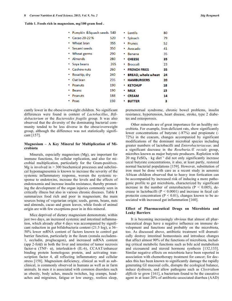
Minerals, especially magnesium (Mg), are important for immune functions, for cellular replication, and also for mi-crobial multiplication, particularly for the Gram-positives. Mg is involved in > 300 biochemical processes and subclini-cal hypomagnesemia is known to increase the severity of the systemic inflammatory response, worsen the systemic re-sponse to endotoxins, increase the levels and the effects of endotoxemia and increase insulin resistance, thereby promot-ing the development of the organ injuries commonly seen in critically illness but also in various chronic diseases. Table 1 summarizes food rich and poor in magnesium; the main sources being of vegetarian origin; seeds, germs, beans, nuts and almonds, cacao and green leaves, while foods of animal origin are with few exceptions poor in in this mineral.
Mice deprived of dietary magnesium demonstrate, within just two days, an increased systemic and intestinal inflamma-tion, which already after 4 days was accompanied by signifi-cant reduction in gut bifidobacteria content (21.5 log), a 36–50% lower mRNA content of factors known to control gut barrier function, particularly in the ileum (zonula occludens-1, occludin, proglucagon), and increased mRNA content (app 2-fold) in both the liver and intestine of tumor necrosis factor- (TNF- ), interleukin-6 (IL-6), CCAAT/enhancer binding protein homologous protein, and activating tran-scription factor 4, all reflecting inflammatory and cellular stress [158]. Magnesium deficiency, clinical as well as sub-clinical, is commonly observed in humans as well as in farm animals. In men it is associated with common disorders such as obesity, body aches, muscle twitches, leg cramps, head-aches and migraines, fatigue or low energy, restless sleep,
premenstrual syndrome, chronic bowel problems, insulin resistance, hypertension, heart disease, stroke, type 2 diabe-tes and osteoporosis.
Other minerals are of great importance for an healthy mi-crobiota. For example, Iron-deficient rats, show significantly lower concentrations of butyrate (-87%) and propionate (-72%) in the ceacum, changes accompanied by significant modifications of the dominant microbial species including greater numbers of lactobacilli and Enterobacteriaceae, and a significant decrease in the Roseburia./E rectale group, microbes known as major butyrate producers. Repletion with 20 mg FeSO · kg diet did not only significantly increase cecal butyrate concentrations, it also, at least partly, restored normal bacterial populations [159]. However, substitution of iron must be done with care as a recent study in aenemic African children observed that to heavy iron fortication can be accompanied by increased risk of inducing a more patho-genic profile to gut microbiota, characterized by significant increase in the number of enterobacteria (P < 0.005), de-crease in lactobacilli (P < 0.0001) and increase in fecal cal-protectin concentration (P < 0.01), changes known to be as-sociated with increased gut inflammation [160].
Effect of Pharmaceutical Drugs on Microbiota and Leaky Barriers
It is becoming increasingly obvious that almost all phar-maceutical drugs have a negative influence on immune de-velopment and functions and probably on the microbiota, too. As discussed above, antibiotic treatment will dramati-cally destroy intestinal homeostasis and introduce changes that affect almost 90% of the functions of microbiota, includ-ing critical metabolic functions such as bile acid metabolism and eicosanoid and steroid hormone synthesis [132,133]. Similar negative effects on microbiota have been reported in association with chemotherapy treatment for cancer; for dec-ades this has been known to significantly damage the rapidly generating GI mucosal cells, disrupts the ecological balance, induce dysbiosis, and allow pathogens such as Clostridium difficile to grow [161], a bacterium found to be the causative agent in at least 20% of antibiotic-associated diarrhea (AAD)
Table 1. Foods rich in magnesium, mg/100 gram food .
Processed Foods, Dysbiosis, Systemic Inflammation Current Nutrition & Food Science, 2013, Vol. 9, No. 2 9
cases [162]. During chemotherapy treatment, as observed in a pediatric patient material, the total number of bacteria in fecal samples is reduced to only 109 per gram of dry weight feces, which is 100-fold lower than normally seen in healthy individuals, and on fluorescent in situ hybridization analysis shown to consist in an up to 10,000-fold decrease in anaero-bic bacteria and a 100-fold increase in potentially pathogenic enterococci [163].
The negative effects of pharmaceutical drugs on microbi-ota are not only limited to antibiotics and chemotherapeutics. Negative effects on microbiota also occur with other drugs including those that, in the past, have been assumed have no or limited side effects, such as proton pump inhibitors and anti-hypertensive drugs. As examples, the offspring of moth-ers consuming proton pump inhibitors during pregnancy has a significantly increased risk of acquiring asthma later in life [164], while users of hypertensive drugs suffer not only sig-nificantly reduced salivation and severe mouth dryness (xerostomia) but also a documented profound oral dysbiosis [165].
New information concerning intimate cross-talk between the intestinal microbiota and the host immune system has opened new avenues. Alterations in the microbiota are known to immediately induce increased translocation of bac-terial antigens and dramatically alter the host immune reac-tion, leading to a chronic inflammatory state and impaired metabolic function, including insulin resistance, hepatic fat deposition, insulin unresponsiveness, and excessive adipose tissue development [166]. Consequently, each decision to use pharmacological treatment may, in the future, need to be based on weighing the need of pharmacological treatment against the importance of maintaining microbiota homeosta-sis and preventing leakage at body surfaces. Clearly, the im-pact of newly developed pharmaceuticals on microbiota and immune functions, neglected in the past, should be fully in-vestigated before products are licensed for public use.
It is very unfortunate that pharmacological treatment and bioecological treatments are in general not compatible. It is frequently observed that pre-, pro- and synbiotic treatments are more successful in experimental animals than in man. Until today most, if not all, clinical trials using probiotic treatment have had to accept being applied merely as adjunc-tive interventions, i.e. in parallel to existing pharmaceutical treatment, and never having the chance to be tried as a truly alternative treatment. Particularly in critically ill patients, trials involving probiotics have always been influenced, and most likely, strongly handicapped by a parallel application of heavy antibiotic, chemotherapeutic and other similar regi-mens. In many, if not most, incidences the supplied probiot-ics have been dramatically compromised before reaching their target organs, which could well explain the absence of positive results observed, especially in the critically ill.
Dysbiosis, Leaky Barriers, Unhealthy.
Increasing evidence suggests that during an inflammatory state leakage will occur, not only from the gut barrier, but also from multiple other barriers such as the oral cavity, the upper GI tract, the airways, the skin, the vagina and female reproductive tract, the placenta, the eye cavity etc., but also the blood-brain barrier, might be of equal importance in the
pathogenesis of disease. Especially are the oral cavity, the skin and the placenta increasingly in focus.
Leaky gut (loss of gut barrier integrity): The gut meets the exterior world across a surface suggested be approxi-mately 7-8,000 m2 - equivalent to the size of a soccer field. This surface is the object of extreme challenges with at least half, if not more, of individuals living a Western-type life-style suggested to suffer impaired microbiota and more or less permanent leaky gut. Increased translocation of toxic or infectious molecules and even whole microorganisms is a frequent phenomenon in a comprehensive series of diseases. The transfer of these elements and others occurs paracellu-larly e.g. through the intercellular space referred to as ‘tight junctions’, but also trans-cellularly, and then encapsulated in fat molecules from the consumed foods. The tight junctions, once regarded as static structures, are now known to be ex-tremely dynamic and ready to adapt to a variety of develop-mental, physiological, and pathological circumstances, and regulated by several molecules including the interesting en-dogenous modulators named zonulins [167, 168]. The tight-ness of the GI mucosa is largely dependent on consumed foods and its effects on intestinal microflora and is thus strongly associated with dysbiosis and subsequent inflamma-tion.
Life-style factors such as physical activity, intake of al-cohol and cigarette smoking play important roles gut mu-cosal integrity. In addition, the dominant regulatory factors are processed and refined food, sugars and content of insu-lino-trophic molecules such as refined carbohydrates, pro-teotoxic foods such as gluten and dysfunctioning molecules such as AGEs and ALEs, molecules common in modern food/industrially produced foods etc., have great disadvan-tages to barriers integrity. High temperature-produced foods are prevalent in the Western world, commonly produced in processes such as high temperature bread baking, and in preparation of fast foods, to a large extent dependent on fry-ing and grilling [2-4,148], storage of foods for longer peri-ods, even at room temperature, as well as flavoring of foods, is known enhance the availability of these molecules in the foods[15-17,169]. These molecules play important roles in the pathogenesis of diseases including Alzheimer´s disease [170], cardiovascular diseases [171, 172], chronic liver dis-eases [173-175], chronic kidney disease [176, 177], chronic obstructive pulmonary diseases (COPD) [178], diabetes [179], inflammatory bowel diseases(IBD) [180, 181], irrita-ble bowel syndrome(IBS) [182], paradontal diseases such as paradontosis [183] and polycystic ovary syndrome (PCOS) [184]. Leaky gut is also seen in a large variety of other con-ditions, such as alcoholism [185], autoimmune diseases [186], chronic encephalopathy [45, 187], chronic fatigue syndrome [188, 189], mental depression [190, 191] and other, idiopathic, conditions, which are mainly observed in the Western world. Translocation of endotoxin , viruses [192, 193], live bacteria and debris of bacteria [194, 195] not only occurs but can remain intra-cellular in various cell types; these may be particularly observed in the adipocytes in obesity, where they seem to enhance inflammation and further storage of fats.
Leaky oral cavity: The oral cavity comprises different mucosal sites, anaerobic pockets and teeth. Each harboring a
10 Current Nutrition & Food Science, 2013, Vol. 9, No. 2 Stig Bengmark
unique and diverse microbial assemblage. Great interper-sonal variation in pattern of microbiota exists. Some oral communities are dominated by Streptococcus species and others by Prevotella, Neisseria, Haemophilus, or Veillonella species. Accumulation of pathogens and inflammatory cells in the vascular wall and the subsequent release of pro-inflammatory cytokines are thought to exacerbate athero-genic processes. Studies published over the last two decades suggest that coronary artery disease may be due to an infec-tion-induced inflammation, but also that the impact of infec-tion on atherogenesis relates to numbers of aggregated pathogens within the endothelial walls/plaques, a concept referred to as ”pathogen burden” [196]. Several studies pub-lished thereafter confirm an oral source of bacteria associ-ated with atherosclerotic plaques [197-199]. A recently pub-lished study of 15 individuals identified Chryseomonas in all atherosclerotic plaque samples studied, and Veillonella and Streptococcus in the majority of them [200]. The combined abundances of Veillonella and Streptococcus in atheroscle-rotic plaques correlated well with their abundance in the oral cavity. Several additional bacterial phylotypes in the same individual were common both to the atherosclerotic plaque and oral or gut samples. Interestingly, several bacterial taxa in the oral cavity and the gut also correlated with levels of plasma cholesterol [200].
Special attention has been paid to Chlamydia pneumonia, the first bacteria identified in atherosclerotic lesions [201], a species known to possess the ability to promote lipid body formation in human macrophages [202]. Recently a diverse range of bacteria have been identified in human atheroma (202). The most frequently observed being Gram-negative, including Acenetobacter baumannii, Escherichia coli, Kleb-siella pneumonia, Pseudomonas aeruginosa, Pseudomonas diminutive and Proteus vulgaris and Gram-positive; Staphy-lococcus aureus, Staphylococcus epidermidis, and Strepto-coccus salivarius [202]. Each of these bacteria, even heat-killed is known to stimulate Toll-like receptors and have demonstrated ability to induce lipid body formation and cho-lesterol ester accumulation in a dose-dependent manner. Mi-crobial debris in atheroma, in the past largely considered harmless, might well play a major role in the formation of lipid bodies in the arterial wall but also in the continuous progress of the arteriosclerotic disease [202]. It is not yet fully verified if the translocation occurs predominantly in the oral cavity or further down the GI tract. The present belief is, however, that it occurs directly through the gingiva and that brisk tooth-brushing, (with eventual smaller bleeding), might enhance the process.
Leaky airways: The surface of adult human airway is, af-ter the gut, the second largest in the body, thought to cover up to 200 m2 (size of a tennis court). Exposure of sensitive individuals to antigens can induce allergic responses, mainly apparent in the respiratory tract but also in the skin and eyes, manifesting as vasodilatation, plasma leakage, leukocyte influx, and bronchoconstriction. Endothelial gaps have been identified through which leakage of plasma and inflamma-tory mediators occur [203], accompanied by leukocyte influx and accumulation of plasma proteins in the airway mucosa. Less interest has been paid to the process of leakage from the airways through the airway epithelium and into the circula-tion, despite the fact that such leakage is a very common
phenomenon, probably as frequent as leaky gut. Such leak-age is known to influence expression pattern/ recognition of receptors that detect environmental stimuli and secrete en-dogenous danger signals, activate dendritic cells and innate and adaptive immunity [203].
For some reason healthy airways until recently been re-garded as sterile but now we know that it has both a rich and diverse microbiota. Most recent studies of microbiota have tended to focus on microbiota in individuals with airway diseases, such those with asthma [203-205], cystic fibrosis (CF) [206, 207], obstructive lung disease (COPD) [208, 209], mechanically ventilated preterm infants [210]. Very less information being available regarding normal microbiota in healthy nose and lungs. In CF for example, in addition to previously recognized pathogens typical for the disease, such as Pseudomonas aeruginosa and Staphylococcus aureus, another 460 phylogenetically diverse bacterial genera, not previously associated with the disease, have now been re-ported [206]. However, much as in the gut, the airway mi-crobiota of patients with CF are not only polymicrobial but also spatially heterogeneous, few taxa being common to all microbial communities in the different anatomical regions of the airways [207]; consequently treatment based only on cultivation of sputum might not always be adequate. Future studies will most probably try to further explore the microbi-ota of different microbial communities in the airways in healthy individuals, as well as the mechanisms behind leaky airways, and the extent and consequences of such leakage for health, are not only associated to the airways, but to the whole body.
Leaky skin: The skin, compared to the gut and the air-ways, a quite modest surface area – less than 2 m2, corre-sponding to approximately half a table tennis board. Non-invasive techniques to study the barrier function of the skin have long been available. It is well known that a number of human skin conditions and disorders are associated with de-fects in the skin barrier and subsequent increased permeabil-ity. Most of the skin barrier function resides in the cornified layer, while most immune cells, especially the dendritic cells/Langerhans cells are located slightly below. The human skin harbors myriad bacteria, fungi, and viruses, these mi-crobial communities are intricately linked to human health and disease. Recent findings suggest that a dysfunctional epidermal barrier is pathologically involved in a variety of common, antigen-driven skin diseases, allergic diseases such as atopic dermatitis (AD) as well as psoriasis [211], and probably contributes to several general health disorders. Ge-nomic approaches reveal a great diversity of organisms pre-dominantly within the four main phyla: Actinobacteria, Fir-micutes, Bacteroidetes and Proteobacteria [212]. Great dif-ferences in the pattern of microbiota are observed in indi-viduals and also in different anatomical regions of the skin. It is largely associated with differences in structure and physi-ology of the various skin sites but also depending on factors including hygiene and character of the skin with moist, dry or sebaceous microenvironments [211, 213]. Staphylococcus and Corynebacterium are the dominant colonizing organisms of moist areas. The greatest diversity of microbes is, how-ever, found in the dry areas with a mixed representation from all four phyla [212]. It is most interesting that Gram-negative organisms, previously thought to rarely colonize the skin, are

Processed Foods, Dysbiosis, Systemic Inflammation Current Nutrition & Food Science, 2013, Vol. 9, No. 2 11
found in abundance in the dry areas, an observation which might have great implications for disease development not only within the skin but in the whole body [212].
The transfer of chemicals through the skin is so effective and reliable that it is increasingly used for drug delivery of analgesics, such as Buprenorphine, Caisapsin, Fentanyl and Lidocaine, hormones, such as estradiol, progesterone and testosterone, drugs against motion sickness and nausea, such as Scopolamine and Granisetron, anti-inflammatory drugs, such as Ketoprofen, Piroxicam, Piclofenac, antihyperten-sives, including Clonidine, Rivastigmine, and Rotigotine to be used in Alzheimer´s and Parkinson´s diseases, Selegiline for mental depression, Oxybutynin for hyperactive bladder, and antihypertensives like Clonidine and Methylphenidate prescribed for ADHD [214], in total some 40 products as registered in 2010 [214]. The fact that at least half of the drugs are meant to target the central nervous system means that they not only have ability to transfer through the skin but also through other barriers, including the blood-brain barrier. If these chemicals can easily pass the skin barriers, and also the blood-brain, it is most likely that other chemicals, such as cosmetics will do the same.
Translocation of chemicals and microbes in individuals with intact skin occurs mainly through the hair follicles. In burnt patients, however, where the protective layer has been eliminated, it occurs directly through the skin. Microbial translocation, sepsis and eventually multiple organ failure (MOF) was for long time thought to happen via a leaky gut. Increasing evidence suggest, however, that to a large extent, such translocation occurs directly through the burned skin surfaces, especially as cultivations from blood and septic skin areas are dominated by pathogens typical for skin. A recent study looked at the microbial pattern in blood and at burn surfaces in a group of 338 patients with thermic inju-ries. The microbes most commonly simultaneously culti-vated in both blood and at the burned skin surfaces were Acinetobacter baumannii (47%) and Pseudomonas aerugi-nosa (37%) [215]; other frequently isolated microorganisms identified in this study were the Gram-positive Staphylococ-cus epidermidis MRSE (20%) and Staphylococcus aureus MRSA (19%) [215].
Leaky vagina (incl. the whole female reproductive
tract): The vaginal microbiota provides a vital and highly effective defense mechanism against a whole range of mi-crobial infections [138]. The predominant phyla of bacteria identified in the vagina belong to Firmicutes, Bacteroidetes, Actinobacteria and Fusobacteria [216]. No single bacterium has been identified as a specific marker for healthy over dis-eased conditions, but three phyla - Bacteroidetes, Actinobac-teria and Fusobacteria, and eight genera including Gardnerella, Atopobium, Megasphaera, Eggerthella, ero-coccus, Leptotrichia/Sneathia, Prevotella and Papillibacter are strongly associated with bacterial vaginosis (BV) (p < 0.05) [216]. The vaginal bacterial communities of 396 as-ymptomatic North American women, representing four eth-nic groups (white, black, Hispanic, and Asian), were recently characterized by pyrosequencing of barcoded 16S rRNA genes [217]. The communities clustered into five groups: four dominated by Lactobacillus iners, L. crispatus, L. gas-seri, or L. jensenii. The proportions of each community
group varied significantly among the four ethnic groups (p < 0.0001). Moreover, the vaginal pH of women in different ethnic groups also differed being higher in Hispanic (pH 5.0 ± 0.59) and black (pH 4.7 ± 1.04) women than in Asian (pH 4.4 ± 0.59) and white (pH 4.2 ± 0.3) women [217].
The tight junction protein, occludin, is to a large extent under control of estrogens and the tightness of the vaginal mucosa will for that reason vary significantly with age [218], as well as with the menstrual cycle. Not only the vagina but the whole female reproductive tract (FRT) has unique struc-tures for the regulation of immune protection, especially as it must deal with not only with sexually transmitted pathogens, but also with allogeneic spermatozoa, and the immunologi-cally very different fetus. To meet these challenges, the FRT has evolved unique immune mechanisms to protect against potential pathogens without compromising fetal survival or maternal health [219].
More than twenty pathogens are transmissible through sexual intercourse, and an estimated 340 million new cases of sexually transmitted infections (STI) are reported each year; bacteria such as (group B streptococcus, Neisseria gonorrhoeae, Chlamydiatrachomatis, Treponema pallidum), parasites (Trichomonas vaginalis), and viruses (Herpes sim-plex, Human Papilloma, Human Immunodeficiency) are commonly identified [219]. The epithelial cell structures of vagina and FRT possess intracellular and extracellular pathogen recognition receptors (TLR, NOD, RIG, MDA-5, etc), and have the ability to secrete chemokines and cytoki-nes that initiate, regulate and link together innate and adap-tive immune responses, present antigens to T cells, produce polymeric immunoglobulin receptors for transporting mu-cosal IgA antibodies from tissues into luminal secretions, and produce intracellular and secreted anti-microbial factors aimed to kill invading microbes [219].
Leaky blood brain barrier (BBB) (and the blood-
cerebrospinal fluid barrier (BCSFB): These two barriers constitute a tight seal between the circulating blood/cerebrospinal fluid and the central nervous system (CNS), both consisting of brain microvascular endothelial cells surrounded by basement membranes, astrocytic endfeet, and pericytes. The brain microvascular endothelium is char-acterized by the presence of tight junctions (TJs) and a lack of fenestrae, meant to limit the entry of plasma components, as well as red blood cells and leukocytes, into the CNS. These anatomical structures confer a low paracellular perme-ability and high electrical resistance to the deposition of molecules such as amyloid beta (Ab) into leptomeningeal and cortical brain vasculature, characteristic of Alzheimer´s disease.
Interplay between dozens of connecting transmembrane proteins (occludin and claudins) are as essential to these bar-riers, in their tight junction formation and function, as they are to all other barriers in the body, and demonstrated to mal-function when leakage occur. Clearly dysfunction of these barriers and their efflux and influx transporters constitute a major factor in the pathogenesis of degenerative neuronal disorders. Complex interactions between AGEs, advanced lipoxidation end products (ALEs), the receptor for advanced glycation end products (RAGE), oxidative stress, inflamma-tory mediators, common proinflammatory pathways and
12 Current Nutrition & Food Science, 2013, Vol. 9, No. 2 Stig Bengmark
amyloid-beta (A beta) peptide contribute to BBB dysfunction in a series of degenerative disorders [15-17, 220, 221]. Mal-function of other transporters such as the organic anion transporter (OAT) 3 and organic cation transporter (OCT) 3 is essential for the leakage of toxic injurious material [222]. Endotoxins, originating from a leaky gut, induce disruption in tight junction (TJ) functions, increase paracellular perme-ability and alters the functions of the TJ proteins occludin ZO-1, and ZO-2, and thereby increase transcellular leakage as observed in sepsis-induced barrier leakage [223,224], and also in encephalopathies in general.
Leaky placenta: For the last two decades, it was known that not all babies are born in sterile conditions [225, 226]. When umbilical cord blood was cultivated from healthy neo-nates born by cesarean section, a shocking 9/20 (45 %) dem-onstrated positive growth and the following species were identified on 16S rDNA sequencing: Enterococcus faecium, Propionibacterium acnes, Staphylococcus epidermidis, and Streptococcus sanguinis [227]. More recent studies describe a strong association between infection, within the amniotic cavity, and low birth weight. These studies reveal the pres-ence of various opportunistic pathogens in the amniotic cav-ity, most of them from outside the genitourinary tract, often of oral origin but also sometimes coming from other body sites of the mother, such as the gut, which are all thought to contribute to the development premature labor and birth. A number of bacteria have been cultured in amniotic infections including Fusobacterium nucleatum, a common oral species, being the most frequently isolated species from amniotic fluid, but Fusobacterium nucleatum, Peptostreptococcus spp, Porphyromonas and Prevotella,Eubacterium and Eiken-ella corrodens are also sometimes found the amniotic fluid of women with preterm labour [227, 228].
Chorioamnionitis is a new entity, defined as the inflam-matory response of the membranes, placenta and amniotic fluid in response to a microbial invasion of the amniotic cav-ity, frequently seen and associated with a greatly enhanced risk of adverse neonatal outcome [229, 230]. Chorioamnioni-tis is most often clinically silent or diagnosed in the presence of signs of inflammatory reactions of the mother, often very early in pregnancy and more or less always associated with microbial invasion of the amniotic cavity, as documented by microbial cultures of amniotic fluid and histologic analysis of the placenta and its membranes [229-231].
Barker et al, in their classical series of studies, presented mainly between 1996 and 2004, provide evidence of a link between intra-uterine programming of the immune system of the infant and later in life chronic diseases, particularly car-diovascular events [232-237]. Since then, numerous clinical and experimental studies have confirmed the early develop-mental influences, with and without alterations in birth weight, on not only later in life cardiovascular but also pul-monary, metabolic, and psychological diseases. The intrau-terine environment is dramatically and almost exclusively impacted by the overall maternal lifestyle and health. Both premature birth and low or high birth weights are most often associated with maternal conditions including alcohol, drug and pharmaceutical consumption, use of tobacco or other toxic substances, over- as well as under-nutrition, dys-nutrition of other reasons, metabolic conditions including
obesity, diabetes and hypercholesterolemia, chronic maternal stresses, infections and inflammations of other reasons [238]. A recent study suggests that the beta-cell adaptive growth, which normally occurs during gestation, does not, under the above-mentioned conditions, take place in the offspring, with risk of gestational diabetes and propagation of diabetes to the new generation [239]. Women with high degree of systemic inflammation, such as seen in psoriasis, are reported to suffer a two-fold risk of chronic diseases. It is most likely that leaky placenta and subsequent chorionamnionitis will pro-vide a satisfactory explanation for such a development, oc-curring at a time when the immune system of the fetus is in its most sensitive phase of “calibration” – the third trimester of pregnancy.
IgG is actively transported across the placenta during the later stages of pregnancy to provide passive immunization of the fetus [240], and hence such antibodies reflect maternal exposures and immune responses to specific antigens. Re-cent studies on archival dried blood report associations be-tween the future development of non affective psycho-sis/schizophrenia in the offspring and high levels of immu-noglobulin G (IgG) directed at the protozoan Toxoplasma gondii [241], herpes simplex virus, type 2 [242] and gluten (anti-gliadin IgG) [243] in the maternal circulation.
Over-reacting Neutrophils
Dysbiosis frequently occurs in severe trauma, major sur-gery and severe sepsis, often in parallel with a significant decrease in lymphocytes, a significant, sometimes dispropor-tionate, increase in circulating and tissue neutrophils, and a persistent decline in T-4 helper lymphocytes and elevation of T-8 suppressor lymphocytes [244]. It is suggested that a T-4/T-8 lymphocyte cell ratio of < 1 is a sign of severe immu-nosuppression and prediction of poor outcome in conditions such as multiple and severe trauma, multiple organ dysfunc-tion syndrome, severe acute pancreatitis but also in myocar-dial infarction, and in chemotherapeutic treatments, espe-cially with oncology patients [245]. A large early increase in circulating neutrophils is always accompanied by tissue infil-tration of neutrophils and is responsible for common post-trauma/postoperative dysfunctions such as paralytic ileus [246, 247], bone marrow suppression, endothelial cell dys-function, and leads to tissue destruction and organ failure, particularly in the lungs [248-251], intestines [251], liver [252]and kidney [253]. Neutrophil infiltration of distant or-gans [254], particularly the lungs [255], is significantly ag-gravated by mechanical therapeutic efforts such as handling of the bowels during operation [246], and ventilation of the lungs [255]. Poor nutritional status, preexisting immune de-ficiency, obesity, diabetes and high levels of blood sugar [256] contribute to immune deterioration and to increased expressions of molecules such as NF- B, COX-2, LOX and iNOS [256, 257]. It is important that a disproportionate in-crease in circulating neutrophils can, to a large extent, be successfully inhibited by the supplementation of antioxidants [257-259] as well as some specific probiotics [260-262]. Supplementation of some specified probiotics is also shown to effectively prevent neutrophil infiltration of the lungs and also to reduce the subsequent tissue destruction as demon-strated in studies with inflammation induced by cecal liga-tion and puncture (CLP) [262, 263].
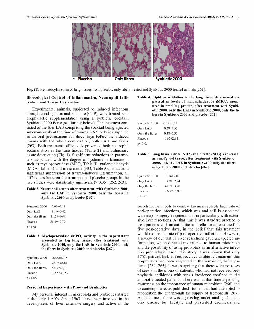
Processed Foods, Dysbiosis, Systemic Inflammation Current Nutrition & Food Science, 2013, Vol. 9, No. 2 13
Bioecological Control of Inflammation, Neutrophil Infil-
tration and Tissue Destruction
Experimental animals, subjected to induced infections through cecal ligation and puncture (CLP), were treated with prophylactic supplementation using a synbiotic cocktail, Synbiotic 2000 Forte (see further below). The treatment con-sisted of the four LAB comprising the cocktail being injected subcutaneously at the time of trauma [262] or being supplied as an oral pretreatment for three days before the induced trauma with the whole composition, both LAB and fibers [263]. Both treatments effectively prevented both neutrophil accumulation in the lung tissues (Table 2) and pulmonary tissue destruction (Fig. 1). Significant reductions in parame-ters associated with the degree of systemic inflammation, such as myeloperoxidase (MPO, Table 3), malondialdehyde (MDA, Table 4) and nitric oxide (NO, Table 5), indicated a significant suppression of trauma-induced inflammation, all differences between the treatment and placebo groups in the two studies were statistically significant (> 0.05) [262, 263].
Table 2. Neutrophil counts after treatment with Synbiotic 2000,
only the LAB in Synbiotic 2000, only the fibers in
Synbiotic 2000 and placebo [262].
Synbiotic 2000 9.00±0.44
Only LAB 8.40±0.42
Only the fibers 31.20±0.98
Placebo 51.10±0.70
p< 0.05
Table 3. Myeloperoxidase (MPO) activity in the supernatant
presented as U/g lung tissue, after treatment with
Synbiotic 2000, only the LAB in Synbiotic 2000, only
the fibers in Synbiotic 2000 and placebo [262].
Synbiotic 2000 25.62±2,19
Only LAB 26.75±2,61
Only the fibres 56.59±1,73
Placebo 145.53±7,53
p< 0.05
Personal Experience with Pro- and Synbiotics
My personal interest in microbiota and probiotics started in the early 1980’s. Since 1963 I have been involved in the development of liver extensive surgery and active in the
search for new tools to combat the unacceptably high rate of peri-operative infections, which was and still is associated with major surgery in general and in particularly with exten-sive liver resections. At that time it was standard practice to treat patients with an antibiotic umbrella for at least the first five post-operative days, in the belief that this treatment would reduce the rate of post-operative infections. However, a review of our last 81 liver resections gave unexpected in-formation, which directed my interest to human microbiota and the possibility of using probiotics as an alternative infec-tion prophylaxis. From this study it was shown that only 57/81 patients had, in fact, received antibiotic treatment; this prophylaxis had been neglected in the remaining 24/81 pa-tients [264, 265]. It was surprising that there were no cases of sepsis in the group of patients, who had not received pro-phylactic antibiotics with sepsis incidence confined to the antibiotic-treated patients. There was at that time a growing awareness on the importance of human microbiota [266] and to contemporaneous published studies that had attempted to recondition the gut through the supply of lactobacilli [267]. At that times, there was a growing understanding that not only disease but lifestyle and prescribed chemicals and
Fig. (1). Hematoxylin-eosin of lung tissues from placebo, only fibers-treated and Synbiotic 2000-treated animals [262].
Table 4. Lipid peroxidation in the lung tissue determined ex-
pressed as levels of malondialdehyde (MDA), meas-
ured in nmol/mg protein, after treatment with Synbi-
otic 2000, only the LAB in Synbiotic 2000, only the fi-
bers in Synbiotic 2000 and placebo [262].
Synbiotic 2000 0.22±1,31
Only LAB 0.28±3,55
Only the fibres 0.48±5,32
Placebo 0.67±2,94
p< 0.05
Table 5. Lung tissue nitrite (NO2) and nitrate (NO3), expressed
as mol/g wet tissue, after treatment with Synbiotic
2000, only the LAB in Synbiotic 2000, only the fibers
in Synbiotic 2000 and placebo [262].
Synbiotic 2000 17.16±2,03
Only LAB 8.91±2,24
Only the fibres 47.71±3,20
Placebo 66.22±5,92
p< 0.05
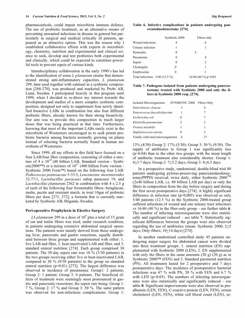
14 Current Nutrition & Food Science, 2013, Vol. 9, No. 2 Stig Bengmark
pharmaceuticals, could impair microbiota immune defense. The use of probiotic treatment, as an alternative means of preventing unwanted infections in disease in general but par-ticularly in surgical and medical critically ill patients, ap-peared as an attractive option. This was the reason why I established collaborative efforts with experts in microbiol-ogy, chemistry, nutrition and experimental and clinical sci-ence to seek, develop and test probiotics both experimental and clinically, which could be expected to constitute power-ful tools to prevent sepsis of various kinds.
Interdisciplinary collaboration in the early 1990’s has led to the identification of some L plantarum strains that demon-strated strong anti-inflammatory capacities. L plantarum 299, later used together with oatmeal in a synbiotic composi-tion [268-270], was produced and marketed by Probi AB, Lund, Sweden. I participated heavily in this program until 1999, when I decided to re-direct my interest towards the development and studies of a more complex synbiotic com-position, designed not only to supplement four newly identi-fied bioactive LABs in combination but also four different prebiotic fibers, already known for their strong bioactivity. Our aim was to provide this composition in much larger doses that was being practiced at that time. Furthermore, knowing that most of the important LABs rarely exist in the microbiota of Westerners encouraged us to seek potent pro-biotic bacteria among bacteria normally growing on plants instead of selecting bacteria normally found in human mi-crobiota of Westerners.
Since 1999, all my efforts in this field have focused on a four LAB/four fiber composition, consisting of either a mix-ture of 4 x 10¹⁰ (40 billion LAB, Standard version - Synbi-otic2000™) or a mixture of 1011 (400 billion Forte version -Synbiotic 2000 Forte™) based on the following four LAB: Pediococcus pentosaceus 5-33:3, Leuconostoc mesenteroides 32-77:1, Lactobacillus paracasei subsp paracasei 19, and Lactobacillus plantarum 2362 in combination with 4 x 2.5 g of each of the following four fermentable fibres: betaglucan, inulin, pectin and resistant starch, in total 10gm of prebiotic fibers per dose [271, 272], a formula that is currently mar-keted by Synbiotic AB, Höganäs, Sweden.
Peri-operative Prophylaxis in Elective Surgery
Lb plantarum 299 in a dose of 109 plus a total of 15 gram of oat and inulin fibers was tried, under research condition, in patients undergoing extensive abdominal surgical opera-tions. The patients were mainly derived from those undergo-ing liver, pancreatic and gastric resections, equally distrib-uted between three groups and supplemented with either: 1. live LAB and fiber, 2. heat-inactivated LAB and fiber, and 3. standard enteral nutrition [274]. Each group comprised 30 patients. The 30-day sepsis rate was 10 % (3/30 patients) in the two groups receiving either live or heat-inactivated LAB, compared to 30 % (9/30 patients) in the group on standard enteral nutrition (p=0.01) [273]. The largest difference was observed in incidence of pneumonia: Group1: 2 patients; Group 2: 1 patient; Group 3: 6 patients. The beneficial ef-fects of treatment were seemingly most pronounced in gas-tric and pancreatic resections; the sepsis rate being: Group 1: 7 %, Group 2: 17 % and Group 3: 50 %. The same pattern was observed for non-infectious complications: Group 1:
13% (4/30) Group 2: 17% (5/30); Group 3: 30 % (9/30). The supply of antibiotics to Group 1 was significantly less (p=0.04) than to the other two groups, with the mean length of antibiotic treatment also considerably shorter: Group 1: 4+3.7 days; Group 2: 7+5.2 days; Group 3: 8+6.5 days.
In a prospective study, randomized, double-blind trial 80 patients undergoing pylorus-preserving pancreatoduodenec-tomy(PPPD) received, twice daily, either Synbiotic 2000TM
(2x40 billion LAB, i.e. 80 billion LAB per day) or only the fibers in composition from the day before surgery and during the first seven postoperative days [274]. A highly significant difference in infection rate (p=0,005) was observed as only 5/40 patients (12.5 %) in the Synbiotic 2000-treated group suffered infections (4 wound and one urinary tract infection) vs 16/40 (40 %) in the fiber-only group – see further table 6. The number of infecting microorganisms were also statisti-cally and significant reduced – see table 7. Statistically sig-nificant differences between the groups were also observed regarding the use of antibiotics (mean: Synbiotic 2000; 2+5 days, Only-fibers; 10+14 days) [274].
In another randomized controlled study 45 patients un-dergoing major surgery for abdominal cancer were divided into three treatment groups: 1. enteral nutrition (EN) sup-plemented with Synbiotic 2000 (LEN), 2. EN supplemented with only the fibers in the same amounts (20 g) (20 g) as in Synbiotic 2000™ (FEN) and 3. Standard parenteral nutrition (PN). All treatments lasted for 2 preoperative and 7 days postoperative days. The incidence of postoperative bacterial infections was 47 % with PN, 20 % with FEN and 6.7 % with LEN (p<0.05). The numbers of infecting microorgan-isms were also statistically and significantly reduced – see table 8. Significant improvements were also observed in pre-albumin (LEN, FEN), C-reactive protein (LEN, FEN), serum cholesterol (LEN, FEN), white cell blood count (LEN), se-
Table 6. Infective complications in patients undergoing pan-
creatoduodenectomy [274].
Synbiotic 2000 Fibers only
Wound infections 4 6
Urinary infection 1 1
Peritonitis 0 5
Pneumonia 0 4
Sepsis 0 2
Cholangitis 0 1
Emphysema 0 1
Total infections 5/40 (12.5 %) 16/40 (40 %) p<0.05
Table 7. Pathogens isolated from patients undergoing pancrea-
tectomy treated with Synbiotic 2000 and only the fi-
bers in Synbiotic 2000 resp. [274].
Isolated Microorganisms SYNBIOTIC 2000 Fibers Only
Enterobacter cloacae 2 8
Enterococcus faecalis/faecium 1 7
Escherichia coli 0 7
Klebsiella pneumoniae 2 2
Proteus mirabilis 1 1
Staphylococcus aureus 0 2
Totally cultivated microorganisms 6 27
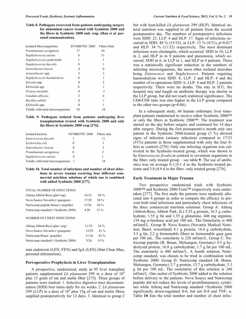
Processed Foods, Dysbiosis, Systemic Inflammation Current Nutrition & Food Science, 2013, Vol. 9, No. 2 15
rum endotoxin (LEN, FEN) and IgA (LEN) (Han Chun Mao, personal information).
Peri-operative Prophylaxis in Liver Transplantation
A prospective, randomized, study in 95 liver transplant patients supplemented Lb plantarum 299 in a dose of 109 plus 15 gram of oat and inulin fiber [275]. Three groups of patients were studied: 1. Selective digestive tract decontami-nation (SDD) four times daily for six weeks. 2. Lb plantarum 299 (LLP) in a dose of 109 plus 15g of oat and inulin fibres supplied postoperatively for 12 days. 3. Identical to group 2
but with heat-killed Lb plantarum 299 (HLP). Identical en-teral nutrition was supplied to all patients from the second postoperative day. The numbers of postoperative infections were SDD: 23, LLP: 4 and HLP: 17. Signs of infections oc-curred in SDD: 48 % (15/32), in LLP: 13 % (4/31), p=0.017 and HLP: 34 % (11/32) respectively. The most dominant infections were cholangitis, which occurred: SDD in 10, LLP in 2, and HLP in in 8 patients and pneumonia, which oc-curred: SDD in 6, in LLP in 1, and HLP in 4 patients. There was a statistically significant reduction in the numbers of infecting microorganisms, the most often isolated microbes being Enterococci and Staphylococci. Patients requiring haemodialysis were SDD: 8, LLP: 2 and HLP: 4 and the number of re-operations SDD: 6, LLP: 4 and HLP: 2 patients respectively. There were no deaths. The stay in ICU, the hospital stay and length on antibiotic therapy was shorter in the LLP group, but did not reach statistical significance. The CD4/CD8 ratio was also higher in the LLP group compared to the other two groups (p=0.06).
In a subsequent study, 66 human orthotopic liver trans-plant patients randomized to receive either Synbiotic 2000™ or only the fibers in Synbiotic 2000™. The treatment was started on the day before surgery and continued for 14 days after surgery. During the first postoperative month only one patient in the Synbiotic 2000-treated group (3 %) showed signs of infection (urinary infection) compared to 17/33 (51%) patients in those supplemented with only the four fi-bers as controls [276]. Only one infecting organism was cul-tivated in the Synbiotic-treated group, which was shown to be Enterococcus fecalis,in contrast to seventeen organisms in the fiber- only treated group – see table 9. The use of antibi-otics was on average 0.1+0.1 d in the Synbiotic-treated pa-tients and 3.8+0.9 d in the fiber- only treated group [276].
Early Treatment in Major Trauma
Two prospective randomized trials with Synbiotic 2000™ and Synbiotic 2000 Forte™ respectively were under-taken [277]. The first study the patients were randomly allo-cated into 4 groups in order to compare the efficacy to pre-vent both total infections and particularly chest infections of the three commercial nutrition solutions. Group A: Alitraq (Abbott-Ross, Abbott Park, IL) 5.25 g protein, 16.5 g carbo-hydrate, 1.55 g fat and 1.55 g glutamine, 446 mg arginine, 154 mg -linolenic acid per 100 mL. The Osmolarity is 480 mOsml/L. Group B: Nova Source (Novartis Medical Nutri-tion, Basel, witzerland) 4.1 g protein, 14.4 g carbohydrate, 3.5 g fat, 2.2 g fermentable fibers as fermentable guar gum per 100 mL. The osmolarity is 228 mOsm/L. Group C: Nu-tricomp peptide (B. Braun, Melsungen, Germany) 4.5 g hy-drolyzed protein, 16.8 g carbohydrate, 1.7 g fat per 100 mL. The osmolarity is 400 mOsm/L. A fourth solution, Nutri-comp standard, was chosen to be tried in combination with Synbiotic 2000: Group D: Nutricomp standard (B. Braun, Melsungen, Germany) 3.7 g protein, 13.7 g carbohydrate, 3.3 g fat per 100 mL. The osmolarity of this solution is 240 mOsm/L. One sachet of Synbiotic 2000 added to the solution before delivery to the patients. Nova Source and Nutricomp peptide did not reduce the levels of proinflammatory cytoki-nes while Alitraq and Nutricomp standard +Synbiotic 2000 significantly down-regulated Il-6, but not Il-8 and TNF- . Table 10 lists the total number and number of chest infec-
Table 8. Pathogens recovered from patients undergoing surgery
for abdominal cancer treated with Synbiotic 2000 and
the fibers in Synbiotic 2000 only resp. (Han et al. per-
sonal communication).
Isolated Microorganisms SYNBIOTIC 2000 Fibers Only
Pseudomonas aeruginosa 17 24
Staphylococcus aureus 8 11
Staphylococcus epidermidis 1 1
Staphylococcus faecalis 0 1
Enterobactercloacae 4 0
Acinetobacter spp. 2 3
Staphylococcus haemolyticus 0 1
Serratia spp. 0 2
Klebsiella spp. 0 1
Proteus mirabilis 0 2
Candida albicans 2 6
Bacillus subtilis 0 1
Klebsiella spp. 0 1
Totally cultivated microorganisms 34 54
Table 9. Pathogens isolated from patients undergoing liver
transplantation treated with Synbiotic 2000 and only
the fibers in Synbiotic 2000 resp [276].
Isolated bacteria SYNBIOTIC 2000 Fibers only
Enterococcus faecalis 1 11
Escherichia coli 0 3
Enterobacter cloacae 0 2
Pseudomonas aeruginosa 0 2
Staphylococcus aureus 0 1
Totally cultivated microorganisms 1 18
Table 10. Total number of infections and number of chest infec-
tions in severe trauma receiving four different com-
mercial nutrition solutions of which one is combined
with added Synbiotic 2000 [277].
TOTAL NUMBER OF INFECTIONS:
Alitraq Abbott-Ross (glut+arg) 16/32 50 %
Nova Source Novartis (+guargum) 17/29 58 %
Nutricomp peptide Braun (+peptide) 13/26 50 %
Nutricomp standard (+Synbiotic 2000) 4/26 15 %
NUMBER OF CHEST INFECTIONS:
Alitraq Abbott-Ross (glut +arg) 11/32 34 %
Nova Source Novartis (+guargum) 12/29 41 %
Nutricomp Braun (peptide) 11/26 42 %
Nutricomp standard (+Synbiotic 2000) 5/26 19 %
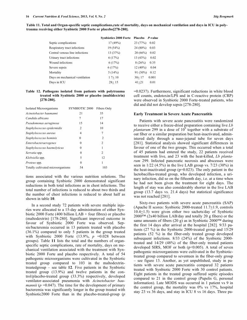
16 Current Nutrition & Food Science, 2013, Vol. 9, No. 2 Stig Bengmark
tions associated with the various nutrition solutions. The group containing Synbiotic 2000 demonstrated significant reductions in both total infections as in chest infections. The total number of infections is reduced to about two thirds and the number of chest infections is reduced to about half as shown in table 10.
In a second study 72 patients with severe multiple inju-ries were allocated to a 15-day administration of either Syn-biotic 2000 Forte (400 billion LAB + four fibres) or placebo (maltodextrin) [178-280]. Significant improved outcome in favour of Synbiotic 2000 Forte was observed. Sep-sis/bacteremia occurred in 13 patients treated with placebo (36.1%) compared to only 5 patients in the group treated with Synbiotic 2000 Forte (13.9%, p =0.028 between groups). Table 11 lists the total and the numbers of organ-specific septic complications, rate of mortality, days on me-chanical ventilation associated with the treatment with Syn-biotic 2000 Forte and placebo respectively. A total of 54 pathogenic microorganisms were cultivated in the Synbiotic treated group compared to 103 in the maltodextrin-treatedgroup – see table 12. Five patients in the Synbiotic treated group (13.9%) and twelve patients in the con-trol/placebo-treated group (33.3%) respectively, developed ventilator-associated pneumonia with Acinetobacter bau-mannii (p =0.047). The time for the development of primary bacteremia was significantly longer in the group treated with Synbiotic2000 Forte than in the placebo-treated-group (p
=0.0237). Furthermore, significant reductions in white blood cell counts, endotoxin/LPS and in C-reactive protein (CRP) were observed in Synbiotic 2000 Forte-treated patients, who did and did not develop sepsis [278-280].
Early Treatment in Severe Acute Pancreatitis
Patients with severe acute pancreatitis were randomized to receive either a freeze-dried preparation containing live Lb plantarum 299 in a dose of 109 together with a substrate of oat fiber or a similar preparation but heat-inactivated, admin-istered daily through a naso-jejunal tube for seven days [281]. Statistical analysis showed significant differences in favour of one of the two groups. This occurred when a total of 45 patients had entered the study, 22 patients received treatment with live, and 23 with the heat-killed, Lb planta-rum 299. Infected pancreatic necrosis and abscesses were seen in 1/22 (4.5%) in the live LAB group vs. 7/23 (30%) in the heat-inactivated group (p=0.023). The only patient in the lactobacillus-treated group, who developed infection, a uri-nary infection, did so on the fifteenth day, i.e. at a time when he had not been given the treatment for eight days. The length of stay was also considerably shorter in the live LAB group (13.7 days vs. 21.4 days) but statistical significance was not reached [281].
Sixty-two patients with severe acute pancreatitis (SAP) (Apache II scores: Synbiotic 2000-treated 11.7+1.9, controls 10.4+1.5) were given either two sachets/day of Synbiotic 2000™ (2x40 billion LAB/day and totally 20 g fibers) or the same amounts of fibers (20 g) as in Synbiotic 2000™ during the first 14 days after arrival at the hospital [282]. 9/33 pa-tients (27 %) in the Synbiotic 2000-treated group and 15/29 patients (52 %) in the fiber-only treated group developed subsequent infections. 8/33 (24%) of the Synbiotic 2000-treated and 14/29 (48%) of the fiber-only treated patients developed SIRS, MOF or both (p<0.005). A total of seven pathogenic microorganisms were cultivated in the Synbiotic-treated group compared to seventeen in the fiber-only group – see figure 13. Another, as yet unpublished, study in pa-tients with severe acute pancreatitis compared 32 patients treated with Synbiotic 2000 Forte with 30 control patients. Eight patients in the treated group suffered septic episodes compared to 21 in the control group (Pupelis G, personal information). Late MODS was occurred in 1 patient vs 9 in the control group, the mortality was 0% vs 17%, hospital stay 23 vs 36 days, and stay in ICU 8 vs 16 days. Three pa-
Table 11. Total and Organ-specific septic complications,rate of mortality, days on mechanical ventilation and days in ICU in poly-
trauma receiving either Synbiotic 2000 Forte or placebo[278-280].
Synbiotics 2000 Forte Placebo P-value
Septic complications 17 (49%) 23 (77%) 0.02
Respiratory tract infections 19 (54%) 24 (80%) 0.03
Central venous line infections 13 (37%) 20 (66%) 0.02
Urinary tract infections 6 (17%) 13 (43%) 0.02
Wound infections 6 (17%) 8 (26%) 0.35
Severe sepsis 6 (17%) 12 (40%) 0.04
Mortality 5 (14%) 91 (30%) 0.12
Days on mechanical ventilation 1 7+ 10 30+ 17 0.001
Days in ICU 28+ 15 41+21 0.01
Table 12. Pathogens isolated from patients with polytrauma
treated with Synbiotic 2000 or placebo (multidextrin)
[278-280].
Isolated Microorganisms SYNBIOTIC 2000 Fibers Only
Acinetobacter baumanni 21 35
Candida albicans 7 17
Pseudomonas aeruginosa 15 14
Staphylococcus epidermidis 2 10
Staphylococcus aureus 4 7
Staphylococcus hominis 0 2
Enterobacteraerogenes 0 2
Staphylococcus haemolyticus 0 1
Serratia spp. 0 2
Klebsiella spp. 5 12
Proteus spp. 0 1
Totally cultivated microorganisms 54 103
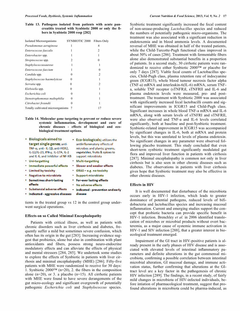
Processed Foods, Dysbiosis, Systemic Inflammation Current Nutrition & Food Science, 2013, Vol. 9, No. 2 17
tients in the treated group vs 12 in the control group under-went surgical operations.
Effects on so Called Minimal Encephalopathy
Patients with critical illness, as well as patients with chronic disorders such as liver cirrhosis and diabetes, fre-quently suffer a mild but sometimes severe confusion, which often has its origin in the gut [283]. Increasing evidence sug-gest that probiotics, alone but also in combination with plant antioxidants and fibers, possess strong neuro-endocrine modulatory effects and can alleviate the effects of physical and mental stressors [284, 285]. We undertook some studies to explore the effects of Synbiotic in patients with liver cir-rhosis and minimal encephalopathy (MHE) [286]. Fifty-five patients with MHE were randomized to receive for 30 days: 1. Synbiotic 2000™ (n=20), 2. the fibers in the composition alone (n=20), or 3. a placebo (n=15). All cirrhotic patients with MHE were found to have severe derangements of the gut micro-ecology and significant overgrowth of potentially pathogenic Escherichia coli and Staphylococcus species.
Synbiotic treatment significantly increased the fecal content of non-urease-producing Lactobacillus species and reduced the numbers of potentially pathogenic micro-organisms. The treatment was also associated with a significant reduction in endotoxemia and in blood ammonia levels. A documented reversal of MHE was obtained in half of the treated patients, while the Child-Turcotte-Pugh functional class improved in about 50% of cases [286]. Treatment with fermentable fibers alone also demonstrated substantial benefits in a proportion of patients. In a second study, 30 cirrhotic patients were ran-domized to receive either Synbiotic 2000™ or placebo for only 7 days [287]. Viable fecal counts of Lactobacillus spe-cies, Child-Pugh class, plasma retention rate of indocyanine green (ICGR15), whole blood tumour necrosis factor alpha (TNF-a) mRNA and interleukin-6(IL-6) mRNA, serum TNF-a, soluble TNF receptor (sTNFR)I, sTNFRII and IL-6 and plasma endotoxin levels were measured, pre- and post-treatment. The treatment with Synbiotic 2000 was associated with significantly increased fecal lactobacilli counts and sig-nificant improvements in ICGR15 and Child-Pugh class. Significant increases in whole blood TNF-a mRNA and IL-6 mRNA, along with serum levels of sTNFRI and sTNFRII, were also observed and TNF-a and IL-6 levels correlated significantly, both at baseline and post-Synbiotic treatment. Synbiotic-related improvement in ICGR15 was accompanied by significant changes in IL-6, both at mRNA and protein levels, but this was unrelated to levels of plasma endotoxin. No significant changes in any parameter were observed fol-lowing placebo treatment. This study concluded that even short-term synbiotic treatment significantly modulated gut flora and improved liver function in patients with cirrhosis [287]. Minimal encephalopathy is common not only in liver cirrhosis but is also seen in other chronic diseases such as diabetes. The observations in patients with liver cirrhosis gives hope that Synbiotic treatment may also be effective in other chronic diseases.
Effects in HIV
It is well documented that disturbance of the microbiota occurs early in HIV-1 infection, which leads to greater dominance of potential pathogens, reduced levels of bifi-dobacteria and lactobacillus species and increasing mucosal inflammation. Current and emerging studies support the con-cept that probiotic bacteria can provide specific benefit in HIV-1 infection. Brenchley et al. in 2006 identified translo-cation of microbes or microbial products without overt bac-teremia, as a major cause of systemic immune activation in HIV-1 and SIV infection [288], that a greater interest in bio-ecological treatment emerged.
Impairment of the GI tract in HIV-positive patients is al-ready present in the early phases of HIV disease and is asso-ciated with elevated levels of intestinal inflammatory pa-rameters and definite alterations in the gut commensal mi-crobiota, confirming a possible correlation between intestinal microbial alteration, GI mucosal damage, and immune acti-vation status, further confirming that alterations at the GI-tract level are a key factor in the pathogenesis of chronic HIV infection [289]. The findings, in a recent study, of fairly mild changes in microbiota of HIV-infected individuals, be-fore intiation of pharmacological treatment, suggest that pro-found alterations in microbiota could be pharma-induced, as
Table 13. Pathogens isolated from patients with acute pan-
creatitis treated with Synbiotic 2000 or only the fi-
bers in Synbiotic 2000 resp [282]
Isolated Microorganisms SYNBIOTIC 2000 Fibers Only
Pseudomonas aeruginosa 1 4
Enterococcus faecalis 1 2
Enterobacter spp. 1 1
Streptococcus spp. 2 0
Staphylococcusaureus 1 1
Enterococcus faecium 1 0
Candida spp. 0 2
Staphylococcus haemolyticus 0 1
Serratia spp. 0 2
Klebsiella spp. 0 1
Escherichia coli 0 1
Stenotrophomonas maltophilia 0 1
Citrobacter freundii 0 1
Totally cultivated microorganisms 7 17
Table 14. Molecular gene targeting to prevent or reduce severe
systemic inflammation, development and cure of
chronic diseases - effects of biological and eco-
biological treatment options.
18 Current Nutrition & Food Science, 2013, Vol. 9, No. 2 Stig Bengmark
only a trend to a greater proportion of Enterobacteriales compared to control subjects (P = 0.099) were observed, despite the significant negative correlations between total bacterial load and duodenal CD4+ and CD8+ T-cell activa-tion levels [290]. As pointed out in a recent review, current and emerging studies appear to support the concept that pro-biotic bacteria can provide specific benefit in HIV-1 infec-tion. Probiotic bacteria have proven active against bacterial vaginosis in HIV-1 positive women and have enhanced growth in infants with congenital HIV-1 infection [288]. Probiotic bacteria may also stabilize CD4+ T cell numbers in HIV-1 infected children and are likely to have protective effects against inflammation and chronic immune activation of the gastrointestinal immune system [291].
Recent studies at least partly support the assumption that Lb rhamnosis GR-1 and Lb Reuteri RC-14 tend to increase the probability of a normal vaginal flora (odds ratio 2.4; P=0.1) and significantly increase the probability of a benefi-cial vaginal pH (odds ratio 3.8; P=0.02) at follow-up [292, 293]. However, later attempts using probiotic yoghurts have proven to less less successful [294]. In a recent pilot study, 38 women with HIV, taking highly active antiretroviral ther-apy (HAART), were supplemented with Synbiotic 2000 Forte™ orally for 4 weeks [295]. In a surprising and very encouraging observation, the supplemented formula found effective, despite heavy pharmaceutical treatment. They sur-vived during the passage through the GI tract, and also showed the ability to colonize inside gut and contributed a significantly elevated level in the stool of the supplemented LAB group. The T-cell activation phenotype was altered by exposure to the Synbiotic formula and was accompanied by a slightly elevated HLA-DR expression of a minor population of CD4+ T-cells, which normally lack expression of HLA-DR or PD-1. These significant changes occurred in the con-text of unaltered microbial translocation, as measured by plasma bacterial 16S ribosomal DNA [295]. It is encourag-ing that the LAB supplemented with Synbiotic 2000, were able to colonize the gut, despite heavy medication/highly active antiretroviral therapy (HAART). and also slight im-provement in immune functions. Hopefully, significantly more pronounced positive effects will be obtained the day we are ready to try eco-biological treatment, not only as complementary treatment, but as an alternative to pharma-ceutical treatment.
Life-threatening Systemic Inflammation
A study of patients in intensive care suffering life-threatening extreme systemic inflammation - a systemic in-flammation response syndrome (SIRS) - and its relation to gut microbiota was recently published. Twenty-five patients with severe SIRS and a serum C-reactive protein level > 10 mg/dL were studied [296]. Analysis of gut microbiota revealed markedly lower total anaerobic bacterial counts, particularly of the beneficial Bifidobacterium and Lactoba-cillus and higher counts of total facultative anaerobes such as Staphylococcus and Pseudomonas compared to healthy vol-unteers. In patients with bacterial translocation, Gram-negative facultative anaerobes were the most commonly identified microbial organisms in mesenteric lymph nodes and serosal scrapings at laparotomy. Gastrointestinal compli-cations were strongly associated with a significantly reduced
number of total obligate anaerobes and highly increased numbers of Staphylococcus and Enterococcus and signifi-cantly decreased numbers of total obligate anaerobes and total facultative anaerobes [296]. A more recent study in 63 similar patients suggests impaired gastrointestinal motility as a significant marker of poor outcome [297]. Patients with 300 mL per day reflux from nasal gastric feeding tube dem-onstrated significantly lower numbers of total obligate an-aerobes including Bacteroidaceae and Bifidobacterium, higher numbers of Staphylococcus, lower concentrations of acetic acid and propionic acid, and higher concentrations of succinic acid and lactic acid (P 0.05), accompanied by dramatically higher incidences of bacteremia (86%vs 18%) and mortality (64%vs 20%) than patients without gastric detension (P 0.05) [297]. Furthermore, in 29 similar pa-tients treatment with a synbiotic composition, consisting of Bifidobacterium breve and Lactobacillus casei, in combina-tion with galactooligosaccharides, was attempted. Higher levels of Bifidobacteria and Lactobacillus, but also total or-ganic acids, particularly short-chain fatty acids, were re-ported and the incidence, compared to historical controls, of infectious complications such as enteritis, pneumonia, and bacteremia, observed to be significantly lower in the treated group [298].
Studies with no or Adverse Effects
Ecologic 641TM
In a multicenter randomized, double-blind, placebo-controlled trial, 298 patients with predicted severe acute pan-creatitis and with APACHE II score > or = 8, Imrie score > 3, or C-reactive protein >150 mg/L) were, within 72 h of onset of symptoms, randomly assigned to receive either a multi-species synbiotic composition (n=153) or a placebo (n=145), administered enterally twice daily for 28 days [299]. The supplemented synbiotic composition, Ecologic 641 (Winclove Bio Industries, Amsterdam, Netherlands), consists of 1010 of each of six different strains of freeze-dried, viable bacteria: Lactobacillus acidophilus, Lactobacil-
lus casei, Lactobacillus salivarius, Lactococcus lactis, Bifi-
dobacterium bifidum, and Bifidobacterium lactis plus corn-starch and maltodextrins. Infectious complications occurred in 46 (30%) of patients in the treated group and in 41 (28%) in the placebo group (relative risk 1.06, 95% CI 0.75-1.51). Twenty-five (16%) patients in the synbiotic group died, compared to nine (6%) in the placebo group (relative risk 2.53, 95% CI 1.22-5.25) [299]. Furthermore, nine patients in the synbiotics group developed bowel ischemia, of which eight had fatal outcomes, compared to none in the placebo group (p=0.004).
Lactobacillus plantarum 299TM
- ProVivaTM
One hundred and three critically ill patients were ran-domized to receive 1. an oral preparation containing L. plan-tarum 299v, ProViva, a fruit drink containing 5 % of LAB-fermented oat and live Lactobacillusb plantarum 299V with a density of 5x10⁷ (n=52) or 2, conventional nutrition ther-apy alone (n = 51). The treatment demonstrated no identifi-able effect in terms of bacterial translocation (12 % vs 12 %; p=0.82), gastric colonization with enteric organisms (11 % vs 17 %, p=0.42), or septic morbidity (13% vs 15 %; p=074),
Processed Foods, Dysbiosis, Systemic Inflammation Current Nutrition & Food Science, 2013, Vol. 9, No. 2 19
serum CRP levels or mortality [300]. In another study, 11 patients undergoing elective abdominal surgery receive lac-tobacillus 299v, (ProViva) for a median time of 9 days (range 5-18 days) to a total average amount of 3250ml (range 2100-9000 ml) and were then compared to 11 control patients. The authors found no significant differences be-tween the Pro Viva group and the control group in terms of concentrations of plasma cells, IgA positive cells or IgM positive cells in the lamina propria [301]. A significantly higher concentration of IgM at the mucosal surface was ob-served in the control group (P = 0.02, Fishers Exact test mid P) but no difference in terms of IgA.
Lactobacillus rhamnosus GGTM
Sixty-one patients in a pediatric ICU study were random-ized with 31 patients receiving treatment with one capsule of Lactobacillus rhamnosus strain GG in a dose of 10 109 and 30 receiving one capsule of inulin daily (control group) [302]. No differences in rate of infections were observed between the groups; the mean number of infections in the treatment and control groups was 1.83 and 1.33, respec-tively. 9/31 patients in the probiotic-treated group developed in total 15 nosocomial infections: 6 bloodstream infections (40%), 5 tracheo-bronchitis (33%), 2 pneumonia (13%), and 2 UTI (13%). There were six deaths in total during the study period; four in the placebo group and two in the treatment group. No cases of Lactobacillus bacteremia or other serious adverse effects were observed [302].
Synbiotic 2000TM
/Synbiotic 2000 ForteTM
Two hundred and fifty nine enterally fed critically ill pa-tients, expected to require mechanical ventilation for 48 hours or more were enrolled in a study; 130 patients received Synbiotic 2000 FORTE® (twice a day) and 129 patients, a cellulose-based placebo for a maximum of 28 days [303]. The oropharyngeal microbial flora and colonization rates were unaffected by the synbiotic treatment. The overall inci-dence of ventilator associated pneumonia (VAP) was lower than anticipated (11.2%) and no statistical difference was demonstrated between the groups receiving synbiotic or pla-cebo; incidence of VAP (9 and 13%, P=0.42), VAP rate per 1000 ventilator days (13 and 14.6, P=0.91) or hospital mor-tality (27 and 33%, P=0.39), respectively. No negative ef-fects of the treatment were observed [303].
TrevisTM
A total of 90 patients admitted to an ICU were random-ized to receive either a synbiotic or placebo (45 into each group) [304]. The synbiotic treatment consisted of the supply of a capsule of TrevisTM (Chr Hansen Biosystem, Denmark) three times a day, containing 4 x 10⁹ colony forming units of each of L. acidophilus La5 (La5), B. lactis Bb-12 (Bb-12), S. thermophilus and L. bulgaricus. In addition, the prebiotic oligofructose (7.5 gm of RaftiloseTM powder, Orafti Active Food Ingredients, Belgium) was administered twice a day. The patients in the synbiotic group demonstrated, after 1 week of therapy, significantly lower incidence of potentially pathogenic bacteria (43% versus 75%, P = 0.05) and multi-ple organisms (39% versus 75%, P = 0.01) in their nasogas-tric aspirates, than the controls. However, there were no sig-
nificant differences between the groups in terms of intestinal permeability, septic complications or mortality [304].
VSL#3TM
Twenty-eight critically ill patients were enrolled and ran-domly assigned to one of 3 treatment groups: 1. placebo (n=9) 2. viable probiotics - 2 sachets daily of VSL#3TM (n=10) or 3. bacterial sonicates - non viable VSL#3 bacteria (n=9) [305]. Each sachet of the supplemented probiotic, VSL#3 (VSL Pharmaceuticals, Ft Lauderdale, FL) contained 900 billion viable lyophilized bacteria of 4 strains of Lacto-bacillus (L. casei, L. plantarum, L. acidophilus, and L. del-brueckii subsp. Bulgaricus) plus 3 strains of Bifidobacterium (B. longum, B. breve, and B. infantis), plus Streptococcus salivarius subsp. Thermophiles, totally eight strains. Intesti-nal permeability decreased in all treatment groups. The rate of severe sepsis and MODS were not significantly affected by the treatment, although a significantly larger increase in systemic IgA and IgG concentrations were observed in the group supplied live bacteria than in the patients who received placebo or sonicated bacteria [305].
A Plea for Alternative Instead of Complementary Studies
Critical care units are generally highly artificial environ-ments and the burden of environment-induced physical and mental stress and subsequent status of systemic hyper-inflammation on the patient, enormous. Patients treated un-der these conditions are in many ways dys-functional; the whole microbiota has often, more or less, disappeared and probiotic bacteria supplied will usually be extinct before they reach one of their targets – the lower gastrointestinal tract. This artificiality seems to vary from country to country and sometimes also from hospital to hospital, an observation that might explain the great variation in outcome from studies undertaken in different countries and regions.
As discussed above, probiotic treatment has never been given the chance as a ‘stand alone’ alternative. It has, thus far, only been tried as an adjunctive treatment to heavy multi-drug pharmaceutical treatments. Many of the numer-ous drugs used in the ICU, including antibiotics are known to derange not only the microbiota but to dramatically de-range the majority of immune functions. Some 25 years ago the use of an antibiotic, Mezlocillin (Bayer, 150 mg/kg body weight) was demonstrated to be significant suppression of macrophage functions, derange chemiluminescence re-sponse, chemotactic motility, bactericidal and cytostatic abil-ity and impairment of lymphocyte proliferation, impairment of macrophage functions and bactericidal efficacy as well as production and secretion of cytokines [306].
Artificial nutrition, both enteral and parenteral, is an im-portant contributor to ICU-associated sepsis; catheter-related sepsis reported to occur in about 25% of patients fed via in-travenous feeding-tubes [307]. Other common perioperative practices, e.g. use of artificial feeding regimens, preoperative antibiotics [308], and mechanical bowel preparation [309, 310] will, instead of preventing expected infections, contrib-ute increased rates of treatment-associated infections. Other measures in the ICU are mechanical ventilation [311], treat-ment with various pharmaceutical drugs or antibiotics [312,
20 Current Nutrition & Food Science, 2013, Vol. 9, No. 2 Stig Bengmark
313], chemical solutions of clinical nutrition and many oth-ers promote super-inflammation and, indirectly, infection.
Enteral nutrition formulas, most likely deleterious to mi-crobiota, are known to induce loss of intestinal barrier func-tion, promote bacterial translocation, and impair host im-mune defense [314], a phenomenon, observed in humans but also extensively elucidated in animal studies. In such studies the incidence of bacterial translocation to the mesenteric lymph node was significantly increased when the animals were fed nutrition formulas such as Vivonex (53%), Criticare (67%), or Ensure (60%) (p < .05) [313-315]. Dramatic eleva-tions in pro-inflammatory cytokines have been observed in patients, when fed a standard enteral nutrition solution (Nu-trison) following pancreatduodenectomy, e.g. IL-1beta day 7 (P<0.001); day 14 (P=0.022), TNF-alpha- day 3 (P=0.006); day 7 (P<0.001) [316].Of special interest are the observa-tions that such changes are not observed when the standard nutrition is replaced with a formula whichis claimed to have immune-modulatory effects (Stresson). Instead anti-inflammatory cytokines were seen to be significantly ele-vated: IL-1ra/s : day 7 (P<0.001); IL-6: day 10 (P=0.017); IL-8: day 1 (P=0.011) days 3, 7, 10, and 14 (P<0.001), and IL-10: days 3 &10 (P<0.001) [316].
Choice of Lactic Acid Bacteria as Probiotics
The choice of bacteria for probiotic purposes is critical. Only a few LAB strains have demonstrated an ability to in-fluence the immune system, reduce inflammation and/or eliminate or reduce unwanted pro-inflammatory molecules from foods. Even strains that carry the same name can have different and even sometimes opposite effects. A recent study selected 46 strains of Lactococcus lactis from about 2,600 LAB and compared their ability to induce cytokines. It was demonstrated that the inter-strain differences in ability to produce pro- and anti-inflammatory cytokines was found [317]. An observation that underlines the importance of ex-tensive animal and preclinical studies before a LAB or com-bination of LAB is chosen as a probiotic. Strains that im-prove immune function by increasing the number of IgA-producing plasma cells, improve phagocytosis, and influence the proportion of Th1 cells and NK cells [318] are particu-larly desirable for probiotic purposes. Popular probiotic spe-cies that are usually reliable and available commercially are L. paracasei, L. rhamnosus, L. acidophilus, L. johnsonii, L. fermentum, L. reuteri, L. plantarum, Bifidobacterium longum and Bifidobacterium animalis [145].
Among the strains with documented stronger anti-inflammatory functions are the species, Lactobacillus para-casei subsp paracasei, Lactobacillus plantarum, and Pedio-coccus pentosaceus. Lactobacillus paracasei, in particular, seems to have a solid record; it has been shown to induce cellular immunity and stimulate production of suppressive cytokines such as TGF and Il-10 and to suppress Th2 activ-ity and CD4 T-cells [319, 320], suppress splenocyte prolif-eration [321] and decrease antigen-specific IgE and IgG1 [322]. When more than one hundred LAB strains were com-pared Lactobacillus paracasei was shown to be the strongest inducer of Th1 and repressor of Th2 cytokines [323].
A recent study using rats compared the ability of four dif-ferent strains: Lactobacillus paracasei, Lactobacillus john-
sonii, Bifidobacterium longum, or Bifidobacterium lactis to control Trichinella spiralis-induced infection. Lactobacillus paracasei, alone was able to reduce infection-associated Th2 response, muscle levels of TGF- , COX-2 and PGE2 and to attenuate infection-induced muscle hyper-contractility [324]. Another study compared the ability to reduce stress-induced changes in gut permeability and sensitivity to colorectal dis-tension of three probiotic strains: Bifidobacterium lactis NCC362, Lactobacillus johnsonii NCC533, and Lactobacil-lus paracasei NCC2461. Lactobacillus paracasei, alone re-stored normal gut permeability, reduced visceral hyperalge-sia and reduced visceral pain [325]. Several other important clinical effects of Lactobacillus paracasei subsp paracasei are summarized in a recent review; a strain called NTU 101 and its fermented products demonstrating the ability to reduce blood cholesterol, blood pressure, and prevent allergies, os-teoporosis and inhibit accumulation of fat tissue [326]. Lac-tobacillus plantarum also has an excellent record. When the ability of fifty different LAB to control twenty-three differ-ent Clostridium difficile (C diff) strains were studied, Lacto-bacillus paracasei and Lactobacillus plantarum seemed to be equally efficient and the only strains of the fifty tried, to demonstrate the ability to effectively eliminate all C diff strains - more than half of the tried LAB strains were totally ineffective, and some effective only against a few [327].
Some LAB seems potentiated in their efficacy by simul-taneous supply of prebiotic fibers (probiotics + prebiotics => synbiotics). However, there are great differences in the abil-ity of different strains to ferment and utilize plant fibers, especially when it comes to semi-fermentable fibers such as oligofructans. Only a handful of 712 LAB strains tested demonstrated an ability to ferment inulin and phlein, namely : L plantarum (several strains), L paracasei subsp. paraca-sei, L brevis&Pediococcus pentosaceus [328].
Molecular Gene Targeting – The Future?
Ingredients specific to certain plants are known to exert profund effects on specific genes. Among these agents are curcumin (turmeric), resveratrol (red grapes, peanuts and berries), genistein (soybean), diallyl sulfide (allium), S-allyl cysteine (allium), allicin (garlic), lycopene (tomato), cap-saicin (red chilli), diosgenin (fenugreek), 6-gingerol (ginger), ellagic acid (pomegranate), ursolic acid (apple, pears, prunes), silymarin (milk thistle), anethol (anise, camphor, and fennel), catechins (green tea), eugenol (cloves), indole-3-carbinol (cruciferous vegetables), limonene (citrus fruits), beta carotene (carrots), and dietary fiber [329, 330].
Curcumin, for example, has demonstrated a profound ability to inhibit a whole series of cell-signaling pathways, including NF-kB, AP-1, STAT3, Akt, Bcl-2, Bcl-X(L), caspases, PARP, IKK, EGFR, HER2, JNK, MAPK, COX2, and 5-LOX [258,329,330]; supplemented probiotics are likely to exert similar effects. Gene expression of human duodenal mucosa cells was studied after exposure to one of the following four lactic acid bacteria; L plantarum WCFS1 [331], L acidophilus L10, Lcasei CRL-431 and L rhamno-sus GG [332], administered in a cross-over study to healthy volunteers in a dose of 1010. Mucosal biopsies were taken from duodenum after 6 hours and compared to control biop-sies. The interventions did not impair immune and metabolic
Processed Foods, Dysbiosis, Systemic Inflammation Current Nutrition & Food Science, 2013, Vol. 9, No. 2 21
homeostasis but a fascinating and most distinct influence on the expression of several hundred genes (transcriptome) was reported after administration of each of the studied LAB. This is possibly the first time that different probiotic lactoba-cilli have been reported to induce more or less strain-specific and markedly different expression profiles; very similar to what is known to occur with ingestion of various foods, es-pecially plant ingredients [329, 330] and in many respects similar to what is observed after supply of certain pharma-ceuticals. L plantarum was observed to specifically modulate overt adaptive immune responses [331], L acidophilus to suppress inflammation, L casei to stimulate Th1 response and improve the Th1–Th2 balance and L. rhamnosis to influ-ence cellular growth and proliferation [332]. These effects were suggested to resemble, although in a distinctly milder form, those obtained by specific pharmaceuticals:
L acidophilus - antagonists of -receptor activity, gua-nine antagonists, synthetic corticosteroids and flavonoids,
L casei - modulators of GABA receptors, cholinergic blocking agents, antagonists of -adrenergic receptors,
L rhamnosus - glycoside steroids, alkaloids, protein syn-thesis inhibitors and protein kinase C inhibitors.
The responsiveness to ingestion of various LAB seems influenced, not only by eventual genetic background and existing resident microbiota, but also by lifestyle, and par-ticularly by diet. This might explain the differences in per-son-to-person response, as observed in the above studies but also the differences in outcome, often encountered in clinical probiotic studies, and especially when tried with critically ill patients.
Reducing Inflammation – Key to Prevention and Cure of Disease
Inflammation, an essential component of immune-mediated protection against pathogens and tissue damage, and uncontrolled immune responses, will commonly in Westerners, institute a state of chronic inflammation, which will occur when immune responses are activated despite the absence of ‘danger’ signals, fail to fully turn-off despite elimination of danger signals and/or fail to completely clear such signals. Numerous factors, in addition to genetic pre-disposition, trauma and various stress factors (physical and emotional) are known to contribute to increased discrete and long-lasting inflammation, among them age, diet and medi-cations.
Studies of human gene-related inflammation suggest that, of the approximately 25,000 human genes, approximately 5 %, or some 1,200 genes, are involved in inflammation [333-335]. It is increasingly understood that the human genome in itself will only explain a minority of chronic diseases, far less than changes in lifestyle, food habits and social behav-ior, factors which seem to have a dominating impact on hu-man health. Clearly, the molecular mechanisms linking envi-ronmental factors and genetic susceptibility were first envi-sioned after the recent exploration of the genomic diversity, i.e. the metagenome with its between 3 [336] an 9 million genes [132]. Although the mechanisms behind the metage-nome-associated low-grade inflammation and the corre-sponding immune response are not yet fully understood, un-
doubtedly the metagenome has a dominating influence on altered body functions such as adipose tissue plasticity and diseases such as hepatic steatosis, insulin resistance and car-diovascular diseases, but also on disorders such as autoim-mune diseases including rheumatoid arthritis, gastrointestinal and neuropsychiatric diseases and on development and pro-gress of a number of cancers [337], as well as many other chronic disorders. When disease exacerbations occur, in trauma or in critical illness, the normally silent or discrete inflammation turn into a storm [338] as experienced in sys-temic inflammatory response syndrome (SIRS) and multiple organ failure (MOF) [339]. In many severe conditions like MOF and SIRS components of cytokine-induced injury might be more damaging than the initial cause/trauma/early invasion of micro-organisms in themselves. Inflammatory cytokines, such as TNF alpha and IL-1 , released by these events will destabilize endothelial cell-cell interactions and cripple vascular barrier function, producing capillary leak-age, tissue edema, organ failure, and sometimes death [339].
Inflammation Control - Biologicals or Eco-Biologicals?
Inflammation is, as discussed above, extraordinarily complex. In rheumatoid arthritis (RA) for example, the joints are rich in cytokine-secreting cells containing a wide range of effector molecules including pro-inflammatory cytokines such as IL-1 , IL-6, TNF- and IL-18, chemokines such as IL-8, IP-10, MCP-1, MIP-1 and RANTES, MMPs such as MMP-1, -3, -9 and -13 and metabolic proteins such as Cox-1, Cox-2 and iNOS, which interact with one another in a complex manner that is thought to cause a vicious cycle of pro-inflammatory signals resulting in chronic and persistent inflammation [340, 341]. NF- B is increasingly suggested to the master regulators of inflammatory cytokine production in RA. These mediators are also involved, although not in an identical manner, in other autoimmune disorders such as inflammatory bowel diseases [340]. This knowledge has led to the development to a new generation of pharmaceutical drugs, generally referred to as biological, designed to inhibit the crucial mediators of pro-inflammatory signals and subse-quent abnormal immune response. A whole series of revolu-tionary new drugs such as anti-TNF- , anti-IL-1 , anti-HER2 etc are already successfully tried and new drugs such as antibodies targeting IL-12/IL-23 pathways, IFN- , IL-17A, IL-2 and IL-6, and also inhibitors of NF- B more or less extensively tried in a variety of chronic inflammatory and autoimmune diseases. Some of these have already dem-onstrated initially promising result, while other treatments such as administration of the regulatory cytokines IL-10 and IL-11 have failed to induce reproducible clinical effects [342]. Significant benefits in quality of life and tissue/organ healing are encountered in at least something over 50%. These drugs are generally tolerated well, but adverse events such as infections including reactivating tuberculosis, tu-mours such as lymphomas and demyelinating diseases and infusion reactions are sometimes evident. These changes must be regarded as acceptable as long as they are used in diseases that have proven to be refractory to all other treat-ments but may be an issue when, as increasingly suggested, they are tried in early stages of diseases, as happened after widening indications for statins [343].
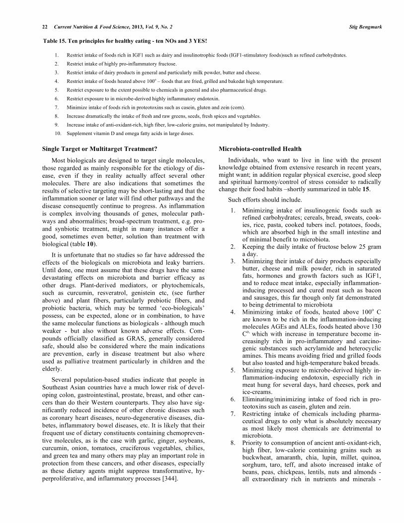
22 Current Nutrition & Food Science, 2013, Vol. 9, No. 2 Stig Bengmark
Single Target or Multitarget Treatment?
Most biologicals are designed to target single molecules, those regarded as mainly responsible for the etiology of dis-ease, even if they in reality actually affect several other molecules. There are also indications that sometimes the results of selective targeting may be short-lasting and that the inflammation sooner or later will find other pathways and the disease consequently continue to progress. As inflammation is complex involving thousands of genes, molecular path-ways and abnormalities; broad-spectrum treatment, e.g. pro- and synbiotic treatment, might in many instances offer a good, sometimes even better, solution than treatment with biological (table 10).
It is unfortunate that no studies so far have addressed the effects of the biologicals on microbiota and leaky barriers. Until done, one must assume that these drugs have the same devastating effects on microbiota and barrier efficacy as other drugs. Plant-derived mediators, or phytochemicals, such as curcumin, resveratrol, genistein etc, (see further above) and plant fibers, particularly prebiotic fibers, and probiotic bacteria, which may be termed ‘eco-biologicals’ possess, can be expected, alone or in combination, to have the same molecular functions as biologicals - although much weaker - but also without known adverse effects. Com-pounds officially classified as GRAS, generally considered safe, should also be considered where the main indications are prevention, early in disease treatment but also where used as palliative treatment particularly in children and the elderly.
Several population-based studies indicate that people in Southeast Asian countries have a much lower risk of devel-oping colon, gastrointestinal, prostate, breast, and other can-cers than do their Western counterparts. They also have sig-nificantly reduced incidence of other chronic diseases such as coronary heart diseases, neuro-degenerative diseases, dia-betes, inflammatory bowel diseases, etc. It is likely that their frequent use of dietary constituents containing chemopreven-tive molecules, as is the case with garlic, ginger, soybeans, curcumin, onion, tomatoes, cruciferous vegetables, chilies, and green tea and many others may play an important role in protection from these cancers, and other diseases, especially as these dietary agents might suppress transformative, hy-perproliferative, and inflammatory processes [344].
Microbiota-controlled Health
Individuals, who want to live in line with the present knowledge obtained from extensive research in recent years, might want; in addition regular physical exercise, good sleep and spiritual harmony/control of stress consider to radically change their food habits –shortly summarized in table 15.
Such efforts should include.
1. Minimizing intake of insulinogenic foods such as refined carbohydrates; cereals, bread, sweats, cook-ies, rice, pasta, cooked tubers incl. potatoes, foods, which are absorbed high in the small intestine and of minimal benefit to microbiota.
2. Keeping the daily intake of fructose below 25 gram a day.
3. Minimizing their intake of dairy products especially butter, cheese and milk powder, rich in saturated fats, hormones and growth factors such as IGF1, and to reduce meat intake, especially inflammation-inducing processed and cured meat such as bacon and sausages, this far though only fat demonstrated to being detrimental to microbiota
4. Minimizing intake of foods, heated above 100o C are known to be rich in the inflammation-inducing molecules AGEs and ALEs, foods heated above 130 Co, which with increase in temperature become in-creasingly rich in pro-inflammatory and carcino-genic substances such acrylamide and heterocyclic amines. This means avoiding fried and grilled foods but also toasted and high-temperature baked breads.
5. Minimizing exposure to microbe-derived highly in-flammation-inducing endotoxin, especially rich in meat hung for several days, hard cheeses, pork and ice-creams.
6. Eliminating/minimizing intake of food rich in pro-teotoxins such as casein, gluten and zein.
7. Restricting intake of chemicals including pharma-ceutical drugs to only what is absolutely necessary as most likely most chemicals are detrimental to microbiota.
8. Priority to consumption of ancient anti-oxidant-rich, high fiber, low-calorie containing grains such as buckwheat, amaranth, chia, lupin, millet, quinoa, sorghum, taro, teff, and alsoto increased intake of beans, peas, chickpeas, lentils, nuts and almonds - all extraordinary rich in nutrients and minerals -
Table 15. Ten principles for healthy eating - ten NOs and 3 YES!
1. Restrict intake of foods rich in IGF1 such as dairy and insulinotrophic foods (IGF1-stimulatory foods)such as refined carbohydrates.
2. Restrict intake of highly pro-inflammatory fructose.
3. Restrict intake of dairy products in general and particularly milk powder, butter and cheese.
4. Restrict intake of foods heated above 100o – foods that are fried, grilled and bakedat high temperature.
5. Restrict exposure to the extent possible to chemicals in general and also pharmaceutical drugs.
6. Restrict exposure to in microbe-derived highly inflammatory endotoxin.
7. Minimize intake of foods rich in proteotoxins such as casein, gluten and zein (corn).
8. Increase dramatically the intake of fresh and raw greens, seeds, fresh spices and vegetables.
9. Increase intake of anti-oxidant-rich, high fiber, low-calorie grains, not manipulated by Industry.
10. Supplement vitamin D and omega fatty acids in large doses.
Processed Foods, Dysbiosis, Systemic Inflammation Current Nutrition & Food Science, 2013, Vol. 9, No. 2 23
prepared by low-temperature cooking for eating - all of important nutrient for maintaining a rich mi-crobiota.
9. Dramatic increase in intake of fresh and raw greens, fresh spices and vegetables, rich in antioxidants, fi-bers, minerals and nutrients, but also inflammation-controlling factors such as curcumin, resveratrol - some of which most likely are of great importance for diversity, replication, growth and functions of the microbiota and for immune development and immune functions of the body
10. Supplementation of large doses of vitamin D and omega fatty acids, is important in both, control of inflammation and for function of microbiota, and in short supply in Western living.
FINAL REMARKS
The global epidemic of un-health is not long-term sus-tainable. Over several decades the cost for delivery health care has grown in most countries and is expected to do so also in the future [344]. In only one country, the United States with its population of 300 million inhabitants, less than 5 % of the global population, has health expenditures approaching $ 3 trillion ($2.6 trillion in 2010), which is more than ten times the costs ($256 billion) spent in 1980. Ad-dressing this fast growing burden to Society is today a major policy priority in many countries around the world. The long-term affordability of national health care questioned in many countries. The cost of health care in for example the US is approaching 20 per cent of GNP, or about $ 15000 per individual a year, which is extremely high in the light of the fact that the average income per person is about $ 40000 per year or per family $ 46000 before tax.
The spending in The US only for the treatment of meta-bolic syndrome and morbidities associated high intake of refined carbohydrate-associated calculated to be about $150 billion for treatments plus about $65 billion in lost produc-tivity, totally about $ 215 trillion. It is estimated that at least seventy-five per cent of all US health-care dollars are today spent on treating life-style induced diseases and their resul-tant disabilities.
Eighty to ninety per cent of human diseases are strongly associated with life style and particularly with eating habits – and thus in theory preventable. Clearly most of Western dis-eases are associated with Western lifestyle and over con-sumption of agricultural products, both meat and refined carbohydrates. It is against this background a mystery that Societies around the world continue to with one-hand pays trillions of dollars to support agriculture to produce even larger amounts of already cheap refined carbohydrate and animal products and then with the other hand pays trillions of dollars to restore health in those who have abused by such products.
Without drastic policy changes, the governments in the near future will no longer be able to maintain national health care programs and the responsibility for personal health, as it will no longer be a responsibility of the individual.
CONFLICT OF INTEREST
The author(s) confirm that this article content has no con-flict of interest.
ACKNOWLEDGEMENTS
Declared none.
REFERENCES
[1] Food Availability (Per Capita) Data System. United States Depart-ment of Agriculture conomic Research Service. Avail-able: http://www.ers.usda.gov/Data/FoodConsumption/. 2012
[2] (a). Kant AK, Graubard BI. Energy density of diets reported by American adults: Association with food group intake, nutrient in-take, and body weight. Int J Obes (Lond) 2005; 29: 950-6; (b). Lustig RH, Schmidt LA, Brindis CD. Public health: The toxic truth about sugar. Nature 2012; 482: 27-9.
[3] Wang YC, McPherson K, Marsh T, Gortmaker SL, Brown M. Health and economic burden of the projected obesity trends in the USA and the UK. Lancet 2011; 378: 815-25.
[4] Boyle JP, Thompson TJ, Gregg EW, Barker LE, Williamson DF. Projection of the year 2050 burden of diabetes in the US adult population: Dynamic modeling of incidence, mortality, and predia-betes prevalence. Popul Health Metr 2010; 8: 29.
[5] Hebert LE, Scherr PA, Bienias JL, Bennett DA, Evans DA. Alz-heimer disease in the US population: prevalence estimates using the 2000 census. Arch Neurol. 2003; 60: 1119-22.
[6] Bray F, Møller B. Predicting the future burden of cancer. Nat Rev Cancer 2006; 6: 63-74.
[7] Malekinejad H, Scherpenisse P, Bergwerff AA. Naturally occurring estrogens in processed milk and in raw milk (from gestated cows). J Agric Food Chem. 2006; 54: 9785-91.
[8] Clempson AM, Pollott GE, Brickell JS, Wathes DC. Associations between bovine IGFBP2 polymorphism with fertility, milk produc-tion, and metabolic status in UK dairy cows. Anim Biotechnol 2012; 23: 101-13.
[9] Frystyk J, Skjaerbaek C, Vestbo E, Fisker S, Orskov H. Circulating levels of free insulin-like growth factors in obese subjects: the im-pact of type 2 diabetes. Diabetes Metab Res Rev 1999; 15: 314-22.
[10] Martin RM, Holly JM, Davey Smith G, Gunnell D. Associations of adiposity from childhood into adulthood with insulin resistance and the insulin-like growth factor system: 65-year follow-up of the Boyd Orr Cohort. J Clin Endocrinol Metab 2006; 91: 3287-95.
[11] Zemva J, Schubert M. Central insulin and insulin-like growth fac-tor-1 signaling: Implications for diabetes associated dementia. Curr Diabetes Rev 2011; 7: 356-66.
[12] Cohen E, Paulsson JF, Blinder P, et al. Reduced IGF-1 signaling delays age-associated proteotoxicity in mice. Cell 2009; 139: 1157-69.
[13] Moore T, Checkley LA, DiGiovanni J. Dietary energy balance modulation of epithelial carcinogenesis: a role for IGF-1 receptor signaling and crosstalk. Ann N Y Acad Sci 2011; 229: 7-17.
[14] Cleary MP, Grossmann ME. The manner in which calories are restricted impacts mammary tumor cancer prevention.J Carcinog 2011; 10: 21.
[15] Bengmark S. Advanced glycation and lipoxidation end products – amplifiers of inflammation: the role of food. JPEN 2007; 31: 430-40.
[16] Bengmark S AGE, ALE RAGE and disease – a foods perspective. In Handbook of Prebiotic and Probiotic Ingredients: Health Bene-fits and Food Applications. Susan S Cho, Terry Finocchiaro, Eds. CRC Press, Taylor and Francis Group, Boca Raton 2010 pp 139-62.
[17] Bengmark S. Modified Amino Acid-Based Molecules: Accumula-tion and Health Implications. In Amino Acids in Human Nutrition and Health. Mello JFD. Eds. CABI Wallingford, UK, 2011 pp 382-405.
[18] Bogazzi F, Rossi G, Lombardi M, et al. Vitamin D status may contribute to serum insulin-like growth factor I concentrations in healthy subjects. J Endocrinol Invest 2011; 34: e200-3.
[19] Balden R, Selvamani A, Sohrabji F. Vitamin D deficiency exacer-bates experimental stroke injury and dysregulates ischemia-induced inflammation in adult rats. Endocrinology 2012; 153: 2420-35.
24 Current Nutrition & Food Science, 2013, Vol. 9, No. 2 Stig Bengmark
[20] Deleskog A, Hilding A, Brismar K, Hamsten A, Efendic S, Östen-son CG. Low serum 25-hydroxyvitamin D level predicts progres-sion to type 2 diabetes in individuals with prediabetes but not with normal glucose tolerance.Diabetologia 2012; 55: 1668-78.
[21] Pérez-López FR, Chedraui P, Fernández-Alonso AM. Vitamin D and aging: beyond calcium and bone metabolism.Maturitas 2011; 69: 27-36.
[22] Pérez-López FR, Fernández-Alonso AM, Mannella P, Chedraui P. Vitamin D, sunlight and longevity.Minerva Endocrinol 2011; 36: 257-66.
[23] Sohl E, van Schoor NM, de Jongh RT, de Vries OJ, Lips P. The impact of medication on vitamin D status in older individuals. Eur J Endocrinol 2012; 166: 477-85.
[24] Fardet A, Leenhardt F, Lioger D, Scalbert A, Rémésy C. Parameters controlling the glycaemic response to breads. Nutr Res Rev 2006; 19: 18-25.
[25] Pendyala S, Walker JM, Holt PR. A high-fat diet is associated with endotoxemia that originates from the gut. Gastroenterology 2012; 142: 1100-1.
[26] Lira FS, Rosa JC, Pimentel GD, et al. Endotoxin levels correlate positively with a sedentary lifestyle and negatively with highly trained subjects. Lipids Health Dis 2010; 9: 82.
[27] Pussinen PJ, Havulinna AS, Lehto M, Sundvall J, Salomaa V. Endotoxemia is associated with an increased risk of incident diabe-tes. Diabetes Care 2011; 34: 392-7.
[28] Fassbender K, Walter S, Kühl S, et al. The LPS receptor (CD14) links innate immunity with Alzheimer's disease. FASEB J 2004; 18: 203-5.
[29] Jaeger LB, Dohgu S, Sultana R, et al. Lipopolysaccharide alters the blood-brain barrier transport of amyloid beta protein: a mechanism for inflammation in the progression of Alzheimer's disease. Brain Behav Immun 2009; 23: 507-17.
[30] Wiedermann CJ, Kiechl S, Dunzendorfer S, et al. Association of endotoxemia with carotid atherosclerosis and cardiovascular dis-ease: prospective results from the Bruneck Study. J Am Coll Car-diol 1999; 34: 1975-81.
[31] Mathew JP, Grocott HP, Phillips-Bute B, et al. Lower endotoxin immunity predicts increased cognitive dysfunction in elderly pa-tients after cardiac surgery. Stroke 2003; 34: 508-13.
[32] Wrigley BJ, Lip GY, Shantsila E. The role of monocytes and in-flammation in the pathophysiology of heart failure. Eur J Heart Fail 2011; 13: 1161-71.
[33] Risley P, Jerrard-Dunne P, Sitzer M, et al. Promoter polymorphism in the endotoxin receptor (CD14) is associated with increased ca-rotid atherosclerosis only in smokers: the Carotid Atherosclerosis Progression Study (CAPS). Stroke 2003; 34: 600-4.
[34] Beyan H, Goodier MR, Nawroly NS, et al. Altered monocyte cy-clooxygenase response to lipopolysaccharide in type 1 diabetes. Diabetes 2006; 55: 3439-45.
[35] Nymark M, Pussinen PJ, Tuomainen AM, et al. Serum Lipopoly-saccharide Activity Is Associated With the Progression of Kidney Disease in Finnish Patients With Type 1 Diabetes. Diabetes Care 2009; 32: 1689-93.
[36] Andreasen AS, Kelly M, Berg RM, Møller K, Pedersen BK. Type 2 diabetes is associated with altered NF- B DNA binding activity, JNK phosphorylation, and AMPK phosphorylation in skeletal mus-cle after LPS. PLoS One. 2011; 6: e23999.
[37] Hsu RY, Chan CH, Spicer JD, et al. LPS-induced TLR4 signaling in human colorectal cancer cells increases beta1 integrin-mediated cell adhesion and liver metastasis. Cancer Res 2011; 71: 1989-98.
[38] Peden DB. The role of oxidative stress and innate immunity in O(3) and endotoxin-induced human allergic airway disease. Immunol Rev 2011; 242: 91-105.
[39] Zhang R, Miller RG, Gascon R, et al. Circulating endotoxin and systemic immune activation in sporadic amyotrophic lateral sclero-sis (sALS). J Neuroimmunol 2009; 206: 121-4.
[40] Emanuele E, Orsi P, Boso M, et al. Low-grade endotoxemia in patients with severe autism. Neurosci Lett 2010; 471: 162-5.
[41] Rabin RL, Levinson AI. The nexus between atopic disease and autoimmunity: a review of the epidemiological and mechanistic lit-erature. Clin Exp Immunol 2008; 153: 19-30.
[42] DellaGioia N, Hannestad J. A critical review of human endotoxin administration as an experimental paradigm of depression. Neurosci Biobehav Rev 2010; 34: 130-43.
[43] Maes M, Coucke F, Leunis JC. Normalization of the increased translocation of endotoxin from gram negative enterobacteria
(leaky gut) is accompanied by a remission of chronicfatigue syn-drome. Neuro Endocrinol Lett. 2007; 28: 739-44.
[44] Bengoechea JA, Ito K. Chronic obstructive pulmonary disease Th1 cells display impaired response to endotoxin. Am J Respir Crit Care Med. 2011; 183: 148-50.
[45] Liu Q, Duan ZP, Ha DK, Bengmark S, Kurtovic J, Riordan SM. Synbiotic modulation of gut flora: effect on minimal hepatic en-cephalopathy in patients with cirrhosis. Hepatology. 2004; 39: 1441-9
[46] Bengmark S. Bio-ecological control of chronic liver disease and encephalopathy. Metab Brain Dis. 2009; 24: 223-36.
[47] Maes M, Leunis JC. Normalization of leaky gut in chronic fatigue syndrome (CFS) is accompanied by a clinical improvement: effects of age, duration of illness and the translocation of LPS from gram-negative bacteria. Neuro Endocrinol Lett. 2008; 29: 902-10.
[48] Dohgu S, Fleegal-DeMotta MA, Banks WA. Lipopolysaccharide-enhanced transcellular transport of HIV-1 across the blood-brain barrier is mediated by luminal microvessel IL-6 and GM-CSF.J Neuroinflammation. 2011; 8: 167
[49] Leung KW, Barnstable CJ, Tombran-Tink J. Bacterial endotoxin activates retinal pigment epithelial cells and induces their degenera-tion through IL-6 and IL-8 autocrine signaling. Mol Immunol. 2009; 46: 1374-86.
[50] McIntyre CW, Harrison LE, Eldehni MT, Jefferies HJ, Szeto CC, John SG et al Circulating endotoxemia: a novel factor in systemic inflammation and cardiovascular disease in chronic kidney dis-ease.Clin J Am Soc Nephrol. 2011; 6: 133-41.
[51] Lassenius MI, Pietiläinen KH, Kaartinen K, Pussinen PJ, Syrjänen J, Forsblom C et al. Bacterial endotoxin activity in human serum is associated with dyslipidemia, insulin resistance, obesity, and chronic inflammation.Diabetes Care. 2011; 34: 1809-15.
[52] Caesar R, Fåk F, Bäckhed F. Effects of gut microbiota on obesity and atherosclerosis via modulation of inflammation and lipid metabolism.J Intern Med. 2010; 268: 320-8.
[53] Schwager J, Hoeller U, Wolfram S, Richard N, Rose hip and its constituent galactolipids confer cartilage protection by modulating cytokine, and chemokine expression. BMC Complement Altern Med. 2011; 11: 105.
[54] Shaddox LM, Wiedey J, Calderon NL, Magnusson I, Bimstein E, Bidwell JA et al. Local inflammatory markers and systemic endo-toxin in aggressive periodontitis.J Dent Res. 2011; 90: 1140-4.
[55] Lange JH, Buja A, Mastrangelo G. Endotoxin, a possible agent in the causation of Parkinson's disease.J Occup Environ Med. 2006; 48: 655.
[56] Lange JH. Endotoxin as a factor for joint pain and rheumatoid arthritis. Clin Rheumatol. 2004; 23: 566.
[57] Jenkins TA, Harte KM, Stenson G, Reynolds GP. Neonatal lipopolysaccharide induces pathological changes in parvalbumin immunoreactivity in the hippocampus of the rat. Behavioural Brain Research. 2009; 205: 355–9.
[58] Huang CJ, Stewart JK, Franco RL, Evans RK, Lee ZP, Cruz TD et al. LPS-stimulated tumor necrosis factor-alpha and interleukin-6 mRNA and cytokine responses following acute psychological stress.Psychoneuroendocrinology. 2011; 36: 1553-61.
[59] Misiuk-Hoj o M, Mi dzybrodzki R, Grzybowski A, Tugowski C, Niedziela T, Turno-Kr cicka A et al. Elevated levels of anti-endotoxin antibodies in patients with bi-lateral idiopathic acute anterior uveitis.Acta Ophthalmol. 2011; 89: e283-8.
[60] Copeland S, Warren HS, Lowry SF, Calvano SE, Remick D. Acute inflammatory response to endotoxin in mice and humans. Clin Di-agn Lab Immunol. 2005; 12: 60–7.
[61] Erridge C. Stimulants of Toll-like receptor (TLR)-2 and TLR-4 are abundant in certain minimally-processed vegetables.Food Chem Toxicol. 2011; 49(6): 1464-7.
[62] Erridge C. Food accumulation of stimulants of Toll-like receptor (TLR)-2 and TLR4 in meat products stored at 5 C. J Food Sci. 2011;76:72-9.
[63] Arrieta MC, Bistritz L, Meddings JB: Alterations in intestinal per-meability. Gut 2006; 55: 1512-20.
[64] Kagnoff MF. Celiac disease: pathogenesis of a model immunoge-netic disease J Clin Invest. 2007; 117: 41–9.
[65] Sapone A, Lammers KM, Casolaro V, Cammarota M, Giuliano MT, De Rosa M. Divergence of gut permeability and mucosal im-mune gene expression in two gluten-associated conditions: celiac disease and gluten sensitivity. BMC Medicine. 2011; 9: 23
Processed Foods, Dysbiosis, Systemic Inflammation Current Nutrition & Food Science, 2013, Vol. 9, No. 2 25
[66] Sapone A, Bai J, Ciacci C, Dolinsek J, Green, PHR, Hadjivassiliou et al. Spectrum of gluten-related disorders: consensus on new no-menclature and classification. BMC Medicine. 2012; 10: 13.
[67] Dykes L, Rooney LW. Phenolic compounds in cereal grains and their health benefits. Cereal Foods World. 2007; 52: 105-11.
[68] Farrar JL, Hartle DK, Hargrove JL, Greenspan P. A novel nu-traceutical property of select sorghum (Sorghum bicolor) brans: in-hibition of protein glycation. Phytother Res. 2008; 22: 1052–6.
[69] Nikulina M, Habich C, Flohe SB, Scott FW, Kolb H. Wheat gluten causes dendritic cell maturation and chemokine secretion. J Immu-nol. 2004; 173; 1925-33.
[70] Tjellström B, Stenhammar L, Högberg L, Fälth-Magnusson K, Magnusson KE, Midtvedt T et al. Gut microflora associated char-acteristics in first-degree relatives of children with celiac dis-ease.Scand J Gastroenterology. 2007; 42: 1204-8.
[71] Pastore MR, Bazzigaluppi E, Belloni C, Arcovio C, Bonifacio E, Bosi E. Six months of gluten-free diet do not influence autoanti-body titers, but improve insulin secretion in subjects at high risk for type 1 diabetes.J Clin Endocrinol Metab. 2003;88:162-5.
[72] Pelsser LM, Frankena K, Toorman J, Savelkoul HF, Dubois AE, Pereira RR et al. Effects of a restricted elimination diet on the be-haviour of children with attention-deficit hyperactivity disorder (INCA study): a randomised controlled trial.Lancet. 2011; 377: 494-503.
[73] Biesiekierski JR, Newnham ED, Irving PM, Barrett JS, Haines M, Doecke JD et al. Gluten causes gastrointestinal symptoms in sub-jects without celiac disease: a double-blind randomized placebo-controlled trial.Am J Gastroenterol. 2011; 106: 508-14.
[74] Choi SJ, DiSilvio B,. Fernstrom MH, Fernstrom JD. Meal inges-tion, amino acids and brain neurotransmitters: Effects of dietary protein source on serotonin and catecholamine synthesis rates. Physiol Behav. 2009; 98: 156–62.
[75] Maniar VP, Yadav SS, Gokhale YA. Intractable seizures and meta-bolic bone disease secondary to celiac disease.J Assoc Physicians India. 2010; 58: 512-5.
[76] Lurie Y, Landau DA, Pfeffer P, Oren R. Celiac Disease Diagnosed in the Elderly. J Clin Gastroenterol. 2008; 42: 59–61.
[77] Genuis SJ, Bouchard TP. Celiac Disease Presenting as Autism. J Child Neurol. 2010; 25: 114-9.
[78] Dickerson F, Stallings C, Origoni A, Vaughan C, Khushalani S, Leister F et al. Markers of Gluten Sensitivity and Celiac Disease in Recent-Onset Psychosis and Multi-Episode Schizophrenia. Biol Phychiatr. 2010; 68: 100–4.
[79] Uribarri J, Woodruff S, Goodman S, Cai W, Chen X, Pyzik R et al. Advanced glycation end products in foods and a practical guide to their reduction in the diet.J Am Diet Assoc. 2010;110:911-6.
[80] Beeri MS, Moshier E, Schmeidler J, Godbold J, Uribarri J, Reddy S et al. Serum concentration of an inflammatory glycotoxin, methyl-glyoxal, is associated with increased cognitive decline in elderly individuals. Mech Ageing Dev. 2011; 132: 583-7.
[81] Williams WM, Weinberg A, Smith MA. Protein modification by dicarbonyl molecular species in neurodegenerative diseases. J Amino Acids.2011: 46: 12-6.
[82] Luevano-Contreras C, Chapman-Novakofski K. Dietary ad-vancedglycation end products and aging. Nutrients. 2010; 2: 1247-65.
[83] Guerin-Dubourg A, Catan A, Bourdon E, Rondeau P. Structural modifications of human albumin in diabetes. Diabetes Metab. 2012; 38: 171-8.
[84] Coughlan MT, Patel SK, Jerums G, Penfold SA, Nguyen TV, Sourris KC et al. Advanced glycation urinary protein-bound bio-markers and severity of diabetic nephropathy in man. Am J Neph-rol. 2011; 34: 347-55.
[85] Matafome P, Santos-Silva D, Crisóstomo J, Rodrigues T, Rodri-gues L, Sena CM et al. Methylglyoxal causes structural and func-tional alterations in adipose tissue independently of obesity.Arch Physiol Biochem. 2012 E pub.
[86] Basta G, Navarra T, De Simone P, Del Turco S, Gastaldelli A, Filipponi F. What is the role of the receptor for advanced glycation end products-ligand axis in liver injury?Liver Transpl. 2011; 17: 633-40
[87] Wu L, Ma L, Nicholson LF, Black PN. Advanced glycation end products and its receptor (RAGE) are increased in patients with COPD.Advanced glycation end products and its receptor (RAGE) are increased in patients with COPD.Respir Med. 2011; 105: 329-36.
[88] Tesarová P, Kalousová M, Jáchymová M, Mestek O, Petruzelka L, Zima T. Receptor for advanced glycation end products (RAGE)--soluble form (sRAGE) and gene polymorphisms in patients with breast cancer.Cancer Invest. 2007; 25: 720-5.
[89] Fuentes MK, Nigavekar SS, Arumugam T, Logsdon CD, Schmidt AM, Park JC et al. RAGE activation by S100P in colon cancer stimulates growth, migration, and cell signaling pathways.Dis Co-lon Rectum. 2007; 50: 1230-40.
[90] Jing RR, Cui M, Sun BL, Yu J, Wang HM. Tissue-specific expres-sion profiling of receptor for advanced glycation end products and its soluble forms in esophageal and lung cancer.Genet Test Mol Biomarkers. 2010; 14: 355-61.
[91] Kuniyasu H, Oue N, Wakikawa A, Shigeishi H, Matsutani N, Kuraoka K et al. Expression of receptors for advanced glycation end-products (RAGE) is closely associated with the invasive and metastatic activity of gastric cancer.J Pathol. 2002; 196: 163-70.
[92] Thornalley PJ. Protecting the genome: defence against nucleotide glycation and emerging role of glyoxalase I overexpression in multidrug resistance in cancer chemotherapy.Biochem Soc Trans. 2003; 31: 1372-7.
[93] Jiao L, Weinstein SJ, Albanes D, Taylor PR, Graubard BI, Virtamo J et al. Evidence that serum levels of the soluble receptor for ad-vanced glycation end products are inversely associated with pan-creatic cancer risk: a prospective study.Cancer Res. 2011; 71: 3582-9.
[94] Elangovan I, Thirugnanam S, Chen A, Zheng G, Bosland MC, Kajdacsy-Balla A et al. ] Targeting receptor for advanced glycation end products (RAGE) expression induces apoptosis and inhibits prostate tumor growth.Biochem Biophys Res Commun. 2012; 417: 1133-8.
[95] Lin L, Zhong K, Sun Z, Wu G, Ding G. Receptor for advanced glycation end products (RAGE) partially mediates HMGB1-ERKs activation in clear cell renal cell carcinoma.J Cancer Res Clin On-col. 2012; 138: 11-22.
[96] Gangemi S, Allegra A, Aguennouz M, Alonci A, Speciale A, Can-navò A et al. Relationship between advanced oxidation protein products, advanced glycation end products, and S-nitrosylated pro-teins with biological risk and MDR-1 polymorphisms in patients affected by B-chronic lymphocytic leukemia.Cancer Invest. 2012; 30: 20-6.
[97] Klune JR, Dhupar R, Cardinal J, Billiar TR, Tsung A. HMGB1: endogenous danger signaling. Mol Med. 2008; 14: 476-84.
[98] Nogueira-Machado JA, Volpe CM, Veloso CA, Chaves MM. ]HMGB1, TLR and RAGE: a functional tripod that leads to dia-betic inflammation.Expert Opin Ther Targets. 2011; 15: 1023-35.
[99] Oppenheim JJ, Yang D. Alarmins: chemotactic activators of im-mune responses.Curr Opin Immunol. 2005; 17: 359-65.
[100] Ebert S, Nau R, Michel U. ]Role of activin in bacterial infections: a potential target for immunointervention?Role of activin in bacterial infections: a potential target for immunointerven-tion?Immunotherapy. 2010; 2: 673-84.
[101] Phillips DJ, de Kretser DM, Hedger MP, Activin and related pro-teins in inflammation: not just interested bystanders. Cytokine Growth Factor Rev. 2009; 20: 153-64.
[102] Sierra-Filardi E, Puig-Kröger A, Blanco FJ, Nieto C, Bragado R, Palomero MI et al. Activin A skews macrophage polarization by promoting a proinflammatory phenotype and inhibiting the acquisi-tion of anti-inflammatory macrophage markers. Blood. 2011;117:5092-101.
[103] Devaraj S, Yun JM, Duncan-Staley CR, Jialal I. Low vitamin D levels correlate with the proinflammatory state in type 1 diabetic subjects with and without microvascular complications.Am J Clin Pathol. 2011; 135: 429-33.
[104] Khoo AL, Chai LY, Koenen HJ, Sweep FC, Joosten I, Netea MG et al. Regulation of cytokine responses by seasonality of vitamin D status in healthy individuals.Clin Exp Immunol. 2011; 164: 72-9.
[105] [126] Brehm JM, Celedon JC, Soto-Quiros ME, Avila L, Hunning-hake GM, Forno E et al. Serum vitamin D levels and markers of severity of childhood asthma in Costa Rica. Am J Respir Crit Care Med. 2009;179:765-71.
[106] Brehm JM, Schuemann B, Fuhlbrigge AL, Hollis BW, Strunk RC, Zeiger RS et al. Serum vitamin D levels and severe asthma exacer-bations in the childhood asthma management program study. J Al-lergy Clin Immunol. 2010; 126: 52-8.
26 Current Nutrition & Food Science, 2013, Vol. 9, No. 2 Stig Bengmark
[107] Ly NP, Litonjua A, Gold DR, Celedón JC. Gut microbiota, probiot-ics, and vitamin D: interrelated exposures influencing allergy, asthma, and obesity?J Allergy Clin Immunol. 2011; 127: 1087-94.
[108] Yu S, Bruce D, Froicu M, Weaver V, Cantorna MT. Failure of T cell homing, reduced CD4/CD8alpha intraepithelial lymphocytes, and inflammation in the gut of vitamin D receptor KO mice. Proc Natl Acad Sci U S A. 2008; 105: 20834-9.
[109] [130] Wu S, Liao AP, Xia Y, Li YC, Li JD, Sartor RB, et al. Vita-min D receptor negatively regulates bacterial-stimulated NF-kappaB activity in intestine. Am J Pathol. 2010;177:686-97.
[110] Arshi S, Ghalehbaghi B, Kamrava SK, Aminlou M.Vitamin D serum levels in allergic rhinitis: any difference from normal popu-lation?Asia Pac Allergy. 2012; 2(1): 45-8.
[111] Finklea JD, Grossmann RE, Tangpricha V. Vitamin D and chronic lung disease: a review of molecular mechanisms and clinical stud-ies.Adv Nutr. 2011; 2(3): 244-53.
[112] Peters, H. P., De Vries, W. R., Vanberge-Henegouwen, G. P., and Akkermans, L. M. Potential benefits and hazards of physical activ-ity and exercise on the gastrointestinal tract. Gut. 2001; 48: 435–39.
[113] Matsumoto M, Inoue R, Tsukahara T, Ushida K, Chiji H, Matsubara N et al. Voluntary run-ning exercise alters microbiota composition and increases n-butyrate concentration in the rat cecum. Biosci Biotechnol Bio-chem. 2008; 72: 572-6.
[114] Lam YY, Mitchell AJ, Holmes AJ, Denyer GS, Gummesson A, Caterson ID et al. Role of the gut in visceral fat inflammation and metabolic disorders. Obesity. 2011; 19: 2113-20.
[115] Kasapis C, Thompson PD. The Effects of Physical Activity on Serum C-Reactive Protein and Inflammatory Markers A Systematic Review. J Am Coll Cardiol. 2005; 45: 1563–9.
[116] Nadal I, Santacruz A, Marcos A, Warnberg J, Garagorri M, Moreno LA, et al. Shifts in clostridia, bacteroides and immuno-globulin-coating fecal bacteria associated with weight loss in obese adolescents. Int J Obes. 2009; 33: 758-67.
[117] Santacruz A, Marcos A, Wärnberg J, Martí A, Martin-Matillas M, Campoy C et al. Interplay between weight loss and gut microbiota composition in overweight adolescents.Obesity. 2009; 17: 1906-15.
[118] Ahmed HM, Blaha MJ, Nasir K, Rivera JJ, Blumenthal RS. Effects of physical activity on cardiovascular disease.Am J Cardiol. 2012; 109: 288-95.
[119] Richman EL, Kenfield SA, Stampfer MJ, Paciorek A, Carroll PR, Chan JM. Physical activity after diagnosis and risk of prostate can-cer progression: data from the cancer of the prostate strategic urologic research endeavor.Cancer Res. 2011; 71: 3889-95.
[120] Eliassen AH, Hankinson SE, Rosner B, Holmes MD, Willett WC. Physical activity and risk of breast cancer among postmenopausal women.Arch Intern Med. 2010; 170: 1758-64.
[121] Irwin ML, Varma K, Alvarez-Reeves M, Cadmus L, Wiley A, Chung GG et al. Randomized controlled trial of aerobic exercise on insulin and insulin-like growth factors in breast cancer survivors: the Yale Exercise and Survivorship study.Cancer Epidemiol Bio-markers Prev. 2009; 18: 306-13.
[122] Irwin ML, Alvarez-Reeves M, Cadmus L, Mierzejewski E, Mayne ST, Yu H et al. Exercise improves body fat, lean mass, and bone mass in breast cancer survivors.Obesity. 2009; 17: 1534-41.
[123] Baker LD, Frank LL, Foster-Schubert K, Green PS, Wilkinson CW, McTiernan A et al. Aerobic exercise improves cognition for older adults with glucose intolerance, a risk factor for Alzheimer's disease.J Alzheimers Dis. 2010; 22: 569-79.
[124] Eguchi E, Iso H, Tanabe N, Wada Y, Yatsuya H, Kikuchi S et al. Healthy lifestyle behaviours and cardiovascular mortality among Japanese men and women: the Japan collaborative cohort study.Eur Heart J. 2012; 33: 467-77.
[125] Zorba E, Cengiz T, Karacabey K. Exercise training improves body composition, blood lipid profile and serum insulin levels in obese children.J Sports Med Phys Fitness 2011; 51: 664-9.
[126] Lambourne K, Donnelly JE. The role of physical activity in pediat-ric obesity.Pediatr Clin North Am. 2011; 58: 1481-91.
[127] Zhang R, Miller RG, Gascon R, Champion S, Katz J, Lancero M et al. Circulating endotoxin and systemic immune activation in spo-radic amyotrophic lateral sclerosis (sALS).J Neuroimmunol. 2009; 206: 121-4.
[128] Lassenius MI, Pietiläinen KH, Kaartinen K, Pussinen PJ, Syrjänen J, Forsblom C et al. Bacterial endotoxin activity in human serum is
associated with dyslipidemia, insulin resistance, obesity, and chronic inflammation.Diabetes Care. 2011; 34: 1809-15.
[129] Kline CE, Crowley EP, Ewing GB, Burch JB, Blair SN, Durstine JL et al. The effect of exercise training on obstructive sleep apnea and sleep quality: a randomized controlled trial.Sleep. 2011; 34: 1631-40.
[130] Nybacka Å, Carlström K, Ståhle A, Nyrén S, Hellström PM, Hirschberg AL. Randomized comparison of the influence of dietary management and/or physical exercise on ovarian function and metabolic parameters in overweight women with polycystic ovary syndrome.Fertil Steril. 2011; 96: 1508-13.
[131] Dominguez-Bello MG, Costello EK, Contreras M, Magris M, Hi-dalgo G, Fierer N et al. Delivery mode shapes the acquisition and structure of the initial microbiota across multiple body habitats in newborns.Proc Natl Acad Sci U S A. 2010; 107: 11971-5.
[132] Zoetendal EG, Rajilic-Stojanovic M, de Vos WM. High-throughput diversity and functionality analysis of the gastrointestinal tract mi-crobiota.Gut. 2008; 57: 1605-15.
[133] Caetano L, Antunes M, Han J, Ferreira RBR, Lolic´ P, Borchers CH et al. Effect of antibiotic treatment on the intestinal me-tabolome. Antimicrob Agents Chemother. 2011; 55: 1494-503.
[134] Festi D, Schiumerini R, Birtolo C, Marzi L, Montrone L, Scaioli E et al. Gut microbiota and its pathophysiology in disease paradigms. Dig Dis.2011; 29: 518-24.
[135] Neish A. Microbes in gastrointestinal health and disease. Gastroen-terology. 2009; 136: 65–80.
[136] Frank DN, Pace NR. Gastrointestinal microbiology enters the me-tagenomics era.Curr Opin Gastroenterol. 2008; 24: 4-10.
[137] Arumugam M, Raes J, Pelletier E, Le Paslier D, Yamada T, Mende DR, et al. Enterotypes of the human gut microbiome. Nature. 2011; 473: 174-80.
[138] Ursell LK, Clemente JC, Rideout JR, Gevers D, Caporaso JG, Knight R. The interpersonal and intrapersonal diversity of human-associated microbiota in key body sites.J Allergy Clin Immunol. 2012; 129: 1204-8.
[139] Clemente JC, Ursell LK, Parfrey LW, Knight R. The impact of the gut microbiota on human health: an integrative view.Cell. 2012; 148: 1258-70.
[140] Ley RE, Hamady M, Lozupone C, Turnbaugh PJ, Ramey RR, Bircher JS, et al. Evolution of mammals and their gut microbes. Science. 2008; 320: 1647-51.
[141] Muegge BD, Kuczynski J, Knights D, Clemente JC, Gonzalez A, Fontana L, et al. Diet drives convergence in gut microbiome func-tions across mammalian phylogeny and within humans. Science. 2011; 332: 970-4.
[142] Wu GD, Chen J, Hoffmann C, Bittinger K, Chen YY, Keilbaugh SA, et al. Linking long-term dietary patterns with gut microbial en-terotypes. Science. 2011; 334: 105-8.
[143] Sekirov I, Russel SL, Antunes CM, Finlay BB. Gut microbiota in health and disease. Physiol Rev. 2010; 90: 859–904.
[144] Tap J, Mondot S, Levenez F, Pelletier E, Caron C, Furet JP et al. Towards the human intestinal microbiota phylogenetic core. Envi-ron Microbiol. 2009; 11: 2574–584.
[145] Håkansson Å, Molin G. Gut microbiota and inflamma-tion.Nutrients. 2011; 3: 637-82.
[146] Stearns JC, Lynch MDJ, Senadheera DB, Tenenbaum HC, Gold-berg MB, Cvitkovitch DG, et al. Bacterial biogeography of the hu-man digestive tract. Sci Rep. 2011; 1: 170.
[147] De Filippo C, Cavalieri D, Di Paola M, Ramazzotti M, Poullet JB, Massart S et al. Impact of diet in shaping gut microbiota revealed by a comparative study in children from Europe and rural Africa. Proc Natl Acad Sci U S A. 2010; 107: 14691-6.
[148] Lee S, Sung J, Lee J, Ko G. Comparison of the gut microbiotas of healthy adult twins living in South Korea and the United States. Appl Environ Microbiol. 2011; 77: 7433-7.
[149] Tappy L. Metabolic consequences of overfeeding in humans. Curr Opin Clin Nutr Metab Care. 2004; 7: 623–28.
[150] Buddington RK, Buddington KK, Sunvold GD. Influence of fer-mentable fiber on small intestinal dimensions and transport of glu-cose and proline in dogs.Am J Vet Res. 1999; 60: 354-8.
[151] Flint HJ. Obesity and the gut microbiota.J Clin Gastroenterol. 2011; 45 Suppl: S128-32.
[152] Macfarlane GT, Gibson GR. Carbohydrate fermentation, energy transduction and gas metabolism in the human large intestine. In Gastrointestinal microbiology, vol 1: Gastrointestinal Ecosystems
Processed Foods, Dysbiosis, Systemic Inflammation Current Nutrition & Food Science, 2013, Vol. 9, No. 2 27
and Fermentations. Mackie RI, White BA. Eds. Chapman and Hall, New York, 1997, pp 269–318.
[153] Turnbaugh PJ, Ley RE, Mahowald MA, Magrini V, Mardis ER, Gordon JI An obesity-associated gut microbiome withincreased ca-pacity for energy harvest. Nature. 2006; 444(7122): 1027–31.
[154] Ley RE, Turnbaugh PJ, Klein S, Gordon JI. Microbial ecology: human gut microbes associated with obesity. Nature. 2006; 444: 1022-3.
[155] Conterno L, Fava F, Viola R, Tuohy KM. ,Obesity and the gut microbiota: does up-regulating colonic fermentation protect against obesity and metabolic disease?Genes Nutr. 2011; 6: 241-60.
[156] Armougom F, Henry M, Vialettes B, Raccah D, Raoult D, Moni-toring bacterial community of human gut microbiota reveals an in-crease in Lactobacillus in obese patients and methanogens in ano-rexic patients. PLOS ONE. 2009; 4: e7125
[157] Karlsson CL, Onnerfält J, Xu J, Molin G, Ahrné S, Thorngren-Jerneck K. The microbiota of the gut in preschool children with normal and excessive body weight.Obesity. 2012 Epub.
[158] Pachikian BD, Neyrinck AM, Deldicque L, De Backer FC, Catry E, Dewulf EM et al. Changes in intestinal bifidobacteria levels are associated with the inflammatory response in magnesium-deficient mice. J Nutr. 2010; 140: 509-14.
[159] Dostal A, Chassard C, Hilty FM, Zimmermann MB, Jaeggi T, Rossi S et al. Iron depletion and repletion with ferrous sulfate or electrolytic iron modifies the composition and metabolic activity of the gut microbiota in rats.J Nutr. 2012; 142: 271-7.
[160] Zimmermann MB, Chassard C, Rohner F, N'goran EK, Nindjin C, Dostal A et al. The effects of iron fortification on the gut microbi-ota in African children: a randomized controlled trial in Cote d'Ivoire.Am J Clin Nutr. 2010; 92: 1406-15.
[161] Zwielehner J, Lassl C, Hippe B, Pointner A, Switzeny OJ, Remely M et al. Changes in human fecal microbiota due to chemotherapy analyzed by TaqMan-PCR, 454 sequencing and PCR-DGGE fin-gerprinting.PLoS One. 2011; 6(12): e28654.
[162] Koning CJ, Jonkers DM, Stobberingh EE, Mulder L, Rombouts FM, et al. The effect of a multispecies probiotic on the intestinal microbiota and bowel movements in healthy volunteers taking the antibiotic amoxycillin. Am J Gastroenterol. 2008; 103: 178–89.
[163] van Vliet MJ, Tissing WJ, Dun CA, Meessen NE, Kamps WA, de Bont ES et al. Chemotherapy treatment in pediatric patients with acute myeloid leukemia receiving antimicrobial prophylaxis leads to a relative increase of colonization with potentially pathogenic bacteria in the gut.Clin Infect Dis. 2009; 49: 262-70.
[164] Andersen AB, Erichsen R, Farkas DK, Mehnert F, Ehrenstein V, Sørensen HT. Prenatal exposure to acid-suppressive drugs and the risk of childhood asthma: a population-based Danish cohort study.Aliment Pharmacol Ther. 2012; 35: 1190-8.
[165] Nonzee V, Manopatanakul S, Khovidhunkit SO. Xerostomia, hy-posalivation and oral microbiota in patients using antihypertensive medications. J Med Assoc Thai. 2012; 95: 96-104.
[166] Burcelin R, Garidou L, Pomié C. Immuno-microbiotacross and talk: the new paradigm of metabolic diseases.Semin Immunol. 2012; 24: 67-74.
[167] Wang W, Uzzau S, Goldblum SE, Fasano A. Human zonulin, a potential modulator of intestinal tight junctions. J Cell Sci.2000; 113: 4435–40.
[168] Fasano A. Zonulin and its regulation of intestinal barrier function: the biological door to inflammation, autoimmunity, and cancer. Physiol Rev. 2011; 91: 151-75.
[169] Rapin JR, Wiernsperger N. Possible links between intestinal per-meablity and food processing: a potential therapeutic niche for glu-tamine. Clinics (Sao Paolo). 2010; 65: 635-43.
[170] Rahmadi A, Steiner N, Münch G. Advanced glycation endproducts as gerontotoxins and biomarkers for carbonyl-based degenerative processes in Alzheimer's disease.Clin Chem Lab Med. 2011; 49: 385-91.
[171] Hegab Z, Gibbons S, Neyses L, Mamas MA. Role of advanced glycation end products in cardiovascular disease.World J Cardiol. 2012; 4: 90-102.
[172] Krack A, Sharma R, Figulla HR, Anker SD. The importance of the gastrointestinal system in the pathogenesis of heart failure. Eur Heart J. 2005; 26: 2368-74.
[173] D'Adamo E, Giannini C, Chiavaroli V, de Giorgis T, Verrotti A, Chiarelli F et al. What is the significance of soluble and endoge-nous secretory receptor for advanced glycation end products in
liver steatosis in obese prepubertal children?Antioxid Redox Sig-nal. 2011; 14: 1167-72.
[174] Fukui H. How leaky gut and endotoxemia induce bacterial infec-tion in cirrhosis and gastrointestinal hemorrhage?J Gastroenterol Hepatol. 2011; 26: 423-5.
[175] Tang Y, Forsyth CB, Farhadi A, Rangan J, Jakate S, Shaikh M et al. Nitric oxide-mediated intestinal injury is required for alcohol-induced gut leakiness and liver damage.Alcohol Clin Exp Res. 2009; 33: 1220-30.
[176] Mallipattu SK, He JC, Uribarri J. Role of Advanced Glycation End products and Potential Therapeutic Interventions in Dialysis Pa-tients.Semin Dial. 2012 E-pub.
[177] Ramasamy R, Yan SF, Schmidt AM. Receptor for AGE (RAGE): signaling mechanisms in the pathogenesis of diabetes and its com-plications.Ann N Y Acad Sci. 2011; 1243: 88-102.
[178] Wu L, Ma L, Nicholson LF, Black PN. Advanced glycation end products and its receptor (RAGE) are increased in patients with COPD.Respir Med. 2011; 105: 329-36.
[179] de Kort S, Keszthelyi D, Masclee AA. Leakygut and diabetes mel-litus: what is the link?Obes Rev. 2011; 12: 449-58.
[180] Kato S, Itoh K, Ochiai M, Iwai A, Park Y, Hata S et al. Increased pentosidine, an advanced glycation end-product, in urine and tissue reflects disease activity in inflammatory bowel diseases.J Gastroen-terol Hepatol. 2008; 23 Suppl 2: S140-5.
[181] Foell D, Wittkowski H, Ren Z, Turton J, Pang G, Daebritz J et al. Phagocyte-specific S100 proteins are released from affected mu-cosa and promote immune responses during inflammatory bowel disease. J Pathol. 2008; 216: 183-92.
[182] Gecse K, Róka R, Séra T, Rosztóczy A, Annaházi A, Izbéki F et al. Leakygut in patients with diarrhea-predominant irritable bowel syndrome and inactive ulcerative colitis.Digestion. 2012; 85: 40-6.
[183] Pietropaoli D, Monaco A, Del Pinto R, Cifone MG, Marzo G, Giannoni M. Advanced glycation end products: possible link be-tween metabolic syndrome and periodontal diseases.Int J Im-munopathol Pharmacol. 2012; 25: 9-17.
[184] Diamanti-Kandarakis E, Katsikis I, Piperi C, Kandaraki E, Piouka A, Papavassiliou AG et al. Increased serum advanced glycation end-products is a distinct finding in lean women with polycystic ovary syndrome (PCOS).Clin Endocrinol. 2008; 69: 634-41.
[185] Leclercq S, Cani PD, Neyrinck AM, Stärkel P, Jamar F, Mikola-jczak M et al. Role of intestinal permeability and inflammation in the biological and behavioral control of alcohol-dependent sub-jects.Brain Behav Immun. 2012 E-pub
[186] Fasano A. Leaky gut and autoimmune diseases.Clin Rev Allergy Immunol. 2012; 42: 71-8.
[187] Bajaj JS, Ridlon JM, Hylemon PB, Thacker LR, Heuman DM, Smith S et al. Linkage of gut microbiome with cognition in hepatic encephalopathy.Am J Physiol Gastrointest Liver Physiol.2012; 302: LiuLiu68-75.
[188] Maes M, Coucke F, Leunis JC. Normalization of the increased translocation of endotoxin from gram negative enterobacteria (lea-kygut) is accompanied by a remission of chronic fatigue syn-drome.Neuro Endocrinol Lett. 2007; 28: 739-44.
[189] Maes M, Leunis JC. Normalization of leakygut in chronic fatigue syndrome (CFS) is accompanied by a clinical improvement: effects of age, duration of illness and the translocation of LPS from gram-negative bacteria.Neuro Endocrinol Lett. 2008; 29: 902-10.
[190] Maes M, Kubera M, Obuchowiczwa E, Goehler L, Brzeszcz J. Depression's multiple comorbidities explained by (neuro)inflammatory and oxidative & nitrosative stress path-ways.Neuro Endocrinol Lett. 2011; 32: 7-24.
[191] Maes M, Kubera M, Leunis JC, Berk M. Increased IgA and IgM responses against gut commensals in chronic depression: Further evidence for increased bacterial translocation or leakygut.J Affect Disord. 2012 E-pub.
[192] Bouwman JJ, Diepersloot RJ, Visseren FL. Intracellular infections enhance interleukin-6 and plasminogen activator inhibitor 1 pro-duction by cocultivated human adipocytes and THP-1 mono-cytes.Clin Vaccine Immunol. 2009; 16: 1222-7.
[193] Na HN, Nam JH. Adenovirus 36 as an obesity agent maintains the obesity state by increasing MCP-1 and inducing inflammation.J In-fect Dis. 2012; 205: 914-22.
[194] Kim JS, Ryu MJ, Byun EH, Kim WS, Whang J, Min KN et al. Differential immune response of adipocytes to virulent and attenu-ated Mycobacterium tuberculosis.Microbes Infect. 2011; 13: 1242-51.
28 Current Nutrition & Food Science, 2013, Vol. 9, No. 2 Stig Bengmark
[195] Hanses F, Kopp A, Bala M, Buechler C, Falk W, Salzberger B et al. Intracellular survival of Staphylococcus aureus in adipocyte-like differentiated 3T3-L1 cells is glucose dependent and alters cyto-kine, chemokine, and adipokine secretion.Endocrinology. 2011; 152: 4148-57.
[196] Epstein SE, Zhu J, Burnett MS, Zhou YF, Vercellotti G, Hajjar D. Infection and atherosclerosis: potential roles of pathogen burden and molecular mimicry. Arterioscler Thromb Vasc Biol. 2000; 20: 1417-20.
[197] Haraszthy VI, Zambon JJ, Trevisan M, Zeid M, Genco RJ. Identifi-cation of periodontal pathogens in atheromatous plaques. J Periodontol. 2000; 71: 1554–60.
[198] Stelzel M, Conrads G, Pankuweit S, Maisch B, Vogt S, Moosdorf R, et al. Detection of Porphyromonas gingivalis DNA in aortic tis-sue by PCR. J Periodontol. 2002; 73: 868–70.
[199] Gaetti-Jardim E, Jr, Marcelino SL, Feitosa AC, Romito GA, Avila-Campos MJ. Quantitative detection of periodontopathic bacteria in atherosclerotic plaques fromcoronary arteries. J Med Microbiol. 2009; 58: 1568–75.
[200] Koren O, Spor A, Felin J, Fåk F, Stombaugh J, Tremaroli V et al. Human oral, gut, and plaque microbiota in patients with atherosclerosis.Proc Natl Acad Sci U S A. 2011; 108 Suppl 1: 4592-8. [201] Mayr M, Kiechl S, Willeit J, Wick G, Xu Q. Infections, immunity, and atherosclerosis: associations of antibodies to Chlamydia pneu-moniae, Helicobacter pylori, and cytomegalovirus with immune re-actions to heat-shock protein 60 and carotid or femoral atheroscle-rosis.Circulation. 2000; 102: 833-9.
[202] Nicolaou G, Goodall AH, Erridge C. Diverse bacteria promote macrophage foam cell formation via Toll-like receptor-dependent lipid body biosynthesis. J Atheroscler Thromb. 2012; 19: 137-48.
[203] Baluk P, Bolton P, Hirata A, Thurston G, McDonald DM. Endothelial gaps and adherent leukocytes in allergen-induced early- and late-phase plasma leakage in rat airways.Am J Pathol. 1998; 152: 1463-76.
[204] Lambrecht BN, Hammad H. The airway epithelium in asthma.Nat Med. 2012; 18: 684-92.
[205] Brar T, Nagaraj S, Mohapatra S. Microbes and asthma: the missing cellular and molecular links.Curr Opin Pulm Med. 2012; 18: 14-22.
[206] Guss AM, Roeselers G, Newton IL, Young CR, Klepac-Ceraj V, Lory S et al. Phylogenetic and metabolic diversity of bacteria asso-ciated with cystic fibrosis. ISME J. 2011; 5: 20–9.
[207] Willner D, Haynes MR, Furlan M, Schmieder R, Lim YW, Rainey PB, et al. Spatial distribution of microbial communities in the cys-tic fibrosis lung. ISME J. 2012; 6: 471-4.
[208] Han MK, Huang YJ, Lipuma JJ, Boushey HA, Boucher RC, Cook-son WO et al. Significance of the microbiome in obstructive lung disease.Thorax. 2012; 67: 456-63.
[209] Sze MA, Dimitriu PA, Hayashi S, Elliott WM, McDonough JE, Gosselink JV et al . The lung tissue microbiome in chronic obstruc-tive pulmonary disease.Am J Respir Crit Care Med. 2012; 185: 1073-80.
[210] Mourani PM, Harris JK, Sontag MK, Robertson CE, Abman SH. Molecular identification of bacteria in tracheal aspirate fluid from mechanically ventilated preterm infants.PLoS One. 2011; 6: e25959.
[211] De Benedetto A, Kubo A, Beck LA. J Skin barrier disruption: a requirement for allergen sensitization? J Invest Dermatol. 2012; 132: 949-63.
[212] Kong HH, Segre JA. Skin microbiome: looking back to move for-ward. J Invest Dermatol. 2012; 132: 933-9.
[213] Costello EK, Lauber CL, Hamady M, Fierer N, Gordon JI, Knight R. Bacterial community variation in human body habitats across space and time.Science. 2009; 326: 1694-7.
[214] Subedi RK, Oh SY, Chun MK, Choi HK. Recent advances in transdermal drug delivery. Arch Pharm Res. 2010; 33: 339-51.
[215] Glik J, Kawecki M, Ga dzik T, Nowak M. The impact of the types of microorganisms isolated from blood and wounds on the results of treatment in burn patients with sepsis.Pol Przegl Chir. 2012; 84: 6-16.
[216] Ling Z, Kong J, Liu F, Zhu H, Chen X, Wang Y et al. Molecular analysis of the diversity of vaginal microbiota associated with bac-terial vaginosis. BMC Genomics. 2010; 11: 488.
[217] Ravel J, Gajer P, Abdo Z, Schneider GM, Koenig SS, McCulle SL et al. Vaginal microbiome of reproductive-age women. Proc Natl Acad Sci U S A. 2011; 108, Suppl 1: 4680-7.
[218] Gorodeski GI. Estrogen modulation of epithelial permeability in cervical-vaginal cells of premenopausal and postmenopausal women Menopause. 2007; 14: 1012–19.
[219] Wira CR, Ghosh M, Smith JM, Shen L, Connor RI, Sundstrom P et al. Epithelial Cell Secretions from the Human Female Reproduc-tive Tract Inhibit Sexually Transmitted Pathogens and Candida al-bicans but not Lactobacillus. Mucosal Immunol. 2011; 4: 335–42.
[220] Schmidt AM, Sahagan B, Nelson RB, Selmer J, Rothlein R, Bell JM. The role of RAGE in amyloid-beta peptide-mediated pathol-ogy in Alzheimer's disease.Curr Opin Investig Drugs. 2009; 10: 672-80.
[221] Ueno M, Nakagawa T, Wu B, Onodera M, Huang CL, Kusaka T et al. Transporters in the brain endothelial barrier. Curr Med Chem. 2010; 17: 1125-38.
[222] Hosoya K, Tachikawa M. Roles of organic anion/cation transport-ers at the blood-brain and blood-cerebrospinal fluid barriers involv-ing uremic toxins.Clin Exp Nephrol. 2011; 15: 478-85.
[223] Esen F, Senturk E, Ozcan PE, Ahishali B, Arican N, Orhan N et al. Intravenous immunoglobulins prevent the breakdown of the blood-brain barrier in experimentally induced sepsis. Crit Care Med. 2012; 40: 1214-20.
[224] Banks WA, Erickson MA. The blood-brain barrier and immune function and dysfunction. Neurobiol Dis. 2010; 37: 26-32.
[225] Hill GB. Investigating the source of amniotic fluid isolates of Fu-sobacteria. Clin Infect Dis 1993; 16: 423–24.
[226] Hill GB. Preterm birth: associations with genital and possibly oral microflora. Ann Periodontol 1998; 3: 222–31.
[227] Jiménez E, Fernández L, Marín ML, Martín R, Odriozola JM et al. Isolation of Commensal Bacteria from Umbilical Cord Blood of Healthy Neonates Born by Cesarean Section Curr Microbiol.2005; 51: 270–4.
[228] Bearfield C, Davenport ES, Sivapathasundaram V, Allaker RP. Possible association between amniotic fluid micro-organism infec-tion and microflora in the mouth. Br J Obstet Gynaecol. 2002; 109: 527–33.
[229] Thomas W, Speer CP: Chorioamnionitis: important risk factor or innocent bystander for neonatal outcome? Neonatology. 2010; 99: 177–87.
[230] Kramer BW. Chorioamnionitis - new ideas from experimental models. Neonatology. 2011; 99: 320-5.
[231] Gantert M, Been JV, Gavilanes AW, Garnier Y, Zimmermann LJ, Kramer BW: Chorioamnionitis: a multiorgan disease of the fetus? J Perinatol. 2010; 30(suppl): S21–S30.
[232] Barker DJ. The fetal origins of coronary heart disease. Eur Heart J. 1997; 18: 883–4.
[233] Barker DJ. Intra-uterine programming of the adult cardiovascular system. Curr Opin Nephrol Hypertens. 1997; 6: 106–10.
[234] Barker DJ. The developmental origins of adult disease. J Am Coll Nutr. 2004; 23,Suppl 6: 588S–95S.
[235] Barker DJ. Developmental origins of adult health and disease. J Epidemiol Community Health. 2004; 58: 114–5.
[236] Barker DJ, Osmond C. Infant mortality, childhood nutrition, and ischaemic heart disease in England and Wales. Lancet. 1986; 1(8489): 1077–81.
[237] Barker DJ, Winter PD, Osmond C, Margetts B, Simmonds SJ. Weight in infancy and death from ischaemic heart disease. Lancet. 1989; 2(8663): 577–80.
[238] Rogers LK, Velten M. Maternal inflammation, growth retardation, and pretermbirth: insights into adult cardiovascular disease. Life Sci. 2011; 89: 417-21.
[239] Portha B, Chavey A, Movassat J. Early-life origins of type 2 diabe-tes: fetal programming of the beta-cell mass. Exp Diabetes Res. 2011; 2011: 105076.
[240] Saji F, Samejima Y, Kamiura S, Koyama M. Dynamics of immu-noglobulins at the feto-maternal interface. Rev Reprod. 1999; 4: 81-89.
[241] Mortensen PB, Norgaard-Pedersen B, Waltoft BL, Sorensen TL, Hougaard D, Torrey EF et al. Toxoplasma gondii as a risk factor for early-onset schizophrenia: analysis of filter paper blood samples obtained at birth. Biol Psychiatry 2007; 61: 688–693.
[242] Mortensen PB, Pedersen CB, Hougaard DM, Norgaard-Petersen B, Mors O, Borglum AD et al. A Danish national birth cohort study of maternal HSV-2 antibodies as a risk factor for schizophrenia in their offspring. Schizophr Res. 2010; 122: 257–263.
Processed Foods, Dysbiosis, Systemic Inflammation Current Nutrition & Food Science, 2013, Vol. 9, No. 2 29
[243] Karlsson H, Blomström A, Wicks S, Yang S, Yolken RH, Dalman C.Maternal antibodies to dietary antigens and risk for nonaffective psychosis in offspring. Am J Psychiatry. 2012; 169(6): 625-32.
[244] Menges T, Engel J, Welters I, Wagner RM, Little S, Ruwoldt R et al. Changes in blood lymphocyte populations after multiple trauma. Crit Care Med. 1999; 27: 733-40.
[245] Zahorec R. Ratio of neutrophil to lymphocyte counts – rapid and simple parameter of systemic inflammation and stress in critically ill. Bratisl Lek Listy. 2001; 102: 5-14
[246] Kalff C, Carlos TM, Schraut WH, Billiar TR, Simmons RL, Bauer AJ. Surgically induced leukocytic infiltrates within the rat intestinal muscularis mediate postoperative ileus. Gastroenterology. 1999; 117: 378-87.
[247] De Jonge WJ, Van den Wungaard RM, The FO, Ter Bek ML, Bennink RJ, Tytgat GNJ. Postoperative ileus is maintained by in-testinal immune infiltrates that activate inhibitory neural pathways in mice. Gastroenterology. 2003; 125: 1137-47.
[248] Steinberg KP, Milberg JA, Martin TA, Maunder RJ, Cockrill BA, Hudson LD. Evolution of bronchoalveolar cell populations in the adult respiratory distress syndrome. Am J Respir Crit Care Med. 1994; 150: 113-22.
[249] Sookhai S, Wang JH, McCourt M, Di Wu Q, Kirwan Hayes D, Redmond HP. A novel mechanism for attenuating neutrophil-mediated lung injury in vivo. Surg Forum. 1999; 50205-8.
[250] Wei L, Wei H, Frenkel K. Sensitivity to tumor promotion of SEN-CAR and C57BL/6J mice correlates with oxidative events and DNA damage.Carcinogenesis. 1993; 14: 841-7.
[251] Kubes P, Hunter J, Granger DN. Ischemia/reperfusion induced feline intestinal dysfunction: importance of granulocyte recruit-ment. Gastroenterology. 1992; 103: 807-12.
[252] Ho JS, Buchweitz JP, Roth RA, Ganey PE. Identification of factors from rat neutrophil responsible for cytotoxicity to isolated hepato-cytes. Leuk Biol. 1996; 59: 716-24.
[253] Lowell CA, Bertin, G. Resistance to endotoxic shock and reduced neutrophil migration in mice deficient for the Src-family kinases Hck and Fgr. PNAS - Proc Natl Acad Sci USA. 1998; 95: 7580-4.
[254] Goris RJ, Boekholtz WK, van Bebber IP, Nuytinck JK, Schillings PH. Multiple-organ failure and sepsis without bacteria. An experimental model. Arch Surg. 1986; 121: 897-901.
[255] Wilson MR, Choudhury S, Takata M. Pulmonary inflammation induced by high-stretch ventilation is mediated by tumor necrosis factor signaling in mice. Am J Physiol Lung Cell Mol Physiol. 2005; 288: L599-607.
[256] Rassias AJ, Marrin CAS, Arruda J, Whalen PK, Beach M, Yeager MP. Insulin infusion improves neutrophil function in diabetic car-diac surgery patients. Anaesth Analg. 1999; 88: 1011-6.
[257] O´Brien G, Shields CJ, Winter DC, Dillin JP, Kirwan WO, Redmont HP. Cyclooxygenase-2 plays a central role in the genesis of pancreatitis and associated lung injury. Hepatobiliary Pancreat Dis Int. 2005; 4: 126-9.
[258] Bengmark S. Control of systemic inflammation and chronic disease – the use of turmeric and curcumenoids. In Nutrigenomics and pro-teonomics in health and disease. Food factors and gene interaction. Mine Y, Miyashita K, Shahidi F, Eds. Wiley-Blackwell, Chiches-ter, West Sussex, UK 2009 pp 161-180.
[259] Lee JC, Kinniry PA, Arguiri E, Serota M, Kanterakis S, Chatterjee S et al. Dietary curcumin increases antioxidant defenses in lung, ameliorates radiation-induced pulmonary fibrosis, and improves survival in mice. Radiat Res. 2010; 173: 590-601.
[260] Landi-Librandi AP, Caleiro Seixas Azzolini AE, de Oliveira CA, Lucisano-Valim YM. Inhibitory activity of liposomal flavonoids during oxidative metabolism of human neutrophils upon stimula-tion with immune complexes and phorbol ester.Drug Deliv. 2012 E-pub.
[261] Bengmark, S. Synbiotics in human Medicine. In Therapeutic Mi-crobiology: Probiotics and Related Strategies Versalovic J, Wilson M, Eds. ASM Press, Washington DC USA, 2008, pp 307-321.
[262] Ilkgul O, Aydede H, Erhan Y, Surucuoglu S, Gazi H, Vatansever S et al. Subcutaneous administration of live lactobacillus prevents sepsis-induced lung organ failure in rats. Br J Int Care. 2005; 15: 52-7.
[263] Tok D, Ilkgul O, Bengmark S, Aydede H, Erhan Y, Taneli, F et al. Pretreatment with pro- and synbiotics reduces peritonitis-induced acute lung injury in rats.J Trauma. 2007; 62: 880-5.
[264] Ekberg H. Colorectal liver cancer, resection and regional chemo-therapy. Bulletin from Department of Surgery. Lund University. 1986;61:1-76.
[265] Ekberg H, Tranberg KG, Andersson R, Jeppsson B, Bengmark S. Major liver resection: perioperative course and manage-ment.Surgery. 1986;100:1-8.
[266] Gustafsson, BE. The physiological importance of the colonic mi-croflora.Scand J Gastroenterol. 1982;77 Suppl:117-31.
[267] Gilliland SE, Speck ML. Antagonistic action of Lactobacillus
acidophilus towards intestinal and food-borne pathogens in asso-ciative cultures. J Food Prot. 1977;40:820–3.
[268] Molin G, Andersson R, Ahrné S, Lönner C, Marklinder I, Johans-son ML et al. Effect of fermented oatmeal soup on the cholesterol level and the Lactobacillus colonization of rat intestinal mu-cosa.Antonie Van Leeuwenhoek. 1992;61:167-73.
[269] Molin G, Jeppsson B, Johansson ML, Ahrné S, Nobaek S, Ståhl et al. Numerical taxonomy of Lactobacillus spp. associated with healthy and diseased mucosa of the human intestines.J Appl Bacte-riol. 1993;74:314-23.
[270] Johansson ML, Molin G, Jeppsson B, Nobaek S, Ahrné S, Beng-mark S. Administration of different Lactobacillus strains in fer-mented oatmeal soup: in vivo colonization of human intestinal mu-cosa and effect on the indigenous flora.Appl Environ Microbiol. 1993;59:15-20.
[271] Ljungh Å, Lan JG, Yamagisawa N. Isolation, selection and charac-teristics of Lactobacillus paracasei ssp paracasei isolate F19. Mi-crob EcolHealth Dis. 2002; Suppl 3:4-6.
[272] Kruszewska K, Lan J , Lorca G, Yanagisawa N, Marklinder I, Ljungh Å. Selection of lactic acid bacteria as probiotic strains by in
vitro tests. Microecol Ther. 2002;29:37-51. [273] Rayes N, Hansen S, Seehöfer D, Müller AR, Serke S, Bengmark S
et al. Early enteral supply of fiber and Lactobacilli versus conven-tional nutrition: a controlled trial in patients with major abdominal surgery.Nutrition. 2002;18:609-15.
[274] Rayes N, Seehöfer D, Theruvath T, Mogl M, Langrehr JM, Nüssler NC et al. Effect of enteral nutrition and synbiotics on bacterial infection rates after pylorus-preserving pancreatoduodenectomy: a randomized, double-blind trial.Ann Surg. 2007;246:36-41.
[275] Rayes N, Seehöfer D, Hansen S, Boucsein K, Müller AR, Serke S et al. Early enteral supply of lactobacillus and fiber versus selective bowel decontamination: a controlled trial in liver transplant recipi-ents.Transplantation. 2002;74:123-7.
[276] Rayes N, Seehöfer D, Theruvath T, Schiller RA, Langrehr JM, Jonas S et al. Combined perioperative enteral supply of bioac-tive pre- and probiotics abolishes postoperative bacterial infec-tions in human liver transplantation - a randomised, double blind clinical trial. Am J Transplant. 2005;5:125-30.
[277] Spindler-Vesel A, Bengmark S, Vovk I, Cerovic O, Kompan L. Synbiotics, prebiotics, glutamine, or peptide in early enteral nutri-tion: a randomized study in trauma patients. JPEN, J Parenter En-teral Nutr. 2007;31:119-26.
[278] Kotzampassi K, Giamerellos-Bourboulis EJ, Voudouris A, Kazamias P, Eleftheriadis E. Benefits of Synbiotic 2000 Forte in critically ill trauma patients – early results of a randomized controlled trial. World J Surg. 2006;30:1848-55.
[279] Giamarellos-Bourboulis EJ, Bengmark S, Kanellakopoulou K, Kotzampassi K. Pro- and synbiotics to control inflammation and in-fection in patients with multiple injuries. J Trauma. 2009;67:815–21.
[280] Koutelidakis, I,M., Bezirtzoglou, E., Giamarellos-Bourboulis, E.J., Grosomanidis, V., Kotzampassi, K. Impact of synbiotics on the intestinal flora of critically ill patients with multiple injuries. Int J Antimicrob Agents. 2010;36:90-1.
[281] Oláh A, Belágyi T, Issekutz Á, Gamal ME, Bengmark S. Early enteral nutrition with specific lactobacillus and fibre reduces sepsis in severe acute pancreatitis. Brit J Surg. 2002;89:1103-7.
[282] Oláh A, Belágyi T, Pótó L, Romics Jr L, Bengmark S. Synbi-otic control of inflammation and infection in severe acute pan-creatitis, a randomized double blind study. Hepatogastroen-terology. 2007;54:36-41.
[283] Butterworth RF. Hepatic encephalopathy: a central neuroinflamma-tory disorder?Hepatology. 2011;53:1372-6.
[284] Eutamene H, Bueno L. Role of probiotics in correcting abnormali-ties of colonic flora induced by stress. Gut. 2007;56:1495-97.
[285] Ait-Belgnaoui A, Durand H, Cartier C, Chaumaz G, Eutamene H, Ferrier L et al. Prevention of gut leakiness by a probiotic treatment
30 Current Nutrition & Food Science, 2013, Vol. 9, No. 2 Stig Bengmark
leads to attenuated HPA response to an acute psychological stress in rats.Psychoneuroendocrinology 2012, E-pub.
[286] Liu Q, Duan ZP, Ha DK, Bengmark S, Kurtovic J, Riordan SM. Synbiotic modulation of gut flora: effect on minimal hepatic en-cephalopathy in patients with cirrhosis.Hepatology. 2004;39:1441-9.
[287] Riordan SM, Skinner NA, McIver CJ, Liu Q, Bengmark S, Bihari D et al. Synbiotic-associated improvement in liver function in cirrhotic patients: Relation to changes in circulating cytokine messenger RNA and protein levels. Microb Ecol Health Dis. 2007;19:7-16.
[288] Brenchley JM, Price DA, Schacker TW, et al. Microbial transloca-tion is a cause of systemic immune activation in chronic HIV infec-tion.Nat Med. 2006;12:1365-71.
[289] Gori A, Tincati C, Rizzardini G, Torti C, Quirino T, Haarman M et al. Early impairment of gut function and gut flora supporting a role for alteration of gastrointestinal mucosa in human immunodefi-ciency virus pathogenesis. J Clin Microbiol. 2008;46:757.
[290] Ellis CL, Ma CM, Mann SK, Li SC, Wu J, Knight TH et al. Mo-lecular characterization of stool microbiota in HIV-infected sub-jects by panbacterial and order-level 16S Ribosomal DNA (rDNA) quantification and correlations with immune activation. J Acquir Immune Defic Syndr. 2011;57:363–70.
[291] Cunningham-Rundles S, Ahrné S, Johann-Liang R, Abuav R, Dunn-Navarra AM, Grassey C et al . Effect of probiotic bacteria on microbial host defense, growth, and immune function in human immunodeficiency virus type-1 infection.Nutrients. 2011;3:1042-70.
[292] Hummelen R, Changalucha J, Butamanya NL, Cook A, Habbema JD, Reid G. Lactobacillus rhamnosus GR-1 and L. reuteri RC-14 to prevent or cure bacterial vaginosis among women with HIV.Int J Gynaecol Obstet. 2010;111:245-8.
[293] Hummelen R, Changalucha J, Butamanya NL, Koyama TE, Cook A, Habbema JD et al. Effect of 25 weeks probiotic supplementation on immune function of HIV patients.Gut Microbes. 2011;2:80-5.
[294] Hummelen R, Hemsworth J, Changalucha J, Butamanya NL, Hek-mat S, Habbema JD et al. Effect of micronutrient and probiotic for-tified yogurt on immune-function of anti-retroviral therapy naive HIV patients.Nutrients. 2011;3:897-909.
[295] Schunter M, Chu H, Hayes TL, McConnell DL, Crawford SS, Luciw PA et al. Randomized Pilot Trial of a Synbiotic Dietary Supplement in Chronic HIV-1 Infection. BMC Complementary and Alternative Medicine. 2012 E-pub.
[296] Shimizu K, Ogura H, Goto M, Asahara T, Nomoto K, Morotomi M et al. Altered gut flora and environment in patients with severe SIRS.J Trauma. 2006;60:126-33.
[297] Shimizu K, Ogura H, Asahara T, Nomoto K, Morotomi M, Nakahori Y et al. Gastrointestinal dysmotility is associated with altered gut flora and septic mortality in patients with severe sys-temic inflammatory response syndrome: a preliminary study. Neurogastroenterol Motil. 2011;23:330-5.
[298] Shimizu K, Ogura H, Goto M, Asahara T, Nomoto K, Morotomi M et al. Synbiotics decrease the incidence of septic complications in patients with severe SIRS: a preliminary report.Dig Dis Sci. 2009; 541071-8.
[299] Besselink MG, van Santvoort C, Buskens E, Boermeester MA, van Goor H, Timmerman HM et al. Probiotic prophylaxis in predicted severe acute pancreatitis: a randomised, double-blind, placebo-controlled trial.Lancet. 2008;371:651-9.
[300] McNaught CE, Woodcock NP, Anderson AD, MacFie J. A pro-spective randomised trial of probiotics in critically ill patients.Clin Nutr. 2005;24:211-9.
[301] Woodcock NP, McNaught CE, Morgan DR, Gregg KL., MacFie J. An investigation into the effect of a probiotic on gut immune func-tion in surgical patients.Clin Nutr. 2004;23:1069-73.
[302] Honeycutt TC, El Khashab M, Wardrop 3rd RM, McNeal-Trice K, Honeycutt AL, Christy CG et al.Probiotic administration and the incidence of nosocomial infection in pediatric intensive care: a ran-domized placebo-controlled trial. Pediatr Crit Care Med. 2007;452-8.
[303] Knight D, Girling K, Banks A, Snape S, Weston W, Bengmark S. The effect of enteral synbiotics on the incidence of ventilator associated pneumonia in mechanically ventilated critically ill patients Abstract. Brit J Anaesth. 2004;92:P307-8.
[304] Jain PK, McNaught CE, Anderson AD, MacFie J, Mitchell CJ. Influence of synbiotic containing Lactobacillus acidophilus La5,
Bifidobacterium lactis Bb 12, Streptococcus thermophilus, Lacto-bacillus bulgaricus and oligofructose on gut barrier function and sepsis in critically ill patients: a randomised controlled trial.Clin Nutr. 2004;23:467-75.
[305] Alberda C, Gramlich L, Meddings J, Field C, McCargar L, Kut-sogiannis D et al. Effects of probiotic therapy in critically ill pa-tients: a randomized, double-blind, placebo-controlled trial.Am J Clin Nutr. 2007;85:816-23.
[306] Roszkowski K, Ko KL, Beuth J, Ohshima Y, Roszkowski W et al.Intestinal microflora of BALB/c-mice and function of local im-mune cells. Zeitschr Bakteriol Hygien.1988;270:270-9.
[307] Beghetto MG, Victorino J, Teixeira L, de Azevedo MJ. Parenteral nutrition as a risk factor or central venous catheter-related infec-tion.JPEN J. Parenter. Enteral Nutr. 2005;29:367-73.
[308] Wren SM, Ahmed N, Jamal A, Safadi BY. Preoperative oral anti-biotics in colorectal surgery increase the rate of Clostridium diffi-cile colitis. Arch Surg. 2005;140:752-6.
[309] Bucher P, Gervaz P, Soravia C, Mermillod B, Erne M, Morel P. Randomized clinical trial of mechanical bowel preparation versus no preparation before elective left-sided colorectal surgery. Br J Surg. 2005;92:409-14.
[310] Bucher P, Gervaz P, Egger JF, Soravia C, Morel P. Morphologic alterations associated with mechanical bowel preparation before elective colorectal surgery: a randomized trial.Dis Colon Rectum. 2006;49:109-12.
[311] Wunsch H, Linde-Zwirble WT, Angus DC, Hartman ME, Mil-brandt EB, Kahn JM. The epidemiology of mechanical ventilation use in the United States.Crit Care Med. 2010;38:1947-53.
[312] Deitch E, Xu D, Naruhn MB, Deitch DC, Lu Q et al. Elemental diet and IV-TPN-induced bacterial translocation is associated with loss of intestinal mucosal barrier function against bacteria. Ann Surg. 1995;221:299-307.
[313] Haskel Y, Xu D, Lu Q, Deitch E. Elemental diet-induced bacterial translocation can be hormonally modulated. Ann Surg. 1993;217:634-42.
[314] Haskel Y, Xu D, Lu Q, Deitch E. Bombesin protects against bacte-rial translocation induced by three commercially available liquid enteral diets: a prospective, randomized, multigroup trial. Crit Care Med. 1994;22:108-13.
[315] Haskel Y, Xu D, Lu Q, Deitch, E. The modulatory role of gut hor-mones in elemental diet and intravenous total parenteral nutrition-induced bacterial translocation in rats. JPEN - J Parenter Enteral Nutr 1994;18:159-66.
[316] Slotwinski R, Olszewski WL, Slotkowski M, Lech G, Zaleska M, Slotwinska S et al. Can the interleukin-1 receptor antagonist (IL-1ra) be a marker of anti-inflammatory response to enteral immu-nonutrition in malnourished patients after pancreaticoduodenec-tomy?JOP - J Pancreas. 2007;8:759-69.
[317] Suzuki C, Kimoto-Nira H, Kobayashi M, Nomura M, Sasaki K, Mizumachi, K. Immunomodulatory and cytotoxic effects of various Lactococcus strains on the murine macrophage cell line J774.1. Int J Food Microbiol. 2008;123:159-65.
[318] Ouwehand AC, Salminen S, Isolauri, E. Probiotics: an overview of beneficial effects. Antonie Van Leeuwenhoek. 2002;82:279-89.
[319] Von der Weid T, Bulliard C, Sciffrin EJ. Induction by a lactic acid bacterium of a population of CD4(+) T cells with low proliferative capacity that produce transforming growth factor beta and interleu-kin-10. Clin Diagn Lab Immunol. 2001;8:695-701.
[320] Ibnou- Zekri N, Blum S, Schiffrin EJ, von der Weid T. Divergent patterns of colonization and immune response elicited from two in-testinal Lactobacillus strains that display similar properties in vitro. Infect Immun. 2003;71:428-36.
[321] Nagler-Andersson, C. Tolerance and immunity in the intestinal immune system. Crit Rev Immunol. 2000;20:103-20.
[322] Prioul, G, Fliss I, Pecquet S. Effect of probiotic bacteria on induc-tion and maintenance of oral tolerance to beta-lactoglobulin in gno-tobiotic mice. Clin Diagn Lab Immunol. 2003;10:787-92.
[323] Fujiwara D, Inoue S, Wakabayashi H, Fujii T. The anti-allergic effects of lactic acid bacteria are strain dependent and mediated by effects on both Th1/Th2 cytokine expression and balance. Int Arch Allergy Immunol. 2004;135:205-15.
[324] Verdú EF, Bercík P, Bergonzelli GE, Huang XX, Blennerhasset P, Rochat F et al. Lactobacillus paracasei normalizes muscle hyper-contractility in a murine model of postinfective gut dysfunction. Gastroenterology. 2004;127:826-37.
Processed Foods, Dysbiosis, Systemic Inflammation Current Nutrition & Food Science, 2013, Vol. 9, No. 2 31
[325] Eutamene H, Lamine F, Chabo C, Theodorou V, Rochat F, Ber-gonzelli GE. Synergy between Lactobacillus paracasei and its bac-terial products to counteract stress-induced gut permeability and sensitivity increase in rats. J Nutr. 2007;137:1901-7.
[326] Chiang SS, Pan TM. Beneficial effects of Lactobacillusparacasei subsp. paracasei NTU 101 and its fermented products.Appl Micro-biol Biotechnol. 2012;93:903-16.
[327] Naaber P, Smidt I, Stsepetova J, Brilene T, Annuk H, Mikelsaar M. Inhibition of Clostridium difficile strains by intestinal Lactobacillus species. J Med Microbiol. 2004;53:551-4.
[328] Müller M, Lier D. Fermentation of fructans by epiphytic lactic acid bacteria. J Appl Bacteriol. 1994;76:406-11.
[329] Aggarwal BB, Shishodia S. Molecular targets of dietary agents for prevention and therapy of cancer.Biochem Pharmacol. 2006;71:1397-421.
[330] Aggarwal BB, Van Kuiken ME, Iyer LH, Harikumar KB, Sung B. Molecular targets of nutraceuticals derived from dietary spices: po-tential role in suppression of inflammation and tumorigenesis.Exp Biol Med (Maywood). 2009;234:825-49.
[331] van Baarlen P, Troost FJ, van Hemert S, van der Meer C, de Vos WM, de Groot PJ. Differential NF-kappaB pathways induction by Lactobacillus plantarum in the duodenum of healthy humans corre-lating with immune tolerance.PNAS - Proc Natl Acad Sci USA. 2009;106:2371-6.
[332] van Baarlen P, Troost F, van der Meer C, Hooiveld G, Boekschoten M, Brummer RJ et al. Human mucosal in vivo transcriptome re-sponses to three lactobacilli indicate how probiotics may modulate human cellular pathways. PNAS - Proc Natl. Acad Sci U S A. 2011;108 Suppl 1:4562-9.
[333] Calvano SE, Xiao W, Richards DR, Felciano RM, Baker HV, Cho RJ et al. A network-based analysis of systemic inflammation in humans.Nature2005;13;437(7061):1032-7.
[334] Loza MJ, McCall CE, Li L, Isaacs WB, Xu J, Chang BL. Assembly of inflammation-related genes for pathway-focused genetic analy-sis.PLoS One. 2007;2:e1035.
[335] Seok J, Xiao W, Moldawer LL, Davis RW, Covert MW. A dy-namic network of transcription in LPS-treated human subjects. BMC Syst Biol. 2009;3:78.
[336] Burcelin R, Serino M, Chabo C, Blasco-Baque V, Amar J. Gut microbiota and diabetes: from pathogenesis to therapeutic perspec-tive.Acta Diabetol. 2011;48:257-73.
[337] Festi D, Schiumerini R, Birtolo C, Marzi L, Montrone L, Scaioli E et al. Gut microbiota and its pathophysiology in disease para-digms.Dig Dis. 2011;29:518-24.
[338] Leavy O. Inflammation: Trauma kicks up a storm.Nat Rev Immu-nol. 2011;12:3.
[339] London NR, Zhu W, Bozza FA, Smith MC, Greif DM, Sorensen LK et al. Targeting Robo4-dependent Slit signaling to survive the cytokinestorm in sepsis and influenza. Sci Transl Med. 2010;2:23ra19.
[340] Muller-Ladner U, Pap T, Gay RE, Neidhart M, Gay S. Mechanisms of disease: the molecular and cellular basis of joint destruction in rheumatoid arthritis. Nat Clin Pract Rheumatol. 2005;1:102–10.
[341] Simmonds RE, Foxwell BM. Signalling, inflammation and arthritis NF-jB and its relevance to arthritis and inflammation. Rheumatol-ogy. 2008;47:584–90.
[342] Perrier C, Rutgeerts P. Cytokine blockade in inflammatory bowel diseases. Immunotherapy. 2011;3:1341-52.
[343] Mancini GB, Baker S, Bergeron J, Fitchett D, Frohlich J, Genest J et al. Diagnosis, prevention, and management of statin adverse ef-fects and intolerance: proceedings of a Canadian Working Group Consensus Conference.Can J Cardiol. 2011;27:635-62.
[344] Aggarwal BB, Sethi G, Baladandayuthapani V, Krishnan S, Shishodia S. Targeting cell signaling pathways for drug discovery: an old lock needs a new key.J Cell Biochem. 2007;102:580-92.
[345] Robert Wood Johnson Foundation, High and rising health care costs: Demystifying U.S. health care spending, October 2008.
[346] Centers for Medicare and Medicaid Services, Office of the Actu-ary, National Health Statistics Group, National Health Care Expen-ditures Data, January 2012.