1. Atal DK --1-4.pmd - Medico Legal Update
229
-
Upload
khangminh22 -
Category
Documents
-
view
0 -
download
0
Transcript of 1. Atal DK --1-4.pmd - Medico Legal Update


ASSOCIATE EDITOR
1. S.K. Dhattarwal (Professor)Forensic Medicine, PGIMS, Rohtak, Haryana
2. Dr. Adarsh Kumar (Additional Professor)Forensic Medicine, AIIMS, New Delhi
3. Dr. Vijaynath V (Associate Professor)Forensic Medicine, Vinayaka Mission Medical college, Tamil Nadu
ASSOCIATE EDITOR
4. Ms. Roma Khan, Forensic Sciences, INSAAF Mumbai
5. Dr. Imran Sabri ( Assistant Professor)Department of Bio-Medical Sciences.College of Medicine, KingFaisal University,Saudi Arabia
NATIONAL EDITORIAL ADVISORY BOARD
1. Prof. N.K. Agarwal (Professor) Forensic Medicine, UCMS, Delhi
2. P.K. Chattopadhyay, (Professor)Forensic Sciences, Amity University, Noida
3. Dalbir Singh (Professor) Forensic Medicine, PGIMER, Chandigarh
4. Dr. Harish Pathak, Mumbai
5. J. Gargi (Professor) GGS Medical College, Faridkot
6. P.C. Dikshit (Professor)Forensic Medicine, Jamia Hamdard Medical College, New Delhi
7. Anil Mittal (Professor)Forensic Medicine, Vardhman Mahavir Medical college, New Delhi
8. Balbir Kaur (Professor)Forensic Medicine, MM institute of Medical Sciences, Ambala
9. Mukesh Yadav (Professor) Forensic Medicine, School of MedicalSciences and research,Greater Noida
NATIONAL EDITORIAL ADVISORY BOARD
10. T.K.K. Naidu (Professor) Forensic Medicine, Prathima Instituteof Medical Sciences Andhra Pradesh
11. S. Das (Professor) Forensic Medicine, Himalayan Institute ofMedical Sciences Dehradun
12. Col Ravi Rautji, Forensic Medicine, Armed Forces Medical College, Pune
13. Dr. Manish Nigam (Professor and Head)Department of Forensic Medicine & Toxicology Sri AurobindoInstitute of Medical Sciences, INDORE (M.P.)
14. Dr. Shailesh Kudva (Principal)Rajasthan Dental College and Hospital Jaipur-302026
15. Usmanganishah Makandar (Associate Professor)Anatomy, AIMS, Bhatinda
16. Dr. Pratik Patel (Professor and Head) Forensic Medicine, SmtNHL Municipal Medical College Ahmedabad
17. Basappa S. Hugar (Associate Professor)Forensic Medicine, Ramaiah Medical College,Bangalore
18. Dr. Vandana Mudda (Awati) (Associate Prof)Dept of FMT, M.R. Medical College, Gulbarga, Karnataka, India
19. Dr. HarishKumar. N. (AssociateProfessor)Dept.of ForensicMedicine, Sri Siddhartha MedicalCollege, Tumkur
20. Dr.Gowri Shankar (Associate Professor)Forensic Medicine, SNMC, Bagalkot
21. Dr. Manjunath Badni (Reader) Dept of Oral pathology MaharanaPratap college of Dentistry and Research Centre, Gwalior
22. Dr. L.Ananda Kumar (Associate Professor) Forensic Medicine,Rajiv Gandhi Institute of Medical Sciences, (RIMS), Kadapa
23. Dr. Ramesh Nanaji Wasnik (Associate Professor and Head)Forensic Medicine Late B.R.K.M. Govt. Medical college, Jagdalpur
INTERNATIONAL EDITORIAL ADVISORY BOARD
1. B. N. Yadav (Professor)Forensic Medicine, BP Koirala Institute of Medical Sciences, Nepal
2. Dr. Vasudeva Murthy Challakere Ramaswam (Senor Lecturer)Department of Pathology, International Medical University, BukitJalil, Kuala Lumpur. Malaysia
3. Babak Mostafazadeh (Associate Professor)Department of Forensic Medicine & Toxicology, Shahid BeheshtiUniversity of Medical Sciences, Tehran-Iran
4. Dr. Sarathchandra Kodikara (Lecturer)Forensic Medicine Department of Forensic Medicine, Faculty ofMedicine, University of Peradeniya, Sri Lanka
Medico Legal Update is a scientific journal which brings latest knowledge regarding changing medico legal scenario to its readers. The journal caters tospecialties of Forensic Medicine, Forensic Science, DNA fingerprinting, Toxicology, Environmental hazards, Sexual Medicine etc. The journal has beenassigned international standard serial number (ISSN) 0971-720X. The journal is registered with Registrar of Newspaper for India vide registration numbers63757/96 under Press and Registration of Books act, 1867. The journal is also covered by EMBASE (Excerpta Medica Database) from 1997 and by INDEXCOPERNICUS, POLAND. Medico legal update is a half yearly peer reviewed journal. The journal has also been assigned E-ISSN 0973-1283 (Electronic version).The first issue of the journal was published in 1996.
© All Rights reserved The views and opinions expressed are of the authors
and not of the Medico Legal Update. The Medico Legal Update does not
guarantee directly or indirectly the quality or efficacy of any products or
service featured in the advertisement in the journal, which are purely
commercial.
Website: www.medicolegalupdate.org Editor
Dr. R.K. Sharma
Institute of Medico-legal Publications4th Floor, Statesman House Building, Barakhamba Road,
Connaught Place, New Delhi-110 001
Design & Printed at
M/s Vineeta Graphics, Mobile: 9990005742, 9990005734
Printed, Published and owned by
Dr. R.K. Sharma
Institute of Medico-legal Publications4th Floor, Statesman House Building, Barakhamba Road,
Connaught Place, New Delhi-110 001
Published at
Institute of Medico-legal Publications4th Floor, Statesman House Building, Barakhamba Road,
Connaught Place, New Delhi-110 001
ASSISSTANT EDITOR
1. Rituja Sharma, Amity University, Jaipur
EDITOR-IN-CHIEF
Prof (Dr) R K SharmaFormer Head, Department of Forensic Medicine & Toxicology
All-India Institute of Medical Sciences, New Delhi-110029E-mail: [email protected]
Medico-Legal Update
Editorial Page.pmd 2/18/2014, 11:29 AM1
I
MEDICO-LEGAL UPDATE
www.medicolegalupdate.org
Volume 14 Number 01 January-June 2014
1. Importance of Suicide Note: In Indian Context ......................................................................................................................... 01Atal DK, Das S, Gautam P
2. Estimation of Stature from Dimensions of Foot ......................................................................................................................... 06Dayananda R, Umesh Babu, Kiran J
3. Oral Mucosal Biopsy: Comparison of Surgical Artifacts in Incisional and ........................................................................... 10Punch Oral Mucosal BiopsyDiksha Singh, Bastian T S, Anil Singh, S Kudva
4. Pattern of Skeletal Injuries in Victims of Fatal Road Traffic Accident .................................................................................... 16Hareesh S Gouda, Manjula Bai K H
5. A Unique Case of Suicide Pact by Smothering: A Case Report ............................................................................................... 22YS Prasad, T Millo
6. Deadly Robbery - A Case Report ................................................................................................................................................... 26Prashanth Mada, G Deva Raju
7. Pattern of Fatal Head Injuries in Road Traffic Accidents at SMS Hospital, .......................................................................... 30Jaipur - An Autopsy Based StudyR K Punia, L C Verma, Deepali Pathak
8. Determination of the Toxic Lead and Copper Levels in Cosmetic Hair-Dye ....................................................................... 35Powders by Flame Atomic Absorption SpectrophotometryRisha Jasmine Nathan, P Sharma, Lily Saroj Nathan
9. Individualisation of the Biological Remains from Anthropological Perspective-A Case Study ....................................... 41Surender Kumar Pal, Arun Sharma, Ajay Sehgal, Vjiay Kumar
10. Study of Cases Filed Under Consumer Protection Act at an Apex Tertiary Care ................................................................ 46Teaching Hospital in IndiaSahoo Mukunda Chandra, Satpathy Sidhartha, Arya Sanjay, Lathwal Amit
11. Study of Pattern of Deaths at Work Place- A Postmortem Study Done in Tertiary Care Hospital ................................... 50Shreedhara K C, Y P Girish Chandra, S Harish
12. Fatal Stab Injuries: A Murder Mystery Revealed by Exhumation .......................................................................................... 55Vijayakumar B Jatti, Shobha, Satish K V, Nagesh C Kuppast, Dileep Kumar R
13. Rare Case of Dengue Fever Complicated with Subarachnoid Hemorrhage ........................................................................ 59Virendra C Patil, Chetan Galande, Neeraj Desai, Sumit Agrawal
14. Dielema in Deciding Manner of Death in a Case of Drowning: A Case Report ................................................................... 62Vyawahare M S, Bardale R, Dixit P G
Contents
Content Final.pmd 2/22/2014, 11:45 AM1

II
15. Sudden Death due to Congenital Heart Disease- A Case Report ........................................................................................... 66Lohith Kumar, Kulbhushan, Shadab Raheel, Abhishek Yadav, Anil Kumar
16. Personal Height Determination from Head Length .................................................................................................................. 69Shalini Chaudhary, Sarvesh
17. Mechanical Injuries by Blast effect a Case Report ...................................................................................................................... 72S D Wakde
18. Seroprevalence of HIV, HBV and HCV among the Cadaver Population - A Jaipur Based Study ................................. 75Yadav A, Pathak D, Alam F, Vyas N
19. Retrospective Analysis of the Occupational Injuries in Eye Patients in E.S.I. Hospital ..................................................... 80A D Mehta, Rohit Kapoor
20. Histopathological Study of Carotid Arteries in Deaths due to Hanging .............................................................................. 82Dattatray Ghodake, Shailesh Mohite, Heena Desai
21. Informed Consent: A Myth of Ethical Spirit and Legal Paradigm in Medical Profession ................................................. 86Ramchandra D S
22. Victim Profile in Fatal Blunt Thoraco-Abdominal Injuries in Jaipur Region ........................................................................ 91R K Punia, Dhruv Singh Meena
23. The Study of Cephalic Phenotype based on Cephalic Index in Medical Students .............................................................. 96from Southern Parts of IndiaShrikanthan G, Ashutosh Baliram Potdar, Kiran G T
24. Palmar Dermatoglyphis in ABO Blood Groups ....................................................................................................................... 100H C Govindaraju, Dayananda R, Prince M Paul, P Sampath Kumar
25. A Scrupulous Autopsy Can Unravel the Mystery of time Since Death ............................................................................... 104Jitender Kumar Jakhar, Kunal Khanna, Ashish Tyagi, S K Dhattarwal, Kuldeep Panchal
26. Insight into Various Aspects of Telemedicine: An Overview ................................................................................................ 107Jyoti Barwa, Ashish Bhute, Anju Rani
27. Blow Holes - A Surgical Artefact ................................................................................................................................................. 111Lohith Kumar, Manish Kumath, Lavanya J, Mohit Gupta, Abhishek Yadav, Kulbushan Prasad
28. A Rare Case of Survival after Homicidal Laryngo Pharyngeal Transaction Cut Throat Injury ...................................... 114K Mallikarjuna Swamy, Vasudeva Murthy C R, Hemanth S Naik
29. Profile and Pattern of Incidence of Women Death during the Year 2011 (Jan-Dec) in ...................................................... 119BTGH (Basaveswar Teaching and Government Hospital), Gulbarga, Karnataka- A Retrospective StudyVandana Mudda, Akash Awati, Rajkumar
30. Comparative Study of Finger Print Pattern in Vitiligo Population of Vidarbha Region of India ................................... 122MK Tabhane, KG Palikundwar, DD Ksheersagar, MM Meshram, AS Rahule
31. Stature Estimation from the Length of Humerus in Vidarbha Region of Maharashtra ................................................... 127Meena M Meshram, Anil S Rahule, M S M Bashir
32. Identification of Gender from Dimensions of Palate ............................................................................................................... 132Manjunath T H, Nagesh C Kuppast, Shahina, Umesh S R, Shradha Iddalgave
33. Study on Pattern of Unintentional Transportation Injury Cases Reported at .................................................................... 135Casualty of a Tertiary Health Care CentrePushpa M G, Manjulabai K H, Somshekhar S Pujar
Content Final.pmd 2/22/2014, 11:45 AM2

III
34. A Profile of Two Wheeler Road Traffic Accidents and Head Injuries in Bangalore .......................................................... 139R Ravikumar
35. Foot Length and Hand Length: Most Reliable Parameters to Estimate the Gestational Age .......................................... 144Sachin Sudarshan Patil, Ramesh Nanaji Wasnik, Ravindra Baliram Deokar,Rajesh Baburao Sukhadeve, Pawan Ramrao Tekade
36. Foramena Magnum Dimensions: An Aid in Identification of Gender ................................................................................ 151Manjunath T H, Nagesh C Kuppast, Umesh S R, Shradha Iddalgave, Narasimha Murthy S, Muralidhar Shepur
37. Time Since Death Using Physico-Chemical Changes in Human Dead Body ..................................................................... 155Ravindra Baliram Deokar, Sachin Sudarshan Patil
38. A Study on Identification of Palatal Rugae Pattern among Males and Females of Lucknow City ................................ 161Renuka Verma, Ridhi Narang, Anil Singh, Rohit Jaiswal
39. Superstitious Belief Leading to a Torturous Killing: A Case Study ...................................................................................... 165Bansal Y S, Sunil S, Senthil K R, Kumaran M
40. Case Report can Firearm Lead to Motor Vehicle Accident: A Unique Case ........................................................................ 169Vinod Kumar Garg, Ramakant Verma, Shailender Kumar, Narendra Vaishnawa, P C Vyas
41. Quantification of Coronary Arterial Narrowing in Autopsy Cases ..................................................................................... 172S S Dalal, S K Dhattarwal, Pankaj Chhikara, KuldeepPanchal, YogeshVashist
42. Determination of Human Body Height by the Measurement of Hand & Foot .................................................................. 178Length in Population of RajasthanSantosh K, Garg R, Dagal N, Shekhawat S
43. Estimation of the Length of Femur from its Proximal Fragments ........................................................................................ 183Thejaswi H T, Atul Murari, Adarsh Kumar, Karthik Krishna
44. Pattern of Paediatric Poisoning - A 3 Year Retrospective Study in Krishna Institute of ................................................... 189Medical Sciences, Karad, MaharashtraVinayak Y Kshirsagar, Anand Patil, Sylvia Colaco, Basaveshwar Patil, J M Pawar
45. Profile of Poisoning at a Teaching Hospital in Shillong (Northeast India) ......................................................................... 193A D Ropmay, D Slong, S J Gogoi, Sonali Shinde Tesia
46. Cardiac Rupture Following Myocardial Infarction - A Case Report .................................................................................... 198Shrikant Shinge, Avinash Waghmode, Umesh Kumar
47. Comparative Study of Placenta in Hypertensive Pregnancies in the Population of ......................................................... 202Vidarbha Region of Maharashtra IndiaShilpa Sonare, PP Nikam, MP Parchand, AS Rahule, MM Meshram
48. Pattern of Fatal Vehicular Accidents Involving Head Injuries in Southern ........................................................................ 206Marathwada Region of MaharashtraAkhade S P, Rohi K R, Dode C R
49. Primary Oral Malignant Melanoma: A Case Report and Review of Literature ................................................................. 211SM Manjunath, Atish Prakash Pawar, Sandeep Anant Bailwad, M VijayRaju,Sandeep Sidhu, Sandeep Goyal, Kanika Aggarwal
50. A Study of Socio-Demographic Profile and Outcome of Poisoning Cases Reported at ................................................... 216Tertiary Care Teaching Hospital of Northern IndiaSukhbir Singh, Brijender Singh, Latika, Vivek Kumar, Ashok Chauhan
Content Final.pmd 2/22/2014, 11:45 AM3
INTRODUCTION
Death note or Suicide note is actually a messageleft behind by a person who committed suicide; it isimportant piece of evidence to find out the intentionof the deceased, especially when it contains the reasonbehind his death. In this situation, it helps medicolegally to confirm the actual manner of death.Occasionally, it is faked to start a new life or avoidprison, or for other reasons.1 Studying of the contentof the suicide note not only helps medico legally, butalso to understand and search for the preventivemeasures to reduce the incidences of suicide. Thepresent article focuses on both aspects of theimportance of suicide note.
Reasons for committing Suicide
Intentional killing of oneself i.e. suicide isconsidered as a crime in various areas of the globe. Itis almost always associated with some reason.Badrinarayana (1977) revealed that younger people (10to 30 years) were more likely to attempt suicide and inmajority of cases attempts were due to mental illness
Importance of Suicide Note: In Indian Context
Atal DK1, Das S2, Gautam P3
1Assistant Professor, 2Professor, Department of Forensic Medicine, HIMS, HIHT University, Jolly Grant, Dehradun.Uttrakhand, 3Post Graduate Student, Department of Psychiatry, Lady Hardinge Medical College, Delhi
ABSTRACT
Suicide note may provide information related to the psychopathology of suicide attempters, whichcan be used to treat them as well as to prevent suicide in a group predisposed to similar situations.Cognitive therapy is a most widely tested psychotherapy, which is very useful in the treatment ofemotional problems by changing or restructuring maladaptive patterns of thought of suicideattempters. Suicide note is used in many legal matters as an important piece of evidence, especiallywhen deciding about the abetment under section 306 IPC. It is also useful in correlating with directevidence. However, its importance over direct evidence depends upon the fact and circumstances ofa particular case. So, the present paper is an attempt to discuss various medical and legal importancerelated to suicide note.
Keywords: Suicide Note, Cognitive Therapy, Sec 306 IPC, Will, Direct Evidence
Corresponding author:Devinder Kumar AtalAssistant ProfessorDepartment of Forensic Medicine, HIMSHIHT University, Jolly Grant, Dehradun, UttrakhandTelephone: 07579067784, 0135-2471335(O)Email: [email protected]
and disturbed interpersonal relationships.2
Srivatsava et al. (2004) identified unemployment,presence of a stressful life event in the last six months,suffering from physical disorders and havingidiopathic pain as definite risk factors for attemptingsuicide.3
Bagadia et al conducted a study on 521 patientsadmitted for suicidal behavior and observed thatprevious attempts were reported in 7% with 2.4%having more than one previous attempt. Depression(39.73%), schizophrenia (24.4%) and hysteria (14%)were the most common psychiatric diagnosis made.4
The common reasons in suicide are UnbearablePsychological Pain, Cognitive Constriction or Tunnelvision, Indirect Expressions, Inability to Adjust, Ego,Interpersonal Relations, Rejection-Aggression (Loss isoften an unbearable narcissistic injury) andIdentification-Egression (Failure to meet emotionalneeds from emotionally tied person).5 So, Suicide ismainly due to psychological and emotional distresswhich occurs when people perceive the world asthreatening.6
Prevention of Suicide and Role of Suicide Note:
Vijayakumar expresses the urgent need for suicideprevention in India and stresses that suicide is amultifaceted problem and hence the preventionprograms should also be multidimensional.Collaboration, coordination, cooperation andcommitment are needed to develop and implement a
DOI Number: 10.5958/j.0974-1283.14.1.001
1. Atal DK --1-4.pmd 2/22/2014, 11:44 AM1

2 Medico-Legal Update, January-June 2014, Vol. 14, No. 1
national plan, which is cost-effective, appropriate andrelevant to the needs of the community. In India,suicide prevention is more of a social and public healthobjective than a traditional exercise in the mental healthsector.7
An assailant, while committing a murder, leaves anumber of evidences. Similarly, a personcontemplating suicide also leaves some importantevidence which helps an investigator to find out themanner of death. Suicide Note is one such evidence. Itcan be a written note, an audio message or a video.The common reasons why people, committing suicide,choose to write a suicide note include:
• To ease the pain of those known to the victim byattempting to dissipate guilt.
• To increase the pain of survivors by attempting tocreate guilt.
• To set out the reason(s) behind suicide.
• To express thoughts and feelings that the personfelt unable to express in life.
• To give instructions for disposal of the remains.
• Occasionally, to confess about some offence1.
Case 1: A school girl committed suicide as she wasunsatisfied with her academic performance. Shespecifically mentioned about this in her suicide noteand also expressed that her parents are not at all relatedto her death. [Photo1]
Case 2: A police person committed suicide as hewas suffering from incurable disease. He mentionedin his suicide note that his family members areresponsible for the disease. [Photo 2]
Girdhar et al studied 320 suicides in the New Delhiarea, India and found that the note writers do not differgreatly from other suicides. Males wrote more notesand suicide in India is associated with psychiatric/psychological and social factors.8
Haines et al (2011) studied 1051 suicides cases andfound that note writers are more often in the midst ofinterpersonal conflicts and, therefore, have more needto communicate to others. However, those who do notleave a suicide note were medically ill and underpsychiatric care and they assume that the reasons fortheir decision would be obvious.5
The content of a suicide note may be distressing tofamily and friends, when it express anger and blametoward them. However, many suicide notes do expresslove for their family and friends and explain in areasonable manner as to why they are making thedecision. Suicide note also has a multiple meaning ifit is examined within the context of the person’s life.5
Thus, a suicide note can be an important piece ofinformation in assessing mental status of the patientin psychological autopsies of completed suicide casesas well as in those who survived the attempt and canbe used to find the solutions or treat any evidentpsychopathology in the survivors.
Foster (2003) studied the role of suicide notesthemes in framing suicide prevention strategies andconcluded that cognitive therapy techniques may havean important role to play in suicide prevention andideally all health professionals working with suicidalpeople should be familiar with basic cognitive therapytechniques, especially problem solving skills training.9
Cognitive Therapy is most widely researchedpsychotherapy and found to be very useful in thetreatment of emotional problems by changing or
1. Atal DK --1-4.pmd 2/22/2014, 11:44 AM2
Medico-Legal Update, January-June 2014, Vol. 14, No. 1 3
restructuring maladaptive patterns of thought.Patients are taught how to uncover and re-examinetheir negative beliefs, and replace them with moreadaptive ways of viewing life events. So, this techniquehelps patients to learn self-help that can produce rapidsymptom relieve, solve current life problems, andimprove self-esteem.6 The treatment includes a 12-week acute phase and a continuation phase, over 6months of contact. It is primarily an individual therapybut also includes family interventions as needed toreduce the adolescent’s suicide risk.10
Cognitive Behavior Therapy for Suicide Prevention(CBT-SP) is based on a Stress-Diathesis Model ofsuicidal behavior. The diathesis for suicidal behaviorincludes multiple factors, such as sex, religion, familialand genetic components, childhood experiences andpsychosocial support system. The stressors triggersuicidal behavior is in the context of an individual whopossesses the diathesis. Stressors include a variety ofpsychosocial events, such as interpersonal conflict andwork or school-related difficulties. CBT-SP acts tomodify reactions to stressors both acutely andchronically in the context of vulnerability (i.e. positivediathesis). A central focus of CBT-SP is theidentification of proximal risk factors and stressors.These risk factors are identified by conducting adetailed chain analysis of the sequence of events, andtheir reactions to these events, that led to the suicidalcrisis. 10
Brown et al conducted a research on suicideattempters and concluded that cognitive therapyproved better for treating depression and feelings ofhopelessness than the methods available. Theyobserved that the Cognitive therapy sessions helpedpatients to learn new ways to handle negativethoughts and feelings of hopelessness. In a relapse-prevention task, the participants were asked to focusdirectly on the factors that led to their previous suicideattempts and explain them the methods to respondsto these situations in a more adaptive way. If theyhandle these situations successfully, their cognitivetherapy ended; if they were unsuccessful, additionalsessions were provided.11
Medico-legal and Legal Importance of Suicide Note
Direct evidence vs. Suicide note
In Sarbans Singh and Ors. Vs. State of NCT of Delhi,the deceased has expressed her regrets in the suicidenote, for not possessing the good virtues of a womanand that she was unable to look after either her
husband or her in-laws. She specifically mentioned thather in-laws have not caused her any harassment onany count and that her death be treated as merely asuicide. The question that arises in this case is whetherin view of the statements of the parents of the deceasedalone charge can be framed against the accusedhusband and his relatives, by overlooking the otherevidence including the suicide note which also is a partof material placed on the record by the prosecutionitself. The High Court observed that there is no doubtthat suicide note is genuine and discharge the petitioneron the basis of guidelines set by Supreme Court inUnion of India vs. Prafulla kumar Samal and Anr. Inthis Leading case Supreme Court declared that thecourt has the power to find out whether a prima faciecase against the accused has been made out. However,the test to determine a prima facie case would naturallydepend upon the facts of each case and it is difficult tolay down a rule of universal application. By and largehowever if two views are equally possible and theJudge is satisfied that the evidence produced beforehim give rise to some suspicion but not grave suspicionagainst the accused, he will be fully within his right todischarge the accused.
Suicide note and Section 306 IPC
In Bindu Patel vs. State of Madhya Pradesh, theHigh Court observed that the husband committedsuicide by consuming some poisonous substance,while getting disturbed by his wife’s taunts andimmoral activities. From the suicidal note it wasrevealed that wife (petitioner/applicant) was havingillicit relations with more than one person and evenafter repeated warning and advice, she continued withthese activities. It is also mentioned in the suicide notethat she aborted her pregnancy without consent of herhusband. The High Court upheld the judgement ofTrial Court and mentioned that trial Court has notcommitted any illegality or perversity or not committedany error in framing the charge u/s 306 IPC againstthe wife as there is prima facie case against her.
In Deepak Prabhakarrao Chondekar vs. State ofMaharashtra, the High Court observed that undersection 306 IPC there has to be a clear mens rea tocommit the offence. It also requires an active act ordirect act which led the deceased to commit suicideseeing no other option and this act or illegal omissionmust have been intended to push the deceased intosuch an ultimate position that he commits suicide.
In Madan Mohan Singh vs State of Gujrat, theSupreme Court observed that in the so-called suicide
1. Atal DK --1-4.pmd 2/22/2014, 11:44 AM3

4 Medico-Legal Update, January-June 2014, Vol. 14, No. 1
note, it cannot be said that the accused ever intendedthat the driver under him should commit suicide orshould end his life and did anything in that behalf. Inorder to bring out an offence under Section 306 IPCspecific abetment as contemplated by Section 107 IPCon the part of the accused with an intention to bringout the suicide of the concerned person as a result ofthat abetment is required. Court also observed thatthere is no nexus between the so called suicide andany of the alleged acts on the part of the appellant.The person like the appellant in present case who isserving in a responsible post would certainly suffergreat prejudice, were he has to face prosecution onabsurd allegations of irrelevant nature. For all thesereasons Supreme Court quashed the proceedingsinitiated against the accused.
In Dr. B.P. Singh Anr V State & Ors , the Delhi HighCourt observed that most cases have arisen out ofmatrimonial disputes, in which one of the spouses asksthe other to “go and die” immediately following aquarrel. In these cases it has been held by the SupremeCourt that such oral statement by itself will not amountto abetment to commit suicide in terms of Section 306IPC read with 107 IPC thereof. There has to be someovert act which really drives the deceased to commitsuicide.
Suicide Note as a valid will
Suicide note may be considered as a valid will if itmeets the requirements for the appropriatejurisdiction. In India, The will can be made at any ageafter 21 years and procedure of making legally validwill is very simple as compared with many othercountries. A will can be made on plain paper and notnecessarily on judicial or non judicial stamp paper. Itmay be typed or handwritten (Holographic will).However, it is advisable to write will in his/her ownhand writing so that the same can be verified later incase of any doubts raised by the relatives. Section 63(c)of the Indian Succession Act prescribes that a willshould be witnessed by two witnesses. It is better toselect the witnesses who are not related to the party(Independent witnesses). These witnesses only certifythat the testator has signed the will in their presence.Similarly, revocation of will can also be made in writingthrough declaring an intention to revoke and thewriting must be signed by the testator and attested bytwo witnesses.
However, it is not seen that a person committingsuicide certifies his suicide note-cum-will or suicidenote –cum revocation of will by independentwitnesses. So, the question arises whether such notecan act as a will in the absence of signatures ofindependent witnesses?
This question is a complex one. South Africa Courtfirst time held that if a court is satisfied that a documentor the amendment of a document drafted or executedby a person who has died since the drafting orexecution thereof, was intended to be his will or anamendment of his will, the court shall order the Masterto accept that document as a will, although it does notcomply with all the formalities for the execution oramendment of wills.10
However, Indian Courts have not admitted suchsuicide notes as valid wills due to absence of signatureof two independent witnesses, who have seen thetestator sign or affix his mark to the will. There is onemore question related to the validity of such will andthis is about the soundness of mind of the testator atthe time of making suicide note cum will. However, aperson who writes a will is presumed to be of soundmind under Indian law. The burden of proof is on theperson who believes that the testator was of unsoundmind, which is very difficult to prove.
A valid holographic will is one that is writtenentirely in decedents own handwriting, and signed,and indicates testamentary intent. When a suicide notefulfills all these requirements, the note could certainlybe legally recognized as a valid will. However, suchholographic will, without the certification ofindependent witnesses, are not legally valid underIndian Law.
ACKNOWLEDGEMENTS
I would like to thank all my seniors and my friendsfor their helpful attitude, valuable guidance andprecious advice without which the present work wouldnot have been possible.
Conflict of Interest Statement
This is to certify that the research article entitled“Importance of Suicide Note: In Indian Context”submitted by us has not been submitted to any otherjournal for publication. Measures have been taken not
1. Atal DK --1-4.pmd 2/22/2014, 11:44 AM4

Medico-Legal Update, January-June 2014, Vol. 14, No. 1 5
to reveal identity of the victim. All of us have sameconclusion.
Source of Funding: The work has been carried outwithout any financial help from any individual orinstitution.
Ethical Clearance: Ethical clearance is not requiredbecause it is review article.
REFERENCES
1. http://en.wikipedia.org/wiki/Suicide_note[Assessed on 10-09-2012]
2. Badrinarayana A. Suicidal attempt inGulbharga. Indian J Psychiatry. 1977; 19:69–70.
3. Srivastava MK, Sahoo RN, Ghotekar LH, DuttaS, Danabalan M, Dutta TK, Das AK. Risk factorsassociated with attempted suicide. Indian JPsychiatry. 2004;46:33–8
4. Bagadia VN, Abhyankar RR, Shroff P, Mehta P,Doshi J, Chawla P, et al. Suicidal behavior: Aclinical study. Indian J Psychiatry. 1979; 21:370–375.
5. http://www.suicidefindinghope.com/content/suicide_notes
6. h t tp ://www.cogni t ive therapy.me .uk/cognitive_therapy.htm [Assessed on 21-02-13]
7. Vijayakumar L. Suicide and its prevention: Theurgent need in India. Indian JPsychiatry. 2007;49:81–4.
8. http://www.ncbi.nlm.nih.gov/pubmed/16006405 [Assessed on 06-02-2013] / Arch SuicideRes. 2004;8(2):179-85.
9. Foster T. Suicide note themes and suicideprevention. Int J Psychiatry Med. 2003; 33(4):323-331.
10. Stanley B, Brown G, Brent D, Wells K, Poling K,Et Al. Cognitive Behavior Therapy For SuicidePrevention (CBT-SP): Treatment Model,Feasibility And Acceptability. J Am Acad ChildAdolesc Psychiatry. 2009 October ; 48(10):1005–1013.
11. http://www.nimh.nih.gov/science-news/2005/cognitive-therapy-reduces-repeat-suicide-attempts-by-50-percent.shtml [Assessed on 23-1-2013]
12. Sarbans Singh and Ors. Vs State of NCT of Delhi,2005 CriLJ 2625
13. Bindu Patel vs State of Madhya Pradesh, CriminalRev No.604/2012 in the High Court of MadyaPradesh: Jabalpur
14. Deepak Prabhakarrao Chondekar vs State OfMaharashtra, 2011 ALL MR (Cri) 2642. [CriminalWrit Petition No. 107/2011 in the High Court ofJudicature at Bombay Nagpur Bench.]
15. Madan Mohan Singh vs State of Gujrat, 2010STPL(Web) 618 SC, (2010) 8 SCC 628
16. Dr. B.P. Singh Anr V State & Ors. In the HighCourt of Delhi WP (CRL) 2152-53 of 2005 & CrlM A 12445/05, 13016/07, 3710/08, 8133/08
17. http://www.ita.co.za/index.php? option=com_content&task=view&id=295&Itemid=88[Assessed on 18-09-2012]
1. Atal DK --1-4.pmd 2/22/2014, 11:44 AM5

6 Medico-Legal Update, January-June 2014, Vol. 14, No. 1
INTRODUCTION
Anthropometry is a series of systemized measuringtechniques that express quantitatively the dimensionsof the human body and skeleton. It is often viewed asa tradition and perhaps the basic tool of biologicalanthropology. It is finding increased use in medicalsciences especially in the field of Forensic Medicine.Relationship that exists between different parts of thebody and height has been of great interest toanthropologists, for many years. “Stature” is one ofthe most important elements in the identification ofan individual.
There is an established relationship between statureand various body parts like head, trunk, upper andlower extremities. This allows a forensic scientist toestimate stature from different parts of the body. With
Estimation of Stature from Dimensions of Foot
Dayananda R1, Umesh Babu2, Kiran J3
1Assistant Professor, 2Associate Professor, 3Professor and HOD, Department of Forensic MedicineSri Devraj Urs Medical College, Tamaka, Kolar
ABSTRACT
Background: Establishing personal identity is one of the main concerns in forensic investigations.Estimation of stature forms a basic domain of the investigation process in unknown and co-mingledhuman remains in forensic anthropology. The objective of the present study was to set up standardsfor estimation of stature from the foot length and breadth.
Method: The sample for the study constituted 120 medical students (60 males and 60 females) of SriDevaraj Urs Medical College, Kolar. The participants were aged between 18 and 21 years. Twoanthropometric measurements viz. foot length and foot breadth will be taken independently on theleft foot of each individual. The results were tabulated and then regression equation was derived.
Results: The correlation between stature and all the foot measurements was found to be positive andstatistically significant (p-value < 0.001). Regression equations were derived for estimation of staturefrom the measurements of the foot. The present study indicates that anthropometric measurementsof foot length and breadth are valuable in the estimation of stature. Foot length measurements estimatestature with greater accuracy when compared to foot breadth measurements.
Keywords: Foot Length, Foot Breadth And Stature
Corresponding author:Dayananda RAssistant Professor,Department of Forensic Medicine and Toxicology,Sri Devaraj Urs Medical College, Kolar, KarnatakaPhone No - 09900076248Email id - [email protected]
the increasing frequency of mass disasters, homicides,air plane crashes, blasts train and road accidents etc.,there is always need for such studies which help inidentifying the deceased from fragmentary anddismembered human remains. In such a situation,measurements of hands and feet provide goodapproximation about the height of a person.
Some of the authors have successfully tried toestimate stature from percutaneous bodymeasurements 1-5, some from the isolated long bones6-
8 and some focused their attention on the estimationof stature using radiographic material.
The purpose of the present study is to analyze theanthropometric relationship between dimensions offeet and stature and to devise regression formulae toestimate stature from these dimensions.
MATERIALS AND METHOD
The present study consists of a cross-sectionalsample of 120 subjects (60 males and 60 females),ranging in age from 18 to 22 years. The procedure, aims& objectives of the study was informed & explainedin a group for participants. A written valid informed
DOI Number: 10.5958/j.0974-1283.14.1.002
2. Dayananda R--6--9.pmd 2/22/2014, 11:44 AM6

Medico-Legal Update, January-June 2014, Vol. 14, No. 1 7
consent is taken from each of the participant. A smallgroup of ten students are taken for measurements eachday at a fixed time to avoid diurnal variations.
Two anthropometric measurements viz. maximumfoot length and maximum foot breadth will be takenindependently on the left foot of each individual. Theleft foot was selected for measurement as perrecommendation of the international agreement forpaired measurements at Geneva (1912). Besides these,stature of each subject will also be recorded. All themeasurements will be taken in a well-lighted room.Before taking the measurements, each subject wouldbe asked to remove the shoes. The measurements aretaken by one observer in order to avoid inter-observererror. The measurements will be taken using standardanthropometric instruments in centimeters to thenearest millimeter according to the techniquesdescribed by Vallois.9
Foot length
It is the distance from the most prominent part ofthe heel backward to the most distal part of the longesttoe (2nd or 1st).
Acropodian: It is the most forwardly projectingpoint on the head of the 1st or 2nd toe which ever islarger when the subject stands erect.
Pternion: It is the most backwardly projecting pointon the heel when the subject is standing upright withequal pressure on both the feet.
Instrument: Sliding caliper.
Technique: The measurement is made on thestanding subject, his right leg being slightly bent anddrawn backward so that the body-rests mainly on theleft foot, which one is to be measured. The caliper ishorizontally placed along the inner border of the foot.
Foot breadth
It is the distance between the points of the anteriorepiphyses (distal) of the 1st metatarsal, the mostprominent of the inner side of the foot (metatarsal-tibiale), and the joint of the anterior epiphyses of the
5th metatarsal, the most prominent of the outer side(metatarsal-fibulare).
Metatarsal-tibiale: It is the most mediallyprojecting point on the head of the 1st metatarsal bonewhen the subject stands erect.
Metatarsal-fibulare: It is the point most laterallyprojecting on the head of the 5th metatarsal bone, whenthe subject stands erect.
Instrument: Sliding caliper.
Technique: The measurement which is taken in thedorsal region of the foot ‘loaded’ as in the precedingmeasurement is oblique with regard to length.
Statistical analysis
The primary outcome is to derive a regressionequation for each parameter. Correlation coefficientwill be calculated for each parameter. We will analyseour data using SPSS (version 16.0.2).
RESULTS
Descriptive statistics of foot dimensions (cm) areshown in the Table 1.
Table 2 depicts the Correlation coefficient andregression equation for the foot length and footbreadth. These regression formulae may be applied instature estimation from the foot and its varioussegments independently. It is observed that stature canbe estimated more accurately from foot lengthmeasurements than foot breadth measurements.
Table 1. Descriptive statistics of foot dimensions.
Foot Footlength (left) Breadth (left)
Minimum Males 19.2 7.0
Females 17.7 6.1
Maximum Males 27.1 10.3
Females 24.6 9.6
Mean Males 23.2 9.1
Females 22.2 8.7
Standard Males 1 0.5Deviation
Females 1 0.48
Table 2. Linear regression models for reconstruction of stature from foot dimensions.
Regression model S.E.E Correlation p-valueco-efficient (r)
Foot length 69.346 + 3.663 x (foot length) 4.568 0.636 <0.001
Foot breadth 112.483+4.619 x foot breadth 5.488 0.375 <0.001
2. Dayananda R--6--9.pmd 2/22/2014, 11:44 AM7
8 Medico-Legal Update, January-June 2014, Vol. 14, No. 1
DISCUSSION
Pearson was the first person to estimate the staturefrom limb bones, many studies have followed and lotof regression equations have been developed.However the measuring bone or part of the body andthe method employed were different. The adult statureis attained by the age of 18 years. Hence in the presentstudy all the subjects selected were above 18 years.
These regression models may be applied in statureestimation from the foot and its various segmentsindependently. It is observed that stature can beestimated more accurately from foot lengthmeasurements than foot breadth measurements. Theseregression models may be applied in statureestimation from the foot and its various segmentsindependently. It is observed that stature can beestimated more accurately from foot lengthmeasurements than foot breadth measurements.
In our study it was observed that the foot lengthand foot breadth was more for males as compared tofemales. The regression formula for foot length andfoot breadth calculated for males and females isdepicted in Table 2.
Macdonnel10 conducted study on 3000 people andhis regression formule for stature based on foot lengthwas 166.457+4.031 (foot-25.688)+/-2.9.
Patel held a study in Gujarat to derive a regressionequation and the formula is 75.45+3.64 X foot length.A study conducted by Qamra11, Singh and Phookanshowed that foot length was more accurate than thefoot breadth. Which is in agreement with our study
Gordon after conducting a study on foot length andfoot breadth for estimation of stature concluded thatregression equations containing both foot length andfoot breadth were better than individual parameters.
Agnihotri et al, developed a relationship betweenfoot length and stature using linear and curvilinearregression analyses on 250 medical students andconcluded that general multiple linear regressionmodel was highly significant.12
CONCLUSION
In the present study the foot length and footbreadth were included in the investigation. Linearregression equations are derived which can be ofimmense help to the police in solving crimes. Stature,
foot length and foot breadth are positively andsignificantly correlated with each other.
While calculating the regression equation it is notedthat there is a linear relationship between the 3parameters and which is corroborating with theprevious studies. However there is variation in theslope and intercept of the equation which may be dueto racial differences.
It was observed that the regression models derivedfrom foot length measurements were more reliable thanthose from foot breadth measurements in the predictionof stature in forensic examinations.
This study was conducted on a diverse populationas is the population of our country. Hence it is possibleto determine the height of a person by using thisformulae with a fair accuracy in our place.
ACKNOWLEDGEMENT
We are grateful to our Principal Dr. M. B. Sanikopwithout whose constant support this work would notbe possible.
Ethical Clearance has been obtained from our institute.
Self Funded
Conflict of Interest: None.
REFERENCES
1. Bhatnagar DP, Thapar SP, Batish MK.Identification of personal height from thesomatometry of the hand in Punjabi males.Forensic Sci Int 1984;24:137–41.
2. Boldsen J. A statistical evaluation of the basis forpredicting stature from lengths of long bones inEuropean populations. Am J Phys Anthropol1984;65:305–11.
3. Nath S, Dayal N, Chandra NS. Reconstruction ofstature on the basis of percutaneous lengths offorearm bones among Mundas of Midnaporedistrict, West Bengal. Hum Sci 1988;37:170–5.
4. Abdel-Malek AK, Ahmed AM, Sharkawi SAA,Hamid NMA. Prediction of stature from handmeasurements. Forensic Sci Int 1990;46:181–7.
5. Jason DR, Taylor K. Estimation of stature fromthe length of the cervical, thoracic and lumbarsegments of the spine in American Whites andBlacks. J Forensic Sci 1995;40:59–62.
2. Dayananda R--6--9.pmd 2/22/2014, 11:44 AM8

Medico-Legal Update, January-June 2014, Vol. 14, No. 1 9
6. Terazawa K, Alkabane H, Gotouda H, MizukamiK, Nagao M, Takatori T. Estimating stature fromthe length of the lumbar part of the spine inJapanese. Med Sci Law 1990;30:354–7.
7. Holland TD. Estimation of adult stature fromfragmentary tibias. J Forensic Sci 1992;37:1223–9.
8. Campobasso CP, Di Vella G, Introna Jr F. Usingscapular measurements in regression formulaefor the estimation of stature. Boll Soc Ital Biol Sper(Napoli) 1998;74:75–82.
9. Vallois HV. Anthropometric techniques. CurrAnthropol 1965;6:127–44.
10. Macdonnel WR. On criminal anthropometry andthe identification of criminals. Biometrika.1901;1:177-227.
11. Qamra SR, Jit I, Deodhar SD, A model forreconstruction of height from foot measurementsin an adult population of North west India.
12. Agnihotri AK, PUrwar B, Googoolye K, AgnihotriS, Jeebun N. Estimation of stature from footlength. J Forensic Legal Medicine.2007;14(5):279-83.
2. Dayananda R--6--9.pmd 2/22/2014, 11:44 AM9

10 Medico-Legal Update, January-June 2014, Vol. 14, No. 1
INTRODUCTION
Biopsy [bios meaning (life) and opsis meaning (vision)i.e. vision of life] is a technique of obtaining tissue fromliving organism with a purpose of examining it underthe microscope in order to establish a diagnosis basedon the sample. The technique allows us to establishthe histological characteristics of the suspect lesions,their differentiation, extent or spread, and to adopt anadequate treatment strategy.1 However, errors mayoccur during taking of a biopsy specimens orimmediately thereafter, before receipt of the specimenby the pathologists, resulting in tissue artifacts in thespecimen.2 It is important to know and understand
Oral Mucosal Biopsy: Comparison of Surgical Artifacts inIncisional and Punch Oral Mucosal Biopsy
Diksha Singh1, Bastian T S2, Anil Singh3, S Kudva4
1Lecturer, Department of Oral Pathology, Faculty of Dental Sciences, King George Medical University, Lucknow, UP,2Professor & Head, Department of Oral pathology and Microbiology, Mahe Institute of Dental Sciences, Puducherry,
3Professor & Head, Department of Oral pathology and Microbiology, Sardar Patel Postgraduate Institute of Dental andMedical Sciences, Lucknow, 4Professor & Head, Jaipur Dental College, Jaipur
ABSTRACT
Background: Artifact is an artificial structure or tissue alteration on a prepared microscopic slide, asa result of an extraneous factor. It makes diagnosis difficult. Punch & Incisional biopsy commonlyused for diagnosis of oral lesions often present with artefacts.
Aims & Objective: To study the artefacts arising in specimens during incisional & punch biopsy andto compare their occurrence in punch & incisional biopsy cases.
Methodology: The study was carried out at Department of Oral Pathology and Microbiology betweenMarch 2007 to May 2008. Study design was cross sectional & study unit comprised of subjects withoral lesions, having indications for incisional & punch biopsy. Incisional biopsy and punch biopsyspecimens were taken from 25 cases each having oral lesions needing biopsy for diagnosis afterinformed consent. Specimens were compared for artefacts like curling, crush, haemorrhage, splits &fragmentation, stretch, pseudo cyst etc. Chi-square test was used to determine any significantdifference between the two biopsy techniques.
Results: Artefacts were more frequent in Incisional biopsy group compared to punch biopsy groupexcept for stretch artefact which was significantly higher in punch biopsy group. No statisticallysignificant difference in proportion of artefacts was observed for artifact induced by improper surgicalremoval, by surgical suction instruments, curling, crush and injection artifacts. A statisticallysignificant difference was found between Incisional & punch biopsy cases for haemorrhage and split& fragmentation artifact, both of which were lesser in punch biopsy group.
Conclusions: Punch biopsy technique produces much less artefacts in biopsy specimens comparedto Incisional biopsy. It is rapid, safe and can reduce potential diagnostic problems and misdiagnosis
Keywords: Artefacts; Biopsy
about artifacts as by learning to recognize them, wecan avoid misdiagnosis. Artifact refers to an artificialstructure or tissue alteration on a prepared microscopicslide – the result of an extraneous factor.3 In some cases,degree of artifactual damage is excessive or mayinvolve the entire specimen, rendering it suboptimalor useless for diagnostic purposes.4 Biopsy is animportant step in diagnosis and management. Up tillnow, many practitioners have used the traditionalscalpel 15 and, recently, the scalpel punch, an easy andquick device has been introduced.5
Punch biopsy is a safe and rapid method ofperforming a biopsy in oral cavity. As per some authors
DOI Number: 10.5958/j.0974-1283.14.1.003
3. Diksha Singh--10--15.pmd 2/22/2014, 11:44 AM10

Medico-Legal Update, January-June 2014, Vol. 14, No. 1 11
punch biopsy tissue sample shows less of surgicalartifacts compared to incisional biopsy. It is primarilydesigned to biopsy epithelial lesions and is unsuitablefor lesions arising in deeper tissues. Certain anatomicalstructures are difficult to biopsy particularly themaxillary buccal alveolar ridge and the anterior lingualaspect of the mandible.6
The present study was done to know the variousartifacts arising in specimens during. Incisional andpunch biopsy & to compare their occurrence in punch& incisional biopsy cases.
METHODOLOGY
The present study was carried out at Departmentof Oral Pathology and Microbiology, between March2007 to May 2008. The study design was cross sectional& study unit comprised of patients with orallesions,having indications for biopsy (Lichen planus,leukoplakia, squamous cell carcinoma, candidiasis,oral submucous fibrosis etc.) attending OPD ofDepartment of Oral Medicine and Radiology andDepartment of Oral and Maxillofacial Surgery. Subjectswere enrolled for the study after informed consent.An initial screening test for blood sugar, bleeding time,clotting time and haemoglobin was done & subjectsfound to have results within normal range wererandomly assigned into two groups, Group I (punchbiopsy ) and Group II (Incisional biopsy) using lotterymethod. Each group comprised of 25 patients each.Medically compromised patients, Geriatric age grouppatients, Patients with any systemic disease & Noncooperative patients were excluded from the study.Subjects in both punch biopsy and incisional biopsygroup were interviewed for relevant history of lesionon a predesigned proforma. Biopsy was donefollowing universal precautions & biopsy specimenwere processed & subsequently stained withHaematoxylin and Eosin.
For Group I cases a non disposable punch of 5 mmdiameter was used, wheras among Group II casesincisional scalpel biopsy was done. Both groups werecompared for occurrence of certain select artifacts(Artifacts induced by improper surgical removal,Curling, Crush, Haemorrhage, Splits andfragmentation, Stretch, Pseudocyst & Artifactsinduced by surgical suction instruments) followingmicroscopic examination of the prepared slide.
Artifacts which arose from processing, embedding andstaining errors were excluded from the study. Resultswere tabulated and presented as numbers andpercentages. Chi-square test was used to determineany significant difference between the two biopsytechniques. A ‘p’ value of 0.05 or less was consideredsignificant for statistical analysis.
RESULTS
A total of 25 cases each were assigned to both groupI & group II. Among group I cases 20 were males and5 cases were females, whereas in Group II, 13 caseswere males and 12 cases were females. Group I andGroup II cases were compared for nine differentartifacts. Artifacts induced by improper surgicalremoval, curling, stretch, injection and artifactsinduced by surgical suction instruments only presenceof the artifact was considered, whereas for crush,haemorrhage, split and fragmentation and pseudocystboth presence and location of the artifact was takeninto consideration.
Table 1 shows the comparison of occurrence ofartifacts between Punch & Incisional biopsy groups.Higher percentage of Curling artefact, Artifactsinduced by surgical suction instruments & Injectionartifacts were observed in punch biopsy group (i.e.64%, 96% & 52% respectively ) compared to Incisionalbiopsy group. Whereas higher percentage of cases inIncisional biopsy group had Artifacts induced byimproper surgical removal (92%) & Stretch artifacts(88%). A statistically significant difference (p=0.006)in occurrence of stretch artefact was found betweenPunch & Incisional biopsy cases.
Table 2 shows that Crush, haemorrhage & split andfragmentation artifacts & pseudocyst were morecommon in group II. 68 % Group I cases showedabsence of Crush & pseudocyst artifacts, & 64% casesof group I showed absence of haemorrhage and split& fragmentation artefact respectively. Occurence ofCrush, haemorrhage artefacts & pseudocyst wascommon at the base & whereas split and fragmentationartifacts were more frequently seen in a combinedlocation. A statistically significant difference was seenbetween the two groups with respect to occurrence &position of haemorrhage artifacts (p=0.028) & split andfragmentation artifacts (p=0.036).
3. Diksha Singh--10--15.pmd 2/22/2014, 11:44 AM11

12 Medico-Legal Update, January-June 2014, Vol. 14, No. 1
Table 1: Comparison of occurrence of artifacts between Punch & Incisional biopsy groups
Type of artifact Occurence Group I Group II χχχχχ2 p value(n=25) (n=25)
Artifacts induced by improper surgical removal Present 21 (84%) 23 (92%) 0.189 0.66
Absent 4 (16%) 2 (8%)
Curling artifact Present 16 (64%) 15 (60%) 0.085 0.77
Absent 9 (36%) 10 (40%)
Artifacts induced by surgical suction instruments Present 24 (96%) 22 (88%) 0.272 0.60
Absent 1 (4%) 3 (12%)
Injection artifacts Present 13 (52%) 11 (44%) 0.32 0.57
Absent 12 (48%) 14 (56%)
Stretch artifacts Present 12 (48%) 22 (88%) 7.45 0.006
Absent 13 (52) 3 (12)
Degree of freedom =1
Table 2: Comparison of occurrence of artifacts between Punch & Incisional biopsy groups in relation to location.
Type of artifact Occurence Group I Group II χχχχχ2 p value(n=25) (n=25)
Crush Absent 17 (68%) 12 (48%) 2.095 0.55
Base 4 (16%) 6 (24%)
Superficial 3 (12%) 5 (20%)
Combined 1 (4%) 2 (8%)
Haemorrhage Absent 16 (64%) 7 (28%) 9.088 0.028
Base 7 (28%) 8 (32%)
Superficial 1 (4%) 7 (28%)
Combined 1 (4%) 3 (12%)
Split and fragmentation Absent 16 (64%) 6 (24%) 8.523 0.036
Base 2 (8%) 5 (20%)
Superficial 2 (8%) 6 (24%)
Combined 5 (20%) 8 (32%)
Pseudocyst Absent 17 (68%) 12 (48%) 2.095 0.55
Base 4 (16%) 6 (24%)
Superficial 3 (12%) 5 (20%)
Combined 1 (4%) 2 (8%)
Fig. 1. Artifacts induced by improper surgical removal Fig. 2. Curling Artifact
3. Diksha Singh--10--15.pmd 2/22/2014, 11:44 AM12

Medico-Legal Update, January-June 2014, Vol. 14, No. 1 13
DISCUSSION
Both incision and punch biopsy techniques haverelatively high accuracy and there is a highconcordance between tissue diagnosis made by eachof these techniques. Incisional techniques shouldpreferably be performed on any atypical lesion.Previous studies have indicated that oral mucosalpunch biopsy is a safe and rapid method of obtainingtissue from the mouth but have not indicated theincidence of artifacts.7,8
Artifact induced by improper surgical removal,curling and Injection do not directly depend on thetype of biopsy technique used, they occurs irrespectiveof the use of punch or scalpel. These parameters wereassessed as a part of our study because they occurimmediately after the biopsy procedure and can beconsidered as surgical/handling artifact. This is inaccordance with previous studies.8 In our study too,we found that surgical skill, handling of the specimen,laboratory conditions and the time period betweentransfer from chairside to laboratory play an importantrole in causing artifacts induced by improper surgicalremoval and curling. Improper surgical removal andcurling artefacts were reported with both techniques.One of the reasons that could be attributed to slightlyhigher number of cases showing curling artifact in thepresent study may be environmental condition. Theenvironmental conditions in tropical countries likeours are conducive to cause dehydration whichultimately results in curling of the tissue specimen.This environmental effect seems to be so dominatingon both the groups that it is difficult to distinguishbetween the two groups on this issue alone. Owing topresence of curling artifacts, the evaluation ofepithelium was found to be difficult, this findingendorses that of a previous study which also statedthat owing to curling the evaluation of the epitheliumis impossible.8 Higher occurrence of artefact due toimproper surgical removal & curling can be attributedto the fact that punch biopsies were not routinely usedin a clinical set up by oral surgeons and lack ofexperience in obtaining the biopsy is probably thereason for inadequate depth of the biopsy. In our studywe observed that both the groups were showinginjection artifacts in many specimens. According to aprevious study, if infiltration techniques are used, thesolution should not be deposited within 1cm of thelesion to avoid injection artefact.9
In our study very few cases were found to haveartifacts caused due to improper use of surgical suction
Fig. 3. Split and fragmentation artifacts
Fig. 4. Stretch artifacts
(a): Gender distribution of cases in Group I
b): Gender distribution of cases in Group
3. Diksha Singh--10--15.pmd 2/22/2014, 11:44 AM13

14 Medico-Legal Update, January-June 2014, Vol. 14, No. 1
apparatus. This is in accordance with a previousstudy.10 The artifact occurs when vacuum draws airinto connective tissue and mobilizes connective tissuemucins. The suctioning effect also inducesextravasation of blood and focal accumulation oferythrocytes within the connective tissue vacuole.10
Suction devices should be used with caution orcompletely avoided to prevent inadvertent loss of thespecimen.10
In the present study, the crush artifact was foundto be insignificant, however it was found less in GroupI than in Group II, both at base & superficially. Previousstudies have shown that punch biopsy have lessnumber of crush artifacts as compared to scalpel biopsy/wedge biopsy which is in accordance with ourstudy.8,9 The punch biopsy yields a cylindrical core oftissue that must be gently handled (usually with aneedle) to prevent crush artifact.3,11
Our study showed a significant difference betweenGroup I and Group II in terms of haemorrhagic artifact(χ2; p=0.0028), with higher number of artefacts in groupII. This is in accordance with a previous study.8 Aprevious study also reported that, use of B forceps withpunch caused less haemorrhagic artifacts, while theywere significantly higher when performed with scalpelwithout B forceps.12
In our study, split and fragmentation artifact wassignificantly less in punch biopsy than in scalpel biopsy(χ2; p=0.0038). This artifact occurs more in Group IIbecause the use of scalpel in incisional biopsy leads tomultiple cuts, which leads to splits in the specimen.Punch biopsy is a single shot procedure and does notincorporate multiple cuts, thus there are fewer chancesof split and fragmentation artefact. According to aprevious study also, splits and other handling artifactswere significantly less in the group that combinedpunch and suture traction. The scalpel and suturetraction group showed significantly more artifacts thanthe group without suture.13
In this study, there was no significant difference inpseudocyst artifacts found between the specimens ofGroup I and Group II. The pseudocysts were presentboth superficially and in depth of the specimen. Theforceps used to grasp the specimen perforate and createcompression zones, as stated in review papers.14,15
In our study, a significant difference for stretchartefact was observed between Group I and Group II,with more number occurring in group I. For punchbiopsies, stretching the skin perpendicular to the
Langer lines creates an ellipse oriented in this optimaldirection and facilitates closure. Use of a sharpinstrument (cut rather than tear the specimen) preventsstretch artifact in the histologic specimen. So, we seethat the punch biopsy causes a lot of stretching of thespecimen, which induces stretch artifact. Punch shouldbe carefully removed perpendicular to the skin so asnot to tear off the biopsy specimen.16
CONCLUSION
Our study confirms with findings of previousstudies that described punch biopsy as a quick andsimple procedure. It is easy to perform in an outpatientenvironment and requires minimum surgicalequipment and specific surgical skills. If the site ofbiopsy is carefully chosen, punch biopsy providestissue specimens of adequate size and quality foraccurate histological diagnosis. Punch biopsy producesa more accurate assessment of the superficial mucosallesions than incisional biopsy, with fewer artifacts.
Source of Funding: It is a self funded study
Conflict of Interest: Is nill
Ethical Clearance: My college authority had given meethical clearance on this study.
ACKNOWLEDGEMENT
I acknowledge my principle,my departmental staff,my collegues and last but not the least my husbandfor letting me carry this study and give it a final shape
REFERENCES
1. Ramirez M A, Francisco JS, Simo MJ. Oral biopsyin dental practice. Med Oral Pathol Oral Cir Bucal2007 Nov 1; 12(7):E504-10.
2. Zegarelli DJ. Common problems in biopsyprocedure. J Oral Surg 1978; 36:644-647
3. Bernstein ML. Biopsy technique: the pathologicalconsiderations. J Am Dent Assoc 1978; 96:438-444.
4. Margarone JE, Natiella JR, Vaughan CD. Artefactsin oral biopsy specimens.J Oral Maxillofac Surg1985; 43(3):163-172.
5. Bramley PA, Smith CJ. Oral cancer and precancer;establishing a diagnosis. Br Dent J 1990 Feb 10;168(3): 103-107.
6. Lynch DP, Morris LF. The oral mucosal punchbiopsy: indications and technique.J Am DentAssoc 1990; 121(1):145-149.
3. Diksha Singh--10--15.pmd 2/22/2014, 11:44 AM14

Medico-Legal Update, January-June 2014, Vol. 14, No. 1 15
7. Moule I, Parsons PA, Irvine GH. Avoidingartefacts in oral biopsy: the punch biopsy versusthe incisional biopsy. Br J Oral Maxillofac Surg1995; 33: 244-247.
8. Ficarra G, McClintock B, Hansen LS. Artifactscreated during oral biopsy procedures. JCraniomaxillofac Surg 1987; 15(1): 34-37
9. Golden DP, Hooley JR. Oral mucosal biopsyprocedures. Excisional and incisional Dent ClinNorth Am 1994; 38(2): 279-300.
10. Wysocki GP, Guenbauer AW, Daley TD, Sapp JP.Surgical suction damage: A common tissueartifact.Oral Surg Oral Med Oral Pathol 1987;63:573-575.
11. Zegarelli DJ.Common problems in biopsyprocedure. J Oral Surg 1978; 36:644-647.
12. Fenoll AB, Jornet MPL, Torres MJJ, Alonso FC,Domingo AO. Biopsy of the buccal mucosa in oral
lichen planus: The traditional method versus theuse of a new pressure forceps. J Am Dent Assoc2007; 138:957-962.
13. Seoane J, Varela-Centelles P, Ramirez JR. Artefactsproduced by suture traction during incisionalbiopsy of oral lesions. Clinical Otolaryngol 2002;27:549-553.
14. Margarone JE, Natiella JR, Vaughan CD. Artefactsin oral biopsy specimens.J Oral Maxillofac Surg1985; 43(3):163-172.
15. Ephros H, Antonio L. Punch biopsy and Scalpelbiopsy, http://www.emedicine.com/derm/topic 700.htm (accessed on: 23.10.2007).
16. Rice J C, Zaragoza P, Waheed K, Schofield J andJones C A.Efficacy of incisional vs. punch biopsyin the histological diagnosis of periocular skintumours. Eye 2003; 17:478–481.
3. Diksha Singh--10--15.pmd 2/22/2014, 11:44 AM15

16 Medico-Legal Update, January-June 2014, Vol. 14, No. 1
INTRODUCTION
Victims of Road traffic accident (RTA) sustainvarieties of injuries and incidence of skeletal injurycomparatively more than other injuries.1 Pattern ofskeletal injuries or fractures mainly depends upon thetype of road user and the offending vehicle. Vitalorgans of the body may be injured in RTAs with orwithout fracture of the adjacent bones. Visceral injuriesassociated with fracture of the adjacent bone will bemore fatal. Furthermore, pattern of injuries sustainedby the victim including skeletal injuries is one of theimportant contributors in the reconstruction of RTA.This will be an additional aid to the police inidentification of those responsible for the accident.
METHODOLOGY
Present cross-sectional study was carried out from15-03-2004 to 14-09-2005, to know the pattern ofskeletal injuries in victims of RTA who died while
Pattern of Skeletal Injuries in Victims of Fatal RoadTraffic Accident
Hareesh S Gouda1, Manjula Bai K H2
1Associate Professor, Dept. of Forensic Medicine, Father Muller Medical College, Magalore, Karnataka, 2Professor &,Head, Dept. of Forensic Medicine, J N Medical College, Belgaum, Karnataka
ABSTRACT
Deaths due to Road Traffic Accidents (RTA) are increasing at a shocking rate throughout the world.Victims in RTA sustain large varieties of injuries and occurrence of skeletal injuries is extremely highin RTA. Present cross-sectional study was carried out to know the pattern of skeletal injuries in victimsof fatal RTA. In this study, out of 176 medico legal autopsies conducted, 57% were RTA related deaths.Skull fracture was present in maximum number of victims and was predominant in all categories ofroad user except drivers of motor vehicle. Commonest type of fracture present in vault and base ofthe skull was fissured fracture. Commonest fossa involved was the middle cranial fossa (49%).
Keywords: Road Traffic Accident, Skeletal Injury, Victim, Offending Vehicle, Skull Fracture, Fissured Fracture
Corresponding author:Hareesh S GoudaAssociate ProfessorDept. of Forensic Medicine, Fr Muller Medical College,Mangalore, Karnataka, INDIATelephone: 09620237977E mail: [email protected]
undergoing treatment at KLE’s Dr. Prabhakar KoreHospital & MRC, Belgaum, Karnataka, India andsubsequently autopsied. Information regarding thetype of victim/ road user and the type of offendingvehicle were gathered from all possible sources. X-ray/ scan reports of each case were reviewed before theautopsy. Victims with fracture were included in thestudy; however, those with Burr hole, craniotomy,subluxation/ dislocation of joint were excluded.Consent for collection of data was obtained from thelegally authorized person. In each case, a thoroughexternal and internal examination was done to locatethe fractures. Data obtained was recorded in thepredesigned and pretested proforma, and analyzed.Victims were categorized into pedestrian, pedal cyclist,motorcyclist, occupant of motor vehicle (driver, frontand rear seat passenger) and occupant of animal drivenvehicle. Offending vehicles were divided intopedalcycle, motorcycle, motor vehicle, animal drivenvehicle, unknown (vehicles that could not be traced)and others (stationary objects like wall, tree etc). Motorvehicles were classified according to Motor Vehicle Actof 1985 of India9 into, Light Motor Vehicle e.g. autorickshaw, car, zeep, taxi etc., Medium Motor vehiclee.g. tempo, van, tractor etc. and Heavy Motor Vehiclee.g. bus, truck etc. Fracture of different bones werecategorized according to Chapter XIX of theInternational Statistical Classification of Diseases andRelated Health Problems -102 (S02 to S92).
DOI Number: 10.5958/j.0974-1283.14.1.004
4. Hareesh--16--21.pmd 2/22/2014, 11:44 AM16

Medico-Legal Update, January-June 2014, Vol. 14, No. 1 17
RESULTS
During the study period, out of 176 medico-legalautopsies conducted, 100 cases were of RTA. Skullfracture was present in maximum number of victims(85%). Hyoid bone fracture was not present in any case[Table 1]. Skull was the most commonly fractured boneamong pedestrians (90%) [Table 2], motor cyclists(91%) [Table 3] and front seat passenger of motorvehicle (71%) [Table 4]. Whereas, fracture of ribs wasmost common in drivers of motor vehicle (85%) [Table4]; fracture of skull and leg bones in pedal cyclists (80%)[Table 3]. Fracture of multiple bones was present inmajority of victims (72%).
Fracture of vault of skull alone was seen in 37% ofcases [Table 5]. This is more than fracture of base ofskull alone (12%). Combination of fracture of vault andbase (48%) was more than fracture of vault alone.Seventy two cases having 124 fracture sites involvingskull vault were present [Table 6]. Commonest type offracture present in the skull vault was fissured fracture(55%) constituting 68 out of 124 sites [Table 6]. Therewere 51 victims with 68 fracture sites of the base ofskull [Table 6]; commonest type being fissured fracture(75%). Commonest fossa involved was the middlecranial fossa (49%) [Table 6].
Table 1: Profile of fracture of bones:
Type of bone Number Percentage
Skull 85 85.00
Cervical vertebrae 8 8.00
Hyoid bone 0 0.00
Thoracic vertebrae 6 6.00
Sternum 20 20.00
Rib/s 41 41.00
Lumbar vertebrae 5 5.00
Pelvis 12 12.00
Clavicle 30 30.00
Scapula 2 2.00
Humerus 8 8.00
Forearm bone/s 13 13.00
Wrist and hand bone/s 9 9.00
Femur 13 13.00
Patella 18 18.00
Leg bone/s 50 50.00
Foot bone/s 25 25.00
Table 2: Fracture of bones in Pedestrians Vs Offending vehicle
Type of bone Offending vehicle Total (40)
Motor cycle Light Motor Medium Motor Heavy Motor Unknown No. %(17) Vehicle(10) Vehicle(2) Vehicle (7) (4)
Skull 15(88.23%) 8 (80%) 2(100%) 7 (100%) 4 (100%) 36 90.00
Cervical vertebrae — — — 1(14.28%) 1 (25%) 2 5.00
Hyoid bone — — — — — 0 0
Thoracic vertebrae — — — 1(14.28%) 2 (50%) 3 7.50
Sternum — — 1 (50%) 4(57.14%) 3 (75%) 8 20.00
Rib/s 4 (23.5%) 2 (20%) 2(100%) 5(71.42%) 2 (50%) 15 37.50
Lumbar vertebrae — 2 (20%) — — — 2 5.00
Pelvis — 3 (30%) — 1(14.28%) 2 (50%) 6 15.00
Clavicle 6(35.29%) — 1 (50%) 4(57.14%) 4 (100%) 15 37.50
Scapula — — — 2(28.57%) — 2 5.00
Humerus — — 1 (50%) 4(57.14%) — 5 12.50
Forearm bone — — 1 (50%) 4(57.14%) — 5 12.50
Wrist& hand bone/s — — 1 (50%) 1(14.28%) — 2 5.00
4. Hareesh--16--21.pmd 2/22/2014, 11:44 AM17

18 Medico-Legal Update, January-June 2014, Vol. 14, No. 1
Table 2: Fracture of bones in Pedestrians Vs Offending vehicle (Contd.)
Type of bone Offending vehicle Total (40)
Motor cycle Light Motor Medium Motor Heavy Motor Unknown No. %(17) Vehicle(10) Vehicle(2) Vehicle (7) (4)
Femur — 3 (30%) 1 (50%) 1(14.28%) 1 (25%) 6 15.00
Patella 3(17.64%) 4 (40%) — 1(14.28%) 1 (25%) 9 22.50
Leg bone/s 9(52.94%) 9 (90%) 1 (50%) 1(14.28%) 1 (25%) 21 52.50
Foot bone/s 3(17.64%) 3 (30%) — 1(14.28%) 2 (50%) 9 22.50
Table 3: Fracture of bones in Pedal cyclists and Motorcyclists Vs Offending vehicle:
Type of bone Pedal cyclists (5) Motor cyclists (33)
Offending vehicle Total Offending vehicle Total
Motor Medium Un Heavy No. % Motor Light Medium Others* Fall (6) Unknown No %cycle Motor known Motor cycle Motor Motor (5) (4)
(2) Vehicle (2) (1) Vehicle (1) Vehicle (4) Vehicle(10)
Skull 1 (50%) 2 (100%) 1 (100%) 4 80 1 (100%) 3 (75%) 3 (100%) 9 (90%) 5(100%) 6(100%) 3 (75%) 30 90.90
Cervical vertebrae — — — 0 00 — — — — — — 1 (25%) 1 3.03
Hyoid bone — — — 0 00 — — — — — — — 0 0
Thoracic vertebrae — — — 0 00 — — — 1 (10%) — — 1 (25%) 2 6.06
Sternum — — — 0 00 — — 1 (33.3%) 1(10%) — — — 2 6.06
Rib/s 1(50%) 1 (50%) — 2 40 1 (100%) 2 (50%) 2 (66.6%) 5 (50%) — 1(16.6%) 1 (25%) 12 36.36
Lumbar vertebrae — — 1 (100%) 1 20 — — — 1 (10%) — — — 1 3.03
Pelvis — — — 0 00 — — — 1 (10%) — — — 1 3.03
Clavicle — — — 0 00 1 (100%) 2 (50%) 2 (66.6%) 5 (50%) — 1(16.6%) 2 (50%) 13 39.39
Scapula — — — 0 00 — — — — — — — 0 0
Humerus — — — 0 — — — 2(20%) — — — 2 6.06‘
Forearm bone/s — 1 (50%) — 1 20 — — 1 (33.3%) 1 (10%) — — — 2 6.06
Wrist&hand bone/s — — — — — 1 (33.3%) 2(20%) — — — 3 9.09
Femur — 1(50%) — 1 20 — — 1 (33.3%) 1 (10%) — — — 2 6.06
Patella — — — 0 00 1 (100%) — — 1 (10%) — — — 2 6.06
Leg bone/s 1(50%) 2 (100%) 1 (50%) 4 80 1 (100%) 3 (75%) 2 (66.6%) 7(70%) 1 (20%) 1 (16.6%) 1 (25%) 16 48.48
Foot bone/s — 2 (100%) — 2 40 1 (100%) 3 (75%) 1 (33.3%) 3 (30%) — — 1 (25%) 9 27.27
* Others include collision with stationary objects like wall, tree, electric pole etc.
Table 4: Fracture of bones in occupants of Motor vehicle and Animal driven vehicle Vs Offending vehicle:
Type of bone Driver (13) Front seat Rear seat Rear seat Occupant of Animalpassenger (7) passenger (1) Driven Vehicle (1)
Offending Total Offending Total Offending Total Offending Totalvehicle vehicle vehicle vehicle
Light Medium Heavy Other No % Light Heavy No. % Fall (1) No. % Fall (1) No. %Motor Motor Motor (2) Motor Motor
Vehicle Vehicle Vehicle Vehicle Vehicle(1) (2) (8) (1) (6)
Skull 1 (100%) 1(50%) 5(62.5%) 1(50%) 8 68.00 1(100%) 4(66.67%) 5 71.42 1 (100%) 1 100 1 (100%) 1 100
Cervical vertebrae — — 2(25%) 1(50%) 3 23.07 — 2(33.33%) 2 28.57 — 0 0 — 0 0
Hyoid bone — — — — 0 0 — — 0 0 — 0 0 — 0 0
Thoracic vertebrae — — 1(12.5%) — 1 7.69 — — 0 0 — 0 0 — 0 0
Sternum 1 (100%) 2(100%) 6(75%) 1(50%) 10 76.92 — — 0 0 — 0 0 — 0 0
Rib/s 1 (100%) 2(100%) 6(75%) 2(100%) 11 84.61 — 1(7.69%) 1 14.28 — 0 0 — 0 0
Lumbar vertebrae — — — — 0 0 — — 0 0 1 (10%) 1 100 — 0 0
Pelvis — — 2 (25%) 1(50%) 3 23.07 — 1(7.69%) 1 14.28 1 (100%) 1 100 — 0 0
Clavicle — — 2 (25%) — 2 15.38 — — 0 0 — 0 0 — 0 0
Scapula — — — — 0 0 — — 0 0 — 0 0 — 0 0
Humerus — — 1(12.5%) — 1 7.69 — — 0 0 — 0 0 — 0 0
Forearm bone/s — — 2 (25%) 1(50%) 3 23.07 — 2(33.33%) 2 28.57 — 0 0 — 0 0
Wrist&hand bone/s — — 2 (25%) 1(50%) 3 23.07 — 1 (7.69%) 1 14.28 — 0 0 — 0 0
4. Hareesh--16--21.pmd 2/22/2014, 11:44 AM18

Medico-Legal Update, January-June 2014, Vol. 14, No. 1 19
Table 4: Fracture of bones in occupants of Motor vehicle and Animal driven vehicle Vs Offending vehicle: (Contd.)
Type of bone Driver (13) Front seat Rear seat Rear seat Occupant of Animalpassenger (7) passenger (1) Driven Vehicle (1)
Offending Total Offending Total Offending Total Offending Totalvehicle vehicle vehicle vehicle
Light Medium Heavy Other No % Light Heavy No. % Fall (1) No. % Fall (1) No. %Motor Motor Motor (2) Motor Motor
Vehicle Vehicle Vehicle Vehicle Vehicle(1) (2) (8) (1) (6)
Femur — — 1(12.5%) 1(50%) 2 15.38 — — 0 0 1 (100%) 1 100 1 (100%) 1 100
Patella — — 4 (50%) 2(100%) 6 46.15 — 1 (7.69%) 1 14.28 — 0 0 — 0 0
Leg bone/s — — 4 (50%) 2(100%) 6 46.15 — 1 (7.69%) 1 14.28 1 (100%) 1 100 1 (100%) 1 100
Foot bone/s — — 3(37.5%) 1 (50%) 4 0 — 1 (7.69%) 1 14.28 — 0 0 — 0 0
* Others include collision with stationary objects like wall, tree, electric pole etc.
Table 5: Distribution of skull fractures:
Site No. of cases %
Vault alone 31 36.47
Base alone 10 11.78
Vault & base 41 48.23
Facial bones 3 3.52
Total 85 100
Table 6: Fractures of the vault and base of skull:
Vault of skull Base of skull
Type No. of fractures % Site Linear Comminuted Total
Linear 68 54.83
Comminuted 40 32.25 Anterior cranial fossa 15 6 21 (30.88%)
Depressed 13 10.48 Middle cranial fossa 25 8 33 (48.54%)
Sutural 3 2.43 Posterior cranial fossa 11 3 14 (20.58%)
Total 124 100 Total 51 (75%) 17 (25%) 68 (100%)
DISCUSSION
In the present study, deaths due to RTA accountedfor maximum number of autopsies done. Differencein the number of deaths due to RTA observed indifferent studies3,4,5,6,7,8 may be due to the factors likegeographical area, condition of environment, categoryof road users, condition of road etc.
Skull fracture was present in maximum number ofvictims and same was the result in other studies. 3,8,9,10
,11 In pedestrians, motorcyclists and pedal cyclists thesecondary impact and secondary injuries areinvariably to the head irrespective of site of primaryimpact. In India, use of seatbelts by the occupants ofmotor vehicle is less; due to this, head of the front seatoccupants usually strikes the windscreen and head restin case of rear seat occupants, due to the forwardmovement of the body following collision. In a studyon non-fatal RTAs, fracture of bones of lower limb wasseen in majority of the victims (43%), but skull fracturewas present in only 5.4% of victims.12 This indicates
that skull fracture is generally associated with highmortality.
Skull fracture was found in majority of pedestrians(90%) which is similar to results of other studies.3,9,10
However, the number of cases with skull fracture issignificantly high in our study. Fracture of leg boneswas more common in accidents involving light motorvehicles; whereas, skull and ribs fracture in mediummotor vehicle; and skull fracture in heavy motorvehicle. Higher occurrence of fracture of pelvis, femurand tibia was observed in accidents involving lightmotor vehicles and relatively more skull fracture,thoracic cage fracture and fracture of upper limb boneswere seen in accidents involving medium and heavymotor vehicles.13
In the present study, majority of motorcyclists hadfracture of skull (91%) followed by fracture of leg bones(49%). Even in pedal cyclists majority had fracture ofskull (80%) and leg bones (80%). Higher incidence ofskull fracture in both pedal cyclists and motor cyclists
4. Hareesh--16--21.pmd 2/22/2014, 11:44 AM19

20 Medico-Legal Update, January-June 2014, Vol. 14, No. 1
is also observed in other studies. 3,9,10 Motorcyclesinvolved in accidents always eject their operators orpassenger, which may lead to fracture of skull alongwith injury to other parts. In motorcyclists, most partof the body is unprotected and legs are the mostcommon part which can have impact with offendingvehicles. Injuries to the pedal cyclists form a less severecounterpart of motorcycle injuries, as the pedal cyclehas the same instability but for lower speed.
In drivers of motor vehicle, fracture of ribs was mostcommon (85%), followed by sternum (77%). In frontseat passengers, most common was skull fracture(71%), followed by fracture of forearm bones (29%).High percentage of fracture of ribs and sternum indrivers compared to the front seat passengers may bedue to the fact that unrestrained drivers move forwardafter collision and chest strikes the steering wheel.Impact of the chest with rigid steering wheel can resultin fracture of bones of thoracic cage. Relatively lesserpercentage of skull fracture among drivers may be dueto the slight protection offered by the steering wheelto the drivers and thus reducing the collision of headagainst windscreen. Another factor may be that thedrivers constantly give their attention to the road andso has warning of an impending crash, compared tothe front seat passengers. In a study 9, fracture of ribswas more common in car drivers (47%) than carpassengers (23%). Occurrence of skull fracture wasrelatively higher in front seat passengers (55%) thandrivers.14
In the present study, fracture of vault of skull wasmore common than the base, but, combination of vaultand base was significantly more than vault alone.Commonest type of fractures present in the skull vaultwas fissured fracture. Similar results were observedin other studies. 4, 15
CONCLUSION
Victims of fatal RTA usually sustain fracture of oneor the other bone. In majority of victims, fracturecontributes to death either directly or indirectly. Mostof the skull fractures and intra cranial injuries cannotbe treated successfully, even in tertiary level hospitals.This may be because of their anatomical configuration.Hence, fatalities due to skull fracture and fracture ofother bones can be reduced by preventing theoccurrence of such fractures. Therefore, the old saying,“Prevention is better than cure” holds good even here.RTA is a complex event and an organized teamworkby people of many disciplines like education,
engineering, medical, law enforcement agencies isrequired for effective prevention of RTAs.
ACKNOWLEDGMENT
We profoundly thank Dr (Late). F.S. Kuligod,Professor, Dept. of Forensic Medicine, J N MedicalCollege, Belgaum, for his guidance and support duringthe study.
Conflict of interest: NIL
Source of funding: NIL
Ethical clearance: Obtained from Institutional EthicalCommittee
REFERENCE
1. Ebnezar J. Textbook of orthopaedics. 3rd ed. NewDelhi: Jaypee Brothers; 2006.
2. WHO. ICD-10 ‘International StatisticalClassification of Diseases and Related Problem’.10th revision. Volume 1. Geneva: World HealthOrganisation; 891-943.
3. Salgado MSL, Colombage SM. Analysis offatalities in road accidents. For Sci Int 1988; 36:91-96.
4. Chandra J, Dogra TD, Dikshit PC. Pattern ofcranio-intracranial injuries in fatal vehicularaccidents in Delhi, 1966-76. Med Sci Law 1979;19(3):186-194.
5. Sharma BR, Dasari H, Sharma V, Vij K. Roadtraffic accidents – a demographic andtopographic analysis. Med Sci Law 2001;41(3):266-274.
6. Sharma BR, Dasari H, Sharma V, Vij K. Dynamicsof road traffic fatalities in Chandigarh – a surprise.Journal of Forensic Medicine & Toxicolgy 2002Jan-Jun; 19(1):25-29.
7. Kaul A, Sinha US, Kapoor AK, Pahak YK, SharmaS, Sign A et al. An epidemiological study of fatalrood traffic accidents in Allahabad region. IndianInternet Journal of Forensic Medicine &Toxicology 2005 [cited 2005 Nov 24]; 3(1).Available from: www.icfmt.org/main.htm_7k.
8. Singh H, Dhattarwal SK. Pattern and distributionof injuries in fatal road traffic accidents in Rohtak(Haryana). Journal of Indian Academy ofForensic Medicine 2004; 26(1):20-23.
9. Tonge JI, O’reilly MJJ, Davison A, Johnston NG,Wilkey IS. Traffic crash fatalities(1968-73):injurypatterns and other factors. Med Sci Law1977;17(1): 9-24.
4. Hareesh--16--21.pmd 2/22/2014, 11:44 AM20

Medico-Legal Update, January-June 2014, Vol. 14, No. 1 21
10. Gissane W, Bull J. Injuries from road accidents.The Practitioner 1962 Apr; 188:489-497.
11. Sevitt S. Fatal road accidents – injuries,complications and causes of death in 250 subjects.The British Journal of Surgery 1968 Jul; 55(7):481-505.
12. Jha N, Srinivasa DK, Roy G, Jagdish S. Injurypattern among road traffic accident cases: a studyfrom South India. Indian Journal of CommunityMedicine 2003; Apr-Jun; 28(2):85-89.
13. Vorel F. Traffic injuries of pedestrians. The Journalof Czecho-Slovak Forensic Medicine 1993 [Cited2006 Feb 06]; 38(4):32-6. Available from: http://
www.nemcb.cz/soudni/SLa/a1993_4.html-177k.
14. Saukko P, Knight B. Knight’s forensic pathology.3rd ed. London: Arnold; 2004.
15. Gupta A. Cranio cerebral damage with specialreference to circle of Willis in fatal road trafficaccidents. Anil Aggrawal’s Internet Journal ofForensic Medicine and Toxicology 2002 July-December [Cited 2005 November 24]; 3(2).Available from:http:/www.geradts.com/anil/ij/vol_003_no_002/others/thesis_avneesh/reference.html.
4. Hareesh--16--21.pmd 2/22/2014, 11:44 AM21

22 Medico-Legal Update, January-June 2014, Vol. 14, No. 1
INTRODUCTION
The suicides are a considerable problem in thesociety. People commit suicide due to a number offactors like social, economic, political, ideological, etc.Most of the people commit suicide when they are in astate of depression. The common methods of suicideare poisoning, hanging, drowning, shooting or self-stabbing. Most suicides are solitary and private, but afew result from a pact between people to die together4.A suicide pact is an agreement between two or morepeople to kill themselves. Suicide pacts constitute 0.6-4.0% of all suicides1. The Suicide pacts are rare anddespite its rarity this form of suicide is of uncommoninterest. Of 20,788 completed suicides in Englandbetween 1955 and 1958 there were only 58 pactsaccounting for 0.28 % of the total. Forty two were madeby husbands and wives whose mean ages were 60 and56 years, respectively4. The frequency of suicide pactswas found to be greatest in Japan. Lovers’ pacts werefound to be typical in Japan, spouse pacts in DadeCounty, USA, and England, and friends’ pacts inBangalore City, India5. People who commit suicide ina pact are more likely to be female, older, married, andof a high social class2. In general, suicide pact victimschoose non-violent suicide methods which permitted
A Unique Case of Suicide Pact by Smothering: A CaseReport
YS Prasad1, T Millo2
1Senior Resident, 2Additional Professor, Department of Forensic Medicine and Toxicology, AIIMS, New Delhi, India
ABSTRACT
A suicide pact is an agreement between two or more people to kill themselves. We are reporting aunique case of suicide pact by smothering of a family consisting of a 38 year old husband, a 34 yearold wife and a 2 ½ year old daughter wherein the wife and the daughter were smothered to death bythe husband who himself attempted suicide by slashing of wrists. A partner in a suicide pact givingvoluntary consent to be smothered has never been reported in the literature and hence we felt theneed to report such a unique case.
Keywords: Suicide Pact, Smothering, Suicide Note, Attempted Suicide
painless, synchronized death while together;commonly used methods are poisoning by a carexhaust or overdosing with medicine. In majority ofpacts both members of each pair use same method3.The victims of a suicide pact form an encapsulatedunit, who under the threat of dissolution undertakesuch a pact7. The fateful decision is usually reachedby both together from the start; they decideindependently at first, and then jointly. The grim secretis carefully guarded, if necessary for weeks or monthsbefore the appointed day. Very rarely the planmiscarries and one partner to the pact survives, butmostly the preparations for death are so meticulousthat both partners perish4. If one of the partnerssurvives the other person is charged with abetment tosuicide or culpable homicide depending on thecircumstances. There is also an element of coercioninvolved in suicide pacts. One of the partners isdominant and the other is passive11. One of the personsof the pair may become the instigator of the idea ofthe suicide pact and persuade the other to co-operate.When the idea of the suicide pact is not equally sharedby the pact members it is hypothesized that amurderous impulse, conscious or unconscious, is inoperation on the part of the instigator8. Hemphill
DOI Number: 10.5958/j.0974-1283.14.1.005
5. Millo--22--25.pmd 2/22/2014, 11:44 AM22
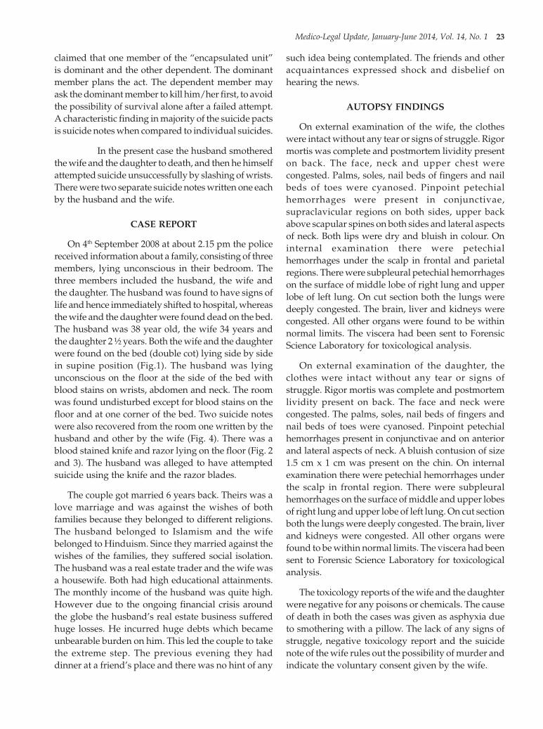
Medico-Legal Update, January-June 2014, Vol. 14, No. 1 23
claimed that one member of the “encapsulated unit”is dominant and the other dependent. The dominantmember plans the act. The dependent member mayask the dominant member to kill him/her first, to avoidthe possibility of survival alone after a failed attempt.A characteristic finding in majority of the suicide pactsis suicide notes when compared to individual suicides.
In the present case the husband smotheredthe wife and the daughter to death, and then he himselfattempted suicide unsuccessfully by slashing of wrists.There were two separate suicide notes written one eachby the husband and the wife.
CASE REPORT
On 4th September 2008 at about 2.15 pm the policereceived information about a family, consisting of threemembers, lying unconscious in their bedroom. Thethree members included the husband, the wife andthe daughter. The husband was found to have signs oflife and hence immediately shifted to hospital, whereasthe wife and the daughter were found dead on the bed.The husband was 38 year old, the wife 34 years andthe daughter 2 ½ years. Both the wife and the daughterwere found on the bed (double cot) lying side by sidein supine position (Fig.1). The husband was lyingunconscious on the floor at the side of the bed withblood stains on wrists, abdomen and neck. The roomwas found undisturbed except for blood stains on thefloor and at one corner of the bed. Two suicide noteswere also recovered from the room one written by thehusband and other by the wife (Fig. 4). There was ablood stained knife and razor lying on the floor (Fig. 2and 3). The husband was alleged to have attemptedsuicide using the knife and the razor blades.
The couple got married 6 years back. Theirs was alove marriage and was against the wishes of bothfamilies because they belonged to different religions.The husband belonged to Islamism and the wifebelonged to Hinduism. Since they married against thewishes of the families, they suffered social isolation.The husband was a real estate trader and the wife wasa housewife. Both had high educational attainments.The monthly income of the husband was quite high.However due to the ongoing financial crisis aroundthe globe the husband’s real estate business sufferedhuge losses. He incurred huge debts which becameunbearable burden on him. This led the couple to takethe extreme step. The previous evening they haddinner at a friend’s place and there was no hint of any
such idea being contemplated. The friends and otheracquaintances expressed shock and disbelief onhearing the news.
AUTOPSY FINDINGS
On external examination of the wife, the clotheswere intact without any tear or signs of struggle. Rigormortis was complete and postmortem lividity presenton back. The face, neck and upper chest werecongested. Palms, soles, nail beds of fingers and nailbeds of toes were cyanosed. Pinpoint petechialhemorrhages were present in conjunctivae,supraclavicular regions on both sides, upper backabove scapular spines on both sides and lateral aspectsof neck. Both lips were dry and bluish in colour. Oninternal examination there were petechialhemorrhages under the scalp in frontal and parietalregions. There were subpleural petechial hemorrhageson the surface of middle lobe of right lung and upperlobe of left lung. On cut section both the lungs weredeeply congested. The brain, liver and kidneys werecongested. All other organs were found to be withinnormal limits. The viscera had been sent to ForensicScience Laboratory for toxicological analysis.
On external examination of the daughter, theclothes were intact without any tear or signs ofstruggle. Rigor mortis was complete and postmortemlividity present on back. The face and neck werecongested. The palms, soles, nail beds of fingers andnail beds of toes were cyanosed. Pinpoint petechialhemorrhages present in conjunctivae and on anteriorand lateral aspects of neck. A bluish contusion of size1.5 cm x 1 cm was present on the chin. On internalexamination there were petechial hemorrhages underthe scalp in frontal region. There were subpleuralhemorrhages on the surface of middle and upper lobesof right lung and upper lobe of left lung. On cut sectionboth the lungs were deeply congested. The brain, liverand kidneys were congested. All other organs werefound to be within normal limits. The viscera had beensent to Forensic Science Laboratory for toxicologicalanalysis.
The toxicology reports of the wife and the daughterwere negative for any poisons or chemicals. The causeof death in both the cases was given as asphyxia dueto smothering with a pillow. The lack of any signs ofstruggle, negative toxicology report and the suicidenote of the wife rules out the possibility of murder andindicate the voluntary consent given by the wife.
5. Millo--22--25.pmd 2/22/2014, 11:44 AM23

24 Medico-Legal Update, January-June 2014, Vol. 14, No. 1
CLINICAL FINDINGS
The husband was shifted to AIIMS TraumaCentre, New Delhi, in an unconscious state. Onexamination in the casualty the airway was patent,breathing was spontaneous, respiratory rate was 30per minute, chest was clear, blood pressure was 140/90 mm Hg, pulse rate was 94/min, Glasgow ComaScale (GCS) was E1 V1 M1 with a score of 3/15 andtemperature was normal. He was intubated initiallyand put on ventilation in view of the poor GCS,subsequently he responded well and was extubatedthe same day. There were three lacerated wounds onthe neck with following description. (1) A 3 x 0.5 cmwound on anterior aspect transversely, (2) A 3 x 0.5cm wound on right lateral aspect of neck and (3) A 2 x0.5 cm wound on left lateral aspect of neck. There weremultiple superficial wounds of varying dimensionsof sizes 4-5 cm x 0.2 cm numbering more than 30 overanterior chest wall and more than 70 over anteriorabdominal wall. There were 5 superficial wounds ofsize 4 x 0.2 cm each over anterior aspect of right wrist.There were 3 superficial wounds of size 4 x 0.3 cmeach over anterior aspect of left wrist. He wasmanaged conservatively and was discharged after 6days. During the hospital stay psychiatric consultationwas also taken.
DISCUSSION
A suicide pact is very rare. As a result there havebeen very few studies on suicide pacts. However therehave been some occasional case reports of suicide pactsin the literature. Most of the victims of suicide pactshad chosen methods of suicide like poisoning, hanging,gas exhaust etc. In a study of 20 suicide pacts5 fromDade County, USA, gun was used as a method ofsuicide in 40% of cases followed by overdose in 27.5%,carbon monoxide in 25%, hanging in 5% and stabbing/slashing in 2.5%. The suicide pact victims in Bangaloreand Japan were much more likely than those inEngland or Dade County to choose poisoning as amethod of suicide6. In the present case smothering asa method of suicide in a suicide pact is unique. Thesuicide pact victims were typically skilled or unskilledworkers, the majority of whom were out of work orretired and 38% victims of spouse pacts were notworking4. In the present case the husband was self-employed who suffered huge losses in his business
Fig.1. The wife and the daughter lying dead on the bed
Fig.2. Blood stained razor lying on the floor
Fig. 3. Blood stained knife lying on the floor
Fig.4. Suicide notes found in the room written by the husbandand the wife
5. Millo--22--25.pmd 2/22/2014, 11:44 AM24

Medico-Legal Update, January-June 2014, Vol. 14, No. 1 25
driving the family to debt and the situation is similarto being out of work. In a study it has been reportedthat the average age of husbands and wives was 60.2years and 56.2 years respectively. In the same study ithas also been reported that the average age of malesand females was 44.1 years and 50.1 years respectively4.The average age of the Bangalore husbands in suicidepacts was 38 years versus 60.2 and 60 years forhusbands in England and Dade County, respectively6.The age of the husband in present case is 38 years andthat of the wife is 34 years. The age of the husband issimilar to the average age in Bangalore husbands asreported by Fishbain6. Leaving a suicide note may beone of the characteristics of suicide pacts. The suicidepact victims from Dade County were much more likelyto leave a suicide note than Bangalore suicide pactvictims6. Suicide pact victims are much more likely toleave a suicide note than single suicide victims. It hasbeen reported that suicide notes were found in 84% ofthe pacts. It has also been also reported3 that bothpartners signed the note in 63% pacts and one partnerin 36%. In the present case both the husband and thewife had written suicide notes. The suicide pact victimstake a great deal of care to cope with the problemsthat would be created by their death4. In the presentcase the husband wrote two detailed suicide notes oneto the police and other to his business partners. In thesuicide note addressed to the police he elaborated thereasons for taking the extreme step. In the suicide noteaddressed to his friends he expressed gratefulness tosome of his friends and also left instructions as to whatto do with his share of business. He also wrote aboutwhere some of the important documents had beenkept. The wife wrote a brief suicide note indicatingher wish to be buried along with her husband andaccording to the customs of Shia rites. But it was anunfortunate event for their innocent child who becamea forced victim of their suicide agreement.
In the instant case the suicide pact was anunsuccessful one as the husband survived the attempt.As per the law the husband has been charged withhomicide. The attempted suicide is also punishable asper the Indian law. Had the method of suicide beenhanging or poisoning the charge on the husbandwould have been abetment of suicide inviting lesserpunishment compared to homicide. However sincethere was no signs of struggle or violence as per theautopsy findings it was a case of assisted suicide. InIndia as per law there is no provision for assistedsuicide and the husband was charged with homicide.
CONCLUSION
The present case is unique because of the methodadopted to commit suicide, that is smothering, whichis a very violent method. Though the wife had itvoluntarily consented to be smothered but under thelaw it is a case of homicide. And also the child was anon consenting partner. Had the husband’s attemptbeen successful it would have been very difficult toarrive at the correct cause of death. Since the husbandsurvived, arriving at the cause of death was helped bycircumstantial evidence.
Acknowledgement: Nil
Conflict of Interest: Nil
Source of Funding : Self
Ethical Clearance: Obtained
REFERENCES
1. Altindag A, Yanik M. Suicide pact among threeyoung sisters. Isr. J. Psychiatry Relat. Sci. 2005;42(4): 278-80.
2. Brown M,Barraclough B. Epidemiology of suicidepacts in England and wales. 1988-92. Brit. Med.J. 1997;315 (7103): 286-7.
3. Brown M, arraclough B. Partners in life and indeath: the suicide pact in England and Wales,1988-1992. Psychol. Med. 1999;29 (6): 1299-06.
4. Cohen J. A study of suicide pacts. Med. Leg. J.1961;29: 144-51.
5. Fishbain DA, D’Achilli L, Barsky S, Aldrich TE.A controlled study of suicide pacts. J. Clin.Psychiat. 1984; 45: 154-7.
6. Fishbain DA, Aldrich TE. Suicide pacts:international comparisions. J. Clin. Psychiat.1985; 46: 11-5.
7. Hemphill R, Thornley F. Suicide pacts. S. Afr.Med. J. 1969; 43, 1335-8.
8. Latha KS. Suicide pact survivors: someobservations. Med. Sci. Law 1996; 36 (4): 295-8.
9. Noyes R, Frye S, Hartford C. Conjugal suicidepact. J. Nerv. Ment. Dis. 1997;165 (1): 72-5.
10. Prateek R, Nagesh KR. Suicide pact by hanging:a case report. Med. Sci. Law 2007;48 (3): 266-8.
11. Santy PA. Observations on double suicide:reviews of the literature and two case reports.Am. J. Psychother. 1982;36: 23-31.
5. Millo--22--25.pmd 2/22/2014, 11:44 AM25

26 Medico-Legal Update, January-June 2014, Vol. 14, No. 1
INTRODUCTION
Electrocution can cause lesions of variable severityaccording to the type and intensity of the current, thelength and method of exposure, and some individualphysiologic and pathologic features. The result maybe lesions of the skin and organs or death.1 Death maybe caused by different mechanisms like cardiacconducting system abnormalities, asphyxia, bulbarparalysis or secondary trauma
The total number of accidental deaths in India inthe year 2012 was 3,94,982 out of which 8750 (2.2%)were because of electrocution.2 Here we are presentinga case of electrocution death, which was actuallybrought to mortuary as death due to accidentaltraumatic asphyxia.
CASE REPORT
A male corpse aged 32 years was brought to themortuary. As per the inquest report the body was
Deadly Robbery - A Case Report
Prashanth Mada1, G Deva Raju2
1Assistant Professor, Department of Forensic Medicine & Toxicology, Mediciti Institute of Medical Sciences (MIMS)Ghanpur, Medchal Mandal, Ranga Reddy, Andhra Pradesh, India, 2Assistant Professor, Department of Forensic
Medicine & Toxicology, Osmania Medical College, Koti, Hyderabad, Andhra Pradesh, India
ABSTRACT
Here we report about a male corpse aged 32 years, which was actually brought to the mortuary asdeath due to accidental traumatic asphyxia. Asphyxia was the definitive mode of death in this case,but trauma was not the cause for the asphyxia instead, by the presence of electrical injury. The differenttypes of lesions were caused due to struggle to get down together with the electrocution. Of all thelesions diagnosed at post mortem examination, one needs a special mention, the subarachnoidhemorrhage, which resulted due to electrocution. Based on postmortem finding, the time since deathwas given as 12 -24 hours prior to the post mortem examination and the Cause of death was given as"ELECTROCUTION".
Keywords: Electrocution, Asphyxia, Subarachnoid Haemorrhage
Corresponding author:Prashanth MadaAssistant ProfessorDepartment of forensic medicine & Toxicology, MedicitiInstitute of Medical Sciences (MIMS) Ghanpur, MedchalMandal, Ranga Reddy, Andhra Pradesh, IndiaE mail: [email protected]: 9849730345
found in a hanging position, trapped in between theanglers used for supporting the tin roof of a wine shop.The history of the case according to the inquest reportis after removing the tin roof with screwdriver &cutting pliers the deceased tried to enter into the wineshop through the narrow 6" gap between the anglers,which are used for supporting the roof. In anunsuccessful effort to get down the deceased wastrapped in between the anglers and subsequently dieddue to traumatic asphyxia.
The following injuries were recorded in the inquestreport
1. Abrasion over the right side of the front & back ofthe chest.
2. Blood stained abrasions present on both elbows.
3. Multiple large abrasions on both thighs.
4. A large abrasion over the right frontal aspect ofleg below knee.
Postmortem findings
External examination
Identified body of middle aged male, 173 cm inheight with normal built fair complexion
The body was dressed in brown half sleeves shirtwith dark blue stripes, dark blue colored pant and lightblue under wear.
DOI Number: 10.5958/j.0974-1283.14.1.006
6. prasantha--26--29.pmd 2/22/2014, 11:44 AM26

Medico-Legal Update, January-June 2014, Vol. 14, No. 1 27
The following postmortem changes are notedgreenish discoloration over both iliac fossae, rigormortis was partially present in the lower limbs andpostmortem lividity was seen on distal parts of boththe lower limbs.
The following ante mortem injuries are noted
1. Mild ecchymosis was seen around the left eye withcongestion of the conjunctiva.
2. Multiple small abrasions over an area of 38´33 cminvolving front of chest and abdomen, reddishbrown in color and leathery in texture.
3. Grazed abrasion 145 cm on anterolateral aspect ofright side of chest, 5cm below the armpit reddishbrown in color.
4. Grazed abrasion 106 cm on back of left side ofchest, 15cm below the left shoulder blade reddishbrown in color.
5. Grazed abrasion 206 cm on front & outer aspect ofright hand involving the arm, elbow & forearmreddish brown in color.
6. An electrical injury, a round pale shallow crater2cm in diameter on the outer aspect of proximalpart of hypothenar eminence of left palm withaverted edges and red peripheral discoloration.
7. Grazed abrasion 18´8 cm on outer aspect of lefthand involving the arm, elbow & forearm reddishbrown in color.
8. Grazed abrasion 7´3 cm on left loin reddish brownin color.
9. Multiple grazed abrasions of varying size over anarea of 16´10 cm on upper outer aspect of rightbuttock reddish brown in color.
10. Abrasions measuring 85cm on front and lower partof scrotal sac reddish brown in color.
11. Grazed abrasion 2010 cm on anteromedial aspectof left thigh reddish brown in color.
12. An electrical injury, a round shallow cratermeasuring 4´3 cm on front of upper 3rd of rightleg with averted edges and red peripheraldiscoloration
13. An electrical injury, a round shallow cratermeasuring 4´3 cm on front of middle 3rd of rightleg with averted edges and red peripheraldiscoloration
14. An electrical injury, a round shallow cratermeasuring 2´1/2 cm on tip of right great toe and1cm diameter on tip of second toe with avertededges and red peripheral discoloration
Internal examination
Both cerebral hemispheres were having diffusesubarachnoid hemorrhage.
Fracture of 7th, 8th, & 9th ribs on left side underthe injury 4. The fractured ribs were showinginfiltration of blood with laceration of underlyingpleura.
Heart chambers filled with frothy fluid with bloodstained pericardial fluid.
Stomach – about 100ml of semi digested food withno obnoxious smell, mucosa of stomach congested.Kidneys both are congested. Testicles contused
All other visceral organs are congested.
No tissues were preserved for further examination.
Taking into account the above findings the timesince death was given as 12 -24 hours prior to the postmortem examination and the Cause of death was givenas “ELECTROCUTION”.
DISCUSSION
This case report analyzes the dynamics andcircumstances surrounding the death of a manengaged in robbery with electrocution.
The mechanisms resulting in death can be ofdifferent types, according to the intensity of the electriccurrent and its path through the body. The first, mostimportant cause is abnormalities of cardiac conductingsystem. The second type is respiratory and occurswhen the current affects the chest, provoking firstspasm and then tetanic contraction of the intercostalmuscles and diaphragm, resulting in asphyxia. Thethird cause is paralysis of the bulbar nerve centers.1
Other rare mechanisms are secondary trauma,drowning and individual damage of organ.3
Asphyxia was the definitive mode of death in thiscase, but trauma was not the cause for the asphyxiainstead, by the presence of electrical injury and thestudy of crime scene, which helped us to come to anopinion about the mode of death as asphyxia due tothe tetanic contractions of the intercostal muscles anddiaphragm due to electrocution.
6. prasantha--26--29.pmd 2/22/2014, 11:44 AM27
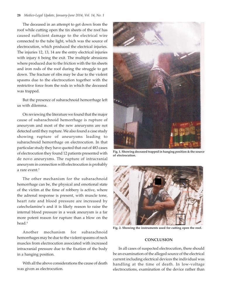
28 Medico-Legal Update, January-June 2014, Vol. 14, No. 1
The deceased in an attempt to get down from theroof while cutting open the tin sheets of the roof hascaused sufficient damage to the electrical wireconnected to the tube light, which was the source ofelectrocution, which produced the electrical injuries.The injuries 12, 13, 14 are the entry electrical injurieswith injury 6 being the exit. The multiple abrasionswhere produced due to the friction with the tin sheetsand iron rods of the roof during the struggle to getdown. The fracture of ribs may be due to the violentspasms due to the electrocution together with therestrictive force from the rods in which the deceasedwas trapped.
But the presence of subarachnoid hemorrhage leftus with dilemma.
On reviewing the literature we found that the majorcause of subarachnoid hemorrhage is rupture ofaneurysm and most of the new aneurysms are notdetected until they rupture. We also found a case studyshowing rupture of aneurysms leading tosubarachnoid hemorrhage on electrocution. In thatparticular study they have quoted that out of 483 casesof electrocution they found 12 patients presented withde novo aneurysms. The rupture of intracranialaneurysm in connection with electrocution is probablya rare event.3
The other mechanism for the subarachnoidhemorrhage can be, the physical and emotional stateof the victim at the time of robbery is active, wherethe adrenal response is present, with muscle tone,heart rate and blood pressure are increased bycatecholamine’s and it is likely reason to raise theinternal blood pressure in a weak aneurysm is a farmore potent reason for rupture than a blow on thehead.4
Another mechanism for subarachnoidhemorrhages may be due to the violent spasms of neckmuscles from electrocution associated with increasedintracranial pressure due to the fixation of the bodyin a hanging position.
With all the above considerations the cause of deathwas given as electrocution.
Fig. 1. Showing deceased trapped in hanging position & the sourceof electrocution.
Fig. 2. Showing the instruments used for cutting open the roof.
CONCLUSION
In all cases of suspected electrocution, there shouldbe an examination of the alleged source of the electricalcurrent including electrical devices the individual washandling at the time of death. In low-voltageelectrocutions, examination of the device rather than
6. prasantha--26--29.pmd 2/22/2014, 11:44 AM28

Medico-Legal Update, January-June 2014, Vol. 14, No. 1 29
examination of the body will often provide the causeof death, because burns may not be present. Thus, onecan make a diagnosis of electrocution without anelectrical burn, based on the circumstances of thedeath, negative autopsy ûndings and the examinationof the electrical device in use. In high-voltageelectrocution, tissue from the victim may be adherentat the point of contact with the source of the current.5
The following important points to be remembered incases of suspected electrocution
1. Thorough examination of the body to find theelectrical injury.
2. Absence of external electrical injury doesn’texclude electrocution.
3. In cases where there are no definitive external signsof electrocution, circumstantial evidence is crucial.
4. Don’t blindly rely on Inquest report.
5. Visiting the crime scene when & where required/at least request the IO for crime scene photographs,which are taken without disturbing the scene ofoffence.
Acknowledgments: Nil
Conflict of Interest: There is no conflict of interest,publishing with academic interest.
Source of Funding: Nil
Ethical Clearance: As identity of the deceased has notbeen revealed & as it is a retrospective case report withno risk involved, ethical clearance is not applicable.
REFERENCES
1. Di Nunno, Nunzio, Vimercati, Luigi, Viola, Luigi,Vimercati, Francesco. A Case of ElectrocutionDuring Illegal Fishing Activities. AmericanJournal of Forensic Medicine & Pathology, June2003 ;Volume 24 (2):164-167.
2. ncrb.nic.in/publications/accidental deaths &suicides/2012/accidental deaths in india/figure1.5.
3. W Sprecher, W Wenz, H.T Haffner, Rupture ofan intracranial aneurysm – unusual complicationof an electric shock, Forensic ScienceInternational, November 2001;Vol. 122 (2) :85-88.
4. Bernard knight, Pekka Saukko, Knight’s ForensicPathology, Third edition, Chapter 5, Pg199
5. Vincent J.M. DiMaio, Dominick DiMaio, ForensicPathology, second edition, chapter 16, Pg413-414.
6. prasantha--26--29.pmd 2/22/2014, 11:44 AM29

30 Medico-Legal Update, January-June 2014, Vol. 14, No. 1
INTRODUCTION
Road traffic accidents are one of the major causesof death in developed as well as developing countries1.Although, this problem has been controlled very muchin developed countries but in developing countries like
Pattern of Fatal Head Injuries in Road Traffic Accidents at SMS Hospital, Jaipur - An Autopsy Based Study
R K Punia1, L C Verma2, Deepali Pathak3
1Professor & Head, 2Resident, 3Assistant Professor, Dept of Forensic Medicine, SMS Medical College, Jaipur
ABSTRACT
Background: Head Injury is the single most common cause of mortality in vehicle accidents. Itsoutcome is a product of different mechanisms, types and amounts of head injuries and their anatomicallocations.
Objective: To analyze the pattern of fatal head Injury in road traffic accidents at SMS Hospital, Jaipur;a tertiary care centre.
Subjects: Cases of Road Traffic Accidents with fatal head injury subjected to medico-legal autopsy atthe Department of Forensic Medicine, SMS Medical College and Hospital Jaipur, Rajasthan, India - atertiary care center.
Main outcome: Young adults (both males and females) in their most productive years of life areespecially prone to head injury, as a result of vehicle accidents. The use of helmets as preventivemeasure has checked the external and bony injuries to skull region but there is an urgent need to planfurther prevention in such cases to check internal brain injuries caused due to movement of brain incranial cavity.
Results: Males clearly outnumbered females with male to female ratio as 4.7:1. Motor-cyclists werethe commonest group of victims. Head injury was the commonest cause of death comprising 53.5%cases. Intracranial hemorrhage of one or more vascular components was the commonest type ofinjury. The most frequent pattern was a combination of subdural, subarachnoid and intracerebralhematomas i.e. 23 (34.9%). This was followed by Subarachnoid and Intracerebral 13 (19.7%) andsubdural and subarachnoid 09 (13.6%). Out of 16 (24.24%) single compartment hemorrhage, themost frequent was intracerebral 08 (50%), followed by subdural 5 (31.25%) and subarachnoid 2 (12.5%)hemorrhage.
Conclusion: Prompt treatment should be instituted to all cases of trauma particularly head injury toprevent fatality. Also there is a need to plan preventive measures.
Keywords: Fatal Head Injury, Road Traffic Accidents, Intracranial Hemorrhages
Corresponding author:R K PuniaProfessor & HeadDept of Forensic Medicine, SMS Medical Cllege, JaipurEmail- [email protected]
India, condition is still worsening. India being one ofthe fastest developing nations in the world with a hugepopulation density, the road traffic density is alsoincreasing. India accounts for about 10% of roadaccident fatalities worldwide2. The head being the mostvulnerable part of the body is involved frequently andlead to morbidity and mortality in road trafficaccidents.
Head injury has been defined as, “a morbid state,resulting from gross or subtle structural changes in thescalp, skull, and/or the contents of the skull, produced
DOI Number: 10.5958/j.0974-1283.14.1.007
7. Punia--30--34.pmd 2/22/2014, 11:44 AM30

Medico-Legal Update, January-June 2014, Vol. 14, No. 1 31
by mechanical forces” 3. Depending upon whether ornot the duramater was torn, the head injury may betermed as open or closed type4. The extent and degreeof injury to the skull and its content is not necessarilyproportional to the quantum of force applied to thehead. Head injury may lead to skull fractures, variousextents of brain parenchymal injuries and traumaticvascular injuries5.
According to Munro, any type of cranio-cerebralinjury is possible with any kind of blow on any sort ofhead6.
Severe head injury, with or without peripheraltrauma, is the commonest cause of death and/ ordisability up to the age of 45 years in developedcountries7. Death due to head injury depends uponvarious factors including intensity of impact on head,degree of deceleration, pattern of skull bones fractured,degree and sites of parenchymal and vascular injuries,time spent from the time of injury to the specializedhealth care at hospital and how efficiently life supportmeasures were given8. Unless some safety andpreventive measures are strictly implemented deathtoll will continue to increase and will cause significantloss of precious lives. In this paper, recent trends inpatterns of fatal head injury
in road traffic accidents with regards to age, sex,frequency of occurrence, anatomical sites involvedwere analyzed and presented.
MATERIAL & METHOD
The present study was carried out prospectively inthe Department of Forensic Medicine & Toxicology,SMS Medical College, Jaipur, Rajasthan over a periodof two months during June and July, 2013. Total 137cases of road traffic accidents were autopsied duringthis period out of which fatal head injury was presentin 80 cases. The information regarding age, sex,residence, date and time of accident and of death wasgathered from police inquest report, relatives, deadbody challan and clinical details from hospital records.During autopsy, detailed examination was carried outand data regarding both external and internal injurieswas carefully recorded analyzed and presented in thispaper.
OBSERVATIONS
Age and Sex Distribution
The highest incidence was seen in the age group of
21-30 years comprising of 26 (32.5%) cases followedby31-40 years comprising of 20 (25%) cases. The lowestincidence was seen in >60 years age group comprisingof only 1.25% cases. The majority of the victimssubjected to fatal head injury were males (82.5%) withmale: female ratio of 4.7:1.
Table 1: Age and sex distribution of victims
Age (in years) Male (%) Female (%) Total (%) Sex Ratio
0-10 01 02 03 (3.75) 0.5:1
11-20 13 0 13 (16.25) 13:0
21-30 25 01 26 (32.5) 25:1
31-40 13 07 20 (25) 1.86:1
41-50 10 02 12 (15) 5:1
51-60 03 02 05 (6.25) 1.5:1
> 60 01 0 01 (1.25) 1:0
Total (%) 66 (82.5) 14 (17.5) 80 (100) 4. 7:1
Place of Death
The maximum number of victims (25%) died whileon the way to hospital, followed by victims died inthe hospital (65%). The number of victims who diedon the spot was 10%.
Profile of victims
The motor-cyclists were the commonest group ofvictims, comprising 32(40%) cases, out of which 29were drivers, followed by Light Motor Vehicle userscomprising 28 (35%) cases of which 23 were occupants.Pedestrians constituted 20 (25%) of the cases. Overall,the drivers of vehicles (34%) outnumbered theoccupants of the vehicles (26%).
Offending Vehicle
In the present study, truck was the commonestoffending vehicle being involved in 52 (65%) cases,followed by Light Motor Vehicle (20%) and buses(15%).
Site of Injury
Pedestrians, occupants of two wheelers and lightmotor vehicles sustained maximum injuries onextremities, followed by on head, neck and face. Fourwheeler users had maximum injuries on head, neckand face region, followed by on extremities.
Cause of Death
Head injury was the commonest cause of deathcomprising 53.5% cases. In drivers, head injury wasthe commonest cause of death observed in 28 cases. Inoccupants of the vehicles, injury to vital organs was
7. Punia--30--34.pmd 2/22/2014, 11:44 AM31
32 Medico-Legal Update, January-June 2014, Vol. 14, No. 1
the commonest cause of death seen in 17 cases. Inpedestrians, shock and hemorrhage was predominantcause of death seen in 14 cases.
Head Injury
A combination of multiple injuries in the form ofskull fractures, intracranial hemorrhage and/ or injuryto brain with or without external injury waspredominantly seen in more than 90% cases, includinga combination involving scalp injury and intracranialhemorrhage (62.5 %) and those involving combinationof skull fractures and intracranial hemorrhages withor without brain injury (28.75%).
Fifty two (65%) cases had no skull bone fracturesat all, 23 (28.75%) patients sustained skull bone fractureof single bone and rest 05 (6.25%) had fractures of morethan one skull bone. Out of the total 23 (28.75%)subjects which received single skull bone fracture, themost frequent bone fractured was frontal (39.3%),followed by parietal (17.8%), occipital (14.3%) andtemporal (10.7%) each. Rest cases showing skullfractures had multiple bony injuries. Out of the 23cases of skull fractures, in 11 cases there was isolatedfracture of the vault, fractures of the vault and basesof skull were seen in 5 cases and in rest of the 7 cases,there were only basal fractures. In cases of basalfractures, fractures of the anterior cranial fossa werethe commonest and the fractures of posterior cranialfossa were the least common.
Table 2 : Showing Anatomical location of Skullfractures
Site of skull fracture (bone) No. of cases Percentage (%)
Frontal 11 39.3
Temporal 03 10.7
Parietal 05 17.8
Occipital 04 14.3
Frontal + Temporal 01 03.6
Temporal + Occipital 01 03.6
Frontal + Temporal + Parietal 03 10.7
Total cases of Skull fractures 28 35
Out of the total 80 cases, more than half (62.5%)subjects presented with hemorrhage in more than onevascular compartment. The most frequent pattern wasa combination of subdural, subarachnoid andintracerebral hematomas i.e. 23 (34.9%). This wasfollowed by Subarachnoid and Intracerebral 13 (19.7%)and subdural and subarachnoid 09 (13.6%). Out of 16(24.24%) single compartment hemorrhage, the mostfrequent was intracerebral 08 (50%), followed by
subdural 5 (31.25%) and subarachnoid 2 (12.5%)hemorrhage.
Table 3: Showing the pattern of IntracranialHemorrhages in cases
Type of Hemorrhage No. of Cases Percentage (%)
EDH only 01 1.5
SDH only 05 7.6
SAH only 02 03
ICR ONLY 08 12.1
EDH + SDH 05 7.6
SDH + SAH 09 13.6
SAH + ICR 13 19.7
SDH +SAH + ICR 23 34.9
Total 66 (82.5%) 100
There were no injuries to other body parts in 22(27.5%) cases. Out of the remaining 58 (72.5%) cases,there were 10 (17.24%) cases with rib fractures, 7 (12%)cases with lung lacerations, 13 (22.4%) with injuries tothe extremeties, 20 (34.4%) with abdomino-pelvicinjuries, 5 (8.6%) with facial bone fractures and 3 (5.1%)with spinal injuries, A total of 28 (35%) cases receivedmore than two injuries other than head injuries.
DISCUSSION
In this study, Males clearly outnumbered femaleswith male to female ratio as 4.7:1. This is in almost inaccordance with the studies of Kumar A et al2, SinghYN et al9, Tripude BH et al10, Singh H et al11, SharmaBR et al12, Biswas G et al13 and Tandle RM et al14. In theage group analysis of the victims, maximum incidencewas in age group of 21-30 years and least in group 0-10 years. Similar findings were observed by otherresearchers10-14.
The maximum number of victims (65%) died in thehospital, followed by victims who died while on theway to hospital (25%). The number of victims who diedon the spot was 10%. This is not consistent with thestudies of Sharma BR et al12 Tandle RM et al14, MenonA et al 15 and Chandra J et al 16. The reason for thisinconsistency may be due to the differences in theseverity of head injuries and other associated injuriesin the various studies of comparison.
The motor-cyclists were the commonest group ofvictims (40%) followed by Light Motor Vehicle users(35%). Pedestrians constituted a quarter of the cases.Our findings are quite similar to those of Tandle RM etal14 and Pathak A et al17. In our study the drivers werereported more than occupants.
7. Punia--30--34.pmd 2/22/2014, 11:44 AM32

Medico-Legal Update, January-June 2014, Vol. 14, No. 1 33
Head Injury was the predominant cause of deathin drivers while shock and hemorrhage resulting frominjuries to other vital organs remained thepredominant cause of death in occupants of light motorvehicles and pedestrians. Our findings are similar tothose reported by Tandle RM et al14, Chandra J et al16
and Kumar A et al2.
In our study abdomino- pelvic region sufferedmaximum injuries (34.4%), followed by thorax(29.24%) and extremities (22.4%). Least numbers ofinjuries were observed on spine (5.1%). Though therewas a component of head injury in eeach case, stillfacial injuries were seen in only 8.6% cases. Ourfindings are in slight discordance with those of SinghH et al11, Tandle RM et al14 and Dhillon S et al18. Thisvariability may be due to variations of studypopulation and also because of the increasing use ofhelmets which protect the head and face region fromexternal injuries.
Intracranial hemorrhage of one or more vascularcomponents was the commonest type of injury seenin 66 of the total 80 cases of head injuries, followed byinjury to the brain (45) and skull fractures (28). Injuryto brain was observed in 45 cases. The findings areslightly variable from the studies of Menon A et al15,Dhillon S et al18 and Tandle RM et al14.
Out of the 28 cases of skull fractures, in 11 casesthere was isolated fracture of the vault, fractures ofthe vault and bases of skull were seen in 5 cases and inrest of the 7 cases, there were only basal fractures. Incases of basal fractures, fractures of the anterior cranialfossa were the commonest and the fractures ofposterior cranial fossa were the least common. Out ofthe total 23 (28.75%) subjects which received singleskull bone fracture, the most frequent bone fracturedwas frontal (39.3%), followed by parietal (17.8%),occipital (14.3%) and temporal (10.7%) each.
CONCLUSION
Most of the cases of death due to head injury invehicular accidents were males of productive agegroup because this is the population more in motionfrom one place to others. Majority of the victims weremotor cyclists and drivers who sustained fatal headinjury because of falls and probably due to head oncollisions. The pattern of head injuries is different fromother studies as intracranial hemorrhages and internalinjuries to brain were predominantly seen. Rather inother studies external and bony injuries were more
commonly seen. This is probably due to the strictnessin traffic rules for compulsion of helmets so that theinjuries due to direct impact are prevented, however,the injuries resulted from movement of brain insidethe cranial cavity. Thus we conclude that additionalpreventive measures should be planned andimplemented to prevent head injuries.
Acknowledgements: To the victims on whom thework was carried out
Conflicts of Interest: Nil
Source of Funding: Nil
Ethical Clearance: Not required
REFERENCES
1. Ganveer GB, Twiari RR. Injury pattern amongnonfatal road traffic accident cases: A cross-sectional study in Central India. Indian J Med Sci2006: 6; 59: 9-12.
2. Arvind Kumar, Sanjeev Lalwani, DeepakAgrawal, Ravi Rautji, T D Dogra. Fatal road trafficaccidents and their relationship with headinjuries: An epidemiological survey of five year.Indian Journal of Neurotrauma, Vol.5, N0.2, 2008
3. Dr. K H Chavli, Dr. B R Sharma, Dr. D Harish,Dr. Anup Sharma. Head injury: The principalkiller in road traffic accidents. JIAFM, 2006:28(4)ISSN: 0971-973.
4. Reddy KSN.The Essentials of Forensic Medicineand Toxicology, 26th Edition, 2007; Page215.
5. Bazarian JJ, McClung J., and Shah MN. Mildtraumatic brain injury in United State, 1998 –2000. Brain injury 2005; 19(2):85-91.
6. Munro D. Cranio-cerebral injuries. OxfordUniversity Press, as quoted by Gordon I, ShapiroHA in ¯Forensic Medicine: A Guide to Principles,3rd Ed, 1988, Page252.
7. Baethman A, Lehr D, Wirth A: prospectiveanalysis of patient management in severe headinjury. Acta Neurochirargica, 1998; 715; 107-110.
8. Marcus M.L. (2003): Incidence, external causes,and outcomes of work-related brain injuries inmale. J Occup Med. 2003; 27: 757- 760.
9. Y N Singh,. Kanak Ch. Das, et al. Anepidemiological study of road traffic accidentvictims in medico legal autopsies. Journal ofIndian Academy of Forensic Medicine, 2005:27(3),ISSN: 0971-0973.
7. Punia--30--34.pmd 2/22/2014, 11:44 AM33

34 Medico-Legal Update, January-June 2014, Vol. 14, No. 1
10. B H Tirpude,. R S Naik, A J Anjankar, et al. A studyof the pattern of cranio-cerebral injuries in roadtraffic accidents; Journal of Indian Academy ofForensic Medicine, 1998, Vol.20, No.1 ISSN:0971-0973.
11. Harnam Singh, S K Dhattarwal. Pattern anddistribution of injuries in fatal road trafficaccidents in Rohtak (Haryana) ;Journal of IndianAcademy of Forensic Medicine,2004;26(1),ISSN0971-0973.
12. B R Sharma, A K Sharma, Swati Sharma,Harshabad Singh. Fatal road traffic injuries inNorthern India: Can they be prevented? Trendsin medical Research 2(3):142-148, 2007.
13. Gautam Biswas, S K Verma, N K Agrawal, et al.Pattern of road traffic accidents in north eastDelhi; Journal of Forensic Medicine andToxicology Vol. 20, No.1, Jan-Jun2003.
14. Tandle RM, Keoliya AN. Patterns of head injuriesin fatal road traffic accidents in a rural district ofMaharashtra- Autopsy based study. J Indian AcadForensic Med, 2011; 33(3): 228-31.
15. Anand Menon, Nagesh K R. Pattern of fatal headinjuries due to vehicular accidents in Manipal.JIAFM, 2005:27(1), 19-22.
16. J Chandra, T D Dogra, P C Dixit. Pattern of cranio-intracranial injuries in fatal vehicular accidentsin Delhi, 1966-76 Med. Sci. Law (1979) Vol.19,No.3, 186-94.
17. Pathak Akhilesh, Vyas P C, Gupta B M. Autopsyfinding of pattern of skull fractures and intra-cranial hemorrhages in cases of head trauma: Aprospective study. Journal of Indian Academy ofForensic Medicine, 2006:28(4), 187-190.
18. Sangeet Dhillon, H S Sekhon. Pattern anddistribution of injuries in fatal road trafficaccidents in Shimla (Himachal Pradesh). Medico-legal Update, Oct-Dec, 2007, Vol.7 No.4.
7. Punia--30--34.pmd 2/22/2014, 11:44 AM34

Medico-Legal Update, January-June 2014, Vol. 14, No. 1 35
INTRODUCTION
Lead is a highly toxic substance, exposure to whichcan produce a wide range of adverse health effects.[1]
There are many ways in which humans are exposedto lead: through deteriorating paint, household dust,bare soil, air, drinking water, food, ceramics, homeremedies, hair dyes and other cosmetics. Much of thislead is of microscopic size, invisible to the naked eye.
Heavy metals are found naturally in theenvironment in rocks, soil and water, and thereforeexist in the manufacture of pigments and other rawmaterials in all industries including the cosmeticsindustry. Some of these metals have been used ascosmetic ingredients in the past. Example includes the
Determination of the Toxic Lead and Copper Levels inCosmetic Hair-Dye Powders by Flame Atomic Absorption
Spectrophotometry
Risha Jasmine Nathan1, P Sharma2, Lily Saroj Nathan3
1M.Sc., Forensic Chemistry & Toxicology, 2SSA (Chemistry) Lok Nayak Jayaprakash Narayan National Institute ofCriminology and Forensic Science, Ministry of Home Affairs, Govt. of India, 3Associate Professor, Zoology, Ewing
Christian College, Allahabad
ABSTRACT
Metal toxicity in humans is a result of long term or high level exposure to toxic metals which enterthe body through common media as air, water, and food. More recently, with the increase in regularuse of numerous cosmetic products, which have been reported to contain heavy metals ascontaminants, human bodies are being exposed to them on a daily basis. A number of dermalabsorption studies have been conducted which satisfy the assumption that harmful chemicals getabsorbed into the blood stream through skin.
Hair-dye powders are one of the most commonly used cosmetic products by all- young and old thesedays and are applied on a regular basis. If contaminated, direct application on hair and scalp canincrease chances of absorption of toxic metals as lead and copper and can potentially add up tosignificant exposure levels as these have cumulative effect. This has drawn our attention to assessthe levels of these metals in nine samples of common hair-dye powders available in markets ofDelhi. The levels of these metals were determined by Flame Atomic Absorption Spectrophotometerafter a wet digestion method for sample preparation. Lead and Copper were detected in differentconcentrations in all the studied samples, the highest being 1.42 ± 0.0814 ppm and 0.75 ± 0.0693 ppmfor lead and copper respectively.
Keywords: Toxic, Lead, Copper, Hair-Dye, Flame AAS
progressive hair dye lead acetate. Oral exposure canoccur for cosmetics used in and around the mouth, aswell as from hand-to-mouth contact after exposure tocosmetics containing heavy metal impurities. [2]
Exposure to environmental lead as reflected by itsaccumulation in hair has been studied by variousresearchers. In addition to this, variables which affectthe accumulation of lead in hair have also beendiscussed. [3]
Apparently, in the use of lead acetate hair dyes,some lead is absorbed systemically from the scalp. [4]
As stated earlier, though heavy metals might not causeimmediate health problems but their cumulative effectdue to repeated application cannot be ignored. Dermal
DOI Number: 10.5958/j.0974-1283.14.1.008
8. Risha puroshattam--35--40.pmd 2/22/2014, 11:44 AM35

36 Medico-Legal Update, January-June 2014, Vol. 14, No. 1
absorption of heavy metals [5] through skin of hands,scalp and face due to prolonged use of cosmeticscontaining lead is a matter of concern.
Copper has been assessed for its presence andconcentration in various cosmetic and herbal productsby various researchers. Saeed M. et. al. have stated, “Copper has both beneficial and toxic effects dependingon its level of consumption. Monitoring of copper limitis essential as it is beneficial in low concentrations butexhibit various toxic effects above the safety limit [6] [7]
Health Canada’s “Draft guidance on heavy metalimpurities in cosmetics, (2009)” has acknowledged thatheavy metal impurities in cosmetic products areunavoidable due to the ubiquitous nature of theseelements, but should be removed wherever technicallyfeasible. Heavy metal concentrations in cosmeticproducts are seen to be technically avoidable whenthey exceed a certain limit e.g. Lead: 10 ppm[8].
The aim of this project was to determine the toxiclead and copper levels in popular cosmetic hair-dyepowders randomly selected from the market of Delhiby Flame Atomic Absorption Spectrophotometry.
MATERIALS AND METHOD
Sample Collection
Nine samples of popular cosmetic hair-dyeformulations (black only) were randomly collectedfrom markets of Delhi. The brand names were blindedand given the codes HD1 to HD9.
Apparatus and Chemicals
Instrument: Atomic absorption spectrophotometer(EC Electronics Corporation of India LimitedAAS4129) was used for determination of the metals-lead (Pb) and copper (Cu) with deuterium lamp forbackground correction. The hollow-cathode lamps forPb and Cu which modulate the light of 285 Hz for therespective metals were employed as radiation source.The flame used was air/acetylene. Nitrogen was usedas carrier gas.
Chemicals: Nitric acid and hydrogen peroxide usedwere of analytical grade and were 99.99 % free fromthe presence of the metals under analysis (J. T. Baker).The water used in all experiment was ultrapure waterobtained from Milli-Q-water purification system(Ranken Rion Ltd, India).
Preparation of standard solutions
Lead standard solution (1000 ppm) was diluted toprepare stock solution of 10 ppm. Nine calibratorsolutions of different concentrations (2.0 ppm, 3.0 ppm,4.0 ppm, 5.0 ppm, 6.0 ppm, 7.0 ppm, 8.0 ppm and 9.0ppm were prepared by serially diluting the stocksolution of 10 ppm of lead immediately before use,whereas 10.0 ppm stock solution was used as the ninthcalibrator solution.
Similarly, copper calibrator solutions were alsoprepared.
Sample preparation [9]
Sample preparation was done according to themethod followed by Kumar S. et. al.
Samples were digested by the wet digestionmethod. 10 ml of nitric acid was added to 2 g ofaccurately weighed dried sample in a 100 ml beakerand was heated at 95°C for 15 min. The digest wascooled and 5 ml of concentrated nitric acid was addedand heated for additional 30 min at 95°C. The last stepwas repeated and the solution was reduced to about 5ml without boiling. The sample was cooled again and2 ml of deionized water and 3 ml of 30% hydrogenperoxide was added. With the beaker covered, thesample was heated gently to start the peroxidereaction. If effervescence becomes excessivelyvigorous, sample was removed from the water bathand 30% hydrogen peroxide was added in 1 mlincrements, followed by gentle heating until theeffervescence subsided. 10 ml of deionized water wasadded and the sample was heated for additional 15min without boiling. The sample was cooled andfiltered through a Whatman No. 42 filter paper anddiluted to 50 ml with deionized water.
Sample analysis
Calibration lead and copper standards wereprepared fresh every day prior to each analysis.Digested samples were analyzed for Lead and Copperusing flame atomic absorption spectrophotometerwhich was done using a manual standard additionprocedure where all nine hair-dye samples weredivided into two batches each - (A) for Lead analysisand the (B) for Copper analysis. In batch (A) samples,known concentration of standard aqueous solution ofLead was added to each sample from HD1 to HD9and the ‘test’ samples so prepared were then subject
8. Risha puroshattam--35--40.pmd 2/22/2014, 11:44 AM36

Medico-Legal Update, January-June 2014, Vol. 14, No. 1 37
to AAS analysis for quantification of Lead. Similarlybatch (B) ‘test’ samples were analysed forquantification of Copper. Control samples were alsoanalyzed.
All the measurements were made in triplicate forthe samples and standard solutions. The instrumentalconditions during the analysis of trace and heavymetals are listed in Table 1 giving details aboutparameters which are defined for Lead and Copper.
Quality assurance [10] [11] [12]
Reliability of the results was ensured by carryingout appropriate quality assurance procedures andprecautions. Due to the unavailability of certifiedmaterial for hair-dye analysis, the accuracy of themethod was determined by measuring the recoveryof lead and copper in the matrix containing knownconcentration of lead and copper solutions. Themethod of standard addition which is considered as avalidation method was used to demonstrate thevalidity of the method used.
A recovery test was performed using method ofstandard addition. The recovery and reproducibilityof the method was carried out by spiking andhomogenizing several already analyzed samples withvaried amounts of standards solutions of lead (Pb) andcopper (Cu) and processed as previously described
These ‘spiked’ samples were run with the ‘test’samples using the same analytical procedure. Theanalytical recovery for nine ‘spiked’ hair-dye sampleswith lead and copper are given in Table 4.
RESULTS
Regression data for the standard calibration plots
There was a good linear relation betweenabsorbance and standard concentration of lead andcopper. The regression equations, pearson’s correlationcoefficients and coefficient of determinations obtainedfor each calibration plot are summarised in Table 2.Linearity was evaluated by calculating the pearson’scorrelation coefficients (r) for three runs of ‘spiked’hair-dye samples each for lead and copper.
Determined metal concentration in each hair-dyesample
The spectrophotometer automatically determinedthe concentration of Lead and Copper present in the‘test’ samples by extrapolating the calibration curve
in parts per million. All samples were run in triplicateand the mean values were used along with therespective standard deviation values for statisticalanalysis. Table 3 shows the data. Calculations weremade to determine the weight of lead and copper andtheir percentage in hair-dye powders (Table 5). Figure1 shows the graph of quantity of the respective metalsfound in hair-dye powders.
Table 6 summarises the statistics of the presentstudy.
DISCUSSION
Lead and Copper poisoning by regular use of hair-dye powders is a matter of concern. Lead poisoningoccurs when lead builds up in the body, often over aperiod of months or years. Lead containing hair-dyeswhether applied a number of times a day or on dailybasis can contribute to significant lead exposure levels.Even small amounts of lead can cause serious healthproblems. In adults, lead can increase blood pressureand cause fertility problems, nerve disorders, muscleand joint pain, irritability, and memory or concentrationproblems. When a pregnant woman has an elevatedblood lead level, that lead can easily be transferred tothe fetus, as lead crosses the placenta. In such cases,the baby is born with an elevated blood lead level. [1]
Previously published works have mentioned [2] -
“Latest study shows that there is no safe level of leadexposure. The Centre for Disease Control andPrevention (CDC) has even gone so far as torecommend that parents should avoid using cosmeticson their children as that could be contaminated withlead…”
Similarly, toxicity may occur as a result of regularuse of products such as hair-dyes containing Copper.
CONCLUSION
Various brands of hair-dyes manufactured byreputed firms are available in the Indian market whichare claimed as safe for consumer-use. As per ChapterIV of Drugs and Cosmetics Rules 1945, Rule 144, thepermitted synthetic organic colours and natural organiccolours used in the cosmetic shall not contain morethan 20 ppm of lead calculated as lead and 100 ppm ofheavy metals other than lead calculated as the total ofrespective metals. Rule 145 prohibits use of lead andarsenic compounds in cosmetics for the purpose ofcolouring. Rule 135 prohibits import also of cosmeticscontaining lead or arsenic compounds for the purposes
8. Risha puroshattam--35--40.pmd 2/22/2014, 11:44 AM37

38 Medico-Legal Update, January-June 2014, Vol. 14, No. 1
of colouring. U/S Section 27A, manufacturing, selling,etc, of cosmetics in contravention to Drugs andCosmetics Act,1940 and/or Rules 1945 calls forimprisonment up to three years and with fine.
It has already been brought to notice by earlierresearchers-[2]
“None of the heavy metals are listed on the labelsof the cosmetic products. Provision should be madein the act for compulsory disclosure of the heavy metalconcentration on the cosmetic labels esp. on the colourcosmetics. Furthermore, manufacturers and importersmust ensure that products are safe and do not pose arisk to the users. Misbranded and Spurious cosmetics
are defined u/s-9-C and 9-D respectively in the Drugsand Cosmetics Act for the purpose of import to India.Adulterated cosmetics are not defined for the purposeof import….. The proposed US Safe Cosmetics Act(2010) suggests that all ingredients, including thosecurrently protected by trade secret laws (i.e. fragrance)unless protected as a trade secret by other laws, willhave to be labelled on cosmetics. It is recommendedthat India take a similar approach.”
The aim of this study was to highlight and bringinto notice the importance of the necessary disclosureof toxic metals (e.g. Pb, Cu etc.) content in hair-dyepowders and other cosmetic products on the respectivelabels by manufacturers.
Table 1: Instrumental Conditions for Analysis of Samples (Hair-Dye Powders)
Element Current (mA) Slit Width (nm) Ëmax (nm) Flame Color Flame Type AASTechnique
Pb 5 mA 1.0 217.7 Blue Air/C2H2 Flame
Cu 5 mA 0.5 324.7 Orange Air/C2H2 Flame
Table 2: Regression data for the analysis of the standard solutions
S. No. AAS calibration Regression equation Coefficient ofdetermination (R2)
1. Pb-1st run y = 0.0251x + 0.2009 0.9926
2. Pb-2nd run y = 0.0251x + 0.1979 0.9969
3. Pb-3rd run y = 0.0278x + 0.1951 0.9975
4. Cu-1st run Y = 0.1691x - 0.0826 0.9931
5. Cu-2nd run Y = 0.1784x - 0.1703 0.9956
6. Cu-3rd run Y = 0.1472x - 0.0245 0.9936
Table 3: Toxic Metal Content (Pb & Cu) in Spiked Samples (Hair-Dye Powders)
S.No. Sample Lead in ppm (Mean±SD) Copper in ppm(Mean±SD)
1. HD1 3.90±0.0624 3.21±0.0473
2. HD2 3.89±0.0458 3.16±0.0643
3. HD3 3.80±0.0551 3.22±0.0557
4. HD4 3.74±0.0586 3.24±0.0529
5. HD5 3.80±0.0153 3.18±0.0153
6. HD6 3.81±0.0577 3.22±0.0569
7. HD7 3.87±0.0551 3.24±0.0493
8. HD8 3.98±0.0351 3.20±0.0203
9. HD9 3.71±0.0416 3.25±0.0693
Table 4: Recovery studies for heavy metal (Pb) and trace element (Cu)
Metal Base Value Quantity Quantity Recovery (ppm) Added (ppm) Detecteda (ppm) (%)b
Pb 1.400 ± 0.0624 5.0 6.36 99.2
Cu 0.75 ± 0.0693 5.0 5.66 98.2
Recovery test, a Mean value (n = 3). b 100×[(found-base)/added].
8. Risha puroshattam--35--40.pmd 2/22/2014, 11:44 AM38

Medico-Legal Update, January-June 2014, Vol. 14, No. 1 39
Table 5 : Actual Concentration of Lead and Copper found in Hair-Dye Powders by Atomic AbsorptionSpectrophotometry
S. No. Sample Pb content Pb content Cu content Cu contentName (ppm)* (mg) (ppm)** (mg)
1. HD1 1.40 ± 0.0624 0.070 0.66 ± 0.0781 0.033
2. HD2 1.36 ± 0.0321 0.068 0.66 ± 0.0643 0.033
3. HD3 1.27 ± 0.0819 0.064 0.72 ± 0.0557 0.036
4. HD4 1.24 ± 0.0586 0.062 0.69 ± 0.0808 0.034
5. HD5 1.27 ± 0.0700 0.064 0.68 ± 0.0153 0.034
6. HD6 1.31 ± 0.0608 0.066 0.71 ± 0.0635 0.036
7. HD7 1.37 ± 0.0551 0.068 0.74 ± 0.0493 0.037
8. HD8 1.42 ± 0.0814 0.071 0.73 ± 0.0603 0.036
9. HD9 1.21 ± 0.0416 0.060 0.75 ± 0.0693 0.038
* Actual lead content present in hair-dye powder obtained by subtracting the spiked concentration value of lead standard solution fromtotal concentration value shown by AAS instrument.
** Actual copper content present in hair-dye powder; obtained by subtracting the spiked concentration value of copper standard solutionfrom total concentration value shown by AAS instrument.
Fig. 1. Graph showing the quantity of lead and copper found inhair-dye powders by AAS analysis
Table 6: Summary of statistics of metal analysis.
S. Parameter Lead (Pb)No. Copper (Cu)
1. Number of 9 9samples analysed
2. Number of samples 9 9with detectable metal
3. % of samples with 100% 100%detectable metal
4. Minimum conc. of 1.21 ± 0.0416 0.66 ± 0.0781metalion detected (ppm)
5. Maximum conc. of 1.42 ± 0.0814 0.75 ± 0.0693metalion detected (ppm)
ACKNOWLEDGEMENT
The authors are thankful to Shri KamlendraPrasad,IPS, Director of LNJN NICFS (MHA) forencouraging the analysis and providing facilities toundertake this work.
REFERENCES
1. Toxicological Profile for Lead (Update), U.S.Department of Health and Human Services,Public Health & Human Services, Agency forToxic Substances and Disease Registry.
2. Kumar S. and Gupta R. N. Regulation for safetyand quality of cosmetics vis-a-vis colourants inIndia compared with other nations ScholarsResearch Library Der Pharmacia Lettre, 2012, 4(1):181-191.
3. Kopito L. E. & Shwachman H. , Lead in HumanScalp Hair: Some Factors Affecting its Variability,Journal of Investigative Dermatology (1975) 64,342–348; doi:10.1111/1523-1747.ep12512284.
4. Marzulli FN, Watlington PM, Maibach HI.,Exploratory skin penetration findings relating tothe use of lead acetate hair dyes. Hair as a testtissue for monitoring uptake of systemic lead.,Curr Probl Dermatol. 1978;7:196-204.
5. Kielhorn J., Melching-Kollmuß S., & MangelsdorfI., Dermal Absorption, Published under the jointsponsorship of the United Nations EnvironmentProgramme, the International LabourOrganization and the World Health Organization,and produced within the framework of the Inter-Organization Programme for the SoundManagement of Chemicals.
8. Risha puroshattam--35--40.pmd 2/22/2014, 11:44 AM39

40 Medico-Legal Update, January-June 2014, Vol. 14, No. 1
6. Saeed M., Muhammad N., Khan H., Zakiullah,Assessment of Heavy Metal Content of BrandedPakistani Herbal Products, Tropical Journal ofPharmaceutical Research August 2011; 10 (4):499-506.
7. Saeed M., Muhammad N., Khan H., Khan S. A.,Analysis of Toxic Heavy Metals in BrandedPakistani Herbal Products J. Chem. Soc. Pak., Vol32, No. 4, 2010.
8. Health Canada, Draft guidance on heavy metalimpurities in cosmetics.(2009). Available from:http://www.hc-sc.gc.ca/cps-spc/legislation/c o n s u l t a t i o n / _ c o s m e t / m e t a l - m e t a u x -consulteng.php [Cited 2011 June 17].
9. Kumar S., Singh J., Das S. and Garg M. AASEstimation of Heavy Metals and Trace elementsin Indian Herbal Cosmetic Preparations ResearchJournal of Chemical Sciences ISSN 2231-606X Vol.2(3), 46-51, March (2012) Res.J.Chem.Sci.
10. Iman A., Sami A., Neptune S. Assessment of leadin cosmetic products Regulatory Toxicology andPharmacology 54 (2009) 105–113.
11. Bempah C. K., Boateng J., Asomaning J., AsabereS. B. Heavy Metals Contamination in HerbalPlants From Some Ghanian Markets. Journal ofMicrobiology, Biotechnology and Food SciencesBempah et al. 2012/13 : 2 (3) 886-896.
12. Iman A., Sami A., Neptune S. Assessment of leadin cosmetic products Regulatory Toxicology andPharmacology 54 (2009) 105–113.
13. Amit S Chauhan1*, Rekha Bhadauria1, Atul KSingh2, Sharad S Lodhi3, Dinesh K Chaturvedi,Vinayak S Tomar1, Determination of Lead andCadmium in cosmetic products, J. Chem. Pharm.Res., 2010, 2(6):92-97.
14. Hardy A.D., Farrant A.J., Rollison G., Barss P.,Vaishnav R., A study of the chemical compositionof traditional eye cosmetics (“kohls”) used inQatar and Yeme, J. Cosmet. sci., 59, 399-418(September/October 2008).
15. Adepoju-Bello, A. A.1, Oguntibeju, O. O.2*,Adebisi, R. A.1, Okpala, N.1 and Coker, H. A. B.2Evaluation of the concentration of toxic metalsin cosmetic products in Nigeria, African Journalof Biotechnology Vol. 11(97), pp. 16360-16364, 4December, 2012.
16. Adulteration of heavy metals (lead and arsenic)in cosmetics i.e., Lipsticks, talcum powders,Shampoos, kajal and hair colours. (2011). Letteraddressed to The Drugs Controller General(India) by prof. S.S. Agrawal, DIPSAR, NewDelhi.
17. Hazardous Metals and Minerals Pollution inIndia: Sources, Toxicity and Management, APosition Paper, August 2011, Indian NationalScience Academy.
18. Fenton John Joseph, Toxicology: A Case-OrientedApproach, CRC Press.
19. Leh F.K.V. & Lak R.K.C., Environment andPollutions (Sources, Health Effects, Monitoringand Control), 1974, Charles C. Thomas Publisher,Springfield, Illinois, U.S.A.
20. Clarke’s Analysis of Drugs and Poisons inPharmaceuticals, Body Fluids and PostmortemMaterial, (Fourth Edition), Vol. 1.
21. Bureau of Indian Standards Office Manual, Vol.I, Vol III
8. Risha puroshattam--35--40.pmd 2/22/2014, 11:44 AM40

Medico-Legal Update, January-June 2014, Vol. 14, No. 1 41
INTRODUCTION
Skeletal remains of a human body always posechallenges in determination of age, sex, identity and
Individualisation of the Biological Remains fromAnthropological Perspective-A Case Study
Surender Kumar Pal1, Arun Sharma2, Ajay Sehgal3, Vjiay Kumar4
1Assistant Director (Biology & Serology Division), Regional Forensic Science Laboratory (RFSL), Northern Range(NR), Dharamshala, Himachal Pradesh, 2Director, State Forensic Science Laboratory, Junga, Himachal Pradesh,
3Scientific Officer, (Biology & Serology Division), RFSL, NR, Dharamshala, Himachal Pradesh, 4Scientific Officer,(Chemistry & Toxicology Division), RFSL, NR Dharamshala, Himachal Pradesh
ABSTRACT
A teenage boy went to see a wrestling bout in nearby village and disappeared mysteriously. Onpresumption of death under suspicious circumstances, the parents lodged a complaint and a murdercase was registered in police station under section 302, 34 IPC. The incidence caused lot of unrest andprotesting villagers blocked roads. After about two weeks, the skeletal remains of a human bodywere found on the bank of Pong Dam Lake in Himachal Pradesh. The police visited the spot andcollected the skeletal remains for post-mortem examination. After about two weeks of death, a teamof forensic experts from RFSL, Dharamshala visited and scientifically examined the spot and skeletalremains kept in the mortuary, collected and photographed the clue materials/physical evidences.The skeletal remains were found nibbled by wild animals indicating that the body got exposed afterwater level at the dam receded. The wild animals attacked and nibbled the remains and gnawingmarks were seen on bones, metatarsal and heel region. In the present case, diatom analysis, study ofskeletal remains, hair and faecal matter examination and DNA fingerprinting was done to ascertainthe real cause of death, age, sex and identity of the person. For establishing identity and estimatingthe age and sex from the skeletal remains, forensic experts examined the skull, mandible and maxillafor dental eruption, pelvis and long bones on the basis of osteological, anthroposcopic, morphologicaland anatomical examination/assessment and the remains were found to be of Homo sapiens (Human)which were morphologically and anatomically consistent with male individual under 18 years ofage. For examining the drowning case, water sample, in which possible drowning took place andfemur bone sample was used for detection of diatoms. Acid digestion test was used and a correlationwas established between diatoms detected in water and bone sample indicated ante-mortemdrowning. In DNA fingerprinting blood samples of the parents matched with the DNA extractedfrom the bones which proved identity. After morphological and microscopic examinations the blackhair embedded in the soil and brown hair with animal droppings collected from the spot revealedthat black hair were human head hair and animal dropping was a mixture of human and animal hairprobing that some wild animal have eaten the body and passed the droppings.
Keywords: Osteological, Morphological, Anatomical, DNA, Diatom, Age, Sex, Hairs and Bones
Corresponding author:Surender Kumar PalAssistant Director(Biology & Serology Division), Regional ForensicScience Laboratory, Northern Range, Dharamshala,Himachal Pradesh, IndiaEmail: [email protected]
cause of death. If the skeletal remains are found nibbledby wild animals and scattered on the bank of any dam,it further poses challenge whether death is due to ante-mortem or post-mortem drowning. In death due todrowning the combination of the autopsy findings andthe diatom test is a good compromise in arriving at aconclusion1. A medico-legal/algological collaborationlasting several years aimed at developing methods fordealing with dead bodies found in water where thecircumstances are not clear, has led to aninterdisciplinary procedure. To enable algologicalanalysis, sample preservation and preparation must
DOI Number: 10.5958/j.0974-1283.14.1.009
9. s k pal--41--45.pmd 2/22/2014, 11:44 AM41

42 Medico-Legal Update, January-June 2014, Vol. 14, No. 1
be free from contamination or carry-over at thebeginning of the autopsy, although it should be notedthat the demands on the digestion method are veryhigh. One or more water samples from the site ofdrowning should be taken. Microscopic-algologicalanalysis should record quantitative, qualitative andmorphological details for every sample2. Diatoms areunicellular, eukaryotic micro-organism measuringfrom 5 micron to 3 mm. They appeared in theprehistoric era, colonize all aquatic or simply moistenvironments and can also be found in dust. The firstobservation of diatom was made in 1703 by an Englishcountry gentleman with a simple microscope. Hispaper was communicated to the Royal Society ofLondon and published in its Philosophical Transaction3
The most distinctive feature of this unicellularorganism is its extra cellular coat or frustules, whichis composed of silica. The vast structural diversity ofthe frustules leads to a remarkable number ofmorphologically distinctive varieties of diatoms4.Diatoms may be extremely abundant in bothfreshwater and marine ecosystems; it is estimated that20% to 25% of all organic carbon fixation on the planetis carried out by diatoms. This is possible because theycontain chlorophyll. Diatoms are thus a major foodresource for marine and freshwater micro-organismsand animal larvae, and are a major source ofatmospheric oxygen5. Various papers are there inliterature for determination of death due to drowningby using diatom test and on detailed study ofdiatoms6-9.
Identification means absolute fixation of theindividuality of a person. Identification of theindividual whether living or dead is of paramountimportance in day to day practice. After the identityof the individual is established it opens up the channelfor successful crime investigation. In case where thebody is skeletonized age and sex determinations arealways crucial and problematic especially whenincomplete skeleton is received and when there is noother means available to identify the body10. Theprimary factors that helpful in the identificationinclude age, sex and stature11.
The determination of age relies on the assessmentof the physiological age of the skeleton. Thephysiological age is based upon relative growthpatterns, and is hoped to give an accurate estimate ofchronological age, but environmental, nutritional, anddisease stresses often cause changes in the skeletonwhich will mask the true age of the individual12.
Gender has long been determined from skull, pelvisand the long bones with epiphysis and metaphysic inunknown skeletons.
Anthropometry is an early tool of physicalanthropology and has been used for identification.Forensic anthropometry is a scientific specializationemerged from the discipline of anthropology dealingwith identification of human remains with the help ofmetric techniques.13
The use of anthropometry may arise under severalsets of circumstances i.e., natural, intentional andaccidental (air crash, train accidents, flood, drowning,fire etc) of the dead body.14 Sex of an individual can beidentified accurately in 80% of cases using skull aloneand 98% cases using pelvis and skull together.15 Theunderstanding that there is a relationship between age,the appearance of the cranial sutures and the jointsbetween the bones of the cranium is ancient.Hippocrates, Aristotle, and Galen were acquaintedwith the fact that some human crania show amultiplicity of sutures whereas others are almost ortotally devoid of sutures.16
MATERIAL AND METHOD
The water body sample, in which possibledrowning took place and a femur bone sample, hairand animal droppings were received for examinationin the RFSL, NR, Dharamshala, Himachal Pradesh,India.
The methods used were based upon the degree ofclosure, union or ossification of the cranial sutures.These methods have until recently been consideredinaccurate, but Meindel and Lovejoy 17 haveintroduced new evidence to indicate parietalectocranial sutures are reliable indicators of age over40 years. In addition, Mann18 have offered the fourmaxillary sutures and their rates of closure as reliableage estimator19.
The diatom analysis, study of skeletal remains, hairand faecal matter examination and DNA fingerprintingwas done to ascertain the real cause of death, age, sexand identity of the person. On the basis of osteological,anthroposcopical, morphological and anatomicalexamination/assessment forensic experts examinedthe skull, mandible and maxilla for dental eruptionand estimation of age. The pelvis and long bones forexamined for establishing identity and estimation ofsex.
9. s k pal--41--45.pmd 2/22/2014, 11:44 AM42

Medico-Legal Update, January-June 2014, Vol. 14, No. 1 43
For examining the drowning case, water sample,in which possible drowning took place and femur bonesample was used for detection of diatoms. Aciddigestion test was used to ascertain a correlationbetween diatoms detected in water and bone sample.Acid digestion method 20 was used for digestion thebone sample i.e., femur and water sample. Fordetection of diatoms in water sample, it wascentrifuged three times on centrifuge with 4000 rpmfor 8 minutes in 20 ml of centrifuge tubes. Thesupernatant was discarded residue was put on slides.The slide was put on warm hot plate. Similarly fordetection of diatoms in bone sample i.e., femur, it wasdigested in concentrated HNO3. Solution was heateduntil its colour turned and finally cleared. Nowsamples were put for coolation in whole night. It wascentrifuged three times on centrifuge at 4000 rpm for10 minutes in 20 ml of centrifuge tubes. It wascentrifuged with distilled water (washing of resultantsuspension of silica diatoms) at 4000 rpm for 10minutes in 20 ml of centrifu tubes. The supernatantwas discarded and residue was placed on slide. Theslide was placed on warm hot plate. Finally the slideswere mounted with DPX and observed under theoptimum 100X of microscope. After detection thediatoms in both samples, a correlation of diatomsbetween the water sample and bone sample wasestablished.
Further, blood samples of the parents were takenfor matching the same with the DNA extracted fromthe bones and was used for DNA fingerprinting. Theblack hair embedded in the soil and brown hair withanimal droppings collected from the spot wasexamined morphologically and microscopically.
RESULTS:
Establishing identity
The identity of skull was established on the basisof osteological, anthroposcopical, morphological andanatomical examination. The globular, forwardeddirected orbits, foramen magnum at the base of theskull, well defined sutures, U shaped/parabolic dentalarch and reduced size of nasal bones indicated thatskull belong to Homo sapiens (Human) and theskeletal remains belong to single human being.
Estimation of sex
Sloping/slanting forehead, pronounced glabella,prominent supra-orbital and brow ridges, squaredshaped eye orbits, larger mastoid process and
prominent occipital protuberance morphologically andanatomically suggestive of male sex. Further, larger sizemastoid process, rough and heavy bones and teethlarger in size and larger muscular impressions on bonesindicated male sex.
Pubic symphyseal morphology
In pelvis bone, narrow and deep greater sciaticnotch, larger acetabulum, oval shaped obturatorforamen, heart shaped pelvic inlet and pubic angle lessthan 90 degree indicated male sex.
Estimation of age: skull and dental eruption
Skull was grossly intact, sagittal suture were notfused, basi-sphenoid and basi-occipital bones were notcompletely fused. Basi-sphenoid suture not completelyfused was suggestive of age below 18 years. Sagittal,coronal and lambdoid sutures were not fused whichindicated age less than 25 years. Coronal suture notobliterated near temporal region indicated age below28 years.
Mandible showed eleven empty sockets, first leftpremolar, left first molar caries, right second molarcaries and on both side un-erupted third molar presentin respective sockets. In maxilla 11 empty sockets wereright 1st molar, right second molar and left first molarpresent. Right third molar non-erupted and on left sidesecond retro-molar space is evident. The third molar(wisdom tooth) erupts after 15-16 years but is sovariable in age of eruption. The eruption of third molarwas seen indicating age below 18 years. In dental arch,total six teeth and 22 empty sockets were present.
Skull was pierced at basi-occipital region and leftzygomatic bone was seen broken, mandible fracturedshowed lacerations, irregular/nibbled sternal end ofthe clavicle, broken vertebral column with some ribsattached to it indicated pressed and eaten/nibbled bysome wild animals. The right foot having skin and softtissue attached and some hairs adhered to it. Gnawingmarks were present on metatarsal and heel region. Theheel and toes of the right foot were seen nibbled bywild animals.
Diatom test
The diatoms detected in bone marrow extractedfrom the femur bone were comparable with the controlwater sample and a correlation was establishedbetween diatoms detected in water and bone indicatingante-mortem drowning.
9. s k pal--41--45.pmd 2/22/2014, 11:44 AM43

44 Medico-Legal Update, January-June 2014, Vol. 14, No. 1
DNA fingerprinting:
DNA fingerprinting showed that blood samples ofthe parents matched with the DNA extracted from theskeletal remains which proved identity.
Animal droppings with hairs
After morphological and microscopic examinationsthe black hairs embedded in the soil were identifiedas human head hairs and the hairs with animaldroppings were identified as mixture of human andanimal hairs probing that some wild animals haveeaten/nibbled the body and passed the droppings.
CONCLUSION:
Sex could be determined very well from thecranium using anthropometry. The parameters likenasal height, nasal width and mastoid process lengthcan be used as predictors to determine gender.
Estimating a person’s age at the time of deathdepends on two fundamental life processes: growthand decline. Growth and developmental changes arebased largely on the degree and location of bonegrowth and dental formation and eruption from thegums in immature individuals.
Osteological examination of skeletal remainsindicated that skeletal remains were morphologicallyand anatomically consistent with male individualhaving less than 18 years of age.
Forensic scientists mostly analyze head hairs, pubichairs, facial hairs and other body hairs for comparison.Morphological and microscopic examinations revealedthat embedded hairs were human head hairs. Theanimal droppings with brown hairs were identifiedas mixture of human and animal hairs indicatingnibbling by some wild animals.
The diatom test for drowning is one of the mostoften applied and studied application of diatomanalysis in forensic investigations and become anestablished forensic technique because diatoms havemany attributes that are applicable to forensic science.The basic principal of the diatom test in drowning isbased on inference that diatoms are present in themedium where the possible drowning took place andthat the inhalation of water causes penetration ofdiatoms into the alveolar system and blood stream,and thus, their deposition into the brain, kidneys andother organs7. Diatom test conducted in the laboratoryrevealed that diatoms in the water sample and femur
bone were of the same species which indicated ante-mortem drowning. Similarly, DNA fingerprinting fromthe bones of the deceased and blood samples of hisparents matched with each other indicating sameparental lineage.
By using interdisciplinary tools and techniquesforensic experts solved the mystery by uncovering thetruth of death of a teenage boy.
ACKNOWLEDGEMENTS
I express my thanks to Mr. Mam Raj Sharma,Scientific Officer (Document and PhotographyDivision), Mr. Rajesh Jamwal (Scientific Assistant(Chemistry and Toxicology Division) and Mr. AjayRana, Scientific Assistant (Biology and SerologyDivision) RFSL, Dharamshala for their constantsupport to carry out this work.
Conflict of Interest: I (we) certify that there is noconflict of interest regarding the data discussed in themanuscript.
Source of Funding: No funding was taken from anysource.
Ethical Clearance: Not applicable.
REFERENCES
1. Piette MH, De Letter EA. Drowning: still adifficult autopsy diagnosis. Forensic Sci Int.2006, 1631-9.
2. Hürlimann J, Feer P, Elber F, Niederberger K,Dirnhofer R, Wyler D. Diatom detection in thediagnosis of death by drowning. Int J Legal Med.2000,114, 6-14.
3. Round FE, Crawford RM Mann DG. The DiatomsBiology and Morphology of the Genera,Cambridge University Press Cambridge. 1990.
4. Available from: http://svazi.com/v1/forensic/diat_intro2.htm. (Accessed on 15 Feb. 2013) .
5. Available from: http://www.ucmp.berkeley.edu/chromista/diatoms/diatomlh.html. (Accessed on 15 Feb. 2013).
6. Pollanen MS, Cheung C, Chiasson DA. Thediagnostic value of the diatom test for drowning,I. Utility: a retrospective analysis of 771 cases ofdrowning in Ontario. Canada. J Forensic Sci. 1997,42, 281-5.
7. Kumar A, Malik M, Kadian A. Role of DiatomTest in Forensic Science for Examination ofDrowning Cases. Report and Opinion, 2011, 3,1-5.
9. s k pal--41--45.pmd 2/22/2014, 11:44 AM44

Medico-Legal Update, January-June 2014, Vol. 14, No. 1 45
8. Pareek R, Singh GP, Singh R. Some fresh waterdiatoms of Galta kund, Jaipur. India Journal ofSoil Science and Environmental Management2011, 2, 110-6.
9. Singh R, Singh R, Thakar MK. DrowningAssociated Diatoms. Indian Internet Journal ofForensic Medicine & Toxicology, 2005, 3.
10. Gangal, R, Yadav M., Chavada, VK. SexDetermination from Sternal End of 4th Rib InWestern UP Population: An Autopsy Study.Journal of Indian Academy of Forensic Medicine,2012, 34, 332-5.
11. Vij K. Identification. In : textbook of forensicmedicine : principles and practice. 1st ed. NewDelhi: B.I. Churchill Livingstone Pvt. Ltd., 2001;62-135.
12. Available from: www.nakedscience.org/agelect.htm. (Accessed on 15 Feb. 2013).
13. Krishnan K. Anthropometry in Forensic Medicineand Forensic Science – ‘Forensic Anthropometry.The Internet Journal of Forensic Science. 2007, 2.
14. Krogman WM. The Human Skeleton in ForensicMedicine. Springfield , Illionas,USA. Charles C .Thomas Pub ltd. 1986.
15. Standring S et al .Gray’s Anatomy. Theanatomical basis of clinical practice. Churchilllivingstone and Elsevier. 2008; 40: 419-20.
16. Todd TW, Lyon DW. Endocranial suture closure.Its progress and age relationship. Part I.-Adultmales of white stock. American Journal ofPhysical Anthropology. 2005, 7, 325-384.
17. Meindl, RS, Lovejoy CO. Ectocranial SutureClosure: A Revised Method for the Determinationof Skeletal Age at Death Based on Lateral-Anterior Sutures. American Journal of PhysicalAnthropology 1985, 68, 57-66
18. Mann RW. A Method for Siding and SequencingHuman Ribs. Journal of Forensic Sciences 1993,38,151-55.
19. Bass, Williams M. Human Osteology: ALaboratory and Field Manual. Fifth Edition.Missouri Archaeological Society, Columbia, MO.2005.
20. Digincamillus C, Gibbeli D, Cattaneo C. Diatomextraction with HCL from animal tissue: Atechnique note Elsevier Science 2011, 14, 142-6.
9. s k pal--41--45.pmd 2/22/2014, 11:44 AM45

46 Medico-Legal Update, January-June 2014, Vol. 14, No. 1
INTRODUCTION
The doctor-patient relationship is one of the mostunique and privileged relations and this relationshipis central to practice of high-quality health care in thediagnosis and treatment of disease. The Doctor patientrelationship in our country has been complicated overthe past few decades. Today medical doctors still take
Study of Cases Filed Under Consumer Protection Act at anApex Tertiary Care Teaching Hospital in India
Sahoo Mukunda Chandra1, Satpathy Sidhartha2, Arya Sanjay3, Lathwal Amit4
1Deputy Medical Superintendent, SIMH, Pt. BD Sharma University of Health Sciences, Rohtak, 2Professor,3Additional Professor, 4Assistant Professor, Department of Hospital Administration, AIIMS, New Delhi
ABSTRACT
Introduction: The doctor-patient relationship is one of the most unique and privileged relations andin our country, it has been complicated over the past few decades. The Consumer protection Act1986& its Medical Profession related Provisions of 1995 is a unique legislation which provides forspeedy and economical redressal in a simple manner.
Objectives: To identify and describe the issues which have a direct or indirect bearing in the filing ofthese cases. To analyze role/ responsibility of respondents and asses the status of cases.
Material and method: Retrospective study of CPA cases of ten years from the year 1998 to 2007 wasdone and there analysis was done. All current cases of alleged medical negligence were studied asmotioned below. a) Case files in establishment section (legal cell) including complaints and legalnotices were retrieved, reviewed and analyzed. b) Case sheets and discharge summary in admittedpatients were reviewed. c) Outpatient and casualty records in selected cases wherever required werereferred.
Results: The total numbers of cases was found to be sixteen. Case filling distribution was made asper District Forum, State consumer Disputes Redressal (SCDR) Commission and National consumerDisputes Redressal (NCDR) Commission and was 4, 7 and 5 respectively. Study showed more caseswere from surgery department. Role and responsibility analysis showed that 3 cases were directresponsibility, 12 cases were joint responsibility and 6 cases were shared responsibility. Status ofcases showed that 15 cases were pending in forum and commissions.
Conclusion: The prime object of the Medical Profession is to render service to humanity; reward inthe shape of material term or anything is a subordinate consideration. Gradually doctors are realizingthat CPA may be beneficial in the long run. There would be more trust and transparency in doctor-patient relationship. Expected outcomes of studied cases filed against AIIMS of the topic are likely tobe decided in favour of AIIMS.
Keywords: Doctor-Patient Relationship, Consumer Protection Act, Service, Redressal, District Forum, MedicalNegligence, AIIMS
the Hippocratic Oath. They pledge to follow a code ofethics based on Hippocrates’s teachings. But the rapidadvent of high-tech gadgets and advancement of latesttechnology has de-personalized medicine and led to anew low in the doctor-patient relationship. Thedoctors of the past were treated like God, and peoplehonoured and valued them. Today, we witness
DOI Number: 10.5958/j.0974-1283.14.1.010
10. Sahoo MC--46--49.pmd 2/22/2014, 11:44 AM46

Medico-Legal Update, January-June 2014, Vol. 14, No. 1 47
influence of money in the lives of people. There isindustrialisation, liberalization policy of thegovernment, a fast pace of commercialization andglobalization on all spheres of life and the medicalprofession is no exception to these phenomena. As aresult, the doctor-patients relationship has deterioratedconsiderably. Damage on doctor-patient relationshipcan be attributed to increased number of patients,technological advancements, change in value system,the materialistic outlook and declined communicationleading to increased cost of health care.
Traditionally, patients in India have unquestioningtrust in their doctors. Most doctors really deserve it.But unfortunately, in some cases, medical negligencehas resulted in severe harm in terms of physical, mentaland financial aspects. Medical profession is a nobleprofession and should be above money andfascination. Doctors have been liable to prosecutionin civil court, but few malpractice victims sue forcompensation, fearing years of costly litigation. Theneed for a simpler and quicker access to redressal toconsumer grievances was felt and accordingly, it leadto the legislation of the Consumer Protection Act(CPA), 1986.
The Consumer protection Act 1986 is a uniquelegislation which provides for speedy and economicalredressal in a simple manner. Consumer Protection Act1986 & its Medical Profession related Provisions cameafter the Judgment of Honorable Supreme Court ofIndia dated 13-11-95. In this year the Supreme Courtdecreed the medical profession to be a “service” underthe Consumer Protection Act, 1986. It set aside a writPetition challenging the same by the Indian medicalAssociation. Previously, doctors were covered byvarious laws, i.e. the Law of Torts, IPC etc., but sincethe passing of the Consumer Protection Act in 1986,litigation against doctors is on the increase.
The medical profession is definitely perturbed bythis and a transformation is crucial on standards ofmedical practice or ‘defensive medicine’. Physicians/Surgeons busy themselves in acquiring knowledge,perfecting operative techniques and incorporating thenewest technologies that are evolving so rapidly.Sometimes the course of the disease or therapeuticdecisions does not run along predictable lines.
The motive of Consumer Protection Act is to ensurebetter protection of consumers from exploitation andfor the purpose to make provisions for the
establishment of consumers’ councils and otherauthorities for the settlement of consumers’ disputeand for matters connected herewith. It is the milestone in the history of socio-economic legislation anddirected towards public welfare and public benefit.
OBJECTIVES
1. To identify and describe the issues which have adirect or indirect bearing in the filing of these cases.
2. To analyse the role/responsibility of the variousrespondents named in the cases.
3. To assess the status of the cases of the studyconducted.
MATERIAL AND METHOD
This study is the retrospective study and it wasconducted by retrieving the case files in establishmentsection (legal cell) of All India Institute of MedicalSciences(AIIMS) main hospital. CPA cases of ten yearsfrom the year 1998 to 2007 were studied and analysed.All current cases of alleged medical negligence werestudied as mentioned below.
a) Case files in establishment section (legal cell)including complaints and legal notices wereretrieved, reviewed and analysed.
b) Case sheets and discharge summary in admittedpatients were reviewed.
c) Outpatient and casualty records in selected caseswherever required were referred.
The cases were sorted out according to case filingdistribution , departmental distribution, year wisedistribution, respondent distribution, individualdistribution, status of the cases, period limitationdistribution, causative factor for litigation, role andresponsibility analysis and other salient findings.
OBSERVATION & RESULTS
CPA cases of ten years from the year 1998 to 2007were extensively studied and the followingsobservations were made. The total numbers of caseswere found to be sixteen. Case filling distribution wasmade as per District Forum, State consumer DisputesRedressal (SCDR) Commission and Nationalconsumer Disputes Redressal (NCDR) Commission.
10. Sahoo MC--46--49.pmd 2/22/2014, 11:44 AM47

48 Medico-Legal Update, January-June 2014, Vol. 14, No. 1
Table: 1 RESULTS (n = 16)
A. CASE (FILING) DISTRIBUTION
Sr. No. Forum Numbers
1 NCDR Commission 5
2 SCDR Commission 7
3 District Forum 4
Total 16
Table : 2
B. DEPARTMENTAL DISTRIBUTION
S. No. Department Involved Number1 Obstetrics & Gynaecology 32 Orthopaedics 33 Surgery 44 G.I. Surgery 15 Haematology 16 ENT 27 Emergency (beds not available) 2
Total 16
Table: 3
C. YEAR-WISE DISTRIBUTION
S. No. Year No. of Cases (Including Appeal)1 1998 12 1999 23 2000 14 2001 NIL5 2002 16 2003 27 2004 18 2005 19 2006 110 2007 6
Total 16
D. RESPONDENT DISTRIBUTION
I. AIIMS DISTRIBUTION
• First Respondent 9• Second Respondent NIL• Third Respondent 3• Fourth Respondent NIL• Fifth Respondent NIL• Sixth Respodent 1• Seventh Respondent NIL• Eighth Respondent 1 (Medical Superintendent)
II. INDIVIDUAL DISTRIBUTION (wherever mentioned)
• Director AIIMS 10
• MS, AIIMS 2
• Doctor, AIIMS 9
• Paramedical Staff NIL
• Nurses NIL
• Group ‘D’ Staff NIL
E. STATUS OF CASES
• Pending 15
• Judgement Reserved NIL
• Dismissed & Re-opened 1*
*(Dismissed andappeal made)
16
F. LIMITATION PERIOD DISTRIBUTION (Between occurringof event and filling of complaint)
• Less than 6 months NIL
• 7 months – 12 months 1
• 13 months – 18 months 4
• Beyond 24 months 11
16
G. Causative Factors For Litigation
• Failure of sterilisation operation 2 cases.
• Wrong Diagnosis in ultrasound 1 case.
• Delaying treatment 1 case
• Other treatment option not informed 2 cases.
• Non-availability of beds; denied admission 2 cases.
• Negligent treatment of chronic disease 1 case.
• Referred to other hospital, delayed treatment 1 case.
H. Role And Responsibility Analysis (N # 16)
• Direct responsibility 3
• Joint (combined) responsibility 12
• Other institution involvement 6(Shared responsibility)
I. Salient other Findings
• Average no. of hearings 2-3 hearings/ year
• Time gap between filing 3 months toand notice to opposite parties 2 years
• Compensation claimed Rs. 5 lakhs toRs. 80 lakhs
• Average time between 5-8 months.two dates
• Time gap between legal 2-3 monthsnotice and filingof case (some cases)
G. EXPECTED OUTCOMES
• Likely to go in favour of AIIMS - ALL
Recommendations
There should be standard operating procedures inthe hospital for Emergency, OPDs and In-PatientDepartments and all doctors should adhere to theprotocols strictly.
There should be proper record keeping especiallyof medico-legal cases. Legibility of signature with seal
10. Sahoo MC--46--49.pmd 2/22/2014, 11:44 AM48
Medico-Legal Update, January-June 2014, Vol. 14, No. 1 49
of name and designation should be appropriatelyaffixed while signing in the case sheets and medico-legal case sheets. Critical documentation should bemade immediately without delay.
There should be better communication channelsbetween doctor and patient. Policy disclosureregarding prognosis, treatment options should madeand thereafter be properly conveyed to the patients &attendants. Communication skills, (Soft skills) of thetreating doctors should be improved.
There should be implementation of “in house”grievance redressed mechanism. The designated nodalofficer should respond promptly to queries/complaints and give viable suggestions.
The involvement of third party arbitration can curblitigation as faith and trust increases. So arbitrator canbe considered and he should be a person of repute andabsolute integrity. This solves the litigation.
Above all there should be meticulousdocumentation, proper maintenance of medicalrecords, well informed consent, and practicing ofevidence based medicine, effective communication,emphasis on patient safety, accountability,transparency, and prompt grievances redressalmechanism to evade from CPA cases.
CONCLUSION
Consumer Protection Act, 1986 a historicalperspective. An intense debate has been going onrelating to the pros and cons of the application of theConsumer Protection Act, 1986 to the MedicalProfession. After inclusion of medical profession underConsumer Protection Act, the doctors are against theinclusion of patients under the category of consumers,because of the feeling that there would be a spurt infalse and harassment-oriented complaints therebydiverting them from their prime duty. In this age ofinformation technology, increased awareness,weakening trust, respect and intolerance will causecases under CPA to rise in future. Gradually doctorsare realizing that it may also be beneficial in the longrun as it will lead to systemic improvements. Therewould be more trust and transparency in doctor-patient relationship. The prime object of the MedicalProfession is to render service to humanity; reward inthe shape of material term or anything is a secondary
consideration. Expected outcomes of studied casesfiled against AIIMS of the topic are likely to be decidedin favour of AIIMS.
ACKNOWLEDGEMENT
The authors are thankful to Dr DK Sharma, MedicalSuperintendent, AIIMS, New Delhi for makingpossible of this study by facilitating to retrieve the oldfiles from establishment section (legal cell) of AIIMSmain hospital.
Conflict of Interest: None
Source of Funding: None. The study was done as partof research during Junior Residency programme
Ethical Clearance: The study is based on dissertationdone during Residency programme. It involvedcollection data from records. Also no patients wereinterviewed and nor were any interventions done.Hence ethical clearance was not required.
REFERENCES
1. RK Sharma, Medico-legal Aspects of Patient Care,3rd ed. Peepee Publishers and Distributors (P)Ltd., 2008
2. BM Sakharkar, Principles of HospitalAdministration & Planning, 2nd ed. JaypeeBrothers Medical Publishers(P) Ltd Edition,2009
3. DC Joshi, Mamata Joshi, HospitalAdministration, 1st ed. Jaypee Brothers MedicalPublishers(P) Ltd Edition;,2009
4. N.Satyanarayana, G. Vijaya Kumar, ConsumerProtection Act and the Medical Profession,Indmedica (Internet)2009Availablefrom: http://cyberlectures.indmedica.com/show/56/1/Consumer_ Protection_ Act_and_ the_ Medical_Profession
5. Consumer Protection Act, Available from,India,http://www.legalhelpindia.com/consumer-protection-act.html
6. THE CONSUMER PROTECTION(AMENDMENT) BILL, 2011,Available from.http://www.prsindia.org/administrator/uploads/media/Consumer/Consumer% 20Protection%20Amendment%20Bill,%202011.pdf
7. RK Sharma, Arti Vij, AIIMS Residents Manual,2nd ed, Saurabh Printers Pvt. Ltd. 2005
10. Sahoo MC--46--49.pmd 2/22/2014, 11:44 AM49

50 Medico-Legal Update, January-June 2014, Vol. 14, No. 1
INTRODUCTION
As per International Labour Organization, workplace means any plant, yard, premises, room, household or any other place where an employee oremployees are engaged in the performance of labouror service for either monitory or non-monitory gains.(1)
The World Health Organization estimatesoccupational health risks as the tenth leading cause ofmorbidity and mortality. Globalization and rapidindustrial growth (about 7% annual economic growth)in the past few years have added further to
Study of Pattern of Deaths at Work Place- A PostmortemStudy Done in Tertiary Care Hospital
Shreedhara K C1, Y P Girish Chandra2, S Harish3
1Assistant Professor, Department of Forensic Medicine, Adichunchangiri Institute of Medical Sciences, 2Professor andH.O.D., 3Professor, Department of Forensic Medicine, M. S. Ramaiah Medical College, Bangalore
ABSTRACT
Pattern of deaths at work place cases among autopsies conducted at M.S.Ramaiah Medical CollegeHospital, Bangalore during October 2006 to January 2008, was studied. The aims of this study wereto study the cause and pattern of deaths at work place, the distribution of such deaths in variousoccupational settings, age and sex distribution. Total numbers of 52 cases were studied. Detailedinformation regarding the deceased and circumstances of death was collected from the police andrelatives by a questionnaire. Complete autopsy of the body was carried out in each case.
Maximum number of work place related deaths occurred in the age group 21-30 years. Males weremore than females. Most of the deaths at work place occurred during morning times followed byafternoon hours. Majority of the deaths occurred among construction workers. Pre existing medicalconditions were the precipitating factors in few cases. Vast majority of deaths occurred at work site.Allegation of homicide was made in few cases prior to conduction of post mortem examination,which was disproved later by police investigations. Maximum numbers of deaths were due tounnatural causes followed by natural causes. Among unnatural, accidental deaths were more. Themaximum numbers of deaths were due to fall from height at work site, followed by electrocution.
Keywords: Occupation, Work Place, Death, Autopsy
Corresponding author:Shreedhara K CAssistant ProfessorDepartment of Forensic Medicine, AdichunchangiriInstitute of Medical Sciences, BalagangadharanathaNagar, Nagamangala Taluk, Mandya District,Karnataka (India)Phone No- +919986086003Email- [email protected]
complexities of occupational health related issues.(2)
Despite the efforts by the government at the centraland state level to ensure safety at work places, thenumber of deaths of workers on duty is on the rise.(3)
Lack of space at the prime localities, gradual shiftin social concept of isolated home towards moresophisticated apartments, newer generationsinclination towards the well equipped flat culture, aregiving rise to construction of more and more skyscrapers which are in turn leading to more mishaps. Itis noticed that deaths due to fall from height isincreasing yearly as many work activities involveworking at height. Many workers in maintenance,construction and many other people in a variety ofjobs could be at risk of falling from height, examplesinclude painters, decorators, window cleaners andthose who undertake one off jobs without propertraining, planning or equipment.
Thus in the present situation study of deaths atwork place assumes a greater importance and it isabsolutely essential to lay more emphasis on it.
DOI Number: 10.5958/j.0974-1283.14.1.011
11. Shreedhara--50--54.pmd 2/22/2014, 11:44 AM50

Medico-Legal Update, January-June 2014, Vol. 14, No. 1 51
Aims and objectives:
1. To study the cause of death in cases of deaths atworkplace.
2. To identify the pattern of deaths at work place.
MATERIALS AND METHOD
This study titled “THE PATTERN OF DEATHS ATWORK PLACE” has been carried out in departmentof forensic medicine, M.S. Ramaiah medical collegeand hospital, Bangalore during the period October2006 to January 2008, of all the cases of deaths at workplace subjected for medico legal autopsy. A total
number of 52 cases with the history of work placeincident and died at hospital while on treatment werestudied. In the present study work place means anyplant, yard, premises, room, house hold or any otherplace where an employee or employees are engagedin the performance of labour or service for eithermonitory or non-monitory gains.
RESULTS AND DISCUSSIONS
Of the total 1125 medico legal autopsies conductedduring the study period (October 2006 to January2008), death at work place constituted 52 cases (4.62%).
Table No. 1: Distribution of the study population according to age and sex
Sl. No Age group Male percentage Female Percentage Total Percentage
01 0-10 00 00 00 00 00 00.00
02 11-20 06 11.54 03 5.77 09 17.31
03 21-30 17 32.69 02 3.85 19 36.54
04 31-40 12 23.08 01 1.92 13 25.00
05 41-50 08 15.38 01 1.92 09 17.30
06 51-60 02 3.85 00 00 02 03.85
Total 45 86.54 07 13.46 52 100.00
From the above table it is observed that maximumnumber of deaths at work place were in the age group21-30 years (36.54%), and the least among 51-60 years(3.85%). The youngest and the oldest being 14 yearsand 58 years respectively. The reason for maximumnumber in the middle age group (21-40) could beattributed to the very fact that it is the most productiveage and encompass the largest in any working sector,so also are entrusted with hazardous work, take risks,and have little regard for safety precautions hence aremore prone.
In the study males (86.54%) outnumber females(13.46%) as much of the works in many Industries aremale oriented viz construction works, electrical works,machinery works etc. Heartening to note is that thereare no incidence of deaths below 14 years of age,attributable to the strict Child Labour Act.
Similar findings were observed in the studiesconducted by Stephen MP(4) Behera C, Patro BK,Sharma RK,(5) Takala J,(6) and Ince H, Ince N, OzyildirimBA.(7)
In the study maximum numbers of deaths at workplace were from the lower socio economic status- 36cases (69.23%), followed by middle socio economic
status -15 cases (28.85%), as ours is a developingcountry and much of the people fall in this group, alsothe changing social trends of nuclear families, illiteracy,financial problems, etc.
Similar findings were observed in the studiesconducted by Yanai O et.al (8) and Terry Cook.(9)
Table No.2: Distribution of the study population basedon type of occupation.
Sl . Occupation Number PercentageNo of cases
01 Construction worker 25 48.08
02 Security worker 06 11.55
03 Supervisor 04 07.69
04 House maid 03 05.77
05 Factory worker 03 05.77
06 Garment factory 02 03.85
07 Road construction worker 02 03.85
08 Lift operator 01 01.92
09 Chef 01 01.92
10 Plumber 01 01.92
11 Sanitary worker 01 01.92
12 Manager 01 01.92
13 Police 01 01.92
14 Painter 01 01.92
Total 52 100
11. Shreedhara--50--54.pmd 2/22/2014, 11:44 AM51

52 Medico-Legal Update, January-June 2014, Vol. 14, No. 1
It is observed from the above table that deaths havetaken place in almost all forms of occupation in thestudy population and alarmingly high amongst thoseemployed in construction work which constitutes 25cases (48.08%).
The reason for such a high rate amongst theconstruction worker is because of rapid urbanizationof the city of Bangalore, with a boom in theconstruction sector.
Similar findings were observed in the studiesconducted by Yanai O et.al, (8) Syed AT et al, (10) and incontrast to observations mentioned in falls in theworkplace. (11)
Table No. 3: Distribution of study populationaccording to time of incident.
Sl . Time of incident Number PercentageNo of cases
01 Morning 19 36.54
02 After Noon 14 26.92
03 Evening 12 23.08
04 Late Night 07 13.46
Total 52 100
In the study it was observed that numbers of deathsis highest in the morning 19 (36.54%) and 14 (26.92%)followed in the afternoon. As these are the productivehours in any given occupation, as can be observed fromthe table number 2 the construction workers areengaged during these hours.
Factors like medication for some form of ailment,viz hypertension, diabetes, or anxiolytics for stress ordepression, based on the information provided by thepolice and relatives, and in case of history of alcoholconsumption was later confirmed by laboratory tests.Which emphasises that proper screening of workersat work place periodically for any diseases, addictions,abuse would go in a long way in preventing suchdeaths.
Table No.4: Distribution of study populationaccording to manner of death
Sl . Manner of death Number PercentageNo of cases
01 Accident 37 82.22
02 Alleged homicide 02 04.45
03 Suicide 06 13.33
Total 45 100
In the study of the 52 cases, 7 cases were deathsdue to natural causes and the remaining 45 cases were
due to unnatural causes which are tabulated in theabove table.
Of the 45 deaths, those due to accidents at workplace constituted 37 cases(82.22%), followed bysuicides 6(13.33%), and there was alleged history ofhomicide in 2(4.45%).
Table No. 5: Distribution of study populationaccording to pattern of death.
Sl . Pattern of death Number PercentageNo of cases
01 Fall from height 19 36.54
02 Electrocution 08 15.38
03 Fall of object/masonry 07 13.48
04 Hanging 05 09.61
05 Cardiac causes 05 09.61
06 Road traffic accidents 03 05.77
07 Respiratory causes 02 03.85
08 Snake bite 01 01.92
09 Gunshot 01 01.92
10 Machinery 01 01.92
Total 52 100
From the above table it is observed that the patternof death had a wide spectrum ranging from falls,electrocution to the most bizarre form of death viz.snake bite to gunshot. Of which fall from heightconstituted 19(36.54%) which were mainly amongstconstruction workers, followed by electrocution8(15.38%) and suicides by hanging.
Similar findings were observed in the studiesconducted by Yanai O et.al (8) and in contrast toobservations mentioned in Foreman electrocuted atsoutheast workplace. (12)
Table No.6: Distribution of study populationaccording to various sections applied to the cases in
our study.
Sl . Manner of death Number PercentageNo of cases
01 304(A) IPC 14 26.92
02 304(A), 337 IPC 02 03.85
03 304(A), 288 IPC 02 03.85
04 304(A), 279 IPC 03 05.77
05 174(c) Cr PC 02 03.85
06 304(A), 279, 337 IPC 01 01.92
07 174 Cr PC 28 53.84
Total 52 100
The above table enlists the various IPC sections thatwere applied to the 52 cases in the study population,only 7 deaths were due to natural causes so
11. Shreedhara--50--54.pmd 2/22/2014, 11:44 AM52

Medico-Legal Update, January-June 2014, Vol. 14, No. 1 53
discounting them, in the remaining 45 cases only Sl.No. 1, 2, 3, 4 and 6 that is a total of 22 cases were thesections 304(A) - causing death by negligence wereapplied and only in 2 cases was there a ground forsuspicious death (Sl. No. 5) which all put togetherwould amount to 24 cases and the remaining 21 caseswere registered under 174 CrPC which is a clear caseof under reporting.
Fig. 1. In the above photograph shows how safety measures beingincorporated after a case of fall occurred at that place.
Fig. 2. Similar case of fatal fall from height in factory worker. Notethe rent/ hole in the roof (asbestos sheet) which gave away whilewalking on the roof. Accident / ? Negligent on the part of party.
Fig. 3. Case of fatal fall from height. The deceased was a securityofficer at the marriage hall, and was under the influence of alcohol(Drug abuse at workplace).
Fig. 4. Shows the body of a 19 years boy with charred abdomen(electric contact burns) involved in the mishap as shown in theabove photo (Plate No.7).
Fig. 5. A case of suicide by Gun Shot. Note the SLR and the bodyof the deceased in the watch tower, who was working in the AirForce as security personnel.
Death at workplace due to Natural cause:
Fig. 6. Shows a section of thickened, calcified and narrowed leftanterior descending coronary artery, death was due to MyocardialInfarction. Deceased was a Deputy Manager in a soap factory, diedat the work place.
CONCLUSION
• Deaths at work place subjected for medico legalautopsy contributed for 4.62% of total autopsiesconducted.
11. Shreedhara--50--54.pmd 2/22/2014, 11:44 AM53

54 Medico-Legal Update, January-June 2014, Vol. 14, No. 1
• Most cases of deaths at work place were accidentalin nature (82.22%).
• Deaths at work place most frequently seen amongconstruction workers (48.08%).
• In two cases there was allegation of homicide.
• Most of the cases were from lower socio-economicstatus (69.23%).
• Fall from height was the commonest form ofdeaths at work place that is 36.54% cases.
• Males (86.54%) outnumbered females (13.40%).
• Deaths were more commonly in morning.
• 21-30 years age group contributed the highestnumber (36.54%).
• There were no cases in the age group 0-14 years,which shows strict child labour attributed.
• Work places like factories/ construction work sitesetc lacked basic first aid facilities or any nearbyhospitals attached to them.
Acknowledgement: Nil
Conflict Interests: The authors declare that they haveno competing interests. All the authors have read andapproved the final manuscript.
Consent: Written informed consent was obtained fromthe relatives of the deceased.
Ethical Clearance: Taken from... Medical EducationCommittee, MS RMC
Source of funding: Nil
REFERENCES
1. “Workplace definition”, http://www.ilo.org.com. Accessed on 20 November 2006; 10:30am.
2. Heinrich HW. “Industrial accident prevention: Asafety management approach”. Mcgraw Hill,New York. 1986; 3:56-72.
3. “Our Bureau New Delhi August 15”, http://www.thehindubuissnessline. Com/2007/08/16/stories/2007081651660400.htm. Accessed on 16September 2008; 10:30 am.
4. “Violence prevention coalition of greater LosAngeles”, http://www.ph.ulca .edux/sciprce/pdf/WORKPLACE VIOLENCE.pdf. Accessedon 2 September 2008; 10:30 am.
5. Behera C, Patro BK, Sharma RK. “Work men’scompensation act- some reflections”, Journal offorensic medicine and Toxicology. July-December2007; 1: 31-36.
6. Takala J. “Introductory report: Decent work-safework”, http://www.ilo.org/public/english/protection/safework/wdcongres/ilo_rep.pdf.accessed on 26 September 2008; 10:30 am.
7. Ince H, Ince N, Ozyildirim BA. “Occupationalaccidents and forensic medicine in Turkey”,Journal of clinical forensic medicine. 2006; 13:326-330.
8. Yanai O, Goldin L, Kugel C, Hiss J. “Occupationalfatalities in Israel”, Journal of clinical forensicmedicine. 1999; 6: 129-132.
9. Terry C. “A rising toll of industrial deaths andinjuries”, http://www.wsws.org/articles/1999mg1999/ilo-m12.shtml. accessed on 4October 2008; 4:30 pm.
10. Syed AT et al. “Fall from walnut tree: anoccupational hazard”, Injury Extra. September2004; 35(9): 65-67.
11. “Falls in the workplace”, http://en.wikipedia.org/wiki/fall_arrest. accessed on22 September 2008; 3:30 pm.
12. “Foreman electrocuted at southeast workplace”,http://www.anapolschwar tz.com/practices/constructionaccidents/constructionaccidents-form.asp. Accessed on 30 September 2008; 11:00am.
11. Shreedhara--50--54.pmd 2/22/2014, 11:44 AM54

Medico-Legal Update, January-June 2014, Vol. 14, No. 1 55
INTRODUCTION
Literally the word exhume is a verb that means todig out something that is buried, especially a corpsefrom the ground. The word exhumation is derivedfrom Latin exhumare; ex-out of + humus – ground 1.Many of the culture are against exhumation; the normis that the dead body should not be disturbed.However, many early traditional societies placed thecorpse in the ground and exhumed it later for religiousrituals. In historical context the French philosopher anddeath expert Philippe Aries, discussed necrophilia’swho disinterred dead bodies for sexual purpose 2.
Exhumation is also done when family memberswant to move the body to different location, in case ifcemetery is closing or if the family buys a new burialplot or wishes to re-enter the deceased elsewhere withother family members.
For medico legal purposes, exhumation is done toestablish its identity, cause of death and manner ofdeath both in civil and criminal cases. In such casesexhumation is done after receiving order frommagistrate. It is carried out under the supervision ofthe police and ideally in the presence of a magistrate,who sees to it that witnesses are present who canidentify the body1. There is practically no limit of time
Fatal Stab Injuries: A Murder Mystery Revealed byExhumation
Vijayakumar B Jatti1, Shobha2, Satish K V3, Nagesh C Kuppast4, Dileep Kumar R5
1Asso. Prof, Dept. of Forensic Medicine, 2Asst. Prof., Dept. of Anatomy, S. S. I. M. S. & R. C., Davangere, 3Asso. Prof.,Dept. of Forensic Medicine, B. M. C. & R.I., Bangalore, 4Asst. Prof., 5Postgraduate Student, Dept. of Forensic
Medicine, S. S. I. M. S. & R. C., Davangere
ABSTRACT
Literally the term 'exhume' is a verb that means 'to dig out something that is buried', especially acorpse from the ground. The word exhumation is derived from Latin exhumare; ex-out of + humus- ground. The paper discusses about a case of 45 year lady who was killed by her brother-in-law andburied without autopsy, stating accidental death by the relatives. But later, it proved to be a murderon exhumation. The paper discusses various aspects of exhumation and also stresses the importanceof careful examination and recording of personal articles, belongings etc., of the victim recoveredfrom or with the body during autopsy examination. Exhumation, if done meticulously with the useof modern ailments, in difficult scenarios, it helps in definitively fixing the exact cause and mannerof death, as in this case.
Keywords: Medico-legal Autopsy, Exhumation, Identification, Murder, Cause of Death
in either India or English law to the utility of anexhumation.
The present paper discusses a case of 45-yearwoman who was killed by her brother–in-law and laterburied, stating accidental death by the relatives. Butlater complaint was filed by co-sister of the deceasedabout the murder by her brother-in-law. Circle PoliceInspector ordered for exhumation to reveal the truth.
CASE REPORT
The deceased was 42 year old lady, whose husbanddied few years earlier and she was murdered bybrother-in-law as she did not co-operate for his illintention. She was stabbed to death on 01/09/2005 at5.00am in the bathroom by her brother-in-law.Relatives did not file a case in police station becauseof fear of death and assault by the accused. And thebody was buried stating it as a natural death byrelatives. But later on 16/11/2005 co-sister of deceasedfiled a case in Kolar police station stating that thedeceased was stabbed to death by her brother–in-law.Then the Thasildhar of Kolar taluk ordered forexhumation of the body to reveal the truth.Exhumation was done in presence of taluk Thasildhar,circle police inspector, forensic medicine expert,
DOI Number: 10.5958/j.0974-1283.14.1.012
12. Vijakumar--55--58.pmd 2/22/2014, 11:44 AM55
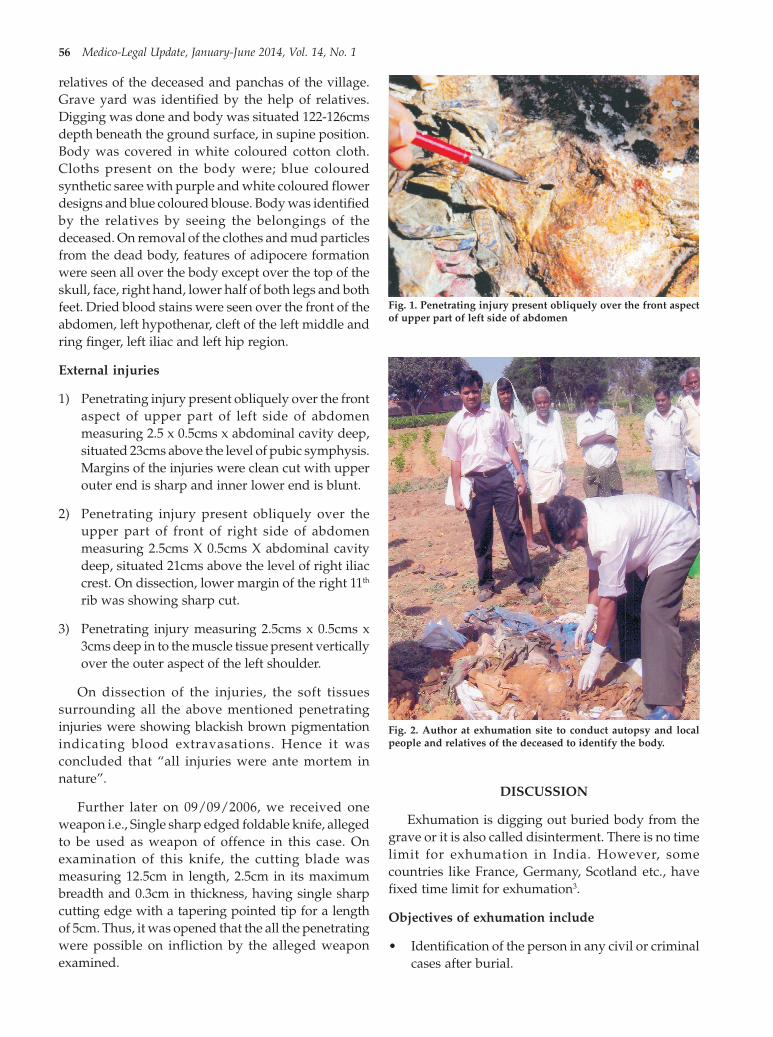
56 Medico-Legal Update, January-June 2014, Vol. 14, No. 1
relatives of the deceased and panchas of the village.Grave yard was identified by the help of relatives.Digging was done and body was situated 122-126cmsdepth beneath the ground surface, in supine position.Body was covered in white coloured cotton cloth.Cloths present on the body were; blue colouredsynthetic saree with purple and white coloured flowerdesigns and blue coloured blouse. Body was identifiedby the relatives by seeing the belongings of thedeceased. On removal of the clothes and mud particlesfrom the dead body, features of adipocere formationwere seen all over the body except over the top of theskull, face, right hand, lower half of both legs and bothfeet. Dried blood stains were seen over the front of theabdomen, left hypothenar, cleft of the left middle andring finger, left iliac and left hip region.
External injuries
1) Penetrating injury present obliquely over the frontaspect of upper part of left side of abdomenmeasuring 2.5 x 0.5cms x abdominal cavity deep,situated 23cms above the level of pubic symphysis.Margins of the injuries were clean cut with upperouter end is sharp and inner lower end is blunt.
2) Penetrating injury present obliquely over theupper part of front of right side of abdomenmeasuring 2.5cms X 0.5cms X abdominal cavitydeep, situated 21cms above the level of right iliaccrest. On dissection, lower margin of the right 11th
rib was showing sharp cut.
3) Penetrating injury measuring 2.5cms x 0.5cms x3cms deep in to the muscle tissue present verticallyover the outer aspect of the left shoulder.
On dissection of the injuries, the soft tissuessurrounding all the above mentioned penetratinginjuries were showing blackish brown pigmentationindicating blood extravasations. Hence it wasconcluded that “all injuries were ante mortem innature”.
Further later on 09/09/2006, we received oneweapon i.e., Single sharp edged foldable knife, allegedto be used as weapon of offence in this case. Onexamination of this knife, the cutting blade wasmeasuring 12.5cm in length, 2.5cm in its maximumbreadth and 0.3cm in thickness, having single sharpcutting edge with a tapering pointed tip for a lengthof 5cm. Thus, it was opened that the all the penetratingwere possible on infliction by the alleged weaponexamined.
DISCUSSION
Exhumation is digging out buried body from thegrave or it is also called disinterment. There is no timelimit for exhumation in India. However, somecountries like France, Germany, Scotland etc., havefixed time limit for exhumation3.
Objectives of exhumation include
• Identification of the person in any civil or criminalcases after burial.
Fig. 2. Author at exhumation site to conduct autopsy and localpeople and relatives of the deceased to identify the body.
Fig. 1. Penetrating injury present obliquely over the front aspectof upper part of left side of abdomen
12. Vijakumar--55--58.pmd 2/22/2014, 11:44 AM56

Medico-Legal Update, January-June 2014, Vol. 14, No. 1 57
• Whenever foul play is suspected exhumation maybe ordered by appropriate authority on request bypublic or relative of the deceased, to know thecause of death, manner of death and nature ofdeath.
• When the first autopsy report is being challengedor is ambiguous, then the second autopsy isordered to clear the doubts by appropriateauthorities.
Karger et al identified following main indicationfor exhumation4.
1. Primary suspicion of intoxication
2. Primary suspicion of homicide
3. Possible medical malpractice
4. Accidents including road traffic accidents
5. To establish cause of death as well as identification.
Exhumation should be carried out after getting theorders from appropriate authorization. Theexhumation is done under the supervision of medicalofficer, magistrate in presence of police officer5. Itshould be preferably carried out in early morning toavoid public, so that the whole process of diggingautopsy can be satisfactorily completed during thenatural light. The grave site should positivelyidentified with identifying features such as location,headstone, grave marks and with the help of relativesor villagers. The burial should be uncovered 10-15cmsand condition of the soil, water content and vegetationgrowth should be noted, which helps to comment onrate and type of decomposition. The burial pit shouldbe opened up to 30cms on all the sides of the bodyand body should be exposed with help of soft brushor whisk broom. Then it is photographed in theposition in which it is found and then autopsy isconducted. In case of suspected poisoning, around3.5kg of earth in contact with body is collected. Sitesinclude earth in contact with body, above, below andfrom each side of the body should be collected.
In one report there is description of 3 and half yearwar in Bosnia-Herzegovina in the year between 1992-1995, when bodies of the killed person were buried inClandestine mass graves, dumped into rivers, wells,septic tanks, caves or simply left unburied in the fields,meadows and forests. To establish identity of morethan 20,000 victims were exhumed to the end of 2008
and this process of exhumation and identification willbe completed by the year 2015.6
Exhumations carried out mainly to establish thecause of death when foul play is suspected andrelatives request for exhumation in most of thecountries7.
In many instances previously, exhumation formedico-legal purposes proved the cause of deathbeyond doubt when it had been ambiguous or un-satisfactory to the relatives. In the present case it hasproved beyond doubt that the cause of death is due topenetrating injuries over the abdomen. This supportsthe history narrated by the relatives and the accusedperson was convicted on the basis of post mortemreport.
In many cases exhumation for medico-legalpurposes has positively identified the missing person.For example, a girl was missing for a period of about 2years. The accused person admitted of committing thecrime of sexual assault, strangulating and then buryedthe victim to evade detection at a later date. Onexhumation the body was identified with the help ofpersonal belongings and later confirmed by DNAfingerprinting from long bones2. Exhumation is evendone for non-medico-legal purposes, in some cultureremains may be exhumed and reburied en masse,when a cemetery is relocated, once local planning andreligious requirement are metallic 8. In the history thereare many instances to answer historical questionnotable individual’s body were exhumed. Evenexhumation helps archaeologists to search the remainsto better understand human culture 9.
CONCLUSION
Exhumation helps in fixing the identity of anindividual. It helps in solving unresolved crime casesat times. If done meticulously with the use of modernailments, in difficult scenarios, it helps in definitivelyfixing the exact cause and manner of death, as in thiscase. Exhumation may also provide valuableinformation or evidence by which innocence ofsuspected persons may be proved or it may establishthe guilty of the crime.
Acknowlegment: Department of Forensic Medicine,Bangalore Medical College, Bangalore.
Conflict of Interest: None
12. Vijakumar--55--58.pmd 2/22/2014, 11:44 AM57
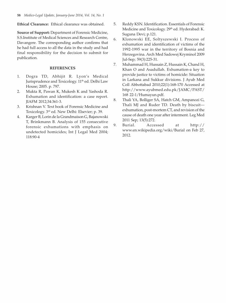
58 Medico-Legal Update, January-June 2014, Vol. 14, No. 1
Ethical Clearance: Ethical clearance was obtained.
Source of Support: Department of Forensic Medicine,S.S.Institute of Medical Sciences and Research Centre,Davangere. The corresponding author confirms thathe had full access to all the data in the study and hadfinal responsibility for the decision to submit forpublication.
REFERENCES
1. Dogra TD, Abhijit R. Lyon’s MedicalJurisprudence and Toxicology. 11th ed. Delhi LawHouse; 2005. p. 797.
2. Mukta R, Pawan K, Mukesh K and Yashoda R.Exhumation and identification: a case report.JIAFM 2012;34:361-3.
3. Krishnan V. Text book of Forensic Medicine andToxicology. 3rd ed. New Delhi: Elsevier; p. 39.
4. Karger B, Lorin de la Grandmaison G, BajanowskiT, Brinkmann B. Analysis of 155 consecutiveforensic exhumations with emphasis onundetected homicides; Int J Legal Med 2004;118:90-4
5. Reddy KSN. Identification. Essentials of ForensicMedicine and Toxicology. 29th ed. Hyderabad: K.Suguna Devi; p.121.
6. Klonowski EE, Soltyszewski I. Process ofexhumation and identification of victims of the1992-1995 war in the territory of Bosnia andHerzegovina. Arch Med Sadowej Kryminol 2009Jul-Sep; 59(3):225-31.
7. Muhammad H, Hussain Z, Hussain K, Chand H,Khan O and Asadullah. Exhumation-a key toprovide justice to victims of homicide: Situationin Larkana and Sukkur divisions. J Ayub MedColl Abbottabad 2010;22(1):168-170 Accessed athttp://www.ayubmed.edu.pk/JAMC/PAST/168 22-1/Humayun.pdf.
8. Thali YA, Bolliger SA, Hatch GM, Ampanozi G,Thali MJ and Ruder TD. Death by biscuit—exhumation, post-mortem CT, and revision of thecause of death one year after interment. Leg Med2011 Sep; 13(5):272.
9. Burial. Accessed at http://www.en.wikipedia.org/wiki/Burial on Feb 27,2012.
12. Vijakumar--55--58.pmd 2/22/2014, 11:44 AM58

Medico-Legal Update, January-June 2014, Vol. 14, No. 1 59
INTRODUCTION
The World Health Organization (WHO) estimatesthat 50 to 100 million infections occur yearly, including500,000 DHF cases and 22,000 deaths, mostly amongchildren. India has recorded over 47,019 dengue casesincluding 242 dengue deaths in 2012 up till 27th Dec2012, the highest number in a year so far.1 Dengue feverhas varying clinical presentations ranging fromasymptomatic infection to dengue shock syndrome.Neurological complications in Dengue fever is unusual.We report a case of a dengue fever withthrombocytopenia and subarachnoid hemorrhage.
CASE HISTORY
A 30 year male presented with moderate gradefever with chills for four to five days. On physicalexamination patient was febrile and vitally stable(blood pressure: 110/82 mmHg. pulse 94/min,respiratory rate: 20/ min, SpO2 of 96%, arterial bloodgas analysis (ABGA): normal). Respiratory and per-abdominal examination was unremarkable.Neurological examination was unremarkable.Tornique test was positive. Laboratory investigationsshowed total leucocyte count 3,900/Cmm, plateletcount 30,000/Cmm with hemoglobin 17.2 Gm% andhematocrit 51.8 % serum electrolytes and renal functiontests were within normal limit. Peripheral smear formalarial parasite and rapid malarial antigen test wasnegative. The test for dengue specific IgM antibodywas positive. Liver enzymes SGOT (233 IU/dl),
Rare Case of Dengue Fever Complicated withSubarachnoid Hemorrhage
Virendra C Patil1, Chetan Galande2, Neeraj Desai2, Sumit Agrawal2
1Associate Professor, 2Resident, Department of MedicineKrishna Institute of Medical, Sciences University (KIMSU), Dhebewadi Road, Karad Dist: Satara, Maharashtra, India
ABSTRACT
This is case report of 30 year male, presented with fever and thrombocytopenia, diagnosed as Denguefever. Worsened neurologically three days after admission and developed subarachnoid hemorrhage.Patient was treated with platelet concentrate transfusion. Patient underwent decompressionhemicraniotomy for increased intracranial pressure. Patient had some improvement at the end of 20days. Although neurological complications reported for dengue fever are unusual, it is reasonable toconsider these manifestations for immediate management and better outcome of patient.
Keywords: Dengue Fever, Subarachnoid Hemorrhage, Thrombocytopenia, Neurological Complications
SGPT(122 IU /dl) were moderately raised, withUltrasonography of abdomen suggestive ofhepatosplenomegaly. The prothrombin time (PT),clotting time (CT) were normal. Bleeding time (BT) was6 min 40 sec. Blood culture was sterile. Patient wastreated symptomatically. After three days of admissionhe started getting headache, vomiting, convulsionsand petechie over trunk and extremities. Clinicallypatient had right sided hemiplegia with bilateralextensor plantar response. CT scan brain was donewhich was suggestive of hyperdensity in left sylvianfissure suggestive of grade three subarachnoidhemorrhage (figure no. 1). CT angiography revealedno evidence of intracranial aneurysm or AVmalformation. There was no history of head injury inrecent past. Clinical scenario favored diagnosis ofdengue fever (DF), with hemorrhagic manifestation(subarachnoid haemorrhage) without shock withthrombocytopenia. We treated patient conservativelywith platelet concentrate, fluid resuscitation accordingto central venous pressure, antibiotics, proton pumpinhibitors, decongestive and anticonvusant drugs.Patients neurological status worsened and he becamestupors. Patient was referred to higher neurosurgicalcentre and decompressive craniotomy was done forthe same. He was operated with left fronto-parieto-temporal decompression craniotomy. Postoperativelypatient was on ventilator for 20 days. On dischargepatient improved clinically and neurologically to someextent with residual right sided hemiparesis.
DOI Number: 10.5958/j.0974-1283.14.1.013
13. Virendra Patil--59--61.pmd 2/22/2014, 11:44 AM59

60 Medico-Legal Update, January-June 2014, Vol. 14, No. 1
DISCUSSION
Today about 2.5 billion people, or 40% of theworld’s population, live in areas where there is a riskof dengue transmission. Dengue is endemic in at least100 countries in Asia, the Pacific, the Americas, Africa,and the Caribbean. DHF was first documented onlyin the 1950s during epidemics in the Philippines andThailand.2 Dengue, also known as break bone fever,due to the extreme pain caused in bones and muscles,is a mosquito-borne infection transmitted through thebite of an infected female Aedes aegypti mosquito. Thereare four serotypes of the virus that cause dengue -DEN-1, DEN-2, DEN-3 and DEN-4. Having recoveredfrom one virus strain provides lifelong immunityagainst that particular serotype, but cross-immunityi.e. being infected by a different serotype increases riskof developing severe dengue which can be fatal (DHF/DSS). In 2011 revised WHO guidelines, dengue wasdivided into dengue fever (DF), dengue hemorrhagicfever (DHF) without shock or with shock (DSS) andexpanded dengue syndrome 3. Dengue in its initialstage is not deadly. However, ignoring symptoms orbeing infected again by a different serotype of virus orhaving another chronic disease can up complicationrisks and may result in dengue hemorrhagic fever(DHS) or dengue shock syndrome (DSS).3
Symptoms usually last for 2–7 days, after anincubation period of 4–10 days. The characteristicsymptoms of dengue include sudden-onset of fever(40°C/ 104°F) which is usually biphasic in nature,breaking and then returning for one or two days. Theother constitutional symptoms are headache, nausea,vomiting retro-orbital pain, muscle and joint pains,swollen glands or skin rash. When dengue advances
into the critical stage it can result in bleeding, low levelsof blood platelets in case of hemorrhagic fever (DHF),or dangerously low blood pressure in case of dengueshock syndrome (DSS). According to WHO, DHFpatient should fulfill following criteria: Acute suddenonset of high fever for 2–7 days, hemorrhagicmanifestations with at least a positive tourniquet test,platelet count <1,00,000/cm3, haemoconcentration, orother evidence of plasma leakage—for example,ascites, pleural effusions, low level of serum protein/albumin.3
Organ dysfunction and severe bleeding, typicallyfrom the gastrointestinal tract may present. Respiratorydistress, rapid breathing, fatigue, restlessness, severeabdominal pain and hematemesis is observed inDengue infection with severe thrombocytopenia.Treatment is mainly supportive care in order tomaintain the body fluid levels and platelet transfusion3
in case it drops to extremely low. In case of neurologicalmanifestations neurosurgical interventions are alsoavailable which can improve patients condition andsurvival rate4,5,6. There is no known vaccine to treatdengue; prevention is the only way to keep away fromthe virus. The first global vaccine for the preventionof dengue is in the final stages of development,currently in phase three clinical trial in humans, beingtested for efficacy across several countries.
Patient in present case report admitted with clinicalfeatures suggestive of Dengue fever whichcomplicated with subarachnoid hemorrhage, requiringdecompressive craniotomy along with supportive lineof management including platelet transfusion. Patientsurvived and recovered with residual right sidedhemiparesis. Present case highlights a rarecomplication of Dengue fever with subarachnoidhaemorrhage which may require timely neurosurgicalintervention and intensive care management for betteroutcome.
Patey O et al7 reported a dengue fever with unusualneurologic manifestations like our case a focalsubarachnoid hemorrhage that was associated withtransient thrombocytopenia. This case was probably apost-infectious syndrome associated with denguevirus who recovered with conservative line oftreatment. Similar to our case report MDPR Fernandoet al8 reported a case of DHF with subarachnoid, retinaland vitreous haemorrhages. Jensenius et al9 reportedthe case of a Norwegian traveler returned withapparently mild dengue fever but with an extensive
Figure no. 1: Shows hyperdensity in sylvian fissure on left sidewith cerebral edema with early evidence of ventricularenlargement with impending non-obstructive communicatinghydrocephalus with near total obliteration of cortical sulcul space,suggestive of sunarachnoid haemorrhage.
13. Virendra Patil--59--61.pmd 2/22/2014, 11:44 AM60

Medico-Legal Update, January-June 2014, Vol. 14, No. 1 61
erythematous exanthema that spread and darkened(perhaps indicative of bleeding), who died of asubarachnoid hemorrhage, for which at autopsy nolocal vascular cause could be found. They refer to theterm “dengue fever with hemorrhage,” which has beenused for adults who bleed but do not fulfil the criteriafor DHF. In our patient there was no neurological AVmalformation. We transfused platelet concentrate andpacked cell volume. Patient underwent left FTPdecompression craniotomy and survived. Accordingto our experience timely diagnosis and interventioncan thus save many precious lives. Lee I-K et al10 alsoreported DSS with subarachnoid hemorrhage.
There are cases presented in the literature are ofcerebral hemorrhage5,6 also cases of subduralhematoma associated with Dengue fever4. Here, weare reporting a rare case of subarachnoid hemorrhagewhich was associated with Dengue fever withthrombocytopenia. Kumar R et al4 reported an case ofacute subdural hematoma in a child with denguehemorrhagic fever occurring during convalescencewho made a successful recovery following surgery.Cam et al2 reported dengue associated encephalopathywith intracranial hemorrhage.
CONCLUSION
Although neurologic complications reported indengue fever are uncommon, it is reasonable toconsider possibility of complication like subarachnoidhemorrhage as neurological manifestations should notbe misinterpreted as fever delirium or toxicencephalopathy. Immediate investigations and timelymanagement including neurosurgical decompressivecraniotomy may require for better outcome. Clinicianshould aware of such unusual complication likesubarachnoid hemorrhage in endemic area of dengueinfection in relevant clinical scenario. It is alsomandatory to rule out other causes ofthrombocytopenia like complicated malaria,leptospirosis, gram negative septicemia anddisseminated intravascular coagulation or dualinfection by relevant investigation.
ACKNOWLEDGMENT
We would like to acknowledge Dr. Mrs. Shilpa PatilAssistant Professor (Dept. of Medicine) and ourresidents for their help in managing patient.
This case report is approved by ethical committee.
Conflict of Interest: Nil
Financial Support: Nil
REFERENCES
1. National vector Borne Disease Control ProgramGovt. Of India. http:/www.nvbdcp.in/den-cd.html.
2. Cam B V, Fonsmark L, Hue N B. et al Prospectivecase control study of encephalopathy in childrenwith dengue hemorrhagic fever. Am J Trop MedHyg 2001. 65848–851.851
3. WHO: Comprehensive guidelines for preventionand control of dengue and dengue hemorrhagicfever. World Health Organization, RegionalOffice for South-East Asia; 2011.
4. Kumar R, Prakash O, Sharma BS Denguehemorrhagic fever: a rare presentation as atypicalacute subdural hematoma.Pediatr Neurosurg.2008; 44(6):490-2.
5. Kumar R, Prakash O, Sharma BS. Intracranialhemorrhage in dengue fever: management andoutcome A series of 5 cases and review ofliterature.Surg Neurol 2009; 72: 429–33.
6. Kumar J, Kumar A, Gupta S, et al. Neurologicalpicture. Dengue haemorrhagic fever: an unusualcause of intracranial haemorrhage. J NeurolNeurosurg Psychiatry 2007; 78:253.
7. Patey O, Ollivaud L, Breuil J, Lafaix C. Unusualneurologic manifestations occurring duringdengue fever infection. Am J Trop Med Hyg.1993;48(6):793-802.
8. MDPR Fernando, JDVC Lekamwasam. Anunusual case of dengue haemorrhagic fever withretinal and vitreous haemorrhages. Galle MedicalJournal 2006;11(1) 48-49.
9. Jensenius M, Berild D, Ormaasen V, et al. Fatalsubarachnoidal haemorrhage in a Norwegiantraveller with dengue virus infection. Scand JInfect Dis 2007; 39:272–4.
10. Lee I-K, Liu J-W, Yang KD (2012) Fatal DengueHemorrhagic Fever in Adults: Emphasizing theEvolutionary Pre-fatal Clinical and LaboratoryManifestations. PLoS Negl Trop Dis 6(2): e1532.doi:10.1371/journal.pntd.0001532
13. Virendra Patil--59--61.pmd 2/22/2014, 11:44 AM61

62 Medico-Legal Update, January-June 2014, Vol. 14, No. 1
INTRODUCTION
Drowning is a very common cause of deathamongst unnatural deaths. About 1,50,000 people dieof drowning over the world. Shetty M.1 has studiedcases of drowning in Mangalore, coastal city ofKarnataka during 1999 to 2004. He studied 350 casesof Drowning. Rate of drowning here is 15.15 per lakhpopulation. All asphyxia deaths were studied byAzmak & Derya 2 in Department of Forensic Medicine,Trakya University, Edirne, Turkey during 1984 to 2004.These constitute 15.7 % of all medicolegal deaths. Outof these 30.5% deaths were of Drowning. Accidentaldrowning can be proved without any doubts on thebasis of circumstantial evidence. However it is verydifficult to prove homicidal drowning in absence ofeyewitness. For proving homicidal drowning,investigating officer will require help of Forensicpathologist, chemical analyzer and also thecircumstantial evidence.
Forensic pathologist will have to observe minutelyinjuries on other parts of body and will also be able todecide whether injuries are ante mortem orpostmortem. Pathologist also has to observe anypathology deceased was suffering from, which issufficient to cause death of a person or which madehim incapacitated before drowning. He has to searchfor injuries on body which might have been causedduring scuffle in homicidal attack.
Dielema in Deciding Manner of Death in a Case ofDrowning: A Case Report
Vyawahare M S1, Bardale R2, Dixit P G3
1Associate Professor, 2Assistant Professor, 3Professor & Head, Department of Forensic Medicine, Government MedicalCollege, Nagpur, Maharashtra, India
ABSTRACT
Once the cause of death is declared as drowning, question before the investigating officer is "Was thedrowning accidental, suicidal or homicidal"? If there is an eyewitness, manner of death will be obvious.In unwitnessed cases, it is very difficult to provide correct answer to above question particularlywhen the place of drowning is unusual.. Case report discussed can give some clues to the investigatingofficers as well as Forensic Pathologist to decide manner of death in cases of drowning in such typeof cases.
Keywords: Drowning, Manner of Death, Intoxicated, Incapacitated, Immersion
Chemical analyzer’s role will be to analyze visceraand to comment on whether person has consumedcertain drugs or substances so that he might have beenincapacitated before drowning.
Circumstantial evidences to be observed on the spotvisit by investigating officer and preferably in presenceof Forensic Pathologist will be
a) depth of water column where body was found
b) projecting parts inside the well or in the lake.
c) Personal belongings of deceased
d) Condition of clothes e.g loss of bottons, torn sites,etc,
e) From above observations and on basis ofpostmortem examination, investigating officer candecide manner of death in drowning as homicidalafter taking into consideration statements ofeyewitnesses if any.
CASE REPORT
A male person, aged about 40 years, was founddead in a iron barrel containing water at about 02.15hours. Barrel was kept in a balcony of a the residentialflat on 2nd floor of an apartment. The incidence wasreported by younger brother of the deceased, who is adoctor by profession. Investigating officer visited spotin early morning.
DOI Number: 10.5958/j.0974-1283.14.1.014
14. Wayawahare--62--65.pmd 2/22/2014, 11:44 AM62

Medico-Legal Update, January-June 2014, Vol. 14, No. 1 63
As per spot inquest, the barrel in which body wasfound was closed to half of its circumference(Fig No 1). Remaining half was open. Height of barrelwas 3 & ½ feet. There was no water inside the barrel.However water was logged in the floor of balcony.Body was already taken out of barrel by the relatives.As per statement of brother of deceased, body wasremoved after making the barrel in slanting positionthereby draining out water from barrel. He gavehistory that deceased was suffering from some mentalillness for which he was taking certain medication asper advice of psychiatrist.
External: I) Injuries : 1) Abrasion on Right cheek, 3X 3 cm. 2) Abrasion over dorsum of Right hand, 3mmX 3 mm, 3) Abrasion over back of Right fore arm, 3 X 2mm, 4) Abrasion over back of Left elbow, 3 X 3 mm , 5)Abrasion over back of Left fore arm, 3 X 5 mm, 6)Abrasion over Left Forehead, 3 X 3 mm, All injurieswere fresh and ante mortem.
II) White fine froth at the nostrils.
Internal: 1) Trachea full of froth.
2) Precordial area was obliterated with lungs.
3) Both lungs were voluminous, oedematous &congested described as emphysema aquosum.(Fig 2). On cutting, free oozing of frothy fluid wasseen. 4) About 100 cc of yellowish fluid in stomachwithout abnormal smell with normal mucosa.5)There was no obvious pathology on gross, neitherany internal injury. Considering external & internalfindings, cause of death was opined as “Asphyxiadue to Drowning”.Routine viscera was preservedfor chemical analysis.
DISCUSSION
With drowning as cause of death, question beforeInvestigating Officer is
1) What is cause and manner of injuries on body?
2) Whether this is a case of accident, suicide orhomicide?
Whether is it possible for normal average built manof height 5 & ½ feet to get himself drowned in water ina barrel of height 3 & ½ feet? To answer this questionone should consider mechanism of drowning.
According to J.Chr.Gierstsen 3 , during stages ofdrowning of human being, person involuntarily makesattempt to come on surface of water due to variousforces and reflexes of body. Giersten conductedexperiments on dog. According to him, same stagesdo occur in human beings. In the said case, height of aperson is 5&1/2 feet and water column of 3 & ½ feet.Unless the person is intoxicated or drugged ordebilitated due to certain reason, it seems very difficultto get drowned in absence of external pressure.Investigating officer tried to produce evidence ofintoxication by subjecting viscera for chemical analysis(In this case reported as negative) . Also in this casethere was no injury to make person incapacitated toprove homicidal manner of death.
Fig. 1. Showing Opening of barrel which was half closed.
Fig. 2. Showing photograph of lung , in which oozing of frothyfluid from cut section of lung.
Postmortem was conducted on the same day.Following were important positive findings.
14. Wayawahare--62--65.pmd 2/22/2014, 11:44 AM63

64 Medico-Legal Update, January-June 2014, Vol. 14, No. 1
As per spot inquest, edges of barrel were rough.Most of injuries are on exposed and prominent partsof body and might be possible while struggling to comeout of barrel or while entering inside the barrel.
Relatives residing with the deceased were alsoexamined medically for presence of injuries on thebody. However there were no injuries on the body ofrelatives, which are expected during scuffle ifdrowning is homicidal.
According to Dr.Anil Aggrawal 4, opining mannerof death depends much upon evaluation ofcircumstances. In India, homicidal drowning isextremely rare except in cases of infants & children. Itis very difficult to drown an adult person until andunless he is attacked unawares or was first stupefiedby giving some narcotic drug. If legs are tied, it ispresumed that death is homicidal except in cases ofswimmers who committed suicide by tying legs toavoid instinctive self preservation. If injuries are therewhose cause can not be explained, it can be case ofhomicide.
Taylor 5 has divided cases of alleged drowning intothree main groups.
I) Bodies with typical finding of drowning. These arefurther subdivided into
A) Without injuries
i) Deceased entered into water voluntarily
ii) He entered water involuntarily while underinfluence of alcohol or drug
iii) With natural disease such as Ischaemic Heartdisease or epilepsy.
B) With injuries
i) deceased was injured by another person or personsand put into water while unable to save himselfthereby drowning.
ii) The injuries did not involve other persons andwere caused when the deceased struck someprojection while entering into water.
II) Those cases in which signs of drowning are notdemonstrable.
These are further subdivided into
A) Without injuries- shows some inhibitory processe.g. cold with immersion like death.
B) With injuries- Fallen into water due to somenatural disease and injuries being sustained at thattime.
III) Death because of violence and body disposed offby immersion.
Most of cases of indoor drowning in shallow waterare accidental. Often natural disease is found such ascerebro-vascular disease or coronary artery disease orepilepsy. Often fault with water heater is also found.Taylor has given illustrative examples of homicidaldrowning in case of R.v. Smith where he murderedthree women by lifting the legs up and pushing thehead under water. He has described many such casesat unusual sites but there is not a single case describedin literature as is described here.
In this case, site where body was found is an ironbarrel. Obviously this rules out accidental manner ofdeath. Investigating Officer has proved beyond doubtthat it is not a case of homicidal drowning by utilizingmethods as described above. In this case, there is aevidence of attempted suicide by the deceased threemonths back in the form of discharge card of hospitalwhere deceased consumed sedative drug to commitsuicide. Taking into consideration all the findings andevidence, in this case manner of death is suicidal
It will be clear from foregoing discussion that thisis an area of forensic practice in which the pathologistis unable to give an opinion which is sufficientlypositive as to satisfy the requirements usuallydemanded in a medico-legal investigation. Hencedetail investigation is must in such type of cases.
Acknowledgements: None
Source of Funding: There was no need of funds forpreparing this case paper.
Conflict of Interest: There is no conflict of interestinvolved in this case amongst the authors.
Ethical Clearance: There is no need of ethical clearancein this case as per guidelines of local ethical clearancecommittee of GMC, Nagpur.
REFERENCES
1. Shetty M. ‘Profile of drowning deaths inMangalore, a coastal city of Karnataka’,Medicolegal Update, 2005 , Vol 5: No2,247-52.
2. Azmak and Derya, ‘Asphyxial deaths: ARetrospective study and Review of Literature’,
14. Wayawahare--62--65.pmd 2/22/2014, 11:44 AM64

Medico-Legal Update, January-June 2014, Vol. 14, No. 1 65
American J of Forensic Medicine and Pathology,June 2006; 27:2.
3. Tedeshi et al,”Drowning”, “Forensic Medicine-Part III” Ist Edition,1977 , W.B.Saunder’s Comp,page 1321.
4. Aggrawal Anil, Column by Dr. Anil Aggrawal,‘Death by Drowning’ available at net search.
www.geradts.com, Web mystery magazine,Summer 2005, Vol.III,Issue I.
5. Alfred Swan Taylor, ‘Medical Jurisprudence’1856, ‘Drowning in shallow water’, 549.
14. Wayawahare--62--65.pmd 2/22/2014, 11:44 AM65

66 Medico-Legal Update, January-June 2014, Vol. 14, No. 1
INTRODUCTION
Congenital malformations of the heart are amongthe most frequent of all congenital anomalies detectedduring 1st year of life.1They constitute one of the majorcauses of infant mortality and morbidity in childhoodand in later adult life. Extra cardiac anomalies occurin 15–45% of cases with CHD.2 Defects range inseverity from tiny pinholes between chambers thatmay resolve spontaneously to major malformationsthat can require multiple surgical procedures beforeschool age and may result in death in utero, in infancy,or in childhood.3 Congenital heart defects may bebroadly grouped into two major categories: 1)morphological abnormalities, includingdevelopmental defects resulting in structuralmalformations; and 2) functional abnormalities,including cardiac rhythm disturbances and
Sudden Death due to Congenital Heart Disease- A CaseReport
Lohith Kumar1, Kulbhushan2, Shadab Raheel1, Abhishek Yadav2, Anil Kumar3
1Senior Resident, 2Assistant Professor, Dept. of Forensic Medicine, VMMC & SJ Hospital, New Delhi, 3DirectorProfessor & Head, Dept. of Forensic Medicine, VMMC & SJ Hospital, Delhi
ABSTRACT
Birth defects have become the most important cause of mortality and morbidity in the perinatalperiod. Congenital heart disease (CHD) is the most common birth defect which includes varyingforms of cardiac abnormalities with an incidence of 1 per 100 live births. We present a case where 6month old Afghan national male baby brought to India for higher treatment with a history of failureto thrive and rapid heart beating. He was diagnosed with Atrio-Ventricular Septal Defect, Pulmonaryartery hypertension and advised pulmonary artery banding as soon as possible. Baby fell unconsciouson board and shifted to Safdarjung hospital where he was declared brought dead and post mortemexamination confirmed the diagnosis. Here an attempt has been made to put forward such a casewith relevant discussion on such potentially life-threatening malformations which warrant carefulclinical follow-up both in antenatal and postnatal period.
Keywords: Atrio-Ventricular Septal Defect (AVSD), Congenital Heart Disease (CHD), Atrial Septal Defect(ASD), Ventricular Septal Defect (VSD
Corresponding author:Abhishek YadavAssistant ProfessorDept. of Forensic Medicine, VMMC&SafdarjungHospital, New DelhiE-Mail:[email protected]
cardiomyopathies. Patients with atrio-ventricularseptal defect typically develop tachypnea, repeatedrespiratory infections, poor feeding, and failure tothrive in the first few months of life. Pulmonaryvascular disease results from damage caused byexcessive pulmonary flow and elevated pulmonaryartery pressure due to the associated VSD.
We describe a similar case in which a 6 month oldmale infant presented with failure to thrive, rapid heartbeating and was diagnosed with AVSD and pulmonaryartery hypertension.
CASE REPORT
A 6 month old Afghan male baby brought to Indiafor higher treatment with a history of failure to thriveand rapid heart beating. He was diagnosed with Atrio-Ventricular Septal Defect, Pulmonary arteryhypertension and advised pulmonary artery bandingas soon as possible. Baby fell unconscious on boardand brought to Safdarjung hospital where he wasdeclared brought dead in emergency. Body was sentto forensic medicine department for post mortemexamination.
DOI Number: 10.5958/j.0974-1283.14.1.015
15. abhishek--66--68.pmd 2/22/2014, 11:44 AM66

Medico-Legal Update, January-June 2014, Vol. 14, No. 1 67
External examination
The body was that of 6 month old male infantwrapped in white coloured hospital cloths. Rigormortis was fully established. Post-mortem hypostasiswas present over the dependent areas (Fig 1). Therewere no features of decomposition. No external injuriespresent over the body except for deformity of the chestcharacterized by a protrusion ofthe sternum and ribsi.e Pigeon Chest (Fig 2).
Internal Examination
The autopsy showed significant degree of rightatrium and right ventricle dilatation and hypertrophywithatrio-ventricular septal defect. Ventricular part ofthe heart had a characteristic globular appearance.Main pulmonary artery was also dilated (Fig 3 & 4).Lungs showed mild pulmonary oedema. Respiratorypassages were congested and contained frothyexudates. All other internal organs were congested. Noabnormality was detected in rest of the body systems.
Echocardiographic findings in inquest papersrevealed situssolitus, normal systemic and pulmonaryvenous connection, complete atrio-ventricular septaldefect, large ostiumprimum atrial septal defect withleft to right shunt, moderate size ventricular septaldefect, mild to moderate right and left AV valveregurgitation and dilated main pulmonary artery. X-ray showed cardiomegaly with prominent lungvasculature.
Based on the findings of the autopsy and theinvestigation reports, Cause of death was given ascongenital heart disease and its sequelae.
Fig. 1. Post Mortem Hypostasis
Fig. 2. PectusCarinatum (Pigeon Chest)
Fig. 3. opened thorax shows enlarged heart
Fig. 4. Globular shaped heart with dilated right atrium, rightventricle and pulmonary valve
15. abhishek--66--68.pmd 2/22/2014, 11:44 AM67

68 Medico-Legal Update, January-June 2014, Vol. 14, No. 1
DISCUSSION
Atrioventricular septal defects (AVSDs) areanatomic defects that arise from faulty developmentof the embryonic endocardial cushions. This spectrumranges from a primum atrial septal defect and cleftmitral valve, known as a partial atrioventricular septaldefect (partial AVSD), to defects of both the primumatrial septum and inlet ventricular septum and thepresence of a common atrio-ventricular valve, referredto as complete atrio-ventricular septal defect (completeAVSD). These defects, particularly the complete form,typically present in the foetal or neonatal period andare an important source of cardiac morbidity andmortality in this age group.4The effects from AVSD arecaused by the holes in the heart walls and leakage fromthe abnormal valve. The holes allow blood in theheart’s left side to leak back to the heart’s right side.This results in a large amount of blood that the rightside of heart must pump again to the lungs. As a resultof the extra workload, most babies with AVSD developsigns of congestive heart failure such as rapidbreathing, feeding problems, slow weight gain, lowenergy, and cold, clammy sweating. Another possiblehealth effect of AVSD is pulmonary arteryhypertension. This problem develops more slowly andis caused by the extra pulmonary blood flow. Thepulmonary arteries that take blood from the heart’sright side to the lungs are not well suited to theincreased pressure caused by the extra blood flow.Over time, if the problem is not corrected, thepulmonary arteries become thick and stiff whichmakes the heart have to work even harder.5
Trisomy 21 (Down syndrome) is the mostfrequently associated genetic abnormality withcomplete atrio-ventricular septal defect, although itmay also occur in association with trisomy 13 andtrisomy 18. In patients without trisomy 21 who havecommon atrio-ventricular canal (CAVC) defects, agenetic locus on chromosome 1 can account for thedisorder in some families.6Interstitial deletion onchromosome 16 can also be associated with atrio-ventricular septal defect.
Early diagnosis and treatment is always aprerequisite in any heart diseases and congenital heartdiseases are always associated with significantmorbidity and mortality.Immediate intervention isrequired to save the life of child. Thorough antenatalcheck-ups should be done in all the cases and if anycongenital defect is detected, it should be closelyfollowed during the child birth and early neonatal
period. Surgical intervention, if required should bedone as soon as possible. In our case doctor hadadvised for pulmonary artery banding as soon aspossible. The child would have been saved if earlyintervention was done.
CONCLUSION
Congenital heart disease is assuming increasingimportance as a cause of perinatal and infant mortality.Despite some improvements in the outlook ofprenatally diagnosed AVSD, the overall prognosisremains considerably worse. If deaths fromunrecognised heart disease are not taken into account,a falsely low picture of the prevalence of seriouscongenital heart disease will be obtained. Soidentification of such undiagnosed cardiovascularanomalies and better awareness of the problem willimprove epidemiological evaluation of CHD and moreimportantly, earlier recognition of treatableabnormalities further reduces the mortality.
Acknowledgements: Nil
Conflict of Interests: Nil
Source of Funding: Not Applicable
Ethical Clearance: Not required
REFERENCES
1. Srivastava D. Genetic regulation of cardiogenesisand congenital heart disease. Annu RevPathol. 2006; 1:199–213.
2. Frias JL. Genetic issues of congenital heart defects.In: GessnerIH, Victrorica BE, editors. Pediatriccardiology. Philadelphia: Saunders; 1993. pp.237–42.
3. Gucer S, Ince T, Kale G, Akcoren Z, Ozkutlu S,Talim B, et al. Non cardiac malformations incongenital heart disease: A retrospective analysisof 305 pediatric autopsies. Turk J Pediatr. 2005;47:159–66.
4. Delisle MF, SandorGG, Tessier F, et al. Outcomeof fetuses diagnosed with atrioventricular septaldefect. ObstetGynecol 1999; 94(pt1):763–7.
5. Merrill WH, Hoff SJ & Bender HW. The SurgicalTreatment of Atrio-ventricular Septal Defect. InC Mavroudis& C Backer (Eds) Pediatric CardiacSurgery. St. Louis, MO: Mosby, 1994, 225-236.
6. Sheffield VC, et al. Identification of a complexcongenital heart defect susceptibility locus byusing DNA pooling and shared segmentanalysis. Hum Molec Genet. 1997; 6(1):117-121.
15. abhishek--66--68.pmd 2/22/2014, 11:44 AM68
Medico-Legal Update, January-June 2014, Vol. 14, No. 1 69
INTRODUCTION
Stature of an individual came into discussion whenprimitive mammal changed its posture frompronograde to orthograde.
The study of human evolution, racial differences,inheritance of body traits, growth and decay of humanorganism is called physical anthropology. Theseinformations are of interest to an anatomist and alsohelpful in medico-legal works. The problem isencountered in cases of mass disasters, explosions, andassault cases where the body is dismembered toconceal the identity of the victim.
The estimation of height from various parametershas been done by various workers Singh and Sohal1
(1951), Inderjit Singh & Shamer Singh2 (1956),Charnalia3 (1961), Ahawale 4(1963), Patel et al(1964)5,Qamara et al 6 , (1979) & Shroff & Vare 7(1979).
Morphology of human head is influenced bycombined effects of heredity and life style. Telkka(1950)8 opined that each racial group will need a
Personal Height Determination from Head Length
Shalini Chaudhary1, Sarvesh2
1Assistant Professor, Department of Anatomy, B.P.S.G.M.C. Khanpur Kalan, Sonepat, Haryana, 2Senior Resident,Department of Anaesthesia, B.P.S. Govt. Medical College for Women ,Khanpur Kalan, Sonepat, Haryana
ABSTRACT
This study was carried out to estimate the relationship between head length and stature using simplelinear regression analysis based on a sample of male and female of Western Maharastra.Measurementsof the head length and stature were taken from 150 subjects ( 75male, 75 female). Obtained data wasanalysed and attempt was made to find out correlation between the head length and stature of anindividual. A good correlation of height was observed with head length and it was statisticallysignificant. The results of the present study would be useful for anthropologists and forensic medicineexperts.
Keywords: Stature, Head Length
Corresponding author:Shalini ChaudharyAssistant ProfessorDepartment of Anatomy, B.P.S.Govt Medical Collegefor Women, Khanpur Kalan, Distt. Sonepat, HaryanaE-mail:[email protected]: 09728816828
different formula, and regional studies of the subjectsare very much needed.
The purpose of the present study is to analyze theanthropometric relationship between dimensions ofthe head length with stature and to devise regressionformulae to estimate height from head length ofknown sex from residents of western maharastra as itis essential to build a context of reference for a givenpopulation.
METHODOLOGY
Present study has been carried out on 150 residentsof western maharastra (75 male; 75 female) agedbetween 8 -12 years. All the subjects were healthy andfree from any apparent symptomatic deformity ofspine. Measurement Technique
Stature: is measured as the vertical distancebetween the vertex and the floor. The subject was madeto stand bare foot in an erect posture against a wallwith both feet kept close together, buttocks & occiputtouching the wall and hands hanging down on thesides
Head length measures the straight distancebetween glabella and opisthocranion.
Glabella: It is the point on protuberance of lowerforehead above nasal root and between the eyebrowridges intersected by mid sagittal plane.
DOI Number: 10.5958/j.0974-1283.14.1.016
16. Shalini---69--71.pmd 2/22/2014, 11:44 AM69

70 Medico-Legal Update, January-June 2014, Vol. 14, No. 1
OPISTHOCRANION
Opisthocranion is the most posterior point on theoccipital protuberance of head in the mid sagittal plane.This point is determined where the head length showsmaximum reading
Three measurements were taken to avoidmeasuring errors. Mean of three measurements wascalculated and noted.
Measurements were taken using blunt endedspreading caliper. Instrument held in such a mannerthat the tips of the caliper were free to touch the head.
Undue pressure was avoided while taking themeasurement.
OBSERVATIONS AND RESULTS
Table1: Comparison of stature between male andfemale
male female P value
mean 136.81 136.01 0.72
range ( 116-161.5) ( 110.8-161.4 )
Table2: Comparison of head length between male andfemale
male female P value
mean 17.39 17.07 0.03
range ( 15.4-18.9) ( 16-18.8)
Table 3: Pearson correlation of head length and bodyheight.
male female All cases
+0.41 +0.3 +0.35
Table 4: Regression analysis for prediction of totalheight in males and females
Equation S.E.E. R
Male =9.3+7.3(head length) 10 0.4
Fkemale =67.8+3.9(head length) 9.4 0.3
All cases =46.78+5.2(head length) 9.69 0.35
S.E.E.: standard error of estimate, R:correlation coefficient
DISCUSSION
Table 1 and table 2 shows that both stature and headlength has higher values among males than in females.
Very less work has been done for estimation ofstature from head length. Saxena et al9 (1981), who
derived a regression equation between head-lengthand height in Agra population (U.P.). Their correlation
coefficient between head -length and height was+0.2048. According to Glaister 10(1957), nasion-inionlength (head-length) is 1/8 of the total height of anindividual.
In present study the correlation
Co-efficient between height and head length is+0.35 which is significant.
CONCLUSION
From the above facts, it is clear that if either of themeasurement (head length or total height) is knownthe other can be calculated and this fact may be ofpractical use in Medico-legal investigations and inAnthropometry.
Acknowledgement: Nil
Source of Funding: Self
Conflict of Interst: Nil
Ethical Clearance: From Ethical Committee of D.Y.Patil Medical College, Kolhapur
REFERENCES
1. Singh B and Sohal HS. Estimation of stature fromclavicle in punjabis; A preliminary report. IndianJournal of Medical Research 1951;40:67-71.
2. Jit Inder and Shamer Singh. Estimation of staturefrom clavicle. Indian Journal of medical research1956;44:137-155.
3. Charnalia VM. Anthropological study of the footand its’s relationship to stature in different castesand tribes of Pondicherry state. Journal ofanatomical society of India 1961;10:26-30
4. Athawale MC. Anthropological study of heightfrom length of forearm bones. A study of onehundred Maharashtrian male adults of agesbetween twenty five and thirty years. AmericanJournal of physical anthropology 1963;21:105-112.
5. Joshi NB, Patel MP and Dongre AV. Regressionequation of height from ulnar length. Indianjournal of medical research 1964;52:1088-1091.
6. Suneel Qamara, Inderjit and Deodhar SD. Amodel for reconstruction of height from foot
16. Shalini---69--71.pmd 2/22/2014, 11:44 AM70

Medico-Legal Update, January-June 2014, Vol. 14, No. 1 71
measurements in an adult population ofnorthwest India Indian Journal of MedicalResearch 1980;71:77-83
7. Shroff AG and Vare AM. Determination of heightfrom length of superior extremity and it’ssegments. Journal of anatomical society of India1979;28:53.
8. Telekka, A. On prediction of human stature fromthe long bones . Acta Anatomica 1950;volume 9, pp 103-11.
9. Saxena SK, Jeyasingh P, Gupta AK and Gupta CD.The estimation of stature from head - length.Journal of anatomical society of India 1981;30:78-79.
10. Glaister John. In medical jurisprudence andtoxicology. 10th Edition E & S, Livingstone Ltd.Edinburg and London k1957;79.
16. Shalini---69--71.pmd 2/22/2014, 11:44 AM71

72 Medico-Legal Update, January-June 2014, Vol. 14, No. 1
INTRODUCTION
The blast wave is a shot without a bullet, a slashwithout a sword. It is present everywhere within itsrange. Blast would be as dreaded a weapon as chemicalwarfare, if its range, when explosives are used werenot limited to small areas. However it would bepremature to believe that this situation will alwaysremain the same.” [1.]
Theodor Benzinger 1950
The incidence of injuries resulting from explosionshas increased throughout the twentieth century. Thishas been caused in part by industrial expansion. Moresignificant has been the proliferation of explosiveweapons and their increased use world-wide. Injuriescaused by explosions are not confined to the battlefield.The International Committee of the Red Cross hasestimated that throughout 70 countries, at least 2,000people (predominantly civilians) are killed orinjured every month by anti-personnel mines. [1]
A bomb is a container filled with an explosivemixture and missiles, which is fired either by detonatoror a fuse. Terrorist bombs often involve 2–10 kg ofexplosives. When an explosion occurs, produces largeamount of gases and energy while the pressuregenerated is up to 1000 tones/sq”. [2]
Explosions have the capability to causemultisystem, life-threatening injuries in single or
Mechanical Injuries by Blast effect a Case Report
S D WakdeAsst. Professor, Department of Forensic Medicine, Government Medical College and Hospital, Nagpur
ABSTRACT
Explosions caused by industrial accidents and terrorist devices are infrequent, but unpredictableevents in developed countries. Therefore, civilian doctors may be required to deal with large numbersof blast injured subjects with little warning in chaotic and difficult circumstances.[1] Explosions andthe injuries caused by them are the subjects of this paper.
This case is presented here because the real incidence of injuries caused by blast of O2 cylinder in achemical manufacturer company was concealed in the inquest with history of found injured in thatcompany and the fact was revealed during autopsy.
Keywords: Industrialization, Explosion, Terrorism, Injuries
multiple victims simultaneously. These types of eventspresent complex triage (abrasions, contusions andpunctured lacerations), diagnostic, and managementchallenges for the health care provider. Explosions canproduce classic injury patterns from blunt andpenetrating mechanisms to several organ systems, butthey can also result in unique injury patterns to specificorgans including the lungs and the central nervoussystem.
Blast injury will also produce scattered foci of smallhemorrhages in the substances of lungs, liver, spleen,kidneys, adrenals, brain, mesenteries, air passages etc.as a result there is bloody froth in the mouth. [3]
CASE
A 35 yr old male having history of giddinessfollowed by fall, injury to head while working in achemical manufacturer company and then broughtdead to GMCH Aurangabad, Maharashtra for medicolegal post mortem.
Post mortem findings: [6]
EXTERNAL EXAMINATION: All cloths werestained with blood and blackish dust. T-shirt showedmultiple tiny holes on it’s back. Trouser stained withblackish dust, having torn on right leg near kneeregion. (Fig.4.)
DOI Number: 10.5958/j.0974-1283.14.1.017
17. Shailesh--72--74.pmd 2/22/2014, 11:44 AM72

Medico-Legal Update, January-June 2014, Vol. 14, No. 1 73
Facial features were injured and disfigured on rightside of body. The skin was stained with blackish dustat face and neck area, both upper limbs distal to lower1/3rd of arms up to fingers and both thighs below lowerone third up to feet.
Lacerated wound was present over right parieto-temporal area of size 10x8 cm x bone deep withavulsion of skin and comminuted fracture ofunderlying bone. (Fig.1.) Brain matter was exposed.There was presence of blood infiltration and irregularmargins.
There were multiple abrasions and puncturedlacerations of varying sizes ranging from diameters of0.1 cm to up to 1 cm, over right side of face and upperchest, left upper limb, right lower thigh. ( Fig. 2 & 3.)
Internal Examination: Under scalp contusion waspresent over right fronto-parieto-temporal area of
Fig.1. Lacerated wound over right parieto-temporal area withavulsion of skin and comminuted fracture of bone.
Fig.2. Multiple abrasions and punctured lacerations, left upperlimb
Fig.3. Multiple abrasions and punctured lacerations, right lowerthigh.
Fig.4. Multiple tiny (Sieve like) holes on T-shirt.
scalp, irregularly with evident depressed comminutedfracture of right fronto-parieto-temporal bones,associated with torn meninges at fractured sites. Therewas laceration of brain over right parietal region ofsize 4.5x2.5x1.5 cm, with irregular margins.Subarachnoid hemorrhages were present over bothhemispheres, more over right side. Lungs showed subpleural patchy hemorrhages, sub pleural blebs as aresult of shock waves of the blast.
DISSCUSSION
Knowledge of the mechanisms of blast injury, theassociated patho physiology, the clinical diagnosis andrecommended management of the resulting complextrauma are the keys for helping casualties. [4]
By convention, blast injuries are classifiedaccording to the mechanism by which they areproduced.
17. Shailesh--72--74.pmd 2/22/2014, 11:44 AM73

74 Medico-Legal Update, January-June 2014, Vol. 14, No. 1
Table No. 1. Show classification of blast injuries. [5]
Category Injury caused by Primary target organ
Primary blast injury Blast wave Ears, lungs, GI tract, CNS
Secondary blast injury Victim struck by flying debris Skin, CNS, eyes, musculoskeletal system
Tertiary blast injury Acceleration + Impact with stationary objects Abdominal viscera, CNS, lungs, skin,musculoskeletal system
Miscellaneous Inhalation of dust, toxic gases, burns radiation, Lungs, skin,musculoskeletal systempsychological impact
The air-containing organs (like ear, lungs, digestivetract etc.) are extremely vulnerable to the uniqueprimary blast effect. Familiarity with the effects thatexplosions exert on these organs, their consequencesand treatment, together with a high index of suspicion,are crucial for medical professionals to obtain the bestoutcome for exposed victims. [4]
All of the injuries presented in this case are due toblast injuries. At the conclusion of autopsy, cause ofdeath was formed as “Head injury associated withblast injuries”. Whereas histopathology of lung tissuessupported the opinion of cause of death as diffusealveolar damage secondary to acute lung injury.
CONCLUSION
Blast injury is becoming more common in the non-military population but it is still rare to see such injuriesand deaths unrelated to terrorist acts. The exactmechanisms involved in blast injuries are unclear.Civilian physicians and surgeons need to have a basicunderstanding of the patho-mechanics andphysiological effects of blast injuries.
Although terrorism has been present for years, thenumber of serious terror incidents is more than tripledin recent years. Access to information on the internethas facilitated tremendous technological advancementin terrorists’ destructive capabilities. This underscoresthe importance of appropriate education within themedical system and the need for careful advancedplanning to cope with terror attacks. Because of theincreasing incidences of explosions, physicians mustbecome familiar with the characteristics of
contemporary explosive devices and the spectrum ofinjuries inflicted by blasts and explosions. [4]
ACKNOWLEDGEMENT
The author would like to thank Dr. K. U. Zine sir,Prof. and Head, Dept. of forensic Medicine, GMCHAurangabad and Dr. P. G. Dixit sir, Prof. and HeadDept. of forensic Medicine, GMCH, Nagpur, for theirguidance and help in preparation of this manuscript.
Conflict of Interest: No conflict of interest observedduring this study.
Source of Funding: Self.
Ethical Clearance: This study is carried out inaccordance with declaration of Helsinki.
REFERENCES
1. Horrocks CL. Blast Injuries: Biophysics,Pathophysiology and Management Principles. JR Army Med Corps 2001 147: 28-40.
2. Narayan Reddy K. S., 31st Edi. Mechanicalinjuries. : 221.
3. Mukherjee J. B., 4th Edi. Injury and it’s med. legalaspects: Bomb blast wounds: 375.
4. Ami Mayo and Yoram Kluger. Blast-inducedinjury of air-containing organs. ADF Health April2006: Vol. 7 No. 1
5. Leung SH, Cheung KY, Yau HH, Kam CW. Casereport: blast injury. Hong Kong j. emerg. med.Vol. 9(1) Jan 2002: 46-51
6. P. M. No. 1276/10, dated. 17/7/10.
17. Shailesh--72--74.pmd 2/22/2014, 11:44 AM74

Medico-Legal Update, January-June 2014, Vol. 14, No. 1 75
INTRODUCTION
The autopsy room has always been a potentialsource of infection. The autopsy surgeons, forensicpathologists and other persons engaged directly orindirectly in conducting postmortem examination areat greater risk of exposure to blood-borne viruses andother infections like- human immunodeficiency virus(HIV) , Hepatitis B virus (HBV), Hepatitis C virus(HCV), Hepatitis D and G viruses, Non-A, Non-BHepatitis, tuberculosis, Creutzfeldt Jacob disease,herpes, hanta virus pulmonary syndrome, small pox,human T-cell lymphotropic virus type 1 and infectionfrom other pathogenic organisms1-12. Many studieshave confirmed that with the cessation of life, certain
Seroprevalence of HIV, HBV and HCV among theCadaver Population - A Jaipur Based Study
Yadav A1, Pathak D2, Alam F3, Vyas N4
1Assistant Professor, Forensic Medicine and Toxicology, NIMS Medical College, Jaipur, 2Assistant Professor, ForensicMedicine and Toxicology, 3Ist Year Resident (Forensic Medicine and Toxicology), 4Professor, Department of
Microbiology, SMS Medical College Jaipur
ABSTRACT
Forensic medical personnel are at risk of exposure to blood-borne viruses including HIV, Hepatitis Band Hepatitis C virus. The aim of this study was to determine seropositivity of HIV, HBV and HCVamong a cadaver population at the SMS hospital, Jaipur. The study group consists of Four hundredeighteen randomly picked subjects over a period of one year from October 2008 to September 2009.These cadavers were screened for HIV antibodies, HBs Antigen (hepatitis B surface antigen) andHCV antibodies (hepatitis C virus antibodies) by applying ELISA Test. For HIV antibodies initialreactive tests were confirmed by Tridot Test and SD Bioline test as per NACO guideline. The maximumnumbers of deceased in the present study group were in the age range of 15-44 years being 295 innumber (70.58%). Out of these, 224 (75.9%) were males and 71 (24.06%) were females. In all 46 (11.0%)samples out of 418 were seropositive for HIV, HBV and HCV infections. From 418 subjects, 10 wereseropositive for HIV (2.39%).all of them males. Seropositivity for HCV was found in 7(1.67%); malesonly. Out of 29 subjects seropositive for HBV, majority i.e. 17 (58.62%) were in age range of 15-44years. In the serological status of HBV, 29(6.94%) subjects showed positive results. Among them 21were males and 8 were females. All forensic cadavers should be treated as potentially infectious anduniversal precautions should be taken.
Keywords: HIV, HBV, HCV, Cadaver
Corresponding author:Anil YadavAssistant ProfessorDepartment of Forensic Medicine & ToxicologyNIMS Medical College, Shobha Nagar, [email protected]
pathogenic bacteria are released, which if leftunchecked, may prove hazardous to the personneldealing with them13. Medicolegal autopsies being abasic legal requirement to ascertain the cause andmanner of unnatural deaths have registered an overallincrease throughout the country14-16.
It has long been recognized that the dissectors,observers and other persons in close proximity to anautopsy are at a high risk of contracting infectiousdiseases from the dead bodies. Studies in Britishclinical laboratories between 1970 and 1989 establishedthat the highest rate of laboratory- acquired infectionswas in autopsy workers17. It has been reported thatresident doctors working in pathology sustained apercutaneous injury with a blood exposure in 1 in 11autopsies whereas the experienced pathologists in 1in 55 autopsies18. Scalpel blades made majority of thesecuts. However, many other objects such as brokenglass, needle fragments, bone pieces, and fragmentedprojectiles can injure the autopsy personnel19. Westonand Lober20 et al. have documented that approximately
DOI Number: 10.5958/j.0974-1283.14.1.018
18. Anil Yadav--75--79.pmd 2/22/2014, 11:44 AM75

76 Medico-Legal Update, January-June 2014, Vol. 14, No. 1
8% surgical gloves get punctured during autopsy andabout 1/3rd of these remain undetected by thepathologist, thus causing any pre existing hand injuriesto be bathed in infectious blood for a prolonged periodof time. The guidelines for handling of cadavers andfor performing autopsies on infected subjects areavailable along with the recommendations forapplication of universal precautions in all cases. But,with the increasing pressures of work and ignorancein our set up, not much of the available rules areimplemented religiously in day to day working. Themain objectives of this study were to firstly examinethe distribution of serological markers for HIV, HBVand HCV infection in medicolegal cases whichincluded all causes of death; secondly to evaluate therisk of potential infection to personnel involved inmedicolegal incidents and procedures; and thirdly toincrease the awareness among personnel involved inmedicolegal work about the risks of transmission ofthese infectious diseases and their prevention thereof.
METHODS AND MATERIAL
The study was carried out from 14 October 2008 to19 September 2009. Samples of blood were collectedfrom 418 bodies at autopsy, carried out within 2 hoursto 45 days of death; 322 were males and 96 females.The subjects were selected to be included in the studygroup were picked up randomly from the autopsiesperformed at department morgue during the studyperiod. There were no inclusion or exclusion criteriafor the selection of subjects except the consent. In all
the cases included, a well informed consent wasobtained from next of kin, relatives of the deceased orwhosoever was in charge of the dead body and wascategorized on the basis of demographic profile andthe cause of death. Case records and relatives wereconsulted as fully as possible to determine whetherthere was antemortem knowledge of actual or potentialrisk of infection. Blood sample were collected from thefemoral vein of all the subjects with sterile precautions.Serum were separated and subjected to different teststo detect the presence of antibodies to HIV and HCValong with detection of HBsAg. Commerciallyavailable microbiology kits were used to test cadavericserum for HIV antibodies, HBS Ag and HCVantibodies. HIV testing was performed as per NACOguidelines.
RESULTS
A total of 418 blood specimens were collected in asequential manner from selected cases, via femoralvein with sterile precautions during autopsy at themortuary. The age of cadavers of this study groupranged from 0-87 years. The maximum numbers ofdeceased in the present study group were in the agerange of 15 – 44 years being 295 in number (70.58%).Out of these, 224 (75.9%) were males and 71 (24.06%)were females. The mean age for male cadaversincluded in the study group was 35.38±14.68 and themean age for the female cadavers included in the studywas 32.54±13.98 (Table-1).
Table 1. Distribution of Study Group on basis of Age & Sex
Age group(In Yrs) Male Female Total
No. % No. % No. %
0-4 3 0.71 1 0.24 4 0.95
5-9 5 1.20 2 0.48 7 1.68
10-14 3 0.71 1 0.24 4 0.95
15-44 224 53.60 71 16.98 295 70.58
45+ 87 20.82 21 5.02 108 25.84
Total 322 77.04 96 22.96 418 100.00
Mean age + Sd (Male cadaver) = 35.38 + 14.68
Mean age + Sd (Female cadaver) = 32.54 + 13.98
Out of 10, 5 (50%) subjects seropositive for HIVfell in age range of 15-44 years and remaining 5 (50%)fell in age range of more than 45 years. Out of 29subjects seropositive for HBV, majority i.e. 17 (58.62%)
were in age range of 15-44 years. 5 (74.13%) subjectsseropositive for HCV were in age range of 15-44 yearsand 2 (28.57%) subjects were more than 45 years ofage(Table-2).
18. Anil Yadav--75--79.pmd 2/22/2014, 11:44 AM76

Medico-Legal Update, January-June 2014, Vol. 14, No. 1 77
Table 2. Serological markers of HIV, HBV and HCV in relation to age
Age group(In Yrs) Communicable disease
HIV HBV HCV
0-4 0(0.00) 0(0.00) 0(0.00)
5-9 0(0.00) 1(3.45) 0(0.00)
10-14 0(0.00) 1(3.45) 0(0.00)
15-44 5(50.00) 17(58.62) 5(71.43)
45+ 5(50.00) 10(34.48) 2(28.57)
Total 10(100.00) 29(100.00) 7(100.00)
From 418 subjects, all 10 (2.39%) subjectsseropositive for HIV were males. Similarly all 7 (1.67%)subjects seropositive for HCV were males. In theserological status of HBV, 29 (6.94%) subjects showed
positive results and 389 (93.06%) showed negativeresults. Among them, 21 (72.4%) were males and 8(27.6%) were females (Table-3).
Table 3. Serological markers for HIV, HBV and HCV in relation to sex
Sex HIV HBsAg HCV
+ ve - ve Total + ve - ve Total + ve - ve Total
Male 10(2.39) 312(74.640) 322(77.03) 21(5.02) 301(72.01) 322(77.03) 7(1.67) 315(75.35) 322(77.03)
Female 0(0.00) 96(22.97) 96(22.97) 8(1.91) 88(21.05) 96(22.97) 0(0.00) 96(22.97) 96(22.97)
Total 10(2.39) 408(97.61) 418(100.00) 29(6.94) 389(93.06) 418(100.00) 7(1.67) 411(98.33) 418(100.00)
χ2 HBsAg = 0.170 d.f.=1 P >.05 NS
Out of 418 subjects, 290 accidental deaths wereregistered. In them 3 (1.03%) were seropositive for HIV,24 (8.28%) were seropositive for HBV and 5 (1.72%)were seropositive for HCV i.e. a total of 32 (11%)subjects of accidental deaths were seropositive for atleast one of the blood borne viral agents. Of the 70suicidal deaths, no subject was seropositive for HIV. 4
(5.71%) subjects were seropositive for HBV and 1(1.43%) subject was seropositive for HCV. Rest 65(92.8%) subjects of suicide deaths were seronegativefor HIV, HBV and HCV. All 20 subjects of Homicidaldeaths were seronegative for HIV, HBV and HCV(Table-4).
Table 4. Serological markers for HIV, HBV and HCV in relation to manner of death
Mode of death HIV HBsAg HCV
+ ve - ve Total + ve - ve Total + ve - ve Total
Accidental 3(1.03) 287(98.97) 290(100.00) 24(8.28) 266(91.72) 290(100.00) 5(1.724) 285(98.28) 290(100.00)
Suicidal 0(0.00) 70(100.00) 70(100.00) 4(5.71) 66(94.29) 70(100.00) 1(1.43) 69(98.57) 70(100.00)
Homicidal 0(0.00) 20(100.00) 20(100.00) 0(0.00) 20(100.00) 20(100.00) 0(0.00) 20(100.00) 20(100.00)
Natural 7(18.42) 31(81.58) 38(100.00) 1(2.63) 37(97.37) 38(100.00) 1(2.63) 37(97.37) 38(100.00)
Total 10(2.39) 408(97.61) 418(100.00) 29(6.94) 389(93.06) 418(100.00) 7(1.67) 411(98.33) 418(100.00)
DISCUSSION
For a health care worker, the average risk for HIVinfection after a percutaneous needles tick injury withHIV-infected blood is estimated to be 0.3% 21. The studygroup comprised of 418 subjects, ranging from 0 to 87years of age. Most of the subjects were in the range of15-44 years being 295 in number accounting for 70%of total population of the study group. There were 295(70.6%) subjects in the age range of 20-49 years and225(53.8%) subjects in age range of 20-39 years. thisage range of 20-49 years comprises of bread winningmembers of the family making them socially
vulnerable to Medicolegal incident of different types.Cattaneo C et al carried out a similar study forprevalence of HIV and hepatitis C markers among acadaver population. Their ages ranged from 16-50years. They reported that most subjects were in the21-40 years age range covering 68.3% population. Inanother study done by Zadeh HS, the ages of cadaversof study group ranged from 2 to 78 years and therewere 74.4% subjects between 20-49 years. Li L et alcarried out a study on 414 autopsy cases, 135 (32.6%)had serologic evidence of at least one viral marker22.There were 23 serum samples positive for HIV-1 (5.6%),96 positive for anti-HBc (23.2%), 79 for HCV (19.1%).
18. Anil Yadav--75--79.pmd 2/22/2014, 11:44 AM77

78 Medico-Legal Update, January-June 2014, Vol. 14, No. 1
The prevalence of HIV-1 infection in both male andfemale was similar in this population, male 5.7%, andfemale 5.2% but in our study all HIV cases belongs tomales only. In the study of Li L et al, there was nosignificant difference in HBV infection by sex. Theincidence of HCV infection in males was at least twotimes higher than that of females (21.8% to 10.3%)while in our study prevalence of HBV in males is 5.02%and 1.91% in females. In our study all positives forHCV were males (1.67%) which is also not similar toLi L et al study.
There were 322 males in our study populationcomprising 77.04% and 96 females accounting for22.96%. The males outnumbered the females in ourstudy population which is easily explainable in our
society, which is still male dominant, especially in therural regions. This is quiet similar to 82.6% males and17.3% females (Cattaneo C et al, Milan, 1997) and 83.2%males and 16.8% females (Zadeh HS, Tehran, 2001).Themanner of death in our study population waspredominantly accidental in nature accounting for64.8% deaths. 16.8% deaths were suicidal in nature;4.6% were homicidal and in 13.8% deaths, no medico-legal manner could be established, death occurredprobably due to some pathological manifestationswhile in Li L et al study, natural (37.2%), accidental(18.8%), homicidal (18.1%), suicidal (13.8%) andundetermined (12.1%) comprise study population.Comparing it to the other studies, we observe thefollowing pattern (Table-5):
Table 5.
Seropositivity for Our Li L et al, Cattaneo du Plessis et Zadeh HS, Sharma BRstudy 1993 C et al, 1997 al, 1999 2000 et al, 1994-2003
HIV 2.4% 5.6% 16.3% 11.0% 0.0% 0.4%
HBV 6.93% 23.2% - 8.0% 4.6% 0.5%
HCV 1.67% 19.1% 28.7% 1.0% 4.0% -
The reason for the slight variation in our study fromthe studies conducted by Sharma BR et al; du Plessiset al regarding HBV and HCV and Zadeh HS may bedue to the variations in the case selection of studygroup in the four studies.
CONCLUSION
High prevalence of various infectious diseases inthe population poses a great risk of occupationalhazards to the forensic pathologist/autopsy surgeonand other staff involved in the postmortemexamination. They may be exposed to a wide varietyof infectious agents such as HIV, Hepatitis B, C viruses,Mycobacterium tuberculosis, etc. For transmission ofHIV, HBV, and HCV; blood is the single mostimportant source of infection. Furthermore, practically,it is almost impossible to know the medical status(whether HIV/HBV/HCV etc, present or not) of eachand every deceased person. It is therefore prudent toconsider all the dead bodies to be potential carriers ofinfection and follow the Universal Precautions, whileconducting autopsy on them. Proper assessment,personal protective equipment, appropriate autopsyprocedures and infrastructural modifications cansubstantially reduce the risks of occupational healthhazards in the autopsy rooms. Accordingly, periodictraining and education in safe postmortem procedures,
prevention of sharp’s injuries and other kinds ofexposures should be imparted to the forensic personnelregularly. They should be aware of the potentialtransmission of these infections and the use ofpreventive measures. Non-availability of thevaccination for some of these deadly infections alertsthat avoidance to such an exposure is the onlyprevention by the use of universal precautions whileat work.
ACKNOWLEDGEMENT
The authors are thankful to Dr. B.M. GuptaProfessor Department of Forensic Medicine andToxicology, SMS Medical College Jaipur for kindly helpto carry out this work.
Conflicts of Interest: Nil
Source of Funding: Nil
Ethical Clearance was sought from appropriateauthorities before the commencement of this work.
REFERENCES
1. Ajmani ML. Recommendations for prevention ofHIV transmission in health care workers involvedin autopsy and embalming. J Forensic MedToxicol 1997; 4: 47-50.
18. Anil Yadav--75--79.pmd 2/22/2014, 11:44 AM78

Medico-Legal Update, January-June 2014, Vol. 14, No. 1 79
2. Brown P, Rohwer RG, Gajdusec Sodium DC.Hydroxide decontamination of Creutzfeldt Jakobdisease. N Engl J Med 1984; 310: 727-9.
3. Douceron H, Deforges L, Gherardi R, Sobel A,Chariot P. Long-lasting post-mortem viability ofhuman immunodeficiency virus: A potential riskin Forensic Medicine Practice. Forensic Sci Int1993; 60: 61-6.
4. Fink TM, Rodents. Human remains and NorthAmerican hantaviruses: Risk factors andprevention measures for Forensic Sciencepersonnel-a review. J Forensic Sci 1996; 41:1052-1056.
5. Galloway A, Snodgrass JJ. Biological andchemical hazards of forensic skeletal analysis. JForensic Sci 1998; 43: 940-8.
6. Geller SA. The autopsy in acquiredimmunodeficiency syndrome-How and Why?Arch Pathol Lab Med 1990; 114: 324-9.
7. Gerbert B. Why fear persists: Health careprofessional and AIDS. JAMA 1988; 260: 3481-3.
8. Ratzan RM, Schneiderman H. AIDS, autopsiesand abandonment. JAMA 1988; 260: 3466-9.
9. Riddell LA, Sherrard J. Blood-borne virusinfection: the occupational risks. Int. J STD AIOS2000; 11: 632-9.
10. Rosenberg RN, White CL, Brown P, et al.Precautions in handling tissues, fluids and othercontaminated materials from patients withdocumented or suspected Creutzfeldt Jakobdisease. Am Neurol 1986; 19: 75-7
11. Sagoe-Moses C, Pearson RD, Jagger J. Risks tohealth care workers in developing countries. NEngl J Med 2001; 345: 538-41.
12. Templeton GL, Illing LA, Young L, Cave D, SteadWW, Bates JH. The risk of transmission ofMycobacterium tuberculosis at the bedside andthe autopsy. Ann Intern Med 1995; 122: 922-5.
13. Sharma BR. Autopsy room: a potential source ofinfection at workplace in
developing countries. Am J Infec Dis 2005;1(1):25-33.
14. Sharma BR, Harish D, Sharma VR,Vij K. kitchenaccidents vis-à-vis dowry deaths.burns2002;28(3):250-3.
15. Sharma BR, Harish D, Sharma VR,Vij K.Poisoning in Northern India changing trends,causes and prevention thereof. Med Sci law 2002;42(3):251-7.
16. Sharma BR, Sharma VR, Harish D, Vij K. Suicidesin Northern India- Causes, methods andprevention thereof. Med Sci law 2003; 43(3):221-229.
17. Grist, NR and JA. Emslie, Association of ClinicalPathologist’s surveys of infection in BritishClinical Laboratories,1970-1989.
18. Butts, JD., 1995. Forensic Pathology automaticallyExposure Prone. N C Med Jour.,55: 210.
19. Hutchins, KD, AW Williams and GA.Natrajan,2001. Neck needle foreign bodies: anadded riskfor autopsy pathologists. Arch.Path.Lab.Med.,125: 790-2.
20. Weston, J and G. Locker, 1992. Frequency of glovepunctures in the postmortem room. J. ClinPathol., 45:177-8.
21. Henderson, D. K., Fahey, B. J., Willy, M., Schmitt,J. M., Carey, K., et al., “Risk for OccupationalTransmission of Human Immunodeficiency VirusType 1 (HIV-1) Associated with ClinicalExposures. A Prospective Evaluation,” Annals ofInternal Medicine, Vol. 113, 1990, pp. 740-746.
22. Li, L., Zhang, X., Constantine, N. T., and Smialek,J. E., “Seroprevalence of ParenterallyTransmitted Viruses (HIV-I, HBV, HCV, andHTLV-I/H) in Forensic Autopsy Cases,” Journalof Forensic Sciences, JFSCA, Vol. 38, No. 5,September 1993, pp. 1075-1083.
18. Anil Yadav--75--79.pmd 2/22/2014, 11:44 AM79

80 Medico-Legal Update, January-June 2014, Vol. 14, No. 1
INTRODUCTION
Employee State Insurance Act(1948) envisagessocial protection of workers in the organized sector incontingencies such as sickness,maternity,death ordisablement due to employment injury andoccupational disease3.Based on the principle of“pooling of risks and resources”, the unique ,multidimensional health insurance scheme guaranteesa fair deal to the covered members by providing fullmedical facilities to the beneficiaries, besides adequatecash compensation to insured persons for loss of wagesor earning capacity in times of physical distress arisingout of sickness or employment injury orunemployment1.
The main benefits provided under E.S.I. Scheme are
• Sickness benefit
• Disablement benefit
• Dependant benefit
• Maternity benefit
• Medical benefit
Retrospective Analysis of the Occupational Injuries in EyePatients in E.S.I. Hospital
A D Mehta1, Rohit Kapoor2
1Head of Eye Department, 2C.M.O. Eye Department, E.S.I. Hospital, Basaidarapur, New Delhi
ABSTRACT
The E.S.I. Scheme has the largest medical infrastructure under one umbrella, in India .Medical care isprovided through its huge network of 150 hospitals, 1403 E.S.I. Dispensaries/ISM Units, 1447 Panelclinics and 6536 IMOs. The E.S.I. Corporation incurred about Rs. 2689.62 crore on the delivery ofmedical care in year 2012-13.Eye trauma is number one cause of vision loss in industrial workers2.We are presenting a retrospective analysis of eye injuries coming in the EYE DEPARTMENT of E.S. I.Hospital, basaidarapur, New Delhi 110015.
Keywords: Employee State Insurance (E.S.I.), Occupational, Injuries, Eye Trauma
Corresponding author:Rohit KapoorChief Medical OfficerE.S.I. Hospital, Basaidarapur, New Delhi 110015E-mail: [email protected]
Besides the above, other benefits being providedto the beneficiaries are Confinement Expenses, FuneralExpenses, Vocational Rehabilitation, UnemploymentAllowance and Skill up gradation training.
In E.S.I.C., Comprehensive medical care rangingfrom OPD, medical attendance, treatment, drugsspecialist consultation, hospitalization of insuredpersons (IPs) and their family members to super-specialty treatment are provided under the scheme.Medical benefit is available to the INSURED PERSON(IP) and his family members from the day he entersthe insurable employment. There is no ceiling on theexpenditure on individual medical care.
MATERIAL AND METHOD
This is a retrospective study. We are presenting thenumber of patients coming to EYE DEPARTMENTwith various occupational injuries in the last 3 yearsi.e. 2010,2011, and 2012.the various eye conditions withwhich patients present to EYE DEPAERTMENT withoccupational injuries;
• Embedded corneal foreign body
• Chemical injuries(Acid/Alkali )
• Welding arc light corneal injury
• Blunt injury
• Penetrating injury
• Traumatic cataract(<40 years)
DOI Number: 10.5958/j.0974-1283.14.1.019
19. Rohit Kapoor--80-81.pmd 2/22/2014, 11:44 AM80

Medico-Legal Update, January-June 2014, Vol. 14, No. 1 81
FINDING
The total number of patients coming to EYEDEPARTMENT of E.S.I. Hospital, BASAIDARAPUR,and NEW DELHI 110015 with occupational injuriesin the last 3 years i.e. 2010, 2011 and 2012 issummarized in the following table:
Table 1: Patients with Occupational InjuriesPresented to Eye Opd, E.s.i. Hospital, Basaidarapur,
New Delhi In Last 3 Years I.E. 2010,2011 and 2012
Year 2010 Year 2011 Year 2012
Embedded corneal 35 42 46foreign body
Chemical injury(acid / 23 28 19alkali injury)
Welding arc light 12 10 12corneal injury
Blunt injury 17 15 18
Penetrating injury 16 14 09
Traumatic cataract (<40 years ) 17 15 11
The percentage injury of the patients withoccupational injuries 4 in the last 3 years i.e. 2010, 2011and 2012 is summarized in table 2.
Table 2: Percentage Vision Loss in Patients withOccupational Eye Injuries Presented to Eye Opd, E.S.I.Hospital, Basaidarapur, and New Delhi in Last 3 Years
I.E. 2010, 2011 and 2012.
Year 2010 Year 2011 Year 2012
Vision loss 10% 37 35 44
Vision loss 30% 34 47 38
Vision loss 40% 44 40 42
CONCLUSION
The magnitude of vision loss due to industrialinjuries is a great loss of earning capacity. The E.S.I.C.is the biggest provider of social security scheme in thiscountry. Every vision loss is compensated duly forthe loss of earning capacity.
ACKNOWLEDGEMENT
We thank all the patients who participated in thestudy.
Conflict of Interest: There is no conflict of interest.
Source of Funding: E.S.I.C. funded the study
Ethical Clearance: Ethical clearance has been taken
REFERENCES
1. Ehud I Assia,Yokrat Ton,Adi Michaeli.Capsuleanchor to manage subluxated lenses: initialclinical experience.J. Cat .Refr. Surgery2009;35:1372-1379
2. Kuhn Ferenc.Mechanical ophthalmic injuries.Ophthalmic clinics of north America 2002;15:150-250
3. Margolin A Edward,Leena Shrinivastav.Prevalance of retinal hemorrhage in perpetrator-confessed cases of abusive head trauma.Arch. ofophthalmology.2010;128:795
4. Snyder E Michal ,Lindsell B Luke.Nonappositional repair of iridodialysis.J. Cat. AndRef. Surgery.2011;37:625-628.
19. Rohit Kapoor--80-81.pmd 2/22/2014, 11:44 AM81

82 Medico-Legal Update, January-June 2014, Vol. 14, No. 1
INTRODUCTION
Human suicidal behavior has always been a sourceof dread and wonder to mankind and its history goesback at least to the earliest human records. Hangingremains to be one of the common method ofcommitting suicide. The thought of hanging oneselfmay come progressively or as an impulse. In Indiasuicide by hanging was the second most commonlyadopted mean of committing suicide i.e.31.5% in theyear 2009. Hanging is the most preferred method ofsuicide along with consumption of poisons inMaharashtra.1
It is very much necessary for us to know the variouspostmortem findings that help us in establishing thatdeath was due to hanging. Age, gender, physicalcondition of the individual, visit to the scene of crime,injuries on the body, type of ligature mark and knot,may all help to decide the death occurred due to
Histopathological Study of Carotid Arteries in Deaths dueto Hanging
Dattatray Ghodake1, Shailesh Mohite2, Heena Desai3
1Assistant Professor, Dept. of Forensic Medicine, RCSM, Govt. Medical College & CPR Hospital, Kolhapur,2Professor and Head, Dept. of Forensic Medicine, 3Assistant Professor, Dept. of Pathology, T.N. Medical College &
B. Y. L. Nair Ch. Hospital, Mumbai
ABSTRACT
We studied the influence of various parameters on carotid artery in cases of deaths due to hanging.In 60 hanging deaths autopsied, the various details were gathered, analyzed and interpreted.Histopathology examination revealed the incidence of carotid artery injury to be 47 % (28 cases). Outof these maximum cases (17%) showed disruption of intimal layer from medial layer followed by 15% cases intimal tear, 8 % cases perivascular congestion and 7 % cases subintimal haemorrhages. Weconcluded that age, type of hanging, position of knot and ligature materials were the parameterswhich influence carotid artery injury.
Keywords: Hanging, Intimal Tear, Carotid Artery, Histopathology
Corresponding author:Dattatray G GhodakeAssistant ProfessorDept. of Forensic MedicineRCSM, Govt. Medical College & CPR Hospital,Kolhapur-416002 Maharashtra State (India)Phone No. 09867850167Email: [email protected]
hanging. Hence, an attempt is made in this study toknow how far these changes are evident in postmortemexamination.2, 3, 4
MATERIAL & METHOD
The present study was conducted at post-mortemcenter attached to the University teaching hospital inMumbai. The cases of deaths due to hanging in whichautopsy have been conducted at this post-mortemcenter during study period from July 2009 to June 2011(2 years) were included in this study. During thespecified period of 2 years, 60 cases were studied withrespect to different age groups and gender. The detailsabout victims regarding the circumstances of death,type of ligature material, manner and apparent causeof death were recorded from police requisition letterand inquest (Panchanama) report. In addition, thehistory was taken from relatives about the incidence.
The detailed post mortem examination was carriedout in each case comprising of an external and aninternal examination. For the study we personallyattended autopsy, dissected the neck structures bytaking ‘V’ shaped incision, collected the segments ofcarotid arteries separately from both sides below theligature mark and subjected them to histopathologicalstudy with the help of Pathology Department of thisinstitute.
DOI Number: 10.5958/j.0974-1283.14.1.020
20. Dattaray--82--85.pmd 2/22/2014, 11:44 AM82
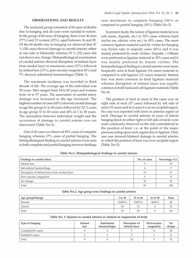
Medico-Legal Update, January-June 2014, Vol. 14, No. 1 83
OBSERVATIONS AND RESULTS
The analyzed group consisted of 60 cases of deathsdue to hanging and all cases were suicidal in nature.In the group of 60 cases of hanging, there were 46 men(77%) and 23 women (23%), aged between 16 and 85.Of the 60 deaths due to hanging we observed that 47% (28) cases showed damage to carotid arteries eitherin one side or bilaterally whereas 53 % (32) cases didnot shown any change. Histopathological examinationof carotid arteries showed disruption of intimal layerfrom medial layer in maximum cases (17%) followedby intimal tear (15%), perivascular congestion (8%) and7% showed subintimal haemorrhages (Table 1).
The maximum incidence was recorded in thirddecade of life. The average age of the individual was50 years. Men ranged from 18 to 85 years and womenfrom 16 to 57 years. The association of carotid arterydamage was increased as the age increased. Thehighest number of cases (60%) showed carotid damagein age the group 61 to 90 years followed by 52 % casesin age group 31 to 60 years and 40% in 1 to 30 years.The association between individual weight and theoccurrence of damage to carotid arteries was notdiscovered (Table No-2).
Out of 60 cases we observed 83% cases of completehanging whereas 17% cases of partial hanging. Thehistopathological finding in carotid arteries were seenin both complete and partial hanging however findings
seen maximum in complete hanging (36%) ascompared to partial hanging (10%) (Table No-3).
In present study, the nature of ligature material wassoft (saree, dupatta, etc.) in 55% cases whereas hard(nylon roe, electric wire etc.) in 45% cases. The mostcommon ligature material used by victim for hangingwas Nylon rope in majority cases (42%) and it wasmainly preferred by male victims. Dupatta / Odhaniwere preferred as ligature material in 30% cases and itwas mainly preferred by female victims. Thehistopathological finding in carotid arteries were morefrequently seen in hard ligature (16 cases) material ascompared to soft ligature (12 cases) material. Intimaltear was most common in hard ligature materialwhereas disruption of intimal layer was equallycommon in both hard and soft ligature materials (TableNo-4).
The position of knot in most of the cases was onright side of neck (37 cases) followed by left side ofneck (19 cases) and in 4 cases it was on occipital region.No case was reported with knot on anterior aspect ofneck. Damage to carotid arteries in cases of lateralhanging (knot on either right or left side of neck) weremore commonly observed on the side contralateral tothe position of knot. i.e. at the point of the majorpressure acting upon neck organs due to ligature. Onlyone case showed bilateral damage to carotid arteriesin which the position of knot was over occipital region(Table No-5).
Table No.1. Histopathological findings in carotid arteries
Findings in carotid artery No. of cases Percentage (%)
Intimal tear 9 15
Sub-intimal haemorrhage 4 7
Disruption of intimal layer from medial layer 10 17
Peri-vascular congestion 5 8
No change 32 53
Total 60 100
Table No.2. Age group wise findings in carotid arteries
Age groupDamage 1 to 30 31 to 60 61 to 90 Total
Present 12(40%) 13(52%) 3(60%) 28
Absent 18 12 2 32
Total 30 25 5 60
Table No. 3. Injuries to carotid arteries in relation to suspension of body
Type of hanging Intimal Subintimal Disruption of Perivascular Notear haemorrhages intimal layer congestion change
Complete(50 cases) 8 3 7 4 28
Partial(10 cases) 1 1 3 1 4
Total 9 4 10 5 32
20. Dattaray--82--85.pmd 2/22/2014, 11:44 AM83

84 Medico-Legal Update, January-June 2014, Vol. 14, No. 1
Table No.4. Injuries to carotid arteries in relation to nature of ligature material
Type of hanging Intimal Subintimal Disruption of Perivascular Notear haemorrhages intimal layer congestion change
Hard(27 cases) 7 3 5 1 11
Soft(33 cases) 2 1 5 4 21
Total 9 4 10 5 32
Table No.5. Injuries to carotid arteries in relation to position of knot
Position of knot Findings Present Absent Total
Right Left Bilateral
Occipital 1 0 1 2 4
Right side of neck 3 12 0 22 37
Left side of neck 9 2 0 8 19
Anterior 0 0 0 0 0
Total 13 14 1 32 60
1. Intimal layer tear,
2. Subintimal haemorrhages,
3. Disruption of intimal layer from medial layer,
4. Perivascular congestion
5. No change
The abovementioned changes in carotid arteries areevident due to traction and direct pressure of theligature on the neck. The acting pressure of ligatureprobably leads to fixation and deformation of the partof artery below the ligature to the deeper tissuestructures of the neck. Section of the artery below thepoint of its fixation due to pressure of ligature isexposed to traction forces that lead to downwardstretching of this part of the artery. A combination offorced compression of the artery and its longitudinalstretching could be the most frequent mechanismleading to occurrence of tears in the intimal layer ofcarotid arteries in hanging deaths. The subintimalhaemorrhage and perivascular congestion appears tobe more due to direct trauma due to pressure ofligature, whereas intimal tear and disruption of intimallayer from medial layer are the result of traction forces.So these findings in carotid arteries are possible dueto direct and indirect trauma in the form of crushingand traction forces.5, 10
Autopsy study of carotid arteries in deaths due tohanging established the information that tear of theintimal layer of carotid arteries suggest morefrequently in individuals of older age groups. Thisstudy most possibly relates to the advancement of age
Fig. 1. Disruption of medial layer from intimal layer (H&E 40X)
Fig. 2. Intimal Haemorrhage (H&E 100X)
DISCUSSION AND CONCLUSION
In this study on external examination, the carotidarteries did not show any appreciable gross findings.The histopathological findings in carotid arteries weregrouped under five heading as follows5, 6
20. Dattaray--82--85.pmd 2/22/2014, 11:44 AM84

Medico-Legal Update, January-June 2014, Vol. 14, No. 1 85
related degenerative and atherosclerotic changes incarotid arteries. However, the exact relationshipbetween the severity of degenerative, atheroscleroticchanges and the occurrence of injuries to the intimallayer of carotid arteries was not studied and is subjectto further study.10
CONCLUSION
1. Findings in carotid arteries are caused due to directand indirect trauma in the form of crushing andtraction forces.
2. Disruption of intimal layer from medial layer isthe most common finding followed by intimal tearin histopathology of carotid arteries in deaths dueto hanging.
3. The association of carotid artery damage wasincreases as the age increases, this possibly relatesto the advancement of age related degenerativeand atherosclerotic changes in arteries.
4. The carotid artery damage was seen maximally incases of complete hanging (36%) as compared topartial hanging (10%).
5. Damage to carotid arteries were more commonlyseen in hard ligature as compared to soft ligature.Intimal tear most common in hard ligature whereasdisruption of intimal layer was equally commonin both hard and soft ligature.
6. Damage to carotid arteries in cases of lateralhanging were more commonly observed on theside contralateral to the position of knot.
7. Bilateral damage to carotid arteries was generallyseen due to crushing and stretching on both sideof the neck when the position of knot was overoccipital region.
Acknowledgment: Nil
Conflict of Interest: Nil
Source of Funding: Self
Ethical Clearance: Taken from Ethics Committee ofT.N. Medical College & BYL Nair Ch. Hospital,Mumbai.
REFERENCES
1. National Crime Record Bureau. (online) 2009(accessed on 2011 Nov 02;p.168-189. Availablefrom:RL;http://ncrb.nic. in/ADS/2009/suicide-09.
2. Reddy KSN. The essential of forensic medicineand toxicology.28th ed. Hydrabad.2009.p.296-303.
3. Karmakar RN editor.JB Mukherjee’s forensicmedicine and toxicology. 3rd. kolkata.academicpublishers 2007.p. 577-599.
4. Vij K. Text book of forensic medicine & toxicology,4rd ed. New Delhi: Reed Elsevier Indian Pvt. Ltd.2008: 161-69.
5. Vinay Kumar M.S. Histopathological study ofcarotid trauma in strangulation deaths. J IndianAcad Forensic Med. April-Jun 2013;35(2):102-105.
6. Jani CB, Gupta BD. An autopsy study ofparameters influencing injury to carotid arteryin hanging. Journal of Forensic Medicine andToxicology 2003 Jan-Jun; 20(1):18-19.
7. Betz p, Eisenmenger W. Frequency of the throatskeleton fractures in hanging. Am J Forensic MedPathol 1996;17(3):191-193.
8. Sharma BR, Singh VP, Harish D. Neck structureinjuries in hanging-Comparing retrospective andprospective studies. Med Sci Law 2005;45(4):321-329.
9. Suarez-Penaranda JM, Alvarez T, Miguens X,Rodriguez-Calvo MS, de Abajo BL, Cortesao M.et al. Characterization of lesions in hangingdeaths. J Forensic Sci. 2008 May;53(3):720-3.
10. Petr Hejna. Amussat’s sign in hanging –Aprospective autopsy study. J Forensic Sci. Jan2011;56(1):132-35.
20. Dattaray--82--85.pmd 2/22/2014, 11:44 AM85

86 Medico-Legal Update, January-June 2014, Vol. 14, No. 1
INTRODUCTION
Medical professionals till the beginning of 20th
century, traditionally had the high status and carriedthe tag of ‘extensions of God’ particularly surgeonsobtained assent, the agreement of the patient to haveprocedure but not consent. It was the mutuallyaccepted practice of the doctors with their patients carein accordance with the principle of Hippocratic Oathof beneficence and informed consent was not legallyrequired.1The first land mark of judgment from NewYork justice, Benjamin Cardoza upheld the patient’sright was documented in a case of Schloendoff in theyear 1914. Mrs. Schloendoff had agreed to examinefor fibroid uterus to exclude malignancy underanaesthesia. But, surgeon was found guilty as he had
Informed Consent: A Myth of Ethical Spirit and LegalParadigm in Medical Profession
Ramchandra D SProfessor and Head, Dept. of Anaesthesiology and Critical Care, M N R Medical College & Hospital Sangareddy,
Hyderabad, A P., India
ABSTRACT
Informed consent may not be major significance in medical malpractice litigations but, it is one of thecritical issues in modern medicine. The development of informed consent has given rise to two majorperceptions of ethical and legal foundations in health care system. The principle of informed consentis applicable to surgical operations, medical treatment and diagnostic procedures involving intentionalinterference with the patient. No doubt, medico-legal issues are more common in the western part ofthe world, the autonomy of the patient along with other principles of informed consent followedregularly in the west. In India lack of disclosure and negligence of not following seriously informedconsent in health care is commonly seen. Institutional policies and procedures in obtaining medicalconsent may form a legal document but, may not necessarily achieve the goal of informed consent,patient or relatives for instance may sign documents, they do not understand. Medical professionalsshould consider the need to achieve informed consent in two symbiotic senses: the ethical sense andthe legal sense.
Keywords: Informed Consent, Ethical, Legal Paradigm, Health Care
Corresponding author:D S RamchandraProfessor and Head.Dept. of Anaesthsiology and Critical Care,Navodaya Medical College Hospital and ResearchCentre, Raichur, Karnataka, IndiaMobile: 09849541812E-mail: [email protected]
removed uterus which patient had not agreed. In themiddle of the 20th century, the Salgo’s malpractice casein1957 introduced the current concept of informedconsent. Followed; by Natson case in1960 for lack ofdisclosure to the reasonable doctor standard,Canterbury case in1972 for insufficient disclosure tothe reasonable patient’s standard, and in 1978 for rightto refuse treatment and to respect autonomy for selfdetermination of the individual in a case of Candura.Each of these cases contributed to the evolution ofmodern informed consent with five ideal elementslike2,3: 1.Disclosure of information to patients andresearch voluntaries. 2. Understanding to optimizethe patients. 3. Voluntariness of participation intreatment and research. 4. Competence of theindividual and 5. Consent in final to indicate validagreement. But, the application of ideal informedconsent remains more of subjective than objective inhealth care system.
The earliest evidence in legal expression forfundamental principle to preserve autonomy is foundin the 1947 Nuremberg Code.4 The Nuremberg Codewas created immediately after World War II, doctorsin Nazi Germany were conducting horrifying research
DOI Number: 10.5958/j.0974-1283.14.1.021
21. Dev Sugerkar --86--90.pmd 2/22/2014, 11:45 AM86

Medico-Legal Update, January-June 2014, Vol. 14, No. 1 87
on prisoners in concentration camps. The research wasdone on millions of involuntary participants whousually died as a result of unethical and illegalexperiments. After the war when international tribunalwas ordered, many of these doctors were tried at theNuremberg trials for their crimes. The code makes itmandatory to obtain voluntary and informed consentof human subjects before to experiment.
Similarly, The Belmont Report (1979) is one of theleading works concerning ethics and health care wasfollowed after unethical Tuskegee syphilis researchstudy from United States (1932-1972). The study wasconducted among the poor American blacks withouttheir knowledge and voluntary consent. Later theresearch work was published as ‘long term effects ofsyphilis’ and not being treated by the National HealthCare doctors though penicillin, the treatment forsyphilis was available from 1947. The Belmont Reportintroduced the act of protection to participants inclinical trials and research studies.5 The Belmont Reportconsists of three basic principles: 1.Respect for persons.2. Beneficence (Hippocratic Oath) and 3 Justice.
Some of the American shocking violated researchstudies were reviewed through the Belmont Report ledto the development of ethical regulations of researchguidelines.6 In fact, ethical responsibility was wellappreciated and seen on May 16th, 1997 when U S (then)president Bill Clinton offered an emotional apology tothe eight remaining survivors after 65 years ofTuskegee syphilis research study.7 The 1964Declaration of Helsinki meeting made by the WorldMedical Association had also emphasized theimportance of obtaining freely given informed consent.Since, then the Declaration of Helsinki has undergoneseveral revision updates to accommodate advances inmedical science in safeguarding research and ethicalproblems.8
Informed Consent in Developing countries andIndian Law
It is difficult to find documented land mark oninformed consent in developing countries. Informedconsent is a modern American ritual not appliedproperly in medical profession, its implementation ofindividual voluntary consent in developing countriesremains controversial and sometimes it is notstraightforward. Informed consent in the Asiansubcontinent is beset with dilemma of ethical spiritand legal paradigm with difference. Despite legalissues are more common and strict in the western part
of the world, the autonomy of the patients has beenreviewed regularly and principles of informed consentare rectified in two. Symbiotic senses; ethical sense andlegal sense. In India lack of disclosure and negligenceof not following seriously informed consent in healthcare is commonly seen. The probable reasons could bedue to; large cultural differences, lack of awarenesswith the illiterate masses, inadequate health caredevelopment and casual aptitude of medicalprofessionals not contemplating potential litigations.9
India being the largest democratic country in theworld, the principle of autonomy is enshrined in thearticle 21 of the constitution, which deals with the rightto the life and personal liberty also applies to themedical science. The application of common law withinformed consent is difficult in India, but dealt withthe principles of Indian Contract Act (ICA) and IndianPenal Code (IPC). Some of the obligations aremaintained between medical professional and hispatient in accordance with the Indian Majority Act; ifindividuals have attained the age of 18, patients withsound mind, and if individuals are not disqualifiedby the law. Of course, these obligations may differ fromcountry to country and developing to developedcountries; for instance in U.K consenting age is 16 yearsold and young person can be treated as an adult if aperson can have capacity to decide. In case ofcompetent child refuses treatment, a parent or courtmay authorize investigation or treatment in the interestof child welfare. In India when the patient is unable togive consent himself, the medical professionals mayproceed with treatment by taking the proxy consentof any relative of the patient or even an attendant. Forinstance wife can give consent to her husbandundergoing bypass surgery for the hospital formalities.
In a case of strained couple family history if one ofthem under goes sterilization by giving false historyto the doctor for the sake of National scheme ofmonitory benefit, after the operation if one of theirrelative complains against the doctor and claims theirconsent was not valid because of their unsound mind,the law protects medical professionals in such asituation. Health care providers need not sense falsehistory or doubt about the capacity of the patient. Adoctor acting reasonably under normal circumstancesis always protected by the law. The legal developmentof patient’s consent in clinical practice, the supremecourt of India recently ruled, that it is not just the‘consent’ or ‘informed consent’ but it shall also be ‘priorinformed consent’ with exception to some specific
21. Dev Sugerkar --86--90.pmd 2/22/2014, 11:45 AM87
88 Medico-Legal Update, January-June 2014, Vol. 14, No. 1
cases of emergency. Hence, written consent has becomeessential to the medical profession for proving consentin case of litigation. For instance, the doctor had donesterilization without the consent as patient was ingeneral anesthesia for some other operation. It wasobserved that when one speaks of consent, that shouldbe informed consent. Therefore doctor was liable inthe legal sense. In case of negligence on the part of thedoctor the law always assumes the principle of consent.
In one of the legal case, a patient gave consent forappendicitis operation and the surgeon after openingthe abdomen found appendix was normal. However,in the interest of the patient, the surgeon removed hergangrenous gall bladder. Subsequently, patientsuffered with renal complications. The doctor was heldliable as he had operated without the valid consent. Insuch cases the law always maintains the right ofautonomy of the patient however the error of diagnosisis pardonable.10
A lady having the son and daughter both minorsunderwent Medical Termination of Pregnancy (MTP)with the written consent. After the operation thehusband was informed that the uterus was removedin order to save the life of the patient. The husband,the complainant filed a case alleging that no consentfor removal of uterus was obtained either from thepatient or from him. In this case, operating surgeonhad taken the second opinion and help of othereminent gynecologist in order to save the life of patienton emergency basis as there was continuous bleedingand patient was deteriorating. Thus no negligenceregarding removal of uterus was found in the part ofthe doctors. So consent was done away because of
emergency life saving situation. However, the practiceof obtaining high risk consent from the patient’s partyshould not aim for stopping the complainant claimingdamages, rather to be presumed for the responsibleconsequences of medical procedures or anesthesiabeing administered. In these circumstances one of theIndian State Commission observed that the signing ofthe consent form does not exclude doctor ’sresponsibility if he is negligent in carrying his duties.11
In southern state of India, a case of perforatedappendix with peritonitis was admitted in generalhospital. But, even after two days no operation wasdone, eventually patient died. The patient’s wife fileda case against hospital saying that the doctordemanded money for performing surgery.Furthermore, the surgeon was busy in attending anoutside private nursing home for some otheroperations and came back only after the death of thepatient. The explanation of the doctor was that surgerycould not be done as patient did not consent foroperation. Therefore, the patient was treatedconservatively and the patient condition gotdeteriorated and died. Finally, the court delivered averdict in favor of the plaintiff’s stating that consentunder such an emergency situation is not necessaryand observed that the surgeon has a duty to informhim of the dangers ahead of the risks involved bygoing without an operation at the earliest timepossible.12,13
To standardize the medical practice, the MedicalCouncil of India (MCI) has laid down guide lines andissued a regulation in regard to informed consent.14
(Shown in the table1)
Table 1. Type of consent Procedures/Treatment
Implied consent For routine or ordinary medical examination like; inspection, palpation & auscultation- during clinicaldiagnosis.
Oral consent For detailed types of treatment including vaginal examination.
Written informed consent For complex types of treatment requiring incisions and using certain dangerous instruments.
The parental consent is necessary for medicaltreatment on the minor. Local guardian can giveconsent on behalf of a person only if the treatment isan emergency one. In an emergency involving children,when their parent or guardian are not available, schoolteacher/principal can give consent for treating childwho become sick during picnic away from thehometown. Husband or wife has no right to refuseconsent to any operation in the interest of the healthof the partner. Unconscious or unknown patient whenadmitted in hospital, the medical superintendent/ in-
charge of hospital can give consent for treatment.Pathological autopsy should not be carried out withoutthe consent of next of kin of the deceased. In case ofconsent for donation of organ after death the will ofthe deceased is enough.15,16 Not taking consent isconsidered as deficiency in medical services under thesection 2 (1) of the Consumer Protection Act -1986India. The Act becoming applicable to the ‘for profit’health care system in India, physicians are treated asservice providers and patients as consumers. Patientsmay now make claims on the basis of a lack of informed
21. Dev Sugerkar --86--90.pmd 2/22/2014, 11:45 AM88

Medico-Legal Update, January-June 2014, Vol. 14, No. 1 89
consent is taking ground in most of the developingcountries and doctors are likely to be litigated withlegal actions.17 However, there is a possibility ofmisusing Consumer Protection Act for legal necessityrather than reality.
In the United States most of the jurisdictionsobserve either professional practice standard or thereasonable patient standard. If the disclosure ininformed consent did not meet standard of care, thenit may be considered as breach of duty.18 In U.Kinformed consent in medical procedure requires proofof standard of care before it is accepted as standardcare of acceptable professional practice. Breach of dutyis decided by the objective test (Bolam Test); thedefendant is expected to meet the standard of areasonable person.19-20
In the western law a lady having 4 children canrefuse family planning procedure if her religious beliefdid not permit. Similarly, a competent adult has a rightto refuse treatment even if others, including concerneddoctors believe that the refusal is neither in his/herbest interest nor reasonable if autonomy is to havemeaning.21, In case of Jehovah’s Witness patient whorefuse blood transfusion can be avoided by makinguse of other available alternative treatment modalitiesto save the patient.22
Refusing to provide care because a patient isinfected with the human immunodeficiency virus(HIV) is unethical.23 The American MedicalAssociation’s council on judicial affairs has stated “Aphysician may not ethically refuse to treat a patientwhose condition is within the physician’s current realmof competence solely because the patient is seropositivefor HIV”. However, the need for seropositive health-care providers to inform patient may depend on thekind of care being rendered and the hospital and itsjurisdiction. Anesthesiologists refusing to provide careshould document their actions in medical records. 24
CONCLUSION
Voluntary informed consent is one of the criticalissues in the area of medical treatment today. TribunalCode, Belmont report reviews and revisions ofHelsinki declaration for safety medical practice haveimprovised informed consent in the west. Very sooninformed consent may take routes in Indian medicalpractice, especially since the enforcement of theConsumer Protection Act (CPA). The written consent
should refer to one specific procedure and not blanketpermission on admission to hospital. It is always saferside to take consent of spouse if operation destroys orlimits sexual functions. In good faith one can consultrelatives though legally not necessary. During anemergency a doctor can treat a patient to preservehealth of well being or life of the patient. Informedconsent is an ongoing process that includes theexchange of information and development of choices.This does not say that the same should be in writing.Modern informed consent is beset with a dilemmabetween ethical and legal sense. It is natural for doctorsto feel pulled in one direction or another. It is alwaysadvisable to be vigilant in the fundamentals of inter-acting ethically with the patients and one should haveadequate knowledge about patient’s consent to achievegoal of valid informed consent in medical profession.
ACKNOWLEDGEMENT
Author would like to acknowledge the help andsuggestions taken by please add Dr. D. B. Kulkarni,Professor and Head Dept. of Forensic Medicine,NMCH & RC, Raichur.Karnataka India. with regardto Indian Law; Indian Penal Codes and Acts related tothe subject of informed consent.
Conflict of Interest: Nil
Sources of Support or Grants: Nil
Institutional Ethical Clearance: Institutional EthicalCommittee (Human)
REFERENCES
1. Gild WM: Informed consent: A review. AnesthAnalg 1989; 68:649-53.
2. Waisel, DB, Truog RD: Informed Consent.Anesthesiology 1997; 87:4; p 968-978.
3. Osman H: History and Development of theDoctrine of Informed Consent. iejhe, 2001; 4:41-47.
4. http:www.en. Wikipedia.org/wiki/Nuremberg_code. Accessed on 14/3/2013
5. http://www.hhs.gov/ohrp/humansubjects/guidance/belmont.html. on14/3/2013
6. Sims, J.A Brief Review of the Belmont Report,2010.
7. President Clinton’s apology for the TuskegeeSyphilis Experiment to the eight remainingsurvivors, All Politics,May 16, 1997
8. h t t p : / / w w w. e n . w i k i p e d i a . o rg / w i k i /Declaration_of_Helsinki. on 14/3/2013
21. Dev Sugerkar --86--90.pmd 2/22/2014, 11:45 AM89

90 Medico-Legal Update, January-June 2014, Vol. 14, No. 1
9. Newton S K, Appiah-Poku J. The perspectives ofresearchers on obtaining consent in developingcountries. Dev World Bioeth 2007:7:19-24.
10. Nandimath O.V; Consent and medical treatment:The legal paradigm in India. Indian J. Urol, 2009;25:3; p 343-347.
11. Murkey P.N., Khandekar I.L., Tripude B.H.,Ninave S.V. Consent – Medico Legal Aspects. 6:4(2006-10 – 2006-12).
12. TT Thomas (Dr.) vs. Elisa. AIR 1987 Ker. 52.‘13. Paschim Banga Khet Mazdoor Samity and Ors v
State of West Bangal and Another. 1996. 4SCC 3714. Regulation 7.16, of Medical Council of India
(Professional Conduct, Etiquette and Ethics)Regulation. 2002.
15. Devettere, R.J. (1995). Practical Decision Makingin Health Care Ethics: Cases and Concepts.Washington, D.C.: Georgetown University Press.
16. Transplantation of human organs acts 1994.India.
17. Consumer protection Act-1986 India.18. King J S, Moulton B W. Rethinking informed
consent: The case for shared medical decision-making. Am J Law Med. 2006;32:429-501.
19. Schwartz R, Crubb A. “Why Britain can’t affordinformed consent” Hasting’s Centre Report. 1985;15(4):19-25.
20. Selinger CP, The right to consent: Is it absolute?BJMP 2009:2(2) 50-54.
21. Rothenberg DM: The approach to the Jehovah’sWitness patient. Anesth Clin North Am 1990; 8(3):589-607.
22. Benson KT: The Jehovah’s Witness patient:Consideration for the anesthesiologist. AnesthAnalg 1989; 69:647-56.
23. Burris S: Human immunodeficiency virus-infected health care workers: The restoration ofprofessional authority. Arch Fam Med 1996;5:102-6.
24. Centers for Disease Control: Recommendationsfor preventing transmission of humanimmunodeficiency virus and hepatitis B virus topatients during exposure-prone invasiveprocedures. MMWR 1991; 40(RR-8): 1-9.
21. Dev Sugerkar --86--90.pmd 2/22/2014, 11:45 AM90

Medico-Legal Update, January-June 2014, Vol. 14, No. 1 91
INTRODUCTION
Since prehistoric times, thoraco-abdominal cavityhas been looked upon as one of the most vulnerableregion of the body and injuries involving it havealways been considered serious. Thoraco-abdominalregion involvement in in any form of blunt forcetrauma can be related to the anatomical location of thisregion which makes it easily susceptible to impact bya wide variety of reasons like road traffic accidents,railway accidents, industrial mishap, assault, fall fromheight etc. Thoraco-abdominal injuries provides amajor contribution to death because the bony thoraciccage contains vital organs of circulation and respirationand trauma to these organs challenges the integrityand viability of entire human body, similarly abdomenis the third commonest region of body that is injuredin civilian trauma, as human abdomen is largelyunprotected by bony structure which containsnumerous important vital organs like liver, spleen,kidney, pancreas and hollow viscous like stomach,intestines and urinary bladder etc.
Road traffic accident is most common cause ofdeath below the age of 50 years because males areusually the earning members of the families makingthem vulnerable to accidents and other mishaps as
Victim Profile in Fatal Blunt Thoraco-Abdominal Injuriesin Jaipur Region
R K Punia1, Dhruv Singh Meena2
1Professor & Head, 2Resident, Department of Forensic Medicine SMS Medical College & Attached Hospital Jaipur
ABSTRACT
The study was conducted with the aim to highlight the victim profile in fatal blunt thoraco-abdominaltrauma in Jaipur, Rajasthan. The study was an autopsy based observation of fatal blunt thoraco-abdominal injuries during October 2011 to September 2012. A male predominance (81%) was observedwith a male-female ratio of 4:1. A total of 142 cases (76%) out of 200 fatalities were from the productiveage group of 20-59 years with predominance of male population accounting for 130 (65%) of totalcases. Of all types of unnatural deaths, a road traffic accidental death probably has the highest incidenceworldwide. In this study, three quarter of fatalities (74%) were due to road accidents followed by 23subjects (11.5%) who succumbed of an episode of fall from height further followed by 5% casualtiesowing to train accidents. Among vehicular accidents, two wheeler riders were the major victims(41%) out of which 69.5% were two-wheeler riders and rest 30.5% were pillion riders of two-wheelervehicles.
Keywords: Thoraco-Abdominal Injuries, Road Traffic Accident (RTA), Fall From Height
compared to females who are mostly indulged inhouse hold works.
MATERIAL AND METHOD
The study was Hospital based DescriptiveObservational Study conducted during October 2011to September 2012 in the Department of ForensicMedicine, S. M. S. Medical College, Jaipur, Rajasthanand includes 200 cases of blunt thoraco-abdominaltraumatic deaths.
A detailed victimiologic profile was made whichcontain data related to incidence which include factfinding interviews of the relatives, friends and eyewitnesses to collect the data such as place of incidence,time of incidence etc., bed head treatment recordduring hospitalization, police inquest documents andanalyze the fatal blunt thoraco-abdominal injuries.
In few cases adequate information could not beobtained and such cases were put under ‘others’ group.
OBSERVATIONS AND RESULTS
Observations and Results of the study are shownin the following tables
DOI Number: 10.5958/j.0974-1283.14.1.022
22. Dhruv Singh Meena --91--95.pmd 2/22/2014, 11:45 AM91

92 Medico-Legal Update, January-June 2014, Vol. 14, No. 1
Table No. 1. Distribution of Cases according to Age &Sex of Total Fatalities n=200
Age group(Years) Sex Total (%)
Male Female
0-9 2 (1.00%) 4 (2.00%) 6 (3.00%)
10-19 17 (8.50%) 4 (2.00%) 21 (10.50%)
20-29 58 (29.00%) 2 (1.00%) 60 (30.00%)
30-39 32 (16.00%) 9 (4.50%) 41(20.50%)
40-49 21(10.50%) 8(4.00%) 29(14.50%)
50-59 19(9.50%) 3(1.50%) 22(11.00%)
60 + 13(6.50%) 8(4.00%) 21(10.50%)
Total 162(81.00%) 38(19.00%) 200(100.00%)
Out of 200 fatal cases, 60 victims (30%) were in theage range of 20-29 years and 41 (20.5%) in 30-39followed by 14.5% cases in 40-49 years age range and11% between 50-59 years of age. A total of 142 cases(76%) of fatalities were from the productive age groupof 20-59 years with predominance of male populationaccounting for 130 (65%) of total cases. Out of theremaining cases 13.5% subjects were of less than 20years and10.5% above 60 years of age. The lesserpreponderance of total traumatic fatalities (24%)obvious for these age groups on account of their lessermobility. Amongst the 200 cases, 81% subjects weremale and remaining only 19% were female, which isan obvious figure owing to preponderant activeparticipation of males in socio-economic activities.
Table No. 2. Distribution of cases according to theRegion
Region Number Percentage
Rural 91 45.50
Urban 105 52.50
Unknown 4 2.00
Total 200 100.00
The fatalities included in the study population werealmost equally received from the urban and ruralregion.
Table No. 3. Distribution of cases according to Modeof Injury
Mode of Injury Total PercentageRoad Accident 148 74.00Fall From Height 23 11.50Train Accident 19 9.50Assault 5 2.50Land Slide 3 1.50Machine Injury 1 0.50Unknown Mode of Injury 1 0.50Total 200 100
Maximum i.e. three quarter of fatalities (74%) weredue to road accidents followed by 23 subjects (11.5%)who succumbed of an episode of fall from heightfurther followed by 9.50% casualties owing to trainaccidents and only 5% casualties occurred as a resultof an episode of assault or land-slide or machine injury.In a single case mode of injury remainedundetermined.
Table No. 4. Distribution of Road Traffic Fatalitiesaccording to Type of Vehicle and position of the victim
in the Vehicle
Type of Vehicle & Number of PercentagePosition of Victim victims (%)Pedestrian 34 22.812W Driver 57 38.252W Pillion 25 16.77LMV Driver. 10 6.71LMV Occupant 19 12.75HMV Driver 0 0HMV Occupant 3 2.01Unknown 1 0.67Total 149 100.00
Classifying the cases of Road Accidents accordingto the victim vehicle, there were 34 (22.8%) pedestrians,82 (55%) two-wheelers and 32 (21.5%) four-wheelersand in 1 case the position of victim remained unknown.Among the two wheeler riders 57 (69.5%) were ridersand 25 (30.5%) were pillion riders. Also, there were 29light motor vehicles with 10 drivers and 19 occupants.There were only 3 cases of heavy motor vehicle withall the victims being occupants.
Table No. 5. Distribution according to Mode of Injury & Cause of death of study subjects
Mode of Injury Cause of death Total
Shock Coma Peritonitis Traumatic DecapitationAsphyxia
Road accidents 132(88.59) 16(10.81) 0(0.00) 1(0.68) 0(0.00) 149(100.00)Fall 19(82.61) 4(17.39) 0(0.00) 0(0.00) 0(0.00) 23(100.00)Train accident 16(84.21) 1(5.26) 1(5.26) 0(0.00) 1(5.26) 19(100.00)Assault 3(60.00) 1(20.00) 1(20.00) 0(0.00) 0(0.00) 5(100.00)Land slide 3(100.00) 0(0.00) 0(0.00) 0(0.00) 0(0.00) 3(100.00)Machine Injury 1(100.00) 0(0.00) 0(0.00) 0(0.00) 0(0.00) 1(100.00)Total 174(87.00) 22(11.00) 2(1.00) 1(0.50) 1(0.50) 200(100.00)
22. Dhruv Singh Meena --91--95.pmd 2/22/2014, 11:45 AM92

Medico-Legal Update, January-June 2014, Vol. 14, No. 1 93
Maximum (87%) fatalities occurred as a result ofhemorrhagic shock, 11% deaths resulted due to headinjury. 0.5% case each were cases where death occurredas a result of traumatic asphyxia and headdecapitation. In 1% cases, death occurred as a result ofperitonitis.
DISCUSSION
In this study, males (81%) outnumbered the females(19%) in the ratio of 4:1 which is explainable by thefact that males are usually the earning members of thefamilies, more indulgent in social activities makingthem more vulnerable to mishaps as compared tofemales who are mostly indulged in house hold works.These findings are similar to those of Chandra J et al(1978)2, Bergvist D et al (1983)3, Adeyemo AO et al(1984)4, Brainard BJ et al (1989)6, Albeksten SB et al(1989)7, Guirguis EM et al (1990)8, Sathiyasekaran BW(1991)9, Gosh PK (1992)10, Croce MA et al (1992)11,Boulanger BR (1993)13, Meera Th et al (2005)17. Jha et al(2004)15 and Kaul et al (2005)19 have reported a slightlylower male-female ratio of 3:1 while a higher male-female ratio of 6:1 has been reported by Husaini N etal (2009)21 and a still higher ratio of 9:1 has beenreported by Singh H and Dhattarwal SK (2004)16. Thisvariation can be attributed to the differences in thesocial setup at the different places where these studieswere carried out. The cases were almost equallyreceived from rural and urban regions, this being atertiary care referral center.
In the present study, it was observed that themaximum numbers of victims were in the age groupof 20-29 years (30%). Similar findings have beenreported by Chandulal R (1971)1, Chandra J et al(1978)2, Bergvist D et al (1983)3, Sharma AK (1986)5,Sathiyasekaran BW (1991)9, Croce MA et al (1992)11,Boulanger BR (1993)13, Sharma BR et al (2005)18 andHusaini N et al (2009)21. The majority of the victimswere in the age group ranging from 20-49 years (65%)of age group amongst whom majority of cases were inthe age group of 20-29 years (30%) followed by theage group of 30-39 years (20.5%). Our findings are inaccordance with the findings of other workersChandulal R (1971)1, Chand J, Dogra TD (1978)2,Banerjee et al (1998)14, Jha N et al (2004)15, Kaul A et al(2005)19, Meera Th et al (2005)17, Husaini N et al (2009)21,Shetty BSK et al (2012)22.
The commonest cause of death was hemorrhagicshock as a result of intra-thoracic and abdominal
bleeding in 87% cases followed by deaths as a resultof head injury in 11% cases
In the present study, majority of the blunt thoraco-abdominal fatalities occurred as a result of road trafficaccidents accounting for about three-quarter (74%) ofthe total cases. This finding is in agreement with thoseobserved by numerous workers Chandra J et al (1979)1,Bergvist D et al (1980,1983)3, Sharma AK (1986)5,Guirguis EM et al (1990)8, Sathiyasekaran BW (1991)9,Croce MA et al (1992)11, Boulanger BR (1993)13, BanerjeeKK (1997)14, Meera Th et al (2005)17, Shetty BSK et al(2012)22. This may be due to the fact that road trafficaccident is one of the major cause of unnatural deathas also reported by Sharma BR et al (2005)18, PathakMK et al (2006)20, Shetty BSK et al (2012)22.
Among vehicular accidents, two wheeler riderswere the major victims (41%) out of which 69.5% weretwo-wheeler riders and rest 30.5% were pillion ridersof two-wheeler vehicles. Pedestrians were the nextmajor victim population of vehicular accidents deaths,accounting for 17% cases among road accident deaths.The next group of victims as regards to numbers werethose travelling by light motor vehicles (14.5%) amongwhich 1/3rd were drivers and 2/3rd were occupants. 3cases (1.5%) were occupants of heavy motor vehiclesand in 1 case the details about vehicle could not beidentified. Our results are in accordance with of ShettyBSK et al (2012)22. However, these findings are indisagreement with the studies of most workers whohave reported pedestrians to be the most commonvictim of vehicular accidents sustaining thoraco-abdominal trauma; as has been reported by ChandulalR (1971)1, Chandra J et al (1978, 1979)2, Bergvist D et al(1983)3, Ghosh PK (1984)10, Sharma AK (1986)5.However, still lower numbers of pedestrian victimswere reported by Croce MA (1992)11 and BoulangerBR et al (1993)13 as low as 7% and 11% respectively.
Majority of two wheelers being involved inthoraco-abdominal fatalities of road traffic accidentscan be explained by firstly, by the fact that two wheelerriders are the most common victims of road trafficaccidents as has been reported by many authors like-Kaul A et al (2005)19 and Shetty BSK et al (2012)22. Thereason for this can be attributed to higher numbers oftwo wheelers and their utility as also proposed by JhaN et al (2004)15 and Shetty BSK et al (2012)22. However,our study reports the two wheeler driver to be mostcommon victims in disagreement to Jha N et al (2004)15
and Shetty BSK et al 2012)22 who report the two wheeler
22. Dhruv Singh Meena --91--95.pmd 2/22/2014, 11:45 AM93
94 Medico-Legal Update, January-June 2014, Vol. 14, No. 1
occupants to have remained as most common victimsof road traffic accidents. Secondly, the preponderanceof two wheeler riders among fatal thoraco-abdominaltraumatic cases of road accidents can also be explainedby the strict traffic protocol as regards to safetymeasures being advocated in recent days. Theincreasing awareness and strictness regarding the useof helmets has decreased the toll of deaths resultingfrom head injury in this category, leaving the thoraco-abdominal region unprotected and vulnerable tosustain injuries resulting in such fatalities.
Keeping in view the results of study, the mortalityin road traffic accident and fall from height victimscan be reduced with following recommendations
• Encroachments on roads should be dealt withstrictly.
• Separate tracks should be made alongside themain roads for cyclists and pedestrians.
• Strategies that increase the use of seat belts or childrestraints will result in fewer injuries
• Stricter enforcement of speed limits will result infewer injuries. The overall contribution of speedto accidents is not known but is widely quoted tobe one third of all fatalities.
• Safer design of roads and roadside environmentswill result in fewer injuries as a result of trauma.
• Roadside guardrails (crash barriers) and crashcushions will reduce injury severity.
• Area wise traffic management schemes should betargeted at areas with high injury rates which willreduce pedestrian injury rates. The provision of crossing patrollers, measures to redistributetraffic and the design of roads to reduce speeds areeffective in reducing pedestrian injuries.
• The roads should be properly planned andmaintained with widening of narrow section ofroads. Building of flyovers or underpasses atrequired sites and four-laning of highways shouldbe done.
• As far as pedestrians and slow moving vehiclesare concerned, complete segregation from thehighway should be carried out.
• The vehicles should be well designed with newertechnology and vehicles should be inspected
regularly for mechanical faults and properlymaintained.
• The driving licenses should be issued only afterstrict testing of his/her driving skills, medicalfitness including strict vision checking andknowledge of traffic rules.
• The government should bring in the suitablelegislations and strictly enforce them for theprevention of accident.
• Advanced Trauma Care Centre with well equippedinfrastructure supported by a team of traumasurgeons especially neurosurgeons & vascularsurgeon at major Government Hospitals.
• Carefully review initial investigation reports ofeach trauma patient
• Awareness campaigns stressing on the need forsafety practices and screening of people workingat heights, to emphasize on supervision /restriction of people on the roof top of publicbuildings.
ACKNOWLEDGEMENTS
Thanks to Dr. Shiv Kochar, Professor, Departmentof Forensic Medicine, for being a source of inspiration.
Conflict of Interest
Blunt thoraco-abdominal trauma is one of theleading causes of morbidity and mortality in traumaticepisodes, hence this study was undertaken to analysethe victim profile in fatal blunt thoraco-abdominaltrauma at our tertiary care centre to look for preventiveand remedial measures to reduce the mortality in suchincidents.
Source of Funding: Dissertation work of the author
Ethical Clearance: Ethical Clearance was sought fromthe Research Review Board of the Institution prior tothe commencement of the dissertation work.
REFERENCES
1. Chandulal R. Fatal road accidents. J PoliceResearch and Development. 1971; (3):17-9
2. Chandra J, Dogra TD. Pattern of injuries invarious road users involved with differentvehicles in fatal accidents; J Police Research andDevelopment. 1978; (3):26-8.
22. Dhruv Singh Meena --91--95.pmd 2/22/2014, 11:45 AM94

Medico-Legal Update, January-June 2014, Vol. 14, No. 1 95
3. Bergvist D, Hedelin H, Karlson G, Lindblad B,Matzsch T. Abdominal trauma and fatal outcome-analysis of a thirty year studies; J Trauma. 1983;23:499-502.
4. Adeyemo AO et al. Thoracic injuries in roadtraffic accident. Injury. 1984; 16(1):30-4.
5. Sharma AK. Postmortem evaluation ofabdominal and pelvic trauma in Delhi. Thesis forthe Degree of Doctor of Medicine (ForensicMedicine). 1986.
6. Brainard BJ, Slauterbeck J, Benjamin JB,Hagaman RM, Higie S. Injury profiles inpedestrian motor vehicle trauma. Ann EmergMed. 1989; 18(8):881-3.
7. Albrektsen SB, Thomsen JL. Detection of injuriesin traumatic deaths-the significance of medico-legal autopsy. Forensic ScienceInternational. 1989; 42:135-43
8. Guirguis EM, Hong C, Liu D, Watters JM, BaillieF, McIntyre RW. Trauma outcome analysis of twoCanadian centers using the TRISS method. JTrauma. 1990; 30(4):426-9.
9. Sathiyasekaran BW. Accident trauma - adescriptive hospital study. J R Soc Health. 1991;111(1):10-11.
10. Ghosh PK. Epidemiological study of the victimsof vehicular accidents in Delhi; J Indian MedAssoc. 1992; 90(12):309-12.
11. Croce, Martin A. Correlation of AbdominalTrauma Index and Injury Severity Score withAbdominal Septic Complications in Penetratingand Blunt Trauma. Journal of Trauma-InjuryInfection & Critical Care. 1992; 32(3):380-8.
12. Sharma AK, Sarin YK, Manocha S, Agarwal LD,Shukla AK, Zaffar M. Pattern of childhoodtrauma- Indian perspective. Indian Pediatr. 1993;30(1):57-60
13. Boulanger BR, Milzman DP, Rosati Carl,Rodriguez Aurelio. A comparison of right and leftblunt traumatic diaphragmatic rupture. JTrauma. 1993; 35(2):255-60.
14. Benerjee KK, Agrawal BBL, Kohli A. Study ofthoraco-abdominal injuries in a fatal road trafficaccident in north-east Delhi. J Forensic MedToxicol. 1998; 14:56-62.
15. Jha N et al. Epidemiological study of road trafficaccident cases: A study from South India. IndianJ Community Med. 2004; 29(1):20-4.
16. Singh H, Dhattarwal SK. Pattern and distributionof injuries in fatal road traffic accidents in Rohtak(Haryana). J Indian Acad Forensic Med. 2004;26(1):20-3.
17. Meera T, Nabachandra H. A study of pattern andInjury severity score in Blunt Thoraco AbdominalTrauma cases in Manipal. Medico-legal update.2005; 5(2):47-52
18. Sharma B R et al. Trauma Score: A valuable fordocumentation of autopsy reports of traumavictims. J Indian Acad Forensic Med. 2005;27(2):77-81.
19. Kual A et al. Fatal road traffic accidents, study ofdistribution, nature and type of injury. Journal ofIndian Academy of Forensic Medicine. 2005;27(2):71-6.
20. Pathak MK, Ahamad Z, Agrawal P, Yadav S,Chaturvedi R, Tripathi SK. Fatality due to chestinjury in road traffic accident victims of Varanasiand adjoining districts, U.P. Medicolegal Update.2006; 4:65-71.
21. Numan H et al. Pattern of Thoraco-abdominalInjuries in rural region. Indian Journal of ForensicMedicine and Pathology. 2009; 2(3):97-103.
22. Shetty SKB, Kanchan T, Menezes RG,Bakkannavar SM, Nayak VC, Yoganarasimha K.Victim Profile and Pattern of Thoraco-Abdominalinjuries Sustained in Fatal Road Traffic Accidents.J Indian Acad Forensic Med. 2012; 34(1):17-20
22. Dhruv Singh Meena --91--95.pmd 2/22/2014, 11:45 AM95

96 Medico-Legal Update, January-June 2014, Vol. 14, No. 1
INTRODUCTION
Among the metric studies used to compare the sizeand shapes of skulls; the most frequently used isbreadth/length ratio, known as Cranial index[Cephalic index in the living]. It is often been used toclassify skull types because of high degree of variabilitywithin and between populations1. Cephalic index (C.I.)is among the different parameters of craniometry,which helps to determine the identity of the individual.
Based on the C.I. cephalic phenotypes are classifiedas Dolicocephalic / Dolicocranic [C.I. – less than 74.9],Mesocephalic / Mesocranic [C.I. – 75.0-79.9] andBrachycephalic / Brachycranic[C.I. – more than 80.0]1,2. Skulls with C.I. 85.0 to 89.9 are classified asHyperbrachycephalic3,4,5,. Few researchers follow theclassification of cephalic phenotypes as :Ultradolicocephalic [C.I. – 55.0-59.9],Hyperdolicocephalic [C.I. – 60.0-64.9], Dolicocephalic
The Study of Cephalic Phenotype based on CephalicIndex in Medical Students from Southern Parts of India
Shrikanthan G1, Ashutosh Baliram Potdar2, Kiran G T2
1Professor and Head, Department of Forensic Medicine, 2Assistant Professor, Department of Forensic Medicine,Aarupadai Veedu Medical College, Puducherry
ABSTRACT
Cephalic Index (C.I.) or cranial index is used anthropologically to find out racial difference from thecephalic phenotype (shape of the skull). This study was conducted to bring out the significance ofanthropometric indices in forensic medicine for determination of identity of an individual. In thepresent study, 305 (142 males and 163 females) medical under graduate students studying inAarupadai Veedu Medical College, Puducherry, who were born and brought up in Southern Parts ofIndia (Puducherry, Tamil Nadu, Karnataka, Kerala and Andhra Pradesh) were measured formaximum head length, maximum head breadth and C.I. The mean C.I. for the study population was79.20, mean C.I. for male was 78.72 and mean C.I. for female was 79.68. The predominant cephalicphenotype based on C.I. in the studied population was Mesocephalic (36.4%).
Keywords: Cephalic Phenotype, Cephalic Index, Maximum Head Length, Maximum Head Breadth
Corresponding author:Shrikanthan GProfessor and HeadDepartment of Forensic Medicine, Aarupadai VeeduMedical College & Hospital, Cuddolore Main Road,Kirumampakkam, Puducherry - 607 402email id: [email protected]
[C.I. – 65.0-74.9], Mesocephalic [C.I. – 75.0-79.9],Brachycephalic [C.I. – 80.0-84.9], Hyperbrachycephalic[C.I. – 85.0-89.9] and Ultrabrachycephalic [C.I. – 90.0-94.9]6,7.
Franz Boas’s study, Changes in bodily form ofdescendants of immigrants, (American Anthropologist14:530–562, 1912) is a landmark in the history ofanthropology in which he studied Cephalic indexamong children of immigrants to the United States.Boas challenged the value of this method noting thatC.I. of the children differed significantly from C.I. oftheir parents implying that local environmentalcondition has a significant impact on the developmentof the head shape. Re-analysis of Boas’s immigrantdata confirmed this finding8.
There are three main racial groups – Caucasian,Mongoloid and Negroid while all others are derivedfrom these and skeletally cannot be distinguished.There are no ‘pure’ Negro skulls, ‘pure’ Mongolian, or‘pure’ Caucasian skulls. However, there are only skulls,which to a greater or lesser degree present acombination of traits that suggests the race category2.
Also, there is sheer ambiguity about the race of theIndian population as mentioned in standard textbooksof forensic medicine. Few authors opine that Indiansare Caucasians with few Negroid morphological
DOI Number: 10.5958/j.0974-1283.14.1.023
23. Shrikanthan--96--99.pmd 2/22/2014, 11:45 AM96

Medico-Legal Update, January-June 2014, Vol. 14, No. 1 97
features9,10. While some authors state that NorthIndians & North West Indians are Caucasians, EastIndians & North East Indians are more of Mongoloidwhile South Indians belong to Negroid race11,12.
Considering these facts and the findings of Boas, itcannot be reasonably expected to formulate a uniformstandard of C.I. for determination of race in Indianpopulation owing to effects of varied climatic, dietetic,hereditary and geographical factors. Present study wasdone to determine the C.I. and the cephalic phenotypesin medical students of Aarupadai Veedu MedicalCollege, Puducherry, hailing from Southern part ofIndia.
MATERIALS AND METHOD
For the present study, 305 medical students,142 males and 163 females studying in AarupadaiVeedu Medical College, Puducherry were selected atrandom as subjects. The age of these students rangesfrom 18 to 25 years. Only the students who were bornand brought up in Tamil Nadu, Puducherry,Karnataka, Kerala and Andhra Pradesh were included.
Students from other states and countries wereexcluded from the study. Also students with parentsfrom other states and countries were excluded fromthe study. The selected students were confirmed freefrom congenital or acquired faciocranial deformities.
With the help of spreading calipers the maximumhead length (maximum AP diameter) was measuredfrom glabella to inion and maximum head breadth(maximum transverse diameter) was measuredbetween two parietal eminences. All the measurementswere taken with the subject sitting in chair, in relaxedcondition and head in anatomical position. The C.I.was calculated and the data was subjected to statisticalanalysis.
RESULTS
Occurrence of different cephalic phenotypes in thepresent study is shown in Table 1. The predominantcephalic phenotype as per C.I. in the studiedpopulation was Mesocephalic (36.4%) followed byBrachycephalic (34.7%), Dolicocephalic (17.7%) andHyperbrachycephalic (11.1%).
Table 1: Cephalic phenotypes based on Cephalic index (C.I.)
Cephalic phenotype Male Female Total Percentages
Dolicocephalic 29 25 54 17.70%
Mesocephalic 56 55 111 36.39%
Brachycephalic 46 60 106 34.75%
Hyperbrachycephalic 11 23 34 11.15%
Total 142 163 305 100.00%
As shown in Table 2 and Table 3, in males the headbreadth was noted to be a minimum of 12.8 cms tomaximum of 16.8 cms with mean reading of 14.5 cms.Head breadth in females was in range of 12.5 cms to
15.5 cms with mean as 13.9 cms. Head length in malesvaried from 16.4 cms to 19.7cms with mean value of18.5 cms. For females head length ranged between 15.7cms to 19.7 cms with mean value of 17.4 cms.
Table 2: Range of Head length, Head breadth and Cephalic index (C.I.)
Parameter Male Female Total
Head breadth (in cms) Minimum 12.8 12.5 12.5
Maximum 16.8 15.5 16.8
Head length (in cms) Minimum 16.4 15.7 15.7
Maximum 22.0 19.7 22.0
Cephalic Index Minimum 65.02 69.44 65.02
Maximum 92.68 91.71 92.68
23. Shrikanthan--96--99.pmd 2/22/2014, 11:45 AM97

98 Medico-Legal Update, January-June 2014, Vol. 14, No. 1
Table 3: Mean of Head breadth, Head length and Cephalic index (C.I.)
Parameter Mean
Male Female Total
Head breadth (in cms) 14.5 13.9 14.2
Head length (in cms) 18.5 17.4 17.9
Cephalic Index 78.72 79.68 79.20
Table 2 shows that the minimum C.I. for the studypopulation was 65.02 and maximum C.I. was 92.68.Mean C.I. of this population was 79.20. Mean C.I. formales was 78.72 while for females it was 79.68.(Table 3)
DISCUSSION
In the present study, the predominant cephalicphenotype for South Indian population was noted tobe Mesocephalic (36.39%) and it is same as thatobserved in other Indian studies by Salve VM et al13 inAndhra region, Shah & Jadhav14 in Gujarathipopulation and Anitha MR et al7 in North Indians. Onthe contrary, Brachycephalic phenotype waspredominantly noted in Punjabi students by MahajanA et al3, in East Indian population by Vijayanath V etal6 and in Manipal by Yagain VK et al5.
Mean C.I. for the South Indians in present studywas 79.20 and it is consistent with findings by YagainVK et al5 in Manipal ( mean C.I.-79.38), Anitha MR etal7 in North Indians (mean C.I.-79.72), Shah & Jadhav14
in Gujarathis (mean C.I.-80.2) and Jadhav HR et al15
for Gujarathi population (mean C.I.-80.81). Mean C.I.
was higher than present study in Punjabis (mean C.I.-85.53) as reported by Mahajan A et al7 while it wasobserved to be lower than present study in Andhraregion (mean C.I.-76.94) by Salve VM et al13.
In comparison with the different studies done onpopulation of different countries, the mean C.I. of thepresent study population ( C.I.-79.20) was found nearlysame as that observed for Ibibios of Nigeria (mean C.I.-79.11) by Oladipo GS et al16 and C.I. for South Brazilianmales (mean C.I.-80.93) and North Brazilian males(mean C.I.-79.06) by Alves HS et al4. On the contrary,the mean C.I. among Gurung community of Nepal(mean C.I.-83.7) as studied by Lobo SW et al17 and C.I.among Ogonis of Nigeria (mean C.I.-92.63) as studiedby Oladipo GS et al18 was found higher than the presentstudy (mean C.I.-79.20).
Majority of population from Gurung communityNepal and Ogonis of Nigeria had Brachycephalicphenotype, contrary to the present study. While forIbibios of Nigeria and South and NorthBrazilian males, the predominant cephalic phenotypewas Mesocephalic which was same as the presentstudy.
Table 4: Comparison of Cephalic index and cephalic phenotype among various Indian studies
Sl. No. Population Authors Sample Mean Cephalic Cephalic Phenotypesize Index (Majority)
1 Gujarathi Shah GV, Jadhav HR (2004) 302 80.81 Mesocephalic
2 Punjabi Mahajan A et al (2009) 400 85.53 Brachycephalic
3 East Indians Vijanath V et al (2010) 100 80.74 Brachycephalic
4 North Indians Anitha MR et al (2011) 100 79.72 Mesocephalic
5 Gujarathi Jadhav HR et al (2011) 180 80.2 —
6 Andhra Region Salve VM et al (2011) 320 76.94 Mesocephalic
7 Manipal Yagain VK et al (2012) 100 79.38 Brachycephalic
8 South Indians Present Study 305 79.2 Mesocephalic
Table 5: Comparison of Cephalic index and cephalic phenotype among various Foreign studies with present study
Sl. No. Population Authors Sample Mean Cephalic Cephalic Phenotypesize Index (Majority)
1 Ibibios of Nigeria Oladipo GS et al (2010) 800 79.11 Mesocephalic2 Gurung of Nepal Lobo SW et al (2005) 267 83.7 Brachycephalic3 Ogonis of Nigeria Oladipo GS et al (2009) 800 92.63 Brachycephalic4 South Brazil Alves HA et al (2011) 56 80.93 Mesocephalic5 North Brazil 54 79.06 Mesocephalic6 South Indians Present Study 305 79.2 Mesocephalic
23. Shrikanthan--96--99.pmd 2/22/2014, 11:45 AM98

Medico-Legal Update, January-June 2014, Vol. 14, No. 1 99
CONCLUSION
Mean C.I. of medical students from Southern partsof India is 79.20. Mean C.I for males is 78.72 and forfemales is 79.68. The predominant cephalic phenotypeof this population is Mesocephalic. Data presented inthis study can be used for determination of identity ofan individual. If similar studies are carried out indifferent parts of India, it may throw light on thecephalic phenotype distribution in Indian population.
ACKNOWLEDGEMENT
The authors are very thankful to the undergraduatestudents of Aarupadai Veedu Medical College,Puducherry, who participated in this study.
Ethical Clearance: The clearance was obtained fromthe College Ethical Committee.
Source of Funding: This study was self funded.
Conflict of interest: None.
REFERENCES
1. Standring S. Gray’s Anatomy. 39th ed.Philadelphia: Elsevier Churchill Livingstone2005. p. 487-9.
2. Krogman WM, Iscan MY. The Human Skeletonin Forensic Medicine. 2nd ed. Illinois: Charles CThomas Publisher; 1986. p.530.
3. Mahajan A, Khurana BS, Seema, Batra APS. Thestudy of Cephalic index in Punjabi students.Journal of Punjab Academy of Forensic Medicine& Toxicology 2009;9:66-70.
4. Alves HA, Santos MIMP, Melo FCL, WellingtonR. Comparative study of the Cephalic Index ofthe population from the Regions of the North andSouth Brazil. Int. J. Morphol 2011;29(4):1370-4.
5. Yagain VK, Pai SR, Kalthur SG, Chetan P,Hemalatha I. Study of Cephalic Index in IndianStudents. Int. J. Morphol 2012;30(1):125-9.
6. Vijayanath V, Anitha MR, Raju GM,Vijayamahantesh SN. Cephalic Index of EasternPart of Indian Population. Indian Journal of
Forensic Medicine and Pathology 2010 Oct-Dec;3(4):137-41.
7. Anitha MR, Vijayanath V, Raju GM,Vijayamahantesh SN. Cephalic Index of NorthIndian Population. Anotomica Karnataka2011;5(1):40-3.
8. Gravlee CC, Bernard HR, Leonard WR. Heredity,Environment, and Cranial Form: A Re-Analysisof Boas’s Immigrant Data. AmericanAnthropologist 2003;105(1):125-38.
9. Reddy KSN. Essentials of Forensic Medicine &Toxicology. 29th ed. Hyderabad: Suguna Devi;2010. p.54.
10. Biswas G. Review of Forensic Medicine &Toxicology. 2nd ed. New Delhi: JAYPEE; 2012. p.46.
11. Nandi A. Principles of Forensic Medicine &Toxicology. 3rd ed. Kolkata: NCBA; 2010. p.93.
12. Rao NG. Text book of Forensic Medicine &Toxicology. 2nd ed. New Delhi: JAYPEE; 2010. p.92.
13. Salve VM, Thota NR, Patibandla A. The Study ofCephalic Index of Andhra Region (India). AsianJournal of Medical Sciences 2011;2:53-5.
14. Shah GV, Jadhav HR. The study of Cephalic indexin students of Gujarat. Journal of Anatomicalsociety of India 2004;53(1):25-6.
15. Jadhav HR, Karia VB, Kodiyatar BB, Pensi CA. Astudy to correlate Cephalic Index of variousCaste/Races of Gujarat State. NJIRM 2011 April-June-Special; 2(2):18-22.
16. Oladipo GS, Okoh PD, Isong EE. AnthropometricStudies of Cephalic Length, Cephalic Breadth andCephalic Indices of the Ibibios of Nigeria. AsianJournal of Medical Sciences 2010;2(3):104-6.
17. Lobo SW, Chandrasekhar TS, Kumar S. Cephalicindex of Gurung community of Nepal - AnAnthropometric study. Kathmandu UniversityMedical Journal 2005;3(3):263-5.
18. Oladipo GS, Olotu JE, Suleiman Y.Anthropometric Studies of Cephalic Indices ofthe Ogonis in Nigeria. Asian Journal of MedicalSciences 2009;1(2):15-7.
23. Shrikanthan--96--99.pmd 2/22/2014, 11:45 AM99

100 Medico-Legal Update, January-June 2014, Vol. 14, No. 1
INTRODUCTION
A reliable personal identification is critical in manysituations like civil, criminal, commercial and latest infinancial transaction frauds, where the question ofidentification becomes a matter of paramountimportance. The various identification data used arefinger-prints, handwriting, bite marks, DNAfingerprinting etc.1
“Man can change, his behaviour can alter, thoughtcan diversify but the dermatoglyphics pattern willremain the same forever”. Fingerprints are constantand individualistic and form the most reliable criteriafor identification. Fingerprint patterns aregenotypically de-termined and remain unchangedfrom birth till death.2 The term dermatoglyphics hasits origin from Greek work ‘derma’ means skin and‘glyphics’ means curved. Dermatoglyphics is definedas the scientific study of epidermal ridges and theirconfiguration on the volar aspect of the palmar andplantar regions.3
Blood group system was discovered way back in1901 by Karl Landsteiner. So, for 19 major groups have
Palmar Dermatoglyphis in ABO Blood Groups
H C Govindaraju1, Dayananda R2, Prince M Paul3, P Sampath Kumar4
1Associate Professor, Sri Ramachandra Medical College, Porur, Chennai, 2Assistant Professor, Sri Devraj Urs MedicalCollege, Tamaka, Kolar, 3Post Graduate, 4Professor and Head, Sri Ramachandra Medical College, Porur, Chennai
ABSTRACT
Fingerprint is one of the oldest, reliable and mature biometric technologies and is considered one ofthe best, cheapest and legitimate proofs of identification. A correlation between physical characteristicslike fingerprints and blood group was demonstrated in previous studies.
Objective: To correlate between palmar dermatoglyphics patterns in ABO, Rh blood groups andevaluate their significance.
Methodology: A total of 200 Hymamshu Jyothi kalapeetha students with known blood groups wereselected. Fingerprints were obtained by printing method. The distribution of dermatoglyphic fingertippatterns in both hands of individuals and its relationship with gender and different ABO and Rhblood groups was evaluated and analysed statistically.
Results: Blood group O positive is the dominant blood group and AB negative is the rarest. Thecommonest pattern of fingerprint was loops. Percentage of whorls, loops is highest in AB and Ogroup respectively. The distribution pattern of the fingerprint was of the same order in individualswith A, B, AB and O blood groups i.e high frequency of loops, moderate of whorls and low of arches.
Keywords: Fingerprint, 'ABO' system, Loops, Arches, Whorls
been identified which vary in their frequency ofdistribution amongst various races of mankind.Clinically, only ‘ABO’ and ‘Rhesus’ groups are of majorimportance. ‘ABO’ system is further classified as A, B,AB, O blood group types according to presence ofcorresponding antigen. ‘Rhesus’ system is classifiedinto ‘Rh positive and ‘Rh negative according to thepresence or absence of ‘D’ antigen.4
There has been extensive research work on palmardermatoglyphics and blood group systemindependently; but studies where the two entities arecorrelated are very few. Hence an effort has been madeto correlate fingerprint with sender and blood groupof an individual.
MATERIALS AND METHOD
The present prospective study was conducted inthe Hymamshu Jyothi kalapeetha students, Bangalore.100 male and 100 female students with known bloodgroups (ABO and Rh) were randomly selected for thestudy. Subjects with deformities of hand, scars on thefingers, extra fingers etc were excluded from the study.
DOI Number: 10.5958/j.0974-1283.14.1.024
24. HC Govindraju--100-103.pmd 2/22/2014, 11:45 AM100

Medico-Legal Update, January-June 2014, Vol. 14, No. 1 101
The subjects were asked to wash there handsthoroughly with soap and water and dry it with towel.They were asked to dip there finger in the stamp pad,from each finger 2 prints were taken on a white paperin the respective blocks. The first print was taken byrolling the fingers from radial to ulnar side to includeall the patterns. The other one was taken by pressingthe finger in the respective block. In the second printcare was taken to avoid sliding of fingers to prevent
smudging of the print. Then this paper was coded withname, age, sex and ABO and Rh blood grouping. Thefinger print patterns were studied with the help of amagnifying lens and were classified into loops, whorlsand arches based on the ridge pattern. The distributionof dermatoglyphic fingertip patterns in both hands ofindividuals and its relationship with gender anddifferent ABO and Rh blood groups was evaluated andanalysed statistically.
RESULTS AND DISCUSSION
Table 1. Distribution of subjects according to sex and ABO blood groups.
Sex A B AB O Total
Male 28 34 5 33 100
Female 18 30 6 46 100
Total 46 64 11 79 200
In the present study 200 students were taken,out of which 100 were males and 100 were females.Most of the subjects in the study belonged to O (79)blood group followed by B (64), A(46) and AB (11). Bblood group was the most common among males,while O blood group predominated in females.(Table 1)
This is in agreement with the study done by PrateekRastogi5 et al but it does not co relate with the studydone by A.Bharadwaja6 et al and Amit A. Mehta3 et al.
Table 2. Distribution of cases according to Rh factor.
Blood groups Rh positive Rh negative Total
A 43 (93.48%) 3 (6.52%) 46 (23%)
B 61 (95.31%) 3 (4.69%) 64 (32%)
AB 11 (100%) 0 11 (5.5%)
O 77 (97.47%) 2 (2,53%) 79 (39.5%)
Total 192 (96%) 8 (4%) 200 (100%)
In the present study Rh positive individualsoutnumbered (96%) the Rh negative individuals (4%)
in all the blood groups and in both the sexes.(Table 2)
Similar results were obtained by almost all theresearches done in India.
Table: 3 General distributions of primary finger printpatterns in all fingers of both hands.
males females Total
Loops 528 (46.77%) 601 (53.23%) 1129 (56.45%)
Whorls 425 (55.78%) 337 (44.2%) 762 (38.1%)
Arches 47 (43.12%) 62 (56.88%) 109 (5.4%)
Total 1000 1000 2000
Finger print analysis showed that the loops werethe most common (56.45%) followed by whorls(38.1%). Arches constituted only a small (5.4%)percentage.
The present study also reveals that frequency ofloops is greater in females as compared to a higherfrequency of whorls in males. Arches were found tobe more frequent in females.(Table 3)
Table 4 Distribution of pattern of finger prints among subjects of A, B, O and Rh blood groups.
Whorls Arches Loops Total
A 172 (37.39%) 23 (5%) 265 (57.61%) 460
B 249 (38.9%) 49 (7.66%) 342 (53.44%) 640
AB 51 (46.36%) 5 (4.54%) 54 (49.1%) 110
O 290 (36.71%) 32 (4.05%) 468 (59.24%) 790
762 109 1129 2000
24. HC Govindraju--100-103.pmd 2/22/2014, 11:45 AM101

102 Medico-Legal Update, January-June 2014, Vol. 14, No. 1
In our study, percentage of whorls were highest inAB blood group (46.36%) and lowest in O blood group(29.02%). Similarly, Bharadwaja6 et al observed higherpercentage of whorls in AB blood group and lowerpercentage in A blood group.
Which was contrary to the findings of Mahajan7 etal and Kshirsagar8 et al who observed higherpercentage of whorls in O blood group and lowerpercentage in AB blood group.
Amit Mehta3 et al observed that the frequency ofwhorls were maximum in B group and least in Ogroup.
Percentage of arches was highest in B group (7.66%)and least in O (4.05%) group in the present study.Bharadwaja6 et al observed percentage of whorls werehighest in B blood group (43.25%) and lowest in Oblood group (29.02%).
The contrary results were observed by Mahajan7 etal, Kshirsagar8 et al and Amit Mehta3 et al, whoobserved Percentage of arches in AB blood group washighest as compared to lowest in B blood group.
In our study, percentage of loops were highest in Oblood group (59.24%) and lowest in AB blood group(49.1%) which correlates with the finding ofBharadwaja6 et al.
However, Mahajan7 et al and Kshirsagar et al8
observed higher percentage of loops in B and AB bloodgroups respectively; while lower percentage in O bloodgroup.
The distribution pattern of the fingerprint was ofthe same order in individuals with A, B, AB and Oblood groups i.e high frequency of loops, moderate ofwhorls and low of arches. Similar findings were seenin Rh-positive and Rh-negative individuals except inblood group O negative where whorls predominated.This reveals that there is an association betweendistribution of fingerprint, and blood group. (Table 4)
CONCLUSION
Blood group O positive is the most common andAB negative is the rarest.
Blood group B was found to be the most commonamong males and blood group O was the mostcommonly seen blood group in females.
Loops are the most common pattern of finger-printpattern and Arches are the least common.
The distribution pattern of the fingerprint was ofthe same order in individuals with A, B, AB and Oblood groups i.e high frequency of loops, moderate ofwhorls and low of arches
Higher percentage of whorls are seen in AB bloodgroup.
Loops are common in O blood group.
Arches are seen maximum in B group.
Males have a higher incidence of whorls andfemales have a higher incidence of loops.
Recommendations
Improvements for this study would includeincreasing the size of sample to get more accuraterepresentation of the population and need for moresimilar studies in other regions so that comparativestudy can be done.
Conflict of Interest: None
Funding: Self Financing
Ethical Clearance: Obtained
ACKNOWLEDGEMENTS
1) The Principal and students of Hymamshu Jyothikalapeetha, Bangalore.
2) Prof.Dr.S.Harish, Professor of Forensic Medicine,KIMS, Bangalore.
3) Prof.Dr.J.Kiran, Professor & Head of ForensicMedicine,Devraj Urs Medical College, Kolar.Karnataka.
REFERENCES
1 Pillay, V.V. Textbook of Forensic Medicine andToxicology. 15th ed. Hyderabad: Paras MedicalPublishers, 2009: 53-94.
2 Vij, K. Textbook of Forensic Medicine andToxicology. 3rd ed. New Delhi: Elsevier, 2005:89-91.
24. HC Govindraju--100-103.pmd 2/22/2014, 11:45 AM102

Medico-Legal Update, January-June 2014, Vol. 14, No. 1 103
3 Amit A. Mehta & Anjulika A. Mehta / Int J BiolMed Res. 2011; 2(4): 961 – 964, Palmardermatoglyphis In ABO, RH Blood groups
4 Bijlani RL. Textbook of Physiology, 2nd ed. BloodGroups: 93 – 94.
5 J Indian Acad Forensic Med, 32(1), A study offingerprints in relation to gender and blood group*Dr. Prateek Rastogi, **Ms. Keerthi R Pillai,11-14.
6 Bharadwaja A., Saraswat P.K., Agrawal S.K.,Banerji P., Bharadwaj S. Pattern of fingerprints indifferent ABO blood groups. Journal of Forensicmedicine & Toxicology, 2004; 21(2): 49-52
7 Mahajan AA. Dermatoglyphics and ABO bloodgroup. Thesis submitted for MS Anatomy,Aurangabad, 1986.
8 Kshirsagar SV, et al. Study of fingerprint patternsin ABO blood group. J Anat Soc India. 2003; 52(1) : 82-115.
24. HC Govindraju--100-103.pmd 2/22/2014, 11:45 AM103

104 Medico-Legal Update, January-June 2014, Vol. 14, No. 1
INTRODUCTION
In most jurisdictions the time of death is legallydefined as the time that the person is declared orrecognized to be dead. Thus, a decomposed body orskeletonized remains, clearly deceased for significantperiods of time, may have an official time of death thatis days, weeks, months, or even years after the actualdeath of the individual1
.
Establishing the time of death or the intervalbetween the times of death and when a body is found(post-mortem interval) typically cannot be determinedwith certainty. Unless death is witnessed, the exact timeof death cannot be determined; however, sufficientinformation is often available to allow estimation of arange of time encompassing the actual moment of
A Scrupulous Autopsy Can Unravel the Mystery of timeSince Death
Jitender Kumar Jakhar1, Kunal Khanna2, Ashish Tyagi2, S K Dhattarwal3, Kuldeep Panchal2
1Asst.Professor, 2Resident, 3Professor, Deptt. of Forensic Medicine, Pt. B. D. Sharma PGIMS, Rohtak,Pt. B. D. Sharma University of Health Sciences, Rohtak, Haryana
ABSTRACT
Estimation of time since death especially in a putrefied body is one of the common problems facedby the forensic experts. Unfortunately, it is also one of the most difficult issues to resolve throughoutthe world. Lack of proper and systemic examination of each and every part of the body can lead tothe wrong estimation of time since death.
Generally, it is believed that decomposition to skeleton remains may occur in less than six weeks inmoderate summer and in less than two months in winter conditions in a tropical country like India.But on certain occasions, moderate to severe scavenging activity by insects or animals may hastenthe process of skeletonization to even less than 24 to 48 hours period. Here, we are going to discuss apostmortem case of a mutilated skeletonized body in multiple pieces which was being referred to usfrom a nearby city and later on which was proved to be a tricky one mainly for ascertaining the timesince death. Hopefully, this case will enlighten you that how a detailed/ scrupulous autopsy couldsolve the uncertainty principle of estimating post mortem interval.
Keywords: Skeletonization, Gnawing, Time Since Death, Scavenging, Post Mortem Interval
Corresponding author:Jitender Kumar JakharAssistant Professor,41/11J Medical Campus, Pt. B. D. Sharma PGIMS,Rohtak, Haryana, PIN- 124001Mobile phone: 09416476754E-mail: [email protected]
death. In general, the shorter the post-mortem interval,the narrower the estimated time ranges. Conversely, alonger post-mortem interval entails a broader rangeestimate and often a greater chance for error. No singleobservation about a dead body is a reliable or accurateindicator of the post-mortem interval2
.
No problem in forensic medicine has beeninvestigated as thoroughly as that of determining thetime of death on the basis of post mortem findings.Apart from its obvious legal importance, its solutionhas been so elusive as to provide a constant intellectualchallenge to workers in many sciences. In spite of thegreat effort and ingenuity expended, the results havebeen meagre 3
.
CASE REPORT
On a morning of September, a dead body wasreferred to the Department of Forensic Medicine,PGIMS, Rohtak from a nearby District Hospital dueto that hospital doctors inability to ascertain theidentity of deceased, cause of death and the postmortem interval as the dead body was in the form ofjumbled up skeletal remains. The dead body which
DOI Number: 10.5958/j.0974-1283.14.1.025
25. Jitender Jhakhar--104--106.pmd 2/22/2014, 11:45 AM104

Medico-Legal Update, January-June 2014, Vol. 14, No. 1 105
was found by the investigating agencies in the bushesof a secluded area, where his plastic foot wear andgreyish color lower were also recovered along with it.According to the police official, the body parts werelying scattered at several places in multiple pieces. Thebody parts were thus collected and taken to the districtHospital from where it was referred to our department.
OBSERVATIONS
(Post-mortem Findings): The skeletonized deadbody was jumbled up in appearance with multiplepieces (later on found to be nine in number) and waswrapped in a white sheet of cloth (Fig1).
All the teeth in both jaws showed mild grindingover their occlusal surface and no loosening of teethwas present.
Right humerus and right ulna articulated at theright elbow joint were also received loosely.
Pelvic bone along with sacrum and the bones oflower extremities except the right fibula were alsoreceived loosely as a single piece. Both the femurs, bothtibias and left fibula were articulated at their respectivejoints. The feet were not skeletonized and showed post-mortem bullae at places. Skin and subcutaneous tissueswere missing over the lateral aspect of the right footwith evidence of gnawing effect visible. Gnawingeffects were also appreciable all over the availablebones at places with remnants of putrefied soft tissuesstill attached to them at places. The available boneswere also smudged with mud, blood like material andwere of typical male in character.
Skin portion of the anterior abdominal and chestwall was present with the part having umbilicus in it.The skin showed gnawing effect at places.
Two pieces of the bones appearing to be non-humanwere also received loosely with the body.
On examination of the skeletonized part of the bodyi.e. whole skeleton except the foot, time since deathappeared to be 4-6weeks but on examination of thesoft tissues on the foot and the skin portion from chestand abdominal wall it was found that the skin washealthy and only post-mortem bullae were present.Epidermis was not peeled off (Fig 2).
Fig. 1. Showing the Jumbled UP Skeletonized Dead Body.
The different body parts were arrangedanatomically and examined. It consisted of adismembered skull bone along with first two cervicalvertebrae; mandible with the teeth; right humerus andulna articulated to each other; Pelvic bone with sacrum,both femurs, both side patella; both tibias, left fibulaand both the feet with skin and subcutaneous tissueintact; a piece of skin from the chest and abdomen;and also two pieces of different bones which appearsto be of animal origin on examination.
The skull was typically male in character presentin three pieces. The maxilla and mandible consisted ofall the permanent teeth in their sockets except the thirdmolar with no space available for third molar. The teethshowed mild grinding over their occlusal surface. Inthe available skull, some portion of both the occipitaland the frontal bone were missing. There were alsotwo other separate pieces of skull bone which appearsto be fractured pieces of parietal bone with presenceof sagittal suture in between and this part showedirregular margins with infiltration of blood in theexposed trabeculae. The brain as a whole was foundto be missing.
Fig. 2. Showing Both the Feet with Gnawing effects andPostmortem Bullae at Places.
After scrupulous examination it was opined thatall the available bones except two (animal bones) wereof a single male individual of age 16-17years. The timesince death was determined as 24-72 hours which later
25. Jitender Jhakhar--104--106.pmd 2/22/2014, 11:45 AM105

106 Medico-Legal Update, January-June 2014, Vol. 14, No. 1
on also correlated with the finding of the investigatingauthorities.
Thus in this case if the portion of the body whichwas having soft tissues would not have been receivedthe time since death might have been given in weeks.A detailed autopsy helps in determination of exact timesince death.
DISCUSSION
The time required for skeletonization is a matter ofdebate on many occasions. It is again clearly evidentfrom the above mentioned case that it is very difficultto estimate the time required for skeletonization evenby the condition of the bones and available tissuesbecause of the role of scavengers.
Time of death is a question which is almostinvariably asked by police officers, sometimes with atouching faith in the accuracy of the estimate.Determining the time of death is extremely difficult,and accuracy is impossible4
. The exact time of deathcannot be fixed by any method, but only anapproximate range of time of time can be given,because there are considerable biological variations inindividual cases5
.
The time necessary for skeletonization of a bodyis also variable. In areas where the body is exposed tothe elements and scavengers, it can proceed quiterapidly, occurring in 9–10 days. In rare instances, itmay be even faster than this6
. The type of predationvaries greatly with geography, season, and whetherthe dead body is indoors or out in the open. If it islying in the countryside, large predators will causeprompt and severe damage, even complete destructionbeing possible in a short time7
. Offensive odour, wetand humid state of bones, will indicate that the deathwas recent. Bodies lying exposed in open atmospherewill get skeletonised in 7-14 days; this can occur evenin few days, when the body has been attacked by birdsand animals. The body may get mutilated by dogs,jackals, vultures, rats, vermins etc. speially when leftexposed in open places, to the extent of non-recognition of body features or skeletonization8.
CONCLUSION
Estimation of time since death mainly depends onthe experience of the expert, his careful examination
of the body and also on the comparison that he willmake with the previously documented cases thatmight had shown similar characteristics. Through thiscase we want to emphasize that panicking andhaphazard examination of the dead body by forensicexpert will lead to nothing but a sincere and systemicapproach if applied along with common sense maynot only help in the estimation of time since death butalso cause of death and in some cases to bring justiceto the demised. It also clearly illustrates that when askeletonized body is brought for post-mortemexamination, then each and every part of body shouldbe taken into consideration while pronouncing theopinion regarding the cause and time since death.
Acknowledgement : Nil
Source of Funding: Self
Conflict of Interest: Nil.
REFERENCES
1. Catanese AC. Colour atlas of forensic medicineand pathology. Florida: CRC Press; 2010;p. 111-113.
2. Dix J, Graham M. Time of death, decompositionand identification an atlas. Florida: CRC Press;2000; p.1.
3. Jaffe. A guide to pathological evidence: forlawyers and police officers. 2nd ed. Toronto:Carswell Criminal Law Series, Carswell Ltd.;1983. p.33.
4. Knight. Legal aspects of medical practice. 4th ed.Edinburgh: Churchill Livingstone; 1987. p.115.
5. Reddy KSN. The essentials of forensic medicineand toxicology; 30th ed. Hyderabad: Om SaiGraphics; 2011; p. 155-60.
6. Dimaio J V, Dimaio D. Forensic Pathology; 2nd ed.Florida: CRC Press; 2001; p. 54.
7. Saukko P, Knight B. Knight’s Forensic Pathology.3rd Ed. London: A Hodder Arnold Publication;2004; p.74.
8. Mukherjee’s JB. Forensic medicine andtoxicology; 4th ed. Kolkata: Academic Publishers;2011; p. 202, 207
25. Jitender Jhakhar--104--106.pmd 2/22/2014, 11:45 AM106
Medico-Legal Update, January-June 2014, Vol. 14, No. 1 107
INTRODUCTION
Telemedicine has been defined by Wootton ashealth care at a distance.1 In essence; it is the use ofcommunications technology to support health caredelivery. The growth of technological innovations suchas high-capacity digital networks, powerful computerhardware and software, high-resolution digital imagecompression and the Internet has had a great impacton the process of health care delivery.1,2
Closely associated with telemedicine is the term“telehealth,” which is often used to encompass abroader definition of remote healthcare that does notalways involve clinical services. Videoconferencing,transmission of still images, e-health including patientportals, remote monitoring of vital signs, continuingmedical education and nursing call centres are allconsidered part of telemedicine and telehealth.3 Sincethe first documented use of telemedicine at the
Insight into Various Aspects of Telemedicine: AnOverview
Jyoti Barwa1, Ashish Bhute1, Anju Rani1
1Senior Resident, Department of Forensic Medicine, Maulana Azad Medical College, Bahadur Shah Zafar Marg,New Delhi
ABSTRACT
The field of Telemedicine (literally meaning medicine at a distance) is one of the most advancing andupcoming techniques worldwide, having enormous potential in the future. In India also, it has provedto be successful, convenient and popular among the patients along with the health care professionals.However, due to inadequate case laws and legislation in this area, it is suggested that guidelinesneed to be effectively implemented to safeguard the interests of both. This article attempts to outlinesome of the medical, legal and ethical issues related to telemedicine, focussing on the professionalrelationships with the patients with regards to liability, responsibility, accountability as well as issuesof security and confidentiality.
Keywords: Telemedicine, Legislation, Liability, Confidentiality
Corresponding author:Jyoti BarwaSenior ResidentDepartment of Forensic Medicine, Maulana AzadMedical College, Bahadur Shah Zafar Marg, New DelhiEmail: [email protected]
University of Nebraska College of Medicine in 1959,telemedicine has been growing progressively.4
A survey by the US Healthcare InformationManagement Systems Society (HIMSS) found that 34%of the responding health care executives reported thattheir organizations currently use telemedicine, 10%plan to use telemedicine within the next year, and 28%are investigating its use in the future.5
Major areas of telemedicine that have been adoptedworldwide are Tele-consultation, Tele-diagnosis, Tele-treatment; patient with the local doctor consults thespecialist. For Continuing Medical Education, trainingfor doctors & paramedics Tele-education and Tele-training have been initiated. Regular monitoring forintensive care & emergency care via Tele-monitoringwhile Support during disaster management is carriedout via Tele-support.3,5,6
Professional concerns about telemedicine
Many potential benefits of telemedicine can beenvisaged especially an improved access toinformation for health professionals, for patients andfor the population in general. If we consider thesocioeconomic perspective, telemedicine hascontributed to providing health care to previously
DOI Number: 10.5958/j.0974-1283.14.1.026
26. Jyoti Barwa--107--110.pmd 2/22/2014, 11:45 AM107

108 Medico-Legal Update, January-June 2014, Vol. 14, No. 1
under-served regions with provision of care which wasnot previously deliverable.7-9
a. Medical
Telemedicine may improve access in primary careand in secondary care. In primary care, telemedicinefacilitate communication with the General Practioner;it also improves access for patients at home such asmonitoring and treatment of wide range of diseasesespecially of the elderly and chronically ill.7 Devicesconnected to specialized computer modems canreliably measure and transmit physiologicalparameters such as blood pressure, heart rate, bloodglucose level, and pulse oximetry data using ordinaryphone lines.10 Thereby, reducing the healthcare costalong with improved quality and continuity of care.8
In secondary care, it improves access both betweenand within hospitals; services by videolink have beenestablished for emergency situations at sea, on aircraftand as part of rescue operations in major civildisasters.7,11 Inter and intra-hospital access toinformation via audiovisual presentations can enablesurgeons and pathologists to discuss the best site fromwhich to take a tissue biopsy and jointly review anyfrozen section taken; consultants could supervisejunior medical staff, providing them with a secondopinion on wards, in outpatient clinics and theEmergency department;7 pathologists and radiologistscould provide an immediate service ‘at the point ofcare’.13,14 Other fields such as dermatology andpsychiatry also make use of the technology fordiagnosis and treatment. However, for all theseservices, the quality of images requires standardisationin order to avoid any wrong interpretation and mis-diagnosis. Also, the delay in transmission of datawhich is of critical importance should be reduced tominimum.9
b. Legal
There are certain drawbacks associated withtelemedicine in terms of legal and ethical issuesparticularly with regards to liability, responsibility andaccountability to the patients.15 Regardless of the factthat an individual is receiving health care throughsome electronic medium, Telemedicine has thepotential, to destroy the Physician-patient privilegewhich is one of the oldest and most importantsafeguards that law offers. There is always an inherentrisk of a third party intercepting communication interms of accessing an electronic medical record.16 Thus,
the issue arises whether electronic communications areconfidential and, therefore, entitled to protection underthe physician-patient privilege.
Case laws in countries like U.K and U.S.A haveestablished that a doctor who practices telemedicineis bound by the same duty of care as a doctor in a face-to-face consultation and therefore will have to meetthe same legal standards and behave reasonablythereafter.17-19
Furthermore, as telemedicine often requiresphysicians to cross state lines and possiblyinternational borders via electronic communication,the issues related to the licensure and regulation ofphysicians practicing telemedicine deserve an analysis.
Interstate telemedicine particularly raisesquestions, such as whether physicians can bepracticing telemedicine in a remote state in which theydo not have a practicing license. Different states in theU.K and U.S prohibit practice of telemedicine withouta telemedicine certificate issued by the state Board ofMedical Examiners while some allow out-of-statephysicians to obtain extraterritorial licenses to accountfor circumstances in which one physician regularlyconsults with a physician from another state. However,if a tele-physician renders direct patient diagnosis andtreatment, then multiple licensure may be required,regardless of the states involved.20,21 Failure to adhereto the requirements stipulated may be grounds forrevocation or suspension of one’s license.20 Therefore,when physicians use telemedicine across state lines,they must be aware of the medical licensure laws ineach of those states so as to safeguard themselves fromunintentional illegal practise.15
Also, the organizational and bureaucratic issues aremanifold along with facility of reimbursement fortelemedicine services because of the absent orinconsistent policies.7
c. Ethical
The critical question is whether the practice oftelemedicine increases or diminishes the level ofpatient care from an ethical point of view. To determinethis, evaluation can be done by the basic ethicalprinciples of respect for persons, beneficence,nonmaleficence and justice.16 However, despite thenumerous advantages, critics of telemedicine arguethat technology can have the potential to dehumanizepatients and reduce them to mere objects.22
26. Jyoti Barwa--107--110.pmd 2/22/2014, 11:45 AM108

Medico-Legal Update, January-June 2014, Vol. 14, No. 1 109
Telemedicine in Indian scenario
In 1999 Department of Information Technology atthe Ministry of Communications and InformationTechnology (Government of India) launched a pilotproject entitled ‘Development of TelemedicineTechnology’ with the objective of reinforcing thenational healthcare delivery system.23
Another Pilot Project in the year 2001, linkingApollo Hospital at Chennai with the Apollo RuralHospital at Aragonda village in the Chittor district ofAndhra Pradesh was successfully completed. Thevaluable experience gained during these subsequentprojects encouraged ISRO (Indian Space ResearchOrganization) to further endeavour for enablingspecialty healthcare delivery to the rural population.6,9
The first international Conference on Telemedicineorganized by ISRO on behalf of the Government ofIndia was held in Bangalore in March 2005, and provedthat Indian telemedicine had come of age.6
Presently, about 500 telemedicine centres linkedwith about 50 specialist hospitals across the countryare actively providing medical care.24 Thegovernment’s 11th Five-Year-Plan (2007–2012)allocated 2000 million rupees (about US$50 million)for the development of telemedicine in the country.25
‘Telemedicine society of India’ (TSI) is anindependent body that has been formed since 2001;the International Society for Telemedicine and e-Health, has now recognized TSI as the official nationalsociety representing Telemedicine activities in India.26
Initiatives have been made from various Ministries ofGovernment such as Department of InformationTechnology (IT) & ISRO, Ministry of Health and FamilyWelfare (MoH & FW), Defense Services, Indian officeof World Health Organization (WHO). Variouscorporate as well as Academic Organizations playingactive role in telemedicine include Apollo, AIIMS(NewDelhi), SGPGI(Lucknow), Tata MemorialHospital(Mumbai), Sir Ganga Ram Hospital(NewDelhi), Pandit Bhagwat Dayal Sharma Post GraduateInstitute of Medical Sciences(PBDSPGIMS, Rohtak,Haryana).9
As the practice of telemedicine spreads,maintaining standards, security and privacy will stillremain a challenge especially with regard to legal andregulatory measures. The legal status of telemedicinebased diagnosis and treatment needs to be furtherestablished in India and constantly updated in termsof rapidly changing scenario of this growing field.27
CONCLUSION
Since, the various fields of telemedicine are activelypractised worldwide and have enormous potential inthe future; it also becomes apparent that it will openup more questions that are only now beginning to beconsidered. Hence, it becomes necessary that theinterests of healthcare professionals as well as patientsare safeguarded against the legal pitfalls so that thistechnology can be used to the benefit of all. However,this could only be accomplished by ensuring theeffective implementation of the guidelines relating tothe relevant issues.
Acknowledgements: Nil
Conflict of Interest: Nil
Source of Funding: Self
Ethical Clearance: Not required
REFERENCES
1. Wootton, R, Craig J. 1st ed. Introduction toTelemedicine. London: Royal Society of MedicinePress Ltd 1999.
2. Demiris G. Integration of Telemedicine inGraduate Medical Informatics Education. J AmMed Inform Assoc. 2003 Jul-Aug;10(4):310-14
3. American Telemedicine Association [online]. 2012[cited 2013 Sep 12]; Available from: URL:http://www.americantelemed.org
4. E-learning Resource Manual. American Collegeof Physicians, Annual Session 2003.
5. Siwicki B. HIMSS Survey: Attention shifts fromY2K to HIPAA. Health Data Management.2000;8(6):12.
6. Indian Space Research Organization [online].2008 [cited 2013 Sep 22]; Available from: URL:http://www.isro.org/scripts/telemedicine.aspx
7. Hjelm NM. Benefits and drawbacks oftelemedicine. J Telemed Telecare 2005;11(2):60-70.
8. Majerowicz A, Tracy S. Telemedicine: BridgingGaps in Healthcare Delivery. J AHIMA 2010May;81(5):52-3.
9. Telemedindia [online]. 2007 Nov 1. [cited 2013Sep 10]; available from: URL: http://www.telemedindia.org/index.html
10. Field MJ, Grigsby J. Telemedicine and RemotePatient Monitoring. JAMA 2002 Jul 24-31;288(4):423-5.
26. Jyoti Barwa--107--110.pmd 2/22/2014, 11:45 AM109

110 Medico-Legal Update, January-June 2014, Vol. 14, No. 1
11. Rizzo N, Fulvio S, Camerucci S, Carvalho M,Biagini M, Dauri A. Telemedicine for airlinepassengers, seafarers and islanders. J TelemedTelecare 1997;3 Suppl 1:7-9
12. Tachakra S, Sivakumar A, Everard R, Mullett S,Freij R. Remote trauma management – setting upa system. J Telemed Telecare 1996;2 Suppl 1:65-8.
13. Eide TJ, Nordrum I. Frozen section via thetelenetwork in northern Norway. Zentralbl Pathol1992 Dec;138(6):409-12.
14. Leong FJ, Graham AK, McGee JO, SchwarzmannP. Clinical trials of robotic interactivetelepathology accuracy in the UK breastpathology external quality assurance scheme. JTelemed Telecare 1999;5 (suppl. 1):139–40.
15. White P. Legal issues in teleradiology—distantthoughts. Br J Radiol. 2002 Mar;75:201-6.
16. Clark PA, Capuzzi K, Harrison J. Telemedicine:Medical, legal and ethical perspectives. Med SciMonit 2010 Dec;16(12): RA261-72.
17. Barnett v Chelsea and Kensington Hospitalmanagement Committee [1969] 1 QB 428, [1968]1 All ER 1068 (QBD).
18. Lowns v Woods (1996) Aust Torts Reports 81-376(NSWCA).
19. Diana L. Webb v T.D. & C.H.A. No. 97-255Supreme Court of Montana. 951 P.2d 1008; 1997Mont.
20. State Telemedicine Legislation 1999. http://www. arentfox.com/quickGuide/businessLines/telemed/e-health_telemed/e health_lawsRegs/tmstateleg/state1999. html
21. Klein SR, Manning WL. The Health LawResource. Telemedicine and the law. http://www. netreach.net/,wmanning/telmedar.htm
22. Gadow S: Nurse and Patient: The CaringRelationship. In: Anne H. Bishop, John ScudderJr. eds. Caring, Curing, Coping: Nurse, Physicianand Patient Relationships. University ofAlabama: The University of Alabama Press, 1985;31–43.
23. Sood S P, Bhatia J S. Development of telemedicinetechnology in India: ‘’Sanjeevani’’-An integratedtelemedicine application. J Postgrad Med2005;51:308-1.
24. Solberg KE. Telemedicine set to grow in Indiaover the next 5 years. Lancet 2008;371;17-8.
25. Sinha VD, Tiwari RN, Kataria R. Telemedicine inneurosurgical emergency: Indian perspective.Asian J Neurosurg 2012 Apr;7(2):75-7.
26. Telemedicine Society of India [online]. [cited on2013 Sep 10]; Available from: URL: http://www.tsi.org.in/index.html
27. Sharma LK, Rajput M. Telemedicine: socio-ethicalconsiderations in the Indian milieu. Med Leg J2009;77:61-5.
26. Jyoti Barwa--107--110.pmd 2/22/2014, 11:45 AM110

Medico-Legal Update, January-June 2014, Vol. 14, No. 1 111
INTRODUCTION
Subcutaneous emphysema (SE) is a nonlethalcondition seen following chest and neck trauma andafter thoracic surgical procedures. Once air enters thesubcutaneous space of the chest wall, it dissects intothe soft tissues of the face, neck, upper chest, andshoulders resulting in cosmetic deformities but rarelyresults in any physiologic problems such as tensionpneumomediastinum, pneumothorax, orpneumopericardium. It is usually self-limiting,requiring no specific treatment. Massive SE, however,should be treated at the earliest both to reducediscomfort and to prevent respiratory embarrassment.Usually a patent functioning chest tube in the pleuralspace on high suction alleviates the problem. However,sometimes this also fails to eradicate the problem.Some then find it necessary to create a small incisionat the base of the neck and chest which serves as a“pop-off valve” through which air can be pressed out.1
We describe a case in which such smallsubcutaneous surgical incisions were made which wasvery difficult to interpret at post-mortem examinationas it was mimicking homicidal injury at first instance.
Blow Holes - A Surgical Artefact
Lohith Kumar1, Manish Kumath2, Lavanya J3, Mohit Gupta1, Abhishek Yadav4, Kulbushan Prasad4
1Senior Resident, 2Associate Professor, Dept. of Forensic Medicine, VMMC & SJ Hospital, New Delhi, 3JuniorResident, Dept. of Microbiology, LHMC & Kalawati Hospital, New Delhi, 4Assistant Professor, Dept. of Forensic
Medicine, VMMC & SJ Hospital, New Delhi
ABSTRACT
Post-mortem artefacts are commonly encountered in forensic practice. They are sometimes veryconfusing for forensic experts to interpret and wrong interpretation may mislead the course of justice.A case is presented where a patient developed severe subcutaneous emphysema after a road trafficaccident and was treated with multiple subcutaneous bilateral "blowhole" incision on the chest andupper part of abdomen. Confusion arose on autopsy about injuries whether they were therapeutic orhomicidal in origin.
Keywords: Subcutaneous Emphysema, SE, Blow Holes, Surgical Incised Wounds
Corresponding author:Lohith Kumar RSenior ResidentDept. of Forensic Medicine,VMMC & Safdarjung Hospital, New Delhi-110001.Phone: 9013331007E-mail: [email protected]
CASE SUMMARY
A 20-year-old man with history of road trafficaccident developed SE of neck and thorax three daysafter the incident. Initially patient had history of lossof consciousness, seizures, nasal and oral bleeding.Later he developed swelling in the neck andinfraclavicular region gradually leading to severerespiratory distress. Immediate endotrachealintubation was done and he was put on ventilatorsupport. Intercostal drainage tube was introduced onboth side of chest cavity and connected to water sealbag. Chest x-ray showed left side pneumothorax.Bronchoscopy showed laryngeal and tracheal damagewith epiglottic swelling and narrowing of laryngealintroitus. Tracheostomy was performed as the patientwas in severe respiratory distress. After cleansing theanterior chest and infiltrating the infraclavicular skinwith local anaesthetic, multiple bilateral 2.5cmsubcutaneous surgical incisions were made on bothside of chest cavity to alleviate massive subcutaneousemphysema. In spite of all these procedures, patientwent on to cardiorespiratory arrest and expired on 5thday following the accident. Body was sent for post-mortem and treatment records were deposited inmedical record section of the hospital.
External examination
The body was that of a well-nourished, malewrapped in white coloured hospital cloths. Rigormortis was fully established. Post-mortem stainingwas present over the dependent areas. There were no
DOI Number: 10.5958/j.0974-1283.14.1.027
27. Lohith Kumar--111-113.pmd 2/22/2014, 11:45 AM111

112 Medico-Legal Update, January-June 2014, Vol. 14, No. 1
features of decomposition. Tracheostomy wound waspresent over the front of neck in midline, below thethyroid cartilage. Intercostal drainage wounds werepresent over both side of chest cavity along the 4thintercostal space in mid-axillary line. Multiple spindleshaped incised wounds (7 in number) of size 2.5cm inlength, subcutaneous deep present over both the sideof thoracic cavity and upper part of abdomen.(Fig.1&2). Margins were regular, clean cut and well defined. 3grazed reddish brown abrasions, measuring 6cm x4cm, 5cm x 3cm and 3cm x 2.5cm were present overthe left infra-clavicular region, anterior and posterioraxillary line below the left axilla respectively.
Internal examination
Chest and neck
On removing the chest cavity, no injuriescorresponding to the incised wounds were presentunderneath (Fig 3 & 4). Fracture of left clavicle atmiddle third and fracture of left 4rd to 6th rib along theposterior axillary line were present. Extravasation ofblood present around the fractured site. Pleural cavitycontained about 400ml of blood mixed fluid on bothsides. Both the lungs were congested and oedematous.Trachea and larynx were congested, and oedematouswith fractured tracheal rings.
No abnormality was detected in rest of the bodysystems.
Fig. 1. Multiple incised wounds over front of chest and upper partof abdomen
Fig. 2. Reddish grazed abrasions and incised wounds over leftside of chest
Fig. 3.
Fig. 3 & 4. No injuries and extravasation corresponding to externalinjuries on left and right side.
27. Lohith Kumar--111-113.pmd 2/22/2014, 11:45 AM112

Medico-Legal Update, January-June 2014, Vol. 14, No. 1 113
DISCUSSION
SE, though it may not be fatal, it adds significantmorbidity and associated with extreme discomfort,disfigurement, and respiratory failure. In most casesit is due to air leak from the lung parenchyma and isself-limiting, requiring no specific treatment. MassiveSE, however, should be treated at the earliest to preventthe complications.2
There are many methods of treating subcutaneousemphysema. Simple subcutaneous emphysema thatdoes not compromise respiration can be treated withconservative management (bed rest, oxygen, analgesia,antibiotic prophylaxis, reassurance and closemonitoring). More severe subcutaneous emphysemais an emergency and methods of management includeinfra-clavicular surgical incisions or “blow holes”,placement of 14G fenestrated subcutaneous catheters,insertion of subcutaneous drains, tracheostomy orsubcutaneous chest tubes. Bilateral infra-clavicularsurgical incisions are a simple, quick, effective andreliable method. However the procedure’s utility islimited by scab formation, infection, residual scars, andis time-consuming.3
Surgical incised wounds or blow holes is one ofthe uncommon finding encountered at autopsy forforensic specialists as these are very rarely usedprocedure in treatment of subcutaneous emphysema.Whether these are homicidal or therapeutic in natureis very difficult to differentiate at first instance andwithout prior history. These blow holes are usuallydone bilaterally in infra-clavicular region. In this case,these were present bilaterally but extending up toupper part of abdomen and these blow holes were notshowing any classical features of stab or homicidalincised wounds which were misleading us in formingthe diagnosis.
Medical records played a vital role as there was noproper history and previous medical records availablewith us. After the autopsy, we tried to access previoustreatment records which were available with relativesand in medical record section of our hospital. Aftergoing through these records we corroborated our post-mortem findings with the available treatment recordswhich helped us in ruling out possibility of homicide.
Taking proper history from police, relatives andreferring previous medical records in unusual cases
carries lot of importance while framing the diagnosis.So it is imperative that all unusual findings must bemeticulously examined, photographed and if needed,history and prior treatment records may be lookedupon prior to autopsy.
CONCLUSION
False interpretation of post-mortem findings couldbe disastrous from medico-legal point of view as it canchange the whole course of investigation andultimately lead to miscarriage of justice. Beforederiving any conclusion at post-mortem, properhistory, and treatment records should be looked uponto eliminate most of doubts and also helps incorroborating the findings at autopsy. Doctorsconcerned with medico-legal work, especiallyautopsies, should be well versed with these artefactswhile concluding their opinions and to aid in theadministration of justice.
Conflict of Interest Statement: There is no conflict todeclare. No financial was taken from any source.
Funding: None to declare.
Ethical approval: No ethical approval is needed.
ACKNOWLEDGEMENT
We gratefully acknowledge the help extended bymedical record section department, VardhmanMahavir Medical College & Safdarjung Hospital, NewDelhi for providing relevant Hospital records relatedto the case.
REFERENCES
1. Robert J. Cerfolio, Ayesha S. Bryant and Lee M.Maniscalco. Management of subcutaneousemphysema after pulmonary resection. AnnThorac Surg. 2008 May; 85(5):1759-63; discussion1764-5. doi: 10.1016/j.athoracsur.2007.12.079.
2. Abu-Omar Y, Catarino PA. Progressivesubcutaneous emphysema and respiratory arrest.J R Soc Med. 2002 Feb; 95(2):90-1.
3. Sciortino CM, Mundinger GS, Kuwayama DP,Yang SC, Sussman MS. Case report: Treatment ofsevere subcutaneous emphysema witha negativepressure wound therapy dressing. Eplasty. 2009;9:e1. Epub 2009 Jan 7.
27. Lohith Kumar--111-113.pmd 2/22/2014, 11:45 AM113

114 Medico-Legal Update, January-June 2014, Vol. 14, No. 1
INTRODUCTION
A Male 26 yr old patient auto driver by occupationwas brought to the causality with alleged history ofcut throat injury. He was said to have attacked by 2men at around 8.30 pm at his residence. Revenge wasthe primary motive by police inquest. The case wasbooked as Medico-legal case (MLC) under section 143,147, 148, 307, 342, 323 and 326 Indian penal codes.
ON EXTERNAL EXAMINATION
The victim was conscious. The cut lacerated woundwhich measured 14cm x 5 cm neck structures deepacross upper part of front and sides of neck.
The wound was situated 5cm below the left mastoidprocess, 6 cm below the chin at the level of thyroidcartilage and 5 cm below the right mastoid process.The injury was clean cut through subcutaneous tissue,anterior neck muscles, nerves, larynx and pharyngealmucosa. Margins of the injury were clean cut and thewound was filled with blood and blood clots.
A Rare Case of Survival after Homicidal LaryngoPharyngeal Transaction Cut Throat Injury
K Mallikarjuna Swamy1, Vasudeva Murthy C R2, Hemanth S Naik3
1Assistant Professor Dept. of ENT. JJMMC, Davangere Karnataka India, 2Senior Lecturer, Department of Pathology,International Medical University, Kuala Lumpur, Malaysia, 3Senior Medical Research Scientist, INFOVALLEY,
Kuala Lumpur, Malaysia
ABSTRACT
The neck contains a lot of vital organs and great vessels which make the patients with injuries to theneck to present most times as emergency. The management of cut throat injuries is bedeviled withcomplications which can be reduced to the barest minimum if managed well by the specialists. Wereport the survival of an extremely rare case of complete laryngo pharyngeal transaction followingincision trauma with knife. The clinical record underlines the importance of prompt airwaymanagement immediate neck exploration and medico legal implications in such cases.
Keywords: Cut Throat Injury, Homicide, Grievous Injury, Patient Care
Fig 1: Anatomy of Neck
On auscultation, decreased air entry in right lung.When patient made attempts to swallow saliva wholelarynx and pharynx moved and could see the salivadribbling into pharynx through the wound.
DOI Number: 10.5958/j.0974-1283.14.1.028
28. Mallikarjun swamy--114--118.pmd 2/22/2014, 11:45 AM114

Medico-Legal Update, January-June 2014, Vol. 14, No. 1 115
MANAGEMENT
The patient was shifted to emergency operationtheatre with the intention of securing airway throughtracheostomy as early as possible.
Thorough examination of the injury on theoperation table revealed that the wound was clean cutlacerated wound measuring 10-12cms at the level ofthyroid cartilage extending from sternocleidomastoidmuscle on either side. The strap muscles at the level ofthyroid cartilage cut, thyroid cartilage was cut throughand through just above the true vocal cords andbilateral arytenoids cut exposing hypopharyngealmucosa, whole of laryngopharynx moved withdeglutination.
OPERATIVE NOTES
The patient was prepared for tracheostomy andprimary wound closure. Tetanus prophylaxis wasgiven to him from the referring hospital. Tracheostomywas performed under local anaesthesia below the cutlacerated wound at the level of thyroid gland isthmus;patient was awake during this procedure. Portextracheostomy tube was used for the procedure. Thishelps to secure airway even if post-operative laryngealoedema occurs.
Then the patient was induced general anaesthesiathrough the tracheotomy tube while the pharyngealmucosa was reconstructed. Thyroid cartilage and strapmuscles were sutured in layers with 3-0 vicrylabsorbable suture. Ryle’s tube passed, Bleeding vesselswere identified and ligated, haemostasis achieved.Skin was sutured with ethilon 3-0; one point ofcompatible blood was transfused during theprocedure. Patient was fine at the end of the procedure.
Post Operative Notes
On the 1st post operative day, surgical emphysemadeveloped in upper part of neck and was managedconservatively. Baseline blood investigations weredone and Ryle’s tube feeding was started. Followingdrugs administered.
• Inj dexamethasone 8mg 3times daily for first 5days
• Inj ceftriaxone 1gm twice daily started for twoweek
• Inj diclofenac intramuscular 2 times daily givenfor first 3 days
Fig 2: Showing Cut throat injury
Fig 3: Showing Cut throat injury
28. Mallikarjun swamy--114--118.pmd 2/22/2014, 11:45 AM115

116 Medico-Legal Update, January-June 2014, Vol. 14, No. 1
Sutures were removed on the 11th post-operativeday and Portex tube was changed to fuller’s metallictracheotomy tube under general anaesthesia,
On the 13th post operative day oral feeds started.Consequently the oral feed started appearing in thesutured wound resulted in pharyngo cutaneous fistula.Higher antibiotics were started intravenously.Gradually the fistula healed in few days.
A psychiatry reference was taken. His voice waslow pitched shallow whispering voice at the time ofdischarge and Speech therapy was advised for it. Hewas discharged after re-establishment of phonation,swallowing and nasal breathing. Otolaryngologic andpsychiatric follow up has been uneventful for 30months.
INJURIES RELATED SECTIONS
Section 44 of IPC: Definition of Injury1
Any harm whatever illegally caused to any personin body, mind, reputation or property.
Section 319 IP C: Hurt1
Hurt means bodily pain, disease or infirmity causedto any person.
Section 320 IP C Grievous Injury1
321 IP C: Defines “Voluntarily Causing Hurt”
322 IP C: Defines “Voluntarily Causing Grievous Hurt
323 IP C: Describes Punishment for voluntarilyCausing Hurt.
324 IP C: Describes Punishment for VoluntarilyCausing Hurt by dangerous weapon
325 IPC: Describes Punishment for VoluntarilyCausing Grievous Hurt.
326 IP C: Describe s Punishment for VoluntarilyCausing Grievous Hurt by dangerous weapon ormeans.
351 IP C1: Defines Assault
Assault: Threat/attempt to apply force Whoevermakes an y gesture, or preparation intending orknowing it t or to be likely that such gesture, orpreparation will cause any person present toapprehend that he who makes the gesture orpreparation is about to use criminal force to that
person, is said to commit an assault.
The present case was booked under 143, 147, 148,307, 342, 323 and 326 Indian penal codes.
143 IPC1 Describe punishment
“Whoever is a member of an unlawful assembly,shall be punished with imprisonment of eitherdescription for a term which may extend to six month,or with fine, or with both”
147 IPC1 - Punishment for rioting
Whoever is guilty of rioting shall be punished withimprisonment of either description for a term whichmay extend to two years, or with fine, or with both.
148 IPC1 - Rioting, armed with deadly weapon
Whoever is guilty of rioting, being armed with adeadly weapon or with anything which, used as aweapon of offence, is likely to cause death, shall bepunished with imprisonment of either description fora term which may extend to three years, or with fine,or with both.
307 IPC1 - Attempt to murder
Whoever does any act with such intention orknowledge, and under such circumstances that, if heby that act caused death, he would be guilty of murder,shall be punished with imprisonment of eitherdescription for a term which may extend to ten years,and shall also be liable to fine; and, if hurt is caused toany person by such act, the offender shall be liableeither to 1[imprisonment for life], or to suchpunishment as is hereinbefore mentioned. Attemptsby life-convicts. - 2[When any person offending underthis section is under sentence of 1[imprisonment forlife], he may, if hurt is caused, be punished with death.]Illustrations (a) A shoots at Z with intention to kill him,under such circumstances that, if death ensued Awould be guilty of murder. A is liable to punishmentunder this section (b) A, with the intention of causingthe death of a child of tender years, exposes it in adesert place A has committed the offence defined bythis section, though
342 IPC1 - Punishment for wrongful confinement
Whoever wrongfully confines any person shall bepunished with simple imprisonment of eitherdescription for a term which may extend to one year,or with fine which may extend to one thousand rupees,or with both.
28. Mallikarjun swamy--114--118.pmd 2/22/2014, 11:45 AM116

Medico-Legal Update, January-June 2014, Vol. 14, No. 1 117
323 IPC1 - Punishment for voluntarily causing hurt.
Whoever, except in the case provided for by section334, voluntarily causes hurt, shall be punished withimprisonment of either description for a term whichmay extend to one year, or with fine which may extendto one thousand rupees, or with both
326 IPC1 - Voluntarily causing grievous hurt bydangerous weapons or means
Whoever, except in the case provided for by section335, voluntarily causes grievous hurt by means of anyinstrument for shooting, stabbing or cutting, or anyinstrument which, used as a weapon of offence, islikely to cause death, or by means of fire or any heatedsubstance, or by means of any poison or any corrosivesubstance, or by means of any explosive substance, orby means of any substance which it is deleterious tothe human body to inhale, to swallow, or to receiveinto the blood, or by means of any animal, shall bepunished with 1 [imprisonment for life], orwith imprisonment of either description for a termwhich may extend to ten years, and shall also be liableto fine.
DISCUSSION & CONCULUSION
Deaths associated with cut throat injuries arecommon in our society. Majority succumb to theirinjuries2. The motives and triggering factors vary witheach case. In suicidal, cut throat attempts familialtroubles, psychiatric illness and poverty are the maintriggering factors while the motives of homicideincludes political conflict, sex related crimes, familial,land related disputes. Poor socioeconomic status andpoverty are also associated with a high incidence ofcut throat injuries3.
Cut throat injury can be homicidal or suicidal, butrarely accidental4. Accidental cut throat injury, thoughpossible in road traffic collision, are also reported inforensic literatures. The pattern of injuries such astentative cuts at the beginning of the wound may serveas a guide to differentiate suicidal and homicidal cutthroat injuries.
Suicidal cut throat without any hesitation marksare extremely rare and only few cases have beenreported in the forensic literature5.
Under Sections 324 & 326 of Indian Penal Code,1860 instruments for cutting causing sharp injuries onhuman body have been described as dangerousweapons and the punishments for causing grievous
hurt with these weapons is more than resulting insimple hurt or grievous hurt without using dangerousweapons on human body.
In the present case, the weapon used was Barber’sknife. The total length of blade was 5 cms, width is1.2cms and 0.5 thickness. The handle of knife wasmade of plastic and was red in colour. The total lengthof the handle was 6 cm. Dried red stains were presentall over the handle and blade.
Death from a cut throat depends on the nature andextent of local damage to the neck. (Fig 1) Severehaemorrhage from the jugular veins, or less often thecarotid arteries, may lead to death fromexsanguination. If the larynx or trachea is opened, theneven relatively minor haemorrhage from local vesselsmay cause blockage of the airways by blood and clot,though many slashed air-passage victims survive. Arare cause of death is air embolism, caused byembolization into cut jugular veins while standing orsitting with the neck at a higher level than the thorax.
In this present case, the incised wound hadtransacted larynx and-hypo pharynx. Margins of theinjury were clean cut and all soft tissues of the neckwere cut. The injury was clean cut throughsubcutaneous tissue, anterior neck muscles, nerves,and laryngo-pharynx. Margins of the injury were cleancut and the wound is filled with blood clots.
This case is survival of an extremely rare case ofcomplete laryngo pharyngeal transaction followingincision trauma with knife. The clinical recordunderlines the importance of prompt airwaymanagement immediate neck exploration and medicolegal implications in such cases.
Acknowledgement: Police Department, chigateridistric hospital and Department of ENT, J.J.M.MedicalCollege. Davangere
Conflict of interest: None to declare
Source of Funding: None to declare
Ethical Clearance: Taken
REFERENCE
1. Indian Penal Code 18602. Bhattacharjee N, Arefin SM, Mazumder SM,
Khan MK, Cut throat injury: a retrospective studyof 26 cases, Bangladesh Med Res Counc Bull. 1997Dec; 23(3):87-90
28. Mallikarjun swamy--114--118.pmd 2/22/2014, 11:45 AM117

118 Medico-Legal Update, January-June 2014, Vol. 14, No. 1
3. Onotai LO, Ibekwe U, The pattern of cut throatinjuries in the University of Port-HarcourtTeaching Hospital, Portharcourt, Niger J Med,2010 Jul-Sep;19(3):264-6.
4. Shrabana Kumar Naik, Devinder Kumar Atal,Atul Murari, Yashoda Rani, Cut throat injury byvehicular accident, Indian journal of medicalspecialities, Published Online - 23-Sep-2011. http://dx.doi.org/10.7713/ijms.2012.0021
5. Shetty BS, Padubidri JR, Bhandarkar AM, ShettyAJ, Shetty M, “Atypical Suicidal” cut throatinjury—a case report, J Forensic Leg Med, 2009Nov;16(8):492-3.
6. Dr. D. S. Bhullar, Dr. K.K. Aggarwal, Medico LegalDiagnosis & Pattern of Injuries with SharpWeapons, JIAFM, 2007 - 29(4); ISSN: 0971-0973
28. Mallikarjun swamy--114--118.pmd 2/22/2014, 11:45 AM118

Medico-Legal Update, January-June 2014, Vol. 14, No. 1 119
INTRODUCTION
Women died more from “injuries” in a given yearthan while giving birth - injuries, it; “appear to beindicator of violence against women”. Deaths fromfire-related incidents, is a major cause. Each year morethan 100,000 women are killed by fires in India. Theresearchers say many cases could be linked to demandsover a dowry leading to women being set on fire. Theyface discrimination, prejudice, and violence andneglect all their lives, as single or married women. . In1998, India was the only country in the world wherefire (burns) was classified among the 15 leading causes
Profile and Pattern of Incidence of Women Death duringthe Year 2011 (Jan-Dec) in BTGH (Basaveswar Teaching
and Government Hospital), Gulbarga, Karnataka - ARetrospective Study
Vandana Mudda1, Akash Awati2, Rajkumar3
1Assoc Prof, Dept of FMT, 2Sr Resident, Dept of Medicine, 3PG Student, Dept of FMT, MR Medical College, SedumRoad, Gulbarga, Karnataka
ABSTRACT
In 1998, India was the only country in the world where fire (burns) was classified among the 15leading causes of death. High mortality in young married women from burns has already become analarming and contentious medical problem in India. The incidences of burn mortality by age, sex,residence, marital status; manner and reasons have been reported from the different (urban andrural) parts of India. From a total of 43/88 female cases that were autopsied at BTGH were studiedduring the period from Jan 2011 to Dec 2011 at Dept of Forensic Medicine and Toxicology M. R.Medical College Gulbarga. The data was analyzed with regard to age, marital status, religion, modeof infliction, cause and manner of death. The cases represented approximately 50% of all autopsiesdone during the same period. The age ranged from 3yrs to 60 yrs, with mean age 27 yrs & standarddeviation 10.85 31 (72%) cases are due to burns, 7 (16%) are due to O.P.poisoning, 5 (12%) are due toother causes like hanging, fall from height, RTA, etc. 29 (67%) were married and 14 (33%) unmarried.Among 43 female cases, 38 (88%) are Hindus and 5 (12%) are Muslims. All the victims were hospitalizedin BTGH after the incident and in majority of them cause of death was septicemia.
Keywords: Women death, Childbearing-age, Burns, Septicemia, and Poisoning
Corresponding author:Vandana MuddaAsso.Prof.C/o Awati Building, H No. 10-105/39, Sharannagar,Near District Science Center, Gulbarga Karnataka,Email id:- [email protected]
of death. High mortality in young married womenfrom burns has already become an alarming andcontentious medical problem in India. The incidencesof burn mortality by age, sex, residence, marital status;manner and reasons have been reported onlyinfrequently from the all parts of India. Death due toburns, Communicable diseases, injury and poisoningand cancers are the major killers among reproductiveage group women. Several factors responsible foraccidents and suicides are also contributedsubstantially to the mortality load among thesewomen. Majority of the maternal deaths were seen inreproductive age group especially in married Hindusindicating the need to strengthen, the maternalphysical and mental health
MATERIAL AND METHOD
During the year Jan2011-to-Dec2011 total 88autopsies were conducted at BTGH Gulbarga, thatwere admitted and died in BTGH during the same
DOI Number: 10.5958/j.0974-1283.14.1.029
29. vandana mudda--119--121.pmd 2/22/2014, 11:45 AM119
120 Medico-Legal Update, January-June 2014, Vol. 14, No. 1
period. Detail analysis of the cases was done based onmedical records and evaluation of autopsy reports.
FINDINGS
Total postmortem conducted are-88, out of which43 are women. Married are-29 and 14 are unmarried.38 women are Hindus, and 5 are Muslims.
CAUSES OF DEATH
Due to OP Poisoning-7, Burns-31 (23-septicaemia,8-hypovolumic shock), and Hanging-1 (due tocomplication of hanging-i.e. cerebral edema &pulmonary edema,) and Surgery-1 (Thrombo-embolism leading to acute right ventricle failure.) .Fall-1(blunt trauma abdomen leading to liver rupture) RTA-1(hypo volumic shock secondary to crush injury andmultiple fractures of right femur, tibia andfibula).Trauma-1(hypo volumic shock secondary tothoraco-abdominal injury& SDH).
MANNER OF DEATH
Suicidal-33(77%), Accidental-8(19%), Homicidal-2(4).
Age-wise distribution of cases
Age-Less than 10yrs -1case, 11-20yrs -10 cases, 21-30yrs -18cases, 31-40yrs -9cases, 41-50yrs- 3cases. 51-60 yrs -2 cases. Total =43 cases
DISCUSSION
A total no of 43 female cases that were autopsied atBTGH were studied during the period from Jan 2011to Dec 2011 at Dept of Forensic Medicine andToxicology M.R.Medocal College Gulbarga. Deathsfrom fire-related incidents are a major cause. The casesrepresented approximately 50% of all autopsies doneduring same period. The age ranged from 3yrs to60yrs,with mean age 27years &standard deviation10.8531(72%) cases are due to burns,7(16%)are due toO.P.poisoning ,5(12%) are due to other causes likehanging, fall from height ,RTA,etc. 29 (67%) weremarried and 14 (33%) unmarried.. 38(88%).are Hindusand 5(%) are Muslims. 33(77%) cases are of suicidal innature,8(19%) are accidental and 2(4%) are homicidal.All the victims were hospitalized after the incident andin majority of them, cause of death was septicemia.High mortality in young married women from burnshas already become an alarming and contentious
medical problem in India. Deep-rooted changes insocial attitudes are needed to make India’s womenmore accepted and secure. Suicide death rates in Indiaare among the highest in the world. A large proportionof adult suicide deaths occur between the ages of 15years and 29 years, especially in women.
Conclusion and preventive measures
In India, suicide is the cause of about twice as manydeaths as is HIV/AIDS, and about the same numberas maternal causes of death in young women. In 1998India was the only country in the world where fire(burns) was classified among the 15 leading causes ofdeath. However, unlike these two other causes ofdeath, suicide attracts little public health attention.Most Indians do not have community or supportservices for the prevention of suicide and haverestricted access to care for mental illnesses associatedwith suicide, especially access to treatment fordepression, which has been shown to reduce suicidalbehaviors. Majority of the maternal deaths were seenin rural and urban areas indicating the need tostrengthen the maternal physical and mental healthcare and economical independence of women.
Efforts should be made with the implementationof comprehensive and evidence-based suicideprevention and Urgent research is needed to explorethe reasons for suicide in young people and the largeregional variations seen in this study strategies.
Public health interventions such as restrictions inaccess to pesticides might prevent many suicidedeaths.
Acknowledgement: The opinions expressed in thisArticle is those of the authors
Conflict of Interest: We declare that we have noconflicts of interest.
Ethical clearance: Not necessary
Source of Funding: Personnel from salary
REFERENCES
1. Study on Causes of Death by Verbal Autopsy inIndia www. Icmr.nic.in/ final / causes death /Contents %20NCD.pdfSimilar
2. Burn mortality: recent trends and socio culturaldeterminants in rural India Batra AK.SourceDepartment of Forensic Medicine & Toxicology,
29. vandana mudda--119--121.pmd 2/22/2014, 11:45 AM120

Medico-Legal Update, January-June 2014, Vol. 14, No. 1 121
Government Medical College, Akola (MS), Indiawww.ncbi.nlm.nih.gov/pubmed/12706 You+1’d this publicly. Undo
3. Suicide mortality in India: a nationallyrepresentative survey by Vikram Patel,Chinthanie Ramasundarahettige , LakshmiVijayakumar, J S Thakur, Vendhan Gajalakshmi,Gopalkrishna
4. Mishra S. Farmers suicide in Maharashtra.Economic and Political Weekly 2006; 41:1538–1545.
5. Registrar General of India and Centre forGlobal Health. Causes of death in India,2001–2003 sample registrationsystem.New Delhi: Government of India, 2009.
6. 23 Aaron R, Joseph A, Abraham S, et al. Suicidesin young people in rural southern India. Lancet2004;363:111–18.
7. Cause of death among reproductive age groupwomen in Maharashtra, India.Kulkarni R,Chauhan S, Shah B, Menon G.
29. vandana mudda--119--121.pmd 2/22/2014, 11:45 AM121
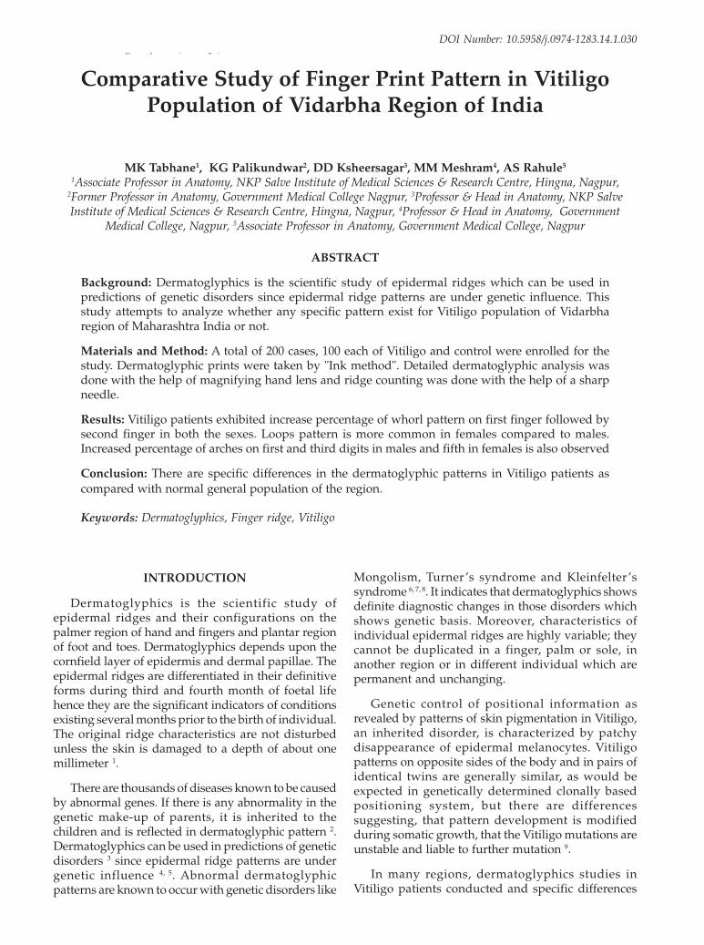
122 Medico-Legal Update, January-June 2014, Vol. 14, No. 1
INTRODUCTION
Dermatoglyphics is the scientific study ofepidermal ridges and their configurations on thepalmer region of hand and fingers and plantar regionof foot and toes. Dermatoglyphics depends upon thecornfield layer of epidermis and dermal papillae. Theepidermal ridges are differentiated in their definitiveforms during third and fourth month of foetal lifehence they are the significant indicators of conditionsexisting several months prior to the birth of individual.The original ridge characteristics are not disturbedunless the skin is damaged to a depth of about onemillimeter 1.
There are thousands of diseases known to be causedby abnormal genes. If there is any abnormality in thegenetic make-up of parents, it is inherited to thechildren and is reflected in dermatoglyphic pattern 2.Dermatoglyphics can be used in predictions of geneticdisorders 3 since epidermal ridge patterns are undergenetic influence 4, 5. Abnormal dermatoglyphicpatterns are known to occur with genetic disorders like
Comparative Study of Finger Print Pattern in VitiligoPopulation of Vidarbha Region of India
MK Tabhane1, KG Palikundwar2, DD Ksheersagar3, MM Meshram4, AS Rahule5
1Associate Professor in Anatomy, NKP Salve Institute of Medical Sciences & Research Centre, Hingna, Nagpur,2Former Professor in Anatomy, Government Medical College Nagpur, 3Professor & Head in Anatomy, NKP SalveInstitute of Medical Sciences & Research Centre, Hingna, Nagpur, 4Professor & Head in Anatomy, Government
Medical College, Nagpur, 5Associate Professor in Anatomy, Government Medical College, Nagpur
ABSTRACT
Background: Dermatoglyphics is the scientific study of epidermal ridges which can be used inpredictions of genetic disorders since epidermal ridge patterns are under genetic influence. Thisstudy attempts to analyze whether any specific pattern exist for Vitiligo population of Vidarbharegion of Maharashtra India or not.
Materials and Method: A total of 200 cases, 100 each of Vitiligo and control were enrolled for thestudy. Dermatoglyphic prints were taken by "Ink method". Detailed dermatoglyphic analysis wasdone with the help of magnifying hand lens and ridge counting was done with the help of a sharpneedle.
Results: Vitiligo patients exhibited increase percentage of whorl pattern on first finger followed bysecond finger in both the sexes. Loops pattern is more common in females compared to males.Increased percentage of arches on first and third digits in males and fifth in females is also observed
Conclusion: There are specific differences in the dermatoglyphic patterns in Vitiligo patients ascompared with normal general population of the region.
Keywords: Dermatoglyphics, Finger ridge, Vitiligo
Mongolism, Turner’s syndrome and Kleinfelter ’ssyndrome 6, 7, 8. It indicates that dermatoglyphics showsdefinite diagnostic changes in those disorders whichshows genetic basis. Moreover, characteristics ofindividual epidermal ridges are highly variable; theycannot be duplicated in a finger, palm or sole, inanother region or in different individual which arepermanent and unchanging.
Genetic control of positional information asrevealed by patterns of skin pigmentation in Vitiligo,an inherited disorder, is characterized by patchydisappearance of epidermal melanocytes. Vitiligopatterns on opposite sides of the body and in pairs ofidentical twins are generally similar, as would beexpected in genetically determined clonally basedpositioning system, but there are differencessuggesting, that pattern development is modifiedduring somatic growth, that the Vitiligo mutations areunstable and liable to further mutation 9.
In many regions, dermatoglyphics studies inVitiligo patients conducted and specific differences
DOI Number: 10.5958/j.0974-1283.14.1.030
30. Manjusha Tabhane--122--126.pmd 2/22/2014, 11:45 AM122
Medico-Legal Update, January-June 2014, Vol. 14, No. 1 123
were observed 10. But regional differences can existsthat’s why this study was undertaken with theintentions to analyze whether any specific pattern, existfor Vitiligo patients? And whether those serve as anearly diagnostic tool?
MATERIAL AND METHOD
The prospective study was conducted inGovernment Medical College Nagpur. Cases wereincluded from various parts of Vidarbha region ofMaharashtra India. A total of 200 cases, 100 each ofVitiligo and control were enrolled. In both the groups60% male and 40% female were parts of the study. InVitiligo group, cases were diagnosed by dermatologistsof the institute by clinical histories, family histories,clinical examinations, laboratory investigations andpast medical records. In control group, normal healthyvolunteers of Vidarbha region of Maharashtra wereincluded after complete general, systemicexaminations and laboratory investigations.
Dermatoglyphic prints were taken by “Ink method”1, 3, 6 in which subjects were asked to clean and dry theirhands with some moisture on the hands. Thin film ofink was applied over the palm with the help of inkedrubber roller taking care of uniformity on digits, hollowof the palm and flexor crease of wrist. Then print wastaken by placing the hand over a sheet of white paperfrom proximal to distal end which was kept over apressure pad. Same procedure was adopted foranother hand. Detailed dermatoglyphic analysis wasdone with the help of magnifying hand lens and ridgecounting was done with the help of a sharp needle.
Ridge Counting: Ridge counting indicates thepattern size. It is primarily utilized on finger tips as away of expressing the distance between digital triradiito the ridge density in a given area (Figure -1).
Finger Ridge Counting: Counting is along astraight line connecting the triradial point to the pointof core. Ridges containing triradial point and point ofcore are excluded. In case of whorl with two triradiiand at least one point of core, two different counts aremade, one from each triradii. Each count is made alonga line drawn between the triradial point and the nearerpoint of core. The two counts are specified as first radialand second ulnar counts. The dermatoglyphic patternsare analyzed according to sex and bi-hand. They aresubjected to nonparametric statistical tests to evaluatesignificant pattern of identifiable differences betweenthe Vitiligo and control groups. For qualitative analysischi-square test was used.
Total Finger Ridge Count (TFRC): TFRCrepresents the sum of ridge counts of all ten digits,where only the larger count is used on those digitswith more than one ridge count. It expresses size ofpattern.
Absolute Finger Ridge Count (AFRC): AFRC isthe sum of the ridge counts from all the separate triradiion the fingers. It reflects the pattern size as well aspattern intensity, which depends on the pattern type.
RESULTS
We found frequency distribution of absolute fingerridge count (AFRC) in the range of 26 to more than350 (Table-1) in all the groups. We also observedincreased in the mean values of AFRC in Vitiligo malesand females and also in Vitiligo males and femalescombined series when compared with controls andthere was statistically significant difference in the meanvalues of AFRC in all the comparison groups(Table-2).
The frequency distribution of total finger ridgecount (TFRC) was observed in between the range of26 to more than 350 (Table-3). We found statisticallysignificant increase in the mean values of TFRC inVitiligo males and females, and also in Vitiligo malesand females combined series (Table-4). All the tablesindicate that there are significant differences in themean values of AFRC and TFRC in Vitiligo comparedto controls.
Moreover, Vitiligo patients exhibited increasepercentage of whorl pattern predominantly on firstfinger and then on second fingers in both the sexeswhile increased percentage of loops on the second
Fig. 1. Ridge counting in various finger tip pattern types. Thecounting is done along the stralght lines coneecting the core andthe triradulus.
30. Manjusha Tabhane--122--126.pmd 2/22/2014, 11:45 AM123

124 Medico-Legal Update, January-June 2014, Vol. 14, No. 1
finger in both the sexes. As far as arches are concerned,in the present study we observed increased percentage
of arches on the first and third digits in males and fifthin females.
Table 1: Frequency distribution of AFRC
Class Interval Vitiligo Controls
Male Female Total Male Female Total
No. % No. % No. % No. % No. % No. %
0-25 0 0.00 0 0.00 0 0.00 0 0.00 0 0.00 0 0.00
26-50 0 0.00 1 1.66 1 1.00 1 2.50 5 8.33 6 6.00
51-75 2 5.00 2 3.33 4 4.00 6 15.00 5 8.33 11 11.00
76-100 0 0.00 6 10.00 6 6.00 6 15.00 15 25.00 21 21.00
101-125 6 15.00 5 8.33 11 11.00 8 20.00 18 30.00 26 26.00
126-150 9 22.50 5 8.33 14 14.00 9 22.50 12 20.00 21 21.00
151-175 3 7.50 7 11.66 10 10.00 5 12.50 2 3.33 7 7.00
176-200 0 0.00 11 18.33 11 11.00 2 5.00 2 3.33 4 4.00
201-225 0 0.00 8 13.33 8 8.00 1 2.50 0 0.00 1 1.00
226-250 3 7.50 6 10.00 9 9.00 0 0.00 0 0.00 0 0.00
251-275 8 20.00 4 6.66 12 12.00 0 0.00 0 0.00 0 0.00
276-300 0 0.00 1 1.66 1 1.00 0 0.00 0 0.00 0 0.00
301-325 5 12.00 2 3.33 7 7.00 0 0.00 0 0.00 0 0.00
326-350 0 0.00 1 1.66 1 1.00 0 0.00 0 0.00 0 0.00
Above 350 3 7.50 2 3.33 5 5.00 0 0.00 0 0.00 0 0.00
Table 2: Mean values of AFRC
Subject Mean(SD) CV (%) t value p value
Vitiligo Male 197.78 (92.54) 46.79 5.078 Significant
Control Male 116.33(41.71) 35.85
Vitiligo Female 182.93(74.44) 40.69 7.17 Significant
Control Female 106.78(35.13) 32.90
Vitiligo Total 188.87(82.03) 43.43 8.66 Significant
Control Total 110.60(37.98) 34.34
(SD) Standard Deviation, (CV) Coefficient of Variance
Table 3: Frequency distribution of TFRC
Class Interval Vitiligo Controls
Male Female Total Male Female Total
No. % No. % No. % No. % No. % No. %
0-25 00 00.00 00 00.00 00 00.00 00 00.00 00 00.00 00 00.00
26-50 00 00.00 00 00.00 00 00.00 06 15.00 06 10.00 12 12.00
51-75 03 07.50 03 05.00 06 06.00 10 25.00 20 33.33 30 30.00
76-100 02 05.00 04 06.66 06 06.00 19 47.50 30 50.00 49 49.00
101-125 17 42.50 11 18.33 28 28.00 02 3.33 02 03.33 04 04.00
126-150 00 00.00 14 23.33 14 14.00 00 00.00 00 00.00 00 00.00
151-175 01 02.50 07 11.66 08 08.00 00 00.00 00 00.00 00 00.00
176-200 09 22.50 07 11.66 16 16.00 00 00.00 00 00.00 00 00.00
201-225 00 07.50 00 00.00 03 03.00 00 00.00 00 00.00 00 00.00
226-250 00 00.00 00 00.00 00 00.00 00 00.00 00 00.00 00 00.00
251-275 00 00.00 00 00.00 00 00.00 00 00.00 00 00.00 00 00.00
276-300 00 00.00 00 00.00 00 00.00 00 00.00 00 00.00 00 00.00
301-325 00 00.00 00 00.00 00 00.00 00 00.00 00 00.00 00 00.00
326-350 00 00.00 00 00.00 00 00.00 00 00.00 00 00.00 00 00.00
Above 350 05 12.50 14 23.33 19 19.00 00 00.00 00 00.00 00 00.00
30. Manjusha Tabhane--122--126.pmd 2/22/2014, 11:45 AM124

Medico-Legal Update, January-June 2014, Vol. 14, No. 1 125
Table 4: Mean values of TFRC
Subject Mean(SD) CV (%) t value p value
Vitiligo Male 130.30(43.05) 33.04 07.17 Significant
Control Male 76.40 (20.20) 26.44
Vitiligo Female 136.37(36.74) 26.94 11.28 Significant
Control Female 77.43 (16.97) 21.91
Vitiligo Total 133.94(38.29) 29.33 13.14 Significant
Control Total 77.02 (18.29) 23.68
(SD) Standard Deviation, (CV) Coefficient of Variance
DISCUSSION
In the present study Absolute Finger Ridge Count(AFRC) is significantly increased in Vitiligenous maleand female patients and a significant difference isfound when Vitiligenous male and female combinedseries is compared to controls. In case of Total FingerRidge Count (TFRC), Vitiligo males and females alsoshow a significant increase as compared to controlsand a significant difference is found in male and femalecombined series compared to those of the controls. Butour findings do not coincide with the findings of Kapurand Verma 11, Verma and Jain 12 and Sahasrabuddhe etal 13. It is suggested that increased AFRC and TFRCvalues in the present study is due to increase in samplesize, whorls and loops.
Our study shows that Vitiligo patients haveincreased percentage of whorl pattern more on firstfinger followed by second fingers in both the sexes.Kapur and Verma 11 also found the similar results aswhorl pattern was common on 1st finger followed by2nd finger in their study. On the fifth finger there is anincrease in percentage of whorls in case of Vitiligomales than females which is a contradictory findingin comparison to observations of Sahasrabuddhe et al13 as they have not observed increase percentage on 5th
finger and probably it is due to variation in samplesize.
Though there is an increase in percentage of loopsin cases of Vitiligo cases but this increase is statisticallyinsignificant. Kapur and Verma 11 and Singh et al 10
also opined the same after their study. Vitiligo casesshowed predominance of ulnar loops compared toradial loops which is a similar finding like Kapur andVerma 11. In our region overall percentage of loops infemales are more compared to males and Singh et al 10
also observed female predominance in their studyalthough with some marginal differences. We observedincreased percentage of loops on the second finger in
both the sexes. This finding correlates withSahasrabuddhe et al 13 they also observed increasenumber of loops on the 2nd finger. In case of femalesthere is an increase in percentage of loops on the thirddigit which is identical to the findings reported byVerma and Jain 12 and also increased loops on fifthfingers in females. But Verma and Jain 12 reportedincrease in loops on fifth fingers irrespective of the sex.
We found frequency of arches is decreased in boththe sexes. Sahasrabuddhe 13 reported decrease infrequency in males while Verma and Jain 12 reporteddecrease in females. In our study increased percentageof arches observed on the first and third digits in malesand fifth in females. These findings partially tally withthat of Kapur and Verma 11. The variation in betweenboth studies is most probably due to the less numberof cases of Vitiligo and absence of sex wise distributionin their study.
CONCLUSION
Our regional Vitiligo population has increasedabsolute finger ridge count and total finger ridge countin both the sexes. They have increased percentage ofwhorl pattern in first and second fingers. Males alsohave increase percentage of whorls on the fifth finger.Percentage of loops is more in Vitiligo population inboth the sexes. Specifically the percentage is more insecond digit in both the sexes and in third and fifthdigits in case of females. Frequency of arches decreasesin both the sexes with increase percentage in first andthird digits of males and in fifth digit of females.
Acknowledgement: Nil
Conflict of Interest: Nil.
Source of Support: Nil financial support.
Ethical Clearance: Obtained.
30. Manjusha Tabhane--122--126.pmd 2/22/2014, 11:45 AM125

126 Medico-Legal Update, January-June 2014, Vol. 14, No. 1
REFERENCES
1. Cummins HM. Morphology of palmarhypothenar dermatoglyphics in man. HumanBiology 1943; 7:1.
2. Walkar NF. The current status of research ondermatoglyphics in medical constitution.Genetics Today 1964; 3: 981-990.
3. Cummins HM. Palmar and plantar epidermalridge configurations in European. AmericanJournal of Physical Anthropology 1926; 9:471-502.
4. Wilder HH. Palms and soles. American Journalof Anatomy 1902; 423.
5. Wilder M. Racial difference in palm and soleconfigurations. Am Anthropol 1904; 244: 292.
6. Cummins HM. Dermatoglyphics stigma inmongolism. Anat Record 1936; 64(2): 11.
7. Penrose LS. Finger prints, palms andchromosomes. Nature 1963; 197: 933-938.
8. Holt SB and Lindstein. Dermatoglyphicanomalies in Turner ’s syndrome. Ann HumGenet 1964; 28: 87-100.
9. Goudie BM, Wilkieson C, Goudie RB. A familystudy of Vitiligo patterns Swff Med J 1983; 28:338-342.
10. Singh PK, Pande SS, Singh G, Girgla HS. Indian JDermatol Venereol and Leprol 1983; 49(2): 68-70.
11. Kapur TR, Verma RD. Indian J Dermatol Venereoland Leprol 1982; 48(4).
12. Verma KC, Jain VK. Dermatoglyphics in Vitiligo.Indian J Dermatol Venereol and Leprol 1981; 47:102-104.
13. Sahasrabuddhe RG, Singh G, Agrawal SP.Dermatoglyphics in Vitiligo. Indian Journal ofDermatology 1985; 21: 20-22.
30. Manjusha Tabhane--122--126.pmd 2/22/2014, 11:45 AM126

Medico-Legal Update, January-June 2014, Vol. 14, No. 1 127
INTRODUCTION
Anthropometric work in this field has been inprogress in various countries, since the last decades ofthe nineteenth century. Anatomists and forensicexperts have been consulted frequently regardingidentification of skeletal remains found undersuspicious circumstances and are asked to pronouncean opinion which may form an important evidence in
Stature Estimation from the Length of Humerus inVidarbha Region of Maharashtra
Meena M Meshram1, Anil S Rahule2, M S M Bashir3
1Professor & Head, 2Associate Professor in Anatomy, Government Medical College, Nagpur, 3Associate Professor inPharmacology, RIMS Adilabad
ABSTRACT
Background: Estimation of stature from length of long bones plays an important role in identifyingunknown bodies. Stature of an individual can be estimated from measurements of long bones withthe help of established formulae which plays an important role in medico-legal practice. Each workerhas derived his own formula for calculating the stature from the length of long bones but no universallyapplicable formula has been derived as the relationship between stature and long bone length differaccording to race, sex, age and side of body. It is proved beyond doubt that each race requires its ownformula. Hence present study is undertaken to derive formulae for stature estimation for individualsof Vidarbha region of Maharashtra state.
Material and Method: The material consists of fully ossified 116 humeri belonging to 58 dissectionhall cadavers, from Government Medical College Nagpur, out of which 42 were males and 16 werefemales of known sex and stature. The linear relationship between the living stature and length ofhumerus of each side was worked out in the form of regression equation and Multiplication factor.With the help of these regression equations, stature was calculated.
Results: Stature estimated with the help of regression equation showed the average error of 3 cm inmales and less than 1 cm in females and with multiplication factor it was less than 2 cm in both thesexes.
Conclusion: The regression formulae and multiplication factor can be used to predict the staturefrom the length of humerus. The accuracy of multiplication factor in estimating the stature in theregion is very high.
Keywords: Cadaver, Humerus, Living Stature, Multiplication Factor, Regression Equations
Corresponding author:Meena M MeshramProfessor & HeadDepartment of AnatomyGovernment Medical College, NagpurMobile: 9850390088, 9422153880Email: [email protected]@yahoo.co.in
the court. Stature is one of the parameter of biologicalprofile used to confirm or exclude presumptiveidentification. Applying the knowledge of humanbiological variation as it relates to developingbiological profiles for unknown victims is a crucialcomponent for the increasing number ofanthropologists working globally investigating crimesagainst international humanitarian law 1.
Examinations of the skeletal samples of the burialsare often fragmentary and are found in mixed lots.Some authors have used fragments of long bones butmost of the times long bones have been used in thedetermination of stature because they providerelatively better accuracy in prediction of stature. Forthis reason there is a need for developing a techniquefor stature estimation from skeletal parts which are
DOI Number: 10.5958/j.0974-1283.14.1.031
31. Meena Meshram--127--131.pmd 2/22/2014, 11:45 AM127
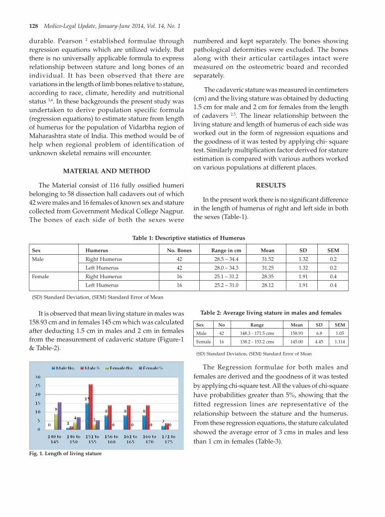
128 Medico-Legal Update, January-June 2014, Vol. 14, No. 1
durable. Pearson 2 established formulae throughregression equations which are utilized widely. Butthere is no universally applicable formula to expressrelationship between stature and long bones of anindividual. It has been observed that there arevariations in the length of limb bones relative to stature,according to race, climate, heredity and nutritionalstatus 3,4. In these backgrounds the present study wasundertaken to derive population specific formula(regression equations) to estimate stature from lengthof humerus for the population of Vidarbha region ofMaharashtra state of India. This method would be ofhelp when regional problem of identification ofunknown skeletal remains will encounter.
MATERIAL AND METHOD
The Material consist of 116 fully ossified humeribelonging to 58 dissection hall cadavers out of which42 were males and 16 females of known sex and staturecollected from Government Medical College Nagpur.The bones of each side of both the sexes were
numbered and kept separately. The bones showingpathological deformities were excluded. The bonesalong with their articular cartilages intact weremeasured on the osteometric board and recordedseparately.
The cadaveric stature was measured in centimeters(cm) and the living stature was obtained by deducting1.5 cm for male and 2 cm for females from the lengthof cadavers 2,5. The linear relationship between theliving stature and length of humerus of each side wasworked out in the form of regression equations andthe goodness of it was tested by applying chi- squaretest. Similarly multiplication factor derived for statureestimation is compared with various authors workedon various populations at different places.
RESULTS
In the present work there is no significant differencein the length of humerus of right and left side in boththe sexes (Table-1).
Table 1: Descriptive statistics of Humerus
Sex Humerus No. Bones Range in cm Mean SD SEM
Male Right Humerus 42 28.5 – 34.4 31.52 1.32 0.2
Left Humerus 42 28.0 – 34.3 31.25 1.32 0.2
Female Right Humerus 16 25.1 – 31.2 28.35 1.91 0.4
Left Humerus 16 25.2 – 31.0 28.12 1.91 0.4
(SD) Standard Deviation, (SEM) Standard Error of Mean
It is observed that mean living stature in males was158.93 cm and in females 145 cm which was calculatedafter deducting 1.5 cm in males and 2 cm in femalesfrom the measurement of cadaveric stature (Figure-1& Table-2).
Table 2: Average living stature in males and females
Sex No Range Mean SD SEM
Male 42 148.3 - 171.5 cms 158.93 6.8 1.05
Female 16 138.2 - 153.2 cms 145.00 4.45 1.114
(SD) Standard Deviation, (SEM) Standard Error of Mean
The Regression formulae for both males andfemales are derived and the goodness of it was testedby applying chi-square test. All the values of chi-squarehave probabilities greater than 5%, showing that thefitted regression lines are representative of therelationship between the stature and the humerus.From these regression equations, the stature calculatedshowed the average error of 3 cms in males and lessthan 1 cm in females (Table-3).
Fig. 1. Length of living stature
31. Meena Meshram--127--131.pmd 2/22/2014, 11:45 AM128

Medico-Legal Update, January-June 2014, Vol. 14, No. 1 129
Table 3: Regression formulae
Sex Side Regression Formulae Chi-Square (d.f.) Significance
Male Right S=46.05+3.58XH X2=6.23 (40) P>0.05.N.S.
Left S=45.17+3.64xH X2=6.07 (40) P>0.05.N.S.
Female Right S=123.325+0.76xH X2=1.30 (14) P>0.05.N.S.
Left S=99.315+1.62xH X2=1.055(14) P>0.05.N.S.
S-living stature of adult, H-humerus, d.f.- Degree of freedom, P-Probability N.S.-Not significant
The average multiplication factor is 5.1 for humerusin Vidarbha region. With the help of this multiplicationfactor the stature calculated which showed the averageerror less than 2 centimeters in both sexes (Table-4).
Table 4: Multiplication factor (MF)
Sex Bones MF Sample Size
Male Rt. Humerus 5.04 42
Lt. Humerus 5.08 42
Female Rt. Humerus 5.11 16
Lt. Humerus 5.15 16
DISCUSSION
In the present study the average living staturefound in males was 158.93 cm, close to the finding 161.5cm of Bhargava and Kher 6 on the population ofBarelas and Bhils in Madhya Pradesh. The livingstature of adult male population of Vidarbha regionwas found to be shorter compared to 163.11 cm inWestern Maharashtra (Athawale)7 and 165.78 cm fromMarathwada Region (Kolte and Bansal)4. It is furtherrevealed that 166.6 cm, the finding of Bose 8 on thepopulation of East Bengal, 163.6 cm the finding ofSiddiqui and Shah 9, 163 cm of Singh and Sohal10 onthe population of Punjab and 162.3 cm of Charnalia11
on the population of Pondicherry are much higher thanthe finding of 158.93 cm of the present work.
The average living stature of 145 cm in females inthe present study is shorter, compared to 149 cm, thefindings of Patil et al12 and 149.72 cm, the finding ofKolte and Bansal 4 in Marathwada region. The presentdata on living adult female stature could not becompared thoroughly as there is insufficient dataregarding the studies in female population.
The resultant regression formulae in the presentstudy thus obtained for Indian bones of Vidarbharegion definitely show a different value for the twosexes. In addition it reveals that they also differsignificantly from Pearson’s formulae, derived fromEnglish bones commonly used in India for forensicopinions. From the above regression formulae, in the
present study the stature was calculated which showedthe average error of 3 cm in males and less than 1 cmin females.
Similar studies were conducted by Patil et al 12 andshowed that regression formulae can be used toestimate the stature from length of humerus. Theregression equations of male bones in the present studyare very close to the regression equations of Kate andMuzumdar 13 who worked on Indian population forAmritsar and Nagpur areas.
It has been further observed that the regressionformulae in the present study differ significantly fromthe findings of Breitinger 14 who worked on Germanpopulation, Telkka15 on Finnish population, Dupertuisand Hadden 16 and Pearson 2 on British population(Table-5). Kolte and Bansal 4 and Trotter and Gleser 3
have found that the regression formulae differaccording to race, sex, age, side of the body andnutritional status of the people. Siddiqui and Shah9
discouraged the use of regression formulae ofpopulation of one region for reconstruction of staturefor population of another region. Kate and Muzumdar13 stated that Pearsonian regression equations do notgive exact results in India. They stated that there arevariations in correlations between stature and lengthof long bones of individuals of different regions andtheir use in not sound and satisfactory. Thus theregression equations obtained by different workersdiffer significantly according to sex, race, climaticconditions, and nutritional states of the individuals,rendering their results incomparable.
Table 5: Regression Formulae by various authors
Authors Sex Regression Formulae Race
Breitinger Male S=83.21+2.71xH German
Telkka Male S=77.28+2.8 xH Finnish
Female S=75.8+2.7xH Finnish
Dupertuis & Male S=73.57+2.97xH GeneralHadden
Female S=64.97+3.14xH General
Stevenson Male S=81.57+2.81xH Chinese
Perarson Male S=70.64+2.89xH British
Female S=71.47+2.75xH British
31. Meena Meshram--127--131.pmd 2/22/2014, 11:45 AM129

130 Medico-Legal Update, January-June 2014, Vol. 14, No. 1
Table 5: Regression Formulae by variousauthors (Contd.)
Authors Sex Regression Formulae Race
Kate et al Male S=51.76+3.61xH Indian
Female S=49.80+3.70xH Indian
Patil et al Male S=91.98+2.28xH Indian
Female S=71.89+2.75xH Indian
PresentStudy Male(Rt.H) S=46.05+3.58xH Indian
Male(Lt.H) S=45.17+3.64xH Indian
Female(Rt.H) S=123.32+0.76xH Indian
Female(Lt.H) S=99.315+1.62xH Indian
S-living stature, H-humerus
The Multiplication factor 5.1 which is equal in boththe sexes is similar with the findings of Pan 17, Nat 18,Siddiqui and Shah 9 and Kate and Muzumdar 13 whoworked at different places in Indian population. Pan17 also noted that Multiplication factor is same in boththe sexes (Table- 6).
Table 6: Multiplication Factor
Authors Multiplication Factor Population
Pan 5.3 Indian
Nat 5.3 Indian
Siddiqui & Shah 5.0 Indian
Kate & Muzumdar 5.3 Indian
Present Study 5.1 Indian
With the help of this Multiplication factor in thepresent study the stature was calculated which showedthe average error less than 2 cm in both sexes. Siddiquiand Shah 9 have shown the accuracy of multiplicationfactor in estimating the stature. They worked onPunjabi bones and showed the average error about 3cm for both the sexes. Thus the study of multiplicationfactor reveals two very important findings. Firstly theproportion of humerus to the stature is almost the sameas worked out by different workers on different races,regions, and at different times and secondly thisproportion is same in both sexes. This gives a valuewhich is constant, irrespective of race, sex or regionand thus an easy method for reliable forensic work.This therefore, becomes a “ratio for man” with anelement of consistency. A similar finding andconclusion was drawn by Kate and Muzumdar 13 andpostulated a “theory of proportion in the human body”which is termed as “Autometry”.
CONCLUSION
The regression formulae and multiplication factorcan be used to predict the stature from the length ofhumerus. Multiplication factor is a better guide for
calculating the height, when it is not definitely knownas to which part of the country, the individual belongs.Thus this study will definitely be useful to theAnatomists and Forensic experts when a bone or pieceof bone is subjected to the medico-legal examination.
ACKNOWLEDGEMENT
We are grateful to Dr.N.Y.Kamdi, AssociateProfessor, Anatomy & Dr. Sumit Patil AssistantProfessor, Anatomy Government Medical CollegeNagpur, for their support, extended in undertakingthis study & Dr. More, Assistant Professor in StatisticsDept of PSM for helping in statistical calculations.
Conflict of Interest: The authors declare that we haveno financial relationship with any organization forsponsorship of this research work. So we have noconflict of interest.
Source of Funding: Nil financial support.
Ethical Clearance: After taking clearance from localEthical committee, we, the authors declare that theResearch work submitted for publication wasconducted on Dissection Hall Cadavers that complywith the current laws of medical research of India inwhich it was performed.
REFERENCES
1. A.Ross, E.Kimmerle: Contribution in quantitativemethods in forensic Anthropology, Handbook ofForensic Anthropology and Archaeology; 2009:pp. 479-489.
2. Pearson K.: Mathematical contributions to thetheory of evolution. V. On the reconstruction ofthe stature of prehistoric race. Philos Trans SeriesR Soc 1899; 192: 169-244.
3. Trotter M. and Gleser G.C.: Estimation of staturefrom long bones of American whites andNegroes. Am J of Physical Anthropology. 1952;10: 453-514.
4. Kolte P.M. and Bansal P.C.: Determination ofregression formulae for reconstruction of staturefrom long bones of upper limb in Maharashtraof Marathwada region. J of Anatomical Soc. ofIndia 1974; 23: 6-11.
5. Glaister J.: In Medical Jurisprudence andToxicology, 10th Ed. Edinburgh, ES LivingstoneLtd; 1957. p. 80.
6. Bhargava I. and Kher G.A.: A comparativeAnthropometric study of Bhils and Barelas of
31. Meena Meshram--127--131.pmd 2/22/2014, 11:45 AM130

Medico-Legal Update, January-June 2014, Vol. 14, No. 1 131
Central India. Journal of Anatomical Society ofIndia 1961; 10: 31-33.
7. Athawale M.C.: The estimation of height from thelength of fore arm bones. A study on one hundredMaharashtra male adults of age between 25 and30 years. Am J Phys Anthropology 1963; 21:105–112.
8. Bose A.: Anthropometry of the Kayasthas ofBengal with special reference to Vangaj Kayastha.Journal of Anatomical Society of India 1963; 12:20-25.
9. Siddiqui M.A.H and Shah M.A.: Estimation ofstature from long bones of Punjabis. Indian Jour.of Med Res 1944; 32: 105-108.
10. Singh B. and Sohal H.S.: Estimation of staturefrom the length of clavicle in Punjabis. Apreliminary report. Indian Jour of Med Res 1952;40: 67-71.
11. Charnalia V.M.: Anthropological study of the footand its relationship to stature in different castesand tribes of Pondicherry State. J of Anat Societyof India 1961; 10: 26-30.
12. Patil T.L, Gawhale K.S and Muzumdar R.D.:Reconstruction of stature from long bones of bothupper and lower limbs. 1983; 32: 111-118.
13. Kate B.R and Muzumdar R.D.: Stature estimationfrom Femur and Humerus by regression andAutometry. Acta Anatomica (Basel) 1976; 94(2):311–320.
14. Breitinger E. Zur Berechnug der Koreperho” heaus den langen Gliedmassen – Knochen.Anthrop. Anz. (1937) 14: 249-274.
15. Telkka, A. (1950): on the prediction of humanstature from long bones. Acta Anatomica (1950)9: 103-107.
16. Dupertuis E.W and Hadden J.A.: TheReconstruction of the stature from long bones.Am J Phys Anthrop 1951; 9(1): 15-54.
17. Pan S.: Length of long bones and their proportionto body height in Hindus. Journal of Anatomy1924; 58: 374-378.
18. Nat B.S.: Estimation of stature from long bonesin Indians of United Provinces. A Medico-legalinquiry in anthropometry. Indian Journal ofMedical Research 1931; 18: 1245-1253.
31. Meena Meshram--127--131.pmd 2/22/2014, 11:45 AM131

132 Medico-Legal Update, January-June 2014, Vol. 14, No. 1
INTRODUCTION
Qualitative sex differentiation using many bonesis been done by many forensic experts, but sexing fromsingle bone is difficult task. Almost all the elements ofhuman skeleton show some degree of sexualdimorphism, but reliable indicators can be obtainedfrom specific bones like hip bone, skull and sacrum.The determination of deceased sex is first step inskeletal analysis since estimation of age at death, race,and stature depends on sex of deceased.
The measurements of the skeleton bones, mainly theneurocranium (brain case) and viscerocranium, sufferinfluences of various factors; however, they are oftenused for human population morphological studies ofage estimation, stature, ethnicity, which are relevantaspects of forensics investigation and anthropologicalexaminations of unknown individuals1.
Identification of Gender from Dimensions of Palate
Manjunath T H1, Nagesh C Kuppast2, Shahina3, Umesh S R4, Shradha Iddalgave5
1Assistant professor, Department of Forensic Medicine and Toxicology,Subbaiah Institute of Medical Science and Research Center, Shimoga, Karnataka, 2Assistant Professor, 3Post GraduateStudent, Department of Forensic Medicine and Toxicology, S. S. Institute of Medical Sciences and Research Center,
Davangere, Karnataka, 4Professor and HOD, Department of Forensic Medicine and Toxicology, M. R. Medical College,Gulbarga, Karnataka, 5Department of Anatomy, J. J. M. Medical College, Davangere, Karnataka
ABSTRACT
Determination of deceased sex is first step in skeletal analysis since estimation of age, race, andstature depends on sex of deceased. Total 100 adult human skulls (50 male and 50 female) availablein the Department of Forensic Medicine and Toxicology and Department of Anatomy of M. R. MedicalCollege and K. B. N. Medical College Gulbarga, Karnataka are studied. The demarking point (D.P.)and identification point (I.P.) of both measurements were calculated and then percentage of bonesidentified by D.P. and I.P. was recorded. Though, the Demarking Point of a single parameter may notidentify sex in all the bones but the accuracy is nearly 100% in the bones, which are identified.Percentage of skulls identified by IP of palatal breadth in males is more as compared to palatal lengthwhere as identification from DP is equal. Percentage of skulls identified by IP and DP of palatallength in females is more as compared to palatal breadth. So palatal length gives better prediction ofsex as compared to palatal breadth in case of females where as in case of males palatal breadth isbetter predictor of sex.
Keywords: Palatal length, Palatal breadth, Demarking point, Identification point, Sexing of Cranium
Corresponding author:Manjunath T HAssistant ProfessorDepartment of Forensic Medicine and Toxicology,Subbaiah Institute of Medical Science and ResearchCenter, Shimoga, KarnatakaE-mail I. D. [email protected]
Recent studies report that morphometry is a fastand efficient method for the evaluation ofmorphological characteristics, such as ethnicity,gender, age, genetic factors, dietary habits, and regionalvariations which can alter the shape and size of bonestructures. These aspects are significantly importantin determining the anthropometric changes betweendifferent populations and genders2
Traditional method is non-metrical andmorphological. Morphological features of bonesdepend upon nutrition, occupation, race andgeography of the region, so the traditional method isnot reliable in the study of bones.
In this study length and breadth of palate werestudied. They were analysed statistically by applyingroutine statistical data like identification point anddemarking point.
MATERIALS AND METHOD
The materials for the present study consisted of 100adult human skulls of known sex (50 male and 50female) available in the Department of ForensicMedicine and Toxicology and Department of Anatomyof M. R. Medical College and K. B. N. Medical College
DOI Number: 10.5958/j.0974-1283.14.1.032
32. Nagesh Kuppast--132--134.pmd 2/22/2014, 11:45 AM132

Medico-Legal Update, January-June 2014, Vol. 14, No. 1 133
Gulbarga, Karnataka. Following parameters werestudied:
1. Palatal length (PL): Palatal length is the distancebetween staphylion and orale. It was recorded withthe help of sliding vernier caliper.
2. Palatal breadth (PB): Palatal breadth is the distancebetween endomolare. it was recorded with thehelp of sliding vernier caliper.
As the first part of the study, all the values weretabulated and analyzed statistically by routinestatistical methods.
The value of Range, Mean, Standard Deviation(SD), Calculated Range (mean ± 3SD), DemarkingPoint and Identification Point were obtained.Maximum value of female range is considered asidentification point for male. Minimum value of malerange is considered as identification point for female.Maximum value of female calculated range isconsidered as demarcation point for male. Minimumvalue of male calculated range is considered asdemarcation point for female. In case where femalerange/calculated range maximum value of malerange/calculated range is considered as Identificationpoint/Demarcation point for female and minimumvalue of female range/calculated range is consideredas identification point/demarcation point for male.Subsequently ‘t’ is applied to both parameters.
OBSERVATIONS
The Range, Mean, Calculated Range (mean + 3S.D.), Demarking Points (DP), Identification Point (IP)and the percentage of bones in which sex could beidentified by them, are given in table no. 1 and 2.
Table No 1. Statistical analysis of palatal length ofskull.
Details of measurements Male Female
Mean 44.72 41.68
SD 3.98 5.56
Range 38-55 24-51
Identification point(IP) >51mm <38
% of bones identified by IP 04% 16%
Standard error 0.56 0.79
Calculated range 32.76-56.67 24.99-58.37
Demarking point(DP) >58.37 <32.76
% of bones identified by DP 0% 08%
Percentage of skulls identified by IP alone was 04%of males and 16% of females. The percentage of skullsidentified by DP alone was 0% of males and 08% offemales. ‘t’ test was significant with p<0.05.
Table No-2. Statistical analysis of palatal breadth ofskull.
Details of measurements Male Female
Mean 34.36 32.02
SD 5.70 4.97
Range 23-45 23-40
Identification point(IP) >40mm <23mm
% of bones identified by IP 14% 0%
Standard error 0.81 0.70
Calculated range 17.25-51.46 17.11-46.93
Demarking point(DP) >46.93 <17.25
% of bones identified by DP 0% 0%
Percentage of skulls identified by IP alone was 14%of males and 0% of females. The percentage of skullsidentified by DP alone was 0% of males and 0% offemales. ‘t’ test was significant with p<0.05.
DISCUSSION
The female skull retains the gracile attributes seenin prepubescent skull. Male cranium becomesmarkedly rougher in adulthood, the differentiatingfeatures of sex become more prominent after puberty,again towards old age there occurs blurring of sexuallydimorphic traits. So the determination of sex frombones should ideally be limited to 15-55 years of age3.Krogman WM4 (1978) analyzed 750 skeletons and cameto a conclusion that the determination of sex is possiblewith accuracy of about 100% if whole skeleton isavailable, 92% when skull alone and 98% when bothpelvis and skull are available. A great number ofmeasurements of the skull have been proposed andused by different investigators during the past. Martinand Saller used eighty one measures; Howell describedseventy; Hrdlicka lists thirty two; Bass gives twentythree.
In the present study both length and breadth ofpalate are statistically significant. The results arecompared with those of previous workers as shownin table no. 3 & 4.
32. Nagesh Kuppast--132--134.pmd 2/22/2014, 11:45 AM133

134 Medico-Legal Update, January-June 2014, Vol. 14, No. 1
Table No 3. Showing comparison of palatal length with other workers.
Sl Name of worker Male Female SS
N M SD N M SD P
1 Hong Wei Song 1992 30 43.2 3.3 30 42.3 4.6 >0.05
2 Deshmukh(2003) 40 45 3.08 34 44 3.37 >0.05
3 Present study (2009) 50 44.72 3.98 50 41.68 5.56 <0.05
Where N – no. of skull, M – Mean, SD – Standard deviation, SS- Statistical
significance, scale in mm.
Table No 4. Showing comparison of palatal breadth with other workers.
Sl Name of worker Male Female SS
N M SD N M SD P
1 Hong Wei Song 1992 30 36.2 3.1 30 35.4 3.8. >0.05
2 Deshmukh(2003) 40 35 1.50 34 34 2.73 <0.05
3 Present study (2009) 50 34.36 5.70 50 32.02 4.97 <0.05
The findings of present study are similar with mostof them, but Hong Wei Song (1992) in his study onChinese skulls found this parameter to be statisticallyinsignificant. It may be due to racial factor as shownin table 4.
CONCLUSION
Palatal dimensions can help in identification of sexof the individual. Among both parameters statisticallyanalyzed, considering the Demarking Point andIdentification point, the most reliable parameter ispalatal length as compared to the palatal breadth incase of females whereas palatal breadth is betterpredictor for sex determination in case of males.
ACKNOWLEDGEMENT:
I thank Dr. Mallikarjun B., dean M. R. MedicalCollege, Gulbarga for encouraging and securing methe all the required facilities for the work.
I express my humble gratitude to all the staffmembers of Dept. Of Forensic Medicine andToxicology, M. R. Medical College, Gulbarga for theirmotivation and constructive ideas during the study.
I thank Mr. Sangam statistician, for his assistanceand positive feedback during statistical analysis.
Source of Funding: Self
Ethical Clearance: Taken
Conflict of Interest Statement: “The undersignedauthor / authors hereby declare that the article isoriginal, neither the article nor a part of it is underconsideration for publication anywhere else and hasnot been previously published anywhere. We have
declared all vested interests. We have meticulouslyfollowed the instructions. The article, if published,shall be the property of the Journal and we surrenderall rights to the Editors. We agree to provide the latestfollow up of cases prior to the publication of casereports when requested”.
REFERENCES
1. Iscan MY Global forensic anthropology in the 21stcentury. Forensic Science International . 2001, vol.117, p. 1-6.
2. Humphrey LT, Dean MC and Stringer CB.Morphological variation in great ape and modernhuman mandibles. Journal of Anatomy. 1999, vol.195, p. 491-513. (part 4).
3. Pillay VV. Text book of Forensic Medicine &Toxicology, 14th edition, Paras Publication, PageNo.56 (2004).
4. Krogman WM. The human skeleton in forensicmedicine. Charles C, Thomas, Springfield,Illinois, 1962; USA.
5. Keen JA (1950) “Sex differences in skull”, APJA,8(1), Page No.65-79.
6. Hong Wei Song, Zi, Qing Lin and Jing Tao Jia.Sex diagnosis of Chinese skulls using multiplestepwise discriminant functional analysis.Forensic Science International, 1992;54: 135-140.
7. Deshmukh AG & Deverthi DB comparison ofCranial Sex determination by univariatea andmultivariate analysis, Joint Anatomy Society ofIndia 55 (2), Page No.48-51 ,(2006).
8. Henríquez-Pino, J.; Cricenti, S. V. & Didio, L. J.A. Morfometría del foramen magno y su relacióncon el tipo de cráneo en individuos brasileños.Rev. Chil. Anat., 13(2):159-64, 1995.
32. Nagesh Kuppast--132--134.pmd 2/22/2014, 11:45 AM134

Medico-Legal Update, January-June 2014, Vol. 14, No. 1 135
INTRODUCTION
Injury has been defined as any harm whateverillegally caused to any person in body, mind,reputation or property, vide sec.44 IPC1. According toWHO, an injury is defined as a ‘body lesion at theorganic level resulting from acute exposure to energy(mechanical, thermal, electrical, chemical or radiant)interacting with the body in amounts or rates thatexceed the threshold of psychological tolerance’2.
The term “unintentional injury” is preferred to“accidents” as the latter implies events are inevitable
Study on Pattern of Unintentional Transportation InjuryCases Reported at Casualty of a Tertiary Health Care
Centre
Pushpa M G1, Manjulabai K H2, Somshekhar S Pujar3
1Dept of Forensic Medicine and Toxicology, BIMS, Belgaum, 2Professor and Head, 3Assistant Professor, Dept. ofForensic Medicine and Toxicology, JNMC, KLE university, Belgaum
ABSTRACT
Injury is probably the most under recognized major public health problem India is facing today.Injuries have always been part of human existence and they comprise a critical health problemthroughout the world. In spite of safety legislation and attempt to raise public awareness of theinjuries, the rate of motor vehicle crashes, fall from height, suicide, drowning, violence etc. still remainshigh.
Unintentional injuries are the largest source of premature morbidity and mortality and a major burdento the society all over the world. Further transportation injuries constitute the bulk of unintentionalinjuries. Hence this study was aimed and undertaken to know the prevalence, pattern of injury,morbidity and mortality in unintentional transportation cases coming to our tertiary care centrecasualty set up.
The study was conducted from October 2011 to March 2012 analyzing a total of 753 unintentionalcases out of which transportation injuries constituted the maximum number i.e, 609 (80.8%) cases.
The victims, injuries, causative factors, type of vehicles, were studied and analysed and acomprehensive analysis was done. Mortality and morbidity burden was calculated.
The present study result is useful in interpreting the causative factors, and burden of mortality andmorbidity. Accordingly, a strategic plan of various preventive measures can be employed to minimizethe burden of unintentional transportation cases for the benefit of the community and people atlarge.
Keywords: Unintentional, Injuries, transportation, Mortality, Morbidity
and unavoidable whereas a high proportion of theseincidents are now regarded as being preventable.
Injury is the most under recognized major publichealth problem India is facing today. Although modernmedicine is able to withstand many of the dangerouscomplications, the injuries that occur without anyone’sintentions i.e, unintentional injuries like transportationinjuries, fall from height, thermal injuries,unintentional poisoning and drowning/ suffocationrepresents serious risk to health, reducing workefficiency and costing money1.
DOI Number: 10.5958/j.0974-1283.14.1.033
33. Pushpa MG--135--138.pmd 2/22/2014, 11:45 AM135
136 Medico-Legal Update, January-June 2014, Vol. 14, No. 1
In India, there is no availability of sufficient dataregarding the mechanism and causes of falls tounderstand the details of unintentional transportationinjuries, thus requiring this type of study/ research.
The growth of the motor vehicle industry,liberalized economic policies of successivegovernments, aggressive media promotion, increasingpurchasing power of people, easy availability of loansand poor public transport systems have possiblycontributed to increasing motorization and a changingtransportation scenario in India3
Keeping this in mind the study was undertaken atthe casualty of our tertiary care center. Data availablein our study regarding the pattern of unintentionalinjury cases are mostly from the northern parts ofKarnataka. This study presents evidence thatunintentional transportation injuries pose a significanthealth burden.
METHODOLOGY
Data was collected from victims/ patients ofunintentional injuries brought to casualty of KLE’s Dr.Prabhakar Kore hospital & MRC, Belgaum and casesof transportation injuries were analysed in the studyfrom October 2011 to March 2012.
All the victims / patients of unintentionaltransportation injury cases coming to the casualty ofKLE’s Dr. Prabhakar Kore hospital during the studyperiod were included in study.Cases of intentional injuries/ deaths (suicidal, assaultand homicidal), Industrial accidents and otherunintentional injury cases were excluded.
Procedure: Data was collected in a pretestedproforma and necessary details needed for the studywhich included a detailed description of all generaldata of the victims/ patients and details ofunintentional fall cases were analyzed.
RESULTS:
Among the victims in transportation injuries,majority of them were rider and pillion rider of a twowheeler 283 & 75 cases (58.78%) followed by occupantsof LMV 103 (16.9%) cases; pedestrian 72 (11.82%) cases;HMV occupant 42 (6.89%) cases.
Table 1: Distribution of transportation injuriesaccording to type of victims.
Victim Cases Percentage
Pedestrian 72 11.82%
Bicyclist 16 2.62%
Two wheeler pillion rider 75 12.31%
Two wheeler rider 283 46.47%
Three wheeler occupant 09 1.47%
LMV occupant 103 16.91%
HMV occupant 42 6.89%
Others (Bullock cart) 09 1.47%
Total 609 100%
Based on the manner of accident of transportationinjuries, it was noted in our study that majority of theunintentional transportation injuries occurred due tohit by/ to another vehicle 371 (60.9%) cases, followedby skid/ slip 196 (32.18%) cases. Injuries due totoppling of the vehicle were 28 (4.59%) cases and dueto hit to a stationary object were 14 (2.29%) cases.
Table 2: Distribution of pattern of transportationinjuries based on manner of the accident:
Manner Number Percentage of cases
Hit by other vehicle 371 60.92%
Skid/slip 196 32.18%
Toppled 28 4.59%
Hit to stationery object 14 2.29%
Total 609 100%
Out of the 371 cases in the transportation injurieswhich were due to hit by/ to another vehicle or agent,majority of the offending agent was HMV in 146(39.4%) cases followed by
2- wheeler in 97 (26.1%) cases, LMV in 80 (21.6%)cases, unknown vehicles in 18 (4.8%) cases, animalintervention in 16 (4.3%) cases, and 3- wheeler in 14(3.8%) cases. Out of the 609 transportation injury casesseen in our study 242 (39.7%) victims had superficialinjuries; 206 (33.8%) victims suffered with skeletalinjuries, 153 (25.1%) victims suffered with head injuryamong which 11 (1.8%) victims had spinal cord injury;and 49 (8%) victims had visceral injuries. The mortalityrate in the transportation injuries was 8.5% (52 cases).
33. Pushpa MG--135--138.pmd 2/22/2014, 11:45 AM136

Medico-Legal Update, January-June 2014, Vol. 14, No. 1 137
Table 3: Distribution of pattern of injuries intransportation injury cases:
Pattern of injuries Number Percentage Total no. ofof cases transportation
injury casesSuperficial 242 39.7% 609Visceral 49 8%Skeletal 206 33.8%Head injury With spinal 11 1.8%
cordWithout 142 23.3%spinal cord
Mortality 52 8.5%
DISCUSSION
In the present study unintentional injury casesconstituted to more than 70% of the injury cases whichcame to the casualty of our tertiary care centre; andamong them unintentional transportation injury casesconstituted the bulk i.e 81% of the total.
While the most common offending vehicle/ agentinvolved was HMV (39.4%) followed by two-wheeler(26.1%) and LMV (21.6%); majority of the injuriesoccurred due to hit by another vehicle (61%) followedby self fall by slip/skid (32.2%). Similar results wereseen according to BISP report 20084, where two wheelerrider and pillion formed the majority (51%) of thevictims in transportation injuries. But it differed fromstudy by Naci H, Chisholm D, Baker TD5 wherein 60%of injuries occurred in LMV occupants in high incomecountries and pedestrians 45% in LMIC.
The reason for this could be the fact that due tourbanization and growing economy, easy availabilityof funds/ loans from the banks have made easier for acommon man to afford a two wheeler. Even a lowincome family will be having a two wheeler forcommuting. With an increase in the number of vehicleson the roads, as well as vehicular speed, along withlack of knowledge about traffic rules, rash & negligentdrive, non usage of safety measures, unchecked roadcrossings, etc., the burden of road traffic injuries aredefinitely to increase.
Regarding pattern of injuries, external injuries39.7% and skeletal injuries 33.8% formed the majorityin transportation injures, followed closely by headinjury (25%). This could be because of clothing patternand invisibility of blunt injuries due to wheatish anddark skin complexion and deeper contusion.
Adults constituted the bulk with majority of thecase load in the age group of 20-49yrs. Males were the
majority with morbidity ratio for male to female being4:1 and mortality ratio is 2.8: 1
The total mortality rate in our study is 12% (91cases) of the total cases 753 seen. The majorcontributors for this mortality were fromtransportation injuries 57.1% (52 cases) A similarpattern of mortality rate due to transportation injurieswas seen in the studies of Bener A, Hyder AA, SchenkE6 and Colarado dept of public health7. But differedfrom the studies of J Jagnoor, L Keay, R Ivers, WSuraweera, P Jha8; M Cardona, R Joshi, R Q Ivers, SIyengar,et.al9 and WHO report 2007 and BISP 200710 .Pattern of mortality rate due to falls in WHO report2007 is similar to present study; but are very low whencompared to the results from J Jagnoor, L Keay, R Ivers,W Suraweera, P Jha8 and M Cardona, R Joshi, R Q Ivers,S Iyengar,et.al9 .
The reason for transportation injuries forming thebulk of the unintentional injuries in our study couldbe due to the fact that urbanization and modernizationhave affected the lifestyle to a great extent. Today evenan average / low income family owns a two- wheelercontributing to ever increase in number of vehicleshitting the road especially in the cities, highways, etc.In other hand, the decreased usage of pump stove andincreased usage of safer gas stoves has decreased theunintentional injuries due to thermal injuries.
CONCLUSION
In the present study unintentional injury casesconstituted to more than 70% of the injury cases whichcame to the casualty of our tertiary care centre; withmajority consisting of adult males, with a totalmortality rate of 12%. Transportation injuriescontributed to 80.8% of morbidity and 57% of mortalityof the total unintentional injury cases. Transportationinjuries being the main culprit for the morbidity andmortality burden of unintentional injuries, our studysheds light on the causative factors, mode ; pattern ofinjuries, and the morbidity and mortality burdenwhich is very significant. Knowing the cause is one ofthe prime mottos to carry out preventive measures.
Preventive measures
A major reduction in injury deaths and hospitalizationsmay be reduced by combined approaches of
• Strict enforcement of stringent safety laws andregulations,
33. Pushpa MG--135--138.pmd 2/22/2014, 11:45 AM137

138 Medico-Legal Update, January-June 2014, Vol. 14, No. 1
• Continuous teaching and awareness programsthrough social media; Increasing awareness in thesociety to accept safety as a pattern of life.
• Developing combinations of educationalstrategies, environmental modifications,legislation, and engineering techniques togetherwith improvement of existing data sources byhospital- and population-based studies.
• Setting safety standards and strict implementationof traffic rules.
• Strengthening of trauma care systems for earlycare, and Public awareness activities onimportance of following safety standards.
Hence, the results of the present study could beuseful in interpreting the types and patterns oftransportation injuries and then strategic planning canbe made accordingly for the benefit of the communityat large. Comprehensive approaches combining policyimplementation, environmental changes, as well ashealth education are needed in order for effectiveprevention of injuries. A surveillance programme ofthe present nature provides clues on changing patternsand profiles, identifies broad characteristics and showsdirections for programme implementation, monitoringand evaluation, along with identifying areas for furtherresearch.
ACKNOWLEDGEMENT
We sincerely thank the casualty medical officers,staff nurses, administrative officers and other staffs ofKLE hospital for their cooperation and supports. Theauthors are grateful to the Principal, J.N.MedicalCollege, KLE University, Belgaum for his guidance andsupport.
We make the following declaration – Ethicalclearance for the study was obtained from EthicalReview Board of K.L.E University. No financialsupport was sought. There is no conflict of interest.
REFERENCES
1. Injury surveillance guidelines, URL: http:///www.who.int/violence _injury_ prevention/index.html: Published in conjunction with theCenters for Disease Control and Prevention,Atlanta, USA, by the World Health Organisation.
2. G. Gururaj: “Road traffic deaths, injuries anddisabilities in India:Current scenario”:Thenational medical journal of India; vol. 21, no. 1,2008
3. M Cardona, R Joshi, R Q Ivers, S Iyengar, C KChow, S Colman, G. Ramakrishna, R Dandona,M R Stevenson and B C Neal: “The burden of fataland non-fatal injury in rural India”: Inj. Prev.2008;14;232-237
4. Gururaj et al: “Bengaluru Injury / Road TrafficInjury Surveillance Programme: A feasibilitystudy”: National Institute of Mental Health andNeuro Sciences, Publication No. 68, Bengaluru,2008
5. Naci H, Chisholm D, Baker TD. Distribution ofroad traffic deaths by road user group: a globalcomparison. Inj Prev 2009; 15(1):55-59.
6. Bener A, Hyder AA, Schenk E. Trends inchildhood injury mortality in a developingcountry: United Arab Emirates. Accid EmergNurs 2007; vol: 15(4):228-233.
7. Jana Smith, “Unintentional Injury: Addressingthe Leading Cause of Death for Coloradoans Ages1-44” Public health update -2003, Report. Tri-County Health Department
8. Jagnoor. J, Keay. L , Ivers. R , Suraweera. W, Jha.P: “Unintentional injury mortality in India, 2005– estimates from a nationally representativemortality survey of 1.1 million homes”: InjuryPrevention, 2010.
9. M Cardona, R Joshi, R Q Ivers, S Iyengar, C KChow, S Colman, G. Ramakrishna, R Dandona,M R Stevenson and B C Neal: “The burden of fataland non-fatal injury in rural India”: Inj. Prev.2008;14;232-237
10. Gururaj et al: “Bengaluru Injury / Road TrafficInjury Surveillance Programme: falls- fatal andnon fatal injury”: Final report; National Instituteof Mental Health and Neuro Sciences, Publication2007, Bengaluru,
33. Pushpa MG--135--138.pmd 2/22/2014, 11:45 AM138
Medico-Legal Update, January-June 2014, Vol. 14, No. 1 139
INTRODUCTION
Road Traffic Accidents are one of the leading causesof deaths, hospitalizations, disabilities andsocioeconomic losses in India, With liberalizedeconomic reforms, industrialization, migration andchanging values of the large middle class - young andmiddle age sections of the society, the motorizationphenomena in Karnataka has been rapid and marked.Head was the most common site to be injured in RTA(1).The exponentially increasing number of automobilevehicles, poor adherence to traffic rules and regulationssuch as maintaining lane discipline, driving in zigzagpatterns by public, poorly maintained and congestedroads, abuse of alcohol and lack of awareness abouthelmets and new generation of high speed vehiclesare all together responsible for accidents. In spite ofthe advancement of the technology and medicalsciences dead and deformity following road trafficaccidents is yet to be controlled successfully. In thisstudy a sincere effort has been made to study thePatterns of road traffic accidents and head injuries
A Profile of Two Wheeler Road Traffic Accidents andHead Injuries in Bangalore
R RavikumarAssistant Professor, Department of Forensic Medicine, Rajarajeshwari Medical College and Hospital, Kambipura,
Bangalore
ABSTRACT
Road Traffic accident is an unplanned event occurring suddenly, unexpectedly and inadvertently inan unforeseen circumstance. Low Income and middle income countries accounts for 85% of the death,and for 90% of the annual disability adjusted life years lost because of road traffic injuries. Incidencesare more common among the two wheeler vehicles. Head is the most common site to be injured inroad traffic accidents. Factors and events contributing to accident may arise from man, machine andenvironment, may occur either before, during or after accident. Hence the present study was conductedto know the patterns of accident and head injuries involving riders and pillion riders of two wheelers.This study was conducted from 1st April 2009 to 30th Sept 2010 at Victoria Hospital Mortuary,Bangalore. A total of 245 cases of deaths due to two wheeler accidents were reported for the autopsy.Riders constituted (76.33%) and pillion riders (23.67%). Most victims were male (87.75%), 20 to 39years constitutes 70.20% of victims, most of the accidents have occurred during 6PM - 12midnight,Helmets used by the victims (64.17%), skull fractures (67.75%) were observed in the two wheeleraccidental death. Subdural hemorrhage was the commonest intracranial hemorrhage and rib injurieswere commonly associated with head injuries.
Keywords: Road Traffic Accidents, Riders, Pillion Riders, Two Wheelers, Skull Fractures, Rib Injuries
involving riders and pillion riders of two wheelers andto suggest measures to be taken to decrease the roadtraffic accidents involving two wheelers.
MATERIALS & METHOD
A cross sectional study of total of 245 cases of deathsdue to fatal road traffic accidents involving riders andpillion riders of two wheelers have been studied.
Inclusive Criteria : All cases of deaths due to headinjuries in fatal road traffic accidents involving ridersand pillion riders of two wheelers of both sexes allage groups, treated and untreated, irrespective ofduration of survival was included in the study.
Exclusive Criteria: Cases other than two wheelerroad traffic accidents to Victoria Hospital Mortuary.
Detailed autopsy examination was done on therequest from the investigating officer in annexure 146(i)and (ii). Relevant information was collected frompolice, relatives and friends of deceased. Rokitansky
DOI Number: 10.5958/j.0974-1283.14.1.034
34. R ravikumar--139--143.pmd 2/22/2014, 11:45 AM139

140 Medico-Legal Update, January-June 2014, Vol. 14, No. 1
enmass evisceration technique was followed inconducting the autopsy[2].
Then with all these findings, post mortemconclusion as to the cause of death in each case wasdrawn and analyzed.
OBSERVATIONS AND RESULTS
A total number of 245 cases of two wheeler roadtraffic accidents were recorded. There were 187(76.33%) two wheeler riders and 58 (23.67%) werepillion riders. The cases are seen more in the malevictims (87.75% as compared to females (12.25%).(Table 1)
Our study showed that the two wheeler road trafficaccidents are more in the third (47.75%) & fourthdecades (22.44%) of total 245 victims. It was followedby 20 to 39 years constitutes 70.20% of total victims.(Table 2)
Most of the accidents have occurred during 6PM –12midnight for Riders (43.52%) & Pillion riders(44.55%) and least during 12midnight – 6AM for Riders(18.06%) & Pillion riders (16.83%). (Table 3)
It was found 11.02% of victims died on spot, while48.16% of RTA victims were survived and was treatedin the hospital. 13.56% victims died within 6hours ofadmission to hospital. 22.04% died within 6 to 12 hoursafter admission to hospital. 18.64% died within 12 to24 hours of hospital admission. 18.64% died within 24to 48 hours of admission to hospital. 27.12% hadsurvived for more than 48 hours in the hospital afteraccidents. (Table 4)
Study shows the frequency of RTA was more in theurban areas (74.29%) than compared to rural areas(25.71%). (Table 5)
Among the total 187(100%) RTA cases involvingriders, the evidence of alcohol was recorded in42(22.46%) of the victims who had consumed alcoholat the time of death. while 145(77.54 %) did not haveit. Out of 58 pillion riders 14 victims had consumedalcohol at the time of conduction of postmortemexamination. None of the female riders and pillionriders had consumed alcohol at the time ofexamination. (Table 6)
Among the total 187 RTA cases involving Riders,the evidence of Helmets used was recorded in 120
(64.17%) of the victims while 67 (35.83%) did not useit. None of the pillion riders were wearing helmets atthe time of accidents. (Table 7)
Basal plus Linear fracture of Vertex constituted 44cases (23.53%), out of 187 riders and 11cases out of58(18.97%) pillion riders, linear fracture of vertex onlycomprised 26(13.90%) cases in riders, 11(18.97%) casesin pillion riders followed by fractures of the base onlyin 21 cases (11.23%) in riders and 08 cases (13.79%) inpillion riders, Depressed fractures of vertex was found07 (5.60%) in riders and 02 (4.87%) in Pillion riders.Commutated fractures were the least common in bothriders and pillion riders. No fracture of skull was foundin 62 cases, out of 187 riders and 17 cases out of 58pillion riders. (Table 8)
The commonest variety of Intra CranialHemorrhage found was subdural hemorrhagefollowed by Sub arachnoid hemorrhage, Intracranialhemorrhage and least is Extra dural hemorrhage bothin riders and pillion riders. Rib fractures were the mostcommon injury seen in 54.40% in riders and 61.11%cases in pillion riders associated with head injuriesfollowed by long bone fractures in 16.00% in ridersand 16.67% cases in pillion riders. (Table 9)
Table 1: Total Deaths due to Fatal RTA involvingRiders/Pillion Riders of Two Wheelers
Male (%) Female (%) Total (%)
Riders 177(94.7) 10(5.3) 187(76.3)
Pillion Riders 38(65.5) 20(34.5) 58(23.7)
Total 215 30 245
Table 2: According to the Age Group
Age (yrs) Riders (%) Pillion Riders (%)
0 – 9 0(0.00) 0 (0.0)
10 – 19 10(5.4) 6(10.3)
20 – 29 90(48.1) 27(46.6)
30 – 39 45(24.0) 10(17.2)
40 – 49 15(8.0) 4(7.0)
50 – 59 18 (9.6 8(13.8)
60 – 69 5 (2.7) 3(5.2)
≥70 4(2.1) 0(0.0)
Total (n=245) 187 58
Table 3: Time of Accident
Time Interval Riders (%) Pillion Riders (%)
6 AM – 12Noon 39(18.0) 5(16.8)
12 Noon– 6PM 60(27.8) 11(27.2)
6PM – 12Mid Night 94(43.5) 10(44.6)
12Mid Night – 6AM 23(10.6) 03(11.4)
Total 216 29
34. R ravikumar--139--143.pmd 2/22/2014, 11:45 AM140

Medico-Legal Update, January-June 2014, Vol. 14, No. 1 141
Table 4: Place of death of RTA Victims
Place of Death Number of Victims %
SPOT 27(11)
On the way to Hospital 100(40.8)
At Hospital 118(48.2)
Total 245
Table 5: Place of Accident
Rural/Urban Rural (%) Urban (%)0 – 9 0 (0.0) 0(0.0)10 – 19 3(4.8) 13(7.1)20 – 29 26(41.3) 91(50.0)30 – 39 19(30.1) 36(19.8)40 – 49 5(7.9) 14(7.7)50 – 59 8(12.7) 18(9.9)60 – 69 2(3.2) 6(3.3)≥70 0(0.0) 4(2.2)Total (n=245) 63 182
Table 6: Evidence of Alcohol in Riders
Alcohol Used Used% Not Used%0 – 9 0(0) 0(0)10 – 19 3(7.1) 8(5.5)20 – 29 25(59.5) 65(44.8)30 – 39 10(23.8) 35(24.1)40 – 49 1(2.3) 14(9.6)50 – 59 3(7.1) 15(10.3)60 – 69 0(0) 4(2.7)≥70 0(0) 4(2.7)Total (n=187) 42(22.46) 145(77.54)
Table 7: According to Evidence of Using Helmetsamong Riders
Helmets Used Used (%) Not Used (%)0 – 9 0(0.0) 0(0.0)10 – 19 7(5.8) 3(2.5)20 – 29 59(49.1) 31(25.8)30 – 39 31(25.8) 14(11.7)40 – 49 10(8.3) 5(4.1)50 – 59 8(6.7) 10(8.3)60 – 69 3(2.5) 2(1.7)≥70 2(1.7) 2(1.7)Total (n=187) 120 (64.1) 67(35.9)
Table 8: Types of Skull fractures in RTA InvolvingRiders & Pillion Riders
Types of Skull Fracture Riders (%) Pillion Riders (%)
Linear Fracture of vertex 26(13.9) 11 (18.9)
Comminuted Fracture of vertex 4(3.2) 2(4.9)
Depressed fracture of vertex 7(5.6) 2(4.9)
Basal Fracture 21(11.2) 8(13.8)
Basal Fracture +linear fracture 44(23.5) 11(19.0)of vertex
Crush fracture of skull 23(12.3) 7(12.0)
No Fracture 62(33.1) 17(29.3)
Total 187(76.3) 58(23.7)
Table 9: Injuries in Vital Organs other than HeadInjuries in Riders & Pillion Riders
Riders (%) Pillion Riders (%)
Ribs Fractures 68(54.4) 22(61.1)
Long Bones Fractures 20(16.0) 6(16.7)
Pelvis Fracture 2(1.6) 2(5.6)
Vertebral Fracture 10(8.0) 3(8.3)
Visceral Lacerations 25(20.0) 3(8.3)
Total 125(77.6) 36(22.4)
DISCUSSION
In the present study, motorcycle riders included187(76.33%) and pillion riders comprised 58(23.67%)of 245 cases. Jan mohammadi. N et al [3] study showedthat 84.3% of motor cyclist riders and 15.7% pillionriders. Findings have been reported by Fitzharris.Met al [4] showed 66.7% of riders and 33.3% of pillionriders. Male preponderance was noted, as most of themotorcyclists were Male accounting for 215(87.75%)and Females accounting for 30(12.25%) similar to thefindings of studies of Kumar et al [5] were malesbelonging to 88.22% and females 11.77%.
Most Vulnerable age groups is the active populationof the study resulting were those persons of thirddecade 48.13% followed by fourth decades 24.06%showing 72.20% of riders and 63.80% of pillion riderssimilar to the studies by Kumar A et al [5]. Peak timingof occurrence of RTA is 06.00 PM to 12.00 midnightfollowed by 12.00noon to 06.00PM which is probablydue to heavy and unequal distribution of the traffic atthese closing hours of the people. And the rider isgenerally exhausted after day work. Sirathanont [6]
demonstrated most of motorcycle crashes werebetween 06.00 PM – 09.00 PM. Ding.et al [7] reportedmost of the head injuries occurred between 04.00 PM– 11.00 PM peaking at 9.00 PM.
Considering the place of death of the victims wehave observed that 27 cases (11.02%) died on spot. ATotal of 118 cases (48.7%) of RTA victims have beenadmitted in the Hospital whereas 100 cases (40.82%)have died on the way to hospital. Similar findings wereobserved in the studies of Singh B et al [8] where morehospital deaths were recorded. It is observed thatincident were more in the Urban areas this reveals thecommon outdoor working time of the urban regions.It was observed out of 245 cases 63 victims (25.71%)were from rural areas and 182(74.29%) are from urbanareas. Findings observed in Singh Y N et al [9] found16.98% of victims from rural areas.
34. R ravikumar--139--143.pmd 2/22/2014, 11:45 AM141
142 Medico-Legal Update, January-June 2014, Vol. 14, No. 1
Helmet use was infrequent among Motor cyclistsin our study. 67(35.82%) Riders, among 187 riders havenot been wearing a helmet at the time of accident.Study in Mumtaz B et al [10] where frequency of helmetuse is 56.6% and that of non users in 43.3%. Use of anapproved helmet at the time of collision significantlyreduces the likely hood of sustaining head injuries,severe traumatic brain injuries, intracranial lesions andserious neuro-motor disability as suggested by CawichSO et al [11]. None of the pillion riders wore the helmets,to reduce the incidence of head injury in pillion ridersthey should wear crash helmet as suggested by Modi(2001)12. It is seen out of 187 riders the evidence ofalcohol was recorded in 42 cases (22.46%). In femaleriders none of them showed evidence of alcohol. Therest of Male riders 145 (77.44%) did not have it. Anna.N.Taylor et.al [13,14] observed that alcohol intoxicationincreases the risk of TBI and may affect the morbidityand mortality associated with head injury.
On detailed analysis it may be observed that headinjuries constituted as a major pathology behind thedeath of the deceased, similarly studies by Bairagi K.Ket al [15] observed head injury was the major cause ofdeath. The most of the head injuries are associated withskull fractures which increases the fatality of victimsKraus JF et al [16]. Skull fractures are not a dictum to bepresent in all fatal head injury cases. In this study skullfractures were present in 166 (67.75%) cases, comparedto 69.63% of cases in the study by Kumar A. et al [5].The dominant type of skull fractures found was thelinear (fissured) fracture in 55.43% cases followed byBasal fracture in 17.47%, Crush fracture in 18.07%,Comminuted fracture in 5.42% and depressed fracturein 3.62%. Fissure fracture was the most commonlyobserved fracture (57%) in study of Menon A.et al [17]
The commonest variety of Intra CranialHemorrhage found was subdural hemorrhagefollowed by sub archnoid hemoharrage, Intra cerebralhemorrhage and least is extra dural hemorrhagesimilarly in study by Pathak et al [18] The most commoncause of death which was Intra Cranial Haemorrhagefrom head injury in study by Nzegwu.et al [19]. Afracture of the skull with associated brain injury is themost common cause of death in our study. We foundmultiple injuries in most of cases involving othersystems a typical feature of fatal motor cycle accidentsas mentioned by Parikh CK [20] Apart from the headinjuries, other system injuries were also noted. Ribfractures were commonest other system injury foundin 44.44% of cases, similar to the study by Kumar.A etal [5] where 33.62% had rib injuries.
CONCLUSION & PREVENTIVE MEASURES
From the present study following conclusions havebeen derived. The two wheeler Road Traffic Accidentscontinue to be a growing menace, incurring heavy lossof valuable man power and human resources alongwith a corresponding drain of potential economicgrowth. In this study it has been observed that thehuman error is mainly responsible for fatal RTA.Sincere efforts have been made in this direction whichcan reduce the mortality and morbidity. Helmets offerprotection to two-wheeler riders and pillions.Development of policies and their strictimplementation based on these risk factors, upgraderoad infrastructure and lastly to strengthen the heathfacilities for the victims can reduce severity and burdenof head Injuries in India.
ACKNOWLEDGEMENT
Work attributed to Department of ForensicMedicine at the Bangalore medical college, Bangalore,Karnataka Their assistance is gratefully acknowledged.
Conflict of interest Statement: None.
Source of funding: Self
Ethical clearance: obtained from the institutionalethical committee.
REFERENCES
1. Reddy KSN. The essentials of Forensic Medicineand Toxicology, 29th edition, published by K.Sugana Devi, 2010; p218.
2. Otto Saphir. Autopsy Diagnosis and technique.Paul B Hober Inc. third edition.
3. Jan Mohammadi N, Poushossein M, Hashemi SR– pattern of motorcyclists mortality in Mazandranprovince, Northern Iran. Iranian Red CrescentMedical Journal, 2009 Jan; 11(1); p 81-83.
4. Fitzharris M, Dondona R, Kumar GA andDondona L. Crash characteristics and pattern ofinjury among hospitalized motorized twowheeled vehicle users in urban India, BMC publichealth ( Online) 2009. Available from URL http:\\www.Biomedcentral.com/1471-2458/9/11.
5. Kumar A, Lalwani S, Agarwal D, Rauti R, DograTD. Fatal Road Traffic Accidents and theirrelationship with head injuries, An Epidemicalsurvey of 5 years; Indian Journal of Neurotrauma2008, 5(2): p63-67.
34. R ravikumar--139--143.pmd 2/22/2014, 11:45 AM142

Medico-Legal Update, January-June 2014, Vol. 14, No. 1 143
6. Sirathanout J. Kasantikul V. Mortality and injuryfrom motorcycle collision in phetchaburiprovince, J.Med Assoc.Thai 2003; 86; p97-102.
7. Ding SL, Pai L, Wang JD, Chen KT. Head injuriesin traffic accidents with emphasis on thecomparisons between motorcycle – helmet usersand non users. J Formos Med Assoc. 1994 ; 93;S42-48.
8. Singh B, Palimar V, A run M, Mohanty MK. Profileof trauma related mortality at Manipal,Kathmandu University Medical Journal, 2008,6(3); 393-398.
9. Singh YN, Bairagi KK, Das KC. Anepidemiological study of road traffic accidentvictims in medico-legal autopsies, JIAFM, 2005,27(3); 166-169.
10. Mumtaz B, Hussain Khan M, Wasif Khan M,Ahmed M, Mahmood A. Frequency of helmetused among motorcycle riders in Rawalpindi,Professional Med Journal, 2007 Dec; 14(4): p663-668.
11. B.V.Subramanayam (editior). Modi’s MedicalJurisprudence and Toxicology, 22 ed., edited by2001; 393-396.
12. Cawich SO, Harding HE, Evans NR, Crandon IW,Mitchell DIG, Martin AC et al. “ Strategies forsecondary prevention of Motorcycle road trafficinjuries” Time for policy re-evaluation in Jamaica,The internet journal of emergency med, 2010;12(1).
13. Anna.N.Taylor, Horacio E Romeo, Anna V Beylin,Deltra L Tio, Shayan U Rahman and David AHorda – Alcohol consumption in traumatic brain
injury. Attenuation if TBI- Induced Hypothermiaand Neurocognitive Deficits – Journal ofNeurotrauma Vol.19.No.12.
14. Gururaj G, The effect of Alcohol on Incidence,Pattern, severity and outcome from traumaticbrain injury, Journal of the Indian MedicalAssociation, 2004, 102(3); 157-160,163.
15. Bairagi, KK, Barbhuiyan SI, Hazarika N. Injuriesin riders and pillion riders of two wheeler roadtraffic accidents in city of Guwahati, IndianJournal of Forensic Medicine and Toxicology; Jul-Dec 2010, 4(2), p25.
16. Kraus JF, Rice TM, Peek Asa C, Mc Arthur DL.Facial Trauma and the risk of intracranial injuryin motor cycle riders, Ann Emerg Med, 2003 Jan,41(1); 18-26.
17. Menon A, Nagesh KR. Pattern of fatal headinjuries due to vehicular accidents in Manipal,Journal of Indian Academy of Forensic Medicine,2005, 27(1); 19-22.
18. Pathak A, Desania NL and Verma R. Profile ofRoad Traffic Accidents and head injuries in Jaipur(Rajasthan), Journal of Indian Academic ForensicMedicine. 2008, 30(1); 6-10.
19. Nzegwu MA, Aligbe JU, Banjo AAF, Akhiwu Wand Nzegwu CO. Patterns of Morbidity andMortality amongst motorcycle riders and theirpassengers in Benin City, Nigeria, Annals ofAfrican medicine, 2008; 7(2); p82-85.
20. Pareikh CK. Text Book of Forensic Medicine andToxicology. CBS Publishers and Distributors, 6th
Edition, 2002; p4.144-4.146.
34. R ravikumar--139--143.pmd 2/22/2014, 11:45 AM143

144 Medico-Legal Update, January-June 2014, Vol. 14, No. 1
INTRODUCTION
Foeticide especially female is on rise in recent timesas compared to older era. With evolution man learnedthe methods of abortions. As the civilization developedthe laws related to prevent abortions, sexual assaultsand illegal pregnancy came forth. The need to know
Foot Length and Hand Length: Most Reliable Parametersto Estimate the Gestational Age
Sachin Sudarshan Patil1, Ramesh Nanaji Wasnik2, Ravindra Baliram Deokar3,Rajesh Baburao Sukhadeve4, Pawan Ramrao Tekade5
1Assistant Professor, Department of Forensic Medicine, Seth G.S. Medical College & K.E.M. hospital, Parel, Mumbai,2Associate Professor & HOD, Department of Forensic Medicine, Late B.R.K.Government Medical College, Jagdalpur,
District - Bastar, Chhattisgarh, 3Assistant Professor & Medicolegal In-charge, 4Associate Professor, Department ofForensic Medicine, Seth G.S. Medical College & K.E.M. hospital, Parel, Mumbai, 5Assistant Professor, Department of
Forensic Medicine, Late B.R.K.Government Medical College, Jagdalpur, District - Bastar, Chhattisgarh
ABSTRACT
Background & Objective: Estimation of gestational age of fetus is of great medicolegal significance.As the civilization developed the laws related to prevent abortions, sexual assaults and illegalpregnancy came forth. The need to know the age of the abortus was indispensible in such cases. Theobjective of the present study is to determine the accuracy in estimating gestational age using handand foot length.
Material and method: The present study was conducted in the Department of Forensic Medicineand Toxicology, Indira Gandhi Government Medical College, Nagpur having sample size of 100foetuses. Fetal foot length and hand length were measured and regression analysis was used foranalyzing the collected data.
Result: A statistically significant linear relationship was observed between fetal foot length (FL) andgestational age (GA) [r = 0.996, p < 0.0001] and also statistically significant linear relationship foundbetween fetal hand length (HL) and gestational age (GA) [r = 0.985, p < 0.0001]
Conclusion: Foot length and hand length of the fetuses has shown a highly significant correlationwith gestational age, which reflects the accuracy of this study. Thus it can serve as highly reliableparameter in the assessment of gestational age.
Keywords: Foetus, Gestational Age Estimation, Foot Length, Hand Length
Corresponding author:Sachin Sudarshan PatilAssistant ProfessorDepartment of Forensic Medicine, Seth G.S. MedicalCollege & K.E.M. hospital, Parel, Mumbai, MaharashtraContact No. 9757179483.E- mail: - [email protected] mail: - [email protected]
the age of the abortus was indispensible in such cases.As the humanity is evolving; the fetus needed to bestudied not only for the prevention of the crimes butalso to know the influence of the regional, maternal,nutritional, environmental and other factors on thedevelopment of tissues and the fetus as a whole.
Estimation of gestational age of fetus is of greatmedicolegal significance01. It plays vital role to supporta charge of infanticide to know that the baby was bornalive and had a separate existence from the motherand that a wilful act of commission or omission causedits death02. Various physical parameters like crown heellength, crown rump length, head circumference,biparietal diameter, chest circumference, abdominalcircumference, femur length, weight and appearance
DOI Number: 10.5958/j.0974-1283.14.1.035
35. Sachin Patil--144-150.pmd 2/22/2014, 11:45 AM144

Medico-Legal Update, January-June 2014, Vol. 14, No. 1 145
of ossification centres has been utilized for theestimation of gestational age 03-10. The objective of thepresent study is to determine the accuracy inestimating gestational age using foetal hand and footlength 11.The current study was carried out, to helpthe investigating authorities to solve legal problemsin relation to foetuses. In addition, accurate fetal ageestimation is very important in obstetric and pediatricclinical practice12.
MATERIAL AND METHOD
The current study was carried out in theDepartment of Forensic Medicine and Toxicology,Indira Gandhi Govt. Medical College, Nagpur fromNovember 2007 to November 2009. Present studyincludes a sample size of 100 fetuses. The source ofthese fetuses were from the womb of deceasedpregnant women, labor room & fetuses brought formedicolegal postmortem examination. The embryosof less than 10 weeks were excluded from this study.Last menstrual period and ultrasonography reportwere used for intrauterine Gestational age of foetus.The parameters considered for the current studyincludes gestational age, foot length and hand lengthof fetus. All measurements were recorded incentimeters and on the right side. Fetal hand lengthwas taken on palmer surface in longitudinal plane fromwrist crease to the tip of the middle finger while footlength was measured in the planter and longitudinalplane from the posterior heel to the tip of longest toeand regression analysis was used for analyzing thecollected data.02
OBSERVATIONS
The present study was carried out in Departmentof Forensic Medicine and Toxicology, in this MedicalCollege, from December 2007 to November 2009. Total100 cases were studied for estimation of fetal age outof which 45 were males, 51 were females and 04 caseswere of unrecognizable sex.
Fig. No. 2. Measurement of Hand-length
Fig No. 1. Measurement of Foot-length
Table No 1. Distribution of cases according to gestational age, foot length and sex of fetuses. (n=100).
G. Age (weeks) Males Females Un-recognised Total Mean footlength
N Foot length N Foot length N Foot lengthRange(mean) Range(mean) Range(mean)
11 00 00 00 - 04 0.8-0.9(0.83) 04 0.825
12 00 — 00 — 00 — 00 —
13 02 1.3-1.5(1.4) 00 — 00 — 02 1.4
14 00 — 02 1.6-1.8 (1.7) 00 — 02 1.7
15 03 2-2.1(2.03) 03 2-2.1(2.03) 00 — 06 2.03
16 00 — 00 — 00 — 00 —
17 00 — 02 2.5-2.7(2.6) 00 — 02 2.6
18 00 — 00 — 00 — 00 —
19 04 3-3.1(3.05) 03 3(3.0) 00 — 07 3.025
20 03 3.4-3.5(3.43) 03 3.4-3.5(3.47) 00 — 06 3.45
21 01 3.8 02 3.3-3.7(3.5) 00 — 03 3.65
35. Sachin Patil--144-150.pmd 2/22/2014, 11:45 AM145

146 Medico-Legal Update, January-June 2014, Vol. 14, No. 1
Table No 1. Distribution of cases according to gestational age, foot length and sex of fetuses. (n=100) (Contd.)
G. Age (weeks) Males Females Un-recognised Total Mean footlength
N Foot length N Foot length N Foot lengthRange(mean) Range(mean) Range(mean)
22 05 4-4.1(4.02) 04 4-4.1(4.03) 00 — 09 4.025
23 02 4.3-4.4(4.35) 01 4.2 00 — 03 4.275
24 00 — 03 4.5 00 — 03 4.5
25 04 4.8-5(4.88) 03 4.8-5(4.9) 00 — 07 4.89
26 02 5.2 00 — 00 — 02 5.2
27 02 5.4-5.5(5.45) 05 5.4-5.5(5.46) 00 — 07 5.455
28 01 5.7 02 5.8 00 — 03 5.75
29 03 6 05 6-6.1(6.04) 00 — 08 6.02
30 — — — — — — — —
31 02 6.2-6.3(6.25) 03 6.2-6.3(6.2) 00 — 05 6.225
32 02 6.5 03 6.5-6.6(6.53) 00 — 05 6.515
33 00 — 02 6.8-7(6.9) 00 — 02 6.9
34 — — — — — — — —
35 00 — 02 7.1-7.2(7.15) 00 — 02 7.15
36 02 7.5 01 7.5 00 — 03 7.5
37 04 7.6-7.8(7.75) 01 7.8 00 — 05 7.775
38 00 — 00 — 00 — 00 —
39 03 8.2 01 8.2 00 — 04 8.2
Total 45 51 04 100 —
Table no 01 shows distribution of cases according to gestationalage, foot length and sex of the fetuses.
Maximum number of fetuses were present in 22weeks consisting of 9 cases followed by 8 cases whichwere present in 29 weeks and no cases were present in12, 16,18,36 and 38 weeks. Maximum numbers of malefetuses were present in 22 weeks having 5 casesfollowed by 4 cases in 19, 25 and 35 weeks and no casesseen in 11, 12, 13, 14, 16, 17, 18, 24, 32, 33, 36 and38.Female fetuses were maximum in 27 and 29 weeksconsisting of 5 cases each. Next in sequence was 22
weeks having 4 cases and no cases were seen in 11, 12,13, 16, 18, 26, 36 and 38.The non recognized 4 caseswere noted in 11th gestational weeks .The mean footlength was 0.825, 1.4, 1.7, 2.03, 2.6, 3.025, 3.45, 3.65,4.025, 4.275, 4.5, 4.89, 5.2, 5.455, 5.75, 6.02, 6.225, 6.515,6.9,7.15, 7.775 and 8.2 in 11, 13, 14, 15, 17, 19, 20, 21, 22,23, 24, 25, 26, 27, 28, 29,30,31,32,33,34,35,37 and 39gestational weeks respectively. The mean foot lengthprogressively increased with increase in gestational ageand also measurements were same in male and femalefetuses.
Table No 2: Distribution of cases according to gestational age, Hand Length of fetuses. (n=100).
Gestational age in weeks No. ofcases Hand Length
Range Mean
11 04 0.6 - 0.8 0.68
12 00 — —
13 02 1-1.1 1.05
14 02 1.4 1.4
15 06 1.4-1.6 1.55
16 00 — —
17 02 2-2.1 2.05
18 00 — —
19 07 1.6-2.8 2.44
20 06 2.1-3.1 2.65
21 03 3-3.1 3.05
22 09 2.5-3.5 3.03
35. Sachin Patil--144-150.pmd 2/22/2014, 11:45 AM146

Medico-Legal Update, January-June 2014, Vol. 14, No. 1 147
Table No 2: Distribution of cases according to gestational age, Hand Length of fetuses. (n=100) (Contd.)
Gestational age in weeks No. ofcases Hand Length
Range Mean
23 03 3.5-3.6 3.53
24 03 3.4-3.5 3.47
25 07 3.7-4.2 3.89
26 02 3.9-4.3 4.1
27 07 4.3-5 4.49
28 03 4.3-4.6 4.47
29 08 4.3-5.2 4.78
30 — — —
31 05 5-5.3 5.18
32 05 4.8-5.6 5.22
33 02 5.6 5.6
34 — — —
35 02 5.6-6.5 6.05
36 02 6.1-6.5 6.27
37 05 6-6.8 6.24
38 — — —
39 04 6.5-6.8 6.6
Table no 02 depicts that the average handlength (HL) was 0.68 cm, 1.05 cm, 1.4 cm, 1.55 cm, 2.05cm, 2.44 cm, 2.65 cm, 3.05 cm, 3.03 cm, 3.53 cm, 3.47cm, 3.89 cm, 4.1 cm, 4.49 cm, 4.47 cm, 4.78 cm,
5.18 cm, 5.22 cm, 5.6 cm, 6.05 cm, 6.27 cm, 6.24 cm and6.6cm in 11, 13, 14, 15, 17, 19, 20, 21, 22, 23, 24, 25, 26,27, 28, 29,30,31,32,33,34,35 ,37 and 39 weeksrespectively.
Statistical analysis of the study
Table No 03: Coefficients of dependable variables of foot length.
Model Unstandardized Coefficients t Sig. 95% ConfidenceCoefficients Standardized Interval for B
B Std. Error Beta Lower Bound Upper Bound
1 (Constant) -1.875 0.059 -31.972 .000 -1.991 -1.759
G. AGE 0.265 0.002 0.996 117.953 .000 0.260 0.269
Table No 04 : Coefficients of dependable variables of hand length.
Model Unstandardized Coefficients t Sig. 95% ConfidenceCoefficients Standardized Interval for B
B Std. Error Beta Lower Bound Upper Bound
1 (Constant) -1.664 .100 -16.605 .000 -1.863 -1.465
G. AGE 0.218 0.004 0.985 56.963 .000 0.211 0.226
Table No 05: Model Summary of relationship between the Gestational age (Predictors) and foot length (DependentVariable).
Model r r Square Adjusted r Std. Error Change StatisticsSquare of the Estimate
r Square F df1 df2 Sig. FChange Change Change
1 0.996a 0.993 0.993 0.16473 0.993 1.391E4 1 98 0.000
35. Sachin Patil--144-150.pmd 2/22/2014, 11:45 AM147
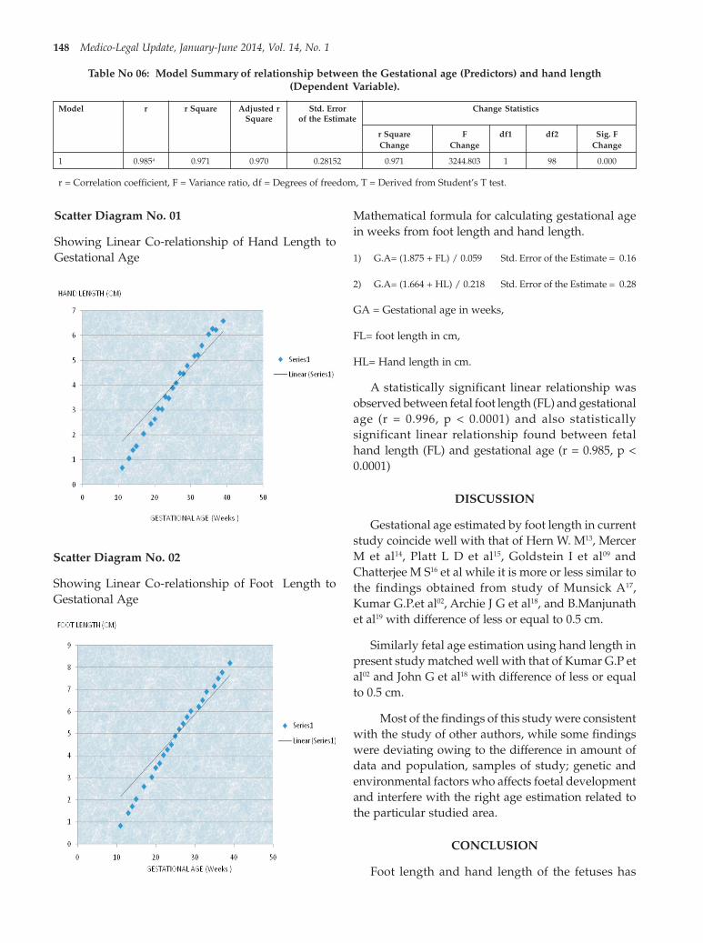
148 Medico-Legal Update, January-June 2014, Vol. 14, No. 1
Table No 06: Model Summary of relationship between the Gestational age (Predictors) and hand length(Dependent Variable).
Model r r Square Adjusted r Std. Error Change StatisticsSquare of the Estimate
r Square F df1 df2 Sig. FChange Change Change
1 0.985a 0.971 0.970 0.28152 0.971 3244.803 1 98 0.000
r = Correlation coefficient, F = Variance ratio, df = Degrees of freedom, T = Derived from Student’s T test.
Scatter Diagram No. 01
Showing Linear Co-relationship of Hand Length toGestational Age
Scatter Diagram No. 02
Showing Linear Co-relationship of Foot Length toGestational Age
Mathematical formula for calculating gestational agein weeks from foot length and hand length.
1) G.A= (1.875 + FL) / 0.059 Std. Error of the Estimate = 0.16
2) G.A= (1.664 + HL) / 0.218 Std. Error of the Estimate = 0.28
GA = Gestational age in weeks,
FL= foot length in cm,
HL= Hand length in cm.
A statistically significant linear relationship wasobserved between fetal foot length (FL) and gestationalage (r = 0.996, p < 0.0001) and also statisticallysignificant linear relationship found between fetalhand length (FL) and gestational age (r = 0.985, p <0.0001)
DISCUSSION
Gestational age estimated by foot length in currentstudy coincide well with that of Hern W. M13, MercerM et al14, Platt L D et al15, Goldstein I et al09 andChatterjee M S16 et al while it is more or less similar tothe findings obtained from study of Munsick A17,Kumar G.P.et al02, Archie J G et al18, and B.Manjunathet al19 with difference of less or equal to 0.5 cm.
Similarly fetal age estimation using hand length inpresent study matched well with that of Kumar G.P etal02 and John G et al18 with difference of less or equalto 0.5 cm.
Most of the findings of this study were consistentwith the study of other authors, while some findingswere deviating owing to the difference in amount ofdata and population, samples of study; genetic andenvironmental factors who affects foetal developmentand interfere with the right age estimation related tothe particular studied area.
CONCLUSION
Foot length and hand length of the fetuses has
35. Sachin Patil--144-150.pmd 2/22/2014, 11:45 AM148
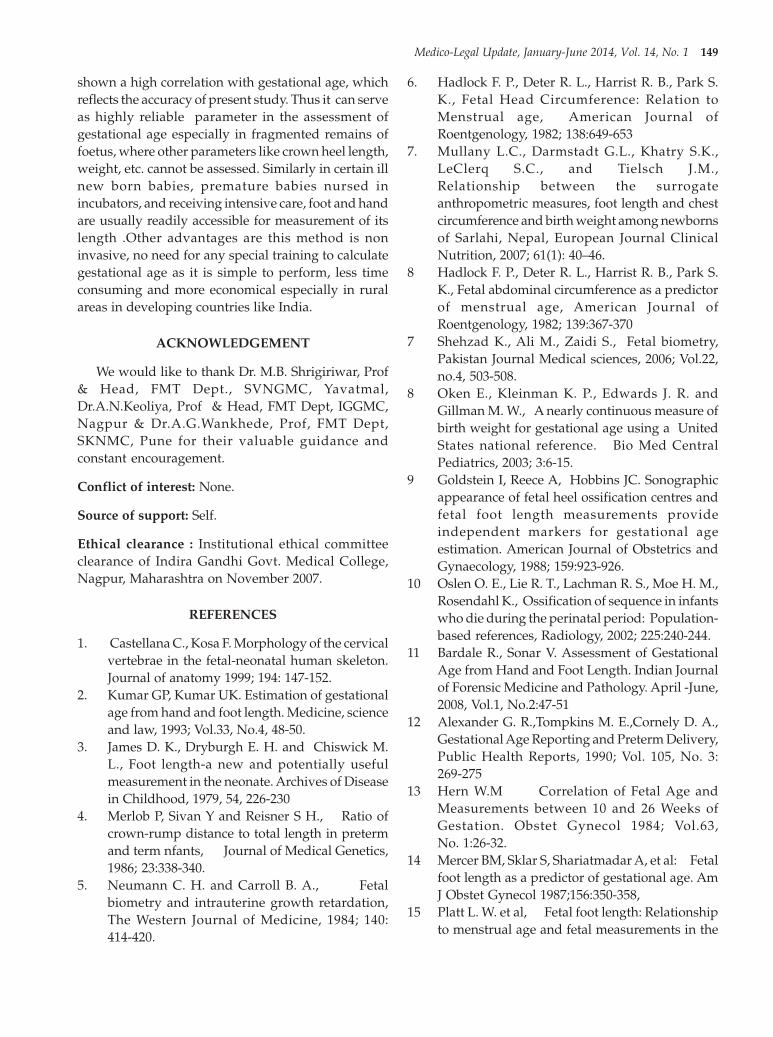
Medico-Legal Update, January-June 2014, Vol. 14, No. 1 149
shown a high correlation with gestational age, whichreflects the accuracy of present study. Thus it can serveas highly reliable parameter in the assessment ofgestational age especially in fragmented remains offoetus, where other parameters like crown heel length,weight, etc. cannot be assessed. Similarly in certain illnew born babies, premature babies nursed inincubators, and receiving intensive care, foot and handare usually readily accessible for measurement of itslength .Other advantages are this method is noninvasive, no need for any special training to calculategestational age as it is simple to perform, less timeconsuming and more economical especially in ruralareas in developing countries like India.
ACKNOWLEDGEMENT
We would like to thank Dr. M.B. Shrigiriwar, Prof& Head, FMT Dept., SVNGMC, Yavatmal,Dr.A.N.Keoliya, Prof & Head, FMT Dept, IGGMC,Nagpur & Dr.A.G.Wankhede, Prof, FMT Dept,SKNMC, Pune for their valuable guidance andconstant encouragement.
Conflict of interest: None.
Source of support: Self.
Ethical clearance : Institutional ethical committeeclearance of Indira Gandhi Govt. Medical College,Nagpur, Maharashtra on November 2007.
REFERENCES
1. Castellana C., Kosa F. Morphology of the cervicalvertebrae in the fetal-neonatal human skeleton.Journal of anatomy 1999; 194: 147-152.
2. Kumar GP, Kumar UK. Estimation of gestationalage from hand and foot length. Medicine, scienceand law, 1993; Vol.33, No.4, 48-50.
3. James D. K., Dryburgh E. H. and Chiswick M.L., Foot length-a new and potentially usefulmeasurement in the neonate. Archives of Diseasein Childhood, 1979, 54, 226-230
4. Merlob P, Sivan Y and Reisner S H., Ratio ofcrown-rump distance to total length in pretermand term nfants, Journal of Medical Genetics,1986; 23:338-340.
5. Neumann C. H. and Carroll B. A., Fetalbiometry and intrauterine growth retardation,The Western Journal of Medicine, 1984; 140:414-420.
6. Hadlock F. P., Deter R. L., Harrist R. B., Park S.K., Fetal Head Circumference: Relation toMenstrual age, American Journal ofRoentgenology, 1982; 138:649-653
7. Mullany L.C., Darmstadt G.L., Khatry S.K.,LeClerq S.C., and Tielsch J.M.,Relationship between the surrogateanthropometric measures, foot length and chestcircumference and birth weight among newbornsof Sarlahi, Nepal, European Journal ClinicalNutrition, 2007; 61(1): 40–46.
8 Hadlock F. P., Deter R. L., Harrist R. B., Park S.K., Fetal abdominal circumference as a predictorof menstrual age, American Journal ofRoentgenology, 1982; 139:367-370
7 Shehzad K., Ali M., Zaidi S., Fetal biometry,Pakistan Journal Medical sciences, 2006; Vol.22,no.4, 503-508.
8 Oken E., Kleinman K. P., Edwards J. R. andGillman M. W., A nearly continuous measure ofbirth weight for gestational age using a UnitedStates national reference. Bio Med CentralPediatrics, 2003; 3:6-15.
9 Goldstein I, Reece A, Hobbins JC. Sonographicappearance of fetal heel ossification centres andfetal foot length measurements provideindependent markers for gestational ageestimation. American Journal of Obstetrics andGynaecology, 1988; 159:923-926.
10 Oslen O. E., Lie R. T., Lachman R. S., Moe H. M.,Rosendahl K., Ossification of sequence in infantswho die during the perinatal period: Population-based references, Radiology, 2002; 225:240-244.
11 Bardale R., Sonar V. Assessment of GestationalAge from Hand and Foot Length. Indian Journalof Forensic Medicine and Pathology. April -June,2008, Vol.1, No.2:47-51
12 Alexander G. R.,Tompkins M. E.,Cornely D. A.,Gestational Age Reporting and Preterm Delivery,Public Health Reports, 1990; Vol. 105, No. 3:269-275
13 Hern W.M Correlation of Fetal Age andMeasurements between 10 and 26 Weeks ofGestation. Obstet Gynecol 1984; Vol.63,No. 1:26-32.
14 Mercer BM, Sklar S, Shariatmadar A, et al: Fetalfoot length as a predictor of gestational age. AmJ Obstet Gynecol 1987;156:350-358,
15 Platt L. W. et al, Fetal foot length: Relationshipto menstrual age and fetal measurements in the
35. Sachin Patil--144-150.pmd 2/22/2014, 11:45 AM149

150 Medico-Legal Update, January-June 2014, Vol. 14, No. 1
second trimester, Obstetric Gynaecology, 1988;71:526-531.
16 Chatterjee M. S., Izquierdo L. A., Nevils B., GilsonG. J., Barada C., Fetal foot: evaluation ofgestational age, Thefetus.net, 1994.
17 Munsick R.A. Human fetal extremity lengths inthe interval from 9 to 21 menstrual weeks ofpregnancy. Am J Obstet Gynecol 1984; 149:883-887.
18 Archie J.G.,Collins J.S, Lebel R.R. QuantitativeStandards for Fetal and Neonatal Autopsy Am JClin Pathol 2006; 126:256-265
19 Manjunath B., Nithin M.D., Sashidharan sameerCross sectional study to determine gestational ageby metrical measurements of foot length.Egyptian Journal of Forensic Sciences 2012; Vol2, No 1, 11-17.
35. Sachin Patil--144-150.pmd 2/22/2014, 11:45 AM150

Medico-Legal Update, January-June 2014, Vol. 14, No. 1 151
INTRODUCTION
Several forensic anthropologists have describedqualitative sex differentiation using many bones, butsexing from single bone is difficult task. Almost all theelements of human skeleton show some degree ofsexual dimorphism, but reliable indicators can beobtained from specific bones like hip bone, skull andsacrum. The determination of deceased sex is first stepin skeletal analysis since estimation of age at death,race, and stature depends on sex of deceased.
The measurements of the skeleton bones, mainlythe neurocranium (brain case) and viscerocranium,suffer influences of various factors; however, they areoften used for human population morphological
Foramena Magnum Dimensions: An Aid in Identificationof Gender
Manjunath T H1, Nagesh C Kuppast2, Umesh S R3, Shradha Iddalgave4, Narasimha Murthy S5,Muralidhar Shepur6
1Assistant Professor, Department of Forensic Medicine and Toxicology, Subbaiah Institute of Medical Science andResearch Center, Shimoga, Karnataka, 2Assistant professor, Department of Forensic Medicine and Toxicology,
S. S. Institute of Medical Sciences and Research Center, Davangere, Karnataka, 3Professor and HOD, Department ofForensic Medicine and Toxicology, M. R. Medical College, Gulbarga, Karnataka, 4Department of Anatomy,
5Assistant Professor, Dept. of Forensic Medicine and Toxicology, 6Assistant Professor, Department of Anatomy,J. J. M. Medical College, Davangere, Karnataka
ABSTRACT
Determination of deceased sex is first step in skeletal analysis since estimation of age, race, andstature depends on sex of deceased. Total 100 adult human skulls (50 male and 50 female) ofHyderabad-Karnataka region are studied. Measurements like foramena magnum length and foramenamagnum breadth were measured. The demarking point (D.P.) and identification point (I.P.) of bothmeasurements was calculated and then percentage of bones identified by D.P. and I.P. was recorded.Though, the Demarking Point of a single parameter may not identify sex in all the bones but theaccuracy is nearly 100% in the bones, which are identified. Percentage of skulls identified by IP andDP of foramena magnum length is more as compared to foramena magnum breadth. So foramenamagnum length gives better prediction of sex as compared to foramena magnum breadth.
Keywords: Foramena magnum length, Foramena magnum breadth, Demarking Point, Identification Point,Sexing of Cranium
Corresponding author:Manjunath T HAssistant ProfessorDepartment of Forensic Medicine and Toxicology,Subbaiah Institute of Medical Science and ResearchCenter, Shimoga, KarnatakaE-mail I. D. [email protected]
studies of age estimation, stature, ethnicity, which arerelevant aspects of forensics investigation andanthropological examinations of unknownindividuals1.
Recent studies report that morphometry is a fastand efficient method for the evaluation ofmorphological characteristics, such as ethnicity,gender, age, genetic factors, dietary habits, and regionalvariations which can alter the shape and size of bonestructures. These aspects are significantly importantin determining the anthropometric changes betweendifferent populations and genders2
Traditional method is non-metrical andmorphological. Morphological features of bonesdepend upon nutrition, occupation, race andgeography of the region, so the traditional method isnot reliable in the study of bones.
In this study length and breadth of foramenamagnum were studied. They were analysedstatistically by applying routine statistical data likeidentification point and demarking point.
DOI Number: 10.5958/j.0974-1283.14.1.036
36. Nagesh Kuppast--151--154.pmd 2/22/2014, 11:45 AM151

152 Medico-Legal Update, January-June 2014, Vol. 14, No. 1
MATERIALS AND METHOD
The materials for the present study consisted of 100adult human skulls of known sex (50 male and 50female) available in the Department of ForensicMedicine and Toxicology and Department of Anatomyof M. R. Medical College and K. B. N. Medical CollegeGulbarga, Karnataka. Following parameters werestudied:
1. Foramena magnum length (FML): Foramenamagnum length is the distance between basion andopisthion. It was recorded with the help of slidingvernier caliper.
2. Foramena magnum breadth (FMB): Foramenamagnum breadth was measured at the broadestpart of foramena magnum with the help of slidingvernier caliper.
As the first part of the study, all the values weretabulated and analyzed statistically by routinestatistical methods.
The value of Range, Mean, Standard Deviation(SD), Calculated Range (mean ± 3SD), DemarkingPoint and Identification Point were obtained.Maximum value of female range is considered asidentification point for male. Minimum value of malerange is considered as identification point for female.Maximum value of female calculated range isconsidered as demarcation point for male. Minimumvalue of male calculated range is considered asdemarcation point for female. In case where femalerange/calculated range maximum value of malerange/calculated range is considered as Identificationpoint/Demarcation point for female and minimumvalue of female range/calculated range is consideredas identification point/demarcation point for male.Subsequently ‘t’ is applied to both parameters.
OBSERVATIONS
The Range, Mean, Calculated Range (mean + 3S.D.), Demarking Points (DP), Identification Point (IP)and the percentage of bones in which sex could beidentified by them, are given in table no. 1 and 2.
Table No 1. Statistical analysis of foramena magnumlength of skull.
Details of measurements Male Female
Mean 33.46 29.62
SD 5.04 2.01
Range 26-43 24-34
Identification point(IP) >34 <26
% of bones identified by IP 38% 06%
Standard error 0.71 0.28
Calculated range 18.33-48.59 23.59-35.65
Demarking point(DP) >35.65 <18.33
% of bones identified by DP 34% 0%
Percentage of skulls identified by IP alone was 38%of males and 06% of females. The percentage of skullsidentified by DP alone was 34% of males and 0% offemales. ‘t’ test was highly significant with p<0.001.
Table No 2. Statistical analysis of foramena magnumbreadth of skull.
Details of measurements Male Female
Mean 29.86 27.64
SD 2.09 2.35
Range 25-36 23-33
Identification point(IP) >33 <25
% of bones identified by IP 02% 08%
Standard error 0.30 0.33
Calculated range 23.6-36.13 20.6-34.7
Demarking point(DP) >34.7 <23.6
% of bones identified by DP 02% 02%Fig. 1. Maximum breadth of foramena magnum
Fig. 2. Maximum length of foramena magnum
36. Nagesh Kuppast--151--154.pmd 2/22/2014, 11:45 AM152

Medico-Legal Update, January-June 2014, Vol. 14, No. 1 153
Percentage of skulls identified by IP alone was 02%of males and 08% of females. The percentage of skullsidentified by DP alone was 02% of males and 02% offemales. ‘t’ test was highly significant with p<0.001.
DISCUSSION
The female skull retains the gracile attributes seenin prepubescent skull. Male cranium becomesmarkedly rougher in adulthood, the differentiatingfeatures of sex become more prominent after puberty,again towards old age there occurs blurring of sexuallydimorphic traits. So the determination of sex frombones should ideally be limited to 15-55 years of age3.Krogman WM4 (1978) analyzed 750 skeletons and came
to a conclusion that the determination of sex is possiblewith accuracy of about 100% if whole skeleton isavailable, 92% when skull alone and 98% when bothpelvis and skull are available. A great number ofmeasurements of the skull have been proposed andused by different investigators during the past. Martinand Saller used eighty one measures; Howell describedseventy; Hrdlicka lists thirty two; Bass gives twentythree.
In the present study both length and breadth offoramena magnum are statistically significant. Theresults are compared with those of previous workersas shown in table no. 3 & 4.
Table No 3. Showing comparison of foramena magnum length with other workers.
Sl Name of worker Male Female SS
N M R SD N M R SD P
1 Keen (1950)5 50 36.3 30-44 2.9 50 34.8 30-40 2.4 -
2 Hong Wei Song 19926 30 34.1 - 2.6 30 32.1 - 2.1 <0.001
3 Deshmukh(2003)7 40 34 26-40 3.09 34 34 30-38 2.05 >0.05
4 Present study (2009) 50 33.46 26.43 5.04 50 29.62 24-34 2.01 <0.001
Table No 4. Showing comparison of foramena magnum breadth with other workers.
Sl Name of worker Male Female SS
N M SD N M SD P
1 Hong Wei Song 1992 30 27.5 2.4 30 25.8 1.8. <0.001
2 Deshmukh(2003) 40 29 1.97 34 28 2.09 >0.05
3 Present study (2009) 50 29.86 2.09 50 27.64 2.35 <0.001
Catalina-Herrera (1987)8 indicated that the sagittaland transverse dimensions of the foramen magnumwere significantly higher in men’s skulls. Zaidi &Dayal (1988)9 classified a sample of Indian skullsaccording to the shape and dimensions of the foramenmagnum, reporting differences between the skulls ofmale and female, which was similar to the findings ofHenríquez-Pino et al. (1995)10 in the Brazilian skulls.
CONCLUSION
Among both parameters statistically analysed,considering the Demarking Point and Identificationpoint, the most reliable parameter is foramenamagnum length as compared to the breadth offoramena magnum. So foramena magnum lengthgives better prediction of sex as compared to thebreadth of foramen magnum.
ACKNOWLEDGEMENT
I thank Dr. Mallikarjun B., dean M. R. MedicalCollege, Gulbarga for encouraging and securing methe all the required facilities for the work.
I express my humble gratitude to all the staffmembers of Dept. Of Forensic Medicine andToxicology, M. R. Medical College, Gulbarga for theirmotivation and constructive ideas during the study.
I thank Mr. Sangam statistician, for his assistanceand positive feedback during statistical analysis.
Source of funding: Self
Ethical clearance: Taken
Conflict of Interest Statement: “The undersignedauthor / authors hereby declare that the article is
36. Nagesh Kuppast--151--154.pmd 2/22/2014, 11:45 AM153

154 Medico-Legal Update, January-June 2014, Vol. 14, No. 1
original, neither the article nor a part of it is underconsideration for publication anywhere else and hasnot been previously published anywhere. We havedeclared all vested interests. We have meticulouslyfollowed the instructions. The article, if published, shallbe the property of the Journal and we surrender allrights to the Editors. We agree to provide the latestfollow up of cases prior to the publication of casereports when requested”.
REFERENCES
1. ISCAN, MY. Global forensic anthropology in the21st century. Forensic Science International . 2001,vol. 117, p. 1-6.
2. HUMPHREY, LT., DEAN, MC. and STRINGER,CB. Morphological variation in great ape andmodern human mandibles. Journal of Anatomy.1999, vol. 195, p. 491-513. (part 4).
3. Pillay V.V – Text book of Forensic Medicine &Toxicology, 14th edition, Paras Publication, PageNo.56 (2004).
4. Krogman WM. The human skeleton in forensicmedicine. Charles C, Thomas, Springfield,Illinois, 1962; USA.
5. Keen JA (1950) “Sex differences in skull”, APJA,8(1), Page No.65-79.
6. Hong Wei Song, Zi, Qing Lin and Jing Tao Jia.Sex diagnosis of Chinese skulls using multiplestepwise discriminant functional analysis.Forensic Science International, 1992;54: 135-140.
7. Deshmukh AG & Deverthi DB comparison ofCranial Sex determination by univariatea andmultivariate analysis, Joint Anatomy Society ofIndia 55 (2), Page No.48-51 ,(2006).
8. Catalina-Herrera, C. J. Study of the anatomicmetric values of the foramen magnum and itsrelation to sex. Acta Anat. (Basel), 130(4):344-7,1987.
9. Zaidi, S. H. & Dayal, S. S. Variations in the shapeof foramen magnum in Indian skulls. Anat. Anz.,167(4):338- 40, 1988.
10. Henríquez-Pino, J.; Cricenti, S. V. & Didio, L. J.A. Morfometría del foramen magno y su relacióncon el tipo de cráneo en individuos brasileños.Rev. Chil. Anat., 13(2):159-64, 1995.
36. Nagesh Kuppast--151--154.pmd 2/22/2014, 11:45 AM154

Medico-Legal Update, January-June 2014, Vol. 14, No. 1 155
INTRODUCTION
Mysteries, particularly the homicidal have oftencreated ripples in the history of Forensic Science,raising so many questions, most important being“Time of death” (or “time since death”)1. This is veryimportant in un-witnessed cases as it shows the trackto the investigators to reach the suspected person andto obligate the innocent ones. Time since death is aquestion almost invariably asked by police officers,sometimes with a touching faith in the accuracy of theestimate 2. Repeated experience teaches theinvestigator to be wary of relying on any singleobservation for estimating the time of death (or“duration of the post mortem interval”), and he wiselyavoids making dogmatic statements based on anisolated observation”3.
Time Since Death Using Physico-Chemical Changes inHuman Dead Body
Ravindra Baliram Deokar1, Sachin Sudarshan Patil1
1Assistant Professor, Department of Forensic Medicine & Toxicology, Seth G.S. Medical College & Kem Hospital,Mumbai, Maharashtra, India
ABSTRACT
Mysteries, particularly the homicidal have often created ripples in the history of Forensic Science,raising so many questions, most important being "Time of death" (or "time since death"). Establishingthe times of an assault and death has a direct bearing on the legal questions of alibi and opportunity.Thephysical changes in the deceased are the important criteria to gives a fair range of time since death.The present study was carried out in Shri. Vasantrao Naik Government Medical College, Yavatmal,Maharashtra, India. The detail postmortem examination of the deceased forwarded for autopsy hadbeen carried out and the postmortem changes were noted. Vitreous humor samples were collectedfrom the eyes of 152 deceased from the autopsies conducted in mortuary at Shri. Vasantrao NaikGovernment Medical College, Yavatmal and analyzed for Magnesium levels. Main objective of thisstudy is to investigate the utility of vitreous magnesium levels and physical changes after death fordetermination of time since death. The approximate time of death can be estimated from physicalchanges after death in human. It is evident that the longer the time interval between death and theexamination of the body the wider will be the limits of probability. The level of Magnesium in thevitreous humour after death is not a reliable criterion for determination of time since death.
Keywords: Time Since Death; Death Changes; Vitreous Humor; Magnesium
Corresponding author:Ravindra Baliram DeokarAssistant ProfessorQuarter No. 5a, Second Floor, Assistant Dean'sBungalow, S. S. Rao Marg, Parel, Mumbai. 400 012Email: [email protected] number: +919869741117
Establishing the times of an assault and death hasa direct bearing on the legal questions of alibi andopportunity. The physical changes in the deceased arethe important criteria to gives a fair range of time sincedeath. Immense work had been carried out todetermine ‘postmortem interval’ from the variousphysical changes4-6 as well as from study of biochemicalchanges 7-14 in different body fluids. Determination ofthe chemical changes in blood, spinal fluid, aqueoushumour and vitreous humour of eye helps the Forensicexpert to ascertain time since death more precisely.
Main objective of this study is to investigate theutility of vitreous magnesium levels and physicalchanges after death for determination of time sincedeath and to observe the correlation of postmortemchanges with changes in vitreous magnesium levelsafter death.
MATERIALS AND METHOD
Materials
The study was carried out in Shri. Vasantrao NaikGovernment Medical College, Yavatmal. The dead
DOI Number: 10.5958/j.0974-1283.14.1.037
37. Ravindra Balimar--155-160.pmd 2/22/2014, 11:45 AM155

156 Medico-Legal Update, January-June 2014, Vol. 14, No. 1
bodies brought to the mortuary at Shri. Vasantrao NaikGovernment Medical College, Yavatmal were used asmaterial for study purpose. The information regardingexact time of death was gathered from police inquestreport, dead body challan, clinical details from hospitalrecords; correlated and checked from relatives, friendsand attendants of the deceased. Cases where exact timeof death was not known were not included in thisstudy. The autopsies conducted in mortuary at Shri.Vasantrao Naik Government Medical College,Yavatmal. Vitreous humor samples were collectedfrom the eyes of 152 deceased from the autopsiesconducted in mortuary at Shri. Vasantrao NaikGovernment Medical College, Yavatmal. After vitreoussample collection detailed postmortem examinationcarried out and the general relevant findings weredocumented in the proforma with details ofpostmortem lividity, rigor mortis, external and internalsign of decomposition. All the information about thedeceased i.e. age, sex, address, cause of death, exacttime of death, temperature recorded. The ambienttemperature ranged 20-35 0C before the samples weretaken.
The vitreous samples drawn from the eyes ofdeceased at the beginning of postmortem examinationof dead body to the mortuary, (only the clear sampleswill be taken, samples having any particulate matterdiscarded). Vitreous humour was collected at autopsyfrom the posterior chamber of the each eye, slowly andgradually avoiding tearing of loose fragments oftissues by needle aspiration through a puncture made5-6 mm away from the limbus (sclero-corneal junction)using 10 ml sterile syringe and 20 gauze needle andpoured in a rubber stoppered vial. The liquid paraffingel was injected in posterior chamber of each eye for
cosmetic purposes.
Biochemical analyses
Presterilized syringe was used and rubberstoppered glass vials washed with deionised doubledistilled water and dried in hot air oven. The samplesafter collection in a rubber stoppered vial was labeledand transported to the biochemistry laboratory foranalysis. The vitreous samples were centrifuged at3000 rpm for 10 minutes; biochemical analysis formagnesium was carried out immediately using aBeckman Coulter LX20 Automated Analyzer.
Statistical methods
The statistical analyses for the data were carriedout using the Statistical Package for Social Sciences(SPSS®) for Windows™ Version 13.0.
RESULTS
The study includes total 152 samples. Out of 152cases, 96 subjects in all were males and 56 were femalesin the age group of 07 to 80 years (Mean ± SD, 35.38years ± 14.37 years) and the known postmorteminterval ranges from1.6 hours to 39.2 hours (Mean ±SD, 13.01± 7.22 hours).
Appearance of Early And Late Death Changes
Out of the 152 subjects, postmortem lividityappeared in patches in 26 cases, well formed in 121cases. It was not fixed in 75 cases, fixed in 72 cases anddispersed in 5 cases. Out of the 152 subjects, rigor notappeared in 05 cases, appeared in 45 cases and wellmarked in 87 cases. It starts disappearing in 12 cases,passed off in 03 cases.
Table No. 1. Time since death using death changes in different subgroups and number of cases in those subgroups.
Sr. Rigor PM lividity Ext.s/o Int.s/o Duration No. ofNo. Mortis (NF/F) decom. decom. (Hours) Cases
0 Not appear In patches (NF) Absent Absent 00 to 03 5
1 Appeared In patches (NF) Absent Absent 01 to 04 14
2 Appeared Well formed (NF) Absent Absent 03 to 08 31
3 Well marked In patches (NF) Absent Absent 03 to 09 7
4 Well marked Well formed (NF) Absent Absent 04 to 09 18
5 Well marked Well formed (F) Absent Absent 08 to 18 51
6 Well marked Well formed (F) Present Absent 12 to 24 6
7 Well marked Well formed (F) Absent Present 12to 24 1
8 Well marked Well formed (F) Present Present 18 to 36 4
9 Start disapp. Well formed (F) Absent Absent 12 to 24 6
10 Start disapp. Well formed (F) Present Absent 18 to 36 0
37. Ravindra Balimar--155-160.pmd 2/22/2014, 11:45 AM156
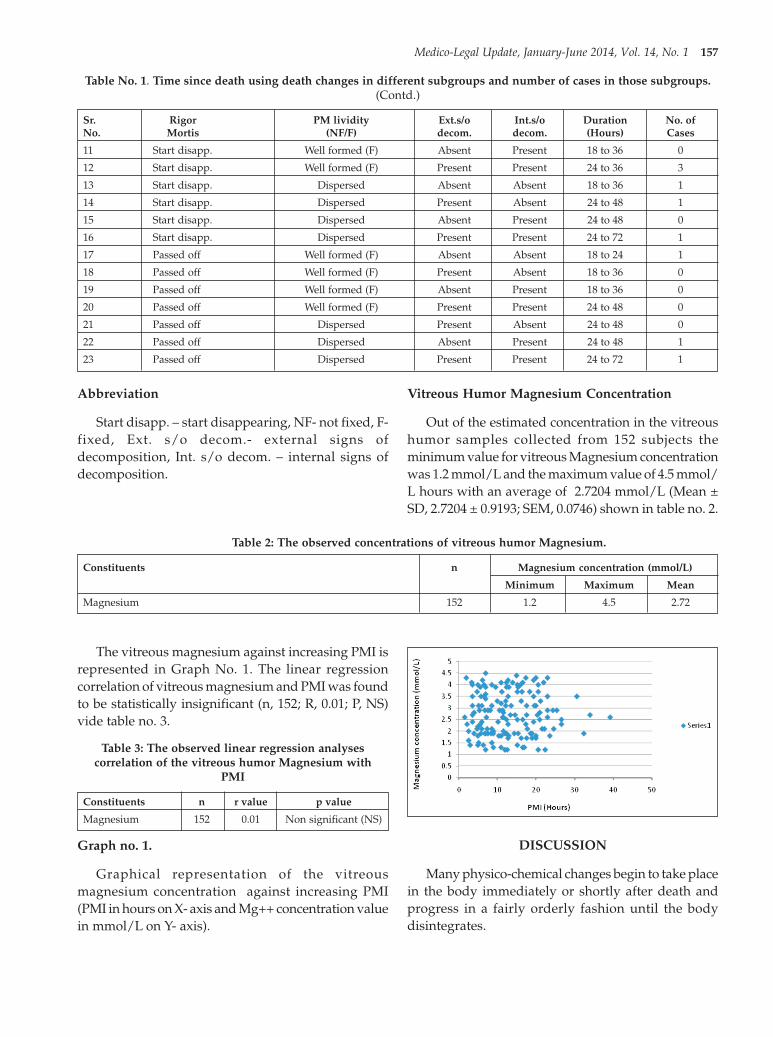
Medico-Legal Update, January-June 2014, Vol. 14, No. 1 157
Table No. 1. Time since death using death changes in different subgroups and number of cases in those subgroups.(Contd.)
Sr. Rigor PM lividity Ext.s/o Int.s/o Duration No. ofNo. Mortis (NF/F) decom. decom. (Hours) Cases
11 Start disapp. Well formed (F) Absent Present 18 to 36 0
12 Start disapp. Well formed (F) Present Present 24 to 36 3
13 Start disapp. Dispersed Absent Absent 18 to 36 1
14 Start disapp. Dispersed Present Absent 24 to 48 1
15 Start disapp. Dispersed Absent Present 24 to 48 0
16 Start disapp. Dispersed Present Present 24 to 72 1
17 Passed off Well formed (F) Absent Absent 18 to 24 1
18 Passed off Well formed (F) Present Absent 18 to 36 0
19 Passed off Well formed (F) Absent Present 18 to 36 0
20 Passed off Well formed (F) Present Present 24 to 48 0
21 Passed off Dispersed Present Absent 24 to 48 0
22 Passed off Dispersed Absent Present 24 to 48 1
23 Passed off Dispersed Present Present 24 to 72 1
Abbreviation
Start disapp. – start disappearing, NF- not fixed, F-fixed, Ext. s/o decom.- external signs ofdecomposition, Int. s/o decom. – internal signs ofdecomposition.
Vitreous Humor Magnesium Concentration
Out of the estimated concentration in the vitreoushumor samples collected from 152 subjects theminimum value for vitreous Magnesium concentrationwas 1.2 mmol/L and the maximum value of 4.5 mmol/L hours with an average of 2.7204 mmol/L (Mean ±SD, 2.7204 ± 0.9193; SEM, 0.0746) shown in table no. 2.
Table 2: The observed concentrations of vitreous humor Magnesium.
Constituents n Magnesium concentration (mmol/L)
Minimum Maximum Mean
Magnesium 152 1.2 4.5 2.72
The vitreous magnesium against increasing PMI isrepresented in Graph No. 1. The linear regressioncorrelation of vitreous magnesium and PMI was foundto be statistically insignificant (n, 152; R, 0.01; P, NS)vide table no. 3.
Table 3: The observed linear regression analysescorrelation of the vitreous humor Magnesium with
PMI
Constituents n r value p value
Magnesium 152 0.01 Non significant (NS)
Graph no. 1.
Graphical representation of the vitreousmagnesium concentration against increasing PMI(PMI in hours on X- axis and Mg++ concentration valuein mmol/L on Y- axis).
DISCUSSION
Many physico-chemical changes begin to take placein the body immediately or shortly after death andprogress in a fairly orderly fashion until the bodydisintegrates.
37. Ravindra Balimar--155-160.pmd 2/22/2014, 11:45 AM157

158 Medico-Legal Update, January-June 2014, Vol. 14, No. 1
Appearance of Death Changes
Rigor mortis is conventionally a postmortemchange. Its occurrence suggests that death has occurredat least a few hours ago 15. In general, rigor appearswithin 01 to 03 hours after death, establishes(wellmarked) in 09 to 12 hours, persists for 12 to 24 hours,start disappearing after 18 to 36 hours and passed offin 24 to 36 hours after death.
PM lividity appears in patches in 01 to 03 hoursafter death, get coalesce within 04 to 06 hours. It is notfixed up to 08 hours after death and get fixed inbetween 08 to 12 hours after death. It gets dispersedin 24 to 48 hours. The external signs of decompositionappear as greenish discoloration in right iliac fossa in18 to 36 hours and become generalized in next 24hours.
Table no.4 (a to d): Appearance of Death Changes (Duration in Hours)
4 a. Rigor Mortis
Rigor appears Established (Well marked) Persists Start disappearing Passed off
01 to 03 09 to 12 12 to 24 18 to 36 24 to 36
4 b. Postmortem Lividity
In patches Patches coalesce Not fixed Fixed Dispersed
01to 03 04 to 06 upto 08 08 to 12 24 to 48
4 c. External signs of decomposition
Greenish discoloration confined to Marbling Blisters
Right iliac fossa Generalized
18 to 36 24 to 48 24 to 48 36 to 72
4 d. Internal signs of decomposition
Intimal discoloration Visceral softening Brain liquefaction
18 to 36 24 to 48 36 to 72
The result of this study are similar with the mostof the available literature 16-21 vide table no. 4. But, eachchange has its own time factor or rate. Unfortunately,these rates of development of post mortem changesare strongly influenced by unpredictable endogenousand environmental factors. Consequently, the longerthe post mortem interval, the wider is the range ofestimate as to when death probably occurred. In otherwords, the longer the post mortem interval, the lessprecise is the estimate of the time of death.
Vitreous Humor Magnesium:
The vitreous magnesium concentrations observedin the present study are very similar to theconcentrations previously reported 9, 22. The presentstudy did not find a significant correlationshipbetween vitreous magnesium and PMI. Out of theestimated concentration in the vitreous humor samplescollected from 152 subjects the minimum value forvitreous Magnesium concentration was 1.2 mmol/Land the maximum value of 4.5 mmol/L hours with anaverage of 2.7204 mmol/L.
The present study results are in variance with theearlier reported utility of vitreous magnesium in PMIpredictions 22, 23. However these reports were based ondata in only particular groups of death like asphyxiaand deaths due to Phenobarbital intoxications. Ourresults are consistent with the earlier findings thatreported no significant correlation between vitreousmagnesium and PMI 24-26 .
CONCLUSIONS
The approximate time of death can be estimatedfrom physical changes after death in human. It isevident that the longer the time interval between deathand the examination of the body the wider will be thelimits of probability. However, even if it givesapproximate and fair idea of the time since death it isalways beneficiary to forensic expert to guide and helpinvestigating authorities in rural India for investigationinto time of death. Also till date there are no othercriteria which completely substitute the usual methodof collecting information on death changes of the deadbody. The level of Magnesium in the vitreous humour
37. Ravindra Balimar--155-160.pmd 2/22/2014, 11:45 AM158

Medico-Legal Update, January-June 2014, Vol. 14, No. 1 159
after death is not a reliable criterion for determinationof time since death.
Conflict of Interests: None.
Source of Funding: Self.
Ethical Clearance: Approved by institutional ethicalcommittee of Shri. V N Govt. Medical College,Yavatmal.
ACKNOWLEDGEMENTS
Special thanks to
Dr. Keoliya Ajay, Prof & head Dept of ForensicMedicine, IGGMC, Nagpur.
Dr. Walter F Vaz, Former Head & Professor, ForensicMedicine, Seth GSMC & KEMH, Mumbai.
Dr. Pathak Harish, Professor & Head, ForensicMedicine, Seth GSMC & KEMH, Mumbai.
Dr. Anil Batra, Professor & Head, Forensic Medicine,GMC, Akola.
Dr. Ambade Vipul, Asso. Professor, Forensic Medicine,GMC Nagpur.
Dr. (Mrs.) Deokar Priya & Miss Dhriti , D.r AmolChavan MS (Optham.), Dr. Vijay Dhanve MD (Pediatrics), Dr.Vrunda & Dr. Meenakshi for being a continuous sourceof encouragement and support.
REFERENCES
1. D. Singh, R. Prashad, S.K. Sharma, A.N. Pandey,Double logarithmic, linear relationship betweenpostmortem vitreous sodium/potassiumelectrolytes concentration ratio and time sincedeath in subjects of Chandigarh zone ofNorthwest India. JIAFM 27 (3) (2005) 159.
2. Knight, legal aspects of medical practice, 4thedition, Churchill Livingstone, Edinburgh (1987)115.
3. Adelson, The Pathology of Homicide, Thomas,Springfield, Illinois (1974) 151.
4. T. Marshall, F. Hoare, Estimating the time of death- the rectal cooling after death and itsmathematical representation, J. Forensic Sci.7(1962) 56-81.
5. G. Dotzauer, Idiomuskuloser Wulst undpostmortale Blutung (German). Z. gericht. Med.46 (1958) 761-771.
6. A. Klein, S. Klein, Die Todeszeitbestimmungammenschlichen Auge, Dresden University, MDThesis, 1978.
7. J.I. Coe, Postmortem chemistry of blood,cerebrospinal fluid and vitreous humour, LegalMedicine Annual wecht CH, ed), New York:Appleton -Century - Crofts, 1976 pp 53-91.
8. W.W. Jetter, Postmortem biochemical changes, J.Forensic Sci. 4 (1959) 330.
9. J.I. Coe, Postmortem Chemistries on vitreoushumour, Am. J. Path. 51 (1969) 741-750.
10. D. Singh, R. Prasad, C. Parkash, Y.S. Bansal, S.K.Sharma, A.N. Pandey, Determination of timesince death from vitreous potassiumconcentration in subjects of Chandigarh zone ofnorth-west India, Int. J.of Med. Toxicol. Leg.Med.4(1) (2001) 12-15.
11. G. Adjutantis, A. Coutselinis, Estimation of thetime of death by potassium levels in the vitreoushumour, Forensic Sci. Int. 1 (1972) 55-60.
12. M. Bray, The eye as a chemical indicator ofenvironmental temperature at the time of death,J. Forensic Sci. 29 (1984) 396-403.
13. S. Komura, S. Oshiro, Potassium levels in theaqueous and vitreous humour after death,Tohoku J. Exp. Med. 122 (1977) 65-68.
14. B.A.W. Balasooriya, C.A. St. Hill, A.R.Williams,The biochemistry of vitreous humour. Acomparative study of the potassium, sodium andurate concentrations in the eyes at identical timeintervals after death, Forensic Sci. Int. 26 (1984)85-91.
15. M. Chakravarthy, Rigor mortis in a live patient,Am J Forensic Med Pathol - 31(1) Mar 2010;87-88.
16. A. Nandy, principles of forensic medicine,Calcutta, New Central Book Agency (P) Ltd, 1996,pp 163-165.
17. Modi, Modi’s Medical Jurisprudence andToxicology, 23rd edition, Postmortem changes andtime since death, Lexis Nexis publishers, 2008,427-451.
18. N. Reddy, Essentials of Forensic Medicine, 32nd
edition, Postmortem changes, 2013, 149-170.19. K. Vij, Textbook of forensic Medicine, 5th edition,
Death and its medicolegal aspect, Elsevier, 2011,74-99.
20. G Biswas, Review of Forensic Medicine andToxicology, 2nd edition, Signs of death, 2012,117-135.
21. Tedeschi, forensic Medicine: study of trauma andenvironmental hazards, Vol II, Postmortem
37. Ravindra Balimar--155-160.pmd 2/22/2014, 11:45 AM159

160 Medico-Legal Update, January-June 2014, Vol. 14, No. 1
chemistry of blood, cerebrospinal fluid andvitreous humour,1033-1060.
22. R Nowak, S Balabanova. Determination ofcalcium and magnesium in postmortem humanvitreous humor as a test to ascertain the causeand time of death. Z Rechtsmed 102 (1989),179-183.
23. S Balabanova, V Gras. Forensic value ofphenobarbital, calcium and magnesiumdetermination in vitreous humor. Arch Kriminol189 (1992) 48-55.
24. Z Gregora, J Kratochvil, J Vavrova,L OPlistil,sodium and Magnesium levels in vitreousbody.Soud Lek 24 (1979) 51-54.
25. M.S. Wheeler, J.D. Butts,P.Hudson, Vitreoushumour magnesium in alcoholics. Am J forensicMed Pathol 4(1983) 105-110.
26. J.G. Farmer, F. Benomran, A.A Watson,W.AHarland, Magnesium,Potassium,Sodium andcalcium in postmortem vitreous humour fromhumans.Forensic sci Int 27 (1985) 1-13.
37. Ravindra Balimar--155-160.pmd 2/22/2014, 11:45 AM160

Medico-Legal Update, January-June 2014, Vol. 14, No. 1 161
INTRODUCTION
Palatal rugae, also called plicae palatinaetransversae and rugae palatina refer to the ridges onthe anterior part of the palatal mucosa, each side ofthe median palatal raphe and behind the incisivepapilla. As an entity they form the rugae pattern.Anatomically, the rugae consist of around 3 to 7 rigidand oblique ridges that radiate out tangentially fromthe incisive papilla.1
Palatal rugae have been shown to be highlyindividual and consistent in shape throughout the life.It is well- established fact that the palatal rugae patternis unique to human, as his fingerprints.2 Palatal rugaeare used in human identification not only due to theirsingularity and unchangeable nature, but also due toother advantages, namely their low utilization costs.3
A Study on Identification of Palatal Rugae Pattern amongMales and Females of Lucknow City
Renuka Verma1, Ridhi Narang2, Anil Singh3, Rohit Jaiswal4
1Senior Lecturer, Department of Oral Pathology and Microbiology, Career Post Graduate Institute of Dental Sciencesand Hospital, Lucknow, UP, India, 2Senior Lecturer, Department of Public Health Dentistry, Surendera Dental Collegeand Research Institute, Sri Ganganagar, Rajasthan, India, 3Professor & Head, 4Reader, Department of Oral Pathology
and Microbiology, Sardar Patel Postgraduate Institute of Dental & Medical Sciences, Lucknow. U.P
ABSTRACT
Palatoscopy, or palatal rugoscopy, is the name given to the study of palatal rugae in order to establisha person's identity. Palatal rugae have been shown to be highly individual and consistent in shapethroughout the life. It is well established fact that the palatal rugae pattern is unique to human, as hisfingerprints. Rugae patterns have been studied for various purposes, mainly in the fields ofanthropology, comparative anatomy, genetics, forensic odontology, prosthodontics and orthodontics.
Keywords: Palatal Rugae, Human Identification, Forensic Odontology, Palatoscopy
Corresponding author:Ridhi NarangSenior LecturerDepartment of Public Health Dentistry,Surendera Dental College and Research Institute,Sri Ganganagar, Rajasthan, IndiaMobile No.: 08947933115, 09915387019Email id- [email protected]
Once formed, they do not undergo any changesexcept in length, due to normal growth, remaining inthe same position throughout an entire person’s life.Not even diseases, chemical aggression or traumaseem to change palatal rugae form.4 It is also seen thatchanges that occurs from orthodontic movements,extraction, aging and palatal expansion do not modifythe rugae enough to hamper identification.4
Classifications of Palatal Rugae:
Thomas palatal rugae classification 5
Classification Rugae type
1 Diverge
2 Converge
3 Curve
4 Wavy
5 Straight
6 Circular
Da Silva simple palatal rugae classification1
Classification Rugae type
1 Line
2 Curve
3 Angle
4 Circle
5 Wavy
6 Point
DOI Number: 10.5958/j.0974-1283.14.1.038
38. Ridhi Narang--161--164.pmd 2/22/2014, 11:45 AM161

162 Medico-Legal Update, January-June 2014, Vol. 14, No. 1
Trobo palatal rugae classification1
Classification Rugae type
Type A Point
Type B Line
Type C Curve
Type D Angle
Type E Sinuous
Type F Circle
Carrea palatal rugae classification1
Classification Rugae type
Type I Posterior-anterior directed rugae
Type II Rugae perpendicular to the raphae
Type III Anterior-posterior directed rugae
Type IV Rugae directed in several directions
AIM
The aim of this study is to identify and comparethe rugae pattern in males and females of Lucknowcity.
MATERIAL AND METHOD
Total number of 60 subjects, 30 males and 30females were selected. All subjects were healthyindividuals free of congenital abnormalities,
inflammation and trauma. Irreversible hydrocolloidimpression material was used along with appropriateperforated impression tray for the maxillary arch.Theimpression was then poured into dental stone. Carewas taken that all casts must be free of air bubbles orvoids especially at the anterior third of the palate.
The method of rugae identification was based onthe classification of Thomas et al.(1983). Thisclassification includes number, shape and size. Theshapes are classified into curved, wavy, straight andcircular. Fragmented rugae are those which havelength less than 5 mm. All the identification andmeasurements were done by one examiner and thereadings were repeated three times for each cast. Theintraexaminer reliability coefficient was found out tobe 0.84. Fragmented type of rugae of size less than 5mmwere ignored. Calcorrugoscopy was placed on the castand the measurement in mm was done using a metallicruler & divider.
RESULTS
The present study comprised on 60 subjects ofwhom 30 were males and 30 were females. Nosignificant difference was found among genderaccording to the number of rugae.
Table 1: Comparison of size of rugae in two genders
Sex Less Than 5-10 mm More Than Total No.5 Mm 10 Mm of Patterns
No. % No. % No. %
MALE 16 5.7 138 49.1 127 45.2 281
FEMALE 30 11.0 116 42.6 126 46.3 272
χ2 5.159 2.325 0.071
p 0.023 0.127 0.790
Table 1: This table shows gender size distributionof subjects classified according to the size of the rugae.Females had significantly more rugae of size less than
5mm as compared to males (p=0.023). No significantdifference among gender was observed for rugae sizeof 5-10mm or more than 10mm.
Table 2: Comparison of different shapes of Rugae in two genders
S.N0. Shape MALE (N=281) FEMALE (n=272) χχχχχ2 p
No. % No. %
1 Converge 10 3.56 16 5.88 1.666 0.197
2 Diverge 13 4.63 16 5.88 0.439 0.508
3 Curve 87 30.96 69 25.37 2.135 0.144
4 Wavy 89 31.67 112 41.18 5.396 0.020
5 Straight 76 27.05 58 21.32 2.465 0.116
6 Circular 6 2.14 1 0.37 3.455 0.043
38. Ridhi Narang--161--164.pmd 2/22/2014, 11:45 AM162

Medico-Legal Update, January-June 2014, Vol. 14, No. 1 163
Table 2: This table shows gender wise distributionof subjects based upon different shapes of rugaepattern. Wavy pattern was found to be more in femalesas compared to males and the difference wasstatistically significant(p=0.020) whereas circularpattern was more in males as compared to females andthe difference was again statistically significant(p=0.043).
DISCUSSION
The anatomical position of the palatal rugae insidethe oral cavity is surrounded by cheek, lips, tongue,teeth and buccal pad of fat. Rugae are amongst thebest protected, morphologically individualizing softtissue structure in the body, which are preserved afterdeath and also accessible during life.6
The earlier references to the palatal rugae occur invarious works on general anatomy. Winslow(1732)seems to have been the first to describe them in ananatomy text. Palatal rugae was first illustrated bySantorin in 1775 in Tabula VII of his septemdicemTabulae, a drawing depicting three continuous wavylines which cross the midline of the palate. Palatalrugoscopy was first proposed in 1932, by a spanishinvestigator called Trobo Hermosa.7,8,9
There are several ways to analyse palatal rugae.Intraoral inspection is probably the most used and alsothe easiest and the cheapest. However, it can createdifficulties if a future comparative exam is required.10
A more detailed and exact study, as well as the needto preserve evidence may justify oral photography ororal impression.11
Calcorrugoscopy, or the overlay print of palatal rugaein a maxillary cast, can be used in order to performcomparative analysis. By using stereoscopy, forexample, one can obtain a three dimensional image ofpalatal rugae anatomy. It is based on the analysis oftwo pictures taken with the same camera, from twodifferent points, using special equipment. Anothertechnique is the sterophotogrammetry which, by usinga special device called Traster Marker, allow for anaccurate determination of the length and position ofevery single palatal ruga. However, due to itssimplicity, price and reliability, the study of maxillarydental casts is the most used technique.1
The method used in this study (Thomas et al, 1983)was found to be the most practical and easiest to applycompared with other methods.
In the present study no significant difference wasfound among males and females according to thenumber of rugae. Similar results were obtained byKapali et al. (1997)12 whereas according to Dohke Mand Osato S (1994) females had fewer rugae than malesand contrastingly Palatinas et al. (2009)13 found thatnumber is slightly higher in males than females.
In the present study Wavy and Curve form are mostcommon and straight and Circular type is leastcommon. Wavy pattern was found to be more infemales and circular pattern more in males and thedifference was statistically significant. According toFahmi et al. (2001)5 there was no significant differencein the number & size of rugae seen in converge type(females) and circular type (males).
CONCLUSION
Palatal rugae appear to possess the features of idealforensic identification parameters, i.e. uniqueness,postmortem resistance, & stability. Thus, in certainsituations, if teeth are lost due to any reason, the mostcommon of which is trauma, then the use of humanpalatal rugae can be suggested as an alternativemethod for identification.
Support: None
Conflicts of Interest: None
Acknowledgement: None
Ethical Clearance: Institutional Ethical Committee.
REFERENCES
1. Hemanth M, Vidya M, Prasad N, Karkera BV.Palatoscopy: A review. Indian Journal of ForensicMedicine Toxicity (2010); 4(2): 59-64.
2. Thomas CJ, van Wyk CW. The palatal rugae inidentification. J. Forensic Odontostomatol (1988);6(1): 21-25.
3. Caldas IM, Magalha T, Afonso A. Establishingidentity using cheiloscopy and palatoscopy.Forensic science international(2007); 165: 1-9.
4. Stuarts LS, Leonard G. Forensic application ofpalatal rugae in dental identification. The forensicexaminer spring(2005); 44-47.
5. Fahmi FM, Al-Shamrani SM, Talic YF. Rugaepattern in a saudi population sample of malesand females. Saudi dental journal(2001); 13(2):92-95.
38. Ridhi Narang--161--164.pmd 2/22/2014, 11:45 AM163

164 Medico-Legal Update, January-June 2014, Vol. 14, No. 1
6. Limson KS and Julian R. Computerized recordingof the palatal rugae pattern and an evaluation ofits application in forensic identification. J.ForensicOdontostomatol (2004); 22(1): 1-4.
7. Thomas CJ and Kotze T. The palatal rugae: a newclassification. Journal of the dental association ofsouth africa (1983); 38: 153-157.
8. Pueya VM, Garrido BR, Sancchez JAS.Odontology a legaly forense, Masson,Barcelona(1994); 277-292.
9. Campos ML, Rugoscopia palatina. http//www.periciasforenses.com.br. accessed on7/9/12
10. Lysell L.Plicae Palatinae transverse and palillaincisiva in man: a morphologic and genetic study.
Acta Odontologia Scandinavica(1955); 13(18):112-116.
11. Utsuno H, Kanoh TO, Inoue K. Preliminary studyof postmortem identification using lip prints.Forensic Sci. Int (2005); 149:129-132.
12. Kapali S, Townsend G, Richards L, Parish T.Palatal rugae patterns in Australian Aboriginesand Caucasians. Australian Dental Journal (1997);42(2):129-133.
13. Veneges VH, Valenzuela JSP, Lopez MC,Galdames ICS. Palatal Rugae: Systematic analysisof its shape and dimensions for use in humanidentification. Int J. Morphol.(2009); 27(3):819-825.
38. Ridhi Narang--161--164.pmd 2/22/2014, 11:45 AM164

Medico-Legal Update, January-June 2014, Vol. 14, No. 1 165
INTRODUCTION
Witchcraft human practices are not new to thepresent day society, various literatures have confirmedthe same and they are prevalent mainly in theunderdeveloped and developing society especiallyamong uneducated rural class. In a society, inhumanactivities like self-torture or injuring human beings tothe extent of sacrificing your own loved ones arecarried out in a belief of ending their misfortunes andmishappenings. Witchcraft is a practice of magicalfaculties on human for their misfortunes1.
Similarly various forms of rituals are followed toget rid of evil possessed body. These rituals sometimesinvolve gruesome acts to the extent of killing theperson during those torturous activities. Here wepresent a case of such brutality, which was referred toour institute for a specialized treatment, but finallydied due to head injury.
Superstitious Belief Leading to a Torturous Killing: ACase Study
Bansal Y S1, Sunil S2, Senthil K R2, Kumaran M2
1Additional Professor, 2Junior Resident, Department of Forensic Medicine, PGIMER, Chandigarh
ABSTRACT
Worldwide weird religious rituals are still prevalent in almost all the societies. The various forms ofapplication of this magical spells for the so called benefit of humans have evolved according to thecultural context. Black Magic, Paganism, Maleficium, witchcraft and superpower are various suchevolution of practicing magic. A case of such inhuman torture with such belief from the state of UttarPradesh in India is discussed, where a 14 years young girl was allegedly beaten to death by her ownfather on the pretext of being possessed by some evil power. The postmortem examination findingswere analyzed with respect to the history provided.
Keywords: Rituals, Witchcraft, Evil Power, Torture
Corresponding author:Y S BansalAdditional ProfessorDepartment of Forensic MedicinePostgraduate Institute of Medical Education &Research, Chandigarh-160012E-mail: [email protected] no : + 91-172- 2755209 (O)
CLINICAL HISTORY
A young girl of about 14 years of age was broughtto the PGIMER emergency trauma center by herneighbors with multiple injuries all over her body. Asit was informed by the investigating officer and therelatives, the deceased was suffering from some viraleruptions for the past few days. Due to some religiousbelief, her father suspected the same to be the result ofsome evil power. She was tortured by her father,inflicting sharp edged injuries with a knife and blunttrauma with a wooden plank. She was rescued by herneighbors and brought to the hospital, where shesuccumbed to the injuries. Peculiar pattern of theinjuries were corroborating with the alleged suspicionof torturing her with some religious belief.
Autopsy Findings
Deceased was 145cm in length and weighed 42.7kg. 16 injuries were caused by blunt weapon and oneby sharp weapon was present on the body of thedeceased.
A clean cut incised wound involving nostrils withnasal bone cut was present. Seven lacerated woundsof varying sizes were present over the face. Twolacerated wounds with contused margins were presenton the scalp. Reddish blue contusions of varying sizeswere noted around both ears, both arms and left thigh.
DOI Number: 10.5958/j.0974-1283.14.1.039
39. Yogender Bansal --165--168.pmd 2/22/2014, 11:45 AM165

166 Medico-Legal Update, January-June 2014, Vol. 14, No. 1
Reddish abrasions of various sizes and shapes werenoted over right arm and over upper part of abdomen.
On internal examination, skull showed coronal andsagittal sutural separation. A thick layer of subduralhemorrhage was noticed over left cerebral lobes.Patchy subarachnoid hemorrhage was present all over
the cerebral lobes. Multiple petechial hemorrhageswere present in the parenchyma of both cerebral andcerebellar lobes. Brain was edematous, with gyriflattened and sulci narrowed. Unci and tonsils weregrooved. Right lobe of liver showed two superficiallacerations. The cause of death was given as Craniocerebral damage consequent to blunt trauma to head.
Fig 1: Triad of Injuries involving face and scalp Fig 2: Incised wound involving nose
DISCUSSION
Wounds that are caused with intension of homicideare usually single, deep stab wound, mainly seen overchest, neck, head and neck2. Features of self-inflictedwounds usually involve accessible body parts;particularly the neck, wrists and chest wall and followthe contours of the body. Injured parts are ‘lesssensitive’ i.e. rarely involving lips, ears. Tentative orhesitation marks are highly characteristic of self-inflicted wounds3.
Wounds that are inflicted during torture tend to bedeliberately delivered to cause pain and humiliationrather than incapacitation, and are often found on theface and they may be associated with more damaginginjuries, such as blunt force trauma to the head4.
In this case on analyzing the wound pattern andtheir region, most of the wounds were clustered inaround the face of the deceased. The incised woundover the nose indicates most likely to be caused with apurpose of humiliating the victim. The laceratedwounds over the scalp were the fatal injuries. Suchrituals do not primarily intend to kill a person, butjust to inflict pain on the possessed body, so that theevil spirit possessing the body will free it. The bruisesand contusions present over the arms would have been
mostly happened when the victim is forcefullyrestrained. All these features suggest the torturousnature of the wounds.
The belief in and the practice of magic has beenpresent since the earliest human cultures and continuesto have an important religious and medicinal role inmany cultures today1. Belief in and practice ofwitchcraft in Europe can be traced to classical antiquityand has continuous history during the Middle Ages,culminating in the Early Modern witch hunts andgiving rise to the fairy tale5. The belief in possessionby jinn, witchcraft, and the effects of the “evil eye” aremainstream Islamic beliefs. According to Islamicscholars, witches must be executed without hesitation6.
In Indian history, the Iron Age Atharvaveda is acollection of charms and spells classically associatedwith witchcraft, with purposes such as harming anenemy or winning a sweetheart. Belief in thesupernatural is strong in certain parts of India, andcounter killing for witchcraft are reported from timeto time7.
In remote parts of India when a woman is accusedof practicing witchcraft, she is forced to undergo a‘pareeksha’ (test). They will take the accused womento a deserted location and will force her to pick a silver
39. Yogender Bansal --165--168.pmd 2/22/2014, 11:45 AM166
Medico-Legal Update, January-June 2014, Vol. 14, No. 1 167
coin from a vessel containing boiling oil8. InJamshedpur, a group of villagers attacked a womanand beheaded her for practicing black magic9.
In a poor district of Chhattisgarh state, 250 milesfrom the capital Raipur 50 women were attacked withsticks and cut off their hair after accusing them ofwitchcraft. In Chhattisgarh, more than 100 women aretortured, paraded naked or harassed in the stateannually as they are accused of practicing witchcraftand Dozens of women are killed every year onsuspicion of being witches or witch doctors inIndia10.The state passed a Witchcraft (Prevention) Actin 2005 to counter a rise in witch hunts. Offenders facejail terms of up to five years10. There were 160 suchcases reported in Chhattisgarh in 200711.
5 Muslim widows are dragged out of their huts bya mob, then taken to a playground, stripped naked,beaten and forced to eat human excrement in front ofhundreds of villagers after being labeled as witches12.In 2 separate incidents, male villagers suspected to beinvolved in sorcery are attacked. One of them is beatento death, while the other is hospitalized from injurieshe gained from a group of villagers13. A 12-year-oldboy was brutally murdered allegedly by hisstepmother in Saharanpur district and the investigatorshad established the murder as a case of humansacrifice14. The present case is also from the samedistrict in UP.
The practice of witchcraft and counter killing ofwitches are highly associated with lack of education,lack of parental care while growing up, deep rootedcultural beliefs and use of undesirable addictive agentslike alcohol, tobacco and drugs15. Poverty,unemployment and illiteracy were common factors inall these states of India with high prevalence of suchpractices.
CONCLUSION
Though these types of cases are prevalent in all thesocieties, they pose a great difficulty for the lawadministrators as well as to the forensic personalsdealing with such cases. They are legally categorizedunder homicides or murders, though defendants takethe plea otherwise. Documentation and studiesinvolving such unique cases of inhuman acts definitelyincreases the confidence of a forensic pathologist topick up such torture wound patterns of witchcraft. Inorder to curb such social evils in the name of religious
beliefs, more strict laws should be formulated andimplemented with vigilance. A multimodal approachinvolving religious bodies, law enforcing agents,medical and social reformers has to be formulated toovercome this inhuman practice.
Authors contributions:
Dr Y S Bansal: Substantial contributions to conceptionand design, Analysis and interpretation of data,Drafting the article, Revising it critically for importantintellectual content, Final approval of the version tobe published.
Dr Sunil S: Substantial contributions to conceptionand design, Drafting the article, Final approval of theversion to be published.
Dr Senthil K R: Substantial contributions toconception and design, Drafting the article, Finalapproval of the version to be published.
Kumaran M: Substantial contributions to conceptionand design, Drafting the article, Final approval of theversion to be published.
Guarantor: The corresponding author is the guarantorof submission.
Ethical issues: No Ethical issues involved.
Conflict of Interest: Authors declare no conflict ofinterest.
Source of Funding: No funding involved.
ACKNOWLEDGEMENT
We are thankful to the technical staff andphotographer of the Department forensic medicineand toxicology, PGIMER, Chandigarh.
REFERENCES
1. Witchcraft. https://en.wikipedia.org/wiki/Witchcraft (accessed 13Aug 2013)
2. Karlsson T. Homicidal and suicidal sharp forcefatalities in Stockholm, Sweden. Orientation ofentrance wounds in stabs gives information inthe classification. Forensic Sci Int 1998; 93(1): 21-32.
3. Karger B, Niemeyer J, Brinkmann B. Suicides bysharp force: typical and atypical features. Int JLegal Med 2000; 113:259-62.
39. Yogender Bansal --165--168.pmd 2/22/2014, 11:45 AM167

168 Medico-Legal Update, January-June 2014, Vol. 14, No. 1
4. Purdue B.N. Cutting and piercing wounds. Thepathology of trauma; 2000
5. European Witchcraft. http://en.wikipedia.org/wiki/European_witchcraft (accessed 13Aug2013)
6. Islamic Witch Hunt. http://wikiislam.net/wiki/Islamic_Witch_Hunts#India (accessed 13Aug2013)
7. Asian Witchcraft. https://en.wikipedia.org/wiki/Asian_witchcraft (accessed 13Aug 2013)
8. Jaipur woman thrashed for witchcraft. The Timesof India. 08 October 2008. http://articles.timesofindia.indiatimes.com/2008-10-10/jaipur/27945236_1_tribal-woman-silver-coin-village (accessed 13Aug 2013)
9. Tribal beheaded for practicing witchcraft. TheTimes of India. 14 November 2003. http://articles.timesofindia.indiatimes.com/2003-11-14/patna/27211115_1_witchcraft-tribal-woman-black-magic(accessed 13Aug 2013)
10. Fifty ‘Witches’ Beaten By Mob. Sky News. 22December 2008. http://news.sky.com/story/657468/fifty-witches-beaten-by-mob(accessed13Aug 2013)
11. Indian villagers ‘killed witch’. BBC News. 27March 2008. http://news.bbc.co.uk/2/hi/south_asia/7317378.stm(accessed 13Aug 2013)
12. Village ‘witches’ beaten in India. BBC News. 20October 2009. http://www.webcitation.org/query?url=http://news.bbc.co.uk/1/hi/world/south_asia/8315980.stm&date=2011-04-18(accessed 13Aug 2013)
13. 1 killed in attack on ‘sorcerers’ Deccan Chronicle.14 August 2011. http://www.webcitation.org/query?url=http://www.deccanchronicle.com/channels/cities/hyderabad/1-killed-attack-% E 2 % 8 0 % 9 8 s o r c e r e r s % E 2 % 8 0 % 9 9 -667&date=2011-08-18(accessed 13Aug 2013)
14. Provide details, actions taken in ‘human sacrifice’case: UPHRC. The Times of India News. 04 March2011. http://articles.timesofindia.indiatimes.com/2011-03-04/lucknow/28657825_1_human-sacrifice-case-saharanpur-suo-motu-cognizance(accessed13Aug 2013)
15. Meel BL. Witchcraft in Transkei Region of SouthAfrican: case report. African Health Science. 2009;9(1):61-64
39. Yogender Bansal --165--168.pmd 2/22/2014, 11:45 AM168
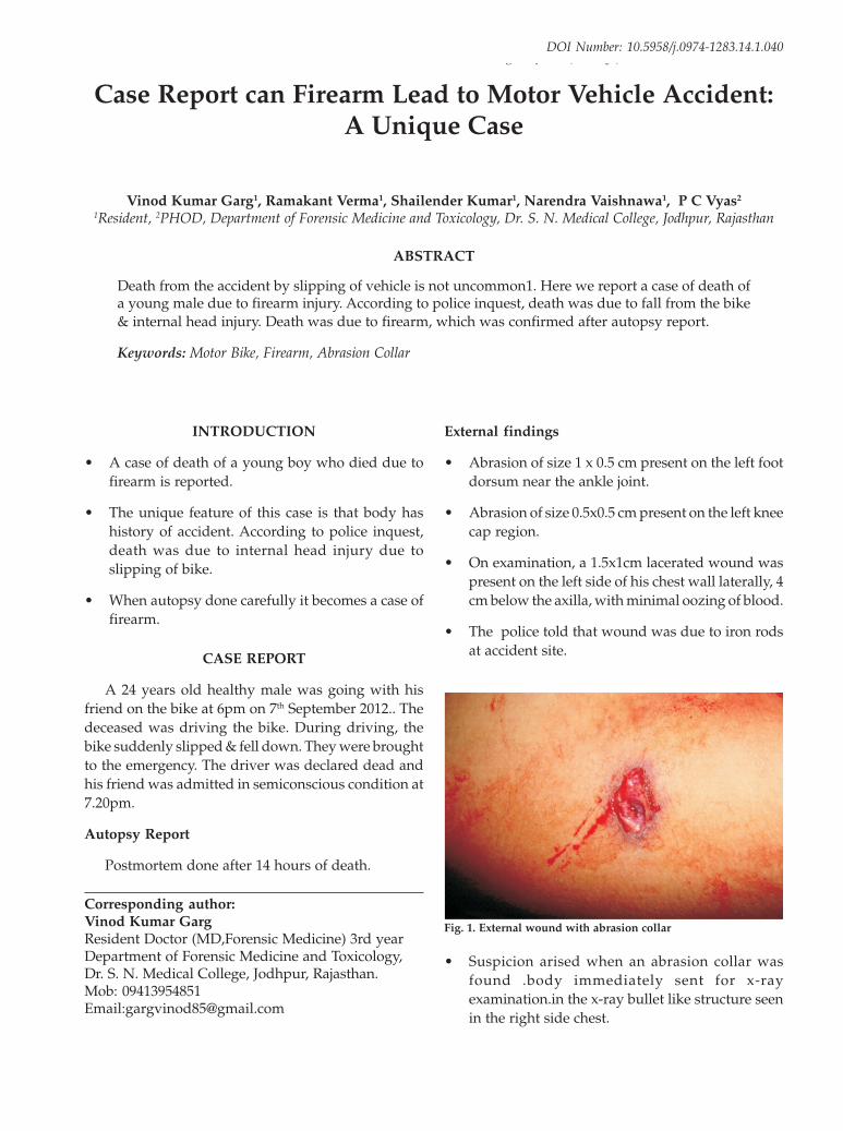
Medico-Legal Update, January-June 2014, Vol. 14, No. 1 169
INTRODUCTION
• A case of death of a young boy who died due tofirearm is reported.
• The unique feature of this case is that body hashistory of accident. According to police inquest,death was due to internal head injury due toslipping of bike.
• When autopsy done carefully it becomes a case offirearm.
CASE REPORT
A 24 years old healthy male was going with hisfriend on the bike at 6pm on 7th September 2012.. Thedeceased was driving the bike. During driving, thebike suddenly slipped & fell down. They were broughtto the emergency. The driver was declared dead andhis friend was admitted in semiconscious condition at7.20pm.
Autopsy Report
Postmortem done after 14 hours of death.
Case Report can Firearm Lead to Motor Vehicle Accident:A Unique Case
Vinod Kumar Garg1, Ramakant Verma1, Shailender Kumar1, Narendra Vaishnawa1, P C Vyas2
1Resident, 2PHOD, Department of Forensic Medicine and Toxicology, Dr. S. N. Medical College, Jodhpur, Rajasthan
ABSTRACT
Death from the accident by slipping of vehicle is not uncommon1. Here we report a case of death ofa young male due to firearm injury. According to police inquest, death was due to fall from the bike& internal head injury. Death was due to firearm, which was confirmed after autopsy report.
Keywords: Motor Bike, Firearm, Abrasion Collar
Corresponding author:Vinod Kumar GargResident Doctor (MD,Forensic Medicine) 3rd yearDepartment of Forensic Medicine and Toxicology,Dr. S. N. Medical College, Jodhpur, Rajasthan.Mob: 09413954851Email:[email protected]
External findings
• Abrasion of size 1 x 0.5 cm present on the left footdorsum near the ankle joint.
• Abrasion of size 0.5x0.5 cm present on the left kneecap region.
• On examination, a 1.5x1cm lacerated wound waspresent on the left side of his chest wall laterally, 4cm below the axilla, with minimal oozing of blood.
• The police told that wound was due to iron rodsat accident site.
Fig. 1. External wound with abrasion collar
• Suspicion arised when an abrasion collar wasfound .body immediately sent for x-rayexamination.in the x-ray bullet like structure seenin the right side chest.
DOI Number: 10.5958/j.0974-1283.14.1.040
40. Vinod garg--169--171.pmd 2/22/2014, 11:45 AM169

170 Medico-Legal Update, January-June 2014, Vol. 14, No. 1
Internal findings
1. Scalp, cranium& abdomen- healthy
2. Thorax-
• Left side pleural cavity- 1.5litre blood
• Right side pleural cavity- 500 ml blood
• Inner side of external lacerated wound in the leftside chest plugged with partially clotted blood &muscle tags.
• Ribs & heart-healthy
Left lung-
• Had an entry wound & exit wound withsurrounding contused area in the lower lobe.
Right lung-
• Had an entry wound & exit wound withsurrounding contused area in the middle lobe.
Fig. 2. x-ray-showing bullet in the right side chest
Track of Bullet- Final opinion-
The cause of death was shock and hemorrhage as aresult of injury to both lungs due to firearm injury .Bullet in a sealed container handed over to theaccompanied police person for ballistic examination.
DISCUSSION
• Bleeding from the wound was minimum likesimple lacerated wound.
• The present case highlights the importance of theautopsy surgeon because simple accident casechanged into the murder case.
• Interestingly no major injuries were present evenfall from the bike.
Left lung Right lung
40. Vinod garg--169--171.pmd 2/22/2014, 11:45 AM170

Medico-Legal Update, January-June 2014, Vol. 14, No. 1 171
• Increased control over procurement andpossession of guns may help to limit suchfatalities.2,3
CONCLUSION
• Road traffic accidents are a common mode ofunnatural death.
• In the present case, the history was very dissimilarwith the postmortem report. Sometimes police alsowant to modify the case.
• But the forensic doctors should keep alert on theall facts during postmortem examination, becauseany accident case can be a murder.
ACKNOWLEDGEMENT
Dr. Arvind Mathur, Principal & Controller, Dr. S.N. Medical College, Jodhpur,
Dr. Jagdish Jugtawat, Department of ForensicMedicine and Toxicology, Jodhpur
Conflict of Interest: Nil
Source of Funding: None
Ethical Clearance: This case report not disclosing theidentity of victim & no active intervention was donein the live object. So we think that ethical clearancenot required.
REFERENCES
1. Cantor CH, Lewin T. Firearm and Suicides inAustralia. Aust. NZJ Psychiatry, 1990; 24:500-509
2. Hardt-Madsen M, Simonsen J. Firearms fatalitiesin Denmark 1970-79. Forensic Sci Int,1983; 23:93-98.
3. Thomsen JL, Albrektsen SB. Pattern of firearmsfatalities before and after the introduction of newlegislation in Denmark. Med Sci Law, 1991;31:162-166.
40. Vinod garg--169--171.pmd 2/22/2014, 11:45 AM171

172 Medico-Legal Update, January-June 2014, Vol. 14, No. 1
INTRODUCTION
Coronary artery disease is a major cause of humanmortality and morbidity the world over. Zeek(1) hadalready concluded in 1930 that arteriosclerosis canoccur at any age. Atherosclerosis begins very early,progressing slowly in a silent manner untilapproximately the fifth or sixth decade, whencomplications with clinical and pathological damagestart.
Traditionally, fatty streaks have been considered tobe the earliest manifestations of atherosclerosis,because they represent the earliest macroscopic lipid-containing lesion. They consist of extracellular lipidswith scattered macrophages(2)
In the live patients coronary angiography is the goldstandard of CAD diagnosis. Severity or degree ofstenosis is measured in cath-lab by comparing the areaof narrowing to an adjacent normal segment and as apercentage of reduction(3). The measurement andquantification of coronary arteries by necropsy histo-pathological studies is found to be more accurate as itmeasures the cross sectional area in addition tomeasurement of luminal diameter alone byangiography. More so, it has been shown than 75% of
Quantification of Coronary Arterial Narrowing inAutopsy Cases
S S Dalal1, S K Dhattarwal2, Pankaj Chhikara3, KuldeepPanchal4, YogeshVashist4
1Asstt .Professor, Deptt. of Forensic Medicine, SGT Medical College, Budhera Gurgaon, 2Professor, 3Demonstrator,4Resident, Deptt. of Forensic Medicine PGIMS, Rohtak
ABSTRACT
Coronary artery disease (CAD) is a major cause of human mortality and morbidity in the world andhas been a matter of great concern for continuing research and its management. The principle causeof CAD worldwide is coronary atherosclerosis. Various studies conducted globally have revealedmarked variations in incidence and extent of CAD. The initiation of the atherosclerotic process startsat a very tender age in the aorta; however the clinical presentations and manifestations are seen lateron. The basic pathological process involved in CAD is stenosis of the coronary lumen which isquantified in living patients by coronary angiography. The present study quantifies the coronaryluminal stenosis with the help of necropsy histo-pathological study in autopsy cases which has distinctadvantage of being able to measure cross-sectional area of the artery concerned. The findings ofpresent study with relation to defining and quantifying the coronary artery dia and cross-sectionalarea in necropsy specimens of an Indian population will be discussed in this paper.
cross sectional area narrowing is equivalent to 50%diameter reduction in the coronary arteries. Therefore,a difference of >25% i.e. the degree of narrowing foundby angiography during life and that by histology afterdeath is invariably present which is termed asangiographic error. The grading of atheroscleroticnarrowing of coronary arteries was performedaccording to the following method: at postmortemexamination, the coronary arteries were transected at2 mm intervals and the encroachment upon the lumenby atherosclerotic material was graded from 0 to IV;grade 0 meant no lesion; grade I, 25% narrowing ofthe lumen; grade II, 50%; grade III, 75%; and grade IV,complete occlusion.. (M Claude)
The present study was conducted to defineand quantify the coronary artery diameter and crosssectional area in necropsy specimens of Haryanvipopulation and to demonstrate the significance ofquantifying these changes at necropsy.
MATERIALS AND METHOD
Material of this study has been obtained frommedico legal autopsies done at various places inHaryana and submitted to departments of Forensic
DOI Number: 10.5958/j.0974-1283.14.1.041
41. Dalal--172-177.pmd 2/22/2014, 11:45 AM172

Medico-Legal Update, January-June 2014, Vol. 14, No. 1 173
medicine and Pathology at Pt. B.D. Sharma PGIMS,Rohtak for histo-pathological examination. Onehundred cardiac autopsy specimens with intactdifferent age and sex group were subjected to morbidanatomical examination. Individual data includingname, age, sex, heart weight and COD were recordedfor each autopsy specimens.
Coronary arteries were examined with the heartintact following fixation. Epidermal portions ofcoronary anterior were examined along their anteriorlength by making transverse cuts every 2 mm with asharp scalpel. One block was obtained from LMCA.The RCA, LAD, LCX were divided into proximalmiddle and distal segments. Decalcification of calcifiedvessels were taken before cutting. Representative tissueblocks were taken from right left ventricle walls andintravenous septum and from areas showinghemorrhage, discoloration and scars. The sectionobtained were stained with H&E as routine stain;section of arteries were subjected to Verhoff’s stainingto delineate the internal elastic lamina.
Luminal diameters were measured using amicrometer (Nikken, Tokyo Scale 0.01mm) and thencross sectional area was calculated accordingly usingstandard formula.
Cross sectional area (circle)
= 22/7 X1/4 (dia)2
= 0.79 X (dia)2
The average of the two readings was used tocalculate the cross sectional area. Residual luminaldiameters were similarly measured using theendothelial lining cells as the reference point.Observations were then tabulated and results werecompared.
For comparison the following normal coronaryluminal diameters were taken. LM 2.0 to 5.5mm (mean4mm); LAD 2.0 to 5.0mm (mean 3.6mm); LCX 1.5 to5.5mm (mean 3.0); and RCA 1.5 to 5.5mm (mean3.2mm)
Table I. Showing age and sex distribution among autopsied subjects
Sr. No. Age Group(Yrs) No.of Patients Males % Female %
1 0-20 7 4 57% 3 43%
2 21-40 51 44 86.3 % 7 13.7%
3 41-60 33 32 96.9 % 1 0.1%
4 > 61 9 7 77.8 % 2 22.2%
Total 100 87 87 % 13 13%
Table II. Showing causes of death among autopsied subjects
Sr. No. Causes No. of patients
1. Cardiac causes 29
2. Non-cardiac illness 51
3. Poisoning 8
4. Injuries 12
Table III. Showing type and distribution of stenotic lesions in autopsied Coronary segments
Sr. No. Coronary Segments Eccentric Concentric
No. of Segments % No. of Segments %
1 RCA (Proximal) 26 26% 74 74%
2 RCA (Middle) 22 22% 78 78%
3 LM 45 45% 55 55%
4 LAD (Proximal) 44 44% 56 56%
5 LAD (Middle) 31 31% 69 69%
6 LCX (Proximal) 29 29% 71 71%
7 LCX (Middle) 16 16% 87 87%
Total 213 30.4% 487 69.6%
41. Dalal--172-177.pmd 2/22/2014, 11:45 AM173

174 Medico-Legal Update, January-June 2014, Vol. 14, No. 1
Table IV. Showing morphological types of atherosclerotic plaques in various coronary segments (EccentricStenosis)
Sr. Coronary No. Non-complicate plaques Complicated plaques (Type of complications)No. Seg- of Seg-
ments ments
Fibrous Fibrofatty Fresh Thrombus Calcification flassuring Haemorrhage Mixedthrombus with
recanlisation
Frequency % Frequency % Frequency % Frequency % Frequency % Frequency % Frequency % Frequency %
1 RCA 26 2 7.7 11 42.3 2 7.7 0 0 4 15.4 2 7.7 0 0 5 19.2(Proximal)
2 RCA (Middle) 22 4 18.3 12 54.5 1 4.5 1 4.5 3 13.7 0 0 0 0 1 4.5
3 LM 45 3 6.8 28 62.3 1 2.2 1 2.2 8 17.9 2 2.2 1 2.2 1 2.2
4 LAD (Proximal) 44 2 4.5 16 36.4 3 6.8 3 6.8 15 34.2 0 0 3 6.8 2 4.5
5 LAD (Middle) 31 3 9.7 15 48.4 0 0 2 6.5 4 12.9 1 3.2 1 3.2 5 16.1
6 LCX (Proximal) 29 1 3.5 15 51.7 1 3.5 0 0 9 31 0 0 0 0 3 10.3
7 LCX (Middle) 16 1 6.2 5 31.3 0 0 0 0 8 50 0 0 0 0 2 12.5
213 16 7.5 102 47.9 8 3.7 7 3.3 51 23.9 5 2.4 5 2.4 19 8.9
Table V. Showing morphological types of atherosclerotic plaques in various coronary segments (ConcentricStenosis)
Sr. Coronary No. Non-complicate plaques Complicated plaques (Type of complications)No. Seg- of Seg-
ments ments
Fibrous Fibrofatty Fresh Thrombus Calcification flassuring Haemorrhage Mixedthrombus with
recanlisation
Frequency % Frequency % Frequency % Frequency % Frequency % Frequency % Frequency % Frequency %
1 RCA (Proximal) 74 48 64.8 20 27 1 1.4 1 1.4 1 1.4 3 4 0 0 0 0
2 RCA (Middle) 78 52 66.7 23 29.4 0 0 1 1.3 0 0 1 1.3 1 1.3 0 0
3 LM 55 28 50.9 25 45.5 0 0 0 0 1 1.8 0 0 1 1.8 0 0
4 LAD (Proximal) 56 30 53.6 18 32.2 0 0 1 1.8 3 5.3 3 5.3 0 0 1 1.8
5 LAD (Middle) 69 44 63.8 23 33.3 0 0 0 0 2 2.9 0 0 0 0 0 0
6 LCX (Proximal) 71 45 63.4 19 26.8 2 2.9 0 0 2 2.9 1 1.5 1 1.5 0 0
7 LCX (Middle) 84 59 70.2 20 23.8 1 1.2 0 0 1 1.2 3 3.6 0 0 0 0
487 306 62.8 148 30.4 4 0.82 3 0.62 10 1.8 11 2.3 3 0.9 1 0.2
Table VI. Showing number of segments and degree of coronary arterial narrowing (diameter)
Sr. Coronary No.of Grade I % Grade II % Grade III % Grade IV %No. Segments Segments 0 -25 6-50 1-75 > 761 RCA (Proximal) 100 52 52 35 35 12 12 1 12 RCA (Middle) 100 63 63 30 30 5 5 2 23 LM 100 57 57 37 37 6 6 0 04 LAD (Proximal) 100 51 51 36 36 11 11 2 25 LAD (Middle) 100 63 63 27 27 9 9 1 16 LCX (Proximal) 100 55 55 38 38 7 7 0 07 LCX (Middle) 100 62 62 35 35 3 3 0 0 Total 700 403 57.6 238 34 53 7.6 6 0.8
Table VII. Showing number of segments and degree of coronary arterial narrowing (cross sectional area)
Sr. Coronary No.of Grade I % Grade II % Grade III % Grade IV %No. Segments Segments 0 -25 6-50 1-75 > 761 RCA (Proximal) 100 14 14 51 51 23 23 12 122 RCA (Middle) 100 16 16 53 53 23 23 8 83 LM 100 14 14 55 55 25 25 6 64 LAD (Proximal) 100 15 15 49 49 23 23 13 135 LAD (Middle) 100 19 19 53 53 19 19 9 96 LCX (Proximal) 100 16 16 51 51 23 23 10 107 LCX (Middle) 100 21 21 55 55 20 20 4 4 Total 700 115 16.4 367 52.4 156 156 62 62
41. Dalal--172-177.pmd 2/22/2014, 11:45 AM174

Medico-Legal Update, January-June 2014, Vol. 14, No. 1 175
Table VIII. Showing the frequency of eccentric and concentric lesions in various segments of coronaries arteries.
Sr. No. Name of Coronary Segments Fibrous % Fibrofatty % Complicated %
Eccentric Concentric Eccentric Concentric Eccentric Concentric
1 RCA (Proximal) 3.85 32.4 21.15 13.5 25.0 4.1
2 RCA (Middle) 9.15 33.35 27.25 14.7 13.6 1.95
3 Left Main 3.4 25.45 31.15 22.75 15.05 1.8
4 LAD (Proximal) 2.25 26.8 18.2 16.1 29.55 7.1
5 LAD (Middle) 4.85 31.9 24.2 16.65 20.95 1.45
6 LCX (Proximal) 1.75 31.7 25.85 13.4 22.4 4.9
7 LCX (Middle) 3.10 35.1 15.65 11.9 31.25 3.0
Total 3.75 31.4 23.95 15.2 22.3 3.4
P value > 0.05 < 0.05 <0.01
DISCUSSION
Coronary artery disease (CAD) is a major healthproblem in the world. Atherosclerosis which is thepermanent cause of coronary stenosis underlies thespectrum of manifestations of ischemic heart disease.There are number of published reports from westerncountries which quantify coronary artery narrowing.Despite considerable research only stray reports haveappeared from the Indian subcontinent.
Angiography is unable to define plaquemorphology or delineate the internal elastic lamina andthus cannot actually be a method for measurement ofthe true lumen. Measurement and quantification ofcoronary stenosis by histo-pathological studies hasbeen more accurate than angiographic quantification.The present study was aimed at defining andquantifying the coronary artery diameter/ crosssectional area and to determine the relation of coronaryartery measuring with the presence of morphologicalevidence of ischemic heart disease.
In present study, of the total 100 subjects, 29 caseseither showed evidence of myocardial infarction. It wasobserved that normal luminal diameters varied indifferent age groups and showed definite increase asage advanced in all the seven segments studies (TableIV). Microscopically two types of stenotic lesions wereobserved, eccentric and concentric. Concentric lesions
outnumbered the eccentric lesions in frequency. It wasalso observed that fibrous plaque was the hallmark ofconcentric narrowing whereas the fibro fatty plaquewas hallmark of eccentric narrowing (TableXI).Quantification was carried out in terms of observeddiameter as well as cross sectional (XSA) narrowing.It was found that diameter measurement yielded ahigher frequency of coronary segmental narrowing ingrade I, whereas XSA quantification of luminal stenosisrevealed higher frequency of segment in grades II, III,IV (Table VII, VIII, IX). Table XII clearly demonstratesthat diameter measurement is not a true assessmentof actual coronary luminal stenosis and it actuallyunderestimates lesions which could be a crucial factorfor coronary baseline quantification. Besides this, crosssection area quantification was found to be a superiormethod for measurement of luminal narrowing.
Table IX clearly demonstrates that the frequencyof grade I and II lesions declined with the advancingage and frequency of grade III and IV lesions increasedwith the advancing age.
In subjects dying of non-cardiac causes coronaryartery narrowing (XSA) was found to be 18.3% in gradeI, 55.7% in grade II, 19.5% in grade III and 6.9% in gradeIV. The frequency of involvement in different gradeshas also been reported by some workers in their studies(Table XIII).
Table IX. Quantification of Coronary arterial narrowing diameter vs. cross sectional area
Sr. No. Degree of CA narrowing Diameter % Cross sectional area %
1 0-25% 403 57.6 115 16.4
2 26-50% 238 34 367 52.4
3 51-75% 53 7.6 156 22.3
4 >75% 6 0.8 62 8.9
41. Dalal--172-177.pmd 2/22/2014, 11:45 AM175

176 Medico-Legal Update, January-June 2014, Vol. 14, No. 1
It was observed that in all the coronary segmentsstudied in cardiac groups; 10.8% were grade I lesions,44.3% were grade II; 29.1% were grade III; 15.8% weregrade IV. Other workers have also reported theprevalence of each grade in their studies.
Cross sectional area narrowing was quantified incoronary segments of cases dying of cardiac causesand compared with others who had no evidence ofany cardiac illness. It was well evident from Table XVthat moderate to severe narrowing (Grade III and IV)
was observed in high frequency in the cardiacgroup.
It is clearly evident from our study that coronaryluminal diameter measurement underestimated thedegree of narrowing and XSA measurement is a bettermethod for quantification of stenosis. Coronary arterialcross sectional narrowing revealed that advancing agewas directly proportional to grades of narrowing. Thepresence of morphological evidence of ischemic heartdisease was directly related to the coronary arterialquantification grading.
Table X. Frequency of high grade coronary artery narrowing (> 50% XSA) in cardiac group vs. non-cardiac group
Sr. No. Name of coronary segment Cardiac Group % Non Cardiac group % p Value
1 RCA (Proximal) 44.8 30.9 <0.001
2 RCA (Middle) 37.9 28.1 <0.001
3 LM 44.8 39.4 <0.001
4 LAD (Proximal) 48.0 31.0 <0.001
5 LAD (Middle) 48.2 19.7 <0.001
6 LCX (Proximal) 48.3 26.7 <0.001
7 LCX (Middle) 41.3 16.9 <0.001
CONCLUSION
To sum up, it can be said that atherosclerosis is anintegral component of the coronary artery luminalstenosis and it was present in all the segments westudied. Both concentric and eccentric lesions wereobserved with former being in higher frequency. Thehallmark of the eccentric narrowing was found to bethe fibro-fatty plaque whereas the hallmark ofconcentric narrowing was fibrous plaque. It is clearlyevident from our study that coronary luminal diametermeasurement underestimated the degree of narrowingand XSA measurement is a better method forquantification of stenosis.
Coronary arterial cross sectional narrowingrevealed that advancing age was directly proportionalto the grades of narrowing. The presence ofmorphological evidences of IHD was directly relatedto the coronary arterial quantification grading.Hemiquantum of (XSA) coronary arterial narrowingis a significant tool for establishing a baseline forpreventive cardiology and a diagnostic tool in suddendeaths in forensic practice.
REFERENCES
1. Zeek P. Juvenile arteriosclerosis. ArchPathol. 1930;10:417–46.
2. Stehbens WE. The pathogenesis ofatherosclerosis: A critical evaluation of theevidence. Cardiovasc Pathol. 1997;6:123–53
3. Grading Stenoses: Dr Abdulla M. Abdulla.Cardiology Site[Internet]. Georgia:MedicalCollege of Georgia;[Cited 2013 August21].Available from www.cardiologysite.com/html.
4. GRONDIN MC et al: Discrepancies BetweenCineangiographic and Postmortem Findings inPatients with Coronary Artery Disease andRecent Myocardial Revascularization, http://circ.ahajournals.org/
5. Reis RN, Robets WC. Amounts of coronary arterynarrowing by arthesclortic plaques in clinicallyisolated mitral valve stenosis. Analysis of 76necropsy patients older than 30 years. Am JCardiol 1986; 57:1117-23.
6. Dresseller FA, Malekzedch S, Robets WC.Quantitative analysis of amounts of coronary
41. Dalal--172-177.pmd 2/22/2014, 11:45 AM176

Medico-Legal Update, January-June 2014, Vol. 14, No. 1 177
arterial narrowing in cocaine addicts. Am JCardiol 19;65:303-8.
7. Joseph A,Ackerman D, Tally JD, Johnstene.Manifestation of coronary artherosclerosis inyoung trauma victims – an autopsy study. J AmColl Cardiol 1993;22(2);459-67.
8. Roberts WC., Vimani R. Quantification ofcoronary artery narrowing in clinically isolatedangina- an analysis of 22 necropsy patients.
9. Roberts WC.Qualitative and quantitativecomparison of amounts of narrowing by
atherosclerotic plaques in the major epicardialcoronary arteries at necropsy in sudden coronarydeath, transmural acute myocardial infarction,transmural healed myocardial infarction andunstable angina pectoris. Am J Cardiol. 1989 Aug1;64(5):324-8.
10. Perdigão C, Andrade A, Monteiro J, Ribeiro C.Coronary atherosclerosis in acute myocardialinfarct. Anatomic profile of diverse causes ofdeath.Rev Port Cardiol 1992;11(6);539-51.
41. Dalal--172-177.pmd 2/22/2014, 11:45 AM177

178 Medico-Legal Update, January-June 2014, Vol. 14, No. 1
INTRODUCTION
Anthropometry, the typical and the traditional toolof physical anthropology, provides the scientificmethods for estimating the various measurements andthe observations on the living as well as the skeletonof man. Physical anthropologists have been mainlyconcerned with the study of the human origin andhuman evolution as well as the varieties of mankindin different parts of the world. Estimation of staturehas a significant importance in the field of forensicanthropometry. To assess the height of an individual,from measurements of different parts of the body, hasalways been of immense interest to Anatomists,Anthropologists and Forensic experts.
Determination of Human Body Height by theMeasurement of Hand & Foot Length in Population of
Rajasthan
Santosh K1, Garg R2, Dagal N3, Shekhawat S2
1Senior Demonstrator, Department of Anatomy, MBS Medical College, Kota, Rajasthan, 2PG Student, Department ofAnatomy, S.M.S Medical College, Jaipur, Rajasthan, 3Senior Demonstrator, Department of Anatomy, SMS Medical
College, Jaipur, Rajasthan
ABSTRACT
Estimation of stature has a significant importance in the field of forensic anthropometry for theidentification of an individual. Study was carried out to assess and correlate hand length, foot lengthand the stature and to predict the stature of an individual by hand length and foot length usingregression analysis. Total 300 (150 males and 150 females) medical students of SMS medical college,Jaipur (Rajasthan) were selected. Foot length, hand length and height of the individual were measured.Correlation coefficient between height & foot length were r=0.92 for male and r= 0.899 for female.Correlation coefficient between height & hand length were r=0.961 for male & r= 0.94 for female.Regression equations were derived to calculate height from foot length and hand length. Presentstudy has established definite correlation between stature and foot-length & hand length. If either ofthe measurement (foot length or total height or hand length) is known, the other can be calculatedand this would be useful for Anthropologists and Forensic Medicine experts.
Keywords: Anthropometry, Stature, Hand length, Foot length, Identity of individual
Corresponding author:Santosh KumarSenior Demonstrator81/16, Sector-8, Pratap Nagar, Sanganer, Jaipur(Rajasthan) PIN: 302033Phone no.: 9461041801Email Id: [email protected]
It is well known that there is a definite relationshipbetween the height of the person and various parts ofthe body like head, trunk and lengths of upper andlower limbs. The relationship between body segmentshas been used to compare and highlight variationsbetween different ethnic groups and to relate them tolocomotor patterns, energy expenditure, and lifestyle.1,2
Prediction of the dimensions of body segments isuseful in many areas of modern science. For example,in growth and development the use is made of therelationships between body segments in theassessment of normal growth as well as in specificsyndromes.3,4 Body proportions and the dimensionsof various body segments, including the long bonesof the limbs and the bones of the hand and foot havebeen used to estimate stature. The long bones of thelimbs, however, have been the most widely studied.5,6
Estimation of stature of an individual from theskeletal material or from the mutilated or amputatedlimbs or parts of limbs has obvious significance in thepersonal identification in the events of the murders,accidents or natural disasters mainly concerns with
DOI Number: 10.5958/j.0974-1283.14.1.042
42. Santosh--178--182.pmd 2/22/2014, 11:45 AM178

Medico-Legal Update, January-June 2014, Vol. 14, No. 1 179
the forensic identification analysis.7 The relationshipbetween specific body dimensions / proportions canbe used to help solve crimes in the absence of completeevidence. For example, it has been proved that staturecan be estimated from imprints of the hand, foot orfootprints or from a shoe left at the scene of a crime.8
Similarly, the stature of a victim can be estimated whena part of body, such as a long bone, or hand, is all thatremains.9
Despite the relationships between body parametersthat have been determined, it has been emphasizedthat these vary from population to population andethnic origin to ethnic origin due to differences innutrition and levels of physical activity.10 To the bestof our knowledge such data are not available forpopulation of Rajasthan. Therefore, this study wascarried out to assess and correlate the hand length, footlength and the stature and to predict the stature of anindividual by hand length, foot length using regressionanalysis.
MATERIALS & METHOD
For present study, total 300 subjects (150 males and150 females) asymptomatic, apparently healthymedical students of SMS medical college, Jaipur(Rajasthan) were selected. The age ranged between 18
– 25 years. A slow decline in the height is known tooccur as the age advances and therefore older subjectswere not studied. The left foot was selected formeasurement as per recommendation of theinternational agreement for paired measurements atGeneva (1912).
Foot length was measured as a direct distance fromthe most prominent posterior point of back of the heelto the tip of hallux or to the tip of second toe (whenthe second toe was longer then hallux). Hand lengthwas measured from midpoint of interstyloid line totip of middle finger. Height of the individual wasmeasured in standing erect anatomical position. It isthe vertical distance between vertex and heel touchingthe floor (ground surface). The measurements weretaken at fixed time between 2 to 5 p.m. to eliminatethe discrepancies due to diurnal variation and by thesame person to avoid personal error in methodology.To minimize subjective errors, all the measurementswere taken three times and then mean was taken. Thearmamentarium comprised of stature meter, spreadingcaliper (blunt ended) and vernier caliper. The obtaineddata was analyzed to find out mean, standarddeviation (S.D.), coefficient of correlation. Regressionequations were derived to calculate height of unknownindividual from foot length and hand length.
RESULTS
Table 1: Observations of hand & foot length, correlation coefficient and regression equations.
Hand Foot
Male Female Male Female
Mean Length (CM.) 16.91+/-1.85 16.948+/-1.25 24.164+/-1.4 22.794+/-1.33
Correlationcoefficient(r) 0.961 0.94 0.92 0.899
Regression Equation Y=124.89+2.665X Y=111.89+2.665X Y=82.45+3.64X Y=79.41+3.43XX=Hand length (cm) X=Hand length (cm) X=foot length (cm) X=foot length (cm)
Here Y=Estimated height.
Linear regression analysis was done to estimate thestature from foot length & hand length as anindependent variables. Pearsons correlation coefficientwas used to find the relationship between the footlength & personal height and between hand length &personal height. Correlation coefficient between height& hand length were r=0.961 for male & r= 0.94 for
female. Correlation coefficient between height & footlength were r=0.92 for male and r= 0.899 for female. Itmeans that there is strong positive correlation betweenheight and hand & foot length of study population.The regression equation for estimation of heightfrom foot length and head length were derived.[Table-1]
42. Santosh--178--182.pmd 2/22/2014, 11:45 AM179

180 Medico-Legal Update, January-June 2014, Vol. 14, No. 1
DISCUSSION
Table 2: Estimation of stature from Hand length (Comparative Analysis)
No. of cases Age gp (yr.) Year Mean hand Mean total Correlation Regressionlength(cm.) height(cm.) coefficient(r) equation
Sunil et al.11 150(M-75,F-75) 18-22 2005 19.5+/-1.2(M)-Lt 169.0+/-7.8(M) 0.6(M)-Lt Y=85.84+4.32X(M),LtDelhi region 18.1+/-1.0(F)-Lt 158.0+/-5.8(F) 0.7(F)-Lt Y=80.94+4.40 X(F)-Lt
Chikhalkar 300 (M-147, F153) 19-23 2009 18.938+/-1.88 167.26+/-8.49 0.5902 Y=116.89+2.665XBG et al.12 Mumbai,population
Ilayperuma I 258 (M-140,F-118) 20-23 2009 19.01+/-0.86(M) 170.14+/-5.22(M) +0.58(M) Y=103.732+3.493X(M)et al.13 Sri Lankhan 17.62+/-0.93(F) 157.55+/-5.75(F) +0.59(F) Y=93.689+3.689X(F)
population Y=60.860+5.637X(both)
Santosh SV 249 (M-125,F-124) >23yrs 2011 17.53+/-0.96(M) 151.77+/-4.59(M) 0.68(M) Y=94.80+3.25X(M)et al .14 Gujrat population 16.26+/-0.85(F) 147.87+/-5.51(F) 0.53(F) Y=92.10+3.43X(F)
Y=80.54+4.32X(both)
Jakhar JK 103 (M-52,F-51) 21-32 2012 19.36+/1.56(M)-Lt 173.485+/-6.206(M) 0.725(M) Y=80.712+0.850X(M)-Ltet al.15 Haryanavi, 17.30+/-0.74(F)-Lt 159.045+/-5.067(F) 0.678(F) Y=78.788+3.206X(F)-Lt
population
Present study 300 (M-150,F-150) 18-25 2012 16.91+/-1.85(M) 170.432+/-4.6(M) +0.961(M) Y=124.89+2.665X(M)Rajasthan region 16.948+/-1.25(F) 157.32+/-3.58(F) +0.94(F) Y=111.89+2.665X(F)
Table 3: Estimation of stature from Foot length (Comparative analysis).
No. of cases Age gp (yr.) Year Mean hand Mean total Correlation Regressionlength(cm.) height(cm.) coefficient(r) equation
Patel SM 502 (M-278,F-224) 17-22 2007 24.44+/-0.99(M) 170.96+/-5.13(M) +0.65(M) Y=75.45+3.64X(M)et al.16 Gujrat region 22.34+/-1.12(F) 156.14+/-5.15(F) +0.80(F) Y=75.41+3.43X (F)
Chikhalkar 300(M-147,F-153) 19-23 2009 24.008 167.265 +0.6102 -BG et al.12 Mumbai population
Shende 391 (M-165,F-226) 18-22 2010 25.83+/-1.27(M) 170.12+/-6.99(M) +/-0.755(M) Y=63.32+/-4.13X(M)MR et al.17 central Indian 23.42+/-1.08(F) 156.15+/-11.10(F) +/-0.335(F) Y=78.22+/-1.11X(F)
population Y=44.39+/-4.81X(T)
Sudhir P 212 (M-107,F-105) 18-25 2011 25.46+/-1.02(M) 170.09+/-5.19(M) +0.530(M) Y=101.61+2.69Xet al.18 Maharashtra region 22.90+/-1.26(F) 155.80+/-6.63(F) +0.705(F) (M)Y=71.53+3.68X
24.19+/-1.71(T) 163+/-9.30(T) +0.840(T) (F)Y=52.7+4.56X (T)
Vikram P 100 (M-50,F-50) - 2012 23.43(M) 168(M) - Y=130.2+1.62X(M)et al.19 Autopsy bodies 20.67(F) 153.86(F) Y=104.6+2.4X(F)
Pawar PK 98 (M-46,F-92) 17-19 2012 25.92+/-0.96(M) 171.23+/-6.07(M) 0.588(M)et al.20 Mumbai region 23.49+/-1.33(F) 156.81+/-5.25(F) 0.442(F)
Mansur DI 440 (M-258,F-182) 17-25 2012 23.89+/-2.09(M) 165.66+/-8.34(M) 0.688(M) Y=100.2+2.738X(M) Lt.et al.21 Nepalese population 22.64+/-1.36(F) 156.70+/-6.16(F) 0.587(F) Y=96.40+2.66X(F) Lt.
Present study 300 (M-150,F-150) 18-25 2012 24.164+/-1.4(M) 170.432+/-4.6(M) 0.92(M) Y=82.45+3.64X(M) Lt.Rajasthan region 22.794+/-1.33(F) 157.32+/-3.58(F) 0.899(F) Y=79.41+3.43X(F) Lt.
Here; M=Male, F=Female, T=Total population (Male&Female), Y=Height (Estimated), X=foot length (cm), Lt=Left.
In forensic examinations and anthropologicalstudies, prediction of stature from incomplete anddecomposing skeletal remains is vital in establishingthe identity of an unknown individual.22 Heightestimation by measurement of various long bones,head measurements, hand, foot length etc. has beenattempted by several workers with variable degree ofsuccess. Each researcher has derived his own formulafor calculating the stature from these parameters.However, hand and foot measurement has notfrequently been used for this. It was Rutishauser whofor the first time showed that reliability of predictionof height from foot length was as high as that fromlong bones.16
Krishna K, et al. has been studied on 252 Koli maleadolescents from North India and suggested that allthe cephalo-facial measurements are significantlycorrelated with stature.23
Kumar S et.al measured length of forearm and handof 200 male medical students age ranging 18-25 yearsin India and developed a formula.24 Bhavna et.al havebeen studied on 503 male Shiah Muslim of Delhi, Indiaand reported a body dimension which correlateshighly with stature.25 Ebite LE, et.al in their study on109 healthy adults (45 male and 66 female) at Nigeriaand derived a formula based on Ulna length.22
42. Santosh--178--182.pmd 2/22/2014, 11:45 AM180

Medico-Legal Update, January-June 2014, Vol. 14, No. 1 181
The available data about morphometry of the handand foot usually apply to Caucasians in Europe orNorth America. Only few studies of other racial groupsexist which emphasize the need to establish standardsin different ethnic populations.
Correlation coefficient between stature and hand& foot length was found to be statistically significantand positive indicating a strong relationship betweenthese parameters. Regression equation for statureestimation was formulated and their accuracy checkedby comparing the estimated stature and the actualstature. The results indicate that hand and foot lengthprovides an accurate and reliable means inreconstructing the stature of an unknown individual.
Chikhalkar BG et al. (2009)12 derived a regressionequation between hand length & height. Theircorrelation coefficient was +0.59. Ilayperuma I et al.(2009) conducted study on 258 medical students Galle,Sri Lanka. Their correlation coefficient between handlength & height was +0.58(male), & +0.59(Female) andderived regression equation for their population13.Various other researchers also studied theseparameters and derived regression equations for theirpopulations to estimate height from hand length11,14,15.In present study correlation coefficient were 0.96 formales and 0.94 for females for hand length & stature.[Table-2]
Patel SM et al. (2007) conducted study on 502medical students in Sri Lanka. The age of subjectsranged from 17-22 years. Their correlation coefficientb/w foot length & height was +0.65 in male, & +0.80in female16. Chikhalkar BG et al. (2009)12 derived aregression equation b/w foot length & height. Theircorrelation coefficient between foot length & heightwas +0.61. Various other researchers also studied theseparameters and derived regression equations for theirpopulations to estimate height from footlength17,18,19,20,21. In present study correlation coefficientwere 0.92 for males and 0.89 for females for foot length& stature. [Table-3]
Trotter M et al. (1958) stated requirement ofdifferent regression equations among different racesafter studying different races for relationship betweenlengths of long bones and stature. It is important tonote that every race of particular age group and sexshould have its own table for estimation of height usingvarious parameters.26 The formulae designed to
estimate stature from various anatomical dimensionsin one population do not apply to another.Furthermore, the need for the alternative formulae forthe genders is also proved as the rate of skeletalmaturity in males and females varies duringdevelopment.13
This is a study of the first kind in the Rajasthanregion. It will help in medico-legal cases in establishingidentity of an individual when only some remains ofthe body are found as in mass disasters, accidents etc.It will also help in establishing identity in certain civilcases. Significant and positive correlation coefficienthas been shown to exist between stature andmeasurements of hand and foot length. The results ofthe current study further highlight the ethnicdifferences in the anatomical dimensions and itsrelation to stature. Taken together the evidencesuggests that the relationship between hand & footlength and stature is of practical use in medico-legal,anthropology and archeological studies when suchevidence provides the investigator the onlyopportunity to gauge that aspect of an individual’sphysical description.
CONCLUSION
The present study has established definitecorrelation between stature and foot-length & handlength and also regression equations have beenestablished. If either of the measurement (foot length,Hand length or total height) is known, the other canbe calculated and this would be useful forAnthropologists and Forensic Medicine experts. Thereare lot of variations in estimating stature from limbmeasurements among people of different region &race. Hence there is a need to conduct more studiesamong people of different regions & ethnicity so thatstature estimation becomes more reliable & identityof an individual is easily established.
ACKNOWLEDGEMENT
We are grateful to Dr.G.C.Agarwal, PHOD, Deptof Anatomy, RNT Medical College, Udaipur, Rajasthanfor their valuable guidance and support.
Conflict of Interest: None.
Source of Funding: Self financing.
Ethical Clearance: Not required.
42. Santosh--178--182.pmd 2/22/2014, 11:45 AM181

182 Medico-Legal Update, January-June 2014, Vol. 14, No. 1
REFERENCES
1. Muncie HL J, Sobal J, Hoopes JM, Tenney JH,Warren JW. Apractical method of estimatingstature of bedridden female nursing homepatients. Journal of the American GeriatricsSociety, 1987 : 285–9.
2. Chiba M, Terazawa K. Estimation of stature fromsomatometry of skull. Forensic ScienceInternational: 1998; 87–92.
3. Liu KM, Shinoda K, Akiyoshi T, Watanabe H.Longitudinal analysis of adolescent growth offoot length and stature of children living in Ogiarea of Japan: a 12 years data. Zeits Morphologyand Anthropology: 1998; 87–101.
4. Hintz RL. Final height prediction in constitutionalgrowth delay. Journal of Pediatric Endocinologyand Metabolism: 2001; 1535–40.
5. Bhatnagar DP, Thapar SP, Batish MK.Identification of personal height from thesomatometry of the hand in Punjabi males.Forensic Science International: 1984; 137–41.
6. Mall G, Hubig M, Buttner A, Kuznik J, PenningR, Graw M. Sex determination and estimation ofstature from the longbones of the arm. ForensicScience International: 2001; 23–30.
7. Jasuja OP, Singh G. Estimation of stature fromhand and phalange length: JIAFM: 2004; 26(3).
8. Saxena SK. A study of correlations and estimationof stature from hand length, hand breadth andsole length. Anthropology Anz: 1984; 271–6.
9. Ozaslan A, Ozaslan I, Koc S. Estimation of staturefrom body parts. Forensic Science International:2003, 12(1): 40–5.
10. Malina RM. Physical activity and training: effectson stature and adolescent growth spurt. MedScience and Sports Exercise: 1994; 759–66.
11. Sunil, Dikshit PC, Aggrawal A, Rani M.Estimation of stature from hand length. JIAFM,2005: 27(4); 219-21.
12. Chikhalkar BG, Mangaonkar AA, Nanandkar SD,Peddawad RG. Estimation of stature frommeasurements of long bones, hand and footdimensions. J Indian Acad Forensic Med,2009:32(4): 329-31.
13. Ilayperuma I, Nanayakkara G, Palahepitiya N.Prediction of personal stature based on the handlength. Galle Medical Journal. 2009:14(1); 15-18.
14. Santosh SV, Kundalkar AD, Zambare BR, SudhirPE. Estimation of stature from hand length in thetribal population of Gujrat region. Indian Journalof Forensic Medicine and Pathology, Jan-March2012: 5(1): 35-8.
15. Jakhar JK, Paliwal PK, Khanagwal VP,Dhattarwal SK, Sirohiwal BL, Sharma L.Estimation of stature from hand length inHaryanavi population of North India. Medico-Legal Update. Jan-June 2012: 12(1): 13-15.
16. Patel SM, Shah, Patel GV. Estimation of heightfrom measurements of foot length in Gujaratregion. J Anat Soc India. 2007; 56(1): 25-27.
17. Shende MR, Bokariya P. Role of foot length inestimation of stature of central Indian population.Indian Journal of Forensic Medicine andPathology. 2010: 3(4):133-6.
18. Sudhir PE, Zambare BR, Sawant VG, Shinde S,Reddy BB. Determination of personal height fromthe foot length in Maharashtra region. IndianJournal of Forensic Medicine and Pathology.April-june 2011: 4(2): 59-62.
19. Vikram P, Gupta C, Priyadarshini NAV.Prediction of stature from foot length. IndianJournal of Forensic Medicine and Pathology.April-June 2012: 5(2): 67-71.
20. Pawar PK, Dadhich A. Study of correlationbetween human height and foot length inresidents of Mumbai. Int J Biol Med Res.2012; 3(3):2232-35.
21. Mansur DI, Haque MK, Sharma K, Karki RK,Khanal K, Karna R. Estimation of stature fromfoot length in adult Nepalese population & itsclinical relevance. Kathmandu Univ. Med J. Jan-march 2012: 10(1): 16-9.
22. Ebite, LE, Ozoko TC, Eweka AO, Otuaga PO,Oni AO, Om’Iniabohs FAE. Height: Ulna Ratio:A Method of Stature Estimation In A RuralCommunity in Edo State, Nigeria. The InternetJournal of Forensic Science: 2008: 3(1).
23. Krishna K, Kumar R. Determination of staturefrom cephalo-facial dimensions in a North IndianPopulation. 2007; 9(3): 128-133.
24. Kumar S, Srivastava AK, Sahai MKB. Estimationof stature by anthropometric examination offorearm and hand. J Indian Acad Forensic Med.2010; 32(1), 62-65.
25. Bhavna, Nath S. Use of Lower LimbMeasurements in Reconstructing Stature amongShia Muslims. Internet Journal of BiologicalAnthropology.2009; 2(2): 86-97.
26. Trotter M, Gleser GC. A re-evaluation of staturebased on measurements taken during life and oflong bones after death. Am J Phys Anthropol:1958; 16: 79-123.
42. Santosh--178--182.pmd 2/22/2014, 11:45 AM182

Medico-Legal Update, January-June 2014, Vol. 14, No. 1 183
INTRODUCTION
Forensic anthropology is “best conceptualizedmore broadly as a field of forensic assessment ofhuman skeletonized remains and theirenvironments”.1 This assessment includes both theidentification of the physical characteristics and causeand manner of death from the skeleton. The two mostcommonly used methods in forensic anthropology arethe metric and the morphological assessment underanthropometry and anthroposcopy respectively.2 Wereiterate that proper analysis of the skeletal remainsinclude determination of the species, race, sex, age andstature of the individual as well as possible cause ofdeath and time since death. 3,4,5,6 Determination of someof these parameters requires the presence of one ormore ‘complete’ and ‘intact’ long bones, a condition
Estimation of the Length of Femur from its ProximalFragments
Thejaswi H T1, Atul Murari2, Adarsh Kumar3, Karthik Krishna4
1Senior Resident, Department of Forensic Medicine, All India Institute of Medical Sciences, New Delhi, 2Director(Former Prof and Head: Department of Forensic Medicine), Lady Hardinge Medical College & Associated Hospitals,
Connaught Place, New Delhi, 3Associate Professor, 4Junior Resident, Department of Forensic Medicine, All IndiaInstitute of Medical Sciences, New Delhi
ABSTRACT
Skeletal remains are often presented for forensic evaluation. Time and again they are in fragmentswhich make it difficult for the forensic investigator to derive parameters of biological and medicolegal significance. Thus forensic anthropologists have been trying to develop techniques that willenable them to overcome these problems. Earlier methods notably by McKern and Steel were notsuccessful when it was put to actual practice. This was because the ambiguous nature of some of thecrucial anatomical landmarks. To overcome these, Simmons et al came up with a new approach. Weadopted this technique to estimate length of femur from its proximal fragments by analyzing 128adult intact femora. The regression formulae thus obtained can be used while analyzing skeletalremains from North Indian population.
Keywords: Maximum Femur Length, Fragmentary Femur, Anthropometry, Regression Equations
Corresponding author:Thejaswi H TSenior ResidentDepartment of Forensic Medicine, All India Instituteof Medical Sciences, New Delhi 110029.Email: [email protected] Number: +91-9873622475
that frequently dodges the investigator. This isincreasingly common because of rapidly changingscenario a forensic investigator encounters. 7, 8, 9
A few examples, which may be observed, are
• Deliberate mutilation of the dead bodies fordestroying evidence as seen in recent Nithari serialmurder in Noida, Uttar Pradesh, India in the year2006. 10
• Mass disasters e.g., plane crashes, earthquake, etc.
• Terrorist bomb attacks with improvised highexplosive devices.
• Many a times, the exhumed remains are infragmentary state.
• Bones recovered in forensic situations may be lyingon the surface or under water, mutilated by theresiding fauna.
In humans, the femur is the longest and largestbone. It is also one of the strongest bones in the body.11,
12 Femur (along with feet) is one of the bones mostfrequently recovered from aviation accidents. It is a
DOI Number: 10.5958/j.0974-1283.14.1.043
43. Tejasawi --183--188.pmd 2/22/2014, 11:45 AM183

184 Medico-Legal Update, January-June 2014, Vol. 14, No. 1
large, durable bone protected by both large amountsof soft tissue and the seat & harness mechanisms ofthe aircraft. 7 By studying the femur, one can get a fairidea about the age, sex, stature and sometimes race ofan individual. 1 This with other corroboratory evidencewould be essential in the identification of deceased.But as discussed above, in many conditions forensicinvestigators have an uphill task to analyzefragmentary skeletal remains to draw inferences ofbiological and medico legal importance.4, 5 One of theways to overcome this hurdle is to estimate the totallength of the long bone from fragmentary remains andlater employ them in statural formulae to getreasonably accurate stature. 7,8,9,15,16,17,18,19 This was firstattempted by Steele and McKern (1969) and Steele(1970) outlined a number of landmarks establishingfour segments in the femur, four in humerus and fivein the tibia. 18 Even in India V R Mysorekar et al (1980to 1984) proposed estimation of stature form parts offemur, tibia, humerus, radius and ulna based onSteele’s method. 13, 14
Over a period of time, when these data wereactually put on practice, many shortcomings werenoticed. Most practicing Forensic Anthropologistsfaced practical difficulties in locating preciseanatomical landmarks in fragmentary remains ofbones because of the ambiguous nature of theanatomical landmarks. Thus the crucial parametersnecessary for determination of the total length wereflawed; stature thus estimated was significantlyinaccurate and the medico legal importance wasominously eroded. This was envisioned by none otherthan Steele himself.7 So an alternate, pragmatic andholistic approach to this problem was required.
Thus in the year 1989 Tal Simmons et al proposeda new revised method which was published in Journalof Forensic Sciences.7 This study attempted tooverwhelm the pitfalls in Steele’s method by usingstandard, clearly defined measurements taken on theproximal, distal and the mid-shaft region of the femora.Thus they tried to overcome the difficulties whichplagued the earlier approach by using clearly definedmeasurements. Most of these measurements werealready in use by physical anthropologists. Thisapproached was followed by many researchers chiefamong them was Mubarak Ariyo Bidmos who studiedestimation of stature and femoral length usingfragmentary femur in indigenous South Africans inthe early part of 2007. Since these studies arepopulation specific, a study was conducted around the
world. Since these research values are populationcentric we cannot employ them for local population.
OBJECTIVES OF RESEARCH
• To assess the feasibility of estimation of maximumfemoral length from metric study of its proximalfragments.
• To correlate various measurements of fragmentsof femur with its maximum length.
• To compare the results obtained with similarstudies.
MATERIALS AND METHOD
The present study was conducted in Departmentof Forensic Medicine and Department of Anatomy,Lady Hardinge Medical College, New Delhi alongwith Department of Anthropology, University of Delhi.
Total of 128 intact adult dry femora were studied.
STUDY DESIGN
A total of 128 adult dry femora were studied fromthe museum collections of Department of ForensicMedicine, Department of Anatomy of Lady HardingeMedical College and Department of Anthropology ofDelhi University.
This whole pool of femora (prior to sexing them)was considered as “unknown-sex”.
Later the femora were grouped into male andfemale based on sexual dimorphism establishedthrough metric evaluation. 20,21,22,23
By using osteometric board and Mitutoyo 8-in Dialcalipers, following measurements (predictor variables)were taken
1. Maximum Femoral Length FML
2. Vertical Diameter of the Femoral Head VHD
3. Vertical Diameter of the Femoral Neck VND
4. Upper Breadth of Femur VHA
5. Transverse Diameter of the Mid-shaft WSD
All the measurements were taken as per thestandards recommended by Martin and Saller. 24
43. Tejasawi --183--188.pmd 2/22/2014, 11:45 AM184

Medico-Legal Update, January-June 2014, Vol. 14, No. 1 185
Data were entered to SPSS Ver.14 (StatisticalPackage for the Social Sciences) software. Using thissoftware, correlation between FML and predictorvariables and univariate and multivariate regressionanalyses were done.
RESULTS
Correlation between the measurements of thefragments of the femur with its maximum length(FML) are scripted in table 1. They are described undermales, females and unknown sex.
When correlation coefficients of individualmeasurements with Maximum Femoral Length aredetermined in ‘unsexed’ femora, all of them showgood degree of association.
After sexing them, we can see that individualmeasurements of the fragments of male femora showbetter correlation than those of the female.
Table 1. Correlations of Measurements of Fragments ofFemur with Maximum Length of Femur (FML)
FML FML FML(unknown Sex) (males) (females)
VHD 0.914(**) 0.707(**) 0.659(**)
VND 0.881(**) 0.749(**) 0.630(**)
VHA 0.943(**) 0.866(**) 0.701(**)
WSD 0.778(**) 0.633(**) 0.436(**)
** Correlation is significant at the 0.01 level (2-tailed)* Correlationis significant at the 0.05 level (2-tailed).
Table 2 presents the slopes, intercepts and standarderrors of estimates for male, female or unknown sexgroup for estimation femoral length (FML). The valuesobtained are by linear regression analysis of theindividual predictor variables with the MaximumFemoral Length (FML).
Table 2 Regression Constants for Estimating FML fromFemur Fragments
Slope (b) Intercept (a) S.E.E.
Predictor Variable- VHD
Males 5.419 19.847 1.343
Females 4.197 23.612 1.075
Unknown Sex 6.104 16.551 1.271
Predictor Variable- Vnd
Males 4.026 31.698 1.259
Females 4.646 27.373 1.109
Unknown Sex 6.380 23.508 1.483
Predictor Variable- VHA
Males 2.697 18.993 0.950
Females 2.217 21.812 1.019
Unknown Sex 3.094 15.025 1.043
Table 2 Regression Constants for Estimating FML fromFemur Fragments (Contd.)
Slope (b) Intercept (a) S.E.E.
Predictor Variable- WSD
Males 5.221 31.360 1.470
Females 3.367 32.024 1.285
Unknown Sex 9.049 20.543 1.967
Table 3 elaborates the regression equations that canbe used to determine FML, when only “proximalsegment” of the femur is recovered. They are classifiedunder male, female and unknown sex. The correlationcoefficient and the standard error of estimate of theequations are also mentioned.
Three measurments are present in the proximalsegment of the femora
1. Upper Breadth of Femur (VHA)
2. Vertical Diameter of Head (VND)
3. Upper Breadth of Femur (VHA)
Table 3 Equations for estimation of FML when only itsproximal end is recovered
Equations Correlations SEE
Sex- Male
21.130 - 0.561 (VHD) + 1.125 0.762 0.941(VND) + 2.362 (VHA)
20.015 + 0.893 (VND)+2.287 (VHA) 0.760 0.937
Sex- Female
20.794 + 0.835 (VHD) + 1.844 0.542 0.985(VND) + 1.344 (VHA)
21.155 + 2.133 (VND) + 1.6 (VHA) 0.537 0.980
Sex- Unknown
5.405 + 1.148 (VHD) + 0.872 0.898 1.008(VND) + 2.209 (VHA)
DISCUSSION
All the fragmentary measurements presentedpositive correlations with the FML (Table 1).Correlation is a measure of association between twovariables, in our case; it is the strength of associationof the maximum femoral length (FML) with itsfragments. As a thumb rule, we shall considercorrelation coefficients between 0.00 and 0.30 areconsidered weak, those between 0.30 and 0.70 aremoderate and coefficients between 0.70 and 1.00 areconsidered high.
In males, all parameters show a high degree ofcorrelation with the exception of WSD, which displaymoderate degree of correlation. However, in females,only VHD shows high degree of correlation, whereas
43. Tejasawi --183--188.pmd 2/22/2014, 11:45 AM185

186 Medico-Legal Update, January-June 2014, Vol. 14, No. 1
all the remaining parameters show moderate degreeof correlation.
In males, the measurements showing the degree ofcorrelation with FML in descending order are VHA,VND, VHD and WSD. In females, the measurementsshowing degree of correlation with FML in descendingorder are VHA, VHD, VND and WSD.
In unknown samples, all the measurementsdemonstrate high degree of correlation.
When only a “small fragment” of femur is availablefor the medico legal investigation, for instance likehead of the femur (with or without the neck), theMaximum Femoral Length can be calculated from themetric evaluation of that fragment.
In the simple linear regression equation (Y = a +bX),Y is the FML, ‘a’ is the intercept (or constant), ‘b’ is theslope. ‘X’ is the measurement of the predictor variable.
The Standard Error of Estimate (SEE) is thestandard deviation of the differences between theactual values of the FML and the predicted FML.
Table-2 shows the regression constants forestimating Maximum Femoral Length from itsindividual fragmentary remains, based on the sex.
The calculated length can be used to estimate thestature of the individual by the regression equations,tables or the multiplication factors already establishedby the various studies.
Likewise, when a “proximal segment” of femur isrecovered, we can determine the Maximum FemoralLength by the multivariate regression analysis of theirrespective measurements.
If the only proximal segment is recovered, we canobtain following measurements,
a) Vertical Diameter of Head (VHD)
b) Vertical Diameter of Neck (VND)
c) Upper Breadth of Femur (VHA)
After multi-vartiate analysis of thesemeasurements, the regression equations thus obtainedcan be used for estimation of Maximum FemoralLength (given in Table 3). The equations are groupedunder male, female and unknown sex.
Equations derived for male samples presented ahigher correlation (0.762 – 0.537) compared for that offemales which exhibited moderate correlation (0.542-0.537), whereas in unknown-sex samples it was highest(0.898).
The Standard Error of Estimate obtained for theestimation of FML ranged from 0.937 to 0.941 in males,0.985 to 0.980 in females and 1.008 in unknown-sexsamples.
We can compare the results of our study with thatof three similar studies.
1) Simmons et al (1990)
2) Bidmos MA (2008) –Indigenous South Africans.
3) Bidmos MA (2008) – South Africans of Europeandescent.
We will be comparing the correlation of thefragmentary measurements with the MaximumFemoral Length between present study and the abovementioned studies.
Table 4. Comparison of Correlation Coefficients between Simmons et al. and the Present Study.
STUDY VHD VND VHA WSD
Simmons Et Al. White Males 0.526 0.384 0.606 0.281
Black Males 0.454 0.315 0.592 0.276
White Females 0.596 0.409 0.632 0.295
Black Females 0.585 0.422 0.513 0.277
Present study Males 0.707 0.749 0.886 0.633
Females 0.659 0.630 0.701 0.436
Table 4, shows the correlation coefficients ofcomparable measurements of the femur in the presentstudy and those of Simmons et al.
• In males all the six measurements, the correlationreported in this study is higher than that ofSimmons et al.
43. Tejasawi --183--188.pmd 2/22/2014, 11:45 AM186
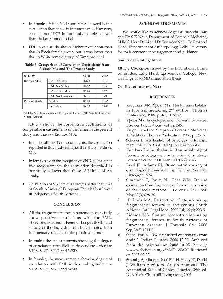
Medico-Legal Update, January-June 2014, Vol. 14, No. 1 187
• In females, VHD, VND and VHA showed bettercorrelation than those in Simmons et al. However,correlation of BCB in our study sample is lowerthan that of Simmons et al.
• FDL in our study shows higher correlation thanthat in Black female group, but it was lower thanthat in White female group of Simmons et al.
Table 5. Comparison of Correlation Coefficients formBidmos MA and The Present Study
STUDY VND VHA
Bidmos M A SAED Males 0.478 0.610
IND SA Males 0.542 0.653
SAED Females 0.544 0.623
IND SA Females 0.681 0.799
Present study Males 0.749 0.866
Females 0.630 0.701
SAED- South Africans of European DecentIND SA- IndigenousSouth Africans
Table 5 shows the correlation coefficients ofcomparable measurements of the femur in the presentstudy and those of Bidmos M A.
• In males all the six measurements, the correlationreported in this study is higher than that of BidmosM A.
• In females, with the exception of VND, all the otherfive measurements, the correlation described inour study is lower than those of Bidmos M A’sstudy.
• Correlation of VND in our study is better than thatof South African of European Females but lowerin Indigenous South Africans.
CONCLUSION
• All the fragmentary measurements in our studyshow positive correlations with the FML.Therefore, Maximum Femoral Length (FML) andstature of the individual can be estimated fromfragmentary remains of the proximal femur.
• In males, the measurements showing the degreeof correlation with FML in descending order areVHA, VND, VHD and WSD.
• In females, the measurements showing degree ofcorrelation with FML in descending order areVHA, VHD, VND and WSD.
ACKNOWLEDGEMENTS
We would like to acknowledge Dr Yashoda Raniand Dr S K Naik, Department of Forensic Medicine,LHMC, New Delhi and Dr Surinder Nath, Ex-Prof andHead, Department of Anthropology, Delhi Universityfor their constant encouragement and guidance.
Source of Funding: None
Ethical Clearance: Issued by the Institutional Ethicscommittee, Lady Hardinge Medical College, NewDelhi., prior to MD dissertation thesis.
Conflict of Interest: None
REFERENCES
1. Krogman WM, Ýþcan MY. The human skeletonin forensic medicine, 2nd edition. ThomasPublication, 1986. p. 4-5, 302-327.
2. Ýþcan MY. Encyclopedia of Forensic Sciences.Elsevier Publications, Vol 1.p.245.
3. Knight B, editor. Simpson’s Forensic Medicine,11th edition. Thomas Publication, 1986. p. 35-37.
4. Scheuer L. Application of osteology to forensicmedicine. Clin Anat. 2002 Jun;15(4):297-312.
5. Kemkes-Grottenthaler A. The reliability offorensic osteology—a case in point. Case study.Forensic Sci Int. 2001 Mar 1;117(1-2):65-72
6. Byrd JE, Adams BJ. Osteometric sorting ofcommingled human remains. J Forensic Sci. 2003Jul;48(4):717-24.
7. Simmons T, Jantz RL, Bass WM. Statureestimation from fragmentary femora: a revisionof the Steele method. J Forensic Sci. 1990May;35(3):628-36.
8. Bidmos MA. Estimation of stature usingfragmentary femora in indigenous SouthAfricans. Int J Legal Med. 2008 Jul;122(4):293-9
9. Bidmos MA. Stature reconstruction usingfragmentary femora in South Africans ofEuropean descent. J Forensic Sci. 2008Sep;53(5):1044-8.
10. Sinha, Varun. “‘We first fished out remains fromdrain’”. Indian Express. 2006-12-30. Archivedfrom the original on 2008-10-05. http://www.webcitation.org/5bMDvWkGC. Retrievedon 2007-02-27.
11. Strandig S, editor in chief. Elis H, Healy JC, DavidJ, William A editors. Gray’s Anatomy: TheAnatomical Basis of Clinical Practice. 39th ed.New York: Churchill Livingstone; 2005
43. Tejasawi --183--188.pmd 2/22/2014, 11:45 AM187

188 Medico-Legal Update, January-June 2014, Vol. 14, No. 1
12. Sinnatamby CS, editor. Last’s Anatomy: Regionaland Applied. 11th ed. London: ChurchillLivingstone; 2006.
13. Mysorekar VL, Verma PK, Nandedkar AN.Estimation of stature from parts of bones- lowerend of femur and upper end of radius. Med SciLaw 1980; 20:283-286.
14. Mysorekar VR, Nandedkar AN, Sarma TCSR.Estimaton of stature from parts of the ulna andtibia. Med Sci Law 1984; 24:113-116.
15. Estimation femur and tibia Length fromfragmentary bones: An evaluation of Steele’s(1970) Method using a prehistoric Europeansample. Am J Phys Anthropol 1992; 89:333-345.
16. Chibba K, Bidmos MA. Estimation of stature andmaximum long bone length of the tibia fromfragments of tibia in south African of Europeandescent. Forensic Sci Int 2007; 169:145-151.
17. Wright LE, Vásquez MA. Estimating the lengthof incomplete long bones: forensic standards fromGuatemala. Am J Phys Anthropol. 2003Mar;120(3):233-51.
18. Steele G, McKern T. A method for statureassessment of maximum long bone length andliving stature from fragmentary long bones. AmJ Phys Anthropol 1969;31:215-225.
19. Shroff AG, Panse AA, Diwan CV. Estimation oflength of femur from its fragments. J Anat SocInd 1999; Jun 48(1):1-5.
20. Purkait R. Sexual dimorphism in the femoralhead-a new approach. Can Soc Foren Sci J 2002;35:209-221.
21. Purkait R. Sex determination from femoral headmeasurements: a new approach. Leg Med 2003;suppl:s347-s350.
22. Purkait R, Chandra H. Sexual dimorphism infemur-an Indian study. Forensic Sci Commun Jul2002; 4(3).
23. Purkait R, Chandra H. A studt of sexual variationin Indian femur. Forensic Sci Intl 2004; 146:25-33.
24. The goldman osteometric data set. [Online]. [cited2007];[6 screens]. Available from :h t t p : / / w e b . u t k . e d u / ~ a u e r b a c h /GoldMeasures.pdf
43. Tejasawi --183--188.pmd 2/22/2014, 11:45 AM188

Medico-Legal Update, January-June 2014, Vol. 14, No. 1 189
INTRODUCTION
Poisoning is a major problem in children. Howeverthe offending agent and the associated morbidity andmortality vary from place to place and change over aperiod of time. With the control of infectious diseases,the contribution of poisoning to childhood mortalityand morbidity has been increasing in developingcountries 1-6. The developed world has accurateinformation on incidence and changing trends ofcausative agents as a result of rigorous populationcensus, mortality data and the development of
Pattern of Paediatric Poisoning - A 3 Year RetrospectiveStudy in Krishna Institute of Medical Sciences, Karad,
Maharashtra
Vinayak Y Kshirsagar1, Anand Patil2, Sylvia Colaco2, Basaveshwar Patil2, J M Pawar3
1Professor, 2Junior Resident, 3Associate Professor, Dept of Paediatrics, Krishna Institute of Medical Sciences University,Karad, Maharashtra
ABSTRACT
Poisoning is a major problem in the paediatric population. The offending substances used vary fromplace to place. The present study was undertaken to determine poisoning in children with respect toage, sex, habitat, seasonal distribution, type of poison, outcome and manner of poisoning. This is aretrospective analysis of 3 years from Jan 2010 to Dec 2012 of all childhood poisoning cases admittedin Krishna Institute of Medical Sciences, Karad in the age group of 0 to 14 years. A total of 152 caseswere studied with male to female ratio of 2.3:1. Most common age group involved was 10-14 yrs withthe incidence being more common in rural areas (86.1%). Most common poisoning was animal bite(63.8%) followed by kerosene (7.8%) pesticide (6.5%), drug (3.2%) and other substance (18.7%)ingestion. Rainy season was found to be the most vulnerable period for poisoning. All cases wereaccidental except one which was homicidal in nature. There was a mortality rate of 0.65 % whereas 4cases were discharged against medical advice. As most of the cases were accidental in nature, soaccess prevention and health education campaign should be instituted to reduce the incidence ofaccidental poisoning in children.
Keywords: Accidental Poisoning, Paediatric Poisoning, Animal Bite
Corresponding author:Vinayak Y KshirsagarHead of Department of Paediatrics,Krishna Institute of Medical Sciences University, KaradMaharashtra, India.E-mail: [email protected] No: 02164 241555 Ext: 361Fax No: 02164 242170
National poisons control centres 7-11. In contrast,paediatric poisoning incidence in developing countriesis poorly documented 8, 12 13. Likewise in India limiteddata are available on morbidity and mortality relatedto poisoning in children, in particular the changes inits pattern over the years. Many studies have been donerelating to the age incidence; sex incidence; seasonaldistribution; type of poison consumed; manner ofpoisoning and outcome of cases. The present studywas undertaken to determine poisoning in childrenwith respect to age, sex, type of poison, habitat, mannerof poisoning, seasonal distribution and outcome of thecases admitted in KIMS Karad.
MATERIAL AND METHOD
This retrospective study was carried out at KIMSKarad, over a period of 3 years from Jan 2010 to Dec2012 in the age d” 14 years. All cases of poisoningadmitted in the study period where analysed andstudied accordingly, the data obtained from hospitaland medical record section.
DOI Number: 10.5958/j.0974-1283.14.1.044
44. Vinayak--189--192.pmd 2/22/2014, 11:45 AM189

190 Medico-Legal Update, January-June 2014, Vol. 14, No. 1
RESULTS
A total of 152 poisoning cases of d” 14 years of agewere studied at KIMS Karad. 42.7% were under 10-14years of age and 70.6% male (Male to female ratio was2.3:1) (Table 1). Incidence was more common in ruralareas (86.1%) as compared to urban areas (13.9%)(Table 2) and there was no significant season-wisevariation observed (Table 3). Animal bite and kerosenewere the most common offending agents, bothaccounting for 71.8% cases (Table 5). All cases wereaccidental except one case was due to homicidal innature (Table 4). All cases improved except 1 whichexpired due to unknown poisoning and 5 cases weredischarged against medical advice (Table 6).
Table 1: Age and Sex wise distribution.
Age(yrs) Male Female Total
0-5 32(21%) 16(10.5%) 48(31.5%)
5-10 28(18.4%) 11(7.2%) 39(25.6%)
10-14 47(33.8%) 18(11.8%) 65(42.7%)
Total 107 (70.3%) 45 (29.6%) 152
Table 2: Habitat wise distribution.
Habitat No of cases
Urban 21(13.8%)
Rural 131(86.1%)
Table 3: Season wise distribution.
Type of poisoning Summer Rainy Winter Total
Snake bite 13(8.5%) 36(23.6%) 19(12.5%) 68(44.7%)
Unknown bite 5(3.2%) 2(1.3%) 12(7.8%) 19(12.5%)
Kerosene 7(4.6%) 2(1.3%) 3(1.9%) 12(7.8%)
Scorpion sting 3(1.9%) 5(3.2%) 2(1.3%) 10(6.5%)
Unknown 3(1.9%) 1(6.5%) 5(3.2%) 22(14.4%)
Organophosphorus 2(1.3%) 1(0.65%) 3(1.9%) 6(3.9%)
Others 19(12.5%) 8(5.2%) 1(0.65%) 15(9.8%)
Total 52(34.2%) 55(36.1%) 45(29.6%) 152
Table 4: Manner of poisoning.
Manner of poisoning No of cases
Accidental 151(99.3%)
Suicidal 00(0.0%)
Homicidal 01(0.7%)
(Table 5): Outcome of various poisons.
Type of poison Outcome
Improved Unchanged Death
Animal bites 96(63.1%) 1(0.65%) 0.0%
Kerosene 9(5.9%) 3(1.9%) 0.0%
Organophosphorus 5(3.2%) 1(0.65%) 0.0%
Others 45(29.6%) 0.0% 1(0.65%)
Table 6: Type of poison distribution.
Sl.no Type of poison No of cases
1) Household products Kerosene 12(7.8%)
Turpentine oil poisoning 01(0.65%)
Castor oil poisoning 08(5.2%)
Colour poisoning 01(0.65%)
Rat poisoning 01(0.65%)
Engine oil poisoning 01(0.65%)
Camphour poisoning 01(0.65%)
2) Animal bites Snake bite 68(44.7%)
Unknown bite 19(12.5%)
Scorpion sting 10(12.5%)
3) Pharmaceutical products Paracetamol poisoning 01(0.65%)
Carbamezepine poisoning 02(1.3%)
Cefixime poisoning 01(0.65%)
Iron poisoning 01(0.65%)
4) Agricultural products Organophosphorous poisoning 06(3.9%)
Permethrin poisoning 02(1.3%)
Malathion poisoning 01(0.65%)
Organo carbamate poisoning 01(0.65%)
5) Plant products Semicarpus anacardium poisoning 01(0.65%)
6) Food products Food poisoning 04(2.6%)
7) Unknown compound Unknown compound 10(6.5%)Poisoning
Total 152
44. Vinayak--189--192.pmd 2/22/2014, 11:45 AM190

Medico-Legal Update, January-June 2014, Vol. 14, No. 1 191
DISCUSSION
The childhood poisoning death rates have declinedover the past decade, largely due to parental awarenessand appropriate interventions 14. However childrencontinue to be at significantly greater risk than adultsfor poisoning, because they have faster metabolic ratesand are less able to physically handle toxic chemicals.In addition natural curiosity and their desire to puteverything in their mouth increase their poisoningrisks.
The incidence, age, sex, nature and type of poisonconsumed vary from area to area. Present study notedthat the most vulnerable age group for poisoning is10-14yrs, because early adolescent playful periodmostly outdoors making the child vulnerable foranimal bites. Study also observed that increasedincidence of animal bites in 10-14yrs age group. Studiesby Patil V C et al 15 and Inamdar et al 25 concludedsnake bite is a common life threatening emergency inthe study area.
There was a male preponderance in our studywhich is consistent with Rashid AKM (2007) 17, in astudy by G N Lucas (2006) who studied 243 childrenwhich showed male to female ratio (148:95)18 , studiesby Hussein AB and Ahmed AB (2007) showed similarresults 19 , Queensland injury surveillance unit studiedchildhood poisoning (2000) which showed malepreponderance in their studies 20 , similarly a study atAll India Institute of Medical Sciences, New Delhi byGupta S K et al (2003) showed male : female sex ratioof 63.11%: 36.88% 21.
Present study observed predominance of poisoningin rural children due increasing incidence of snake bitesin the rural area. A retrospective study done by SharmaN et al in PGI Chandigarh showed an increasingincidence of snake bites in rural area with a ratio of(urban:rural-1:4.7) (2006)26 which is in agreement withPatil V C et al and Inamdar et al 15, 24.
The study noted that, rainy season was the mostvulnerable period when maximum cases werereported 17, 22, 23.This can be explained on the basis ofmore snakes coming out during rainy season due tofilling of water in their burrows and thus increasingthe human snake interaction resulting in more numberof snake bites during the season, while summer beinga dry season, thirst causes an increased attraction
towards liquids thus increasing cases of accidentalpoisoning especially Kerosene poisoning which wasthe commonest poisoning in summer season. This wasconsistent with various studies from Syeda Akhtar etal (2006) 16, G N Lucas (2006) 18 & Gupta S K et al (2003)21. This can be attributed to the use of kerosene oil as afuel especially in lower income group, its storage ineasily accessible places & its colour which is oftenmistaken for soft drink.
All the cases studied were accidental except a 3month old child who was homicidal withorganophosphorous compound. The studied showedthat all the cases improved except one which expireddue to unknown poisoning and 5 cases weredischarged against medical advice. Age and sex arethe factors to be considered in evaluating a child’s riskfor poisoning. In addition, developmental andenvironmental factors may contribute to the risk of apoisoning event25.
CONCLUSION
Most common age group involved was 10–14 yrsand more frequent in males population of rural areas.Rainy season is the most vulnerable period, whenmaximum cases of snake bites were reported whilekerosene poisoning is the most frequently reportedhousehold poisoning. 0.65% of cases proved to be fataldue to unknown compound poisoning and animalbites are responsible for most of the cases due togeographical distribution. All cases were accidentalexcept 1 case which was homicidal in nature. Thus werecommend establishment of regional toxicologicalcentres which help guide hospitals in the quickidentification of poisons and provide guidelines fortreatment. There is also a need to initiate publicawareness campaigns to attempt to decrease morbidityand mortality from this eminently preventableproblem.
ACKNOWLEDGEMENT
We thanks to the Dr Sunil Armani- AssociateProfessor, Department of Forensic Medicine andToxicology KIMS Deemed University, who facilitatesour mission at the University.
Ethical Approval: Ethical approval taken from theuniversity ethics committee.
44. Vinayak--189--192.pmd 2/22/2014, 11:45 AM191
192 Medico-Legal Update, January-June 2014, Vol. 14, No. 1
REFERENCES
1. Christoffel KK, Marcus D, Sagerman S, Bennett S(1988) Adolescent suicide and suicide attempts:a population study. Pediatr Emerg Care 4: 32-40.
2. Lewis HH, Cronje RE, Naude SP, van den Berg C(1989) Accidental poisoning in childhood. S AfrMed J 76: 429-431.
3. Pearn J, Nixon J, Ansford A, Corcoran A (1984)Accidental poisoning in childhood: five yearurban population study with 15 year analysis offatality. Br Med J (Clin Res Ed) 288: 44-46.
4. Litovitz TL, Flagler SL, Manoguerra AS, Veltri JC,Wright L (1989) Recurrent poisonings amongpaediatric poisoning victims. Med ToxicolAdverse Drug Exp 4: 381-386.
5. Wiseman HM, Guest K, Murray VSC, Volans GN(1987) Accidental poisoning in childhood: Amulticentre survey I. General epidemiology. HumToxicol 6: 293-301.
6. Jacobsen D, Halvorsen K, Marstrander J, SundeK, Bakken AF (1983) Acute poisonings of childrenin Oslo. A one year prospective study. ActaPaediatr Scand 72: 553-557.
7. Kivistö JE, Mattila VM, Arvola T, Paavola M,Parkkari J (2008) Secular trends in poisoningsleading to hospital admission among Finnishchildren and adolescents between 1971 and 2005.J Paediatrics 153: 820-824.
8. Meyer S, Eddleston M, Bailey B, Desel H,Gottschling S, et al. (2007) Unintentionalhousehold poisoning in children. Klin Paediatrics219: 254-270.
9. McKenzie LB, Ahir N, Stolz U, Nelson NG (2010)Household cleaning product- related injuriestreated in US emergency departments in 1990-2006. Paediatrics 126: 509-516.
10. Liebelt EL, DeAngelis CD (1999) Evolving trendsand treatment advances in paediatric poisoning.JAMA 282: 1113-1115.
11. Litovitz T, Manoguerra A (1992) Comparison ofpaediatric poisoning hazards: an analysis of 3.8million exposure incidents. A report from theAmerican association of poison control centre.Paediatrics 89; 999-1006.
12. Tagwireyi D, Ball DE, Nhachi CF (2002) Poisoningin Zimbabwe: a survey of eight major referralhospitals. J Appl Toxicol 22: 99-105.
13. Balme KH, Roberts JC, Glasstone M, Curling L,Rother HA, et al. (2010) Pesticide poisonings at atertiary children’s hospital in South Africa: anincreasing problem. Clin Toxicol (Phila) 48:928-934.
14. National SAFE KIDS Campaign (NSKC).Poisoning Fact Sheet. Washington (DC): NSKC,2004.
15. Virendra C Patil, Harsha V Patil, Avinash Patil,Vaibhav Agrawal (2011) Clinical Profile andoutcome of envenomous snake-bite at tertiarycare centre in western Maharashtra. InternationalJournal of Medicine and Public Health 1: 28-38.
16. Akhtar S, Gulati R, Fahad Al Anezi (2006) Riskfactors in acute poisoning in children-Aretrospective study. Kuwait Medical Journal 38:33-36.
17. Rashid AKM (2007) Seasonal variation ofchildhood acute poisoning. Pakistan Journal ofMedical Science 23: 1-5.
18. Lucas GN (2006) A hospital based prospectivestudy of acute childhood poisoning. SrilankaJournal of child health 35: 12-19.
19. Hussein AB, Ahmed AB (2007) Childhoodaccidental poisoning in Tafila. Iran J Ped 17:23-27.
20. Hockey R, Reith D, Miles E (2000) Queenslandinjury surveillance unit childhood poisoning andingestion, injury bulletin 60: 1-6.
21. Gupta SK, Peshin SS, Srivastava A, Kaleekal T(2003) A study of childhood poisoning at NationalPoisons Information Centre, All India Instituteof Medical Sciences, New Delhi. J Occup Health45: 191-196.
22. Matiyahu L, Vladimir G (2000) Acute poisoningin children. IMAJ 2: 504-506.
23. Moghadamnia AA, Shirvani TE, Esmaeili MR,Bayat Z, Gholotabar ZM (2004) A report ofchildhood poisoning in Babol. Archives of Iranianmedicine 7: 297-299.
24. IF Inamdar, NR Aswar, M Ubaidula, SD Dalvi(2010) Snake bite: Admissions at a Tertiaryhealthcare centre in Maharashtra, India. S AfrMed J 100: 456-458.
25. Kelly NR, Drutz JE. Prevention of poisoning inchildren. Up-to-date [Internet]. 2012 Oct 1.Available from: http://www. uptodate.com/contents/prevention-of-poisoning-in-children.
26. Sharma N, Chauhan S, Farugi S, Bhat P, Varma S.Snake envenomation in north India hospital.Emerg Med J 2005 Feb; 22(2):118-20. Availablefrom: http:// www.pubmed.com-accessed on14/2/2006.
44. Vinayak--189--192.pmd 2/22/2014, 11:45 AM192

Medico-Legal Update, January-June 2014, Vol. 14, No. 1 193
INTRODUCTION
‘Poison’, in its simplest form, is any substance thatcan cause harm, severe distress or death if ingested,injected, breathed in, or absorbed through the skin; inshort it is anything that endangers a person’s healthand well-being. In India, the commonest poisons arepesticides, sedative drugs, chemicals, alcohols, planttoxins and household poisons (cleaning agents) 1-4.Among children, kerosene, household chemicals,drugs, pesticides and garden plants are the mostcommon culprits5,6.
Every incident of poisoning is fraught with medico-legal and ethical implications. Lately, the ingestion ofdrugs and poisonous substances is increasinglybecoming a means of attempting to end one’s lifeamong the frustrated and emotionally disturbed youthof our region. Further, a good number of cases broughtto the emergency department are those of intentionaldrug overdose and substance abuse. Although it isoften stated that the history of exposure is unreliable,
Profile of Poisoning at a Teaching Hospital in Shillong(Northeast India)
A D Ropmay1, D Slong2, S J Gogoi2, Sonali Shinde Tesia2
1Associate Professor, 2Senior Resident, Department of Forensic Medicine,NEIGRIHMS, Shillong
ABSTRACT
Background: Every incident of poisoning is fraught with medico-legal and ethical implications.Knowledge of the poisoning profile in a particular area is helpful to ensure better management andprevention of such episodes in future.
Method: All cases admitted to the emergency department of the hospital between July 2009 and June2012 with suspicion of acute poisoning (from the history and clinical findings) were included in thestudy. Data was obtained retrospectively from the Casualty Medico-Legal Case (MLC) Registers forthat period and analyzed using Microsoft Excel 2007.
Results: Males in the age group 21 to 30 years were most commonly affected. The mode of poisoningwas mainly an overdose of drugs and medicinal substances. The majority of cases were suicidalattempts.
Conclusion: A multidisciplinary approach is needed to deal with the problem of suicidal poisoning,especially in the vulnerable age group of young adults and adolescents in the northeast region. Themisuse of prescription drugs calls for health education and enforcement of laws to curb illicit saleand trafficking of medicinal substances.
Keywords: Poisoning, Suicidal, Northeast Region
historical information from the patient and others,coupled with the physical examination and routinelaboratory studies, can be quite accurate indetermining the diagnosis7. Acute poisoning withvarious substances is common everywhere. The earlierthe initial resuscitations, gastric decontamination anduse of specific antidotes, the better the outcome8.
The pattern of poisoning varies in different partsof the world and within the same country dependingon sociocultural, geographical and economic factors.Hence, it is imperative to have specific data from eachregion for a better approach towards management andprevention of poisoning and drug overdose.
METHOD
The present study was performed retrospectivelyat a teaching hospital in Shillong (northeast India). Allcases registered as medico-legal in the emergencydepartment of the hospital with history or suspicionof acute poisoning and suggestive clinical features
DOI Number: 10.5958/j.0974-1283.14.1.045
45. AD Ropmay--193--197.pmd 2/22/2014, 11:45 AM193

194 Medico-Legal Update, January-June 2014, Vol. 14, No. 1
were included in the study. Cases of snake and insectbite, food poisoning and allergic reaction to drugs wereexcluded from the study. Data was obtained from theMedico-Legal Case (MLC) Registers for the period July2009 to June 2012. A total of 79 cases were included inthe study. The parameters considered weresociodemographic factors, agents and route of intake,and manner of poisoning. The particulars of every casewere recorded on a proforma, entered in a computerdatabase and analyzed using Microsoft Office Excel2007.
Ethical clearance for the study was obtained fromthe institutional ethics committee (IEC).
RESULTS
The results are shown in Tables 1 to 4 and Figures 1to 3. Out of the total number of 2074 medico-legal casesregistered in the emergency department during thestudy period, 3.8% are attributed to known orsuspected poisoning. The agents comprised a widerange of prescription medicines, pesticides, drugs ofabuse and household chemicals. Most of the poisonswere ingested orally (96.2%) except for heroin(intravenous), cannabis (inhalation) and an unknowndrug injected intravenously. Five patients (6.3%)consumed more than one agent. The vulnerable agegroup was found to be 21 to 30 years in both sexes(Male: Female=1.6:1). In more than half of the cases,incidents of poisoning were self-inflicted. However,no fatalities were recorded in the present study. Only16.5% of patients arrived in hospital within the ‘goldenhour’, i.e. the first 60 minutes after exposure to thepoison9.
Table 1: Age and sex distribution of cases.
Age group Total Percentage Number Number ofof male(s) female(s)
<10 years 3 3.8 1 2
11-20 years 18 22.8 6 12
21-30 years 37 46.8 23 14
31-40 years 14 17.7 8 6
41-50 years 4 5.1 3 1
51-60 years 1 1.3 1 0
>60 years 2 2.5 1 1
Total 79 100 43 36
Table 2: Manner of poisoning (n=79).
Manner Number Percentage
Suicidal 43 54.4
Accidental 25 31.6
Homicidal 2 2.5
Uncertain 9 11.4
Table 3: Type of poison (n=79)
Poison Number Percentage
Phenol 9 11.4
Pesticide 14 17.7
Drug/Medicine 26 32.9
Alcohol and spirits 10 12.6
Dettol 2 2.5
Kerosene 3 3.8
Diesel 1 1.3
Battery acid 1 1.3
Datura 6 7.6
Shampoo 1 1.3
Unknown 6 7.6
Table 4: Substances of abuse.
Substance No.of cases Route ofadministration
Spasmoproxyvon (SP)a 3 Oral
Cannabis 1 Inhalation
Alcohol 3 Oral
Alcohol and SP 2 Oral
Heroin 1 Intravenous
aA capsule of Spasmoproxyvon contains 10 mg ofDicyclomine (Dicycloverine) 400 mg ofAcetaminophen and 65 mg of Dextropropoxyphene
Fig.1. Marital status of poisoned patients
Fig.2. Medicines involved in poisoning
45. AD Ropmay--193--197.pmd 2/22/2014, 11:45 AM194
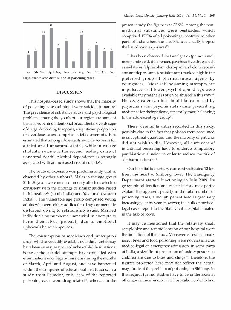
Medico-Legal Update, January-June 2014, Vol. 14, No. 1 195
DISCUSSION
This hospital-based study shows that the majorityof poisoning cases admitted were suicidal in nature.The prevalence of substance abuse and psychologicalproblems among the youth of our region are some ofthe factors behind intentional or accidental overdosageof drugs. According to reports, a significant proportionof overdose cases comprise suicide attempts. It isestimated that among adolescents, suicide accounts fora third of all unnatural deaths, while in collegestudents, suicide is the second leading cause ofunnatural death1. Alcohol dependence is stronglyassociated with an increased risk of suicide10.
The route of exposure was predominantly oral asobserved by other authors11. Males in the age group21 to 30 years were most commonly affected, which isconsistent with the findings of similar studies basedin Mangalore12 (south India) and Yavatmal (westernIndia)13. The vulnerable age group comprised youngadults who were either addicted to drugs or mentallydisturbed owing to relationship issues. Marriedindividuals outnumbered unmarried in attempts toharm themselves, probably due to emotionalupheavals between spouses.
The consumption of medicines and prescriptiondrugs which are readily available over the counter mayhave been an easy way out of unbearable life situations.Some of the suicidal attempts have coincided withexaminations or college admissions during the monthsof March, April and August, and have happenedwithin the campuses of educational institutions. In astudy from Ecuador, only 26% of the reportedpoisoning cases were drug related14, whereas in the
present study the figure was 32.9%. Among the non-medicinal substances were pesticides, whichcomprised 17.7% of all poisonings, contrary to otherparts of India where these substances usually toppedthe list of toxic exposures15.
It has been observed that analgesics (paracetamol,mefenamic acid, diclofenac), psychoactive drugs suchas sedatives (alprazolam, diazepam and clonazepam)and antidepressants (escitalopram) ranked high in thepreferred group of pharmaceutical agents byyoungsters. Most self poisoning attempts areimpulsive, so if fewer psychotropic drugs wereavailable they might less often be abused in this way16.Hence, greater caution should be exercised byphysicians and psychiatrists while prescribingmedicines for their patients, especially those belongingto the adolescent age group17.
There were no fatalities recorded in this study,possibly due to the fact that poisons were consumedin suboptimal quantities and the majority of patientsdid not wish to die. However, all survivors ofintentional poisoning have to undergo compulsorypsychiatric evaluation in order to reduce the risk ofself harm in future18.
Our hospital is a tertiary care centre situated 12 kmfrom the heart of Shillong town. The EmergencyDepartment started functioning in July 2009. Itsgeographical location and recent history may partlyexplain the apparent paucity in the total number ofpoisoning cases, although patient load is graduallyincreasing year by year. However, the bulk of medico-legal cases report to the State Civil Hospital situatedin the hub of town.
It may be mentioned that the relatively smallsample size and remote location of our hospital werethe limitations of this study. Moreover, cases of animal/insect bites and food poisoning were not classified asmedico-legal on emergency admission. In some partsof India, a significant proportion of toxic exposures inchildren are due to bites and stings19. Therefore, thefigures projected here may not reflect the actualmagnitude of the problem of poisoning in Shillong. Inthis regard, further studies have to be undertaken inother government and private hospitals in order to find
Fig.3. Monthwise distribution of poisoning cases
45. AD Ropmay--193--197.pmd 2/22/2014, 11:45 AM195

196 Medico-Legal Update, January-June 2014, Vol. 14, No. 1
out the reasons for self poisoning, obtain a betterrepresentation of the affected population, and therebyformulate preventive measures for the same. Inaddition, chemical analysis to categorize the type ofpoison would help to corroborate clinical findingsand institute specific line of treatment for a particularagent.
CONCLUSION
As the age groups involved are mostly youngadults and adolescents, it is concluded that amultidisciplinary approach is needed to address theissue of self poisoning. Explicit guidelines and policiesmust be in place so that pharmacies cannot dispensemedicines over the counter without a properprescription from a registered medical practitioner.Enforcement of laws to curb illicit sale and traffickingof drugs is the need of the hour. Awareness-raisingcampaigns for rational use of medicines and socialprograms for suicidal patients should also helpdecrease the frequency of such cases20.
ACKNOWLEDGEMENT
The authors wish to thank the Casualty medicalofficers – Dr.Anna, Dr.Kalidas, Dr.Kevin and Dr.Arnest,and medical records department staff who made thisstudy possible.
Conflict of Interest: None to declare
Source of Funding: Nil
REFERENCES:
1. Pillay VV. Comprehensive Medical Toxicology.14th edition. Hyderabad: Paras publications; 2004.
2. Dash SK, Raju AS, Mohanty MK, Patnaik KK,Mohanty S. Sociodemographic profile ofpoisoning cases. J. Ind Acad Forensic Med 2005;27(3):133-7.
3. Batra AK, Keoliya AN, Jadhav GU. Poisoning: Anunnatural cause of morbidity and mortality inrural India. J Assoc Physicians India 2003; 51:955-9.
4. Singh D, Jit I, Tyagi S. Changing trends in acutepoisoning in Chandigarh zone: A 25 year autopsy
experience from a tertiary care hospital inNorthern India. Am J Forensic Med Pathol 1999;20:203-10.
5. Vaswani V, Patil VD. Spectrum of childhoodpoisoning: A Belgaum experience. J Forensic MedToxicol 1998;15:50-2.
6. Singh LR, Momonchand A, Singh PI. Pattern ofaccidental poisoning in children. J Indian AcadForensic Med 2001; 23:69-71.
7. Brent J, Wallace KL, Burkhart KK, Phillips SD,Donovan JW. Critical Care Toxicology: Diagnosisand Management of the Critically PoisonedPatient. 1st edition. Philadelphia: Elsevier Mosby;2005.
8. Kumar SV, Venkateswarlu B, Sasikala M, KumarGV. A study on poisoning cases in a tertiary carehospital. J Nat Sci Biol Med 2010 Jul;1(1):35-9.
9. Lau FL. Emergency management of poisoning inHong Kong. HKMJ 2000;6:288-92.
10. Mitchell AM, Garand L, Dean D, Panzak G, TaylorM. Suicide assessment in hospital emergencydepartments: implications for patient satisfactionand compliance. Top Emerg Med 2005 Oct;27(4):302-12.
11. Singh O, Javeri Y, Juneja D, Gupta M, Singh G,Dang R. Profile and outcome of patients withacute toxicity admitted in intensive care unit:Experiences from a major corporate hospital inurban India. Indian J Anaesth 2011 Jul-Aug;55(4):370-4.
12. Singh B, Unnikrishnan B. A profile of acutepoisoning at Mangalore (South India). J ClinForensic Med 2006 Apr; 13(3):112-6.
13. Vaidya YP, Hulke SM. Study of trends ofpoisoning in the cases reported to governmenthospital, Yavatmal. Chron Young Sci 2012; 3: 63-7.
14. Brito MA, Reyes RM, Arguello JR, Spiller HA.Principal causes of poisoning in Quito, Ecuador:A retrospective epidemiology study. Vet HumToxicol 1998; 40: 40-2.
15. Goel A, Aggarwal P. Pesticide poisoning. NationalMedical Journal of India 2007; 20(4): 182-91.
16. Skegg K, Skegg DCG, Richards SM. Incidence ofself poisoning in patients prescribed psychotropicdrugs. BMJ 1983 Mar; 286:841-3.
45. AD Ropmay--193--197.pmd 2/22/2014, 11:45 AM196

Medico-Legal Update, January-June 2014, Vol. 14, No. 1 197
17. Buckley NA, Dawson AH, Whyte IM, Hazell P,Meza A, Britt H. An analysis of age and genderinfluences on the relative risk for suicide andpsychotropic drug self-poisoning. Acta PsychiatrScand 1996 Mar;93(3):168-71.
18. Niveditha, Priyalatha N, Maity N, ShivamurthyMC. Intentional poisoning as a cause ofadmission to accident and emergency in a tertiarycare hospital within a year. Al Ameen J Med Sci2011; 4(4):396-400.
19. Honnungar RS, Kumar L, Shetty AK, Jirli PS. Astudy of paediatric poisoning cases at District
Hospital, Belgaum, Karnataka. Medico-LegalUpdate 2010 Jan-Jun; 10(1):47-50.
20. Bernardes Sara Santos, Turini ConceiçãoAparecida, Matsuo Tiemi. Profile of suicideattempts using intentional overdose withmedicines, treated by a poison control center inParaná State, Brazil. Cad. Saúde Pública 2010 July[cited 2012 Aug 08]; 26(7):1366-1372. Availablefrom: http://www.scielosp.org/scielo.php?script=sci_arttext&pid=S0102-311X2010000700015&lng=en.
45. AD Ropmay--193--197.pmd 2/22/2014, 11:45 AM197

198 Medico-Legal Update, January-June 2014, Vol. 14, No. 1
INTRODUCTION
“It seems that a person dies from a broken heart,not in young adult life from a great grief or emotion,but usually in old age, on account of diseasedcoronaries…”
Krumbhaar and Crowell1
Ventricular wall rupture occurs in 1-3% cases withacute myocardial infarction (AMI) and is the third mostcommon cause of death caused by AMI. This mostsever mechanical complication of the AMI oftenremains undiagnosed and constitute a necropsyfinding.2 Acute ventricular free wall rupture maypresent by chest pain, or by the classic features ofcardiac tamponade, namely, shock with hypotension,pulsus paradoxus, elevated venous pressure, quietheart sounds and sinus bradycardia, or frankelectromechanical dissociation. Death usually ensues
Cardiac Rupture Following Myocardial Infarction - A CaseReport
Shrikant Shinge1, Avinash Waghmode2, Umesh Kumar2
1Assistant Professor, Dept. of Forensic Medicine, Govt. Medical College, Miraj, Dist, Sangli, Maharashtra, 2JuniorResident, Dept of Forensic Medicine, Indira Gandhi Govt. Medical College, Nagpur
ABSTRACT
Ventricular wall rupture occurs in 1-3% cases with acute myocardial infarction (AMI) and is the thirdmost common cause of death caused by AMI. This most sever mechanical complication of the AMIoften remains undiagnosed and constitute a necropsy finding. Acute free wall rupture complicatingAMI is defined as an abrupt and complete transmural rupture of the infracted region, causing cardiactemponade and death within 30 minutes. Ventricular free wall rupture occurs up to ten times morefrequently than septal or papillary muscle rupture. The pathophysiological process involves thinningof the myocardial wall with the intensity of necrosis occurring at the terminal end of the vessel(watershed area) where there is often poor collateral flow. Here we described in brief about this fatalcomplications presentation, pathophysiology and diagnosis.
Keywords: Acute Myocardial Infarction, Ventricular Wall Rupture, Cardiac Temponade
Corresponding author:Shrikant ShingeAssistant ProfessorBlock No 3. Bulding No- 2, Residential campus,Govt. Medical College, Miraj, Dist.: Sangli,Maharashtra, Pin - 416410Phone No - +919860151299
in a matter of minutes to hours.3 Thepathophysiological process involves thinning of themyocardial wall with the intensity of necrosisoccurring at the terminal end of the vessel (watershedarea) where there is often poor collateral flow. 4
CASE DESCRIPTION
A 58 years old male patient non-smoker and non-alcoholic, without the previous history of any cardiacdisease was brought to the emergency department ofIndira Gandhi Govt. Medical College, Nagpur in earlymorning. There was history of collapse while he wasin toilet without any preceding complains such as chestpain or breathlessness. On examination by attendingphysician, patient was unconscious andcardiopulmonary resuscitation measures wereineffective so the patent declared as brought dead. Thedead body sent to mortuary for post-mortemexamination.
At autopsy, it was a dead body of a male, obesebuilt (weight 98kg) and external examination wasunremarkable. During internal examination a largeblood clot about 350ml was present in the pericardialcavity.(Photograph - 1) Heart was enlarged weighing400gm. Coronary arteries were tortuous, thickened andhardened, showing blockage of about 70 to 80%. A tear
DOI Number: 10.5958/j.0974-1283.14.1.046
46. shrikant shinge--198--201.pmd 2/22/2014, 11:45 AM198

Medico-Legal Update, January-June 2014, Vol. 14, No. 1 199
was present at anterior wall of left ventricular cavitymeasuring about 1.5cm X 0.8cm having irregularmargins (Photograph – 2). Hyperemia of ventricularwall was present. Rest of internal examination wasinsignificant.
The cause of death was given as “Death due tocardiac temponade following myocardial infarction”.
subacute setting after a myocardial infarction in whichthe infarcted muscle of the heart thins out and tears.Myocardial rupture is more likely to happen in elderlyindividuals who suffer from their first heart attack andnot revascularized. In patients, who experienced thefirst myocardial infarction and were not in heart failure,electromechanical dissociation has a very highsensitivity and specificity as a sign of left ventricularfree wall rupture7. It is particularly common in womenand elderly, with higher mortality in women comparedto men.5 William Harvey in 1647 described cardiacrupture as a finding at autopsy of a knight whosuffered severe chest pain8. In 1765, G. Morgagnireported it as a necropsy finding. O’Rourke firstclassified free ventricular wall rupture as acute,subacute and chronic.2
Ventricular free wall rupture occurs up to ten timesmore frequently than septal or pappilary misclerupture.3 Opinions differ as to the most common siteof the left ventricular wall rupture. It was suggestedthat a lateral wall infarction is more likely to rupturethan an anterior or inferior infarction. However,anterior myocardial infarctions are more frequent thanlateral myocardial infarctions and thus the anteriorwall is the most common site.3 However, in terms ofpressure gradient, the right ventricle is not subject tostrain comparable to the left ventricle, and hence themuch lower frequency of right ventricular rupture.This is not the only factor that protects the rightventricle. The right ventricle has better collateralcirculation and thinner wall, allowing better perfusionof myocardium and decreasing the likelihood oftransmural infarction. Anterior and lateral wallruptures of the left ventricle are much more prevalentthan posterior wall ruptures, probably because of theprotection afforded by the adherent pericardium.8
Acute ventricular wall rupture may present bychest pain, or by the classic features of cardiactamponade, namely, shock with hypotension, pulsusparadoxus, elevated venous pressure, quiet heartsounds and sinus bradycardia, or frankelectromechanical dissociation. Death usually ensuesin a matter of minutes to hours. Cardiopulmonaryresuscitation maneuvers are unsuccessful in thesecases.3
The pathophysiological process involves thinningof the myocardial wall with the intensity of necrosisoccurring at the terminal end of the vessel (watershedarea) where there is often poor collateral flow.4
Collection of blood in the pericardial sac interfares with
Fig. 1. Blood clot in the pericardial cavity
Fig. 2. Rupture of the left ventricular wall
DISCUSSION
Cardiac rupture is an early and predominantly fatalcomplication of acute MI that occurs in approximately1% of patients but is responsible for nearly 15% of alldeaths5. Acute free wall rupture complicating AMI wasdefined as an abrupt and complete transmural ruptureof the infracted region, causing cardiac temponade anddeath within 30 minutes6. It typically happens in the
46. shrikant shinge--198--201.pmd 2/22/2014, 11:45 AM199

200 Medico-Legal Update, January-June 2014, Vol. 14, No. 1
ventricular dilation during diastole and compressesthe great veins and the right atrium. As the arterialblood pressure falls and the venous pressure rises,there is a progressive failure of the circulation. As littleas 150-300ml is sufficient to cause death due to cardiactemponade.9
Purcaro et al10 described six pathologic types ofruptures encountered in their series of 28 patients.They were as follows:
(1) Through and through straight rupture of normalthickness myocardium;
(2) Similar but through the thin necrotic myocardium;
(3) Multiple small perforations;
(4) Rupture of outer layer only;
(5) Subepicardial haematoma without free ruptureinto the pericardial sac; and
(6) Hemorrhagic infarct with wall integrity but leakingsurface
Transeosophageal echo is diagnostic for thiscondition and is a gold standard. Initial managementis hemodynamic stabilization followed by bloodtransfusion, pericardiocentesis, inotropic support anduse of an intra-aortic balloon pump but ultimatelyprompt surgical repair is required.4
CONCLUSION
The patient presentations i.e. collapse with nopreceding symptoms coupled with the fact that he wassuffering a silent AMI with no prior indication ofischemic heart disease eg. Angina, shortness of breathetc and the rapid subsequent development of leftventricular free wall rupture in young age made thisquite an unusual case.
DECLARATION
I hereby submit the original research paper entitled“Cardiac Rupture following myocardial infarction – acase report”. The manuscript represents valid workand that neither this manuscript nor one withsubstantially similar content under the presentauthorship has been published or is being consideredfor publication elsewhere and the authorship of thisarticle will not be contested by anyone whose name(s)is/are not listed here. The order of authorship as placed
in the manuscript is final and accepted by thecoauthors.
ACKNOWLEDGEMENT
I greatfully acknowlwdge to Dr. M. B.Shrigiriwar, Prof & head, Dept of FMT, VNGMC,Yavatmal for his valuable support and constantencouragment. I am also thankful to the Dr. V. D. Sonar,Assisant professor, Dept of FMT, GMC, Miraj, for hishelp and support. I express sincere gratitude to theauthors & writers of various textbook and journalswhose refernces have been cited.
Conflict of Interest: None
Source of Funding: Self
Ethical Clearance: Not required
REFERENCES
1. Krumbhaar EB, Crowell C. Spontaneous ruptureof the heart. American Journal of Medical Science,1925; Vol 170: P 828-56
2. Nikolaos I. Exadaktylos, Athanassios I. Kranidis,Michalis O. Argyrious, Christos G. Chariots,Georgios K. Andrikopoulos, Left ventricular freewall rupture during acute myocardial infarction.Early diagnosis and treatment, Hellenic Journalof Cardiology,Vol 43, 2002, P 246-252
3. Arthur Shiyovich, Lior Nesher, Contained LeftVentricular Free Wall Rupture followingMyocardial Infarction, Case Reports in CriticalCare, vol. 2012, Article ID 467810, 4 pages, 2012.doi:10.1155/2012/467810
4. Andrew Peter Venezis, Rehan Quadrey,Mohammad Wasil, Mohammad Azher,Contained Left Ventricular Free Wall Rupturefollowing Myocardial Infarction, British Journalof Medical Practitioner, March 2009, Vol 2,P 41-43
5. Richard C. Becker, Judith S. Hochman,Christopher P. Cannon, Frederick A. Spencer,Steven P. Ball, Michael J. Rizzo, Elliott M. Antman,Fatal cardiac rupture among patient treated withthrombolytic agent and adjuvant thrombinantagonist, Journal of American College ofCardiology, 1999, Vol33, no 2, P 479-487
6. Mehmet S Ülgen1, Önder Öztürk2, MehmetKayrak1, Ahmet Soylu1 M Akif Düzenli1, FatihKoc A lethal but treatable complication : free wall
46. shrikant shinge--198--201.pmd 2/22/2014, 11:45 AM200

Medico-Legal Update, January-June 2014, Vol. 14, No. 1 201
rupture after acute myocardial infarction,Europian Journal of General Medicine, 2006, Vol3(1), P 41-44
7. Amit Sharma, Jatin Bodwal, Cardiac TamponadeFollowing Post Myocardial Ventricular WallRupture: A Case Report, Journal of IndianAcademy of Forensic Medicine, April-June 2011,Vol. 33, No. 2,P 179-181
8. F Lateef and N Nimbkar, Ventricular free wallrupture after myocardial infarction, Hong KongJournal of Emergency Medicine, Vol 10 (4), Oct2003, P 238-246
9. Prateek Rastogi,Cardiac Temponade – a casereport, Journal of Panjab academy of ForensicMedicne and Toxicology, 2009; Vol 9(1)., P 27-28
10. Purcaro A, Costantini C,Mazzanti M, Silenzi C,Gili A, Belardinelli et al, Diagnostic criteria andmanagement of subacute ventricular free wallrupture complicating acute myocardial infarction.American Journal of Cardiology, August 1997, Vol80(4) P 397-405
46. shrikant shinge--198--201.pmd 2/22/2014, 11:45 AM201

202 Medico-Legal Update, January-June 2014, Vol. 14, No. 1
INTRODUCTION
Placenta is a matter of interest and curiosity forages. Many workers have unfolded several aspects ofit since many years. Still then it is the topic of interestbecause of its incomparable importance in theintrauterine development of human body. The growthof the placenta is directly influenced by the maternalhealth conditions and accordingly it affects theintrauterine status of the foetus 1.
Now a day, the hypertensive diseases complicatingpregnancies are very common and such hypertensivepregnancies are one of the commonest causes ofmaternal and foetal mortality and morbidity. In thesediseases, there is increased resistance to flow in utero-placental circulation of blood which adversely affectsthe growth of placenta in terms of weight, surface area,and volume. These placental abnormalities ultimatelyresults in reduction of foetal weight 2.
Comparative Study of Placenta in HypertensivePregnancies in the Population of Vidarbha Region of
Maharashtra India
Shilpa Sonare1, PP Nikam2, MP Parchand3, AS Rahule4, MM Meshram5
1Assistant Professor in Anatomy, Government Medical College (GMC) Nagpur, 2Associate Professor in Anatomy,DMIMS, Sawangi, 3Professor & Head GMC Kolhapur, 4Associate Professor in Anatomy, 5Professor & Head GMC Nagpur
ABSTRACT
This study was carried out to compare the placentae of normal and hypertensive pregnancies interms of weight, number of cotyledons, surface area, volume and presence of infarcted areas andattachment of umbilical cord. Then an attempt was made to correlate the hypertensive disease withfoetal outcome. 230 Fresh placentae were collected immediately after delivery of the newborn, washedin running tap water, dried with blotting paper and then weighed. Surface area and volume was alsocalculated. The weight of the newborn was noted in both the groups. The results of this study indicatesignificant reduction in weight, surface area and volume of the hypertensive placentae as comparedto the normal placentae. We conclude that placenta of hypertensive mothers have associatedmorphological changes.
Keywords: Foetal outcome, Hypertensive pregnancies, Placentae
Corresponding author:Shilpa Sonare (Nikam)Assistant ProfessorAnatomy, Government Medical College, Nagpur (MS)Email: [email protected],[email protected]
All these findings are helpful in the immediate andlater management of the mother and the infant. So here,we have made an attempt to correlate and comparethe morphological changes in the placenta of normaland hypertensive pregnancies in respect of placentalweight, surface area, volume, infarcted areas andattachment of umbilical cord and correlate it with thebirth weight of the baby.
MATERIALS AND METHOD
The fresh placentae collected from normal pregnantcases and cases of preeclampsia-eclampsia syndromeadmitted, diagnosed and delivered in the departmentof obstetrics and gynaecology, Government MedicalCollege Nagpur, Maharashtra (India) were includedin the study. Overall 230 cases were included in thestudy, which were divided into two main groups:
Group 1 or control group: Normal control casesand their respective placentae (figure 1). This groupincludes 80 cases.
Group 2 or study group: Hypertensive pregnancieswhich were diagnosed as pre-eclampsia (figure2),eclampsia or gestational hypertension and their
DOI Number: 10.5958/j.0974-1283.14.1.047
47. Anil Rahule--202--205.pmd 2/22/2014, 11:45 AM202

Medico-Legal Update, January-June 2014, Vol. 14, No. 1 203
respective placentae. 150 cases were included in thisgroup.
Criteria for selection of cases for group 1 (controlgroup) as per Chesley 1985
1. Registered cases whose present and past historywas uneventful and who were attending ante-natalclinics regularly.
2. Whose haemoglobin level is above 10mg%.
3. Whose B.P. is below 140/90mm Hg throughout thepregnancy 3.
1. Blood pressure of mother after completion of 20weeks of gestation was 140/90mm of Hg or greater.
2. Albumin in urine (proteinuria)- 300mg or more ofurinary proteins during 24 hours period or 100mg/dl or more in atleast 2 random urine samplescollected 6 hours apart or more 3.
The placentae from both these groups werecollected immediately after delivery from labourrooms or O.T. They were first washed, and then driedwith blotting paper and then weighed in weighingmachine. The diameter and central thickness of theplacentae were measured to calculate the surface areaand volume of the placentae. Number of cotyledonson the maternal surface was calculated. Also presenceof infarcted areas and attachment of umbilical cord isnoted. The weights of the newborn babies were noted4. The mean birth weight in Kg of the babies from studyand control group was calculated. The mean placentalweights in gram of placentaes from both groups werecalculated and the foeto-placental ratio is calculated.Statistical analysis is done using statistical software.
RESULT
Table-1 indicates that the mean placental weightand the mean birth weight of babies are significantlylower in hypertensive group as compared to controlgroup, but mean foeto-placental weight ratio issignificantly higher in control group than hypertensivegroup.
Table 1: Placental Morphometric Study
Parameters Study Control Z-valueGroup Group(n=8o) (n=150)
Mean birth 2.73+0.42 2.26+0.54 7.14*weight ofbabies in Kg
Mean placental 405.75+59.20 317.56+69.23 10.13*weight in grams
Mean foeto- 6.72+0.49 7.18+1.18 4.14*placental weightratio
*Indicates p value significant
Table 2: Gross Anatomy of Placenta
Parameters Study Control Z-valueGroup Group
Mean no of cotyledons 16+2.01 14.41+2.59 5.12*per placenta
Mean placental 214.59+57.44 175.63+57.41 4.90*area in sq cm
Mean placental 461.76+137.46 356.85+44.90 5.40*volume in cu. Cm
*Indicates p value significant
Fig. 1. Full term placenta
Fig. 2. Placenta from a case of pre-eclampsia
Criteria for selection of cases for group 2 (studygroup) as per Chesley (1985)
47. Anil Rahule--202--205.pmd 2/22/2014, 11:45 AM203

204 Medico-Legal Update, January-June 2014, Vol. 14, No. 1
Mean number of cotyledons per placenta, meanplacental area, and mean placental volume, allparameters are found to be lower in study group incomparison to normal group (Table-2).
Out of the 80 cases of control group only 3 cases(3.75%) showed infarcted areas while out of 150 casesof the PIH group 27 cases (18%) showed infarcted areasin the placenta(figure 3). Thus infarcted areas aresignificantly common in the hypertensive group thanthe control group. (P=0.00017, significant) (Table-3) 5.
Table 3: Infarcted areas in both the groups
Infarcted Areas Study Control Fisher’sGroup Group Exact test
Present 03 27 P=0.0017*
Absent 77 123 Non significant
*Indicates p value significant
We found that in normal pregnancies out of 80cases, only single case (1.25%) shows marginalattachment of umbilical cord while in hypertensivegroup 23 cases (15.33%) out of 150 cases showedmarginal attachment of umbilical cord (figure 4) toFisher’s Exact test these are significant findings 6
(Table-4).
Table 4: Attachment of umbilical cord in both thegroups
Umblical cord Study Control Fisher’sattachment Group Group Exact test
Central 79 127 P=0.0004*
Marginal 01 23 Non significant
*Indicates p value significant
Fig. 3. Infarcted areas in Placenta
Fig. 4. The marginal attachment of umbilical cord is shown in thefollowing
DISCUSSION
Although many studies have been undertaken onexamination of placenta and foetal outcome inhypertensive pregnancies, a very few studies haveactually used parameters which are comparable witheach other. In the present study, the mean placentalweight in hypertensive group is measured as317.56+69.23 gm which is significantly lower than thecontrol group. This finding is in conformity with Adairand Theelander [1925] 7, Udainia and Jain [2001] 8 andMajumdar [2005] 9.
Similarly weight of the newborn baby in thehypertensive group is measured as 2260+54gm whichis lower than the control group. Similar findings wereobserved in studies of Baird and Thomson [1957] 4,AP Chakravorty [1967] 2 and Majumdar [2005] 9.
Majumdar et al recorded that mean foeto-placentalweight ratio was higher in the hypertensive group thanthe control group. Similarly, in the present study, themean foeto-placental weight ratio in the hypertensivegroup is higher, i.e.,7.18+1.18 than that of the controlgroup ,i.e.,6.72+0.49. Damania [1989], Rathand Garg[2000] and Majumdar [2005] observed thatmeanplacental area, mean placental volume and mean
47. Anil Rahule--202--205.pmd 2/22/2014, 11:45 AM204

Medico-Legal Update, January-June 2014, Vol. 14, No. 1 205
number of cotyledons per placenta in the normalcontrol group are significantly higher than that of thestudy group. Similar findings are observed in thepresent study.
Udainia et al and Majumdar et al recorded thatinfarcted areas are commonly present in thehypertensive placentae as compared to thehypertensive placentae. In the present study, infarctedareas are seen in 18% cases of study group while innormal pregnancies, it is seen in only 3.75% cases.
Parceival 1980 had reported that 73% cases ofhypertensive group shows eccentric attachment ofumbilical cord 10. Similarly Rath G at al showedmarginal attachment of umbilical cord in 42% cases ofstudy group. In the present study, 23 out of 150 cases(15.33%) cases of study grouped showed marginalattachment of umbilical cord.
CONCLUSIONS
We conclude that foetus and placenta of the womenhaving pregnancy induced hypertension weighed lessthan that of the normal pregnancies. Foeto-placentalweight ratio is more in the hypertensive group thanthe control group. The mean surface area and meanvolume of the placenta is also less in pregnancyinduced hypertension. The hypertensive placenta hasless number of cotyledons. Presence of infarcted areasis a common finding in hypertensive placentae andmarginal attachment of umbilical cord more commonin hypertensive group.
Conflict of Interest: Nil
Source of Funding: Nil
Acknowledgement: Nil
Ethical Clearance: Obtained
REFERENCES
1. Brown and Veall N. The maternal blood flow innormotensive and hypertensive women.Journal of Obstetrics and Gynaecology of BritishEmpire 1953; 60:141-147.
2. Chakravorty A.P. Foetal and placental weightchanges in normal pregnancy and pre-eclampsia.Journal of Obstetrics and Gynaecology of Brit.Commonwealth 1967; 74: 247-253.
3. Chesley LC. Diagnosis of pre-eclampsia andeclampsia. Obst. Gynaecology 1985; 65; 423.
4. Baird D Thomson and Billewicz WZ. Birthweights and placental weights in preeclampsia..Journal of Obs and Gynae British Empire. 1957.
5. Kher AV and Zawar MP. Study of placentalpathology in toxaemia of pregnancy and its foetalimplications. Indian J.Pathol. Microbiol. 1981;245-251.
6. Rath G, Garg K and Sood M. Insertion ofumbilical cord in hypertensive placentae. J.Anatomical society of India
7. Adair FL and Theelander H. A study of weightand dimensions of the human placenta in itsrelation to the weight of the new born infant. Am.J. Obst. and Gyn 1925; 10: 172-205.
8. Udainia A and Jain M L. Morphological study ofplacenta in pregnancy induced hypertension withits clinical relevance. J.Anat. Soc. India 53 (1)27-30.
9. Majumdar S, Dasgupta H, Bhattacharya K andBhattacharya A. A study of placenta in normaland hypertensive pregnancies. J. Anat.Soc.India2005; 54(2): 34-38.
47. Anil Rahule--202--205.pmd 2/22/2014, 11:45 AM205

206 Medico-Legal Update, January-June 2014, Vol. 14, No. 1
INTRODUCTION
World is facing two major wars nowadays, civil andregional war on one hand whereas road war on theother hand. The automobile was the reply to the 19th
century dream of self-propelling the horse-drawncarriage (1). Twentieth century was revolutionary inproduction of automobiles with satisfying the needsof the people. Hence forth tremendous increase in salesand production seen in this duration. Though thereare tremendous benefits of the roads and vehicles,there are certain drawbacks and dark side began toemerge as congestion increases on roads.
According to definition given by World HealthOrganization, Accident has been defined as:“unpremedicated event resulting in recognizabledamage”. Accidents represent major epidemic of non-communicable disease in present century (2). In 1896,two deaths were registered in Great Britain due tomotor vehicle. One was registered in United States in1899(3). From these small beginnings terrible stream ofdeaths and injuries has followed. In India Road trafficinjury projected to become second leading cause ofdeath by the year 2020. (4) Head injury forms importantaspect of forensic work, which can further applied tostudy various factors involved in its causation. No suchstudy has been conducted so far in SouthernMarathwada region of Maharashtra. A main purposeof this study was to collect pattern of Fatal vehicularaccidents involving head injuries, their correlation withfactors such as age, sex, time of incidence, seasonalvariations, condition of roads, type of vehicle involvedetc.
Pattern of Fatal Vehicular Accidents Involving HeadInjuries in Southern Marathwada Region of Maharashtra
Akhade S P1, Rohi K R2, Dode C R3
1MD Forensic Medicine, GMC, Latur Maharashtra, 2MD Community Medicine, Grant Medical College, MumbaiMaharashtra, 3MD Forensic Medicine, Professor & Head, GMC, Latur, Maharashtra
ABSTRACT
Fatal vehicular accidents mostly involve Head injury. Present study is done to understand the patternof Vehicular Accidents involving head injuries. This is hospital based cross sectional study at mortuaryof Govt. Medical College, Latur, where 202 cases of Vehicular accidents involving head injuries wereoccurred during period of 1st December 2010 to 31st November 2012. Majority of victims are malesbetween 21-40yrs age group. Maximum accidents occurred between 06.00pm to 09.00pm. Two-wheelerdriver were the most vulnerable victims.
Keywords: Head Injury, Male, 21-40yrs, Two-wheeler Drivers, Southern Marathwada
MATERIAL AND METHOD
The current study is two years cross sectional studyconducted from 1st December 2010 to 31st November2012 which include the cases of vehicular accidentsinvolving head injuries brought to Govt. MedicalCollege, Latur for medico-legal autopsy. Detailedhistory was taken from the relatives as well as fromthe eye witnesses if available at the time of post-mortem examination and also from other injuredpatients involved in the same accident about theincidence of accident. Thorough persual of case papersincluding investigation, medico-legal register andpolice records were referred to collect relevantinformation. To test the statistical significance,appropriate statistical tests were applied by using SPSSversion 20.
OBSERVATION AND RESULTS
Table 1: Distribution according to causeof death
Cause of death Cases
Head injury alone 143
Polytrauma 28
Head injury with thoracic injury 10
Head injury with fracture of long bones 09
Head injury with abdominal injury 07
Haemorrhagic shock 03
Complications of Head injury 02
DOI Number: 10.5958/j.0974-1283.14.1.048
48. Swapnil --206--210.pmd 2/22/2014, 11:45 AM206

Medico-Legal Update, January-June 2014, Vol. 14, No. 1 207
Table 1 shows head injury (70.79%) alone was thesole cause of death in most of cases, followed bypolytrauma (28%). Head injury associated withthoracic injury, long bones, abdominal injury shows4.95%, 4.45%, 3.46% respectively. Death due tocomplications of head injury is seen in 0.99% cases.
Table 2: Age and sex wise distribution of victims offatal vehicular accidents involving head injuries
Age group Male (%) Female (%) Total (%)
0-10 06(03.0) 01(0.5) 7(3.5)
11-20 10(05.0) 00(0.00) 10(5)
21-30 51(25.2) 07(03.5) 58(28.7)
31-40 53(26.2) 03(1.55) 56(27.7)
41-50 30(14.9) 03(1.5) 33(16.3)
51-60 16(07.9) 03(1.5) 19(9.4)
>60 17(08.4) 02(1.0) 19(9.4)
Total 183(90.6) 19(9.4) 202(100)
Table 2 clearly demonstrates the malepreponderance (90.6%) of head injury in all age groupsin fatal vehicular accidents. The peak incidence wasobserved in the age group 21-40 years comprising56.4% cases. Least number of cases was seen inextremes of ages. Male preponderance with 90.6% and9.4% female contribution.
Table 3: Season wise distribution
Season Cases Percentage
Summer 87 43.1
Rainy 60 29.7
Winter 55 27.2
Total 202 100
From Table 3, largest number of the accidents tookplace during the summer months of March, April, Mayand June (43.1%). Next in order of frequency of thenumber is rainy season which includes July, Augustand September (29.7%), followed by winter season(27.7%).
Table 4: Area wise distribution
Area Cases Percentage
Urban 47 23.3
Rural 155 76.7
Total 202 100
From the table 4, it is clear that the majority ofvictims are from rural areas 76.7% whereas urbanpopulation is involved in 23.3% of cases.
Table 5: Distribution on the basis of time of incidenceand sex fatal vehicular accidents involving head injury
Time of incidence Cases Male Females Percentage
00.01am-03.00am 7 6 1 3.46
03.01am-06.00am 3 3 0 1.48
06.01am-09.00am 11 11 0 5.44
09.01am-12.00pm 29 23 6 14.35
12.01pm-03.00pm 22 16 6 10.89
03.01pm-06.00pm 29 26 3 14.35
06.01pm-09.00pm 61 58 3 30.19
09.01pm-12.00am 40 40 0 19.80
Total 202 183 19 100
Table 5 depicts that the maximum number of fatalvehicular accidents with head injuries sustainedbetween 06 pm to 09 pm followed by next peak in 9pm to 12 am. Thus maximum cases are seen duringnight between 6 pm to12 am. Whereas least cases areseen between 3 am to 6 am. Most of the female victimsare seen between 9 am to 3 pm.
Table 6: Distribution of victims of fatal vehicularaccidents involving head injury according to type of
road user (Deceased)
Type of Road User No of Cases Total(%)
Pedestrian 60 29.7
Driver 106 52.5
Passenger 36 17.8
Total 202 100
From the table 6, it is seen that, in the present study,about half of victims (52.5%) were drivers followedby pedestrians (29.7%) and then passengers (17.8%).
Table 7: Distribution of type of vehicle and type ofdeceased
Vehicleinvolved Deceased Total
Driver Passenger Pedestrian
Bicycle 8(4) 0(0) 0(0) 8(4)
Bullock cart 1(0.5) 1(0.5) 0(0) 2(1)
Two wheeler 93(46) 19(9.4) 8(4) 120(59.4)
Three wheeler 0(0) 4(2) 4(2) 8(4)
Four wheeler 4(2) 12(5.9) 24(11.9) 40(19.8)
Railway 0(0) 0(0) 4(2) 4(2)
Not known 0(0) 0(0) 20(9.9) 20(9.9)
Total 106(52.5) 36(17.8) 60(29.7) 202(100)
Table 7 shows that the victims associated with twowheelers were the commonest group, comprising59.4% cases, driver of these motorized two wheelerswere most common victim being 46% among all cases.
48. Swapnil --206--210.pmd 2/22/2014, 11:45 AM207

208 Medico-Legal Update, January-June 2014, Vol. 14, No. 1
Pedestrian ranks second comprising 29.7%; most ofthem are hit by motorized four wheelers. Amongrailway accidents all were crossing the railway trackand hit by train. Passengers are least involved,comprising 17.8%, most of them were on two wheelers(9.4%).
Table 8: Condition of road involved in vehicularaccidents
Condition of road Cases Percentage
Good 131 64.9
Bad 44 21.8
Under construction 23 11.4
Not applicable 04 2
Total 202 100
The table 8 shows the relation of road factors withfatal road traffic accident. Maximum number of deathswas on roads which were in good condition (64.9%),followed by bad condition (21.8%). The underconstruction roads constituted 11.4% cases.
DISCUSSION
In the present study, head injury alone was the solecause of death in most (70.79%) of cases, followed bypolytrauma (28%). Head injury associated withthoracic injury, long bones, abdominal injury shows4.95%, 4.45%, 3.46% respectively. Death due tocomplications of head injury seen in 0.99% cases.Chaudhary B (5), Wasnik R (6), Kaul A (7), Mark Fitzgerald(8), reported similar findings.
The age of the victims varied from 1-90 years. Thepeak incidence was observed in the age group 21-30years. Collectively 56.4% of cases found in the agegroup of 21-40 years. Males comprised a majority ofcases (90.6%) as compared to females (9.4%). The maleto female ratio in the study was 9.63:1 (Male=183,Female=19). Amongst female victims the highestnumber of deaths was observed in age group 21-30years (3.5%).
Due to the fact that this is one of the active periodsof life in males who work outdoors and therefore mostcommonly exposed to traffic accidents. The leastcommon affected age group is 0 – 10 years (3.5%). Thisis due to the fact that young children are less exposedto the traffic environment. Similar observations werenoted by Eke N, Etebu E, Nwosu S (9), Menon A (10),Sharma B (11), Shinde J (12), Chaudhary B (5), Singh H (13)
and Kaul A (7).
Most of the accidents took place during summer(43.1%), followed by rainy (29.7%), and winter season(27.7%). Summer is the season for marriages,harvesting and marketing of the grains and vacationperiod, eventuated by increase movement of peoplefrom one place to another through the road transport.During rainy season slippage on the wet roads andraining may cause improper visualization of vehicle.Festive months of Diwali, harvesting months for theautumn crops, foggy weather during winter leads toimproper visualization. Similar findings wereobserved by Adhya S (14) and Jha S (15), while Singh H(13) and Kaul A (7) observed maximum number of casesduring winter season.
The majority of victims are from rural areas (76.7%)as compared to urban population (23.3%). Kumar D(16) observed majority of victims from urban population.
Most of the incidents of vehicular accidentsoccurred between 6 pm to 9 pm (30.19%) and a largepercentage of cases 49.99% occurred between 6 pm to12 mid night, followed by 25.24% between 9 am to 3pm. The least number of incidents occurred between3am to 6am (1.48%). Congestion is more on the roadsas everybody is in hurry to return back to home fromjobs during evening periods and lighting conditionsare poor on most of the roads on the outskirts of cityas well as surrounding sub urban and rural areas.Menon A (10), Singh H (13) and Gupta S (17) also reportedthe similar findings.
Most of the victims were drivers (52.5%), followedby pedestrian (29.7%) and passengers (17.8%). Mostinvolved vehicles were motorized two wheelers(59.4%). Among all drivers and passengers, occupantsof motorized two wheelers were most common(87.73% and 52.77 respectively). None of the occupantswas wearing helmet. Pedestrians were most commonlyhit by four wheelers in 40% cases. Similar findings werenoted by Deepak Sharma (18) and Khare Neeraj (19), whileHetal C. Kyada (20) reported maximum victims aspedestrians.
Maximum number of deaths was on roads whichwere in good condition (64.9%), followed by badcondition (21.8%) and under construction (11.4%). Thisshows that in majority of cases carelessness is majorfactor. Probable cause may be the speed andrecklessness of driver in case of good roads which leadto loss of control and increase in accidents. On the otherhand, in case of bad or under-construction road though
48. Swapnil --206--210.pmd 2/22/2014, 11:45 AM208

Medico-Legal Update, January-June 2014, Vol. 14, No. 1 209
the frequency of road traffic accident are more but theyare comparatively less fatal in nature. None of thestudies shows comparison between condition of roadand fatal head injuries in road traffic accidents.
CONCLUSIONS
More than half of cases comprised of age groupof 21-40 years in the study. The lowest incidences wereseen in extremes of ages. Male preponderance clearlyseen as they are more exposed to traffic. Most of theincidents of vehicular accidents occurred between 6pm to 9 pm as traffic is more congested during thisperiod and lighting conditions are also poor in thisregion. Largest number of the accidents took placeduring the summer, which could be attributed to thefact that this is the season for marriages, vacation andharvesting season.
Driver’s involvement was hallmark of all vehicularaccidents, followed by pedestrian and passengers.Motorized two wheelers were most commonlyinvolved as neither driver nor passenger of motorizedtwo wheelers were wearing helmet and two wheelersare less protected as well.
RECOMMENDATIONS:
Keeping in view the results of study, the mortalityin road traffic accident victims can be reduced withfollowing recommendations: Multi factorial approachis needed to prevent road traffic accident and tominimize their consequences.
Safety education
• Safety education must begin with school children.Mass media can also be used in this regard.
• Children and youth involvement in various socialprogrammes for awareness among commonmasses regarding vehicular accidents, safe drivingand first aid.
Enforcement of law
• Good enforcement of law is an integral part of roadsafety; legislation embodies codified set of rules.These are enforced by state to prevent accidents.
Measures to reduce environmental risk factors
• Avoid poor lighting, overloaded, overcrowdedvehicles and illegal transport system and preferpublic transport.
Trauma Centre
• Advanced Trauma Care Centre with well equippedinfrastructure supported by a team of traumasurgeons especially neurosurgeons & vascularsurgeon at major Government Hospitals is thecornerstone for the management of road trafficaccident cases. Well-equipped trauma care centersshould be developed in all tertiary care centre’s inmajor cities and rural areas.
• Seeking ways to make environment safer; andevaluating more precisely the efficiency of controlmeasures.
Acknowledgment: Nil
Ethical Clearance: Taken from Govt.Medical.College,Latur. Ethical Committee.
Source of Funding: Self
Conflict of Interest: nil
REFERENCES
1. Two Wheelers, Evolution, History [Internet]. 2012[cited 2012 Oct 27]. Available from: http://auto.indiamart.com/two-wheelers/history-2w.html
2. Park K. Parks Textbook Of Preventive And SocialMedicine. 21th Edition. Jabalpur: BanarsidasBhanot; 2011:374.
3. World Health Organization. World report on roadtraffic injury prevention. [Internet] 2004 [cited2012 Nov12]. Available from: http://siteresources. worldbank.org /INTTRANSPORT/214578-1099519018421/20281262/WHO_full_report_en.pdf
4. Park J E. Parks Textbook Of Preventive And SocialMedicine. 20th Edition. Jabalpur: BanarsidasBhanot;2009:353
5. Chaudhary B, Singh D, Tirpude B, Sharma R,Meel V.Profile of Road Traffic Accident Cases inKasturba Hospital of M.G.I.M.S., Sevagram,Wardha, Maharashtra Medico-Legal Update;2005,5 ( 4) [Internet] 2005. [updated 2005 cited2012 July 21]. Available From: http://www.indmedica.com/journals.php?journalid9&issueid70&articleid=884&action=article.
6. Wasnik R.: Analysis of Fatal Road TrafficAccidents in Nagpur (Maharashtra). Medico-legal Update;2012,12(1):68-69
48. Swapnil --206--210.pmd 2/22/2014, 11:45 AM209

210 Medico-Legal Update, January-June 2014, Vol. 14, No. 1
7. Kaul A, Sinha, Pathak Y, Sharma S, Singh A, SinghS. An Epidemiological Study of Fatal Road TrafficAccidents In Allahabad Region IIJFMT 3(1) 2005.Available from: http://www.indianjournals.com/ijor.aspx? target= ijor: iijfmt & volume=3&issue=1&article=001
8. Fitzgerald M, Dewan Y, Reilly G, Mathew J,Mckenna C.India And The Management Of RoadCrashes Indian J Surg; 2006, 68( 4):226-232.Available From: http://www.bioline.org .br /pdf? is06066.
9. Eke N, Etebu E, Nwosu S. Road Traffic AccidentMortalities In Port Harcourt, Nigeria. AnilAggrawal’s Internet Journal Of ForensicMedicine And Toxicology; 2000; 1(2). [Internet]2000 [updated 2000 August 5 cited 2012 Nov 10]: Available From: http://www.anilaggrawal.com/ij/vol_001_no_002/paper006.html
10. Menon A. Nagesh K. Pattern of fatal head injuriesdue to vehicular accidents in manipal .Journal OfIndian Academy Of Forensic Medicine, 2005 : 27(1): 19-22.
11. Sharma B, Harish D, Singh G, Vij K. Patterns ofFatal Head Injury in Road Traffic Accidents.Bahrain Medical Bulletin ; 2003, 25 (1): 22-25.
12. Shinde J, Jawale S, Lamb M, Tandale R, WakadeS. A Study of Fatal Road Traffic Accidents inAurangabad, Maharashtra. Medicolegal Update2012; 12(1): 37-39
13. Singh H, Dhattarwal S, Pattern And DistributionOf Injuries In Fatal Road Traffic Accidents InRohtak (Haryana) Journal Of Indian Academy OfForensic Medicine; 2004; 26(1): 20-23.
14. Adhya S, Meshram R, Sukul B, Batabyal S. ARetrospective Study on Different Aspects of RoadTraffic Accident Victims in N.R.S. MedicalCollege, Kolkata in Last 3Years (2006-2008).Medico-Legal Update; 2011, 11(2): 29-30.
15. Jha S, Yadav B, Karn A, Aggarwal A, Gautam A.Epidemiological Study Of Fatal Head Injury InRoad Traffic Accident Cases: A Study FromBPKIHS, DHARAN. Health Renaissance. 2010, 8(2): 97-101. Available From: www.nepjol.info/index. php/HREN /article/download /4420/3702
16. Kumar D, Bains V, Sharma B, Dasari H.Descriptive Study of Head Injury and itsAssociated Factors at Tertiary Hospital, NorthernIndia Journal Of Community Medicine AndHealth Education; 2012, 2 (4): 2-4. AvailableFromwww.omicsonline.org/2161-0711/ pdfdownload .php?download=2161-0711-2-141.pdf&&aid=5712
17. Gupta S, Deb P, Moitra R, Chhetri D.Demographic Study of Fatal Cranio-CerebralRoad Traffic Injuries in North Bengal Region.Journal Of Indian Academy Of ForensicMedicine; 2007, 29 (1): 25-27.
18. Sharma D, Singh U, Mukherjee S.A Study onRoad Traffic Accidents in Anand-Gujarat.healthline; 2011, 2 (2). [Internet] 2011 [updated2011 cited 2012 Aug 21]. Available From: http://iapsmgc.org/healthlinevol2issue22011.pdf
19. Khare N, Gupta S, Varshney A, Athavale A.National Journal Of Community Medicine.Epidemiological Study Of Road Traffic AccidentCases Attending Tertiary Care Hospital, InBhopal Madhya Pradesh. 2012. 3 (3): 395-399.
20. Kyada H, Mangal H, Momin S, Vijapura M,Bhuva S. Profile of Fatal Road Traffic Accidentsin Rajkot City. Journal Of Indian Academy OfForensic Medicine; 2012, 34 (2): 135-138.
48. Swapnil --206--210.pmd 2/22/2014, 11:45 AM210

Medico-Legal Update, January-June 2014, Vol. 14, No. 1 211
INTRODUCTION
Oral cancer accounts for approximately 3.5% of allhuman cancer, of which almost 96% are carcinomas. [1]
Oral conditions with increased melanin pigmentationare common; however, melanocytic hyperplasias arerare. Head and neck mucosal melanomas are rareentity accounting for less than 1% of all melanomasand less than 10% of head and neck melanomas. [2]
Melanomas are malignant neoplasms arising fromuncontrolled growth of melanocytes derived from theneural crest cells that constitute melanin pigmentedcells found in the basal layer of the epidermis andmucous membrane.[3] Like their cutaneouscounterparts, oral melanomas are believed to ariseeither from nevus, pre-existing pigmented areas,Hutchinson’s premalignant lentigo or denovo (30%cases).[4] These areas of hyper pigmentation mayrepresent an initial phase characterized by radialgrowth persisting for months or years followed by aphase of invasion of the underlying tissues which iscalled as “vertical growth phase”.[5] Melanoma is apotentially aggressive tumour being the third most
Primary Oral Malignant Melanoma: A Case Report andReview of Literature
SM Manjunath1, Atish Prakash Pawar2, Sandeep Anant Bailwad3, M VijayRaju4, Sandeep Sidhu5,Sandeep Goyal5, Kanika Aggarwal6
1Professor, Department of Oral & Maxillofacial Pathology, MM College of Dental Sciences & Research, Mullana,Ambala, Haryana, India, 2Department of Oral Pathology, Aditya Dental College, Beed, Maharashtra, 3Department of
Pediatric Dentistry, MAHSA University, Kualalumpur, Malaysia, 4Department of Pedodontics and PreventiveDentistry, College of Dental Science, Amargadh, Bhavnagar, Gujarat, 5Department of Oral Pathology, Surendera
Dental College and Research Institute, Sri Ganganagr, Rajasthan. India, 6Department of Conservative andEndodontics, National Dental College, Derra Bassi, Punjab. India
ABSTRACT
Malignant melanoma is a neoplasm of melanocytic origin that arises from a benign melanocyticlesion or denovo from melanocytes within otherwise normal skin or mucosa. Primary malignantmelanoma of the oral cavity is a rare neoplasm and is considered among the most deadly of allhuman neoplasms. This malignancy commonly affects male subjects and the average age for diagnosisof oral melanoma is 55 years. A biopsy is required to establish a diagnosis. Ablative surgery withtumor-free margins remains the treatment of choice. Described is a case of malignant melanoma ofthe maxillary gingiva in a 42-year-old female. Immunohistochemistry using HMB-45 and S100 wasdone for confirmatory diagnosis.
Keywords: HMB-45, Maxillary Gingiva, Melanoma, Malignant, Oral Melanoma, S100
common malignant tumor of skin accounting for lessthan 0.5% of all oral malignancies.[6],[7],[8]
The primary malignant melanoma of oral cavitywas first described by Weber in 1859.[1] All races canbe affected by oral mucosal melanomas. Indiansubcontinent, Africans and Japanese have higherincidence than people of the western countries. Oralmelanomas are more predominantly found in males,with male to female ratio of 2:1.[3] Onset is usuallybetween 41 to 60 years of age with an average of 55years and it is rare before 20 years.[9] The main sitesaffected are the hard palate followed by the maxillarygingiva. Less common locations in order of decreasingincidence include buccal mucosa, mandibular gingiva,lips, tongue and floor of mouth.[10]
The oral mucosal melanomas are classified byhistopathological pattern as in situ, invasive andcombination of in situ and invasive. Criteria fordiagnosing primary oral mucosal melanomas are (1)demonstration of oral mucosa in melanoma; (2)presence of junctional activity; and (3) inability todemonstrate extra oral melanoma. [11]
DOI Number: 10.5958/j.0974-1283.14.1.049
49. melanoma--211--215.pmd 2/22/2014, 11:45 AM211

212 Medico-Legal Update, January-June 2014, Vol. 14, No. 1
Malignant melanoma with its myriad histologicalpatterns and varied manifestations is a perpetualdiagnostic problem.[12] The oral melanoma is moreaggressive and the abundant blood supply of the oralcavity may permit blood vessel invasion andhaematogenous dissemination early in the course ofthe disease. Regional lymphadenopathy may bepresent and connotes a poor prognosis. [4] The presentarticle reports a case of 42-year-old female withprimary malignant melanoma of maxillary gingiva.
CASE HISTORY
A female patient aged 42 years presented with thechief complaint of an irregular pigmented swelling inthe right upper gums. The swelling was present forthe last six months. There was no relevant medical orfamily history. Oral examination revealed blackishbrown growth on the buccal aspect of right maxillaryalveolus involving marginal, attached and interdentalgingival in the anterior region, extending infero-superiorly from marginal gingiva to vestibular sulcusand anterio-posteriorly from canine to secondpremolar region, measuring 1.5cm in width and 2cmin length (Figure 1). The growth was firm inconsistency, non-tender with an intact and irregularsurface. Two submandibular lymph nodes on bothright and left side were palpable, stony hard inconsistency and tender on palpation (Figures 2). Onthe basis of history and clinical examination, aprovisional diagnosis of oral malignant melanoma wasconsidered.
Radiographic examination revealed erosion ofmaxillary alveolus along with bilateral lymph nodeinvolvement. Fine needle aspiration cytology of thelesion and the involved submandibular lymph nodeswas done and smears showed anisocytosis,anisonucleosis and atypical melanocytes suggestive ofmalignant melanoma. A punch biopsy of the lesionwas done under local anaesthesia and was sent forhistopathological examination in 10% formalinsolution.
On microscopic examination, 5 µm thickhematoxylin and eosin stained paraffin based sectionsrevealed numerous atypical melanocytes with orwithout melanin pigment in the connective tissue(Figure 3). Well vascularised tumor tissue extensivelyinfiltrated by sheets and nests of large cells withpleomorphic nuclei, prominent nucleoli and abundantcytoplasm with brown pigment, confirmed thediagnosis of malignant melanoma.Immunohistochemical analysis demonstrated that thetumor cells had strong, diffuse, granular cytoplasmicreactivity for HMB-45 (Figure 4) and S-100 (Figure 5),thus strongly supporting the diagnosis of malignantmelanoma.
Fig.1. Intra-oral photograph showing pigmented lesion in the rightmaxillary gingiva.
Fig. 2. Extra-oral photograph showing enlarged submandibularlymph node on the right side.
Fig. 3. Histopathological picture showing diffuse proliferation ofatypical melanocytes (10X).
49. melanoma--211--215.pmd 2/22/2014, 11:45 AM212

Medico-Legal Update, January-June 2014, Vol. 14, No. 1 213
DISCUSSION
The initial symptom and sign of oral melanoma isoften a pigmented growth or swelling. The surface maybe smooth, with an intact or ulcerated overlyingmucosa. Satellite foci may surround the primarytumor. The color may be uniformly brown or black ormay show variation of color, with black, brown, grey,purple, and red shades, or depigmentations. [4] Otherpresenting signs and symptoms include bleeding, ill-fitting dentures, pain, increased mobility of teeth, anddelayed healing of extraction sockets.[4] Clinically, oral
melanomas are classified into five types: pigmentednodular, non-pigmented nodular, pigmented macular,pigmented mixed, and non-pigmented mixed.[13]
Lymph node metastases account for nearly twothird of these cases in most of the series with axillarylymph nodes being the most common site. The so-called ABCDE checklist which includes ‘Asymmetry’,‘Border irregularity’, ‘color variation’, ‘Diametergreater than 6 mm’ and ‘Evolving’, commonly used inthe identification process of cutaneous melanomacould also be of some help in the diagnosis of oralmelanoma.[14],[15] When an oral pigmentation cannot beconfidently diagnosed as benign on clinical grounds,a biopsy is mandatory. An excisional biopsy with 1 to2 mm margin for small lesions or an incisional biopsythrough the thickest or the most suspicious part of thetumor in case of a large lesion is required.[4]
Radiological examination through computedtomography, magnetic resonance imaging or positronemission tomography (PET) could be useful forevaluation of primary tumour and regional or distantmetastases.[16] Immunohistochemical staining with S –100, HMG – 45, Melan – A, vimentin can also be helpfulin diagnosis.[17] Special stains like Masson Fontana andmelanin bleach can also be used.
Malignant cells of oral melanoma show a widerange of shapes, including spindle, plasmocytoid, clearcell, and epithelioid ones. These malignant cells possessconsiderable pleomorphism with large, irregularhyperchromatic nuclei, and prominent nucleoli, andhave readily detectable mitotic activity. [7] A simpleTNM clinical staging, recognizing three stages, hasshown to be of prognostic value. A recenthistopathological microstaging for stage subclassifiesit into three levels [11]:
Stage I: Primary tumour present only (Tany N0 M0).
Level I: pure in situ melanoma without evidence ofinvasion or in situ melanoma with “microinvasion,”
Level II: invasion up to the lamina propria,
Level III: deep skeletal tissue invasion into skeletalmuscle, bone, or cartilage.
Stage II: Tumour metastatic to regional lymph nodes(Tany N1M0).
Stage III: Tumour metastatic to distant sites (TanyNany M1).
Fig. 4. Immunohistochemical staining of tumor cells showingdiffuse, strong positivity for HMB-45 (10X)
Fig. 5. Immunohistochemical staining of tumor cells showingdiffuse, strong positivity for S100 (40X)
49. melanoma--211--215.pmd 2/22/2014, 11:45 AM213

214 Medico-Legal Update, January-June 2014, Vol. 14, No. 1
In this case, melanoma showed invasive melanomawith an in situ component. Since it metastasized toregional lymph nodes it was categorized understage II.
Treatment of oral melanoma is still controversial.Excision of the primary lesion, preferably using anintraoral approach and involving at least 1.5 cm ofhealthy tissue, is recommended.[1] Patients withprimary oral melanoma present lymph nodemetastasis in 25% of cases. Neck dissection should bereserved for cases with preoperatively confirmedlymph node metastases and the choice of the neckdissection modality should be guided by the extentand the level of the nodes.[11] Surgery could becombined with radiotherapy, chemotherapy, orimmunotherapy even though the effectiveness of suchtherapies is mostly unknown.[5]
CONCLUSION
It is well known that early diagnosis and treatmentof melanoma can reduce mortality rate. If diagnosedearly, when the malignant cells are limited to theepidermis or invasion is minimal, melanoma is either100% curable by excision (for in situ lesion) or isassociated with a 5-year survival rate of 95% (forlesions < 1 mm in thickness without ulceration).[14] Atumor thickness greater than 5 mm, presence ofvascular invasion, necrosis, polymorphous tumor cellmorphology and the inability to properly resect thelesions with negative margins have been associatedwith poor survival in patients with primary oralmelanoma. For melanoma in the oral cavity, theprognosis is much worse: the 5-year survival rate isgenerally in the range of 10% to 25%, partly becausedetection is more difficult as pigmented lesions in theoral cavity are less visible than on the skin. Recurrencesmay occur even 10–15 years after primary therapy.[4]
For this reason, the dental profession plays animportant role in the early diagnosis of oral melanoma.
Acknowledgment: Nil
Conflict of Interest: None
Funding Agency: Self
Ethical Clearance: Nil (not required)
REFERENCES
1. Shankar KP, Praveen B, .Prakash CA, ParthibanJ. Primary oral malignant melanoma: a casereport. Int J Dent Case Reports 2011;1(2):46-51.
2. Mendenhall WM, Amdur RJ, Hinerman RW,Werning JW, Villaret DB, Mendenhall NP. Headand Neck Mucosal Melanoma. Am J Clin Oncol2005; 28: 626–630.
3. Reddy BVR, Sridhar GR, Anuradha CH,Chandrasekhar P, Lingamaneni KP. Malignantmelanoma of the mandibular region: A rareoccurrence. Indian J Dent Res 2010; 21(2):302-305.
4. Sharma N. Primary Oral Malignant Melanoma:Two Case Reports and Review of Literature. CaseReports in Dentistry 2012.
5. Meleti M, Leemans CR, Mooi WJ, Vescovi P, WaalIV. Oral malignant melanoma: A review of theliterature. Oral Oncology (2007) 43, 116– 121.
6. Meleti M, Leemans CR, Bree RD, Vescovi P,Sesenna E, Waal IV. Head and neck mucosalmelanoma: experience with 42 patients, withemphasis on the role of postoperativeradiotherapy. Head Neck 2008; 30:1543– 1551.
7. Uratani AM, Perez DEC, Vargas PA, Jorge J, LopesMA. Oral melanoma: review of the literature.Braz J Oral Sci. 3(9): 428-432.
8. Guarneri C, Vaccaro M. Primary melanoma of theoral cavity. Q J Med 2012; 105:91–92.
9. Ebenezer J. Malignant melanoma of the oralcavity. Ind J Dent Res 2006; 17(2):94-96.
10. Lessa NL, Moleri AB, Merly F, Moreira LC ,Moreira MJ, Antunes HS. Oral melanoma: Anunusual presentation. Dermatology OnlineJournal 14 (1): 17.
11. Gondivkar SM, Indurkar A, Degwekar S,Bhowate R. Primary oral mucosal melanoma – acase report and review of literature. QuintessenceInt 2009; 40:41-46.
12. Agarwal K, Jain S, Dass J, Andley M. MalignantMelanoma of Unknown Primary Site SimulatingA Salivary Gland Neoplasm. Turkish Journal ofPathology 2011; 27(1):77-79.
13. Devi P, Bhovi T, Jayaram R, Walia C, Singh S.Malignant melanoma of the oral cavity showingsatellitism. J Oral Sci 2011; 53(2): 239-244.
49. melanoma--211--215.pmd 2/22/2014, 11:45 AM214

Medico-Legal Update, January-June 2014, Vol. 14, No. 1 215
14. Auluck A, Zhang L, Desai R, Rosin MP. PrimaryMalignant Melanoma of Maxillary Gingiva — ACase Report and Review of the Literature. JCDA2008;74(4):367-371.
15. Neville BW, Damm DD, Allen CM, Bouquot JE.Epithelial pathology. Oral and MaxillofacialPathology. 3rd ed. St. Louis: Elsevier; 2009.P.362-452.
16. Moreira RN, Santos CRR, Lima NL, Verli FD,Marinh SA. Oral and Cutaneous Melanoma:
Similarities and Differences. J Clin Med Res •2010;2(4):155-158.
17. Aguas SC, Quarracino MC, Lence AN,Lanfranchi-Tizeira HE. Primary melanoma of theoral cavity: Ten cases and review of 177 cases fromliterature. Med Oral Patol Oral Cir Bucal. 2009Jun 1; 14 (6):E265-271.
49. melanoma--211--215.pmd 2/22/2014, 11:45 AM215
216 Medico-Legal Update, January-June 2014, Vol. 14, No. 1
INTRODUCTION
Any substance that causes a harmful effect whenadministered, either by accident or by design to a livingorganism is known as poison.1 World wide intentionalpoisoning is increasing day by day due to changes inthe life style and social behavior. One of the mostcommon cause of intentional death is poisoning.
A Study of Socio-Demographic Profile and Outcome ofPoisoning Cases Reported at Tertiary Care Teaching
Hospital of Northern India
Sukhbir Singh1, Brijender Singh2, Latika3, Vivek Kumar4, Ashok Chauhan5
1Senior Resident Cum Deputy Medical Superintendent, 2Associate Professor & Head, Department of HospitalAdministration, 3Senior Resident, Department of Gynaecology & Obstetrics, 4Assistant Professor, Department of
Anatomy, 5Medical Superintendent, PT.B.D.S. PGIMS, Rohtak, Haryana
ABSTRACT
Aim & Objective
1. To Study the Socio-Demographic profile of poisoning cases reported at PT.B.D.S. PGIMS, Rohtak
2. To study the outcome of poisoning cases reported at PT.B.D.S. PGIMS, Rohtak
Material & Method: This is a retrospective record based hospital study. The case files of patientsadmitted with poisoning in the hospital w. e. f. 1st July 2012 to 31st December 2012 were retrievedfrom the Medical Record Department. The data was collected on a prescribed Performa; keeping inview various factors viz. age, sex, marital status, address, type of poison, time of ingestion andhospitalization and outcome etc. Whole of data was entered in the excel sheet and various tableswere prepared.
Result: A total 92 cases of poisoning reported in the hospital over a period of six month are includedin the study. Majority of the poisoning patients were males (57%). Most of the patients (37%) were inthe age group of 21-30 years followed by (20%) in the age group of 11-20 years. The lowest number ofpatients (1.09%) were in the age group of 61-70 years. Out of total sample size, most of them (73%)were from the rural area. Out of total patients, most of them (35%) were suffering from the celphospoisoning followed by snake bite (17%). It was revealed from the study that Celphos is the mostlethal poison as most of the deaths (75%) were reported in the patients of celphos poisoning followedby the death due to unknown poisonous substances. 61% patients survived (Discharged/ Relived/LAMA/ Absconded etc.) and around 39% patient died due to poisoning.
Keywords: Pesticides, Organophosphorus, Poisoning, Snake Bite
Corresponding author:Sukhbir SinghSenior Resident Cum Deputy Medical SuperintendentRoom No-18, Office of Medical Superintendent (ABlock), PT.B.D.S. PGIMS, Rohtak (Haryana)Ph. No-+91-9467512233E Mail ID:- [email protected]
Accidental poisoning is due to various stings andbites.2,3 In Asian regions particularly in rural areas,agriculture pesticides are used for self poisoning witha fatality range of 10-20%. The cases of pesticideexposures are mostly seen in middle and low incomecountries due to increase use/ accessibility ofagrochemicals in their agriculture sector.4 The datafrom poison information centre of AIIMS, New Delhirevels that highest incidence of poisoning cases wasfound in the age group of 14-40 years with malepreponderance5. Majority of Indian populationinhabits rural areas and depends largely on agriculture.The pesticides are very commonly used in agricultureand are easily available from the market for agriculturepurpose. Hence pesticide is an easily accessible sourcefor suicidal poisoning6,7. In southern region most of
DOI Number: 10.5958/j.0974-1283.14.1.050
50. Sukhbir Singh--216-221.pmd 2/22/2014, 11:45 AM216

Medico-Legal Update, January-June 2014, Vol. 14, No. 1 217
the self poisoning deaths are due to organophosphoruscompounds and farmers form a significant proportionof the population who commonly useorganophosphorus compound as insecticide.Therefore due to easy accessibility or improperhandling of these compounds, a large number ofsuicidal cases are reported in this region8. In tropicaland subtropical countries with heavy rainfall andhumid climate snake bite is a common acute medicalemergency faced by the rural population9. Snake biteis a common cause of morbidity and mortality in Indiawhich causes around 35000- 50000 deaths per year.
This study is planned in PT.B.D.S. PGIMS, Rohtak,a tertiary care hospital of Northern India to see thesocio demographic profile and outcome of thepoisoning cases reported in hospital.
AIM & OBJECTIVE
1. To Study the Socio-Demographic profile ofpoisoning cases reported at PT.B.D.S. PGIMS,Rohtak
2. To study the outcome of poisoning cases reportedat PT.B.D.S. PGIMS, Rohtak
MATERIALS AND METHOD
This is a retrospective record based hospital study.The case files of patients admitted with poisoning inthe hospital w. e. f. 1st July 2012 to 31st December 2012were retrieved from the Medical Record Department.The data was collected on a prescribed Performa;keeping in view various factors viz. age, sex, maritalstatus, address, type of poison, time of ingestion andhospitalization and outcome etc. Whole of data wasentered in the excel sheet and various tables wereprepared.
RESULT SECTION
Table 1: Distribution of Sample size on the basis ofSex
Sr. No. Sex Number
1 Male 57
2 Female 34
3 Not Known 1
4 Total 92
Above Table depict that majority of the poisoning patients weremales (57%).
Table 2: Distribution of sample size on the basis of Age group and Sex
Sr. No. Age Group(In Years) Sex Total
Male % Female % Sex Not Known No. %
1 >01 1 1.75 1 2.94 - 2 2.17
2 1-10 10 17.54 6 17.65 - 16 17.39
3 11-20 9 15.79 9 26.47 - 18 19.57
4 21-30 22 38.60 12 35.29 - 34 36.96
5 31-40 9 15.79 3 8.82 - 12 13.04
6 41-50 3 5.26 1 2.94 - 4 4.35
7 51-60 3 5.26 1 2.94 - 4 4.35
8 61-70 - - 1 2.94 - 1 1.09
9 Age Not Known - - - - 1 1 1.09
Total 57 100% 34 100% 1 92 100%
Most of the patients (37%) were in the age group of21-30 years followed by (20%) in the age group of 11-20 years. The lowest number of patients (1.09%) werein the age group of 61-70 years
Table 3: Distribution of Sample size as per Locality
Sr. No. Area No.
1 Rural 67
2 Urban 20
3 Not Known 05
Out of total sample size, most of them (73%) were from the rural area.
50. Sukhbir Singh--216-221.pmd 2/22/2014, 11:45 AM217

218 Medico-Legal Update, January-June 2014, Vol. 14, No. 1
Table 4: Distribution of sample size as per Type of Poison and outcome of Patient
Sr. No. Type of Poison Patients Out Come Remarks
Death Relived/Cured/
Discharged
No. % No % No %
1 Acid 1 1.09% - - 1 2.04% -
2 Mosquito All Out 1 1.09% - - 1 2.04% -
3 Alprazolam 1 1.09% - - 1 2.04% -
4 CO Poisoning 2 2.17% - - 2 4.08% -
5 Clonazepam 1 1.09% - - 1 2.04% -
6 Food Poisoning 3 3.26% - - 3 6.12% -
7 Kerosene Oil 6 6.52% - - 6 12.24% -
8 Lakhsman Rekha 2 2.17% - - 2 4.08% —
9 Mehandi Oil 1 1.09% - - 1 2.04% -
10 Mosquito Coil 1 1.09% - - 1 2.04% -
11 Olanzapine 1 1.09% - - 1 2.04% -
12 Opiate 1 1.09% - — 1 2.04% -
13 Organo Phosphorus 2 2.17% 1 2.78% 1 2.04% -
14 Petrol 1 1.09% - 1 2.04%
15 Sedative 1 1.09% - 1 2.04% —
16 Seizure Disorder 1 1.09% - 1 2.04%
17 Snake Bite 17 18.48% - 14 28.57% 1Absconded & 2 LAMA
18 Celphos Poisoning 32 34.78% 27 75% 4 8.16% Out Come of 1patient not known
19 Un Known Poison 17 18.48% 8 22.2% 6 12.24% Status of 3 not known
Total 92 100% 36 100% 49 100% 7
Out of total patients, most of them (35%) weresuffering from the celphos poisoning followed bysnake bite (17%). It was revealed from the study thatCelphos is the most lethal poison as most of the deaths(75%) were reported in the patients of celphospoisoning followed by the death due to unknownpoisonous substances. 61% patients survived(Discharged/ Relived/ LAMA/ Absconded etc.) andaround 39% patient died due to poisoning.
Table 5: Distribution of Sample size as per nature ofPoisoning
Sr. Mode of No. of PercentageNo. Ingestion Patients (%)
1 Accidental 33 35.87%
2 Suicidal 40 43.48%
3 Un Known 19 20.65%
Total 92 100%
Most of the poisoning cases (43%) were suicidal innature and 36% cases were due to accidental ingestionof poison.
DISCUSSION
In our present study, it is revealed that most of thepoisoning cases were due to the Organophosphoruspoison, as out of total poisoning cases, around 35%cases were due to Organophosphorus poison. This maybe due to the reason that our society is broadlyagriculture based and Organ phosphorus poison is themost commonly used pesticide in our farms. Thefindings of few other studies10 -12 conducted in variousregions also support this fact. Most of the patients inour study were males (62%). In our study it comes outthat most of the poisoning cases (73%) were from therural area, it may be explained on the simple fact thatmost of the rural communities are involved inagriculture. In our study majority of the patients (36.96%) were in the age group of 21-30 years followed by19.57% of patients in the age group of 11-20 years. Thismay be explained on the basis that physically, mentallyand socially this age group is most active and hencemore prone to stress and high vulnerability toaccidents. The various other studies 11, 13-14 also have
50. Sukhbir Singh--216-221.pmd 2/22/2014, 11:45 AM218

Medico-Legal Update, January-June 2014, Vol. 14, No. 1 219
similar results. 43% cases of poisoning were suicidalin nature. The reason for suicidal tendency may bestress, lack of employment or domestic reasons. Inother studies10,13-14 conducted in different areas it comesout that most of the poisoning cases were suicidal innature.
CONCLUSION
It is easy to understand that younger generation isworking under different stressful conditions. So, it isessential to strengthen the measures for preventing thepoisoning incidences like
• Educating people through mass awarenessprogrammes through different media vizElectronic/ TV/ Print media etc.
• Easy availability of counseling centers andintroduction of appropriate health educationprogramme by the Government.
• Strict laws should be made to restrict theavailability of poisonous substances in the openmarket.
• Availability of separate toxicological units in bighospitals and upgrading the skill of medical/ Paramedical staff posted at PHC/CHC’s in handlingthe poisonous cases.
REFERENCES
1. Hodgson E. Introduction to Toxicology. In:Hodgson E. A textbook of modern toxicology. 3rd
ed. New Jersey: A john Wiley & Sons Inc.Publication; 2004.p.3-12
2. Dhanya SP etal. A retrospective analysis of thepattern of poisoning in patients admitted toMedical college Hospital. Calicut Medical Journal2009; (2): 3
3. Vijaya Kumar S, Venkateshwarlu B, Sasikala M.Vijay Kumar G. A study on poisoning cases inatertiary care hospital. Jnsbm 2010; (1): 35:9
4. Jesslin J, Adepu R, Churi S. Assesment ofprevalence and mortality incidence due topoisoning in a South Indian tertiary care teaching
HOSPITAl. Indian J. Pharm. Sci 2010; 72(5);587-591
5. Prakash J, Singh PK, Kotwal A, Ramakrishna TSUnknown poisoning amongst serving personnel.MJAFI 2009;65:41-3
6. Srinivas rao CH etal. Pesticide poisoning in SouthIndia- Opportunities for prevention andimproved medical management. Trop Med IntHealth 2005; 10(6): 581-588.
7. Anju N, Shobha Rani RH, Nalini P. Comparisonof efficacy and safety of atropine sulphate andglycopyrrolate in the treatment oforganophosphorus poisoning at St. Martha’shospital, Bangalore. Ijopp 2010;3(1):43-6.
8. KanchanT, Menezes RG. Suicidal poisoning inSouthern India: Gender difference. J Forensic LegMed. 2008;15:7-14. (Pub Med)
9. Banejee RN. Poisonus snakes and their venoms,symptomatology and treatment. In: Ahuja MM,editor. Progressin Clinical Medicine, SecondSeries. India: Heinemann; 2003.pp.136-79.
10. Kora SA, Doddamani GB, Halagali GR,Vijayamahantesh SN, Boke Umakanthy.Sociodemographic Profile of theOrganophosphorus Poisoning Cases in SouthernIndia.Journal of Clinical and Diagnostic Research,2011 October, Vol-5(5):953-956
11. Sinha US, kapoor AK, Agnihotri AK, SrivastavaPC. A Profiel of The Poisoniing Cases which wereadmitted to the SRN Hospital, Allahabad, with aspecial reference to aluminium phosphidepoisoning. J. Forensic Med. Toxicol.1999;16(1):40-43
12. Dash SK, Mohanty MK. Patnaik KK, Mohanty S.The socio demographic profile of the poisoningcases. J.Ind. Acad. Forensic Med.2005;3:133-38
13. Karki P, Hansdak SG, Bhandari S, Shukla A,koirala SA. A clinic epidemiological study oforgan phosphorus poisoning at a rural basesteaching hospital in eastern Nepal. Trop.Doct.2001;31(1): 32-34
14. Sahin HA, Sahin I, Arabaci F. Socio demographicfactors in organ phosphorus poisoning: aprospective study. Hum. Exp. Toxicol. 2003;22(7):349-53.
50. Sukhbir Singh--216-221.pmd 2/22/2014, 11:45 AM219

50. Sukhbir Singh--216-221.pmd 2/22/2014, 11:45 AM220
50. Sukhbir Singh--216-221.pmd 2/22/2014, 11:45 AM221