Ventilator Mekanik.pptx
214
Ventilator Mekanik Peran dan fungsi perawat pada pasien dengan respirator mekanik By MAS YOESZ’
-
Upload
viants-okta -
Category
Documents
-
view
72 -
download
8
Transcript of Ventilator Mekanik.pptx
Ventilator Mekanik
Peran dan fungsi perawat pada pasien dengan respirator mekanik
By MAS YOESZ’
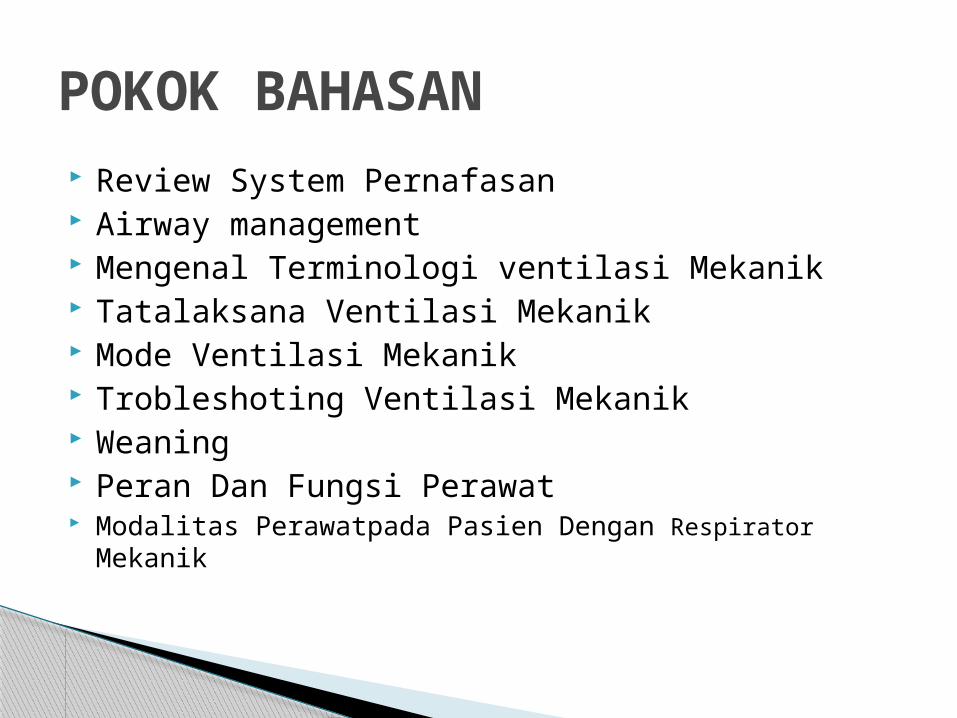
Review System Pernafasan Airway management Mengenal Terminologi ventilasi Mekanik Tatalaksana Ventilasi Mekanik Mode Ventilasi Mekanik Trobleshoting Ventilasi Mekanik Weaning Peran Dan Fungsi Perawat Modalitas Perawatpada Pasien Dengan Respirator Mekanik
POKOK BAHASAN
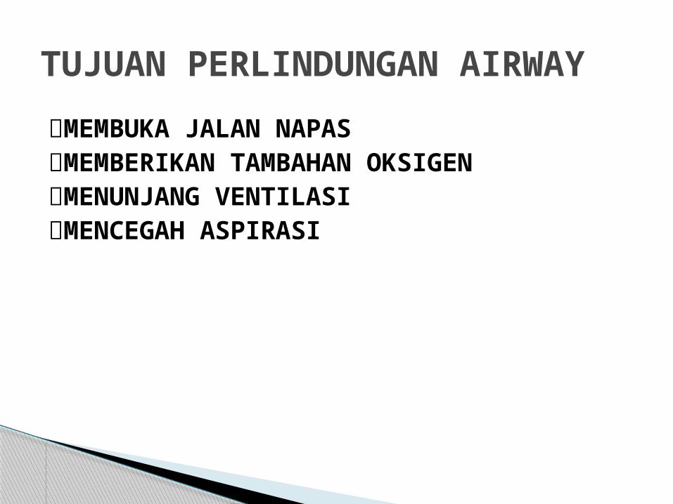
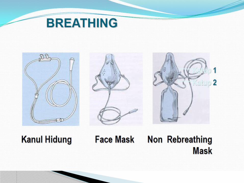
MEMBUKA JALAN NAPASMEMBERIKAN TAMBAHAN OKSIGENMENUNJANG VENTILASIMENCEGAH ASPIRASI
TUJUAN PERLINDUNGAN AIRWAY


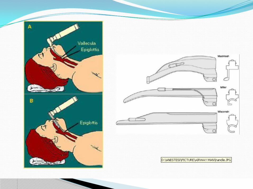
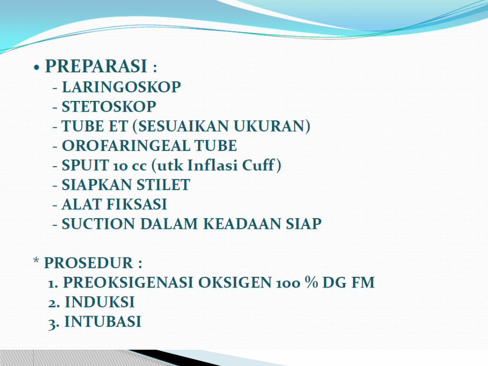
EndotrachealTube


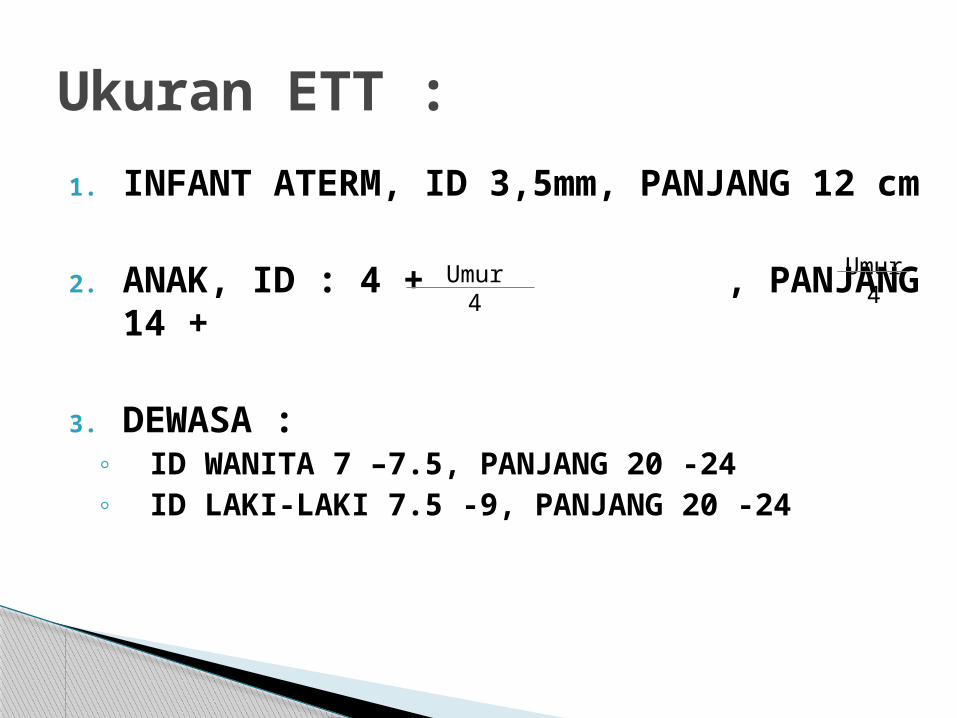
1. INFANT ATERM, ID 3,5mm, PANJANG 12 cm
2. ANAK, ID : 4 + , PANJANG 14 +
3. DEWASA : ◦ ID WANITA 7 –7.5, PANJANG 20 -24◦ ID LAKI-LAKI 7.5 -9, PANJANG 20 -24
Ukuran ETT :
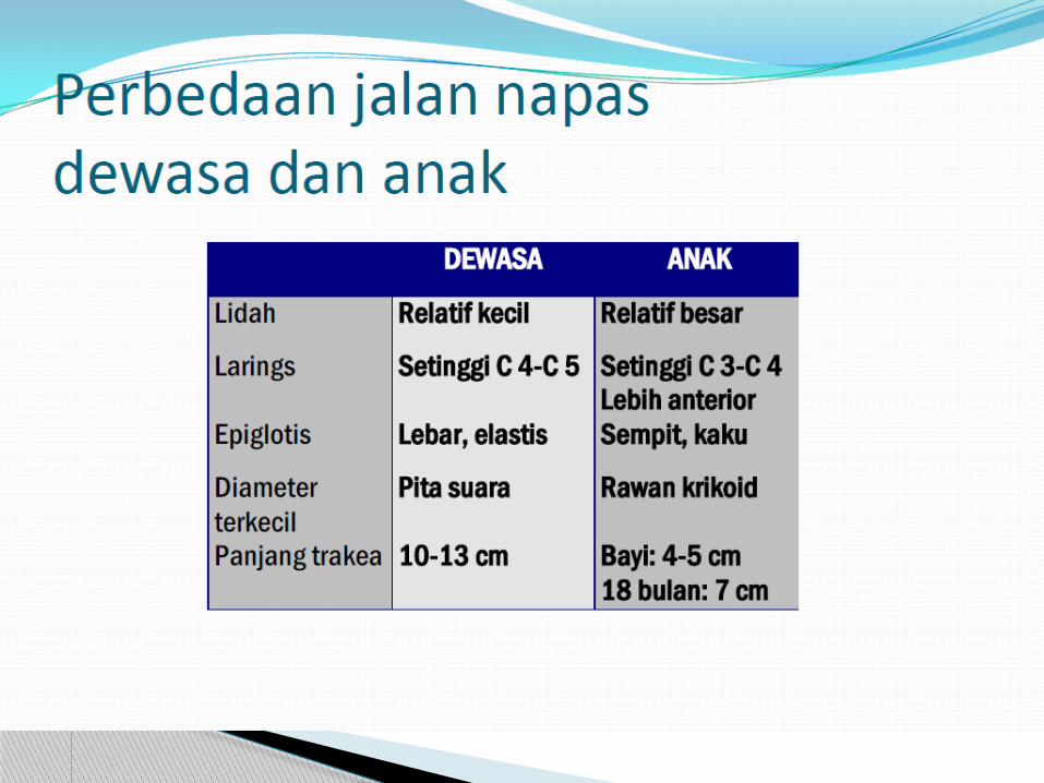
Umur4
Umur4


Masih ingat

Masih ingat

Masih ingat?

Ingatkah……

PROSES MEKANIK, KELUAR MASUKNYA UDARA DARI LUAR KE DALAM PARU DAN SEBALIKNYA YAITU BERNAFAS
TERJADI ANTARA UDARA DALAM ALVEOLUS DENGAN DARAH DALAM KAPILER, PROSESNYA DISEBUT DIFUSI
PROSES RESPIRASI PROSES RESPIRASI
VENTILASI PARU
PERTUKARAN GAS
EKSTERNA EKSTERNA
INTERNA INTERNA
UTILISASI O2
PERTUKARAN GAS
PEMAKAIAN OKSIGEN DALAM SEL PADA REAKSI PELEPASAN ENERGI
PERTUKARAN GAS ANTARA DARAH DENGAN SEL JARINGAN/TISUE

MEKANISME INSPIRASI
KONTRAKSI DIAFRAGMA & INTERKOSTALIS EKST
VOLUME INTRATORAKS >>
INTRAPLEURAL PRESSURE >> NEGATIF
PARU EKSPANSI (MENGEMBANG)
INTRAPULMONAL PRESSURE >> NEGATIF
UDARA MENGALIR KE DALAM PARU
VENTILASI PARUVENTILASI PARU

HUKUM BOYLEHUKUM BOYLE PRESSURE DARI GAS BERBANDING TERBALIK DGN VOL CONTAINER
VOLUME
PRESSURE
VOLUME
PRESSURE
PERUBAHAN VOLUME MENYEBABKAN PERUBAHAN PRESSURE
TABRAKAN PARTIKEL2 GAS
KE DINDING KONTAINER
MENIMBULKAN PRESSURE
VENTILASI PARUVENTILASI PARU

VENTILASI PARUVENTILASI PARUINSPIRASIINSPIRASI
KONTRAKSI OTOT INTERKOSTALIS EKSTERNA IGA TERANGKAT
KONTRAKSI DIAFRAGMA DIAFRAGMA BERGERAK INFERIOR
EKSPIRASIEKSPIRASI
RELAKSASI OTOT INTERKOSTALIS EKSTERNA IGA KE POSISI SEMULA
RELAKSASI DIAFRAGMA DIAFRAGMA BERGERAK KE POSISI SEMULA
INTRATORAK
VOLUME
PRESSURE
VOLUME
PRESSURE

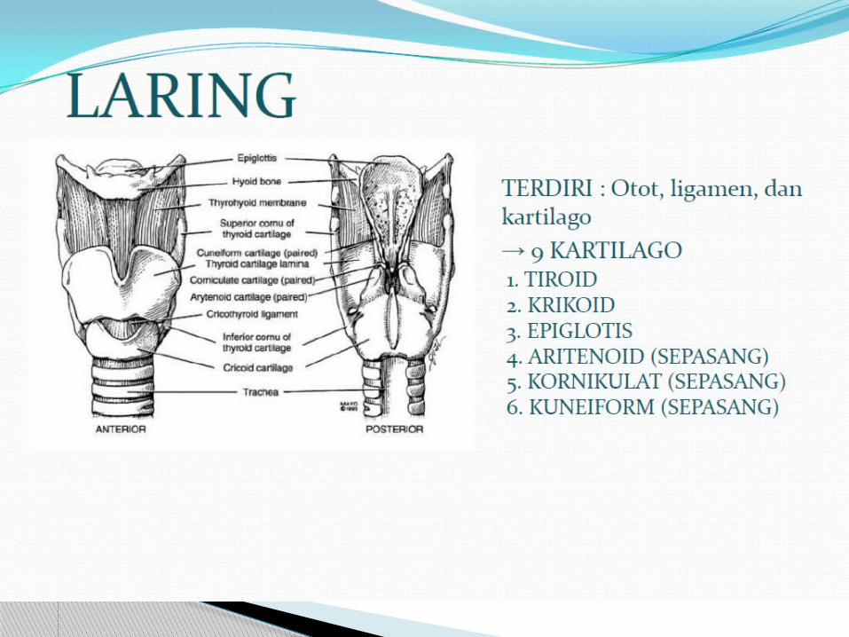
Terminologi Ventilator Mechanic

AIRWAY RESISTANCE (RAW)
AIRWAY RESISTANCE (RAW)
COMPLIANCE (COMPL)
COMPLIANCE (COMPL)
VENTILASI PARUVENTILASI PARU
CL
RAW
LUNG
AIRWAY

– Membatasi jumlah gas yg mengalir melewati jalan nafas (obstruksi jalan nafas)
– Flow = pressure/resistance
– Jika R Flow
– Ditentukan oleh besarnya diameter jalan nafas
– Pada nafas spontan, jika resistance me , secara normal respon tubuh adalah meningkatkan usaha nafas (WoB = RR >>, otot bantu nafas >>)
AIRWAY RESISTANCE (RAW)AIRWAY RESISTANCE (RAW)

FLOW = PRESSURE
RESISTANCE
BRONKUS NORMAL
AIRWAY RESISTANCE (RAW)AIRWAY RESISTANCE (RAW)

FLOW = PRESSURE
RESISTANCE
BRONKODILATASI: EPINEFRINAMINOFILINBETA 2 AGONIS
AIRWAY RESISTANCE (RAW)AIRWAY RESISTANCE (RAW)

FLOW = PRESSURE
RESISTANCE
BRONKOKONSTRIKSI: HISTAMIN
OBSTRUKSI: MUKUS/SEKRET
AIRWAY RESISTANCE (RAW)AIRWAY RESISTANCE (RAW)

FLOW = PRESSURE
RESISTANCE
BRONKOSPASME
TUMOR/SEKRET
ETT TERLALU KECIL
KOLAPS/ATELEKTASIS
AIRWAY RESISTANCE (RAW)AIRWAY RESISTANCE (RAW)

Kaku Elastis
LOW COMPLIANCE
HIGH COMPLIANCE
BALON
COMPLIANCE (COMPL)COMPLIANCE (COMPL)

DefinisiRasio perubahan volume akibat terjadinya perubahan
pressure V/PTerbagi 2;
Compl paru (edema paru, fibrosis, surfactan <<) Compl dinding dada (obesitas, distensi abdomen)
Low compliance ◦ Edema paru, pneumonia berat, ARDS, efusi pleura,
hematopneumotoraks, abdominal pressure >>: u/ memasukkan volume yang diinginkan dibutuhkan pressure yg lebih besar.
High compliance◦ Muscle relaxant, COPD, open chest dgn pressure yg
kecil dapat tidal volume yg masuk besar
COMPLIANCE (COMPL)COMPLIANCE (COMPL)


P-V LOOP
15
30
250
500
0
P
Vol
500
500
250
250
15
30
15
30
LOW COMPLIANCE
HIGH COMPLIANCENORMAL
PEEP 5
INSPIRASI
EKSPIRASI
NAFAS SPONTAN

SHUNT DAN DEAD SPACE

ANATOMICAL DEAD SPACE
ALVEOLAR DEAD SPACE
PHYSIOLOGICAL DEAD SPACE
VENOUS ADMIXTURE (SHUNT)
V/Q =
V/Q > 1
V/Q = 1
V/Q < 1
V/Q = 0
Hubungan Ventilasi (V) dan Perfusi (Q)Hubungan Ventilasi (V) dan Perfusi (Q)
TRAKEA
KAPILER PARU MECHANICAL
DEAD SPACE:
TUBE
CONNECTOR
ET CO2
BREATHING CIRCUIT
NORMAL

FiO2 :
FRAKSI KONSENTRASI OKSIGEN INSPIRASI YG DIBERIKAN (21 – 100%)
TIDAL VOLUME (VT):
JUMLAH GAS/UDARA YG DIBERIKAN VENTILATOR SELAMA INSPIRASI DALAM SATUAN ml/cc ATAU liter. (5-10 cc/kgBB)
FREKUENSI / RATE (f) :
JUMLAH BERAPA KALI INSPIRASI DIBERIKAN VENTILATOR DALAM 1 MENIT (10-12 bpm)
FLOW RATE :
KECEPATAN ALIRAN GAS ATAU VOLUME GAS YG DIHANTARKAN PERMENIT (liter/menit)

- Menentukan siklus respirasi - Jika setting RR pd ventilator 10 x/menit
maka 60/10 = 6 dtk- Jadi T (Total) = T (Inspirasi) + T (Ekspirasi) = 6 dtk
- Berarti inspirasi + ekspirasi harus selesai dalam waktu 6 dtk.
6 dtk 6 dtk
Ins + Eksp
Ins + Eksp
T I M E = WAKTU frekuensiT I M E = WAKTU frekuensi

Sensitivity
Setelan sensitifitas akan menentukan variabel trigger Variabel trigger menentukan kapan ventilator mengenali adanya
upaya nafas pasien Ketika upaya nafas pasien dikenali, ventilator akan memberikan
nafas Variabel trigger dapat berupa pressure atau flow

Pressure Triggering
Upaya nafas pasien dimulai saat terjadi kontraksi otot diafragma
Upaya nafas ini akan menurunkan tekanan (pressure) di dalam sirkuit ventilator (tubing)
X X

Pressure Triggering
Ketika pressure turun mencapai batas yang diset oleh dokter, ventilator akan mentrigger nafas dari ventilator
Namun tetap ada keterlambatan waktu antara upaya nafas pasien dengan saat ventilator mengenali kemudian memberikan nafas.
Baseline
Trigger
Patient effort
Pressure

Pressure Triggering
1. Setelan sensitivity pada -2 cm H2O
2. Gambar dibawah menunjukkan pada 2 nafas pertama upaya nafas pasien mencapai sensitivitas yang diset; sedangkan gbr ketiga terlihat bahwa upaya nafas pasien tidak mencapai sensitivitas yg diset sehingga ventilator tidak mengenalinya
-2 cm H2O

Flow Triggering
Ventilator secara kontinyu memberikan flow rendah ke dalam sirkuit pasien (open system)
Delivered flowReturned flow
No patient effort

Flow Triggering
1. Upaya nafas dimulai saat kontraksi diafragma2. Saat pasien bernafas beberapa bagian flow didiversi ke
pasien
Delivered flowLess flow returned

Flow Triggering
1. Level flow yg rendah akan lebih nyaman untuk pasien (lebih sensitif)
2. Keterlambatan waktu lebih kecil dibanding pressure trigger3. Meningkatan respon waktu dari ventilator
All inspiratory efforts recognized
Time
Pressure

PEEP
DEFINISI◦POSITIVE END EXPIRATORY PRESSURE◦SEWAKTU AKHIR EXPIRATORY, AIRWAY
PRESSURE TIDAK KEMBALI KETITIK NOL DIGUNAKAN BERSAMA DENGAN MODE
LAIN SEPERTI; SIMV, ACV ATAU PS DISEBUT CPAP JIKA DIGUNAKAN PADA
MODE NAFAS SPONTAN

PEEP(Positive End Expiratory Pressure)PEEP(Positive End Expiratory Pressure)
PEEP 5
REDISTRIBUSI CAIRAN EKSTRAVASKULAR PARU
MENINGKATKAN VOLUME ALVEOLUS
MENGEMBANGKAN ALVEOLI YG KOLAPS (ALVEOLI RECRUITMENT)

REDISTRIBUSI CAIRAN EKSTRAVASKULAR PARU
+10
0
A B
PEEP(Positive End Expiratory Pressure)PEEP(Positive End Expiratory Pressure)

MENINGKATKAN VOLUME ALVEOLUS
+20
+10
0
A B C
PEEP(Positive End Expiratory Pressure)PEEP(Positive End Expiratory Pressure)

Indications for Mechanical ventilation


Goals
Patient comfort and rest Reversal of Hypoxemia Reversal of acute respiratory acidosis Reversal of respiratory muscle
fatigue Prevention/Reversal of atelectasis Decrease myocardial ischemic Allowance of neuromuscular blockade Improve lung compliance

Variables for Initial Settings
Fraction of Inspired O2 - FIO2 Tidal Volume - TV Respiratory Rate - RR(f) Flow Rate - Vi(L/m) PSV Mode (A/C, SIMV, PS) PEEP (cm of H2O)

Mechanical Ventilation
Non Invasive
Invasive
Non Invasive: Ventilatory support that is given without establishing endo- tracheal intubation or tracheostomy is called Non invasive mechanical ventilation
Invasive: Ventilatory support that is given through endo-tracheal intubation or tracheostomy is called as Invasive mechanical ventilation

Non invasive
Negative pressure
Producing Neg. pressure intermittently in the pleural space/ around the thoracic cage
Positive pressure
Delivering air/gas with positive pressure to the airway
e.g.: Iron Lung
BiPAP & CPAP


Non Invasive Mechanical ventilation
การใช้� non-invasive mechanical ventilation ในผู้�ป่�วยที่��เหมาะสม จะลดโอกาสการใส�ที่�อช้�วยหายใจได�

Non invasive mechanical ventilation
Head gear
Interface (mask)
ventilator

Positive Pressure
Pressure cycle Volume cycle Time cycle
Invasive


Modes of Ventilation


Modes of Ventilation
Mode◦ Description of a breath type and the timing
of breath delivery Basically there are three breath delivery
techniques used with invasive positive pressure ventilation CMV – controlled mode ventilation SIMV – synchronized Spontaneous modes

Modes of Ventilation
CMV◦ Continuous Mandatory Ventilation
All breaths are mandatory and can be volume or pressure targeted
Controlled Ventilation – when mandatory breaths are time triggered
Assist/Control Ventilation – when mandatory breaths are either time triggered or patient triggered

Modes of Ventilation
CMV◦ Continuous Mandatory Ventilation
Controlled Ventilation – when mandatory breaths are time triggered Mandatory breath – ventilator determines the start
time (time triggered) and/or the volume or pressure target

Modes of Ventilation
CMV◦ Controlled Ventilation
Appropriate when a patient can make no effort to breathe or when ventilation must be completely controlled Drugs Cerebral malfunctions Spinal cord injury Phrenic nerve injury Motor nerve paralysis

Modes of Ventilation
CMV◦ Controlled Ventilation
In other types of patients, controlled ventilation is difficult to use unless the patient is sedated or paralyzed with medications Seizure activity Tetanic contractions Inverses I:E ratio ventilation Patient is fighting (bucking) the ventilator Crushed chest injury – stabilizes the chest Complete rest for the patient

Modes of Ventilation
CMV◦ Controlled Ventilation
Adequate alarms must be set to safeguard the patient Ex. disconnection
Sensitivity should be set so that when the patient begins to respond, they can receive gas flow from the patient
Do not lock the patient out of the ventilator!

Modes of Ventilation
CMV◦ Assist/Control Ventilation
A time or patient triggered CMV mode in which the operator sets a minimum rate, sensitivity level, type of breath (volume or pressure)
The patient can trigger breaths at a faster rate than the set minimum, but only the set volume or pressure is delivered with each breath

Modes of Ventilation
CMV◦ Assist/Control Ventilation
Indications Patients requiring full ventilatory support Patients with stable respiratory drive
Advantages Decreases the work of breathing (WOB) Allows patients to regulate respiratory rate Helps maintain a normal PaCO2
Complications Alveolar hyperventilation

Modes of Ventilation
CMV◦ Volume Controlled –
CMV Time or patient
triggered, volume targeted, volume cycled ventilation
Graphic (VC-CMV) Time-triggered, constant
flow, volume-targeted ventilation

Modes of Ventilation
CMV◦ Volume Controlled – CMV
Time or patient triggered, volume targeted, volume cycled ventilation
Graphic (VC-CMV) Time-triggered,
descending-flow, volume-targeted ventilation

Modes of Ventilation
CMV◦ Pressure Controlled – CMV
PC – CMV (AKA – Pressure control ventilation - PCV)
Time or patient triggered, pressure targeted (limited), time cycled ventilation
The operator sets the length of inspiration (Ti), the pressure level, and the backup rate of ventilation
VT is based on the compliance and resistance of the patient’s lungs, patient effort, and the set pressure

Modes of Ventilation
CMV◦ Pressure Controlled – CMV
Note inspiratory pause

Modes of Ventilation
CMV◦ Pressure Controlled – CMV
Note shorter Ti

Modes of Ventilation
CMV◦ Pressure Controlled – CMV
Airway pressure is limited, which may help guard against barotrauma or volume-associated lung injury Maximum inspiratory pressure set at 30 – 35 cm
H2O Especially helpful in patients with ALI and ARDS
Allows application of extended inspiratory time, which may benefit patients with severe oxygenation problems
Usually reserved for patient who have poor results with a conventional ventilation strategy of volume ventilation

Modes of Ventilation
CMV◦ Pressure Controlled – CMV
Occasionally, Ti is set longer than TE during PC-CMV; known as Pressure Control Inverse Ratio Ventilation Longer Ti provides better oxygenation to some
patients by increasing mean airway pressure Requires sedation, and in some cases paralysis

Modes of Ventilation
IMV and SIMV◦ Intermittent Mandatory Ventilation – IMV
Periodic volume or pressure targeted breaths occur at set interval (time triggering)
Between mandatory breaths, the patient breathes spontaneously at any desired baseline pressure without receiving a mandatory breath Patient can breathe either from a continuous flow
or gas or from a demand valve

Modes of Ventilation
IMV and SIMV◦ Intermittent Mandatory Ventilation – IMV
Indications Facilitate transition from full ventilatory support
to partial support
Advantages Maintains respiratory muscle strength by
avoiding muscle atrophy Decreases mean airway pressure Facilitates ventilator discontinuation – “weaning”

Modes of Ventilation
IMV and SIMV◦ Intermittent Mandatory Ventilation – IMV
Complications When used for weaning, may be done too quickly
and cause muscle fatigue Mechanical rate and spontaneous rate may
asynchronous causing “stacking” May cause barotrauma or volutrauma

Modes of Ventilation
IMV and SIMV◦ Synchronized IMV
Operates in the same way as IMV except that mandatory breaths are normally patient triggered rather than time triggered (operator set the volume or pressure target)
As in IMV, the patient can breathe spontaneously through the ventilator circuit between mandatory breaths

Modes of Ventilation
IMV and SIMV◦ Synchronized IMV
At a predetermined interval (respiratory rate), which is set by the operator, the ventilator waits for the patient’s next inspiratory effort
When the ventilator senses the effort, the ventilator assists the patient by synchronously delivering a mandatory breath

Modes of Ventilation
IMV and SIMV◦ Synchronized IMV
If the patient fails to initiate ventilation within a predetermined interval, the ventilator provides a mandatory breath at the end of the time period

Modes of Ventilation
IMV and SIMV◦ Synchronized IMV
Indications Facilitate transition from full ventilatory support
to partial support
Advantages Maintains respiratory muscle strength by
avoiding muscle atrophy Decreases mean airway pressure Facilitates ventilator discontinuation – “weaning”

Modes of Ventilation
IMV and SIMV◦ Synchronized IMV
Complications When used for weaning, may be done too quickly
and cause muscle fatigue

Modes of Ventilation
Spontaneous Modes◦ Three basic means of providing support
for continuous spontaneous breathing during mechanical ventilation
Spontaneous breathing
CPAP
PSV – Pressure Support Ventilation

Modes of Ventilation
Spontaneous Modes◦ Spontaneous breathing
Patients can breathe spontaneously through a ventilator circuit; sometimes called T-Piece Method because it mimics having the patient ET tube connected to a Briggs adapter (T-piece)
Advantage Ventilator can monitor the patient’s breathing
and activate an alarm if something undesirable occurs
Disadvantage May increase patient’s WOB with older
ventilators

Modes of Ventilation
Spontaneous Modes◦ CPAP
Ventilators can provide CPAP for spontaneously breathing patients Helpful for improving
oxygenation in patients with refractory hypoxemia and a low FRC
CPAP setting is adjusted to provide the best oxygenation with the lowest positive pressure and the lowest FiO2

Continuous Positive Airway Pressure (CPAP) Positive airway pressure maintained
throughout respiratory cycle: during inspiratory and expiratory phases
Can be administered via ETT or nasal prongs

Modes of Ventilation
Spontaneous Modes◦ CPAP
Advantages Ventilator can
monitor the patient’s breathing and activate an alarm if something undesirable occurs

Modes of Ventilation
Spontaneous Modes◦ PEEP (Positive End Expiratory Pressure)
“According to its purest definition, the term PEEP is defined as positive pressure at the end of exhalation during either spontaneous breathing or mechanical ventilation. However, use of the term commonly implies that the patient is also receiving mandatory breaths from a ventilator.” (Pilbeam)
PEEP becomes the baseline variable during mechanical ventilation

Modes of Ventilation
Spontaneous Modes◦ PEEP
Helps prevent early airway closure and alveolar collapse and the end of expiration by increasing (and normalizing) the functional residual capacity (FRC) of the lungs
Facilitates better oxygenation
NOTE: PEEP is intended to improve oxygenation, not to provide ventilation, which is the movement of air into the lungs followed by exhalation

Modes of Ventilation
Spontaneous Modes◦ Pressure Support Ventilation – PSV
Patient triggered, pressure targeted, flow cycled mode of ventilation
Requires a patient with a consistent spontaneous respiratory pattern
The ventilator provides a constant pressure during inspiration once it senses that the patient has made an inspiratory effort

Modes of Ventilation
Spontaneous Modes◦ PSV

Modes of Ventilation
Spontaneous Modes◦ PSV
Indications Spontaneously breathing patients who require
additional ventilatory support to help overcome WOB, CL, Raw Respiratory muscle weakness
Weaning (either by itself or in combination with SIMV)

Modes of Ventilation
Spontaneous Modes◦ PSV
Advantages Full to partial ventilatory support Augments the patients spontaneous VT
Decreases the patient’s spontaneous respiratory rate
Decreases patient WOB by overcoming the resistance of the artificial airway, vent circuit and demand valves
Allows patient control of TI, I, f and VT

Modes of Ventilation
Spontaneous Modes◦ PSV
Advantages Set peak pressure Prevents respiratory muscle atrophy Facilitates weaning Improves patient comfort and reduces need for
sedation May be applied in any mode that allows
spontaneous breathing, e.g., VC-SIMV, PC-SIMV

Modes of Ventilation
Spontaneous Modes◦ PSV
Disadvantages Requires consistent spontaneous ventilation Patients in stand-alone mode should have back-
up ventilation VT variable and dependant on lung
characteristics and synchrony Low exhaled E Fatigue and tachypnea if PS level is set too low

Modes of Ventilation
Spontaneous Modes◦ Flow Cycling During PSV
Flow cycling occurs when the ventilator detects a decreasing flow, which represents the end of inspiration
This point is a percentage of peak flow measured during inspiration PB 7200 – 5 L/min Bear 1000 – 25% of peak flow Servo 300 – 5% of peak flow
No single flow-cycle percent is right for all patients

Modes of Ventilation
Spontaneous Modes◦ Flow Cycling During
PSV Effect of changes in
termination flow
A: Low percentage (17%)
B: High percentage (57%)
Newer ventilators have an adjustable flow cycle criterion, which can range from 1% - 80%, depending on the ventilator

Modes of Ventilation
Spontaneous Modes◦ PSV during SIMV
Spontaneous breaths during SIMV can be supported with PSV (reduces the WOB)
PCV – SIMV with PSV

Modes of Ventilation
Spontaneous Modes◦ PSV during SIMV
Spontaneous breaths during SIMV can be supported with PSV
VC – SIMV with PSV

Modes of Ventilation
Spontaneous Modes◦ PSV
NOTE: During pressure support ventilation (PSV), inspiration ends if the inspiratory time (TI) exceeds a certain value. This most often occurs with a leak in the circuit. For example, a deflated cuff causes a large leak. The flow through the circuit might never drop to the flow cycle criterion required by the ventilator. Therefore, inspiratory flow, if not stopped would continue indefinitely. For this reason, all ventilators that provide pressure support also have a maximum inspiratory time.

Modes of Ventilation
Spontaneous Modes◦ PSV
Setting the Level of Pressure Support Goal: To provide ventilatory support
Spontaneous tidal volume is 10 – 12 mL/Kg of ideal body weight
Maintain spontaneous respiratory rate <25/min
Goal: To overcome system resistance (ET Tube, circuit, etc.) in the spontaneous or IMV/SIMV mode Set pressure at (PIP – Pplateau) achieved in a
volume breath or at 5 – 10 cm H2O

Modes of Ventilation
Spontaneous Modes◦ PSV
Exercise: Using the PIP and the PPlateau from the pressure waveform below, recommend a pressure support setting for this patient (patient is in VC-SIMV mode)
35
25
Answer: 10 cm H2O

Modes of Ventilation
Spontaneous Modes◦ PSV - The results of your work
35 cm H2O
10 cm H2O

Modes of Ventilation
Spontaneous Modes◦ Bilevel Positive Airway Pressure (BiPAP)
An offshoot of PEEP/CPAP therapy Most often used in NPPV AKA
Bilevel CPAP Bilevel PEEP Bilevel Pressure Support Bilevel Pressure Assist Bilevel Positive Pressure Bilevel Airway Pressure

Modes of Ventilation
Spontaneous Modes◦ Bilevel Positive Airway Pressure (BiPAP)
Commonly patient triggered but can be time triggered, pressure targeted, flow or time cycled
The operator sets two pressure levels IPAP (Inspiratory Positive Airway Pressure)
IPAP is always set higher than EPAP Augments VT and improves ventilation
EPAP (Expiratory Positive Airway Pressure) Prevents early airway closure and alveolar collapse
at the end of expiration by increasing (and normalizing) the functional residual capacity (FRC) of the lungs
Facilitates better oxygenation

Modes of Ventilation
Spontaneous Modes◦ Bilevel Positive Airway Pressure (BiPAP)
The operator sets two pressure levels IPAP EPAP
NOTE: The pressure difference between IPAP and EPAP is pressure support


Trouble Shooting (Mechanical Ventilation)

Precautions that would reduce troubles
I. Power: Plug into a grounded AC power with
correct voltage receptacle.
Secure the power cord properly.
Battery Back up:
Check the battery level before connecting. Charging should be carried out
regularly. Remember it is for short term use.

II. Gas Source
Preferable to have centralised supply. If cylinders used, should be full Spare cylinders should be available. Gas hoses should be in good condition. Hoses – not contaminated with grease or
oil (combustible) Availability of compressors should be
ensured. Gases should remain dry and clean.

III. Personnel
Properly trained personnel should only use.
Familiarising staff with operator’s manuel before using on a patient.
(One manufacturer’s manual may not exactly match with other brands).
Appropriate monitoring the functioning state of the ventilator while in use.

Contd…
Familiarizing staff with alarm system. Do not place ventilators in a combustible or
explosive environment. Do not use with flammable anaesthetic
agents such as nitrous oxide and ether.

IV Servicing and Testing
Qualified personnel should undertake servicing.
Ventilator housing should not be opened while it is still connected with power.
Follow the specifications mentioned in the service manual.
Use replacement parts supplied by the manufacturer only.

Contd….
General servicing at regular intervals should be done.
Run the prescribed tests and calibrations before using the ventilator on a patient.
Ensure that the ventilators pass all the tests before putting them in to clinical use.

ALARMS
All ventilators are equipped with visual and audible alarms which notify the user problems.

Points to remember
Never ignore an alarm. Never mute the alarm on regular
basis. Find out for yourself what alarm is
on. Check the patient. Silence the alarm.

Act Swiftly Depending upon the patient’s status
and nature of the alarm, act appropriately.
This includes disconnecting the ventilator and connecting another means of ventilation to patient – Bain’s/ Ambu.

Do not forget
The use of an alarm monitoring system does not give absolute assurance of warning for every form of trouble that may occur with the ventilator.

Do not be like this !
But hear the alarm and respond
See the problem and Ask if you do not know what to
do

Common Troubles and Shooting
Ensure Alarm knobs / switches are turned on and functional.
Alarm Cause ShootingApnoea •No breath was
delivered for the operator set apnoea time in spont, SIMV, AC, CMV & NIV modes•Because spontaneous Ventilation is too high or patient effort is too minimal•Trigger level set improperly.
•Check the patient- Arouse if needed•Activate back up facility if it was not done already.•Consider switching over to any mandatory mode•Or go up on rate•Set trigger level appropriately

Low SpO2
Air / O2 Blender continuousalarm
Delivery of O2 : FiO2, PEEP
• High resistance due to various clinical reasons
Supply pressures are inadequate.
• Disconnect patient from ventilator• Manually bag with Bain’s and Ambu.
• Insert the gas hose fittings (air & O2)
correctly into the wall outlets.• Ensure wall outlets has adequate pressure

High Pressure Alarm
The measured peak inspiratory pressure is great than set level because of•Secretions in airway • Partial block – (ETt)• Kinking of tube• Biting the tube• Water in the tube• Cuff herniation• Deep Rt. sided intubation • Fighting the ventilator
•Suctioning, Irrigation• Release tubings• Bite block insertion• Empty the tubings and water traps• Deflate & reinflate cuff 3-4 times• Reposition the ET tube• Reposition the patient• Re assurance• Sedation &
medication (pain)

Low pressure or Low min.VentOrLow exhaled volume or Disconnection
The measured PIP is lesser than the set minimum level because of • cuff leak.• Leak in the circuit • Connections may be loose• ET tube displacement• Disconnection• Inadequate flow
•Evaluate cuff pressure at regular intervals.• Reinflate if leak / ruptured is noticed – change ET tube.• Check circuits, junctions- tighten or replace.• Check water traps • Check ET tube placement. Position it properly.• Reconnect ventilator.• Patient may require higher flow.

High pressure alarm
• Cough• Increased airway resistance or decreased compliance because of• Bronchospasm• Atelectasis• Fluid overload
• Pneumothorax
• Medication• Bronchodilators• Adjust the settings ¯VT & Rate• Adjust the settings VT Rate, PEEP
(Peak pressure to be monitored)
• Immediate intervention

Auto Cycling
High Tidal Volume
Leak & Improper trigger setting
Patient trying to take more volume of air
• Secure all tubings tight• Set proper trigger level
• Increase flow rate or Increase tidal volume

Weaning from Mechanical Ventilation

Definition of Weaning
The transition process from total ventilatory support
to spontaneous breathing.
This period may take many forms ranging from abrupt withdrawal to gradual withdrawal from
ventilatory support.

Weaning
* Discontinuation of IPPV is achieved in most patients without difficulty
* up to 20% of patients experience difficulty
* requires more gradual process so that they can progressively assume spont. respiration
* the cost of care, discontinue IPPV should proceed as soon as possible

Reversible reasons for prolonged mechanical ventilation
Inadequate respiratory drive Inability of the lungs to carry
out gas exchange effectively Psychological dependency Inspiratory fatigue

WeaningPatients who fail attempts at weaning
constitute a unique problem in critical care
It is necessary to understand the mechanisms of ventilatory failure in order to address weaning in this population

Why patients are unable to sustain spontaneous breathing
Concept of Load exceeding Capacity to breathe
Load on respiratory systemCapacity of respiratory system

Balance Load vs Capacity
Most patients fail the transition from ventilator support to sustain spont. breathing because of failure of the respiratory muscle pump
They typically have a resp muscle load the exceeds the resp neuromuscular capacity

Load on Respiratory System
Need for increase ventilation increased carbon dioxide
production increased dead space ventilation increased respiratory driveIncreased work of breathing

Causes of Inspiratory respiratory muscle fatigue
Nutrition and metabolic deficiencies: K, Mg, Ca, Phosphate and thyroid hormone
Corticosteroids Chronic renal failure Systemic disceases; protein synthesis, degradation, glycogen stores Hypoxemia and hypercapnia

Capacity of respiratory system
Central drive to breatheTransmission of CNS signal via Phrenic
nerve Impairment of resp muscles to generate
effective pressure gradients Impairment of normal muscle force
generation


Definitions
Tolerated – observations to monitorLook at patient, do they look unsettled/tired/stressed?
Is respiratory rate below 35bpm & above 8bpm?
Are O2 saturations above 90%? (or as appropriate for patient)
Are ABGs acceptable for the patient?
Is PaO2 / FiO2 ratio >27.5kpc?
Is TV 5ml/kg?
Is patient cardiovasculary stable?
Is patient settled and showing no signs of fatigue?
Is respiratory rate/TV ratio <105 breaths/min (spontaneous rate for 1 min divided by the TV in Litres)
Signs of fatigue are: Decreasing TV Increasing respiratory rate Changes in blood gases Decreases in O2 saturations Tachycardic ECG changes Hypertension Breathlessness Use of accessory muscles Changes in conscious levels Sweating
Inform Anaesthetist and discontinue weaning if any changes noted.
Document: All changes on ventilator and check ventilator changes with another nurse.


Peran Dan Fungsi Perawat

Peran Dan Fungsi Perawat


SETTINGS
O2 Air Power
Ventilator
Patient


ม�น��าใน circuit มากจนต้�องเที่ที่"�งบ่�อยๆ

suction ETT

Stabilize the ETT

VAP: Preventive strategies


Nebulisation

NURSE

NURSE

Precaution & Care
Tracheobronchial Hygiene:
Placement of tube: Chest movementAuscultationPost intubation X-ray
Cuff pressure: If insufficient Leak - Displacement of the tube, Aspiration high pressure - Tracheal stenosis
Desired Pressure - 20-30cm water

Humidification Filling water & adjusting temperature appropriately :
If inadequate: secretions would become thicker and lead to tube block
Medication: Besides specific therapautic drugs the
following basic drugs are to be given. Sedatives & paralysing agents if needed. Analgesics Diuretics to reduce circulating fluid and
volume overload Reduce Gastric Acid: H2 blockers

Suction Should be done on PRN basis Ascultate and assess View the chest X-ray Determine the need and for effective
suctioning Hyperoxygenation & ventilation –
ambu/normal Keep strict vigil on the cardiac monitor
pulse oximeter during and soon after suctioning
If necessary carry out effective chest physio

Monitoring:
Continuous and Periodic monitoring of Vital parameters such as temperature,SpO2,
Pulse, BP,ECG pattern, breath rate etc.
Ventilator settings: All settings should be recorded as per the doctors order
Sensorium Intake and output Level of comfort Arterial blood gases twice daily

Nebulisation
It is advisable to put all the patients on bronchodilators on regular basis.
Nebulise as per the doctor’s order

Monitoring for infection
Colour, consistency, and amount of the sputum / secretions with each suctioning should be observed.
Fever and other parameters have to closely observed for any other infection. (central line, etc)

Oxygen toxicity
Try and maintain a SpO2 of > 90% and PaO2 of 60 – 90 mmHg with minimum possible FiO2 to prevent O2 toxicity.
Especially for COPD patients : Maintain SpO2 of 85 – 90% and PaO2
of 55 – 70 mmHg.

Nutrition: Enteral nutrition to support the
patient’s metabolic needs and defend against infection.
Avoid high carbohydrate diet during weaning.
NG tube if necessary – relieves gastric distension and prevents aspiration.

Stress gastric ulcer Very common in critically ill patients Send stools for occult blood and
gastric juice for pH estimation Auscultate bowel movements Sedation and antacids adequately.

Alarms & Positioning: Never keep alarm system muted Never ignore even when you know the
cause for the alarm and may not be fatal
Place the patient in low or semi Fowler’s position to improve comfort and facilitate respiration.

Communication:
If conscious, explain the environment, procedures, co-operation expected etc.
Use verbal & non verbal methods Use paper & pen if necessary Provide calling bell if necessary Reassurance and support the patient
during the period of anxiety, frustration and hopelessness
Document patient’s emotional response and any signs of psychosis
Include family in the care

Teach…… Co-operation with medical and nursing
interventions Certain breathing techniques The patient to recognize the importance
of breathing techniques. Frequent assessment of consciousness
level, adequate rest etc. are necessary.


• Multiply the tracheal tubes inner diameter by 2.
Then use the next smallest size catheter.
Example: 6mm ETT: 6 x 2 = 12; nextsmallest catheter is 10 French
Example: 8mm ETT: 6 x 2 = 16; nextsmallest catheter is 14 French
Choosing the right catheter


Hypoxemia - #1 complication– give oxygen before and after– catheter size• if the catheter is too big, there will be little or no airentrained– Time – suction no more that 15 secs.Tissue trauma– May be able to prevent it . . .– catheter selection?– intermittent vs. continuous– a “delicate touch”– vacuum adjustment
Complications and Hazards of Suctioning

Complications and Hazards of SuctioningCardiac arrhythmias– Vagal stimulation will cause
• bradycardia
– Hypoxemia can cause• PVCs• tachycardia
– If these occur…STOP procedure and give oxygen

Extubation
The nurse should explain the procedure to the patient and prepare suction. The patient should be sitting up at least 45 degrees.
Prior to extubating, the patient should be suctioned both via the ETT and orally.
All fasteners holding the ETT should be loosened.

Extubation
A sterile suction catheter should be inserted into the ETT and withdrawn as the tube is removed.
The ETT should be removed in a steady, quick motion as the patient will likely cough and gag.

Extubation
The patient should be asked to cough and speak. Quite often, the patient’s first request is for water because of a dry, sore throat. Generally, you can immediately swab the patient’s mouth with an oral swab dipped in water.

Post-Extubation Care
Humidified oxygen Respiratory exercises Assessment and monitoring Prepare for intubation