


Bahasa
Halaman
Hukum

Viremic HIV Infected Individuals with High CD4 T Cellsand Functional Envelope Proteins Show Anti-gp41Antibodies with Unique Specificity and FunctionMarta Curriu1, Hughes Fausther-Bovendo2, Marıa Pernas3, Marta Massanella1, Jorge Carrillo1, Cecilia
Cabrera1, Cecilio Lopez-Galındez3, Bonaventura Clotet1,4, Patrice Debre2, Vincent Vieillard2*, Julia
Blanco1*
1 IrsiCaixa-HIVACAT, Institut de Recerca en Ciencies de la Salut Germans Trias i Pujol (IGTP), Hospital Germans Trias, Universitat Autonoma de Barcelona, Badalona,
Barcelona, Catalonia, Spain, 2 INSERM UMR-S 945, Laboratoire Immunite et Infection, Hopital Pitie-Salpetriere, UPMC, Universite Paris-6, Paris, France, 3 Centro Nacional de
Microbiologıa (CNM), Instituto de Salud Carlos III, Majadahonda, Madrid, Spain, 4 Lluita contra la SIDA Foundation, Institut de Recerca en Ciencies de la Salut Germans Trias
i Pujol, Hospital Universitari Germans Trias i Pujol, Universitat Autonoma de Barcelona, Badalona, Barcelona, Spain
Abstract
Background: CD4 T-cell decay is variable among HIV-infected individuals. In exceptional cases, CD4 T-cell counts remainstable despite high plasma viremia. HIV envelope glycoprotein (Env) properties, namely tropism, fusion or the ability toinduce the NK ligand NKp44L, or host factors that modulate Env cytopathic mechanisms may be modified in such situation.
Methods: We identified untreated HIV-infected individuals showing non-cytopathic replication (VL.10,000 copies/mL andCD4 T-cell decay,50 cells/mL/year, Viremic Non Progressors, VNP) or rapid progression (CD4 T-cells,350 cells/mL withinthree years post-infection, RP). We isolated full-length Env clones and analyzed their functions (tropism, fusion activity andcapacity to induce NKp44L expression on CD4 cells). Anti-Env humoral responses were also analyzed.
Results: Env clones isolated from VNP or RP individuals showed no major phenotypic differences. The percentage offunctional clones was similar in both groups. All clones tested were CCR5-tropic and showed comparable expression andfusogenic activity. Moreover, no differences were observed in their capacity to induce NKp44L expression on CD4 T cellsfrom healthy donors through the 3S epitope of gp41. In contrast, anti- Env antibodies showed clear functional differences:plasma from VNPs had significantly higher capacity than RPs to block NKp44L induction by autologous viruses. Consistently,CD4 T-cells isolated from VNPs showed undetectable NKp44L expression and specific antibodies against a variable regionflanking the highly conserved 3S epitope were identified in plasma samples from these patients. Conversely, despitecontinuous antigen stimulation, VNPs were unable to mount a broad neutralizing response against HIV.
Conclusions: Env functions (fusion and induction of NKp44L) were similar in viremic patients with slow or rapid progressionto AIDS. However, differences in humoral responses against gp41 epitopes nearby 3S sequence may contribute to the lackof CD4 T cell decay in VNPs by blocking the induction of NKp44L by gp41.
Citation: Curriu M, Fausther-Bovendo H, Pernas M, Massanella M, Carrillo J, et al. (2012) Viremic HIV Infected Individuals with High CD4 T Cells and FunctionalEnvelope Proteins Show Anti-gp41 Antibodies with Unique Specificity and Function. PLoS ONE 7(2): e30330. doi:10.1371/journal.pone.0030330
Editor: Yuntao Wu, George Mason University, United States of America
Received September 9, 2011; Accepted December 13, 2011; Published February 1, 2012
Copyright: � 2012 Curriu et al. This is an open-access article distributed under the terms of the Creative Commons Attribution License, which permitsunrestricted use, distribution, and reproduction in any medium, provided the original author and source are credited.
Funding: Spanish AIDS network ‘Red Tematica Cooperativa de Investigacion en SIDA’, RIS, RD06/0006 (to JB, CLG, MP and MC). ‘‘Fundacion para la Investigaciony Prevencion del SIDA en Espana’’ FIPSE grant 08/36725 (to CC). Health Department of the Catalan Government (Generalitat de Catalunya) and Instituto de SaludCarlos III (contracts CP04/00271 and CES98/3047 to CC and JB, respectively). Instituto de Salud Carlos III (contract CD09/00150 to JC). Work in CNM is supported bygrant SAF 2007-61036 and SAF 2010-17226 and by FIPSE grants 36558/06, 36641/07, 36779/08, 360766/09. The funders had no role in study design, datacollection and analysis, decision to publish, or preparation of the manuscript.
Competing Interests: The authors have read the journal’s policy and have the following conflicts: B.C. has received research funding from Merck Sharp &Domme and has received honoraria for speaking and serving on advisory boards from Abbott, Bristol-Myers Squibb, Boehringer-Ingelheim, Gilead Sciences,GlaxoSmithKline, Roche, Janssen-Cilag, and Merck Sharp & Domme. J.B. has received honoraria as a consultant for GlaxoSmithKline. This does not alter theauthors’ adherence to all the PLoS ONE policies on sharing data and materials. All other authors: no conflicts.
* E-mail: [email protected] (JB); [email protected] (VV)
Introduction
HIV infection is characterized by an important decrease on
CD4 T cell count, resulting in weakened immune responses that
lead to AIDS-defining events. Progression to AIDS among HIV-
infected individuals is highly heterogeneous due to host and viral
factors [1,2], ranging from ,3 years in rapid-progressors (RP) to
.10 years in long term nonprogressors (LTNP). Usually, LTNPs
show undetectable or controlled (,2000 copies/ml) HIV replica-
tion; however, a reduced number of LTNP show uncontrolled
viral load (VL.2,000 copies/ml) with asymptomatic HIV
infection over almost 10 years after seroconversion [1]. Further-
more, a really limited group of HIV-infected individuals show a
particular discordant profile with high viral load (VL.10,000
copies/ml) in the absence of quantitative immune defects (Viremic
Non-Progresors, VNP). This fact is paradoxical, as HIV-infected
PLoS ONE | www.plosone.org 1 February 2012 | Volume 7 | Issue 2 | e30330

CD4 T lymphocytes have a shortened lifespan due to direct
cytopathic effects of HIV [3] or lysis by immune cells [4].
Moreover, the number of dying cells in infected individuals greatly
exceeds the number of HIV-infected cells [4] due to detrimental
effects of immune activation [4], HIV proteins [5,6] or abortive
infection [7] on the bystander uninfected CD4 T cell population.
Among viral determinants, the envelope glycoprotein (gp120/
gp41, Env), which defines HIV tropism for CCR5 or CXCR4, can
influence CD4 T cell decline in vitro [8] and in vivo [9].
Furthermore, Env is a major determinant of viral pathogenicity,
which is related to the fusogenic activity of gp41 [10,11] and
affects both infected [12] and bystander CD4 T cells [13–15].
This plethora of cytopathic mechanisms of HIV seem to fail in
particular SIV-infected primates (sooty mangabeys) and in a small
subset of VNP patients, showing constant level of CD4 T cells
despite high-level viral replication [16,17]. Several attempts to
unravel this paradox have pointed to strong differences in the level
of immune activation [17,18], CCR5 expression in GALT [16] or
the expression of NK activating ligands [19] among individuals
showing pathogenic versus non pathogenic HIV replication as
non-excluding reasons for the different outcome of infection.
It has been proposed that CD4 T cell depletion is, partly, a
consequence of the expression of the NK ligand NKp44L on CD4
T cells, which render these cells sensitive to NK lysis [20].
Interestingly, NKp44L is induced by the gp41 HIV envelope
glycoprotein. Indeed, a highly conserved motif in gp41, called 3S,
plays a critical role in the translocation of NKp44L to the surface
of CD4 T cells [20] by engaging the receptor for the globular
domain of C1q (gC1qR) on these cells [21]. The NKp44L cell
surface expression correlates with the extent of CD4 T cell
depletion and is inhibited by humoral responses against the 3S
epitope in both HIV-infected individuals and SHIVinfected
macaques [22–24]. Moreover, the disappearance of anti-3S
antibodies over progression to AIDS is concomitant with CD4 T
cell depletion and with an increase in the expression of NKp44L
on the surface of these cells [19,22].
We have identified a small group of VNPs who display high
constant CD4 T cell counts despite continuous active viral
replication. Given the multifaceted role of HIV Env in cytopathic
events, we analyzed full-length envelope clones isolated from these
patients. The role of viral tropism, fusion activity, expression of
NKp44L on CD4 T cells and the presence of protective anti-gp41
antibodies have been evaluated and compared with RPs.
Materials and Methods
IndividualsFour viremic non-progressors (VNP) without antiretroviral
therapy during at least two years (and naive for fusion inhibitors)
were identified in the Hospital Germans Trias i Pujol (Badalona,
Spain) fulfilling selection criteria for non-cytopathic HIV high
replication: documented VL.10.000 copies RNA/mL and levels of
CD4 T cells .400 cells/ml with a loss of CD4 T cells ,50 cells/ml/
year. For comparative purposes, a matched group of five Rapid
Progressors (RP) was selected from Centro Sanitario Sandoval
(Comunidad Autonoma de Madrid). Rapid progression was defined
by CD4 T cell levels ,350 cells/ml within 3 years after serocon-
version, documented by a HIV negative test within one year before
the first positive test. All procedures followed the Helsinki
Declaration in 1975, as revised in 1983, and were approved by
the Ethics committee of the Hospital Germans Trias I Pujol. All
individuals provided their written informed consent. Plasma and
peripheral blood mononuclear cells (PBMC) were obtained from
selected patients by standard protocols and cryopreserved until use.
Cells, Reagents and plasmidsSamples from HIV negative healthy individuals were obtained
at the local blood banks. Buffy-coats were processed to obtain
PBMC, which were immediately used to purify CD4+ T cells
(.95%) by immunomagnetic positive selection (Miltenyi-Biotec).
293T cells (ATCC) and TZM-bl cells (NIH AIDS Research and
Reference Reagent Program) were maintained in DMEM
supplemented with 10% heat inactivated Fetal Bovine Serum
(FBS) with selection antibiotics when required. All media were
from Invitrogen.
Anti-CD4 monoclonal Antibody (mAb) Leu3a was from BD
Biosciences; anti-gp120 IgGb12 and 2G12 mAbs, and anti-g41
4E10 and 2F5 mAbs were from Polymun; 3S peptide, anti-3S and
anti-NKp44L antibodies have been previously described [19,22].
CCR5 and CXCR4 antagonists TAK-779 and JM-2987 respec-
tively, were from NIH AIDS Research and Reference Reagent
Program.
Expression plasmids coding for SVBP13, SVBP16 [25] and
BaL.1 [26] envelopes, the Env-defective pSG3 plasmid [26] and
the tat expression plasmid pTat [27] were obtained through the
NIH AIDS Research and Reference Reagent Program. Env from
the NL4-3 isolate was amplified from pHenv plasmid [28] and
cloned as described below.
HIV envelope amplification and cloningVirion-associated RNA was purified from plasma samples
(QIAmp viral RNA, QIAGEN). Full-length env/rev genes were
amplified as described [25]. PCR products from position 5954 to
8904 (HXB2 numbering) were purified using the SNAP kit
(Invitrogen) and cloned into the pcDNA3.1D/V5-His Topo vector
(Invitrogen). 10–15 positive transformant bacterial colonies were
isolated from each patient.
Genotypic characterization of HIV EnvThe V3 loop of gp120 and the gp41 ectodomain were
sequenced using specific primers and BigDye Terminator v3.1
kit (Applied Biosystems) with an automated DNA Sequencer (3100
Analyzer; Applied Biosystems). Sequences were edited using the
Sequencher program 4.26 (Gene Codes Corp.). To assess the
tropism of the Env clones, the V3 loop sequences were analyzed by
both PSSM [29] and geno2pheno software [30]. All sequence data
has been deposited in GenBank with accession number JN673277
to JN673310 (3S epitope sequences) and JN673311 to JN673335
(V3 loop sequences).
Analysis of HIV Env-mediated fusion293T cells were co-transfected with pTat and Env clones using
CalPhos (Clontech). As negative control, 293T cells were
transfected only with pTat. 293 T cells were chosen as effector
cells since they provide sensitive measures of fusion even when
using low fusogenic envelopes (Cunyat F, Curriu M et al,
Submitted). 24 hours post-transfection, cells were collected, and
tested for Env surface expression and fusion activity. NL4-3 and
BaL Env expression plasmids were used as positive control for Env
staining and as reference value for fusion activity (BaL = 100%).
To test Env expression, 26105 Env-Tat transfected 293T cells
were incubated with 2G12 and IgGb12 mAbs at 4 mg/ml (each)
for 40 minutes at 37uC. After washing the cells, the PE-labeled
goat anti-human IgG (Jackson ImmunoResearch Laboratories)
was added and incubated at room temperature for 15 minutes.
Cells were washed, fixed in formaldehyde 1%, acquired in a FACS
LSRII flow cytometer and analyzed by the Flow-Jo software (Tree
Star Inc.) The percentage of Env-expressing cells and the Mean
HIV gp41 Determinants of CD4 T Cell Decay
PLoS ONE | www.plosone.org 2 February 2012 | Volume 7 | Issue 2 | e30330
Fluorescence Intensity (MFI) of these cells were considered as
individual parameters or used to calculate Relative Fluorescence
Intensity (RFI = % of Env+cells6MFI of Env+cells) as described
[31].
To test fusion activity, 16104 Env/Tat-transfected or control
Tat-transfected 293T cells were mixed (ratio 1:1) in 96-well plates
with CD4+CXCR4+CCR5+TZM-bl reporter cells for 6 hours.
Luciferase activity was measured (Fluoroskan Accent, Labsystems)
using Brite-Lite (PerkinElmer) and normalized to BaL-mediated
fusion. To obtain a phenotypic measure of tropism and
corroborate in silico results, fusion was also assayed in the
presence of TAK779 and JM-2987 (1 mg/ml). A Fusogenicity
Index was calculated for each Env as the ratio between the fusion
activity value obtained in the absence of drugs and the Relative
Fluorescence Intensity.
Pseudovirus production and NKp44L induction assayPseudoviruses with different Env were obtained by co-
transfection of 293T cells with Env expression vectors and pSG3
plasmid (Calphos). Supernatants were collected 36 hours post-
transfection, titrated in TZM-bl cells and stored at 280uC until
use.
Purified CD4 T cells from healthy donors were activated in
RPMI medium with 10% FBS, 5 mg/mL PHA (Sigma-Aldrich)
and 10 U/mL IL-2 (Roche) during 72 hours and then maintained
with 10 U/mL IL-2. Activated cells were incubated for 5 hours at
37uC with 3S peptide (5 mg/ml) or different pseudoviruses (1,000
TCID50/ml) that have been preincubated in the presence or
the absence of anti-3S antibodies or autologous plasma (from 1/50
to 1/5,000 dilutions). Cells were then stained with anti-NKp44L
mAb, washed, stained with PE-Rat anti-mouse IgM (BD
Biosciences) and analyzed by flow cytometry (LSRII, BD
Biosciences). For the analysis of the expression of NKp44L in
PBMC from HIV infected individuals, unstimulated thawed cells
were stained with anti CD3 and anti CD4 antibodies and the cell
surface expression of NKp44L was stained as indicated above.
Alternatively, cells from VNP individuals were treated with a
synthetic 15-mer peptide containing the 3S sequence, (consensus
peptide V, see below), prior to staining. In both cases, gated
CD3+CD4+ cells were analyzed.
In some experiments, IgG were purified from plasma samples
using the Ab-Spin Trap kit (GE Healthcare, Madrid, Spain)
following manufacturer’s instructions. IgG-depleted plasma and
dialyzed IgG preparations were stored at 280uC until use. Protein
content in IgG purified fractions was assessed by the Pierce BCA
protein assay kit (Thermo Scientific, Rockford, IL).
Analysis of humoral reponsesQuantification of antibodies recognizing the 3S epiotpe or its
flanking regions was performed by ELISA as described [22] and
expressed in arbitrary units (AU). The peptides used in this study
(Covalab, France) were derived from patient sequences: NH2-
PWNSSWSNKSYEQIW–COOH (VNP-8), NH2-PWNTSWSN-
KTLNDIW – COOH (VNP-9), NH2- PWNASWSNKSLNDIW-
COOH (VNP-11), NH2- PWNTSWSNKSYHEIW–COOH
(VNP-16) or from consensus sequences NH2- PWNASWSNKS-
LDDIW–COOH (consensus peptide V). For HIV neutralization
assays, 200 TCID50 of pseudoviruses bearing NL4-3, BaL,
SVBP13 and SVBP16 envelopes (obtained as described above)
were pre-incubated for 1 h at 37uC, in 96 well plates with control
reagents Leu3a (0.25 mg/ml), IgGb12, 4E10 and 2F5 (10 mg/mL)
or plasma dilutions (range 1/60-1/1620) and added to duplicate
wells containing 10,000 TZM-bl cells and 37.5 mg/ml DEAE-
dextran (Sigma). Cultures were analyzed after two days by
luminometry as described above.
Statistical analysisVariables were compared using non-parametric tests. Non-
linear fit was used to calculate IC50 values of HIV neutralization
and blockade of NKp44L induction. Normalized values fitted to
an one-site inhibition curve with fixed Hill slope [32]. For IC50
calculations in neutralization assays, plasma samples without
detectable inhibitory capacity were considered to have an IC50 of
reciprocal dilution of 60. All statistical analyses and non-linear
fitting were performed using the GraphPad Prism v5.0 software.
Results
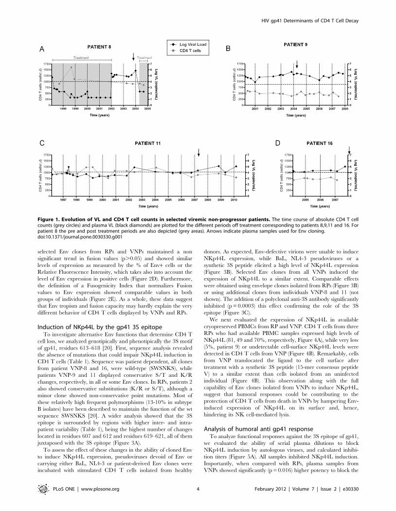
Patient descriptionFour VNP individuals were identified. Figure 1 shows their VL
and CD4 T cell count longitudinal evolution. Mean CD4 T cell
count for patients VNP-8, 9, 11 and 16 was 945, 486, 995 and 735
cells/ml, respectively (Figure 1). Mean VL for patients VNP-8, 9,
11 and 16 was 168193, 126383, 25144 and 31523 copies RNA/ml
(Figure 1). Individuals VNP-8, 9 and 11 showed low CD4 T cells
loss, 48, 14 and 6 cells/mL per year, respectively; while VNP-16
showed an increasing trend of 4 cells/mL per year (Figure 1).
Individual VNP-8 received antiretroviral therapy from 1997 to
2002, and after 2004. Plasma samples selected for this study
belong to year 2003, when the patient was off therapy. Patient
VNP-11 showed VL.10,000 copies/ml the last five years of
follow up, samples analyzed correspond to this period (Figure 1).
In contrast, individuals identified as RPs showed CD4 T cell
counts below 350 (median 262) cells/uL with VL comparable to
VNPS (median 77,000 copies/mL). All samples from this group
were collected from HAART-naıve patients within 3 years after
seroconversion (median 0.7 years).
Env tropism, fusion capacity and expressionFull-length envelopes were amplified from plasma samples and
cloned into expression vectors. Since Env tropism is a major
determinant of cytopathicity [9], V3 loop sequences of the Env
clones were analyzed by both PSSM [29] and geno2pheno
software [30]. All clones isolated showed a V3 loop sequence
predicted as R5-tropic in silico (Table S1). Next, Env clones were
screened for fusion activity after cotransfection with pTat into
293T cells and cocultured with TZM-bl reporter cells (Figure 2A).
In these experiments we used the R5 BaL envelope clone as
positive fusion control (100%) and pTat as negative control (0%).
Most of Env clones showed detectable fusogenic activity (.10% of
BaL values, Figure 2A). Remarkably, no differences were observed
between VNPs and RPs when the percentage of functional clones
was analyzed (values ranging from 50% for patient VNP-16 to
100% for patient VNP-8; Figure 2B) Furthermore, the use of co-
receptor inhibitors in the fusion assay provided a phenotypic
measure of tropism. The lack of effect of JM-2987 and the
sensitivity to TAK-779 of all clones isolated from VNPs or RPs
confirmed the general use of CCR5 (Figure 2A). Median fusion
values for all patients were comparable or higher than the fusion
elicited by the BaL Env clone (ranged from 99, 23–273 (median,
min-max) % of BaL Fusion for individual VNP-16 to 248, 18–
392% for individual RP- 6). Nevertheless, comparison of median
fusion values between RPs and VNPs showed a non significant
trend towards a lower fusion in the latter group (data not shown).
Therefore, to further analyze this potential difference, we selected
4 representative clones from each patient and we characterized
their expression and fusogenic capacity. Figure 2C shows that
HIV gp41 Determinants of CD4 T Cell Decay
PLoS ONE | www.plosone.org 3 February 2012 | Volume 7 | Issue 2 | e30330
selected Env clones from RPs and VNPs maintained a non
significant trend in fusion values (p.0.05) and showed similar
levels of expression as measured by the % of Env+ cells or the
Relative Fluorescence Intensity, which takes also into account the
level of Env expression in positive cells (Figure 2D). Furthermore,
the definition of a Fusogenicity Index that normalizes Fusion
values to Env expression showed comparable values in both
groups of individuals (Figure 2E). As a whole, these data suggest
that Env tropism and fusion capacity may hardly explain the very
different behavior of CD4 T cells displayed by VNPs and RPs.
Induction of NKp44L by the gp41 3S epitopeTo investigate alternative Env functions that determine CD4 T
cell loss, we analyzed genotipically and phenotipically the 3S motif
of gp41, residues 613–618 [20]. First, sequence analysis revealed
the absence of mutations that could impair NKp44L induction in
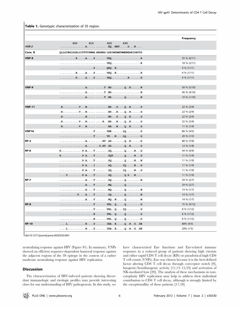
CD4 T cells (Table 1). Sequence was patient dependent, all clones
from patient VNP-8 and 16, were wild-type (SWSNKS), while
patients VNP-9 and 11 displayed conservative S/T and K/R
changes, respectively, in all or some Env clones. In RPs, patients 2
also showed conservative substitutions (K/R or S/T), although a
minor clone showed non-conservative point mutations. Most of
these relatively high frequent polymorphisms (13-10% in subtype
B isolates) have been described to maintain the function of the wt
sequence SWSNKS [20]. A wider analysis showed that the 3S
epitope is surrounded by regions with higher inter- and intra-
patient variability (Table 1), being the highest number of changes
located in residues 607 and 612 and residues 619–621, all of them
juxtaposed with the 3S epitope (Figure 3A).
To assess the effect of these changes in the ability of cloned Env
to induce NKp44L expression, pseudoviruses devoid of Env or
carrying either BaL, NL4-3 or patient-derived Env clones were
incubated with stimulated CD4 T cells isolated from healthy
donors. As expected, Env-defective virions were unable to induce
NKp44L expression, while BaL, NL4-3 pseudoviruses or a
synthetic 3S peptide elicited a high level of NKp44L expression
(Figure 3B). Selected Env clones from all VNPs induced the
expression of NKp44L to a similar extent. Comparable effects
were obtained using envelope clones isolated from RPs (Figure 3B)
or using additional clones from individuals VNP-8 and 11 (not
shown). The addition of a polyclonal anti-3S antibody significantly
inhibited (p = 0.0003) this effect confirming the role of the 3S
epitope (Figure 3C).
We next evaluated the expression of NKp44L in available
cryopreserved PBMCs from RP and VNP. CD4 T cells from three
RPs who had available PBMC samples expressed high levels of
NKp44L (81, 49 and 70%, respectively, Figure 4A), while very low
(5%, patient 9) or undetectable cell-surface NKp44L levels were
detected in CD4 T cells from VNP (Figure 4B). Remarkably, cells
from VNP translocated the ligand to the cell surface after
treatment with a synthetic 3S peptide (15-mer consensus peptide
V) to a similar extent than cells isolated from an uninfected
individual (Figure 4B). This observation along with the full
capability of Env clones isolated from VNPs to induce NKp44L,
suggest that humoral responses could be contributing to the
protection of CD4 T cells from death in VNPs by hampering Env-
induced expression of NKp44L on its surface and, hence,
hindering its NK cell-mediated lysis.
Analysis of humoral anti gp41 responseTo analyze functional responses against the 3S epitope of gp41,
we evaluated the ability of serial plasma dilutions to block
NKp44L induction by autologous viruses, and calculated inhibi-
tion titers (Figure 5A). All samples inhibited NKp44L induction.
Importantly, when compared with RPs, plasma samples from
VNPs showed significantly (p = 0.016) higher potency to block the
Figure 1. Evolution of VL and CD4 T cell counts in selected viremic non-progressor patients. The time course of absolute CD4 T cellcounts (grey circles) and plasma VL (black diamonds) are plotted for the different periods off treatment corresponding to patients 8,9,11 and 16. Forpatient 8 the pre and post treatment periods are also depicted (grey areas). Arrows indicate plasma samples used for Env cloning.doi:10.1371/journal.pone.0030330.g001
HIV gp41 Determinants of CD4 T Cell Decay
PLoS ONE | www.plosone.org 4 February 2012 | Volume 7 | Issue 2 | e30330

effect of 3S epitope (Figure 5B). To confirm the role of specific
anti-gp41 antibodies, we induced NKp44L expression in CD4 T
cells from healthy donors with a 15-mer peptide covering the 3S
sequence and evaluated the inhibitory activity of whole plasma,
IgG-depleted plasma or purified IgG preparations. VNP samples
showed a significant capacity to inhibit peptide-induced NKp44L
expression, which was retained by IgG fraction and lost in IgG-
depleted plasma. Conversely, plasma from RP individuals showed
very low inhibitory activity that was not clearly associated with
IgG fraction in the experimental conditions analyzed (Figure 5C).
Humoral reponses were further characterized in ELISA experi-
ments against the consensus 15-mer 3S peptide. In these
experiments, first and last available plasma samples covering the
non cytophativc HIV replication period of VNP were compared
with plasma from RP. Consistent with functional data, RP samples
showed poor peptide recognition, while VNP plasma showed
higher prevalence of antibodies against this peptide (Figure 6A),
although some VNP samples also showed poor reactivity in
ELISA. This observation along with the variability of 3S
surrounding sequences, might suggest that antibodies with a
different specificity than the 3S core (SWSNKS) could also be
active at blocking NKp44L induction. To test whether the
variability of 3S adjacent sequences may confer some specificity
to antibodies blocking the 3S motif, we designed four peptides
according to the specific gp41 sequences of each VNP patient. The
earliest and latest available samples from VNPs (up to 11 years
timeframe) were analyzed in ELISA and their gp41 ectodomain
was sequenced. Results, summarized in Figure 6B, indicate that
plasma from every patient was able to recognize autologous
sequences; although patient 8 switched the reactivity from the
autologous to the consensus peptide overtime. Patient 11 showed
parallel changes in sequence and antibody response, switching
from consensus sequence (LDDIW in C terminus) to LNDIW
overtime. Moreover, sera showed limited cross-reactivity; only
plasma from patient 11 was able to recognize sequences from
patient 9, both of them displaying an arginine at position +2 of the
SWSNKS core sequence. Sequence analyses of the 3S epitope at
population level (Figure 6B) confirmed mutations associated with
changes in recognition patterns, and pointed to the more variable
C terminal adjacent region as the key target for antibodies against
the 3S flanking region in VNPs. None of the RP plasma
recognized this set of peptides (data not shown). Regarding whole
anti HIV neutralizing responses, VNPs had higher neutralization
titers for NL4-3 or BaL isolates, but failed to mount a broad
Figure 2. Analysis of Env tropism, fusion and expression. (A) The levels of fusion of functional Env clones isolated from viremic non-progresors (VNP) patients 8, 9, 11 and 16 and rapid progressors (RP) was assayed in a coculture of Env-Tat expressing 293T cells and TZM-bl cells (redsymbols). The CXCR4 antagonists JM-2987 or CCR5 antagonists TAK-779 (blue and yellow symbols, respectively) were added as controls for functionaltropism. Fusion activity was calculated relative to the Env BaL clone (100%) that was tested in each experiment. Negative controls were obtained byusing 293T cells transfected only with a pTat plasmid and were subtracted to all measures. Values for each clone are the mean of three differentexperiments. (B) The frequency of functional clones was evaluated in both VNPs (green bar, total number of tested clones 44) and rapid-progressor(red bar, total number of tested clones 56) groups. (C–E) Comparative analysis of representative Env clones (n = 32) isolated from VNP (n = 15) and RP(n = 17) patients. (C) Levels of fusion capacity of selected Env clones isolated from VNP and RP patients (green and red bar, respectively) as describedfor controls on panel A. Fusion activity was calculated relative to the Env BaL clone (100%). (D) Left panel shows the percentage of Env+ cells andright panel shows the Relative Fluorescence Intensity, which is a measure of total envelope expression that accounts for both the percentage of Envexpressing cells and the fluorescence intensity of positive cells (see Methods). (E) A fusogenic index was calculated for each selected Env clone as theratio of Fusion activity and Relative Fluorescence Intensity. For panels B–E, boxes represent median and min-max values.doi:10.1371/journal.pone.0030330.g002
HIV gp41 Determinants of CD4 T Cell Decay
PLoS ONE | www.plosone.org 5 February 2012 | Volume 7 | Issue 2 | e30330
neutralizing response against HIV (Figure S1). In summary, VNPs
showed an efficient sequence-dependent humoral response against
the adjacent regions of the 3S epitope in the context of a rather
moderate neutralizing response against HIV replication.
Discussion
The characterization of HIV-infected patients showing discor-
dant immunologic and virologic profiles may provide interesting
clues for our understanding of HIV pathogenesis. In this study, we
have characterized Env functions and Env-related immune
responses in a reduced group of patients showing high viremia
and either rapid CD4 T cell decay (RPs) or paradoxical high CD4
T cell counts (VNPs). Env was chosen because it is the best-defined
factor altering CD4 T cell decay through coreceptor switch [9],
fusogenic/hemifusogenic activity [11,13–15,33] and activation of
NK-mediated lysis [20]. The analysis of these mechanisms in non-
cytophatic HIV replication may help to address their individual
contribution to CD4 T cell decay, although is strongly limited by
the exceptionallity of these patients [17,18].
Table 1. Genotypic characterization of 3S region.
Frequency
HXB-2:600 610 620 630
................A..... ...... .EQ..NHT.....D...N....
Cons. B QLLGIWGCSGKLICTTTVPWNA SWSNKS LDEIWDNMTWMEWEREIDNYTS
VNP-8 ..........R.....A....S ...... YEQ..................R 55 % (6/11)
...................... ...... YKQ..................R 18 % (2/11)
.....................S ...... QKQ..E................ 9 % (1/11)
..........R.....A....S ...... YEQ..E...............R 9 % (1/11)
..........R.....A....S ...... YEQ..........K.......R 9 % (1/11)
VNP-9 ................A..... .....T .ND........Q..K......N 50 % (5/10)
................A..... .....T .ND..................N 40 % (4/10)
................A..... .....T .ND........Q.........N 10 % (1/10)
VNP-11 .R..........V...N..... ...... .ND...S....Q..K......G 22 % (2/9)
.R..........V...N..... ...... .ND...K....Q..K......G 22 % (2/9)
.R..............N..... ...... .ND...S....Q..K......G 22 % (2/9)
.R..........V...N..... ....R. .ND...N....Q..K......G 22 % (2/9)
.R..........V...N..... ...... .NA...N....Q..K......G 11 % (1/9)
VNP16 .....................T ...... YNK.......LQ.........D 80 % (4/5)
.....................T ...... YK...N....LQ.........G 20 % (1/5)
RP-2 ................A..... ....RT .GD........Q..K......D 88 % (7/8)
................A..... ..G.RT .GD........Q..K......D 12 % (1/8)
RP-6 R.............P.A....T ...... .SQ........Q.....N...G 44 % (4/9)
R.............P.A....T ...... .SQT.......Q.....N...G 11 % (1/9)
..............P.A....T ...... .SQ........Q.....N...N 11 % (1/9)
..............P.A....I ...... .SQ.......IQ.....N...G 11 % (1/9)
..............P.A....T ...... .SQ.......IQ.....N...G 11 % (1/9)
....V.........P.A....T ...... .SQ........Q.D...N.... 11 % (1/9)
RP-7 ................A....T ...... .SQ........Q.........N 29 % (2/7)
................A....T ...... .NQ........Q.......... 29 % (2/7)
................A....T ...... .NQ........Q.........N 14 % (1/7)
...........P....A....T ...... .SQ........Q.........N 14 % (1/7)
................A....T ...... .NQ..N.....Q.........N 14 % (1/7)
RP-8 .....................T ...... YNL..Q.....Q.........G 75 % (9/12)
.....................T ...... YNL..Q....IQ.........G 8 % (1/12)
.....................N ...... YNL..Q.....Q.........G 8 % (1/12)
.....................N ...... YNL..Q.....Q.........D 8 % (1/12)
RP-10 ....L...........N....S ...... IEA..E.....Q..K..G..SE 80% (4/5)
....L...........N....S ...... IKA..E.....Q..K..G..SE 20% (1/5)
doi:10.1371/journal.pone.0030330.t001
HIV gp41 Determinants of CD4 T Cell Decay
PLoS ONE | www.plosone.org 6 February 2012 | Volume 7 | Issue 2 | e30330

Previous reports have described different fusion activities in
envelope clones isolated from Elite controllers or pre-AIDS
patients compared to patients progressing to AIDS [31,34]. In
our samples, we have also addressed the analysis of fusion capacity
by isolating full-length Env clones and monitoring different
functional parameters. In all cases, we were unable to detect
significant differences between groups. Indeed, the percentage of
functional clones, the level of Env expression (either measured by
% of Env+ cells or by the Relative Fluorescence Intensity) and the
Fusogenic Index were similar for VNPs and RPs. Such an
irrelevant role of fusion activity in our patients compared to Lassen
et al [31] may rely in a higher Env evolution in our VNPs, which
show higher VL than ‘‘regular’’ LTNPs. In agreement with our
data, similar cytopathicity in organ culture has been reported for
viral clones isolated from a small group of VNPs and RPs [18].
Furthermore, the homogeneous R5 tropism of Env clones isolated
from our patients ruled out any potential role of CXCR4 use in
our small cohort. Although in vitro fusion assays using cell lines
may differ from in vivo envelope function, our data suggest that
fusion defects and viral tropism fail to explain the different
outcomes of VNPs and RPs in terms of CD4 T cell loss.
Considering the deleterious role of NKp44L on CD4 T cells,
which render these cells sensitive to NK lysis [20], we next
analyzed the potential role of NKp44L induction by gp41. Env
clones from both groups of patients showed conserved 3S epitope
sequences and hence displayed full capability to induce NKp44L
expression in CD4 T cells from healthy donors. However,
NKp44L expression in CD4 T cells isolated from these patients
showed a different profile with a lower cell-surface expression in
VNP samples. Indeed, NKp44L translocation was inhibited by
plasma from VNPs with significantly higher efficiency than plasma
from RPs, being the purified IgG fraction responsible for this
effect. Further analysis of anti-3S responses in VNP patients
revealed that these individuals showed high titers of specific
antibodies against sequences flanking the conserved 3S epitope.
Indeed, we found that only some VNP sera tested recognized a
consensus peptide due to changes in residues juxtaposed to the
core sequence SWSNKS. This is the case of plasma from patient
VNP-8, which strongly recognized the peptide containing a YEQ
sequence in residues 620–622, which was present in the
autologous virus. Consistently, patient VNP-16, whose virus shows
an YKE sequence in these residues, specifically recognizes an
YHE containing peptide. Interestingly, no cross reactivity between
both patients was observed. A similar scenario was observed for
VNP patients 9 and 11, in which the D621N change determined
recognition, although in this case we observed a non-reciprocal
Figure 3. Analysis of the NKp44L induction by cloned envelopes. (A) Graphic representation of sequence variability in gp41 regionssurrounding the 3S epitope. The picture has been generated using the weblogo software (http://weblogo.berkeley.edu/), representing polar aminoacids in red, basic in blue, acidic in green and hydrophobic in black. (B) NKp44L expression in CD4 T cells from representative donors after incubationwith Env-defective pseudoviruses (pSG3, negative control), with the 3S consensus peptide V, or with pseudoviruses bearing the following HIV Env:NL4-3, BaL (positive controls). The effect of selected Env clones from VNPs (upper panels) or RPs (lower panels is shown) Empty peaks correspond tocontrol staining of untreated CD4 T cells. (C) The effect of a polyclonal anti 3S antibody on NKp44L expression induced by several Env clones isshown. Values represent the relative expression of NKp44L in the surface of CD4 T cells normalized to the effect of the 3S synthetic peptide (100%).Data show the effect of NL4-3 and BaL envelopes (Ctrl-Env) and a total of 8 patient-derived Env clones.doi:10.1371/journal.pone.0030330.g003
HIV gp41 Determinants of CD4 T Cell Decay
PLoS ONE | www.plosone.org 7 February 2012 | Volume 7 | Issue 2 | e30330
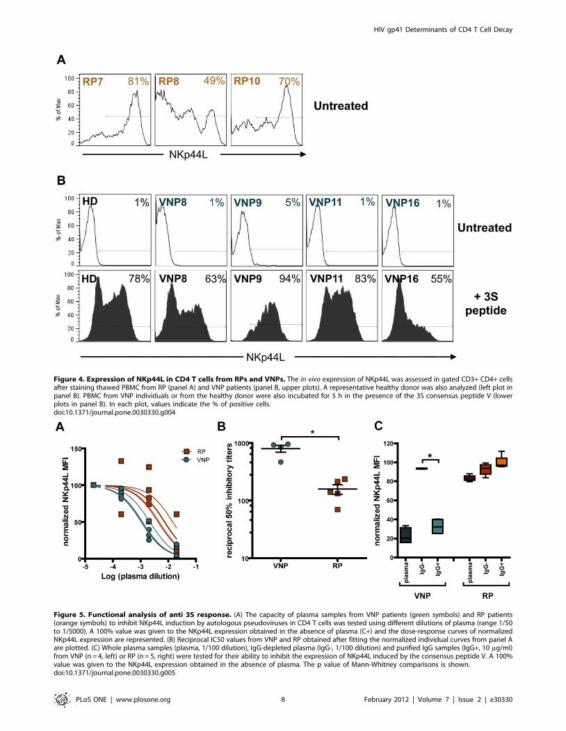
Figure 4. Expression of NKp44L in CD4 T cells from RPs and VNPs. The in vivo expression of NKp44L was assessed in gated CD3+ CD4+ cellsafter staining thawed PBMC from RP (panel A) and VNP patients (panel B, upper plots). A representative healthy donor was also analyzed (left plot inpanel B). PBMC from VNP individuals or from the healthy donor were also incubated for 5 h in the presence of the 3S consensus peptide V (lowerplots in panel B). In each plot, values indicate the % of positive cells.doi:10.1371/journal.pone.0030330.g004
Figure 5. Functional analysis of anti 3S response. (A) The capacity of plasma samples from VNP patients (green symbols) and RP patients(orange symbols) to inhibit NKp44L induction by autologous pseudoviruses in CD4 T cells was tested using different dilutions of plasma (range 1/50to 1/5000). A 100% value was given to the NKp44L expression obtained in the absence of plasma (C+) and the dose-response curves of normalizedNKp44L expression are represented. (B) Reciprocal IC50 values from VNP and RP obtained after fitting the normalized individual curves from panel Aare plotted. (C) Whole plasma samples (plasma, 1/100 dilution), IgG-depleted plasma (IgG-, 1/100 dilution) and purified IgG samples (IgG+, 10 mg/ml)from VNP (n = 4, left) or RP (n = 5, right) were tested for their ability to inhibit the expression of NKp44L induced by the consensus peptide V. A 100%value was given to the NKp44L expression obtained in the absence of plasma. The p value of Mann-Whitney comparisons is shown.doi:10.1371/journal.pone.0030330.g005
HIV gp41 Determinants of CD4 T Cell Decay
PLoS ONE | www.plosone.org 8 February 2012 | Volume 7 | Issue 2 | e30330

cross-reactivity that may be related to position 612 (A or T).
Moreover, the longitudinal analysis of anti-3S responses in VNPs
showed that several unique specificities of antibodies inhibiting 3S
triggering are present over the whole period of non-cytopathic
viral replication, and hence do not seem to be the consequence of
a long-term continuous antigen stimulation of B cells. It has been
recently shown that the 3S epitope of gp41 mediates the NKp44L
translocation to the cell surface by activating oxidative stress
sensors. The 3S epitope interacts with the gC1qR on the
membrane of CD4 T cells to activate a PI3K/NADPH oxidase
and a p190 RhoGAP GTPase dependent pathway [21]. Thus,
antibodies against the 3S epitope act by hampering 3S binding to
its receptor. Taken together, these data suggest that specific anti-
gp41 antibodies against variable 3S adjacent region might limit the
ability of gp41 to induce NKp44L expression on CD4 cells by a
similar mechanism in VNPs, although its exact mode of action
remains to be defined. These antibodies may represent a
compensatory defense against the gp41 pathogenicity and CD4
Figure 6. Mapping humoral responses. (A) Plasma from RP (n = 5) and VNP (n = 8) individuals were tested in triplicate for the recognition ofconsensus peptide V in ELISA. Data are arbitrary absorbance units per ml of plasma, boxes represent mean+/2SD. (B) Plasma from VNP patients 8(upper left), 9 (upper right), 11 (lower left) and 16 (lower right) were assayed for the recognition of five different peptides displaying a consensussequence covering 609–624 residues of gp41 or the equivalent patient-based envelope sequences. Two samples were analyzed for each patient, theearliest sample available (empty bars) and the sample (or closest sample for patient 9) of Env cloning (dark bars). Peptide sequences used in ELISAassays are shown in the left axis of each plot. The autologous population sequence from each patient obtained at the indicated timepoints (year-month) is also shown in top of each panel. Intrapatient sequence changes are highlighted. Data are mean+/2SD of triplicate samples.doi:10.1371/journal.pone.0030330.g006
HIV gp41 Determinants of CD4 T Cell Decay
PLoS ONE | www.plosone.org 9 February 2012 | Volume 7 | Issue 2 | e30330

depletion when antibodies directed against the highly conserved 6
amino-acid 3S peptide are decreasing, as it occur normally during
the disease progression [20,22–24]. Consistently, very low levels of
NKp44L expression were observed on the surface of CD4 T cells
isolated from VNP, despite its full responsiveness to a 3S peptide
and its continuous exposure to fully functional envelopes in vivo,
with VL.10,000 copies/ml for more than 2 years.
By suppressing NKp44L expression, antibodies against the 3S
epitope or its flanking region might contribute more to T cell
protection than neutralizing antibodies. In our study, VNPs are
unable to mount a broad neutralizing response against HIV and
several groups have reported little, if any, impact of HIV
neutralizing responses on clinical condition or progression to
AIDS [35,36]. Conversely, anti-3S antibodies, which are inde-
pendent of neutralization, as this epitope is not the target of HIV
neutralizing antibodies [37], may have indirect favorable effects on
CD4 T cell survival [20].
In conclusion, our data, obtained in a small group of patients,
support the notion that HIV Envelope glycoproteins from VNPs
are functionally similar to those isolated from RPs in terms of
expression, tropism, fusion and induction of NK ligands. This is
consistent with other small studies [18]. However, our data point
out that VNPs are able to mount strong humoral responses against
a variable region flanking the 3S epitope of gp41 that block the
capacity of autologous viruses to trigger surface expression of the
NKp44L in CD4 T cells, a mechanism clearly related to CD4
depletion [20,22,24]. These antibodies may contribute to non-
cytopathic HIV replication in humans along with the reported
lower immune activation of these patients [17,18] and other viral
determinants outside Env [38].
Supporting Information
Figure S1 Neutralization activity of plasma from VNPand RP. Neutralization capacity of plasma from VNP and RP
patients was tested against pseudoviruses carrying NL43 (B), BaL
(C) or primary isolates SVPB13 (C) and SVPB16 (D) envelope
glycoproteins using TZM-bl reporter cell line. In figure A and B
left plots represent the curves of normalized RLU for each plasma
dilution in a logarithmic scale; right plots represent the reciprocal
50% inhibitory titers deduced from the normalized curves. For
SVPB13 and SVPB16 no neutralization activity could be
quantified (all reciprocal IC50 values were ,60).
(DOC)
Table S1 Genotypic and phenotypic characterization ofthe V3 loop. a Tropism assessed in sillico by the Pssm software.
Similar results were obtained using the geno2pheno software. b
Tropism assessed by sensitivity to JM-2987 and TAK779 in TZM-
bl cells.
(DOC)
Acknowledgments
We thank Jorge del Romero and Carmen Rodriguez from Centro
Sanitario Sandoval (IMSALUD Comunidad Autonoma de Madrid.
Madrid, 28010 Spain) for samples and clinical data of the rapid progressor
patients. This work is part of MC PhD thesis at the Autonomous University
of Barcelona, Spain.
Author Contributions
Conceived and designed the experiments: MC HFB CC MM JC VV.
Performed the experiments: MC HFB MM JC VV. Analyzed the data: MP
CLG CC BC PD JB. Contributed reagents/materials/analysis tools: VV
MP CLG. Wrote the paper: MC PD BC JB.
References
1. Casado C, Colombo S, Rauch A, Martınez R, Gunthard HF, et al. (2010) Host
and viral genetic correlates of clinical definitions of HIV-1 disease progression.
PLoS One 5: e11079.
2. Dalmau J, Puertas MC, Azuara M, Marino A, Frahm N, et al. (2009)
Contribution of immunological and virological factors to extremely severeprimary HIV type 1 infection. Clin Infect Dis 48: 229–238.
3. Gandhi RT, Chen BK, Straus SE, Dale JK, Lenardo MJ, Baltimore D (1998)
HIV-1 directly kills CD4+ T cells by a Fas-independent mechanism. J Exp Med
187: 1113–1122.
4. McCune JM (2001) The dynamics of CD4+ T-cell depletion in HIV disease.
Nature 410: 974–979.
5. Badley AD, Pilon AA, Landay A, Lynch DH (2000) Mechanisms of HIV-
associated lymphocyte apoptosis. Blood 96: 2951–2964.
6. Gougeon ML, Piacentini M (2009) New insights on the role of apoptosis and
autophagy in HIV pathogenesis. Apoptosis 14: 501–508.
7. Doitsh G, Cavrois M, Lassen KG, Zepeda O, Yang Z, et al. (2010) AbortiveHIV infection mediates CD4 T cell depletion and inflammation in human
lymphoid tissue. Cell 143: 789–801.
8. Blanco J, Barretina J, Cabrera C, Gutierrez A, Clotet B, Este JA (2001) CD4(+)
and CD8(+) T cell death during human immunodeficiency virus infection in
vitro. Virology 285: 356–365.
9. Tersmette M, Lange JM, de Goede RE, de Wolf F, Eeftink-Schattenkerk JK,
et al. (1989) Association between biological properties of human immunodefi-
ciency virus variants and risk for AIDS and AIDS mortality. Lancet 1: 983–985.
10. Etemad-Moghadam B, Rhone D, Steenbeke T, Sun Y, Manola J, et al. (2001)
Membrane-fusing capacity of the human immunodeficiency virus envelope
proteins determines the efficiency of CD+ T-cell depletion in macaques infected
by a simianhuman immunodeficiency virus. J Virol 75: 5646–5655.
11. Karlsson GB, Halloran M, Schenten D, Lee J, Racz P, et al. (1998) Theenvelope glycoprotein ectodomains determine the efficiency of CD4+ T
lymphocyte depletion in simian-human immunodeficiency virus-infected
macaques. J Exp Med 188: 1159–1171.
12. LaBonte JA, Patel T, Hofmann W, Sodroski J (2000) Importance of membranefusion mediated by human immunodeficiency virus envelope glycoproteins for
lysis of primary CD4-positive T cells. J Virol 74: 10690–10698.
13. Blanco J, Barretina J, Ferri KF, Jacotot E, Gutierrez A, et al. (2003) Cell-
surfaceexpressed HIV-1 envelope induces the death of CD4 T cells during
GP41-mediated hemifusion-like events. Virology 305: 318–329.
14. Blanco J, Barretina J, Clotet B, Este JA (2004) R5 HIV gp120-mediated cellular
contacts induce the death of single CCR5-expressing CD4 T cells by a gp41-
dependent mechanism. J Leukoc Biol 76: 804–811.
15. Denizot M, Varbanov M, Espert L, Robert-Hebmann V, Sagnier S, et al. (2008)
HIV- 1 gp41 fusogenic function triggers autophagy in uninfected cells.
Autophagy 4: 998–1008.
16. Paiardini M, Pandrea I, Apetrei C, Silvestri G (2009) Lessons learned from the
natural hosts of HIV-related viruses. Annu Rev Med 60: 485–495.
17. Rotger M, Dalmau J, Rauch A, McLaren P, Bosinger S, et al. (2011)
Comparative transcriptomics of extreme phenotypes of human HIV-1 infection
and SIV infection in sooty mangabey and rhesus macaque. J Clin Invest.
18. Choudhary SK, Vrisekoop N, Jansen CA, Otto SA, Schuitemaker H, et al.
(2007) Low immune activation despite high levels of pathogenic human
immunodeficiency virus type 1 results in long-term asymptomatic disease. J Virol
81: 8838–8842.
19. Vieillard V, Fausther-Bovendo H, Samri A, Debre P, French Asymptomatiques
a Long Terme (ALT) ANRS-CO15 Study Group (2010) Specific phenotypic and
functional features of natural killer cells from HIV-infected long-term
nonprogressors and HIV controllers. J Acquir Immune Defic Syndr 53:
564–573.
20. Vieillard V, Strominger JL, Debre P (2005) NK cytotoxicity against CD4+ T
cells during HIV-1 infection: a gp41 peptide induces the expression of an
NKp44 ligand. Proc Natl Acad Sci U S A 102: 10981–10986.
21. Fausther-Bovendo H, Vieillard V, Sagan S, Bismuth G, Debre P (2010) HIV
gp41 Engages gC1qR on CD4+ T Cells to Induce the Expression of an NK
Ligand through the PIP3/H2O2 Pathway. PLoS Pathog 6: e1000975.
22. Vieillard V, Costagliola D, Simon A, Debre P, French Asymptomatiques a Long
Terme (ALT) Study Group (2006) Specific adaptive humoral response against a
gp41 motif inhibits CD4 T-cell sensitivity to NK lysis during HIV-1 infection.
AIDS 20: 1795–1804.
23. Vieillard V, Habib RE, Brochard P, Delache B, Bovendo HF, et al. (2008)
CCR5 or CXCR4 use influences the relationship between CD4 cell depletion,
NKp44L expression and NK cytotoxicity in SHIV-infected macaques. AIDS 22:
185–192.
24. Vieillard V, Le Grand R, Dausset J, Debre P (2008) A vaccine strategy against
AIDS: an HIV gp41 peptide immunization prevents NKp44L expression and
CD4+ T cell depletion in SHIV-infected macaques. Proc Natl Acad Sci U S A
105: 2100–2104.
HIV gp41 Determinants of CD4 T Cell Decay
PLoS ONE | www.plosone.org 10 February 2012 | Volume 7 | Issue 2 | e30330

25. Li M, Gao F, Mascola JR, Stamatatos L, Polonis VR, et al. (2005) Human
immunodeficiency virus type 1 env clones from acute and early subtype Binfections for standardized assessments of vaccine-elicited neutralizing antibod-
ies. J Virol 79: 10108–10125.
26. Li Y, Svehla K, Mathy NL, Voss G, Mascola JR, Wyatt R (2006)Characterization of antibody responses elicited by human immunodeficiency
virus type 1 primary isolate trimeric and monomeric envelope glycoproteins inselected adjuvants. J Virol 80: 1414–1426.
27. Malim MH, Hauber J, Fenrick R, Cullen BR (1988) Immunodeficiency virus rev
trans-activator modulates the expression of the viral regulatory genes. Nature335: 181–183.
28. Freed EO, Myers DJ, Risser R (1990) Characterization of the fusion domain ofthe human immunodeficiency virus type 1 envelope glycoprotein gp41. Proc
Natl Acad Sci U S A 87: 4650–4654.29. Jensen MA, Coetzer M, van ’t Wout AB, Morris L, Mullins JI (2006) A reliable
phenotype predictor for human immunodeficiency virus type 1 subtype C based
on envelope V3 sequences. J Virol 80: 4698–4704.30. Sing T, Low AJ, Beerenwinkel N, Sander O, Cheung PK, et al. (2007) Predicting
HIV coreceptor usage on the basis of genetic and clinical covariates. AntivirTher 12: 1097–1106.
31. Sterjovski J, Churchill MJ, Ellett A, Gray LR, Roche MJ, et al. (2007) Asn 362 in
gp120 contributes to enhanced fusogenicity by CCR5-restricted HIV-1 envelopeglycoprotein variants from patients with AIDS. Retrovirology 4: 89.
32. Blanco J, Canela EI, Mallol J, Lluıs C, Franco R (1992) Characterization of
adenosine receptors in brush-border membranes from pig kidney. Br J Pharmacol
107: 671–678.
33. Garg H, Joshi A, Freed EO, Blumenthal R (2007) Site-specific mutations in
HIV-1 gp41 reveal a correlation between HIV-1-mediated bystander apoptosis
and fusion/hemifusion. J Biol Chem 282: 16899–16906.
34. Lassen KG, Lobritz MA, Bailey JR, Johnston S, Nguyen S, et al. (2009) Elite
suppressor-derived HIV-1 envelope glycoproteins exhibit reduced entry
efficiency and kinetics. PLoS Pathog 5: e1000377.
35. Doria-Rose NA, Klein RM, Daniels MG, O’Dell S, Nason M, et al. (2010)
Breadth of human immunodeficiency virus-specific neutralizing activity in sera:
clustering analysis and association with clinical variables. J Virol 84: 1631–1636.
36. Euler Z, van Gils MJ, Bunnik EM, Phung P, Schweighardt B, et al. (2010)
Crossreactive neutralizing humoral immunity does not protect from HIV type 1
disease progression. J Infect Dis 201: 1045–1053.
37. Pietzsch J, Scheid JF, Mouquet H, Seaman MS, Broder CC, Nussenzweig MC
(2010) Anti-gp41 antibodies cloned from HIV-infected patients with broadly
neutralizing serologic activity. J Virol 84: 5032–5042.
38. Lum JJ, Cohen OJ, Nie Z, Weaver JG, Gomez TS, et al. (2003) Vpr R77Q is
associated with long-term nonprogressive HIV infection and impaired induction
of apoptosis. J Clin Invest 111: 1547–1554.
HIV gp41 Determinants of CD4 T Cell Decay
PLoS ONE | www.plosone.org 11 February 2012 | Volume 7 | Issue 2 | e30330
Top Related
Copyright © 2022 FDOKUMEN

