


Bahasa
Halaman
Hukum

The Epidemiological and Molecular Aspects of InfluenzaH5N1 Viruses at the Human-Animal Interface in EgyptGhazi Kayali1*, Richard J. Webby1, Mariette F. Ducatez1, Rabeh A. El Shesheny2, Ahmed M. Kandeil2,
Elena A. Govorkova1, Ahmed Mostafa2, Mohamed A. Ali2
1 Division of Virology, Department of Infectious Diseases, St. Jude Children’s Research Hospital, Memphis, Tennessee, United States of America, 2 Virology Laboratory,
Environmental Research Division, National Research Centre, Cairo, Egypt
Abstract
With 119 confirmed cases between March 2006 and December 2010, Egypt ranks second among countries reporting humanH5N1 influenza virus infections. In 2009–2010, Egypt reported 68 new human cases and became the new epicenter forH5N1 infections. We conducted an epidemiological and molecular analysis in order to better understand the situation inEgypt. The onset of new cases peaked annually during the winter and spring months, with majority of cases reported in theNile Delta region. Most cases were less than 18 years old (62%) and females (60%). The overall case-fatality rate was 34% andsignificantly increased by age. There was a significant difference between the case-fatality rates among females and males.We observed a significant drop (p = 0.004) in case fatality rate in 2009 (10%) as compared to higher rates (36%–56%) in otheryears. Hospitalization within 2 or 3 days after onset of symptoms significantly decreased mortality. Molecular analysisshowed that variations do occur among viruses isolated from birds as well as from humans in Egypt, and these mutationswere especially noted in 2009 viruses. As the epidemiological profile of Egyptian cases differs from other countries, there isan urgent need to conduct prospective studies to enhance our understanding of incidence, prevalence, and determinantsof virulence of human infections with avian H5N1 influenza viruses.
Citation: Kayali G, Webby RJ, Ducatez MF, El Shesheny RA, Kandeil AM, et al. (2011) The Epidemiological and Molecular Aspects of Influenza H5N1 Viruses at theHuman-Animal Interface in Egypt. PLoS ONE 6(3): e17730. doi:10.1371/journal.pone.0017730
Editor: Malcolm Semple, University of Liverpool, United Kingdom
Received September 29, 2010; Accepted February 9, 2011; Published March 21, 2011
Copyright: � 2011 Kayali et al. This is an open-access article distributed under the terms of the Creative Commons Attribution License, which permitsunrestricted use, distribution, and reproduction in any medium, provided the original author and source are credited.
Funding: This work was supported by National Institute of Allergy and Infectious Diseases, National Institutes of Health, Department of Health and HumanServices, under contract number HHSN266200700005C, and by the American Lebanese Syrian Associated Charities. The funders had no role in study design, datacollection and analysis, decision to publish, or preparation of the manuscript.
Competing Interests: The authors have declared that no competing interests exist.
* E-mail: [email protected]
Introduction
Preparedness for a possible influenza pandemic caused by
highly pathogenic avian influenza (HPAI) A subtype H5N1 has
become a global priority [1]. The continued occurrence of human
infection with these viruses and breached host barrier have
compounded pandemic concern. Long-term endemic influenza
virus infections in poultry increase exposure risks to humans, and
in turn, create opportunities for the emergence of human-adapted
strains with pandemic potential [2,3].
The first outbreak of human infection with the HPAI H5N1
virus was reported in 1997 in Hong Kong, where 18 people were
infected, of whom 6 died [4]. After that outbreak, no new human
cases were reported until 2003, when the virus was detected in a
family recently returned to Hong Kong from mainland China.
Since then, new human cases have occurred in numerous
countries around the world [4]. In 2006, the first human case
outside Southeast Asia was reported in Turkey. Later, cases were
reported in Iraq, Egypt, Azerbaijan, Djibouti, and Nigeria [4]. In
the Middle East, clade 2.2.1 H5N1 viruses were circulating among
poultry, and were also responsible for the human cases [5,6,7].
With 119 confirmed cases up to December 2010, the largest
number of cases outside Southeast Asia, Egypt ranks second
among all countries reporting human infection with H5N1,
following Indonesia [4]. In Egypt, it is estimated that 105 million
birds are raised in rural areas and 126 million are kept in urban
areas [8]. Response to outbreaks of HPAI H5N1 in Egyptian
poultry focused on increasing awareness, culling infected poultry,
and vaccinating the rest. Several inactivated H5 poultry vaccines
with varying efficacy levels are available in the Egyptian market.
The main problems in the strategy of control and prevention of
avian influenza in Egypt were the lack of mass disinfection of the
infected foci and failure to successfully vaccinate all poultry.
In this paper, we present the epidemiology of the laboratory-
confirmed cases of H5N1 infection in Egypt. Sequence analysis of
the human and avian H5N1 influenza viruses isolated between 2006
and 2010 as well as analysis of the sequences available in GenBank
were conducted to understand the evolution of the Egyptian H5N1
viruses and to predict the possibility of pandemic emergence.
Methods
Epidemiological AnalysisData on the laboratory-confirmed H5N1 cases in Egypt were
compiled from various reports of the Epidemic and Pandemic
Alert and Response Program of the World Health Organization
(WHO) [9]. These reports were not intended for research and
hence were inconsistent in reporting certain data variables. The
available data variables included age, sex, exposure to poultry,
governorate, and dates of symptoms’ onset, hospitalization, and
PLoS ONE | www.plosone.org 1 March 2011 | Volume 6 | Issue 3 | e17730

death. Where the date of onset of symptoms was not accurately
provided, the month during which the case was reported was used
to estimate the date of onset. All cases were confirmed by reverse
transcriptase polymerase chain reaction at the Egyptian Central
Public Health Laboratory and some were subsequently confirmed
by the U.S. Naval Medical Research Unit No. 3 (NAMRU-3) in
Cairo, Egypt [9,10]. Since the data used was publicly available on
the internet and no patient identifiers were used, approval from an
ethics committee and patient consent were not sought.
The chi-square and Fisher exact tests were used to compare
categorical data. Logistic and linear regressions were used to study
the effect of age and sex on hospitalization and death. All statistical
analysis was performed using PASW (SPSS) 18.0 software.
Sequence and phylogenetic analysisAvian samples were collected for diagnostic purposes by
veterinarians from farms they provide care for through the period
of 2006 to 2010. As those specimens were collected as part of the
routine animal care, no animal care and use committee approval
was sought. Five isolated viruses were included in this analysis
(Table 1). All samples were tested for the presence of influenza A
viruses by detection of M gene using one-step RT-PCR kit
(Qiagen, Valencia, CA). Samples were subtyped using specific
primers (Table 2). Comparative analysis of HA genes for all H5N1
viruses was carried out and compared with the available sequences
using the NCBI influenza virus resource for both avian and human
H5N1 viruses. Sequences were analyzed using the BioEdit
program [11]. Sequences were aligned with ClustalW [12].
Phylogenetic analyses were carried out using MEGA version
4.0.2, with the neighbor-joining method, Poisson correction [13].
Bootstrap values (1000 replications) are indicated on the tree.
Antiviral resistanceThe antiviral susceptibilities of H5N1 influenza viruses were
determined by fluorescence-based NA enzyme inhibition assay
using 29-(4-methylumbelliferyl)-a-D-N-acetylneuraminic acid
(MUNANA; Sigma, St. Louis, MO) at a final concentration of
100 mM as a substrate. The IC50 was defined as the concentration
of NA inhibitor necessary to reduce NA activity by 50% relative to
that of a reaction mixture containing virus but no inhibitor. Full-
length NA and matrix (M2) proteins were sequenced to identify
molecular markers of resistance.
Results
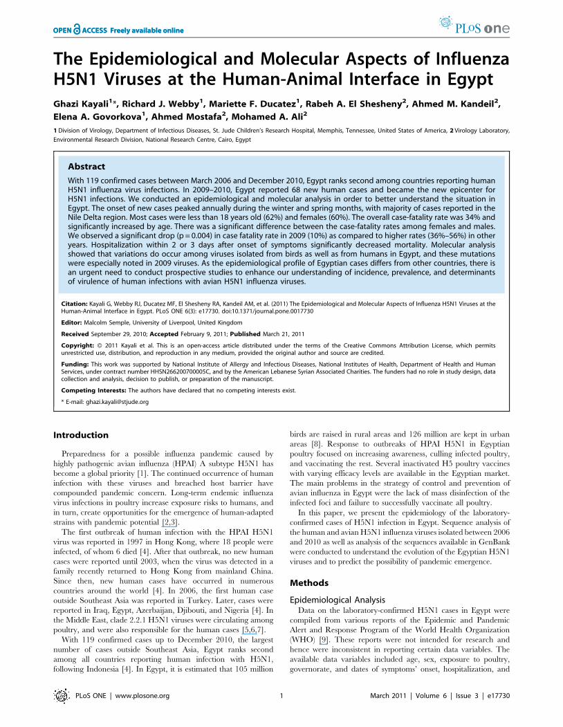
Epidemiology of human H5N1 casesFrom March 2006 to December 2010, 119 human cases of
infection with H5N1 were reported in Egypt. The onset of new
cases peaked five times. The first was during the spring of 2006,
and then during the winter and spring months of 2007, 2008,
2009, and 2010 (Figure 1). Ten cases were reported in March
2007 and May 2009, the highest number of cases in any month
during the study period. The occurrence of cases in the summer
months was rare, with only 17 cases reported in summer 2006 (1
case), summer 2007 (4 cases), summer 2009 (9 cases), and summer
2010 (3 cases). Three family clusters were reported: the sixth and
seventh reported cases were sisters; cases 16, 17, and 18 were of
the same extended family; and cases 28 and 30 were siblings.
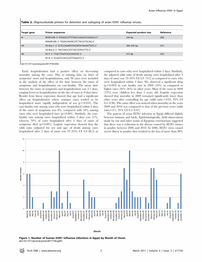
Most cases (n = 94, 79%) occurred in the northern part of the
country and were concentrated in the Nile Delta region. The rest
of the cases occurred over the southern governorates along the
Nile, where agriculture is practiced. The northern governorates of
Menufiyah and Qalyubiyah had 12 cases, the highest number of
cases per governorate, followed by the Kafr El Sheikh (11 cases).
In the south, the governorate of Qena had the highest number of
cases (n = 7). Five cases were reported in the heavily urbanized
Cairo governorate (Figure 2).
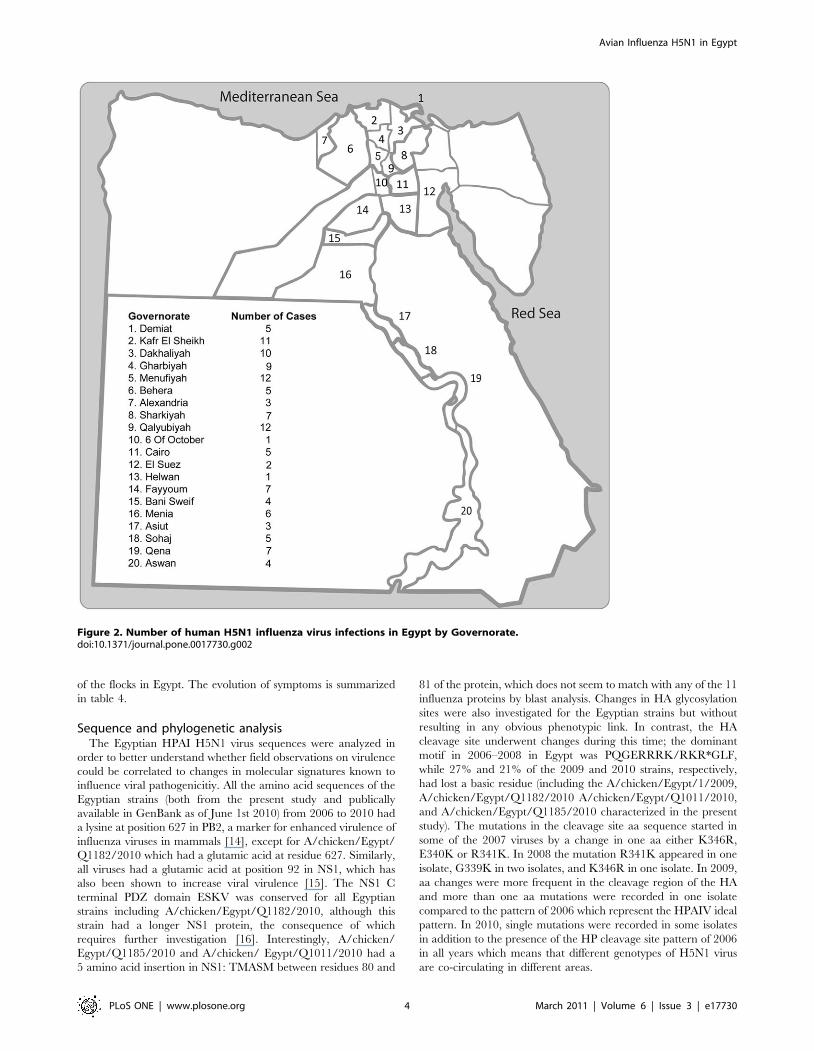
The age and sex distribution of the cases is presented in Figure 3.
The mean age of these cases was 15.7 years, while the median age
was 10.0 years. The youngest patient was 12 months old while the
oldest was 75 years old. Most of the cases in Egypt occurred
among patients less than 18 years old (62%). Children under 5
years old constituted 40% of all cases. Young adults between 19
and 39 years old accounted for 32% of all cases. Three cases were
in the 40–49 age group, and 4 cases were older than 50 years.
Overall, there were more female (60%) than male cases. There
were more males among cases younger than 10 years, and more
females among older cases. The overall female-to-male sex ratio
was 1.5. The largest sex ratio was recorded for the 19–29 years age
category, in which there were 4.0 female cases for every male case.
This ratio dropped to 3.5 for the 30–39 age group. For cases
between 10 and 18 years, the sex ratio was 2.4. Among cases older
than 40 years old, there were 1.3 female cases for every male case.
There were 0.8 female case to each male case among cases less
than 10 years old.
Of the 119 confirmed cases, 40 died, putting the overall case-
fatality rate at 34%. Table 3 shows the case-fatality rates by age,
sex, and days of hospitalization. Case-fatality rates significantly
increased by age (p,0.001). Two patients younger than 5 years
old and another aged between 5 and 9 years old died (4% and
10% respectively). The case-fatality rate for children between 10
and 18 years old was 53%, while 61% of adults between 19 and 49
years died. Most cases older than 50 died. There was a statistically
significant difference between the case-fatality rates among females
and males (p,0.001). Seven of the 48 male cases died (15%), while
33 of the 71 female cases died (47%).
Table 1. Avian influenza H5N1 viruses isolated in Egypt between 2006 and 2010.
H5N1 Virus Accession no.
A/chicken/Qalubia/1/2006 FR687275, FR687287, FR687276, FJ472343, FR687264, FR687263, FR687288, FR687299
A/turkey/Egypt/7/2007 CY055188–CY055195
A/chicken/Egypt/1/2008 FR687274, FR687286, FR687277, CY061552, FR687265, CY061553, FR687289, FR687298
A/chicken/Egypt/1/2009 FR687273, FR687285, FR687278, FR687255, FR687266, FR687262, FR687290, FR687297
A/chicken/Egypt/Q1011/2010 FR687272, FR687284, FR687279, FR687256, FR687267, FR687261, FR687291, FR687296
A/chicken/Egypt/Q1182/2010 FR687270, FR687282, FR687281, FR687258, FR687269, FR687259, FR687292, FR687295
A/chicken/Egypt/Q1185/2010 FR687271, FR687283, FR687280, FR687257, FR687268, FR687260, FR687293, FR687294
doi:10.1371/journal.pone.0017730.t001
Avian Influenza H5N1 in Egypt
PLoS ONE | www.plosone.org 2 March 2011 | Volume 6 | Issue 3 | e17730
Early hospitalization had a positive effect on decreasing
mortality among the cases. Due to missing data on dates of
symptoms’ onset and hospitalization, only 90 cases were included
in the analysis of the effect of the time between the onset of
symptoms and hospitalization on case-fatality. The mean time
between the onset of symptoms and hospitalization was 2.7 days,
ranging between hospitalization on the day of onset to 9 days later.
Results from linear regression showed that age had a significant
effect on hospitalization where younger cases tended to be
hospitalized more rapidly independent of sex (p = 0.010). The
case-fatality rate among cases who were hospitalized within 2 days
of the onset of symptoms was 8%, compared with 54% among
cases who were hospitalized later (p,0.001). Similarly, the case-
fatality rate among cases hospitalized within 3 days was 11%,
whereas 70% of cases hospitalized after 3 days of onset of
symptoms died (p,0.001). Logistic regression showed that the
odds ratio (adjusted for sex and age) of death among cases
hospitalized after 2 days of onset was 19 (95% CI 4.0–86.3) as
compared to cases who were hospitalized within 2 days. Similarly,
the adjusted odds ratio of death among cases hospitalized after 3
days of onset was 19 (95% CI 5.0–74.2) as compared to cases who
were hospitalized within 3 days. We observed a significant drop
(p = 0.003) in case fatality rate in 2009 (10%) as compared to
higher rates (36%–56%) in other years. Most of the cases in 2009
(72%) were children less than 5 years old. Logistic regression
showed that mortality in 2009 remained significantly lower than
other years after controlling for age (odds ratio = 0.65, 95% CI
0.4–0.98). The same effect was noticed when mortality in the years
2009 and 2010 was compared to that of the previous years (odds
ratio = 0.7, 95% CI 0.5–0.97).
The pattern of avian H5N1 infection in Egypt differed slightly
between humans and birds. Epidemiologically, field observations
made by our and other teams of Egyptian veterinarians suggested
that there was a reduction in the disease caused by H5N1 viruses
in poultry between 2006 and 2010. In 2006, H5N1 virus caused
severe illness in poultry that resulted in the loss of more than 90%
Table 2. Oligonucleotide primers for detection and subtyping of avian H5N1 influenza viruses.
Target gene Primer sequences Expected product size Reference
M M30F2/08: 59-ATGAGYCTTYTAACCGAGGTCGAAACG-39 244 bp [40]
M264R3/08: 59-TGGACAAANCGTCTACGCTGCAG-39
HA H5-kha-1: 59-CCTCCAGARTATGCMTAYAAAATTGTC-39 300–320 bp [41]
H5-kha-3: 59-TACCAACCGTCTACCATKCCYTG-39
NA N1-F: 59-TTGCTTGGTCGGCAAGTGC-39 615 bp [42]
N1-R: 59-CCAGTCCACCCATTTGGATCC-39
doi:10.1371/journal.pone.0017730.t002
Figure 1. Number of human H5N1 influenza infections in Egypt by Month of Onset.doi:10.1371/journal.pone.0017730.g001
Avian Influenza H5N1 in Egypt
PLoS ONE | www.plosone.org 3 March 2011 | Volume 6 | Issue 3 | e17730
of the flocks in Egypt. The evolution of symptoms is summarized
in table 4.
Sequence and phylogenetic analysisThe Egyptian HPAI H5N1 virus sequences were analyzed in
order to better understand whether field observations on virulence
could be correlated to changes in molecular signatures known to
influence viral pathogenicitiy. All the amino acid sequences of the
Egyptian strains (both from the present study and publically
available in GenBank as of June 1st 2010) from 2006 to 2010 had
a lysine at position 627 in PB2, a marker for enhanced virulence of
influenza viruses in mammals [14], except for A/chicken/Egypt/
Q1182/2010 which had a glutamic acid at residue 627. Similarly,
all viruses had a glutamic acid at position 92 in NS1, which has
also been shown to increase viral virulence [15]. The NS1 C
terminal PDZ domain ESKV was conserved for all Egyptian
strains including A/chicken/Egypt/Q1182/2010, although this
strain had a longer NS1 protein, the consequence of which
requires further investigation [16]. Interestingly, A/chicken/
Egypt/Q1185/2010 and A/chicken/ Egypt/Q1011/2010 had a
5 amino acid insertion in NS1: TMASM between residues 80 and
81 of the protein, which does not seem to match with any of the 11
influenza proteins by blast analysis. Changes in HA glycosylation
sites were also investigated for the Egyptian strains but without
resulting in any obvious phenotypic link. In contrast, the HA
cleavage site underwent changes during this time; the dominant
motif in 2006–2008 in Egypt was PQGERRRK/RKR*GLF,
while 27% and 21% of the 2009 and 2010 strains, respectively,
had lost a basic residue (including the A/chicken/Egypt/1/2009,
A/chicken/Egypt/Q1182/2010 A/chicken/Egypt/Q1011/2010,
and A/chicken/Egypt/Q1185/2010 characterized in the present
study). The mutations in the cleavage site aa sequence started in
some of the 2007 viruses by a change in one aa either K346R,
E340K or R341K. In 2008 the mutation R341K appeared in one
isolate, G339K in two isolates, and K346R in one isolate. In 2009,
aa changes were more frequent in the cleavage region of the HA
and more than one aa mutations were recorded in one isolate
compared to the pattern of 2006 which represent the HPAIV ideal
pattern. In 2010, single mutations were recorded in some isolates
in addition to the presence of the HP cleavage site pattern of 2006
in all years which means that different genotypes of H5N1 virus
are co-circulating in different areas.
Figure 2. Number of human H5N1 influenza virus infections in Egypt by Governorate.doi:10.1371/journal.pone.0017730.g002
Avian Influenza H5N1 in Egypt
PLoS ONE | www.plosone.org 4 March 2011 | Volume 6 | Issue 3 | e17730
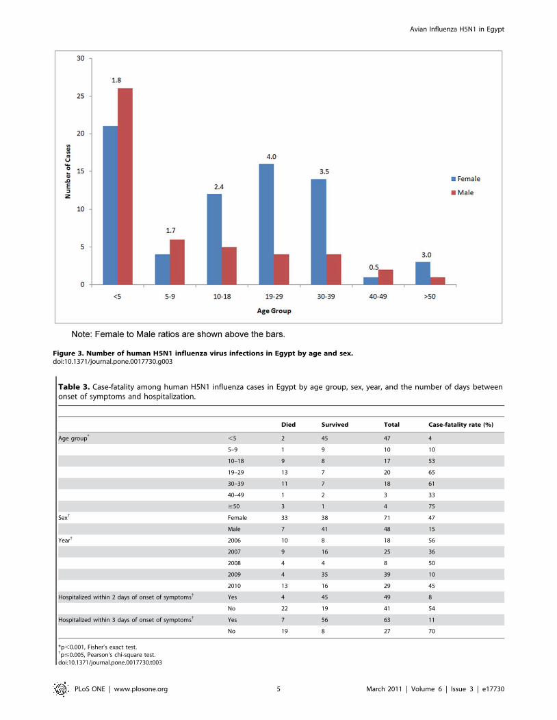
Figure 3. Number of human H5N1 influenza virus infections in Egypt by age and sex.doi:10.1371/journal.pone.0017730.g003
Table 3. Case-fatality among human H5N1 influenza cases in Egypt by age group, sex, year, and the number of days betweenonset of symptoms and hospitalization.
Died Survived Total Case-fatality rate (%)
Age group* ,5 2 45 47 4
5–9 1 9 10 10
10–18 9 8 17 53
19–29 13 7 20 65
30–39 11 7 18 61
40–49 1 2 3 33
$50 3 1 4 75
Sex{ Female 33 38 71 47
Male 7 41 48 15
Year{ 2006 10 8 18 56
2007 9 16 25 36
2008 4 4 8 50
2009 4 35 39 10
2010 13 16 29 45
Hospitalized within 2 days of onset of symptoms{ Yes 4 45 49 8
No 22 19 41 54
Hospitalized within 3 days of onset of symptoms{ Yes 7 56 63 11
No 19 8 27 70
*p,0.001, Fisher’s exact test.{p#0.005, Pearson’s chi-square test.doi:10.1371/journal.pone.0017730.t003
Avian Influenza H5N1 in Egypt
PLoS ONE | www.plosone.org 5 March 2011 | Volume 6 | Issue 3 | e17730
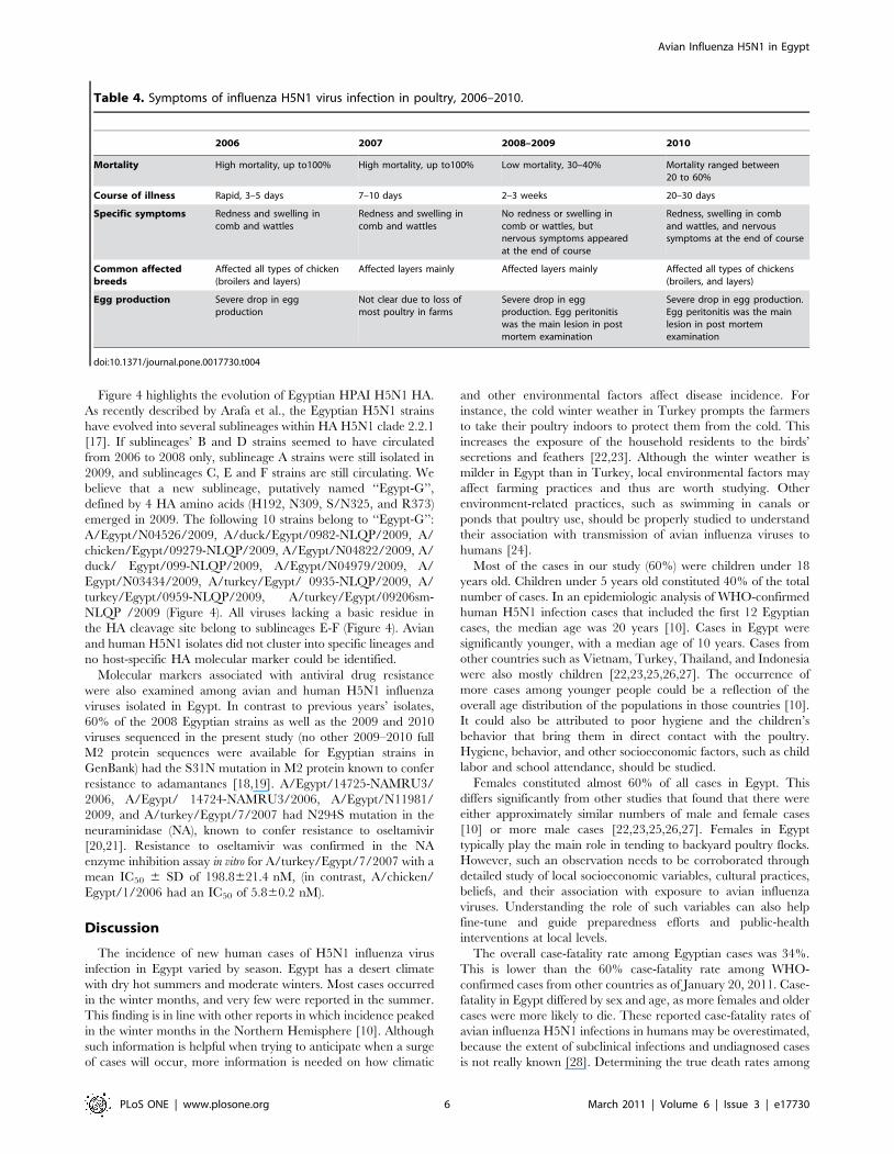
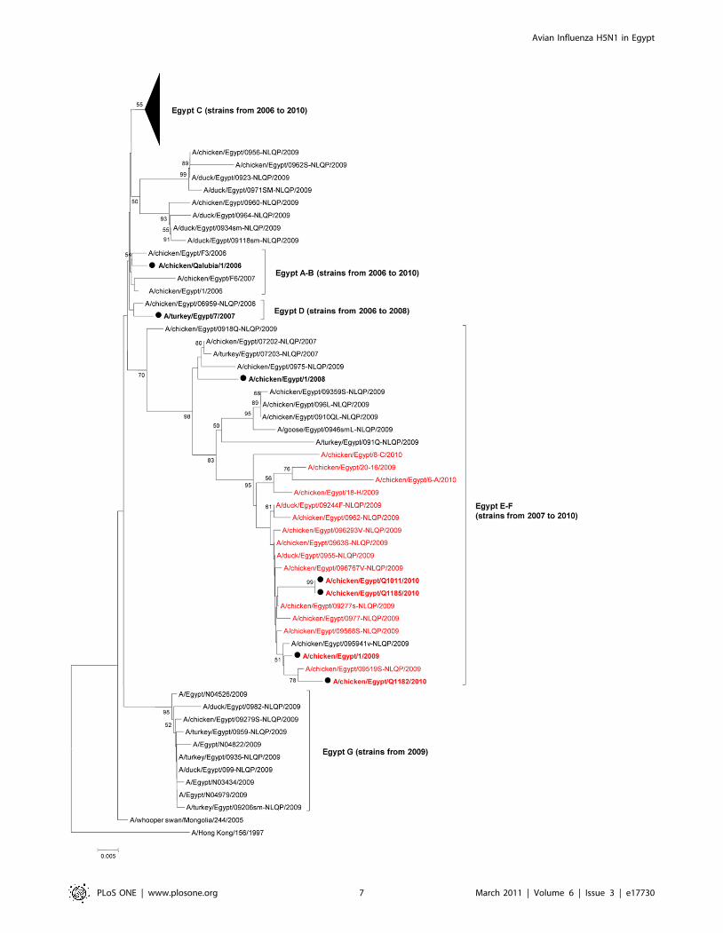
Figure 4 highlights the evolution of Egyptian HPAI H5N1 HA.
As recently described by Arafa et al., the Egyptian H5N1 strains
have evolved into several sublineages within HA H5N1 clade 2.2.1
[17]. If sublineages’ B and D strains seemed to have circulated
from 2006 to 2008 only, sublineage A strains were still isolated in
2009, and sublineages C, E and F strains are still circulating. We
believe that a new sublineage, putatively named ‘‘Egypt-G’’,
defined by 4 HA amino acids (H192, N309, S/N325, and R373)
emerged in 2009. The following 10 strains belong to ‘‘Egypt-G’’:
A/Egypt/N04526/2009, A/duck/Egypt/0982-NLQP/2009, A/
chicken/Egypt/09279-NLQP/2009, A/Egypt/N04822/2009, A/
duck/ Egypt/099-NLQP/2009, A/Egypt/N04979/2009, A/
Egypt/N03434/2009, A/turkey/Egypt/ 0935-NLQP/2009, A/
turkey/Egypt/0959-NLQP/2009, A/turkey/Egypt/09206sm-
NLQP /2009 (Figure 4). All viruses lacking a basic residue in
the HA cleavage site belong to sublineages E-F (Figure 4). Avian
and human H5N1 isolates did not cluster into specific lineages and
no host-specific HA molecular marker could be identified.
Molecular markers associated with antiviral drug resistance
were also examined among avian and human H5N1 influenza
viruses isolated in Egypt. In contrast to previous years’ isolates,
60% of the 2008 Egyptian strains as well as the 2009 and 2010
viruses sequenced in the present study (no other 2009–2010 full
M2 protein sequences were available for Egyptian strains in
GenBank) had the S31N mutation in M2 protein known to confer
resistance to adamantanes [18,19]. A/Egypt/14725-NAMRU3/
2006, A/Egypt/ 14724-NAMRU3/2006, A/Egypt/N11981/
2009, and A/turkey/Egypt/7/2007 had N294S mutation in the
neuraminidase (NA), known to confer resistance to oseltamivir
[20,21]. Resistance to oseltamivir was confirmed in the NA
enzyme inhibition assay in vitro for A/turkey/Egypt/7/2007 with a
mean IC50 6 SD of 198.8621.4 nM, (in contrast, A/chicken/
Egypt/1/2006 had an IC50 of 5.860.2 nM).
Discussion
The incidence of new human cases of H5N1 influenza virus
infection in Egypt varied by season. Egypt has a desert climate
with dry hot summers and moderate winters. Most cases occurred
in the winter months, and very few were reported in the summer.
This finding is in line with other reports in which incidence peaked
in the winter months in the Northern Hemisphere [10]. Although
such information is helpful when trying to anticipate when a surge
of cases will occur, more information is needed on how climatic
and other environmental factors affect disease incidence. For
instance, the cold winter weather in Turkey prompts the farmers
to take their poultry indoors to protect them from the cold. This
increases the exposure of the household residents to the birds’
secretions and feathers [22,23]. Although the winter weather is
milder in Egypt than in Turkey, local environmental factors may
affect farming practices and thus are worth studying. Other
environment-related practices, such as swimming in canals or
ponds that poultry use, should be properly studied to understand
their association with transmission of avian influenza viruses to
humans [24].
Most of the cases in our study (60%) were children under 18
years old. Children under 5 years old constituted 40% of the total
number of cases. In an epidemiologic analysis of WHO-confirmed
human H5N1 infection cases that included the first 12 Egyptian
cases, the median age was 20 years [10]. Cases in Egypt were
significantly younger, with a median age of 10 years. Cases from
other countries such as Vietnam, Turkey, Thailand, and Indonesia
were also mostly children [22,23,25,26,27]. The occurrence of
more cases among younger people could be a reflection of the
overall age distribution of the populations in those countries [10].
It could also be attributed to poor hygiene and the children’s
behavior that bring them in direct contact with the poultry.
Hygiene, behavior, and other socioeconomic factors, such as child
labor and school attendance, should be studied.
Females constituted almost 60% of all cases in Egypt. This
differs significantly from other studies that found that there were
either approximately similar numbers of male and female cases
[10] or more male cases [22,23,25,26,27]. Females in Egypt
typically play the main role in tending to backyard poultry flocks.
However, such an observation needs to be corroborated through
detailed study of local socioeconomic variables, cultural practices,
beliefs, and their association with exposure to avian influenza
viruses. Understanding the role of such variables can also help
fine-tune and guide preparedness efforts and public-health
interventions at local levels.
The overall case-fatality rate among Egyptian cases was 34%.
This is lower than the 60% case-fatality rate among WHO-
confirmed cases from other countries as of January 20, 2011. Case-
fatality in Egypt differed by sex and age, as more females and older
cases were more likely to die. These reported case-fatality rates of
avian influenza H5N1 infections in humans may be overestimated,
because the extent of subclinical infections and undiagnosed cases
is not really known [28]. Determining the true death rates among
Table 4. Symptoms of influenza H5N1 virus infection in poultry, 2006–2010.
2006 2007 2008–2009 2010
Mortality High mortality, up to100% High mortality, up to100% Low mortality, 30–40% Mortality ranged between20 to 60%
Course of illness Rapid, 3–5 days 7–10 days 2–3 weeks 20–30 days
Specific symptoms Redness and swelling incomb and wattles
Redness and swelling incomb and wattles
No redness or swelling incomb or wattles, butnervous symptoms appearedat the end of course
Redness, swelling in comband wattles, and nervoussymptoms at the end of course
Common affectedbreeds
Affected all types of chicken(broilers and layers)
Affected layers mainly Affected layers mainly Affected all types of chickens(broilers, and layers)
Egg production Severe drop in eggproduction
Not clear due to loss ofmost poultry in farms
Severe drop in eggproduction. Egg peritonitiswas the main lesion in postmortem examination
Severe drop in egg production.Egg peritonitis was the mainlesion in post mortemexamination
doi:10.1371/journal.pone.0017730.t004
Avian Influenza H5N1 in Egypt
PLoS ONE | www.plosone.org 6 March 2011 | Volume 6 | Issue 3 | e17730
Avian Influenza H5N1 in Egypt
PLoS ONE | www.plosone.org 7 March 2011 | Volume 6 | Issue 3 | e17730
H5N1 infection cases requires conducting prospective, large-scale
epidemiologic studies in well-defined populations.
Early hospitalization had a positive effect on survival of the
cases. Similar findings have been reported by other researchers
[25,29,30,31]. Young age of patients was significantly associated
with faster admission to the hospital after onset of the symptoms. It
is possible that younger patients had more severe illness than older
patients thus leading to rapid hospitalization. Alternatively, if the
severity of illness is not different, adults may be delaying seeking
medical care especially if the symptoms are mild. However, we
were not able to ascertain the reason of delayed hospitalization
among adults using available data. Delayed hospitalization,
independent of sex and age, had a significant effect on death.
Arabi et. al. [32] reported that H5N1 patients are usually critically
ill upon admission to a hospital or shortly thereafter. Furthermore,
patients infected with H5N1 virus present with a wide range of
symptoms common to many other widespread illnesses
[22,24,26,27,33,34,35]. This complicates an early differential
diagnosis, and it is sometimes not until the disease is in advanced
stages that such a diagnosis is made. To ensure early diagnosis and
subsequently early hospitalization and treatment, primary care
facilities receiving human cases of H5N1 should always suspect
avian influenza as the cause of disease, especially among cases
exposed to poultry.
This is not the first report on human cases of HPAI in Egypt
[36,37,38], yet our manuscript is the first to attempt to explain the
epidemiology of human cases by comparing it to molecular level
changes in circulating viruses. We have also added the new cases
that were reported in Egypt and were not analyzed before.
Furthermore, we used regression analysis to control for age and
sex when analyzing factors related to fatality and time of
hospitalization. Our analysis was limited to laboratory-confirmed
cases reported to the WHO. Thus, selection bias may have
affected our results if some cases died or recovered before
diagnosis or if false-positive or false-negative laboratory findings
were included. The occurrence of cases with subclinical illness is
not well studied, and this could have also affected our results. Due
to lack of accurate data for some variables and absence of data for
many other variables of interest, we were not able to conduct an
in-depth epidemiologic investigation. Data on human infection
with avian influenza reported to the WHO should be more
accurate, includes linkage to sequence data, and be made more
readily available for investigators; countries in which cases occur
need to be more proactive at investigating cases properly [39].
The clinical symptoms among human cases reported in 2006
were similar to those in 2010, yet there was no human surveillance
conducted in Egypt to ascertain the presence or absence of
asymptomatic cases. The frequency of human H5N1 influenza
infections parallels the pattern in poultry. Milder disease among
poultry leads to mild human infection. For instance, a decrease in
human case-fatality rate in 2009 was accompanied by an observed
decrease in mortality among poultry. Since no host-adaptation
mutation was observed so far, suggesting that the main
transmission route of H5N1 in Egypt is still contact with infected
birds, just as observed in the rest of the world.
We were not able to confirm that lower pathogenicity of the viruses
is the main reason for increase or decrease in the the mortality rate
among humans in Egypt. Considerable variation among backyard
poultry was indeed observed; ducks and geese seemed significantly
less affected by the virus than in 2006–2007. This is supported by the
fact that mortality rates among humans in 2009 and 2010 was lower
than that in the previous years after controlling for age. We were able
to link (by age and date) the epidemiological and sequence data of the
cases infected with A/Egypt/N04526/2009, A/Egypt/N04822/
2009, A/Egypt/N04979/2009, and A/Egypt/N03434/2009 (viruses
belonging to the proposed sublineage G) and found that three of these
cases survived. As the virus appeared in its most virulent form in
2006, it is expected that any significant mutation, especially in the
multibasic aa sequence, should lead to lower virulence. The increase
in mortality in 2010 may be explained by the fact that viruses with the
same virulence markers as those isolated in 2006 continued to
circulate into 2010. However, other host and environmental factors
that potentially affect disease severity need to be properly studied
before claiming that the decrease in mortality is solely due to viral
mutations.
Among cases reported in 2009 and 2010, 68 of the 121 (56%)
new human cases reported were from Egypt. As Egypt became the
new epicenter for human infection with H5N1, experts are
concerned about the potential of H5N1 viruses circulating in
Egypt to become more adapted to human-to-human transmission.
These fears arise from the fact that these new viruses are less
virulent and may be causing asymptomatic infections especially
among adults. Thus, there is an urgent need to conduct more
epidemiologic studies in Egypt and other endemic areas to
enhance our understanding of incidence, prevalence, and
determinants of human infections with avian influenza. It remains
unknown from where the pandemic strain may emerge. Thus,
sustained viral sequence comparisons and phylogenetic analyses of
current HPAIV H5N1 are necessary to recognize newly emerging
influenza variants and to monitor the global spread of these
viruses.
Author Contributions
Conceived and designed the experiments: GK RJW MAA. Performed the
experiments: GK MFD RAES AMK EAG AM. Analyzed the data: GK
RJW MFD MAA. Wrote the paper: GK RJW MFD MAA.
References
1. Chen H, Smith GJ, Li KS, Wang J, Fan XH, et al. (2006) Establishment of
multiple sublineages of H5N1 influenza virus in Asia: implications for pandemic
control. Proc Natl Acad Sci U S A 103: 2845–2850.
2. Matrosovich M, Zhou N, Kawaoka Y, Webster R (1999) The surface
glycoproteins of H5 influenza viruses isolated from humans, chickens, and wild
aquatic birds have distinguishable properties. J Virol 73: 1146–1155.
3. Webster RG, Wright SM, Castrucci MR, Bean WJ, Kawaoka Y (1993)
Influenza–a model of an emerging virus disease. Intervirology 35: 16–25.
4. WHO (2009) H5N1 avian influenza: Timeline of major events.
5. Al-Azemi A, Bahl J, Al-Zenki S, Al-Shayji Y, Al-Amad S, et al. (2008) Avian
influenza A virus (H5N1) outbreaks, Kuwait, 2007. Emerg Infect Dis 14:
958–961.
Figure 4. Phylogenic relationships of the HA genes of Egyptian HPAI H5N1 influenza viruses. A/chicken/Qalubia/1/2006, A/turkey/Egypt/7/2007, A/chicken/Egypt/1/2008, A/chicken/Egypt/1/2009, A/chicken/Egypt/Q1011/2010, A/chicken/Egypt/Q1182/2010, and A/chicken/Egypt/Q1185/2010 HA sequences (in bold, marked with a closed circle) were compared to all 2009–2010 HA gene sequences currently available on GenBank, aswell as to a few reference 2006–2008 strains as described by Arafa et al [17]. Numbers at nodes correspond to bootstrap values .49. Strains with abasic amino acid less than their counterparts are in red font. Sublineages are indicated on the right hand side of the tree, with in parentheses for eachsublineage, the year range of clustered strains.doi:10.1371/journal.pone.0017730.g004
Avian Influenza H5N1 in Egypt
PLoS ONE | www.plosone.org 8 March 2011 | Volume 6 | Issue 3 | e17730
6. Aly MM, Arafa A, Hassan MK (2008) Epidemiological findings of outbreaks of
disease caused by highly pathogenic H5N1 avian influenza virus in poultry in
Egypt during 2006. Avian Dis 52: 269–277.
7. Saad MD, Ahmed LS, Gamal-Eldein MA, Fouda MK, Khalil F, et al. (2007)
Possible avian influenza (H5N1) from migratory bird, Egypt. Emerg Infect Dis
13: 1120–1121.
8. Egyptian-Cabinet (2006) Information and Decision Support Center. Analysis of
Poultry Industry Structure in Egypt: Proposal in the light of Avian Flu Crisis.
9. WHO (2009) Epidemic and Pandemic Alert and Response.
10. WHO (2006) Epidemiology of WHO-confirmed human cases of avian influenza
A(H5N1) infection. Wkly Epidemiol Rec 81: 249–257.
11. Hall TA (1999) BioEdit: a user-friendly biological sequence alignment editor and
analysis for Windows 95/98/NT. Nucleic Acids Symp 41: 95–98.
12. Chenna R, Sugawara H, Koike T, Lopez R, Gibson TJ, et al. (2003) Multiple
sequence alignment with the Clustal series of programs. Nucleic Acids Res 31:
3497–3500.
13. Kumar S, Tamura K, Jakobsen IB, Nei M (2001) MEGA2: molecular
evolutionary genetics analysis software. Bioinformatics 17: 1244–1245.
14. Hatta M, Gao P, Halfmann P, Kawaoka Y (2001) Molecular basis for high
virulence of Hong Kong H5N1 influenza A viruses. Science 293: 1840–1842.
15. Seo SH, Hoffmann E, Webster RG (2002) Lethal H5N1 influenza viruses escape
host anti-viral cytokine responses. Nat Med 8: 950–954.
16. Jackson D, Hossain MJ, Hickman D, Perez DR, Lamb RA (2008) A new
influenza virus virulence determinant: the NS1 protein four C-terminal residues
modulate pathogenicity. Proc Natl Acad Sci U S A 105: 4381–4386.
17. Arafa A, Suarez DL, Hassan MK, Aly MM Phylogenetic analysis of
hemagglutinin and neuraminidase genes of highly pathogenic avian influenza
H5N1 Egyptian strains isolated from 2006 to 2008 indicates heterogeneity with
multiple distinct sublineages. Avian Dis 54: 345–349.
18. Cheung CL, Rayner JM, Smith GJ, Wang P, Naipospos TS, et al. (2006)
Distribution of amantadine-resistant H5N1 avian influenza variants in Asia.
J Infect Dis 193: 1626–1629.
19. Hurt AC, Selleck P, Komadina N, Shaw R, Brown L, et al. (2007) Susceptibility
of highly pathogenic A(H5N1) avian influenza viruses to the neuraminidase
inhibitors and adamantanes. Antiviral Res 73: 228–231.
20. Abed Y, Baz M, Boivin G (2006) Impact of neuraminidase mutations conferring
influenza resistance to neuraminidase inhibitors in the N1 and N2 genetic
backgrounds. Antivir Ther 11: 971–976.
21. Yen HL, Ilyushina NA, Salomon R, Hoffmann E, Webster RG, et al. (2007)
Neuraminidase inhibitor-resistant recombinant A/Vietnam/1203/04 (H5N1)
influenza viruses retain their replication efficiency and pathogenicity in vitro and
in vivo. J Virol 81: 12418–12426.
22. Oner AF, Bay A, Arslan S, Akdeniz H, Sahin HA, et al. (2006) Avian influenza
A (H5N1) infection in eastern Turkey in 2006. N Engl J Med 355: 2179–2185.
23. WHO (2006) Human cases of influenza A(H5N1) infection in eastern Turkey,
December 2005–January 2006. Wkly Epidemiol Rec 81: 410–416.
24. de Jong MD, Bach VC, Phan TQ, Vo MH, Tran TT, et al. (2005) Fatal avian
influenza A (H5N1) in a child presenting with diarrhea followed by coma.N Engl J Med 352: 686–691.
25. Chotpitayasunondh T, Ungchusak K, Hanshaoworakul W, Chunsuthiwat S,
Sawanpanyalert P, et al. (2005) Human disease from influenza A (H5N1),Thailand, 2004. Emerg Infect Dis 11: 201–209.
26. Sedyaningsih ER, Isfandari S, Setiawaty V, Rifati L, Harun S, et al. (2007)Epidemiology of cases of H5N1 virus infection in Indonesia, July 2005–June
2006. J Infect Dis 196: 522–527.
27. Tran TH, Nguyen TL, Nguyen TD, Luong TS, Pham PM, et al. (2004) Avianinfluenza A (H5N1) in 10 patients in Vietnam. N Engl J Med 350: 1179–1188.
28. Li FC, Choi BC, Sly T, Pak AW (2008) Finding the real case-fatality rate ofH5N1 avian influenza. J Epidemiol Community Health 62: 555–559.
29. Kandun IN, Tresnaningsih E, Purba WH, Lee V, Samaan G, et al. (2008)Factors associated with case fatality of human H5N1 virus infections in
Indonesia: a case series. Lancet 372: 744–749.
30. Thanh TT, van Doorn HR, de Jong MD (2008) Human H5N1 influenza:current insight into pathogenesis. Int J Biochem Cell Biol 40: 2671–2674.
31. Yu H, Gao Z, Feng Z, Shu Y, Xiang N, et al. (2008) Clinical characteristics of 26human cases of highly pathogenic avian influenza A (H5N1) virus infection in
China. PLoS ONE 3: e2985.
32. Arabi Y, Gomersall CD, Ahmed QA, Boynton BR, Memish ZA (2007) Thecritically ill avian influenza A (H5N1) patient. Crit Care Med 35: 1397–1403.
33. Hui DS (2008) Review of clinical symptoms and spectrum in humans withinfluenza A/H5N1 infection. Respirology 13 Suppl 1: S10–13.
34. Wiwanitkit V (2005) Diarrhoea as a presentation of bird flu infection: a summaryon its correlation to outcome in Thai cases. Gut 54: 1506.
35. Wong SS, Yuen KY (2006) Avian influenza virus infections in humans. Chest
129: 156–168.36. Dudley JP (2009) Age-specific infection and death rates for human A(H5N1)
avian influenza in Egypt. Euro Surveill 14.37. Fasina FO, Ifende VI, Ajibade AA Avian influenza A(H5N1) in humans:
lessons from Egypt. Euro Surveill 15: 19473.
38. Kandeel A, Manoncourt S, Abd el Kareem E, Mohamed Ahmed AN, El-Refaie S, et al. Zoonotic transmission of avian influenza virus (H5N1), Egypt,
2006–2009. Emerg Infect Dis 16: 1101–1107.39. Nicoll A (2006) Human H5N1 infections: so many cases–why so little
knowledge? Euro Surveill 11: 74–75.40. WHO (2009) WHO information for laboratory diagnosis of pandemic (H1N1)
2009 virus in humans - revised.
41. Slomka MJ, Coward VJ, Banks J, Londt BZ, Brown IH, et al. (2007)Identification of sensitive and specific avian influenza polymerase chain reaction
methods through blind ring trials organized in the European Union. Avian Dis51: 227–234.
42. Wright KE, Wilson GA, Novosad D, Dimock C, Tan D, et al. (1995) Typing
and subtyping of influenza viruses in clinical samples by PCR. J Clin Microbiol33: 1180–1184.
Avian Influenza H5N1 in Egypt
PLoS ONE | www.plosone.org 9 March 2011 | Volume 6 | Issue 3 | e17730
Top Related
Copyright © 2022 FDOKUMEN

