


Bahasa
Halaman
Hukum


Testosterone Plus Low-Intensity Physical Training in LateLife Improves Functional Performance, Skeletal MuscleMitochondrial Biogenesis, and Mitochondrial QualityControl in Male MiceWen Guo1*, Siu Wong1, Michelle Li1, Wentao Liang1, Marc Liesa1, Carlo Serra, Ravi Jasuja1,
Andrzej Bartke2, James L Kirkland3, Orian Shirihai1, Shalender Bhasin1
1Department of Medicine, Boston University School of Medicine, Boston, Massachusetts, United States of America, 2Department of Internal Medicine, Southern Illinois
School of Medicine, Springfield, Illinois, United States of America, 3 Robert and Arlene Kogod Center on Aging, Mayo Clinic, Rochester, Minnesota, United States of
America
Abstract
Testosterone supplementation increases muscle mass in older men but has not been shown to consistently improvephysical function and activity. It has been hypothesized that physical exercise is required to induce the adaptationsnecessary for translation of testosterone-induced muscle mass gain into functional improvements. However, the effects oftestosterone plus low intensity physical exercise training (T/PT) on functional performance and bioenergetics are unknown.In this pilot study, we tested the hypothesis that combined administration of T/PT would improve functional performanceand bioenergetics in male mice late in life more than low-intensity physical training alone. 28-month old male mice wererandomized to receive T/PT or vehicle plus physical training (V/PT) for 2 months. Compare to V/PT control, administration ofT/PT was associated with improvements in muscle mass, grip strength, spontaneous physical movements, and respiratoryactivity. These changes were correlated with increased mitochondrial DNA copy number and expression of markers formitochondrial biogenesis. Mice receiving T/PT also displayed increased expression of key elements for mitochondrial qualitycontrol, including markers for mitochondrial fission-and-fusion and mitophagy. Concurrently, mice receiving T/PT alsodisplayed increased expression of markers for reduced tissue oxidative damage and improved muscle quality. Conclusion:Testosterone administered with low-intensity physical training improves grip strength, spontaneous movements, andrespiratory activity. These functional improvements were associated with increased muscle mitochondrial biogenesis andimproved mitochondrial quality control.
Citation: Guo W, Wong S, Li M, Liang W, Liesa M, et al. (2012) Testosterone Plus Low-Intensity Physical Training in Late Life Improves Functional Performance,Skeletal Muscle Mitochondrial Biogenesis, and Mitochondrial Quality Control in Male Mice. PLoS ONE 7(12): e51180. doi:10.1371/journal.pone.0051180
Editor: Jean-Marc A. Lobaccaro, Clermont Universite, France
Received September 25, 2012; Accepted October 29, 2012; Published December 11, 2012
Copyright: � 2012 Guo et al. This is an open-access article distributed under the terms of the Creative Commons Attribution License, which permits unrestricteduse, distribution, and reproduction in any medium, provided the original author and source are credited.
Funding: This work was supported by National Institutes of Health (NIH) grants 1R21AG037859 (WG), RO1AG037193 (SB), AG13925 (JLK), DK074778 (OS) andPO1AG031736 (AB) and by the Boston Claude D. Pepper Older Americans Independence Center (5P30AG031679). The funders had no role in study design, datacollection and analysis, decision to publish, or preparation of the manuscript.
Competing Interests: The authors have declared that no competing interests exist.
* E-mail: [email protected]
Introduction
In men, age-related decline in serum testosterone levels has
been associated with loss of skeletal muscle mass, strength, and
physical performance [1,2,3,4,5,6]. Randomized clinical trials are
in agreement that testosterone supplementation increases skeletal
muscle mass in young as well as older men [2,3,4]. Indeed,
testosterone and many other androgens are being investigated as
potential therapies for functional limitations associated with aging
and illness. However, randomized trials have failed to show
consistent improvements in functional performance; the effects of
testosterone on physical activity also remain poorly understood
[7,8]. In fact, in a recent trial in older men with mobility
limitation, testosterone administration failed to induce significant
improvements in physical activity [8].
Functional performance is an integrated measure of physical
function that has been associated with important health
outcomes, including frailty, functional limitations, and mortality.
Although there are several potential reasons why testosterone
alone may not induce consistent improvements in functional
performance in spite of substantial gains in muscle mass, one
plausible hypothesis is that physical exercise is required to
induce the adaptations necessary for translation of muscle mass
gains into functional improvements. Low-intensity physical
exercise interventions that emphasize walking improve some
aspects of physical performance but have been typically
associated with only modest gains in muscle mass and physical
function. However, the effects of testosterone administered in
conjunction with an adjunctive low-intensity physical exercise on
functional performance and bioenergetics are unknown and
were the subject of this investigation. In this study, we tested the
hypothesis that the combined administration of testosterone plus
a low-intensity physical training program in male mice at a late
stage of life would improve functional performance and
bioenergetics more than low-intensity physical training alone.
PLOS ONE | www.plosone.org 1 December 2012 | Volume 7 | Issue 12 | e51180

Functional performance is an integrated measure of complex
interplay of multiple factors, including muscle mass and quality,
bioenergetics, behavioral, and social factors. In skeletal muscle,
mitochondrial oxidative phosphorylation (OXPHOS) serve as
a major source of energy to meet basic metabolic needs and
energy demands during physical activity. Recent studies have
linked the progressive decline in skeletal mitochondrial function
with aging and functional decline, where physical strength
declines out of proportion to the loss of muscle mass [9,10].
Mitochondria are also the major source of free radicals,
generated as byproducts from OXPHOS, whose effects are
counteracted by multiple scavenging enzymes and non-enzy-
matic antioxidants. To optimize energy production and
minimize oxidative damage, mitochondria are engaged in
dynamic network exchange through fission and fusion, which
identifies damaged mitochondrial and tags them for mitophagy
[11,12]. Impairments of these mechanisms of mitochondrial
quality control during aging contribute to the age-related
increase in tissue oxidative damage and functional decline
[13,14,15,16,17]. Aerobic exercise is an effective physiological
intervention that counteracts aging-related mitochondrial dys-
function through simultaneous improvement of mitochondrial
biogenesis and quality control, including up-regulation of
mitophagy [18,19,20]. Glass and colleagues reported that
testosterone enhances voluntary wheel running in castrated
young adult mice and increases the mRNA expression of
mitochondrial genes, especially those related to complex I in the
electron transport chain [21]. In addition, myocyte-specific
transgenic expression of androgen receptor in young adult rats
and mice also increases skeletal mitochondrial enzyme activities
and increases in vivo oxygen consumption [22,23]; implying
a positive role of testosterone and its nuclear receptor on
mitochondrial biogenesis and function. Hence a second aim of
this study was to determine the effects of testosterone
supplementation plus low-intensity physical exercise on muscle
mitochondrial biogenesis and quality control. We hypothesized
that testosterone supplementation plus low intensity physical
exercise training will improve skeletal muscle mitochondrial
biogenesis and mitochondrial quality control in very old mice
undergoing routine low-intensity physical training.
We used 28-month old male C57BL6 mice as our model
because these animals display age-related decline in testosterone
levels similar to that observed in older men. The intervention was
started in late life - at 28 months of age - and lasted for two
months, giving an age range within the 50–25% survival window
(http://www.nia.nih.gov/aged-rodent-colonies-handbook/strain-
survival-information). Previous studies suggest that functional
performance within this survival window predicts late life
healthspan in both human and rodents [10,24]. We show here
that testosterone plus low-intensity physical training even at this
late stage of life increases spontaneous physical activity, respira-
tion, and grip strength more than physical training alone, in
addition to the expected gains in muscle mass. We also provide the
first evidence that testosterone supplementation when adminis-
tered together with low-intensity physical training increases
mitochondrial biogenesis and improves mitochondrial quality
control.
Materials and Methods
AnimalsThe animal use protocol for this study was approved by the
Institutional Animal Care and Use Committee of Boston
University School of Medicine. Male C57BL/6 mice at 28 months
of age were obtained from the rodent longevity colony of the
National Institute on Aging (NIA). After acclimation, baseline
measurements of body composition using nuclear magnetic
resonance (NMR), grip strength, and treadmill performance
(6 m/min, 5% incline) were obtained.
Testosterone AdministrationAfter completion of baseline assessments, the mice were
assigned with matching body composition and grip strength to
receive either testosterone or vehicle injection (N=8 in each
group). Testosterone, dissolved in medium-chain oil (www.life-
enhancement.com), was administered by subcutaneous injection
(50 mg/kg, twice per week). Control mice were injected with equal
volume of oil (100 ul). Serum testosterone concentration was
measured using liquid chromatography tandem mass spectrometry
(10–20 ng/dL for control mice and 500- 1000 ng/dL for mice
injected with testosterone).
Low-intensity Physical Training RoutinesBoth control and testosterone-treated animals were engaged
in a 30 min treadmill walk three times each week (6 m/min,
5% inclination) for the first six weeks. This condition is
considered as ‘‘low-intensity’’ as compared to a 60 min daily
running at 13 m/min, 10% incline for similarly aged rodents
[18]. To promote walking, a shock grid (0.97 mA, 3 Hz) at the
back of the treadmill was used to discourage the mice from
stopping while the treadmill belt was moving. After the initial
acclimation, all mice walked voluntarily throughout 30 min
without the use of the shock grid. Two mice in the control
group died before and one died after the final functional
assessments. Hence, for most of the functional tests, we had 6 to
8 mice in each group but tissues were available for 5 mice in
the control group and 8 mice in the testosterone group. At the
end of the experiment, all animals were examined for
abnormality for internal organs and external appearance. Most
animals show moderate discoloring in seminal vesicles with no
difference between the control and testosterone supplemented
groups. No incidence of visually detectable tumor, ulcer, or
other abnormal tissue appearance was found among these
animals.
Functional AssessmentNMR and grip strength measurement was repeated in week 7.
Metabolic cage study was performed at the end of week 6 and
rotarod test during week 7, as described previously [25]. For grip
strength, the animals were allowed to grasp a horizontal metal bar
while being pulled by their tail. The results were recorded using an
automatic force transducer (Columbia Instrument, www.colinst.
com). For rota-rod test, a 4-lane accelerating rota-rod (Columbus
Instrument), equipped with a built-in automatic timer was used
and the speed of the rotating rod was adjusted manually. Mice
were trained first at a low speed starting at 4 rpm, and the speed
was gradually increased by 0.5 rpm/min to 9 rpm over 3 days.
For the running test, mice were placed on a static rod. Once all
animals were on the rod, the motor was turned on and the rod
rotation was accelerated at a rate of 0.5 rpm/min until all animals
fell off the rod. The running distance was calculated as the speed
multiplied by the running time.
Tissue AnalysisTissue DNA and RNA isolation, RNA reverse transcription,
and real-time qPCR were performed as described before [25]. All
PCR primers were designed as intron-spanning except for the
Testosterone and Mitochondria in Aging Muscle
PLOS ONE | www.plosone.org 2 December 2012 | Volume 7 | Issue 12 | e51180

nuclear DNA-encoded cytochrome c and all mitochondrial DNA-
encoded genes which do not have introns. To eliminate DNA
contamination for qPCR analysis of these genes, mRNA was
isolated from total RNA samples using the mRNA purification kit
(Qiagen #72041). We found this method to be superior to the
DNase I digestion method which was efficient in eliminating
nuclear DNA but not mitochondrial DNA. The mRNA thus
obtained was reverse-transcribed to first-strand cDNA using
standard protocol. For Western analysis, tissue was homogenized
in cell lysis buffer (Cell Signaling Technology, #9803, www.
cellsignal.com) supplemented with 0.1% SDS and standard
cocktails of protease inhibitors and phosphatase inhibitors and
10 mM DTT. Lysate was cleared by centrifuge at 3000 g for
15 min. Protein was loaded as 0.015 mg/lane for detection of
mitochondrial proteins and 0.05 mg/lane for detection of other
proteins. A special blocking reagent (Rodent block M, www.
biocare.net) was used to block endogenous mouse IgG signals. A
light-chain specific anti-mouse secondary antibody (Millipore,
#MAB201P) was used for all first antibodies that were generated
in mice. Mitochondria-enriched fraction was isolated from frozen
skeletal muscle following a published protocol [26]. The protocol
was validated in-house using Western analysis to confirm
a substantial enrichment of the mitochondrial enzyme manganese
superoxide dismutase (MnSOD) and a marked reduction in alpha-
actin concentration in the mitochondrial fraction as compared to
a whole tissue lysate. Mitochondrial protein ATP synthase subunit
5a (ATP5a) was used as loading control for mitochondria-enrich
fractions and GAPDH was used for loading control for whole
tissue lysates. Antibodies for mitoprofile cocktail were purchased
from Mitoscience (www.mitoscience.com, #MS604) and antibody
for GAPDH and secondary antibody against mouse or goat IgG
from Santa Cruz (www.scbt.com). All other first antibodies and
a secondary against rabbit were from Cell Signaling Technology.
Results of Western analysis were quantified using NIH Image J
program. Thiobarbituric Acid-Reactive (TBAR) substance was
measured in skeletal muscle lysate using a commercial kit following
manufacturer’s instructions (www.caymanchem.com, #10009055). To avoid tissue oxidation during sample preparation,
butylated hydroxyanisole was added to a lysis buffer to a final
concentration of 5 mM.
Assays for Mitochondrial Enzyme ActivityFrozen tissue was pulverized in liquid nitrogen. About 30 mg of
tissue was homogenized in 2 ml buffer (20 mM Tris, sucrose
250 mM, KCl 40 mM, EGTA 2 mM, 10% glycerol (v/v) and
1 mM PMSF). A Glas-Col motor-driven homogenizer was used at
4000 rpm and each sample was homogenized by 30 strokes in
a standard homogenizer with fitted Teflon pestle, with homoge-
nizer kept on ice all time. The solution was then separated into
260.8 ml fractions. For samples to be used for citrate synthase
assay, 0.04% Triton X-100 was added. For samples to be used for
mitochondrial electron transport chain activity, 0.02% dodecyl
maltoside was added. Both fractions were rotated at 4uC for one
hour and then centrifuged at 3000 g for 15 min. The supernatant
was divided into small aliquots and stored at 280uC until use. The
enzyme activities were measured using standard spectrophotom-
etry.
Statistical AnalysesResults are presented as mean 6 SEM. Comparison between
two groups were analyzed using Student’s t test. Comparisons
among multiple groups were performed using one-way analysis of
variance (ANOVA), followed by Tukey’s test.
Results
1. Testosterone Supplementation Plus Low-intensityPhysical Training Improves Functional Performance inAging Male MiceAs expected,, testosterone plus low-intensity physical training
(T/PT) for two months beginning at 28 month of age was
associated with a significant increase in whole body lean mass
and a decrease in fat mass, in comparison to the baseline values
or by cross-comparison with the control group that received
vehicle and the same level of physical training (V/PT). Theseresults are shown in supplementary Figure S1A. The mice
receiving T/PT also had significantly higher mass of weight-
bearing forelimb (triceps) and hind-limb (quadriceps femoris and
gastocnemius/soleus) muscles, as well as non-weight-bearing
cardiac, pectoralis, and levator ani muscles than those receiving
V/PT (Figure S1B). The mice receiving T/PT displayed
a significant increase in grip strength over the 7 week treatment
period, while the control mice showed no change (Figure 1A,
left panel). The mice receiving T/PT also displayed an average
30% increase in Rota-rod running distance, although the effect
did not reach statistical significance (Figure S2). When placed in
a metabolic cage system, T/PT mice displayed a significant
increase in their nocturnal spontaneous movements along the
X- and Y- axes (Figure 1B, upper and middle panels) but not in
the Z-axis (Figure 1B, lower panel). Diurnal activity was not
significantly different between the two groups in the X- and Y-
direction but was lower in the Z-axis for the T/PT group.
Since mice are nocturnal creatures, this difference in the day
time may not be physiologically important.
In comparison with the V/PT group, treatment with T/PTincreased O2 consumption and CO2 production by 14.3% and
13.5% during day time and by 17.9% and 20.9% at night time,
respectively, after normalization by body weight (Figure 1C). As
O2 consumption is related to lean mass and mitochondrial
function, and each may contribute to increase respiration
independently [27], we also normalized the data by lean body
mass and found that the increase in respiratory activity in T/PT group remained significant (Figure S3). The increase in
respiration induced by T/PT was observed both during the
dark period and the light period when the animals were less
active (Figure S4); indicating that part of the increase in
metabolic rate was independent of muscle mass and physical
movements. The respiratory exchange ratio was similar between
the two groups and varied from near 0.8 in the light period to
near 0.95 in the dark period (data not shown). Together, these
results indicate that late-life testosterone supplementation plus
low-intensity physical training improved functional performance
as compared to the control animals that received the vehicle but
engaged in the same physical training protocol.
2. Testosterone Supplementation Plus Low-intensityPhysical Training Increases Mitochondrial DNA CopyNumber and the Expression of Selected MitochondrialTranscripts in Skeletal Muscle of Aging Male MiceSkeletal muscle force capacity is correlated with mitochon-
drial content and function [28]. Administration of T/PT was
associated with a significantly greater (20% higher) mitochon-
drial DNA copy number per unit nuclear DNA (mtDNA/
nDNA ratio) than V/PT. We measured the mRNA expression
of selected mitochondrial transcripts encoded by the mitochon-
drial as well as the nuclear genomes. As shown in Figure 2B,
T/PT increased the expression of genes encoded by mtDNA,
Testosterone and Mitochondria in Aging Muscle
PLOS ONE | www.plosone.org 3 December 2012 | Volume 7 | Issue 12 | e51180

including NADH dehydrogenase subunits (complex I: ND1,
ND4), cytochrome b (complex III), and cytocrhome c oxidase
subunits (complex IV: Cox2a and Cox3a), but not ATP
synthase subunit 6 (ATP6, complex V). Among the tested
mitochondrial transcripts encoded by nDNA, T/PT induced
a large increase in aminolevulinate, d-synthase 1, (ALAS1,
mitochondrial heme synthesis [29]) and adenine nucleotide
translocase (ANT, exchanges cytosolic ADP for mitochondrial
ATP). T/PT also moderately increased the mRNA expression
of CPT1beta (gate-keeper for mitochondrial fatty acid oxidation)
and PDK4 (inhibitor of pyruvate oxidation to favor fatty acid
for mitochondrial fuel [30]). Neither the expression of
cytochrome c, a nuclear genome encoded component of
complex IV, nor that of UCP3, a muscle-specific mitochondrial
uncoupling protein, was significantly affected. The latter finding
agrees with others showing that testosterone does not increase
UCP3 expression in myotubes from male donors [31].
Together, these results show that testosterone supplementation
increased the mtDNA copy number and also the expression of
selected mitochondrial transcripts encoded by both mitochon-
drial and nuclear genomes in the old mice that were engaged in
low-intensity physical training.
Figure 1. Effect of testosterone supplementation plus low-intensity physical training on grip strength, spontaneous physicalactivity, and respiratory activity in old male mice. (A) Forelimb grip strength measured at baseline (wk 0, N= 8 per group, C: control, T:testosterone) and after 7 weeks of (N = 6 for control, N = 8 for T group) testosterone supplementation (10 mg/kg, sc, twice per week). Control wasinjected with an equal volume of vehicle (0.1 ml). (B) Spontaneous physical movements recorded as total counts of infrared beam breaking per min.Each animal was housed individually in a metabolic cage. Results were averaged separately over the light and dark periods. The measurement wasperformed during week 6 after testosterone supplementation (N= 4 for each group). Xtot, Ytot, Ztot each denotes movements in X-axis, Y-axis, and Z-axis. (C) Mean respiratory activity recorded as oxygen consumption and carbon dioxide production for animals individually housed in a metaboliccage (N= 4). Results were normalized to total body weight. All results are shown as mean 6 SEM, and the difference between control andtestosterone groups at each time period was analyzed by t test. ns, not significantly different.doi:10.1371/journal.pone.0051180.g001
Testosterone and Mitochondria in Aging Muscle
PLOS ONE | www.plosone.org 4 December 2012 | Volume 7 | Issue 12 | e51180

Figure 2. Effect of testosterone supplementation plus low-intensity physical training on markers of mitochondrial biogenesis in theskeletal muscle. (A) Testosterone increased mitochondrial DNA (mtDNA) copy number when normalized to nuclear genome DNA (nDNA). (B)Testosterone increased mtDNA-encoded mitochondrial transcripts for markers of complex I (NADH dehydrogenase subunits: ND1, ND4), complex III(cytochrome b: Cyto b), complex IV (cytochrome c oxidase subunits: Cox2a and Cox3a), and complex V (ATP synthase subunit 6, APT6) of the electrontransport chain. (C) Testosterone increased the expression of selected nuclear genome-encoded mitochondrial transcripts for d-aminolevulinatesynthase 1 (ALAS1), adenine nucleotide translocator (ANT), Carnitine palmitoyltransferase 1(CPT1b, CPT1a). pyruvate dehydrogenase kinase 4 (PDK4),medium-chain acylCoA dehydrogenase (MCAD), uncoupling protein 3 (UCP3), and cytochrom c (Cyto c). All qPCR results were normalized to house-keeping gene Hypoxanthine phosphoribosyltransferase (HPRT) and expressed in an arbitrary unit (a.u). All results were obtained from triceps andpresented as mean 6 SEM and compared by t test. N = 5 for the control (C) and N=8 for the testosterone (T) group.doi:10.1371/journal.pone.0051180.g002
Testosterone and Mitochondria in Aging Muscle
PLOS ONE | www.plosone.org 5 December 2012 | Volume 7 | Issue 12 | e51180

3. Effects of Testosterone Plus Low-intensity PhysicalTraining on the Expression of Selected MitochondrialProteins and the Activity of Selected Enzymes in SkeletalMuscle of Aging Male MiceWemeasured the protein expression levels of selected subunits of
each complex in the mitochondrial electron transport chain (ETC).
When normalized to the loading control GAPDH, T/PT was
associated with an increase in the expression of complex IV subunit
(mt-CO1) (Figure 3A). However, the expression levels of selected
subunits of complex I (NDUFB8), complex III (UQCRC2), complex
II (SDH), and complex V (ATP5a) did not differ significantly
between the control and testosterone groups. The increase in mt-
CO1, encoded by the mtDNA, induced byT/PT is consistent with
increased transcripts of Cox2a and Cox3a, two other complex IV
subunits also encoded by mtDNA (Figure 2). We also measured the
expression level of selected non-ETC mitochondrial proteins, such
as aconitase (ACO2) and superoxide dismutase (MnSOD); both
were not significantly affected by T/PT (Figure 3A). As a positive
control, skeletal muscle of the T/PT group was found to associate
with a higher androgen receptor (AR) expression level than that of
the V/PT group, as expected (Figure 3A).
As shown in Figure 3B, T/PT increased the activity of citrate
synthase and complex I, but did not affect complex IV. Citrate
synthase is the flux-generating enzyme in the citric acid cycle and
complex I is the primary site of electron entrance to the
mitochondrial ETC, whereas complex IV is the last site before
energy is released to drive ATP synthesis. The lack of an effect on
complex IV was somewhat unexpected, considering that complex
IV transcripts and protein markers (cox 2a, cox 3a, andmt-CO1, all
encoded bymtDNA)were increased byT/PT. In contrast, complex
I displayed increased activity but lack of change in protein
expression for its subunit marker (NDUFB8, encoded by nDNA),
although the expression of mtDNA-encoded subunits of complex I
(e.g. ND1 and ND4) was increased by T/PT (Figure 2). Because
each complex contains multiple subunits encoded by both genomes,
and we only measured one subunit for each complex, it is possible
that the selected markers may not be rate-limiting factor for the
specific enzyme complex. A recent microarray analysis showed that
wheel running plus testosterone administration in mice after
orchidectomy up-regulated a large number of nuclear genome-
encoded transcripts for complex I, but only one for complex IV [21],
implying that testosterone plus physical activity intervention may
have a greater impact on complex I than complex IV. This is in line
with our data on the enzyme activity measurements displayed in
Figure 3B.
4. The Effects of Testosterone Plus Low-intensity PhysicalTraining on the Expression of Biomarkers ofMitochondrial Fusion and Fission as well as Key Elementsin Mitophagy in Skeletal Muscle of Aging Male MiceThus far, our data show that T/PT improved physical activity
and metabolic rate (Figure 1A–C), and was associated with higher
mtDNA copy number, increased expression of selected mitochon-
drial transcripts (Figure 2A–C), and increased activity of citrate
synthase and complex I (Figure 3B). However, these changes did not
correlate robustly with changes in mitochondrial protein expression
levels. We hypothesized that the increase in mitochondrial bio-
genesis might have been balanced by a parallel increase in dynamic
mitochondrial turnover, e.g. mitophagy [32]. Aging is known to be
associated with a general decline in autophagy and a specific decline
in mitophagy in skeletal muscle [14,33,34,35], whereas intensive
endurance exercise can increase skeletal muscle expression of
markers for mitophagy and offset age-related decline in muscle
mitochondrial quality control [18,36].We considered the possibility
that T/PT increases mitochondrial quality control, namely
mitochondrial fussion, fission, and mitophagy, in addition to
stimulating mitochondrial biogenesis, which could explain the
observed functional improvement in the absence of a significant
increase in mitochondrial protein mass.
To test this hypothesis, we measured the expression of several
transcripts involved in mitochondrial quality control, including the
key elements in mitochondrial fission, fusion, and mitophagy.
Damaged mitochondria are targeted for mitophagy after being
removed from the dynamic fission and fusion cycles. Fusion is
regulated primarily by mitofusin 1 and 2 (MFN1, MFN2) and
OPA1, and fission by DRP1 and Fis1 [37,38,39,40]. As shown in
Figure 4A, T/PT increased mRNA expression of MFN2, OPA1,
DPR1, and Fis1. Western analysis also revealed an increase in
protein expression of MFN2 and DRP1 (Figure 4B). These results
support the hypothesis that T/PT increases mitochondrial fission
and fusion, which would be expected to enhance mitochondrial
proliferation and as well as to eliminate damaged organelles.
Dysfunctionalmitochondria are tagged by PINK1,which recruits
Parkin for self-ubiquitination to be targeted for autophagasome,
which eventually fuses with the lysosome for hydrolytic degradation
[41,42]. As shown in Figure 4A,T/PT increasedmRNAexpression
of PINK1 but did not affect Parkin. However, Parkin, a component
of the E3 ubiquitin ligase, is generally regulated at a post-trans-
lational level. To test the hypothesis that T/PT may promote
mitophagy, we measured the protein expression of the autophaga-
some marker LC3II in skeletal muscle whole tissue lysate and
mitochondria-enriched fractions. T/PT was associated with in-
creased expression of LC3AII and LC3BII in the whole tissue
(Figure 4C) and in the mitochondria-enriched fractions (Figure 4D),
indicating an increase in total and mitochondria-associated
autophagosomes [43,44,45,46]. Parkin, which ubiquitinates mito-
chondrial to initiate autophagasome formation [41], was also
increased in the mitochondria-enriched fraction (Figure 4D). These
data suggest that T/PT increased the formation of mitochondria-
associated autophagasomes. We cannot determine from these data
whether autophagic flux was increased or decreases, as both events
could result in a quantitative increase of autphagasomes in a steady-
state. However, if the flux were decreased, one would expect to
detect a non-specific increase in other autophagic proteins.
However, Western analyses did not reveal significant changes in
beclin 1, Atg7, and Parkin expression levels in whole tissue lysate
(data not shown), pointing against a general blockade of autophagic
flux in response to T/PT. Finally, we show that the expression of
autophagic receptor, SQSTM1/p62, was markedly reduced in
response toT/PT (Figure 4C). As one of the ‘‘classical’’ autophagic
receptor, p62 is known to mediate ‘‘selective’’ autophagy, including
mitophagy [47]. Being located in the lumen of autophagasomes, p62
itself is an autophagy substrate which is degraded together with the
cargo it carries [48]. Hence, a reduction of p62, coupled with an
increase in upstream autophagasome number (marked by LC3II),
suggest that T/PT increased the formation of mitochondrion-
associated autophagasomes, and enhanced their removal through
increased autophagic flux [48,49,50].
5. Testosterone Plus Low-intensity Physical TrainingReduces Tissue Oxidative Damage, Attenuates StressKinase Activation, and Increases Pro-myogenic and Anti-inflammatory Growth Factors in Skeletal Muscle of AgingMale MiceDysfunctional mitochondria are the primary source of free
radicals that contribute to the age-related oxidative damage in
Testosterone and Mitochondria in Aging Muscle
PLOS ONE | www.plosone.org 6 December 2012 | Volume 7 | Issue 12 | e51180

skeletal muscle [51]. If T/PT improves mitochondrial biogenesis
and quality control as our data suggest, the tissue oxidant stress
would be expected to be reduced as well. To test this hypothesis,
we measured the effect of T/PT on tissue content of
thiobarbituric acid reactive substances (TBAR). Since the TBAR
assay measures the products of lipid peroxidation, it is particularly
useful for assessing cumulative oxidative damage in the tissue and
the interpretation is less confounded by intervention-related
changes in protein degradation pathways including proteasomes
and autophagasomes. As shown in Figure 5A, TBAR was reduced
in both quadriceps and gastrocnemius muscle groups of animals
receiving T/PT in comparison to the control group that received
V/PT. This observation was associated with a reduction in
phosphorylation (activation) of stress-activated protein kinase
JNK2 and p38-Map kinase in skeletal muscle from mice treated
with T/PT (Figure 5B). A third member of MAPK family,
Erk42/44, was not different between V/PT and T/PT groups
(Figure 5B). While all three members of the MAPK family can be
activated by acute peroxide induction, phosphorylation/activation
of JNK and p38 are more sustained in the presence of long-term
stress while Erk42/44 activation is usually transient and more
responsive to growth factor stimulation [52,53,54,55]. Together,
these results suggest that treatment with T/PT reduced tissue
oxidative stress in skeletal muscle. Concurrently, skeletal muscle
from T/PT group also showed increased expression of insulin-like
growth factor IGF1 and reduced expression of the muscle
atrogenes, MuRF1 and MAFbx, as shown in Figure 5C. T/PTwas also associated with increased mRNA expression of fibroblast
growth factor 21 (FGF21, Figure 5C), an anti-inflammatory
myokine that has been shown to correlate with serum testosterone
concentrations [56]. Both IGF1 and FGF21 are known to be
negatively regulated by oxidant stress in muscle cells [57,58]. As
both IGF1 and FGF21 are closely related to optimal muscle
function and systemic metabolism [59,60], increased expression of
these growth factors can be another useful marker for improved
muscle quality in response to T/PT treatment. It remains to be
Figure 3. Effect of testosterone supplementation plus low-intensity physical training on the expression of mitochondrial proteinsand the activity of mitochondrial marker enzymes in skeletal muscle. (A) (left panel) Western analysis of a panel of subunits of complex I-Vas well as mitochondrial aconitase (ACO2) and superoxide dismutase (MnSOD). GAPDH was used as loading control and expression of androgenreceptor was measured as a positive control; (right panel) quantification of protein expression normalized to GAPDH. (B). Activity of mitochondrialenzyme citrate synthase, complex I, and complex IV. All measurements were performed using whole tissue homogenates of the triceps muscle group.Bar graphs are presented as mean 6 SEM and group means were compared using Student’s t test, N = 5 for the control (C) group and 8 for thetestosterone (T) group.doi:10.1371/journal.pone.0051180.g003
Testosterone and Mitochondria in Aging Muscle
PLOS ONE | www.plosone.org 7 December 2012 | Volume 7 | Issue 12 | e51180

Testosterone and Mitochondria in Aging Muscle
PLOS ONE | www.plosone.org 8 December 2012 | Volume 7 | Issue 12 | e51180
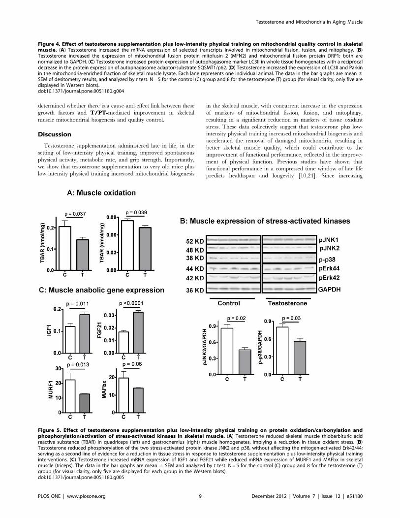
determined whether there is a cause-and-effect link between these
growth factors and T/PT-mediated improvement in skeletal
muscle mitochondrial biogenesis and quality control.
Discussion
Testosterone supplementation administered late in life, in the
setting of low-intensity physical training, improved spontaneous
physical activity, metabolic rate, and grip strength. Importantly,
we show that testosterone supplementation to very old mice plus
low-intensity physical training increased mitochondrial biogenesis
in the skeletal muscle, with concurrent increase in the expression
of markers of mitochondrial fission, fusion, and mitophagy,
resulting in a significant reduction in markers of tissue oxidant
stress. These data collectively suggest that testosterone plus low-
intensity physical training increased mitochondrial biogenesis and
accelerated the removal of damaged mitochondria, resulting in
better skeletal muscle quality, which could contribute to the
improvement of functional performance, reflected in the improve-
ment of physical function. Previous studies have shown that
functional performance in a compressed time window of late life
predicts healthspan and longevity [10,24]. Since increasing
Figure 4. Effect of testosterone supplementation plus low-intensity physical training on mitochondrial quality control in skeletalmuscle. (A) Testosterone increased the mRNA expression of selected transcripts involved in mitochondrial fission, fusion, and mitophagy. (B)Testosterone increased the expression of mitochondrial fusion protein mitofusin 2 (MFN2) and mitochondrial fission protein DRP1; both arenormalized to GAPDH. (C) Testosterone increased protein expression of autophagasome marker LC3II in whole tissue homogenates with a reciprocaldecrease in the protein expression of autophagasome adaptor/substrate SQSMT1/p62. (D) Testosterone increased the expression of LC3II and Parkinin the mitochondria-enriched fraction of skeletal muscle lysate. Each lane represents one individual animal. The data in the bar graphs are mean 6SEM of desitometry results, and analyzed by t test. N = 5 for the control (C) group and 8 for the testosterone (T) group (for visual clarity, only five aredisplayed in Western blots).doi:10.1371/journal.pone.0051180.g004
Figure 5. Effect of testosterone supplementation plus low-intensity physical training on protein oxidation/carbonylation andphosphorylation/activation of stress-activated kinases in skeletal muscle. (A) Testosterone reduced skeletal muscle thiobarbituric acidreactive substance (TBAR) in quadriceps (left) and gastrocnemius (right) muscle homogenates, implying a reduction in tissue oxidant stress. (B)Testosterone reduced phosphorylation of the two stress-activated protein kinase JNK2 and p38, without affecting the mitogen-activated Erk42/44;serving as a second line of evidence for a reduction in tissue stress in response to testosterone supplementation plus low-intensity physical traininginterventions. (C) Testosterone increased mRNA expression of IGF1 and FGF21 while reduced mRNA expression of MURF1 and MAFbx in skeletalmuscle (triceps). The data in the bar graphs are mean 6 SEM and analyzed by t test. N = 5 for the control (C) group and 8 for the testosterone (T)group (for visual clarity, only five are displayed for each group in the Western blots).doi:10.1371/journal.pone.0051180.g005
Testosterone and Mitochondria in Aging Muscle
PLOS ONE | www.plosone.org 9 December 2012 | Volume 7 | Issue 12 | e51180

healthspan is the ultimate goal of anti-aging interventions, future
studies should determine the effects of testosterone plus low-
intensity physical training on healthspan in older human adults.
Because all animals involved in this study had received low-
intensity physical training, we cannot determine from the current
data whether testosterone administration alone would produce
similar improvements in physical activity and mitochondrial
function. In a preliminary study in which 28-month old mice
were given testosterone supplementation for 3 weeks without
exercise training, we found no effect of testosterone supplemen-
tation on mitochondrial DNA copy numbers although an increase
in muscle mass was significant. Unfortunately, no functional
performance was measured in these animals. Randomized trials of
testosterone in older men with mobility limitation have not found
significant changes in physical activity [8]. Hence, we suggest that
testosterone supplementation alone can increase muscle hypertro-
phy but may not be sufficient to improve muscle bioenergetics and
functional performance in the absence of physical training.
Randomized trials in humans are needed to determine the
interactive effects of testosterone and low-intensity exercise on
functional performance and bioenergetics, to determine the
optimal intensity and frequency of physical training that will
induce clinically meaningful functional improvements.
Although testosterone has been used for several decades by
athletes and recreational body builders to enhance muscle mass
and performance, and is being explored as a function promoting
therapy for functional limitations associated with aging and
chronic illness, the effects of testosterone on mitochondrial
biogenesis and function have not been well characterized.
Testosterone has been reported to promote, inhibit, or have no
effect on mitochondrial biogenesis or function [61,62,63,64,65]. In
men, however, low testosterone levels have been associated with
decreased expression of mitochondrial genes in OXPHOS
pathway and reduced maximal aerobic capacity [66]. Orchidect-
omy in young male mice down-regulated gene expression in the
pathways for energy metabolism, especially those involved in
OXPHOS and ubiquinone pathways [21]. These reports are in-
line with our current findings that testosterone supplementation
plus low-intensity physical training increased skeletal muscle
mitochondrial biogenesis in aging skeletal muscle.
We have provided several lines of evidence that increased
mitochondrial biogenesis in response to testosterone plus low-
intensity physical training is accompanied by enhanced autophagic
removal of damaged organelles. First, testosterone plus physical
training increased expression of Parkin and LC3II in the
mitochondria-enriched fraction of skeletal muscle lysate, implying
increased formation of mitochondria-associated autophagasome.
Second, testosterone plus physical training reduced the level of
autophagasome adaptor STSTM1/p62 in the muscle, which
coupled with an increase in LC3II, the upstream quantitative
marker for autophagasome formation, suggests an enhancement of
autophagic flux [50]. Third, the activity of selected mitochondrial
enzymes was increased by testosterone plus physical training in the
absence of a substantial increase in steady-state mitochondrial
protein level, suggesting possible improvement in the quality of
mitochondrial proteins. Fourth, a reduction in tissue oxidative
damage and decreased expression of stress kinases, as well as
increased expression of pro-myogenic and anti-inflammatory
growth factors and reciprocal reduction in muscle expression of
atrogenes also indicate that testosterone plus low-intensity physical
training improved muscle quality, which is in-line with an
improvement of mitochondrial quality control [13,32,67]. To-
gether, our results support the hypothesis that testosterone plus
low-intensity physical training improved mitochondrial biogenesis
and quality control in old male mice, although the molecular
mechanisms underlying these effects remain to be investigated.
In summary, we have provided multiple lines of evidence that
links the functional performance with improved skeletal muscle
mitochondrial biogenesis and quality control in aging mice treated
with testosterone supplementation plus low-intensity exercise as
compared with control mice receiving vehicle and low-intensity
exercise only. We used older male mice that were naturally
testosterone deficient to increase the translational value of the
findings to testosterone trials in older men with age-related decline
in testosterone levels. Further studies are needed to determine the
molecular mechanisms for these observations and to determine the
minimal requirements in exercise intensity and frequency that can
induce the functional improvements in response to testosterone
supplementation.
Supporting Information
Figure S1 Effect of testosterone supplementation onbody composition. (A). Body fat mass (upper left), lean mass
(upper right), total body weight (lower left) and lean/fat ratio
(lower right). Results are shown as means +/2 se, N= 8 for
control (C28) and testosterone (T28) at baseline. N= 6 for control
(C30) and N=8 for testosterone (T30) group at 30 month,
unpaired t test. (B). Terminal tissue weight for selected muscle
groups as labeled [means +/2 se, N= 5 for the control (C) group,
N= 8 for testosterone (T) group, unpaired t test].
(PPT)
Figure S2 Effect of testosterone supplementation onrotarod running distance. Mice were allowed to run on the
rota-rod set with a low and gradually increasing speed until they
fell off the rod, as described in the Methods. Each data point
represented the mean distance by one individual animal. Unpaired
t test.
(PPT)
Figure S3 Effect of testosterone supplementation onrespiration after normalized to body lean mass. Resultsare re-plotted from original data presented in Figure 1C (re-
spiratory activity normalized to total body weight) and Figure S1
(lean mass).
(PPT)
Figure S4 Comparison of effect of testosterone supple-mentation on respiration normalized to body weight andlean mass. Percentage wise, the difference were similar during
light period but was diminished during dark period when the
results were normalized to lean body mass. Blue: vehicle control;
red: testosterone supplementation.
(PPT)
Author Contributions
Conceived and designed the experiments: WG SW OS SB. Performed the
experiments: WG SW ML WL. Analyzed the data: WG SW ML OS CS
RJ JLK AB SB. Wrote the paper: WG SW ML OS JLK AB SB.
References
1. Schaap LA, Pluijm SM, Smit JH, van Schoor NM, Visser M, et al. (2005) The
association of sex hormone levels with poor mobility, low muscle strength and
incidence of falls among older men and women. Clin Endocrinol (Oxf) 63: 152–
160.
Testosterone and Mitochondria in Aging Muscle
PLOS ONE | www.plosone.org 10 December 2012 | Volume 7 | Issue 12 | e51180

2. Snyder PJ, Peachey H, Hannoush P, Berlin JA, Loh L, et al. (1999) Effect oftestosterone treatment on body composition and muscle strength in men over 65
years of age. J Clin Endocrinol Metab 84: 2647–2653.
3. Kovacheva EL, Hikim AP, Shen R, Sinha I, Sinha-Hikim I (2009) Testosteronesupplementation reverses sarcopenia in aging through regulation of myostatin, c-
Jun NH2-terminal kinase, Notch, and Akt signaling pathways. Endocrinology
151: 628–638.
4. Ferrando AA, Sheffield-Moore M, Yeckel CW, Gilkison C, Jiang J, et al. (2002)
Testosterone administration to older men improves muscle function: molecularand physiological mechanisms. Am J Physiol Endocrinol Metab 282: E601–607.
5. Seidman SN (2007) Androgens and the aging male. Psychopharmacol Bull 40:
205–218.
6. Krasnoff JB, Basaria S, Pencina MJ, Jasuja GK, Vasan RS, et al. (2010) Freetestosterone levels are associated with mobility limitation and physical
performance in community-dwelling men: the Framingham Offspring Study.J Clin Endocrinol Metab 95: 2790–2799.
7. Caminiti G, Volterrani M, Iellamo F, Marazzi G, Massaro R, et al. (2009) Effect
of long-acting testosterone treatment on functional exercise capacity, skeletalmuscle performance, insulin resistance, and baroreflex sensitivity in elderly
patients with chronic heart failure a double-blind, placebo-controlled, random-ized study. J Am Coll Cardiol 54: 919–927.
8. Travison TG, Basaria S, Storer TW, Jette AM, Miciek R, et al. (2011) Clinical
meaningfulness of the changes in muscle performance and physical functionassociated with testosterone administration in older men with mobility
limitation. J Gerontol A Biol Sci Med Sci 66: 1090–1099.
9. Parise G, De Lisio M (2010) Mitochondrial theory of aging in human age-relatedsarcopenia. Interdiscip Top Gerontol 37: 142–156.
10. Carter CS, Marzetti E, Leeuwenburgh C, Manini T, Foster TC, et al. (2011)
Usefulness of preclinical models for assessing the efficacy of late-life interventionsfor sarcopenia. J Gerontol A Biol Sci Med Sci 67: 17–27.
11. Bereiter-Hahn J, Voth M (1994) Dynamics of mitochondria in living cells: shape
changes, dislocations, fusion, and fission of mitochondria. Microsc Res Tech 27:198–219.
12. Schafer A, Reichert AS (2009) Emerging roles of mitochondrial membrane
dynamics in health and disease. Biol Chem 390: 707–715.
13. Weber TA, Reichert AS (2010) Impaired quality control of mitochondria: aging
from a new perspective. Exp Gerontol 45: 503–511.
14. Green DR, Galluzzi L, Kroemer G (2011) Mitochondria and the autophagy-inflammation-cell death axis in organismal aging. Science 333: 1109–1112.
15. Xu J, Seo AY, Vorobyeva DA, Carter CS, Anton SD, et al. (2010) Beneficial
effects of a Q-ter based nutritional mixture on functional performance,mitochondrial function, and oxidative stress in rats. PLoS One 5: e10572.
16. Hofer T, Marzetti E, Xu J, Seo AY, Gulec S, et al. (2008) Increased iron content
and RNA oxidative damage in skeletal muscle with aging and disuse atrophy.Exp Gerontol 43: 563–570.
17. Gottlieb RA, Carreira RS (2010) Autophagy in health and disease. 5. Mitophagy
as a way of life. Am J Physiol Cell Physiol 299: C203–210.
18. Koltai E, Hart N, Taylor AW, Goto S, Ngo JK, et al. (2012) Age-associated
declines in mitochondrial biogenesis and protein quality control factors are
minimized by exercise training. Am J Physiol Regul Integr Comp Physiol 303:R127–134.
19. Bori Z, Zhao Z, Koltai E, Fatouros IG, Jamurtas AZ, et al. (2012) The effects ofaging, physical training, and a single bout of exercise on mitochondrial protein
expression in human skeletal muscle. Exp Gerontol 47: 417–424.
20. Lanza IR, Sreekumaran Nair K (2010) Regulation of skeletal musclemitochondrial function: genes to proteins. Acta Physiol (Oxf) 199: 529–547.
21. Ibebunjo C, Eash JK, Li C, Ma Q, Glass DJ (2010) Voluntary running, skeletal
muscle gene expression, and signaling inversely regulated by orchidectomy andtestosterone replacement. Am J Physiol Endocrinol Metab 300: E327–340.
22. Fernando SM, Rao P, Niel L, Chatterjee D, Stagljar M, et al. (2010) Myocyte
androgen receptors increase metabolic rate and improve body composition byreducing fat mass. Endocrinology 151: 3125–3132.
23. Musa M, Fernando SM, Chatterjee D, Monks DA (2011) Subcellular effects of
myocyte-specific androgen receptor overexpression in mice. J Endocrinol 210:93–104.
24. Carter CS, Sonntag WE, Onder G, Pahor M (2002) Physical performance and
longevity in aged rats. J Gerontol A Biol Sci Med Sci 57: B193–197.
25. Tu P, Bhasin S, Hruz PW, Herbst KL, Castellani LW, et al. (2009) Genetic
disruption of myostatin reduces the development of proatherogenic dyslipidemia
and atherogenic lesions in Ldlr null mice. Diabetes 58: 1739–1748.
26. Egan B, Dowling P, O’Connor PL, Henry M, Meleady P, et al. (2011) 2-D
DIGE analysis of the mitochondrial proteome from human skeletal muscle
reveals time course-dependent remodelling in response to 14 consecutive days ofendurance exercise training. Proteomics 11: 1413–1428.
27. Lee HY, Choi CS, Birkenfeld AL, Alves TC, Jornayvaz FR, et al. (2010)Targeted expression of catalase to mitochondria prevents age-associated
reductions in mitochondrial function and insulin resistance. Cell Metab 12:
668–674.
28. Andrews JL, Zhang X, McCarthy JJ, McDearmon EL, Hornberger TA, et al.
(2010) CLOCK and BMAL1 regulate MyoD and are necessary for maintenance
of skeletal muscle phenotype and function. Proc Natl Acad Sci U S A 107:19090–19095.
29. McNabney LA, Essig DA (1992) 5’-Aminolevulinate synthase activity is
decreased in skeletal muscle of anemic rats. Am J Physiol 263: C429–435.
30. Sugden MC, Holness MJ (2006) Mechanisms underlying regulation of the
expression and activities of the mammalian pyruvate dehydrogenase kinases.
Arch Physiol Biochem 112: 139–149.
31. Salehzadeh F, Rune A, Osler M, Al-Khalili L (2012) Testosterone or 17{beta}-
estradiol exposure reveals sex-specific effects on glucose and lipid metabolism in
human myotubes. J Endocrinol 210: 219–229.
32. Hirota Y, Kang D, Kanki T (2012) The physiological role of mitophagy: new
insights into phosphorylation events. Int J Cell Biol 2012: 354914.
33. Apostolova N, Blas-Garcia A, Esplugues JV (2011) Mitochondria sentencingabout cellular life and death: a matter of oxidative stress. Curr Pharm Des 17:
4047–4060.
34. Wohlgemuth SE, Seo AY, Marzetti E, Lees HA, Leeuwenburgh C (2009)
Skeletal muscle autophagy and apoptosis during aging: effects of calorie
restriction and life-long exercise. Exp Gerontol 45: 138–148.
35. Liu Y, Shi S, Gu Z, Du Y, Liu M, et al. (2012) Impaired autophagic function in
rat islets with aging. Age (Dordr).
36. Jamart C, Francaux M, Millet GY, Deldicque L, Frere D, et al. (2012)
Modulation of autophagy and ubiquitin-proteasome pathways during ultra-
endurance running. J Appl Physiol 112: 1529–1537.
37. Mai S, Klinkenberg M, Auburger G, Bereiter-Hahn J, Jendrach M (2010)
Decreased expression of Drp1 and Fis1 mediates mitochondrial elongation in
senescent cells and enhances resistance to oxidative stress through PINK1. J Cell
Sci 123: 917–926.
38. Joseph AM, Adhihetty PJ, Buford TW, Wohlgemuth SE, Lees HA, et al. (2012)
The impact of aging on mitochondrial function and biogenesis pathways in
skeletal muscle of sedentary high- and low-functioning elderly individuals. Aging
Cell 11: 801–809.
39. Chen L, Gong Q, Stice JP, Knowlton AA (2009) Mitochondrial OPA1,
apoptosis, and heart failure. Cardiovasc Res 84: 91–99.
40. Westermann B (2012) Bioenergetic role of mitochondrial fusion and fission.
Biochim Biophys Acta 1817: 1833–1838.
41. Jin SM, Youle RJ (2012) PINK1- and Parkin-mediated mitophagy at a glance.
J Cell Sci 125: 795–799.
42. Shi CS, Shenderov K, Huang NN, Kabat J, Abu-Asab M, et al. (2012)
Activation of autophagy by inflammatory signals limits IL-1beta production by
targeting ubiquitinated inflammasomes for destruction. Nat Immunol 13: 255–
263.
43. Kabeya Y, Mizushima N, Ueno T, Yamamoto A, Kirisako T, et al. (2000) LC3,
a mammalian homologue of yeast Apg8p, is localized in autophagosome
membranes after processing. EMBO J 19: 5720–5728.
44. Mizushima N, Yoshimori T (2007) How to interpret LC3 immunoblotting.
Autophagy 3: 542–545.
45. Wu J, Dang Y, Su W, Liu C, Ma H, et al. (2006) Molecular cloning and
characterization of rat LC3A and LC3B–two novel markers of autophagosome.
Biochem Biophys Res Commun 339: 437–442.
46. Rubinsztein DC, Cuervo AM, Ravikumar B, Sarkar S, Korolchuk V, et al.
(2009) In search of an ‘‘autophagomometer’’. Autophagy 5: 585–589.
47. Shaid S, Brandts CH, Serve H, Dikic I (2012) Ubiquitination and selective
autophagy. Cell Death Differ.
48. Komatsu M, Kageyama S, Ichimura Y (2012) p62/SQSTM1/A170: Physiology
and pathology. Pharmacol Res: Epub ahead of print.
49. Ichimura Y, Komatsu M (2010) Selective degradation of p62 by autophagy.
Semin Immunopathol 32: 431–436.
50. Tanida I (2010) Autophagosome formation and molecular mechanism of
autophagy. Antioxid Redox Signal 14: 2201–2214.
51. Wong YT, Gruber J, Jenner AM, Ng MP, Ruan R, et al. (2009) Elevation of
oxidative-damage biomarkers during aging in F2 hybrid mice: protection by
chronic oral intake of resveratrol. Free Radic Biol Med 46: 799–809.
52. Rosser EM, Morton S, Ashton KS, Cohen P, Hulme AN (2004) Synthetic
anisomycin analogues activating the JNK/SAPK1 and p38/SAPK2 pathways.
Org Biomol Chem 2: 142–149.
53. Widegren U, Ryder JW, Zierath JR (2001) Mitogen-activated protein kinase
signal transduction in skeletal muscle: effects of exercise and muscle contraction.
Acta Physiol Scand 172: 227–238.
54. McCubrey JA, Lahair MM, Franklin RA (2006) Reactive oxygen species-
induced activation of the MAP kinase signaling pathways. Antioxid Redox
Signal 8: 1775–1789.
55. Owuor ED, Kong AN (2002) Antioxidants and oxidants regulated signal
transduction pathways. Biochem Pharmacol 64: 765–770.
56. Gorar S, Culha C, Uc ZA, Dellal FD, Serter R, et al. (2010) Serum fibroblast
growth factor 21 levels in polycystic ovary syndrome. Gynecol Endocrinol 26:
819–826.
57. Liu Y, Wang C, Wang Y, Ma Z, Xiao J, et al. (2012) Cobalt chloride decreases
fibroblast growth factor-21 expression dependent on oxidative stress but not
hypoxia-inducible factor in Caco-2 cells. Toxicol Appl Pharmacol 264: 212–221.
58. Sestili P, Barbieri E, Martinelli C, Battistelli M, Guescini M, et al. (2009)
Creatine supplementation prevents the inhibition of myogenic differentiation in
oxidatively injured C2C12 murine myoblasts. Mol Nutr Food Res 53: 1187–
1204.
59. Perrini S, Laviola L, Carreira MC, Cignarelli A, Natalicchio A, et al. (2010) The
GH/IGF1 axis and signaling pathways in the muscle and bone: mechanisms
underlying age-related skeletal muscle wasting and osteoporosis. J Endocrinol
205: 201–210.
Testosterone and Mitochondria in Aging Muscle
PLOS ONE | www.plosone.org 11 December 2012 | Volume 7 | Issue 12 | e51180

60. Cuevas-Ramos D, Aguilar-Salinas CA, Gomez-Perez FJ (2012) Metabolic
actions of fibroblast growth factor 21. Curr Opin Pediatr 24: 523–529.61. Rodriguez-Cuenca S, Monjo M, Gianotti M, Proenza AM, Roca P (2007)
Expression of mitochondrial biogenesis-signaling factors in brown adipocytes is
influenced specifically by 17beta-estradiol, testosterone, and progesterone.Am J Physiol Endocrinol Metab 292: E340–346.
62. Ikeda Y, Aihara K, Akaike M, Sato T, Ishikawa K, et al. (2010) Androgenreceptor counteracts Doxorubicin-induced cardiotoxicity in male mice. Mol
Endocrinol 24: 1338–1348.
63. Dhatariya KK, Greenlund LJ, Bigelow ML, Thapa P, Oberg AL, et al. (2008)Dehydroepiandrosterone replacement therapy in hypoadrenal women: protein
anabolism and skeletal muscle function. Mayo Clin Proc 83: 1218–1225.
64. Zahavi A, Perel M (2011) The information encoded by the sex steroid hormones
testosterone and estrogen: a hypothesis. J Theor Biol 280: 146–149.65. Pansarasa O, D’Antona G, Gualea MR, Marzani B, Pellegrino MA, et al. (2002)
‘‘Oxidative stress’’: effects of mild endurance training and testosterone treatment
on rat gastrocnemius muscle. Eur J Appl Physiol 87: 550–555.66. Pitteloud N, Mootha VK, Dwyer AA, Hardin M, Lee H, et al. (2005)
Relationship between testosterone levels, insulin sensitivity, and mitochondrialfunction in men. Diabetes Care 28: 1636–1642.
67. Kurihara Y, Kanki T, Aoki Y, Hirota Y, Saigusa T, et al. (2011) Mitophagy
plays an essential role in reducing mitochondrial production of reactive oxygenspecies and mutation of mitochondrial DNA by maintaining mitochondrial
quantity and quality in yeast. J Biol Chem 287: 3265–3272.
Testosterone and Mitochondria in Aging Muscle
PLOS ONE | www.plosone.org 12 December 2012 | Volume 7 | Issue 12 | e51180
Top Related
Copyright © 2022 FDOKUMEN

