


Bahasa
Halaman
Hukum


CliniCal CardiaC ElECtrophysiology and hEart FailurE:
impaCt oF ElECtriCal disordErs and thEir trEatmEnt
Máximo José Rivero Ayerza

Financial support for this book was given by Biotronik, St. Jude Medical,
Boston Scientific and Stereotaxis Inc.
ISBN: 978 90 8559 508 3

CliniCal CardiaC ElECtrophysiology and hEart FailurE:
impaCt oF ElECtriCal disordErs and thEir trEatmEnt
KlinisChE ElEKtroFysiologiE bij hartFalEn:
hEt bElang van ritmE- En gElEidingsstoornissEn En hun bEhandEling.
Thesis
To obtain the degree of Doctor from the
Erasmus University Rotterdam
by command of the
rector magnificus
Prof.dr. S.W.J. Lamberts
And in accordance with the decision of the Doctorate Board
The public defence shall be held on
Wednesday, April 1st, 2009 at 15:45 o’clock
by
Máximo José Rivero Ayerza
born at Buenos Aires, Argentina

doctoral Committee
Promotor: Prof.dr. L. Jordaens
Other members: Prof.dr. P. de Feyter
Prof.dr. W. Van Mieghem
Prof.dr. W. Niessen
Prof.dr. P. Brugada
Prof.dr. P.W. Serruys

tablE oF ContEnts
Introduction 9
part i – dEviCE thErapy in hEart FailurE
Chapter 1 Effects of cardiac resynchronization therapy on overall
mortality and mode of death: a meta-analysis of random-
ized controlled trials.
19
Chapter 2 Implantable devices for the treatment of patients with left
ventricular dysfunction and heart failure.
41
Chapter 3 Device therapy in heart failure: Do all treatment goals apply
to all patients?
59
part ii – diFFiCultiEs rElatEd to CardiaC rEsynChronization thErapy
Chapter 4 Indications for cardiac resynchronization therapy: should
they be extended?
67
Chapter 5 Polymorphic ventricular tachycardia induced by left
ventricular pacing.
77
Chapter 6 Double wire technique to catheterize sharply angulated
coronary sinus branches in cardiac resynchronization
therapy.
81
Chapter 7 A grateful heart. 87
Chapter 8 Tailored echocardiographic interventricular delay program-
ming further optimizes left ventricular performance after
cardiac resynchronization therapy.
89

part iii – CardiaC rEsynChronization thErapy: how to improvE implanta-
tion tEChniquEs
Chapter 9 Potential applications of magnetic navigation in clinical
electrophysiology.
105
Chapter 10 Left ventricular lead placement within a coronary sinus side
branch using remote magnetic navigation of a guide-wire: A
feasibility study.
113
Chapter 11 Magnetically guided left ventricular lead implantation
based on a virtual three-dimensional reconstructed image
of the coronary sinus.
125
Chapter 12 Performance of different magnetically steered guidewires
during left ventricular pacing lead implantation.
137
Chapter 13 Left ventricular lead implantation assisted by magnetic
navigation in a patient with a persistent left superior vena
cava.
141
part iv – arrhythmias and hEart FailurE
Chapter 14 New onset atrial fibrillation is an independent predictor of
in hospital mortality in hospitalized heart failure patients.
Results of the euro heart failure survey.
149
Chapter 15 Interventional therapy for atrial fibrillation and heart failure
– A case report of tachycardia mediated cardiomyopathy.
161
Chapter 16 Effects of cardiac resynchronization therapy on left atrial
size in heart failure patients with implantable defibrillators.
167
Chapter 17 Heart transplantation as last resort against brugada
syndrome.
177

part v - summary and ConClusions (English-nEdErlands)
Summary and conclusions 185
Samenvatting en besluit 189
part vi– misCEllanEous
Curriculum vitae an Publications 197
Acknowledgments 209


Dedicado a Maria y los chicos.


introduCtion

12 Introduction
implantablE dEviCEs and hEart FailurE
Ageing of the population, life style and increased survival of patients with cardiovascular dis-
eases has favoured the development of heart failure and made of it a growing epidemiological
problem in developed countries (1). The understanding of the mechanisms of progression of
this disease and the recent advances in medical therapy has improved symptoms and progno-
sis of these patients (2-6). However, the degree of disability, the impairment in quality of life
and mortality rates of patients with advanced heart failure remain high. Despite appropriate
treatment, progressive mechanical and electrical remodeling of the heart may occur, favouring
the development of arrhythmias (increasing the risk of sudden death and development of atrial
fibrillation) and conduction disturbances that will further complicate the course of this disease
and the treatment of these patients.
Up to one third of the patients with systolic heart failure, may present signs of intra-
ventricular conduction delay manifested by a broad QRS complex on the electrocardiogram,
which is considered a surrogate for electrical dyssynchrony (7). These ventricular conduction
disturbances have proven to be associated to more advanced heart disease and a worse prog-
nosis. They usually take the form of left bundle branch block and produce a dyssynchronous
contraction that reduces myocardial efficiency, further impairing systolic and diastolic function
and worsening mitral regurgitation.
Clinical cardiac electrophysiology through implantable device therapy has greatly evolved
in recent years. While device therapy was initially only able to treat bradyarrhythmias (conven-
tional pacemakers) it has grown to recognize and successfully treat life threatening ventricular
arrhythmias (implantable cardioverter defibrillator). Furthermore, by synchronously stimulating
different chambers of the heart, cardiac pacing now allows to restore a resynchronized con-
traction to patients with atrio-ventricular or inter- and intra-ventricular dyssynchrony (cardiac
resynchronization therapy). The incorporation of electrical interventions like the implantable
defibrillator (ICD) and cardiac resynchronization therapy (CRT) has revolutionized the treat-
ment of patients with left ventricular systolic dysfunction and advanced heart failure. However,
despite the increasing application of these therapies many unresolved issues remain.
Understanding how device therapy affects prognosis and mode of death in patients with
advanced heart failure allows to tailor treatment to the specific patient’s needs. It was not clear
from the published trials in which way CRT alone affected prognosis and mode of death of these
patients. For this reason, in chapter 1, we will pool and analyse data from those randomised
clinical trials that evaluated the effects of CRT alone as compared to optimal pharmacological
therapy on mortality and mode of death. The contribution of the ICD, CRT or both (CRT-D) for
the treatment of patients with LV systolic dysfunction and heart failure will discussed in chapter
2. The current ways of identifying those patients who will benefit from device implantation is far
from ideal. A high proportion of these patients will not benefit from the device, and as will be
discussed in chapter 3, appropriate ways of accurately identifying these patients are needed.

Introduction 13
Despite the well proven favourable effects of CRT, this therapy is only indicated in a highly
selected group of patients (broad QRS with low LVEF and NYHA III-IV) representing a small
proportion of those patients with heart failure. In chapter 4 we will discuss the possibility of
expanding the indications of CRT beyond those currently accepted, to see whether the benefits
of this therapy might apply to other patient groups.
Probably the biggest challenge regarding CRT is to reduce the proportion of patients that
will not benefit from implantation. Lack of response to CRT is not only due to inappropriate
patient selection but also to inadequate therapy delivery. Placing the left ventricular lead in
the appropriate coronary sinus side branch is essential to obtain adequate resynchronization.
However, as will be discussed in chapters 5, 6 and 7, left ventricular lead implantation is not
always straight forward, it can be associated to complications and it sometimes requires some
particular manoeuvres to successfully place it in the desired side branch.
Individually adjusting device programming may further optimize CRT and probably reduce
the rate of non-responders. In chapter 8 the acute hemodynamic effects of individually adjust-
ing the settings of atrio-biventricular pacing will be assessed.
Despite the technological progress aimed at improving success and reducing complication
rates of CRT device implantation, in around 5-10 percent of the patients LV lead implantation
fails (8). Remote magnetic navigation has recently emerged as a useful tool for accurately steer-
ing guidewires and ablation catheters. After describing the potential uses of this technology
in the field of cardiac electrophysiology in chapter 9, we will evaluate the feasibility of using
magnetically steered guidewires to guide left ventricular lead implantations in chapter 10. In
chapter 11, we will describe how a virtual 3D reconstruction of the coronary sinus can be used
to navigate and steer the guidewire to the target side branch.
Even though left ventricular lead implantation using magnetic navigation has not yet proven
to be superior to conventional implantation, we aim at identifying which magnetically steered
guidewires perform better for navigation within the coronary sinus (chapter 12). This work is
still ongoing. In chapters 7, 11 and 13 the usefulness of magnetic navigation in challenging
CRT cases, when conventional implantation was either not successful or not feasible, will be
explained.
CardiaC arrhythmias and hEart FailurE
Atrial fibrillation is frequently seen in patients with heart failure. They share common
predisposing factors and therefore commonly co-exist. The prevalence of atrial fibrillation
in heart failure patients increases with the severity of the disease. Whether atrial fibrillation
is another marker of disease severity or contributes to mortality is unresolved. Many reports
have addressed this issue and arrived to contradicting conclusions (9-14). However, recently
published sub-analyses of large randomized controlled trials performed in patients with heart

14 Introduction
failure and epidemiological studies suggest that atrial fibrillation is associated with a worse
long term outcome (15-20). Nevertheless, there are few data on how atrial fibrillation affects
the in-hospital course and prognosis of patients with heart failure. The prognostic role of this
arrhythmia in hospitalized heart failure patients will be assessed in chapter 14.
By causing loss of atrial contraction and fast and irregular rates, atrial fibrillation can cause
or become a reason for worsening heart failure. Advances in the understanding of the mecha-
nisms leading to atrial fibrillation and the incorporation of sophisticated technology allowing
to accurately map arrhythmias and reconstruct essential anatomical structures has allowed to
effectively treat atrial fibrillation by catheter ablation. Even though the role of atrial fibrillation
ablation in heart failure patients has not yet been established, it could prove beneficial for a
selected group of patients as will be illustrated in chapter 15.
The role of CRT in the setting of atrial fibrillation is controversial. Solid evidence regarding
the effects of CRT in atrial fibrillation patients is lacking, mainly because it has been evaluated
only by small clinical studies. By inducing a positive remodelling effect, reducing left ventricular
pressures and mitral regurgitation, CRT could potentially influence also left atrial remodeling
and potentially reduce atrial fibrillation burden. There is few and conflicting data evaluating
the effects of CRT on atrial fibrillation burden. In chapter 16, the effects of CRT on left atrial size
will be assessed.
Malignant ventricular arrhythmias refractory to treatment are an accepted indication for cardiac
transplantation. These usually develop in patients with overt and highly symptomatic structural
heart disease. In rare cases, in patients with no structural heart abnormality a primary electrical
failure of the heart can develop and, as will be shown in chapter 17, lead to heart transplantation.
In summary, our project is aimed to further understand the role that electrical therapies of
the heart exert on the treatment of patients with heart failure and to optimize the application
of these therapies. It also attempts to establish how the development and treatment of differ-
ent electrophysiological abnormalities, like conduction disturbances and arrhythmias, affect
the evolution these patients.

Introduction 15
rEFErEnCEs:
1. Massie BM, Shah NB. Evolving trends in the epidemiologic factors of heart failure: Rationale for preventive strategies and comprehensive disease management. Am Heart J 1997; 133:703.
2. The CONSENSUS trial study group. Effects of enalapril in mortality in severe congestive heart failure: results of the Cooperative North Scandinaivan Enalapril Survival Study. N Eng J Med 1987;316:429.
3. Effect of enalapril on survival in patients with reduced left ventricular ejection fractions and conges-tive heart failure. The SOLVD Investigators. N Engl J Med 1991; 325(5):293-302;
4. CIBIS-II Investigators and Committees. The Cardiac Insufficiency Bisoprolol Study II (CIBIS-II): a ran-domised trial. Lancet 1999 Jan 2;353(9146):9-13
5. MERIT-HF Study Group. Effect of metoprolol CR/XL in chronic heart failure: Metoprolol CR/XL Randomised Intervention Trial in Congestive Heart Failure (MERIT-HF). Lancet 1999 Jun 12;353(9169):2001-7.
6. Pitt B, Zannad F, Remme WJ, et al. The effect of spironolactone on morbidity and mortality in patients with severe heart failure. Randomized Aldactone Evaluation Study Investigators. N Engl J Med 1999 Sep 2;341(10):709-17
7. D. Farwell,N. R. Patel, A. Hall, S. Ralph, A. N. Sulke. How many people with heart failure are appropriate for biventricular resynchronization? European Heart Journal, 2000;21: 1246-1250.
8. Bhatta L, Luck JC, Wolbrette DL, Naccarelli GV. Complications of biventricular pacing. Curr Opin Cardiol. 2004;19:31-5.
9. Middlekauff H.R., Stevenson W.G., and Stevenson L.W., Prognostic significance of atrial fibrillation in advanced heart failure. A study of 390 patients. Circulation, 1991. 84(1):p. 40-8.
10. Crijns H.J., Tjeerdsma G, de Kam P, Boomsma F, van Gelder I, van den Berg M and van Veldhuisen D. Prognostic value of the presence and development of atrial fibrillation in patients with advanced chronic heart failure. Eur Heart J, 2000. 21(15):1238-45.
11. Dries, D.L., Exner, D., Gersh, B., Domanski, M., Waclawiw, M. and Stevenson L., Atria fibrillation is associ-ated with an increased risk for mortality and heart failure progression in patients with asymptomatic and symptomatic left ventricular systolic dysfunction: a retrospective analysis of the SOLVD trials. Studies of Left Ventricular Dysfunction. J Am Coll Cardiol, 1998. 32(3): p. 695-703.
12. Ahmed, A. and G.J. Perry, Incident atrial fibrillation and mortality in older adults with heart failure. Eur J Heart Fail, 2005. 7(7): p. 1118-21.
13. Carson, P.E., Johnson, G., Dunkman, W., Fletcher, R., Farrell, L. and Cohn, J., The influence of atrial fibril-lation on prognosis in mild to moderate heart failure. The V- HeFT Studies. The V-HeFT VA Cooperative Studies Group. Circulation, 1993. 87(6 Suppl): p. VI102-10.
14. Stevenson, W.G.,Stevenson L., Middlekauff, H., Fonarow, M., Woo, m., Saxon, L., Natterson, P., Steimle, A. and Walden, J. Improving survival for patients with advanced heart failure: a study of 737 consecutive patients. J Am Coll Cardiol, 1995. 26(6): p. 1417-23.
15. Stewart, S., Hart, C. L., Hole, D. and McMurray, J., A population-based study of the long-term risks associ-ated with atrial fibrillation: 20-year follow-up of the Renfrew/Paisley study. Am J Med, 2002. 113(5): p. 359-64.
16. Mathew, J., Hunsberger, S., Fleg, J., Mc Sherry, F. Williford, W. and Yusuf, S. Incidence, predictive factors, and prognostic significance of supraventricular tachyarrhythmias in congestive heart failure. Chest, 2000. 118(4): p. 914-22.
17. Olsson, L.G., Swedberg, K., Ducharme, A., Granger, C., Michelson, E., McMurray, J., Puu, M., Yusuf, S. and Pfeffer, M. Atrial fibrillation and risk of clinical events in chronic heart failure with and without left ventricular systolic dysfunction: results from the Candesartan in Heart failure-Assessment of Reduction in Mortality and morbidity (CHARM) program. J Am Coll Cardiol, 2006. 47(10): p. 1997-2004.
18. Swedberg, K., Olsson, L., Charlesworth, A., Cleland, J., Hanrath, P., Komajda, M., Metra, M., Torp-Peder-sen, C. and Poole-Wilson, P. Prognostic relevance of atrial fibrillation in patients with chronic heart failure on long-term treatment with beta- blockers: results from COMET. Eur Heart J, 2005. 26(13): p. 1303-8.

16 Introduction
19. Wang, T.J., Larson, M., Levy, D., Vasan, R., Leip, E., Wolf, P., D’Agostino, R., Murabito, J., Kannel, W. and Benjamin, E., Temporal relations of atrial fibrillation and congestive heart failure and their joint influence on mortality: the Framingham Heart Study. Circulation, 2003. 107(23): p. 2920-5.
20. Miyasaka, Y., Barnes, M., Gersh, B., Cha, S., Bailey, K., Abhayaratna, W., Seward, J., Iwasaka, T. and Tsang, T., Incidence and mortality risk of congestive heart failure in atrial fibrillation patients: a community-based study over two decades. Eur Heart J, 2006. 27(8): p. 936-41.

part idEviCE thErapy in hEart FailurE

Chapter 1EFFECts oF CardiaC rEsynChronization
thErapy on ovErall mortality and
modE oF dEath: a mEta-analysis oF
randomizEd ControllEd trials.
M. Rivero-Ayerza , D. Theuns, H. Garcia-Garcia E. Boersma, M. Simoons and L. Jordaens.
Eur Heart J. 2006 Nov;27(22):2682-8.

20 Chapter 1
abstraCt
Background:
Cardiac resynchronization therapy (CRT) has been shown to improve symptoms and exercise
tolerance in patients with advanced heart failure. However, studies were underpowered to
address its effect on overall mortality.
Objective:
To evaluate whether CRT alone (without a combined defibrillator function) reduces overall
mortality as compared to optimal pharmacological therapy, and how it affects the mode of
death in patients with advanced heart failure.
Methods:
Public domain databases were systematically searched. Randomized controlled studies that
evaluated the effects of CRT alone in patients with advanced heart failure and a depressed
left ventricular systolic performance were selected for this analysis. Trials, which did not inde-
pendently report data on CRT alone or had a follow up period of less than 3 months, were
excluded.
Results:
Five studies were identified and analyzed. They included a total of 2371 patients, 1028 controls
and 1343 CRT treated patients. Pooled analysis demonstrated that CRT alone, as compared to
optimal medical therapy, significantly reduced all cause mortality by 29 % (16.9 % vs. 20.7 %;
odds ratio [OR], 0.71; 95% confidence interval [CI], 0.57-0.88) and mortality due to progressive
heart failure by 38 % (6.7 % vs 9.7 %; OR, 0.62; 95% CI, 0.45-0.84). No effect on sudden cardiac
death was observed with CRT (6.4 % vs 5.9 %;OR, 1.04; 95% CI, 0.73-1.22).
Conclusions:
Cardiac resynchronization therapy alone as compared to optimal medical therapy reduces all
cause mortality in patients with advanced heart failure. It predominantly reduces worsening
heart failure mortality, not affecting sudden cardiac death.
Effects of CRT on overall mortality and mode of death 21
introduCtion
Heart failure (HF) is a growing public health problem in the western world. For instance only
in the USA more than 5 million patients suffer from this disease, and about 500,000 patients
are diagnosed with HF yearly (1). Despite the latest achievements of medical therapy (2-8), in
patients with advanced stages of the disease, mortality remains high and quality of life severely
impaired (9).
Cardiac resynchronization therapy (CRT) has consistently proven to improve symptoms,
quality of life (QOL) and exercise tolerance in patients with a severely depressed left ventricular
ejection fraction (≤ 35%) who remain symptomatic (NYHA class III-IV) despite optimal medical
therapy (10-16). Early published studies were specifically designed to evaluate the effects of CRT
upon these functional end points and were underpowered to evaluate its effect on mortality
(10,12,14,15). The recently published Comparison of Medical Therapy, Pacing and Defibrillation
in Chronic Heart Failure (11) study has shown a survival benefit only in those patients random-
ized to CRT with a combined defibrillator function (CRT-D). In the CARE-HF (13) trial patients
who received CRT alone (without a combined defibrillator function) had a significant reduction
in overall mortality compared to those under optimal pharmacological therapy.
Previously published meta-analyses have corroborated the effects of CRT upon symptoms,
QOL and exercise tolerance (17-19). However, an overall survival benefit of CRT alone has not
been addressed. This is true, mainly because in their analysis trials that also evaluated the
effects of CRT-D were included. In this way the effects of CRT were confounded by the proven
life saving effect of the ICD. Performing a meta-analysis increases the power to see a difference
in mortality that was not evident in the majority of individual trials performed. It also allows
having a more precise estimation of this effect (20). We designed a meta-analysis with the pur-
pose of establishing whether CRT alone, compared to optimal medical therapy, reduces overall
mortality and in which way it affects the different modes of death in patients with advanced
heart failure.
mEthods
Search strategy
A comprehensive search of public domain databases was carried out with the purpose of
identifying reports of randomized trials comparing CRT alone versus optimal pharmacological
therapy (control) in patients with advanced symptoms of heart failure (HF) due to left ventricular
systolic dysfunction. Using the terms heart failure, pacemaker, pacing, biventricular, biventricular
pacing, left ventricular pacing, left ventricular pre-excitation, multi-site pacing, cardiac resinchroni-
zation and cardiac resinchronisation MEDLINE (1985-2005) and the Cochrane Central Register of
Controlled Trials (third quarter 2005) were searched. The search was limited to English language

22 Chapter 1
publications. Additionally, the website of the US Food and Drug Administration (www.fda.gov)
was searched, and the reference lists of identified papers were examined. Reports presented
during the last five years at the scientific sessions of the American College of Cardiology, the
American Heart Association, the North American Society of Pacing and Electrophysiology later
the Heart Rhythm Society and the European Society of Cardiology were manually or electroni-
cally sought. The latest search was carried out in November 2005.
Study Selection
Randomized trials performed in patients with advanced symptoms of HF due to left ventricular
systolic dysfunction that evaluated the effects of CRT alone versus optimal pharmacological
therapy (control) were included in this analysis. Studies were excluded if they evaluated the
effects of CRT-D and did not separately report data on CRT alone. Because we were mainly
interested in the chronic effects of CRT, studies with a follow up of less than 3 months were
excluded. To be included, the duration of the first follow up phase of the randomized cross over
trials had to be at least 3 months. To avoid a carry over effect only the first randomized cross
over period was considered for analysis.
Two investigators (MRA, DAMJT) independently screened all titles and abstracts to deter-
mine which studies met the inclusion criteria. Publications for this review were selected if they
fulfilled the following criteria: 1) randomized trials performed in humans, 2) comparing the
effects of CRT alone with optimal pharmacological therapy in patients with advanced HF due
to left ventricular systolic dysfunction, and 3) reported mortality and mode of death during the
randomized period. Discrepancies between investigators were resolved by consensus.
Data Analysis
Two investigators using a standardized form independently abstracted data. A meta-analysis of
summary statistics from individual trials was performed. Odds ratios from each included trial
were pooled using both fixed and random effects model that used weighting based on inverse
variance calculated according to DerSimonian and Laird (21). To check for statistical evidence of
heterogeneity among trial-specific ORs a chi-square test was used and it was quantified using
the I² statistic (22). When pooled analysis resulted in a significant heterogeneity, the random
effects model was used.
Quantitative analyses were performed on an intention to treat basis using the same stan-
dardized end point definitions (overall mortality, worsening heart failure mortality and sudden
cardiac death) as in the primary studies. For non-worsening heart failure mortality all deaths
except those due to heart failure were considered. Data analysis was performed using the
Review Manager 4.2.
Effects of CRT on overall mortality and mode of death 23
rEsults
Search results
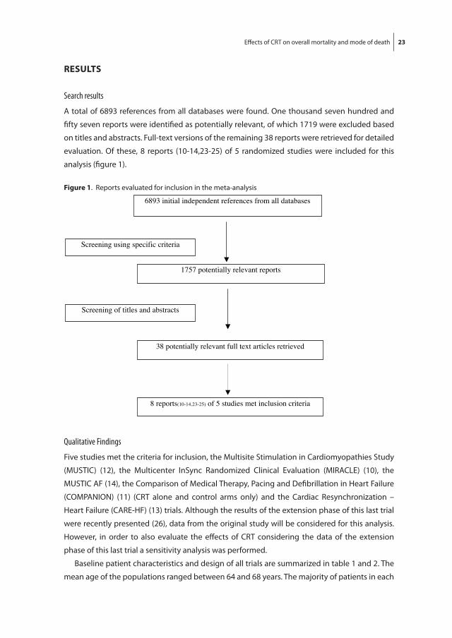
A total of 6893 references from all databases were found. One thousand seven hundred and
fifty seven reports were identified as potentially relevant, of which 1719 were excluded based
on titles and abstracts. Full-text versions of the remaining 38 reports were retrieved for detailed
evaluation. Of these, 8 reports (10-14,23-25) of 5 randomized studies were included for this
analysis (figure 1).
Qualitative Findings
Five studies met the criteria for inclusion, the Multisite Stimulation in Cardiomyopathies Study
(MUSTIC) (12), the Multicenter InSync Randomized Clinical Evaluation (MIRACLE) (10), the
MUSTIC AF (14), the Comparison of Medical Therapy, Pacing and Defibrillation in Heart Failure
(COMPANION) (11) (CRT alone and control arms only) and the Cardiac Resynchronization –
Heart Failure (CARE-HF) (13) trials. Although the results of the extension phase of this last trial
were recently presented (26), data from the original study will be considered for this analysis.
However, in order to also evaluate the effects of CRT considering the data of the extension
phase of this last trial a sensitivity analysis was performed.
Baseline patient characteristics and design of all trials are summarized in table 1 and 2. The
mean age of the populations ranged between 64 and 68 years. The majority of patients in each
Figure 1. Reports evaluated for inclusion in the meta-analysis
Chapter 1
Figure 2. Effect of cardiac resynchronization therapy alone (CRT) versus control on overall mortality.
1757 potentially relevant reports
38 potentially relevant full text articles retrieved
8 reports(10-14,23-25) of 5 studies met inclusion criteria
Screening of titles and abstracts
Screening using specific criteria
6893 initial independent references from all databases

24 Chapter 1
tabl
e 1
- Bas
elin
e cl
inic
al c
hara
cter
istic
s of
pat
ient
s in
clud
ed in
the
anal
ysed
tria
ls.
CarE
-hF(
13)
Com
pan
ion
(11,
24)
mir
aCl
E(10
,23)
m
ust
iC(1
2,25
) m
ust
iC a
F(14
) Co
ntro
lCR
TCo
ntro
lCR
TCo
ntro
lCR
TA
llA
llN
404
409
308
617
269
263
5843
Age,
mea
n, y
ears
66*
67*
6867
6464
6465
Men
, %73
7467
6768
6874
81Is
chem
ic c
ause
, %40
3659
5458
5037
43N
YHA
cla
ss II
I, %
9394
8287
9190
100
100
LVEF
, mea
n, %
25*
25*
2220
2121
2326
QRS
dur
atio
n, m
ean,
ms
160*
160*
158
160
165
167
174
209
Beta
-blo
cker
use
, %74
7066
6855
6228
23AC
EI o
r ARB
use
, %
9595
8989
9093
9610
0Sp
irono
lact
one
use,
%59
5455
53N
RN
R22
16
* re
port
ed a
s m
edia
n.
ACEI
= a
ngio
tens
in c
onve
rtin
g en
zym
e in
hibi
tors
, ARB
= a
ngio
tens
in re
cept
or b
lock
ers,
LVEF
= le
ft v
entr
icul
ar e
ject
ion
frac
tion,
NR
= no
t rep
orte
d, N
YHA
= N
ew Y
ork
Hea
rt A
ssoc
iatio
n fu
nctio
nal c
lass
.
Figu
re 2
. Eff
ect o
f car
diac
resy
nchr
oniz
atio
n th
erap
y al
one
(CRT
) ver
sus
cont
rol o
n ov
eral
l mor
talit
y.

Effects of CRT on overall mortality and mode of death 25
tabl
e 2
- Des
crip
tion
of th
e an
alys
ed tr
ials
.
CarE
-hF(
13)
COM
PAN
ION
(11,
24)
mir
aCl
E(10
,23)
mu
stiC
(12,
25)
mu
stiC
aF(
14)
Incl
usio
n Cr
iteria
NYH
A II
I-IV
≥ 6
w, E
F ≤
35 %
, QRS
≥12
0 m
s, (Q
RS
120-
149
ms
mec
hani
cal
dyss
ynch
rony
requ
ired)
NYH
A II
I-IV,
HF
hosp
≤ 1
2 m
onth
s,EF
≤ 3
5 %
, QRS
≥12
0 m
s, PR
>
150
ms.
NYH
A II
I ≥ 1
mon
th, E
F ≤
35 %
, LV
EDD
≥ 5
5 m
m,
QRS
≥13
0 m
s, 6
min
wal
k <4
50 m
NYH
A II
I-IV,
EF
< 35
%,
LVED
D >
60
mm
, QRS
>1
50 m
s
NYH
A II
I, Pa
cem
aker
in
dica
tion
and
atria
l fib
rilla
tion,
Pace
d Q
RS ≥
200
ms,
EF <
35
%, L
VED
D >
60
mm
, 6 m
in w
alk
≤ 45
0 m
Dea
th a
s en
dpoi
nt- P
rimar
y (c
ombi
ned
with
un
plan
ned
hosp
italiz
atio
ns
for m
ajor
CV
even
ts).
- Sec
onda
ry
- Prim
ary
(com
bine
d w
ith
hosp
italiz
atio
n fo
r any
cau
se).
- Sec
onda
ry
- As
safe
ty v
aria
ble
- Sec
onda
ry- S
econ
dary
Des
ign
RND
par
alle
lRN
D p
aral
lel
RND
par
alle
lRN
D C
ross
-ove
rRN
D C
ross
-ove
r
Inte
rven
tion
CRT
vs N
o CR
TCR
T vs
No
CRT
CRT
On
vs O
ffCR
T O
n vs
Off
CRT
On
vs O
ff
Dev
ice
man
ufac
ture
rM
edtr
onic
G
uida
ntM
edtr
onic
M
edtr
onic
/ EL
A m
edic
alM
edtr
onic
/ EL
A
med
ical
Follo
w u
p, m
onth
s29
.412
3-6
3#3#
Inte
ntio
n to
trea
tYe
sYe
sYe
sYe
sYe
s
Blin
ding
N
otN
otD
oubl
e bl
ind
Sing
le b
lind
Sing
le b
lind
End
poin
t com
mitt
eeBl
inde
d*Bl
inde
dBl
inde
dN
RN
R
*Rep
orte
d fo
r adj
udic
atio
n of
hea
rt fa
ilure
hos
pita
lizat
ions
. # F
irst c
ross
-ove
r pha
se.
CRT
= ca
rdia
c re
sync
hron
izat
ion
devi
ce w
ith p
acem
aker
func
tion,
CV
= ca
rdio
vasc
ular
, EF
= ej
ectio
n fr
actio
n, H
F ho
sp =
hea
rt fa
ilure
hos
pita
lizat
ion,
NR
= no
t rep
orte
d,
NYH
A =
New
Yor
k H
eart
Ass
ocia
tion
func
tiona
l cla
ss, R
ND
= ra
ndom
ized
, vs
= ve
rsus
, w =
wee
ks.

26 Chapter 1
trial were male and presented symptoms of advanced heart failure (NYHA functional class III-IV).
In 2 studies ischemic cardiomyopathy was the main etiologic diagnosis (10,11,23,24). Although
most trials excluded patients with atrial fibrillation, one trial specifically evaluated the effects
of CRT in this population (14). The only trial in which mechanical ventricular dyssynchrony was
considered an inclusion criterion was the CARE-HF (13). In order to be included, patients in
whom the QRS duration ranged between 120 and 150 ms were required to present echocar-
diographic documentation of ventricular dyssynchrony. MUSTIC AF (14) was the only study to
include patients with a pacemaker indication. The rest of the studies excluded patients with
sinus node dysfunction, AV block or other indications for permanent pacemaker implantation.
In no trial was overall mortality the primary end point. In COMPANION (11,24) and CARE-HF
(13) all cause mortality was part of a combined primary end point. In all the studies analyzed
mortality was one of the secondary end points (table 2).
All patients underwent implantation of a pacemaker with CRT capabilities through a trans-
venous left ventricular lead implantation. In the COMPANION (11,24) trial one arm of the study
evaluated the effects of CRT-D. In order to establish the effects of CRT alone, this last arm of the
study was not included in this analysis. In three trials all patients received CRT devices and were
randomized in a parallel way (10,23) or cross-over design (12,14) to CRT on or off. In the two
largest trials (11,13,24) patients were randomized to receive, or not to, a CRT device.
In the COMPANION (11,24) study 13 % of the patients in the control group received com-
mercially available implants before reaching the primary end point; 2 % of the patients in the CRT
alone group withdrew from the study. In CARE-HF (13) only 5 % of the patients assigned to receive
optimal medical therapy were actively paced with CRT before reaching the primary end point.
In the MIRACLE (10,23) study 10 pts in the control group had their CRT device activated. Only
1 patient in the MUSTIC (12) and 1 in the MUSTIC AF (14) were switched to active biventricular
pacing during the first cross-over phase. All results were reported on an intention to treat basis.
Weighted mean follow up was 18.4 months. It ranged from 3 months to 29.4 months
depending on the trial. In the COMPANION (11,24) trial the median duration of follow up for
mortality was 14.8 months in the control group and 16.5 months in the CRT group. In CARE-HF
(13) mean follow up for all patients enrolled was 29.4 months.
Effects of CRT alone on overall Mortality
When pooling data from all 5 studies together (2371 pts) using a fixed effects model, CRT alone
significantly showed to reduce all cause mortality by 29 % (OR, 0.71; 95% CI, 0.57 to 0.88) with
respect to controls (figure 2). Two hundred twenty seven patients died (16.9 %) among the
CRT treated group compared to 213 controls (20.7 %). This represents an absolute reduction
of 3.8 %; 26 patients need to be treated (NNT) with CRT in order to save one life during the
corresponding follow up. No evidence of statistical heterogeneity was observed between trials
regarding this effect (P = 0.54). When performing a sensitivity analysis considering the exten-

Effects of CRT on overall mortality and mode of death 27
Figu
re 3
. Effe
ct o
f car
diac
resy
nchr
oniz
atio
n th
erap
y al
one
(CRT
) ver
sus
cont
rol o
n w
orse
ning
hea
rt fa
ilure
mor
talit
y
Fig
ure
3.
Eff
ect
of
card
iac
resy
nch
ron
izat
ion
th
erap
y a
lon
e (C
RT
) v
ersu
s co
ntr
ol
on
wo
rsen
ing
hea
rt f
ailu
re m
ort
alit
y
Figu
re 4
. Effe
ct o
f car
diac
resy
nchr
oniz
atio
n th
erap
y al
one
(CRT
) ver
sus
cont
rol o
n su
dden
car
diac
dea
th
Fig
ure
4.
Eff
ect
of
card
iac
resy
nch
roniz
atio
n t
her
apy a
lone
(CR
T)
ver
sus
contr
ol
on s
udden
car
dia
c dea
th

28 Chapter 1
sion phase of the CARE-HF study the overall mortality reduction afforded by CRT was 34 % (OR,
0.66; 95% CI, 0.54 to 0.82).
Mode of death
Data on the mode of death (all cause mortality, mortality due to worsening heart failure mortal-
ity, sudden cardiac death) was reported in all trials (10-14,23-25). When considering mortality
due to progressive HF, most studies showed a tendency towards a reduction in this mode of
death in patients treated with CRT (figure 3). The only trial to show a significant 45 % reduction
in mortality due to worsening HF was the CARE-HF (13) trial. When pooling the data of all trials a
significant 38 % relative reduction in this end point was observed among patients treated with
CRT alone (OR, 0.62; 95% CI, 0.45 to 0.84). It was observed that 90 pts (6.7 %) died due to pump
failure in the CRT group as compared to 100 pts (9.7 %) randomized to optimal pharmacological
therapy. Thirty-three patients need to be treated in order to avoid one death due to worsening
HF. No evidence of statistical heterogeneity was observed regarding this effect (P=0.45).
A neutral effect of CRT on sudden cardiac death (SCD) was observed (CRT group 6.4% vs
controls 5.9 %; OR, 1.04; 95% CI, 0.73 to 1.46) (figure 4). There was also no statistical evidence
of heterogeneity between trials regarding this effect (P=0.45). However, in CARE-HF (13) more
SCDs occurred in the control group, whereas in the other studies more patients suffering this
mode of death were observed between the CRT treated patients. After the extended phase of
the CARE-HF study, a significant reduction in SCD was observed in patients treated with CRT
(26). However, even after performing a sensitivity analysis including these results, no effect of
CRT on SCD was observed (OR, 0.86; 95% CI, 0.63 to 1.19).
Of the total amount of deaths in the control group (213 deaths) 47% were due to progressive
HF and 28 % were considered to be sudden, while in the CRT treated patients (227 deaths)
these represented 39 % and 38 % respectively.
disCussion
Our analysis demonstrates that CRT alone reduces all cause mortality in patients with advanced
symptoms of HF refractory to standard pharmacological therapy. It does so predominantly by
reducing mortality due to progressive HF not affecting SCD.
Cardiac resynchronization therapy has recently emerged as an effective treatment for
patients with HF. It improves symptoms, QOL and exercise tolerance in a selected group of
patients with systolic HF who present, as a surrogate sign of ventricular dyssynchrony, with
a broad QRS complex on the ECG (10-19,25). Improvement in more objective end points like
a positive ventricular remodeling effect (13,16,25,27-29) and admissions due to HF, have also
been demonstrated (10,12,13,17,23). For patients who suffer from chronic diseases, improve-
ment in QOL is an important treatment goal. When this is cost effective and achieved without

Effects of CRT on overall mortality and mode of death 29
significant side effects, acceptance among the medical community is likely. So far, this has been
the case for CRT. The implantation rate of CRT devices has importantly expanded in recent years
before conclusive evidence of a survival benefit was demonstrated.
It is only recently that the CARE-HF (13) trial demonstrated a significant reduction in over-
all mortality with CRT alone. Other trials (30,31) and meta-analyses (17-19) confounded the
benefits that CRT alone might have on this last end point by including in their analysis the
effects of CRT-D devices. It is mandatory then to definitively establish to what extent, and in
which way CRT alone affects survival. By pooling data from randomized trials that evaluated the
effects of CRT alone as compared to optimal pharmacological therapy, this analysis shows that
biventricular pacing confers a significant overall survival benefit of 29 %. This 3.8 % absolute
reduction in overall mortality after CRT is similar to the absolute mortality reduction observed
with ACE inhibitors or beta-blockers (32,33), and was observed on top of the beneficial effects
afforded by these life saving therapies.
The predominant modes of death in patients with HF are SCD and death attributed to pro-
gression of the disease (5,34-36). Patients who are mildly symptomatic will more likely die sud-
denly while those with advanced symptoms are more likely to die due to pump failure (5,34,36).
All patients included in the analyzed studies were in advanced stages of HF. Since CRT alone
directly affects myocardial function and HF profile, it is not surprising that the survival benefit
can almost exclusively be attributed to the significant 38 % reduction in HF mortality (figure 5).
A previous meta-analysis (18) showed a non-significant increase in SCD among patients
treated with CRT. In the COMPANION (11,24) trial a modest increase in this mode of death was
observed among patients who underwent CRT compared to control patients. Median time
to SCD was shorter in the CRT arm (186 days) than in CRT-D (341 days) and control patients
(253 days) (24). In those studies with shorter follow up, like MUSTIC (12) and MUSTIC AF (14),
most deaths occurred suddenly during active biventricular pacing. CARE-HF reported a non-
significant reduction in SCD in patients undergoing CRT compared to controls (9.4 % vs 7.0 %;
P=0.25) during follow up (13). When pooling data of all studies together, no effect of CRT on
Figure 5. Mode of death according to treatment allocation.Figure 5. Mode of death according to treatment allocation.

30 Chapter 1
the occurrence of SCD was observed (even after considering the data of the extension phase
of CARE-HF (26)). Patients whose heart failure profile improves after being treated with CRT are
less likely of dying due to worsening heart failure; in this way the relative contribution of SCD to
overall mortality increases (figure 5). This raises the question of what the role of the ICD will be
in these patients. In the COMPANION (11,24) study, CRT-D significantly increased survival com-
pared to controls and showed a trend towards a beneficial effect when compared to CRT alone.
However, not all patients profited from the addition of an ICD. The patients who derived more
benefit were those with somewhat better preserved EF, and who had a better symptom profile
(37). Furthermore, in the SCD-Heft trial patients in NYHA functional class III did not benefit from
the ICD, as did patients with a lower functional class (38). Having shown an important mortality
reduction with CRT alone, and considering that the benefits of adding a defibrillator to the
CRT device have not yet been proven (18), the question whether which CRT candidates should
receive additional defibrillator function gains relevance and remains unanswered. Nonetheless,
probability of survival, quality of the life prolonged and mode of death are important aspects
that patients and physicians should consider when discussing treatment options and deciding
whether to implant, and which device to select.
Our analysis has some potential limitations that should be addressed. Although overall mor-
tality is a reliable end point, determination of the mechanism leading to death is sometimes
very difficult and not accurate (39). Misclassification of deaths due to pump failure is less likely
to occur because symptoms of progressive heart failure are easily recognized and many patients
are admitted during the final stages of their disease. In contrast, it is more likely to misclassify
deaths when they occur suddenly because they are frequently thought to be arrhythmic in
origin even though other cardiovascular causes could be responsible.
Duration of follow up varied between the analyzed studies. However, most of the patients
were followed for at least one year (73 %) and the mean weighted follow up was 18.4 months.
It is important to highlight that the observed effects of CRT only apply to the limited follow up
period covered by this meta-analysis.
Another potential limitation of this study is the influence of publication bias. This type of
bias can never be completely avoided, although performing an extensive search may minimize
it. Though funnel plots were performed, the small number of trials included in our analysis
reduces its usefulness.
Some of the data used for this analysis was extracted from public domain reports that did
not undergo conventional peer review (23,24); however the thorough scrutiny to which these
reports were submitted by the US Food and Drug Administration is in favor of their reliability.

Effects of CRT on overall mortality and mode of death 31
ConClusion
This meta-analysis demonstrates that CRT alone reduces all cause mortality in a selected group
of patients with advanced symptoms of heart failure. It predominantly reduces mortality due
to worsening heart failure and does not affect sudden cardiac death. This observation raises
the need of establishing to what extent and which patients will benefit from combining an
additional defibrillator function.

32 Chapter 1
rEFErEnCEs
1. Ho KK, Pinsky JL, Kannel WB, Levy D. The epidemiology of heart failure: the Framingham Study. J Am Coll Cardiol. 1993;22:6A-13A.
2. Effects of enalapril on mortality in severe congestive heart failure. Results of the Cooperative North Scandinavian Enalapril Survival Study (CONSENSUS). The CONSENSUS Trial Study Group. N Engl J Med. 1987;316:1429-1435.
3. Effect of enalapril on survival in patients with reduced left ventricular ejection fractions and conges-tive heart failure. The SOLVD Investigators. N Engl J Med. 1991;325:293-302.
4. The Cardiac Insufficiency Bisoprolol Study II (CIBIS-II): a randomised trial. Lancet. 1999;353:9-13. 5. Effect of metoprolol CR/XL in chronic heart failure: Metoprolol CR/XL Randomised Intervention Trial
in Congestive Heart Failure (MERIT-HF). Lancet. 1999;353:2001-2007. 6. Pitt B, Zannad F, Remme WJ, Cody R, Castaigne A, Perez A, Palensky J, Wittes J. The effect of spironolac-
tone on morbidity and mortality in patients with severe heart failure. Randomized Aldactone Evalua-tion Study Investigators. N Engl J Med. 1999;341:709-717.
7. Swedberg K, Cleland J, Dargie H, Drexler H, Follath F, Komajda M, Tavazzi L, Smiseth OA, Gavazzi A, Haverich A, Hoes A, Jaarsma T, Korewicki J, Levy S, Linde C, Lopez-Sendon JL, Nieminen MS, Pierard L, Remme WJ. Guidelines for the diagnosis and treatment of chronic heart failure: executive sum-mary (update 2005): The Task Force for the Diagnosis and Treatment of Chronic Heart Failure of the European Society of Cardiology. Eur Heart J. 2005;26:1115-1140.
8. Hunt SA, Abraham WT, Chin MH, Feldman AM, Francis GS, Ganiats TG, Jessup M, Konstam MA, Mancini DM, Michl K, Oates JA, Rahko PS, Silver MA, Stevenson LW, Yancy CW, Antman EM, Smith SC Jr, Adams CD, Anderson JL, Faxon DP, Fuster V, Halperin JL, Hiratzka LF, Jacobs AK, Nishimura R, Ornato JP, Page RL, Riegel B. ACC/AHA 2005 Guideline Update for the Diagnosis and Management of Chronic Heart Failure in the Adult: a report of the American College of Cardiology/American Heart Association Task Force on Practice Guidelines (Writing Committee to Update the 2001 Guidelines for the Evaluation and Management of Heart Failure): developed in collaboration with the American College of Chest Physicians and the International Society for Heart and Lung Transplantation: endorsed by the Heart Rhythm Society. Circulation.2005;112:e154-235.
9. Roger VL, Weston SA, Redfield MM, Hellermann-Homan JP, Killian J, Yawn BP, Jacobsen SJ. Trends in heart failure incidence and survival in a community-based population. Jama. 2004;292:344-350.
10. Abraham WT, Fisher WG, Smith AL, Delurgio DB, Leon AR, Loh E, Kocovic DZ, Packer M, Clavell AL, Hayes DL, Ellestad M, Trupp RJ, Underwood J, Pickering F, Truex C, McAtee P, Messenger J. Cardiac resynchronization in chronic heart failure. N Engl J Med. 2002;346:1845-1853.
11. Bristow MR, Saxon LA, Boehmer J, Krueger S, Kass DA, De Marco T, Carson P, DiCarlo L, DeMets D, White BG, DeVries DW, Feldman AM. Cardiac-resynchronization therapy with or without an implant-able defibrillator in advanced chronic heart failure. N Engl J Med. 2004;350:2140-2150.
12. Cazeau S, Leclercq C, Lavergne T, Walker S, Varma C, Linde C, Garrigue S, Kappenberger L, Haywood GA, Santini M, Bailleul C, Daubert JC. Effects of multisite biventricular pacing in patients with heart failure and intraventricular conduction delay. N Engl J Med. 2001;344:873-880.
13. Cleland JG, Daubert JC, Erdmann E, Freemantle N, Gras D, Kappenberger L, Tavazzi L. The effect of car-diac resynchronization on morbidity and mortality in heart failure. N Engl J Med. 2005;352:1539-1549.
14. Leclercq C, Walker S, Linde C, Clementy J, Marshall AJ, Ritter P, Djiane P, Mabo P, Levy T, Gadler F, Bail-leul C, Daubert JC. Comparative effects of permanent biventricular and right-univentricular pacing in heart failure patients with chronic atrial fibrillation. Eur Heart J. 2002;23:1780-1787.
15. Auricchio A, Stellbrink C, Sack S, Block M, Vogt J, Bakker P, Huth C, Schondube F, Wolfhard U, Bocker D, Krahnefeld O, Kirkels H. Long-term clinical effect of hemodynamically optimized cardiac resynchro-nization therapy in patients with heart failure and ventricular conduction delay. J Am Coll Cardiol. 2002;39:2026-2033.

Effects of CRT on overall mortality and mode of death 33
16. Linde C, Leclercq C, Rex S, Garrigue S, Lavergne T, Cazeau S, McKenna W, Fitzgerald M, Deharo JC, Alonso C, Walker S, Braunschweig F, Bailleul C, Daubert JC. Long-term benefits of biventricular pacing in congestive heart failure: results from the MUltisite STimulation in cardiomyopathy (MUSTIC) study. J Am Coll Cardiol. 2002;40:111-118.
17. Bradley DJ, Bradley EA, Baughman KL, Berger RD, Calkins H, Goodman SN, Kass DA, Powe NR. Cardiac resynchronization and death from progressive heart failure: a meta-analysis of randomized controlled trials. Jama. 2003;289:730-740.
18. McAlister FA, Ezekowitz JA, Wiebe N, Rowe B, Spooner C, Crumley E, Harling L, Klassen T, Abraham W. Systematic review: cardiac resynchronization in patients with symptomatic heart failure. Ann Intern Med. 2004;141:381-390.
19. Freemantle N, Tharmanathan P, Calvert MJ, Abraham WT, Ghosh J, Cleland JGF. Cardiac resynchronisa-tion for patients with heart failure due to left ventricular systolic dysfunction - a systematic review and meta-analysis. Eur J Heart Failure. 2006;8:433-440
20. Koretz RL. Methods of meta-analysis: an analysis. Curr Opin Clin Nutr Metab Care. 2002;5:467-474. 21. DerSimonian R, Laird N. Meta-analysis in clinical trials. Control Clin Trials. 1986;7:177-188. 22. Higgins JP, Thompson SG, Deeks JJ, Altman DG. Measuring inconsistency in meta-analyses. Bmj.
2003;327:557-560. 23. Barold H. Preliminary clinical review of Medtronic’s InSync MIRACLE PMA [Report]. http://www.fda.
gov/cdrh/pdf/p010015.html. (26 September 2005). 24. Proestel S. Preliminary clinical review of COMPANION PMA [Report]. http://www.fda.gov/ohrms/
dockets/ac/04/briefing/… (26 September 2005). 25. Duncan A, Wait D, Gibson D, Daubert JC. Left ventricular remodelling and haemodynamic effects
of multisite biventricular pacing in patients with left ventricular systolic dysfunction and activation disturbances in sinus rhythm: sub-study of the MUSTIC (Multisite Stimulationin Cardiomyopathies) trial. Eur Heart J. 2003;24:430-441.
26. Cleland JGF, Tavazzi L and Freemantle N. CARE-HF long term effects of cardiac resynchronization on mortality in the CARE-HF extension study. Hot Lines & Clinical Trial Updates 2005. ESC Congress 2005, Stockholm, Sept, 2005.
27. Ghio S, Freemantle N, Clenland JFG, Serio A, Magrini G, Scelsi L, Pasotti M, Stefemann B, Tavazzi L. Long-term ventricular reverse remodeling with cardiac resynchronization therapy. Results from the CARE-HF trial. Circulation. 2005;112:II-672.
28. St John Sutton MG, Plappert T, Abraham WT, Smith AL, DeLurgio DB, Leon AR, Loh E, Kocovic DZ, Fisher WG, Ellestad M, Messenger J, Kruger K, Hilpisch KE, Hill MR. Effect of cardiac resynchronization therapy on left ventricular size and function in chronic heart failure. Circulation. 2003;107:1985-1990.
29. Stellbrink C, Breithardt OA, Franke A, Sack S, Bakker P, Auricchio A, Pochet T, Salo R, Kramer A, Spinelli J. Impact of cardiac resynchronization therapy using hemodynamically optimized pacing on left ven-tricular remodeling in patients with congestive heart failure and ventricular conduction disturbances. J Am Coll Cardiol. 2001;38:1957-1965.
30. Higgins SL, Hummel JD, Niazi IK, Giudici MC, Worley SJ, Saxon LA, Boehmer JP, Higginbotham MB, De Marco T, Foster E, Yong PG. Cardiac resynchronization therapy for the treatment of heart failure in patients with intraventricular conduction delay and malignant ventricular tachyarrhythmias. J Am Coll Cardiol. 2003;42:1454-1459.
31. Young JB, Abraham WT, Smith AL, Leon AR, Liberman R, Wilkoff B, Canby RC, Schroeder JS, Liem LB, Hall S, Wheelan K. Combined cardiac resynchronization and implantable cardioversion defibrillation in advanced chronic heart failure: the MIRACLE ICD Trial. Jama. 2003;289:2685-2694.
32. Garg R, Yusuf S. Overview of randomized trials of angiotensin-converting enzyme inhibitors on mor-tality and morbidity in patients with heart failure. Collaborative Group on ACE Inhibitor Trials. Jama. 1995;273:1450-1456.

34 Chapter 1
33. Brophy JM, Joseph L, Rouleau JL. Beta-blockers in congestive heart failure. A Bayesian meta-analysis. Ann Intern Med. 2001;134:550-560.
34. Jong P, Vowinckel E, Liu PP, Gong Y, Tu JV. Prognosis and determinants of survival in patients newly hospitalized for heart failure: a population-based study. Arch Intern Med. 2002;162:1689-1694.
35. Cleland JG, Erhardt L, Murray G, Hall AS, Ball SG. Effect of ramipril on morbidity and mode of death among survivors of acute myocardial infarction with clinical evidence of heart failure. A report from the AIRE Study Investigators. Eur Heart J. 1997;18:41-51.
36. Poole-Wilson PA, Uretsky BF, Thygesen K, Cleland JG, Massie BM, Ryden L. Mode of death in heart failure: findings from the ATLAS trial. Heart. 2003;89:42-48.
37. Bristow MR, Saxon LA, DeMarco T, Boehmer J, Galle E, Ecklund F, Feldman A. What does an ICD add to CRT in advanced heart failure patients? An analysis of major clinical endpoints in the CRT vs CRT-D groups in the COMPANION trial. Circulation. 2005;112:II-673.
38. Bardy GH, Lee KL, Mark DB, Poole JE, Packer DL, Boineau R, Domanski M, Troutman C, Anderson J, Johnson G, McNulty SE, Clapp-Channing N, Davidson-Ray LD, Fraulo ES, Fishbein DP, Luceri RM, Ip JH. Amiodarone or an implantable cardioverter-defibrillator for congestive heart failure. N Engl J Med 2005;352:225-37.
39. Jordaens L. The classification of sudden death in clinical trials. In: Alliot E, Clementy J, Prystowsky EN, ed. Fighting sudden cardiac death: A Worldwide Challenge. Futura Pub Co, Armonk; 2000.p.29-38

doEs CardiaC rEsynChronization thErapy
rEduCE suddEn CardiaC dEaths?: rEply
M. Rivero-Ayerza and L. Jordaens.
Eur Heart J. 2007 May;28 (10):1268-1269

36
Does cardiac resynchronization therapy reduce sudden cardiac deaths?
Rivero-Ayerza et al. (1) report a meta-analysis of five trials comparing cardiac resynchroni zation
therapy (CRT) with optimal medical treatment to determine if CRT affects total mortality,
heart failure deaths, and sudden cardiac deaths (SCD). In three of the trials, (2–4) the follow-up
period was less than 6 months with a total of 30 overall mortality events which together only
contributed ,9% of statistical weights to the meta-analysis. The meta-analysis is dominated by
data from CARE-HF (5) (demonstrating a favourable effect on all-cause mortality [hazard ratio
(HR), 0.64; 95% confidence interval (CI), 0.48–0.85; P , 0.002]) and COMPANION (6) (sug gestive
of a favourable effect on all-cause mortality (HR, 0.76; CI, 0.58–1.01; P ¼ 0.059)). Since these
two trials dominate the meta-analysis it is not surprising that it too found a favourable effect
on all-cause mortality. CARE-HF alone provides level of evidence B for the efficacy of CRT on
all-cause mortality. Do the authors contend that the findings from the meta-analysis raise this
to level of evidence A?
The effects on mode of death are also pre sented. CRT favourably affects death due to pro-
gressive heart failure, but again this has been established to level of evidence B by CARE-HF. (5,7)
Individually, the five trials con sidered in the meta-analysis (including the CARE-HF main study)
(5) did not provide any evidence for an effect of CRT on SCD nor did the meta-analysis (OR, 1.04;
95% CI, 0.73–1.22). The CARE-HF trial extension phase (7) did, however, find a beneficial effect of
CRT on SCD (HR, 0.54; 95% CI, 0.35–0.84; P ¼ 0.005). The fixed effects meta-analysis presented,
incorporating the CARE-HF extension study, however did not demonstrate a benefit (OR, 0.86;
95% CI, 0.63–1.19). Although a random effects model is more appropriate [because of the pres-
ence of moderate statistical heterogen eity (x 2 ¼ 8.25; df ¼ 4; P ¼ 0.08; It ¼ 51.5%)], using such
a model does not materi ally affect the result (OR, 1.01; 95% CI, 0.53–1.90; P ¼ 0.99). Thus, the
only evi dence we have of a beneficial effect of CRT on SCD is derived from the CARE-HF trial
extension phase. Given the established symptomatic (2–6) and mortality (5,7) benefits of CRT in
this patient population (with NYHA Class III or IV heart failure symptoms) it would be unethical
to conduct further trials of CRT against medical treatment. Thus, it is unlikely that we will ever
get a more definitive answer as to whether CRT reduces the risk of SCD when compared with
medical treatment alone.

Does cardiac resynchronization therapy reduce sudden cardiac deaths?: reply 37
rEFErEnCEs
1. Rivero-Ayerza M, Theuns DA, Garcia-Garcia HM, Boersma E, Simoons M, Jordaens LJ. Effects of cardiac resynchronization therapy on overall mortality and mode of death: a meta-analysis of randomized controlled trials. Eur Heart J 2006;27:2682–2688.
2. Abraham WT, Fisher WG, Smith AL, Delurgio DB, Leon AR, Loh E, Kocovic DZ, Packer M, Clavell AL, Hayes DL, Ellestad M, Trupp RJ, Underwood J, Pickering F, Truex C, McAtee P, Messenger J. Cardiac resynchronization in chronic heart failure. N Engl J Med 2002;346: 1845–1853.
3. Cazeau S, Leclercq C, Lavergne T, Walker S, Varma C, Linde C, Garrigue S, Kappenberger L, Haywood GA, Santini M, Bailleul C, Daubert JC. Effects of multisite biventricular pacing in patients with heart failure and intra ventricular conduction delay. N Engl J Med 2001;344:873–880.
4. Leclercq C, Walker S, Linde C, Clementy J, Marshall AJ, Ritter P, Djiane P, Mabo P, Levy T, Gadler F, Bailleul C, Daubert JC. Comparative effects of permanent biventricular and right univentricular pacing in heart failure patients with chronic atrial fibrillation. Eur Heart J 2002;23:1780–1787.
5. Cleland JG, Daubert JC, Erdmann E, Freemantle N, Gras D, Kappenberger L, Tavazzi L. The effect of car-diac resynchroniza tion on morbidity and mortality in heart failure. N Engl J Med 2005;352:1539–1549.
6. Bristow MR, Saxon LA, Boehmer J, Krueger S, Kass DA, De Marco T, Carson P, DiCarlo L, DeMets D, White BG, DeVries DW, Feldman AM. Cardiac-resynchronization therapy with or without an implant-able defibrillator in advanced chronic heart failure. N Engl J Med 2004;350:2140–2150.
7. Cleland JG, Daubert JC, Erdmann E, Freemantle N, Gras D, Kappenberger L, Tavazzi L. Longer-term effects of cardiac resyn chronization therapy on mortality in heart failure [the CArdiac REsynchroniza-tion-Heart Failure (CARE-HF) trial extension phase]. Eur Heart J 2006;27:1928–1932.
Simon K.H. Lam Heartland Medical Centre PO Box 86485 Gillies Avenue Hong Kong
Tel: þ 852 91928726 fax: þ 852 83445463 E-mail address: [email protected] Andrew Owen
Department of Cardiology Kent and Canterbury Hospital Canterbury, Kent UK
38
Does cardiac resynchronization therapy reduce sudden cardiac deaths?: reply
We thank Dr Lam and Dr Owen for their interest in our manuscript. We found it rel evant to
perform a meta-analysis evaluating the effects of cardiac resynchronization therapy (CRT) on
overall mortality and mode of death (1) mainly for two reasons. First, none of the randomized
controlled trials comparing the effects of CRT vs. optimal medical therapy in patients with
advanced systolic heart failure were powered to prove a survival benefit. Only the CARE-HF (2)
trial showed a reduction in overall mortality (secondary endpoint). Pre vious meta-analysis failed
to prove the effects of CRT alone on survival because trials that also included patients receiving
a CRT with an added defibrillator function were also considered in their analysis. As it is highly
unlikely that a trial comparing the effects of CRT alone with optimal medical therapy will ever
be conducted, it was our purpose to attempt to give a definitive esti mation of the effects of
CRT on overall mor tality in this specific patient population. The message of this meta-analysis
is that there is enough evidence to strongly support CRT as a class I indication to improve
survival in this selected group of patients with advanced systolic heart failure. Whether the
level of evidence sustaining the recommendation to implant CRT devices should be A instead
of B is probably more dependent on the defi nition used by the task force working group (some
task forces only consider multiple ran domized clinical trials and not meta-analyses as level of
evidence A).
The second reason for conducting this meta-analysis was to evaluate in what way CRT
affects the mode of death. Probably, no randomized clinical trial will ever be con ducted for
the purpose of answering this ques tion. We think, as the CARE-HF investigators showed, (3)
that the positive effect of CRT on mode of death is probably time (remodeling) dependent. Our
meta-analysis showed that CRT did not affect the incidence of sudden cardiac death (SCD) dur-
ing the follow-up covered by it (18.4 months). Nonetheless, CRT modified the mode of death,
increasing the proportion of SCD relative to other modes of death. After long-term treatment,
CRT probably also reduces the incidence of SCD; however, the proportion of patients dying
suddenly remains high and the use of a combined CRT device with defibrillator func tion, when
indicated, is warranted.

Does cardiac resynchronization therapy reduce sudden cardiac deaths?: reply 39
rEFErEnCEs
1. Rivero-Ayerza M, Theuns DAMJ, Garcia-Garcia HM, Boersma E, Simoons M, Jordaens L. Effects of cardiac resynchronization therapy on overall mortality and mode of death: a meta-analysis of ran-domized controlled trials. Eur Heart J 2006;27:2682–2688.
2. Cleland JG, Daubert JC, Erdmann E, Freemantle N, Gras D, Kappenberger L, Tavazzi L; Cardiac Resyn-chronization-Heart Failure (CARE-HF) Study Investigators. The effect of cardiac resynchronization on morbid ity and mortality in heart failure. N Engl J Med 2005;352:1539–1549.
3. Cleland JG, Daubert JC, Erdmann E, Freemantle N, Gras D, Kappenberger L, Tavazzi L. Longer-term effects of cardiac resyn chronization therapy on mortality in heart failure [the CArdiac REsynchroniza-tion-Heart Failure (CARE-HF) trial extension phase]. Eur Heart J 2006;27:1928–1932.
Maximo Rivero-Ayerza Erasmus Medical Center Cardiology Department Dr Molewaterplein 40 3015 GD Rotterdam The Nether-
lands E-mail address: m.riveroayerza@ erasmusmc.nl Luc Jordaens
Cardiology Department Erasmus MC, Rotterdam The Netherlands


Chapter 2 implantablE dEviCEs For thE trEatmEnt
oF patiEnts with lEFt vEntriCular
dysFunCtion and hEart FailurE.
M. Rivero-Ayerza
In: Atherosclerose, Atherotrombose: What’s new. H. Kulbertus and W. Van Mieghem.
Lubbeek 2008; pages 265-284. ISBN 978-90-81054-23-2.
42 Chapter 2
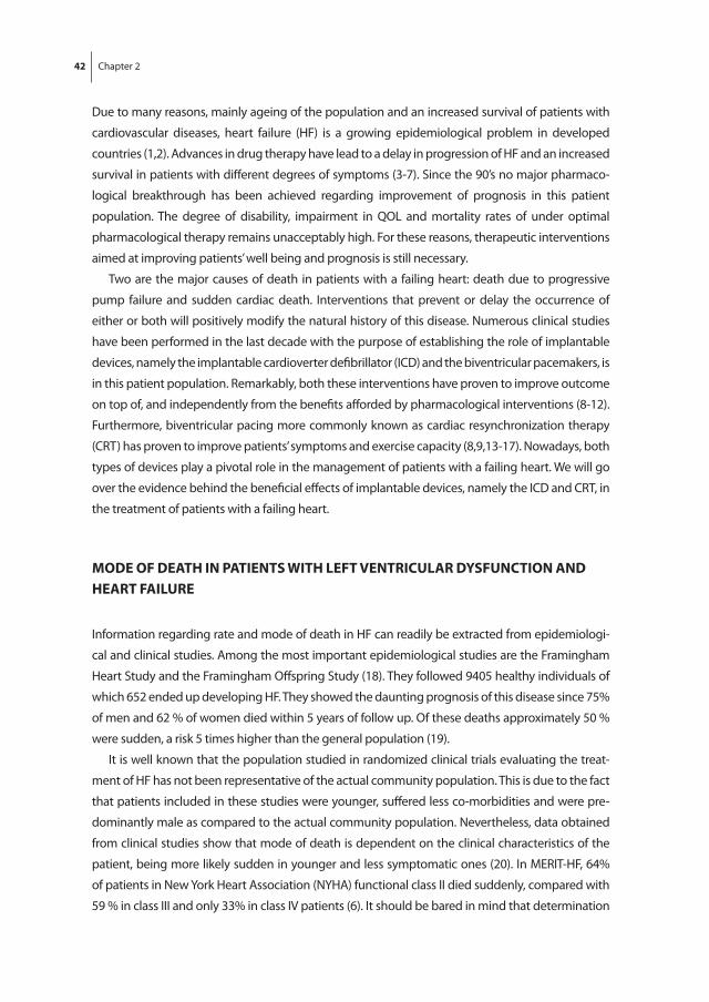
Due to many reasons, mainly ageing of the population and an increased survival of patients with
cardiovascular diseases, heart failure (HF) is a growing epidemiological problem in developed
countries (1,2). Advances in drug therapy have lead to a delay in progression of HF and an increased
survival in patients with different degrees of symptoms (3-7). Since the 90’s no major pharmaco-
logical breakthrough has been achieved regarding improvement of prognosis in this patient
population. The degree of disability, impairment in QOL and mortality rates of under optimal
pharmacological therapy remains unacceptably high. For these reasons, therapeutic interventions
aimed at improving patients’ well being and prognosis is still necessary.
Two are the major causes of death in patients with a failing heart: death due to progressive
pump failure and sudden cardiac death. Interventions that prevent or delay the occurrence of
either or both will positively modify the natural history of this disease. Numerous clinical studies
have been performed in the last decade with the purpose of establishing the role of implantable
devices, namely the implantable cardioverter defibrillator (ICD) and the biventricular pacemakers, is
in this patient population. Remarkably, both these interventions have proven to improve outcome
on top of, and independently from the benefits afforded by pharmacological interventions (8-12).
Furthermore, biventricular pacing more commonly known as cardiac resynchronization therapy
(CRT) has proven to improve patients’ symptoms and exercise capacity (8,9,13-17). Nowadays, both
types of devices play a pivotal role in the management of patients with a failing heart. We will go
over the evidence behind the beneficial effects of implantable devices, namely the ICD and CRT, in
the treatment of patients with a failing heart.
modE oF dEath in patiEnts with lEFt vEntriCular dysFunCtion and hEart FailurE
Information regarding rate and mode of death in HF can readily be extracted from epidemiologi-
cal and clinical studies. Among the most important epidemiological studies are the Framingham
Heart Study and the Framingham Offspring Study (18). They followed 9405 healthy individuals of
which 652 ended up developing HF. They showed the daunting prognosis of this disease since 75%
of men and 62 % of women died within 5 years of follow up. Of these deaths approximately 50 %
were sudden, a risk 5 times higher than the general population (19).
It is well known that the population studied in randomized clinical trials evaluating the treat-
ment of HF has not been representative of the actual community population. This is due to the fact
that patients included in these studies were younger, suffered less co-morbidities and were pre-
dominantly male as compared to the actual community population. Nevertheless, data obtained
from clinical studies show that mode of death is dependent on the clinical characteristics of the
patient, being more likely sudden in younger and less symptomatic ones (20). In MERIT-HF, 64%
of patients in New York Heart Association (NYHA) functional class II died suddenly, compared with
59 % in class III and only 33% in class IV patients (6). It should be bared in mind that determination
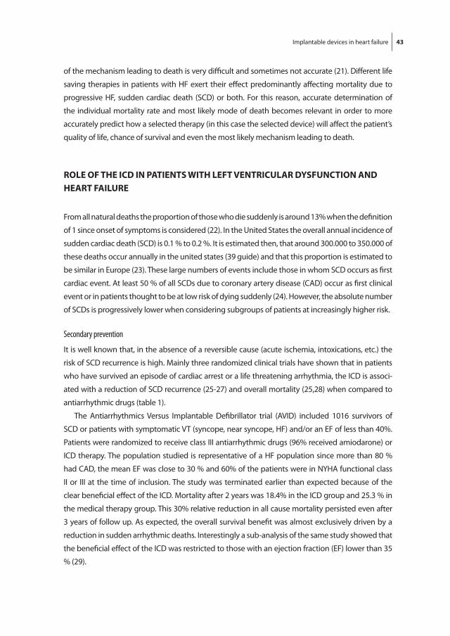
Implantable devices in heart failure 43
of the mechanism leading to death is very difficult and sometimes not accurate (21). Different life
saving therapies in patients with HF exert their effect predominantly affecting mortality due to
progressive HF, sudden cardiac death (SCD) or both. For this reason, accurate determination of
the individual mortality rate and most likely mode of death becomes relevant in order to more
accurately predict how a selected therapy (in this case the selected device) will affect the patient’s
quality of life, chance of survival and even the most likely mechanism leading to death.
rolE oF thE iCd in patiEnts with lEFt vEntriCular dysFunCtion and hEart FailurE
From all natural deaths the proportion of those who die suddenly is around 13% when the definition
of 1 since onset of symptoms is considered (22). In the United States the overall annual incidence of
sudden cardiac death (SCD) is 0.1 % to 0.2 %. It is estimated then, that around 300.000 to 350.000 of
these deaths occur annually in the united states (39 guide) and that this proportion is estimated to
be similar in Europe (23). These large numbers of events include those in whom SCD occurs as first
cardiac event. At least 50 % of all SCDs due to coronary artery disease (CAD) occur as first clinical
event or in patients thought to be at low risk of dying suddenly (24). However, the absolute number
of SCDs is progressively lower when considering subgroups of patients at increasingly higher risk.
Secondary prevention
It is well known that, in the absence of a reversible cause (acute ischemia, intoxications, etc.) the
risk of SCD recurrence is high. Mainly three randomized clinical trials have shown that in patients
who have survived an episode of cardiac arrest or a life threatening arrhythmia, the ICD is associ-
ated with a reduction of SCD recurrence (25-27) and overall mortality (25,28) when compared to
antiarrhythmic drugs (table 1).
The Antiarrhythmics Versus Implantable Defibrillator trial (AVID) included 1016 survivors of
SCD or patients with symptomatic VT (syncope, near syncope, HF) and/or an EF of less than 40%.
Patients were randomized to receive class III antiarrhythmic drugs (96% received amiodarone) or
ICD therapy. The population studied is representative of a HF population since more than 80 %
had CAD, the mean EF was close to 30 % and 60% of the patients were in NYHA functional class
II or III at the time of inclusion. The study was terminated earlier than expected because of the
clear beneficial effect of the ICD. Mortality after 2 years was 18.4% in the ICD group and 25.3 % in
the medical therapy group. This 30% relative reduction in all cause mortality persisted even after
3 years of follow up. As expected, the overall survival benefit was almost exclusively driven by a
reduction in sudden arrhythmic deaths. Interestingly a sub-analysis of the same study showed that
the beneficial effect of the ICD was restricted to those with an ejection fraction (EF) lower than 35
% (29).

44 Chapter 2
The Canadian Implantable Defibrillator trial (CIDS) (26) randomized 659 patients to receive and
ICD or be treated with amiodarone. Inclusion criteria were similar to those of AVID except that the
cutoff EF was set at 35% and patients with unmonitored syncope that proved to have inducible
VT were also included. A 20 % reduction in all cause mortality and a 33% reduction in arrhythmic
mortality in ICD treated patients was observed at 5 years of follow up, although none of these
differences were statistically significant. However, the cross over rate was significant since 30% of
ICD patients received amiodarone and 16% of patients initially assigned to antiarrhythmic therapy
ended up with an ICD. In a later published sub-analysis the authors showed that patients older than
70 years, with an EF ≤ 35 % and advanced symptoms of HF were those who benefited the most
from the device (30).
The Cardiac Arrest Study Hamburg (CASH) (27) study randomized survivors of cardiac arrest
to the ICD or antiarrhythmic drugs (amiodarone, metoprolol and propafenone). The propafenone
group had to be discontinued because of an increased mortality, mainly sudden cardiac death.
After a follow up of 57 months, the ICD showed to reduce mortality by 23 % (p=0.08). Although the
difference was not statistically significant, some factors like the fact that 10% of the patients had
no structural heart disease, that the mean EF was 46% or that more than half of the patients under-
went thoracotomy for implantation of the ICD (with a higher operative risk) probably prevented
this tendency to become significant.
A meta-analysis of these 3 studies showed that among survivors of SCD, the ICD conferred a
28% reduction in all cause mortality, mainly due to a 50% reduction in the risk of recurrent SCD
(28).
Based on these findings the ICD has become a Class I recommendation for patients who survived
a cardiac arrest or had symptomatic VT (31). These trials also showed that the group of patients who
benefit the most are those with a severely depressed EF and with symptomatic HF. Highlighting the
relevance of left ventricular (LV) function as predictor of overall mortality and SCD (table 1).
table 1. Major trials evaluating the effects of the ICD for secondary prevention of SCD
study n inclusion Criteria design major FindingAVID (1997)
(25)1013 SCD survivors,
symptomatic VT or EF ≤ 40
ICD vs AAD, randomized
30 % reduction in mortality with ICD
CASH (2000)(27)
349 SCD survivors, symptomatic VT
ICD vs AAD (amiodarone, propafenone, metoprolol) conventional, randomized
Significant reduction in SCDTrend reduction in mortality with ICDIncreased mortality with propafenone
CIDS (2000)(26)
259 SCD survivors, Symptomatic VT
ICD vs amiodarone Trend towards reduction in mortality with ICD

Implantable devices in heart failure 45
Primary prevention
- Ischemic cardiomyopathySurvival from an out of hospital cardiac arrest remains low. Not more than 5% of people can be
successfully reanimated after such an event. Therefore, the identification of those at increased
risk of a first cardiac arrest is of utmost importance. In an attempt to define such a population,
and using the lessons learned from secondary prevention trials, randomized clinical trials enrolled
progressively broader groups of patients that would likely benefit from this therapy. As inclusion
criteria the earlier studies required inducibility during electrophysiological study, documentation
of non-sustained ventricular tachycardia (NSVT) and/or other markers thought to be predictors
of SCD (32-34). Later only LV dysfunction with or without HF was the main requisite for inclusion
(10,11) (table 2).
Coronary artery disease in combination with a poor LV function was thought to be a marker
of increased risk of SCD. In this line, the first trial to demonstrate a benefit of the ICD over antiar-
rhythmic drugs was the Multicenter Automatic Defibrillator Implantation Trial (MADIT) (11). This
trial enrolled 196 patients with previous myocardial infarction, an EF ≤ 35% and non-sustained VT.
Patients underwent electrophysiological (EP) study and if VT could be induced and not be sup-
pressed (non-suppressible) with antiarrhythmic drugs (AAD) they were randomized to the ICD or
antiarrhythmic medication. Mean EF was around 26% and two-thirds of the patients were in NYHA
functional class II or III HF. This landmark study had to be stopped prematurely due to a dramatic
table 2. Major trials evaluating the effects of the ICD in primary prevention of SCD
study n inclusion criteria design major FindingMADIT (1996)(32) 196 NSVT, EF ≤ 35 %, previous
MI, Non-supressible VT ICD vs AAD, randomized
54 % reduction in mortality with ICD
MUSTT (1999)(33) 2202 NSVT, EF ≤ 40 %, previous MI, Inducible at EP study
EP study guided therapy vs conventional, randomized
Reduction in mortality in ICD patients only
MADIT II (2002)(11)
1232 EF ≤ 30 %, previous MI ICD vs conventional therapy, randomized
31 % reduction in mortality with ICD
DINAMIT (2004)(38)
674 EF ≤ 35 %, within 40 days of MI, depressed heart rate variability
ICD vs conventional therapy, randomized
No beneficial effect of the ICD
DEFINITE (2004)(12)
458 EF ≤ 35 %, non-ichemic cardiomyopathy, NSVT
ICD vs conventional therapy
Reduction in SCD, Trend in all cause mortality reduction with ICD
SCD-HeFT (2005)(10)
2521 EF ≤ 35 % of any etiology, NYHA class II or III
ICD vs amiodarone vs placebo, randomized
Reduction in mortality with ICD compared to amiodarone or placebo. No effect of amiodarone.

46 Chapter 2
54% reduction in overall mortality afforded by the ICD. In this way it became the first trial to show
that the ICD could improve survival in previously “asymptomatic” patients and the first to highlight
the importance of risk stratification for the prevention of SCD. Based on the results of MADIT, ICD
therapy was approved for this indication. Again, the group of patients who benefited the most
from the device were those with the worse EF fraction (35).
The CABG-patch trial included patients undergoing coronary artery bypass surgery (CABG)
(34) with an EF < 36% and an abnormal signal average ECG (SAECG). Patients were randomized to
receive an ICD or no other therapy. This trial failed to prove any effect of the ICD regarding overall
mortality suggesting the probable protective effect of revascularization and the limitation of
SAECG as a non-invasive risk stratifier.
The Multicenter Unsustained Tachycardia Trial (MUSTT) (33) included patients with non-
sustained VT, a previous MI and an EF ≤ 40%. The purpose of the trial was not to evaluate the role of
the ICD in the prevention of sudden death (it ultimately showed this) but to evaluate the effective-
ness of an EP study guided therapy to reduce this event. Patients who fulfilled inclusion criteria and
were inducible at the EP study were randomized to no specific treatment or to undergo EP guided
treatment (different AADs were tested to suppress VT or VF). Non-suppressible patients underwent
ICD implantation. After 5 years, mortality was 24% in the ICD group, 48% in the group not receiving
any treatment and 55% in those receiving AAD. This study not only showed the beneficial effects of
the ICD in this high risk population but also corroborated the associated risks related to the use of
AAD (mainly of class I-A) in patients with CAD and LV dysfunction (mean EF was 30%).
The recently published MADIT II trial markedly widened the spectrum of patients proven to
benefit from the ICD (11). It recruited patients based solely on the presence of an EF ≤30%, the
strongest known predictor of mortality in CAD patients. No documentation of NSVT or proof of VT
inducibility was required. More than 1200 patients were randomized to the use of an ICD or con-
ventional therapy. This study was also prematurely stopped due to the observed 31 % reduction in
overall mortality afforded by the ICD. This beneficial effect was entirely due to a reduction in the rate
of SCD. Nonetheless, in patients receiving the ICD the incidence of new or worsened heart failure
was higher than in the control group. This could probably be explained by the harmful effects of
right ventricular (RV) pacing (36) or even the potentially detrimental effects of ICD discharges (37).
Nowadays care is taken to avoid unnecessary RV pacing when programming these devices.
A sub study of MADIT II showed that the beneficial effect of the ICD is time dependent. In those
patients who suffered the myocardial infarction (MI) within 18 months of implantation no benefi-
cial effect of the ICD was observed, however, a significant reduction in mortality was observed in
those in whom the MI occurred beyond 18 months prior to implantation. The beneficial effect of
the ICD persisted even after 15 years of the MI. The DINAMIT study, who evaluated the effects of ICD
in those patients with a severely depressed EF (≤ 35%) implanted within 40 days of MI clearly failed
to show a beneficial role of the ICD (38).
In summary, results of MADIT I, MUSTT and MADIT II showed that in patients with a history of MI
and poor LV function implantation of an ICD results in a significant reduction in overall mortality

Implantable devices in heart failure 47
(through a reduction in SCD). Based on the results of DINAMIT, decisions regarding ICD implanta-
tion in patients with CAD should be withheld at least 40 days after the diagnosis of the MI.
- Non ischemic cardiomyopathyIn patients with cardiomyopathy of non ischemic origin the role of the ICD is more debatable. The
Cardiomyopathy Trial (CAT) (39) and the Amiodarone versus ICD trial (AMIOVIRT) (40) evaluated
whether the ICD was beneficial as compared to standard medical therapy or amiodarone, respec-
tively, in patients with non-ischemic dilated cardiomyopathy. None of these studies showed any
beneficial effect of the device because the number of patients included was small, the follow up
was probably not long enough and most importantly the event rate in the control group was lower
than expected.
The Defibrillators In Non-Ischemic Cardiomyopathy Treatment Evaluation (DEFINITE) trial (12)
randomized 458 patients with non-ischemic cardiomyopathy and an EF of less than 36% with sig-
nificant ventricular arrhythmia (frequent PVC’s or non-sustained VT) to standard medical therapy
or ICD implantation. After a mean follow up of 29 months a trend towards a reduction in mortality
(p=0.08) and a significant 80 % reduction in arrhythmic death was observed in the ICD treated
group. The investigators explain the lack of significance of the overall mortality effect of the ICD
in that the rate of SCD with respect to the total amount of deaths was lower than expected and
consequently the study resulted underpowered for overall mortality.
The Sudden Cardiac Death in Heart Failure trial (SCD-HeFT) (10) enrolled 2521 patients with an
EF≤ 35% and NYHA class II-III chronic heart failure. The trial was designed to include patients with
both ischemic and non-ischemic cardiomyopathy. Patients were randomized to receive a single
chamber ICD (programmed only to provide defibrillation), to amiodarone or to placebo on top
of conventional medical therapy. The cause of HF was ischemic in 52% and non-ischemic in 48%
of the cases. About 70% of the patients were in NYHA functional class II and 30% in class III. After
a mean follow up of 45 months a significant 23% reduction in mortality was observed with ICD
therapy compared to placebo (22% vs 29%; P=0.007). There was an absolute reduction in mortality
afforded by the ICD after 5 years of 7.2%. The results did not vary according to the etiology of HF. No
mortality reduction was observed with the use of amiodarone when compared to placebo (28% vs
29% respectively). Surprisingly, patients in NYHA class III did not show any benefit from the ICD as
compared to placebo. However, considering the relatively small amount of patients included with
this functional class and the contradictory of these results with previous ICD trials, this observation
does not confer sufficient evidence to withhold ICD therapy from class III patients at this point
in time. Amiodarone should be cautiously prescribed in NYHA class III patients due to the 44%
increase in mortality observed in this group of patients.
To summarize, evidence has taught us that in survivors of cardiac arrest or patients with symp-
tomatic VT, ICD therapy is the first line treatment. Patients with congestive HF and LV dysfunction,
irrespective of etiology, form a group at high risk of SCD and their long term prognosis is highly
dependent on the LV function. With the information provided by MADIT II and the SCD-HeFT it

48 Chapter 2
is likely that in patients with an EF ≤ 35 % the implantation of an ICD (with or without CRT) will
become the standard of care for primary prophylaxis. It is important to highlight that it is unlikely
that these patients will benefit from the ICD if they are within 3 months of revascularization, 40
days within an MI or have had the initial diagnosis of the cardiomyopathy within a 3 month period
(31) (table 3).
CardiaC rEsynChronization thErapy For thE trEatmEnt oF hEart FailurE
In patients with advanced LV systolic dysfunction and HF, conduction defects are common and lead
to cardiac mechanical dyssynchrony. Three distinct types of dyssynchrony have been described:
atrio-ventricular (AV), inter-ventricular (V-V) and intra-ventricular (IV). AV dyssynchrony, due to a
prolonged AV conduction delay, is associated to an ineffective ventricular filling, diastolic mitral
regurgitation and decreased cardiac output. VV and IV dyssynchrony can be manifested by a pro-
longation of the QRS complex and is associated to an abnormal prolongation of the contraction
delay between both ventricles and within the left ventricle itself. This inter- and intra-ventricular
dyssynchronous contraction reduces myocardial efficiency, further impairing systolic and diastolic
function and worsening mitral regurgitation (41).
Approximately one third of the patients with systolic HF present signs of intra-ventricular
conduction delay, most of them with a left bundle branch block (LBBB) like pattern (42). These
ventricular conduction disturbances have proven to be associated to more advanced heart disease
and a worse prognosis. In a recent report of 5517 pts with HF due to different etiologies (45%
ischemic and 36% idiopathic) 25% of the patients had LBBB, 6% had right bundle branch block
(RBBB) and another 6% had other forms of intraventricular conduction delay (43). Patients with
LBBB had more sever HF, reduced systolic blood pressure, increased incidence of third heart sound
and more abnormal cardiothoracic ratios. The proportion of patients with a LVEF of less than 30%
was higher in patients with LBBB and most importantly they had a significantly higher mortality
table 3. Recommendations for the use of the implantable defibrillator for primary prevention of SCD in heart failure patients (AHA/ACC/ESC practice guidelines)
• Prior MI (>40 days), EF≤ 30% to 40% and NYHA class II or III, optimally treated HF(class I- level of evidence A)
• Non-ischemic heart disease, EF ≤ 30% to 35 % and NYHA class II or III optimally treated HF (class I-level of evidence B)
• In combination with CRT in NYHA class III-IV, QRS ≥ 120 ms , optimally treated HF (class IIa-level of evidence B)
• Prior MI, EF ≤ 30% to 35 % and NYHA class I, optimally treated (class IIa-level of evidence B)
• Recurrent stable VT, normal or near normal EF, optimally treated HF (class IIa-level of evidence C)
• Non-ischemic heart disease, EF ≤ 30% to 35 % and NYHA class I, optimally treated (class IIb-level of evidence B)

Implantable devices in heart failure 49
than patients without conduction disturbances and even when compared with those with other
forms of intra-ventricular conduction abnormalities.
The aim of CRT is not only to correct the AV dyssynchrony, but through biventricular stimula-
tion also to improve the abnormal VV and IV dyssynchrony. Since CRT was first clinically applied in
1994 (44), initial studies have shown that it can markedly improve cardiac output, increase systolic
pressure, lower pulmonary capillary wedge pressures (45,46), enhance ventricular systolic function
and pressure-volume loops, reduce mitral regurgitation (46) and improve synchrony of ventricular
contraction (47,48).
Clinical effects afforded by CRT
Improvement in symptom profile and exercise capacityOverall, non-randomized and randomized studies have shown a highly consistent effect of CRT in
improving symptoms, exercise capacity and left ventricular performance (table 4). Patients enrolled
in the major CRT trials generally had NYHA class III or IV HF on the basis of ischemic or non-ischemic
dilated cardiomyopathy, were in sinus rhythm, had a broad QRS complex on the ECG and were
under stable optimal pharmacological therapy.
The Pacing therapies in Congestive Heart Failure (PATH-CHF) trial (13) compared CRT with RV
pacing and reported a statistically significant reduction in LV end diastolic volume (LVEDV) (253±83
ml to 227±112 ml), LV end systolic volume (LVESV) (202±79 ml vs 174±101ml) and an increase in
LVEF (22±7 %vs 26±9 %) after six month of hemodynamically optimized atrial and biventricular
pacing. Only patients with higher volumes at baseline were less likely to positively remodel after
CRT (13). The Multisite Stimulation in Cardiomyopathies (MUSTIC)-SR trial (14) used a cross over
design, where patients were randomized to active versus back up biventricular pacing. It showed
a significant improvement in HF symptoms, quality of life (QOL) and improvements in more objec-
tive parameters as distance walked during 6 minutes and peak oxygen consumption. These small
trials were followed by the MIRACLE (15) study. It included 453 patients with moderate to sever
HF and EF ≤ 35% and a QRS duration greater than 130 ms. All patients that underwent successful
CRT implantation were randomized to biventricular pacing or control (no pacing). Patients were
followed for 6 months and the primary end points were change in NYHA class, QOL and distance
walked in 6 minutes. CRT was associated to a significant improvement in all three variables. This
was apparent as early as one month after treatment and was sustained throughout the study
period. The MIRACLE-ICD (16), that evaluated CRT with an additional defibrillator function (CRT-D)
compared to ICD therapy alone, also showed that CRT was associated with the improvement in
NYHA class and QOL. These results were supported by the findings of the CONTAK CD trial (17).
Linde et al. showed over long term follow up of patients included in the MUSTIC trial that biven-
tricular pacing significantly improved 6-min walk distance by 20%, increased oxygen consumption
by 11%, and reduced both NYHA by 25% and quality of life Minnesota score by more than 30%
(49).

50 Chapter 2
Positive remodeling effect of CRTThe improvement in symptoms and exercise capacity afforded by CRT are mainly the result of an
accompanying improvement in LV performance, a reduction in mitral regurgitation and reduction
table 4. Major trials evaluating the effects of cardiac resynchronization therapy
trial n inclusion criteria
design implant Failure
Fu Effect of Crt
PATH-CHF (2002)(13) 41 Low EF, NYHA class III-IV, QRS ≥ 120 ms, ≥ 150 ms
CRT vs RV pacing, single blind, cross over
N/A 3 months
EF LV volumes
MUSTIC (2001)(14) 67 EF ≤ 35 %, NYHA class III, QRS ≥ 150 ms
CRT vs no pacing, cross over
8 % 3 months
distance during 6 min peak O2 consumptionImproved QOL
MIRACLE (2002)(15) 453 EF ≤ 35 %, NYHA class III or IV, QRS ≥ 130 ms
CRT vs Medical therapy, randomized
8 % 6 months
distance during 6 min EF peak O2 consumption NYHA Improved QOL
MIRACLE-ICD (2003)(16) 369 EF ≤ 35 %, NYHA class III or IV, QRS ≥ 130 ICD indication
CRT-D vs ICD, randomized, double blind
7.8% 6 months
peak O2 consumption NYHA Improved QOL
CONTAK CD (2003)(17) 490 EF ≤ 35 %, NYHA class III or IV, QRS ≥ 120 ms, VT/VF
CRT-D vs ICD, randomized
6 months
peak O2 consumption distance during 6 min NYHA
COMPANION(2004)(8) 1520 EF ≤ 35 %, NYHA class III or IV, QRS ≥ 120 ms, PR > 150 ms
Medical therapy vs CRT vs CRT-D
13 % 12 months
all cause mortality + all cause hospitalization overall mortality with CRT-D and tendency with CRT alone
CARE-HF (2005)(9) 813 EF ≤ 35 %, NYHA class III or IV, QRS ≥ 120 ms, QRS >120 <150 dyssynchrony was required
CRT vs Medical therapy, randomized
5 % 29.4 months
all cause mortality + hospitalization for cardiovascular event overall mortality

Implantable devices in heart failure 51
in left ventricular volumes. This last effect is known as the “positive remodeling” effect of CRT and
is an important predictor of positive outcome (50). A sub-study of the MUSTIC trial reported the
effects of CRT on left ventricular remodeling, exclusively in patients in sinus rhythm (51). They
observed that both systolic and diastolic LV dimensions significantly improved 3 months after
implantation and that these further improved after 12 months of therapy. The degree of mitral
regurgitation significantly improved as well. Correction of dyssynchrony was crucial for this posi-
tive remodeling effect and was observed regardless of etiology of heart disease. When considering
symptoms, they improved significantly by 3 months with no further improvement at 12 months.
Symptom improvement also consistently correlated with reverse remodeling. These effects were
significantly greater in idiopathic dilated cardiomyopathy patients than in ischemic patients.
Remarkable is that this sustained reverse remodeling effect was seen in patients already receiving
conventional drug therapy. The MIRACLE investigators also evaluated long-term effects of CRT.
They reported a significant reduction in LVED and LVES volumes, in degree of mitral regurgitation
and an increase in LVEF at 6 month follow up (52). Though reverse remodeling and LVEF improved
during the first 3 months, this effect was even greater at 6-month follow up. This improvement
was significantly greater in patients with idiopathic dilated cardiomyopathy than in patients with
ischemic cardiomyopathy. The clinical improvements observed in this study were found to be
associated to positive left ventricular remodeling and significantly improved parameters of dys-
synchrony mediated by biventricular pacing.
Although symptomatic improvement is an important goal in the management of severely
symptomatic HF patients, it is difficult to evaluate to what extent this improvement is a conse-
quence of the applied therapy or due to the placebo effect that any given treatment might exert.
In order to ensure that CRT is effectively being applied improvement in objective parameters like a
positive LV remodeling effect should accompany symptomatic relief.
Data derived from drug treatment trials in HF showed that increased LV size and volumes in
patients with left ventricular dysfunction were predictors of adverse cardiovascular events, worsen-
ing HF and SCD (53,54). Yu et al. elegantly showed in a large cohort of CRT treated patients, that
only a positive remodeling effect was significantly associated to a reduction in heart failure events
and improved outcome. A reduction in LVESV of more than 10% was the single most important
predictor of all cause and cardiovascular mortality (50). Clinical parameters alone were unable to
predict any outcome.
table 5. Recommendations for the use of cardiac resynchronization therapy
• NYHA class III or IV heart failure under optimal medical therapy
• LVEF ≤ 35%
• LV end diastolic diameter ≥ 55 mm
• QRS duration ≥ 130 ms
• No expected improvement from coronary revascularization or valve surgery

52 Chapter 2
Prognostic relevance of CRT
The Comparison of Medical Therapy, Pacing and Defibrillation in Heart Failure (COMPANION) trial
(8) was designed to evaluate if CRT with or without defibrillator function would reduce the risk
of death and hospitalization among patients with advanced HF (NYHA functional class III or IV),
severely depressed LV function (≤ 35%) and a broad QRS on the ECG (≥120 ms). The secondary end
point was all cause mortality. The 1520 enrolled patients were randomized to receive CRT alone,
CRT with a defibrillator function (CRT-D) or optimal medical therapy in a 2-2-1 fashion. After 12
months the rate of the combined primary end point of death from any cause or hospitalization for
any cause was 68% in the optimal medical therapy group, and 56 % in both CRT and CRT-D groups.
In this way, CRT (irrespective of the defibrillator function) was associated with a 20 % reduction of
the primary end point. When considering the secondary end point of all cause mortality, a 24%
reduction (P=0.06) was observed in the CRT alone group and 36% in the CRT-D group (P=0.003).
The risk of death or hospitalization due to HF was also found to be significantly reduced by 25 %
and 28% in the CRT and CRT-D groups respectively. This beneficial effect of CRT was observed both
in patients with ischemic and non-ischemic cardiomyopathy (55). COMPANION was the first trial to
show independently that CRT alone could potentially improve survival in patients with HF and set
the stage for the results of the coming Cardiac Resynchronization Heart Failure trial.
The Cardiac Resynchronization Heart Failure (CARE-HF) trial (9) compared the effects of CRT
alone to standard medical therapy on death and unplanned hospitalizations for major cardiac
events. The principal secondary end point was death from any cause. Inclusion criteria differed
slightly from other CRT trials; in those cases were QRS duration was between 120 ms and 149 ms
echocardiographic documentation of dyssynchrony was required (only a minority of patients were
included due to this criterion). A total of 813 patients were enrolled and followed for a mean of
29.4 months. During follow up, the primary end point of all cause mortality and hospitalization
for major cardiovascular event was reduced by 27% (P<0.001) in the CRT treated patients. More
importantly, overall mortality rate was 30% in the optimal medical therapy group and 20% in the
patients treated with CRT (P<0.002).
Although, the COMPANION trial did show a significant survival benefit of CRT-D, CARE-HF was
the only trial to show independently that CRT alone can reduce mortality in patients with HF.
However, the trial was not prospectively powered to show a reduction in overall mortality, and this
end point was only set as a secondary one. Previously published meta-analyses have corroborated
the effects of CRT upon symptoms, QOL and exercise tolerance (56-58). However, an overall survival
benefit of CRT alone could not be addressed. Mainly because in these meta-analyses trials that also
evaluated the effects of CRT-D were included. In this way the effects of CRT were confounded by
the proven life saving effect of the ICD. Since it is very unlikely that another trial comparing CRT
alone to medical therapy will exist, we performed a meta-analysis with the purpose of establishing
whether CRT alone compared to optimal medical therapy, reduces overall mortality and in which
way it affects the different modes of death of patients with advanced HF. This analysis demon-
strated that CRT alone significantly reduces all cause mortality by 29 % after a weighted mean

Implantable devices in heart failure 53
follow up of 18 months. This effect is mainly due to an impressive 38% reduction in mortality due
to progressive HF.
The problem of non responders to CRT
Using the classic indications for CRT, both uncontrolled and controlled clinical studies reported
a rate of “non-responders” ranging from 15 % to 35 % (13-17). This was true when considering a
“responder” either after symptom improvement or after positive left ventricular remodeling was
demonstrated during follow up.
Though in the reports of the mayor trials on CRT the rate of responders is not mentioned as such, this
data can be inferred. Fifteen percent of the patients at the end of the cross over phase of the MUSTIC
trial preferred a pacing mode different from the biventricular mode. This was true both for patients in
sinus rhythm or in atrial fibrillation (49). The results of the MIRACLE trial show that in, respectively, 33%
and 16 % of the patients randomized to CRT, the clinical composite HF score did not improve or was
considered to have worsened, after 6 months follow up (15). The PATH-CHF investigators reported
similar rates of non-positive remodeling. In 36% of the patients no improvement or even a worsened
LV performance (as measured by echocardiography) was observed after 6 months (13).
Many factors have been reported to influence the degree of response to CRT like location of
left ventricular stimulating lead (59,60), presence of viable tissue at the site of stimulation, etiology
of heart disease (51,52), degree of left ventricular dysfunction (13), other co-morbidities and most
importantly degree of mechanical intra and inter-ventricular dyssynchrony (61,62). Most uncon-
trolled and controlled clinical trials regarding CRT considered broadness of the QRS as a surrogate
for mechanical IV and V-V dyssynchrony. However, the accuracy of the ECG to predict responsive-
ness to CRT has recently been put in doubt. Most studies comparing the predictive accuracy of
the ECG with mechanical indicators of dyssynchrony showed the superiority of the last method to
predict symptom improvement and positive left ventricular remodeling after CRT (61-63). However,
most of these studies included a relatively small number of patients, defined mechanical dyssyn-
chrony differently and were single center studies. The recently presented results of the Predictors
of Response to Cardiac Resynchronization Therapy (PROSPECT) study has suggested that the clini-
cal utilization of echo techniques for selecting patients for CRT has not yet proven benefits over
broadness of the QRS. None of many echo indicators of ventricular dyssynchrony on their own, had
enough sensitivity and specificity for prediction of response to CRT (64). Furthermore variability of
more than 50% for some tissue-Doppler imaging measurements and of more than 90% for some
measures of inter-ventricular delay in the interpretation of the data has been observed between
the three different core laboratories that interpreted the studies. Even though it is highly likely and
intuitive that mechanical dyssynchrony criteria would add something to the present selection
criteria, QRS width should still be considered the gold standard for selection of candidates for CRT.
For the moment patients with standard indications for CRT (low EF, refractory HF symptoms and
broad QRS on the ECG) should not be withheld from this therapy even if significant mechanical
dyssynchrony is not demonstrated prior to implantation.

54 Chapter 2
CombinEd Crt and dEFibrillator FunCtion
MADIT II and SCD-HeFT have shown that ICDs should form part of the therapy for patients with
severely depressed LV function. This data would suggest that most of the patients who require
biventricular pacing would also be candidates for ICD implantation since most of them have a LVEF
≤ 35%. In the COMPANION (8,65) trial a modest increase in SCD was observed among patients
who underwent CRT alone compared to control patients. Median time to SCD was shorter in the
CRT arm (186 days) than in CRT-D (341 days) and control patients (253 days) (65). In those studies
with shorter follow up, like MUSTIC (14) and MUSTIC AF (66), most deaths occurred suddenly dur-
ing active biventricular pacing. CARE-HF originally reported a non-significant reduction in SCD in
patients undergoing CRT compared to controls (9.4 % vs 7.0 %; P=0.25) (9). However, in the “exten-
sion” phase of the same study a significant reduction of in SCD was observed after 37.4 months of
follow up in CRT treated patients (HR 0.54, P=0.005).
The beneficial effect of CRT regarding overall mortality is predominantly a consequence of a
reduction in progressive HF mortality (67). Consequently, patients whose heart failure profile
improves after CRT are less likely of dying due to worsening HF; in this way the relative contribution
of SCD to overall mortality increases (figure 1). This raises the question of what the role of the ICD
will be in these patients. In the COMPANION (8,65) study, CRT-D significantly increased survival
compared to controls and showed a trend towards a beneficial effect when compared to CRT
alone. However, not all patients profited from the addition of an ICD. The patients who derived
more benefit were those with somewhat better preserved EF, and who had a better symptom
profile (68). In the SCD-Heft trial patients in NYHA functional class III did not benefit from the ICD,
as did patients with a lower functional class (10). The amount of patients who continue to die of
SCD despite CRT remains high and highlights the need of an additional ICD function. It is likely that
biventricular defibrillators will almost totally replace the biventricular pacemaker for managing
patients with HF. Nonetheless, probability of survival, quality of the life prolonged and mode of
death are important aspects that patients and physicians should consider when discussing treat-
ment options and deciding whether to implant, and which device to select.
Figure 1. Mode of death in HF and CRT treated patientsChapter 2

Implantable devices in heart failure 55
rEFErEnCEs
1. Massie BM, Shah NB. Evolving trends in the epidemiologic factors of heart failure: Rationale for preventive strategies and comprehensive disease management. Am Heart J 1997; 133:703.
2. Swedberg K, Cleland J, Dargie H, Drexler H, Follath F, Komajda M, Tavazzi L, Smiseth OA, Gavazzi A, Haverich A, Hoes A, Jaarsma T, Korewicki J, Levy S, Linde C, Lopez-Sendon JL, Nieminen MS, Pierard L, Remme WJ. Guidelines for the diagnosis and treatment of chronic heart failure: executive sum-mary (update 2005): The Task Force for the Diagnosis and Treatment of Chronic Heart Failure of the European Society of Cardiology. Eur Heart J. 2005;26:1115-1140.
3. The CONSENSUS trial study group. Effects of enalapril in mortality in severe congestive heart failure: results of the Cooperative North Scandinaivan Enalapril Survival Study. N Eng J Med 1987;316:429.
4. Effect of enalapril on survival in patients with reduced left ventricular ejection fractions and conges-tive heart failure. The SOLVD Investigators. N Engl J Med 1991; 325(5):293-302;
5. CIBIS-II Investigators and Committees. The Cardiac Insufficiency Bisoprolol Study II (CIBIS-II): a ran-domised trial. Lancet 1999 Jan 2;353(9146):9-13
6. MERIT-HF Study Group. Effect of metoprolol CR/XL in chronic heart failure: Metoprolol CR/XL Randomised Intervention Trial in Congestive Heart Failure (MERIT-HF). Lancet 1999 Jun 12;353(9169):2001-7.
7. Pitt B, Zannad F, Remme WJ, et al. The effect of spironolactone on morbidity and mortality in patients with severe heart failure. Randomized Aldactone Evaluation Study Investigators. N Engl J Med 1999 Sep 2;341(10):709-17.
8. Bristow MR, Saxon LA, Boehmer J, Krueger S, Kass DA, De Marco T, Carson P, DiCarlo L, DeMets D, White BG, DeVries DW, Feldman AM. Cardiac- resynchronization therapy with or without an implant-able defibrillator in advanced chronic heart failure. N Engl J Med. 2004;350:2140-2150.
9. Cleland JG, Daubert JC, Erdmann E, Freemantle N, Gras D, Kappenberger L, Tavazzi L. The effect of car-diac resynchronization on morbidity and mortality in heart failure. N Engl J Med. 2005;352:1539-1549.
10. Bardy GH, Lee KL, Mark DB, Poole JE, Packer DL, Boineau R, Domanski M, Troutman C, Anderson J, Johnson G, McNulty SE, Clapp-Channing N, Davidson- Ray LD, Fraulo ES, Fishbein DP, Luceri RM, Ip JH. Amiodarone or an implantable cardioverter-defibrillator for congestive heart failure.N Engl J Med 2005;352:225-37
11. Moss AJ, Zareba W, Hall WJ, et al. Prophylactic implantation of a defibrillator in patients with myocar-dial infarction and reduced ejection fraction. N Engl J Med 2002;346:877-883
12. Kadish A, Dyer A, Daubert J, et al. for the Defibrillators in Non-Ichemic Cardiomyopathy Treatment Evaluation (DEFINITE) Investigators. Prophylactic defibrillator implantation in patients with non-ischemic dilated cardiomyopathy. N Engl J Med 2004;350:2151-58
13. Stellbrink C, Breithardt OA, Franke A, et al. Impact of cardiac resynchronization therapy using hemo-dynamically optimized pacing on left ventricular remodeling in patients with congestive heart failure and ventricular conduction disturbances. J Am Coll Cardiol 2001;38:1957-65.
14. Cazeau S., Leclercq C., Lavergne T, et al. Effects of Multisite Biventricular Pacing in Patients with Heart Failure and Intraventricular Conduction Delay. N Engl J Med 2001; 344:873-880
15. Abraham W. T., Fisher W. G., Smith A. L., et al. Cardiac Resynchronization in Chronic Heart Failure. N Engl J Med 2002; 346:1845-1853.
16. Young JB, Abraham WT, Smith AL, et al. Combined cardiac resynchronization and implantable cardiover-sion defibrillation in advanced chronic heart failure: the MIRACLE-ICD Trial. JAMA 2003;289:2685-94
17. Higgins SL, Hummel JD, Niazi IK, et al. Cardiac resynchronization therapy for the treatment of heart failure in patients with intraventricular conduction delay and malignant ventricular tachyarrhythmias. J Am Coll Cardiol 2003;42:1454-59
18. Ho KKL, Anderson KM, Kannel WB, et al. Survival after the onset of congestive heart failure in Framing-ham Heart Study subjects. Circulation 1993;88:107-15
19. Kannel WB, Plenh JF, Cupples LA. Cardiac failure and sudden death in the Framingham Study. Am Heart J 1988;11:869-75

56 Chapter 2
20. Josephson M, Wellens HJ. Implantable defibrillators and sudden cardiac death. Circulation 2004;109:2685-91
21. Jordaens L. The classification of sudden death in clinical trials. In: Alliot E, Clementy J, Prystowsky EN, ed. Fighting sudden cardiac death: A Worldwide Challenge. Futura Pub Co, Armonk; 2000.p.29-38
22. de Vreede-Swagemakers JJ, Gorgels AP, Dubois-Arbouw WI, et al. Out of hospital cardiac arrest in the 1990’s: a population-based study in the Maastricht area on incidence, characteristics and survival. J Am Coll Cardiol 1997;30:1500-5
23. Priori SG, Aliot E, Blomstrom-Lundqvist C, et al. Task Force on Sudden Cardiac Death of the European Society of Cardiology. Eur Heart J 2001;22:1374-450.
24. Myerburg RJ. Sudden cardiac death: exploring the limits of our knowledge. J Cardiovasc Electrophysiol 2001;12:369-81
25. The antiarrhythmics versus Implantable Defibrillators (AVID) Investigators. A comparison of antiarrhythmic-drugtherapy with implantable defibrillators in patients resuscitated from near-fatal ventricular arrhythmias. N Engl J Med 1997;37:76-83
26. Connolly SJ, Gent M, Roberts RS, et al. Canadian implantable defibrillator study (CIDS): a randomized trial of the implantable cardioverter defibrillator against amiodarone. Ciculation 2000;101:1297-302
27. Kuck KH, Cappato R, Siebels J, et al. Randomized comparison of antiarrhythmic drug therapy with implantable defibrillators in patients resuscitated from cardiac arrest: the Cardiac Arrest Study Ham-burg (CASH). Circulation 2000;102:748-54.
28. Connolly SJ, Hallstrom AP, Cappato R, et al. Meta-analysis of the implantable cardioverter defibrillator secondary prevention trials. AVID, CASH and CIDS studies. Antiarrhythmics versus Implantable Defi-brillator study. Cardiac Arrest study Hamburg. Canadian Implantable Defibrillator study. Eur Heart J 2000;21:2071-78.
29. Domanski MJ, Saksena S, Epstein AE, et al. Relative effectiveness of the implantable cardioverter-defibrillator and antiarrhythmic drugs in patients with varying degrees of left ventricular dysfunction who have survived malignant ventricular arrhythmias. AVID Investigators – Antiarrhythmic versus Implantable Defibrillators. J Am Coll Cardiol 1999;34:1090-95.
30. Sheldon R, Connolly S, Krahn A, et al. Identification of patients most likely to benefit from implant-able cardioverter defibrillator therapy: the Canadian Implantable Defibrillator Study. Circulation 2000;101:1660-64
31. Zipes DP, Camm AJ, Borggrefe M, et al. ACC/AHA/ESC 2006 guidelines for management of patients with ventricular arrhythmias and the prevention of sudden cardiac death: a report of the American College of Cardiology/American Heart Association Task Force and the European Society of Cardiol-ogy Committee for Practice Guidelines (Writing Committee to Develop guidelines for management of patients with ventricular arrhythmias and the prevention of sudden cardiac death) developed in collaboration with the European Heart Rhythm Association and the Heart Rhythm Society. Europace. 2006 Sep;8(9):746-837.
32. Moss AJ, Hall WJ, Cannom DS, et al. Improved survival with an implanted defibrillator in patients with coronary artery disease at high risk for ventricular arrhythmia: the Multicenter Automatic Defibrillator Implant Trial Investigators. N Engl J Med 1996;335:1933-40
33. Buxton AE, Lee KL, Fisher JD, et al. A randomized study of the prevention of sudden death in patients with coronary artery disease: Multicenter unsustained tachycardia trial investigators. N Engl J Med 1999;341:1882-90
34. Bigger JT, for the Coronary Artery Bypass Graft (CABG) Patch Trial Investigators. Prophylactic use of implanted cardiac defibrillators in patients at high risk for ventricular arrhythmias after coronary artery bypass graft surgery. N Engl J Med 1997;337:1569-75.
35. Moss AJ, Fadl y, Zareba W, et al. Survival benefit with an implantable defibrillator in relation to mortal-ity risk in chronic coronary heart disease. Am J Cardiol 2001;88:516-20.

Implantable devices in heart failure 57
36. Wilkoff BL, Cook JR, Epstein AE, et al. Dual-chamber pacing or ventricular backup pacing in patients with an implantable defibrillator: the Dual Chamber and VVI Implantable Defibrillator (DAVID) Trial. JAMA. 2002 Dec 25;288(24):3115-23.
37. Moss AJ, Greenberg H, Robert B, et al. Long term clinical course of patients after termination of ventricular tachyarrhythmia by an implantable defibrillator. Circulation 2004;110:3760-65.
38. Hohnloser SH, Kuck KH, Dorian P et al. on behalf of the Defibrillator in Acute Myocardial Infarction Trial (DINAMIT) invetigators. Prophylactic use of an implantable cardioverter defibrillator after acute myocardial infarction. N Engl J Med 2004;351:2481-88
39. Bansch D, Antz M, Boczor S et al. Primary prevention of sudden cardiac death in idiopathic dilated cardiomyopathy: the cardiomyopathy trial (CAT). Circulation 2002;105:1453-58
40. Strickberger A, Hummel JD, Bartlett TG, et al. Amiodarone versus implantable cardioverter defibrilla-tor: randomized trial in patients with nonischemic cardiomyopathy and asymptomatic nonsustained ventricular tachycardia - AMIOVIRT. J Am Coll Cardiol 2004;41:1707-12
41. Toquero J, Geelen P, Goethals M, Brugada P. What is first, left bundle branch block of left ventricular dysfunction? J Cardiovasc Electrophysiol 2001;12:1245-1428.
42. D. Farwell,N. R. Patel, A. Hall, S. Ralph, A. N. Sulke. How many people with heart failure are appropriate for biventricular resynchronization? European Heart Journal, 2000;21: 1246-1250.
43. Baldasseroni S, Opasich C, Gorini M, et al. Left bundle branch block (LBBB) is associated with increased 1 year sudden and total rate of death in 5517 outpatients with congestive heart failure. Am Heart J 2002;143:398-405.
44. Cazeau S, Ritter P, Bakdach S, et al. Four chamber pacing in dilated cardiomyopathie. Pacing Clin Electrophysiol 1994;17:1974-9.
45. Blanc JJ, Etienne Y, Gilard M, et al. Evaluation of different ventricular pacing sites in patients with severe congestive heart failure: results of an acute hemosynamic study. Circulation 1997;96:3272-77
46. Mansourati J, Etienne Y, Gilard M, et al. Left ventricular based pacing in patients with chronic heart failure: comparison of acute hemodynamic benefits according to underlying heart disease. Eur J Heart Fail 2000;2:195-199.
47. Saxon LA, Kerwin WF, Cahalan MK, et al. Acute effects of intraoperative multisite ventricular pacing on left ventricular function and activation/contraction sequence in patients with depressed ventricular function. J Cardiovasc Electrophysiol 1998;9:13-21.
48. Kerwin WF, Botvinick EH, O’Connell JW, et al. Ventricular contraction abnormalities in dilated cardio-myopathy: effect of biventricular pacing to correct interventricular dyssynchrony. J Am Coll Cardiol 2003;35:1221-27.
49. Linde C, Leclercq C, Rex S, et al. Long-term benefits of biventricular pacing in congestive heart failure: results from the multisite stimulation in cardiomyopathy study. J Am Coll Cardiol 2002;40:111-18.
50. Yu CM, Bleeker GB, Wing-Hong Fung J, et al. Left ventricular reverse remodeling but not clinical improvement predicts long term survival after cardiac resynchronization therapy. Circulation 2005;112:1580-86.
51. Duncan A, Wait D, Gibson D, Daubert JC. Left ventricular remodeling and haemodynamic effects of multisite biventricular pacing in patients with left ventricular systolic dysfunction and activation disturbances in sinus rhythm: sub-study of the MUSTIC trial. Eur Heart J 2003;24:430-441.
52. St John Sutton M, Plappert T, Abraham W, et al. Effect of cardiac resynchronization therapy on left ventricular size and function in chronic heart failure. Circulation 2003;107:1985-1990.
53. ST. John Sutton M, Pfeffer MA, Plappert T, et al. Cardiovascular death and left ventricular remodeling two years after myocardial infarction: The protective effects of captopril. Circulation 1994;89:68-75.
54. Burns RJ, Gibbons FJ, Yi Q, et al. The relationship of left ventricular ejection fraction, end-systolic volume index and infract size to six-month mortality after hospital discharge following myocardial infarction treated by thrombolisis. J Am Coll Cardiol. 2002; 39:30-36.
55. Bristow MR, et al. Comparison of medical therapy, pacing, and defibrillation in chronic heart failure (COM-PANION) trial. Presented in the 52nd American College of Cardiology Annual Scientific Sessions, 2003.

58 Chapter 2
56. Bradley DJ, Bradley EA, Baughman KL, Berger RD, Calkins H, Goodman SN, Kass DA, Powe NR. Cardiac resynchronization and death from progressive heart failure: a meta-analysis of randomized controlled trials. Jama. 2003;289:730-740.
57. McAlister FA, Ezekowitz JA, Wiebe N, Rowe B, Spooner C, Crumley E, Harling L, Klassen T, Abraham W. Systematic review: cardiac resynchronization in patients with symptomatic heart failure. Ann Intern Med. 2004;141:381-390.
58. Freemantle N, Tharmanathan P, Calvert MJ, Abraham WT, Ghosh J, Cleland JGF. Cardiac resynchronisa-tion for patients with heart failure due to left ventricular systolic dysfunction - a systematic review and meta-analysis. Eur J Heart Failure. 2006;8:433-440.
59. Ansalone G, Giannantoni P, Ricci R, et al. Doppler myocardial imaging to evaluate the effectiveness of pacing sites in patients receiving biventricular pacing. J Am Coll Cardiol 2002;39:489-99
60. Gasparini M, Mantica M, Galimberti P, et al. Is the left ventricular lateral wall the best lead implantation site for cardiac resynchronization therapy? PACE 2003;26[Pt II]:162-168.
61. Penicka M, Bartunek J, De Bruyne B, et al. Tissue Doppler assessment of asynchrony predicts positive left ventricular remodeling after biventricular pacing in heart failure. Circulation 2002;106(19):3185.
62. Soogard P, Egeblad H, Kim WY, et al. Tissue Doppler imaging predicts improved systolic performance and reversed left ventricular remodeling during long-term cardiac resynchronization therapy. J Am Coll Cardiol 2002;40:723-30.
63. Yu CM, Fung WH, Lin H, Zhang Q, Sanderson JE, Lau CP. Predictors of left ventricular reverse remodeling after cardiac resynchronization therapy for heart failure secondary to idiopathic dilated or ischemic cardiomyopathy. Am J Cardiol 2003;91:684-8.
64. Ghio S. Results of the predictors of response to CRT (PROSPECT) trial. European Society of Cardiology Congress 2007; September 4, 2007; Vienna, Austria.
65. Proestel S. Preliminary clinical review of COMPANION PMA [Report]. http://www.fda.gov/ohrms/dockets/ac/04/briefing/… (26 September 2005).
66. Leclercq C, Walker S, Linde C, Clementy J, Marshall AJ, Ritter P, Djiane P, Mabo P, Levy T, Gadler F, Bail-leul C, Daubert JC. Comparative effects of permanent biventricular and right-univentricular pacing in heart failure patients with chronic atrial fibrillation. Eur Heart J. 2002;23:1780-1787.
67. Rivero-Ayerza M, Theuns DA, Garcia-Garcia HM, et al. Effects of cardiac resynchronization therapy on overall mortality and mode of death: a meta-analysisof randomized controlled trials. Eur Heart J 2006;27:2682–2688.
68. Bristow MR, Saxon LA, DeMarco T, Boehmer J, Galle E, Ecklund F, Feldman A. What does an ICD add to CRT in advanced heart failure patients? An analysis of major clinical endpoints in the CRT vs CRT-D groups in the COMPANION trial. Circulation. 2005;112:II-673.

Chapter 3 dEviCE thErapy in hEart FailurE: do all
trEatmEnt goals apply to all patiEnts?
M. Rivero-Ayerza and B. Schaer
Europace 2009 (published January 22, 2009)

60 Chapter 3
Cardiac resynchronization therapy (CRT) through atrio-biventricular pacing has been first
applied in the treatment of patients with severe heart failure (HF) in the year 1996 (1). Since
then it has been shown to improve symptoms, quality of life (QoL), reduce HF hospitalizations
and induce a significant positive remodeling effect that persists over time (2-4). Before a life
saving effect of this therapy was ever demonstrated (5), CRT has gained acceptance for the
treatment of patients with advanced symptomatic HF, and a severely depressed left ventricular
ejection fraction (LVEF) that presented with a broad QRS on the ECG. This was due to the high
degree of disability, impairment in quality of life (QoL) and need for frequent hospitalizations
of this patient population, in whom modalities beyond drug therapy were desirable. However,
it is only recently that a significant life saving effect of CRT alone (CRT-P) or in combination with
a defibrillator (CRT-D) was demonstrated (5,6). The implantable cardioverter defibrillator (ICD)
as stand alone therapy has also clearly shown to improve outcome in patients with severe left
ventricular systolic dysfunction with or without symptoms of heart failure (7,8).
Despite the recent advances of device therapy in the treatment of patients with HF, impor-
tant unresolved issues remain. Around 30% of patients who will undergo CRT implantation will
never respond to this therapy and only a similar proportion of patients implanted with an ICD
will require appropriate therapies after long-term follow-up (7,9). The reasons leading to a lack
of benefit of device therapy are complex and poorly understood. For the ICD the main difficulty
consists of identifying the patient at risk for arrhythmic death. For CRT the response to therapy
will depend on appropriate patient identification (phenotype) and adequate therapy delivery.
Castel et al. evaluated the value of baseline clinical and echocardiographic parameters to
predict subsequent cardiovascular mortality after CRT-P or CRT-D device implantation (10). In
their single center observational study they included 155 consecutive patients undergoing
CRT-P (40%) or CRT-D (60%) implantation between the years 2000 and 2006. Patients with car-
diac or non-cardiac diseases limiting their ability to perform a 6-minute walk test (6-MWT) were
excluded. During a mean follow up of 24 months a total of 24 patients died of cardiovascular
causes (15%) of which 10 (41%) died suddenly. In the univariate analysis the strongest predictor
of cardiovascular mortality was the use of CRT-D. However, after adjusting for the presence of
a defibrillator function, the multivariable model showed that a lower LVEF and a poor 6-MWT
were independent predictors of cardiovascular mortality.
Several studies evaluated the prognostic value of baseline clinical characteristics. In most of
these studies, NYHA functional class IV was the strongest predictor of overall mortality (11-13).
As the authors acknowledge, the 6-MWT has been found to be a prognostic indicator in HF
patients (14) but little is known about the predictive value of the test in patients undergoing CRT.
In their series, Castel et al. found that patients who were unable to walk more than 225 m dur-
ing the baseline 6-MWT had a significantly higher cardiovascular mortality irrespective of other
clinical variables. These results should be interpreted with caution since patients that could not
perform the test due to “cardiac or non-cardiac reasons” were excluded from analysis. Exclud-
ing some of the sickest patients (NYHA class IV, pulmonary disease, high BMI, etc.) introduces

Device therapy in HF. Do all treatment goals apply to all patients 61
an important bias difficult to correct for. Nevertheless, it seems intuitive and is in accordance
with previous reports, that the sickest patients (in this case lower LVEF and a lower 6-MWT) will
carry the worst outcome after treatment. Unfortunately, until now no single baseline clinical or
echocardiographic variable has been able to predict response to CRT better than the accepted
clinical indications for this therapy. Furthermore, the predictive accuracy of baseline character-
istics seems to loose power when short-term response to CRT is considered in the model. Yu et
al. showed that a positive remodeling effect (reduction in left ventricular end-systolic volume
assessed 3 to 6 months after CRT implantation) was the single most important predictor of
long term overall mortality and heart failure events (15). Clinical parameters alone like 6-MWT,
QoL questionnaire and NYHA functional class were unable to predict outcome. Recently, Di
Biase et al. showed that an increase of LVEF greater than 6% measured 3 to 6 months after CRT
implantation independently predicted a favorable long-term outcome (16). Even though Castel
et al. show that the two independent predictors of cardiovascular mortality (LVEF and 6-MWT)
significantly improve during follow-up, the predictive value of these variables after adjusting
for clinical or echocardiographic response to CRT was not assessed. Despite all the research per-
formed, which aimed to identify ideal candidates for CRT, we are still lacking sufficiently strong
pre-implant predictive variables that will allow preventing unnecessary CRT implantations.
Another controversial issue regarding device therapy is how to identify those CRT candidates
that will less likely benefit from an additional defibrillator function. While the ICD will prevent
arrhythmic deaths, CRT will improve survival in the short term by predominantly reducing HF
mortality (17). In the longer term it will reduce both sudden and pump failure mortality (18).
MADIT II (7) and SCD-HeFT (8) have shown that ICDs are clearly beneficial for patients with
severely depressed LV function. This suggests that most of the patients who require biventricu-
lar pacing would also be candidates for ICD implantation. In the COMPANION (6) study, CRT-D
significantly increased survival compared to controls and showed a trend towards a beneficial
effect when compared to CRT alone. However, not all patients profited from the addition of an
ICD. The patients who derived more benefit were those with somewhat more preserved LVEF
who were less symptomatic (19). In the SCD-Heft trial patients in NYHA functional class III did
not benefit from the ICD in the same way as patients with a lower functional class did (8). Gold-
enberg et al. elegantly showed that the benefit of the ICD is not uniform among patients with a
low LVEF (20). They established a clinical risk score based on the presence of advanced age (>70
years), NYHA class >II, baseline atrial fibrillation, broad QRS (>120 ms) and renal failure (BUN >
26 mg/dl). In patients with no risk factors or at very high risk of death (creatinine > 2.5 mg/dl)
no benefit of the ICD was observed. Koller et al. used a competing risk methodology to study
predictors of appropriate ICD therapy or death before first using the ICD (9). They observed that
11 % to 23 % of the patients (depending on whether VT or VF was considered) died without
using their devices and an additional 36 % of all patients remained alive and never used the
device after up to 7 years of follow-up. The risk of dying prior to ICD therapy was higher among
patients with advanced heart failure.

62 Chapter 3
Accordingly, due to the more then five fold increased risk of cardiovascular mortality
observed, Castel et al. suggest that patients with a 6-MWT lower than 225 m are probably less
likely to benefit from the defibrillator. However in their study cardiovascular mortality was
equally assessed in patients receiving a CRT-P or CRT-D, introducing an additional bias. The
investigators classified almost 50% of the deaths as sudden cardiac death (SCD). However,
no information regarding mode of death according to the implanted device is provided. It is
likely that some of the SCDs in the CRT-P group could have been prevented by the use of the
defibrillator. The determination of mode of death is difficult and subject to misinterpretation.
Therefore, overall mortality, instead of cardiovascular mortality, would have been a more reli-
able and clinically relevant end point when assessing predictors of outcome. This bias could
have been partially overcome by considering appropriate ICD therapies for VF as a surrogate
for SCD in patients receiving CRT-D.
Castel et al. are to be congratulated for their efforts in attempting to identify those patients
at risk despite device therapy. As they suggest patients with a very high risk at baseline will
probably continue to be at risk after implantation. Therefore, probability of survival and mode
of death, co-morbidities and patient’s choice are important aspects that should be considered
when discussing treatment options with a patient. Irrespective of prognosis, for some patients
with advanced heart failure, a significant improvement of symptoms and reduction of hospital-
izations remains an important treatment goal.

Device therapy in HF. Do all treatment goals apply to all patients 63
rEFErEnCEs
1 Cazeau S, Ritter P, Lazarus A, Gras D, Backdach H, Mundler O, Mugica J Multisite pacing for end-stage heart failure: early experience. Pacing Clin Electrophysiol 1996; 19:1748-1757
2. Abraham WT, Fisher WG, Smith AL, Delurgio DB, Leon AR, Loh E, Kocovic DZ, Packer M, Clavell AL, Hayes DL, Ellestad M, Messenger J for the MIRACLE study group Cardiac resynchronization in heart failure N Engl J Med 2002; 346:1845-1853
3. Linde C, Leclercq C, Rex S, Garrigue S, Lavergne T, Cazeau S, McKenna W, Fitzgerald M, Deharo JC, Alonso C, Walker S, Braunschweig F, Bailleul C, Daubert JC on behalf of the MUltisite STimulation in Cardiomyopathies study group J Am Coll Cardiol 2002; 40:111-118
4. Auricchio A, Stellbrink C, Sack S, Block M, Vogt J, Bakker P, Huth C, Schoendube F, Wolfhard U, Boecker D, Krahnefeld O, Kirkels H Pacing Therapies in Congestive Heart Failure (PATH-CHF) study group J Am Coll Cardiol 2002; 39:2026-2033
5. Cleland JCF, Daubert JC, Erdmann E, Freemantle N, Gras D, Kappenberger L, Tavazzi L Cardiac Resynchronization-Heart Failure (CARE-HF) The effect of cardiac resynchronization on morbidity and mortality in heart failure. N Engl J Med 2005; 352:1539-1549
6. Bristow MR, Saxon LA, Boehmer J, Krueger S, Kass DA, De Marco T, Carson P, DiCarlo L, DeMets D, White BG, DeVries DW, Feldman AM Comparison of Medical Therapy, Pacing and Defibrillation in Heart Failure (COMPANION) Investigators Cardiac-resynchronization therapy with or without an implantable defibrillator in advanced chronic heart failure. N Engl J Med 2004; 350:2140-2150
7. Moss AJ, Zareba W, Hall WJ, Klein H, Wilber DJ, Cannom DS, Daubert JP, Higgins SL, Brown MW, Andrews ML; Multicenter Automatic Defibrillator Implantation Trial II Investigators. Prophylactic implantation of a defibrillator in patients with myocardial infarction and reduced ejection fraction. N Engl J Med 2002; 346:877-883
8. Bardy GH, Lee KL, Mark DB, Poole JE, Packer DL, Boineau R, Domanski M, Troutman C, Anderson J, Johnson G; McNulty SE, Clapp-Channing N; Davidson-Ray LD, Fraulo ES, Fishbein DP, Luceri RM, Ip JH; Sudden Cardiac Death in Heart Failure Trial (SCD-HeFT) Investigators. Amiodarone or an implantable cardioverter-defibrillator for congestive heart failure. N Engl J Med 2005; 352:225-237
9. Koller MT, Schaer B, Wolbers M, Sticherling C, Bucher HC, Osswald S Death without prior appropriate implantable cardioverter-defibrillator therapy: a competing risk study. Circulation 2008; 117:1918-1926
10. Castel MA, Mendez F, Tamborero D, Mont L, Magnani S, Tolosana JM, Berruezo A, Godoy M, Sitges M, Vidal B, Roig E, Brugada J. Europace (in press).
11. Gasparini M, Lunati M, Santini M, Tritto M, Curnis A, Bocchiardo M, Vincenti A, Pistis G, Valsecchi S, Denaro A INSYNC/INSYNC ICD ITALIAN REGISTRY INVESTIGATORS Long-term survival in patients treated with cardiac resynchronization therapy: a 3-year follow-up study from the InSync/InSync ICD Italian Registry Pacing Clin Electrophysiol 2006; 29:S2-S10
12. Kronborg MB, Mortensen PT, Kirkfeldt RE, Nielsen JC Very long term follow-up of cardiac resynchroni-zation therapy: Clinical outcome and predictors of mortality Eur J Heart Fail 2008 ;10:796-801
13. de Sisti A, Toussaint JF, Lavergne T, Ollitrault J, Abergel E, Paziaud O, Ait Said M, Sader R, le Heuzey JY, Guize L Determinants of mortality in patients undergoing cardiac resynchronization therapy: baseline clinical, echocardiographic, and angioscintigraphic evaluation prior to resynchronization Pacing Clin Electrophysiol 2005;28:1260-70
14. Bittner V, Weiner DH, Yusuf S, Rogers WJ, McIntyre KM, Bangdiwala SI, Kronenberg MW, Kostis JB, Kohn RM, Guillote M Prediction of mortality and morbidity with a 6-minute walk test in patients with left ventricular dysfunction. SOLVD Investigators JAMA 1993; 270:1702-1707
15. Yu CM, Bleeker GB, Fung JW, Schalij MJ, Zhang Q, van der Wall EE, Chan YS, Kong SL, Bax JJ Left ven-tricular reverse remodeling but not clinical improvement predicts long-term survival after cardiac resynchronization therapy. Circulation 2005; 112:1580-1586.
16. Di Biase L, Auricchio A, sorgente A, Civello K, Klersy C, Faletra F, Riedlbauchova L, Patel D, Arruda M, Schweikert RA, Martin DO, Saliba WI, Moccetto T, Wilkoff BL, Natale A The magnitude of reverse

64 Chapter 3
remodeling irrespective of aetiology predicts outcome of heart failure patients treated with cardiac resynchronization therapy Eur Heart J 2008; 29:2497-2505
17. Rivero-Ayerza M, Theuns DA, Garcia-Garcia HM, Boersma E, Simoons M, Jordaens LJ Effects of cardiac resynchronization therapy on overall mortality and mode of death: a meta-analysis of randomized controlled trials. Eur Heart J 2006; 27:2682–2688
18. Cleland JG, Daubert JC, Erdmann E, Freemantle N, Gras D, Kappenberger L, Tavazzi L Longer-term effects of cardiac resynchronization therapy on mortality in heart failure [the CArdiac REsynchroniza-tion-Heart Failure (CARE-HF) trial extension phase] Eur Heart J 2006; 16:1928-1932
19. Bristow MR, Saxon LA, DeMarco T, Boehmer J, Galle E, Ecklund F, Feldman A. What does an ICD add to CRT in advanced heart failure patients? An analysis of major clinical endpoints in the CRT vs CRT-D groups in the COMPANION trial. Circulation. 2005;112:II-673.
20. Goldenberg I, Vyas AK, Hall WJ, Moss AJ, Wang H, He H, Zareba W, McNitt S, Andrews ML; MADIT-II Investigators. Risk stratification for primary implantation of a cardioverter-defibrillator in patients with ischemic left ventricular dysfunction. J Am Coll Cardiol 2008; 51:288-296.

part iidiFFiCultiEs rElatEd to CardiaC
rEsynChronization thErapy


Chapter 4 indiCations For CardiaC rEsynChronization
thErapy: should thEy bE ExtEndEd?
M. Rivero-Ayerza, T; De Backer, M. Vanderheyden, P. Geelen, M. Goethals, M. de Zutter, D. Kalpakos and P. Brugada.
Eur Heart J. 2003 December 2003;5:I97-I101.

68 Chapter 4
abstraCt
Cardiac resynchronization therapy (CRT) has shown to improve symptoms and quality of life
of patients suffering from systolic heart failure refractory to medical therapy. It also induces
a positive left ventricular remodeling process during long term follow up. However, based on
classical indications for CRT, only a minority of patients with heart failure will benefit from this
therapy and approximately one third of the patients implanted will not improve. There is a need
for better methods with which to identify potential responders to CRT more accurately. Such
methods might enable the identification of patients with other ECG patterns different from
left bundel branch block or standard pacing indications, who could benefit from CRT. Better
identification methods might also help to avoid heart failure progression in less symptomatic
patients with a depressed left ventricular ejection fraction.

Indications for CRT: should they be extended 69
introduCtion
For many reasons, but mainly ageing of the population and an increased survival of patients
with cardiovascular diseases, heart failure (HF) is a growing epidemiological problem in devel-
oped countries (1). Recent advances in drug therapy have lead to a delay in progression and
increased survival in HF (2-6). However, disability, impairment in quality of life and mortality
rates in patients with advanced HF remain unacceptably high. At present, heart transplantation
is the only therapeutic intervention that can improve outcome and improve symptoms end
stage HF. Unfortunately, because of the shortage of donors, only a small proportion of patients
will benefit from this option. Thus, new therapeutic interventions to improve well being and
prognosis in HF patients refractory to standard medical are urgently needed.
Approximately one-third of the patients with systolic HF exhibit signs of intraventricular
conduction delay, most of them with a left bundle branch block (LBBB)-like pattern (7). These
ventricular conduction disturbances are associated with more advanced heart disease and a
worse prognosis. LBBB produces an interventricular and intraventricular asynchronous con-
traction that reduces myocardial efficiency, further impairing systolic and diastolic function
and worsening mitral regurgitation (8). Biventricular stimulation aimed at resynchronizing
ventricular contraction was first clinically applied in 1994 (9) and since then has proved effec-
tive in reducing symptoms and slowing progression of heart failure in patients with systolic
dysfunction, a broad QRS and HF symptoms refractory to standard medical therapy (10-12).
However, cardiac resynchronization therapy (CRT) is a relatively new therapy and many ques-
tions remained to be answered.
CandidatEs For CardiaC rEsynChronization thErapy
CRT has proven effective to improve symptoms caused by HF. It reduces mortality and hospi-
talizations among patients with HF refractory to standard medical therapy (12). Most trials of
CRT selected patients on the basis of a markedly depressed left ventricular ejection fraction
(LVEF), New York Heart Association (NYHA) functional class III-IV symptoms, and the presence
of a broad QRS on the ECG (table 1). However, the epidemiological impact that CRT might
produce in the population with HF will depend on the proportion of patients with HF who are
candidates for this therapy.
In more than 7100 patients who underwent cardiac catheterization, the prevalence of
LBBB and a severely depressed LVEF was less than 2 % (13). However, one in every three or
four patients HF has a broad QRS. Among 200 patients sent for initial evaluation for heart
transplantation, only 7.5% of the patients fulfilled all of the inclusion criteria required for CRT
in most randomized clinical trials. However, if less strict selection criteria were to be applied,
such as a narrower QRS or a lower NHYA functional class, then as many as 35% of the patients

70 Chapter 4
could be candidates for CRT (14). Wolfram et al. showed that, among 566 patients with dilated
cardiomyopathy, only 7 % met all criteria for CRT but that 14 % might have benefited from this
therapy if less strict criteria were applied (15). In another study (7), conducted in a population of
patients admitted for HF at a single center irrespective of etiology of the disease, approximately
10 % were candidates for CRT based on criteria of NHYA functional class III or IV and a QRS
duration of more than 120 ms.
Not only does the proportion of potential candidates based on current selection criteria
seems to be low, but also the reported rate of “responders” to CRT appears to be far from
optimal. Using the classic indications for CRT, both uncontrolled and controlled clinical studies
report that 15 % to 35 % of implanted patients are “non-responders” (11,16-22). This is true
regardless of whether response is defined in terms of symptom improvement or in terms of left
ventricular reverse remodeling (table 2).
Although responder rates are not usually mentioned in the reports of the mayor trials on
CRT, this data can be inferred. At the end of the crossover phase of the Multisite Stimulation
in Cardiomyopathies (MUSTIC) trial (22), 15% of patients preferred a pacing mode other than
the biventricular mode. This was true both for both patients in sinus rhythm or in atrial fibrilla-
tion. The Multicenter InSync Randomized Clinical Evaluation (MIRACLE) trial (11) showed that,
among patients randomly assigned to CRT, the clinical composite HF score after 6 months of
follow-up did not improve in 33% worsened in 16 %. The Pacing Therapies for Congestive Heart
Failure (PATH-CHF) investigators (6) reported similar rates of non-positive remodeling. In that
trial, echocardiography after 6 months showed that left ventricular performance had either not
improved or had deteriorated in 36% of patients.
table 1. Inclusion criteria used by some randomized controlled studies of cardiac resynchronization therapy.
* QRS width after right ventricular pacing.

Indications for CRT: should they be extended 71
Many factors have been reported to influence the degree of response to CRT, including
location of the left ventricular stimulating lead (23,24), presence of viable tissue at the site of
stimulation, etiology of heart disease (25-26), degree of left ventricular dysfunction (16), other
comorbidities and, most importantly, degree of mechanical intraventricular and interventricu-
lar dyssynchrony (17,27).
Most controlled and uncontrolled clinical trials of CRT have considered broadness of the QRS
as a surrogate for mechanical intraventricular and interventricular dyssynchrony. However, the
ability of the ECG to predict response to CRT is now considered poor. Many imaging methods
(e.g. three-dimensional echocardiography, tissue Doppler imaging, etc.) have been reported to
accurately assess regional electromechanical coupling. Most studies show that such mechani-
cal indicators of dyssynchrony predict symptom improvement and positive left ventricular
remodeling after CRT more accurately than the ECG (17,21,27).
With the objective of improving the rate of patients that will respond to CRT, Penicka et al.
(17) prospectively evaluated the association between clinical and Doppler echocardiographic
parameters associated to positive left ventricular remodeling after 6 months of effective biven-
tricular pacing. Using a pulsed wave tissue Doppler imaging technique, those investigators
assessed regional electromechanical coupling times between the left ventricular basal segments
table 2. Reported percentage of non-responders in various randomized and non-randomized studies
*Patients that exhibited no improvement or preferred a pacing mode oter than biventricular pacing after
the end of the crossover period.

72 Chapter 4
of the lateral, posterior, septal and right ventricular lateral walls. Patients in whom LVEF was
significantly increased and/or in whom end-diastolic volume was decreased were considered
“responders”. It was found that interventricular and intraventricular tissue Doppler imaging
derived parameters accurately predicted response. The summation of both interventricular and
intraventricular dyssynchrony was able to identify all but one responder. The combination of
both parameters was positively correlated with an increase in LVEF and positive remodeling,
with a sensitivity of 88 % and a specificity of 93%. Furthermore, QRS duration correlated neither
with the degree of dyssynchrony measured by tissues Doppler imaging nor with improvement
in LVEF after therapy. Thus, the simple and readily available method of tissues Doppler imaging
appears to offer an accurate method for selecting those who are most likely to respond to CRT.
Other have reported similar results electromechanical coupling parameters were used as
markers of non-synchronized ventricular contraction. These parameters were superior to QRS
width in predicting positive response. Furthermore, such methods may be used to demonstrate
intraventricular dyssynchrony in patients with other ECG features (e.g. right bundel branch
block patterns, narrower QRS morphologies) and in patients with systolic dysfunction who are
require a pacemaker implantation or are pacemaker dependent. Although no clinical benefit of
CRT has been consistently reported in these patient groups, the demonstration of desynchro-
nized contraction may predict. Thus, better techniques for demonstrating dyssynchrony could
lead to an expansion in the indications for CRT and better management of HF.
rEvErsE rEmodEling aFtEr Crt
Overall, randomized and non-randomized studies have shown a highly consistent effect of
CRT in improving left ventricular performance parameters over a long-term follow-up (figure
1). Based on a relatively small number of patients, the PATH-CHF investigators (16) reported a
statistically significant reduction in left ventricular end-diastolic volume (253±83 ml to 227±112
ml), left ventricular end-systolic volume (202±79 ml vs 174±101ml) and an increase in LVEF (22±7
%vs 26±9 %) after 6 month of hemodynamically optimized atrial and biventricular pacing. Only
patients with higher volumes at baseline were less likely to undergo positive remodeling after
CRT. Long term follow-up of patients included in the MUSTIC trial (22) showed that biventricular
pacing significantly improved 6 minute walk distance by 20%, increased oxygen consumption by
11%, and reduced both NYHA by 25% and improved Minnesota quality of life score by more than
30%. LVEF significantly increased by 5% and mitral regurgitation decreased more than 45%.
The MUSTIC investigators also recently reported the effects of CRT on left ventricular remod-
eling in the subgroup of patients in sinus rhythm (25). Both systolic and diastolic left ventricular
dimensions improved significantly 3 months after implantation and further improved after 12
months of therapy. Also, the degree of mitral regurgitation was significantly reduced. Correc-
tion of dyssynchrony was crucial for reverse remodeling. Reverse remodeling was significantly

Indications for CRT: should they be extended 73
greater in patients with idiopathic dilated cardiomyopathy than in patients with ischemic
heart disease. Remarkably, the sustained reverse remodeling effect of CRT was seen in patients
already receiving conventional drug therapy.
The MIRACLE investigators also evaluated the long-term effects of CRT (26). They reported
reduced end-diastolic and end-systolic volumes, reduced mitral regurgitation and increased
LVEF after 6 months of follow up. (Although reverse remodeling and LVEF improved during the
first 3 months, this effect was even greater at 6-months.) Again, improvement was significantly
greater in patients with idiopathic dilated cardiomyopathy than in patients with ischemic
cardiomyopathy. The effects of CRT occurred in patients already on optimal medical therapy
and were independent of beta-blockade.
The Comparison of Medical Therapy, Pacing and Defibrillation in Chronic Heart Failure (COM-
PANION) trial (12) showed that CRT alone significantly reduced the combined end point of sur-
vival and all cause hospitalizations by 19% over 12 months. There were no significant differences
in treatment effects between patients with ischemic and non-ischemic cardiomyopathy. CRT was
also associated with a non-significant trend towards a reduction in all-cause mortality by 24%.
Data from drug treatment trials in HF show that, in patients with left ventricular dysfunction,
increased left ventricular size and volumes predict adverse cardiovascular events, worsening
HF and sudden cardiac death (28,29). Furthermore, attenuation of progressive left ventricular
remodeling after myocardial infarction and in patients with idiopathic dilated cardiomyopa-
thy is associated with a reduced risk of suffering cardiovascular events (30,31). In adequately
selected patients with severely depressed LVEF and severe symptoms of HF refractory to medi-
cal therapy, CRT not only improves symptoms and quality of life but also induces a positive left
ventricular reverse remodeling effect that appears to be sustained over time (which may be
predictive of improved survival). Furthermore, it may be speculated that CRT will also help to
Figure 1.
Reverse left ventricular remodelling effect after long-term cardiac resynchronization therapy. Studies summarized here are MIRACLE (11), PATH-CHF (16) and MUSTIC AF (22). LVEDD= left ventricular end-diastolic diameter; LVEF= left ventricular ejection fraction.
74 Chapter 4
delay progression of HF and improve survival in less symptomatic patients with depressed left
ventricular function and marked intraventricular and interventricular dyssynchrony.
ConClusion
CRT has consistently shown to improve symptoms and quality of life of patients suffering from
systolic HF refractory to medical therapy. It also induces a left ventricular reverse remodeling
during long term follow up, an effect that occurs on top of that standard medical therapy. How-
ever, based on “standard” indications for CRT, only a minority of patients with HF will receive a
biventricular device and approximately one-third of those who receive it will benefit from it.
Thus, better methods are needed to identify potential responders to CRT. The use of imaging
techniques to select patients with markedly dyssynchronous ventricular contraction should
increase the proportion of responders to this therapy. These techniques may help identify
patients that may also benefit from this therapy, but who now a days don’t meet the “standard”
CRT indications (e.g. patients with a narrower QRS width, patients with a non-LBBB like broad
QRS morphology or patients with standard pacing indications in whom right ventricular apical
pacing may induce or worsen symptoms of heart failure). If the role of CRT in reducing heart
failure progression is confirmed, more effective patient selection might also enable CRT to be
offered to less symptomatic patients.

Indications for CRT: should they be extended 75
rEFErEnCEs
1. Massie BM, Shah NB. Evolving trends in the epidemiologic factors of heart failure: Rationale for preventive strategies and comprehensive disease management. Am Heart J 1997; 133:703.
2. The CONSENSUS trial study group. Effects of enalapril in mortality in severe congestive heart failure: results of the Cooperative North Scandinaivan Enalapril Survival Study. N Eng J Med 1987;316:429.
3. Effect of enalapril on survival in patients with reduced left ventricular ejection fractions and conges-tive heart failure. The SOLVD Investigators. N Engl J Med 1991; 325(5):293-302;
4. CIBIS-II Investigators and Committees. The Cardiac Insufficiency Bisoprolol Study II (CIBIS-II): a ran-domised trial. Lancet 1999 Jan 2;353(9146):9-13
5. MERIT-HF Study Group. Effect of metoprolol CR/XL in chronic heart failure: Metoprolol CR/XL Randomised Intervention Trial in Congestive Heart Failure (MERIT-HF). Lancet 1999 Jun 12;353(9169):2001-7.
6 Pitt B, Zannad F, Remme WJ, et al. The effect of spironolactone on morbidity and mortality in patients with severe heart failure. Randomized Aldactone Evaluation Study Investigators. N Engl J Med 1999 Sep 2;341(10):709-17
7. D. Farwell,N. R. Patel, A. Hall, S. Ralph, A. N. Sulke. How many people with heart failure are appropriate for biventricular resynchronization? European Heart Journal, 2000;21: 1246-1250
8. Toquero J, Geelen P, Goethals M, Brugada P. What is first, left bundle branch block of left ventricular dysfunction? J Cardiovasc Electrophysiol 2001;12:1245-1428.
9. Bakker P, Meijburg H, de Jonge N, et al. Beneficial effects of biventricular pacing in congestive heart failure (abstract). Pacing Clin Electrophysiol 1994;17:820.
10. Cazeau S., Leclercq C., Lavergne T, et al. Effects of Multisite Biventricular Pacing in Patients with Heart Failure and Intraventricular Conduction Delay. N Engl J Med 2001; 344:873-880 11- Abraham W. T., Fisher W. G., Smith A. L., et al. Cardiac Resynchronization in Chronic Heart Failure. N Engl J Med 2002; 346:1845-1853.
12. Bristow MR, et al. Comparison of medical therapy, pacing, and defibrillation in chronic heart failure (COM-PANION) trial. Presented in the 52nd American College of Cardiology Annual Scientific Sessions, 2003.
13. Erdogan A, Rueckleben S, Tillmanns H and Waldecker B. Proportion of candidates for cardiac resyn-chronization therapy. PACE 2003; 26[Pt. II]: 152-154.
14. Galizio NO, Pesce R, Valero E, et al. Which patients with congestive heart failure may benefit from binventricular pacing? PACE 2003; 26[Pt. II]: 158-161.
15. Wofram G, Sharkova J, Reinhard F and Maisch B. How many patients with dilated cardiomyopathy may potentially benefit from cardiac resynchronization therapy? PACE 2003; 26[Pt. II]:155-157.
16. Stellbrink C, Breithardt OA, Franke A, et al. Impact of cardiac resynchronization therapy using hemo-dynamically optimized pacing on left ventricular remodeling in patients with congestive heart failure and ventricular conduction disturbances. J Am Coll Cardiol 2001;38:1957-65.
17. Penicka M, Bartunek J, De Bruyne B, et al. Tissue Doppler assessment of asynchrony predicts positive left ventricular remodeling after biventricular pacing in heart failure. Circulation 2002;106(19):3185.
18. Lunati M, Paolucci M, Oliva F, et al. Patient selection for biventricular pacing. J Cardiovasc Electro-physiol 2002;13(1 suppl):S63-7
19. Oguz E, Dagdeviren B, Bilsel T, Echocardiographic prediction of long-term response to biventricular pacemaker in severe heart failure. Eur J Heart Fail 2002;4(1):83-89
20. Alonso C, Leclerq C, Victor F, et al. Electrocardiographic predictive factors of long-term clinical improve-ment with multisite biventricular pacing in advanced heart failure. Am J Cardiol 1999;84:1417-21
21. Yu CM, Fung WH, Lin H, Zhang Q, Sanderson JE, Lau CP. Predictors of left ventricular reverse remodeling after cardiac resynchronization therapy for heart failure secondary to idiopathic dilated or ischemic cardiomyopathy. Am J Cardiol 2003;91:684-8
22. Linde C, Leclercq C, Rex S, et al. Long-term benefits of biventricular pacing in congestive heart failure: results from the multisite stimulation in cardiomyopathy study. J Am Coll Cardiol 2002;40:111-18

76 Chapter 5
23. Ansalone G, Giannantoni P, Ricci R, et al. Doppler myocardial imaging to evaluate the effectiveness of pacing sites in patients receiving biventricular pacing. J Am Coll Cardiol 2002;39:489-99
24. Gasparini M, Mantica M, Galimberti P, et al. Is the left ventricular lateral wall the best lead implantation site for cardiac resynchronization therapy? PACE 2003;26[Pt II]:162-168.
25. Duncan A, Wait D, Gibson D, Daubert JC. Left ventricular remodeling and haemodynamic effects of multisite biventricular pacing in patients with left ventricular systolic dysfunction and activation disturbances in sinus rhythm: sub-study of the MUSTIC trial. Eur Heart J 2003;24:430-441.
26. St John Sutton M, Plappert T, Abraham W, et al. Effect of cardiac resynchronization therapy on left ventricular size and function in chronic heart failure. Circulation 2003;107:1985-1990
27. Soogard P, Egeblad H, Kim WY, et al. Tissue Doppler imaging predicts improved systolic performance and reversed left ventricular remodeling during long-term cardiac resynchronization therapy. J Am Coll Cardiol 2002;40:723-30
28. ST. John Sutton M, Pfeffer MA, Plappert T, et al. Cardiovascular death and left ventricular remodeling two years after myocardial infarction: The protective effects of captopril. Circulation 1994;89:68-75
29. Burns RJ, Gibbons FJ, Yi Q, et al. The relationship of left ventricular ejection fraction, end-systolic volume index and infract size to six-month mortality after hospital discharge following myocardial infarction treated by thrombolisis. J Am Coll Cardiol. 2002; 39:30-36.
30. ST. John Sutton M, Pfeffer MA, Moye L, et al. Cardiovascular death and left ventricular remodeling two years after myocardial infarction: baseline predictors and impact of long-term use of captopril: infor-mation from the Survival and Ventricular Enlargement (SAVE) trial. Circulation. 1997;96:3294-3299
31. Vasan RS, Larson MG, Benjamin EF, et al. Left ventricular dilatation and the risk of congestive heart failure in people without myocardial infarction. N Engl J Med. 1997;336:1350-1355.

Chapter 5 polymorphiC vEntriCular taChyCardia
induCEd by lEFt vEntriCular paCing
M. Rivero-Ayerza, M. Vanderheyden, S. Verstreken, M. De Zutter, P. Geelen and P. Brugada.
Circulation. 2004 Jun 15;109(23):2924-5.

78 Chapter 5
A 67- year old woman, with advanced heart failure accompanying idiopathic dilated car-
diomyopathy had a broad QRS complex on the ECG and significant inter and intraventricular
asynchrony as shown by tissue Doppler echocardiography. She had no history of syncope, no
documentation of any ventricular arrhythmias and no metabolic or electrolyte abnormalities.
Implantation of a biventricular pacemaker was indicated. When left ventricular stimulation was
started, she developed multiple polymorphic ventricular extrasystoles (fig 1) and polymorphic
ventricular tachycardias. During the left ventricular lead threshold testing, the first non-
captured stimulus generated a “long-short” like sequence that triggered a sustained episode
of “torsade de pointes” that required electrical cardioversion (fig 2). No ventricular arrhythmias
were induced when pacing the right ventricle or both ventricles simultaneously.
Heterogeneity within the ventricular wall is a mayor mechanism of ventricular arrhythmias
in primary electrical disorders like Brugada syndrome and the long QT syndrome. But acquired
forms also exist, like drug-induced “torsade de pointes”, drug induced Brugada syndrome, and
the entity shown here: left ventricular pacing induced polymorphic ventricular tachycardia.
Figure 1.
Chapter 5 Figure 1
Initiation of left ventricular pacing immediately generates ventricular extrasystoles and non-
sustained polymorphic ventricular tachycardia. The top panel: ECG lead I. Bottom panel:
arterial pressure monitoring. Vertical lines mark 2-second intervals.

LV pacing induced polymorphic VT 79
Figure 2.Figure 2
Left ventricular pacing during threshold testing. Note that a single non-captured stimulus
generates a “long-short” like sequence that triggers a sustained episode of “torsade de pointes”.
Same organization as figure 1.


Chapter 6 doublE wirE tEChniquE to CathEtErizE
sharply angulatEd Coronary sinus branChEs
in CardiaC rEsynChronization thErapy
G. Chierchia, P. Geelen, M. Rivero-Ayerza and P. Brugada.
Pacing Clin Electrophysiol. 2005 Feb;28(2):168-170.

82 Chapter 6
abstraCt
Placing a pacing lead for left ventricular pacing through the coronary sinus can be hampered by
anatomic obstacles. In this case report we describe a technique that can overcome the problem
of sharply angulated coronary sinus branches by using simultaneously two guidewires in the
target vessel.

Double wire technique for sharp CS side branches 83
introduCtion
Cardiac resynchronization therapy (CRT) has demonstrated to result in significant improvement
in quality of life and relief of symptoms in patients with heart failure. (1,2) CRT requires position-
ing of a left ventricular pacing electrode in a coronary sinus branch. Individual variations in
coronary sinus anatomy may cause serious obstacles to implantation. We describe a technique
used to overcome the difficulty in the effective placement of an over-the-wire left ventricular
pacing lead in sharply angulated coronary sinus branches.
CasE rEport
A 77-year-old man with ischemic cardiomyopathy and previous pacemaker implantation for
total AV block was referred to our center because of drug refractory heart failure. At admittance,
the ECG showed atrially synchronous ventricular stimulation at 79 beats/min with a wide QRS
complex of 190 ms. Transthoracic echocardiography and left ventricular angiography were
performed which showed severe left ventricular dysfunction (EF = 25%), grade II–III mitral valve
regurgitation, and marked intraventricular and interventricular asynchrony. The indication for a
biventricular pacemaker (Medtronic InSync III, Medtronic, Inc., Minneapolis, MN) was made. The
previously implanted right ventricular and right atrial leads were functioning well and thus kept
in place. An Attain straight curve 9 french (Medtronic, Inc.) coronary sinus sheath was inserted
in the coronary sinus using right subclavian venous access.
The venogram showed a very angulated posterolateral side branch (Fig. 1A). A Whisper
guidewire (Guidant Corp, Minneapolis,MN) was advanced in this side branch but it was not
possible to advance the Attain 4193 over-the-wire lead (Medtronic,Inc.) into the posterolateral
branch (Fig. 1B). Negotiating the acute angle with the Attain lead consistently pushed the
coronary sinus guiding sheath back into the right atrium. It was decided to insert a Balance
Middleweight guidewire (Guidant) into the posterolateral branch as a second wire in order to
attempt to straighten the angle of the vessel (Fig. 1C). The positioning of this second guidewire
against the vein’s wall effectively reduced the acuteness of the angle of the side branch and this
permitted the over-the-wire lead to be further advanced over the Whisper guidewire to reach
a stable pacing position (Fig. 1D). Both wires were then retracted leaving the lead in position.
Pacing threshold was 0.5 V at a pulse width of 0.5 ms with sensing at 30 mV.
disCussion
CRT has shown to result in significant improvement in quality of life and relief of symptoms in
patients with heart failure. (1,2) The rational for this therapy is based on correcting ventricular

84 Chapter 6
mechanical dyssynchrony, which can be observed in approximately a third of patients affected
by chronic heart failure. (3) Biventricular pacing implicates the implantation of an electrode in a
posterolateral branch of the coronary sinus. However, correct positioning of the left ventricular
lead can sometimes be problematic due to individual anatomical variations in the cardiac
venous system which can occur in 20–25% of cases. (4,5) Previously described techniques and
new products have been used to overcome such anatomical obstacles. Our group recently
published an article (6) on approaching markedly angulated coronary sinus tributaries using
a 6-French mammary catheter to cannulate the side branch. Commercially available systems
Figure 1.Chapter 6
Coronary sinus venogram (A) and fluoroscopic images (B–D) indentifying the sharply angulated posterolateral branch of the coronary sinus and the placement of the left ventricular pacing lead using the double wire technique (see text for details).

Double wire technique for sharp CS side branches 85
consisting of an inner and outer guide catheter (Guidant Rapido dual catheter system), which
when used together offer special flexibility in approaching sharply angulated branches, may
permit a higher success rate in placing the left ventricular lead. The “double wire technique”
that we report reduces the abruptness of the angle of sharply angulated coronary sinus side
branches by placing two wires in the target vessel. By keeping both wires in the side branch,
a stiffer wire that is positioned against the wall of the vein can decrease the sharp angle,
permitting the over-the-wire left ventricular lead to be effectively advanced over the other
softer guidewire. We recommend using a somewhat stiffer wire (e.g., Balance Middle weight or
Balance Heavy weight wire or occasionally a HiTorque Cross-it wire (Guidant)) to straighten the
angle of the side branch. A softer, more slippery wire (e.g., Whisper MS wire (Guidant) or Shinobi
wire (Cordis Corp, Miami, FL)) is then used to help in advancing the lead into position. This,
easily reproducible and safe technique, represents another useful tool to approach anatomical
difficulties in left ventricular lead placement.

86 Chapter 6
rEFErEnCEs
1. Abraham WT, Fisher WG, Smith AL, et al. for the MIRACLE Study Group. Cardiac resynchronisation in chronic heart failure. N Engl J Med 2002; 346:1845–1853.
2. Cazeau S, Leclerq C, Lavergne T, et al. for the Multisite Stimulationin Cardiomyopathies (MUSTIC) Study Investigators. Effectsof multisite biventricular pacing in patients with heart failure and intra-ventricular conduction delay. N Engl J Med 2001; 344:873–880.
3. Farwell D, Patel NR, Hal A, et al. How many people with heart failure are appropriate for biventricular resynchronisation? Eur Heart J2000; 21:1246–1250.
4. Purerfellner H, Nesser HJ, Winter S, et al. for the Easytrack Clinical Investigation Study Group and the European Easytrack Registry. Transvenous left ventricular lead implantation with the easytracklead system: The European experience.AmJ Cardiol 2000; 86:K157–K164.
5. SantiniM, Ricci R. Biventricular pacing in patients with heart failure and intraventricular conduction delay: State of the art and perspectives. The European view. Eur Heart J 2001; 23:682–686.
6. Debruyne Ph, Geelen P, Janssens L, et al. Useful tip to improve electrodepositioning in markedly angulated coronary sinus tributaries. J Cardiovasc Electrofysiol 2003;14:4154-416

Chapter 7 a gratEFul hEart
M. Rivero-Ayerza, E. Jessurun, D. Theuns and L. Jordaens.
Europace. 2007 Jul;9(7):533

88 Chapter 7
A 72 year old man was diagnosed with advanced symptoms of heart failure due to an idiopathic
dilated cardiomyopathy. He presented with sinus rhythm and a left bundle branch block pat-
tern on the ECG. The echocardiogram showed a severely depressed left ventricular systolic
function with evidence of intraventricular and interventricular dyssynchrony. Symptoms
were considered to be refractory to standard medical therapy and the patient was proposed
cardiac resynchronization therapy device implantation. The patient underwent trans-venous
implantation of a biventricular defibrillator. A magnetic guidewire was remotely steered to the
coronary sinus side branch. Due to the marked tortuosity of a lateral coronary sinus side branch
(the ideal pacing site), positioning of the left ventricular (LV) pacing lead was difficult and time
consuming (figure 1). Aware of the difficulty and the efforts spent in accessing the ideal LV
pacing site, the patient considered the peculiar shape described by the LV lead inside his heart
as a way of expressing his gratitude. He presented no complications during the procedure and
was dismissed uneventfully 48 hs after implantation.
Figure 1.
Chapter 7 Figure 1
Fluoroscopic view of the final position of the LV pacing lead in a markedly tortuous lateral coronary sinus side branch. Note the peculiar “heart” shape drawn by the lead in a 35º left anterior oblique view.

Chapter 8 tailorEd EChoCardiographiC intErvEntriCular
dElay programming FurthEr optimizEs
lEFt vEntriCular pErFormanCE aFtEr
CardiaC rEsynChronization thErapy.
M. Vanderheyden, T. De Backer, M. Rivero-Ayerza, P. Geelen, J. Bartunek, S. Verstreken, M. De Zutter and M. Goethals.
Heart Rhythm. 2005 Oct;2(10):1066-72.

90 Chapter 8
abstraCt
BACKGROUND:
The aim of cardiac resynchronization therapy is correction of left ventricular (LV) dyssynchrony.
However, little is known about the optimal timing of LV and right ventricular (RV) stimulation.
OBJECTIVES:
The purpose of this study was to evaluate the acute hemodynamic effects of biventricular pac-
ing, using a range of interventricular delays in patients with advanced heart failure.
METHODS:
Twenty patients with dilated ischemic (n = 12) and idiopathic (n = 8) cardiomyopathy (age 66 ±
6 years, New York Heart Association class III–IV, LV end-diastolic diameter >55 mm, ejection frac-
tion 22% ± 18%, and QRS 200 ± 32 ms) were implanted with a biventricular resynchronization
device with sequential RV and LV timing (VV) capabilities. Tissue Doppler echocardiographic
parameters were measured during sinus rhythm before implantation and following an optimal
AV interval with both simultaneous and sequential biventricular pacing. The interventricular
interval was modified by advancing the LV stimulus (LV first) or RV stimulus (RV first) up to
60 ms. For each stimulation protocol, standard echocardiographic Doppler and tissue Doppler
imaging (TDI) echo were used to measure the LV outflow tract velocity-time integral, LV filling
time, intraventricular delay, and interventricular delay.
RESULTS:
The highest velocity-time integral was found in 12 patients with LV first stimulation, 5 patients
with RV first stimulation, and 3 patients with simultaneous biventricular activation. Com-
pared with simultaneous biventricular pacing, the optimized sequential biventricular pacing
significantly increased the velocity-time integral (P <.001) and LV filling time (P = .001) and
decreased interven tricular delay (P = .013) and intraventricular delay (P = .010). The optimal VV
interval could not be predicted by any clinical nor echocardiographic parameter. At 6-month
follow-up, the incidence of nonresponders was 10%.
CONCLUSION:
Optimal timing of the interventricular interval results in prolongation of the LV filling time,
reduction of interventricular asynchrony, and an increase in stroke volume. In patients with
advanced heart failure undergoing cardiac resynchronization therapy, LV hemodynamics may
be further improved by optimizing LV–RV delay.

Biventricular delay optimization after cardiac resynchronization 91
introduCtion
Cardiac resynchronization therapy (CRT) using biventricu lar pacing has emerged as an effective
therapy for patients with advanced systolic heart failure and a broad QRS com plex. It improves
symptoms, quality of life, and exercise tolerance in patients with refractory systolic heart failure
and a wide QRS complex of left bundle branch block-like morphology (1-3). By partially restor-
ing the coordination between both ventricles, CRT improves systolic mechanical efficiency (4),
reduces mitral regurgitation (5), and enhances ventricular relaxation (6).
Theoretically, three distinct levels of dyssynchrony can be distinguished and evaluated by stan-
dard echocardiographic techniques (7): (1) atrioventricular (AV) dyssynchrony due to delay of AV
conduction (PR interval) that contributes to suboptimal chamber filling and mitral regurgitation;
(2) interventricular dyssynchrony due to abnormal impulse propagation between both ventricles,
characterized by a prolongation between the onset of electrical systole and the opening of aortic
and pulmonic valves, which is as sessed by the difference between left and right preejection
intervals; and (3) intraventricular dyssynchrony due to re gions of delayed activation within the
left ventricle itself as evidenced by delayed posteroseptal wall activation. Clinical improvement
following CRT results from beneficial changes in each distinct level of dyssynchrony. (7)
Preliminary data demonstrated that sequential, rather than simultaneous, biventricular
pacing can further improve mechanical efficiency, with less myocardium displaying delayed
longitudinal contraction, together with an increase in left ventricular (LV) ejection fraction (8)
and dP/dt. (9,10) New developments in pacing technology now allow adjustment in the timing
of LV and RV activation separately. Whether sequential biventricular pacing will enhance func-
tion be yond that of “classic” simultaneous biventricular pacing remains to be determined. To
address this issue, an acute echocardiographic study of biventricular DDD pacing with varying
VV intervals was conducted. The purpose of this study was to evaluate whether optimizing
the VV interval results in a better acute hemodynamic profile than conven tional simultaneous
stimulation. We also evaluated the eff ects of VV interval optimization on the different levels of
dyssynchrony (interventricular and intraventricular).
mEthods
study population
The study population consisted of 20 consecutive patients (16 men and 4 women; mean age
66 ± 6 years) with advanced heart failure who were prospectively selected for CRT between
February and July 2003 according to the following criteria: (1) congestive heart failure (New
York Heart Association [NYHA] (11) class II or higher) for at least 12 months; (2) stable medica-
tion (angiotensin-converting enzyme inhibitors, beta-blockers) for ≥3 months; (3) wide QRS
complex (≥130 ms) of left bundle branch block-like morphology; and (4) LV ejection fraction
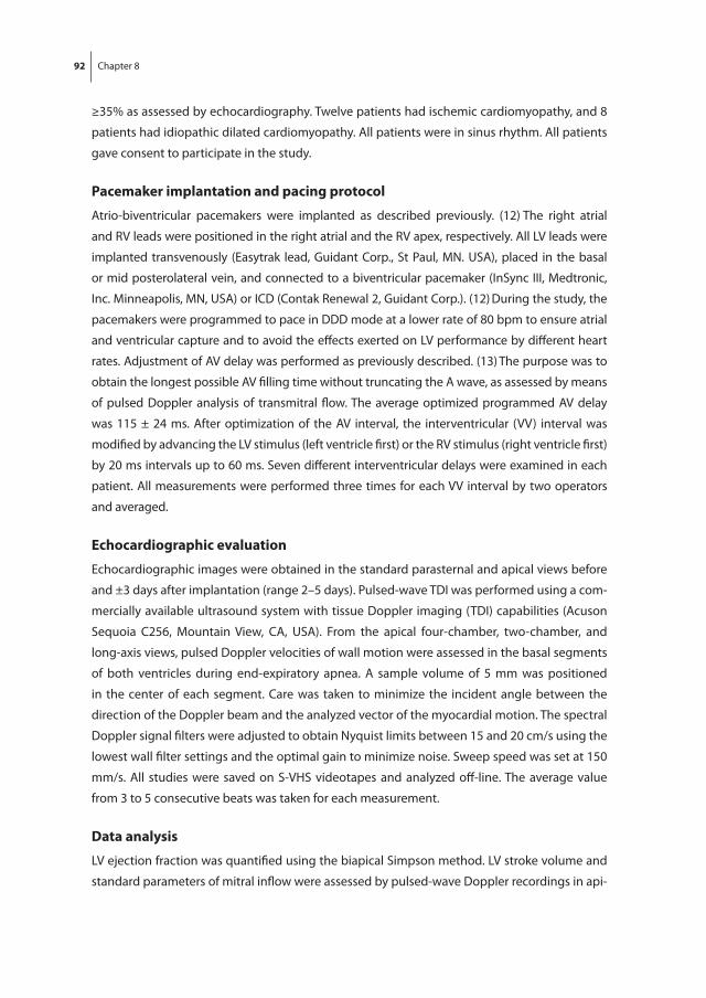
92 Chapter 8
≥35% as as sessed by echocardiography. Twelve patients had ischemic cardiomyopathy, and 8
patients had idiopathic dilated car diomyopathy. All patients were in sinus rhythm. All patients
gave consent to participate in the study.
pacemaker implantation and pacing protocol
Atrio-biventricular pacemakers were implanted as described previously. (12) The right atrial
and RV leads were positioned in the right atrial and the RV apex, respectively. All LV leads were
implanted transvenously (Easytrak lead, Guidant Corp., St Paul, MN. USA), placed in the basal
or mid posterolateral vein, and connected to a biventricular pace maker (InSync III, Medtronic,
Inc. Minneapolis, MN, USA) or ICD (Contak Renewal 2, Guidant Corp.). (12) During the study, the
pacemakers were programmed to pace in DDD mode at a lower rate of 80 bpm to ensure atrial
and ven tricular capture and to avoid the effects exerted on LV performance by different heart
rates. Adjustment of AV delay was performed as previously described. (13) The pur pose was to
obtain the longest possible AV filling time without truncating the A wave, as assessed by means
of pulsed Doppler analysis of transmitral flow. The average optimized programmed AV delay
was 115 ± 24 ms. After optimization of the AV interval, the interventricular (VV) interval was
modified by advancing the LV stimulus (left ventricle first) or the RV stimulus (right ventricle first)
by 20 ms intervals up to 60 ms. Seven different interventricular delays were examined in each
patient. All measurements were performed three times for each VV interval by two operators
and averaged.
Echocardiographic evaluation
Echocardiographic images were obtained in the standard parasternal and apical views before
and ±3 days after im plantation (range 2–5 days). Pulsed-wave TDI was per formed using a com-
mercially available ultrasound system with tissue Doppler imaging (TDI) capabilities (Acuson
Sequoia C256, Mountain View, CA, USA). From the apical four-chamber, two-chamber, and
long-axis views, pulsed Doppler velocities of wall motion were assessed in the basal segments
of both ventricles during end-expiratory apnea. A sample volume of 5 mm was positioned
in the center of each segment. Care was taken to minimize the incident angle between the
direction of the Doppler beam and the analyzed vector of the myocardial motion. The spectral
Doppler sig nal filters were adjusted to obtain Nyquist limits between 15 and 20 cm/s using the
lowest wall filter settings and the optimal gain to minimize noise. Sweep speed was set at 150
mm/s. All studies were saved on S-VHS videotapes and analyzed off-line. The average value
from 3 to 5 consecu tive beats was taken for each measurement.
data analysis
LV ejection fraction was quantified using the biapical Simpson method. LV stroke volume and
standard parameters of mitral inflow were assessed by pulsed-wave Doppler re cordings in api-

Biventricular delay optimization after cardiac resynchronization 93
cal views. Mitral regurgitation was graded on a point scale from 0 to 4 using color flow mapping.
(14)
Indices of AV dyssynchrony calculated were LV filling time and LV filling time corrected for
heart rate. Cardiac dyssynchrony was assessed from measurements of time intervals between
onset of the QRS complex and the begin ning of regional velocity of myocardial shortening,
which is considered a surrogate for regional electromechanical cou pling intervals. Intraven-
tricular delay was assessed by pulsed-wave TDI as the difference between the longest and the
shortest time interval between onset of the QRS com plex and peak systolic myocardial veloc-
ity in the six basal segments of the left ventricle, that is, septal, anteroseptal, lateral, inferior,
anterior, and posterior segments. (15,16) The interventricular delay was determined by the
difference be tween the time to opening of aortic and pulmonic valves and by the difference
between electromechanical coupling times in the basal lateral segment of the RV and in the
most delayed LV segment using TDI. The combined index of intraventricular and interventricular
mechanical dyssyn chrony was calculated by adding both numbers: Total dys synchrony (sum) =
Interventricular delay + Intraventricu lar delay. (14) Cardiac systolic performance was quantified
by measuring the LV outflow tract velocity-time integral. The hemodynamic impact of the
programmed AV/VV interval was derived from the velocity-time integral and LV filling time
echocardiographic parameters, which are easy to ob tain. In addition, steady-state velocity-time
integral and LV filling time are rapidly achieved, thus enabling study of numerous settings in a
short interval. At 6-month follow-up, responders to CRT were identi fied by a relative increase in
LV ejection fraction or de crease in LV end-diastolic diameter ≥25% vs baseline. This cutoff value
is >2 SD above the changes of both parameters observed after administration of placebo in
randomized tri als with beta-blockers. (14)
statistical analysis
All results are given as mean ± SD. Student’s t-test, Mann-Whitney test, and Spearman cor-
relation coefficient were used for appropriate comparisons. For each protocol step and for
each patient, LV outflow tract velocity-time integral and LV filling time measurements were
compared by anal ysis of variance (Tukey test for repeated measures). Statis tical significance
was set at a two-tailed P < .05.
rEsults
preimplantation data
Twenty patients (average age 66 ± 6 years; NYHA heart failure class 3.2 ± 1.1) were included in
the study. All patients had significant intraventricular delay (70 ± 28 ms), interventricular delay
(65 ± 32 ms), and total dyssynchrony (135 ± 55 ms). Baseline clinical and ECG characteristics
of the patients enrolled in the study are given in Table 1. LV lead implantation was successful

94 Chapter 8
in all patients. One LV lead was placed in the posterior wall; 19 leads were posi tioned in the
posterolateral wall.
biventricular activation and av optimization
Simultaneous biventricular pacing resulted in a significant improvement in LV performance as
demonstrated by increased velocity-time integral (P < .001), increased LV filling time (P = .001),
and trend toward higher LV filling time corrected for heart rate (P = .174; Table 2). Mitral regur-
gitation decreased from 1.8 ± 1.1 to 1.0 ± 0.8 (P = .005). This hemodynamic improvement was
associated with a significant reduction in the degrees of interventricular delay (P = .003) and the
total amount of dyssynchrony (P = .003). There was a trend toward lower intraventricular delay
following simultaneous biventricular activation (P = .054; Table 2).
table 1. Baseline clinical and electrocardiographic parameters of the study population
Gender (male/female) 17/3
Age(years) 66 ± 6
ICMP/DCMP (%) 67%/33%
NYHA functional class 3.2 ± 1.1
EF (%)LVEDD (mm)
22 ± 1876 ± 6
QRS (msec)PR-interval (msec)
200 ± 32187 ± 42
HR (bpm)ACE-I n (%)Diuretics n (%)ß-blockers n (%)Aldosterone-antagonists n (%)
66 ± 1618 (100)18 (100)14 (78)15 (83)
Values are given as mean ± SD or number of patients (percentage).
table 2. Echocardiographic parameters before implantation and during simultaneous biventricular pacing after AV optimization in all patients (n = 20)
Pre-implantationn = 20
Simultaneous BiV pacingn = 20
p
VTI (mm) 74 ± 15 115 ± 30 < 0.001
HR (bpm) 66 ± 16 71 ± 9 0.165
LVFT (msec) 306 ± 82 395 ± 96 0.001
LVFT/HR (msec/bpm) 5.1 ± 2.1 5.8 ± 1.9 0.174
IVD (msec) 65 ± 32 38 ± 31 0.003
VD (msec) 70 ± 28 48 ± 32 0.054
Total asynchrony (msec) 135 ± 55 85 ± 46 0.003
MR (grade) 1.8 ± 1.1 1.0 ± 0.8 0.005
Values are expressed as mean ± SD. HR = heart rate; IVD = interventricular delay; LVFT = left ventricular filling time; LVFT/HR = left ventricular filling time corrected for heart rate; MR = mitral regurgitation; VD = intraventricular delay; VTI = left ven-tricular outflow tract velocity-time integral.

Biventricular delay optimization after cardiac resynchronization 95
Figure1.Chapter 8 Figure 1
Chapter 8 Figure 1
Velocity-time integral (VTI) and left ventricular filling time (LVFT) at baseline, during simultaneous biventricular pacing, and during sequential biventricular pacing (n = 17). CRT = cardiac resynchronization therapy.
Figure 2.
Figure 2
Figure 2
Figure 2
Interventricular delay (IVD), intraventricular delay (VD), and sum dyssynchrony at baseline, during simultaneous biventricular pacing, and during sequential biventricular pacing (n = 17). CRT = cardiac resynchronization therapy.

96 Chapter 8
sequential vs simultaneous biventricular activation
In 17 patients, hemodynamics were further ameliorated by adjusting the VV interval (Figure
1), whereas in 3 patients simultaneous biventricular activation was superior to se quential
biventricular pacing. In the 17 patients, optimized sequential biventricular pacing resulted in
a significant in crease in velocity-time integral (P < .001), an increase in LV filling time (P = .001),
and a decrease in interventric ular delay (P = .013) and intraventricular delay (P = .010) with
respect to simultaneous biventricular activation (Table 3). Accordingly, the total amount of
dyssynchrony signifi cantly decreased during sequential CRT ( P = .002) com pared with simul-
taneous activation (Figure 2). No change in severity of mitral regurgitation was noted during
se quential compared with simultaneous biventricular pacing (Table 3).
There was no “standard” optimal VV delay for all pa tients, and no difference was noted in
optimal RV–LV delay between patients with ischemic cardiomyopathy and those with idio-
pathic dilated cardiomyopathy. The best VV option was “LV first” in 12 patients and “RV first” in
5 patients. However, there were trends toward higher baseline PR interval (195 ± 45 vs 150 ±
25 ms; P = .06) and higher degree of total amount of dyssynchrony (142 ± 48 vs 127 ± 34; P =
.05) in patients who benefited from VV optimization compared with those who benefited from
simultaneous biventricular pacing. Figure 3 shows typical Doppler tracings obtained before
CRT, during simultaneous biventricular pacing, and during sequential biventricular pacing in
an individual pa tient.
six-month Follow-up
At 6-month follow-up, there was a significant improvement in ejection fraction from 22% ±
18% to 34% ± 18% (P < .001) and a decrease in LV end-diastolic diameter from 76 ± 6mm to 71
± 8mm (P = .001). This hemo dynamic benefit was associated with a significant improve ment in
table 3. Echocardiographic parameters before and after VV optimization (n = 17)
SimultaneousBiV pacing
n = 17
Optimal V-V intervaln = 17
p
VTI (mm) 122 ± 31 154 ± 42 < 0.001
HR (bpm) 71 ± 10 70 ± 10 0.410
LVFT (ms) 404 ± 102 472 ± 110 0.001
LVFT/HR (ms/bpm) 6.0 ± 2.0 7.1 ± 2.2 0.001
IVD (msec) 35 ± 33 13 ± 25 0.013
VD (msec) 51 ± 34 34 ± 18 0.010
Total asynchrony (msec) 86 ± 49 47 ± 31 0.002
MR 1.1 ± 0.7 1.1 ± 0.8 0.252
Values are expressed as mean ± SD. HR = heart rate; IVD = interventricular delay; LVFT = left ventricular filling time; LVFT/HR = left ventricular filling time corrected for heart rate; MR = mitral regurgitation; VD = intraventricular delay; VTI = left ven-tricular outflow tract velocity-time integral.

Biventricular delay optimization after cardiac resynchronization 97
NYHA class from 3.2 ± 1.1 to 1.8 ± 1.2 (P = .001). Interestingly, the incidence of nonresponders
was 10%, which is lower than the number reported in the liter ature.
Reproducibility The interobserver variability for measurement of LV dys synchrony in 10 study subjects was
8.5% and for LV–RV dyssynchrony was 7.2%. The intraobserver variability was 6.8% for LV dys-
synchrony and 6.1% for LV–RV dyssyn chrony. (17)
disCussion
The present study demonstrates that sequential biventricular pacing results in further improve-
ment of LV performance over simultaneous biventricular pacing in a subgroup of patients with
Figure 3.Figure 3
Figure 3 Doppler echocardiography of the aorta (Ao), pulmo nary artery (PA), and left ventricular outflow tract (LVOT) during simultaneous (left) and sequential biventricular pacing (right). Adjusting the VV delay to -40 ms results in a higher LVOT velocity-time integral (VTI) together with less interventricular dyssynchrony as evidenced by the lower interventricular delay (IVD). CW = continuous wave; PW = pulsed wave; ↔ = inter-ventricular delay (IVD).

98 Chapter 8
advanced heart failure and left bundle branch block morphology. Sequential biventricular
pacing resulted in more homogenous activation of the ventricles as evi denced by prolongation
of LV filling time and reduction in interventricular and intraventricular dyssynchrony. Finally,
optimization of interventricular delay favorably affects the remodeling process as evidenced
by the low incidence of nonresponders at 6 months. Therefore, individual echocar diography-
guided VV interval programming seems advis able in order to maximize the benefit of CRT.
sequential vs simultaneous stimulation
Although conventional simultaneous CRT ameliorated LV performance compared with sinus
rhythm, we demonstrated that LV hemodynamics can be further improved by individ ually
programming the VV interval to the optimal pacing mode in at least 80% of cases. The reported
26% increase in velocity-time integral by tailored VV interval adjustment corroborates previ-
ous observations. (8,9) Of note, a reduction in the extent of myocardium displaying delayed
longitudinal contraction, together with an increase in LV ejection frac tion by tissue tracking
and three-dimensional echocardiog raphy (8) and improvement in hemodynamic markers
such as dP/dt (9,10) and myocardial performance index, (18) were noted following sequential
biventricular pacing.
Adjustment of the VV interval not only improved the interventricular dyssynchrony but
also positively influenced intraventricular dyssynchrony, resulting in more homoge neous
activation of the LV with faster LV emptying, thereby increasing the time available for LV filling.
The observed prolongation of LV filling time indicates that improvement of LV dyssynchrony
also hastens LV relaxation and im proves AV mechanics and diastolic LV performance. (19)
In terestingly, patients with longer PR intervals at baseline tended to have more dyssynchrony
and benefited more from VV optimization. This finding indirectly suggests that in addition to
left bundle branch block, the presence of a prolonged PR interval might be a major indicator of
severe conduction delay and contribute to mechanical dyssyn chrony.
optimal vv interval
The range of optimal VV intervals in a particular patient was narrow, with small changes result-
ing in large differ ences in LV hemodynamics. This is in contrast with the optimal AV interval,
where mechanical responses are sim ilar over a broad range of AV timings. (20) In most patients,
advancing LV activation was superior, which seems logical because dyssynchrony results in
delayed emptying of the LV compared with the RV. However, in a subset of patients, RV preacti-
vation or simultaneous activation of both ventri cles resulted in the best hemodynamic profile.
Two mech anisms account for this observation. First, differences in baseline ventricular conduc-
tion and anisotropic differences due to conduction system abnormalities or myocardial scars
between heart failure patients interfere with mechanical activation and therefore affect the
various timing intervals. (6) Second, the exact positioning of LV leads varies depending on the
operator’s choice and the coronary sinus anatomy. This situation leads to a variety of ventricular

Biventricular delay optimization after cardiac resynchronization 99
activation patterns among patients. The closer the lead is to the latest activated portion of the
LV segment, the higher the mechan ical effect on resynchronization and the greater the clinical
benefit. On the contrary, pacing the already early activated segment may further deteriorate LV
function, as reported by Butter et al. (21)
Long-term effects of optimized VV interval Similar to other studies, CRT was associated with a signifi cant improvement in hemodynamic
and clinical parame ters. However, the incidence of nonresponders was 10%, which appears
to be much lower than the 30% reported in literature. (1–3) The long-term benefits might be
attributed to the greater reduction in interventricular and total dyssyn chrony. In the long term,
these features could result in more pronounced reverse remodeling observed following VV
in terval adjustment.
study limitations
Previous observations reporting the superiority of left uni ventricular pacing over biventricular
pacing could not be confirmed. (4,22) Although we did not specifically test univen tricular
stimulation, none of our patients had the best he modynamic response with the relative earliest
activation of
LV. Our observation corroborates data of Perego et al, (9) who speculated that sequential
stimulation with the relative ear liest activation of LV and right bundle branch block ECG pat-
tern achieves hemodynamic results more similar to those of left univentricular stimulation than
those obtained with a less negative VV interval. Only a direct comparison be tween sequential
CRT vs left univentricular stimulation can address this issue.
The purpose of CRT is to reestablish synchronous car diac contraction. Therefore, optimized
resynchronization of the heart is expected to yield a better clinical response during follow-up.
The lower number of nonresponders and the beneficial effects upon remodeling parameters
observed at 6 months in our study indirectly suggest that acute echo-cardiographic VV interval
adjustment may be helpful to achieve this goal. Nevertheless, the concept that sequential CRT
is superior to standard CRT and that acute echocar diographic optimization of the VV interval
translates into long-term clinical benefit must be evaluated in a prospective double-blind
randomized trial.
Because these data were obtained at a single heart rate, at rest with the patient in a
recumbent position and at a single point in time, extrapolation of these data requires caution.
Prospective studies evaluating the impact of exercise and position on the “optimal” VV interval
are mandatory.
Clinical implications
Identification of optimal candidates for CRT remains a high priority. Although initially focusing
on electrical markers, data have highlighted the value of more direct assessment of mechanical
100 Chapter 8
dyscoordination by tissue Doppler, (23) echo Doppler, (24) or magnetic resonance imaging.
Nevertheless, despite all these selection criteria, evidence from clinical trials suggest that 20%
to 30% of patients who receive CRT do not respond to the therapy. (2,25) Reasons for failure
to respond include insufficient ventricular dyssynchrony at baseline, overriding comorbidities
that attenuate the benefit of CRT, failure to resynchronize LV function because of inadequate
lead implant, and suboptimal device program ming. Based upon our observation, we speculate
that the rate of responders can be increased not only by better selection of patients but also
by more accurate programming of the device. The present study highlights the importance of
the VV interval, especially in patients with questionable benefit after implantation. However,
prospective studies are needed to address the issue.
ConClusion
This study demonstrates that adjustment of interventricular pacing intervals further improves
cardiac performance. This technique can be helpful in tracking the progress of re sponse of an
individual patient to therapy. Because of the important hemodynamic consequences of opti-
mization of VV interval and the high rate of nonresponders reported in literature, we recom-
mend tailoring the interventricular in terval in every patient referred for CRT. Randomized trials
evaluating the potential benefit of VV optimization in daily life are warranted.
aCKnowlEdgmEnts
We thank Peter Goemaere and the staff of the Echocardio graphic Laboratory for excellent
technical assistance through out the study.

Biventricular delay optimization after cardiac resynchronization 101
rEFErEnCEs
1. Cazeau S, Leclercq C, Lavergne T, Walker S, Varma C, Linde C, Garrigue S, Kappenberger L, Haywood GA, Santini M, Bailleul C, Daubert JC. Effects of multisite biventricular pacing in patients with heart failure and intraventricular conduction delay. N Engl J Med 2001;344:873–880.
2. Abraham WT, Fisher WG, Smith AL, Delurgio DB, Leon AR, Loh E, Kocovic DZ, Packer M, Clavell AL, Hayes DL, Ellestad M, Trupp RJ, Underwood J, Pickering F, Truex C, McAtee P, Messenger J. Cardiac resynchronization in chronic heart failure. N Engl J Med 2002;346: 1845–1853.
3. Gras D, Leclercq C, Tang AS, Bucknall C, Luttikhuis HO, Kirstein-Pedersen A. Cardiac resynchroniza-tion therapy in advanced heart failure the multicenter InSync clinical study. Eur J Heart Fail 2002;4: 311–320.
4. Nelson GS, Berger RD, Fetics BJ, Talbot M, Spinelli JC, Hare JM, Kass DA. Left ventricular or biventricular pacing improves cardiac function at diminished energy cost in patients with dilated cardiomyopathy and left bundle-branch block. Circulation 2000;102:3053–3059.
5. Breithardt OA, Sinha AM, Schwammenthal E, Bidaoui N, Markus KU, Franke A, Stellbrink C. Acute effects of cardiac resynchronization therapy on functional mitral regurgitation in advanced systolic heart failure. J Am Coll Cardiol 2003;41:765–770.
6. Leclercq C, Kass DA. Retiming the failing heart: principles and cur rent clinical status of cardiac resyn-chronization. J Am Coll Cardiol 2002;39:194–201.
7. Cazeau S, Bordachar P, Jauvert G, Lazarus A, Alonso C, Vandrell MC, Mugica J, Ritter P. Echocardio-graphic modeling of cardiac dyssyn chrony before and during multisite stimulation: a prospective study. Pacing Clin Electrophysiol 2003;26:137–143.
8. Sogaard P, Egeblad H, Pedersen AK, Kim WY, Kristensen BO, Han sen PS, Mortensen PT. Sequential versus simultaneous biventricular resynchronization for severe heart failure: evaluation by tissue Doppler imaging. Circulation 2002;106:2078–2084.
9. Perego GB, Chianca R, Facchini M, Frattola A, Balla E, Zucchi S, Cavaglia S, Vicini I, Negretto M, Osculati G. Simultaneous vs. se quential biventricular pacing in dilated cardiomyopathy: an acute he modynamic study. Eur J Heart Fail 2003;5:305–313.
10. van Gelder BM, Bracke FA, Meijer A, Lakerveld LJ, Pijls NH. Effect of optimizing the VV interval on left ventricular contractility in cardiac resynchronization therapy. Am J Cardiol 2004;93:1500–1503.
11. Abraham WT. Rationale and design of a randomized clinical trial to assess the safety and efficacy of cardiac resynchronization therapy in patients with advanced heart failure: the Multicenter InSync Random ized Clinical Evaluation (MIRACLE). J Card Fail 2000;6:369–380.
12. Daubert JC, Ritter P, Le Breton H, Gras D, Leclercq C, Lazarus A, Mugica J, Mabo P, Cazeau S. Permanent left ventricular pacing with transvenous leads inserted into the coronary veins. Pacing Clin Elec-trophysiol 1998;21:239–245.
13. Ritter P, Padeletti L, Gillio-Meina L, Gaggini G. Determination of the optimal atrioventricular delay in DDD pacing. Comparison between echo and peak endocardial acceleration measurements. Europace 1999;1:126–130.
14. Bristow MR, O’Connell JB, Gilbert EM, French WJ, Leatherman G, Kantrowitz NE, Orie J, Smucker ML, Mar-shall G, Kelly P, et al. Dose-response of chronic beta-blocker treatment in heart failure from either idio-pathic dilated or ischemic cardiomyopathy. Bucindolol In vestigators. Circulation 1994;89:1632–1642.
15. Ansalone G, Giannantoni P, Ricci R, Trambaiolo P, Fedele F, Santini M. Doppler myocardial imaging to evaluate the effectiveness of pacing sites in patients receiving biventricular pacing. J Am Coll Cardiol 2002;39:489–499.
16. Pellerin D, Berdeaux A, Cohen L, Giudicelli JF, Witchitz S, Veyrat C. Pre-ejectional left ventricular wall motions studied on conscious dogs using Doppler myocardial imaging: relationships with indices of left ventricular function. Ultrasound Med Biol 1998;24:1271–1283.

102 Chapter 8
17. Penicka M, Bartunek J, De Bruyne B, Vanderheyden M, Goethals M, De Zutter M, Brugada P, Geelen P. Improvement of left ventricular function after cardiac resynchronization therapy is predicted by tissue Doppler imaging echocardiography. Circulation 2004;109:978–983.
18. Porciani MC, Dondina C, Macioce R, Demarchi G, Pieragnoli P, Musilli N, Colella A, Ricciardi G, Michelucci A, Padeletti L. Echo-cardiographic examination of atrioventricular and interventricular de lay optimization in cardiac resynchronization therapy. Am J Cardiol 2005;95:1108–1110.
19. Saxon LA, De Marco T, Schafer J, Chatterjee K, Kumar UN, Foster E. Effects of long-term biven-tricular stimulation for resynchronization on echocardiographic measures of remodeling. Circulation 2002;105: 1304–1310.
20. Auricchio A, Stellbrink C, Block M, Sack S, Vogt J, Bakker P, Klein H, Kramer A, Ding J, Salo R, Tockman B, Pochet T, Spinelli J. Effect of pacing chamber and atrioventricular delay on acute systolic function of paced patients with congestive heart failure. The Pacing Therapies for Congestive Heart Failure Study Group. The Guidant Congestive Heart Failure Research Group. Circulation 1999;99:2993–3001.
21. Butter C, Auricchio A, Stellbrink C, Fleck E, Ding J, Yu Y, Huvelle E, Spinelli J. Effect of resynchroniza-tion therapy stimulation site on the systolic function of heart failure patients. Circulation 2001;104: 3026–3029.
22. Auricchio A, Ding J, Spinelli JC, Kramer AP, Salo RW, Hoersch W, KenKnight BH, Klein HU. Cardiac resyn-chronization therapy restores optimal atrioventricular mechanical timing in heart failure patients with ventricular conduction delay. J Am Coll Cardiol 2002;39:1163–1169.
23. Yu CM, Chau E, Sanderson JE, Fan K, Tang MO, Fung WH, Lin H, Kong SL, Lam YM, Hill MR, Lau CP. Tissue Doppler echocardio graphic evidence of reverse remodeling and improved synchronicity by simultaneously delaying regional contraction after biventricular pacing therapy in heart failure. Circulation 2002;105:438 –445.
24. Pitzalis MV, Iacoviello M, Romito R, Massari F, Rizzon B, Luzzi G, Guida P, Andriani A, Mastropasqua F, Rizzon P. Cardiac resynchro nization therapy tailored by echocardiographic evaluation of ventric ular asynchrony. J Am Coll Cardiol 2002;40:1615–1622.
25. Stellbrink C, Breithardt OA, Franke A, Sack S, Bakker P, Auricchio A, Pochet T, Salo R, Kramer A, Spinelli J. Impact of cardiac resynchro nization therapy using hemodynamically optimized pacing on left ven-tricular remodeling in patients with congestive heart failure and ventricular conduction disturbances. J Am Coll Cardiol 2001;38: 1957–1965.

part iiiCardiaC rEsynChronization thErapy: how
to improvE implantation tEChniquEs


Chapter 9 potEntial appliCations oF magnEtiC
navigation in CliniCal ElECtrophysiology
L. Jordaens, M. Rivero-Ayerza and A. Thornton
Eur Heart J. – E Journal 2005; 3(vol. 40)
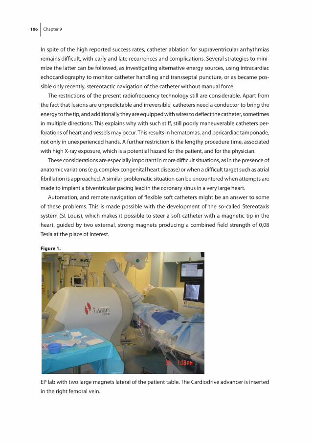
106 Chapter 9
In spite of the high reported success rates, catheter ablation for supraventricular arrhythmias
remains difficult, with early and late recurrences and complications. Several strategies to mini-
mize the latter can be followed, as investigating alternative energy sources, using intracardiac
echocardiography to monitor catheter handling and transseptal puncture, or as became pos-
sible only recently, stereotactic navigation of the catheter without manual force.
The restrictions of the present radiofrequency technology still are considerable. Apart from
the fact that lesions are unpredictable and irreversible, catheters need a conductor to bring the
energy to the tip, and additionally they are equipped with wires to deflect the catheter, sometimes
in multiple directions. This explains why with such stiff, still poorly maneuverable catheters per-
forations of heart and vessels may occur. This results in hematomas, and pericardiac tamponade,
not only in unexperienced hands. A further restriction is the lengthy procedure time, associated
with high X-ray exposure, which is a potential hazard for the patient, and for the physician.
These considerations are especially important in more difficult situations, as in the presence of
anatomic variations (e.g. complex congenital heart disease) or when a difficult target such as atrial
fibrillation is approached. A similar problematic situation can be encountered when attempts are
made to implant a biventricular pacing lead in the coronary sinus in a very large heart.
Automation, and remote navigation of flexible soft catheters might be an answer to some
of these problems. This is made possible with the development of the so-called Stereotaxis
system (St Louis), which makes it possible to steer a soft catheter with a magnetic tip in the
heart, guided by two external, strong magnets producing a combined field strength of 0,08
Tesla at the place of interest.
Figure 1.
Chapter 9 Figure 1
Figure 1. EP lab with two large magnets lateral of the patient table. The Cardiodrive advancer is inserted in the right femoral vein.
Figure 2 EP lab with two large magnets lateral of the patient table. The Cardiodrive advancer is inserted
in the right femoral vein.

Potential applications of magnetic navigation in electrophysiology 107
In an experimental set up the tip prolapse push force on catheters was reduced from 240 to
132 g; the tip curl force (for alignment) was reduced from 45 to 15 g. This indicates indeed how
the safety profile of this approach is very promising. The potential to advance and retract the
catheter via robotics, or with a simpler system CardiodriveTM , was also developed by Stereo-
taxis. This combination permits perfect remote control over the catheter, and should result in
less radiation for the physician.
ElECtrophysiology (Ep) studiEs
It was shown that all basic steps for a diagnostic EP study can be taken with the described prin-
ciple of magnetic navigation. If one wants to combine pacing and recording, a conventional
catheter has to be inserted as well.
CathEtEr ablation
Supraventricular tachycardia
We completed our learning curve for AVNRT, an arrhythmia with a simple anatomy in about
20 patients. The results were just as good as with conventional radiofrequency, or cryotherapy
(table 1), with considerably less radiation for the patient as well. Due to the magnetic field,
less catheter movement with cardiac and respiratory cycles and during junctional rhythm were
seen as with conventional approaches. We have used the system for ectopic and incisional atrial
tachycardia and right and left sided accessory pathways. We used a retrograde approach for
the latter, even with the actual catheters, which are floppy over the entire length, what creates
some difficulties at the aortic arch and valve. Others have preferred the transseptal route, which
may result in better appositioning of the catheter in the left lateral region.
Ventricular tachycardia
Ventricular tachycardia of non-ischemic origin can be addressed. In RV outflow tract tachycardia,
conventional ablation poses some difficulties, as two opposite curves have to be accomplished,
table 1. Comparison of ablation variables in AVNRT, using 3 different techniques
Magnet ablation Cryotherapy RF
Number of ablations (median (range))Total ablation time (s) (median (range))
6 (1-22) 5 (2-16) 7 (2-28)
240 (60-633) 633 (452-2599) 290 (90-895)
Success 19/ 20 12/12 5/5
Procedure time (min) (median (mean±sd)) 163 (167±46) 148 (167±65) 159 (177±61)
Fluoroscopy time patient (min) (median (mean±sd))
12 (17±12) 17 (20±11) 30 (30±8.8)

108 Chapter 9
and precise mapping has to be performed in a relatively small area. This makes precision move-
ment difficult, and often induces non-relevant arrhythmias. We have approached both left and
right-sided idiopathic VT with magnetic navigation. This allows small steps, and very precise
mapping with the floppy magnet catheter.
Atrial fibrillation (AF)
AF is the real challenge today. Some attempts were made by Ernst et al to achieve pulmonary
vein isolation. People tend to accept now that a wide circumferential approach is the best way
to proceed. Pappone recently presented the first cases with integrated CARTO imaging, result-
ing in successful ablation of the AF substrate. The perspective should be that even the less
invasive retrograde transaortic - transmitral approach should be considered.
Figure 2.
Figure 3
Figure 3
Figure 3
Left ventricular fascicular tachycardia. The upper panel shows the isolines in RAO and LAO, as acquired with the RPM mapping system, showing the apico-septal origin of the arrhythmia, with reference catheters in the coronary sinus and the right ventricular apex. The lower panel shows how the magnetic ablation lead (Helios 2, stereotaxis Inc) is moved from the yellow to the green line by magnetic forces. Here was a fascicular potential, and ablation was performed at this site. As both systems are not integrated, some imagination is left to the researcher. The angulations in RAO are not exactly the same.

Potential applications of magnetic navigation in electrophysiology 109
Complex congenital heart disease.
Especially corrected complex congenital heart disease is frequently associated with arrhyth-
mias. These can often be very difficult to control with conventional means. Reconstructed
chambers, recesses, patches, and tubes make catheter manipulation and ablation very difficult.
If there is a place for magnetic navigation, it is in these dilated, scarred tissues of operated aging
hearts. Integration of anatomy, voltage mapping and activation mapping should assist in more
performant ablation.
Advanced cardiac mapping
Electro-anatomic maps to guide the ablation process can now automated, more easily, be
constructed aided by this remote technology. This will shorten the procedure time, and assist
in assessing the success of otherwise lengthy procedures as pulmonary vein isolation.
Figure 3.
Figure 3
Figure 3
At the left designer lines as suggested by the investigator using the Navigant software (Stereotaxis Inc). At the right the CARTO map showing how these lines contributed to an electroanatomic map in sinus rhythm.
Figure 4.
Figure 4
At the right coronary sinus venogram using dedicated software to reconstruct a 3D image (at the left) what can be used to direct the guide wire.

110 Chapter 9
Attempts are now undertaken to merge (old) MRI and multislice CT images in on-line electro-
anatomic maps. The overlay seems acceptable. All want to believe hat this is progress. Real
image integration in the EP domain will be realized when on-line integration of a 3-dimensional
image with the online EP data becomes a fact. This will probably be realized with 3D echo, but if
developments in X-ray and MRI are fast, these techniques have a chance to be involved as well.
CardiaC rEsynChronisation thErapy (Crt)
Implanting a biventricular pacing lead in the coronary sinus (CS) can be very time consuming.
These procedures can require extensive fluoroscopic screening. This is partly due to difficulties
in cannulating the CS, and once the guide wire or the lead is in the CS, partly to attempts to
reach the potentially best side branch, and finally because the lead has to remain there after
it is advanced over the magnetic guide wire, and when the guide wire is retracted. Further,
complications as dissection of the CS and pericardiac tamponade exist. The idea to cannulate
the CS with a sheath into the mid right atrium or without a sheath at all should be tested with
dedicated guide wires.
Further, target side branches should be reached with the assistance of magnetic navigation,
once the vessel is engaged. Magnetic force should be able to keep the guide wire in position
when advancing the pacing wire. Most of these principles were tested already in our lab, and
hold great promise.
FuturE ConCEpts
It is expected that with the incorporation of catheter registration technology or echocardiog-
raphy to this system or using these technologies in parallel procedure and radiation times will
decrease. Combining this technology with other new technology such as cryotherapy and the
other mapping technologies mentioned above, may significantly improve outcome, as well as
reducing the number of applications, and associated collateral tissue damage. The potential
for other areas in cardiology is there: stem cell therapy, difficult coronary artery procedures,
congenital heart disease. Even when this system is only a first step on the road to performant
magnetic navigation, the tested principles so far seem to confirm that the concept is valid, and
should be considered a milestone in the development of safer and automated procedures.

Potential applications of magnetic navigation in electrophysiology 111
rEFErEnCEs
1. Hindricks G. The Multicentre European Radiofrequency Survey (MERFS): complications of radiofre-quency catheter ablation of arrhythmias. The Multicentre European Radiofrequency Survey (MERFS) investigators of the Working Group on Arrhythmias of the European Society of Cardiology. Eur Heart J 1993;14:1644-53.
2. Faddis MN, Blume W, Finney J, et al. Novel, magnetically guided catheter for endocardial mapping and radiofrequency catheter ablation. Circulation 2002;106:2980-5.
3. Faddis MN, Chen J, Osborn J, et al. Magnetic guidance system for cardiac electrophysiology: a pro-spective trial of safety and efficacy in humans. J Am Coll Cardiol 2003;42:1952-8.
4. Ernst S, Ouyang F, Linder C, et al. Initial experience with remote catheter ablation using a novel magnetic navigation system: magnetic remote catheter ablation. Circulation 2004;109:1472-5.
5. Ernst S, Ouyang F, Linder C, et al. Modulation of the slow pathway in the presence of a persistent left superior caval vein using the novel magnetic navigation system Niobe. Europace 2004;6:10-4.
6. Thornton AS, Alings M, Scholten MF, Jordaens LJ. Left ventricular lead placement within a coronary sinus side branch, using only a floppy guide wire and magnetic navigation. Heart 2005;91;22.
7. Rivero-Ayerza M, Thornton A, Scholten M, Mekel J, Res J, Theuns D. and Jordaens L. Left ventricular lead placement within a coronary sinus side branch is feasible using remote magnetic navigation of a guide wire (abstract). Heart Rhythm 2005; 2: S 281.


Chapter 10 lEFt vEntriCular lEad plaCEmEnt
within a Coronary sinus sidE branCh
using rEmotE magnEtiC navigation oF
a guidEwirE: a FEasibility study
M. Rivero-Ayerza, A. Thornton, D. Theuns, M. Scholten, J. Mekel, J. Res and L. Jordaens.
J Cardiovasc Electrophysiol. 2006 Feb;17(2):128-33

114 Chapter 10
abstraCt
Background:
A novel magnetic navigation system (MNS) allowing remote guidance of catheters and guide-
wires might assist in implantation of left ventricular (LV) pacing leads.
Objective:
To assess the feasibility of deploying a LV pacing lead into a coronary sinus (CS) side branch using
a magnetically guided wire and of performing the procedure without a CS guiding sheath.
Methods:
Twenty-one patients were included in this study. Nine underwent CRT device implantation using
a magnetic navigation system to steer the guide-wire (MNS group) while 12 patients were con-
ventionally implanted (control group). In 6 pts in the MNS group, the procedure was performed
using a CS guiding sheath. In 3 others the decision was to perform the procedure without a CS
sheath. In these patients the wire was advanced manually, while the external magnets oriented it
towards the CS os. In the CS, “vector based” navigation was used to guide the wire to the desired
side branch.
Results:
In all 9 patients in the MNS group the target vessel could be successfully engaged by the mag-
netically guided wire. In 7, the LV lead was lodged in the target vessel. In 2 patients the LV lead was
repositioned in an anterolateral side branch due to instability or inability to engage the vessel with it.
Mean total procedure time was 164 ± 58 min (without sheath 229 ± 52 vs with sheath 132 ± 26 min;
p=0.007). Mean fluoroscopy time was 28 ± 9 min. For control pts the procedure and fluoroscopy
time were similar (144 ± 41 min and 26 ± 12 min, respectively). No major complications occurred.
Conclusion:
LV lead implantation can be performed using a remote-magnetically steered guide-wire.
Though the lead could be implanted without a CS guiding sheath, longer procedure times were
required.

Magnetically guided LV lead implantation 115
A novel magnetic navigation system has been designed with the purpose of allowing remote
guidance of soft radiofrequency ablation catheters and PTCA guide-wires (1). This system is
intended to allow catheters or guide-wires to reach areas or access vessels that would otherwise
be difficult to get to by conventional means. In humans, the system was used in electrophysi-
ological studies and mapping of right and left foci (2), while catheter ablation until now is only
reported for right sided circuits (3,4).
In recent years significant technological progress has been made aiming at improving success
and reducing complication rates during cardiac resynchronization therapy (CRT) device implanta-
tion procedures. Nevertheless, in a proportion of cases, attempts at deploying a left ventricular (LV)
pacing lead through the coronary sinus (CS) still fail (5). Inability to cannulate the CS or accessing
tortuous or highly angulated side branches, and the chances of CS dissection or perforation by the
sheath or lead are some of the reasons why transvenous implantation procedures may fail (5,6).
Further, extracardiac or diaphragmatic stimulation occur and may happen at the most optimal
prospective pacing site. All these reasons can lead to subsequent surgical LV lead positioning in up
to 10% of the patients (7). This study was conducted with the purpose of evaluating the feasibility
of deploying a LV pacing lead into the desired CS side branch by means of magnet navigation to
guide the wire. We also tested if this was possible without a guiding-sheath inserted in the CS.
mEthods
Patient Population
Twenty-one consecutive patients who underwent CRT device implantation were included in this
study, 9 using a remote magnetic navigation system (Stereotaxis Navigant™) (MNS group) as part
of the safety protocol approved by the ethics committee of our institution were included in the
study, and 12 other patients included as a control group. Patients were alternated according to
availability of our catheterization rooms (magnet and standard room). These patients met stan-
dard criteria for CRT, that is, advanced heart failure refractory to medical therapy, low EF (< 35 %)
and a broad QRS (≥120 msec) on the ECG with a left bundle branch block (LBBB)-like morphology.
All patients gave informed consent.
Implantation Procedure - MNS group:
In all 9 patients the left cephalic vein was dissected. A RV shock lead (model 1580 Riata, St. Jude
Medical, Sylman, CA, USA or model 6947 Sprint Quattro Secure, Medtronic Inc, Minneapolis, MN,
USA) was introduced and actively fixed to the right ventricular apical wall. After a double left
subclavian venous puncture a right atrial active fixation lead (model 5076, Medtronic Inc, Min-
neapolis, MN, USA) was introduced and positioned in the right atrial appendage. After testing
pacing and sensing properties of these leads, a 10 FR sheath was introduced in the left subclavian
vein. Through this sheath (in patients 4 to 9) a 9 FR long guiding-sheath (model Attain 6216,

116 Chapter 10
Medtronic Inc, Minnesota, MN, USA) was introduced, in order to cannulate the coronary sinus. In
patients 1, 2 and 3 access to the CS was achieved without a CS sheath (see below).
A CS angiogram was performed in patients 4 to 9. In patients 1 and 2, the image obtained
during the venous phase of a previous coronary angiography was used as an anatomic guide
to navigate within and select the target CS branch (figure 1). In patient 3, a CS angiogram from
a previous conventional approach was used. After analyzing the coronary sinus angiograms the
ideal side branch to place the LV lead was chosen. In most cases this was either a posterolateral
or a lateral branch.
Two external magnets (Stereotaxis, St. Louis, MO, USA) were positioned at each side of the
table in order to generate a magnetic field within the patient. The orientation of the magnetic
field is established by the interaction that the two external magnets exert with each other and
is specified by the operator working remotely in the viewing room. A single magnet inserted in
the tip of a 0.014’’ guide-wire (Cronus Floppy Endovascular Guide wire, Stereotaxis, St. Louis, MO,
USA) aligns itself with the direction of the magnetic field. In this way, by remotely changing the
orientation of the magnets, the vector of the magnetic field changes and consequently the tip of
the guide wire positions itself in the same direction. No mechanical advancer system was used.
- Left ventricular lead implantationIn patients 1 to 3, using the Stereotaxis NavigantTM navigation system and two radiographic views
at 280 right anterior oblique (RAO) and 280 left anterior oblique (LAO) (the maximum allowed by
the large external magnets) markers were placed at the level of the CS os on the x-ray images (fig-
ure 1). The location of these markers is automatically transferred to the navigation system, which
orients the magnetic vector in the direction of this point (“target based” navigation) (figure 1).
The CronusTM wire was advanced until the CS os was reached. In patients 4 to 9 the CronusTM
Figure 1.
Chapter 10 Figure 1
Coronary sinus anatomy depicted during the late phase of a left coronary angiogram. Note how the os of the coronary sinus is marked as a target for navigation. Left panel: left anterior oblique view; Right panel: right anterior oblique view.

Magnetically guided LV lead implantation 117
wire was introduced conventionally through a long guiding sheath. Once the guide-wire was
introduced in the right atrium (patients 1 to 3) or the CS was successfully cannulated (patients 4 to
9) the magnets were placed in “navigate position” pointing towards the patient and 60 cm apart
from each other. The time required to place the magnet in “navigate” position is 50 sec, 15 sec are
needed to move back the magnets to “stowed” position.
Once in the CS (in all 9 patients) vectors following the direction of the vessel were used to navigate
within the CS to the desired side branch (“vector based” navigation) (figure 2). Once in the targeted
side branch, the wire was advanced as distally as possible. Afterwards, an over the wire LV pacing
lead system [ELA Situs OTW UW28D, ELA Medical, Le Plessis-Robinson Cedex, France (patients
1 to 5); Medtronic Attain OTW 4193, Medtronic Inc., Minneapolis, MN, USA (patients 6 to 9)] was
introduced and lodged in the vessel (figure 3). Pacing and sensing properties of all the leads were
Figure 2.Figure 2
Vector based navigation. Right anterior oblique 28° (left hand panels) and left anterior oblique 28° (right hand panels) arrows show the consecutive vectors chosen to navigate within the CS to reach the desired posterolateral side branch.

118 Chapter 10
checked and after determining its adequacy the magnets were pivoted away (90°) from the patient
(“stowed” position) and the guiding-sheath was removed. Implanted devices were controlled, an
echocardiogram was performed and chest x-rays were obtained 24 hs after implantation.
Implantation Procedure - Control group
Twelve consecutive patients, who underwent CRT device implantation during the study period,
were included as a control group. These patients underwent conventional CRT device implanta-
tion as previously described (8).
Data Analysis
Data are expressed as means ± SD, as appropriate. Student’s t test and one- way analysis of vari-
ance (ANOVA) was used to compare continuous variables.
rEsults
MNS group - patient characteristics and procedure data
The clinical characteristics of these patients are listed in table 1. Mean age of the population was 63
± 9 years. All had a LBBB-like broad QRS complex (170 ± 30 msec) on the ECG, a severely depressed
LV ejection fraction (26 ± 8 %) and advanced symptoms of heart failure (NHYA functional class 3 ±
0.5). Patient 3 had already failed a conventional approach.
The mean procedure time was 164 ± 58 min, the mean fluoroscopy time was 28 ± 9 min.
Whether a guiding sheath was used or not, fluoroscopy time did not differ significantly (24 ± 8
vs 35 ± 3 min; p=NS), however, procedure time was significantly longer in the group of patients
Figure 3.Figure 3
Left panel: LAO view during a CS angiogram showing a markedly angulated vessel that was inaccessible by conventional means. Right panel: over the wire LV pacing lead lodged within this same vessel after the wire was magnetically guided to this position.

Magnetically guided LV lead implantation 119
in whom no guiding sheath was used (229 ± 52 vs 132 ± 26 min; p=0.007) (table 3). The longer
procedure times observed in patients in whom no sheath was used was mainly determined by
the time required to access the CS with the guide-wire and LV lead.
- Effectiveness of accessing the target vessel with the guide-wireIn 6 patients the target CS side branch was located in the posterolateral wall of the LV while in 3
patients the ideal CS side branch was considered to be in the lateral LV wall (table 2). The mag-
netically guided wire could be successfully introduced in the target side branch in all cases. In 2
patients, though the target side branch was successfully accessed by the guide-wire, the LV lead
had to be positioned in a different vessel (see below).
- LV lead placementIn all patients an effective pacing site was achieved with a mean LV lead sensing amplitude of 19
± 7 mV, mean pacing threshold of 1.5 ± 1.4 V and an impedance of 957 ± 352 ohms. In patients
in whom a guiding sheath was used, LV pacing thresholds were lower than in those in whom no
sheath was used (0.85 ± 0.5 mV vs 3 ± 1.8 mV; p=0.02). The ideal pacing site was accessed by the
LV lead in 7 patients. In 2 cases (in whom a guiding sheath was used) the desired side branch was
either not accessed by the LV lead or the lead was unstable at that site. For this reason, in both
cases the LV lead had to be repositioned in an anterolateral branch. Device control and chest
X-rays confirmed stability of the LV lead 24 hr after the procedure in all cases.
Comparison between MNS group and control group
Baseline clinical variables of patients in the MNS group did not differ significantly from those of
the control group (table 1).
No differences were observed in total procedure time (164 ± 58 min vs 144 ± 41 min; p=NS)
and fluoroscopy time (28 ± 8 min vs 26 ± 12 min; p=NS) between patients in the MNS group and
control patients respectively (table 3). Furthermore, LV lead pacing (1.5 ± 1.4 V vs 0.94 ± 0.6 V;
table 1. Baseline characteristics
MNS group(n=9)
Control group(n=12)
P
Age (year) 63 ± 9 59 ± 12 NS
Gender (M / F) 5 / 4 8 / 4 NS
Etiology (ischemic / non – ischemic) 6/3 4 / 8 NS
NYHA functional class 3 ± 0.5 2.7 ± 0.5 NS
Ejection Fraction (%) 26 ± 8 28 ± 10 NS
QRS duration (msec) 170 ± 30 164 ± 12 NS
Medical therapyBeta blockers (%) 88 75 NS
ACE inhibitors (%) 100 100 NS
Diuretics (%) 100 100 NS

120 Chapter 10
p=NS) and sensing (19 ± 7 vs 15 ± 9 mV; p=NS) properties did not significantly differ between
both groups (table 3). The results were virtually the same when comparing MNS patients in whom
a CS guiding sheath was used to control patients (table 3). In 2 patients in the control group access
to a lateral branch could also not be achieved, due to anatomical difficulties, and the LV lead had
to be positioned in an anterolateral tributary.
Complications
No major complications were observed during or after the procedure. One patient required re-
intervention by conventional means due to LV lead dislodgement detected 5 days after the pro-
cedure. One patient had diaphragmatic stimulation during 9 V pacing at an ideal LV pacing site.
The LV lead was not repositioned, the output was programmed at 3 V and the patient remained
asymptomatic.
The devices detected no electromagnetic interference while the magnets were placed in the
“stowed” position.
table 2. Implantation data – MNS group only.
pt proc (min) Fluoro (min) Cs angio sheath tv wa la def pt(v)
s(mv)
Disl
1 180 32 No No L Yes Yes L 2.8 9.5 No
2 285 39 No No PL Yes Yes PL 5 10 No
3 222 35 Yes No L Yes Yes L 1.3 20 No
4 120 31 Yes Yes PL Yes No AL 0.7 25 No
5 180 18 Yes Yes L Yes Yes L 0.9 13.5 Yes
6 110 31 Yes Yes PL Yes Yes PL 0.4 14 No
7 110 19 Yes Yes L Yes Yes L 0.8 16 No
8 147 35 Yes Yes L Yes No AL 1.8 10 No
9 130 15 Yes Yes L Yes Yes L 0.5 12 No
Pt=patient, Proc= procedure time, Fluoro= fluoroscopy time, Angio= CS angiogram performed, TV= targeted vessel (PL= posterolateral branch, AL= anterolateral, L= lateral branch), WA=target vessel accessed with the guide-wire, LA=target vessel accessed with the lead, Def= definitive LV lead position, PT= LV lead pacing threshold, S= LV lead sensing, Disl=LV lead dislodgment.
table 3 – LV lead properties; MNS group vs Control group
mns group Control group (n=12) pw/o sheath
(n=3)with sheath
(n=6)all (n=9)
lv pacing (v) 3 ± 1.8 * 0.85 ± 0.5 * 1.5 ± 1.4 0.95 ± 0.6 NS
lv sensing (mv) 15 ± 7 20 ± 8 18 ± 7 15 ± 5 NS
procedure time (min) 229 ± 52 # 132 ± 26 # 164 ± 58 144 ± 41 NS
Fluoroscopy time (min) 35 ± 3 24 ± 8 28 ± 9 26 ± 12 NS
*p = 0.02 comparison within MNS group; # p = 0.007 comparison within MNS group

Magnetically guided LV lead implantation 121
disCussion
Inability to access a desired CS side branch is a phenomenon that is often encountered during
conventional CRT device implantations. We report how accessing a predefined “ideal” CS side
branch with a remotely, magnetically steered guide-wire for placement of a LV pacing lead is
both safe and feasible.
In all patients, the CronusTM guide-wire successfully engaged the predefined “ideal” side
branch. One patient was included in this study after being admitted due to a failed conventional
CRT implantation elsewhere. Supposedly the reason for the previous failure in this patient was
the presence of a markedly angulated side branch. This could only be engaged by the magneti-
cally steered guide-wire (figure 3). This last patient, in whom a conventional approach failed and
MNS guided implantation avoided an epicardial lead implantation, represents one of the ideal
candidates for this approach. This is especially true given the fact that few implanting centers
have magnetic navigation systems and that the only option after a failed conventional approach
nowadays would be epicardial lead placement.
In the two patients in whom we could not reach or keep a more stable position of the lead this
was probably due to anatomical characteristics of the CS and its side branches or to the rigidity
and thickness of the available lead tips. Also, the guide-wire used was not primarily designed for
LV lead implantations but as a PTCA wire.
Another patient, in whom a CS guiding sheath was used, suffered a LV lead dislodgment requir-
ing a conventional approach. Acceptable pacing thresholds and sensing properties were observed
throughout the series; only once was diaphragmatic stimulation seen at a high pacing output.
Although in a group of patients the procedure was performed without inserting a guiding
sheath in the CS, procedure and fluoroscopy times were longer than those expected for a conven-
tional approach. Nevertheless, procedure and fluoroscopy times in patients in whom a CS sheath
was used virtually did not differ from the control patients.
Implications for LV lead positioning without use of a guiding sheath
During conventional CRT device implantation, a guiding sheath is introduced within the CS and
a balloon is inflated to occlude it and obtain an angiogram that allows selection of the ideal LV
site available for deploying the pacing lead. While manipulating the sheath and/or the balloon
there is a risk of cardiac perforation or CS perforation of up to 2 % (8) or dissection of up to 4 % (8)
that can lead to discontinuing the procedure. In addition, during sheath removal there is a risk of
LV lead displacement of up to 11 % (9). All these potential complications can be reduced if no CS
guiding sheath is required to deploy the LV pacing lead. For this reason, we attempted to perform
the procedure without the use of a CS guiding sheath. For these patients we selected an LV lead
(ELA Situs OTW UW28D) that had no pre-designed shape, in order to facilitate its introduction to
the coronary sinus, and that had a design feature to try to decrease undesired dislodgment from
the side branches. In 3 patients the procedure was entirely done without the need for a guiding

122 Chapter 10
sheath to cannulate the CS. A previous CS angiogram (in a patient in whom a conventional
approach failed) or an indirect CS angiogram (obtained during the venous phase of a previous
coronary angiography) was used to select the desired side branch and to guide the magnetic
navigation system. In these cases, procedure times were significantly longer than in patients in
whom a sheath was used. Two issues determined the prolonged procedure time in the first three
patients in whom no CS sheath was used. One is that it took some time to engage the CS os with
the magnetically steered guide-wire. But most importantly, the lack of support provided by the
floppy guide-wire determined that when attempting to access the CS with the stiffer LV lead,
often the wire was retracted and did not stay in position. In these patients a number of attempts
were required to advance the pacing lead beyond the os of the CS. When not using a sheath,
support at the CS os is poor. One of the major reasons is that the guide-wire used was a soft PTCA
wire not specifically designed for this purpose providing very little support to the lead.
For this reason we decided to continue the study, in the remaining patients, using a guiding
sheath to cannulate the CS. However, we strongly believe that with the availability of magneti-
cally navigated guide-wires specifically designed for the purpose of implanting CRT devices, it will
be possible to avoid the use of CS guiding sheaths (and their consequences) during CRT device
implantation. When 3-D reconstructions of the CS can be integrated to the magnetic navigation
system, this may even be feasible with minimal fluoroscopy.
limitations
A limitation of this study is the number of patients included, the reason being that it was part of a
safety protocol required by our institution in order to evaluate the safety and effectiveness of this
approach. Though the study shows that this procedure is safe and feasible, the need for a larger
randomized trial is warranted.
One of the disadvantages of the actual procedure is that exposure to x-rays is not yet reduced.
The guide wires and the LV leads were manually advanced, implying that the physician in charge
was exposed to fluoroscopy as in a conventional procedure. During this study, the system used to
remotely advance or retract radiofrequency ablation catheters (avoiding the physician exposure
to the x-rays) could not be used with the currently available guide wires and LV pacing leads. We
intend to modify and test the system for this purpose.
Another limitation is the lack of support provided by the currently used magnetically steered
guide wire (CronusTM). However, stiffer wires than the ones used in this study are now available.
These can provide more support to the LV lead potentially allowing the procedure to be per-
formed without the need for a CS guiding sheath.

Magnetically guided LV lead implantation 123
ConClusion
Implantation of a LV lead through the CS is safe and feasible using a magnetic navigation system
and it can even be performed without the need for a CS guiding-sheath, although procedure and
fluoroscopy times were increased.

124 Chapter 10
rEFErEnCEs:
1. Faddis MN, Blume W, Finney J, et al. Novel, magnetically guided catheter for endocardial mapping and radiofrequency catheter ablation. Circulation. 2002;106:2980-5.
2. Faddis MN, Chen J, Osborn J, et al. Magnetic guidance system for cardiac electrophysiology: a pro-spective trial of safety and efficacy in humans. J Am Coll Cardiol. 2003;42:1952-8.
3. Ernst S, Ouyang F, Linder C, et al. Initial experience with remote catheter ablation using a novel magnetic navigation system: magnetic remote catheter ablation. Circulation. 2004;109:1472-5.
4. Ernst S, Ouyang F, Linder C, et al. Modulation of the slow pathway in the presence of a persistent left superior caval vein using the novel magnetic navigation system Niobe. Europace. 2004;6:10-4.
5. Lau CP, Barold S, Tse HF, et al. Advances in devices for cardiac resynchronization in heart failure. J Interv Card Electrophysiol. 2003;9:167-81.
6. Bhatta L, Luck JC, Wolbrette DL, Naccarelli GV. Complications of biventricular pacing. Curr Opin Cardiol. 2004;19:31-5.
7. DeRose JJ, Ashton RC, Belsley S, et al. Robotically assisted left ventricular epicardial lead implantation for biventricular pacing. J Am Coll Cardiol. 2003;41:1414-9.
8. Daubert JC, Ritter P, Le Breton H, et al. Permanent left ventricular pacing with transvenous leads inserted into the coronary veins. Pacing Clin Electrophysiol 1998;21:239-45.
9. Alonso C, Leclercq C, d’Allonnes FR, et al. Six year experience of transvenous left ventricular lead implantation for permanent biventricular pacing in patients with advanced heart failure: Technical aspects. Heart 2001;86:405–410.

Chapter 11 magnEtiCally guidEd lEFt vEntriCular
lEad implantation basEd on a virtual
thrEE-dimEnsional rEConstruCtEd
imagE oF thE Coronary sinus.
M. Rivero-Ayerza, E. Jessurun, S. Ramcharitar, Y. Van Belle, P. Serruys and L Jordaens.
Europace. 2008 Sep;10(9):1042-7.

126 Chapter 11
abstraCt
Background:
Left ventricular lead (LV) implantation is feasible using remote magnetic navigation of a
guidewire (Stereotaxis, St. Louis, MO, USA). A novel software that performs a 3-D reconstruction
of vessels based on 2 or more angiographic views has recently been developed (CardiOp-B
system™, Paeion Inc, Haifa, Israel).
Objectives:
To evaluate 1) the performance of the 3D reconstruction software to reproduce the anatomy of
the coronary sinus (CS) and 2) the efficacy of remotely navigating a magnetic guidewire within
the CS based on this reconstruction.
Methods:
In patients undergoing CRT implantation a 3D reconstruction of the CS was performed using
CardiOp-B™ system. Accuracy of the reconstruction was evaluated as compared to the CS
angiogram. This reconstruction was imported into the Stereotaxis system. Based on the recon-
struction magnetic vectors to navigate within the CS were automatically selected and manually
adjusted if needed. Feasibility of deploying the guidewire and LV lead into the selected side
branch, fluoroscopy time (FT) required for cannulation of the target side branch (SB) and total
FT were also evaluated.
Results:
Sixteen patients were included. In one case the software could not reconstruct the CS. The
quality of the reconstruction was graded as good in 13 and poor in 2. In 10 cases manual adjust-
ments to the traced edges of the CS was required to perform the 3-D reconstruction and in 5 no
adjustments were needed. In 13 patients the target SB was engaged based on the automatically
selected vectors. In 2 cases manual modification of the vector was required. Mean total FT was
23 ± 14 min and FT required to cannulate the target SB was 1.7 ± 1.3 min.
Conclusion:
A 3D reconstruction of the CS can be accurately performed using 2 angiographic views. This
reconstruction allows precise magnetic navigation of a guidewire within the CS.

Magnetic navigation using a 3D reconstructed image of the CS 127
Despite the technological progress aimed at improving success and reducing complication
rates during cardiac resynchronization therapy (CRT) device implantation, in a proportion of
cases delivery of a left ventricular (LV) pacing lead through the coronary sinus (CS) still fails. As
recently demonstrated transvenous implantation of a LV pacing lead is safe and feasible using
remote magnetic navigation of a guidewire (1,2). However, we encountered certain limitations
in order to reach the ultimate goal of reproducibly implanting the LV lead remotely. Among
these limitations was the lack of having a real-time three-dimensional (3D) CS model to facili-
tate more accurate navigation of the guidewire. A novel imaging system (CardiOp-B system,
Paeion Inc, Haifa, Israel) is becoming an established technique for the 3D reconstruction of
vessels using data obtained from standard vascular angiography (3). The reconstructed vessel
can subsequently be imported to the Niobe™ (Stereotaxis, St. Louis, USA) magnetic navigation
system and magnetic vectors can be selected based on this virtual model of the vessel.
Our purpose was to test the feasibility of reconstructing the anatomy of the CS using this
software and evaluate the accuracy of navigating within the CS based on this 3D model.
mEthods
Patient Population
Sixteen consecutive patients who underwent CRT device implantation were included in this
study. These patients met the standard criteria for CRT comprising of advanced heart failure
refractory to medical therapy, low ejection fraction (EF) (<35 %), and a broad QRS (≥120 msec)
on the electrocardiogram (ECG) with a left bundle branch block (LBBB)-like morphology. All
patients gave informed consent.
Implantation Procedure
All procedures were performed in the room equipped for magnetic navigation (Niobe™ II,
Stereotaxis, St. Louis, MO, USA). In all patients the left cephalic vein was dissected. An RV shock
lead (model 1580 Riata, St. Jude Medical, Sylman,CA, USA or model 6947 Sprint Quattro Secure,
Medtronic Inc., Minneapolis, MN, USA) was introduced and actively fixed to the right ventricular
apical wall. After a double left subclavian venous puncture, a right atrial active fixation lead
(model 5076, Medtronic, Inc.) were introduced and positioned in the right atrial appendage.
Pacing and sensing properties of these leads was assessed.
Coronary sinus angiography and three-dimensional reconstruction
A 9 F long guiding sheath (model Attain 6216, Medtronic, Inc.) was introduced, in order to
cannulate the CS. Angiograms of the CS were performed using 30˚ LAO, AP and 30˚ RAO pro-
jections (projections allowed when the magnets are in navigate position). These angiograms
were imported to the CardiOp-B System™ (Paeion Inc.). An automatic algorithm detects the

128 Chapter 11
vessel edges in each of the projections used for the reconstruction. In case edge detection was
inaccurate (not exactly superimposed to the angiographic edge), manual adjustments could
be made so as to obtain the best reconstruction possible. Based on the traced edges, and using
a minimum of two complementary angiographic views, a 3D reconstruction of the vessel was
performed. The system allows reconstructing the body of the vessel and one bifurcation or
side branch at a time. The reconstructed 3D model of the CS and its side branch can then be
imported to the Niobe™ (Stereotaxis, St. Louis, MO, USA) where, after alignment to the real
time fluoroscopic views, flythrough images of the model can be used to navigate (Figure 1).
The quality of the reconstruction was assessed by two operators performing the procedure
and graded as i) not possible to be reconstructed, ii) poor or iii) good, after superimposing and
Figure 1.
Chapter 11 Figure1
Top: at the left, 3D reconstruction of the coronary sinus with one side branch in an antero-posterior view. Note the marked angulation of the lateral side-branch. At the right, a flythrough image of the reconstruction.
Bottom: Reconstructed model aligned to the fluoroscopic view in LAO projection (left panel) and AP projection (right panel). In green, the automatically selected vector.

Magnetic navigation using a 3D reconstructed image of the CS 129
aligning it to the real-time angiography in the LAO, AP and RAO projections (Figure 2). The need
of manual adjustment of the traced edges in order to improve the quality of the reconstruction
was also evaluated.
Magnetic navigation based on the virtual three-dimensional image of the coronary sinus
Magnetic navigation is performed by positioning two external magnets (Stereotaxis) at each
side of the table in order to generate a 0.08 Tesla magnetic field within the patient. The orienta-
tion of the magnetic field is established by the interaction the two external magnets exert with
each other and can be automatically determined or specified by an operator working remotely
in the viewing room. A single 2 mm neodymium iron boron magnet attached at the tip of a
Figure 2.Figure 2
Four panels, as seen in real time, showing the different automatically selected vectors (green arrows) chosen to navigate within the reconstructed coronary sinus (CS) model. In each panel, at the top left: 3D reconstruction of the CS; top right: endovascular view; bottom left: RAO projection and not aligned 3D model to facilitate visualization of the side branch; bottom right: LAO projection with aligned 3D model. Note how the vector changes according to the portion of the vessel it is aiming at.

130 Chapter 11
0.014 guidewire (Cronus™ Soft support Endovascular Guide wire, Stereotaxis) aligns itself to
the direction of the magnetic vector. In this way, by changing the orientation of the magnets,
the resultant magnetic vector changes and the tip of the guidewire aligns itself in the direc-
tion of the newly applied vector. A dedicated software package, the Navigant™ (Stereotaxis)
allows the full integration of the Niobe™ system and the imported CardioOp® 3D reconstruction
(Paeion). In this way the vectors needed to navigate within the reconstructed vessels and to
access the desired portion of the vessel, can be automatically determined by the system (Figure
2). In case the vectors suggested by the system are not accurate enough to allow access to the
desired side branch, manual adjustments can be performed. In this study the guidewire was
manually advanced after each modification of the magnetic field (every 3 to 5 mm steps) in
order to access the ideal side branch (as previously defined by the operators). Once the target
side branch was engaged an over-the-wire LV pacing lead, (Medtronic Attain, Medtronic, Inc. or
QuickSite, St. Jude Medical, Sylmar, CA, USA) was introduced and lodged in the vessel.
Feasibility of i) deploying the guidewire and LV lead into the selected side branch (SB) using
the automatically determined vectors, ii) need of manual adjustments of these vectors, iii)
fluoroscopy time (FT) required for cannulation of the target SB, iv) total FT and v) procedure
time were assessed.
table 1. Procedure characteristics and left ventricular lead position
Patientnumber
Procedure Time(min)
FluoroscopyTime(min)
LVL Fluoroscopy
(min)
Target SB accessed w/guidewire
Final LVL Location
Pacing Threshold
(mV)
1 150 18,4 0,2 Yes PL 1
2 85 36,7 3 Yes AL 0,4
3 75 23,3 2,36 Yes PL 0,5
4 - - - Yes PL 1.1
5 95 23,2 1,64 Yes PL 0,7
6 140 46 0,13 Yes Ant 1,6
7 96 14,48 4,5 Yes L 1
8 101 19,2 0,33 Yes L 1,2
9 75 18,4 0,26 Yes PL 0,7
10 70 7,5 2,26 Yes L 0,8
11 85 27,35 1,05 Yes L 0,9
12 110 9,41 0,18 Yes Ant 4,8
13 98 16,21 2 Yes PL 2
14 60 8,7 3,05 Yes L 1,5
15 110 21,42 2,4 Yes PL 1,1
16 130 58 2,2 Yes L 0,4
Mean (SD) 98.6 (25) 23.2 (14) 1.7 (1.3) 1.2 (1)
AL=anterolateral, Ant= anterior, LVL= left ventricular lead, L=lateral, PL= postero-lateral side, SB= coronary sinus side branch, SD= standard deviation.

Magnetic navigation using a 3D reconstructed image of the CS 131
rEsults
The procedural outcome of the 16 consecutive patients included in the study is detailed in Table
1. All had a LBBB-like broad QRS complex on the ECG, a severely depressed LV ejection fraction
and advanced symptoms of heart failure (NHYA functional class 3 ± 1). The mean procedure
time was 99 ± 26 min, the mean FT was 23 ± 14 min and the mean FT required to position the
LV lead into the target CS side branch was 1.7 ± 1.3 min.
Three-dimensional reconstruction of the coronary sinus
In 15 patients (93 %) the 3D reconstruction of the CS was successfully performed. In case num-
ber 4 the software was unable to reconstruct the CS and consequently the LV lead implantation
was conventionally performed. In 10 cases minor manual adjustments to the traced edges of
the CS was required in order to obtain a 3-D reconstruction of good (in 8 cases) or poor (2 cases)
quality. In the remaining 5 patients no manual adjustments were required in order to obtain a
good reconstruction (Figure 1). Overall, the quality of the reconstruction was considered to be
good in 13 cases (81 %) and poor in 2 (12 %).
LV lead implantation
In all 15 patients in whom magnetic navigation based on the reconstructed CS was performed
the target side branch was successfully engaged with the magnetically steered guidewire.
In 13 patients (87%) this was possible based only on the automatically selected vectors; in 2
table 2. Quality of reconstruction and Navigation
Patientnumber
Quality of 3D reconstruction
Edge adjustment needed
Type of navigation
1 Good Yes Automatic
2 Good Yes Automatic
3 Good Yes Automatic
4 Not obtained - -
5 Good Yes Automatic
6 Good Yes Automatic
7 Good Yes Automatic
8 Good No Automatic
9 Good Yes Automatic + Manual
10 Good No Automatic
11 Poor Yes Automatic
12 Good No Automatic
13 Poor Yes Automatic + Manual
14 Good No Automatic
15 Good Yes Automatic
16 Good No Automatic

132 Chapter 11
cases (13%) manual modification of the automatically selected vectors was required in order to
navigate distally in the target vessel.
In one case the lead had to be definitively deployed in an anterior side branch due to lack
of acceptable pacing thresholds in other locations. However, navigation all through the CS was
feasible in this patient using the automatically selected vectors. In 2 other cases the LV lead had
to be deployed in a different side branch than that initially targeted because they could not be
engaged with the selected leads. It is interesting to note that in patient 1 after CS angiography
no further fluoroscopy was required to engage the target side branch and position the lead.
One patient suffered from diaphragmatic stimulation at high pacing output (10 V) but the
lead required no repositioning because of lack of diaphragmatic capture with lower output (7.5
V). No other complications were observed during or after the procedure.
disCussion
To the best of our knowledge, this is the first reported use of 3D reconstruction software to cre-
ate an accurate real-time virtual map of the CS. We demonstrate that this virtual 3D model can
be integrated with the magnetic navigation system in order to allow reliable navigation within
the CS. Based on the reconstructed image, the system allows automatic vector selection to
guide a magnetically enabled wire to its desired position within the CS in order to later advance
the LV pacing lead.
Three-dimensional reconstruction of the coronary sinus
Transvenous LV lead implantation is sometimes time consuming; it may require prolonged
fluoroscopy times and can eventually fail. Local complications generated by the delivery sys-
tem and the occlusive angiogram of the CS (dissection of the vessel, spasm or even rupture)
may lead to implantation failure. However, the main reason of implantation failure remains
the sometimes challenging anatomical properties of the CS. Valvular structures, stenosis, lack
of side branches, highly angulated side branches and tortuous vessels are common findings
during CRT implantation. Despite continuous innovations and technological improvement of
the delivery systems, guidewires and LV leads, approximately 10% of implantations attempts
still fail (4).
Implantation procedures are conventionally guided by the use of fluoroscopy which offers
a two dimensional view of the vessel of interest. In this way, tortuous side branches can appear
foreshortened or overlapped (5) resulting in inaccurate interpretations of the anatomy and
making decisions regarding selection of the appropriate material and manoeuvres needed
to reach certain vessels more difficult. Magnetic navigation of guidewires was meant to help
overcome these difficulties and allow access to places that using conventional technology is
very challenging (6,7). The possibility of performing an accurate 3D reconstruction of a vessel

Magnetic navigation using a 3D reconstructed image of the CS 133
that is integrated to the magnetic navigation system allows more reliable and effective naviga-
tion as compared to that guided only by fluoroscopy. The advantage of this reconstruction is
that it is performed with the patient in the same position and under the same circumstances
(heart rate, rhythm, hemodynamic status, etc.) than that during the implantation. This should
allow more precise navigation than using imported pre-operative 3D images obtained under
different circumstances than during implantation.
In our experience it was not possible to obtain a 3D model of the CS, in only one case prob-
ably due to the fact that the diameter of this particular vessel was larger than conventionally
encountered. This software it should be borne in mind that this software was developed to
reproduce the anatomy of coronary arteries which are narrower and less tortuous than the
CS and its side-branches. In 2 other cases the final result was a poor reconstruction. Here, the
sizes of the vessels were no different than those that were more accurately reconstructed. Prob-
ably the particular anatomy of these vessels required other fluoroscopic projections than the
ones used (LAO 30˚, AP and RAO 30˚) to create the model. Nevertheless, these reconstructions
were good enough to allow navigation and deployment of the guidewire into the desired side
branch. In the remaining 13 cases a good quality reconstruction was obtained. In 10 cases the
automatically traced contour of the vessel had to be manually modified in order to correct for
inaccuracies in the interpretation of the vessel’s edge. However, in most of them the end result
of the reconstruction was satisfactory.
Performing the 3D reconstruction, importing it to the magnetic navigation system and
aligning it to the fluoroscopic views required only a few minutes and did not significantly delay
the procedure. The reconstruction of the CS was performed while the implanting physician was
selecting the appropriate lead, preparing it and introducing it into the guiding sheath.
This software, and its integration to the magnetic navigation system, offers a reliable 3D view
of the CS and its side branches allowing precise navigation within the vessel. In all patients were
reconstruction of the CS was feasible the target side branch was successfully engaged by the
guidewire. In 87% of these cases this was achieved only using the automatically selected vectors
and in 2 cases manual modifications were required. Using the 3D reconstruction as a model for
navigation provides much more information than the fluoroscopic views and allows the system
(and the operator) to more precisely interpret the side-branch location, direction, angulation
and length. It is this model that is used to guide the implant, consequently potentially reducing
the use of fluoroscopy and limiting the amount of projections (and contrast injections) needed
to interpret the angiograms. In all the patients in whom the CS was successfully reconstructed,
navigation within the CS was only based on the reconstruction irrespective of whether vectors
were automatically selected or manually adjusted.

134 Chapter 11
limitations
One limitation of this technique is that the 3D model of the vessel is obtained from static images
from a beating heart. Although the fluoroscopic images used to perform the reconstruction
were ECG-gated, it remains a static model that is being used to guide an implantation performed
in a beating heart. Also respirator movements are also not compensated for by this system.
Consequently, inaccuracies of magnetic navigation based on this 3D reconstructed model may
also be due to a lack of compensation for respiratory movements and cardiac cycle. Whether
developments as rotational angiography will serve as better models for magnetic navigation
is another step to investigate (8). Nonetheless, these limitations would be applicable also for
this technique. However, nowadays, it is not possible to integrate rotational angiography to the
magnetic navigation system.
Another limitation of the present version of the software is that it allows reconstruction of
the body of the vessel and only one side branch or bifurcation. In this way, in order to navigate
to a different side branch than the initially selected, a new reconstruction using the second side
branch was required.
In our view, once the guiding sheath and the guidewire can be remotely advanced using
an advancer system similar to the one used for radiofrequency ablation catheters (9-12), this
software will allow remote LV lead implantations with minimal fluoroscopic exposure. One
example is the patient in whom navigation using the reconstructed CS image was enough to
engage the desired side branch without the use of fluoroscopy.
ConClusion
A reliable 3D reconstruction of the coronary sinus can be performed using at least two comple-
mentary angiographic views. This reconstruction, when integrated into the magnetic naviga-
tion system, allows accurate navigation within the vessel of a magnetically steered guidewire to
perform left ventricular lead implantations.

Magnetic navigation using a 3D reconstructed image of the CS 135
rEFErEnCEs
1. Rivero-Ayerza M, Thornton AS, Theuns DA, Marcoen F. Scholten, Joris Mekel, Jan Res, and Luc J. Jordaens. Left ventricular lead placement within a coronary sinus side branch using remote magnetic navigation of a guidewire: a feasibility study. J Cardiovasc Electrophysiol. 2006 Feb;17(2):128-33.
2. Gallagher P, Martin L, Angel L and Tomassoni G. Initial clinical experience with cardiac resynchroniza-tion therapy utilizing a magnetic navigation system. J Cardiovasc Electrophysiol. 2007 Feb;18(2):174-80.
3. Ramcharitar S, Daeman J, Patterson M, van Guens RJ, Boersma E, Serruys PW and van der Giessen WJ. First direct in vivo comparison of two commercially available three-dimensional quantitative coronary angiography systems. Catheter Cardiovasc Interv 2008;71:44-50
4. DeRose JJ, Ashton RC, Belsley S, Shaw R and Ashton RC Jr. Robotically assisted left ventricular epicar-dial lead implantation for biventricular pacing. J Am Coll Cardiol. 2003;41:1414-9.
5. Rivero-Ayerza M, Jessurun E, Theuns D and Jordaens L. A grateful heart. Europace. 2007 Jul;9(7):533 6. Rivero-Ayerza M, van Belle Y, Mekel J and Jordaens L. Left ventricular lead implantation assisted by
magnetic navigation in a patient with a persistent left superior vena cava. Int J Cardiol. 2007 Mar 2;116(1):e15-7.
7. Ramcharitar S, Patterson MS, van Guens RJ and Serruys PW. Magnetic navigation system used success-fully to cross a crushed stent in a bifurcation that failed with conventional wires. Catheter Cardiovasc Interv 2007;69:852-855.
8. Mansour M, Reddy VY, Singh J, Mela T, Rasche V, Ruskin J. Three-dimensional reconstruction of the coronary sinus using rotational angiography. J Cardiovasc Electrophysiol. 2005;16(6):675-6.
9. Thornton AS, Janse P, Theuns DA, Scholten MF, Jordaens LJ. Magnetic navigation in AV nodal re-entrant tachycardia study: early results of ablation with one- and three-magnet catheters. Europace. 2006 Apr;8(4):225-30.
10. Thornton AS, Rivero-Ayerza M, Knops P, Jordaens LJ. Magnetic navigation in left-sided AV reen-trant tachycardias: preliminary results of a retrograde approach. J Cardiovasc Electrophysiol. 2007 May;18(5):467-72.
11. Ernst S, Ouyang F, Linder C, Hertting K, Stahl F, Chun J, Hachiya H, Bansch D, Antz M, Kuck KH. Initial experience with remote catheter ablation using a novel magnetic navigation system: magnetic remote catheter ablation. Circulation. 2004;109:1472-5.
12. Ernst S, Ouyang F, Linder C, Hertting K, Stahl F, Chun J, Hachiya H, Krumsdorf U, Antz M, Kuck KH. Modulation of the slow pathway in the presence of a persistent left superior caval vein using the novel magnetic navigation system Niobe. Europace. 2004;6:10-4.


Chapter 12 pErFormanCE oF diFFErEnt magnEtiCally
stEErEd guidEwirEs during lv
paCing lEad implantation
M. Rivero-Ayerza MD, B. Schwagten MD, M. Scheffer MD, S. Ramcharitar,
T. Szili-Torok MD, PhD,and L. Jordaens MD, PhD.
(In preparation)

138 Chapter 12
abstraCt
Magnetic navigation for cardiac resynchronization remains difficult. It is no clear which of the
actually available guidewires is most suitable for introducing the lead to the target vessel. We
therefore propose a protocol to assess the performance of different types of magnetically
steered guide-wires: i) Cronus intermediate (developed for CRT with a 3 mm tip magnet), ii)
Titan super support (which has a more rigid body and a 2 mm tip magnet) and iii) the newly
developed Pegasus guide-wire, all manufactured by Stereotaxis). All guide wires are commer-
cially available for the transvenous implantation of left ventricular pacing leads.
baCKground
Transvenous left ventricular lead implantation can be time consuming, as it may require pro-
longed fluoroscopy and can eventually fail. Further selection of the target vessel is now not
only based on the ideal pacing site but also on the judgment of the operator that he will be
able to reach the site and deploy the lead. This probably also contributes to the proportion of
non-responders.
Local complications generated by the delivery system (dissection of the vessel, spasm or
even rupture) and the occlusive coronary sinus (CS) angiogram may lead to implantation failure.
However, the main reason of implantation failure remains the sometimes challenging anatomi-
cal properties of the CS. Valvular structures, stenosis, lack of side branches, highly angulated
side branches and tortuous vessels are common findings during CRT implantation. We recently
demonstrated that transvenous implantation of a LV pacing lead is safe and feasible using
remote magnetic navigation of a guidewire (1,2). Magnetic navigation could play a role in a
more appropriate selection of the optimal pacing site as shown by Gallagher et al. (3). Recently
more magnetically enabled guidewires with different characteristics became available (table
1). This opens new perspectives for magnetically guided LV lead implantations.
However, whether magnetic navigation should be routinely applied or should be performed
only during challenging cases or previously failed procedures has not yet been established.
objECtivE
To evaluate the ability of different magnetically steerable guidewires to navigate within the
coronary sinus (CS) and to deploy an LV pacing lead into the desired side branch.

Performance of different magnetically steered guidewires 139
mEthods
• 30patientsinwhomaCRTisindicated,andwhogaveinformedconsent,aretobeincluded.
After conventional CS cannulation and performing an angiogram in AP, LAO and RAO views,
a 3D reconstruction of the target side branch will be made with Paeion. Target vessels will
be classified according to anatomical characteristics (5).
• TheLVlead(QuickSite,St.JudeMedical,Sylmar,Ca,USAoradifferentoneifneeded)and
the guide wire will be introduced by the investigators until the end of the guiding sheath.
The guide wire will be chosen according to availability. The magnets have to be brought in
navigation mode, and it is attempted to access the target side branch with the magnetically
steered guide-wire. Selection of the vectors will be based on the investigators preference.
• OnceinthesidebranchtheLVleadwillbedeployedtothistargetbytheinvestigators.
• Time and fluoroscopy required to access the side branchwith the guidewire, time and
fluoroscopy required to deploy the lead, need of implantation in a different side branch
and pacing and sensing properties will be assessed. The proportion crossed over to a con-
ventional approach will be noted.
• Comparisonofresultswillbeperformedandanalyzedaccordingtoanatomiccharacteristics
of the CS (vessel, site, order, diameter, angulation, and other factors as tortuosity, etc.).
table 1 (modified after reference 4)Characteristics of the stereotaxis guidewire Family
guidewire distal Core proximal Core magnet tip length
hydrophilic distal coating
Cronus tm Nitinol Nitinol 2 & 3 mm 25 cm
Titan tm Stainless Steel Stainless Steel 2 & 3 mm 10-34 cm
Pegasus tm Nitinol Stainless Steel 2 & 3 mm 40 cm
rEsults and disCussion
The first patient has been included in November 2008. The purpose is to include 30 patients by
the end of September 2009. The availability of the Pegasus wire prevented us from making it a
randomized trial.

140 Chapter 12
rEFErEnCEs
1. Rivero-Ayerza M, Thornton AS, Theuns DA, Scholten MF, Mekel JM, Res J, Jordaens LJ. Left ventricular lead placement within a coronary sinus side branch using remote magnetic navigation of a guidewire: a feasibility study. J Cardiovasc Electrophysiol. 2006 Feb;17(2):128-33.
2. Rivero-Ayerza M, Jessurun E, Ramcharitar S, van Belle Y, Serruys PW, Jordaens L. Magnetically guided left ventricular lead implantation based on a virtual three-dimensional reconstructed image of the coronary sinus. Europace. 2008 Sep;10(9):1042-7. Epub 2008 Jun 27.
3. Gallagher P, Martin L, Angel L and Tomassoni G. Initial clinical experience with cardiac resynchroniza-tion therapy utilizing a magnetic navigation system. J Cardiovasc Electrophysiol. 2007 Feb;18(2):174-80.
4. Ramcharitar S. Magnetic navigation in percutaneous coronary and non-coronary interventions. Dis-sertation Erasmus University Rotterdam, 2008.
5. Singh JP, Houser S, Heist EK, Ruskin JN. The coronary venous anatomy: a segmental approach to aid cardiac resynchronization therapy. J Am Coll Cardiol. 2005 Jul 5;46(1):68-74.

Chapter 13 lEFt vEntriCular lEad implantation assistEd
by magnEtiC navigation in a patiEnt with
a pErsistEnt lEFt supErior vEna Cava
M. Rivero-Ayerza, Y. Van Belle, J. Mekel and L. Jordaens.
Int J Cardiol. 2007 Mar 2;116(1):e15-7.


LV lead implantation in a persistent left superior vena cava 143
introduCtion
A magnetic navigation system has been designed with the purpose of allowing remote steer-
ing of dedicated guidewires. In this way it becomes possible to reach areas or access vessels
that would otherwise be difficult to get to by conventional means. This system has proven to
be useful in
assisting conventional CRT device implantations [1]. We present the case of a patient with
a dilated cardiomyopathy and a persistent left superior vena cava (LSVC). Magnetic navigation
(Niobe™ Stereotaxis, St. Louis, MO, USA) allowed successful implantation of a LV pacing lead
within a lateral coronary sinus (CS) side branch.
CasE prEsEntation
A 37 year-old female with a Turner syndrome and standard indications for cardiac resynchroni-
zation therapy (CRT) was scheduled for implantation of a biventricular ICD device.
A CT scan showed a persistent LSVC communicating into the right atrium through a mark-
edly dilated CS (Fig. 1). It was certain that a conventional left ventricular (LV) lead placement,
with a venogram using an occlusive balloon would be virtually impossible. After identification
of two potentially adequate CS side branches in the CT images, a right-sided biventricular ICD
implantation was planned with magnetic navigation. A 9 F sheath was introduced (ELA Situs
LV, ELA Medical, Le Plessis-Robinson Cedex, France) through the right subclavian vein into the
right atrium. An intra-cardiac echocardiogram probe (View Mate® EP MedSystems, West Berlin,
NJ, USA) was introduced in order to identify the CS os and ensure optimal resynchronization
during biventricular pacing. Considering the anatomic characteristics of this particular patient
our judgement was that the chances of LV lead displacement when removing the CS were
high. For this reason the sheath was positioned in the mid right atrium. A magnetically enabled
guide-wire (Cronus Floppy Endovascular Guide wire, Stereotaxis) was introduced and remotely
guided up to the CS os. Vectors were oriented in order to follow the same direction of the vessel
until reaching the level of the LV lateral wall. At this point, vectors to cannulate the lateral side
branch were selected (Fig. 2) and a lateral CS side branch was easily engaged. Afterwards, an
over the wire LV pacing lead (ELA Situs OTW) was successfully introduced and advanced distally
within the vessel. Left ventricular pacing threshold was 0.5 V and the LV lead electrogram 15 mV.
Using the intra-cardiac echocardiogram adequate resynchronization of ventricular contraction
was documented during biventricular pacing. Duration of the QRS was reduced from 172 ms
during baseline to 142 ms.

144 Chapter 13
disCussion
Cardiac resynchronization therapy device implantation is now a common practice for any
implanting physician. However, due to unforeseen reasons, subsequent surgical LV lead
positioning is required in up to 10% of the patients [2]. Persistent left superior vena cava is
an unusual anatomic variant occurring in approximately 0.5% of patients with normal hearts in
the general population and can make pacemaker lead implantation challenging [3,4]. A recent
case report and more recently a case series of four patients describe the placement of a CS pac-
ing lead through a left subclavian approach in patients with persistent LSVC requiring resyn-
chronization therapy [4,5]. To the best of our knowledge this is the first report to describe the
usefulness of a magnetically guided, right-sided approach, for implantation of a left ventricular
pacing lead in a patient with persistent LSVC. During conventional CRT device implantation, a
guiding
sheath is introduced within the CS and a balloon is inflated to occlude it and obtain an
angiogram that allows selection of the ideal LV site available for deploying the pacing lead.
However, in the present case the chances of being able to occlude the CS were slim. With the
help of a magnetically steered guide-wire, a CT scan and images obtained with an intra-cardiac
echocardiogram we were able to perform the implantation without the need of an occlusive CS
angiogram. In this way surgical LV lead implantation was avoided. The use of a magnetic navi-
gation system has proven to be a helpful tool for CRT device implantations [1]. Two external
magnets (Niobe, Stereotaxis) are positioned at each side of the table in order to generate
a magnetic field within the patient. The operator working remotely specifies the orientation
of the magnetic field and a single magnet inserted in the tip of a guide-wire (Cronus Floppy
Figure 1.
Chapter 13 Figure 1
Three-dimensional reconstruction seen from an left anterior oblique projection. Note how the LSVC drains into a markedly dilated CS.

LV lead implantation in a persistent left superior vena cava 145
endovascular Guidewire, Stereotaxis) aligns itself with the direction of the magnetic field. This
allowed us to both cannulate the CS with the guide-wire in the absence of a sheath and to
engage
an “ideal” CS side branch that we knew was present from the CT scan. The added value of
performing the procedure using an intra-cardiac echocardiogram was double. It first helped us
locate the CS os, while remotely manipulating the magnets, in this way allowing us to orient
the guide-wire towards it. Secondly, it allowed us to assess whether the chosen pacing site
was effectively resynchronizing the ventricles. Incorporating real time images and being able
to orient the magnets according to these images will certainly increase success rates in those
difficult cases that would otherwise need a surgical approach.
Figure 2.Figure 2
Orientation of the magnetic vector (green arrow) chosen to engage the lateral CS side branch in a left anterior oblique projection (right panel) and right anterior oblique projection (left panel). Note how the vector and LV lead orientation coincide with that of the virtual lateral side branch of the script (top right corner of each panel).

146 Chapter 13
rEFErEnCEs
1. Rivero-Ayerza M, Thornton AS, Theuns DAMJ, et al. Left ventricular lead placement within a coronary sinus side branch using remote magnetic navigation of a guidewire: a feasibility study. J Cardiovasc Electrophysiol 2006;17:128–33.
2. DeRose JJ, Ashton RC, Belsley S, et al. Robotically assisted left ventricular epicardial lead implantation for biventricular pacing. J Am Coll Cardiol 2003;41:1414–9.
3. Biffi M, Boriani G, Frabetti L, Bronzetti G, Branzi A. Left superior vena cava persistence in patients undergoing pacemaker or cardioverter defibrillator implantation: a 10 year experience. Chest 2001;120:139–44.
4. Meijboom WB, Vanderheyden M. Biventricular pacing and persistent left superior vena cava. Case report and review of the literature. Acta Cardiol 2002;57:287–90.
5. Gasparini M, Mantica M, Galimverti P, et al. Biventricular pacing via a persistent left superior vena cava: report of four cases. PACE 2003;26 (Pt II):192–6.

part ivarrhythmias and hEart FailurE


Chapter 14nEw onsEt atrial Fibrillation is an indEpEndEnt
prEdiCtor oF in hospital mortality in
hospitalizEd hEart FailurE patiEnts.
rEsults oF thE Euro hEart FailurE survEy
M. Rivero-Ayerza, W. Scholte op Reimer, M. Lenzen, D. Theuns, L. Jordaens, M. Komajda, F. Follath, K. Swedber, and J. Cleland.
Eur Heart J. 2008 Jul;29(13):1618-24

150 Chapter 14
abstraCt
Background:
The prognostic significance of atrial fibrillation (AF) in hospitalized patients with heart failure
(HF) remains poorly understood.
Objective:
To evaluate in what way AF, and its different modes of presentation, affect in-hospital mortality
in patients admitted with HF.
Methods:
The EuroHeart Failure Survey was conducted to ascertain how hospitalized HF patients are
managed in Europe. The survey enrolled patients over a 6-week period in 115 hospitals from
24 countries. For this analysis patients were categorized in 3 groups according to the type of
AF; previous AF (patients known to have had AF prior to admission), new-onset AF (no previous
AF with AF diagnosed during hospitalization) and no AF (no previous AF and no AF during
hospitalization). Clinical variables, duration of hospitalization and in-hospital survival status
were assessed and compared among groups.
Results:
Of 10701 of patients included in the survey; 6027 (57 %) had no AF, 3673 (34%) had previ-
ous AF and 1001 (9%) had new-onset AF. Patients with new-onset AF had a longer stay in the
intensive care unit (ICU) as compared to previous AF and no AF patients (mean 2.6±5.3, 1.2±3.5
and 1.5±4.1 days respectively; P<0.001). In-hospital mortality was higher among patients with
new-onset AF as compared to previous AF or no AF patients (12%, 7% and 7% respectively;
P<0.001). After adjusting for multiple clinical variables, new-onset AF (not previous AF) was an
independent predictor of in-hospital mortality (OR 1.53, 95% CI 1.1-2.0).
Conclusion:
In hospitalized patients with HF, new-onset AF is an independent predictor of in-hospital mor-
tality, and a longer ICU and hospital stay.

New onset AF predicting in-hospital mortality 151
introduCtion
The prevalence of atrial fibrillation (AF) and of heart failure (HF) are increasing due, at least in part,
to the increasing proportion of the population that is aged >60 years [1, 2]. They share common
predisposing factors and therefore commonly co-exist[3] . Many reports have addressed the
issue of whether AF is a marker of worse prognosis in patients suffering from HF and arrived to
contradicting conclusions[4-9]. However, recently published sub-analyses of large randomized
controlled trials performed in patients with HF, and epidemiological studies suggest that either
prevalent and/or incident AF is associated with a worse long term outcome[2, 10-14].
Hospitalization is a common event in patients with HF, it occurs more frequently in advanced
stages of the disease, it is a marker of worse prognosis and it is in this setting that most deaths
due to progressive heart failure occur. Nevertheless, there are few data on how AF affects the
in-hospital course and prognosis of patients with HF. With this purpose in mind the EuroHeart
Failure survey database was analyzed in order to establish, in a “real-world” population of hospi-
talized HF patients, how AF affects length of hospital stay and in-hospital survival.
mEthods
The EuroHeart Failure Survey was the second in a series of surveys that were conducted under
the umbrella of the Euro Heart Survey Program, which aimed to investigate the implementation
of treatment guidelines in clinical practice. The design details of the EuroHeart Failure Survey,
which was undertaken between March 2000 and May 2001, were published previously[15]. In
short, 45933 consecutive discharges and deaths in the departments of cardiology, cardiovascu-
lar surgery, general internal medicine and geriatrics were screened over a 6-week period. The
survey included 115 hospitals from 24 ESC member countries, including community hospitals
and regional university centers. Patients were enrolled if they fulfilled at least one of the follow-
ing criteria:
(1) a clinical diagnosis of heart failure during the admission;
(2) a diagnosis of heart failure recorded at any time in the last three years;
(3) administration of a loop diuretic for any reason other than renal failure within 24 h of death
or discharge;
(4) pharmacological treatment for heart failure or ventricular dysfunction within 24 h of death
or discharge.
For the purpose of this analysis patients were categorized according to the type of AF in
the following groups: (1) Previous AF: those patients who were known to have had AF prior
to hospital admission (irrespective of whether it was paroxysmal, persistent or permanent),
(2) New onset AF: patients with no prior history of AF and that were diagnosed as having AF
during hospitalization, and (3) No AF: patients with no prior AF and not suffering AF during

152 Chapter 14
hospitalization. Clinical variables, treatment, length of stay in the coronary care unit / intensive
care unit (CCU/ICU), length of hospitalization, hospital survival, and cause of death were ana-
lyzed according to type of AF.
Statistical analysis
Continuous variables are described as mean values with their corresponding standard devia-
tions or as median values and corresponding 25th and 75th percentiles. Dichotomous variables
are reported as absolute numbers and percentages. To evaluate differences in clinical charac-
teristics, pharmacological treatment, length of ICU and hospital stay, and in-hospital survival
between patients according to type of AF, chi-square tests for more than 2 groups were applied.
ANOVA test was used for comparison of continuous variables. Multivariable logistic regression
analysis was applied to study the relationship between type of AF and all-cause in-hospital
mortality and a long length of ICU (>75th percentile = 2 days) and hospital stay (> 75th percentile
= 13 days). Clinically relevant variables like: type of AF, age, gender, hypertension, diabetes,
ischemic heart disease, valvular heart disease, prior renal insufficiency, prior stroke, rapid AF
(defined in the protocol as >120 beats/min), moderate or sever left atrial dilatation (as assessed
by the investigators), and presence of left ventricular dysfunction (ejection fraction (EF) ≤
50%) were forced into the regression model were no AF was used as the reference group. We
report odds ratios (OR) and corresponding 95% confidence intervals (CI). All calculations were
performed using SPSS 14.0 software package. For all tests a P value of 0.05 or less (two-sided)
was considered statistically significant.
rEsults
Of 10701 patients included in the survey; 6027 (57 %) had no AF prior to or during hospital-
ization (no AF group), 3673 (34%) were known to have had AF prior to admission (previous
AF group) and 1001 (9%) had no AF before admission but were reported to have AF during
admission (new-onset AF group) (Table 1). Patients with no AF were younger than patients with
AF (irrespective of type) (70±13 vs. 73±12; P<0.001). The proportion of patients with a reduced
left ventricular ejection fraction was similar between groups but more patients with prior AF or
new-onset AF had moderate or severe left atrial dilatation as compared to no AF patients (28%,
17% and 13% respectively; P<0.001). Rapid AF was more frequent in the new-onset AF group as
compared to the previous AF group (77% vs. 27%; P <0.001). Patients with new-onset AF were
more frequently treated in-hospital with antiarrhythmic drugs than those with previous AF or
no AF (32%, 22% and 7% respectively; P<0.001). Digitalis and anticoagulation use was higher in
patients with AF irrespective of type.

New onset AF predicting in-hospital mortality 153
table 1. Clinical characteristics and pharmacological treatment of patients enrolled in the EuroHeart Failure Survey by type of atrial fibrillation (n=10701)
No AF(n=6027)
Previous AF(n=3673)
New onset AF (n=1001)
P-value±
Mean age in years (SD)MaleClinical characteristicsRapid AF (>120 beats/min)Ischemic Heart Disease Acute Coronary SyndromesPrior Revascularisation EF known Mean EF* (SD) LVSD (EF ≤ 45%)*Moderate/severe LA dilatationNot reportedValvular Heart DiseasePrior strokePrior renal insufficiencyDiabetesHypertensionPulmonary diseaseBMI (mean, SD)Medical treatment during hospital admissionAntiarrhythmic drugsAntiplateletsAnticoagulantsACE-I or ARBBeta BlockersDigoxinDiuretics
70 (13)3260 (54%)
0 (0%)3921 (65%)1192 (30%)935 (16%)
3739 (54%)0.42 (0.15)2482 (66%)788 (13%)
3269 (54%)649 (11%)794 (13%)647 (11%)
1715 (29%)3202 (53%)1830 (30%)
27.1 (5.2)
437 (7%)3461 (57%)1996 (33%)3933 (65%)2385 (40%)1236 (21%)5119 (85%)
73 (12)1859 (51%)
1000 (27%)2070 (56%)363 (18%)423 (12%)
2484 (36%)0.44 (0.16)1570 (63%)1023 (28%)1702 (46%)688 (19%)665 (18%)477 (13%)944 (26%)
1973 (54%)1244 (34%)
26.8 (5.2)
822 (22%)1604 (44%)1999 (54%)2445 (67%)1199 (33%)2068 (56%)3302 (90%)
73 (12)523 (52%)
768 (77%)563 (56%)212 (38%)98 (10%)
672 (10%)0.45 (0.15)419 (62%)167 (17%)507 (51%)142 (14%)125 (13%)
83 (8%)221 (22%)504 (50%)303 (30%)26.8 (5.2)
315 (32%)528 (53%)567 (57%)615 (61%)360 (36%)521 (52%)876 (88%)
<0.0010.005
<0.001<0.001<0.001<0.001<0.001<0.0010.013
<0.001<0.001<0.001<0.001<0.001<0.0010.1650.0010.056
<0.001<0.001<0.001
0.01<0.001<0.001<0.001
* Based on those patients in whom EF was known. ± Chi-square for > 2 groups for categorical variables and ANOVA for continuous variables ACE-I, angiotensin converting enzyme inhibitors; ARB, angiotensin II receptor blockers; AF, atrial fibrillation;
EF, ejection fraction; LA, left atrial.
table 2. In-hospital evolution
No AF(n=6027)
Previous AF(n=3673)
New-onset AF (n=1001)
P-value±
Days in IC or CCUMean (SD) Median (IQR)Days hospitalizedMean (SD) Median (IQR)In-hospital mortality
1.5 (4.1)0 (0-1)
12 (12)9 (5-15)
419 (7%)
1.2 (3.5)0 (0-0)
13 (12)9 (6-16)
249 (7%)
2.6 (5.3)0 (0-3)
14 (12)11 (7-17)
123 (12%)
<0.001<0.001
<0.001<0.001<0.001
± Chi-square for > 2 groups for categorical variables and ANOVA for continuous variables

154 Chapter 14
Figure 3.
Chapter 14
Independent predictors of in-hospital mortality *No AF used as reference group.
Figure 1.
Chapter 14
Independent predictors of length of stay in the CCU-ICU *No AF used as reference group.
Figure 2.
Chapter 14
Independent predictors of length of hospitalization *No AF used as reference group.

New onset AF predicting in-hospital mortality 155
Duration of intensive care unit stay and hospitalization
The mean length of stay in the ICU for the entire population was 1.5 ± 4.1 days. Patients with
new-onset AF had a significantly longer stay in the ICU as compared to previous AF and no AF
patients (2.6 ± 5.3, 1.2 ± 3.5 and 1.5 ± 4.1 days respectively; P<0.001) (table 2). When adjusting
for multiple clinical variables, new-onset AF (OR 1.45, 95% CI 1.2-1.8) and rapid AF (OR 1.84, 95%
CI 1.5-2.1) were independent predictors of a longer stay in the ICU (figure 1). However, previous
AF was not predictive of a longer ICU stay.
The mean amount of days hospitalized for the entire population was 12 ± 12. Patients with
new-onset AF were admitted longer than those with previous AF and no AF (14±12, 13±12 and
12±12 days respectively; P< 0.001) (table 2). After adjusting for multiple clinical variables new-
onset AF (OR 1.46, 95% CI 1.2-1.7) was an independent predictor of a longer hospitalization
(figure 2).
In- hospital mortality
There were 791 deaths (7%) during admission. In-hospital mortality was higher among patients
with new-onset AF as compared to previous or no AF patients (12%, 7% and 7% respectively;
P<0.001). New onset AF and rapid AF (but not previous AF or presence of AF irrespective of
type) were predictors of mortality in the univariate analysis. When introducing these variables
in a multiple logistic regression model, new-onset AF (OR 1.53, 95% CI 1.1-2.0) remained an
independent predictor of in hospital mortality (figure 3). Left atrial dilatation was also inde-
pendently associated to a worse prognosis (OR 1.31, 95% CI 1.1-1.4) and rapid AF showed a
tendency towards an increased mortality. However, the presence of previous AF did not predict
in hospital survival.
Mode of death
Overall, the predominant causes of death was worsening heart failure (32%) followed by pul-
monary oedema (24%) and other cardiovascular causes (18%). No differences were observed
regarding causes of death between the different groups (table 3).
table 3. Mode of death by type of atrial fibrillation*
No AF(n=419)
Previous AF(n=249)
New-onset AF (n=123)
P-value±
Worsening Heart FailurePulmonary oedemaStrokeOther cardiovascular causeNon-cardiovascular cause
141 (34%)99 (24%)17 (4%)
79 (19%)71 (17%)
71 (29%)58 (23%)27 (11%)35 (14%)40 (16%)
42 (34%)34 (28%)
8 (7%)27 (22%)25 (20%)
<0.001<0.001<0.001<0.001<0.001
*Data collectors could choose more than one cause ± Chi-square for > 2 groups for categorical variables and ANOVA for continuous variables

156 Chapter 14
disCussion
This study shows that new onset AF is common in patients hospitalized with HF, occurring in
9% of admissions, and that it is associated with a higher in-hospital mortality, conferring a 53%
increased risk independent of other relevant clinical variables as age, sex, left ventricular ejec-
tion fraction and renal function. In patients with new-onset AF, both hospital and ICU stay were
longer, even after adjusting for other clinically relevant variables. Our findings also show that
having had AF prior to admission did not affect hospital survival or length of stay.
Middlekauf et al. [4] reported, in a cohort of 390 patients, that 1 year mortality was higher
in patients with HF if they had atrial fibrillation but a report from the Vasodilator in Heart Fail-
ure Trials (V-HeFT) suggested that AF was not associated to worse survival[8]. Subsequently,
Stevenson et al. compared the prognosis of patients with and without AF evaluated for heart
transplantation between 1985 and 1989 to those evaluated between 1990-1993[9]. They
reported that AF was associated to a worse outcome only during the first time period and sug-
gested that changes in treatment, such as the use of ACE inhibitors and a reduction in the use of
class I antiarrhythmic drugs might account for this observation. In a sub-group of 409 patients
in the PRIMIE-II study, Crijns et al.[5] showed that patients with AF had a higher mortality than
patients in sinus rhythm but this could be accounted for by differences in age, disease severity
and co-morbidity. Neither the presence of AF nor the development of AF were independent
predictors of mortality during long term follow up. A retrospective analysis of the SOLVD trials
showed that in a much larger cohort of 6517 pts (419 with AF) AF was independently associated
to a 34 % increase in overall mortality after a mean follow up of 33 months[6].
Other trials also assessed the impact of new onset AF in this patient population and results
are more consistent in establishing the role of new onset AF as a marker of poor outcome[7,
16-19]. In a post-hoc analysis of the COMET trial the authors report that baseline AF did not
independently predict mortality, however, onset of AF during the study was independently
associated to a worse survival during follow up (RR 1.9)[12]. The CHARM studies showed that new
onset AF was an independent predictor of adverse outcome both in patients with depressed
or preserved left ventricular systolic function (OR 2.57 in preserved and 1.85 with reduced LV
ejection fraction) [11]. In agreement with these findings the DIG investigators reported that
development of new onset supraventricular arrhythmias was independently associated to a
reduced survival during follow up (HR = 2.45)[10].
A recent report from the Framingham Heart Study evaluated the time relation between
onset of HF and AF[13]. It showed that patients with established HF who later on developed AF
carried an increased risk of death (RR 1.6). Other observational studies performed in hospitalized
HF patients show that occurrence of AF is associated with a worse long-term prognosis[7].
It is unclear whether chronic AF has an adverse impact on prognosis in patients with well
established HF who receive the high quality of management expected in clinical trial centers.
However, new onset AF appears to predict an adverse outcome during long term follow up.

New onset AF predicting in-hospital mortality 157
Role of atria fibrillation during heart failure hospitalization
Hospitalization is a turning point in the natural history of HF. Patients with chronic HF who
require hospitalization have a worse prognosis and many will die during the first or a subse-
quent admission. Accordingly, it is important to understand the role that AF plays during this
period. We showed in a broad population of patients hospitalized for or with HF that new onset
AF (but not prior AF) adversely affected hospital survival, despite the fact that patients with
new onset AF had an apparently lower risk profile than those with previous AF or no AF. Patients
with new onset AF were less likely to have renal dysfunction, diabetes, hypertension or a previ-
ous stroke. Nonetheless, mortality among new onset AF patients was twice as high as that of
patients in the other groups. Pozzoli et al. showed that development of AF was associated to a
sudden reduction in cardiac index, rise in filling pressures and a worse prognosis shortly after
its initiation, but this did not persist after longer follow up[16]. Therefore, it is possible that
increased mortality is due to the adverse consequences of new onset AF on cardiac function.
However, it is also possible that the cause of new onset AF is also the reason for the increase
in mortality, making AF a marker but not a mediator of mortality. The causes leading to death
in the patients with new onset AF were predominantly related to decompensated heart failure
(table 3). The mechanisms leading to mortality in recent onset AF patients are probably dif-
ferent (predominantly hemodynamic) than those in long standing forms of the arrhythmia
(stroke, progressive remodeling, etc.). Furthermore, our study suggests that those HF patients
who go on to develop persistent forms of AF are probably those who were able to survive the
initial stages of the arrhythmia.
Study limitations
There are certain limitations regarding this study that should be considered when interpret-
ing the results. This survey was not conducted with the purpose of evaluating the relation of
atrial fibrillation occurrence and hospitalization prognosis. Consequently, relevant information
regarding specific characteristics of AF and management of the arrhythmia during admission
was not accounted for. It should also be noted that categorization according to type of AF
was retrospectively performed and thus subject to misclassification. However, studying large
sample sizes and screening for consecutive patients provides protection against random and
systematic error respectively. Furthermore, clinical characteristics of the different groups seem
to be representative of what would be expected; for example: the presence of left atrial dilation
was higher in previous AF than in new onset AF or no AF, prior stroke was more common in
patients with AF than in no AF, antiarrhythmic drug therapy was more common in patients
suffering from AF than those without it, etc.
Even though surveys attempt to make a realistic description of a certain clinical situation it
could be argued that a potential limitation of this study is the heterogeneity of the population
included (patients were recruited from different departments and were heart failure was not
always the primary diagnosis) that may not be completely representative of the heart failure
158 Chapter 14
population. However, it was not the objective of this survey to restrict enrolment to the heart
failure population usually included in clinical trials.
It should be borne in mind that in an important proportion of patients no echocardiogram
was performed during or prior to the index admission therefore information regarding EF and
left atrial dimension should be cautiously interpreted accordingly.
Our results suggest that development of AF in hospitalized patients with HF is a relevant
clinical event that should be adequately approached. Although it should prospectively be
determined, measures aiming at preventing the occurrence of AF during admission and
appropriate and expeditious treatment of these episodes seem warranted in order to improve
survival of hospitalized HF patients.
ConClusion
In hospitalized patients with heart failure, the occurrence of new-onset AF was associated to a
longer stay in the ICU, a longer hospitalization and higher in-hospital mortality independently
from other relevant clinical variables.

New onset AF predicting in-hospital mortality 159
rEFErEnCEs
1. Go, A.S., Hylek, E., Phillips, K.A., Chang, Y., Henault, L., Selby, J. and Singer, D., Prevalence of diagnosed atrial fibrillation in adults: national implications for rhythm management and stroke prevention: the AnTicoagulation and Risk Factors in Atrial Fibrillation (ATRIA) Study. Jama, 2001. 285(18): p. 2370-5.
2. Stewart, S., Hart, C. L., Hole, D. and McMurray, J., A population-based study of the long-term risks associated with atrial fibrillation: 20-year follow-up of the Renfrew/Paisley study. Am J Med, 2002. 113(5): p. 359-64.
3. Cleland, J.G., Shelton, R., Nikitin, N., Ford, S., Frison, L. and Grind, M. Prevalence of markers of heart failure in patients with atrial fibrillation and the effects of ximelagatran compared to warfarin on the incidence of morbid and fatal events: A report from the SPORTIF III and V trials. Eur J Heart Fail, 2007.
4. Middlekauff, H.R., W.G. Stevenson, and L.W. Stevenson, Prognostic significance of atrial fibrillation in advanced heart failure. A study of 390 patients. Circulation, 1991. 84(1): p. 40-8.
5. Crijns, H.J., Tjeerdsma, G., de Kam, P., Boomsma, F., van Gelder, I., van den Berg, M. and van Veldhuisen, D., Prognostic value of the presence and development of atrial fibrillation in patients with advanced chronic heart failure. Eur Heart J, 2000. 21(15): p. 1238-45.
6. Dries, D.L., Exner, D., Gersh, B., Domanski, M., Waclawiw, M. and Stevenson L., Atrial fibrillation is associated with an increased risk for mortality and heart failure progression in patients with asymptom-atic and symptomatic left ventricular systolic dysfunction: a retrospective analysis of the SOLVD trials. Studies of Left Ventricular Dysfunction. J Am Coll Cardiol, 1998. 32(3): p. 695-703.
7. Ahmed, A. and G.J. Perry, Incident atrial fibrillation and mortality in older adults with heart failure. Eur J Heart Fail, 2005. 7(7): p. 1118-21.
8. Carson, P.E., Johnson, G., Dunkman, W., Fletcher, R., Farrell, L. and Cohn, J., The influence of atrial fibrillation on prognosis in mild to moderate heart failure. The V-HeFT Studies. The V-HeFT VA Coopera-tive Studies Group. Circulation, 1993. 87(6 Suppl): p. VI102-10.
9. Stevenson, W.G.,Stevenson L., Middlekauff, H., Fonarow, M., Woo, m., Saxon, L., Natterson, P., Steimle, A. and Walden, J. Improving survival for patients with advanced heart failure: a study of 737 consecutive patients. J Am Coll Cardiol, 1995. 26(6): p. 1417-23.
10. Mathew, J., Hunsberger, S., Fleg, J., Mc Sherry, F. Williford, W. and Yusuf, S. Incidence, predictive factors, and prognostic significance of supraventricular tachyarrhythmias in congestive heart failure. Chest, 2000. 118(4): p. 914-22.
11. Olsson, L.G., Swedberg, K., Ducharme, A., Granger, C., Michelson, E., McMurray, J., Puu, M., Yusuf, S. and Pfeffer, M. Atrial fibrillation and risk of clinical events in chronic heart failure with and without left ventricular systolic dysfunction: results from the Candesartan in Heart failure-Assessment of Reduction in Mortality and morbidity (CHARM) program. J Am Coll Cardiol, 2006. 47(10): p. 1997-2004.
12. Swedberg, K., Olsson, L., Charlesworth, A., Cleland, J., Hanrath, P., Komajda, M., Metra, M., Torp-Pedersen, C. and Poole-Wilson, P. Prognostic relevance of atrial fibrillation in patients with chronic heart failure on long-term treatment with beta-blockers: results from COMET. Eur Heart J, 2005. 26(13): p. 1303-8.
13. Wang, T.J., Larson, M., Levy, D., Vasan, R., Leip, E., Wolf, P., D’Agostino, R., Murabito, J., Kannel, W. and Benjamin, E., Temporal relations of atrial fibrillation and congestive heart failure and their joint influence on mortality: the Framingham Heart Study. Circulation, 2003. 107(23): p. 2920-5.
14. Miyasaka, Y., Barnes, M., Gersh, B., Cha, S., Bailey, K., Abhayaratna, W., Seward, J., Iwasaka, T. and Tsang, T., Incidence and mortality risk of congestive heart failure in atrial fibrillation patients: a community-based study over two decades. Eur Heart J, 2006. 27(8): p. 936-41.
15. Cleland, J.G., Swedberg, K., Follath, F., Komajda, M., Cohen-Solal, A., Aguilar, J., Dietz, R., Gavazzi, A., Hobbs, R., Korewicki, J., Madeira, H., Moiseyev, V., Preda, I., van Gilst, W., Widimisky, J., Freemantle, N., Eastaugh, J. and Mason, J. The EuroHeart Failure survey programme-- a survey on the quality of care among patients with heart failure in Europe. Part 1: patient characteristics and diagnosis. Eur Heart J, 2003. 24(5): p. 442-63.

160 Chapter 14
16. Pozzoli, M., Cioffi, G., Traversi, E., Pinna, G., Cobelli, F. and Tavazzi, L., Predictors of primary atrial fibril-lation and concomitant clinical and hemodynamic changes in patients with chronic heart failure: a prospective study in 344 patients with baseline sinus rhythm. J Am Coll Cardiol, 1998. 32(1): p. 197-204.
17. Asanin, M., Perunicic, J., Mrdovic, I., Matic, M., Vujisic-Tesic, B., Arandjelovic, A., Vasiljevic, Z. and Ostojic, M., Prognostic significance of new atrial fibrillation and its relation to heart failure following acute myo-cardial infarction. Eur J Heart Fail, 2005. 7(4): p. 671-6.
18. Koitabashi, T., Inomata, T., Niwano, S., Nishii, M., Takeuchi, I., Nakano, H., Shinagawa, H., Takehana, H. and Izumi, T.., Paroxysmal atrial fibrillation coincident with cardiac decompensation is a predictor of poor prognosis in chronic heart failure. Circ J, 2005. 69(7): p. 823-30.
19. Corley, S.D., Epstein, A., DiMarco, J., Domanski, M., Geller, N., Greene, H., Josephson, R., Kellen, J., Klein, R., Krahn, A., Mickel, M., Mitchell, L., Nelson, J., Rosenberg, Y., Schron, E., Shemanki, L., Waldo, A. and Wyse, D., Relationships between sinus rhythm, treatment, and survival in the Atrial Fibrillation Follow-Up Investigation of Rhythm Management (AFFIRM) Study. Circulation, 2004. 109(12): p. 1509-13.

Chapter 15 intErvEntional thErapy For atrial Fibrillation
and hEart FailurE – a CasE rEport oF
taChyCardia mEdiatEd Cardiomyopathy.
Maximo Rivero-Ayerza, Yves Van Belle, Kadir Caliskan, Tamas Szili-Torok, Luc Jordaens
(Submitted).

162 Chapter 15
abstraCt
A young man developed tachycardiomyopathy with severe heart failure, secondary to an
infectious syndrome, complicated by atrial fibrillation (AF) and flutter. He was considered for
heart transplantation. This downward clinical course was reverted by a simple, balloon guided
pulmonary vein ablation. He had no more events after an initial complicated course of 28
months.

Interventional therapy for AF in HF 163
A 35-year old man with a history of asthma returned from Corsica, with progressive dyspnoea
and nocturnal sweating. His main other symptoms were chest pain, situated in the right flank,
related to ventilatory efforts. He had no specific cardiac symptoms. He mentioned diabetes and
arrhythmias in the family, and smoked 10 cigarettes /day, while he used 1-2 glasses of alcohol.
On clinical examination he showed a normal habitus (with 87 kg for 189 cm), and was clearly
ill, without fever. He had a fast heart rate (124 bpm) and left dorsobasal rales. The chest x-ray
showed upper lobe diversion, cardiomegaly with a cardiothoracic ratio of 0.56 and signs of
enlarged left and right atria. The electrocardiogram showed atrial fibrillation (AF) with coarse
f-waves, and a ventricular response of 184 bpm (figure 1).
The QRS-complex showed a left axis, and the repolarisation was disturbed in V5-V6. The
echocardiogram showed a global hypokinetic, dilated left ventricle, with LVEDD of 68 mm and
the LVESD of 57 mm (figure 2).
The left atrium was enlarged (48 mm). All valves had a normal function, but a minor aortic insuf-
ficiency was noted. The main intervention consisted in diuretics, and he lost 5 kg over the next
days. Spironolactone, Carvedilol, Enalapril, Simvastatine, and oral anticoagulation were given,
while his condition improved. Finally he was electrically cardioverted; stable sinus rhythm
became present with normal P-waves. His echo slowly showed an improving function over the
next months, but he had several heavily symptomatic arrhythmias episodes, including typical
atrial flutter, always necessitating cardioversion, as his function became very poor under the
arrhythmia. The background of the initial episode remained unclear: he had a normal thyroid
function, normal coronaries, and no metabolic, toxic, auto-immune, virologic or bacterial etiol-
Figure 1.
Chapter 15 Figure 1
Figure 2
12-lead electrocardiogram at admission

164 Chapter 15
ogy was discovered. He was briefly considered for heart transplantation but then it was agreed
upon that the case had evolved into a classical tachycardiomyopathy.
After an ablation of the cavo-tricuspid isthmus recurrent AF was observed, in spite of
amiodarone impregnation. More than 44% of the days in the month before the scheduled
ablation were associated with AF during the transtelephonic recording. He needed additional
Figure 2.
Chapter 15 Figure 1
Figure 2
Panels A and B show endsystolic and enddiastolic frames at the initial admission. Panels C and D show the same, 10 months after the ablation.
Figure 2.
Figure 3
Activation pattern before and after ablation, using the RPM system. Anterior view, with the mitral valve shown (MV).

Interventional therapy for AF in HF 165
cardioversions, even when his ventricular rate was effectively slowed by beta-blockers and
amiodarone.
After a protracted clinical course of 28 months since the initial presentation, a cryoablation
was performed with a 28 mm cryoballoon. All veins were isolated with at least one good occlu-
sion, and after repeated voltage mapping all antra showed low voltages (‹ 0.1 V). The interatrial
activation pattern was shifted from a normal one to low septal (figure 3).
AF was no longer more inducible. He had not a single symptomatic episode since this ablation
with a follow-up of 25 months. He had a complete functional recovery (figure 2).
disCussion
Tachycardiomyopathy remains an ill-defined clinical picture when related to AF. (1) It is assumed
that fast heart rates, and the permanent fibrillation (which becomes self-sustaining) affect
ventricular function and contribute to heart failure. Its reversibility has been shown for regular
supraventricular rhythms, and to a lesser extent for AF. (1,2) Nevertheless, data are accumulat-
ing that ablation of AF is improving left ventricular function, which is another debate. (3) We
showed in this patient that tachycardiomyopathy due to AF is fully reversible, with a simple
pulmonary vein isolation.
Acknowledgement.
We would like to thank P. Knops who assisisted during electroanatomical mapping.

166 Chapter 15
rEFErEnCEs
1. Redfield MM, Kay GN, Jenkins LS, Mianulli M, Jensen N, Ellenbogen KA. Tachycardia-related cardio-myopathy: a common cause of ventricular dysfunction in patients with atrial fibrillation referred for atrioventricular ablation. Mayo Clin Proc 2000; 75: 790–5.
2. Cruz FES, Cheriex EC, Smeets JLRM, Atie J, Peres AK, Penn OCKM, Brugada P, Wellens HJJ. Reversibility of tachycardia induced cardiomyopathy after cure of incessant supraventricular tachycardia. J Am Coll Cardiol 1990; 16:739–744
3. Hsu L-F, Jaïs P, Sanders P, Garrigue S, Hocini M, Sacher F, et al. Catheter ablation for atrial fibrillation in congestive heart failure. N Engl J Med 2004;351:2373-83.

Chapter 16 EFFECts oF CardiaC rEsynChronization thErapy
on lEFt atrial sizE in hEart FailurE
patiEnts with implantablE dEFibrillators
D. Theuns, O. Soliman, M. Rivero-Ayerza, E. Jessurun, M, Geleijnse, F. ten Cate and L. Jordaens.
(Submitted).

168 Chapter 16
abstraCt
Introduction:
Cardiac resynchronization therapy (CRT) is beneficial for patients with heart failure (HF). The
mechanism of CRT benefit is an improved left ventricular (LV) systolic function and reverse
LV remodeling. The effect of CRT on left atrial (LA) remodeling is unknown. Accordingly, the
purpose of this study was to evaluate the effects of CRT on LA remodeling and the incidence of
atrial fibrillation (AF).
Methods and Results:
Eighty-three consecutive patients with HF (71% NYHA III), LV ejection fraction (LVEF) ≤ 35%,
and a QRS duration > 120 ms were included. Clinical parameters, LA and LV dimensions and
volumes, and LVEF were assessed at baseline and after 3 to 6 months of CRT. The presence of
AF was determined by use of ECGs, monitoring, and stored electrograms of the defibrillator.
At baseline 35% of patients had a history of AF. NYHA class, six minute walk distance, and LVEF
improved significantly. LV end-diastolic and end-systolic volumes decreased from 228 (84) to
206 (86) ml, and from 181 (66) to 146 (66) ml, respectively (p < 0.001). Significant reduction in
mitral regurgitation was observed. Left atrial diameter and volume decreased from 46 (7) to
42 (7) mm and from 67 (21) to 57 (19) ml, respectively (p < 0.001). During follow-up, 28% of
patients had documented episodes of AF.
Conclusions:
Three months of CRT resulted in significant LA and LV reverse remodeling. Despite the remodel-
ing effects, the proportion of patients with AF was not significantly reduced.

Effects of CRT on left atrial size 169
introduCtion
Cardiac resynchronization therapy with defibrillation (CRT-D) has shown to improve symptoms,
reduce hospitalizations, and reduce mortality in patients with medically refractory heart failure
(HF).[1,2] The proposed mechanisms of the benefit of CRT include improved synchronous left
ventricular (LV) contraction, a decrease in mitral regurgitation, and reverse LV remodeling.
[3,4] Recent data suggest that CRT is also associated with left atrial (LA) remodeling.[5] Thus,
CRT may have a favorable effect on the presence of atrial fibrillation (AF). This issue is clinically
important as AF is common in patients with heart failure, and its prevalence is dependent on
the severity of the disease.[6,7] Furthermore, in patients treated with an implantable cardio-
verter-defibrillator (ICD), AF can trigger the device to deliver therapy despite the absence of
ventricular tachyarrhythmias. Accordingly, the objective of this study was to evaluate the effect
of CRT on LA dimensions and the incidence of AF.
mEthods
Study population and device description
The study population consisted of 83 consecutive patients with symptomatic HF, scheduled
for implantation of a CRT-D. The following selection criteria for CRT were applied: symptomatic
HF, left ventricular ejection fraction (LVEF) ≤ 35%, inter- or intraventricular conduction delay
(QRS duration ≥ 120 ms), and LV end-diastolic diameter > 55 mm. The patients were eligible for
ICD implantation according to the international guidelines.[8] The implantation method has
been previously described in detail.[9] The implanted devices were manufactured by Guidant
(Renewal IV; Guidant Inc, St Paul, MN, USA), Medtronic (InSync 7279 and 7298; Medtronic Inc.,
Minneapolis, MN, USA), and St Jude Medical (Atlas HF and Epic HF; St Jude Medical, Sylmar, CA,
USA). ICD programming was intended to avoid inappropriate therapy and tailored according to
the clinical presentation. For all patients, the respective arrhythmia discrimination algorithms
were activated immediately after ICD implantation.
Echocardiographic evaluation
Transthoracic echocardiography was performed at baseline and 3 to 6 months after implantation.
Patients were imaged in the left lateral decubitus position with a commercially available system
(Sonos 7500 ultrasound system, Philips, Best, The Netherlands). Standard two-dimensional and
colour Doppler data were stored in cineloop format. Quantification of LV dimensions and vol-
umes was performed according to the guidelines.[10] LV internal dimensions (end-systolic and
end-diastolic) were determined from the parasternal short axis view at the level of the papillary
muscle. LV volumes (end-systolic, end-diastolic) and LV ejection fraction were derived from the
apical 2-chamber (A2CH) and apical 4-chamber (A4CH) images using the Simpson’s rule. The

170 Chapter 16
degree of mitral regurgitation (grade I – IV) was assessed as the mid-systolic percentage jet area
relative to left atrial area in the A4CH view.[11]
Left atrium volume measurement
The following measures were taken to assess the LA volume (LAV): LA area (A1) by manual
tracing of the LA endocardial border from an A4CH view. The inferior border of the LA area
was a straight line connecting both mitral leaflet base attachment points. The atrial append-
age and pulmonary veins were excluded when visualized. The LA area (A2) was assessed by
manual tracing at A2CH view. LA long axis (L) was defined as the distance of the perpendicular
line measured from the middle of the plane of the mitral annulus to the superior aspect of LA
in both apical views and the shortest was used.[12] LAV can be estimated using the formula:
Volume = 0.85 X A1A4CH X A2A2CH / L. A physician unaware of the clinical data and the other
echocardiographic study did all echocardiographic measurements.
Clinical evaluation and data selection
Patients were evaluated clinically at baseline and after 3 to 6 months of CRT. A surface ECG was
obtained to establish QRS morphology and duration. HF symptoms were classified according
to the NYHA score. Exercise capacity was determined by using the six-minute hall walk test. A
patient was considered a clinical responder when an improvement of ≥1 NYHA class and/or
when an increase in exercise capacity (> 25% increase in six-minute walk distance) was noted
and an echocardiographic responder when a >15% reduction in LV end-systolic volume was
noted.
The detection of AF relied on ECGs recorded during outpatient clinic visits, monitoring dur-
ing hospital admission, and interrogation of the device at every follow-up visit (at 3-monthly
intervals) or visit prompted by ICD therapy. Two independent researchers reviewed all stored
data of tachyarrhythmia episodes. In case of disagreement between the 2 reviewers about the
stored electrograms, a third one was consulted and made a decision. As the atrial electrogram
was present, the presence of atrioventricular dissociation was used to classify a ventricular
tachyarrhythmia. Therapy delivered for atrial tachyarrhythmias (including atrial fibrillation,
atrial flutter, atrial tachycardia or sinus tachycardia) was defined as inappropriate.
Statistical analysis
Continuous variables were expressed as mean ± SD, if normally distributed, and compared with
the Student’s t test. In case of non-normal distribution of data, the Mann-Whitney U test was
used. Categorical data were expressed as percentages and compared with Fisher’s exact test.
Simultaneous comparison of > 2 mean values were performed by one-way analysis of variance.
A two-tailed P value < 0.05 was considered as significant.

Effects of CRT on left atrial size 171
rEsults
Patient characteristics
The clinical characteristics of the study population are listed in Table 1. According to the selec-
tion criteria for CRT, all patients had a prolonged QRS duration 168 ± 28 ms (range 120 to 252
ms), a mean LV end-diastolic diameter 61 ± 7 mm, and mean LVEF 21 ± 5%. The mean NYHA
class was 2.7 ± 0.5, the majority of patients was in NYHA class III (71%). Twenty-nine patients
(35%) had a documented history of AF; 22 patients had paroxysmal AF and 7 patients had
permanent AF.
The ICD was indicated as primary prevention in 69 patients (83%) and as secondary pre-
vention in 14 patients (17%). The mean ventricular tachycardia detection cycle length was
programmed at 344 ± 23 ms and ventricular fibrillation detection cycle length at 282 ± 16 ms.
All included patients had successful ICD placement and appropriate device function at hospital
discharge.
Clinical parameters
After 3 months of CRT, the NYHA class significantly decreased from 2.7 ± 0.5 at baseline to 2.2 ±
0.4 (p < 0.01) at 3 months’ follow-up. The exercise capacity significantly improved, as reflected
by the increase in the six minute walk distance from 330 ± 119 m to 395 ± 111 m (p < 0.001).
table 1. Patients’ clinical characteristics
variable (n = 83)Male gender 80%
Age (years) 61 ± 13
Heart failure underlying etiology
Ischemic 52%
Nonischemic 48%
QRS duration (ms) 168 ± 28
NYHA class
II 28%
III-IV 72%
Ejection fraction (%) 21 ± 5
Pharmacological treatment
Amiodarone 33%
β blocker 76%
Digoxin 42%
ACEi/ARB 86%
Diuretic 83%
Lipid-lowering 55%
Data are given as mean ± SD. Categorical data are shown as percentage. ACEi = angiotensin-converting enzyme inhibitor; ARB = angiotensin receptor blocker; NYHA = New York Heart Association

172 Chapter 16
LV and LA remodeling
In the entire cohort, there was a significant increase in LVEF compared with baseline (21 ± 5%
vs 30 ± 8%, p < 0.001). In addition, significant reverse volumetric LV remodeling was observed,
as evidenced by the decrease in LV diameters and the reduction in LV volumes (Table 2). In
line with reverse remodeling of the LV, CRT also resulted in a significant reverse remodeling of
the LA at 3 months’ follow-up. Also, LA size significantly decreased after 3 months of CRT. LA
diameter significantly decreased from 46 ± 7 mm to 41 ± 7 mm (p < 0.001), and LAV decreased
from 67 ± 21 ml to 57 ± 19 ml (p < 0.001). Left atrial reverse remodeling was also associated with
a reduction in mitral regurgitation from 1.8 ± 0.8 to 1.5 ± 0.7 (p < 0.001).
Reverse remodeling versus atrial fibrillation
At baseline, 29 patients (35%) had a documented history of AF, including 7 patients with per-
manent AF. After a follow-up of 12 ± 6 months, 23 patients (28%) had documented episodes
of AF. Of these patients, 7 had new-onset AF. In order to assess the reverse remodeling effect
by CRT on the presence of AF, we compared dimensions and volumes of LA and LV between
patients with and without AF during follow-up (Table 3). In both groups, CRT resulted in signifi-
cant reverse remodeling of LA and LV. However, LA reverse remodeling is more significant in
patients without AF compared to patients with AF during follow-up (Figure 1).
Discussion
This study shows that CRT improved exercise capacity, functional status, LV systolic function,
and LV reverse remodeling in heart failure patients. In addition, left atrial reverse remodeling
was observed in this study population. Despite the beneficial remodeling of the LA, CRT was
not associated with a favorable effect on the incidence of AF.
Cardiac resynchronization therapy has consistently proven to improve symptoms, quality-
of-life, and exercise tolerance in heart failure patients.[2,13-15] The proposed mechanisms of
the benefit of CRT include improved synchronous left ventricular (LV) contraction, a decrease
table 2. Echocardiographic data at baseline and at 3 to 6 months’ follow-up
variable baseline3 to 6 months’
Follow-up p valueLV ejection fraction (%) 21 ± 5 30 ± 8 < 0.001
LV end-diastolic diameter (mm) 61 ± 7 55 ± 7 < 0.001
LV end-systolic diameter (mm) 52 ± 7 43 ± 7 < 0.001
Interventricular septal wall thickness (mm) 8.8 ± 1 9.2 ± 0.8 < 0.001
LV posterior wall thickness (mm) 9.6 ± 1.4 8.9 ± 1.1 < 0.001
LV end-diastolic volume (ml) 228 ± 84 206 ± 86 < 0.001
LV end-systolic volume (ml) 181 ± 66 146 ± 66 < 0.001
LA diameter (mm) 46 ± 7 41 ± 7 < 0.001
LA volume (ml) 67 ± 21 57 ± 19 < 0.001
LA = left atrial; LV = left ventricular

Effects of CRT on left atrial size 173
in mitral regurgitation, and reverse LV remodeling.[3,4,16] The present study confirmed the
significant decrease in LV diameters and the reduction in LV volumes. Similar to LV reverse
remodeling, LA diameter and volume decreased significantly in the present study. Left atrial
remodeling was more pronounced in patients with sinus rhythm. Only a small study of patients
with chronic AF described LA remodeling.[17] The possible mechanism for LA reverse remodel-
ing is the improvement in left atrial volume overload.[5,17]
Given the fact that CRT has beneficial remodeling effects, it seems intuitive that CRT might
positively influence the development of AF. A small nonrandomized study did demonstrate
a reduced incidence of AF by CRT[18], but we could not confirm this. Despite LA remodeling,
the proportion of patients with AF was not significantly reduced. In the present study, 35%
of patients had a history of AF, and the proportion of patients experiencing AF was 28% dur-
Figure 1.
Chapter 16
Figure 1.
Changes in left atrial volume (Panel A) and left atrial diameter (Panel B) at 3 to 6 months follow-up according to the documented rhythm at baseline and during follow-up.

174 Chapter 16
ing a mean follow-up of 12 months. These data are consistent with the results of the Cardiac
Resynchronization in Heart Failure (CARE-HF) trial.[19]
Because patients with heart failure who develop AF have a worse outcome[19], the question
arises whether outcome can be improved by CRT. In several studies, the benefits of CRT on
cardiac function, functional class, and reverse remodeling remained, despite the development
of AF.[17,19,20] Cardiac resynchronization therapy can induce left atrial remodeling by decreas-
ing the LA size, but CRT probably can not reverse the structure of interstitial fibrosis.
Study limitations
The study was not designed prospectively to analyze the effect of CRT on the burden of AF
during follow-up. The incidence of AF was assessed by ECGs recorded during outpatient clinic
visits, monitoring during hospital admission, and interrogation of the device at every follow-
up visit (at 3-monthly intervals) or visit prompted by ICD therapy. The results of our study are
based on the incidence of AF on per-patient basis, and not on AF burden as such. Further, both
persistent and paroxysmal AF were included.
Conclusion
Cardiac resynchronization therapy results in significant reverse remodeling of the left atrium
and ventricle. Despite the beneficial remodeling effects, the incidence of AF is not reduced
by cardiac resynchronization. Further prospective studies are needed to evaluate the effect of
resynchronization therapy on atrial fibrillation.
table 3. Echocardiographic data at baseline and after 3 to 6 months of CRT in patients with AF (n=23) and without AF (n=60) during follow-up.
patients with aF (n=23) patients without aF (n=60)
variable baseline3 to 6
months’ Follow-
up
p value baseline3 to 6
months’ Follow-
up
p value § p value
LV ejection fraction (%) 20 ± 6 29 ± 9 < 0.001 21 ± 5 30 ± 7 < 0.001 NS
LV end-diastolic volume (ml)
247 ± 118
236 ± 129 NS 225 ± 71 195 ± 62 < 0.001 NS
LV end-systolic volume (ml)
200 ± 93 171 ± 95 < 0.05 178 ± 57 138 ± 51 < 0.001 NS
LV end-diastolic diameter (mm)
62 ± 11 55 ± 10 < 0.001 61 ± 6 55 ± 6 < 0.001 NS
LV end-systolic diameter (mm)
53 ± 11 43 ± 9 < 0.001 52 ± 5 44 ± 6 < 0.001 NS
LA diameter (mm) 46 ± 9 42 ± 9 < 0.001 46 ± 6 42 ± 6 < 0.001 NS
LA volume (ml) 71 ± 27 64 ± 23 < 0.05 66 ± 19 55 ± 17 < 0.001 0.08
AF = atrial fibrillation; LA = left atrial; LV = left ventricular; NS = non significant § Comparison of echocardiographic dimensions and volumes at baseline and 3 months follow-up between patients with and without AF (one-way analysis of variance).

Effects of CRT on left atrial size 175
rEFErEnCEs
1. Abraham W, Fisher A, Smith A, Delurgio D, Leon A, Loh E, Kocovic D, Packer M. Multicenter InSync Randomized Clinical Evaluation. Cardiac resynchronization in chronic heart failure. N Engl J Med 2002;346:1845-53.
2. Bristow MR, Saxon LA, Boehmer J, Krueger S, Kass DA, De Marco T, Carson P, DiCarlo L, DeMets D, White BG, DeVries DW, Feldman AM. Cardiac-resynchronization therapy with or without an implant-able defibrillator in advanced chronic heart failure. N Engl J Med 2004;350:2140-50.
3. Saxon LA, De Marco T, Schafer J, Chatterjee K, Kumar UN, Foster E. Effects of long-term biven-tricular stimulation for resynchronization on echocardiographic measures of remodeling. Circulation 2002;105:1304-10.
4. Yu CM, Chau E, Sanderson JE, Fan K, Tang MO, Fung WH, Lin H, Kong SL, Lam YM, Hill MR, Lau CP. Tissue Doppler echocardiographic evidence of reverse remodeling and improved synchronicity by simultaneously delaying regional contraction after biventricular pacing therapy in heart failure. Circulation 2002;105:438-45.
5. Vural A, Agacdiken A, Ural D, Sahin T, Kozdag G, Kahraman G, Ural E, Akbas H, Suzer K, Komsuoglu B. Effect of cardiac resynchronization therapy on left atrial reverse remodeling and spontaneous echo contrast. Tohoku J Exp Med 2004;202:143-53.
6. Carson P, Johnson G, Dunkman W, Fletcher R, Farrell L, Cohn J. The influence of atrial fibrillation on prognosis in mild to moderate heart failure: The V-HeFT studies. Circulation 1993;87:VI102-VI110.
7. Cleland J, Swedberg K, Follath F. The EuroHeart Failure survey programme - a survey on the quality of care among patients with heart failure in Europe. Part 1: patient characteristics and diagnosis. Eur Heart J 2003;24:442-63.
8. Gregoratos G, Abrams J, Epstein AE, Freedman RA, Hayes DL, Hlatky MA, Kerber RE, Naccarelli GV, Schoenfeld MH, Silka MJ, Winters SL, Gibbons RJ, Antman EM, Alpert JS, Hiratzka LF, Faxon DP, Jacobs AK, Fuster V, Smith SC, Jr. ACC/AHA/NASPE 2002 guideline update for implantation of cardiac pace-makers and antiarrhythmia devices: summary article: a report of the American College of Cardiology/American Heart Association Task Force on Practice Guidelines (ACC/AHA/NASPE Committee to Update the 1998 Pacemaker Guidelines). Circulation 2002;106:2145-61.
9. Theuns DA, Thornton AS, Klootwijk AP, Scholten MF, Vantrimpont PJ, Balk AH, Jordaens LJ. Outcome in patients with an ICD incorporating cardiac resynchronisation therapy: Differences between primary and secondary prophylaxis. Eur J Heart Fail 2005;7:1027-32.
10. Lang RM, Bierig M, Devereux RB, Flachskampf FA, Foster E, Pellikka PA, Picard MH, Roman MJ, Seward J, Shanewise JS, Solomon SD, Spencer KT, Sutton MS, Stewart WJ. Recommendations for chamber quantification: a report from the American Society of Echocardiography’s Guidelines and Standards Committee and the Chamber Quantification Writing Group, developed in conjunction with the European Association of Echocardiography, a branch of the European Society of Cardiology. J Am Soc Echocardiogr 2005;18:1440-63.
11. Helmcke F, Nanda NC, Hsiung MC, Soto B, Adey CK, Goyal RG, Gatewood RP, Jr. Color Doppler assess-ment of mitral regurgitation with orthogonal planes. Circulation 1987;75:175-83.
12. Khankirawatana B, Khankirawatana S, Porter T. How should left atrial size be reported? Comparative assessment with use of multiple echocardiographic methods. Am Heart J 2004;147:369-74.
13. Cazeau S, Leclercq C, Lavergne T, Walker S, Varma C, Linde C, Garrigue S, Kappenberger L, Haywood GA, Santini M, Bailleul C, Daubert JC. Effects of multisite biventricular pacing in patients with heart failure and intraventricular conduction delay. N Engl J Med 2001;344:873-80.
14. Abraham WT, Young J, Leon A, Adler S, Bank A, Hall S, Lieberman R, Bing Liem L, O’Connell J, Schroeder J, Wheelan K. Effects of cardiac resynchronization on disease progression in patients with left ven-tricular systolic dysfunction, an indication for an implantable cardioverter-defibrillator, and mildly symptomatic chronic heart failure. Circulation 2004;110:2864-68.

176 Chapter 16
15. Rivero-Ayerza M, Theuns DA, Garcia-Garcia HM, Boersma E, Simoons M, Jordaens LJ. Effects of cardiac resynchronization therapy on overall mortality and mode of death: a meta-analysis of randomized controlled trials. Eur Heart J 2006;27:2682-88.
16. Auricchio A, Spinelli JC, Trautmann SI, Kloss M. Effect of cardiac resynchronization therapy on ven-tricular remodeling. J Card Fail 2002;8:S549-55.
17. Kies P, Leclercq C, Bleeker GB, Crocq C, Molhoek SG, Poulain C, van Erven L, Bootsma M, Zeppenfeld K, van der Wall EE, Daubert JC, Schalij MJ, Bax JJ. Cardiac resynchronization therapy in chronic atrial fibrillation: impact on left atrial size and reversal to sinus rhythm. Heart 2006;92:490-94.
18. Fung JW, Yu CM, Chan JY, Chan HC, Yip GW, Zhang Q, Sanderson JE. Effects of cardiac resynchroniza-tion therapy on incidence of atrial fibrillation in patients with poor left ventricular systolic function. Am J Cardiol 2005;96:728-31.
19. Hoppe UC, Casares JM, Eiskjaer H, Hagemann A, Cleland J, Freemantle N, Erdmann E. Effect of cardiac resynchronization on the incidence of atrial fibrillation in patients with severe heart failure. Circula-tion 2006;114:18-25.
20. Gasparini M, Auricchio A, Regoli F, Fantoni C, Kawabata M, Galimberti P, Pini D, Ceriotti C, Gronda E, Klersy C, Fratini S, Klein HH. Four-year efficacy of cardiac resynchronization therapy on exercise tolerance and disease progression: the importance of performing atrioventricular junction ablation in patients with atrial fibrillation. J Am Coll Cardiol 2006;48:734-43.

Chapter 17 hEart transplantation as
last rEsort against brugada syndromE
M. Rivero-Ayerza, M. De Zutter, M. Goethals, F. Wellens, P. Geelen and P. Brugada.
J Cardiovasc Electrophysiol. 2002 Sep;13(9):943-944.

178 Chapter 17
CasE rEport
A 34 year old man came to our attention in October 1992 after being successfully resusci-
tated from a cardiac arrest caused by ventricular fibrillation (VF). The patient had three uncles
who died suddenly before the age of 50 and one brother who did so at the age of 32 years.
Results of physical examination, blood chemistry, echocardiography and coronary angiography
Figure 1.
Chapter 17
Figure 1
Left panel shows the 12-lead ECG at rest with a pattern of incomplete right bundle branch block and slight ST segment elevation in leads V1 and V2 (saddleback-type). Right panel shows the characteristic coved-type ST segment elevation in leads V1 and V2 compatible with Brugada syndrome. Paper speed is 25 mm/sec

Heart transplantation for Brugada syndrome 179
were all normal. The resting 12 lead ECG showed an incomplete right bundle branch block (RBBB)
pattern and “coved” type ST segment elevation in the right precordial leads V1 to V3, compatible
with Brugada syndrome (figure 1). VF was induced during electrophysiologic investigation and a
cardioverter defibrillator was implanted.
Figu
re 2
.F
igure
2
Rh
ythm
str
ips
show
ing
four
con
secu
tive
epis
odes
of v
entr
icul
ar fi
brill
atio
n oc
curr
ing
in a
n 8-
min
ute
perio
d du
ring
one
of th
e m
any “
elec
tric
al
stor
ms”
suff
ered
by
the
patie
nt. P
aper
spe
ed is
25
mm
/sec
.

180 Chapter 17
The patient’s course was complicated by very high incidence of malignant ventricular arrhyth-
mias, that were refractory to multiple antiarrhythmic drugs. The patient required three implantable
cardioverter defibrillator (ICD) replacements due to early battery exhaustion. Despite adequate ICD
function, the patient was admitted several times because of syncope caused by VF with “electrical
storms” refractory to intravenous antiarrhythmic medication (figure 2). On one occasion, the patient
required extracorporeal circulation for 3 days with general anesthesia until the “electrical storm”
resolved. Due to the high frequency of symptomatic VF episodes, requiring multiple ICD discharges
(129 shocks in 5 months), the patient was included in the heart transplantation list and was success-
fully transplanted on February 2002.
disCussion
The Brugada syndrome was first described 10 years ago and is defined by the presence of sudden
death or syncope, a RBBB like pattern on the ECG and ST segment elevation in leads V1 to V3, in
patients with a structurally normal heart (1). The symptoms are explained by the occurrence of VF or
fast ventricular tachycardias. This syndrome is genetically determined, the pattern of transmission
being autosomic dominant. Approximately 60% of patients with aborted sudden death and the
typical electrocardiogram have a family history of sudden death. Several mutations linked to this
syndrome affecting the gene SCN5A that encodes for the cardiac sodium channel, and determines
its dysfunction, have been described.
Intravenous administration of sodium channel blockers to carriers of this disease, either unmasks
or exacerbates the characteristic ECG pattern. This pharmacologic test has proven to be highly sensi-
tive and specific for the diagnosis of concealed forms of this disease (2). Affected individuals have a
wide range of clinical presentations that go from asymptomatic and concealed forms (only evident
after administration of sodium channel blockers) to the presence of a spontaneously abnormal ECG
and/or the occurrence of syncope or sudden cardiac death. In carriers of the this disease, arrhythmia
incidence during a 3 year follow up period ranges from 8% in asymptomatic individuals to 64%
in those with history of aborted sudden death (3). The presence of symptoms (syncope or cardiac
arrest), a spontaneous abnormal ECG and inducibility of VF during electrophysiologic investigation
have all been related to a worse prognosis. Patients with these characteristics are candidates for
implantation of an ICD (3). No antiarrhythmic drug has been effective to prevent the arrhythmias of
this syndrome. No patient in our series (> 600 patients) treated with an ICD died during follow up, in
spite of the high recurrence rate of the arrhythmias (unpublished observations).
Recurrent symptomatic ventricular arrhythmia refractory to medical, ICD, and surgical treatment
is an accepted indication for cardiac transplantation (4). However, these arrhythmias leading to heart
transplantation usually develop in patients with overt and highly symptomatic structural heart
disease. The present case is, to the best of our knowledge, the first case of a patient with Brugada
syndrome who required heart transplantation to control multiple “electrical storms”.

Heart transplantation for Brugada syndrome 181
rEFErEnCEs
1. Brugada P, Brugada J. Right bundle branch block, persistent ST segment elevation and sudden cardiac death: a distinct clinical and electrocardiographic syndrome. J Am Coll Cardiol 1992;20:1391-6
2. Brugada R, Brugada J, Antzelevitch C, Kirsch G, Potenza D, Towbin J, Brugada P. Sodium channel block-ers identify risk for sudden death in patients with ST-segment elevation and right bundle branch block but structurally normal hearts. Circulation 2000;101:510-515
3. Brugada J, Brugada R, Antzelevitch C, Towbin J, Nademanee K, Brugada P. Long-term follow up of individuals with the electrocardiographic pattern of right bundle branch block and ST-segment elevation in precordial leads V1 to V3. Circulation 2002;105:73-78
4. Hunt SA. 24th Bethesda Conference: cardiac transplantation. J Am Coll Cardiol 1993;22 (suppl 1): 1-64.


part vsummary and ConClusions
(English-nEdErlands)


summary and ConClusions

186 Summary and conclusions
The aim of this thesis was to investigate how the development and treatment of different
electrophysiological abnormalities, like conduction disturbances and arrhythmias, affect the
evolution of patients with heart failure and attempted to find ways to optimize the application
of these therapies.
Following an introduction the thesis presents four parts: part I (device therapy in heart
failure), part II (difficulties related to cardiac resynchronization therapy), part III (cardiac resyn-
chronization therapy: how to improve implantation techniques), part IV (arrhythmias and heart
failure), a summary and conclusion.
dEviCE thErapy in hEart FailurE
Cardiac resynchronization therapy (CRT) has been shown to improve symptoms and exercise
tolerance in patients with advanced heart failure. However, studies were underpowered to
address its effect on overall mortality. Furthermore, the combined effects of the defibrillator
function confounded the results of previously reported meta-analyses. In chapter 1, we pooled
and analyzed the data of those randomized clinical studies that compared the effects of CRT
alone with respect to optimal medical therapy. We show that CRT alone significantly reduces
overall mortality by predominantly reducing heart failure death, not affecting the occurrence
of sudden cardiac death during a mean follow up of 18 months.
The major causes of death in patients with a failing heart are twofold: death due to progres-
sive pump failure and sudden cardiac death. Interventions that prevent or delay the occurrence
of either or both will positively modify the natural history of this disease. The implantable car-
dioverter defibrillator (ICD) and the biventricular pacemakers have proven to improve outcome
on top of, and independently from the benefits afforded by pharmacological interventions.
Nowadays, both types of devices play a pivotal role in the management of patients with a
failing heart. In chapter 2, we analyze the evidence demonstrating the beneficial effects of
implantable devices in heart failure patients and describe the population that will most likely
benefit from these interventions.
An important percentage of patients will undergo implantation and will never benefit from
the device. In chapter 3, we attempt to describe appropriate ways of selecting candidates for
implantation based on the published evidence.
diFFiCultiEs rElatEd to CardiaC rEsynChronization thErapy
Only a minority of patients with heart failure fulfills the standard indications for CRT (advanced
systolic heart failure, refractory to standard medical therapy and LBBB on the EKG). In Chapter
4, we discuss the potential role that CRT might play in avoiding disease progression in less

Summary and conclusions 187
symptomatic patients with depressed left ventricular function. We also hypothesize whether
indications could be extended to patients presenting with standard pacemaker indications.
CRT device implantation is a technique associated to complications, and in around 5% to
10 % of the cases may fail. In chapter 5, we describe how left ventricular epicardial pacing,
by generating electrical heterogeneity within the ventricular wall, may cause polymorphic
ventricular tachycardia. In chapters 6 and 7 we describe ways of overcoming difficult coronary
sinus anatomies, in order to successfully place the left ventricular lead in the most appropriate
pacing site.
A still unresolved issue regarding CRT is the important number of patients who meet the
indications for implantation but end up not responding to this therapy. This may be due to sub-
optimal patients selection or inappropriate treatment. In chapter 8, we investigated the effects
of individually programming CRT device settings on left ventricular performance. We found
that optimal programming of the interventricular interval significantly improved left ventricu-
lar hemodynamic performance as evidenced by an increase in stroke volume, a prolongation of
left ventricular filling time and a reduction of intra and interventricular dyssynchrony.
CardiaC rEsynChronization thErapy: how to improvE implantation tEChniquEs
Despite evolving techniques and the increasing experience of the operators, CRT device
implantation is sometimes difficult; it often requires prolonged fluoroscopy exposure and in an
important number of cases may fail. A novel magnetic navigation system has been designed
with the purpose of allowing remote guidance of radiofrequency ablation catheters and guide-
wires. In chapter 9, we describe the principle behind magnetic navigation and its potential
applications in the field of clinical electrophysiology.
In chapter 10 we demonstrated for the first time, that deploying a LV pacing lead into a
desired coronary sinus side branch is feasible using a magnetically steered guidewire. We also
showed that this was feasible without the use of a guiding-sheath inserted in the CS, potentially
avoiding the risk of coronary sinus perforation and dissection.
In order to optimize magnetic navigation within the coronary sinus, in chapter 11, we evalu-
ated the performance of software able to perform a 3D reconstruction of the coronary sinus.
We evaluated the efficacy of remotely navigating a magnetic guidewire within the coronary
sinus based on this reconstruction. We found that in the majority of cases the quality of the
reconstruction was good allowing to successfully navigate into the desired coronary sinus side
branch using the automatically selected magnetic vectors based on this model. In chapter 12,
we outlined an evaluation of the performance of 3 different types of commercially available
magnetically steered guide-wires for the transvenous implantation of left ventricular pacing
leads.

188 Summary and conclusions
In chapter 13, we present the case of a patient with a dilated cardiomyopathy and a per-
sistent left superior vena cava were the assistance of magnetic navigation allowed successful
implantation of a left ventricular pacing lead.
Magnetic navigation is clearly beneficial for selected procedures were conventional implan-
tation failed or was not possible. Material should be improved in order to achieve the desired
goal of performing fully remote implantations even without the use of a guiding sheath.
arrhythmias and hEart FailurE
As previously mentioned in this thesis atrial fibrillation and heart failure share predisposing fac-
tors and often coincide. The prognostic significance of atrial fibrillation in hospitalized patients
with heart failure is poorly understood. In chapter 14, we evaluated in what way atrial fibrilla-
tion, and its different modes of presentation, affect in-hospital mortality in hospitalized heart
failure patients. We showed that new onset atrial fibrillation, but not previous atrial fibrillation,
was an independent predictor of in-hospital mortality and prolonged hospitalization and stay
in the intensive care unit.
Despite the fact that the best strategy to treat atrial fibrillation in the setting of heart failure
is unclear, catheter ablation will play a role. In chapter 15, we present the case of a young man
with atrial fibrillation who was considered for heart transplantation and whose downward clini-
cal course, cardiomyopathy and arrhythmia were reverted by isolating the pulmonary veins.
There is less evidence for the role of CRT in atrial fibrillation patients. Furthermore, its role
on atrial remodeling and specifically on atrial fibrillation burden is unknown. In chapter 16, we
evaluated the effects of CRT on left atrial size and atrial fibrillation burden. We observed that 3
months of CRT resulted in significant left atrial and left ventricular reverse remodeling but the
incidence of atrial fibrillation was not significantly reduced.
Not only severe structural heart disease and heart failure may lead to heart transplantation.
In chapter 17, we present the case of a patient with the Brugada syndrome (primary electrical
disease of the heart) who developed recurrent ventricular arrhythmias in the form of “electrical
storms” refractory to medical treatment, requiring heart transplantation.
In conclusion, electrophysiologic abnormalities and heart failure are interrelated. Better
understanding of the mechanisms of conduction disturbances and arrhythmias and its conse-
quences has lead not only to an improvement in the management of the purely electrophysi-
ological patients, but also to the improved management and prognosis of patients with heart
failure.

sammEnvatting En bEsluit

190 Sammenvatting en Besluit
Het doel van dit proefschrift was te onderzoeken hoe de ontwikkeling en behandeling van ver-
schillende elektrofysiologische afwijkingen, zoals ritme- en geleidingsstoornissen, het klinisch
beloop van patiënten met hartfalen beïnvloeden. Tevens werd ook getracht wegen te vinden
om de toepassing van nieuwe therapieën te verbeteren. Na de inleiding bestaat deze thesis
uit vier delen : deel 1 behandelt `device`-therapie bij hartfalen, deel 2 beschrijft moeilijkheden
die met cardiale resynchronisatiebehandeling (CRT) geassocieerd zijn, deel 3 beschrijft hoe
implantatietechnieken van CRT verbeterd kunnen worden en deel 4 beschrijft de invloed van
ritmestoornissen op hartfalen. Uiteindelijk volgen de samenvatting en besluiten.
bEhandEling mEt `arrhythmia-managEmEnt` dEviCEs bij hartFalEn.
Cardiale resynchronisatie (CRT) isnuttig om symptomen en de inspanningstolerantie van
mensen met gevorderd hartfalen te verbeteren. Nochtans waren de voornaamste studies
onvoldoende groot om een effect op totale sterfte aan te tonen. De combinatie van de defibril-
latorfunctie en de CRT maakten de resultaten van eerder gerapporteerde meta-analyses zeer
moeilijk en verwarrend.
In hoofdstuk 1 hebben we voor het eerst de gegevens van deze gerandomiseerde studies,
die CRT vergeleken hebben met optimale medische therapie, in analyse gebracht en gerap-
porteerd. Wij hebben aangetoond dat CRT alleen de totale sterfte significant verbetert, vooral
door sterfte door hartfalen terug te dringen, zonder echter de incidentie van plotse dood te
beïnvloeden, over een gemiddelde follow-up van 18 maanden.
De belangrijkste oorzaken van overlijden bij patiënten met een falend hart zijn dubbel:
ofwel overlijdt men door progressief hartfalen ofwel overlijdt men plots. Interventies die het
optreden van het ene of het andere of beiden, voorkomen of uitstellen, zullen het ziektebeloop
gunstig beïnvloeden. Zowel de implanteerbare cardioverter defibrillator (ICD) als de biventricu-
laire pacemaker (CRT) hebben een bewezen gunstige invloed , onafhankelijk van de voordelen
die al werden aangedragen door farmacologische behandelingen. Tegenwoordig spelen
beide types een cruciale rol bij het behandelen van patiënten met hartfalen. We analyseren in
hoofdstuk 2 de voorhanden zijnde evidentie om de gunstige voordelen van deze devices bij
hartfalenpatiënten (de populatie die het meest voordeel zal hebben van deze therapie). Een
belangrijk aantal patiënten zal echter een implantatie ondergaan zonder ooit een aantoonbaar
voordeel van dit device te hebben. We beschrijven dan ook in hoofdstuk 3 geschikte manieren
om kandidaten voor implantatie te selecteren op grond van de gepubliceerde studies.

Sammenvatting en Besluit 191
moEilijKhEdEn in vErband mEt CardialE rEsynChronisatiE.
Slechts een minderheid van de patiënten met hartfalen heeft ook conventionele CRT indicaties
(dit wil zeggen gevorderd systolisch hartfalen dat niet beantwoordt aan conventionele medische
behandeling) en de aanwezigheid van een linker bundeltakblok op het electrocardiogram. In
hoofdstuk 4 bespreken we de mogelijke rol die CRT zou kunnen hebben op het voortschrijden
van de ziekte in minder symptomatische patiënten met verminderde linker kamerfunctie. We
speculeren ook over het feit dat de indicaties uitgebreid kunnen worden naar patiënten die een
standaard indicatie hebben voor een pacemaker.
Het implanteren van een CRT systeem is een techniek die geassocieerd wordt met complica-
ties en die in ongeveer 5 tot 10% gewoon mislukt. We schrijven in hoofdstuk 5 hoe epicardiale
stimulatie van de linker kamer polymorfe ventrikeltachycardie kan veroorzaken door elektri-
sche heterogeniteit binnen de kamerwand te veroorzaken. In hoofdstuk 6 en 7 beschrijven we
manieren om bij moeilijke anatomie van de coronaire sinus toch succesvol te implanteren en
de linker kamerdraad op de meest geschikte plaats achter te laten. Een onopgelost probleem
stelt de patiënt die wel een implantatie krijgt maar die uiteindelijk niet gunstig reageert op
deze techniek. Dit kan te wijten zijn aan minder goede patiëntenselectie of aan verkeerde of
niet adequate afstelling. We onderzochten in hoofdstuk 8 hoe het individueel programmeren
van een CRT systeem de linker kamerfunctie kan beïnvloeden. We vonden dat optimaal
programmeren van het interventriculaire interval de linker kamer hemodynamica significant
verbetert zoals wordt aangetoond door een toegenomen slagvolume, een verlenging van de
linker kamer vullingstijd en een verminderde intra- en interventriculaire dissynchronie.
CardialE rEsynChronisatiEthErapiE: maniErEn om dE implantatiEtEChniEK tE vErbEtErEn.
Ondanks verbeterde technologie en de toegenomen ervaring van cardiologen blijkt het
implanteren van CRT systemen soms moeilijk. Vaak vraagt het langdurig gebruik van röntgen-
stralen. In een niet onbelangrijk aantal gevallen faalt het. Een nieuw systeem voor magnetische
navigatie werd ontwikkeld om op afstand radiofrequente ablatiekatheters en guidewires aan
te kunnen sturen. We schrijven in hoofdstuk 9 hoe dit werkt en hoe dit toegepast kan worden
voor de klinische elektrofysiologie. In hoofdstuk 10 beschrijven we voor de eerste keer dat het
implanteren van een linker kamer stimulatiedraad in een geschikte zijtak van de coronaire sinus
mogelijk is, met een magnetisch aangestuurde guidewire. We hebben ook aangetoond dat het
mogelijk was zonder een sheath in de coronaire sinus in te brengen, waarbij het risico van
coronaire sinusperforatie en dissectie compleet vermeden wordt. Om magnetische navigatie
binnen de coronaire sinus te verbeteren, hebben we de toepassing van een sofwarepakket
geëvalueerd om de coronaire sinus in drie dimensies te reconstrueren. We hebben uitgezocht

192 Sammenvatting en Besluit
hoe een magnetische guidewire kan navigeren binnen deze reconstructie. In de meerderheid
van de gevallen was de kwaliteit van de reconstructie goed genoeg om met succes te navigeren
naar de gewenste zijtak met de automatisch geselecteerde magnetische vectors gebaseerd op
het model. We hebben in hoofdstuk 12 een onderzoeksprotocol beschreven met drie verschil-
lende commercieel beschikbare guidewires, die gebruikt kunnen worden voor de transveneuze
implantatie van linker kamer stimulatiedraden. In hoofdstuk 13 wordt beschreven hoe een
patiënt met een persistente linker vena cava superior en gedilateerde cardiomyopathie met
magnetische navigatie een succesvolle implantatie krijgt van een linker kamer stimulatiedraad.
Magnetische navigatie is duidelijk nuttig voor geselecteerde procedures waarbij conventionele
implantatie faalt of onmogelijk is. Het materiaal moet verbeterd worden om, op afstand, com-
plete implantaties te uit te kunnen voeren met of zonder gebruik van een guiding sheath.
ritmEstoornissEn bij hartFalEn.
Zoals voorheen reeds vermeld in de thesis, delen atriale fibrillatie en hartfalen risicofactoren.
Zij komen vaak samen voor. De prognostische betekenis van atriale fibrillatie in patiënten
die opgenomen worden in een ziekenhuis met hartfalen, wordt soms niet goed ingeschat. In
hoofdstuk 14 hebben we geëvalueerd op welke manier atriale fibrillatie en zijn verschillende
presentatievormen de sterfte in het ziekenhuis beïnvloeden als er hartfalen is. We hebben aan-
getoond dat nieuw opgetreden atriale fibrillatie, (dus niet vooraf bekend), een onafhankelijke
voorspeller was van ziekenhuissterfte, verlengde ziekenhuisopname en verblijf op de afdeling
Intensive Care. Ondanks het feit dat de beste aanpak om atriale fibrillatie te behandelen bij
hartfalen nog altijd onduidelijk is, lijkt het evident te worden dat katheterablatie hierbij een
rol speelt. We presenteren de ziektegeschiedenis van een jongeman die reeds aangemeld was
voor harttransplantatie, en wiens neergaand klinisch beloop met cardiomyopathie en ritme-
stoornissen, omgekeerd werden door een eenvoudige isolatie van de pulmonaal venen.
Er is minder evidentie voor de rol van CRT bij patiënten met atriale fibrillatie. Verder is de rol
van atriale remodelling en de impact op het burden atriale fibrillatie onduidelijk. In hoofdstuk
16 hebben we de effecten van CRT op de afmetingen van linker boezem en de duur van atriale
fibrillatie geëvalueerd. We hebben geobserveerd dat na 3 maanden CRT er een duidelijke
invloed was op de linker boezem afmetingen en linker ventrikel remodelling. Het optreden van
atriale fibrillatie was echter niet significant beïnvloed. Het zijn niet alleen structureel hartlijden
en hartfalen die tot harttransplantatie kunnen leiden. We zagen een patiënt met een Brugada
syndroom, beschreven in hoofdstuk 17, die recidiverende kamerritmestoornissen ontwikkelde
onder de vorm van elektrische stormen en die niet behandeld konden worden met medicijnen
waardoor harttransplantatie noodzakelijk werd.

Sammenvatting en Besluit 193
Samenvattend, elektrofysiologische afwijkingen en hartfalen zijn sterk met elkaar gerelateerd.
Een beter begrip van de mechanismen van ritme- en geleidings stoornissen en de consequen-
ties hiervan, heeft niet alleen geleid tot verbetering van de aanpak van pure elektrofysiologi-
sche patiënten, maar heeft ook het management en de prognose van patiënten met hartfalen
verbeterd.


part vi misCEllanEous


CurriCulum vitaE and publiCations

198 Curriculum Vitae and Publications
CurriCulum vitaE and publiCations
Máximo José Rivero Ayerza, son of Máximo E. Rivero Kelly and Cecilia Ayerza, was born and
grew up in Buenos Aires, Argentina.
He attended the secondary school “Instituto San Juan el Precursor” located in San Isidro, Buenos
Aires were he graduated in 1987.
He obtained his medical degree in 1994, from the “Universidad del Salvador” in Buenos Aires.
Between the years 1995 and 2000 he completed his training in cardiology and was chief resi-
dent in the “Sanatorio Mitre” in Buenos Aires. He obtained the degree of Cardiologyst from the
University of Buenos Aires in 2000.
In 2001, inspired by Prof. Dr. Pedro Brugada, he continued his training in Clinical Cardiac Elec-
trophysiology in the Cardiovascular Center Aalst in Belgium.
In October, 2004 he started his PhD program at the the Thoraxcentre, Erasmus MC in Rotterdam,
with Prof. Dr. Luc Jordaens as Promotor.
Since the year 2007 his is a staff member of the Cardiology Department in the Ziekenhuis Oost
Limburg in Genk, Belgium.
He is married to María Rosa Arce and has five children: Máximo, Paloma, Albertina, Salvador
and Francisca.
Curriculum Vitae and Publications 199
publiCations
trends in mortality due to acute myocardial infarction in argentina, period from 1980 to
1997.
Sosa Liprandi MI, Rivero-Ayerza M, et al. Rev Arg Cardiol 1999;67:733-738.
invasive versus Conservative strategy in unstable angina.
Rivero Ayerza M, et al. Rev Arg Cardiol 2000;68:53-60.
about brugada syndrome.
Raquel Fuentes Manso, Peter Geelen, Maximo Rivero Ayerza, Pedro Brugada. Heart 2002, eLet-
ter for Wong et al. 86 (6);624-625
heart transplantation as last resort against brugada syndrome.
Rivero-Ayerza M, De Zutter M, Goethals M, Wellens F, Geelen P, Brugada P. J Cardiovasc Electro-
physiol, September 2002; 13: 943
the electrocardiogram of brugada syndrome and its dynamic pattern.
Rivero-Ayerza M, Brugada R, Brugada J, Geelen P, Brugada P. Dynamic Electrocardiography.
Editors: Malik M, Camm J.Blackwell Publishing.
natural history of brugada syndrome: the prognostic value of programmed Electrical
stimulation of the heart.
Brugada P, Brugada R, Mont L, Rivero M, Geelen P, Brugada J. J Cardiovasc Electrophysiol, May
2003;14;455.
an unusual Case of av nodal wenckebach block.
Rivero- Ayerza M, Kalpakos D, De Zutter M, Geelen P, Brugada P. Europace, July 2003;5:231-233.
spontaneous premature complexes during a narrow qrs tachycardia: what is the mecha-
nism?
Rivero-Ayerza M, Vanneste L, Kalpakos D, De Zutter M, Geelen P, Brugada P. J Cardiovasc Electro-
physiol, August 2003;14:891.
indications for cardiac resynchronization therapy: should they be extended?
Rivero-Ayerza M, De Backer T, Vanderheyden M, Geelen P, Goethals M, de Zutter M, Kalpakos D
and Brugada. European Heart Journal, December 2003;5:I97-101.

200 Publications
polimorphic ventricular tachycardia induced by left ventricular pacing.
Rivero-Ayerza M, Vanderheyden M, Verstreken S, De Zutter M, Geelen P, Brugada P.
Circulation 2004; 109: 2924 - 2925.
why and how to perform drug challenge as a diagnostic test in brugada syndrome.
Rivero-Ayerza M, Geelen P and Brugada P. Cardiologie Vademecum 2003.
bases celulares del patrón electrocardiográfico en el síndrome de brugada y su variación
con drogas antiarrítmicas.
Rivero Ayerza M, Baratta S, Brugada P. Farmacologia Cardiovascular 2004
an unusual mode of termination of a tachycardia uncovers the underlying mechanisms.
Rivero-Ayerza M, De Zutter M, Chierchia GB, Geelen P, Brugada P. J Cardiovasc Electrophysiol
2004;15:1333-4.
double wire technique to Catheterize sharply angulated Coronary sinus branches in
Cardiac resynchronization therapy.
Chierchia GB, Geelen P, Rivero-Ayerza M and Brugada P. PACE 2005;28:168-170.
a Case of junctional Extrasystoles?
Chierchia GB, Rivero-Ayerza M,Geelen P and Brugada P. J Cardiovasc Electrophysiol 2005;16:557-
558.
potential applications of magnetic navigation in Clinical Electrophysiology.
Jordaens L, Rivero-Ayerza M, Thornton A. European Heart Journal – E Journal 2005; 3(vol. 40).
tailored echocardiographic interventricular delay programming further optimizes left
ventricular performance after cardiac resynchronization therapy.
Vanderheyden M, De Backer T, Rivero-Ayerza M, Geelen P, Bartunek J, Verstreken S, De Zutter M.
And Goethals M. Heart Rhythm 2005;2:1066-1072.
pulmonary vein antrum isolation guided by phased array intracardiac echocardiography.
a third way to do pulmonary vein isolation.
Scholten M, Thornton A, Mekel J, Rivero-Ayerza M. and Jordaens L. Neth Heart J 2005;13:439-
43.

Publications 201
left ventricular lead placement within a coronary sinus side branch using remote mag-
netic navigation of a guide-wire: a feasibility study.
Rivero-Ayerza M, Thornton A, Theuns D, Scholten M, Mekel J, Res J. and Jordaens L. J Cardiovasc
Electrophysiol. 2006 Feb;17(2):128-33.
one-year follow-up in a prospective, randomized study comparing radiofrequency and
cryoablation of arrhythmias in Koch’s triangle: clinical symptoms and event recording.
Kimman GJ, Theuns DA, Janse PA, Rivero-Ayerza M, Scholten MF, Szili-Torok T, Jordaens LJ.
Europace. 2006 Aug;8(8):592-5.
magnetic assisted navigation in electrophysiology and cardiac resynchronisation: a
review.
Thornton A, Rivero-Ayerza M and Jordaens L. Indian Pacing Electrophysiol J. 2006 Oct 1;6(4):202-
13.
Effects of cardiac resynchronization therapy on overall mortality and mode of death: a
meta-analysis of randomized controlled trials.
Rivero-Ayerza M, Theuns DAMJ, Garcia-Garcia HM, Boersma E, Simoons M and Jordaens LJ. Eur
Heart J. 2006 Nov;27(22):2682-8.
Evaluation of morphology discrimination for ventricular tachycardia diagnosis in
implantable cardioverter-defibrillators.
Theuns DA, Rivero-Ayerza M, Goedhart DM, van der Perk R, Jordaens LJ. Heart Rhythm. 2006
Nov;3(11):1332-8.
left ventricular lead implantation assisted by magnetic navigation in a patient with a
persistent left superior vena cava.
Rivero-Ayerza M, van Belle Y, Mekel J and Jordaens L. Int J Cardiol. 2007 Mar 2;116(1):e15-7.
magnetic navigation in left-sided av reentrant tachycardias: preliminary results of a
retrograde approach.
Thornton A, Rivero-Ayerza M, Knops P and Jordaens L. J Cardiovasc Electrophysiol, Vol. 18, pp.
1-6, June 2007.
bidirectional superior vena cava: right atrial conduction delay during tachycardia.
Thornton A, Rivero-Ayerza M, Mekel J and Jordaens L. Europace, 2007 May;9(5):302-4.

202 Publications
prevention of inappropriate therapy in implantable defibrillators: a meta-analysis of
clinical trials comparing single-chamber and dual-chamber arrhythmia discrimination
algorithms.
Theuns DA, Rivero-Ayerza M, Boersma E and Jordaens LJ. Int J Cardiol. 2008 Apr 25;125(3):352-
7.
a grateful heart.
Rivero-Ayerza M, Jessurun E, Theuns D and Jordaens L. Europace. 2007 Jul;9(7):533
pulmonary vein isolation using an occluding cryoballoon for circumferential ablation:
feasibility, complications, and short-term outcome. Van Belle Y, Janse P, Rivero-Ayerza MJ,
Thornton AS, Jessurun ER, Theuns D, Jordaens L. Eur Heart J. 2007 Sep;28(18):2231-7.
symptoms versus objective rhythm monitoring in patients with paroxysmal atrial fibrilla-
tion undergoing pulmonary vein isolation.
Janse PA, van Belle YL, Theuns DA, Rivero-Ayerza M, Scholten MF, Jordaens LJ. Eur J Cardiovasc
Nurs. 2008 Jun;7(2):147-51.
ablation of a focal left atrial tachycardia via a retrograde approach using remote mag-
netic navigation.
Thornton AS, Rivero-Ayerza M, Jordaens LJ. Europace. 2008 Jun;10(6):687-9.
new-onset atrial fibrillation is an independent predictor of in-hospital mortality in hospi-
talized heart failure patients: results of the Euroheart Failure survey.
Rivero-Ayerza M, Scholte Op Reimer W, Lenzen M, Theuns DA, Jordaens L, Komajda M, Follath F,
Swedberg K, Cleland JG. Eur Heart J. 2008 Jul;29(13):1618-24.
magnetically guided left ventricular lead implantation based on a virtual three-dimen-
sional reconstructed image of the coronary sinus.
M. Rivero-Ayerza, E. Jessurun, S. Ramcharitar, Y. Van Belle, P. Serruys and L Jordaens. Europace.
2008 Sep;10(9):1042-7.
Electro-anatomical mapping of the left atrium before and after cryothermal balloon
isolation of the pulmonary veins.
Van Belle Y, Knops P, Janse P, Rivero-Ayerza M, Jessurun E, Szili-Torok T, Jordaens L.
J Interv Card Electrophysiol. 2009 Jan 16.
device therapy in heart failure: do all treatment goals apply to all patients?
M. Rivero-Ayerza and B. Schaer. Europace 2009 (published January 22, 2009).

Published Abstracts 203
publishEd abstraCts
Conduction disorders after Cardiac surgery: incidence and Evolution.
Rivero-Ayerza M. et al. XVII National Meeting of Cardiology Residents, 1997.
malignant ventricular arrhythmia after Coronary surgery.
Rivero-Ayerza M. et al. X National Meeting of Regional Districts. Revista Argentina de Cardio-
logia, 1998.
Electrocardiographic, oximetric and Cathecolaminergic Changes observed during ascent
to mount aconcagua.
Rivero-Ayerza M. et al. XXV Argentine Congress of Cardiology. Revista Argentina de Cardiolo-
gia, 1998.
heart Failure treatment in argentina: results from of the sixth national Council of resi-
dents of Cardiology registry.
Rivero-Ayerza M. et al. XXV Argentine Congress of Cardiology. Revista Argentina de Cardiolo-
gia, 1998.
in hospital Complications in heart Failure patients: results from of the sixth national
Council of residents of Cardiology registry.
Rivero-Ayerza M. et al. XXV Argentine Congress of Cardiology. Revista Argentina de Cardiolo-
gia, 1998.
utilization of aCE inhibitors in heart Failure patients: results from of the sixth national
Council of residents of Cardiology registry.
Rivero-Ayerza M. et al. XVII Interamerican Congress of Cardiology. Revista Argentina de Car-
diologia, 1999.
impact of Cardiovascular diseases as a Cause of death in argentina.
Rivero-Ayerza M. et al. XVII Interamerican Congress of Cardiology. Revista Argentina de Car-
diologia, 1999.
diseases responsible of Cardiovascular death in argentina.
Rivero-Ayerza M. et al. XVII Interamerican Congress of Cardiology. Revista Argentina de Car-
diologia, 1999.

204 Published Abstracts
trends in mortality due to acute myocardial infarction in argentina, period from 1980 to
1997.
Rivero-Ayerza M. et al. XVII Interamerican Congress of Cardiology. Revista Argentina de Car-
diologia, 1999.
invasive versus Conservative strategy in unstable angina.
Rivero-Ayerza M. et al. XVII Interamerican Congress of Cardiology. Revista Argentina de Car-
diologia, 1999.
differences in initiation mode of ventricular tachyarrhythmias between patients with
Chagasic Cardiomyopathie and Coronary Cardiomyopathie.
Rivero-Ayerza M. et al. Argentine Congress of Cardiology. Revista Argentina de Cardiología,
2000.
hybrid pharmacological and ablation for the treatment of recurrent atrial fibrillation.
Rivero-Ayerza M, et al. European Heart Journal. Vol 23; August./Sept. 2002.
long term outcome after radiofrequency ablation of atrial flutter.
Rivero-Ayerza M ,et al. PACE, February 2003;26(Part II): S29.
hemodynamic improvement after biventricular pacing is associated with maintenance
of sinus rhythm in Congestive heart Failure patients with atrial Fibrillation.
Geelen P, Rivero-Ayerza M, Brugada P. PACE, April 2003; Vol 26, No 4 (Part II); 976.
Cardiac resynchronization is associated with lower incidence of implantable cardioverter-
defibrillator therapy in a case control study.
Geelen P, Vanderheyden M, Rivero-Ayerza M, Kalpakos D, De Zutter M, Goethals M, Brugada P.
European Heart Journal. 2003; 24: 399.
improvement after biventricular pacing associated with maintenance of sinus rhythm in
Congestive heart Failure patients with atrial Fibrillation.
Geelen P, Vanderheyden M, Rivero-Ayerza M, Kalpakos D, Brugada P, De Zutter M, Goethals M.
European Heart Journal. 2003; 24: 520.
results of the dC Fibber multicenter registry: Effectiveness of direct Current pulses to
induce ventricular Fibrillation.
Rivero-Ayerza M, Geelen P, Schalij M, De Vusser P, Pezewas T, Hintringer F, De Roy L, Hartikainen
J, Toivonen L, Pakarinen S, Brugada P. Europace 2003; 4: A48.

Published Abstracts 205
myocardial production of bnp in patients with advanced heart failure.
Rivero-Ayerza M, Vanderheyden M, Geelen P, De Zutter M, Goethals M, De Bruyne B, Bartunek J.
Acta Cardiologica 2004;59.
sequential biventricular pacing results in better hemodynamic profile compared to
conventional resynchronisation therapy.
De Backer T, Rivero-Ayerza M, Geelen P, Carlier S, Brugada P, Bartunek J, De Bruyne B, Vanderhey-
den M. Acta Cardiologica 2004;59:75-114.
Cardiac resynchronization therapy as bridge to or alternative for heart transplantation
in patients with End-stage heart Failure.
Vanderheyden M, Wellens F, De Proft M, Kerre N, Walraevens M, Bartuneck J, Goethals M, Rivero-
Ayerza M. American Heart Association Scientific Session 2004.
acute effects of interventricular delay programming upon left ventricular contractile
performance in heart failure patients.
Vanderheyden M, Segers P, Carlier S, Rivero-Ayerza M, Geelen P, Bartunek J, Brugada P, Goethals
M. European Heart Journal 2004; 25:658.
tailored echocardiographic interventricular delay programming further optimizes left
ventricular performance after cardiac resynchronization therapy.
Rivero-Ayerza M, De Backer T, Geelen P, Bartunek J, Goethals M, Brugada P, Vanderheyden M.
European Heart Journal 2004; 25:399.
left ventricular lead placement within a coronary sinus side branch is feasible using
remote magnetic navigation of a guide wire.
Rivero-Ayerza M, Thornton A, Scholten M, Mekel J, Res J, Theuns D. and Jordaens L. Heart
Rhythm 2005; 2(5): S 281.
implantation of left ventricular pacing leads using remote magnetic navigation of a
guide-wire: a feasibility study.
Rivero-Ayerza M, Thornton A, Scholten M, Mekel J, Res J, Theuns D and Jordaens L. Europace
2005; Vol 7, supp 1: 9.
morphology discrimination in iCds: improved arrhythmia discrimination with a potential
risk of underdetection of ventricular tachycardia.
Theuns D, Rivero-Ayerza M, Scholten M, Thornton A, and Jordaens L. Europace 2005; Vol 7, supp
1: 46.

206 Published Abstracts
ventricular tachyarrhythmias after iCd implantation – differences in event rates accord-
ing to presenting arrhythmia.
Theuns D, Klootwijk A, Scholten M, Thornton A, Rivero-Ayerza M and Jordaens L. Europace
2005; Vol 7, supp 1: 94.
safety and follow up of iCE guided antrum isolation in the treatment of paroxysmal aF.
initial results.
Scholten M, Rivero-Ayerza M, Thornton A, Janse P, Theuns D, Marrouche N and Jordaens L.
Europace 2005; Vol 7, supp 1: 178.
left ventricular lead placement within a coronary sinus side branch is feasible using
remote magnetic navigation of a guide wire. Comparison with a historical control.
Rivero-Ayerza M, Thornton A, Scholten M, Mekel J, Res J, Theuns D. and Jordaens L.
European Heart Journal 2005.
long-term follow-up in a prospective, randomised study comparing transvenous cryo-
thermy and radiofrequency ablation of arrhythmias in the triangle of Koch.
D. Theuns, M. Rivero-Ayerza, G.P. Kimman, P.A. Janse, A.S. Thornton, M.F. Scholten, L.J. Jordaens.
European heart journal 2005.
Effects of cardiac resynchronization therapy alone on all cause mortality and heart failure
hospitalizations. a meta-analysis of randomized controlled trials.
Rivero-Ayerza M, Theuns D, Boersma E and Jordaens L. American Heart Association Scientific
Session 2005.
dual chamber vs single chamber discrimination algorithms from prevention of inappro-
priate therapies: a meta-analysis of randomized controlled trials.
Theuns D, Rivero-Ayerza M, Boersma E and Jordaens L. American Heart Association Scientific
Session 2005.
remote radiofrequency ablation of left sided accessory pathways using the retrograde
transaortic approach: a comparison of catheters with different magnetic mass.
Thornton A, Rivero-Ayerza M, Knoops P, Theuns D. and Jordaens LJ. J am Col Cardiol 2006; Vol
:5A.
symptoms versus objective rhythm monitoring in patients with paroxysmal atrial fibrilla-
tion undergoing pulmonary vein isolation.
PA Janse, D Theuns, M Rivero-Ayerza and LJ Jordaens. Eur J Cardiovasc Nurs 2006; 5(supl 1):S8.

Published Abstracts 207
influence of magnetic mass catheter properties on the effectiveness of remote transaortic
radiofrequency ablation of left sided accessory pathways.
Thornton A, Rivero-Ayerza M, Knoops P, Theuns D and Jordaens LJ. Heart Rhythm 2006; vol 3
(supl 1S);S323.
predictors of inappropriate iCd therapy in patients treated with cardiac resynchroniza-
tion therapy.
Theuns D, Rivero-Ayerza M, Klootwijk P. and Jordaens LJ. Europace 2006.
pulmonary vein isolation using an occluding cryoballoon for circumferential ablation.
Van Belle Y, Thornton AS, Rivero-Ayerza M, Jansse P and Jordaens L. World Congress of Cardiol-
ogy 2006.
new onset atrial fibrillation is an independent predictor of in-hospital mortality in
patients admitted with heart failure. results of the Euro heart Failure survey.
Rivero-Ayerza M, Scholte op Reimer W, Theuns DAMJ, Lenzen M, Jordaens LJ, Komajda M and
Cleland JGF. Circulation 2006; vol 114 (18): Supplement II - 404.
Cryothermal balloon ablation isolates the pulmonary veins and modifies the electrical
properties of the atrium.
Y.Van Belle, M.Rivero-Ayerza, E.Jessurun, P.Knops, P.Jansse, M.Miltenburg, K.Nieman, L.Jordaens.
European Heart Journal (2007) 28 (Abstract Supplement), 166.
magnetically guided left ventricular lead implantation based on a virtual 3-d recon-
structed image of the coronary sinus.
M. Rivero-Ayerza, E.Jessurun, Y. Van Belle, D. Theuns, L. Jordaens. European Heart Journal (2007)
28 (Abstract Supplement), 289.
an occluding cryoballoon for circumferential pulmonary vein isolation: feasibility and
efficacy.
Y. Van Belle, P. Jansse, M.J. Rivero-Ayerza, D. Theuns, E.R. Jessurun, L. Jordaens. European Heart
Journal (2007) 28 (Abstract Supplement), 167
remote monitoring of implantable defibrillators: the impact on clinical workload
D. Theuns, M. Rivero-Ayerza, J. Res, P. Knops, Jordaens. European Heart Journal (2008) 29
(Abstract Supplement), 640

208 Published Abstracts
Effects of cardiac resynchronization therapy on left atrial size in heart failure patients
with implantable defibrillators.
D. Theuns, O. Soliman, M. Rivero-Ayerza, E. Jessurun, M; Geleijnse, F. ten Cate and L. Jordaens.
Europace 2007; Vol 9:iii83.
Experience with remote monitoring of implantable cardioverter-defibrillators: an analy-
sis of 24929 transmitted reports.
D. Theuns, M. Rivero-Ayerza, J.C.J. Res, E.R. Jessurun, P. Knops, L.J. Jordaens. Europace 2007; Vol
9:iii141.
Comparison of clinical success and complications of three thechniques for pulmonary
vein isolation in paroxysmal aF.
Y. Van Belle, P Janse, E. Jesurun, M Scholten, N. Marrouche, L. Jordaens. Europace 2008;10 (1):
i15.
morphology discrimination in iCds: stability of morphology match scores during atrial
tachyarrhythmias at different heart rates.
D. Theuns, M. Rivero-Ayerza, M. Miltenburg, L. Jordaens. Europace 2008;10 (1): i15.
arrhythmia management with remote monitoring of implantable cardioverter defibril-
lators.
D. Theuns, J. Res, P. Knops, M. Rivero-Ayerza, L. Jordaens. Europace 2008;10 (1): i15.

aCKnowlEdgmEnts

210 Acknowledgments
aCKnowlEdgmEnts
In a scientific manuscript usually the first author is the one who most efforts made to make a
project become reality. I will first thank my wife, Maria. Thanking you for following and sup-
porting me to fulfil my professional goals would be underestimating your role in this project.
You also left your home, family, friends and comfort for this project. You raised five marvellous
kids, without help and with all the limitations that you and I know. You did not follow me! You
decided together with me to live this adventure. And you generously decided, and for that I
thank you, to play the most sacrificed role in this project. You made the biggest efforts, and
resigned the credits of the results. From the bottom of my heart, this thesis and all we obtained
is yours.
Máximo, Paloma, Albertina, Salvador and Francisca you give meaning to this entire project.
You have also resigned a lot to make this true. For that my apologies but we sincerely hope that
our decisions will broaden your world and opportunities.
To Prof Luc Jordans, I still remember the first interview for the position in your department.
Very nice lunch somewhere in Rotterdam. At the end, I remember telling you: “you know what,
I like you.” And you still gave me the job! We later shared countless dinners were we discussed
projects and over our good and not so good moments. I call this, friendship. Dear Luc, you are
an example of hard work and motivation (irrespective of the circumstances). Your vision in the
field of electrophysiology is impressive! Thanks for all your support, experience and advice,
they have resulted essential not only to complete this thesis, but also for what will come. Please
consider that some of the reasons to thank that follow, are obviously also meant for you.
I would also like to thank Prof Maarten Simoons, head of the cardiology department, for
allowing me to join the Thoraxcentre. Thank you for your support, completion of this thesis
would otherwise have been impossible.
Special thanks to the medical and nursing staff of clinical electrophysiology, thanks to you I
enjoyed every minute of my stay in the Thoraxcentre. The working atmosphere is unique. Such
a combination of high quality of care, scientific interaction and pleasant working environment
I never experienced. Thanks to Andrew, Joris, Tamas, Bruno, Marcoen, Marco, Suzanne, Osama,
Peter, Paul, Steve, Hector, Prof Eric Boersma, Wilma and Mattie. The interaction with you has
enriched my knowledge and skills. Yves many but many thanks (shhh!). Emile thanks for your
friendship and for generously teaching me your tricks.
Very special thanks to my friend and office neighbour Dominic. What an incredible understand-
ing! I hope our brainstorming sessions are not over and we continue producing nice things.
I would also like to thank the prestigious PhD Committee Members for their participation:
Prof.dr. P. de Feyter, Prof.dr. W. Van Mieghem, Prof.dr. W. Niessen, Prof.dr. P. Brugada and Prof.dr.
P.W. Serruys.
I would like to thank all the rest of the staff of the Thoraxcentre as well. What a stimulation place!

Acknowledgments 211
A portion of the work included in this thesis has been performed in the Cardiovascular
Center of Aalst, my first destiny in Europe.
I first would like to thank Prof.dr. Pedro Brugada. Dear Pedro, how can I express in words how
thankful I feel towards you? Inspired by you and your work is that I decided to continue my
education abroad, and the reason why I arrived to Aalst and later gone to Rotterdam. You are
an incredible person and a great teacher. Extremely bright and humble. You teach by giving
freedom and in this way you wake up the best of each of your fellows. Thanks for teaching me
so much, for allowing me to do so much and for taking so good care of my family and me. None
of this work and this “life experience” could have been possible without you.
I would also like to thank the staff of the Cardiovascular Center Aalst. I enjoyed so much
working there. Thank you for your incredible support and patience. To Eric Andries, Guy Hen-
drix, Paul Nellens, Marc Goethals, Walter Paulus, William Wijns, Bernard de Bruyne, Peter Geelen,
Jozef Bartunek and Marc Vanderheyden. Also to Marc de Zutter for the times when “we only
cut the onions”.
Dear Maggy, thanks for your friendship and making of me a “legal alien.” You have become
an expert in immigration law, nationalization process and medical degree homologation in
order to help me out. All the key people in Aalst’s town hall and Belgium’s Parliament and
Ministry of Public Health now know you.
I would like to thank Rita and Paul for “adopting” us as part of their family. Thanks for picking us
up at the airport, for lending us your own house, and giving us our first chairs, beds, glasses, etc.
Also, many thanks to Gaby and Pepe, to Werner and Tineke and Manu and Sophie. You made
us feel at home.
My thanks to Walter Van Mieghem, and the rest of my colleagues at the Cardiology depart-
ment of the Ziekenhuis Oost Limburg in Genk: Mathias Vrolix, Philip De Vusser, Jos Eerdekens,
Johan Van Lierde, Pieter Vandervoort, Patrick Noyens, Jo Dens and Wilfried Mullens. Thanks for
your trust and for giving me the chance. I have no doubt that the best is yet to come.
To Cecilia, Maria, Ignacio, Paula and Rafael, my brothers and sisters, your support has been
a source of motivation that has helped me all along this period. Rafael thanks for buying my
car at such a generous price. I swear it was not broken before handing it in to you! To Ale, Wen,
Juan, Gonzalo, Camila and Bichi. And all my in-laws…
Also thanks to Cachopo, Lucio, Faro, Tucan, Sergio, Adrian and all my friends in Buenos Aires
for their support.
Usually the last author of a manuscript is the senior, the one who generated the situation
that allowed the work to be performed. I will lastly thank my parents, Max and Cecilia. Viejos, I
thank you for the efforts spent in my education and for generating the very nice environment
were I grew. You are an example of honesty, dignity and resignation.
Muchas gracias.
Top Related
Copyright © 2022 FDOKUMEN

