


Bahasa
Halaman
Hukum


Study of Tetrapodal ZnO-PDMS Composites: AComparison of Fillers Shapes in Stiffness andHydrophobicity ImprovementsXin Jin1, Mao Deng2, Soren Kaps1, Xinwei Zhu1, Iris Holken1, Kristin Mess1, Rainer Adelung1, Yogendra
Kumar Mishra1*
1 Functional Nanomaterials, Institute for Materials Science, Christian-Albrechts University of Kiel, Kiel, Germany, 2 Synthesis and Real Structures, Institute for Materials
Science, Christian-Albrechts University of Kiel, Kiel, Germany
Abstract
ZnO particles of different size and structures were used as fillers to modify the silicone rubber, in order to reveal the effect ofthe filler shape in the polymer composites. Tetrapodal shaped microparticles, short microfibers/whiskers, and nanosizedspherical particles from ZnO have been used as fillers to fabricate the different ZnO-Silicone composites. The detailedmicrostructures of the fillers as well as synthesized composites using scanning electron microscopy have been presentedhere. The tensile elastic modulus and water contact angle, which are important parameters for bio-mimetic applications, offabricated composites with different fillers have been measured and compared. Among all three types of fillers, tetrapodalshaped ZnO microparticles showed the best performance in terms of increase in hydrophobicity of material cross-section aswell as the stiffness of the composites. It has been demonstrated that the tetrapodal shaped microparticles gain theiradvantage due to the special shape, which avoids agglomeration problems as in the case for nanoparticles, and thedifficulty of achieving truly random distribution for whisker fillers.
Citation: Jin X, Deng M, Kaps S, Zhu X, Holken I, et al. (2014) Study of Tetrapodal ZnO-PDMS Composites: A Comparison of Fillers Shapes in Stiffness andHydrophobicity Improvements. PLoS ONE 9(9): e106991. doi:10.1371/journal.pone.0106991
Editor: Yang Gan, Harbin Institute of Technology, China
Received April 10, 2014; Accepted August 5, 2014; Published September 10, 2014
Copyright: � 2014 Jin et al. This is an open-access article distributed under the terms of the Creative Commons Attribution License, which permits unrestricteduse, distribution, and reproduction in any medium, provided the original author and source are credited.
Data Availability: The authors confirm that all data underlying the findings are fully available without restriction. All relevant data are within the paper.
Funding: This research was sponsored by the German Research Foundation (DFG) under the schemes SFB 677-C10, SFB 855-A5, Ad/183/10-1. Furthermore thefinancial support from EU Graphene Flagship is also appreciated. The funders had no role in study design, data collection and analysis, decision to publish, orpreparation of the manuscript.
Competing Interests: The authors have declared that no competing interests exist.
* Email: [email protected]
Introduction
The fabrication of synthetic materials always tries to learn from
nature and compete with it in terms of quality and quantity.
[1,2,3,4,5] For example, enormous interests have been devoted to
study the amazing ability of gecko’s toes and fabricate devices that
mimic them. [6,7,8] Not only can they run fast on any surfaces,
but can also clean their toes without the help of water. Against
intuitive judgment, gecko’s toe is not sticky like epoxy adhesive
tape, but made of rigid material with low surface energy. To
exactly mimic the gecko structure, appropriate selection of
material in terms of processing and properties is very important
issue. Silicone rubber, such as cross-linked Poly(dimethylsiloxane)
(PDMS), has been an important material in the field of micro-
fabrication such as the bio-inspired adhesive surfaces, due to its
excellent biocompatibility, low surface energy and very easy
processing procedures. [9] In case of fibrous structure, there is
always a compromise between stiffness and the aspect ratio of the
structures to prevent the fibers from collapsing. Using a material of
higher elastic modulus, one can achieve finer fibril structures with
larger aspect ratios, which can better mimic the gecko’s toe.
[10,11] In order to increase the relative low elastic modulus of
PDMS, reinforcement of the polymer with fillers would be desired.
After filling, suitable characteristics such as biocompatibility and
low surface energy should be maintained. Apart from higher
modulus, superior hydrophobicity is also much desired for the
improved self-cleaning ability. [12] To increase the elastic
modulus and hydrophobicity of PDMS polymer, appropriate filler
materials are required, especially in terms of size and shape. It is
therefore very important to study the influence of the shape and
size of the filler materials on the overall properties of a composite
material. In this respect, ZnO material is a very suitable candidate
due to its excellent biocompatible behavior and versatility of
fabricating different nano- and microstructures. [13,14,15,16] In
this research, our main intention is to compare the stiffness and
hydrophobicity of PDMS composites filled with ZnO fillers of
different sizes and shapes, which gives an insight into the special
effect of filler shape on composite material with elastomer matrix.
It is well known that the properties of polymers can be improved
by the incorporation of rigid particles or fibers. The term
‘‘reinforcement’’ refers to the striking improvement in terms of
stress-strain behavior, tear and abrasion resistance, which is
induced by the presence of fillers in the polymer matrix, such as
the addition of carbon black to natural rubber. In order to
improve the properties of the composite, appropriate choice of
filler material is necessary and parameters like size, shape, surface
chemical structure of the fillers play very important roles. Various
inorganic filler materials have been employed to reinforce polymer
PLOS ONE | www.plosone.org 1 September 2014 | Volume 9 | Issue 9 | e106991

matrix, such as nano- and micro- SiO2, Al2O3, Mg(OH)2, CaCO3,
glass fiber, carbon fiber, etc. [17,18,19,20,21,22,23] Traditionally
most of the polymer composite materials can be classified by the
shape of fillers as follows: particulate composites (fine particles),
fiber composite (continuous or discontinuous fiber; long or short
fiber or even shorter whiskers) and structural composite (laminates
and sandwich panels). [24,25] For particulate composite, size of
the filler has been found to be the most important factor and it is
shown that below a certain critical size (e.g., 30 nm diameter), the
Young’s modulus of the composite increases dramatically with the
smaller size. [26] The nanosized particulates benefit from their
high surface to volume (S/V) ratio on the better filler-polymer
interaction, however, they suffer from the general problem of
agglomeration. [27] Homogeneous distribution of these nanopar-
ticles is highly desirable but very difficult through the existing
techniques and is also the main limiting factor for their application
as reinforcement fillers. [28] In the case of long fibers as fillers, one
drawback is that the reinforcing effect is not isotropic. It becomes
more significant when the fibers are continuous and arranged in a
preferred orientation. Despite that the long fiber can share more
load than short fiber, they are normally not suitable in important
fabrication method for polymeric product such as injection
molding and extrusion molding. [28,29] Instead of long fibres,
short fiber reinforcement gains its advantage in terms of cost and
easy fabrication but the achievement of truly random oriented
filler distribution is still difficult and remains an open challenge to
further improve the reinforcement.
Recent developments in nanotechnology have added further
dimensions for fabrication of different reinforced composites, due
to new types of nano- and micro- scale fillers with excellent
physical and chemical properties. [28,29,30,31] Utilization of
carbon nanotubes (CNT), as well as layered silicates as fillers have
particularly gained enormous interests in the field of polymer
composites reinforcement and have shown promising results.
[32,33,34,35,36] Studies have revealed that the high aspect ratio
of both kinds of fillers contributes to their extraordinary
reinforcement for the composite. However, the issues like
anisotropy, agglomeration and high cost of synthesis are still
major problems and appropriate alternative fillers are desirable.
ZnO material is probably one of the most investigated inorganic
material in last decade because of its excellent multifunctional
properties caused by, e.g., wide direct band gap (,3.37 eV), large
exciton binding energy (,60 meV) and hexagonal wurtzite crystal
structure which facilities easy growth of its one dimensional and
several other types of nano- and micro- structures. [13] Use of
ZnO structures as fillers in the composites exhibits double
advantages. Firstly, the mechanical and other properties of the
composites will be improved because of filler infiltration. [37]
Secondly, the interesting physical and chemical properties of these
ZnO structures equip/introduce multifunctional properties in
these composites, e.g., antimicrobial ZnO-PLA films, [37]
luminescent self-reporting composite materials. [38] In addition,
the recently introduced flame transport synthesis (FTS) enables the
cost effective synthesis of different ZnO nano- and microstructures
with various morphologies, e.g., nanoseaurchins, nanorods,
nanowires, tetrapods, multipods and several others. [39] The
grown ZnO structures, e.g., 1D needles, tetrapods etc. by FTS
approach have already shown promising potentials in the direction
of antiviral, photo-catalysis, joining the un-joinable polymer, UV-
detection and other applications. [40,41,42,43,44,45] Here,
tetrapodal shaped ZnO microparticles are selected as fillers
because of their sub-micron size (easy to process) and characteristic
tetrapodal geometry. The tetrapodal structure exhibits four arms
(originating from one nucleation core) pointing along ,105.9u
with respect to each other which leads to a concave shape in space.
Its high aspect ratio in 3 directions enables random whisker-like
distribution of fillers when mixed into a matrix. Growth
mechanism of ZnO tetrapodal shape structures has already been
discussed in the literature and same holds for flame transport
synthesis approach too. [46] Such type of special fillers has also
drawn some interests recently in polymer composite research, such
as in improvement for wear resistance, microwave absorption,
tensile strength, etc. [47,48,49,50] However, a comparative study
on properties of reinforced composites infiltrated with different
size and shapes of ZnO fillers which reveals the functionality of
this special shape has not been reported to the best of our literature
knowledge. In this work, composites of ZnO microcrystals filled in
PDMS matrix (as illustrated in Figure 1) were fabricated and their
mechanical and cross-section surface wetting properties were
studied. The function of three different fillers, (i) nanosized
spherical ZnO particles (S-ZnO), (ii) microsized ground ZnO short
fibers/whiskers (G-ZnO) and (iii) microsized tetrapodal ZnO
particles (T-ZnO) on the properties of composites have been
compared in order to gain an insight into the effect of the shape of
fillers.
Results and Discussion
(A) Structural morphologies of fillers and reinforcedcomposites
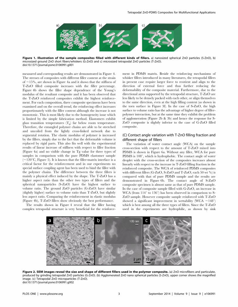
Structural morphologies corresponding to G-ZnO, S-ZnO and
T-ZnO fillers are shown by SEM images in Figure 2 (a–c). The G-
ZnO type of filler consists of short fiber and particulates with
aspect ratio approximately ranging from 1 to 10 (Figure 2a). The
S-ZnO consists of spherical particles with diameter around
100 nm and due to their high surface area, they form microsized
agglomerations (Figure 2b). A typical SEM image corresponding
to T-ZnO filler is shown in Figure 2c. These tetrapods exhibit four
arms pointing out from one core which leads to a concave shape in
space. The angle between each two respective arms is mostly
identical. The aspect ratio of each arm is ranging from 20 to 30.
Due to the shape of T-ZnO, their agglomeration was prohibited
and whiskers are well separated from each other, as can be clearly
seen in Figure 2c. On the contrary, the G-ZnO particles can be
packed more closely to each other.
The fabricated composites (with different fillers) have been
investigated in detail by SEM and cross-sectional SEM images of
different composite samples are shown in Figure 3. It is observed
from both cut cross-sections and torn cross-sections, that T-ZnO
filled composites are relatively rougher in comparison to the
specimen reinforced with G-ZnO (Figure 3c, d, e & f). For G-ZnO
reinforced composites, it is found that whiskers are mostly laying
along the surface. However, for T-ZnO reinforced composites, the
arms of the tetrapods are pointing to different directions, which
are especially pronounced in the torn cross-section (Figure 3f). In
the cut cross-section, the T-ZnO are partially broken into short
whiskers which is believed to be due to the cutting of the specimen
during SEM sample preparation. For the S-ZnO filled composite,
the nanoscale particles are hardly separable. As shown in
magnified image in Figure 3c, the actual filler unit is rather the
microsized agglomeration than the individual nanoparticles.
Comparing to G-ZnO and T-ZnO filled composites, the cut
cross- section of S-ZnO filled ones are relatively smoother on the
micrometer scale level (Figure 3b).
(B) Improvement in stiffnessThe tensile response of synthesized silicone composites rein-
forced with different ZnO fillers (up to 50 wt. %) has been
Tetrapodal ZnO-PDMS Composites for Multifunctional Applications
PLOS ONE | www.plosone.org 2 September 2014 | Volume 9 | Issue 9 | e106991
measured and corresponding results are demonstrated in Figure 4.
The stresses of composites with different filler content at the strain
of ,15%, are shown in Figure 4a and it shows that the stiffness of
T-ZnO filled composite increases with the filler percentage.
Figure 4b shows the filler shape dependence of the Young’s
modulus of the resultant composite and it has been observed that
the T-ZnO reinforced composites exhibit the highest reinforce-
ment. For each composition, three composite specimens have been
examined and on the overall trend, the reinforcing effect increases
proportionately with the filler content although the increase is not
monotonic. This is most likely due to the homogeneity issue which
is limited by the simple fabrication method. Elastomers exhibit
glass transition temperatures (Tg) far below room temperature.
Therefore, the entangled polymer chains are able to be stretched
and uncoiled from the lightly cross-linked network due to
segmental rotation. The elastic modulus of polymer is increased
by the fillers, simply due to the fact that the deformable volume is
replaced by rigid parts. This also fits well with the experimental
results of linear increase of stiffness with respect to filler fraction
(Figure 4a) and no visible change in Tg value for three types of
samples in comparison with the pure PDMS elastomer sample
(,120uC, Figure 5). It is known that the filler-matrix interface is a
critical factor for the reinforcement and in our experiments no
special surface coupling agent has been used to bind the filler with
the polymer chains. The difference between the three fillers is
mainly a physical effect induced by the shape. The T-ZnO has a
higher aspect ratio than the other two types of fillers and the
spherical nanoparticles (S-ZnO) have the highest surface to
volume ratio. The ground ZnO particles (G-ZnO) have similar
(slightly higher) surface to volume ratio than T-ZnO, but slightly
less aspect ratio. Comparing the reinforcement in elastic modulus
(Figure 4b), T-ZnO fillers show obviously the best performance.
The results shown in Figure 4 reveal that the filler having
complex tetrapodal structure is very beneficial for the reinforce-
ment in PDMS matrix. Beside the reinforcing mechanisms of
whisker fillers introduced in many literatures, the tetrapodal fillers
in present case require larger force to reorient and align to the
direction of external force and thus further reducing the
deformability of the composite material. Furthermore, due to the
directional arms supported by the tetrapodal structure, T-ZnO are
less likely to be densely packed with each other, or align themselves
to the same direction, even at the high filling content (as shown in
the torn surface in Figure 3f). In the case of S-ZnO, the high
surface to volume ratio has the advantage of higher degree of filler-
polymer interaction, but at the same time they exhibit the problem
of agglomeration (Figure 2b & 3b) and hence the response for S-
ZnO composite is slightly inferior to the case of G-ZnO filled
composite.
(C) Contact angle variation with T-ZnO filling fraction anddifferent shape of fillers
The variation of water contact angle (WCA) on the sample
cross-section with respect to the amount of T-ZnO mixed into
PDMS is shown in Figure 6a. Without any filler, WCA for pure
PDMS is 108u, which is hydrophobic. The contact angle of water
droplet with the cross-section of the composites increases almost
linearly with respect to the increase in T-ZnO filling fraction in the
reinforced composite. The WCA of reinforced PDMS composites
with different fillers (G-ZnO, S-ZnO and T-ZnO, each 50 wt %) is
compared with that of pure PDMS sample and the results are
demonstrated in Figure 6b. The contact angle of S-ZnO
composite specimen is almost same as that of pure PDMS sample.
In the case of composite sample filled with G-ZnO, an increase in
WCA (from 114u to 136u) has been observed in comparison to S-
ZnO sample. However composite sample reinforced with T-ZnO
showed a significant improvement in wettability (WCA ,148u)which is best among all the three types of fillers. Since the T-ZnO
used in the experiments are hydrophilic, as shown by sink
Figure 1. Illustration of the sample composites filled with different kinds of fillers. a) nanosized spherical ZnO particles (S-ZnO), b)microsized ground ZnO short fibers/whiskers (G-ZnO) and c) microsized tetrapodal ZnO particles (T-ZnO).doi:10.1371/journal.pone.0106991.g001
Figure 2. SEM images reveal the size and shape of different fillers used in the polymer composite. (a) ZnO microfibers and particulate,produced by grinding tetrapodal ZnO particles (G-ZnO). (b) Agglomerated ZnO nano spherical particles (S-ZnO), upper corner shows the magnifiedimage. (c) Tetrapodal ZnO microparticles (T-ZnO).doi:10.1371/journal.pone.0106991.g002
Tetrapodal ZnO-PDMS Composites for Multifunctional Applications
PLOS ONE | www.plosone.org 3 September 2014 | Volume 9 | Issue 9 | e106991

technique, the increased hydrophobicity cannot be only due to T-
ZnO sitting on the surface. The variation in WCA can understood
in terms of difference in roughness value in the composites filled
with different fillers. Comparing the SEM images in Figure 3c and
3d, the T-ZnO samples exhibit the roughest cross-section.
According to the Wenzel’s theory [51] of surface wetting,
hydrophobic surfaces become more hydrophobic as the roughness
increases. The S-ZnO particles induce roughness on nanoscale,
which showed no apparent effect on the WCA.
ConclusionIn summary, ZnO fillers of different size and shape have been
used to modify the silicone rubber, in order to reveal the role of the
shape of fillers on the properties of reinforced composites.
Tetrapodal shaped microparticles, short microfibers and whiskers
and nanosized spherical particles were successfully embedded in
PDMS elastomer in form of composites in a straightforward
method. The tensile elastic modulus and water contact angle of
composites prepared with different types fillers have been
measured. The results demonstrated that the tetrapodal shaped
ZnO microparticles increase the stiffness and hydrophobicity of
material cross-section on the highest level comparing to the other
two kinds of fillers. Such properties of this polymer composite are
highly desired in applications like biomimetics. It has been shown
that the tetrapodal shaped microparticles gain the advantage due
to their special shape. Such particular shaped fillers avoid the
problem of agglomeration as in case of nanoparticles and the
Figure 3. SEM images of the fabricated composites cross-sections. (a–d) are the cut cross-section and (e, f) are the torn cross-section (whitescale bar indicates 50 mm): (a) the pure silicone (cross-linked PDMS) sample. (b) SEM image corresponding to Silicone filled with 50 wt% of S-ZnO andinset image in (b) is a high magnification view showing the nanoparticle agglomerates. (c) cut cross-section of silicone filled with 50 wt% of G-ZnO.(d) cut cross-section of silicone filled with 50 wt% of T-ZnO. (e) torn cross-section of silicone filled with 50 wt% of G-ZnO. (f) torn cross-section ofsilicone filled with 50 wt% of T-ZnO.doi:10.1371/journal.pone.0106991.g003
Figure 4. Tensile response of the fabricated composites. (a) Stiffness vs. filler content for T-ZnO. The stress at 15% stain is increased almostlinearly as the T-ZnO content increase. (b) Stiffness vs. filler types. The Young’s modulus of control specimen (pure PDMS elastomer) and compositesfilled with 50 wt% of different fillers (G-ZnO, S-ZnO and T-ZnO) are plotted here. It is shown that all the fillers increase the Young’s modulus of thePDMS, whereas T-ZnO gives the most distinctive result among all.doi:10.1371/journal.pone.0106991.g004
Tetrapodal ZnO-PDMS Composites for Multifunctional Applications
PLOS ONE | www.plosone.org 4 September 2014 | Volume 9 | Issue 9 | e106991

difficulty of achieving truly random distribution as in the case of
short fiber fillers, therefore have shown the best results in both the
reinforcement of tensile modulus and the reduction of cross-
section surface wettability of the polymer composites.
Materials and Methods
(i) Materials and sample preparationThe silicone elastomer (PDMS, Sylgard 184) was purchased
from Dow Corning Corporation. The ZnO tetrapods (T-ZnO)
were synthesized by flame transport synthesis approach from Zn
powder (diameter ,5 mm from GoodFellow, UK) and polyvinyl
butyral (PVB) powder (Mowital B 60 H from Kuraray GmbH,
Europe) in an oven. [39] G-ZnO were obtained by grinding the T-
ZnO in a ball milling machine at a rotation speed of 100 rpm. S-
ZnO were purchased from Sigma Aldrich (CAS 1314-13-2, purity
99.99%). The composite samples were fabricated by mixing the
fillers of different weight percentages into prepolymer mixture and
curing afterwards. Initially, the two components of PDMS were
mixed and the mixture was then degassed in a vacuum oven for
about 5 min until no air bubbles observed in the solution. Then
PDMS solution was dropped to the desired amount of ZnO
particles (T-ZnO, G-ZnO, or S-ZnO). The mixture was gently
stirred with a spatula till all the ZnO were mixed into the slurry.
Afterwards the slurry was transferred to an Aluminum mold. Then
it was degassed in the vacuum oven for about 15 min since it takes
longer time for the bubbles to come out from a clay-like mixture
than from the pure PDMS solution. Finally the mold is heated on
a hotplate at 100uC for 40 min. After cooling down to ambient
Figure 5. DSC measurement of Tg. The heat flow is shown with baseline subtraction. Temperature scan rate is 10uC/minute. It can be observedfor all three types of composite samples, the glass transition occur around the same value 2120uC.doi:10.1371/journal.pone.0106991.g005
Figure 6. Wettability measurement results. (a) the water contact angle vs. filler content. It shows that the contact angle increases almost linearlywith T-ZnO content. (b) the water contact angle verses different types of fillers at the same filling factor of 50 wt%. It is shown that the S-ZnO has nosignificant influence on the contact angle and T-ZnO gives the highest value.doi:10.1371/journal.pone.0106991.g006
Tetrapodal ZnO-PDMS Composites for Multifunctional Applications
PLOS ONE | www.plosone.org 5 September 2014 | Volume 9 | Issue 9 | e106991

temperature, the cured samples were carefully removed from the
mold. The reason for heating the mixture is that the long cure time
(48 hours) at room temperature can cause precipitation of the
ZnO, which might reduce their uniformity.
(ii) Morphological investigationThe morphologies of different filler particles were investigated
by scanning electron microscopy (SEM) instrument Zeiss Ultraplus
(3 kV, 15 mA). The fabricated composites were also investigated
inside SEM in detail to check their morphologies and distributions.
The samples were prepared by two methods: either by cutting it
through with a knife or tearing it apart after a crack is initiated by
a cut. The revealed cross-sections were then sputter-coated with a
few nanometer of gold to avoid the charging.
(iii) Stress-strain responseStress-strain measurements were carried out on a tensile testing
machine (QuickTest QTS3). Most samples were extended at an
elongation of 6 mm, which is c.a. 30% of the original length, in
order to avoid error induced by sample slippage from the clamps.
The strain rate was 60 mm/min. The dimension of the tested
specimen region is 206561 mm3. All measurements have been
performed at room temperature. The tensile stress was calculated
by the formula s = F/A0, where F is the force applied on the
specimen and A0 is the cross section area.
(iv) Differential scanning calorimetry (DSC) measurementThe measurement was carried out in a Differential Scanning
Calormeter (Perkin Elmer Pyris 1), samples with c.a. 25 mg weight
were used. Two temperature scan cycle per sample between 2
140uC to 270uC was carried out with rate 10uC/minutes. Results
were subtracted with baseline measurement.
(v) Wettability measurementThe wettability experiments were performed on a contact angle
measurement machine (Dataphysics OCA5), where a camera can
capture images of water droplets on different sample surfaces. The
captured images were analyzed with image process software
SCA20. The samples were cut into slices. A water droplet with a
defined amount of volume was deposited on the cut surface of a
slice. An image was captured immediately to avoid error induced
by water evaporation. The wettability of T-ZnO was tested by sink
technique. The T-ZnO were completely wetted by water and sunk
in after being placed on top of water surface in a beaker.
Acknowledgments
Authors would like to thank Christoph Chluba (CAU Kiel) for help during
DSC measurements. YKM acknowledges the postdoctoral grant from
Alexander von Humboldt Foundation.
Author Contributions
Conceived and designed the experiments: XJ YKM RA. Performed the
experiments: XJ MD SK XZ IH KM YKM. Analyzed the data: XJ SK
RA YKM. Contributed reagents/materials/analysis tools: RA. Contribut-
ed to the writing of the manuscript: XJ RA YKM.
References
1. Xia F, Jiang L (2008) Bio-inspired, smart, multiscale interfacial materials. Adv
Mater 20: 2842–2858.
2. Meyers MA, Chen P-Y, Lin AY-M, Seki Y (2008) Biological materials: Structure
and mechanical properties. Prog Mater Sci 53: 1–206.
3. Weiner S, Wagner HD (1998) The material bone: structure-mechanical function
relations. Annu Rev Mater Sci 28: 271–298.
4. Li J, Liu X, Ye Y, Zhou H, Chen J (2011) Gecko-inspired synthesis of
superhydrophobic ZnO surfaces with high water adhesion. Colloids Surf, A 384:
109–114.
5. Hussain F, Hojjati M, Okamoto M, Gorga RE (2006) Polymer-matrix
nanocomposites, processing, manufacturing, and application: An overview.
J Compos Mater 40: 1511–1575.
6. Murphy MP, Kim S, Sitti M (2009) Enhanced adhesion by gecko-inspired
hierarchical fibrillar adhesives. ACS Appl Mater Interfaces 1: 849–855.
7. Mahdavi A, Ferreira L, Sundback C, Nichol JW, Chan EP, et al. (2008) A
biodegradable and biocompatible gecko-inspired tissue adhesive. Proc Natl Acad
Sci USA 105: 2307–2312.
8. Boesel LF, Greiner C, Arzt E, del Campo A (2010) Gecko-inspired surfaces: A
path to strong and reversible dry adhesives. Adv Mater 22: 2125–2137.
9. Reddy S, Arzt E, del Campo A (2007) Bioinspired surfaces with switchable
adhesion. Adv Mater 19: 3833–3837.
10. Lee J, Majidi C, Schubert B, Fearing RS (2008) Sliding-induced adhesion of stiff
polymer microfibre arrays. I. Macroscale behaviour. J R Soc Interface 5: 835–
844.
11. Menon C, Murphy M, Sitti M (2004) Gecko inspired surface climbing robots.
IEEE. 431–436.
12. Genzer J, Marmur A (2008) Biological and synthetic self-cleaning surfaces. MRS
Bull 33: 742–746.
13. Wang ZL (2009) ZnO nanowire and nanobelt platform for nanotechnology.
Mater Sci Eng, R 64: 33–71.
14. Zeng H, Duan G, Li Y, Yang S, Xu X, et al. (2010) Blue luminescence of ZnO
nanoparticles based on non-equilibrium processes: Defect origins and emission
controls. Adv Funct Mater 20: 561–572.
15. Devan RS, Patil RA, Lin JH, Ma YR (2012) One dimensional metal oxide
nanostructures: Recent developments in synthesis, characterization, and
applications. Adv Funct Mater 22: 3326–3370.
16. Mishra YK, Kaps S, Schuchardt A, Paulowicz I, Jin X, et al. (2014) Versatile
fabrication of complex shaped metal oxide nano-microstructures and their
interconnected networks for multifunctional applications. KONA Powder Part J
31: 92–110.
17. Gorrasi G, D’Ambrosio S, Patimo G, Pantani R (2014) Hybrid clay-carbon
nanotube/PET composites: Preparation, processing, and analysis of physical
properties. J Appl Polym Sci 131: 40441.
18. Mannov E, Schmutzler H, Chandrasekaran S, Viets C, Buschhorn S, et al.
(2013) Improvement of compressive strength after impact in fibre reinforced
polymer composites by matrix modification with thermally reduced graphene
oxide. Compos Sci Technol 87: 36–41.
19. Raquez J-M, Habibi Y, Murariu M, Dubois P (2013) Polylactide (PLA)-based
nanocomposites. Prog Polym Sci 38: 1504–1542.
20. Zhou W, Qi S, Tu C, Zhao H, Wang C, et al. (2007) Effect of the particle size of
Al2O3 on the properties of filled heat-conductive silicone rubber. J Appl Polym
Sci 104: 1312–1318.
21. Wong M, Paramsothy M, Xu X, Ren Y, Li S, et al. (2003) Physical interactions
at carbon nanotube-polymer interface. Polymer 44: 7757–7764.
22. Giannelis EP (1996) Polymer layered silicate nanocomposites. Adv Mater 8: 29–
35.
23. Lipatov YS (1995) Polymer reinforcement: ChemTec Publishing.
24. Jones RM (1999) Mechanics of composite material. Taylor & Francis, New
York.
25. Fu S-Y, Feng X-Q, Lauke B, Mai Y-W (2008) Effects of particle size, particle/
matrix interface adhesion and particle loading on mechanical properties of
particulate–polymer composites. Composites, Part B 39: 933–961.
26. Cadek M, Coleman J, Ryan K, Nicolosi V, Bister G, et al. (2004) Reinforcement
of polymers with carbon nanotubes: The role of nanotube surface area. Nano
Lett 4: 353–356.
27. Fiedler B, Gojny FH, Wichmann MH, Nolte M, Schulte K (2006) Fundamental
aspects of nano-reinforced composites. Compos Sci Technol 66: 3115–3125.
28. Moczo J, Pukanszky B (2008) Polymer micro and nanocomposites: Structure,
interactions, properties. J Ind Eng Chem (Amsterdam, Neth) 14: 535–563.
29. Isayev A I, Modic M (1987) Self-Reinforced melt processible polymer
composites: Extrusion, compression, and injection molding. Polym Compos 8:
158–175.
30. Paul DR, Robeson LM (2008) Polymer nanotechnology: Nanocomposites.
Polymer 49: 3187–3204.
31. Wagner HD (2007) Nanocomposites: Paving the way to stronger materials. Nat
Nanotechnol 2: 742–744.
32. Potschke P, Krause B, Buschhorn ST, Kopke U, Muller MT, et al. (2013)
Improvement of carbon nanotube dispersion in thermoplastic composites using a
three roll mill at elevated temperatures. Compos Sci Technol 74: 78–84.
33. Wagner HD, Ajayan PM, Schulte K (2013) Nanocomposite toughness from a
pull-out mechanism. Compos Sci Technol 83: 27–31.
Tetrapodal ZnO-PDMS Composites for Multifunctional Applications
PLOS ONE | www.plosone.org 6 September 2014 | Volume 9 | Issue 9 | e106991

34. Wichmann MH, Buschhorn ST, Boger L, Adelung R, Schulte K (2008)
Direction sensitive bending sensors based on multi-wall carbon nanotube/epoxy
nanocomposites. Nanotechnology 19: 475503.
35. Pavlidou S, Papaspyrides C (2008) A review on polymer–layered silicate
nanocomposites. Prog Polym Sci 33: 1119–1198.
36. Breuer O, Sundararaj U (2004) Big returns from small fibers: A review of
polymer/carbon nanotube composites. Polym Compos 25: 630–645.
37. Pantani R, Gorrasi G, Vigliotta G, Murariu M, Dubois P (2013) PLA-ZnO
nanocomposite films: Water vapor barrier properties and specific end-use
characteristics. Eur Polym J 49: 3471–3482.
38. Jin X, Gotz M, Wille S, Mishra YK, Adelung R, et al. (2013) A novel concept for
self-reporting materials: Stress sensitive photoluminescence in ZnO tetrapod
filled elastomers. Adv Mater 25: 1342–1347.
39. Mishra YK, Kaps S, Schuchardt A, Paulowicz I, Jin X, et al. (2013) Fabrication
of macroscopically flexible and highly porous 3D semiconductor networks from
interpenetrating nanostructures by a simple flame transport approach. Part Part
Syst Charact 30: 775–783.
40. Mishra YK, Adelung R, Rohl C, Shukla D, Spors F, et al. (2011) Virostatic
potential of micro-nano filopodia-like ZnO structures against herpes simplex
virus-1. Antiviral Res 92: 305–312.
41. Antoine T, Mishra YK, Trigilio J, Tiwari V, Adelung R, et al. (2012)
Prophylactic, therapeutic and neutralizing effects of zinc oxide tetrapod
structures against herpes simplex virus type-2 infection. Antiviral Res 96: 363–
375.
42. Papavlassopoulos H, Mishra YK, Kaps S, Paulowicz I, Abdelaziz R, et al. (2014)
Toxicity of functional nano-micro zinc oxide tetrapods: Impact of cell cultureconditions, cellular age and material properties. PLoS ONE 9: e84983.
43. Jin X, Strueben J, Heepe L, Kovalev A, Mishra YK, et al. (2012) Joining the Un-
joinable: Adhesion between low surface energy polymers using tetrapodal ZnOlinkers. Adv Mater 24: 5676–5680.
44. Reimer T, Paulowicz I, Roder R, Kaps S, Lupan O, et al. (2014) Single stepintegration of ZnO nano-and microneedles in Si trenches by novel flame
transport approach: Whispering gallery modes and photocatalytic properties.
ACS Appl Mater Interfaces 6: 780627815.45. Gedamu D, Paulowicz I, Kaps S, Lupan O, Wille S, et al. (2014) Rapid
fabrication technique for interpenetrated ZnO nanotetrapod networks for fastUV sensors. Adv Mater 26: 1541–1550.
46. Ronning C, Shang N, Gerhards I, Hofsass H, Seibt M (2005) Nucleationmechanism of the seed of tetrapod ZnO nanostructures. J Appl Phys 98: 34307.
47. Hu G, Ma Y, Wang B (2009) Mechanical properties and morphology of nylon
11/tetrapod-shaped zinc oxide whisker composite. Mater Sci Eng, A 504: 8–12.48. Zhou ZW, Hu S, Chu LS (2005) Studies on tetrapod-shaped ZnO whisker
modified polymer composites. Trans Tech Publ. 1033–1036.49. Zhou Z, Liu S, Gu L (2001) Studies on the strength and wear resistance of
tetrapod-shaped ZnO whisker–reinforced rubber composites. J Appl Polym Sci
80: 1520–1525.50. Zhou Z, Peng W, Ke S, Deng H (1999) Tetrapod-shaped ZnO whisker and its
composites. J Mater Process Technol 89–90: 415–418.51. Wenzel RN (1936) Resistance of solid surfaces to wetting by water. Ind Eng
Chem 28: 988–994.
Tetrapodal ZnO-PDMS Composites for Multifunctional Applications
PLOS ONE | www.plosone.org 7 September 2014 | Volume 9 | Issue 9 | e106991
Top Related
Copyright © 2022 FDOKUMEN

