


Bahasa
Halaman
Hukum


STRUCTURE OF VIRUSES:

Defective Viruses
Defective viruses are those virus particles whose genome
lacks a specific gene or genes due to either mutation or
deletion.
As a result, defective viruses are not capable of undergoing a
productive life cycle in cells.
However, if the cell infected with the defective virus is co-
infected with a "helper virus", the gene product lacking in the
defective one is complemented by the helper and defective
virus can replicate.
Interestingly, for some viruses, during infection a greater
quantity of defective virions is produced than infectious
virions (as much as 100:1).
The production of defective particles is a characteristic of
some virus species and is believed to moderate the severity of
the infection/disease in vivo.

PseudovirionsPseudovirions may be produced during viral
replication when the host genome is fragmented.
As a result of this process, host DNA fragments are
incorporated into the capsid instead of viral DNA.
Thus, pseudovirions possess the viral capsid to
which antibodies may bind and facilitate
attachment and penetration into a host cell, but
they cannot replicate once they have gained access
to a host cell, as they have none of the essential
viral genes for the process.

Prions
Prions are proteinaceous infectious particles associated
with transmissible spongiform encephalopathies (TSE) of
humans and animals.
TSEs include the Creutzfeldt-Jacob disease of humans,
scrapie of sheep and
bovine spongiform encephalopathy.
At postmortem, the brain has large vacuoles in the cortex
and cerebellum regions an thus prion diseases are called
"spongiform encephalopathies". Closer examination of
brain tissue reveals the accumulation of prion-protein
associated fibrils and amyloid plaques.
These diseases are characterized by loss of motor control,
dementia, paralysis, wasting and eventually death.

Viroids
Viroids are naked, low-molecular weight nucleic acids that
are extremely resistant to heat, ultraviolet, and ionizing
radiation. These particles are composed exclusively of a
single piece of circular, single stranded RNA that has some
double-stranded regions. Viroids mainly cause plant
diseases, such as potato spindle tuber disease.
Virusoids
Virusoids (also called satellite RNAs) are similar to viroids
in that they are naked, low-molecular weight nucleic acids
that are extremely resistant to heat and ultraviolet and
ionizing radiation. However, they depend on a helper virus
for replication. Virusoids replicate in cytoplasm via a RNA
dependent RNA polymerase.

Viruses are extremely small particles comprises of
just proteins and nucleic acid.
The largest viruses are about 300nm in size,
whereas the smallest known viruses are about 20nm.
Viruses are comprised of two important components a
protein capsid covering the nucleic acid.
Some viruses also possess a lipid envelope but the
envelope is not a virus- mediated structure
Outer coat or capsid: The outer structure of virus is
referred as capsid. It is made up of proteins. It is a
protein shell that encloses the nucleic acid. It is built
of structure units, which are the smallest functional
equivalent building units of the capsid.
.
.

Capsomeres are morphological units seen on the
surface of particles and represent clusters of
structural units.
Capsomeres are also referred as the basic
structural subunit of capsid.
The capsid together with its enclosed nucleic acid is
called the nucleocapsid.
The nucleocapsid may be enclosed in an envelope,
which may contain material of host cell as well as
viral origin.
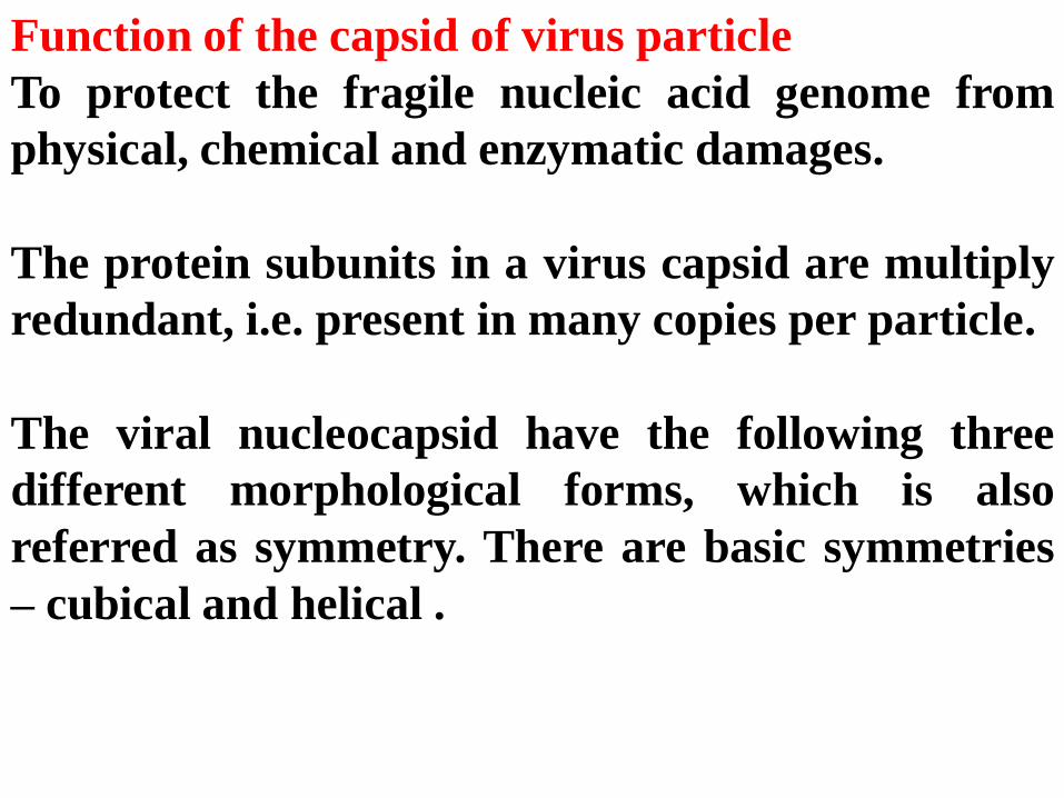
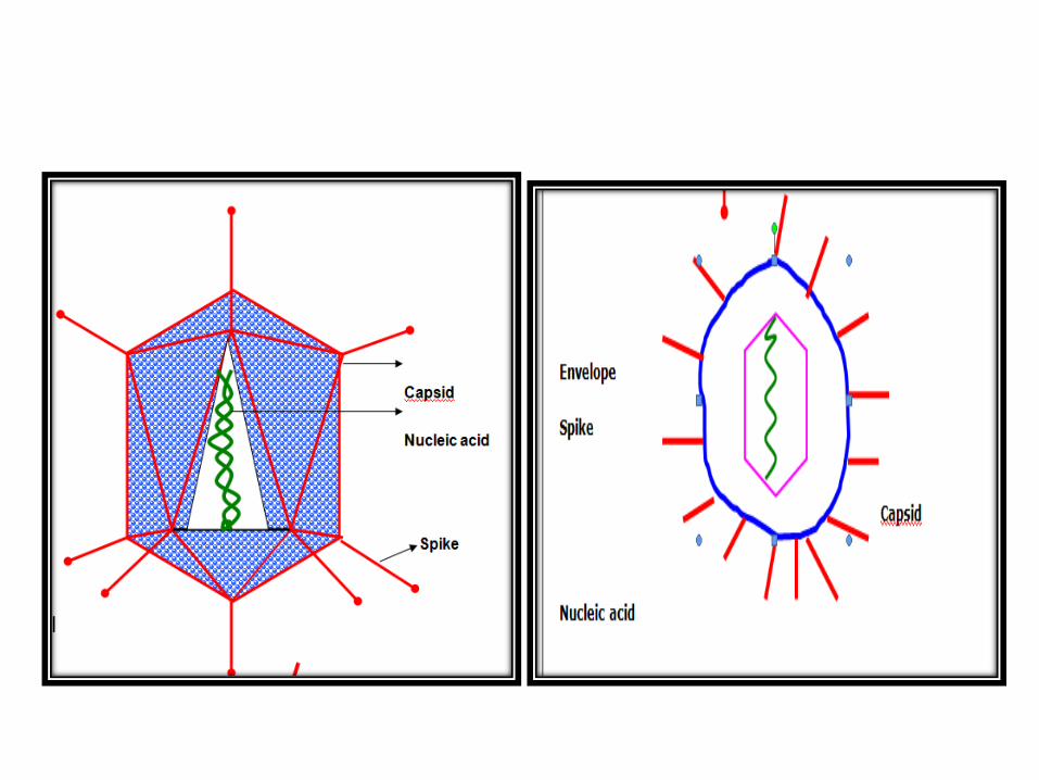
Function of the capsid of virus particle
To protect the fragile nucleic acid genome from
physical, chemical and enzymatic damages.
The protein subunits in a virus capsid are multiply
redundant, i.e. present in many copies per particle.
The viral nucleocapsid have the following three
different morphological forms, which is also
referred as symmetry. There are basic symmetries
– cubical and helical .

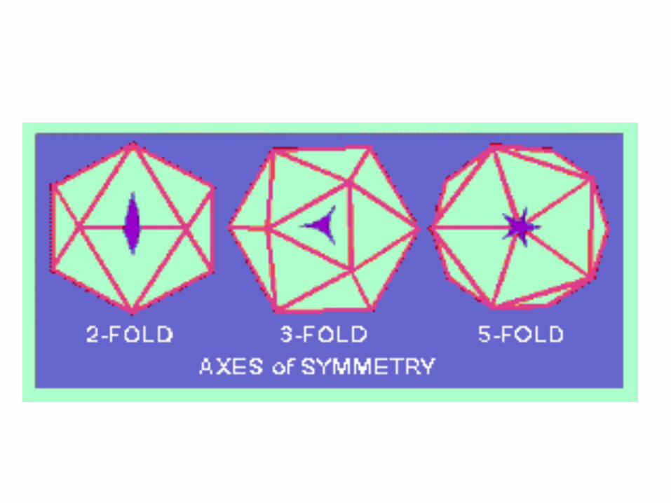
Icosahedral / Cubical symmetry Caplur & Klung
• In this arrangement, the nucleic acids are arranged inside a shell, which is in the shape of an icosahedran.
• Icosahedran is a geometrical figure with 12 vertices (corners) and 20 identical facets (faces) and 30 edges.
• Each facet is an equilateral triangle. There are six 5-fold axes of symmetry passing through the vertices,
• Ten 3-fold axes extending through each face and
• Fifteen 2-fold axes passing through the edges of an icosahedron.
• Icosahedral symmetry requires definite numbers of structure units to complete a shell. A virus with icosahedral symmetry (5:3:2 symmetry) requires a multiple of 60 subunits to cover the surface completely.

12 vertices (corners) and 20 identical facets (faces) and 30 edges.


Helical symmetry:
• The nucleic acid and capsomeres are helicallycoiled together.
• The length of the helical viral nucleocapsid isdetermined by the length of the nucleic acid.
• In this symmetry the identical protein subunits arearranged in the form of a circle to form a discshaped appearance.
• The cylinder or helical structure is formed as aresult of stacking of multiple discs, with the virusgenome coated by the protein shell or contained inthe hollow centre of the cylinder.

Complex symmetry:
• It is also referred as undefined symmetry.This arrangement does not fit into eitherhelical or cubical symmetries. It has thefeature of both cubical and helicalsymmetries.
Eg. Pox Virus

VIRAL NUCLEIC ACID:
•The genetic material of viruses are made up of only one
type of nucleic acid ie. either DNA or RNA but never
composed of both.
•The nucleic acid both DNA and RNA may be present
either as single stranded or double stranded.
•The nucleic acid (RNA) may also present either as a single
molecule or in segments. The DNA may present either as
linear or circular molecule.
•The RNA may present either as positive sense or negative
sense. The molecular weight ranges from 2 million to 200
million.
•In some viruses the nucleic acids are just packed inside
the capsid, whereas in some viruses, they are integrated
with the nucleocapsid.

ENVELOPE:
•The capsid of some viruses are surrounded by a
membrane called envelope. Viral envelopes are derived
from cellular membranes of host, which are acquired
during the release of virus from host cell.
•The envelope also contain proteins that are referred as
matrix proteins and are specified by viral genes. The
envelope proteins appear as spikes and are also referred as
spike proteins. The spike proteins are responsible for
attachment of virus to cells.
•The viral structures are studied by electron microscopic
techniques like negative staining, freeze etching and
shadowing. The method that is more commonly used in X
ray crystallography. Of late nuclear magnetic resonance
(NMR) imaging is used to study structural details.

Productive infection: The cells allow viruses to
replicate and the progeny virions are released from
the infected cell.
Abortive infection: The cells do not allow viruses to
replicate & as a result of this daughter virions are
not produced.
Restrictive infection: The cells allow minimal
replication of viruses and as a result only few
daughters alone are produced.
However, the viral genome persists and can lead to
serious consequence for the host.
Eg. Epstein Barr & herpes simplex
Top Related
Copyright © 2022 FDOKUMEN

