


Bahasa
Halaman
Hukum

J. Exp. Med.
The Rockefeller University Press • 0022-1007/2001/01/25/10 $5.00Volume 193, Number 1, January 1, 2001 25–34http://www.jem.org/cgi/content/full/193/1/25
25
Role of the High Affinity Immunoglobulin E Receptor in Bacterial Translocation and Intestinal Inflammation
By David Dombrowicz,
*
Sophie Nutten,
§
Pierre Desreumaux,
§
Christel Neut,
i
Gérard Torpier,
‡
Marc Peeters,
¶
Jean-Frédéric Colombel,
§
and Monique Capron
*
From the
*
Institut National de la Sante et de la Recherche Medicale U167 and the
‡
Institut National de la Sante et de la Recherche Medicale U325, Institut Pasteur de Lille, 59019 Lille, France; the
§
Laboratoire de Recherche sur les Maladies Inflammatoires Intestinales et Département d’Hépatogastroenterologie, Centre Hospitalier Régional Universitaire de Lille, 59045 Lille, France; the
i
Faculty of Pharmacy, University of Lille II, 59045 Lille, France; and the
¶
Department of Gastroenterology, University Hospital Gasthuisberg, B-3000 Leuven, Belgium
Abstract
A role for immunoglobulin E and its high affinity receptor (Fc
e
RI) in the control of bacterialpathogenicity and intestinal inflammation has been suggested, but relevant animal models arelacking. Here we compare transgenic mice expressing a humanized Fc
e
RI (hFc
e
RI), with acell distribution similar to that in humans, to Fc
e
RI-deficient animals. In hFc
e
RI transgenicmice, levels of colonic interleukin 4 were higher, the composition of fecal flora was greatlymodified, and bacterial translocation towards mesenteric lymph nodes was increased. InhFc
e
RI transgenic mice, 2,4,6-tri-nitrobenzenesulfonic acid (TNBS)-induced colitis was alsomore pronounced, whereas Fc
e
RI-deficient animals were protected from colitis, demonstrat-ing that Fc
e
RI can affect the onset of intestinal inflammation.
Key words: immunoglobulin E receptor • bacterial translocation • intestinal permeability • inflammatory bowel disease • colitis
Introduction
IgE is known to induce inflammatory processes such as im-mediate hypersensitivity reactions (allergy, asthma, anaphy-laxis, etc.) through interaction with its high affinity recep-tor, Fc
e
RI (1). Upon receptor aggregation with IgE andmultivalent antigen, cells release inflammatory mediators,whether preformed, like histamine, or newly synthesized,like leukotrienes. In mice, Fc
e
RI is expressed only on mastcells and basophils, whereas in humans, it is additionally ex-pressed on monocytes, Langerhans cells, eosinophils, andplatelets (1). To obtain the most suitable animal model forthe study of IgE-mediated diseases, we first generated trans-genic (Tg)
1
mice expressing the human Fc
e
RI
a
(hFc
e
RI
a
)chain under the control of its own promoter region (2).
Those hFc
e
RI
a
Tg animals (bred in an Fc
e
RI-deficientbackground), express a “humanized” receptor, with thesame cellular distribution as found in humans (3). This re-ceptor is able to bind human or murine IgE and to restoreIgE-mediated anaphylaxis in Fc
e
RI-deficient (Fc
e
RI
a
2
/
2
)mice (2, 4).
In chronic inflammatory bowel disease (IBD), intestinallesions are considered as the consequence of a dysfunctionof the mucosal immune system. Genetic and environmen-tal factors have also been associated to the onset of the dis-ease. Infectious agents such as
Mycobacterium paratuberculosis
,measles virus, and
Listeria monocytogenes
were once sus-pected as causal agents (5). However, none of them wereformally confirmed as actually responsible for Crohn’s dis-ease (CD), one of the two entities of IBD.
Most animal models, including 2,4,6-tri-nitrobenzene-sulfonic acid (TNBS)-induced colitis in mice and rats, havebeen described as “mediated” by type 1 cytokines. Indeed,chronic intestinal lesions are associated with an increasedsynthesis of proinflammatory and type 1 cytokines (5, 6).However, more recent studies have demonstrated the im-portance of type 2 cytokines for the induction of colitis. InTCR-
a
–deficient animals (TCR-
a
2
/
2
) spontaneously de-
D. Dombrowicz, S. Nutten, and P. Desreumaux contributed equally tothis work.
Address correspondence to M. Capron, INSERM U167, Institut Pas-teur de Lille, 1 rue Professeur Calmette, BP245, 59019 Lille Cedex,
France. Phone: 33-320-87-79-62; Fax: 33-320-87-78-88; E-mail: [email protected]
1
Abbreviations used in this paper:
CD, Crohn’s disease; hFc
e
RI, humanFc
e
RI; IBD, inflammatory bowel disease, MLN, mesenteric lymph node,RT, reverse transcription; SPF, specific pathogen-free; Tg, transgenic;TNBS, 2,4,6-tri-nitrobenzenesulfonic acid; WT, wild-type.
on July 1, 2015jem
.rupress.orgD
ownloaded from
Published December 27, 2000
26
IgE Receptor in Bacterial Translocation and Intestinal Inflammation
veloping colitis (7), acute lesions are associated with an in-creased IL-4 synthesis (8) and with local IgE production (9),whereas chronic lesions are associated with type 1 cytokines(8). Indeed TCR-
a
2
/
2
3
IL-4
2
/
2
mice do not developcolitis, whereas colitis in TCR-
a
2
/
2
3
IFN-
g
2
/
2
animals issimilar to the pathology observed in TCR-
a
2
/
2
3
IFN-
g
1
/
1
mice (10). Mucosal immune response is associated, inTCR-
a
2
/
2
and IL-2
2
/
2
mice, to IgE synthesis (9, 11), andis dependent on luminal bacteria (12, 13). Additionally, inTNBS-induced colitis, disruption of the IL-4 gene or ad-ministration of anti–IL-4 antibodies attenuates the severityof the lesions, whereas disruption of the IFN-
g
gene leadsto an increased pathology (14). The respective roles of type1 and type 2 cytokines have been further delineated usingthe same model. Indeed, a recently published study hasshown that Th1-like cytokine responses were inducing fa-tal, acute, transmural, and focal types of lesions, whereasTh2-like cytokine responses were playing a significant rolein the diffuse atrophic changes in crypts and the mucosallayer that occur in the late stages of this disease (15).
Recently, we have shown that divergent mucosal cyto-kine patterns evolved during the different stages of CD. Atype 2 pattern with prominent IL-4 response and local pro-duction of IgE is associated with the early intestinal lesionsof patients with CD and followed by a type 1 response inthe chronic lesions of the same patients (16, 17). Further-more, it has been shown that the enhancement of intestinalpermeability observed in patients with CD is associatedwith an increase of activated B cells (CD45 RO
1
CD19
1
),which have the capacity to synthesize IgE (18). Addition-ally, we have demonstrated, in patients with type 2– andIgE-mediated disease like atopy or asthma, the presence ofan airway-like inflammation of the gut, including high lev-els of type 2 cytokines (19) and an increase of the intestinalpermeability (20). Taken together, these results suggest thatthe common mucosal immune system could modify the in-testinal permeability and therefrom bacterial translocationthrough mechanisms involving IL-4/IgE and/or Fc
e
RI,the high affinity IgE receptor.
Evidences for a role of the endogenous bacterial flora inthe bowel inflammation have also been accumulating (5).In humans, the importance of bacterial flora was demon-strated in a model of postsurgery relapse, which occurs at arate of 73% after 1 yr (21). If the anastomosis is isolatedfrom the fecal stream, no lesion occurs, whereas the relapseis rapid after the infusion of intestinal luminal content (22).We thus hypothesize that, in CD, lesions are due either tocompounds from the digestive flora or to virulent bacteriainvading the digestive tract in a patient with a defective im-mune intestinal response. Indeed flora from stool samples,obtained in patients with CD, contains higher concentra-tions of anaerobic bacteria, among others
Bacteroides vulgatus
and some coccobacilli (
Eubacterium
,
Peptostreptococcus
, and
Coprococcus
[23]). Likewise, a significant increase in thenumber of luminal ileal and colic
Bacteroides fragilis
and
Escherichia coli
was observed (24, 25), as well as an increaseof mucosal enterobacteria in biopsies (26). However, it hasnot been so far formally demonstrated that such bacteria
were the causal agent of the disease. It is rather suspectedthat the presence of these bacteria worsen the symptomsinitiated by alterations of the immune system. In most ani-mal models of spontaneous colitis, the severity of the in-flammatory lesions increases with the presence of the bac-terial flora (6, 12, 27).
Using hFc
e
RI
a
Tg and Fc
e
RI-deficient animals, we in-vestigated if IgE receptor expression was affecting inflam-matory parameters, bacterial flora and its translocation to-wards mesenteric lymph nodes (MLNs) in naive animals.Our results suggest that Fc
e
RI might be able to contributeto the development of intestinal inflammation, possibly dueto increased levels of IL-4.
Materials and Methods
Animals.
Human Fc
e
RI
a
Tg and Fc
e
RI
a
2
/
2
mice that havebeen described elsewhere (2–4) are in a Balb/c background. Un-less specified, animals aged between 8 and 16 wk as well as corre-sponding wild-type (WT) Balb/c were raised in a specific patho-gen-free (SPF) facility, under a 12:12 h day–night illuminationcycle. Tap water was available ad libitum and animals were fedwith standard mice chow (D04; UAR).
Experimental Procedures.
Animals were killed by cervical dislo-cation. MLNs were first aseptically removed before intestinal re-section and placed immediately in cysteinated-strength Ringer’smedium for microbiological analyses. Then, transparietal samplesof distal colon and ileum were resected and fixed in 4% paraform-aldehyde (PFA) or snap frozen in liquid nitrogen, respectively, forhistologic examination and cytokine quantification. Samples offeces were put in the same medium as above for microbiologicalanalyses.
Microbiological Analyses.
MLNs and feces were weighted. In-oculations were performed within 4 h after animal sacrifice. Cul-tures for both aerobic and anaerobic flora were performed onColumbia blood agar either by direct inoculation or after an en-richment in liquid Wilkins-West medium for detection of germspresent in small amounts. Anaerobic conditions were obtained inhermetic jar using Anaerogen (Oxoid) reagent.
Results were expressed as log CFU per gram of feces for bothtotal counts and counts of identified bacteria. Detection limit formesenteric tissue was
z
10 germs/g of tissue.
Reverse Transcription PCR.
Frozen colon samples were placedin Trizol (Life Technologies), and total RNA was extracted ac-cording to the manufacturer’s instructions (Bioprobe). Quantita-tive reverse transcription (RT)-PCR was performed exactly asdescribed previously (16). pQB3 and pMus-3 were respectivelyused as competitor for
b
-actin and for IL-1
b
, IL-6, TNF-
a
, IL-10, IFN-
g
, and IL-4 (a gift from Dr. D. Shire, Sanofi Elf Bio Re-cherches, Labege, France). The size of the amplified fragmentsand the sense and antisense primers are figured on Table I. Fc
e
RIand IgE mRNA levels were determined by semiquantitative RT-PCR. A fixed amount of cDNA, corresponding to the lowestnumber of
b
-actin cDNA molecules in the group of samples, wasused. Results were expressed as absorbance units (at 260 nm) permolecule of
b
-actin mRNA.
IgE Measurements.
Serum IgE concentrations were measuredas described previously (28). In brief, 96-well microtiter plates(Maxisorb; Nunc) were coated overnight at 4
8
C with 2
m
g/mlpurified anti–mouse IgE (clone R35-72; BD PharMingen) inPBS buffer, pH 7.4. After washing, wells were saturated withPBS–1% BSA. After washing, samples or anti-TNP IgE (clone
on July 1, 2015jem
.rupress.orgD
ownloaded from
Published December 27, 2000
27
Dombrowicz et al.
C48-2; BD PharMingen), used for the standard curve, wereadded and incubated overnight at 4
8
C. After washing, biotiny-lated anti-IgE antibody (2
m
g/ml, clone R35-92; BD PharMin-gen) was incubated for 1 h at room temperature. After washing,plates were then incubated for 30 min at room temperature withstreptavidin-conjugated horseradish peroxidase (1:10,000; JacksonImmunoResearch Laboratories) and reactions were revealed byaddition of ortho-phenylenediamine (OPD). Absorbance wasread at 492 nm.
Pathological Analyses.
4-
m
m paraffin sections were stainedwith May-Grünwald/Giemsa and examined for morphology andpresence of inflammatory infiltrates. Microscopic inflammatoryscore was measured according to Ameho et al. (29).
Intestinal Permeability.
Intestinal permeability was measuredaccording to Meddings and Gibbons (30). Mice were denied ac-cess to water for 3 h. Then, each mouse was given, by gavage, 8mg mannitol, 12 mg lactulose, and 6 mg sucralose in 200
m
ldrinking water. Groups of five mice were placed into stainlesssteel metabolic cages in order to separate feces from urine. 10
m
lof a 10% sodium azide solution were added to each collectingtube to prevent bacterial degradation of urinary sugars. Urine wascollected for a total of 24 h and frozen until measurement of itssugar composition. After thawing, d(
1
)furanose was added as in-ternal standard, and samples were deionized using a mixture ofAmberlite IR-120 plus and IRA-400c resin. After centrifugation,samples were evaporated under nitrogen and derivatized with bis-(trimethylsilyl)trifluoroacetamide (BSTFA) plus trimethylchlo-rosilane (TMCS) (1%) after reaction with NH
2
OH.HCl in pyri-dine. Sucralose, lactulose, and mannitol were determined by gaschromatography-flame ionization detector (GC-FID) after ex-traction in n-heptane using a 100% dimethylpolysiloxane column(CP-Sil 5 CB Low Bleed/MS; Variant; 25 m
3
0.25 mm, diam-eter 0.2 mm) with a multirise temperature program. All sampleswere diluted so that concentrations fell within the range of thestandards.
TNBS-induced Colitis.
For induction of colitis, mice wereanesthetized for 90–120 min with Xylazine (50 mg/kg; Rom-pun
®
2%; Bayer Pharma) and Ketamine (50 mg/kg; Imalgene
®
1000; Rhône Mérieux). TNBS (150 mg/kg; Fluka) was adminis-tered intrarectally in 40
m
l 50% ethanol via a 3.5 F catheter (Ref
EO 3416-1; Biotrol) inserted to a length of 4 cm (31). Controlmice received 40
m
l 50% ethanol saline or saline alone. Animalswere killed 48 h later. Macroscopic lesions were assessed accord-ing to the score established by Wallace et al
. (32).Statistical Analysis. Comparisons between number of bacteria
in the feces, number of bacteria in the MLN, and cytokinecDNA levels in the Tg and WT mice were analyzed by theKruskall-Wallis test. Differences were statistically significant if theP value was ,0.05.
ResultsTo study the role of FceRI on intestinal inflammation,
three different strains of mice expressing FceRI at differentlevels and with a different cell distribution were compared:WT Balb/c mice expressing FceRI only on mast cells andbasophils, hFceRIa Tg mice expressing a functional hu-manized receptor on mast cells, basophils, monocytes, eo-sinophils, and epidermal Langerhans cells, and FceRIa2/2
animals in which receptor expression is absent. To correlatereceptor expression levels with the observed phenotype,steady-state levels of FceRIa mRNA were determined incolonic samples using semiquantitative RT-PCR. Primerswere chosen in regions displaying 100% homology be-tween human and mouse, so that amplification efficiencywas identical in WT and hFceRIa Tg mice. Fig. 1 (top)shows that receptor expression is z8.5-fold higher in Tgthan in WT animals. As expected, no mRNA amplificationwas detected for FceRIa2/2 animals.
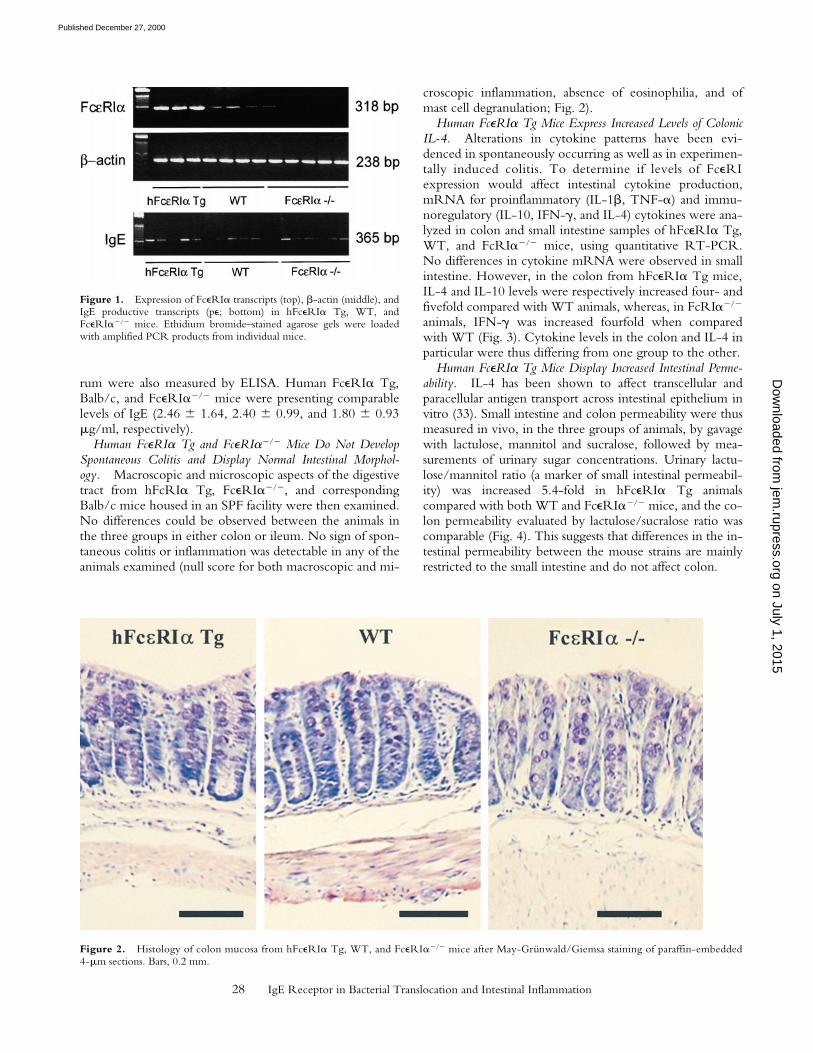
Igs. To rule out that differences in IgE levels betweenthe groups of animals would be superimposed to differen-tial expression of the receptor, presence of the productivetranscript for IgE was assessed in the colon by semiquantita-tive RT-PCR. No differences were observed in the sam-ples from the three groups of mice (Fig. 1, bottom), show-ing that IgE production by intestinal plasmocytes wascomparable. Likewise, total IgE concentrations in the se-
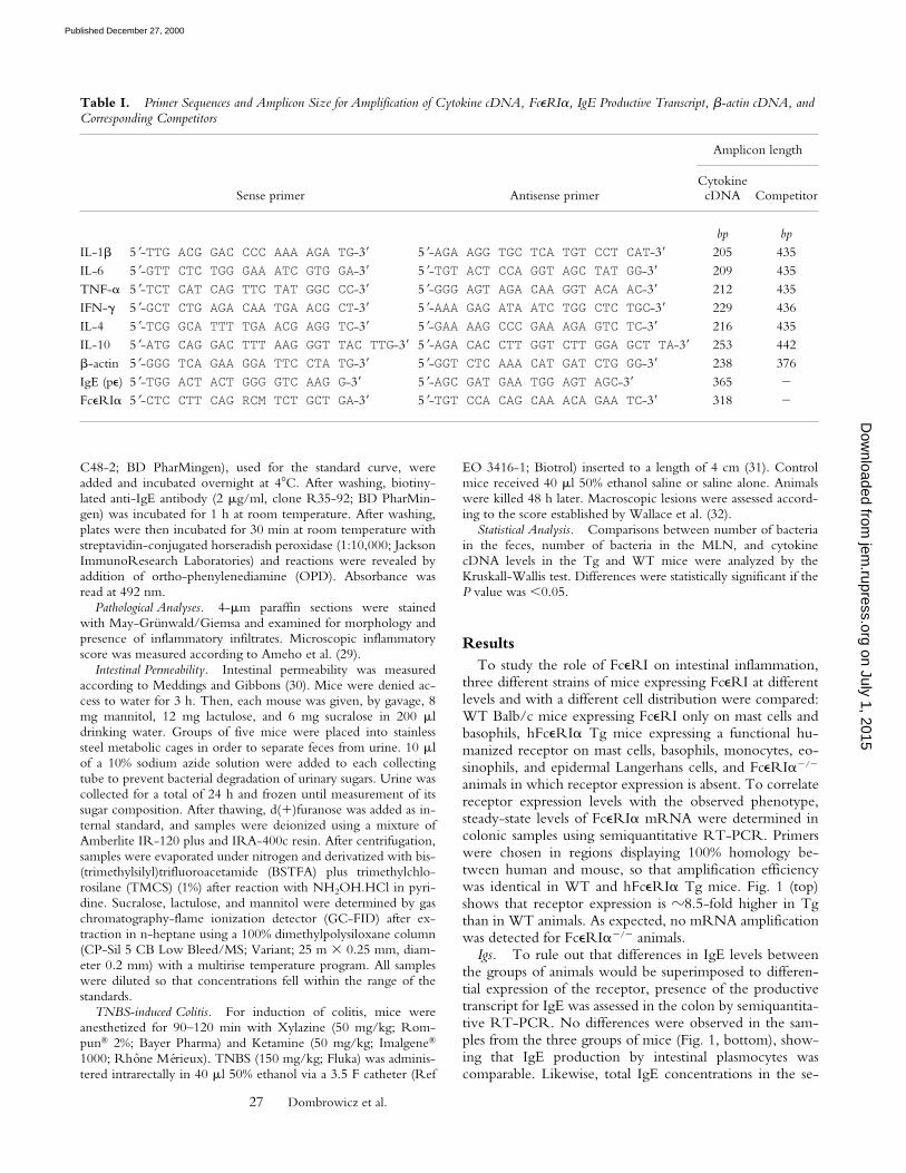
Table I. Primer Sequences and Amplicon Size for Amplification of Cytokine cDNA, FceRIa, IgE Productive Transcript, b-actin cDNA, and Corresponding Competitors
Amplicon length
Sense primer Antisense primerCytokinecDNA Competitor
bp bpIL-1b 59-TTG ACG GAC CCC AAA AGA TG-39 59-AGA AGG TGC TCA TGT CCT CAT-39 205 435IL-6 59-GTT CTC TGG GAA ATC GTG GA-39 59-TGT ACT CCA GGT AGC TAT GG-39 209 435TNF-a 59-TCT CAT CAG TTC TAT GGC CC-39 59-GGG AGT AGA CAA GGT ACA AC-39 212 435IFN-g 59-GCT CTG AGA CAA TGA ACG CT-39 59-AAA GAG ATA ATC TGG CTC TGC-39 229 436IL-4 59-TCG GCA TTT TGA ACG AGG TC-39 59-GAA AAG CCC GAA AGA GTC TC-39 216 435IL-10 59-ATG CAG GAC TTT AAG GGT TAC TTG-39 59-AGA CAC CTT GGT CTT GGA GCT TA-39 253 442b-actin 59-GGG TCA GAA GGA TTC CTA TG-39 59-GGT CTC AAA CAT GAT CTG GG-39 238 376IgE (pe) 59-TGG ACT ACT GGG GTC AAG G-39 59-AGC GAT GAA TGG AGT AGC-39 365 2
FceRIa 59-CTC CTT CAG RCM TCT GCT GA-39 59-TGT CCA CAG CAA ACA GAA TC-39 318 2
on July 1, 2015jem
.rupress.orgD
ownloaded from
Published December 27, 2000
28 IgE Receptor in Bacterial Translocation and Intestinal Inflammation
rum were also measured by ELISA. Human FceRIa Tg,Balb/c, and FceRIa2/2 mice were presenting comparablelevels of IgE (2.46 6 1.64, 2.40 6 0.99, and 1.80 6 0.93mg/ml, respectively).
Human FceRIa Tg and FceRIa2/2 Mice Do Not DevelopSpontaneous Colitis and Display Normal Intestinal Morphol-ogy. Macroscopic and microscopic aspects of the digestivetract from hFcRIa Tg, FceRIa2/2, and correspondingBalb/c mice housed in an SPF facility were then examined.No differences could be observed between the animals inthe three groups in either colon or ileum. No sign of spon-taneous colitis or inflammation was detectable in any of theanimals examined (null score for both macroscopic and mi-
croscopic inflammation, absence of eosinophilia, and ofmast cell degranulation; Fig. 2).
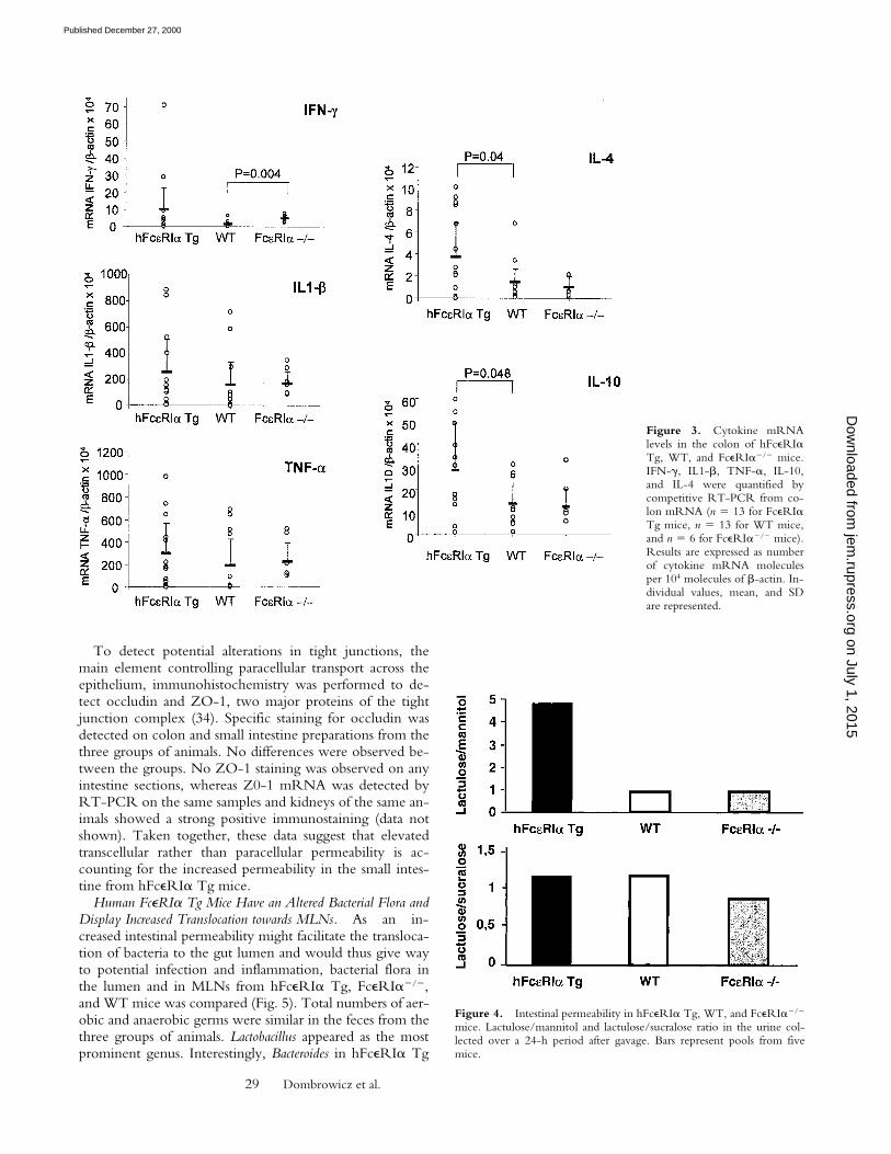
Human FceRIa Tg Mice Express Increased Levels of ColonicIL-4. Alterations in cytokine patterns have been evi-denced in spontaneously occurring as well as in experimen-tally induced colitis. To determine if levels of FceRIexpression would affect intestinal cytokine production,mRNA for proinflammatory (IL-1b, TNF-a) and immu-noregulatory (IL-10, IFN-g, and IL-4) cytokines were ana-lyzed in colon and small intestine samples of hFceRIa Tg,WT, and FcRIa2/2 mice, using quantitative RT-PCR.No differences in cytokine mRNA were observed in smallintestine. However, in the colon from hFceRIa Tg mice,IL-4 and IL-10 levels were respectively increased four- andfivefold compared with WT animals, whereas, in FcRIa2/2
animals, IFN-g was increased fourfold when comparedwith WT (Fig. 3). Cytokine levels in the colon and IL-4 inparticular were thus differing from one group to the other.
Human FceRIa Tg Mice Display Increased Intestinal Perme-ability. IL-4 has been shown to affect transcellular andparacellular antigen transport across intestinal epithelium invitro (33). Small intestine and colon permeability were thusmeasured in vivo, in the three groups of animals, by gavagewith lactulose, mannitol and sucralose, followed by mea-surements of urinary sugar concentrations. Urinary lactu-lose/mannitol ratio (a marker of small intestinal permeabil-ity) was increased 5.4-fold in hFceRIa Tg animalscompared with both WT and FceRIa2/2 mice, and the co-lon permeability evaluated by lactulose/sucralose ratio wascomparable (Fig. 4). This suggests that differences in the in-testinal permeability between the mouse strains are mainlyrestricted to the small intestine and do not affect colon.
Figure 1. Expression of FceRIa transcripts (top), b-actin (middle), andIgE productive transcripts (pe; bottom) in hFceRIa Tg, WT, andFceRIa2/2 mice. Ethidium bromide–stained agarose gels were loadedwith amplified PCR products from individual mice.
Figure 2. Histology of colon mucosa from hFceRIa Tg, WT, and FceRIa2/2 mice after May-Grünwald/Giemsa staining of paraffin-embedded4-mm sections. Bars, 0.2 mm.
on July 1, 2015jem
.rupress.orgD
ownloaded from
Published December 27, 2000
29 Dombrowicz et al.
To detect potential alterations in tight junctions, themain element controlling paracellular transport across theepithelium, immunohistochemistry was performed to de-tect occludin and ZO-1, two major proteins of the tightjunction complex (34). Specific staining for occludin wasdetected on colon and small intestine preparations from thethree groups of animals. No differences were observed be-tween the groups. No ZO-1 staining was observed on anyintestine sections, whereas Z0-1 mRNA was detected byRT-PCR on the same samples and kidneys of the same an-imals showed a strong positive immunostaining (data notshown). Taken together, these data suggest that elevatedtranscellular rather than paracellular permeability is ac-counting for the increased permeability in the small intes-tine from hFceRIa Tg mice.
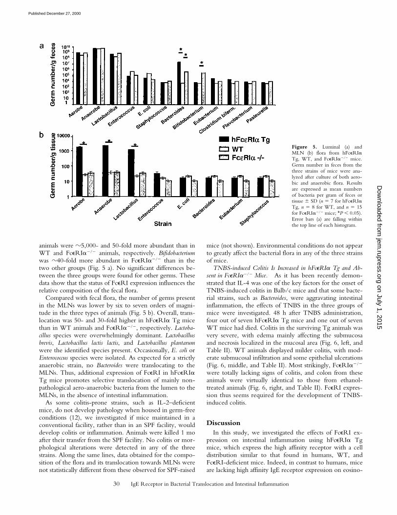
Human FceRIa Tg Mice Have an Altered Bacterial Flora andDisplay Increased Translocation towards MLNs. As an in-creased intestinal permeability might facilitate the transloca-tion of bacteria to the gut lumen and would thus give wayto potential infection and inflammation, bacterial flora inthe lumen and in MLNs from hFceRIa Tg, FceRIa2/2,and WT mice was compared (Fig. 5). Total numbers of aer-obic and anaerobic germs were similar in the feces from thethree groups of animals. Lactobacillus appeared as the mostprominent genus. Interestingly, Bacteroides in hFceRIa Tg
Figure 3. Cytokine mRNAlevels in the colon of hFceRIaTg, WT, and FceRIa2/2 mice.IFN-g, IL1-b, TNF-a, IL-10,and IL-4 were quantified bycompetitive RT-PCR from co-lon mRNA (n 5 13 for FceRIaTg mice, n 5 13 for WT mice,and n 5 6 for FceRIa2/2 mice).Results are expressed as numberof cytokine mRNA moleculesper 104 molecules of b-actin. In-dividual values, mean, and SDare represented.
Figure 4. Intestinal permeability in hFceRIa Tg, WT, and FceRIa2/2
mice. Lactulose/mannitol and lactulose/sucralose ratio in the urine col-lected over a 24-h period after gavage. Bars represent pools from fivemice.
on July 1, 2015jem
.rupress.orgD
ownloaded from
Published December 27, 2000
30 IgE Receptor in Bacterial Translocation and Intestinal Inflammation
animals were z5,000- and 50-fold more abundant than inWT and FceRIa2/2 animals, respectively. Bifidobacteriumwas z40-fold more abundant in FceRIa2/2 than in thetwo other groups (Fig. 5 a). No significant differences be-tween the three groups were found for other germs. Thesedata show that the status of FceRI expression influences therelative composition of the fecal flora.
Compared with fecal flora, the number of germs presentin the MLNs was lower by six to seven orders of magni-tude in the three types of animals (Fig. 5 b). Overall, trans-location was 50- and 30-fold higher in hFceRIa Tg micethan in WT animals and FceRIa2/2, respectively. Lactoba-cillus species were overwhelmingly dominant. Lactobacillusbrevis, Lactobacillus lactis lactis, and Lactobacillus plantarumwere the identified species present. Occasionally, E. coli orEnterococcus species were isolated. As expected for a strictlyanaerobic strain, no Bacteroides were translocating to theMLNs. Thus, additional expression of FceRI in hFceRIaTg mice promotes selective translocation of mainly non-pathological aero-anaerobic bacteria from the lumen to theMLNs, in the absence of intestinal inflammation.
As some colitis-prone strains, such as IL-2–deficientmice, do not develop pathology when housed in germ-freeconditions (12), we investigated if mice maintained in aconventional facility, rather than in an SPF facility, woulddevelop colitis or inflammation. Animals were killed 1 moafter their transfer from the SPF facility. No colitis or mor-phological alterations were detected in any of the threestrains. Along the same lines, data obtained for the compo-sition of the flora and its translocation towards MLNs werenot statistically different from these observed for SPF-raised
mice (not shown). Environmental conditions do not appearto greatly affect the bacterial flora in any of the three strainsof mice.
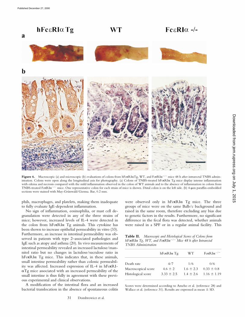
TNBS-induced Colitis Is Increased in hFceRIa Tg and Ab-sent in FceRIa2/2 Mice. As it has been recently demon-strated that IL-4 was one of the key factors for the onset ofTNBS-induced colitis in Balb/c mice and that some bacte-rial strains, such as Bacteroides, were aggravating intestinalinflammation, the effects of TNBS in the three groups ofmice were investigated. 48 h after TNBS administration,four out of seven hFceRIa Tg mice and one out of sevenWT mice had died. Colitis in the surviving Tg animals wasvery severe, with edema mainly affecting the submucosaand necrosis localized in the mucosal area (Fig. 6, left, andTable II). WT animals displayed milder colitis, with mod-erate submucosal infiltration and some epithelial ulcerations(Fig. 6, middle, and Table II). Most strikingly, FceRIa2/2
were totally lacking signs of colitis, and colon from theseanimals were virtually identical to those from ethanol-treated animals (Fig. 6, right, and Table II). FceRI expres-sion thus seems required for the development of TNBS-induced colitis.
DiscussionIn this study, we investigated the effects of FceRI ex-
pression on intestinal inflammation using hFceRIa Tgmice, which express the high affinity receptor with a celldistribution similar to that found in humans, WT, andFceRI-deficient mice. Indeed, in contrast to humans, miceare lacking high affinity IgE receptor expression on eosino-
Figure 5. Luminal (a) andMLN (b) flora from hFceRIaTg, WT, and FceRIa2/2 mice.Germ number in feces from thethree strains of mice were ana-lyzed after culture of both aero-bic and anaerobic flora. Resultsare expressed as mean numbersof bacteria per gram of feces ortissue 6 SD (n 5 7 for hFceRIaTg, n 5 8 for WT, and n 5 15for FceRIa2/2 mice; *P , 0.05).Error bars (a) are falling withinthe top line of each histogram.
on July 1, 2015jem
.rupress.orgD
ownloaded from
Published December 27, 2000
31 Dombrowicz et al.
phils, macrophages, and platelets, making them inadequateto fully evaluate IgE-dependent inflammation.
No sign of inflammation, eosinophilia, or mast cell de-granulation were detected in any of the three strains ofmice; however, increased levels of IL-4 were detected inthe colon from hFceRIa Tg animals. This cytokine hasbeen shown to increase epithelial permeability in vitro (33).Furthermore, an increase in intestinal permeability was ob-served in patients with type 2–associated pathologies andIgE such as atopy and asthma (20). In vivo measurements ofintestinal permeability revealed an increased lactulose/man-nitol ratio but no changes in lactulose/sucralose ratio inhFceRIa Tg mice. This indicates that, in these animals,small intestine permeability rather than colonic permeabil-ity was affected. Increased expression of IL-4 in hFceRI-aTg mice associated with an increased permeability of thesmall intestine is thus fully in agreement with these previ-ous experimental and clinical observations.
A modification of the intestinal flora and an increasedbacterial translocation in the absence of spontaneous colitis
were observed only in hFceRIa Tg mice. The threegroups of mice were on the same Balb/c background andraised in the same room, therefore excluding any bias dueto genetic factors in the results. Furthermore, no significantdifference in the fecal flora was detected, whether animalswere raised in a SPF or in a regular animal facility. This
Figure 6. Macroscopic (a) and microscopic (b) evaluations of colons from hFceRIaTg, WT, and FceRIa2/2 mice 48 h after intrarectal TNBS admin-istration. Colons were open along the longitudinal axis for photography. (a) Colons of TNBS-treated hFceRIa Tg mice display intense inflammationwith edema and necrosis compared with the mild inflammation observed in the colon of WT animals and to the absence of inflammation in colons fromTNBS-treated FceRIa2/2 mice. One representative colon for each strain of mice is shown. Distal colon is on the left side. (b) 4-mm paraffin-embeddedsections were stained with May-Grünwald/Giemsa. Bar, 0.2 mm.
Table II. Macroscopic and Histological Scores of Colons from hFceRIa Tg, WT, and FceRIa2/2 Mice 48 h after Intrarectal TNBS Administration
hFceRIa Tg WT FceRIa2/2
Death rate 4/7 1/6 0/6Macroscopical score 4.6 6 2 1.6 6 2.3 0.33 6 0.8Histological score 3.33 6 2.5 1.4 6 2.6 1.16 6 1.19
Scores were determined according to Ameho et al. (reference 28) andWallace et al. (reference 31). Results are expressed as mean 6 SD.
on July 1, 2015jem
.rupress.orgD
ownloaded from
Published December 27, 2000
32 IgE Receptor in Bacterial Translocation and Intestinal Inflammation
suggests that the composition of the flora was not modifiedaccording to the external environment. Similarly, no quan-titative differences in the fecal flora between the threegroups of animals were detected. In contrast, major qualita-tive differences were observed. Indeed, Bacteroides was5,000- and 50-fold more abundant in hFceRIaTg than inWT and FceRI-deficient animals, respectively. Thesestrictly anaerobic bacteria have been associated in the colitisoccurring in a rat model with a modified immune system(HLA-B27/b2 microglobulin Tg animals). Indeed, thenormal endogenous flora (containing Bacteroides) is suffi-cient to trigger colitis in these animals, whereas non-Tg an-imals harboring the same flora do not develop colitis. Coli-tis in Tg rats can only be prevented by keeping the animalsin germ-free conditions (35). Furthermore, increased num-ber of B. vulgatus are found in feces from patients with CD(23). Likewise, a significant increase in the number of lu-minal ileal and colic B. fragilis has been observed (24). Fi-nally, Bacteroides was also associated to an extra-intestinalinflammatory pathology. Indeed, a comparative study be-tween healthy Estonian (country with low atopy preva-lence) and Swedish (country with high atopy prevalence)1-yr-old children, has shown that the former had highernumber of Lactobacilli and Eubacteria, whereas the later dis-played higher number of Bacteroides and Clostridia (36). In-creased population of Bifidobacterium, which are 40-foldmore abundant in FceRI-deficient animals, have been in-volved in the development of gut-associated lymphoid tis-sue in neonates and in particular in the regulation of type 2responses in mice (37). Levels of FceRI expression and dif-ferences in receptor cellular distribution thus affect thecomposition of fecal flora.
As expected after the permeability measurements, bacte-rial translocation from the intestinal lumen towards MLNswas also affected by FceRI expression. The increased spon-taneous translocation observed in hFceRIa Tg animals wasmainly affecting Lactobacillus. There were no Bacteroidesfound in MLNs from any strain of animal. Indeed, it hasbeen reported that aerobes such as Lactobacillus are very effi-ciently translocated, whereas anaerobic germs, such asBacteroides, poorly translocate (38). Would some anaerobicbacteria cross the intestinal wall and reach the peritonealcavity, they would not withstand the aerobic environmentand thus do not survive lest proliferate.
Although mechanisms of bacterial translocation are stillunclear, it is considered that bacteria are either crossing theintestinal wall by a transcellular way or taking a paracellularroute (38). In this later case, a disruption or some looseningof the tight junctions maintaining the integrity of the epi-thelial barrier would have to take place. However, nomodification of occludin expression was detected by im-munohistochemistry. As tight junctions do not seem to beaffected, it is likely that an increased transcellular passage ofbacteria is responsible for the increased bacterial transloca-tion. There is no report so far on Lactobacillus-specific trans-cytosis mechanisms. It has been reported that Campylo-bacter jejuni were translocating across Caco-2 cells using aparacellular way (39). Likewise, E. coli isolated from pa-
tients with necrotizing enterocolitis were transcytosed invitro in the absence of ultrastructural changes (40).
In the present model of acute TNBS-induced colitis(31), strikingly different effects were observed on the threestrains of mice. FceRI-deficient mice were completely pro-tected from colitis, suggesting that FceRI expressed onmast cells (and basophils) was involved in the pathologyonset. By contrast, colitis in hFceRIaTg mice was exacer-bated, even leading to a high death rate. Higher IL-4 levelsin these colitis-sensitive animals are in agreement with theinhibition of chronic (10 d) TNBS-induced colitis in IL-4–deficient animals (14) and with the absence of colitis inTCR-a2/2 3 IL-42/2 animals (10). Furthermore, the in-creased number of Bacteroides found in hFceRIa Tg mightalso have increased the severity of colitis, as demonstratedpreviously on TNBS-induced colitis in rat (41, 42). Finally,additional FceRI expression on eosinophils and macro-phages compared with WT animals might also contribute tothe increased pathology by affecting the intestinal barrier.
hFceRIaTg mice might thus be considered in a state of“pre-IBD” with increased number of luminal Bacteroides,increased Lactobacillus translocation to the MLNs, increasedsmall intestinal permeability, higher colonic levels of IL-4,but no macroscopic or microscopic symptom of inflamma-tion. Along these lines, increase of intestinal permeability(measured by lactulose/mannitol ratio) in the absence of aclinical sign of inflammation has been considered as apredictive marker of relapse in patients with remitting CD(43, 44).
If bacterial flora has been shown to influence the devel-opment of immune responses (45), we have reported herethat alterations of the immune system were also able to af-fect bacterial flora and its translocation. Besides the in-volvement of FceRI in susceptibility to intestinal inflam-mation, we have recently obtained evidences that IgE wasalso increasing susceptibility to colitis (Nutten, S., manu-script in preparation). Thus, pharmacological targeting ofIgE or its high affinity receptor, with reagents such asnonanaphylactogenic anti-IgE antibodies, would thereforebe potentially useful for treatment of IBD. In this context,colitis-prone hFceRIa Tg mice represent a valuable modelto assess the potency of compounds, which would preventthe development of clinically detectable inflammation.
We are grateful to Prof. M. Peuchmaur for his help with histologi-cal examination, to Prof. A. Cortot for critical reading of the manu-script, to Dr. S. Molinary (Tate and Lyle, Reading, UK) for provid-ing us with purified sucralose, and to Dr. J.-P. Kinet (HarvardMedical School, Boston, MA) for allowing the use of hFceRIaTgand FceRIa2/2 mice. V. Prima, V. Jeronimo, J.-P. Papin, G. Spik,and F. Monsuur also provided skilful technical assistance.
This work was supported in part by a grant from François Au-petit Foundation, by CR Institut National de la Sante et de la Re-cherche Medicale grant 4U004B, and a grant from University hos-pital (Programme Hospitalier de Recherche Clinique).
Submitted: 26 June 2000Revised: 16 November 2000Accepted: 20 November 2000
on July 1, 2015jem
.rupress.orgD
ownloaded from
Published December 27, 2000
33 Dombrowicz et al.
References1. Kinet, J.P. 1999. The high-affinity IgE receptor (Fc epsilon
RI): from physiology to pathology. Annu. Rev. Immunol. 17:931–972.
2. Dombrowicz, D., A.T. Brini, V. Flamand, E. Hicks, J.N.Snouwaert, J.P. Kinet, and B.H. Koller. 1996. Anaphylaxismediated through a humanized high affinity IgE receptor. J.Immunol. 157:1645–1651.
3. Dombrowicz, D., S. Lin, V. Flamand, A.T. Brini, B.H. Kol-ler, and J.P. Kinet. 1998. Allergy-associated FcRbeta is a mo-lecular amplifier of IgE- and IgG-mediated in vivo responses.Immunity. 8:517–529.
4. Dombrowicz, D., V. Flamand, K.K. Brigman, B.H. Koller,and J.P. Kinet. 1993. Abolition of anaphylaxis by targeteddisruption of the high affinity immunoglobulin E receptor al-pha chain gene. Cell. 75:969–976.
5. Fiocchi, C. 1998. Inflammatory bowel disease: etiology andpathogenesis. Gastroenterology. 115:182–205.
6. Elson, C.O., R.B. Sartor, G.S. Tennyson, and R.H. Riddell.1995. Experimental models of inflammatory bowel disease.Gastroenterology. 109:1344–1367.
7. Bhan, A.K., E. Mizoguchi, R.N. Smith, and A. Mizoguchi.1999. Colitis in transgenic and knockout animals as models ofhuman inflammatory bowel disease. Immunol. Rev. 169:195–207.
8. Mizoguchi, A., E. Mizoguchi, C. Chiba, G.M. Spiekermann,S. Tonegawa, C. Nagler-Anderson, and A.K. Bhan. 1996.Cytokine imbalance and autoantibody production in T cellreceptor-a mutant mice with inflammatory bowel disease. J.Exp. Med. 183:847–856.
9. Wen, L., S.J. Roberts, J.L. Viney, F.S. Wong, C. Mallick,R.C. Findly, Q. Peng, J.E. Craft, M.J. Owen, and A.C. Hay-day. 1994. Immunoglobulin synthesis and generalized au-toimmunity in mice congenitally deficient in alpha beta(1) Tcells. Nature. 369:654–658.
10. Mizoguchi, A., E. Mizoguchi, and A.K. Bhan. 1999. Thecritical role of interleukin 4 but not interferon gamma in thepathogenesis of colitis in T-cell receptor alpha mutant mice.Gastroenterology. 116:320–326.
11. Schorle, H., T. Holtschke, T. Hunig, A. Schimpl, and I.Horak. 1991. Development and function of T cells in micerendered interleukin-2 deficient by gene targeting. Nature.352:621–624.
12. Sadlack, B., H. Merz, H. Schorle, A. Schimpl, A.C. Feller,and I. Horak. 1993. Ulcerative colitis-like disease in micewith a disrupted interleukin-2 gene. Cell. 75:253–261.
13. Dianda, L., A.M. Hanby, N.A. Wright, A. Sebesteny, A.C.Hayday, and M.J. Owen. 1997. T cell receptor-alpha beta-deficient mice fail to develop colitis in the absence of a mi-crobial environment. Am. J. Pathol. 150:91–97.
14. Dohi, T., K. Fujihashi, P.D. Rennert, K. Iwatani, H. Ki-yono, and J.R. McGhee. 1999. Hapten-induced colitis is as-sociated with colonic patch hypertrophy and T helper cell2–type responses. J. Exp. Med. 189:1169–1180.
15. Dohi, T., K. Fujihashi, H. Kiyono, C.O. Elson, and J.R.McGhee. 2000. Mice deficient in Th1- and Th2-type cyto-kines develop distinct forms of hapten-induced colitis. Gastro-enterology. 119:724–733.
16. Desreumaux, P., E. Brandt, L. Gambiez, D. Emilie, K. Ge-boes, O. Klein, N. Ectors, A. Cortot, M. Capron, and J.F.Colombel. 1997. Distinct cytokine patterns in early andchronic ileal lesions of Crohn’s disease. Gastroenterology. 113:118–126.
17. Desreumaux, P., K. Geboes, L. Gambiez, N. Ectors, O.Klein, A. Cortot, M. Capron, and J.F. Colombel. 1998. Earlyileal lesions of Crohn’s disease (CD) are associated with theexpression of IL-4 and IgE and not of inflammatory cyto-kines. Gastroenterology. 114:G3945.
18. Yacyshyn, B.R., and J.B. Meddings. 1995. CD45RO expres-sion on circulating CD191 B cells in Crohn’s disease corre-lates with intestinal permeability. Gastroenterology. 108:132–137.
19. Wallaert, B., P. Desreumaux, M.C. Copin, I. Tillie, A. Be-nard, J.F. Colombel, B. Gosselin, A.B. Tonnel, and A. Janin.1995. Immunoreactivity for interleukin 3 and 5 and granulo-cyte/macrophage colony–stimulating factor of intestinal mu-cosa in bronchial asthma. J. Exp. Med. 182:1897–1904.
20. Benard, A., P. Desreumeaux, D. Huglo, A. Hoorelbeke,A.B. Tonnel, and B. Wallaert. 1996. Increased intestinal per-meability in bronchial asthma. J. Allergy Clin. Immunol. 97:1173–1178.
21. Rutgeerts, P., K. Geboes, G. Vantrappen, J. Beyls, R. Kerre-mans, and M. Hiele. 1990. Predictability of the postoperativecourse of Crohn’s disease. Gastroenterology. 99:956–963.
22. D’Haens, G.R., K. Geboes, M. Peeters, F. Baert, F. Pen-ninckx, and P. Rutgeerts. 1998. Early lesions of recurrentCrohn’s disease caused by infusion of intestinal contents inexcluded ileum. Gastroenterology. 114:262–267.
23. Van de Merwe, J.P., A.M. Schroder, F. Wensinck, and M.P.Hazenberg. 1988. The obligate anaerobic faecal flora of pa-tients with Crohn’s disease and their first-degree relatives.Scand. J. Gastroenterol. 23:1125–1131.
24. Keighley, M.R., Y. Arabi, F. Dimock, D.W. Burdon, R.N.Allan, and J. Alexander-Williams. 1978. Influence of inflam-matory bowel disease on intestinal microflora. Gut. 19:1099–1104.
25. Darfeuille-Michaud, A., C. Neut, N. Barnich, E. Lederman,P. Di Martino, P. Desreumaux, L. Gambiez, B. Joly, A. Cor-tot, and J.F. Colombel. 1998. Presence of adherent Escherichiacoli strains in ileal mucosa of patients with Crohn’s disease.Gastroenterology. 115:1405–1413.
26. Peach, S., M.R. Lock, D. Katz, I.P. Todd, and S. Tabaqchali.1978. Mucosal-associated bacterial flora of the intestine in pa-tients with Crohn’s disease and in a control group. Gut. 19:1034–1042.
27. Panwala, C.M., J.C. Jones, and J.L. Viney. 1998. A novelmodel of inflammatory bowel disease: mice deficient for themultiple drug resistance gene, mdr1a, spontaneously developcolitis. J. Immunol. 161:5733–5744.
28. Dombrowicz, D., V. Flamand, I. Miyajima, J.V. Ravetch,S.J. Galli, and J.P. Kinet. 1997. Absence of Fc epsilonRI al-pha chain results in upregulation of Fc gammaRIII-depen-dent mast cell degranulation and anaphylaxis. Evidence ofcompetition between Fc epsilonRI and Fc gammaRIII forlimiting amounts of FcR beta and gamma chains. J. Clin. In-vest. 99:915–925.
29. Ameho, C.K., A.A. Adjei, E.K. Harrison, K. Takeshita, T.Morioka, Y. Arakaki, E. Ito, I. Suzuki, A.D. Kulkarni, A.Kawajiri, and S. Yamamoto. 1997. Prophylactic effect of di-etary glutamine supplementation on interleukin 8 and tu-mour necrosis factor alpha production in trinitrobenzene sul-phonic acid induced colitis. Gut. 41:487–493.
30. Meddings, J.B., and I. Gibbons. 1998. Discrimination of site-specific alterations in gastrointestinal permeability in the rat.Gastroenterology. 114:83–92.
on July 1, 2015jem
.rupress.orgD
ownloaded from
Published December 27, 2000
34 IgE Receptor in Bacterial Translocation and Intestinal Inflammation
31. Mazelin, L., V. Theodorou, J. More, J. Fioramonti, and L.Bueno. 1998. Protective role of vagal afferents in experimen-tally-induced colitis in rats. J. Auton. Nerv. Syst. 73:38–45.
32. Wallace, J.L., W.K. MacNaughton, G.P. Morris, and P.L.Beck. 1989. Inhibition of leukotriene synthesis markedly ac-celerates healing in a rat model of inflammatory bowel dis-ease. Gastroenterology. 96:29–36.
33. Berin, M.C., P.C. Yang, L. Ciok, S. Waserman, and M.H.Perdue. 1999. Role for IL-4 in macromolecular transportacross human intestinal epithelium. Am. J. Physiol. 276:C1046–C1052.
34. Mitic, L.L., and J.M. Anderson. 1998. Molecular architectureof tight junctions. Annu. Rev. Physiol. 60:121–142.
35. Rath, H.C., H.H. Herfarth, J.S. Ikeda, W.B. Grenther, T.E.Hamm, Jr., E. Balish, J.D. Taurog, R.E. Hammer, K.H. Wil-son, and R.B. Sartor. 1996. Normal luminal bacteria, espe-cially Bacteroides species, mediate chronic colitis, gastritis,and arthritis in HLA-B27/human beta2 microglobulin trans-genic rats. J. Clin. Invest. 98:945–953.
36. Sepp, E., K. Julge, M. Vasar, P. Naaber, B. Bjorksten, and M.Mikelsaar. 1997. Intestinal microflora of Estonian and Swed-ish infants. Acta. Paediatr. 86:956–961.
37. Sudo, N., S. Sawamura, K. Tanaka, Y. Aiba, C. Kubo, andY. Koga. 1997. The requirement of intestinal bacterial florafor the development of an IgE production system fully sus-ceptible to oral tolerance induction. J. Immunol. 159:1739–1745.
38. Berg, R.D. 1995. Bacterial translocation from the gastrointes-tinal tract. Trends Microbiol. 3:149–154.
39. Bras, A.M., and J.M. Ketley. 1999. Transcellular translocationof Campylobacter jejuni across human polarised epithelialmonolayers. FEMS (Fed. Eur. Microbiol. Soc.) Microbiol. Lett.179:209–215.
40. Panigrahi, P., P. Bamford, K. Horvath, J.G. Morris, Jr., andI.H. Gewolb. 1996. Escherichia coli transcytosis in a Caco-2cell model: implications in neonatal necrotizing enterocolitis.Pediatr. Res. 40:415–421.
41. Garcia-Lafuente, A., M. Antolin, F. Guarner, E. Crespo, A.Salas, P. Forcada, M. Laguarda, J. Gavalda, J.A. Baena, J.Vilaseca, and J.R. Malagelada. 1997. Incrimination of anaero-bic bacteria in the induction of experimental colitis. Am. J.Physiol. 272:G10–G15.
42. Garcia-Lafuente, A., M. Antolin, F. Guarner, E. Crespo, A.Salas, P. Forcada, and J. Malagelada. 1998. Derangement ofmucosal barrier function by bacteria colonizing the rat co-lonic mucosa. Eur. J. Clin. Invest. 28:1019–1026.
43. Wyatt, J., H. Vogelsang, W. Hubl, T. Waldhoer, and H.Lochs. 1993. Intestinal permeability and the prediction of re-lapse in Crohn’s disease. Lancet. 341:1437–1439.
44. D’Inca, R., V. Di Leo, G. Corrao, D. Martines, A. D’Odor-ico, C. Mestriner, C. Venturi, G. Longo, and G.C. Sturni-olo. 1999. Intestinal permeability test as a predictor of clinicalcourse in Crohn’s disease. Am. J. Gastroenterol. 94:2956–2960.
45. Bjorksten, B., P. Naaber, E. Sepp, and M. Mikelsaar. 1999.The intestinal microflora in allergic Estonian and Swedish2-year-old children. Clin. Exp. Allergy. 29:342–346.
on July 1, 2015jem
.rupress.orgD
ownloaded from
Published December 27, 2000
Top Related
Copyright © 2022 FDOKUMEN

