


Bahasa
Halaman
Hukum

RESEARCH ARTICLE Open Access
Recent acquisition of imprinting at the rodentSfmbt2 locus correlates with insertion of a largeblock of miRNAsQianwei Wang1†, Jacqueline Chow1†, Jenny Hong1†, Anne Ferguson Smith2, Carol Moreno3, Peter Seaby4,Paul Vrana5, Kamelia Miri1, Joon Tak1, Eu Ddeum Chung1, Gabriela Mastromonaco4,6, Isabella Caniggia7 andSusannah Varmuza1*
Abstract
Background: The proximal region of murine Chr 2 has long been known to harbour one or more imprinted genesfrom classic genetic studies involving reciprocal translocations. No imprinted gene had been identified from thisregion until our study demonstrated that the PcG gene Sfmbt2 is expressed from the paternally inherited allele inearly embryos and extraembryonic tissues. Imprinted genes generally reside in clusters near elements termedImprinting Control Regions (ICRs), suggesting that Sfmbt2 might represent an anchor for a new imprinted domain.
Results: We analyzed allelic expression of approximately 20 genes within a 3.9 Mb domain and found that Sfmbt2and an overlapping non-coding antisense transcript are the only imprinted genes in this region. These transcriptsrepresent a very narrow imprinted gene locus. We also demonstrate that rat Sfmbt2 is imprinted in extraembryonictissues. An interesting feature of both mouse and rat Sfmbt2 genes is the presence of a large block of miRNAs inintron 10. Other mammals, including the bovine, lack this block of miRNAs. Consistent with this association, weshow that human and bovine Sfmbt2 are biallelic. Other evidence indicates that pig Sfmbt2 is also not imprinted.Further strengthening the argument for recent evolution of Sfmbt2 is our demonstration that a more distantmuroid rodent, Peromyscus also lacks imprinting and the block of miRNAs.
Conclusions: These observations are consistent with the hypothesis that the block of miRNAs are drivingimprinting at this locus. Our results are discussed in the context of ncRNAs at other imprinted loci.Accession numbers for Peromyscus cDNA and intron 10 genomic DNA are [Genbank:HQ416417 and Genbank:HQ416418], respectively.
BackgroundGenomic imprinting is an epigenetic process that affectsa small subset of genes resulting in their expression/repression in a parent of origin dependent fashion. Oneset of imprinted genes is expressed only from the pater-nally inherited allele, while another set is expressed onlyfrom the maternally inherited chromosome. The imprintis reset at each generation when the two haploid gen-omes are separate, either during gametogenesis or
immediately after fertilization, when the two genomesare physically separated in their own pronuclei.Imprinted genes generally reside in clusters around a
cis acting element called an Imprinting Control Region(ICR) that exerts its effects over a large chromosomaldomain (up to 4 Mb) (reviewed in [1]). Imprinteddomains can contain genes that are biallelic, paternallyexpressed or maternally expressed. Monoallelic expres-sion can be universal (ie in all tissues tested), or limitedto a subset of tissues. The most common type of tissuespecific limitation is to the extraembryonic lineages,exemplified by the placenta and the yolk sac. This latterobservation has provided strong support for the ideathat the evolutionary origins of imprinting are rooted inextraembryonic tissue biology.
* Correspondence: [email protected]† Contributed equally1Department of Cell and Systems Biology, University of Toronto, 25 HarbordSt., Toronto, Ontario, M5S 3G5, CanadaFull list of author information is available at the end of the article
Wang et al. BMC Genomics 2011, 12:204http://www.biomedcentral.com/1471-2164/12/204
© 2011 Wang et al; licensee BioMed Central Ltd. This is an Open Access article distributed under the terms of the Creative CommonsAttribution License (http://creativecommons.org/licenses/by/2.0), which permits unrestricted use, distribution, and reproduction inany medium, provided the original work is properly cited.
In mouse uniparental embryos, the tissue most severelyaffected is the trophoblast, the precursor of several pla-cental cell types. Gynogenetic/parthenogenetic embryoshave almost none by midgestation, whereas androgeneticembryos have hyperplastic trophoblast. Moreover, deriva-tion of trophoblast stem cells from parthenogeneticmouse blastocysts is extremely inefficient, and is accom-panied by selective loss of imprinting of several genes(Miri and Varmuza, Parthenogenetic embryos areimpaired in their ability to make TS cells, manuscript inpreparation). These observations led us to hypothesizethat a gene critical for trophoblast establishment/func-tion in blastocysts is expressed from a paternally-inher-ited chromosome, and is therefore missing fromgynogenetic/parthenogenetic embryos. A microarraycomparison of the transcriptomes of androgenetic andgynogenetic blastocysts revealed that the PcG geneSfmbt2 is expressed almost exclusively from the paternalallele starting at the blastocyst stage [2]. Monoallelicexpression in all tissues is preserved up to e7.5, afterwhich a high level monoallelic expression is preserved inextraembryonic tissues, while significantly reduced, butbiallelic, expression in somatic tissues can be observed.These observations place Sfmbt2 in the class of imprintedgenes that are specific to the extraembryonic tissues.Sfmbt2 maps to the proximal region of Chromosome
2. This region was mapped as imprinted through thechromosome translocation studies of Cattanach and col-leagues [3,4], but no imprinted gene had been identifieduntil our study [2]. Here we extend our analysis of thedomain surrounding Sfmbt2, and the search for a poten-tial ICR. Our results indicate that Sfmbt2 is the onlygene within a 4 Mb region that is imprinted, and thatwe find no evidence of a classical ICR displaying robustgermline specific DNA methylation that is preservedafter fertilization. We also show that imprinting appearsto be unique to mice and rats, and is associated withthe acquisition of a block of miRNAs in one of theintrons. Our results suggest that we have caught a gene“in the act” of becoming imprinted.
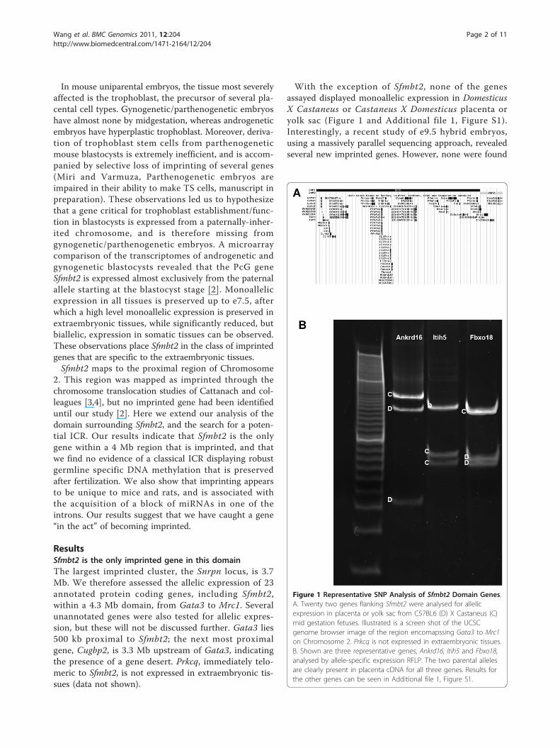
ResultsSfmbt2 is the only imprinted gene in this domainThe largest imprinted cluster, the Snrpn locus, is 3.7Mb. We therefore assessed the allelic expression of 23annotated protein coding genes, including Sfmbt2,within a 4.3 Mb domain, from Gata3 to Mrc1. Severalunannotated genes were also tested for allelic expres-sion, but these will not be discussed further. Gata3 lies500 kb proximal to Sfmbt2; the next most proximalgene, Cugbp2, is 3.3 Mb upstream of Gata3, indicatingthe presence of a gene desert. Prkcq, immediately telo-meric to Sfmbt2, is not expressed in extraembryonic tis-sues (data not shown).
With the exception of Sfmbt2, none of the genesassayed displayed monoallelic expression in DomesticusX Castaneus or Castaneus X Domesticus placenta oryolk sac (Figure 1 and Additional file 1, Figure S1).Interestingly, a recent study of e9.5 hybrid embryos,using a massively parallel sequencing approach, revealedseveral new imprinted genes. However, none were found
Figure 1 Representative SNP Analysis of Sfmbt2 Domain Genes.A. Twenty two genes flanking Sfmbt2 were analysed for allelicexpression in placenta or yolk sac from C57BL6 (D) X Castaneus (C)mid gestation fetuses. Illustrated is a screen shot of the UCSCgenome browser image of the region encomapssing Gata3 to Mrc1on Chromosome 2. Prkcq is not expressed in extraembryonic tissues.B. Shown are three representative genes, Ankrd16, Itih5 and Fbxo18,analysed by allele-specific expression RFLP. The two parental allelesare clearly present in placenta cDNA for all three genes. Results forthe other genes can be seen in Additional file 1, Figure S1.
Wang et al. BMC Genomics 2011, 12:204http://www.biomedcentral.com/1471-2164/12/204
Page 2 of 11
in the proximal region of Chromosome 2, includingSfmbt2 [5]. This is consistent with our observation thatSfmbt2 becomes biallelic in embryo-derived somatic tis-sues. None of the neighbouring genes is imprinted inembryo. Together with our findings, these results indi-cate that Sfmbt2 is the only imprinted gene within a 4.3Mb region of proximal Chromosome 2, and only inearly embryo and extraembryonic tissues.Sfmbt2 has multiple transcriptional starts, stops and
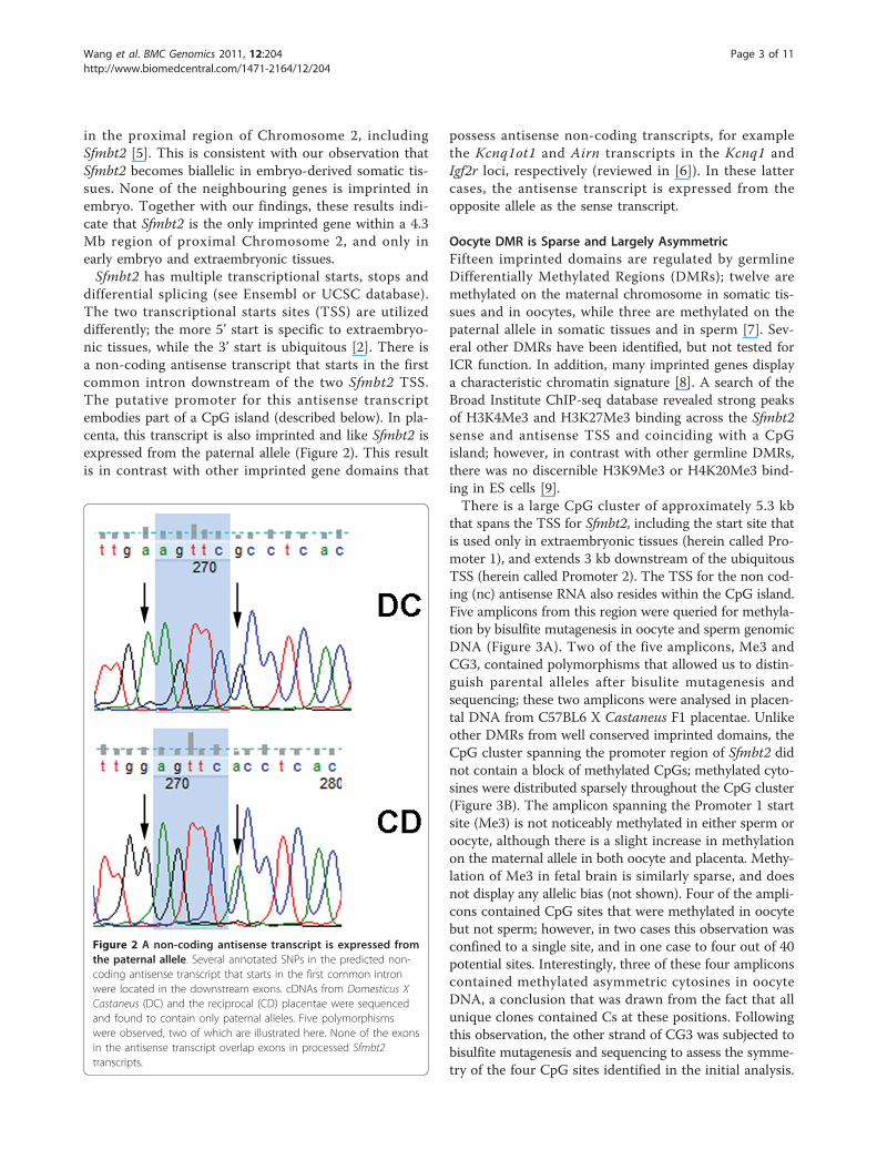
differential splicing (see Ensembl or UCSC database).The two transcriptional starts sites (TSS) are utilizeddifferently; the more 5’ start is specific to extraembryo-nic tissues, while the 3’ start is ubiquitous [2]. There isa non-coding antisense transcript that starts in the firstcommon intron downstream of the two Sfmbt2 TSS.The putative promoter for this antisense transcriptembodies part of a CpG island (described below). In pla-centa, this transcript is also imprinted and like Sfmbt2 isexpressed from the paternal allele (Figure 2). This resultis in contrast with other imprinted gene domains that
possess antisense non-coding transcripts, for examplethe Kcnq1ot1 and Airn transcripts in the Kcnq1 andIgf2r loci, respectively (reviewed in [6]). In these lattercases, the antisense transcript is expressed from theopposite allele as the sense transcript.
Oocyte DMR is Sparse and Largely AsymmetricFifteen imprinted domains are regulated by germlineDifferentially Methylated Regions (DMRs); twelve aremethylated on the maternal chromosome in somatic tis-sues and in oocytes, while three are methylated on thepaternal allele in somatic tissues and in sperm [7]. Sev-eral other DMRs have been identified, but not tested forICR function. In addition, many imprinted genes displaya characteristic chromatin signature [8]. A search of theBroad Institute ChIP-seq database revealed strong peaksof H3K4Me3 and H3K27Me3 binding across the Sfmbt2sense and antisense TSS and coinciding with a CpGisland; however, in contrast with other germline DMRs,there was no discernible H3K9Me3 or H4K20Me3 bind-ing in ES cells [9].There is a large CpG cluster of approximately 5.3 kb
that spans the TSS for Sfmbt2, including the start site thatis used only in extraembryonic tissues (herein called Pro-moter 1), and extends 3 kb downstream of the ubiquitousTSS (herein called Promoter 2). The TSS for the non cod-ing (nc) antisense RNA also resides within the CpG island.Five amplicons from this region were queried for methyla-tion by bisulfite mutagenesis in oocyte and sperm genomicDNA (Figure 3A). Two of the five amplicons, Me3 andCG3, contained polymorphisms that allowed us to distin-guish parental alleles after bisulite mutagenesis andsequencing; these two amplicons were analysed in placen-tal DNA from C57BL6 X Castaneus F1 placentae. Unlikeother DMRs from well conserved imprinted domains, theCpG cluster spanning the promoter region of Sfmbt2 didnot contain a block of methylated CpGs; methylated cyto-sines were distributed sparsely throughout the CpG cluster(Figure 3B). The amplicon spanning the Promoter 1 startsite (Me3) is not noticeably methylated in either sperm oroocyte, although there is a slight increase in methylationon the maternal allele in both oocyte and placenta. Methy-lation of Me3 in fetal brain is similarly sparse, and doesnot display any allelic bias (not shown). Four of the ampli-cons contained CpG sites that were methylated in oocytebut not sperm; however, in two cases this observation wasconfined to a single site, and in one case to four out of 40potential sites. Interestingly, three of these four ampliconscontained methylated asymmetric cytosines in oocyteDNA, a conclusion that was drawn from the fact that allunique clones contained Cs at these positions. Followingthis observation, the other strand of CG3 was subjected tobisulfite mutagenesis and sequencing to assess the symme-try of the four CpG sites identified in the initial analysis.
Figure 2 A non-coding antisense transcript is expressed fromthe paternal allele. Several annotated SNPs in the predicted non-coding antisense transcript that starts in the first common intronwere located in the downstream exons. cDNAs from Domesticus XCastaneus (DC) and the reciprocal (CD) placentae were sequencedand found to contain only paternal alleles. Five polymorphismswere observed, two of which are illustrated here. None of the exonsin the antisense transcript overlap exons in processed Sfmbt2transcripts.
Wang et al. BMC Genomics 2011, 12:204http://www.biomedcentral.com/1471-2164/12/204
Page 3 of 11
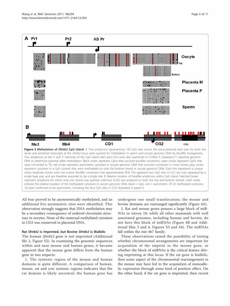
All four proved to be asymmetrically methylated, and anadditional five asymmetric sites were identified. Thisobservation strongly suggests that DNA methylation maybe a secondary consequence of ordered chromatin struc-ture in oocytes. None of the maternal methylated cytosinesin CG3 was conserved in placental DNA.
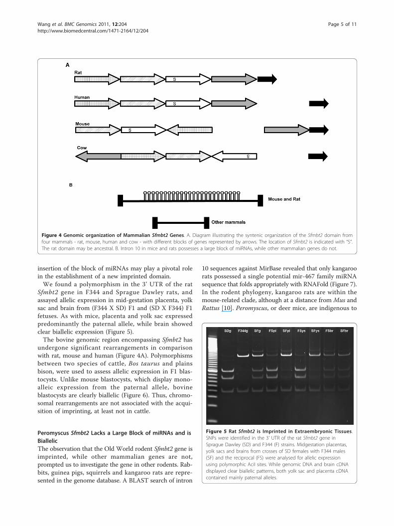
Rat Sfmbt2 is Imprinted, but Bovine Sfmbt2 is BiallelicThe human Sfmbt2 gene is not imprinted (Additionalfile 2, Figure S2). In examining the genomic sequenceswithin and near mouse and human genes, it becameapparent that the mouse gene differs from the humangene in two respects:1. The syntenic region of the mouse and human
domains is quite different. A comparison of human,mouse, rat and cow syntenic regions indicates that therat domain is likely ancestral; the human gene has
undergone one small translocation; the mouse andbovine domains are rearranged significantly (Figure 4A).2. Rat and mouse genes possess a large block of miR-
NAs in intron 10, while all other mammals with wellannotated genomes, including human and bovine, donot have this block of miRNAs (Figure 4B and Addi-tional files 3 and 4, Figures S3 and S4). The miRNAsfall within the mir-467 family.These observations raised the possibility of testing
whether chromosomal arrangements are important foracquisition of the imprint in the mouse gene, orwhether the block of miRNAs is the critical feature driv-ing imprinting at this locus. If the rat gene is biallelic,then some aspect of the chromosomal rearrangement inthe mouse may have led to the acquisition of monoalle-lic expression through some kind of position effect. Onthe other hand, if the rat gene is imprinted, then recent
Figure 3 Methylation of Sfmbt2 CpG Island. A. Five amplicons representing 140 CpG sites across the transcriptional start sites for both thesense and antisense transcripts at the Sfmbt2 locus were queried for methylation in sperm and oocyte genomic DNA by bisulfite mutagenesis.Two amplicons, at the 5’ and 3’ extremes of the CpG island, Me3 and CG3, were also examined in C57BL6 X Castaneus F1 placenta genomicDNA to determine parental allele methylation. Black circles represent CpGs that survived bisulfite conversion; open circles represent CpGs thatwere converted to TG; red circles represent asymmetric cytosines in oocyte genomic DNA that survived conversion in most clones; grey circlesrepresent cytosines in a CpG context that were methylated on only the bottom strand in oocyte genomic DNA. Each line represents a uniqueclone; duplicate clones were not scored. Bisulfite conversion was approximately 95%. The apparent two CpG sites in CG1 are only separated by asingle base pair, and are therefore assumed to be a single site. B. Relative location of bisulfite amplicons within CpG island. Hatched boxesrepresent amplicons for which only one strand was queried; solid box (CG3) was analysed on both the top and bottom strands. Hash marksindicate the relative location of the methylated cytosines in oocyte genomic DNA; black = CpG, red = asymmetric. Of 25 methylated cytosines,18 were confirmed to be asymmetric, including the four CpG sites in CG3 illustrated in panel A.
Wang et al. BMC Genomics 2011, 12:204http://www.biomedcentral.com/1471-2164/12/204
Page 4 of 11
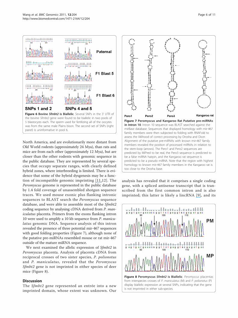
insertion of the block of miRNAs may play a pivotal rolein the establishment of a new imprinted domain.We found a polymorphism in the 3’ UTR of the rat
Sfmbt2 gene in F344 and Sprague Dawley rats, andassayed allelic expression in mid-gestation placenta, yolksac and brain from (F344 X SD) F1 and (SD X F344) F1fetuses. As with mice, placenta and yolk sac expressedpredominantly the paternal allele, while brain showedclear biallelic expression (Figure 5).The bovine genomic region encompassing Sfmbt2 has
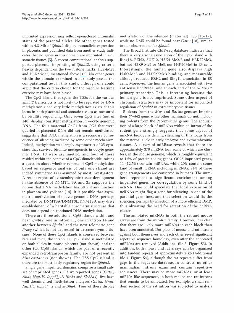
undergone significant rearrangements in comparisonwith rat, mouse and human (Figure 4A). Polymorphismsbetween two species of cattle, Bos taurus and plainsbison, were used to assess allelic expression in F1 blas-tocysts. Unlike mouse blastocysts, which display mono-alleic expression from the paternal allele, bovineblastocysts are clearly biallelic (Figure 6). Thus, chromo-somal rearrangements are not associated with the acqui-sition of imprinting, at least not in cattle.
Peromyscus Sfmbt2 Lacks a Large Block of miRNAs and isBiallelicThe observation that the Old World rodent Sfmbt2 gene isimprinted, while other mammalian genes are not,prompted us to investigate the gene in other rodents. Rab-bits, guinea pigs, squirrels and kangaroo rats are repre-sented in the genome database. A BLAST search of intron
10 sequences against MirBase revealed that only kangaroorats possessed a single potential mir-467 family miRNAsequence that folds appropriately with RNAFold (Figure 7).In the rodent phylogeny, kangaroo rats are within themouse-related clade, although at a distance from Mus andRattus [10]. Peromyscus, or deer mice, are indigenous to
Figure 4 Genomic organization of Mammalian Sfmbt2 Genes. A. Diagram illustrating the syntenic organization of the Sfmbt2 domain fromfour mammals - rat, mouse, human and cow - with different blocks of genes represented by arrows. The location of Sfmbt2 is indicated with “S”.The rat domain may be ancestral. B. Intron 10 in mice and rats possesses a large block of miRNAs, while other mammalian genes do not.
Figure 5 Rat Sfmbt2 is Imprinted in Extraembryonic Tissues.SNPs were identified in the 3’ UTR of the rat Sfmbt2 gene inSprague Dawley (SD) and F344 (F) strains. Midgestation placentas,yolk sacs and brains from crosses of SD females with F344 males(SF) and the reciprocal (FS) were analysed for allelic expressionusing polymorphic AciI sites. While genomic DNA and brain cDNAdisplayed clear biallelic patterns, both yolk sac and placenta cDNAcontained mainly paternal alleles.
Wang et al. BMC Genomics 2011, 12:204http://www.biomedcentral.com/1471-2164/12/204
Page 5 of 11
North America, and are evolutionarily more distant fromOld World rodents (approximately 24 Mya), than rats andmice are from each other (approximately 12 Mya), but arecloser than the other rodents with genomic sequence inthe public database. They are represented by several spe-cies that occupy separate ranges, with clearly definedhybrid zones, where interbreeding is limited. There is evi-dence that some of the hybrid dysgenesis may be a func-tion of incompatible genomic imprinting [11,12]. ThePeromyscus genome is represented in the public databaseby 1.4 fold coverage of unassembled shotgun sequencetraces. We used mouse exonic plus flanking intronicsequences to BLAST search the Peromyscus sequencedatabase, and were able to assemble most of the Sfmbt2coding sequence by analysing cDNA derived from P. man-iculatus placenta. Primers from the exons flanking intron10 were used to amplify a 10 kb sequence from P. manicu-latus genomic DNA. Sequence analysis of this intronrevealed the presence of three potential mir-467 sequenceswith good folding properties (Figure 7), although none ofthe putative pre-miRNAs resembled mouse or rat mir-467outside of the mature miRNA sequence.We next examined the allelic expression of Sfmbt2 in
Peromyscus placenta. Analysis of placenta cDNA fromreciprocal crosses of two sister species, P. polionotusand P. maniculatus, revealed that the PeromyscusSfmbt2 gene is not imprinted in either species of deermice (Figure 8).
DiscussionThe Sfmbt2 gene represented an entrée into a newimprinted domain, whose extent was unknown. Our
analysis has revealed that it comprises a single codinggene, with a spliced antisense transcript that is tran-scribed from the first common intron and is alsoimprinted; this latter is likely a lincRNA [9], and its
Figure 6 Bovine Sfmbt2 is Biallelic. Several SNPs in the 3’ UTR ofthe bovine Sfmbt2 gene were found to be biallelic in two pools of5 blastocysts each. The sperm used for fertilizing all of the oocyteswas from the same male Plains bison. The second set of SNPs (rightpanel) is uninformative in pool 6.
Figure 7 Peromyscus and Kangaroo Rat Putative pre-miRNAsin Intron 10. Intron 10 sequence was BLAST searched against themirBase database. Sequences that displayed homology with mir-467family members were then subjected to folding with RNAFold toassess the liklihood of correct processing by Drosha and Dicer.Alignment of the putative pre-miRNAs with known mir-467 familymembers revealed the position of processed miRNAs in relation tothe stem-loop (arrows). The Pero1 and Pero2 sequences arepredicted by MiPred to be real, the Pero3 sequence is predicted tobe a false miRNA hairpin, and the Kangaroo rat sequence ispredicted to be a pseudo miRNA. Note that the region with highesthomology to known mir-467 family members in the Kangaroo rat istoo close to the Drosha base.
Figure 8 Peromyscus Sfmbt2 is Biallelic. Peromyscus placentasfrom interspecies crosses of P. maniculatus (M) and P. polionotus (P)display biallelic expression at several SNPs, indicating that the geneis not imprinted in either sub-species.
Wang et al. BMC Genomics 2011, 12:204http://www.biomedcentral.com/1471-2164/12/204
Page 6 of 11
imprinted expression may reflect open/closed chromatinstates of the parental alleles. No other genes testedwithin 4.3 Mb of Sfmbt2 display monoalleic expressionin placenta, and published data from another study indi-cates that no genes in this domain are imprinted in e9.5somatic tissues [5]. A recent computational analysis sup-ported placental imprinting of Sfmbt2, using criteriaheavily dependent on the two histone marks, H3K4Me3and H3K27Me3, mentioned above [13]. No other geneswithin the domain examined in our study passed thecomputational test in this study, although one couldargue that the criteria chosen for the machine learningexercise may have been biased.The CpG island that spans the TSSs for the various
Sfmbt2 transcripts is not likely to be regulated by DNAmethylation since very little methylation exists at thislocus in both placental and somatic tissues as measuredby bisulfite sequencing. Only seven CpG sites (out of140) display consistent methylation in oocyte genomicDNA. The four maternal CpGs from CG3 that werequeried in placental DNA did not remain methylated,suggesting that DNA methylation is a secondary conse-quence of silencing mediated by some other mechanism.Indeed, methylation was largely asymmetric; of 25 cyto-sines that survived bisulfite mutagenesis in oocyte geno-mic DNA, 18 were asymmetric, and four of theseresided within the context of a CpG dinucleotide, raisinga question about whether reports of CpG methylationbased on sequence analysis of only one strand areindeed symmetric as is assumed by most investigators.A recent report of extraembryonic tissue developmentin the absence of DNMT1, 3A and 3B supports thenotion that DNA methylation has little if any functionin placenta and yolk sac [14]. It is possible that asym-metric methylation of oocyte genomic DNA, perhapsmediated by DNMT3A/DNMT3L/DNMT3B, may driveestablishment of a heritable chromatin structure thatdoes not depend on continued DNA methylation.There are three additional CpG islands within and
near Sfmbt2; one in intron 11, one in intron 14 andanother between Sfmbt2 and the next telomeric genePrkcq (which is not expressed in extraembryonic tis-sues). None of these CpG islands is conserved betweenrats and mice, the intron 11 CpG island is methylatedon both alleles in mouse placenta (not shown), and theother two CpG islands, which are part of a recentlyexpanded retrotransposon family, are not present inMus castaneus (not shown). The TSS CpG island istherefore the most likely regulatory region for Sfmbt2.Single gene imprinted domains comprise a small sub-
set of imprinted genes. Of six reported genes (Gatm,Nnat, Nap1l5, Inpp5f_v2, Htr2a and Slc38a4), five havewell documented methylation analyses (Gatm, Nnat,Nap1l5, Inpp5f_v2 and Slc38a4). Four of these display
methylation of the silenced (maternal) TSS [15-17],while no DMR could be found near Gatm [18], similarto our observations for Sfmbt2.The Broad Institute ChIP-seq database indicates that
there is very strong association of the CpG island withRing1b, EZH2, SUZ12, H3K4 Me2/3 and H3K27Me3,but not H3K9 Me2 or Me3, nor H4K20Me3 in ES cells.Interestingly, the human gene also displays highH3K4Me3 and H3K27Me3 binding, and measurablealthough reduced EZH2 and Ring1b association in EScells. Moreover, the human gene is associated with twoantisense lincRNAs, one at each end of the SFMBT2primary transcript. This is interesting because thehuman gene is not imprinted. Some other aspect ofchromatin structure may be important for imprintedregulation of Sfmbt2 in extraembryonic tissues.Rodents from the Mus and Rattus genuses imprint
their Sfmbt2 gene, while other mammals do not, includ-ing rodents from the Peromyscine genus. The acquisi-tion of a large block of miRNAs within an intron of therodent gene strongly suggests that some aspect ofmiRNA biology is driving silencing of this locus fromthe maternal allele in early embryos and extraembryonictissues. A survey of miRBase reveals that there areapproximately 370 miRNA loci, some of which are clus-ters, in the mouse genome, which is roughly equivalentto 1.5% of protein coding genes. Of 96 imprinted genes,11 (12.5%) contain miRNAs, while 20% contain somekind of small ncRNA including miRNAs; 84% of thesegene arrangements are conserved in humans. The num-bers represent a significant enrichment amongimprinted genes for co-regulation by some kind ofncRNA. One could speculate that local expansion ofncRNAs might flag a gene for silencing in one of theparental germlines, and that selection would fix thesilencing, perhaps by insertion of a more efficient DMR,thus obviating the need for retention of the ncRNAcluster.The annotated miRNAs in both the rat and mouse
arrays are from the mir-467 family. However, it is clearthat there are likely more miRNAs in each block thanhave been annotated. Dot plots of mouse and rat intronsagainst both themselves and each other reveal significantrepetitive sequence homology, even after the annotatedmiRNAs are removed (Additional file 3, Figure S3). Inaddition, both mouse and rat arrays can be organizedinto tandem repeats of approximately 2 kb (Additionalfile 4, Figure S4), although the rat repeats suffer fromgaps in the sequence database. In contrast, no othermammalian introns examined contain repetitivesequences. There may be more miRNAs, or at leastmiRNA-like sequences, in both mouse and rat intronsthat remain to be annotated. For example, a small ran-dom section of the rat intron was subjected to analysis
Wang et al. BMC Genomics 2011, 12:204http://www.biomedcentral.com/1471-2164/12/204
Page 7 of 11
with MiPred [19] to reveal the presence of two addi-tional predicted pre-miRNA sequences from the mir-467 family (Additional file 5, Figure S5).Kangaroo rats and deer mice fall within the mouse
related clade of rodents [10]. Both possess potentialmir-467 miRNA sequences in intron 10, although thesedo not resemble the mir-467 pre-miRNA sequences inrats and mice, apart from the putative mature miRNA.It is unclear whether these potential miRNAs in kan-garoo rats and Peromyscus are evolutionary precursorsor remnants.Several of the miRNAs from the murine Sfmbt2 locus
are expressed and processed in placenta (Additional file6, Figure S6), not surprising given that they are part ofthe primary transcript for Sfmbt2, which is robustlyexpressed in placenta. Recent analysis of small RNAlibraries with massively parallel sequencing strategieshave indicated that several of the miRNAs from thecluster are expressed in ES cells [20]; interestingly, thenumber of reads does not correlate with the frequencyof the miRNA within the block of repeats, suggestingthat the primary transcript likely folds in a complexmanner that only allows processing of a subset of thepotential miRNAs. Whether these miRNAs mediate anyfunctions in placental biology remains an open question.There are mir-467 family members in other mammaliangenomes, notably human and pig, although in far fewernumbers than in rodents. Moreover, there are severalmmu-mir-467 at other locations in the mouse genome.Any of the miRNAs processed from the Sfmbt2 primarytranscript would be imprinted, although testing this isproblematical given the highly repetitive nature of thearray itself, and the presence of other family members atother locations in the genome.Genomic imprinting is an epigenetic mechanism that
is restricted to mammals and angiosperm plants. Thereis mounting evidence that it is mediated by maternalfactors, possibly as a means of controlling extraembryo-nic tissues that parasitize the maternal reproductivetract [21]. Studies of the evolution of imprinting indicatethat it seems to have emerged at different times at dif-ferent loci [22-24]. Interestingly, several imprinted clus-ters contain miRNAs or SNORDs, and some of thesehave been demonstrated to be imprinted themselves.The role of ncRNAs in imprinting also includes regula-tory functions, such as that exerted by the Kcnq1ot1antisense transcript at the Kcnq1 locus, and the Airntranscript at the Igf2r locus [6].RNAi biology is providing interesting new insights
into gene regulation. Studies in plants and yeast haverevealed a wide array of effects, from post transcrip-tional gene silencing involving both mRNA degradationand translational control, to heterochromatin formation,particularly at centromeres. A recent report of the
ncRNA clusters at the Dlk1 and Snrpn imprinted locihas revealed that transcripts containing precursors ofthe miRNAs or SNORDs from these loci are retained bythe nucleus [25]. This raises the interesting notion thatthe function of the ncRNAs at these loci may be moresimilar to the regulation of centromeres in yeast than to“traditional” cytoplasmic RNAi. Such a role is alsoimplied by the phenotype of both Dicer and Ago2 condi-tional null alleles in mouse oocytes, which fail to com-plete the first meiotic division due to misalignedchromosomes [26,27]. Interestingly, maternal loss ofDgcr8, a component of the miRNA processing pathway,is without effect on development, even though the pro-file of misexpressed miRNAs in maternal Dicer -/- andDgcr8 -/- zygotes is the same [28]. It is tempting tospeculate that expression of a subset of the miRNAsfrom the Sfmbt2 cluster provides the RNAi transcrip-tional silencing biochemistry with the molecular sub-strates that allow it to target the repeated sequences atthe Sfmbt2 intron for transcriptional epigenetic silencingon the maternal allele.The evolution of miRNAs is highly dynamic, and is
characterized by expansions and contractions, partlythrough segmental duplication/deletion events. The mir-467 family was acquired by amniotes approximately 350Mya [29], and has remained relatively small; humanspossess one copy on Chromosome 3, and another onthe X chromosome, while pigs and orangutans possessonly the X linked gene. Mice have acquired, in additionto the large cluster at Sfmbt2, several other copies onChromosomes 5, 9, 10, 11 and 13, although currentmiRNA annotation and archiving may be incomplete forthis family. Interestingly, a large cluster of miRNAs in adifferent family expanded in primates after divergencefrom the rodent lineage, and is expressed exclusively inplacenta [30]. This cluster maps to Chromosome19q13.4, and is near several loci that display eitherimprinted expression or parent of origin effects inhumans (see imprinted gene catalogue records http://igc.otago.ac.nz/Search.html). A recent report demon-strated paternal expression of the primary transcriptencoding this miRNA cluster in human placenta [31].The primary transcript is associated with a classic germ-line DMR; this locus is however much older than thecluster at the rodent Sfmbt2 gene by at least 30 millionyears. As mentioned above, two other imprinted loci,the Dlk1 domain and the Snrpn region, also containlarge clusters of non-coding RNAs (miRNAs andSNORD genes). They also possess classic germlineDMRs. The lack of any noticeable germline methylationat Sfmbt2 may reflect its youth as an imprinted domain,suggesting that imprinted regulation precedes establish-ment of differential methylation. This raises the possibi-lity that one general mechanism by which genes or gene
Wang et al. BMC Genomics 2011, 12:204http://www.biomedcentral.com/1471-2164/12/204
Page 8 of 11
domains become imprinted is through local expansionof miRNAs [32], and that the rodent Sfmbt2 locus is awindow on an actively evolving imprinted domain. Howthese clusters of ncRNAs mediate gene silencing at thetranscriptional level will be an interesting puzzle tosolve.
ConclusionsThe very recent insertion of a large block of miRNAsinto the intron of the PcG gene Sfmbt2 in old worldrodents coincides with the acquisition of placentalimprinting at this locus. The transcriptional start siteCGI does not display heritable differential methylation,suggesting that maternal silencing by other chromatinmechanisms mediates imprinting at this locus.
MethodsAnimalsMice and rats were bred using standard animal husban-dry. Timed pregnancies were assessed by the appearanceof a vaginal plug on day 0.5 in mice, and by the pre-sence of a copulatory plug in the bottom of the cage onday 0.5 in rats. Peromyscus (PO - P. polionotus and BW- P. maniculatus stocks) were obtained from the breed-ing colonies of the Peromyscus Genetic Stock Center.Reciprocal interspecific hybrids were produced by pair-ing PO with BW animals. Peromyscus pregnancies wereassessed visually. Bovine blastocysts were produced byin vitro fertilization of domestic cattle oocytes maturedfrom follicles obtained from slaughterhouse ovaries withfrozen-thawed plains bison sperm stored in liquid nitro-gen at the Toronto Zoo. Bovine embryos were culturedin modified synthetic oviductal fluid medium supple-mented with 2% steer serum until the expanded blasto-cyst stage before extraction of RNA from pools of fiveembryos. All procedures involving laboratory animalswere approved by the Canadian Council on AnimalCare, by a licence from the UK Government HomeOffice (80/2042), or by the Guide for the Care and Useof Laboratory Animals, USA.
Human placentaThis study was conducted according to the principlesexpressed in the Declaration of Helsinki. Informed consentwas obtained from each individual patient and tissue col-lections were approved by the Mount Sinai Hospital’sReview Committee on the Use of Human Subjects andcarried out in accordance with the participating institu-tions’ ethics guidelines. First and second trimester humanplacental tissues were obtained immediately followingelective termination of pregnancies by dilatation and cur-ettage, or suction evacuation. Gestational age was deter-mined by the date of the last menstrual period and firsttrimester ultrasound measurement of crown-rump-length
(CRL). Term placental tissue was collected at the time ofdelivery from healthy pregnancies with normally grownfetuses that did not have signs of placental dysfunction.Samples were collected randomly from central and periph-eral placental areas and snap frozen immediately afterdelivery. Calcified, necrotic and visually ischemic areaswere excluded from collection.
cDNA SynthesisTotal RNA isolated with Trizol reagent according tomanufacturer’s instructions was used as template for thesynthesis of cDNA, using random hexamers as primersand either Superscript III (Invitrogen) or RevertAid(Fermentas) cDNA synthesis kits. All cDNA synthesisreactions were preceded by a DNAse I step to eliminateany contaminating genomic DNA; an aliquot of theDNAse treated RNA was set aside as a zero R.T control.No PCR product was ever obtained with the zero R.Tcontrol samples. PCR was performed with either LATaq (Takara) or Bioline Taq (Bioline International).
Peromyscus Sfmbt2 cDNA and Genomic SequenceBLAST analysis of sequence trace files for Peromyscusmaniculatus using murine exon sequence plus someflanking intron sequence as search query yielded fourteencoding exons with strong homology. The seven missingexons were recovered with primers designed from thePeromyscus sequence derived either from the database orfrom first pass sequence analysis of Sfmbt2 cDNA (Addi-tional file 7, Table S2). The Peromyscus Sfmbt2 cDNAsequence accession number is [Genbank:HQ416417].For analysis of Peromyscus intron 10, forward and
reverse primers were designed from the exonicsequences of exons 9 and 10, respectively for amplifica-tion of genomic DNA. PCR product was sequenced witha series of primers in a walk from each end until theamplifiable intron sequence was small enough to cloneinto pT-Easy. Subclones of the pT-Easy intron frag-ments were sequenced with universal primers, and thefull length intron 10 sequence was assembled. Some ofthe intron sequence was obtained from the sequencetrace files. The Peromyscus intron 10 genomic DNAsequence accession number is [Genbank:HQ416418}.
SNP analsysisAnnotated SNPs were chosen for analysis; where possi-ble, SNPs that created RFLPs were selected. Primerswere designed to amplify the selected SNPs, and PCRproduct was subjected either to restriction enzymedigestion or direct sequence analysis. Primers and analy-sis strategy are listed in Additional file 7, Tables S1 andS2. Where no SNPs could be found in the public data-base, genomic DNA from parental species was surveyedby sequence analysis.
Wang et al. BMC Genomics 2011, 12:204http://www.biomedcentral.com/1471-2164/12/204
Page 9 of 11
Bisulfite MutagenesisGenomic DNA was treated with sodium bisulfite, using aMethylCode kit from Invitrogen. For the isolation ofoocyte genomic DNA, 10 - 20 females aged 5-10 weekswere superovulated with 5 I.U. of PMS administeredintraperitoneally followed 48 hours later by 5 I.U. ofhCG. Eighteen hours after the hCG injection, oocyteswere retrieved from the oviducts and stripped of cumuluscells with a combination of hyaluronidase treatment, vig-ourous pipetting, and removal of the zona pellucida withacidic tyrodes. This latter step ensured that all cumuluscells were removed from the sample before extraction ofgenomic DNA, which involved a 5 hour treatment withProteinase K followed by ethanol precipitation.With the exception of CG3 top strand, bisulfite pri-
mers were designed with the aid of the MethPrimer webbased tool (http://www.urogene.org/methprimer/index1.html) (Additional file 7, Table S3). Only one strand wassequenced for amplicons Me3, CG1, (bottom strands),and Me4, CG2 (top strands). CG3 was sequenced fromboth strands. The primers used for amplification of thetop strand were manually designed to avoid bias gener-ated by asymmetric methylation. Oocyte genomic DNArequired further amplification with nested primers, soall samples were subjected to nested amplification priorto cloning into pGEM-TEasy, using a kit from Promega.Plasmid DNA was purified from individual clones andsubjected to sequence analysis with the T7 universal pri-mer. Occasional samples were sequenced with the SP6universal primer. Only unique clones were reported;uniqueness was assessed by random PCR generatedSNPs, some of which were the result of less than com-plete bisulfite mutagenesis. Only clones exhibiting 95%mutagenesis of non-CG cytosines were scored.
Additional material
Additional file 1: Figure S1 Human SFMBT2 is Biallelic. Placenta RNAfrom four individuals (#6 - 18 weeks gestation; #8 - 39 weeks gestation;#9 - 40 weeks gestation; #10 - 41 weeks gestation) was converted tocDNA as described in Materials and Methods, and amplified with primersfor two SNPs found at high frequencies in the human population (seePrimer Table for details). Two samples amplified successfully with SNP1primers, and three with SNP3 primers. In both cases, alleles wereidentified by digestion with diagnostic restriction enzymes; asterisks (*)indicate one allele and number sign (#) indicates the other allele. Sample#6 was homozygous for both SNPs. The other samples contained twoalleles, indicating the presence of both maternal and paternal alleles.
Additional file 2: Figure S2 Allelic Analysis of Genes in the MurineSfmbt2 Domain. Placenta cDNA from e14.5 (C57BL6 X Castaneus) F1embryos was analysed either by direct sequencing of PCR product, or byrestriction enzyme digestion with the indicated enzymes.
Additional file 3: Figure S3 Dot Plot Comparison of Rat and MouseIntron 10. Sequence from rat and mouse Sfmbt2 intron 10 was plottedagainst each other or against itself. Note the large segment of highlyrepetitive sequence, even after the removal of annotated miRNAsequence. In contrast, human, kangaroo rat and cow introns generatethe predicted diagonal line when self plotted.
Additional file 4: Figure S4 Alignment of Repeats in Mouse and RatIntron 10. The block of miRNAs is arranged in a series of tandemrepeats of an approximately 2.5 kb sequence, each of which contains 6-7miRNAs. The mouse repeats align well with each other, while the ratrepeats are less well conserved.
Additional file 5: Figure S5 Annotation of miRNAs May BeIncomplete. A small segment of the rat intron close to an annotatedmiRNA (yellow highlighting), when subjected to MiPred reveals thepresence of two additional potential mir-467 family members (greenhighlighting) that fold into good potential pre-miRNAs with RNAFold(http://rna.tbi.univie.ac.at/cgi-bin/RNAfold.cgi).
Additional file 6: Figure S6 MiRNAs from the Intron 10 Cluster AreExpressed in Placenta. Five of the most commonly annotated miRNAsfrom the Sfmbt2 intron 10 cluster were assayed by RT-PCR in placentaRNA. The asterisk indicates a primer dimer artifact.
Additional file 7: Tables S1-S4. Tables 1-4 list primer sequences usedfor analyses described in the paper.
AbbreviationsMb: megabase; e7.5: embryonic day 7.5; miRNA: micro RNA; kb: kilobase;DMR: differentially methylated region; ChIP-seq: chromatinimmunoprecipitation massively parallel sequence; H3K4Me3: histone H3lysine 4 trimethylation; H3K27Me3: histone H3 lysine 27 trimethylation;H3K9Me3: histone H3 lysine 9 trimethylation; H4K20Me3: histone H4 lisine 20trimethylation; ES: embryonic stem cells; 3’UTR: 3’ untranslated region; Mya:million years ago; lincRNA: long intergenic non coding RNA; DNMT: DNAmethyltransferase; ncRNA: non coding RNA; TSS: transcriptional start site
AcknowledgementsThe authors thank Dr Allan King for his help obtaining the bovine samplesand Julia Varmuza for assistance with bioinformatic analyses. This work wasfunded by grants from the Natural Sciences and Engineering ResearchCouncil of Canada [138636 to S.V; 371932-09 to G.M.], the Medical ResearchCouncil UK to AFS and the Canadian Institutes of Health Research [MOP-14096 to IC]. RS is recipient of a Mining Industry Training and AdjustmentCouncil graduate student internship.
Author details1Department of Cell and Systems Biology, University of Toronto, 25 HarbordSt., Toronto, Ontario, M5S 3G5, Canada. 2Department of Physiology,Development and Neuroscience, University of Cambridge, Downing Street,Cambridge, CB2 3EG, UK. 3Department of Physiology, Medical College ofWisconsin, 8701 Watertown Plank Road Milwaukee, WI, USA, 53226, USA.4Department of Biomedical Sciences, University of Guelph, 50 Stone Rd. E.,Guelph, Ontario, N1G 2W1, Canada. 5Peromyscus Genetic Stock Center &Department of Biological Sciences, University of South Carolina, Columbia,SC 29208, USA. 6Reproductive Physiology, Toronto Zoo, 361A Old FinchAvenue, Toronto, Ontario, M1B 5K7, Canada. 7Samuel Lunenfeld ResearchInstitute, Mount Sinai Hospital, Joseph and Wolf Lebovic Health Complex,600 University Avenue, Toronto, Ontario, M5G 1X5, Canada.
Authors’ contributionsAll of the molecular analysis was performed by QW, JC, JH, JT, EDC, KM andSV. Some of the work was performed in the lab of AFS while SV was onsabbatical leave. Bovine embryo cDNA was provided by PS and GM.Peromyscus tissues were provided by PV. Human cDNA placental cDNA wasprovided by IC. Rat genomic sequence data were provided by CM. Themanuscript was written by SV, with input from all authors; all authorsapproved the final manuscript.
Received: 21 January 2011 Accepted: 21 April 2011Published: 21 April 2011
References1. Edwards C, Ferguson-Smith AC: Mechanisms regulating imprinted genes
in clusters. Curr Opin Cell Biol 2007, 19:281-289.
Wang et al. BMC Genomics 2011, 12:204http://www.biomedcentral.com/1471-2164/12/204
Page 10 of 11
2. Kuzmin A, Han Z, Golding M, Mann M, Latham K, Varmuza S: The PcG geneSfmbt2 is paternally expressed in extraembryonic tissues. Gene ExprPatterns 2008, 8:107-116.
3. Cattanach B, Kirk M: Differential activity of maternally and paternallyderived chromosome regions in mice. Nature 1985, 315:496-498.
4. Cattanach B: Parental origin effects in mice. J Embryol Exp Morphol 1986,97(Supplement):137-150.
5. Babak T, Deveale B, Armour C, Raymond C, Cleary M, van der Kooy D,Johnson J, Lim L: Global survey of genomic imprinting by transcriptomesequencing. Curr Biol 2008, 18:1735-1741.
6. Hudson QJ, Kulinski TM, Huetter SP, Barlow DP: Genomic imprintingmechanisms in embryonic and extraembryonic mouse tissues. Heredity2010, 1-12.
7. Kobayashi H, Suda C, Abe T, Kohara Y, Ikemura T, Sasaki H: Bisulfitesequencing and dinucleotide content analysis of 15 imprinted mousedifferentially methylated regions (DMRs): paternally methylated DMRscontain less CpGs than maternally methylated DMRs. Cytogenet GenomeRes 2006, 113:130-137.
8. McEwen K, Ferguson-Smith AC: Distinguishing epigenetic marks ofdevelopmental and imprinting regulation. Epigenetics & Chromatin 2010,3:2, 2010.
9. Guttman M, Amit I, Garber M, French C, Lin MF, Feldser D, Huarte M, Zuk O,Carey BW, Cassady JP, Cabili MN, Jaenisch R, Mikkelson TS, Jacks T,Hacohen N, Bernstein BE, Kellis M, Rinn JL, Lander ES: Chromatin signaturereveals over a thousand highly conserved large non-coding RNAs inmammals. Nature 2009, 458:223-227.
10. Blanga-Kanfi S, Miranda H, Penn O, Pupko T, DeBry RW, Huchon D: Rodentphylogeny revised: analysis of six nuclear genes from all major rodentclades. BMC Evolutionary Biology 2009, 9:71.
11. Duselis A, Wiley C, O’Neill M, Vrana P: Genetic evidence for a maternaleffect locus controlling genomic imprinting and growth. Genesis 2005,43:155-165.
12. Loschiavo M, Nguyen Q, Duselis A, Vrana P: Mapping and identification ofcandidate loci responsible for Peromyscus hybrid overgrowth.Mammalian Genome 2006, 18:75-85.
13. Brideau C, Eilertson K, Hagarman J, Bustamante C, Soloway P: Successfulcomputational prediction of novel imprinted genes from epigenomicfeatures. Mol Cell Biol 2010, 30:3357-3370.
14. Sakaue M, Ohta H, Kumaki Y, Oda M, Sakaide Y, Matsuoka C, Yamagiwa A,Niwa H, Wakayama T, Okano M: DNA methylation is dispensable for thegrowth and survival of the extraembryonic lineages. Current Biology 2010,20:1452-1457.
15. Kikyo N, Williamson CM, John RM, Barton SC, Beechey CV, Ball ST,Cattanach BM, Surani MA, Peters J: Genetic and functional analysis ofneuronatin in mice with maternal or paternal duplication of distalchromosome 2. Dev Biol 1997, 190:66-77.
16. Smith RJ, Dean W, Konfortova G, Kelsey G: Identification of novelimprinted genes in a genome-wide screen for maternal methylation.Genome Research 2003, 13:558-569.
17. Wood AJ, Roberts RG, Monk D, Moore GE, Schulz R, Oakey RJ: A screen forretrotransposed imprinted genes reveals an association between Xchromosome homology and maternal germline methylation. PlOSGenetics 2007, 3(2):e20.
18. Sandell LL, Guan XJ, Ingram R, Tilghman SM: Gatm, a creatine synthesisenzyme, is imprinted in mouse placenta. Proc Nat Acad Sci 2003,100:4622-4627.
19. Jiang P, Wu H, Wang W, Ma W, Sun X, Lu Z: MiPred: classification of realand pseudo microRNA precursors using random forest prediction modelwith combined features. Nucleic Acids Res 2007, 35:W339-W344[http://www.bioinf.seu.edu.cn/miRNA/index.html].
20. Chiang HR, Schoenfeld LW, Ruby JG, Auyeung VC, Spies N, Baek D,Johnston WK, Russ C, Luo S, Babiarz JE, Blelloch R, Schroth GP, Nusbaum C,Bartel1 DP: Mammalian microRNAs: experimental evaluation of noveland previously annotated genes. Genes Dev 2010, 24:992-1009.
21. Miri K, Varmuza S: Genomic imprinting and extraembryonic tissues -mom takes control. International Review of Cell & Molecular Biology 2009,276:216-262.
22. Rapkins RW, Hore T, Smithwick M, Ager E, Pask AJ, Renfree M, Kohn M,Hameister H, Nicholls R, Deakin JE, Graves JA: Recent assembly of animprinted domain from non-imprinted components. PLoS Genet 2006, 2:e182.
23. Hore T, Rapkins R, Marshall Graves JA: Construction and evolution ofimprinted loci in mammals. Trends Genet 2007, 23:440-448.
24. Edwards CA, Mungall AJ, Matthews L, Ryder E, Gray DJ, Pask A, Shaw G,Graves J, Rogers J, SAVOIR consortium, Dunham I, Renfree MB, FergusonSmith A: The evolution of the DLK1-DIO3 imprinted domain in mammals.PLoS Biol 2008, 6:e135.
25. Vitali P, Royo H, Marty V, Bortolin-Cavaillé M-L, Cavaillé J: Long nuclear-retained non-coding RNAs and allele-specific higher-order chromatinorganization at imprinted snoRNA gene arrays. J Cell Sci 2010, 123:70-83.
26. Murchison P, Stein P, Xuan Z, Pan H, Zhang M, Schultz R, Hannon G:Critical roles for Dicer in the female germline. Genes Dev 2007,21:682-693.
27. Kaneda M, Tang F, O’Carroll D, Lao K, Surani MA: Essential role forArgonaute2 protein in mouse oogenesis. Epigenetics & Chromatin 2009,2:9.
28. Suh N, Baehner L, Moltzahn F, Melton C, Shenoy A, Chen J, Blelloch R:MicroRNA function is globally suppressed in mouse oocytes and earlyembryos. Curr Biol 2010, 20:271-277.
29. Peterson K, Dietrich M, McPeek M: MicroRNAs and metazoanmacroevolution: insights into canalization, complexity, and theCambrian explosion. BioEssays 2009, 31:736-747.
30. Bentwich I, Avniel A, Karov Y, Aharonov R, Gilad S, Barad O, Barzilai A,Einat P, Einav U, Meiri E, Spector Y, Bentwich Z: Identification of hundredsof conserved and nonconserved human microRNAs. Nature Genetics 2005,37:766-770.
31. Noguer-Dance M, Abu-Amero S, Al-Khtib M, Lefèvre A, Coullin P, Moore G,Cavaillé J: The primate-specific microRNA gene cluster (C19MC) isimprinted in the placenta. Hum Mol Genet 2010.
32. Zhang YJ, Qu LH: Non-coding RNAs and the acquisition of genomicimprinting in mammals. Sci China Ser C-Life Sci 2009, 52:195-204.
33. Seitz H, Royo H, Bortolin ML, Lin SP, Ferguson-Smith AC, Cavaille J: A largeimprinted microRNA gene cluster at the mouse Dlk1-Gtl2 domain.Genome Research 2004, 14:1741-1748.
doi:10.1186/1471-2164-12-204Cite this article as: Wang et al.: Recent acquisition of imprinting at therodent Sfmbt2 locus correlates with insertion of a large block ofmiRNAs. BMC Genomics 2011 12:204.
Submit your next manuscript to BioMed Centraland take full advantage of:
• Convenient online submission
• Thorough peer review
• No space constraints or color figure charges
• Immediate publication on acceptance
• Inclusion in PubMed, CAS, Scopus and Google Scholar
• Research which is freely available for redistribution
Submit your manuscript at www.biomedcentral.com/submit
Wang et al. BMC Genomics 2011, 12:204http://www.biomedcentral.com/1471-2164/12/204
Page 11 of 11
Top Related
Copyright © 2022 FDOKUMEN

