


Bahasa
Halaman
Hukum

Comparative Biochemistry and Physiology, Part C 151 (2010) 343–350
Contents lists available at ScienceDirect
Comparative Biochemistry and Physiology, Part C
j ourna l homepage: www.e lsev ie r.com/ locate /cbpc
Influence of the dark/light rhythm on the effects of UV radiation in the eyestalk of thecrab Neohelice granulata
Marcelo Alves Vargas a, Marcio Alberto Geish b, Fabio Everton Maciel b, Bruno Pinto Cruz b,Daza de Moraes Vaz Batista Filgueira b, Gabrielle de Jesus Ferreira a,Luiz Eduardo Maia Nery b,1, Silvana Allodi a,⁎,1
a Programa de Pós-Graduação em Ciências Morfológicas, Instituto de Ciências Biomédicas, Universidade Federal do Rio de Janeiro-UFRJ, 21941-590, Rio de Janeiro, RJ, Brazilb Programa de Pós-Graduacão em Ciências Fisiológicas-Fisiologia Animal Comparada, Instituto de Ciências Biológicas, Universidade Federal do Rio Grande-FURG, Av. Itália, Km 8,Rio Grande-RS, 96201-900, Brazil
⁎ Corresponding author. Av. Carlos Chagas Filho 37Ciências Biomédicas, Universidade Federal do Rio de JaneRJ, Brazil. Tel.: +55 21 25626431; fax: +55 21 224912
E-mail address: [email protected] (S. Allodi).1 Contributed equally to the paper.
1532-0456/$ – see front matter © 2010 Elsevier Inc. Aldoi:10.1016/j.cbpc.2009.12.011
a b s t r a c t
a r t i c l e i n f oArticle history:Received 6 November 2009Received in revised form 20 December 2009Accepted 22 December 2009Available online 7 January 2010
Keywords:Antioxidant capacity against peroxyl radicalsCatalase activityCrustaceansLipid peroxidationOxidative stressPhotoperiodReactive oxygen speciesVisual system
Crustaceans are interesting models to study the effects of ultraviolet (UV) radiation, and many species maybe used as biomarkers for aquatic contamination of UV radiation reaching the surface of the Earth. Here, weinvestigated cell damage in the visual system of crabs Neohelice granulata that were acclimated to either12L:12D, constant light, or constant dark, and were exposed to UVA or UVB at 12:00 h (noon). Theproduction of reactive oxygen species (ROS), antioxidant capacity against peroxyl radicals (ACAP), lipidperoxidation (LPO) damage, catalase activity, and pigment dispersion in the eye were evaluated. Nosignificant differences from the three groups of controls (animals acclimated to 12L:12D, or in constant light,or not exposed to UV radiation) were observed in animals acclimated to 12L:12D, however, crabs acclimatedto constant light and exposed to UV radiation for 30 min showed a significant increase in ROS concentration,catalase activity, and LPO damage, but a decrease in ACAP compared with the controls. Crabs acclimated toconstant darkness and exposed to UV for 30 min showed a significantly increased ROS concentration and LPOdamage, but the ACAP and catalase activity did not differ from the controls (animals kept in the dark whilethe experimental group was being exposed to UV radiation). Pigment dispersion in the pigment cells of eyesof animals acclimated to constant light was also observed. The results indicate that UVA and UVB alterspecific oxidative parameters; however, the cell damage is more evident in animals deviated from thenormal dark/light rhythm.
3 Bloco F2-012, Instituto deiro, 21949-902, Rio de Janeiro,41.
l rights reserved.
© 2010 Elsevier Inc. All rights reserved.
1. Introduction
Ultraviolet (UV) radiation comprises electromagnetic wavelengthsbetween 200 and 400 nm. In order to analyze its capacity to causedamage in cell targets, UV is divided into UVA (320–400 nm), UVB(290–320 nm), and UV-C (200–290 nm). The wavelengths of UV thatinclude UVA and UVB are also called Solar UV (Diffey, 2002). Theincreased incidence of UV radiation on the Earth's surface is receivingmore attention because it can produce biological changes and someimpact on biodiversity (McKenzie et al., 2007). However, additionalstudies are necessary in order to understand the responses ofbiological systems to UV radiation damage.
The first and main target-structure for UV radiation in animals isthe body surface, including the skin and eyes. The influence of UV
radiation on the human skin has been investigated: in general, theeffects of UV radiation on the epidermis are harmful (Tran et al.,2008), except for stimulation of vitamin D synthesis (Holick, 2008).The adverse effects of UV radiation include immunosuppression(Timares et al., 2008), production of reactive oxygen species (ROS)(Heck et al., 2003), photoaging, DNAmutation, and cancer (Albert andOstheimer, 2003). In the eye, damage has been reported in the cornea,characterized by inflammation; in the lens, with cataracts the mostcommon pathology (Sliney, 2001; Meyer-Rochow, 2000); and in theretina, where apoptosis, especially in the photoreceptors, wasobserved (Miguel et al., 2003). The mechanism underlying thisdamage is oxidative stress, which results from an imbalance betweenreactive oxygen species (ROS) and antioxidants, i.e., oxidative stressoccurs when ROS production exceeds the capacity of the antioxidantsystem to repair the deleterious effect of ROS. The most commonlyencountered ROS in biological systems include the hydroxyl radical,the superoxide anion, hydrogen peroxide (H2O2), and singlet oxygen(Sies, 1991). ROS results from the incomplete reduction of oxygenspecies and can be produced by both exogenous, such as solarradiation, and endogenous sources, such as the mitochondrial
344 M.A. Vargas et al. / Comparative Biochemistry and Physiology, Part C 151 (2010) 343–350
electron transport chain and the endoplasmic reticulum (Dean et al.,1997), the activity of enzymes, viz. cytochrome P450, xanthineoxidase, urate oxidase, and D-amino acid oxidase (Stadtman andLevine, 2000).
Animals may respond to environmental conditions, the mostimportant being the photoperiod. Circadian and exogenous dailyvariations, including those related to locomotor and brain activity, aswell as environmental temperature and light fluctuations, result incorresponding daily patterns of reactive oxygen species (ROS)(Hardeland et al., 2003). Some investigators have proposed thatcircadian clocks organize metabolic functions into a coherent dailyschedule, assuring their synchrony with environmental changes(Wijnen and Young, 2006).
If in vertebrates, mainly mammals, the adverse effects of UVradiation are well known, this is not true for invertebrates. To ourknowledge, in crustaceans only Gouveia et al. (2005) have reportedoxidative and DNA damage in the cephalothorax and pereiopodepithelia in the crab Neohelice granulata, and Miguel et al. (2002)reported morphological damage in the retina and lamina ganglionariscells in the mangrove crab Ucides cordatus.
The crab N. granulata (=Chasmagnathus granulatus) is a semi-terrestrial animal of the southern coast of Brazil, Uruguay, andArgentina, and it has been used by our group as a model for studies onthe effects of UV. This species shows pigment dispersion in themelanophores during UV exposure, which disappears when thestimulus ceases (Gouveia et al., 2004; Vargas et al., 2008). Althoughthe effects of UV radiation on the epithelium of this crab have beenstudied, the effects of UV on the visual system have not so far beeninvestigated. Thus, the aim of this study was to investigate the effectsof UV radiation on the visual system of N. granulata in response tooxygen species production, using a biochemical approach and theevaluation of pigment dispersion. This may help to understand howcrustaceans, which are an integral part of estuarine environments andcontribute substantially to their dynamics, are affected by UV rays. Inaddition, because UVB radiation reaching the surface of the Earth hasbeen increasing in recent decades (Casiccia et al., 2003; Bertagnolliet al., 2007; Kirchhoff et al., 2000), these animals may eventually beused as biomarkers for aquatic contamination.
2. Materials and methods
2.1. Animals
Adult male crabs N. granulataweighing 7.0±0.5 g (mean±S.E.M)were collected in salt marshes near the city of Rio Grande, Brazil. Theywere transferred to the laboratory for an acclimation period of at least10 days in tanks under constant conditions of temperature (20 °C)and salinity (20 ppt), and in three photoperiods, consisting of12L:12D, constant light, and constant darkness. In the 12L:12Dregime, the lights went on at 6:00 h and went off at 18:00 h. Allprocedures adopted in this study were performed after approval bythe National Environmental Committee (IBAMA document number1.637.714), and every effort was made to minimize animal suffering.
2.2. Assays
Within each set of crabs acclimated to one of the three differentphotoperiods, a group was exposed to UVA radiation, another groupwas exposed to UVB radiation, and still another groupwas the control.This control group was constituted by the animals that were notexposed to UV radiation (UVA or UVB), but to visible light (theanimals acclimated to 12L:12D and constant light) or maintained inthe dark (animals acclimated to constant darkness) during the sameperiod of time that the experimental groups were exposed to UVradiation.
Therefore, after the acclimation period, two groups of crabs wereirradiated at 12:00 h (noon), since this is the time when UV radiationis maximal, with different doses of UVA (1.575 J/cm2) and UVB(1.294 J/cm2), respectively, for 30 min. The UVA (VL: 115 L, 30 W) orUVB (VL: 115 C, 30 W; Vilber Lourmat, Marne Lavalee, France) lampswere monitored using a radiometer/photometer (model IL 1400A,International Light, Newburyport, MA, USA). The UVA lamp irradia-tion was 1.39 mW/cm2 UVA, with contamination of 0.006 mW/cm2
UVB and 0.000928.0 mW/cm2 of visible light. The UVB lampirradiation was 1.195 mW/cm2, produced with contamination of0.493 mW/cm2 of UVA and 0.000113 mW/cm2 of visible light. Bothlamps showed no contamination with UV-C. The control group,formed with crabs that were not irradiated with UV (animalsacclimated to 12L:12D and constant light), was maintained underfluorescent lamps (Philips TLT 40 W/75, São Paulo, Brazil) irradiating96.0 mW/cm2 visible light. After the exposure, the animals were killedby severing the supra-esophageal ganglion, and the eyes wereremoved for further analyses.
2.2.1. ROS productionTwo hundred and seventy eyestalks (90 for each group: control,
exposed to UVA, and exposed to UVB) were weighed and homoge-nized (1:20 w/v) in a cold (4 °C) buffer solution containing sucrose(250 mM), PMSF (1 mM), and EDTA (5 mM), with pH adjusted to 7.6.The samples were centrifuged twice (2000g, 4 °C for 20 min) and thesupernatant was collected and centrifuged again (10,000 g, 4 °C for45 min). The supernatant resulting from this last centrifugation wasused for the determination of the ROS (Viarengo et al., 1999). For ROSdetection, we used 2′,7′-dichlorofluorescein-diacetate (H2DCF-DA,Molecular Probes). This molecule in the presence of ROS generates afluorochrome, detected at 488 and 525 nm wavelengths for excite-ment and emission, respectively. The analyses were carried out in afluorescence microplate reader (Victor 2, Perkin Elmer) with readingsevery 5 min for 60 min. The total fluorescence production wascalculated by integrating the fluorescence units (FU) during theperiod of the measurement, after adjusting the FU data to a second-order polynomial function. The ROS concentration was referred to thetotal protein content present in the biological sample and expressedin FU (mg of protein)−1.
2.2.2. Antioxidant capacity against peroxyl radical analysisAnother 270 eyestalks (separated into three groups as explained in
Section 2.2.1) were weighed and homogenized (1:20 w/v) in a cold(4 °C) buffer solution containing sucrose (250 mM), PMSF (1 mM),and EDTA (5 mM), with pH adjusted to 7.6. The samples werecentrifuged twice at 2000g, 4 °C, for 20 min, and the supernatant wascollected and centrifuged again (10,000g, 4 °C, for 45 min). Thesupernatant of this last centrifugation was then employed for theanalyses. The antioxidant capacity against peroxyl radicals (ACAP)wasmeasured according to themethod of Amado et al. (2009). Briefly,10 µL of the supernatant prepared for the enzyme analysis waspipetted into a white 96-well microplate, six wells per sample. Thereaction buffer (127.5 µL) containing 30 mMHEPES (pH 7.2), 200 mMKCl, and 1 mM MgCl2 was added to the wells with the samples. Inthree of the six wells of each sample, 7.5 µL of 2,2′-azobis 2methylpropionamidine dihydrochloride (ABAP; 4 mM; Aldrich) wasadded, and the same volume of ultrapure water was pipetted into theother three wells. The microplate was inserted into a fluorescencemicroplate reader (Victor 2, Perkin Elmer), at a programmedtemperature of 35 °C, and the peroxyl radicals were produced bythermal decomposition of ABAP. Immediately before reading, 10 µL ofthe fluorescent probe 2′,7′-dichlorofluorescein-diacetate (H2DCF-DA)was added to the wells at a final concentration of 40 µM (Ferreira-Cravo et al., 2007). H2DCF-DA is cleaved by esterases present insamples, and the non-fluorescent compound H2DCF is oxidized byROS to the fluorescent compound DCF, which is detected at
Table 1Values for ROS, LPO, ACAP and catalase activity of control groups (mean±SEM) in 12L–12D (n=5) photoperiod, constant light (n=5) and constant darkness (n=5) at12:00 h.
Assay 12L:12D Constant light Constant dark
ROS concentration(FU (g of protein)−1)
1887.6±417.3a 2176.0±89.5a 606.8±100.9b
LPO(CHP (g of wet mass)−1)
130.78±8.48a 102.75±6.32b 41.11±9.35c
ACAP 0.0046±0.00032a 0.048±0.008b 0.059±0.009b
Catalase activity(U CAT (mg of protein)−1)
24.98±2.83a 43.66±11.11a 27.04±1.81a
Different letters represent statistically significant differences for each variable(Pb0.05).
345M.A. Vargas et al. / Comparative Biochemistry and Physiology, Part C 151 (2010) 343–350
wavelengths of 488 and 525 nm, for excitation and emission,respectively. The thermal decomposition of ABAP and ROS formationwere monitored with readings every 5 min for 60 min. According toRegoli and Winston (1999) and Regoli (2000), non-enzymatic low-molecular-weight scavengers (GSH, ascorbic acid, uric acid, andvitamin E) generally account for 70% of the total scavenging capacitytowards peroxyl radicals. Therefore, if enzymatic inhibition occursdue to the high temperature (since crabs are poikilotherms) neededfor the ABAP decomposition in the peroxyl radical (35 °C), thedecrease of the antioxidant capacity should be a minor problem. Thetotal fluorescence production was calculated by integrating thefluorescence units (FU) along with the time of the measurement,after adjusting the FU data to a second-order polynomial function. Theresults were calculated as the difference in area of FU×min in thesame sample, with and without ABAP addition, and standardized tothe ROS area without ABAP (background area). The inverse of therelative difference between the ROS area with and without ABAP wasconsidered as a measure of the antioxidant capacity: the greater thedifference in area, the higher the antioxidant capacity, since highfluorescence levels were obtained after adding ABAP, signifying a lowcompetence to neutralize peroxyl radicals. Thus, the calculation forthe antioxidant competence is:
1= ðROSareaABAP−ROSareabackgroundÞ= ROSareabackground:
2.2.3. Catalase activityStill another 270 optic ganglia (separated into three groups as
explained in Section 2.2.1) were weighed and homogenized (1:20 w/v)in a cold (4 °C) buffer solution containing Tris base (20 mM), EDTA(1 mM), dithiothreitol (1 mM, Sigma), KCl (150 mM), and PMSF(0.1 mM), with pH adjusted to 7.6. The homogenates werecentrifuged at 9000 g, 4 °C, for 30 min, and the supernatant wasthen employed for the analyses. The catalase (EC 1.11.1.6) activitywas analyzed according to Beutler (1975), determining the initialrate of H2O2 (50 mM) decomposition at 240 nm. The results wereexpressed in CAT units: one unit is the amount of enzyme thathydrolyzes 1 µmol H2O2 per min and per mg of protein, at 25 °C andpH 8.0. This procedure was performed using a digital spectropho-tometer (Biomatte 3).
2.2.4. Lipid peroxidationTwo hundred and seventy eyestalks (separated into three groups
as explained in Section 2.2.1) were weighed and homogenized(1:20 w/v) with methanol [in a cold (4 °C) condition and centrifugedat 1000g, 4 °C, for 10 min]. The supernatant was used for the analyses.The methodology, termed FOX (Hermes-Lima et al., 1995; Monserratet al., 2003), is based on the oxidation of Fe(II) under acidicconditions, and measures the quantity of lipid peroxides. For thelipid peroxidation (LPO) measurements, FeSO4 (1 mM), H2SO4
(0.25 M), xylenol orange (1 mM, Sigma) and MilliQ water wereadded sequentially. Samples (30 µL) or methanol (blanks) wereadded and incubated for 450 min. Subsequently, the absorbance(550 nm) was determined using a microplate reader (Victor 2, PerkinElmer), and cumene hydroperoxide (CHP, Sigma) was employed as astandard. LPO was expressed as cumene hydroperoxide (CHP)equivalents per g of wet mass.
2.3. Histological analysis
The eyestalks of nine crabs acclimated to the three photoperiodsanalyzed were dissected at 12:00 h, fixed in 4% paraformaldehyde for4 h in 0.1 M phosphate-buffered saline (PBS), and dehydrated in agraded ethanol series. The material was embedded in paraffin, and12 µm sections, obtained using a rotatory microtome, were collectedon slides. The sections were stained with hematoxylin and eosin, for
observation by means of an Olympus BX50microscope connected to aCCD camera (Pro-series, High Performance), and provided withcommercial software for image acquisition using Image Pro.
2.4. Statistical analysis
The statistical analyses were carried out by analysis of variance(ANOVA) followed by the Newman–Keuls test (a=0.05). Normalityand variance homogeneity were verified as ANOVA assumptions.Mathematical transformations were performed when necessary (Zar,1984). The results of all experiments were expressed as a percentagecompared to the control group.
3. Results
The control groups for 12L:12D, for constant light, and for constantdarkness (none of them exposed to UV) showed different values forROS, LPO, ACAP, and catalase activity according to the acclimationregime (Table 1). The ROS concentration was low in animalsacclimated to constant darkness, and did not differ from animalsacclimated to 12L:12D and constant light. The ACAP test showedhigher values in those animals acclimated to constant darkness andconstant light than in the animals acclimated to 12L:12D. The catalaseactivity showed no significant difference in the different acclimationschedules employed, whereas the LPO values were low in the animalsacclimated to constant light and constant darkness.
3.1. ROS concentration
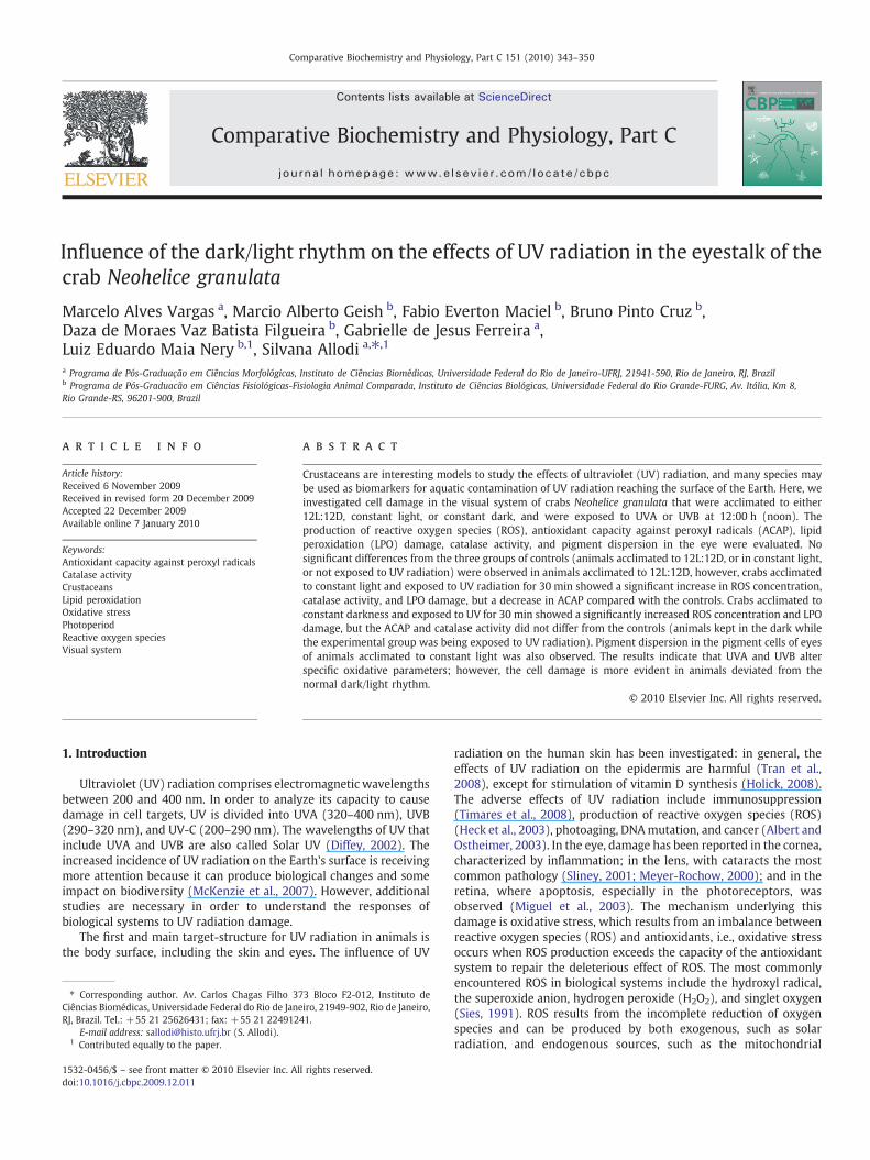
The animals acclimated to 12L:12D and exposed to UVA or UVBshowed no significant difference when compared to the controlgroups for each treatment (Fig. 1A). However, the ROS concentrationincreased in the animals acclimated to constant light (Fig. 1B) andconstant darkness (Fig. 1C) when exposed to UV radiation.
3.2. Antioxidant capacity against peroxyl radicals
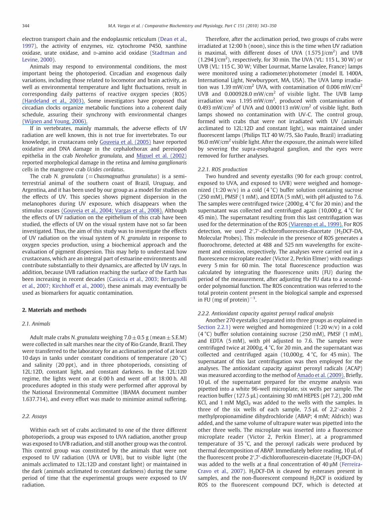
The ACAP test performed in animals acclimated to 12L:12D(Fig. 2A) and to constant darkness (Fig. 2B) after being exposed toUV radiation showed no significant difference when compared withthe control group. However, when the animals were acclimated toconstant light, the ACAP result was significantly lower in animalsexposed to UVA and UVB than in the controls (Fig. 2C).
3.3. Catalase activity
In the crabs acclimated to 12L:12D, the catalase activity increasedafter exposure to UVB when compared with controls (Fig. 3A).Additionally, in animals acclimated to constant light, after theexposure to UVA and UVB the catalase activity was higher than inthe controls (Fig. 3B). In contrast, in animals acclimated to constant
Fig. 1. Concentration at 12:00 h of reactive oxygen species in eyestalks of the crabNeohelice granulata acclimated to (A) 12L:12D, (B) constant light, (C) constantdarkness, and exposed to UV radiation. Each point represents a percentage of themean±S.E.M (n=5).
Fig. 2. Antioxidant competence against peroxyl radical (ACAP) at 12:00 h of eyestalksfrom the crab Neohelice granulata acclimated to (A) 12L:12D, (B) constant light, (C)constant darkness, and after exposure to UV. Each point represents a percentage of themean±S.E.M (n=5).
346 M.A. Vargas et al. / Comparative Biochemistry and Physiology, Part C 151 (2010) 343–350
darkness and exposed to UVA and UVB, catalase activity showed nosignificant difference from the control group (Fig. 3C).
3.4. Lipid peroxidation
Regarding LPO, animals acclimated to 12L:12D and exposed toUVA or UVB did not show any significant differences when comparedwith the control group (Fig. 4A), but when crabs acclimated toconstant light and constant darkness were exposed to UV radiation,LPO values increased significantly, as shown in Fig. 4B and C,respectively.
3.5. Histological analysis
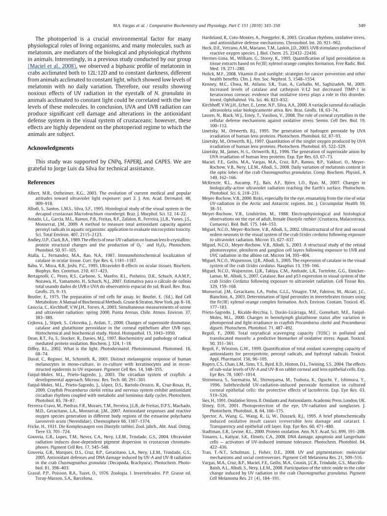
The visual system of decapod crustaceans consists of ommatidiacomposed of a cornea, a crystalline cone lens system, and a retinaformed by photoreceptors and pigment glial cells. The photoreceptorsare connected via the fasciculated zone to a group of three sequentially
arranged optic ganglia: the lamina ganglionaris, the external medulla,and the internal medulla. The basement membrane limits the retinaproximally (Grassé et al., 1976; Allodi et al., 1995) (Fig. 5A).
The sections of eyestalks of the crabs acclimated to 12L:12D andfixed at 12:00 h showed few pigments in the retina near the basementmembrane (Fig. 5B), and the pigments were more distributed in thefasciculated zone. In the sections obtained from the retina of the crabsacclimated to constant light (Fig. 5C) and to constant darkness(Fig. 5D), pigments were clearly seen in the retina near the basementmembrane and were more aggregated in the fasciculated zone(Fig. 5D). It is important to note here that in decapods maintainedin normal photoperiods, light-adapted eyes show dispersed pigments,whereas during the night, most of the eye pigments are withdrawn(Fricke, 1931; Meyer-Rochow and Lindström, 1988). Therefore, thepigment position seen in eyes of crabs maintained in constant lightand constant darkness indicates that these crabs are on differentphotoperiod schedules.
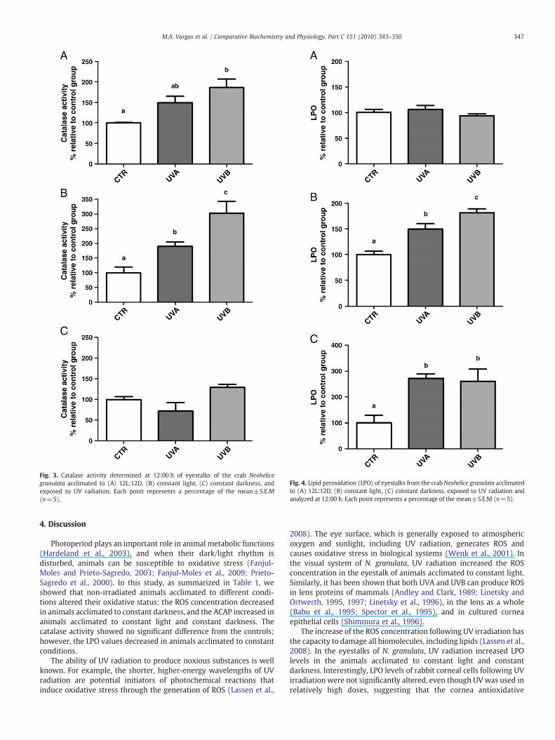
Fig. 3. Catalase activity determined at 12:00 h of eyestalks of the crab Neohelicegranulata acclimated to (A) 12L:12D, (B) constant light, (C) constant darkness, andexposed to UV radiation. Each point represents a percentage of the mean±S.E.M(n=5).
Fig. 4. Lipid peroxidation (LPO) of eyestalks from the crabNeohelice granulata acclimatedto (A) 12L:12D, (B) constant light, (C) constant darkness, exposed to UV radiation andanalyzed at 12:00 h. Each point represents a percentage of the mean±S.E.M (n=5).
347M.A. Vargas et al. / Comparative Biochemistry and Physiology, Part C 151 (2010) 343–350
4. Discussion
Photoperiod plays an important role in animal metabolic functions(Hardeland et al., 2003), and when their dark/light rhythm isdisturbed, animals can be susceptible to oxidative stress (Fanjul-Moles and Prieto-Sagredo, 2003; Fanjul-Moles et al., 2009; Prieto-Sagredo et al., 2000). In this study, as summarized in Table 1, weshowed that non-irradiated animals acclimated to different condi-tions altered their oxidative status: the ROS concentration decreasedin animals acclimated to constant darkness, and the ACAP increased inanimals acclimated to constant light and constant darkness. Thecatalase activity showed no significant difference from the controls;however, the LPO values decreased in animals acclimated to constantconditions.
The ability of UV radiation to produce noxious substances is wellknown. For example, the shorter, higher-energy wavelengths of UVradiation are potential initiators of photochemical reactions thatinduce oxidative stress through the generation of ROS (Lassen et al.,
2008). The eye surface, which is generally exposed to atmosphericoxygen and sunlight, including UV radiation, generates ROS andcauses oxidative stress in biological systems (Wenk et al., 2001). Inthe visual system of N. granulata, UV radiation increased the ROSconcentration in the eyestalk of animals acclimated to constant light.Similarly, it has been shown that both UVA and UVB can produce ROSin lens proteins of mammals (Andley and Clark, 1989; Linetsky andOrtwerth, 1995, 1997; Linetsky et al., 1996), in the lens as a whole(Babu et al., 1995; Spector et al., 1995), and in cultured corneaepithelial cells (Shimmura et al., 1996).
The increase of the ROS concentration following UV irradiation hasthe capacity to damage all biomolecules, including lipids (Lassen et al.,2008). In the eyestalks of N. granulata, UV radiation increased LPOlevels in the animals acclimated to constant light and constantdarkness. Interestingly, LPO levels of rabbit corneal cells following UVirradiation were not significantly altered, even though UVwas used inrelatively high doses, suggesting that the cornea antioxidative
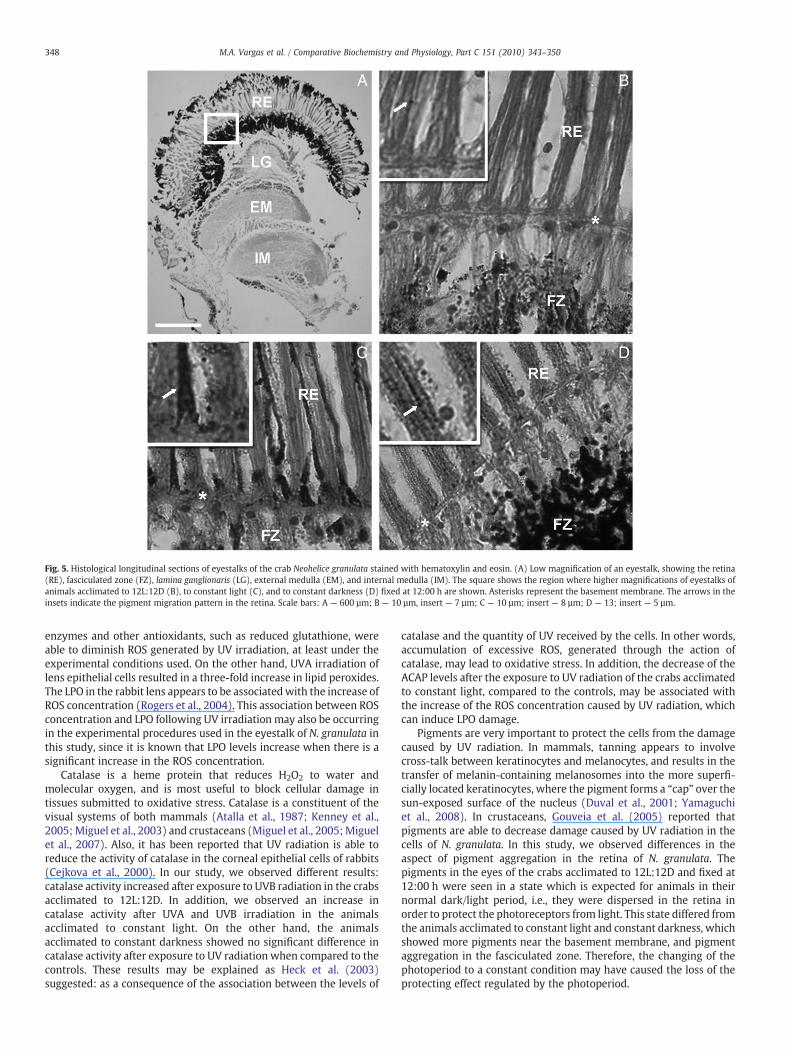
Fig. 5. Histological longitudinal sections of eyestalks of the crab Neohelice granulata stained with hematoxylin and eosin. (A) Low magnification of an eyestalk, showing the retina(RE), fasciculated zone (FZ), lamina ganglionaris (LG), external medulla (EM), and internal medulla (IM). The square shows the region where higher magnifications of eyestalks ofanimals acclimated to 12L:12D (B), to constant light (C), and to constant darkness (D) fixed at 12:00 h are shown. Asterisks represent the basement membrane. The arrows in theinsets indicate the pigment migration pattern in the retina. Scale bars: A — 600 µm; B — 10 µm, insert — 7 µm; C — 10 µm; insert — 8 µm; D — 13; insert — 5 µm.
348 M.A. Vargas et al. / Comparative Biochemistry and Physiology, Part C 151 (2010) 343–350
enzymes and other antioxidants, such as reduced glutathione, wereable to diminish ROS generated by UV irradiation, at least under theexperimental conditions used. On the other hand, UVA irradiation oflens epithelial cells resulted in a three-fold increase in lipid peroxides.The LPO in the rabbit lens appears to be associatedwith the increase ofROS concentration (Rogers et al., 2004). This association between ROSconcentration and LPO following UV irradiation may also be occurringin the experimental procedures used in the eyestalk of N. granulata inthis study, since it is known that LPO levels increase when there is asignificant increase in the ROS concentration.
Catalase is a heme protein that reduces H2O2 to water andmolecular oxygen, and is most useful to block cellular damage intissues submitted to oxidative stress. Catalase is a constituent of thevisual systems of both mammals (Atalla et al., 1987; Kenney et al.,2005; Miguel et al., 2003) and crustaceans (Miguel et al., 2005; Miguelet al., 2007). Also, it has been reported that UV radiation is able toreduce the activity of catalase in the corneal epithelial cells of rabbits(Cejkova et al., 2000). In our study, we observed different results:catalase activity increased after exposure to UVB radiation in the crabsacclimated to 12L:12D. In addition, we observed an increase incatalase activity after UVA and UVB irradiation in the animalsacclimated to constant light. On the other hand, the animalsacclimated to constant darkness showed no significant difference incatalase activity after exposure to UV radiation when compared to thecontrols. These results may be explained as Heck et al. (2003)suggested: as a consequence of the association between the levels of
catalase and the quantity of UV received by the cells. In other words,accumulation of excessive ROS, generated through the action ofcatalase, may lead to oxidative stress. In addition, the decrease of theACAP levels after the exposure to UV radiation of the crabs acclimatedto constant light, compared to the controls, may be associated withthe increase of the ROS concentration caused by UV radiation, whichcan induce LPO damage.
Pigments are very important to protect the cells from the damagecaused by UV radiation. In mammals, tanning appears to involvecross-talk between keratinocytes and melanocytes, and results in thetransfer of melanin-containing melanosomes into the more superfi-cially located keratinocytes, where the pigment forms a “cap” over thesun-exposed surface of the nucleus (Duval et al., 2001; Yamaguchiet al., 2008). In crustaceans, Gouveia et al. (2005) reported thatpigments are able to decrease damage caused by UV radiation in thecells of N. granulata. In this study, we observed differences in theaspect of pigment aggregation in the retina of N. granulata. Thepigments in the eyes of the crabs acclimated to 12L:12D and fixed at12:00 h were seen in a state which is expected for animals in theirnormal dark/light period, i.e., they were dispersed in the retina inorder to protect the photoreceptors from light. This state differed fromthe animals acclimated to constant light and constant darkness, whichshowed more pigments near the basement membrane, and pigmentaggregation in the fasciculated zone. Therefore, the changing of thephotoperiod to a constant condition may have caused the loss of theprotecting effect regulated by the photoperiod.
349M.A. Vargas et al. / Comparative Biochemistry and Physiology, Part C 151 (2010) 343–350
The photoperiod is a crucial environmental factor for manyphysiological roles of living organisms, and many molecules, such asmelatonin, are mediators of the biological and physiological rhythmsin animals. Interestingly, in a previous study conducted by our group(Maciel et al., 2008), we observed a biphasic profile of melatonin incrabs acclimated both to 12L:12D and to constant darkness, differentfrom animals acclimated to constant light, which showed low levels ofmelatonin with no daily variation. Therefore, our results showingnoxious effects of UV radiation in the eyestalk of N. granulata inanimals acclimated to constant light could be correlated with the lowlevels of these molecules. In conclusion, UVA and UVB radiation canproduce significant cell damage and alterations in the antioxidantdefense system in the visual system of crustaceans; however, theseeffects are highly dependent on the photoperiod regime to which theanimals are subject.
Acknowledgments
This study was supported by CNPq, FAPERJ, and CAPES. We aregrateful to Jorge Luis da Silva for technical assistance.
References
Albert, M.R., Ostheimer, K.G., 2003. The evolution of current medical and popularattitudes toward ultraviolet light exposure: part 2. J. Am. Acad. Dermatol. 48,909–918.
Allodi, S., Santos, L.M.S., Silva, S.F., 1995. Histological study of the visual system in thedecapod crustacean Macrobrachium rosenbergii. Braz. J. Morphol. Sci. 12, 14–22.
Amado, L.L., Garcia, M.L., Ramos, P.B., Freitas, R.F., Zafalon, B., Ferreira, J.L.R., Yunes, J.S.,Monserrat, J.M., 2009. A method to measure total antioxidant capacity againstperoxyl radicals in aquatic organisms: application to evaluate microcystins toxicity.Sci. Total Environ. 407, 2115–2123.
Andley, U.P., Clark, B.A., 1989. The effects of near-UV radiation onhuman lensb-crystallins:protein structural changes and the production of O2
− and H2O2. Photochem.Photobiol. 50, 97–105.
Atalla, L., Fernandez, M.A., Rao, N.A., 1987. Immunohistochemical localization ofcatalase in ocular tissue. Curr. Eye Res. 6, 1181–1187.
Babu, V., Misra, R.B., Joshi, P.C., 1995. Ultraviolet-B effects on ocular tissues. Biochem.Biophys. Res. Commun. 210, 417–423.
Bertagnolli, C., Peres, R.S., Carbone, S., Manfro, R.L., Pinheiro, D.K., Schuch, A.A.M.P.,Nozawa, H., Yamamoto, H., Schuch, N.J., 2007. Estimativa para o cálculo de ozôniototal usando dados de UVB e UVA do observatório espacial do sul, Brasil. Rev. Bras.Geofis. 25, 9–15.
Beutler, E., 1975. The preparation of red cells for assay. In: Beutler, E. (Ed.), Red CellMetabolism: AManual of BiochemicalMethods. Grune & Straton, NewYork, pp. 8–18.
Casiccia, C., Kirchhoff, V.W.J.H., Torres, A., 2003. Simultaneous measurements of ozoneand ultraviolet radiation: spring 2000, Punta Arenas, Chile. Atmos. Environ. 37,383–389.
Cejkova, J., Stipek, S., Crkovska, J., Ardan, T., 2000. Changes of superoxide dismutase,catalase and glutathione peroxidase in the corneal epithelium after UVB rays.Histochemical and biochemical study. Histol. Histopathol. 15, 1043–1050.
Dean, R.T., Fu, S., Stocker, R., Davies, M.J., 1997. Biochemistry and pathology of radicalmediated protein oxidation. Biochem. J. 324, 1–18.
Diffey, B.L., 2002. What is the light. Photodermatol. Photoimmunol. Photomed. 18,68–74.
Duval, C., Reginer, M., Schimith, R., 2001. Distinct melanogenic response of humanmelanocytes in mono-culture, in co-culture with keratinocytes and in recon-structed epidermis to UV exposure. Pigment Cell Res. 14, 348–355.
Fanjul-Moles, M.L., Prieto-Sagredo, J., 2003. The circadian system of crayfish: adevelopmental approach. Microsc. Res. Tech. 60, 291–301.
Fanjul-Moles, M.L., Prieto-Sagredo, J., López, D.S., Bartolo-Orozco, R., Cruz-Rosas, H.,2009. Crayfish Procambarus clarkii retina and nervous system exhibit antioxidantcircadian rhythms coupled with metabolic and luminous daily cycles. Photochem.Photobiol. 85, 78–87.
Ferreira-Cravo, M., Piedras, F.R., Moraes, T.M., Ferreira, J.L.B., de Freitas, D.P.S., Machado,M.D., Geracitano, L.A., Monserrat, J.M., 2007. Antioxidant responses and reactiveoxygen species generation in different body regions of the estuarine polychaetaLaeonereis acuta (Nereididae). Chemosphere 66, 1367–1374.
Fricke, H., 1931. Die Komplexaugen von Diastylis rathkei. Zool. Jahrb., Abt. Anal. Ontog.Tiere S3, 701–724.
Gouveia, G.R., Lopes, T.M., Neves, C.A., Nery, L.E.M., Trindade, G.S., 2004. Ultravioletradiation induces dose-dependent pigment dispersion in crustacean chromato-phores. Pigment Cell Res. 17, 545–548.
Gouveia, G.R., Marques, D.S., Cruz, B.P., Geracitano, L.A., Nery, L.E.M., Trindade, G.S.,2005. Antioxidant defenses and DNA damage induced by UV-A and UV-B radiationin the crab Chasmagnathus granulata (Decapoda, Brachyura). Photochem. Photo-biol. 81, 398–403.
Grassé, P.P., Poisson, R.A., Tuzet, O., 1976. Zoologia. I. Invertebrados. P.P. Grasse ed.Toray-Masson, S.A., Barcelona.
Hardeland, R., Coto-Montes, A., Poeggeler, B., 2003. Circadian rhythms, oxidative stress,and antioxidative defense mechanisms. Chronobiol. Int. 20, 921–962.
Heck, D.E., Vetrano, A.M., Mariano, T.M., Laskin, J.D., 2003. UVB stimulates production ofreactive oxygen species. J. Biol. Chem. 25, 22432–22436.
Hermes-Lima, M., William, G., Storey, K., 1995. Quantification of lipid peroxidation intissue extracts based on Fe(III) xylenol orange complex formation. Free Radic. Biol.Med. 19, 271–280.
Holick, M.F., 2008. Vitamin D and sunlight: strategies for cancer prevention and otherhealth benefits. Clin. J. Am. Soc. Nephrol. 5, 1548–1554.
Kenney, M.C., Chwa, M., Atilano, S.R., Tran, A., Carballo, M., Saghizadeh, M., 2005.Increased levels of catalase and cathepsin V/L2 but decreased TIMP-1 inkeratoconus corneas: evidence that oxidative stress plays a role in this disorder.Invest. Ophthalmol. Vis. Sci. 46, 823–832.
Kirchhoff, V.W.J.H., Echer, E., Leme, N.P., Silva, A.A., 2000. A variação sazonal da radiaçãoultravioleta solar biologicamente ativa. Rev. Bras. Geofís. 18, 63–74.
Lassen, N., Black, W.J., Estey, T., Vasiliou, V., 2008. The role of corneal crystallins in thecellular defense mechanisms against oxidative stress. Semin. Cell Dev. Biol. 19,100–112.
Linetsky, M., Ortwerth, B.J., 1995. The generation of hydrogen peroxide by UVAirradiation of human lens proteins. Photochem. Photobiol. 62, 87–93.
Linetsky, M., Ortwerth, B.J., 1997. Quantitation of the singlet oxygen produced by UVAirradiation of human lens proteins. Photochem. Photobiol. 65, 522–529.
Linetsky, M., James, H.-L., Ortwerth, B.J., 1996. The generation of superoxide anion byUVA irradiation of human lens proteins. Exp. Eye Res. 63, 67–73.
Maciel, F.E., Geihs, M.A., Vargas, M.A., Cruz, B.P., Ramos, B.P., Vakkuri, O., Meyer-Rochow, V.B., Nery, L.E.M., Allodi, S., 2008. Daily variation of melatonin content inthe optic lobes of the crab Chasmagnathus granulatus. Comp. Biochem. Physiol., A149, 162–166.
McKenzie, R.L., Aucamp, P.J., Bais, A.F., Björn, L.O., Ilyas, M., 2007. Changes inbiologically-active ultraviolet radiation reaching the Earth's surface. Photochem.Photobiol. Sci. 6, 218–231.
Meyer-Rochow, V.B., 2000. Risks, especially for the eye, emanating from the rise of solarUV-radiation in the Arctic and Antarctic regions. Int. J. Circumpolar Health 59,38–51.
Meyer-Rochow, V.B., Lindström, M., 1988. Electrophysiological and histologicalobservations on the eye of adult, female Diastylis rathkei (Crustacea, Malacostraca,Cumacea). Biol. Bull. 175, 144–153.
Miguel, N.C.O., Meyer-Rochow, V.B., Allodi, S., 2002. Ultrastructural of first and secondordem neurons in the visual system of the crab Ucides cordatus following exposureto ultraviolet radiation. Micron 33, 627–637.
Miguel, N.C.O., Meyer-Rochow, V.B., Allodi, S., 2003. A structural study of the retinalphotoreceptor, plexiform and ganglion cell layers following exposure to UVB andUVC radiation in the albino rat. Micron 34, 395–404.
Miguel, N.C.O., Wajsenzon, I.J.R., Allodi, S., 2005. The expression of catalase in the visualsystem of the crab Ucides cordatus. Nauplius 13, 159–166.
Miguel, N.C.O., Wajsenzon, I.J.R., Takiya, C.M., Andrade, L.R., Tortelote, G.G., Einicker-Lamas, M., Allodi, S., 2007. Catalase, Bax and p53 expression in visual system of thecrab Ucides Cordatus following exposure to ultraviolet radiation. Cell Tissue Res.329, 159–168.
Monserrat, J.M., Geracitano, L.A., Pinho, G.L.L., Vinagre, T.M., Faleiros, M., Alciati, J.C.,Bianchini, A., 2003. Determination of lipid peroxides in invertebrates tissues usingthe Fe(III) xylenol orange complex formation. Arch. Environ. Contam. Toxicol. 45,177–183.
Prieto-Sagredo, J., Ricalde-Recchia, I., Durán-Lizárraga, M.E., Gonsebatt, M.E., Fanjul-Moles, M.L., 2000. Changes in hemolymph glutathione status after variation inphotoperiod and light-irradiance in crayfish Procambarus clarkii and Procambarusdigueti. Photochem. Photobiol. 71, 487–492.
Regoli, F., 2000. Total oxyradical scavenging capacity (TOSC) in polluted andtranslocated mussels: a predictive biomarker of oxidative stress. Aquat. Toxicol.50, 351–361.
Regoli, F., Winston, G.W., 1999. Quantification of total oxidant scavenging capacity ofantioxidants for peroxynitrite, peroxyl radicals, and hydroxyl radicals. Toxicol.Appl. Pharmacol. 156, 96–105.
Rogers, C.S., Chan, L.M., Sims, Y.S., Byrd, K.D., Hinton, D.L., Twining, S.S., 2004. The effectsof sub-solar levels of UV-A and UV-B on rabbit corneal and lens epithelial cells. Exp.Eye Res. 78, 1007–1014.
Shimmura, S., Suematsu, M., Shimoyama, M., Tsubota, K., Oguchi, Y., Ishimura, Y.,1996. Subthreshold UV-radiation-induced peroxide formation in culturedcorneal epithelial cells: the protective effects of lactoferrin. Exp. Eye Res. 63,519–526.
Sies, H., 1991. Oxidative Stress. II. Oxidants and Antioxidants. Academic Press, London, UK.Sliney, D.H., 2001. Photoprotection of the eye, UV-radiation and sunglasses. J.
Photochem. Photobiol., B. 64, 166–175.Spector, A., Wang, G., Wang, R., Li, W., Duszark, R.J., 1995. A brief photochemically
induced oxidative insult causes irreversible lens damage and cataract. I.Transparency and epithelial cell layer. Exp. Eye Res. 60, 471–480.
Stadtman, E.R., Levine, R.L., 2000. Protein oxidation. Ann. N.Y. Acad. Sci. 899, 191–208.Timares, L., Katiyar, S.K., Elmets, C.A., 2008. DNA damage, apoptosis and Langerhans
cells — activators of UV-induced immune tolerance. Photochem. Photobiol. 84,422–436.
Tran, T.-N.T., Schulman, J., Fisher, D.E., 2008. UV and pigmentation: molecularmechanisms and social controversies. Pigment Cell Melanoma Res. 21, 509–516.
Vargas, M.A., Cruz, B.P., Maciel, F.E., Geihs, M.A., Cousin, J.C.B., Trindade, G.S., Muccillo-Baish, A.L., Allodi, S., Nery, L.E.M., 2008. Participation of the nitric oxide in the colorchange induced by UV radiation in the crab Chasmagnathus granulatus. PigmentCell Melanoma Res. 21 (4), 184–191.
350 M.A. Vargas et al. / Comparative Biochemistry and Physiology, Part C 151 (2010) 343–350
Viarengo, A., Burlando, B., Cavaletto, M., Marchi, B., Panzano, E., Blasco, J., 1999. Role ofmetallothionein against oxidative stress in the musselMytilus galloprovincialis. Am.J. Physiol. 46, 1612–1619.
Wenk, J., Brenneisen, P., Meewes, C., Wlaschek, M., Peters, T., Blaudschun, R., Ma, W.,Kuhr, L., Schneider, L., Scharffetter-Kochanek, K., 2001. UV-induced oxidative stressand photoaging. Curr. Probl. Dermatol. 29, 83–94.
Wijnen, H., Young, M.W., 2006. Interplay of metabolic circadian clocks and metabolicrhythms. Annu. Rev. Genet. 40, 409–448.
Yamaguchi, Y., Takahashi, K., Zmudzka, B.Z., Kornhauser, A., Miller, S.A., Tdokoro, T.,Berens, W., Beer, J.Z., Hearing, V.J., 2008. Human skin response to UV radiationpigment in the upper epidermis protects against DNA damage in the lowerepidermis and facilitates apoptosis. FASEB J. 20, 1486–1488.
Zar, J.H., 1984. Biostatistical Analysis, 2nd ed. Prentice-Hall Inc., Englewood Cliffs, N.J.,718 pp.
Top Related
Copyright © 2022 FDOKUMEN

