


Bahasa
Halaman
Hukum

P
IL
CAa
b
c
a
ARR2AA
KVSOMH
1
iaia2idlr(e
ST
0h
International Journal of Pharmaceutics 443 (2013) 128– 136
Contents lists available at SciVerse ScienceDirect
International Journal of Pharmaceutics
jo ur n al homep age: www.elsev ier .com/ locate / i jpharm
harmaceutical nanotechnology
nhibition of skin inflammation in mice by diclofenac in vesicular carriers:iposomes, ethosomes and PEVs
arla Caddeoa, Octavio Diez Salesb, Donatella Valenti a, Amparo Ruiz Saurí c,nna Maria Faddaa,∗, Maria Manconia
Dept. Scienze della Vita e dell’Ambiente, Sezione Scienze del Farmaco, University of Cagliari, Via Ospedale 72, 09124 Cagliari, ItalyDept. of Pharmacy and Pharmaceutical Technology, University of Valencia, Avda Vicente Andrés Estellés s/n, 46100-Burjassot, Valencia, SpainDept. of Pathology, University of Valencia, Avda Blasco Ibanez 17, 46010 Valencia, Spain
r t i c l e i n f o
rticle history:eceived 17 September 2012eceived in revised form4 December 2012ccepted 29 December 2012vailable online xxx
eywords:esicleskin inflammationedemayeloperoxidase
a b s t r a c t
Diclofenac-loaded phospholipid vesicles, namely conventional liposomes, ethosomes and PEVs (pene-tration enhancer-containing vesicles) were developed and their efficacy in TPA (phorbol ester) inducedskin inflammation was examined. Vesicles were made from a cheap and unpurified mixture of phospho-lipids and diclofenac sodium; Transcutol® P and propylene glycol were added to obtain PEVs, and ethanolto produce ethosomes. The structure and lamellar organization of the vesicle bilayer were investigatedby transmission electron microscopy and small and wide angle X-ray scattering, as well as the mainphysico-chemical features. The formulations, along with a diclofenac solution and commercial VoltarenEmulgel®, were tested in a comparative trial for anti-inflammatory efficacy on TPA-treated mice dorsalskin.
Vesicles were around 100 nm, negatively charged, able to encapsulate diclofenac in good yields, anddisclosed different lamellarity, as a function of the formulation composition. Vesicular formulations
istology promoted drug accumulation and reduced the permeation. Administration of vesicular diclofenac onTPA-inflamed skin resulted in marked attenuation of oedema and leucocyte infiltration, especially usingPEVs. Histology confirmed the effectiveness of vesicles, since they provided an amelioration of the tissualdamage induced by TPA.
The proposed approach based on vesicular nanocarriers may hold promising therapeutic value fortreating a variety of inflammatory skin disorders.
. Introduction
During last decades, nanoparticles have shown their valuen drug delivery. Indeed, nanoparticulate carriers have severaldvantages among which their capability to overcome limitingssues, such as low solubility and consequent reduced bioavail-bility of several important hydrophobic drugs (Fukami et al.,009; Io et al., 2010). In particular, there has been a great deal of
nterest in lipid vesicles as a tool to improve dermal and trans-ermal drug delivery. However, the lack of ability of conventional
iposomes to deliver drugs across the skin has led to intensive
esearch, with the development of new classes of lipid vesiclesPaolino et al., 2007, 2008; Sinico and Fadda, 2009). A strat-gy to improve liposome efficacy is to modify the hydrophilic∗ Corresponding author at: Dept. Scienze della Vita e dell’Ambiente, Sezionecienze del Farmaco, University of Cagliari, Via Ospedale 72, 09124 Cagliari, Italy.el.: +39 070 675 8565; fax: +39 070 675 8710.
E-mail address: [email protected] (A.M. Fadda).
378-5173/$ – see front matter © 2013 Elsevier B.V. All rights reserved.ttp://dx.doi.org/10.1016/j.ijpharm.2012.12.041
© 2013 Elsevier B.V. All rights reserved.
phase using a mixture of water and a cosolvent, such as ethanol,propylene glycol (PG) or polyethylene glycol (Elsayed et al., 2007;Paolino et al., 2005; Priprem et al., 2008). These systems include,among others, ethosomes: soft and malleable vesicles composedmainly of phospholipids, ethanol and water, capable of transport-ing active substances through the stratum corneum and to thedeeper skin layers (Touitou et al., 2000). As a promising alterna-tive, we have been studying PEVs: phospholipid vesicles containingdifferent penetration enhancers (PEs), capable of influencing vesi-cle deformability and (trans)dermal drug delivery (Manconi et al.,2009a, 2011a,b). In this work, PEVs were prepared as carriers fordiclofenac sodium by using two hydromiscible PEs, i.e. Transcutol®
P (Trc) and PG, since these cosolvents were proved to producesmall, stable vesicular systems capable of allowing different drugsto penetrate deeply into the skin, by increasing skin permeabilityand vesicle fluidity (Elsayed et al., 2007; Manconi et al., 2011a,b).
The purpose of this work was to evaluate their efficiency andsafety in drug delivery to and through the skin by ex vivo andin vivo experiments and in comparison with an efficient vesic-ular carrier such as ethosomes. Therefore, different lipid-based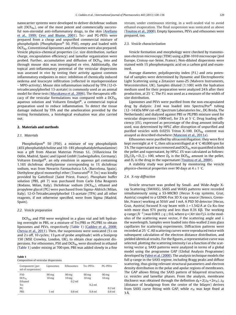
al of P
nsfeppDVcpttwio–tmcapdto
2
2
(wOV1sDps(pIrS
2
ilCa1p(
TC
C. Caddeo et al. / International Journ
anocarrier systems were developed to deliver diclofenac sodiumalt (DCFNa), one of the most potent and commercially success-ul non-steroidal anti-inflammatory drugs, to the skin (Arellanot al., 1999; Cevc and Blume, 2001). Trc- and PG-PEVs wererepared from a cheap and unpurified commercial mixture ofhospholipids (Phospholipon® 50, P50), empty and loaded withCFNa. Conventional liposomes and ethosomes were also prepared.esicle physico-chemical properties (i.e. size distribution, surfaceharge, encapsulation efficiency) and lamellar organization wererobed. Further, accumulation and diffusion of DCFNa into andhrough mouse skin was investigated ex vivo. Additionally, theopical anti-inflammatory potential of the vesicular dispersionsas assessed in vivo by testing their activity against common
nflammatory endpoints in mice: inhibition of chemically inducededema and leucocyte infiltration (reflected in myeloperoxidaseMPO-activity). Mouse skin inflammation induced by TPA (12-O-
etradecanoylphorbol 13-acetate) is commonly used as an animalodel for these tests (Murakawa et al., 2006). The therapeutic effi-
acy of the vesicular formulations was compared with a DCFNaqueous solution and Voltaren Emulgel®, a commercial topicalreparation used to reduce inflammation. To detect the tissueamage induced by TPA and the amelioration provided by theesting formulations, a histological evaluation was also carriedut.
. Materials and methods
.1. Materials
Phospholipon® 50 (P50), a mixture of soy phospholipids45% phosphatidylcholine and 10–18% phosphatidylethanolamine)as a gift from Abaran Materias Primas S.L. (Villaviciosa Dedón, Madrid, Spain) and Lipoid GmbH (Ludwigshafen, Germany).oltaren Emulgel®, an oily emulsion in aqueous gel containing.16% diclofenac diethylamine corresponding to 1% diclofenacodium, was from Novartis Farmacéutica S.A. (Barcelona, Spain).iethylene glycol monoethyl ether (Transcutol® P, Trc) was kindlyrovided by Gattefossè (Saint Priest, France). Phosphate bufferolution (PBS, pH 7) was purchased from Carlo Erba ReagentsRodano, Milan, Italy). Diclofenac sodium (DCFNa), ethanol andropylene glycol (PG) were purchased from Sigma–Aldrich (Milan,
taly). 12-O-Tetradecanoylphorbol 13-acetate (TPA) and all othereagents, if not otherwise specified, were from Sigma (Madrid,pain).
.2. Vesicle preparation
DCFNa and P50 were weighted in a glass vial and left hydrat-ng overnight in PBS, or a mixture of Trc/PBS or PG/PBS to obtainiposomes and PEVs, respectively (Table 1) (Caddeo et al., 2008;hessa et al., 2011). Then, the suspensions were sonicated (5 s on
nd 2 s off, 10 cycles; 13 �m of probe amplitude) with a Soniprep50 (MSE Crowley, London, UK), to obtain clear opalescent dis-ersions. For ethosomes, P50 and DCFNa were dissolved in ethanolTable 1) under mixing at 700 rpm. PBS was added slowly in a fineable 1omposition of vesicular dispersions.
Component (perml of suspension)
Liposomes Ethosomes Trc-PEVs PG-PEVs
P50 90 mg 90 mg 90 mg 90 mgDCFNa 10 mg 10 mg 10 mg 10 mgEthanol 0.2 mlTrc 0.2 mlPG 0.2 mlPBS 1 ml 0.8 ml 0.8 ml 0.8 ml
harmaceutics 443 (2013) 128– 136 129
stream, under continuous stirring, in a well-sealed vial to avoidethanol evaporation. The final suspension was sonicated as above(Touitou et al., 2000). Empty liposomes, PEVs and ethosomes wereprepared, too.
2.3. Vesicle characterization
Vesicle formation and morphology were checked by transmis-sion electron microscopy (TEM) using a JEM-1010 microscope (JeolEurope, Croissy-sur-Seine, France). Non-diluted dispersions werestained with 1% phosphotungstic acid on a carbon grid and exam-ined.
Average diameter, polydispersity index (P.I.) and zeta poten-tial of samples were determined by Dynamic and ElectrophoreticLight Scattering using a Zetasizer nano-ZS (Malvern Instruments,Worcestershire, UK). Samples diluted (1:100) with the hydrationmedium used for their preparation were analyzed 24 h after theirproduction, at 25 ◦C. The P.I. was used as a measure of the width ofsize distribution.
Liposomes and PEVs were purified from the non-encapsulateddrug by dialysis: 2 ml was loaded into Spectra/Por® tubing(12–14 kDa MW cut-off; Spectrum Laboratories Inc., DG Breda, TheNetherlands) and dialysed against PBS or PE/PBS mixture used forvesicular dispersions (1000 ml), for 2 h at 5 ◦C. Drug loading effi-ciency (E%), expressed as percentage of the drug amount initiallyused, was determined by HPLC after disruption of unpurified andpurified vesicles with 0.025% Triton X-100. DCFNa content wasassayed as described elsewhere (Manconi et al., 2011a).
Ethosomes were purified by ultracentrifugation: they were firstkept overnight at 4 ◦C, then ultracentrifuged at 4 ◦C 40,000 rpm for3 h. The supernatant was removed and DCFNa was quantified in boththe pellet and supernatant, by HPLC. E% was calculated as follows:[Dp/(Dp + Ds)] × 100, where Dp is the DCFNa amount in the pellet,and Ds is the drug in the supernatant (Touitou et al., 2000).
A stability study was performed by monitoring the vesiclephysico-chemical properties over 90 days at 4 ± 1 ◦C.
2.4. X-ray diffraction
Vesicle structure was probed by Small- and Wide-Angle X-ray Scattering (SWAXS). SAXS and WAXS patterns were recordedsimultaneously using a S3-MICRO (Hecus X-ray systems, Graz,Austria) coupled to a GENIX-Fox 3D X-ray source (Xenocs, Greno-ble, France) working at 50 kV and 1 mA. A PSD 50 detector (Hecus,Graz, Austria) focused X-ray beam with � = 1.542 A at Cu K�-linewith more than 97% purity and less than 0.3% K�. The workingq-range (A−1) was 0.003 ≤ q ≤ 0.6, where q = (4� sin �)/� is the mod-ulus of the scattering wave vector, � the scattering angle and �the wavelength. Samples were loaded into thin-walled 2 mm glasscapillaries for scattering experiments. Diffraction patterns wererecorded at 25 ◦C. All scattering curves were reproduced twice withsubsequent calculation of the electron distance distribution, andyielded identical results. For the figures, a representative curve wasselected, plotting the scattering intensity I as a function of the scat-tering vector q. SAXS patterns were analyzed in terms of a globalmodel using the programme GAP (Global Analysis Programme)developed by Pabst et al. (2000). The analysis technique models thefull q-range in the SAXS regime, including Bragg peaks and diffusescattering, thus giving relevant structural parameters and electrondensity distribution in the polar and apolar regions of membranes.The GAP allows fitting the SAXS pattern of bilayered structures,i.e. vesicles and lamellar phases. From the analysis, membrane
thickness was obtained through the definition dB = 2(zH + 2�H). zH(distance of headgroup from the centre of the bilayer) derivesfrom SAXS curve fitting with GAP, while �H was kept fixed at3 A.
1 al of P
2
HblahaEmU
wdmTuavpcw
SGtTim
2
sn32teaeawwm
2
mM
TCt
30 C. Caddeo et al. / International Journ
.5. Mice and ex vivo drug penetration/permeation studies
Female CD-1 mice (5–6 weeks old, 25–35 g) were obtained fromarlan Laboratories (Barcelona, Spain) and acclimatized for 1 weekefore use. Animals were fed rodent pellets (Global diet 2014, Har-
an Teklad, Barcelona, Spain) ad libitum with free access to water,nd maintained in a room controlled at 20 ± 2 ◦C with a relativeumidity of 60 ± 5% and a 12-h light/dark cycle (light from 8:00.m. to 8:00 p.m.). All studies were performed in accordance withuropean Union regulations for handling and use of laboratory ani-als. Protocols were approved by the Institutional Animal Care andse Committee of the University of Valencia.
Mice were sacrificed and shaved. Skin from the dorsal regionas excised and mounted on Franz diffusion vertical cells (effectiveiffusion area 0.785 cm2), between donor and receptor compart-ents, with the stratum corneum (SC) side facing the donor one.
he receptor was filled with 5.5 ml of saline (NaCl 0.9%, w/v), contin-ously stirred and thermostated at 37 ± 0.5 ◦C. The same dose foundppropriate for in vivo experiments (i.e. 20 �l, see below) of DCFNaesicle dispersions, aqueous solution, and Voltaren emulgel® (n = 6er formulation), were placed onto the skin surface (non-occlusiveonditions). After 1, 2, 4, 6, 8, 23, and 24 h, the receiving solutionas withdrawn and analyzed by HPLC for drug content.
After 24 h, the skin specimens were gently washed, and theC removed by stripping with adhesive tape Tesa® AG (Hamburg,ermany). The epidermis was separated from the dermis and the
issues directly underneath by gentle scraping with a scalpel blade.he effectiveness of separation was verified by histologic exam-nation. Tape strips, epidermis, and dermis were placed each in
ethanol, sonicated to extract DCFNa, and then assayed by HPLC.
.6. Mice and in vivo experimental design
The back skin of CD-1 mice, housed and fed as above, washaved one day before the experiment, and those animals showingo hair re-growth were used. TPA dissolved in acetone (243 �M;
�g/20 �l) was applied to the shaved dorsal area (approximately cm2) to induce cutaneous inflammation (day 1). Negative con-rol mice received acetone only (20 �l). All test compounds (20 �l),mpty or DCFNa-loaded conventional liposomes, ethosomes, Trc-nd PG-PEVs, a DCFNa aqueous solution, and commercial Voltarenmulgel® were topically smeared over the same dorsal site 3 hfter TPA application (non-occlusive conditions). The procedureas repeated (at 24-h intervals) on day 2 and 3. On day 4, miceere sacrificed by cervical dislocation. Each group comprised fourice.
.7. Oedema formation and myeloperoxidase determination
The inhibitory effect of test compounds on TPA-induced inflam-ation was determined by two biomarkers: oedema formation andPO activity. Mice were sacrificed after 72 h of treatment (on day
able 2haracteristics of empty and DCFNa-loaded vesicular formulations: mean diameter, polydhe means ± standard deviation (n = 3).
Sample Mean diameter(nm)
Liposomes Empty 117 ± 0.9
Loaded 107 ± 1.5
Ethosomes Empty 104 ± 2.1
Loaded 95 ± 1.8
Trc-PEVs Empty 115 ± 4.8
Loaded 102 ± 4.7
PG-PEVs Empty 100 ± 0.3
Loaded 92 ± 0.9
harmaceutics 443 (2013) 128– 136
4). The dorsal treated skin area of each mouse was excised andweighted to assess any increase indicative of oedema formation,and immediately stored at −80 ◦C (De Vry et al., 2005; Manconiet al., 2009b).
MPO assay was performed following the methods of De Younget al. (1989) and Sato et al. (2004), adapted to a 96-well plateformat. The biopsies were homogenized in ice bath with a Ultra-Turrax® T25 homogenizer (IKA® Werke GmbH & Co. KG, Staufen,Germany) in 750 �l of 80 mM sodium phosphate buffer (pH 5.4)containing 0.5% hexadecyltrimethylammonium bromide. The pel-lets were centrifuged at 10,000 rpm for 15 min at −1 ◦C. A 25-�lsample supernatant was added to 75 �l saline phosphate buffer (pH7.4), 10 �l sodium phosphate buffer (pH 5.4), 10 �l of 0.026% hydro-gen peroxide and left incubating at 37 ◦C for 5 min. Then, 10 �l of18 mM 3,3′,5,5′-tetramethylbenzidine dihydrochloride hydrate in8% aqueous N,N-dimethylformamide was added to start the reac-tion. After 10 min of incubation, the reaction was stopped withthe addition of 15 �l of 1.5 M sodium acetate (pH 3.0), and theabsorbance recorded at 620 nm using a Wallac 1420 Victor2 platereader (Perkin Elmer, Madrid, Spain). The MPO activity was calcu-lated from the linear portion of a standard curve.
2.8. Histological examination
Skin biopsies (approximately 2 cm × 2 cm) were excised frommouse dorsal treated region, after 72 h of treatment (on day 4),and fixed and stored in formaldehyde (10%, v/v). Tissue specimenswere processed routinely and embedded in paraffin wax. Longi-tudinal sections (5 �m) were stained with haematoxylin and eosin(H&E). Microscopic assessment by light microscope was performedon blind-coded samples.
2.9. Statistical analysis of data
Results are expressed as the mean ± standard deviation. Para-metric and non-parametric analysis of variance (ANOVA andKruskal–Wallis, respectively) and Bartlet’s test for homogeneityof variance were performed using SPSS version 17.0 for Windows(SPSS Inc., USA). Post hoc testing (P < 0.05) for multiple comparisonswas performed by Scheffe and Dunnett’s T3 tests.
3. Results
3.1. Vesicle characterization: physico-chemical and structuralfeatures
Light scattering results (Table 2) showed only a slight decrease(P < 0.05) in the mean diameter of DCFNa-loaded vesicles, as com-
pared to the empty ones, probably because the insertion of thehydrophilic DCFNa, localizing in the interbilayer water regions,caused a variation of the aqueous captured volume, as also assumedby Müller et al. (2004). All vesicles were small in size, aroundispersity index (P.I.), zeta potential (�) and encapsulation efficiency (E%). Values are
P.I. � (mV) E%
0.23 −62 ± 2.50.22 −65 ± 1.6 65 ± 1.1
0.21 −56 ± 2.80.20 −53 ± 2.0 55 ± 2.5
0.25 −65 ± 0.80.25 −67 ± 3.7 52 ± 6.2
0.25 −59 ± 0.10.22 −58 ± 0.3 53 ± 3.1
C. Caddeo et al. / International Journal of Pharmaceutics 443 (2013) 128– 136 131
(B) ol
1≤
vww
ncifbt5
ab(
owtiPblNtatwlTn
Fig. 1. TEM micrographs of (A) unilamellar ethosomes,
00 nm, with acceptable and repeatable homogeneity, since P.I. was0.25. The zeta potential was always highly negative (≈−60 mV).
Stability studies demonstrated that no relevant changes affectedesicle size and zeta potential. Variations in liposomes and Trc-PEVsere never higher than ±10%, while in ethosomes and PG-PEVsere around ±5%.
During this work, liposomes and PEVs were purified from theon-encapsulated drug by dialysis, while ethosomes by ultra-entrifugation, following the patented method reported by theirnventors (Touitou et al., 2000). Nevertheless, dialysis was per-ormed also for ethosomes and no statistical differences were foundetween results obtained from the two techniques. Results showedhat all vesicles were able to encapsulate DCFNa in good yields (E%:2–65; Table 2).
TEM provided evidence of vesicle formation, and showed smallnd almost spherical vesicles, with different lamellarity (i.e. num-er of bilayers), as a function of the formulation compositionFig. 1).
Further information about the influence of vesicle componentsn conformation and structure of the prepared colloidal systemsere gained by SAXS and WAXS, well-established techniques for
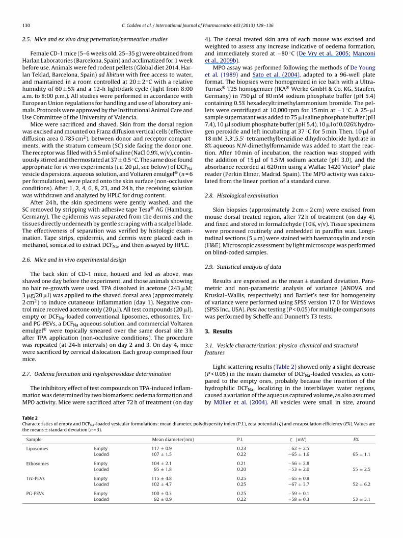
he study of self-assembling nanostructures. Fig. 2 displays SAXSntensity profiles of the studied dispersions. For ethosomes and PG-EVs a diffuse scattering was observed, with a broad symmetricand that could be fitted by assuming a population of only uni-
amellar vesicles, regardless of the presence or absence of DCFNa.evertheless, the insertion of the drug caused a shift of the curve
o higher q, as reported in Fig. 2. This indicates a distinct alter-tion of the bilayer microstructure, becoming more disordered,hus more fluid. In the case of liposomes, a broad symmetric band
as obtained for empty vesicles, while in the presence of DCFNa, aess symmetric and sharper peak, shifted to higher q, was visible.his increase in sharpness corresponds to an increase in the meanumber of bilayers, since the curve results from the superposition
igolamellar liposomes and (C) multilamellar Trc-PEVs.
of the scattering of unilamellar vesicles with diffraction peaks ofoligolamellar vesicles. For empty Trc-PEVs, a diffraction peak typ-ical of multilamellar vesicles (i.e. first order peak) was apparent.The diffraction intensity decreased and the curve was shifted tohigher q due to the addition of DCFNa, indicating a reduction ofvesicle lamellarity. Therefore, according to vesicle composition, thepresence of DCFNa caused a change of the vesicle structure. Indeed,while PG-PEVs and ethosomes were still unilamellar, the unilamel-lar conventional liposomes turn into oligolamellar vesicles, and themultilamellar Trc-PEVs transformed into oligolamellar vesicles.
A decrease in bilayer thickness (dB) caused by the insertion ofthe drug was observed, except for liposomes where no statisticallysignificant difference was found (dB = 47 ± 0.4 A). dB for ethosomes,Trc-PEVs and PG-PEVs decreased from 50 ± 0.5, 49 ± 0.3, 51 ± 0.4to 48 ± 0.4, 46 ± 0.2, 48 ± 0.3 A, respectively, leading to a flatten-ing of the scattering curve at higher q values, as mentioned above.In the case of Trc-PEVs and DCFNa-loaded liposomes, the multilay-ered structure repeat distance d was determined from the positionof the first diffraction peak using Bragg equation: 2d sin � = �, where� is half of the scattering angle. For DCFNa-loaded liposomes dwas 51 ± 0.3 A. For Trc-PEVs, a decrease in the lamellar spacingfrom 53 ± 0.4 to 51 ± 0.5 A was observed in the presence of DCFNa,presumably due to a phospholipid rearrangement causing a com-pression of dB, and to a diminution of the interbilayer water content,since d corresponds to dB plus dW (interbilayer water thickness).
WAXS patterns (data not reported) did not exhibit any sharpband for all samples (empty or loaded vesicles), an evidence of theliquid-crystalline L� state of the bilayer.
3.2. Ex vivo skin penetration/permeation
The penetration ability of DCFNa was evaluated on intact full-thickness mouse skin. Experiments were carried out using 20 �l ofthe testing samples because this was found to be the appropriate
132 C. Caddeo et al. / International Journal of P
Fq
ddab(ruMnttlVhh
using the latter, irritation was diffuse but superficial. Empty vesi-
TLF
ig. 2. SAXS patterns of empty and DCFNa-loaded liposomes, ethosomes and PEVs. value of the maximum of scattering curves is reported.
ose in the proposed in vivo experimental protocol. The amount ofrug accumulated into the whole skin and permeated is expresseds the percentage of the drug applied onto the skin. As illustratedy Table 3, Trc-PEVs provided the highest accumulation of DCFNa13%; 3- and 1.6-fold higher than aqueous solution and Voltaren,espectively), while ethosomes and PG-PEVs gave deposition val-es 2-fold higher than the solution, and comparable to Voltaren.ore specifically, using all vesicular formulations a statistically sig-
ificant accumulation was found in the SC, 6.4–12.4-fold higherhan that obtained using the solution and 3.0–5.8-fold higher thanhat obtained with Voltaren; in the epidermis, liposomes gave theowest accumulation (2.5%), and ethosomes and PEVs similar to
oltaren (∼5%); in the dermis, all vesicles reached deposition extentigher than the solution (∼3% vs 1.1%), and Trc-PEVs even muchigher than Voltaren (7.5% vs 2.5%).able 3ag time of DCFNa permeation; percentage of accumulated (in the whole skin) and permeranz diffusion experiments ex vivo. Values are the means ± standard deviation (n = 6).
Sample Lag time (h) Accumulated drug (% ±DCFNa liposomes 0.5 ± 0.07 6 ± 0.7
DCFNa ethosomes 2.7 ± 0.15 10 ± 1.5
DCFNa Trc-PEVs −0.4 ± 0.05 13 ± 2.0
DCFNa PG-PEVs −1.0 ± 0.06 9 ± 1.5
Voltaren −0.1 ± 0.03 8 ± 2.0
DCFNa solution −4.5 ± 0.22 4 ± 1.2
harmaceutics 443 (2013) 128– 136
Voltaren and DCFNa solution gave the highest flux (determinedas the slope of the linear portion of the plot) and amount of perme-ated drug, approximately 4- and 2-fold than vesicles, respectively,and showed negative lag time (calculated by extrapolating the lin-ear portion of the curve to the abscissa), due to the rapid drug exitfrom the skin. Among all vesicle formulations, Trc-PEVs providedthe highest percentage of permeated drug (22%), with a short neg-ative lag time. On the other hand, liposomes gave the highest flux,but with a lag time longer than that of Trc-PEVs.
3.3. In vivo inflammatory response
TPA-induced inflammation is widely used in the screening ofanti-inflammatory activity. The application of TPA to mouse skinstimulates inflammatory responses that are mediated by leuco-cyte infiltration. An early hallmark of skin irritation and localinflammation is thickening within 1–4 h due to increased vascularpermeability, oedema and swelling within the dermis. Secondarily,polymorphonuclear leukocytes migrate to the dermis within about24 h, and produce MPO, a marker enzyme of neutrophil granules,which can be quantified as a measure of the magnitude of neu-trophil activation (De Vry et al., 2005).
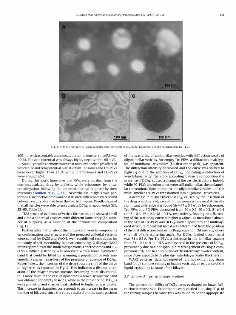
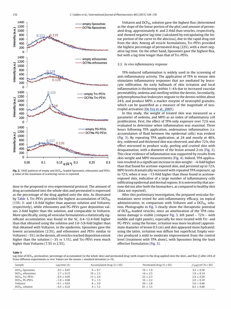
In this study, the weight of treated skin was measured as aparameter of oedema, and MPO as an index of inflammatory cellproliferation. First, the effect of TPA-only exposure over 72 h wasevaluated to determine when inflammation was maximal. Threehours following TPA application, oedematous inflammation (i.e.accumulation of fluid between the epidermal cells) was evident(Fig. 3). By repeating TPA application, at 24 and mostly at 48 h,dry, reddened and thickened skin was observed, and after 72 h, thiseffect worsened to produce scaly, peeling and crusted skin withdesquamation, with a diameter of the lesion around 2 cm (Fig. 3).This clear evidence of inflammation was supported by results fromskin weight and MPO measurements (Fig. 4). Indeed, TPA applica-tion resulted in a significant increase in skin weight: ∼4-fold higherthan that found for acetone-exposed skin, and persisted up to 72 h.MPO levels dramatically increased with repeated TPA exposure, upto 72 h, when it was ∼13-fold higher than those found in acetone-exposed skin, indicative of a large number of inflammatory cellsinfiltrating epidermal and dermal regions. It is noteworthy that ace-tone did not alter both the biomarkers, as compared to healthy skin(data not reported).
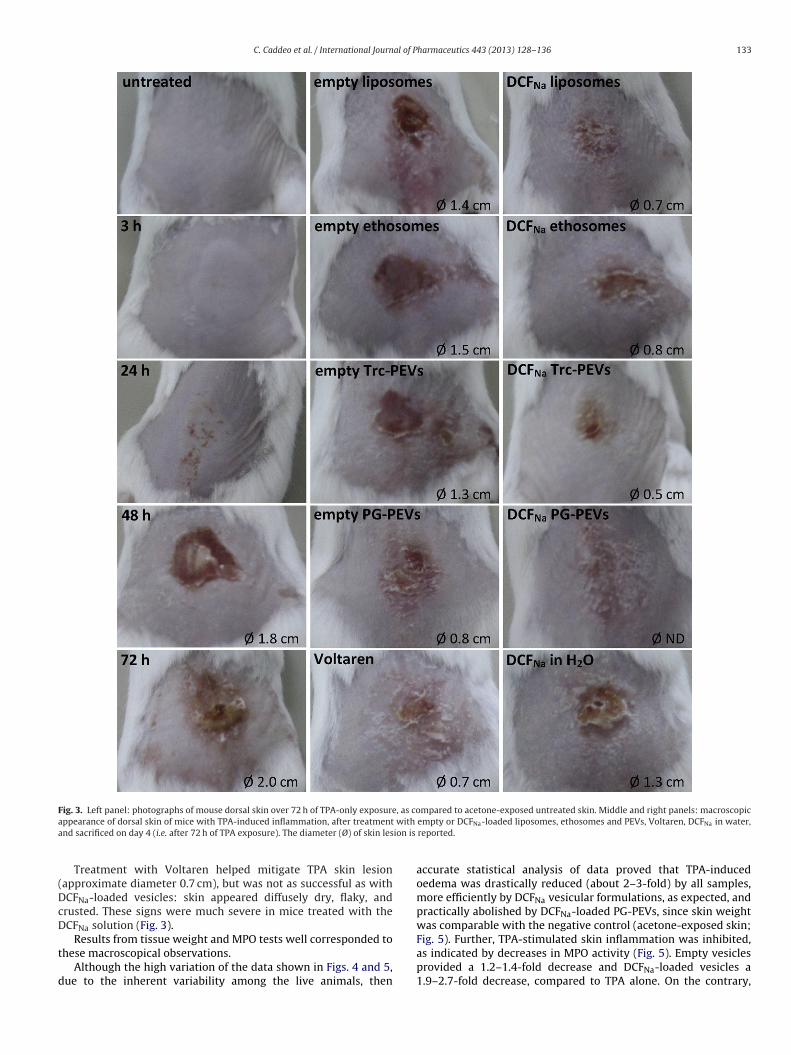
After this preliminary investigation, the prepared vesicular for-mulations were tested for anti-inflammatory efficacy, on topicaladministration, in comparison with Voltaren and a DCFNa solu-tion. Photographs in Fig. 3 clearly show the therapeutic potentialof DCFNa-loaded vesicles, since an amelioration of the TPA cuta-neous damage is visible (compare Fig. 3, left panel – 72 h – withmiddle and right panels), especially for mice treated with Trc- andPG-PEVs: using the former, irritation was more localized (approxi-mate diameter of lesion 0.5 cm) and skin appeared more hydrated;
cles produced a mild to moderate improvement from the controllevel (treatment with TPA alone), with liposomes being the leasteffective formulation (Fig. 3).
ated drug (with respect to the drug applied onto the skin), and flux (J) after 24 h of
SD) Permeated drug (% ± SD) J (�g/cm2/h ± SD)
19 ± 1.9 3.2 ± 0.3614 ± 2.5 1.8 ± 0.1422 ± 2.3 2.4 ± 0.2016 ± 2.2 2.0 ± 0.1050 ± 2.8 5.0 ± 0.4681 ± 3.5 8.2 ± 0.60
C. Caddeo et al. / International Journal of Pharmaceutics 443 (2013) 128– 136 133
Fig. 3. Left panel: photographs of mouse dorsal skin over 72 h of TPA-only exposure, as compared to acetone-exposed untreated skin. Middle and right panels: macroscopica with
a ion is
(DcD
t
d
ppearance of dorsal skin of mice with TPA-induced inflammation, after treatmentnd sacrificed on day 4 (i.e. after 72 h of TPA exposure). The diameter (Ø) of skin les
Treatment with Voltaren helped mitigate TPA skin lesionapproximate diameter 0.7 cm), but was not as successful as withCFNa-loaded vesicles: skin appeared diffusely dry, flaky, andrusted. These signs were much severe in mice treated with theCFNa solution (Fig. 3).
Results from tissue weight and MPO tests well corresponded tohese macroscopical observations.
Although the high variation of the data shown in Figs. 4 and 5,ue to the inherent variability among the live animals, then
empty or DCFNa-loaded liposomes, ethosomes and PEVs, Voltaren, DCFNa in water,reported.
accurate statistical analysis of data proved that TPA-inducedoedema was drastically reduced (about 2–3-fold) by all samples,more efficiently by DCFNa vesicular formulations, as expected, andpractically abolished by DCFNa-loaded PG-PEVs, since skin weightwas comparable with the negative control (acetone-exposed skin;
Fig. 5). Further, TPA-stimulated skin inflammation was inhibited,as indicated by decreases in MPO activity (Fig. 5). Empty vesiclesprovided a 1.2–1.4-fold decrease and DCFNa-loaded vesicles a1.9–2.7-fold decrease, compared to TPA alone. On the contrary,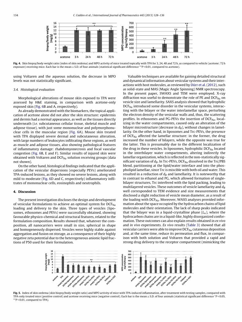
134 C. Caddeo et al. / International Journal of Pharmaceutics 443 (2013) 128– 136
F ice tree nifica
ul
3
ae
cauacwwaocon
cTmt
4
olsffpaant
FT*
ig. 4. Skin biopsy/body weight ratio (index of skin oedema) and MPO activity of mxposure) receiving mice. Each bar is the mean ± S.D. of four animals (statistical sig
sing Voltaren and the aqueous solution, the decrease in MPOevels was not statistically significant.
.4. Histological evaluation
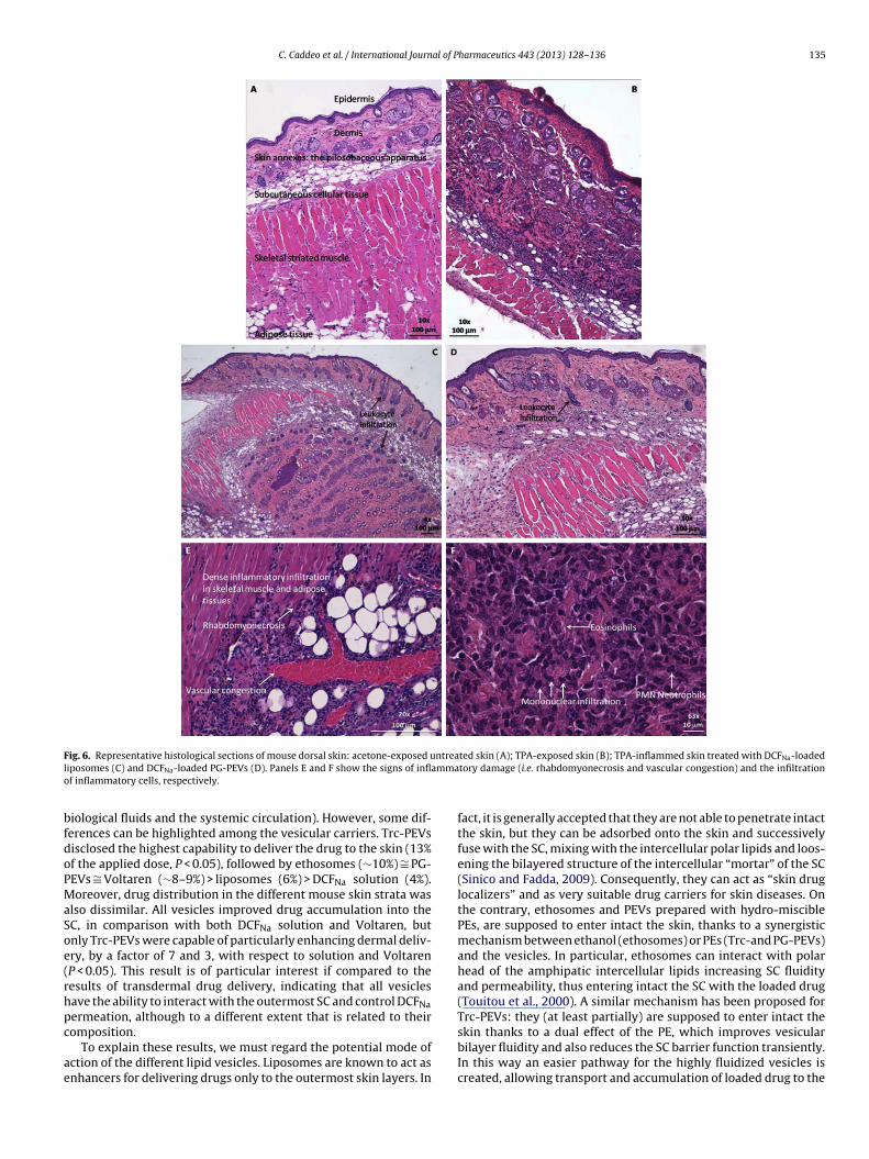
Morphological alterations of mouse skin exposed to TPA weressessed by H&E staining, in comparison with acetone-onlyxposed skin (Fig. 6B and A, respectively).
As already demonstrated with the biomarkers, the topical appli-ation of acetone alone did not alter the skin structure: epidermisnd dermis had a normal appearance, as well as the tissues directlynderneath (i.e. subcutaneous cellular tissue, skeletal muscle anddipose tissue), with just some mononuclear and polymorphonu-lear cells in the muscular region (Fig. 6A). Mouse skin treatedith TPA displayed severe dermal and subcutaneous alteration,ith large numbers of leukocytes infiltrating these regions, as well
s muscle and adipose tissues, also showing pathological featuresf inflammatory damage: rhabdomyonecrosis and focal vascularongestion (Fig. 6B, E and F). Similar results of injured skin werebtained with Voltaren and DCFNa solution receiving groups (dataot shown).
On the other hand, histological findings indicated that the appli-ation of the vesicular dispersions (especially PEVs) amelioratedPA-induced lesions, as they showed no severe lesions, along withild to moderate (Fig. 6D and C, respectively) inflammatory infil-
rates of mononuclear cells, eosinophils and neutrophils.
. Discussion
The present investigation discloses the design and developmentf vesicular formulations to achieve an optimal system for DCFNaoading and delivery to the skin. Three types of vesicles (lipo-omes, ethosomes and PEVs) were successfully obtained, showingavourable physico-chemical and structural features, related to theormulation composition. Results showed that, whatever the com-osition, all nanocarriers were small in size, spherical in shape
nd homogeneously dispersed. Vesicles were highly stable againstggregation and fusion on storage, as a consequence of their highlyegative zeta potential due to the heterogeneous anionic lipid frac-ions of P50 used for their formulation.ig. 5. Index of skin oedema (skin biopsy/body weight ratio) and MPO activity of mice witPA-only treated mice (positive control) and acetone receiving mice (negative control). Ea*P < 0.01, compared to TPA).
ated topically with TPA for 3, 24, 48 and 72 h, as compared to vehicle (acetone; 72 hnt difference **P < 0.01, compared to acetone).
Valuable techniques are available for gaining detailed structuraland dynamical information about vesicular systems and their inter-actions with host molecules, as reviewed by Dürr et al. (2012), suchas solid-state and MAS (Magic Angle Spinning) NMR spectroscopy.In the present paper, SWAXS and TEM were employed. X-raydiffraction was useful to demonstrate the role of PE and DCFNa onvesicle size and lamellarity. SAXS analysis showed that hydrophilicDCFNa introduced some disorder in the vesicular systems, interac-ting with the bilayer or the water interlamellar space, perturbingthe electron density of the vesicular walls and, thus, the scatteringprofiles. In ethosomes and PG-PEVs the insertion of DCFNa, local-izing in the water compartments, caused only an alteration of thebilayer microstructure (decrease in dB), without changes in lamel-larity. On the other hand, in liposomes and Trc-PEVs, the presenceof DCFNa affected the lamellar structure: in the former, the drugincreased the number of bilayers, while the opposite was true forthe latter. This is presumably due to the different localization ofthe drug in these vesicles. In liposomes, hydrophilic DCFNa locatedin the interbilayer water compartments, altering indirectly thelamellar organization, which is reflected in the non-statistically sig-nificant variation of dB. In Trc-PEVs, DCFNa dissolved in the Trc/PBSblend, partitioning at the lipid/water interface and into the phos-pholipid lamellae, since Trc is miscible with both oil and water. Thisresulted in a reduction of dB and lamellarity. It is noteworthy thatin contrast to ethanol and PG, which allowed formation of single-bilayer structures, Trc interfered with the lipid packing, leading tomultilayered vesicles. These outcomes of vesicle lamellarity and dB
well corresponded to TEM evidence and size measurements thatdisclosed a slight reduction of vesicle mean diameter, as a result ofthe loading with DCFNa. Moreover, WAXS analyses provided infor-mation about the space occupied by the hydrocarbon chains of lipidmolecules and their orientation. The lack of sharp peaks indicatedthat the bilayer was in a liquid-crystalline phase (L�), where thehydrocarbon chains are in a liquid-like, highly disorganized confor-mation. These outcomes can also explain results obtained in ex vivoand in vivo experiments. Ex vivo results (Table 3) showed that all
vesicular carriers were able to improve DCFNa cutaneous depositionand, at the same time, reduce its permeation and flux, in compar-ison with both solution and Voltaren that provided a rapid andstrong drug delivery to the receptor compartment (mimicking theh TPA-induced inflammation, after treatment with testing samples, compared withch bar is the mean ± S.D. of four animals (statistical significant difference *P < 0.05,
C. Caddeo et al. / International Journal of Pharmaceutics 443 (2013) 128– 136 135
F ntreal ammao
bfdoPMaSoe(rhpc
ae
ig. 6. Representative histological sections of mouse dorsal skin: acetone-exposed uiposomes (C) and DCFNa-loaded PG-PEVs (D). Panels E and F show the signs of inflf inflammatory cells, respectively.
iological fluids and the systemic circulation). However, some dif-erences can be highlighted among the vesicular carriers. Trc-PEVsisclosed the highest capability to deliver the drug to the skin (13%f the applied dose, P < 0.05), followed by ethosomes (∼10%) ∼= PG-EVs ∼= Voltaren (∼8–9%) > liposomes (6%) > DCFNa solution (4%).oreover, drug distribution in the different mouse skin strata was
lso dissimilar. All vesicles improved drug accumulation into theC, in comparison with both DCFNa solution and Voltaren, butnly Trc-PEVs were capable of particularly enhancing dermal deliv-ry, by a factor of 7 and 3, with respect to solution and VoltarenP < 0.05). This result is of particular interest if compared to theesults of transdermal drug delivery, indicating that all vesiclesave the ability to interact with the outermost SC and control DCFNaermeation, although to a different extent that is related to their
omposition.To explain these results, we must regard the potential mode ofction of the different lipid vesicles. Liposomes are known to act asnhancers for delivering drugs only to the outermost skin layers. In
ted skin (A); TPA-exposed skin (B); TPA-inflammed skin treated with DCFNa-loadedtory damage (i.e. rhabdomyonecrosis and vascular congestion) and the infiltration
fact, it is generally accepted that they are not able to penetrate intactthe skin, but they can be adsorbed onto the skin and successivelyfuse with the SC, mixing with the intercellular polar lipids and loos-ening the bilayered structure of the intercellular “mortar” of the SC(Sinico and Fadda, 2009). Consequently, they can act as “skin druglocalizers” and as very suitable drug carriers for skin diseases. Onthe contrary, ethosomes and PEVs prepared with hydro-misciblePEs, are supposed to enter intact the skin, thanks to a synergisticmechanism between ethanol (ethosomes) or PEs (Trc-and PG-PEVs)and the vesicles. In particular, ethosomes can interact with polarhead of the amphipatic intercellular lipids increasing SC fluidityand permeability, thus entering intact the SC with the loaded drug(Touitou et al., 2000). A similar mechanism has been proposed forTrc-PEVs: they (at least partially) are supposed to enter intact the
skin thanks to a dual effect of the PE, which improves vesicularbilayer fluidity and also reduces the SC barrier function transiently.In this way an easier pathway for the highly fluidized vesicles iscreated, allowing transport and accumulation of loaded drug to the
1 al of P
ect
catmacttrldvbrp
ielidIraMt(Pbi(si
5
ivovtsssi
A
VMI
SM
36 C. Caddeo et al. / International Journ
pidermis and dermis. Here, they form a depot from which the drugan be released and, free from the carrier, diffuse deeply accordingo its physico-chemical properties (Manconi et al., 2011a).
As written above, conventional liposomes provided a highumulative amount of permeated drug and the highest flux amongll vesicles (Table 3). These results could be the consequence ofhe lipid composition of these vesicles made from a commercial
ixture of soy phosphatidylcholine rich of unsaturated fatty acidsnd lysophospholipids. It is well known that these componentsan modify liposomal bilayer properties acting as “edge activa-or” and thus giving rise to liposomes that become very similaro the deformable, elastic vesicles (Sinico and Fadda, 2009). WAXSesults showed that the bilayer of all vesicular systems was in aiquid-crystalline state (L�), with hydrocarbon chains in a highlyisorganized conformation, thus confirming the high fluidity ofesicles. Hence, the highly fluidized conventional liposomes canetter interact with the mouse skin ex vivo but they probablyelease their content fast, leading to an intense and rapid DCFNaermeation across the skin, similarly to the solution and Voltaren.
Results from ex vivo experiments are in perfect agreement withn vivo anti-inflammatory tests. Indeed, the in vivo studies providedvidence that topically applied DCFNa, when delivered by vesicu-ar nanocarriers, markedly reduced TPA-induced inflammation, asnferred by macroscopic and histological observations, as well as byirect measurements of two biomarkers: oedema and MPO activity.
t was found that empty vesicles themselves exerted a protectiveole against inflammatory processes. This is most likely due to thentioxidant activity possessed by soy lecithin (P50). The release ofPO from the granules of neutrophils and monocytes in response
o inflammatory stimuli, leads to the generation of oxidizing agentse.g. reactive oxygen species) implicated in cellular damage. Hence,50 vesicles may defend skin by coping with oxidative stress,esides controlling several growth-regulatory pathways, thus play-
ng a role other than drug carrier in the treatment of skin diseasesCaddeo et al., 2008). Therefore, it seems obvious that treating thekin with DCFNa-loaded vesicles, the protective effect was furthermproved, especially using PEVs.
. Conclusions
In conclusion, our findings emphasize the superior anti-nflammatory efficacy of the prepared vesicle systems. In vivo and exivo results indicate that vesicles, and especially PEVs, are capablef really localizing the drug in the site of inflammation, while in con-entional dosage forms (solution and Voltaren) the drug continueo diffuse across the skin without any control, mostly reaching theystemic circulation (thus metabolism and excretion). This workhows that the present therapeutic approach based on vesicularystems may be promising in the curative treatment of a variety ofnflammatory skin disorders.
cknowledgments
This study was supported by grants from the University ofalencia (Relacions Internacionals I Cooperació), and the Italianinistry of Education, University and Research (Azioni integrate
talia-Spagna 2009, IT090H532G).The authors thank Jaume Caelles for measuring samples at the
AXS-WAXS service of IQAC-CSIC in Barcelona, and Dr Monicausio for her guidance in the statistical analysis of data.
harmaceutics 443 (2013) 128– 136
References
Arellano, A., Santoyo, S., Martín, C., Ygartua, P., 1999. Influence of propylene glycoland isopropyl myristate on the in vitro percutaneous penetration of diclofenacsodium from carbopol gels. Eur. J. Pharm. Sci. 7, 129–135.
Caddeo, C., Teskac, K., Sinico, C., Kristl, J., 2008. Effect of resveratrol incorporatedin liposomes on proliferation and UV-B protection of cells. Int. J. Pharm. 363,183–191.
Cevc, G., Blume, G., 2001. New, highly efficient formulation of diclofenac for the topi-cal, transdermal administration in ultradeformable drug carriers, transfersomes.Biochim. Biophys. Acta 1514, 191–205.
Chessa, M., Caddeo, C., Valenti, D., Manconi, M., Sinico, C., Fadda, A.M., 2011. Effectof penetration enhancer containing vesicles on the percutaneous delivery ofquercetin through new born pig skin. Pharmaceutics 3, 497–509.
De Vry, C.G., Valdez, M., Lazarov, M., Muhr, E., Buelow, R., Fong, T., Suhasini, I., 2005.Topical application of a novel immunomodulatory peptide, RDP58, reduces skininflammation in the phorbol ester-induced dermatitis model. J. Invest. Dermatol.125, 473–481.
De Young, L.M., Kheifets, J.B., Ballaron, S.J., Young, J.M., 1989. Edema and cell infiltra-tion in the phorbol ester-treated mouse ear are temporally separate and can bedifferentially modulated by pharmacologic agents. Agents Actions 26, 335–341.
Dürr, U.H.N., Gildenberg, M., Ramamoorthy, A., 2012. The magic of bicelles lights upmembrane protein structure. Chem. Rev. 112, 6054–6074.
Elsayed, M.M.A., Abdallah, O.Y., Naggar, V.F., Khalafallah, N.M., 2007. PG-liposomes:novel lipid vesicles for skin delivery of drugs. J. Pharm. Pharmacol. 59,1447–1450.
Fukami, T., Ishii, T., Io, T., Suzuki, N., Suzuki, T., Yamamoto, K., Xu, J., Ramamoorthy,A., Tomono, K., 2009. Nanoparticle processing in the solid state dramaticallyincreases the cell membrane permeation of a cholesterol-lowering drug. Probu-col. Mol. Pharm. 6, 1029–1035.
Io, T., Fukami, T., Yamamoto, K., Suzuki, T., Xu, J., Tomono, K., Ramamoorthy, A., 2010.Homogeneous nanoparticles to enhance the efficiency of a hydrophobic drug,antihyperlipidemic probucol, characterized by solid-state NMR. Mol. Pharm. 7,299–305.
Manconi, M., Mura, S., Sinico, C., Fadda, A.M., Vila, A.O., Molina, F., 2009a. Devel-opment and characterization of liposomes containing glycols as carriers fordiclofenac. Colloid Surf. A-Physicochem. Eng. Asp. 342, 53–58.
Manconi, M., Pendás, J., Ledón, N., Moreira, T., Sinico, C., Saso, L., Fadda, A.M., 2009b.Phycocyanin liposomes for topical anti-inflammatory activity: in-vitro in-vivostudies. J. Pharm. Pharmacol. 61, 423–430.
Manconi, M., Caddeo, C., Sinico, C., Valenti, D., Mostallino, M.C., Biggio, G., Fadda, A.M.,2011a. Ex vivo skin delivery of diclofenac by transcutol containing liposomes andsuggested mechanism of vesicle–skin interaction. Eur. J. Pharm. Biopharm. 78,27–35.
Manconi, M., Sinico, C., Caddeo, C., Vila, A.O., Valenti, D., Fadda, A.M., 2011b. Pene-tration enhancer containing vesicles as carriers for dermal delivery of tretinoin.Int. J. Pharm. 412, 37–46.
Müller, M., Mackeben, S., Müller-Goymann, C.C., 2004. Physicochemical character-ization of liposomes with encapsulated local anaesthetics. Int. J. Pharm. 274,139–148.
Murakawa, M., Yamaoka, K., Tanaka, Y., Fukuda, Y., 2006. Involvement of tumornecrosis factor (TNF)-a in phorbol ester 12-O-tetradecanoylphorbol-13-acetate(TPA)-induced skin edema in mice. Biochem. Pharmacol. 71, 1331–1336.
Pabst, G., Rappolt, M., Amenitsch, H., Laggner, P., 2000. Structural information frommultilamellar liposomes at full hydration: full q-range fitting with high qualityX-ray data. Phys. Rev. E Stat. Phys. Plasmas Fluids Relat. Interdiscip. Topics 62,4000–4009.
Paolino, D., Lucania, G., Mardente, D., Alhaique, F., Fresta, M., 2005. Ethosomes forskin delivery of ammonium glycyrrhizinate: ex vivo percutaneous permeationthrough human skin and in vivo anti-inflammatory activity on human volun-teers. J. Control. Release. 106, 99–110.
Paolino, D., Muzzalupo, R., Ricciardi, A., Celia, C., ·Picci, N., Fresta, M., 2007. In vitroand in vivo evaluation of Bola-surfactant containing niosomes for transdermaldelivery. Biomed. Microdev. 9, 421–433.
Paolino, D., Cosco, D., Muzzalupo, R., Trapasso, E., Picci, N., Fresta, M., 2008. Innova-tive bola-surfactant niosomes as topical delivery systems of 5-fluorouracil forthe treatment of skin cancer. Int. J. Pharm. 353, 233–242.
Priprem, A., Watanatorn, J., Sutthiparinyanont, S., Phachonpai, W., Muchimapura, S.,2008. Anxiety and cognitive effects of quercetin liposomes in rats. Nanomed.-Nanotechnol. Biol. Med. 4, 70–78.
Sato, H., Nakayama, Y., Yamashita, C., Uno, H., 2004. Anti-inflammatory effects oftacalcitol (1,24(R)(OH)2D3, TV-02) in the skin of TPA-treated hairless mice. J.Dermatol. 31, 200–217.
Sinico, C., Fadda, A.M., 2009. Liposomes and niosomes as carriers for dermal drugdelivery. Expert Opin. Drug Deliv. 6, 813–825.
Touitou, E., Dayan, N., Bergelson, L., Godin, B., Eliaz, M., 2000. Ethosomes novelvesicular carriers for enhanced delivery: characterization and skin penetrationproperties. J. Control. Release. 65, 403–418.
Top Related
Copyright © 2022 FDOKUMEN

