


![KLV-30MR1 - Error: [object Object]](https://static.fdokumen.com/doc/165x107/631786651e5d335f8d0a6a63/klv-30mr1-error-object-object.jpg)


Bahasa
Halaman
Hukum


Evaluating object recognition ability in developmental prosopagnosia using the Cambridge Car Memory Test Article
Accepted Version
Gray, K. L. H., Biotti, F. and Cook, R. (2019) Evaluating object recognition ability in developmental prosopagnosia using the Cambridge Car Memory Test. Cognitive Neuropsychology, 36 (1-2). pp. 89-96. ISSN 1464-0627 doi: https://doi.org/10.1080/02643294.2019.1604503 Available at https://centaur.reading.ac.uk/83174/
It is advisable to refer to the publisher’s version if you intend to cite from the work. See Guidance on citing .
To link to this article DOI: http://dx.doi.org/10.1080/02643294.2019.1604503
Publisher: Taylor & Francis
All outputs in CentAUR are protected by Intellectual Property Rights law, including copyright law. Copyright and IPR is retained by the creators or other copyright holders. Terms and conditions for use of this material are defined in the End User Agreement .
www.reading.ac.uk/centaur
CentAUR

Central Archive at the University of Reading Reading’s research outputs online

In press at: Cognitive Neuropsychology
Format: Research Article
Running head: Object recognition in developmental prosopagnosia
Word count: 3425
Evaluating object recognition ability in developmental prosopagnosia using
the Cambridge Car Memory Test
Katie L.H. Gray1, Federica Biotti2, & Richard Cook3*
1School of Psychology and Clinical Language Sciences,
University of Reading, Reading, U.K.
2Department of Psychology,
Royal Holloway, University of London, Egham, U.K.
3Department of Psychological Sciences,
Birkbeck, University of London, London, U.K.
*Corresponding author
Dr Richard Cook
Department of Psychological Sciences,
Birkbeck, University of London,
London, UK [email protected]

2
Abstract
Individuals with developmental prosopagnosia (DP) sometimes experience object
identification difficulties in addition to problems recognising faces. To better understand the
distribution of non-face object recognition ability in this population, we administered the
Cambridge Car Memory Test (CCMT) – a leading, standardised measure of object
recognition ability – to a large sample of DPs (N = 46). When considered as a single group,
the DPs scored lower than matched controls. This finding provides further evidence that
developmental object agnosia (DOA) may be more common in DP than in the general
population. Relative to the DPs’ face recognition deficits, however, car matching deficits
were small and inconsistent. In fact, we observed a striking range of CCMT performance in
our DP sample. While some DPs performed extremely poorly, many more achieved scores
within one standard deviation of the typical mean, and several DP participants achieved
excellent CCMT scores comparable with the best controls.
Keywords:
Developmental prosopagnosia; Face recognition; Object recognition; Independent disorders
hypothesis; Cambridge Car Memory Test

3
Introduction
Developmental prosopagnosia (DP) is a neurodevelopmental condition associated with
difficulties recognising familiar faces and distinguishing unfamiliar faces, that occurs in
people with normal intelligence and typical visual acuity, and in the absence of manifest
brain injury (Behrmann & Avidan, 2005; Duchaine & Nakayama, 2006b; Susilo &
Duchaine, 2013). DP often runs in families indicating that the condition may have a genetic
component (Duchaine, Germine, & Nakayama, 2007; Johnen et al., 2014; Schmalzl,
Palermo, & Coltheart, 2008). Individuals with DP identify others using non-face cues (e.g.,
hairstyle voice, and gait) and often experience great difficulty when familiar people are met
in unusual contexts or when they alter their appearance (Cook & Biotti, 2016; Shah, Gaule,
Sowden, Bird, & Cook, 2015). Historically, the condition was thought to be rare
(McConachie, 1976), but current estimates suggest that 2% of the general population may
experience face recognition difficulties severe enough to disrupt their daily lives
(Kennerknecht et al., 2006; Kennerknecht, Ho, & Wong, 2008).
The origins of DP remain poorly understood. Cognitive theories have argued that individuals
with DP may be less able to integrate information from disparate facial regions to form
unified perceptual descriptions relative to typical observers (Avidan, Tanzer, & Behrmann,
2011; DeGutis, Cohan, & Nakayama, 2014; Palermo et al., 2011). Many DPs, however,
appear to exhibit typical markers of ‘holistic face processing’ (Biotti, Wu, et al., 2017; Le
Grand et al., 2006; Susilo et al., 2010). At the neurological level, studies have revealed
reduced grey matter volume in occipitotemporal cortex of individuals with DP (Behrmann,
Avidan, Gao, & Black, 2007; Garrido et al., 2009) and have suggested atypical functional
connectivity in high-level visual areas (Avidan & Behrmann, 2009; Lohse et al., 2016;
Rosenthal et al., 2017). Recent studies also suggest that reduced density and coherence of the
inferior longitudinal fasciculus, a white matter tract connecting the occipital and temporal
lobes, may impair information exchange within the face processing network in DP (Gomez
et al., 2015; Song et al., 2015; Thomas et al., 2009).
In addition to their characteristic face recognition difficulties, some individuals with DP also
exhibit signs of co-occurring object recognition difficulties. Individuals have been described,
for example, who experience problems identifying cars (e.g., Biotti, Gray, & Cook, 2017; De
Haan & Campbell, 1991; Duchaine, Germine, et al., 2007; Duchaine & Nakayama, 2005;
Klargaard, Starrfelt, & Gerlach, 2018), bicycles (e.g., Dalrymple & Duchaine, 2014), guns

4
(e.g., Duchaine, Germine, et al., 2007; Duchaine & Nakayama, 2005), flowers (e.g., De Haan
& Campbell, 1991), scenes (e.g., Duchaine & Nakayama, 2005), animals and tools (e.g.,
Duchaine & Nakayama, 2005; Gerlach, Klargaard, & Starrfelt, 2016). Some authors have
argued that the incidence of object recognition difficulties in the DP population is so high,
that the condition can be understood only in terms of a domain-general perceptual deficit
(Gerlach et al., 2016; Geskin & Behrmann, 2017). One possibility is that DP is a
developmental form of integrative agnosia, whereby individuals are unable integrate
component parts into a coherent whole, that impairs both face and object recognition (e.g.,
Riddoch & Humphreys, 1987). A closely-related idea is that individuals with DP may
experience delayed or impoverished processing of global shape information (Avidan et al.,
2011; Gerlach & Starrfelt, 2018; Tanzer, Freud, Ganel, & Avidan, 2013).
Other authors reject the view that object recognition problems are a universal feature of DP.
According to the independent disorders hypothesis (IDH; Gray & Cook, 2018), DP and
developmental object agnosia (DOA) are best thought of as independent neurodevelopmental
conditions that sometimes co-occur. A key prediction of this hypothesis is the existence of
‘pure’ cases of DP and DOA; individuals who experience impaired face recognition, but
typical object recognition, and vice versa. Consistent with this view, some DPs exhibit
apparently typical object recognition (e.g., Duchaine, Yovel, Butterworth, & Nakayama,
2006) and cases of DOA have been described where the individual exhibits apparently
typical face recognition (e.g., Germine, Cashdollar, Duzel, & Duchaine, 2011). However, the
IDH also predicts that DP and DOA co-occur, that is to say, the incidence of DOA is higher
in DP than in the wider population, due to common genetic or environmental risk factors
(Gray & Cook, 2018). Specifically, susceptibility to aberrant structural development of
occipitotemporal cortex may be a common risk factor for DP and DOA (see also: Susilo &
Duchaine, 2013).
Both the IDH (Gray & Cook, 2018) and domain-general accounts (Avidan et al., 2011;
Gerlach & Starrfelt, 2018; Tanzer et al., 2013) predict a degree of correlation between the
face and object recognition abilities seen in large samples of observers. Under domain-
general accounts, one would expect a tight coupling between observers’ ability to identify
faces and non-face objects; for example, an individual’s degree of impairment with faces
ought to relate closely to their degree of impairment with objects. In contrast, however, the
IDH predicts a weaker, idiosyncratic relationship. Although people at risk of perceptual

5
difficulties with faces may often develop co-occurring perceptual difficulties with non-face
objects, this co-occurrence is not inevitable – some individuals may exhibit selective
problems with either faces or objects (i.e., ‘pure’ cases).
In light of these conflicting views, we sought a better understanding of the object recognition
difficulties seen in the DP population. To this end, we examined performance on the
Cambridge Car Memory Test (CCMT; Dennett et al., 2011) in a large sample of DPs (N =
46) and matched controls (N = 61). The CCMT employs a 3-AFC match-to-sample format
that mirrors that of the Cambridge Face Memory Test (CFMT; Duchaine & Nakayama,
2006a), a standardised measure of face recognition used widely in the diagnosis of DP. In
both tasks participants are asked to identify target items encountered in a study phase from a
line up of three test items (target plus two lures). In both tasks, trial difficulty is varied across
a 72-trial procedure through viewpoint manipulations and through the addition of high-
frequency visual noise. Both the CFMT and CCMT exhibit good internal reliability; for
example, α = .88 (Bowles et al., 2009) and α = .84 (Dennett et al., 2011), respectively.
Responses are not speeded and both tests stress accuracy1.
The CCMT has been used to address a wide range of questions in cognitive neuropsychology
(e.g., Esins, Schultz, Stemper, Kennerknecht, & Bulthoff, 2016; Klargaard et al., 2018; Shah,
Gaule, Gaigg, Bird, & Cook, 2015), cognitive psychology (e.g., Dennett, McKone, Edwards,
& Susilo, 2012), neuropsychiatry (e.g., Ewbank et al., 2017), and behavioural genetics (e.g.,
Shakeshaft & Plomin, 2015). To date, however, it remains unclear how DPs perform on this
widely-used measure of object recognition ability. Several studies have described individual
DPs who score badly on the CCMT (e.g., Klargaard et al., 2018; Palermo et al., 2017;
Rivolta, Lawson, & Palermo, 2017; Susilo et al., 2010; White, Rivolta, Burton, Al-Janabi, &
Palermo, 2017). However, previous comparison of matched samples has failed to reveal
differences at the group level; for example, Shah et al. (2015; N = 15 DPs) and Esins et al.
(2016; N = 16 DPs) found that DPs’ performance on the CCMT did not differ significantly
from that of controls.
Participants
We describe data from 107 adults, 46 with DP (21 males; Mage = 39.4 years, SDage = 9.4
years) and 61 typically developed (TD) controls (27 males; Mage = 37.0 years, SDage = 9.8
years). Neither participant age [t(105) = 1.313, p = .192] nor proportion of males [X2(1) =
6
.01, p = .888] differed significantly between the two groups. Ethical approval was granted by
the local ethics committee. The research was conducted in line with the ethical guidelines
provided by the 6th (2008) Declaration of Helsinki. All participants provided informed
consent and were fully debriefed after the experimental procedure. All participants were
tested in person under controlled lab conditions. DP participants completed the CFMT first,
followed by the CCMT. Wherever possible, DP participants completed the tests in a single
session. Control participants completed the tests in a single session. Half of the controls
completed the CFMT first, half completed the CCMT first.
DP participants were recruited through www.troublewithfaces.org and reported lifelong face
recognition difficulties in the absence of brain injury and psychiatric disorder (autism or
schizophrenia). Diagnostic decisions were based primarily on participants’ scores on the
Twenty-Item Prosopagnosia Index (PI20; Gray, Bird, & Cook, 2017; Shah, Gaule, Sowden,
et al., 2015) and the CFMT (Duchaine & Nakayama, 2006a). The participants with DP also
completed the Cambridge Face Perception Test (CFPT; Duchaine, Germine, et al., 2007).
Typical participants, recruited through local subject-pools, completed the CFMT, the CCMT,
and the PI20. None of the typical controls scored more than 60 on the PI20. No-one was
excluded on this basis. All members of the DP sample scored at least 2 SDs below the
typical mean on the CFMT, and at least 3 SDs above the typical mean on the PI20. Summary
statistics for both groups are provided in Table 1 and diagnostic information for each DP is
provided as supplementary material. The use of convergent self-report evidence and scores
on objective, computer-based tasks may be a particularly effective approach to the
identification and classification of DP; for example, less than 1.5% of the general population
score below 65% on the CFMT and more than 65 on the PI20 (Gray et al., 2017).
Table-1
Results
The data were analysed using ANOVA with Test (CFMT, CCMT) as a within-subjects
factor, and Observer Sex (male, female) and Group (DP, TD) as between-subjects factors. As
noted above, low CFMT scores formed an important part of the diagnostic evidence used to
classify observers as DP. The fact that the DPs scored well-below typical controls on this
measure is therefore entirely unsurprising. We include a Test factor (CFMT, CCMT),
however, so that readers can compare the relative size of deficits seen on the face and car

7
variants of this test. Previous studies have suggested an effect of observer sex on the CCMT,
whereby male observers typically perform a little better than females2 (Dennett et al., 2011).
We therefore included Observer Sex as a factor to determine whether this interacts with
Group (TD, DP).
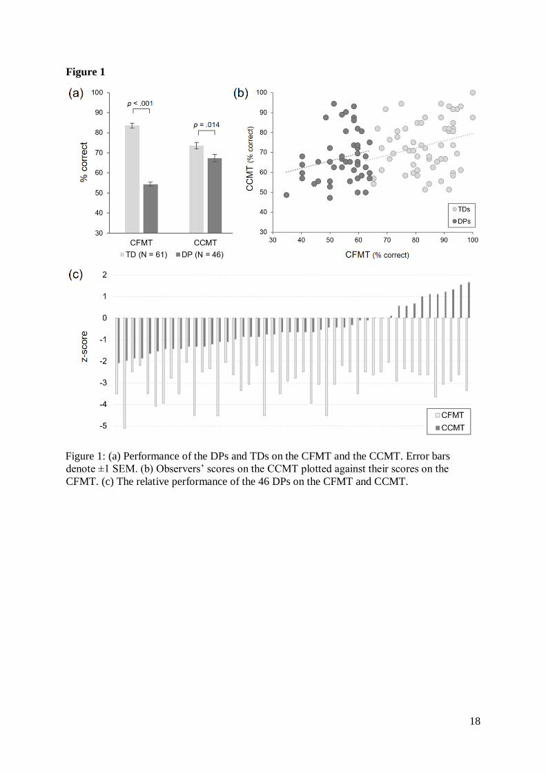
The analysis revealed a significant main effect of Group [F(1,103) = 115.194, p < .001, ηp2 =
.528] and a Group × Test interaction [F(1,103) = 78.121, p <.001, ηp2 = .431]. While
controls generally outperformed the DPs, the difference was more pronounced on the CFMT
(MTD = 83.56%, SDTD = 9.57%; MDP = 54.38% SDDP = 7.39%) than on the CCMT (MTD =
73.52%, SDTD = 12.57%; MDP = 67.36% SDDP = 12.60%; Figure 1a). Planned contrasts
revealed significant group differences on both the CFMT [t(105) = 17.174, p < .001] and the
CCMT [t(105) = 2.506, p = .014]. We observed no main effect of Test [F(1,103) = 1.899, p
= .171, ηp2 = .018], no main effect of Observer Sex [F(1,103) = 1.973, p = .163, ηp
2 = .019],
nor a Test × Observer Sex × Group interaction [F(103) = .434, p = .512, ηp2 = .004].
However, the analysis revealed a Test × Observer Sex interaction [F(1,103) = 5.063, p
=.027, ηp2 = .047] whereby male participants tended to score higher on the CCMT than
females.
Figure-1
In light of the significant Observer Sex (male, female) × Test (CFMT, CCMT) interaction
described above, we sought to confirm that the effect of Group on CCMT scores (TD > DP)
was seen for both male (Figure 2a) and female (Figure 2b) observers. First, we compared the
performance of the male controls (N = 27, Mage = 37.4 years, SDage = 9.5 years) with the
male DPs (N = 21, Mage = 40.5 years, SDage = 10.1 years). Relative to the male controls, the
male DPs were significantly impaired on both the CFMT [t(46) = 10.990, p < .001] and the
CCMT [t(46) = 2.117, p = .040], but disproportionately impaired at the CFMT [F(1,46) =
26.685, p < .001, ηp2 = .367]. Next, we compared the performance of the female controls (N
= 34, Mage = 36.6 years, SDage = 10.1 years) and the female DPs (N = 25, Mage = 38.6 years,
SDage = 8.9 years). Once again we observed a highly significant group difference on the
CFMT [t(57) = 13.068, p < .001], and a significant Group (TD, DP) × Test (CFMT, CCMT)
interaction [F(1,57) = 56.472, p < .001, ηp2 = .498]. When the analysis was restricted to
female participants, however, the difference between the DPs and the TDs on the CCMT did
not reach significance [t(57) = 1.502, p = .139].

8
Figure-2
As expected, the variance in CFMT scores differed significantly between the DPs and
controls [F(1,105) = 5.06, p = .03]. This likely reflects the fact that the CFMT scores of the
DPs were tightly constrained, whilst the CFMT scores of the controls were free to vary. The
variance in CCMT scores did not differ between the two groups [F(1,105) = .17, p = .69].
Having pooled the TD and DP groups to form a single combined sample (N = 107; Figure
1b), we observed a modest correlation between observers’ performance on the CFMT and
their scores on the CCMT (r = .329, p < .001, N = 107). When considered separately, this
correlation was seen in the TD group (r = .276, p = .031, N = 61), but did not reach
significance in the DP group (r = .215, p = .151, N = 46), possibly reflecting the limited
range of CFMT scores. The difference between these correlation coefficients was not
significant [Fisher’s z = .323, p = .747]. Examination of the individual differences revealed a
striking range of object recognition ability in the DP group. At one end of the distribution,
five individuals in the DP sample produced CCMT scores 1.64 standard deviations below the
typical mean, and two produced scores 1.96 standard deviations below the typical mean. At
the other extreme, however, several DPs performed very well, achieving scores comparable
with the best controls (Figure 1c).
To formally explore how many of the DPs were significantly impaired on the CCMT, we
performed single-case analysis (Crawford, Garthwaite, & Ryan, 2011) to compare each DP’s
CCMT performance with the TD group. In this analysis, we found that 2/46 DPs were
classified as having significantly lower CCMT scores than the TD group. We also explored
possible dissociations between CFMT and CCMT performance (e.g., Gerlach, Lissau, &
Hildebrandt, 2018). Dissociations in performance between two tasks can be classified as
putatively classical, or strong. A DP is considered to fulfil the criteria for a putatively
classical dissociation when performance is significantly lower than TDs on the CFMT, but
not the CCMT, and their standardised difference between the two tasks is significantly
different from controls. Strong dissociations are fulfilled when a DP is significantly impaired
on the CFMT and CCMT, and their standardised difference between the two tasks is
significantly different from controls. In our sample of DPs, we found that 24/46 DPs met

9
criteria for a putatively classical dissociation, whereas only one DP met the criteria for a
strong dissociation.
General Discussion
To better understand the distribution of non-face object recognition ability in the DP
population, we administered the CCMT to a large sample of DPs (N = 46) and matched
controls. Overall, we found that the DP group achieved lower scores on the CCMT than
typical controls. This finding provides further evidence that object recognition difficulties
may be more common in DP than in the general population. Relative to their face
recognition deficits, however, the DPs’ car matching deficits were small and inconsistent.
Some authors have proposed that DP is characterised by a single domain-general deficit that
impairs the perception of faces and non-face objects alike (Avidan et al., 2011; Gerlach et
al., 2016; Gerlach & Starrfelt, 2018; Geskin & Behrmann, 2017; Tanzer et al., 2013).
Critically, this view predicts that individuals with DP should consistently exhibit difficulties
recognising a wide range of non-face objects. For example, individuals with integrative
agnosia appear to exhibit perceptual deficits for most types of complex visual object –
including cars (Germine et al., 2011; Moscovitch, Winocur, & Behrmann, 1997; Riddoch &
Humphreys, 1987).
The striking range of CCMT performance seen in our DP sample is hard to reconcile with
this view. While some DPs performed extremely poorly, many more achieved CCMT scores
within one standard deviation of the TD mean and several DP participants exhibited
excellent performance, achieving accuracy scores exceeding 90% comparable with the best
controls. Typical levels of CCMT performance were common in the DP sample despite the
fact that these individuals describe severe lifelong face recognition difficulties and all
produced CFMT scores more than two standard deviations below the TD mean. To score
well on the CCMT, observers must be able to identify complex visual objects across
viewpoints, and when obscured by high-frequency visual noise. Excellent CCMT scores
therefore suggests that a subset of DPs were able integrate local features and process global
shape information, typically (also see: Biotti, Wu, et al., 2017; Duchaine et al., 2006;
Duchaine, Yovel, & Nakayama, 2007; Le Grand et al., 2006; Susilo et al., 2010)

10
In contrast, the range of object recognition abilities seen in our DP sample accords very well
with the IDH (Germine et al., 2011; Gray & Cook, 2018). Under this account, DP and DOA
are viewed as independent conditions that sometimes occur on their own as pure cases of DP
and DOA, but often co-occur within the same individuals due to common risk factors (e.g.,
inherited susceptibility to aberrant structural development of occipitotemporal cortex; Gray
& Cook, 2018). Like domain-general accounts, the IDH predicts that the incidence of DOA
is more common in DP than in the general population. Crucially, however, it predicts i) an
idiosyncratic relationship between object and face processing abilities in DP, and ii) a subset
of DPs with intact object perception. Both of these predictions are supported by the dataset
described here.
According to the IDH, the composition of DP samples may determine whether authors find
evidence of group-level object recognition deficits in DP. Where samples include high or
low proportions of DPs with co-occurring DOA, authors may be more or less likely to find
group differences, respectively. Smaller samples of DPs may often contain too few cases of
DP with co-occurring DOA to reveal group differences on tasks such as the CCMT (Esins et
al., 2016; Shah, Gaule, Gaigg, et al., 2015). The idiosyncratic nature of object recognition
deficits in DP is highlighted by the absence of a significant effect of Group (DP, TD) in our
female participants. Due to the inconsistency of object perception deficits, large samples of
DPs may be required to detect consistently group-level effects on object recognition tasks.
Where observed, however, we speculate that such differences are attributable to co-occurring
DOA, not DP per se.
Compared with the CFMT, the CCMT may be less able to detect impairment at the single-
case level. One issue is that the mean score of typical controls on the CCMT (M = 73.5%) is
a little lower than the mean typical score on the CFMT (M = 83.6%). A second issue is that
the variability seen in typical scores is slightly greater on the CCMT (SD = 12.6%) than on
the CFMT (SD = 9.6%). Together, however, this means that DPs need to score < 50% to
achieve a z-score of < -1.96. Given that the CCMT is a 3-AFC task, participants’
performance therefore needs to approach chance levels in order to be classified as
significantly impaired at the single-case level. Despite the fact that only a few DPs reached
this threshold, the significant group difference seen on the CCMT suggests that mild deficits
may be relatively common in this population (see also: Biotti, Gray, & Cook, 2019).

11
Our investigation was restricted to a single object category – cars. It remains to be seen
whether similar findings emerge when DPs are tested with other types of object, or whether
cars are somehow ‘special’ (e.g., Ćepulić, Wilhelm, Sommer, & Hildebrandt, 2018; Richler,
Wilmer, & Gauthier, 2017). However, the present results are important because problems
recognising and distinguishing cars are amongst the most commonly reported object
perception deficits in DP. This stimulus class therefore appears to tax object recognition
processes that are sometimes aberrant in DP. Moreover, the CCMT has an identical format to
the CFMT, a measure that is known to reveal the perceptual problems DPs experience with
faces, and is just as challenging. The failure to observe clear, widespread object recognition
deficits in our large DP sample cannot therefore be attributed to the particular stimulus class
used, the format of the test, or the fact the task is easier.

12
Footnotes
1The versions of the CFMT and CCMT employed here do not record response latencies.
2Dennett and colleagues speculate that this may reflect the fact that male observers sometimes
have greater knowledge of existing car manufacturers and models.

13
References
Avidan, G., & Behrmann, M. (2009). Functional MRI reveals compromised neural integrity
of the face processing network in congenital prosopagnosia. Current Biology, 19(13),
1146-1150.
Avidan, G., Tanzer, M., & Behrmann, M. (2011). Impaired holistic processing in congenital
prosopagnosia. Neuropsychologia, 49(9), 2541-2552.
Behrmann, M., & Avidan, G. (2005). Congenital prosopagnosia: face-blind from birth.
Trends in Cognitive Sciences, 9(4), 180-187.
Behrmann, M., Avidan, G., Gao, F., & Black, S. (2007). Structural imaging reveals
anatomical alterations in inferotemporal cortex in congenital prosopagnosia. Cerebral
Cortex, 17(10), 2354-2363.
Biotti, F., Gray, K. L., & Cook, R. (2019). Is developmental prosopagnosia best characterised
as an apperceptive or mnemonic condition? Neuropsychologia, 124, 285-298.
Biotti, F., Gray, K. L. H., & Cook, R. (2017). Impaired body perception in developmental
prosopagnosia. Cortex, 93, 41-49.
Biotti, F., Wu, E., Yang, H., Jiahui, G., Duchaine, B., & Cook, R. (2017). Normal composite
face effects in developmental prosopagnosia. Cortex, 95, 63-76.
Bowles, D. C., McKone, E., Dawel, A., Duchaine, B., Palermo, R., Schmalzl, L., . . . Yovel,
G. (2009). Diagnosing prosopagnosia: effects of ageing, sex, and participant-stimulus
ethnic match on the Cambridge Face Memory Test and Cambridge Face Perception
Test. Cognitive Neuropsychology, 26(5), 423-455.
Ćepulić, D. B., Wilhelm, O., Sommer, W., & Hildebrandt, A. (2018). All categories are
equal, but some categories are more equal than others: The psychometric structure of
object and face cognition. Journal of Experimental Psychology: Learning, Memory,
and Cognition, 44(8), 1254-1268.
Cook, R., & Biotti, F. (2016). Developmental prosopagnosia. Current Biology, 26(8), R312-
R313.
Crawford, J. R., Garthwaite, P. H., & Ryan, K. (2011). Comparing a single case to a control
sample: Testing for neuropsychological deficits and dissociations in the presence of
covariates. Cortex, 47, 1166-1178.
Dalrymple, K. A., & Duchaine, B. (2014). Impaired face detection may explain some but not
all cases of developmental prosopagnosia. Developmental Science, 19, 440-451.
De Haan, E. H., & Campbell, R. (1991). A fifteen year follow-up of a case of developmental
prosopagnosia. Cortex, 27(4), 489-509.
DeGutis, J., Cohan, S., & Nakayama, K. (2014). Holistic face training enhances face
processing in developmental prosopagnosia. Brain, 137(Pt 6), 1781-1798.
14
Dennett, H. W., McKone, E., Edwards, M., & Susilo, T. (2012). Face aftereffects predict
individual differences in face recognition ability. Psychological Science, 23(11),
1279-1287.
Dennett, H. W., McKone, E., Tavashmi, R., Hall, A., Pidcock, M., Edwards, M., & Duchaine,
B. (2011). The Cambridge Car Memory Test: a task matched in format to the
Cambridge Face Memory Test, with norms, reliability, sex differences, dissociations
from face memory, and expertise effects. Behavior Research Methods, 44(2), 587-
605.
Duchaine, B., Germine, L., & Nakayama, K. (2007). Family resemblance: ten family
members with prosopagnosia and within-class object agnosia. Cognitive
Neuropsychology, 24(4), 419-430.
Duchaine, B., & Nakayama, K. (2005). Dissociations of face and object recognition in
developmental prosopagnosia. Journal of Cognitive Neuroscience, 17(2), 249-261.
Duchaine, B., & Nakayama, K. (2006a). The Cambridge Face Memory Test: results for
neurologically intact individuals and an investigation of its validity using inverted
face stimuli and prosopagnosic participants. Neuropsychologia, 44, 576-585.
Duchaine, B., & Nakayama, K. (2006b). Developmental prosopagnosia: a window to content-
specific face processing. Current Opinion in Neurobiology, 16, 166-173.
Duchaine, B., Yovel, G., Butterworth, E., & Nakayama, K. (2006). Prosopagnosia as an
impairment to face-specific mechanisms: Elimination of the alternative hypotheses in
a developmental case. Cognitive Neuropsychology, 23(5), 714-747.
Duchaine, B., Yovel, G., & Nakayama, K. (2007). No global processing deficit in the Navon
task in 14 developmental prosopagnosics. Social, Cognitive, and Affective
Neuroscience, 2(2), 104-113.
Esins, J., Schultz, J., Stemper, C., Kennerknecht, I., & Bulthoff, I. (2016). Face perception
and test reliabilities in congenital prosopagnosia in seven tests. I Perception, 7(1),
2041669515625797.
Ewbank, M. P., Pell, P. J., Powell, T. E., von dem Hagen, E. A. H., Baron-Cohen, S., &
Calder, A. J. (2017). Repetition suppression and memory for faces is reduced in adults
with autism spectrum conditions. Cerebral Cortex, 27(1), 92-103.
Garrido, L., Furl, N., Draganski, B., Weiskopf, N., Stevens, J., Tan, G. C., . . . Duchaine, B.
(2009). Voxel-based morphometry reveals reduced grey matter volume in the
temporal cortex of developmental prosopagnosics. Brain, 132(Pt 12), 3443-3455.
Gerlach, C., Klargaard, S. K., & Starrfelt, R. (2016). On the relation between face and object
recognition in developmental prosopagnosia: no dissociation but a systematic
association. PLoS One, 11(10), e0165561.
Gerlach, C., Lissau, C. H., & Hildebrandt, N. K. (2018). On defining and interpreting
dissociations. Cognitive Neuropsychology, 35(1-2), 66-69.

15
Gerlach, C., & Starrfelt, R. (2018). Delayed processing of global shape information is
associated with weaker top-down effects in developmental prosopagnosia. Cognitive
Neuropsychology.
Germine, L., Cashdollar, N., Duzel, E., & Duchaine, B. (2011). A new selective
developmental deficit: Impaired object recognition with normal face recognition.
Cortex, 47(5), 598-607.
Geskin, J., & Behrmann, M. (2017). Congenital prosopagnosia without object agnosia? A
literature review. Cognitive Neuropsychology, 22, 1-51.
Gomez, J., Pestilli, F., Witthoft, N., Golarai, G., Liberman, A., Poltoratski, S., . . . Grill-
Spector, K. (2015). Functionally defined white matter reveals segregated pathways in
human ventral temporal cortex associated with category-specific processing. Neuron,
85(1), 216-227.
Gray, K. L. H., Bird, G., & Cook, R. (2017). Robust associations between the 20-item
prosopagnosia index and the Cambridge Face Memory Test in the general population.
Royal Society Open Science, 4(3), 160923.
Gray, K. L. H., & Cook, R. (2018). Should developmental prosopagnosia, developmental
body agnosia, and developmental object agnosia be considered independent
neurodevelopmental conditions? Cognitive Neuropsychology, 35(1-2), 59-62.
Johnen, A., Schmukle, S. C., Hüttenbrink, J., Kischka, C., Kennerknecht, I., & Dobel, C.
(2014). A family at risk: Congenital prosopagnosia, poor face recognition and
visuoperceptual deficits within one family. Neuropsychologia, 58, 52-63.
Kennerknecht, I., Grüter, T., Welling, B., Wentzek, S., Horst, J., Edwards, S., & Grüter, M.
(2006). First report of prevalence of non-syndromic hereditary prosopagnosia (HPA).
American Journal of Medical Genetics, 140A(15), 1617-1622.
Kennerknecht, I., Ho, N. Y., & Wong, V. C. N. (2008). Prevalence of heriditary
prosopagonsia (HPA) in Hong Kong Chinese population. American Journal of
Medical Genetics, 146A(22), 2863-2870.
Klargaard, S. K., Starrfelt, R., & Gerlach, C. (2018). Inversion effects for faces and objects in
developmental prosopagnosia: A case series analysis. Neuropsychologia, 113, 52-60.
Le Grand, R., Cooper, P. A., Mondloch, C. J., Lewis, T. L., Sagiv, N., de Gelder, B., &
Maurer, D. (2006). What aspects of face processing are impaired in developmental
prosopagnosia? Brain and Cognition, 61(2), 139-158.
Lohse, M., Garrido, L., Driver, J., Dolan, R. J., Duchaine, B. C., & Furl, N. (2016). Effective
connectivity from early visual cortex to posterior occipitotemporal face areas supports
face selectivity and predicts developmental prosopagnosia. Journal of Neuroscience,
36(13), 3821-3828.
McConachie, H. R. (1976). Developmental prosopagnosia. A single case report. Cortex,
12(1), 76-82.

16
Palermo, R., Rossion, B., Rhodes, G., Laguesse, R., Tez, T., Hall, B., . . . Irons, J. (2017). Do
people have insight into their face recognition abilities? Quarterly Journal of
Experimental Psychology, 70(2), 218-233.
Palermo, R., Willis, M. L., Rivolta, D., McKone, E., Wilson, C. E., & Calder, A. J. (2011).
Impaired holistic coding of facial expression and facial identity in congenital
prosopagnosia. Neuropsychologia, 49(5), 1226-1235.
Richler, J. J., Wilmer, J. B., & Gauthier, I. (2017). General object recognition is specific:
Evidence from novel and familiar objects. Cognition, 166, 42-55.
Riddoch, M. J., & Humphreys, G. W. (1987). A case of integrative visual agnosia. 110(6),
1431-1462.
Rivolta, D., Lawson, R. P., & Palermo, R. (2017). More than just a problem with faces:
altered body perception in a group of congenital prosopagnosics. Quarterly Journal of
Experimental Psychology, 70(2), 276-286.
Rosenthal, G., Tanzer, M., Simony, E., Hasson, U., Behrmann, M., & Avidan, G. (2017).
Altered topology of neural circuits in congenital prosopagnosia. Elife, 6.
Schmalzl, L., Palermo, R., & Coltheart, M. (2008). Cognitive heterogeneity in genetically
based prosopagnosia: a family study. Journal of Neuropsychology, 2(1), 99-117.
Shah, P., Gaule, A., Gaigg, S. B., Bird, G., & Cook, R. (2015). Probing short-term face
memory in developmental prosopagnosia. Cortex, 64, 115-122.
Shah, P., Gaule, A., Sowden, S., Bird, G., & Cook, R. (2015). The 20-item prosopagnosia
index (PI20): a self-report instrument for identifying developmental prosopagnosia.
Royal Society Open Science, 2(6), 140343.
Shakeshaft, N. G., & Plomin, R. (2015). Genetic specificity of face recognition. Proceedings
of the National Academy of Sciences of the United States of America, 112(41), 12887-
12892.
Song, S., Garrido, L., Nagy, Z., Mohammadi, S., Steel, A., Driver, J., . . . Furl, N. (2015).
Local but not long-range microstructural differences of the ventral temporal cortex in
developmental prosopagnosia. Neuropsychologia, 78, 195-206.
Susilo, T., & Duchaine, B. (2013). Advances in developmental prosopagnosia research.
Current Opinion in Neurobiology, 23, 423-429.
Susilo, T., McKone, E., Dennett, H., Darke, H., Palermo, R., Hall, A., . . . Rhodes, G. (2010).
Face recognition impairments despite normal holistic processing and face space
coding: evidence from a case of developmental prosopagnosia. Cognitive
Neuropsychology, 27(8), 636-664.
Tanzer, M., Freud, E., Ganel, T., & Avidan, G. (2013). General-holistic impairment in
congenital prosopagnosia: Evidence from Garner's speeded-classification task.
Cognitive Neuropsychology, 30(6), 429-445.

17
Thomas, C., Avidan, G., Humphreys, K., Jung, K. J., Gao, F., & Behrmann, M. (2009).
Reduced structural connectivity in ventral visual cortex in congenital prosopagnosia.
Nature Neuroscience, 12(1), 29-31.
White, D., Rivolta, D., Burton, A. M., Al-Janabi, S., & Palermo, R. (2017). Face matching
impairment in developmental prosopagnosia. Quarterly Journal of Experimental
Psychology, 70(2), 287-297.
18
Figure 1
Figure 1: (a) Performance of the DPs and TDs on the CFMT and the CCMT. Error bars
denote ±1 SEM. (b) Observers’ scores on the CCMT plotted against their scores on the
CFMT. (c) The relative performance of the 46 DPs on the CFMT and CCMT.

19
Figure 2
Figure 2: Performance of the male (a) and female (b) participants on the CFMT and CCMT .

20
Table 1
Table 1: Diagnostic information for the DP and TD samples. Scores on the Cambridge Face
Perception Test (CFPT) indicate the number of sorting errors made in the upright condition.
Prosopagnosics (N = 46) Typical controls (N = 61)
PI20
Mean (SD) 82.04 (5.84) 38.03 (8.59)
Range 70 to 94 20 to 57
CFMT
Mean (SD) 54.38% (7.39) 83.56% (9.57)
Range 34.72% to 63.89% 62.50% to 100.00%
CFPT
Mean (SD) 52.39 (16.20) -
Range 26 to 88 -
Top Related
Copyright © 2022 FDOKUMEN

