DEVELOPMENTAL STAGES OF FROG
10
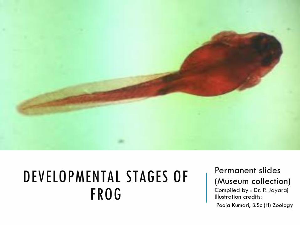
DEVELOPMENTAL STAGES OF FROG Permanent slides (Museum collection) Compiled by : Dr. P. Jayaraj Illustration credits: Pooja Kumari, B.Sc (H) Zoology
-
Upload
khangminh22 -
Category
Documents
-
view
0 -
download
0
Transcript of DEVELOPMENTAL STAGES OF FROG
DEVELOPMENTAL STAGES OF FROG
Permanent slides(Museum collection)Compiled by : Dr. P. JayarajIllustration credits:
Pooja Kumari, B.Sc (H) Zoology

FROG EMBRYO-THIRD CLEAVAGE (WHOLE MOUNT)8-CELLED STAGE (ANIMAL POLAR VIEW) POLAR VIEW
The polar view shows eight cells formed in two tiers.
Upper four cells of animal pole are small called micromeres and appear pigmented.
The lower four cells in the vegetal pole are larger with large amount of yolk called as megamers or macromeres.
Third cleavage: This is a latitudinal division from just above the equatorial plane perpendicular to the first two divisions. As a result, eight cells are formed in two tiers.
Micromeres
Macromeres

FROG EMBRYO : MORULA(W.M)
1. The cells formed by cleavage are blastomeres, the upper
black blastomeres are called micromeres, and lower white
ones are macromeres.
2. Further cleavages divide the micromeres more rapidly
than the lower macromeres whose division is hindered by
yolk. The blastomeres’ mutual pressure flattens their
surfaces in contact with each other but free surfaces of
each blastomere remain spherical.
3. At this stage the whole embryo acquires a characteristic
appearance reminiscent of a mulbery and so it is called
morula.
Blastomeres

FROG EMBRYOBLASTULA (L.S)
1. The section of blastula shows acavity known as blastocoel
2. The Blastocoel in blastula islocated above the equator,therefore it is called as eccentricin position.
3. Blastocoel is surrounded by twotypes of blastomeres-micromeres in the animal poleand macromeres in the vegetalpole.
Micromeres
Macromeres
Blastocoel cavity

Frog Embryo -Advanced gastrula/yolk plug stage (L.S)
1. Three primary germ layers can be found.
2. Ectoderm is made up of micromeres and surrounds the embryo
3. Endoderm is well developed and internalized
4. Mesoderm is formed from the roof of the archenteron and endoderm, from the
5. floor of the archenteron.
6. Due to enlargement of archenteron, blastocoel is gradually reduced.
7. The yolk laden macromeres are pushed towards to blastopore which forms yolk plug.
8. This stage of the gastrula is called as yolk plug stage of the gastrula.
Archenteron
Blastocoel
Yolk plug
Dorsal lip of blastopore
Ventral Lip

FROG EMBRYO-EARLY NEURAL NEURAL PLATE STAGE (T.S)
1. During late gastrulation, external changes along
the upper surface of the embryo begin to form
the neural tube—a process called neurulation.
2. An oval-shaped area on the dorsal side of the
embryo marks the presumptive neural tube. This
region is the neural plate.
3. During this process the ectoderm along the mid-
dorsal line becomes thick and flattens forming
plate-like structure known as neural plate
(medullar plate).
4. Microfilaments in neural plate cells flatten and
thicken the neural plate.
Notochord
Neural plate
Endoderm
Mesoderm
Archenteron
Ectoderm
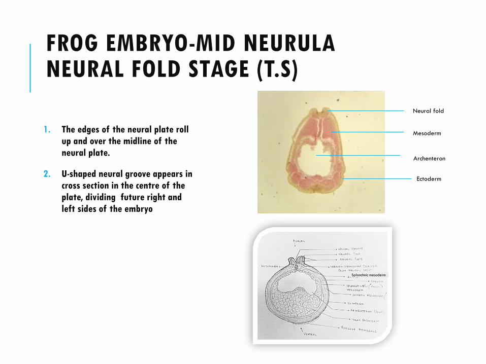
FROG EMBRYO-MID NEURULANEURAL FOLD STAGE (T.S)
1. The edges of the neural plate roll
up and over the midline of the
neural plate.
2. U-shaped neural groove appears in
cross section in the centre of the
plate, dividing future right and
left sides of the embryo
Splanchnic mesoderm
Neural fold
Mesoderm
Archenteron
Ectoderm

FROG EMBRYO-ADVANCED NEURULANEURAL TUBE STAGE(T.S)
The neural folds approach each other towards the
midline of the embryo, eventually fusing to form
the neural tube beneath the overlying epithelium
A distinct notochord is found in the mid line (axis)
immediately below the neural tube
Paraxial mesoderm on both sides of notochord
organises into blocks called somites
Downward growth of mesoderm continues
Neural Tube
Notochord
Archenteron
Mesoderm
Ectoderm
Endoderm
EXTERNAL GILL-STAGE OF TADPOLE (W.M)
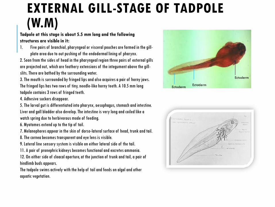
Tadpole at this stage is about 5.5 mm long and the following
structures are visible in it:
1. Five pairs of branchial, pharyngeal or visceral pouches are formed in the gill-
plate area due to out pushing of the endodermal lining of pharynx.
2. Soon from the sides of head in the pharyngeal region three pairs of external gills
are projected out, which are feathery extensions of the integument above the gill-
slits. There are bathed by the surrounding water.
3. The mouth is surrounded by fringed lips and also acquires a pair of horny jaws.
The fringed lips has two rows of tiny, needle-like horny teeth. A 10.5 mm long
tadpole contains 3 rows of fringed teeth.
4. Adhesive suckers disappear.
5. The larval gut is differentiated into pharynx, oesophagus, stomach and intestine.
Liver and gall bladder also develop. The intestine is very long and coiled like a
watch spring due to herbivorous mode of feeding.
6. Myotomes extend up to the tip of tail.
7. Melanophores appear in the skin of dorso-lateral surface of head, trunk and tail.
8. The cornea becomes transparent and eye lens is visible.
9. Lateral line sensory system is visible on either lateral side of the tail.
11. A pair of pronephric kidneys becomes functional and excretes ammonia.
12. On either side of cloacal aperture, at the junction of trunk and tail, a pair of
hindlimb buds appears.
The tadpole swims actively with the help of tail and feeds on algal and other
aquatic vegetation.
Ectoderm
EctodermEctoderm

INTERNAL GILL-STAGE OF TADPOLE:
1. The opercular folds grow backward from the hyoid arch of each
side covering the external gills and gill-slits and finally fuse
with each other ventrally and with the belly wall. Thus, an
operculum or gill-cover is formed enclosing the external gills
2. External gills later fall off and four pairs of filamentous
internal gills develop on the walls of gill-slits.
3. Intestine is still coiled and long.
4. Different parts of hindlimbs such as thigh, shank, ankle, foot
and five toes become well formed in the tadpole of 40 mm long.
5. A pair of forelimb buds appear behind the head but remain
hidden within operculum. As development proceeds, the left
forelimb emerges through the spiracle. The right forelimb
appears later.
6. In a mature tadpole, a pair of lungs develop from the pharynx.
Now the larva breathes by both, the internal gills and lungs.
Mouth
EyeIntestine
Tail fin