


Bahasa
Halaman
Hukum


Submit Manuscript | http://medcraveonline.com
IntroductionGestational diabetes mellitus (GDM) is a condition of glucose
intolerance with onset in pregnancy 1,2) It is associated with increased obstetric complications, such as fetal macrosomia, neonatal hypoglycemia and hypocalcemia, as well as maternal hypertension and thromboembolic disease. Therefore, surveillance for GDM is important.1,2 Women at risk should be tested at the first antenatal visit using the American Diabetes Association diagnostic criteria for non- pregnant adults.2 The 50-g non-fasting 1-h glucose challenge test (GCT) is the most widely implemented screen used. Alternative screening methods have been proposed to increase the detection rates of GDM and to overcome these shortcomings of the GCT. Some are based on ultrasound examinations. Although uncomplicated GDM with less severe fasting hyperglycemia has not been associated with increased perinatal mortality, GDM of any severity increases the risk of fetal macrosomia.3 Increased glucose transfer from the diabetic mother to the fetus and placenta results in fetal hyperglycemia and hyperinsulinemia, promoting growth of insulin-dependent tissues and organs, such as the liver.4 The aim of the present study is to test the efficiency of ultrasound parameters (biparietal diameter, abdominal circumference, estimated fetal weight, fetal truncal subcutaneous fat layer, and fetal liver length) against the efficiency of the 50 gram
oral glucose challenge test (O’ Sullivan test) to screen for gestational diabetes in the second trimester between 24-28weeks of gestation.
MethodsThe study enrolled three hundred second trimester pregnant
subjects selected from outpatient clinic coming for antenatal visit at 24-28weeks gestation, with or without history of gestational diabetes in a previous pregnancy. Subjects with pre-gestational diabetes were excluded. Consent was taken from all subjects to participate in the study. All recruited patients were subjected to history taking, complete clinical examination, ultrasound examination(Including: fetal biometry measurements to confirm gestational age, detailed anomaly scan, fetal subcutaneous fat layer, length of the right lobe of the liver, abdominal circumference, biparietal diameter, head circumference, femur length, and estimated fetal weight calculated by (Hadlock AC,BPD) in grams. All examinations were performed by the same sonologist (E.A) using an HDI 1500 scanner equipped with a 3.5-MHz transducer (Medison ultrasound, SonoR-7). To measure the fetal anterior abdominal wall fat, the abdominal circumference was selected. The measurement was taken as close to vertical as possible. The quadrant that included the spine was avoided. The measurements were taken from the inner to the outer aspect of the echogenic subcutaneous fat that surrounded
MOJ Womens Health. 2017;6(1):344‒356. 344©2017 Fattah. This is an open access article distributed under the terms of the Creative Commons Attribution License, which permits unrestricted use, distribution, and build upon your work non-commercially.
Diagnostic ability of the fetal ultrasonographic parameters in screening for gestational diabetes
Volume 6 Issue 1 - 2017
Eman Ali Abd El FattahDepartment of Obstetrics and Gynecology, Alexandria University, Shatby Maternity hospital, Egypt
Correspondence: Eman Ali Abd El Fattah, Department of Obstetrics and Gynecology, Faculty of Medicine, Alexandria University, Shatby Maternity hospital, 4, fathia Baheeg St, Fleming, Alexandria, Egypt, Email [email protected]
Received: February 11, 2017 | Published: October 27, 2017
Abstract
Pancreatic β-cell hyperplasia occurring in normal pregnancy results in higher fasting and postprandial insulin levels. In addition, placental hormones cause increased insulin resistance, especially throughout the third trimester. Pregnancies with GDM are associated with increased obstetric complications. Therefore, the surveillance of GDM during pregnancy is especially important. The best method for screening continues to be controversial. O’ Sullivan, is the most widely used screening test and is recommended by the ADA. GDM of any severity increases the risk of fetal macrosomia which can be diagnosed by ultrasonography Increased body weight is due to organomegaly and increased fat deposition. Increased glucose transfer from the diabetic mother to the fetus and placenta results in fetal hyperglycemia and hyperinsulinemia, promoting growth of insulin-dependent tissues and organs, such as the liver. The aim of the present study is to test the efficiency of ultrasound parameters (biparietal diameter, abdominal circumference, estimated fetal weight, fetal truncal subcutaneous fat layer, and fetal liver length) against the efficiency of the 50gram oral glucose challenge test (O’ Sullivan test) to screen for gestational diabetes in the second trimester between 24-28weeks of gestation.
The study enrolled three hundred second trimester pregnant subjects selected from outpatient clinic coming for antenatal visit at 24-28weeks gestation. All recruited patients were subjected to history taking, complete clinical examination, ultrasound examination(Including: fetal biometry measurements to confirm gestational age, detailed anomaly scan, fetal subcutaneous fat layer, length of the right lobe of the liver, abdominal circumference, biparietal diameter, head circumference, femur length, and estimated fetal weight calculated by (Hadlock AC,BPD) in grams O’Sullivan test was done, patients were considered screen positive if plasma venous glucose concentration ≥140md/dl or 7.8mmol/L. 100 gram oral glucose tolerance curve was used to confirm or rule out the diagnosis of gestational diabetes. A blood glucose level below 180 mg after one hour was considered normal. A blood glucose level below 140 mg after two hours is considered normal. Two or more abnormally high readings are considered Diabetes.
MOJ Women’s Health
Research Article Open Access
Diagnostic ability of the fetal ultrasonographic parameters in screening for gestational diabetes 345Copyright:
©2017 Fattah
Citation: Fattah EAAEI. Diagnostic ability of the fetal ultrasonographic parameters in screening for gestational diabetes. MOJ Womens Health. 2017;6(1):344‒356. DOI: 10.15406/mojwh.2017.06.00148
the abdomen. Patient is considered screen positive if expected fetal weight or abdominal circumference is above the 90th percentile for age, or measured fat layer equals to or exceeds 3.5mm at 24-28weeks gestation. To measure liver length, the tip of the right lobe was clearly identified and the liver length was measured from the dome of the right hemi-diaphragm to the tip of the right lobe (Figure 1). O’Sullivan test was done; patients received 50gram glucose orally irrespective of their fasting state. Blood sugar level was evaluated one hour later. Patients were considered screen positive if plasma venous glucose concentration ≥140md/dl or 7.8mmol/L.5 Screen positive cases were subjected to 100gram oral glucose tolerance curve: (2hours 100gram OGTT) to confirm or rule out the diagnosis of gestational diabetes. The patient’s blood glucose was recorded at baseline after eight hours fasting. The patient was then given 100gram glucose orally then blood glucose was measured at one hour and two hours. The diagnosis of GDM was based on the criteria of the World Health Organization, which is considered the gold standard. A blood glucose level below 180 mg after one hour was considered normal. A blood glucose level below 140mg after two hours is considered normal. One abnormal reading denotes glucose intolerance and requires further assessment at a later gestation (later than 28weeks). Otherwise; abnormal reading on two occasions of the 100gram OGTT, the patient is diagnosed as having gestational diabetes.
Statistical methodology
Data were collected and entered to the computer using SPSS (Statistical Package for Social Science) program for statistical analysis (ver 21).5 Data were entered as numerical or categorical, as appropriate. When Kolmogorov-Smirnov test revealed no significance in the distribution of variables, parametric statistics was carried out, while in the not-normally distributed data the non-parametric statistics was carried out.6
i. Data were described using minimum, maximum, mean, and standard deviation for the normally distributed data.
ii. Data were described using minimum, maximum, median and inter-quartile range for not-normally distributed data.
iii. Comparisons were carried out between two studied independent not-normally distributed subgroups using Mann-Whitney U test.7
iv. Histograms with distribution curve, Box and Whiskers graph, bar chart and clustered bar chart were used accordingly.
v. Area under the ROC (AUC) was carried using MedCalc Software version 14.8,9
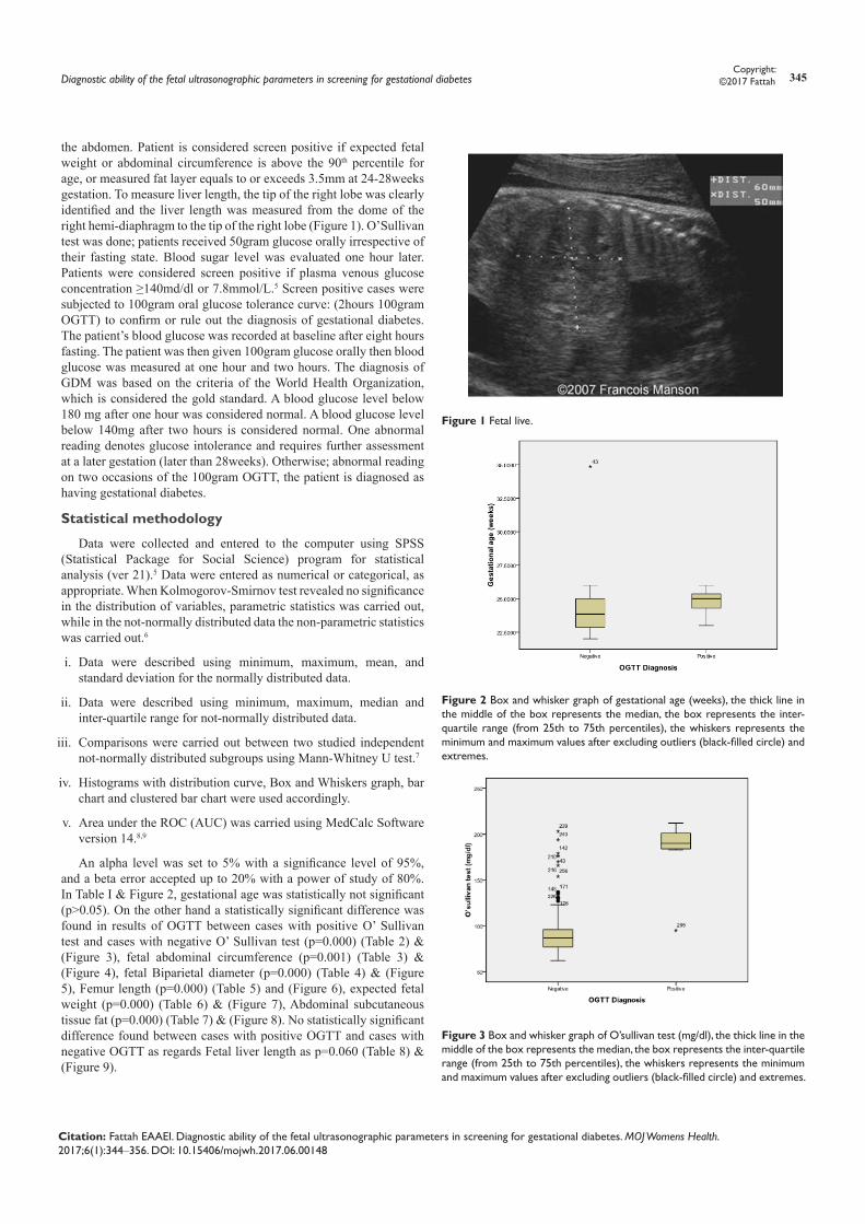
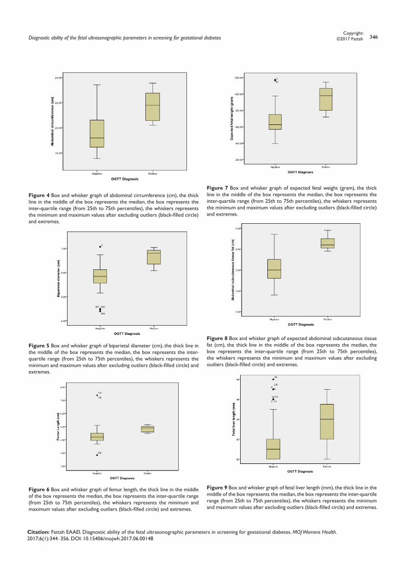
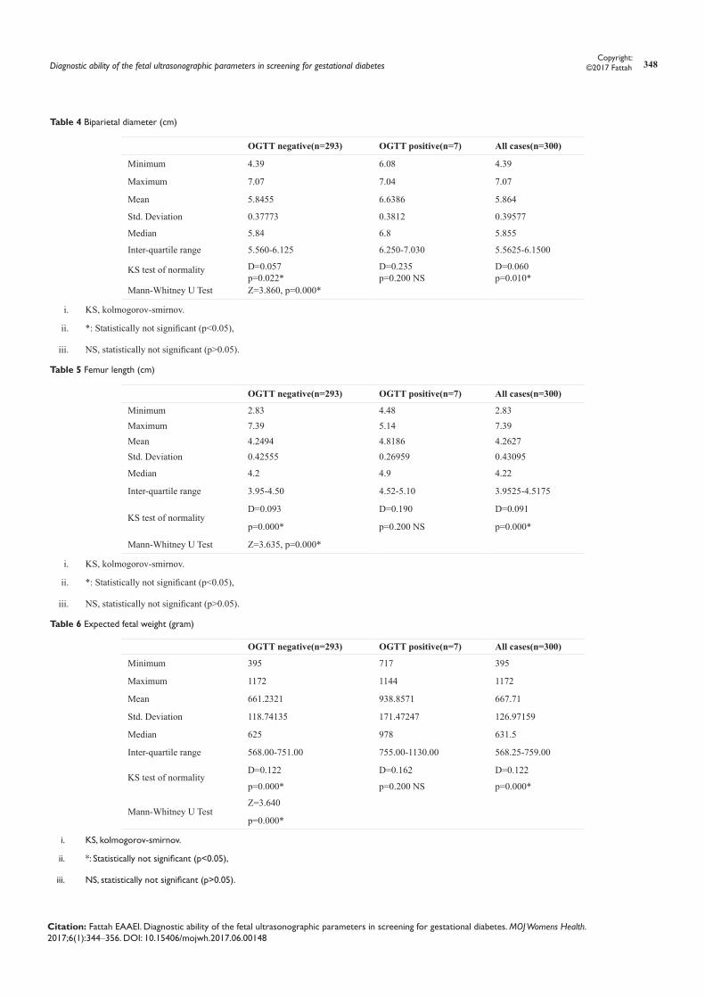
An alpha level was set to 5% with a significance level of 95%, and a beta error accepted up to 20% with a power of study of 80%. In Table I & Figure 2, gestational age was statistically not significant (p>0.05). On the other hand a statistically significant difference was found in results of OGTT between cases with positive O’ Sullivan test and cases with negative O’ Sullivan test (p=0.000) (Table 2) & (Figure 3), fetal abdominal circumference (p=0.001) (Table 3) & (Figure 4), fetal Biparietal diameter (p=0.000) (Table 4) & (Figure 5), Femur length (p=0.000) (Table 5) and (Figure 6), expected fetal weight (p=0.000) (Table 6) & (Figure 7), Abdominal subcutaneous tissue fat (p=0.000) (Table 7) & (Figure 8). No statistically significant difference found between cases with positive OGTT and cases with negative OGTT as regards Fetal liver length as p=0.060 (Table 8) & (Figure 9).
Figure 1 Fetal live.
Figure 2 Box and whisker graph of gestational age (weeks), the thick line in the middle of the box represents the median, the box represents the inter-quartile range (from 25th to 75th percentiles), the whiskers represents the minimum and maximum values after excluding outliers (black-filled circle) and extremes.
Figure 3 Box and whisker graph of O’sullivan test (mg/dl), the thick line in the middle of the box represents the median, the box represents the inter-quartile range (from 25th to 75th percentiles), the whiskers represents the minimum and maximum values after excluding outliers (black-filled circle) and extremes.
Diagnostic ability of the fetal ultrasonographic parameters in screening for gestational diabetes 346Copyright:
©2017 Fattah
Citation: Fattah EAAEI. Diagnostic ability of the fetal ultrasonographic parameters in screening for gestational diabetes. MOJ Womens Health. 2017;6(1):344‒356. DOI: 10.15406/mojwh.2017.06.00148
Figure 4 Box and whisker graph of abdominal circumference (cm), the thick line in the middle of the box represents the median, the box represents the inter-quartile range (from 25th to 75th percentiles), the whiskers represents the minimum and maximum values after excluding outliers (black-filled circle) and extremes.
Figure 5 Box and whisker graph of biparietal diameter (cm), the thick line in the middle of the box represents the median, the box represents the inter-quartile range (from 25th to 75th percentiles), the whiskers represents the minimum and maximum values after excluding outliers (black-filled circle) and extremes.
Figure 6 Box and whisker graph of femur length, the thick line in the middle of the box represents the median, the box represents the inter-quartile range (from 25th to 75th percentiles), the whiskers represents the minimum and maximum values after excluding outliers (black-filled circle) and extremes.
Figure 7 Box and whisker graph of expected fetal weight (gram), the thick line in the middle of the box represents the median, the box represents the inter-quartile range (from 25th to 75th percentiles), the whiskers represents the minimum and maximum values after excluding outliers (black-filled circle) and extremes.
Figure 8 Box and whisker graph of expected abdominal subcutaneous tissue fat (cm), the thick line in the middle of the box represents the median, the box represents the inter-quartile range (from 25th to 75th percentiles), the whiskers represents the minimum and maximum values after excluding outliers (black-filled circle) and extremes.
Figure 9 Box and whisker graph of fetal liver length (mm), the thick line in the middle of the box represents the median, the box represents the inter-quartile range (from 25th to 75th percentiles), the whiskers represents the minimum and maximum values after excluding outliers (black-filled circle) and extremes.
Diagnostic ability of the fetal ultrasonographic parameters in screening for gestational diabetes 347Copyright:
©2017 Fattah
Citation: Fattah EAAEI. Diagnostic ability of the fetal ultrasonographic parameters in screening for gestational diabetes. MOJ Womens Health. 2017;6(1):344‒356. DOI: 10.15406/mojwh.2017.06.00148
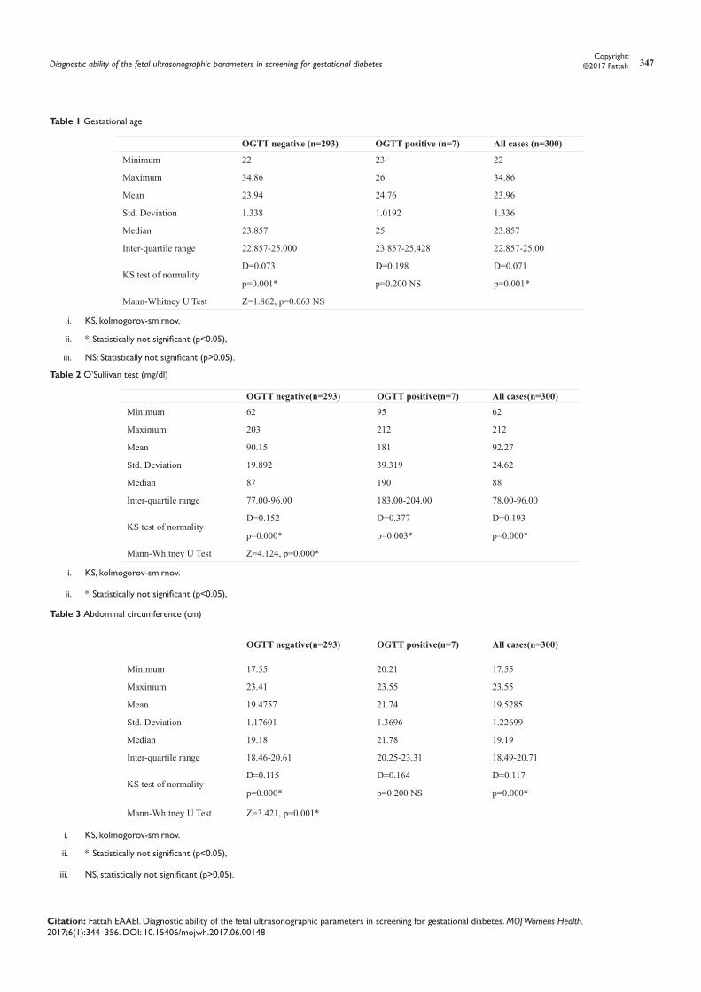
Table 1 Gestational age
OGTT negative (n=293) OGTT positive (n=7) All cases (n=300)
Minimum 22 23 22
Maximum 34.86 26 34.86
Mean 23.94 24.76 23.96
Std. Deviation 1.338 1.0192 1.336
Median 23.857 25 23.857
Inter-quartile range 22.857-25.000 23.857-25.428 22.857-25.00
KS test of normalityD=0.073 D=0.198 D=0.071
p=0.001* p=0.200 NS p=0.001*
Mann-Whitney U Test Z=1.862, p=0.063 NS
i. KS, kolmogorov-smirnov.
ii. *: Statistically not significant (p<0.05),
iii. NS: Statistically not significant (p>0.05).
Table 2 O’Sullivan test (mg/dl)
OGTT negative(n=293) OGTT positive(n=7) All cases(n=300)Minimum 62 95 62
Maximum 203 212 212
Mean 90.15 181 92.27
Std. Deviation 19.892 39.319 24.62
Median 87 190 88
Inter-quartile range 77.00-96.00 183.00-204.00 78.00-96.00
KS test of normalityD=0.152 D=0.377 D=0.193
p=0.000* p=0.003* p=0.000*
Mann-Whitney U Test Z=4.124, p=0.000*
i. KS, kolmogorov-smirnov.
ii. *: Statistically not significant (p<0.05),
Table 3 Abdominal circumference (cm)
OGTT negative(n=293) OGTT positive(n=7) All cases(n=300)
Minimum 17.55 20.21 17.55
Maximum 23.41 23.55 23.55
Mean 19.4757 21.74 19.5285
Std. Deviation 1.17601 1.3696 1.22699
Median 19.18 21.78 19.19
Inter-quartile range 18.46-20.61 20.25-23.31 18.49-20.71
KS test of normalityD=0.115 D=0.164 D=0.117
p=0.000* p=0.200 NS p=0.000*
Mann-Whitney U Test Z=3.421, p=0.001*
i. KS, kolmogorov-smirnov.
ii. *: Statistically not significant (p<0.05),
iii. NS, statistically not significant (p>0.05).
Diagnostic ability of the fetal ultrasonographic parameters in screening for gestational diabetes 348Copyright:
©2017 Fattah
Citation: Fattah EAAEI. Diagnostic ability of the fetal ultrasonographic parameters in screening for gestational diabetes. MOJ Womens Health. 2017;6(1):344‒356. DOI: 10.15406/mojwh.2017.06.00148
Table 4 Biparietal diameter (cm)
OGTT negative(n=293) OGTT positive(n=7) All cases(n=300)
Minimum 4.39 6.08 4.39
Maximum 7.07 7.04 7.07
Mean 5.8455 6.6386 5.864
Std. Deviation 0.37773 0.3812 0.39577
Median 5.84 6.8 5.855
Inter-quartile range 5.560-6.125 6.250-7.030 5.5625-6.1500
KS test of normality D=0.057 D=0.235 D=0.060p=0.022* p=0.200 NS p=0.010*
Mann-Whitney U Test Z=3.860, p=0.000*
i. KS, kolmogorov-smirnov.
ii. *: Statistically not significant (p<0.05),
iii. NS, statistically not significant (p>0.05).
Table 5 Femur length (cm)
OGTT negative(n=293) OGTT positive(n=7) All cases(n=300)
Minimum 2.83 4.48 2.83Maximum 7.39 5.14 7.39Mean 4.2494 4.8186 4.2627Std. Deviation 0.42555 0.26959 0.43095
Median 4.2 4.9 4.22
Inter-quartile range 3.95-4.50 4.52-5.10 3.9525-4.5175
KS test of normalityD=0.093 D=0.190 D=0.091
p=0.000* p=0.200 NS p=0.000*
Mann-Whitney U Test Z=3.635, p=0.000*
i. KS, kolmogorov-smirnov.
ii. *: Statistically not significant (p<0.05),
iii. NS, statistically not significant (p>0.05).
Table 6 Expected fetal weight (gram)
OGTT negative(n=293) OGTT positive(n=7) All cases(n=300)
Minimum 395 717 395
Maximum 1172 1144 1172
Mean 661.2321 938.8571 667.71
Std. Deviation 118.74135 171.47247 126.97159
Median 625 978 631.5
Inter-quartile range 568.00-751.00 755.00-1130.00 568.25-759.00
KS test of normalityD=0.122 D=0.162 D=0.122
p=0.000* p=0.200 NS p=0.000*
Mann-Whitney U TestZ=3.640
p=0.000*
i. KS, kolmogorov-smirnov.
ii. *: Statistically not significant (p<0.05),
iii. NS, statistically not significant (p>0.05).

Diagnostic ability of the fetal ultrasonographic parameters in screening for gestational diabetes 349Copyright:
©2017 Fattah
Citation: Fattah EAAEI. Diagnostic ability of the fetal ultrasonographic parameters in screening for gestational diabetes. MOJ Womens Health. 2017;6(1):344‒356. DOI: 10.15406/mojwh.2017.06.00148
Table 7 Abdominal subcutaneous tissue fat (cm)
OGTT negative(n=293) OGTT positive(n=7) All cases(n=300)
Minimum 1.8 3.9 1.8
Maximum 4.7 4.9 4.9
Mean 3.0567 4.3 3.0857
Std. Deviation 0.56314 0.35119 0.58951
Median 3 4.2 3.05
Inter-quartile range 2.60-3.50 4.00-4.50 2.60-3.50
KS test of normalityD=0.084 D=0.184 D=0.073
p=0.000* p=0.200 NS p=0.001*
Mann-Whitney U Test Z=4.373, p=0.000*
i. KS, kolmogorov-smirnov.
ii. *: Statistically not significant (p<0.05),
iii. NS. statistically not significant (p>0.05).
Table 8 Fetal liver length (mm)
OGTT negative(n=293) OGTT positive(n=7) All cases(n=300)
Minimum 30 30 30
Maximum 38 37 38
Mean 31.78 33.71 31.83
Std. Deviation 1.655 2.752 1.706
Median 31 34 31.5
Inter-quartile range 30.00-32.50 30.00-36.00 30.00-33.00
KS test of normalityD=0.199 D=0.256 D=0.200
p=0.000* p=0.185 NS p=0.000*
Mann-Whitney U TestZ=1.883
p=0.060 NS
i. KS, kolmogorov-smirnov.
ii. *: Statistically not significant (p<0.05),
iii. NS, statistically not significant (p>0.05).
Table 9 Abdominal circumference (cm)
ROC Curve
Variable Abdominal Circumference (cm)
Classification Variable OGTT_Diagnosis
Sample size 300
Positive group: OGTT Diagnosis = 1 7
Negative group: OGTT Diagnosis = 0 293
Disease prevalence (%) 2.33

Diagnostic ability of the fetal ultrasonographic parameters in screening for gestational diabetes 350Copyright:
©2017 Fattah
Citation: Fattah EAAEI. Diagnostic ability of the fetal ultrasonographic parameters in screening for gestational diabetes. MOJ Womens Health. 2017;6(1):344‒356. DOI: 10.15406/mojwh.2017.06.00148
Area under the ROC curve (AUC)
Area under the ROC curve (AUC) 0.878
Standard Errora 0.0615
95% Confidence intervalb 0.836 to 0.913
z statistic 6.148
Significance level P (Area=0.5) <0.0001aDeLong et al.8
bBinomial exact
Youden Index
Youden index J 0.6587
95% Confidence intervala 0.5714 to 0.7143
Associated criterion >20.2
95% Confidence intervala >20.12 to >21.69
Sensitivity 100
Specificity 65.87
aBCa bootstrap confidence interval (1000 iterations; random number seed: 978).
Table 10 Biparietal diameter (cm)
ROC curve
Variable Biparietal diameter (cm)
Classification Variable OGTT Diagnosis
Sample Size 300
Positive group: OGTT Diagnosis = 1 7
Negative group: OGTT Diagnosis = 0 293
Disease prevalence (%) 2.33
Area under the ROC curve (AUC)
Area under the ROC curve (AUC) 0.927
Standard Errora 0.0431
95% Confidence intervalb 0.891 to 0.954
z statistic 9.894
Significance level P (Area=0.5) <0.0001
aDeLong et al.8
bBinomial exact
Youden Index
Youden index J 0.7036
95% Confidence intervala 0.6314 to 0.8055
Associated criterion >6.24
95% Confidence intervala >6.07 to >6.74
Sensitivity 85.71
Specificity 84.64aBCa bootstrap confidence interval (1000 iterations; random number seed: 978).
Table 11 Femur length
ROC Curve
Variable Femur Length (cm)
Classification variable OGTT Diagnosis
Sample size 300
Positive group: OGTT Diagnosis = 1 7
Negative group: OGTT Diagnosis = 0 293
Disease prevalence (%) 2.33
Area under the ROC curve (AUC)
Area under the ROC curve (AUC) 0.902
Standard Errora 0.0457
95% Confidence Intervalb 0.863 to 0.933
z statistic 8.795
Significance level P (Area=0.5) <0.0001aDeLong et al.8
bBinomial exact
Youden Index
Youden Index J 0.7235
95% Confidence Intervala 0.6485 to 0.7816
Associated Criterion >4.46
95% Confidence Intervala >4.42 to >4.51
Sensitivity 100
Specificity 72.35aBCa bootstrap confidence interval (1000 iterations; random number seed: 978).
Diagnostic ability of the fetal ultrasonographic parameters in screening for gestational diabetes 351Copyright:
©2017 Fattah
Citation: Fattah EAAEI. Diagnostic ability of the fetal ultrasonographic parameters in screening for gestational diabetes. MOJ Womens Health. 2017;6(1):344‒356. DOI: 10.15406/mojwh.2017.06.00148
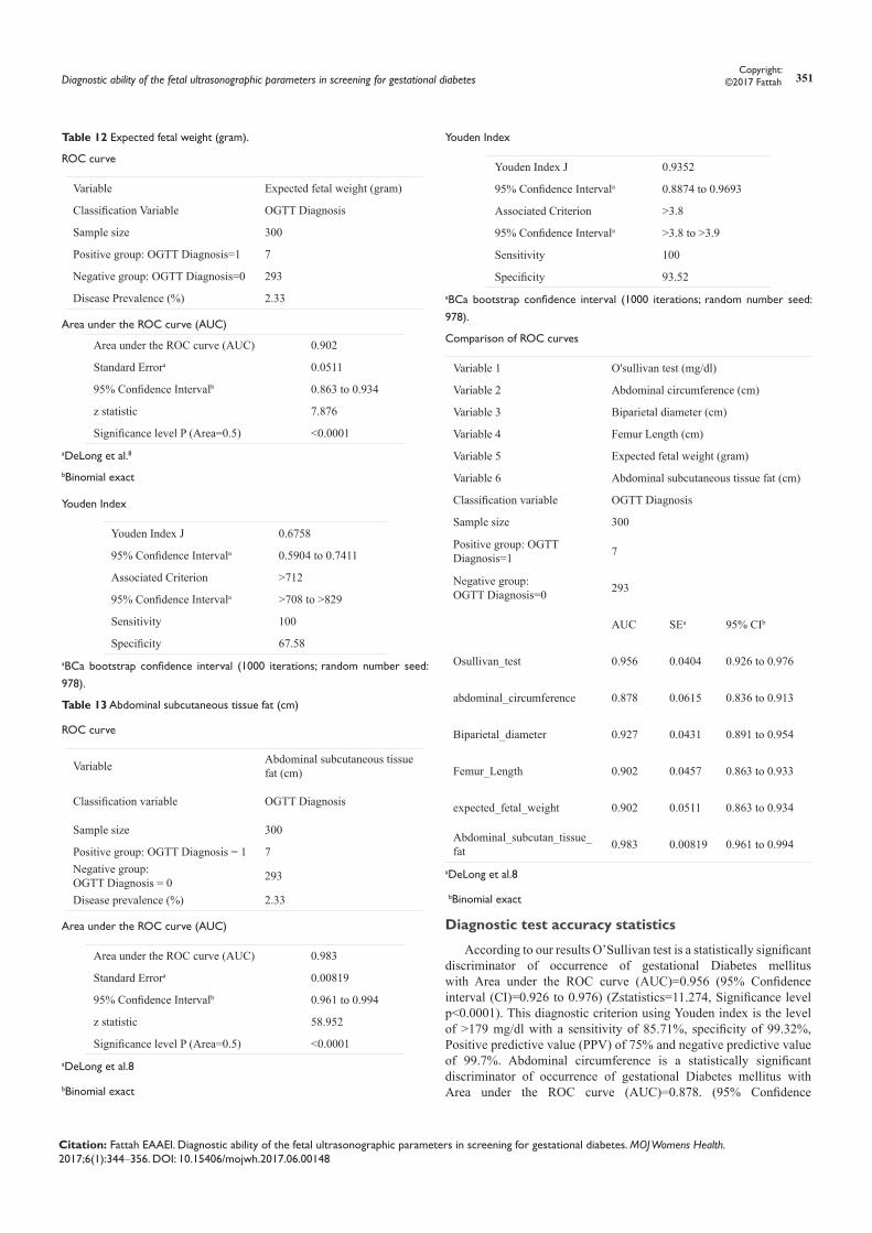
Table 12 Expected fetal weight (gram).
ROC curve
Variable Expected fetal weight (gram)
Classification Variable OGTT Diagnosis
Sample size 300
Positive group: OGTT Diagnosis=1 7
Negative group: OGTT Diagnosis=0 293
Disease Prevalence (%) 2.33
Area under the ROC curve (AUC)
Area under the ROC curve (AUC) 0.902
Standard Errora 0.0511
95% Confidence Intervalb 0.863 to 0.934
z statistic 7.876
Significance level P (Area=0.5) <0.0001aDeLong et al.8
bBinomial exact
Youden Index
Youden Index J 0.6758
95% Confidence Intervala 0.5904 to 0.7411
Associated Criterion >712
95% Confidence Intervala >708 to >829
Sensitivity 100
Specificity 67.58aBCa bootstrap confidence interval (1000 iterations; random number seed: 978).
Table 13 Abdominal subcutaneous tissue fat (cm)
ROC curve
Variable Abdominal subcutaneous tissue fat (cm)
Classification variable OGTT Diagnosis
Sample size 300
Positive group: OGTT Diagnosis = 1 7Negative group: OGTT Diagnosis = 0 293
Disease prevalence (%) 2.33
Area under the ROC curve (AUC)
Area under the ROC curve (AUC) 0.983
Standard Errora 0.00819
95% Confidence Intervalb 0.961 to 0.994
z statistic 58.952
Significance level P (Area=0.5) <0.0001aDeLong et al.8
bBinomial exact
Youden Index
Youden Index J 0.9352
95% Confidence Intervala 0.8874 to 0.9693
Associated Criterion >3.8
95% Confidence Intervala >3.8 to >3.9
Sensitivity 100
Specificity 93.52aBCa bootstrap confidence interval (1000 iterations; random number seed: 978).
Comparison of ROC curves
Variable 1 O'sullivan test (mg/dl)
Variable 2 Abdominal circumference (cm)
Variable 3 Biparietal diameter (cm)
Variable 4 Femur Length (cm)
Variable 5 Expected fetal weight (gram)
Variable 6 Abdominal subcutaneous tissue fat (cm)
Classification variable OGTT Diagnosis
Sample size 300
Positive group: OGTT Diagnosis=1 7
Negative group: OGTT Diagnosis=0 293
AUC SEa 95% CIb
Osullivan_test 0.956 0.0404 0.926 to 0.976
abdominal_circumference 0.878 0.0615 0.836 to 0.913
Biparietal_diameter 0.927 0.0431 0.891 to 0.954
Femur_Length 0.902 0.0457 0.863 to 0.933
expected_fetal_weight 0.902 0.0511 0.863 to 0.934
Abdominal_subcutan_tissue_fat 0.983 0.00819 0.961 to 0.994
aDeLong et al.8
bBinomial exact
Diagnostic test accuracy statistics
According to our results O’Sullivan test is a statistically significant discriminator of occurrence of gestational Diabetes mellitus with Area under the ROC curve (AUC)=0.956 (95% Confidence interval (CI)=0.926 to 0.976) (Zstatistics=11.274, Significance level p<0.0001). This diagnostic criterion using Youden index is the level of >179 mg/dl with a sensitivity of 85.71%, specificity of 99.32%, Positive predictive value (PPV) of 75% and negative predictive value of 99.7%. Abdominal circumference is a statistically significant discriminator of occurrence of gestational Diabetes mellitus with Area under the ROC curve (AUC)=0.878. (95% Confidence
Diagnostic ability of the fetal ultrasonographic parameters in screening for gestational diabetes 352Copyright:
©2017 Fattah
Citation: Fattah EAAEI. Diagnostic ability of the fetal ultrasonographic parameters in screening for gestational diabetes. MOJ Womens Health. 2017;6(1):344‒356. DOI: 10.15406/mojwh.2017.06.00148
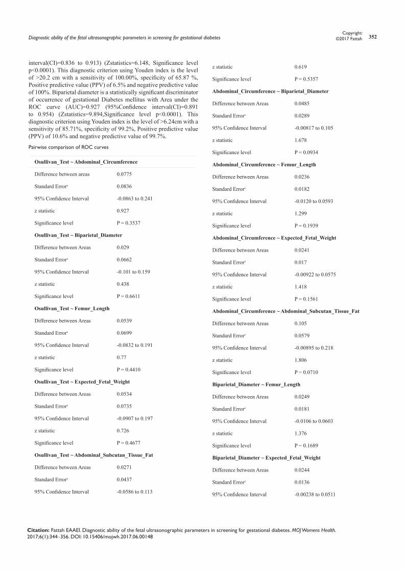
interval(CI)=0.836 to 0.913) (Zstatistics=6.148, Significance level p<0.0001). This diagnostic criterion using Youden index is the level of >20.2 cm with a sensitivity of 100.00%, specificity of 65.87 %, Positive predictive value (PPV) of 6.5% and negative predictive value of 100%. Biparietal diameter is a statistically significant discriminator of occurrence of gestational Diabetes mellitus with Area under the ROC curve (AUC)=0.927 (95%Confidence interval(CI)=0.891 to 0.954) (Zstatistics=9.894,Significance level p<0.0001). This diagnostic criterion using Youden index is the level of >6.24cm with a sensitivity of 85.71%, specificity of 99.2%, Positive predictive value (PPV) of 10.6% and negative predictive value of 99.7%.
Pairwise comparison of ROC curves
Osullivan_Test ~ Abdominal_Circumference
Difference between areas 0.0775
Standard Errorc 0.0836
95% Confidence Interval -0.0863 to 0.241
z statistic 0.927
Significance level P = 0.3537
Osullivan_Test ~ Biparietal_Diameter
Difference between Areas 0.029
Standard Errorc 0.0662
95% Confidence Interval -0.101 to 0.159
z statistic 0.438
Significance level P = 0.6611
Osullivan_Test ~ Femur_Length
Difference between Areas 0.0539
Standard Errorc 0.0699
95% Confidence Interval -0.0832 to 0.191
z statistic 0.77
Significance level P = 0.4410
Osullivan_Test ~ Expected_Fetal_Weight
Difference between Areas 0.0534
Standard Errorc 0.0735
95% Confidence Interval -0.0907 to 0.197
z statistic 0.726
Significance level P = 0.4677
Osullivan_Test ~ Abdominal_Subcutan_Tissue_Fat
Difference between Areas 0.0271
Standard Errorc 0.0437
95% Confidence Interval -0.0586 to 0.113
z statistic 0.619
Significance level P = 0.5357
Abdominal_Circumference ~ Biparietal_Diameter
Difference between Areas 0.0485
Standard Errorc 0.0289
95% Confidence Interval -0.00817 to 0.105
z statistic 1.678
Significance level P = 0.0934
Abdominal_Circumference ~ Femur_Length
Difference between Areas 0.0236
Standard Errorc 0.0182
95% Confidence Interval -0.0120 to 0.0593
z statistic 1.299
Significance level P = 0.1939
Abdominal_Circumference ~ Expected_Fetal_Weight
Difference between Areas 0.0241
Standard Errorc 0.017
95% Confidence Interval -0.00922 to 0.0575
z statistic 1.418
Significance level P = 0.1561
Abdominal_Circumference ~ Abdominal_Subcutan_Tissue_Fat
Difference between Areas 0.105
Standard Errorc 0.0579
95% Confidence Interval -0.00895 to 0.218
z statistic 1.806
Significance level P = 0.0710
Biparietal_Diameter ~ Femur_Length
Difference between Areas 0.0249
Standard Errorc 0.0181
95% Confidence Interval -0.0106 to 0.0603
z statistic 1.376
Significance level P = 0.1689
Biparietal_Diameter ~ Expected_Fetal_Weight
Difference between Areas 0.0244
Standard Errorc 0.0136
95% Confidence Interval -0.00238 to 0.0511

Diagnostic ability of the fetal ultrasonographic parameters in screening for gestational diabetes 353Copyright:
©2017 Fattah
Citation: Fattah EAAEI. Diagnostic ability of the fetal ultrasonographic parameters in screening for gestational diabetes. MOJ Womens Health. 2017;6(1):344‒356. DOI: 10.15406/mojwh.2017.06.00148
z statistic 1.786
Significance level P = 0.0741
Biparietal_Diameter ~ Abdominal_Subcutan_Tissue_Fat
Difference between Areas 0.0561
Standard Errorc 0.0383
95% Confidence Interval -0.0189 to 0.131
z statistic 1.465
Significance level P = 0.1429
Femur_Length ~ Expected_Fetal_Weight
Difference between Areas 0.000488
Standard Errorc 0.0139
95% Confidence Interval -0.0267 to 0.0276
z statistic 0.0352
Significance level P = 0.9719
Femur_Length ~ Abdominal_Subcutan_Tissue_Fat
Difference between Areas 0.0809
Standard Errorc 0.0421
95% Confidence Interval -0.00156 to 0.163
z statistic 1.923
Significance level P = 0.0545
Expected_Fetal_Weight ~ Abdominal_Subcutan_Tissue_Fat
Difference between Areas 0.0804
Standard Errorc 0.0467
95% Confidence Interval -0.0111 to 0.172
z statistic 1.723
Significance level P = 0.0850
cDeLong et al.8
Figure 10 O’ Sullivan test specificity and sensitivity.
Figure 11 Abdominal circumference sensitivity and specificity.
Figure 12 Biparietal Diameter sensitivity and specificity.
Figure 13 Femur length sensitivity and specificity.

Diagnostic ability of the fetal ultrasonographic parameters in screening for gestational diabetes 354Copyright:
©2017 Fattah
Citation: Fattah EAAEI. Diagnostic ability of the fetal ultrasonographic parameters in screening for gestational diabetes. MOJ Womens Health. 2017;6(1):344‒356. DOI: 10.15406/mojwh.2017.06.00148
Figure 14 Expected fetal weight sensitivity and specificity.
Figure 15 Abdominal subcutaneous fat sensitivity and specificity.
Figure 16 sensitivity and specificity for ultrasonographic parameters.
Femur length is a statistically significant discriminator of occurrence of gestational Diabetes mellitus with Area under the ROC curve (AUC)=0.902 (95% Confidence interval (CI)=0.863 to 0.933) (Zstatistics=8.795, Significance level p<0.0001). This diagnostic criterion using Youden index is the level of >4.46cm with a sensitivity of 100%, specificity of 72.35%, Positive predictive value (PPV) of 8% and negative predictive value of 100%. Expected fetal weight is a statistically significant discriminator of occurrence of gestational Diabetes mellitus with Area under the ROC curve (AUC)=0.902 (95% Confidence interval (CI)=0.863 to 0.934) (Zstatistics=7.876, Significance level p<0.0001). This diagnostic criterion using Youden index is the level of >712gm with a sensitivity of 100%, specificity of 67.58%, Positive predictive value (PPV) of 6.9% and negative predictive value of 100%. Abdominal subcutaneous tissue fat is a statistically significant discriminator of occurrence of gestational Diabetes mellitus with Area under the ROC curve (AUC)=0.983 (95% Confidence interval (CI)=0.961 to 0.994) (Zstatistics=58.952, Significance level p<0.0001). This diagnostic criterion using Youden index is the level of >3.8cm with a sensitivity of 100%, specificity of 93.52%, Positive predictive value (PPV) of 26.9% and negative predictive value of 100%.
DiscussionPregnancy is a state of insulin resistance revealing subclinical
defect(s) in carbohydrate metabolism that may develop into a state of carbohydrate intolerance, or gestational Diabetes Mellitus. Therefore, pregnant women must be screened for gestational Diabetes Mellitus. Uniform diagnostic criteria are lacking for more than 40years. Our study aimed at assessing O’Sullivan test (50gram Glucose challenge test) against ultrasound measurements of abdominal circumference, biparital diameter, femur length, estimated fetal weight and abdominal subcutaneous skin fat, fetal liver length as screening for gestational diabetes mellitus between 24 and 28weeks of gestation. The 2hours OGTT was positive in only seven cases out of the 300 screened cases. Those positive cases were preliminary tested positive using O’sullivan test (statistically significant). In contrast, Acharya et al.10 studied the role of O’Sullivan test in screening of pregnant women at 24-36weeks for Gestational Diabetes. One Thousand cases were enrolled, of which only 8 cases (0.8%) were screen positive in contrast to 60 to 63 cases per 1000 in most world series. Akram et al.11 used GCT to screen 1000 patients with analysis of risk factors to compare the efficiency of glucose challenge test with oral glucose tolerance test for detection of gestational diabetes, 450 patients were screened with positive result and 550 patients were screened negative. Out of 450 patients with positive GCT, OGTT detected 40% patients with true positive and 50 patients with false positive results. Out of 550 GCT negative, OGTT screened 510 patients with true negative and 40 patients with false negative. So by performing OGTT 440 patients were diagnosed as gestational diabetics, while 56% had no diabetes. These results matched with ours as regards O’ Sullivan test.
A range of ultrasound anthropometric parameters are used to determine normal fetal growth. In the present study we used ROC analysis to make a decision regarding fetal ultrasound parameters in predicting GDM. The present study has demonstrated that there is a highly significant correlation between fetal biparietal diameter, abdominal circumference, femur length, fetal weight, Abdominal subcutaneous tissue fat and blood glucose values during an OGTT in patients with GDM. The nature of the relationship detected implies that these parameters may be strong predictive factors for OGTT values; this was confirmed by ROC analysis. Conversely, no such relationship

Diagnostic ability of the fetal ultrasonographic parameters in screening for gestational diabetes 355Copyright:
©2017 Fattah
Citation: Fattah EAAEI. Diagnostic ability of the fetal ultrasonographic parameters in screening for gestational diabetes. MOJ Womens Health. 2017;6(1):344‒356. DOI: 10.15406/mojwh.2017.06.00148
was found in the control group. Abdominal subcutaneous tissue fat was found to be a statistically significant discriminator of occurrence of gestational Diabetes mellitus with Area under the ROC curve (AUC) 0.983. with a sensitivity of 100%, specificity of 93.52% with regard to a cut-off value of >3.8cm, Positive predictive value (PPV) of 26.9% and negative predictive value of 100%. One of the major disadvantages of many of the screening tests used currently is false-positive results. In the present study, Abdominal subcutaneous tissue fat measurements demonstrated a specificity near that for glucose tolerance test results based on data in the literature. Recently, studies have shown that abdominal subcutaneous tissue fat measurements, either on their own or incorporated into conventional fetal weight prediction formulae, could be used to evaluate fetal growth, and, in addition, assess whether maternal glucose levels are normal.12
In a comparison of abdominal subcutaneous tissue fat thickness (ASCTT) between fetuses from a group of mothers with gestational diabetes and those from a normal control group, there were significant differences between initial fetal ASCTT, but no difference after the mothers had been treated for diabetes. The measurement of fetal ASCTT gives a more accurate estimation of the stability of maternal glucose levels than a maternal ambulatory glycemic profile.12 As reported, ultrasound measurements of subcutaneous adipose tissue may be a reliable indicator of the fetal metabolic state in pregnancies with GDM.12 Meanwhile, the availability of reference values for fetal subcutaneous fat ultra-sound measurements may provide clinically useful information to identify excessive fetal fat deposition and evaluate maternal glucose levels in pregnancies with GDM, beside the routine fetal ultrasonographic biometric parameters. Comparing our results with similar studies, Vedavathi et al.13 studied 30cases with an established diagnosis of gestational diabetes at 24-28weeks of gestation, by measuring abdominal circumference, head circumference and the estimated fetal weight. Abdominal circumferences (AC) of 27 (90%) cases, Head circumferences (HC) of 20 (67%) cases and estimated fetal weight (EFW) of 8 (27 %) cases were≥90 percentile. He concluded that fetal growth parameters show significant high values in GDM. These results are criticized by the very small sample he worked upon.
In another large prospective cohort, Smith et al.,14 concluded that excessive fetal growth preceded the diagnosis of gestational diabetes, by analysing data from 4069 pregnant women. All women provided blood samples and underwent ultrasounds at 20, 28 and 36weeks of gestation, as well as an oral glucose tolerance test at 28weeks. Risks were set for abdominal circumference > 90th percentile and HC/AC ratio <10th percentile. Within the cohort, 171 women (4.2%) were diagnosed with gestational diabetes at 28weeks. Researchers found no association between fetal measurements at 20weeks of gestation and a subsequent diagnosis of gestational diabetes; however, at 28 weeks, researchers found an increased risk for increased fetal abdominal circumference (RR=2.05; 95% CI, 1.37-3.07) and decreased head circumference to abdominal circumference ratio (RR=1.97; 95% CI, 1.3-2.99) in mothers with gestational diabetes. At 28 weeks, mothers with gestational diabetes had a nearly fivefold increased risk for greater fetal abdominal circumference (RR=4.52; 95% CI, 2.98-6.85) and a nearly threefold increased risk for decreased head circumference to abdominal circumference ratio (RR=2.8; 95% CI, 1.65-4.78). Researchers also found that increased fetal abdominal circumference was associated with a nearly fourfold increased risk for the fetus being born large for gestational age (RR=3.86; 95% CI, 2.37-6.29).14
Honestly we cannot criticize these results but we find them
more logic than ours as the sample size was bigger and the cases were preliminary diagnosed as having gestational diabetes mellitus. Bulhing et al.,15 studied the relationship between sonographically estimated fetal subcutaneous adipose tissue measurements and neonatal skin fold measurements in diabetic fetopathy, but his study was conducted at 37 weeks of gestation. Bulhing enrolled 172 patients (142 controls and 30 with gestational diabetes), both fetal subcutaneous fat tissue and estimated fetal weight were measured. He concluded that ultrasound examination is a reliable method for non-invasive intrauterine measurement of fetal subcutaneous tissue for predicting mechanical neonatal skinfold thickness measurements and thus gestational diabetes. Our results matched with those studies where a positive correlation was detected between fetal ultrasonographic biometric parameters including ASCTT and GDM, but we failed to find a positive correlation between fetal liver length and GDM although maternal hyperglycemia is related to fetal hyperglycemia and hyperinsulinemia, which has a significant impact on the growth of insulin-dependent tissues and organs, such as the liver.15
Conclusioni. Screening for gestational diabetes by ultrasound measurement of
abdominal circumference, biparietal diameter and estimated fetal weight between 24-28weeks of gestation is a reliable, cost-effective method that can be used solely or combined with O’Sullivan test.
ii. The addition of fetal abdominal subcutaneous fat layer measurement (using a cut-off value ≥3.8mm) yielded better accuracy, better positive and negative predictive value, better sensitivity and specificity.
AcknowledgementsNone.
References1. Chen L, Wu JJ, Chen XH, et al. Measurement of fetal abdominal and
subcapsular subcutaneous tissue thickness during pregnancy to predict macrosomia: A pilot study. PLOS One. 2014;9(3):e93077.
2. Wier LM, Witt E, Burgess J, et al. Hospitalizations related to diabetes in pregnancy, 2008. HCUP Statistical Brief #102. Agency for Healthcare Research and Quality. USA: Agency for Healthcare Research and Quality; 2010. p. 1–10.
3. American Diabetes Association. Standards of medical care in diabetes. Diabetes Care. 2014;37(suppl 1):S14–80.
4. Khoury JC, Dolan LM, Vandyke R, et al. Fetal development in women with diabetes: Imprinting for a life-time. J Matern Fetal Neonatal Med. 2012;25(1):11–14.
5. IBM SPSS Statistics for Windows, Version 21.0. USA: Armonk; 2012.
6. Field A. Discovering Statistics Using SPSS. 2nd ed. USA: SAGE Publications Ltd; 2006.
7. Mann HB, Whitney DR. On a test of whether one of two random variables is stochastically larger than the other. Annals of Mathematical Statistics. 1947;18(1):50–60.
8. DeLong ER, DeLong DM, Clarke-Pearson DL. Comparing the areas under two or more correlated receiver operating characteristic curves: a nonparametric approach. Biometrics. 1988;44(3):837–845.
9. MedCalc Statistical Software version 14.8.1 (MedCalc Software bvba, Ostend, Belgium; 2014 http://www.medcalc.org).
Diagnostic ability of the fetal ultrasonographic parameters in screening for gestational diabetes 356Copyright:
©2017 Fattah
Citation: Fattah EAAEI. Diagnostic ability of the fetal ultrasonographic parameters in screening for gestational diabetes. MOJ Womens Health. 2017;6(1):344‒356. DOI: 10.15406/mojwh.2017.06.00148
10. Acharya N, Inamdar A, Inamdar S. Role of O Sullivan S test in screening of pregnant women for gestational diabetes in rural area. JSAFOG. 2011;3(2):86–88.
11. Akram N, Butt F. Universal screening with glucose challenge test in detection of gestational diabetes. Biomedica. 2014;3(1):29–33.
12. Chen L, Wu JJ, Chen XH, et al. Measurement of fetal abdominal and subscapular subcutaneous tissue thickness during pregnancy to predict macrosomia: a pilot study. PLoS One. 2014;9(3):e93077.
13. Vedavathi KJ, Swamy RM, Shekharappa KP, et al. Influence of Gestational Diabetes Mellitus on Fetal growth parameters. Int J Biol Med Res. 2011;2(3):832–834.
14. Sovio U, Murphy HR, Smith GC. Accelerated fetal growth prior to diagnosis of gestational diabetes mellitus: a prospective cohort study of nulliparous women. Diabetes Care. 2016;39(6):982–987.
15. Buhling KJ, Doll I, Siebert G, et al. Relationship between sonographically estimated fetal subcutaneous adipose tissue measurements and neonatal skinfold measurements. Ultrasound Obstet Gynecol. 2012;39:558–562.
Top Related
Copyright © 2022 FDOKUMEN

