


Bahasa
Halaman
Hukum


Collagenolytic Activities of the Major Secreted CathepsinL Peptidases Involved in the Virulence of the HelminthPathogen, Fasciola hepaticaMark W. Robinson1*, Ileana Corvo2, Peter M. Jones1, Anthony M. George1, Matthew P. Padula1, Joyce
To1, Martin Cancela2¤a, Gabriel Rinaldi2¤b, Jose F. Tort2, Leda Roche2, John P. Dalton3
1 Infection, Immunity and Innovation (i3) Institute, University of Technology Sydney (UTS), Sydney, New South Wales, Australia, 2 Departmento de Genetica, Facultad de
Medicina, Universidad de la Republica (UDELAR), Montevideo, Uruguay, 3 Institute of Parasitology, McGill University, St. Anne de Bellevue, Quebec, Canada
Abstract
Background: The temporal expression and secretion of distinct members of a family of virulence-associated cathepsin Lcysteine peptidases (FhCL) correlates with the entry and migration of the helminth pathogen Fasciola hepatica in the host.Thus, infective larvae traversing the gut wall secrete cathepsin L3 (FhCL3), liver migrating juvenile parasites secrete bothFhCL1 and FhCL2 while the mature bile duct parasites, which are obligate blood feeders, secrete predominantly FhCL1 butalso FhCL2.
Methodology/Principal Findings: Here we show that FhCL1, FhCL2 and FhCL3 exhibit differences in their kineticparameters towards a range of peptide substrates. Uniquely, FhCL2 and FhCL3 readily cleave substrates with Pro in the P2position and peptide substrates mimicking the repeating Gly-Pro-Xaa motifs that occur within the primary sequence ofcollagen. FhCL1, FhCL2 and FhCL3 hydrolysed native type I and II collagen at neutral pH but while FhCL1 cleaved only non-collagenous (NC, non-Gly-X-Y) domains FhCL2 and FhCL3 exhibited collagenase activity by cleaving at multiple sites withinthe a1 and a2 triple helix regions (Col domains). Molecular simulations created for FhCL1, FhCL2 and FhCL3 complexed tovarious seven-residue peptides supports the idea that Trp67 and Tyr67 in the S2 subsite of the active sites of FhCL3 andFhCL2, respectively, are critical to conferring the unique collagenase-like activity to these enzymes by accommodatingeither Gly or Pro residues at P2 in the substrate. The data also suggests that FhCL3 accommodates hydroxyproline (Hyp)-Glyat P3-P2 better than FhCL2 explaining the observed greater ability of FhCL3 to digest type I and II collagens compared toFhCL2 and why these enzymes cleave at different positions within the Col domains.
Conclusions/Significance: These studies further our understanding of how this helminth parasite regulates peptidaseexpression to ensure infection, migration and establishment in host tissues.
Citation: Robinson MW, Corvo I, Jones PM, George AM, Padula MP, et al. (2011) Collagenolytic Activities of the Major Secreted Cathepsin L Peptidases Involved inthe Virulence of the Helminth Pathogen, Fasciola hepatica. PLoS Negl Trop Dis 5(4): e1012. doi:10.1371/journal.pntd.0001012
Editor: Malcolm Jones, University of Queensland, Australia
Received November 8, 2010; Accepted December 21, 2010; Published April 5, 2011
Copyright: � 2011 Robinson et al. This is an open-access article distributed under the terms of the Creative Commons Attribution License, which permitsunrestricted use, distribution, and reproduction in any medium, provided the original author and source are credited.
Funding: MWR is supported by a UTS Chancellor’s Postdoctoral Fellowship. IC is supported by a Wood-Whelan Research Fellowship from the International Unionof Biochemistry and Molecular Biology (IUBMB) and CSIC-UDELAR. PMJ is supported by Cure Cancer Australia and UTS fellowships. JPD is funded by a grant fromthe Natural Sciences and Engineering Research Council (NSERC) of Canada. The computational component of this research was undertaken at the NCI NationalFacility in Canberra, Australia, which is supported by the Australian Commonwealth Government. The funders had no role in study design, data collection andanalysis, decision to publish, or preparation of the manuscript.
Competing Interests: The authors have declared that no competing interests exist.
* E-mail: [email protected]
¤a Current address: Laboratorio de Cestodeos, Centro de Biotecnologia, Universidade Federal do Rio Grande do Sul (UFRGS), Porto Alegre, Brazil¤b Current address: Department of Microbiology, Immunology and Tropical Medicine, George Washington University Medical Center, Washington, DC, UnitedStates of America
Introduction
Papain-like cysteine peptidases, including cathepsins B and L,
are ubiquitously secreted extracorporeally by helminth parasites of
human and veterinary importance where they perform many
important roles that are critical to the development and survival of
the parasite within the mammalian host [1]. These roles include
penetration and migration through host tissues [2], catabolism of
host proteins to peptides and amino acids [3,4], and modulation of
the host immune response by cleaving immunoglobulin [5,6] or by
altering the activity of immune effector cells [7]. Accordingly,
cathepsin peptidases are leading targets for novel anti-parasitic
drugs and vaccines that block their function [8,9].
Fasciola hepatica is the causative agent of liver fluke disease
(fasciolosis) of domestic animals in regions with temperate climates.
Although traditionally regarded as a disease of livestock, fasciolosis
is now recognised as an important emerging foodborne zoonotic
disease in rural areas of South America (particularly Bolivia, Peru
and Equador), Egypt and Iran [10]. It is estimated that over 2.4
million people are infected with F. hepatica worldwide and around
91 million are at risk of infection [11]. To infect their mammalian
hosts, F. hepatica larvae, which are ingested with vegetation
www.plosntds.org 1 April 2011 | Volume 5 | Issue 4 | e1012
contaminated with dormant cysts (metacercariae), penetrate the
intestinal wall, enter the liver capsule and migrate through the
parenchyma before invading into the bile ducts [12]. To facilitate
this tissue migration, Fasciola secrete various members of a
multigenic family of cathepsin L peptidases that exhibit overlapping
but complementary substrate specificities and together cleave host
macromolecules very efficiently [13,14]. In fact, the ability of
Fasciola to infect and adapt to a wide range of host species has been
attributed to the effectiveness of this proteolytic machinery [14,15].
Phylogenetic analyses have shown that the Fasciola cathepsin L
gene family expanded by a series of gene duplications followed by
divergence which gave rise to three clades expressed by tissue-
migrating and adult worms (Clades 1, 2, and 5) and two clades
specific to the early infective juvenile stage (Clades 3 and 4)
[13,14]. Consistent with these observations, our proteomics
analysis identified representative enzymes from Clades 1, 2 and
5, but not from Clades 3 and 4, in the secretory products of adult
F. hepatica [14]. More recently, we showed that the temporal
expression and secretion of the specific cathepsin L clades
correlated with the migration of the parasite through host issues;
members of cathepsin L clade 3 (FhCL3) are secreted by Fasciola
infective larvae and effected penetration of the host intestinal wall
while clades 1, 2 and 5 (FhCL1, FhCL2 and FhCL5) peptidases
are secreted by the immature liver-stage flukes and adult worms
and function in preparing a migratory path through the liver and
in the acquisition of nutrient by degrading host blood and tissue
components. While clade 4 (FhCL4) peptidases are expressed by
infective larvae they do not seem to be secreted and, therefore,
may play an intracellular house-keeping function [13,16]. Recent
transcriptomic analyses of juvenile and adult stages have
confirmed these observations [17,18].
The secreted Fasciola cathepsins are produced in specialised
gastrodermal cells which line the parasites’s gut and are packaged
in secretory vesicles before being extruded into the gut lumen from
where they are released into host tissues [19]. The peptidases can
efficiently degrade a range of host macromolecules including
haemoglobin, immunoglobulin and interstitial matrix proteins
such as fibronectin and laminin [3,4,20–22]. Notably, however,
studies in our laboratory using functionally-active recombinant
enzymes have shown that FhCL2 and FhCL3 exhibit an unusual
ability to cleave native collagen [22,23]. This is of relevance
because collagenase-like activity is restricted to very few enzymes
(e.g. bacterial collagenases, matrix metalloproteinases and human
cathepsin K) and, therefore, the evolution and maintenance of
such an activity in Fasciola suggests that it is essential to the
parasite’s ability to degrade the connective tissue matrix of the
organs through which it migrates.
The active site of papain-like cysteine peptidases is relatively
short, and while consisting of four subsites (S2-S1-S19-S29) with
additional binding areas (S4-S3 and S39) the specificity of substrate
binding is principally governed by the residues that make up the S2
subsite [24,25]. This S2 site forms a deep pocket capable of holding
the P2 amino acid of the substrate and positioning the scissile bond
into the S1 subsite for cleavage. In Carica papaya papain (PDB ID:
9PAP), the S2 subsite is composed of residues occupying positions
67, 68, 133, 157, 160 and 205. An analysis of these residues in the
various cathepsin L clades clearly demonstrates divergence within
the S2 subsite, in particular at the three positions that have the
greatest influence on P2 binding i.e. at residues 67, 157 and 205
[14,15,23]. For FhCL2, the collagenolytic activity has been
attributed to the presence of a particular residue, Tyr69, within
the enzyme’s S2 substrate binding site which is also found in human
cathepsin K, the only mammalian cathepsin with the ability to
cleave within the covalently-linked triple helices, of Col domains of
native collagen [26,27]. The S2 Tyr69 is also suggested to allow
both enzymes to cleave macromolecular and dipeptide substrates
with a Pro residue in the P2 position [22,28]. More recently we
showed that in FhCL3, this position is occupied by a larger Trp
residue, a feature shared only with a ginger rhizome peptidase [29]
which is also capable of cleaving collagen. Notably the S3 subsite of
both FhCL3 and the plant enzyme are quite shallow, an observation
that led us to advance the idea that the specificity of these enzymes
might be restricted [23].
Our laboratory recently determined the three-dimensional
structure of one of the major cathepsin L peptidases of adult F.
hepatica, FhCL1 [22]. Similar to other cathepsins, the enzyme is
composed of two domains (R and L) at the juncture of which is a
cleft that forms the substrate-binding site and contains the enzyme
catalytic machinery. Super-imposition of the alpha carbons of
FhCL1 with cysteine peptidases from plants (e.g. papain, PDB ID
9PAP) and mammals (e.g. human cathepsin L PDB ID 1CJL)
yields an r.m.s. deviation in the range 0.78 A to 1.085 A which is
indicative of the very high conservation of the overall fold and
shape that exists amongst all members of the papain family of
cysteine peptidases [30]. Since the FhCL1 structure and fold can
be described as practically identical to all other cathepsin L-like
peptidases its scaffold can be exploited as a ‘prototype’ to
investigate the role of critical amino acids within the S2 subsite
in substrate binding (Table 1), particularly those of FhCL2 and
FhCL3, whose primary structures are 78% and 70% identical to
FhCL1, respectively. In the present study, we investigated and
compared the substrate specificity of active recombinant forms of
FhCL1, FhCL2 and FhCL3 with specific emphasis on their ability
to degrade native collagen. This activity was interpreted by
obtaining the enzymatic kinetic parameters (Km, kcat, and kcat/
Km) of these enzymes on a range of peptide substrates and binding
kinetics for specific inhibitory compounds. Furthermore, using
mass spectrometry we mapped the cleavage sites of native collagen
I and derived peptides, adding evidence for differential activities
between FhCL2 and FhCL3. Finally, using our FhCL1 crystal
structure as a template we created molecular dynamics simulations
to explain how the active sites of these enzymes accommodate
collagen-like substrates and endow them with this unusual
collagenolytic activity. Our study provides biochemical and
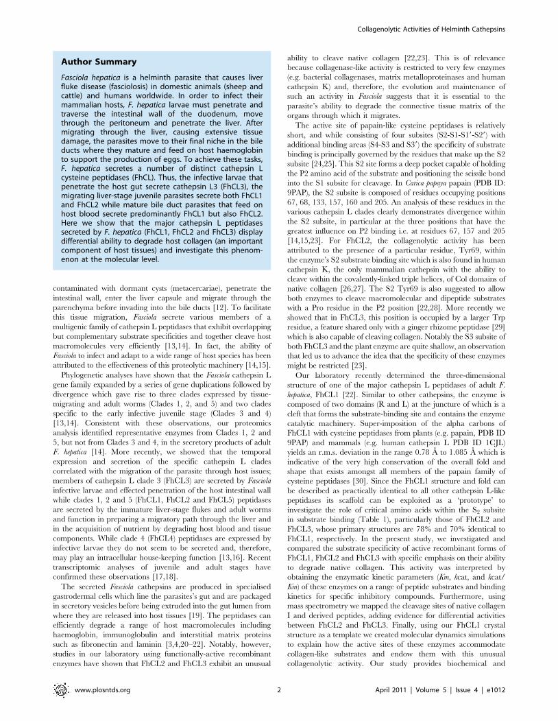
Author Summary
Fasciola hepatica is a helminth parasite that causes liverfluke disease (fasciolosis) in domestic animals (sheep andcattle) and humans worldwide. In order to infect theirmammalian hosts, F. hepatica larvae must penetrate andtraverse the intestinal wall of the duodenum, movethrough the peritoneum and penetrate the liver. Aftermigrating through the liver, causing extensive tissuedamage, the parasites move to their final niche in the bileducts where they mature and feed on host haemoglobinto support the production of eggs. To achieve these tasks,F. hepatica secretes a number of distinct cathepsin Lcysteine peptidases (FhCL). Thus, the infective larvae thatpenetrate the host gut secrete cathepsin L3 (FhCL3), themigrating liver-stage juvenile parasites secrete both FhCL1and FhCL2 while mature bile duct parasites that feed onhost blood secrete predominantly FhCL1 but also FhCL2.Here we show that the major cathepsin L peptidasessecreted by F. hepatica (FhCL1, FhCL2 and FhCL3) displaydifferential ability to degrade host collagen (an importantcomponent of host tissues) and investigate this phenom-enon at the molecular level.
Collagenolytic Activities of Helminth Cathepsins
www.plosntds.org 2 April 2011 | Volume 5 | Issue 4 | e1012

structural insights into the molecular mechanism of tissue invasion
by these important parasitic helminths.
Materials and Methods
MaterialsZ-Phe-Arg-NHMec, Z-Leu-Arg-NHMec, Z-Val-Val-Arg-NHMec,
Tos-Gly-Pro-Arg-NMec, Tos-Gly-Pro-Lys-NMec, Boc-Ala-Gly-Pro-
Arg-NMec, Boc-Val-Leu-Lys-NMec, Boc-Val-Pro-Arg-NMec, Z-
Phe-Ala-CHN2, Z-Gly-Pro-Gly-Gly-Pro-Ala and Z-Gly-Pro-Leu-
Gly-Pro were obtained from Bachem (St. Helens, UK). Cathepsin
K inhibitor II was purchased from BD Biosciences (Sydney,
Australia). E-64, DTT, EDTA and bovine nasal septum collagen
type II were obtained from Sigma-Aldrich (Sydney, Australia).
Calf skin collagen type I was purchased from Calbiochem. Pichia
pastoris strain X33 was obtained from Invitrogen (San Diego, CA,
USA). Ni-NTA agarose and columns were obtained from Qiagen
(Australia). Pre-cast NuPage 4–12% Bis-Tris gels and pre-stained
molecular weight markers were purchased from Invitrogen (Australia).
Expression and purification of recombinant F. hepaticacathepsins in yeast
Recombinant F. hepatica procathepsin L1, L2 and L3 (FhCL1,
FhCL2 and FhCL3) were produced in yeast as previously
described [22,23]. Briefly, P. pastoris (for FhCL1 and FhCL2
expression) and P. angusta (for FhCL3 expression) yeast transfor-
mants were cultured in 500 ml BMGY broth, buffered to pH 8.0,
in 5 L baffled flasks at 30uC until an OD600 of 2–6 was reached.
Cells were harvested by centrifugation at 20006 g for 5 min and
protein expression induced by resuspending in 100 ml BMMY
broth, buffered at pH 6.0 containing 1% methanol. Recombinant
proteins were affinity purified from yeast using Ni-NTA-agarose.
Recombinant propeptidases were dialysed against phosphate
buffered saline (PBS) and stored at 220uC. The 37 kDa cathepsin
L zymogens were autocatalytically activated and processed to
24.5 kDa mature enzymes by incubation for 2 h at 37uC in 0.1 M
sodium citrate buffer (pH 5.0) containing 2 mM DTT and
2.5 mM EDTA. The mixture was then dialysed against PBS,
pH 7.3. The proportion of functionally active recombinant protein
in these preparations was determined by titration against E-64.
Enzyme assays and kinetics with fluorogenic peptidesubstrates
Initial rates of hydrolysis of the fluorogenic peptide substrates
shown in table 2 were monitored by the release of the fluorogenic
leaving group, NHMec, at an excitation wavelength of 380 nm
and an emission wavelength of 460 nm using a Bio-Tek KC4
microfluorometer. kcat and Km values were determined using
nonlinear regression analysis. Initial rates were obtained at 37uCover a range of substrate concentrations spanning Km values (0.2–
200 mM) and at fixed enzyme concentrations (0.5–5 nM). Assays
were performed in 100 mM sodium phosphate buffer (pH 6.0)
containing 1 mM DTT and 1 mM EDTA. Rate constants for the
inactivation of the Fasciola enzymes by Z-Phe-Ala-CHN2 and
cathepsin K inhibitor II were determined from progress curves in
the presence of substrate as previously described [22].
Hydrolysis of native collagen and collagen-like peptidesCalf skin collagen type I and bovine nasal septum collagen type II
(solubilised in 0.2 M acetic acid at a concentration of 2 mg/ml) were
dialysed for two days against 0.1 M sodium acetate (pH 5.5) or PBS
(pH 7.0). Digestion reactions contained 10 mg of dialysed collagen
substrates, 1 mM DTT and 1 mM EDTA and 2 mM activated
FhCL1, FhCL2 or FhCL3 in a final volume of 100 ml of one of the
above buffers at 28uC. For collagen type 1, reactions were performed
for 3 h (pH 5.5) or 20 h (pH 7.0) whilst collagen type II was digested
over 13–18 h. All reactions were stopped by the addition of 10 mM
E-64. Digests were analyzed on reducing 4–12% NuPage Bis-Tris gels
and visualised by staining with Flamingo fluorescent stain (Bio-Rad).
For digestion of collagen-like peptide substrates, 20 mg of Z-Gly-
Pro-Leu-Gly-Pro and Z-Gly-Pro-Gly-Gly-Pro-Ala in DMSO were
Table 1. Residues forming the S2 active site of human and F.hepatica cathepsin L peptidases.
Residues
67 68 133 157 160 205
Human cathepsin L Leu Met Ala Met Gly Ala
C. papaya papain Tyr Pro Val Val Ala Ser
FhCL1 Leu Met Ala Val Ala Leu
FhCL2 Tyr Met Ala Leu Ala Leu
FhCL3 Trp Met Ala Val Ala Val
Comparison of the residues from the S2 active site that contribute to differentialsubstrate-binding in human cathepsin L, C. papaya papain and F. hepaticacathepsins FhCL1, FhCL2 and FhCL3.doi:10.1371/journal.pntd.0001012.t001
Table 2. Kinetic parameters for hydrolysis of peptidyl-NHMecsubstrates by recombinant F. hepatica cathepsin Ls.
Enzyme Substrate KM mM ± Kcat s21 ±Kcat/KM
Ms21
FhCL1 Z-LR-NHMec 1.09 0.38 1.63 0.1 1492354.7
FhCL2 Z-LR-NHMec 2.13 0.29 0.95 0.035 444106.1
FhCL3 Z-LR-NHMec 48 5.7 1.2 0.05 25000
FhCL1 Z-FR-NHMec 1.9 0.57 0.12 0.008 64912.3
FhCL2 Z-FR-NHMec 7.8 1.5 0.09 0.008 11088
FhCL3 Z-FR-NHMec 20.05 2.9 0.02 0.001 999.3
FhCL1 Boc-VLK-NHMec 2.5 0.6 0.14 0.007 54266.7
FhCL2 Boc-VLK-NHMec 2.2 0.79 0.07 0.005 30712.5
FhCL3 Boc-VLK-NHMec 6.13 1.6 0.01 0.001 1747.8
FhCL1 Z-VVR-NHMec 2.84 0.7 0.021 0.001 7464.8
FhCL2 Z-VVR-NHMec 1.53 0.29 0.014 0.001 9237.5
FhCL3 Z-VVR-NHMec 11.7 2.1 0.043 0.020 3696.6
FhCL1 Tos-GPR-NHMec 27.2 4.9 0.02 0.001 671.6
FhCL2 Tos-GPR-NHMec 13.9 3.2 0.26 0.02 18559.5
FhCL3 Tos-GPR-NHMec 10.6 2.3 1.015 0.08 95774.7
FhCL1 Tos-GPK-NHMec 17.42 2.6 0.01 0.001 612.3
FhCL2 Tos-GPK-NHMec 12.9 3.4 0.18 0.013 13746.8
FhCL3 Tos-GPK-NHMec 9.8 3.7 0.15 0.015 36419.8
FhCL1 Boc-VPR-NHMec 22 2.5 0.01 0.0004 478.8
FhCL2 Boc-VPR-NHMec 11.39 2.1 0.23 0.01 20193.2
FhCL3 Boc-VPR-NHMec 21.1 3.2 0.033 0.002 1564
FhCL1 Boc-AGPR-NHMec 18.8 4 0.01 0.001 673.8
FhCL2 Boc-AGPR-NHMec 9.4 1.3 0.55 0.023 58027.1
FhCL3 Boc-AGPR-NHMec 9.6 1.6 0.58 0.025 60763.9
doi:10.1371/journal.pntd.0001012.t002
Collagenolytic Activities of Helminth Cathepsins
www.plosntds.org 3 April 2011 | Volume 5 | Issue 4 | e1012

incubated with FhCL2 or FhCL3 (15 mM) in 100 mM sodium
acetate buffer, pH 4.5, containing 1 mM EDTA and 2 mM DTT
for 30 min at 37uC. Digestion reactions were halted by the
addition of 10 mM E-64.
Analysis of collagen digests by mass spectrometryRecombinant FhCL2 and FhCL3 were removed from collagen
type I digests using Ni-NTA agarose. The reactions were then
spun at 13,000 rpm for 15 min to remove particulates and were
concentrated to a final volume of 15 ml using a Concentrator 5301
(Eppendorf). Using an Eksigent AS-1 autosampler connected to a
Tempo nanoLC system (Eksigent, USA), 10 mL of the sample was
loaded at 20 ml/min with MS buffer A (2% acetonitrile+0.2%
formic acid) onto a C8 trap column (Michrom, USA). After
washing the trap for three minutes, the peptides were washed off
the trap at 300 nL/min onto an IntegraFrit column
(75 mm6100 mm) packed with ProteoPep II C18 resin (New
Objective, Woburn, MA). Peptides were eluted from the column
and into the source of a QSTAR Elite hybrid quadrupole-time-of-
flight mass spectrometer (AB Sciex) using the following program:
5–50% MS buffer B (98% acetonitrile+0.2% formic acid) over
15 minutes, 50–80% MS buffer B over 5 minutes, 80% MS buffer
B for 2 minutes, 80–5% for 3 min. The eluting peptides were
ionised with a 75 mm ID emitter tip that tapered to 15 mm (New
Objective) at 2300 V. An Intelligent Data Acquisition (IDA)
experiment was performed, with a mass range of 375–1500 Da
continuously scanned for peptides of charge state 2+–5+ with an
intensity of more than 30 counts/s. Selected peptides were
fragmented and the product ion fragment masses measured over a
mass range of 50–1500 Da. The mass of the precursor peptide was
then excluded for 15 seconds. Peak list files generated by MSX
(Infochromics) were exported to a local PEAKS Studio v5.0
(Bioinformatics Solutions Inc.) search engine for protein database
searching. MS/MS data was used to search a custom-made
database containing only bovine collagen sequences. The enzyme
specificity was set to ‘‘no enzyme’’ and propionamide (acrylamide)
modification of cysteines was used as a fixed parameter and
oxidation of methionines was set as a variable protein modifica-
tion. The mass tolerance was set at 100 ppm for precursor ions
and 0.2 Da for fragment ions. Only 1 missed cleavage was
allowed. Matched peptides achieving a score .60% were accepted
during PEAKs searches [16]. The matching peptides were then
mapped onto the primary amino acid sequence of bovine collagen
to identify FhCL2 and FhCL3 cleavage sites and to plot P2 residue
preference for each enzyme.
For collagen-like peptide substrates, digests were concentrated
and analysed by MS/MS as described above with the following
modifications. The mass range of 150–600 Da was scanned for
peptides of charge state 2+ with an intensity of more than 100
counts/s. Selected peptides were fragmented and the product ion
fragment masses measured over a mass range of 50–600 Da. The
mass of the precursor peptide was then excluded for 120 seconds.
An inclusion list describing all possible substrate ions that could be
produced by enzymatic cleavage of the peptide substrates was
generated and programmed into the Analyst acquisition software.
The resulting data files were manually interrogated to determine
the presence of peptide ions described in the inclusion list. The
MS/MS spectra of those peptides were de novo sequenced for b and
y ion fragments describing the peptide substrate’s sequence to a
mass accuracy of approximately 0.2 Da.
Molecular dynamics (MD) simulationsFor the MD simulations, starting coordinates for F. hepatica
cathepsin L were taken from the 1.4 A resolution crystal structure
of a FhCL1 mutant zymogen, in which the active site Cys was
replaced by Gly ([22]; PDB 2O6X). The prosegment (residues 1–
100) was removed and the active site Gly mutation reversed to the
wild type Cys. Initial coordinates for a template peptide substrate
(Ala-Leu-Ala-Leu-Pro) were derived from X-ray structures of
inhibitors bound to human cathepsin K ([31]; PDB 1NLJ) and
bovine cathepsin B ([32]; PDB 1SP4) after structural alignment
with FhCL1. This initial peptide was altered to Ala-Leu-Arg-Asn-
Ala using the mutate function in Swiss-PdbViewer ([33]; http://
spdbv.vital-it.ch/) and then minimized while bound to the wild-
type FhCL1 using the equilibration protocol described below. The
equilibrated peptide was then extended by one Ala residue at its
N- and C-termini using the coordinate generation function in the
psfgen program [34], and then re-equilibrated. The resultant
peptide (ligand A) was used to generate all other substrate starting
coordinates by using the mutate function in Swiss-PdbViewer.
Mutations to FhCL1 were similarly generated using Swiss-
PdbViewer. Rotamers for mutated enzyme and substrate side-
chains were chosen by visual inspection and using the rotamer
score provided in Swiss-PdbViewer. The N-terminal residue of the
substrate was acetylated and the C-terminus N-methylamidated.
Each complex was optimally oriented to minimize cell volume
[35] and solvated in a truncated octahedral periodic cell with a
minimum of 20 A between periodic images of the protein. The
system was neutralized with sodium ions.
MD simulations were carried out with NAMD 2.6 [34] using
the CHARMM27 force field with Q/y cross-term map corrections
[36]. Parameters for Hyp were from Veld and Stevens [37]. Water
molecules were simulated with the TIP3P model [38]. Simulation
conditions were maintained at 1.0 atm constant pressure by the
Nose-Hoover Langevin piston method [39,40] and at 310 K
constant temperature by Langevin dynamics with a damping
coefficient at 5 ps21. The time step used for the simulations was
1.5 fs. A cutoff of 12 A, with a switching function between 10 and
12 A, was used for short-range non-bonded interactions. Long-
range electrostatic interactions were computed using the particle
mesh Ewald method [41] with a grid density of approximately
1/A. A multiple time-stepping algorithm was used with interac-
tions involving covalent bonds and short-range non-bonded
interactions computed every time step, while long-range electro-
static forces were computed every two time steps. SHAKE [42]
and SETTLE [43] were applied to constrain the lengths of all
bonds that involve hydrogen.
The solvated starting structure was minimized using conjugate
gradient minimization to a 0.5 kcal/(mol?A) r.m.s. gradient with
all enzyme heavy atoms fixed, with the exception of side-chain
atoms of mutated residues, which were unrestrained. In addition,
in this phase of the equilibration, ligand atoms were not fixed and
harmonic positional constraints of 100 kcal/(mol?A2) force
constant were placed on the Ca atoms of ligand residues 3–5
(P2, P1 and P19). The unrestrained atoms were then further
minimized during a 50 ps molecular dynamics run at 310 K. This
starting model was then minimized with harmonic positional
constraints on the NCaCO backbone of the protein and Ca atoms
of ligand residues 3–5. A 100 kcal/(mol?A2) force constant was
used to minimise the system to a 0.5 kcal/(mol?A) r.m.s. gradient.
The constraints were gradually removed by subsequent minimi-
zations to a 0.1 kcal/(mol?A) r.m.s. gradient, scaling the initial
force constants by factors of 0.5, 0.15, 0.05, and 0. The
unrestrained minimized structure was then heated from 50 K to
310 K in steps of 25 K using velocity reassignment during a 30 ps
molecular dynamics run. The equilibrated system was then used
for production runs with no restraints. All systems were run for
12 ns. All simulations remained stable to completion. For analysis,
Collagenolytic Activities of Helminth Cathepsins
www.plosntds.org 4 April 2011 | Volume 5 | Issue 4 | e1012

the distance between the sulphur atom of the active Cys residue
and the scissile backbone carbonyl carbon of the substrate (S-C
distance) was recorded every 50 time-steps (0.075 ps); trajectory
coordinates were recorded every 1000 time-steps (1.5 ps).
Free energy of binding calculationsThe free energy of binding of the peptide ligand to the peptidase
contains an enthalpic and an entropic contribution. Free energy
analysis of the production trajectories employed the single-
trajectory MM/PBSA method combined with a determination
of the change in configurational entropy using the harmonic
approximation of normal-mode analysis [44,45]. Snapshots from
the MD trajectory, stripped of water and counterions, were
analysed. The enthalpy of binding is composed of the change in
the molecular mechanics free energy upon complex formation,
and the solvated free energy contribution. The molecular
mechanics free energy difference was calculated using the
SANDER module in AMBER 9 [46], with no cutoff for the
non-bonded energies and the AMBER ff03 force field to describe
the protein and peptide ligands [47]. Compatible parameters for
Hyp were not available and binding energies for ligand F were not
calculated. The AMBER PBSA module was used for the
evaluation of the electrostatic free energy of solvation. A grid
density of 3/A was employed for the cubic lattice, the internal and
external dielectric constants were set to 1 and 80, respectively, and
1000 linear iterations were performed. The non-polar solvation
free energy was calculated from the solvent accessible surface area
using the MSMS program [48], with a probe radius of 1.4 A, the
surface tension set to 0.00542 kcal/(mol?A2), and the off-set to
0.92 kcal/mol?m.
The changes in configurational entropy upon ligand association
were estimated by an all-atom normal-mode analysis performed
with the AMBER NMODE module. Prior to the normal mode
calculations, the complex, receptor, and ligand were subjected to
minimization with a distance dependent dielectric constant 4r and
convergence tolerance tighter than a root-mean-squared gradient
of drms 1024 kcal/(mol?A). Entropy and enthalpy calculations on
all peptidase-ligand systems are performed separately and
averaged over equally spaced snapshots, extracted over the final
4.005 ns of the production phase. The mean of the binding free
enthalpies and entropies of all the snapshots were computed and
then summed to obtain the binding free energy. For the enthalpy
calculations, snapshots were taken every 10.5 ps (381 frames), for
the entropy calculations, snapshots were taken every 190.5 ps (21
frames). VMD [49] was used to prepare the initial simulation
system and analyse trajectories. Structural figures were prepared
with PyMol [50]. Simulaid (http://atlas.physbio.mssm.edu/,mezei/) was used in the preparation of the truncated octahedral
cell [35] and to convert the NAMD dcd format MD coordinate
trajectories to AMBER format for the MM/PBSA analysis.
Results and Discussion
F. hepatica FhCL1, FhCL2 and FhCL3 exhibit distinctsubstrate specificities
Functionally active recombinant forms of the major cathepsin L
peptidases of F. hepatica, FhCL1, FhCL2 and FhCL3, were
expressed in yeast and isolated to homogeneity as previously
described [22,23]. To compare their biochemical substrate
specificity the kinetic parameters (Km, kcat, and kcat/Km) for each
enzyme was determined against a range of small fluorogenic
peptide (predominantly tripeptide) substrates (Table 2).
FhCL1 most efficiently cleaved substrates containing hydro-
phobic residues at the P2 position such as the dipeptides Z-Leu-
Arg-NHMec (kcat/Km 1,492,354 M21 s21), Z-Phe-Arg-NHMec
(kcat/Km 64,912 M21 s21) and tripeptide Boc-Val-Leu-Lys-
NHMec (kcat/Km 54,266 M21 s21). In contrast, tripeptide
substrates containing Pro at the P2 position, including Tos-Gly-
Pro-Arg-NHMec (kcat/Km 671 M21 s21), Boc-Ala-Gly-Pro-Arg-
NHMec (kcat/Km 673 M21 s21), Tos-Gly-Pro-Lys-NHMec (kcat/
Km 612 M21 s21) and Boc-Val-Pro-Arg (kcat/Km 478 M21 s21),
were cleaved relatively poorly (Table 2).
In comparison to FhCL1, substrates with Phe and Leu in the P2
position were much less effectively cleaved by FhCL2 and even less
so by FhCL3. The kcat/Km values for FhCL2 and FhCL3 against
Z-Phe-Arg-NHMec were 6- and 65-fold lower, respectively, than
that observed for FhCL1. Similarly, the kcat/Km values for Z-Leu-
Arg-NHMec were 3.5- and 66-fold lower than FhCL1 for FhCL2
and FhCL3 respectively. By contrast, FhCL2 and FhCL3 cleaved
Pro-containing substrates much more readily than FhCL1 with
kcat/Km values of 18,559 M21 s21 (28-fold greater, FhCL2) and
95,774 M21 s21 (142-fold increase, FhCL3) for Tos-Gly-Pro-Arg-
NHMec; 58,027 M21 s21 (86-fold increase, FhCL2) and
60,763 M21 s21 (90-fold increase, FhCL3) for Boc-Ala-Gly-Pro-
Arg-NHMec; 13,746 M21 s21 (22-fold increase, FhCL2) and
36,419 M21 s21 (60-fold increase, FhCL3) for Tos-Gly-Pro-Lys-
NHMec and 21,193 M21 s21 (44-fold increase, FhCL2) and
1,564 M21 s21 (3-fold increase, FhCL3) for Boc-Val-Pro-Arg-
NHMec (Table 2). Collectively, these data highlight significant
differences in the substrate specificity of the three major F. hepatica
cathepsin L peptidases. More specifically, the data demonstrates
that FhCL3 prefers a bulky Pro residue in the P2 position of
substrates over hydrophobic residues such as Leu or Phe, while
FhCL2 can readily accept Pro despite preferring hydrophobic
moieties at P2, and FhCL1 has an inverse preference to FhCL3.
Kinetic analyses of recombinant F. hepatica peptidaseswith specific inhibitors
Peptidyl diazomethyl ketones are irreversible inhibitors of
cysteine peptidases [51]. Changes in rates of inactivation by these
inhibitors have highlighted different specificities at subsites of
cysteine peptidases such as cathepsin L and cathepsin B [52]. In
this study, we measured the rates of inactivation of FhCL1, FhCL2
and FhCL3 by the cathepsin inhibitor Z-Phe-Ala-CHN2. Both
FhCL1 and FhCL2 were rapidly inactivated by Z-Phe-Ala-CHN2
with the rate of inactivation of FhCL1 being almost 2-fold higher
than that of FhCL2 (Table 3). This is in accordance with our
previous data [22] and demonstrates that FhCL1 accommodates
hydrophobic P2 residues more effectively than FhCL2. In contrast,
the rate of inactivation of FhCL3 by Z-Phe-Ala-CHN2 was 20-fold
times lower, showing that Z-Phe-Ala-CHN2 is a poor inhibitor of
Table 3. Inhibition parameters of Z-Phe-Ala-CHN2 andcathepsin K inhibitor II against recombinant F. hepaticacathepsin Ls.
Enzyme Inhibitor Ki(app) nM ± Ki nM ±
FhCL1 Z-Phe-Ala-CHN2 31.4 0.61 1.62 0.03
FhCL2 Z-Phe-Ala-CHN2 55.2 4.3 5.32 0.42
FhCL3 Z-Phe-Ala-CHN2 969.3 67.8 336.5 23.2
FhCL1 Cathepsin K inhibitor II 12.2 0.75 0.63 0.03
FhCL2 Cathepsin K inhibitor II 4.8 1.0 0.46 0.09
FhCL3 Cathepsin K inhibitor II 26.1 0.3 9.05 0.15
doi:10.1371/journal.pntd.0001012.t003
Collagenolytic Activities of Helminth Cathepsins
www.plosntds.org 5 April 2011 | Volume 5 | Issue 4 | e1012

FhCL3 (Table 3). This is in agreement with our kinetic substrate
data using peptidyl fluorogenic substrates (Table 2) that revealed
the poor capacity of FhCL3 to accommodate hydrophobic
residues in the P2 position.
The inhibitor known as cathepsin K Inhibitor II (Z-
LNHNHCONHNHLF-Boc, CKII) is a potent time-dependent
inhibitor of human cathepsin K; its selectivity for this enzyme is
largely because of the effectiveness by which Leu occupies the S2
subsite [53]. FhCL1 and FhCL2 were both potently inhibited by
cathepsin K inhibitor II with Ki values of 0.63 nM and 0.46 nM
respectively. In contrast, CKII was 14-fold less effective against
FhCL3 (Ki 336 nM) compared to FhCL1 and 20-fold less effective
compared to FhCL2 (Table 3). The data are consistent with the
kinetic data for hydrolysis of peptidyl fluorogenic substrates as
both FhCL1 and FhCL2 had high kcat/Km values for Z-Leu-Arg-
NHMec whereas that of FhCL3 against this substrate was much
lower (Table 2).
Recombinant FhCL2 and FhCL3, but not FhCL1, cleavenative collagen at physiological pH
The a chains of collagens are woven together to form triple
helical, or Col, regions of collagen. These are flanked by non-
collagenous, or non-helical, regions termed NC domains Type I
and type II collagens are most abundant in nature and are the
major components of vertebrate connective tissue. They share
,70% primary sequence identity and are composed largely of
repeating Gly-X-Y motifs [27]. FhCL1, FhCL2 and FhCL3
effectively degraded type I collagen at pH 5.5 which induces a
denaturation of the protein’s helical Col structure. However,
FhCL1 was much less able to degrade type I collagen at pH 7.0,
where its native structure is preserved, and its activity was limited
to the b and c chains of the NC domains leaving the a1 and a2
chains of the Col domain intact (Fig. 1A). By contrast, both FhCL2
and FhCL3 degraded native collagen at pH 7.0 and cleaved
efficiently within the Col helical structures as revealed by the
breakdown of the a1 and a2 chains (Fig. 1A).
To determine the relative activity of FhCL1, FhCL2 and
FhCL3 for collagen type I, digests were performed at pH 7.0 over
a time course (up to 18 h) at 28uC (Fig. 1B). Only FhCL3 was
capable of completely digesting collagen type I after 18 h
incubation in these conditions. FhCL2 digested collagen a chains
to a lesser extent than FhCL3 while FhCL1 only digested the b11
and b12 dimers but not the collagen a chains (Fig. 1B). Similarly,
only FhCL3 was capable of degrading type II collagen whilst
FhCL2 displayed much less activity against this substrate at
pH 7.0 (Fig. 1C). FhCL1 was unable to cleave within the tightly
wound type II collagen helices under these conditions (Fig. 1C).
FhCL2 and FhCL3 cleave at different sites within thenative collagen structure
To identify the cleavage sites for FhCL2 and FhCL3 within
collagen type I a1 and a2 chains, the 18 h reaction aliquots (shown in
Fig. 1B) were analysed by tandem mass spectrometry to determine the
masses and sequence identities of the resulting hydrolytic products.
Liberated peptides were mapped onto the primary amino acid
sequence of bovine collagen to identify the cleavage sites of the F.
hepatica peptidases (Fig. 2). FhCL2 cleaved collagen type I at 43 sites
within the a1 chain and 26 sites within the a2 chain while FhCL3
cleaved at 24 sites within the a1 chain and 24 sites within the a2 chain.
Strikingly, only three of these cleavage sites were shared between
FhCL2 and FhCL3, all of which occurred in the a1 chains (Fig. 2).
We examined the frequency of each amino acid in the P1, P2
and P3 position of the proteolytic cleavage sites identified in the
collagen digests described above to determine preferences for
binding their respective active site S1, S2 and S3 subsites (Fig. 3).
While substrate residues present at the P2 position from the scissile
bond interact with the S2 subsite of the active site of papain-like
cysteine peptidases are considered most critical in determining the
efficiency by which the P1-P19 bond is cleaved [54], the binding of
these residues are influenced by residues in the P3 positions.
Consistent with our previous findings using positional scanning of
synthetic combinatorial libraries the P1 position can be occupied
by many different amino acids without a strong preference [22].
However, specificity is observed in the P2 position; Gly was most
commonly found in the P2 position of the FhCL2 cleavages (27%),
and this was followed by Leu (21%) and Pro (18%) (Fig. 3). By
contrast, FhCL3 displayed a highly specific preference for Gly at
the P2 position (44% of all cleavages) with a weak preference for
all other amino acids including Leu and Pro in this position (3%
for both residues, Fig. 3). The P3 and P4 positions were occupied
by a wide range of amino acids.
To further investigate the cleavage of collagen by FhCL2 and
FhCL3, the ability of both enzymes to cleave two small peptide
substrates, Z-Gly-Pro-Leu-Gly-Pro and Z-Gly-Pro-Gly-Gly-Pro-
Ala, mimicking the repeating Gly-X-Y motifs (where X is often
Figure 1. Comparison of the collagen cleaving activities ofrecombinant F. hepatica FhCL1, FhCL2 and FhCL3. (A) Type Icollagen was incubated with 2 mM FhCL1, FhCL2 and FhCL3 for either3 h (pH 5.5) or 20 h (pH 7) at 28uC. Type I collagen (B) and type IIcollagen (C) were incubated with 2 mM FhCL1, FhCL2 and FhCL3 atpH 7 for up to 18 h at 28uC. Reaction aliquots were analysed on 4–12%NuPage Bis-Tris gels (Invitrogen). C1, type I collagen alone; C2, type IIcollagen alone.doi:10.1371/journal.pntd.0001012.g001
Collagenolytic Activities of Helminth Cathepsins
www.plosntds.org 6 April 2011 | Volume 5 | Issue 4 | e1012

Pro) that occur within the collagen primary sequence was followed
by tandem mass spectrometry. The presence of several peptides
matching hydrolytic cleavage products showed that FhCL2 and
FhCL3 were able to digest both substrates (Fig. 4). The cleavage
pattern of peptide Z-Gly-Pro-Leu-Gly-Pro was identical for both
FhCL2 and FhCL3. However, while FhCL2 cleaved the peptide
Z-Gly-Pro-Gly-Gly-Pro-Ala at three sites (with Gly or Pro in the
P2 position), FhCL3 was unable to cleave at one of these three sites
where Pro occupied the P2 position (Fig. 4).
Molecular dynamics (MD) simulations provide astructural explanation for collagen digestion by FhCL2and FhCL3
In order to delineate the molecular basis of the ability of FhCL2
and FhCL3 to digest collagen, our recently determined crystal
structure of FhCL1 was used as the starting point for a
computational analysis of ligand binding. Complexes of FhCL1,
with variations to key residues involved in substrate binding
(summarised in Table 4), bound to different seven-residue peptides
(Table 5), were analysed by performing MD simulations. Using the
simulation trajectories, free energies of binding of the peptide
substrates were calculated using the well-established MM-PBSA
method [44,45]. In addition, the distances between the nucleo-
philic sulphur atom of the active site Cys residue and the backbone
carbonyl carbon atom of the scissile peptide bond, were examined
over the course of the simulations. Since higher frequencies of
close approach of these atoms would likely correlate with higher
frequencies of formation of the transition state of the hydrolysis
reaction this measure gives an indication of how well the substrate
fits into the binding cleft and how readily it is cleaved [55]. Fig. 5
illustrates the critical residues of the active site investigated and
their disposition in FhCL1 relative to the bound peptide substrate
ligand A (AALR*NAA, shown as an example, asterisk represents
position of scissile bond) and in FhCL2 bound to ligand C
(AGPR*NAA). Table 5 presents the results of the binding energy
calculations, as well as the average nucleophilic sulphur-scissile
carbon (S-C) distances for the various peptidase-ligand complexes
simulated. Fig. S1 illustrates the regions of the peptidase that
contact the ligand during the FhCL1 ligand A simulation.
The MD simulations indicate that, for wildtype FhCL1, activity is
greatest for substrates with Leu at P2 and that Arg is favoured at P1
(consistent with our substrate and inhibitor binding kinetics shown in
Table 2 and 3). Thus, the results for the FhCL1-ligand A complex
(Table 4; Fig. 5A) are taken as a benchmark against which the other
results are compared. The free energy of ligand binding is related to
the dissociation constant Kd by the formula DG = 2RT ln Kd. Thus,
the calculated binding energy for the FhCL1-ligand A complex of
10.83 kcal/mol corresponds to a Kd of 22.9 nM, while the
approximate level of error in the free energy calculations of 1 kcal/
mol corresponds to a 5-fold difference in Kd. Where differences
between calculated binding energies are greater than the error
bounds, the calculations are taken to predict differences in binding
affinities. The calculations for ligand A (AALR*NAA), which has Leu
at P2, thus discriminate between binding affinities for FhCL2 and
FhCL3, predicting an approximately 5–10 fold difference in Kd,
which correlates well with the inhibition constants determined for the
CKII inhibitor (Table 3), which also has Leu at P2.
The calculations also agree with the experimental data in
suggesting reduced activity of FhCL1 against a ligand with Pro at
P2 (ligand B, AAPR*NAA) compared to Leu (ligand A). However,
they also predict that for ligand B, FhCL1 has a higher binding
affinity than FhCL2 and equal or greater activity than FhCL3.
When ligand B is altered such that Gly is substituted for Ala at P3
(ligand C, AGPR*NAA), the binding affinities for the peptidases
with FhCL2 or FhCL3 S2 subsites show a marked increase over
those for ligand B. Thus, the data suggest that the collagenolytic
activity of FhCL2 and FhCL3 may not be due simply to the P2
Pro-S2 subsite interaction, and that Gly at ligand residue P3 is also
important, consistent with our earlier suggestions [23]. This
inference is consistent with our previously reported experiments
using combinatorial libraries [22] that showed FhCL2, strongly
favoured a Gly at P3, and with our present data using native
collagen digestion which indicated that Gly is favoured at P3 for
both FhCL2 and FhCL3. Analysis of our collagen digest revealed
that of the 11 cleavage sites for FhCL2 and FhCL3 (Tyr and Trp
at position 67, respectively) containing Pro at P2 (seven for FhCL2
and four for FhCL3), 8 had Gly at P3. Given their similar active
site residues to FhCL2 and FhCL3 we also analysed previous
studies with human cathepsin K [56] and ginger rhizome GP2
[29] (also possess Tyr and Trp at position 67, respectively) and
observed that of the 12 peptidase cleavage sites within native
collagen type I containing Pro at P2 (eight for cathepsin K and
four for GP2), 10 had Gly at P3. Examination of the simulation
trajectories suggests that Gly (that lacks a side-chain) at P3, would
offer minimal steric interference with the large active site Tyr or
Trp side-chain at position 67 in FhCL2 and FhCL3, respectively,
allowing the Tyr or Trp ring to form a ‘‘lid’’ over the ligand’s P2
Pro ring, helping to sequester it in the S2 subsite (Fig. 6).
Figure 2. Hydrolysis of bovine type I collagen by FhCL2 andFhCL3 and analysis of digests by LC-MS/MS. Map of collagen a1and a2 chains indicating sites of FhCL2 (triangle) and FhCL3 (invertedtriangle) cleavage. Cleavage sites shared by FhCL2 and FhCL3 areshown by (circle).doi:10.1371/journal.pntd.0001012.g002
Collagenolytic Activities of Helminth Cathepsins
www.plosntds.org 7 April 2011 | Volume 5 | Issue 4 | e1012

Analysis of the cleavage sites within native type I collagen show
that both FhCL2 and FhCL3 have a strong preference for Gly at
P2, most particularly for the latter enzyme (Figs. 2 and 3). To
investigate the molecular basis of this preference, simulations were
performed using ligands with Gly at P2 (ligands D and E). The
simulations with Gly at P2 generally showed markedly greater S-C
distances than were observed in the complexes with Leu or Pro at
P2 (Table 5). This supports the idea that the P2-S2 interaction has
a strong influence on the S-C interaction. The binding affinity of
collagen-like ligand D (which has Ala at P3, PAGP*AGP) is
substantially higher for FhCL2 compared wildtype FhCL1, but
when in complex with FhCL3, ligand D essentially disengages.
However, when ligand D is altered such that Leu occurs at P3
(ligand E, PLGP*AGP), binding affinity to FhCL3 is restored but
greatly reduced in the complex with FhCL2. These results further
support the idea that the interaction of ligand residue P3 with the
peptidase is a significant factor in ligand binding, and possibly of
greater importance when Gly is at P2.
Collagens comprise polypeptide chains containing the repeating
triplet sequence Gly-Pro-Y where 4-hydroxyproline (Hyp) com-
monly occupies the Y position [57]. Thus, simulations of
complexes with a ligand containing Hyp at P3 and P19 and Gly
at P2 (ligand F, PPGP*PGP) were performed. For the FhCL2
variant, the ligand began to disengage from the peptidase whilst
for FhCL3 the ligand remained closely bound. Although the
binding affinity for the FhCL3-ligand F complex was not
calculated, the average S-C distance was much lower than for
the other complexes with Gly at P2 (Table 5). Moreover, the plot
of the S-C distance frequencies showed high frequencies of very
close approach for the FhCL3-ligand F complex (Fig. 7). These
data suggest that FhCL3 is able to digest collagen with Hyp-Gly at
P3-P2 whereas FhCL2 cannot. This may explain why we observed
a greater ability of FhCL3 to digest type I and II collagen
compared to FhCL2 (Fig. 1).
The FhCL3-ligand F simulation trajectories revealed that the
side-chain of Trp 67 occupies the FhCL3 S2 subsite and sits
against the peptide backbone of the ligand P2 Gly (Fig. 6A). This
may stop solvent from entering the S2 subsite and interacting with
the ligand P2 Gly, thus ‘‘sealing’’ the ligand in the enzyme’s
binding cleft. A similar disposition of the Trp 67 side-chain was
Figure 3. Collagen P1-P4 residues. Residues occupying the P1-P4 positions in peptides released from type I collagen following digestion byFhCL2 and FhCL3 as determined by LC-MS/MS analysis of digest samples (see Fig. 2). The frequency by which amino acids occur at the P1-P4positions of collagen a1 and a2 chains (converted to a percentage of the total) are plotted for the 18 h reactions shown in Fig. 1B.doi:10.1371/journal.pntd.0001012.g003
Collagenolytic Activities of Helminth Cathepsins
www.plosntds.org 8 April 2011 | Volume 5 | Issue 4 | e1012

also observed in the FhCL3-ligand E simulations. Tyr at position
67 in FhCL2 behaves in a similar manner to the Trp 67 of FhCL3
when binding ligands with Gly at P2. Thus, Tyr 67 occupied the
S2 subsite cleft and contacted the peptide backbone of the ligand
P2 Gly in the FhCL2-ligand D complex (Fig. 6B). The position of
the Tyr side-chain was further stabilised by a hydrogen bond
between its hydroxyl oxygen and the backbone oxygen of residue
157.
Figure 4. LC-MS/MS data for the cleavage of small collagen-like peptide substrates by recombinant FhCL2 and FhCL3. Substrates Z-Gly-Pro-Leu-Gly-Pro (A) and Z-Gly-Pro-Gly-Gly-Pro-Ala (B) were incubated with FhCL2 and FhCL3 for 15 min at pH 4.5 and the reactions were analysedby LC-MS/MS. The resulting peptide fragment masses (obs) that matched with y and b ions derived from theoretical fragmentation of the peptidesubstrates (exp) were used to map the FhCL2 and FhCL3 cleavage sites (arrows).doi:10.1371/journal.pntd.0001012.g004
Table 4. F. hepatica cathepsin L variants used for the MD simulations.
Subsite S3 S2 S2 S19 S2
Residue 61 67 157 158 205
Structure Description
FhCL1 Asn Leu Val Asn Leu Wild-type FhCL1
FhCL2 Tyr Tyr Leu Thr Leu FhCL2 S19, S2, and S3 subsites
FhCL3 His Trp Val Thr Val FhCL3 S19, S2, and S3 subsites
HK Asp Tyr Leu Asn Leu Human cathepsin K
GZ His Trp Ala Asn Phe Ginger rhizome peptidase
FhCL1 variants used for the MD simulations and their relationship to the substrate binding subsites of FhCL2 and FhCL3 variants. Substituted residues are in red andpapain numbering is used for residue positions. For reference, the equivalent residues in the collagenolytic cathepsins human cathepsin K and ginger rhizomepeptidase are also given.doi:10.1371/journal.pntd.0001012.t004
Collagenolytic Activities of Helminth Cathepsins
www.plosntds.org 9 April 2011 | Volume 5 | Issue 4 | e1012

F. hepatica has evolved a repertoire of cathepsin L peptidases as a
result of gene duplication and diversification that exhibit subtle but
distinct substrate specificities [1,8,14,15]. The expression of
different members of this peptidase family is temporally regulated
suggesting that they perform precise functions at different stages of
the parasites’ development [13]. This idea is supported by our
previous data showing that the predominant enzyme, FhCL1,
secreted by the mature adult parasites, which are obligate blood-
feeders, is adapted to the degradation of host haemoglobin; the S2
subsite of the FhCL1 active site, which contributes mostly to
substrate binding, readily accommodates P2 residues such as Leu,
Ala, Val and Phe that together represent .40% of the residues
present in haemoglobin [3].
FhCL1 does not readily accept Pro into the S2 subsite as shown
in this and other [22,28] studies and thus it’s activity against type I
and II collagens observed here was restricted to the non-
collagenous, NC, domains. By contrast, both FhCL2 and FhCL3
have evolved to accommodate Pro in the S2 subsite of their active
sites; this property has been attributed to the presence of Tyr and
Trp, respectively, at position 67 within the S2 subsite of these
enzymes, a position that is occupied by Leu in FhCL1
[1,14,15,22,23]. In this study, our computational data show that
Figure 5. Disposition of residues within ligand binding subsites S19, S2 and S3. (A) Final trajectory frame from the MD simulation of LigandA (AALR*NAA) bound to wild type FhCL1, with Leu at P2. The ligand is shown in space filling representation and coloured in a spectrum pattern, withN-terminal residue 1 blue and C-terminal residue 7 red. The side-chain of P2 residue Leu (cyan) is bound in the S2 subsite. The side-chains of residuesmutated in the MD simulations are shown in stick form with carbon atoms tan, oxygen red and nitrogen blue, with papain numbering. Secondarystructural elements of the peptidase are shown in cartoon representation in grey. (B) Final trajectory frame from the MD simulation of Ligand C(AGPR*NAA) bound to FhCL2, with Pro at P2. Interaction of Tyr 67 with Pro bound in subsite S2 is observed. The ligand is shown in stick form withcarbon atoms cyan, oxygen red and nitrogen blue.doi:10.1371/journal.pntd.0001012.g005
Table 5. Computed free energies of binding and average nucleophilic sulphur-Scissile carbon distancesa.
Ligand:AAALR*NAA
BAAPR*NAA
CAGPR*NAA
DPAGP*AGP
EPLGP*AGP
FPPGP*PGP
FhCL1 DGDd
210.83 (1.18)3.96 (0.24)
29.10 (1.30)4.00 (0.25)
28.30 (0.96)4.41 (0.30)
FhCL2 DGDd
211.77 (0.75)3.97 (0.24)
26.30 (1.19)3.95 (0.24)
210.88 (0.96)3.94 (0.23)
210.83 (1.30)4.27 (0.34)
24.11 (1.02)4.82 (0.51)
2b
. 10
FhCL3 DGDd
210.40 (0.84)3.99 (0.26)
28.28 (0.82)4.06 (0.25)
212.71 (0.95)4.05 (0.25)
2
. 1029.56 (1.20)4.40 (0.28)
2b
4.08 (0.5)
aAmino acid sequences of peptide ligands A–F are shown in single letter code with P2 in red and the position of the scissile bond shown by an asterisk; Hyp is indicatedby P in ligand F. FhCL1 variants are shown in the leftmost column and are denoted as in Table 4. Mean energies (DG) are in kcal/mol, with corresponding standard errorof the mean in parentheses. The average distances between the sulphur atom of the catalytic Cys and the backbone carbonyl carbon of scissile ligand residue 4 (Dd)are in Angstrom, with corresponding standard deviations in parentheses. All measures calculated over the final 4.005 ns of the simulation. Standard errors of the meanfor all Dd measures are ,0.01 A;
bNot calculated, see Methods.doi:10.1371/journal.pntd.0001012.t005
Collagenolytic Activities of Helminth Cathepsins
www.plosntds.org 10 April 2011 | Volume 5 | Issue 4 | e1012

Tyr and Trp at position 67 have the ability to function in distinct
ways to accommodate either Gly or Pro residues at P2 and
explains why FhCL2 and FhCL3 have an ability to degrade the
Gly-X-Y containing Col helices of collagen. The results are also in
accordance with our previous suggestion that the interaction of
substrate residue P3 with the peptidase is a significant factor in
substrate binding, in particular when Gly or Pro is at P3 [23]. This
is also the case when we compare FhCL3 specificity towards
synthetic peptides; Pro is readily accepted in P2 only when Gly is
at P3 (Tos-Gly-Pro-Arg-NHMec, Tos-Gly-Pro-Lys-NHMec and
Boc-Ala-Gly-Pro-Arg-NHMec) but not when Val is at P3 (Boc-
Val-Pro-Arg-NHMec) (Table 2). Therefore, the collagenolytic
activity of FhCL2 and FhCL3 is not due simply to the P2 Pro-S2
subsite interaction, and with Pro at P2, Gly at residue P3 is critical.
A comparison of the cleavage sites of FhCL2, FhCL3, human
cathepsin K [56] and ginger rhizome GP2 [29] revealed that
many of their cleavage sites within collagen where Pro is at P2, a
Gly is present at P3. A P3 Gly, which lacks a side-chain, offers
minimal steric interference with the large active site Tyr or Trp
side-chain at position 67, and allows the Tyr or Trp ring to form a
‘‘lid’’ over the ligand’s P2 Pro ring, helping to sequester it in the S2
subsite (Fig. 6).
However, we observed that FhCL3 digested type I and II
collagens more efficiently compared to FhCL2 and that these two
enzymes cleave at mostly different sites (see Figs. 1 and 2). The
computational data indicates that this may be, in part, because
FhCL3 binds substrates containing a P3 and P19 Hyp much
tighter than FhCL2. A difference between these two enzymes was
also observed using a peptide substrate that mimics the Gly-X-Y
repeat in the collagen Col domain, Z-Gly-Pro-Gly-Gly-Pro-Ala;
FhCL2 cleaves at three sites with Gly or Pro in the P2 position,
whereas FhCL3 was unable to cleave at one of these three sites
despite having Pro occupying the P2 position and Gly at the P1
position. This result contrasts with our data using fluorogenic
peptide substrates which showed that FhCL3 cleaved the
tripeptides Tos-Gly-Pro-Arg-NHMec and Tos-Gly-Pro-Lys-
NHMec with 5- and 3-fold better efficiency, respectively, than
FhCL2. On the other hand, the two enzymes exhibited equal
efficiency for the substrate Boc-Ala-Gly-Pro-Arg-NHMec. The
influence of P4 and P9 regions of the peptides on substrate binding
in these two enzymes need greater attention in future studies when
suitable reagents become available. Notwithstanding, it is clear
that the modification within the active site of FhCL2 and FhCL3
(Tyr or Trp at position 67) has subtly altered the substrate
specificity of the two enzymes such that they exhibit different
substrate profiles without compromising their unique ability to
degrade host native collagen.
FhCL3 is expressed by the invasive stage of F. hepatica which
must quickly penetrate the wall of the intestine to enter its host
[1,13]. RNAi-mediated knockdown experiments have demonstrat-
ed that the secretion of this peptidase and a cathepsin B cysteine
peptidase by these invasive parasites is critical to invasion of the
intestinal tissue [2]. Once the intestine has been traversed
expression of these enzymes is switched off and the parasite up-
Figure 6. Binding mode of Gly at P2 in the FhCL2 and FhCL3 S2 subsites. The side-chains of residues mutated in the MD simulations areshown in stick form with carbon atoms tan, oxygen red and nitrogen blue, with papain numbering. The ligand is shown in stick form with carbonatoms green, oxygen red and nitrogen blue. Secondary structural elements of the peptidase are in shown in cartoon representation in grey. (A) Framefrom the final 4 ns of the FhCL3-ligand F (PPGP*PGP) simulation showing the interaction of the Trp (W) 67 side-chain within the S2 subsite cleft withthe ligand P2 Gly (G). The N-terminal acyl group and Pro residue of ligand F are omitted for clarity. The S-C distance is indicated by a red dashed line.(B) Final frame from the FhCL2-ligand D (PAGP*AGP) simulation showing the interaction of the Tyr (Y) 67 side-chain within the S2 subsite cleft withthe ligand P2 Gly (G).doi:10.1371/journal.pntd.0001012.g006
Collagenolytic Activities of Helminth Cathepsins
www.plosntds.org 11 April 2011 | Volume 5 | Issue 4 | e1012

regulates expression and secretion of FhCL1 and FhCL2 which
are required to facilitate tunnelling through the liver mass and
feeding on host tissue (the parasite undergoes rapid growth at this
stage) [16]. The collagenolytic activity of FhCL3 and FhCL2 is
important in degrading the extracellular matrix of the tissues
through which this parasite moves. While collagenase activity has
been demonstrated in ginger rhizome cysteine peptidases [29,58],
only one other animal cysteine peptidase, human cathepsin K
which functions in bone re-modelling [59], possesses collagenase
activity. Accordingly, the evolution of this activity in F. hepatica
must represent an important step in the development of a parasitic
way of life.
Supporting Information
Figure S1 Residues contacting the ligand. Final trajectory
frame from the MD simulation of Ligand A (AALR*NAA) bound
to wild type FhCL1, with Leu at P2. The ligand is shown in stick
form with carbon atoms magenta, oxygen red and nitrogen blue.
The side-chain of P2 residue Leu (cyan) is bound in the S2 subsite.
Secondary structural elements of the protease in cartoon
representation and the side-chains of residues that contact the
ligand in the MD simulations shown in stick form. Sidechain
atoms or backbone segments of the peptidase which contacted the
ligand are coloured according to the relative frequency of contact
over the final 4 ns with yellow (low) to green (high).
(TIF)
Author Contributions
Conceived and designed the experiments: MWR IC PMJ AMG MC GR
JFT LR JPD. Performed the experiments: MWR IC PMJ MPP JT.
Analyzed the data: MWR IC PMJ MPP JFT JPD. Contributed reagents/
materials/analysis tools: MWR IC PMJ JPD. Wrote the paper: MWR PMJ
JPD.
References
1. Robinson MW, Dalton JP, Donnelly S (2008) Helminth pathogen cathepsin
proteases: it’s a family affair. Trends Biochem Sci 33: 601–608.
2. McGonigle L, Mousley A, Marks NJ, Brennan GP, Dalton JP, et al. (2008) The
silencing of cysteine proteases in Fasciola hepatica newly excysted juveniles using
RNA interference reduces gut penetration. Int J Parasitol 38: 149–155.
3. Lowther J, Robinson MW, Donnelly SM, Xu W, Stack CM, et al. (2009) The
importance of pH in regulating the function of Fasciola hepatica cathepsin L1
cysteine protease. PLoS Negl Trop Dis 3: e369.
4. Sripa J, Laha T, To J, Brindley PJ, Sripa B, et al. (2010) Secreted cysteine
proteases of the carcinogenic liver fluke, Opisthorchis viverrini: regulation of
cathepsin F activation by autocatalysis and trans-processing by cathepsin B. Cell
Microbiol 12: 781–795.
5. Smith AM, Dowd AJ, Heffernan M, Robertson CD, Dalton JP (1993) Fasciola
hepatica: a secreted cathepsin L-like proteinase cleaves host immunoglobulin.
Int J Parasitol 23: 977–983.
6. Berasain P, Carmona C, Frangione B, Dalton JP, Goni F (2000) Fasciola hepatica:
parasite-secreted proteinases degrade all human IgG subclasses: determination
of the specific cleavage sites and identification of the immunoglobulin fragments
produced. Exp Parasitol 94: 99–110.
7. Donnelly S, O’Neill SM, Stack CM, Robinson MW, Turnbull L, et al. (2010)
Helminth cysteine proteases inhibit TRIF–dependent activation of macrophages
via degradation of TLR3. J Biol Chem 285: 3383–3392.
8. Dalton JP, Neill SO, Stack C, Collins P, Walshe A, et al. (2003) Fasciola hepatica
cathepsin L-like proteases: biology, function, and potential in the development of
first generation liver fluke vaccines. Int J Parasitol 33: 1173–1181.
9. Abdulla MH, Lim KC, Sajid M, McKerrow JH, Caffrey CR (2007) Schistosomiasis
mansoni: novel chemotherapy using a cysteine protease inhibitor. PLoS Med 4: e14.
10. Robinson MW, Dalton JP (2009) Zoonotic helminth infections with particular
emphasis on fasciolosis and other trematodiases. Phil Trans Royal Soc B 364:
2763–2776.
11. Keiser J, Utzinger J (2005) Emerging foodborne trematodiasis. Emerg Infect Dis
11: 1507–1514.
12. Andrews SJ (1999) The life-cycle of Fasciola hepatica. In: Dalton JP, ed. Fasciolosis
CABI: Oxford, United Kingdom. pp 1–29.
Figure 7. Frequency distributions of nucleophilic sulphur-scissile carbon distances. Smoothed histogram plots of the frequencies ofdistances between the sulphur atom of the active site nucleophilic sulphur atom and the ligand P1 backbone carbonyl carbon atom. Measures takenover the final 4 ns of each simulation at 0.075 ps intervals (53334 measures per simulation); frequencies assigned to 0.1 A bins between 3.3 A and5.5 A. Simulation FhCL1-ligand A blue, FhCL2-ligand C red, FhCL3-ligand C yellow, FhCL3-ligand F green.doi:10.1371/journal.pntd.0001012.g007
Collagenolytic Activities of Helminth Cathepsins
www.plosntds.org 12 April 2011 | Volume 5 | Issue 4 | e1012

13. Cancela M, Acosta D, Rinaldi G, Silva E, Duran R, et al. (2008) A distinctive
repertoire of cathepsins is expressed by juvenile invasive Fasciola hepatica.Biochimie 90: 1461–1475.
14. Robinson MW, Tort JF, Wong E, Donnelly SM, Lowther J, et al. (2008)
Proteomic and phylogenetic analysis of the cathepsin L protease family of thehelminth pathogen, Fasciola hepatica: expansion of a repertoire of virulence-
associated factors. Mol Cell Proteomics 7: 1111–1123.15. Irving JA, Spithill TW, Pike RN, Whisstock JC, Smooker PM (2003) The
evolution of enzyme specificity in Fasciola spp. J Mol Evol 57: 1–15.
16. Robinson MW, Menon R, Donnelly SM, Dalton JP, Ranganathan S (2009) Anintegrated transcriptomic and proteomic analysis of the secretome of the
helminth pathogen, Fasciola hepatica: proteins associated with invasion andinfection of the mammalian host. Mol Cell Proteomics 8: 1891–1907.
17. Cancela M, Ruetalo N, Dell’Oca N, da Silva E, Smircich P, et al. (2010) Surveyof transcripts expressed by the invasive juvenile stage of the liver fluke Fasciola
hepatica. BMC Genomics 11: 227.
18. Young ND, Hall RS, Jex AR, Cantacessi C, Gasser RB (2010) Elucidating thetranscriptome of Fasciola hepatica – a key to fundamental and biotechnological
discoveries for a neglected parasite. Biotechnol Adv 28: 222–231.19. Collins PR, Stack CM, O’Neill SM, Doyle S, Ryan T, et al. (2004) Cathepsin
L1, the major protease involved in liver fluke (Fasciola hepatica) virulence:
propetide cleavage sites and autoactivation of the zymogen secreted fromgastrodermal cells. J Biol Chem 279: 17038–17046.
20. Na BK, Kim SH, Lee EG, Kim TS, Bae YA, et al. (2006) Critical roles forexcretory-secretory cysteine proteases during tissue invasion of Paragonimus
westermani newly excysted metacercariae. Cell Microbiol 8: 1034–1046.21. Na BK, Kang JM, Sohn WM (2008) CsCF-6, a novel cathepsin F-like cysteine
protease for nutrient uptake of Clonorchis sinensis. Int J Parasitol 38: 493–502.
22. Stack CM, Caffrey CR, Donnelly SM, Seshaadri A, Lowther J, et al. (2008)Structural and functional relationships in the virulence-associated cathepsin L
proteases of the parasitic liver fluke, Fasciola hepatica. J Biol Chem 283:9896–9908.
23. Corvo I, Cancela M, Cappetta M, Pi-Denis N, Tort JF, et al. (2009) The major
cathepsin L secreted by the invasive juvenile Fasciola hepatica prefers proline in theS2 subsite and can cleave collagen. Mol Biochem Parasitol 167: 41–47.
24. Turk D, Guncar G, Podobnik M, Turk B (1998) Revised definition of substratebinding sites of papain-like cysteine proteases. Biol Chem 379: 137–147.
25. Turk D, Guncar G (2003) Lysosomal cysteine proteases (cathepsins): promisingdrug targets. Acta Crystallogr D Biol Crystallogr 59: 203–213.
26. Atley LM, Mort JS, Lalumiere M, Eyre DR (2000) Proteolysis of human bone
collagen by cathepsin K: characterization of the cleavage sites generating thecross-linked N-telopeptide neoepitope. Bone 26: 241–247.
27. Gordon MK, Hahn RA (2010) Collagens. Cell Tissue Res 339: 247–257.28. Smooker PM, Whisstock JC, Irving JA, Siyaguna S, Spithill TW, et al. (2000) A
single amino acid substitution affects substrate specificity in cysteine proteinases
from Fasciola hepatica. Protein Sci 9: 2567–2572.29. Kim M, Hamilton SE, Guddat LW, Overall CM (2007) Plant collagenase:
Unique collagenolytic activity of cysteine proteases from ginger. BiochimBiophys Acta 1770: 1627–1635.
30. Rawlings ND, Morton FR, Barrett AJ (2006) MEROPS: The peptidasedatabase. Nucleic Acids Res 34: D270–272.
31. Marquis RW, Ru Y, LoCastro SM, Zeng J, Yamashita DS, et al. (2001)
Azepanone-Based Inhibitors of Human and Rat Cathepsin K. J Med Chem 44:1380–1395.
32. Stern I, Schaschke N, Moroder L, Turk D (2004) Crystal structure of NS-134 incomplex with bovine cathepsin B: a two-headed epoxysuccinyl inhibitor extends
along the entire active-site cleft. Biochem J 381: 511–517.
33. Guex N, Peitsch MC (1997) SWISS-MODEL and the Swiss-PdbViewer: Anenvironment for comparative protein modeling. Electrophoresis 18: 2714–2723.
34. Phillips JC, Braun R, Wang W, Gumbart J, Tajkhorshid E, et al. (2005) Scalablemolecular dynamics with NAMD. J Comput Chem 26: 1781–1802.
35. Mezei M (1997) Optimal Position of the Solute for Simulations. J Comp Chem
18: 812–815.36. MacKerell AD, Feig M, Brooks CL (2004) Extending the treatment of backbone
energetics in protein force fields: limitations of gas-phase quantum mechanics in
reproducing protein conformational distributions in molecular dynamics
simulations. J Comput Chem 25: 1400–1415.
37. Veld PJ, Stevens MJ (2008) Simulation of the Mechanical Strength of a Single
Collagen Molecule. Biophys J 95: 33–39.
38. Jorgensen WL, Chandrasekhar J, Madura JD, Impey RW, Klein ML (1983)
Comparison of simple potential functions for simulating liquid water. J Chem
Phys 79: 926–935.
39. Martyna GJ, Tobias DJ, Klein ML (1994) Constant pressure molecular
dynamics algorithms. J Chem Phys 101: 4177–4189.
40. Feller SE, Zhang YH, Pastor RW, Brooks BR (1995) Constant pressure
molecular dynamics simulation - the Langevin piston method. J Chem Phys 103:
4613–4621.
41. Darden T, York D, Pedersen L (1993) Particle mesh Ewald. An N log(N) method
for Ewald sums in large systems. J Chem Phys 98: 10089–10092.
42. Ryckaert JP, Ciccotti G, Berendsen HJC (1977) Numerical integration of the
Cartesian equations of motion of a system with constraints: molecular dynamics
of n-alkanes. J Comp Phys 23: 327–341.
43. Miyamoto S, Kollman PA (1992) SETTLE: An analytical version of the SHAKE
and RATTLE algorithm for rigid water molecules. J Comput Chem 13:
952–962.
44. Wang W, Donini O, Reyes CM, Kollman PA (2001) Biomolecular simulations:
Recent developments in force fields, simulations of enzyme catalysis, protein-
ligand, protein-protein, and protein-nucleic acid noncovalent interactions. Annu
Rev Biophys Biomol Struct 30: 211–243.
45. Stoica I, Sadiq SK, Coveney PV (2008) Rapid and accurate prediction of
binding free energies of saquinavir-bound HIV-1 proteases. J Am Chem Soc
130: 2639–2648.
46. Case DA, Cheatham T, Darden T, Gohlke H, Luo R, et al. (2005) The Amber
biomolecular simulation programs. J Comput Chem 26: 1668–1688.
47. Duan Y, Wu C, Chowdhury S, Lee MC, Xiong G, et al. (2003) A point-charge
force field for molecular mechanics simulations of proteins based on condensed
phase quantum mechanical calculations. J Comput Chem 24: 1999–2012.
48. Sanner MF, Olson AJ, Spehner JC (1996) Reduced surface: an efficient way to
compute molecular surfaces Biopolymers 38: 305–320.
49. Humphrey W, Dalke A, Schulten K (1996) VMD: visual molecular dynamics.
J Mol Graph 14: 33–38.
50. DeLano WL (2002) The PyMOL Molecular Graphics System (Delano Scientific,
San Carlos, California). Available: http://www.pymol.org. Accessed 2010.
51. Green GD, Shaw E (1981) Peptidyl diazomethyl ketones are specific inactivators
of thiol proteinases. J Biol Chem 256: 1923–1928.
52. Kirschke H, Shaw E (1981) Rapid interaction of cathepsin L by Z-Phe-
PheCHN12 and Z-Phe-AlaCHN2. Biochem Biophys Res Commun 101:
454–458.
53. Wang D, Pechar M, Li W, Kopeckova P, Bromme D, et al. (2002) Inhibition of
cathepsin K with lysosomotropic macromolecular inhibitors. Biochemistry 41:
8849–8859.
54. Schechter I, Berger A (1968) On the size of the active site in proteases. I. Papain.
Biochem Biophys Res Commun 27: 157–162.
55. Schwartz SD, Schramm VL (2009) Enzymatic transition states and dynamic
motion in barrier crossing. Nat Chem Biol 5: 551–558.
56. Kafienah W, Bromme D, Buttle DJ, Croucher LJ, Hollander AP (1998) Human
cathepsin K cleaves native type I and II collagens at the N-terminal end of the
triple helix. Biochem J 331: 727–732.
57. Krane SM (2008) The importance of proline residues in the structure, stability
and susceptibility to proteolytic degradation of collagens. Amino Acids 35:
703–710.
58. Choi KH, Laursen RA, Allen KN (1999) The 2.1 A structure of a cysteine
protease with proline specificity from ginger rhizome, Zingiber officinale.
Biochemistry 38: 11624–11633.
59. Wilson S, Hashamiyan S, Clarke L, Saftig P, Mort J, et al. (2009)
Glycosaminoglycan-mediated loss of cathepsin K collagenolytic activity in
MPS I contributes to osteoclast and growth plate abnormalities. Am J Pathol
175: 2053–2062.
Collagenolytic Activities of Helminth Cathepsins
www.plosntds.org 13 April 2011 | Volume 5 | Issue 4 | e1012
Top Related
Copyright © 2022 FDOKUMEN

