


Bahasa
Halaman
Hukum

BIZONOPLAST, A UNIQUE CHLOROPLAST IN THE EPIDERMAL
CELLS OF MICROPHYLLS IN THE SHADE PLANT
SELAGINELLA ERYTHROPUS (SELAGINELLACEAE)1
CHIOU-RONG SHEUE,2 VASSILIOS SARAFIS,3,4,5,6 RUTH KIEW,7 HO-YIH LIU,8 ALEXANDRE SALINO,9
LING-LONG KUO-HUANG,10 YUEN-PO YANG,8 CHI-CHU TSAI,11 CHUN-HUNG LIN,12 JEAN W. H. YONG,13
AND MAURICE S. B. KU14,15
2Department of Biological Resources, National Chiayi University, 300 Syuefu Rd., Chiayi 600, Taiwan;3School of Integrative Biology, University of Queensland, Queensland 4752, Australia; 4Centre for Plant and Food Science,
University of Western Sydney, Hawkesbury Campus, Locked Bag 1797, South Penrith NSW 1797, Australia;5Institute of Physical Biology, University of South Bohemia, Zamek 136, Nove Hrady, CZ 37333, Czech Republic;
6School of Physics, The University of Melbourne, Victoria 3010, Australia; 7Forest Research Institute Malaysia, 52109 Kepong,
Selangor, Malaysia; 8Department of Biological Sciences, National Sun Yat-sen University, 70 Lien-hai Rd., Kaohsiung 804,
Taiwan; 9Departamento de Botanica, Universidade Federal de Minas Gerais, Av. Antonio Carlos, 6627, Pampulha, 31270-901,
Belo Horizonte, Minas Gerais, Brazil; 10Department of Life Sciences and Institute of Ecology and Evolutionary Biology, National
Taiwan University, 1, Sec. 4, Roosevelt Rd., Taipei 106, Taiwan; 11Kaohsiung District Agricultural Improvement Station, 2-6
Dehe Rd., Changihih, Pingtung 908, Taiwan; 12Major Instruments Co., 9 Fl., 69-3, Chung-Cheng E. Rd., Sec. 2, Tan-Shui 251,
Taipei, Taiwan; 13Natural Sciences, National Institute of Education, 1 Nanyang Walk, Nanyang Technological University,
637616 Singapore; and 14Institute of Agricultural Biotechnology, National Chiayi University,
300 Syuefu Rd., Chiayi 600, Taiwan
Study of the unique leaf anatomy and chloroplast structure in shade-adapted plants will aid our understanding of how plants use
light efficiently in low light environments. Unusual chloroplasts in terms of size and thylakoid membrane stacking have been
described previously in several deep-shade plants. In this study, a single giant cup-shaped chloroplast, termed a bizonoplast, was
found in the abaxial epidermal cells of the dorsal microphylls and the adaxial epidermal cells of the ventral microphylls in the
deep-shade spike moss Selaginella erythropus. Bizonoplasts are dimorphic in ultrastructure: the upper zone is occupied by
numerous layers of 2–4 stacked thylakoid membranes while the lower zone contains both unstacked stromal thylakoids and
thylakoid lamellae stacked in normal grana structure oriented in different directions. In contrast, other cell types in the microphylls
contain chloroplasts with typical structure. This unique chloroplast has not been reported from any other species. The enlargement
of epidermal cells into funnel-shaped, photosynthetic cells coupled with specific localization of a large bizonoplast in the lower
part of the cells and differential modification in ultrastructure within the chloroplast may allow the plant to better adapt to low
light. Further experiments are required to determine whether this shade-adapted organism derives any evolutionary or
ecophysiological fitness from these unique chloroplasts.
Key words: bizonoplast; chloroplast; iridescence; iridoplast; Selaginella; shade plant; structure; thylakoid stacking.
Several structural features of chloroplasts typically found indeep-shade plants were once thought to be representative of allthe terrestrial shade plants (Anderson et al., 1973; Boardman,1977; Sarafis, 1998). Many shade plants have large chloro-plasts with numerous thylakoids per granum positioned at thebase of conical chlorenchyma (Nasrulhaq-Boyce and Duckett,1991; Sarafis, 1998; Sheue et al., 2003). Massive grana with
large diameters were reported in the gametophytes ofliverworts Dumortiera hirsuta (Sw.) Nees (50–100 thylakoidsper stack, 2–3 lm diameter) (Duckett and Ligrone, 1993) andCyathodium foetidissimum Schiffn. (Duckett and Ligrone,2006a), the moss Atrichum undulatum (Hedw.) P. Beauv.(Geitler, 1937), the tropical fern Teratophyllum rotundifoliatum(Bonap.) Holttum [(22–)86(�280) thylakoids per stack][(minimum–)average(–maximum)] (Nasrulhaq-Boyce andDuckett, 1991), and a range of vascular plants (Sheue et al.,2003) from deeply shaded habitats. The submerged water plantSynnema triflorum Kuntze also possesses shade chloroplastswith well-defined granal stacks arranged vertically like a pile ofcoins (van Spronsen et al., 1989). These unique structuralmodifications of chloroplast are thought to have adaptivesignificance for the plants in low light environments (Ander-son, 1999).
Recent studies with a range of shade plants, however,indicate that no chloroplast structure universally applies to allshade plants. For example, in deeply shaded liverworts,Neohodgsonia mirabilis H. Perss and Marchantia foliaceaMitt., grana have only 5–10 thyakoids (Duckett and Ligrone,2006b), and a Cyathodium species collected from Singapore
1 Manuscript received 27 November 2006; revision accepted 10October 2007.
The authors thank J. G. Duckett for improving the English andproviding valuable comments and suggestions; R. Ligrone and threeanonymous reviewers for providing valuable comments and suggestionsfor revising the manuscript; S. K. Lu, H. B. Juang, and the Centre forMicroscopy and Microanalysis of University of Queensland for assistancein TEM operation and film development; H. T. Huang for the use of anLKB knife maker; and S. C. Chin for permission to collect plant materialfrom the Singapore Botanic Gardens. This study was partly supported bythe National Science Council (NSC94-2311-B-020-001, NSC96-2317-B-415-005) and National Chiayi University (NCYU96T001-02-06-019) ofTaiwan (Republic of China).
15 Author for correspondence (e-mail: [email protected]),phone: þ886-5-2717776, fax: þ886-5-2717755
1922
American Journal of Botany 94(12): 1922–1929. 2007.
has a similar range of granal stacking and size (C. R. Sheue andV. Sarafis, unpublished observation). Similarly, many bryo-phytes from shady environments lack massive grana andstacking (Duckett and Renzaglia, 1988), including the shiningmoss Schistostega pennata (Hedw.) Web. & Mohr thatnormally inhabits unusually low light habitats in rock cracks,caves, and holes (Glime, 2006). This moss species normallyhas several large chloroplasts (Duckett et al., 2004) andnormal-sized grana in the aerial part of its protonema (V.Sarafis, unpublished observation). However, Makgomol andSheffield (2001) concluded that the success of Trichomanesspeciosum Willd. in deep shade can be attributed to its lowmetabolic rate because neither gametophyte filaments norsporophyte leaves have chloroplasts with extreme forms.
Iridescence is defined as variation in color when viewedfrom different angles. Iridescent blue leaves have beenassociated with some deep-shade plants, and the ultrastructuralbasis of this iridescence has been investigated. Two ultrastruc-tural features were reported to produce this feature: the multiplelayers of cellulose microfibrils in the external cell walls of theadaxial epidermis in Selaginella uncinata and S. willdenowii(Desv.) Baker (Hebant and Lee, 1984; Lee, 2001) and thepresence of unusual ‘‘iridoplasts’’—highly modified plastidscharacterized by equidistant layering of a small number ofthylakoid membranes per layer (Gould and Lee, 1996; Lee,1997, 2001). However, whether both ultrastructural features arerequired for iridescence is unclear.
The genus Selaginella, sometimes called the spike moss(Bold et al., 1987), includes some 750 species occurring mainlyin tropical zones (Jermy, 1990). Dimorphic microphylls arecharacteristic of the dorsiventral species, with two rows ofdorsal microphylls and another two rows of ventral microphylls(Bold et al., 1987). Haberlandt (1888) might have been the firstto report the large cup-shaped chloroplasts in the funnel-shapedphotosynthetic cells of several species of Selaginella. Jagels(1970a) showed that such cup-shaped chloroplasts arerestricted to the adaxial epidermal cells of S. apoda (L.)Spring, S. martensii Spring, S. serpens (Desv.) Spring, and S.uncinata (Desv.) Spring and to the subepidermal cells in S.kraussiana (Kunze) A. Braun, while smaller disk-shapedchloroplasts are found in other cells. Moreover, Jagels(1970b) indicated that the ultrastructural details of thechloroplasts of Selaginella are identical to those of highervascular plants.
The aim of this study was to extend the current knowledge ofthe unusual chloroplasts of Selaginella and to describe thenovel structure of a new type of chloroplast in the deep-shade-adapted plant S. erythropus (Mart.) Spring. We propose callingthis type of unique chloroplast as a ‘‘bizonoplast’’, a giantchloroplast with dimorphic ultrastructures separated into zones:an iridoplast-like upper zone without grana and a lower zonewith normal grana thylakoid structure. This unusual chloroplastis found in the abaxial epidermal cells of the dorsal microphyllsand the adaxial epidermal cells of the ventral microphylls of S.erythropus. This deep-shade Selaginella species occurs intropical South America, including Bolivia, Brazil, Chile,Colombia, Costa Rica, Ecuador, and probably Panama(Svenson, 1946; Valdespino, 1993). Collectively, the modifi-cations in chloroplast size, location, and ultrastructure indiverse photosynthetic cells of microphylls may allow S.erythropus to use light more efficiently. No iridescent bluecolor has been observed for the plant either in natural habitatsor in cultivation.
MATERIALS AND METHODS
Branches with microphylls from three individual plants of S. erythropus(probably from the same colony, voucher specimen number RK 5303, SING),grown in the shade in the Singapore Botanic Gardens, were sampled andinvestigated for anatomical features (Fig. 1A). Incident light (PAR,photosynthetic active radiation) was 10–60 lmol�m�2�s�1 at mid-day (neverexceeding 300 lmol�m�2�s�1 for any extended period), as measured using aportable LICOR quantum sensor model LI-190 (Lincoln, Nebraska, USA).
Both surfaces of live aerial branches were examined with a stereoscope(Zeiss Stemi SV11, Germany) before and after submersion in water (Fig. 1B,C). We found that submerging the specimens improved the quality of thephotographs by excluding stray light reflections without affecting the color ofthe microphylls but slightly increasing the brightness.
Three dorsal and three ventral microphylls from each of three individualplants were sampled for anatomical and chloroplast ultrastructure investigation.Aerial branches (Fig. 1A), harvested between 1330–1730 hours, were cut intosmall pieces (2.0 3 2.0 3 0.5 mm) and fixed in 2.5% glutaraldehyde in 0.1 Msodium phosphate buffer (pH 7.3) for 4 h at room temperature. After threewashings in buffer for 30 min each, the specimens were postfixed in 1% OsO4
in the same buffer for 4 h. After dehydration through an ethanol series, thematerial was infiltrated for 3 d and embedded in Spurr’s resin (DER ¼ 6.0)(Spurr, 1969). The embedded material was then polymerized in an oven at 708Cfor 12 h. Semithin sections (1 lm) were cut with an Ultracut E Microtome(Reichert-Jung, Wien, Austria) or MTX Ultramicrotome (RMC, Tucson,Arizona, USA) and stained with 1% toluidine blue for observation with a lightmicroscope (Olympus, BH-2, Tokyo, Japan). Ultrathin sections (about 75 nm)were cut and stained with uranyl acetate (5% in 50% methanol) and lead citrate(1% in water) for examination with either a Hitachi H 600 (Tokyo, Japan) or aJEOL (JEM-2000 EXII, Tokyo, Japan) transmission electron microscope(TEM).
Chloroplast size and number per cell were estimated from three replicatecells each from the dorsal-facing epidermis, mesophyll, and elongated ventral-facing epidermis. Therefore, a total of 27 cells were measured for each cell typein the dorsal or ventral microphylls. The excitation wavelength was 633 nm andthe emission wavelength was 649–719 nm for observation with a confocalscanning light microscope (CSLM, Leica TSC-SP5, Wetzlar, Germany) using a633 objective. Three-dimensional-like images with 40 iterations werereconstructed using the program MetaMorph 7.0 (AQI 3D) (MolecularDevices, Downingtown, Pennsylvania, USA). The number of thylakoids pergranum and the grana diameter of the different chloroplast types in the cells ofconical dorsal-facing epidermis, mesophyll, and elongated ventral-facingepidermis were counted and measured from TEM micrographs taken at highmagnifications (60 0003 for counting number of thylakoids, 12 0003 formeasuring grana diameter). For grana characteristics, three grana from eachchloroplast type of the 27 replicate cells were measured (81 replications).
RESULTS
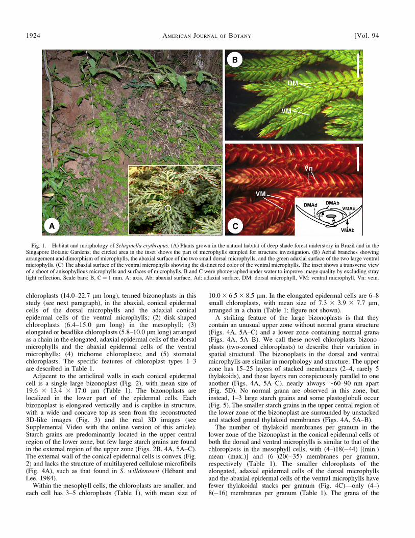
Two rows of small dorsal microphylls and two rows of largeventral microphylls occur on each branch of Selaginellaerythropus (Fig. 1). The abaxial surface of the dorsalmicrophyll and the adaxial surface of the ventral microphyllare green (Fig. 1B). No iridescent blue color was observed inthe plants cultivated in the Singapore Botanic Gardens or in thenatural habitats of the species (Fig. 1A, B). The adaxial surfaceof the dorsal microphylls, which cannot be easily viewed fromeither the dorsal or ventral surface of the shoot, is green exceptfor the red margins. The abaxial surface of all ventralmicrophylls on the branches is red on the plants used in ourinvestigation (Fig. 1C), although the upper branches may varyfrom red to green in natural habitats (Valdespino, 1993).
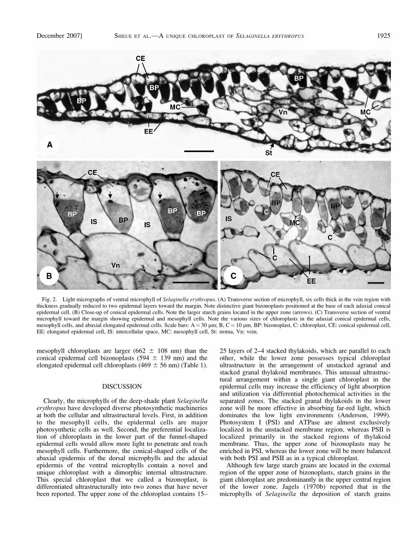
Dorsal and ventral microphylls of S. erythropus are six cellsthick in the vein region, with microphyll thickness graduallyreduced to two layers (the upper and lower epidermis) towardthe margin (Fig. 2). Five types of chloroplasts, based on sizeand number, can be recognized from the dorsal and ventralmicrophylls in association with different cell types: (1) cuplike
December 2007] SHEUE ET AL.—A UNIQUE CHLOROPLAST OF SELAGINELLA ERYTHROPUS 1923
chloroplasts (14.0–22.7 lm long), termed bizonoplasts in thisstudy (see next paragraph), in the abaxial, conical epidermalcells of the dorsal microphylls and the adaxial conicalepidermal cells of the ventral microphylls; (2) disk-shapedchloroplasts (6.4–15.0 lm long) in the mesophyll; (3)elongated or beadlike chloroplasts (5.8–10.0 lm long) arrangedas a chain in the elongated, adaxial epidermal cells of the dorsalmicrophylls and the abaxial epidermal cells of the ventralmicrophylls; (4) trichome chloroplasts; and (5) stomatalchloroplasts. The specific features of chloroplast types 1–3are described in Table 1.
Adjacent to the anticlinal walls in each conical epidermalcell is a single large bizonoplast (Fig. 2), with mean size of19.6 3 13.4 3 17.0 lm (Table 1). The bizonoplasts arelocalized in the lower part of the epidermal cells. Eachbizonoplast is elongated vertically and is cuplike in structure,with a wide and concave top as seen from the reconstructed3D-like images (Fig. 3) and the real 3D images (seeSupplemental Video with the online version of this article).Starch grains are predominantly located in the upper centralregion of the lower zone, but few large starch grains are foundin the external region of the upper zone (Figs. 2B, 4A, 5A–C).The external wall of the conical epidermal cells is convex (Fig.2) and lacks the structure of multilayered cellulose microfibrils(Fig. 4A), such as that found in S. willdenowii (Hebant andLee, 1984).
Within the mesophyll cells, the chloroplasts are smaller, andeach cell has 3–5 chloroplasts (Table 1), with mean size of
10.0 3 6.5 3 8.5 lm. In the elongated epidermal cells are 6–8small chloroplasts, with mean size of 7.3 3 3.9 3 7.7 lm,arranged in a chain (Table 1; figure not shown).
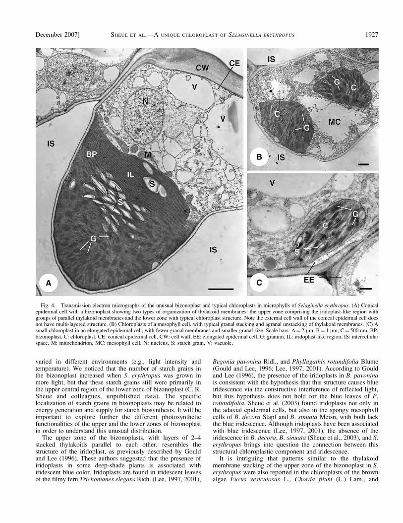
A striking feature of the large bizonoplasts is that theycontain an unusual upper zone without normal grana structure(Figs. 4A, 5A–C) and a lower zone containing normal grana(Figs. 4A, 5A–B). We call these novel chloroplasts bizono-plasts (two-zoned chloroplasts) to describe their variation inspatial structural. The bizonoplasts in the dorsal and ventralmicrophylls are similar in morphology and structure. The upperzone has 15–25 layers of stacked membranes (2–4, rarely 5thylakoids), and these layers run conspicuously parallel to oneanother (Figs. 4A, 5A–C), nearly always ;60–90 nm apart(Fig. 5D). No normal grana are observed in this zone, butinstead, 1–3 large starch grains and some plastoglobuli occur(Fig. 5). The smaller starch grains in the upper central region ofthe lower zone of the bizonoplast are surrounded by unstackedand stacked granal thylakoid membranes (Figs. 4A, 5A–B).
The number of thylakoid membranes per granum in thelower zone of the bizonoplast in the conical epidermal cells ofboth the dorsal and ventral microphylls is similar to that of thechloroplasts in the mesophyll cells, with (4–)18(�44) [(min.)mean (max.)] and (6–)20(�35) membranes per granum,respectively (Table 1). The smaller chloroplasts of theelongated, adaxial epidermal cells of the dorsal microphyllsand the abaxial epidermal cells of the ventral microphylls havefewer thylakoidal stacks per granum (Fig. 4C)—only (4–)8(�16) membranes per granum (Table 1). The grana of the
Fig. 1. Habitat and morphology of Selaginella erythropus. (A) Plants grown in the natural habitat of deep-shade forest understory in Brazil and in theSingapore Botanic Gardens; the circled area in the inset shows the part of microphylls sampled for structure investigation. (B) Aerial branches showingarrangement and dimorphism of microphylls, the abaxial surface of the two small dorsal microphylls, and the green adaxial surface of the two large ventralmicrophylls. (C) The abaxial surface of the ventral microphylls showing the distinct red color of the ventral microphylls. The inset shows a transverse viewof a shoot of anisophyllous microphylls and surfaces of microphylls. B and C were photographed under water to improve image quality by excluding straylight reflection. Scale bars: B, C¼ 1 mm. A: axis, Ab: abaxial surface, Ad: adaxial surface, DM: dorsal microphyll, VM: ventral microphyll, Vn: vein.
1924 AMERICAN JOURNAL OF BOTANY [Vol. 94
mesophyll chloroplasts are larger (662 6 108 nm) than theconical epidermal cell bizonoplasts (594 6 139 nm) and theelongated epidermal cell chloroplasts (469 6 56 nm) (Table 1).
DISCUSSION
Clearly, the microphylls of the deep-shade plant Selaginellaerythropus have developed diverse photosynthetic machineriesat both the cellular and ultrastructural levels. First, in additionto the mesophyll cells, the epidermal cells are majorphotosynthetic cells as well. Second, the preferential localiza-tion of chloroplasts in the lower part of the funnel-shapedepidermal cells would allow more light to penetrate and reachmesophyll cells. Furthermore, the conical-shaped cells of theabaxial epidermis of the dorsal microphylls and the adaxialepidermis of the ventral microphylls contain a novel andunique chloroplast with a dimorphic internal ultrastructure.This special chloroplast that we called a bizonoplast, isdifferentiated ultrastructurally into two zones that have neverbeen reported. The upper zone of the chloroplast contains 15–
25 layers of 2–4 stacked thylakoids, which are parallel to eachother, while the lower zone possesses typical chloroplastultrastructure in the arrangement of unstacked agranal andstacked granal thylakoid membranes. This unusual ultrastruc-tural arrangement within a single giant chloroplast in theepidermal cells may increase the efficiency of light absorptionand utilization via differential photochemical activities in theseparated zones. The stacked granal thylakoids in the lowerzone will be more effective in absorbing far-red light, whichdominates the low light environments (Anderson, 1999).Photosystem I (PSI) and ATPase are almost exclusivelylocalized in the unstacked membrane region, whereas PSII islocalized primarily in the stacked regions of thylakoidmembrane. Thus, the upper zone of bizonoplasts may beenriched in PSI, whereas the lower zone will be more balancedwith both PSI and PSII as in a typical chloroplast.
Although few large starch grains are located in the externalregion of the upper zone of bizonoplasts, starch grains in thegiant chloroplast are predominantly in the upper central regionof the lower zone. Jagels (1970b) reported that in themicrophylls of Selaginella the deposition of starch grains
Fig. 2. Light micrographs of ventral microphyll of Selaginella erythropus. (A) Transverse section of microphyll, six cells thick in the vein region withthickness gradually reduced to two epidermal layers toward the margin. Note distinctive giant bizonoplasts positioned at the base of each adaxial conicalepidermal cell. (B) Close-up of conical epidermal cells. Note the larger starch grains located in the upper zone (arrows). (C) Transverse section of ventralmicrophyll toward the margin showing epidermal and mesophyll cells. Note the various sizes of chloroplasts in the adaxial conical epidermal cells,mesophyll cells, and abaxial elongated epidermal cells. Scale bars: A¼ 30 lm; B, C¼ 10 lm. BP: bizonoplast, C: chloroplast, CE: conical epidermal cell,EE: elongated epidermal cell, IS: intercellular space, MC: mesophyll cell, St: stoma, Vn: vein.
December 2007] SHEUE ET AL.—A UNIQUE CHLOROPLAST OF SELAGINELLA ERYTHROPUS 1925
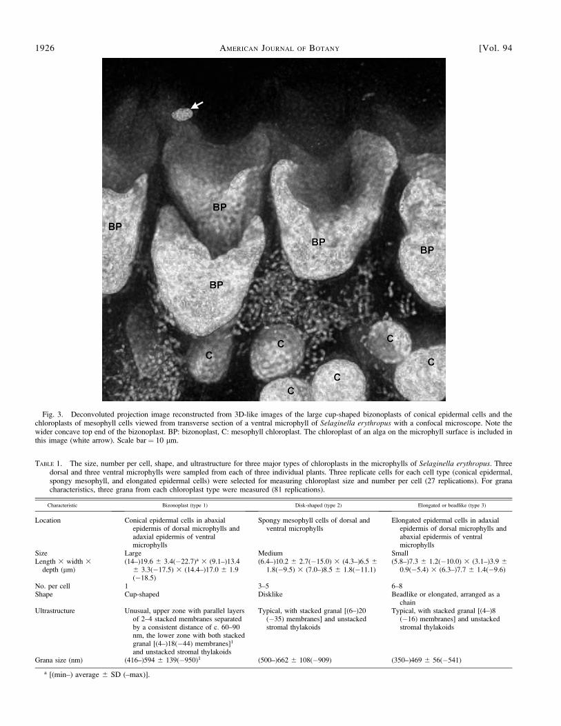
Fig. 3. Deconvoluted projection image reconstructed from 3D-like images of the large cup-shaped bizonoplasts of conical epidermal cells and thechloroplasts of mesophyll cells viewed from transverse section of a ventral microphyll of Selaginella erythropus with a confocal microscope. Note thewider concave top end of the bizonoplast. BP: bizonoplast, C: mesophyll chloroplast. The chloroplast of an alga on the microphyll surface is included inthis image (white arrow). Scale bar ¼ 10 lm.
TABLE 1. The size, number per cell, shape, and ultrastructure for three major types of chloroplasts in the microphylls of Selaginella erythropus. Threedorsal and three ventral microphylls were sampled from each of three individual plants. Three replicate cells for each cell type (conical epidermal,spongy mesophyll, and elongated epidermal cells) were selected for measuring chloroplast size and number per cell (27 replications). For granacharacteristics, three grana from each chloroplast type were measured (81 replications).
Characteristic Bizonoplast (type 1) Disk-shaped (type 2) Elongated or beadlike (type 3)
Location Conical epidermal cells in abaxialepidermis of dorsal microphylls andadaxial epidermis of ventralmicrophylls
Spongy mesophyll cells of dorsal andventral microphylls
Elongated epidermal cells in adaxialepidermis of dorsal microphylls andabaxial epidermis of ventralmicrophylls
Size Large Medium SmallLength 3 width 3
depth (lm)(14–)19.6 6 3.4(�22.7)a 3 (9.1–)13.4
6 3.3(�17.5) 3 (14.4–)17.0 6 1.9(�18.5)
(6.4–)10.2 6 2.7(�15.0) 3 (4.3–)6.5 61.8(�9.5) 3 (7.0–)8.5 6 1.8(�11.1)
(5.8–)7.3 6 1.2(�10.0) 3 (3.1–)3.9 60.9(�5.4) 3 (6.3–)7.7 6 1.4(�9.6)
No. per cell 1 3–5 6–8Shape Cup-shaped Disklike Beadlike or elongated, arranged as a
chainUltrastructure Unusual, upper zone with parallel layers
of 2–4 stacked membranes separatedby a consistent distance of c. 60–90nm, the lower zone with both stackedgranal [(4–)18(�44) membranes]1
and unstacked stromal thylakoids
Typical, with stacked granal [(6–)20(�35) membranes] and unstackedstromal thylakoids
Typical, with stacked granal [(4–)8(�16) membranes] and unstackedstromal thylakoids
Grana size (nm) (416–)594 6 139(�950)1 (500–)662 6 108(�909) (350–)469 6 56(�541)
a [(min–) average 6 SD (–max)].
1926 AMERICAN JOURNAL OF BOTANY [Vol. 94
varied in different environments (e.g., light intensity andtemperature). We noticed that the number of starch grains inthe bizonoplast increased when S. erythropus was grown inmore light, but that these starch grains still were primarily inthe upper central region of the lower zone of bizonoplast (C. R.Sheue and colleagues, unpublished data). The specificlocalization of starch grains in bizonoplasts may be related toenergy generation and supply for starch biosynthesis. It will beimportant to explore further the different photosyntheticfunctionalities of the upper and the lower zones of bizonoplastin order to understand this unusual distribution.
The upper zone of the bizonoplasts, with layers of 2–4stacked thylakoids parallel to each other, resembles thestructure of the iridoplast, as previously described by Gouldand Lee (1996). These authors suggested that the presence ofiridoplasts in some deep-shade plants is associated withiridescent blue color. Iridoplasts are found in iridescent leavesof the filmy fern Trichomanes elegans Rich. (Lee, 1997, 2001),
Begonia pavonina Ridl., and Phyllagathis rotundifolia Blume(Gould and Lee, 1996; Lee, 1997, 2001). According to Gouldand Lee (1996), the presence of the iridoplasts in B. pavoninais consistent with the hypothesis that this structure causes blueiridescence via the constructive interference of reflected light,but this hypothesis does not hold for the blue leaves of P.rotundifolia. Sheue et al. (2003) found iridoplasts not only inthe adaxial epidermal cells, but also in the spongy mesophyllcells of B. decora Stapf and B. sinuata Meisn, with both lackthe blue iridescence. Although iridoplasts have been associatedwith blue iridescence (Lee, 1997, 2001), the absence of theiridescence in B. decora, B. sinuata (Sheue et al., 2003), and S.erythropus brings into question the connection between thisstructural chloroplastic component and iridescence.
It is intriguing that patterns similar to the thylakoidmembrane stacking of the upper zone of the bizonoplast in S.erythropus were also reported in the chloroplasts of the brownalgae Fucus vesiculosus L., Chorda filum (L.) Lam., and
Fig. 4. Transmission electron micrographs of the unusual bizonoplast and typical chloroplasts in microphylls of Selaginella erythropus. (A) Conicalepidermal cell with a bizonoplast showing two types of organization of thylakoid membranes: the upper zone comprising the iridoplast-like region withgroups of parallel thylakoid membranes and the lower zone with typical chloroplast structure. Note the external cell wall of the conical epidermal cell doesnot have multi-layered structure. (B) Chloroplasts of a mesophyll cell, with typical granal stacking and agranal unstacking of thylakoid membranes. (C) Asmall chloroplast in an elongated epidermal cell, with fewer granal membranes and smaller granal size. Scale bars: A¼ 2 lm, B¼ 1 lm, C¼ 500 nm. BP:bizonoplast, C: chloroplast, CE: conical epidermal cell, CW: cell wall, EE: elongated epidermal cell, G: granum, IL: iridoplast-like region, IS: intercellularspace, M: mitochondrion, MC: mesophyll cell, N: nucleus, S: starch grain, V: vacuole.
December 2007] SHEUE ET AL.—A UNIQUE CHLOROPLAST OF SELAGINELLA ERYTHROPUS 1927
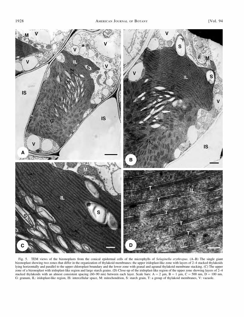
Fig. 5. TEM views of the bizonoplasts from the conical epidermal cells of the microphylls of Selaginella erythropus. (A–B) The single giantbizonoplast showing two zones that differ in the organization of thylakoid membranes: the upper iridoplast-like zone with layers of 2–4 stacked thylakoidslying horizontally and parallel to the upper chloroplast boundary and the lower zone with granal and agranal thylakoid membrane stacking. (C) The upperzone of a bizonoplast with iridoplast-like region and large starch grains. (D) Close-up of the iridoplast-like region of the upper zone showing layers of 2–4stacked thylakoids with an almost consistent spacing (60–90 nm) between each layer. Scale bars: A ¼ 2 lm, B ¼ 1 lm, C ¼ 500 nm, D ¼ 100 nm.G: granum, IL: iridoplast-like region, IS: intercellular space, M: mitochondrion, S: starch grain, T: a group of thylakoid membranes, V: vacuole.
1928 AMERICAN JOURNAL OF BOTANY [Vol. 94
Giffordia spp. (Bouck, 1965). This pattern is also similar tothose of the iridoplasts mentioned earlier (Gould and Lee,1996; Lee, 1997; Sheue et al., 2003), suggesting that thefunction and evolutionary implications of this unusual structurein these distantly related organisms (brown algae, spike moss,fern and flowering plants) need to be studied further.
The size, number, and ultrastructure of chloroplasts of S.erythropus (including the dimension and number of stacks ofthylakoids per granum) are distinctively different in the conicalepidermial cells, mesophyll cells, and elongated epidermalcells. This pattern of differences is very similar to that reportedfor the fern Teratophyllum rotundifoliatum (Nasrulhaq-Boyceand Duckett, 1991) and somewhat similar to the resultsobtained with other Selaginella species (Jagels, 1970a, b). In S.apoda, S. martensii, S. serpens, and S. uninata, however, thedimensions of the grana in the small discoid and cup-shapedchloroplasts are virtually identical (Jagels, 1970b).
In summary, this study clearly demonstrated that thechloroplasts in the microphylls of S. erythropus possess aneven greater diversity (cup-shaped bizonoplast, disklikemesophyll chloroplast, elongated or beadlike chloroplast inchains, trichome chloroplast, and stomatal chloroplast) thanthat reported by Haberladt (1888). The diversification inchloroplast structure in different cell types and in ultrastructurewithin a chloroplast in the deep-shade plant S. erythropus mayrepresent evolutionary changes in photosynthetic functionalityin adaptation to low-light environments. The giant anddistinctive bizonoplasts of S. erythropus could provide us withnovel plant material to further understand the evolutionarydevelopment of chloroplasts in a light-limited environment.
LITERATURE CITED
ANDERSON, J., D. GOODCHILD, AND N. K. BOARDMAN. 1973. Composition ofthe photosystems and chloroplast structure in extreme shade plants.Biochimica et Biophysica Acta 325: 573–585.
ANDERSON, J. M. 1999. Insights into the consequences of grana stacking ofthylakoid membranes in vascular plants: a personal perspective.Australian Journal of Plant Physiology 26: 625–639.
BOARDMAN, N. K. 1977. Comparative photosynthesis of sun and shadeplants. Annual Review of Plant Physiology 28: 355–377.
BOLD, H. C., C. J. ALEXOPOULOS, AND T. DELEVORYAS. 1987. Morphology ofplants and fungi, 5th ed. Harper & Row, New York, New York, USA.
BOUCK, G. B. 1965. Fine structure and organelle associations in brownalgae. The Journal of Cell Biology 26: 523–537.
DUCKETT, J. G., J. BURCH, P. W. FLETCHER, H. W. MATCHAM, D. J. READ, A.J. RUSSELL, AND S. PRESSEL. 2004. In vitro cultivation of bryophytes: areview of practicalities, problems, progress and promise. Journal ofBryology 26: 3–20.
DUCKETT, J. G., AND R. LIGRONE. 1993. Massive grana and stromal crystalsin the chloroplasts of an extreme-shade liverwort Dumortiera hirsuta(W.) Nees (Marchantiales, Hepaticae). Giornale Botanico Italiano127: 320–322.
DUCKETT, J. G., AND R. LIGRONE. 2006a. Cyathodium Kunze (Cyathodia-
ceae: Marchantiales), a tropical liverwort genus and family new toEurope, in southern Italy. Journal of Bryology 28: 88–96.
DUCKETT, J. G., AND R. LIGRONE. 2006b. Novel features of the plastids insome deep-shade, antipodean thalloid. Cryptogamie, Bryologie 27:75–83.
DUCKETT, J. G., AND K. S. RENZAGLIA. 1988. Ultrastructure anddevelopment of plastids in bryophytes. Advances in Bryology 3:33–93.
GEITLER, L. 1937. Uber den Granabau der Plastiden. Planta 26: 463–469.GLIME, J. M. 2006. Bryophyte ecology, vol. I. Physiological ecology. E-
book sponsored by Michigan Technological University and theInternational Association of Bryologists, Website http://www.bryoecol.mtu.edu/ [accessed 10 June 2007].
GOULD, K. S., AND D. W. LEE. 1996. Physical and ultrastructural basis ofblue leaf iridescence in four Malaysian understory plants. AmericanJournal of Botany 83: 45–50.
HABERLANDT, G. 1888. Die Chlorophyllkorper der Selaginellen. Flora 71:291–308.
HEBANT, C., AND D. W. LEE. 1984. Ultrastructural and developmentalcontrol of iridescence in Selaginella leaves. American Journal ofBotany 71: 216–219.
JAGELS, R. 1970a. Photosynthetic apparatus in Selaginella. I. Morphologyand photosynthesis under different light and temperature regimes.Canadian Journal of Botany 48: 1843–1852.
JAGELS, R. 1970b. Photosynthetic apparatus in Selaginella. II. Changes inplastid ultrastructure and pigment content under different lightregimes. Canadian Journal of Botany 48: 1853–1860.
JERMY, A. C. 1990. Selaginellaceae. In K. U. Kramer and P. S. Green[eds.], The families and genera of vascular plants, vol. I.Pteridophytes and gymnosperms, 39–45. Springer-Verlag, Berlin,Germany.
LEE, D. W. 1997. Iridescent blue plants. American Science 85: 56–73.LEE, D. W. 2001. Leaf colour in tropical plants: some progress and much
mystery. Malayan Nature Journal 55: 117–131.MAKGOMOL, K., AND E. SHEFFIELD. 2001. Gametophyte morphology and
ultrastructure of the extremely deep shade fern Trichomanesspeciosum. New Phytologist 151: 243–253.
NASRULHAQ-BOYCE, A., AND J. G. DUCKETT. 1991. Dimorphic epidermalcell chloroplasts in the mesophyll-less leaves of an extreme-shadetropical fern, Teratophyllum routundifoliatum (R. Bonap.) Holtt.: alight and electron microscope study. New Phytologist 119: 433–444.
SARAFIS, V. 1998. Chloroplasts: a structural approach. Journal of PlantPhysiology 152: 248–264.
SHEUE, C. R., V. SARAFIS, H. Y. LIU, Y. P. YANG, AND R. KIEW. 2003.Structural study on leaves of super shade plants. Proceedings of the24th Republic of China Symposium on Microscopy, Taipei, Taiwan,National Defense University, 13–14 (Abstract).
SPURR, A. R. 1969. A low viscosity epoxy resin embedding medium forelectron microscopy. Journal of Ultrastructure Research 26: 31–43.
SVENSON, H. K. 1946. Vegetation of the coast of Ecuador and Peru and itsrelation to that of the Galapagos Islands. II. Catalogue of plants.American Journal of Botany 33: 335–426.
VALDESPINO, I. A. 1993. Notes on neotropical Selaginella (Selaginella-ceae), including new species from Panama. Brittonia 45: 315–327.
VAN SPRONSEN, E. A., V. SARAFIS, G. J. BRAKENHOFF, H. T. M. VAN DER
VOORT, AND N. NANNINGA. 1989. Three-dimensional structure of livingchloroplasts as visualized by confocal scanning laser microscopy.Protoplasma 148: 8–14.
December 2007] SHEUE ET AL.—A UNIQUE CHLOROPLAST OF SELAGINELLA ERYTHROPUS 1929
Top Related
Copyright © 2022 FDOKUMEN

