


Bahasa
Halaman
Hukum


BioMed CentralRetrovirology
ss
Open AcceResearchApoptosis resistance in HIV-1 persistently-infected cells is independent of active viral replication and involves modulation of the apoptotic mitochondrial pathwayPablo N Fernández Larrosa*1, Diego O Croci2, Diego A Riva3, Mariel Bibini1, Renata Luzzi1, Mónica Saracco1, Susana E Mersich3, Gabriel A Rabinovich2 and Liliana Martínez Peralta1Address: 1National Reference Center for AIDS, Department of Microbiology, School of Medicine, University of Buenos Aires, Buenos Aires, Argentina, 2Laboratory of Immunopathology, Institute of Biology and Experimental Medicine (IBYME), CONICET, Buenos Aires, Argentina and 3Laboratory of Virology, Department of Biochemistry, School of Exact and Natural Sciences, University of Buenos Aires, Buenos Aires, Argentina
Email: Pablo N Fernández Larrosa* - [email protected]; Diego O Croci - [email protected]; Diego A Riva - [email protected]; Mariel Bibini - [email protected]; Renata Luzzi - [email protected]; Mónica Saracco - [email protected]; Susana E Mersich - [email protected]; Gabriel A Rabinovich - [email protected]; Liliana Martínez Peralta - [email protected]
* Corresponding author
AbstractBackground: HIV triggers the decline of CD4+ T cells and leads to progressive dysfunction of cell-mediated immunity. Although an increased susceptibility to cell death occurs during the acute phaseof HIV infection, persistently-infected macrophages and quiescent T-cells seem to be resistant tocell death, representing a potential reservoir for virus production.
Results: Lymphoid (H9/HTLVIIIB and J1.1) and pro-monocytic (U1) HIV-1 persistently-infected celllines were treated with hydrogen peroxide (H2O2) and staurosporine (STS) for 24 h, andsusceptibility to apoptosis was evaluated and compared with uninfected counterparts (H9, Jurkatand U937 respectively). When exposed to different pro-apoptotic stimuli, all persistently-infectedcell lines showed a dramatic reduction in the frequency of apoptotic cells in comparison withuninfected cells. This effect was independent of the magnitude of viral replication, since theinduction of viral production in lymphoid or pro-monocytic cells by exposure to TNF-α or PMAdid not significantly change their susceptibility to H2O2- or STS-induced cell death. A mechanisticanalysis revealed significant diferences in mitochondrial membrane potential (MMP) and caspase-3activation between uninfected and persistently-infected cells. In addition, Western blot assaysshowed a dramatic reduction of the levels of pro-apototic Bax in mitochondria of persistently-infected cells treated with H2O2 or STS, but not in uninfected cells.
Conclusion: This study represents the first evidence showing that resistance to apoptosis inpersistently-infected lymphoid and monocytic cells is independent of active viral production andinvolves modulation of the mitochondrial pathway. Understanding this effect is critical to specificallytarget the persistence of viral reservoirs, and provide insights for future therapeutic strategies inorder to promote complete viral eradication.
Published: 8 February 2008
Retrovirology 2008, 5:19 doi:10.1186/1742-4690-5-19
Received: 11 October 2007Accepted: 8 February 2008
This article is available from: http://www.retrovirology.com/content/5/1/19
© 2008 Fernández Larrosa et al; licensee BioMed Central Ltd. This is an Open Access article distributed under the terms of the Creative Commons Attribution License (http://creativecommons.org/licenses/by/2.0), which permits unrestricted use, distribution, and reproduction in any medium, provided the original work is properly cited.
Page 1 of 12(page number not for citation purposes)

Retrovirology 2008, 5:19 http://www.retrovirology.com/content/5/1/19
BackgroundApoptosis represents a type of programmed cell death(PCD) occurring in various physiological and pathology-cal processes. The ability of a cell to undergo or resistapoptosis in response to viral infection is crucial in deter-mining the clinical outcome of the disease and its thera-peutic oportunities [1,2]. Human imunodeficiency virus(HIV) is the causative agent of acquired immunodefi-ciency syndrome (AIDS), which triggers the decline ofCD4+ T cells and leads to immune system dysfunction[3,4]. During HIV-1 infection, most apoptotic events pre-dominantly occur in uninfected bystander T cells throughindirect mechanisms, such as the Fas/Fas ligand andCXCR4/CD4-mediated pathways [5,6]. However, acutely-infected CD4+ T cells are susceptible to dying by apopto-sis, by direct cell cytotoxicity induced by HIV replication,superantigen-induced cell death, immune-mediated kill-ing involving cytotoxic T-lymphocytes (CTL), antibody-dependent cell cytotoxicity (ADCC) or syncytia formation[7].
However, in some circumstances, HIV-infected cells donot seem to undergo apoptosis following infection andthese cells have been proposed to play an important roleas viral reservoirs. Persistently-infected pro-monocytic,but not lymphoid cell lines have been shown to be lesssensitive to several apoptotic stimuli when compared withtheir uninfected counterparts [8]. Besides, chronically-infected macrophages and quiescent T cells seem to beresistant to cell death, thus representing a potential reser-voir for viral production which might favor viral spread toother susceptible target cells [5,9,10]. The survival of pro-ductively-infected CD4+ lymphocytes or T cell lines wasfound to be influenced by viral proteins when exposed toapoptotic stimuli [11-13].
However, in spite of the relevance of these reservoir cellsin the control of viral persistence, the mechanismsresponsible of apoptosis resistance of persistently-infectedcells are not well understood. In particular, it is stillunclear whether resistance of infected cells to apoptoticstimuli involves modulation of active viral replication. Inthe present study, persistently-infected pro-monocyticand T-cell lines and their uninfected counterparts weretreated with H2O2 or STS. These apoptotic stimuli wereselected according to their ability to induce apoptosis viareactive oxygen species (ROS) [14] and protein kinase C(PKC) inhibition [15], which lead to an increase of oxida-tive stress. These stimuli generate a cell state which resem-bles the typical phenotype of cells undergoing active viralreplication and antiretroviral treatment [16,17]. Whentreated, all persistently-infected cells showed significantlylower frequency of apoptotic cells when compared withthose uninfected, independently of the magnitude of viralproduction. In addition, resistance to apoptosis induced
by HIV involved modulation of mitochondrial Baxexpression in persistently-infected cells.
ResultsHIV-1 persistently-infected cell lines are resistant to apoptosis induced by H2O2 and STSUninfected H9 and persistently-infected H9/HTLVIIIBcells were cultured with RPMI 1640 complete medium ina humidified atmosphere (5% CO2 in air) at 37°C andincubated with different concentrations of H2O2 and STS.Simultaneously, cells were incubated with medium aloneand used as controls. Cells were collected 24 h post-treat-ment, and apoptosis was evaluated by annexin-V/propid-ium iodide (PI) and APO-BrdU staining. Treatment with10 μM H2O2 induced 35% of annexin-V+/PI- H9 cells, and15% of annexin-V+/PI- infected H9/HTLVIIIB cells (P <0.01), whereas 20 μM H2O2 induced massive death inboth cell lines, characterized by predominant necrosis(60–65%) and lower levels of apoptosis (18–20%) (Fig-ure 1A). On the other hand, treatment with 0.1 μM STSinduced 43% of apoptotic H9 cells, whereas the frequencyof annexin-V+/PI- cells was only 15% in the infected H9/HTLVIIIB cells (P < 0.01). These differences were alsoobserved when concentrations of 1 μM STS were used topromote cell death (Figure 1A). Furthermore, differencesin the levels of apoptosis between infected and uninfectedcells were confirmed when cells were exposed to 10 μMH2O2 or 0.1 μM STS and stained with APO-BrdU andHoechst 33324 (Figure 1B).
Cell viability was assessed by the MTT assay and absorb-ances were measured at 540 nm, normalized against con-trols (Ctrl) and expressed as percentages. The percentageof viable cells was found to be 34% when H9 cells weretreated with 10 μM H2O2, but reached percentages of 50%in H9/HTLVIIIB cells. Furthermore, treatment with 0.1 μMSTS showed a decrease of MTT levels up to 48% and 64%in H9 and H9/HTLVIIIB cells respectively (data notshown). MTT was also assessed in both cell lines treatedwith 0.1 μM STS for 24, 48 and 72 h, indicating differ-ences in cell viability of both cell lines that were still sig-nificant until day 3 post-treatment (Figure 1C).
In order to investigate the association between apoptosisand viral production in H9/HTLVIIIB cells, p24 antigen,viral load and production of infectious viral particles werequantified. The magnitude of decrease of p24 antigen pro-duction observed was 80% (119 ng/ml), 75% (189 ng/ml), 78% (312 ng/ml) and 23% (114 ng/ml), when H9/HTLBIIIB cells were treated with 10 μM H2O2, 20 μMH2O2, 0.1 μM STS and 1 μM STS respectively and com-pared with controls (H2O2: 254 ng/ml; STS: 398 ng/ml)(Figure 1D). Viral load values were similar to p24 antigenlevels (data not shown). Infectious virus titres were also in
Page 2 of 12(page number not for citation purposes)

Retrovirology 2008, 5:19 http://www.retrovirology.com/content/5/1/19
agreement with p24 levels when cells were treated withapoptosis inducers (Figure 1D).
To examine whether apoptosis resistance in persistently-infected cells was dependent of the nature of the cell linestested, experiments were carried out using the lymphoidJurkat T-cell line and its infected counterpart (J1.1), or in
the pro-monocytic U937 cell line and its infected U1counterpart. The percentage of annexin-V+/PI- cells was30% and 47% when Jurkat T cells were treated with 10 μMH2O2 and 0.1 μM STS, respectively, and only 8% and 6%for J1.1 cells exposed to these apoptotic stimuli (Figure2A). Regarding the pro-monocytic U937 cell line and itsinfected counterpart U1, an important increase was
Apoptosis resistance in HIV persistently-infected H9/HTLVIIIBcells in comparison with non-infected H9 cellsFigure 1Apoptosis resistance in HIV persistently-infected H9/HTLVIIIBcells in comparison with non-infected H9 cells. A) H9 and H9/HTLVIIIB cells were treated with different concentrations of H2O2 or STS or complete medium as control. After 24 h, cells were harvested and annexin-V/PI staining was performed. The percentages of annexin-V+, PI- or PI+ cells are shown. B) (a) Analysis by APO-BrdU labeling by flow cytometry. The corresponding histograms and the percentages of APO-BrdU+
cells are shown; (b) Analysis of apoptosis with Hoechst 33324 by fluorescence microscopy. Micrographs (100×) of predomi-nant Hoechst stained nuclei are depicted. C) H9 and H9/HTLVIIIB cells were treated with 0.1 μM STS or complete medium as control, and cells were harvested 24, 48 and 72 h post-treatment. Cell viabillity was analyzed by the MTT assay. Absorbances from treated samples were normalized to 100% of untreated controls. D) Cells treated with H2O2 or STS or complete medium for 24 h were pelleted and the supernatants were used to quantify infective viral (grey bar) and p24 antigen (red line) production.
Page 3 of 12(page number not for citation purposes)

Retrovirology 2008, 5:19 http://www.retrovirology.com/content/5/1/19
observed in the percentage of annexin-V+/PI- cells (45%)in U937 cells treated with 0.1 μM STS, but only 8.2% inU1 infected cells. Remarkably, no significant differenceswere observed in the frequency of apoptosis when cellswere treated with 10 μM H2O2 (Figure 2B). However, ahigher concentration of H2O2 (50 μM) was capable ofinducing 34% of annexin-V+ U937 cells, and only 16.5%of dead cells in the infected U1 cell lines (data notshown). This result could be explained by the fact thatpro-monocytic cells are substantially less susceptible toexperience damage by ROS [18]. Thus, lymphoid and pro-monocytic HIV-1 persistently-infected cell lines are lesssusceptible to apoptosis induced by H2O2 or STS treat-ment compared with their uninfected counterparts.
Apoptosis resistance of HIV-infected cell lines is independent of the magnitude of viral productionUnlike H9/HTLVIIIB, J1.1 and U1 cell lines are non-pro-ductive cells unless treated with a viral activator. To inves-tigate the differential sensitivity to apoptosis of infected
cells under conditions of active viral replication, Jurkatand J1.1 cells were treated with 1000 U/ml tumor necrosisfactor-α (TNF-α) for 48 h and U937 and U1 cells weretreated with 100 ng/ml phorbol-12-myristate-13-acetate(PMA) for 24 h. Active viral production was confirmed bydetermining the p24 antigen at different days post-treat-ment. TNF-α treatment induced 100-fold viral reactiva-tion at 48 h with respect to untreated cells, while U1 cellsshowed 50-fold and 200-fold increase of viral productionat 24 h and 48 h, respectively, when cultured with PMA(Table I). Under these conditions, the percentage ofannexin-V+/PI- cells was 48% and 52% when Jurkat cellswere treated with 10 μM H2O2 and 0.1 μM STS, respec-tively, and only 12% for J1.1 cells exposed to both apop-totic stimuli (Figure 2A).
Regarding the pro-monocytic cell lines, when these cellswere pre-incubated with PMA, the frequency of earlyapoptotic cells was significantly increased in both celllines treated with STS: 72% in U937 and 30% in U1 cells
Apoptosis resistance is independent of the magnitude of viral replicationFigure 2Apoptosis resistance is independent of the magnitude of viral replication. A) Jurkat and J1.1 cells were incubated in the presence or absence of 1000 U/ml TNF-α for 48 h, and then treated with 10 μM H2O2 or 0.1 μM STS. The percentages of annexin-V+, PI- or PI+ cells are shown. B) U937 and U1 cells were incubated in the presence or absence of 100 ng/ml PMA for 24 h, and then exposed to 10 μM H2O2 or 0.1 μM STS. The percentages of annexin-V+, PI- or PI+ cells are shown.
Table 1: P24 production in HIV-1 persistently-infected cell lines exposed to TNF-α or PMA
Cell line and treatment 0 h 24 h 48 h
J1.1 Cells 14.07 ± 0.01 14.14 ± 0.05 18.26 ± 0.80J1.1 Cells + TNF α 14.08 ± 0.01 12.86 ± 0.90 152.04 ± 1.50
U1 Cells 1.06 ± 0.03 1.60 ± 0.03 4.18 ± 1.11U1 Cells + PMA 1.05 ± 0.03 51.96 ± 9.20 191.76 ± 0.48
HIV-1 persistently-infected lymphoid J1.1 and monocytic U1 cells were treated with TNF-α and PMA respectively in order to stimulate viral replication. P24 antigen was determined at 0, 24 and 48 h in cell supernatants by ELISA (HIVAG-1 Monoclonal, Abbot Laboratories). Values correspond to p24 antigen per 200,000 cells, expressed in J1.1 as pg/ml and in U1 Cells as ng/ml.
Page 4 of 12(page number not for citation purposes)

Retrovirology 2008, 5:19 http://www.retrovirology.com/content/5/1/19
(Figure 2B). Control cells showed also higher levels ofapoptosis when pre-incubated with PMA. It should beemphasized that PMA, independently of its ability tostimulate viral replication, can also induce cell differenti-ation, an effect which can influence the susceptibility toapoptosis [19]. These data suggest that apoptosis resist-ance in persistently-infected cell lines is independent ofthe magnitude of viral replication.
Apoptosis resistance of HIV persistenly-infected cell lines involves modulation of the mitochondrial pathwayIn order to dissect the mechanisms involved in this pro-tective effect, uninfected or persistently-infected cell linestreated or not with apoptotic stimuli were used to analyzedifferent apoptotic pathways. First, the anti-Fas activatingantibody CH11 was used to induce apoptosis by theextrinsic pathway in H9 and H9/HTLVIIIB cells, and Jurkatand J1.1 cells. No significant differences were observedbetween uninfected and persistently-infected cells (Figure
3A–B). As Fas/CD95 expression was found to be modu-lated by HIV-1 [5], we examined cell surface expression ofFas antigen in order to check whether our results could bedue to differential expression of this receptor. Flow cytom-etry analysis revealed no significant differences of Fasexpression among all cell lines tested (Figure 3C). Thus,HIV-1 persistent infection does not seem to modulate thesusceptibility to apoptosis by controlling the extrinsicpathway.
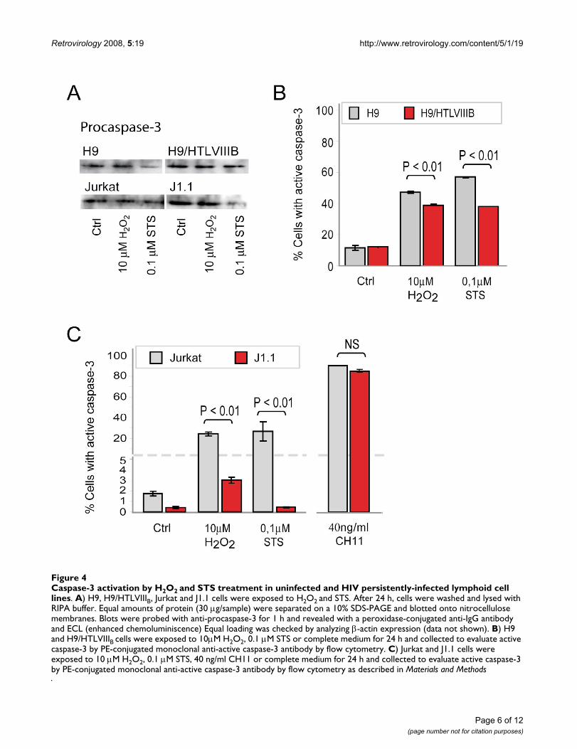
To gain insight into the mechanistic basis of this effect, wenext analyzed events associated with the execution ofapoptosis. When procaspase-3 expression was evaluatedby Western blot analysis in H9 and H9/HTLVIIIB, or Jurkatand J1.1 cells, no significant differences were observed inuntreated controls. However, when treated with the pro-apoptotic agents, a decrease of procaspase-3 was observedin all the cases (Figure 4A). When cells were analyzed byflow cytometry, H9 cells were 57% and 47% positive for
Fas-mediated apoptosis in uninfected and HIV persistently-infected cellsFigure 3Fas-mediated apoptosis in uninfected and HIV persistently-infected cells. H9 and H9/HTLVIIIB (A) and Jurkat and J1.1 (B) cells were incubated with 20 g/ml or 40 ng/ml of CH11, an anti-Fas activating antibody. After 24 h, cells were washed and stained with annexin-V/PI. The percentages of annexin-V+, PI- or PI+ cells are shown. C) Fas/CD95 and CD4 cell surface expression was analyzed by flow cytometry on H9, H9/HTLVIIIB, Jurkat and J1.1 cells.
Page 5 of 12(page number not for citation purposes)
Retrovirology 2008, 5:19 http://www.retrovirology.com/content/5/1/19
Page 6 of 12(page number not for citation purposes)
Caspase-3 activation by H2O2 and STS treatment in uninfected and HIV persistently-infected lymphoid cell linesFigure 4Caspase-3 activation by H2O2 and STS treatment in uninfected and HIV persistently-infected lymphoid cell lines. A) H9, H9/HTLVIIIB, Jurkat and J1.1 cells were exposed to H2O2 and STS. After 24 h, cells were washed and lysed with RIPA buffer. Equal amounts of protein (30 μg/sample) were separated on a 10% SDS-PAGE and blotted onto nitrocellulose membranes. Blots were probed with anti-procaspase-3 for 1 h and revealed with a peroxidase-conjugated anti-IgG antibody and ECL (enhanced chemoluminiscence) Equal loading was checked by analyzing β-actin expression (data not shown). B) H9 and H9/HTLVIIIB cells were exposed to 10μM H2O2, 0.1 μM STS or complete medium for 24 h and collected to evaluate active caspase-3 by PE-conjugated monoclonal anti-active caspase-3 antibody by flow cytometry. C) Jurkat and J1.1 cells were exposed to 10 μM H2O2, 0.1 μM STS, 40 ng/ml CH11 or complete medium for 24 h and collected to evaluate active caspase-3 by PE-conjugated monoclonal anti-active caspase-3 antibody by flow cytometry as described in Materials and Methods

Retrovirology 2008, 5:19 http://www.retrovirology.com/content/5/1/19
active caspase-3 when treated with H2O2 and STS respec-tively, while H9/HTLVIIIB raised percentages of 39% and38% respectively (Figure 4B). Besides, Jurkat cells showedeven higher differences in caspase-3 activation than J1.1when treated with H2O2 (Jurkat: 24%; J1.1: 3%) and STS(Jurkat: 25.13% ;J1.1: 0.43%) (Figure 4C). Furthermore,when treated with 40 ng/ml CH11 anti-Fas antibody, thenumber of cells with active caspase-3 was similar in bothuninfected and persistently-infected cells (Figure 4C).Taken together, these results suggest that differences in thesusceptibility to apoptosis between infected and unin-fected cells can not be explained by defective caspase-3activation and that apoptosis modulation may be local-ized upstream of caspase-3.
To further understand this effect we analyzed events asso-ciated with the mitochondrial apoptotic pathway. For thispurpose, the mitochondrial membrane potential (MMP)was studied in cells treated with H2O2 or STS by JC-1(Mitoscreen, BD) staining and flow cytometry. When cellswere treated with 10 μM H2O2 or 0.1 μM STS for 24 h, H9and Jurkat cells showed higher MMP (H9: 45% with H2O2and 40% with STS; Jurkat: 23% with H2O2 and 64% withSTS) compared with H9/HTLVIIIB and J1.1 cells respec-tively (H9/HTLVIIIB: 30% with H2O2 and 26% with STS;J1.1: 8% with H2O2 and 3.6% with STS) (Figure 5A–B).
Finally, Bcl-2 and Bax expression of different uninfectedand persistently-infected cell lines was analyzed by West-ern blot of total cell lysates. Densitometric analysisrevealed no significant differences in Bcl-2 (25 kDa)expression levels between H9 and H9/HTLVIIIB cells,treated or not with H2O2 or STS. However, dimeric Bax(42 kDa) was decreased by ~40% (H2O2) and ~70% (STS)in H9/HTLVIIIB cells treated with pro-apoptotic stimuli incomparison with controls or uninfected H9 cells, whichreached values of only 20% (H2O2) or 40% (STS). Theoverall effect could be observed by analyzing the Bcl-2/Bax ratio, which estimates the anti-apoptotic/pro-apop-totic balance. When treated with pro-apoptotic stimuli,H9/HTLVIIIB cells (lanes 5 and 6) showed a higher Bcl-2/Bax ratio compared to H9 cells (lanes 2 and 3) (Figure5C). In order to confirm this observation, Bax dimeriza-tion and insertion in mitochondria was analyzed by West-ern blot from cytosolic and mitochondrial fractions.While the levels of Bax expression remained unaltered inthe cytosolic fraction of different uninfected or infectedcell lines, the levels of Bax increased substantially in themitochondrial fraction of uninfected cells treated withapoptotic agents. However, no significant differences wereobserved in persistently-infected cells when compared tocontrols (Figure 5D).
These results suggest that apoptosis resistance observed inpersistently-infected cells involves modulation of themitochondrial pathway.
Conclusions and DiscussionDuring the clinical course of HIV-1 infection, the deple-tion of the CD4+ T cell compartment is mainly explainedby apoptosis of uninfected cells due to indirect mecha-nisms including Fas/FasL interaction, syncytia formationand direct citotoxicity of soluble viral proteins such asgp120, Tat or Nef [5,7]. However, HIV-1 may survive in alatent status, mainly in macrophages, resting CD4+ quies-cent T cells and CD44high memory T cells [10,20-22].These cells appear to be less sensitive to death induced bya variety of apoptotic stimuli such as chronic stress [18],or the Fas/FasL (CD95L) system [23] independently ofviral cofactors. Therefore, when the chronic infection isestablished in macrophages or in memory T cells, thevirus may survive longer in these cells due to a variety ofcellular and viral factors [24,25].
Our data suggest that persistently-infected pro-monocyticand lymphocytic cells are less susceptible to undergoapoptosis when exposed to different apoptotic stimulisuch as H2O2 and STS, compared with uninfected cells.This protection from apoptosis is consistent with the factthat HIV-1 persistently-infected macrophages, quiescent Tcell and pro-monocytic cell lines were described to survivelonger [8,10,20-22]. Our study provides the first evidenceshowing that apoptosis resistance in persistently-infectedcell lines is independent of the magnitude of viral replica-tion. In spite of the fact that H9/HTLVIIIB cells producedvirus actively, while viral production in J1.1 or U1 wasinducible, all cell lines showed similar tendences in theirresistance to apoptosis when compared with their unin-fected counterparts.
Viral proteins are known to modulate cell surface levels ofFas and its ability to transduce death signals upon bindingits specific ligand [5]. However, similar expression of Fasantigen was found on the surface of the cells studied,whether infected or not. In addition, engagement of Fasby the stimulating CH11 antibody resulted in similar lev-els of apoptosis in the cell lines studied, suggesting thatHIV-1 infection does not modulate the extrinsic pathwayof cell death.
In addition, when the MMP was analyzed in cells treatedwith H2O2and STS, substantial differences in the induc-tion of apoptosis were observed between uninfected andpersistently-infected cells. This result might be explainedby the ability of H2O2 and STS to induce oxidative stress,thus priming cells to undergo apoptosis via the mitochon-drial pathway. These results are also consistent with thelevels of caspase-3 activation, indicating that once the
Page 7 of 12(page number not for citation purposes)

Retrovirology 2008, 5:19 http://www.retrovirology.com/content/5/1/19
Page 8 of 12(page number not for citation purposes)
MMP induction and Bcl-2 and Bax expression in uninfected or HIV persistently-infected cell linesFigure 5MMP induction and Bcl-2 and Bax expression in uninfected or HIV persistently-infected cell lines. H9 and H9/HTLVIIIB (A) and Jurkat and J1.1 (B) cells were exposed to 10 μM H2O2, 0.1 μM STS or complete medium for 24 h and har-vested to evaluate mitochondrial membrane potential (ΔΨm) by JC-1 staining by flow cytometry. C) H9 and H9/HTLVIIIB cells were exposed to H2O2 and STS. After 24 h, cells were washed and lysed with RIPA buffer. Equal amounts of protein (30 μg/sample) were separated by 10% SDS-PAGE and blotted onto nitrocellulose membranes. The blots were probed with anti-Bcl-2 and anti-Bax antibody, revealed using a peroxidase-conjugated anti-IgG and developed using a chemiluminiscence Western blotting detection reagent. Equal loading was checked by analyzing β-actin expression. Films were analyzed with Scion image analysis software (Scion, Frederick, MD) and the Bcl-2/Bax ratio was depicted. D) H9 and H9/HTLVIIIB cells were exposed to H2O2 and STS and after 24 h lysates from cytosolic and mitochondrial fractions were prepared by differential centrifugation. Equal amounts of protein (30 μg/sample) were separated by 10% SDS-PAGE and blotted onto nitrocellulose membranes. Blots were then probed with an anti-Bax polyclonal antibody, incubated with a peroxidase-conjugated anti-rabbit secondary antibody and developed using ECL detection reagent. Equal loading was checked by analyzing β-actin (cytosol fraction) and Complex I (mitochondrial fraction) expression.

Retrovirology 2008, 5:19 http://www.retrovirology.com/content/5/1/19
mitochondrial pore is induced, apoptosis events proceednormally. Thus, modulation of apoptosis might occurbefore or during pore induction. In order to analyze thepossible mechanisms involved in this effect, expression ofBcl-2 and Bax was analyzed in the cytosolic and mito-chondrial compartments. Bcl-2 expression did not showany significant difference between both cell lines, whetherthey were treated or not with pro-apoptotic stimuli. How-ever, expression of Bax was dramatically reduced in mito-chondria of persistently-infected cells when apoptosis wasinduced by exposure to H2O2 or STS.
It is now widely accepted that persistent HIV-1 infectionrepresents a new homeostatic state of the cell, which islikely promoted by the combination of both cellular andviral factors. Several viral proteins have been recognizedby their ability to induce apoptosis in infected or unin-fected cells, but some viral proteins can also protectagainst cell death [5]. Decreased caspase-3 activation [26]and p53 expression [27] were described as possible mech-anisms implicated in apoptosis resistance in HIV-1-per-sistently infected cells. This study provides novel evidenceshowing that resistance to apoptosis in persistently-infected cells involves direct modulation of the mitochon-drial pathway by regulating Bax pore induction. Furtherexperiments are needed in order to clarify the mechanismby which the virus decreases MMP and controls the execu-tion of apoptosis. Viral regulation of autophagy of dam-aged mitochondrias or Bax proteolysis might be potentialexplanatory mechanisms for our observations.
The survival of viral reservoirs is a great challenge to tackleregarding HIV eradication. Understanding the mechanis-tic bases of the resistance to apoptosis is essential to spe-cifically target the persistence of viral reservoirs and mightcontribute to provide insights for future therapeutic strat-egies in order to promote complete viral eradication.
Materials and MethodsCell linesThe following uninfected cell lines of human origin wereused: lymphocytic H9, Jurkat and promonocytic U937cell lines; and their respective HIV-1 persistently-infectedcell lines: H9/HTLVIIIB, J1.1 and U1. All cell lines wereprovided by the NIH AIDS Research and References Rea-gent Program, except for U937. Cell lines were culturedwith RPMI 1640 medium supplemented with 2 mM L-glutamine, 100 μg/ml streptomycin and 10% fetal calfserum at 37°C in a humidified atmosphere (5% CO2 inair).
Antibodies and reagentsAnnexin-V apoptosis kit, APO-BrdU apoptosis kit, activecaspase-3 antibody kit, JC-1 Mitoscreen, TNF-α, PE-conju-gated anti-CD95 and PerCP-conjugated anti-CD4 anti-
bodies were from BD Biosciences,(CA, USA). Anti-Bcl-2(DC21), anti-Bax (D21), anti-procaspase-3 (L-18), anti-β-actin (I-19) polyclonal antibodies and peroxidase-conju-gated anti-rabbit and anti-goat antibodies were fromSanta Cruz Biotechnology, (CA, USA). Anti-complex Iantibody was a generous gift from Dr. J. Poderoso (Hospi-tal de Clínicas, University of Buenos Aires). Anti-Fas acti-vating antibody (CH11) was from Upstate (New York,USA). Other reagents including Hoechst, MTT, PMA, stau-rosporine (STS), Kodak BioMax films were from Sigma(St. Louis, MO, USA). Hydrogen peroxide (H2O2) and iso-propanol were from Merck (New Jersey, USA). RPMI 1640medium, fetal calf serum, L-glutamine and streptomycinwere from Gibco (New York, USA). Micro-BCA proteinassay kit was from Pierce (Rockford, USA). Chemilumi-niscence Western blotting detection reagent and nitroce-lulose membranes were from Amersham Biosciences, UK.
Induction of HIV-1 productionIn order to induce viral production, J1.1 cells were incu-bated for 48 h with 1000 U/ml TNF-α [28] and U1 cellswere exposed to 100 ng/ml PMA for 24 h [29]. Cells werewashed twice with PBS and fresh medium was added tocarry out experiments. Viral production was confirmed byp24 antigen determination.
Determination of viral productionCells were pelleted and supernatants were used to quan-tify p24 antigen with a commercial ELISA kit (HIVAG-1monoclonal, Abbot Laboratorios, Illinois, USA), and viralload using a commercial assay (Quantiplex XTm HIV RNA3.0 Assay bDNA, Chiron Corp, CA, USA). Infective virustitration was performed by limiting dilution and syncytiaformation in MT-2 cells, and calculated by the Reed &Müench method.
Induction of apoptosisCells were collected, washed with PBS, resuspended withcomplete medium, and divided in a 24-well culture platewith a final cell concentration of 150,000 cells/ml. Forapoptosis induction, H2O2, STS and the CH11 Fas activat-ing antibody were used. Optimal concentrations forexperiments were standarized by testing different concen-trations, which ranged from 5 to 1000 μM (H2O2), from0.01 to 10 μM (STS) and from 20 to 40 ng/ml (CH11).Treated cells were always compared with untreated con-trols (Ctrl). In most experiments, cells were incubatedwith the apoptosis inducers for 24 h
MTT assayCell viability was determined by the MTT (3-[4,4-dimeth-ylthiazol-2-yl]-2,5-diphenyltetrazolium bromide) assay[30]. After 24 h of exposure to pro-apoptotic stimuli,medium was removed and cells were plated at 5 × 10⁴
cells/well in 96-well plates and incubated with 0.5 mg/ml
Page 9 of 12(page number not for citation purposes)

Retrovirology 2008, 5:19 http://www.retrovirology.com/content/5/1/19
MTT in RPMI-1640 without Red Phenol for 1 h at 37°C ina CO2 incubator. Cells were pelleted and formazan crys-tals were solubilized with 0.04 M HCl in isopropanol.Finally, the absorbance measured at 640 nm was sub-tracted from the absorbance at 540 nm. Each assay wasperformed in triplicate. Absorbances corresponding totreated samples were normalized to 100% of untreatedcontrols and expressed as percentages. In this assay, thenumber of surviving cells was directly correlated with theamount of formazan obtained.
Asssesment of apoptosisCells were incubated in the presence or absence of proap-optotic stimuli for 24 h, washed twice with PBS and thefrequency of apoptotic cells was analyzed by the followingmethods:
Annexin-V/PI stainingTo determine the percentage of early apoptotic cells, phos-phatidylserine (PS) cell translocation and plasma mem-brane permeability were evaluated by dual staining withFITC-conjugated annexin-V and propidium iodide (PI)using the Annexin-V/PI apoptosis detection kit (BD Bio-sciences) and analyzed by flow cytometry using a FACS-Canto (BD Biosciences). Annexin-V+/PI- cells representingearly apoptotic cells, and annexin-V+/PI+ mostly repre-senting necrotic cells were determined.
APO-BrdU stainingLate apoptotic cells were determined with the APO-BrdUkit by incorporation of bromodeoxyuridine triphosphate(Br-dUTP) to 3'-hydroxyl sites in cell DNA, and analyzedby flow cytometry in a FACSCanto (BD Bioscience).
Hoechst 33324 stainingApoptotic cells were determined by Hoechst staining andvisualized in a fluorescence microscope (Axiophot WestGermany).
Cytofluorimetric analysis of MMPAfter treatments with H2O2 and STS for 24 hours, cellswere collected and resuspended in PBS, and then stainedwith JC-1 (5,5',6,6'-tetrachloro-1,1',3,3'-tetraethylbenz-imidazolcarbocyanine iodide) (JC-1 Mitoscreen, BD) for15 min at 37°C in a CO2 incubator. Cells were pelleted,washed twice with buffer supplemented by the kit as indi-cated by the manufacter and analyzed on a flow cytometer(FACSCanto, BD Biosciences).
Cytofluorimetric analysis of caspase-3 activationTreated and control cells were pelleted and washed twicewith PBS and the percentage of cells with active
caspase-3 was assessed using the PE-conjugated mono-clonal active caspase-3 antibody kit (BD Pharmigen) and
analyzed on a FACSCanto flow cytometer (BD Bio-sciences).
Flow cytometry analysisIn all cases where flow cytometry was required, 20,000events were acquired in a FACSCanto flow cytometer (BDBiosciences) and different parameters were analyzedusing the WinMDI 2.8 software.
Isolation and purification of mitochondriaCells (1 × 10⁷ cells) incubated in the presence or absenceof pro-apoptotic stimuli were washed and homogenizedin MSHE (0.225 M mannitol, 0.07 M sucrose, 1 mMEGTA, and 25 mM HEPES/KOH; 1/10 w/v; pH 7.4) andcentrifuged at 5,500 × g for 10 min at 4°C. The resultantsupernatant was centrifuged at 15,000 × g for 20 min at4°C and the pellet was resuspended in 30 μl of MSHE(mitochondrial fraction) [31]. To remove broken mito-chondria, contaminating organelles, and debris from thecytosol fractions, the supernatants were further centri-fuged at 21,000 × g for 30 min at 4°C. Protein concentra-tion from cytosolic and mitochondrial fractions wasdetermined by the Micro-BCA protein assay kit (Pierce,Rockford, USA).
Western blot analysisAfter exposure to pro-apoptotic stimuli, cells were lysed inRIPA buffer containing 20 mM Tris-HCl, 150 mM NaCl,1% Triton X-100, 1% sodium deoxycholate, 2 mM EDTA,0.1% SDS and protease inhibitor cocktail. Protein concen-trations from total, cytosolic or mitochondrial lysateswere quantified using the Micro-BCA protein assay kit asdescribed above. Equal amounts of protein (30 μg/sam-ple) were separated in a 10% SDS-PAGE and blotted ontonitrocellulose membranes. Blots were then probed withanti-pro-caspase-3, anti-Bcl-2 or anti-Bax rabbit polyclo-nal antibodies as described [32], and incubation with per-oxidase-conjugated anti-IgG was performed in a blockingbuffer for 1 h. Blots were then developed using a chemilu-miniscence Western blotting detection reagent andexposed to X-ray films. Films were analyzed using theScion image analysis software (Scion, Frederick, MD).Total cell lysates were used to analyze pro-caspase-3, Bcl-2 and Bax expression and normalized with β-actin expres-sion. Cytosolic and mitochondrial extracts were used toanalyze the insection of Bax into mitochondria, and pro-tein bands were compared with the expression of β-actin(marker of cytosolic fraction) and Complex I (marker ofmitochondrial fraction).
Statistical analysisValues represent the mean ± s.e.m. of at least three inde-pendent experiments. Comparisons among groups wereperformed by using the Student's t test and One-wayANOVA using a SPSS 12.0 software.
Page 10 of 12(page number not for citation purposes)

Retrovirology 2008, 5:19 http://www.retrovirology.com/content/5/1/19
Competing interestsThe author(s) declare that they have no competing inter-ests.
Authors' contributionsPNFL was responsible for designing, performing and writ-ing the manuscript. DAR and SEM contributed to experi-ments of apoptosis by Hoechst and APOBrDU. DOC andGAR were responsible of experiments using Western blotanalysis, and contributed to writing of the manuscript.MB, RL and MS performed and interpreted the flowcytometry experiments. LMP was responsible for thedesign and writing of the manuscript. All authors read andapproved the final manuscript.
AcknowledgementsWe thank the AIDS Research and Reference Program (National Institute of Allergy and Infectious Diseases, NIH, USA) for the reagents used in this study. We are grateful with Dr. J. Poderoso for providing essential reagents and assisting with the procedures for mitochondria purification. This work was supported by grants from the University of Buenos Aires (M050) and the National Agency for Promotion of Science and Technology (PICT 05-11734) to L.M.P. and grants from Fundación Sales, National Agency for Pro-motion of Science and Technology (PICT 2003 05-13787) and University of Buenos Aires (M091) to G.A.R..
References1. Benedict CA, Norris PS, Ware CF: To kill or be killed: viral eva-
sion of apoptosis. Nature immunology 2002, 3(11):1013-1018.2. He B: Viruses, endoplasmic reticulum stress, and interferon
responses. Cell death and differentiation 2006, 13(3):393-403.3. Brenchley JM, Price DA, Douek DC: HIV disease: fallout from a
mucosal catastrophe? Nature immunology 2006, 7(3):235-239.4. Hel Z, McGhee JR, Mestecky J: HIV infection: first battle decides
the war. Trends in immunology 2006, 27(6):274-281.5. Gougeon ML: To kill or be killed: how HIV exhausts the
immune system. Cell death and differentiation 2005, 12 Suppl1:845-854.
6. Lelievre JD, Mammano F, Arnoult D, Petit F, Grodet A, Estaquier J,Ameisen JC: A novel mechanism for HIV1-mediatedbystander CD4+ T-cell death: neighboring dying cells drivethe capacity of HIV1 to kill noncycling primary CD4+ T cells.Cell death and differentiation 2004, 11(9):1017-1027.
7. Varbanov M, Espert L, Biard-Piechaczyk M: Mechanisms of CD4 T-cell depletion triggered by HIV-1 viral proteins. AIDS reviews2006, 8(4):221-236.
8. Pinti M, Biswas P, Troiano L, Nasi M, Ferraresi R, Mussini C, VecchietJ, Esposito R, Paganelli R, Cossarizza A: Different sensitivity toapoptosis in cells of monocytic or lymphocytic origin chron-ically infected with human immunodeficiency virus type-1.Experimental Biology and Medicine 2003, 228(11):1346-1354.
9. Balestra E, Perno CF, Aquaro S, Panti S, Bertoli A, Piacentini M, For-bici F, D'Arrigo R, Calio R, Garaci E: Macrophages: a crucial res-ervoir for human immunodeficiency virus in the body. Journalof biological regulators and homeostatic agents 2001, 15(3):272-276.
10. Zamborlini A, Lehmann-Che J, Clave E, Giron ML, Tobaly-Tapiero J,Roingeard P, Emiliani S, Toubert A, de The H, Saib A: Centrosomalpre-integration latency of HIV-1 in quiescent cells. Retrovirol-ogy 2007, 4:63.
11. Conti L, Matarrese P, Varano B, Gauzzi MC, Sato A, Malorni W,Belardelli F, Gessani S: Dual role of the HIV-1 vpr protein in themodulation of the apoptotic response of T cells. Journal ofImmunology 2000, 165(6):3293-3300.
12. Gibellini D, Caputo A, Celeghini C, Bassini A, La Placa M, Capitani S,Zauli G: Tat-expressing Jurkat cells show an increased resist-ance to different apoptotic stimuli, including acute humanimmunodeficiency virus-type 1 (HIV-1) infection. British jour-nal of haematology 1995, 89(1):24-33.
13. Mahlknecht U, Deng C, Lu MC, Greenough TC, Sullivan JL, O'BrienWA, Herbein G: Resistance to apoptosis in HIV-infected CD4+T lymphocytes is mediated by macrophages: role for Nef andimmune activation in viral persistence. Journal of Immunology2000, 165(11):6437-6446.
14. Dumont A, Hehner SP, Hofmann TG, Ueffing M, Droge W, SchmitzML: Hydrogen peroxide-induced apoptosis is CD95-inde-pendent, requires the release of mitochondria-derived reac-tive oxygen species and the activation of NF-kappaB.Oncogene 1999, 18(3):747-757.
15. Bloom DA, Jaiswal AK: Phosphorylation of Nrf2 at Ser40 byprotein kinase C in response to antioxidants leads to therelease of Nrf2 from INrf2, but is not required for Nrf2 sta-bilization/accumulation in the nucleus and transcriptionalactivation of antioxidant response element-mediatedNAD(P)H:quinone oxidoreductase-1 gene expression. TheJournal of biological chemistry 2003, 278(45):44675-44682.
16. Arnoult D, Petit F, Lelievre JD, Estaquier J: Mitochondria in HIV-1-induced apoptosis. Biochemical and biophysical research communica-tions 2003, 304(3):561-574.
17. Buenz EJ, Badley AD: Impact of mitochondrial regulation ofapoptosis on the pathogenesis and treatment of HIV-1-induced immunodeficiency. Mitochondrion 2004, 4(2-3):235-254.
18. Gieseg SP, Whybrow J, Glubb D, Rait C: Protection of U937 cellsfrom free radical damage by the macrophage synthesizedantioxidant 7,8-dihydroneopterin. Free radical research 2001,35(3):311-318.
19. Pennington KN, Taylor JA, Bren GD, Paya CV: IkappaB kinase-dependent chronic activation of NF-kappaB is necessary forp21(WAF1/Cip1) inhibition of differentiation-induced apop-tosis of monocytes. Molecular and cellular biology 2001,21(6):1930-1941.
20. Marcello A: Latency: the hidden HIV-1 challenge. Retrovirology2006, 3(1):7.
21. Petitjean G, Al Tabaa Y, Tuaillon E, Mettling C, Baillat V, Reynes J, Seg-ondy M, Vendrell JP: Unintegrated HIV-1 provides an inducibleand functional reservoir in untreated and highly activeantiretroviral therapy-treated patients. Retrovirology 2007,4:60.
22. Redpath S, Angulo A, Gascoigne NR, Ghazal P: Immune check-points in viral latency. Annual review of microbiology 2001,55:531-560.
23. Fas SC, Baumann S, Krueger A, Frey CR, Schulze-Bergkamen H, Bren-ner D, Stumpf C, Kappes K, Krammer PH: In vitro generatedhuman memory-like T cells are CD95 type II cells and resist-ant towards CD95-mediated apoptosis. European journal ofimmunology 2006, 36(11):2894-2903.
24. Gougeon ML: Apoptosis as an HIV strategy to escape immuneattack. Nature Reviews Immunology 2003, 3(5):392-404.
25. Guillemard E, Jacquemot C, Aillet F, Schmitt N, Barre-Sinoussi F,Israel N: Human immunodeficiency virus 1 favors the persist-ence of infection by activating macrophages through TNF.Virology 2004, 329(2):371-380.
26. Tanaka Y, Kameoka M, Ota K, Itaya A, Ikuta K, Yoshihara K: Estab-lishment of persistent infection with HIV-1 abrogates thecaspase-3-dependent apoptotic signaling pathway in U937cells. Experimental cell research 1999, 247(2):514-524.
27. Kim CH, Chiplunkar S, Gupta S: Chronic HIV type 1 infectiondown-regulates expression of DAP kinase and p19ARF-p53checkpoint and is associated with resistance to CD95-medi-ated apoptosis in HUT78 T cells. AIDS research and human retro-viruses 2004, 20(2):183-189.
28. Perez VL, Rowe T, Justement JS, Butera ST, June CH, Folks TM: AnHIV-1-infected T cell clone defective in IL-2 production andCa2+ mobilization after CD3 stimulation. Journal of Immunology1991, 147(9):3145-3148.
29. Shelley CS, Teodoridis JM, Park H, Farokhzad OC, Bottinger EP,Arnaout MA: During differentiation of the monocytic cell lineU937, Pur alpha mediates induction of the CD11c beta 2integrin gene promoter. Journal of Immunology 2002,168(8):3887-3893.
30. Denizot F, Lang R: Rapid colorimetric assay for cell growth andsurvival. Modifications to the tetrazolium dye procedure giv-ing improved sensitivity and reliability. Journal of immunologicalmethods 1986, 89(2):271-277.
Page 11 of 12(page number not for citation purposes)

Retrovirology 2008, 5:19 http://www.retrovirology.com/content/5/1/19
Publish with BioMed Central and every scientist can read your work free of charge
"BioMed Central will be the most significant development for disseminating the results of biomedical research in our lifetime."
Sir Paul Nurse, Cancer Research UK
Your research papers will be:
available free of charge to the entire biomedical community
peer reviewed and published immediately upon acceptance
cited in PubMed and archived on PubMed Central
yours — you keep the copyright
Submit your manuscript here:http://www.biomedcentral.com/info/publishing_adv.asp
BioMedcentral
31. Galli S, Labato MI, Bal de Kier Joffe E, Carreras MC, Poderoso JJ:Decreased mitochondrial nitric oxide synthase activity andhydrogen peroxide relate persistent tumoral proliferation toembryonic behavior. Cancer research 2003, 63(19):6370-6377.
32. Toscano MA, Bianco GA, Ilarregui JM, Croci DO, Correale J, Hern-andez JD, Zwirner NW, Poirier F, Riley EM, Baum LG, RabinovichGA: Differential glycosylation of TH1, TH2 and TH-17 effec-tor cells selectively regulates susceptibility to cell death.Nature immunology 2007, 8(8):825-834.
Page 12 of 12(page number not for citation purposes)
Top Related
Copyright © 2022 FDOKUMEN

