


Bahasa
Halaman
Hukum

Anatomy of the kidney
Editing file
Renal block-Anatomy-Lecture 1

Color guide :Only in boys slides in GreenOnly in girls slides in Purpleimportant in RedNotes in Grey
ObjectivesBy the end of this course you should be able to discuss :● Components of the urinary system ● Kidney :
1. Shape & Position 2. Surface anatomy 3. External features 4. Hilum & its contents5. Relation6. Internal features 7. Blood supply8. Lymph drainage 9. Nerve supply
3Introduction
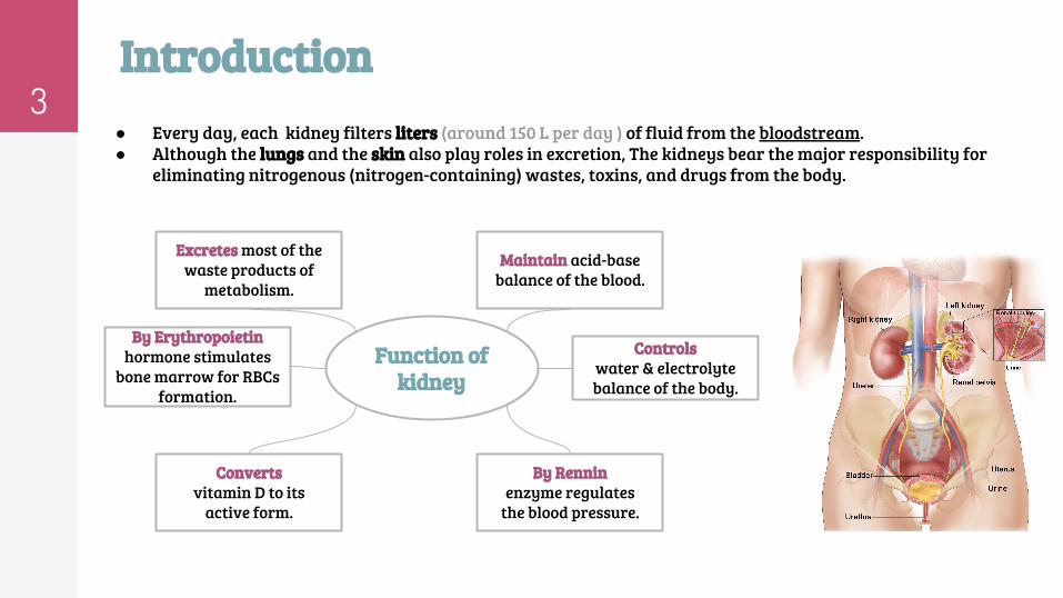
● Every day, each kidney filters liters (around 150 L per day ) of fluid from the bloodstream.● Although the lungs and the skin also play roles in excretion, The kidneys bear the major responsibility for
eliminating nitrogenous (nitrogen-containing) wastes, toxins, and drugs from the body.
Maintain acid-base balance of the blood.
Controls water & electrolyte balance of the body.
Excretes most of the waste products of
metabolism.
By Erythropoietin hormone stimulates
bone marrow for RBCs formation.
By Rennin enzyme regulates
the blood pressure.
Converts vitamin D to its
active form.
Function of kidney
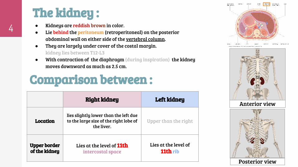
4 ● Kidneys are reddish brown in color.● Lie behind the peritoneum (retroperitoneal) on the posterior
abdominal wall on either side of the vertebral column.● They are largely under cover of the costal margin.
kidney lies between T12-L3● With contraction of the diaphragm (during inspiration) the kidney
moves downward as much as 2.5 cm.
T12
L3
Anterior view
Posterior view
Right kidney Left kidney
Locationlies slightly lower than the left due to the large size of the right lobe of
the liver.Upper than the right
Upper border of the kidney
Lies at the level of 11th intercostal space
Lies at the level of 11th rib
Comparison between :
The kidney :

5
Covering of the kidney
4. Pararenal (paranephric) fat :
It lies external to the renal fascia, and forms
part of the retroperitoneal fat.
3. Renal fascia: It encloses the kidneys and suprarenal glands
(but the suprarenal glands is in special
compartment)
2. Perirenal (perinephric) fat :
It covers the fibrous capsule
1. Fibrous capsule:It surrounds the
kidney
Describe the shape of kidney?● The lateral border is convex, while the medial border is convex at both ends
but it is concave at its middle where it shows a vertical slit called the hilum.
Hilum:● The hilum extends into a large cavity called the renal sinus.● The hilum transmits the from the front backward V.A.U.A.
1. Renal vein (most anterior)2. 2 branches of renal artery3. Ureter.(“we consider ureter as most posterior”)4. 3rd branch of renal artery
* The last 3 structures support the kidney in position.

6Renal structure:
KidneyInner Medulla
Outer Cortex
is composedof about 12 renal
pyramids.
Apex(the renal papilla):
is projectingmedially
Base of eachpyramid :is directedtoward the
cortex
extends into the medulla between
adjacent pyramids as the renal column.
● Extending from the bases of the renal pyramids into the cortex are striations known as medullary rays.
● The renal sinus within the hilum, contains the upper expanded end of the ureter(dilated part of ureter),
the renal pelvis.
● Renal pelvis divides into two or three major calyces, which divides into two or three minor calyces.
Medullary rays
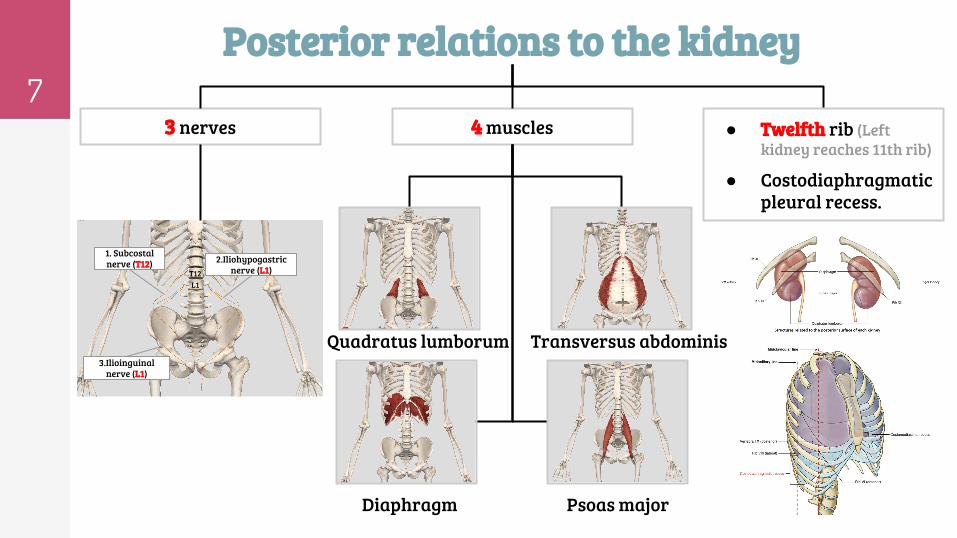
7Posterior relations to the kidney
● Twelfth rib (Left kidney reaches 11th rib)
● Costodiaphragmatic pleural recess.
4 muscles3 nerves
Transversus abdominis
Diaphragm Psoas major
Quadratus lumborum
T12L1
1. Subcostal nerve (T12) 2.Iliohypogastric
nerve (L1)
3.Ilioinguinal nerve (L1)
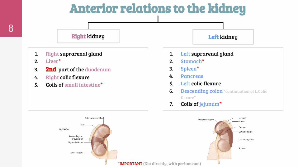
1. Right suprarenal gland2. Liver*3. 2nd part of the duodenum 4. Right colic flexure5. Coils of small intestine*
1. Left suprarenal gland2. Stomach*3. Spleen*4. Pancreas5. Left colic flexure6. Descending colon “continuation of L.Colic
flexure”7. Coils of jejunum*
8
Anterior relations to the kidney
Left kidney Right kidney
*IMPORTANT (Not directly ,with peritoneum)

9
Blood supply (arteries) ➔ The renal artery arises from the aorta at the level of the 2nd lumbar vertebra.
➔ Each renal artery divides into : 5 segmental arteries that enter the hilum of the kidney, four in front and one behind the renal pelvis. They are distributed to different segments of the kidney.
➔ Lobar artery arises from each segmental artery, one for each renal pyramid.
➔ Each lobar artery gives off 2 or 3 interlobar arteries.
➔ The interlobar arteries run toward the cortex on each side of the renal pyramid.
➔ Interlobar arteries give off the arcuate arteries at thejunction of the cortex & medulla.
➔ The arcuate arteries give off several interlobular arteries.
➔ Interlobular artery gives off afferent glomerular arterioles .
❶
❷
❸
❹
❺
❻
❼
• Each nephron is associated with 2 capillary beds:The glomerulus & The peritubular capillary bed.• The glomerulus is both fed and drained by arterioles:
1. The afferent arteriole,which arises from aninterlobular artery, is the "feeder vessel"
2. the efferent arteriole receives blood thathas passed through the glomerulus.
10
Segments of the : Found only in male’s slides
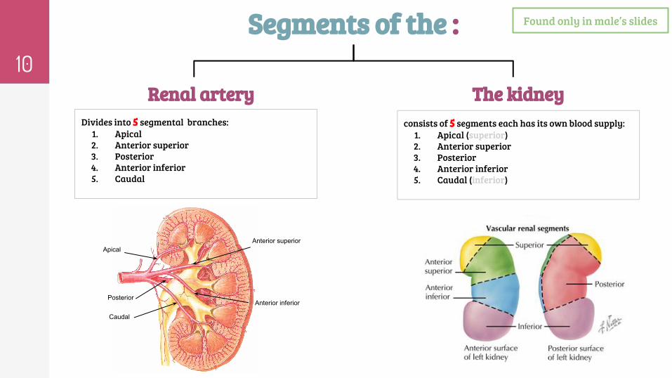
Renal artery The kidney consists of 5 segments each has its own blood supply:
1. Apical (superior)2. Anterior superior 3. Posterior 4. Anterior inferior5. Caudal (inferior)
Divides into 5 segmental branches:1. Apical 2. Anterior superior 3. Posterior 4. Anterior inferior5. Caudal
Anterior inferior
Apical
Posterior
Caudal
Anterior superior
11
1. Venous Drainage 2. Lymph Drainage
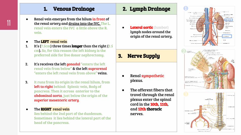
● Renal vein emerges from the hilum in front of the renal artery and drains into the IVC. The L. renal vein enters the IVC a little above the R. vein.
● The LEFT renal vein1. It’s (7.5cm) three times longer than the right (2.5
cm). So, for this reason the left kidney is the preferred side for live donor nephrectomy.
2. It’s receives the left gonadal “enters the left renal vein from below” & the left suprarenal “enters the left renal vein from above” veins.
3. It runs from its origin in the renal hilum, from left to right behind: Splenic vein, Body of pancreas. Then it across anterior to the abdominal aorta, just below the origin of the superior mesenteric artery.
● The RIGHT renal veinlies behind the 2nd part of the duodenum. Sometimes it lies behind the lateral part of the head of the pancreas.
● Lateral aortic (lumbar) lymph nodes around the origin of the renal artery.
3. Nerve Supply
● Renal sympatheticplexus.
● The afferent fibers that travel through the renal plexus enter the spinalcord in the 10th, 11th, and 12th thoracic nerves.
❶
❷
❸
12
MCQsQuestion 5: All of these are one of the anterior relations of the L. kidney Except:
A. Stomach
B. Liver
C. Spleen
D. Pancreas
Question 6: the interlobular arteries gives off __________.
A. Arcuate arteries
B. Efferent glomerular arterioles
C. Afferent glomerular arterioles
D. Both b & c
Question 7: the left renal vein is longer, and receives blood from __________ veins.
A. L. Gonadal
B. Hepatic
C. L. Suprarenal
D. Both a & c
Question 8: Renal pelvis divides into two or three__________.
A. Renal papilla
B. Major calyces
C. Renal sinus
D. Renal column
Answers: Q1.B- Q2.A -Q3.D -Q4.C- Q5.B- Q6.C-Q7.D- Q8.B
Question 1: The upper border of the R. Kidney lies at the level of:
A. 12th intercostal space
B. 11th intercostal space
C. 12th rib
D. 11th rib
Question 2: The area where the renal artery enters the kidney and the
renal vein and ureter exits the kidney is called the __________.
A.Renal hilum
B. Renal medulla
C. juxtamedullary area
D.Renal papilla
Question 3: The most anterior structure in the Hilum:
A. 2 branches of Renal artery
B. 3rd branch of renal artery
C. Ureter
D.Renal vein
Question 4: which of these isn’t one of posterior relation of the kidney?
A. Ilioinguinal nerve
B. Subcostal nerve
C. Liver
D.Psoas major

Team leaders
● Abdulrahman Shadid● Ateen Almutairi
Team members
Girls team :
● Ajeed Al Rashoud● Taif Alotaibi● Noura Al Turki● Amirah Al-Zahrani● Alhanouf Al-haluli● Sara Al-Abdulkarem● Renad Al Haqbani● Nouf Al Humaidhi● Jude Al Khalifah● Nouf Al Hussaini● Rahaf Al Shabri● Danah Al Halees● Rema Al Mutawa● Amirah Al Dakhilallah● Maha Al Nahdi ● Ghaida Al Braithen
Boys team:
● Faisal Alqifari ● Salman Alagla● Ziyad Al-jofan● Ali Aldawood● Khalid Nagshabandi● Omar Alammari● Sameh nuser
Contact us:
THANKS!Editing file
Copyright © 2022 FDOKUMEN

