
Zoonoses and communicable diseases common to man - IRIS ...
395
ZOONOSES AND COMMUNICABLE DISEASES COMMON TO MAN AND ANIMALS Third Edition Volume I Bacterioses and Mycoses Scientific and Technical Publication No. 580 PAN AMERICAN HEALTH ORGANIZATION Pan American Sanitary Bureau, Regional Office of the WORLD HEALTH ORGANIZATION 525 Twenty-third Street, N.W. Washington, D.C. 20037 U.S.A. 2001
-
Upload
khangminh22 -
Category
Documents
-
view
1 -
download
0
Transcript of Zoonoses and communicable diseases common to man - IRIS ...

ZOONOSES AND COMMUNICABLE DISEASES COMMON TO MAN AND ANIMALS
Third Edition
Volume I
Bacterioses and Mycoses
Scientific and Technical Publication No. 580
PAN AMERICAN HEALTH ORGANIZATIONPan American Sanitary Bureau, Regional Office of the
WORLD HEALTH ORGANIZATION525 Twenty-third Street, N.W.
Washington, D.C. 20037 U.S.A.
2001

Also published in Spanish (2001) with the title:Zoonosis y enfermedades transmisibles comunes al hombre a los animales
ISBN 92 75 31580 9
PAHO Cataloguing-in-PublicationPan American Health Organization
Zoonoses and communicable diseases common to man and animals3rd ed. Washington, D.C.: PAHO, © 2001.3 vol.—(Scientific and Technical Publication No. 580)
ISBN 92 75 11580 XI. Title II. Series1. ZOONOSES2. BACTERIAL INFECTIONS AND MYCOSES3. COMMUNICABLE DISEASE CONTROL4. FOOD CONTAMINATION5. PUBLIC HEALTH VETERINARY6. DISEASE RESERVOIRS
NLM WC950.P187 2001 En
The Pan American Health Organization welcomes requests for permission toreproduce or translate its publications, in part or in full. Applications and inquiriesshould be addressed to the Publications Program, Pan American HealthOrganization, Washington, D.C., U.S.A., which will be glad to provide the latestinformation on any changes made to the text, plans for new editions, and reprintsand translations already available.
© Pan American Health Organization, 2001
Publications of the Pan American Health Organization enjoy copyright protectionin accordance with the provisions of Protocol 2 of the Universal CopyrightConvention. All rights are reserved.
The designations employed and the presentation of the material in this publicationdo not imply the expression of any opinion whatsoever on the part of the Secretariatof the Pan American Health Organization concerning the status of any country, ter-ritory, city or area or of its authorities, or concerning the delimitation of its frontiersor boundaries.
The mention of specific companies or of certain manufacturers’ products does notimply that they are endorsed or recommended by the Pan American HealthOrganization in preference to others of a similar nature that are not mentioned.Errors and omissions excepted, the names of proprietary products are distinguishedby initial capital letters.

CONTENTS
Prologue . . . . . . . . . . . . . . . . . . . . . . . . . . . . . . . . . . . . . . . . . . . . . . . . . . . . viiPreface to the First Edition . . . . . . . . . . . . . . . . . . . . . . . . . . . . . . . . . . . . . . ixPreface to the Second Edition . . . . . . . . . . . . . . . . . . . . . . . . . . . . . . . . . . . . xiIntroduction . . . . . . . . . . . . . . . . . . . . . . . . . . . . . . . . . . . . . . . . . . . . . . . . . . xv
PART I: BACTERIOSES
Actinomycosis . . . . . . . . . . . . . . . . . . . . . . . . . . . . . . . . . . . . . . . . . . . . . . . . 3Aeromoniasis. . . . . . . . . . . . . . . . . . . . . . . . . . . . . . . . . . . . . . . . . . . . . . . . . 6Animal Erysipelas and Human Erysipeloid . . . . . . . . . . . . . . . . . . . . . . . . . . 14Anthrax . . . . . . . . . . . . . . . . . . . . . . . . . . . . . . . . . . . . . . . . . . . . . . . . . . . . . 21Botulism . . . . . . . . . . . . . . . . . . . . . . . . . . . . . . . . . . . . . . . . . . . . . . . . . . . . 28Brucellosis. . . . . . . . . . . . . . . . . . . . . . . . . . . . . . . . . . . . . . . . . . . . . . . . . . . 40Campylobacteriosis . . . . . . . . . . . . . . . . . . . . . . . . . . . . . . . . . . . . . . . . . . . . 67Cat-scratch Disease . . . . . . . . . . . . . . . . . . . . . . . . . . . . . . . . . . . . . . . . . . . . 78Clostridial Food Poisoning. . . . . . . . . . . . . . . . . . . . . . . . . . . . . . . . . . . . . . . 82Clostridial Wound Infections . . . . . . . . . . . . . . . . . . . . . . . . . . . . . . . . . . . . . 87Colibacillosis . . . . . . . . . . . . . . . . . . . . . . . . . . . . . . . . . . . . . . . . . . . . . . . . . 90Corynebacteriosis . . . . . . . . . . . . . . . . . . . . . . . . . . . . . . . . . . . . . . . . . . . . . 99Dermatophilosis. . . . . . . . . . . . . . . . . . . . . . . . . . . . . . . . . . . . . . . . . . . . . . . 103Diseases Caused by Nontuberculous Mycobacteria . . . . . . . . . . . . . . . . . . . . 107Diseases in Man and Animals Caused by Non-O1 Vibrio cholerae. . . . . . . . . 117Enterocolitic Yersiniosis. . . . . . . . . . . . . . . . . . . . . . . . . . . . . . . . . . . . . . . . . 122Enterocolitis Due to Clostridium difficile . . . . . . . . . . . . . . . . . . . . . . . . . . . . 132Food Poisoning Caused by Vibrio parahaemolyticus . . . . . . . . . . . . . . . . . . . 138Glanders . . . . . . . . . . . . . . . . . . . . . . . . . . . . . . . . . . . . . . . . . . . . . . . . . . . . 142Infection Caused by Capnocytophaga canimorsus and C. cynodegmi . . . . . . . 146Leprosy . . . . . . . . . . . . . . . . . . . . . . . . . . . . . . . . . . . . . . . . . . . . . . . . . . . . . 149Leptospirosis . . . . . . . . . . . . . . . . . . . . . . . . . . . . . . . . . . . . . . . . . . . . . . . . . 157Listeriosis . . . . . . . . . . . . . . . . . . . . . . . . . . . . . . . . . . . . . . . . . . . . . . . . . . . 168Lyme Disease . . . . . . . . . . . . . . . . . . . . . . . . . . . . . . . . . . . . . . . . . . . . . . . . 179Melioidosis . . . . . . . . . . . . . . . . . . . . . . . . . . . . . . . . . . . . . . . . . . . . . . . . . . 184Necrobacillosis . . . . . . . . . . . . . . . . . . . . . . . . . . . . . . . . . . . . . . . . . . . . . . . 190Nocardiosis . . . . . . . . . . . . . . . . . . . . . . . . . . . . . . . . . . . . . . . . . . . . . . . . . . 195Pasteurellosis . . . . . . . . . . . . . . . . . . . . . . . . . . . . . . . . . . . . . . . . . . . . . . . . . 199Plague . . . . . . . . . . . . . . . . . . . . . . . . . . . . . . . . . . . . . . . . . . . . . . . . . . . . . . 207Pseudotuberculous Yersiniosis . . . . . . . . . . . . . . . . . . . . . . . . . . . . . . . . . . . . 218Rat-bite Fever . . . . . . . . . . . . . . . . . . . . . . . . . . . . . . . . . . . . . . . . . . . . . . . . 226Rhodococcosis. . . . . . . . . . . . . . . . . . . . . . . . . . . . . . . . . . . . . . . . . . . . . . . . 229Salmonellosis. . . . . . . . . . . . . . . . . . . . . . . . . . . . . . . . . . . . . . . . . . . . . . . . . 233Shigellosis . . . . . . . . . . . . . . . . . . . . . . . . . . . . . . . . . . . . . . . . . . . . . . . . . . . 247Staphylococcal Food Poisoning . . . . . . . . . . . . . . . . . . . . . . . . . . . . . . . . . . . 251Streptococcosis . . . . . . . . . . . . . . . . . . . . . . . . . . . . . . . . . . . . . . . . . . . . . . . 257Tetanus . . . . . . . . . . . . . . . . . . . . . . . . . . . . . . . . . . . . . . . . . . . . . . . . . . . . . 265Tick-borne Relapsing Fever . . . . . . . . . . . . . . . . . . . . . . . . . . . . . . . . . . . . . . 271
iii

iv CONTENTS
Tularemia . . . . . . . . . . . . . . . . . . . . . . . . . . . . . . . . . . . . . . . . . . . . . . . . . . . 275Zoonotic Tuberculosis . . . . . . . . . . . . . . . . . . . . . . . . . . . . . . . . . . . . . . . . . . 283
PART II: MYCOSES
Adiaspiromycosis . . . . . . . . . . . . . . . . . . . . . . . . . . . . . . . . . . . . . . . . . . . . . 303Aspergillosis . . . . . . . . . . . . . . . . . . . . . . . . . . . . . . . . . . . . . . . . . . . . . . . . . 305Blastomycosis . . . . . . . . . . . . . . . . . . . . . . . . . . . . . . . . . . . . . . . . . . . . . . . . 311Candidiasis . . . . . . . . . . . . . . . . . . . . . . . . . . . . . . . . . . . . . . . . . . . . . . . . . . 315Coccidioidomycosis. . . . . . . . . . . . . . . . . . . . . . . . . . . . . . . . . . . . . . . . . . . . 320Cryptococcosis . . . . . . . . . . . . . . . . . . . . . . . . . . . . . . . . . . . . . . . . . . . . . . . 326Dermatophytosis . . . . . . . . . . . . . . . . . . . . . . . . . . . . . . . . . . . . . . . . . . . . . . 332Histoplasmosis. . . . . . . . . . . . . . . . . . . . . . . . . . . . . . . . . . . . . . . . . . . . . . . . 339Mycetoma . . . . . . . . . . . . . . . . . . . . . . . . . . . . . . . . . . . . . . . . . . . . . . . . . . . 345Protothecosis . . . . . . . . . . . . . . . . . . . . . . . . . . . . . . . . . . . . . . . . . . . . . . . . . 348Rhinosporidiosis . . . . . . . . . . . . . . . . . . . . . . . . . . . . . . . . . . . . . . . . . . . . . . 350Sporotrichosis . . . . . . . . . . . . . . . . . . . . . . . . . . . . . . . . . . . . . . . . . . . . . . . . 352Zygomycosis . . . . . . . . . . . . . . . . . . . . . . . . . . . . . . . . . . . . . . . . . . . . . . . . . 356
Index . . . . . . . . . . . . . . . . . . . . . . . . . . . . . . . . . . . . . . . . . . . . . . . . . . . . . . 361
LIST OF TABLES AND ILLUSTRATIONS
Bacterioses
Tables
1. Foods giving rise to botulism, and number of outbreaks,United States of America, 1899–1977 . . . . . . . . . . . . . . . . . . . . . . . . . . . 31
2. Number of cases and deaths from human plague in the Americas, 1971–1980 . . . . . . . . . . . . . . . . . . . . . . . . . . . . . . . . . . . . . . . 210
3. Outbreaks of foodborne salmonellosis in selected countries,1981–1985 . . . . . . . . . . . . . . . . . . . . . . . . . . . . . . . . . . . . . . . . . . . . . . . 235
4. Distribution of tetanus morbidity according to political division and climate, Argentina, 1967–1977 . . . . . . . . . . . . . . . . . . . . . . . . . . . . . 267
Figures
1. Animal erysipelas and human erysipeloid (Erysipelothrix rhusiopathiae). Mode of transmission . . . . . . . . . . . . . . . . . . . . . . . . . . . 17
2. Anthrax. Transmission cycle . . . . . . . . . . . . . . . . . . . . . . . . . . . . . . . . . . 253. Botulism (transmitted by foods). Reported cases and deaths
per year, United States of America, 1960–1980 . . . . . . . . . . . . . . . . . . . . 294. Reported cases of botulism per year, Argentina, 1967–1981 . . . . . . . . . . 325. Bovine brucellosis (Brucella abortus). Mode of transmission . . . . . . . . . 526. Swine brucellosis (Brucella suis). Mode of transmission . . . . . . . . . . . . . 53

7. Caprine and ovine brucellosis (Brucella melitensis). Mode of transmission . . . . . . . . . . . . . . . . . . . . . . . . . . . . . . . . . . . . . . . 54
8. Campylobacteriosis (Campylobacter jejuni). Mode of transmission. . . . . 709. Campylobacteriosis (Campylobacter fetus). Probable mode
of transmission . . . . . . . . . . . . . . . . . . . . . . . . . . . . . . . . . . . . . . . . . . . . 7510. Enterocolitic yersiniosis (Yersinia enterocolitica). Supposed
mode of transmission . . . . . . . . . . . . . . . . . . . . . . . . . . . . . . . . . . . . . . . 12711. Glanders. Mode of transmission . . . . . . . . . . . . . . . . . . . . . . . . . . . . . . . 14412. Leptospirosis. Synanthropic transmission cycle . . . . . . . . . . . . . . . . . . . . 16213. Melioidosis (Pseudomonas pseudomallei). Mode of transmission . . . . . . 18714. Number of cases and deaths from human plague worldwide,
1971–1980 . . . . . . . . . . . . . . . . . . . . . . . . . . . . . . . . . . . . . . . . . . . . . . . 20915. Plague. Domestic and peridomestic transmission cycle . . . . . . . . . . . . . . 21316. Pseudotuberculous yersiniosis (Yersinia pseudotuberculosis).
Probable mode of transmission . . . . . . . . . . . . . . . . . . . . . . . . . . . . . . . . 22217. Salmonellosis. Mode of transmission (except Salmonella typhi
and the paratyphoid serotypes) . . . . . . . . . . . . . . . . . . . . . . . . . . . . . . . . 24118. Tick-borne relapsing fever (Ornithodoros spp.).
Mode of transmission . . . . . . . . . . . . . . . . . . . . . . . . . . . . . . . . . . . . . . . 27319. Tularemia. Mode of transmission in the Americas . . . . . . . . . . . . . . . . . . 27920. Tuberculosis (Mycobacterium bovis). Mode of transmission . . . . . . . . . . 293
CONTENTS v

vii
PROLOGUE
Zoonoses and communicable diseases common to man and animals continue tohave high incidence rates and to cause significant morbidity and mortality.Infections and parasitoses of cattle can reduce meat or milk production and can leadto the death or destruction of the animals, all of which diminishes the supply ofavailable food for man. These diseases are also an obstacle for international trade,as well as a serious financial drain for cattle farmers and, more broadly, for a com-munity’s or a country’s economy, which can have wide repercussions for a society’shealth.
With the aim of helping to solve these problems, the Pan American HealthOrganization (PAHO)—an international public health organization that has devoteditself to improving the health and living conditions of the people of the Americas fornearly one hundred years—established the Veterinary Public Health Program. TheProgram’s overall objective is to collaborate with PAHO’s Member Countries in thedevelopment, implementation, and evaluation of policies and programs that lead tofood safety and protection and to the prevention, control, or eradication of zoonoses,among them foot-and-mouth disease.
To this end, PAHO’s Veterinary Public Health Program has two specializedregional centers: the Pan American Foot-and-Mouth Disease Center (PANAFTOSA),created in 1951 in Rio de Janeiro, Brazil, and the Pan American Institute for FoodProtection and Zoonoses (INPPAZ), established on November 15, 1991 in BuenosAires, Argentina. INPPAZ’s precursor was the Pan American Zoonoses Center(CEPANZO), which was created through an agreement with the Government ofArgentina to help the countries of the Americas combat zoonoses, and which oper-ated from 1956 until 1990.
Since its creation in 1902, PAHO has participated in various technical coopera-tion activities with the countries, among them those related to the surveillance, pre-vention, and control of zoonoses and communicable diseases common to man andanimals, which cause high morbidity, disability, and mortality in vulnerable humanpopulations. PAHO has also collaborated in the strengthening of preventive medi-cine and public health through the promotion of veterinary health education in learn-ing, research, and health care centers. An example of this work is the preparation ofseveral publications, among which the two previous Spanish and English editions ofZoonoses and Communicable Diseases Common to Man and Animals stand out.
Scientific knowledge has progressed since the last edition. Also, the countries ofthe Americas have modified their livestock production strategies in recent years,which has affected the transmission of zoonotic infections and their distribution. Thepublication of this third edition is an attempt to address these changes. The third edi-tion is presented in three volumes: the first contains bacterioses and mycoses; thesecond, chlamydioses, rickettsioses, and viroses; and the third, parasitoses.
We believe that this new edition will continue to be useful for professors and stu-dents of public health, medicine, and veterinary medicine; workers in public healthand animal health institutions; and veterinarians, researchers, and others interestedin the subject. We also hope that this publication is a useful tool in the elaborationof national zoonosis control or eradication policies and programs, as well as in risk

viii PROLOGUE
evaluation and in the design of epidemiological surveillance systems for theprevention and timely control of emerging and reemerging zoonoses. In summary,we are confident that this book will contribute to the application of the knowledgeand resources of the veterinary sciences for the protection and improvement ofpublic health.
GEORGE A.O. ALLEYNEDIRECTOR

ix
PREFACE TO THE FIRST EDITION
This book considers two groups of communicable diseases: those transmittedfrom vertebrate animals to man, which are—strictly speaking—zoonoses; and thosecommon to man and animals. In the first group, animals play an essential role inmaintaining the infection in nature, and man is only an accidental host. In the sec-ond group, both animals and man generally contract the infection from the samesources, such as soil, water, invertebrate animals, and plants; as a rule, however,animals do not play an essential role in the life cycle of the etiologic agent, but may contribute in varying degrees to the distribution and actual transmission ofinfections.
No attempt has been made to include all infections and diseases comprised inthese two groups. A selection has been made of some 150 that are of principal inter-est, for various reasons, in the field of public health. The number of listed zoonosesis increasing as new biomedical knowledge is acquired. Moreover, as human activ-ity extends into unexplored territories containing natural foci of infection, newzoonotic diseases are continually being recognized. In addition, improved healthservices and better differential diagnostic methods have distinguished zoonoses pre-viously confused with other, more common diseases. A number of diseasesdescribed in this book have only recently been recognized, examples of whichinclude the Argentine and Bolivian hemorrhagic fevers, angiostrongyliasis, rotaviralenteritis, Lassa fever, Marburg disease, and babesiosis.
The principal objective in writing this book was to provide the medical profes-sions a source of information on the zoonoses and communicable diseases commonto man and animals. Toward that end, both medical and veterinary aspects, whichhave traditionally been dealt with separately in different texts, have been combinedin a single, comprehensive volume. As a result, physicians, veterinarians, epidemi-ologists, and biologists can all gain an overview of these diseases from one source.
This book, like most scientific works, is the product of many books, texts, mono-graphs, and journal articles. Many sources of literature in medicine, veterinary med-icine, virology, bacteriology, mycology, and parasitology were consulted, as were alarge number of reports from different biomedical disciplines, in order to provideup-to-date and concise information on each disease. It is expected that any errors oromissions that may have been committed can, with the collaboration of the readers,be corrected in a future edition.
Where possible, explanations were attempted with special emphasis on theAmericas, particularly Latin America. An effort was made, one which was notalways successful, to collect available information on diseases in this Region. Dataon the incidence of many zoonoses are fragmentary and frequently not reliable. It ishoped that the establishment of control programs in various countries will lead toimproved epidemiologic surveillance and disease reporting.
More space has been devoted to those zoonoses having greatest impact on publichealth and on the economy of the countries of the Americas, but information is alsoincluded on those regionally less important or exotic diseases.
The movement of persons and animals over great distances adds to the risk ofintroducing exotic diseases that may become established on the American continent
x PREFACE TO THE FIRST EDITION
given the appropriate ecologic factors for existence of the etiologic agents. Today,public health and animal health administrators, physicians, and veterinarians mustbe familiar with the geographic distribution and pathologic manifestations of thevarious infectious agents so that they can recognize and prevent the introduction ofexotic diseases.
We, the authors, would like to give special recognition to Dr. Joe R. Held,Assistant Surgeon-General of the United States Public Health Service and Directorof the Division of Research Services of the U.S. National Institutes of Health, whogave impetus to the English translation and reviewed the bacterioses sections.
We would also like to express our utmost appreciation to the experts whoreviewed various portions of this book and offered their suggestions for improvingthe text. These include: Dr. Jeffrey F. Williams, Professor in the Department ofMicrobiology and Public Health, Michigan State University, who reviewed thechapters dealing with parasitic zoonoses; Dr. James Bond, PAHO/WHO RegionalAdviser in Viral Diseases, who read the viroses; Dr. Antonio Pío, formerlyPAHO/WHO Regional Adviser in Tuberculosis and presently with WHO in Geneva,and Dr. James H. Rust, PAHO/WHO Regional Adviser in Enteric Diseases, both ofwhom reviewed the bacterioses; and Dr. F. J. López Antuñano, PAHO/WHORegional Adviser in Parasitic Diseases, who read the metazooses.
We would like to thank Dr. James Cocozza, PAHO/WHO Veterinary Adviser, forhis review of the translation and Dr. Judith Navarro, Editor in the Office ofPublications of PAHO, for her valuable collaboration in the editorial revision andcomposition of the book.
PEDRO N. ACHABORIS SZYFRES

xi
PREFACE TO THE SECOND EDITION
The fine reception accorded the Spanish, English, and French versions of thisbook has motivated us to revise it in order that it still may serve the purpose forwhich it was written: to provide an up-to-date source of information to the medicalprofession and allied fields. This book has undoubtedly filled a void, judging by itswide use in schools of public health, medicine, and veterinary medicine, as well asby bureaus of public and animal health.
The present edition has been considerably enlarged. In the seven years since thefirst edition was published, our knowledge of zoonoses has increased broadly andrapidly, and new zoonotic diseases have emerged. Consequently, most of the dis-cussions have been largely rewritten, and 28 new diseases have been added to theoriginal 148. Some of these new diseases are emerging zoonoses; others are patho-logic entities that have been known for a long time, but for which the epidemiologicconnection between man and animal has been unclear until recently.
The use this book has had outside the Western Hemisphere has caused us to aban-don the previous emphasis on the Americas in favor of a wider scope and geomed-ical view. Moreover, wars and other conflicts have given rise to the migration ofpopulations from one country or continent to another. A patient with a diseaseheretofore known only in Asia may now turn up in Amsterdam, London, or NewYork. The physician must be aware of these diseases in order to diagnose and treatthem. “Exotic” animal diseases have been introduced from Africa to Europe, theCaribbean, and South America, causing great damage. The veterinary physicianmust learn to recognize them to be able to prevent and eradicate them before theybecome entrenched. It must be remembered that parasites, viruses, bacteria, andother agents of zoonotic infection can take up residence in any territory where theyfind suitable ecologic conditions. Ignorance, economic or personal interests, andhuman customs and needs also favor the spread of these diseases.
Research in recent years has demonstrated that some diseases previously consid-ered to be exclusively human have their counterparts in wild animals, which in cer-tain circumstances serve as sources of human infection. On the other hand, theseanimals may also play a positive role by providing models for research, such as inthe case of natural leprosy in nine-banded armadillos or in nonhuman primates inAfrica. Of no less interest is the discovery of Rickettsia prowazekii in eastern flyingsquirrels and in their ectoparasites in the United States, and the transmission of theinfection to man in a country where epidemic typhus has not been seen since 1922.A possible wild cycle of dengue fever is also discussed in the book. Is Creutzfeldt-Jakob disease a zoonosis? No one can say with certainty, but some researchersbelieve it may have originated as such. In any case, interest is aroused by the sur-prising similarity of this disease and of kuru to animal subacute spongiformencephalopathies, especially scrapie, the first known and best studied of this group.Discussion of human and animal slow viruses and encephalopathies is included inthe spirit of openness to possibilities and the desire to bring the experience of onefield of medicine to another. In view of worldwide concern over acquired immuno-deficiency syndrome (AIDS), a brief section on retroviruses has also been added, inwhich the relationship between the human disease and feline and simian AIDS is

xii PREFACE TO THE SECOND EDITION
noted. Another topic deeply interesting to researchers is the mystery of the radicalantigenic changes of type A influenza virus, a cause of explosive pandemics thataffect millions of persons around the world. Evidence is mounting that thesechanges result from recombination with a virus of animal origin (see Influenza).That this should occur is not surprising, given the constant interaction between manand animals. As a rule, zoonoses are transmitted from animal to man, but the reversemay also occur, as is pointed out in the chapters on hepatitis, herpes simplex, andmeasles. The victims in these cases are nonhuman primates, which may in turnretransmit the infection to man under certain circumstances.
Among emerging zoonoses we cite Lyme disease, which was defined as a clinicalentity in 1977; the etiologic agent was found to be a spirochete (isolated in 1982),for which the name Borrelia burgdorferi was recently proposed. Emerging viralzoonoses of note in Latin America are Rocio encephalitis and Oropouche fever; thelatter has caused multiple epidemics with thousands of victims in northeast Brazil.Outstanding among new viral disease problems in Africa are the emergence of Eboladisease and the spread of Rift Valley fever virus, which has caused tens of thousandsof human cases along with great havoc in the cattle industry of Egypt and has evokedalarm around the world. Similarly, the protozoan Cryptosporidium is emerging asone of the numerous agents of diarrheal diseases among man and animals, and prob-ably has a worldwide distribution.
As the English edition was being prepared, reports came to light of two animaldiseases not previously confirmed in humans. Three cases of human pseudorabiesvirus infection were recognized between 1983 and 1986 in two men and one womanwho had all had close contact with cats and other domestic animals. In 1986, sero-logic testing confirmed infection by Ehrlichia canis in a 51-year-old man who hadbeen suspected of having Rocky Mountain spotted fever. This is the first knownoccurrence of E. canis infection in a human. These two diseases bear watching aspossible emerging zoonoses.
The space given to each zoonosis is in proportion to its importance. Some diseasesthat deserve their own monographs were given more detailed treatment, but noattempt was made to cover the topic exhaustively.
We, the authors, would like to give special recognition to Dr. Donald C. Blenden,Professor in the Department of Medicine and Infectious Diseases, School ofMedicine, and Head of the Department of Veterinary Microbiology, College ofVeterinary Medicine, University of Missouri; and to Dr. Manuel J. Torres, Professorof Epidemiology and Public Health, Department of Veterinary Microbiology,College of Veterinary Medicine, University of Missouri, for their thorough review ofand valuable contributions to the English translation of this book.
We would also like to recognize the support received from the Pan AmericanHealth Organization (PAHO/WHO), the Pan American Health and EducationFoundation (PAHEF), and the Pan American Zoonoses Center in Buenos Aires,Argentina, which enabled us to update this book.
We are most grateful to Dr. F. L. Bryan for his generous permission to adapt hismonograph “Diseases Transmitted by Foods” as an Appendix to this book.

PREFACE TO THE SECOND EDITION xiii
Mr. Carlos Larranaga, Chief of the Audiovisual Unit at the Pan AmericanZoonosis Center, deserves our special thanks for the book’s artwork, as do Ms. IrisElliot and Mr. William A. Stapp for providing the translation into English. We wouldlike to express our most sincere gratitude and recognition to Ms. Donna J. Reynolds,editor in the PAHO Editorial Service, for her valuable collaboration in the scientificeditorial revision of the book.
PEDRO N. ACHABORIS SZYFRES

xv
INTRODUCTION
This new edition of Zoonoses and Communicable Diseases Common to Man andAnimals is published in three volumes: I. Bacterioses and mycoses; II.Chlamydioses and rickettsioses, and viroses; and III. Parasitoses. Each of the fiveparts corresponds to the location of the etiologic agents in the biological classifica-tion; for practical purposes, chlamydias and rickettsias are grouped together.
In each part, the diseases are listed in alphabetical order to facilitate readersearches. There is also an alphabetical index, which includes synonyms of the dis-eases and the etiologic agents’ names.
In this edition, the numbers and names of the diseases according to theInternational Statistical Classification of Diseases and Related Health Problems,Tenth Revision (ICD-10), are listed below the disease title. However, some zoonosesare not included in ICD-10 and are difficult to classify within the current scheme.
In addition, for each disease or infection, elements such as synonyms; etiology;geographical distribution; occurrence in man and animals; the disease in man andanimals; source of infection and mode of transmission; role of animals in the epi-demiology; diagnosis; and control are addressed. Patient treatment (for man or otherspecies) is beyond the scope of this work; however, recommended medicines areindicated for many diseases, especially where they are applicable to prophylaxis.Special attention is paid to the epidemiological and ecological aspects so that thereader can begin to understand the determining factors of the infection or disease.Some topics include simple illustrations of the etiologic agent’s mode of transmis-sion, showing the animals that maintain the cycle of infection in nature. Similarly,other graphics and tables are included to provide additional information on the geo-graphical distribution or prevalence of certain zoonoses.
The data on the occurrence of the infection in man and animals, along with dataon the geographical distribution, may help the reader judge the relative impact thateach disease has on public health and the livestock economy in the different regionsof the world, given that the importance of different zoonoses varies greatly. Forexample, foot-and-mouth disease is extremely important from an economic stand-point, but of little importance in terms of public health, if animal protein losses arenot considered. In contrast, Argentine and Bolivian hemorrhagic fevers are impor-tant human diseases, but their economic impact is minimal, if treatment costs andloss of man-hours are not taken into account. Many other diseases, such as brucel-losis, leptospirosis, salmonellosis, and equine encephalitis, are important from botha public health and an economic standpoint.
Finally, each disease entry includes an alphabetical bibliography, which includesboth the works cited and other relevant works that the reader may consult for moreinformation about the disease.

ACTINOMYCOSIS
ICD-10 A42.9
Synonyms: Actinostreptotrichosis, mandibular cancer, ray fungus disease.
Etiology: Actinomyces israelii is the principal etiologic agent in man, and A.bovis the main one in animals. A. naeslundi, A. viscosus, A. odontolytical, A. meyeriand Arachnia propionica (A. propionicus) are isolated less often, although A. visco-sus plays an important role in canine actinomycosis. Some reports indicate isolationof A. israelii from animals (Georg, 1974) and A. bovis from man (Brunner et al.,1973). Actinomyces are higher bacteria with many characteristics of fungi. They aregram-positive, do not produce spores, are non–acid-fast, range from anaerobic tomicroaerophilic, and are part of the normal flora of the mouth and of women’s gen-ital tract (Burden, 1989).
Geographic Distribution: Worldwide.
Occurrence in Man: Infrequent; however, data are very limited. Fewer than 100cases of the disease are recorded each year by the Public Health LaboratoryService’s Communicable Disease Surveillance Centre in Great Britain (Burden,1989). According to older data, 368 cases were recorded in Wales and England over12 years (1957–1968), with an incidence of 0.665 per million inhabitants, with ahigher incidence among industrial workers (Wilson, 1984). In Scotland, the annualincidence was three per million and the rate of attack was 10 times higher in agri-cultural workers than among others.
The historical ratio of two cases in men to one in women is probably no longervalid because of the number of cases of genital actinomycosis in women usingintrauterine contraceptive devices (IUDs).
Occurrence in Animals: The frequency of the disease varies widely amongregions and is also influenced by different livestock management practices. The dis-ease usually appears as sporadic cases. Small outbreaks have occurred in somemarshy areas of the United States and the former Soviet Union.
The Disease in Man: A. israelii, the main causal agent in man, is a normal com-ponent of the flora of the mouth. As a result of wounds or surgery, it can enter thesoft tissues and bones, where it causes a suppurative granulomatous process thatopens to the surface through fistulas. Several clinical forms have been identifiedaccording to their location: cervicofacial, thoracic, abdominal, and generalized.Cervicofacial, which is the most common (from 50% to more than 70% of cases), isusually caused by a tooth extraction or a jaw injury; it begins with a hard swellingunder the mucous membrane of the mouth, beneath the periosteum of the mandible,or in the skin of the neck. At a later stage, softened areas, depressions, and openingsto the exterior with a purulent discharge are evident. These secretions usually con-tain the characteristic “sulphur granules,” which are actinomyces colonies. The tho-racic form is generally caused by breathing the etiologic agent into the bronchialtubes where it establishes a chronic bronchopneumonia that affects the lower por-tions of the right lung (Burden, 1989), with symptoms similar to pulmonary tuber-culosis. As the disease progresses, invasion of the thoracic wall and its perforation
3

by fistulous tracks may occur. The abdominal form usually occurs after surgery andappears as an encapsulated lesion that often becomes localized in the cecum and theappendix, where it produces hard tumors that adhere to the abdominal wall.
The generalized form is infrequent and results from the erosive invasion of bloodvessels and lymphatic system, resulting in liver and brain disease.
In recent years, reports of actinomycosis in the genital tract of women usingintrauterine contraceptive devices have multiplied, with the rate of infection increas-ing in proportion to the duration of IUD use. In one study (Valicenti et al., 1982),the infection was found in 1.6% of women in the general population of IUD usersand in 5.3% of those attending the clinics. Another study of 478 IUD users found arate of infection of 12.6% based on Papanicolaou (Pap) smears (Koebler et al.,1983). Attempts to isolate the bacteria in Pap smears rarely yield positive results.However, A. israelii is also isolated from the genital tract of women who do not useIUDs, indicating that actinomyces are part of the normal flora (Burden, 1989). In thevast majority of cases, colonization by actinomyces produces only a superficial orasymptomatic infection.
Treatment consists of prolonged high doses of penicillin (weeks or months).Erythromycin, clindamycin, and tetracycline may also be used. Surgical drainage ofabscesses is important. In women with an endometrium colonized by actinomyces,removing the IUD is sometimes enough for the endometrium to return to normal.
The Disease in Animals: A. bovis is the principal agent of actinomycosis inbovines and, occasionally, in other animal species. In bovines, it centers chiefly inthe maxillae where it forms a granulomatous mass with necrotic areas that developinto abscesses. These open via fistulous passages and discharge a viscous, odorless,yellow pus. The pus contains small, yellow, sulphur granules, which are rosette-shaped when viewed under a microscope. In some cases chewing becomes very dif-ficult, and the animal stops eating and loses weight.
The cost-benefit ratio must be measured when treating bovine and equine actino-mycosis. Long-standing chronic lesions do not respond readily to treatment. If thelesions are small and circumscribed, they may be removed surgically. In other cases,curettage can be performed on the abscesses and fistulas, which are then packedwith gauze saturated with iodine tincture. Medical treatment is the same as forhuman actinomycosis, preferably using penicillin.
In swine the etiologic agent localizes principally in the sow’s udder, where it givesrise to abscesses and fistulas. Its pathway of penetration is the lesion caused by theteeth of suckling pigs. This infection is attributed to Actinomyces suis, whose tax-onomy is still uncertain.
In dogs, the disease produces cervicofacial abscesses, empyemas accompanied bypleurisy and osteomyelitis, and, more rarely, abdominal abscesses and cutaneousgranulomas. The most common agent encountered prior to 1982 was A. viscosus(Hardie and Barsanti, 1982).
Source of Infection and Mode of Transmission: The infection is endogenous.Actinomyces develop as saprophytes within and around carious teeth, in the mucinon dental enamel and in the tonsillar crypts. In studies carried out in several coun-tries, actinomyces have been found in 40% of excised tonsils and have been iso-lated in 30% to 48% of saliva samples or material from decayed teeth, as well asfrom the vaginal secretions of 10% of women using IUDs (Benenson, 1992).
4 BACTERIOSES

Infections and pathological developments are the product of tissue trauma, lesions,or prolonged irritation. It has not been possible to isolate the agent of actinomyco-sis from the environment. It is believed that the causal agent penetrates the tissuesof the mouth through lesions caused by foods or foreign objects, or by way of den-tal defects. From the oral cavity, the bacteria can be swallowed or breathed into thebronchial tubes.
Role of Animals in the Epidemiology of the Disease: The species ofActinomyces that attack man are different from those that affect animals. Rarely isA. israelii found in animals or A. bovis found in man. The designation of speciesprior to 1960 is doubtful (Lerner, 1991) and thus, distinguishing one species fromanother presents great problems. The infection in animals is not transmitted to man,nor is it transmitted from person to person or animal to animal.
Diagnosis: The clinical picture may be confused with other infections, such asactinobacillosis, nocardiosis, and staphylococcosis, as well as neoplasia and tuber-culosis. The first step in confirming the diagnosis is to obtain pus, sputum, or tissuesamples for microscopic examination and culture, and to inspect them for granules.Filament masses are visible by direct observation. In smears of crushed granules orpus stained by the Gram and Kinyoun methods, gram-positive and non–acid-fastfilaments or pleomorphic forms, occasionally with bacillary-sized branching, maybe seen (Cottral, 1978). It is possible to identify the species of actinomyces causingthe disease only by culturing and typing the isolated microorganism. In testingwomen who use IUDs, direct immunofluorescence has yielded good results(Valicenti et al., 1982).
Control: Prevention in man consists of proper oral hygiene and care after dentalextractions or other surgery in the oral cavity. No practical means have been estab-lished yet to prevent actinomycosis in animals.
Bibliography
Ajello, L., L.K. Georg, W. Kaplan, L. Kaufman. Laboratory Manual for MedicalMycology. Washington, D.C.: U.S. Government Printing Office; 1963. (Public Health ServicePublication 994).
Benenson, A.S., ed. Control of Communicable Diseases in Man. 15th ed. An official reportof the American Public Health Association. Washington, D.C.: American Public HealthAssociation; 1990.
Brunner, D.W., J.H. Gillespie. Hagan’s Infectious Diseases of Domestic Animals. 6th ed.Ithaca: Comstock; 1973.
Burden P. Actinomycosis [editorial]. J Infect 19:95–99, 1989.Cottral, G.E., ed. Manual of Standardized Methods for Veterinary Microbiology. Ithaca:
Comstock; 1978.Dalling, T., A. Robertson, eds. International Encyclopaedia of Veterinary Medicine.
Edinburgh: Green; 1966. Georg, L.K. The agents of human actinomycosis. Cited in: Lerner, P.L. Actinomyces and
Arachnia species. In: Mandell, G.L., R.G. Douglas, Jr., J.E. Bennett, eds. Principles andPractice of Infectious Diseases. 3rd ed. New York: Churchill Livingstone, Inc.; 1990.
Hardie, E.M., J.A. Barsanti. Treatment of canine actinomycosis. J Am Vet Assoc180:537–541, 1982.
ACTINOMYCOSIS 5

Koebler, C., A. Chatwani, R. Schwartz. Actinomycosis infection associated with intrauter-ine contraceptive devices. Am J Obstet Gynecol 145:596–599, 1983.
Lerner, P.L. Actinomyces and Arachnia species. In: Mandell, G.L., R.G. Douglas, Jr., J.E.Bennett, eds. Principles and Practice of Infectious Diseases. 3rd ed. New York: ChurchillLivingstone, Inc.; 1990.
Pier, A.C. The actinomycetes. In: Hubbert, W.T., W.F. McCulloch, P.R. Schnurrenberger,eds. Diseases Transmitted from Animals to Man. 6th ed. Springfield: Thomas; 1975.
Valicenti, J.F., Jr., A.A. Pappas, C.D. Graber, H.O. Williamson, N.F. Willis. Detection andprevalence of IUD-associated Actinomyces colonization and related morbidity. A prospectivestudy of 69,925 cervical smears. JAMA 247:1149–1152, 1982.
Wilson, G. Actinomycosis, actinobacillosis, and related diseases. In: Smith, G.R., ed. Vol3: Topley and Wilson’s Principles of Bacteriology, Virology and Immunity. Baltimore:Williams & Wilkins; 1984.
AEROMONIASIS
ICD-10 AO5.8 other specified bacterial foodborne intoxications
Etiology: The genus Aeromonas is classified within the family Vibrionaceae andshares some characteristics with members of other genera of this family. However,genetic hybridization studies indicate that the genus Aeromonas is sufficiently dif-ferent to place it in a new family, with the suggested name of Aeromonadaceae. Twogroups can be distinguished in the genus Aeromonas. The first group is psy-chrophilic and nonmotile and is represented by Aeromonas salmonicida, an impor-tant pathogen for fish (the agent of furunculosis). It does not affect man because itcannot reproduce at a temperature of 37°C. The second group is mesophilic andmotile, and it is this group that causes aeromoniasis, a disease common to man andanimals. These aeromonas are gram-negative, straight bacilli ranging from 1 to 3microns in length. They have a polar flagellum and are oxidase positive and facul-tatively anaerobic. They essentially include the species A. hydrophila, A. sobria, andA. caviae (Janda and Duffey, 1988), to which A. veronii and A. schuberti were addedlater, as well as the genospecies A. jandae and A. trota. However, only A. hydrophilaand A. sobria are of clinical interest.
More recent hybridization studies show that the A. hydrophila complex is geneti-cally very variable. Thirteen different genospecies have been established, but froma practical standpoint the three principal phenospecies are retained. It is possible toidentify 95% of isolates on the basis of their biochemical properties (Janda, 1991).
A system of 40 serogroups was established based on the somatic antigens (O) ofA. hydrophila and A. caviae. All the O antisera contain antibodies to the rugose form(R) of the bacillus, and thus the antisera must be absorbed by culturing the R formbefore being used (Sakazaki and Shimada, 1984). Typing is done by gel proteinelectrophoresis, isoenzyme analysis, and genetic analysis. Isoenzyme analysis madeit possible to identify genospecies through four enzymes. All these methods have
6 BACTERIOSES
shown that the clinical strains are very diverse and that no single clone is responsi-ble for most of the infections (Von Graevenitz and Altwegg, 1991).
Over the last decade, researchers have tried to define the virulence factors of thisgenus, both in terms of structural characteristics and the extracellular products theysecrete. Considered important among the structural characteristics is a type of pilus,the “flexible” or curvilinear pilus. It is expressed when stimulated by certain envi-ronmental conditions that give the bacteria the ability to colonize. Another structuralcharacteristic that was first discovered in autoagglutinating strains of A. salmonicidais the S layer, which is outside the cell wall. The loss of this layer—which can beseen with an electron microscope—decreases pathogenicity for fish 1,000 to 10,000times. A similar layer was later discovered in certain strains of A. hydrophila and A.sobria in infected fish and mammals, but their functional role seems to differ sub-stantially from the same S layer in A. salmonicida (it does not make the surface ofthe bacteria hydrophobic).
The substances externally secreted by aeromonas include beta-hemolysin that isproduced by certain strains of A. hydrophila and A. sobria. It has been determinedthat this hemolysin has enterotoxigenic effects on lactating mice and ligated ilealloops of rabbits. Purified beta-hemolysin inoculated intravenously into mice is lethalat a dose of 0.06 µg. The cytotonic enterotoxin that causes an accumulation of fluidin the ligated ileal loop of the rabbit, as well as other effects, has also beendescribed. Between 5% and 20% of the strains produce a toxin that cross reacts withthe cholera toxin in the ELISA test (Janda, 1991).
Based on tests conducted in mice and fish (the latter are much more susceptible),it can be concluded that A. hydrophila and A. sobria are more virulent than A.caviae. In addition, there is a great difference in the virulence of the strains withineach species (Janda, 1991). These variations cannot be attributed to a single viru-lence factor. In addition, it was not possible to detect a common mechanism in thepathogenic capacity of Aeromonas spp. in humans or in animals.
An enzyme (acetylcholinesterase) isolated from fish infected by A. hydrophilaproved to be highly active against the central nervous system. The toxin was lethalfor fish at a dose of 0.05 µg/g of bodyweight; no lesions were observed in the tis-sues. The same toxin was obtained from six different strains (Nieto et al., 1991).
A comparison was made of 11 environmental strains and 9 human strains. All theenvironmental strains and four of the human strains proved to be pathogenic fortrout, at a dose of 3 x 10⁷ colony forming units (CFU). Only the human strainscaused death or lesions through intramuscular inoculation of mice. The virulentstrains produced more hemolysis and cytotoxins in cultures at 37°C than at 28°C(Mateos et al., 1993).
Geographic Distribution: The motile aeromonas appear worldwide. Their prin-cipal reservoir is in river and estuary waters, as well as in salt water where it meetsfresh water. Population density is lower in highly saline waters and waters with lim-ited dissolved oxygen. It has sometimes been possible to isolate Aeromonas fromchlorinated water, including muncipal water supplies. These bacteria are more pro-lific in summer than in winter (Stelma, 1989).
Occurrence in Man: Aeromoniasis generally occurs sporadically. There is noevidence that water or foods contaminated by Aeromonas spp. have been the sourceof outbreaks (as happens with other agents, such as enterobacteria). The only cases
AEROMONIASIS 7

that suggest the possibility of outbreaks are those described in 1982 and 1983. Inlate 1982, some 472 cases of gastroenteritis associated with the consumption of rawoysters occurred in Louisiana (USA). One year later, another outbreak affectedseven people in Florida. This was also attributed to raw oysters that came fromLouisiana. Pathogenicity tests were performed on 23 of the 28 strains identified asA. hydrophila; 70% tested positive in at least one of the virulence tests (Abeyta etal., 1986). There may have been other outbreaks that were not recognized becausefood and patient stools were not examined for detection and identification of A.hydrophila (Stelma, 1989).
Occurrence in Animals: A. hydrophila is a recognized pathogen in fish, amphib-ians, and reptiles. The disease may occur individually or epidemically, particularlyin fish-farming pools. The agent affects many fish species, particularly fresh waterspecies. Its economic impact varies, but can be severe (Stoskopf, 1993).Aeromoniasis due to A. hydrophila also causes significant illness in colonies ofamphibians and reptiles bred for experimental purposes.
The Disease in Man: For some time the aeromonas were considered opportunis-tic bacteria. Clinical and epidemiological information amassed in recent years seemsto confirm that A. hydrophila and A. sobria are the primary human pathogens, par-ticularly as agents of enteritis in children.
The disease appears in two forms: enteric and extraenteric. Studies on the patho-genic role of Aeromonas spp. in gastroenteritis have been conducted in Australia, theUnited States, England, Thailand, and, more recently, in Rosario, Argentina (Notarioet al., 1993). Patients with and without diarrhea have been compared, with the lat-ter group consisting of patients suffering from other diseases or healthy individuals.In Argentina, 8 strains (2%) were isolated from 400 fecal samples and from a colonbiopsy in children with diarrhea, and no strains were isolated from 230 childrenwithout diarrhea. In the United States, the agent was found in 1.1% of the cases andin none of the controls (Agger et al., 1985). The tests in the other countries also iso-lated A. hydrophila and A. sobria with greater frequency and in greater numbersfrom diarrheal feces than from nondiarrheal feces.
Enteritis due to Aeromonas spp. occurs more frequently in summer and predomi-nantly in children from 6 months to 5 years of age. The clinical symptoms includeprofuse diarrhea, slight fever, and abdominal pains; vomiting is occasionally seen inpatients under 2 years of age. Cases of gastroenteritis with blood and mucus in thefeces have also been described. The disease is generally benign in children and lastsonly a few days. Gastroenteritis is much less frequent in adults, but can occur withdiarrhea of longer duration (from 10 days to several weeks or months), weight loss,and dehydration. The predominant species are A. hydrophila and A. sobria, but A.caviae has also been implicated in some cases (Janda and Duffey, 1988).
The extra-intestinal clinical form can affect different organs and tissues. One verycommon form of contamination is through wounds and various traumas. The woundgenerally becomes infected through contact with river water, ponds, or other waterreservoirs. The most common clinical expression is cellulitis. The patient recoverscompletely in such cases.
Some 20 cases have been described of infection caused by medicinal leeches(Hirudo medicinalis) used to treat postoperative venous congestion after grafts orreplantations. The leeches inject a very powerful anticoagulant, causing the
8 BACTERIOSES

congested area to bleed for one to two hours (or longer) and preventing loss of thegraft. Leeches may harbor A. hydrophila in their digestive tract and suckers andtransmit the bacteria to the patient. These infections are usually limited to contami-nation of the wound, but can cause extensive tissue loss and septicemia(Lineaweaver et al., 1992).
Untreated cellulitis can become complicated by myonecrosis and require amputa-tion of a limb. If there is bacteremia, the infection may ultimately be fatal.Septicemia occurs primarily in immunodeficient patients and rarely in immuno-competent patients. The clinical manifestations are similar to septicemia caused byother gram-negative bacteria and consist of fever and hypotension. Mortality is highin these cases (Janda and Duffey, 1988). Other clinical forms are rare.
Gastroenteritis in children is a self-limiting disease and does not require treat-ment, except in prolonged cases. All other forms should be treated with antibiotics,such as gentamicin, amikacin, chloramphenicol, and cyprofloxacin. All strains of A.hydrophila and A. sobria are resistant to ampicillin (Gutierrez et al., 1993), A. trotais not.
The Disease in Animals: Aeromoniasis is primarily a disease that affects fish, amphibians, and reptiles. The disease is rare in wild or domestic mammals and birds.
FISH: A. hydrophila is the agent of bacterial hemorrhagic septicemia in fish. Allspecies of fresh water fish are considered susceptible to this disease. The clinicalpicture is very varied and sometimes other pathogens are isolated that can confusethe diagnosis and signs of the disease. In the very acute form of the disease, deathmay occur without warning signs. In other cases, scales are lost and localizaed hem-orrhages appear in the gills, mouth, and base of the fins. Ulcers in the skin, exoph-thalmia, and abdomen-distending ascites may also be found. Renal and hepaticlesions are seen in very prolonged cases (Stoskopf, 1993). The disease occurs spo-radically or in outbreaks. Mortality is variable but can be high.
Intensive fish farming can create conditions that favor infection, such as overpop-ulation and adverse environmental factors (increase in organic material and decreasein dissolved oxygen). These factors reduce the resistance of the fish and favor thepathogenic action of A. hydrophila and other bacteria. Pseudomonas spp. oftenaccompanies A. hydrophila in ulcerous lesions in the skin of fish (erythrodermatitis,fin disease). In northern Greece, where great losses of carp (Cyprinus carpio)occurred in ponds due to a disease characterized primarily by cutaneous ulcers, bothA. hydrophila and various species of Pseudomonas were isolated. It was possible toreproduce the disease experimentally through subcutaneous inoculation of A.hydrophila without the simultaneous presence of other bacteria (Sioutas et al.,1991). Previously, there was an outbreak in Argentina of fin disease in young blackcatfish (Rhamdia sapo). Both A. hydrophila and Pseudomonas aeruginosa were iso-lated from fin lesions. When the disease was reproduced experimentally, there wasnot much difference between the fish inoculated with A. hydrophila alone and thoseinoculated with both bacteria (Angelini and Seigneur, 1988).
Infection of striped (grey) mullet (Mugil cephalus) by A. hydrophila results in anacute septicemic disease. The agent can be isolated from the blood of mullet withthe experimentally reproduced disease one or two days after inoculation. The dis-ease is characterized by inflammatory and proliferative changes and later by
AEROMONIASIS 9

necrotic lesions. Enteritis and hepatic necrosis are constant lesions (Soliman et al., 1989).
Aeromoniasis in fish can be treated with antibiotics.
AMPHIBIANS: Frogs used for experimental purposes—whether in laboratorycolonies or under natural conditions—die from a disease called “red leg” that causescutaneous ulceration and septicemia. The Louisiana frog (Rana catesbeiana) suf-fered various epizootics in 1971 and 1972. Of 4,000 tadpoles separated from theirnatural habitat and kept under laboratory conditions, 70% died during metamorpho-sis and 20% died after completing it.
Of the wild frogs brought to the laboratory, 10% became ill and died during thefirst year. The tadpoles born in the laboratory that became ill during metamorphosisdemonstrated lassitude, edema, and hemorrhage in the tail; accumulation of bloodylymph around the leg muscles; and small ulcers on the operculum and the skin ofthe abdomen. Death occurred 24 hours after onset of the disease. The disease pro-gressed slowly in adults; it sometimes lasted up to six months and ended in death.Sick frogs had petechial or diffuse hemorrhages on the skin of their entire bodies.The lymphatic sacks were full of a bloody serous fluid and intramuscular hemor-rhages were found on the hind legs and on the periosteum (Glorioso et al., 1974).
“Red leg” disease in Xenopus leavis (a frog of African origin of the familyPipidae) was described in Cuba, the United States, Great Britain, and South Africa.In Cuba, the outbreak of the disease occurred three weeks after the frogs were trans-ferred from the laboratory (where they were kept at 22°C) to ambient temperaturein order to acclimate them. The disease lasted for about 48 days and the principalsymptoms were lethargy, anorexia, petechiae, and edema. Autopsy revealed subcu-taneous edemas, hemorrhages, and ascites. Aeromonas hydrophila was isolated from14 of the 50 frogs (Bravo Fariñas et al., 1989). According to the authors, the diseasewas unleashed by environmental changes, infrequent changes of water, and traumas,as well as other factors.
In Johor, Malaysia, where there is a small frog-breeding industry, an outbreakoccurred that affected 80% of the animals in a population of 10,000. The disease wascharacterized by ulcers and petechial hemorrhages on the skin and opaque corneas,but no visceral lesions. In a second outbreak, the disease followed a more chroniccourse, with symptoms such as ascites, visceral tumefaction, and nervous disorders(Rafidah et al., 1990).
The indicated treatment is antibiotics to which A. hydrophila is susceptible.
REPTILES: In a variety of lizards and snakes, infection due to Aeromonas is asso-ciated with ulcerous stomatitis. The lesions may result in septicemia, with hemor-rhages and areas with ecchymoses on the integument. The animals are anorexic andsuffer deterioration in their general health. One complication is pneumonia. Atautopsy, exudates are found in the lungs and secondary air passages. The viscera andgastrointestinal tract show pronounced congestion with hemorrhagic areas.Treatment consists of removing the necrotic tissue from the mouth, followed by irri-gation with 10% hydrogen peroxide. The use of such antibiotics as chloramphenicoland gentamicin is indicated (Jacobson, 1984).
OTHER ANIMALS: A case was described in Nigeria of aeromoniasis in a caracallynx (Felis caracal) at a zoo. The animal was found with profuse diarrhea, anorexia,
10 BACTERIOSES
and depression. Despite antidiarrheal treatment, it died in a month. The lesions sug-gested that the cause of death was acute septicemia. A. hydrophila was isolated fromthe animal’s internal organs (Ocholi and Spencer, 1989). Similar cases had appearedin young ferrets at a research institute in Japan. The agent isolated was identified asA. sobria (Hiruma et al., 1986). A case of polyarthritis in a 3-day-old calf wasdescribed in Australia. A. hydrophila was isolated from the synovial fluid (Love andLove, 1984). In Germany, a septicemic condition attributed to A. hydrophila hasbeen described in turkeys at 3 to 16 weeks of life, with morbidity of 10% and mor-tality of 1%. Cases have also been recorded in canaries and in a toucan sufferingfrom enteritis; A. hydrophila was isolated from the viscera. A. hydrophila was iso-lated in a routine postmortem examination of 15 wild, farm, and pet birds. The iso-lates were taken primarily in the cold months (Shane et al., 1984). A pure culture ofA. hydrophila was isolated from a parrot (Amazona versicolor) with bilateral con-junctivitis (García et al., 1992). In all cases, the stressful conditions that contributedto the development of the disease were emphasized.
Source of Infection and Mode of Transmission: The primary reservoir of A.hydrophila and A. sobria is fresh water in rivers, ponds, and lakes. It is also foundin estuaries and in low-salinity salt water. Even treated municipal water supplies cancontain Aeromonas. In a French hospital, intestinal and extraintestinal aeromoniasisin 12 patients was attributed to the drinking water (Picard and Goullet, 1987).
Due to the increased numbers of motile Aeromonas in the water supply in TheNetherlands, health authorities established maximum indicative values for the den-sity of these bacteria in drinking water. These values are 20 CFU/100 ml for thedrinking water in water treatment plants and 200 CFU/100 ml for water being dis-tributed (Van der Kooij, 1988).
Motile Aeromonas have not caused outbreaks with multiple cases (Altwegg et al.,1991). It is difficult to understand why, since the bacteria are widely distributed innature, water, animal feces, and foods of animal origin, and since they also multiplyat refrigeration temperatures.
The distribution of the agents in water reaches its highest level during the warmmonths, as does the disease. The situation seems to be different in tropical countries.In India, the most frequent isolates from river water occur in late winter, decliningin summer and the monsoon season (Pathak et al., 1988). These authors believe thatfish are an independent or additional reservoir, since Aeromonas can be isolatedfrom them independent of the bacteria’s density in river water.
Water contaminated by virulent strains of A. hydrophila or A. sobria is the sourceof infection for man and other animals. Domestic animals, especially cattle and pigs,eliminate in their feces a large amount of Aeromonas that are probably of aquaticorigin. There are indications that, in addition to water, other contaminated foods,such as oysters and shrimp, may be a source of infection for man. A case of enteri-tis caused by eating a shrimp cocktail occurred in Switzerland in a healthy 38-year-old. Only A. hydrophila and no other pathogen was isolated from the patient’s stool.The strain isolated from the shrimp was biochemically identical and had the sameribosomal DNA sequence (Altwegg et al., 1991).
Enteric disease occurs in normal children and the route of infection is through themouth. In contrast, both enteric and extraintestinal aeromoniasis in individuals olderthan 5 years of age occurs in combination with other conditions, such as an under-
AEROMONIASIS 11

lying disease, trauma, or other stress factors. Wounds become infected upon contactwith water. Medicinal leeches can infect the wound they produce with theaeromonas they harbor in their digestive tract and suckers. The most serious form ofthe disease, septicemia and its various organic complications, occurs in immunode-ficient individuals and the route of infection is usually extraintestinal.
Fish, amphibians, and reptiles—especially in intensive breeding programs—areinfected through the mouth. The factors that contribute to infection are stress fromoverpopulation, temperature changes, lack of hygiene, and inadequate feeding.
Role of Animals in the Epidemiology of the Disease: Aeromoniasis is primarilya disease common to man and animals. Fish may act as a reservoir in addition towater. Other animals contribute to contamination of the environment with their feces.
Diagnosis: Diagnosis can be obtained by isolating and identifying the species ofthe etiologic agent. As a selective medium, Rimler-Shotts agar can be used; it con-tains citrate, novobiocin, and sodium deoxycholate as selective agents, and lysine,ornithine, and maltose as differential agents. Another commonly used medium isagar with ampicillin and sodium deoxycholate as selective agents and trehalose as adifferential agent (García-López et al., 1993).
Control: Until more is known about the disease’s epidemiology and the factorsthat determine its virulence, the consumption of raw foods of animal origin shouldbe avoided.
Aeromonas are sensitive to heat, and pasteurization is an effective means fordestroying them in milk.
The measure introduced by health authorities in The Netherlands of setting a max-imum indicative value for the density of aeromonas in the water in water treatmentplants and in the water distribution network should be considered by other countrieswhen warranted by the number of human cases.
Wounds should be cleaned and disinfected to prevent contamination.In cases of replantation surgery that require the application of medical leeches, it
is recommended that the patient be given antibiotics to which A. hydrophila and A.sobria are sensitive a few days prior to surgery, so as to eliminate them from thedigestive tract of the leeches.
Preventing aeromoniasis in aquatic and semi-aquatic animals in intensive breed-ing programs requires avoiding overpopulation, changing the water, and maintain-ing proper temperature and feeding regimes. Work is being done to develop vaccinesfor fish. Tests indicate that they can provide good protection (Plumb, 1984; Lamerset al., 1985; Ruangpan et al., 1986).
Bibliography
Abeyta, C., C.A. Kaysner, M.A. Wekell, et al. Recovery of Aeromonas hydrophila fromoysters implicated in an outbreak of foodborne illness. J Food Protect 49:643–644, 1986.
Agger, W.A., J.D. McCormick, M.J. Gurwith. Clinical and microbiological features ofAeromonas hydrophila associated diarrhea. J Clin Microbiol 21:909–913, 1985.
Altwegg, M., G. Martinetti Lucchini, J. Lüthy-Hottenstein, M. Rohr-Bach. Aeromonas-associated gastroenteritis after consumption of contaminated shrimp. Europ J Clin MicrobiolInfect Dis 10:44–45, 1991.
12 BACTERIOSES
Angelini, N.M., G.N. Seigneur. Enfermedad de las aletas de Rhamdia sapo. Aislamiento delos agentes etiológicos e infección experimental. Rev Argent Microbiol 20:37–48, 1988.
Bravo Fariñas, L., R.J. Monté Boada, R. Cuellar Pérez, S.C. Dumas Valdiviezo. Aeromonashydrophila, infección en Xenopus leavis. Rev Cubana Med Trop 41:208–213, 1989.
García, M.E., A. Domenech, L. Dominguez, et al. Aeromonas hydrophila in a pet parrot(Amazona versicolor). Avian Dis 36:1110–1111, 1992.
García-López, M.L., A. Otero, M.C. García-Fernández, J.A. Santos. Incidencia, compor-tamiento y control de Aeromonas hydrophila en productos cárnicos y lácteos. Microbiología9:49–56, 1993.
Glorioso, J.C., R.L. Amborski, G.F. Amborski, D.D. Culley. Microbiological studies onsepticemic bullfrogs (Rana catesbeiana). Am J Vet Res 35:1241–1245, 1974.
Gutiérrez, J., M.C. Nogales, M.C. Aretio, E. Martín. Patrón de sensibilidad de lasAeromonas spp. productoras de infecciones extraintestinales. An Med Interna 10:65–67, 1993.
Hiruma, M., K. Ike, T. Kume. Focal hepatic necrosis in young ferrets infected withAeromonas spp. Jpn J Vet Sci 48:159–162, 1986.
Jacobson, E.R. Biology and diseases of reptiles. In: Fox, J.G., B.J. Cohen, F.M. Loew, eds.Laboratory Animal Medicine. Orlando: Academic Press; 1984.
Janda, J.M. Recent advances in the study of taxonomy, pathogenicity, and infectious syn-dromes associated with the genus Aeromonas. Clin Microbiol Rev 4:397–410, 1991.
Janda, J.M., P.S. Duffey. Mesophilic aeromonads in human disease: Current taxonomy, lab-oratory identification, and infectious disease spectrum. Rev Infect Dis 10:980–997, 1988.
Lamers, C.H., M.J. De Haas, W.B. Muiswinkel. Humoral response and memory formationin carp after infection of Aeromonas hydrophila bacterin. Dev Comp Immunol 9:65–75, 1985.
Lineaweaver, W.C., M.K. Hill, G.M. Buncke, et al. Aeromonas hydrophila infections follow-ing use of medicinal leeches in replantation and flap surgery. Ann Plast Surg 29:238–244, 1992.
Love, R.J., D.N. Love. Aeromonas hydrophila isolated from polyarthritis in a calf. Aust VetJ 61:65, 1984.
Mateos, O., J. Anguita, G. Navarro, C. Paniagua. Influence of growth temperature on theproduction of extracellular virulence factors and pathogenicity of environmental and humanstrains of Aeromonas hydrophila. J Appl Bacteriol 74:111–118, 1993.
Nieto, T.P., Y. Santos, L.A. Rodríguez, A.E. Ellis. An extracellular acetylcholinesteraseproduced by Aeromonas hydrophila is a major toxin for fish. Microbiol Pathogenesis11:101–110, 1991.
Notario, R., E. Careno, N. Borda, et al. Aeromonas spp. en niños con síndrome diarreicoagudo. Infect Microbiol Clin 5:85–89, 1993.
Ocholi, R.A., T.H. Spencer. Isolation of Aeromonas hydrophila from a captive caracal lynx(Felis caracal). J Wildl Dis 25:122–123, 1989.
Pathak, S.P., J.W. Bhattache, N. Kalra, S. Chandra. Seasonal distribution of Aeromonashydrophila in river water and isolation from river fish. J Appl Bacteriol 65:347–352, 1988.
Picard, B., Goullet P. Epidemiological complexity of hospital aeromonas infectionsrevealed by electrophoretic typing of esterases. Epidemiol Infect 98:5–14, 1987.
Plumb, J.A. Immunisation des poissons d’eau chaude contre cinq agents pathogenes impar-tants. Symposium sur la vaccination des poissons. Paris, Office Internationale des Epizooties(OIE), 20–22 février 1984.
Rafidah, J., B.L. Ong, S. Saroja. Outbreak of “red leg”—An Aeromonas hydrophila infec-tion in frogs. J Vet Malaysia 2:139–142, 1990.
Ruangpan, L., I. Kitao, I. Yoshida. Protective efficacy of Aeromonas hydrophila vaccines innile tilapia. Vet Immunol Immunopathol 12:345–350, 1986.
Sakazaki, R., T. Shimada. O-serogrouping scheme for mesophilic Aeromonas strains. Jpn JMed Sci Biol 37:247–255, 1984.
Shane, S.M., K.S. Harrington, M.S. Montrose, R.G. Roebuck. The occurrence ofAeromonas hydrophila in avian diagnostic submissions. Avian Dis 28:804–807, 1984.
AEROMONIASIS 13

Sioutas, S., R.W. Hoffmann, C. Pfeil-Putzien, T. Scherl. Carp erythrodermatitis (CE) due toan Aeromonas hydrophila infection. J Vet Med B 38:186–194, 1991.
Soliman, M.K., M. el S. Easa, M. Faisal, et al. Motile Aeromonas infection of striped (grey)mullet, Mugil cephalus. Antonie Van Leeuwenhoek 56:323–335, 1989.
Stelma, G.N. Aeromonas hydrophila. In: Doyle, M.T., ed. Foodborne Bacterial Pathogens.New York: Marcel Dekker; 1989.
Stoskopf, M.K. Bacterial diseases of goldfish, koi and carp. In: Stoskopf, M.K., ed. FishMedicine. Philadelphia: W.B. Saunders; 1993.
Van der Kooij, D. Properties of aeromonads and their occurrence and hygienic significancein drinking water. Zbl Bakteriol Mikrobiol Hyg 187B:1–17, 1988.
Von Graevenitz, A., M. Altwegg. Aeromonas and Plesiomonas. In: Balows, A., K.L.Herrmann, H.D. Isenberg, H.J. Shadomy, eds. Manual of Clinical Microbiology. 5th ed.Washington, D.C.: American Society for Microbiology; 1991.
ANIMAL ERYSIPELAS AND HUMAN ERYSIPELOID
ICD-10 A26.0 cutaneous erysipeloid
Synonyms: Rosenbach’s erysipeloid, erythema migrans, erysipelotrichosis, rosedisease (in swine).
Etiology: The etiologic agent is Erysipelothrix rhusiopathiae (E. insidiosa), agram-positive (with uneven coloration), facultatively aerobic or anaerobic, non-motile bacillus 0.6 to 2.5 microns long that does not produce spores. When found inthe rugose phase it tends to form filaments. It is resistant to environmental factors,and survives 5 days in water and 15 days in mud (Jones, 1986). The number ofserotypes is increasing: in 1987, 23 (from 1 to 23) had been recognized, with sub-serotypes 1a, 1b and 2a, 2b (Norrung et al., 1987), and by 1991, there were already26 serotypes (Norrung and Molin, 1991). Serotyping is important in epidemiologyand immunization.
A second species, E. tonsillarum, was isolated from the tonsils of apparentlyhealthy swine (Takahashi et al., 1987).
The classification and nomenclature of the genus Erysipelothrix is still underinvestigation. DNA:DNA hybridization studies have shown that one group of E. rhu-siopathiae serotypes is genetically more related to this species, while another isgenetically more related to E. tonsillarum. Two serotypes, 13 and 18, possiblybelong to a new species, given their low level of hybridization with both species(Takahashi et al., 1992).
Geographic Distribution: The etiologic agent is distributed on all continentsamong many species of domestic and wild mammals and birds. It has also been iso-lated from aquatic animals, such as dolphins, American alligators and crocodiles,and sea lions.
14 BACTERIOSES

Occurrence in Man: Human erysipeloid is for the most part an occupational dis-ease affecting workers in slaughterhouses and commercial fowl-processing plants,fishermen and fish-industry workers, and those who handle meat (particularly pork)and seafood products. It is not a notifiable disease and little is known of its inci-dence. In the former Soviet Union, nearly 3,000 cases were reported between 1956and 1958 in 13 slaughterhouses in the Ukraine, and 154 cases were reported in theTula region in 1959. From 1961 to 1970, the U.S. Centers for Disease Control andPrevention confirmed the diagnosis of 15 cases in the US. A few isolated cases haveoccurred in Latin America. Some epidemic outbreaks have occurred in the formerSoviet Union, in the United States, and on the southern Baltic coast (see section onsource of infection and mode of transmission).
Occurrence in Animals: The disease in swine (rose disease, swine erysipelas) isimportant in Asia, Canada, Europe, Mexico, and the United States. It has also beenseen in Brazil, Chile, Guatemala, Guyana, Jamaica, Peru, and Suriname, but theincidence is low in these countries. However, the disease seems to be increasing inimportance in Chile (Skoknic et al., 1981). Polyarthritis in sheep due to E. rhu-siopathiae has been described in many sheep-breeding areas of the world.
The Disease in Man: The cutaneous form is known by the name erysipeloid todistinguish it from erysipelas caused by a hemolytic streptococcus. The incubationperiod ranges from one to seven days. Erysipeloid localizes primarily in the handsand fingers and consists of an erythematous, edematous skin lesion with violet col-oration around a wound (the inoculation point) that may be a simple abrasion.Arthritis in the finger joints occurs with some frequency. The patient experiences aburning sensation, a pulsating pain, and at times an intense pruritus.
The course of the disease is usually benign and the patient recovers in two to fourweeks. If the infection becomes generalized, septicemia and endocarditis may causedeath. In the US, most cases reported in recent years have been the septicemic formgenerally associated with endocarditis (McClain, 1991). An analysis of 49 cases ofsystemic infection occurring over a 15-year period (Gorby and Peacock, 1988) foundthat E. rhusiopathiae has a peculiar tropism toward the aortic valve. In 40% of thecases, there was a concomitant cutaneous erysipeloid lesion and fatality was 38%. Inslightly more than 40%, there was a history of prior valvular disease. Only 17% had ahistory that could be characterized as involving a compromised immune system. Theprincipal symptoms were fever (92%), splenomegaly (36%), and hematuria (24%).
Nelson (1955) did not record any cases of endocarditis among 500 cases oferysipeloid in the US, which would indicate that the systemic disease is rather rare.The first case of endocarditis in Brazil was described by Rocha et al. (1989). Thedisease began with an erysipeloid and progressed to septicemia and endocarditis.The patient was an alcoholic with a prior history of aortic insufficiency, who hadpricked himself with a fishbone.
The preferred treatment is penicillin, to which E. rhusiopathiae is very sensitive.Treatment with cephalosporins can be substituted for patients who are allergic topenicillin (McClain, 1991).
The Disease in Animals: Many species of domestic and wild mammals and birdsare hosts to the etiologic agent. In several animal species, E. rhusiopathiae producespathologic processes. Swine are the most affected species.
ANIMAL ERYSIPELAS AND HUMAN ERYSIPELOID 15

SWINE: Swine erysipelas is an economically important disease in many countries.In several central European countries, swine can only be raised profitably where sys-tematic vaccination is practiced. Morbidity and mortality vary a great deal from oneregion to another, perhaps due to differences in the virulence of the etiologic agent.At present, acute forms are infrequent in western Europe and in North America.
The incubation period lasts from one to seven days. There are three main clinicalforms: acute (septicemia), subacute (urticaria), and chronic (arthritis, lymphadeni-tis, and endocarditis). These forms may coexist in a herd or appear separately. Theacute form begins suddenly with a high fever. Some animals suffer from prostration,anorexia, and vomiting, while others continue to feed despite the high fever. In someanimals, reddish purple spots appear on the skin, particularly in the ears. There issplenomegaly and swelling of the lymph nodes. In the final phase of septicemicerysipelas, dyspnea and diarrhea are the most obvious symptoms. The disease has arapid course and mortality is usually very high (Timoney et al., 1988). The subacuteform is characterized by urticaria, which initially appears as reddish or purple rhom-boid-shaped spots on the skin. These spots are found particularly on the abdomen,the inside of the thighs, the neck, and the ears. The plaques later become necrotic,dry up, and fall off.
The chronic form is characterized by arthritis. At first, the joints swell and move-ment is painful; later, the lesion may develop into ankylosis. Losses from arthritisare considerable because the animals’ development and weight gain are affected andbecause they may be confiscated from the abattoirs. The chronic form may alsoappear as endocarditis, with progressive emaciation or sudden death. Lymphadenitisis another manifestation of the chronic form (Timoney et al., 1988; Blood andRadostits, 1989).
Among the isolates of E. rhusiopathiae obtained from swine with clinicalerysipelas, serotypes 1 (subtypes 1a and 1b) and 2 predominate. Subtype 1a isusually isolated from the septicemic form, serotype 2 from the urticarial and arthriticform, and serotypes 1 and 2 from endocarditis. A study conducted in Japan typed300 isolates from swine with erysipelas. Most belonged to serotypes 1a, 1b, or 2.Serotype 1a was also isolated in 9.7% of arthritis and lymphadenitis cases. Only6.7% belonged to other serotypes: 3, 5, 6, 8, 11, 21, and N (could not be typed),isolated from the chronic form of erysipelas. These latter strains were analyzedexperimentally for their pathogenicity in swine and were found to produce theurticarial form.
The strains of serotype 1a isolated from swine with arthritis or lymphadenitis pro-duced various symptoms: generalized urticaria with depression and anorexia insome animals, localized urticaria lesions in other animals, and no symptoms in theremaining animals (Takahashi, 1987).
Acute cases can be treated with simultaneous administration of penicillin andantiserum.
SHEEP AND CATTLE: E. rhusiopathiae causes arthritis in lambs, usually after taildocking or sometimes as a result of an umbilical infection. The infection becomesestablished about two weeks after tail docking or birth, and the main symptoms aredifficulty in movement and stunted growth. Recovery is slow.
In Argentina, Brazil, Chile, Great Britain, and New Zealand, a cutaneous infec-tion caused by E. rhusiopathiae has been observed on the hooves of sheep a few
16 BACTERIOSES

days after they have undergone a benzene hexachloride dip. The lesions consist oflaminitis and the animals experience difficulty moving. The disease lasts about twoweeks. As with human erysipeloid, the infection gains entry through small skinabrasions. It can be prevented by adding a disinfectant such as a 0.03% solution ofcupric sulfate to the dip. Serotype 1b was the most common of the isolates found inAustralia, not only in swine but in domestic and wild sheep and fowl as well.Serotypes 1a and 2 were less frequent in sheep (Eamens et al., 1988).
Other forms of erysipelas in sheep are valvular endocarditis, septicemia, andpneumonia (Griffiths et al., 1991).
Arthritis has been observed in calves, and the agent has been isolated from thetonsils of healthy adult cows.
FOWL: A septicemic disease caused by E. rhusiopathiae occurs in many speciesof domestic and wild fowl; turkeys are the most frequently affected. Symptomsinclude general weakness, diarrhea, cyanosis, and a reddish-purple swollen comb.The disease tends to attack males in particular. Mortality can vary between 2.5% and25%. The lesions consist of large hemorrhages and petechiae of the pectoral and legmuscles, serous membranes, intestine, and gizzard. The spleen and liver areenlarged. Symptoms and lesions are similar in chickens, ducks, and pheasants.
Source of Infection and Mode of Transmission (Figure 1): Many animalspecies harbor E. rhusiopathiae. The principal reservoir seems to be swine; the eti-ologic agent has been isolated from the tonsils of up to 30% of apparently healthyswine. In a study carried out in Chile, the agent was isolated from tonsil samples of53.5% of 400 swine in a slaughterhouse (Skoknic et al., 1981). E. rhusiopathiae wasisolated from 25.6% of soil samples where pigs live and from their feces (Wood and
ANIMAL ERYSIPELAS AND HUMAN ERYSIPELOID 17
Figure 1. Animal erysipelas and human erysipeloid �(Erysipelothrix rhusiopathiae). Mode of transmission.
Infectedanimals(boar)
Susceptibleanimals(boar)
Environmentalcontamination
(pasture, water)
Ingestion,through the skin
Man
Thro
ugh
the
skin
by
hand
ling
anim
als
and
anim
al p
rodu
cts,
incl
udin
g fis
h

Harrington, 1978). Alkaline soil is particularly favorable to the agent’s survival. Agreat variety of serotypes may be isolated from apparently healthy swine. In exper-imental tests, some serotypes prove to be highly virulent, others moderately patho-genic (producing only localized urticaria), and others avirulent (Takahashi, 1987).
Fish, mollusks, and crustaceans are an important source of infection. The etio-logic agent has been isolated from fish skin. In the former Soviet Union, an epi-demic of erysipeloid was caused by handling fish brought in by several differentboats; on the Baltic coast there was another outbreak of 40 cases. In Argentina,where swine erysipelas has not been confirmed but where cases of humanerysipeloid have been described, the agent was isolated from 2 out of 9 water sam-ples from the Atlantic coast, and from 1 out of 40 samples of external integument offish (de Diego and Lavalle, 1977). Subsequently, these strains were identified asbelonging to serotypes 21 and 22.
In meat and poultry processing plants, rodents can be important reservoirs anddisseminators of the infection. Fourteen different serotypes of E. rhusiopathiae wereisolated from 38 samples (33.9%) obtained from pork in 112 shops in Tokyo. Somesamples contained more than one serotype (Shiono et al., 1990).
E. rhusiopathiae can survive a long time outside the animal organism, both in theenvironment and in animal products, which contributes to its perpetuation.
Man is infected through wounds and skin abrasions, but is very resistant to otherentry routes. The infection is contracted by handling animals and animal products,including fish. Veterinarians have contracted the infection when they pricked them-selves while administering the simultaneous vaccination (virulent culture andserum). This procedure is no longer in use. In Chile, a case of human endocarditiswas attributed to the ingestion of smoked fish sold on the street (Gilabert, 1968).
The agent can multiply in an apparently healthy carrier under stress, and cancause disease and contaminate the environment. A pig with the acute form oferysipelas sheds an enormous amount of the bacteria in its feces, urine, saliva, andvomit, thus becoming a source of infection for the other pigs on the farm (Timoneyet al., 1988).
The routes of infection are believed to be digestive and cutaneous, through abra-sions and wounds. The long survival of the agent in the environment ensuresendemism in affected areas. Other animals and fowl may also contribute to main-taining the infection or to causing outbreaks.
Role of Animals in the Epidemiology of the Disease: Man is an accidental hostwho contracts the infection from sick animals, carriers, animal products, or objectscontaminated by animals.
Diagnosis: Clinical diagnosis, based on the patient’s occupation and on the char-acteristics of the cutaneous lesion, can be confirmed by isolation and identificationof the etiologic agent. E. rhusiopathiae can be isolated from biopsies of the lesion.The sample is cultured in trypticase soy broth and incubated at 35°C for seven days;if there is growth, the culture is repeated in blood agar. The blood of septicemicpatients can be cultured directly in blood agar (Bille and Doyle, 1991).
In septicemic cases in animals, the etiologic agent can be isolated from the bloodand internal organs. In cases of arthritis or skin infections, cultures are made fromlocalized lesions. Isolations from contaminated materials are accomplished throughinoculation of mice, which are very susceptible.
18 BACTERIOSES
Diagnosis of animal erysipelas makes use of several serologic tests, such as agglu-tination, growth inhibition, passive hemagglutination, and complement fixation.Given the frequency of subclinical infections and vaccination in animals, serologictests are often difficult to interpret. A comparative study of the growth inhibition testand the complement fixation test concluded that the latter is more useful for diag-nosis, since it eliminates low titers caused by subclinical infection or vaccination(Bercovich et al., 1981). Another serologic method is the indirect enzyme-linkedimmunosorbent assay (ELISA), which is as sensitive as the growth inhibition testand is easier and less expensive to conduct (Kirchhoff et al., 1985).
Control: In persons exposed as a result of their occupations, prevention oferysipeloid primarily involves hygiene, namely frequent hand washing with disin-fectant and proper treatment of wounds. Establishments where foods of animal ori-gin are processed should control rodent populations.
The control of swine erysipelas depends mostly on vaccination. There are twovaccines in use that have given good results: a bacterin adsorbed on aluminumhydroxide and a live avirulent vaccine (EVA=erysipelas vaccine avirulent).Vaccination confers immunity for five to eight months. The bacterin is first admin-istered before weaning, followed by another dose two to four weeks later. The avir-ulent vaccine is administered orally via drinking water. The vaccines are not entirelysatisfactory in preventing chronic erysipelas and it is even suspected that vaccina-tion may contribute to arthritic symptoms (Timoney et al., 1988). On the other hand,the great reduction or near elimination of the acute form in western Europe, Japan,and the US is probably due to systematic vaccination. In the case of an outbreak ofsepticemic erysipelas, it is important to destroy the carcasses immediately, disinfectthe premises, and to treat sick animals with penicillin and the rest of the herd withanti-erysipelas serum. Rotation of animals to different pastures and environmentalhygiene measures are also of great help in control.
Bacterins are used on turkey-raising establishments, where the infection isendemic. A live vaccine administered orally via drinking water has yielded goodresults in tests (Bricker and Saif, 1983).
Bibliography
Bercovich, Z., C.D. Weenk van Loon, C.W. Spek. Serological diagnosis of Erysipelothrixrhusiopathiae: A comparative study between the growth inhibition test and the complementfixation test. Vet Quart 3:19–24, 1981.
Bille, J., M.P. Doyle. Listeria and Erysipelothrix. In: Ballows, A., W.J. Hausler, Jr., K.L.Hermann, et al. Manual of Clinical Microbiology. 5th ed. Washington, D.C.: AmericanSociety for Microbiology; 1991.
Blood, D.C., O-M. Radostits. Veterinary Medicine. 7th ed. London: Baillière Tindall; 1989.Bricker, J.M., Y.M. Saif. Drinking water vaccination of turkeys, using live Erysipelothrix
rhusiopathiae. J Am Vet Med Assoc 183:361–362, 1983.de Castro, A.F.P., O. Campedelli Filho, C. Troise. Isolamento de Erysipelothrix rhu-
siopathiae de peixes maritimos. Rev Inst Med Trop Sao Paulo 9:169–171, 1967.de Diego, A.I., S. Lavalle. Erysipelothrix rhusiopathiae en aguas y pescados de la costa
atlántica de la provincia de Buenos Aires (Argentina). Gac Vet 39:672–677, 1977.Eamens, G.J., M.J. Turner, R.E. Catt. Serotypes of Erysipelothrix rhusiopathiae in
Australian pigs, small ruminants, poultry, and captive wild birds and animals. Aust Vet J65:249–252, 1988.
ANIMAL ERYSIPELAS AND HUMAN ERYSIPELOID 19

Gilabert, B. Endocarditis bacteriana producida por Erysipelothrix. Primer caso humanoverificado en Chile. Bol Hosp San Juan de Dios 15:390–392, 1968. Cited in: Skoknic, A., I.Díaz, S. Urcelay, R. Duarte, O. González. Estudio de la erisipela en Chile. Arch Med Vet13:13–16, 1981.
Gledhill, A.W. Swine erysipelas. In: Stableforth, A.W., I.A. Galloway, eds. InfectiousDiseases of Animals. London: Butterworths; 1959.
Gorby, G.L., J.E. Peacock, Jr. Erysipelothrix rhusiopathiae endocarditis: Microbiologic,epidemiologic, and clinical features of an occupational disease. Rev Infect Dis 10:317–325,1988.
Griffiths, I.B., S.H. Done, S. Readman. Erysipelothrix pneumonia in sheep. Vet Rec128:382–383, 1991.
Jones, D. Genus Erysipelothrix, Rosenbach 1909. In: Sneath, P.H., H.S. Mair, M.E. Sharpe,eds. Vol. 2: Bergey’s Manual of Systemic Bacteriology. Baltimore: Williams & Wilkins; 1986.
Kirchhoff, H., H. Dubenkroop, G. Kerlen, et al. Application of the indirect enzymeimmunoassay for the detection of antibodies against Erysipelothrix rhusiopathiae. VetMicrobiol 10:549–559, 1985.
Levine, N.D. Listeriosis, botulism, erysipelas, and goose influenza. In: Biester, H.E., L.H.Schwarte, eds. Diseases of Poultry. 4th ed. Ames: Iowa State University Press; 1959.
McClain, J.B. Erysipelothrix rhusiopathiae In: Mandell, G.L., R.G. Douglas, Jr., J.E.Bennett, eds. Principles and Practice of Infectious Diseases. 3rd ed. New York: ChurchillLivingstone, Inc.; 1990.
Nelson, E. Five hundred cases of erysipeloid. Rocky Mt Med J 52:40–42, 1955. Cited in:Gorby, G.L., J.E. Peacock, Jr. Erysipelothrix rhusiopathiae endocarditis: Microbiologic, epi-demiologic, and clinical features of an occupational disease. Rev Infect Dis 10:317–325, 1988.
Norrung, V., G. Molin. A new serotype of Erysipelothrix rhusiopathiae isolated from pigslurry. Acta Vet Hung 39:137–138, 1991.
Norrung, V., B. Munch, H.E. Larsen. Occurrence, isolation and serotyping of Erysipelothrixrhusiopathiae in cattle and pig slurry. Acta Vet Scand 28:9–14, 1987.
Rocha, M.P., P.R.S. Fontoura, S.N.B. Azevedo, A.M.V. Fontoura. Erysipelothrix endo-carditis with previous cutaneous lesion: Report of a case and review of literature. Rev Inst MedTrop Sao Paulo 31:286–289, 1989.
Shiono, H., H. Hayashidani, K-I. Kaneko, et al. Occurrence of Erysipelothrix rhusiopathiaein retail raw pork. J Food Protect 53:856–858, 1990.
Shuman, R.D., R.L. Wood. Swine erysipelas. In: Dunne, H.W., ed. Diseases of Swine. 3rded. Ames: Iowa State University Press; 1970.
Skoknic, A., I. Díaz, S. Urcelay, R. Duarte, O. González. Estudio de la erisipela en Chile.Arch Med Vet 13:13–16, 1981.
Takahashi, T. Studies on serotypes, antibiotic resistance, and pathogenic characteristics ofErysipelothrix rhusiopathiae. Bull Nipon Vet Zootechn College 36:153–156, 1987.
Takahashi, T., T. Fujisawa, T. Benno, et al. Erysipelothrix tonsillarum sp. nov. isolated fromtonsils of apparently healthy pigs. Int J Syst Bacteriol 37:166–169, 1987.
Takahashi, T., T. Fujisawa, Y. Tamura, et al. DNA relatedness among Erysipelothrix rhu-siopathiae strains representing all twenty-three serovars and Erysipelothrix tonsillarum. Int JSyst Bacteriol 42:469–473, 1992.
Timoney, J.F., J.H. Gillespie, F.W. Scott, J.E. Barlough. Hagan and Bruner’s Microbiologyand Infectious Diseases of Domestic Animals. 8th ed. Ithaca: Comstock; 1988.
Wood, R.L. Erysipelothrix infection. In: Hubbert, W.T., W.F. McCulloch, P.R.Schnurrenberger, eds. Diseases Transmitted from Animals to Man. 6th ed. Springfield:Thomas; 1975.
Wood, R.L., R. Harrington. Serotypes of Erysipelothrix rhusiopathiae isolated from swine and from soil and manure of swine pens in the United States. Am J Vet Res39:1833–1840, 1978.
20 BACTERIOSES

Wood, R.L., R. Harrington, D.R. Hubrich. Serotypes of previously unclassified isolates ofErysipelothrix rhusiopathiae from swine in the United States and Puerto Rico. Am J Vet Res42:1248–1250, 1981.
ANTHRAX
ICD-10 A22.0 cutaneous anthrax; A22.1 pulmonary anthrax; A22.2 gastrointestinal anthrax
Synonyms: Malignant pustule, malignant carbuncle, charbon, hematic anthrax,bacterial anthrax, splenic fever, woolsorters’ disease.
Etiology: Bacillus anthracis, an aerobic, nonmotile, gram-positive bacillus 3–5microns long that forms centrally located spores. It should be differentiated from B.cereus, which is quite similar. One of the media used to differentiate them is thegamma phage specific for B. anthracis. The etiologic agent is found in a vegetativestate in man and animals. When exposed to oxygen in the air, it forms spores thatare highly resistant to physical and chemical agents.
In nature, B. anthracis occurs in a virulent form—the pathogenic agent ofanthrax—and in an avirulent form. Virulence is determined by a capsule that inhibitsphagocytosis and an exotoxin, both of which are plasmid mediated. In turn, the toxinconsists of three protein factors: the protective antigen, the lethal factor, and theedema factor. None of these factors is toxic by itself. When injected intravenouslyat the same time, the protective antigen and the lethal factor are lethal in some ani-mal species. The combination of the protective agent and the edema factor producesedema when injected subcutaneously (Little and Knudson, 1986).
Geographic Distribution: Worldwide, with areas of enzootic and sporadicoccurrence.
Occurrence in Man: The infection in humans is correlated with the incidence ofthe disease in domestic animals. In economically advanced countries, where animalanthrax has been controlled, it occurs only occasionally among humans. Some casesstem from the importation of contaminated animal products. Human anthrax is mostcommon in enzootic areas in developing countries, among people who work withlivestock, eat undercooked meat from infected animals, or work in establishmentswhere wool, goatskins, and pelts are stored and processed. The incidence of humanillness in developing countries is not well known because those sick with the diseasedo not always see a doctor, nor do doctors always report the cases; in addition, thediagnosis often is based only on the clinical syndrome.
According to data from recent years, epidemic outbreaks continue to occurdespite the availability of excellent preventive measures for animal anthrax and,therefore, for the occurrence of the disease in humans. There are some hyperen-demic areas, as was shown in Haiti when an American woman contracted the infec-
ANTHRAX 21

tion after acquiring some goatskin drums. Compilation of data in that countryrevealed a high incidence of human anthrax in the southern peninsula, Les Cayes,which has a population of approximately 500,000. From 1973 to 1977, 1,587 caseswere recorded in the 31 clinics in that region (La Force, 1978).
In Zambia, at least 30 people died from anthrax in 1992. Eastern Nigeria has avery high incidence of human anthrax (Okolo, 1985). On the borders betweenThailand, Myanmar (Burma), and Laos that are crossed by animals transported fromas far away as India, outbreaks occur frequently. In one Thai village, several of theapproximately 200 inhabitants participated in cutting up a buffalo that had suppos-edly drowned; eight of them became ill and one died with symptoms suspected ofbeing anthrax (Ngampachjana et al., 1989). In a settlement in eastern Algeria, 6cases of anthrax occurred in an extended family of 59 members. Those who fell illhad participated in slaughtering and butchering a sheep with symptoms thatincluded hemorrhage, black blood, and splenomegaly. Fourteen animals of variousruminant species had died before the appearance of the index case, a child who laterdied (Abdenour et al., 1987). In the former Soviet Union, at least 15,000 cases ofhuman anthrax occurred prior to 1917 and 178 cases were reported as late as 1985(Marshall, 1988).
In enzootic areas, the human disease is usually endemosporadic with epidemicoutbreaks. The latter are caused primarily by ingestion of meat, often by many peo-ple, from animals who were dead or dying from anthrax when slaughtered (Rey etal., 1982; Fragoso and Villicaña, 1984; Sirisanthana et al., 1984). In 1978, in aregion in the Republic of Mali, there were 84 cases with 19 deaths. High mortality,possibly due to intestinal anthrax, was also seen in Senegal in 1957, with 237 deathsout of 254 cases (Simaga et al., 1980).
In 1979, an epidemic outbreak in Sverdlovsk, in the former Soviet Union, led toa controversy between that country and the United States. According to the formerSoviet Union, fewer than 40 people died from gastric anthrax in this epidemic, whileUS intelligence sources claimed that several hundred to a thousand people perishedfrom pulmonary anthrax within a few weeks. Later Soviet sources indicated a totalof 96 victims, 79 suffering from intestinal infection (64 of whom died), and no pul-monary cases (Marshall, 1988). The controversy centered on whether the epidemicwas natural or man-induced, since the US intelligence source suspected that an acci-dent had occurred at a plant presumably engaged in biological warfare projects. Ifso, this would have indicated a violation of the 1975 treaty against biologicalweapons (Wade, 1980). Sverdlovsk is located in an enzootic area and, according toMarshall (1988), the source of infection was probably a bone meal food supplementon State-run and private farms. Using preserved tissue, Russian and Americanresearchers were ultimately able to determine that at least 42 people had died frominhaling rather than ingesting the etiological agent. They thus confirmed the suspi-cion that the source of infection was airborne and probably came from an illegalplant that the Soviet authorities did not allow to be inspected.
Occurrence in Animals: Anthrax is common in enzootic areas where no controlprograms have been established.
In a hyperenzootic area of eastern Nigeria, animals submitted for emergencyslaughter were studied. There is no ante mortem inspection of animals in that region,thus increasing the risk of human exposure. Of 150 animals, 34 (22.7%) were posi-
22 BACTERIOSES

tive through culture and inoculation of laboratory animals. Of 35 cows, 42.9% were positive, and of 70 bulls, 14.3% were positive. The milk from 43 cows and 8sheep was also examined; 15 and 2 of the samples, respectively, were positive(Okolo, 1988).
Some outbreaks and occasional cases of human infection have also been reportedin industrialized countries, such as the US (Hunter et al., 1989).
In Africa, wildlife reserves periodically suffer great losses, especially among her-bivores. A thesis presented at the University of Nairobi, Kenya, estimated thatanthrax accounts for about 11% of the mortality in the animal population each year,excluding calves. At Etosha National Park in Namibia, anthrax caused the death of1,635 wild animals of 10 species, or 54% of total mortality between January 1966and June 1974. The source of infection was artificial ponds (Ebedes, 1976). An out-break occurred on a reserve in Zambia between June and November of 1987, with atotal loss of over 4,000 animals. The victims were primarily hippopotamuses(Hippopotamus amphibius). Other species, such as the Cape buffalo (Syncerus caf-fer) and the elephant (Loxodonta africana), also seem to have been affected(Turnbull et al., 1991).
The Disease in Man: The incubation period is from two to five days. Three clin-ical forms are recognized: cutaneous, pulmonary or respiratory, and gastrointestinal.
The cutaneous form is the most common and is contracted by contact withinfected animals (usually carcasses) or contaminated wool, hides, and fur. Theexposed part of the skin begins to itch and a papule appears at the inoculation site.This papule becomes a vesicle and then evolves into a depressed, black eschar.Generally, the cutaneous lesion is not painful or is only slightly so; consequently,some patients do not consult a doctor in time. If left untreated, the infection can leadto septicemia and death. The case fatality rate for untreated anthrax is estimated atbetween 5% to 20%.
The pulmonary form is contracted by inhalation of B. anthracis spores. At the onset of illness, the symptomatology is mild and resembles that of a commonupper respiratory tract infection. Thus, many patients do not see a doctor in the earlystage of the disease when it would be easily cured. Some three to five days later thesymptoms become acute, with fever, shock, and resultant death. The case fatalityrate is high.
Gastrointestinal anthrax is contracted by ingesting meat from infected animalsand is manifested by violent gastroenteritis with vomiting and bloody stools.Mortality ranges from 25% to 75% (Brachman, 1984).
The recommended treatment for cutaneous anthrax is intramuscular administra-tion of 1 million units of procaine penicillin every 12 to 24 hours for five to sevendays. In the case of serious illness, as in pulmonary anthrax, the recommendation is2 million units of penicillin G per day administered intravenously or 500,000 unitsadministered intravenously through a slow drip every four to six hours until tem-perature returns to normal. Streptomycin, in 1 g to 2 g doses per day, has a syner-gistic effect if administered at the same time as penicillin. Some penicillin-resistantstrains of B. anthracis have been found (Braderic and Punda-Polic, 1992). Penicillinsterilizes the organism in a short time, even in a single day in patients suffering fromcutaneous anthrax, but it should be borne in mind that the toxin remains and thepatient is still not cured.
ANTHRAX 23

The Disease in Animals: It takes three forms: apoplectic or peracute, acute andsubacute, and chronic. The apoplectic form is seen mostly in cattle, sheep, andgoats, and occurs most frequently at the beginning of an outbreak. The onset is sud-den and death ensues rapidly. The animals show signs of cerebral apoplexy and die.
The acute and subacute forms are frequent in cattle, horses, and sheep. The symp-tomatology consists of fever, a halt to rumination, excitement followed by depres-sion, respiratory difficulty, uncoordinated movements, convulsions, and death.Bloody discharges from natural orifices as well as edemas in different parts of thebody are sometimes observed.
Chronic anthrax occurs mainly in less susceptible species, such as pigs, but is alsoseen in cattle, horses, and dogs. During outbreaks in swine herds, some animals fallvictim to the acute form, but most suffer from chronic anthrax. The main symptomof this form is pharyngeal and lingual edema. Frequently, a foamy, sanguinolent dis-charge from the mouth is observed. The animals die from asphyxiation. Anotherlocalized chronic form in pigs is intestinal anthrax.
Anthrax also affects free-roaming wild animals and those in zoos and nationalparks (see section on occurrence in animals).
Autopsies of acute cases reveal bloody exudate in the natural orifices.Decomposition is rapid and the carcass becomes bloated with gases. Rigor mortis isincomplete. Hemorrhages are found in the internal organs; splenomegaly is almostalways present (but may not be in some cases), with the pulp being dark red or black-ish and having a soft or semifluid consistency; the liver, kidneys, and lymph nodesare congested and enlarged; and the blood is blackish with little clotting tendency.
Animals treated early with penicillin recover. Treatment consists of intravenousadministration of 12,000 to 17,000 units/kg of bodyweight of sodium benzylpeni-cillin followed by intramuscular administration of amoxicillin.
Source of Infection and Mode of Transmission (Figure 2): Soil is the reservoirfor the infectious agent. The process followed by spores in the earth is a subject ofcontroversy. It has been suggested that there is a cycle of germination and subsequentresporulation, but there is no evidence to this effect. The lifecycle of spores under lab-oratory conditions (in culture media) or in sterile soil is extremely long. However,under natural conditions, it seems that their survival is limited to a few years, due tothe activity of saprophytic microbes in the soil. This is probably the case in wild ani-mal reserves in Africa, where attempts to isolate B. anthracis from the soil or waterone or two years after an epizootic yielded negative results, except near the remainsof some animals that died from sporadic cases of anthrax. Turnbull et al. (1991)believe that in order for an enzootic area to be maintained, it would be necessary forthe etiologic agent to multiply in animals. However, a fact to be noted is the long sur-vival of B. anthracis on the Scottish island of Gruinard, which was abundantly seededwith B. anthracis during the Second World War for purposes of experimentation withbiological weapons. Some 40 years later, viable spores of B. anthracis were stilldetected. It is speculated that this long survival is due to the island’s acidic soil andcold, moist climate, which are unfavorable to the activity of microbial flora.
For man, the source of infection is always infected animals, contaminated animalproducts, or environmental contamination by spores from these sources.
Cutaneous anthrax is contracted by inoculation during the process of skinning orbutchering an animal or by contact with infected leather, pelts, wool, or fur. Broken
24 BACTERIOSES

skin favors transmission. Products made from contaminated hair (e.g., shavingbrushes), skins (e.g., drums), and bone meal (e.g., fertilizer) may continue to besources of infection for many years. Transmission from animals to man is possibleby means of insects acting as mechanical vectors, but reliably documented cases arefew. A recent case occurred in a Croatian villager who was probably stung by ahorsefly and developed a case of cutaneous anthrax on the back of her neck. The pre-sumed source of infection was a cow close to her home that had died of anthrax(Braderic and Punda-Polic, 1992).
Pulmonary anthrax comes from inhaling spores released from contaminated woolor animal hair.
The source of infection for the gastrointestinal form is domestic and wild animalsthat died from anthrax. The pathway of transmission is through the digestive tract.Cases have been observed in Asia, Africa, and Latin America.
Animals contract the infection mainly by ingestion of pasture or water contami-nated with B. anthracis spores, especially in places near anthrax-infected carcasses.An animal dying of anthrax produces an enormous quantity of B. anthracis in its tis-sues, and if the carcass is opened, the bacilli sporulate, contaminating the soil, grass,and water. Animals that graze in the contaminated area become infected themselvesand produce new foci of infection. Animals and birds that feed on carrion can trans-port the infection some distance. The most serious outbreaks occur during dry sum-mers after heavy rains. The rain washes spores loose and concentrates them in lowspots, forming so-called “cursed fields,” that are usually damp areas with glacialcalcareous soils containing abundant organic material and having a pH above 6 (VanNess, 1971). Nevertheless, outbreaks of anthrax may occur in acidic soil, as hap-
ANTHRAX 25
Figure 2. Anthrax. Transmission cycle.
Man
Cows and calves, sheep, goats, horses, swine, dogs infected withB. anthracis
Ground contaminatedwith open cadavers,
secretions, andexcretions
Susceptible(not vaccinated)cows and calves,
sheep, goats,horses, swine,
dogs
Ingestion
Mai
nly
by c
onta
ct w
ith d
ead
anim
als,
hide
s, w
ool;
inge
stio
n of
ant
hrac
icm
eat;
inha
latio
n of
spo
res

pened in the 1974 epizootic in Texas (USA), during which 218 cattle, 6 horses, and1 mule died. Eighty-three percent of the fields where the outbreak took place hadacid pH soil and 94% had subsoil with an alkaline pH (Whitford, 1979).
Contaminated animal by-products, especially bone meal and blood meal used asfood supplements, can also give rise to distant foci of infection.
Another mode of transmission is cutaneous entry through insect bites, but this isconsidered of minor epidemiologic importance.
Role of Animals in the Epidemiology of the Disease: Animals are essential.Anthrax is transmitted to humans by animals or animal products. Transmissionbetween humans is exceptional.
Diagnosis: The presence of the etiologic agent must be confirmed by microscopicexamination of stained smears of vesicular fluid (in man), edemas (in swine), andblood (in other animals); by culturing the microorganism from the liquid aspiratedfrom malignant pustules or from blood samples of a dead or dying animal; and byinoculation of laboratory animals (guinea pigs and mice). If the material is contam-inated, cutaneous inoculation (by scarification) should be used. The use of antibi-otics quickly reduces the possibility of isolating the etiologic agent.
The fluorescent antibody technique applied to fresh stains or blood smears canprove useful for presumptive diagnosis. Smears of blood or other bodily fluids canalso be stained using the Giemsa or Wright method to make the pink capsule thatsurrounds the bacillus stand out. The Ascoli precipitation test has limited value dueto its limited specificity, but is still used in some laboratories for animal products,from which the agent cannot be isolated.
The ELISA and Western Blot tests can be used to detect antibodies to the protec-tive antigen in individuals who have had anthrax and from whom the agent cannotbe isolated, i.e., in retrospective studies (Thurnbull et al., 1986; Sirisanthana et al.,1988; Harrison et al., 1989). Antibodies have also been found in people living nearanimal reserves in Africa who have been exposed to anthrax in wild animals with-out becoming ill themselves (Thurnbull et al., 1991).
Control: In man, the prevention of anthrax is based mainly on: (a) control of theinfection in animals; (b) prevention of contact with infected animals and contami-nated animal products; (c) environmental and personal hygiene in places whereproducts of animal origin are handled (adequate ventilation and work clothing); (d)medical care for cutaneous lesions; and (e) disinfection of fur and wool with hotformaldehyde. Occupational groups at risk may benefit from vaccination with theprotective antigen.
The human vaccine used in the US and Great Britain is acellular and consists ofa filtrate of B. anthracis culture from a nonencapsulated strain that is adsorbed withaluminum hydroxide. This vaccine is not very potent and may not protect against allfield strains. In the countries of Eastern Europe and in China, a live attenuated sporevaccine is administered by scarification.
In animals, anthrax control is based on systematic vaccination in enzootic areas.Sterne’s avirulent spore vaccine is indicated because of its effectiveness and safety.The vaccine consists of spores from the nonencapsulated 34F2 strain with an adju-vant—usually a saponin—and is currently used worldwide, with a few exceptions.It is suitable for all domestic animal species. However, goats sometimes have severe
26 BACTERIOSES

reactions and the recommendation is thus to administer the vaccine in two doses inthis species, with a month between doses (administer one-fourth of the dose in thefirst month and the full dose the following month). Pregnant females of any speciesshould not be vaccinated unless they are at high risk of contracting anthrax.Antibiotics should not be administered a few days before or a few days after vacci-nation. In general, annual vaccination is sufficient; only in hyperenzootic areas isvaccination at shorter intervals recommended. Immunity is established in approxi-mately one week in cattle, but takes longer in horses. In regions where anthraxoccurs sporadically, mass vaccination is not justified and should be limited toaffected herds. Rapid diagnosis, isolation, and treatment of sick animals with antibi-otics (penicillin) are important.
Autopsies should not be performed on animals that have died from anthrax. Anunopened carcass decomposes rapidly and the vegetative form of B. anthracis isdestroyed in a short time. To make the diagnosis, it is recommended that blood betaken from a peripheral vessel with a syringe and sent to the laboratory in a sterilecontainer. Dead animals should be destroyed where they lie as quickly as possible,preferably by incineration. The alternative is to bury them two meters deep andcover them with a layer of lime.
In areas where these procedures are not possible, the dead animal should be leftintact so that it will start to decompose and, as much as possible, natural orifices andthe surrounding soil should be treated with 10% formol (25% formalin).
Affected herds should be placed in quarantine, which should last until two weeksafter the last case is confirmed, with no animal or animal product allowed out.
If anthrax is suspected at a slaughterhouse, all operations should be halted untilthe diagnosis is confirmed. If positive, all exposed carcasses should be destroyedand the premises carefully disinfected (with a 5% caustic lye solution for eighthours) before operations are resumed.
Bibliography
Abdenour D., B. Larouze, D. Dalichaouche, M. Aouati. Familial occurrence of anthrax inEastern Algeria [letter]. J Infect Dis 155:1083–1084, 1987.
Brachman, P.S. Anthrax. In: Warren, K.S., A.A.F. Mahmoud, eds. Tropical andGeographical Medicine. New York: McGraw-Hill Book Co.; 1984.
Braderic N., V. Punda-Polic. Cutaneous anthrax due to penicillin-resistant Bacillusanthracis transmitted by an insect bite [letter]. Lancet 340:306–307, 1992.
Ebedes, H. Anthrax epizootics in wildlife in the Etosha Park, South West Africa. In: Page,L.A., ed. Wildlife Diseases. New York: Plenum Press; 1976.
Fragoso Uribe, R., H. Villicaña Fuentes. Antrax en dos comunidades de Zacatecas, México.Bol Oficina Sanit Panam 97:526–533, 1984.
Harrison, L.H, J.W. Ezzel, T.G. Abshire, et al. Evaluation of serologic tests for diagnosis ofanthrax after an outbreak of cutaneous anthrax in Paraguay. J Infect Dis 160:706–710, 1989.
Hunter, L., W. Corbett, C. Grinden. Anthrax. Zoonoses update. J Am Vet Med Assoc194:1028–1031, 1989.
La Force, F.M. Informe a la Oficina Sanitaria Panamericana. Haiti, 1978.Little, S.F., G.B. Knudson. Comparative efficacy of Bacillus anthracis live spore vaccine
and protective antigen vaccine against anthrax in the guinea pig. Infect Immun 52:509–512, 1986.
ANTHRAX 27

Marshall, E. Sverdlovsk: Anthrax capital? [news and comments]. Science 240:383–385, 1988.Ngampochjana, M., W.B. Baze, A.L Chedester. Human anthrax [letter]. J Am Vet Med
Assoc 195:167, 1989.Okolo, M.I. Studies on anthrax in food animals and persons occupationally exposed to the
zoonoses in Eastern Nigeria. Int J Zoonoses 12:276–282, 1985.Okolo, M.I. Prevalence of anthrax in emergency slaughtered food animals in Nigeria. Vet
Rec 122:636, 1988.World Health Organization. Joint FAO/WHO Expert Committee on Zoonoses. Third Report.
Geneva: WHO; 1967. (Technical Report Series 378).Rey, J.L., M. Meyran, P. Saliou. Situation épidémiologique de charbon human en Haute
Volta. Bull Soc Pathol Exot 75:249–257, 1982.Simaga, S.Y., E. Astorquiza, M. Thiero, R. Baylet. Un foyer de charbon humain et animal
dans le cercle de Kati (République du Mali). Bull Soc Pathol Exot 73:23–28, 1980.Sirisanthana, T., M. Navachareon, P. Tharavichitkul, et al. Outbreak of oral-pharyngeal
anthrax: An unusual manifestation of human infection with Bacillus anthracis. Am J Trop MedHyg 33:144–150, 1984.
Sirisanthana, T., K.E. Nelson, J.W. Ezzell, T.G. Abshire. Serological studies of patients withcutaneous and oral-orophangyngeal anthrax from northern Thailand. Am J Trop Med Hyg39:575–581, 1988.
Sirol, J.,Y. Gendron, M. Condat. Le charbon humain en Afrique. Bull WHO 49:143–148, 1973.Sterne, M. Anthrax. In: Stableforth, A.W., I.A. Galloway, eds. Infectious Diseases of
Animals. London: Butterworths; 1959.Turnbull, P.C., R.H. Bell, K. Saigawa, et al. Anthrax in wildlife in the Luangwa Valley,
Zambia. Vet Rec 128:399–403, 1991.Turnbull, P.C., M.G. Broster, A. Carman, et al. Development of antibodies to protective
antigen and lethal factor components of anthrax toxin in humans and guinea pigs and their rel-evance to protective immunity. Infect Immun 52:356–363, 1986.
Van Ness, G.B. Ecology of anthrax. Science 172:1303–1307, 1971.Wade, N. Death at Sverdlovsk: A critical diagnosis. Science 209:1501–1502, 1980.Whitford, H.W. Anthrax. In: Steele, J.H., H. Stoenner, W. Kaplan, M. Torten, eds. Vol I,
Section A: CRC Handbook Series in Zoonoses. Boca Raton: CRC Press; 1979.Wright, G.G. Anthrax. In: Hubbert, W.T., W.F. McCulloch, P.R. Schnurrenberger, eds.
Diseases Transmitted from Animals to Man. 6th ed. Springfield: Thomas; 1975.
BOTULISM
ICD-10 A05.1
Synonyms: Allantiasis; “lamziekte” (bovine botulism in South Africa); “limber-neck” (botulism in fowl).
Etiology: Toxins produced by Clostridium botulinum, which are the most potentknown. C. botulinum is an obligate, spore-forming anaerobe. It has been sub-classi-fied (Smith, 1977) into four groups (I to IV), according to culture and serologicalcharacteristics. Seven different types of botulinum antigens have been identified(A–G), according to their serological specificity. Classical botulism results from
28 BACTERIOSES

preformed toxins ingested with food. In wound botulism, the toxin forms by con-tamination of the injured tissue. In 1976, a new clinical type, infant botulism, wasidentified. It is caused by colonization of the infant’s intestinal tract by C. botulinumand the resultant production and absorption of toxins.
The species C. botulinum is very heterogeneous. The different groups (I to IV) aredifferentiated according to their ability to digest proteins and break down sugars.Group I is highly proteolytic and saccharolytic and includes all the type A strains aswell as various type B and F strains. Group II includes all the type E strains and thenonproteolytic type B and F strains that are highly saccharolytic. Group III consistsof the type C and D strains, which are not proteolytic (except that they digest gela-tin). Group IV contains only type G, which is proteolytic but not saccharolytic(Sakaguchi et al., 1981; Concon, 1988).
Despite the metabolic and DNA differences among them, these groups ofclostridia have until now been classified in a single species because they all producea botulinum neurotoxin that acts similarly in animal hosts (Cato et al., 1986).However, not all researchers agree with this scheme and one argument of thosefavoring a reclassification is the recent discovery of neurotoxigenic strains inClostridium baratii and C. butyricum.
In effect, two cases of type E infant botulism in Rome, Italy, caused by C.butyricum (Aureli et al., 1986) have been described and another case was describedin New Mexico (USA) in a child suffering from a neurotoxigenic type F botulismproduced by a clostridium that was later identified as C. baratii (Hall et al., 1985).These clostridia were identified on the basis of their phenotype characteristics andwere later confirmed through DNA hybridization (Suen et al., 1988a). The neuro-toxin isolated from C. baratii was similar in structure and amino acid sequence toC. botulinum types A, B, and E (Giménez et al., 1992). The proponents of reclassi-fication, such as Suen et al. (1988b), have suggested renaming group IV, which con-tains the single toxigenic type G, as Clostridium argentinense. Arnon (1986) agreesthat reclassification would be justified based on logical criteria and taxonomicpurity, but questions whether this would improve clinical, microbiological, and epi-demiological knowledge.
Geographic Distribution: Occurs on all continents, with a marked regional dis-tribution that probably reflects the presence in the soil of the microorganism and itsdifferent types of toxins.
Occurrence in Man: The disease occurs more frequently in the northern hemi-sphere than in the southern hemisphere, and can appear sporadically and amonggroups of people who ingest the same food with the preformed toxin. From 1950 to1973, an average of 15.1 outbreaks occurred annually in the US, with 2.4 cases peroutbreak. In that period, there were only three outbreaks affecting more than 10 peo-ple, but in 1977, an outbreak of 59 cases involving type B botulinum toxin wasdescribed, caused by food eaten in a restaurant (Terranova et al., 1978). Figure 3illustrates the reported cases and deaths by year in the US during the period1960–1980 (PAHO, 1982). More than half of the cases reported since 1899 from 45states occurred in five western states. Table 1 shows the foods and type of botulinumtoxin that caused the illness. In the United States, only 4% of the outbreaks origi-nated in restaurants, but they represent 42% of the 308 cases occurring between1976 and 1984. The most widespread outbreak recorded in the United States
BOTULISM 29

occurred in Michigan in 1977, when 59 people became sick after eating restaurantfood that had been home-canned and contained botulinum toxin B (MacDonald etal., 1986). In Canada, between 1979 and 1980, 15 incidents were investigated(PAHO, 1982). In Argentina, during the period 1967–1981, 139 cases were reported(Figure 4). In 1958, several suspected cases of botulism occurred in Brazil when sixmembers of the same family died and others became ill after eating home-cannedboiled fish. In 1981, two other suspected cases occurred in Rio de Janeiro, causedby ingestion of an industrially processed food.
In Europe and Asia, the occurrence of the illness varies from one country toanother. In Poland, which seems to be the country hardest hit by botulism, themajority of cases have occurred in rural areas, as they have in other countries. From1979 to 1983, there were a total of 2,390 cases and 45 deaths, with an average of478 cases per year (Hauschild, 1989). The largest outbreak known to date took placein Dniepropetrovsk (former USSR), in 1933, with 230 cases and 94 deaths. Morerecently, about 14 outbreaks per year have occurred. In France, botulism is infre-quent, with about four outbreaks annually. However, during World War II, 500 out-breaks were recorded with more than 1,000 cases in that country. Germany is sec-ond to Poland in the incidence of the illness. In the rest of Europe botulism is rare.During the period 1958–1983, there were 986 outbreaks, 4,377 cases, and 548deaths in China; most of the cases (81%) occurred in the northwest province ofXinjiang (Ying et al., 1986). During the period 1951–1984, there were 96 outbreaks,
30 BACTERIOSES
Figure 3. Botulism (transmitted by foods). Reported cases �and deaths per year, United States of America, 1960–1980.
1960 '62 '70'64 '66 '68 '72 '74 '76 '78 '80
100
90
80
70
60
50
40
30
20
10
Num
ber
Deaths (data not available for 1979 and 1980)
Source: PAHO Epidemiol Bull 3(4):2, 1982.
CasesYear

Tabl
e 1.
Foo
ds g
ivin
g ri
se t
o bo
tulis
m,a
nd n
umbe
r of
out
brea
ks,U
nite
d St
ates
of A
mer
ica,
1899
–197
7.a,
b
Type
of
Fish
and
Milk
and
bo
tulin
umfi
shC
ondi
-m
ilkto
xin
Veg
etab
les
prod
ucts
Frui
tsm
ents
cB
eefd
prod
ucts
Pork
Fow
lO
ther
seU
nkno
wne
Tota
l
A11
511
2217
63
22
89
195
B31
47
51
21
23
359
E1
25—
——
——
—3
130
F—
——
—1
——
——
—1
A a
nd B
2—
——
——
——
——
2U
nkno
wne
21
—1
——
——
—6
10To
tal
151
4129
238
53
414
1929
7a F
or th
e pe
riod
189
9–19
73,o
nly
outb
reak
s in
whi
ch th
e ty
pe o
f to
xin
was
con
firm
ed a
re in
clud
ed; f
or 1
974–
1977
,all
outb
reak
s ar
e in
clud
ed.
b Pre
pare
d by
the
Cen
ters
for
Dis
ease
Con
trol
and
Pre
vent
ion,
Atla
nta,
Geo
rgia
,USA
.c I
nclu
des
outb
reak
s ca
used
by
tom
ato
cond
imen
ts,h
ot s
auce
s,an
d sa
lad
dres
sing
s.d I
nclu
des
one
type
F o
utbr
eak
caus
ed b
y ve
niso
n,an
d on
e ty
pe A
out
brea
k ca
used
by
mut
ton.
e Cat
egor
ies
adde
d fo
r th
e pe
riod
197
4–19
77.
Sour
ce:
PAH
O E
pide
mio
l B
ull
3(4)
:2,1
982.

478 cases, and 109 deaths in Japan (Hauschild, 1989). In the rest of Asia and inAfrica, few cases have been identified (Smith, 1977).
Infant botulism was recognized for the first time in the United States, and then inCanada, several European countries, and Australia. From 1976 to 1980, 184 caseswere recorded in the United States, 88 of which occurred in California and 96 in 25other states. Currently, infant botulism is the predominant form in the United States,with approximately 100 cases reported each year (Suen et al., 1988). The diseasehas also been described in Argentina, Australia, Canada, the former Czechoslovakia,Great Britain, and Italy. Almost all cases occurred in children under 6 months of age(Morris, 1983) though some occurred in children up to 1 year.
The first case of wound botulism was recognized in the United States in 1943. By1982, 27 cases were recorded, 20 of them in states west of the Mississippi River(CDC, 1982). In 1986, MacDonald et al. reported on the incidence of botulism inthe United States between 1976 and 1984, including 16 cases of wound botulism(two of these in drug addicts using intravenous drugs).
Occurrence in Animals: Botulism in animals, including birds, is caused by typesC (C alpha and C beta) and D, but there are also outbreaks due to A, E, and B.Botulism in bovines is becoming economically important in some areas, where itcan affect a large number of animals. Such areas, generally poor in phosphorus, arefound in the southwestern United States, in Corrientes province in Argentina, and inPiaui and Matto Grosso in Brazil. However, bovine botulism occurs even more fre-quently in South Africa (“lamziekte”) and Australia. It is also important in Senegal,where it is believed to cause more cattle loss than any other disease. In other coun-tries, sporadic outbreaks and cases occur, primarily caused by ingestion of fodder
32 BACTERIOSES
1967 '68 '72'69 '70 '71 '73 '74 '75 '76 '77
30
27
24
21
18
15
12
9
6
3
'79'78 '80 '810
Source: PAHO Epidemiol Bull 3(4):2, 1982.
Num
ber o
f cas
es
Year
Figure 4. Reported cases of botulism per year, Argentina, 1967–1981.

and silage containing the preformed toxins (Smith, 1977). Recently, various impor-tant outbreaks have been recorded due to consumption of bedding silage and birddroppings. In Great Britain, 80 out of 150 stabled cattle became sick and 68 died.Type C toxin was detected in 18 of the 22 sera examined and the same toxin wasconfirmed in the remains of dead chickens found in the silage (McLoughlin et al.,1988). Similar outbreaks also occurred in Brazil and Canada because the bird bed-ding used to feed the cattle contained the type C toxin (Bienvenue et al., 1990;Schocken-Iturrino et al., 1990). Animal remains are usually found when botulismoutbreaks occur in cattle, especially due to silage; however, no animal remains couldbe found in the fodder in various cases in the United States and Europe.
Bovine poisoning by type B is rare; outbreaks have occurred in Europe (Blood etal., 1983), the United States (Divers et al., 1986), and Brazil (Lobato et al., 1988).
Botulism in sheep is due to type C and has been identified only in westernAustralia and South Africa.
In horses, botulism cases are sporadic and for the most part are caused by C.botulinum type C. It has been diagnosed in various European countries, the US,Israel, Australia, and South Africa. A special form occurs in colts at 6 to 8 weeksold (“shaker foal syndrome”); it is due to the type B neurotoxin and its pathogene-sis is similar to botulism in children, since apparently C. botulinum has to colonizefirst in the intestine and other sites in order to produce the neurotoxin later.Outbreaks of this form have been described in the United States and Australia(Thomas et al., 1988).
Botulism in swine is rare because of the natural high resistance of this species tobotulinum toxin. Outbreaks diagnosed in Senegal and Australia were caused by typeC beta and one in the United States was caused by type B.
Botulism in mink can be an important problem owing to their eating habits, if theyare not vaccinated as recommended. Mink are highly susceptible to type C, whichcauses almost all the outbreaks.
Botulism in fowl occurs practically worldwide and is caused principally by typeC alpha. Outbreaks of types A and E have been recorded in waterfowl. In the west-ern United States, type C is responsible for massive outbreaks in wild ducks duringthe summer and early fall. Many other species of wild fowl are susceptible to botu-lism and outbreaks also occur in domestic chickens and farm-bred pheasants (Smith,1977). Botulism in domestic fowl has been related to cannibalism and the ingestionof maggots in decomposing carcasses. The explanation is that the body temperatureof fowl (41°C) favors the type C toxin, which would be produced and absorbed inthe caecum, whose pH of 7.4 also favors the toxin (Castro et al., 1988).
Botulism in dogs is rare and is caused primarily by ingesting bird carcasses, withtype C being responsible for the disease (Hatheway and McCroskey, 1981).
The Disease in Man: (a) Botulism poisoning by foods is produced primarily bytypes A, B, and E and rarely by F or G. Outbreaks in man described as type C havenot been confirmed, as the toxin has not been found in the patients’ blood or fecesnor in foods they ate. An outbreak of type D was identified in Chad, Africa, amongpeople who had eaten raw ham (Smith, 1977). A new toxicogenic type, C. botulinumtype G, was isolated from the soil in Argentina in 1969 (Giménez and Ciccarelli,1970). The first human cases were recognized in Switzerland (Sonnabend et al.,1981). The microorganism was isolated in autopsy specimens from four adults and
BOTULISM 33

an 18-week-old child. In addition, the presence of the toxin could be confirmed inthe blood serum of three of these persons who died suddenly at home. The symp-toms were similar to those of classic botulism. Nine additional cases of sudden andunexpected death were described (Sonnabend et al., 1985).
The incubation period is usually from 18 to 36 hours, but the illness can show upwithin a few hours or as long as eight days after ingestion of the contaminated food.The clinical signs of the different types vary little, although the mortality rate seemsto be higher for type A. The disease is afebrile, and gastrointestinal symptoms, suchas nausea, vomiting, and abdominal pain, precede neurological symptoms.Neurological manifestations are always symmetrical, with weakness or descendingparalysis. Diplopia, dysarthria, and dysphagia are common. Consciousness and sen-sibility remain intact until death. The immediate cause of death is usually respira-tory failure. The mortality rate in botulinum poisonings is high. The highest rate isrecorded in patients with short incubation periods, i.e., those who have ingested ahigh dose of the toxin. In the United States, the fatality rate has been reduced from60% in 1899–1949 to 15.7% in 1970–1977, by means of early and proper treatment.In patients who survive, complete recovery, especially of ocular movement, maytake as long as six to eight months.
Treatment should be initiated as soon as possible through administration of thetrivalent botulinum antitoxin (A, B, and E). The patient must be hospitalized inintensive care in order to anticipate and treat respiratory distress, which is the imme-diate cause of death (Benenson, 1990).
(b) Infant botulism is an intestinal infection caused by the ingestion of C. botu-linum spores, which change in the intestine into the vegetative form, multiply, andproduce toxins. Of 96 cases studied (Morris et al., 1983) in the United States,excluding California, 41 were caused by C. botulinum type A, 53 by type B, one bytype F, and another by type B together with F. Type A appeared almost exclusivelyin the western states, while type B predominated in the East. This distribution is sim-ilar to that of the spores in the environment (see the section on source of poisoningor infection and mode of transmission). In addition, two cases in Italy due to type Eproduced by Clostridium butyricum and one case in New Mexico (USA) due to typeF produced by Clostridium baratii have been described; i.e., by two species otherthan C. botulinum (see section on etiology). The case of a girl under age 6 monthswith paroxystic dyspnea due to C. botulinum type C toxin was also described. Thechild survived the illness, but may have been left with a cerebral lesion probablycaused by hypoxia (Oguma et al., 1990). Sonnabend et al. (1985) had previouslyidentified the bacteria and C toxin in the colon of a child who died suddenly inSwitzerland.
The fact that C. botulinum primarily colonizes the caecum and colon, and that96% of patients with infant botulism are under 6 months of age led to research onthe characteristics of the intestinal microflora that allow the clostridium to multiply.Using a normal mouse as a model, it was established that the microflora in adultsprevent the establishment of C. botulinum; however, if adult “germ-free” mice areused, the clostridium multiplies in their intestines. The same thing happens to con-ventional mice from 7 to 13 days old, which are very susceptible. In addition, breast-fed infants have feces with a lower pH (5.1–5.4) than those fed with formula (pH5.9–8.0). This difference may be significant, because the multiplication of C. botu-linum declines as pH falls and ceases below 4.6. In addition, breast-feeding has the
34 BACTERIOSES

advantage of transferring immunogenic factors to the infant (Arnon, 1986). Anotherimportant factor seems to be the frequency of defecation: of 58 patients who wereat least 1 month old, 37 (64%) had a usual pattern of one defecation per day, as com-pared to 17 (15%) of the 115 in the control group (Spika et al., 1989).
In adults, botulism may occur without the presence of preformed toxin in food: itmay occur through colonization of the large intestine by C. botulinum, the produc-tion of toxins, and their absorption. Such cases are rare and primarily affect thosewho have alterations in intestinal structure and microflora.
The disease in infants begins with constipation, followed by lethargy and loss ofappetite, ptosis, difficulty swallowing, muscular weakness, and loss of head control.Neuromuscular paralysis may progress from the cranial nerves to the peripheral andrespiratory muscles, resulting in death. The severity of the illness varies from mod-erate to life-threatening, causing sudden death of the child. It has been estimated thatinfant botulism is responsible for at least 5% of the cases of sudden infant death syn-drome (Benenson, 1990).
(c) Wound botulism is clinically similar to classic botulism in its neurologicalsyndrome. It is a toxin infection produced as the result of a contaminated wound thatcreates anaerobic conditions where C. botulinum can become established, repro-duce, and develop a neurotoxin that is absorbed by the vessels. Of the 27 knowncases, 15 were associated with type A, 5 with type B, 1 with both A and B, and onewas undetermined.
The Disease in Animals: Botulism in domestic mammals is caused primarily bytypes C and D, and in fowl, by type C. Outbreaks in bovines are usually associatedwith a phosphorus deficiency and the resultant osteophagea and compulsive con-sumption (“pica”) of carrion containing botulinum toxins. In locations where typesC beta and D are found, such as South Africa, C. botulinum spores multiply rapidlyin carrion and produce toxins to which bovines are very susceptible. The mainsymptom is the partial or complete paralysis of the locomotor, masticatory, andswallowing muscles. The animals have difficulty moving, stay motionless or recum-bent for long periods of time, and, as the illness progresses, cannot hold their headsup and so bend their necks over their flanks. The mortality rate is high.
In sheep, botulism is associated with protein and carbohydrate deficiencies, whichlead the animals to eat the carcasses of small animals they find as they graze.
In horses, as in other mammals, the incubation period varies widely according tothe amount of toxin ingested. In very acute cases, death may ensue in one or twodays. When the course is slower, the disease generally begins with paralysis of thehind quarters and progresses to other regions of the body until it produces death dueto respiratory failure. A toxin infection similar to infant botulism and wound botu-lism has been described in young colts. The neuromuscular paralytic syndromeaffects colts from 2 to 8 weeks old; they show signs of progressive motor paralysisthat includes muscular tremors (shaker foal syndrome), dysphagia with flaccidparalysis of the tongue, difficulty remaining on their feet, a tendency to collapse,dyspnea, mydriasis, and constipation (Thomas et al., 1988). Sometimes they diewithout signs of disease (“sudden death”). The disease is produced by the type Btoxin that requires prior colonization of C. botulinum in gastric, intestinal, umbili-cal, hepatic (necrosis), muscular, or subcutaneous lesions. Necrotic lesions seem tobe necessary for toxicity, as in wound botulism in man (Swerczek, 1980).
BOTULISM 35

Outbreaks with high death rates have been observed on mink farms. Food poi-soning in these animals is due primarily to type C beta.
In ducks and other waterfowl, the first symptom of poisoning is paralysis of thewings, which then extends to other muscles, and finally to those of the neck. Thebirds drown when they can no longer hold their heads above water. An outbreak dueto type E occurred in waterfowl on Lake Michigan (USA).
The illness in chickens is produced mainly by type C alpha. It has been given the name “limberneck” because flaccid paralysis is frequently observed in af-flicted birds.
Treatment with botulinum antitoxin produced variable results in bovines. Betterresults were obtained from antitoxin C in mink and ducks, but the cost can be exces-sive. Control and prevention must be the principal tools for combating losses due toanimal botulism.
Source of Poisoning or Infection and Mode of Transmission: The reservoir ofC. botulinum is the soil, river and sea sediments, vegetables, and the intestinal tractsof mammals and birds. The spores formed by the bacteria are very resistant to heatand desiccation. The etiologic agent is distributed on all continents, though irregu-larly. The distribution of the toxicogenic types also varies according to region. In astudy (Smith, 1978) carried out across the United States, subdividing it into fourtransverse sections, C. botulinum was found in 23.5% of 260 soil samples. Type Awas most prevalent in the western states with neutral or alkaline soil. Type B had amore uniform distribution, but predominated in the East, a pattern which seems tobe associated with highly organic soils. Type C was found in acid soils on the GulfCoast, type D in some alkaline soils in the West, and type E in the humid soils ofseveral states. In the former Soviet Union, C. botulinum was isolated from 10.5% of4,242 soil samples, with type E accounting for 61% of all positive cultures. Thegreatest concentration of spores was found in the European section of the countrysouth of 52° N latitude (Kravchenko and Shishulina, 1967).
The wide distribution of C. botulinum in nature explains its presence in food.Vegetables are contaminated directly from the soil. Foods of animal origin are prob-ably contaminated via the animals’ intestinal tract and by spores in the environment.The main source of botulinum poisoning for man and animals is food in which themicroorganism has multiplied and produced the powerful toxin. After ingestion, thetoxin is absorbed through the intestine, primarily the upper portion, and carried bythe bloodstream to the nerves. It acts upon the presynaptic union of cholinergicnerve endings by inhibiting the release of acetylcholine.
Any food, whether of vegetable or animal origin, can give rise to botulism if con-ditions favor the multiplication of C. botulinum and, consequently, the production oftoxins. The main requirements for the multiplication of C. botulinum are anaerobio-sis and a pH above 4.5, but once the toxin is formed an acid medium can be favor-able. Home-canned foods are generally responsible for the disease, although incor-rectly sterilized or preserved commercial products are sometimes the cause.Poisoning ensues after eating a raw or insufficiently cooked product that was pre-served some time earlier. The types of food responsible for poisoning vary accord-ing to regional eating habits.
The most common sources of types A and B botulism in the United States andCanada are home-canned fruits and vegetables. In Europe, on the other hand, meat
36 BACTERIOSES

and meat products seem to play the most important role. In Japan, the former SovietUnion, the northern United States, Alaska, and northern Canada, type E, which isassociated with foods of marine origin, predominates.
Ethnic customs and food habits often favor food poisoning. In 1992, a family ofEgyptian origin living in New Jersey (USA) became sick from type E botulism after consuming “moloha,” an uneviscerated salt-cured fish (CDC, 1992). There hadbeen an earlier outbreak in which two Russian immigrants in New York and fivepeople in Israel became ill with type E botulism; all of them had eaten uneviscer-ated salt-cured, air-dried fish (“ribyetz”) from the same source (CDC, 1989). A pre-served regional dish that is popular in the Orient, a soft cheese prepared with soymilk, led to 705 (71.5%) of the 986 outbreaks of botulism recorded in China (Yingand Shuan, 1986).
In contrast to classic botulism (acquired through foods), infant botulism begins asan intestinal infection caused by C. botulinum, where the spores germinate, multi-ply, and produce the toxin that is absorbed through the intestinal wall. Honey hasbeen implicated as a source of infection, since it is frequently a supplementary foodfor the nursing child. However, results of research on the presence of botulinumspores in this food, as well as epidemiological investigations, are inconclusive. Inany case, there is no doubt that the infection is caused by ingestion.
The source of wound botulism is environmental. Botulism in bovines results fromgrazing (“lamziekte”) or the consumption of bailed fodder or silage. Poisoning con-tracted while grazing usually occurs in areas lacking or deficient in phosphorus.Many species of animals in the area contain C. botulinum in their intestinal flora;when an animal dies, these bacteria invade the whole organism and produce greatquantities of toxin. Bovines suffering from pica ingest animal remains containingthe preformed toxin and contract botulism. After dying, these same bovines are asource of poisoning for the rest of the herd. Mortality in cattle has also been ascribedto drinking water that contained the decomposed bodies of small animals. Botulismcontracted through the consumption of fodder or silage is produced by the acciden-tal presence of a small animal’s body (usually a cat) or the remains of birds and thediffusion of the botulinum toxin around them into the food (Smith, 1977). Thesources of poisoning for other mammalian species are similar.
For wild ducks, the source of poisoning is insect larvae that invade the bodies ofducks that died from various causes. If a duck had C. botulinum in its intestinal flora,the bacteria invades the whole organism after its death. The larvae then absorb thetoxin produced, constituting a source of toxin for birds. It is estimated that a duckneed only ingest a few such larvae for death by botulism to ensue.
Research on outbreaks among pheasants has found that they ate maggots from thebodies of small animals.
Role of Animals in the Epidemiology of the Disease: No epidemiological rela-tionship between human and animal botulism has been established. C. botulinumtype A spores have been isolated from animal feces, and types A and B botulismmicroorganisms have been found in the intestine and liver of bovines that died fromother causes. The microorganism has also been isolated from the intestine and bonemarrow of healthy dogs. Thus the possibility exists that these animals are carriers ofthe microorganism and serve to transport and disseminate C. botulinum from oneplace to another.
BOTULISM 37

38 BACTERIOSES
Diagnosis: Clinical diagnosis should be confirmed with laboratory tests. Themost conclusive evidence is the presence of botulinum toxin in the serum of thepatient. Stomach contents and fecal material of persons exposed to the suspectedfood should also be examined for the toxin. The food in question should be culturedto isolate and identify the microorganism. In infant botulism, the attempt is made toisolate the agent and the toxin from the infant’s feces, since the toxin is rarelydetected in serum. An enzyme-linked immunosorbent assay (ELISA) test has beendeveloped for the detection of A and B toxin in children’s fecal samples; this maybe useful as a screening test in clinical specimens (Dezfulian et al., 1984). When awound is the suspected origin of the poisoning, fluid is aspirated from the woundand biopsies are performed for bacteriological examination.
Control: With regard to man, control measures include: (a) regulation and inspec-tion of industrial bottling, canning, and food-preserving processes, and (b) healtheducation to point out the dangers of home canning and to make the public aware ofimportant factors in the preservation of home products, such as duration, pressure,and temperature of sterilization. Home-canned foods should be boiled before beingserved to destroy the toxins that are thermolabile. Foods from swollen cans or foodaltered in taste, smell, or appearance should not be eaten even after cooking. Anyfood that is bottled, canned, or prepared in some other way (salted, dried, etc.) andhas led to a case or outbreak should be seized.
Immediate epidemiological investigation and prompt diagnosis of an outbreak areessential to both the prevention of new cases and the recovery of the patient.
In areas where botulism in animals is a problem, the diet of livestock should besupplemented with feed rich in phosphorus to avoid osteophagia or pica; vaccina-tion of animals with the appropriate toxoid can yield good results. When bird bed-ding is used as silage for cattle or for pasture fertilizer, any remains of birds or otheranimals should be carefully eliminated. When an outbreak occurs in a fowl facility,carcasses must be removed as soon as possible to prevent its progression.
Bibliography
Arnon, S.S. Infant botulism: Anticipating the second decade. J Infect Dis 154:201–206, 1986.Aureli, P.L., F.B. Pasolini, M. Gianfranceschi, et al. Two cases of type E infant botulism
caused by neurotoxigenic Clostridium butyricum in Italy. J Infect Dis 154:207–211, 1986.Benenson, A.S., ed. Control of Communicable Diseases in Man. 15th ed. An official report
of the American Public Health Association. Washington, D.C.: American Public HealthAssociation; 1990.
Bienvenue, J.G., M. Morin, S. Forget. Poultry litter associated botulism (type C) in cattle.Can Vet J 31:711, 1990.
Blood, D.C., O.M. Radostits, J.A. Henderson. Veterinary Medicine. 6th ed. London:Baillière Tindall; 1983.
Castro, A.G.M., A.M. Carvalho, L. Baldassi, et al. Botulismo em aves de postura no estadode São Paulo. Arq Inst Biol 55:1–4, 1988.
Cato, E.P., W.L. George, S.N. Finegold. Clostridium. In: Sneath, P.H.A., N.S. Mair, M.E.Sharpe, J.G. Holt, eds. Bergey’s Manual of Systemic Bacteriology. Vol 2. Baltimore: Williams& Wilkins; 1986.
Ciccarelli, A.S., D.F. Giménez. Clinical and epidemiological aspects of botulism inArgentina. In: Lewis, G.E., Jr., ed. Biomedical Aspects of Botulism. New York: AcademicPress; 1981.

Concon, J.M. Part B: Contaminants and additives. In: Food Toxicology. New York: MarcelDekker; 1988.
Dezfulian, M., C.L. Hatheway, R.H. Yolken, J.G. Bartlett. Enzyme-linked immunosorbentassay for detection of Clostridium botulinum type A and type B toxins in stool samples ofinfants with botulism. J Clin Microbiol 20:379–383, 1984.
Divers, T.J., R.C. Bartholomew, J.B. Messick, et al. Clostridium botulinum type B toxico-sis in a herd of cattle and a group of mules. J Am Vet Med Assoc 188:382–386, 1986.
Giménez, D.F., A.S. Ciccarelli. Another type of Clostridium botulinum. Zentralbl Bakteriol215:221–224, 1970.
Giménez, D.F., A.S. Ciccarelli. Clostridium botulinum en Argentina: presente y futuro. RevAsoc Argent Microbiol 8:82–91, 1976.
Giménez, J.A., M.A. Giménez, B.R. Dasgupta. Characterization of the neurotoxin from aClostridium baratii strain implicated in infant botulism. Infect Immun 60:518–522, 1992.
Hall, J.D., L.M. McCroskey, B.J. Pincomb, C.L. Hatheway. Isolation of an organism resem-bling Clostridium barati which produces type F botulinal toxin from an infant with botulism.J Clin Microbiol 21(4):654–655, 1985.
Hatheway, C.L., L.M. McCroskey. Laboratory investigation of human and animal botulism.In: Lewis, G.E., Jr., ed. Biomedical Aspects of Botulism. New York: Academic Press; 1981.
Hauschild, A.H. Clostridium botulinum. In: Doyle, M.P., ed. Foodborne BacterialPathogens. New York: Marcel Dekker; 1989.
Ingram, M., T.A. Roberts. Botulism 1966. London: Chapman & Hall; 1967. Kravchenko, A.T., L.M. Shishulina. Distribution of C. botulinum in soil and water in the
USSR. In: Ingram, M., T.A. Roberts, eds. Botulism 1966. London: Chapman & Hall; 1967.Lobato, F.C.F., M.A. de Melo, N. de Silva, J.M. Diniz. Botulismo em bovinos causado pelo
Clostridium botulinum tipo B [letter]. Arq Brasil Med Vet Zootec 40:445–446, 1988.MacDonald, K.L., M.L. Cohen, P.A. Blake. The changing epidemiology of adult botulism
in the United States. Am J Epidemiol 124:794–799, 1986.McLoughlin, M.F., S.G. McIlroy, S.D. Neill. A major outbreak of botulism in cattle being
fed ensiled poultry litter. Vet Rec 122:579–581, 1988.Morris, J.G., J.D. Snyder, R. Wilson, R.A. Feldman. Infant botulism in the United States:
An epidemiological study of cases occurring outside of California. Am J Public Health73:1385–1388, 1983.
Oguma, K., K. Yokota, S. Hyashi, et al. Infant botulism due to Clostridium botulinum typeC toxin. Lancet 336:1449–1450, 1990.
Pan American Health Organization (PAHO). Botulism in the Americas. Epidemiol Bull3(4):1–3, 1982.
Prévot, A.R., A. Turpin, P. Kaiser. Les bactéries anaérobies. Paris: Dunod; 1967. Riemann, H. Botulism: Types A, B, and F. In: Riemann, H., ed. Food-borne Infections and
Intoxications. New York: Academic Press; 1969.Sakaguchi, G., I. Ohishi, S. Kozaki. Purification and oral toxicities of Clostridium botu-
linum progenitor toxins. In: Lewis, G.E., Jr., ed. Biomedical Aspects of Botulism. New York:Academic Press; 1981.
Schocken-Iturrino, R.P., F.A. Avila, S.C.P. Berchielli, et al. Cama de frango contaminadacon toxina botulínica. Ciencia Vet Jaboticabal 4:11–12, 1990.
Smith, L.D. Clostridial diseases of animals. Adv Vet Sci 3:463–524, 1957.Smith, L.D. Botulism: The Organism, its Toxins, the Disease. Springfield: Thomas; 1977. Smith, L.D. The occurrence of Clostridium botulinum and Clostridium tetani in the soil of
the United States. Health Lab Sci 15:74–80, 1978.Sonnabend, O., W. Sonnabend, R. Heinzle, T. Sigrist, R. Dirnohofer, U. Krech. Isolation of
Clostridium botulinum type G and identification of type G botulinal toxin in humans: Reportof five sudden unexpected deaths. J Infect Dis 143:22–27, 1981.
Sonnabend, O.A., W.F. Sonnabend, U. Krech, et al. Continuous microbiological and patho-
BOTULISM 39

logical study on 70 sudden and unexpected infant deaths: Toxigenic intestinal Clostridiumbotulinum infection in 9 cases of sudden infant death syndrome. Lancet 2:237–241, 1985.
Spika, J.S., N. Schaffer, N. Hargrett-Bean, et al. Risk factors for infant botulism in theUnited States. Am J Dis Child 143:828–832, 1989.
Suen, J.C., C.L. Hatheway, A.G. Steigerwalt, D.J. Brenner. Genetic confirmation of identi-ties of neurotoxigenic Clostridium baratii and Clostridium butyricum implicated as agents ofinfant botulism. J Clin Microbiol 26:2191–2192, 1988a.
Suen, J.C., C.L. Hatheway, A.G. Steigerwalt, D.J. Brenner. Clostridium argentinense spnov; a genetically homogenous group of all strains of Clostridium botulinum toxin type G andsome nontoxigenic strains previously identified as Clostridium subterminale and Clostridiumhastiforme. Int J Syst Bacteriol 38:375–381, 1988b.
Swerczek, T.W. Toxicoinfectious botulism in foals and adult horses. J Am Vet Med Assoc176:217–220, 1980.
Terranova, W., J.G. Breman, R.P. Locey, S. Speck. Botulism type B: Epidemiologic aspectsof an extensive outbreak. Am J Epidemiol 180:150–156, 1978.
Thomas, R.J., D.V. Rosenthal, R.J. Rogers. A Clostridium botulinum type B vaccine forprevention of shaker foal syndrome. Aust Vet J 65:78–80, 1988.
United States of America, Department of Health and Human Services, Centers for DiseaseControl and Prevention (CDC). Botulism in the United States 1899–1973. Handbook forEpidemiologists, Clinicians, and Laboratory Workers. Atlanta: CDC; 1974.
United States of America, Department of Health and Human Services, Centers for DiseaseControl and Prevention (CDC). Wound botulism associated with parenteral cocaine abuse.MMWR Morb Mortal Wkly Rep 31:87–88, 1982.
United States of America, Department of Health and Human Services, Centers for DiseaseControl and Prevention (CDC). International outbreak of type E botulism associated withungutted, salted whitefish. MMWR Morb Mortal Wkly Rep 36:812–813, 1987.
United States of America, Department of Health and Human Services, Centers for DiseaseControl and Prevention (CDC). Outbreak of type E botulism associated with an uneviscerated,salt-cured fish product—New Jersey, 1992. MMWR Morb Mortal Wkly Rep 41:521–522, 1992.
Ying, S., C. Shuan. Botulism in China. Rev Infec Dis 8:984–990, 1986.
BRUCELLOSIS
ICD-10 A23.0 brucellosis due to Brucella melitensis; A23.1 brucellosis due to Brucella abortus; A23.2 brucellosis due to Brucella suis;
A23.3 brucellosis due to Brucella canis
Synonyms: Melitococcosis, undulant fever, Malta fever, Mediterranean fever (inman); contagious abortion, infectious abortion, epizootic abortion (in animals);Bang’s disease (in cattle).
Etiology: Six species are presently known in the genus Brucella: B. melitensis, B.abortus, B. suis, B. neotomae, B. ovis, and B. canis.
The first three species (called “classic brucella”) have been subdivided into bio-vars that are distinguished by their different biochemical characteristics and/or reac-
40 BACTERIOSES

tions to the monospecific A. (abortus) and M. (melitensis) sera. Thus, B. melitensisis subdivided into three biovars (1–3); B. abortus, into seven (1–7)—biovars 7 and8 were discarded and the current biovar 7 corresponds to 9 in the old classification;and B. suis, into five (1–5). From an epidemiological viewpoint, the taxonomicsystem of the genus Brucella has eliminated confusion arising from the naming ofnew species or subspecies that did not agree with epidemiological reality. Moreover,typing by biovars constitutes a useful research tool in that field. The characteristicsof B. abortus determined by conventional methods vary greatly, such as sensitivityor tolerance to aniline dyes, production of H2S, and CO2 requirements for growth.Less plasticity is shown by B. melitensis or B. suis (Meyer, 1984). In various partsof the world, strains of B. abortus and, to a lesser extent, of B. suis or B. melitensishave been discovered that are difficult to place within the current scheme, in thatthey differ in some characteristics (Ewalt and Forbes, 1987; Corbel et al., 1984;Banai et al., 1990).
However, the genome of the genus Brucella is very homogeneous as shown byVerger et al. (1985) in a DNA:DNA hybridization study. These researchers proposemaintaining a single species, B. melitensis, subdivided into six biogroups, whichwould correspond to the six previous species. For all practical purposes, and espe-cially for epidemiological purposes, the previous scheme that divides the genus intospecies and biovars is still in effect.
Geographic Distribution: Worldwide. The distribution of the different species ofBrucella and their biovars varies with geographic areas. B. abortus is the most wide-spread; B. melitensis and B. suis are irregularly distributed; B. neotomae was iso-lated from desert rats (Neotoma lepida) in Utah (USA), and its distribution is lim-ited to natural foci, as the infection has never been confirmed in man or domesticanimals. Infection by B. canis has been confirmed in many countries on several con-tinents, and its worldwide distribution can be asserted. B. ovis seems to be found inall countries where sheep raising is an important activity.
Occurrence in Man: Each year about a half million cases of brucellosis occur inhumans around the world (WHO, 1975). The prevalence of the infection in animalreservoirs provides a key to its occurrence in humans. B. abortus and B. suis infec-tions usually affect occupational groups, while B. melitensis infections occur morefrequently than the other types in the general population. The greatest prevalence inman is found in those countries with a high incidence of B. melitensis infectionamong goats, sheep, or both species. The Latin American countries with the largestnumber of recorded cases are Argentina, Mexico, and Peru. The same pattern holdstrue for Mediterranean countries, Iran, the former Soviet Union, and Mongolia.
In Saudi Arabia, 7,893 human cases of brucellosis were recorded in 1987 (74 per100,000 inhabitants). Brucellosis probably became very important in public healthbecause during the period 1979 to 1987, Saudi Arabia imported more than 8 millionsheep, more than 2 million goats, more than 250,000 cattle, and other animals (buf-falo, camels). In Iran, 71,051 cases (13 per 100,000) were recorded in 1988 and it isestimated that 80,000 cases have occurred each year since 1989. In Turkey, 5,003cases (9 per 100,000) were recorded in 1990, an incidence three times higher thanduring the period 1986–1989 (3 per 100,000).
Programs for the control and eradication of bovine brucellosis markedly reducethe incidence of disease in humans. For example, in the United States, 6,321 cases
BRUCELLOSIS 41

were recorded in 1947, while in the period 1972–1981, the annual average was 224cases (CDC, 1982). In Denmark, where some 500 cases per year were reportedbetween 1931 and 1939, human brucellosis had disappeared by 1962 as a result ofthe eradication of the infection in animals. In Uruguay, where there is no animalreservoir of B. melitensis and where the few foci of B. suis had been eliminated(although they have recently been reintroduced through importation), the disease inhumans has almost disappeared since compulsory vaccination of calves was begunin 1964. China and Israel have been able to significantly reduce the incidence ofhuman brucellosis thanks to vaccination campaigns for sheep and goats. In the west-ern Mediterranean area, there has also been a marked reduction of human brucel-losis cases caused by B. melitensis due to vaccination of the small ruminants withthe Rev. 1 vaccine. In Spain, for example, the incidence fell from 4,683 cases in1988 to 3,041 in 1990.
Occurrence in Animals: Bovine brucellosis is found worldwide, but it has beeneradicated in Finland, Norway, Sweden, Denmark, the Netherlands, Belgium,Switzerland, Germany, Austria, Hungary, the former Czechoslovakia, Rumania, andBulgaria, as well as other countries (Timm, 1982; Kasyanov and Aslanyan, 1982).Most European countries are free of bovine brucellosis (García-Carrillo and Lucero,1993). The large meat-producing countries, such as France, Great Britain, Australia,New Zealand, Canada, and the United States, among others, are free of bovine bru-cellosis or close to being so. Three important cattle-raising countries, Argentina,Brazil, and Mexico, still have limited control programs. A country-by-countryanalysis can be found in a monograph on bovine brucellosis (García-Carrillo andLucero, 1993). In the rest of the world, rates of infection vary greatly from one coun-try to another and between regions within a country. The highest prevalence is seenin dairy cattle. In many countries, including most of those Latin American countriesthat have no control programs, the data are unreliable. Nevertheless, available infor-mation indicates that it is one of the most serious diseases in cattle in Latin Americaas well as in other developing areas. Official estimates put annual losses from bovinebrucellosis in Latin America at approximately US$ 600 million, which explains thepriority given by animal health services to control of this disease.
Swine brucellosis is infrequent and occurs sporadically in most of Europe, Asia,and Oceania. In China, B. suis biovar 3 was introduced with breeding stock fromHong Kong in 1954 and spread rapidly through the southern part of the country (Luand Zhang, 1989). In many European countries, swine brucellosis shows an epi-demiological relationship to brucellosis caused by B. suis biovar 2 in hares (Lepuseuropaeus). With the new swine-breeding technology, swine have little access tohares and outbreaks have thus shown a marked decline. The disease has never beenpresent in Finland, Norway, Great Britain, and Canada. Many predominantlyMuslim countries and Israel are probably free of B. suis infection as a result of reli-gious beliefs that have limited swine raising (Timm, 1982).
In most of Latin America, swine brucellosis is enzootic and, while the availabledata have little statistical value, this region is thought to have the highest prevalencein the world. However, recent surveys of breeding operations for purebreds andhybrids in Argentina and Rio Grande do Sul (Brazil) have shown the percentage ofinfected herds to be low. The problem is possibly rooted in commercial operationswhere animals of different origins are brought together. Thus far, only B. suis bio-
42 BACTERIOSES
var 1, which predominates worldwide, has been confirmed from Latin America.Biovar 2 is limited to pigs and hares in central and western Europe, while biovar 3is limited to the corn belt of the United States and to some areas of Asia and Africa.The US and Cuba have successful national eradication programs.
Goat and sheep brucellosis constitute a significant problem in the Mediterraneanbasin of Europe and Africa, in the southeastern part of the former Soviet Union, inMongolia, in the Middle East, and Saudi Arabia. In Latin America, the prevalenceof B. melitensis infection in goats is high in Argentina, Mexico, and Peru. To date,sheep infection with B. melitensis in Argentina has been identified only in flocks liv-ing with infected goats in the north of the country (Ossola and Szyfres, 1963). InVenezuela’s goat-raising region, a serological and bacteriological examination wasconducted in 1987. B. abortus biovar 1 was isolated from milk and lymph nodes. B.melitensis was not isolated (De Lord et al., 1987). Goat brucellosis does not appearto exist in Brazil, which has a sizable number of goats. In Chile, where the rate ofinfection in Cajón de Maipo was significant, the Government reported that the dis-ease had been eradicated (Chile, Ministerio de Agricultura, 1987). Other Americancountries, including the US, are free of goat brucellosis at the present time.
Ram epididymitis caused by B. ovis is widespread. It has been confirmed in NewZealand, Australia, Africa, and Europe. It is present in Argentina, Brazil (RioGrande do Sul), Chile, Peru, Uruguay, and the US, i.e., in all American countrieswhere sheep are raised on a large scale. Prevalence is high.
Infection of dogs with B. canis has been found in almost every country in theworld where it has been studied. Prevalence varies according to region and diag-nostic method used. It constitutes a problem for some dog breeders, since it causesabortions and infertility, but the infection is also found in family dogs and strays. Inthe latter, the incidence of infection is usually higher. In a study carried out inMexico City, for example, 12% of 59 stray dogs were positive in the isolation of theetiologic agent (Flores-Castro et al., 1977).
The Disease in Man: Man is susceptible to infection caused by B. melitensis, B.suis, B. abortus, and B. canis. No human cases caused by B. ovis, B. neotomae, or B.suis biovar 2 have been confirmed. The most pathogenic and invasive species for manis B. melitensis, followed in descending order by B. suis, B. abortus, and B. canis.
In general, the incubation period is one to three weeks, but may sometimes be sev-eral months. The disease is septicemic, with sudden or insidious onset, and isaccompanied by continued, intermittent, or irregular fever. The symptomatology ofacute brucellosis, like that of many other febrile diseases, includes chills and pro-fuse sweating. Weakness is an almost constant symptom, and any exercise producespronounced fatigue. Temperature can vary from normal in the morning to 40°C inthe afternoon. Sweating characterized by a peculiar odor occurs at night. Commonsymptoms are insomnia, sexual impotence, constipation, anorexia, headache,arthralgia, and general malaise. The disease has a marked effect on the nervous sys-tem, evidenced by irritation, nervousness, and depression. Many patients haveenlarged peripheral lymph nodes or splenomegaly and often hepatomegaly, butrarely jaundice. Hepatomegaly or hepatosplenomegaly is particularly frequent inpatients infected by B. melitensis (Pfischner et al., 1957). Brucella organisms local-ize intracellularly in tissues of the reticuloendothelial system, such as lymph nodes,bone marrow, spleen, and liver. Tissue reaction is granulomatous. The duration of
BRUCELLOSIS 43

the disease can vary from a few weeks or months to several years. Modern therapyhas considerably reduced the disease’s duration as well as the incidence of relapses.At times, it produces serious complications, such as encephalitis, meningitis, periph-eral neuritis, spondylitis, suppurative arthritis, vegetative endocarditis, orchitis, sem-inal vesiculitis, and prostatitis. A chronic form of the disease occurs in some patientsand may last many years, with or without the presence of localized foci of infection.The symptoms are associated with hypersensitivity. Diagnosis of chronic brucellosisis difficult.
Separate mention should be made of human infection caused by the B. abortusstrain 19 vaccine, which is the vaccine used most often to protect cattle. Cases havebeen described of accidents among those administering the vaccine (veterinariansand assistants) who have pricked a finger or hand with the syringe needle or havegotten aerosol in their eyes. If someone has no prior exposure to brucellae and hasno antibodies to the agent, the disease sets in abruptly after a period of 8 to 30 days.The course of the disease is usually shorter and more benign than that caused by thefield strains of B. abortus, but there are severe cases that require hospitalization. Inindividuals who have been exposed to brucellae, as is usually the case with veteri-narians and vaccinators, a different, allergic-type syndrome appears that is charac-terized by painful swelling at the inoculation site. After some hours, the patient mayexperience systemic symptoms similar to those described in individuals infected bystrain 19 without prior exposure. The symptoms usually abate in a few days with orwithout treatment. Local and general symptoms may recur if the person has anotheraccident (Young, 1989). Considering the millions of doses of strain 19 vaccine usedeach year throughout the world, the rate of incidence of the disease due to this strainis insignificant.
Another strain that is used to vaccinate small ruminants, B. melitensis Rev. 1, canalso infect the vaccinator. Under the aegis of the World Health Organization (WHO)and its collaborative centers, Rev. 1 vaccine was administered to 6 million animalsin Mongolia between 1974 and 1977. Six trained vaccinators inoculated themselvesaccidentally; four of them showed clinical symptoms but recovered after immediatehospital treatment.
There are many infections that occur asymptomatically in areas with enzooticbrucellosis, particularly the bovine form.
The recommended treatment for acute brucellosis is a daily dose of 600 mg to 900mg of rifampicin, combined with 200 mg per day of doxycycline for at least sixweeks. Relapses are very rare with this treatment. If there is a Jarisch-Herxheimerreaction upon starting antibiotic treatment, intravenous administration of cortisol isrecommended. Sometimes various series of treatment are needed. If antibiotic ther-apy is not successful, a chronic focus of infection should be sought, particularly ininfections caused by B. melitensis and B. suis (WHO, 1986). In the event of arelapse, the treatment indicated above should be restarted. Steroids may be admin-istered to counteract toxicity in patients who are very ill (Benenson, 1992).
The Disease in Animals: The principal symptom in all animal species is abortionor premature expulsion of the fetus.
CATTLE: The main pathogen is B. abortus. Biovar 1 is universal and predominantamong the seven that occur in the world. The distribution of the different biovarsvaries geographically. In Latin America, biovars 1, 2, 3, 4, and 6 have been con-
44 BACTERIOSES

firmed, with biovar 1 accounting for more than 80% of the isolations. In the UnitedStates, biovars 1, 2, and 4 have been isolated. In eastern Africa and China, biovar 3predominates and affects both native cattle and buffalo (Timm, 1982). Biovar 5,which occurred in cattle in Great Britain and Germany, has biochemical and sero-logical characteristics similar to B. melitensis. This similarity was a source of con-fusion for years until new methods of species identification (oxidative metabolismand phagocytolysis) established the biovar as B. abortus. The other biovars also havea more or less marked geographic distribution. Cattle can also become infected byB. suis and B. melitensis when they share pasture or facilities with infected pigs,goats, or sheep. The infection in cattle caused by heterologous species of Brucellaare usually more transient than that caused by B. abortus. However, such cross-infections are a serious public health threat, since these brucellae, which are highlypathogenic for man, can pass into cow’s milk. Infection caused by B. suis is not verycommon. By contrast, infections caused by B. melitensis have been seen in severalcountries, with a course similar to those caused by B. abortus.
In natural infections, it is difficult to measure the incubation period (from time ofinfection to abortion or premature birth), since it is not possible to determine themoment of infection. Experiments have shown that the incubation period varies con-siderably and is inversely proportional to fetal development: the more advanced thepregnancy, the shorter the incubation period. If the female is infected orally duringthe breeding period, the incubation period can last some 200 days, while if she isexposed six months after being bred, incubation time is approximately two months.The period of “serologic incubation” (from the time of infection to the appearanceof antibodies) lasts several weeks to several months. The incubation period variesaccording to such factors as the virulence and dose of bacteria, the route of infec-tion, and the susceptibility of the animal.
The predominant symptom in pregnant females is abortion or premature or full-term birth of dead or weak calves. In general, abortion occurs during the second halfof the pregnancy, often with retention of the placenta and resultant metritis, whichmay cause permanent infertility. It is estimated that the infection causes a 20% to25% loss in milk production as a result of interrupted lactation due to abortion anddelayed conception. Cows artificially inseminated with infected semen may comeinto estrus repeatedly, as happens in cases of vibriosis or trichomoniasis.Nonpregnant females show no clinical symptoms and, if infected prior to breeding,often do not abort.
In bulls, brucellae may become localized in the testicles and adjacent genitalglands. When the clinical disease is evident, one or both testicles may becomeenlarged, with decreased libido and infertility. Sometimes a testicle may atrophy dueto adhesions and fibrosis. Seminal vesiculitis and ampullitis are common.Occasionally, hygromas and arthritis are observed in cattle.
Brucellae entering the animal’s body multiply first in the regional lymph nodesand are later carried by the lymph and blood to different organs. Some two weeksafter experimental infection, bacteremia can be detected and it is possible to isolatethe agent from the bloodstream. Brucella organisms are most commonly found inthe lymph nodes, uterus, udder, spleen, liver, and, in bulls, the genital organs. Largequantities of erythritol, a carbohydrate that stimulates the multiplication of brucel-lae, have been found in cow placentas. This could explain the high susceptibility ofbovine fetal tissues.
BRUCELLOSIS 45

Once an infected cow aborts or gives birth normally, the pathogen does not remainlong in the uterus. The infection becomes chronic and the brucellae are harbored inthe cow’s lymph nodes and mammary glands. Brucellae may remain in the udder foryears (García-Carrillo and Lucero, 1993).
Individual animals within a herd manifest different degrees of susceptibility toinfection depending on their age and sex. Male and female calves up to 6 months ofage are not very susceptible and generally experience only transitory infections. Abull calf fed milk containing brucella organisms can harbor the agent in its lymphnodes, but after six to eight weeks without ingesting the contaminated food, the ani-mal usually rids itself of the infection.
Heifers kept separate from cows, as is routine in herd management, often havelower infection rates than adult cows. Heifers exposed to infection before breedingcan become infected, but generally do not abort. In view of this, at the beginning ofthe century heifers were inoculated before breeding with virulent strains or withstrains of unknown virulence to prevent abortion. This practice had to be abandoned,however, when it was found that a large number of animals remained infected.
Cows, especially when pregnant, are the most susceptible; infection is commonand abortion frequently results.
Bulls are also susceptible, although some researchers maintain that they are moreresistant to infection than females. This conclusion may reflect herd managementpractices more than natural resistance in males, however, since bulls are usually keptseparate from cows. On the other hand, neutered males and females do not play arole in the epizootiology of brucellosis, since they cannot transmit brucellae to theexterior environment.
In addition to age and sex, it is important to take individual susceptibility intoaccount. Even in the most susceptible categories—cows and heifers—some animalsnever become infected, or if they do, their infection is transient. Some less suscep-tible cows have generalized infections and suffer losses in reproductive function andmilk production for one or more years, but then gradually recover. In such animals,the agglutination titer becomes negative, the shedding of brucellae may cease, andboth reproductive function and milk production return to normal. However, mostcows become infected, and their agglutination titers remain positive for many yearsor for life; although after one or two abortions they may give birth normally andresume normal production of milk, many continue to carry and shed brucellae. Othercows remain totally useless for breeding and milk production.
In a previously uninfected herd, brucellosis spreads rapidly from animal to ani-mal, and for one or two years there are extensive losses from abortions, infertility,decreased milk production, and secondary genital infections. This acute or activephase of the disease is characterized by a large number of abortions and a high rateof reactors in serological tests. Because of individual differences in susceptibility tothe infection, not all animals become infected and not all those that are serologicallypositive abort. After a year or two, the situation stabilizes and the number of abor-tions decreases. It is estimated that only between 10% and 25% of the cows willabort a second time. In this stabilization phase, it is primarily the heifers—not pre-viously exposed to the infection—that become infected and may abort. A final,decline phase can be observed in small and self-contained herds. In this phase, theinfection rate gradually decreases, and most of the cows return to normal reproduc-tive function and milk production. Nevertheless, when a sufficient number of sus-
46 BACTERIOSES

ceptible animals accumulates—either heifers from the same herd or newly intro-duced animals—a second outbreak can occur. In large herds, there are alwaysenough susceptible animals to maintain the infection, and abortions continue.Trading and movement of animals also help maintain active infection.
SWINE: The main etiologic agent of brucellosis in swine is B. suis. In LatinAmerica, only biovar 1 infection has been confirmed, while in the United States both1 and 3 have been involved. Biovar 2 is found only in Europe. Infection by biovars1 and 3 is spread directly or indirectly from pig to pig. In contrast, biovar 2 (orDanish biovar) is transmitted to pigs when they ingest European hares (Lepuseuropaeus). Pigs can also be infected by B. abortus, although it is less pathogenicfor pigs and apparently not transmitted from one animal to another; the infection isgenerally asymptomatic, with the affected organisms limited to the lymph nodes ofthe head and neck.
When brucellosis is introduced into a previously healthy herd, the symptoms arethose of acute disease: abortions, infertility, birth of weak piglets, orchitis, epi-didymitis, and arthritis. In small herds, the infection tends to die out or decrease inseverity because of a lack of susceptible animals owing to the normal sale of somepigs and to the spontaneous recovery of others. In large herds, the infection is per-sistent and transmitted from one generation to the next.
Early abortions, which occur when the female is infected during coitus, generallygo unnoticed under free-range conditions. The aborted fetuses are eaten by the pigs,and the only abnormality that may be noted by the owner is the sows’ repeatedestrus. Abortions occur in the second half of gestation when the females are infectedafter one or two months of pregnancy. Affected sows rarely have a second abortion,and females infected before sexual maturity rarely abort.
Infection is usually temporary in suckling pigs. However, a few may retain theinfection and become carriers. It rarely results in recognizable clinical symptoms.Occasionally, arthritis is observed, but transient bacteremia and low agglutinationtiters may be found.
In infected pigs, abscesses of different sizes frequently occur in organs and tis-sues. Spondylitis is often found.
Infection of the genital organs lasts for a shorter period of time in the female thanin the male. In the latter it may last for the life of the animal.
GOATS: The main etiologic agent of brucellosis in goats is B. melitensis with itsthree biovars. All types of goats are susceptible to infection by B. melitensis.Infection by B. suis and B. abortus has occasionally been found.
The symptomatology is similar to that observed in other species of animals and themain symptom is abortion, which occurs most frequently in the third or fourth monthof pregnancy. In natural infections occurring in the field, other symptoms, such asarthritis, mastitis, spondylitis, and orchitis, are rarely found. These symptoms can beseen when the animals are inoculated experimentally with large doses of the agent.Sexually mature female goats that are not pregnant are susceptible and suffer from achronic infection that may have no clinical symptoms, but that represents a risk forthe other animals in the flock. Infection of the mammary gland is common (Alton,1985). In chronically infected flocks, the signs of the disease are generally not veryapparent. Gross pathological lesions are also not usually evident, though thepathogen can frequently be isolated from a large number of tissues and organs.
BRUCELLOSIS 47

Several researchers have observed that young goats can be born with the infectionor become infected shortly after birth. Most of them recover spontaneously beforereaching reproductive age, but in some the infection may persist longer.
The primitive conditions under which goats are raised constitute one of the mostimportant factors in the maintenance and spread of the infection in Latin America(Argentina, Mexico, and Peru) and in the rest of the developing world. In goat-raisingareas, it is common to find community-shared pastures, a lack of hygiene in makeshiftcorrals, nomadic flocks, and owners with little understanding of herd management.
SHEEP: Two disease entities are distinguishable in sheep: classic brucellosis andram epididymitis. Classic brucellosis is caused by B. melitensis and constitutes apublic health problem equally or even more important than goat brucellosis in areaswhere the agent is found outside the American continent. In Latin America, theinfection in sheep has been confirmed only in some mixed goat and sheep flocksraised far away from intensive sheep-raising areas.
While sheep brucellosis is similar in its symptomatology to the disease in goats,sheep appear to be more resistant to infection and, in mixed flocks, fewer sheep thangoats are found to be infected. Susceptibility varies from breed to breed. Maltesesheep are very resistant, while Middle Eastern Awassi (fat tail) sheep are very sus-ceptible (Alton, 1985). Abortions are also less common. The infection tends to dis-appear spontaneously, and the high prevalence of the disease in some areas can bestbe attributed to poor herd management.
Occasionally, sheep have been found to be infected by B. suis (biovar 2 inGermany) and B. abortus (in various parts of the world). These agents are not verypathogenic for sheep; they are acquired through contact with infected animals ofother species, and are usually not transmitted from sheep to sheep. However, trans-mission can occur, as in the case described in an outbreak occurring on a ranch inthe US (Luchsinger and Anderson, 1979).
Ram epididymitis is caused by B. ovis. The clinical signs consist of genital lesionsin rams, associated with varying degrees of sterility. Sometimes the infection inpregnant ewes can cause abortion or neonatal mortality. Epididymitis is generallyunilateral but can be bilateral and the tail of the organ is most commonly affected.Adhesions may occur in the tunica vaginalis testis, and the testicle may be atrophiedwith varying degrees of fibrosis. Lesions cannot be seen or palpated in manyinfected rams, even though B. ovis may be isolated from their semen. Some of theseanimals develop lesions in more advanced stages of the disease. Early in the infec-tion, the semen contains many brucellae, but with time the number decreases, andeventually the semen may be free of the infectious agent. When localized in the kid-neys, B. ovis is also shed through the urine.
HORSES: B. abortus and B. suis have been isolated from this species. The diseaseusually manifests itself in the form of fistulous bursitis, “poll evil” and “fistulouswithers.” Abortions are rare but they do occur (Robertson et al., 1973). B. abortushas been isolated from horse feces, but this is uncommon. Horses acquire the infec-tion from cattle or swine, but transmission from horses to cattle has also beenproven. Man can contract the infection from horses that have open lesions. In gen-eral, horses are more resistant to the infection. Cases of horse-to-horse transmissionare unknown. In areas where there is a high rate of infection, it is common to findhorses with high agglutination titers.
48 BACTERIOSES

DOGS AND CATS: Sporadic cases of brucellosis caused by B. abortus, B. suis, andB. melitensis occur in dogs. They acquire the infection primarily by eating contam-inated material, especially fetuses, afterbirth, and milk. The course of the infectionis usually subclinical, but sometimes the symptomatology can be severe, with fever,emaciation, orchitis, anestrus, arthritis, and sometimes abortion. Cases of dog-to-dog transmission are rare. In some cases, the infection may last for more than 150days. Although it is rare, dogs can eliminate brucellae in their urine, vaginal secre-tions, feces, and aborted fetuses. A study conducted in Canada collected 14 dogsfrom 10 cattle properties with bovine brucellosis. Positive cultures were obtainedfrom vaginal mucus and from the bladder of a single dog. The final positive vaginalsecretion sample was obtained 464 days after the probable date when the dog wasinfected. In other dogs, Brucella was isolated from organs that do not discharge tothe environment (Forbes, 1990). Several human cases have been described in whichthe source of infection was dogs (especially fetuses).
A canine disease that occurs worldwide and can reach epizootic proportions isthat caused by B. canis. This form of brucellosis is characterized by a prolongedafebrile bacteremia, embryonic death, abortions, prostatitis, epididymitis, scrotaldermatitis, lymphadenitis, and splenitis. Abortion occurs about 50 days into gesta-tion. The pups may be stillborn at full term or die a few days after birth. Survivorsusually have enlarged lymph nodes and often have bacteremia.
In an experimental treatment, minocycline (27.5 mg/kg twice a day) was admin-istered to 18 infected dogs. Fifteen of them had positive cultures in autopsies con-ducted between 6 and 28 weeks after treatment ended (WHO, 1986).
Man is susceptible to B. canis, though less so than to classic brucellae. Severalcases have been confirmed in the United States, Mexico, Brazil, and Argentina inlaboratory and kennel personnel as well as in members of families with infecteddogs.
Cats are resistant to Brucella and no cases of natural disease occurrence are known.
OTHER DOMESTIC MAMMALS: Brucellosis caused by B. abortus occurs in domes-tic buffalo (Bubalus bubalis) and in yaks (Bos grunniens) with symptomatologysimilar to that in cattle. The disease has also been observed in Old World camels(Camelus bactrianus), in dromedaries (Camelus dromedarius), and in AmericanCamelidae. Infection in Camelidae is caused primarily by B. melitensis, although B.abortus has been isolated (Al-Khalaf and El-Khaladi, 1989). An outbreak of brucel-losis caused by B. mellitus biovar 1, accompanied by abortions and neonatal mor-tality, occurred on an alpaca (Lama pacos) ranch in the high plateau (altiplano)region of Peru; a serious outbreak also occurred in the human population of thatranch (Acosta et al., 1972).
WILD ANIMALS: Natural infections caused by Brucella occur in a wide range ofwild species. There are natural foci of infection, for example, among the desert ratsof the United States (Neotoma lepida), which are the reservoir of B. neotomae. InKenya, B. suis biovar 3 has been isolated from two species of rodents (Arvicanthisniloticus and Mastomys natalensis). In Australia, there are as yet unclassified bio-vars of Brucella in various species of rodents. In the Caucasus, rodents infected byBrucella were found; it was initially classified as B. muris and later as B. suis bio-var 5. In Europe, the infection of hares (Lepus europaeus), which are the reservoirof B. suis biovar 2, is transmitted to domestic swine. Caribou (Rangifer caribou),
BRUCELLOSIS 49
which is the reservoir of B. suis biovar 4 in Alaska, can transmit the infection to manand to sled dogs. The infection can also be transmitted in the opposite direction,from domestic animals to wild animals. This is the case in Argentina, where infec-tion in foxes (Dusicyon gymnocercus, D. griseus) (Szyfres and González Tomé,1966) and grisons (Galictis furax-huronax) is caused by B. abortus biovar 1, infec-tion in European hares (Lepus europaeus) is caused by B. suis biovar 1 (Szyfres etal., 1968), and that in opossums (Didelphis azarae), by B. abortus biovar 1 and B.suis biovar 1 (De la Vega et al., 1979). Carnivores acquire the infection by eatingfetuses and afterbirth. There is no evidence that the infection is transmitted from oneindividual to another among carnivores, and it probably dies out when brucellosis iscontrolled in domestic animals. The situation is different when domestic animalstransmit the infection to wild ruminants, such as the steppe antelope (Saiga tatar-ica) or the American bison (Bison bison), in which brucellosis persists.
Fur-bearing animals, such as minks and silver foxes, may contract brucellosis whenfed viscera of infected animals, and they may in turn transmit this infection to man.
The etiologic agent has been isolated from many species of arthropods. Ticks canharbor the organism for lengthy periods and transmit the infection through biting.They also eliminate the bacteria in their coxal gland secretions. Nevertheless, thenumber of ticks harboring brucellae is insignificant (in one study done in the formerSoviet Union, eight strains of Brucella spp. were isolated from 20,000 ticks) andthere are few brucellae per tick. The species that have been isolated from arthropodsare B. melitensis and B. abortus. In Brazil, B. canis was isolated from specimens ofRhipicephalus sanguineus attached to a bitch suffering from brucellosis (Peres et al.,1981). There is consensus that arthropods play only a small role, if any, in the epi-demiology of brucellosis.
FOWL: In a few cases, Brucella has been isolated from naturally infected domesticfowl. The symptomatology described is quite varied, and there is no certainty that italways involves brucellosis. The infection may not be evident, with symptoms suchas weight loss, reduction in egg production, and diarrhea. Fowl do not play a role inmaintaining the infection in nature. Brucella has been isolated from some wild birdspecies such as ravens (Corvus corvix) and crows (Tripanscorax fragilecus).
Source of Infection and Mode of Transmission: The natural reservoirs of B.abortus, B. suis, and B. melitensis are, respectively, cattle, swine, and goats andsheep. The natural host of B. canis is the dog and that of B. ovis is the sheep.
INFECTION IN HUMANS: Man is infected by animals through direct contact or indi-rectly by ingestion of animal products and by inhalation of airborne agents. The rel-ative importance of the etiologic agent’s mode of transmission and pathway of pen-etration varies with the epidemiological area, the animal reservoirs, and theoccupational groups at risk. Fresh cheese and raw milk from goats and sheepinfected with B. melitensis are the most common vehicles of infection and can causemultiple cases of human brucellosis. Sometimes more widespread outbreaks occurwhen infected goat’s milk is mixed with cow’s milk. Cow’s milk infected by B.melitensis or B. suis has also been known to produce outbreaks of epidemic propor-tions. Cow’s milk and milk products containing B. abortus may give rise to sporadiccases. The organisms rarely survive in sour milk, sour cream and butter, or fer-mented cheese (aged over three months).
50 BACTERIOSES

In arctic and subarctic regions, there have been confirmed cases that resulted fromeating bone marrow or raw meat from reindeer or caribou infected with B. suis bio-var 4. Brucellae are resistant to pickling and smoke curing, therefore some meatproducts thus prepared could possibly cause human infection; however, this mode oftransmission has never been verified.
It is also possible for raw vegetables and water contaminated with the excreta ofinfected animals to serve as sources of infection.
Transmission by contact predominates in areas where bovine and porcine brucel-losis are enzootic. Human brucellosis is, for the most part, an occupational diseaseof stockyard and slaughterhouse workers, butchers, and veterinarians. The infectionis usually contracted by handling fetuses and afterbirth, or by contact with vaginalsecretions, excreta, and carcasses of infected animals. The microorganism entersthrough skin abrasions as well as through the conjunctiva by way of the hands. Inslaughterhouses, prevalence of the disease is higher among recently employed staff.The practice in some companies of employing workers with negative serology ismisguided, since an individual who is asymptomatic but has a positive serology isless likely to become sick.
In areas where goat and sheep brucellosis is enzootic, transmission by contact alsooccurs when shepherds handle newborn animals or fetuses. In some countries withhard winters, goats share the beds of goatherds and their families for protectionagainst the cold, which results in infection of the whole family (Elberg, 1981).
Airborne transmission has been proved by experimentation and research. In labo-ratories, centrifugation of brucellosis suspensions poses a special risk when done incentrifuges that are not hermetically sealed. An epidemic outbreak of 45 casesoccurred among students at Michigan State University (USA) in 1938–1939. The 45students were attending classes on the second and third floors of a building thathoused a brucellosis research laboratory in the basement. In the ensuing investiga-tion, it was shown that the only possible means of transmission was by aerosol par-ticles. Subsequent epidemiological studies have supplied proof that airborne trans-mission in meat lockers and slaughterhouses plays an important role, and perhaps ismore frequent than transmission by direct contact with infected tissue. When air inthe killing area is allowed to disperse, it leads to high rates of infection among work-ers in adjoining areas. The minimum infective dose for man by way of the respira-tory passages seems to be small. When the killing area is completely separate, ormaintained at a negative air pressure, the risk to surrounding areas is reduced(Kaufmann et al., 1980; Buchanan et al., 1974).
Some cases of possible human-to-human transmission of brucellosis have beendescribed. One of them occurred in Kuwait due to transmission of B. melitensis to a30-day-old girl through her mother’s milk. The mother had experienced fever, dis-comfort, and arthralgia for at least two weeks prior to the child’s becoming sick. B.melitensis biovar 1 was repeatedly isolated from the blood of both mother and child(Lubani et al., 1988). In a hospital laboratory in the US, eight microbiologists wereexposed to accidental dispersion of a clinical specimen in aerosol and B. melitensisbiovar 3 was isolated from five of them. The spouse of one of the patients becameill six months after her husband had been admitted to the hospital and B. melitensisof the same biovar was isolated from her blood; it is suspected that the infection wassexually transmitted (Ruben et al., 1991). A probable case of transmission duringchildbirth occurred in Israel. The mother had a fever on the first day postpartum and
BRUCELLOSIS 51

B. melitensis biovar 3 was identified in a cervical culture. Cervical and blood cul-tures continued to be positive. Although the child was asymptomatic, a positiveblood culture of the same biovar and an agglutinating titer of 1:100 were obtained.Splenomegaly was the only abnormality found in the child at 13 days. Prior to thesecases, there were descriptions of human-to-human transmission due to transfusionor bone marrow transplants.
INFECTION IN CATTLE (Figure 5): The main sources of infection for cattle arefetuses, afterbirth, and vaginal discharges containing large numbers of brucellae. Toa lesser extent, farm areas can be contaminated by fecal matter of calves fed on con-taminated milk, since not all the organisms are destroyed in the digestive tract.
The most common route of transmission is the gastrointestinal tract followingingestion of contaminated pasture, feed, fodder, or water. Moreover, cows custom-arily lick afterbirth, fetuses, and newborn calves, all of which may contain a largenumber of the organisms and constitute a very important source of infection. Cows’habit of licking the genital organs of other cows also contributes to transmission ofthe infection.
It has been shown experimentally that the organism may penetrate broken andeven intact skin. The extent to which this mode of transmission is involved in natu-ral infection is unknown.
Bang and others experimentally reproduced infection and disease via the vaginalroute. The results of those experiments indicate that a large number of brucellae arenecessary to infect a cow by this means. However, there is no doubt that the
52 BACTERIOSES
Figure 5. Bovine brucellosis (Brucella abortus). Mode of transmission.
Bull andCow
Bull Cow
Cow
Man
Artificial insemination
Ingestion, contact
Fetuses, fetal membranes,
vaginal secretions Environmentalcontamination
(pasture, forage,water, stable)
Dire
ct a
nd in
dire
ct c
onta
ct,
inge
stio
n of
raw
milk
and
fresh
che
eses
Cont
act w
ith fe
tuse
s,
cont
amin
ated
obj
ects

intrauterine route used in artificial insemination is very important in transmitting theinfection. The use of infected bulls for artificial insemination constitutes an impor-tant risk, since the infection can thus be spread to many herds.
In closed environments, it is likely that infection is spread by aerosols; airborneinfection has been demonstrated experimentally.
Congenital infection and the so-called latency phenomenon have also beendescribed. An experiment was carried out in France (Plommet et al., 1973) in whichcalves born to cows artificially infected with a high dose of B. abortus were sepa-rated from their mothers and raised in isolation units. At 16 months of age, theheifers were artificially inseminated. In six experiments (Fensterbank, 1980) using55 heifers born to infected cows, 5 were infected and brucellae were isolated duringcalving and/or after butchering six weeks later. At 9 and 12 months of age, two ofthese animals had serologic titers that were unstable until pregnancy. The other threeheifers did not have serological reactions until the middle or end of pregnancy(latency). The authors of the experiment admit that under natural range conditionsthe frequency of the latency phenomenon could be much lower. In herds in whichvaccination of calves is systematically carried out, the phenomenon may go unno-ticed. In a similar vein, other research projects (Lapraik et al., 1975; Wilesmith,1978) have been undertaken on the vertical transmission of brucellosis accompaniedby a prolonged and serologically unapparent phase of the infection. In a retrospec-tive study of highly infected herds (Wilesmith, 1978), it was found that 8 of 317heifers (2.5%) born to reactive cows tested serologically positive. One study con-ducted on 150 calves born to naturally infected mothers (with positive culture for B.abortus), taken from 82 herds in three southern states in the US, suggests that thelatency phenomenon does not occur very frequently. The calves were raised in iso-lation until sexual maturity and breeding. Brucella was not isolated from the prog-eny of 105 infected cows, nor from the 95 fetuses and newborns of these heifers(second generation). Two heifers from the first generation had positive and persist-ent serological reactions from an undetermined source (Ray et al., 1988). The extentof the latency phenomenon is still not known, but it has not prevented the eradica-tion of bovine brucellosis in vast areas and many countries. On the other hand, it hasundeniably slowed its eradication in some herds.
INFECTION IN SWINE (Figure 6): In swine, the sources of infection are the same asin cattle. The principal routes of transmission are digestive and venereal. Contraryto the situation in cattle, natural sexual contact is a common and important mode oftransmission. The infection has often been introduced into a herd following theacquisition of an infected boar. Pigs, because of their eating habits and the condi-tions in which they are raised, are very likely to become infected through the oralroute. It is also probable that they become infected by aerosols entering via the con-junctiva or upper respiratory tract.
INFECTION IN GOATS AND SHEEP (Figure 7): Goats and sheep are infected with B.melitensis in a manner similar to cattle. The role of the buck and ram in transmis-sion of the infection is not well established. Infection of goats in utero is notunusual, and kids can also become infected during the suckling period; such infec-tion may persist in some animals.
In ram epididymitis caused by B. ovis, semen is the main and possibly the onlysource of infection. The infection is commonly transmitted from one ram to another
BRUCELLOSIS 53
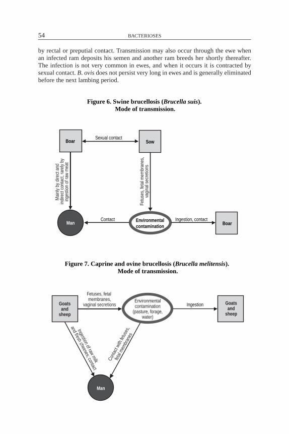
by rectal or preputial contact. Transmission may also occur through the ewe whenan infected ram deposits his semen and another ram breeds her shortly thereafter.The infection is not very common in ewes, and when it occurs it is contracted bysexual contact. B. ovis does not persist very long in ewes and is generally eliminatedbefore the next lambing period.
54 BACTERIOSES
Figure 7. Caprine and ovine brucellosis (Brucella melitensis). �Mode of transmission.
Goatsand
sheep
Ingestion
Man
Goatsand
sheep
Fetuses, fetal membranes,
vaginal secretionsEnvironmentalcontamination
(pasture, forage,water)
Ingestion of raw milk
and fresh cheeses; contact
Cont
act w
ith fe
tuse
s,
fetal
mem
bran
es
Figure 6. Swine brucellosis (Brucella suis). �Mode of transmission.
Boar Sow
Boar
Sexual contact
Environmentalcontamination
ManContact Ingestion, contact
Fetu
ses,
feta
l mem
bran
es,
vagi
nal s
ecre
tions
Mai
nly
by d
irect
and
indi
rect
con
tact
, rar
ely
byin
gest
ion
of ra
w m
eat

INFECTION IN DOGS: The transmission of B. canis occurs as a result of contact withvaginal secretions, fetuses, and fetal membranes. Infected males may transmit theinfection to bitches during coitus. The milk of infected bitches is another possiblesource of infection. Human cases recorded in the literature amount to several dozen,many resulting from contact with bitches that had recently aborted.
Role of Animals in the Epidemiology of the Disease: The role of animals isessential. Cases of human-to-human transmission are exceptional. Brucellosis is azoonosis par excellence.
Diagnosis: In man, a clinical diagnosis of brucellosis based on symptoms and his-tory should always be confirmed in the laboratory. Isolation and typing of the causalagent is definitive and may also indicate the source of the infection. Blood or marrowfrom the sternum or ileal crest taken while the patient is febrile is cultured in appro-priate media. Culture material may also be taken from lymph nodes, cerebrospinalfluid, and abscesses. It is recommended that the cultures be repeated several times,especially in enzootic areas of B. abortus. Due to the widespread use of antibioticsbefore diagnosis in febrile patients, bacteriologic examinations, particularly of blood,often yield negative results, and serologic tests become increasingly necessary. Theserum agglutination test, preferably in tubes, is the simplest and most widely usedprocedure. A high titer (more than 100 international units, IU) and increasing titersin repeated serum samples provide a good basis for diagnosis. Cross-reactions inserum agglutination have been observed in cases of cholera or tularemia (or as aresult of vaccination against these diseases) and in infections caused by Yersinia ente-rocolitica 0:9, as well as Escherichia coli 0:157 and 0:116, Salmonella serotypes ofKauffmann-White group N, and Pseudomonas maltophila (Corbel et al., 1984). Theserum agglutination test reveals both M and G immunoglobulins. It is generallyaccepted that in an active stage of brucellosis IgG is always present. Thus, when lowserum agglutination titers are found, tests to detect the presence of IgG must be per-formed, such as the 2-mercaptoethanol (ME) and complement fixation (CF) test (inman, IgGs fix complement but often lack agglutinating power). These tests are of spe-cial interest in chronic brucellosis, where active infection may continue even thoughagglutination titers return to low levels. The intradermal test with noncellular aller-gens is useful for epidemiological studies, but not for clinical diagnosis.
The 2-mercaptoethanol test is also useful in following the treatment and cure ofthe patient. In one study (Buchanan and Faber, 1980), the titers of 92 brucellosispatients were followed for 18 months with tube agglutination and ME tests. Despiteantibiotic treatment, the tube agglutination test continued positive for 18 months in44 (48%) of the patients, but the ME titers were positive in only 8 (9%) of thepatients at the end of one year and in 4 (4%) at 18 months. None of the 84 patientstesting negative by ME at the end of a year of treatment had signs or symptoms ofbrucellosis and none acquired chronic brucellosis. By contrast, four of the eightpatients testing positive by ME after a year continued to have symptoms of brucel-losis and had to continue treatment. Thus, a negative result by ME provides goodevidence that a patient does not have chronic brucellosis and that the antibiotic treat-ment was successful. If effective treatment is begun early, it is possible that IgG anti-bodies (resistant to ME) never develop. This was probably the case in three patientswho acquired brucellosis in the laboratory and in whom infection was confirmed byblood culture. Diagnosis and treatment were done early enough that at no time dur-
BRUCELLOSIS 55

ing the two-year follow-up did these patients show ME-resistant antibodies (García-Carrillo and Coltorti, 1979). However, other researchers dispute the usefulness ofthis test in the diagnosis of brucellosis (Díaz and Moriyon, 1989).
Other useful methods for the diagnosis of human brucellosis are the rose bengaltest and counterimmunoelectrophoresis. The rose bengal test is easily performed andis recommended over the plate agglutination test or the Huddleson method. In astudy of 222 cases (Díaz et al., 1982), rose bengal was the most sensitive test, with98.3% positive results. Counterimmunoelectrophoresis was positive in 84.9% of theacute cases and in 91.6% of the chronic cases.
The indirect enzyme-linked immunosorbent assay (ELISA) test has been used forsome years with good results in terms of specificity and sensitivity in research (Díazand Moriyón, 1989). It is a very versatile test and, once it is introduced in a labora-tory, it can be adapted for use with many other diseases.
The Joint FAO/WHO Expert Committee on Brucellosis (WHO, 1986) calls atten-tion to the limited value of serological tests in individuals who are repeatedly exposedto brucellae because they can be serologically positive in the absence of symptoms.This category would include veterinarians, vaccinators, and laboratory personnelinvolved in the production of antigens, vaccines, and cultures of clinical specimens.
In serologic diagnosis of humans or animals, it is necessary to bear in mind thatat the outset of the infection only IgM antibodies are produced; consequently, theagglutination test will provide the best standard for diagnosis, since ME will yieldnegative results. As the infection progresses, IgG antibodies resistant to the ME testwill appear and will increase unless appropriate treatment is begun.
Diagnosis of infection caused by B. melitensis, B. suis, and B. abortus is carriedout with a properly standardized antigen of B. abortus (Alton et al., 1976). However,this antigen does not permit diagnosis of infection caused by B. canis, since thisspecies of Brucella (as well as B. ovis) is found in a rugose (R) phase, lacking thelipopolysaccharidic surface that characterizes “classic brucellae” (for diagnosis ofB. canis and B. ovis, see below).
In cattle, the diagnosis is based primarily on serology. A great many serologictests are presently available, all of which are useful when applied with judgment.Both a serologic test reaction and the test’s usefulness in each circumstance are aresult of the sensitivity it shows to antibodies of different immunoglobulin types andof the seric concentration of each type of antibody (Chappel et al., 1978). The mostthoroughly studied immunoglobulins in bovine brucellosis are IgM and IgG.Although available tests are not qualitative enough to identify an individualimmunoglobulin, they do indicate which one predominates. In the diagnosis ofbovine brucellosis, the evolution of immunoglobulins during infection and vaccina-tion is of special interest. In both cases, the IgMs appear first, followed by the IgGs.The difference is that in infected animals, the IgGs tend to increase and persist,while in calves vaccinated at between three and eight months, the IgGs tend to dis-appear about six months after vaccination. Based on this fact, complementary testsare used to distinguish infection from the agglutination titer, which may persist aftervaccination with strain 19, and also from heterospecific reactions caused by bacte-ria that share surface antigens with the brucellae and that give rise to antibodies that,in general, are the IgM type.
According to their use in different countries, serologic tests may be classified asfollows: (1) routine or operative, (2) complementary, (3) epidemiological surveil-
56 BACTERIOSES

lance, and (4) screening tests. A single test might serve as operative, as diagnosticallydefinitive, as a screen, or as complementary, depending on the program employing it.
Serum agglutination tests (tube and plate) have been and continue to be widelyused. They contributed greatly to the reduction of infection rates in Europe, Australia,and the Americas. Nevertheless, when the proportion of infected herds and world-wide prevalence is reduced, their limitations become apparent in so-called “problem”herds and it becomes necessary to use other tests to help eradicate the infection. Thetests are internationally standardized, easy to carry out, and allow the examination ofa great many samples. In agglutination tests, the IgM reaction predominates. In ani-mals classified as suspect or marginally positive, complementary tests are used toclarify their status. However, it is necessary to keep in mind that low agglutinationtiters could be due to recent infection and it is thus advisable to repeat the test.
The rose bengal test (with buffered antigen) is fast, easy, and allows processing ofmany samples per day. It is qualitative and classifies animals as positive or negative.In regions where incidence of infection is low or where systematic vaccination ofcalves is practiced, the rose bengal test gives many “false positives,” and so is unspe-cific if used as the only and definitive test. In many countries, such as Great Britainand Australia, it is used as a preliminary or screening test. Animals showing a neg-ative test result are so classified and those testing positive are subjected to other testsfor confirmation. In regions of high incidence, results are very satisfactory. Rosebengal may also be used as a complementary test for those animals classified as sus-pect by agglutination. Many suspect sera test negative to rose bengal, and since thistest is very sensitive (there are few “false negatives”) and detects the infection early,there is little risk of missing infected animals.
The principal complementary tests are complement fixation, 2-mercaptoethanol,and rivanol. Other tests have been developed, such as indirect hemolysis, ELISA fordifferent types of immunoglobulins, and radial immunodiffusion with a polysaccha-ride antigen. All these tests are used to distinguish antibodies caused by the infec-tion from those left by vaccination or stimulated by heterospecific bacteria.
Both the direct and the competitive ELISA tests are appropriate for diagnosis ofbrucellosis in all species according to the consensus of groups of experts that havemet several times in Geneva. WHO, with the collaboration of FAO, the InternationalOrganization of Epizootics, the International Atomic Energy Agency, and theseorganizations’ reference laboratories, is coordinating a project to evaluate and stan-dardize these assays as well as the antigens and other technical variables.
In Australia, the ELISA technique and the complement fixation test have beenvery useful in recent phases in the eradication of bovine brucellosis, when many“problem herds” occur with “latent carrier animals.” In comparison with the CF test,ELISA revealed a significantly higher number of reactive animals in infected herds,both vaccinated (with strain 19) and unvaccinated, but gave negative results in herdsfree of brucellosis, whether vaccinated or not. The specificity of ELISA in the groupof infected herds was less than that of CF, but sensitivity—which is what wasneeded—was greater (Cargill et al., 1985). It costs less to eliminate some false pos-itive animals in the final phase of eradication than to allow the infection to reassertitself and spread in the herd because one or more infected animals remained(Sutherland et al., 1986). The competitive enzymatic immunoassay also lends itselfto differentiating the reactions of animals vaccinated with strain 19 and animals nat-urally infected, using the O polysaccharide antigen (Nielsen et al., 1989).
BRUCELLOSIS 57

The complement fixation test is considered the most specific, but it is laborious,complicated, and involves many steps and variables. Moreover, it is not standardizedinternationally. Other, simpler tests can take its place, such as 2-mercaptoethanoland rivanol, which measure the IgG antibodies.
Animal health laboratories in the US and various laboratories in Latin Americahave successfully used the BAPA (buffered antigen plate agglutination) or BPA(buffered plate antigen) screening test, which is performed on a plate with a bufferedantigen at pH 3.65 (Angus and Barton, 1984). BAPA greatly simplifies the workwhen numerous blood samples must be examined, because it eliminates the negativesamples and many of the sera with nonspecific reactions. The test results classify thesamples as negative (which are definitively discarded) and presumably positive; thelatter are submitted to one or more definitive and/or complementary tests, such astube agglutination, complement fixation, or 2-mercaptoethanol. This test was alsoevaluated in Canada (Stemshorn et al., 1985) and Argentina (González Tomé et al.,1989) with very favorable results.
Epidemiological surveillance of brucellosis is carried out separately on dairy andmeat-producing herds, at strategic checkpoints and using different diagnostic tests.The principal objective is to identify infected herds and monitor healthy ones. Forbeef cattle, screening tests or other tests of presumed high sensitivity are used, suchas BAPA, and the checkpoints for collecting samples are cattle markets and slaugh-terhouses. The sera that test positive are then subjected to standard tests and theanimals are traced back to their points of origin. For dairy cattle, the milk-ring testis used. It is very simple and allows the examination of many herds in a short time.The composite samples are gathered from milk cans or tanks at collection pointsand dairy plants or on the dairy farm itself. If a positive sample is found, individ-ual serologic examinations of the animals belonging to the source herd must thenbe carried out.
Bacteriologic examinations are of more limited use. The samples most oftentested in this way are taken from fetuses, fetal membranes, vaginal secretions, milk,and semen. Infected cows may or may not abort, but a high percentage will elimi-nate brucellae from the genital tract beginning a few days before parturition andcontinuing some 30 days afterwards. It is estimated that 85% of recently infectedcows and more than 15% of chronically infected cows eliminate brucellae duringcalving. Since elimination through milk may be constant or intermittent, milk canbe an excellent material for the isolation of Brucella if examinations are repeated.Serologic testing of bulls should be done using blood serum and seminal fluid.Bacteriologic examination of semen should be repeated if results are negative, sincebrucellae may be shed intermittently.
In swine, serologic tests are not indicated for individual diagnosis but rather toreveal the presence of herd infection. Agglutination (tube or plate), complement fix-ation, buffered-acid antigen (rose bengal), or BAPA tests may be used. The latter ispreferable because it is negative in herds having only low and nonspecific aggluti-nation (tube or plate) titers. For a herd to be classified as positive with the aggluti-nation test (tube or plate), there must be one or more animals with titers of 100 IUor more.
In goats, serologic tests are also applied on a flock basis and not on individual ani-mals. In infected flocks, one or more individuals are found with titers of 100 IU ormore; in such cases, titers of 50 IU should be adopted as indicative of infection. The
58 BACTERIOSES

complement fixation test is considered superior to the agglutination test, especiallyin herds vaccinated with B. melitensis Rev. 1, where agglutinating antibodies persistfor long periods. The 2-mercaptoethanol test has also given very good results in vac-cinated flocks. The results from the buffered-acid antigen (rose bengal) test arepromising, but experience with it is limited and definitive conclusions cannot bedrawn at this time. The ELISA test is the most promising.
In diagnosing infections caused by B. melitensis in sheep, the Coombs’ test(antiglobulin test) modified by Hajdu can reveal 70% of infected animals. The othertests (agglutination, complement fixation) give less satisfactory results. In using theagglutination and complement fixation tests, adoption of significant titer levelslower than those for other animal species is recommended. Counterimmunoelectro-phoresis would detect antibodies against intracellular antigens that appear late in theserum but which remain a long time. Consequently, its use would be appropriate forsheep with chronic brucellosis that test negative by agglutination, rose bengal, andcomplement fixation (Trap and Gaumont, 1982). Experts agree that the diagnosticmethods for brucellosis caused by B. melitensis in goats and sheep leave much to bedesired and that more attention should be given to this problem given its publichealth importance.
In diagnosing ram epididymitis caused by B. ovis, antigen prepared with thisagent must be used. The preferred tests are gel diffusion, complement fixation, andELISA. A study conducted in Australia in flocks infected by B. ovis and flocks freeof infection showed that this enzyme immunoassay detected more reactive animalsand that the complement fixation test failed to detect some rams that excreted B.ovis. In infection-free flocks, both ELISA and CF produced false positives at a rateof 0.5% (Lee et al., 1985). Bacteriologic examination of semen is an appropriatediagnostic method, but it should be kept in mind that the shedding of brucellae canbe intermittent.
For dogs infected by B. canis, the surest diagnostic method is isolation of the eti-ologic agent from blood, vaginal discharges, milk, or semen, or from fetal tissue andplacenta. Bacteremia can last from one to two years, but after the initial phase it maybecome intermittent; thus, a negative blood culture does not exclude the possibilityof brucellosis.
The most common serologic tests are plate and tube agglutination using B. canisantigen, immunodiffusion in agar gel with antigens extracted from the cell wall, 2-mercaptoethanol plate agglutination, and the modified 2 ME tube agglutination test.Possibly the most specific test to date, but also the least sensitive, is the immuno-diffusion test in agar gel that utilizes antigens extracted from the cytoplasm of B.canis. To a greater or lesser degree, all these tests give nonspecific reactions. Zohaand Carmichael (1982) showed that the immunodiffusion test using sonicated anti-gens (internal cellular antigens) is satisfactory shortly after the onset of bacteremiaand can detect infected animals for up to six months after it disappears, i.e., whenother tests give equivocal results. A new test has been developed that uses a non-mucoid (M–) variant of B. canis as the antigen in tube agglutination, after treatingthe sera with 2 ME. The test is more specific without reducing sensitivity(Carmichael and Joubert, 1987).
Control: The most rational approach for preventing human brucellosis is the con-trol and elimination of the infection in animal reservoirs, as has been demonstrated
BRUCELLOSIS 59

in various countries in Europe and the Americas. Some human populations may beprotected by mandatory milk pasteurization. In many goat- and sheep-herdingregions, pasteurization of milk is an unattainable goal for the time being. Preventionof the infection in occupational groups (cattlemen, abattoir workers, veterinarians,and others who come into contact with animals or their carcasses) is more difficultand should be based on health education, the use of protective clothing wheneverpossible, and medical supervision.
Protecting refrigerator plant and slaughterhouse workers against brucellosis isparticularly important because they constitute the occupational group at highest risk.Protection is achieved by separating the slaughter area from other sections and con-trolling air circulation. In countries with eradication programs, slaughter of reactiveanimals is limited to one or more designated slaughterhouses (cold storage plants)with official veterinary inspection in each region. These animals are butchered at theend of the workday with special precautions and proper supervision to protect theworkers. Employees should be instructed in personal hygiene and provided with dis-infectants and protective clothing. A 5% solution of chloramine or an 8% to 10%solution of caustic soda should be used to disinfect installations after slaughter(Elberg, 1981). Instruments should be sterilized in an autoclave or boiled for 30minutes in a 2% solution of caustic soda. Clothes may be disinfected with a 2%solution of chloramine or a 3% solution of carbolic acid soap followed by washing.Hands should be soaked for five minutes in a solution of 1% chloramine or 0.5%caustic soda, and then washed with soap and water.
The immunization of high-risk occupational groups is practiced in the formerSoviet Union and China. In the former Soviet Union, good results have apparentlybeen obtained with the use of a vaccine prepared from strain 19-BA of B. abortus(derived from strain 19 used for bovine brucellosis), applied by skin scarification.Annual revaccination is carried out for those individuals not reacting to serologic orallergenic tests. To avoid the discomfort caused by the vaccine in man, a vaccine wasrecently developed that consists of chemically defined fractions of the lipid-poly-saccharide (LPS) component of the strain 19-BA (Drannovskaia, 1991). In China,an attenuated live vaccine made from B. abortus strain 104M is applied percuta-neously. These vaccines are not used in other countries because of possible sideeffects. Promising trials have also been conducted in France with antigenic fractionsof Brucella.
Vaccination is recommended for control of bovine brucellosis in enzootic areaswith high prevalence rates. The vaccine of choice is B. abortus strain 19, confirmedby its worldwide use, the protection it gives for the useful lifetime of the animal, andits low cost. To avoid interference with diagnosis, it is recommended that vaccina-tion be limited (by legislation) to young animals (calves of 3 to 8 months), as theseanimals rapidly lose the antibodies produced in response to the vaccine. It is esti-mated that 65% to 80% of vaccinated animals remain protected against the infec-tion. The antiabortive effect of the vaccine is pronounced, thus reducing one of theprincipal sources of infection, the fetuses. In a systematic vaccination program, thebest results are obtained with 70% to 90% annual coverage in calves of the properage for vaccination. Male calves and females over 8 months of age should not bevaccinated. Where possible, the upper limit should be 6 months. Revaccination isnot recommended. The main objective of systematic and mandatory vaccination ofcalves in a given area or country is to reduce the infection rate and obtain herds
60 BACTERIOSES

resistant to brucellosis, so that eradication of the disease may then begin. It is esti-mated that 7 to 10 years of systematic vaccination are necessary to achieve thisobjective.
In regions or countries with a low prevalence of the disease, an eradication pro-gram can be carried out by repeated serologic diagnostic tests applied to the entireherd, and elimination of reactors until all foci of infection have disappeared. Thisprocedure can be used alone (in countries with a low prevalence) or in combinationwith the vaccination of calves. Epidemiological surveillance and control of animaltransport are very important in such programs. Countries that are close to eradica-tion may suspend vaccination with strain 19 or any other vaccine.
Until a few years ago, the vaccination of adult cows with strain 19 was inadvis-able because of the prolonged resistance of antibodies that could interfere withdiagnosis. In the 1950s, several researchers proved that vaccination of adult animalswith a smaller dose could impart an immunity comparable to that of a full dose,while at the same time agglutination titers stayed lower and disappeared faster. In1975, Nicoletti (1976) began a series of studies in the US using a reduced dose inhighly infected herds; he concluded that vaccination decreases the spread of theinfection within the herd, that antibodies disappear approximately six months aftervaccination, and that less than 1% of the females remained infected by the vaccinestrain from three to six months after vaccination. Complementary tests were veryuseful in distinguishing between reactions due to infection and those due to vacci-nation. Other studies, done under both controlled and natural conditions, confirmedthese findings (Nicoletti et al., 1978; Alton et al., 1980; Viana et al., 1982; Alton etal., 1983). Vaccination of adult females may be considered in herds suffering acutebrucellosis characterized by abortions and rapidly spreading infections, as well as inlarge herds where chronic brucellosis has proven hard to eradicate. The recom-mended dose is one to three billion cells of strain 19 Brucella administered subcu-taneously. Only animals testing negative should be vaccinated and they should beindelibly marked under government supervision. At the beginning of the operation,reactors should be eliminated immediately. Vaccinated animals should be examinedserologically 6 months later, using tests such as rivanol, mercaptoethanol, and com-plement fixation, and those that have become infected should be slaughtered. Usingperiodic serologic examination, it is estimated that a problem herd can be free ofinfection in 18 to 24 months (Barton and Lomme, 1980).
The control of swine brucellosis consists of identifying and certifying brucellosis-free herds. If infection is diagnosed in an establishment where pigs are raised formarket, it is advisable to send the entire herd to the abattoir and reestablish it withanimals from a brucellosis-free herd. If the infected pigs are valuable for breedingor research, suckling pigs should be weaned at 4 weeks and raised in facilities sep-arate from the main herd. Periodic serologic tests (such as rose bengal) are recom-mended to eliminate any reactor. Finally, when no brucellosis is found in the newherd and it is well established, the original herd should be sent to slaughter. Thereare no effective vaccines for swine.
Control of the infection caused by B. melitensis in goats and sheep is basedmainly on vaccination. The preferred vaccine is B. melitensis Rev. 1, which isadministered to 3- to 6-month-old females. Adult females can receive a smaller dose(20,000 times fewer bacterial cells than in the dose for young females) of the samevaccine (Alton et al., 1972).
BRUCELLOSIS 61

As goats are generally raised in marginal areas where socioeconomic conditionsare very poor, it is difficult to carry out eradication programs. In these areas, rein-fection occurs constantly, flocks are often nomadic, and animal-raising practicesmake sanitary control difficult. Another important factor is that diagnostic methodsfor small ruminants are deficient. Experience with Rev. 1 vaccine in Italy, Turkey,Iran, Mongolia, Peru, and the Caucasian Republics of the former Soviet Union hasproven it to be an excellent means of control. In Mongolia, 6 million animals werevaccinated between 1974 and 1977; as a result, the prevalence of from 3 to 4 per10,000 animals was reduced by half or more, as was the incidence of human cases.In Malta, after seven years of vaccination of small ruminants with Rev. 1, the num-ber of human cases per year fell from 260 to 29. The same thing happened in Italy,although there are no reference data (Alton, 1987). However, the control procedureof diagnosing and sacrificing reactor animals has produced satisfactory results inareas of low prevalence.
Rev. 1 vaccine has some limitations, such as residual virulence, the possibility ofabortions when pregnant females are vaccinated, and the limited stability of the vac-cine, which necessitates constant monitoring. These disadvantages should not elim-inate use of the vaccine as the basis for control of brucellosis in small ruminants, atleast until there is a better vaccine. A Chinese strain of B. suis biovar 1, known as B.suis strain 2, is considered reliable. This strain was isolated from a swine fetus andits virulence was attenuated by continuous and repeated replications in culturemedia over years, reaching an attenuation that remains stable. Vaccine B. suis strain2 has been used in China with very good results for more than 20 years, not only insmall ruminants but in cattle and swine as well. Its use began in the semiarid regionsof northern China, where vaccination operations were very difficult due to the lackof fetters and traps, and thus the vaccine was administered in the drinking water(Xin, 1986). Various research institutes have conducted tests on conjunctive, oral(with syringes of the type used to administer antiparasitic agents), and subcutaneousvaccines in small ruminants; it has generally been possible to confirm the resultsobtained in China. Elimination of the vaccine strain in milk or through the vaginahas not been confirmed and studies continue on this vaccine.
Ram epididymitis can be successfully controlled by a combination of the follow-ing measures: elimination of rams with clinically recognizable lesions, eliminationof clinically normal rams positive to the gel diffusion or the complement fixationtest, and separation of young rams (those not yet used for breeding) from adultmales. In some countries, such as New Zealand and the US, a bacterin prepared fromB. ovis and adjuvants is used. Animals are vaccinated when weaned, revaccinatedone or two months later and annually thereafter. This vaccine produces antibodiesagainst B. ovis but not B. abortus. The B. melitensis Rev. 1 vaccine is effectiveagainst epididymitis, but also produces B. abortus antibodies, which could be con-fused with infection by B. melitensis. The B. suis strain 2 vaccine does not provideprotection against ram epididymitis.
Brucellosis caused by B. canis in dog kennels can be controlled by repeated sero-logic tests and blood cultures, followed by elimination of reactor animals. No vac-cines are available yet. Veterinary clinics should advise owners of the risk of keep-ing a dog with brucellosis and should recommend that the dog be put to sleep.
62 BACTERIOSES

Bibliography
Acosta, M., H. Ludueña, D. Barreto, M. Moro. Brucellosis en alpacas. Rev Invest Pec1:37–49, 1972.
Al-Khalaf, S., A. El-Khaladi. Brucellosis in camels in Kuwait. Com Immun MicrobiolInfect Dis 12:1–4, 1989.
Alton, G.G. The epidemiology of Brucella melitensis infection in sheep and goats. In:Verger, J.M., M. Plommet, eds. Brucella melitensis. Seminar held in Brussels, 14–15November 1984. Dordrecht: Martinus Nijhoff; 1985.
Alton, G.G. Control of Brucella melitensis infection in sheep and goats—A review. TropAnim Health Prod 19:65–74, 1987.
Alton, G.G., L.A. Corner, P.P. Plackett. Vaccination of pregnant cows with low doses ofBrucella abortus strain 19 vaccine. Aust Vet J 56:369–372, 1980.
Alton, G.G., L.A. Corner, P.P. Plackett. Vaccination of cattle against brucellosis. Reduceddoses of strain 19 compared with one and two doses of 45/20 vaccine. Aust Vet J 60:175–177,1983.
Alton, G.G., L.M. Jones, C. García-Carrillo, A. Trenchi. Brucella melitensis Rev. 1 andBrucella abortus 45/20 vaccines in goats: Immunity. Am J Vet Res 33:1747–1751, 1972.
Alton, G.G., L.M. Jones, D.E. Pietz. Laboratory techniques in brucellosis. 2nd ed. Geneva:World Health Organization; 1975. (Monographs Series 55).
Anczykowski, F. Further studies on fowl brucellosis. II. Laboratory experiments. Pol ArchWet 16:271–292, 1973.
Angus, R.D., C.E. Barton. The production and evaluation of a buffered plate antigen for usein a presumptive test for brucellosis. In: Third International Symposium on Brucellosis,Algiers, Algeria, 1983. Developments in Biological Standardization. Basel: Karger; 1984.
Banai, M., I. Mayer, A. Cohen. Isolation, identification, and characterization in Israel ofBrucella melitensis biovar 1 atypical strains susceptible to dyes and penicillin, indicating theevolution of a new variant. J Clin Microbiol 28:1057–1059, 1990.
Barg, L. Isolamento de Brucella canis em Minas Gerais, Brazil. Pesquisa de aglutininas emsoros caninos e humanos [thesis]. Belo Horizonte: Universidad Federal de Minas Gerais, 1975.
Barton, C.E., J.R. Lomme. Reduced-dose whole herd vaccination against brucellosis: Areview of recent experience. J Am Vet Med Assoc 177:1218–1220, 1980.
Benenson, A.S., ed. Control of Communicable Diseases in Man. 15th ed. An official reportof the American Public Health Association. Washington, D.C.: American Public HealthAssociation; 1990.
Buchanan, T.M., L.C. Faber. 2-mercaptoethanol brucella agglutination test: Usefulness forpredicting recovery from brucellosis. J Clin Microbiol 11:691–693, 1980.
Buchanan, T.M., S.L. Hendricks, C.M. Patton, R.A. Feldman. Brucellosis in the UnitedStates. An abattoir-associated disease. Part III. Epidemiology and evidence for acquiredimmunity. Medicine 53:427–439, 1974.
Cargill, C., K. Lee, I. Clarke. Use of an enzyme-linked immunosorbent assay in a bovinebrucellosis eradication program. Aust Vet J 62:49–52, 1985.
Carmichael, L.E., J.C. Joubert. A rapid agglutination test for the serodiagnosis of Brucella canis infection that employs a variant (M–) organism as antigen. Cornell Vet77:3–12, 1987.
Chappel, R.J., D.J. McNaught, J.A. Bourke, G.S. Allen. Comparison of the results of someserological tests for bovine brucellosis. J Hyg 80:365–371, 1978.
Chile, Ministerio de Agricultura, Servicio Agrícola and Ganadero. Brucellosis bovina,ovina and caprina. Diagnóstico, control, vacunación. Paris: Office International desEpizooties; 1987. (Technical Series 6).
Corbel, M.J. The serological relationship between Brucella spp., Yersinia enterocoliticaserotype IX and Salmonella serotypes of Kauffman-White group. J Hyg 75:151–171, 1975.
BRUCELLOSIS 63

Corbel, M.J., F.A. Stuart, R.A. Brewer. Observations on serological cross-reactionsbetween smooth Brucella species and organisms of other genera. In: Third InternationalSymposium on Brucellosis, Algiers, Algeria, 1983. Developments in BiologicalStandardization. Basel: Karger; 1984.
Corbel, M.J., E.L. Thomas, C. García-Carrillo. Taxonomic studies on some atypical strainsof Brucella suis. Br Vet J 140:34–43, 1984.
De Lord, V., S. Nieto, E. Sandoval et al. Brucellosis en caprinos: estudios serológicos andbacteriológicos en Venezuela. Vet Trop 12:27–37, 1987.
De la Vega, E., C. García-Carrillo, C. Arce. Infección natural por Brucella en comadrejasDidelphis marsupialis en la República Argentina. Rev Med Vet 60:283–286, 1979.
Díaz, R., E. Maravi-Poma, J.L. Fernández, S. García-Merlo, A. Rivero-Puente. Brucellosis:estudio de 222 casos. Parte IV: Diagnóstico de la brucellosis humana. Rev Clin Esp166:107–110, 1982.
Díaz, R., I. Moriyon. Laboratory techniques in the diagnosis of human brucellosis. In:Young, E.J., M.J. Corbel, eds. Brucellosis: Clinical and Laboratory Aspects. Boca Raton:CRC Press; 1989.
Drannovskaia, E. Brucella and brucellosis in man and animals. Izmir, Turkey, 1990. TurkishMicrobiol Soc 16:87–100, 1991.
Elberg, S.S. The Brucellae. In: Dubos, R.J., J.G. Hirsch, eds. Bacterial and MycoticInfections of Man. 4th ed. Philadelphia and Montreal: Lippincott; 1965.
Elberg, S.S. Immunity to Brucella infection. Medicine 52:339–356, 1973.Elberg, S.S. A guide to the diagnosis, treatment and prevention of human brucellosis.
Geneva: World Health Organization; 1981. VPH/81.31 Rev.1. (Unpublished document).Ewalt, D.R., L.B. Forbes. Atypical isolates of Brucella abortus from Canada and the
United States characterized as dye sensitive with M antigen dominant. J Clin Microbiol25:698–701, 1987.
Fensterbank, R. Congenital brucellosis in cattle. Geneva: World Health Organization; 1980.WHO/BRUC/80.352. (Unpublished document).
Flores-Castro, R., F. Suárez, C. Ramírez-Pfeiffer, L.E. Carmichael. Canine brucellosis:Bacteriological and serological investigation of naturally infected dogs in Mexico City. J ClinMicrobiol 6:591–597, 1977.
Forbes, L.B. Brucella abortus infection in 14 farm dogs. J Am Vet Med Assoc 196:911–916, 1990.
Fredickson, L.E., C.E. Barton. A serologic survey for canine brucellosis in a metropolitanarea. J Am Vet Med Assoc 165:987–989, 1974.
García-Carrillo, C. Métodos para el diagnóstico de la brucellosis. Gac Vet 32:661–667, 1970. García-Carrillo, C. Programa de erradicación de la brucellosis en California. Buenos
Aires: Centro Panamericano de Zoonosis, 1975. (Scientific and Technical Monographs 9).García-Carrillo, C., E.A. Coltorti. Ausencia de anticuerpos resistentes al 2-marcaptoetanol
en tres pacientes de brucellosis. Medicina 39:611–613, 1979.García-Carrillo, C., N.E. Lucero. Brucellosis bovina. Buenos Aires: Hemisferio Sur; 1993.García-Carrillo, C., B. Szyfres, J. González Tomé. Tipificación de brucelas aisladas del
hombre and los animales en Latin America. Rev Latinoam Microbiol 14:117–125, 1972.George, L.W., L.E. Carmichael. A plate agglutination test for the rapid diagnosis of canine
brucellosis. Am J Vet Res 35:905–909, 1974. Gilman, H.L. Brucellosis. In: Gibbons, W.J., ed. Diseases of Cattle, a text and reference
work. The work of 54 authors. Wheaton: American Veterinary Publication; 1963.González Tomé, J.S., L.J. Villa, E. del Palacio, R. Gregoret. El test de Angus and Barton (BPA)
como prueba tamiz en el diagnóstico de la brucellosis bovina. Rev Med Vet 70:34–36, 1989.Hendricks, S.L., M.E. Meyer. Brucellosis. In: Hubbert, W.T., W.F. McCulloch, P.R.
Schnurrenberger, eds. Diseases Transmitted from Animals to Man. 6th ed. Springfield:Thomas; 1975.
64 BACTERIOSES

Kasyanov, A.N., R.G. Aslanyan. Epizootiology and clinical appearance of animal brucel-losis. In: Lisenko, A., ed. Zoonoses Control. Moscow: VII Centre Projects; 1982.
Kaufmann, A.F., M.D. Fox, J.M. Boyce, D.C. Anderson, M.E. Potter, W.J. Martone, et al.Airborne spread of brucellosis. Ann NY Acad Sci 353:105–114, 1980.
Lapraik, R.D., D.D. Brown, H. Mann. Brucellosis. A study of five calves from reactordams. Vet Rec 97:52–54, 1975.
Lee, K., C. Cargill, H. Atkinson. Evaluation of an enzyme-linked immunosorbent assay forthe diagnosis of Brucella ovis infection in rams. Aust Vet J 62:91–93, 1985.
Lu, S-L., J-L. Zhang. Brucellosis in China. In: Young, E.J., M.J. Corbel, eds. Brucellosis:Clinical and Laboratory Aspects. Boca Raton: CRC Press; 1989.
Lubani, M., D. Sharda, I. Helin. Probable transmission of brucellosis from breast milk to anewborn. Trop Geogr Med 40:151–152, 1988.
Luchsinger, D.W., R.K. Anderson. Longitudinal studies of naturally acquired Brucellaabortus infection in sheep. Am J Vet Med Res 40:1307–1312, 1979.
Manthei, C.A. Brucellosis as a cause of abortion today. In: Faulkner, L.C., ed. AbortionDiseases of Livestock. Springfield: Thomas; 1968.
Manthei, C.A. Brucellosis. In: Dunne, H.W., ed. Diseases of Swine. 3rd ed. Ames: IowaState University Press; 1970.
McCaughey, W.J. Brucellosis in wildlife. In: Diarmid, A., ed. Diseases in Free-living WildAnimals. New York: Academic Press; 1969.
McCullough, N.B. Microbial and host factors in the pathogenesis of brucellosis. In: Mudd,S., ed. Infectious Agents and Host Reactions. Philadelphia: Saunders; 1970.
Meyer, M.E. Inter- and intra-strain variants in the Genus Brucella. In: Third InternationalSymposium on Brucellosis, Algiers, Algeria, 1983. Developments in BiologicalStandardization. Basel: Karger; 1984.
Morgan, W.J.B. Brucella classification and regional distribution. In: Third InternationalSymposium on Brucellosis, Algiers, Algeria, 1983. Developments in BiologicalStandardization. Basel: Karger; 1984.
Myers, D.M., L.M. Jones, V.M. Varela-Díaz. Studies of antigens for complement fixationand gel diffusion tests in the diagnosis of infections caused by Brucella ovis and otherBrucella. Appl Microbiol 23:894–902, 1972.
Nicoletti, P. A preliminary report on the efficacy of adult cattle vaccination using strain 19in selected dairy herds in Florida. Proc Annu Meet US Livest Sanit Assoc 80:91–106, 1976.
Nicoletti, P., L.M. Jones, D.T. Berman. Adult vaccination with standard and reduced dosesof Brucella abortus strain 19 in a dairy herd infected with brucellosis. J Am Vet Med Assoc173:1445–1449, 1978.
Nicoletti, P., F.W. Milward. Protection by oral administration of Brucella abortus strain 19against an oral challenge exposure with a pathogenic strain of Brucella. Am J Vet Res44:1641–1643, 1983.
Nielsen, K., J.W. Cherwonogrodzky, J.R. Duncan, D.R. Bundle. Enzyme-linkedimmunosorbent assay for differentiation of the antibody response of cattle naturally infectedwith Brucella abortus or vaccinated with strain 19. Am J Vet Res 50:5–9, 1989.
Ossola, A.L., B. Szyfres. Natural infection of sheep by Brucella melitensis in Argentina. AmJ Vet Res 24:446–449, 1963.
Pacheco, G., M.T. De Mello. Brucelose. Rio de Janeiro: Instituto Brasileiro de Geografia eEstatística; 1956.
Pan American Health Organization. Guía para la preparación and evaluación de proyectosde lucha contra la brucellosis bovina. Buenos Aires: Centro Panamericano de Zoonosis; 1972.(Technical Note 14).
Peres, J.N., A.M. Godoy, L. Barg, J.O. Costa. Isolamento de Brucella canis de carrapatos(Rhipicephalus sanguineus). Arq Esc Vet UFMG 33:51–55, 1981.
BRUCELLOSIS 65

Pfischner, W.C.E., K.G. Ishak, E. Neptune, et al. Brucellosis in Egypt: A review of experi-ence with 228 patients. Am J Med 22:915, 1957. Cited in: Young, E.J. Clinical manifestationsof human brucellosis. In: Young, E.J., M.J. Corbel, eds. Brucellosis: Clinical and LaboratoryAspects. Boca Raton: CRC Press; 1989.
Plommet, M., R. Fensterbank. Vaccination against bovine brucellosis with a low dose ofstrain 19 administered by the conjunctival route. III.—Seriological response and immunity inthe pregnant cow. Ann Rech Vét 7:9–23, 1976.
Plommet, M., R. Fensterbank, G. Renoux, J. Gestin, A. Philippon. Brucellose bovineexpérimentale. XII. Persistance a l’age adulte de l’infection congénitale de la génisse. AnnRech Vét 4:419–435, 1973.
Ramacciotti, F. Brucelosis. Córdoba, Argentina: Edición del Autor; 1971. Ray, W.C., R.B. Brown, D.A. Stringfellow, et al. Bovine brucellosis: An investigation of
latency in progeny of culture-positive cows. J Am Vet Med Assoc 192:182–185, 1988.Robertson, F.J., J. Milne, C.L. Silver, H. Clark. Abortion associated with Brucella abortus
(biovar 1) in the T.B. mare. Vet Rec 92:480–481, 1973.Ruben, B., J.D. Band, P. Wong, J. Colville. Person-to-person transmission of Brucella
melitensis. Lancet 337:14–15, 1991.Schwabe, C.W. Veterinary Medicine and Human Health. 2nd ed. Baltimore: Williams &
Wilkins; 1969. Spink, W.W. The Nature of Brucellosis. Minneapolis: University of Minnesota Press; 1956.Spink, W.W. Brucellosis (Undulant fever, Malta fever). In: Wyngaarden, I.B., L.H. Smith,
Jr., eds. Cecil Textbook of Medicine. 16th ed. Philadelphia: Saunders; 1982. Stemshorn, B.W., L.B. Forbes, M.D. Eaglesome, et al. A comparison of standard sero-
logical tests for the diagnosis of bovine brucellosis in Canada. Can J Comp Med 49:391–394, 1985.
Sutherland, S.S., R.J. Evans, J. Bathgate. Application of an enzyme-linked immuno-sorbent assay in the final stages of a bovine brucellosis eradication program. Aust Vet J63:412–415, 1986.
Szyfres, B. La situación de la brucellosis en Latin America. Bol Hig Epidemiol5:400–409, 1967.
Szyfres, B. Taxonomía del género Brucella. Gac Vet 33:28–40, 1971. Szyfres, B., J. González Tomé. Natural Brucella infection in Argentine wild foxes. Bull
World Health Organ 34:919–923, 1966.Szyfres, B., J. González Tomé, T. Palacio Mendieta. Aislamiento de Brucella suis de
la liebre europea (Lepus europaeus) en la Argentina. Bol Oficina Sanit Panam 65:441–445, 1968.
Timm, B.M. Brucellosis. Distribution in Man, Domestic and Wild Animals. Berlin:Springer; 1982.
Trap, D., R. Gaumont. Comparaison entre electrosynerèse et épreuves serologiques clas-siques dans le diagnostic de la brucellose ovine. Ann Rech Vét 13:33–39, 1982.
United States, Department of Health and Human Services, Centers for Disease Control andPrevention (CDC). Annual summary 1981: Reported morbidity and mortality in the UnitedStates. MMWR Morb Mort Wkly Rep 30:14, 1982.
Van der Hoeden, J. Brucellosis. In: Van der Hoeden, J., ed. Zoonoses. Amsterdam:Elsevier; 1964.
Verger, J.M., F. Grimont, P.A.D. Grimont, M. Grayon. Brucella: a monospecific genus asshown by deoxyribonucleic acid hybridization. Int J Syst Bacteriol 35:292–295, 1985.
Viana, F.C., J.A. Silva, E.C. Moreira, L.G. Villela, J.G. Mendez, T.O. Dias. Vacinação con-tra brucelose bovina com dose reduzida (amostra B19) por via conjuntival. Arq Esc Vet UFMG34:279–287, 1982.
Wilesmith, J.W. The persistence of Brucella abortus infection in calves: A retrospectivestudy of heavily infected herds. Vet Rec 103:149–153, 1978.
66 BACTERIOSES

Witter, J.F., D.C. O’Meara. Brucellosis. In: Davis, J.W., L.H. Karstady, D.O. Trainer.Infectious Diseases of Wild Mammals. Ames: Iowa State University Press; 1970.
World Health Organization (WHO). Fifth Report on the World Health Situation,1969–1972. Geneva: WHO; 1975. (Official Records 225).
World Health Organization (WHO). Joint FAO/WHO Expert Committee on Brucellosis,Sixth Report. Geneva: WHO; 1986. (Technical Report Series 740).
Xin, X. Orally administrable brucellosis vaccine: Brucella suis strain 2 vaccine. Vaccine4:212–216, 1986.
Young, E.J. Clinical manifestations of human brucellosis. In: Young, E.J., M.J. Corbel, eds.Brucellosis: Clinical and Laboratory Aspects. Boca Raton: CRC Press; 1989.
Zoha, S.J., L.E. Carmichael. Serological response of dogs to cell wall and internal antigensof Brucella canis (B. canis). Vet Microbiol 7:35–50, 1982.
CAMPYLOBACTERIOSIS
ICD-10 A04.5 campylobacter enteritis
The genus Campylobacter (heretofore Vibrio) contains several species of impor-tance for both public and animal health. The principal pathogenic species are C.jejuni and C. fetus subsp. fetus (previously subsp. intestinalis) and C. fetus subsp.venerealis. Occasionally C. coli, C. laridis, and C. upsaliensis cause enteritis in manand animals. These bacteria are gram-negative, microaerophilic, thermophilic, cata-lase positive (with the exception of C. upsaliensis), and have a curved or spiral shape.
The importance of campylobacteriosis as a diarrheal disease became evidentwhen better knowledge was gained about its requirements for culture and isolation,particularly oxygen pressure (strictly microaerophilic) and an optimum temperatureof 42°C (thermophilic).
Increased medical interest since 1977 in enteritis caused by C. jejuni and theenormous bibliography on this new zoonosis make it advisable to discuss this dis-ease separately from those caused by C. fetus and its two subspecies. Furthermore,the disease caused by C. jejuni and those caused by C. fetus are clinically different.
1. Enteritis caused by Campylobacter jejuni
Synonym: Vibrionic enteritis.
Etiology: Campylobacter jejuni and occasionally C. coli. Two principal schemeshave been proposed for serotyping C. jejuni. The scheme proposed by Penner usessomatic antigens and includes 60 serotypes, which are identified using the passivehemagglutination method (Penner and Hennessy, 1980; McMyne et al., 1982). Thescheme proposed by Lior uses a flaggelar antigen and identifies 90 serotypes withthe slide plate agglutination method (Lior, 1982). Patton et al. (1985) compare bothschemes in 1,405 isolates of human, animal, and environmental origin, and find that
CAMPYLOBACTERIOSIS 67

96.1% could be typed using the Penner system and 92.1% could be typed using theLior system. They also conclude that these schemes complement each other and areuseful for epidemiological research.
Geographic Distribution: Worldwide.
Occurrence in Man: At present, C. jejuni is considered to be one of the princi-pal bacterial agents causing enteritis and diarrhea in man, particularly in developedcountries. In these countries, the incidence is similar to that of enteritis caused bySalmonella. As culture media and isolation methods have been perfected, the num-ber of recorded cases caused by C. jejuni has increased. In Great Britain, the 200public health and hospital laboratories had been reporting isolations of salmonellasexceeding those of Campylobacter, but beginning in 1981, the proportions werereversed: 12,496 isolations of Campylobacter as opposed to 10,745 of Salmonella(Skirrow, 1982). According to Benenson (1990), campylobacteriosis causes 5% to14% of diarrhea cases worldwide. Based on records from private medical practice,it has been estimated that 20% of office consultations for enteritis in Great Britainwere associated with campylobacteriosis and that there are a projected 600,000cases annually at the national level (Skirrow, 1982). It is harder to establish the inci-dence in developing countries; because of deficiencies in hygiene, C. jejuni is iso-lated from 5% to 17% of persons without diarrhea (Prescott and Munroe, 1982) andfrom 8% to 31% of persons with diarrhea. Thus, it is likely that Campylobacter isan important cause of infantile diarrhea in the Third World (Skirrow, 1982).
The illness affects all age groups. In developing countries, it particularly affectschildren under the age of 2 years; in developed countries, children and young adultsbecome ill more frequently. Campylobacteriosis is also an important cause of “trav-ellers’ diarrhea” (Benenson, 1990). The disease is primarily sporadic, although thereare also epidemic outbreaks. The largest known epidemics originated from commonsources, such as unpasteurized milk or contaminated water from the municipal sup-ply of two European cities. In countries with a temperate climate, the disease is mostprevalent in the warm months.
In Great Britain, the US, Canada, and Switzerland, consumption of unpasteurizedmilk or products prepared with raw milk has caused campylobacteriosis outbreaks.The largest outbreak in Great Britain affected approximately 3,500 people (Jones etal., 1981). Outbreaks may be due to milk contaminated by fecal matter or, less fre-quently, to milk from udders with mastitis caused by C. jejuni. Another outbreakaffected more than 30 people in a small town in Great Britain. The ensuing investi-gation showed that the source of infection was two cows with mastitis caused by C.jejuni that contaminated the bulk milk of 40 cows (Hutchinson et al., 1985). Thesame serotypes of C. jejuni were isolated from the cows, patients’ feces, milk filters,and bulk milk.
It is estimated that C. jejuni causes more than 90% of human cases of the disease(Karmali and Skirrow, 1984) and only 10% in other species.
Occurrence in Animals: Domestic and wild mammals and birds constitute thelarge reservoir of C. jejuni, but it is difficult to implicate this agent as a cause of diar-rheal disease because a high rate of infection is found in clinically healthy animals.
The Disease in Man: Enteritis caused by C. jejuni is an acute illness. In general,the incubation period is from two to five days. The principal symptoms are diarrhea,
68 BACTERIOSES

fever, abdominal pain, vomiting (in one-third of the patients), and visible or occultblood (50% to 90% of patients). Fever is often accompanied by general malaise,headache, and muscle and joint pain. The feces are liquid and frequently containmucus and blood. The course of the illness is usually benign, and the patient recov-ers spontaneously in a week to 10 days; acute symptoms often fade in two to threedays. Symptoms may be more severe in some patients, similar to those of ulcerativecolitis and salmonellosis, and may lead to suspicion of appendicitis and anexploratory laparatomy. In some cases, septicemia has been confirmed, either simul-taneous to the diarrheic illness or afterward. Complications are rare and consist ofmeningitis and abortions.
Enteric campylobacteriosis is a self-limiting disease and does not usually requiremedication, except for electrolyte replacement. In cases that require medication,erythromycin is the antibiotic of choice.
The Disease in Animals: C. jejuni has been identified as an etiologic agent inseveral illnesses of domestic animals (Prescott and Munroe, 1982).
CATTLE: Enteritis caused by C. jejuni in calves is clinically similar to that in man.Calves suffer a moderate fever and diarrhea that may last as long as 14 days. It isalso possible that this agent causes mastitis in cows, as demonstrated by the fact thatexperimental inoculation of the udder with a small number of bacteria causes acutemastitis (see outbreaks due to raw milk in the section on occurrence in man).
SHEEP: C. jejuni is a major cause of abortions in sheep. In the number of out-breaks, it is assigned a role similar to that of C. fetus subsp. fetus (intestinalis).Sheep abort toward the end of their pregnancy or give birth at term to either dead orweak lambs that may die within a few days.
DOGS AND CATS: Puppies with diarrhea constitute a source of infection for theirowners. Diarrhea is the predominant symptom and vomiting seems to be frequent.The disease is more frequent in puppies, but may occur in adult animals as well. Foxet al. (1984) described an outbreak caused by C. jejuni in nine of 10 young beaglesthat had diarrheal feces with traces of bile and occasionally blood. In England, astudy of dogs treated at various veterinary clinics isolated C. jejuni from 59 (11.6%)of the 505 dogs with diarrhea and from only 2 (1.6%) of the 122 dogs without diar-rhea. In another study (Fleming, 1983), 39 dogs had either persistent or intermittentchronic diarrhea.
Burnens and Nicolet (1992) cultured 241 samples of fecal matter from dogs and156 from cats with diarrhea. The cultures were positive for Campylobacter spp. in20% of the dog samples and in 13% of the cat samples. The frequency of C.upsaliensis among the positive cultures was approximately equal in dogs and cats.However, the authors present no conclusions regarding the pathogenic role of C.upsaliensis in dogs and cats because they did not examine animals without diarrhea.
OTHER MAMMALS: Enteritis caused by C. jejuni probably occurs in many other ani-mal species. It has been described in monkeys and in one outbreak in young horses.
FOWL: Fowl are an important reservoir of C. jejuni. Although it was possible tocause diarrhea in 3-day-old chicks with orally administered C. jejuni, it is not knownif the illness occurs naturally, since a high proportion of healthy birds harbor thebacteria in their intestines.
CAMPYLOBACTERIOSIS 69

Source of Infection and Mode of Transmission (Figure 8): Mammals and birds,both domestic and wild, are the principal reservoir of C. jejuni. In studies by vari-ous authors (Skirrow, 1982; Prescott and Munroe, 1982), C. jejuni was found in thececa of 100% of 600 turkeys and in the droppings of 38 out of 46 chickens and 83out of 94 ducks that had large numbers of the bacteria in their intestines prior toslaughter. The organism has been found in several species of wild birds, for exam-ple, in 35% of migratory birds, 50% of urban pigeons, and 20% to 70% of seagulls.The agent has been isolated from the feces of 2.5% to 100% of healthy cows, fromthe gallbladder of 20 out of 186 sheep, from the feces of 0% to 30% of healthy dogs,and also from a wide variety of wild mammal species.
C. jejuni is commonly found in natural water sources, where it can survive forseveral weeks at low temperatures. However, it is interesting to note that it hasalways been found in the presence of fecal coliforms, and therefore the contamina-tion presumably stems from animals (mammals and fowl) and, in some circum-stances, from man. The source of infection is almost always food, although it issometimes difficult to identify the immediate source. Given the common occurrenceof C. jejuni and C. coli in the intestines of mammals and fowl (C. jejuni can survivefor several weeks at 4°C on the moist surface of chickens), it can easily be assumedthat contamination of bird and animal meat is a frequent occurrence. One study con-ducted by various laboratories in the US demonstrated that approximately 30% ofthe 300 chickens included in the sample had C. jejuni and that 5.1% of the 1,800samples of red meat were contaminated. C. coli was isolated from pork and C. jejuniwas isolated from other meats (Stern et al., 1985).
70 BACTERIOSES
Figure 8. Campylobacteriosis (Campylobacter jejuni). �Mode of transmission.
Oral routeFecal route Environmentalcontamination
Man
Cattle, sheep,dogs, cats,wild and domesticmammalsand birds
Cattle, sheep,dogs, cats,wild and domesticmammalsand birds

The infection in man may be caused by cross contamination in the kitchen ofmeats with C. jejuni and other foods that do not require cooking, or that are under-cooked (Griffith and Park, 1990). Other sources of infection are unpasteurized milkand milk products, river water, and inadequately treated municipal water. In somecases, the infection is acquired directly from animals, especially from puppies andcats with diarrhea. The victims are almost always children who play with these ani-mals and come in contact with the animal’s feces.
In peripheral urban areas of Lima (Peru), Grados et al. (1988) studied 104 chil-dren under the age of 3 years who had diarrhea and compared them to the samenumber of children without gastrointestinal disorders (control group) in order toidentify the various risk factors. The authors concluded that the presence of chick-ens and hens in the home environment constitutes an important risk factor. Childrenbecome infected by contact with the droppings of birds in the household environ-ment. Another interesting fact is that slaughterhouse workers—particularly thosewho come into direct contact with animals and their by-products—have a muchhigher rate of positive reactions to Campylobacter spp. than the blood donors whoserved as controls (Mancinelli et al., 1987).
Person-to-person transmission may occur, but is unusual. Among the few casesdescribed was a nosocomial infection of children in Mexico (Flores-Salorio et al.,1983). Untreated patients may eliminate C. jejuni for six weeks, and a few for a yearor more. As in the case of other enteric infections, entry is through the digestive tract.
Diagnosis: Consists mainly of isolating the agent from the patient’s feces.Diagnosis is made using selective media that are incubated in an atmosphere of 5%oxygen, 10% carbon dioxide, and 85% nitrogen, preferably at a temperature of43°C. Serologic diagnosis may be done using direct immunofluorescence or othertests on paired sera.
In animals, because of the high rate of healthy carriers, isolation of the agent isinadequate proof that it is responsible for the illness, and it is advisable to confirman increase in titers with serologic testing.
Control: According to present knowledge of the epidemiology of the illness, pre-ventive measures can be only partial in scope. In a study of risk factors in Colorado(USA), where sporadic cases of infection were caused by C. jejuni, it was estimatedthat approximately one-third of the cases could have been prevented by such meas-ures as avoiding the consumption of untreated water, unpasteurized milk, or under-cooked chicken (Hopkins et al., 1984). People in contact with dogs and cats withdiarrhea should follow personal hygiene rules, such as thorough handwashing. Sickanimals should not be in contact with children. The same recommendations on per-sonal hygiene apply to homemakers. In the kitchen, care should be taken to separateraw animal products from other foods, particularly in the case of fowl. Control ofthe infection in animals is clearly desirable, but is not presently feasible, given thewide diffusion of the agent and its presence in wild animal reservoirs.
Bibliography
Benenson, A.S., ed. Control of Communicable Diseases in Man. 15th ed. An official reportof the American Public Health Association. Washington, D.C.: American Public HealthAssociation; 1990.
CAMPYLOBACTERIOSIS 71

Burnens, A.P., J. Nicolet. Detection of Campylobacter upsaliensis in diarrheic dogs andcats, using a selective medium with cefoperazone. Am J Vet Res 53:48–51, 1992.
Fleming, M.P. Association of Campylobacter jejuni with enteritis in dogs and cats. Vet Rec113:372–374, 1983.
Flores-Salorio, S.G., V. Vázquez-Alvarado, L. Moreno-Altamirano. Campylobacter comoagente etiológico de diarrea en niños. Bol Med Hosp Infant Mex 40:315–319, 1983.
Fox, J.G., K.O. Maxwell, J.I. Ackerman. Campylobacter jejuni associated diarrhea in com-mercially reared beagles. Lab Animal Sci 34:151–155, 1984.
Grados, O., N. Bravo, R.E. Black, J.P. Butzler. Paediatric campylobacter diarrhoea from household exposure to live chickens in Lima, Peru. Bull World Health Organ66:369–374, 1988.
Griffiths, P.L., R.W. Park. Campylobacters associated with human diarrhoeal disease. Areview. J Appl Bacteriol 69:281–301, 1990.
Hopkins, R.S., R. Olmsted, G.R. Istre. Endemic Campylobacter jejuni infection inColorado: Identified risk factors. Am J Public Health 74:249–250, 1984.
Hutchinson, D.N., F.J. Bolton, P.M. Hinchliffe, et al. Evidence of udder excretion ofCampylobacter jejuni as the cause of milk-borne campylobacter outbreak. J Hyg 94:205–215, 1985.
Jones, P.H., A.T. Willis, D.A. Robinson, et al. Campylobacter enteritis associated with theconsumption of free school milk. J Hyg 87:155–162, 1981.
Karmali, M.A., M.B. Skirrow. Taxonomy of the genus Campylobacter. In: Butzler, J.P., ed.Campylobacter Infection in Man and Animals. Boca Raton: CRC Press; 1984.
Lior, H., D.L. Woodward, J.A. Edgar, et al. Serotyping of Campylobacter jejuni by slideagglutination based on heat-labile antigenic factors. J Clin Microbiol 15:761–768, 1982.
Mancinelli, S., L. Palombi, F. Riccardi, M.C. Marazzi. Serological study of Campylobacterjejuni infection in slaughterhouse workers [letter]. J Infect Dis 156:856, 1987.
McMyne, P.M.S., J.L. Penner, R.G. Mathias, W.A. Black, J.N. Hennessy. Serotyping ofCampylobacter jejuni isolated from sporadic cases and outbreaks in British Columbia. J ClinMicrobiol 16:281–285, 1982.
Patton, C.M., T.J. Barrett, G.K. Morris. Comparison of the Penner and Lior methods forserotyping Campylobacter spp. J Clin Microbiol 22:558–565, 1985.
Penner, J.L., J.N. Hennessy. Passive hemagglutination technique for serotypingCampylobacter fetus subsp. jejuni on the basis of soluble heat-stable antigens. J ClinMicrobiol 12:732–737, 1980.
Prescott, J.M., D.L. Munroe. Campylobacter jejuni. Enteritis in man and domestic animals.J Am Vet Med Assoc 181:1524–1530, 1982.
Sandstedt, K., J. Ursing. Description of Campylobacter upsaliensis sp.nov. previouslyknown as CNW group. System Appl Microbiol 14:39–45, 1989.
Skirrow, M.B. Campylobacter enteritis. The first five years. J Hyg 89:175–184, 1982.Stern, N.J., M.P. Hernández, L. Blakenship, et al. Prevalence and distribution of Campylo-
bacter jejuni and Campylobacter coli in retail meats. J Food Protect 48:595–599, 1985.
2. Diseases caused by Campylobacter fetus
Synonyms: Vibriosis, vibrionic abortion, epizootic infertility, bovine genital vib-riosis, epizootic ovine abortion.
Etiology: Campylobacter (Vibrio) fetus subsp. fetus (intestinalis) and C. fetussubsp. venerealis. C. fetus develops in such media as blood agar and Brucella agar;it is microaerophilic, but is unlike C. jejuni in that it grows at 25°C but not at 42°C.
Geographic Distribution: Worldwide.
72 BACTERIOSES

Occurrence in Man: Uncommon. Up to 1981, the literature recorded at least 134confirmed cases (Bokkenheuser and Sutter, 1981), most of them occurring in the USand the rest in various parts of the world. The incidence is believed to be muchhigher than that recorded.
Occurrence in Animals: The disease is common in cattle and sheep and occursworldwide.
The Disease in Man: The strains isolated from man have characteristics similar tothose of C. fetus subsp. fetus (intestinalis), which causes outbreaks of abortion amongsheep and sporadic cases in cattle. Two cases caused by C. fetus subsp. venerealis havealso been described (Veron and Chatelain, 1973). Campylobacteriosis is generally rec-ognized when accompanied by predisposing debilitating factors, such as pregnancy,premature birth, chronic alcoholism, neoplasia, and cardiovascular disease. Themajority of isolations are from pregnant women, premature babies, and men andwomen over 45 years of age. The proportion of cases is higher in men than in women.
Infection by C. fetus causes septicemia in man. In more than half of the cases,bacteremia is secondary and follows different localized infections. Between 17%and 43% of septicemic patients die (Morrison et al., 1990). Most cultures have beenobtained from the bloodstream during fever, but the etiologic agent has also beenisolated from synovial and spinal fluid, and sometimes from the feces of patientswith acute enteritis.
In pregnant women, the illness has been observed from the fifth month of preg-nancy, accompanied by a sustained fever and often by diarrhea. Pregnancy may ter-minate in miscarriage, premature birth, or full-term birth. Premature infants andsome full-term infants die from the infection, which presents symptoms of menin-gitis or meningoencephalitis. The syndrome may begin the day of birth with a slightfever, cough, and diarrhea; after two to seven days, the signs of meningitis appear.The case fatality rate is approximately 50%. Malnourished children, and at timesapparently healthy ones, can develop bacteremia along with vomiting, anorexia,diarrhea, and fever. The patient usually recovers spontaneously or after antibiotictreatment. In adults, often those already weakened by other illness, the diseaseappears as a generalized infection with extremely variable symptomatology(Bokkenheuser and Sutter, 1981). C. fetus subsp. fetus is above all an opportunisticpathogen that gives rise to a systemic infection but rarely causes enteritis, in contrastto C. jejuni. Some cases of gastroenteritis caused by C. fetus subsp. fetus have beennoted in men without a compromised immune system (Devlin and McIntyre, 1983;Harvey and Greenwood, 1983).
Gentamicin is the recommended antibiotic in the case of bacteremia and otherclinical forms of nonenteric infection. Chloramphenicol is recommended when thecentral nervous system is involved. Prolonged antibiotic treatment is necessary toprevent relapses (Morrison et al., 1990).
The Disease in Animals: In cattle and sheep, vibriosis is an important diseasethat causes considerable losses due to infertility and abortions.
CATTLE: In this species, the principal etiologic agent is C. fetus subsp. venerealisand, to a lesser degree, subsp. fetus. Genital vibriosis is a major cause of infertility,causing early embryonic death. The principal symptom is the repetition of estrusafter service. During an outbreak, a high proportion of cows come into heat repeat-
CAMPYLOBACTERIOSIS 73

edly for three to five months, but only 25% to 40% of them become pregnant afterbeing bred twice. Of the cows or heifers that finally become pregnant, 5% to 10%abort five months into gestation. An undetermined proportion of females harbor C.fetus subsp. venerealis during the entire gestation period and become a source ofinfection for the bulls in the next breeding season. After the initial infection, cowsacquire resistance to the disease and recover their normal fertility, i.e., the embryodevelops normally. However, immunity to the infection is only partial and the ani-mals may become reinfected even though the embryos continue to develop nor-mally. Resistance decreases substantially after three to four years.
The infection is transmitted by natural breeding or artificial insemination. Bullsare the normal, though in most cases temporary, carriers of the infection. They playan important role in its transmission to females. The etiologic agent is carried in thepreputial cavity. Bulls may become infected while servicing infected cows, as wellas by contaminated instruments and equipment used in artificial insemination. Theetiologic agent is sensitive to antibiotics that are added to the semen used in artifi-cial insemination.
C. fetus subsp. fetus is responsible for sporadic abortions in cattle. Some femalesare carriers of the infection, house the infectious agent in the gallbladder, and elim-inate it in fecal matter.
SHEEP: The principal agents of epizootic abortion in sheep are C. fetus subsp. fetusand C. jejuni and, to a lesser extent, C. fetus subsp. venerealis. The disease is char-acterized by fetal death and abortions in the final months of gestation, or by full-term birth of dead lambs or lambs that die shortly thereafter. The infection also givesrise to metritus and placentitis, both of which may result in septicemia and death ofthe ewe. Losses of 10% to 20% of the lambs and 5% of the ewes that abort are com-mon. The rate of abortions varies and depends on the proportion of susceptible ewes.Infected animals acquire immunity. Ewes do not abort again for about three years.If the infection is recent in the flock, the abortion rate can be quite high, at times upto 70% of the pregnant ewes. The infection is transmitted orally; venereal transmis-sion apparently plays no role.
Source of Infection and Mode of Transmission (Figure 9): The reservoir of C.fetus is animals, but it is not clear how man contracts the infection. It is presumedthat he can become infected by direct contact with infected animals, by ingestion ofcontaminated food (unpasteurized milk, raw liver) or water, by transplacental trans-mission, exposure during birth, or sexual contact. It should be noted, however, thatsome patients have denied any contact with animals or even with products of animalorigin. It is also suspected that the infection may be endogenous. The etiologic agentwould be an oral commensal parasite that could penetrate the bloodstream during adental extraction. Another hypothesis is that C. fetus could be harbored in the humanintestine without becoming evident until the host loses resistance due to some ill-ness. It would then invade through the mucosa, causing a generalized infection. Insummary, the source and pathogenesis of C. fetus in man continue to be unknown(Morrison et al., 1990).
The sources of infection in cattle are carrier bulls and also cows that remaininfected from one parturition to the next. The mode of transmission is sexual contact.
For sheep, the source of infection is environmental contamination. The placentasof infected sheep that abort or even of those that give birth normally, as well as
74 BACTERIOSES

aborted fetuses and vaginal discharges, contain a large number of Campylobacter. Afew infected ewes become carriers by harboring the infection in the gallbladder andshedding the agent in fecal matter. Contaminated grass, tools, and clothing are thevehicles of infection. Transmission is oral. Sexual transmission has not been demon-strated, but knowledge on this subject is inadequate.
Role of Animals in the Epidemiology of the Disease: Animals are the naturalreservoir of C. fetus. The agent has been observed to lodge in the human gallblad-der, but it is not known how often man may become a carrier and give rise to humanfoci of infection. It is probably an exceptional occurrence. The mechanism of trans-mission from animals to humans is unclear.
Diagnosis: So far, diagnosis of campylobacteriosis in man has been largely for-tuitous, when C. fetus is discovered in hemocultures of patients in whom the etiol-ogy was not suspected. During the febrile period, repeated blood samples should betaken for culture. In cases of meningitis, cultures of cerebrospinal fluid should also
CAMPYLOBACTERIOSIS 75
Figure 9. Campylobacteriosis (Campylobacter fetus). �Probable mode of transmission.
2. Campylobacter fetus subsp. fetus (intestinalis)
Bull CowSexual contact
Environmentalcontamination
(pasture, water, objects)
Sheep Sheep
Placenta,fetuses, excreta Oral route
Man
Con
tam
inat
ion
of fo
od?
Man
Direct contact?
Placental route?
1. Campylobacter fetus subsp. venerealis
NOTE: It is not known how the disease is transmitted to humans; transmission is assumed to occur through direct contact,contamination of foods, or transplacental passage.

be made. For isolation from vaginal fluid, repeated cultures on antibiotic media arerecommended.
In cattle, diagnosis of epizootic infertility is based on the history of the herd, onthe culture of the preputial secretion and semen from bulls and of vaginal mucusfrom nonpregnant cows and heifers, and also on culture of fluid from the abomasumand from the liver of aborted fetuses. All samples should be cultured within sixhours of collection. The highest rate of isolation of C. fetus from the cervicovaginalmucus is obtained in the two days immediately before or after estrus.
When the infection is suspected in a herd of beef cattle, bacteriologic examinationof the cervicovaginal mucus of about 20 heifers that were bred but remained barrenis recommended. Samples should be taken six months after the start of the breedingseason.
A good diagnostic technique for herd infection, though not for individual infec-tion, is the agglutination test using cervicovaginal mucus. Another test in use is indi-rect hemagglutination, also employing vaginal mucus. Immunofluorescence is non-specific in cows, since C. fetus subsp. venerealis gives cross-reactions with C. fetussubsp. fetus.
Individual diagnosis is difficult in bulls. An isolation obtained from the preputialsecretion is conclusive if the culture is positive but not if it is negative. It is acceptedthat before a bull is introduced into an artificial insemination center, he must passfour consecutive bacteriological tests at one-week intervals or four immunofluores-cence tests. An excellent test is to have him service virgin heifers and subsequentlyculture their cervicovaginal mucus.
In sheep, diagnosis is carried out primarily by culture of fetal tissue, afterbirths,and vaginal fluid. Fluid from the abomasum and liver of the fetus is preferable forisolation.
Control: The few facts available at present on the epidemiology of the humaninfection are insufficient to determine control measures.
The best method for preventing epizootic infertility in cattle is to use semen frominfection-free bulls in artificial insemination. In herds where this procedure is notpractical, cows and heifers can be vaccinated annually some two or three monthsbefore breeding using commercial bacterins with an adjuvant. Several trials offerevidence that vaccination with bacterins can also eliminate the carrier state in bullsand cows. The curative properties of the vaccines provide a new perspective in con-trol. Nevertheless, it must be borne in mind that while this method can reduce theinfection in bulls under range conditions, vaccination of infected animals will noteliminate the infection from the herd. In one experiment (Vázquez et al., 1983), C.fetus subsp. venerealis was isolated from 2 out of 10 artificially infected bulls fiveweeks after administration of the recommended two doses one month apart.
In sheep, good control can be obtained based on the vaccination of females withboth monovalent (with the subsp. fetus) and bivalent (fetus and venerealis) bacterinswith adjuvants, although the combined product is preferable. In flocks where adultfemales have acquired natural immunity, good results have been obtained by vacci-nating only the yearly replacement ewes. Proper sanitary management is important,especially such measures as immediate removal of fetuses and afterbirths, isolationof sheep that have aborted, and protection of water from contamination.
76 BACTERIOSES

CAMPYLOBACTERIOSIS 77
Bibliography
Andrews, P.J., F.W. Frank. Comparison of four diagnostic tests for detection of bovine gen-ital vibriosis. J Am Vet Med Assoc 165:695–697, 1974.
Bokkenheuser, V. Vibrio fetus infection in man. I. Ten new cases and some epidemiologicobservations. Am J Epidemiol 91:400–409, 1970.
Bokkenheuser, V.D., V.L. Sutter. Campylobacter infections. In: Balows, A., W.Y. Hausler,Jr., eds. Bacterial Mycotic and Parasitic Infections. 6th ed. Washington, D.C.: AmericanPublic Health Association; 1981.
Bouters, R., J. De Keyser, M. Fandeplassche, A. Van Aert, E. Brone, P. Bonte. Vibrio fetusinfection in bulls. Curative and preventive vaccination. Brit Vet J 129:52–57, 1973.
Bryner, J.H. Vibriosis due to Vibrio fetus. In: Hubbert, W.T., W.F. McCulloch, P.R.Schnurrenberger, eds. Diseases Transmitted from Animals to Man. 6th ed. Springfield:Thomas; 1975.
Bryner, J.H., P.C. Estes, J.W. Foley, P.A. O’Berry. Infectivity of three Vibrio fetus biotypesfor gallbladder and intestines of cattle, sheep, rabbits, guinea pigs, and mice. Am J Vet Res32:465–470, 1971.
Bryner, J.H., P.A. O’Berry, A.H. Frank. Vibrio infection of the digestive organs of cattle.Am J Vet Res 25:1048–1050, 1964.
Carroll, E.J., A.B. Hoerlein. Diagnosis and control of bovine genital vibriosis. J Am Vet MedAssoc 161:1359–1364, 1972.
Clark, B.L. Review of bovine vibriosis. Aust Vet J 47:103–107, 1971. Clark, B.L., J.H. Duffy, M.J. Monsbourgh, I.M. Parsonson. Studies on venereal transmis-
sion of Campylobacter fetus by immunized bulls. Aust Vet J 51:531–532, 1975.Devlin, H.R., L. McIntyre. Campylobacter fetus subsp. fetus in homosexual males. J Clin
Microbiol 18:999–1000, 1983.Firehammer, B.D., W.W. Hawkings. The pathogenicity of Vibrio fetus isolated from ovine
bile. Cornell Vet 54:308–314, 1964.Harvey, S.M., J.R. Greenwood. Probable Campylobacter fetus subsp. fetus gastroenteritis.
J Clin Microbiol 18:1278–1279, 1983.Hoerlein, A.B. Bovine genital vibriosis. In: Faulkner, L.C., ed. Abortion Diseases of
Livestock. Springfield: Thomas; 1968.Hoerlein, A.B., E.J. Carroll. Duration of immunity to bovine genital vibriosis. J Am Vet Med
Assoc 156:775–778, 1970.Laing, J.A. Vibrio fetus Infection of Cattle. Rome: Food and Agriculture Organization;
1960. (Agricultural Studies 51). Miller, V.A. Ovine genital vibriosis. In: Faulkner, L.C., ed. Abortion Diseases of Livestock.
Springfield: Thomas; 1968.Miner, M.L., J.L. Thorne. Studies on the indirect transmission of Vibrio fetus infection in
sheep. Am J Vet Res 25:474–477, 1964.Morrison, V.A., B.K. Lloyd, J.K.S. Chia, C.U. Tuazon. Cardiovascular and bacteremic
manifestations of Campylobacter fetus infection: Case report and review. Rev Infect Dis12:387–392, 1990.
Osburn, B.I., R.K. Hoskins. Experimentally induced Vibrio fetus var. intestinalis infectionin pregnant cows. Am J Vet Res 31:1733–1741, 1970.
Schurig, G.G.D., C.E. Hall, K. Burda, L.B. Corbeil, J.R. Duncan, A.J. Winter. Infection pat-terns in heifers following cervicovaginal or intrauterine instillation of Campylobacter (Vibrio)fetus venerealis. Cornell Vet 64:533–548, 1974.
Schurig, G.G.D., C.E. Hall, L.B. Corbeil, J.R. Duncan, A.J. Winter. Bovine venerealvibriosis. Cure genital infection in females by systemic immunization. Infect Immun 11:245–251, 1975.
Storz, J., M.L. Miner, A.E. Olson, M.E. Marriott, Y.Y. Elsner. Prevention of ovine vibriosis byvaccination: Effect of yearly vaccination of replacement of ewes. Am J Vet Res 27:115–120, 1966.

Vásquez, L.A., L. Ball, B.W. Bennett, G.P. Rupp, R. Ellis, J.D. Olson, et al. Bovine geni-tal campylobacteriosis (vibriosis): Vaccination of experimentally infected bulls. Am J Vet Res44:1553–1557, 1983.
Véron, M., R. Chatelain. Taxonomic study of the genus Campylobacter Sebald and Véronand designation of the neotype strain for the type species, Campylobacter fetus (Smith andTaylor) Sebald and Véron. Int J Syst Bacteriol 23:122–134, 1973.
White, F.H., A.F. Walsh. Biochemical and serologic relationships of isolants of Vibrio fetusfrom man. J Infect Dis 121:471–474, 1970.
CAT-SCRATCH DISEASE
ICD-10 A28.1
Synonyms: Cat-scratch fever, benign inoculation lymphoreticulosis, cat-scratchsyndrome.
Etiology: For many years, microbiologists were unable to identify the etiologicagent. Various microbes considered the etiologic agent at one time or another wereisolated; these included viruses, chlamydiae, and various types of bacteria. In 1983,Wear et al. conducted a histopathologic examination of the lymph nodes of 39patients and demonstrated in 34 of them the presence of small, gram-negative, pleo-morphic bacilli located in capillary walls or near areas of follicular hyperplasia andinside microabscesses. The observed bacilli were intracellular in the affected areas;they increased in number as lesions developed and diminished as they disappeared.The sera of three convalescent patients and human anti-immunoglobulin conjugatedwith peroxidase resulted in a precipitate with bacilli from the histological sectionsof different patients, demonstrating that they were serologically related (Wear et al.,1983). This finding was later confirmed by other researchers during the period1984–1986 in skin lesions, lymph nodes, and conjunctiva.
Researchers managed to culture and isolate the bacillus in a biphasic medium ofbrain-heart infusion broth, as well as in tissue cultures (English et al., 1988;Birkness et al., 1992). It is a bacillus that is difficult to isolate and its dimensions areat the light microscope’s limit of resolution. A polar flagellum could be seen in elec-tron microscope images. Depending on the temperature at which cultures are incu-bated, vegetative forms (at 32°C) or forms with defective walls (at 37°C) are seen.There are more vegetative bacilli in lesions of the skin and conjunctiva (at 32°C),and fewer in lymph node lesions (37°C). This would also explain why cat-scratchdisease (CSD) could only be reproduced in armadillos and not in guinea pigs andother common laboratory animals.
This bacillus, for which the name Afipia felis was suggested (Birkness et al.,1992), satisfies Koch’s postulates for being the etiologic agent of CSD, according toEnglish et al. (1988). Birkness et al. were very cautious about considering A. felisthe etiologic agent of CSD. This caution appears to be well-founded, as a microor-
78 BACTERIOSES

ganism belonging to the rickettsiae, Bartonella (formerly Rochalimaea) henselae,was recently detected, which could be the agent responsible for most cases of cat-scratch disease and which also causes other diseases in man (see Infections causedby Rochalimaea henselae, in Volume 2: Chlamydioses, Rickettsioses, and Viroses).
Geographic Distribution: Worldwide (Benenson, 1990). It occurs sporadically.According to Heroman and McCurley (1982), more than 2,000 cases occur each year.Approximately 75% of the cases occurred in children. Small epidemic outbreaks andfamilial clustering have been reported in several countries. When an outbreak occursin a family, there are usually several familial contacts in whom intracutaneous testswill be positive to the Hanger-Rose antigen. It is possible, but questionable, that sev-eral endemic areas exist around Toronto (Canada), New York City (USA), andAlfortville (France). Positive intracutaneous tests have been obtained in 10% of thepopulation living in the vicinity of Alfortville, a result that is difficult to interpret.
The Disease in Man: Seven to twenty days or more can elapse between the catscratch or bite (or other lesion caused by some inanimate object) and the appearanceof symptoms. The disease is characterized by a regional lymphadenopathy withoutlymphangitis. In about 50% of the cases, primary lesions are seen at the point ofinoculation. These consist of partially healed ulcers surrounded by an erythematousarea, or of erythematous papules, pustules, or vesicles. Lymphadenitis is generallyunilateral and commonly appears in the epitrochlear, axillary, or cervical lymphnodes, or in the femoral and inguinal lymph glands. Swelling in the lymph glands,which is generally painful and suppurates in about 25% of patients, persists for peri-ods ranging from a few weeks to a few months. A high proportion of patients showsigns of systemic infection, which consist of a low, short-lived fever and, less fre-quently, chills, anorexia, malaise, generalized pain, vomiting, and stomach cramps.Morbilliform cutaneous eruptions sometimes occur.
In general, the disease is benign and heals spontaneously without sequelae.Complications have been observed in a small proportion of the patients. The mostcommon is Parinaud’s oculoglandular syndrome; encephalitis, osteolytic lesions,and thrombocytopenic purpura are less frequent. The lymph gland lesions are notpathognomonic, but they follow a certain pattern, which helps in diagnosis.Histopathologic studies have shown that alterations begin with hyperplasia of thereticular cells, followed by an inflammatory granulomatous lesion. The center of thegranuloma degenerates and becomes a homogenous eosinophilic mass, in whichabscesses and microabscesses later appear.
In a study of 76 cases with neurological complications (51 with encephalopathyand 15 with disorders of the cranial or peripheral nerves), 50% of the patients had afever, but only 26% had temperatures above 30°C. Forty-six percent of the patientshad convulsions and 40% displayed aggressive behavior. Lethargy, with or withoutcoma, was accompanied by various neurological symptoms. Of the other 15 patientswithout encephalopathy, 10 had neuroretinitis, two children had facial paresis, andthree women had peripheral neuritis. Seventy-eight percent of the patients recoveredwithout sequelae within a period of 1 to 12 weeks and the rest recovered within ayear. Treatment consisted of controlling the convulsions and support measures.Commonly used antibiotics were apparently ineffective (Carithers and Margileth,1991). Infection of the viscera is rare, but has been reported as well (Delahoussayeand Osborne, 1990).
CAT-SCRATCH DISEASE 79

Most cases have occurred in children, who have more contact with cats.In temperate climates, the disease tends to be seasonal, with most cases occurring
in fall and winter. In hot climates, there are no seasonal differences.
Source of Infection and Mode of Transmission: The most salient fact in the epi-demiology of this disease is its causal relation with a cat scratch. It is estimated thatabout 65% of patients were scratched or bitten by cats and that 90% of the cases hadsome contact with these animals. Nevertheless, cases have been observed in whichthe skin lesion was inflicted by such inanimate objects as splinters, thorns, or pins.
Cats undoubtedly play an important role in the epidemiology, but there is doubtabout whether it is as host for the etiologic agent or simply as a mechanical vector.Another possibility is that the etiologic agent is part of the normal flora of the cat’smouth and is transferred to the nails when the cat grooms itself (Hainer, 1987).Several observations—among them the fact that some cases were caused by inani-mate agents—suggest that cats could be mechanical transmitters. Cats implicated inhuman cases were healthy animals, almost always young, that did not react to theHanger-Rose intradermal test. It is also interesting to note that cats inoculated withmaterial from the lymph nodes of human patients did not become ill. In summary, ithas not yet been possible to show that cats are infected with the disease or are car-riers of its causal agent, despite the many attempts made. According to Margileth(1987), cats are only able to transmit the infection for a short time (two to threeweeks). CSD is usually transmitted from cats to man through a scratch and, less fre-quently, through a bite or licking. In Parinaud’s oculoglandular syndrome, the pointof entry for the agent is the conjunctiva or eyelids when a person rubs his or her eyesafter picking up a cat (August, 1988).
Diagnosis: CSD can be clinically confused with other diseases that cause regionallymphadenopathies, such as tularemia, brucellosis, tuberculosis, pasteurellosis,infectious mononucleosis, Hodgkin’s disease, venereal lymphogranuloma, lym-phosarcoma, and lymphoma. All these diseases must be excluded before consider-ing a diagnosis of CSD. The symptoms described above, a history of a skin lesioncaused by a cat scratch or bite, the histopathology of biopsy material taken from theaffected lymph node, and the Hanger-Rose intradermal test constitute the basis fordiagnosis. The Hanger-Rose antigen is prepared by suspending pus taken from anabscessed lymph node in a 1:5 saline solution and heating it for 10 hours at 60°C.The antigen is very crude and difficult to standardize. The test is carried out by intra-dermal inoculation with 0.1 ml of the antigen. The reaction may be read in 48 hours.Edema measuring 0.5 cm and erythema of 1 cm are considered a positive reaction.The test is very useful, since 90% of 485 clinically diagnosed cases gave positiveresults, while only 4.1% out of 591 controls tested positive.
There is a danger of transmitting viral hepatitis with this antigen; therefore, thepreparation should be heat-treated for a lengthy period, as indicated above. It maybe very useful to demonstrate the presence of the putative etiologic agent, A. felis,using Warthin-Starry stain on histological sections from the skin or lymph nodes.
Control: Prevention is limited to avoiding cat scratches and bites. Cutting thecat’s nails, washing and disinfecting any scratch or bite, and washing one’s handsafter petting or handling a cat are also recommended (August, 1988).
80 BACTERIOSES

CAT-SCRATCH DISEASE 81
Bibliography
Andrews, C., H.G. Pereira. Viruses of Vertebrates. 3rd ed. Baltimore: Williams & Wilkins; 1972.August, J.R. Cat-scratch disease. Zoonosis update. J Am Vet Med Assoc 193:312–315, 1988.Benenson, A.S., ed. Control of Communicable Diseases in Man. 15th ed. An official report
of the American Public Health Association. Washington, D.C.: American Public HealthAssociation; 1990.
Birkness, K.A., V.G. George, E.H. White, et al. Intracellular growth of Afipia felis, a puta-tive etiologic agent of cat-scratch disease. Infect Immun 60:2281–2287, 1992.
Carithers, H.A. Cat-scratch disease. An overview based on a study of 1,200 patients. Am JDis Child 139:1124–1133, 1985.
Carithers, H.A., A.M. Margileth. Cat-scratch disease. Acute encephalopathy and other neu-rologic manifestations. Am J Dis Child 145:98–101, 1991.
Delahoussaye, P.M., B.M. Osborne. Cat-scratch disease presenting as abdominal visceralgranulomas. J Infect Dis 161:71–78, 1990.
Emmons, R.W., J.L. Riggs, J. Schachter. Continuing search for the etiology of cat-scratchdisease. J Clin Microbiol 4:112–114, 1976.
English, C.K, D.J. Wear, A.M. Margileth, et al. Cat-scratch disease. Isolation and culture ofthe bacterial agent. JAMA 259:1347–1352, 1988.
Euseby, J.B. Le genre Afipia et la maladie de griffes du chat. Revue Med Vet 143:95–105,1992.
Gerber, M.A., A.K. Sedgwick, T.J. MacAlister, K.B. Gustafson, M. Ballow, R.C. Tilton.The aetiological agent of cat-scratch disease. Lancet 1:1236–1239, 1985.
Griesemer, R.A., L.G. Wolfe. Cat-scratch disease. J Am Vet Med Assoc 158:1008–1012, 1971.Hainer, B.L. Cat-scratch disease. J Fam Pract 25:497–503, 1987.Heroman, V.M, W.S. McCurley. Cat-scratch disease. Otolaryngol Clin North Am
15:649–658, 1982. Cited in: Delahoussaye, P.M., B. Osborne. Cat-scratch disease presentingas abdominal visceral granulomas. J Infect Dis 161:71–78, 1990.
Macrae, A.D. Cat-scratch fever. In: Graham-Jones, O., ed. Some Diseases of AnimalsCommunicable to Man in Britain. Oxford: Pergamon Press; 1968.
Margileth, A.M. Cat-scratch disease. A therapeutic dilemma. Vet Clin North Am Small AnimPract 17:91–103, 1987. Cited in: August, J.R. Cat-scratch disease. Zoonosis update. J Am VetMed Assoc 193:312–315, 1988.
Rose, H.M. Cat-scratch disease. In: Wyngaarden, J.B., L.H. Smith, Jr., eds. Cecil Textbookof Medicine. 16th ed. Philadelphia: Saunders; 1982.
Warwick, W.J. The cat-scratch syndrome, many diseases or one disease. Progr Med Virol9:256–301, 1967.
Wear, D.J., A.M. Margileth, T.L. Hadfield, et al. Cat-scratch disease: A bacterial infection.Science 221:1403–1404, 1983.

CLOSTRIDIAL FOOD POISONING
ICD-10 A05.2 foodborne Clostridium perfringens[Clostridium welchii] intoxication
Synonyms: Clostridial gastroenteritis, clostridial toxicosis.
Etiology: Clostridium perfringens (C. welchii) is an anaerobic, gram-positive,sporogenic, nonmotile, encapsulated bacillus that produces extracellular toxins. Theoptimum temperature for its growth is between 41°C and 45°C. At these tempera-tures, C. perfringens reproduces at what is considered record speed for most bacte-ria. This growth potential is very important in food protection. A temperature of 60°Cis lethal for the vegetative form of C. perfringens in culture media. It is more resist-ant to heat when found in foods (Labbe, 1989). Five different toxigenic types areknown, designated by the letters A through E; these produce four principal toxins.The vegetative forms produce large quantities of enterotoxins during sporulation inthe intestine. The optimum temperature for sporulation is between 35°C and 40°C.
Geographic Distribution: C. perfringens type A is ubiquitous in the soil and inthe intestinal tract of humans and animals worldwide. The other types are found onlyin the intestinal tract of animals. Types B and E have a marked regional distribution.
Occurrence in Man: Outbreaks of food poisoning due to C. perfringens type Aprobably occur the world over, but most of the information comes from developedcountries.
In Great Britain, where food poisoning is a notifiable disease, clostridial poison-ing is estimated to cause 30% of all cases, as well as many general and familial out-breaks; an average of 37 people are affected per outbreak.
In the United States, during the period 1976–1980, 62 outbreaks affecting 6,093persons were reported, representing 7.4% of all outbreaks of food toxicoses withknown etiology and 14.8% of the total number of known cases in the country overthe same period. The median number of cases per outbreak was 23.5, but six out-breaks affected more than 200 persons (Shandera et al., 1983).
Even in developed countries, cases are greatly underreported, because the diseaseis mild and usually lasts no more than 24 hours. Moreover, laboratory diagnosis can-not always be performed, as it depends on obtaining food and patient stool samplesthat are not always available.
Outbreaks affecting large numbers of people are usually reported. These arecaused by meals prepared in restaurants or institutions. An outbreak occurred inArgentina at the farewell ceremony for 60 participants in an international course.The food served included meat pies, canapés, and sweet cakes provided by a restau-rant. Of the 41 people who were still in the country during the epidemiologicalinvestigation, 56% reported having developed symptoms typical of gastroenteritis.The meat pies were considered the source of the poisoning (Michanie et al., 1993).
In New Guinea, necrotic enteritis in man caused by C. perfringens type C hasbeen confirmed.
Occurrence in Animals: In domesticated ruminants, several types of exterotox-emias due to C. perfringens types B, C, D, and E are known. Enterotoxemia results
82 BACTERIOSES

from the absorption into the bloodstream of toxins produced in the intestine by thevarious types of C. perfringens that form part of the normal intestinal flora.
The Disease in Man: The disease is contracted upon ingestion of foods (espe-cially red meat and fowl) in which C. perfringens type A has multiplied. It is nowknown that illness is caused by thermoresistant strains, which can survive at 100°Cfor more than an hour, as well as by thermolabile and hemolytic strains, which areinactivated after approximately 10 minutes at 100°C.
The incubation period is from 6 to 24 hours after ingestion, but has been as shortas two hours in a few people, which indicates that the food ingested contained pre-formed toxin. The disease begins suddenly, causing abdominal cramps and diarrhea,but usually not vomiting or fever. It lasts a day or less and its course is benign,except in debilitated persons, in whom it may prove fatal. Food poisoning caused byC. perfringens type A does not usually require medical treatment.
In recent years, an intestinal infection with diarrhea not associated with food con-sumption has been described. The disease is due to an infection caused by colo-nization of C. perfringens in the intestine and the production of enterotoxin. Its clin-ical picture is very different from that of clostridial food poisoning and more closelyresembles an infection caused by Salmonella or Campylobacter. In England, a seriesof cases was described involving 50 elderly patients (ages 76 to 96) who were hos-pitalized with diarrhea not associated with food consumption. The diarrhea lastedfor an average of 11 days, but lasted for a shorter period in two-thirds of the patients.Sixteen of 46 patients had bloody stools (Larson and Borriello, 1988).
Necrotic enteritis produced by the ingestion of food contaminated with C. per-fringens type C is characterized by a regional gangrene in the small intestine, espe-cially the jejunum.
A rare type of necrotizing enteritis caused by C. perfringens type A was describedin the Netherlands in a 17-year-old girl. The patient recovered after resection ofthree meters of her intestine and intravenous treatment with gentamicin, cefotaxime,and metronidazole for seven days. Counterimmunoelectrophoresis of blood samplesindicated the presence of antibodies for the alpha toxin that is predominant in typeA. A similar illness appeared in Germany and Norway after the Second World War.Currently, necrotic enteritis is rare in the Western world, though some cases amongadolescents and the elderly have been described (Van Kessel et al., 1985).
On rare occasions, gastroenteritis due to C. perfringens type D has been con-firmed in man. This type causes enterotoxemia in sheep and goats.
The Disease in Animals: C. perfringens type A is part of the normal flora of theintestine, where it does not usually produce its characteristic alpha toxin. Few casesof illness caused by type A have been confirmed in cattle. In California and Oregon(USA), a disease produced by type A in nursing lambs (“yellow lamb disease”) hasbeen described. The disease occurs in spring, when there is a large population ofnursing animals. The lambs suffer depression, anemia, jaundice, and hemoglobin-uria. They die 6 to 12 hours after the onset of clinical symptoms (Gillespie andTimoney, 1981).
Type B is the etiologic agent of “lamb dysentery,” which occurs in Great Britain,the Middle East, and South Africa. It usually attacks lambs less than 2 weeks old. Itis characterized by hemorrhagic enteritis, and is frequently accompanied by ulcera-tion of the mucosa. It also affects calves and colts.
CLOSTRIDIAL FOOD POISONING 83

Type C causes hemorrhagic enterotoxemia (“struck”) in adult sheep in GreatBritain, as well as necrotic enteritis in calves, lambs, suckling pigs, and fowl inmany parts of the world (Timoney et al., 1988).
Type D is the causal agent of enterotoxemia in sheep. It is distributed worldwideand attacks animals of all ages. The disease is associated with abundant consump-tion of food, whether milk, pasture, or grains. Outbreaks have also been describedin goats and, more rarely, in cattle.
Type E causes dysentery or enterotoxemia in calves and lambs, and has been con-firmed in the US, England, and Australia (Timoney et al., 1988).
Source of Infection and Mode of Transmission: The natural reservoir of C. per-fringens type A is the soil and the intestine of man and animals. Some studies(Torres-Anjel et al., 1977) have shown that man harbors higher numbers of C. per-fringens than fowl or cattle and that some people excrete great quantities of thesebacteria, making man the most important reservoir of clostridial food poisoning. Theamount of C. perfringens type A in the intestine varies with the animal species andlocation. C. perfringens is found in large numbers in the small intestine of pigs, insmall amounts in sheep, goats, and cattle, and is practically nonexistent in horses(Smith, 1965).
Type A enterotoxemia is caused primarily by the alpha toxin, which forms in theintestine and is released during sporulation, for which the small intestine is a favor-able environment. The source of poisoning for man is food contaminated by sporesthat survive cooking. Heat (heat shock) activates the spores, which then germinate.The vegetative forms multiply rapidly if the prepared food is left at room tempera-ture, and can reach very high concentrations if the temperature is high for a suffi-cient amount of time (see the section on etiology). The vegetative forms carried tothe intestine by the food sporulate, releasing the enterotoxin in the process. The foodvehicle is almost always red meat or fowl, since they provide C. perfringens with theamino acids and vitamins it needs. Less frequently, other foods, such as pigeon peas,beans, mashed potatoes, cheeses, seafood, potato salad, noodles, and olives havegiven rise to the disease (Craven, 1980). Immersing meat in broth or cooking it inlarge pieces creates anaerobic conditions that favor the multiplication of the bacte-ria during cooling or storage. The foods that cause poisoning are usually prepared inlarge quantities by restaurants or dining halls and are served later that day or thenext. The spores of some strains of C. perfringens can be destroyed by adequatecooking, but other spores are heat-resistant. Reheating food before serving it canstimulate the multiplication of bacteria if the heating temperature is not highenough. It is now known that high concentrations of the vegetative form of C. per-fringens in food cannot be destroyed by stomach acid, and thus pass into the intes-tine. The enterotoxin synthesized in the intestine when the bacteria sporulate isresistant to intestinal enzymes, has a cytotoxic effect on the intestinal epithelium,affects the electrolyte transport system, and thus causes diarrhea (Narayan, 1982).
It should be borne in mind that not all strains of C. perfringens are toxigenic. Onestudy of strains implicated in food poisonings found that 86% were toxigenic, whileanother study found that 2 strains out of 174 isolated from other sources producedthe enterotoxin (Narayan, 1982).
In lamb dysentery caused by C. perfringens type B, the animals are infected dur-ing the first days of life, apparently from the mother or the environment. Young
84 BACTERIOSES

lambs that receive a lot of milk are particularly likely to fall ill. The bacteria multi-ply and produce beta toxin when they sporulate (Timoney et al., 1988).
In hemorrhagic enteritis or “struck” caused by C. perfringens type C in adult sheepin England, the agent is found in the soil of areas of Romney Marsh and it is possiblethat most of the sheep in the region are infected. Beta toxin predominates. The soil andthe intestinal tract of healthy sheep are the reservoir for type D, which is the agent ofenterotoxemia in sheep. Epsilon toxin is the most important (Timoney et al., 1988).
The intestines of 75 animals with diarrhea of unknown origin were examinedpostmortem to detect the presence of C. perfringens enterotoxins. Positive resultswere found in 8 of 37 swine, 4 of 10 sheep, 1 of 3 goats, 1 of 16 cattle, and none of9 horses (Van Baelen and Devriese, 1987).
In animals, C. perfringens seems to multiply primarily in the intestine, where itsporulates and produces toxins. The types of C. perfringens (B, C, D, E) that pro-duce enterotoxemia in animals multiply rapidly in the intestine and produce toxinswhen animals are suddenly released to rich pastures, are given too much fodder, orconsume large quantities of milk.
Role of Animals in the Epidemiology of the Disease: Human food poisoning iscaused by foods contaminated by C. perfringens type A, usually foods consistingmainly of red meat or fowl. The animals themselves do not play a direct role in theepidemiology, since the etiologic agent is ubiquitous and can be found in the soil orin dust. Foods of animal origin are important as substrates for the multiplication ofthe bacteria and as vehicles for the disease. The soil and the intestines of humansand animals are the reservoir of the etiologic agent. C. perfringens type A is foundin the muscles and organs of animals a few hours after slaughter, unless they are rap-idly refrigerated.
Heat-resistant strains of C. perfringens may be found in the mesenteric lymphnodes of some animals after slaughter. Strains are isolated at a lower rate in animalsallowed to rest 24 to 48 hours before butchering.
Diagnosis: The incubation period and clinical picture make it possible to distin-guish clostridial food poisoning, which is afebrile, from salmonellosis, shigellosis,or colibacillosis, which produce fever. Staphylococcal intoxication usually results invomiting, while this symptom is rare with clostridial poisoning. Laboratory confir-mation is based on the C. perfringens count in the implicated food and in thepatient’s stool (within 48 hours of onset of illness). The existence of 105 cells pergram of food and 106 per gram of fecal material is considered significant. Serotypingof strains from food and feces with a battery of 70 sera has provided good results inepidemiological research in Great Britain, but not in the United States, where only40% of the strains received by the Centers for Disease Control and Prevention couldbe typed. There is no proof that only certain serotypes are related to the disease(Shandera, 1983).
Laboratory diagnosis of animal enterotoxemias is performed by mouse inocula-tion to demonstrate the presence of specific toxins. Some mice are inoculated onlywith intestinal contents and others are inoculated with both intestinal contents andantitoxin. Tests to directly detect the toxin can also be performed and are currentlypreferred. These include reverse passive latex agglutination, enzyme immunoassay,or culturing of Vero cells with neutralizing antibodies to inhibit the cytopathiceffects (Bartlett, 1990).
CLOSTRIDIAL FOOD POISONING 85

86 BACTERIOSES
Control: In man, the control measures are as follows. Meat dishes should beserved hot and as soon as possible after cooking. If food must be kept for a whilebefore eating, it should be rapidly refrigerated. If possible, meat should be cut intosmall pieces for cooking. Broth should be separated from the meat. The use of pres-sure cookers is a good preventive measure. If necessary, food should be reheated ata temperature high enough to destroy the agent’s vegetative cells.
Educating those who prepare meals in restaurants or at home is very important,since it is impossible to avoid the presence of C. perfringens in red meats and rawchicken (Michanie et al., 1993).
In animals, enterotoxemia control consists of good herd management, avoidanceof a sudden change from poor to rich pasture, and active immunization with specifictoxoids. Two doses of toxoid a month apart, followed by a booster at six months(type D) or a year (type C), are recommended.
To protect lambs, ewes should be vaccinated with two doses, with the second doseadministered two weeks before lambing. To prevent lamb dysentery (type B), ewescan be vaccinated with the specific toxoid or lambs can be passively immunizedwith antiserum at birth. In C. perfringens types B and C, the beta toxin predomi-nates, and therefore a toxoid or antiserum from one type will give cross-immunity.
Bibliography
Bartlett, J.G. Gas gangrene (other clostridium-associated diseases). In: Mandell, G.L., R.G.Douglas, Jr., J.E. Bennett, eds. Principles and Practice of Infectious Diseases. 3rd ed. NewYork: Churchill Livingstone, Inc.; 1990.
Craven, S.E. Growth and sporulation of Clostridium perfringens in foods. Food Techn34:80–87, 1980.
Dobosch, D., R. Dowell. Detección de enterotoxina de Clostridium perfringens en casos deintoxicación alimentaria. Medicina 43:188–192, 1983.
Faich, G.A., E.J. Gangarosa. Food poisoning, bacterial. In: Top, F.M., P.F. Wehrle, eds.Communicable and Infectious Diseases. 7th ed. Saint Louis: Mosby; 1972.
Gillespie, J.H., J.F. Timoney. Hagan and Bruner’s Infectious Diseases of DomesticAnimals. 7th ed. Ithaca: Cornell University Press; 1981.
Hobbs, B.C. Clostridium perfringens and Bacillus cereus infections. In: Riemann, H., ed.Food-borne Infections and Intoxications. New York: Academic Press; 1969.
Labbe, R. Clostridium perfringens. In: Doyle, M.P., ed. Food-borne Bacterial Pathogens.New York: Marcel Dekker; 1989.
Larson, H.E., S.P. Borriello. Infectious diarrhea due to Clostridium perfringens. J Infect Dis157:390–391, 1988.
Michanie, S., A. Vega, G. Padilla, A. Rea Nogales. Brote de gastroenteritis provocado porel consumo de empanadas de carne. Alimentacion Latinoamer 194:49–54, 1993.
Narayan, K.G. Food-borne infection with Clostridium perfringens Type A. Int J Zoonoses9:12–32, 1982.
Roberts, R.S. Clostridial diseases. In: Stableforth, A.W., I.A. Galloway, eds. InfectiousDiseases of Animals. London: Butterworths; 1959.
Rose, H.M. Diseases caused by Clostridia. In: Wyngaarden, J.B., L.H. Smith, Jr., eds. CecilTextbook of Medicine. 16th ed. Philadelphia: Saunders; 1982.
Shandera, W.X., C.O. Tacket, P.A. Blake. Food poisoning due to Clostridium perfringensin the United States. J Infect Dis 147:167–170, 1983.
Smith, H.W. Observations on the flora of the alimentary tract of animals and factors affect-ing its composition. J Path Bact 89:95–122, 1965. Cited in: Van Baelen, D., L.A. Devriese.

Presence of Clostridium perfringens enterotoxin in intestinal samples from farm animals withdiarrhoea of unknown origin. J Vet Med B 34:713–716, 1987.
Smith, L.D.S. Clostridial diseases of animals. Adv Vet Sci 3:463–524, 1957.Timoney, J.F., J.H. Gillespie, F.W. Scott, J.E. Barlough. Hagan and Bruner’s Microbiology
and Infectious Diseases of Domestic Animals. 8th ed. Ithaca: Comstock; 1988.Torres-Anjel, M.J., M.P. Riemann, C.C. Tsai. Enterotoxigenic Clostridium perfringens Type
A in Selected Humans: A Prevalence Study. Washington, D.C.: Pan American HealthOrganization; 1977. (Scientific Publication 350).
Van Baelen, D., L.A. Devriese. Presence of Clostridium perfringens enterotoxin inintestinal samples from farm animals with diarrhoea of unknown origin. J Vet Med B34:713–716, 1987.
Van Kessel, L.J.P., H.A. Verbugh, M.F. Stringer, et al. Necrotizing enteritis associated withtoxigenic Type A Clostridium perfringens [letter]. J Infect Dis 151:974–975, 1985.
CLOSTRIDIAL WOUND INFECTIONS
ICD-10 A48.0 gas gangrene
Synonyms: Gas gangrene, clostridial myonecrosis, histotoxic infection, anaero-bic cellulitis; malignant edema (in animals).
Etiology: Wound infection is characterized by mixed bacterial flora. The mostimportant species are Clostridium perfringens (welchii), C. novyi, C. septicum, C.sordelli, C. histolyticum, and C. fallax. Like all clostridia, these bacteria are gram-positive, anaerobic, sporogenic bacilli. These species produce potent exotoxins thatdestroy tissue. In human gas gangrene, the most important etiologic agent is C. per-fringens, toxigenic type A. Infection by C. septicum predominates in animals.
Geographic Distribution: Worldwide.
Occurrence in Man: Gas gangrene used to be more prevalent in wartime than inpeacetime. It has been estimated that during World War I, 100,000 German soldiersdied from this infection. However, its incidence has decreased enormously duringmore recent wars. During the eight years of the Vietnam War, there were only 22cases of gas gangrene out of 139,000 wounds, while in Miami (USA), there were 27 cases in civilian trauma patients over a 10-year period (Finegold, 1977). Thedisease is relatively rare and occurs mainly in traffic- and occupational accidentvictims. However, in natural disasters or other emergencies, it constitutes a seriousproblem. Gas gangrene also occurs after surgery, especially in older patients whohave had a leg amputated. It may also develop in patients receiving intramuscularinjections, especially of medications suspended in an oil base. Gas gangrene canoccur in soft tissue lesions in patients with vascular insufficiency, such as diabetics(Bartlett, 1990).
Occurrence in Animals: The frequency of occurrence in animals is not known.
CLOSTRIDIAL WOUND INFECTIONS 87

The Disease in Man: Pathogenic species of Clostridium may be found as simplecontaminants in any type of traumatic lesion. When infection occurs, the microor-ganisms multiply and produce gas in the tissues. Gas gangrene is an acute and seri-ous condition that produces myositis as its principal lesion. The incubation periodlasts from six hours to three days after injury. The first symptoms are increasing painaround the injured area, tachycardia, and decreased blood pressure, followed byfever, edematization, and a reddish serous exudate from the wound. The skinbecomes taut, discolored, and covered with vesicles. Crepitation is felt upon palpa-tion. Stupor, delirium, and coma develop in the final stages of the disease. The infec-tion may also begin in the uterus following an abortion or difficult labor. These casesshow septicemia, massive hemolysis, and acute nephrosis, with shock and anuria.
C. perfringens type A, alone or in combination with other pathogens, caused 60%to 80% of gas gangrene cases in soldiers during the two world wars.
Treatment consists primarily of debridement with extensive removal of theaffected muscle. Amputation of the limb affected by gas gangrene should be con-sidered. Penicillin G is generally the preferred antibacterial. However, better resultshave been obtained with clindamycin, metronidazole, rifampicin, and tetracycline(Bartlett, 1990). Mortality is still very high.
The Disease in Animals: C. septicum is the principal agent of clostridial woundinfection, known as “malignant edema.” C. septicum produces four toxins that causetissue damage. The incubation period lasts from a few hours to several days. This dis-ease is characterized by an extensive hemorrhagic edema of the subcutaneous tissueand intermuscular connective tissue. The muscle tissue turns dark red; little or no gasis present. The infected animal exhibits fever, intoxication, and lameness. Swellingsare soft and palpation leaves depressions. The course of the disease is rapid and theanimal can die a few days after symptoms appear. Cattle are the most affected species,but sheep, horses, and swine are also susceptible. The infection is rare in fowl.
C. perfringens type A is sometimes responsible for infection of traumatic woundsin calves, lambs, and goats. As in man, the infection gives rise to gas gangrene.Edema with a large amount of gas develops around the injury site, spreads rapidly,and causes death in a short time.
In animals, as in man, other clostridia (e.g., C. novyi, C. sordelli, and C. his-tolyticum) can cause wound infection and the wound’s bacterial flora may be mixed.
Treatment with high doses of penicillin or broad-spectrum antibiotics may yieldresults if administered at the onset of disease.
Source of Infection and Mode of Transmission: Clostridia are widely distrib-uted in nature, in the soil, and in the intestinal tract of man and most animals. Thesources of infection for man and animals are the soil and fecal matter. Transmissionis effected through traumatic wounds or surgical incisions. Gas gangrene can alsooccur without any wound or trauma (endogenous or spontaneous gas gangrene) inpatients weakened by malignant disease and those with ulcerative lesions in the gas-trointestinal or urogenital tract or in the bile ducts (Finegold, 1977). In animals, theinfection may originate in minor wounds, such as those produced by castration, taildocking, and shearing.
Role of Animals in the Epidemiology of the Disease: Wound clostridiosis is adisease common to man and animals, not a zoonosis.
88 BACTERIOSES

CLOSTRIDIAL WOUND INFECTIONS 89
Diagnosis: Diagnosis is based primarily on clinical manifestations, such as thecolor around the lesion or wound, swelling, toxemia, and muscle tissue destruction.The presence of gas is not always indicative of clostridial infection. A smear of exu-date from the wound or a gram-stained muscle tissue sample may be helpful in diag-nosis if numerous large gram-positive bacilli are found. The culture of anaerobicbacilli from human cases is generally of little value because of the time required andthe urgency of diagnosis. Moreover, isolation of a potentially pathogenic anaerobefrom a wound may only indicate contamination and not necessarily active infection(penetration and multiplication in the human or animal organism). In animals, cul-ture can be important in distinguishing infection caused by C. chauvoei (sympto-matic anthrax, blackleg, or emphysematous gangrene) from infections caused by C.septicum. The latter bacterium rapidly invades the animal’s body after death; thus,the material used for examination should be taken before or very shortly after death.
The fluorescent antibody technique permits identification of the pathogenicclostridia in a few hours and can be very useful in diagnosis.
Control: Prevention of the infection consists of prompt treatment of wounds andremoval of foreign bodies and necrotic tissue. Special care must be taken to ensurethat tourniquets, bandages, and casts do not interfere with circulation and thus cre-ate conditions favorable to the multiplication of anaerobic bacteria by reducing localoxidation-reduction potential.
Combined vaccines of C. chauvoei and C. septicum are used for active immu-nization of calves and lambs. Vaccination with bacterins or alpha toxoid must be car-ried out prior to castration, tail docking, shearing, or removal of horns. Calves canbe vaccinated at 2 months of age.
Bibliography
Bartlett, J.G. Gas gangrene (other clostridium-associated diseases). In: Mandell, G.L., R.G.Douglas, Jr., J.E. Bennett, eds. Principles and Practice of Infectious Diseases. 3rd ed. NewYork: Churchill Livingstone, Inc.; 1990.
Bruner, D.W., J.H. Gillespie. Hagan’s Infectious Diseases of Domestic Animals. 6th ed.Ithaca: Comstock; 1973.
Finegold, S.M. Anaerobic Bacteria in Human Disease. New York: Academic Press; 1977.Joklik, W.K., D.T. Smith. Zinsser’s Microbiology. 15th ed. New York: Meredith; 1972. MacLennan, J.D. The histotoxic clostridial infections of man. Bact Rev 26:177–274, 1962. Prévot, A.R., A. Turpin, P. Kaiser. Les bactéries anaerobies. Paris: Dunod; 1967.Rose, H.M. Disease caused by clostridia. In: Wyngaarden, J.B., L.H. Smith, Jr., eds. Cecil
Textbook of Medicine. 16th ed. Philadelphia: Saunders; 1982.Rosen, M.M. Clostridial infections and intoxications. In: Hubbert, W.T., W.F. McCulloch,
P.R. Schnurrenberger, eds. Diseases Transmitted from Animals to Man. 6th ed. Springfield:Thomas; 1975.
Smith, D.L.S. Clostridial diseases of animals. Adv Vet Sci 3:465–524, 1957.Smith, L.D., L.V. Holderman. The Pathogenic Anaerobic Bacteria. Springfield:
Thomas; 1968.

COLIBACILLOSIS
ICD-10 A04.0 enteropathogenic Escherichia coli infection; A04.1 enterotoxigenic Escherichia coli infection; A04.2 enteroinvasive Escherichia coli infection;
A04.3 enterohemorrhagic Escherichia coli infection
Synonyms: Colibacteriosis, colitoxemia, enteropathogenic diarrhea.
Etiology and Physiopathogenesis: Escherichia coli belongs to the familyEnterobacteriaceae. E. coli is a normal component of the flora in the large intestineof warm-blooded animals, including man. It is a gram-negative, motile or non-motile, facultatively anaerobic bacillus.
It is classified into different serotypes according to the scheme originally devel-oped by Kauffmann, which is based primarily on the somatic O antigens (polysac-charide and thermostable) that differentiate E. coli into more than 170 serogroups.The flagellar H antigen, which is thermolabile and proteinic, distinguishes theserotypes (56 to date) of each serogroup. The K (capsular) and F (fimbrial) antigensare also important (Doyle and Padhye, 1989). The pathogenic strains, which causeenteric disease, are grouped into six categories: (a) enterohemorrhagic (EHEC), (b)enterotoxigenic (ETEC), (c) enteroinvasive (EIEC), (d) enteropathogenic (EPEC),(e) enteroaggregative (EAggEC), and (f) diffuse-adherent (DAEC). The last two cat-egories are not yet well defined, and the last category is not dealt with here. Thesecategories differ in their pathogenesis and virulence properties, and each comprisesa distinct group of O:H serotypes. Their clinical symptoms and epidemiological pat-terns may also differ (Chin, 2000).
In terms of the zoonoses, the most important category is the enterohemorrhagic,which is also the most severe.
a) Enterohemorrhagic E. coli (EHEC). The principal etiologic agent of thiscolibacillosis is E. coli O157:H7. Since it was first recognized in 1983 (Riley et al.,1983), this category has been a public health problem in Europe and the US, whichbecame more serious with an outbreak that occurred in the latter betweenNovember 15, 1992 and February 28, 1993. In Washington State and other westernUS states, 470 people fell ill and four died—three in Washington and one in SanDiego, California (Spencer, 1993; Dorn, 1993). Griffin and Tauxe (1991) concludethat O157:H7 is an emerging and new pathogen, because they feel that such a dis-tinctive illness—which often has serious consequences (hemolytic uremic syn-drome)—would have attracted attention in any period. Later, O26:H11, O45:H2,and three nonmotile E. coli—O4, O111, and O145—were added to this serotype.This group is characterized by a 60-megadalton virulence plasmid and by its secre-tion of Shiga-like toxins or verotoxins. The Shiga-like toxin was thus namedbecause it is similar in structure and activity to the toxin produced by Shigella dis-enteriae type 1, and is neutralized by the Shiga toxin antiserum. There are actuallytwo toxins, Shiga-like toxin I and Shiga-like toxin II (or verotoxins I and II). Bothare cytotoxic (lethal to Vero and HeLa cells), cause fluid accumulation in rabbit lig-ated ileal loops, and paralysis and death in mice and rabbits (O’Brien and Holmes,1987). Verotoxin II produces hemorrhagic colitis in adult rabbits. The two toxins areantigenically different.
90 BACTERIOSES

Geographic Distribution and Occurrence in Man: Worldwide. SerotypeO157:H7 has been isolated in outbreaks in Canada, Great Britain, and the UnitedStates. It has also been isolated in Argentina, Australia, Belgium, the formerCzechoslovakia, China, Germany, Holland, Ireland, Italy, Japan, and South Africa(Griffin and Tauxe, 1991). These isolates were obtained from fecal samples takenfrom sporadic cases of hemorrhagic diarrhea submitted to public health or hospitallaboratories for examination.
From 1982 to 1992, 17 outbreaks occurred in the US; the smallest affected 10people and the largest 243. In November 1992, an outbreak occurred among peo-ple who had eaten undercooked hamburgers at a fast food restaurant chain. Thesame E. coli serotype was isolated from the ground beef found in these restaurants(CDC, 1993). Seventeen more outbreaks occurred in 1993. Case-reporting is nowcompulsory in 18 US states. It is estimated that there are 8 cases each year per100,000 inhabitants in Washington State (approximately the same incidence as forsalmonellosis).
During the same period (1982–1992), there were three outbreaks in Canada andtwo in Great Britain (Griffin and Tauxe, 1991).
Occurrence in Animals: Based on outbreaks in the US, studies were conductedto evaluate the infection rate in cattle. The agent was isolated from only 25 sucklingcalves of the approximately 7,000 examined in 28 states. This study indicated thatthe agent is widely distributed in the US, but that the rate of animals harboring thisserotype is low. The prevalence of infected herds is estimated at approximately 5%. In Washington State, between 5% and 10% of herds harbor E. coli O157:H7(Spencer, 1993). This serotype was also isolated from cattle in Argentina, Canada,Egypt, Germany, Great Britain, and Spain. In Argentina and Spain, there was an association between serotype O157:H7 and a diarrheal disease in cattle, whereasin the other countries the isolates were produced from apparently normal cattle(Dorn, 1993).
The Disease in Man: The incubation period is from two to nine days. The appear-ance of the disease ranges from a slight case of diarrhea to severe hemorrhagic coli-tis, with strong abdominal pains and little or no fever. At the outset, diarrhea iswatery but later becomes hemorrhagic, either with traces of blood or highly hemor-rhagic stools. Diarrhea lasts an average of four days and about 50% of patients expe-rience vomiting. Hemorrhagic diarrhea was present in more than 95% of a largenumber of sporadic cases recorded. In some outbreaks in nursing homes, wherestricter surveillance was possible, it was shown that between 56% and 75% ofaffected patients had hemorrhagic stools and the rest had diarrhea without blood;asymptomatic infections were also confirmed (Griffin and Tauxe, 1991). E. coliO157:H7 infection is feared primarily because of its complications. One of these ishemolytic uremic syndrome, which is the principal cause of acute renal deficiencyin children and frequently requires dialysis and transfusions. Another complicationis thrombotic thrombocytopenic purpura, which is characterized by thrombocytope-nia, hemolytic anemia, azotemia, fever, thrombosis in the terminal arterioles andcapillaries, and neurological symptoms that dominate the clinical picture.Depending on the population, cases involving hemolytic uremic syndrome probablyrepresent between 2% and 7% of the total number of cases due to E. coli O157:H7(Griffin and Tauxe, 1991).
COLIBACILLOSIS 91

Although E. coli O157:H7 is susceptible to many commonly used antibiotics, theyshould not be used as a preventive measure. During an outbreak in a nursing home,antibiotics were considered a risk factor for contracting infection (Carter et al.,1987). It is believed that antibiotics may increase the risk of infection and compli-cations, probably by stimulating the production of toxin and altering the normalintestinal flora, thus allowing greater growth of serotype O157:H7. There is also arisk of producing resistant strains (Dorn, 1993).
Source of Infection and Mode of Transmission: Of nine outbreaks in the US,six were caused by undercooked ground beef and three by roast beef. An outbreakin Canada was caused by raw milk. These facts point to cattle as the reservoir of theEHEC agent. Other foods, such as cold sandwiches and uncooked potatoes, werealso investigated; calf feces was the suspected contaminant in the potatoes. A laterstudy indicated that undercooked meat (especially from calves and heifers) was thesource of infection in more than 75% of the outbreaks. Another outbreak thatoccurred in 1989 in Cabool, Missouri (USA) and affected 243 people (one of every12 people in the town) was caused by city-supplied water. The water may have beencontaminated by deer feces. Human-to-human transmission also occurs, as second-ary cases, through the fecal-oral route. A baby-sitter contracted the infection whilecaring for a sick child. Secondary cases have also occurred in day-care centers(Dorn, 1993).
Diagnosis: Sorbitol-MacConkey (SMAC) agar is recommended for isolation ofE. coli O157:H7 from fecal samples. Various enzyme immunoassay techniques canbe used to detect Shiga-like toxins in fecal matter or cultures. Isolation becomes dif-ficult beyond one week after the onset of symptoms.
Control and Prevention: Ground beef should be cooked until it is no longerpink. Meat from cattle, like that of other mammalian and avian species, can be con-taminated by feces during slaughter and processing. Thus, all precautions should betaken to minimize this risk, and foods of animal origin should be well cooked beforethey are eaten. Personal hygiene, particularly handwashing after relieving oneself, isalso important (Doyle and Padhye, 1989).
b) Enterotoxigenic E. coli (ETEC). Enterotoxigenic E. coli has been the cate-gory most intensely studied in recent years. Research has not only added knowledgeabout the physiopathogenic action mechanisms of these bacteria, but has also pro-vided means to prevent diarrheal disease in several animal species. Enterotoxigenicstrains synthesize various types of toxins—a heat-labile (LT) toxin that is immuno-logically related to the cholera toxin, a heat-stable (ST) toxin that is not antigenic,or both (LT/ST). The toxins are plasmid-dependent and may be transferable fromone ETEC strain to other strains that lack them.
Enterotoxigenic strains are distributed heterogeneously among the different O:Hserotypes. ETEC strains make use of fimbriae or pili (nonflagellar, proteinic, fila-mentous appendices) to adhere to the mucosa of the small intestine, multiply, andproduce one or more toxins. These pili interact with epithelial cells, are very impor-tant virulence elements, and are called colonization factors. Since the toxins areplasmid-dependent, the antigenic characteristics of the pili differ in different animalspecies. In man, there are seven colonization factors: CFA-1 and CS1 through CS6(WHO, 1991). In calves and lambs, the implicated pili are primarily F5 (formerly
92 BACTERIOSES

K99). Although F4 (K88) and 987P have also been isolated, they are not believed toplay a role in ETEC virulence in these animals. The pili associated with enterotox-igenic colibacillosis in suckling pigs are F4 (K88), F5 (K99), F41, and 987P.
In the developing countries, the enterotoxigenic E. coli group primarily affectschildren under 2 or 3 years of age. In unhygienic homes, children may frequently suf-fer from ETEC. The incidence of the disease declines after the age of 4 and remainslow. In addition, ETEC is the most common cause of “traveller’s diarrhea” in adultswho visit endemic countries. This epidemiological characteristic suggests that thepopulation in endemic countries acquires immunity, while in industrialized countriesthe population is little exposed to these agents and does not acquire immunity.
The disease in man produces symptoms that closely resemble those caused byVibrio cholerae. After an incubation period of 12 to 72 hours, there is profuse,watery diarrhea; abdominal colic; vomiting; acidosis; and dehydration. The feces donot contain mucus or blood and there may be fever. The duration of the illness isshort and the symptoms generally disappear in two to five days.
ETEC can be diagnosed in man by demonstrating the presence of enterotoxin TL,TS, or both through an enzyme immunoassay. DNA probes can also be used to iden-tify the genes in the bacteria that encode the toxins.
Man is the main reservoir and source of infection is the feces of patients and car-riers. The route of transmission is fecal-oral. The vehicle of infection may be foodand water contaminated by human feces.
ETEC is the cause of some outbreaks that affected many people in the developedcountries. Some occurred in children’s hospitals in Great Britain and the US,although the source of infection was not definitively determined. There have beenoutbreaks among adults that affected hundreds of people and were attributed to spe-cific foods and contaminated water. One of the largest outbreaks, affecting morethan 2,000 people, occurred in 1975 in a national park in Oregon (USA). Other out-breaks were due to imported Brie cheese that caused enterocolitis in several USstates as well as in Denmark, the Netherlands, and Sweden. A large outbreak affect-ing 400 people occurred among diners at a restaurant in Wisconsin (USA). In thiscase, the source of infection was believed to be an employee who had diarrhea twoweeks prior to the outbreak. A passenger on a cruise ship suffered two episodes ofgastroenteritis caused by ETEC, one of them due to the ship’s contaminated water(Doyle and Padhye, 1989).
c) Enteroinvasive E. coli (EIEC). This category represents a small group of E.coli. Many components are nonmotile (lacking the H antigen) and they are slow toferment lactose or are nonlactose fermenting. The disease they cause is very similarto bacillary dysentery caused by Shigella. Their somatic antigens may cross-reactwith those of Shigella. Enteroinvasive E. coli can invade and multiply in the cells ofthe intestinal mucosa, especially in the colon.
EIEC colitis begins with strong abdominal pains, fever, malaise, myalgia,headache, and watery feces containing mucus and blood. The incubation period isfrom 10 to 18 hours. If diarrhea is severe, the patient can be treated with ampicillin.
The reservoir seems to be man and the source of infection contaminated water orfood. However, the source of infection is not always definitively identified.
EIEC is endemic in the developing countries and accounts for 1% to 5% of allpatients with diarrhea who see a doctor (Benenson, 1990). Studies conducted with
COLIBACILLOSIS 93

volunteers indicate that a very high bacterial load is needed to reproduce the disease.Some outbreaks due to contaminated water and food have occurred in the developedcountries.
EIEC can be suspected when a large number of leukocytes is found in a prepara-tion made from fecal mucus. The guinea pig-keratoconjunctivitis test (Sereny test)has diagnostic value. This test uses enteroinvasive E. coli cultures to demonstrate thecapacity to invade epithelial cells. An enzyme immunoassay has been developed todetect a polypeptide in the surface membrane of the bacteria that determines viru-lence (invasive capacity).
d) Enteropathogenic E. coli (EPEC). The etiologic agents of the enteropatho-genic disease belong to 15 O serogroups of E. coli. The disease occurs primarily innursing babies under 1 year, in whom it can cause a high mortality rate.
The disease is characterized by watery diarrhea containing mucus but no visibleblood; fever; and dehydration. The incubation period is short.
The disease occurs primarily in developing countries and has practically disap-peared in Europe and the US. It occurs mostly in the warm seasons (summer diar-rhea) and the sources of infection are formula milk and weaning foods that becomecontaminated due to poor cleaning of bottles and nipples, or deficient hygiene on themother’s part. Children in poor socioeconomic groups are frequently exposed toEPEC and generally acquire immunity after the first year of life. In epidemic diar-rhea in newborns in nurseries, airborne transmission is possible through contami-nated dust. Some outbreaks have also been described in adults.
E. coli isolated from feces must be serotyped. Once the EPEC serotype has beendetermined, a DNA probe should be used to try to identify the EPEC adherence fac-tor (EAF), which is plasmid-dependent. EPEC strains also show localized adherenceto HEp-2 cells.
In epidemics, hospitals and nurseries should have a separate room for sick babies.Treatment consists primarily of electrolyte replacement with oral saline solutions, orwith intravenous solutions, if necessary. In most cases, no other treatment is needed.In serious cases, the child can be given oral cotrimoxazole, which reduces the inten-sity and duration of the diarrhea. Feeding, including breast-feeding, should continue(Benenson, 1990).
e) Enteroaggregative E. coli (EAggEC). This name is given to a group of E. colithat has an aggregative adherence pattern in an HEp-2 assay rather than a localized(as in EPEC) or diffuse one. This category is provisional until it is better defined. Astudy was done on 42 cultures—40 from children with diarrhea in Santiago (Chile),1 from Peru, and 1 from a North American student who had visited Mexico. Allthese strains tested negative for enterohemorrhagic, enterotoxigenic, enteropatho-genic, and enteroinvasive E. coli with DNA probes. They also failed to fit in one ofthese categories on the basis of serotyping. This group causes characteristic lesionsin rabbit ligated ileal loops and mice (Vial et al., 1988; Levine et al., 1988).
EAggEC causes persistent diarrhea in nursing babies. The incubation period isestimated at one to two days (Benenson, 1990).
The Disease in Animals: In addition to sporadic cases of mastitis, urogenitalinfections, abortions, and other pathological processes, E. coli is responsible for sev-eral important diseases.
94 BACTERIOSES
CATTLE: Calf diarrhea (white scours) is an acute disease that causes high mortalityin calves less than 10 days old. It manifests as serious diarrhea, with whitish feces andrapid dehydration. It may last from a few hours to a few days. Colostrum-deprivedcalves are almost always victims of this disease. Colostrum, with its high IgM content,is essential in preventing diarrhea in calves. In the first 24 to 36 hours of life, the intes-tinal membrane is permeable to immunoglobulins, which pass quickly into the blood-stream and protect the animal against environmental microorganisms.
Enterotoxigenic strains that cause diarrhea in newborn calves are different fromhuman strains. They generally produce a heat-stable toxin and the pili antigen isalmost always type F5 (K99).
The septicemic form of colibacillosis in colostrum-deprived calves includes diar-rhea as well as signs of generalized infection. Animals that survive longer usuallysuffer from arthritis and meningitis (Gillespie and Timoney, 1981).
Mastitis due to E. coli appears particularly in older cows with dilated milk ducts. Inmilk without leukocytes, coliforms multiply rapidly, causing an inflammatory reactionthat destroys the bacteria and releases a large quantity of endotoxins. This producesacute mastitis, with fever, anorexia, cessation of milk production, and weight loss. Inthe next lactation period, the mammary glands return to normal function.
SHEEP: A disease with white diarrhea, similar to that in calves, has been reportedin lambs in several countries. In South Africa, colipathogens were indicated as thecause of a septicemic illness in lambs, with neurological symptoms, ascites, andhydropericarditis, but without major gastrointestinal disorders.
HORSES: A long-term study of horse fetuses and newborn colts found that close to1% of abortions and 5% of newborn deaths were due to E. coli.
SWINE: Neonatal enteritis in suckling pigs, caused by E. coli, begins 12 hours afterbirth with profuse, watery diarrhea and may end with fatal dehydration. Mortality isparticularly high in suckling pigs from sows giving birth for the first time. About50% of isolated strains are toxicogenic and some produce both thermostable (ST)and thermolabile (LT) toxins (Gillespie and Timoney, 1981). In newborn piglets, thecolonization factors are F4 (K88), F5 (K99), F41, and 987P and there is probablyone other factor. Diarrhea in weaned piglets is caused by hemolytic strains of ETECthat have the F4 (K88) colonization factor, but there are also some strains thatexpress no known factor (Casey et al., 1992). Diarrhea begins shortly after weaningand is a very common complication. The animals also suffer from anorexia anddepression. Mortality is lower than in newborn suckling pigs and the pathogenesismay be similar.
Edema in suckling pigs is an acute disease that generally attacks between 6 and 14weeks of age. It is becoming increasingly important in swine-producing areas. It ischaracterized by sudden onset, uncoordinated movement, and edema of the eyelids,the cardiac region of the stomach, and occasionally other parts of the body. Bodytemperature is usually normal. Neurological symptoms may be preceded by diarrhea(Nielsen, 1986). The disease usually occurs in winter. Morbidity ranges from 10% to35% and mortality from 20% to 100%. The disease seems to be triggered by stressdue to weaning, changes in diet, and vaccination against hog cholera. The diseasemechanism could be an intestinal toxemia caused by specific strains of E. coli. A vari-ant toxin similar to Shiga-like toxin II (see enterohemorrhagic E. coli) was identified
COLIBACILLOSIS 95

as the principal factor in the edema. This variant is toxic for Vero cells, but not HeLacells (Dobrescu, 1983; Marques et al., 1987; Kausche et al., 1992).
FOWL: Pathogenic serotypes of E. coli have been isolated in septicemic diseasesof fowl, as well as in cases of salpingitis and pericarditis. Contamination of eggs byfeces or through ovarian infection is the source of colibacillosis in newborn chicks.Colibacillosis in adult chickens and turkeys primarily affects the lungs, though itmay also invade the circulatory system and cause septicemia and death (Timoney etal., 1988). Another avian disease, “swollen head” syndrome, has also beendescribed. It is characterized by swelling of the orbital sinuses, torticollis,opisthotonos, and a lack of coordination. The illness lasts two to three weeks, andmortality is between 3% and 4% (O’Brien, 1985). Its etiology is uncertain. Viruses,E. coli, and some other bacteria have been isolated. The viral infection (paramyx-ovirus, coronavirus, pneumovirus) is thought to cause acute rhinitis and prepare theway for E. coli to invade subcutaneous facial tissue. It was possible to reproduce thedisease with some strains of E. coli; in contrast, the disease could not be reproducedwith the viruses (White et al., 1990; Pages Mante and Costa Quintana, 1987). A col-ibacillary etiology has also been attributed to Hjarre’s disease (coligranuloma),which causes granulomatous lesions in the liver, cecum, spleen, bone marrow, andlungs of adult fowl. The lesions resemble those of tuberculosis and mucoid strainsof E. coli have been isolated from them. The disease can be reproduced in labora-tory animals and chickens by parenteral inoculation but not by oral administration.
Source of Infection and Mode of Transmission: Man is the reservoir for all cat-egories except enterohemorrhagic E. coli (EHEC), for which there are strong indi-cations that the reservoir is cattle.
Cattle and swine may occasionally harbor strains of enterotoxigenic E. coli(ETEC) in their intestines, as Doyle and Padhye (1989) point out. In Bangladesh,three ETEC cultures were isolated from healthy calves and cows; these were of thesame serotype and toxin variety as those taken from patients with diarrhea who hadbeen in contact with the animals (Black et al., 1981). In the Philippines, an ETECserotype (O78:H12, LT+ST+) was isolated from a rectal swab from a pig; this serotypewas considered to be the agent of human diarrhea in many countries (Echeverría etal., 1978, cited in Doyle and Padhye, 1989). However, volunteers were fed an ETECstrain isolated from a pig, but none of them had diarrhea (Du Pont et al., 1971, citedin Doyle and Padhye, 1989). The source of infection is the feces of infected persons(primarily sick persons, secondarily carriers) and objects contaminated by them. Themost common mode of transmission is the oral-fecal route. Contaminated foods,including those from animals (meat, milk, cheeses), are a common vehicle in variouscategories of human colibacillosis. In EHEC, beef is considered the principal sourceof human infection. In the case of epidemic diarrhea in newborn infants in nurseries,airborne transmission by contaminated dust is possible.
In animals, the source of infection and mode of transmission follow the same pat-terns as in human infection. Animals with diarrhea constitute the main source ofinfection.
Diagnosis: Diagnosis in man is based on isolation of the etiologic agent and on teststhat can identify it as enterohemorrhagic, enterotoxigenic, enteroinvasive, or enteroag-gregative (see each separate category for the most suitable diagnostic method).
96 BACTERIOSES

In the case of diarrhea in newborn cattle, sheep, and swine, fresh feces or theintestinal contents of a recently dead or slaughtered animal can be cultured.
The immunofluorescence test is very useful for detecting colonization factors;sections of the ileal loop of a recently dead animal are stained with conjugate for thispurpose (Timoney et al., 1988).
Control: For man, control measures include: (a) personal cleanliness andhygienic practices, sanitary waste removal, and environmental sanitation; (b) provi-sion of maternal and child hygiene services; (c) protection of food products, pas-teurization of milk, and compulsory veterinary inspection of meat; and (d) specialpreventive measures in hospital nursery wards. These measures should include keep-ing healthy newborns separate from sick nursing infants or older children. Nurseswho tend the nurseries should not have contact with other wards, and those in chargeof feeding bottles should not change diapers. Special precautions should be taken inthe laundry.
To prevent colibacillosis in animals, the commonly accepted rules of herd man-agement should be followed. For calves, colostrum is important for the preventionof white scours, and for pigs, all unnecessary stress should be avoided during wean-ing in order to prevent edema.
In recent years, investigations of the factors that permit enterotoxigenic E. colistrains to colonize the small intestine have opened up new horizons in colibacillosisprevention in animals. Vaccines for cattle and swine have been developed based onfimbria (pili) antigens. These antigens inhibit E. coli from adhering to the mucosa ofthe small intestine. To this end, gestating cows and sows are vaccinated with vaccinesbased on F5 (K99) and F4 (K88) antigens, respectively. Newborns acquire passiveimmunity via colostrum and milk, which contain antibodies against these factors. Inthe same way, good results have been obtained in protecting newborn lambs by vac-cinating ewes with F5 (K99). In addition, studies (Rutter et al., 1976; Myers, 1978;Nagy, 1980) are being carried out with oral vaccines for humans using toxicogenicE. coli toxoids of both thermolabile and thermostable toxins as well as antiadherencefactors (purified fimbriae). Genetic engineering is another approach being used toobtain vaccines with attenuated E. coli virulence (Levine and Lanata, 1983).
Bibliography
Benenson, A.S., ed. Control of Communicable Diseases in Man. 15th ed. An official reportof the American Public Health Association. Washington, D.C.: American Public HealthAssociation; 1990.
Biester, H.E., L.H. Schwarte, eds. Diseases of Poultry. 4th ed. Ames: Iowa State UniversityPress; 1959.
Binsztein, N. Estudio de la diarrea. Factores de virulencia and mecanismos fisiopatogéni-cos. Bacteriol Clin Argent 1:138–142, 1982.
Black et al., 1981. Cited in: Doyle, M.P., V.V. Padhye. Escherichia coli. In: Doyle, M.P., ed.Foodborne Bacterial Pathogens. New York: Marcel Dekker; 1989.
Carter, A.O., A.A. Borczyk, J.A. Carlson, et al. A severe outbreak of Escherichiacoli O157:H7-associated hemorrhagic colitis in a nursing home. N Engl J Med317:1496–1500, 1987.
Casey, T.A., B. Nagy, H.W. Moon. Pathogenicity of porcine enterotoxigenic Escherichiacoli that do not express K88, K99, F41, or 987P adhesins. Am J Vet Res 53:1488–1492, 1992.
COLIBACILLOSIS 97

Chin, J., ed. Control of Communicable Diseases Manual. 17th ed. An official report of the American Public Health Association. Washington, D.C.: American Public HealthAssociation; 2000.
Dobrescu, L. New biological effect of edema disease principle (Escherichia coli neuro-toxin) and its use as an in vitro assay for this toxin. Am J Vet Res 44:31–34, 1983.
Dorn, C.R. Review of foodborne outbreak of Escherichia coli O157:H7 infection in thewestern United States. J Am Vet Med Assoc 203:1583–1587, 1993.
Doyle, M.P., V.V. Padhye. Escherichia coli. In: Doyle, M.P., ed. Foodborne BacterialPathogens. New York: Marcel Dekker; 1989.
Du Pont et al., 1971. Cited in: Doyle, M.P., V.V. Padhye. Escherichia coli. In: Doyle, M.P.,ed. Foodborne Bacterial Pathogens. New York: Marcel Dekker; 1989.
Echeverría et al., 1978. Cited in: Doyle, M.P., V.V. Padhye. Escherichia coli. In: Doyle,M.P., ed. Foodborne Bacterial Pathogens. New York: Marcel Dekker; 1989.
Edwards, P.R., W.H. Ewing. Identification of Enterobacteriaceae. 3rd ed. Minneapolis:Burgess; 1972.
Ellens, D.J., P.W. de Leeuw, H. Rozemond. Detection of the K99 antigen of Escherichiacoli in calf feces by enzyme-linked immunosorbent assay (ELISA). Tijdschr Dieregeneedskd104:169–175, 1979.
Fraser, C.M., J.A. Bergeron, A. Mays, S.E. Aiello, eds. The Merck Veterinary Manual. 7thed. Rahway: Merck; 1991.
Gay, C.C. Escherichia coli and neonatal disease of calves. Bact Rev 29:75–101, 1965. Gillespie, J.H., J.F. Timoney. Hagan and Bruner’s Infectious Diseases of Domestic
Animals. 7th ed. Ithaca: Comstock; 1981.Griffin, P.M., R.V. Tauxe. The epidemiology of infections caused by Escherichia coli
O157:H7, other enterohemorrhagic E. coli, and the associated hemolytic uremic syndrome.Epidemiol Rev 13:60–98, 1991.
Kausche, F.M., E.A. Dean, L.H. Arp, et al. An experimental model for subclinical edemadisease (Escherichia coli enterotoxemia) manifest as vascular necrosis in pigs. Am J Vet Res53:281–287, 1992.
Levine, M.M., C. Lanata. Progresos en vacunas contra diarrea bacteriana. Adel MicrobiolEnf Infec 2:67–118, 1983.
Levine, M.M, V. Prado, R. Robins-Browne, et al. Use of DNA probes and HEp-2 celladherence assay to detect diarrheagenic Escherichia coli. J Infect Dis 158:224–228, 1988.
Marques, L.R.M., J.S.M. Peiris, S.J. Cryz, et al. Escherichia coli strains isolated from pigsproduce a variant Shiga-like toxin II. FEMS Microbiol Lett 44:33–38, 1987.
Merson, M.H., R.H. Yolken, R.B. Sack, J.L. Froehlich, H.B. Greenberg, I. Huq, et al.Detection of Escherichia coli enterotoxins in stools. Infect Immun 29:108–113, 1980.
Mills, K.W., K.L. Tietze, R.M. Phillips. Use of enzyme-linked immunosorbent assay fordetection of K88 pili in fecal specimens from swine. Am J Vet Res 44:2188–2189, 1983.
Myers, L.L. Enteric colibacillosis in calves: immunogenicity and antigenicity ofEscherichia coli isolated from calves with diarrhea. Infect Immun 13:1117–1119, 1978.
Nagy, B. Vaccination of cows with a K99 extract to protect newborn calves against experi-mental enterotoxic colibacillosis. Infect Immun 27:21–24, 1980.
Nielsen, N.O. Edema disease. In: Leman, A.D., B. Straw, R.D. Glock, et al., eds. Diseasesof Swine. 6th ed. Ames: Iowa State University Press; 1986.
O’Brien, A.D., R.K. Holmes. Shiga and Shiga-like toxins. Microbiol Rev 51:206–220, 1987.
O’Brien, J.D. Swollen head syndrome in broiler breeders. Vet Rec 117:619–620, 1985.Pages Mante, A., L. Costa Quintana. Síndrome de cabeza hinchada (SH). Etiología and pro-
filaxis. Med Vet 4:53–57, 1987.Riley, L.W., R.S. Remis, S.D. Helgerson, et al. Hemorrhagic colitis associated with a rare
Escherichia coli serotype. N Engl J Med 308:681–685, 1983.
98 BACTERIOSES

Robins-Browne, R.M., M.M. Levine, B. Rowe, E.M. Gabriel. Failure to detect conventionalenterotoxins in classical enteropathogenic (serotyped) Escherichia coli strains of proven path-ogenicity. Infect Immun 38:798–801, 1982.
Rowe, B., J. Taylor, K.A. Bettelheim. An investigation of traveller’s diarrhoea. Lancet1:1–5, 1970.
Rutter, J.M., G.W. Jones, G.T. Brown, M.R. Burrows, P.D. Luther. Antibacterial activity incolostrum and milk associated with protection of piglets against enteric disease caused byK88-positive Escherichia coli. Infect Immun 13:667–676, 1976.
Ryder, R.W., R.A. Kaslow, J.G. Wells. Evidence for enterotoxin production by a classicenteropathogenic serotype of Escherichia coli. J Infect Dis 140:626–628, 1979.
Saltys, M.A. Bacteria and Fungi Pathogenic to Man and Animals. London: Baillière,Tindall and Cox; 1963.
Spencer, L. Escherichia coli O157:H7 infection forces awareness of food production andhandling. J Am Vet Med Assoc 202:1043–1047, 1993.
Timoney, J.F., J.H. Gillespie, F.W. Scott, J.E. Barlough. Hagan and Bruner’s Microbiologyand Infectious Diseases of Domestic Animals. 8th ed. Ithaca: Comstock; 1988.
United States of America, Department of Health and Human Services, Centers for DiseaseControl and Prevention (CDC). Preliminary report: Foodborne outbreak of Escherichia coliO157:H7 infections from hamburgers—western United States, 1993. MMWR Morb MortWkly Rep 42:85–86, 1993.
Vial, P.A., R. Robins-Browne, H. Lior, et al. Characterization of enteroadherent-aggrega-tive Escherichia coli, a putative agent of diarrheal disease. J Infect Dis 158:70–79, 1988.
White, D.G., R.A. Wilson, A. San Gabriel, et al. Genetic relationship among strains of avian Escherichia coli associated with swollen-head syndrome. Infect Immun 58:3613–3620, 1990.
Williams, L.P., B.C. Hobbs. Enterobacteriaceae infections. In: Hubbert, W.T., W.F.McCulloch, P.R. Schnurrenberger, eds. Diseases Transmitted from Animals to Man. 6th ed.Springfield: Thomas; 1975.
World Health Organization (WHO). Research priorities for diarrhoeal disease vaccines:Memorandum from a WHO meeting. Bull WHO 69:667–676, 1991.
World Health Organization Scientific Working Group. Escherichia coli diarrhoea. BullWHO 58:23–36, 1980.
CORYNEBACTERIOSIS
ICD-10 A48 other bacterial diseases, not elsewhere classified
Etiology: The genus Corynebacterium consists of slightly inflexed, gram-posi-tive, non–acid-fast, nonmotile, nonsporogenic, nonencapsulated, facultatively aero-bic or anaerobic, catalase-positive bacilli. The genus is related to Nocardia,Rhodococcus, and Mycobacterium.
The genus Corynebacterium includes species such as C. diphtheriae (typespecies), the agent of human diphtheria, and such animal pathogens as C. pseudotu-berculosis (C. bovis) and C. renale. There are also species that are pathogenic for
CORYNEBACTERIOSIS 99

plants and others that are saprophytes. Corynebacteria, with the exception of thespecies C. diphtheriae, are often called diphtheroids.
The species that are animal commensals or pathogens and are transmitted to manare C. pseudotuberculosis, C. ulcerans, C. bovis (the latter two are still not recog-nized as species), C. kutscheri, and a group of three species: C. renale, C. pilosum,and C. cystitidis.
Geographic Distribution: Worldwide.
Occurrence in Man: Few cases have been recognized.
Occurrence in Animals: C. pseudotuberculosis (C. ovis) occurs in many parts ofthe world among sheep and goats. It is less frequent in horses and camels. C. bovisis a commensal bacteria in the udder and genital tract of bovines. It may occasion-ally cause mastitis (Gillespie and Timoney, 1981). C. ulcerans is found in the noseand throat of man and horses (Wiggins et al., 1981). The species of the group C.renale are frequent etiological agents of cystitis, ureteritis, and pyelonephritis inbovines. C. kutscheri is a commensal and pathogen in rodents.
The Disease in Man: Twelve human cases caused by C. pseudotuberculosis (C.ovis) have been described. The common lesion in these patients was a suppurativegranulomatous lymphadenitis. There was only one different clinical picture: a vet-erinary student who contracted eosinophilic pneumonia after exposure in a microbi-ology laboratory. The victims were treated with erythromycin or tetracycline forseveral weeks (Brown, 1990). Almost all strains of C. pseudotuberculosis produce adermonecrotic toxin.
C. ulcerans has caused a variety of pathological symptoms in man, particularlypharyngitis, but also ulcers in the limbs, presumed cases of pneumonia, and a dis-ease similar to diphtheria, with pseudomembranes and cardiac and neurologicalmanifestations (Brown, 1990; Krech and Hollis, 1991).
C. bovis is a common commensal in cow’s milk, whose fat it hydrolyzes. The lit-erature describes seven human cases of disease caused by this agent. Three of thesehad CNS impairment and the others had prosthetic valve endocarditis, chronic oti-tis, and a persistent ulcer on one leg (Vale and Scott, 1977; Brown, 1990).
C. renale has caused rectal and chest abscesses.C. kutscheri is an opportunistic pathogen in wild and laboratory rodents (rats and
mice). There are only two known human cases of disease caused by this agent: onewith septic arthritis and the other a premature infant with chorioamnionitis (Krechand Hollis, 1991). The species is not clearly defined in the human cases describedin the literature.
The recommended treatment is simultaneous administration of rifampicin anderythromycin (Brown, 1990).
The Disease in Animals: The corynebacterioses are much more important in veteri-nary medicine. Some of the diseases are described briefly below (Timoney et al., 1988).
C. pseudotuberculosis is the usual etiologic agent of caseous lymphadenitis insheep and goats, which occurs in many parts of the world where these animalspecies are raised. The agent gains entry through wounds and localizes in theregional lymph nodes, where it forms a caseous greenish pus. Abscesses may alsobe found in the lungs, as well as in the mediastinal and mesenteric lymph nodes.
100 BACTERIOSES

Two different pathological conditions have been found in horses. One is ulcera-tive lymphangitis, with metacarpal and metatarsophalangeal abscesses that containa thick, greenish pus and at times leave an ulceration that is slow to heal. The otherconsists of large and painful abscesses on the chest and in the inguinal and abdom-inal regions. It may also affect camels, deer, mules, and bovines.
C. pseudotuberculosis has two serotypes. Serotype 1 predominates in sheep andgoats, and serotype 2 in buffalo and cows. It produces an exotoxin, phospholipaseD, which gives the bacteria much of its virulence by increasing vascular permeabil-ity. The other virulence factors are a thermostable pyogenous factor that attractsleukocytes and a surface lipid that is toxic to leukocytes.
C. renale is the most frequent agent in the group that causes pyelonephritis. It isalso responsible for many cases of cystitis and ureteritis, particularly in cows. Thisbacteria produces diphtherial inflammation of the bladder, ureters, kidneys, andpelvis. It can be found in healthy cows in herds with sick animals. C. renale alsoaffects horses and sheep sporadically. C. pilosum is not very virulent and is onlyoccasionally the agent of pyelonephritis. C. cystitidis causes severe hemorrhagiccystitis, followed by pyelonephritis. C. bovis is usually a commensal in the udderand is only sometimes the primary agent of mastitis.
C. ulcerans is a commensal in bovines and horses. It has been isolated from milk and is presumed to occasionally cause mastitis in cows (Lipsky et al., 1982).An outbreak of gangrenous dermatitis caused by C. ulcerans occurred inRichardson ground squirrels (Spermophilus richardsonii) captured within the citylimits of Calgary (Canada). Between two and five months after capture, 63 (18%)of the animals fell ill with symptoms of dermatitis and cellulitis. Some of the 350squirrels captured died, probably due to toxemia and/or septicemia, and had lesionsfrom acute necrotic dermatitis over a large part of their bodies. Pharyngitis wasfound in 4 of the 10 that were examined (Olson et al., 1988). The infection isassumed to have spread through bites, in a manner similar to that described inmonkeys (May, 1972).
Most infections due to C. kutscheri in rodents are subclinical. Clinical cases shownasal and ocular secretion, as well as dyspnea, arthritis, and cutaneous abscesses thatform gray nodules some 15 mm in diameter. Upon autopsy, abscesses are found inthe liver, kidneys, lungs, and lymph nodes. Diagnosis can be performed through cul-ture and isolation of the etiologic agent or serology (ELISA, complement fixation,agglutination). Treatment with penicillin can prevent the appearance of clinicalsymptoms in animals in an affected colony, but does not eliminate carrier status(Fraser et al., 1991).
C. diphtheriae is an exclusively human pathogen. However, in an outbreak thatoccurred in a colony of 300 guinea pigs in Nigeria, 60 died with pneumonia lesions,endometritis, and slight intestinal congestion. C. diphtheriae was considered thecause of death, since it was isolated from the lungs and heart blood. The source ofinfection could not be determined (Okewole et al., 1990).
Treatment with high doses of penicillin is effective if begun early in the course ofthe disease.
Source of Infection and Mode of Transmission: The corynebacteria describedhere are considered zoonotic, with the exception of C. diphtheriae, for which thereservoir is man and transmission is from human to human.
CORYNEBACTERIOSIS 101

The reservoir of C. pseudotuberculosis is sheep and goats. Man acquires theinfection through contact with sick animals, their organs, or products (skins, milk).Among sheep and goats, the infection is transmitted from an animal with an openabscess to another animal with abrasions, such as those produced during shearing.Sometimes C. pseudotuberculosis can penetrate through abrasions in the oralmucosa, or it can be inhaled and cause abscesses in the lungs (Timoney et al., 1988).
C. ulcerans is a common commensal in bovines and horses. The bacteria is prob-ably transmitted to man through raw milk. The infection may also be transmitted viathe airborne route (Brown, 1990).
C. bovis is a commensal in the reproductive system of bovines and can frequentlybe found in milk; only occasionally does it cause mastitis. In a survey conducted in74 dairy farms in Ontario (Canada), C. bovis was found in the milk of 36% of thecows (Brooks et al., 1983).
The reservoir of C. renale, C. pilosum, and C. cystitidis is bovines. The mode oftransmission from bovines to man is unclear. C. renale and C. pilosum are transmittedamong bovines when the urine from a sick cow reaches the vulva of a healthy cow. C.cystitidis is a commensal of the prepuce of bulls and can be transmitted sexually. It canalso be transmitted when drops of urine are sprinkled from one cow to another.
Diagnosis: A diagnosis of human corynebacteriosis can only be confirmedthrough isolation and identification of the species.
The same applies to animal corynebacteriosis, although in the case of caseouslymphadenitis in the surface lymph nodes of sheep and goats, the lesions, along witha gram-stained smear, are sufficiently characteristic for diagnosis. Several serologi-cal tests have been used to detect healthy carriers of C. pseudotuberculosis.
Control: The few human cases identified to date do not justify the establishmentof special preventive measures. However, correct diagnosis is important for effectivetreatment.
To prevent caseous lymphadenitis due to C. pseudotuberculosis, it is essential toavoid lesions during shearing. When they do occur, they should be treated promptlyand correctly.
Bibliography
Brooks, B.W., D.A. Barnum, A.H. Meek. An observational study of Corynebacterium bovisin selected Ontario dairy herds. Can J Comp Med 47:73–78, 1983.
Brown, A.E. Other corynebacteria. In: Mandell, G.L., R.G. Douglas, Jr., J.E. Bennett, eds.Principles and Practice of Infectious Diseases. 3rd ed. New York: Churchill Livingstone, Inc.;1990.
Fraser, C.M., J.A. Bergeron, A. Mays, S.E. Aiello. The Merck Veterinary Manual. 7th ed.Rahway: Merck; 1991.
Gillespie, J.H., J.F. Timoney. Hagan and Bruner’s Infectious Diseases of DomesticAnimals. 7th ed. Ithaca: Comstock; 1981.
Krech, T., D.G. Hollis. Corynebacterium and related organisms. In: Balows, A., W.J.Hausler, K.L. Hermann, H.D. Isenberg, H.J. Shadomy, eds. Manual of Clinical Microbiology.5th ed. Washington, D.C.: American Society for Microbiology; 1991.
Lipsky, B.A., A.C. Goldberger, L.S. Tompkins, J.J. Plorde. Infections caused by nondiph-theria corynebacteria. Rev Infect Dis 4:1220–1235, 1982. Cited in: Krech, T., D.G. Hollis.
102 BACTERIOSES

Corynebacterium and related organisms. In: Balows, A., W.J. Hausler, K.L. Hermann, H.D.Isenberg, H.J. Shadomy, eds. Manual of Clinical Microbiology. 5th ed. Washington, D.C.:American Society for Microbiology; 1991.
May, B.D. Corynebacterium ulcerans infections in monkeys. Lab Anim Sci 22:509–513, 1972.Meers, P.D. A case of classical diphtheria and other infections due to Corynebacterium
ulcerans. J Infect 1:139–142, 1979.Okewole, P.A., O.S. Odeyemi, E.A. Irokanulo, et al. Corynebacterium diphtheriae isolated
from guinea pigs. Indian Vet J 67:579–580, 1990. Olson, M.E., I. Goemans, D. Bolingbroke, S. Lundberg. Gangrenous dermatitis caused by
Corynebacterium ulcerans in Richardson ground squirrels. J Am Vet Med Assoc 193:367–368,1988.
Rountree, P.M., H.R. Carne. Human infection with an unusual corynebacterium. J PatholBacteriol 94:19–27, 1967.
Timoney, J.F., J.H. Gillespie, F.W. Scott, J.E. Barbough. Hagan and Bruner’s Microbiologyand Infectious Diseases of Domestic Animals. 8th ed. Ithaca: Comstock; 1988.
Vale, J.A., G.W. Scott. Corynebacterium bovis as a cause of human disease. Lancet2:682–684, 1977.
Van Etta, L.L., G.A. Filice, R.M. Ferguson, D.N. Gerding. Corynebacterium equi: A reviewof 12 cases of human infection. Rev Infect Dis 5:1012–1018, 1983.
Wiggins, G.L., F.O. Sottnek, G.Y. Hermann. Diphtheria and other corynebacterial infec-tions. In: Balows, A., W.J. Hausler, Jr., eds. Diagnostic Procedures for Bacterial, Mycotic andParasitic Infections. 6th ed. Washington, D.C.: American Public Health Association; 1981.
Willet, H.P. Corynebacterium. In: Joklik, W.K., H.P. Willet, D.B. Amos, eds. ZinsserMicrobiology. 17th ed. New York: Appleton-Century-Crofts; 1980.
DERMATOPHILOSIS
ICD-10 A48.8 other specified bacterial diseases
Synonyms: Streptothrichosis, mycotic dermatitis (in sheep).
Etiology: Dermatophilus congolensis (D. dermatonomus, D. pedis) is a bac-terium belonging to the order Actinomycetales. It is facultatively anaerobic, gram-positive, and non–acid-fast. D. congolensis is characterized by branched filamentswith transverse and longitudinal septation. When the filaments mature, they frag-ment and release motile, flagellate spores, called zoospores, which constitute theinfective agent. In turn, the zoospores germinate and form filaments that producenew zoospores, thus repeating the cycle.
Geographic Distribution: Worldwide. Dermatophilosis has been confirmed inmany areas of Africa, Australia, Europe, and New Zealand, as well as in North andSouth America.
Occurrence in Man: The first known cases were identified in 1961 in New York(USA), where four people became ill after handling a deer with dermatophilosis
DERMATOPHILOSIS 103

lesions. Subsequently, several other cases were described: one in a student at theUniversity of Kansas (USA), three cases in Australia, and two in Brazil (Kaplan,1980; Portugal and Baldassi, 1980). A case was recorded in Costa Rica of a veteri-narian who came into contact with infected cattle.
Occurrence in Animals: The disease has been observed in several species ofdomestic and wild animals. Those most frequently affected are cattle, sheep, goats,and horses. The disease is most prevalent in tropical and subtropical climates. Theimportance of dermatophilosis lies in the economic losses it causes, due to the dam-age to leather, wool, and pelts. In some African countries, from 16% (Kenya) to 90%(Tanzania) of cow hides have been damaged. In Great Britain, it has been estimatedthat affected fine wool loses 20% of its commercial value. Moreover, shearing is dif-ficult in chronically sick woolbearing animals.
The Disease in Man: In the few known cases, the disease has been characterizedby pimples and multiple pustules (2–25) on the hands and forearms, containing aserous or yellowish white exudate. Upon rupturing, they left a reddish crateriformcavity. The lesions healed in 3 to 14 days, leaving a purplish red scar.
The Disease in Animals: In dermatophilosis or streptotrichosis in bovines, sheep,horses, or goats, a serous exudate at the base of hair tufts dries and forms a scab.When the scab comes off, it leaves a moist alopecic area. The lesions vary in size;some may be very small and go unnoticed, but at times they are confluent and covera large area. In general, they are found on the back, head, neck, and places whereticks attach. In sheep, the disease known as mycotic dermatitis (lumpy wool) beginswith hyperemia and swelling of the affected area of skin, and an exudation thatbecomes hard and scablike. In chronic cases, conical hard crusts with a horny con-sistency form around tufts of wool. In mild cases, the disease is seen only duringshearing, since it makes the operation difficult. Animals do not experience a burn-ing sensation and are not seen to scratch themselves against posts or other objects.Secondary infections may cause death in lambs. Dermatophilosis is also a factorfavoring semispecific myiases (see section on myiases in Volume III: ParasiticDiseases), caused in Australia by Lucillia cuprina (the principal agent of “bodystrike”). The fly not only prefers the moist areas affected by dermatophilosis aboveother moist areas in the fur for egg laying, but larval development is aided by theskin lesion caused by D. congolensis (Gherardi et al., 1981).
In Great Britain, a localized form of the disease in the distal regions of the extrem-ities of sheep has been confirmed and named proliferative hoof dermatitis. This formis characterized by extensive inflammation of the skin and formation of thick scabs.The scabs come loose, revealing small hemorrhagic dots that cause the lesion toresemble a strawberry, from which the disease’s common name, “strawberry footrot,” is derived. In cases without complications and in the dry season, the lesionsheal spontaneously in about three weeks.
In dermatophilosis cases described in domestic cats, the lesions differ from thoseof other domestic species in that they affect deeper tissues. In cats, granulomatouslesions due to D. congolensis have been found on the tongue, bladder, and popliteallymph nodes (Kaplan, 1980).
Source of Infection and Mode of Transmission: The etiologic agent, D. con-golensis, is an obligate parasite that has been isolated only from lesions in animals.
104 BACTERIOSES

DERMATOPHILOSIS 105
However, according to Bida and Dennis (1977), the agent can be found in the soilduring the dry season.
Environmental humidity and moist skin are predisposing factors in the disease. Thezoospore needs moisture to mobilize and be released. The rainy seasons in tropical cli-mates are the most favorable to spread of the infection. Another important factor insheep is malnutrition, which usually occurs during the dry season due to the lack ofpasture. Malnourished animals have more persistent and chronic lesions than well-nourished animals. The difference is probably due to the reduced growth of wool andreduced production of lanoline in malnourished animals (Sanders et al., 1990). Mostresearchers assign great importance to the level of tick infestation in cattle (Koney andMorrow, 1990) and other animal species, as well as to infestation by other insects.
Human cases have arisen from direct contact with animal lesions. Man is proba-bly quite resistant to the infection, as the number of human cases is small despite thefrequency of the disease in animals.
The most common means of transmission between animals seems to be mechan-ical transport by arthropod vectors, including ticks, flies, and mosquitoes. The infec-tive element is the zoospore. Most infections occur at the end of spring and in sum-mer, when insects are most abundant. An important factor in transmission ismoisture, which allows the zoospore to detach from the mycelium.
The most serious outbreaks occur during prolonged humid seasons and during therainy season in tropical areas. Sheep with long wool that retains moisture are mostsusceptible to the infection. During dry seasons, the agent can survive in moist spotson the body, such as the axilla or in skinfolds.
The infection may also be transmitted by means of objects, such as plant thornsor shears that cause lesions on the extremities or on the lips.
Role of Animals in the Epidemiology of the Disease: The infection is transmit-ted from one animal to another and only occasionally from animal to man. The onlyknown reservoirs of the agent are domestic and wild animals.
Diagnosis: Clinical diagnosis is confirmed by microscopic examination ofstained smears (Giemsa, methylene blue, or Wright’s stain) made from exudates orscabs. This is the simplest and most practical method. Immunofluorescence mayalso be used on smears or tissue samples.
The isolation of the agent should be done in rich media such as blood agar. Thisculture method is often difficult due to contamination. To overcome this difficulty,passage through rabbits has been used.
Several serological methods have been used to detect antibodies to D. congolen-sis. In a study comparing passive hemagglutination, immunodiffusion in agar gel,and counterimmunoelectrophoresis, the last test gave the best results in terms ofboth sensitivity and specificity (Makinde and Majiyagbe, 1982).
Control: Given the few cases of dermatophilosis in man, special control measuresto protect against infection are not justified. Nevertheless, it would be prudent not tohandle animals with lesions with bare hands (especially if one has abrasions or skinwounds).
In Africa, tick control has been shown to be effective in preventing bovine der-matophilosis.
Sheep with mycotic dermatitis should be shorn last or, preferably, in a separateplace. Affected wool should be burned. Satisfactory results have been obtained using

106 BACTERIOSES
1% alum dips. In chronic cases, an intramuscular injection of 70 mg of streptomycinand 70,000 units of penicillin may be administered two months before shearing.This drug therapy seems to be very effective and prevents difficulties in shearing.
The use of antibiotics (streptomycin, penicillin, and others) was effective in pro-ducing clinical cure or improvement in affected animals, but did not always elimi-nate the causal agent.
The infection is controlled by isolating or eliminating chronically sick animalsand combating ectoparasites. Externally applied insecticides are used to combat bit-ing insects.
The study of a vaccine against animal dermatophilosis is in an experimental stage(Sutherland and Robertson, 1988; How et al., 1990).
Bibliography
Ainsworth, G.C., P.K.C. Austwick. Fungal Diseases of Animals. 2nd ed. Farnham Royal,Slough, United Kingdom: Commonwealth Agriculture Bureau; 1973.
Bida, S.A., S.M. Dennis. Sequential pathological changes in natural and experimental der-matophilosis in Bunaji cattle. Res Vet Sci 22:18–22, 1977.
Carter, G.R. Diagnostic Procedures in Veterinary Microbiology. 2nd ed. Springfield:Thomas; 1973.
Dean, D.J., M.A. Gordon, C.W. Sveringhaus, E.T. Kroll, J.R. Reilly. Streptothricosis: Anew zoonotic disease. N Y State J Med 61:1283–1287, 1961.
Gherardi, S.G., N. Monzu, S.S. Sutherland, K.G. Johnson, G.M. Robertson. The associa-tion between body strike and dermatophilosis of sheep under controlled conditions. Aust Vet J57:268–271, 1981.
Gordon, M.A. The genus Dermatophilus. J Bacteriol 88:508–522, 1964. How, S.J., D.H. Lloyd, A.B. Sanders. Vaccination against Dermatophilus congolensis infec-
tion in ruminants: Prospects for control. In: Tcharnev C., R. Halliwell, eds. Advances inVeterinary Dermatology. London: Baillière Tindall; 1990.
Kaplan, W. Dermatophilosis in man and lower animals: A review. In: Proceedings of theFifth International Conference on the Mycoses. Superficial, cutaneous, and subcutaneousinfections. Caracas, Venezuela, 27–30 April 1980. Washington, D.C.: Pan American HealthOrganization; 1980. (Scientific Publication 396).
Koney, E.B.M., A.N. Morrow. Streptothricosis in cattle on the coastal plains of Ghana: Acomparison of the disease in animals reared under two different management systems. TropAnim Health Prod 22:89–94, 1990.
Makinde, A.A., K.A. Majiyagbe. Serodiagnosis of Dermatophilus congolensis infection bycounterimmunoelectrophoresis. Res Vet Sci 33:265–269, 1982.
Pier, A.C. Géneros Actinomyces, Nocardia y Dermatophilus. In: Merchant, I.A., R.A.Packer. Bacteriología veterinaria. 3.ª ed. Zaragoza, España: Acribia; 1970.
Portugal, M.A.C.S., L. Baldassi. A dermatofilose no Brasil. Revisão bibliográfica. Arq InstBiol 47:53–58, 1980.
Roberts, D.S. Dermatophilus infection. Vet Bull 37:513–521, 1967. Sanders, A.B., S.J. How, D.H. Lloyd, R. Hill. The effect of energy malnutrition in rumi-
nants on experimental infection with Dermatophilus congolensis. J Comp Pathol103:361–368, 1990.
Sutherland, S.S., G.M. Robertson. Vaccination against ovine dermatophilosis. Vet Microbiol18:285–295, 1988.

DISEASES CAUSED BY NONTUBERCULOUSMYCOBACTERIA
ICD-10 A31.0 pulmonary mycobacterial infection; A31.1 cutaneous mycobacterial infection;
A31.8 other mycobacterial infections
Synonyms: Mycobacteriosis, nontuberculous mycobacteriosis, nontuberculousmycobacterial infection.
Etiology: The etiologic agents of nontuberculous mycobacteriosis (NTM) form agroup separate from those that cause tuberculosis in mammals, Mycobacteriumtuberculosis, M. bovis, M. africanum, and M. microti (the agent of tuberculosis inrodents). Previously called anonymous, atypical, or unclassified mycobacteria, theyhave since been characterized and given specific names.
Mycobacteria potentially pathogenic for man and animals currently include some15 species. The most important group among these species is Mycobacterium aviumcomplex (MAC), replacing what was formerly called MAI (M. avium-intracellu-lare) or MAIS (M. avium-intracellulare-scrofulaceum). These mycobacteria are important pathogens for birds (avian tuberculosis) and some mammals (swinetuberculosis). MAC has become important as a human pathogen due to the AIDS epidemic.
There are both genetic and antigenic indications that M. paratuberculosis, theagent of chronic hypertrophic enteritis in cattle and sheep, should be included in thesame complex as M. avium (Grange et al., 1990). There are also data suggesting thatthe mycobacterial strains isolated from patients with Crohn�s disease are geneticallyrelated to M. paratuberculosis (Sanderson et al., 1992).
M. paratuberculosis is characterized by its requirement of mycobactin (a lipid thatbinds iron) for growth in culture media. There are also strains similar to MAC thatare mycobactin-dependent to a greater or lesser degree, among them the strains iso-lated from the wild pigeon (Palumba palumbus), which through experimental inoc-ulation in cattle produces a disease similar to paratuberculosis.
DNA:DNA hybridization studies demonstrated that M. avium, M. paratuberculo-sis, and the mycobacteria of the European wild pigeon (Palumba palumbus) belongto a single genomic species. Based on numerical taxonomy studies of mycobactin-dependent mycobacteria, DNA sequences, and genotype and other studies, Thorel etal. (1990) suggest dividing the species into M. avium subsp. avium, M. avium subsp.paratuberculosis, and M. avium subsp. silvaticum. The latter would correspond tothe mycobacteria isolated from the wild pigeon.
MAC is composed of 28 serotypes (1�28); the first three belong to M. avium, andthe rest to M. intracellulare. Serotyping has been valuable in research but is notapplicable in routine laboratories and has been discontinued. Runyon�s classifica-tion, developed in 1959, is still in use. It subdivides the mycobacteria into four largegroups: photochromogens (Group 1), scotochromogens (Group 2), nonchromogens(Group 3), and rapid growers (Group 4). The different species of mycobacteria aredistinguished by their phenotypic characteristics, such as optimum growth tempera-ture, rapid or slow growth, utilization of niacin, nitrate reduction, and other bio-chemical properties (Wayne and Kubica, 1986).
DISEASES CAUSED BY NONTUBERCULOUS MYCOBACTERIA 107
The mycobacteria that are potentially pathogenic for man and animals include theslow-growing MAC, M. kansasii, M. marinum, M. xenopi, M. szulgai, and M.simiae; and the fast-growing M. fortuitum and M. chelonae (or M. fortuitumcomplex).
Geographic Distribution: Their presence, distribution, and relative importanceas a cause of disease have been studied primarily in the more developed countries,where the prevalence of tuberculosis is also lower. Some species are distributedworldwide, while others predominate in certain areas. For example, the pulmonarydisease in man caused by M. kansasii is prevalent in England and Wales (UnitedKingdom), and in Kansas City, Chicago, and the state of Texas (USA). On the otherhand, the disease caused by MAC is more frequent in the southeastern United States,western Australia, and Japan (Wolinsky, 1979). The situation has changed radicallywith the advance of the AIDS epidemic.
Distribution is similar in animals, since the infection comes from an environmen-tal source. These agents are believed to be more important in hot and humid areasthan in temperate and cold climates.
Occurrence in Man: A distinction must be made between colonization and tem-porary sensitivity, infection, and cases of disease. Since diagnosis depends on theisolation and typing of the etiologic agent, most confirmations come from countrieswith a good system of laboratories. In Australia, the annual rate of pulmonary infec-tion has been estimated at 1.7 to 4 cases per 100,000 inhabitants in Queensland andfrom 0.5 to 1.2 per 100,000 in the entire country. In the Canadian province of BritishColumbia, the annual rate for all nontuberculous mycobacterial diseases increasedfrom 0.17 to 0.53 per 100,000 inhabitants between 1960 and 1972 (Wolinsky, 1979).The incidence of MAC in AIDS patients continues to increase. In the US, it was5.7% in the period 1985�1988, while it reached 23.3% in 1989�1990 (Havlik et al.,1992). Isolates of nontuberculous mycobacteria from 727 AIDS patients in the US(sample from the entire country) were sent to the Centers for Disease Control andPrevention for serotyping. It was possible to type 87% and almost all the isolatesbelonged to MAC serotypes 1 to 6 and 8 to 11. Most M. avium isolates and the iso-lates that could not be typed were taken from blood samples. M. intracellulare madeup of only 3% of the isolates. More than 50% of all the cultures came from NewYork and California (Yakrus and Good, 1990).
A prospective study of AIDS patients was able to diagnose MAC only in thosewho had a CD4+ count of less than 100 cells/mm3. These patients had fever, diar-rhea, and weight loss (Havlik et al., 1992).
In Zurich (Switzerland), a retrospective study covering the period 1983�1988examined patients negative for human immunodeficiency virus (HIV).Nontuberculous mycobacteria were isolated from 513 cases, 34 of whom had anobvious disease. In 23 of the 34 cases, the disease was pulmonary; the soft tissueswere affected in 10 cases and there was 1 case of disseminated infection (Debrunneret al., 1992).
In Argentina, 8,006 cultures from 4,894 patients were studied. Of these cultures,113 (1.4%) were identified as nontuberculous mycobacteria, belonging to 18 cases(0.37% of the total number of patients). The agents isolated were M. kansasii ineight cases, MAIS in another eight cases, M. marinum in one case, and an infectioncaused by both M. tuberculosis and M. kansasii in another case. Localization was
108 BACTERIOSES

pulmonary in 16 cases and cutaneous in 2 (Di Lonardo et al., 1983). A study con-ducted by 15 laboratories in 6 regions of Argentina obtained 13,544 mycobacteriacultures from 7,662 patients. The etiologic agent was Mycobacterium tuberculosisin 99.17% of the patients, M. bovis in 0.47%, and MAIS in 0.35% (Barrera and DeKantor, 1987). Between June 1985 and December 1991, at the Muñiz Hospital (forinfectious diseases) in Buenos Aires, the prevalence of nontuberculous mycobacte-rial diseases was 6.2% in HIV-positive or AIDS patients, and 0.5% in HIV-negativepatients (Di Lonardo et al., 1993).
In Mexico, 547 cultures were made from samples taken from patients diagnosedwith tuberculosis using bacterioscopy. Of these cultures, 89.6% were identified asM. tuberculosis and 8.9% as potentially pathogenic nontuberculous mycobacteria,such as M. fortuitum, M. chelonae, M. scrofulaceum, and M. kansasii.
Occurrence in Animals: The same considerations that apply to man are also valid for animals. The disease has been confirmed in many mammalian andavian animal species, as well as poikilotherms. Among domestic animals, the dis-ease is economically important in swine due to the losses it causes. MAC serotypes1 and 2 are the most commonly isolated from swine. These two serotypes are alsoresponsible for avian tuberculosis. Serotype 8 is an important pathogen for both manand animals (Thoen et al., 1981). Serotypes 4 and 5 are also isolated from swine inthe US.
Surveillance and identification of mycobacteriosis in animals is mainly carriedout in countries where bovine tuberculosis has been controlled, as in the UnitedStates. Nontuberculous mycobacteria may interfere in the diagnosis of tuberculosis,causing unnecessary losses due to the slaughter of nontuberculous animals. There islittle information on animal mycobacteriosis in other areas.
The Diseases in Man: The most common diseases in people with intact cellularimmunity are: (a) pulmonary disease, (b) lymphadenitis, and (c) soft tissue lesions.Other organs and tissues may be affected and, in some cases, hematogenous dis-semination occurs (Wolinsky, 1979).
a) Chronic pulmonary disease resembling tuberculosis is the most important clin-ical problem caused by nontuberculous mycobacteria. The most common etiologicagents of this disease are MAC and M. kansasii. M. xenopi, M. scrofulaceum, M.szulgai, M. simiae, and M. fortuitum-chelonae are found less frequently. As withtuberculosis, there is great variation in the clinical presentation of the disease, fromminor lesions to an advanced disease with cavitation. Most cases occur in middle-aged persons who have preexisting pulmonary lesions (pneumoconiosis, chronicbronchitis, and others). Persons taking immunosuppressant drugs or with acquiredimmune deficiency are also susceptible. However, an appreciable percentage ofpatients have acquired the disease without having previous damage to the respira-tory or immune systems (Wolinsky, 1979).
b) Mycobacterial lymphadenitis occurs in children from 18 months to 5 years ofage. The affected lymph nodes are primarily those of the neck close to the jaw bone,and generally on one side only. They soften rapidly and develop openings to the out-side. The child�s general health is not affected. Calcification and fibrosis occur dur-ing the healing process.
In countries at low risk for tuberculous infection, lymphadenitis due to MAC isprevalent, unlike countries with a medium to high prevalence of tuberculosis. In
DISEASES CAUSED BY NONTUBERCULOUS MYCOBACTERIA 109

British Columbia (Canada), the case rate was 0.37 per 100,000 inhabitants, while fortuberculous lymphadenitis due to M. tuberculosis, it was only 0.04 per 100,000inhabitants annually. In Great Britain, as in many other parts of the world, M. tuber-culosis is prevalent in cases of lymphadenitis caused by Mycobacterium (Grangeand Yates, 1990). The most common etiologic agents are different MAC serotypes,M. scrofulaceum, and M. kansasii. The proportion of each of these mycobacteriavaries by region. Other mycobacteria are isolated from lesions less frequently(Wolinsky, 1979).
c) Diseases of the skin and subcutaneous tissue are caused by M. marinum, M.ulcerans, M. fortuitum, and M. chelonae.
Localized abscesses ensue, particularly after injections, surgical interventions,war wounds, thorn penetration, and various traumas.
Granulomas (swimming pool granuloma, fish tank granuloma) develop on theextremities as a group of papules that ulcerate and scab over. Lesions may persist formonths. Healing is usually spontaneous. The etiologic agent is M. marinum, whichinhabits and multiplies in fresh and salt water. M. marinum is a photochromogenalso found in marine animals; it grows well at 32°C and little or not at all at 37°C(Sanders and Horowitz, 1990). In Glenwood Spring, Colorado (USA), 290 cases ofgranulomatous lesions were found among children who swam in a pool of tepidmineral water.
Infections caused by M. ulcerans occur in many tropical areas of the world, par-ticularly in central Africa. They start as erythematous nodules on the extremities andgradually become large, indolent ulcers with a necrotic base. This lesion is knownas �Buruli ulcer� in Africa and �Bairnsdale ulcer� in Australia.
Infections caused by nontuberculous mycobacteria have also been described inthe joints, spinal column, and the urogenital tract, and as osteomyelitis of the ster-num after heart operations. A generalized, highly lethal infection may occur mainlyin leukemia patients or those undergoing treatment with immunosuppressants.Generalized infection with bacteremia detectable through hemoculture has beenconfirmed only in AIDS patients.
Many other species of mycobacteria generally considered saprophytes can causepathological processes in man.
There has been much interest and much controversy over the possibility that M.paratuberculosis, or a similar MAC mycobacteria, is the agent of Crohn�s disease.This chronic disease in man is of unknown etiology and causes a granulomatousprocess in the terminal ileum, although lesions are also found in other parts of theintestine as well as the skin, the liver, and the joints. A mycobacterium that is theagent of chronic enteritis in cattle, sheep, and occasionally nonhuman primates (withcharacteristics very similar to M. paratuberculosis) was isolated from a few patients.It is a mycobactin-dependent mycobacterium that has biochemical, genomic, and cul-turing characteristics similar to M. paratuberculosis (except in the arylsulfatase andniacin reactions). It is experimentally pathogenic and capable of producing a granu-lomatous disease in the intestine of goats. Further research is needed to determinewhether this mycobacterium is actually the agent of Crohn�s disease (McFadden etal., 1987; Thorel, 1989; Sanderson et al., 1992). A primary objective should be toimprove culture media so that the mycobacterium can be isolated.
Treatment of the pulmonary forms caused by MAC is difficult due to the resist-ance of these mycobacteria to the antimicrobials commonly used in treating tuber-
110 BACTERIOSES

culosis. It is generally advisable to treat with various medications, selecting themafter conducting a sensitivity test on the isolated mycobacteria (Benenson, 1990).Such drugs would include isoniazid, rifampicin, and ethambutol, adding strepto-mycin at the start, and treatment must be sufficiently prolonged. Clarithromycin hasproven to be highly active in vitro and in vivo. The intracellular activity of clar-ithromycin increases with ethambutol and rifampicin. An evaluation of the variousdrugs can be found in the article by Inderlied et al. (1993). In cases of serious or dis-seminated pulmonary disease, the patient may benefit from the addition of othermedications (Sanders and Horowitz, 1990). If the disease is limited�such as alocalized pneumopathy, a nodule, cervical lymphadenitis, or a subcutaneousabscess�surgical resection should be considered (Benenson, 1990).
The Diseases in Animals: Many species of mammals and birds are susceptible tonontuberculous mycobacteria. The various MAC serotypes are the most importantetiological agents. The most frequent clinical form in mammals is lymphadenitis,but other tissues and organs may be affected (Thoen et al., 1981).
CATTLE: In cattle, the most common nontuberculous mycobacterial infectionaffects the lymph glands. In the United States during the period 1973�1977, nontu-berculous mycobacteria were isolated from more than 14% of specimens submittedto laboratories on suspicion of tuberculosis (Thoen et al., 1979). More than 50% ofthe isolates corresponded to serotypes 1 and 2 of the M. avium complex; the rest pri-marily consisted of other serotypes from the same complex, and only 2.7% wereother species, such as M. fortuitum, M. paratuberculosis, M. kansasii, M. scrofu-laceum, and M. xenopi.
In São Paulo (Brazil), attempts at isolations from lesions in 28 cows and 62caseous lesions in slaughterhouse carcasses yielded 18 isolations of M. bovis and oneeach of M. tuberculosis, M. fortuitum, and M. kansasii (Correa and Correa, 1973).
Although nontuberculous mycobacteria usually cause lesions only in lymphnodes, granulomas are sometimes found in other tissues.
The principal problem presented by nontuberculous mycobacteria in cattle lies in the paraspecific sensitization for mammalian tuberculin, which causes confusionin diagnosis as well as the unnecessary slaughter of animals. The comparativetuberculin test (mammalian and avian) carried out in several countries shows thatsensitization to MAC is common in some countries and rare in others (Grangeet al., 1990).
SWINE: In swine, MAC infection causes serious economic losses in many parts ofthe world due to confiscations of animals from slaughterhouses and lockers. In coun-tries that have carried out successful programs to eradicate bovine tuberculosis, swineconfiscated for �tuberculosis� are primarily infected by MAC. Serotypes 1, 2, 4, 5,and 8 of this complex are the principal causes of mycobacterial infection in swine inthe United States (Songer et al., 1980). Serotype 8, in particular, has caused outbreakswith great losses for swine producers in several countries, including the UnitedStates, Japan, and South Africa. Lesions in these animals are usually restricted to cer-vical and mediastinal lymph glands, that is particularly near the digestive tract.Generalized lesions are usually due to M. bovis, but nontuberculous mycobacteriamay sometimes be responsible. In addition to the various MAC serotypes, other non-tuberculous mycobacteria have also been isolated from swine, including M. kansasii
DISEASES CAUSED BY NONTUBERCULOUS MYCOBACTERIA 111

and M. fortuitum. Strains similar to M. fortuitum, but differing in several biochemi-cal characteristics, were isolated from swine with lymphadenitis; the name M.porcinum has been proposed for these strains (Tsukamura et al., 1983).
MAC bacteria can sometimes be isolated from the apparently healthy lymphnodes of a large percentage of animals inspected in slaughterhouses (Brown andNeuman, 1979).
In the US, any mycobacterial lesion is considered tuberculous for purposes ofinspecting pork. Economic losses due to tuberculosis were US$ 2.3 million in 1976,but fell 73% in 1988 (Dey and Parham, 1993).
CATS AND DOGS: In cats, nodular lesions, with or without fistulation, are seen in thecutaneous and subcutaneous tissues, primarily on the venter. M. fortuitum is amongthe mycobacteria identified; on one occasion, M. xenopi was also found. This diseaseshould be distinguished from �cat leprosy,� whose etiologic agent is M. leprae-murium and which is probably transmitted by rat bite. The cutaneous or subcutaneousnodules of �leprosy� can localize in any part of the body (White et al., 1983). Skininfections caused by nontuberculous mycobacteria also occur in dogs. Although dogsare resistant to MAC, 10 cases were confirmed in basset hounds; their susceptibilitymay be due to a genetic immunodeficiency (Carpenter et al., 1988).
OTHER SPECIES: In addition to infections caused by the prevalent tuberculosismycobacteria (M. tuberculosis and M. bovis), infections caused by nontuberculousmycobacteria, such as various MAC serotypes, also occur in nonhuman primateskept in captivity. The infection is predominantly intestinal and manifests as diarrheaand emaciation. Lesions in these animals differ from those caused by M. tuberculo-sis and M. bovis in that tubercles do not form and necrosis and giant cells are absent.The lamina propria of the intestine is infiltrated by epithelioid cells (Thoen et al.,1981). In a cage of macaques (Macaca arctoides), MAC infection was prevalentamong various diseases and caused the death of 44 of 54 animals over a period oftwo-and-a-half years. The lesions found upon autopsy indicated an enteric origin forthe disease process. Histopathological examination and clinical laboratory examina-tions suggested that the common basis of the diseases was an immunologic abnor-mality (Holmberg et al., 1985).
Infection due to nontuberculous mycobacteria also occurs in other animal specieskept in captivity. In poikilotherms, the disease may be caused by various species ofmycobacteria, such as M. chelonae, M. marinum, M. fortuitum, and M. avium.
An infection due to M. ulcerans was described in koalas (Phascolarctos cinereus)on Raymond Island (Australia). The animals had ulcers on the flexor muscles oftheir extremities. This is the first confirmation of infection due to M. ulcerans in ani-mals other than man (Mitchell and Johnson, 1981).
Disease among aquarium or aquiculture fish may be caused by several mycobac-teria, particularly M. marinum and M. fortuitum. The clinical symptoms are variableand may resemble other diseases, with emaciation, ascites, skin ulcerations, hemor-rhages, exophthalmos, and skeletal deformities. Upon necropsy, grayish whitenecrotic foci are found in the viscera. Exposure to M. marinum in fish kept in aquar-iums may cause skin infections in man (Leibovitz, 1980; Martin, 1981).
Unculturable mycobacteria that can be confused with M. leprae have been foundin several animal species, such as frogs in Bolivia (Pleurodema cinera and P. mar-moratus) and water buffalo in Indonesia (Bubalus bubalis).
112 BACTERIOSES

In the province of Buenos Aires (Argentina), the lymph nodes of 67 apparentlynormal armadillos were cultured. Potentially pathogenic mycobacterial strains wereisolated from 22 (53.7%) of 41 hairy armadillos (Chaetophractus villosus) exam-ined. These strains included M. intracellulare, M. fortuitum, and M. chelonae.Mycobacterial cultures were not obtained from 26 Dasypus hybridus armadillos(�mulitas�) (De Kantor, 1978).
To avoid errors, leprologists doing experimental work with armadillos must takeinto account both identified mycobacteria from these animals as well as those insuf-ficiently characterized to be identified (Resoagli et al.,1982).
FOWL: Avian tuberculosis is due to M. avium serotypes 1, 2, and 3. Serotype 2 isthe most common in chickens, and serotype 1, in wild or captive birds in the US(Thoen et al., 1981). M. intracellulare is usually not pathogenic for fowl (Grange etal., 1990). The lesions are found mainly in the liver, spleen, intestine, and bone mar-row, and, infrequently, in the lungs and kidneys. Avian tuberculosis is common; ithas a high incidence on farms where chickens have been kept for many years andthe enclosures and grounds are contaminated. M. avium can survive in the soil forseveral years. In industrial establishments, the infection is rare because of the rapidreplacement of fowl, maintenance conditions, and hygienic measures.
Turkeys can contract tuberculosis by living in association with infected chickens.Ducks and geese are not very susceptible to M. avium.
The disease has been observed in several species of wild birds. It may affect anyspecies kept in zoos. Among birds kept as family pets, tuberculosis infections haveoccasionally been found in parrots, with M. tuberculosis as the etiologic agent caus-ing infections localized on the skin and in the natural orifices. This situation isexceptional among birds.
Source of Infection and Mode of Transmission: Man and animals contract theinfection from environmental sources, such as water, soil, and dust. Human-to-human transmission has never been reliably demonstrated. M. fortuitum abounds innature and the ability of this mycobacteria, as well as M. chelonae, to multiply insoil has been confirmed experimentally. The natural hosts of serotypes 1, 2, and 3 ofM. avium are fowl, whose droppings help to contaminate the soil, which would bethe real reservoir. Other MAC serotypes have been isolated repeatedly from water.In one study (Gruft et al., 1981), MAIS complex mycobacteria were isolated from25% of 250 water samples collected along the eastern coast of the United States, pri-marily from the warmer waters of the southeast coast. Similarly, isolations weremore abundant from estuarine samples than from river or sea water. During thisstudy, M. intracellulare was isolated from aerosols, which would explain the mech-anism of transmission to man. Various MAC serotypes were also isolated from soiland house dust in research carried out in Australia and Japan. M. kansasii and M.xenopi were isolated from drinking water systems. The habitat of M. marinum iswater, and it has been isolated from snails, sand, and infected aquarium fish.
Many nontuberculous mycobacteria are able to colonize the mucosa of thenasopharynx, bronchia, and intestines of immunocompetent people, who may expe-rience mycobacterial disease when their defenses are low. However, colonization isgenerally temporary in normal people, as demonstrated by PPD-A (avian) and PPD-B (Battey bacillus or M. intracellulare) tuberculin tests that become negative overtime. However, the more virulent MAC strains and serotypes that are able to estab-
DISEASES CAUSED BY NONTUBERCULOUS MYCOBACTERIA 113

lish themselves in normal subjects and immunodeficient individuals, such as AIDSpatients, now constitute an important pathogen.
Nontuberculous mycobacteria are particularly abundant in soil contaminated byinfected animals, such as in pigsties, from where they may be carried to surfacewaters (Kazda, 1983).
MAC and other mycobacteria can colonize drinking water. In a hospital in Boston(USA), MAC was cultured from 11 of 16 hot water faucets and shower heads, aswell as from 3 of 18 cold water faucets. Serotype 4 was predominant (du Moulin etal., 1988 cited in Grange et al., 1990).
It is likely that the pulmonary disease in man is acquired through the respiratorysystem via aerosols. On the other hand, judging from the affected lymph nodes, lym-phadenitis in man, cattle, and swine is possibly acquired through the intestine.Obviously, the mycobacteria that cause abscesses, cutaneous granulomas, and ulcerspenetrate through skin lesions.
Tuberculosis in birds is transmitted by way of the intestine, through contaminatedfood, soil, and water.
Role of Animals in the Epidemiology of the Disease: Mycobacteriosis is not azoonosis but rather a disease common to man and animals. Both acquire the infec-tion from environmental sources. Animals help to contaminate the environment, asin the case of birds and swine with MAC.
Diagnosis: A reliable diagnosis can only be obtained through culture and identi-fication of the causal agent. The possibility of environmental contamination of theculture media should be kept in mind, as well as the fact that sputum, gastric wash,and saliva may contain nontuberculous mycobacteria without these causing disease.Repeated cultures with abundant growth of a potentially pathogenic Mycobacteriumspecies isolated from a patient with symptoms consistent with the disease should beconsidered significant. The diagnosis is certain when nontuberculous mycobacteriaare isolated from surgical resection specimens. Differential diagnosis betweentubercular pulmonary infections (M. tuberculosis, M. bovis, and M. africanum) andnontuberculous mycobacterial infections is important, since M. avium-intracellulareis naturally resistant to anti-tuberculosis medications, while M. kansasii is sensitiveto rifampicin and slightly resistant to the other medications (Wolinsky, 1979). Theother common forms of infection due to nontuberculous mycobacteria present fewerproblems in diagnosis.
Infection in cattle and swine is generally diagnosed using lymph nodes obtainedin the slaughterhouse or lockers and sent to the laboratory for culture.
Clinical diagnosis of avian tuberculosis can be confirmed through autopsy andlaboratory techniques. The avian tuberculin test on the wattle is useful for diagnos-ing the disease on farms. The agglutination test with whole blood is considered moreuseful in birds than the tuberculin test (Thoen and Karlson, 1991).
The enzyme-linked immunosorbent assay (ELISA) has proven to be highly sen-sitive for detecting antibodies to mycobacteria in swine, fowl, cattle, and other ani-mals (Thoen et al., 1981).
Control: Prevention of the pulmonary disease in man would consist of removingthe environmental sources of infection, which are difficult to recognize. Consequently,the recommended alternative is prevention and treatment of predisposing causes.
114 BACTERIOSES

Specific measures for preventing lymphadenitis in children are not available either. Onthe other hand, proper skin care, proper treatment of wounds, and avoidance of con-taminated swimming pools can prevent dermal and subcutaneous infections.
The source of infection in swine affected by lymphadenitis has been determinedon several occasions, such as in cases described in Australia, the US, and Germany(Songer et al., 1980). When other materials were substituted for sawdust and shav-ings used as bedding, the problem disappeared.
The control of avian tuberculosis should focus primarily on farms. Given thelong-term survival of M. avium in the environment contaminated with the droppingsof tubercular fowl, the only remedy is to eliminate all existing birds on a farm andrepopulate with healthy stock in an area not previously inhabited by fowl.
Similar measures are needed to control mycobacteriosis in fish. Infected fishshould be destroyed and the aquarium disinfected. In addition, the introduction ofcontaminated fish or products should be avoided.
Bibliography
Barrera, L., I.N. De Kantor. Nontuberculous mycobacteria and Mycobacterium bovis as acause of human disease in Argentina. Trop Geogr Med 39:222�227, 1987.
Benenson, A.S., ed. Control of Communicable Diseases in Man. 15th ed. An official reportof the American Public Health Association. Washington, D.C.: American Public HealthAssociation; 1990.
Blancarte, M., B. Campos, S. Serna Villanueva. Micobacterias atípicas en la RepúblicaMexicana. Salud Publica Mex 24:329�340, 1982.
Brown, J., M.A. Neuman. Lesions of swine lymph nodes as a diagnostic test to determinemycobacterial infection. Appl Environ Microbiol 37:740�743, 1979.
Brown, J., J.W. Tollison. Influence of pork consumption on human infection withMycobacterium avium-intracellulare. Appl Environ Microbiol 38:1144�1146, 1979.
Carpenter, J.L., A.M. Myers, M.W. Conner, et al. Tuberculosis in five basset hounds. J AmVet Med Assoc 192:1563�1568, 1988.
Correa, C.N., W.M. Correa. Micobacterias isoladas de bovinos e suinos em São Paulo,Brasil. Arq Inst Biol 40:205�208, 1973.
Debrunner, M., M. Salfinger, O. Brandli, A. von Graevenitz. Epidemiology and clinical sig-nificance of nontuberculous mycobacteria in patients negative for human immunodeficiencyvirus in Switzerland. Clin Infect Dis 15:330�345, 1992.
De Kantor, I.N. Isolation of mycobacteria from two species of armadillos: Dasypushybridus (�mulita�) and Chaetophractus villosus (�peludo�). In: Pan American HealthOrganization. The Armadillo as an Experimental Model in Biomedical Research. Washington,D.C.: PAHO; 1978. (Scientific Publication 366).
Dey, B.P., G.L. Parham. Incidence and economics of tuberculosis in swine slaughtered from1976 to 1988. J Am Vet Med Assoc 203:516�519, 1993.
Di Lonardo, M., J. Benetucci, M. Beltrán, et al. La tuberculosis and la infección porVIH/SIDA en Argentina. Un resumen de información. Respiración 8:60�62, 1993.
Di Lonardo, M., N.C. Isola, M. Ambroggi, G. Fulladosa, I.N. De Kantor. Enfermedad pro-ducida por micobacterias no tuberculosas en Buenos Aires, Argentina. Bol Oficina SanitPanam 95:134�141, 1983.
Du Moulin, et al., 1988. Cited in: Grange, J.M., M.D. Yates, E. Boughton. The avian tuber-cle bacillus and its relatives. J Appl Bacteriol 68:411�431, 1990.
Grange, J.M., M.D. Yates, E. Boughton. The avian tubercle bacillus and its relatives. J ApplBacteriol 68:411�431, 1990.
DISEASES CAUSED BY NONTUBERCULOUS MYCOBACTERIA 115

Gruft, H., J.O. Falkinham III, B.C. Parker. Recent experience in the epidemiology of dis-ease caused by atypical mycobacteria. Rev Infect Dis 3:990�996, 1981.
Guthertz, L.S., B. Damsker, E.J. Bottone, et al. Mycobacterium avium and Mycobacteriumintracellulare infections in patients with and without AIDS. J Infect Dis 160:1037�1041, 1989.
Havlik, J.A., Jr., C.R. Horsburgh, Jr., B. Metchock, et al. Disseminated Mycobacteriumavium complex infection: Clinical identification and epidemiologic trends. J Infect Dis165:577�580, 1992.
Holmberg, C.A., R. Henrickson, R. Lenninger, et al. Immunologic abnormality in a groupof Macaca arctoides with high mortality due to atypical mycobacterial and other diseaseprocesses. Am J Vet Res 46:1192�1196, 1985.
Inderlied, C.B., C.A. Kemper, L.E. Bermúdez. The Mycobacterium avium complex. ClinMicrobiol Rev 6:266�310, 1993.
Kazda, J. The principles of the ecology of mycobacteria. In: Ratledge C., J.L. Stanford, eds.Vol 2: The Biology of the Mycobacteria. London: Academic Press; 1983.
Leibovitz, L. Fish tuberculosis (mycobacteriosis). J Am Vet Med Assoc 176:415, 1980.Martin, A.A. Mycobacteriosis: A brief review of a fish-transmitted zoonosis. In: Fowler,
M.F., ed. Wildlife Diseases of the Pacific Basin and Other Countries. 4th InternationalConference of the Wildlife Diseases Association, Sydney, Australia, 1981.
McFadden, J.J., P.D. Butcher, R. Chiodini, J. Hermon-Taylor. Crohn�s disease-isolatedmycobacteria are identical to Mycobacterium paratuberculosis, as determined by DNA probesthat distinguish between mycobacterial species. J Clin Microbiol 25:796�801, 1987.
Mitchell, P., D. Johnson. The recovery of Mycobacterium ulcerans from koalas in eastGippsland. In: Fowler, M.F., ed. Wildlife Diseases of the Pacific Basin and Other Countries.4th International Conference of the Wildlife Diseases Association, Sydney, Australia, 1981.
Resoagli, E., A. Martínez, J.P. Resoagli, S.G. de Millán, M.I.O. de Rott, M. Ramírez.Micobacteriosis natural en armadillos, similar a la lepra humana. Gac Vet 44:674�676, 1982.
Runyon, E.H. Anonymous mycobacteria in pulmonary disease. Med Clin North Am43:273�290, 1959.
Sanders, W.E., Jr., E.A. Horowitz. Other Mycobacterium species. In: Mandell, G.L., R.G.Douglas, Jr., J.E. Bennett, eds. Principles and Practice of Infectious Diseases. 3rd ed. NewYork: Churchill Livingstone, Inc.; 1990.
Sanderson, J.D., M.T. Moss, M.L. Tizard, J. Hermon-Taylor. Mycobacterium paratubercu-losis DNA in Crohn�s disease tissue. Gut 33:890�896, 1992.
Schaefer, W.B. Incidence of the serotypes of Mycobacterium avium and atypical mycobac-teria in human and animal diseases. Am Rev Respir Dis 97:18�23, 1968. Cited in: Wolinsky,E. Non tuberculous mycobacteria and associated diseases. Am Rev Resp Dis 119:107�159, 1979.
Songer, J.G., E.J. Bicknell, C.O. Thoen. Epidemiological investigation of swine tuberculo-sis in Arizona. Can J Comp Med 44:115�120, 1980.
Thoen, C.O., E.M. Himes, W.D. Richards, J.L. Jarnagin, R. Harrington, Jr. Bovine tuber-culosis in the United States and Puerto Rico: A laboratory summary. Am J Vet Res 40:118�120, 1979.
Thoen, C.O., A.G. Karlson. Tuberculosis. In: Calnek, B.W., H.J. Barnes, C.W. Beard, W.M.Reid, H.W. Yoder, Jr., eds. Diseases of Poultry. 9th ed. Ames: Iowa State University Press; 1991.
Thoen, C.O., A.G. Karlson, E.M. Himes. Mycobacterial infections in animals. Rev InfectDis 3:960�972, 1981.
Thorel, M.F. Relationship between Mycobacterium avium, M. paratuberculosis andmycobacteria associated with Crohn�s disease. Ann Rech Vet 20:417�429, 1989.
Thorel, M.F., M. Krichevsky, V.V. Levy-Frebault. Numerical taxonomy of mycobactin-dependent mycobacteria, emended description of Mycobacterium avium, and description ofMycobacterium avium subsp. avium subsp. nov., Mycobacterium avium subsp. paratubercu-
116 BACTERIOSES

losis subsp. nov., and Mycobacterium avium subsp. silvaticum subsp. nov. Int J Syst Bacteriol40:254�260, 1990.
Tsukamura, M., H. Nemoto, H. Yugi. Mycobacterium porcinum sp. nov. A porcinepathogen. Int J Syst Bacteriol 33:162�165, 1983.
Wayne, L.G., G.P. Kubica. Family Mycobacteriaceae. In: Sneath, P.A., M.S. Mair,M.E. Sharpe. Vol 2: Bergey’s Manual of Systemic Bacteriology. Baltimore: Williams &Wilkins; 1986.
White, S.D., P.J. Ihrke, A.A. Stannard, C. Cadmus, C. Griffin, S.A. Kruth, et al. Cutaneousatypical mycobacteriosis in cats. J Am Vet Med Assoc 182:1218�1222, 1983.
Wolinsky, E. Nontuberculous mycobacteria and associated diseases. Am Rev Resp Dis119:107�159, 1979.
Yakrus, M.A., R.C. Good. Geographic distribution, frequency, and specimen source ofMycobacterium avium complex serotypes isolated from patients with acquired immunodefi-ciency syndrome. J Clin Microbiol 28:926�929, 1990.
DISEASES IN MAN AND ANIMALS CAUSED BY NON-O1VIBRIO CHOLERAE
ICD-10 A00.0 cholera due to Vibrio cholerae O1, biovar cholerae
Etiology: Vibrio cholerae, a slightly curved, comma-shaped, gram-negative,motile bacillus, 1.5 microns long by 0.4 microns in diameter. This species includesO1 V. cholerae, the etiologic agent of pandemic cholera, and non-O1 V. cholerae,which sometimes causes disease in man and animals.
V. cholerae is serologically divided on the basis of its somatic O antigen. The eti-ological agents of typical, Asiatic, or epidemic cholera belong to serogroup O1. Allthe rest that do not agglutinate with the O1 antigen are non-O1 V. cholerae, formerlycalled nonagglutinable vibrios (NAGs).
In March 1993, an epidemic strain of non-O1 V. cholerae was identified in SouthAsia and was designated as serogroup O139 (WHO, 1993). The first outbreakoccurred in November 1992 in Madras (India) and quickly assumed epidemic pro-portions in India and Bangladesh, with thousands of cases and high mortality (Daset al., 1993). Isolated strains of serogroup O139 produce a cholera toxin (CT) andhybridize with the CT�s DNA probe (Nair and Takeda, 1993). V. cholerae O139 con-tains a large number of gene copies of the toxin and is capable of producing it inlarge quantities so as to produce a severe pathogenic reaction (Das et al., 1993).
Non-O1 Vibrio cholerae has biochemical and culturing properties that are verysimilar to those of the El Tor biotype of V. cholerae that is currently causing the sev-enth cholera pandemic, which began in Indonesia in 1958 and spread to a large partof the Third World. Non-O1 V. cholerae does not agglutinate with a polyvalentserum against El Tor or against the Ogawa and Inaba subtypes. In addition, there isa great similarity between the O1 and non-O1 strains in numeric taxonomy, isoen-zyme analysis, and DNA:DNA hybridization analysis (Benenson, 1991).
DISEASES IN MAN AND ANIMALS CAUSED BY NON-O1 VIBRIO CHOLERAE 117

There are various schemes that classify non-O1 V. cholerae in serovars (orserotypes). One of them (currently used in the US) is the Smith scheme (Smith,1977), which distinguishes more than 70 serovars. Serotyping is limited to referencelaboratories for epidemiologic studies.
Geographic Distribution: Worldwide. The presence of non-O1 V. cholerae hasbeen confirmed on all inhabited continents, either in the environment (particularlyin bodies of water), or in man and animals. In Asia, serogroup O139 has spread fromBangladesh and India to China, Malaysia, Nepal, Pakistan, and Thailand, and mayspread further. The first case introduced into the US was a California resident whohad traveled to India. There was also a case in the UK.
Occurrence in Man: In man, it appears as sporadic cases or small outbreaks. Inareas where cholera is endemic, patients have frequently suffered a disease similarto cholera, but caused by non-O1 V. cholerae. In India and Pakistan, nonagglutin-able vibrios were isolated (i.e., non-O1 V. cholerae) from a small percentage ofpatients with choleriform symptoms. In 1968, an outbreak attributed to this agentoccurred in Sudan and caused gastroenteritis in 544 people, 31 of whom died(Kamal, 1971). In the former Czechoslovakia, an outbreak of gastroenteritis affected56 young people at a training center. NAGs were isolated from 42 of the 56, but notfrom 100 controls. The disease was attributed to this etiologic agent and the vehicleof infection was thought to be potatoes (possibly contaminated after cooking) thatthe patients ate. The disease was mild and short lived (Aldová et al., 1968). Therewas also an outbreak on a flight from London to Australia that was attributed to anasparagus and egg salad (Dakin et al., 1974). Sporadic cases are more common andhave occurred in several countries.
The appearance of serogroup O139 completely changed the scenario. Thisserogroup is not distinguished from serogroup O1 as an epidemic agent of cholera.The epidemic it is causing has affected tens of thousands of people, with approxi-mately a 5% mortality rate (WHO, 1993). The fear is that this new agent has thepotential to cause a pandemic. The epidemic wave has already moved from India toThailand, Bangladesh, and other countries.
Occurrence in Animals: Non-O1 V. cholerae has been isolated from manydomestic and wild mammalian species, as well as from birds. In India, 14% of morethan 500 dogs harbored �noncholeric vibrios� in their intestines. In the same geo-graphic area, the same vibrios were found in ravens (Sack, 1973). In another area ofIndia, far from the endemic cholera area, 195 domestic animals were examined(goats, cows, dogs, and birds) in a search for an animal reservoir for V. cholerae.Fifty-four strains were isolated, 8 of which were O1 V. cholerae and 46 of whichwere non-O1. Serotype O1 was found only during the months when cholera washighly prevalent in the population, whereas the other serotype was found through-out the year (Sanyal et al., 1974).
The Disease in Man: It appears in two forms: intestinal, which is prevalent, andextraintestinal.
Gastroenteritis caused by non-O1 V. cholerae is usually of short duration and thesymptoms are mild to moderate. The disease is only occasionally severe, as occursin epidemic cholera (Morris, 1990). The clinical picture is usually variable. In agroup of 14 patients in the US, 100% had diarrhea (25% of the patients had bloody
118 BACTERIOSES

diarrhea), 93% had abdominal pain, 71% had fever, and 21% had nausea and vom-iting. Eight of the 14 patients required hospitalization (Morris, 1990). A severe dis-ease was diagnosed in two young tourists who returned to Canada from theDominican Republic (Girouard et al., 1992).
Of three strains given to volunteers, only one reproduced the diarrheal diseasewith stool volumes of 140 ml to 5,397 ml (Morris et al., 1990).
In contrast with epidemic V. cholerae, which is exclusively intestinal, non-O1 wasisolated from different localizations, such as blood (20,8%), wounds (approximately7%), the respiratory tract (5%), the ears (11.9%), and others (cystitis, cellulitis,peritonitis).
Septicemia caused by non-O1 V. cholerae occurs primarily in immunodeficientindividuals (chronic hepatopathy, malignant hematological diseases, transplants),with a fatal outcome in more than 60% of cases. In other localizations, non-O1 isoften found with other pathogens, and thus it is difficult to discern its true role(Morris, 1990). A case of spontaneous peritonitis and sepsis caused by non-O1 vib-rio was described in Argentina. The underlying disease was hepatitis B and non-O1Vibrio cholerae was isolated through blood culture as well as from ascitic fluid in apure state (Soloaga et al., 1991). In addition, serotype O139 was isolated from theblood of a hepatic patient in India, something that does not occur with patients suf-fering from cholera caused by O1 V. cholerae.
The disease caused by serogroup O139 is not distinguished from that caused byO1. Infection due to O1 Vibrio cholerae apparently does not confer cross immunityagainst O139, as the latter occurs in areas where people of all ages should have somelevel of immunity to cholera.
In severe cases, dehydration must be treated through fluid and electrolyte replace-ment. In addition, patients should be treated with tetracycline.
Treatment for patients infected by O139 is the same as for patients infected by O1V. cholerae: rehydration and, in severe cases, administration of tetracycline.
The Disease in Animals: There are few records of the disease in animals; mostspecies seem to be asymptomatic carriers.
In western Colorado (USA), there was an outbreak of the disease in which 7 ofapproximately 100 American bison (Bison bison) died in about three days. The sickbison were depressed and separated themselves from the rest of the herd. The prin-cipal symptoms were diarrhea, vomiting, serous nasal discharge, weepy eyes, andconjunctival congestion. Upon necropsy, lesions were found only in the digestivetract. Non-O1 V. cholerae was isolated from the abomasum, duodenum, and colonof one animal and from an intestinal swab from another animal. The agent had beenisolated previously in the same region from a colt and a sheep (Rhodes et al., 1985).Rhodes et al. (1986) studied several bodies of water, from freshwater to salt water(17 mmol of sodium/L). In western Colorado, 16 different serovars of non-O1 V.cholerae were found.
In Argentina, an outbreak occurred in young bulls, affecting 20% of 800 animals,with 50% mortality. The main symptom was diarrhea with dark green feces andweight loss. Upon necropsy, hypertrophy of the mesenteric lymph node chain wasfound. A bacterium with the characteristics of non-O1 V. cholerae was isolated fromthe lymph nodes (Fain Binda et al., 1986).
In addition to these outbreaks, sporadic cases have been recorded in several countries.
DISEASES IN MAN AND ANIMALS CAUSED BY NON-O1 VIBRIO CHOLERAE 119

Source of Infection and Mode of Transmission: Non-O1 V. cholerae is a natu-ral inhabitant of the surface waters of estuaries, rivers, streams, lakes, irrigationchannels, and the sea. It has also been isolated from wastewater from an Argentinecity (Corrales et al., 1989). Thus, water constitutes the principal reservoir of this eti-ologic agent.
Non-O1 V. cholerae has been isolated from many animal species in different partsof the world. However, their role as reservoirs is still in dispute (Morris, 1990).
Man can be a carrier of the agent and the source of infection for others. In a studyconducted on Iranian pilgrims returning from Mecca, several of their contactsacquired the infection and had diarrhea (Zafarí et al., 1973). The source of infectionis different in each country. In the US, the main source of infection is raw oysters.Of 790 samples of fresh oysters, 14% contained non-O1 V. cholerae. The number ofisolations was higher in the summer months, when there are more vibrios in thewater (Twedt et al., 1981). As expected, the highest number of human cases has alsooccurred in summer and autumn. A variety of contaminated foods have been impli-cated in other countries (see the section on occurrence in man). Surface water adja-cent to a cistern was possibly the vehicle of infection for an outbreak in Sudan in1968 (Kamal, 1971). In a refugee camp in Thailand, 16% of drinking water sampleswere contaminated by non-O1 V. cholerae (Taylor et al., 1988).
A case of cystitis occurred in a woman after she swam in Chesapeake Bay (USA).Ear and wound infections have almost always been caused by exposure to seawater.It is more difficult to establish the source of infection in septicemias. Some are asso-ciated with diarrhea, which would indicate infection via the oral route. Shellfishhave been suspected in several cases.
Only a minority of strains of non-O1 V. cholerae are pathogenic. At present, it canbe stated that strains isolated from patients are more virulent than environmentalstrains. Strains are differentiated based on hemolysins, their ability to colonize theintestine (adherence factor), and the production of a toxin similar to cholera toxin,Shiga-like toxin, and a thermostable enterotoxin similar to that produced by entero-toxigenic Escherichia coli. In India and Bangladesh, non-O1 strains that producecholera toxin were isolated, but this happens less frequently in other countries. InThailand, only 2% of 237 environmental non-O1 strains and none of 44 strains iso-lated from clinical cases carried gene sequences homologous with the cholera toxingene. In summary, strains of V. cholerae vary greatly in terms of the factors thatcould determine virulence and no single characteristic has been identified that couldbe used to differentiate pathogenic strains from avirulent strains (Morris, 1990).
Role of Animals in the Epidemiology of the Disease: Although the agent hasbeen isolated from many animal species and many researchers consider such ani-mals reservoirs or possible sources of infection for man (Sack, 1973; Sanyal et al.,1974), their actual role is questionable.
Diagnosis: Culture, isolation, and characterization of the microorganism is theonly irrefutable method for diagnosing the disease. Alkaline peptone water (APW)and Monsur broth with tellurite and bile salts are useful enrichment media. The rec-ommended selective medium is thiosulfate citrate bile salts sucrose agar (TCBS)(Corrales et al., 1989).
The corresponding antiserum should be used for specific diagnosis of the O139strain (WHO, 1993).
120 BACTERIOSES

Control and Prevention: The few recommendations that can be made are not toeat raw or inadequately cooked shellfish and other seafood, and to drink onlypotable water.
To prevent infection by serogroup O139, the same recommendations as for clas-sic cholera (serogroup O1) apply.
Bibliography
Aldová, E., K. Laznickova, E. Stepankova, J. Lietava. Isolation of nonagglutinable vibriosfrom an enteritis outbreak in Czechoslovakia. J Infect Dis 118:25�31, 1968. Cited in: Morris,J.G., Jr. Non-O group 1 Vibrio cholerae: A look at the epidemiology of an occasionalpathogen. Epidemiol Rev 12:179�191, 1990.
Benenson, A.S. Cholera. In: Evans, A.S., P.S. Brachman, eds. Bacterial Infections ofHumans. 2nd ed. New York: Plenum Medical Book Co.; 1991.
Corrales, M.T., G.B. Fronchkowsky, T. Eiguer. Aislamiento en Argentina de Vibrio choleraeno O1 en líquidos cloacales. Rev Argent Microbiol 21:71�77, 1989.
Dakin, W.P., D.J. Howell, R.G. Sutton, et al. Gastroenteritis due to non-agglutinable (non-cholera) vibrios. Med J Aust 2:487�490, 1974. Cited in: Morris, J.G., Jr. Non-O group 1Vibrio cholerae: A look at the epidemiology of an occasional pathogen. Epidemiol Rev12:179�191, 1990.
Das, B., R.K. Ghosh, C. Sharma, et al. Tandem repeats of cholera toxin gene in Vibriocholerae O139. Lancet 342(8880):1173�1174, 1993.
Fain Binda, J.C., E.R. Comba, H. Sánchez, et al. Primer aislamiento de Vibrio cholerae noO1 en la Argentina de una enteritis bovina. Rev Med Vet 67:203�207, 1986.
Girouard, Y., C. Gaudreau, G. Frechette, M. Lorange-Rodriques. Non-O1 Vibrio choleraeenterocolitis in Quebec tourists returning from the Dominican Republic. Can Commun DisRep 18:105�107, 1992.
Kamal, A.M. Outbreak of gastroenteritis by nonagglutinable (NAG) vibrios in the Republicof the Sudan. J Egypt Public Health Assoc 46:125�173, 1971. Cited in: Benenson, A.S.Cholera. In: Evans, A.S., P.S. Brachman, eds. Bacterial Infections of Humans. 2nd ed. NewYork: Plenum Medical Book Co.; 1991.
Kaper, J., H. Lockman, R.R. Colwell, S.W. Joseph. Ecology, serology, and enterotoxin pro-duction of Vibrio cholerae in Chesapeake Bay. Appl Environ Microbiol 37:91�103, 1979.
Morris, J.G., Jr. Non-O group 1 Vibrio cholerae: A look at the epidemiology of an occa-sional pathogen. Epidemiol Rev 12:179�191, 1990.
Morris, J.G., Jr., T. Takeda, B.D. Tall, et al. Experimental non-O group 1 Vibrio choleraegastroenteritis in humans. J Clin Invest 85:697�705, 1990.
Nair, G.B., Y. Takeda. Vibrio cholerae in disguise: A disturbing entity. Wld J MicrobiolBiotech 9:399�400, 1993.
Rhodes, J.B., D. Schweitzer, J.E. Ogg. Isolation of non-O1 Vibrio cholerae associated withenteric disease of herbivores in western Colorado. J Clin Microbiol 22:572�575, 1985.
Rhodes, J.B., H.L. Smith, Jr., J.E. Ogg. Isolation of non-O1 Vibrio cholerae serovars fromsurface waters in western Colorado. Appl Environ Microbiol 51:1216�1219, 1986.
Sack, R.B. A search for canine carriers of Vibrio. J Infect Dis 127:709�712, 1973.Sanyal, S.C., S.J. Singh, I.C. Tiwari, et al. Role of household animals in maintenance of
cholera infection in a community. J Infect Dis 130:575�579, 1974.Smith, H.L. Serotyping of non-cholera vibrios. J Clin Microbiol 30:279�282, 1977.Soloaga, R., G. Martínez, A. Cattani, et al. Peritonitis bacteriana espontánea y sepsis por
Vibrio cholerae no O1. Infect Microbiol Clin 3:58�62, 1991.Taylor, D.N., P. Echeverría, C. Pitarangsi, et al. Application of DNA hybridization tech-
niques in the assessment of diarrheal disease among refugees in Thailand. Am J Epidemiol
DISEASES IN MAN AND ANIMALS CAUSED BY NON-O1 VIBRIO CHOLERAE 121

127:179�187, 1988. Cited in: Morris, J.G., Jr. Non-O group 1 Vibrio cholerae: A look at theepidemiology of an occasional pathogen. Epidemiol Rev 12:179�191, 1990.
Twedt, R.M., J.M. Madden, J.M. Hunt, et al. Characterization of Vibrio cholerae isolatedfrom oysters. Appl Environ Microbiol 41:1475�1478, 1981.
World Health Organization (WHO). Epidemic diarrhoea due to Vibrio cholerae non-O1.Wkly Epidemiol Rec 68(20):141�142, 1993.
Zafarí, Y., S. Rahmanzadeh, A.Z. Zarifi, N. Fakhar. Diarrhoea caused by non-agglutinableVibrio cholerae (non-cholera Vibrio). Lancet 2:429�430, 1973. Cited in: Morris, J.G., Jr. Non-O group 1 Vibrio cholerae: A look at the epidemiology of an occasional pathogen. EpidemiolRev 12:179�191, 1990.
ENTEROCOLITIC YERSINIOSIS
ICD-10 A04.6 enteritis due to Yersinia enterocolitica
Etiology: Yersinia enterocolitica is a gram-negative coccobacillus that is motileat 25°C and belongs to the family Enterobacteriaceae. This species includes a veryheterogeneous group of bacteria that differ greatly in their biochemical properties.Currently, the biochemically atypical strains have been classified as seven differentadditional species: Y. aldovae, Y. bercovieri, Y. frederiksenii, Y. intermedia, Y. mol-laretii, Y. kristensenii, and Y. rohdei. These are generally environmental species, canbe confused with Y. enterocolitica, and at times cause some extraintestinal infec-tions (Farmer and Kelly, 1991). The suggestion has been made to subdivide thespecies Y. enterocolitica into biotypes and serotypes. Biotyping is based on bio-chemical characteristics, while serotyping is based on the O antigen. The species hasbeen subdivided into more than 50 serotypes, but only some have proven to be path-ogenic for man or animals. More recently, the use of ribotyping was suggested forserotype O:3, permitting the differentiation of four clones. Most O:3 isolates belongto clones I and II. These same ribotypes were isolated in Japan. Ribotype I wasisolated in Canada and ribotypes II and IV were isolated in Belgium (Blumberg et al., 1991).
A 40- to 50-megadalton plasmid is apparently responsible for the virulence of Y.enterocolitica. Strains with this plasmid are characterized by autoagglutination, cal-cium dependence (antigens V and W cause dependence on calcium for growth at37°C), and absorption of Congo red. There are also strains that do not contain theplasmid; these are pathogenic and generally negative to pyrazinamide, salicin, andaesculin (Riley and Toma, 1989). Several researchers have found inconsistenciesbetween virulence markers and disease. A study conducted in Santiago (Chile) on acohort of children up to 4 years of age isolated Y. enterocolitica from the feces of1.1% of children with diarrhea and from 0.2% of the controls. In a subgroup of thiscohort, 6% of the children with weekly fecal cultures were bacteriologically posi-
122 BACTERIOSES

tive without presenting clinical symptoms. The isolates of Y. enterocolitica from theasymptomatic children were serotype O:3, but did not have the virulence propertiesattributed to virulent strains (Morris et al., 1991).
Geographic Distribution: Worldwide. The agent has been isolated from animals,man, food, and water. The human disease has been confirmed on five continents andin more than 30 countries (Swaminathan et al., 1982). There are geographic differ-ences in the distribution of serotypes. Serotypes 3, 5, 9, and 27 are found in Europeand many countries on other continents with temperate or cold climates. Serotypes8, 13, 18, 20, and 21 appear primarily in the US. Serotype 8 has caused several epi-demic outbreaks (Carniel and Mollaret, 1990). Outside the Americas, serotype 8 hasbeen isolated from the fecal matter of a healthy dog and a piglet in Nigeria. None ofthese strains had virulence markers (Trimnell and Adesiyun, 1988).
The serotype pattern is changing in the US. In New York City and New York State,serotype 3 appears most frequently; this is also true in California. From 1972 to1979, only two isolates of O:3 were confirmed in California, but the frequency ofthis serotype began to increase such that from 1986 to 1988 it was part of 41% ofall isolates of Y. enterocolitica (Bissett et al., 1990). This trend seems to be spread-ing as serotype O:3 in children has emerged in Atlanta, Georgia and in other UScities (Lee et al., 1991).
Occurrence in Man: There are marked differences in disease incidence betweendifferent regions and even between neighboring countries. The highest incidencerates are observed in Scandinavia, Belgium, several eastern European countries,Japan, South Africa, and Canada. The disease is less common in the United States,Great Britain, and France. In Belgium, the agent was isolated from 3,167 patientsbetween 1963 and 1978, with isolations increasing since then. Of the strains iso-lated, 84% belonged to serotype 3, but isolations of serotype 9 have risen since then(de Groote et al., 1982). In Canada from 1966 to 1977, 1,000 isolations (serotype 3)were made from human patients, while in the US from 1973 to 1976, 68 casesoccurred, with serotype 8 predominating. Approximately 1% to 3% of acute enteri-tis cases in Sweden, the former West Germany, Belgium, and Canada are caused byY. enterocolitica (WHO Scientific Working Group, 1980). The lack of laboratoryfacilities hinders knowledge of disease incidence in developing countries. In tropicalareas, Y. enterocolitica seems to be a minor cause of diarrhea (Mata and Simhon, 1982).
Most cases are sporadic or appear as small, familial outbreaks, but several epi-demics have also been described. Three of these outbreaks occurred in Japan in 1972and affected children and adolescents, with 189 cases in one epidemic, 198 inanother, and 544 in the third. The source of infection could not be determined. In1976, an outbreak in the state of New York affected 218 school children. The sourceof the infection was thought to be chocolate milk (possibly made with contaminatedchocolate syrup). An outbreak in 1982 in the US affected three states (Tennessee,Arkansas, and Mississippi), and caused 172 patients to be hospitalized. Serotypes 13and 18 of Y. enterocolitica, which are rare in the US, were isolated from thesepatients. Statistical association indicated milk from a single processing plant as thesource of infection (Tacket et al., 1984). An outbreak was reported in 1973 inFinland that affected 94 conscripts (Lindholm and Visakorpi, 1991). A study con-ducted in a hospital in the Basque country of Spain on 51 cases of yersiniosis
ENTEROCOLITIC YERSINIOSIS 123

recorded during the period 1984�1989 found that most of the patients were urban,62% were male, their average age was 16�19 years, and the hospital stay was 6 to12 days for adults and less for children (Franco-Vicario et al., 1991). However, thisscenario varies from country to country. In many industrialized countries, Y. entero-colitica is one of the principal causes of gastroenteritis in children and sometimes issecond to Salmonella in isolates taken from the pediatric population (Cover andAber, 1989). In late 1989 and early 1990, an outbreak occurred in Atlanta, Georgia(USA) among black children. Y. enterocolitica serotype 3 was isolated from 38(0.78%) of 4,841 fecal samples from seven hospitals in different American cities.Twenty of the 38 children had eaten pig intestines (�chitterlings�), which were prob-ably undercooked. Other intestinal pathogens isolated were Shigella (1.01%),Campylobacter (1.24%), and Salmonella (2.02%) (Lee et al., 1991). An outbreakaffecting 80 children was recorded in Rumania (Constantiniu et al., 1992).
Most cases occur in fall and winter in Europe and from December to May inSouth Africa.
Occurrence in Animals: Y. enterocolitica has been isolated from many domesticand wild mammals, as well as from some birds and cold-blooded animals. Theserotypes isolated from most animal species differ from those in man. Importantexceptions are swine, dogs, and cats, from which serotypes 3 and 9, the most preva-lent causes of human infection in many countries, have been isolated. In addition,serotype 5 was found in swine and is common in people in Japan (Hurvell, 1981).
In some countries, the rate of isolations from animals is very high. In Belgium,serotypes that affect man were isolated from 62.5% of pork tongues collected frombutchers (de Groote et al., 1982). Studies done in Belgium and Denmark revealedthat 3% to 5% of swine carry the agent in their intestines.
The Disease in Man: Y. enterocolitica is mainly a human pathogen that usuallyaffects children. The predominant symptom in small children is an acute enteritiswith watery diarrhea lasting 3 to 14 days; blood is present in the stool in 5% of cases.In older children and adolescents, pseudoappendicitis syndrome predominates, withpain in the right iliac fossa, fever, moderate leukocytosis, and a high rate of ery-throsedimentation. The syndrome�s great similarity to acute appendicitis has fre-quently led to surgery. In adults, especially in those over 40 years of age, an erythemanodosum may develop one to two weeks after enteritis. The prognosis is favorable foralmost all those affected, 80% of whom are women. Reactive arthritis of one or morejoints is a more serious complication. About 100 cases of septicemia have beendescribed, mainly in Europe. Other complications may be present, but are much rarer.
Of 1,700 patients with Y. enterocolitica infection in Belgium, 86% had gastroen-teritis, nearly 10% had the pseudoappendicitis syndrome, and less than 1% had sep-ticemia and hepatic abscesses (Swaminathan et al., 1982).
An epidemic with 172 cases occurred in the United States in 1982 and was attrib-uted to pasteurized milk: 86% had enteritis and 14% had extraintestinal infectionslocalized in the throat, blood, urinary tract, peritoneum, central nervous system, andwounds. Extraintestinal infections were more common in adults. In patients withenteritis (mostly children), the disease caused fever (92.7%), abdominal pain(86.3%), diarrhea (82.7%), vomiting (41.4%), sore throat (22.2%), cutaneous erup-tions (22.2%), bloody stool (19.7%), and joint pain (15.1%). The last symptom wasseen only in patients 3 years of age or older (Tacket et al., 1984).
124 BACTERIOSES

Although extraintestinal complications are rare, they can be fatal (mortality isestimated at 34% to 50%) (Marasco et al., 1993). Complications, such as hepatic orsplenic abscesses, occur in adults and generally in immunodeficient patients.Mortality is very high in septicemia caused by transfusion of red blood cells con-taminated by Y. enterocolitica. Of 35 cases counted, 23 were fatal. Fever andhypotension are the principal symptoms and appear in less than one hour (see thesection on source of infection and mode of transmission).
In Norway during the period 1974�1983, 458 cases of yersiniosis were diagnosedand patients were followed up for 10 years. Upon admission to the hospital, 184patients had abdominal pain, 200 had diarrhea, 45 experienced vomiting, and 36experienced weight loss. Mesenteric lymphadenitis or ileitis was found in 43 of 56who underwent laparotomy. Four to 14 years after discharge, 38 were readmittedwith abdominal pain, and 28 with diarrhea. High mortality was confirmed in 16 of22 patients who suffered from chronic hepatitis as a result of the infection (Saeboand Lassen, 1992a and 1992b).
Treatment may be useful in the case of gastrointestinal symptoms and is highlyrecommended for septicemia and complications from the disease (Benenson, 1992).Y. enterocolitica is susceptible to commonly used antimicrobials, except for ampi-cillin and cephalothin. There are indications that there is no good correlationbetween in vitro assays and clinical efficacy (Lee et al., 1991). Aminoglycosides arethe antibiotics recommended most often in cases of septicemia. Other indicatedantimicrobials are cotrimoxazole and ciprofloxacin.
The Disease in Animals: In the 1960s, several epizootics in chinchillas occurredin Europe, the United States, and Mexico, with many cases of septicemia and highmortality. These outbreaks were originally attributed to Y. pseudotuberculosis, butthe agent was later determined to be Y. enterocolitica serotype 1 (biotype 3), whichhad never been isolated from man. The principal clinical symptoms consisted ofsialorrhea, diarrhea, and weight loss. In the same period, cases of septicemia weredescribed in hares, from which serotype 2 (biotype 5) was isolated; this serotypealso does not affect man. Y. enterocolitica has been isolated from several species ofwild animals, in some of which intestinal lesions or hepatic abscesses were found.In the former Czechoslovakia and the Scandinavian countries, Y. enterocolitica wasisolated from 3% to 26% of wild rodents, but necropsy of these animals revealed nolesions. Similar results were obtained in southern Chile, where the agent was iso-lated from 4% of 305 rodents of different species and from different habitats(Zamora et al., 1979). Serotypes isolated from rodents are generally not those path-ogenic for man. Among wild animals in New York State, serotype O:8 has been iso-lated from a gray fox (Urocyon cinereoargenteus) and from a porcupine (Erethizondorsatum); serotype O:3 has been isolated from another gray fox. Both serotypes arepathogenic for man (Shayegani et al., 1986).
Studies carried out on swine, dogs, and cats are of particular interest, since theseanimals harbor serotypes that infect man. The agent has been isolated from clini-cally healthy swine and from animals destined for human consumption. In onestudy, a much higher rate of isolations was obtained from swine with diarrhea thanfrom apparently healthy animals. In another study, however, the agent was isolatedfrom 17% of healthy swine and from 5.4% of swine tested because of various symp-toms (Hurvell, 1981). Y. enterocolitica has been isolated from swine during out-
ENTEROCOLITIC YERSINIOSIS 125

breaks of diarrhea, with no other pathogen detected. Blood or mucus do not gener-ally appear in the feces, but may be found in the stool of some animals. Diarrhea isaccompanied by a mild fever (Taylor, 1992). Swine that are carriers of Y. enteroco-litica serotypes that infect man have been noted primarily in countries where theincidence of human disease is higher, such as in the Scandinavian countries,Belgium, Canada, and Japan. The rate of isolation from swine varies from one herdto another and depends on the level of contamination in each establishment. On onefarm the agent may be isolated only sporadically and at a low rate, while on another,isolations may be continuous and reach 100% of the groups examined (Fukushimaet al., 1983).
Y. enterocolitica has been isolated from young sheep with enterocolitis in NewZealand and also in southern Australia. The sheep from 14 herds in New SouthWales from which the agent (serotypes 2, 3) was isolated had diarrhea and someshowed delayed growth and died (Philbey et al., 1991). In Great Britain, Y. entero-colitica was thought to have caused abortions in sheep. The etiologic agent was iso-lated from sheep fetuses and most of the serotypes were 6,30 and 7, which did nothave the plasmid that determines the markers to which virulence is attributed(Corbel et al., 1990). An O:6,30 strain isolated from the liver of an aborted sheepfetus was inoculated intravenously in a group of sheep that had been pregnant forapproximately 90 days; the infection produced a necrotizing placentitis and abor-tions (Corbel et al., 1992).
Abortions have been described in association with Y. enterocolitica in cattle in theformer Soviet Union and Great Britain and in buffalo in India. In the latter country,serotype O:9 was isolated from nine buffaloes that aborted; this serotype sharescommon antigens with Brucella and gives serologic cross reactions with that bacte-rial species (Das et al., 1986).
Serotypes of Y. enterocolitica were isolated from 5.5% of 451 dogs in Japan(Kaneko et al., 1977) and from 1.7% of 115 dogs in Denmark (Pedersen andWinblad, 1979). In contrast, the incidence of canine carriers in the US and Canadais low. The disease seems to occur rarely in dogs, but it should be borne in mind thatmany clinical cases are not diagnosed because isolation is not attempted. In twocases of enteritis described in Canada, the dogs manifested neither fever nor abdom-inal pains, but they had frequent defecations covered with mucous and blood(Papageorges and Gosselin, 1983). Y. enterocolitica has also been isolated fromapparently healthy cats. Serotypes O:8 and O:9 are among the types isolated fromdogs and cats.
Infection caused by Y. enterocolitica has been confirmed in several monkeyspecies. In one colony of patas monkeys (Erythrocebus patas) in Missouri (USA),two monkeys died less than one month apart from a generalized infection caused byY. enterocolitica. The remaining 20 monkeys were examined and the agent was iso-lated from rectal swabs taken from five of the clinically normal monkeys (Skavlenet al., 1985).
Source of Infection and Mode of Transmission (Figure 10): The epidemiologyof enterocolitic yersiniosis is not entirely clear. The agent is widespread in water,food, many animal species, and man. Of interest is the fact that the serotypes iso-lated from water and food often do not correspond to the types that produce diseasein man. This is also true of the serotypes found in the majority of animal species,
126 BACTERIOSES

with the exception of pigs and, to some extent, dogs and cats. In countries with thehighest incidence of human disease, pigs are frequently carriers of serotypes patho-genic for man. In contrast, in those countries where the incidence of human diseaseis low, such as the US or Great Britain, serotypes pathogenic for man are rarely iso-lated from pigs (Wooley et al., 1980; Brewer and Corbel, 1983).
Research conducted in Scandinavia, Canada, and South Africa strongly suggeststhat the probable reservoir of the agent is swine. In other countries, however, thereservoir is still unknown. Serotype 8, which predominated in the United States, wasisolated from 2 of 95 asymptomatic individuals after an outbreak in New York Statedue to chocolate milk and caused by the same serotype. Serotype 8 was isolatedfrom water and foods in the former Czechoslovakia, but no human cases were seen(Aldova et al., 1981). Some nosocomial outbreaks indicate that human-to-humantransmission is possible. A study was conducted in a university hospital in theUnited States between 1987 and 1990 to evaluate nosocomial transmission of theinfection. Of 18 patients from whom Y. enterocolitica was isolated, 8 acquired theinfection outside the hospital, 5 were infected in the hospital 18 to 66 days afterbeing admitted for causes other than gastroenteritis, and in 5 cases, the origin couldnot be identified (Cannon and Linnemann, 1992). Some familial outbreaks havebeen attributed to exposure to dogs. Nevertheless, dogs and cats are not consideredimportant reservoirs.
At present, serotype O:3 predominates throughout the world as a humanpathogen. Swine are the primary reservoir and source of infection. In Denmark,where there are approximately 2,000 human cases each year, it was demonstratedthat more than 80% of swine herds are infected. Healthy pigs show a high preva-lence of Y. enterocolitica serotype O:3. A study conducted at a slaughterhouse in
ENTEROCOLITIC YERSINIOSIS 127
Figure 10. Enterocolitic yersiniosis (Yersinia enterocolitica). �Supposed mode of transmission.
Swine
Man Man
SwineFeces, environmental contamination
Inge
stio
n of
por
k pr
oduc
ts
Fecal-oral route

Denmark examined 1,458 pigs; serotype O:3 was isolated from the feces of 360(24.7%) animals. Fecal contamination of the carcass varied with the eviscerationtechnique. In the manual procedure, which is the traditional technique, frequencywas 26.3%, while in the mechanical procedure�suggested along with plugging theanus and rectum with a plastic bag�it fell by 1% to 2.2%, depending on the regionof the carcass (Andersen, 1988). The clinically important serotypes, O:3, O:5,27,and O:8, have been isolated from chopped pork, pig tongue, and from chicken.
Swine slaughterhouse workers are an occupational group at risk of being infected.Enzyme-linked immunosorbent assay (ELISA) was used to examine serum samplesfrom 146 workers in Finland; antibodies were found for serotype O:3 in 19% ofthem and in 10% of blood donors used as controls. The tonsils of 31 of 120 pigsfrom the same slaughterhouse yielded positive cultures for serotype O:3 (Merilahti-Palo et al., 1991). In a similar study conducted in Norway, 25 (11.1%) of 316slaughterhouse workers and 9.9% of 171 veterinarians were positive for IgG anti-bodies to serotype O:3. Counter to expectations, of 813 army recruits, prevalencewas higher among those from urban areas (15.2%) than from rural areas (Nesbakkenet al., 1991).
Milk and water are vehicles of infection, among others. An outbreak in 1976 wasattributed to pasteurized chocolate milk. Another outbreak occurred in New York in1981, when 239 people became ill. The largest outbreak of all occurred in severalU.S. states and affected 1,000 people who drank recontaminated pasteurized milk.Unlike other outbreaks, the infection was caused by very rare serotypes (O:13a,O:13b). Pasteurization is effective in destroying the agent, and thus it is assumedthat contamination occurred afterward. Water contaminated by animal fecal matterhas been assumed to be the common source of infection in various Nordic countriesin Europe and in the US. A small familial outbreak occurred in Canada; it wascaused by serotype O:3, which is responsible for about 75% of all human cases inthat country. The agent was isolated from two family members and from water froma shallow well that may have been contaminated by dog feces swept in by heavyrains. The strains from the patients and the water had the same characteristics(Thompson and Gravel, 1986).
It is often difficult to identify the source of infection. Foods may contain a smallnumber of pathogenic Y. enterocolitica within a large population of other bacteria,primarily environmental species of Yersinia spp. and nonpathogenic serotypes of Y.enterocolitica. Isolation and enrichment procedures are not always able to detect theetiologic agent (Schiemann, 1989).
Blood transfusion is another route for human-to-human transmission. Althoughrare, the consequences of such cases are usually serious. From April 1987 toFebruary 1991, there were 10 cases in the US of bacteremia caused by transfusionof red blood cells. The final six of these patients showed fever and hypotensionwithin 50 minutes of transfusion. One patient suffered explosive diarrhea within 10minutes of transfusion. Four of the six died within a period of 12 hours to 37 days.The serotypes isolated were O:5,27 (4 cases), O:3 (one case), and O:20 (one case)(CDC, 1991). Blood donors were interviewed and some acknowledged having haddiarrhea in the 30 days prior to donating blood; one had diarrhea the same day andtwo indicated they had had no gastrointestinal complaints. In Great Britain, four ofa total of six cases were fatal in 1988. Two cases occurred in Scotland in four monthsalone and both people died (Prentice, 1992; Jones et al., 1993). Prentice (1992) esti-
128 BACTERIOSES

mates that outside Great Britain there have been 27 cases of septicemia caused byblood transfusion, 17 of them fatal. One case of autologous transfusion has also beendescribed (Richards et al., 1992).
The mode of transmission is not well known either, but it is widely accepted thatthe infection is contracted by ingestion of contaminated foods, as in the case of otherenterobacterial diseases, as well as by contact with carrier animals and by human-to-human transmission. It is known that Y. enterocolitica can multiply at refrigera-tion temperature. It is believed that this led to the 1982 epidemic in the US (see thesection on occurrence in man) produced by recontaminated pasteurized milk. Thisepidemic also reveals that serotypes other than 3, 5, 8, and 9 can give rise to the dis-ease, although less commonly.
Role of Animals in the Epidemiology of the Disease: Although not providingdefinitive proof, the accumulated data in countries with a high incidence of thehuman disease indicate that swine are probably an important reservoir of Y. entero-colitica, particularly serotype O:3, which is currently the prevalent type, and typeO:9, which is also frequent in swine. The disease caused by a dish prepared with pigintestines (�chitterlings�) in various American cities is good evidence that the infec-tion is transmitted through food.
Diagnosis: In cases of enteritis, appendicitis, erythema nodosum, and reactivearthritis, the possibility of Y. enterocolitica infection should be considered. Theagent can be isolated from the patients� feces. MacConkey agar and a selective agarcalled cefsulodina irgasan novobiocin (CIN), created specifically for Yersinia, canbe used for this purpose. Both biotype and serotype should be identified. The coldenrichment technique is useful, particularly in the case of carriers that may excretefew Y. enterocolitica cells. Samples are suspended in peptone culture broth or abuffered phosphate solution for 3 to 7 days at 4°C to encourage the growth of Y.enterocolitica and suppress that of other bacteria. However, routine diagnosis is animpractical procedure, takes a long time (about 1 month), and does not exclude non-pathogenic Yersinia.
Tube serum agglutination and the ELISA test can be used with good results asadditional diagnostic techniques. Active infections produce high titers that declineover time. Serum agglutination titers of 1/40 to 1/80 are rare in healthy individuals,but common in yersiniosis patients and can rise to very high titers. Positive cultureswithout clear evidence of gastroenteritis are not always accompanied by a highserum agglutination titer.
Very high titers are common in patients with acute appendicitis (Schiemann, 1989).In countries where serotype 9 is a frequent pathogen for man and is also harbored byswine, cross-reaction between Brucella and that serotype may cause difficulties.
Antibodies in swine against serotype 9 of Y. enterocolitica can be differentiatedfrom those against Brucella by flagellar antigens, which Y. enterocolitica has andbrucellae do not. Y. enterocolitica also possesses the common enterobacterial anti-gen, which Brucella does not have and which therefore may also be used to distin-guish them (Mittal et al., 1984). Other animals that have been exposed to serotype9 can also show cross-reactions with Brucella.
A comparison of three tests for serum diagnosis of type O:3 (immunoelec-trophoresis, ELISA, and agglutination) produced similar results in terms of sensi-tivity and specificity (Paerregaard et al., 1991).
ENTEROCOLITIC YERSINIOSIS 129

A method has been developed for direct identification of Yersinia enterocoliticain blood using polymerase chain reaction (PCR). This procedure can detect theagent in 500 bacteria per 100 microliters of blood (Feng et al., 1992).
Control: Currently recommended measures are to observe food hygiene rules; toensure that animal products, particularly pork, are well cooked; and to not drink rawmilk or water of doubtful purity.
An important step in prevention is to avoid contaminating swine carcasses withfecal matter (see the section on source of infection and mode of transmission). Giventhe possibility of interhuman infection in hospitals, generally recommended meas-ures for nosocomial infections should be implemented.
A practical method for preventing transmission through transfusions is to screenwith hematology stains (Wright, Wright-Giemsa) any blood bank unit that has beenrefrigerated for 25 days or more. Testing has shown that when the contamination isfrom a single colony forming unit (CFU) of Y. enterocolitica, the bacterial count at26 days rises to 107�108 CFU and ≥ 1 ng/mL of endotoxin is detected (CDC, 1991).
Bibliography
Aldova, E., J. Sobotková, A. Brezinova, J. Cerna, M. Janeckova, J. Pegrimkova, et al.Yersinia enterocolitica in water and foods. Zbl Bakt Mikrobiol Hyg [B] 173:464�470, 1981.
Andersen, J.K. Contamination of freshly slaughtered pig carcasses with human pathogenicYersinia enterocolitica. Int J Food Microbiol 7:193�202, 1988.
Benenson, A.S., ed. Control of Communicable Diseases in Man. 15th ed. An official reportof the American Public Health Association. Washington, D.C.: American Public HealthAssociation; 1990.
Bissett, M.L., C. Powers, S.L. Abbott, J.M. Janda. Epidemiologic investigations of Yersiniaenterocolitica and related species: Sources, frequency and serogroup distribution. J ClinMicrobiol 28:910�912, 1990.
Blumberg, H.M., J.A. Kiehlbauch, I.K. Wachsmuth. Molecular epidemiology of Yersiniaenterocolitica O:3 infections: Use of chromosomal DNA restriction fragment length poly-morphisms of rRNA genes. J Clin Microbiol 29:2368�2374, 1991.
Brewer, R.A., M.J. Corbel. Characterization of Yersinia enterocolitica strains isolated fromcattle, sheep and pigs in the United Kingdom. J Hyg 90:425�433, 1983.
Cannon, C.G., C.C. Linnemann. Yersinia enterocolitica infections in hospitalized patients:The problem of hospital-acquired infections. Infect Control Hosp Epidemiol 13:139�143, 1992.
Carniel, E., H.H Mollaret. Yersiniosis. Comp Immunol Microbiol Infect Dis 13(2):51�58, 1990.
Constantiniu, S., C. Naciu, A. Romaniuc, et al. Serological diagnosis in human Yersiniainfections. Roum Arch Microbiol Immunol 51:225�232, 1992.
Corbel, M.J., R.A. Brewer, D. Hunter. Characterisation of Yersinia enterocolitica strainsassociated with ovine abortion. Vet Rec 127:526�527, 1990.
Corbel, M.J., B. Ellis, C. Richardson, R. Bradley. Experimental Yersinia enterocolitica pla-centitis in sheep. Brit Vet J 148:339�349, 1992.
Cover, T.L., R.C. Aber. Yersinia enterocolitica. N Engl J Med 321:16�24, 1989.Das, A.M., V.L. Paranjape, S. Winblad. Yersinia enterocolitica associated with third
trimester abortion in buffaloes. Trop Anim Health Prod 18:109�112, 1986.de Groote, G., J. Vandepitte, G. Wauters. Surveillance of human Yersinia enterocolitica
infections in Belgium: 1963�1978. J Infect 4:189�197, 1982.
130 BACTERIOSES

Farmer, J.J., III., M.T. Kelly. Enterobacteriaceae. In: Ballows, A., W.J. Hausler, Jr., K.L.Hermann, H.D. Isenberg, H.J. Shadomy. Manual of Clinical Microbiology. 5th ed.Washington, D.C.: American Society for Microbiology; 1991.
Feng, P., S.P. Keasler, W.E. Hill. Direct identification of Yersinia enterocolitica in blood bypolymerase chain reaction amplification. Transfusion 32:850�854, 1992.
Franco-Vicario, R., P. Echevarria Villegas, P. Martínez-Olaizola, et al. Yersiniosis en unhospital general del País Vasco (1984�1989). Aspectos clínicos and epidemiológicos. MedClin 97:241�244, 1991.
Fukushima, H., R. Nakamura, Y. Ito, K. Saito, M. Tsubokura, K. Otsuki. Ecological stud-ies of Yersinia enterocolitica. I. Dissemination of Y. enterocolitica in pigs. Vet Microbiol8:469�483, 1983.
Hurvell, B. Zoonotic Yersinia enterocolitica infection: Host-range, clinical manifestations,and transmission between animals and man. In: Bottone, E.J., ed. Yersinia enterocolitica.Boca Raton: CRC Press; 1981.
Jones, B.L., M.H. Saw, M.F. Hanson, et al. Yersinia enterocolitica septicaemia from trans-fusion of red cell concentrate stored for 16 days. J Clin Pathol 46:477�478, 1993.
Kaneko, K., S. Hamada, E. Kato. Ocurrence of Yersinia enterocolitica in dogs. Jpn J Vet Sci39:407�414, 1977.
Lee, L.A., J. Taylor, G.P. Carter, et al. Yersinia enterocolitica O:3: An emerging cause ofpediatric gastroenteritis in the United States. The Yersinia enterocolitica Collaborative StudyGroup. J Infect Dis 163:660�663, 1991.
Lindholm, H., R. Visakorpi. Late complications after a Yersinia enterocolitica epidemic: Afollow up study. Ann Rheum Dis 50:694�696, 1991.
Marasco, W.J., E.K. Fishman, J.E. Kuhlman, R.H. Hruban. Splenic abscess as a complica-tion of septic yersinia: CT evaluation. Clin Imaging 17:33�35, 1993.
Mata, L., A. Simhon. Enteritis y colitis infecciosas del hombre. Adel Microbiol Enf Infec(Buenos Aires) 1:1�50, 1982.
Merilahti-Palo, R., R. Lahesmaa, K. Granfors, et al. Risk of Yersinia infection amongbutchers. Scand J Infect Dis 23:55�61, 1991.
Mittal, K.R., I.R. Tizard, D.A. Barnum. Serological cross-reactions between Brucella abor-tus and Yersinia enterocolitica 09. International Symposium on Human and AnimalBrucellosis. Taipei, Taiwan, 1984.
Morris, J.G., V. Prado, C. Ferreccio, et al. Yersinia enterocolitica isolated from two cohortsof young children in Santiago, Chile: Incidence of and lack of correlation between illness andproposed virulence factors. J Clin Microbiol 29:2784�2788, 1991.
Nesbakken, T., G. Kapperud, J. Lassen, E. Skjerve. Yersinia enterocolitica O:3 antibodiesin slaughterhouse employees, veterinarians, and military recruits. Occupational exposure topigs as a risk factor for yersiniosis. Contr Microbiol Immunol 12:32�39, 1991.
Paerregaard, A., G.H. Shand, K. Gaarslev, F. Espersen. Comparison of crossed immuno-electrophoresis, enzyme-linked immunosorbent assays, and tube agglutination for serodiag-nosis of Yersinia enterocolitica serotype O:3 infection. J Clin Microbiol 29:302�309, 1991.
Papageorges, M., R. Higgins, Y. Gosselin. Yersinia enterocolitica enteritis in two dogs. JAm Vet Med Assoc 182:618�619, 1983.
Pedersen, K.B., S. Winblad. Studies on Yersinia enterocolitica isolated from swine anddogs. Acta Path Microbiol Scand [B] 87B:137�140, 1979.
Philbey, A.W., J.R. Glastonbury, I.J. Links, L.M. Mathews. Yersinia species isolated fromsheep with enterocolitis. Aust Vet J 68:108�110, 1991.
Prentice, M. Transfusing Yersinia enterocolitica. Brit Med J 305:663�664, 1992.Richards, C., J. Kolins, C.D. Trindade. Autologous transfusion-transmitted Yersinia entero-
colitica [letter]. JAMA 268:154, 1992.Riley, G., S. Toma. Detection of pathogenic Yersinia enterocolitica by using Congo red-
magnesium oxalate agar medium. J Clin Microbiol 27:213�214, 1989.
ENTEROCOLITIC YERSINIOSIS 131

Saebo, A., J. Lassen. Acute and chronic liver disease associated with Yersinia enterocolit-ica infection: A Norwegian 10-year follow-up study of 458 hospitalized patients. J Intern Med231:531�535, 1992a.
Saebo, A., J. Lassen. Acute and chronic pancreatic disease associated with Yersinia entero-colitica infection: A Norwegian 10-year follow-up study of 458 hospitalized patients. J InternMed 231:537�541, 1992b.
Schiemann, D.A. Yersinia enterocolitica and Yersinia pseudotuberculosis. In: Doyle, M.P.,ed. Foodborne Bacterial Pathogens. New York: Marcel Dekker; 1989.
Shayegani, M., W.B. Stone, I. DeForge, et al. Yersinia enterocolitica and related speciesisolated from wildlife in New York State. Appl Environ Microbiol 52:420�424, 1986.
Skavlen, P.A., H.F. Stills, E.K. Steffan, C.C. Middleton. Naturally ocurring Yersinia entero-colitica septicemia in patas monkeys (Erythrocebus patas). Lab Anim Sci 35:488�490, 1985.
Swaminathan, B., M.C. Harmon, I.J. Mehlman. Yersinia enterocolitica. J Appl Bacteriol52:151�183, 1982.
Tacket, C.O., J.P. Narain, R. Sattin, J.P. Lofgren, C. Konigsberg, R.C. Rendtorff, et al. Amultistate outbreak of infections caused by Yersinia enterocolitica transmitted by pasteurizedmilk. J Am Med Assoc 251:483�486, 1984.
Taylor, D.J. Infection with Yersinia. In: Leman, A.D., B.E. Straw, W.L. Mengeling,S. D�Allaire, D.J. Taylor, eds. Diseases of Swine. 7th ed. Ames: Iowa State University Press; 1992.
Thompson, J.S., M.J. Gravel. Family outbreak of gastroenteritis due to Yersinia enterocol-itica serotype O:3 from well water. Can J Microbiol 32:700�701, 1986.
Trimnell, A.R., A.A. Adesiyun. Characteristics of the first isolate of Yersinia enterocoliticaserogroup O:8 from a dog in Nigeria. Isr J Vet Med 44:244�247, 1988.
United States of America, Department of Health and Human Services, Centers for DiseaseControl and Prevention (CDC). Update: Yersinia enterocolitica bacteremia and endotoxinshock associated with red blood cell transfusions�United States, 1991. MMWR Morb MortalWkly Rep 40(11):176�178, 1991.
WHO Scientific Working Group. Enteric infections due to Campylobacter, Yersinia,Salmonella, and Shigella. Bull World Health Organ 58:519�537, 1980.
Wooley, R.E., E.B. Shotts, J.W. McConnell. Isolation of Yersinia enterocolitica fromselected animal species. Am J Vet Res 41:1667�1668, 1980.
Zamora, J., O. Alonso, E. Chahuán. Isolement et caracterisation de Yersinia enterocoliticachez les rongeurs sauvages du Chili. Zentralbl Veterinarmed [B] 26:392�396, 1979.
ENTEROCOLITIS DUE TO CLOSTRIDIUM DIFFICILE
ICD-10 A04.7
Synonyms: Pseudomembranous enterocolitis, antibiotic-associated diarrhea,hemorrhagic necrotizing enterocolitis.
Etiology: Clostridium difficile is an anaerobic, gram-positive bacillus 3�16microns long and 0.5�1.9 microns in diameter that forms oval, subterminal spores.
132 BACTERIOSES

Some strains produce chains of two to six cells. C. difficile is generally motile inbroth cultures.
C. difficile produces two types of toxins: enterotoxin A and cytotoxin B. Toxin Ais lethal to hamsters when administered orally. Toxin B is cytotoxic for cultured cellsof all types. A picogram of toxin B is enough to produce the cytotoxic effect (Catoet al., 1986). Not all strains produce toxins. Another virulence factor is a substancethat affects intestinal motility.
Various subclassification schemes have been devised for a better understanding ofthe pathogenicity of C. difficile as well as for epidemiological purposes. One ofthem is based on electrophoretic patterns of proteins on the cellular surface due tothe different protein profiles produced by SDS-PAGE (polyacrylamide gel elec-trophoresis with sodium dodecyl sulphate), staining and autoradiography ofradiomarked proteins. This method has made it possible to distinguish 15 types ofC. difficile (Tabaqchali, 1990). In addition, 15 serogroups were distinguished usingthe plate serotyping system. Six of these serogroups proved to be cytotoxigenic. Thecultures were isolated from patients who had pseudomembranous colitis or antibi-otic-associated diarrhea (Toma et al., 1988).
Geographic Distribution: Probably worldwide. The agent has been isolatedfrom several sources, such as soil; marine sediment; and fecal matter from dogs,cats, cattle, camels, horses and other animals; as well as from people withoutdiarrhea (Cato et al., 1986). The number of animals and environmental samples(non-nosocomial) studied to determine C. difficile carriage was very limited (Levett, 1986).
Occurrence in Man: The disease appears sporadically and in nosocomial out-breaks. Most cases of pseudomembranous colitis are nosocomial infections (Lyerlyet al., 1988). It is estimated that more than 90% of pseudomembranous colitis casesare due to C. difficile and that about 20% of diarrhea cases are associated withantibiotics.
Occurrence in Animals: Outbreaks of enterocolitis have occurred in horses, rab-bits, hamsters, guinea pigs, and dogs.
In Australia, a study was done of dogs and cats treated in two veterinary clinics.C. difficile was successfully cultured in 32 of 81 fecal samples (39.5%). Of the 29animals that received antibiotics, 15 (52%) tested positive in cultures for C. difficile.There was no difference in carriage rate between dogs and cats. The environment ofboth clinics was also surveyed for contamination. In one clinic, 15 of 20 sites werecontaminated; in the other, 6 of 14 sites were contaminated. There were both cyto-toxigenic and noncytotoxigenic isolates. Fifty percent of the animal isolates and71.4% of the environmental isolates were not cytotoxigenic. Both dogs and cats maybe potential reservoirs (Riley et al., 1991).
The Disease in Man: C. difficile produces pseudomembranous colitis or antibi-otic-associated diarrhea in man. The clinical symptoms range from watery diarrhea,with varying degrees of abdominal pain, to pseudomembranous hemorrhagic necro-tizing colitis. Infections outside the intestine caused by C. difficile are less importantand occur less frequently. Abscesses, wound infections, pleurisy, and other organiceffects have been described. Arthritis may also occur as a complication of acute coli-tis caused by C. difficile (Limonta et al., 1989).
ENTEROCOLITIS DUE TO CLOSTRIDIUM DIFFICILE 133

Forty to fifty percent of infants have a high load of C. difficile in their intestines,with a high rate of A and B toxins; despite this, they do not become ill. There is nosatisfactory explanation for this phenomenon yet (Lyerly et al., 1988). Children whosuffer from other diseases or have undergone surgery are at risk of developingpseudomembranous colitis (Adler et al., 1981). In contrast, C. difficile is part of thenormal flora in a very small percentage (approximately 3%) of adults (Limonta etal., 1989).
Pseudomembranous colitis was described in the late 1800s, but its importancewas established in the 1970s with the use of antibiotics against anaerobes. Pseudo-membranous colitis emerged as reports began to appear on the death of patientstreated with clindamycin, a derivative of lincomycin that had proven effectiveagainst serious anaerobe infections. Diarrhea had already been observed in patientstreated with lincomycin, but with the new antibiotic a severe inflammation of themucosa of the colon with pseudomembranes occurred as well. The mortality rateamong patients treated sometimes reached 10%, but was generally less (Lyerle etal., 1988). It was soon seen that other antibiotics, such as ampicillin and cephalo-sporins, could cause enterocolitis as well (George, 1984). In essence, antibioticsaltered the normal flora of the intestine, disturbing the balance among the differentbacterial species and allowing C. difficile to multiply.
The first step in treatment should be to stop treatment with the antibiotic that mayhave caused the disease. The most common treatment is with vancomycin, which isnot absorbed in the intestine and can reach high concentrations. The patient recov-ers rapidly. Metronidazole, which is less expensive and widely used in Europe, isalso effective. It should be kept in mind that vancomycin and metronidazole may, inturn, cause the disease if their concentration in the colon is below an inhibitory level(Lyerly et al., 1988). Relapses occur in approximately 20% of the patients treated.One study compared the efficacy of vancomycin and teicoplanin. Clinical cure wasachieved in 100% of 20 patients treated with vancomycin; 96.2% of 25 patientstreated with teicoplanin were cured. After treatment, five (25%) of those treated withvancomycin and two (7.7%) of those treated with teicoplanin were carriers of C. dif-ficile (de Lalla et al., 1992).
The Disease in Animals: The difference between the disease in humans and ani-mals lies in the different sites affected. While the disease appears primarily as ente-rocolitis in man, in animals the disease may be cecitis or ileocecitis. Typhlocolitisalso occurs.
In the state of Missouri (USA), an outbreak of colitis associated with the possiblyaccidental contamination of feed by lincomycin was described. Seven horses devel-oped diarrhea. Autopsy of a stallion revealed that the cecum was black and con-tained some 20�30 L of a serosanguineous fluid; the abdominal cavity containedsome 5 L of a clear liquid. Two other outbreaks affecting 15 horses occurred in thesame state (Raisbeck et al., 1981).
An outbreak of diarrhea in colts 2 to 5 days old occurred in Colorado (USA). C.difficile was isolated from the feces of 27 of 43 neonates with diarrhea (63%) andthe cytotoxin was detected in the feces of 65% of the animals. C. difficile could notbe isolated from healthy foals and adults. This outbreak was not associated withantimicrobial treatment. One foal that died had hemorrhagic necrotizing enteritis; anabundant culture was obtained from the contents of the small intestine (Jones et al.,
134 BACTERIOSES

1987). Hemorrhagic necrotizing enteritis in neonate foals is usually caused by otherclostridia, such as C. perfringens types B and C, and C. sordelli. Some cases may bedue to C. difficile. C. difficile was isolated from four foals that died at three ranches,and the presence of the cytotoxin was also confirmed (Jones et al., 1988). A case oftyphlocolitis was also described in an adult horse (Perrin et al., 1993). Traub-Dargatz and Jones (1993) recently reviewed the literature on the disease in horses.
Chronic diarrhea due to C. difficile was described in dogs; it was successfullytreated with metronidazole (Berry and Levett, 1986).
A rabbit breeder observed green, watery diarrhea in approximately 25% of his130 animals. Upon autopsy, lesions (of varying intensity) were found only in thececum. The total loss was 40 rabbits. A study confirmed that the feed was contami-nated by a food meant for swine, to which lincomycin had been added (permittedonly in feed for pigs and fowl). The situation returned to normal when the feed waschanged (Thilsted et al., 1981).
Hamsters (Mesocricetus auratus) are very susceptible to C. difficile and are usedas model animals. Proliferative ileitis is seen in young animals; in adult hamsters,the disease is characterized by chronic typhlocolitis with hyperplasia of the mucosa(Rehg and Lu, 1982; Chang and Rohwer, 1991; Ryden et al., 1991).
Outbreaks of typhlitis not induced by antibiotics also occur in guinea pigs. Anoutbreak occurred in a colony of 400 female specific-pathogen free guinea pigs,maintained gnotobiologically with mice; 123 animals became ill, died, or were sac-rificed. The disease was attributed to deficient intestinal flora (Boot et al., 1989).
Source of Infection and Mode of Transmission: Diarrhea due to C. difficileoccurs in both man and animals absent any association with antibiotics. However,the use of antibiotics and the resulting imbalance in the normal intestinal flora is apredominant factor inducing pseudomembranous enteritis or diarrhea varying fromslight to profuse and hemorrhagic. The implicated antibiotics are, in particular,clindamycin and lincomycin, but other antimicrobials may also be responsible(ampicillin and cephalosporins). An intraperitoneal injection of ampicillin given tomice increased the rate of C. difficile fecal isolates from 19.4% to 63.6% (Itoh et al., 1986).
The main reservoir of C. difficile seems to be infants in the first months of life.The carriage and excretion of cytotoxigenic strains in diarrheal dogs may also be anadditional zoonotic source of infection (Berry and Levett, 1986; Weber et al., 1989;Riley et al., 1991).
Another aspect to consider is that C. difficile forms spores that are resistant toenvironmental factors. Environmental contamination by C. difficile plays an impor-tant role in the epidemiology of the disease, in both hamsters and man. C. difficilewas isolated from 31.4% of the environmental samples from a hospital ward (Kaatzet al., 1988). Studies conducted with epidemiological markers demonstrate crossinfection between nosocomial patients and hospital acquisition of the infection, aswell as a direct relationship between symptoms and the type of C. difficile(Tabaqchali, 1990). A recent study on nosocomial transmission is illustrative in thisregard. Rectal swabs taken from 49 chronic-care patients in a geriatric hospital con-firmed the presence of C. difficile in 10 of them (20.4%). A prospective study tooksamples from 100 consecutive patients admitted to an acute care ward in the samehospital, upon admission and every two weeks thereafter. Two patients (2%) were
ENTEROCOLITIS DUE TO CLOSTRIDIUM DIFFICILE 135

positive upon admission and 12 of the initial 98 negatives became colonized by C.difficile, representing a 12.2% nosocomial acquisition rate. The length of hospital-ization was the most important determinant in colonization (Rudensky et al., 1993).
Role of Animals in the Epidemiology of the Disease: Animals play a limitedrole in the transmission of the infection.
Diagnosis: Clinical diagnosis of pseudomembranous enterocolitis can beobtained through endoscopy to detect the presence of pseudomembranes ormicroabscesses in the colon of diarrheal patients with C. difficile toxins in their feces(Lyerly et al., 1988).
Laboratory diagnosis consists of culturing the patient�s feces in CCFA medium(cycloserine cefoxitin fructose egg yolk agar), which is a selective and differentialmedium. Patients generally have an elevated number (107 or more) of C. difficile intheir feces (Bartlett et al., 1980). The medium can be improved by substitutingsodium taurocholate for egg yolk.
Since not all strains are toxigenic, detection of the toxin in the feces confirms thediagnosis. One of the assays used most often is tissue culture, which is extremelysensitive: it can detect a picogram of toxin B (cytotoxin). The mouse lethality testcan also be used. Currently, a commercially available system containing a mono-layer of preputial fibroblasts in a 96-well microdilution plate is used (Allen andBaron, 1991).
Prevention: Avoid abuse of antibiotics. This factor is particularly acute in thedeveloping countries, where antibiotics can often be obtained without a prescription.
Hypochlorite solutions have been recommended for disinfection of surfaces inhospital settings (Kaatz et al., 1988); glutaraldehyde-based disinfectants have beenrecommended for instruments, particularly endoscopes (Rutala et al., 1993).
Bibliography
Adler, S.P., T. Chandrika, W.F. Berman. Clostridium difficile associated with pseudomem-branous colitis. Occurrence in a 12 week-old infant without prior antibiotic therapy. Am J DisChild 135:820�822, 1981.
Allen, S.D., E.J. Baron. Clostridium. In: Ballows, A., W.J. Hausler, K.L. Hermann, H.D.Isenberg, H.J. Shadomy, eds. Manual of Clinical Microbiology. 5th ed. Washington, D.C.:American Society for Microbiology; 1991.
Bartlett, J.G., N.S. Taylor, T. Chang, J. Dzink. Clinical and laboratory observations inClostridium difficile colitis. Am J Clin Nutr 33:2521�2526, 1980.
Berry, A.P., P.N. Levett. Chronic diarrhoea in dogs associated with Clostridium difficile. VetRec 118:102�103, 1986.
Boot, R., A.F. Angulo, H.C. Walvoort. Clostridium difficile-associated typhlitis in specificpathogen free guineapigs in the absence of antimicrobial treatment. Lab Anim 23:203�207, 1989.
Cato, E.P., W.L. George, S.M. Finegold. Genus Clostridium Prazmowski 1880. In: Sneath,P.H.A., N.S. Mair, M.E. Sharpe, J.G. Holt. Vol 2: Bergey’s Manual of Systemic Bacteriology.Baltimore: Williams & Wilkins; 1986.
Chang, J., R.G. Rohwer. Clostridium difficile infection in adult hamsters. Lab Anim Sci41:548�552, 1991.
136 BACTERIOSES

de Lalla, F., R. Nicolin, E. Rinaldi, et al. Prospective study of oral teicoplanin versus oralvancomycin for therapy of pseudomembranous colitis and Clostridium difficile-associateddiarrhea. Antimicrob Agents Chemother 36:2192�2196, 1992.
George, W.L. Antimicrobial agent-associated colitis and diarrhea: Historical backgroundand clinical aspects. Rev Infect Dis 6(Suppl 1):S208�S213, 1984.
Itoh, K., W.K. Lee, H. Kawamura, et al. Isolation of Clostridium difficile from variouscolonies of laboratory mice. Lab Anim 20:266�270, 1986.
Jones, R.L., W.S. Adney, A.F. Alexander, et al. Hemorrhagic necrotizing enterocolitis asso-ciated with Clostridium difficile infection in four foals. J Am Vet Med Assoc 193:76�79, 1988.
Jones, R.L., W.S. Adney, R.K. Shidaler. Isolation of Clostridium difficile and detection ofcytotoxin in the feces of diarrheic foals in the absence of antimicrobial treatment. J ClinMicrobiol 25:1225�1227, 1987.
Kaatz, G.W., S.D. Gitlin, D.R. Schaberg, et al. Acquisition of Clostridium difficile from thehospital environment. Am J Epidemiol 127:1289�1294, 1988.
Limonta, M., A. Arosio, D. Salvioni, A. De Carli. Due casi di artralgia associati adinfezione da Clostridium difficile. Boll Ist Sieroter Milan 68:142�144, 1989.
Levett, P.N. Clostridium difficile in habitats other than the human gastrointestinal tract. JInfect 12:253�263, 1986.
Lyerly, D.M., H.C. Krivan, T.D. Wilkins. Clostridium difficile: Its disease and toxins. ClinMicrobiol Rev 1:1�18, 1988.
Perrin, J., I. Cosmetatos, A. Galluser, et al. Clostridium difficile associated with typhlocol-itis in an adult horse. J Vet Diagn Invest 5:99�101, 1993.
Raisbeck, M.F., G.R. Holt, G.D. Osweiler. Lincomycin-associated colitis in horses. J AmVet Med Assoc 179:362�363, 1981.
Rehg, J.E., Y.S. Lu. Clostridium difficile typhlitis in hamsters not associated with antibiotictherapy. J Am Med Vet Assoc 181:1422�1423, 1982.
Riley, T.V., J.E. Adams, G.L. O�Neill, R.A. Bowman. Gastrointestinal carriage ofClostridium difficile in cats and dogs attending veterinary clinics. Epidemiol Infect107:659�665, 1991.
Rudensky, B., S. Rosner, M. Sonnenblick, et al. The prevalence and nosocomial acquisitionof Clostridium difficile in elderly hospitalized patients. Postgrad Med J 69:45�47, 1993.
Rutala, W.A., M.F. Gergen, D.J. Weber. Inactivation of Clostridium difficile spores by dis-infectants. Infect Control Hosp Epidemiol 14:36�39, 1993.
Ryden, E.B., N.S. Lipman, N.S. Taylor, et al. Clostridium difficile typhlitis associated withcecal mucosal hyperplasia in Syrian hamsters. Lab Anim Sci 41:553�558, 1991.
Tabaqchali, S. Epidemiologic markers of Clostridium difficile. Rev Infect Dis 12(Suppl2):S192�S199, 1990.
Thilsted, J.P., W.M. Newton, R.A. Crandell, R.F. Bevill. Fatal diarrhea in rabbits resultingfrom the feeding of antibiotic-contaminated feed. J Am Vet Med Assoc 179:360�362, 1981.
Toma, S., G. Lesiak, M. Magus, et al. Serotyping of Clostridium difficile. J Clin Microbiol26:426�428, 1988.
Traub-Dargatz, J.L., R.L. Jones. Clostridia-associated enterocolitis in adult horses andfoals. Vet Clin North Am Equine Pract 9:411�421, 1993.
Weber, A., P. Kroth, G. Heil. Untersuchungen zum Vorkommen von Clostridium difficile inKotproben von Hunden und Katzen. Zentralbl Veterinarmed 36:568�576, 1989.
ENTEROCOLITIS DUE TO CLOSTRIDIUM DIFFICILE 137

FOOD POISONING CAUSED BY VIBRIO PARAHAEMOLYTICUS
ICD-10 A05.3 foodborne Vibrio parahaemolyticus intoxication
Etiology: Vibrio parahaemolyticus, belonging to the family Vibrionaceae, is agram-negative, motile, curved or straight bacillus that does not produce spores. It isa halophile that develops best in media with 2% to 3% sodium chloride but can mul-tiply in an 8% concentration of this salt. In most cases, isolated strains are ureasenegative, but urease-positive strains are also found; this difference may serve as anepidemiological marker.
Based on O (somatic) and K (capsular) antigens, 20 O groups and 65 K serotypesare serologically distinguished. Most clinical strains can be typed, but environmen-tal strains cannot.
Many clinical strains of V. parahaemolyticus cultured in Wagatsuma agar (whichcontains human red blood cells) are beta-hemolytic, while environmental isolatesfrom water are not. This has been called the Kanagawa phenomenon or test. Giventhe difference in hemolytic capacity between clinical and environmental strains, itwas assumed that hemolysin is a virulence factor. This toxin is called thermostabledirect hemolysin (TDH). However, it has been demonstrated that TDH-negativestrains can cause disease and produce an immunologically related toxin, ther-mostable related hemolysin (TRH), with very similar properties. The twohemolysins are coded by two different genes. In some strains, it was possible to findboth hemolysins. Of 112 V. parahaemolyticus strains studied, 52.3% had the TDHgene alone, 24.3% had the TRH gene, and 11.2% had both genes (TDH and TRH).It can thus be stated that TDH and TRH are important factors in virulence (Shirai etal., 1990). In addition, strains from diarrhea patients producing TRH were comparedwith environmental strains of V. parahaemolyticus (isolated from seawater orseafood) producing TRH. The results show that they were indistinguishable (Yoh etal., 1992).
Pili are another important factor in intestinal colonization and thus in virulence.Various researchers have shown that pili attach to rabbit intestinal epithelium andthat adhesive capacity is blocked by treating the vibrios with anti-pilus antibodies(Fab fraction). This does not produce an antihemolysin serum (Nakasone andIwanaga, 1990 and 1992; Chakrabarti et al., 1991).
Geographic Distribution: V. parahaemolyticus has been isolated from sea andestuary waters on all continents. The agent�s distribution shows marked seasonalvariations in natural reservoirs. During cold months, it is found in marine sediment;during warm months, it is found in coastal waters, fish, and shellfish (Benenson,1990). There have been a few reports of the isolation of V. parahaemolyticus fromcontinental waters and fish in rivers or lakes. It is assumed that these waters had ahigh concentration of sodium chloride, which would allow the agent to survive(Twedt, 1989). The factors that determine the abundance of the bacteria includewater temperature, salinity, and plankton, among other factors.
The countries most affected by the disease are Japan, Taiwan, and other Asiancoastal regions, though cases of disease have been described in many countries andon many continents.
138 BACTERIOSES

Occurrence in Man: Food poisoning caused by this agent occurs sporadically orin outbreaks. Much of the knowledge about this disease is due to researchers inJapan, where the disease was first described in 1953. Subsequent studies showedthat during the summer months, 50% to 70% of food poisoning cases and outbreakswere caused by V. parahaemolyticus (Snydman and Gorbach, 1991).
It is difficult to estimate the number of sporadic cases that occur. Many of thosewho fall ill do not see a doctor, and if they do, diagnosis is limited to a clinical exam-ination without laboratory confirmation. Outbreaks can affect few or many people.During an outbreak that occurred in 1978, two-thirds of the 1,700 people whoattended a dinner in Port Allen, Louisiana (USA) fell ill. The source of the outbreakwas probably undercooked shrimp (CDC, 1978). The attack rate of people exposedduring outbreaks in the US varied from 24% to 86% and the number of thoseaffected ranged from 6 to 600. In the four years between 1983 and 1986, there wasan outbreak that affected two people (Snydman and Gorbach, 1991).
Another outbreak that affected several hundred people occurred in the Bahamasin 1991. At the most critical point in the outbreak, 348 cases were treated in a hos-pital on the island. The outbreak was attributed to a gastropod (Strombus gigas),commonly called �conch,� that the population usually eats raw or partially cooked.Kanagawa-positive V. parahaemolyticus was isolated from 5 of 14 patients� stoolsamples; two positive cultures were also isolated from eight conch samples.Although the number of cultures was limited, it is thought that V. parahaemolyticuswas the causative agent of the diarrheal disease, which during the entire course ofthe outbreak affected more than 800 people, most of them adults.
In British Columbia (Canada), V. parahaemolyticus cultures were isolated from13 patients as well as from 221 environmental samples; 23% and 1.4%, respectively,were Kanagawa positive. The cases of infection contracted locally were urease pos-itive and Kanagawa negative; the patients who were infected during a trip abroadwere urease negative and Kanagawa positive. Eight percent of the environmentalsamples were also urease positive and Kanagawa negative. These results suggestthat the hemolysin identified by the Kanagawa test is not the only hemolysininvolved in the pathogenesis of the infection (Kelly and Stroh, 1989. Also see thesection on etiology).
In Recife, in northeastern Brazil, in 8 (38%) of 21 fecal samples from adultpatients with gastroenteritis, cultures were also isolated that were urease positiveand Kanagawa negative (Magalhães et al., 1991b). Also in Recife, V. para-haemolyticus was isolated from 14 (1.3%) of 1,100 diarrheal fecal samples. If onlyadult samples are taken into account, the isolation rate would be 7.1%. It was alsopossible to show that the cultures belonged to seven different K antigen serovars(Magalhães et al., 1991a).
Occurrence in Animals: V. parahaemolyticus is frequently isolated from fish,mollusks, and crustaceans in coastal waters, throughout the year in tropical climatesand during the summer months in cold or temperate climates.
The Disease in Man: The incubation period is from 12 to 24 hours, but may varyfrom 6 to more than 90 hours. The most prominent symptom is watery diarrhea,which becomes bloody in some cases, as has been seen in Bangladesh, the US, andIndia. The other common symptoms are abdominal pains, nausea, vomiting, cepha-lalgia, and sometimes fever and chills (Twedt, 1989).
FOOD POISONING CAUSED BY VIBRIO PARAHAEMOLYTICUS 139

The disease is usually mild and lasts from one to seven days, but there have beenfatal cases (Klontz, 1990). Some extraintestinal cases have occurred, such as infec-tion of wounds, ears, and eyes, and there have also been isolates from blood. In someof these latter cases, there is doubt as to whether they were caused by V. para-haemolyticus or other halogenous Vibrios. Sautter et al. (1988) described the case ofa foot wound infected by Kanagawa-negative V. parahaemolyticus. A hospitalemployee suffered a superficial abrasion and a small bruise on the ankle and traveledthe following day to the eastern coast of the US. The abrasion began to ulcerate,edema and erythema formed around the ulcer, and the area became painful. By thesixth day, the erythema had grown to 18 cm and a 4 cm ulcer appeared. The patientwas treated with dicloxacillin for 14 days. After two days of treatment, the ulcerbegan to leak a serosanguineous fluid, from which V. parahaemolyticus was isolated.Treatment was completed, the patient recovered, and the cultures were negative.
Generally no treatment other than rehydration is required for food poisoningcaused by V. parahaemolyticus. The use of antibiotics should be reserved for pro-longed or severe cases.
The Disease in Animals: V. parahaemolyticus causes only an inapparent con-tamination or infection in fish, mollusks, and crustaceans.
Source and Mode of Transmission: The major reservoir is seawater. Fish, mol-lusks, and crustaceans acquire the infection from seawater. When humans eat themraw or insufficiently cooked, they act as a source of infection. Humans need a loadof 105�107 of V. parahaemolyticus to become infected (Twedt, 1989).
Recently caught fish have a V. parahaemolyticus load of only 1,000 per gram orless and recently harvested mollusks have a load of some 1,100 per gram; i.e., a loadlower than that needed to infect humans (Twedt, 1989). It is thus assumed that thehigher load is caused by handling of these seafoods, permitting multiplication of V.parahaemolyticus in the food. V. parahaemolyticus reproduces in a very short time(approximately 12 minutes) and exposure of the food to room temperature for a fewhours is enough to allow the bacterial load to produce poisoning in man.
A very important factor in the epidemiology of the disease in many countries isthe custom of eating raw seafood. Japan is one of the countries with the most out-breaks of food poisoning caused by V. parahaemolyticus because raw fish, shellfish,and crustaceans are consumed there. In the US, the most common source of poi-soning is the consumption of raw oysters and even some uncooked or undercookedcrustaceans.
Carrier status lasts for a few days and there are no known cases of secondaryinfection.
Role of Animals: The role is indirect and transmission is through food. The onlyvertebrates involved in the chain of transmission to man are fish, along with mol-lusks and crustaceans.
Diagnosis: A diarrheal disease occurring during the warm months and in associ-ation with the ingestion of seafood should lead one to suspect the possibility of foodpoisoning caused by V. parahaemolyticus. Certain diagnosis is obtained throughisolation and characterization of the etiologic agent.
The medium most often used for culturing feces is thiosulfate citrate bile saltssucrose (TCBS) agar. The colonies in this medium take on a green or bluish color,
140 BACTERIOSES

with a darker green center. As a pre-enrichment medium, water with 1% peptone and3% salt can be used. Wagatsuma medium is used to determine whether the cultureis Kanagawa-positive or negative.
Prevention: The main recommendation is to cook shellfish, crustaceans, and fishat a sufficiently high temperature (15 minutes at 70°C) to destroy V. para-haemolyticus, with particular attention to the volume of the seafood in order toachieve the appropriate temperature.
However, the well-established custom in some countries of eating raw seafoodmakes it very difficult to enforce the recommendation to inactivate V. para-haemolyticus in fish, crustaceans, and mollusks by sufficiently cooking these foods.
An experiment conducted to study the increase of hemolysin in comparison with thebacterial count reached the conclusion that the toxin appears when V. parahaemolyti-cus reaches the level of 106 per gram and continues to increase with multiplication ofthe microbe. At 35°C it reached 32 units of hemolysin per gram after 24 hours; at 25°Cit reached this level after 48 hours. Once formed, hemolysin is quite stable. Hemolysinshowed its maximum heat resistance at a pH of between 5.5 and 6.5. The Kanagawahemolysin in shrimp homogenate proved to be stable for 17 days when kept at 4°C; attemperatures between 115°C and 180°C, it took between 48.1 and 10.4 minutes forthermal inactivation as demonstrated in rats (Bradshaw et al., 1984).
The results of this and other experiments indicate that from the outset it is neces-sary to prevent the load of V. parahaemolyticus in seafood as much as possible. Acontra-indicated practice is washing fish or other seafood in contaminated estuarinewater. Cold storage is recommended as soon as possible after cooking. Table sur-faces where these products are processed should be waterproof and must be com-pletely cleaned with fresh water (without salt) as there may be cross contamination,particularly from salted foods.
Bibliography
Benenson, A.S., ed. Control of Communicable Diseases in Man. 15th ed. An official reportof the American Public Health Association. Washington, D.C.: American Public HealthAssociation; 1990.
Bradshaw, J.G., D.B. Shah, A.J. Wehby, et al. Thermal inactivation of the Kanagawahemolysin of Vibrio parahaemolyticus in buffer and shrimp. J Food Sci 49:183�187, 1984.
Chakrabarti, M.K., A.K. Sinha, T. Biswas. Adherence of Vibrio parahaemolyticus to rabbitintestinal epithelial cells in vitro. FEMS Microbiol Lett 68:113�117, 1991.
Doyle, M.P. Pathogenic Escherichia coli, Yersinia enterocolitica, and Vibrio para-haemolyticus. Lancet 336(8723):1111�1115, 1990.
Kelly, M.T., E.M. Stroh. Urease-positive, Kanagawa-negative Vibrio parahaemolyticusfrom patients and the environment in the Pacific Northwest. J Clin Microbiol 27:2820�2822, 1989.
Klontz, K.C. Fatalities associated with Vibrio parahaemolyticus and Vibrio cholerae non-O1 infections in Florida (1981 to 1988). South Med J 83:500�502, 1990.
Magalhães, V., R.A. Lima, S. Tateno, M. Malgahães. Gastroenterites humanas associadas aVibrio parahaemolyticus no Recife, Brasil. Rev Inst Med Trop Sao Paulo 33:64�68, 1991a.
Magalhães, M., V. Malgahães, M.G. Antas, S. Tateno. Isolation of urease-positive Vibrioparahaemolyticus from diarrheal patients in northeast Brasil. Rev Inst Med Trop Sao Paulo33:263�265, 1991b.
FOOD POISONING CAUSED BY VIBRIO PARAHAEMOLYTICUS 141

Nakasone, N., M. Iwanaga. Pili of a Vibrio parahaemolyticus strain as a possible coloniza-tion factor. Infect Immun 58:61�69, 1990.
Nakasone, N., M. Iwanaga. The role of pili in colonization of the rabbit intestine by Vibrioparahaemolyticus Na2. Microbiol Immunol 36:123�130, 1992.
Sautter, R.L., J.S. Taylor, J.D. Oliver, C. O�Donnell. Vibrio parahaemolyticus (Kanagawa-negative) wound infection in a hospital dietary employee. Diagn Microbiol Infect Dis 9:41�45, 1988.
Shirai, H., H. Ito, T. Hirayama, et al. Molecular epidemiologic evidence for association ofthermostable direct hemolysin (TDH) and TDH-related hemolysin of Vibrio parahaemolyti-cus with gastroenteritis. Infect Immun 58:3568�3573, 1990.
Snydman, D.R., S.L. Gorbach. Bacterial food poisoning. In: Evans, A.S., P.S. Brachman,eds. Bacterial Infections of Humans. 2nd ed. New York: Plenum Medical Book Co.; 1991.
Spriggs, D.R., R.B. Sack. From the National Institute of Allergy and Infectious Diseases.Summary of the 25th United States-Japan Joint Conference on Cholera and Related DiarrhealDiseases. J Infect Dis 162:584�590, 1990.
Twedt, R.M. Vibrio parahaemolyticus. In: Doyle, M., ed. Foodborne Bacterial Pathogens.New York: Marcel Dekker; 1989.
United States of America, Department of Health and Human Services, Centers for DiseaseControl and Prevention (CDC). Vibrio parahaemolyticus foodborne outbreak�Louisiana.MMWR Morb Mortal Wkly Rep 27:345�346, 1978.
Yoh, M., T. Miwatani, T. Honda. Comparison of Vibrio parahaemolyticus hemolysin (Vp-TRH) produced by environmental and clinical isolates. FEMS Microbiol Lett71:157�161, 1992.
GLANDERS
ICD-10 A24.0
Synonyms: Farcy, cutaneous glanders, equine nasal phthisis, maliasmus.
Etiology: Pseudomonas (Malleomyces, Actinobacillus) mallei, a nonmotile,gram-negative bacillus that is not very resistant to environmental conditions; this isthe only nonmotile species in the genus Pseudomonas.
Geographic Distribution: At one time, the disease was distributed worldwide. Itwas eradicated in Europe and the Americas, but foci reappeared in 1965 in Greece,Romania, and Brazil (FAO/WHO/OIE, 1972). The present distribution is not wellknown, but there are indications that it persists in some African and Asian countries;Mongolia is or was the area of greatest incidence. According to official countryreports to the Food and Agriculture Organization of the United Nations (FAO), theInternational Office of Epizootics (OIE), and the World Health Organization(WHO), no government currently reports cases of glanders. Isolated suspected caseswere noted in Mongolia and diagnostic tests were being conducted. No cases havebeen reported since 1991 in India and since 1987 in Iraq (FAO/WHO/OIE, 1992).
142 BACTERIOSES
Occurrence in Man: At present, the disease in man is exceptional, if it occurs atall. Attenuated strains of P. mallei are found in Asia, where the infection is assumedto persist.
Occurrence in Animals: According to various sources, incidence in solipeds isnow low or nonexistent in Myanmar (Burma), China, India, Indonesia, Vietnam, andThailand, and the disease is seen only occasionally. Cases used to occur sporadicallyin Pakistan and rarely in Iran. In June 1982, 826 foci with 1,808 cases were reportedin solipeds in Turkey and in 1984, 274 foci were reported (OIE, 1982, 1984). The inci-dence in Mongolia is believed to have been high. The present situation in Ethiopia andthe Central African Republic is not known, but cases have occurred in these countriesin recent years. The most recent information available is from the �GeographicDistribution� section of the Animal Health Yearbook (FAO/WHO/OIE, 1993). Basedon the reports obtained, it would seem that the disease is becoming extinct.
In endemic areas, the incidence of infection was higher during the rainy season.
The Disease in Man: The incubation period is usually from 1 to 14 days. Casesof latent infection that became clinically evident after many years have beendescribed. The disease course may be either acute or chronic. In addition, subclini-cal infections have been discovered during autopsy.
In man as well as in animals, P. mallei tends to localize in the lungs, nasal mucosa,larynx, and trachea. The infection is manifested clinically as pneumonia, broncho-pneumonia, or lobar pneumonia, with or without bacteremia. Pulmonary abscesses,pleural effusion, and empyema may occur. In the acute forms, there is mucopurulentdischarge from the nose, and in the chronic forms, granulomatous nodular lesionsare found in the lungs.
Ulcers appear in the mucosa of the nostrils and may also be found in the pharynx.Cellulitis with vesiculation, ulceration, lymphangitis, and lymphadenopathy is seenon the skin at the etiologic agent�s point of entry. Mortality in clinical cases is high.
The Disease in Animals: Glanders is primarily a disease of solipeds. The diseasecourse is predominantly chronic in horses and is almost always acute in asses andmules. The acute form results in high fever, depression, dyspnea, diarrhea, and rapidweight loss. The animal dies in a few weeks. The chronic form may last years; someanimals recover, others die.
Chronic glanders is characterized by three clinical forms, occurring alone orsimultaneously: pulmonary glanders, upper respiratory tract disease, and cutaneousglanders.
Pulmonary glanders can remain inapparent for lengthy periods. When clinicalsymptoms do occur, they consist of intermittent fever, cough, depression, and weightloss. In more advanced stages, there is dyspnea with rales. Pulmonary lesions usu-ally consist of nodules or pneumonic foci. The nodules are grayish white with redborders; in time, the center becomes caseous or soft, or undergoes calcification andbecomes surrounded by grayish granulated or whitish fibrous tissue.
The upper respiratory disease is characterized by ulcerations of the mucosa(necrosis of the nodules is the initial lesion) of one or both nostrils and, frequently,of the larynx and trachea. The ulcers have a grayish center with thick, jagged bor-ders. There is a mucous or mucopurulent discharge from one or both nostrils thatforms dark scabs around them.
GLANDERS 143

The cutaneous form (farcy) begins with superficial or deep nodules; these laterbecome ulcers that have a gray center and excrete a thick, oily liquid that encruststhe hair. The lymph vessels form visible cords, and the lymph nodes are swollen.
Most authors consider upper respiratory glanders and cutaneous glanders to besecondary forms of pulmonary glanders.
In zoos and circuses, carnivores have contracted glanders as a consequence of eat-ing meat from infected solipeds. The dog is another accidental host.
Source of Infection and Mode of Transmission (Figure 11): Man contracts theinfection through contact with sick solipeds, especially those kept in crowded con-ditions, such as army stables. The portals of entry are the skin and the nasal and ocu-lar mucosa. Nasal discharges, skin ulcer secretions, and contaminated objects con-stitute the source of infection.
Solipeds acquire the infection from conspecifics, mainly via the digestive route,but probably also through inhalation and wound infection.
Role of Animals in the Epidemiology of the Disease: The reservoir of P. malleiis solipeds. The great epizootics of glanders have occurred in metropolitan stables,especially during wartime. Horses with chronic or latent infection are responsiblefor maintaining the disease in an establishment or region, and their movement from one place to another contributes to its spread. Man and carnivores are acci-dental hosts.
Diagnosis: Diagnosis of glanders is based on: (a) bacteriologic examinations bymeans of culture or inoculation into hamsters of nasal or skin secretions or tissuefrom internal organs, especially the lungs; (b) allergenic tests with mallein (the intra-palpebral test is preferred); and (c) serologic tests, especially complement fixation.Although this last test is considered specific, false positives have occurred.
144 BACTERIOSES
Man
Infectedsolipeds
Felidsin zoos
Figure 11. Glanders. Mode of transmission.
Inge
stio
n of
mea
t Direct contact (skin);
aerosols (nasal and conjunctival route)
Susceptiblesolipeds
Contaminated fodder, waterand objects; direct contact; aerosols

Control: Prevention in humans consists primarily of eradication of the infectionin solipeds. Greatly improved diagnostic methods have made successful eradicationcampaigns possible, as have the disapperance of stables from cities and the almostcomplete substitution of automobiles for horses. Eradication procedures consist ofidentification of infected animals with allergenic or serologic tests, and sacrifice ofreactors. Installations and equipment must then be disinfected.
Bibliography
Blood, D.C., J.A. Henderson. Veterinary Medicine. 4th ed. Baltimore: Williams & Wilkins; 1974.
Bruner, D.W., J.H. Gillespie. Hagan’s Infectious Diseases of Domestic Animals. 6th ed.Ithaca: Comstock; 1973.
Cluff, L.E. Diseases caused by Malleomyces. In: Beeson, P., B.W. McDermott, J.B.Wyngaarden, eds. Cecil Textbook of Medicine. 15th ed. Philadelphia: Saunders; 1979.
Food and Agriculture Organization of the United Nations (FAO)/World HealthOrganization (WHO)/International Office of Epizootics (OIE). Animal Health Yearbook,1971. Rome: FAO; 1972.
Food and Agriculture Organization of the United Nations (FAO)/World HealthOrganization (WHO)/International Office of Epizootics (OIE). Animal Health Yearbook,1984. Rome: FAO; 1985.
Food and Agriculture Organization of the United Nations (FAO)/World HealthOrganization (WHO)/International Office of Epizootics (OIE). Animal Health Yearbook,1992. Rome: FAO; 1993. (FAO Production and Animal Health Series 32).
Hagebock, J.M., L.K. Schlater, W.M. Frerichs, D.P. Olson. Serologic responses to themallein test for glanders in solipeds. J Vet Diagn Invest 5:97�99, 1993.
Hipólito, O., M.L.G. Freitas, J.B. Figuereido. Doenças Infeto-Contagiosas dos AnimaisDomésticos. 4th ed. São Paulo: Melhoramentos; 1965.
International Office of Epizootics (OIE). Report on the Disease Status Worldwide in 1984.53rd General Session. Paris: IOE; 1985. (Document 53SG/2).
International Office of Epizootics (OIE). Enfermedades animales señaladas a la OIE,estadísticas 1982. Paris: OIE; 1982.
Langeneger, J., J. Dobereiner, A.C. Lima. Foco de mormo (Malleus) na região de Campos,Estado do Rio de Janeiro. Arq Inst Biol Anim 3:91�108, 1960.
Oudar, J., L. Dhennu, L. Joubert, A. Richard, J.C. Coutard, J.C. Proy, et al. A propos d�unrécent foyer de morve du cheval en France. Bull Soc Sci Vet Med Comp 67:309�317, 1965.
Van der Schaaf, A. Malleus. In: Van der Hoeden, J., ed. Zoonoses. Amsterdam: Elsevier; 1964. Van Goidsenhoven, C., F. Schoenaers. Maladies Infectieuses des Animaux Domestiques.
Liège: Desoer; 1960.
GLANDERS 145

INFECTION CAUSED BY CAPNOCYTOPHAGA CANIMORSUS AND C. CYNODEGMI
ICD-10 A28.8 other specified zoonotic bacterial diseases,not elsewhere classified; T14.1 open wound of unspecified body region
Synonyms: Infection caused by DF-2 and DF-2�like bacteria.
Etiology: Among the bacterial strains sent for identification to the US Centers forDisease Control and Prevention (CDC), there was a group that was named DF-2(dysgonic fermenter-2). It consisted of small, gram-negative bacilli that grow slowlyand with difficulty in common laboratory media. The first strain was received in1961 and the first report on the human disease�a person bitten by two dogs�waspublished in 1976. Another group was named DF-2�like. These organisms were ulti-mately described according to the rules of nomenclature and bacterial classification.
There are two different species: Capnocytophaga canimorsus and C. cynodegmi(Brenner et al., 1989). Both species consist of gram-negative bacilli 1 to 3 micronslong that form filaments and are longer in blood agar. They do not have flagella, butdo have gliding motility. They are microaerophilic and grow better in an atmosphereto which 5% to 10% carbon dioxide has been added. The best medium for theirgrowth is heart infusion agar with 5% sheep or rabbit blood. They are oxidase- andcatalase-positive, unlike CDC group DF-1 (C. ochracea, C. gingivalis, and C. sputi-gena), which is involved in dental processes and is not of zoonotic interest. C. cyn-odegmi differs from C. canimorsus in that it ferments raffinose, sucrose, and meli-biose (Brenner et al., 1989), and exhibits marked pathogenic differences.
Geographic Distribution: Worldwide, as are their reservoirs and sources ofinfection, cats and dogs. CDC received strains of C. canimorsus not only from theUS, but also from Australia, Canada, Denmark, France, Great Britain, theNetherlands, New Zealand, South Africa, and Sweden.
Occurrence in Man: From 1961 to February of 1993, CDC received 200 culturesof C. canimorsus isolated from man (CDC, 1993). C. canimorsus occurs primarilyin people who have had a splenectomy, alcoholics, and those with chronic pul-monary disease or a malignant blood disease. The disease may occur at any age, butpeople over age 50 predominated in a series of cases. In 77% of the cases, the dis-ease was preceded by a dog bite or, less frequently, a cat bite, or some other expo-sure to these animals (a scratch, for example).
C. cynodegmi occurs in healthy individuals, without any preceding or concurrentdisease.
Occurrence in Animals: C. canimorsus and C. cynodegmi have been isolatedfrom the saliva of healthy dogs and cats, and thus are assumed to make up part ofthe normal flora in the mouths of these animals.
The Disease in Man: In infections caused by C. canimorsus, the spectrum ofclinical manifestations varies from cellulitis that heals spontaneously to fatal sep-ticemia. Serious cases are usually associated with people who have had a splenec-tomy or whose liver has been affected by alcoholism. This would indicate that C.canimorsus is opportunistic and not very virulent. However, a fatal case was
146 BACTERIOSES

described in Australia of a 66-year-old woman with septicemia who was hospital-ized 48 hours after having been bitten by her dog. The patient presented with symp-toms of septicemic shock, hemorrhagic eruption, and altered consciousness. She hadno prior illness that could have predisposed her to this syndrome. She died 16 hoursafter being admitted, despite having received intravenous antibiotic treatment(Clarke et al., 1992).
A similar case occurred in Belgium in a 47-year-old woman without any historyof prior illness. She was admitted to the emergency room with septic shock five daysafter receiving a small lesion on the hand from her dog. C. canimorsus was isolatedfrom her blood. Despite intensive treatment, she developed multiple organic defi-ciencies and died 27 days after being admitted (Hantson et al., 1991).
The clinical picture includes meningitis, endocarditis, septic arthritis, gangrene,disseminated intravascular coagulation, and keratitis. The literature records a total offive cases of ophthalmic infections due to cat scratches or close exposure to thisanimal. There was also one case attributed to a dog (Paton et al., 1988).
Capnocytophaga cynodegmi causes infection in wounds inflicted by dogs. It doesnot produce systemic infection.
C. canimorsus and C. cynodegmi are sensitive to various antibiotics, includingpenicillin, erythromycin, minocycline, and doxycycline. Penicillin G is usually pre-ferred by doctors for wounds caused by dogs (Hicklin et al., 1987). It should be keptin mind that 3% to 23% of the gram-negative bacteria isolated from the oropharynxof dogs may be resistant to penicillin (Hsu and Finberg, 1989).
The Disease in Animals: C. canimorsus and C. cynodegmi are normal compo-nents of the bacterial flora in the oropharynx of dogs, cats, sheep, and cattle. Theyare not pathogenic for these animal species.
Source of Infection and Mode of Transmission: The reservoir of the infectionis dogs and cats. The source is the saliva of these animals and transmission iseffected by a bite.
C. canimorsus was isolated from the nose and mouth of 4 out of 50 clinically nor-mal dogs (8%). The agent was also isolated from dogs and cats whose bites causedinfection in man (Bailie et al., 1978; Chen and Fonseca, 1986; Martone et al., 1980;Carpenter et al., 1987). A broader study indicated that in a sample of 180 dogs, 24%were carriers of C. canimorsus and 11% were carriers of C. cynodegmi; in a sampleof 249 cats, 17% carried C. canimorsus and 8% carried C. cynodegmi in theirmouths. The agent was also isolated in a significant percentage of sheep and cattle(25% and 33%, respectively). In contrast, these agents could not be isolated from thenormal flora of man (Westwell et al., 1989).
C. canimorsus is primarily an opportunistic pathogen that infects individualsweakened by concurrent diseases. Those who have had a splenectomy comprise ahigh-risk group. Asplenic individuals suffer deficient IgM and IgG production anddelayed macrophage mobilization. They also produce less tuftsin, a protein derivedfrom IgG that stimulates phagocytosis (August, 1988). Liver disease caused by alco-holism is another predisposing factor for the infection. Predisposition is associatedwith susceptibility to bacteremia (Kanagasundaram and Levy, 1979).
Role of Animals in the Epidemiology of the Disease: This is a zoonosis inwhich dogs and, to a lesser extent, cats, play an essential role.
CAPNOCYTOPHAGA CANIMORSUS AND C. CYNODEGMI 147

Diagnosis: C. canimorsus can be isolated from blood (see the culture medium andatmosphere indicated in the section on etiology). In asplenic patients, it is useful tomake a gram-stained preparation of the leukocyte layer of the extracted blood sample.
C. cynodegmi is isolated from wounds.
Prevention and Control: The treatment for any bite should first be thorough irri-gation with water, then cleaning with soap and water. In the case of asplenic patientsand alcoholics, it is advisable to administer antibiotics prophylactically. It is recom-mended that such people not own dogs or cats. However, not all authors agree withthis recommendation.
Bibliography
August, J.R. Dysgonic fermenter-2 infections. J Am Vet Med Assoc 193:1506�1508, 1988.Bailie, W.E., E.C. Stowe, A.M. Schmitt. Aerobic bacterial flora of oral and nasal fluids of
canines with reference to bacteria associated with bites. J Clin Microbiol 7:223�231, 1978.Cited in: August, J.R. Dysgonic fermenter-2 infections. J Am Vet Med Assoc 193:1506�1508,1988.
Brenner, D.J., D.G. Hollis, G.R. Fanning, R.E. Weaver. Capnocytophaga canimorsus sp.nov. (formerly CDC group DF-2), a cause of septicemia following dog bite, and C. cynodegmisp. nov., a cause of localized wound infection following dog bite. J Clin Microbiol27:231�235, 1989.
Carpenter, P.D., B.T. Heppner, J.W. Gnann. DF-2 bacteremia following cat bites. Report oftwo cases. Am J Med 82:621�623, 1987.
Chan, P.C., K. Fonseca. Septicemia and meningitis caused by dysgonic fermenter-2 (DF-2). J Clin Pathol 39:1021�1024, 1986.
Clarke, K., D. Devonshire, A. Veitch, et al. Dog-bite induced Capnocytophaga canimorsussepticemia. Aust New Zealand J Med 22:86�87, 1992.
Hantson, P., P.E. Gautier, M.C. Vekemans, et al. Fatal Capnocytophaga canimorsus sep-ticemia in a previously healthy woman. Ann Emerg Med 20:93�94, 1991.
Hicklin, H., A. Verghese, S. Alvarez. Dysgonic fermenter 2 septicemia. Rev Infect Dis9:884�890, 1987.
Hsu, H-W., R.W. Finberg. Infections associated with animal exposure in two infants. RevInfect Dis 11:108�115, 1989.
Kanagasundaram, N., C.M. Levy. Immunologic aspects of liver disease. Med Clin NorthAm 63:631�642, 1979. Cited in: Hicklin, H., A. Verghese, S. Alvarez. Dysgonic fermenter 2septicemia. Rev Infect Dis 9:884�890, 1987.
Martone, W.J., R.W. Zuehl, G.E. Minson, W.M. Scheld. Postsplenectomy sepsis with DF-2: Report of a case with isolation of the organism from the patient�s dog. Ann Intern Med93:457�458, 1980. Cited in: August, J.R. Dysgonic fermenter-2 infections. J Am Vet MedAssoc 193:1506�1508, 1988.
Paton, B.G., L.D. Ormerod, J. Peppe, K.R. Kenyon. Evidence for a feline reservoir of dys-gonic fermenter 2 keratitis. J Clin Microbiol 26:2439�2440, 1988.
United States of America, Department of Health and Human Services, Centers for DiseaseControl and Prevention (CDC). Capnocytophaga canimorsus sepsis misdiagnosed asplague�New Mexico, 1992. MMWR Morb Mortal Wkly Rep 42:72�73, 1993.
Westwell, A.J., K. Kerr, M.B. Spencer, et al. DF-2 infection. Br Med J 298:116�117, 1989.
148 BACTERIOSES

LEPROSY
ICD-10 A30.9 leprosy, unspecified
Synonyms: Hansen�s disease, hanseniasis.
Etiology: Mycobacterium leprae, a polymorphic acid-alcohol-fast bacillus that upto now has been impossible to culture in artificial laboratory media or in tissue cul-tures. M. leprae is difficult to distinguish from other unculturable mycobacteria nat-urally affecting animals.
The failure of attempts to culture M. leprae in vitro constitutes a great barrier tobetter determining its biochemical characteristics for identification purposes as wellas for therapeutic and immunologic studies. In part, this difficulty has been over-come, first by in vivo culture on mouse footpads and later in nine-banded armadil-los (Dasypus novemcinctus). At present, the latter serve as a model for lepromatousleprosy and provide a large number of bacilli for research.
In identification of M. leprae, the dopa (3,4-dihydroxyphenylalanine) oxidationtest and extraction with pyridine are of value. Homogenate of human leproma (gran-ulomatous nodule rich in M. leprae and characteristic of lepromatous leprosy) oxi-dizes dopa to indole. Extraction with pyridine eliminates the acid-fast quality of M.leprae, but not that of other mycobacteria.
In recent years, more precise identification of M. leprae has been achieved bystructural analysis of its mycolic acids, analysis by immunodiffusion of its antigens,and interaction of leprosy bacilli with bacteriophages specific for mycobacteria(Rastogi et al., 1982).
Occurrence in Man: Leprosy is endemic in 93 countries. Eighty percent of allrecorded cases are concentrated in five countries: India, Brazil, Nigeria, Myanmar(Burma), and Indonesia (WHO, 1988). The highest prevalence is found in the tropi-cal and subtropical regions of Asia, Africa, Latin America, and Oceania. Leprosy isvery prevalent in India, Southeast Asia, the Philippines, Korea, southern China,Papua New Guinea, and some Pacific islands. Ninety percent of the cases reported inLatin America come from five countries: Argentina, Brazil, Colombia, Mexico, andVenezuela (Brubaker, 1983). Chile is the only South American country free of theinfection. In the United States, most cases occur among immigrants. Autochthonouscases arise in Hawaii, Puerto Rico, Texas, and Louisiana. The infection�s prevalenceis related to the population�s socioeconomic level. The fact that the disease has prac-tically disappeared in Europe is attributed to the improved standard of living there.
There are differences in the regional or racial prevalence of tuberculoid and lep-romatous leprosy. Ninety percent of the cases in endemic areas of Africa and 80%of the cases in India are of the tuberculoid type. The lepromatous form represents30% to 50% of cases among the white population or in some Asian countries suchas Japan, China, and Korea (Bechelli et al., 1972).
In countries with efficient control programs, it was expected that prevalencewould fall by 60% to 80% by the year 2000. Of the cases reported worldwide, 49.1%were under multidrug treatment in 1990 (Noorden, 1990). The cumulative rate ofcoverage with polychemotherapy has reached 82%. Each year 1.4 million patientsare freed from the disease (WHO, 1993).
LEPROSY 149

Occurrence in Animals: Natural infection has been found in nine-bandedarmadillos (Dasypus novemcinctus) in Louisiana and Texas (USA) and in Mexico.By 1983, the infection had been observed in some 100 armadillos captured inLouisiana (Meyers et al., 1983). Depending on their place of origin, between 4%and 29.6% of 1,033 armadillos examined were infected. On the Gulf Coast of Texas,leprosy lesions were found in 4.66% of 451 armadillos captured (Smith et al., 1983).The disease form found in these animals was a lepromatous leprosy identical to thetype produced by experimental inoculation with material from humans. On the otherhand, the search for naturally infected armadillos carried out by other researchers inLouisiana, Texas, and Florida, as well as in Colombia and Paraguay, produced neg-ative results (Kirchheimer, 1979).
At present, natural infection of nine-banded armadillos is a well-established fact.Its distribution is limited to some states in the US and Mexico. In Mexico, 1 of 96armadillos was positive based on histopathology and mouse footpad inoculation(Amezcua et al., 1984). In a study of armadillos found dead on the highways ofLouisiana, 10 of 494 (2%) were positive based on histopathology and pyridineextraction (Job et al., 1986a). The infection was also confirmed in an armadillo atthe San Diego Zoo, California, and in another armadillo at the Centers for DiseaseControl and Prevention. The two armadillos originally came from Texas (Walsh etal., 1981).
The ELISA method was adopted for serological study of leprosy in armadillosusing phenolic glycolipid (PGL-1) antigen (Truman et al., 1986), which is consid-ered specific for M. leprae (Young and Buchanan, 1983). This test was conductedon armadillos captured in central Louisiana before being used in the laboratory(1960�1964). It was found that 17 of the 182 sera (9.3%) were serologically posi-tive. This study was undertaken to refute the argument that free armadillos couldhave become infected by experimental armadillos through carelessness. The serawere collected at that time for a study on leptospirosis. Of 20 armadillos capturedshortly before this study, four were positive.
Another study used ELISA and histopathological tests to examine 77 armadillosin an estimated population of 254 ± 60 animals in a defined area of Louisiana. Fiveof 67 (1.5%) sera tested with ELISA and 1 of 74 (1.3%) ears submitted forhistopathological examination were positive (Stallknecht et al., 1987).
On the Texas Gulf Coast, the presence of leprosy in armadillos was demonstrated(Smith et al., 1983). More recently, 237 armadillo ears from 51 central Texas dis-tricts were examined; no positives were found upon histological examination (Clarket al., 1987). A similar negative result was obtained for 853 ears from armadilloskilled on the highways or captured for research purposes in five southeastern USstates. The examination included microscopic and histopathological examination(Howerth et al., 1990). An infected animal had previously been found in the state ofMississippi (Walsh et al., 1986).
A spontaneous case of leprosy similar to the borderline or dimorphous form wasdescribed in a chimpanzee imported from Sierra Leone to the United States. Clinicaland histopathologic characteristics (with invasion of dermal nerves by the agent)were identical to those of the human disease. Attempts to culture the bacteria werenegative, and the chimpanzee did not respond to tuberculin or lepromin, just ashumans infected with lepromatous or dimorphous leprosy give a negative reaction.As with M. leprae of human origin, experimental inoculation of rats with the iso-
150 BACTERIOSES

lated bacillus produced neither disease nor lesions. The only differences with M.leprae of human origin were negative results to the dopa oxidation and pyridinetests. However, the dopa oxidation test sometimes fails in animals (armadillos) inoc-ulated experimentally with human M. leprae (Donham and Leininger, 1977).Results obtained by inoculating mouse foot pads were similar to those derived withM. leprae of human origin, i.e., reproduction of the bacterium in six months up to aquantity similar to that of M. leprae without dissemination from the inoculationpoint (Leininger et al., 1978).
Another case of naturally acquired leprosy was discovered in a primate,Cercocebus atys or sooty mangabey monkey (identified in one publication asCercocebus torquatus atys), captured in West Africa and imported to the UnitedStates in 1975 (Meyers et al., 1980, 1981). The clinical picture and histopathologywere similar to man�s and the etiologic agent was identified as M. leprae based onthe following criteria: invasion of the host�s nerves, staining properties, electronmicroscopy findings, inability to grow in mycobacteriologic media, positive dopaoxidation reaction, reactivity to lepromin, patterns of infection in mice and armadil-los, sensitivity to sulfones, and DNA homology (Meyers et al., 1985). Simultaneousintravenous and intracutaneous inoculation succeeded in reproducing the infectionand disease in other Cercocebus monkeys. The early appearance of signs (5 to 14months), varying clinical disease forms, neuropathic deformities, bacillemia, anddissemination to various cool parts of the body make the mangabey monkey poten-tially the most complete model for the study of leprosy. It is the third animal speciesreported to be able to acquire leprosy by natural infection (Walsh et al., 1981;Meyers et al., 1983, 1985).
The Disease in Man: The incubation period is usually 3 to 5 years, but it can varyfrom 6 months to 10 years or more (Bullock, 1982). Clinical forms of leprosy covera wide spectrum, ranking from mild self-healing lesions to a progressive and destruc-tive chronic disease. Tuberculoid leprosy is found at one end of the spectrum and lep-romatous leprosy at the other. Between them are found the intermediate forms.
Tuberculoid leprosy is characterized by often asymptomatic localized lesions ofthe skin and nerves. Basically, the lesion consists of a granulomatous, paucibacil-lary, inflammatory process. The bacilli are difficult to detect, and can be observedmost frequently in the nerve endings of the skin. This form results from activedestruction of the bacilli by the undeteriorated cellular immunity of the patient. Onthe other hand, the humoral response generally involves low titers. Nerve destruc-tion causes lowered conduction; heat sensibility is the most affected, tactile sensi-bility less so. Trophic and autonomic changes are common, especially ulcers on thesole and mutilation of limbs (Toro-González et al., 1983).
Lepromatous leprosy is characterized by numerous symmetrical skin lesions con-sisting of macules and diffuse infiltrations, plaques, and nodules of varying sizes(lepromas). There is involvement of the mucosa of the upper respiratory tract, oflymph nodes, liver, spleen, and testicles. Infiltrates are basically histiocytes with afew lymphocytes. Cellular immunity is absent (negative reaction to lepromin) andantibody titers are high. In this form of the disease, as in the dimorphous, erythemanodosum leprosum (ENL) often appears.
The indeterminate form of leprosy has still not been adequately defined from theclinical standpoint; it is considered to be the initial state of the disease. The first
LEPROSY 151

cutaneous lesions are flat, hypopigmented, and have ill-defined borders. If this ini-tial form is not treated, it may develop into tuberculoid, dimorphous, or lepromatousleprosy. Bacilli are few and it is difficult to confirm their presence.
Finally, the dimorphous or borderline form occupies an intermediate positionbetween the two polar forms (tuberculoid and lepromatous), and shares propertiesof both; it is unstable and may progress in either direction. Destruction of nervetrunks may be extensive. Bacilli are observed in scrapings taken from skin lesions.
A study group (WHO, 1985) has, primarily for practical treatment purposes,defined two types of the disease.
�a) Paucibacillary: This includes the categories described as indeterminate (I) andtuberculoid (T) leprosy in the Madrid classification, and the indeterminate (I), polartuberculoid (TT) and borderline tuberculoid (BT) categories in the Ridley andJopling classification, whether diagnosed clinically or histopathologically with abacterial index of <2 according to the Ridley scale at all sites.
b) Multibacillary: This includes lepromatous (L) and borderline (B) leprosy in theMadrid classification and lepromatous (LL) and borderline lepromatous (BL) lep-rosy in the Ridley and Jopling classification, whether diagnosed clinically orhistopathologically, with a bacterial index of ≥2 according to the Ridley scale at any site.�
An estimated one-third of clinical cases become incapacitated, half of them com-pletely. Nevertheless, these proportions are now changing, due to both preven-tion/control programs and early implementation of effective treatments.
There is evidence that inapparent infection may occur with a certain frequencyamong persons, especially family members, in contact with patients.
The Disease in Animals: The disease in armadillos (Dasypus novemcinctus) issimilar to the lepromatous form in man. Infection in these animlas is characterizedby macrophage infiltrates containing a large number of bacilli. Skin lesions rangefrom mild to severe. The small dermal nerves are invaded by the etiologic agent.Many bacilli are seen in the macrophages of the lymph tissue, in the pulp of thespleen, and in Kupffer�s cells in the liver.
M. leprae is known to prefer the coolest parts of the human or mouse body. Forthis reason, armadillos began to be used as experimental animals even before natu-ral infection had been confirmed in these animals, since body temperature in nine-banded armadillos is between 30°C to 35°C. Experimental inoculation of armadil-los with human leproma material reproduces the disease, characterized by broaddissemination of the agent, and involvement of the lymph glands, liver, spleen,lungs, bone marrow, meninges, and other tissues, in a more intense form than is usu-ally observed in man (Kirchheimer et al., 1972).
The disease in the chimpanzee appeared as a progressive, chronic dermatitis withnodular thickening of the skin on the ears, eyebrows, nose, and lips. Lesions of thenose, skin, and dermal nerves contained copious quantities of acid-fast bacteria(Donham and Leininger, 1977). The disease was histopathologically classified asdimorphous or borderline 12 months after the clinical symptoms were firstobserved, and as lepromatous in a subsequent biopsy (Leininger et al., 1978).
In the case of the Cercocebus monkey, the initial lesion consisted of nodules onthe face. Four months later, a massive infiltration and ulceration were seen on theface, and nodules on the ears and forearms. Sixteen months after cutaneous lesions
152 BACTERIOSES

were first observed, the animal began to suffer deformities and paralysis of theextremities. Histopathologic findings indicated the subpolar or intermediate lepro-matous form, according to the Ridley and Jopling classification. The disease wasprogressive, with neuropathic deformation of the feet and hands. It seemed toregress when specific treatment was administered. The animal apparently acquiredthe disease from a patient with active leprosy. Experimental infections carried out todate have indicated that these animals may experience a spectrum of different formssimilar to those seen in man (Meyers et al., 1985).
Source of Infection and Mode of Transmission: Man is the principal reservoirof M. leprae. The method of transmission is still not well known due to the extendedincubation period. Nevertheless, the principal source of infection is believed to belepromatous patients, in whom the infection is multibacillary, skin lesions are oftenulcerous, and a great number of bacilli are shed through the nose; similarly, bacilliare found in the mouth and pharynx. Consequently, transmission might be broughtabout by contact with infected skin, especially if there are abrasions or wounds.Currently, particular importance is attributed to aerosol transmission. Nasal secre-tions from lepromatous patients contain approximately 100 million bacilli per mil-liliter. In addition, M. leprae can survive for about seven days in dried secretions.Another possible route of transmission is mother�s milk, which contains a largenumber of bacilli in lepromatous patients (Bullock, 1990). Oral transmission andtransmission by hematophagous arthropods are not discounted, but they are assignedless epidemiological importance.
Until recently, leprosy was believed to be an exclusively human disease. However,research in recent years has demonstrated that the infection and the disease alsooccur naturally in wild animals. Although some researchers (Kirchheimer, 1979)have expressed doubt that the animal infection is identical to the human, the accu-mulated evidence indicates that the etiologic agent is the same. The criteria (Binfordet al., 1982) used to identify the bacillus in animals as M. leprae were as follows:(1) selective invasion of the peripheral nerves by bacilli, since the onlyMycobacterium known to date to invade the nerves is M. leprae, (2) failure to growon common laboratory media for mycobacteria, (3) positive pyridine test to elimi-nate acid-fastness, (4) positive dopa test, (5) characteristic multiplication in mousefoot pads and in armadillos, and (6) reactivity of lepromin prepared with animalbacilli compared to that of standard lepromin.
The origin of the infection in animals is unknown. Some authors believe thatarmadillos contracted the infection from a human source, perhaps from multibacil-lary patients before the era of sulfones. In this regard, it should be pointed out thatleprosy bacilli may remain viable for a week in dried nasal secretions and thatarmadillos are in close contact with the soil. The high prevalence in some localitieswould also indicate that armadillos can transmit the disease to each other, either byinhalation or direct contact. Another possible transmission vehicle is maternal milk,in which the agent has been detected (Walsh et al., 1981). It has also been suggestedthat transmission among armadillos may be brought about by thorns penetrating theears, nose, or other body parts (Job et al., 1986b), as apparently armadillos useplaces with spiny plants to hide from their predators. These authors have foundthorns in the ears of 25.5% of 494 armadillos captured in Louisiana, and in the noseof 36.6% of them.
LEPROSY 153

It is difficult to demonstrate that armadillos are a source of infection for manbecause of the long incubation period and the impossibility of excluding a humansource in an endemic area. In Texas, a case of human leprosy was attributed to apatient�s practice of capturing armadillos and eating their meat (Freiberger andFudenberg, 1981). Subsequently, another five cases with hand lesions were detectedin natives of the same state who habitually hunted and cleaned armadillos but hadno known contact with leprosy patients (Lumpkin III et al., 1983). To determine ifthere was a significant association between contact with armadillos and human lep-rosy in Louisiana, a group of 19 patients was compared with another group of 19healthy individuals from the same area. Of those with leprosy, four had had contactwith armadillos, as opposed to five in the control group. Consequently, it was con-cluded that such an association did not exist (Filice et al., 1977). However, this con-clusion was questioned, since the only valid comparison would be between personswho have handled armadillos and those who have had no contact with them(Lumpkin III et al., 1983).
The prevalence of leprosy in armadillos in Louisiana and Texas suggests thatthese animals could serve as a reservoir of M. leprae. However, nothing is knownabout the frequency of infection in nonhuman primates and the role they may playin transmission of the disease. The sources of the cases of leprosy in these animalswere probably people with lepromatous leprosy.
Diagnosis: Clinically, an anesthetic or hypoesthetic cutaneous lesion raises suspi-cion of leprosy, even more so if the nerves are enlarged. Diagnosis is confirmed bybiopsy of the skin lesion, which also permits classification of the form of the leprosy.For patients with lepromatous or borderline lepromatous leprosy, diagnosis can bemade by using the Ziehl-Neelsen staining technique on a film of nasal mucosa scrap-ings or the interphase between erythrocytes and leukocytes from a centrifuged bloodsample. Histopathologic preparations do not stain well using Ziehl-Neelsen and con-sequently a Fite-Faraco stain is recommended. Also used is the simplified stainingmethod consisting of eliminating acid-fastness with pyridine in order to differentiateM. leprae (Convit and Pinardi, 1972). In tuberculoid and other paucibacillary formsof leprosy, it is difficult and at times impossible to confirm the presence of the etio-logic agent; in any case, examination of many histologic sections is recommended inorder to detect any bacteria present, especially in the nerve endings.
Skin tests have no diagnostic value, but they do serve as an aid to prognosis.Patients with tuberculoid leprosy or other paucibacillary forms react positively tothe intradermal lepromin or Mitsuda test (with dead M. leprae bacilli and a readingafter 28 days), since their cellular immunity is generally not affected. In contrast,lepromatous leprosy and other multibacillary forms give negative results to theMitsuda test. The lepromin test has limited value for detecting infection in those incontact with patients or the general population in an endemic area (Jacobson, 1991).Serological tests are also of limited use.
The ELISA technique (Young and Buchanan, 1983) for measuring PGL-1 (phe-nolic glycolipid antigen) antibodies is a great step forward. The reactive titerdepends on the patient�s bacillary load and also serves to detect infection in thosewho are in contact with multibacillary patients, as well as in some people in endemicareas (Jacobson, 1991). In Malawi, Africa, where most cases are paucibacillary, thetest was not sufficiently sensitive (unless its specificity were to be sacrificed), but it
154 BACTERIOSES

was used to detect a high percentage of multibacillary patients (Burgess et al.,1988). A variation of this test is the use of the synthetic disaccharide epitope ofPGL-1 as an antigen (Brett et al., 1986).
Control: Control is based on early detection and chemotherapy. Given the multi-ple confirmed cases of resistance to dapsone, combination of this medication withrifampicin is presently recommended for paucibacillary leprosy, and the same twomedications in combination with clofazimine are recommended for multibacillaryleprosy. Rifampicin has a rapid bactericidal effect and eliminates contagion inpatients in one to two weeks. To achieve the objective of eliminating leprosy, allpatients should receive polychemotherapy. This treatment has been successful inreducing general prevalence from 5.4 million in 1986 to 3.7 million in 1990.Widespread testing began in 1992 on a new oral treatment that was developed overthe preceding five years and combines two antibiotics, rifampicin and ofloxacin.Ofloxacin inhibits an enzyme that controls the way that DNA coils inside the bac-terium. It is hoped that this combination will be able to cure leprosy in the course ofone month. If testing is successful, all patients should have access to this medication(WHO, 1992). The isolation of patients in leprosariums is no longer necessary, sincemedication is effective in suppressing infectiousness and thus interrupts transmis-sion of the disease.
Bibliography
Amezcua, M.E., A. Escobar-Gutierrez, E.E. Storrs, et al. Wild Mexican armadillo with lep-rosy-like infection [letter]. Int J Lepr Other Mycobact Dis 52:254�255, 1984.
Bechelli, L.M., V. Martínez Domínguez. Further information on the leprosy problem in theworld. Bull World Health Organ 46:523�536, 1972. Cited in: Bullock, W.E. Mycobacteriumleprae (Leprosy). In: Mandell, G.L., R.G. Douglas, Jr., J.E. Bennett, eds. Principles andPractice of Infectious Diseases. 3rd ed. New York: Churchill Livingstone, Inc.; 1990.
Binford, C.H., W.M. Meyers, G.P. Walsh. Leprosy. JAMA 247:2283�2292, 1982.Binford, C.H., W.M. Meyers, G.P. Walsh, E.E. Storrs, H.L. Brown. Naturally acquired lep-
rosy-like disease in the nine-banded armadillo (Dasypus novemcinctus): Histopathologic andmicrobiologic studies of tissues. J Reticuloendothel Soc 22:377�388, 1977.
Brett, S.J., S.N. Payne, J. Gigg, et al. Use of synthetic glycoconjugates containing theMycobacterium leprae specific and immunodominant epitope of phenolic glycolipid I in theserology of leprosy. Clin Experim Immunol 64:476�483, 1986.
Brubaker, M. Leprosy Control in the Americas. Part I: General Considerations. In: Bolivar�sBicentennial Seminar on Leprosy Control: Report: Caracas 12�14 September 1983. PanAmerican Health Organization, 1983. (PNSP/84�05).
Bullock, W.E. Leprosy (Hansen�s disease). In: Wyngaarden, J.B., L.H. Smith, Jr., eds. CecilTextbook of Medicine. 16th ed. Philadelphia: W.B. Saunders; 1982.
Bullock, W.E. Mycobacterium leprae (Leprosy). In: Mandell, G.L., R.G. Douglas, Jr., J.E.Bennett, eds. Principles and Practice of Infectious Diseases. 3rd ed. New York: ChurchillLivingstone, Inc.; 1990.
Burgess, P.J., P.E. Fine, J.M. Ponnighaus, C. Draper. Serological tests in leprosy. Thesensitivity, specificity and predictive value of ELISA tests based on phenolic glycolipid anti-gens, and the implications for their use in epidemiological studies. Epidemiol Infect101:159�171, 1988.
Clark, K.A., S.H. Kim, L.F. Boening, et al. Leprosy in armadillos (Dasypus novemcinctus)from Texas. J Wildl Dis 220�224, 1987.
LEPROSY 155
Convit, J., M.E. Pinardi. A simple method for the differentiation of Mycobacterium lepraefrom other mycobacteria through routine staining technics. Int J Lepr Other Mycobact Dis40:130�132, 1972.
Donham, K.J., J.R. Leininger. Spontaneous leprosy-like disease in a chimpanzee. J InfectDis 136:132�136, 1977.
Filice, G.A., R.N. Greenberg, D.W. Fraser. Lack of observed association between armadillocontact and leprosy in humans. Am J Trop Med Hyg 26:137�139, 1977.
Fine, P.E. Leprosy: The epidemiology of a slow bacterium. Epidemiol Rev 4:161�188, 1982.Freiberger, H.F., H.H. Fudenberg. An appetite for armadillo. Hosp Practice 16:137�
144, 1981.Howerth, E.W., D.E. Stallknecht, W.R. Davidson, E.J. Wentworth. Survey for leprosy in
nine-banded armadillos (Dasypus novemcinctus) from the southeastern United States. J WildlDis 26:112�115, 1990.
Jacobson, R.R. Leprosy. In: Evans, A.S., P.S. Brachman, eds. Bacterial Infections ofHumans. 2nd ed. New York: Plenum; 1991.
Job, C.K., E.B. Harris, J.L. Allen, R.C. Hastings. A random survey of leprosy in wild nine-banded armadillos in Louisiana. Int J Lepr Other Mycobact Dis 54:453�457, 1986a.
Job, C.K., E.B. Harris, J.L. Allen, R.C. Hastings. Thorns in armadillo ears and noses andtheir role in the transmission of leprosy. Arch Pathol Lab Med 110:1025�1028, 1986b.
Job, C.K., R.M. Sánchez, R.C. Hastings. Manifestations of experimental leprosy in thearmadillo. Am J Trop Med Hyg 34:151�161, 1985.
Kirchheimer, W.F. Leprosy (Hansen�s Disease). In: Stoenner, H., W. Kaplan, M. Torten, eds.Vol I, Section A: CRC Handbook Series in Zoonoses. Boca Raton: CRC Press; 1979.
Kirchheimer, W.F., E.E. Storrs, C.H. Binford. Attempts to establish the armadillo (Dasypusnovemcinctus Linn) as model for the study of leprosy. II. Histopathologic and bacteriologicpost-mortem findings in lepromatoid leprosy in the armadillo. Int J Lepr Other Mycobact Dis40:229�242, 1972.
Leininger, J.R., K.J. Donham, W.M. Meyers. Leprosy in a chimpanzee. Postmortem lesions.Int J Lepr Other Mycobact Dis 48:414�421, 1980.
Leininger, J.R., K.J. Donham, M.J. Rubino. Leprosy in a chimpanzee. Morphology of theskin lesions and characterization of the organism. Vet Pathol 15:339�346, 1978.
Lumpkin III, L.R., G.F. Cox, J.E. Wolf, Jr. Leprosy in five armadillo handlers. J Am AcadDermatol 9:899�903, 1983.
Martin, L.N., B.J. Gormus, R.H. Wolf, G.P. Walsh, W.M. Meyers, C.H. Binford, et al.Experimental leprosy in nonhuman primates. Adv Vet Sci Comp Med 28:201�236, 1984.
Meyers, W.M., G.P. Walsh, C.H. Binford, H.L. Brown, R.H. Wolf, B.J. Gormus, et al.Multibacillar leprosy in unaltered hosts, with emphasis on armadillos and monkeys [abstract].Int J Lepr Other Mycobact Dis 50:584�585, 1982.
Meyers, W.M., G.P. Walsh, C.H. Binford, R.H. Wolf, B.J. Gormus, L.N. Martin, et al.Models of multibacillary leprosy in unaltered hosts: Current Status. In: Bolivar�s BicentennialSeminar on Leprosy Control: Report: Caracas 12�14 September 1983. Pan American HealthOrganization, 1983. (PNSP/84�05).
Meyers, W.M., G.P. Walsh, H.L. Brown, C.H. Binford, P.J. Gerone, R.H. Wolf, et al. Leprosyin a mangabey monkey (Cercocebus torquatus atys �sooty� mangabey) [abstract]. Int J LeprOther Mycobact Dis 49:500�502, 1981.
Meyers, W.M., G.P. Walsh, H.L. Brown, C.H. Binford, G.D. Imes, Jr., T.L. Hadfield, et al.Leprosy in a mangabey monkey�naturally acquired infection. Int J Lepr Other Mycobct Dis53:1�14, 1985.
Meyers, W.M., G.P. Walsh, H.L. Brown, Y. Fukunishi, C.H. Binford, P.J. Gerone, et al.Naturally-acquired leprosy in a mangabey monkey (Cercocebus spp.) [summary]. Int J LeprOther Mycobact Dis 48:495�496, 1980.
Noorden, S.K. Multidrug therapy (MDT) and leprosy control. Indian J Lepr 62:448�458, 1990.
156 BACTERIOSES

Rastogi, N., C. Frehel, A. Ryter, H.L. David. Comparative ultrastructure of Mycobacteriumleprae and M. avium grown in experimental hosts. Ann Microbiol 133:109�128, 1982.
Smith, J.H., D.S. Folse, E.G. Long, J.D. Christie, D.T. Crouse, M.E. Tewes, et al. Leprosyin wild armadillos (Dasypus novemcinctus) of the Texas Gulf Coast: Epidemiology andmycobacteriology. J Reticuloendothel Soc 34:75�88, 1983.
Stallknecht, D.E., R.W. Truman, M.E. Hugh-Jones, C.K. Job. Surveillance for naturallyacquired leprosy in a nine-banded armadillo population. J Wildl Dis 23:308�310, 1987.
Toro-González, G., G. Román-Campos, L. Navarro de Román. Leprosy: neurología tropical.Bogotá: Printer Colombiana; 1983.
Truman, R.W., E.J. Shannon, H.V. Hagstad, et al. Evaluation of the origin of Mycobacteriumleprae infections in the wild armadillo, Dasypus novemcinctus. Am J Trop Med Hyg35:588�593, 1986.
Walsh, G.P., W.M. Meyers, C.H. Binford. Naturally acquired leprosy in the nine-bandedarmadillo: A decade of experience 1975�1985. J Leukoc Biol 40:645�656, 1986. Cited in:Howerth, E.W., D.E. Stallknecht, W.R. Davidson, E.J. Wentworth. Survey for leprosy in nine-banded armadillos (Dasypus novemcinctus) from the southeastern United States. J Wildl Dis26:112�115, 1990.
Walsh, G.P., W.M. Meyers, C.H. Binford, P.J. Gerone, R.H. Wolf, J.R. Leininger. Leprosy�a zoonosis. Lepr Rev 52(Suppl.1):77�83, 1981.
Walsh, G.P., E.E. Storrs, W.M. Meyers, C.H. Binford. Naturally acquired leprosy-like dis-ease in the nine-banded armadillo (Dasypus novemcinctus): Recent epizootiologic findings. JReticuloendothel Soc 22:363�367, 1977.
World Health Organization (WHO). Epidemiology of leprosy in relation to control. Reportof a WHO Study Group. Geneva: WHO, 1985. (Technical Report Series 716).
World Health Organization (WHO). WHO Expert Committee on Leprosy, Sixth Report.Geneva: WHO; 1988. (Technical Report Series 768).
World Health Organization (WHO). Trials begin of new treatment for leprosy�diseasecould be conquered [press release]. Geneva: WHO; 3 February 1992. (Press Release WHO 5).
World Health Organization (WHO). Progress towards the elimination of leprosy as a publichealth problem. Wkly Epidemiol Rec 68(25):181�186, 1993.
Young, D.B., T.M. Buchanan. A serological test for leprosy with a glycolipid specific forMycobacterium leprae. Science 221:1057�1059, 1983.
LEPTOSPIROSIS
ICD-10 A27.0 leptospirosis icterohaemorrhagica; A27.8 other forms of leptospirosis
Synonyms: Weil�s disease, swineherd�s disease, rice-field fever, cane-cutter�sfever, swamp fever, mud fever, and other local names; Stuttgart disease, canicolafever (dogs).
Etiology: Leptospires are spiral-shaped bacteria, with open, hooked ends; theyare motile, aerobic, and culturable, and they measure some 6 to 20 microns long by0.1 microns in diameter. They can be seen under a dark-field microscope and pass
LEPTOSPIROSIS 157

through filters that block other bacteria. Two species are recognized: Leptospirainterrogans and L. biflexa. L. interrogans is pathogenic for man and animals; L.biflexa, a free-living saprophyte found in surface waters, is seldom associated withinfection in mammals.
The species of interest as a zoonotic agent is L. interrogans. It has more than 200serologic variants, or serovars, which constitutes the basic taxon. Serovars aregrouped for convenience into 23 serogroups (which is not a recognized taxon) on thebasis of the predominant agglutinogenic components they share (Faine, 1982;Alexander, 1991). Through the use of ribosomal RNA gene restriction patterns, anattempt is being made to characterize L. interrogans serovars in order to establishthe bases for molecular typing (Perolat et al., 1990).
Geographic Distribution: Worldwide. There are universal serovars, such as L.interrogans serovar icterohaemorrhagiae and serovar canicola, and serovars thatoccur only in certain regions. Each region has characteristic serotypes, determinedby its ecology. Leptospirosis has a high prevalence in tropical countries with heavyrainfall and neutral or alkaline soils.
Occurrence in Man: Incidence varies in different parts of the world. The diseasemay occur sporadically or in epidemic outbreaks. In general, outbreaks are causedby exposure to water contaminated by the urine of infected animals. Several occu-pational groups are particularly at risk, such as workers in rice fields, sugarcaneplantations, mines, sewer systems, and slaughterhouses, as well as animal caretak-ers, veterinarians, and members of the military.
Occurrence in Animals: The infection is common in rodents and other wild anddomestic mammals. Worldwide, the infection occurs in approximately 160 mam-malian species (Alexander, 1991). Each serovar has its preferred animal host orhosts, but each animal species may be host to one or more serovars. Thus, for exam-ple, the serovar pomona has pigs and cattle as its principal hosts, but it may transi-torily infect other host animals. Dogs are the principal reservoir of canicola, but itmay occasionally be found in foxes, swine, and cattle.
The Disease in Man: Man is susceptible to a large number of serovars. The incu-bation period lasts from one to two weeks, although cases with an incubation periodof only two days or more than three weeks are known. The disease is characterizedby two phases, the bacteremic phase, lasting 7 to 10 days, and the leptospiruricphase, lasting from a week to several months. Clinical manifestations vary and havediffering degrees of severity. In addition, numerous cases of infection occur inap-parently or subclinically. In general, two clinical types are distinguished: icteric andanicteric. The serious icteric, or hepatonephritic, type (Weil�s disease) is much lessfrequent than the anicteric type. Some authors estimate that this form occurs inapproximately 10% of cases. It is often associated with infection caused by ictero-haemorrhagiae, but this is not the only serovar that can produce it. On the otherhand, numerous infections caused by icterohaemorrhagiae occur in anicteric form.In the classical form of Weil�s disease, the onset of symptoms is sudden, with fever,headache, myalgias, conjunctivitis, nausea, vomiting, and diarrhea or constipation.Prostration may be severe. Petechiae on the skin, hemorrhages in the gastrointesti-nal tract, and proteinuria are common. Hepatomegaly and jaundice, renal insuffi-ciency with marked oliguria or anuria, azotemia, and electrolyte imbalance develop
158 BACTERIOSES

with the disappearance of leptospiremia and fever. If the patient�s conditionimproves, diuresis is reestablished and jaundice decreases. Convalescence lasts oneor two months, during which time fever, cephalalgia, myalgias, and general malaisemay reappear for a few days.
In anicteric cases, the symptomatology is milder. The symptoms during lep-tospiremia, which occurs during the first week of the disease, are fever, myalgias(particularly in the calves), conjunctivitis, stiffness in the neck, nausea, and some-times vomiting. Often, the disease resembles influenza. The anicteric form has abenign course and patients recover in about a month. Leptospiruria may continue fora week or several months after the disappearance of clinical symptoms.
Treatment should be started early in order to prevent tissue lesions. Penicillin Gand amoxicillin were effective as late as one week after the onset of the disease(Benenson, 1990).
The Disease in Animals
CATTLE: At least 13 serovars have been isolated from cattle. In the Americas, thepredominant serovars in cattle are pomona, hardjo, and grippotyphosa; at times,infections caused by canicola and icterohaemorrhagiae, as well as by otherserovars, are found. The serovars pomona and hardjo are universal. As laboratorymethods have improved, outbreaks caused by the latter have been confirmed withincreasing frequency. In recent years, serovars belonging to the Hebdomadis grouphave been isolated more frequently. The importance of infection caused by someserovars is difficult to interpret. This is true of the serotype paidjan (Bataviaeserogroup), isolated from the kidneys of cattle (obtained in an Argentine slaughter-house), and the serotype galtoni (Canicola serogroup), isolated in Argentina andColombia (Szyfres et al., 1967; Tedesco et al., 1969). To date, there are no knownoutbreaks caused by these serotypes in Argentina.
The infection may cause an acute or subacute disease or remain clinically inappar-ent. The disease manifests with a fever lasting four or five days, anorexia, conjunc-tivitis, and diarrhea. Leptospiremia begins to disappear when antibodies form, and theleptospires completely disappear from the bloodstream in approximately one weekdue to humoral immunity. The surviving leptospires are then harbored in the convo-luted tubules of the kidneys and the infection enters a chronic phase. Leptospiruriasheds enormous quantities of leptospires to the outside environment, particularly dur-ing the first months of infection; later this decreases or ceases entirely. Leptospiruriacaused by hardjo is much more prolonged than that caused by pomona. The hardjoserovar (Sejroe serogroup) in cattle is characterized by two syndromes: (a) agalactia,or a significant reduction in milk production, and (b) abortions or birthing of weakcalves that die soon after birth. In infections caused by hardjo�but not by pomona�it was found that leptospires can reside in the genital tract (uterus and oviducts) in bothpregnant and nonpregnant females (Ellis and Thiermann, 1986). Infection of the gen-ital tract may indicate the possibility of sexual transmission (Prescott, 1991). L. inter-rogans is subdivided into two genotypes: hardjo hardjo-bovis and hardjo hardjo-pra-jitno. The first genotype is the most prevalent in the US.
Infertility may be a sequela of the infection. Serious cases include jaundice.However, the most notable symptoms in a certain proportion of the animals are abor-tion and hemoglobinuria. Abortions usually occur between one and three weeksafter the onset of the disease. Up to 20% of aborting animals retain the placenta.
LEPTOSPIROSIS 159

Cattle of all ages are susceptible. The course of the disease is more severe incalves, which experience stunted growth and variable mortality rates.
Quick-spreading epizootics are characterized by a high morbidity rate. It is pos-sible that rapid passage of the leptospires from one animal to another intensifiestheir virulence. In slow-moving epizootics, the rate of inapparent infection variesfrom one herd to another.
Treatment with high doses of penicillin G or tetracycline is recommended foracute leptospirosis. Dihydrostreptomycin (12.5 mg/kg of bodyweight twice a day)may also be used, but due to its potential toxicity, treatment should be suspendedafter three days. Another suggested treatment is intramuscular sodium ampicillin (20mg/kg of bodyweight twice a day). In the chronic disease caused by pomona, it hasbeen repeatedly shown that a single intramuscular injection of dihydrostreptomycin(25 mg/kg of bodyweight) eliminates the infection from the kidneys of most animalstreated. However, this treatment fails in the case of infection caused by hardjo,although the number of leptospires is apparently reduced (Ellis et al., 1985).
SWINE: The serovars most often isolated from swine in the Americas and in therest of the world are pomona, tarassovi, grippotyphosa, canicola, and icterohaem-orrhagiae, as well as bratislava and muenchen of the Australis serogroup.
Swine are a very important reservoir of pomona, with abundant and prolongedleptospiruria. The clinical infection varies from one herd to another. In some cases,infection occurs subclinically, although the animals may exhibit a fever lasting a fewdays; in others, the infection produces such symptoms as abortion and birth of weakpiglets. Stunted growth of piglets, jaundice, hemoglobinuria, convulsions, and gas-trointestinal disorders have also been seen. At times, meningitis and nervous symp-tomatology are present. Abortion usually occurs between 15 and 30 days after infec-tion. The principal serovars that cause abortions or stillborn piglets are pomona,tarassovi, and canicola. Infection that occurs during the last third of pregnancy isthe most critical in interrupting gestation. Leptospires of the serovars bratislava andmuenchen localize in the kidneys and in the genital tract of swine, as do hardjo lep-tospires in cattle.
As in cattle, a single intramuscular injection of dihydrostreptomycin (25 mg/kg ofbodyweight) is recommended for chronic infections caused by pomona.
HORSES: Horses react serologically to many serotypes prevalent in the environ-ment. Pomona has been isolated from these animals in the United States, and hardjohas been isolated in Argentina. In Europe, icterohaemorrhagiae, sejroe, and cani-cola have been isolated, as well as pomona. Most infections are inapparent. Theremay be photophobia, watery eyes, edema of the ocular conjunctiva, miosis, and iri-tis in the acute phase of the disease. In the chronic phase, there may be anterior andposterior adhesions, a turbid vitreous body, formation of cataracts, uveitis, and otherophthalmologic abnormalities (Sillerud et al., 1987). Abortions may occasionallyoccur in infected mares (Bernard et al., 1993).
Corneal opacity, which is frequently a sequela of the acute phase, can be repro-duced through inoculation of inactivated leptospires from various serovars. An anti-gen relationship has also been demonstrated between L. interrogans, crystallin, andthe cornea (Parma et al., 1986). Often, the disease�s sequela (periodic ophthalmia)is recognized instead of the acute, febrile phase. The onset of periodic ophthalmiaoccurs when the febrile phase has disappeared, after a latent phase that sometimes
160 BACTERIOSES

lasts several months. Leptospires have been detected in eye lesions of affected ani-mals, and a high concentration of antibodies can be found in the aqueous humor.However, it should be borne in mind that leptospirosis is not the only cause of peri-odic ophthalmia. One hundred horses from the Minnesota River valley (USA) wereexamined ophthalmologically and serologically. A statistically significant associa-tion was found between uveitis and serology positive for pomona. Not all theseropositive horses were affected by uveitis, possibly due to different levels of expo-sure, strains of varying virulence, or different routes of infection (Sillerud et al.,1987). Serious cases of leptospirosis with hepatonephritic and cardiovascular syn-dromes have been described in Europe.
SHEEP AND GOATS: Epizootics in these species are not very frequent. Variousserovars that appear to have come from other animal species in the same environ-ment have been isolated from sheep and goats in different countries (Faine, 1982),for example, hardjo in Australia and New Zealand, pomona in the United States andNew Zealand, grippotyphosa in Israel, and ballum in Argentina. In WesternAustralia (Australia), a persistent leptospirosis caused by the serovar hardjo wasfound in sheep that had no contact with cattle infected by the same serovar (Cousinset al., 1989). The authors conclude that, in addition to cattle, sheep could be a main-tenance host for hardjo.
As in other ruminant species, the disease is characterized by fever, anorexia, and,in some animals, by jaundice, hemoglobinuria, anemia, abortion, birth of weak orstillborn animals, and infertility. The virulence of the infecting serovar and the con-dition of the animal determine the severity of the clinical picture.
DOGS AND CATS: The predominant serovars in dogs throughout the world are cani-cola and icterohaemorrhagiae. In addition to these serovars, pyrogenes, paidjan,and tarassovi have been isolated in Latin America and the Caribbean, and ballum,grippotyphosa, pomona, and bratislava have been isolated in the United States(Nielsen et al., 1991). Similar serovars predominate in Europe. The infection mayrange from asymptomatic to severe. The most serious form is the hemorrhagic,which begins suddenly with a fever that lasts from three to four days, followed bystiffness and myalgia in the hind legs, and hemorrhages in the oral cavity with a ten-dency toward necrosis and pharyngitis. In a subsequent stage, there may be hemor-rhagic gastroenteritis and acute nephritis. Jaundice may occur with infection bycanicola or by icterohaemorrhagiae, particularly in infection caused by the latterserovar. Case fatality is estimated at 10%.
The disease rarely occurs in cats.
WILD ANIMALS: Many wild animals, including rodents, are perfectly adapted toleptospires and show no symptoms or lesions.
Source of Infection and Mode of Transmission (Figure 12): After a week ofleptospiremia, animals shed leptospires in their urine, contaminating the environ-ment. The best reservoirs of the infection are animals that have prolonged lep-tospiruria and generally do not suffer from the disease themselves. For example, thisis true of rats, which harbor icterohaemorrhagiae and rarely have lesions. The infec-tion in man and animals is contracted directly or indirectly, through skin abrasionsand the nasal, oral, and conjunctival mucosa. Indirect exposure through water, soil,or foods contaminated by urine from infected animals is the most common route. An
LEPTOSPIROSIS 161
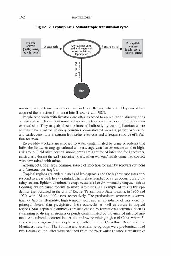
unusual case of transmission occurred in Great Britain, where an 11-year-old boyacquired the infection from a rat bite (Luzzi et al., 1987).
People who work with livestock are often exposed to animal urine, directly or asan aerosol, which can contaminate the conjunctiva, nasal mucosa, or abrasions onexposed skin. They may also become infected indirectly by walking barefoot whereanimals have urinated. In many countries, domesticated animals, particularly swineand cattle, constitute important leptospire reservoirs and a frequent source of infec-tion for man.
Rice-paddy workers are exposed to water contaminated by urine of rodents thatinfest the fields. Among agricultural workers, sugarcane harvesters are another high-risk group. Field mice nesting among crops are a source of infection for harvesters,particularly during the early morning hours, when workers� hands come into contactwith dew mixed with urine.
Among pets, dogs are a common source of infection for man by serovars canicolaand icterohaemorrhagiae.
Tropical regions are endemic areas of leptospirosis and the highest case rates cor-respond to areas with heavy rainfall. The highest number of cases occurs during therainy season. Epidemic outbreaks erupt because of environmental changes, such asflooding, which cause rodents to move into cities. An example of this is the epi-demics that occurred in the city of Recife (Pernambuco State, Brazil), in 1966 and1970, with 181 and 102 cases, respectively. The predominant serovar was ictero-haemorrhagiae. Humidity, high temperatures, and an abundance of rats were theprincipal factors that precipitated these outbreaks as well as others in tropicalregions. Small epidemic outbreaks are also caused by recreational activities, such asswimming or diving in streams or ponds contaminated by the urine of infected ani-mals. An outbreak occurred in a cattle- and swine-raising region of Cuba, where 21cases were diagnosed in people who bathed in the Clavellina River and theManiadero reservoir. The Pomona and Australis serogroups were predominant andtwo isolates of the latter were obtained from the river water (Suárez Hernández et
162 BACTERIOSES
Figure 12. Leptospirosis. Synanthropic transmission cycle.
Leptospiruria Skin and mucus
Man
Infectedanimals
(cattle, swine,rodents, dogs)
Skin
, ora
l and
nasa
l muc
osas
Contamination ofsoil and water with
urine containingleptospires
Susceptibleanimals
(cattle, swine,rodents, dogs)

al., 1989). Epidemic outbreaks caused by several different serovars have occurredamong soldiers wading in streams or camping by riverbanks during jungle maneu-vers. Such epidemics have occurred in Panama and Malaysia; in these cases, thesource of infection was the urine of infected wild animals.
Animals, either primary or secondary hosts, contract the infection in a similarway. The density of the host population and the environmental conditions in whichit lives play important roles. On cattle ranches, the infection is usually introduced bya carrier animal with leptospiruria and, at times, by fields that flood with water con-taminated at a neighboring establishment.
Pathogenic leptospires (L. interrogans) do not multiply outside the animal organ-ism. Consequently, in addition to carrier animals, existence of a leptospirosis focusrequires environmental conditions favorable to the survival of the agent in the exte-rior environment. Leptospires need high humidity, a neutral or slightly alkaline pH,and suitable temperatures. Low, inundated ground and artificial or natural freshwa-ter receptacles (ponds, streams, reservoirs, etc.) are favorable to their survival,whereas salt water is deleterious. Soil composition, both its physiochemical and bio-logical characteristics (microbe population), also acts to prolong or shorten life forleptospires in the environment. Temperatures in the tropics constitute a very favor-able factor for survival of leptospires, but cases of leptospirosis may also occur incold climates, although they are less frequent.
Role of Animals in the Epidemiology of the Disease: Wild and domesticatedanimals are essential for the maintenance of pathogenic leptospires in nature.Transmission of the infection from animals to man is effected directly or indirectly.
Human-to-human transmission is rare. Man is an accidental host and only in veryspecial conditions can he contribute to the maintenance of an epidemic outbreak.Such was the case in an epidemic described in the forest northeast of Hanoi(Vietnam). The outbreak occurred among soldiers occupied in logging and transportof logs by buffalo through a swampy area. Leptospiruria was observed in 12% of 66convalescent soldiers. In contrast, the rate of infection was insignificant among buf-falo and rodents in the region. The pH of the surface water was neutral, the workersworked barefoot, and their urine had a pH that fluctuated around seven (their dietwas vegetarian). Leptospiruria persisted in some of the soldiers for more than sixmonths (Spinu et al., 1963).
A case of transmission through mother�s milk was described in the US (Songerand Thiermann, 1988). A female veterinarian continued nursing after being infectedwith the serovar hardjo while performing an autopsy on a cow. Twenty-one daysafter the appearance of clinical symptoms in the mother, the baby became ill withfever, anorexia, irritability, and lethargy. The serovar hardjo was isolated from thebaby�s urine and the baby recovered with antibiotic treatment.
Various cases of congenital infection have also been described (Faine, 1991).
Diagnosis: In man, the etiologic agent can be isolated from blood during the firstweek of the disease; afterwards, it can be isolated from the urine, either by directculture or by inoculation into young hamsters. Repeated blood samples are neces-sary for serological examination. The patient has no antibodies during the first week;they appear in six to seven days and reach maximum levels in the third or fourthweek. If the first sample is negative or low-titer and the second shows an apprecia-ble increase in antibody titer (fourfold or more), leptospirosis is indicated.
LEPTOSPIROSIS 163

The same diagnostic procedures are employed for animals as for man. Blood orurine may be used for the bacteriologic examination, depending on the stage of theillness. If a necropsy is performed on a sacrificed or dead animal, kidney culturesshould be made. Examination of several tissue samples from the same individual isnot always easily done in veterinary practice, but individual diagnosis of domesticanimals is not as important as herd diagnosis. Discovery of high antibody titers inseveral members of a herd and a clinical picture compatible with leptospirosis indi-cate a recent infection.
Low titers may indicate residual antibodies from a past infection or recentlyformed antibodies that have not yet had time to reach a high level.
The serologic reference test that is used most for man as well as for animals ismicroscopic agglutination (MAT). This test should be carried out using representa-tive serovars from different serogroups, especially those occurring in the region. Itis necessary to bear in mind that cross-reactions are produced not only betweendifferent serovars of the same serogroup but, at the beginning of the infection (two to three weeks), also between serovars of different serogroups, and a het-erologous serovar titer may predominate. Reaction to the homologous serovarbecomes more pronounced with time. Cross-reactions are much more frequent inman than in animals.
The macroscopic plate test with inactivated antigens can be used as a preliminaryor screening test for man and animals. It is fast and easy, and particularly useful fordiagnosing disease in a herd.
Plate agglutination is a genus-specific test, which uses as an antigen the patocstrain of saprophytic leptospira (L. biflexa) to determine if the patient is sufferingfrom leptospirosis (Mazzonelli et al., 1974). Reaction to this test is marked duringthe acute phase of leptospirosis and then quickly becomes negative (Faine, 1982).Among more recent tests, those of interest are indirect immunofluorescence and enzyme-linked immunosorbent assay (ELISA). With both, the types ofimmunoglobulins (IgM or IgG) can be determined by using the correspondingantigens. IgM appears after the first week of the disease and IgG appears afterseveral weeks. In some human cases, IgG antibodies cannot be detected for reasonsas yet unknown.
The utility of ELISA for diagnosing infection due to hardjo was compared withMAT. It was found that a positive reaction can be obtained with MAT 10 days afterthe animal has been experimentally infected; ELISA does not give a positive reac-tion until 25 days after infection. In addition, there was a 90% concordance betweenboth tests. Cross-reactions with sera from animals inoculated with other serotypesoccurred in fewer than 1% (Bercovich et al., 1990).
The serovar hardjo is subdivided into subserovars or genotypes: hardjo genotypehardjo-bovis and hardjo genotype prajitno. A DNA probe for genotype hardjo-boviswas developed by LeFebvre (1987). Three methods for detecting hardjo type hardjo-bovis were compared: with DNA hybridization, 60 of the 75 urine samples fromcows experimentally exposed were positive; with immunofluorescence, 24 sampleswere positive; and with culturing, only 13 were positive. The DNA probe was shownto be much more sensitive than the other techniques in detecting the genotypehardjo-bovis (Bolin et al., 1989a).
A very sensitive generic test is polymerase chain reaction (PCR), which can detectas few as 10 leptospires (Mérien et al., 1992).
164 BACTERIOSES

Control: In man, control measures include: (a) personal hygiene; (b) use of pro-tective clothes during farm work; (c) drainage of lowlands whenever possible; (d)rodent-proof structures; (e) food protection and correct garbage disposal; (f) controlof infection in domestic animals; (g) avoidance of swimming in streams and otherfresh watercourses that may be contaminated, and (h) chemoprophylaxis of high-risk occupational groups (sugarcane harvesters, rice-paddy workers, or soldiers).
Human immunization has not been widely applied. It has been used with promis-ing results in Italy, Poland, and the former Soviet Union. However, because of sec-ondary, mainly allergic effects, its use did not spread. Tests of a vaccine made in achemically defined, protein-free medium are under way (Shenberg and Torten,1973). In China, a similar vaccine is being used on a wide scale.
The use of antibiotics in prophylaxis and treatment of human leptospirosis hasyielded contradictory results. One study (Takafuji et al., 1984) showed that doxycy-cline is effective in chemoprophylaxis; the same drug is probably also effective intreatment. Because leptospirosis caused many cases of disease among American sol-diers training in Panama, a double-blind field test was undertaken to determine theefficacy of doxycycline in preventing the infection. Nine hundred forty soldier vol-unteers were randomly divided into two groups. One group was given an oral doseof 200 mg of doxycycline each week for three weeks, and the other group was givena placebo. After remaining in the jungle for three weeks, 20 cases of leptospirosiswere diagnosed in the placebo group (attack rate of 4.2%) and only one case wasdiagnosed in the doxycycline group (attack rate of 0.2%), i.e., the drug was 95%effective (Takafuji et al., 1984). It has been suggested (Sanford, 1984) that chemo-prophylaxis would be justified in areas where incidence is 5% or higher.Mechanization of farm work has resulted in a decrease of outbreaks, for example,among rice-paddy workers.
Among domesticated animals, vaccination of pigs, cattle, and dogs is effective inpreventing the disease, but it does not protect completely against infection.Vaccinated animals may become infected without showing clinical symptoms; theymay have leptospiruria, although to a lesser degree and for a shorter time thanunvaccinated animals. A few known human cases of leptospirosis were contractedfrom vaccinated dogs. There are bacterins to protect against the pomona, hardjo, andgrippotyphosa serovars in cattle; against pomona in swine; and against canicola andicterohaemorrhagiae in dogs. Immunity is predominantly serovar-specific, and theserovar or serovars active in a focus must be known in order to correctly immunizethe animals. Females should be vaccinated before the reproductive period to protectthem during pregnancy. Young animals can be immunized after 3 or 4 months of age.Bacterins now in use require annual revaccination. For herds to which outside ani-mals are being introduced, it is recommended that vaccination be repeated every sixmonths. An effective measure is to combine vaccination with antibiotic treatment(Thiermann, 1984).
Vaccination against hardjo is not very satisfactory, not even if the prevalent geno-type hardjo-bovis is used in combined vaccines (Bolin et al., 1989b) or in monova-lent vaccines (Bolin et al., 1991).
It has been demonstrated that vaccination with bacterins initially stimulates theproduction of IgM antibodies, which disappear after a few months and are replacedby IgG antibodies. Vaccination generally does not interfere with diagnosis becauseof the quick disappearance of IgM antibodies, which are active in agglutination. IgG
LEPTOSPIROSIS 165
are the protective antibodies and can be detected with serum protection assays inhamsters or with the growth inhibition test in culture media.
A vaccine derived from the outer membrane of leptospires has been obtained andhas yielded very promising results in laboratory tests by conferring resistance notonly against the disease but also against the establishment of leptospiruria.Chemotherapy is promising. Experiments have shown that a single injection of dihy-drostreptomycin at a dose of 25 mg/kg of bodyweight is effective against lep-tospiruria in cattle and swine. The infection has been eradicated in several herds withantibiotic treatment and proper environmental hygiene. The combination of vaccina-tion and chemotherapy for the control of swine leptospirosis has been proposed.
Proper herd management is important for control. It has been repeatedly demon-strated that swine can transmit the pomona serovar to cattle. Therefore, separationof these two species is important for prophylaxis.
Bibliography
Alexander, A.D. Leptospira. In: Balows, A., W.J. Hausler, K.L. Hermann, H.D. Isenberg,H.J. Shadomy, eds. Manual of Clinical Microbiology. 5th ed. Washington, D.C.: AmericanSociety for Microbiology; 1991.
Alexander, A.D., W.E. Gochenour, Jr., K.R. Reinhard, M.K. Ward, R.H. Yagen.Leptospirosis. In: Bodily, H.L., ed. Diagnostic Procedures for Bacterial, Mycotic andParasitic Infections. 5th ed. New York: American Public Health Association, 1970.
Alston, J.M., J.C. Broom. Leptospirosis in Man and Animals. Edinburgh, London:Livingstone; 1958.
Benenson, A.S., ed. Control of Communicable Diseases in Man. 15th ed. An official reportof the American Public Health Association. Washington, D.C.: American Public HealthAssociation; 1990.
Bercovich, Z., R. Taaijke, B.A. Bokhout. Evaluation of an ELISA for the diagnosis ofexperimentally induced and naturally occurring Leptospira hardjo infections in cattle. VetMicrobiol 21:255�262, 1990.
Bernard, W.V., C. Bolin, T. Riddle, et al. Leptospiral abortion and leptospiruria in horsesfrom the same farm. J Am Vet Med Assoc 202:1285�1286, 1993.
Bolin, C.A., J.A. Cassells, R.L. Zuerner, G. Trueba. Effect of vaccination with a monova-lent Leptospira interrogans serovar hardjo type hardjo-bovis vaccine on type hardjo-bovisinfection of cattle. Am J Vet Res 52:1639�1643, 1991.
Bolin, C.A., R.L. Zuerner, G. Trueba. Comparison of three techniques to detect Leptospirainterrogans serovar hardjo type hardjo-bovis in bovine urine. Am J Vet Res 50:1001�1003, 1989a.
Bolin, C.A., R.L. Zuerner, G. Trueba. Effect of vaccination with a pentavalent leptospiralvaccine containing Leptospira interrogans serovar hardjo type hardjo-bovis on type hardjo-bovis infection in cattle. Am J Vet Res 50:2004�2008, 1989b.
Cacchione, R.A. Enfoques de los estudios de la leptospirosis humana y animal en AméricaLatina. Rev Asoc Argent Microbiol 5:36�53, 100�111, 143�154, 1973.
Cousins, D.V., T.M. Ellis, J. Parkinson, C.H. McGlashan. Evidence for sheep as a mainte-nance host for Leptospira interrogans serovar hardjo. Vet Rec 124:123�124, 1989.
Diesch, S.L., H.C. Ellinghausen. Leptospiroses. In: Hubbert, W.T., W.F. McCulloch, P.R.Schnurrenberger, eds. Diseases Transmitted from Animals to Man. 6th ed. Springfield:Thomas; 1975.
Ellis, W.A., J. Montgomery, J.A. Cassells. Dihydrostreptomycin treatment of bovine carri-ers of Leptospira interrogans serovar hardjo. Res Vet Sci 39:292�295, 1985.
166 BACTERIOSES

Ellis, W.A., A.B. Thiermann. Isolation of leptospires from the genital tracts of Iowa cows.Am J Vet Res 47:1694�1696, 1986.
Everard, C.O., A.E. Green, J.W. Glosser. Leptospirosis in Trinidad and Grenada, with spe-cial reference to the mongoose. Trans Roy Soc Trop Med Hyg 70:57�61, 1976.
Faine, S., ed. Guidelines for the control of leptospirosis. Geneva: World HealthOrganization; 1982. (Offset Publication 67).
Faine, S. Leptospirosis. In: Evans, A.S., P.S. Brachman, eds. Bacterial Infections ofHumans. 2nd ed. New York: Plenum Medical Book Co.; 1991.
Hanson, L.E., D.N. Tripathy, A.H. Killinger. Current status of leptospirosis immunizationin swine and cattle. J Am Vet Med Assoc 161:1235�1243, 1972.
Hart, R.J., J. Gallagher, S. Waitkins. An outbreak of leptospirosis in cattle and man. BritMed J (Clin Res Ed) 288(6435):1983�1984, 1984.
LeFebvre, R.B. DNA probe for detection of the Leptospira interrogans serovar hardjogenotype hardjo-bovis. J Clin Microbiol 25:2236�2238, 1987.
Leptospirosis in man, British Isles, 1983. Br Med J (Clin Res Ed) 288(6435):1984�1985, 1984.
Luzzi, G.A., L.M. Milne, S.A. Waitkins. Rat-bite acquired leptospirosis. J Infect 15:57�60, 1987.
Mazzonelli, J. Advances in bovine leptospirosis. Bull Off Int Epizoot 3:775�808, 1984.Mazzonelli, J., G.T. Dorta de Mazzonelli, M. Mailloux. Possibilité de diagnostique
sérologique des leptospires á l�aide d�un antigène unique. Med Mal Infect 4:253, 1974.Mérien, F., P. Amouriaux, P. Perolat, et al. Polymerase chain reaction for detection of
Leptospira spp. in clinical samples. J Clin Microbiol 30:2219�2224, 1992.Myers, D.M. Serological studies and isolations of serotype hardjo and Leptospira biflexa
strains from horses of Argentina. J Clin Microbiol 3:548�555, 1976.Nielsen, J.N., G.K. Cochran, J.A. Cassells, L.E. Hanson. Leptospira interrogans serovar
bratislava infection in two dogs. J Am Vet Med Assoc 199:351�352, 1991.Parma, A.E., C.G. Santisteban, A.S. Fernández, et al. Relación antigénica entre Leptospira
interrogans, cristalino y córnea equina, probada por enzimoinmunoensayo. Rev Med Vet67:72�76, 1986.
Perolat, P., F. Grimont, F. Regnault, et al. rRNA gene restriction patterns of Leptospira: Amolecular typing system. Res Microbiol 141:159�171, 1990.
Prescott, J. Treatment of leptospirosis [editorial]. Cornell Vet 81:7�12, 1991.Sanford, J.P. Leptospirosis�time for a booster. N Engl J Med 310:524�525, 1984.Shenberg, E., M. Torten. A new leptospiral vaccine for use in man. I. Development of a vac-
cine from Leptospira grown on a chemically defined medium. J Infect Dis 128:642�646, 1973.Sillerud, C.L., R.F. Bey, M. Ball, S.I. Bistner. Serologic correlation of suspected Leptospira
interrogans serovar pomona-induced uveitis in a group of horses. J Am Vet Med Assoc191:1576�1578, 1987.
Songer, J.G., A.B. Thiermann. Leptospirosis. J Am Vet Med Assoc 193:1250�1254, 1988.Spinu, I., V. Topcin, Trinh Thi Hang Quy, Vo Van Hung, Mguyen Sy Quoe, Chu Xnan Long,
et al. L�homme comme réservoir de virus dans une épidémie de leptospirose survenue dans lajungle. Arch Roum Path Exp 22:1081�1100, 1963.
Stalheim, O.H. Vaccination against leptospirosis: Protection of hamsters and swine againstrenal leptospirosis by killed but intact gamma-irradiated or dihydrostreptomycin-exposedLeptospira pomona. Am J Vet Res 28:1671�1676, 1967.
Stalheim, O.H. Chemotherapy of renal leptospirosis in cattle. Am J Vet Res 30:1317�1323, 1969.
Stalheim, O.H. Duration of immunity in cattle in response to a viable, avirulent Leptospirapomona vaccine. Am J Vet Res 32:851�854, 1971.
Suárez Hernández, M., J. Bustelo Aguila, V. Gorgoy González, et al. Estudio epidemi-ológico de un brote de leptospirosis en bañistas en el poblado de Jicotea de la provincia Ciegode Ávila. Rev Cubana Hig Epidemiol 27:272�284, 1989.
LEPTOSPIROSIS 167

Sulzer, C.R., W.L. Jones. Leptospirosis Methods in Laboratory Diagnosis. Atlanta: U.S.Centers for Disease Control and Prevention; 1974.
Szyfres, B. La leptospirosis como problema de salud humana y animal en América Latinay el área del Caribe. In: VIII Reunión Interamericana sobre el Control de la Fiebre Aftosa yOtras Zoonosis. Washington, D.C.: Organización Panamericana de la Salud; 1976.(Publicación Científica 316).
Szyfres, B., C.R. Sulzer, M.M. Galton. A new leptospiral serotype in the Bataviaeserogroup from Argentina. Trop Geogr Med 19:344�346, 1967.
Takafuji, E.T., J.W. Kirkpatrick, R.N. Miller, et al. An efficacy trial of doxycycline chemo-prophylaxis against leptospirosis. N Engl J Med 310:497�500, 1984.
Tedesco, L.F., G. Manrique, C.R. Sulzer. A new leptospiral serotype in the Canicolaserogroup from Argentina. Trop Geogr Med 21:203�206, 1969.
Thiermann, A.B. Leptospirosis: Current developments and trends. J Am Vet Med Assoc184:722�725, 1984.
Tripathy, D.N., A.R. Smith, L.E. Hanson. Immunoglobulins in cattle vaccinated with lep-tospiral bacterins. Am J Vet Res 36:1735�1736, 1975.
Van der Hoeden, J. Leptospirosis. In: Van der Hoeden, J., ed. Zoonoses. Amsterdam:Elsevier; 1964.
Waitkins, S.A. From the PHLS. Update on leptospirosis. Brit Med J (Clin Res Ed)290(6480):1502�1503, 1985.
World Health Organization. Research needs in leptospirosis [memorandum]. Bull WorldHealth Organ 47:113�122, 1972.
LISTERIOSIS
ICD-10 A32.1 listerial meningitis and meningoencephalitis; A32.7 listerial septicaemia; A32.8 other forms of listeriosis;
P37.2 neonatal (disseminated) listeriosis
Synonyms: Leukocytosis, listerial infection, listeriasis, listerellosis, circling dis-ease (in animals).
Etiology: The genus Listeria contains seven species, but only two are of interestin human and animal pathology: L. monocytogenes and L. ivanovii (formerly L. bul-garica or serovar 5 of L. monocytogenes). A notable difference between the twopathogenic species is their hemolytic ability.
The most important species for both man and animals is L. monocytogenes, agram-positive, facultatively anaerobic bacillus 0.5 to 2 microns long and 0.5microns in diameter that is motile at temperatures between 20°C and 25°C. It isbeta-hemolytic in blood agar and forms a narrow band of hemolysis around thecolonies (unlike L. ivanovii, which forms a wide band). A noteworthy characteristicof L. monocytogenes is its ability to grow at low temperatures; at a pH between 6and 9, it can reproduce at temperatures from 3°C to 45°C. It is a facultative, intra-cellular parasite of the reticuloendothelial system. For purposes of epidemiological
168 BACTERIOSES

research, L. monocytogenes is subdivided into 11 serovars. Most human (92%) andanimal cases are caused by serovars 4b, 1/2b, and 1/2a (Bortolussi et al., 1985).Therefore, serotyping is of limited usefulness for identifying a source of infection(Gelin and Broome, 1989).
Of 161 isolates serotyped in the US, 33% belonged to serovar 4b; 31.5%, toserovar 1/2b; 30%, to serovar 1/2a; 4%, to serovar 3b; 1%, to serovar 3a; and 0.5%,to serovar 1/2c (Gelin et al., 1987). When 71 isolates were serotyped in Brazil, sevendifferent serovars were recognized; 50% were 4b and 29.6% were 1/2a (Hofer et al.,1984).
Although serotyping has been useful as a preliminary approach, other schemeshad to be devised in order to be able to specify the source of infection. Subtypingwas done primarily with two methods: phage typing and electrophoretic enzymetyping. In Great Britain, 64% of the strains could be typed using 28 phages; inFrance, 78% could be typed using 20 phages. In both cases, a sizeable percentage ofstrains could not be typed. All strains of L. monocytogenes can be typed using theisoenzymatic subtyping method (Selander et al., 1986). Recently, ribosomal RNAtyping (ribotyping) has been used with success.
Geographic Distribution: Worldwide. L. monocytogenes is widely distributed invegetation, soil, and human and animal intestines.
Occurrence in Man: Incidence is low, but it is an important disease because ofits high mortality. In many developing countries, listeriosis is rare. There is greaterconcentration of cases in European countries and the United States, perhaps becausemedical personnel in these countries are more on the lookout for the disease andbecause better laboratory support is available. In the former Federal Republic ofGermany (West Germany), there were 296 cases of listeriosis between 1969 and1985, 60% of which occurred in urban areas. Fifty percent of the strains isolatedwere from newborns and the most common serovar was 4b (Schmidt-Wolf et al.,1987). In Great Britain, information was obtained from 722 cases occurring from1967 to 1985. In 246 cases (34%), the infection affected the mother, the fetus, or thenewborn. Neonatal infection was diagnosed within the first two days postpartum in133 (54%) cases; 56 cases (23%) were diagnosed later than two days postpartum.There were 47 (19%) cases of intrauterine death. There were also 10 cases (4%) ofmothers with bacteremia that did not affect the fetus. Overall case fatality was 50%(McLauchlin, 1990a). The author estimates that these data must include less than50% of the total number of cases that occurred in the country. In adults and youth,474 cases were recorded; 275 (58%) were men and 199 (42%) were nonpregnantwomen. Case fatality was 44%. Seventy-six percent of these patients had an under-lying illness. An increase in incidence was noted in autumn (McLauchlin, 1990b).
There were an estimated 800 cases per year in the US between 1980 and 1982,with an incidence of 3.6 per million inhabitants and at least 150 deaths (19% casefatality). The highest attack rates were seen in newborns (4.7 per 100,000 live births)and in persons 70 years of age or older (11 per million) (Gellin and Broome, 1989).There are few recognized cases in developing countries. Sporadic cases have beenseen in several Latin American countries. In a Mexican hospital, hemocultures werecarried out during a three-month period on all children whose mothers showed signsof amniotic infection; L. monocytogenes was isolated from 4 out of 33 newbornsexamined (Pérez-Mirabate and Giono, 1963). In Peru (Guevara et al., 1979),
LISTERIOSIS 169

serovars 4d and 4b of L. monocytogenes were isolated from three fatal cases ofneonatal listeriosis and from five aborted fetuses. In Argentina, there are few data onthe occurrence of human listeriosis. In Córdoba (Argentina), there are cases ofneonatal listeriosis each year, and these constitute between 2% and 3% of bacterio-logically confirmed sepsis (Paolasso, 1981). In a small town in the province ofBuenos Aires, Manzullo (1981) isolated L. monocytogenes type 1a from a bovinefetus, the vaginal exudate of the woman who milked the cows, and from the house-hold�s female dog. In another town, Manzullo (1990) isolated the agent from awoman�s vaginal exudate and from the woman�s female cat. In a Buenos Aires med-ical center, nine cases of listeriosis were diagnosed in 15 years, two of them fatal.Only one patient was not immunocompromised (Roncoroni et al., 1987).
Most cases occur sporadically, but epidemic outbreaks have occurred in severalcountries. In 1981, in a maternity hospital in Halifax (Canada), there were 34 peri-natal cases and 7 cases in women without underlying illness or immunosuppression.Case fatality in the babies born live was 27%. There were five spontaneous abortionsand four babies stillborn at term. The epidemic outbreak was attributed to coleslaw:L. monocytogenes serovar 4b was isolated from the cabbage as well as from thepatients. On the farm where the cabbages were grown, two sheep had died from lis-teriosis the previous year; in addition, the farmer used sheep dung as fertilizer. It isalso worth noting that the farmer kept the cabbage refrigerated at 4°C, whichallowed the etiologic agent to multiply at the expense of other contaminant microor-ganisms (Schlech et al., 1983).
An earlier outbreak occurred in 1979 in eight hospitals in Boston (USA); itaffected 20 patients, 15 of whom acquired the infection in the hospital. Raw veg-etables were assumed to be the source of infection.
In Massachusetts (USA), an epidemic outbreak caused by pasteurized milk wasrecorded in 1983. It affected 42 immunocompromised patients and seven immuno-competent patients; there were also perinatal cases. Case fatality was 29%. Of 40isolates, 32 were type 4b. It is possible that the milk had been contaminated afterpasteurization (Schuchat et al., 1991).
The largest epidemic in the US was recorded in 1985 in Los Angeles, California(Linnan et al., 1988). The epidemic affected pregnant women, their fetuses, andtheir newborns. Case fatality was 63% for the infected fetuses and newborns. Theoutbreak was due to a Mexican soft cheese; serovar 4b was isolated from the patientsand the cheese. The incubation period was 11 to 70 days, with an average of 31 days(Schuchat et al., 1991).
There have been epidemic outbreaks in Switzerland, Denmark, and France. InSwitzerland, the 1987 outbreak that led to 64 perinatal cases and 58 nonperinatalcases was caused by a soft cheese. Case fatality was 28%. The strain of L. monocy-togenes responsible was the same enzymatic type as the strain that caused the out-break in California in 1985 (Gelin and Broome, 1989). One of the largest epidemicsknown to date occurred in France in 1992. It affected 691 persons and 40% of thecases were caused by serotype 4b. The epidemic strain was isolated from 91 preg-nant women and their children. Of the remaining persons affected by the epidemicstrain, 61% were immunodeficient. The phage type was the same as in California(1985), Switzerland (1983�1987), and Denmark (1985�1987). The epidemic strainwas isolated from 163 samples of meat products, 35 cheese samples, and 12 otherfood samples. The epidemic lasted from 18 March to 23 December 1992 and caused
170 BACTERIOSES

63 deaths and 22 abortions. The cause was attributed to pig�s tongue in gelatin(WHO, 1993). In 1993 (January�August), 25 new cases occurred in France. Thistime the outbreak was again due to serogroup 4, but to a different lysotype than inthe 1992 epidemic. Of the 25 cases, 21 were maternal-fetal, with 4 spontaneousabortions and 2 stillbirths at term. Most of the cases occurred in western France. Theepidemiological investigation was able to attribute the infection to a pork product(�rillettes�) distributed by a single commercial firm (Bol Epidemiol Hebdom No. 34, 1993).
There are various risk groups: pregnant women, fetuses, newborns, the elderly,and immunocompromised patients. However, there was some controversy regardingAIDS patients. Several authors maintained that AIDS patients were unlikely to con-tract listeriosis, even though their cellular immunity system was highly compro-mised. While it is true that listeriosis is not one of the principal conditions affectingAIDS patients, its incidence in those infected by HIV is 300 times higher than in thegeneral population (Schuchat et al., 1991).
Occurrence in Animals: Listeriosis has a wide variety of domestic and wild ani-mal hosts. The infection has been confirmed in a large number of domestic and wildmammals, in birds, and even in poikilotherms. The most susceptible domesticspecies is sheep, followed by goats and cattle. The frequency of occurrence in theseanimals is not known.
Outbreaks in sheep have been described in several Latin American countries. Thedisease has been confirmed in alpacas in Peru, and in sheep, fowl, and cattle inArgentina and Uruguay.
The first epizootic outbreak (1924) was recognized in England in laboratory rab-bits suffering from a disease characterized by mononucleosis, from whence the spe-cific name of the agent, monocytogenes, comes. Mononucleosis rarely occurs inman or in animals other than rabbits and rodents.
Several outbreaks have been described in Great Britain and the US due to silagewith a pH higher than 5, which favors multiplication of L. monocytogenes. As theuse of silage increases, outbreaks, which occur when the quality of silage is poor andthe pH high, increase as well.
The Disease in Man: The most affected group is newborns (50% of cases inFrance and 39% in the US), followed by those over age 50. The disease is very rarebetween 1 month and 18 years of age. According to data from two German obstet-rical clinics, listerial infection caused 0.15% to 2% of perinatal mortality. Listerialabortion in women usually occurs in the second half of pregnancy, and is more fre-quent in the third trimester. Symptoms that precede miscarriage or birth by a fewdays or weeks may include chills, increased body temperature, cephalalgia, slightdizziness, and sometimes, gastrointestinal symptoms. These septicemic episodesmay or may not recur before birth of a stillborn fetus or a seriously ill full-term baby.After delivery, the mother shows no disease symptoms, but L. monocytogenes canbe isolated from the vagina, cervix, and urine for periods varying from a few daysto several weeks. If the child is born alive but was infected in utero, it may showsymptoms immediately after birth or within a few days. The symptomatology is thatof sepsis or, less frequently, a disseminated granulomatosis (granulomatosis infan-tisepticum). There may also be symptoms of a respiratory tract disorder. Case fatal-ity is high. The main lesion is a focal hepatic necrosis in the form of small, grayish-
LISTERIOSIS 171

white nodules. Some children born apparently healthy fall ill with meningitis shortlythereafter (a few days to several weeks). In these cases, the infection was probablyacquired in utero or during birth. In the US, neonatal meningitis is the most com-mon clinical form, while in Europe, perinatal septicemia prevails. Hydrocephalus isa common sequela of neonatal meningitis.
Meningitis or meningoencephalitis is the most common clinical form in adults,especially in those over 50. Listerial meningitis often occurs as a complication indebilitated persons, alcoholics, diabetics, in patients with neoplasias, or in elderlypatients with a declining immune system. Before the existence of antibiotics, casefatality was 70%. Listerial septicemia also occurs among weakened adults, espe-cially patients undergoing long-term treatment with corticosteroids or antimetabo-lites. In addition, listeriosis may result in endocarditis, external and internalabscesses, and endophthalmitis. A cutaneous eruption has been described amongveterinarians who handled infected fetuses.
The recommended treatment for maternal-fetal listeriosis is ampicillin. Variousantibiotics, such as ampicillin (alone or in combination with aminoglycosides),tetracycline (not for those under 8 years of age), and chloramphenicol, may be usedfor the other forms of the disease (Benenson, 1990).
The Disease in Animals
SHEEP, GOATS, AND CATTLE: Listeriosis manifests itself in ruminants as encephalitis,neonatal mortality, and septicemia. The most common clinical form is encephalitis. Insheep and goats, the disease has a hyperacute course, and mortality may vary from 3%to more than 30%. In cattle, listerial encephalitis has a chronic course, with the ani-mals surviving for 4 to 14 days. In general, only 8% to 10% of a herd is affected.
A ruminant with encephalitis isolates itself from the herd and shows symptoms ofdepression, fever, lack of coordination, torticollis, spasmodic contractions and paral-ysis of facial muscles and throat, profuse salivation, strabismus, and conjunctivitis.The animal tries to lean against some support while standing and, if able to walk,moves in circles. In the final phase of the disease, the animal lies down and makescharacteristic chewing movements when attempting to eat.
Listerial encephalitis can affect animals of any age, but it is more common in thefirst three years of life. Nevertheless, it does not appear before the rumen becomesfunctional. Septicemia is much more common in young animals than adults.Abortion occurs mainly during the last months of gestation and is generally the onlysymptom of genital infection, the dam showing no other signs of disease. If uterineinfection occurs in the cow before the seventh month of pregnancy, the dead fetus isusually retained in the uterus for several days and has a macerated appearance, withmarked focal necrotic hepatitis. In addition, the placenta may be retained and metri-tus may develop. If infection occurs in the final months of pregnancy, the fetus ispractically intact and shows minimal lesions.
L. monocytogenes can also cause mastitis in cows. There are few described cases,either because the presence of this agent in cows has not been studied or because itsoccurrence really is rare. Mastitis caused by Listeria varies in severity from sub-clinical to acute and chronic. Elimination of the agent in milk occurs over a longperiod of time and may have public health repercussions, especially since pasteur-ization does not guarantee complete safety if the viable bacteria count is high beforeheat treatment (Gitter, 1980).
172 BACTERIOSES

A study carried out in 1970�1971 in Victoria (Australia) (Dennis, 1975) showedthat listeriosis is an important cause of perinatal mortality in sheep. In 94 flocks,fetuses and lambs that died during the neonatal period were examined, and L. mono-cytogenes was found in 25%. The disease caused by this agent occurs mostly in win-ter. It has been estimated that the rate of abortion in flocks affected by listeriosis inVictoria varies from 2% to 20%.
L. ivanovii, which differs from L. monocytogenes on the basis of several pheno-typic characteristics, was associated in several countries with abortions in sheep and,occasionally, in cows (Alexander et al., 1992).
OTHER MAMMALS: Listeriosis is rare in swine; when it does occur in the first fewweeks of life, it usually takes the septicemic form. Few cases are known in dogs, inwhich the disease may be confused with rabies. In other domestic and wild species,the disease generally appears as isolated cases and in the septicemic form.Outbreaks have been described in rabbit and guinea pig breeding colonies.
FOWL: Young birds are the most affected. Outbreaks are infrequent and mortalitymay range from the loss of a few birds on one farm to a high rate of losses on other farms. The septicemic form is the most common, with degenerative lesions ofthe myocardium, pericarditis, and focal hepatic necrosis. On rare occasions, themeningoencephalitic form is found, with marked torticollis. Since the generalizeduse of antibiotics in poultry feed began, few cases of listeriosis in this species havebeen reported.
Source of Infection and Mode of Transmission: The causal agent is widely dis-tributed in animals and man, as well as in the environment. L. monocytogenes hasbeen isolated from different mammalian and avian species and from the soil, plants,mud, pasture, wastewater, and streams. The presence of virulent and avirulent (formice) strains in animals and in the environment complicates clarification of the epi-demiology, but serotyping can be of considerable help. Cattle, sheep, and manyother animal species eliminate the agent in their feces. L. monocytogenes has alsobeen isolated from the feces of patients and their contacts, as well as from a smallpercentage of the general human population. However, it has been isolated from thestools of some 20% to 30% of pregnant women, and has also been found in thefemale genital tract. In addition to untypeable strains, potentially pathogenicserotype 1 and serovar 4b have been isolated (Kampelmacher and van NoorleJansen, 1980). Consequently, the natural reservoir is wide and the number of hostsis large. Despite this, few people contract the disease. Many women from whosestools the agent has been isolated give birth to healthy children. Concurrent condi-tions, such as stress and other predisposing causes (particularly diseases or treat-ments that depress the immune system), come into play in initiating the disease.Another predisposing cause is the decline in the immune system that occurs withaging, as well as endocrine changes during pregnancy and deficiencies inimmunoregulation at the placental level.
The source of infection for the fetus and newborn is evidently the infected motherherself. It is believed that the almost inapparent disease course manifested by themother is caused by a mild bacteremia. Airborne infection might play a role, as sug-gested by the influenza-like symptoms exhibited by the mother. The mother�s geni-tal tract is probably infected via the fecal route, while the fetus is infected via the
LISTERIOSIS 173

bloodstream or placenta. The discovery of the causal agent in the semen of a manwhose wife�s genitals were infected would indicate that, in some cases, the infectionmay be transmitted through sexual contact.
The oral route of transmission seems to be important, as indicated by the recentoutbreaks occurring in the US, Switzerland, and France (see the section on occur-rence in man), where some contaminated vegetables, milk and milk products, andmeat were the vehicle of the infection. It is also interesting to note that the milk thatled to one of the outbreaks came from establishments where listeriosis had beendiagnosed in the animals. The disease affected two very susceptible groups: new-borns and debilitated persons. Of 49 patients hospitalized with listerial septicemiaor meningitis, 7 were newborns and 42 were adults. All the adults were sufferingfrom other diseases or undergoing treatment with immunosuppressants.
A rise in listeriosis cases when animals feed on silage would indicate the diges-tive system as the portal of entry. The causal agent has been isolated from poorlyprepared fodder that had a pH higher than 5. During an outbreak of encephalitis insheep, the same serovar and phage type of L. monocytogenes was isolated from thesilage and from the animals� brains. The silage contained 1 million listeriae per gram(Vázquez-Boland et al., 1992).
L. monocytogenes is distributed in populations of healthy animals and the diseasecan be produced when stress lowers the host�s resistance.
Although it has been demonstrated that food has been the source of infection inboth human and animal outbreaks, the source of infection is not known with cer-tainty in sporadic cases in man. However, it has been possible to confirm that a sig-nificant percentage of such cases were caused by the ingestion of a contaminatedfood (Schuchat et al., 1992; Pinner et al., 1992). Cases of listeriosis have been asso-ciated with ingestion of raw sausage and undercooked chicken (Schwartz et al.,1988). It is likely that many cases with a food source cannot be detected because theextended incubation period makes it impossible for patients to associate a food withtheir infection (Gelin and Broome, 1989).
One case of listeriosis in a woman with cancer was associated with consumptionof turkey franks. The investigation established that the franks in opened packages inher refrigerator were contaminated, but those in unopened packages were not.Cultures from other foods in the refrigerator also yielded positive results. The con-clusion was that a cross-contamination was involved (CDC, 1989).
An interesting study was conducted in the US in 1992 (CDC, 1992). During theperiod 1988�1990, special epidemiological surveillance was conducted in four of thecountry�s districts, with a population of 18 million inhabitants. There were 301 casesof listeriosis identified (7.4 per million inhabitants), 67 (23%) of whom died. Thepatients� food consumption histories indicated that the listeriosis patients ingested 2.6times more soft cheeses than did the controls, or purchased 1.6 times more preparedfoods. The patients� refrigerators were examined and it was found that 79 (64%) of 123contained at least one food contaminated by L. monocytogenes; in 26 (33%) of the 79refrigerators the same enzymatic strain as that found in the patients was isolated.
The wide distribution of Listeria spp. in nature and in animal feces explains whyits presence in raw meats is almost inevitable. Prevalence in raw meats may varyfrom 0% to 68%. Pork is contaminated most often, but contamination is also fre-quent in uncooked chicken. There is little information regarding the virulence of L.monocytogenes strains isolated from meats (Johnson et al., 1990).
174 BACTERIOSES

There is no uniform criterion regarding when to reject foods according to thedegree of contamination by L. monocytogenes. Several countries (France, US)require that there be no contamination at all, while others (Canada, Germany) havea certain tolerance. It is impossible to ensure the total absence of Listeria spp. in allfoods (Dehaumont, 1992).
In California (USA), a six-month study was conducted on the prevalence ofListeria spp. in environmental samples from 156 milk-processing plants. Listeriaspp. was isolated from 75 (12.6%) of the 597 environmental samples. Half of theisolates were identified as L. monocytogenes. Of the 156 plants, 46 gave positivesamples for Listeria spp. and 19.9% of these isolates were identified as L. monocy-togenes (Charlton et al., 1990).
Role of Animals in the Epidemiology of the Disease: The epidemiology of spo-radic listeriosis is still not well known. Most researchers consider it a disease com-mon to man and animals and not a zoonosis per se. It is likely that animals contributeto maintenance of listeria in general in nature and especially to its distribution.
Studies conducted in recent years suggest that man and animals can contract theinfection from many sources. Most cases in man occur in urban areas, where thereis little contact with animals. Nonetheless, animals may be the source of the infec-tion. In one case, infection was confirmed in a woman who drank raw milk; the sameserotype of Listeria was isolated from the raw milk and from the woman�s prema-ture twins. The etiologic agent was isolated from 16% of cows that had listerialabortions. The previously described outbreaks caused by milk, meat, or vegetablescontaminated by manure from listeria-infected animals demonstrate that animalsmay be an important source of infection.
There are indisputable cases of direct transmission of the infection from animalsto man. A cattleman assisted during the delivery of a cow, inserting his arms in theuterus. Within the next 24 hours, a rash appeared on his hands and one arm anddeveloped into pustules. He later experienced fever, chills, and generalized pain. Thesame phage type was isolated from the cow�s vagina and from the cattleman�s pus-tules (Cain and McCann, 1986). The veterinary profession is particularly at risk ofcontracting cutaneous listeriosis. Many veterinarians have become ill after attendingcows that aborted, fetuses, or newborns, or after conducting autopsies of septicemicanimals. The most frequent lesion is a papular exanthema (Owen et al., 1960;Nieman and Lorber, 1980; Hird, 1987). Contact with sick birds may also causehuman infection (Gray and Killinger, 1966).
Diagnosis: Diagnosis can be made only through isolation of the causal agent. Ifthe sample is obtained from usually sterile sites, such as blood, cerebrospinal fluid,amniotic fluid, or biopsy material, seeding can be done directly in blood agar, withincubation at 35°C for a week and daily checks. Listeria can be isolated from anyorgan in septicemic fetuses.
In sheep, goats, or cattle with encephalitis, samples of the medulla oblongatashould be cultured. In septicemic fowl, rodents, or neonatal ruminants, blood orinternal organs should be cultured. The �cold enrichment� method is used especiallyin epidemiological investigations and is indicated for culturing highly contaminatedspecimens. However, this method has no diagnostic value for clinical cases becauseof the time it takes, since treatment with antibiotics (preferably ampicillin) shouldbegin as soon as possible to be effective.
LISTERIOSIS 175

At present, contaminated samples as well as foods are cultured in an enrichmentmedium and then in a selective medium. One procedure is the US Department ofAgriculture procedure; it uses nalidixic acid and acriflavin in broth to inhibit thegrowth of contaminating flora. The culture is incubated for 24 hours at 30°C andthen a subculture is done in another broth of the same composition for another 24hours at 30°C. Finally, a highly selective solid medium that contains lithium chlo-ride and moxalactam is used (McClain and Lee, 1988).
A test has been developed to distinguish pathogenic from nonpathogenic strainsof L. monocytogenes. This method is based on the potentiating and synergistic effectthat the extrosubstance of Rhodococcus equi has for producing hemolysis in culturesof pathogenic strains of L. monocytogenes (Skalka et al., 1982).
In general, serologic tests are confusing and not useful because of cross-reactionswith enterococci and Staphylococcus aureus, especially by serogroups 1 and 3 ofListeria. DNA probes that specifically detect L. monocytogenes have been devel-oped (Datta et al., 1988).
Control: In regions where human neonatal listeriosis is common, a Gram staincan be made from the meconium of a newborn, and treatment with antibiotics canbe rapidly initiated if bacteria suspected of being Listeria are found. Women whodevelop influenza-like symptoms in the final months of pregnancy should be care-fully examined and treated, if necessary, with antibiotics. The limited arsenal ofdefense against the infection includes such measures as the pasteurization of milk,rodent control, and common practices of environmental and personal hygiene.
Special recommendations have been developed for food preparation (CDC,1992): cook products of animal origin well, thoroughly wash vegetables that are eaten raw, keep raw meats separate from other foods, do not consume raw milk, wash utensils used in food preparation well, and reheat all food leftovers at ahigh temperature. Immunocompromised individuals must not eat soft cheeses andveterinarians must take precautions during delivery, and particularly during abor-tions and autopsies.
Animals with encephalitis or those that have aborted should be isolated and theirplacentas and fetuses destroyed. Recently acquired animals should only be added toa herd after undergoing a reasonable period of quarantine.
Bibliography
Alexander, A.V., R.L. Walker, B.J. Johnson, et al. Bovine abortions attributable to Listeriaivanovii: Four cases (1988�1990). J Am Vet Med Assoc 200:711�714, 1992.
Benenson, A.S., ed. Control of Communicable Diseases in Man. 15th ed. An official reportof the American Public Health Association. Washington, D.C.: American Public HealthAssociation; 1990.
Bojsen-Moller, J. Human listeriosis: Diagnostic, epidemiological and clinical studies. ActaPathol Microbiol Scand (Suppl 229):1�157, 1972.
Bortolussi, R., W.F. Schlech III, W.L. Albritton. Listeria. In: Lennette, E.H., A. Balows,W.J. Hausler, Jr., H.J. Shadomy. Manual of Clinical Microbiology. 4th ed. Washington, D.C.:American Society for Microbiology; 1985.
Broadbent, D.W. Infections associated with ovine perinatal mortality in Victoria. Aust Vet J51:71�74, 1975.
176 BACTERIOSES

Cain, D.B., V.L. McCann. An unusual case of cutaneous listeriosis. J Clin Microbiol23:976�977, 1986.
Charlton, B.R., H. Kinde, L.H. Jensen. Environmental survey for Listeria species inCalifornia milk processing plants. J Food Protect 53:198�201, 1990.
Datta, A.R., B.A. Wentz, D. Shook, M.W. Trucksess. Synthetic oligodeoxyribonucleotideprobes for detection of Listeria monocytogenes. Appl Environ Microbiol 54:2933�2937, 1988.
Dehaumont, P. Listeria monocytogenes et produits alimentaires: �zérotolérance au moins.�Bull Epidemiol Hebdom 24:109, 1992.
Dennis, S.M. Perinatal lamb mortality in Western Australia. 6. Listeric infection. Aust Vet J51:75�79, 1975.
Fleming, D.W., S.L. Cochi, K.L. MacDonald, J. Brondum, P.S. Hayes, B.D. Plikaytis, et al.Pasteurized milk as a vehicle of infection in an outbreak of listeriosis. N Engl J Med312(7):404-407, 1985.
Franck, M. Contribution a l�Étude de l�Épidémiologie des Listerioses Humaines etAnimales [thesis]. École Nationale Vétérinaire de Lyon, France, 1974.
García, H., M.E. Pinto, L. Ross, G. Saavedra. Brote epidémico de listeriosis neonatal. RevChil Pediatr 54:330�335, 1983.
Gellin, B.G., C.V. Broome. Listeriosis. JAMA 261:1313�1320, 1989.Gellin, B.G., C.V. Broome, A.W. Hightower. Geographic differences in listeriosis in the
United States [abstract]. 27th Interscience Conference of Antimicrobial Agents andChemotherapy. New York, Oct 5, 1987. Cited in: Gellin, B.G., C.V. Broome. Listeriosis.JAMA 261:1313�1320, 1989.
Gitter, M., R. Bradley, P.H. Blampied. Listeria monocytogenes infection in bovine mastitis.Vet Rec 107:390�393, 1980.
Gray, M.L., A.H. Killinger. Listeria monocytogenes and listeric infections. Bact Rev30:309�382, 1966.
Green, H.T., M.B. Macaulay. Hospital outbreak of Listeria monocytogenes septicaemia: Aproblem of cross infection? Lancet 2:1039�1040, 1978.
Guevara, J.M., J. Pereda, S. Roel. Human listeriosis in Peru. Tropenmed Parasitol30:59�61, 1979.
Hird, D.W. Review of evidence for zoonotic listeriosis. J Food Protect 50:429�433, 1987.Hofer, E., G.V.A. Pessoa, C.E.A. Melles. Listeriose humana. Prevalência dos sorotipos de
Listeria monocytogenes isolados no Brasil. Rev Inst Adolfo Lutz 44:125�131, 1984.Johnson, J.L., M.P. Doyle, R.G. Cassens. Listeria monocytogenes and other Listeria spp. in
meat and meat products. A review. J Food Protect 53:81�91, 1990.Kampelmacher, E.H., L.M. van Noorle Jansen. Listeriosis in humans and animals in the
Netherlands (1958�1977). Zbl Bakt Hyg Orig A 246:211�227, 1980.Killinger, A.H. Listeriosis. In: Hubbert, W.T., W.F. McCulloch, P.R. Schnurrenberger, eds.
Diseases Transmitted from Animals to Man. 6th ed. Springfield: Thomas; 1975.Killinger, A.H., M.E. Mansfield. Epizootiology of listeric infection in sheep. J Am Vet Med
Assoc 157:1318�1324, 1970.Larsen, H.E. Epidemiology of listeriosis. The ubiquitous occurrence of Listeria monocyto-
genes. In: Proceedings, Third International Symposium on Listeriosis, Bilthoven, TheNetherlands, 1966.
Linnan, M.J., L. Mascola, X.D. Lou, et al. Epidemic listeriosis associated with Mexican-style cheese. N Engl J Med 319:823�828, 1988.
Mair, N.S. Human listeriosis. In: Graham-Jones, O., ed. Some Diseases of AnimalsCommunicable to Man in Britain. Oxford: Pergamon; 1968.
Manzullo, A. Epidemiología y epizootiología de la listeriosis. Acta Bioq Clin Latinoam14:539�546, 1981.
Manzullo, A.C. Listeriosis [letter]. An Acad Nacl Agr Vet 44 (4):5�13, 1990.McClain, D., W.H. Lee. Development of USDA-FSIS method for isolation of Listeria
monocytogenes from raw meat and poultry. J Assoc Off Anal Chem 71:660�664, 1988.
LISTERIOSIS 177

McLauchlin, J. Human listeriosis in Britain, 1967�85, a summary of 722 cases. 1.Listeriosis during pregnancy and in the newborn. Epidemiol Infect 104:181�190, 1990a.
McLauchlin, J. Human listeriosis in Britain, 1967�85, a summary of 722 cases. 2.Listeriosis in non-pregnant individuals, a changing pattern of infection and seasonal inci-dence. Epidemiol Infect 104:191�201, 1990b.
Moro, M. Enfermedades infecciosas de las alpacas. 2. Listeriosis. Rev Fac Med Vet16/17:154�159, 1961�1962.
Nieman, R.E., B. Lorber. Listeriosis in adults: A changing pattern. Report of eight cases andreview of the literature, 1968�1978. Rev Infect Dis 2:207�227, 1980.
Owen, C.R., A. Neis, J.W. Jackson, H.G. Stoenner. A case of primary cutaneous listeriosis.N Engl J Med 362:1026�1028, 1960.
Paolasso, M.R. Listeria en Córdoba. Acta Bioq Clin Latinoam 14:581�584, 1981.Pérez-Miravete, A., S. Giono. La infección perinatal listérica en México. II. Aislamiento de
Listeria monocytogenes en septicemia del recién nacido. Rev Inst Salubr Enferm Trop23:103�113, 1963.
Pinner, R.W., A. Schuchat, B. Swaminathan, et al. Role of foods in sporadic listeriosis. II.Microbiologic and epidemiologic investigation. The Listeria Study Group. JAMA267:2046�2050, 1992.
Rocourt, J., J.M. Alonso, H.P. Seeliger. Virulence comparée des cinq groupes génomiquesde Listeria monocytogenes (sensu lato). Ann Microbiol 134A:359�364, 1983.
Roncoroni, A.J., M. Michans, H.M. Bianchini, et al. Infecciones por Listeria monocyto-genes. Experiencia de 15 años. Medicina 47:239�242, 1987.
Schlech, W.F., P.M. Lavigne, R.A. Bortolussi, et al. Epidemic listeriosis�evidence fortransmission by food. N Engl J Med 308: 203�206, 1983.
Schmidt-Wolf, G., H.P.R. Seeliger, A. Schretten-Brunner. Menschliche Listeriose-Erkrankungen in der Bundesrepublik Deutschland, 1969�1985. Zbl Bakt Hyg A 265:472�486, 1987.
Schuchat, A., K.A. Deaver, J.D. Wenger, et al. Role of foods in sporadic listeriosis. I. Case-control study of dietary risk factors. JAMA 267:2041�2045, 1992.
Schuchat, A., B. Swaminathan, C.V. Broome. Epidemiology of human listeriosis. ClinMicrobiol Rev 4:169�183, 1991.
Schwartz, B., C.A. Ciesielski, C.V. Broome, et al. Association of sporadic listeriosis withconsumption of uncooked hot dogs and undercooked chicken. Lancet 2:779�782, 1988.
Seeliger, H.P.R. Listeriosis. New York: Hafner Publishing Co.; 1961.Selander, R.K., D.A. Caugant, H. Ochman, et al. Methods of multilocus enzyme elec-
trophoresis for bacterial population genetics and systematics. Appl Environ Microbiol51:873�884, 1986.
Skalka, B., J. Smola, K. Elischerova. Routine test for in vitro differentiation of pathogenicand apathogenic Listeria monocytogenes strains. J Clin Microbiol 15:503�507, 1982.
Stamm, A.M., W.E. Dismukes, B.P. Simmons, C.G. Cobbs, A. Elliott, P. Budrich, et al.Listeriosis in renal transplant recipients: Report of an outbreak and review of 102 cases. RevInfect Dis 4:665�682, 1982.
United States of America, Department of Health and Human Services, Centers for DiseaseControl and Prevention (CDC). Listeriosis associated with consumption of turkey franks.MMWR Morb Mort Wkly Rep 38(15):267�268, 1989.
United States of America, Department of Health and Human Services, Centers for DiseaseControl and Prevention (CDC). Update: Foodborne listeriosis�United States, 1988�1990.MMWR Morb Mort Wkly Rep 41:251, 257�258, 1992.
Vázquez-Boland, J.A., L. Domínguez, M. Blanco, et al. Epidemiologic investigation of asilage-associated epizootic of ovine listeric encephalitis, using a new Listeria-selective enu-meration medium and phage typing. Am J Vet Res 53:368�371, 1992.
Weis, J., H.P.R. Seeliger. Incidence of Listeria monocytogenes in nature. Appl Microbiol30:29�32, 1975.
178 BACTERIOSES

World Health Organization (WHO). Outbreak of listeriosis in 1992. Wkly Epidemiol Rec68(13):89�91, 1993.
Young, S. Listeriosis in cattle and sheep. In: Faulkner, L.C., ed. Abortion Diseases ofLivestock. Springfield: Thomas; 1968.
LYME DISEASE
ICD-10 A69.2
Synonyms: Lyme borreliosis, Lyme arthritis, erythema migrans (formerly ery-thema chronicum migrans) with polyarthritis.
Etiology: The etiologic agent is a spirochete, transmitted by ticks of the Ixodesricinus complex and named Borrelia burgdorferi in honor of the person who dis-covered it (Burgdorfer et al., 1982; Steere et al., 1983; Johnson et al., 1984). Thegenus Borrelia belongs to the family Spirochaetaceae and is made up of spiral-shaped, actively motile bacteria. B. burgdorferi is 11 to 39 microns long and has 7to 11 flagella. The strains of B. burgdorferi isolated in Europe have demonstratedsome heterogeneity, particularly in the two principal plasmid-dependent surface pro-teins (Steere, 1990).
Geographical Distribution and Occurrence in Man: The human disease hasbeen recognized in 46 states in the US. Areas with endemic foci in that country arethe Atlantic coast (particularly in the Northeast), Wisconsin and Minnesota in theMidwest, and California and Oregon along the Pacific coast (Benenson, 1990). Thenatural foci of the infection are expanding. In New York State, the number of coun-ties with recorded human cases increased from four to eight between 1985 and 1989and the number of counties where the presence of the tick Ixodes dammini, the vec-tor of the infection, was documented increased from 4 to 22 during the same period(White et al., 1991).1 In the US, more than 40,000 cases were recorded between1982 and 1992, and it is currently the principal disease transmitted by ticks. Themajor vectors of the infection in the US are Ixodes dammini in the East andMidwest, and I. pacificus on the Pacific coast.
The etiological agent has also been isolated in Ontario (Canada). Many Europeancountries record cases of Lyme borreliosis and the vector on that continent is Ixodesricinus. The disease has also been recognized in Australia, China, Japan, and coun-
LYME DISEASE 179
1 A study indicates that Ixodes dammini and I. scapularis (a tick in the southern US) aregeographic variants of the same species, which would correctly be named I. scapularis (Oliveret al., 1993). Since there are differences in terms of ecology and the rate of infection(Kazmierczak and Sorhage, 1993), we feel it is advisable to retain the terminology commonlyin use for both varieties in order to avoid confusion.

tries of the former Soviet Union. In the Asian countries, the tick that transmits theinfection is I. persulcatus.
In the Northern Hemisphere, the disease has the highest incidence in summer dur-ing the months of June and July, but it may appear in other seasons depending onthe tick life cycle in the region (Benenson, 1990).
Occurrence in Animals: In endemic areas and areas near to them, various speciesof domestic animals (dogs, horses, and cattle) are infected by B. burgdorferi.
In the natural foci of the infection, wild animals form the major part of the lifecycle of the tick and of the agent it transmits. In these foci, high rates of reactors tothe indirect immunofluorescence test, using antigens from the etiologic agent, havebeen found in several wild animal species. The prevalence of reactors among ani-mals infested with I. dammini in eastern Connecticut from 1978 to 1982 was as fol-lows (Magnarelli et al., 1984): white-tailed deer (Odocoileus virginianus), 27%;white-footed mice (Peromyscus leucopus), 10%; eastern chipmunks (Tamias stria-tus), 17%; gray squirrels (Sciurus carolinensis), 50%; opossums (Didelphis virgini-ana), 17%; raccoons (Procyon lotor), 23%; and dogs, 24%. The spirochete was iso-lated from the bloodstream of 1 out of 20 white-footed mice examined (Andersonand Magnarelli, 1983; Bosler et al., 1984).
Of 380 samples obtained from dogs from two locations selling animals inWisconsin, 53% reacted positively to the immunofluorescence test and the pathogenicagent was isolated from the blood of 8 out of 111 dogs (Burgess, 1986). In Texas, thesame test was used to examine 2,409 canine samples in 1988; of these, 132 (5.5%)yielded positive results. Many of the seropositive dogs were from the north-centralpart of the state, where most of the human cases are recorded (Cohen et al., 1990).
It has been noted that horses are frequently bitten by I. dammini. In a serologicalstudy of 50 randomly selected horses in New England (USA), a known endemicarea, 13 of the horses were reactive to the indirect immunofluorescence test.
In another serological survey using the enzyme-linked immunosorbent assay(ELISA) technique, 13 of 100 horses examined in the month of June tested positiveand 6 of 91 (7%) tested positive in the month of October. The horses came from fiveeastern US states. The frequency of antibody responses was higher in horses fromNew Jersey than in horses from Pennsylvania (Bernard et al., 1990). In contrast, noreactive horses were found in central Texas (Cohen et al., 1992).
The Disease in Man: The characteristic cutaneous lesion, erythema migrans(EM), appears from 3 to 20 days after the tick bite. The lesion begins with a red mac-ula or papule that widens. The borders are clearly delineated, the central lesionpales, and an annular erythema forms. The erythema may be recurrent, with sec-ondary lesions appearing on other parts of the body. The cutaneous lesions may beaccompanied for several weeks by malaise, fever, cephalalgia, stiff neck, myalgias,arthralgia, or lymphadenopathy. The EM constitutes the first stage or phase of thedisease and lasts a few weeks, but may recur. In the second stage, after several weeksor months have passed and the agent has disseminated, some patients develop mul-tiple EMs, meningoencephalitis, neuropathies, myocarditis, and atrioventriculartachycardia. Some suffer arthritic attacks in the large joints, which may recur forseveral years, at times taking a chronic course (Steere et al., 1983). Months or yearslater, the third stage may occur in some patients; this stage sometimes includes acro-dermatitis chronica atrophicans and neurological and articular changes.
180 BACTERIOSES

It should be borne in mind that the connection between EM and arthritis might notbe apparent, as several weeks or months transpire between the two episodes. Of 405patients showing EM, 249 had later neurological, cardiac, and articular symptoms(Steere and Malawista, 1979). In Europe, cases with arthritis are rare, while neuro-logical symptoms and acrodermatitis are more frequent.
Treatment for Lyme disease consists of giving the patient doxycycline for 10 to 30days, or ceftriaxone, particularly if there is a neurological disorder (Benenson, 1990).
The Disease in Animals: The effect of the spirochete infection on wild animalsis not known, but it may be asymptomatic. The predominant symptom in dogs islameness due to arthritis in different joints, which may be migratory. Arthralgia isoften accompanied by fever, anorexia, fatigue, and lymphadenitis. Arthritis is usu-ally temporary, but may become chronic.
Different symptoms have been observed in horses, including arthritis, encephali-tis, uveitis, dermatitis, edema of the limbs, and death of colts associated with natu-ral infection in pregnant mares. However, the infection has not been confirmed inany of the cases described (Cohen et al., 1992).
In cattle, infection caused by B. burgdorferi has also been associated with lame-ness. Serological analysis using Western blot, ELISA, and indirect immunofluores-cence techniques on 27 milk cows from 17 herds in Minnesota and Wisconsin foundthat high serological titers were associated with arthritis (Wells et al., 1993).
Source of Infection and Mode of Transmission: The etiologic agent is trans-mitted by a vector, which in the US is the tick Ixodes dammini on the NortheastCoast and northern states of the Midwest, but I. pacificus on the West Coast(Benenson, 1990). The vector is I. ricinus in Europe, possibly I. holocyclus inAustralia (Stewart et al., 1982), and I. persulcatus in Asia.
Isolation of the etiologic agent has made it possible to definitively establish therole of ticks as vectors. In fact, in the endemic area of Connecticut, a spirochetewith the same antigenic and morphological characteristics as the one in Lyme dis-ease patients was isolated from 21 (19%) of 110 nymphs and adult ticks (I.dammini). The high rate of infection of the vector was shown by direct immuno-fluorescence; in one locality, 30 (21%) of 143 I. dammini contained spirochetes,and in another, 17 (26%) of 66 contained spirochetes. These results were obtainedonly for nymphs and adults that had fed, while 148 larvae that had not fed werenegative (Steere et al., 1983). On Shelter Island, New York, more than 50% of tickswere infected (Bosler et al., 1983). In contrast, only 2% of the I. pacificus tickswere infected.
The fact that the larvae were not infected prior to feeding on blood would indicatethat the tick becomes infected from an animal reservoir. This reservoir would besmall rodents and other wild mammals; among these the white-footed mouse(Peromyscus leucopus) is considered very important on the East Coast of the US. InEurope, the reservoir of the infection is also small, wild rodents, such as Apodemussylvaticus and Clethrionomys spp. Tick larvae and nymphs feed on the blood ofthese small mammals and become infected with B. burgdorferi. The adult tick maytransmit the etiologic agent to a very small percentage of the eggs, but there is agradual loss of the agent when they go on to the larval and nymph stages until it dis-appears completely. The infection is renewed when larval and nymph ticks feed onrodents (Burgdorfer et al., 1989). This fact is reflected in the high percentage of lar-
LYME DISEASE 181

vae and nymphs found on these small wild mammals, as well as their high rate ofinfection by B. burgdorferi. The adult tick has a predilection for deer (Odocoileusvirginianus) in the foci along the eastern coast of the US. This cycle is repeated inother areas of the world, with different animal species whose blood feeds the stagesof various tick species. The biotope where these cycles develop is wooded areas orareas of dense vegetation that retain the moisture that is favorable to ticks (Madiganand Tleitler, 1988).
Adult ticks are abundant in spring and fall; nymphs, in spring and early summer;and larvae, in late summer and early fall. All stages in the development of ticks areparasitic in humans, but the nymph stage is primarily responsible for the transmis-sion of B. burgdorferi to man (Anderson, 1989; Steere, 1990).
Role of Animals in the Epidemiology of the Disease: On the basis of currentinformation, it can be asserted that wild animals are primarily responsible for main-taining the infection in natural foci. Dogs and birds may spread ticks and increaseendemic areas. Man is an accidental host.
Diagnosis: Until recently, diagnosis was based exclusively on the clinical picture,especially a history of EM, and on epidemiological information.
Although now possible, isolation of the infective agent by culture is still not verypractical. In 1983, Steere et al. isolated the agent in only three patients, using a totalof 142 clinical samples taken from 56 patients. Barbour, Stoener, Kelly (BSK)medium is used for isolation and is incubated at 33°C; it is easier to isolate the agentfrom cutaneous lesions than from blood. The indirect immunofluorescence test withconjugated IgM and IgG sera was widely used. Patients with EM had elevated IgMantibody titers only between the EM phase and convalescence two to three weekslater. Patients with late manifestations of the disease (arthritis, cardiac, or neurolog-ical anomalies) had elevated IgG antibody titers (Steere et al., 1983). It was latershown that indirect ELISA was more sensitive and specific than immunofluores-cence (Steere, 1990). In serological tests, there may be cross-reactions with otherspirochetes. Given that all serological tests have limited specificity and sensibility,their use is not recommended for asymptomatic individuals.
Diagnosis in animals is similar to that in humans. Early treatment with antibioticsshortens the duration of EM and may prevent or lessen late manifestations of the dis-ease; it may also have an effect on reducing the level of antibodies.
Control: The only methods of prevention consist of avoiding endemic areas andtick bites. Persons entering natural foci should use protective footwear and clothing,though this is not always possible. Insect repellents may provide some protection. Itis advisable to check the body frequently and remove attached ticks by pulling gen-tly with tweezers pressed as closely as possible to the skin. It is recommended thatgloves be used during this operation.
Dogs should be checked frequently and ticks should be removed as carefully aswith humans. The use of tickicides in powder form or collars is a good preventivemeasure. There is currently an inactivated commercial vaccine available for dogs. Itis administered in two doses at three week intervals and annually thereafter (Chu etal., 1992). Widespread and indiscriminate use of this vaccine is a matter of discus-sion, although it is recognized that the bacterin has no side effects (Kazmierczak andSorhage, 1993).
182 BACTERIOSES

Bibliography
Anderson, J.F. Epizootiology of Borrelia in Ixodes tick vectors and reservoir hosts. RevInfect Dis 11(Suppl):1451�1459, 1989.
Anderson, J.F., L.A. Magnarelli. Spirochetes in Ixodes dammini and Babesia microti onPrudence Island, Rhode Island. J Infect Dis 148:1124, 1983.
Anderson, J.F., L.A. Magnarelli. Avian and mammalian hosts for spirochete-infected ticksand insects in a Lyme disease focus in Connecticut. Yale J Biol Med 57:627�641, 1984.
Barbour, A.G. Isolation and cultivation of Lyme disease spirochetes. Yale J Biol Med57:521�525, 1984.
Benenson, A.S., ed. Control of Communicable Diseases in Man. 15th ed. An official reportof the American Public Health Association. Washington, D.C.: American Public HealthAssociation; 1990.
Bernard, W.V., D. Cohen, E.M. Bosler, D. Zamos. Serologic survey for Borrelia burgdor-feri antibody in horses referred to a mid-Atlantic veterinary teaching hospital. J Am Vet MedAssoc 196:1255�1258, 1990.
Bosler, E.M., J.L. Coleman, J.L. Benach, et al. Natural distribution of the Ixodes damminispirochete. Science 220:321�322, 1983.
Bosler, E.M., B.G. Ormiston, J.L. Coleman, et al. Prevalence of the Lyme disease spirochetein populations of white-tailed deer and white-footed mice. Yale J Biol Med 57:651�659, 1984.
Bruhn, F.W. Lyme disease. Am J Dis Child 138:467�470, 1984. Burgdorfer, W., A.G. Barbour, S.F. Hayes, et al. Lyme disease�A tickborne spirochetosis?
Science 216:1317�1319, 1982.Burgdorfer, W., S.F. Hayes, D. Corwin. Pathophysiology of the Lyme disease spirochete,
Borrelia burgdorferi, in ixodid ticks. Rev Infect Dis 11(Suppl 6):1442�1450, 1989.Burgess, E.C. Natural exposure of Wisconsin dogs to the Lyme disease spirochete (Borrelia
burgdorferi). Lab Anim Sci 36:288�290, 1986.Chu, H.J., L.G. Chavez, B.M. Blumer, et al. Immunogenicity and efficacy study of a com-
mercial Borrelia burgdorferi bacterin. J Am Vet Med Assoc 201:403�411, 1992.Cohen, N.D., C.N. Carter, M.A. Thomas, Jr., et al. Clinical and epizootiologic characteris-
tics of dogs seropositive for Borrelia burgdorferi in Texas: 110 cases (1988). J Am Vet MedAssoc 197:893�898, 1990.
Cohen, N.D., F.C. Heck, B. Heim, et al. Seroprevalence of antibodies to Borrelia burgdor-feri in a population of horses in central Texas. J Am Vet Med Assoc 201:1030�1034, 1992.
Charmot, G., F. Rodhain, C. Perez. Un cas d�arthrite de Lyme observé en France. NouvPresse Med 11:207�208, 1982.
Gerster, J.C., S. Guggi, H. Perroud, R. Bovet. Lyme arthritis appearing outside the UnitedStates: A case report from Switzerland. Brit Med J 283:951�952, 1981.
Hanrahan, J.P., J.L. Benach, J.L. Coleman, et al. Incidence and cumulative frequency ofendemic Lyme disease in a community. J Infect Dis 150:489�496, 1984.
Hayes, S.F., W. Burgdorfer, A.G. Barbour. Bacteriophage in Ixodes dammini spirochete, eti-ological agent of Lyme disease. J Bacteriol 154:1436�1439, 1983.
Johnson, R.C., F.W. Hyde, C.M. Rumpel. Taxonomy of the Lyme disease spirochetes. YaleJ Biol Med 57:529�537, 1984.
Johnson, R.C., F.W. Hyde, A.G. Steigerwalt, D.J. Brenner. Borrelia burgdorferi sp. nov.:Etiologic agent of Lyme disease. Int J Syst Bacteriol 34:496�497, 1984.
Kazmierczak, J.J., F.E. Sorhage. Current understanding of Borrelia burgdorferi infection,with emphasis on its prevention in dogs. J Am Vet Med Assoc 203:1524�1528, 1993.
Madigan, J.E., J. Teitler. Borrelia burgdorferi borreliosis. J Am Vet Med Assoc192:892�896, 1988.
Magnarelli, L.A., J.F. Anderson, W. Burgdorfer, W.A. Chappel. Parasitism by Ixodesdammini (Acari: Ixodidae) and antibodies to spirochetes in mammals at Lyme disease foci inConnecticut, USA. J Med Entomol 21:52�57, 1984.
LYME DISEASE 183

Oliver, J.N., M.R. Owsley, H.J. Hutcheson, et al. Conspecificity of the ticks Ixodes scapu-laris and I. dammini (Acari: Ixodidae). J Med Entomol 30:54�63, 1993.
Russell, H., J.S. Sampson, G.P. Schmid, et al. Enzyme-linked immunosorbent assay andindirect immunofluorescence assay for Lyme disease. J Infect Dis 149:465�470, 1984.
Schmid, G.P. The global distribution of Lyme disease. Rev Infect Dis 7:41�50, 1985. Schulze, T., G.S. Bowen, E.M. Bosler, et al. Amblyomma americanum: A potential vector
of Lyme disease in New Jersey. Science 224:601�603, 1984. Stanek, G., G. Wewalka, V. Groh, et al. Differences between Lyme disease and European
arthropod-borne Borrelia infections. Lancet 1(8425):401, 1985. Steere, A.C. Borrelia burgdorferi (Lyme disease, Lyme borreliosis). In: Mandell, G.L., R.G.
Douglas, Jr., J.E. Bennett, eds. Principles and Practice of Infectious Diseases. 3rd ed. NewYork: Churchill Livingstone, Inc.; 1990.
Steere, A.C., J. Green, R.T. Schoen. Successful parenteral penicillin therapy of establishedLyme arthritis. N Engl J Med 312:869�874, 1985.
Steere, A.C., R.L. Grodzicki, A.N. Kornblatt, et al. The spirochetal etiology of Lyme dis-ease. N Engl J Med 308:733�744, 1983.
Steere, A.C., S.E. Malawista. Cases of Lyme disease in the United States: Locations corre-lated with distribution of Ixodes dammini. Ann Intern Med 91:730�733, 1979.
Stevens, R., K. Hechemy, A. Rogers, J. Benach. Fluoroimmunoassay (FIAX) for Lyme dis-ease antibody. Abstracts, Annual Meeting of the American Society for Microbiology, March3�7, 1985.
Stewart, A., J. Glass, A. Patel, G. Watt, A. Cripps, R. Clancy. Lyme arthritis in the HunterValley. Med J Aust 1:139, 1982.
United States of America, Department of Health and Human Services, Centers for DiseaseControl and Prevention (CDC). Current Trends Update: Lyme disease and cases occurringduring pregnancy�United States. MMWR Morb Mortal Wkly Rep 34:376�378, 383�384, 1985.
Wells, S.J., M. Trent, R.A. Robinson, et al. Association between clinical lameness andBorrelia burgdorferi antibody in dairy cows. Am J Vet Res 54:398�405, 1993.
White, D.J., H.G. Chang, J.L. Benach, et al. The geographic spread and temporal increaseof the Lyme disease epidemic. JAMA 266:1230�1236, 1991.
Wilkinson, H.W. Immunodiagnostic tests for Lyme disease. Yale J Biol Med 57:567�572, 1984.
MELIOIDOSIS
ICD-10 A24.1 acute and fulminating melioidosis; A24.2 subacute and chronic melioidosis; A24.3 other melioidosis
Synonyms: Whitmore�s disease, rodent glanders.
Etiology: Pseudomonas (Malleomyces) pseudomallei, a small, aerobic, motile,gram-negative bacillus closely related to P. mallei, the agent of glanders. Whenstained with methylene blue or Wright stain, it shows bipolar coloration, in the shapeof a safety pin. It is pleomorphic and sometimes forms chains. It is a saprophytic
184 BACTERIOSES

bacteria that lives in surface waters and in soil. P. pseudomallei can survive in moist,clayey soil; under laboratory conditions; at ambient temperature; and in shade for 30months (Thomas and Forbes-Faulkner, 1981).
P. pseudomallei has several possible virulence factors, including an endotoxin, anexotoxin, and various digestive enzymes that can attack tissue. The role that each ofthese virulence factors plays in pathogenesis is still unknown. The exotoxin is themost toxic substance produced by the bacteria and can inhibit intracellular proteinsynthesis (Dance, 1991; Ismail et al., 1991).
Geographic Distribution: Most human and animal cases have been recorded inSoutheast Asia (Indonesia, Malaysia, Myanmar [Burma], and Thailand), which isconsidered the main endemic area. The disease has also been diagnosed in north-eastern Australia, Guam, Iran, Korea, Madagascar, Papua New Guinea, Sri Lanka,and Turkey. Cases have also occurred in Bangladesh, India, and Pakistan. In theAmericas, the infection has been confirmed in Aruba, the Bahamas, Ecuador, ElSalvador, Mexico, Panama, and Puerto Rico. More recent investigations haverevealed the agent�s presence in other areas (Brazil, Burkina Faso, Côte d�Ivoire,Haiti, and Peru) by isolating it from people, animals, or soil and water samples.Sporadic cases have also occurred in human in Kenya and the Gambia, in swine inBurkina Faso and Niger, and in goats in Chad. The agent�s distribution is predomi-nantly tropical. The epizootic that occurred in the �Jardin des Plantes,� in Paris, isthe first reported outbreak in a temperate climate. In Europe, in addition to France,there have been cases in horses in Spain (Benenson, 1990; Galimard and Dodin,1982; Dance, 1991a).
Occurrence in Man: Clinically apparent infection caused by P. pseudomallei isnot very common. During the war in Indochina, several hundred French, American,and Vietnamese soldiers became ill with melioidosis. From 1965 to 1969, threecases per month occurred among US Army soldiers in Vietnam (Piggott, 1976).According to a serological survey, 9% of the 3 million US personnel participating inthe Vietnam conflict were exposed to the agent. Cases confirmed in the US duringthe 1970s were almost all military personnel or travelers returning from SoutheastAsia (CDC, 1977).
The numerous cases that occurred among military personnel during the VietnamWar provoked interest in the disease among medical professionals in Thailand. Priorto 1965, only three cases of melioidosis had been recorded in that country, whilethere were a total of about 1,000 cases between 1967 and 1988 (Kanai andDejsirilert, 1988).
Melioidosis is currently recognized as the most common cause of pneumoniaoccurring in the Top End region in the Northern Territory of Australia (Currie, 1993).
Occurrence in Animals: In endemic zones, sporadic cases have been reported indifferent animal species. Occasional outbreaks have occurred among sheep (inAustralia and Aruba), in swine (Vietnam), in goats, cattle, horses, dogs, dolphins,tropical fish, and zoo animals, as well as in monkeys imported for laboratories. Acase occurred in macaques of the species Macaca fascicularis imported to GreatBritain from the Philippines. There were a total of 13 confirmed or suspected casesin 50 imported animals. Most had splenic abscesses, but their general condition was
MELIOIDOSIS 185

not affected and infection was suspected on the basis of results from serological tests(Dance et al., 1992).
The Disease in Man: The incubation period may be a few days, but in somepatients the agent lies dormant for months, or even years, before clinical signs areseen. The infection may occur subclinically, as was shown by a serologic survey ofwar veterans, or the disease may take an acute and fulminant, or subacute andchronic form. In the acute form, the patient dies in a few days, after suffering fever,pneumonia, and gastroenteritis. The disease generally appears as a respiratory ill-ness that varies from mild bronchitis to severe and fatal pneumonia. Case mortalityis approximately 30% (Kanai and Dejsirilert, 1988).
In septicemic cases of short duration, the principal lesion consists of smallabscesses distributed throughout the body. When septicemia is prolonged, larger,confluent abscesses are found, often localized in one organ.
Lasting from a few months to many years, the subacute and chronic form is char-acterized by localization in some organ, such as the lungs, lymph glands, skin, orbones. The lesion consists of a combination of necrosis and granulomatous inflam-mation. The central zone of necrosis contains a purulent or caseous exudate that canbe confused with a tubercular lesion.
In endemic areas, such as Southeast Asia, seroepidemiological surveys indicatethat latent or subclinical forms are common. In latent cases, P. pseudomallei mayremain inactive for years and become activated when there is some other disease oran individual�s defenses are lowered due to the administration of steroids or otherimmunosuppressant therapy (Kanai and Dejsirilert, 1988).
In northern Australian aborigines, a form of the disease has been observed inwhich primary localization is in the lower urogenital system. This localization wasobserved in 6 of 16 aborigines with melioidosis (Webling, 1980).
Although the infection may occur in healthy individuals, P. pseudomallei islargely an opportunistic bacteria. In Thailand, 70% of patients have some concurrentdisease, particularly diabetes or renal deficiency (Dance, 1991b). The ratio betweenmen and women affected is 3:2. Most cases occur during or after tropical rains.
Various beta-lactamic agents are bactericides for P. pseudomallei. One of thesecompounds, ceftazidime, reduced mortality by 50% among individuals with acuteand severe melioidosis (White et al., 1989).
The Disease in Animals: Many animal species are susceptible. Sporadic caseshave been observed in sheep, goats, horses, swine, cattle, dogs, cats, nonhuman pri-mates, wild and peridomestic rats, other wild animals, laboratory guinea pigs, andrabbits. The most susceptible species are sheep, swine, and goats, in which epidemicoutbreaks have occurred.
As seen in Aruba, the disease in sheep consisted primarily of abscesses of the vis-cera, joints, and lymph nodes. In a few weeks, 25 of 90 sheep died from the diseaseand many survivors suffered weight loss and polyarthritis (Sutmöller et al., 1957).In cases in Australian sheep, cough and nervous symptoms were also observed(Laws and Hall, 1964).
In swine, the symptomatology consists of fever, prostration, dyspnea, cough, andarthritis. In suckling pigs, the disease is often fatal. In addition, the disease may befound through necropsy or when meat is seized in slaughterhouses. In northernQueensland (Australia), melioidosis occurs sporadically in swine bred in contact
186 BACTERIOSES

with the soil. In the southern part of the same state, which is not an enzootic area,veterinary inspection at a slaughterhouse uncovered cases in suckling pigs fromeight intensive breeding facilities, during three successive years. These cases (159out of 17,397 animals inspected) occurred after abundant rains and flooding.Abscesses were found in the bronchial ganglia of 40% and in the spleen of 34%. Theoutbreak was attributed to inhalation of aerosols from water (Ketterer et al., 1986).
The disease is rare in cattle. The etiologic agent has been isolated from splenicabscesses, from the central nervous system, and from aborted fetuses.
In horses, the infection may become apparent due to the symptoms of septicemia,colic, diarrhea, and edemas in the legs.
The symptomatology is not very characteristic and the disease is difficult to diag-nose in animal species in which it occurs sporadically. The lesions, which are simi-lar to those in man, may suggest melioidosis and lead to its diagnosis.
Source of Infection and Mode of Transmission (Figure 13): Investigations haveshown that the reservoirs of P. pseudomallei are surface waters and soil, as corrob-orated by sampling done in Southeast Asia. The highest isolation rates wereobtained in rice fields and newly planted oil palm plantations (14.5%�33.3% of theisolations were from water samples). Seroepidemiologic studies also show that thehighest reactor rates to the hemagglutination test came from workers or inhabitantsof those areas. Human and animal infection occurs mainly during the rainy season.The etiologic agent can survive for many months in surface water and, with its lownutritional requirements, it can multiply in the hot, humid environment characteris-tic of endemic regions.
MELIOIDOSIS 187
Figure 13. Melioidosis (Pseudomonas pseudomallei). �Mode of transmission.
Man
Contaminated surfacewaters and soil
Sheep, goats,horses, swine,
cattle, dogs, cats,nonhuman primates,wild and
semi-domesticrats

Transmission from animal to animal or from animal to man has not been proven,but it is thought that in some cases the infection may be passed from person to per-son. In addition to a case in Vietnam in which an American soldier with prostatitisseems to have transmitted the disease venereally to a woman, venereal transmissionof the disease was also suspected among Australian aborigines with urogenitalmelioidosis. In accordance with tribal rituals, these aborigines smear their genitalswith clay and coitus normally takes place in contact with the soil (Webling, 1980).
It is accepted that humans and animals acquire the infection through contact withcontaminated water or soil, primarily through skin abrasions, but also throughinhalation of dust and ingestion of contaminated water. During the war in Indochina,the number of recorded human cases climbed considerably due to contamination of war wounds with mud, traversal of flooded countryside, or prolonged stay in trenches.
The rat flea Xenopsylla cheopis and the mosquito Aedes aegypti are capable oftransmitting the infection experimentally to laboratory animals. The etiologic agentmultiplies in the digestive tract of these insects. The role of these possible vectors in natural transmission has not yet been evaluated, but it is thought to be of little importance.
Role of Animals in the Epidemiology of the Disease: Melioidosis seems to bea disease common to man and animals, with water and soil as reservoirs and sourcesof infection for both. Nevertheless, animals are believed to play a role as hosts intransporting the etiologic agent to new geographic areas.
Diagnosis: The only incontrovertible diagnostic method is isolation andidentification of the etiologic agent, by either direct culture or inoculation of guineapigs. P. pseudomallei can be isolated from abscesses, sputum, blood, urine, andvarious tissues.
The allergenic test using melioidin may be useful for diagnosis in animals, butgives many false negatives in swine and false positives in goats.
Of the serologic tests, indirect hemagglutination with melioidin-sensitized ery-throcytes has proven to be sufficiently sensitive and specific in nonendemic areas.In endemic areas, titers of ≥ 1:160 would have to be considered significant reactions(Appassakij et al., 1990). The indirect immunofluorescence and ELISA tests have proven to be more sensitive and specific (Dance, 1991). A latex agglutinationtest developed and evaluated by Smith et al. (1993) is considered highly sensitiveand specific.
The ELISA test was used on Malaysian sheep to detect the anti-exotoxin. Theantitoxin was confirmed in 49.3% of the sera taken from a sheep herd that had beennaturally exposed to the infection. In sheep kept on a property without infection, therate was 6% (Ismail et al., 1991).
Control: Since it is an infrequent disease, specific preventive measures are notjustified. In man, the use of boots during outdoor work can provide a certain amountof protection against the infection in endemic areas. Proper treatment of wounds andabrasions is important.
In animals, control of the infection is difficult, unless the environment is changedthrough such measures as drainage of low-lying, flooded fields.
188 BACTERIOSES

Bibliography
Appassakij, H., K.R. Silpapojakul, R. Wansit, M. Pornpatkul. Diagnostic value of the indi-rect hemagglutination test for melioidosis in an endemic area. Am J Trop Med Hyg42:248�253, 1990.
Benenson, A.S., ed. Control of Communicable Diseases in Man. 15th ed. An official reportof the American Public Health Association. Washington, D.C.: American Public HealthAssociation; 1990.
Biegeleisen, J.Z., Jr., M.R. Mosquera, W.B. Cherry. A case of human melioidosis. Clinical,epidemiological and laboratory findings. Am J Trop Med Hyg 13:89�99, 1966.
Currie, B. Medicine in tropical Australia. Med J Aust 158:609, 612�615, 1993.Dance, D.A. Pseudomonas pseudomallei: Danger in the paddy fields. Trans R Soc Trop
Med Hyg 85:1�3, 1991a.Dance, D.A. Melioidosis: The tip of the iceberg? Clin Microbiol Rev 4:52�60, 1991b.Dance, D.A., C. King, H. Aucken, et al. An outbreak of melioidosis in imported primates
in Britain. Vet Rec 130:525�529, 1992.Galimard, M., A. Dodin. Le point sur la melioidose dans le monde. Bull Soc Pathol Exot
75:375�383, 1982.Hubbert, W.T. Melioidosis: Sources and potential. Wildl Dis 5(3):208�212, 1969.Ismail, G., R. Mohamed, S. Rohana, et al. Antibody to Pseudomonas pseudomallei exo-
toxin in sheep exposed to natural infection. Vet Microbiol 27:277�282, 1991.Kanai, K., S. Dejsirilert. Pseudomonas pseudomallei and melioidosis, with special refer-
ence to the status in Thailand. Jpn J Med Sci Biol 41:123�157, 1988.Kaufman, A.E., A.D. Alexander, A.M. Allen, R.J. Cronkin, L.A. Dillingham, J.D. Douglas,
et al. Melioidosis in imported nonhuman primates. Wildl Dis 6:211�219, 1970.Ketterer, P.J., B. Donald, R.J. Rogers. Bovine melioidosis in south-eastern Queensland.
Aust Vet J 51:395�398, 1975.Ketterer, P.J., W.R. Webster, J. Shield, et al. Melioidosis in intensive piggeries in south-east-
ern Queensland. Aust Vet J 63:146�149, 1986.Laws, L., W.T.K. Hall. Melioidosis in animals in north Queensland. IV. Epidemiology. Aust
Vet J 40:309�314, 1964.Piggott, J.A. Melioidosis. In: Binford, C.H., D.H. Connor, eds. Pathology of Tropical and
Extraordinary Diseases. Washington, D.C.: Armed Forces Institute of Pathology; 1976.Piggott, J.A., L. Hochholzer. Human melioidosis. A histopathologic study of acute and
chronic melioidosis. Arch Pathol 90:101�111, 1970.Redfearn, M.S., N.J. Palleroni. Glanders and melioidosis. In: Hubbert, W.T., W.F.
McCulloch, P.R. Schnurrenberger, eds. Diseases Transmitted from Animals to Man. 6th ed.Springfield: Thomas; 1975.
Smith, M.D., V. Wuthiekanun, A.L. Walsh, T.L. Pitt. Latex agglutination test for identifica-tion of Pseudomonas pseudomallei. J Clin Pathol 46:374�375, 1993.
Strauss, J.M., M.G. Groves, M. Mariappan, D.W. Ellison. Melioidosis in Malaysia. II.Distribution of Pseudomonas pseudomallei in soil and surface water. Am J Trop Med Hyg18:698�702, 1969.
Sutmöller, P., F.C. Kraneveld, A. Van der Schaaf. Melioidosis (Pseudomalleus) in sheep,goats, and pigs in Aruba (Netherlands Antilles). J Am Vet Med Assoc 130:415�417, 1957.
Thomas, A.D., J.C. Forbes-Faulkner. Persistence of Pseudomonas pseudomallei in soil.Aust Vet J 57:535�536, 1981.
United States of America, Department of Health and Human Services, Centers for DiseaseControl and Prevention (CDC). Melioidosis�Pennsylvania. MMWR Morb Mortal Wkly Rep25:419�420, 1977.
Webling, D.D. Genito-urinary infections with Pseudomonas pseudomallei in Australianaboriginals. Trans R Soc Trop Med Hyg 74:138�139, 1980.
MELIOIDOSIS 189

White, N.J., D.A. Dance, W. Chaowagul, et al. Halving of mortality of severe melioidosisby ceftazidime. Lancet 2:697�701, 1989.
NECROBACILLOSIS
ICD-10 A48.8 other specified bacterial diseases
Synonyms: Schmorl�s disease, calf diphtheria, foot rot.
Etiology: Fusobacterium necrophorum, a nonsporulating, obligate anaerobe thatis a pleomorphic, gram-negative bacillus of the family Bacteroidaceae. In broth cul-tures, F. necrophorum varies from coccoid shapes to filaments with granular inclu-sions. Rod shapes are more common in agar cultures. This bacteria is a componentof the normal flora of the mouth, gastrointestinal tract, and urogenital tract of manand animals. Strains have varied virulence categories: pathogenic for mice; slightlypathogenic or not at all, but hemolytic, like the first category; and a third category(formerly called Sphaerophorus pseudonecrophorus) that is neither hemolytic norpathogenic. There may be mutation from one category (or phase) to another. Thevalidity of the identification of this bacteria in works prior to 1970 is questioned(Holdeman et al., 1984).
Different species of Bacteroides play an important pathogenic role in necrobacil-losis. They may appear alone or in conjunction with other species of the same genus,particularly in man, or with F. necrophorus in animals. Bacteroides spp. is also anonsporulating, gram-negative, obligate anaerobe. Bacteroides nodosus is of partic-ular interest in sheep pathology. These bacteria are nonmotile, and take the shape ofstraight or slightly curved rods sized 1 to 1.7 by 3 to 6 microns. They appear singlyor in pairs and often have thickened ends (Holdeman et al., 1984). They have numer-ous pili (fimbriae), an important virulence factor. The pili likely play an importantrole in colonization of the epidermal matrix of hooves. These appendices also makeit possible to sub-classify the agent serologically into 9 serogroups containing 16 to20 serovars or serotypes, according to their determination in different countries(Gradin et al., 1991).
The polymicrobial nature of most anaerobic infections in man makes it difficultto distinguish the true pathogen or pathogens from those that merely accompany theinfection (Kirby et al., 1980). Singly or acting jointly with other nonsporulating,anaerobic bacteria, F. necrophorum causes different diseases and pathological con-ditions in man and animals. Different species of the genus Bacteroides, whichbelong to the same family as F. necrophorum, cause disease either by themselves (inman) or in combination and at times in synergistic action with F. necrophorum (inman and animals).
Geographic Distribution: Worldwide.
190 BACTERIOSES

Occurrence in Man: Advances in laboratory technology for the isolation ofanaerobes have led to greater recognition of their role in human pathology and, con-sequently, to an increase in the number of recorded cases in medical facilities.
Occurrence in Animals: Some diseases, such as foot rot in sheep, occur fre-quently in all countries where sheep are raised. Others, such as calf diphtheria(necrobacillary stomatitis), are less common. Bovine hepatic necrobacillosis causesappreciable economic losses in many countries due to confiscation of animals inslaughterhouses; it is more frequent in areas where cattle are fed grain in feedlots(Timoney et al., 1988).
The Disease in Man: F. necrophorum causes a wide variety of necrotic lesions,empyema, pulmonary abscesses, arthritis, and ovariosalpingitic sepsis. Bacteroidesfragilis and F. necrophorum are important agents of cerebral abscesses and, occa-sionally, of meningitis, almost always as a consequence of an otitis media (Islam andShneerson, 1980). The formerly high incidence of septicemia caused by F.necrophorum in children and adolescents who had suffered from tonsillitis(Lemierre�s syndrome) has now diminished notably and constitutes only 1% to 2%of all bacteremias caused by anaerobes. Patients with septicemia usually exhibitexudative pharyngitis or a peritonsillar abscess, but these symptoms may disappearby the time some patients obtain medical attention (Seidenfeld et al., 1982). In mosthuman clinical specimens, only the genera Bacteroides, Prevotella, Porphyromonas,and Fusobacterium should be considered among the anaerobic bacilli (Jousimies-Somer and Finegold, 1991). Infections in man come from the normal flora of adja-cent cavities.
The most effective antibiotics for treating infections caused by gram-negativeanaerobes are metronidazole, chloramphenicol, and imipenem (Jousimies-Somerand Finegold, 1991).
The Disease in Animals: F. necrophorum is more important in animal than inhuman pathology and is the cause of several common diseases.
SHEEP: Foot rot is the most common cause of lameness in sheep. The diseasebegins with interdigital dermatitis, progresses to the epidermal matrix of the hooves,and then causes destruction of the interdigital skin and detachment of the hoof.Environmental factors, such as wet soil and grass that soften the feet, are involvedin producing the disease, along with two bacterial agents, F. necrophorum andBacteroides nodosus. F. necrophorum establishes itself first and causes inflamma-tion and destruction of the epidermis before penetrating to deeper layers. Hoofdegeneration is due to the proteolytic properties of B. nodosus. The disease mayappear in several forms: benign, usually caused by less virulent strains of B.nodosus; virulent, with deformation and detachment of the hoof; and chronic, whichmay last years, with or without producing lameness.
Other hoof diseases affecting sheep are interdigital dermatitis and infectious bul-bar necrosis. The former, caused by F. necrophorum, is characterized by an edema-tous and erythematous inflammation of the interdigital skin, which may be coveredby a layer of moist, gray, necrotic material. Infectious bulbar necrosis is caused byF. necrophorum and Corynebacterium pyogenes and is characterized by abscessesand suppuration of the bulbar area of the hoof, particularly on the hind feet. The dis-ease results from the interaction of both bacteria. C. pyogenes produces a factor that
NECROBACILLOSIS 191

stimulates proliferation of F. necrophorum, and the latter protects C. pyogenes fromphagocytosis by producing a leukocidin (Cottral, 1978).
CATTLE: Calf diphtheria (necrobacillary stomatitis) is characterized by sialorrhea,anorexia, and necrotic areas in the oral cavity. Infection can spread to the larynx and,by inhalation, to the lungs, where it causes abscesses and pneumonia. The diseaseonly occurs in animals under 2 years of age; mature animals seem immune. The dis-ease is caused by F. necrophorum and is seen in dairy operations with deficienthygiene. The same disease also affects young goats.
Hepatic necrobacillosis is discovered by veterinary inspection in slaughterhousesand results in confiscation of carcasses. Lesions on the liver are characterized bywell-delineated yellow areas with a firm consistency.
Foot rot in bovines is an acute or chronic necrotic infection of the interdigital skinand the coronary region. The chronic form frequently produces arthritis in the distaljoint of the limb. F. necrophorum and Bacteroides meleninogenicus have been iso-lated from biopsy samples of foot rot lesions. A mixture of both bacteria adminis-tered by interdigital scarification or intradermal inoculation reproduced the typicallesions (Berg and Loan, 1975). Nevertheless, the etiology still has not been com-pletely clarified, and it is possible that concurrent infection by F. necrophorum andother bacteria (B. nodosus, staphylococci) causes the disease (Timoney et al., 1988).Mastitis caused by Bacteroides fragilis has also been described in cattle.
SWINE: Pathologies such as ulcerous stomatitis, necrotic enteritis, necrotic rhini-tis, and abscesses have been described in this species.
OTHER ANIMAL SPECIES: Similarly to what happens in man, osteomyelitis in ani-mals may be caused by anaerobes. Of a total of 39 anaerobic bacteria isolated from19 marrow specimens, the most frequent genus was Bacteroides (18 isolates). B.asaccharolyticus was isolated from 26% of the specimens (Walker et al., 1983).
Source of Infection and Mode of Transmission: F. necrophorum andBacteroides spp. are part of the normal flora of several mucous membranes inhumans and animals. The infection is endogenous, particularly in man. The relativeinfrequency of the disease in man indicates that predisposing factors are necessaryfor it to occur. These are usually traumas and debilitating illnesses. In sum, they areopportunistic agents. A lowered oxidation-reduction potential (Eh) resulting frominsufficient blood supply, together with tissue necrosis and the presence of other fac-ultative bacteria, creates a favorable environment for this and other anaerobic bacte-ria. Vascular disease, edema, surgery, and cold are some of the common factorsfavoring implantation and multiplication of anaerobes (Finegold, 1982). Mostpatients with anaerobic pulmonary infection (abscesses, necrotic pneumonia, pneu-monitis, empyema) suffer from altered consciousness or dysphagia due to aspirationof the oropharyngeal content, which is rich in anaerobic flora. The underlying con-ditions are usually alcoholism, a cerebrovascular accident, general anesthesia, con-vulsions, and narcotics abuse, among others (Bartlett and Finegold, 1974).
An important predisposing factor in sheep and bovine foot rot is softening of theinterdigital epidermis caused by moist ground, enabling F. necrophorum to implantitself and multiply. In addition, this bacteria abounds in humid environments (soiland grass contaminated by animal feces) and has been proved able to survive out-side a host�s body for several months. In contrast, B. nodosus is a parasite that can
192 BACTERIOSES

live for only a short time in the environment and is introduced in establishments bysick or carrier animals. F. necrophorum creates conditions necessary for the multi-plication of B. nodosus. Thus, both bacteria are required to cause the disease.
As mentioned above, under different conditions other bacteria, such asCorynebacterium pyogenes (which causes infectious bulbar necrosis), interact syn-ergistically with F. necrophorum.
Bovine hepatic necrobacillosis, the agent of which is F. necrophorum, has anendogenous origin. The agent possibly penetrates by way of the portal circulationfrom epithelial lesions in the rumen, which in turn may be caused by excessive acid-ity due to provision of concentrated foods.
Calf diphtheria, or necrobacillary stomatitis, is prevalent in environments wherehygienic practices are markedly poor.
Role of Animals in the Epidemiology of the Disease: None. Necrobacillosis isa disease common to man and animals.
Diagnosis: When a nonsporulating, anaerobic bacterium is suspected as the causeof infection in a human patient, specimens collected from the lesions for bacterio-logic diagnosis must be free of contaminants from the normal flora, of which theseanaerobes are natural components. Thus, for example, when anaerobic origin is sus-pected for human pulmonary infection, transtracheal aspiration with a needle ordirect penetration of the lung must be used. By contrast, the patient�s sputum is nota suitable material for examination. In the case of empyema or abscesses, obtainingpus under aseptic conditions is not a problem (Finegold, 1982).
In veterinary practice, diagnosis of hoof diseases in sheep and cattle is based onclinical characteristics. Samples for laboratory diagnosis of hepatic necrobacillosiscan be collected without difficulty. In calf diphtheria, ulcerous, necrotizing lesionswith a strong, putrid odor point to the disease; if a bacteriologic examination isattempted, epithelial samples from the edges of the ulcer should be used (Guarino etal., 1982).
Control: Prevention in man consists primarily of avoiding and properly treatingpredisposing conditions. Specific control measures are neither known nor justified.
Control of sheep foot rot is the subject of continuing research. An important pre-ventive measure is avoiding introduction of animals from places where the diseaseexists, since B. nodosus is considered an obligate parasite. As with other contagiousdiseases, a period of isolation is recommended for recently acquired animals beforeintroducing them into the flock. Once the disease is introduced, transmission maybe reduced by chemoprophylaxis using a foot bath of 5% formalin, 10% zinc sul-fate, or 10% copper sulfate. To control the disease, it is recommended that damagedhooves be cut during the dry season in order to expose the necrotic parts, and thatthe animals be given foot baths or topical treatment with the preparations indicatedabove, in addition to intramuscular administration of antibiotics. Studies are stillunder way to perfect a vaccine made with B. nodosus, but the existence ofserogroups and serotypes within this bacterium complicates this task (Ribeiro,1980). One study indicates that in addition to 9 serogroups (A to H), there are 16 ormore serotypes. More than one serogroup of B. nodosus may exist in a single flock,and several serogroups are sometimes isolated from the hoof of a single sheep.Vaccines made from purified pili (which contain the principal protecting immuno-
NECROBACILLOSIS 193

gen) of B. nodosus immunize satisfactorily against a homologous strain of the bac-terium (Stewart et al., 1983a). In addition to the pili, which only protect againsthomologous strains, there are two other immunogens that could give heterologousimmunity. Vaccines made of whole cells tested against vaccines of purified pili (allhaving an equal pilus content) provided comparable protection. Although vaccinesusing whole cells are cheaper to produce and confer protection against heterologousstrains, they are irritants and cause weight loss (Stewart, 1983b).
Control reduces but does not eliminate the problem. Some apparently healthy ani-mals may harbor B. nodosus in their hooves and maintain the infection in the field.
Prevention of calf diphtheria is achieved principally by maintaining hygienicstandards.
Chlortetracycline administered in feedlot foods helps to reduce the incidence ofhepatic abscesses and allows the animals to gain weight normally (Timoney et al.,1988). An important preventive measure is not allowing animals to change abruptlyfrom their customary food to concentrated foods.
Bibliography
Bartlett, J.G., S.M. Finegold. Anaerobic infections of the lung and pleural space. Am RevResp Dis 110:56�77, 1974.
Berg, J.N., R.W. Loan. Fusobacterium necrophorum and Bacteroides melaninogenicus asetiologic agents of foot rot in cattle. Am J Vet Res 36:1115�1122, 1975.
Claxton, P.D., L.A. Ribeiro, J.R. Egerton. Classification of Bacteroides nodosus by agglu-tination tests. Aust Vet J 60:331�334, 1983.
Cottral, G.E., ed. Manual of Standardized Methods for Veterinary Microbiology. Ithaca:Comstock; 1978.
Finegold, S.M. Anaerobic bacteria. In: Wyngaarden, J.B., L.H. Smith, Jr., eds. CecilTextbook of Medicine. 16th ed. Philadelphia: Saunders; 1982.
Gradin, J.L., J.A. Stephens, G.E. Pluhar, et al. Diversity of pilin of serologically distinctBacteroides nodosus. Am J Vet Res 52:202�205, 1991.
Guarino, H., G. Uriarte, J.J. Berreta, J. Maissonnave. Estomatitis necrobacilar en terneros.Primera comunicación en Uruguay. In: Primer Congreso Nacional de Veterinaria, Montevideo,Sociedad de Medicina Veterinaria del Uruguay, 1982.
Holdeman, L.V., R.W. Kelley, W.E.C. Moore. Anaerobic gram-negative straight, curved andhelical rods. In: Krieg, N.R., J.G. Holt. Vol I. Bergey’s Systematic Bacteriology. Baltimore:Williams & Wilkins; 1984.
Islam, A.K.M.S., J.M. Shneerson. Primary meningitis caused by Bacteroides fragilis andFusobacterium necrophorum. Postgrad Med J 56:351�353, 1980.
Jousimies-Somer, H.R., S. Finegold. Anaerobic gram-negative bacilli and cocci. In:Ballows, A., W.J. Hausler, Jr., K.L. Hermann, H.D. Isenberg, H.J. Shadomy, eds. Manual ofClinical Microbiology. 5th ed. Washington, D.C.: American Society for Microbiology; 1991.
Kirby, B.D., W.L. George, V.L. Sutter, et al. Gram-negative anaerobic bacilli: Their role ininfection and patterns of susceptibility to antimicrobial agents. I. Little-known Bacteroidesspecies. Rev Infect Dis 2:914�951, 1980.
Ribeiro, L.A.O. Foot-rot dos ovinos: etiología, patogenia e controle. Bol Inst Pesq VetFinamor 7:41�45, 1980.
Seidenfeld, S.M., W.L. Sutker, J.P. Luby. Fusobacterium necrophorum septicemia follow-ing oropharyngeal infection. JAMA 248:1348�1350, 1982.
Stewart, D.J., B.L. Clark, D.L. Emery, J.E. Peterson, K.J. Fahey. A Bacteroides nodosusimmunogen, distinct from the pilus, which induces cross-protective immunity in sheep vacci-nated against footrot. Aust Vet J 60:83�85, 1983a.
194 BACTERIOSES

Stewart, D.J., B.L. Clark, J.E. Peterson, D.A. Griffiths, E.F. Smith, I.J. O�Donnell. Effectof pilus dose and type of Freund�s adjuvant on the antibody and protective responses of vac-cinated sheep to Bacteroides nodosus. Res Vet Sci 35:130�137, 1983b.
Timoney, J.F., J.H. Gillespie, F.W. Scott, J.E. Barlough. Hagan and Bruner’s InfectiousDiseases of Domestic Animals. 8th ed. Ithaca: Comstock; 1988.
Walker, R.D., D.C. Richardson, M.J. Bryant, C.S. Draper. Anaerobic bacteria associatedwith osteomyelitis in domestic animals. J Am Vet Med Assoc 182:814�816, 1983.
NOCARDIOSIS
ICD-10 A43.0 pulmonary nocardiosis; A43.1 cutaneous nocardiosis; A43.8 other forms of nocardiosis
Etiology: Three pathogenic species, Nocardia asteroides, N. brasiliensis, and N.otitidiscaviarum (N. caviae). The first was proposed as the type species.
Nocardia belong to the order Actinomycetales and are higher bacteria that resem-ble fungi in many characteristics. They are aerobic, gram-positive, weakly acid-fast,and form long, branched filaments that fragment into coccoid and bacillary forms.This fragmentation is the way the bacteria multiply.
Geographic Distribution: Worldwide. Nocardia are common members of thesoil flora and act to decompose organic matter. They are not part of the normal floraof man or other animals. There seems to be a difference in the distribution of thespecies. N. asteroides has been identified all over the world, while N. brasiliensis ispresent mainly in tropical and subtropical climates in North, Central, and SouthAmerica (Pier, 1979; Land et al., 1991). N. otitidiscaviarum predominates in the soilin the US, India, Japan, Mexico, and Tunisia (Land et al., 1991).
Occurrence in Man: Nocardiosis is not a reportable disease and there is no reli-able information on its frequency. Cases are sporadic. In the US (Beaman et al.,1976), an estimated 500 to 1,000 cases occur each year. Between 1972 and 1974,81.2% of the cases were due to N. asteroides, 5.6% to N. brasiliensis, 3% to N. oti-tidiscaviarum, and 10.2% to unspecified Nocardia. The majority of cases occurredin people between 21 and 50 years of age, and the male to female ratio was 3 to 1.
Occurrence in Animals: The frequency of animal nocardiosis is not well known.Different diseases due to Nocardia spp. have been described in cattle, sheep, mon-keys, dogs, cats, wild animals, marine mammals, and fish. In New Zealand, wherelittle attention had been paid to this disease previously, 34 cases were reportedbetween 1976 and 1978, and 26 of these were manifested as bovine mastitis(Orchard, 1979).
The Disease in Man: The principal agent is N. asteroides. Nocardiosis is a sup-purative infection whose course varies from acute to chronic, with a tendency
NOCARDIOSIS 195

toward remission. The most common clinical form is pulmonary. Pulmonary nocar-diosis may become chronic if not treated properly. Acute pneumonic forms occurprimarily in immunodeficient patients (Lerner, 1991). The symptomatology is notspecific: cough, respiratory difficulty, and hemoptysis when there is chronic cavita-tion. It usually begins with a primary pyogenous lesion in the lungs. Throughhematogenous dissemination, the agent localizes in different organs and tissues.Cerebral abscesses are frequent. Between 20% and 38% of persons with nocardio-sis show nervous symptoms. The case fatality rate in patients with cerebralabscesses is nearly 50%. A few cases of cerebral abscesses caused by N. otitidis-caviarum have been reported (Bradsher et al., 1982). Other localizations includesubcutaneous tissue, bones, and various organs.
Smego and Gallis (1984) analyzed 62 cases of infection caused by N. brasiliensisin the US, from their own files and from the literature. Of the 62 patients, 46 hadboth a cutaneous disease and a soft tissue disease. The cutaneous disease took theform of cellulitis, pustules, ulcers, pyoderma, subcutaneous abscesses, and myce-toma. Six patients had a pleuropulmonary disease and one of them also had a dis-ease of the central nervous system. Dissemination of the disease, which is consid-ered characteristic of N. asteroides, was seen in eight cases. Traumas were animportant predisposing factor in cutaneous nocardiosis in 19 of 43 cases. All thepatients with cutaneous or soft tissue disease recovered, as did 83% of the pul-monary patients. Case fatality was high in the cases of dissemination.
The recommended treatment is cotrimoxazole, sulfisoxazole, or sulfadiazine. It isimportant that treatment begin as soon as possible and continue for some time. Incases that are resistant to the sulfonamides, it is advisable to add amikacin or highdoses of ampicillin (Benenson, 1990).
The incubation period is unknown. It most likely varies depending on the viru-lence and phase of multiplication of the Nocardia strain, as well as the host�s resist-ance. Most (85%) cases of nocardiosis have occurred in immunologically compro-mised persons (Beaman et al., 1976).
N. brasiliensis seldom causes pulmonary disease, but more frequently producesmycetomas.
The Disease in Animals: Cattle are the most affected species. N. asteroides and,more rarely, N. otitidiscaviarum are agents of bovine mastitis. The udder usuallybecomes infected one to two days after calving (Beaman and Sugar, 1983), but thedisease may appear throughout lactation, frequently caused by unhygienic thera-peutic infusions into the milk duct. The disease course varies from acute to chronic.The mammary gland becomes edematous and fibrotic. Fever is common and pro-longed. Pus forms with small granules (microcolonies) as do fistulas to the surface.There may also be lymphatic or hematogenous dissemination to other organs.Among animals with acute infection, mortality is high.
Bovine nocardiosis may also manifest as pulmonary disease (especially in calvesunder 6 months of age), abortions, lymphadenitis of various lymph nodes, andlesions in different organs.
Canids are the second most affected group. The principal agent is N. asteroides,but infections caused by N. brasiliensis and N. otitidiscaviarum have also beendescribed. The clinical picture is similar to that in man, and the most common clin-ical form is pulmonary. Dogs exhibit fever, anorexia, emaciation, and dyspnea.
196 BACTERIOSES

Dissemination from the lungs to other organs is frequent and may affect the centralnervous system, bones, and kidneys. The cutaneous form is also common in dogs,with purulent lesions usually located on the head or extremities. Nocardiosis is mostfrequent in male dogs under 1 year of age. The fatality rate is high (Beaman andSugar, 1983).
Nocardiosis in cats is more unusual and is seen mostly in castrated males. Mostcases are due to N. asteroides, but 30% have been attributed to N. brasiliensis or toother similar nocardias.
A disease with multiple pyogranulomatous foci in the liver, intestines, peri-toneum, lungs, and brain was described in three macaque monkeys (Macacamulatta and M. menestrina). Nocardia spp. was isolated in two cases. The assump-tion is that two monkeys were infected orally (Liebenberg and Giddens, 1985).Earlier references record five cases in monkeys, four of which had a localizedinfection. Pulmonary lesions were found in three of them. N. otitidiscaviarum wasisolated from the hand of a baboon (Papio spp.) and a cynomolgus macaque (M.fascicularis) had lesions on the brain, jaws, lungs, heart, and liver (Liebenberg andGiddens, 1985).
The recommended treatment is prolonged administration of cotrimoxazole forsome six weeks.
Source of Infection and Mode of Transmission: Nocardias are components of the normal soil flora. These potential pathogens are much more virulent duringthe logarithmic growth phase than during the stationary phase, and it is believedthat actively growing soil populations are more virulent for man and animals(Orchard, 1979).
Man probably acquires the infection by inhaling contaminated dust. Predisposingcauses are important in the pathogenesis of the disease, since most cases occur eitherin persons with deficient immune systems or those taking immunosuppressantdrugs. An outbreak was confirmed among patients in a renal transplant unit and thestrain of N. asteroides was isolated from the dust and air in the room (Lerner, 1991).Mycetomas caused by N. brasiliensis may be caused by a trauma to the skin.Wounds that come into contact with the soil may become infected by Nocardia spp.The most common route of infection by N. brasiliensis is through traumatic inocu-lation of the skin by thorns, nails, cat scratches, or burns (Smego and Gallis, 1984).
Animals probably contract pulmonary infections in the same way as man. Mastitisoccurring later in the lactation period is produced by contaminated catheters.Mastitis at the beginning of lactation is more difficult to explain. It is possible thatthe focus of infection already exists in the nonlactating cow and that when the udderfills with milk, the infection spreads massively through the milk ducts and causesclinical symptoms (Beaman and Sugar, 1983). However, the origin of the initialinfection remains an enigma, but it could also be due to the insertion of contami-nated instruments. The multiple cases of nocardia-induced mastitis that are at timesobserved in a dairy herd are attributable to transmission of the infection from onecow to another by means of contaminated instruments or therapeutic infusions.
Role of Animals in the Epidemiology of the Disease: Nocardiosis is a diseasecommon to man and animals; soil is the reservoir and source of infection. There areno known cases of transmission from animals to man or between humans.
NOCARDIOSIS 197

Diagnosis: Microscopic examination of exudates can indicate nocardiosis, butonly culture and identification of the agent provide a definitive diagnosis. In pul-monary nocardiosis, bronchoalveolar lavage and aspiration of abscesses or collec-tion of fluids can be used, guided by radiology (Forbes et al., 1990).
Various serological tests have been described. An enzyme immunoassay with anantigen (a 55-kilodalton protein) specific for Nocardia asteroides yielded goodresults in terms of both sensitivity and specificity (Angeles and Sugar, 1987).Serodiagnosis in immunodeficient patients�who currently suffer more frequentlyfrom nocardiosis�is very difficult. A more recent work (Boiron and Provost, 1990)suggests that the 54-kilodalton protein would be a good candidate as an antigen fora probe for detecting antibodies in nocardiosis.
Control: No specific control measures are available. Prevention consists of avoid-ing predisposing factors and exposure to dust (Pier, 1979). Environmental hygieneand sterilization of instruments are important.
For control of mastitis caused by Nocardia spp. in cows, it is recommended thatudder hygiene practices be adopted as well as general hygiene rules for the dairyfacility.
Bibliography
Angeles, A.M., A.M. Sugar. Rapid diagnosis of nocardiosis with an enzyme immunoassay.J Infect Dis 155:292�296, 1987.
Beaman, B.L., J. Burnside, B. Edwards, W. Causey. Nocardial infections in the UnitedStates, 1972�1974. J Infect Dis 134:286�289, 1976.
Beaman, B.L., A.M. Sugar. Nocardia in naturally acquired and experimental infections inanimals. J Hyg 91:393�419, 1983.
Benenson, A.S., ed. Control of Communicable Diseases in Man. 15th ed. An official reportof the American Public Health Association. Washington, D.C.: American Public HealthAssociation; 1990.
Boiron, P., F. Provost. Use of partially purified 54-kilodalton antigen for diagnosis of nocar-diosis by Western blot (immunoblot) assay. J Clin Microbiol 28:328�331, 1990.
Bradsher, R.W., T.P. Monson, R.W. Steele. Brain abscess due to Nocardia caviae: Reportof a fatal outcome associated with abnormal phagoctye function. Am J Clin Pathol78:124�127, 1982.
Forbes, G.M., F.A. Harvey, J.N. Philpott-Howard, et al. Nocardiosis in liver transplantation:Variation in presentation, diagnosis and therapy. J Infect 20:11�19, 1990.
Land, G., M.R. McGinnis, J. Staneck, A. Gatson. Aerobic pathogenic Actinomycetales. In:Balows, A., W.J. Hausler, K.L. Hermann, H.D. Isenberg, H.J. Shadomy, eds. Manual ofClinical Microbiology. 5th ed. Washington, D.C.: American Society for Microbiology; 1991.
Lerner, P.L. Nocardia species. In: Mandell, G.L., R.G. Douglas, Jr., J.E. Bennett, eds.Principles and Practice of Infectious Diseases. 3rd ed. New York: Churchill Livingstone,Inc.; 1990.
Liebenberg, S.P., W.E. Giddens. Disseminated nocardiosis in three macaque monkeys. LabAnimal Sci 35:162�166, 1985.
Orchard, V.A. Nocardial infections of animals in New Zealand, 1976�78. N Z Vet J27:159�160, 165, 1979.
Pier, A.C. Actinomycetes. In: Stoenner, H., W. Kaplan, M. Torten, eds. Vol 1, Section A:CRC Handbook Series in Zoonoses. Boca Raton: CRC Press; 1979.
Smego, R.A., H.A. Gallis. The clinical spectrum of Nocardia brasiliensis infection in theUnited States. Rev Infect Dis 6:164�180, 1984.
198 BACTERIOSES

PASTEURELLOSIS
ICD-10 A28.0
Synonyms: Shipping fever, bovine respiratory disease complex, fibrinous pneu-monia (cattle); pasteurella pneumonia (lambs); hemorrhagic septicemia (cattle,lambs); fowl cholera; snuffles (rabbits).
Etiology: The genus Pasteurella was reclassified on the basis of DNA:DNAhybridization in order to determine the genetic relationship of the different acceptedor proposed species (Mutters et al., 1985). Based on the results of that study, thegenus has been subdivided into 11 species. The species of interest here are:Pasteurella multocida, P. dagmatis sp. nov., P. canis sp. nov., and P. stomatis sp.nov. P. caballi, described more recently, should be added as well (Schater et al.,1989). P. haemolytica, an important pathogen for animals and, occasionally, forman, is more related to the genus Actinobacillus and might receive its own genericname in the future (Mutters et al., 1986). In addition, the DNA:DNA hybridizationbetween strains of biotype A and biotype T ranges only from 3% to 13%, depend-ing on the biotype used as the reference strain, and thus the two biotypes should beclassified as separate species (Bingham et al., 1990). The advantages of reclassifi-cation are not yet evident in epidemiological research, diagnosis, and treatment.Pasteurellae are small, pleomorphic, nonmotile, gram-negative, bipolar staining,nonsporulating bacilli, with little resistance to physical and chemical agents.
Subdivision of P. multocida and P. haemolytica into serotypes is important in theareas of epidemiology and control (vaccines). Subclassification of P. multocida intoserotypes is based on its capsular (A, B1, D, and E) and somatic (1�16) antigens;the latter can occur in different combinations. P. haemolytica has been subdividedinto two biotypes (A and T) and 15 serotypes.
Geographic Distribution: P. multocida and P. haemolytica are distributedworldwide. The distribution of the other species is less well-known, but based ontheir reservoirs they can be assumed to exist on all continents.
Occurrence in Man: Rare. It is not a reportable disease and its incidence is littleknown. According to laboratory records, 822 cases occurred in Great Britain from1956 to 1965. A special survey in the US revealed 316 cases caused by P. multocidafrom 1965 to 1968. Data on the occurrence of human pasteurellosis in other coun-tries are scarce. The disease caused by P. haemolytica is rare.
Occurrence in Animals: Common in domestic and wild species of mammals and birds.
The Disease in Man: The principal etiologic agent of human pasteurellosis is P.multocida. The other species make a lesser contribution to human disease. Fifty-sixcultures from Göteborg University (Sweden), obtained from human cases of pas-teurellosis, were reexamined. As a result, 26 strains were reclassified as P. multo-cida subspecies multocida; 11 as P. multocida ssp. septica; 12 as P. canis; 4 as P.dagmatis, and 1 as P. stomatis. Two strains were provisionally classified, one as P.haemolytica biogroup 2 (T) and another as belonging to the group that cannot betyped (Bisgaard and Falsen, 1986). The main clinical symptoms of the disease con-
PASTEURELLOSIS 199

sist of infected bites or scratches inflicted by cats or dogs (or occasionally by otheranimals), diseases of the respiratory system, and localized infections in differentorgans and tissues. Cases of septicemia are rare. The English-language literaturerecords 21 cases of meningitis (Kumar et al., 1990).
Various cases of pasteurellosis in pregnant women have been described. Oneprimigravida who was carrying twins suffered from chorioamnionitis caused by P.multocida at 27 weeks, after her membranes had broken. The twin close to thecervix became infected and died shortly after birth, while the other twin did notbecome infected. It is believed that the infection rose upwards from the vagina, withasymptomatic colonization (Wong et al., 1992). Two pregnant women with no his-tory of concurrent disease received phenoxymethylpenicillin in an early phase ofpasteurellosis. Despite the treatment, one of them became ill with meningitis and theother suffered cellulitis with deep abscess formation. Both of them had animals (dogand cat), but had not been bitten (Rollof et al., 1992).
Most clinical cases arise from infected wounds. Most cats and dogs are normalcarriers of Pasteurella and harbor the etiologic agent in the oral cavity. The microor-ganism is transmitted to the bite wound and a few hours later produces swelling, red-dening, and intense pain in the region. The inflammatory process may penetrate intothe deep tissue layers, reaching the periosteum and producing necrosis. Septicarthritis and osteomyelitis are complications that occur with some frequency. Septicarthritis often develops in patients suffering from rheumatoid arthritis. Cases havebeen described in which articular complications appeared several months and evenyears after the bite (Bjorkholm and Eilard, 1983). Of 20 cases of osteomyelitis withor without septic arthritis, 10 developed from cat bites, 5 from dog bites, 1 from dogand cat bites, and 4 had no known exposure (Ewing et al., 1980).
P. multocida may also aggravate certain respiratory tract diseases, such asbronchiectasis, bronchitis, and pneumonia. In terms of case numbers, chronic respira-tory conditions from which the agent is isolated are second in importance to infectiontransmitted by animal bite or scratch. Septicemia and endocarditis are extremely rare.
The age group most affected is persons over 40 years old, despite the fact thatbites are more frequent in children and younger people.
P. multocida is sensitive to penicillin, but some resistant animal and humanstrains have been found; thus, it is advisable to do an antibiogram. In vitro tests havealso shown excellent sensitivity to ampicillin, third-generation cephalosporins, andtetracycline (Kumar et al., 1990).
The Disease in Animals: Pasteurellae have an extremely broad spectrum of ani-mal hosts. Many apparently healthy mammals and birds can harbor pasteurellae inthe upper respiratory tract and in the mouth. According to the most accepted hypoth-esis, pasteurellosis is a disease of weakened animals that are subjected to stress andpoor hygienic conditions. In an animal with lowered resistance, pasteurellae har-bored in the fauces or trachea may become pathogenic for their host. There is amarked difference in the level of virulence among different strains of P. multocida.In some diseases, P. multocida is the primary and only etiologic agent; in others, itis a secondary invader that aggravates the clinical picture.
A relationship exists between the serotype of Pasteurella, its animal host, and thedisease it causes. Therefore, serologic typing is important for epizootiologic studiesas well as for control (through vaccination).
200 BACTERIOSES

Bovine hemorrhagic septicemia is caused by P. multocida serotype 6:B in Asia,and by 6:E and 6:B in Africa. In fibrinous pneumonia (�shipping fever�) in cattle,serotype 1 of P. haemolytica, and serotype 2:A of P. multocida predominate.
CATTLE: Shipping fever, also called bovine respiratory disease complex, is a syn-drome that causes large economic losses in the cattle industry of the WesternHemisphere. In the US, it causes annual losses estimated at more than US$ 25 mil-lion. Shipping fever is an acute respiratory disease that particularly affects beefcalves and heifers as well as adult cows when they are subjected to the stress of pro-longed transport. The symptomatology varies from a mild respiratory illness to arapidly fatal pneumonia. Symptoms generally appear from 5 to 14 days after the cat-tle reach their destination, but some may be sick on arrival. The principal symptomsare fever, dyspnea, cough, nasal discharge, depression, and appreciable weight loss.The fatality rate is low.
The etiology of the disease has not been completely clarified, and it is notewor-thy that the disease does not occur in Australia, even when animals are transportedover long distances (Irwin et al., 1979). Several concurrent factors are believed tocause the syndrome. Most prominent among these are such stress factors as fatigue,irregular feeding, exposure to cold or heat, and weaning. Viral infections, whichoccur constantly throughout a herd and are often inapparent, are exacerbated by fac-tors such as overcrowding during transport. Moreover, susceptible animals suddenlyadded to a herd lead to increased virulence. The virus most often identified as theprimary etiologic agent is parainfluenza virus 3 (PI3) of the genus Paramyxovirus.Infection by this virus alone usually causes a mild respiratory disease. However, thedamage it causes to the respiratory tract mucosa aids such secondary invaders as P.multocida and P. haemolytica, which aggravate the clinical picture. On the otherhand, virulent strains of Pasteurella can cause the disease by themselves.Pasteurellae frequently isolated in cases of shipping fever include P. haemolyticabiotype A, serotype 1, and various serotypes of group A of P. multocida. The factthat treatment with sulfonamides and antibiotics gives good results also indicatesthat a large part of the symptomatology is due to pasteurellae. Another importantviral agent that acts synergistically with pasteurellae is the herpesvirus of infectiousbovine rhinotracheitis. Similarly, viral bovine diarrhea, chlamydiae, and mycoplas-mas can play a part in the etiology of this respiratory disease.
An important disease among cattle and water buffalo in southern and southeast-ern Asia is hemorrhagic septicemia. In many countries, it is the disease responsiblefor the most losses once rinderpest has been eradicated. Hemorrhagic septicemiaalso occurs in several African countries, including Egypt and South Africa, and, lessfrequently, in southern Europe. The disease seems to be enzootic in American bison,and several outbreaks have occurred (the last one in 1967), without the diseasespreading to domestic cattle (Carter, 1982). In tropical countries, hemorrhagic sep-ticemia occurs during the rainy season. The main symptoms are fever, edema, sial-orrhea, copious nasal secretion, and difficulty in breathing. Mortality is high.Surviving animals become carriers and perpetuate the disease. Cases of hemorrhagicsepticemia have also been recorded in horses, camels, swine, yaks, and otherspecies. It must be borne in mind that hemorrhagic septicemia is due to the specificP. multocida serotypes 6:B and 6:E. There is no evidence that the disease occurs indomestic cattle in the Americas.
PASTEURELLOSIS 201

P. multocida is also responsible for cases of mastitis.
SHEEP: P. haemolytica is the etiologic agent of two different clinical forms, pneu-monia and septicemia. Biotype A serotype 2 is the most prevalent agent of pas-teurella pneumonia among lambs in Great Britain (Fraser et al., 1982). Pulmonarydisease in sheep follows a viral infection (P13). Although Pasteurella is a secondaryinvader, it is the predominant pathogen. Occurrence of the disease is sporadic orenzootic. The main symptoms are a purulent nasal discharge, cough, diarrhea, andgeneral malaise. Lesions consist of hemorrhagic areas in the lungs and petechiae inthe pericardium. Pasteurella septicemia is caused by biotype T of P. haemolyticaand appears in temperate climates in the fall, when the sheep�s diet is changed(Gillespie and Timoney, 1981). In Mexico, 860 pneumonic lungs were examined,and 120 isolates of P. haemolytica type A were obtained from them. The most com-mon serotypes were 1 (22%), 2 (16%), 5 (11%), and 9 (7%). Twenty-seven percentof the isolates could not be typed (Colin et al., 1987). P. haemolytica is the only eti-ologic agent of sporadic sheep mastitis in the western US, Australia, and Europe(Blood et al., 1979).
SWINE: Pasteurellosis also appears in the form of pneumonia and, more rarely, assepticemia. Pasteurella may be a primary or secondary agent of pneumonia, partic-ularly as a complication of the mild form of classic swine plague (hog cholera) ormycoplasmal pneumonia. The anterior pulmonary lobes are the most affected, withhepatization and a sero-fibrinous exudate on the surface. Serotype 3:A of P. multo-cida is the most prevalent in chronic swine pneumonia (Pijoan et al., 1983). Studieshave revealed evidence of the etiologic role of toxigenic strains of P. multocidaserotype D in atrophic rhinitis. Bordetella bronchiseptica acting synergistically withtoxigenic strains of P. multocida probably causes this disease, the etiology of whichhas been the subject of much debate (Rutter, 1983).
Atrophic rhinitis is characterized by atrophy of the nasal turbinate bones, some-times with distortion of the septum. Experiments have shown that the agents�B. bronchiseptica and toxigenic P. multocida�can cause the disease separately in1-week-old gnotobiotic suckling pigs. However, turbinate atrophy is more severeand may become complete when the animals are inoculated with both agents(Rhodes et al., 1987). Atrophic rhinitis could not be seen in some herds from whichonly B. bronchiseptica was isolated. The purified toxin of type D strains of P. mul-tocida, inoculated intranasally, caused severe turbinate atrophy (Dominick andRimler, 1986).
Various outbreaks of hemorrhagic septicemia caused by P. multocida 2:B have been reported in India. In one of these outbreaks, 40% of the herd died(Verma, 1988).
RABBITS: Pasteurellosis is common in rabbit hutches. The most frequent clinicalmanifestation is coryza. As in other animal species, the disease appears under stress-ful conditions. The principal symptoms are a serous or purulent exudate from thenose and sometimes from the eyes, sneezing, and coughing. The pathologicalprocess may spread to the lungs. Septicemia and death are not uncommon. Malesthat are kept together may show pasteurella-infected abscesses produced by bites.An atrophic rhinitis syndrome also occurs in rabbits. Autopsy of 52 adult rabbitsrevealed that 26 of them (50%) had turbinate atrophy. P. multocida and B. bron-
202 BACTERIOSES

chiseptica were isolated from more than 70% of the rabbits. Six percent of thosefrom which only B. bronchiseptica was isolated had the syndrome (DiGiacomo etal., 1989).
WILD ANIMALS: Pasteurellosis occurs in many wild animal species, among whichoccasional epizootic outbreaks take place. The etiologic agent is P. multocida; P.haemolytica has not yet been isolated. Two disease forms are found: hemorrhagicsepticemia, in which the whole animal body is invaded by pasteurellae, and the res-piratory syndrome or pulmonary pasteurellosis.
FOWL: Fowl cholera is an acute septicemic disease with high morbidity andmorality in all species of domestic fowl. Its incidence has diminished worldwide dueto improved commercial poultry management practices. The disease usually appearson poultry farms where hygiene is deficient. Explosive outbreaks may occur twodays after infected birds are introduced into a flock. Mortality is variable, at timesreaching 60% of the poultry on a farm. Many of the survivors become carriers andgive rise to new outbreaks. At the beginning of a hyperacute outbreak, fowl die with-out premonitory symptoms; mortality increases, but the only symptom seen iscyanosis of the wattle and comb. Later, the disease process slows down and respira-tory symptoms appear. Cases of chronic or localized pasteurellosis may occur fol-lowing an acute outbreak, or the disease may take this course from the outset ofinfection. The chronic disease is caused by attenuated strains of P. multocida andmanifests itself mostly as �wattle disease� (edematization and later caseation ofthese appendages). Another localization can be the wing or foot joints. Fowl cholerais produced by P. multocida of serogroup A, predominantly serotypes 1 and 3(Mushin, 1979); some strains of group D have also been isolated, but they seem tobe less pathogenic. P. multocida causes outbreaks with high mortality among wildbirds, especially waterfowl.
Source of Infection and Mode of Transmission: The reservoir includes cats,dogs, and other animals. The etiologic agent is harbored in the upper respiratory pas-sages. Cats are the carriers of the agent 70% to 90% of the time, but dogs (20% to50%), sheep, cattle, rabbits, and rats are also important carriers (Kumar et al., 1990).The most common form of the disease (60% to 86% of cases) is a wound contami-nated as the result of an animal bite. Cats are primarily responsible in 60% to 75%of the cases, followed by dogs. The mode of transmission for the pulmonary form isprobably aerosolization of the saliva of cats or dogs. Some patients (7% to 13%) donot acknowledge having been bitten by or otherwise exposed to animals (Kumar etal., 1990).
For human infections transmitted by animal bite or scratch, the source of the infection and the mode of transmission are obvious. Except in the case of bites,animal-to-man transmission is accomplished through the respiratory or digestivetract. An analysis of 100 cases of human pasteurella infections of the respiratorytract and other sites found that 69% of the patients had had contact with dogs or cats,or with cattle, fowl, or their products. Nevertheless, 31% of the patients denied allcontact with animals; consequently, it is suspected that interhuman transmissionmay also occur.
Among fowl, where P. multocida is undoubtedly the primary agent of infection,the source of the outbreaks is carrier fowl, and transmission occurs predominantly
PASTEURELLOSIS 203

by means of aerosols. Dogs and cats rarely suffer from pasteurellosis (with theexception of wounds infected with pasteurellae in fights) and are healthy carriers.Other mammals acquire the disease from members of their own species eitherthrough the respiratory or digestive tract, or by falling victim to the pasteurellae intheir own respiratory tracts when stress lowers their defenses. There is much evi-dence that stress factors play an important enabling role in unleashing the respira-tory syndrome of shipping fever, and that these factors permit multiplication ofserotype 2 of P. haemolytica (Frank and Smith, 1983). Serotypes 6:B and 6:E,which cause hemorrhagic septicemia in cattle and water buffalo, are perpetuated bymeans of carriers and chronically ill animals that serve as a source of infection fortheir kind.
Role of Animals in the Epidemiology of the Disease: Pasteurellae survive onlya very short time in the environment. It is certain that animals constitute the mostimportant reservoir of the pasteurellae that are pathogenic for man.
Diagnosis: In the case of human infection, diagnosis is made by isolating andidentifying the etiologic agent from wounds or other sites.
In hemorrhagic septicemia or fowl cholera, the etiologic agent can be cultivatedfrom the animal�s blood or viscera. In pneumonia of domestic animals, a pure cul-ture of pasteurellae may indicate their role in the pathology, but does not revealwhether these bacteria are primary or secondary agents of the disease.
Control: Measures to reduce the likelihood of bites, such as elimination of straydogs, can prevent some cases of human infection.
Control in animals lies mainly in adequate management of herds or poultry farms.Bacterins as well as live attenuated vaccines are in use, or are being tested, againstP. multocida and P. haemolytica. Protection against homologous serotypes is satis-factory, but protection is only partial or irregular against heterologous serotypes. Ingeneral, attenuated live vaccines give better immunity than bacterins. In Asia, exten-sive experimentation proved that a bacterin with an oil adjuvant can offer solidimmunity against hemorrhagic septicemia. A single dose of live vaccine with astreptomycin-dependent mutant strain conferred immunity against hemorrhagic sep-ticemia in 66.6% to 83.3% of calves and in 100% of young buffalo (De Alwis andCarter, 1980).
The use of PI3 vaccine has been recommended for the control of shipping fever.It is better to vaccinate against the principal viral agents before weaning or trans-porting animals. The bacterins of P. haemolytica and P. multocida have been ques-tioned. Attenuated live vaccines or vaccines from subunits, such as the cytotoxin(leukotoxin) of P. haemolytica (Confer et al., 1988), are more reliable. Attenuatedlive vaccines of P. haemolytica are being tested. A bacterin containing multiple anti-gens of the prevalent serotypes, incorporated into a polyvalent anticlostridial bio-logical with aluminum hydroxide adjuvant, has been tested against P. haemolyticapneumonia in lambs and has given satisfactory results (Wells et al., 1984). Severallive vaccines are available against avian cholera, some of which can be administeredin the drinking water. Selection of Pasteurella strains within the serotypes that causethe disease is important in immunization.
Bovine hemorrhagic septicemia should be considered an exotic disease andappropriate measures should be taken to prevent its spread to disease-free areas.
204 BACTERIOSES

Bibliography
Bingham, D.P., R. Moore, A.B. Richards. Comparison of DNA:DNA homology and enzy-matic activity between Pasteurella haemolytica and related species. Am J Vet Res51:1161�1166, 1990.
Bisgaard, M., E. Falsen. Reinvestigation and reclassification of a collection of 56 humanisolates of Pasteurellaceae. Acta Pathol Microbiol Immunol Scand [B] 94:215�222, 1986.
Bisgaard, M., O. Heltberg, W. Fredriksen. Isolation of Pasteurella caballi from an infectedwound on a veterinary surgeon. Acta Pathol Microbiol Immunol Scand 99(3):291�294, 1991.
Bjorkholm, B., T. Eilard. Pasteurella multocida osteomyelitis caused by cat bite. J Infect6:175�177, 1983.
Blood, D.C., J.A. Henderson, O.M. Radostitis. Veterinary Medicine. 5th ed. Philadelphia:Lea and Febiger; 1979.
Bruner, D.W., J.H. Gillespie. Hagan’s Infectious Diseases of Domestic Animals. 6th ed.Ithaca: Comstock; 1973.
Burdge, D.R., D. Scheifele, D.P. Speert. Serious Pasteurella multocida infections from lionand tiger bites. JAMA 253:3296�3297, 1985.
Carter, G.R. Pasteurellosis: Pasteurella multocida and Pasteurella haemolytica [review].Adv Vet Sci 11:321�379, 1967.
Carter, G.R. Pasteurella infections as sequelae to respiratory viral infections. J Am Vet MedAssoc 163:863�864, 1973.
Carter, G.R. Whatever happened to hemorrhagic septicemia? J Am Vet Med Assoc180:1176�1177, 1982.
Colin, R., L. Jaramillo M., F. Aguilar R., et al. Serotipos de Pasteurella haemolytica en pul-mones neumónicos ovinos en México. Rev Latinoam Microbiol 29:231�234, 1987.
Confer, A.W., R.J. Panciera, D.A. Mosier. Bovine pneumonic pasteurellosis: Immunity toPasteurella haemolytica [review]. J Am Vet Med Assoc 193:1308�1316, 1988.
De Alwis, M.C., G.R. Carter. Preliminary field trials with a streptomycin-dependent vac-cine against haemorrhagic septicaemia. Vet Rev 106:435�437, 1980.
DiGiacomo, R.F., B.J. Deeb, W.E. Giddens, et al. Atrophic rhinitis in New Zealand whiterabbits infected with Pasteurella multocida. Am J Vet Res 50:1460�1465, 1989.
Dominick, M.A., R.B. Rimler. Turbinate atrophy in gnotobiotic pigs intranasally inoculatedwith protein toxin isolated from type D Pasteurella multocida. Am J Vet Res 47:1532�1536, 1986.
Ewing, R., V. Fainstein, D.M. Musher, M. Lidsky, J. Clarridge. Articular and skeletal infec-tions caused by Pasteurella multocida. South Med J 73:1349�1352, 1980.
Frank, G.H, R.G. Marshall. Parainfluenza-3 virus infection of cattle. J Am Vet Med Assoc163:858�859, 1973.
Frank, G.H., P.C. Smith. Prevalence of Pasteurella haemolytica in transported calves. Am JVet Res 44:981�985, 1983.
Fraser, J., N.J. Gilmour, S. Laird, W. Donachie. Prevalence of Pasteurella haemolyticaserotypes isolated from ovine pasteurellosis in Britain. Vet Rec 110:560�561, 1982.
Gillespie, J.H., J.F. Timoney. Hagan’s and Bruner’s Infectious Diseases of DomesticAnimals. 7th ed. Ithaca: Comstock; 1981.
Harshfield, G.S. Fowl cholera. In: Biester, H.E., L.H. Schwarte, eds. Diseases of Poultry.4th ed. Ames: Iowa State University Press; 1959.
Hoerlein, A.B. Shipping fever. In: Gibbons, W.J., ed. Diseases of Cattle. 2nd ed. SantaBarbara: American Veterinary Publications; 1963.
Hubbert, W.T., M.N. Rosen. Pasteurella multocida infection due to animal bite. Am JPublic Health 60:1103�1108, 1970.
Hubbert, W.T., M.N. Rosen. Pasteurella multocida infections. II. Pasteurella multocidainfection in man unrelated to animal bite. Am J Public Health 60:1109�1117, 1970.
PASTEURELLOSIS 205

Irwin, M.R., S. McConnell, J.D. Coleman, G.E. Wilcox. Bovine respiratory disease com-plex: A comparison of potential predisposing and etiologic factors in Australia and the UnitedStates. J Am Vet Med Assoc 175:1095�1099, 1979.
Kumar, A., H.R. Devlin, H. Vellend. Pasteurella multocida meningitis in an adult: Casereport and review. Rev Infect Dis 12:440�448, 1990.
Mair, N.S. Some Pasteurella infections in man. In: Graham-Jones, O., ed. Some Diseasesof Animals Communicable to Man in Britain. Oxford: Pergamon Press; 1968.
Mushin, R. Serotyping of Pasteurella multocida isolants from poultry. Avian Dis23:608�615, 1979.
Mutters, R., M. Bisgaard, S. Pohl. Taxonomic relationship of selected biogroups ofPasteurella haemolytica as revealed by DNA:DNA hybridizations. Acta Pathol MicrobiolImmunol Scand [B] 94:195�202, 1986.
Mutters, R., P. Ihm, S. Pohl, W. Frederiksen, W. Manuheim. Reclassification of the genusPasteurella Trevisan 1887 on the basis of deoxyribonucleic acid homology with proposals forthe new species Pasteurella dagmatis, Pasteurella canis, Pasteurella stomatis, Pasteurellaanatis, and Pasteurella langaa. Int J Syst Bacteriol 35:309�322, 1985.
Namioka, S., M. Murata, R.V.S. Bain. Serological studies on Pasteurella multocida. V.Some epizootiological findings resulting from O antigenic analysis. Cornell Vet 54:520�534, 1964.
Pijoan, C., R.B. Morrison, H.D. Hilley. Serotyping of Pasteurella multocida isolated fromswine lungs collected at slaughter. J Clin Microbiol 17:1074�1076, 1983.
Rhodes, M.B., C.W. New, P.K. Baker, et al. Bordetella bronchiseptica and toxigenic typeD Pasteurella multocida as agents of severe atrophic rhinitis of swine. Vet Microbiol13:179�187, 1987.
Rollof, J., P.J. Johansson, E. Holst. Severe Pasteurella multocida infection in pregnantwomen. Scand J Infect Dis 24:453�456, 1992.
Rosen, M.N. Pasteurellosis. In: Davis, J.W., L.H. Karstad, D.O. Trainer, eds. InfectiousDiseases of Wild Mammals. Ames: Iowa State University Press; 1970.
Rutter, J.M. Virulence of Pasteurella multocida in atrophic rhinitis of gnotobiotic pigsinfected with Bordetella bronchiseptica. Res Vet Sci 34:287�295, 1983.
Schlater, L.K., D.J. Brenner, A.G. Steigerwalt, et al. Pasteurella caballi, a new species fromequine clinical specimens. J Clin Microbiol 27:2169�2174, 1989.
Verma, N.D. Pasteurella multocida B:2 in haemorrhagic septicaemia outbreak in pigs inIndia. Vet Rec 123:63, 1988.
Wells, P.W., J.T. Robinson, N.J. Gilmour, W. Donachie, J.M. Sharp. Development of a com-bined clostridial and Pasteurella haemolytica vaccine for sheep. Vet Rec 114:266�269, 1984.
Wong, G.P., N. Cimolai, J.E. Dimmick, T.R. Martin. Pasteurella multocida chorioamnioni-tis from vaginal transmission. Acta Obstet Gynecol Scand 71:384�387, 1992.
206 BACTERIOSES

PLAGUE
ICD-10 A20.0 bubonic plague; A20.2 pneumonic plague; A20.7 septicaemic plague
Synonyms: Black death, pestilential fever, pest.
Etiology: The etiologic agent of plague is Yersinia pestis, a gram-negative, non-motile bacterium, coccobacillary to bacillary in form and showing bipolar stainingthat is not very resistant to physical and chemical agents. DNA hybridization stud-ies demonstrated the close genetic relationship between Yersinia pestis and Y.pseudotuberculosis (Bercovier et al., 1980). On the basis of this, the authors sug-gested calling the etiologic agent of plague Y. pseudotuberculosis subsp. pestis(International Committee on Systemic Bacteriology, List 7, 1981). However, theCommittee�s Judicial Commission (1985) decided to reject this nomenclature andretain the name Y. pestis in order, among other reasons, to avoid possible confusion.Three biological varieties are distinguished: Orientalis (oceanic), Antiqua (conti-nental), and Mediaevalis. This distinction has a certain epidemiological signifi-cance, principally for nosography, but there is no difference in the biotypes� patho-genicity.
Some virulence factors of Y. pestis were defined in the 1980s. Apparently, theprincipal factor is a 45-megadalton plasmid. This plasmid encodes calcium depend-ency for growth at 37°C, but not at lower temperatures, as well as the virulence anti-gens V and W. The two proteins on the outer membranes that are assumed to beimportant in virulence (E and K) are also plasmid dependent. The precise role ofeach of these factors is not yet well defined (Butler, 1989).
Geographic Distribution: Natural foci of infection persist on nearly all conti-nents; they do not exist in Australia, New Zealand, or New Guinea. In the Americas,sylvatic plague is maintained in rodents in the western third of the United States, theborder region of Ecuador and Peru, southeastern Bolivia, and northeastern Brazil.Similarly, there are foci in north-central, eastern, and southern Africa, includingMadagascar; the Near East; the border area between Yemen and Saudi Arabia;Kurdistan province (Iran); and central and Southeast Asia, in Myanmar (Burma) andVietnam. There are also several natural foci in the former Soviet Union and inIndonesia (Benenson, 1990).
Occurrence in Man: Since the dawn of the Christian era, there have been threegreat pandemics: the first began in 542 (Justinian plague) and is estimated to havecaused 100 million deaths; the second began in 1346, lasted three centuries, andclaimed 25 million victims; and the last began in 1894 and continued until the1930s. However, the data on incidence in the Middle Ages are very approximate anddifficult to verify. As a result of the last pandemic, natural foci of infection wereestablished in South America, West Africa, South Africa, Madagascar, andIndochina.
Urban plague has been brought under control in almost the entire world, and ruralplague of murine origin is also on the decline. Nevertheless, epidemics haveoccurred in Indonesia, Nepal, and southern Vietnam. In this last country, there were5,274 cases in 1967 due to contact with domestic rats and their fleas.
PLAGUE 207

From 1958 to 1979, 46,937 cases of human plague were recorded in 30 countries;if Vietnam is excluded, the total number is reduced to 15,785. The large number ofcases in Vietnam is attributed to military operations there and consequent ecologicchanges. On the other hand, 16 of the 30 countries reporting plague cases were inAfrica. However, incidence of the disease on that continent was very low, less than6% of the world total (Akiev, 1982). Figure 14 shows the number of cases anddeaths caused by human plague worldwide from 1971 to 1980.
The incidence of plague from 1977 to 1991 included 14,752 cases with 1,391deaths distributed in 21 countries (WHO, 1993).
In 1991, there was a large increase of cases in Africa, with a total of 1,719 peopleaffected, due primarily to an outbreak in Tanzania. In that country, there were 60deaths among a total of 1,293 cases, 1,060 of which occurred in the Tanga region.There were also 137 cases reported in Madagascar and 289 in Zaire (WHO, 1993).
In Asia, there were 226 total cases, with 15 deaths. There were 100 cases inMyanmar, 94 in Vietnam, 29 in China (with 11 deaths), and the remainder in twoother countries (WHO, 1993).
There are seven countries in the Americas with cases of plague: Bolivia, Brazil,Ecuador, Peru, the US, and occasionally, Colombia and Venezuela (Akiev, 1982).During the period 1971�1980, there were 2,312 cases in the Americas (Table 2),1,551 of which occurred in Brazil, 316 in Peru, 247 in Bolivia, 123 in the US, and75 in Ecuador (PAHO, 1981). In all the countries, the number of cases fluctuatedgreatly from year to year; at times, epidemic outbreaks have occurred. Plague con-tinues to be a public health problem in the Americas because of the persistence ofsylvatic plague and the link between domestic and wild rodents. In Ecuador, an out-break of seven cases occurred in May 1976 in Nizac, Chimborazo Province, a set-tlement of 850 inhabitants. The outbreak was preceded by a large epizootic in ratsand mortality among guinea pigs raised in homes for food. The worst outbreak since1966 occurred in 1984 in northern Peru, with 289 cases reported in 40 localities. Anassociation was presumed between this outbreak and a great abundance of rodents,possibly the result of ecologic changes due to flooding (Rust, 1985).
In the US, 35 cases were recorded from April to August 1983, the greatest num-ber of cases since 1925. Almost all the cases occurred in five southwestern states.
Twenty-one cases of plague were reported in the Americas in 1991. Ten of theseoccurred in Brazil and 11 in the US, although there were no deaths (WHO, 1993).In 1992, there were 8 cases in Brazil (all in Bahia) and 13 in the US (4 in Arizona,4 in New Mexico, and 1 each in five more states) (OPS, 1992). One of the cases inArizona was primary pulmonary plague in a 31-year-old patient who died one dayafter being admitted to the hospital. Blood and urine cultures taken from the patientwere negative. After the patient�s death, Y. pestis was isolated from the sputum. Thesource of the infection was a sick cat. This is the third case in the US of primary pul-monary plague contracted from a cat. The incubation period is very short in thesecases (two to three days) and the symptoms do not lead one to suspect plague (CDC,1992). There have been no cases of direct human-to-human transmission in the USsince 1924 (Benenson, 1990).
In October 1992, an outbreak of plague was reported in Cajamarca (Peru) whichis still active. In nine localities affected, with an estimated at-risk population of30,000, there were 547 cases and 19 deaths (up to mid-January 1994). The outbreakswere preceded by deaths among wild rodents and guinea pigs (Cavia porcellus) bred
208 BACTERIOSES

PLAGUE 209
2,500
2,000
1,500
1,000
500
01971 '72 '73 '74 '75 '76 '77 '78 '79 '80
4,422 2,755
World (cases)
World (deaths)
Africa (cases)
Americas (cases)
Asia (cases)
Source: PAHO Epidemiol Bull 2(6):4–5, 1981.
Figure 14. Number of cases and deaths from human plague worldwide, 1971–1980.

210 BACTERIOSES
Tabl
e 2.
Num
ber
of c
ases
and
dea
ths
from
hum
an p
lagu
e in
the
Am
eric
as,1
971–
1980
.
Cou
ntry
1971
1972
1973
1974
1975
1976
1977
1978
1979
1980
____
____
____
____
____
____
____
____
____
____
____
____
____
____
____
____
____
____
____
____
CD
CD
CD
CD
CD
CD
CD
CD
CD
CD
Bol
ivia
193
00
00
145
20
245
299
682
100
262
Bra
zil
146
216
913
152
...29
1...
496
597
...1
...11
...0
098
0E
cuad
or27
09
01
10
00
08
10
00
00
00
0Pe
ru22
511
815
302
82
30
10
00
61
00
00
Uni
ted
Stat
es
of A
mer
icaa
20
10
20
81
204
163
182
122
132
185
Tota
l21
610
297
2818
53
321
852
19
146
948
1197
523
214
27
C =
Cas
esD
= D
eath
s�
Dat
a un
avai
labl
ea P
lagu
e fo
und
in r
oden
ts.
Sour
ce:
PAH
O E
pide
mio
l B
ull
2(6)
:4�5
,198
1.

at home by the peasants. One factor that helped to increase the number of cases wasthat rodenticides were used without simultaneous or prior use of flea pulicides(Report from Dr. Alfonso Ruiz to the Pan American Health Organization, February8, 1994).
Occurrence in Animals: Natural infection by Y. pestis has been found in 230species and subspecies of wild rodents. In natural foci, sylvatic plague is perpetu-ated through the continuous circulation of the etiologic agent, transmitted by fleasfrom one rodent to another. It is generally believed that the survival of the etiologicagent in a natural focus depends on the existence of rodent species, or individualswithin a species, with differing levels of susceptibility. The most resistant individu-als are host to and infect the fleas, which in turn infect susceptible animals in thearea and can spread to domestic rodents. Susceptible animals generally die, but theyincrease the population of infected fleas by means of their bacteremia. When thenumber of susceptible individuals is large and climatic conditions favorable, an epi-zootic may develop in which many rodents die. As the epizootic diminishes, theinfection continues in enzootic form in the surviving population until a new out-break occurs. Infection may remain latent in enzootic foci for a long time, and theabsence of human cases should not be interpreted as a sign that the natural focus iseliminated.
During the period 1966�1982, 861 isolates were taken of Y. pestis in foci in north-eastern Brazil. Of these, 471 were from rodents or other small mammals, 236 werefrom batches of fleas, 2 from batches of Ornithodorus, and 152 from patients. In therodents, the highest number of isolates were taken from Zygodontomys lasiarus pix-una, which also provided the highest number of fleas, primarily of the genusPolygenis; on only one occasion was the agent isolated from cat fleas(Ctenocephalides felis). The agent was isolated from human fleas (Pulex irritans)found on the floor of dwellings on 10 occasions. Human flea infection suggests thepossibility of human-to-human transmission through flea bites, usually after a fatalcase in the family (Almeida et al., 1985).
House cats that come into contact with rodents and/or their fleas can becomeinfected and fall ill, and can transmit the infection to man. In the US and SouthAfrica, several cases of the disease in cats have been described (Kaufmann et al.,1981; Rollag et al., 1981). In New Mexico (USA), 119 cases of plague werereported in domestic cats from 1977 to 1988 (Eidson et al., 1991). There is also evi-dence that camels and sheep in enzootic plague areas can contract the infection andthat, in turn, man can become infected when sacrificing these animals. Such casesoccurred in Libya (Christie et al., 1980).
The Disease in Man: The incubation period lasts from two to six days, though itmay be shorter. Three clinical forms of plague are recognized: bubonic, septicemic,and pneumonic. The symptoms shared by all three are fever, chills, cephalalgia, nau-sea, generalized pain, diarrhea, or constipation; toxemia, shock, arterial hypoten-sion, rapid pulse, anxiety, staggering gait, slurred speech, mental confusion, andprostration are also frequent.
Bubonic plague�the most common form in interpandemic periods�is charac-terized by acute inflammation and swelling of peripheral lymph nodes (buboes),which can become suppurative. There may be a small vesicle at the site of the fleabite. The buboes are painful and the surrounding area is usually edematous.
PLAGUE 211

Bacteremia is present at the beginning of the disease. The fatality rate in untreatedcases is from 25% to 60%. At times, the disease may take the form of a mild, local-ized, and short-lived infection (pestis minor). Another, less frequent form is menin-gitis, which occurs primarily after ineffective treatment for bubonic plague (Butler,1988). In septicemic plague, nervous and cerebral symptoms develop extremely rap-idly. Epistaxis, cutaneous petechiae, hematuria, and involuntary bowel movementsare seen. The course of the disease is very rapid, from one to three days, and casefatality may reach nearly 100%.
Pneumonic plague may be a secondary form derived from the bubonic or sep-ticemic forms by hematogenous dissemination, or it may be primary, produceddirectly by inhalation during contact with a pneumonic plague patient (primarypneumonic plague). In addition to the symptoms common to all forms, dyspnea,cough, and expectoration are present. The sputum may vary from watery and foamyto patently hemorrhagic. This is the most serious form.
Primary pneumonic plague, the origin of which is human-to-human transmissionby aerosol and which has caused outbreaks and sometimes devastating epidemics,is rare. The pneumonic form seen in present times is the secondary form, resultingfrom septicemic dissemination. Since 1925, the US has recorded very few cases of primary pneumonic plague, all of which have resulted from exposure to a catwith secondary pneumonia. The first case occurred in California in 1980 (CDC,1982). A similar case occurred more recently in Arizona (CDC, 1992). In total,there have been three cases of primary pneumonia with the same characteristics.Secondary invasion of the lungs (secondary pneumonic plague) occurs in untreatedpatients and approximately 95% of them die without becoming transmitters of the agent by aerosol. If left untreated, the small number of patients who do not diemay give rise to other cases of pneumonic plague by airborne transmission (Polandand Barnes, 1979). In countries that maintain epidemiologic surveillance andwhere physicians and the general population are alert to the disease, the high fatal-ity rates caused by all forms of plague have been largely arrested by early diagno-sis and prompt treatment with antibiotics, such as streptomycin, tetracycline, andchloramphenicol.
The Disease in Animals: Y. pestis primarily infects animals of the orderRodentia; if affects wild as well as domestic rodents and, to a lesser degree, rabbitsand hares (lagomorphs). The infection may be acute, chronic, or inapparent.Different species of rodents and different populations of the same species showvarying degrees of susceptibility. In this regard, it has been observed that a popula-tion in an enzootic area is more resistant than another in a plague-free area, a phe-nomenon attributed to natural selection. Domestic (commensal) rats are very sus-ceptible; Rattus rattus die in large numbers during epizootics. By contrast,susceptibility varies greatly between different species in natural foci and must bedetermined for each situation. In the western United States, prairie dogs (Cynomysspp.) and the ground squirrel Citellus beecheyi are very susceptible, while certainspecies of Microtus or Peromyscus are resistant.
Lesions found in susceptible animals dead from plague vary with the course of thedisease. In acute cases, hemorrhagic buboes and splenomegaly are present withoutother internal lesions; in subacute cases the buboes are caseous, and punctiformnecrotic foci are found in the spleen, liver, and lungs.
212 BACTERIOSES

Natural infection in cats has come under close scrutiny, as they have been a sourceof infection for man in several instances. Feline plague is characterized by formationof abscesses, lymphadenitis, lethargy, and fever (Rollag et al., 1981). Secondary pneu-monia may also be present, as in the case at Lake Tahoe, California, where a kittentransmitted the infection to a man by aerosol. Fatality is over 50% in cats infectedexperimentally. In contrast, dogs inoculated with the plague agent react only withfever. Other carnivores are not very susceptible, with the exception of individuals withgreater than normal susceptibility, as might be expected in any animal population.
Natural infection has been recorded in camels and sheep in the former SovietUnion and Libya (Christie et al., 1980) and, more recently, in camels from SaudiArabia (A. Barnes, personal communication).
Source of Infection and Mode of Transmission (Figure 15): Wild rodents arethe natural reservoir. The maintenance hosts vary in each natural focus, but they arealmost always rodent species with low susceptibility, i.e., the animals becomeinfected but do not die from the disease. Very susceptible species, in which manyanimals die during an epizootic, are important in amplification and diffusion of theinfection as well as in its transmission to man, but they cannot be permanent hosts.The epizootics that afflict prairie dogs (Cynomys spp.) are devastating. In one epi-zootic, only some animals survived in two of seven colonies. Another explosive epi-zootic annihilated an entire colony of 1,000 to 1,500 animals in two months. A thirdepizootic reduced the population by 85% (Ubico et al., 1988). R. rattus is very sus-ceptible, but the infection usually dies out rapidly in this species. Only in some cir-cumstances, as occurred in India, can it serve as a temporary host, but not for manyyears. Consequently, the persistence of a focus depends on rodent species that havea wide spectrum of partial resistance.
In a natural focus, the infection is transmitted from one individual to another byfleas. Different species of fleas vary greatly in their efficiency as vectors. Biological
PLAGUE 213
Figure 15. Plague. Domestic and peridomestic transmission cycle.
Man
Infectedrodents
Susceptiblerodents
Xenopsylla cheopis (rat flea)
Tick bite Bite
Bite

vectors are characterized by the blocking phenomenon. When Y. pestis is ingestedwith the septicemic host�s blood, the agent multiplies in the flea�s stomach and theproventriculum becomes blocked by the mass of bacteria. When a blocked flea tries tofeed again, it regurgitates the bacteria into the bloodstream of the new host (this is thecase with Xenopsylla cheopis, the domestic rat flea). Wild rodent fleas are generallyless efficient and their capacity as biological vectors varies; it is believed that mechan-ical transmission may be important in natural foci. Also, these vectors are not veryspecies-specific and can transmit the infection between different rodent species livingin an enzootic area. The etiologic agent survives for a long time in fleas; some haveremained infected for a period of 396 days. For this reason, fleas may be consideredpart of the natural reservoir, which would be an arthropod-vertebrate complex. Morethan 200 species of fleas have been implicated in the transmission of plague.
Infection from a natural focus may be passed to commensal rodents (domestic ratsand mice) by members of the ubiquitous rodent species that approach humandwellings and can thus initiate an outbreak of plague within households. In the sameway, peridomestic rodents may come into contact with wild rodents. Transmissionis effected by means of fleas.
Other mammals (dogs, marsupials) may also serve as the link between the wildand domestic cycles by transporting fleas from one place to another. In northeasternBrazil, South American short-tailed gray opossums (Monodelphis domestica) natu-rally infected with the plague agent via Polygenis bohlsi jordani (a principal vectorof sylvatic plague in this region) have been found to live near and enter houses. Thenatural plague foci can experience long periods of reduced activity, during which theproportion of infected rodents is small and no human cases occur. When these focibecome active, epizootics among rodents and, at times, epidemic outbreaks canoccur. Such could have been the case in the central Java (Indonesia) focus where nohuman plague had occurred since 1959, but where 100 cases were reported in 1968and 40 in 1971.
When man enters a natural focus, he may contract the infection through bites offleas of wild rodents or lagomorphs, or through skin abrasions or bites when han-dling these animals. Human cases are sporadic under these circumstances. Whenplague penetrates the domestic and peridomestic environment, man is infected viafleas of commensal rodents, and epidemic outbreaks may result. The domestic ratflea (Xenopsylla cheopsis) is the biological vector par excellence of plague. Thename zootic plague has been given to plague transmitted by insects. Indirect inter-human transmission via human ectoparasites (Pulex irritans and Pediculus humanis)is rare and has only been observed in heavily infected environments. In some areasof the Andes, this mode of transmission occurs with some frequency, especially dur-ing wakes for those who have died of plague. These outbreaks almost always occurwithin families.
Secondary pneumonia as a complication of bubonic or septicemic plague maygive rise to a series of primary pneumonic plague cases through interhuman trans-mission via the respiratory route. This is so-called demic plague. At present, bubonicplague is eminently zoonotic and occurs primarily in semi-arid areas.
Cats have transmitted the infection in a small proportion of cases (in the US, 2.2%from 1930 to 1979). Because buboes in cats are located in the head and neck region,it is thought that cats contract the infection by consuming infected rodents.Transmission from cat to man has resulted from direct contact, bites, or scratches.
214 BACTERIOSES

Role of Animals in the Epidemiology of the Disease: Perpetuation of plaguedepends on the Y. pestis-rodents-fleas complex in natural foci. Plague in commen-sal rats is usually a collateral phenomenon to sylvatic plague, and so, by extension,is demic plague.
Diagnosis: Early diagnosis is essential to protect the patient and the community.Diagnosis is confirmed in the laboratory by puncturing the bubo and collecting fluidfrom gelatinous edemas, cerebrospinal fluid, and sputum for preparation of a Gram-or Giemsa-stained smear, and culturing in appropriate media. The culture can beidentified rapidly using specific phagocytolysis or the immunofluorescence andagglutination tests.
An index case (the first case in a community), which may be the precursor of anoutbreak, can be provisionally diagnosed with the rapid immunofluorescence test,using material from a bubo, and confirmed later by culture or inoculation in labora-tory animals (guinea pigs or mice).
Hemoculture can be used in the initial, septicemic period of bubonic plague. The serological tests most often used for human patients are passive hemaggluti-
nation and the fluorescent antibody test. The enzyme-linked immunosorbent assay(ELISA) procedure for detecting the F1 antigen (Fraction 1) of Y. pestis with mon-oclonal antibodies yields apparently satisfactory results, but does not eliminate theneed for bacteriological confirmation (Williams et al., 1986).
Inoculation of laboratory animals has proven superior to culture on culture mediafor plague research in rodents or fleas. Passive hemagglutination is of great value forepizootic studies of the infection, both in native rodent populations and in sentinelanimals in natural foci. Resistant animals, such as dogs, can fulfill the latter sur-veillance function. During a plague episode in which one man in southeastern Utah(USA) died, the only evidence of the infection�s activity was the discovery of posi-tive titers in two of the family dogs. In the same country, coyotes have proved use-ful as sentinels. Coyotes rarely die of plague, but produce antibodies against the dis-ease agent; in addition, since they feed on sick and dead rodents, examining a coyoteis equivalent to examining several hundred rodents. A rapid serological test (anenzymatic immunoassay) has been perfected for testing these animals (Willeberg etal., 1979). The passive hemagglutination test, employing specific Fraction-1 antigen(pesticin), is also useful in retrospective studies of plague in human communities inenzootic areas. A DNA probe has been developed that could prove useful in epi-demiological surveillance of plague (McDonough et al., 1988).
Control: Prevention of human plague is based on control of rodents and vectorsof infection. Eradication of natural foci is a long-term, costly, and difficult task thatcan be achieved by changing the ecology of the foci and dedicating the enzootic areato agriculture. In general, the objectives of prevention campaigns are more limitedand consist mainly of emergency programs in situations with a high potential forhuman infection. In all areas where natural plague foci exist, continuous surveil-lance must be maintained (dogs have been used very successfully as sentinel ani-mals) and emergency measures set in motion if cases of the disease develop.Essentially, these measures consist of the use of insecticides and rodenticides.Insecticides should be employed before or at the same time as rodenticides, butnever after, as fleas abandon dead animal hosts and seek out new hosts, includingman. During outbreaks, the main effort should be directed toward flea control,
PLAGUE 215

which is very effective and economical. If human plague cases occur, patients mustbe isolated (stringent isolation is required for pneumonic patients) and treated. Allcontacts should be disinfected and kept under surveillance; if deemed necessary,chemoprophylaxis (tetracycline and sulfonamides) should be given for six days; fleaand rodent control should be continued. In such places as the Andes, where fleainfestations on humans are prevalent, prophylactic measures are recommended forpersons attending funerals of plague victims, along with strict control of these casesto prevent human-to-human transmission.
In the mountains of Tienshan (China), measures were taken to control the gray orAltai marmot (Marmota baibacina), a reservoir of plague. Between 1967 and 1987,the marmot population was reduced from 14.52 animals for every 10 hectares in1967 to 0.91 in 1987. More recently, bacteriologic and serologic tests were per-formed on 5,000 marmots and 2,000 domestic dogs; with the exception of threedogs, the tests were negative. No more human cases were reported (Lu et al., 1991).
The inactivated vaccine provides protection for no more than six months and vac-cination is justified only for inhabitants of high-incidence areas, laboratory person-nel who work with plague, and people who must enter a plague focus. It should bekept in mind that several doses are needed to obtain a satisfactory level of protec-tion. The inactivated vaccine was used on US troops in Vietnam and is believed tohave been very useful in protecting them.
Plague is subject to control measures established under the International SanitaryCode (World Health Organization).
Bibliography
Akiev, A.K. Epidemiology and incidence of plague in the world, 1958�79. Bull WorldHealth Organ 60:165�169, 1982.
Almeida, A.M.P. de., D.P. Brasil, F.G. de Carvalho, C.R. de Almeida. Isolamento daYersinia pestis nos focos pestosos do nordeste do Brasil no periodo de 1966 a 1982. Rev InstMed Trop Sao Paulo 27:207�218, 1985.
Benenson, A.S., ed. Control of Communicable Diseases in Man. 15th ed. An official reportof the American Public Health Association. Washington, D.C.: American Public HealthAssociation; 1990.
Bercovier, H., H.H. Mollaret, J.M. Alonso, J. Brault, G.R. Fanning, A.G. Steigerwalt, et al.Intra- and interspecies relatedness of Yersinia pestis by DNA hybridization and its relationshipto Yersinia pseudotuberculosis. Curr Microbiol 4:225�229, 1980.
Butler, T. Plague. In: Warren, K.S., A.A.F. Mahmoud, eds. Tropical and GeographicalMedicine. New York: McGraw-Hill; 1984.
Butler, T. The black death past and present. 1. Plague in the 1980s. Trans R Soc Trop MedHyg 83:458�460, 1989.
Christie, A.B., T.H. Chen, S.S. Elberg. Plague in camels and goats: Their role in human epi-demics. J Infect Dis 141:724�726, 1980.
Davis, D.H.S., A.F. Hallett, M. Isaacson. Plague. In: Hubbert, W.T., W.F. McCulloch, P.R.Schnurrenberger, eds. Diseases Transmitted from Animals to Man. 6th ed. Springfield:Thomas; 1975.
Dinger, J.E. Plague. In: Van der Hoeden, J., ed. Zoonoses. Amsterdam: Elsevier; 1964.Eidson, M., J.P. Thilsted, O.J. Rollag. Clinical, clinicopathologic, and pathologic features
of plague in cats: 119 cases (1977�1988). J Am Vet Med Assoc 199:1191�1197, 1991.Hudson, B.W., M.I. Goldenberg, J.D. McCluskie, H.E. Larson, C.D. McGuire, A.M.
216 BACTERIOSES

Barnes, et al. Serological and bacteriological investigations of an outbreak of plague in anurban tree squirrel population. Am J Trop Med Hyg 20:225�263, 1971.
International Committee on Systemic Bacteriology, List 7. Validation of the publication ofnew names and new combinations previously effectively published outside USB. Int J SystBacteriol 31:382�383, 1981.
Judicial Commission of the International Commitee on Systemic Bacteriology. Opinion 60.Rejection of the name Yersinia pseudotuberculosis subsp. Yersinia pestis (Lehmann andNeumann) van Loghem 1944 for the plague bacillus. Int J Syst Bacteriol 35:540, 1985.
Kartman, L., M.I. Goldenberg, W.T. Hubbert. Recent observations on the epidemiology ofplague in the United States. Am J Public Health 56:1554�1569, 1966.
Kaufmann, A.F., J.M. Mann, T.M. Gardiner, F. Heaton, J.D. Poland, A.M. Barnes, et al.Public health implications of plague in domestic cats. J Am Vet Med Assoc 179:875�878, 1981.
Lu, C.F. [Epidemiologic significance of the eradication of the gray marmot (Marmotabaibacina) in natural foci in the mountains of Tienshan, in Hutubi District, Xinjang]. BullEndem Dis 5:4�18, 1990�1991.
McDonough, K.A., T.G. Schwan, R.E. Thomas, S. Falkow. Identification of a Yersiniapestis-specific DNA probe with potential for use in plague surveillance. J Clin Microbiol26:2515�2519, 1988.
Meyer, K.F. Pasteurella and Francisella. In: Dubos, R.J., J.G. Hirsch, eds. Bacterial andMycotic Infections of Man. 4th ed. Philadelphia: Lippincott; 1965.
Olsen, P.F. Sylvatic (wild rodent) plague. In: Davis, J.W., L.H. Karstad, D.O. Trainer, eds.Infectious Diseases of Wild Mammals. Ames: Iowa State University Press; 1970.
Organización Panamericana de la Salud (OPS). Enfermedades sujetas al ReglamentoSanitario Internacional. Bol Epidemiol 13:16, 1992.
Pan American Health Organization (PAHO). Health Conditions in the Americas,1969–1972. Washington, D.C.: PAHO; 1974. (Scientific Publication 287).
Pan American Health Organization (PAHO). Status of plague in the Americas, 1970�1980.Epidemiol Bull 2:5�8, 1981.
Pan American Health Organization (PAHO). Plague in the Americas. Washington, D.C.:PAHO; 1965. (Scientific Publication 115).
Pavlovsky, E.N. Natural Nidality of Transmissible Diseases. Urbana: University of IllinoisPress; 1966.
Poland, J.D., A.M. Barnes. Plague. In: Stoenner, H., W. Kaplan, M. Torten, eds. Vol 1,Section A: CRC Handbook Series in Zoonoses. Boca Raton: CRC Press; 1979.
Pollitzer, R. A review of recent literature on plague. Bull World Health Organ 23:313�400, 1960.
Pollitzer, R., K.F. Meyer. The ecology of plague. In: May, J.M., ed. Studies in DiseaseEcology. New York: Hafner Pub. Co.; 1961.
Rollag, O.J., M.R. Skeels, L.J. Nims, J.P. Thilsted, J.M. Mann. Feline plague in NewMexico: Report of five cases. J Am Vet Med Assoc 179:1381�1383, 1981.
Rust, J.H. Plague research in northern Peru. PAHO/WHO report, June 1985.Stark, H.E., B.W. Hudson, B. Pittman. Plague Epidemiology. Atlanta: US Centers for
Disease Control and Prevention; 1966. Tirador, D.F., B.E. Miller, J.W. Stacy, A.R. Martin, L. Kartman, R.N. Collins, et al. Plague
epidemic in New Mexico, 1965. An emergency program to control plague. Public Health Rep82:1094�1099, 1967.
Ubico, S.R., G.O. Maupin, K.A. Fagerstone, R.G. McLean. A plague epizootic in the white-tailed prairie dogs (Cynomys leucurus) of Meeteetse, Wyoming. J Wildl Dis 24:399�406, 1988.
United States of America, Department of Health and Human Services, Centers for DiseaseControl and Prevention (CDC). Human plague�United States, 1981. MMWR Morb MortalWkly Rep 31:74�76, 1982.
PLAGUE 217

United States of America, Department of Health and Human Services, Centers for DiseaseControl and Prevention (CDC). Pneumonic plague�Arizona, 1992. MMWR Morb MortalWkly Rep 41:737�739, 1992.
Willeberg, P.W., R. Ruppanner, D.E. Behymer, H.H. Higa, C.E. Franti, R.A. Thomson, etal. Epidemiologic survey of sylvatic plague by serotesting coyote sentinels with enzymeimmunoassay. Am J Epidemiol 110:328�334, 1979.
Williams, J.E., L. Arntzen, G.L. Tyndal, M. Isaacson. Application of enzyme immunoas-says for the confirmation of clinically suspect plague in Namibia, 1982. Bull World HealthOrgan 64:745�752, 1986.
World Health Organization (WHO). WHO Expert Committee on Plague. Fourth Report.Geneva: WHO; 1970. (Technical Report Series 447).
World Health Organization (WHO). Human plague in 1991. Wkly Epidemiol Rec68(4):21�23, 1993.
PSEUDOTUBERCULOUS YERSINIOSIS
ICD-10 A28.2 extraintestinal yersiniosis
Etiology: Yersinia pseudotuberculosis is a coccobacillary, gram-negative bacteriathat is motile at 25°C, nonmotile at 37°C, and can live a long time in soil and water.It belongs to the family Enterobacteriaceae. DNA hybridization studies have con-firmed the close relationship between the agent of plague and that of pseudotuber-culous yersiniosis.
Y. pseudotuberculosis is subdivided on the basis of its biochemical properties intofive biotypes and on the basis of somatic (O) antigens into six serogroups (1�6),types 1, 2, 4, and 5 of which are divided into subgroups (Schiemann, 1989). Morerecently, Tsubokura et al. (1993) expanded the serogroups to 11 and also added asubgroup to O:1 (O:1C).
Virulent strains of Y. pseudotuberculosis have a plasmid that determines the viru-lence factors, including a kinase that determines the pathogenicity of the strains(Galyov et al., 1993).
Geographic Distribution: The distribution of the etiologic agent is probablyworldwide. The greatest concentration of animal and human cases is found inEurope, the Russian Far East, and Japan.
Occurrence in Man: For many years, pseudotuberculous yersiniosis was consid-ered a disease that almost exclusively affected animals. However, since the 1950s,cases of lymphadenitis were described in children who had been operated on forappendicitis. In slightly more than three years, 117 cases of the disease werereported in Germany, most of which were diagnosed serologically. Hundreds ofcases were diagnosed in Europe in later years (Schiemann, 1989).
Outbreaks occur as well as sporadic cases, which are possibly more numerous. Anepidemic outbreak with 19 cases occurred in Finland (Tertti et al., 1984). In the
218 BACTERIOSES

Russian Far East, a scarlatiniform form of the disease has been described, with sev-eral thousand cases (Stovell, 1980). Three outbreaks occurred in the period1982�1984 in Okayama Prefecture (Japan). In one outbreak, serogroup 5a was iso-lated from 16 patients and the infection was tied to contaminated foods. The othertwo outbreaks occurred in remote mountainous regions and affected a large numberof preschool- and school-aged children, as well as adults. In these two outbreaks, acommon source of infection could not be found, although it may have been well orstream water. Serotype 2c was detected in the feces of one patient and in well water.In another case, serotype 4b was detected in the feces of the patient and of a wildanimal (Inoue et al., 1988).
Also in Japan, outbreaks occurred in 1991 in Aomori Prefecture in four primaryschools and one secondary school. A total of 732 people became ill, including stu-dents, teachers, and administrative personnel; 134 were hospitalized. Y. pseudotu-berculosis serotype 5a was isolated from 27 (81.8%) of the 33 samples examined.The strains isolated had the plasmid that determines various virulence factors, suchas calcium dependence at 37°C and autoagglutination. The outbreak was attributedto food served in the schools, but no specific food could be pinpointed. The etiologicagent was also isolated from wastewater and the cooks� feces (Toyokawa et al.,1993). Serotypes 1, 2, and 3 have been isolated in Asia, Europe, Canada, and the US;serotypes 4 and 5 have been isolated in Europe and Japan; and serotype 6 has beenisolated in a few cases in Japan (Quan et al., 1981).
Occurrence in Animals: Numerous species of domestic and wild mammals,birds, and reptiles are naturally susceptible to the infection. The disease occurs spo-radically in domestic animals. In Europe, devastating epizootics have been describedin hares. Epizootic outbreaks have occurred in guinea pigs, wild birds, turkeys,ducks, pigeons, and canaries. Serotype 1 predominates in animal disease.
The Disease in Man: The disease mainly affects children, adolescents, and youngadults. In the past, the most recognized clinical form was mesenteric adenitis orpseudoappendicitis with acute abdominal pain in the right iliac fossa, fever, andvomiting. In the outbreaks in Okayama Prefecture, abdominal pains were accompa-nied by diarrhea. In another large outbreak in Japan, 86.4% of 478 patients hadpyrexia, 73.8% had rashes, 66.7% had abdominal pain, and 63.4% experienced nau-sea and vomiting. Another frequent sign is strawberry tongue and painful pharyn-geal redness. In the 19 patients studied in Finland (Tertti et al., 1984), the diseaselasted from one week to six months. Twelve of the patients had complications: sixhad erythema nodosum, four had arthritis, one had iritis, and one had nephritis.
The incubation period is still unclear, but is estimated to last from one to three weeks.
Septicemia caused by Y. pseudotuberculosis is rare and usually appears in weak-ened individuals, particularly in the elderly or immunodeficient.
In the Russian Far East, a scarlatiniform type of the disease has been described.This syndrome is characterized by fever, a scarlatiniform rash, and acute polyarthri-tis. The disease can be reproduced in volunteers using cultures of the agent isolatedfrom patients (Stovell, 1980).
Y. pseudotuberculosis is sensitive to tetracycline. Ofloxacin proved very effectivein treatment tested on infected rats, but the beta-lactams were not effective(Lemaitre et al., 1991).
PSEUDOTUBERCULOUS YERSINIOSIS 219

The Disease in Animals: Outbreaks of yersiniosis in guinea pig colonies haveoccurred in several parts of the world with some frequency. The course of the dis-ease in these animals is usually subacute. The mesenteric lymph nodes becomeswollen and caseous, and sometimes there are nodular abscesses in the intestinalwall, spleen, liver, and other organs The animal rapidly loses weight and often hasdiarrhea. The disease lasts about a month. The septicemic form is rarer; the animaldies in a few days without showing significant symptoms. Mortality varies from 5%to 75%. Apparently healthy animals infected with Y. pseudotuberculosis that remainin the colony can perpetuate the infection and cause new outbreaks. Serotype 1 wasisolated in an outbreak in a colony of guinea pigs in Argentina (Noseda et al., 1987).
In cats, anorexia, gastroenteritis, jaundice, and often palpable mesenteric lymphnodes and hypertrophy of the spleen and liver are observed. Death can ensue two orthree weeks after the onset of the disease.
Epizootics with abortions, suppurative epididymo-orchitis, and high mortalityhave been recorded in sheep in Australia and Europe. In Australian sheep, infectioncaused by serotype 3 of Y. pseudotuberculosis is common and occurs primarily inanimals 1 to 2 years of age. The infection lasts up to 14 weeks during winter andspring (Slee and Skilbeck, 1992). Affected animals usually experience diarrhea andweight loss. Symptoms include characteristic microabscesses in the intestinalmucosa and increased thickness in the colonic and cecal mucosa (Slee and Button,1990). Isolated cases with abortions and abscesses have been confirmed in sheep inseveral countries. Serotypes O:3 and O:1 have been isolated from goats in Australia.Diarrhea and loss of conditioning are the most notable symptoms (Slee and Button,1990). Abortions and neonatal death were described in a herd of goats (Witte et al., 1985).
The infection and disease in cattle have been recognized in several countries. InAustralia, they are caused by serotype 3, which seems to prevail in the country�sruminants. In an episode of diarrhea in a dairy herd, 35 young animals died; in 20of 26 examined histologically, the characteristic microabscesses were found in theintestinal mucosa. The disease occurred during the winter, spring, and early summer.In adult animals, there was a high rate of serologic reactors (Slee et al., 1988). Thedisease has also been described in Australia in adult cattle in flooded fields, withdiarrhea and death. Again, the serotype isolated was O:3 (Callinan et al., 1988).More recently, the disease was described in two herds in Argentina. In one herd,5.8% of the cattle became sick and 1.7% died. The symptoms consisted of cachexia,diarrhea, and lack of motor coordination. In the second herd, 0.6% of 700 animalsdied and deaths occurred suddenly without prior symptoms. The serotype responsi-ble was also O:3 in Argentina (Noseda et al., 1990). In Canada, there have also beencases in cattle, with abortions and pneumonia.
Cases of gastroenteritis have been observed in swine. Y. pseudotuberculosis hasbeen isolated from the feces and particularly from the tonsils of apparently healthy animals.
Outbreaks in turkeys have been described in the US (Oregon and California) andEngland. An outbreak occurred on four farms in California (Wallner-Pendleton andCooper, 1983). The main symptoms were anorexia; watery, yellowish-green diar-rhea; depression; and acute locomotor impairment. The disease affected males 9 to12 weeks old and had a morbidity rate of 2% to 15% and high mortality, principallydue to cannibalism. Administration of high doses of tetracyclines in food seemed to
220 BACTERIOSES

arrest the disease, but the birds were condemned in the postmortem inspectionbecause of septicemic lesions. The principal lesions were necrotic foci in the liverand spleen, catarrhal enteritis, and osteomyelitis.
The pseudotuberculosis agent is the most common cause of death in hares (Lepuseuropaeus) in France and Germany. Rabbits (Oryctolagus cuniculus) and the ring-dove (Columba palumbus) are also frequent victims of the disease. Epizootics havebeen described among rats (Rattus norvegicus) in Japan.
In captive animals, disease caused by Y. pseudotuberculosis occurs with some fre-quency. Serotype O:1 was isolated in farm-bred nutrias (Myocastor coypus); itaffected both young animals and adults with acute or chronic symptoms. The prin-cipal symptoms were diarrhea, swelling of the lymph nodes, formation of nodulesin various organs, cachexia, and paralysis of the hindquarters (Cipolla et al., 1987;Monteavaro et al., 1990). In two London zoos, there were several deaths across abroad band of mammalian and avian species. Disease and death occurred sporadi-cally, particularly in winter. The most affected species was the Patagonian mara(Dolichotis patagonum). Death in captive animals due to Y. pseudotuberculosis rep-resents 0.66% to 0.79% of deaths each year. The serotypes isolated were 1a and 1b,which are the predominant types in many European countries. Some strains of 2awere also isolated (Parsons, 1991).
The disease also occurs in captive monkeys. In one colony, one green monkey(Cercopithecus aethiops) and nine squirrel monkeys (Saimiri sciureus) became sick.The digestive system was most affected during the acute phase and the lymphatictissues, spleen, and liver suffered severe alteration in the chronic phase (Plesker andClaros, 1992). In another colony of New World monkeys, two different serotypeswere isolated (O:1 and O:2), depending on the group of origin (Brack andGatesman, 1991; Brack and Hosefelder, 1992).
Source of Infection and Mode of Transmission (Figure 16): Many facets of theepidemiology of pseudotuberculous yersiniosis still need to be clarified. The broadrange of animal and bird species that are naturally susceptible to the infection andare carriers of Y. pseudotuberculosis suggests that animals are the reservoir of theetiologic agent. In this enormous reservoir, researchers emphasize the role ofrodents and various bird species. In mountainous areas of Shimane Prefecture(Japan), a bacteriological study was conducted of 1,530 wild mice of the generaApodemus and Eothenomys, and moles (Urotrichus talpoides). Y. pseudotuberculo-sis was isolated from the cecum of 72 animals and 10 of the strains had virulenceplasmids. The etiologic agent was detected only in the mice, more frequently duringthe mating season and in newborns (Fukushima et al., 1990). Another study in thesame prefecture cultured feces from 610 wild mammals and 259 wild birds. Thirty-seven strains of Y. pseudotuberculosis were isolated from 34 mammals (5.6%) andfrom 2 anserine fowl (0.8%). The serotypes isolated were the same as those isolatedfrom humans in that region of Japan, thus the inference of an epidemiological con-nection between the human infection and the infection in wild animals. The highestrate of infection (14%) was obtained in an omnivorous canine, the raccoon dog(Nyctereutes procyonoides), which is common in Japan, China, and Korea(Fukushima and Gomyoda, 1991).
In studies conducted in Germany and Holland, the agent was isolated from 5.8%and 4.3% of the tonsils of 480 and 163 clinically health swine, respectively, indi-
PSEUDOTUBERCULOUS YERSINIOSIS 221

cating that this animal is a healthy carrier (Weber and Knapp, 1981a). In Japan, 2%of pig tongues and 0.8% of chopped pork contained Y. pseudotuberculosis. Whensamples taken from retail pork were examined, two of the four strains isolated (cor-responding to serotype 4b) had the same pathogenic properties as the human strainsobtained from patients (Shiozawa et al., 1988). One 4b strain was isolated previ-ously from pork by Fukushima (1985). The agent was isolated in 0.58% of 1,206samples of swine feces examined over 14 months. These isolations, as well as thosefrom tonsils, were done in the cold months, corresponding to the season in whichhuman cases occur (Weber and Knapp, 1981a). In New Zealand, the agent has fre-quently been isolated from deer. A study conducted in cattle in the same country iso-lated the agent from 134 (26.3%) of 509 fecal samples from 84% of 50 herds.Serotype 3 was the most prevalent (93.2%), followed by 1 and 2. None of the herdshad prior history of disease due to Y. pseudotuberculosis, and thus were healthy car-riers. The study was conducted in young animals during the winter (Hodges andCarman, 1985). The authors note that diagnosis should not rely solely on examina-tion of feces.
Several authors believe the soil is the reservoir of the agent, but isolations fromthe soil in Europe have primarily yielded serotype 2, which is rarely found in thehuman disease (Aldova et al., 1979). However, in the focus of scarlatiniformpseudotuberculosis in the Russian Far East, serotype 1 has been isolated from waterand soil possibly contaminated by animal feces, which would explain the large num-ber of cases. In Khabarovsk Kray, in the Asiatic northeast of the Russian Federation,the disease changed seasons in the period 1983�1989, going from winter to the mid-dle of summer. This change could be explained by the early provision of vegetablesin stores, which could be contaminated by the feces of wild and synanthropic
222 BACTERIOSES
Figure 16. Pseudotuberculous yersiniosis (Yersinia pseudotuberculosis).�Probable mode of transmission.
Man
Rodents,fowl, and
lagomorphs
Elimination throughfeces and urine IngestionContaminated food
and environment
Inge
stio
n of
con
tam
inat
edfo
od, c
onta
ct
Rodents,fowl, and
lagomorphs
�

Muridae (Dziubak et al., 1991). In any case, animals and wild fowl undoubtedlycontribute to environmental contamination. An epizootic or epornitic in one animalspecies often has repercussions in other species due to the excretion of the agent infeces and contamination of the environment.
The mode of transmission is fecal-oral. The localization of the infection in themesenteric lymph nodes indicates that the digestive tract is the bacteria�s principalroute of entry.
In repeated outbreaks of yersiniosis in guinea pig colonies in Great Britain, theinfection was transmitted by vegetables contaminated with feces of the ringdove(Columba palumbus). In the outbreak of pseudotuberculosis in turkeys in California(USA) (Wallner-Pendleton and Cooper, 1983), two dead squirrels were found nearthe feeders. The etiologic agent was isolated from necrotic lesions in the liver andspleen of one of the squirrels. The immediate source of infection for man is oftendifficult to ascertain. A common source of infection was not found for the epidemicoutbreak in 19 patients in Finland (Tertti et al., 1984).
The vehicles of infection are pork and possibly meat from other species; waterfrom contaminated wells and streams; and vegetables contaminated by feces of wildanimals, rodents, and other mammals and birds.
In both man and animals, the disease is prevalent in the cold months. Two reasonsare suggested for this phenomenon. The agent survives better at low temperaturesand many animals are healthy carriers that become ill when stressed by cold, mois-ture, and poor nutrition, and eliminate the agent in their feces (Carniel and Mollaret,1990). Parturition is another stress factor. Young animals are more susceptible. Theinfection is transmitted from animal to animal in contaminated pastures.
Role of Animals in the Epidemiology of the Disease: Wild mammals, rodents,and others, as well as domestic mammals (swine) and wild birds, constitute thereservoir. The most common route of transmission to man is perhaps indirectlythrough contamination of the environment and foods by feces. The agent can sur-vive for a relatively long time on vegetables and inanimate objects. A case of trans-mission by dog bite is also known.
Diagnosis: Definitive diagnosis can only be obtained through isolation and iden-tification of the causal agent. The most suitable material is the mesenteric lymphnodes. The agent can be isolated from contaminated samples in culture media usedfor enterobacteria. A selective agar called cefsulodin-irgasan-novobiocin (CIN) canbe used for epidemiological studies. Enrichment with diluted alkalis has been usedsuccessfully for isolations from meat samples (Fukushima, 1985). Serotyping of iso-lated strains is important from an epidemiological perspective. Serological testscommonly used to determine infection by Y. pseudotuberculosis are agglutination,hemagglutination, complement fixation, and more recently, enzyme-linkedimmunosorbent assay (ELISA) with the corresponding serotype, which is consid-ered most sensitive and specific. Results should be carefully evaluated, since Y.pseudotuberculosis and Y. enterocolitica give cross-reactions and various serotypeshave antigens in common with other enterobacteria.
Control: The principal preventive measure consists of protecting food and wateragainst fecal contamination by rodents and fowl. Controlling peridomestic rodentpopulations and limiting the number of birds in public places are also recommended.
PSEUDOTUBERCULOUS YERSINIOSIS 223

Meats and other animal products should be well cooked. Only chlorinated watershould be consumed or, in its absence, water should be boiled for several minutes.Vegetables should be washed well with chlorinated water.
Bibliography
Aldova, E., A. Brezinova, J. Sobotkova. A finding of Yersinia pseudotuberculosis in wellwater. Zbl Bakt Hyg [B] 169:265�270, 1979.
Bercovier, H., H.H. Mollaret, J.M. Alonso, J. Brault, G.R. Fanning, A.G. Steigerwalt, et al.Intra and interspecies relatedness of Yersinia pestis by DNA hybridization and its relationshipto Yersinia pseudotuberculosis. Curr Microbiol 4:225�229, 1980.
Brack, M., T.J. Gatesman [Yersinia pseudotuberculosis in New World monkeys]. BerlMunch Tierarztl Wochenschr 104:4�7, 1991.
Brack, M., F. Hosefelder. In vitro characteristics of Yersinia pseudotuberculosis of nonhu-man primate origin. Zentralbl Bakteriol 277:280�287, 1992.
Callinan, R.B., R.W. Cook, J.G. Boulton, et al. Enterocolitis in cattle associated withYersinia pseudotuberculosis infection. Aust Vet J 65:8�11, 1988.
Carniel, E., H.H. Mollaret. Yersiniosis [review]. Comp Immunol Microbiol Infect Dis13:51�58, 1990.
Cipolla, A.L., P.E. Martino, J.A. Villar, M. Catena. Rodenciosis en nutrias (Myocastor coy-pus) de criadero: primeros hallazgos en Argentina. Rev Argent Prod Animal 7:481�486, 1987.
Dziubak, V.F., A.S. Maramovich, I.I. Lysanov, R.N. Liberova. [The epidemiological pat-terns of pseudotuberculosis in Khabarovsk Kray]. Zh Mikrobiol Epidemiol Immunobiol.October (10):25�28, 1991.
Fukushima, H. Direct isolation of Yersinia enterocolitica and Yersinia pseudotuberculosisfrom meat. Appl Environ Microbiol 50:710�712, 1985.
Fukushima, H., M. Gomyoda. Intestinal carriage of Yersinia pseudotuberculosis by wildbirds and mammals in Japan. Appl Environ Microbiol 57:1152�1155, 1991.
Fukushima, H., M. Gomyoda, S. Kaneko. Mice and moles inhabiting mountainous areas ofShimane Peninsula as sources of infection with Yersinia pseudotuberculosis. J Clin Microbiol28:2448�2455, 1990.
Galyov, E.E., S. Hakansson, A. Forsberg, H. Wolf-Watz. A secreted protein kinase ofYersinia pseudotuberculosis is an indispensable virulence determinant. Nature 361(6414):730�732, 1993.
Hodges, R.T., M.G. Carman. Recovery of Yersinia pseudotuberculosis from faeces ofhealthy cattle. N Z Vet J 33:175�176, 1985.
Inoue, M., H. Nakashima, T. Ishida, M. Tsubokura. Three outbreaks of Yersinia pseudotu-berculosis infection. Zbl Bakt Hyg [B] 186:504�511, 1988.
Joubert, L. La pseudo-tuberculose, zoonose d�avenir. Rev Med Vet Lyon 119:311�322, 1968.
Lemaitre, B.C., D.A. Mazigh, M.R. Scavizzi. Failure of beta-lactam antibiotics and markedefficacy of fluoroquinolones in treatment of murine Yersinia pseudotuberculosis infection.Antimicrob Agents Chemother 35:1785�1790, 1991.
Mair, N.S. Yersiniosis in wildlife and its public health implications. J Wildl Dis 9:64�71, 1973.
Mair, N.S. Yersiniosis (Infections due to Yersiniosis pseudotuberculosis and Yersiniosisenterocolitica). In: Hubbert, W.T., W.F. McCulloch, P.R. Schnurrenberger, eds. DiseasesTransmitted from Animals to Man. 6th ed. Springfield: Thomas; 1975.
Monteavaro, C., A. Schettino, P. Soto, et al. Aislamiento de Yersinia pseudotuberculosis ennutrias de criadero. Rev Med Vet 71:220�224, 1990.
224 BACTERIOSES

Noseda, R.P., J.C. Bardón, A.H. Martínez, J.M. Cordeviola. Yersinia pseudotuberculosis:epizootia en una colonia de Cavia porcellus. Vet Argent 4:134�136, 1987.
Noseda, R.P., A.H. Martínez, J.C. Bardón, et al. Yersinia pseudotuberculosis en bovinos dela Provincia de Buenos Aires. Vet Argent 7:385�388, 1990.
Parsons, R. Pseudotuberculosis at the zoological society of London (1981 to 1987). Vet Rec128:130�132, 1991.
Plesker, R., M. Claros. A spontaneous Yersinia pseudotuberculosis infection in a monkeycolony. Zbl Vet Med [B] 39:201�208, 1992.
Quan, T.J., A.M. Barnes, J.D. Poland. Yersinioses. In: A. Balows, W.J. Hausler, Jr., eds.Diagnostic Procedures for Bacterial, Mycotic and Parasitic Infections. Washington, D.C.:American Public Health Association; 1981.
Schiemann, D.A. Yersinia enterocolitica and Yersinia pseudotuberculosis. In: Doyle, M.P.Foodborne Bacterial Pathogens. New York: Marcel Dekker; 1989.
Shiozawa, K., M. Hayashi, M. Akiyama, et al. Virulence of Yersinia pseudotuberculosisisolated from pork and from the throats of swine. Appl Environ Microbiol 54:818�821, 1988.
Slee, K.J., P. Brightling, R.J. Seiler. Enteritis in cattle due to Yersinia pseudotuberculosisinfection. Aust Vet J 65:271�275, 1988.
Slee, K.J., C. Button. Enteritis in sheep, goats and pigs due to Yersinia pseudotuberculosisinfection. Aust Vet J 67:320�322, 1990.
Slee, K.J., N.W. Skilbeck. Epidemiology of Yersinia pseudotuberculosis and Y. enterocolit-ica infection in sheep in Australia. J Clin Microbiol 30:712�715, 1992.
Stovell, P.L. Pseudotubercular yersiniosis. In: Stoenner, H., W. Kaplan, M. Torten, eds. Vol2, Section A: CRC Handbook Series in Zoonoses. Boca Raton: CRC Press; 1980.
Tertti, R., K. Granfors, O.P. Lehtonen, J. Mertsola, A.L. Makela, I. Valimaki, et al. An out-break of Yersinia pseudotuberculosis infection. J Infect Dis 149:245�250, 1984.
Toyokawa, Y., Y. Ohtomo, T. Akiyama, et al. [Large scale outbreak of Yersinia pseudotu-berculosis serotype 5a infection at Noheji-machi in Aomori Prefecture]. KansenshogakuZasshi 67:36�44, 1993.
Tsubokura, M., S. Aleksic, H. Fukushima, et al. Characterization of Yersinia pseudotuber-culosis serogroups O9, O10 and O11; subdivision of O1 serogroup into O1a, O1b, and O1csubgroups. Zentralbl Bakteriol 278:500�509, 1993.
Wallner-Pendleton, E., G. Cooper. Several outbreaks of Yersinia pseudotuberculosis inCalifornia turkey flocks. Avian Dis 27:524�526, 1983.
Weber, A., W. Knapp. [Seasonal isolation of Yersinia enterocolitica and Yersinia pseudotu-berculosis from tonsils of healthy slaughter pigs]. Zbl Bakt Hyg A 250:78�83, 1981a.
Weber, A., W. Knapp. [Demonstration of Yersinia enterocolitica and Yersinia pseudotuber-culosis in fecal samples of healthy slaughter swine depending on the season]. Zbl Vet Med B28:407�413, 1981b.
Wetzler, T.F. Pseudotuberculosis. In: Davis, J.W., L.H. Karstad, D.O. Trainer, eds.Infectious Diseases of Wild Mammals. Ames: Iowa State University Press; 1970.
Witte, S.T., D.P. Sponenberg, T.C. Collins. Abortion and early neonatal death of kidsattributed to intrauterine Yersinia pseudotuberculosis infection. J Am Vet Med Assoc 187:834, 1985.
PSEUDOTUBERCULOUS YERSINIOSIS 225

RAT-BITE FEVER
ICD-10 A25.0 spirillosis; A25.1 streptobacillosis
Etiology: Streptobacillus moniliformis and Spirillum minus (S. minor).Rat-bite fever is caused by two different bacteria: Streptobacillus moniliformis
and Spirillum minus. Their geographic distribution and clinical picture are differentand thus they will be treated separately.
1. Infection due to Streptobacillus moniliformis
Synonyms: Haverhill fever, epidemic arthritic erythema, streptobacillary fever.
Etiology: Streptobacillus moniliformis is a gram-negative, pleomorphous, non-motile, nonsporogenic, microaerophilic bacillus 1 to 5 microns long and 0.1 to 0.7in diameter. It occurs in isolated form or in chains 10 to 150 microns long, depend-ing on the culture medium. Isolation of S. moniliformis requires media with a 20%supplement of serum, blood, or ascitic fluid (Savage, 1984).
Geographic Distribution: Worldwide.
Occurrence in Man: Very rare. It generally occurs in sporadic cases. Almost halfof all cases are due to bites from laboratory rats. There have also been outbreaks inthe US and Great Britain. The name Haverhill fever derives from an outbreak of�epidemic arthritic erythema� that occurred in 1926 in Haverhill, Massachusetts(USA). The largest outbreak to date occurred in Great Britain. It affected 304 peo-ple at a girls� school in a rural area, representing 43% of all the students and per-sonnel at the school (McEvoy et al., 1987).
Occurrence in Animals: The agent is isolated from the nasopharynx of a highpercentage of healthy rats. Epizootics have been described in wild and laboratorymice. There have been some outbreaks in turkeys and isolated cases in otheranimals.
The Disease in Man: The incubation period lasts from 2 to 14 days after the bitefrom a rat or other rodent. The disease begins with symptomatology similar to thatof influenza: fever, headache, chills, and myalgia. The bite wound heals sponta-neously without complications. A maculopapular rash on the extremities as well asmigratory arthralgia and myalgia are common. Polyarthritis is seen in the mostsevere cases. After a short time, body temperature returns to normal, but the fevermay recur. Endocarditis is a possible complication. Mortality reaches 10% inuntreated cases.
Haverhill fever has been attributed to the ingestion of milk contaminated by ratfeces. Its characteristics were the severity of vomiting and the incidence of pharyn-gitis, as well as the usual symptoms of rat-bite fever (Washburn, 1990).
The outbreak that affected so many people at the school in Great Britain wasattributed to water contaminated by rats. Many girls were hospitalized for weeks,with severe arthralgia and frequent relapses. There were also complications, such asendocarditis, pneumonia, metastatic abscesses, and anemia (McEvoy et al., 1987).
226 BACTERIOSES

The recommended treatment is intramuscular administration of penicillin for twoweeks. McEvoy et al. (1987) recommend treatment with erythromycin to preventthe spontaneous development of L forms during the disease.
The Disease in Animals: Laboratory and wild rats are healthy carriers and har-bor the etiologic agent in their nasopharynx. Purulent lesions have sometimes beenobserved in these animals. S. moniliformis is pathogenic for rats and has producedepizootics among rats in laboratories and in their natural habitat. In one epizooticamong laboratory rats, high morbidity and mortality rates were recorded, with suchsymptoms as polyarthritis, gangrene, and spontaneous amputation of members. Inguinea pigs, the agent can produce cervical lymphadenitis with large abscesses inthe regional lymph nodes. Some outbreaks have been described among turkeys inwhich the most salient symptom was arthritis.
Source of Infection and Mode of Transmission: Rats are the reservoir of theinfection. They harbor the etiologic agent in the nasopharynx and transmit it tohumans by biting. In the Haverhill epidemic, the source was milk. According to theepidemiological investigation conducted on the school in Great Britain, the sourceof infection was drinking water contaminated by rat feces.
All outbreaks are due to a common source, whereas sporadic cases are due to abite from a rat or other rodent. It would seem that man is not very susceptible, sincethere are very few recorded cases. Personnel working with laboratory rodents areexposed to infection. People who live in rat-infested houses can become infectedwithout contact with rodents (Benenson, 1990). Infection among turkeys has beenattributed to rat bites. It is suspected that infection in mice and other rodents in lab-oratories can be caused by aerosols when these rodents are kept in the same envi-ronment as rats.
Role of Animals in the Epidemiology of the Disease: Rats are the reservoir ofthe infection and play an essential epidemiological role.
Diagnosis: Diagnosis is accomplished by isolating S. moniliformis from the blood-stream or articular lesions in blood- or serum-enriched media. Inoculation of guineapigs or rats from colonies that are demonstrably free of infection can also be used.
A few laboratories use serological tests, such as tube agglutination, complementfixation, or immunofluorescence (Wilkins et al., 1988).
Control: The principal means of prevention is control of the rat population. Otherimportant measures are pasteurization of milk and protection of food and wateragainst rodents. Laboratory rats, mice, and guinea pigs should be kept in separateenvironments and personnel charged with their care should be instructed in properhandling techniques.
2. Infection due to Spirillum minus
Synonyms: Sodoku, spirillary fever.
Etiology: The etiologic agent is Spirillum minus. These bacteria are not well char-acterized and there are no reference strains because it is difficult to culture the spir-illum. The genus name is still uncertain and the species name minor is considered
RAT-BITE FEVER 227

incorrect. It is a spiral-shaped bacterium with two or three twists; it is motile, 3 to 5microns long, and about 0.2 microns in diameter (Krieg, 1984).
Geographic Distribution: Worldwide, but more frequent in the Far East.
Occurrence in Man: Occasional.
Occurrence in Animals: The incidence of the infection in rats varies in differentparts of the world. It affects 25% of rats in some regions.
The Disease in Man: It is similar to the disease caused by S. moniliformis. Themost notable differences are that arthritic symptoms are rare and that four weeksafter the bite there is a characteristic eruption with reddish or purple plaques. Theincubation period is one to four weeks. Fever begins suddenly and lasts a few days,but it recurs several times over a period of one to three months. There is a general-ized exanthematous eruption that may reappear with each attack of fever. Althoughthe bite wound heals during the incubation period, it exhibits an edematous infiltra-tion and often ulcerates. Similarly, the lymph nodes become hypertrophic.
Mortality is approximately 10% in untreated patients.Treatment consists of intramuscular administration of procaine penicillin for
two weeks.
The Disease in Animals: The infection is not apparent in rats.
Source of Infection and Mode of Transmission: The reservoir is rats and otherrodents; their saliva is the source of infection for man. The infection is transmittedby bites.
Role of Animals in the Epidemiology of the Disease: Rats play the principalrole. Human infections caused by bites from ferrets, dogs, cats, and other carnivoreshave also been described. It is presumed that these animals become contaminatedwhile catching rodents and thus act as mechanical transmitters.
Diagnosis: Diagnosis is accomplished by dark-field microscopic examination ofinfiltrate from the wound, the lymph nodes, the erythematous plaques, and from theblood. The most reliable diagnosis is obtained by intraperitoneal inoculation of micewith blood or infiltrate from the wound, followed by microscopic examination oftheir blood and peritoneal fluid some two weeks after inoculation. The bacteria donot grow in laboratory culture media.
Control: Control is based on reduction of the rat population and on constructionof rat-proof dwellings.
Bibliography
Anderson, L.C., S.L. Leary, P.J. Manning. Rat-bite fever in animal research laboratory per-sonnel. Lab Anim Sci 33:292�294, 1983.
Benenson, A.S., ed. Control of Communicable Diseases in Man. 15th ed. An official reportof the American Public Health Association. Washington, D.C.: American Public HealthAssociation; 1990.
Bisseru, B. Diseases of Man Acquired from His Pets. London: Heinemann Medical; 1967.Boyer, C.I., D.W. Bruner, J.A. Brown. A Streptobacillus, the cause of tendo-sheat infection
in turkeys. Avian Dis 2:418�427, 1958.
228 BACTERIOSES

Krieg, N.R. Aerobic/microaerophilic, motil, helical/vibroid gram-negative bacteria. In:Krieg, N.R., J.G. Holt, eds. Vol. 1: Bergey’s Manual of Systematic Bacteriology. Baltimore:Williams & Wilkins; 1984.
McEvoy, M.B., N.D. Noah, R. Pilsworth. Outbreak of fever caused by Streptobacillusmoniliformis. Lancet 2:1361�1363, 1987.
Ruys, A.C. Rat bite fevers. In: Van der Hoeden, J., ed. Zoonoses. Amsterdam:Elsevier; 1964.
Savage, N. Genus Streptobacillus. In: Krieg, N.R., J.G. Holt, eds. Vol. 1: Bergey’s Manualof Systematic Bacteriology. Baltimore: Williams & Wilkins; 1984.
Washburn, R.G. Streptobacillus moniliformis (Rat-bite fever). In: Mandell, G.L., R.G.Douglas, Jr., J.E. Bennett, eds. Principles and Practice of Infectious Diseases. 3rd ed. NewYork: Churchill Livingstone, Inc.; 1990.
Wilkins, E.G.L., J.G.B. Millar, P.M. Cockroft, O.A. Okubadejo. Rat-bite fever in a gerbilbreeder. J Infect 16:177�180, 1988.
Yamamoto, R., G.T. Clark. Streptobacillus moniliformis infection in turkeys. Vet Rec79:95�100, 1966.
RHODOCOCCOSIS
ICD-10 J15.8 other bacterial pneumonia
Etiology: Rhodococcus (Corynebacterium) equi belongs to the orderActinomycetales; it has a coccoid or bacillary shape, is gram-positive, aerobic, non-motile, encapsulated, and nonsporogenic. Its normal habitat is the soil; it is a sapro-phytic bacteria that requires few nutrients and multiplies abundantly in fecal matterfrom herbivores.
Most strains of R. equi belong to 4 serogroups, which in turn contain 14 serotypes.Approximately 60% of the strains in North America belong to capsular serotype 1,and 26% belong to capsular serotype 2. In Japan, capsular serotype 3 predominatesin cultures isolated from foals (Timoney et al., 1988).
R. equi is an opportunistic pathogen and is harbored in the macrophages in theanimal organism, causing a granulomatous inflammation (Prescott, 1991). A 15- to17-kilodalton antigen has been identified that is probably associated with the viru-lence of R. equi (Takai et al., 1991a) and could be used as a marker for it.
Geographic Distribution: Worldwide. Since 1923, when the first case ofrhodococcosis was described in foals in Switzerland, the disease has been reportedon all continents. R. equi is frequently and abundantly isolated from soil where therehave been sick horses, but also from areas where there was no rhodococcosis, andeven from soil where there have been no horses or other domestic animals recently(Barton and Hughes, 1980).
Occurrence in Man: Very rare. From the first human case described in 1977 upto 1983, the literature records no more than 13 human cases (Van Etta et al., 1983).
RHODOCOCCOSIS 229
Cases are more frequent due to the AIDS epidemic and at least 20 more cases werereported from 1983 to 1990 (Prescott, 1991). In many parts of the world, particu-larly in developing countries, physicians and hospital microbiologists know littleabout this disease. Consequently, underreporting is possible.
Occurrence in Animals: Infection due to R. equi is recognized worldwide as animportant cause of bronchopneumonia, ulcerative enteritis, and lymphadenitis infoals, and less frequently in other animal species (Barton and Hughes, 1980).
The Disease in Man: As in other animals, in man the lungs are the organ mostoften affected. The disease appears with a fever lasting several days to severalweeks, discomfort, dyspnea, unproductive cough, and, frequently, chest pain.Initially, x-rays show infiltration with nodular lesions, particularly in the superiorlobes of the lungs. If the patient is not treated, the granulomatous lesion can developinto suppuration and cavitation. Extrapulmonary cases, such as osteomyelitis, hem-orrhagic diarrhea and cachexia, pleurisy, abscesses, and lymphadenitis occur rarely(Prescott, 1991).
The infection and the disease appear in immunocompromised patients. R. equi isan intracellular parasite of the macrophages, which explains the pyogranulomatousnature of the disease and the predisposition of patients with cell-mediated immunesystem defects. Currently, AIDS patients represent 88% of cases. The remainingcases are patients undergoing immunosuppressive treatment due to neoplasias or anorgan transplant. HIV-infected patients have a higher incidence of simultaneoussecondary infections and higher mortality (54.5% as compared to 20% for patientsnot infected with HIV).
Given the intracellular nature of rhodococcosis, the efficacy of the antimicrobialagent depends on its ability to penetrate the phagocytes. R. equi is sensitive to eryth-romycin, vancomycin, amikacin, gentamicin, neomycin, and rifampicin. Surgicalresection of the lesion or lesions is an important part of treatment (Prescott, 1991;Harvey and Sunstrum, 1991). The survival rate was 75% when antibiotic treatmentwas combined with surgical resection of the infected tissue. The survival rate forthose who received only antibiotics was 61% (Harvey and Sunstrum, 1991).
The Disease in Animals: Rhodococcosis is a disease that occurs primarily in foalsfrom 2 to 6 months of age, and particularly from 2 to 4 months of age. This suscep-tibility of young foals could be because at that age the passive immunity conferredby the mother is in decline and the animal�s own immune system is still immature.Foals older than 6 months are resistant, unless they have a defect in cellular immu-nity or another concurrent disease with a debilitating effect (Yager, 1987).
Equine rhodococcosis appears as a subacute or chronic suppurative broncho-pneumonia. Formation of abscesses is extensive, accompanied by a suppurativelymphadenitis. The lesions progress slowly. The degeneration of the macrophagescoincides with the lysis of pulmonary parenchyma. Formation of abscesses contin-ues with expansion of the purulent center. The infection is spread through the lym-phatic system and affects the regional lymph nodes. Despite bacteremia no lesionsare found in the liver or spleen, which would indicate that fixed macrophages coulddestroy R. equi in the circulatory system. It is estimated that approximately 50% offoals with bronchopneumonia develop concomitant ulcerative colitis and typhlitis. Asmall number of foals develop only intestinal lesions (Yager, 1987).
230 BACTERIOSES

In infection caused by R. equi, we find both subclinical cases discovered uponautopsy and a disease with a fatal outcome in less than one week (26% of 89 foalswho died from rhodococcosis).
The disease usually begins with a fever, rapid breathing, and cough and thenbecomes more intense. Mucopurulent nasal discharge and dyspnea are also common.
Most cases occur in summer, when there are more foals at a susceptible age andthe temperature favors growth of the bacteria.
The recommended treatment is a combination of erythromycin and rifampicin,which have a synergistic effect, for 4 to 10 weeks. The two drugs are liposoluble andcan penetrate the phagocytes. In the case of diarrhea, fluids and electrolytes shouldbe replaced.
In swine, rhodococcosis appears as cervical and submaxillary lymphadenitis. R.equi has also been isolated from normal lymph nodes. Infection is rare in otherspecies. Some sporadic cases have been reported in cattle, goats, sheep, reptiles, andcats. In cattle, the few cases reported were pyometra, chronic pneumonia, and lym-phadenitis (Barton and Hughes, 1980).
Source of Infection and Mode of Transmission: R. equi is a saprophyte in soil.Its concentration depends on the presence of horses and ambient temperature. Fecesof herbivores greatly favor their growth. It is believed that one of the feces� compo-nents, acetic acid, is the principal factor in the agent�s multiplication (Fraser et al.,1991). The prevalence of virulent R. equi (isolated from the soil and the feces offoals) on a horse-breeding farm where rhodococcosis is endemic is much higherthan on a farm that has no history of the disease. Foals bred on an endemic farm areconstantly exposed to virulent strains of R. equi (Takai et al., 1991b). In NewZealand, samples of fecal matter from different animals and from the environmenthave been examined. The most frequent isolates came from the feces of foals (82%),mares (76%), deer (89%), sheep (97%), goats (83%), pigeons (64%), and soil sam-ples (94%) (Carman and Hodges, 1987). However, R. equi could only be isolatedfrom 2 of 521 human fecal samples (Mutimer et al., 1979).
The route of infection for man is through inhalation. The gastroenteritis caused by R.equi that a few patients suffer from may be caused by swallowing sputum. A possibleanimal source of infection was assumed in 12 of 32 human patients (Prescott, 1991).
The airborne route is also preponderant in foals that inhale dust from the soil. Incontrast, in swine the route of infection is probably oral, as indicated by their lesions(cervical and submaxillary lymphangitis). R. equi colonizes in the intestine of foalsin the first 2 months of life. The formation of antibodies and the increased rate offormation would indicate a subclinical infection, acquired orally (Takai et al., 1986;Hietala et al., 1985; Yager, 1987).
Role of Animals in the Epidemiology of the Disease: Although many non�HIV-infected patients suffering from rhodococcosis acknowledged some exposure to ani-mals, it is still unclear whether animals represent a source of infection for man.Herbivores contribute with their feces to the rapid multiplication of R. equi in thewarm months and sick foals seem to be responsible for spreading virulent strains.The reservoir of R. equi is the soil.
Diagnosis: Positive diagnosis can be obtained by isolating the etiologic agent. R.equi grows in common laboratory media. Sputum can be used for isolation, but it is
RHODOCOCCOSIS 231

much more accurate to collect material from a bronchial sample obtained throughpercutaneous thoracic aspiration, or biopsy during a lobectomy. R. equi can some-times be found in cultures with a variety of other bacteria and may be inadvertentlydiscarded as �diphtheroid.� The etiologic agent could be isolated from the blood ofapproximately one-third of human patients with pneumonia (Prescott, 1991).
Control: There are no practical measures for protecting humans or foals. It ismore reasonable to prevent diseases that predispose humans to infection by R. equi,particularly AIDS. Another measure could be to reduce the dose of immunosup-pressive medications whenever possible.
There are no preventive vaccines for equine rhodococcosis. On horse-breedingfarms, the accumulation of feces and resulting multiplication of R. equi should notbe permitted. Dusty conditions should be avoided in and around stables. On endemicfarms, it is recommended that foals be examined frequently in the first months oflife and that sick foals be treated (Fraser et al., 1991).
Bibliography
Barton, M.D., K.L. Hughes. Corynebacterium equi: A review. Vet Bull 50:65�80, 1980.Carman, M.G., R.T. Hodges. Distribution of Rhodococcus equi in animals, birds and from
the environment. N Z Vet J 35:114�115, 1987.Fraser, C.M., J.A. Bergeron, A. Mays, S.E. Aiello, eds. The Merck Veterinary Manual. 7th
ed. Rahway: Merck; 1991.Harvey, R.L., J.C. Sunstrum. Rhodococcus equi infection in patients with and without
human immunodeficiency virus infection. Rev Infect Dis 13:139�145, 1991.Hietala, S.K., A.A. Ardans, A. Sansome. Detection of Corynebacterium equi-specific anti-
body in horses by enzyme-linked immunosorbent assay. Am J Vet Res 46:13�15, 1985.Mutimer, M.D., J.B. Woolcock, B.R. Sturgess. Corynebacterium equi in human faeces.
Med J Aust 2:422, 1979. Cited in: Prescott, J.F. Rhodococcus equi: An animal and humanpathogen. Clin Microbiol Rev 4:20�34, 1991.
Prescott, J.F. Rhodococcus equi: An animal and human pathogen. Clin Microbiol Rev4:20�34, 1991.
Takai, S., K. Koike, S. Ohbushi, et al. Identification of 15- to 17-kilodalton antigens asso-ciated with virulent Rhodococcus equi. J Clin Microbiol 29:439�443, 1991a.
Takai, S., S. Ohbushi, K. Koike, et al. Prevalence of virulent Rhodococcus equi in isolatesfrom soil and feces of horses from horse-breeding farms with and without endemic infections.J Clin Microbiol 29:2887�2889, 1991b.
Takai, S., H. Ohkura, Y. Watanabe, S. Tsubaki. Quantitative aspects of fecal Rhodococcus(Corynebacterium) equi in foals. J Clin Microbiol 23:794�796, 1986.
Timoney, J.F., J.H. Gillespie, F.W. Scott, J.E. Barlough. Hagan and Bruner’s Microbiologyand Infectious Diseases of Domestic Animals. 8th ed. Ithaca: Comstock; 1988.
Van Etta, L.L., G.A. Filice, R.M. Ferguson, D.N. Gerding. Corynebacterium equi: A reviewof 12 cases of human infection. Rev Infect Dis 5:1012�1018, 1983.
Yager, J.A. The pathogenesis of Rhodococcus equi pneumonia in foals. Vet Microbiol14:225�232, 1987.
232 BACTERIOSES

SALMONELLOSIS
ICD-10 A02.0 salmonella enteritis; A02.1 salmonella septicaemia; A02.8 other specified salmonella infections
Synonyms: Nontyphoid salmonellosis.
Etiology: The genus Salmonella belongs to the family Enterobacteriaceae. It ismade up of gram-negative, motile (with a few exceptions), facultatively anaerobicbacteria. Salmonellae grow between 8°C and 45°C and at a pH of 4 to 8. They donot survive at temperatures higher than 70°C. Pasteurization at 71.1°C for 15 sec-onds is sufficient to destroy salmonellae in milk.
These bacteria can resist dehydration for a very long time, both in feces and infoods for human and animal consumption. In addition, they can survive for severalmonths in brine with 20% salinity, particularly in products with a high protein or fatcontent, such as salted sausages; they also resist smoking. It has been indicated thatthey can survive for a long time in soil and water (WHO Expert Committee onSalmonellosis Control, 1988).
A study conducted in Great Britain showed that S. typhimurium can survive 4 to14 months in the environment of facilities with infected calves, an important epi-demiological factor (McLaren and Wray, 1991). It can survive in ripening cheddarcheese for 10 months at 7°C (el-Gazzar and Marth, 1992).
Le Minor and Popoff (1987) used DNA:DNA hybridization to show that they aregenetically a single species. Various classification schemes have been proposed,leading to controversy and confusion. At present, the trend is to return to the schemeconceived by Kauffmann-White due to its simplicity and because it is clearer andmore useful from a clinical and epidemiological standpoint. The nomenclaturescheme of Edwards and Ewing that was frequently used, particularly in theAmericas, is being abandoned (Farmer et al., 1984). As a result, the serotype termis used directly as a species. Thus, S. enterica serotype Typhimurium according toone scheme or Salmonella subspecies I serotype typhimurium according to anotherscheme would currently be S. typhimurium.
The Kauffmann-White scheme divides salmonellae into serotypes. O somatic, Hflagellar, and Vi capsular antigens are distinguished primarily on the basis of theirantigenic structure. Currently, there are close to 2,200 serotypes.
Some serotypes have several different phenotypes, and their identification can beimportant in epidemiologic investigation. For example, biochemical tests were ableto differentiate three biotypes of S. typhimurium, each of which was associated witha geographic and ecological region. S. gallinarum and S. pullorum are two non-motile salmonellae adapted to birds. Some authors consider them a single species orserotype because they are antigenically identical. However, each of these serotypescauses a different disease (fowl typhoid and pullorum disease). They can be distin-guished because, unlike S. gallinarum, S. pullorum does not use dulcitol or d-tartrate(D�Aoust, 1989).
Phage typing is also useful for some serotypes. The Scottish SalmonellaReference Laboratory studied 2,010 cultures of S. typhimurium and differentiated137 different groups of phage types/biotypes. Four major epidemic clones were rec-ognized that accounted for 52% of the cultures, with a predominance of bovine and
SALMONELLOSIS 233

human strains. Epidemiological investigation shows that most salmonellosis out-breaks caused by S. typhimurium were caused by a lysotype/biotype that remainedstable throughout the course of the epidemic (Barker et al., 1980). Plasmid profilesand patterns of antibiotic resistance are also useful as epidemic markers.
Except for serotypes S. typhi and S. paratyphi A, and S. paratyphi C, which arestrictly human and whose only reservoir is man, all serotypes can be consideredzoonotic or potentially zoonotic.
Salmonellae have several virulence factors that contribute to causing diarrhea,bacteremia, and septicemia. These factors include the lipopolysaccharide of theouter wall, pili, flagella, cytotoxin, and enterotoxin (Murray, 1986).
Geographic Distribution: Worldwide. S. enteritidis is the most prevalentspecies, followed by S. typhimurium. Changes in the relative frequency of serotypescan be observed over short periods of time, sometimes within one or two years.
Only a limited number of serotypes is isolated from man or animals in a singleregion or country. The predominance of one or another can vary over time. Someserotypes, such as S. enteritidis and S. typhimurium, are found worldwide; in con-trast, S. weltevreden seems to be confined to Asia.
Occurrence in Man: It is very common. Salmonellosis occurs both in sporadiccases and outbreaks affecting a family or several hundreds or thousands of people ina population. The true incidence is difficult to evaluate, since many countries do nothave an epidemiological surveillance system in place, and even where a system doesexist, mild and sporadic cases are not usually reported. In countries with a reportingsystem, the number of outbreaks has increased considerably in recent years; thisincrease is in part real and in part due to better reporting.
In 1980, Salmonella was isolated from slightly more than 30,000 people in the US(CDC, 1982). In 1986, 42,028 cases were isolated (Hargrett-Bean et al., 1988). Inthe US and many other countries, the prevalent serotype was S. typhimurium. From1976 to 1993, the rate of isolation of S. enteritidis increased (21% of all isolates) andovertook S. typhimurium as the most common serotype. During the period1985�1991, 375 outbreaks caused by S. enteritidis were reported, with 12,784 cases,1,508 hospitalized cases, and 49 deaths. Most of the cases were sporadic or smallfamily outbreaks, and many of them were from the same phage type, indicating thepossibility of a single source of infection (CDC, 1992a).
During a conference held in 1990 that was attended by 1,900 people from 30states in the US, at least 23% became ill with gastroenteritis caused by S. enteritidis.The source of infection was a dessert prepared with eggs that were possibly under-cooked (CDC, 1990).
In 1985, an outbreak occurred in Illinois (USA) that affected 20,000 people andwas caused by pasteurized milk contaminated by S. typhimurium that was multire-sistant to antibiotics (ampicillin and tetracycline).
Table 3 shows information on some outbreaks in the period 1981�1985 in variouscountries (WHO Expert Committee on Salmonellosis Control, 1988).
According to several authors� estimates, the number of human cases occurringeach year in the US ranges from 740,000 to 5,300,000. In Canada, the data weresimilar (Bryan, 1981). Rates for reported cases are about 10 per 100,000 inhabitantsin Denmark, 44 per 100,000 in Finland, and 43 per 100,000 in Sweden, one-third totwo-thirds of which were probably contracted by international travelers (Silliker,
234 BACTERIOSES

1982). In the former West Germany, 33,215 cases were reported in 1978, 40,717 in1979, and 48,607 in 1980 (Poehn, 1982). In Australia, from 1980 to 1983, annualincidence was 32.2 per 100,000 inhabitants and in 1985 it was 27.0 per 100,000(D�Aoust, 1989).
Seven of the 23 outbreaks of gastroenteritis that occurred on transatlantic flightsto the US between 1947 and 1984 were due to salmonellosis (Tauxe et al., 1987).
It is difficult to evaluate the situation of this disease in developing countriesbecause of the lack of epidemiological surveillance data, but epidemic outbreaks areknown to occur. In 1977, an extensive outbreak took place in Trujillo (Peru) amonguniversity students who lunched in the university dining hall. Of 640 students whoate regularly in the hall, 598 (93%) became ill and 545 were hospitalized, resultingin temporary overcrowding of community medical services. Serotype S. thompsonwas isolated from the patients� stools, and epidemiologic evidence pointed to eggsused in the food as the source of the infection (Gunn and Bullón, 1979). In theperiod 1969�1974, 3,429 cases of acute diarrhea in children in Buenos Aires(Argentina) and its environs were studied. Isolations of 932 strains of Salmonellawere obtained from 3,429 stool samples. Between 1969 and 1972, isolation of S.typhimurium predominated, revealing the existence of an epidemic. The clinical pic-ture was serious and the mortality rate reached 14% of the 246 children studied.After 1972, isolations of serotype S. oranienburg increased, and those of S.typhimurium decreased. Seventy-three percent of the children acquired the infectionat home; 27% first showed symptoms in the hospital after being admitted for causesother than gastrointestinal disorders (Binsztein et al., 1982). Starting in 1986, therewas a notable increase in S. enteritidis in Europe, the US, South America, and someAfrican countries. From 1986 to 1988, 39 outbreaks occurred in Argentina, affect-ing more than 2,500 people, with a serious clinical picture (Eiguer et al., 1990;Rodrigue et al., 1990).
SALMONELLOSIS 235
Table 3. Outbreaks of foodborne salmonellosis in selected countries, 1981–1985.
Approx. number Year Country Food Serotype of cases
1981 England Chicken montevideo 5001981 The Netherlands Cold buffet indiana 7001981 Scotland Raw milk typhimurium 6541982 England Chocolate napoli 2451982 Norway Black pepper oranienburg 1261984 England Cooked meat virchow 2741984 England Cold roast beef typhimurium 4501984 Canada Cheddar cheese typhimurium 2,0001984 Worldwidea Meat gelatin enteritidis 7661985 England Cooked meats infantis 1501985 England Powdered milk ealing 60
for infants1985 United States Pasteurized milk typhimurium 20,000
of AmericaaMeals prepared in London for airlines.Source: World Health Organization, 1988.

Occurrence in Animals: It is very common. The rate of infection in domesticanimals has been estimated at from 1% to 3%. In 1980, 16,274 strains of 183serotypes of Salmonella were isolated in the former West Germany from animals,foods of animal origin, water, and other sources (Pietzsch, 1982). In 1985,Salmonella was isolated from 1.25% of 222,160 samples of meat obtained duringveterinary inspection in slaughterhouses. In other examinations of animals and ani-mal organs, Salmonella was isolated from 4.81% of 81,851 examined. Positive cul-tures were obtained from 4.59% of 141,827 bovine fecal samples. In the US, 2,515cultures of nonhuman origin were obtained in 1980 (CDC, 1982). Several surveyshave found the incidence of avian salmonellosis to be lower than 1% in Sweden,approximately 5% in Denmark, and approximately 7% in Finland. Its incidence inother countries is higher. In Great Britain, there were 3,626 isolations in 1980 and2,992 in 1981. Epidemiologic surveillance of animals, including birds, is of theutmost importance, since the source of the large majority of nontyphoid salmonel-losis cases is food of animal origin. There are no data from developing countries inthis regard.
Some reports on pets indicate that salmonellosis occurs frequently. In the formerWest Germany between 1967 and 1983, different researchers isolated salmonellaefrom 8.4% to 12.8% of the dogs examined and from 9.8% to 11.2% of 908 cats. Inthe US, 0.04% of 124,774 dogs examined during the same time period gave positivecultures, as did 0.1% of 29,613 cats. In Iran, 7.7% of 672 dogs and 13.6% of 301cats were positive (D�Aoust, 1989).
Infection caused by Salmonella is also spread among wild mammals, birds,amphibians, reptiles, and invertebrates.
The Disease in Man: With the exception of S. typhi and the paratyphoidserotypes (particularly A and C), which are species-specific for man, all other infec-tions caused by Salmonella may be considered zoonoses. Salmonellosis is perhapsthe most widespread zoonosis in the world.
Salmonellae of animal origin cause an intestinal infection in man characterized bya 6- to 72-hour incubation period after ingestion of the implicated food, and suddenonset of fever, myalgias, cephalalgia, and malaise. The main symptoms consist ofabdominal pain, nausea, vomiting, and diarrhea. Salmonellosis normally has abenign course and clinical recovery ensues in two to four days. The convalescentcarrier may shed salmonellae for several weeks and, more rarely, for a few months.Conversely, the carrier state is persistent in infections due to S. typhi or paratyphoidsalmonellae. Although salmonellosis may occur in persons of all ages, incidence ismuch higher among children and the elderly. Dehydration can be serious.
Extraintestinal infections caused by zoonotic salmonellae are relatively infre-quent. Of the 6,564 strains of Salmonella spp. isolated from 1969 to 1983 in a hos-pital in Liverpool (England), 3% were extraintestinal infections. Of the 194 extrain-testinal cultures, 34% were from blood, 32% from urine, 23% from pus andinflamed tissues, 5% from bones, 5% from cerebrospinal fluid, and 3% from spu-tum (Wilkins and Roberts, 1988).
Serotypes adapted to a particular animal species are usually less pathogenic forman (pullorum, gallinarum, abortus equi, abortus ovis). An exception is S. choler-aesuis, which produces a serious disease with a septicemic syndrome,splenomegaly, and high fever a few days to a few weeks after the onset of gastroen-
236 BACTERIOSES

teritis. Bacteremia is present in more than 50% of patients with S. choleraesuisinfections and the fatality rate may reach 20%. Serotypes sendai and dublin can alsocause septicemia (�enteric fever�) and often metastatic abscesses.
Zoonotic salmonellae usually heal without complications and the only treatmentrecommended is rehydration and electrolyte replacement. A small proportion ofpatients, particularly those weakened by other diseases (AIDS, neoplasias, diabetes,etc.), can suffer from bacteremia. There may also be different localizations, such asthe lungs, pleura, joints, and, more rarely, the endocardium. Children under the ageof 5 and the elderly are more susceptible to complications. Children younger than 2months, the elderly, and patients with concurrent diseases should be given antibi-otics (ampicillin, amoxycillin, cotrimoxazole, and chloramphenicol). Antibioticsshould also be given to patients with a prolonged fever with extraintestinal compli-cations (Benenson, 1990).
A high proportion of Salmonella strains with multiple antibiotic resistance hasbeen seen in many countries. The main cause of this in industrialized countries hasbeen the overuse of antibiotics in animal feed as a growth enhancer, as well as theindiscriminate prescription-drug treatment of people and animals. In Great Britain,the prophylactic use of antibiotics against bovine salmonellosis has resulted in theemergence of multiresistant strains of S. typhimurium, which have caused epizooticswith high mortality. Outbreaks and epidemics of multiresistant strains of severalserotypes have occurred in nurseries and pediatric clinics, with complications ofsepticemia or meningitis and high mortality. An epidemic caused by multiresistantstrains of serotype wien originated in Algeria in 1969 and spread to severalEuropean and Asian countries; the source in the food chain was not discovered.Other epidemics spreading to several countries have been caused by S. typhimuriumphage type 208 (WHO Scientific Working Group, 1980) and, in more recent years,by S. enteritidis. In developing countries, the principal cause of the emergence ofmultiresistant Salmonella strains may be self-medication, made possible by the pub-lic�s easy access to antibiotics without a prescription.
The Disease in Animals: Salmonellae have a wide variety of domestic and wildanimal hosts. The infection may or may not be clinically apparent. In the subclini-cal form, the animal may have a latent infection and harbor the pathogen in itslymph nodes, or it may be a carrier and eliminate the agent in its fecal materialbriefly, intermittently, or persistently. In domestic animals, there are several well-known clinical entities due to species-adapted serotypes, such as S. pullorum or S.abortus equi. Other clinically apparent or inapparent infections are caused byserotypes with multiple hosts.
CATTLE: The principal causes of clinical salmonellosis in cattle are serotypedublin and S. typhimurium. Other serotypes can sometimes be isolated from sick animals.
Salmonellosis in adult cattle occurs sporadically, but in calves it usually acquiresepizootic proportions. The disease generally occurs when stress factors are involved.Serotype dublin, adapted to cattle, has a focal geographic distribution. In theAmericas, outbreaks have been confirmed in the western United States, Venezuela,Brazil, and Argentina. It also occurs in Europe and South Africa.
In adult cattle, the disease begins with high fever and the appearance of bloodclots in the feces, followed by profuse diarrhea, and then a drop in body tempera-
SALMONELLOSIS 237
ture to normal. Signs of abdominal pain are very pronounced. Abortion is common.The disease may be fatal within a few days or the animal may recover, in which caseit often becomes a carrier and new cases appear. Calves are more susceptible thanadults, and in them the infection gives rise to true epidemic outbreaks, often withhigh mortality. Septicemia and death are frequent in newborns. The carrier state isless frequent among young animals and occurs primarily in adult cattle. The infec-tion is almost always spread by the feces of a cow that is shedding the agent, but itmay also originate from milk.
SWINE: Swine are host to numerous Salmonella serotypes and are the principalreservoir of S. choleraesuis. Serotypes that attack swine include S. enteritidis, S.typhimurium, and S. dublin. These serotypes are generally isolated from the intes-tine and from the mesenteric lymph nodes. S. choleraesuis is very invasive andcauses septicemia; it may be isolated from the blood or from any organ. Swine areparticularly susceptible and experience epidemic outbreaks between 2 and 4months of age, but the infection also appears in mature animals, almost always asisolated cases.
Swine paratyphoid (S. choleraesuis) or necrotic enteritis occurs mostly in poorlymanaged herds living in poor hygienic conditions. It is frequently associated withclassic swine plague (cholera) or with such stress factors as weaning and vaccina-tion. The most frequent symptoms are fever and diarrhea. The infection usually orig-inates from a carrier pig or contaminated food.
Infection by other serotypes may sometimes give rise to serious outbreaks of sal-monellosis with high mortality.
Because of the frequency with which swine are infected with different types ofsalmonellae, pork products have often been a source of human infection.
SHEEP AND GOATS: Cases of clinical salmonellosis in these species are infrequent.The most common serotype found in gastroenteritis cases is S. typhimurium, butmany other serotypes have also been isolated. Serotype S. abortus ovis, which causesabortions in the last two months of pregnancy and gastroenteritis in sheep and goats,seems to be restricted to Europe and the Middle East (Timoney et al., 1988).
HORSES: The most important pathogen among horses is S. abortus equi, whichcauses abortions in mares and arthritis in colts. It is distributed worldwide. As inother types of salmonellosis, predisposing factors influence whether the infectionmanifests itself clinically. Pregnant mares are especially susceptible, particularly ifother debilitating conditions are present. Abortion occurs in the last months of preg-nancy, and the fetus and placenta contain large numbers of bacteria. This serotype isadapted to horses and is rarely found in other animal species.
Horses are also susceptible to other types of salmonellae, particularly S.typhimurium. Salmonella enteritis occurs in these animals, sometimes causing highmortality. Calves suffer from acute enteritis with diarrhea and fever; dehydrationmay be rapid. Nosocomial transmission has been seen in hospitalized horses. FromApril 1990 to January 1991, in an outbreak among hospitalized horses, 97.8% of theanimals contracted the infection due to S. typhimurium var. copenhagen with thesame plasmid profile. Other strains of S. typhimurium var. copenhagen with a dif-ferent plasmid profile and S. enteritidis began to appear in February 1991(Bauerfeind et al., 1992).
238 BACTERIOSES

DOGS AND CATS: In recent years, a high prevalence of infection caused by numer-ous serotypes has been confirmed in cats and dogs. These animals may be asymp-tomatic carriers or may suffer from gastroenteritic salmonellosis with varyingdegrees of severity.
Dogs can contract the infection by eating the feces of other dogs, other domesticor peridomestic animals, or man. Dogs and cats can also be infected by contami-nated food. In addition, dogs can transmit the disease to man.
Treatment for these animals consists mainly of fluid and electrolyte replacement.Antibiotic treatment is reserved for septicemic cases and is effective if begun earlyin the course of the disease. The antibiotics indicated for invasive salmonellosis areampicillin, chloramphenicol, and sulfamethoxazole with trimethoprim (Timoney etal., 1988).
Multiresistant animal strains that can be transmitted to man are another problem.The indiscriminate use of antibiotics in animals often results in changes in flora inthe colon, allowing rapid multiplication of resistant bacteria. In addition, the num-ber of carrier animals in the group that shed the etiologic agent can increase(Timoney et al., 1988).
FOWL: Two serotypes, S. pullorum and S. gallinarum, are adapted to domesticfowl. They are not very pathogenic for man, although cases of salmonellosis causedby these serotypes have been described in children. Many other serotypes are fre-quently isolated from domestic poultry; for that reason, these animals are consideredone of the principal reservoirs of salmonellae.
Pullorum disease, caused by serotype S. pullorum, and fowl typhoid, caused by S.gallinarum, produce serious economic losses on poultry farms if not adequatelycontrolled. Both diseases are distributed worldwide and give rise to outbreaks withhigh morbidity and mortality. Pullorum disease appears during the first 2 weeks oflife and causes high mortality. The agent is transmitted vertically as well as hori-zontally. Carrier birds lay infected eggs that contaminate incubators and hatcheries.Fowl typhoid occurs mainly in adult birds and is transmitted by the fecal matter ofcarrier fowl. On an affected poultry farm, recuperating birds and apparently healthybirds are reservoirs of infection.
Salmonellae unadapted to fowl also infect them frequently. In the US, more than200 serotypes of Salmonella spp. have been isolated from chickens and/or turkeys(Nagaraja et al., 1991). Nearly all the serotypes that attack man infect fowl as well.Some of these serotypes are isolated from healthy birds. The infection in adult birdsis generally asymptomatic, but during the first few weeks of life, its clinical pictureis similar to pullorum disease (loss of appetite, nervous symptoms, and blockage ofthe cloaca with diarrheal fecal matter). The highest mortality occurs during the first2 weeks of life. Most losses occur between six and ten days after hatching.Mortality practically ceases after a month. The clinically apparent form of the dis-ease is rare after three weeks of life, but many birds survive as carriers (Nagarajaet al., 1991).
The most common agent in ducks and geese is S. typhimurium. The infection maybe transmitted from the infected ovary to the egg yolk, as in pullorum disease, or bycontamination of the shell when it passes through the cloaca.
The most common agent of salmonellosis in pigeons is S. typhimurium var.copenhagen.
SALMONELLOSIS 239

Salmonellosis is frequent in wild birds. In one species of seagull (Larus argenta-tus), it was found that 8.4% of 227 birds examined were carriers of salmonellae andthat the serotypes were similar to those in man. Wild birds have also been implicatedas vectors of outbreaks of serotype S. montevideo in sheep and cattle in Scotland(Butterfield et al., 1983; Coulson et al., 1983).
OTHER ANIMALS: Rodents become infected with the serotypes prevalent in theenvironment in which they live. The rate of wild animal carriers is not high.Rodents found in and around food processing plants can be an important source ofhuman infection.
Of 974 free-living wild animals examined in Panama, 3.4% were found to beinfected, principally by serotype S. enteritidis and, less frequently, by S. arizonae(Arizona hinshawii) and Edwardsiella. The highest rate of infection (11.8%) wasfound among the 195 marsupials examined. Salmonella was isolated from only 8 of704 spiny rats (Proechimys semispinosus).
Outbreaks of salmonellosis among wild animals held in captivity in zoos or onpelt farms are not unusual.
Salmonella infection in cold-blooded animals has merited special attention.Because of the high rate of infection among small turtles kept as house pets in theUS, their import was prohibited and a certificate stating them to be infection-freewas required for interstate commerce.
An infection rate of 37% was found in 311 reptiles examined live or necropsiedat the National Zoo in Washington, D.C. The highest rate of infection was observedin snakes (55%) and the lowest in turtles (3%). The salmonellae isolated were 24different serotypes formerly classified under the common name of S. enteritidis, 1strain of S. choleraesuis, and 39 of S. arizonae. No disease in their hosts was attrib-uted to these bacteria, but they may act together with other agents to cause oppor-tunistic infections (Cambre et al., 1980).
Source of Infection and Mode of Transmission (Figure 17): Animals are thereservoir of zoonotic salmonellae. Practically any food of animal origin can be asource of infection for man. The most common vehicles are contaminated poultry,pork, beef, eggs, milk, and milk and egg products. Foods of vegetable origin contam-inated by animal products, human excreta, or dirty utensils, in both commercial pro-cessing plants and household kitchens, have occasionally been implicated as vehiclesof human salmonellosis. An outbreak of enteritis occurred in June and July of 1991 inthe US and Canada. It affected 400 people who ate melons contaminated by S. poona,a relatively rare serotype. It is assumed that the salmonellae penetrated the soft portionof the fruit from the rind when contaminated knives were used and the melons wereleft at ambient temperature in the summer (Publ Hlth News. Abst Hyg Comm Dis 60(9): 210, 1991). Contaminated public or private water supplies are important sourcesof infection in typhoid fever (S. typhi) and, less frequently, in other salmonella infec-tions. An outbreak caused by S. typhimurium occurred in 1965 in Riverside, Californiadue to contaminated water. The causal agent was isolated from 100 patients examined,though it probably affected 16,000 people (Aserkoff et al., 1970).
Fowl (chickens, turkeys, and ducks) represent the most important reservoir ofsalmonellae entering the human food chain (D�Aoust, 1989). In England and Walesfrom 1981 to 1983, 51.3% of 347 vehicles of human salmonellosis were associatedwith fowl; in 1984�1985, 32.2% of 177 vehicles were of avian origin (Humphrey et
240 BACTERIOSES

al., 1988). Another important source of salmonellae is raw or poorly cooked eggs,whether alone or as a component of various foods. An outbreak of S. enteritidis dur-ing a wedding celebration in a London hotel affected 173 people. The agent, phagetype 4, was isolated from 118 of those affected and another 17 asymptomatic peo-ple. The source of infection was a sauce made from eggs imported from the conti-nent. An unusual aspect of this outbreak was that some people fell ill only threehours after consuming the food. The percentage of eggs infected with S. enteritidisis low (an estimated 0.001%) but the risk increases when a large number of eggs isused to prepare a dish (Stevens et al., 1989). Pork, beef, milk, and milk products (icecream, cheeses) are other sources of human infection. Important contributing factorsare inadequate cooking, slow cooling of the food, lack of refrigeration for manyhours, and inadequate reheating before serving. Large outbreaks are invariably dueto improper handling of food in restaurants and institutional dining facilities. Mancan also contract the infection directly from domestic animals or house pets, such asdogs, turtles, monkeys, hamsters, and others. Young children are especially suscep-tible to salmonellae in reptiles, even without direct contact. In Indiana (USA), twocases were described in children; one child was less than 2 weeks old and the otherless than 3 months. They were infected indirectly by S. marina. The source of theinfection was iguanas kept in the house. These animals harbor a wide variety ofserotypes and their rate of infection varies from 36% to 77% (CDC, 1992b). Thereare numerous reports from Asia, Canada, the US, and Europe on transmission fromsmall turtles to humans, particularly children. This led several countries to prohibittheir import. The serotypes isolated most frequently are S. poona and S. arizonae(D�Aoust et al., 1990). The long period of survival (many months) of salmonellae in
SALMONELLOSIS 241
Figure 17. Salmonellosis. Mode of transmission �(except Salmonella typhi and the paratyphoid serotypes).
Man Man
Contaminated foodand environment
Feces Ingestion
Wat
er a
nd v
eget
able
s
Sick orcarrier
animalsof manyspecies
Susceptibleanimalsof manyspecies
Inge
stio
n of
food
sof
ani
mal
orig
in
Fecal-oral route(especially in hospitals)
�

fecal matter can explain why direct contact is not always necessary, as in the case ofsome reptiles kept in or near a house (Morse and Duncan, 1974). Interhuman trans-mission is particularly important in hospitals; children and the elderly are the prin-cipal victims. In Baden-Würtenberg (Germany), an outbreak of S. enteritidis, phagetype 4, occurred in a home for elderly disabled persons. The same serotype andphage type was isolated from 95 residents and 14 employees. The source of infec-tion was a dessert made of orange cream prepared with eggs with contaminatedshells (WHO Surveillance Programme for Control of Foodborne Infections andIntoxications in Europe, 1991).
Institutional and nosocomial outbreaks are usually due to food that is under-cooked and kept at the wrong temperature, or to a kitchen employee who is anasymptomatic carrier. Nosocomial cases require prompt epidemiological investiga-tion because they can involve patients who, given their age or illness, can experiencesevere cases of salmonellosis (CDC, 1991).
Insects, particularly flies, may have some role as mechanical vectors in very con-taminated environments.
Carrier animals perpetuate the animal-to-animal cycle by means of their excretaor, in the case of fowl, infected eggs. Feed contaminated by such ingredients asbone, meat, or fish meal plays an important role as a vehicle of infection.
Intensive cattle-raising in developed countries is a very important contributingfactor in the epidemiology of salmonellosis. Close contact between animals and theuse of concentrated feed or ingredients that may be contaminated create conditionsfavorable to outbreaks. In developing countries, the source of infection is mainly thecontaminated environment and water sources where animals crowd together.
Animal-to-animal transmission occurs not only at the home establishment, butalso during shipping, at auctions and fairs, and even at slaughterhouses prior to sac-rifice. Meat can become contaminated in abattoirs by means of contaminated equip-ment and utensils during skinning and butchering. Contaminated water can be asource of infection for man and animals.
The cycle of infection in fowl begins with contaminated eggshells or yolks.Contaminated eggs spread the infection in the incubator. When the eggs hatch, thenewborn chicks become infected and many of those that do not die become carriers.This is the most important mechanism at work when fowl in poultry yards becomeinfected. Another vehicle of infection may be contaminated feed. Cannibalism andthe ingestion of contaminated eggs also contribute to transmission of the infection.
Non�species-specific serotypes spread easily from one animal species to anotherand also to humans.
Role of Animals in the Epidemiology of the Disease: Since animals constitutethe reservoir of salmonellae (except S. typhi and the paratyphoid serotypes), theyplay an essential role in its epidemiology.
Diagnosis: In humans, clinical diagnosis of gastroenteritis due to Salmonella isconfirmed by isolation of the etiologic agent from the patient�s stool, serologic typ-ing, and, when necessary, phage typing and plasmid profiling. In the few cases ofsepticemia, the agent may be isolated from the blood during the first week of the dis-ease, and from feces in the second and third weeks.
Diagnosis of animal salmonellosis is also made by culturing fecal material. Forinfections caused by S. pullorum and S. gallinarum in fowl, serologic diagnosis is
242 BACTERIOSES

important to identify and eliminate individual carriers. Infection by S. dublin can bediagnosed serologically in a herd, but not in individual cattle. As a screening test,the S. pullorum antigen can also be used in detecting antibodies for the lipopolysac-charide of S. enteritidis in chickens. Seroagglutination with the S. abortus equi anti-gen can be used as a preliminary test prior to culture in mares that have aborted.Postmortem examinations of animals primarily use cultures from the mesentericlymph nodes.
Surveillance of food processing requires that cultures be made from product sam-ples at different stages of preparation, and from utensils and surfaces that come intocontact with the food. Special sampling methods have been developed for differentkinds of foods.
Control: Given current conditions under which cattle and poultry are raised,transported, marketed, and slaughtered, as well as existing food processing prac-tices, it is impossible to obtain salmonellae-free foods of animal origin. Control iscurrently based on protecting man from infection and reducing its prevalence in ani-mals. Veterinary meat and poultry inspection and supervision of milk pasteurizationand egg production are important for consumer protection.
Another important control measure is the education of food handlers, both incommercial establishments and in the home, about correct cooking and refrigerationpractices for foods of animal origin, and about personal and environmental hygiene.
Epidemiological surveillance by health authorities is necessary to evaluate themagnitude of the problem in each country, locate the origins of outbreaks, and adoptmethods designed to reduce risks.
In animals, salmonellosis control consists of: (a) elimination of carriers, which is currently possible for pullorum disease and fowl typhoid by means of sero-logic tests; (b) bacteriologic control of foods, mainly of such ingredients as fish,meat, and bone meal; (c) immunization; and (d) proper management of herds andpoultry farms.
Immunization may be an important method for preventing animal salmonellosis.Two types of vaccines are being used: bacterins and live attenuated vaccines.Bacterins are administered parenterally, usually in two doses two to four weeksapart. Commercially available bacterins act against S. dublin, S. typhimurium, andS. abortus equi. Live salmonellae vaccines are administered orally; they are usuallygenetically defective mutants. In the US, strains of S. dublin and S. typhimurium thatare unable to synthesize aromatic amino acids are used. Vaccines that are unable tosynthesize purines are used against these serotypes and S. choleraesuis in Germany.These vaccines are avirulent and do not revert to the virulent state.
In the US, a vaccine has been developed against S. choleraesuis, with a strainattenuated through repeated selection of a virulent strain with passes of neutrophilsfrom salmonellosis-free swine. In this way, the vaccine strain lost the 50-kilobaseplasmid, which is a virulence factor (Kramer et al., 1992). Live vaccines stimulatea greater cell-mediated immune response than bacterins, which primarily promote ahumoral response with little or no association with protection. Oral administration(whether of bacterins or live vaccines) has the advantage of producing local immu-nity in the intestine and reducing elimination of salmonellae in feces. Parenteraladministration of live vaccines can sometimes cause adverse reactions due to endo-toxins (WHO Expert Committee on Salmonellosis Control, 1988).
SALMONELLOSIS 243

The results of many tests conducted to date indicate that immunization with vac-cines and some bacterins can prevent the disease (particularly in its severe form), butnot the infection or carrier status.
A control measure known as Nurmi�s method originated in Finland. Salmonella-free cultures of fecal organisms from the cecum of adult birds are administeredorally to newly hatched chickens and turkey chicks. The cecal flora (some 60species of bacteria) from the adult birds compete with the salmonellae and thus pro-tect the chicks against salmonellosis at their most susceptible age. Treated chicksresist high doses of salmonellae. It is believed to work by competitive exclusion.
Various countries have had success combating pullorum disease (S. pullorum) andfowl typhoid (S. gallinarum), reducing the rate of infection to a minimum. Severalcountries have undertaken control programs for S. enteritidis in fowl. Control isimportant to reduce both public health risk and economic losses. In general terms,the first step is to ensure that establishments that provide eggs for incubation and 1-day-old chicks are free of infection. Each group of egg layers must be examinedserologically and bacteriologically to certify those that are disease-free and destroythose that are infected. After the surroundings and the installations are disinfected,they should be repopulated from a safe source. Once a reliable source of eggs andchicks is assured, clean-up of commercial farms should begin.
Some countries limit the control program to S. enteritidis, others to all invasiveserotypes, including S. typhimurium and S. hadar. In Sweden, during the first yearof control (1991�1992), infection was found in 6% of the layer establishments. Thisfell to 2% in the second year. An organization of broiler breeders in Northern Irelandhad a successful program that eradicated infection by S. enteritidis in the establish-ments of their associates (McIlroy et al., 1989).
Bibliography
Ager, E.A., F.H. Top, Sr. Salmonellosis. In: Top, F.H., Sr., P.F. Wehrle, eds. Communicableand Infectious Diseases. 7th ed. Saint Louis: Mosby; 1972.
Aserkoff, B., S.A. Schroeder, P.S. Brachman. Salmonellosis in the United States�a five-year review. Am J Epidemiol 92:13�24, 1970.
Barker, R., D.C. Old, J.C. Sharp. Phage type/biotype groups of Salmonella typhimurium inScotland 1974�6: Variation during spread of epidemic clones. J Hyg 84:115�125, 1980.
Bauerfeind, R., L.H. Wieler, R. Weiss, G. Baljer. [Comparative plasmid profile analysis ofSalmonella typhimurium var. Copenhagen strains from a Salmonella outbreak in hospitalizedhorses.] Berl Münch Tierärztl Wochenschr 105:38�42, 1992.
Benenson, A.S., ed. Control of Communicable Diseases in Man. 15th ed. An official reportof the American Public Health Association. Washington, D.C.: American Public HealthAssociation; 1990.
Binsztein, N., T. Eiguer, M. D�Empaire. [An epidemic of salmonellosis in Buenos Aires andits suburbs.] Medicina 42:161�167, 1982.
Bryan, F.L. Current trends in food-borne salmonellosis in the United States and Canada. JFood Protect 44:394�402, 1981.
Butterfield, J., J.C. Coulson, S.V. Kearsey, P. Monaghan, J.H. McCoy, G.E. Spain. The her-ring gull Larus argentatus as a carrier of Salmonella. J Hyg 91:429�436, 1983.
Buxton, A., H.I. Field. Salmonellosis. In: Stableforth, A.W., I.A. Galloway, eds. InfectiousDiseases of Animals. London: Butterworth; 1959.
244 BACTERIOSES

Cambre, R.C., D.E. Green, E.E. Smith, R.J. Montali, M. Bush. Salmonellosis and arizonosisin the reptile collection at the National Zoological Park. J Am Vet Med Assoc 177:800�803, 1980.
Clarenburg, A. Salmonellosis. In: Van der Hoeden, J., ed. Zoonoses. Amsterdam: Else-vier; 1964.
Coulson, J.C., J. Butterfield, C. Thomas. The herring gull Larus argentatus as a likely trans-mitting agent of Salmonella montevideo to sheep and cattle. J Hyg 91:437�443, 1983.
D�Aoust, J.Y. Salmonella. In: M.P. Doyle, ed. Foodborne Bacterial Pathogens. New York:Marcel Dekker; 1989.
D�Aoust, J.Y., E. Daley, M. Crozier, A.M. Sewell. Pet turtles: A continuing internationalthreat to public health. Am J Epidemiol 132:233�238, 1990.
Edwards, P.R., W.H. Ewing. Identification of Enterobacteriaceae. 3rd ed. Minneapolis:Burgess; 1972.
Edwards, P.R., M.M. Galton. Salmonellosis. Adv Vet Sci 11:1�63, 1967. Eiguer, T., M.I. Caffar, G.B. Fronchkowsky. [Significance of Salmonella enteritidis in out-
breaks of diseases transmitted by foods in Argentina, 1986�1988]. Rev Argent Microbiol22:31�36, 1990.
Farmer, J.J., III, A.C. McWhorter, D.J. Brenner, et al. The Salmonella-Arizona group ofEnterobacteriaceae: Nomenclature, classification and reporting. Clin Microbiol Newsletter6:63�66, 1984.
el-Gazzar, F.E., E.H. Marth. Salmonellae, salmonellosis, and dairy foods: A review. J DairySci 75:2327�2343, 1992.
Gunn, R.A., Bullón, F. Salmonella enterocolitis: Report of a large foodborne outbreak inTrujillo, Peru. Bull Pan Am Health Organ 13(2):162�168, 1979.
Hargrett-Bean, N.T., A.T. Pavia, R.V. Tauxe. Salmonella isolates from humans in the UnitedStates, 1984�1986. MMWR Morb Mortal Wkly Rep 37 (Suppl 2):25�31, 1988.
Humphrey, T.J., G.C. Mead, B. Rowe. Poultry meat as a source of human salmonellosis inEngland and Wales. Epidemiological Overview. Epidemiol Infect 100:175�184, 1988.
Kourany, M., L. Bowdre, A. Herrer. Panamanian forest mammals as carriers of Salmonella.Am J Trop Med Hyg 25:449�455, 1976.
Kramer, T.T., M.B. Roof, R.R. Matheson. Safety and efficacy of an attenuated strain ofSalmonella choleraesuis for vaccination of swine. Am J Vet Res 53:444�448, 1992.
Le Minor, L., M.Y. Popoff. Request for an opinion. Designation of Salmonella enterica sp.nov., nom. rev., as the type and only species of the genus Salmonella. Int J Syst Bacteriol37:465�468, 1987.
McIlroy, S.G., R.M. McCracken, S.D. Neill, J.J. O�Brien. Control, prevention and eradica-tion of Salmonella enteritidis infection in broiler and broiler breeder flocks. Vet Rec125(22):545�548, 1989.
McLaren, I.M., C. Wray. Epidemiology of Salmonella typhimurium infection in calves:Persistence of salmonellae on calf units. Vet Rec 129:461�462, 1991.
Morse, E.V., M.A. Duncan. Salmonellosis�an environmental health problem. J Am VetMed Assoc 165:1015�1019, 1974.
Murray, M.J. Salmonella: Virulence factors and enteric salmonellosis. J Am Vet Med Assoc189:145�147, 1986.
Nagaraja, K.V., B.S. Pomeroy, J.E. Williams. Paratyphoid infections. In: Calnek, B.W., H.J.Barnes, C.W. Beard, W.M. Reid, H.W. Yoder, Jr., eds. Diseases of Poultry. 9th ed. Ames: IowaState University Press; 1991.
Peluffo, C.A. Salmonellosis in South America. In: Van Ove, E., ed. The World Problem ofSalmonellosis. The Hague: Junk; 1964.
Pietzsch, O. Salmonellose-Überwachtung in der Bundesrepublik Deutschland einschl.Berlin (West). Bundesgesundhbl 25:325�327, 1982.
Pietzsch, O. Salmonellose-Überwachtung bei Tieren, Lebens-und Futtermitteln in derBundesrepublik Deutschland einschl. Berlin (West), 1984/85. Bundesgesundhbl 29:427�429, 1986.
SALMONELLOSIS 245

Poehn, H.P. Salmonellose-Überwachtung beim Menschen in der BundesrepublikDeutschland einschl. Berlin (West). Bundesgesundhbl 25:320�324, 1982.
Rodrigue, D.C., R.V. Tauxe, B. Rowe. International increase in Salmonella enteritidis: Anew pandemic? Epidemiol Infect 105:21�27, 1990.
Silliker, J.H. The Salmonella problem: Current status and future direction. J Food Protect45:661�666, 1982.
Skerman, V.B.D., V. McGowan, P.H.A. Sneath. Approved list of bacterial names. Int J SystBacteriol 30:225�420, 1980.
Smith, B.P., M. Reina-Guerra, S.K. Hoiseth, B.A. Stocker, F. Habasche, E. Johnson, et al.Aromatic-dependent Salmonella typhimurium as modified live vaccines for calves. Am J VetRes 45:59�66, 1984.
Stevens, A., C. Joseph, J. Bruce, et al. A large outbreak of Salmonella enteritidis phage type4 associated with eggs from overseas. Epidemiol Infect 103:425�433, 1989.
Tauxe, R.V., M.P. Tormey, L. Mascola, et al. Salmonellosis outbreak on transatlanticflights; foodborne illness on aircraft: 1947�1984. Am J Epidemiol 125:150�157, 1987.
Taylor, J., J.H. McCoy. Salmonella and Arizona infections. In: Riemann, H., ed. Food-Borne Infections and Intoxications. New York: Academic Press; 1969.
Timoney, J.F., J.H. Gillespie, F.W. Scott, J.E. Barlough. Hagan and Bruner’s Microbiologyand Infectious Diseases of Domestic Animals. 8th ed. Ithaca, New York: Comstock; 1988.
United States of America, Department of Health and Human Services, Centers for DiseaseControl and Prevention (CDC). Salmonella Surveillance. Annual Summary 1980. Atlanta:CDC; 1982.
United States of America, Department of Health and Human Services, Centers for DiseaseControl and Prevention (CDC). Update: Salmonella enteritidis infections and shell eggs�United States, 1990. MMWR Morb Mortal Wkly Rep 39:909�912, 1990.
United States of America, Department of Health and Human Services, Centers for DiseaseControl and Prevention (CDC). Foodborne nosocomial outbreak of Salmonella reading—Connecticut. MMWR Morb Mortal Wkly Rep 40 (46):804�806, 1991.
United States of America, Department of Health and Human Services, Centers for DiseaseControl and Prevention (CDC). Outbreak of Salmonella enteritidis infection associated withconsumption of raw shell eggs, 1991. MMWR Morb Mortal Wkly Rep 41:369�372, 1992a.
United States of America, Department of Health and Human Services, Centers for DiseaseControl and Prevention (CDC). Iguana-associated salmonellosis�Indiana, 1990. MMWRMorb Mortal Wkly Rep 41 (3):38�39, 1992b.
United States of America, National Academy of Sciences, National Research Council. Anevaluation of the Salmonella problem. Washington, D.C.: National Academy of Sciences; 1969.
WHO Expert Committee on Microbiological Aspects of Food Hygiene. Microbiologicalaspects of food hygiene. Report of a WHO expert committee with the participation of FAO.Geneva: WHO; 1968. (Technical Report Series 399).
WHO Expert Committee on Salmonellosis Control. Salmonellosis Control: The Role ofAnimal and Product Hygiene. Report of a WHO Expert Committee. Geneva: WHO, 1988.(Technical Report Series 774).
WHO Scientific Working Group. Enteric infections due to Campylobacter, Yersinia,Salmonella, and Shigella. Bull World Health Organ 58:519�537, 1980.
WHO Surveillance Programme for Control of Foodborne Infections and Intoxications inEurope N° 28:4�5, 1991.
Wilkins, E.G., C. Roberts. Extraintestinal salmonellosis. Epidemiol Infect 100:361�368,1988.
Williams, L.P., B.C. Hobbs. Enterobacteriaceae infections. In: Hubbert, W.T., W.P.McCulloch, P.R. Schnurrenberger, eds. Diseases Transmitted from Animals to Man. 6th ed.Springfield: Thomas; 1975.
246 BACTERIOSES

SHIGELLOSIS
ICD-10 A03.0 shigellosis due to Shigella dysenteriae; A03.1 shigellosis due to Shigella flexneri; A03.2 shigellosis due to Shigella boydii;
A03.3 shigellosis due to Shigella sonnei; A03.8 other shigellosis
Synonyms: Bacillary dysentery.
Etiology: The genus Shigella belongs to the family Enterobacteriaceae. Shigellaeare small gram-negative, nonmotile, unencapsulated bacilli; they are anaerogenic(with a few exceptions) and non-lactose fermenting (or slow fermenters).
The genus Shigella may be considered genetically as a single species, closelyrelated in DNA analyses to E. coli. However, it is divided into four species based onphenotype traits. Each species is a distinct serogroup: Shigella dysenteriae(serogroup A), S. flexneri (serogroup B), S. boydii (serogroup C), and S. sonnei(serogroup D). The four serogroups contain a total of 38 serotypes. Serotyping isimportant in epidemiological investigation. Diagnostic laboratories are generallylimited to identifying the serogroup and sending cultures to a reference laboratoryfor identification of the serotype.
The primary virulence factor of a Shigella strain is its ability to invade cells of theintestinal mucosa. The invasive capacity depends on factors controlled by both chro-mosomic and plasmidic genes (Keusch and Bennish, 1991).
The invasive capacity of a strain can be demonstrated with the Sereny test, whichconsists of inoculating a culture in a guinea pig�s conjunctival sac. Invasive strainsproduce keratoconjunctivitis in 24 to 48 hours. Cultures obtained from clinical casesalways yield a positive Sereny test result. Shigellae also produce cytotoxins, partic-ularly in the case of S. dysenteriae serotype 1 (Shiga toxin).
Geographic Distribution: Worldwide.
Occurrence in Man: Shigellosis can be either epidemic or endemic. Epidemic orpandemic shigellosis is usually caused by S. dysenteriae serotype 1 (Shiga bacillus),the most virulent and toxigenic strain. In 1969�1970, a widespread epidemic causedby S. dysenteriae serotype 1 occurred in Central America and Mexico, with highmorbidity and mortality rates, particularly in children. More than 13,000 patientsdied as a result. The infection was introduced in the US, where 140 cases occurredfrom 1970 to 1972. The epidemic spread to Central Africa and Asia (India,Bangladesh, and Sri Lanka). Plasmid analysis demonstrated that the pandemic wasnot produced by a single bacterial clone. Thus, it is difficult to explain the appear-ance of the disease in such distant areas. The strains isolated in all areas proved tobe multiresistant to antibiotics (WHO, 1987). In Guatemala, there were 112,000cases with 10,000 deaths from 1969 to 1972. A new outbreak appeared in Guatemalain 1991 caused by S. dysenteriae serotype 1; it affected 540 people in the course ofone month, both in Guatemala City and in Verapaz, a city of 10,000 inhabitants(CDC, 1991).
Endemic shigellosis is usually caused by S. flexneri and S. sonnei. The first occursprimarily in developing countries, the second, in economically advanced countries.Morbidity and mortality rates are high in developing countries, particularly in chil-
SHIGELLOSIS 247

dren aged 1 to 5 years (WHO, 1987). Of 16,567 isolations done in the US during1987, 67.7% were S. sonnei, 22.2% were S. flexneri, 2.1% were S. boydii, 1.4% wereS. dysenteriae, and 6.6% were unidentified species (Keusch and Bennish, 1991).
It is very difficult to calculate the number of cases worldwide, but they are esti-mated at more than 200 million each year, 650,000 of whom die (WHO, 1991). Inthe US, there are an estimated 300,000 clinical cases (Bennett, cited in Wachsmuthand Morris, 1989).
An outbreak affecting large numbers of people occurred in 1987 during a mass�Rainbow Family� gathering in a forest in North Carolina (USA). It is estimatedthat more than 50% of the 12,700 participants were affected. The location�s sani-tary infrastructure was insufficient for so many people. The outbreak was causedby S. sonnei, which is resistant to many antibiotics (ampicillin, tetracycline, andtrimethoprim with sulfamethoxazole), and which contained a 90-kilobase plasmidnot found in strains not related to this epidemic. When the participants dispersed,they became the source of infection for outbreaks in three US states (Wharton etal., 1990).
Those who suffer most from the disease are those who cannot follow personalhygiene rules, such as patients or residents confined in different institutions.Children are the principal victims of the disease in endemic areas. Resistance inadults is due to acquired immunity to the prevalent serotype. Adult travelers visitingendemic areas contract the disease because they have had no previous exposure.Similarly, when a new serotype is introduced into a susceptible population, the dis-ease affects all age groups (Levine and Lanata, 1983).
Occurrence in Animals: It is common in captive nonhuman primates and rare inother animal species. All species of Shigella, including S. dysenteriae type 1 (Shigabacillus), which is considered the most pathogenic form for man, have been isolatedfrom nonhuman primates (L�Hote, 1980). In 1984, an epizootic caused by S. flexnerioccurred at the National Zoo in Washington, DC (USA), and since then, shigellosishas become endemic. The infected species were gibbons (Hylobates concolor andH. syndactylies), macaques (Macaca silenus, M. nigra, and M. sylvanus), colobusmonkeys (Colobus guerzea), and gorillas (Gorilla gorilla). From 1984 to 1988, thetwo species of gibbons (species in danger of extinction) had a high rate of infectionand disease (Banish et al., 1993a). S. sonnei was isolated from mangabey monkeys(Cercocebus albigena) and spider monkeys (Ateles susciceps) in the same zoologi-cal collection (Banish et al., 1993a).
The Disease in Man: It is seen most often in preschool-aged children. When anew serotype is introduced in tropical areas in which the population is undernour-ished, the disease affects all age groups, particularly children, the elderly, and debil-itated individuals. The incubation period is one to seven days, but usually four days.
The clinical picture may vary from an asymptomatic infection to a serious andfatal disease. The disease begins with fever and abdominal pains, as well as diarrheathat may be watery at first and later dysenteric with blood and mucus. The rectumand colon are the parts of the intestine most affected. In the final stages, there is anintense tenesmus with frequent elimination of small amounts of feces consistingalmost entirely of blood and mucus. The disease is self-limiting in well-nourishedindividuals, but may last for weeks or months in undernourished persons (Keuschand Bennish, 1991). Convulsions are frequent in hospitalized children.
248 BACTERIOSES

Shigellae rapidly acquire resistance to antimicrobials. The choice of an antimi-crobial will depend on the antibiogram of the strain isolated or local patterns of sus-ceptibility. Fluids and electrolytes must be replaced if dehydration occurs.Antiperistaltics are contraindicated, both for intestinal infections caused by shigel-lae and for other intestinal infections.
In many countries, strains of Shigella resistant to sulfonamides and to severalantibiotics have been observed.
The Disease in Animals: It occurs in monkeys, with a clinical picture similar tothat in man. In nonhuman primate colonies, strains resistant to many antibiotics arefrequently found. As in man, an antibiogram must be done to identify the mostappropriate antimicrobial. Enrofloxacine was used with good results at the NationalZoo in Washington, DC (Banish et al., 1993a).
Source of Infection and Mode of Transmission: The principal reservoir of theinfection for man is other humans who are sick or carriers. The sources of infectionare feces and contaminated objects. The most common mode of transmission is thefecal-oral route. Outbreaks with numerous cases have had their origin in a commonsource of infection, such as foods contaminated by hands or feces of carrier indi-viduals. Insects, particularly flies, can also play a role as mechanical vectors.
There is a direct relationship between the frequency of shigellosis and a country�sdegree of economic development, as well as between poor and well-off classeswithin a country. Lack of health education, health infrastructure (potable water andsewer system), environmental hygiene, and personal hygiene habits are all factorsthat contribute to the spread of infection. Shigellosis is a disease of poverty.
Bacillary dysentery is a serious disease with high mortality in nonhuman primatesin captivity, but there is doubt that monkeys can harbor the etiologic agent in theirnatural habitat. Monkeys probably contract the infection though contact withinfected humans. The infection spreads rapidly in nonhuman primate coloniesbecause the monkeys defecate on the cage floor and often throw their food there.
Role of Animals in the Epidemiology of the Disease: Of little significance.Cases of human bacillary dysentery contracted from nonhuman primates are known.The victims are mainly children. In highly endemic areas, dogs may shed Shigella,at least temporarily.
The etiologic agent has been isolated rarely from bats and rattlesnakes.Nevertheless, animals other than nonhuman primates play an insignificant role.
Diagnosis: Definitive diagnosis depends on isolation of the etiologic agent by cul-ture of fecal material on selective media. There are several selective media, which arebased on the suppression of lactose fermenters. One of these is MacConkey agar withbile salts, xylose, lysine, and deoxycholate (XLD). Serologic identification and typing,at least of the serogroup, are important for diagnosis and for epidemiological research.
Control: In man, control measures include: (a) environmental hygiene, especiallydisposal of human waste and provision of potable water; (b) personal hygiene; (c)education of the public and of food handlers about the sources of infection andmodes of transmission; (d) sanitary supervision of the production, preparation, andpreservation of foods; (e) control of flies; (f) reporting and isolation of cases andsanitary disposal of feces; and (g) search for contacts and the source of infection.
SHIGELLOSIS 249

A live streptomycin-dependent vaccine, administered orally in three or four doses,provided good protection against the clinical disease for 6 to 12 months. However,it is currently not in use due to side effects such as vomiting in a small number ofthose who have been given the vaccine. Another undesirable and more serious effectof this vaccine is the instability of the strain and reversion to its original virulence(WHO, 1991). Different types of vaccines have been developed, including hybridsof Shigella and E. coli, and of Shigella and Salmonella; vaccines obtained throughdeletions and mutations; and oral vaccines of dead shigellae. All these vaccines areawaiting evaluation (WHO, 1991).
In two military camps in Israel, intensive measures (primarily bait and strategi-cally located traps) were taken in the early summer of 1988 to control flies for 11weeks. The test was repeated in the summer of 1989. The number of flies wasreduced by 64% and clinic visits for diarrhea caused by shigellosis fell by 42% inthe first year and 85% in the second. These results indicate that flies, acting asmechanical vectors, are an important factor in the transmission of shigellosis (Cohenet al., 1991).
Indiscriminate use of antibiotics must be avoided in order to prevent the emer-gence of multiresistant strains and to ensure that these medications remain availablefor use in severe cases.
In animals, control consists of: (a) isolation and treatment of sick or carrier mon-keys; (b) careful cleaning and sterilization of cages; (c) prevention of crowding incages; and (d) prompt disposal of wastes and control of insects.
At the National Zoo in Washington, DC, carrier status for multiresistant S. flexneriwas eliminated through intramuscular administration of enrofloxacine (5 mg/kg ofbodyweight every 24 hours for 10 days). The large primates received the same med-icine orally. In this way, it was possible to eradicate S. flexneri from a colony of 85primates, although after 10 to 12 months S. sonnei was isolated from the feces ofthree animals (Banish et al., 1993b).
Bibliography
Banish, L.D., R. Sims, D. Sack, et al. Prevalence of shigellosis and other enteric pathogensin a zoologic collection of primates. J Am Vet Med Assoc 203:126�132, 1993a.
Banish, L.D., R. Sims, M. Bush, et al. Clearance of Shigella flexneri carriers in a zoologiccollection of primates. J Am Vet Med Assoc 203:133�136, 1993b.
Benenson, A.S., ed. Control of Communicable Diseases in Man. 15th ed. An official reportof the American Public Health Association. Washington, D.C.: American Public HealthAssociation; 1990.
Bennett, J.V. Cited in: Wachsmuth, K., G.K. Morris. Shigella. In: Doyle, M.P. FoodborneBacterial Pathogens. New York: Marcel Dekker; 1989.
Cohen, D., M. Green, M. Block, et al. Reduction of transmission of shigellosis by controlof houseflies (Musca domestica). Lancet 337:993�997, 1991.
Edwards, P.R., W.H. Ewing. Identification of Enterobacteriaceae. 3rd ed. Minneapolis:Burgess; 1972.
Fiennes, R. Zoonoses of Primates. Ithaca: Cornell University Press; 1967.Keusch, G.T., M.L. Bennish. Shigellosis. In: Evans, A.S., P.S. Brachman, eds. Bacterial
Infections of Humans. 2nd ed. New York: Plenum Medical Book Co.; 1991.Keusch, G.T., S.B. Formal. Shigellosis. In: Warren, K.S., A.A.F. Mahmoud, eds. Tropical
and Geographical Medicine. New Jersey: McGraw-Hill; 1984.
250 BACTERIOSES

Levine, M.M., C. Lanata. Progresos en vacunas contra diarrea bacteriana. Adel MicrobiolEnf Inf 2:67�118, 1983.
Lewis, J.N., E.J. Gangarosa. Shigellosis. In: Top, F.H., Sr., P.F. Wehrle, eds. Communicableand Infectious Diseases. 7th ed. Saint Louis: Mosby; 1972.
L�Hote, J.L. Contribution à l�étude des salmonelloses et des shigelloses des primates.Zoonoses [thesis]. École Nationale Vétérinaire de Lyon, 1980.
Ruch, T.C. Diseases of Laboratory Primates. Philadelphia: Saunders; 1959.United States of America, Department of Health and Human Services, Centers for Disease
Control and Prevention (CDC). Shigella dysenteriae type 1�Guatemala, 1991. MMWR MorbMortal Wkly Rep 40(25):421, 427�428, 1991.
Wachsmuth, K., G.K. Morris. Shigella. In: Doyle, M.P. Foodborne Bacterial Pathogens.New York: Marcel Dekker; 1989.
Wharton, M., R.A. Spiegel, J.M. Horan, et al. A large outbreak of antibiotic-resistantshigellosis at a mass gathering. J Infect Dis 162:1324�1328, 1990.
WHO Scientific Working Group. Enteric infections due to Campylobacter, Yersinia,Salmonella, and Shigella. Bull World Health Organ 58:519�537, 1980.
Williams, L.P., B.C. Hobbs. Enterobacteriaceae infections. In: Hubbert, W.T., W.F.McCulloch, P.R. Schnurrenberger, eds. Diseases Transmitted from Animals to Man. 6th ed.Springfield: Thomas; 1975.
World Health Organization (WHO). Development of vaccines against shigellosis:Memorandum from a WHO meeting. Bull World Health Organ 65:17�25, 1987.
World Health Organization (WHO). Research priorities for diarrhoeal disease vaccines:Memorandum from a WHO meeting. Bull World Health Organ 69(6):667�676, 1991.
STAPHYLOCOCCAL FOOD POISONING
ICD-10 A05.0 foodborne staphylococcal intoxication
Synonyms: Staphylococcal alimentary toxicosis, staphylococcal gastroenteritis.
Etiology: It is caused by an enterotoxin preformed in food by Staphylococcusaureus. The overwhelming majority of outbreaks are due to coagulase-positivestrains of S. aureus. Very few coagulase-negative strains are capable of producingenterotoxins. Some outbreaks may be due to S. intermedius and S. hyicus.
The genus Staphylococcus consists of gram-positive bacteria in the form of coccigrouped in clusters. The bacteria is not very heat-resistant, but the enterotoxin is.There are five known types of enterotoxins (A, B, C, D, and E), but enterotoxin A ismost prevalent in outbreaks. Some strains of S. aureus can produce both the entero-toxins and toxic shock syndrome toxin-1.
Geographical Distribution: Worldwide.
Occurrence in Man: In some countries, the disease is an important cause of foodpoisoning. Most sporadic cases are not recorded. Outbreaks affecting several ormany people are those that are primarily known and recorded.
STAPHYLOCOCCAL FOOD POISONING 251

In the US during the period 1977�1981, 131 outbreaks were reported, affecting7,126 people. In the last three years of that five-year period, only enterotoxin A wasincriminated. Milk (the most common source of toxins C and D) and commerciallypackaged foods are the least common causes of the disease in the United States(Holmberg and Blake, 1984). In Japan, the annual average of food poisoning out-breaks from 1976 to 1980 was 827. Of a total of 8,742 cases, 28.2% were caused bystaphylococcal poisoning (Genigeorgis, 1989).
It has been suggested that a proportion of the intestinal disorders frequentlyobserved in developing countries are caused by staphylococcal food poisoning.Evidence of this is the fact that titers of antibodies to enterotoxins are higher in res-idents of these countries than in travelers (Bergdoll, 1979).
Occurrence in Animals: Spontaneous cases of staphylococcal poisoning indomestic animals are not known. The rhesus monkey (Macaca mulatta) is suscepti-ble to the enterotoxin through the digestive tract and is used as an experimental ani-mal to show the presence of the toxin in implicated foods. Intravenous or peritonealinoculation with the enterotoxin in cats and kittens has also been used for the samepurpose. Dogs possibly suffer from gastroenteritis similar to that in man.
Mastitis in cattle caused by staphylococci is of interest from a public health per-spective. In modern milking systems, S. aureus is a common pathogen in cows�udders. The agent is transmitted by means of milking machines or the milker�shands, and enters through the milk duct or superficial lesions on the teat. Mastitiscaused by S. aureus in cattle may vary from the prevalent subclinical form of infec-tion to a severe gangrenous form. Both forms are economically important becauseof the losses they cause in milk production (Gillespie and Timoney, 1981). Studiesconducted in five northern European countries and in Japan have shown that a largeproportion of the staphylococci isolated from cases of bovine mastitis are toxigenic.In Europe, 41.4% of 174 strains isolated produced enterotoxins, and of these, 48.6%produced A; 5.6%, B; 29.2%, C; and 33.3%, D; either singly or in combination. InJapan, 34.4% of 1,056 strains isolated from cows with subclinical mastitis were tox-igenic, and of these 31.1% produced enterotoxin A; 54.3%, C; 27%, D; and 10.7%,B; either singly or in combination. Enterotoxins A, C, or D are the predominanttypes in staphylococcal poisoning in many countries (Kato and Kume, 1980).Nevertheless, the types of enterotoxins produced by strains isolated from milk seemto vary in prevalence in different countries; this may often be because an unrepre-sentative number of strains has been studied.
S. intermedius and S. aureus are the most common agents in canine skin infec-tions and cause pyoderma, impetigo, folliculitis, and furunculosis. S. aureus is fre-quently a complicating agent of demodectic mange, producing cellulitis in the deeplayers of the skin. Enterotoxigenic staphylococci were isolated from 13% of 115domestic dogs in Japan. The strains isolated were producers of enterotoxins A, C,and D that can cause food poisoning in man (Kaji and Kato, 1980). A study con-ducted in Brazil in dogs with pyodermatitis confirmed that 13 of 52 isolates of S.intermedius and 6 of 21 of S. aureus produced enterotoxins. There were six isolatesof enterotoxin C, seven of D, and six of E. Four strains produced toxic shock syn-drome toxin-1 (Hirooka et al., 1988).
In fowl, staphylococcal infection can cause diseases ranging from pyoderma tosepticemia with different localizations (salpingitis, arthritis, and other disorders).
252 BACTERIOSES

Purulent staphylococcal synovitis is a disease that causes appreciable losses inchickens and turkeys. In the former Czechoslovakia, one of the principal sources ofstaphylococcal food poisoning is thought to be infected poultry (Raska et al., 1980).Staphylococcal strains isolated from poultry farms in that country and others pro-duce enterotoxin D. Many researchers have isolated S. aureus from the nasal pas-sages and skin of 100% of the birds examined, as well as from the nose and skin of72% of swine (Genigeorgis, 1989). These data indicate that meat- and milk-produc-ing animals may make a significant contribution to contamination of the food chain.
The Disease in Man: The incubation period is short, generally three hours afteringestion of the food involved. The interval between ingestion and the first symp-toms may vary from 30 minutes to 8 hours depending on the amount of toxiningested and the susceptibility of the individual.
The major symptoms are nausea, vomiting, abdominal pain, and diarrhea. Somepatients may show low fever (up to 38°C). More serious cases may also show pros-tration, cephalalgia, abnormal temperature, and lowered blood pressure as well asblood and mucus in the stool and vomit. The course of the disease is usually benignand the patient recovers without medication in 24 to 72 hours.
There are patients who require hospitalization due to the severity of the symp-toms. It is assumed that these are people who have ingested foods with high dosesof the enterotoxin, who were not exposed to the enterotoxin in the past, or who maybe debilitated due to other causes.
Source of Infection and Mode of Transmission: The principal reservoir of S.aureus is the human carrier. A high proportion of healthy people (30% to 35%) havestaphylococci in the nasopharynx and on the skin. A carrier with a respiratory dis-ease can contaminate foods by sneezing, coughing, or expectorating. Similarly, hemay contaminate food he handles if he has a staphylococcal skin lesion. However,even if not sick himself, the carrier may contaminate food by handling different foodingredients, equipment, utensils, or the finished product. According to variousauthors, the proportion of enterotoxin-producing S. aureus strains of human originvaries from 18% to 75% (Pulverer, 1983). The proportion of toxigenic strains iso-lated from various sources (humans, animals, and food) is very high.
Strains of human origin predominate in epidemics, but animals are also reservoirsof the infection. Milk from cow udders infected with staphylococci can contaminatenumerous milk products. Many outbreaks of staphylococcal poisoning have beencaused by the consumption of inadequately refrigerated raw milk or cheeses fromcows whose udders harbored staphylococci. The largest outbreak affected at least500 students in California (USA) between 1977 and 1981 and was traced to choco-late milk (Holmberg and Blake, 1984). Another outbreak occurred in the US inwhich 850 students became ill after drinking chocolate milk. The average amount ofenterotoxin A in the milk was 144 ng per half-pint carton (Evenson et al., 1988).
Goat milk is implicated more rarely. A small outbreak occurred in Israel amongBedouin children who drank semna, goat milk that is skimmed, sweetened, andheated. The milk came from a goat with mastitis caused by S. aureus enterotoxin B(Gross et al., 1988).
Small outbreaks and sporadic cases occurred in a town in Scotland betweenDecember 1984 and January 1985, in which cheeses made from sheep milk wereimplicated. Bacteriological examination of the various samples of the cheese was
STAPHYLOCOCCAL FOOD POISONING 253

unsuccessful in isolating Staphylococcus, but the presence of enterotoxin A wasconfirmed (Bone et al., 1989). In various Mediterranean countries, Staphylococcusis one of the most important agents in ovine mastitis. Not only can ovine staphylo-coccus cause economic losses, it could also be a public health problem. Food poi-soning is probably the most important foodborne disease in Spain and other coun-tries of the region. The vehicle of poisoning could be cheese made from sheep�smilk. In Spain, 46 of 59 isolates of S. aureus produced enterotoxin C; 2, enterotoxinA; 1, enterotoxin D; and 2, enterotoxins A and C (Gutiérrez et al., 1982). In devel-oping countries, where the refrigeration of milk after milking leaves much to bedesired, it is possible that milk and milk products are an important source of staphy-lococcal intoxication.
According to recent studies, a high proportion of strains isolated from staphylo-coccal mastitis produce enterotoxin A, which causes many human outbreaks.
Several studies were successful in isolating the S. aureus phage type 80/81 fromskin lesions and cow�s milk, which is related to epidemic infections in man. One ofthe studies proved that phage type 80/81 produced interstitial mastitis in cows. Thesame phage type was found among animal caretakers, which indicates that thebacterium can be transmitted between man and animals and that the latter mayreinfect man.
Infected fowl and dogs (see the section on occurrence in animals) may also giverise to and be a source of staphylococcal poisoning in man.
One subject that deserves special attention is the appearance of antibiotic-resist-ant strains in animals whose food contains antibiotics. There is concern regardingthe possible transmission of these strains to man. On several occasions, resistantstrains have been found both in animals (cows, swine, and fowl) and in their care-takers, with the same antibiotic resistance. Moreover, �human� strains (phage typed)have occasionally been isolated from the nostrils and lesions of other species ofdomestic animals.
A variety of foods and dishes may be vehicles of the toxin. If environmental con-ditions are favorable, S. aureus multiplies in the food and produces enterotoxins.Once made, the toxin is not destroyed even if the food is subjected to boiling for theusual cooking time. Consequently, the toxin may be found in food while staphylo-cocci are not.
Poisoning is usually caused by primarily protein-based cooked foods that are con-taminated during handling and left at room temperature. Red meat and fowl wereresponsible for 47.3% of the outbreaks in the US (ham was the most common sourcein that country) and 77.2% in England. In Spain, primarily mayonnaise and foodscontaining mayonnaise were implicated; in Germany, four outbreaks were due tomeat and three to eggs and milk products (Genigeorgis, 1989). During a Caribbeancruise, 215 of 715 passengers were poisoned by cream-filled pastries served at twodifferent meals on board. The remaining pastries were thrown out and could not bestudied, but enterotoxigenic strains of S. aureus phage type 85/+ were isolated fromthe feces of 5 of 13 patients and from none of the controls. Isolates of the samephage were obtained from a perirectal sample and from a forearm lesion from twoof seven members of the ship�s crew who were in charge of pastry preparation(Waterman et al., 1987).
An important causal factor in poisoning is keeping food at room temperature orinadequate refrigeration, practices which allow staphylococci to multiply. Lack of
254 BACTERIOSES

hygiene in food handling is another notable factor. Outbreaks of food poisoning mayoften be traced to a single dish.
Role of Animals in the Epidemiology of the Disease: Most outbreaks are causedby human strains and, to a lesser degree, by strains from cattle or other animals.
Animal products, such as meat, milk, cheese, cream, and ice cream, usually con-stitute a good substrate for staphylococcal multiplication. Pasteurization of milkdoes not guarantee safety if toxins were produced prior to heat treatment, as the tox-ins are heat-resistant. Outbreaks have also been caused by reconstituted powderedmilk, even when the dried powder contained few or no staphylococci.
Diagnosis: The short incubation period between ingestion of the food involvedand the appearance of symptoms is the most important clinical criterion. Laboratoryconfirmation, when possible, is based mainly on demonstration of the presence ofenterotoxin in the food. Biological methods (inoculation of cats with cultures of thesuspect food, or of rhesus monkeys with the food or cultures) are expensive and notalways reliable. As substitutes, serological methods, such as immunodiffusion,immunofluorescence, hemagglutination inhibition, enzyme-linked immunosorbentassay (ELISA), and reverse passive latex agglutination are used (Windemann andBaumgartner, 1985; Shinagawa et al., 1990). These tests are useful in epidemiolog-ical research but not in daily practice (Benenson, 1990).
The isolation of enterotoxigenic staphylococcal strains from foods and typing byphage or immunofluorescence have epidemiological value. Quantitative examina-tion of staphylococci in processed or cooked foods serves as an indicator of hygieneconditions in the processing plant and of personnel supervision.
Control: Control measures include the following: (a) education of those who pre-pare food at home and other food handlers, so that they will take proper personalhygiene measures; (b) prohibiting individuals with abscesses or other skin lesionsfrom handling food; (c) refrigeration at 4°C or lower of all foods in order to preventbacterial multiplication and the formation of toxins. Foods must be kept at roomtemperature for as little time as possible.
The veterinary milk inspection service should supervise dairy installations, thecorrect operation of refrigeration units and their use immediately after milking, andrefrigerated transport of the milk to pasteurization plants.
The veterinary meat inspection service should be responsible for enforcinghygiene regulations before and after slaughter as well as during handling and pro-cessing of meat products. Control of hygiene conditions in meat retail establish-ments is also important.
Bibliography
Benenson, A.S., ed. Control of Communicable Diseases in Man. 15th ed. An official reportof the American Public Health Association. Washington, D.C.: American Public HealthAssociation; 1990.
Bergdoll, M.S. The enterotoxins. In: Cohen, J.O., ed. The Staphylococci. New York:Wiley; 1972.
Bergdoll, M.S. Staphylococcal intoxications. In: Riemann, H., F.L. Bryan, eds. Food-BorneInfections and Intoxications. 2nd ed. New York: Academic Press; 1979.
STAPHYLOCOCCAL FOOD POISONING 255

Bergdoll, M.S., C.R. Borja, R.N. Robbins, K.F. Weiss. Identification of enterotoxin E.Infect Immun 4:593�595, 1971.
Bergdoll, M.S., B.A. Crass, R.F. Reiser, R.N. Robbins, J.P. Davis. A new staphylococcalenterotoxin, enterotoxin F, associated with toxic-shock-syndrome Staphylococcus aureus iso-lates. Lancet 1:1017�1021, 1981.
Bergdoll, M.S., R. Reiser, J. Spitz. Staphylococcal enterotoxin detection in food. FoodTechn 30:80�83, 1976.
Bone, F.J., D. Bogie, S.C. Morgan-Jones. Staphylococcal food poisoning from sheep milkcheese. Epidemiol Infect 103:449�458, 1989.
Casman, E.P., R.W. Bennett. Detection of staphylococcal enterotoxin in food. ApplMicrobiol 13:181�189, 1965.
Cohen, J.O., P. Oeding. Serological typing of staphylococci by means of fluorescent anti-bodies. J Bact 84:735�741, 1962.
De Nooij, M.P., W.J. Van Leeuwen, S. Notermans. Enterotoxin production by strains ofStaphylococcus aureus isolated from clinical and non-clinical specimens with special refer-ence to enterotoxin F and toxic shock syndrome. J Hyg 89:499�505, 1982.
Evenson, M.L., M.W. Hinds, R.S. Bernstein, M.S. Bergdoll. Estimation of human dose ofstaphylococcal enterotoxin A from a large outbreak of staphylococcal food poisoning involv-ing chocolate milk. Int J Food Microbiol 7:311�316, 1988.
Fluharty, D.N. Staphylococcocis. In: Hubbert, W.T., W.F. McCulloch, P.R. Schnurrenberger,eds. Diseases Transmitted from Animals to Man. 6th ed. Springfield: Thomas; 1975.
Genigeorgis, C.A. Present state of knowledge on staphylococcal intoxication. Int J FoodMicrobiol 9:327�360, 1989.
Gillespie, J.H., J.F. Timoney. Hagan and Bruner’s Infectious Diseases of DomesticAnimals. 7th ed. Ithaca: Comstock; 1981.
Gross, E.M., Z. Weizman, E. Picard, et al. Milkborne gastroenteritis due to Staphylococcusaureus enterotoxin B from a goat with mastitis. Am J Trop Med Hyg 39:103�104, 1988.
Gutiérrez, L.M., I. Menes, M.L. García, et al. Characterization and enterotoxigenicity ofstaphylococci isolated from mastitic ovine milk in Spain. J Food Protect 45:1282�1285, 1982.
Hirooka, E.Y., E.E. Müller, J.C. Freitas, et al. Enterotoxigenicity of Staphylococcus inter-medius of canine origin. Int J Food Microbiol 7:185�191, 1988.
Holmberg, S.D., P.A. Blake. Staphylococcal food poisoning in the United States. New factsand old misconceptions. JAMA 251:487�489, 1984.
Kaji, Y., E. Kato. Occurrence of enterotoxigenic staphylococci in household and laboratorydogs. Jpn J Vet Res 28:86�94, 1980.
Kato, E., T. Kume. Enterotoxigenicity of bovine staphylococci isolated from Californiamastitis test-positive milk in Japan. Jpn J Vet Res 28:75�85, 1980.
Live, I. Staphylococci in animals: Differentiation and relationship to human staphylococ-cosis. In: Cohen, J.O., ed. The Staphylococci. New York: Wiley; 1972.
Merchant, I.A., R.A. Packer. Veterinary Bacteriology and Virology. 7th ed. Ames: IowaState University Press; 1967.
Mossel, D.A.A., F. Quevedo. Control microbiológico de los alimentos. Lima: UniversidadNacional Mayor de San Marcos; 1967.
Pulverer, G. Lebensmittelvergiftugen durch Staphylokokken. Bundesgesundhbl26:377�381, 1983.
Raska, K., V. Matejosvska, L. Polak. To the origin of contamination of foodstuff by entero-toxigenic staphylococci. Proceedings of the World Congress of Foodborne Infections andIntoxications. Berlin: Institute of Veterinary Medicine; 1980.
Shinagawa, K., K. Watanabe, N. Matsusaka, et al. Enzyme-linked immunosorbent assay forthe detection of staphylococcal enterotoxins in incriminated foods and clinical specimensfrom outbreaks of food poisoning. Jpn J Vet Sci 52:847�850, 1990.
Thatcher, F.S., D.S. Clark. Análisis microbiológico de los alimentos. Zaragoza:Acribia; 1972.
256 BACTERIOSES

Waterman, S.H., T.A. Demarcus, J.G. Wells, P.A. Blake. Staphylococcal food poisoning ona cruise ship. Epidemiol Infect 99:349�353, 1987.
Windemann, H., E. Baumgartner. Bestimmung von Staphylokokken-Enterotoxinen A, B, Cund D in Lebensmitteln mittels Sandwich-ELISA mit markierten Antikörper. Zbl Bak Hyg IAbt Orig B 181:345�363, 1985.
STREPTOCOCCOSIS
(S. suis and other species of interest)
ICD-10 A38 scarlet fever; G00.2 streptococcal meningitis; J02.0 streptococcal pharyngitis
Synonyms: Streptococcal infection, streptococcal sore throat, scarlatina.
Etiology: The genus Streptococcus includes many species that display notabledifferences in their biological properties and their pathogenicity for man and ani-mals. Streptococci are round, nonmotile, gram-positive bacteria that occur in pairsor long chains, particularly in fluid cultures. Streptococcus does not form spores andcertain species, such as S. suis, have capsules that can be seen when cultured inserum media.
Lancefield�s serological classification is very useful for identifying these bacteria.This scheme currently distinguishes 20 serogroups and identifies them with the let-ters A to V, excluding I and J. Many components of the serogroups have not beengiven specific names. Lancefield�s classification is based on a precipitation test withantisera for the different dominant polysaccharide antigens located on the bacteriumwall. Capsular species can in turn be divided into serotypes. This is true of S. suis,which is currently subdivided into 29 capsular serotypes (Higgins et al., 1992).Several serogroups produce additional antigens that serve to identify serotypes.Serotyping is useful in epidemiology.
A single serogroup may include strains that are physiologically and biochemicallydifferent, thus classification cannot be based solely on serology (Timoney et al.,1988). Moreover, some strains cannot be typed serologically in a serogroup and canonly be identified on the basis of biochemical and physiological properties or by thecombination of these characteristics plus serology (Kunz and Moellering, 1981).
A common technique for preliminary identification consists of dividing the strep-tococci into three categories according to their hemolytic reactivity: alpha (incom-plete hemolysis and greenish discoloration on blood agar), beta (complete lysis oferythrocytes), and gamma (nonhemolytic). β-hemolytic streptococci are usually thecause of acute diseases and suppurative lesions, while α-hemolytic and Γ-strepto-cocci cause subacute disease, with some exceptions.
S. suis serotype 2 is of particular interest in terms of zoonoses because trans-mission from swine to man has been confirmed. This agent belongs to Lancefield�s
STREPTOCOCCOSIS 257

group D. There are other Streptococcus species that are common to both man and animals, but which may or may not have specific reservoirs for different animal species.
Geographic Distribution: Streptococci are distributed worldwide. S. suis isprobably prevalent in all areas where swine are bred.
Occurrence in Man: Disease caused by S. suis in man is rare. Between 1968 and1984, it was isolated from 30 cases of meningitis in the Netherlands; another 30cases caused by this agent occurred outside that country from 1968 to 1985 (Arendsand Zanen, 1988).
Infections caused by group A (S. pyogenes) are common in man, with an appar-ently higher prevalence in temperate climates. For a long time, streptococci belong-ing to serogroup B (S. agalactiae) were considered mainly pathogenic for animals.They are now recognized as one of the major causes of septicemia, pneumonia, andmeningitis in human newborns. In addition, streptococci belonging to serogroup D(S. bovis) are a frequent cause of endocarditis and bacteremia in man. There are spo-radic cases of disease caused by streptococci belonging to groups C, G, F, H, andothers. In man, there have been rare cases due to S. acidominimus, which is foundin milk and in the genital and intestinal tracts of cattle; to S. uberis, which causesmastitis in cows and is found in milk, the oropharynx, skin, and intestinal tract; toS. lactis and S. cremoris, which cause mastitis in cows and are found in cow milk;and to S. equi and its subspecies, S. zooepidemicus, which produce various diseasesin animals. Finally, there is S. canis, groups G, L, and M (Gallis, 1990).
Occurrence in Animals: Some diseases are very common and economicallyimportant. These include mastitis in cows caused by S. agalactiae (group B) andstrangles caused by S. equi (group C) in horses and S. suis in swine.
The Disease in Man: In the 60 cases recorded up to 1988, the clinically pre-dominant form of infection caused by S. suis was meningitis. Most patients showedclassic symptoms of meningitis: severe headache, high fever, confusion, and stiffneck. More than 50% experienced a loss of auditory acuity. Other complicationswere arthritis and endophthalmitis. Mortality was 7%. Most patients were employedin occupations involving the handling of swine or their products (swine breeders,slaughterhouse workers, butchers, swine transporters). Of the 30 patients in theNetherlands, 28 cases were caused by S. suis type 2, 1 by type 4, and 1 by a strainthat could not be typed (Arends and Zanen, 1988). The same authors estimate thatin that country the risks for slaughterhouse workers and swine breeders would be 3per 100,000 inhabitants.
S. pyogenes is the principal pathogen among hemolytic streptococci. This agentfrequently causes epidemics of septic sore throat and scarlet fever (streptococcaltonsillitis and pharyngitis), various suppurative processes, septicemias, puerperalsepsis, erysipelas, ulcerative endocarditis, and other localized infections.Streptococcal sore throat and scarlet fever are epidemiologically similar. The latteris differentiated clinically by the exanthema caused by strains producing an ery-throgenic toxin. The disease is mild or inapparent in a high percentage of thoseinfected. Rheumatic fever is a sequela of streptococcal sore throat or scarlet feverand may be caused by any strain of group A. Glomerulonephritis is another compli-cation, produced only by certain nephritogenic strains of the same group.
258 BACTERIOSES

Group B streptococci are important causal agents of neonatal disease. Group Astreptococci and Staphylococcus aureus were replaced by Escherichia coli andserogroup B streptococci as the principal agents of neonatal infection. In infectionscaused by group B streptococci (S. agalactiae), two clinical syndromes are distin-guished, depending on the age of the infant at the onset of disease. The acute orearly-onset syndrome appears between the first and fifth day of life and is charac-terized by sepsis and respiratory difficulty. The delayed-onset syndrome generallyappears after the tenth day and is characterized by meningitis, with or without sep-sis. Affected children show lethargy, convulsions, and anorexia. Mortality is high inboth forms, but higher in the early-onset syndrome.
In older children and adults, group B streptococci cause a variety of clinical syn-dromes: urinary tract infections, bacteremia, gangrene, postpartum infection, pneu-monia, endocarditis, empyema, meningitis, and other pathological conditions(Patterson and el Batool Hafeez, 1976).
Disease caused by group C streptococci (S. equi) is sporadic and rare in man.However, in 1983, an epidemic outbreak occurred in New Mexico (USA), with 16cases caused by the consumption of white cheese made at home with unpasteurizedmilk. The agent was identified as S. zooepidemicus, one of the four species thatmake up group C. The disease in these patients consisted of fever, chills, and vagueconstitutional symptoms, but five of them had a localized infection, which mani-fested in such varied symptoms as pneumonia, endocarditis, meningitis, pericardi-tis, and abdominal pains (CDC, 1983).
In England and Wales between 1983 and 1984, there were eight deaths during 32outbreaks associated with milk and milk products contaminated by S. zooepidemi-cus (Barrett, 1986). There were 11 cases in Hong Kong between 1982 and 1990 inpatients suffering from septicemia associated with a cardiovascular illness.Mortality was 22%. Five of the 11 patients had a predisposing disease. The sourceof infection was undercooked or raw pork (Yuen et al., 1990).
In sporadic cases caused by streptococcus group C, the most common clinicalmanifestation is exudative pharyngitis or tonsillitis. With some exceptions, group Cstreptococci isolated from these cases belong to S. equisimilis, which produces sep-ticemia in suckling pigs. An outbreak of pharyngitis caused by group C streptococci,due to the consumption of raw milk, was followed by a high incidence of glomeru-lonephritis (Duca et al., 1969).
Both enterococcal and nonenterococcal group D streptococci cause serious dis-eases in man. S. bovis causes bacteremia and endocarditis, and enterococci causeurinary tract infections, abdominal abscesses, and a significant percentage of casesof bacterial endocarditis. S. suis, already described, belongs to group D.
Streptococci belonging to other serogroups, as well as those not serologicallygrouped, cause a wide variety of clinical manifestations, including dental caries andabscesses, meningitis, puerperal sepsis, wound infections, endocarditis, and otherpathological conditions (Kunz and Moellering, 1981).
Nonhemolytic streptococci and �viridans� type (a-hemolytic) streptococci cancause subacute endocarditis.
The preferred antimicrobial for treatment is penicillin (Benenson, 1990).
The Disease in Animals: S. suis belongs to group D and can be β- or α-hemolytic(Timoney et al., 1988). This agent frequently causes septicemia, meningitis, pneu-
STREPTOCOCCOSIS 259

monia, and arthritis. Less frequently, it causes endocarditis, polyserositis, encephali-tis, and abscesses. Although the rate of infection in a herd can be high, it does notusually affect more than 5% (Clifton-Hadley, 1984). Of 663 strains isolated from sickswine in Canada, 21% belonged to type 2 (the most frequently occurring type in allcountries), followed by types 1/2 (which has capsular antigens from 1 and 2) and 3,with 12% each. Types 20 and 26 were the only types not found (Higgins andGottschalk, 1992). In Denmark, types 2 and 7 represented 75% of the isolates. Type7 was isolated more frequently than in other countries, usually in suckling pigsyounger than 3 weeks. Experimental inoculation with S. suis type 7 in suckling pigsunder 7 days old caused severe disease (Boetner et al., 1987). In Australia, type 1 hascaused septicemia, meningitis, and polyarthritis in suckling pigs (Cook et al., 1988).In weaned piglets from various regions of Australia, type 2 is predominant (Ossowiczet al., 1989), although types 3, 4, and 9 have also been isolated and there are indica-tions that they can produce the same disease picture. In another study in New SouthWales and Victoria (Australia), type 9 was predominant (Gogolewski et al., 1990).
In cattle, sheep, and goats, strains of types 5 and 2 were isolated from purulentlesions in the lungs and from other extramammary sites (Hommez et al., 1988).
Most isolates of S. suis type 2 are sensitive to penicillin.S. agalactiae (S. mastitidis), in Lancefield�s group B, is the principal agent of
chronic catarrhal mastitis in dairy cows. S. dysgalactiae (group C) and S. uberis(group E) cause sporadic cases of acute mastitis in bovines. S. pyogenes, a humanpathogen, can infect the cow�s udder, producing mastitis and leading to epidemicoutbreaks in man.
Horse strangles, caused by S. equi (group C), is an acute disease of horses char-acterized by inflammation of the pharyngeal and nasal mucosa, with a mucopuru-lent secretion and abscesses of the regional lymph nodes.
S. equisimilis (group C) infects different tissues in several animal species. GroupC streptococci that are adapted to animals and classified as S. zooepidemicus pro-duce cervicitis and metritis in mares and often cause abortions. They also cause sep-ticemia in colts. They are pathogenic for bovines, swine, and other animals, in whichthey produce various septicemic processes.
S. zooepidemicus (group C) is an opportunistic pathogen in many animal species.It is a commensal on the skin, the mucosa of the upper respiratory tract, and in thetonsils of many animal species. In horses, it is the common agent of wound infec-tions and is a secondary disease agent after a viral infection in the upper respiratorytract of colts and young animals. It is also the agent of other infections in horses(Timoney et al., 1988). In cows, S. zooepidemicus can cause acute mastitis when itenters a wound in the teat. A fatal case of septicemia was described in a chicken(Timoney et al., 1988).
Streptococci belonging to other groups cause abscesses and different diseaseprocesses in several animal species. The many diseases caused by streptococci areclinically differentiated by the agent�s portal of entry and the tissue it affects.
Source of Infection and Mode of Transmission: The reservoir of S. pyogenes isman. Transmission of this respiratory disease agent (septic sore throat, scarlet fever)results from direct contact between an infected person, whether patient or carrier,and another susceptible person. The disease is most frequent among children from5 to 15 years old, but also occurs at other ages.
260 BACTERIOSES

In Germany, Denmark, the US, Great Britain, and Iceland, important epidemicshave had their origin in the consumption of raw milk or ice cream made with milkfrom cows with udders infected by S. pyogenes. These epidemics were due to infec-tion in the cows� udders contracted from infected milkers. Between 1920 and 1944,103 such epidemics of septic sore throat and 105 of scarlet fever were recorded inthe US due to consumption of raw milk from cows with infected udders. In otherinstances, the milk was contaminated directly (without the udders� being infected bypeople with septic sore throat or localized infections). In several epidemic out-breaks, the milk became contaminated after pasteurization.
According to the WHO Expert Committee on Streptococcal and StaphylococcalInfections (1968), contamination of milk products has caused small outbreaks ofstreptococcal respiratory disease, but these are increasingly less frequent.
Pasteurization has been the most important factor in the reduction of streptococ-cal outbreaks resulting from milk. In Third World countries, much milk is still con-sumed raw, and even in developed countries, outbreaks are produced by productsmade with raw milk.
Special attention has been given to neonatal sepsis caused by group B streptococci(S. agalactiae). Research has shown that S. agalactiae colonizes a high percentageof women (7% to 30% or more) in different locations, such as the intestinal tract, thecervicovaginal region, and the upper respiratory tract. The agent is possibly trans-ferred from the rectal region to the vaginal canal, since most of the bacteria are intes-tinal. Infants can become contaminated in utero or during childbirth. Only a smallpercentage of neonates (approximately 1%) become infected and fall ill; in most, theagent colonizes the skin and the mucosa without affecting their health. The princi-pal victims of the infection, especially in the case of the early-onset syndrome, arepremature infants, low birthweight babies, and those born after a difficult labor. Theprincipal reservoir of group B streptococci causing neonatal disease is clearly themother. The S. agalactiae serotypes isolated from mothers and sick newborns arealways the same. Although S. agalactiae is an agent of bovine mastitis and has alsobeen isolated from other animal species, there is no evidence that the infection istransmitted from animals to man. In general, human and animal strains differ insome biochemical, metabolic, and serologic properties. It has been experimentallyshown that human strains of S. agalactiae can produce mastitis in bovines (Pattersonand el Batool Hafeez, 1976). However, some studies have suggested that a percent-age of human infections may have derived from a bovine source (Van den Heeverand Erasmus, 1980; Berglez, 1981) or that there is reciprocal transmission betweenhumans and bovines. Nonetheless, research findings seem to indicate that if suchtransmission occurs, its importance is probably limited.
The outbreak of disease caused by S. zooepidemicus (group C) in New Mexico(USA) (see the section on the disease in man) clearly indicates that raw milk andunpasteurized milk products can be the source of infection for man. The epidemio-logical investigation of this outbreak sampled milk from cows on the establishmentwhere the cheese was made as well as samples of the cheese itself. S. zooepidemicuswas isolated from many of the samples. In Europe, there have also been cases of S.zooepidemicus infection caused by ingestion of raw milk. A case of pneumonia causedby S. zooepidemicus in a woman who cared for a sick horse has been described (Roseet al., 1980). The cases of disease caused by S. zooepidemicus that occurred in HongKong were attributed to the consumption of cooked or raw pork (Yuen et al., 1990).
STREPTOCOCCOSIS 261

Infection caused by S. suis type 2 is a true zoonosis. It is a highly occupationaldisease among those who breed pigs or participate in slaughtering, processing, ormarketing them. Man contracts the infection primarily through skin lesions.
The infection in swine is widespread in areas where these animals are bred. In anendemic herd, both sick and healthy pigs carry the agent in their nasal cavities andtonsils. The percentage of carrier animals can reach 50% or more of the herd duringoutbreaks and fall to only 3% when there are no clinical cases. Carrier status can lastfor at least 45 days and may persist in animals treated with penicillin (Clifton-Hadley and Alexander, 1980). Among swine, the infection is transmitted through theair and possibly through the digestive route as well. Pigs can also be carriers of S.suis in the vaginal canal and piglets can become infected during delivery (Robertsonet al., 1991).
Animals can also transmit groups G, L, and M to man, but the epidemiology ofthese cross-infections has not yet been elucidated.
Role of Animals in the Epidemiology of the Disease: Swine are the reservoirand source of S. suis infection in man. Animals do not act as maintenance hosts forS. pyogenes, but can sometimes cause important epidemic outbreaks by contractingthe infection from man and retransmitting it by means of contaminated milk. Thereis no firm evidence that animals play any significant role in the transmission ofgroup B streptococci causing neonatal sepsis. Raw cow milk can be a source ofgroup C streptococcal infection in humans.
Diagnosis: If milk is suspected as the source of an epidemic outbreak in man, anattempt should be made to isolate the etiologic agent. Obviously, a correct identifi-cation of the agent is required. From either a human or animal source, it is advisableto identify the serogroup of streptococci involved, and to establish the species when-ever possible. However, few laboratories have the human and material resourcesnecessary for this task.
A method has been described for identifying pregnant women with heavy colo-nization of the genital tract by group B streptococci (Jones et al., 1983). The goalof this procedure is to start treating the newborn with drugs immediately after birthto reduce morbidity and mortality due to neonatal sepsis caused by group Bstreptococci.
Infection by S. suis should be suspected if the patient presents the clinical mani-festations described and his or her occupation involves contact with swine or theirby-products. Culture, isolation, and typing can confirm the diagnosis.
In swine, definitive diagnosis also depends on isolation and identification of theagent. In endemic herds, the symptomatology may be sufficiently clear to make aclinical diagnosis during new outbreaks. In a study conducted in Quebec (Canada)with 1,716 weaned pigs belonging to 49 herds and 23 control herds, nasal and ton-sil samples were taken with swabs. The samples were cultured in a brain-heart infu-sion broth, strengthened with a supplement selective for Streptococcus and 5% anti-S. suis type 2 serum developed in goats. After the diameter of the precipitation zonewas measured in 539 isolates, serum plate agglutination was used to identify isolatesof S. suis serotype 2. This method successfully identified 93.1% of the cultures iso-lated using the diameter of the precipitation zone as the sole criterion. Specificitywas 94.5% and relative sensitivity was 88.7% (Moreau et al., 1989).
262 BACTERIOSES

Control: Those who work with swine or their by-products should pay attentionto cuts or abrasions and treat them properly to prevent infection by S. suis type 2.
As for preventing the disease in swine, there are doubts regarding the efficacy ofthe bacterins used against S. suis. However, many veterinarians and breeders main-tain that they prevent outbreaks of acute illness. Adding penicillin to feed whenpiglets are being weaned early can also control acute disease. The disadvantage isthat penicillin becomes inactive in feed (Fraser et al., 1991). Experimental testsshowed that tiamulin administered in water was effective in reducing the effects ofS. suis type 2 (Chengappa et al., 1990).
Prevention of human infection transmitted through milk is achieved primarily bypasteurization. Infected persons should not participate in milking or handling milkor other foods.
The prevention of neonatal sepsis has been attempted by active immunization ofpregnant women with capsular polysaccharides of group B streptococci, as well asby passive immunization with immunoglobulin preparations given intravenously.Both immunization methods are in the experimental stage. Promising results havebeen obtained with prophylactic intravenous administration of ampicillin to womenin labor. In this way, a significant level of the antibiotic is obtained in the amnioticfluid and in samples of the umbilical cord. Among the newborns of obstetric patientsreceiving this treatment, only 2.8% were colonized by group B streptococci andnone became ill, while in the control group, 35.9% of the newborns were colonizedand four developed the early-onset syndrome (Fischer et al., 1983).
To reduce the prevalence of mastitis caused by S. agalactiae in dairy herds, cowstesting positive to the California Mastitis Test (CMT) are treated with penicillin byextramammary infusion. However, this procedure does not eradicate the infection,probably because of reinfection. Application of antiseptic creams to teat lesions canhelp to prevent mastitis caused by S. dysgalactiae and S. zooepidemicus. Bacterinshave been tried for preventing equine strangles caused by S. equi. Although theyconfer satisfactory immunity, they produce a local and systemic reaction (Timoneyet al., 1988).
Bibliography
Arends, J.P., H.C. Zanen. Meningitis caused by Streptococcus suis in humans. Rev InfectDis 10:131�137, 1988.
Barrett, N.J. Communicable disease associated with milk and dairy products in Englandand Wales: 1983�1984. J Infect 12:265�272, 1986.
Benenson, A.S., ed. Control of Communicable Diseases in Man. 15th ed. An official reportof the American Public Health Association. Washington, D.C.: American Public HealthAssociation; 1990.
Berglez, I. Comparative studies of some biochemical properties of human and bovineStreptococcus agalactiae strains. Zbl Bakt Hyg I Abst Orig 173:457�463, 1981.
Boetner, A.G., M. Binder, V. Bille-Hansen. Streptococcus suis infections in Danish pigs andexperimental infection with Streptococcus suis serotype 7. Acta Pathol Microbiol Scand [B]95:233�239, 1987.
Chengappa, M.M., L.W. Pace, J.A. Williams, et al. Efficacy of tiamulin against experi-mentally induced Streptococcus suis type 2-infection in swine. J Am Vet Med Assoc197:1467�1470, 1990.
STREPTOCOCCOSIS 263

Clifton-Hadley, F.A. Studies of Streptococcus suis type 2 infection in pigs. Vet Res Commun8:217�227, 1984.
Clifton-Hadley, F.A., T.J. Alexander. The carrier site and carrier rate of Streptococcus suistype II in pigs. Vet Rec 107:40�41, 1980.
Cook, R.W., A.R. Jackson, A.D. Ross. Streptococcus suis type 1 infection of suckling pigs.Aust Vet J 65:64�65, 1988.
Davies, A.M. Diseases of man transmissible through animals. In: Van der Hoeden, J., ed.Zoonoses. Amsterdam: Elsevier; 1964.
Duca, E., G. Teodorovici, C. Radu, A. Vita, P. Talasman-Niculescu, E. Bernescu, et al. Anew nephritogenic streptococcus. J Hyg 67:691�698, 1969.
Eickhoff, T.C. Group B streptococci in human infection. In: Wannamaker, L.W., J.M.Matsen, eds. Streptococci and Streptococcal Diseases: Recognition, Understanding, andManagement. New York: Academic Press; 1972.
Fischer, G., R.E. Horton, R. Edelman. From the National Institute of Allergy and InfectiousDiseases. Summary of the National Institutes of Health workshop on group B streptococcalinfection. J Infect Dis 148:163�166, 1983.
Fluharty, D.M. Streptococcosis. In: Hubbert, W.T., W.F. McCulloch, P.R. Schnurrenberger,eds. Diseases Transmitted from Animals to Man. 6th ed. Springfield: Thomas; 1975.
Fraser, C.M., J.A. Bergeron, A. Mays, S.E. Aiello, eds. The Merck Veterinary Manual. 7thed. Rahway: Merck; 1991.
Gallis, H.A. Viridans and β-hemolytic (Non-Group A, B, and D) streptococci. In: Mandell,G.L., R.G. Douglas, Jr., J.E. Bennett, eds. Principles and Practice of Infectious Diseases. 3rded. New York: Churchill Livingstone, Inc.; 1990.
Gogolewski, R.P., R.W. Cook, C.J. O�Connell. Streptococcus suis serotypes associated withdisease in weaned pigs. Aust Vet J 67:202�204, 1990.
Higgins, R., M. Gottschalk. Distribution of Streptococcus suis capsular types in Canada in1991. Can Vet J 33:406, 1992.
Hommez, J., J. Wullepit, P. Cassimon, et al. Streptococcus suis and other streptococcalspecies as a cause of extramammary infection in ruminants. Vet Rec 123:626�627, 1988.
Jones, D.E., E.M. Friedl, K.S. Kanarek, J.K. Williams, D.V. Lim. Rapid identification ofpregnant women heavily colonized with group B streptococci. J Clin Microbiol 18:558�560, 1983.
Kunz, L.J., R.C. Moellering. Streptococcal infection. In: Balows, A., W.J. Hausler, Jr., eds.Diagnostic Procedures for Bacterial, Mycotic, and Parasitic Infections. 6th ed. Washington,D.C.: American Public Health Association; 1981.
MacKnight, J.F., P.J. Ellis, K.A. Jensen, B. Franz. Group B streptococci in neonatal deaths.Appl Microbiol 17:926, 1969.
Merchant, I.A., R.A. Packer. Veterinary Bacteriology and Virology. 7th ed. Ames: IowaState University Press; 1967.
Moreau, A., R. Higgins, M. Bigras-Poulin, M. Nadeau. Rapid detection of Streptococcussuis serotype 2 in weaned pigs. Am J Vet Res 50:1667�1671, 1989.
Ossowicz, C.J., A.M. Pointon, P.R. Davies. Streptococcus suis isolated from pigs in SouthAustralia. Aust Vet J 66:377�378, 1989.
Patterson, M.J., A. el Batool Hafeez. Group B streptococci in human disease. Bacteriol Rev40:774�792, 1976.
Robertson, I.D., D.K. Blackmore, D.J. Hampson, Z.F. Fu. A longitudinal study of naturalinfection of piglets with Streptococcus suis types 1 and 2. Epidemiol Infect 107:119�126, 1991.
Rose, H.D., J.R. Allen, G. Witte. Streptococcus zooepidemicus (group C) pneumonia in ahuman. J Clin Microbiol 11:76�78, 1980.
Stollerman, G.H. Streptococcal disease. In: Beeson, P.B., W. McDermott, eds. Cecil-LoebTextbook of Medicine. 12th ed. Philadelphia: Saunders; 1967.
264 BACTERIOSES

Timoney, J.F., J.H. Gillespie, F.W. Scott, J.E. Barlough. Hagan and Bruner’s Microbiologyand Infectious Diseases of Domestic Animals. 8th ed. Ithaca: Comstock; 1988.
United States of America, Department of Health and Human Services, Centers for DiseaseControl and Prevention (CDC). Group C streptococcal infections associated with eating home-made cheese�New Mexico. MMWR Morb Mortal Wkly Rep 32(39):510, 515�516, 1983.
Van den Heever, L.W., M. Erasmus. Group B Streptococcus�comparison of Streptococcusagalactiae isolated from humans and cows in the Republic of South Africa. J S Afr Vet Assoc51:93�100, 1980.
WHO Expert Committee on Streptococcal and Staphylococcal Infections. Streptococcaland Staphylococcal Infections. Report of a WHO Expert Committee. Geneva: WHO; 1968.(Technical Report Series 394).
Yuen, K.Y., W.H. Seto, C.H. Choi, et al. Streptococcus zooepidemicus (Lancefield group C)septicaemia in Hong Kong. J Infect 21:241�250, 1990.
TETANUS
ICD-10 A33 tetanus neonatorum; A34 obstetrical tetanus; A35 other tetanus
Synonyms: Trismus, lockjaw.
Etiology: Clostridium tetani; the pathology is produced by the neurotoxin of theinfectious agent, since the bacterium does not invade the animal body. C. tetani, likeall clostridia, is a gram-positive, anaerobic, motile bacillus, 2�2.5 microns long by0.3�0.5 microns in diameter. It forms terminal ovoid spores, giving it the appearanceof a tennis racket. While multiplying logarithmically, C. tetani amasses an intracel-lular neurotoxin called tetanospasmin, which is released when the cell lyses.Tetanospasmin is a very potent toxin. It is estimated that less than 2.5 ng/kg of body-weight would be fatal for man and that 0.3 ng/kg of bodyweight would be fatal fora guinea pig (Orenstein and Wassilak, 1991). Production of the neurotoxin is deter-mined by a plasmid gene (Finn et al., 1984).
C. tetani spores are very resistant to environmental factors and can survive in thesoil for many years.
Geographic Distribution: Worldwide. The etiologic agent is a soil microorgan-ism that can also be found in the feces of animals and man. The spores of C. tetaniare found primarily in cultivated land rich in organic matter, or in pastures. The dis-ease occurs more frequently in the tropics than in temperate or cold climates.
Occurrence in Man: The incidence of the disease is low in industrialized coun-tries; in developing countries, it still represents an important public health problem.In the decade 1951�1960, the mortality rate from tetanus was 0.16 per 100,000inhabitants in the US and Canada and 8.50 per 100,000 in Latin America, excluding
TETANUS 265

Argentina and Brazil. In 1987, it was estimated that 1,680,000 cases and 1,030,000deaths occurred worldwide. In 1973, 60% to 90% of cases occurred in newbornsduring the first month of life (Orenstein and Wassilak, 1991). Currently, the distri-bution of the disease by age group is completely different in the US. In the period1989�1990, there were 117 cases of tetanus in 34 states, with an annual incidenceof 0.02 per 100,000 inhabitants. In marked contrast to developing countries, 58% ofthe patients were 60 years of age or older and only one case occurred in a newborn.Case fatality bore a direct relationship to age: 17% in patients aged 40 to 49, and50% in those aged 80 or older (CDC, 1993).
Inhabitants of rural areas are more exposed than those in urban areas. Case fatal-ity is high despite improved treatment.
A study conducted in Paraguay demonstrated that tetanus is more frequent in menthan in women, and more common in newborns and children than in adults (VeraMartínez et al., 1976).
In Argentina, the annual rates of incidence for the period 1965�1977 were 1.2 to1.7 per 100,000 inhabitants (except in 1967, when the rate was 3.1 per 100,000inhabitants). The disease was more frequent in subtropical or temperate provincesthan in the cold Patagonian provinces. Average hospital admissions for tetanus inBuenos Aires between 1968 and 1973 were higher during the hot months. Tetanusmortality in these municipal hospitals reached 35.8% and was eight times higher inchildren younger than 15 days than in other age groups (Mazzáfero et al., 1981).Table 4 shows the morbidity distribution by climate for tetanus in Argentina duringthe period 1967�1977. In 1990, 49 cases were reported; in 1991, there were 38 casesof all ages; and in 1992, there were 7 neonatal cases. Underreporting is evident,since the number of deaths exceeds the number of patients, as indicated by theauthorities in charge of the National Disease Surveillance System (Argentina,Ministerio de Salud y Acción Social, 1990, 1991, and 1992).
Occurrence in Animals: The disease is infrequent in animals. There are enzooticareas, particularly in the tropics. Horses are the most susceptible species. Cases alsooccur in sheep and cattle.
The Disease in Man: It is characterized by painful spasms of the masseter mus-cles (trismus) and neck muscles (rictus), but it frequently affects other muscles inthe body. Although the average incubation period is 14 days, it may vary from lessthan two days to several months. If the disease is not complicated by other infec-tions, temperature may be normal or only slightly elevated. Reflexes are exagger-ated, and rigidity of the abdominal muscles, urine retention, and constipation arecommon. The case fatality rate is high, but varies from one country to another. In theUS, fatality fell from 90% in 1947 to 60% in 1969. In 1989�1990, it was 17% inpatients aged 40 to 49 and 50% in those aged 80 or older. The disease is much moresevere when the incubation period is short and convulsions appear early. The longer,more frequent, and more intense the convulsions become, the worse the prognosis.
The symptomatology of neonatal tetanus is the same as that of the disease inadults; only the infection�s portal of entry differs. In newborns, the infection usuallyenters through the umbilical stump. At other ages, the route of entry is a wound.Puncture wounds produced by contaminated objects or trauma wounds are espe-cially dangerous. Surgical interventions and induced abortions performed withoutadequate asepsis have given rise to tetanus.
266 BACTERIOSES

C. tetani is not an invasive bacteria. The spores enter through a wound that maybe an anaerobic medium, especially if there is tissular necrosis. Under such condi-tions, C. tetani enters a vegetative state, multiplies, and releases the neurotoxin as itlyses. The disease is due to tetanospasmin, a very potent neurotoxin (see the sectionon etiology). It enters the nervous system through the neuromuscular junctions ofalpha motor neurons. Tetanospasmin inhibits the release of various neurotransmit-ters, allowing the lower motor neurons to increase muscle tone and produce convul-sions simultaneously in the agonist and antagonist muscles (Cate, 1990).
The patient must be kept in an intensive care unit and treated with benzodi-azepines to reduce anxiety, and to obtain a central anticonvulsive effect and muscu-lar relaxation. It is often necessary to continue with tracheal intubation or a tra-
TETANUS 267
Table 4. Distribution of tetanus morbidity according to political division and climate, Argentina, 1967–1977.
Political Average number Population at middle Rate perdivision of notified of reporting period 100,000
and climate cases per year (in thousands) inhabitants
SubtropicalCatamarcaCorrientesChacoFormosaJujuyMisionesSaltaSantiago del EsteroTucumán
TemperateFederal DistrictBuenos AiresCórdobaEntre RíosLa PampaLa RiojaMendozaSan JuanSan LuisSanta Fe
ColdChubutRío NegroNeuquénSanta CruzTierra del Fuego
168.83.4
19.238.5 14.26.7
15.320.415.732.6
217.618.5
111.920.916.53.40.64.53.11.5
36.2
3.70.61.31.60.2�
4,221175587572248323470533519794
19,4092,9749,2892,177
838177139
1,025403187
2,200
7622022811709415
3.91.93.36.75.62.13.33.83.04.1
1.10.61.20.91.92.20.40.40.70.81.6
0.50.30.40.90.20.0
Source: Bull Pan Am Health Organ 15:328, 1981.

cheostomy. Simultaneously with these measures, human antitetanus immunoglobu-lin must be administered (intramuscular administration of 500 IU). Administrationof penicillin or other antibiotics is recommended to reduce the toxin load (Cate, 1990).
The Disease in Animals: Horses are very susceptible to tetanus and usuallyacquire it from shoeing nails. They may also contract it from any other wound con-taminated with C. tetani if anaerobic conditions favor its multiplication. Their symp-toms are similar to those of human tetanus. Localized rigidity appears first, due totonic convulsions of the masseter muscles, the neck muscles, and the hind legs, fol-lowed by generalized rigidity. Reflexes are increased and the animals are easily star-tled by noise, which causes general convulsions.
Postpartum cases are seen in cows, especially if the placenta is retained. Cattlehave a high rate of neutralizing antibodies against the neurotoxin (tetanospasmin) ofC. tetani, but the antibody level drops markedly after parturition, leaving the animalvery susceptible to the disease. In calves and lambs, tetanus often follows castration,especially when rubber bands are used, since the necrotic tissue left by this opera-tion favors anaerobiosis.
Dehorning, tail docking, and shearing may also give rise to the disease. Iatrogenic tetanus sometimes occurs after surgical operations and vaccinations.The incubation period lasts 2 to 14 days. The symptomatology is similar to that
in man. Death occurs in 4 to 10 days.Treatment consists of tranquilizers, curariform agents, and 300,000 IU of tetanus
antitoxin every 12 hours. Good results can be obtained in horses if they are treatedat the onset of the disease. The wound must also be cleaned and drained, and broadspectrum antibiotics administered (Fraser et al., 1991).
Source of Infection and Mode of Transmission: The reservoir and source ofinfection is soil containing C. tetani. The etiologic agent is found in many soils, par-ticularly cultivated soil rich in organic matter. Areas where the exposure risk is highare referred to as �telluric foci� of C. tetani.
The agent is commonly found in horse feces. It has also been found in otherspecies, such as cattle, sheep, dogs, rats, and chickens; similarly, man may harbor C.tetani in the intestinal tract.
Transmission is effected through wounds. Scabs or crusts promote multiplicationof the etiologic agent. Some cases are due to dog bites. Tetanospasmin is producedafter the spores have germinated, i.e., by the vegetative form of the bacteria.
In Paraguay, of 2,337 cases studied from 1946 to 1972, the portal of entry was theumbilical stump in 31.7%, small wounds in 38.7%, wounds caused by removal ofthe chigoe flea Tunga penetrans in 7.7%, and the remainder followed induced abor-tions, surgical interventions, burns, and injections without proper asepsis (VeraMartínez et al., 1976).
Role of Animals in the Epidemiology of the Disease: Tetanus is a diseasecommon to man and animals, not a zoonosis. Some authors ascribe the role ofreservoir to animals (McComb, 1980; Benenson, 1990), but it is more likely thatthe disease agent derives from the soil, and that it is present in the digestive tract ofherbivores and omnivores only transitorily and does not multiply there (Wilson andMiles, 1975; Smith, 1975). Nevertheless, domesticated animals can disseminate
268 BACTERIOSES

toxigenic strains of C. tetani by means of their feces, in cultivated as well as uncul-tivated areas.
Diagnosis: Prior existence of a wound and accompanying symptoms are the basesfor diagnosis. Direct microscopic examination of wound material is useful. Giventhe urgency of diagnosis, the value of culturing C. tetani is doubtful. It is not alwayspossible to isolate the etiologic agent from a wound.
Control: In man, given the soil origin of the infection, the only rational means ofcontrol is active immunization with toxoid. Children 2 to 3 months of age shouldreceive three doses of the toxoid in the triple DPT vaccine (diphtheria, pertussis,tetanus) at intervals of one month to six weeks. They should then receive a booster,preferably administered 18 months after the last dose. An initial series of three dosesinduces protective titers of antitoxin for 5 to 13 years in 90% or more of those vac-cinated. Booster shots ensure higher titers of the antitoxin and probably conferimmunity throughout a woman�s childbearing years (Halsey and de Quadros, 1983).Periodic boosters of tetanus toxoid every 10 years are recommended, particularly forpopulation groups most at risk. The effectiveness of the toxoid was confirmed dur-ing World War II. US soldiers who were vaccinated with three doses of tetanus tox-oid experienced one case of tetanus among 455,803 wounded, while in the unvacci-nated Japanese army, the incidence was 10 cases per 100,000 wounded soldiers.
In developing countries, immunization is recommended for pregnant mothers toprevent tetanus mortality in newborns. The effectiveness of prenatal immunizationwith tetanus toxoid (anatoxin) has been demonstrated. Primary immunization con-sists of administering two doses, one at the start of pregnancy and another onemonth later, but not beyond three weeks before birth. If a pregnant woman hasalready been immunized, she only needs a booster and probably has enough anti-bodies to protect the children she bears over the next five years (Stanfield andGalazka, 1984).
Passive immunization with antitoxin should be reserved for persons with no pre-vious active immunization who must undergo surgical operations, as well as forwomen after abortion or birth and for their newborn children in high-risk areas. Theuse of human antitoxin serum is preferable, but if unavailable, horse or bovinehyperimmune serum can be used after the patient is tested for a possible allergicreaction to the serum.
Wounds should be cleaned and debrided. Persons who have previously receivedbasic toxoid treatment should be given a booster if the wound is small and more than10 years have passed since the last dose. If the patient has a large, contaminatedwound, a booster toxoid should be given if he was not vaccinated in the last fiveyears. Persons who did not receive a full primary series of tetanus toxoid shouldreceive a dose of toxoid and may require an injection of human tetanusimmunoglobulin, if it is a major wound and/or is contaminated (Benenson, 1990).
Control procedures in animals are similar. Horses in particular should be vacci-nated with toxoid; two doses given one to two months apart are sufficient. If thehorse suffers from a potentially dangerous wound, another toxoid injection shouldbe given. If the animal has not received toxoid previously, 2,000 to 3,000 IU of anti-toxin should be given. At the same time, one dose of toxoid should be given andrepeated one month later. The antitoxin confers passive immunity for approximatelytwo weeks. Colts are given toxoid at 2 months of age and mares are given toxoid in
TETANUS 269

the last six weeks of pregnancy (Fraser et al., 1991). Operations such as dehorning,castration, and tail docking should be done in the most aseptic conditions possibleand antiseptics should be applied to surgical wounds.
Lambs in the first month of life can become passively immunized when the eweis vaccinated with two doses of aluminum phosphate-adsorbed toxoid. The firstinjection should be administered eight weeks and the second, three or four weeksbefore the birth (Cameron, 1983).
Bibliography
Argentina, Ministerio de Salud y Acción Social. Boletines Epidemiológicos Nacionales,1990, 1991, and 1992.
Benenson, A.S., ed. Control of Communicable Diseases in Man. 15th ed. An official reportof the American Public Health Association. Washington, D.C.: American Public HealthAssociation; 1990.
Bytchenko, B. Geographical distribution of tetanus in the world, 1951�60. Bull WorldHealth Organ 34:71�104, 1966.
Cameron, C.M., B.J. Van Biljon, W.J. Botha, P.C. Knoetze. Comparison of oil adjuvant andaluminium phosphate-adsorbed toxoid for the passive immunization of lambs against tetanus.Onderstepoort J Vet Res 50:229�231, 1983.
Cate, T.R. Clostridium tetani (Tetanus). In: Mandell, G.L., R.G. Douglas, Jr., J.E. Bennett,eds. Principles and Practice of Infectious Diseases. 3rd ed. New York: Churchill Livingstone,Inc.; 1990.
Cvjetanovic, B. Epidemiología del tétanos considerada desde un punto de vista práctico desalud pública. Bol Oficina Sanit Panam 75:315�324, 1973.
Finn, C.W., Jr., R.P. Silver, W.H. Habig, M.C. Hardegree, et al. The structural gene fortetanus neurotoxin is on a plasmid. Science 224:881�884, 1984.
Fraser, C.M., J.A. Bergeron, A. Mays, S.E. Aiello, eds. The Merck Veterinary Manual. 7thed. Rahway: Merck; 1991.
Halsey, N.A., C.A. de Quadros. Recent Advances in Immunization. Washington, D.C.: PanAmerican Health Organization; 1983. (Scientific Publication 451).
Mazzáfero, V.E., M. Boyer, A. Moncayo-Medina. The distribution of tetanus in Argentina.Bull Pan Am Health Organ 15(4):327�332, 1981.
McComb, J.A. Tetanus (Lockjaw). In: H. Stoenner, W. Kaplan, M. Torten, eds. Vol 2,Section A: CRC Handbook Series in Zoonoses. Boca Raton: CRC Press; 1980.
Orenstein, W.A., S.G.F. Wassilak. Tetanus. In: Evans, A.S., P.S. Brachman, eds. BacterialInfection of Humans. 2nd ed. New York: Plenum Medical Book Co.; 1991.
Rosen, H.M. Diseases caused by clostridia. In: Beeson, P.B., W. McDermott, J.B.Wyngaarden, eds. Cecil Textbook of Medicine. 15th ed. Philadelphia: Saunders; 1979.
Rosen, M.N. Clostridial infections and intoxications. In: Hubbert, W.T., W.F. McCulloch,P.R. Schnurrenberger, eds. Diseases Transmitted from Animals to Man. 6th ed. Springfield:Thomas; 1975.
Smith, J.W.G. Diphtheria and tetanus toxoids. Br Med Bull 25(2):177�182, 1969. Smith, L.D. Clostridial diseases of animals. Adv Vet Sci 3:465�524, 1957.Smith, L.D. The Pathogenic Anaerobic Bacteria. 2nd ed. Springfield: Thomas; 1975.Spaeth, R. Tetanus. In: Top, F.H., Sr., P.F. Wehrle, eds. Communicable and Infectious
Diseases. 7th ed. St. Louis: Mosby; 1972.Stanfield, J.P., A. Galazka. Neonatal tetanus in the world today. Bull World Health Organ
62:647�669, 1984.Tavares, W. Profilaxis do tetano. Fundamentos e critica de sua realização. Rev Assoc Med
Bras 28:10�14, 1982.
270 BACTERIOSES

United States of America, Department of Health and Human Services, Centers for DiseaseControl and Prevention (CDC). Tetanus surveillance�United States, 1989�1990. MMWRMorb Mortal Wkly Rep 42:233, 1993.
Vera Martínez, A., C.M. Ramírez Boettner, V.M. Salinas, R. Zárate. Tétanos: estudio clínicoy epidemiológico de 2.337 casos. Bol Oficina Sanit Panam 80:323�332, 1976.
Wilson, G.S., A. Miles. Vol. 2. Topley and Wilson’s Principles of Bacteriology, Virology,and Immunity. 6th ed. Baltimore: Williams & Wilkins; 1975.
World Health Organization (WHO). Guidelines for the prevention of tetanus. WHO Chron30:201�203, 1976.
TICK-BORNE RELAPSING FEVER
ICD-10 A68.1
Synonyms: Endemic relapsing fever, spirochetosis, spirochetal fever, recurrenttyphus, borreliosis.
Etiology: Spirochetes of the genus Borrelia (syn. Spirillum, Spirochaeta,Spironema). Given the close relationship of specificity between the tick species andthe Borrelia strains it harbors, classification of the etiologic agent according to itsvector has been proposed. Thus, the agent transmitted by Ornithodoros hermsiiwould be named Borrelia hermsii, the one found in O. brasiliensis would be B.brasiliensis, etc. Other borreliae derive their species name from their geographicalregion of origin. These include B. hispanica, transmitted by O. erraticus; B. venezue-lensi, transmitted by O. rudis; and B. caucasica, transmitted by O. verrucosus.
However, not all researchers agree with this taxonomy. Some maintain that all thestrains adapted to different Ornithodoros species are merely variants of a singlespecies, Borrelia recurrentis, the agent of epidemic relapsing fever, transmitted bylice.
Borreliae are helical bacteria 3�20 microns long by 0.2�0.5 microns in diameter.They are gram-negative, have flagella between the external and internal membranes,are actively motile, and change direction frequently. Some species (B. duttoni, B.parkeri, B. turicata) grow in laboratory culture media (Kelly, 1984).
Geographic Distribution: Natural foci of Borrelia transmissible to man arefound worldwide, with the exception of Australia, New Zealand, and Oceania.
Occurrence in Man: The incidence is low. Man contracts the infection only uponentering the natural foci where infected Ornithodoros are found. In some regions ofAfrica, the vector O. moubata has become established in dwellings, where it lives indirt floors. In Latin America, O. rudis (O. venezuelensis) and O. turicata also havean affinity for dwellings.
In 1969, the number of cases in South America was 278, with one death. In 1976,15 cases were reported in the US. Sporadic cases occur in the western US states, in
TICK-BORNE RELAPSING FEVER 271

Canada (British Columbia), Mexico, Guatemala, Panama, Colombia, Venezuela,Ecuador, and Argentina.
Although endemic relapsing fever is usually sporadic, at times group outbreaksoccur. In 1973, there was an outbreak with 62 cases (16 confirmed and 46 clinicallydiagnosed) among tourists at Grand Canyon National Park in Arizona (USA) whowere lodged in rustic wooden cabins infested by rodents and their ticks. In 1976, anoutbreak occurred under similar circumstances in California, with 6 cases among 11tourists (Harwood and James, 1979).
A telephone and mail survey was conducted of 10,000 people who visited theGrand Canyon. The results showed that there were 14 cases of relapsing feveramong the tourists, and that 7 of these had to be hospitalized. There was laboratoryconfirmation of 4 cases and clinical diagnosis of 10 cases. Rodent nests were foundbeneath the ceilings and underneath the floors of the cabins where the tourists werelodged. These nests may have sheltered the vectors of the infection, as frequentlyhappens with Ornithodoros (CDC, 1991).
Occurrence in Animals: In natural foci, many wild animal species are infected,among them rodents, armadillos, opossums, weasels, tree squirrels, and bats.
The Disease in Man: Epidemic relapsing fever (transmitted by lice) and endemicrelapsing fever (transmitted by ticks) have similar clinical pictures. The averageincubation period is 7 days after the tick bite, but may vary from 4 to 18 days. Thedisease is characterized by an initial pyrexia that lasts three to four days and beginsand disappears suddenly. The fever, which may reach 41°C, is accompanied bychills, profuse sweating, vertigo, cephalalgia, myalgia, and vomiting. At times, ery-themas, petechiae, epistaxis, and jaundice of varying degrees of severity may beobserved. After several days without fever, the attacks of fever recur several times,lasting longer than in the first episode. The primary characteristic of the disease isthe syndrome of periodic fevers. There are generally three to seven relapses of fever,with intervals of four to seven days (Barbour, 1990). Periodic recurrences are attrib-uted to antigenic changes or mutations in the borreliae, against which the patientcannot develop immunity. Borreliae in the first attack are antigenically differentfrom those isolated in relapses and there is no protective immunity among theseserotypes. The variable antigens are proteins on the outer membrane and their vari-ation is the result of a new DNA arrangement (Barbour, 1990).
Treatment is based on tetracyclines. Complications consist of meningitis andsome other neurological disorders, but these occur in a small percentage of patients.Endemic fever is fatal in 2% to 5% of cases.
The Disease in Animals: Little is known about the natural course of the infec-tion and its possible clinical manifestations in wild animals. As with many otherreservoirs of infectious agents in natural foci, the hosts and borreliae are probablywell adapted to each other, and the latter likely have little or no pathogenic effecton their hosts.
Borreliosis (spirochetosis) of fowl is a serious disease in geese, ducks, and chick-ens. It is caused by B. anserina and transmitted by Argus persicus and A. miniatus.The bovine infection in South Africa produced by B. theileri and transmitted byMargaropus decoloratus and Rhipicephalus evertsi causes a benign disease. Theseborrelioses affect only animals and are not transmitted to man.
272 BACTERIOSES

Source of Infection and Mode of Transmission (Figure 18): The borreliae thatcause endemic relapsing fever have as their reservoir wild animals and ticks of thegenus Ornithodoros; in addition, the latter are vectors of the infection. These ticksare xerophilic argasids that are long-lived and very resistant to dessication and longperiods of fasting in environments with low humidity and high temperatures.Borreliae survive in the ticks for a long time. Depending on the species ofOrnithodoros, transovarial transmission may vary from less than 1% to 100%. In theWestern Hemisphere, the most important vectors of Borrelia are O. hermsii, O. turi-cata, O. rudis, and possibly O. talaje. The continuous circulation of borreliae innature is ensured by the ticks� characteristics and their feeding on infected wild ani-mals. O. hermsii lives at altitudes of over 1,000 meters, feeds on the blood of squir-rels, and can be found in rodent burrows and wooden huts. O. turicata attacks sheepand goats, as well as other animals, and infests hides, rodent and snake burrows, andpigsties.
Transmission to humans is caused by a bite from an infected tick.
Role of Animals in the Epidemiology of the Disease: Several species of wildanimals constitute the reservoir of the etiologic agent. The relative importance ofticks and wild animals as reservoirs is the subject of debate, but both undoubtedlyplay important roles in maintaining the infection in nature. An exception is infectionby B. duttoni in Africa, which has not been found in animals and is transmitteddirectly to man by the tick O. moubata.
Diagnosis: Diagnosis is based on demonstrating the presence of the etiologicagent in the patient�s blood during the febrile phase by dark-field microscopy usingfresh smears or films stained by Giemsa or Wright techniques, or by inoculation inmice. The number of borreliae diminishes or disappears at the end of a fever attack;
TICK-BORNE RELAPSING FEVER 273
Figure 18. Tick-borne relapsing fever (Ornithodoros spp.).�Mode of transmission.
TickOrnithodoros spp.
Tick bite Bite
Man
Wild animals(rodents, armadillos,
skunks, weasels,squirrels, and bats)
Wild animals(rodents, armadillos,
skunks, weasels,squirrels, and bats)
By b
ites,
upo
nen
terin
g na
tura
l foc
i

thus, intraperitoneal inoculation of young mice and examination of their blood 24 to72 hours after inoculation is advisable.
Control: Control measures are difficult to apply and are impractical, since casesin the Western Hemisphere are rare and usually widely dispersed. The principal rec-ommendation is to avoid being bitten by ticks living in caves, burrows of rodents andother animals, or primitive huts.
Human dwellings should be built to keep out the hosts (rodents or others) ofOrnithodoros. In addition, the storage of wood inside or near buildings should beavoided. People entering natural foci should examine themselves for ticks periodi-cally, in addition to using protective footwear and clothing. Repellents provide par-tial protection; dimethyl phthalate is the most highly recommended.
Bibliography
Barbour, A.G. Antigenic variation of a relapsing fever Borrelia species. Ann Rev Microbiol44:155�171, 1990.
Bruner, D.W., J.H. Gillespie. Hagan’s Infectious Diseases of Domestic Animals. 6th ed.Ithaca: Comstock; 1973.
Coates, J.B., B.C. Hoff, P.M. Hoff, eds. Preventive Medicine in World War II. Vol. VII:Communicable Diseases: Arthropod-borne Diseases other than Malaria. Washington, D.C.:Department of the Army; 1964.
Felsenfeld, O. Borreliae, human relapsing fever, and parasite-vector-host relationships.Bact Rev 29:46�74, 1965.
Francis, B.J., R.S. Thompson. Relapsing fever. In: Hoeprich, P.D., ed. Infectious Diseases.Hagerstown: Harper & Row; 1972.
Geigy, R. Relapsing fevers. In: Weinmann, D., M. Ristic, eds. Vol 2: Infectious BloodDiseases of Man and Animals. New York: Academic Press; 1968.
Harwood, K.F., M.T. James. Entomology in Human and Animal Health. 7th ed. New York:Macmillan; 1979.
Jellison, W.J. The endemic relapsing fevers. In: Hubbert, W.T., W.F. McCulloch, P.R.Schnurrenberger, eds. Diseases Transmitted from Animals to Man. 6th ed. Springfield:Thomas; 1975.
Kelly, R.T. Genus IV Borrelia. In: Krieg, N.R., J.G. Holt, eds. Vol 1: Bergey’s Manual ofSystematic Bacteriology. Baltimore: Williams & Wilkins; 1984.
Pan American Health Organization. Reported Cases of Notifiable Diseases in the Americas,1969. Washington, D.C.: PAHO; 1972. (Scientific Publication 247).
United States of America, Department of Health and Human Services, Centers for DiseaseControl and Prevention (CDC). Outbreak of relapsing fever�Grand Canyon National Park,Arizona, 1990. MMWR Morb Mortal Wkly Rep 40(18):296�297, 303, 1991.
274 BACTERIOSES

TULAREMIA
ICD-10 A21.0 ulceroglandular tularaemia; A21.1 oculoglandular tularaemia;A21.2 pulmonary tularaemia; A21.7 generalized tularaemia;
A21.8 other forms of tularaemia
Synonyms: Francis� disease, deer-fly fever, rabbit fever, Ohara�s disease.
Etiology: Francisella tularensis, a highly pleomorphic, gram-positive, nonmotilebacillus; it has a fine capsule and can survive for several months in water, mud, anddecomposing cadavers.
Two biovars are recognized: F. tularensis biovar tularensis (Jellison type A) and F.tularensis biovar palaearctica (Jellison type B). Names have also been suggested forsome local biovars, such as mediaasiatica (Olsufjev and Meshcheryakova, 1982) andjaponica. Classification into biovars is not based on antigenic differences, but on theagent�s biochemical, virulence, and ecologic characteristics, and its nosography.
Geographic Distribution: Natural foci of infection are found in the NorthernHemisphere. In the Americas, the disease has been confirmed in Canada, the US, andMexico. It is found in most European countries, Tunisia, Turkey, Israel, Iran, China,and Japan. In the former Soviet Union, there are extensive areas with natural foci.
F. tularensis biovar tularensis predominates in North America and causes 70% to90% of human cases in that part of the world. The principal sources of infection bythis biovar are lagomorphs (mainly those of the genus Sylvilagus) and ticks. Biovarpalaearctica (syn. holarctica) causes 10% to 30% of human cases; its principalhosts are rodents. Biovar tularensis is more virulent than biovar palaearctica (Belland Reilly, 1981).
Biovar palaearctica is found in western and northern Europe, Siberia, the Far East, in some parts of central Europe, and less frequently, in North America.Biovar palaearctica is distributed in natural foci among Rodentia spp. andLagomorpha spp. In the Asian part of the former Soviet Union, where there are nat-ural foci among Lepus and Gerbilinae, the name F. tularensis var. mediaasiaticahas been suggested for the etiologic agent. This biovar, like palaearctica, is mod-erately virulent. Genetic studies have shown that the mediaasiatica and japonicavarieties hybridize with F. tularensis var. tularensis, indicating the possibility ofgenetically related strains outside of North America. The strains of Central Asiadiffer from the two main biovars in their glucose fermentation properties(Sandström et al., 1992).
Occurrence in Man: It is not an internationally reportable disease and its globalincidence is hard to establish. The countries with the best data are the US and theformer Soviet Union. In both, the number of human cases has apparently declinedsharply. In the former Soviet Union, where in the 1940s some 100,000 cases werereported annually, the incidence has diminished to a few hundred cases per year. Inthe US, the average number of annual cases fell from 1,184 in the 1940s to some274 cases between 1960 and 1969 and has continued to fall.
In the period 1977�1986, the average number of cases per year was 225. Manymild cases are not reported (Rohrbach, 1988). Current incidence is approximately
TULAREMIA 275

0.6 to 1.3 per million inhabitants (Boyce, 1990). The reduced incidence is attributedto, among other factors, limited demand for beaver (Castor canadensis) and muskrat(Ondatra zibethicus) skins and the resulting decline in hunting for these animals. Inthe US, 50% or more of the cases appear in a few states, such as Arkansas, Missouri,Oklahoma, Tennessee, and Texas. Although sporadic cases have occurred in allstates except Hawaii, their numbers have fallen.
In areas where transmission is effected primarily by arthropods, incidence peaksin spring and summer. In contrast, in areas where cases of infection transmitted bywild rabbits predominate, the peaks occur in winter (Boyce, 1990), during the hunt-ing season.
Epidemiological data on 1,026 human cases in the midwestern US states indicatedthat 63% involved an attached tick and 23% exposure to wild rabbits or other ani-mals, such as squirrels, cats, and raccoons (Taylor et al., 1991). In Canada, therewere 31 cases between 1975 and 1979 (Akerman and Embil, 1982).
Occurrence in Animals: The disease affects a large number of vertebrates (morethan 100 species of wild and domestic animals) and invertebrates (more than 100species). Natural infection has been found in ticks, mosquitoes, horseflies, fleas, andlice that parasitize lagomorphs and rodents.
Epizootic outbreaks have been described in sheep, commercially bred furbearers(mink, beaver, and fox), and wild rodents and lagomorphs.
In Sweden, epizootics occur in hares (Lepus timidus), the principal source ofhuman infection caused by F. tularensis biovar palaearctica. Between 1973 and1985, 1,500 samples were submitted to the National Veterinary Institute in Uppsala,divided nearly equally into Lepus europeus and L. timidus. Tularemia was diagnosedby immunofluorescence in 109 samples of L. timidus, but in none of the L. europeussamples. The rate of animals infected varied by year; the highest rates occurred inautumn (Mörner et al., 1988).
Few serological surveys have been conducted in domestic animals (Rohrbach,1988). In endemic areas of Georgia and Florida, titers of ≥ 1/80 were found in 2(6.2%) of 32 stray cats. As a result of an outbreak of 12 human cases of tularemiaon the Crow Indian reservation in southern Montana (USA), 90 dogs were testedserologically. Of these, 56 had agglutination titers of ≥ 1/40, whereas in a nearbytown, only 6 of 34 yielded similar titers (Schmid et al., 1983).
A study conducted in western Georgia and northwestern Florida (USA) used theserum agglutination test on 2,004 mammals of 13 species; 344 animals of 10 specieswere positive with titers of ≥ 1/80 (McKeever et al., 1958).
The Disease in Man: It is seen most commonly as sporadic cases, but epidemicoutbreaks have occurred in the US and the former Soviet Union.
The incubation period usually lasts from three to five days, but may range fromone to ten days. Several clinical forms of the disease are known; they are determinedprincipally by the agent�s route of entry. In all its forms, the disease is of suddenonset, with rising and falling fever, chills, asthenia, joint and muscle pain, cephalal-gia, and vomiting. The most common clinical form is ulceroglandular, which repre-sents 85% of all cases in the Western Hemisphere. A local lesion is seen at the siteof entry (an arthropod bite, or a scratch or cut inflicted by contaminated nails orknife), which progresses to a necrotic ulceration accompanied by swelling of thenearby lymph node. The node frequently suppurates, ulcerates, and becomes scle-
276 BACTERIOSES

rotic. In untreated cases, the disease course lasts three to five weeks and convales-cence takes several weeks or months, with intermittent bouts of fever. A variety ofthis form is the glandular, in which there is no primary lesion; this is the most preva-lent type in Japan. The oculoglandular form develops when contaminated materialcomes into contact with the conjunctiva. The primary lesion localizes on the lowereyelid and consists of an ulcerated papule; at the same time, the regional lymphnodes swell. The primary pulmonary form, caused by aerosols, affects rural and lab-oratory workers, and produces pneumonia in one or both lungs. The typhoidal formis rare; it is caused by ingestion of contaminated foods (usually infected wild rabbitmeat) or water. It is a systemic disease that has very varied symptoms and is diffi-cult to diagnose. It is sometimes expressed as gastroenteritis, fever, and toxemia.Pneumonia is frequent in typhoidal tularemia. If not treated early, the course of thisclinical form may be short and fatal. Mortality in the pulmonary forms is high. Priorto the existence of antibiotics, mortality for all cases of tularemia in the US wasclose to 7%. Mortality outside of the Americas has rarely exceeded 1%. This differ-ence is attributed to the greater virulence of the tick-transmitted strains of F. tularen-sis (biovar tularensis) in the US. In the former Soviet Union, untreated cutaneousinfections (ulceroglandular form) are fatal for less than 0.5% of patients (biovarpalaearctica).
The results of serologic and skin sensitivity tests carried out among exposedgroups show that inapparent infections are common.
Streptomycin is the preferred antibiotic for all forms of tularemia. Recommendedtreatment is 15 to 20 mg/kg/day of streptomycin via intramuscular administration,divided into various doses over 7 to 14 days (Boyce, 1990). Laboratory tests withScandinavian strains of F. tularensis biovar palaearctica obtained the lowest mini-mal inhibitory concentration with quinolones, as compared to other antibiotics. Thisresult should be taken into account in clinical assays. In the Scandinavian countries,where most tularemia patients are ambulatory, quinolones have the additionaladvantage that they can be administered orally (Scheel et al., 1993).
The Disease in Animals: In has been demonstrated experimentally that suscepti-bility to F. tularensis varies in different species of wild animals. Three groups havebeen established based on the infecting dose and the lethal dose. Group 1, the mostsusceptible, contains most species of rodents and lagomorphs, which generally suf-fer a fatal septicemic disease. Group 2 is composed of other species of rodents andbirds, which though highly susceptible to the infection, rarely die from it. Group 3consists of carnivores, which require high doses to become infected, rarely developbacteremia, and only occasionally manifest overt disease.
Group 1 animals are an important source of infection for arthropods, other ani-mals, man, and the environment. The clinical picture of the natural disease in theseanimals is not well known, since they are usually found dead or dying.Experimentally inoculated hares show weakness, fever, ulcers, abscesses at the inoc-ulation site, and swelling of the regional lymph nodes. Death ensues in 8 to 14 days.The lesions resemble those of plague and pseudotuberculosis, with caseous lymphnodes and grayish white foci in the spleen.
High-mortality outbreaks have occurred in sheep in enzootic areas in Canada, theUS, and the former Soviet Union. In addition to causing economic losses, tularemiain sheep is a source of infection for man. In the US, the infection is transmitted by
TULAREMIA 277

the tick Dermacentor andersoni, which during outbreaks is found in great numbersat the base of the sheep�s ears and on the neck. Sick animals separate themselvesfrom the flock and manifest fever, rigid gait, diarrhea, frequent urination, and respi-ratory difficulty. Most deaths occur among young animals. Pregnant ewes mayabort. Reactions to serologic tests indicate that many animals have an inapparentinfection. Sheep can be classified in group 2 based on their susceptibility to theinfection. Autopsy reveals infarcts of the regional lymph nodes, mainly those of thehead and neck, as well as pneumonic foci. In this species, tularemia is a seasonaldisease, coinciding with tick infestations.
The disease has been confirmed on occasion in horses, with symptoms thatinclude lack of coordination, fever, and depression. The animals were parasitized bya large number of ticks. Infected young swine can manifest fever, dyspnea, anddepression. Cattle seem to be resistant (Rohrbach, 1988).
Cats can become infected and fall ill when hunting rodents in endemic areas or byconsuming dead lagomorphs. Cats can, in turn, transmit the infection to man. In acase that occurred in Georgia (USA), a young man who contracted ulceroglandulartularemia had three Siamese cats that had fallen ill two weeks earlier. The cats hadfever, anorexia, and apathy; the veterinarian prescribed streptomycin and penicillin.The animals were cared for by their owner, who developed a necrotic ulcerous lesionthat started with a wound on his finger, although he did not recall having beenscratched or bitten. The three cats died despite treatment. On autopsy, necrotic fociwere found in the liver and spleen that contained coccobacilli positive for F. tularen-sis with immunofluorescence.
Another case was described in New Mexico (USA). The patient found his catunder the bed eating a dead wild rabbit. He tried to remove the cat and was bitten;four days later he fell ill with tularemia. The cat fell ill one day earlier, with apathy,anorexia, and fever, but no other symptoms. The veterinarian did not prescribe anytreatment and the animal was found to be healthy when examined a week later.Serum agglutination yielded a titer of 1/160. The owner also recovered, after beingtreated with streptomycin (CDC, 1982). In Oklahoma (USA), a state consideredendemic, a case of acute tularemia in three cats was diagnosed clinically and thenconfirmed by culture and immunofluorescence. The three animals showed signs ofdepression, lethargy, ulcerated tongue and palate, moderate lymphadenomegaly,hepatosplenomegaly, and panleukopenia, with a severe toxic change in the neu-trophils. Upon necropsy, multiple necrotic foci were found in the lymph nodes, liver,and spleen, as well as severe enterocolitis. The diagnosis was confirmed byimmunofluorescence and culture (Baldwin et al., 1991). Although tularemia is rarein cats, it should be kept in mind in enzootic areas. Since 1928, only 51 human caseshave been described that involve exposure to infected cats (Capellan and Fong, 1993).
Source of Infection and Mode of Transmission (Figure 19): In natural foci, theinfection circulates among wild vertebrates, independently of man and domestic ani-mals. Ticks are biological vectors of F. tularensis; not only do they transmit the eti-ologic agent from donor animals to other animals, they also constitute an importantinterepizootic reservoir. They are also responsible for transtadial and transovarialtransmission of the bacteria. Each enzootic region has one or more species of verte-brate animals and of ticks that play the primary roles of transmitting and maintain-
278 BACTERIOSES

ing the infection in nature. It is a matter of debate whether very susceptible lago-morphs and rodents (group 1) are true reservoirs or only amplifiers and the mainsource of infection for man. Less susceptible animals (group 2), together with ticks,are thought to be important reservoirs.
Domestic animals, such as sheep and cats, are accidental hosts, but they may alsoconstitute sources of infection for man.
Humans contract the infection upon entering the natural foci of tularemia. Thesources of infection and modes of transmission of the causal agent are many. InNorth America, the animals that most frequently serve as the source of infection forman are wild rabbits (Sylvilagus spp.), hares (Lepus californicus), beavers (Castorcanadensis), muskrats (Ondatra zibethicus), meadow voles (Microtus spp.), andsheep. The biovar tularensis is generally transmitted by wild rabbits or by their ticks(Dermacentor variabilis, D. andersoni, Amblyomma americanum). The biovarpalaearctica is more common among rodents, particularly aquatic rodents, but alsoin some species of lagomorphs, such as Lepus europaeus and L. variabilis.Tularemia in Sweden is transmitted from L. variabilis by means of mosquitoes. TheEuropean hare plays no role in Sweden, but it does in other European countries.Rodents such as beavers and muskrats are important in aquatic cycles. In differentecological areas, other ticks (e.g., Ixodes spp., Haemophysalis spp.) and arthropodsare also involved. In many enzootic areas, the principal route of penetration isthrough the skin (by means of hematophagous arthropods, scratches, or knife cuts).Another portal of entry is the conjunctiva, which can be contaminated by materialssplashed into the eyes or, in the case of hunters or sheep shearers, by hands soiledfrom handling sick animals. Infection via the oral route occurs as a result of ingest-ing water contaminated by dead animals or the urine and feces of infected animals,or by eating undercooked meat of lagomorphs or other infected animals. In addition,
TULAREMIA 279
Figure 19. Tularemia. Mode of transmission in the Americas.
Ticks
Man ManSheep Dogs
Bite
Bite Bite
Infected wildanimals: septicemicwild lagomorphs and
rodents (Sylvilagus spp.,Castor canadensis,
muskrats,Ondatra zibethicus)
Han
dlin
g ga
me
anim
als,
inge
stio
n of
con
tam
inat
edw
ater
and
mea
t, ae
roso
lsSusceptible wild
animals: septicemicwild lagomorphs and
rodents (Sylvilagus spp.,Castor canadensis,
muskrats,Ondatra zibethicus)
Tick bite Bite

the disease can be contracted through the respiratory system by inhaling aerosolscontaminated in the laboratory or dust from fodder, grain, or wool contaminatedwith rodent excreta.
Some cases of human infection by cat scratches or bites have been described. It isassumed that these animals had recently hunted and captured sick rodents or hadeaten dead lagomorphs. Another case occurred in a person exposed to a cat with anulcer (CDC, 1982). The disease was also contracted by a Canadian zoo veterinarianwho was bitten on the finger when treating a sick primate (Sanguinus nigricollis). Inthis zoo, four primates in adjacent cages died from tularemia, possibly transmitted byfleas from squirrels that often came near the cages. F. tularensis was isolated fromone of the squirrels. The primate responsible for infecting the veterinarian had sia-lorrhea, ocular and nasal discharges, and ulcers on the tongue (Nayar et al., 1979).
The highest incidence of cases occurs in the summer, when ticks are most active.Hunters are an especially vulnerable group, and the number of human casesincreases in hunting season.
Role of Animals in the Epidemiology of the Disease: Human-to-human trans-mission has not been confirmed. Tularemia is a zoonosis that is transmitted to man(an accidental host) through contact with wild or domestic animals (of the latter,usually sheep), by a contaminated environment, or by such vectors as ticks, horse-flies, and mosquitoes.
Diagnosis: In man, clinical diagnosis is based on the symptomatology and priorcontact with a likely source of infection. Laboratory confirmation is based on: (a)isolation of the etiologic agent from the patient�s local lesion, lymph nodes, andsputum by means of direct culture or inoculation into laboratory animals; (b) theimmunofluorescence test on exudates, sputum, and other contaminated materials;(c) the skin test with bacterial allergen, which gives delayed hypersensitivity reac-tions (these reagents can give a diagnosis during the first week of illness); and (d)serologic tests, such as tube agglutination or microagglutination (Snyder, 1980;Sato et al., 1990). An enzyme-linked immunosorbent assay with sonicated antigenhas been perfected (Viljanen et al., 1983). This test has the advantage of permittingan early diagnosis, which is important for treatment; it can also detect IgM, IgA, orIgG antibodies. However, the microagglutination test is used more often due to itssimplicity and reliability; other tests are used only in case of doubt (Syrjälä et al.,1986). In the agglutination test, a four-fold increase in titer is significant.Antibodies appear in the second week of illness and may persist for years. Crossagglutination with Brucella antigen can occur, but at a lower level than with thehomologous antigen. Absorption of the patient�s serum with Brucella antigenremoves all doubt.
In sheep, laboratory confirmation is obtained by isolating the causal agent or byserologic tests.
Due to the risk to laboratory personnel, methods to isolate the causal agent shouldonly be used at reference laboratories that have the required safety measures.
Control: To prevent the disease in man, general and individual protective meas-ures may be taken. General measures include reducing the source of infection, con-trolling vectors, changing the environment, and educating the public. Except for thelast one, these control measures are costly and difficult to apply. In the former Soviet
280 BACTERIOSES

Union, where tularemia was an important health problem, anti-tularemia instituteshave been established in epizootic regions to carry out these control activities.
An important protective measure consists of immunizing at-risk individuals, pop-ulations, or occupational groups with attenuated live vaccines. In the former SovietUnion, the drastic reduction achieved in human morbidity is attributed to this singleactivity. In the US, there is an attenuated vaccine for high-risk groups (Burke, 1977)that has proven effective in reducing the incidence of the typhoidal form and atten-uating the ulceroglandular form (Rohrbach, 1988). Other protective measures con-sist of using insect repellents and protective clothing to avoid tick infestation andbites of other arthropods, promptly removing ticks from the body, using gloves tohandle and skin wild animals, avoiding consumption of untreated water in areaswhere contamination by F. tularensis is suspected, and thoroughly cooking wild ani-mal meat in enzootic areas.
Controlling the infection in sheep involves applying tickicides by spray or dip,and administering antibiotics (streptomycin, tetracyclines) in case of an outbreak.
Bibliography
Akerman, M.B., J.A. Embil. Antibodies to Francisella tularensis in the snowshoe hare(Lepus americanus struthopus) populations of Nova Scotia and Prince Edward Island and inthe moose (Alces alces americana Clinton) population of Nova Scotia. Can J Microbiol28:403�405, 1982.
Arata, A., H. Chamsa, A. Farhang-Azad, O. Mescerjakova, V. Neronov, S. Saidi. First detec-tion of tularaemia in domestic and wild mammals in Iran. Bull World Health Organ49:597�603, 1973.
Baldwin, C.J., R.J. Panciera, R.J. Morton, et al. Acute tularemia in three domestic cats. JAm Vet Med Assoc 199:1602�1605, 1991.
Bell, J.F, J.R. Reilly. Tularemia. In: Davis, J.W., L.H. Karstad, D.O. Trainer, eds. InfectiousDiseases of Wild Mammals. 2nd ed. Ames: Iowa State University Press; 1981.
Benenson, A.S., ed. Control of Communicable Diseases in Man. 15th ed. An official reportof the American Public Health Association. Washington, D.C.: American Public HealthAssociation; 1990.
Boyce, J.M. Francisella tularensis (Tularemia). In: Mandell, G.L., R.G. Douglas, Jr., J.E.Bennett, eds. Principles and Practice of Infectious Diseases. 3rd ed. New York: ChurchillLivingstone, Inc.; 1990.
Burke, D.S. Immunization against tularemia: Analysis of the effectiveness of liveFrancisella tularensis vaccine in prevention of laboratory-acquired tularemia. J Infect Dis135:55�60, 1977.
Capellan, J., I.W. Fong. Tularemia from a cat bite: Case report and review of feline-associ-ated tularemia. Clin Infect Dis 16:472�475, 1993.
Frank, F.W., W.A. Meinershagen. Tularemia epizootic in sheep. Vet Med 56:374�378, 1961. Gelman, A.C. Tularemia. In: May, J.M., ed. Studies in Disease Ecology. New York:
Hafner; 1961.Hornick, R.B. Tularemia. In: Hoeprich, P.D., ed. Infectious Diseases. Hagerstown: Harper
& Row; 1972.Marsh, H. Newsom’s Sheep Diseases. 2nd ed. Baltimore: Williams & Wilkins; 1958.McKeever, S., J.H. Schubert, M.D. Moody, et al. Natural ocurrence of tularemia in marsu-
pials, carnivores, lagomorphs, and large rodents in southwestern Georgia and northwesternFlorida. J Infect Dis 103:120�126, 1958.
Meyer, K.F. Pasteurella and Francisella. In: Dubos, R.J., J.G. Hirsch, eds. Bacterial andMycotic Infections of Man. 4th ed. Philadelphia: Lippincott; 1965.
TULAREMIA 281

Mörner, T., G. Sandström, R. Mattsson, P.O. Nilsson. Infections with Francisella tularen-sis biovar palaearctica in hares (Lepus timidus, Lepus europaeus) from Sweden. J Wildl Dis24:422�433, 1988.
Nayar, G.P., G.J. Crawshaw, J.L. Neufeld. Tularemia in a group of nonhuman primates. JAm Vet Med Assoc 175:962�963, 1979.
Olsen, P.F. Tularemia. In: Hubbert, W.T., W.F. McCulloch, P.R. Schnurrenberger, eds.Diseases Transmitted from Animals to Man. 6th ed. Springfield: Thomas; 1975.
Olsufjev, N.G., I.S. Meshcheryakova. Subspecific taxonomy of Francisella tularensis,McCoy and Chapin, 1912. Int J Syst Bacteriol 33:872�874, 1983.
Pavlosky, E.N. Natural Nidality of Transmissible Diseases. Urbana: University of IllinoisPress; 1966.
Reilly, J.R. Tularemia. In: Davis, J.W., L. Karstad, D.O. Trainer, eds. Infectious Diseases ofWild Mammals. Ames: Iowa State University Press; 1970.
Rohrbach, B.W. Tularemia. J Am Vet Med Assoc 193:428�432, 1988.Sandström, G., A. Sjöstedt, M. Forsman, et al. Characterization and classification of strains
of Francisella tularensis isolated in the central Asian focus of the Soviet Union and in Japan.J Clin Microbiol 30:172�175, 1992.
Sato, T., H. Fujita, Y. Ohara, M. Homma. Microagglutination test for early and specificserodiagnosis of tularemia. J Clin Microbiol 28:2372�2374, 1990.
Scheel, O., T. Hoel, T. Sandvik, B.P. Berdal. Susceptibility pattern of ScandinavianFrancisella tularensis isolates with regard to oral and parenteral antimicrobial agents. APMIS101:33�36, 1993.
Schmid, G.P., A.N. Kornblatt, C.A. Connors, et al. Clinically mild tularemia associatedwith tick-borne Francisella tularensis. J Infect Dis 148:63�67, 1983.
Snyder, M.J. Immune response to Francisella. In: Rose, H.R., H. Friedman, eds. Manual ofClinical Immunology. 2nd ed. Washington, D.C.: American Society for Microbiology; 1980.
Syrjälä, H., P. Koskela, T. Ripatti, et al. Agglutination and ELISA methods in the diagno-sis of tularemia in different clinical forms and severities of the disease. J Infect Dis153:142�145, 1986.
Taylor, J.P., G.R. Istre, T.C. McChesney, et al. Epidemiologic characteristics of humantularemia in the southwest-central states, 1981�1987. Am J Epidemiol 133:1032�1038, 1991.
Thorpe, B.D., R.W. Sidwell, D.E. Johnson, K.L. Smart, D.D. Parker. Tularemia in thewildlife and livestock of the Great Salt Lake Desert Region, 1951 through 1964. Am J TropMed Hyg 14:622�637, 1965.
Tiggert, W.D. Soviet viable Pasteurella tularensis vaccines. A review of selected articles.Bacteriol Rev 26:354�373, 1962.
United States of America, Department of Health and Human Services, Centers for DiseaseControl and Prevention (CDC). Tularemia associated with domestic cats�Georgia, NewMexico. MMWR Morb Mortal Wkly Rep 31:39�41, 1982.
Viljanen, M.K., T. Nurmi, A. Salminen. Enzyme-linked immunosorbent assay (ELISA)with bacterial sonicate antigen for IgM, IgA, and IgG antibodies to Francisella tularensis:Comparison with bacterial agglutination test and ELISA with lipopolysaccharide antigen. JInfect Dis 148:715�720, 1983.
Woodward, T.E. Tularemia. In: Beeson, P.B., W. McDermott, eds. Cecil Textbook ofMedicine. 15th ed. Philadelphia: Saunders; 1979.
Zidon, J. Tularemia. In: Van der Hoeden, J., ed. Zoonoses. Amsterdam: Elsevier; 1964.
282 BACTERIOSES

ZOONOTIC TUBERCULOSIS
ICD-10 A16 respiratory tuberculosis, not confirmed bacteriologically or histologically; A18 tuberculosis of other organs
Etiology: The etiologic agents of mammalian tuberculosis are Mycobacteriumtuberculosis, the main cause of human tuberculosis; M. bovis, the agent of bovinetuberculosis; and M. africanum, which causes human tuberculosis in tropical Africa.This last species has characteristics halfway between those of M. tuberculosis andM. bovis. M. microti, which causes tuberculosis in rodents, should be added to theseagents, although it is not of zoonotic interest (nontuberculous mycobacteria are pre-sented in the chapter, �Diseases Caused by Nontuberculous Mycobacteria�).
The principal agent of zoonotic tuberculosis is M. bovis; the agent in man andother primates is M. tuberculosis, which is the type species of the genus.
Tuberculous mycobacteria are alcohol- and acid-resistant, nonsporogenic, gram-positive bacilli. These mycobacteria are resistant to many disinfectants, desiccation,and other adverse environmental factors because the cell wall has a high lipidcontent.
Phage typing is being used in epidemiological research on M. tuberculosis, andthe API ZIM system divides the genus into seven biovars (Casal and Linares, 1985;Humble et al., 1977). The use of phage typing was not widespread and the methodpractically fell into disuse. It has been replaced by DNA hybridization.
Analysis of DNA fragments obtained through the digestive action of one or morerestriction endonucleases is useful for identifying strains of both M. tuberculosis andM. bovis (Collins and De Lisle, 1985; Shoemaker et al., 1986).
Many authors prefer to refer to a single species (M. tuberculosis) and human andbovine types.
Geographic Distribution: The distribution of M. bovis and M. tuberculosis isworldwide. M. africanum is prevalent in Africa, but it has also been isolated inGermany and England. M. africanum strains phenotypically related to M. tubercu-losis are nitrase positive and are found in western Africa; those that are similar to M.bovis are nitrase negative and are isolated more frequently in eastern Africa (Grangeand Yates, 1989).
Occurrence in Man: The prevalence of human tuberculosis of animal origin hasdiminished greatly in countries where mandatory pasteurization of milk has beenimplemented and where successful campaigns to control and eradicate the bovineinfection have been carried out. The British Isles, where the incidence of humaninfection due to M. bovis is currently low and is limited to the elderly, were once themost affected area due to the consumption of raw milk. However, despite the greatreduction in rates of human infection by bovine strains in Great Britain, tuberculo-sis originated by these strains continues to occur. From 1977 to 1979 in southeastEngland, isolations from 5,021 tuberculosis patients revealed 63 patients (1.25%)infected with �classic bovine strains� (M. bovis), 53 of which were Europeans and10, immigrants. Of these cases, 27 (42.85%) had pulmonary tuberculosis and 36(57.14%) had extrapulmonary tuberculosis. There was a marked difference in thefrequency of renal tuberculosis caused by M. bovis (23.8%) and M. tuberculosis
ZOONOTIC TUBERCULOSIS 283

(8.2%). Commenting on these results, Collins et al. (1981) suggested the possibilityof human-to-human transmission, given that bovine tuberculosis had practically dis-appeared from Great Britain, that milk is pasteurized, and that some cases occurredin young people. Also in southeast England, human cases caused by M. bovis con-tinued to occur nearly 30 years after the program to eradicate bovine tuberculosisended in 1960. From 1977 to 1987, there were 201 new confirmed human casescaused by M. bovis, or 1.20% of all isolations of tuberculous mycobacteria (Yatesand Grange, 1988). Most cases occurred in the elderly, who may have acquired theinfection when it was still prevalent in cattle (in 1935, before the start of the eradi-cation program, 40% of cattle were positive to tuberculin). The pulmonary and gen-itourinary forms are currently the most common forms in humans infected by M.bovis (Yates and Grange, 1988). In Slovakia, 52 human cases caused by the bovinebacillus were recorded during the period 1979�1983, 10 to 15 years after the eradi-cation of bovine tuberculosis. The average age of the patients was 61. Of these, 88%suffered from pulmonary tuberculosis; 17% of these were relapses and 71% werenew cases (Burjanova and Nagyova, 1985). In the Czech Republic, 47 patientsinfected by M. bovis were reported during the period 1981�1983 (Kubin et al.,1985). In Germany during the period 1953�1957, when the prevalence of tubercu-losis in cattle was still high, 45% of tuberculous adenitis cases in children werecaused by M. bovis. Later, as prevalence declined in cattle, this form of tuberculosisas well as cutaneous tuberculosis declined notably. In the US at the beginning of the20th century, up to 20% of human tuberculosis was attributed to M. bovis; in 1980,barely 0.1% of human tuberculosis was so attributed (Good and Snider, 1980). In theNetherlands, where bovine tuberculosis had been eradicated, 125 people wereinfected by M. bovis from 1972 to 1975 (Schonfeld, 1982). More than 80% of thesepatients were born when transmission of M. bovis via milk was still possible. Thefive patients younger than 20 years, who were born after the bovine infection waseradicated, were presumed to have contracted the infection outside the Netherlands.Interhuman transmission is still a matter of controversy, but it is undeniable thateradication campaigns against bovine tuberculosis have drastically reduced the inci-dence of human cases of this origin. For example, in Great Britain in 1945, 5% ofall fatal tuberculosis cases and 30% of cases of the disease in children under 5 yearsold were due to bovine strains (Collins and Grange, 1983).
In countries where milk is routinely boiled, as in Latin America, the incidence ofinfection by M. bovis has always been low. Nevertheless, pulmonary and extrapul-monary forms of human tuberculosis of animal origin continue to be a problem inareas where the prevalence of infection in cattle is high, because not all milk con-sumed is boiled, many products are prepared from unpasteurized milk, and cases ofinfection are contracted via aerosols. In Peru, a study of 853 strains of pulmonarytuberculosis identified 38 (4.45%) as M. bovis (Fernández Salazar et al., 1983).Several laboratories in Argentina studied a total of 7,195 strains, primarily between1978 and 1981. Most of the strains were isolated from adult pulmonary tuberculo-sis patients, and 82 (1.1%) were classified as M. bovis (Argentina, ComisiónNacional de Zoonosis, 1982).
Occurrence in Animals: In industrialized countries, bovine tuberculosis has beeneradicated or is in an advanced stage of control, while in several developing coun-tries the situation has not improved or prevalence is increasing. Almost all Western
284 BACTERIOSES

European countries report a prevalence of bovine infection lower than 0.1%. In theWestern Hemisphere, Canada and the US have reduced the infection rate to very lowlevels. In the US in 1969, 0.06% of 4.5 million cattle examined reacted to tuberculin(most of the reactors showed no evident lesions when slaughtered). In 1989, 33.5million cattle were slaughtered in the US (excluding reactors to tuberculin), ofwhich only 143 had tuberculous lesions (0.0004%). In Latin America, Costa Rica,Cuba, Jamaica, Panama, Uruguay, and Venezuela have national control programs.Cuba is already in the post-eradication surveillance phase. The rate of infection isvery low in several Central American and Caribbean countries. The highest infec-tion rates are found in the milk-producing regions near large cities in South America.In South American countries in which hogs are fed unpasteurized milk products, theinfection rate in swine is similar to or higher than in cattle, judging by records ofconfiscations at slaughterhouses. However, it should be kept in mind that these fig-ures include a large percentage of lesions caused by nontuberculous mycobacteria(see the chapter, �Diseases Caused by Nontuberculous Mycobacteria�).
Bovine tuberculosis is important not only because it is a source of human infec-tion, but also because of the economic losses it causes.
Mycobacterium africanum, isolated for the first time from a human patient inSenegal and described in 1969 (Castets et al., 1969), is capable of infecting nonhu-man primates and causing pulmonary, lymph node, and renal lesions. Thorel (1980)isolated these strains from chimpanzees and from a Cercopithexus monkey ofAfrican origin that were found in experimental stations in Europe. These animalshad probably contracted the infection from man. There is a potential danger ofretransmission to those who work with them. There is also a report on bovine infec-tion in Malawi caused by M. africanum (Berggren, 1981), but it fails to providedetails on typing (Pritchard, 1988).
Man can transmit M. tuberculosis to monkeys, dogs, cats, and psittacine birds (seethe section on the disease in animals).
The Disease in Man: M. bovis can cause the same clinical forms and pathologiclesions as M. tuberculosis (the agent of human tuberculosis). Historically, the mostprevalent forms caused by M. bovis were extrapulmonary, and children were amongthose most affected. The reason for extrapulmonary localization of the bovine bacil-lus is not that it has an affinity for other tissues, but that it is most commonly trans-mitted by consumption of raw milk or raw milk products. Thus, in those countrieswhere the prevalence of bovine tuberculosis was high and raw milk was consumed,many cases of extrapulmonary tuberculosis, such as cervical adenitis, genitourinaryinfections, tuberculosis of the bones and joints, and meningitis, were caused by M.bovis. According to data on typing of tuberculosis bacilli in the British Isles prior tocontrol of bovine infection, 50% or more of cervical adenitis cases were caused byM. bovis. Pulmonary tuberculosis caused by the bovine bacillus occurs less fre-quently, but its incidence is significant in occupational groups in contact withinfected cattle or their carcasses, particularly in countries where animals are stabled.This form cannot be distinguished clinically or radiologically from the diseasecaused by M. tuberculosis. Transmission occurs by aerosol droplets micromillime-ters in diameter. In countries where the incidence of the human infection caused byM. tuberculosis has declined and the bovine infection has not been controlled, it isbelieved that M. bovis could assume a principal role in human pulmonary tubercu-
ZOONOTIC TUBERCULOSIS 285

losis. Although Denmark was declared free of bovine tuberculosis in 1952, 127cases of human infection caused by M. bovis, 58% of which were pulmonary tuber-culosis, were detected between 1959 and 1963 in middle-aged and elderly persons.
In countries where the control of bovine tuberculosis is advanced, human casescaused by M. bovis are observed mainly in the elderly, who were exposed to thepathogenic agent in their youth or childhood.
Reduction or elimination of M. bovis in cattle and compulsory pasteurization ofmilk have helped reduce the incidence of infection in man. At the same time, the clin-ical picture of human infection caused by the bovine agent has changed. Currently,pulmonary tuberculosis predominates, followed by urogenital tuberculosis.
Interhuman transmission of M. bovis is possible, but few cases have been satisfac-torily confirmed. As is the case with most zoonoses, man is generally an accidentalhost of M. bovis and human infection depends on the animal source. Since M. tuber-culosis and M. bovis are very similar in their pathogenic effect on man, it is not under-stood why large-scale interhuman transmission of the bovine infection does not occur.A possible explanation is that pulmonary patients infected by M. bovis shed fewer bac-teria in their sputum than do those infected by M. tuberculosis (Griffith, 1937).
Inhabitants of Latin America have been assumed to be protected from infectionby the bovine bacillus because of the widespread custom of boiling milk.Undoubtedly, if this practice were not followed, the rate of human infection by M.bovis would be much higher there, considering the infection�s wide distribution andthe rate of infection in dairy cattle in many Latin American countries. However,some people in rural areas do drink raw milk and frequently consume products(cream, butter, soft cheese) made at home from raw milk. In Latin America and otherparts of the world, children are the main victims, as indicated by typing data fromBrazil, Peru, and Mexico. These data also confirm that some children are fed milkor milk products that are not heat-treated.
Although it is not customary to stable cattle in Latin America, cases of pulmonarytuberculosis caused by M. bovis have been recorded, with rural laborers and employ-ees of abattoirs and locker plants being the most exposed groups. In Argentina, thebovine bacillus was isolated from 8% of 85 pulmonary patients from rural areas,while only 1 case due to M. bovis was found among 55 patients in the capital.
People suffering from pulmonary tuberculosis of bovine origin can, in turn,retransmit the infection to cattle. This occurrence is particularly evident in herdsfrom which tuberculosis has been eradicated and which later become reinfected, thesource of exposure often being a ranch hand with M. bovis tuberculosis. Suchepisodes have occurred in the US and in several European countries. Between 1943and 1952, 128 herds containing more than 1,000 head of cattle were reinfected inDenmark by 107 individuals with tuberculosis. Similar occurrences continued inDenmark until 1960, despite advances made in the eradication of bovine tuberculo-sis. Huitema (1969) reports on 50 herds that were infected by people with tubercu-losis caused by M. bovis; 24 of the patients suffered from renal tuberculosis. It ispossible that this phenomenon (retransmission of the infection from man to cattle)also occurs in the Southern Hemisphere, but goes unnoticed due to high rates oftuberculosis in cattle.
In regions where bovine tuberculosis has been eradicated, cattle cease to be asource for human infection, but man may continue to be a potential source of infec-tion for cattle for years.
286 BACTERIOSES

Persons with pulmonary or genitourinary tuberculosis due to the human typespecies (M. tuberculosis) can temporarily infect and sensitize cattle. Cattle are veryresistant to M. tuberculosis; the agent does not cause a progressive tuberculosis inthese animals, but the bacillus can survive for some time in their tissues, especiallythe lymph nodes, sensitizing the animal to mammalian tuberculin and confusing thediagnosis. Sensitization can persist for some six to eight months after the humansource of infection is removed. Elimination of M. tuberculosis in milk has occa-sionally been confirmed, but tuberculous lesions of the udder were not present. Mancan transmit the human bacillus to several other animals, principally monkeys anddogs, in which it produces a progressive tuberculosis.
In many countries, direct or indirect exposure of man to bovine tuberculosis is animportant source of sensitization to tuberculin. In Denmark, a relationship wasfound between the prevalence of bovine tuberculosis and the rate of reactors totuberculin in the human population. In the same country, statistical data indicate thata third of the population between the ages of 30 and 35 owes its tuberculin sensiti-zation to infection by M. bovis. The same study suggests that the risk of developingpulmonary tuberculosis later is much smaller among those sensitized by the bovinebacillus than by the human bacillus, perhaps because M. bovis infection is con-tracted mainly through the digestive tract and not via aerosols. Another interestingconclusion is that less calcification occurs in pulmonary tuberculosis resulting fromM. bovis than from M. tuberculosis.
The treatment for humans infected by M. bovis is the same as for those infectedby M. tuberculosis (isoniazid, rifampicin, ethambutol), except that pyrazinamideshould be excluded, as it is not active against the bovine bacillus.
The Disease in Animals: Many mammalian species are susceptible to the agentsof tuberculosis. Bovine tuberculosis is the most important form in economic termsand as a zoonosis. Tuberculosis in swine also causes substantial economic losses.
CATTLE: The principal etiologic agent for cattle is M. bovis. As in man, the bacil-lus enters the body mainly by inhalation. The intestinal tract is an important route ofinfection in calves nursed on contaminated milk. The most common clinical andpathological form is pulmonary tuberculosis. The causal agent enters the lungs andmultiplies there, forming the primary focus; this is accompanied by tuberculouslesions in the bronchial lymph nodes of the same side, thus producing the primarycomplex. These lesions can remain latent or develop further, depending on the inter-action between the agent and the host�s body. If the animal�s resistance to tubercu-losis bacilli breaks down, the infection will spread to other organs via the lymph orblood vessels, giving rise to early generalization of the infection. If the immune sys-tem is unable to destroy the bacilli, they will cause tubercles to form in organs andtissues where they lodge. New foci are produced, mainly in the lungs, kidneys, liver,spleen, and their associated lymph nodes. Dissemination may also give rise to acutemiliary tuberculosis.
In most cases, tuberculosis has a chronic course, with effects limited to the lungs. Thedisease process is slow and may remain clinically inapparent for a long time. In fact,some animals spend their entire useful lives without any evident symptomatology,although they constitute a potential threat for the rest of the herd. Other animals developchronic bronchopneumonia, accompanied by coughing and reduced milk production.In advanced cases, when the lungs are largely destroyed, there is pronounced dyspnea.
ZOONOTIC TUBERCULOSIS 287

Pearl disease, a tuberculous peritonitis or pleurisy, is another form sometimesobserved in infected herds in countries with no tuberculosis control program.
It is estimated that about 5% of tuberculous cows, especially in advanced cases,have tuberculous uterine lesions or tuberculous metritus, and that 1% to 2% havetuberculous mastitis. This clinical form not only has public health repercussions, butalso serves as a source of infection for calves nursed naturally or artificially. One ofthe main signs of tuberculosis acquired by the oral route is swelling of the retropha-ryngeal lymph nodes. In calves, the primary lesion is usually located in the mesen-teric lymph nodes and the intestinal mucosa is not affected.
The disease appears more frequently in older animals because the disease ischronic and because older animals have had more time to be exposed to the infec-tion. The infection is more prevalent among dairy cattle than among beef cattle,not only because their useful economic life is longer, but because dairy cattle arein closer contact with one another when gathered for milking or when housed indairy sheds.
Cattle are resistant to the M. avium complex (MAC) and rarely suffer progressivetuberculosis due to these agents. Nevertheless, they are very important in controlprograms because cattle can become paraspecifically sensitized to mammaliantuberculin, leading to difficulties in diagnosis. M. avium infects cattle through thedigestive tract. When lesions are present, they are generally limited to the intestineand mesenteric lymph nodes. However, lesions can occasionally be found in thelungs and regional lymph nodes but not in other tissues, indicating that the entryroute may sometimes be the respiratory tract. Lesions tend to heal spontaneously.Bovine-to-bovine transmission of M. avium infection does not occur (see the chap-ter, �Diseases Caused by Nontuberculous Mycobacteria�).
Cattle are very resistant to M. tuberculosis, and rarely develop anatomicopatho-logic lesions. In several countries, M. tuberculosis has been isolated from the lymphnodes of some positive reactors to tuberculin that showed no lesions in postmortemexamination. Again in this instance, the infection�s importance lies in sensitizingthese animals to tuberculin.
An experiment comparing the pathogenicity of M. africanum, M. bovis, and M.tuberculosis for calves inoculated intravenously showed that M. africanum (at leastthe strain used in this experiment) was as pathogenic for calves as M. bovis (deKantor et al., 1979).
SWINE: This species is susceptible to the following agents: M. bovis, M. aviumcomplex, and M. tuberculosis. M. bovis is the most pathogenic and invasive forswine and is the cause of most cases of generalized tuberculosis.
The principal route of infection is the digestive tract through consumption of con-taminated milk or milk products, kitchen and abattoir scraps, and excreta fromtuberculous fowl and cattle. The primary infection complex is found in the orophar-ynx and the submaxillary lymph nodes, or in the intestines and the mesenteric lymphnodes. The lesions are usually confined in the primary complex. Chronic lesions arenot found in single organs, as they often are in cattle. Prevalence is lower in younganimals than in adults, but the former show a greater tendency toward generalizationof the infection. Eradication programs for bovine tuberculosis directly help toreduce the infection rate among swine. In the US in 1924, tuberculous lesions werefound in 15.2% of hogs butchered, while in 1989, they were found in only 0.67%.
288 BACTERIOSES

Most cases of swine tuberculosis are due to the M. avium complex. Thus, the reduc-tion of avian tuberculosis has also helped lower the rate of infection in swine. InGreat Britain, as tuberculosis of bovine origin declined, infections caused by MACincreased proportionally (Lesslie et al., 1968). The total number of confiscationsdue to generalized tuberculosis was reduced even more drastically. In some LatinAmerican countries, M. bovis is the cause of 80% to 90% of tuberculous lesions inswine. The relative proportions of M. bovis and MAC as the cause of swine tuber-culosis are reversed when M. bovis infection is controlled in cattle, as it has been inseveral European countries and in the US.
MAC usually causes adenitis of the digestive tract and, more rarely, a generalizeddisease (see the chapter, �Diseases Caused by Nontuberculous Mycobacteria�).
Swine are also susceptible to the human bacillus (M. tuberculosis), which pro-duces an infection of the lymph nodes that drain the digestive system and, morerarely, generalized tuberculosis. The main sources of infection are kitchen scrapsand leftovers from tuberculosis sanatoriums. This infection has been confirmed inseveral countries in the Americas, Europe, and Africa.
Swine-to-swine transmission of the infection is insignificant. Intestinal lesions arehyperplastic, and ulcers that would cause the agent to be shed are not observed.However, swine may transmit the infection to other swine when they have pul-monary, uterine, or mammary lesions (Thoen, 1992).
If there is generalization of the infection caused by MAC, the lesions appear indiffuse form and there is little tendency toward encapsulation. The cutaway view ofa lesion generally shows a smooth surface and there may be foci of caseation, butcalcification is minimal. Lesions caused by M. bovis or M. tuberculosis, in contrast,are caseous and well-circumscribed by fibrosis with pronounced calcification(Thoen, 1992). Other bacteria, for example Rodococcus equi, can produce lesionssimilar to tuberculous lesions.
SHEEP AND GOATS: Tuberculosis in sheep is generally rare and sporadic. In the fewcases described, the most important agent was M. avium, followed by M. bovis. Onlytwo cases involved M. tuberculosis. In research in New Zealand stemming from aprogram to eradicate bovine tuberculosis, multiples cases of infection by M. boviswere confirmed among sheep sharing the same pasture with infected cattle. In onearea, 597 sheep were given the tuberculin test on the inner thigh and 108 (18%) reac-tors were discovered. Lesions, mostly in the lymph nodes, were found in 43 (61%)out of 70 necropsies. The lungs were affected in eight sheep (Davidson et al., 1981).A similar result was observed in another region of New Zealand, on land where theprevalence of tuberculosis in cattle and opossums (Trichosurus vulpecula) was high.The tuberculin test yielded positive results in 11% of the sheep, and was judged tohave a sensitivity of 81.6% and a specificity of 99.6% (Cordes et al., 1981).
Prevalence in goats seems to be low. In countries with advanced programs to erad-icate bovine tuberculosis, the infection in goats is monitored, since this species issusceptible to M. bovis, frequently suffers from pulmonary tuberculosis, and canreinfect cattle. Nannies also suffer from tuberculous mastitis and their milk mayconstitute a danger to the consumer. In addition, goats are susceptible to M. aviumand M. tuberculosis, and the latter agent sometimes causes generalized processes.Little is known about the disease�s occurrence in goats in developing countries,since these animals are generally slaughtered without veterinary inspection.
ZOONOTIC TUBERCULOSIS 289

HORSES: Tuberculosis is infrequent in horses. In countries where the incidence ofbovine infection is high, the principal agent of the disease in horses is M. bovis. Theinfection�s predominant route of entry is the digestive system. Lesions are generallyconfined to the lymph nodes of the digestive tract, where they produce a tissue reac-tion that resembles tumors. Some cases of generalized infection, caused by both M.bovis and M. avium, have been described. Often, no lesions are found in infectionsproduced by M. avium. In Germany, the avian bacillus was isolated from 30% of 208horses with no apparent lesions.
M. tuberculosis is seldom isolated from horses. In a study carried out some time ago, only 13 of 241 typed strains corresponded to the human bacillus (Francis, 1958).
The disease is very rare in asses and mules.It is interesting to note that horses are hypersensitive to tuberculin, and thus the
allergenic test does not give reliable results.
DOGS AND CATS: Dogs are resistant to experimental tuberculosis infection.Recorded cases in dogs are probably due to massive and repeated exposure broughtabout by living with humans with tuberculosis or frequently eating contaminatedfood. Infection may be produced by aerosols, or by ingestion of sputa, milk, and vis-cera. Almost 75% of the cases are due to the human bacillus and the rest to thebovine. The clinical picture is not characteristic. The only symptoms found in eighttuberculous dogs in New York City were anorexia, weight loss, lethargy, vomiting,and leukocytosis. Radiology revealed pleural and pericardial effusion, ascites, andhepatomegaly. Granulomatous lesions in soft tissues were similar to those observedin neoplasias (Liu et al., 1980). Infection mainly localizes in the lungs or mesentericlymph nodes; intestinal ulcers and renal lesions are sometimes found as well.Consequently, dogs can shed bacilli by coughing and in their saliva, feces, and urine.It has also been demonstrated that the etiologic agent can be present in the pharynxand feces of dogs living in the same house with tuberculous patients, even when theanimals show no tuberculous lesions. Although few cases of transmission from dogto man have been confirmed, a tuberculous dog (or even an apparently healthy ani-mal living with a tuberculous patient) represents a potential risk and should bedestroyed. A dog infected with M. bovis can, in turn, be a potential source of rein-fection for cattle.
Cats also have a great natural resistance to tuberculosis. M. bovis is the most com-mon pathogen in cats, and has been isolated in 90% of the cases. The agent gainsentry via the digestive tract when milk or viscera containing tuberculosis bacilli isconsumed. Cat-to-cat transmission of M. bovis in a scientific institution in Australiahas been described (Isaac et al., 1983). In countries where bovine tuberculosis hasbeen brought under control, infection in cats is rare, and the few recorded cases havebeen caused by M. tuberculosis and occasionally MAC.
Destructive lesions are sometimes found; pneumonitis and cutaneous tuberculosisare frequent. In urban areas of Buenos Aires, a cooperative study was conducted bythe Pasteur Institute and the Pan American Institute for Food Protection andZoonoses (INPPAZ). M. bovis was isolated from the lesions of 10 of approximately150 cats studied (I.N. de Kantor. Personal communication). In New Zealandbetween 1974 and 1986, M. bovis was isolated from 57 cats. With the exception ofsix animals, all came from suburban and rural areas where tuberculosis is also pres-
290 BACTERIOSES

ent in wild animals, particularly the opossum (Trichosurus vulpecula). Cutaneouslesions were observed in 58% of the cats, with a pyogranulomatous reaction andcoagulative necrosis. These lesions had not been described earlier in other geo-graphic areas, where the prevalence of tuberculosis among cats was 2% to 13%before successful control and eradication programs were implemented. Presumablythe cats acquired the infection when feeding on tuberculous wild animals (De Lisleet al., 1990). Several cases of reinfection of cattle herds by tuberculous cats havebeen described.
WILD, CAPTIVE, AND DOMESTIC ANIMALS: Animals living in the wild, far from manand domestic animals, generally do not contract tuberculosis. On the other hand,captive animals in zoos, on pelt farms, in laboratories, and in family homes may beexposed to infection. Monkeys are susceptible to M. tuberculosis as well as M.africanum and M. bovis. Almost 70% of the isolations from these animals are strainsof the human bacillus, some are M. africanum, and the rest are M. bovis. The dis-ease is contracted via the respiratory or digestive route. The infection can be propa-gated from monkey to monkey and constitutes a grave problem for colonies kept inscientific institutions and zoos. These animals can retransmit the infection to man.It is not unusual to find tuberculous pet monkeys that may have been infected beforetheir acquisition or through contact with a family member. In France, infection dueto M. africanum has been described in three chimpanzees and a Cercopithexus mon-key. Three of these animals belonged to a scientific center and one of the chim-panzees belonged to a zoo. Since M. africanum has properties intermediate betweenthose of M. bovis and M. tuberculosis, it is possible that infection by M. africanumwas not described earlier in nonhuman primates because the species type of strainsisolated previously was misidentified. It still has not been determined whether theinfection was transmitted to the primates by man or acquired in their natural foresthabitat (Thorel, 1980).
Tuberculosis is a problem in cervids, particularly now that deer farming hasbecome popular in several countries. Tuberculosis in deer is caused primarily by M.bovis. This presents the possibility of retransmission of the infection to cattle incountries that are practically free of bovine tuberculosis. It is also a potential risk forpeople who are in contact with these animals. M. bovis has been found in free-roam-ing deer, probably living near cattle operations in Canada, the US (Hawaii), GreatBritain, Ireland, and Switzerland. Deer most exposed to the disease are captive ani-mals in zoos or deer on farms.
The first report on infection in farmed deer comes from New Zealand, in a regionwhere the disease exists in cattle and opossum (Trichosurus vulpecula). An outbreakof bovine tuberculosis was recorded on farms in England that imported red deer(Cervus elaphus) from an eastern European country. Upon necropsy of 106 deer, 26were found to be infected and 19 had visible lesions. The tuberculin test had 61.3%specificity and 80% sensitivity (Stuart et al. 1988). In a case of this type, the testwould be used primarily to determine whether or not tuberculosis exists on a farm.An eradication program has been established in New Zealand based on the tuber-culin test and slaughter of reactors, but the high percentage of false negatives ham-pers success.
In South Australia, an outbreak was reported in 1986 in three herds of anotherdeer species (Dama dama). Upon necropsy, 47 of 51 animals were found to have
ZOONOTIC TUBERCULOSIS 291

bovine tuberculosis (Robinson et al., 1989). In eight US states, the infection wasfound in 1991 in ten herds of deer. This caused concern because information fromCanada indicated the possibility of human infection from this source (Essey et al.,1991).
Outbreaks have been described in farmed fur-bearing animals, such as mink andsilver fox; the source of infection was meat or viscera of tuberculous cattle or fowl.Tuberculosis has been found in wild species of ungulates and carnivores in zoos andsome nature preserves, and the infection has been confirmed in several animalspecies in zoos in Latin America and other parts of the world.
Two wild species are reservoirs of and sources of infection by M. bovis in cattle. Anopossum (Trichosurus vulpecula) from Australia�where tuberculous infection hasnot been found in this species�was introduced into New Zealand, where it contractedbovine tuberculosis. Currently, opossum are attributed a major role in maintaininginfection caused by M. bovis in cattle in the region of New Zealand where they arefound. DNA restriction endonuclease analysis demonstrated that M. bovis isolatesfrom cattle and opossum in Upper Hutt (the region where the eradication programencountered difficulties) belong to the same restriction category (Collins et al., 1988).
In southwestern England, reinfection of cattle herds has been attributed to thehigh rate of M. bovis infection found in badgers (Meles meles). When the badgerpopulation was eliminated from certain areas and prevented from repopulatingthem, transmission to cattle was halted, thus proving the causal relationship betweeninfection in these species (Wilesmith, 1983).
There is abundant literature on the infection in badgers and cattle. It is estimatedthat the badger population in Great Britain is approximately 250,000 animals andthat infection by M. bovis is endemic on the island, regardless of the density of thecolonies. A total of 15,000 badgers, most of them killed on roadways, were exam-ined and 3.9% were positive for M. bovis (Cheeseman et al., 1989). Theseresearchers and others (Wilesmith et al., 1986) consider the badger an ideal mainte-nance host or natural reservoir that acts as a source of infection for cattle, althougha low level source. In Ireland, it was demonstrated that destroying badger coloniesreduced the prevalence of the disease in cattle. The highest incidence of tuberculo-sis in cattle was found in areas with a high population density of cattle and badgers(McAleer, 1990). Although these researchers admit that badgers may be partiallyresponsible for tuberculosis in cattle, there are other questions that must be investi-gated and clarified. As badgers scour pastures in search of worms, they excrete M.bovis in their feces, urine, and sputum and in pus when they have open abscesses. Itis not clear how the cattle become infected, as they suffer primarily from pulmonarytuberculosis, which has a respiratory route of infection, except in the case of calves,many of which become infected by mouth through contaminated milk. Badgercolonies with tuberculosis may also coexist with cattle for some time without trans-mitting the infection to these animals (Grange and Collins, 1987).
In Argentina in 1982 and 1983, 4 million hares (Lepus europaeus) were slaugh-tered under veterinary inspection and 369 animals were confiscated for variouscauses. M. bovis was isolated from only five hares and histopathological examina-tion showed a tuberculous granuloma with significant caseation and little calcifica-tion, with the presence of alcohol- and acid-resistant bacilli (de Kantor et al., 1984).Alpacas imported from the Andean highlands to Europe were the cause of small out-breaks of tuberculosis (Veen et al., 1991).
292 BACTERIOSES

In South Africa, tuberculosis has been diagnosed in many wild species: the Capebuffalo (Syncerus caffer), the greater kudu (Tragelaphus strepsiceros), and the for-est duiker (Cephalophus grimmia), among others (Pastoret et al., 1988).
Source of Infection and Mode of Transmission (Figure 20): The main reservoirof M. bovis is cattle, which can transmit the infection to many mammalian species,including man. Man contracts the infection primarily by ingesting the agent in rawmilk and milk products, and secondarily by inhaling it.
Tuberculosis is transmitted among cattle mainly via aerosols. The digestive tractis an important route of transmission prior to weaning.
A human infected by M. bovis who suffers from the pulmonary or urogenital formof tuberculosis can retransmit the infection to cattle. This phenomenon becomes par-ticularly evident during the final stages of bovine tuberculosis eradication.
Tuberculosis in swine, goats, and sheep has as it principal source of infection cat-tle, fowl, and occasionally man. Swine are infected enterogenously, and retransmis-sion to other swine, other species, and man is thought to be rare. Goats can consti-tute a source of infection for man and for cattle.
Dogs often contract the infection from humans and, less frequently, from cattle.They may in turn retransmit it to man and cattle. Dogs become infected via thedigestive and respiratory tracts. The principal source of infection for cats is cattleand, to a lesser degree, man. The route of entry is mainly oral. At times, cats can bea source of infection for cattle and humans.
Among wild animals in captivity, monkeys are particularly interesting because oftheir susceptibility to M. tuberculosis and M. bovis. They contract the infection fromman by inhaling the agent. Tuberculous primates constitute a health risk for humans.
ZOONOTIC TUBERCULOSIS 293
Figure 20. Tuberculosis (Mycobacterium bovis). Mode of transmission.
Tuberculousbovine Bovine
ManSwine, sheep,goats, horses,
dogs, cats,wild animals
Mainly airborne route;digestive route (suckling calves)
Digestive route, mainly through raw
milk and milk products;
less frequently via airborne route Occa
siona
lly rei
nfectio
n of b
ovine
s
via ai
rborne
route
Dig
estiv
e ro
ute,
mai
nly
thro
ugh
raw
milk
and
milk
pro
duct
s;le
ss fr
eque
ntly
via
airb
orne
rout
e

Domestic cattle are the source of infection for wild animals. Once the agent isintroduced among wild animals that share pasture with cattle, it can spread amongthem and represent a risk for domestic animals and for man. This is true of deer andbadgers (Meles meles) in Great Britain and of opossum (Trichosurus vulpecula) inNew Zealand.
Role of Animals in the Epidemiology of the Disease: Human-to-human trans-mission of animal tuberculosis is rare. The infection depends on an animal source.
Diagnosis: Since the human infections caused by M. tuberculosis and M. bovisare clinically and radiologically indistinguishable, definitive diagnosis can only beachieved by isolating and typing the etiologic agent. In this regard, it should benoted that M. bovis grows poorly in media containing glycerin, such as Löwenstein-Jensen culture, which are generally used for culturing M. tuberculosis.
For routine diagnosis of bovine tuberculosis, the only approved method for erad-ication programs is the tuberculin test. The most appropriate tuberculin is the puri-fied protein derivative (PPD), since it is specific and not very costly to produce. Ithas been made from both human and bovine strains, but research has shown thattuberculin produced with an M. bovis strain is more specific. In most countries, onlya PPD tuberculin is used in eradication campaigns, and the comparative test (simul-taneous application of mammalian and avian tuberculin) is reserved for problemherds in which paraspecific sensitization is suspected. The test is carried out byintradermal inoculation of 0.1 ml of tuberculin into the skin of the caudal fold or thewide part of the neck, depending on standards established in each country. It shouldbe borne in mind that the skin of the neck is much more sensitive than that of thecaudal fold. The amount of tuberculin used varies from 2,000 to 10,000 IU in dif-ferent countries. The test will be more sensitive but less specific when larger dosesare used. The test�s effectiveness depends not only on the tuberculin and its correctapplication, but on the response capability of the infected animal. Some herdsinclude anergic animals, which are usually old and have very advanced tuberculo-sis. Clinical examination and knowledge of the herd�s history can help to completethe diagnosis.
The tuberculin test may also be applied to goats, sheep, and swine with satisfac-tory results. In swine, the preferred inoculation site is the base of the ear, with 2,000IU of mammalian and avian tuberculin; in goats and sheep, the tuberculin can beapplied to the eyelid, the fold of the tail, or the inner thigh.
The tuberculin test is unsatisfactory for horses, dogs, and cats. Some research hassuggested that the test using BCG might give better results in dogs. For monkeys,the intrapalpebral test is recommended, as well as radiography in advanced cases.
The tuberculin test has several disadvantages. These include waiting time (read-ing at 72 hours in cattle) and the need for the veterinarian to visit the herd twice(once to inject the tuberculin and the other to read the test). Similarly, humanpatients require two medical visits. Old cows with advanced tuberculosis are aner-gic. This can also happen if there is an intercurrent febrile disease. As an eradicationprogram progresses, the percentage of reactor animals without visible tuberculosislesions increases at slaughterhouses. These disadvantages have led many researchersto seek serologic tests than can replace or at least complement tuberculin tests.
The enzyme-linked immunosorbent assay (ELISA) using bovine PPD was evalu-ated in five different groups of cattle, including 53 animals with positive cultures
294 BACTERIOSES

and 101 animals from a tuberculosis-free area. Sensitivity of 73.6% and specificityof 94.1% were obtained (Ritacco et al., 1990). The authors note that ELISA wasable to detect IgG against M. bovis in the sera of cattle with active tuberculosis, butnot in those with a clinically inapparent infection (e.g., at the onset of infection orin the latent state). There was little coincidence between the results from the tuber-culin test and results from ELISA. Antibodies were detected in almost three out offour bovines with active tuberculosis. In contrast to what happens with anergic ani-mals, which lose cellular reactivity to the hypersensitivity test with tuberculin, anti-bodies are more abundant when there is a strong antigenic discharge. Thus, ELISAcould be useful as a complement to the intradermal test in detecting anergic tuber-culous animals that represent a risk for the rest of the herd (Ritacco et al., 1990). Theresults obtained in humans infected by M. tuberculosis are not unlike those obtainedin cattle infected by M. bovis. Specificity was 93% in adults and 98% in children;sensitivity was 69% in adults and 51% in children. The conclusion is that enzymeimmunoassay can be useful for detecting patients with nonbacilliferous, extrapul-monary, and pediatric tuberculosis (de Kantor et al., 1991).
The enzyme immunoassay can also be used to detect circulating antigens or todiagnose tuberculosis in homogenized animal tissues (Thoen et al., 1981). For a pro-gram to eliminate infected badgers, a serological procedure is being sought thatcould detect individual infected animals and thus prevent indiscriminate slaughter.
In Australia, a simple test has been developed to measure in vitro the cell-medi-ated immune response to bovine PPD tuberculin. The test is based on detecting�using a sandwich enzyme immunoassay�gamma-interferon produced by incuba-tion (for 24 hours) of whole bovine blood in the presence of tuberculin (Rothel etal., 1990). A field study conducted of a large number of cattle compared the anal-caudal test with PPD tuberculin and the gamma-interferon assay. Specificity withgamma-interferon was 96% to 98%, while sensitivity was 76.8% to 93.6% (depend-ing on the method of interpretation). If the two diagnostic tests are combined, it ispossible to obtain sensitivity of 95.2% (Wood et al., 1991; Wood et al., 1992). Thesandwich enzyme immunoassay to detect gamma-interferon in whole bovine bloodproved to be more sensitive and specific than the direct enzyme immunoassay fordetecting IgG in serum. In a study conducted in Argentina, the gamma-interferontest was positive in 9 of 19 animals that had tuberculous lesions limited to the lymphnodes and no antibodies in the ELISA test. In contrast, cattle with disseminatedlesions had a high antibody titer and little or no gamma-interferon production(Ritacco et al., 1991).
Control: Prevention of human infection by M. bovis consists of the pasteurizationof milk, vaccination with BCG, and above all, control and eradication of bovinetuberculosis.
The only rational approach to reducing and eliminating losses produced by theinfection in cattle and preventing human cases caused by M. bovis consists of estab-lishing a control and eradication program for bovine tuberculosis. Eradication cam-paigns are usually carried out by administering tuberculin tests repeatedly, until allinfected animals are eliminated from the herd. Application of the tuberculin test andslaughter of reactors has given excellent results in all countries that have undertakeneradication campaigns. At present, many developed countries are free or practicallyfree of bovine tuberculosis. In developing countries, the inability of governments to
ZOONOTIC TUBERCULOSIS 295

compensate owners for the destruction of reactors hinders establishment of eradica-tion programs and makes it necessary to find other incentives, such as a surchargeon milk. Campaigns should be begun in regions of low prevalence, where replacingreacting animals is easier, and later extended to areas of higher prevalence. The suc-cess of a program depends on the cooperation of the meat inspection agencies so thattuberculosis-free herds are correctly certified, activities are evaluated, and appropri-ate epidemiologic surveillance is maintained. The cooperation of the health servicesis also important to prevent persons with tuberculosis from working with animalsand either infecting or sensitizing them.
Controlling tuberculosis caused by M. bovis in its principal reservoir, cattle, is thebest method of preventing transmission to other species, including man.
Several vaccines have been tested for preventing bovine tuberculosis, among themBCG, but none have proved effective. Treatment with anti-tuberculosis drugs, par-ticularly isoniazid, takes many months, is costly, can produce drug-resistant M.bovis strains, and the result is uncertain.
Data on the status of bovine tuberculosis in Latin America and the Caribbean,with a summary on other countries, have been compiled and tabulated by de Kantorand Alvarez (1991) and de Kantor and Ritacco (1994).
Bibliography
Argentina, Comisión Nacional de Zoonosis, Subcomisión de Tuberculosis Bovina. Latuberculosis bovina en la República Argentina. Buenos Aires: Centro Panamericano deZoonosis; 1982.
Berggren, S.A.. Field experiment with BCG vaccine in Malawi. Br Vet J 137:88�96, 1981.Cited in: Pritchard, D.G. A century of bovine tuberculosis 1888�1988: Conquest and contro-versy. J Comp Pathol 99:357�399, 1988.
Burjanova, B., M. Nagyova. [Tuberculosis due to Mycobacterium bovis in the human pop-ulation in Slovakia 1978�1983]. Studia Pneumologica Phtiseologica Cechoslavaca45:342�346, 1985.
Casal, M., M.J. Linares. Enzymatic profile of Mycobacterium tuberculosis. Eur J ClinMicrobiol 3:155�156, 1985.
Castets, M., N. Rist, H. Boisvert. Le varieté africaine du bacille tuberculeux. Medicine del’Afrique Noire 16:321, 1969.
Centrángolo, A., L.S. de Marchesini, C. Isola, I.N. de Kantor, M. Di Lonardo. ElMycobacterium bovis como causa de tuberculosis humana. Actas, 13r. Congreso Argentino deTisiología, Mar del Plata, 1973.
Cheeseman, C.L., J.W. Wilesmith, F.A. Stuart. Tuberculosis: The disease and its epidemi-ology in the badger, a review. Epidemiol Infect 103:113�125, 1989.
Collins, D.M., G.W. De Lisle. DNA restriction endonuclease analysis of Mycobacteriumbovis and other members of the tuberculosis complex. J Clin Microbiol 21:562�564, 1985.
Collins, D.M., D.M. Gabric, G.W. De Lisle. Typing of Mycobacterium bovis isolates fromcattle and other animals in the same locality. N Z Vet J 36:45�46, 1988.
Collins, C.H., J.M. Grange. The bovine tubercle bacillus. J Appl Bacteriol 55:13�29, 1983.Collins, C.H., M.D. Yates, J.M. Grange. A study of bovine strains of Mycobacterium tuber-
culosis isolated from humans in South-East England, 1977�1979. Tubercle 62:113�116, 1981.Cordes, D.O., J.A. Bullians, D.E. Lake, M.E. Carter. Observations on tuberculosis caused
by Mycobacterium bovis in sheep. N Z Vet J 29:60�62, 1981.Davidson, R.M., M.R. Alley, N.S. Beatson. Tuberculosis in a flock of sheep. N Z Vet J
29:1�2, 1981.
296 BACTERIOSES

de Kantor, I.N., E. Alvarez, eds. Current status of bovine tuberculosis in Latin America andthe Caribbean. Buenos Aires: Pan American Zoonoses Center; 1991. (Special Publication 10).
de Kantor, I.N., L. Barrera, V. Ritacco, I. Miceli. Utilidad del enzimoinmunoensayo en eldiagnóstico de la tuberculosis. Bol Oficina Sanit Panam 110:461�470, 1991.
de Kantor, I.N., E. de la Vega, A. Bernardelli. Infección por Mycobacterium bovis en liebresen la provincia de Buenos Aires, Argentina. Rev Med Vet 65:268�279, 1984.
de Kantor, I.N., N. Marchevsky, I.W. Lesslie. Respuesta al PPD en pacientes tuberculososafectados por M. tuberculosis y por M. bovis. Medicina 36:127�130, 1976.
de Kantor, I.N., J. Pereira, J. Miquet, R. Rovère. Pouvoir pathogène experimental deMycobacterium africanum pour les bovins. Bull Acad Vet Fr 52:499�503, 1979.
de Kantor, I.N., V. Ritacco. Bovine tuberculosis in Latin America and the Caribbean:Current status, control and eradication programs. Vet Microbiol 40:5�14, 1994.
De Lisle, G.W., D.M. Collins, A.S. Loveday, et al. A report of tuberculosis in cats in NewZealand and the examination of strains of Mycobacterium bovis by DNA restriction endonu-clease analysis. N Z Vet J 38:10�13, 1990.
Essey, M.A., A. Fanning, D.A. Saavi, J. Payeur. Bovine tuberculosis in Cervidae; humanhealth concerns. Proc Annu Meet U S Anim Health Assoc 95:427�436, 1991.
Feldman, W.H. Tuberculosis. In: Hull, T.G., ed. Diseases Transmitted from Animals to Man.5th ed. Springfield: Thomas; 1963.
Fernández Salazar, M., V. Gómez Pando, L. Domínguez Paredes. Mycobacterium bovis enla patología humana en el Perú. Bol Inf Col Med Vet 14:16�18, 1983.
Francis, J. Tuberculosis in Animals and Man. A Study in Comparative Pathology. London:Cassel; 1958.
Francis, J., C.L. Choi, A.J. Frost. The diagnosis of tuberculosis in cattle with special refer-ence to bovine PPD tuberculin. Aust Vet J 49:246�251, 1973.
García Carrillo, C., B. Szyfres. La tuberculosis animal en las Américas y su transmisión al hombre. Roma: Organización de las Naciones Unidas para la Agricultura y la Ali-mentación; 1963.
Good, R.C., D.E. Snider, Jr. Isolation of nontuberculous mycobacteria in the United States,1980. J Infect Dis 146:829�833, 1982.
Grange, J.M., C.H. Collins. Bovine tubercle bacilli and disease in animals and man.Epidemiol Infect 99:221�234, 1987.
Grange, J.M., M.D. Yates. Incidence and nature of human tuberculosis due toMycobacterium africanum in South-East England: 1977�87. Epidemiol Infect 103:127�132, 1989.
Griffith, A.S. Bovine tuberculosis in man. Tubercle 18:528�543, 1937. Cited in: Collins,C.H., J.M. Grange. The bovine tubercle bacillus. J Appl Bacteriol 55:13�29, 1983.
Hawthorne, V.M., I.M. Lauder. Tuberculosis in man, dog and cat. Am Rev Resp Dis85:858�869, 1962.
Horsburgh, C.R., U.G. Mason III, D.C. Farhi, M.D. Iseman. Disseminated infection withMycobacterium avium-intracellulare. A report of 13 cases and review of the literature.Medicine 64:36�48, 1985.
Huitema, H. Development of a comparative test with equal concentrations of avian andbovine PPD tuberculin for cattle. Tijdschr Diergeneeskd 98:396�407, 1973.
Huitema, H. The eradication of bovine tuberculosis in cattle in the Netherlands and the sig-nificance of man as a source of infection in cattle. Selected Papers of the Royal NetherlandsTuberculosis Association 12:62�67, 1969. Cited in: Grange, J.M., C.H. Collins. Bovine tuber-cle bacilli and disease in animals and man. Epidemiol Infect 99:221�234, 1987.
Humble, M.W., A. King, I. Phillips. API ZYM: A simple rapid system for the detection ofbacterial enzymes. J Clin Pathol 30:275�277, 1977.
Isaac, J., J. Whitehead, J.W. Adams, M.D. Barton, P. Coloe. An outbreak of Mycobacteriumbovis infection in cats in an animal house. Aust Vet J 60:243�245, 1983.
ZOONOTIC TUBERCULOSIS 297

Joint FAO/WHO Expert Committee on Zoonoses. Joint FAO/WHO Expert Committee onZoonoses. Third report. Geneva: WHO; 1967. (Technical Report Series 378).
Karlson, A.G. Tuberculosis. In: Dunne, H.W., ed. Diseases of Swine. 3rd ed. Ames: IowaState University Press; 1970.
Kleeberg, H.H. Tuberculosis and other mycobacterioses. In: Hubbert, W.T., W.F.McCulloch, P.R. Schnurrenberger, eds. Diseases Transmitted from Animals to Man. 6th ed.Springfield: Thomas; 1975.
Konyha, L.D., J.P. Kreier. The significance of tuberculin tests in the horse. Am Rev RespDis 103:91�99, 1971.
Kubin, M., J. Stastna, M. Havelkova. [Classification of mycobacterial causal agents oftuberculosis and mycobacteriosis in the Czech Republic in 1981�1983]. Studia PneumologicaPhtisiologica Cechoslovaca 45:599�603, 1985.
Lesslie, I.W., K.J. Birn, P. Stuart, et al. Tuberculosis in the pig and the tuberculin test. VetRec 83:647�651, 1968.
Liu, S., I. Weitzman, G.G. Johnson. Canine tuberculosis. J Am Vet Med Assoc 177:164�167, 1980.
Magnus, K. Epidemiological basis of tuberculosis eradication. 3. Risk of pulmonary tuber-culosis after human and bovine infection. Bull World Health Organ 35:483�508, 1966.
Magnus, K. Epidemiological basis of tuberculosis eradication. 5. Frequency of pulmonarycalcifications after human and bovine infection. Bull World Health Organ 36:703�718, 1967.
Matthias, D. Vergleichende Pathologie der Tuberkulose der Tiere. In: Meissner, G., A.Schmiedel, eds. Mykobakterien und Mykobakterielle Krankheiten. Teil VII. Jena: Fischer; 1970.
McAleer, P.D. The relationship between badger density and the incidence of bovine tuber-culosis in County Galway. Ir Vet J 43:77�80, 1990.
Myers, J.A., J.H. Steele. Bovine Tuberculosis Control in Man and Animals. St. Louis:Green; 1969.
Pan American Health Organization (PAHO). First International Seminar on BovineTuberculosis for the Americas, Santiago, Chile, 21–25 September 1970. Washington, D.C.:PAHO; 1972. (Scientific Publication 258).
Pastoret, P.P., E.T. Thiry, B. Brochier, et al. Maladies de faune sauvage transmissibles auxanimaux domestiques. Rev Sci Tech Off Int Epiz 7:661�704, 1988.
Patterson, A.B., J.T. Stamp, J.N. Ritchie. Tuberculosis. In: Stableforth, A.W., I.A.Galloway, eds. Infectious Diseases of Animals. London: Butterworths; 1959.
Pritchard, D.G. A century of bovine tuberculosis 1888�1988: Conquest and controversy. JComp Pathol 99:357�399, 1988.
Ritacco, V., B. López, L. Barrera, et al. Further evaluation of an indirect enzyme-linkedimmunosorbent assay for the diagnosis of bovine tuberculosis. Zentralbl Veterinarmed [B]37:19�27, 1990.
Ritacco, V., B. López, I.N. de Kantor, et al. Reciprocal cellular and humoral immuneresponses in bovine tuberculosis. Res Vet Sci 50:365�367, 1991.
Robinson, R.C., P.H. Phillips, G. Stevens, P.A. Storm. An outbreak of Mycobacterium bovisin fallow deer (Dama dama). Aust Vet J 66:195�197, 1989.
Roswurm, J.D., L.D. Konyha. The comparative-cervical tuberculin test as an aid to diag-nosing bovine tuberculosis. Proc Annu Meet U S Anim Health Assoc 77:368�389, 1973.
Rothel, J.S., S.L. Jones, L.A. Corner, et al. A sandwich enzyme immunoassay for bovineinterferon-gamma and its use for the detection of tuberculosis in cattle. Aust Vet J 67:134�137, 1990.
Ruch, T.C. Diseases of Laboratory Primates. Philadelphia: Saunders; 1959.Schliesser, T. Epidemiologie der Tuberkulose der Tiere. In: Meissner, G., A. Schmiedel,
eds. Mykobakterien und Mykobakterielle Krankheiten. Teil VII. Jena: Fischer; 1970.Schmiedel, A. Erkrankungen der Menschen durch Mycobacterium bovis. In: Meissner,
G., A. Schmiedel, eds. Mykobakterien und Mykobakterielle Krankheiten. Teil VII. Jena:Fischer, 1970.
298 BACTERIOSES

Schonfeld, J.K. Human-to-human spread of infection by M. bovis. Tubercle 63:143�144, 1982.
Shoemaker, S.A., J.H. Fisher, W.D. Jones, C.H. Scoggin. Restriction fragment analysis ofchromosomal DNA defines different strains of Mycobacterium tuberculosis. Am Rev RespirDis 134:210�213, 1986.
Sjorgen, I., I. Sutherland. Studies of tuberculosis in man in relation to infection in cattle.Tubercle 56:127�133, 1974.
Stuart, F.A., P.A. Manser, F.G. McIntosh. Tuberculosis in imported red deer (Cervus ela-phus). Vet Rec 122:508�511, 1988.
Thoen, C.O. Tuberculosis. In: Leman, A.D., B.E. Straw, W.L. Mengeling, S. D�Allaire, D.J.Taylor, eds. Diseases of Swine. 7th ed. Ames: Iowa State University; 1992.
Thoen, C.O., C. Malstrom, E.M. Himes, K. Mills. Use of enzyme-linked immunosorbentassay for detecting mycobacterial antigens in tissues of Mycobacterium bovis-infected cattle.Am J Vet Res 42:1814�1815, 1981.
Thorel, M.F. Isolation of Mycobacterium africanum from monkeys. Tubercle 61:101�104, 1980.
Veen, J., J.V. Kuyvenhoven, E.T.B. Dinkla, et al. [Tuberculosis in alpacas; a zoonosis as animported disease]. Nederlands Tijdschr Geneesk 135:1127�1130, 1991.
Vestal, A.L. Procedures for the isolation of Mycobacteria. Atlanta: Centers for DiseaseControl and Prevention; 1969. (Public Health Publication 1995).
Wiessmann, J. Die Rindertuberkulose beim Menschen und ihre epidemiologischeBedentung fur die Veterinarmedizin. Schweiz Arch Tierhenlkd 102:467�471, 1960.
Wilesmith, J.W. Epidemiological features of bovine tuberculosis in cattle herds in GreatBritain. J Hyg 90:159�176, 1983.
Wilesmith, J.W., P.E. Sayers, R. Bode, et al. Tuberculosis in East Sussex. II. Aspects ofbadger ecology and surveillance for tuberculosis in badger populations (1976�1984). J Hyg97:11�26, 1986.
Winkler, W.G., N.B. Gale. Tuberculosis. In: Davis, J.W., L.H. Karstad, D.O. Trainer, eds.Infectious Diseases of Wild Mammals. Ames: Iowa State University Press; 1970.
Wood, P.R., L.A. Corner, J.S. Rothel, et al. Field comparison of the interferon-gamma assayand the intradermal tuberculin tests for the diagnosis of bovine tuberculosis. Aust Vet J68:286�290, 1991.
Wood, P.R., L.A. Corner, J.S. Rothel, et al. A field evaluation of serological and cellulardiagnostic tests for bovine tuberculosis. Vet Microbiol 31:71�79, 1992.
Yates, M.D., J.M. Grange. Incidence and nature of human tuberculosis due to bovine tuber-cle bacilli in South-East England: 1977�1987. Epidemiol Infect 101:225�229, 1988.
ZOONOTIC TUBERCULOSIS 299

Part II
MYCOSES

ADIASPIROMYCOSIS
ICD-10 B48.8
Synonyms: Adiaspirosis, haplomycosis, haplosporangiosis.
Etiology: Chrysosporium (Emmonsia) parvum var. crescens and C. parvum var.parvum, saprophytic soil fungi that characteristically form large spherules (adia-spores) in the lungs. In the tissular phase, the fungus does not multiply. C. crescens isthe most common agent in man and animals. C. parva occurs primarily in animals andforms smaller spherules than C. crescens. C. parvum var. crescens and C. parvum var.parvum differ in size. In the lungs, C. crescens measures 200 to 700 microns, whereasC. parvum measures 40 microns. In addition, C. parvum is mononuclear, even whenit reaches its maximum size, while C. crescens eventually has hundreds of nuclei.
Geographic Distribution: Worldwide. In the Americas, the infection has beenconfirmed in Argentina, Brazil, Canada, Guatemala, Honduras, the United States,and Venezuela.
Occurrence in Man: Rare. Eleven human cases have been reported in Asia,Europe, South America, and the United States (Englund and Hochholzer, 1993).According to Moraes et al. (1989) there were 23 cases, four of which were extra-pulmonary.
Occurrence in Animals: Frequent in small wild mammals. The disease has beenconfirmed in at least 124 mammalian species or subspecies (Leighton and Wobeser,1978). Among other mammals, the disease has been diagnosed in skunks (Mephitismephitis) in Argentina, Canada, and the United States.
The Disease in Man and Animals: The only clinically significant form, in manas well as animals, is pulmonary adiaspiromycosis. The few human cases have beendiagnosed through biopsy or autopsy specimens. The fungus causes light gray toyellowish lesions in the lungs, without greatly affecting the animal’s overall health.The number of spherules (adiaspores) in the lung tissue depends on the number ofconidia (spores) inhaled. In the lungs, the fungus increases significantly in size. Iffew conidia are inhaled, usually only one lung is affected. If the inoculum is large,both lungs are likely to be affected. Adiaspiromycosis usually disappears sponta-neously but requires surgical resection if it persists (Englund and Hochholzer, 1993).The etiologic agent may also be found in other organs, though rarely. One case ofdisseminated adiaspiromycosis was described in an AIDS patient. The most signif-icant clinical characteristic was disseminated osteomyelitis. The fungus,Chrysosporium parvum var. parvum, was isolated during surgery from the pus of awrist lesion, as well as from the sputum and bone-marrow aspirate. The mycoticinfection was controlled with amphotericin B (Echeverría et al., 1993). A fatal caseof adiaspiromycosis was recorded in Brazil in a 35-year-old rural worker who hadcomplained of generalized weakness, dry cough, afternoon fever, and a weight lossof 8 kg during the four weeks prior to hospitalization. Clinical symptoms and radi-ography were similar to miliary tuberculosis. The fungus was detected in the speci-mens obtained during autopsy (Peres et al., 1992). Another similar fatal case hadoccurred previously in Brazil (Moraes et al., 1989).
303

304 MYCOSES
The disease is generally asymptomatic, but resection of the affected tissue may benecessary if it persists and symptoms appear.
In 7 out of 25 skunks (Mephitis mephitis) captured and autopsied in Alberta,Canada, lesions were found that varied from slight and only visible microscopicallyto severe with grayish-white nodules in the pulmonary parenchyma that spread tothe bronchotracheal and mediastinal lymphatic ganglia. Histologically, the lesionswere characterized by a centrally located spherule surrounded by granulomatousinflammation (Albassam et al., 1986).
Source of Infection and Mode of Transmission: The great preponderance ofpulmonary localizations indicates that the infection is contracted through inhala-tion. C. crescens has been isolated from the soil. Differences in the infection ratesfor three very similar species of squirrels indicate that the fungus may be presentin certain habitats (Leighton and Wobeser, 1978), possibly linked to the rootmicroflora of certain plants. Other authors (cited by Mason and Gauhwin, 1982)suggest that predator-prey interactions affect its distribution: upon ingestinginfected animals, carnivores eliminate adiaspores in their feces, where the sporesgerminate and develop. This was demonstrated in cats, in a mustelid (Mustelanivalis), and in birds of prey. Thus, predators could play a role in disseminating theetiologic agent.
Under very windy conditions, both animals and humans may inhale airborne coni-dia released from the soil.
Role of Animals: The soil is the reservoir for the fungus and the source of infec-tion in humans and other animals. It is believed that some animals may play a rolein disseminating the agent.
Diagnosis: Diagnosis may be made by observation of spherules in lung tissue, bystained histological preparations, and by culture and inoculation into laboratory ani-mals. The most effective method for detecting adiaspores in the lungs of animals istissue digestion with a 2% sodium hydroxide solution (Leighton and Wobeser,1978). The spherules are stained with acid-Schiff and Gomori methenamine silvernitrate reagents (Englund and Hochholzer, 1993).
Bibliography
Ainsworth, G.C., P.K.C. Austwick. Fungal Diseases of Animals. 2nd ed. Farnham Royal,Slough, United Kingdom: Commonwealth Agricultural Bureau; 1973.
Albassam, M.A., R. Bhatnagar, L.E. Lillie, L. Roy. Adiaspiromycosis in striped skunks inAlberta, Canada. J Wildl Dis 22:13–18, 1986.
Cueva, J.A., M.D. Little. Emmonsia crescens infection (adiaspiromycosis) in man inHonduras. Am J Trop Med Hyg 20:282–287, 1971.
Echevarría, E., E.L. Cano, A. Restrepo. Disseminated adiaspiromycosis in a patient withAIDS. J Med Vet Mycol 31:91–97, 1993.
England, D.M., L. Hochholzer. Adiaspiromycosis: An unusual fungal infection of the lung.Report of 11 cases. Am J Surg Pathol 17:876–886, 1993.
Jellison, W.L. Adiaspiromycosis. In: Davis, J.W., L.H. Karstad, D.O. Trainer, eds.Infectious Diseases of Wild Mammals. Ames: Iowa State University Press; 1970.
Leighton, F.A., G. Wobeser. The prevalence of adiaspiromycosis in three sympatric speciesof ground squirrels. J Wildl Dis 14:362–365, 1978.
Mason, R.W., M. Gauhwin. Adiaspiromycosis in South Australian hairy-nosed wombats(Lasiorhinus latifrons). J Wildl Dis 18:3–8, 1982.
Moraes, N.A., M.C. de Almeida, A.N. Raick. Caso fatal de adiaspiromicose pulmonarhumana. Rev Inst Med Trop Sao Paulo 31:188–194, 1989.
Peres, L.C., F. Figueiredo, M. Peinado, F.A. Soares. Fulminant disseminated pulmonaryadiaspiromycosis in humans. Am J Trop Med Hyg 46:146–150, 1992.
Salfelder, K. New and uncommon opportunistic fungal infections. In: Pan American HealthOrganization. Proceedings of the Third International Conference on the Mycoses.Washington, D.C.: PAHO; 1975. (Scientific Publication 304).
ASPERGILLOSIS
ICD-10 B44.0 invasive pulmonary aspergillosis; B44.1 other pulmonary aspergillosis;
B44.7 disseminated aspergillosis; B44.8 other forms of aspergillosis
Synonyms: Pneumonomycosis, bronchomycosis (in animals).
Etiology: Aspergillus fumigatus and occasionally other species of the genusAspergillus, such as A. flavus, A. nidulans, A. niger, and A. terreus. These sapro-phytic fungi are common components of the soil microflora; they play an importantrole in the decomposition of organic matter.
Aspergillus flavus and A. parasiticus are known for their production of aflatoxinsin oleaginous grains and seeds such as corn, rice, peanuts, and cottonseeds storedunder damp conditions. Aflatoxin B1 is hepatotoxic and carcinogenic for humansand animals. These fungi do not produce the aflatoxin in animal tissue. Thus, thischapter covers only infection by Aspergillus spp.
Geographic Distribution: The fungus is ubiquitous and distributed worldwide.The disease has no particular distribution.
Occurrence in Man: Aspergillosis occurs sporadically and is uncommon. Itsincidence, as is that of other opportunistic mycoses1 (candidiasis, zygomycosis), isincreasing due to the growing use of antibiotics, antimetabolites, and cortico-steroids. It occurs frequently in advanced cases of cancer. Small nosocomial out-breaks have also been reported (see section on the disease in man).
In Mexico, aspergillosis lesions were found in 1.2% of more than 2,000 randomautopsies performed in a general hospital (González-Mendoza, 1970).
Occurrence in Animals: Sporadic cases have been described in many species ofdomestic and wild mammals and birds. The disease in fowl and cattle has economic
ASPERGILLOSIS 305
1 Mycoses that attack debilitated persons or those treated over a long period with antibi-otics, antimetabolites, or corticosteroids.

implications. The incidence is low in adult domestic fowl, but outbreaks in chicksand young turkeys can cause considerable losses on some farms.
The Disease in Man: Aspergillosis establishes itself in patients debilitated bychronic diseases (such as diabetes, cancer, tuberculosis, deep mycoses) and diseasesof the immune system, as well as in persons treated with antibiotics, antimetabolites,and corticosteroids for prolonged periods. Persons occupationally exposed for longperiods to materials contaminated by fungus spores (grain, hay, cotton, wool, andothers) run a greater risk.
A study group on aspergillosis in AIDS patients conducted a retrospective reviewof 33 patients with invasive aspergillosis in different medical facilities in France.Of this group of 33 patients, 91% were recorded from 1989 to 1991, suggesting that invasive aspergillosis is an emerging complication of AIDS. Aspergillus spp.cultures were obtained from bronchopulmonary lavage of 28 patients, and no otherpathogenic agents were found. Of 15 patients who underwent biopsy or autopsy,14 were histologically positive. The clinical and radiological symptoms were comparable to aspergillosis in non-AIDS patients with neutropenia, though theAIDS patients had a higher incidence of neurological complications (Lortholary et al., 1993).
There are two differentiated clinical forms of the disease: localized and invasive.Aspergillosis is essentially a respiratory system infection acquired through inhala-tion of Aspergillus spp. conidia. Patients with pronounced granulocytopenia maycontract an acute and rapidly progressing pneumonia. The symptoms are high fever,pulmonary consolidation, and cavitation. Normal children who inhale a large num-ber of conidia may develop fever, dyspnea, and miliary infiltration (Bennett, 1990).Allergic bronchopulmonary aspergillosis (ABPA) occurs in patients with preexist-ing asthma who present eosinophilia and intermittent bronchial obstruction(Bennett, 1990). Eosinophilia, precipitant antibodies, and high serum IgE concen-tration are found in these patients; the intradermal [skin prick] test produces animmediate reaction to Aspergillus antigens, with papules and reddening. Despiterecurring exacerbations, some patients do not experience any permanent loss of pul-monary function. Other patients, however, suffer corticoid-dependent asthma or per-manent obstructive disease (Bennett, 1990). ABPA patients may expectoratebronchial plugs in which hyphae of the fungus can be detected microscopically.Even during remission, 33% of patients evidenced circulating immune complexes,primarily involving IgG (Bhatnagar et al., 1993).
Allergic bronchopulmonary aspergillosis is more common than was thought in thepast. The disease may begin during childhood and continue without being clinicallyrecognized for many years or decades, until the patient begins to suffer from fibroticpulmonary disease. In this regard, it must be noted that aspergillosis infection maybe asymptomatic and suspected only due to a significant increase in serum IgE.Often the diagnosis comes too late for chemotherapy treatment to be effective. Whencorticoids are discontinued, dyspnea and wheezing occur, requiring a return to med-ication with prednisolone (Greenberger, 1986). A later study concluded that inhaledbeclomethasone dipropionate may be more effective in treating ABPA than tradi-tional prednisolone by mouth (Imbeault and Cormier, 1993).
Another form of the disease is the fungus ball or aspergilloma, which occurs whenthe fungus colonizes respiratory cavities caused by other preexisting diseases (bron-
306 MYCOSES

chitis, bronchiectasis, tuberculosis). This form is relatively benign, but it occasion-ally produces hemoptysis.
Other clinical forms are otomycosis (often caused by A. niger) and invasion of theparanasal sinuses by the fungus. Though rare, the cutaneous form of the disease mayappear in immunodeficient patients.
The invasive form is usually very serious. The fungi penetrate the blood vesselsand can spread throughout the body. Cases of pulmonary aspergillosis have alsobeen described in patients who are not immunodeficient. There is general insistencethat the invasive form of the disease occurs only in patients with neutropenia.Neutrophil polymorphonuclear leukocytes are very important in the defense againstaspergillosis or in those who have serious defects in cell-mediated immunity (Karamand Griffin, 1986). Karam and Griffin describe three cases over five years in a uni-versity hospital and cite 32 cases found in the literature. Of the 32 cases cited, 14had no underlying disease.
Surgical intervention in the case of pulmonary or pleuropulmonary aspergillosismay be indicated to treat pleural empyemas and bronchopleural fistulae. In thesecases, myoplasty, thoracomyoplasty, and omentoplasty are the procedures most rec-ommended (Wex et al., 1993). Surgical removal is also justified in the case of inva-sive aspergillosis in the brain and paranasal sinuses, as well as in noninvasive colo-nization of the paranasal sinuses (Bennett, 1990). When colonization is invasive, itis advisable to discontinue or reduce the use of immunosuppressants and to starttreatment with intravenous amphotericin B or itraconazole.
Several small outbreaks have occurred during the renovation, expansion, orremodeling of hospitals and the construction of highways near hospitals. Duringthese projects, large numbers of conidia are made airborne, and may become con-centrated due to ventilation systems with defective filters. Between July 1981 andJuly 1988, 11 immunodeficient patients in a military hospital contracted dissemi-nated aspergillosis and died as a result. The hospital’s project involved the renova-tion of the intensive care unit and several other rooms. The infection spread no fur-ther after several simultaneous measures were taken, such as installingfloor-to-ceiling partitions in the construction area, negative pressure in the samearea, antifungal decontamination using copper 8-hydroxyquinoline, and high-effi-ciency particulate air (HEPA) filters in air conditioning units and in rooms withimmunodeficient patients (Opal et al., 1986). However, a certain percentage ofpatients with lymphoma who received bone marrow transplants and were located insingle-occupancy rooms with positive air pressure and high efficiency air filters didacquire aspergillosis. Of 417 lymphoma patients studied, 22 (5.2%) contracted inva-sive aspergillosis. These 22 patients were treated with amphotericin B, 17 of themprior to being diagnosed with aspergillosis; seven survived. All of the patients withdisseminated aspergillosis died (Iwen et al., 1993).
The Disease in Animals: Although aspergillosis occurs sporadically in manyanimal species, where it primarily causes respiratory system disorders, the follow-ing discussion only deals with the disease in cattle, horses, dogs, and fowl.
CATTLE: It is estimated that 75% of mycotic abortions are due to Aspergillus, par-ticularly A. fumigatus, and 10% to 15% to fungi of the order Mucorales. As brucel-losis, campylobacteriosis, and trichomoniasis are brought under control, the relativerole of fungi as a cause of abortions increases. Mycotic abortion is seen mainly in
ASPERGILLOSIS 307

stabled animals; thus, it occurs during the winter in countries with cold or temper-ate climates. Generally, only one or two females in a herd abort.
The pathogenesis of the disease is not well known. It is thought that the fungusfirst localizes in the lungs or the digestive system, where it multiplies before invad-ing the placenta via the bloodstream and causing placentitis. Most abortions occurduring the third trimester of pregnancy. The cotyledons swell and turn a brownishgray color. In serious cases, the placenta becomes wrinkled and leathery. The fun-gus may invade the fetus as well, causing dermatitis and bronchopneumonia.Retention of the placenta is common. Other forms of the infection are the pulmonaryforms, also due primarily to A. fumigatus, and skin aspergillomas, caused by A. ter-reus (Schmitt, 1981).
HORSES: Invasive pulmonary aspergillosis is relatively rare in horses. As in cattle,the disease is generally associated with abortion. There is also an associationbetween enterocolitis (Salmonella, Ehrlichia ristici) and invasive pulmonaryaspergillosis (Hattel et al., 1991).
DOGS: Aspergillosis in dogs is generally confined to the nasal cavity or paranasalsinuses. A. fumigatus is the most common fungus. Disseminated aspergillosis is rareand has been found in dry, warm regions. In Australia, 12 cases due to A. terreuswere recorded during 1980–1984. Eleven of the 12 dogs were German Shepherds.The disease was characterized by granulomas in several organs, particularly in thekidneys, spleen, and bones. Lumbar diskospondylitis and focal osteomyelitis werecommon, generally in the epiphysis of the long bones (Day et al., 1986). Six casesof disseminated aspergillosis were recorded in the United States with characteristicssimilar to those in Australia (Dallman et al., 1992).
FOWL: Outbreaks of acute aspergillosis occur in chicks and young turkeys, some-times causing considerable losses. The symptoms include fever, loss of appetite,labored breathing, diarrhea, and emaciation. In chronic aspergillosis, which occurssporadically in adult birds, the clinical picture is varied and depends on the local-ization. The affected birds may survive for a long time in a state of general debilita-tion. Yellowish granulomas of 1 to 3 mm (or larger, if the process is chronic) appearin the lungs. Plaques develop in the air sacks and may gradually cover the serosa;the same lesions or a mucoid exodate are found in the bronchial tubes and the tra-chea. Granulomatous lesions are also found frequently in different organs, as eithernodules or plaques. The principal etiologic agents are A. fumigatus and A. flavus.Many species of domesticated and wild birds are susceptible to the disease. Captivepenguins frequently are victims (Chute and Richard, 1991).
Clinical forms other than the pulmonary form occur in birds. These are dermati-tis, osteomycosis, ophthalmitis, and encephalitis. Osteomycosis and encephalitis areprobably spread through the bloodstream (Chute and Richard, 1991).
Source of Infection and Mode of Transmission: The reservoir is the soil. Theinfecting element is the conidia (exospores) of the fungus, which are transmitted toman and animals through the air. The causal agent is ubiquitous and can survive inthe most varied environmental conditions. Despite this, the disease does not occurfrequently in man, indicating natural resistance to the infection. This resistance maybe undone by the use of immunosuppressant medications or factors that impair theimmune system (see the section on the disease in man for other details on predis-
308 MYCOSES

posing factors and diseases). In domestic mammals and birds, as well as in peoplewho work with them, an important source of infection is fodder and bedding con-taminated by the fungus, which releases conidia upon maturing. Apparently, expo-sure must be prolonged or massive for the infection to become established. Airborneconidia are found in incubators, hatcheries, incubation rooms, and air ducts; thesemay be the source of infection for chicks or young turkeys (Chute and Richard,1991).
Role of Animals in Epidemiology: The source of infection is always the envi-ronment. The infection is not transmitted from one individual to another (man oranimal).
Diagnosis: Due to the ubiquitous nature of the agent, isolation by culture is not areliable test, since the agent may exist as a contaminant in the environment (labora-tory or hospital) or as a saprophyte in the upper respiratory tract. A conclusive testmay be obtained by simultaneously conducting a histological examination usingbiopsy material and confirming the presence of the fungus in the preparations. Theagent may also be isolated by culturing aseptically obtained specimens from lesionsnot exposed to the environment. The species can only be identified by means of aculture. The immunodiffusion test has yielded very good results, as have counter-immunoelectrophoresis and enzyme-linked immunosorbent assay (ELISA).Serological tests are useful for diagnosing aspergillomas and in allergic bron-chopulmonary aspergillosis, but not in invasive aspergillosis (Bennett, 1990). Highlevels of A. fumigatus-specific IgE and IgG are detected in the sera of ABPApatients, while IgG alone is detected in aspergillomas (Kurup, 1986). In fowl, it isenough to confirm the presence of the fungus through direct observation or by cul-turing materials from lesions of sacrificed birds.
Control: Due to the ubiquitous nature of the fungus, it is impossible to establishpractical control measures. Prolonged treatment with antibiotics or corticosteroidsshould be limited to cases in which such therapy is essential. It is advisable to takespecial precautionary measures to avoid nosocomial outbreaks and to protectimmunodeficient patients when construction work is done inside or near hospitals.Patients with lymphoma who receive bone marrow transplants should receive pro-phylactic treatment with amphotericin B (Iwen et al., 1993). Moldy bedding or fod-der should not be handled or given to domestic mammals and birds. Hygienic con-ditions in incubators and incubation rooms are important in preventing avianaspergillosis.
Bibliography
Ainsworth, G.C., P.K.C. Austwick. Fungal Diseases of Animals. 2nd ed. Farnham Royal,Slough, United Kingdom: Commonwealth Agricultural Bureau; 1973.
Ajello, L., L.K. Georg, W. Kaplan, L. Kaufman. Laboratory Manual for MedicalMycology. Washington, D.C.: U.S. Government Printing Office; 1963. (Public Health ServicePublication 994).
Bennett, J.E. Aspergillus species. In: Mandell, G.L., R.G. Douglas, Jr., J.E. Bennett, eds.Vol 2: Principles and Practice of Infectious Disease. 3rd ed. New York: Churchill Livingstone,Inc.; 1990.
Bhatnagar, P.K., B. Banerjee, P.V. Sarma. Serological findings in patients with allergicbronchopulmonary aspergillosis during remission. J Infect 27:33–37, 1993.
ASPERGILLOSIS 309

Chute, H.L., J.L. Richard. Fungal infections. In: Calnek, B.W., H.J. Barnes, C.W. Beard,W.M. Reid, H.W. Yoder, Jr., eds. Diseases of Poultry. 9th ed. Ames: Iowa State UniversityPress; 1991.
Dallman, M.J., T.L. Dew, L. Tobias, R. Doss. Disseminated aspergillosis in a dog withdiskospondylitis and neurologic deficits. J Am Vet Med Assoc 200:511–513, 1992.
Day, M.J., W.J. Penhale, C.E. Eger, et al. Disseminated aspergillosis in dogs. Aust Vet J63:55–59, 1986.
González-Mendoza, A. Opportunistic mycoses. In: Pan American Health Organization.Proceedings: International Symposium on Mycoses. Washington, D.C.: PAHO; 1970.(Scientific Publication 205).
Gordon, M.A. Current status of serology for diagnosis and prognostic evaluation of oppor-tunistic fungus infections. In: Pan American Health Organization. Proceedings of the ThirdInternational Conference on the Mycoses. Washington, D.C.: PAHO; 1975. (ScientificPublication 304).
Greenberger, P.A. Aspergillosis: Clinical aspects. Zbl Bakt Hyg A 261:487–495, 1986.Hattel, A.L., T.R. Drake, B.J. Anderholm, E.S. McAllister. Pulmonary aspergillosis associ-
ated with acute enteritis in a horse. J Am Vet Med Assoc 199:589–590, 1991.Imbeault, B., Y. Cormier. Usefulness of inhaled high-dose corticosteroids in allergic bron-
chopulmonary aspergillosis. Chest 103:1614–1617, 1993.Iwen, P.C., E.C. Reed, J.O. Armitage, et al. Nosocomial invasive aspergillosis in lymphoma
patients treated with bone marrow or peripheral stem cell transplants. Infect Control HospEpidemiol 14:131–139, 1993.
Karam, G.H., F.M. Griffin, Jr. Invasive pulmonary aspergillosis in nonimmunocompro-mised, nonneutropenic hosts. Rev Infect Dis 8:357–363, 1986.
Kurup, V.P. Enzyme-linked immunosorbent assay in the detection of specific antibodiesagainst Aspergillus in patient sera. Zbl Bakt Hyg A 261:509–516, 1986.
Lortholary, O., M.C. Meyohas, B. Dupont, et al. Invasive aspergillosis in patients withacquired immunodeficiency syndrome: Report of 33 cases. French Cooperative Study Groupon Aspergillosis in AIDS. Am J Med 95:177–187, 1993.
Mishra, S.K., S. Falkenberg, N. Masihi. Efficacy of enzyme-linked immunosorbent assayin serodiagnosis of aspergillosis. J Clin Microbiol 17:708–710, 1983.
Opal, S.M., A.A. Asp, P.B. Cannady, Jr., et al. Efficacy of infection control measures dur-ing a nosocomial outbreak of disseminated aspergillosis associated with hospital construction.J Infect Dis 153:634–637, 1986.
Schmitt, J.A. Mycotic diseases. In: Ristic M., I. McIntyre, eds. Diseases of Cattle in theTropics. The Hague: Martinus Nijhoff; 1981.
Utz, J.P. The systemic mycoses. In: Wyngaarden, J.B., L.H. Smith, Jr., eds. Cecil Textbookof Medicine. 16th ed. Philadelphia: Saunders; 1982.
Wex, P., E. Utta, W. Drozdz. Surgical treatment of pulmonary and pleuropulmonaryAspergillus disease. Thorac Cardiovasc Surg 41:64–70, 1993.
Winter, A.J. Mycotic abortion. In: Faulkner, L.C., ed. Abortion Diseases of Livestock.Springfield: Thomas; 1968.
310 MYCOSES

BLASTOMYCOSIS
ICD-10 B40.0 acute pulmonary blastomycosis; B40.1 chronic pulmonaryblastomycosis; B40.3 cutaneous blastomycosis; B40.7 disseminated
blastomycosis; B40.8 other forms of blastomycosis
Synonyms: North American blastomycosis, Chicago disease, Gilchrist’s disease.
Etiology: Blastomyces dermatitidis, a dimorphic fungus existing in mycelial formin cultures and as a budding yeast in the tissues of infected mammals. The fungusalso exists as yeast in enriched culture media at 37°C. The mycelial form in culturemedia at 25°C is cottony white, turning to brown over time.
Sandy, acidic soil close to rivers or other freshwater reservoirs is the micro-ecosystem most favorable to B. dermatitidis. It remains in an infective sporulatedstate in this biotope, as its spores (conidia) can detach and become airborne. Highambient humidity seems to favor the release of spores.
B. dermatitidis is subdivided into two serotypes (1 and 2) based on the presenceof an exoantigen, called A and recognized by a specific precipitin. Strains examinedfrom India, Israel, and the United States, and one strain examined from Africa allcontained A antigen (serotype 1). Eleven of 12 African strains examined were type2. The African strains are deficient in A antigen, but contain K antigen (Kaufman etal., 1983).
Geographic Distribution: The disease has been observed in eastern Canada,India, Israel, South Africa, Tanzania, Tunisia, Uganda, the United States, and the former Zaire. Autochthonous cases have also occurred in some Central andSouth American countries (Klein et al., 1986). In the United States, endemic areasare located along the Mississippi, Missouri, and Ohio rivers, and in parts of NewYork State. In Canada, they are located along the St. Lawrence River and near theGreat Lakes.
Occurrence in Man: Predominantly sporadic. Most of the cases have beenrecorded in the United States, with the highest prevalence in the Mississippi andOhio river basins. From 1885 to 1968, there were 1,573 cases in that country(Menges as cited by Selby, 1975). Klein et al. (1986) summarized from the litera-ture the incidence in different endemic U.S. states: from 0.1 to 0.7 cases per 100,000inhabitants per year in Arkansas from 1960 to 1965; 0.61, 0.44, and 0.43 cases per100,000 inhabitants per year in Mississippi, Kentucky, and Arkansas, respectively,from 1960 to 1967; and 0.48 cases per 100,000 inhabitants per year in Wisconsinfrom 1873 to 1982. Hyperendemic areas in these states have an incidence of 4 casesper 100,000 per year. These data do not include slight cases of the disease that donot generally receive medical attention.
In Louisiana (USA), an attempt was made to identify all cases that occur in thestate and to study one district in detail (Washington Parish) that is consideredendemic. The average annual incidence for the entire state during 1976–1985 was0.23 cases per 100,000 inhabitants, while the incidence for Washington Parish was6.8 cases per 100,000. In 30 cases studied in this district, the patients’ ages rangedfrom 3 weeks to 81 years. Five people died, and one of these was probably infectedin utero (Lowry et al., 1989).
BLASTOMYCOSIS 311

In Canada, about 120 cases of blastomycosis were recorded up until 1979. Mostof the cases occurred in Quebec, followed by Ontario and the maritime provinces.More recently, 38 cases were reported in Ontario, as was a new focus to the north and east of Lake Superior that accounted for 20 of the patients (Bakerspigel et al., 1986).
The disease also occurs in the form of outbreaks. Klein et al. (1986) reportedseven of them, mainly in the northern part of the mid-western United States. Thelargest outbreak affected 48 people in Wisconsin who traveled to a beaver pond andalso visited their dens and dams. Only one outbreak occurred in an urban area (nearChicago), and in nine months affected five people living close to a highway underconstruction. Another outbreak in a wooded, marshy area of Virginia simultaneouslyaffected four raccoon hunters and their four hunting dogs (Armstrong et al., 1987).The disease occurs more frequently among males and the highest rate of infection isfound in men over 20 years of age. Most cases occur in winter (Klein et al., 1986).
Occurrence in Animals: Sporadic. Canids are the most affected, and the greatestconcentration of cases is seen in Arkansas (USA). A study was conducted on theaccumulated data of 20 university veterinary hospitals in terms of the risk factors forblastomycosis in dogs. From 1980 to 1990, 971 cases were recorded. The prevalenceof blastomycosis in dogs was 205 per 100,000 hospital admissions. The highest inci-dence of the disease occurred in autumn. The principal victims were hunting dogsweighing between 23 and 45 kg and aged 2 to 4 years. The endemic areas were thesame as for man. Hunting dogs generally cover large distances and can enterendemic areas with ecological niches of the fungus (Rudmann et al., 1992). Caseshave also been described in cats, a horse, a captive sea lion (Eumetopias jubata), anAfrican lion (Panthera leo) in a zoo, a dolphin, and a ferret. Cats follow dogs interms of numbers of cases, but the total number of cats affected is small. In a uni-versity hospital in Tennessee (USA), 5 out of 5,477 cats treated presented blasto-mycosis (Breider et al., 1988).
The Disease in Man: The incubation period is not well known, but is estimatedto be from 21 to 106 days (an average of 43 days) (Klein et al., 1986).Blastomycosis may develop insidiously and silently, or acutely with symptoms of afebrile disease, arthralgia, myalgia, and pleuritic pain. It may start with a dry coughthat becomes productive with hemoptysis, chest pain, and weight loss. Fever, cough,dyspnea, and diffuse pulmonary infiltration indicated by chest x-ray were seen in adescription of acute respiratory distress syndrome in 10 adult patients. Six of thepatients had no underlying disease associated with a change in immunity and twohad no recent exposure to environmental reservoirs of B. dermatitidis. Microscopicexamination of tracheal secretions showed budding yeasts. Five of the 10 patientsdied, despite intravenous treatment with amphotericin B (Meyer et al., 1993).However, in most cases, the disease is asymptomatic at the outset and is diagnosedin a chronic state. The principal clinical form is pulmonary blastomycosis. It is asystemic disease with a wide variety of pulmonary and extrapulmonary manifesta-tions. The pulmonary form has the symptoms of chronic pneumonia. The lesions aresimilar to those produced by other granulomatous diseases (Chapman, 1990).
The extrapulmonary forms are attributed to dissemination from the lungs. Thecutaneous form is the form most commonly seen in patients. Some patients do notpresent simultaneous pulmonary involvement. The disease is evidenced by verruci-
312 MYCOSES

form lesions on exposed parts of the body or by an irregularly shaped scabby ulcerwith raised borders. A single patient may present both types of cutaneous lesions(Chapman, 1990). Other forms consist of subcutaneous nodules and particularlylesions in the joints, long bones, vertebrae, and ribs. The lesions are osteolytic andwell defined, with abscesses forming in the soft tissue. A large number of patientsmay have prostate and epididymis lesions (Chapman, 1990).
Of 15 AIDS patients in six endemic and four nonendemic areas, seven sufferedfrom a localized pulmonary blastomycosis and eight from disseminated or extrapul-monary blastomycosis. Localization in the CNS was frequent (40% of cases). Six ofthe patients died in the first 21 days after admission to the medical facility with aclinical picture of blastomycosis, two of them with fulminant pneumonia (Pappas etal., 1992). The authors conclude that blastomycosis is a late and often fatal compli-cation that occurs in a small number of AIDS patients.
The preferred medication for disseminated cases is intravenous amphotericin B;ketoconazole is preferred for patients with more limited lesions, as it does not havethe amphotericin B side effects.
The Disease in Animals: The highest incidence is seen in dogs around two yearsof age. Symptoms consist of weight loss, chronic cough, dyspnea, cutaneousabscesses, fever, anorexia, and, with some frequency, blindness. The lesions local-ize in the lungs, lymph nodes, eyes, skin, and joints and bones. Of the 47 clinicalcases described, 72% occurred in large males. Lesions were present in the respira-tory tract in 85% of the cases (Legendre et al., 1981). The number of cases in dogsis increasing in the United States; between January 1980 and July 1982, 200 casesof canine blastomycosis were recorded in Wisconsin alone. Cases also have beenreported east of the Mississippi River (Archer et al., 1987). The preferred treatmentis the same as for man—intravenous amphotericin B. A large percentage of sickdogs are euthanized due to the high cost of treatment and the possible side effect ofnephrotoxicity (Holt, 1980).
Source of Infection and Mode of Transmission: The reservoir is environmental.Epidemiologic studies conducted in recent years reveal that the optimum micro-ecosystem is sandy, acidic soil along waterways, and probably around artificialreservoirs as well (see the section on etiology). When environmental conditionschange, the agent isolated once often cannot be isolated again. Exposed men anddogs are those who come into contact with the foci in endemic areas (see the sec-tion on geographic distribution), for work or recreation, particularly hunting.Transmission to man and animals is via the airborne route; fungal conidia are theinfecting element.
Role of Animals in the Epidemiology of the Disease: None. It is a disease com-mon to man and animals. There are no known cases of transmission from one indi-vidual to another (man or animal).
Diagnosis: Diagnosis is based on direct microscopic examination of sputum andmaterial from lesions, on isolation of the agent in culture media, and on histologicalpreparations. B. dermatitidis grows well in Sabouraud’s culture medium or anothersuitable culture medium. It is most distinctive in its budding yeast form, and thus theinoculated medium should be incubated at 37°C (the mycelial form of the fungus isobtained at ambient temperature). B. dermatitidis in its yeast form (in tissues or cul-
BLASTOMYCOSIS 313

tures at 37°C) is characterized by a single bud attached to the parent cell by a widebase from which it detaches upon reaching a size similar to that of the parent cell.In contrast, Paracoccidioides brasiliensis, the agent of paracoccioidomycosis(“South American blastomycosis”), has multiple buds in the yeast-forming phase. Acommercial DNA probe can be used to confirm the identity of the B. dermatitidisculture (Scalarone et al., 1992).
Serological tests used are complement fixation and gel immunodiffusion; the lat-ter yields the best results. Sensitivity is much greater in disseminated than in local-ized blastomycosis. An antigen-capture ELISA test proved to be more specific thanthe conventional enzymatic immunoassays: of eight serum samples obtained frompatients in an early stage of the disease, seven tested positive with this method,whereas three tested positive with gel immunodiffusion and three with complementfixation. There were no cross reactions with sera from patients with histoplasmosisor coccidioidomycosis (Lo and Notenboom, 1990), though it should be borne inmind that cross reactions with Histoplasma and Coccidioides may occur. At present,the intradermal test is considered to have no diagnostic value. In dogs, serologicaltests have not yielded reliable results.
Control: There are no adequate control measures.
Bibliography
Ajello, L. Comparative ecology of respiratory mycotic disease agents. Bact Rev 31:6–24, 1967.
Archer, J.R., D.O. Trainer, R.F. Schell. Epidemiologic study of canine blastomycosis inWisconsin. J Am Vet Med Assoc 190:1292–1295, 1987.
Armstrong, C.W., S.R. Jenkins, L. Kaufman, et al. Common-source outbreak of blastomy-cosis in hunters and their dogs. J Infect Dis 155:568–570, 1987.
Bakerspigel, A., J.S. Kane, D. Schaus. Isolation of Blastomyces dermatitidis from anearthen floor in southwestern Ontario, Canada. J Clin Microbiol 24:890–891, 1986.
Benenson, A.S., ed. Control of Communicable Diseases in Man. 15th ed. An official reportof the American Public Health Association. Washington, D.C.: American Public HealthAssociation; 1990.
Breider, M.A., T.L. Walker, A.M. Legendre, R.T. VanEl. Blastomycosis in cats: Five cases(1979–1986). J Am Vet Med Assoc 193:570–572, 1988.
Chapman, S.W. Blastomyces dermatitidis. In: Mandell, G.L., R.G. Douglas, Jr., J.E.Bennett, eds. Principles and Practice of Infectious Diseases. 3rd ed. New York: ChurchillLivingstone, Inc.; 1990.
Drutz, D.J. The mycoses. In: Wyngaarden, J.B., L.H. Smith, Jr., eds. Cecil Textbook ofMedicine. 16th ed. Philadelphia: Saunders; 1982.
Holt, R.J. Progress in antimycotic chemotherapy, 1945–1980. Infection 8:5284–5287, 1980.Kaplan, W. Epidemiology of the principal systemic mycoses of man and lower animals and
the ecology of their agents. J Am Vet Med Assoc 163:1043–1047, 1973.Kaufman, L. Current status of immunology for diagnosis and prognostic evaluation of blas-
tomycosis, coccidioidomycosis, and paracoccidioidomycosis. In: Pan American HealthOrganization. Proceedings of the Third International Conference on the Mycoses.Washington, D.C.: PAHO; 1975. (Scientific Publication 304).
Kaufman, L., P.G. Standard, R.J. Weeks, A.A. Padhye. Detection of two Blastomyces der-matitidis serotypes by exoantigen analysis. J Clin Microbiol 18:110–114, 1983.
314 MYCOSES

Klein, B.S., J.M. Vergeront, J.P. Davis. Epidemiologic aspects of blastomycosis, the enig-matic systemic mycosis. Semin Respir Infect 1:29–39, 1986.
Legendre, A.M., M. Walker, N. Buyukmihci, R. Stevens. Canine blastomycoses: A reviewof 47 clinical cases. J Am Vet Med Assoc 178:1163–1168, 1981.
Lo, C.Y., R.H. Notenboom. A new enzyme immunoassay specific for blastomycosis. AmRev Resp Dis 141:84–88, 1990.
Lowry, P.W., K.Y. Kelso, L.M. McFarland. Blastomycosis in Washington Parish, Louisiana,1976–1985. Am J Epidemiol 130:151–159, 1989.
Menges, R.W. Blastomycosis in animals. Vet Med 55:45–54, 1960. Cited in: Selby, L.A.Blastomycosis. In: Hubbert, W.T., W.F. McCulloch, P.R. Schnurrenberger, eds. DiseasesTransmitted from Animals to Man. 6th ed. Springfield: Thomas; 1975.
Meyer, K.C., E.J. McManus, D.G. Maki. Overwhelming pulmonary blastomycosis associ-ated with the adult respiratory distress syndrome. N Engl J Med 329:1231–1236, 1993.
Pappas, P.G., J.C. Pottage, W.G. Powderly, et al. Blastomycosis in patients with acquiredimmunodeficiency syndrome. Ann Intern Med 116:847–853, 1992.
Rudmann, D.G., B.R. Coolman, C.M. Pérez, L.T. Glickman. Evaluation of risk factors forblastomycosis in dogs: 857 cases (1980–1990). J Am Vet Med Assoc 201:1754–1759, 1992.
Scalarone, G.M., A.M. Legendre, K.A. Clark, K. Pusatev. Evaluation of a commercial DNAprobe assay for the identification of clinical isolates of Blastomyces dermatitidis from dogs. JMed Vet Mycol 30:43–49, 1992.
Selby, L.A. Blastomycosis. In: Hubbert, W.T., W.F. McCulloch, P.R. Schnurrenberger, eds.Diseases Transmitted from Animals to Man. 6th ed. Springfield: Thomas; 1975.
CANDIDIASIS
ICD-10 B37.0 candidal stomatitis; B37.1 pulmonary candidiasis; B37.2 candidiasis of skin and nail; B37.3 candidiasis of vulva and vagina;
B37.4 candidiasis of other urogenital sites; B37.5 candidal meningitis; B37.6 candidal endocarditis; B37.7 candidal septicaemia
Synonyms: Moniliasis, candidosis, thrush, candidomycosis.
Etiology: Candida albicans (Monilia albicans, Oidium albicans) is the most com-mon species in man and animals. Other less frequent species are Candida tropicalis,C. parapsilosis, C. krusei, C. guillermondi, C. pseudotropicalis, and C. lusitaniae.
C. albicans in young cultures measures approximately 3 x 5 microns. It is gram-positive and reproduces by budding. In Sabouraud’s medium it forms creamy white,convex colonies. Old cultures have septate hyphae and sometimes chlamydospores(enlarged spherical cells with thick walls). C. albicans forms part of the normal florain the human and animal digestive system, mucosa and, to a lesser extent, the skin. Itis also found in the soil, in plants, and in fruits. In its normal habitat, Candida takesthe form of a budding yeast. In infected tissue, it can produce hyphae or pseudohy-phae (filaments consisting of elongated budding cells that did not detach from theparent cell). Odds and Abbott (1980, 1983) developed a biotyping method forCandida albicans, later modified by Childress et al. (1989). This method consists of
CANDIDIASIS 315

evaluating the growth of the strain in nine agar plates with various biochemical com-positions in order to differentiate the strains for epidemiological purposes.
Geographic Distribution: Worldwide. There are no delimited endemic zones.
Occurrence in Man: It is the most frequent opportunistic mycosis. Its incidencehas increased in recent years due to the increase in prolonged treatments with antibi-otics and corticosteroids. Candidiasis is a sporadic disease; epidemics have occurredin nurseries, particularly among premature babies in intensive care units; some epi-demics are due to the use of contaminated medicinal solutions or parenteral feedingfluids. It is estimated that the disease is responsible for nearly one-quarter ofmycotic deaths. In a general hospital in Mexico, candidiasis lesions were found in5.4% of random autopsies conducted (González-Mendoza, 1970).
Occurrence in Animals: The disease has been confirmed in numerousmammalian and avian species. Moniliasis in chicks and poults is common andsometimes has economic implications. Outbreaks have been described in variousparts of the world.
The Disease in Man: Candida is found as a commensal in the digestive tract andvagina of a high percentage of healthy individuals. Diaper rash and cheilitis (lipsores) are often caused by Candida. In adults, candidiasis is always associated withdebilitating diseases or conditions, such as diabetes (which particularly favorssuperficial candidiasis), AIDS, tuberculosis, syphilis, cancer, obesity, and others.The agent often is responsible for intertrigo of large skin folds, balanitis, and ony-chia with paronychia (especially in women whose work frequently requires them toimmerse their hands in water).
The most frequent form of the mucosal infection presents clinically as a mycoticstomatitis (thrush) characterized by lightly adhering white plaques on the tongueand other parts of the mouth that can leave a bloody surface when removed. Somehave observed that this clinical form increased in asthmatic children treated withinhaled steroids. The infection often heals spontaneously (Edwards, 1990).
The high incidence of thrush in cancer or AIDS patients should lead a physiciantreating a patient with thrush to test for these diseases (Syrjanen et al., 1988).
Another form of mucosal infection is esophageal candidiasis, which may or maynot be an extension of oral thrush. It is particularly frequent in patients receivingtreatment for malignant processes of the hematopoietic or lymphatic system. Themost common symptoms of esophagitis are pain upon swallowing and substernalpain (Edwards, 1990).
Gastrointestinal candidiasis follows the esophageal form in frequency among can-cer patients. The small intestine is the third most frequent site of infection. Ulcersare the most common lesions in the stomach and intestine.
Mucosal candidiasis recently has been surpassing Trichomonas as a cause of vul-vovaginitis. This form is commonly accompanied by vaginal discharge of varyingintensity and pruritus vulvae.
Although candidiasis is usually limited to mucocutaneous forms, systemic infec-tion can occur through hematogenous transmission, particularly in very weakpatients who are treated with antibiotics over a long period. These cases oftendevelop as a result of lesions caused by medical explorations using catheters, inser-tion of these instruments in the urethra, or surgical interventions. Though localiza-
316 MYCOSES

tion may occur in any organ, it is most frequent in the eyes, kidneys, lung, spleen,and the CNS, as well as around a cardiac value prosthesis or in the bones.
The antimycotic recommended for mucosal and skin candidiasis is nystatin; clotri-mazole is also effective. Amphotericin B or fluconazole are used to treat other sites.
A cooperative study in 18 medical centers in Europe evaluated the efficacy,harmlessness, and tolerance of oral fluconazole (50 mg/day in a single dose) and of polyenes (oral amphotericin B at 2 g/day or nystatin at 4 million units/day infour or more doses) in preventing mycotic infection. The study included 536patients hospitalized with a malignant disease who were about to receivechemotherapy, radiotherapy, or bone marrow transplants, including patients whoalready had neutropenia or who were expected to develop it. Treatment was admin-istered for approximately 30 days. Oral fluconazole proved to be more effectivethan the oral polyenes in preventing buccopharyngeal infection and was equallyeffective in preventing infections in other parts of the body in patients with neu-tropenia. Side effects were recorded in 5.6% of 269 patients in the group treatedwith fluconazole and 5.2% of 267 patients treated with polyenes. These reactionsled to a discontinuation of treatment in seven patients in each group (Philpott-Howard et al., 1993).
The Disease in Animals: Candidiasis in chicks, poults, and other fowl is usuallysporadic. Epidemic outbreaks sometimes occur, particularly in poults, with mortal-ity ranging from 8% to 20%. Avian candidiasis is an infection of the upper respira-tory system. In young birds it sometimes has an acute course, with nervous symp-toms. However, the disease is generally asymptomatic and diagnosis occurspostmortem. The most frequent lesion is found in the crop and consists of plaquesthat resemble curdled milk and adhere lightly to the mucosa. In adult birds, can-didiasis has a chronic course and causes thickening of the crop wall, on which a yel-lowish necrotic material accumulates. In Israel, a strange epidemic of a venereal dis-ease in geese that affected many farms was described. The disease began withreddening and tumefaction of the mucosa of the penis or cloaca; the lesion laterbecame gangrenous and a portion of the penis was lost. Examinations indicated amixed flora of bacteria and C. albicans. Experimental inoculation of the bacterialflora did not affect the birds; in contrast, it was possible to reproduce the diseasewith C. albicans isolated from the lesions.
Oral candidiasis occurs sporadically in calves, colts, lambs, swine, dogs, cats, lab-oratory mice and guinea pigs, as well as in zoo animals. Candida spp. can, on rareoccasions, lead to mastitis and abortions in cattle. A systemic disease due to C. albi-cans, with lesions in various organs, was reported in calves that underwent prolongedtreatment with antibiotics. Skin lesions and thrush have been described in cats.
Source of Infection and Mode of Transmission: C. albicans occurs as a com-ponent of the normal flora in the digestive system of a high percentage of healthyindividuals and animals. The yeast is also found in nature.
In young fowl, C. albicans is probably a primary etiologic agent, while in mancandidiasis is almost always associated with other diseases. Prolonged treatmentwith antibiotics, cytotoxic agents, and corticosteroids is a predisposing factor. Theuse and abuse of antibiotics over an extended period is an important factor in theproliferation of and later infection by Candida and other fungi, in that they alter thenatural flora of the mucosal surfaces.
CANDIDIASIS 317

Most infections have an endogenous source. The infection can be spread throughcontact with oral secretions, skin, vagina, and feces of sick individuals or carriers. Amother with vaginal candidiasis can infect her child during childbirth. Balanitis mayin some cases be due to sexual relations with women suffering from vaginitis causedby C. albicans. In nurseries, particularly in units for premature infants, the infectionmay have an environmental source (see the section on occurrence in man). An exoge-nous infection probably occurred due to indirect contact between patients in a hospi-tal bone marrow transplant unit and an intensive care unit (Vázquez et al., 1993).
Role of Animals in the Epidemiology of the Disease: It is a disease common toman and animals. There are no known cases of transmission from animal to animal,but human-to-human transmission has occurred, as in the case of mothers who infecttheir children during childbirth.
Diagnosis: Given the ubiquitous nature of the yeast, laboratory diagnosis must beconducted with great care. Direct examination of lesions in the nails, skin (in potas-sium hydroxide) or the mucous membranes (in lactophenol-cotton blue), or micro-scopic observation of gram-stained films, is diagnostically significant if the microor-ganism is found in great numbers. The examination should be carried out with freshspecimens. The presence in lesions of the budding yeast form together with formswith hyphae or pseudohyphae has diagnostic value. Isolation of the agent fromblood, pleural or peritoneal fluid, cerebrospinal fluid, or biopsy material obtainedaseptically from closed localized foci permits diagnosis of disseminated candidiasis.However, it should be kept in mind that fungemia may be transient and is not alwaysindicative of systemic infection. Hemocultures can detect candidemia in 35% to44% of patients with disseminated candidiasis. C. albicans grows well in a mediumof blood agar and Sabouraud agar at 25°C and 37°C. It can be identified by demon-strating the presence of chlamydospores upon seeding in depth a plate of corn mealagar and observing it at 24 and 48 hours. Since another characteristic of this speciesis the production of germinating tubes, identification can be performed by adding asmall amount of culture to a small amount of serum and incubating the mixture at37°C for two to four hours (Carter and Chengappa, 1991). The other species ofCandida can be identified by their biochemical properties of carbohydrate fermen-tation and assimilation. A labeled anti-C. albicans globulin for immunofluorescencetesting of smears of pathologic or cultured materials is available.
The most widely used serologic test to diagnose systemic candidiasis is immuno-diffusion or double diffusion in ouchterlony agar gel, which cumulative experiencehas shown to be highly sensitive and specific. The immunoelectrophoresis test corre-lates well with the immunodiffusion test and results are obtained in only two hours.Nonetheless, serologic diagnosis of systemic candidiasis presents serious difficultiesand an increase in patients’ titers should be confirmed. Immunosuppressed patientshave a poor humoral response, and thus an attempt has been made to use techniquesthat detect antigenemia rather than circulating antibodies. To date the results have notbeen very encouraging. Sensitivity is low (Lemieux et al., 1990; Bougnoux et al.,1990). Tube agglutination, indirect immunofluorescence, and indirect hemagglutina-tion are also useful tests if the antibody level detected is above that prevalent in thenormal population. The predominant or sole antibodies in healthy individuals areIgM. In contrast, with systemic candidiasis there is an initial rapid increase of IgMand then IgG, with subsequent reduction of IgM and persistence of IgG.
318 MYCOSES

Control: Neonatal candidiasis can be prevented by treating the mother’s vaginalcandidiasis with nystatin during the third trimester. This antimycotic antibiotic canalso be used in patients undergoing prolonged treatment with broad-spectrum antibi-otics. Plastic catheters should be avoided. Generalized thrush in weakened patientscan be halted by treating the oral lesions. To prevent epidemics in nurseries, patientswith oral thrush should be isolated and strict hygiene measures established. As a pre-ventive measure, nutritional deficiencies should be corrected, given that candidiasisoccurs with greater frequency in patients with vitamin deficiencies or inadequatediets (Ajello and Kaplan, 1980).
Recommended control measures in case of a moniliasis outbreak among fowlinclude destroying all sick birds and administering copper sulphate (1:2,000) in thedrinking water and nystatin (110 mg/kg) in the feed.
To date there is no vaccine.
Bibliography
Ainsworth, G.C., P.K.C. Austwick. Fungal Diseases of Animals. 2nd ed. Farnham Royal,Slough, United Kingdom: Commonwealth Agricultural Bureau; 1973.
Ajello, L., W. Kaplan. Systemic mycoses. In: Stoenner, H., W. Kaplan, M. Torten, eds.Section A, Vol 2: CRC Handbook Series in Zoonoses. Boca Raton: CRC Press; 1980.
Anderson, K.L. Pathogenic yeasts. In: Hubbert, W.T., W.F. McCulloch, P.R.Schnurrenberger, eds. Diseases Transmitted from Animals to Man. 6th ed. Springfield:Thomas; 1975.
Beemer, A.M., E.S. Kuttin, Z. Katz. Epidemic venereal disease due to Candida albicans ingeese in Israel. Avian Dis 17:639–649, 1973.
Benenson, A.S., ed. Control of Communicable Diseases in Man. 15th ed. An official reportof the American Public Health Association. Washington, D.C.: American Public HealthAssociation; 1990.
Bougnoux, M.E., C. Hill, D. Moissenet, et al. Comparison of antibody, antigen, andmetabolite assays for hospitalized patients with disseminated or peripheral candidiasis. J ClinMicrobiol 28:905–909, 1990.
Carter, G.R. Diagnostic Procedures in Veterinary Microbiology. 2nd ed. Springfield:Thomas; 1973.
Carter, G.R., M.N. Chengappa. Essentials of Veterinary Bacteriology and Mycology. 4th ed.Philadelphia: Lea & Febiger; 1991.
Childress, C.M., J.A. Holder, A.N. Neely. Modifications of a Candida albicans biotypingsystem. J Clin Microbiol 27:1392–1394, 1989.
Edwards, J.E., Jr. Candida species. In: Mandell, G.L., R.G. Douglas, Jr., J.E. Bennett, eds.Principles and Practice of Infectious Diseases. 3rd ed. New York: Churchill Livingstone,Inc.; 1990.
González-Mendoza, A. Opportunistic mycoses. In: Pan American Health Organization.Proceedings: International Symposium on Mycoses. Washington, D.C.: PAHO; 1970.(Scientific Publication 205).
Gordon, M.A. Current status of serology for diagnosis and prognostic evaluation of oppor-tunistic fungus infections. In: Pan American Health Organization. Proceedings of the ThirdInternational Conference on the Mycoses. Washington, D.C.: PAHO; 1975. (ScientificPublication 304).
Lemieux, C., G. St.-Germain, J. Vincelette, et al. Collaborative evaluation of antigen detec-tion by a commercial latex agglutination test and enzyme immunoassay in the diagnosis ofinvasive candidiasis. J Clin Microbiol 28:249–253, 1990.
CANDIDIASIS 319

Negroni, P. Micosis cutáneas y viscerales. 5.ª ed. Buenos Aires: López; 1972.Odds, F.C., A.B. Abbott. A simple system for the presumptive identification of Candida
albicans and differentiation of strains within the species. Sabouraudia 18:301–317, 1980.Odds, F.C., A.B. Abbott. Modification and extension of tests for differentiation of Candida
species and strains. Sabouraudia 21:79–81, 1983.Philpott-Howard, J.N., J.J. Wade, G.J. Mufti, et al. Randomized comparison of oral flu-
canozole versus oral polyenes for the prevention of fungal infection in patients at risk of neu-tropenia. Multicentre Study Group. J Antimicrob Chemother 31:973–984, 1993.
Soltys, M.A. Bacteria and Fungi Pathogenic to Man and Animals. London: Baillière-Tindall; 1963.
Syrjanen, S., S.L. Valle, J. Antonen, et al. Oral candidal infection as a sign of HIV infec-tion in homosexual men. Oral Surg Oral Med Oral Pathol 65:36–40, 1988.
Vázquez, J.A., V. Sánchez, C. Dmuchowski, et al. Nosocomial acquisition of Candida albi-cans: An epidemiologic study. J Infect Dis 168:195–201, 1993.
COCCIDIOIDOMYCOSIS
ICD-10 B38.0 acute pulmonary coccidioidomycosis; B38.1 chronic pulmonarycoccidioidomycosis; B38.3 cutaneous coccidioidomycosis; B38.7 disseminated
coccidioidomycosis; B38.8 other forms of coccidioidomycosis
Synonyms: Posada’s disease, San Joaquin Valley fever, desert fever.
Etiology: Coccidioides immitis, a diphasic fungus that exists in the mycelial phasewhen it is a soil saprophyte, and in the spherule phase in organic tissues and fluids.
The life cycle of C. immitis is unique among pathogenic fungi. The fungus occursin one phase in the natural environment, i.e., the soil of semiarid regions, and inanother when it is parasitic in the mammalian host. In the soil, C. immitis developsas a mycelium (a mass of filamentous hyphae that make up the fungus). The cyclebegins with the arthroconidium, or arthrospore (spore formed in the hyphae), whichin a suitable medium germinates and forms a branching, septate mycelium. Whenthe mycelium fragments, it releases into the air arthroconidia 2 to 5 microns in size.The parasitic phase begins with the inhalation of arthroconidia by man and animals.Arthroconidia grow to form thick-walled spherules 10 to 80 microns in diameter.The cytoplasm of the spherules divides to produce hundreds of endospores which,when released, disperse into the surrounding tissue and give rise to new spherules.The parasitic cycle lasts from four to six days (Drutz and Huppert, 1983) and canrevert to the saprophytic or mycelial phase if the endospores reach the soil upon thedeath of the infected animal or through bodily excretions. The endospores give riseto hyphae and renew the cycle (Stevens, 1990). However, the mycelial cycle doesnot depend on this reversion as the hyphae contain large amounts of arthroconidiathat are dispersed by the wind and colonize new sites in the soil.
Geographic Distribution: Limited to the Americas. The fungus is found in aridand semiarid areas of the United States Southwest, northwestern Mexico, Argentina,
320 MYCOSES

Colombia, Guatemala, Honduras, Paraguay, Venezuela, and probably Bolivia. Theendemic area in Latin America is estimated to cover 1.5 million km2, more than 1 million km2 of which are in Mexico (Borelli, 1970).
Occurrence in Man: In some endemic areas the rate of infection seems to bevery high and it is estimated that in some of these areas in the United States nearly100% of the population could contract the infection within a few years (Fiese, citedby Ajello, 1970). There are an estimated 25,000 to 100,000 cases in the US eachyear. Approximately 20% of the cases involve people who live outside endemicareas and become infected while visiting them (Drutz and Huppert, 1983). Somecases have also been described in Europe (the former Czechoslovakia, Great Britain,and Denmark). The rate of reactors to the skin test in different endemic areas variesfrom 5% to more than 50% of the population. There is a significant increase in casesin the United States. In 1991, 1,208 new cases were recorded in California as com-pared to 450 cases per year on average in the previous five years. Of these cases,80% came from Kern County, a known endemic area. Sixty-three percent of thecases were reported from October through December. The outbreak in Californiacould have been associated with prolonged drought, followed by occasional heavyrains. Another important factor could be migration to California of people not pre-viously exposed to the fungus. In the United States, endemic areas are found inArizona, California, Nevada, New Mexico, Texas, and Utah (CDC, 1993). The dataon South America are more fragmentary, but the rate of infection appears to be lowerin this region.
Occurrence in Animals: Natural infection has been found in many species ofmammals. Infection is very frequent in cattle and dogs in endemic areas. Veterinaryinspection has discovered coccidioidomycosis lesions in 5% to 15% of the cattleslaughtered in abattoirs in central Arizona (USA). Several million cattle are thoughtto be infected in the endemic areas of the southwestern United States. Infection hasalso been demonstrated in sheep, horses, swine, and wild rodents.
Several studies were carried out on animals in the endemic region of Mexico. Inthe state of Sinaloa, sera from 100 hogs and 200 cattle were examined by immuno-electrophoresis and reactions were found in 12% and 13%, respectively (VelascoCastrejón and Campos Nieto, 1979). In the state of Sonora, when the intradermaltest using coccidioidin was conducted on 459 cattle, 6.75% tested positive. Anotherstudy performed histological examinations of granulomatous lesions discovered in3,032 slaughtered cattle and found that the lesions in 77 (44%) of 175 animals con-fiscated for suspected tuberculosis were actually caused by C. immitis, indicating arate of infection of 2.5% in all the animals (Cervantes et al., 1978).
The Disease in Man: The incubation period lasts from one to four weeks. Anestimated 60% of infections occur asymptomatically and are only recognizable withthe intradermal test. The remaining 40% present as a respiratory disease with acutesymptoms similar to those of influenza and that generally pass without sequelae.About 5% of primary infections develop either an erythema multiforme or an ery-thema nodosum arthralgia. What is more common, however, is a light erythrodermaor maculopapular eruption. Chest pain can be strong and pleuritic. The radiologicalpicture is varied, but hilar adenopathy with alveolar infiltrates and infiltrates thatchange area are indicative of coccidioidal pneumonia (Ampel et al., 1989). When
COCCIDIOIDOMYCOSIS 321

the primary respiratory disease does have sequelae, these consist of fibrotic or cav-ernous lesions in the lungs. Pneumonia may persist in some patients for six to eightweeks, accompanied by fever, chest pain, cough, or postration (persistent coccid-ioidal pneumonia). Mortality in these cases is high in immunocompromisedpatients. Another disease form is the chronic form, which can be confused withtuberculosis (Drutz, 1982).
Extrapulmonary dissemination generally occurs following the primary disease inapproximately 0.5% of infections (CDC, 1993). Thoracic radiography may or maynot show abnormalities. The most common localization is in the cutaneous and sub-cutaneous tissues. Cutaneous lesions generally consist of verruciform granulomas(usually on the face), erythromatous plaques, and nodules. Sometimes there are sub-cutaneous abscesses. Osteomyelitis occurs in 10% to 50% of disseminated cases andmay affect one or more bones. Meningitis cases are frequent (33% to 50% ofpatients) and generally fatal within two years. Eosinophilic pleocytosis is frequentin coccidioidal meningitis and has diagnostic value (Ragland et al., 1993). Othermanifestations are thyroiditis, tenosynovitis, and prostatis (Drutz, 1982). Clinicalcoccidioidomycosis is more frequent among migrant workers and soldiers trans-ferred to endemic zones. In endemic areas of C. immitis the symptomatic form ofthe disease is frequent in individuals infected by the human immunodeficiencyvirus. Immunodeficiency is an important risk factor for developing the disease(Ampel et al., 1993).
Treatment is difficult and often unpredictable. Fungicides that were effective insome cases were not in other similar cases. It is estimated that less than 5% of thoseinfected need treatment. Those who are suffering from a progressive illness, patientswith severe primary pulmonary disease, and those who have disseminated infectionshould be treated. Treatment should also be considered for patients with a compro-mised immune system. Amphotericin B and ketoconazole are the medications mostfrequently used (Ampel et al., 1989). The administration of 400 mg of fluconazoledaily for up to four years to 47 patients with coccidioidal meningitis produced afavorable result in 37 patients (Galgiani et al., 1993).
The Disease in Animals: The infection is asymptomatic in cattle. Lesions aregenerally limited to the bronchial and mediastinal lymph nodes. On rare occasions,small granulomatous lesions are found in the lungs and the submaxillary andretropharyngeal lymph nodes. Macroscopic lesions resemble those seen in cases oftuberculosis.
Ziemer et al. (1992) conducted a retrospective study of 15 cases of coccid-ioidomycosis in horses recorded from 1975 to 1984 in a university hospital inCalifornia, with diagnosis confirmed by culture or histopathology. The most com-mon symptom in 53% of the horses was chronic weight loss, which ranged from45.5 kg to 91 kg in three horses. One of the horses lost 24% of its body weight inthree months. Thirty-three percent of the horses had a persistent cough. Sixty per-cent of the animals had respiratory abnormalities detected through auscultation.Other symptoms were depression and superficial abscesses.
Various cases have been described in sheep, with lesions similar to those in cattle.In the same university hospital in California, 19 cases of coccidioidomycosis were
recorded in llamas (10 from Arizona and 9 from California). Eighteen of the animalshad disseminated mycosis, with pyogranulomas in the lungs, thoracic ganglia, liver,
322 MYCOSES

and kidneys. The llama seems to be highly susceptible to infection by C. immitis. Itis not known whether there are unapparent or slight infections in this species(Fowler et al., 1992).
After man, the dog is the species most affected. In addition to the lungs, granulo-matous lesions are found in nearly all organs. The disseminated form of the diseaseis frequent in dogs and the disease advances progressively until death (Timoney etal., 1988).
Source of Infection and Mode of Transmission: C. immitis is a soil saprophytein arid and semiarid regions. Its distribution in endemic zones is not uniform. Theinfection is transmitted to man and animals through inhalation of wind-bornearthrospores of the fungus; it occurs more frequently after dust storms. The infec-tion can be contracted in the laboratory by inhaling the spores from fungus cultures.
Exposure to soil with a high concentration of the agent increases the risk of asymptomatic and severe disease. This was probably the case with two archeologystudents on a dig in southern California (Larsen et al., 1985; Ampel et al., 1989).
Those most exposed to contracting the infection are individuals without a historyof the infection who visit or migrate to endemic areas.
Coccidioidomycosis is currently increasing in the United States due to significantgrowth in population and tourism in endemic areas.
In recent decades, due to the great increase in the use of immunosuppressantdrugs for transplants, oncology, and rheumatology, as well as to AIDS, the severeform of the disease is seen more frequently (Ampel et al., 1989).
Role of Animals: The soil is the common source of infection for man and ani-mals. The fungus is not transmitted from one individual to another, because man andother infected animals do not produce arthroconidia, the infecting agent. An excep-tional case due to aerosolization of endospores occurred during the autopsy of ahorse with disseminated coccidioidomycosis. The veterinarian who performed theautopsy contracted the infection by inhaling the endospores (Kohn et al., 1992).
Diagnosis: Diagnosis is based on confirmation of the fungus’s presence by meansof: (1) direct microscopic examination that reveals spherules with endospores insputum, pus, pleural fluid, or gastric juices (treated with a 10% solution of potas-sium hydroxide); (2) culture of clinical material; and (3) histopathology. Culturesshould not be prepared in Petri dishes but in closed tubes so as to avoid infection ofthe handler and laboratory personnel. Appropriate biosafety equipment should alsobe used.
The skin test using coccidioidin or spherulin (considered to be more sensitive) isvery valuable in epidemiologic studies. It is administered in the same way as tuber-culin. The test should be read at 24 and 48 hours. A reaction of 5 mm or more is con-sidered positive. This test is very useful for delimiting endemic areas. In infectionsby C. immitis there may be cross-reactions with other fungal antigens, especiallyhistoplasmin. In clinical diagnosis, the intradermal test with a positive result is onlysignificant if the patient had no reaction at the beginning of the illness. In a studycomparing the tests with coccidioidin (prepared from the mycelial phase fungus)and spherulin (parasitic phase fungus) in patients with coccidioidomycosis, onepreparation could not be shown superior to the other for diagnosis. Forty-three per-cent of the patients reacted positively to both preparations, another 43% reacted
COCCIDIOIDOMYCOSIS 323

negatively to both, and 14% has contradictory results. The lack of reaction in a highpercentage of patients is perhaps due to defects in immune function, particularly inthe case of advanced disease (Gifford and Catanzaro, 1981). Serologic tests in useare complement fixation (CF), precipitation, immunodiffusion, and latex agglutina-tion. The combination of immunobiological tests provides useful information forboth diagnosis and prognosis. In the first two weeks of the disease, IgM antibodiespredominate, as can be demonstrated by the tube precipitation, latex agglutination,and immunodiffusion tests. IgG antibodies appear somewhat later and may bedetected through CF or immunodiffusion. A persistent high CF titer with loss ofreactivity to the skin test indicates dissemination of the infection. In 75% to 95% ofmeningitis cases, antibodies can be detected with the CF test (Drutz, 1982).Radioimmunoassay is useful for diagnosis and prognosis of the pulmonary disease.As patients improve, the test titer decreases (Catanzaro and Flataner, 1983). The CFtest also indicates the efficacy of the treatment.
Control: It is recommended that persons from nonendemic areas not work inendemic areas, since they lack immunity against coccidioidomycosis. In the UnitedStates, dust control measures (paving roads, seeding lawns, sprinkling dust with oil)have been used successfully to protect military personnel.
People at risk of contracting disseminated coccidioidomycosis (pregnant women,immunocompromised patients) should be advised to avoid endemic areas. Trials ofa vaccine made from formalin-inactivated spherules are being conducted inCalifornia and Arizona (USA). Animal tests have shown that the vaccine does notprevent the infection, but does arrest its progress and prevent dissemination of thedisease (Drutz and Huppert, 1983). A test conducted from 1980 to 1985 with 1,436vaccinated subjects and 1,431 subjects given a placebo showed a slight but statisti-cally insignificant reduction in the incidence of coccidioidomycosis in the vacci-nated group as compared to the group receiving the placebo. There was no differ-ence between the two groups in the severity of the disease (Pappagianis, 1993).Treatment with antifungal drugs may be useful to prevent dissemination in high-riskpatients with primary coccidioidomycosis.
Bibliography
Ajello, L. Comparative ecology of respiratory mycotic disease agents. Bact Rev 31:6–24, 1967.
Ajello, L. The medical mycological iceberg. In: Pan American Health Organization.Proceedings: International Symposium on Mycoses. Washington, D.C.: PAHO; 1970.(Scientific Publication 205).
Ajello, L., L.K. Georg, W. Kaplan, L. Kaufman. Laboratory Manual for MedicalMycology. Washington, D.C.: U.S. Government Printing Office; 1963. (Public HealthService Publication 994).
Ampel, N.M., C.L. Dols, J.N. Galgiani. Coccidioidomycosis during human immunodefi-ciency virus infection: Results of prospective study in a coccidioidal endemic area. Am J Med94:235–240, 1993.
Ampel, N.M., M.A. Wieden, J.N. Galgiani. Coccidioidomycosis: Clinical update. RevInfect Dis 11:897–911, 1989.
Benenson, A.S., ed. Control of Communicable Diseases in Man. 15th ed. An official reportof the American Public Health Association. Washington, D.C.: American Public HealthAssociation; 1990.
324 MYCOSES

Borelli, D. Prevalence of systemic mycoses in Latin America. In: Pan American HealthOrganization. Proceedings: International Symposium on Mycoses. Washington, D.C.: PAHO;1970. (Scientific Publication 205).
Catanzaro, A., F. Flataner. Detection of serum antibodies in coccidioidomycosis by solid-phase radioimmunoassay. J Infect Dis 147:32–39, 1983.
Cervantes, R.A., A.J. Solózano, C.B.J. Pijoan. Presencia de coccidioidomicosis en bovinosdel Estado de Sonora. Rev Latinoam Microbiol 20:247–249, 1978.
Davis, J.W. Coccidioidomycosis. In: Davis, J.W., L.H. Karstad, D.O. Trainer, eds.Infectious Diseases of Wild Mammals. Ames: Iowa State University Press; 1970.
Drutz, D.J. The mycoses. In: Wyngaarden, J.B., L.H. Smith, Jr., eds. Cecil Textbook ofMedicine. 16th ed. Philadelphia: Saunders; 1982.
Drutz, D.J., M. Huppert. Coccidioidomycosis: Factors affecting the host-parasite interac-tion. J Infect Dis 147:372–390, 1983.
Fiese, M.J. Coccidioidomycosis. Springfield: Thomas; 1958. Cited in: Ajello, L. The med-ical mycological iceberg. In: Pan American Health Organization. Proceedings: InternationalSymposium on Mycoses. Washington, D.C.: PAHO; 1970. (Scientific Publication 205).
Fowler, M.E., D. Pappagianis, I. Ingram. Coccidioidomycosis in llamas in the UnitedStates: 19 cases (1981–1989). J Am Vet Med Assoc 201:1609–1614, 1992.
Galgiani, J.N., A. Catanzaro, G.A. Cloud, et al. Fluconazole therapy for coccidioidalmeningitis. The NIAID-Mycoses Study Group. Ann Intern Med 119:28–35, 1993.
Gifford, J., A. Catanzaro. A comparison of coccidioidin and spherulin skin testing in thediagnosis of coccidioidomycosis. Am Rev Resp Dis 124:440–444, 1981.
Kohn, G.J., S.R. Linne, C.M. Smith, P.D. Hoeprich. Acquisition of coccidioidomycosis atnecropsy by inhalation of coccidioidal endospores. Diagn Microbiol Infect Dis 15:527–530,1992.
Larsen, R.A., J.A. Jacobson, A.H. Morris, B.A. Benowitz. Acute respiratory failure causedby primary pulmonary coccidioidomycosis. Two case reports and a review of the literature.Am Rev Resp Dis 131:797–799, 1985.
Maddy, K.T. Coccidioidomycosis. Adv Vet Sci 6:251–286, 1960. Negroni, K.T. Micosis cutáneas y viscerales. 5.ª ed. Buenos Aires: López; 1972. Pappagianis, D. Evaluation of protective efficacy of the killed Coccidioides immitis
spherule vaccine in humans. The Valley Fever Vaccine Study Group. Am Rev Resp Dis148:656–660, 1993.
Ragland, A.S., E. Arsura, Y. Ismail, R. Johnson. Eosinophilic pleocytosis in coccidioidalmeningitis: Frequency and significance. Am J Med 95:254–257, 1993.
Stevens, D.A. Coccidioides immitis. In: Mandell, G.L., R.G. Douglas, Jr., J.E. Bennett, eds.Principles and Practice of Infectious Diseases. 3rd ed. New York: Churchill Livingstone,Inc.; 1990.
Timoney, J.F., J.H. Gillespie, F.W. Scott, J.E. Barlough. Hagan and Bruner’s Microbiologyand Infectious Diseases of Domestic Animals. 8th ed. Ithaca: Comstock; 1988.
United States of America, Department of Health and Human Services, Centers for DiseaseControl and Prevention (CDC). Coccidioidomycosis—United States, 1991–1992. MMWRMorb Mortal Wkly Rep 42:21–24, 1993.
Velasco Castrejón, O., E. Campos Nieto. Estudio serológico de la coccidioidomicosis bov-ina y porcina del Estado de Sinaloa (México). Rev Latinoam Microbiol 21:99, 1979.
Ziemer, E.L., D. Pappagianis, J.E. Madigan, et al. Coccidioidomycosis in horses: 15 cases(1975–1984). J Am Vet Med Assoc 201:910–916, 1992.
COCCIDIOIDOMYCOSIS 325

CRYPTOCOCCOSIS
ICD-10 B45.0 pulmonary cryptococcosis; B45.1 cerebral cryptococcosis;B45.2 cutaneous cryptococcosis; B45.3 osseous cryptococcosis;
B45.7 disseminated cryptococcosis; B45.8 other forms of cryptococcosis
Synonyms: Torulosis, European blastomycosis, Busse-Buschke’s disease.
Etiology: Cryptococcus neoformans (Saccharomyces neoformans, Torulopsisneoformans, Torula histolytica), a saprophytic yeast growing in certain soils. Theagent has a spheroid or ovoid shape, is encapsulated, ranges from 4 to 7 microns indiameter, and is gram-positive. It reproduces by means of buds attached by a deli-cate base to the parent cell. Research in recent years has demonstrated that C. neo-formans has a sexual form and is a basidiomycete.
Of epidemiological interest is C. neoforman’s subdivision into four serotypes (A,B, C, and D) on the basis of capsular polysaccharide antigens. In turn, the serotypesare categorized in two varieties: C. neoformans var. neoformans (serotypes A and D)and C. neoformans var. gattii (serotypes B and C). In addition to biochemical, sero-logical, and genetic differences, serotypes A and D are different in their perfect (sex-ual) state from serotypes B and C. Although a few A and D strains can be conjugatedwith B and C, their survival is short-lived (Diamond, 1990).
Geographic Distribution: Worldwide. In the Americas the disease has been con-firmed in Argentina, Brazil, Canada, Colombia, Mexico, the United States, andVenezuela. Serotype A is prevalent throughout the world. Serotype D is common insome European countries (Denmark, Italy, and Switzerland), but rare in the UnitedStates. In contrast, serotypes B and C are more localized and are recognized as diseaseagents, particularly in southern California, southeastern Oklahoma, and some otherareas of the United States, as well as in Asia (Kaplan et al., 1981; Fromtling et al.,1982). In some regions of Australia, a high percentage of isolated strains have thecharacteristics of the gattii strain, as in the case of the indigenous population in theNorthern Territory. In one study, 25 of 26 isolates (24 of them from meningitispatients) corresponded to gattii. In another study, 21 of 22 strains (95.5%) were alsoof the gattii variety. In South Australia, which has a primarily urban population, 65.2%of 23 strains were classified as gattii (Ellis, 1987). Other sites with a high prevalenceof gattii var. are Brazil (10 of 31 strains) and southern California (30 of 73) (Kwon-Chung and Bennett, 1984). In Argentina, 101 of 105 isolates from 1981–1990 wereclassified as C. neoformans var. neoformans and 4 as var. gattii (serotype B). Thesedata are similar to those found in the United States (Bava and Negroni, 1992).
Occurrence in Man: Cases are sporadic, with a higher incidence in men than inwomen. From 1965 to 1997, 1,264 cases of cryptococcosis were documented in theUnited States. Of 848 cases confirmed between 1973 and 1997, 608 patients hadmeningitis and 240 had extrameningeal localizations. These data indicate a greatincrease as compared to earlier periods (Kaufman and Blumer, 1978). There were85 cases in Malaysia between 1974 and 1980, predominantly among ethnic Chinese(Pathmanathan and Soo-Hoo, 1982). In the United States and Europe, cryptococco-sis occurs primarily in patients with immune system defects (especially AIDS) orwho are undergoing immunosuppressant treatment. The prevalence of the disease
326 MYCOSES

has grown worldwide as the number of AIDS patients has increased. In Argentina,the annual number of cases ranged from four to eight until 1987, began to increasein 1988, and reached 35 cases in 1990. The age group most affected was 20- to 39-year-olds. More men were affected than women, particularly when the underlyingdisease was AIDS (Bava and Negroni, 1992). It is estimated that the male-femaleratio was 3:1. The percentage of AIDS patients who contracted cryptococcosis inArgentina increased from 12.5% in 1990 to 25.9% in 1991. This percentage is sim-ilar to the incidence of cryptococcosis in AIDS patients in central Africa and south-east Asia (20% to 35%), but greater than that in Europe and the United States (6%to 10%). In greater Buenos Aires, cryptococcosis is second among the tracer dis-eases of AIDS, after esophageal candidiasis (Bava et al., 1992). In Malaysia, on theother hand, only 14% of the patients studied had AIDS.
Epidemiologic studies based on the intradermal test indicate that many individu-als exposed to the agent show no symptoms of the disease.
Occurrence in Animals: Rare, sporadic cases. Some epizootic outbreaks of mas-titis and cryptococcal pneumonia have been described in cattle. The disease has beendescribed in goats, horses, and cats.
The Disease in Man: The large majority of cases are meningitis or meningoen-cephalitis. This form is preceded by a pulmonary infection, which is often asymp-tomatic or, if symptomatic, may resolve spontaneously. In most cases of localizationin the CNS, pulmonary invasion is not evident (Diamond, 1990). The initial pul-monary infection can resolve spontaneously, give rise to a granulomatous mass(“cryptococcoma”), or disseminate via the bloodstream. The pulmonary form man-ifests with fever, cough, chest pain, and hemoptysis. Radiography shows single ormultiple nodules or large cryptococcomas. The course is usually chronic. When dis-semination from the original pulmonary focus occurs, the infection localizes prima-rily in the meninges, spreading to the brain. The most obvious symptoms of themeningeal form of the disease are headache and visual disturbances. Other symp-toms may include confusion, personality changes, agitation, and lethargy.Cryptococcal meningoencephalitis can follow a course lasting for weeks or monthsand is almost always fatal if not properly treated. The characteristic lesion in thebrain is comprised of groups of fungal cysts without inflammation. This lesion canalso be found in other sites (Diamond, 1990). Asymptomatic meningitis sometimesoccurs when there are other locations and the disease is discovered through lumbarpuncture and culture of the cerebrospinal fluid (Liss and Rimland, 1981). The lesioncan affect the skin, the mucosa, and the bones, as well as various other organs.Cutaneous infection is characterized by the formation of papules and abscesses andsubsequent ulceration.
Man is resistant to C. neoformans. There are cryptococcosis patients who show noobvious predisposing factors. However, the fungus is to a large extent an oppor-tunistic pathogenic agent. The number of cases increased significantly with the HIVepidemic. In the United States, cryptococcosis is the fourth potential leading case ofdeath in AIDS patients, after Pneumocystis carinii, cytomegaloviruses, andmycobacteria. A retrospective study of AIDS patients was conducted in a hospital inPorto Alegre, Brazil to determine the diseases that could affect the CNS. Between1985 and 1990, 138 autopsies were performed and all the brains were examinedmacro- and microscopically. According to the results, 29 (21%) suffered from
CRYPTOCOCCOSIS 327
cerebral toxoplasmosis; 17 (12%), from cryptococcosis; 2 (1%), from tuberculosis;and 1 (0.7%), from candidiasis. In addition, there were cadavers with vascularlesions and gliosis; 5% had encephalopathy due to HIV (Wainstein et al., 1992).
Cryptococcosis often appears in patients weakened by other diseases (reticuloen-dothelial system disorders, particularly Hodgkin’s disease) and by corticosteroidtreatment. The incubation period is unknown. Pulmonary lesions may precede cere-bral lesions by months or years. It is estimated that some 100 deaths per year in theUnited States are due to cryptococcosis.
Intravenous amphotericin B in doses of 0.4–0.6 mg/kg per day for six weeks canbe effective in many cases. Recently, the preferred therapy is a combination of intra-venous amphotericin in reduced doses and oral flucytosine. This combination is notindicated for AIDS patients due to the early development of signs of flucytosine poi-soning. Fluconazole is useful for preventing relapses after administering ampho-tericin B (Diamond, 1990; Benenson, 1990).
The Disease in Animals: The disease has been recognized in cattle, horses,sheep, goats, dogs, cats, nonhuman primates, and several species of wild animals (inzoos), but not in birds. Various cases have been described in sheep and goats withpulmonary disease and mastitis. Of four cases described in goats in WesternAustralia, the pulmonary form predominated in two animals; one had accumulatedfluid in the pleural and peritoneal cavities, atelectatic lungs, and dark red plaque inthe trachea from which C. neoformans was isolated; the fourth animal had analopecic lesion on the head from which a yellow exudate seeped, which showedCryptococcus spp. upon microscopic examination (Chapman et al., 1990). The dis-seminated form of the disease is the form most commonly diagnosed in dogs andcats. Of 21 cases in dogs with a clinical history, 13 manifested the meningeal form,4 the nasal form, and 1 osteoarticular involvement; the remaining animals hadlesions in other organs. Six cases described in Australia all had the meningeal form(Sutton, 1981). The primary diagnosis in cats has been a disorder of the centralnervous system, with granulomas in the eyes and nasal passages, as well as the cuta-neous form. Of 29 cats with cryptococcosis, 24 (83%) had the nasal form and 15 hadthe cutaneous and subcutaneous form. One cat with a significant involvement of thenasal cavity developed meningoencephalitis and optical neuritis. Antibodies tofeline immunodeficiency virus were detected in eight cats. These animals sufferedfrom advanced or disseminated cryptococcosis. C. neoformans var. neoformans wasisolated from 21 cats and C. neoformans var. gattii was isolated from 6 cats.Treatment with oral fluconazole yielded very good results. All the cats were curedexcept for one that died four days after treatment began (Malik et al., 1992). Nasaland pulmonary tumors with a myxomatous consistency have been observed in vari-ous animal species. Several outbreaks of mastitis have been confirmed in cows, withvisible abnormalities in the udder and changes in the milk. A few cases of menin-goencephalitic and pulmonary cryptococcosis, cases affecting the frontal sinusesand para-orbital area, and abortions have been described in horses.
Source of Infection and Mode of Transmission: Serotypes A and D (C. neofor-mans var. neoformans) are ubiquitous and have been isolated from various environ-mental sources, such as soil, certain plants, bird feces, raw milk, and fruit juices. Thecausal agent is found frequently in pigeon roosts and in soil contaminated by pigeonfeces. The creatinine in pigeon fecal matter serves as a source of nitrogen for C. neo-
328 MYCOSES

formans, favoring its development and prolonging its survival in the soil. Pigeons donot become ill with cryptococcosis.
The environmental source of C. neoformans var. gattii was unknown until a fewyears ago. A study conducted in Australia succeeded in isolating var. gattii from 35samples of bark and plant remains accumulated under the foliage of a species ofeucalyptus, Eucalyptus camaldulensis. Attempts to isolate samples from other euca-lyptus species were unsuccessful. E. camaldulensis has been exported to variouscountries in the Americas, Africa, and Asia. The air sample taken from beneath thefoliage demonstrated that the presence of the agent in the air coincided with the euca-lyptus’ blooming season in late spring. These findings would explain the high inci-dence of C. neoformans var. gattii among the aborigines in Australia’s NorthernTerritory, where these trees are abundant and the indigenous population lives in closecontact with them (Ellis and Pfeiffer, 1990). Man and animals become infected byinhaling dust containing the causal agent. C. neoformans, which has no capsule innature, becomes encapsulated in the lungs, allowing it to resist phagocytosis.Although all researchers agree that the infection is contracted through inhalation,there is still debate regarding the infecting element. Some believe it is the yeast formof the agent while others believe it is the basidiospores of the agent’s sexual phase. Ithas also been pointed out that the yeast form would be too large (4 to 7 microns) toenter the alveoli, while basidiospores measure only about 2 microns (Cohen, 1982).
Role of Animals in the Epidemiology of the Disease: There are no known casesof transmission of the disease from animal to animal, from animal to man, or fromman to man, except in the case of a corneal transplant (Beyt and Waldman, 1978).
Diagnosis: Diagnosis can be made through microscopic observation of encapsu-lated C. neoformans in tissues and body fluids, and can be confirmed by culture. Theuse of culture media to differentiate serotypes A and D from serotypes B and C nowfacilitates serotyping (Salkind and Hurd, 1982; Kwon-Chung et al., 1982). Thedirect immunofluorescence test can be used for the same purpose for cultures andfor some histological preparations (Kaplan et al., 1981).
As the etiologic agent multiplies in the human host, the capsular polysaccharideof C. neoformans neutralizes antibodies. Excess antibodies can be detected in bloodand urine, as well as in cerebrospinal fluid in cases in which the central nervous sys-tem is affected. Cases that come to receive medical attention are frequently faradvanced. Consequently, better results are obtained if the medical examination isdirected toward detecting the specific antigen rather than the antibodies. The platelatex agglutination test with particles sensitized by anticryptococcal globulin is usedto detect the cryptococcal antigen. The enzyme-linked immunosorbent assay(ELISA) test is also available to detect the capuslar polysaccharide antigen of theetiologic agent. This test is much more sensitive than latex agglutination and permitsearlier diagnosis (Scott et al., 1980). In patients with meningoencephalitis, a sampleof the cerebrospinal fluid is used for direct microscopic examination and a cellcount, another examination with India ink to detect encapsulated fungus cells, andculture in Sabouroud’s dextrose agar with incubation at 30°C to 37°C to isolate thefungus. The antigen is sought in serum and cerebrospinal fluid.
In England, 828 HIV-positive patients with fever were examined (in the UnitedKingdom, 85% of cases occur in immunodeficient individuals, while in the UnitedStates, 50% of patients apparently have a normal immune system). Sixty-nine of the
CRYPTOCOCCOSIS 329

828 patients had meningitis. The cryptococcal antigen detection test was performedusing the latex technique for the capsulated polysaccharide antigen. The test waspositive in 16 of 17 patients with meningitis and with positive cultures (Nelson etal., 1990).
A study conducted on 20 cats with cryptococcosis and 184 uninfected animalsused the latex agglutination test. The latex particles were sensitized with rabbit anti-bodies to C. neoformans to detect the antigen in the cats’ serum. The test was posi-tive in 19 of the 20 cats with cryptococcosis and in none of the controls (Medlean etal., 1990). According to some authors, the test has prognostic value in humans, inthat a progressive disease is accompanied by a rise in titer, whereas there is gener-ally a decline in the agglutinating titer when there is clinical improvement.
Control: There are no specific measures for preventing the disease. It is impor-tant to control underlying diseases and to reduce prolonged treatment with cortico-steroids as much as possible.
Controlling the pigeon population might prevent some cases. Human exposure toaccumulations of pigeon excrement should be avoided, particularly on windowsills,in roosts, perches, and nests. Removal of pigeon excrement should be preceded bychemical decontamination or by wetting down with water or oil to preventaerosolization (Benenson, 1990).
Bibliography
Ainsworth, G.C., P.K.C. Austwick. Fungal Diseases of Animals. 2nd ed. Farnham Royal,Slough, United Kingdom: Commonwealth Agricultural Bureau; 1973.
Bava, A.J., R. Negroni. Características epidemiológicas de 106 casos de criptococosis diag-nosticados en la República Argentina entre 1981–1990. Rev Inst Med Trop Sao Paulo34:335–340, 1992.
Bava, A.J., R. Negroni, A.M. Robles, et al. Características epidemiológicas de 71 casos decriptococosis diagnósticos en diferentes centros asistenciales de la ciudad de Buenos Aires ysus alrededores durante 1991. Infect Microbiol Clin 4:85–89, 1992.
Benenson, A.S., ed. Control of Communicable Diseases in Man. 15th ed. An official reportof the American Public Health Association. Washington, D.C.: American Public HealthAssociation; 1990.
Beyt, B.E., Jr., S.R. Waltman. Cryptococcal endophthalmitis after corneal transplantation.N Engl J Med 298:825–826, 1978.
Chapman, H.M., W.F. Robinson, J.R. Bolton, J.P. Robertson. Cryptococcus neoformansinfection in goats. Aust Vet J 67:263–265, 1990.
Cohen, J. The pathogenesis of cryptococcosis. J Infect 5:109–116, 1982.Diamond, R.D. Cryptococcus neoformans. In: Mandell, G.L., R.G. Douglas, Jr., J.E.
Bennett, eds. Principles and Practice of Infectious Diseases. 3rd ed. New York: ChurchillLivingstone, Inc.; 1990.
Drutz, D.J. The mycoses. In: Wyngarden, J.B., L.H. Smith, Jr., eds. Cecil Textbook ofMedicine. 16th ed. Philadelphia: Saunders; 1982.
Ellis, D.H. Cryptococcus neoformans var. gattii in Australia. J Clin Microbiol 25:430–431, 1987.
Ellis, D.H., T.J. Pfeiffer. Natural habitat of Cryptococcus neoformans var. gattii. J ClinMicrobiol 28:1642–1644, 1990.
Fromtling, R.A., S. Shadomy, H.J. Shadomy, W.E. Dismukes. Serotypes B/C Cryptococcusneoformans isolated from patients in nonendemic areas. J Clin Microbiol 16:408–410, 1982.
330 MYCOSES

Gordon, M.A. Current status of serology for diagnosis and prognostic evaluation of oppor-tunistic fungus infections. In: Pan American Health Organization. Proceedings of the ThirdInternational Conference on the Mycoses. Washington, D.C.: PAHO; 1975. (ScientificPublication 304).
Kaplan, W., S.L. Bragg, S. Crane, D.G. Ahearn. Serotyping Cryptococcus neoformans byimmunofluorescence. J Clin Microbiol 14:313–317, 1981.
Kaufman, L., S. Blumer. Cryptococcosis: The awakening giant. In: Pan American HealthOrganization. Proceedings of the Fourth International Conference on Mycoses: The Black andWhite Yeasts. Washington, D.C.: PAHO; 1978. (Scientific Publication 356).
Kwon-Chung, K.J., J.E. Bennett. Epidemiologic differences between the two varieties ofCryptococcus neoformans. Am J Epidemiol 120:123–130, 1984.
Kwon-Chung, K.J., I. Polacheck, J.E. Bennett. Improved diagnostic medium for separationof Cryptococcus neoformans var. neoformans (serotypes A and D) and Cryptococcus neofor-mans var. gattii (serotypes B and C). J Clin Microbiol 15:535–537, 1982.
Liss, H.P., D. Rimland. Asymptomatic cryptococcal meningitis. Am Rev Resp Dis124:88–89, 1981.
Malik, R., D.I. Wigney, D.B. Muir, et al. Cryptococcosis in cats: Clinical and mycologicalassessment of 29 cases and evaluation of treatment using orally administered flucanozole. JMed Vet Mycol 30:133–144, 1992.
Medlean, L., M.A. Marks, J. Brown, W.L. Borges. Clinical evaluation of a cryptococcalantigen latex agglutination test for diagnosis of cryptococcosis in cats. J Am Vet Med Assoc196:1470–1473, 1990.
Muchmore, H.G., F.G. Felton, S.B. Salvin, E.R. Rhoades. Ecology and epidemiology ofcryptococcosis. In: Pan American Health Organization. Proceedings: InternationalSymposium on Mycoses. Washington, D.C.: PAHO; 1970. (Scientific Publication 205).
Negroni, P. Micosis cutáneas y viscerales. 5.ª ed. Buenos Aires: López; 1972. Nelson, M.R., M. Bower, D. Smith, et al. The value of serum cryptococcal antigen in the
diagnosis of cryptococcal infection in patients infected with the human immunodeficiencyvirus. J Infect 21:175–181, 1990.
Pathmanathan, R., T.S. Soo-Hoo. Cryptococcosis in the University Hospital Kuala Lumpurand review of published cases. Trans Roy Soc Trop Med Hyg 76:21–24, 1982.
Salkind, I.F., N.J. Hurd. New medium for differentiation of Cryptococcus neoformansserotype pairs. J Clin Microbiol 15:169–171, 1982.
Scott, E.N., H.G. Muchmore, F.G. Felton. Comparison of enzyme immunoassay and latexagglutination methods for detection of Cryptococcus neoformans antigen. Am J Clin Pathol73:790–794, 1980.
Sutton, R.H. Cryptococcosis in dogs: A report on 6 cases. Aust Vet J 57:558–564, 1981.Wainstein, M.V., L. Ferreira, L. Wolfenbuttel, et al. Achados neuropatológicos na síndrome
da imunodeficiência adquirida (SIDA): revisão de 138 casos. Rev Soc Brasil Med Trop25:95–99, 1992.
CRYPTOCOCCOSIS 331

DERMATOPHYTOSIS
ICD-10 B35
Synonyms: Tinea, dermatomycosis, ringworm.
Etiology: Several species of Microsporum and Trichophyton and the speciesEpidermophyton floccosum. Ecologically and epidemiologically, three groups ofspecies are distinguished according to the reservoir: anthropophilic, zoophilic, andgeophilic. This discussion will consider only zoophilic species transmissible to man.
Dermatophytes were formerly considered imperfect fungi, Fungi imperfecti orDeuteromycotyna. However, several species have been shown to reproduce sexually.The most important zoophilic species are Microsporum canis (whose perfect statereceived the name Nannizzia otae), Trichophyton mentagrophytes (Arthrodermabenhamiae), and T. verrucosum. Species of more limited interest are M. equinum, T.equinum, M. gallinae, M. nanum, M. persicolor, and T. simii. The species T. menta-grophytes is subdivided into two varieties: T. mentagrophytes var. erinacei and var.quinckeanum.
The infecting element is the arthrospore (an asexual spore formed in the hyphaeand released when these break down) of the parasitic phases. Conidia that form inorganic material substrates (where the fungus may form sexual and asexual spores)may also be infective.
A notable characteristic is that the hyphae and spores are highly resistant indesquamated epithelium, where they may remain viable for several months or evenyears if they don’t dry up.
Geographic Distribution: Among the zoophilic species, M. canis, T. verruco-sum, T. equinum, and T. mentagrophytes are distributed worldwide. T. mentagro-phytes var. erinacei has limited distribution (France, Great Britain, Italy, and NewZealand) and T. simii is limited to Asia. The geographic distribution of these fungidepends on the dispersion of the host animals. The host hedgehog of T. mentagro-phytes var. erinacei exists only in Europe and in New Zealand, where it was intro-duced from Europe. The abundance or rarity of a dermatophyte species dependslargely on the rural or urban habitat and the relationship between man and animals.M. canis is a fungus that occurs primarily in urban areas where its natural hosts, thedog and cat, are abundant and in close contact with humans. In contrast, T. verruco-sum is found in rural areas, particularly among stabled cattle, i.e., generally in areaswith cold or temperate climates.
Occurrence in Man: Dermatophytic infections are common, but their exact preva-lence is unknown. The disease is not notifiable and, moreover, many people withminor infections do not see a doctor. Most of the data come from dermatologists,mycology laboratories, and epidemiologic investigations. Economically advancedcountries have experienced a marked reduction in some species of anthropophilic der-matophytes. This is true of M. audouinii, which causes epidemic outbreaks of tineacapitis. In such countries, zoophilic dermatophytes are now much more significant. Astudy conducted in England on 23 families to evaluate the prevalence of the infectionamong family members who were in contact with clinically or sub-clinically infectedyoung cats found that 46 (50%) out of 92 individuals contracted the infection due to
332 MYCOSES

M. canis. The percentage of adults infected was 44.2% and that of children and youngpeople was 80% (12 out of 15) (Pepin and Oxenham, 1986). A retrospective study of1,717 Ministry of Agriculture veterinarians in the United Kingdom found that der-matophytosis was the most common zoonosis, with a prevalence of 24% (Constableand Herington, cited by Pepin and Oxenham, 1986). In northeast Madrid (Spain), theannual incidence of dermatophytosis was found to be 84 cases per 10,000 inhabitants,and the most frequent agents in 135 patients were Epidermophyton floccosum, ananthropophilic dermatophyte (35.5%), Microsporum canis (26.6%), andTrichophyton mentagrophytes (20.7%) (Cuadros et al., 1990). A retrospective studyconducted in Argentina on 1,225 samples of superficial mycoses (95% adult patientsand 5% children) indicated that 60% were dermatophytes. The most common agentwas T. rubrum (66.6%), followed by T. mentagrophytes (20%), M. canis (8%), andothers (Canteros et al., 1993). In Peru, it is likely that zoophilic species are responsi-ble for 21% of human dermatomycoses (Gómez Pando and Matos Díaz, 1982). Astudy conducted in India found that T. verrucosum and T. mentagrophytes var. men-tagrophytes were responsible for 56 (38.6%) of 145 human isolates and for 50(53.8%) of the 93 human cases in the rural area (Chatterjee et al., 1980).
Many human cases originated in Hungary, in a nursery of 5,500 rabbits. Over thecourse of six months, all the rabbits were infected by T. mentagrophytes var. menta-grophytes (var. granulosum) and 38 human cases appeared among workers and theirfamilies (Szili and Kohalmi, 1983).
Occurrence in Animals: In recent years, epidemiologic studies have demon-strated that dermatophytic infection in animals is very common. Tinea occurs morefrequently among stabled animals than those kept in open pastures throughout theyear.
Infection by M. canis is very common in cats and dogs and is often asymptomatic.In Lima (Peru) and its environs, M. canis was found in 12 (15%) of 79 cats withoutapparent lesions examined and T. mentagrophytes in 8 (10%). M. canis was isolatedfrom 17 (3.9%) of 432 samples from dogs, and T. mentagrophytes from 22 (5%)(Gómez Pando and Matos Díaz, 1982). In the United Kingdom, in 1,368 dermato-phytes isolated between 1956 and 1991, Microsporum canis was diagnosed in 92%of infected cats and in 65% of dogs (Sparkes et al., 1993). Long-haired cats and dogsunder one year of age were most affected (Sparkes et al., 1993).
The cat is the most common host and reservoir of M. canis. Some studies havefound infection in between 6.5% and 88% or more of the cats examined. These stud-ies were conducted in areas where the cats were in contact with other cats. A com-pletely different picture was obtained in a study conducted at the University ofWisconsin (USA). Fifteen genera of fungi, 13 of which were considered sapro-phytes, were isolated from the skin of 172 cats that lived alone with their owners. T.rubrum, considered an anthropophilic dermatophyte, was isolated from 14 cats; T.gypseum and M. vanbreuseghemii, both geophilic, were isolated from 1 cat each; butM. canis was isolated from none. T. rubrum is a common agent in human dermato-phytosis, but the role that cats might play in its transmission to man is unknown(Moriello and De Boer, 1991).
The Disease in Man: Dermatophytosis, or tinea, is a superficial infection of thekeratinized parts of the body (skin, hair, and nails). As a general rule, zoophilic andgeophilic dermatophytes produce more acute inflammatory lesions than the anthro-
DERMATOPHYTOSIS 333

pophilic species, which are parasites better adapted to man. The Microsporumspecies cause most cases of tinea capitis and tinea corporis, but are rarely responsi-ble for infection of the nails (onychomycosis) or skin folds (intertrigo). However, theTrichophyton species can affect the skin in any part of the body.
There are two varieties of T. mentagrophytes: an anthropophilic variety (var. inter-digitale) that is relatively nonvirulent in humans and localizes in the feet (athlete’sfoot), and a zoophilic variety (morphologically granular) that causes a very inflam-matory dermatophytosis on different areas of the human body. The zoophilic varietyis usually found in rodents, cats, dogs, and other animals. Transmission to man isprobably caused by contamination of his habitat by hair from infected animals.Several epidemic outbreaks of inflammatory dermatophytoses on different parts ofthe body among the U.S. troops in Vietnam were caused by T. mentagrophytes var.mentagrophytes (var. granulosum). About one-fourth of the rats trapped in the vicin-ity of military camps were infected with strains of the same variety of fungus. Amongthe inhabitants of the region, the disease was seen only in children, suggesting thatadults were probably immunized by infections contracted during childhood.
Currently, M. canis is one of the principal etiologic agents of tinea and, in manycountries, has displaced the anthropophilic species M. audouinii as the cause oftinea capitis. In South America, M. canis is the most common of the microspora.
The incubation period of the disease is one to two weeks. Tinea of the scalp ismost frequent among those aged 4 to 11 years and its incidence is higher amongmales. The disease begins with a small papule, the hair becomes brittle, and theinfection spreads peripherally, leaving scaly, bald patches. Suppurative lesions (keri-ons) are frequent when the fungus is of animal origin. Tinea caused by M. canisheals spontaneously during puberty.
Suppurative tinea barbae, which affects rural populations, is caused by T. menta-grophytes of animal origin. However, in the United States dry tinea barbae is causedby T. mentagrophytes of human origin and by T. rubrum (Silva-Hunter et al., 1981).
Tinea corporis is characterized by flat lesions that tend to be annular. The bordersare reddish and may be raised, with microvesicles or scales.
Tinea corporis in children is usually an extension of tinea capitis to the face andis caused by M. canis or M. audouinii. Active lesions may also appear on the wristsand neck of mothers or young adults who have contact with the infected child. Tineacorporis in adults, occurring primarily on the limbs and torso, is chronic in natureand usually is caused by the anthropophilic dermatophyte T. rubrum (Silva-Hunteret al., 1981).
Tinea pedis (athlete’s foot), the incidence of which is increasing worldwide, iscaused by anthropophilic species of Trychophyton and, to a lesser extent, byEpidermophyton floccosum (also anthropophilic).
In AIDS patients, mycosis caused by T. mentagrophytes and M. canis can be cuta-neous and disseminated (Lowinger-Seoane et al., 1992). AIDS patients may sufferfrom extensive dermatophytosis caused by a fungus as rare in humans as M. galli-nae, a zoophilic dermatophyte; there are only seven known cases, all of them local-ized (del Palacio et al., 1992).
The recommended treatment is topical application of antimycotics. The azoles(miconazole, clotrimazole, econazole, bifonazole, oxiconazole, tioconazaole, andothers) are used most frequently. These antimycotics produce good results in allforms of dermal tinea caused by zoophilic dermatophytes.
334 MYCOSES
Topical treatment should continue for two to four weeks. Naftifine is anotherpowerful antimycotic (Hay, 1990).
The Disease in Animals: The most important species considered reservoirs ofdermatophytes transmissible to humans are cats, dogs, cattle, horses, and rodents.
CATS AND DOGS: The most important etiologic agent in these animals is M. canis.This dermatophyte species is very well adapted to cats and approximately 90% ofinfected animals manifest no apparent lesions. When lesions do occur, they appearprimarily on the face and paws.
Lesions are frequent and apparent in dogs and may appear on any part of the bodyin the form of tinea circinata (ringworm).
Dogs and cats may also be infected by other dermatophytes, particularly T.mentagrophytes.
CATTLE: The principal etiologic agent of tinea in cattle is T. verrucosum (T. favi-forme, T. ochraceum, T. album, and T. discoides). The disease is more common incountries where animals are kept in stables during winter, and its incidence is higherin calves than in adults. Lesions may be as small as 1 cm in diameter or may coverextensive areas; they are most frequently located on the face and neck, but lesions arealso found with some frequency on other parts of the body, such as the flanks and legs.The lesion is initially characterized by grayish white, dry areas with a few brittle hairs.The lesion then thickens and resembles a light brown scab. The scab falls off, leavingan alopecic area. The condition clears up spontaneously within two to four months.
HORSES: Dermatophytosis in horses is caused by T. equinum and M. equinum; thelatter is rare in the Americas. Lesions are usually found in areas where the harnesscauses friction. They are dry, bald, covered with scales, and the skin is thickened.Colts are the most susceptible. Infections caused by Trichophyton equinum are usu-ally more severe, with pruritus and exudative lesions causing the hair to sticktogether in clumps. When they drop off, they leave alopecic areas. Infections due toM. equinum cause less serious lesions with small scaly areas with brittle hairs.
RODENTS AND LAGOMORPHS: Tinea favus of mice, caused by T. mentagrophytesvar. quinckeanum, is widely distributed throughout the world and is transmissible todomestic animals and man. The lesion is white and scabby and localized on the headand trunk. T. mentagrophytes (var. mentagrophytes) is another dermatophyte com-mon to rodents. Laboratory mice and guinea pigs are mostly infected by T. menta-grophytes, and may not have apparent lesions; the agent’s presence is often detectedwhen humans contract the infection. It is also transmissible to dogs.
Dermatophytosis in rabbits is also caused by T. mentagrophytes and usuallyoccurs in animals that have recently been weaned. Scabby areas of alopecia are seenclinically around the eyes and nose. Secondary lesions appear on the feet. This dis-ease is self-limiting.
SHEEP AND GOATS: Tinea is rare in these species. The lesions localize on the headand face. The most frequent agent is T. verrucosum. The lesions are limited to areasof the head covered by hair; they are circular, balding, and have thick scabs. Twooutbreaks of dermatophytosis caused by M. canis were described in Australia. In thefirst outbreak, transmission was attributed to cats and to the use of contaminatedshearing implements. In the second, with 20% of 90 sheep infected, it was not pos-
DERMATOPHYTOSIS 335

sible to determine how the infection had been introduced, but its spread throughoutthe establishment was undoubtedly due to shearing implements and close contactamong the animals immediately after being sheared (Jackson et al., 1991).
SWINE: The most common agent of swine tinea is M. nanum. Infection has beenconfirmed in Australia, Canada, Cuba, Kenya, Mexico, New Guinea, New Zealand,and the United States. This dermatophyte was isolated in only a few human cases.The lesion is characterized by a wrinkled area covered by a thin, brown scab thatdetaches easily. M. nanum lives as a soil saprophyte in areas where swine are raisedand is classified as geophilic.
FOWL: Tinea favus in hens occurs sporadically throughout the world and is rarelytransmitted to man. Its agent is T. gallinae.
Source of Infection and Mode of Transmission: The natural reservoirs ofzoophilic dermatophytes are animals. Transmission to man occurs through contactwith an infected animal (either sick or a carrier) or indirectly through spores con-tained in the hair and dermal scales shed by the animal. Dermatophytes remain viablein shed epithelium for several months or even years. The same animal can infect sev-eral people within a family, but a zoophilic dermatophyte does not usually spreadfrom person to person and, unlike the anthropophilic dermatophytes, does not causeepidemic tinea. Cases of human-to-human transmission of M. canis have beenobserved, but the agent loses its infectiveness for man after a few intermediaries(Padhye, 1980). A nosocomial infection was described in a nursery for newborns.Although tinea of the scalp is common among children, it is rarely found in new-borns. The common source of the infection turned out to be a nurse who had an indo-lent infection due to M. canis (Snider et al., 1993). T. verrucosum, whose principalreservoir is cattle, is found in infections in rural populations. A study conducted inSwitzerland found that 14% of those working with infected cattle contracted der-matophytosis caused by T. verrucosum (Haub, as reported to Gudding et al., 1991).This mycosis also has economic consequences, in that skins from the infected animaldepreciate in value. It is a reportable disease in Norway. In contrast, M. canis is trans-mitted by cats and dogs to urban and rural populations. The cat is considered the prin-cipal source of infection for humans due to the custom of picking up and petting acat, as well as to its high rate of infection. Cats can also host the anthropophilic der-matophyte, T. rubrum, in their hair, but it has not been demonstrated that they cantransmit it to man. Infection due to T. mentagrophytes var. mentagrophytes (var. gran-ulosum) and T. mentagrophytes var. quinckeanum is indirectly transmitted fromrodents to man via residues of shed epithelium in the environment. Cats and dogs canalso become infected by these dermatophytes in the same way or by direct contactwhen they hunt rodents and can, in turn, transmit the infection to man.
Animal-to-animal transmission occurs in the same ways. Crowding and reducedorganic resistance influence the incidence of infection.
Role of Animals in the Epidemiology of the Disease: Animals are the reservoirof zoophilic dermatophytes and the source of infection for man. As in otherzoonoses, human-to-human transmission is rare. Transmission of anthropophilicdermatophytes from humans to animals is also rare.
The dermatophyte M. gypseum is the causal agent of sporadic cases of tinea inhumans and animals; its reservoir is the soil (geophilic).
336 MYCOSES

Diagnosis: Clinical diagnosis can be confirmed by the following methods: a)microscopic observation of hair and scales from lesions; this method can provide adiagnosis at the genus level, since the spores surround the hair shaft in an irregularmosaic when infection is due to Microsporum and are arranged in chains wheninfection is due to Trichophyton; b) the use of Wood’s light (filtered ultravioletlight), under which hair infected by many species of Microsporum exhibits a brightblue-green fluorescence; c) isolation in culture media, the only method that permitsidentification of the species.
Control: Prevention of human dermatophytoses caused by zoophilic speciesshould be based on controlling the infection in animals, although this is difficult toaccomplish. Avoiding contact with animals that are obviously sick can prevent a cer-tain percentage of human cases. These animals should be isolated and treated withtopical antimycotics or griseofulvin administered orally. Remains of hair and scalesshould be burned and rooms, stables, and all utensils should be disinfected.Apparently healthy cats can be examined with Wood’s light. Controlling the rodentpopulation is a useful measure.
In cold climates where animals are stabled over long periods of time, dermato-phytoses can be a problem in cattle and horses. Man and animals respond to infec-tion with a humoral and cellular immunity, as has been demonstrated by experi-ments as well as by the observation that animals once infected are protected againstreinfection. Two vaccines were developed in the former Soviet Union: one for cat-tle, made from an attenuated strain of T. verrucosum, and another for horses, madefrom T. equinum. Both vaccines yielded satisfactory results in preventing dermato-phytoses. The vaccine was used in Norway in 200,000 cattle with very good results(Aamodt et al., 1982). An eradication program was established in Gausdal, Norway;vaccination was required for all cattle for a period of six years, followed by volun-tary vaccination thereafter. The prevalence of infected herds was 70% and eradica-tion was achieved in 1987. A live attenuated vaccine was used (two doses with aninterval of 14 days) along with disinfection of stables, isolation of infected animals,and other hygiene methods (Gudding et al., 1991).
Bibliography
Aamodt, O., B. Naess, O. Sandvik. Vaccination of Norwegian cattle against ringworm. ZblVet Med B 29:451–456, 1982.
Ainsworth, G.C., P.K.C. Austwick. Fungal Diseases of Animals. 2nd ed. Farnham Royal,Slough, United Kingdom: Commonwealth Agricultural Bureau; 1973.
Allen, A.M., D. Taplin. Epidemiology of cutaneous mycoses in the tropics and subtropics:Newer concepts. In: Pan American Health Organization. Proceedings of the ThirdInternational Conference on the Mycoses. Washington, D.C.: PAHO; 1975. (ScientificPublication 304).
Benenson, A.S., ed. Control of Communicable Diseases in Man. 15th ed. An official reportof the American Public Health Association. Washington, D.C.: American Public HealthAssociation; 1990.
Canteros, C.E., G.O. Davel, W. Vivot, S. D’Amico. Incidencia de los distintos agentes eti-ológicos de micosis superficiales. Rev Argent Microbiol 25:129–135, 1993.
DERMATOPHYTOSIS 337

Chatterjee, A., D. Chattopadhyay, D. Bhattacharya, A.K. Dutta, D.N. Sen Gupta. Some epi-demiological aspects of zoophilic dermatophytosis. Int J Zoonoses 7:19–33, 1980.
Chmel, L. Epidemiological aspects of zoophilic dermatophytes. In: Chmel, L., ed. RecentAdvances in Human and Animal Mycology. Bratislava: Slovak Academy of Sciences; 1967.
Cuadros, J.A., J. García, J.I. Alos, R. González-Palacios. Dermatofitosis en un mediourbano: un estudio prospectivo de 135 casos. Enferm Infecc Microbiol Clin 8:429–433, 1990.
del Palacio, A., M. Pereiro-Miguens, C. Gimeno, et al. Widespread dermatophytosis due toMicrosporum (Trichophyton) gallinae in a patient with AIDS: A case report from pain. ClinExp Dermatol 17:449–453, 1992.
Emmons, C.W. Mycoses of animals. Adv Vet Sci 2:47–63, 1955. English, M.P. The epidemiology of animal ringworm in man. Br J Dermatol
86(Suppl)8:78–87, 1972. Gentles, J.C. Ringworm. In: Graham-Jones, O., ed. Some Diseases of Animals
Communicable to Man in Britain. Oxford: Pergamon Press; 1968. Georg, L.K. Animal Ringworm in Public Health, Diagnosis and Nature. Atlanta, Georgia:
U.S. Centers for Disease Control and Prevention; 1960. (Public Health Service Publication 727).
Gómez Pando, V., J. Matos Díaz. Dermatofitos: aspectos epidemiológicos. Bol Inf Col MedVet Peru 17:16–19, 1982.
Gudding, R., B. Naess, O. Aamodt. Immunisation against ringworm in cattle. Vet Rec128:84–85, 1991.
Hay, R.J. Dermatophytosis and other superficial mycoses. In: Mandell, G.L., R.G. Douglas,Jr., J.E. Bennett, eds. Principles and Practice of Infectious Diseases. 3rd ed. New York:Churchill Livingstone Inc.; 1990.
Jackson, R.B., B.F. Peel, C. Donaldson-Wood. Endemic Microsporum canis infection in asheep flock. Aust Vet J 68:122, 1991.
Lowinger-Seoane, M., J.M. Torres-Rodríguez, N. Madrenys-Brunet, et al. Extensive der-matophytosis caused by Trichophyton mentagrophytes and Microsporum canis in a patientwith AIDS. Mycopathologia 120:143–146, 1992.
Moriello, K.A., D.J. De Boer. Fungal flora of the coat of pet cats. Am J Vet Res 52:602–606, 1991.
Negroni, P. Micosis cutáneas y viscerales. 5.ª ed. Buenos Aires: López; 1972. Padhye, A.A. Cutaneous mycoses. In: Stoenner, J., W. Kaplan, M. Torten, eds. Section A,
Vol 2: CRC Handbook Series in Zoonoses. Boca Raton: CRC Press; 1980.Pepin, G.C., P.K.C. Austwick. Skin diseases of domestic animals. II. Skin disease, myco-
logical origin. Vet Rec 82:208–214, 1968. Pepin, G.A., M. Oxenham. Zoonotic dermatophytosis (ringworm) [letter]. Vet Rec
118:110–111, 1986.Raubitscheck, F. Fungal diseases. In: Van der Hoeden, J., ed. Zoonoses. Amsterdam:
Elsevier; 1964. Rebell, G., D. Taplin. Dermatophytes: Their Recognition and Identification. Miami:
University of Miami Press; 1970. Sarkisov, A.K. New methods of control of dermatomycoses common to animals and man.
In: Lysenko, A., ed. Vol 2: Zoonoses Control. Moscow: Centre of International Projects; 1982. Silva-Hunter, M., I. Weitzman, S.A. Rosenthal. Cutaneous mycoses (dermatomycoses). In:
Balows, A., W.J. Hausler, Jr., eds. Diagnostic Procedures for Bacterial, Mycotic and ParasiticInfections. 6th ed. Washington, D.C.: American Public Health Association; 1981.
Smith, J.M.B. Superficial and cutaneous mycoses. In: Hubbert, W.T., W.F. McCulloch, P.R.Schnurrenberger, eds. Diseases Transmitted from Animals to Man. 6th ed. Springfield:Thomas; 1975.
Snider, R., S. Landers, M.L. Levy. The ringworm riddle: An outbreak of Microsporum canisin the nursery. Pediatr Infect Dis J 12:145–148, 1993.
338 MYCOSES

Sparkes, A.H., T.J. Gruffyd-Jones, S.E. Shaw, et al. Epidemiological and diagnostic fea-tures of canine and feline dermatophytosis in the United Kingdom from 1956 to 1991. Vet Rec133:57–61, 1993.
Szili, M., I. Kohalmi. Endemic Trichophyton mentagrophytes infection of rabbit origin.Mykosen 24:412–420, 1981. Abst Rev Med Vet 64:65, 1983.
HISTOPLASMOSIS
ICD-10 B39.0 acute pulmonary histoplasmosis capsulati; B39.1 chronicpulmonary histoplasmosis capsulati; B39.3 disseminated histoplasmosis
capsulati; B39.5 Histoplasma duboisii
Synonyms: Reticuloendothelial cytomycosis, cavern disease, Darling’s disease.
Etiology: Histoplasma capsulatum, a dimorphic fungus that has a yeast form inthe parasitic phase and develops a filamentous mycelium in the saprophytic phase,producing macroconidia and microconidia. The yeast form may also be grown in thelaboratory by culturing the fungus in an enriched medium at 37°C. The perfect (orsexual) state of the fungus is also known and has been given the name Emmonsielacapsulata.
There are two known varieties of the agent: H. capsulatum var. capsulatum andH. capsulatum var. duboisii. They are indistinguishable in the mycelial phase but ininfected tissue the yeast-form cells of var. duboisii are much larger (7–15 microns)than those of var. capsulatum (2–5 microns). The tissue reactions they produce arealso different. In regions in which the two varieties of the fungus coexist, the use ofmonoclonal antibodies in the ELISA or Western blot tests has been suggested fordifferentiating them in the yeast phase (Hamilton et al., 1990).
Geographic Distribution: Distribution of var. capsulatum is worldwide andmore abundant in the Americas than other continents. Autochthonous human casesare rare in Europe and Asia. The var. duboisii is known only in Africa between 20° S and 20° N (there are known cases in Madagascar) (Coulanges, 1989), wherethe other variety is known to exist as well. Distribution of the fungus in the soil isnot uniform, as some regions are more contaminated than others and microfoci existwhere the agent is highly concentrated. The assumption is that endemic areas wouldbe determined by the number of microfoci. As for the duboisii variety, efforts todetermine its habitat in the environment have been unsuccessful.
Occurrence in Man: Judging from the results of the histoplasmin intradermaltest, the rate of infection is very high in endemic areas. It has been estimated that inthe United States, where the infection is concentrated in the Missouri, Ohio, andMississippi river basins, 30 million inhabitants have been infected by Histoplasmaand some half million people become infected each year (Selby, 1975). The diseaseappears sporadically or in epidemic outbreaks. Isolated cases frequently elude diag-nosis. There was an outbreak in 1980 with 138 cases of acute pulmonary disease
HISTOPLASMOSIS 339

among workers in a lime quarry in northern Michigan, an area not consideredendemic (Waldman et al., 1983). Another outbreak occurred in 1978–1979 at theIndianapolis campus of Indiana University, affecting 435 people. Again in1980–1981, an outbreak in an area close to the same university affected 51 people(Schlech et al., 1983). Histoplasmosis is considered the most common systemicmycotic infection in the United States (Loyd et al., 1990). There are also endemicregions in Latin America. Although prevalence varies from region to region, it hasbeen claimed that the entire population of Latin America lives within or near areaswhere the infection can be contracted (Borelli, 1970). In Mexico, epidemic out-breaks or isolated cases of the disease have been recorded in all but two states. Therewas a study of 11 outbreaks affecting 75 people in 1979, with mortality at 5.3%, and12 outbreaks affecting 68 people in 1980. Most of the cases occurred in people whofor occupational, educational, or recreational reasons had visited caves, abandonedmines, and tunnels in which bat droppings had accumulated. More than 2,000 largemines have had to be abandoned because of the presence of H. capsulatum due tolarge bat colonies (OPS, 1981). There are also endemic areas in Guatemala, Peru,and Venezuela (Ajello and Kaplan, 1980). In Cuba, three outbreaks, one of whichaffected 521 people, occurred between 1962 and 1963. In 1978 there was an out-break among students who visited a cave in the province of Havana; more recently,in a cave in the city of Morón, seven of eight spelunkers contracted the disease(González Menocal et al., 1990).
Although the infection is common, the clinical disease is much less so.Radiography revealed pulmonary calcifications in a high percentage (about 25%) ofpeople reacting to histoplasmin. Approximately 90% of those who have a positivereaction to the histoplasmin hypersensitivity skin test are clinically normal.
In Africa, there are some 200 known cases of histoplasmosis due to the duboisiivariety (Coulanges, 1989).
Occurrence in Animals: Many species of domestic and wild mammals are sus-ceptible to the infection. Surveys using the histoplasmin test have shown that infec-tion is frequent in cattle, sheep, and horses in endemic areas. Dogs are the animalspecies in which the infection appears most frequently with clinical symptoms. Of14,000 dogs admitted to the University of Ohio clinic (USA) over the course of fouryears, histoplasmosis was diagnosed in 62 (0.44%) (Cole et al., 1953).
The Disease in Man: When conidia are inhaled, they can lodge in the bronchi-oles and alveoli. After a few days, they germinate and produce yeasts that are phago-cytized by macrophages where they proliferate. The macrophages move toward themediastinal lymph nodes and the spleen. When immunity develops, themacrophages acquire the ability to destroy the phagocytized yeasts, and the infil-trates in the nodes and other infection sites disappear (Loyd et al., 1990). Mostinfections occur asymptomatically. The development of the disease depends on thenumber of conidia inhaled and on the individual’s cellular immunity. The incubationperiod lasts from 5 to 18 days. There are essentially three clinical forms of the dis-ease: acute pulmonary, chronic cavitary pulmonary, and disseminated. The acutepulmonary form is the most frequent and resembles influenza with febrile symptomsthat may last from one day to several weeks. A high percentage of patients alsoexperience cough and chest pain. In most patients, chest radiographs show nochanges, but in other cases small infiltrates and an increase in the hilar and
340 MYCOSES
mediastinal nodes can be seen. Erythema nodosum or multiforme, diffuse eruption,and arthralgia may be present. This form of the disease often goes unnoticed. In mildcases, recovery occurs without treatment, with or without pulmonary calcification.The chronic form of the disease occurs most frequently in people over the age of 40,with a high prevalence among males, and almost always with a preexisting pul-monary disease (particularly emphysema). Its clinical form resembles pulmonarytuberculosis, with cavitation. The course may vary from months to years and curemay be spontaneous. The disseminated form of the disease is the most serious andis seen primarily in the very young or elderly, where it can take an acute or chroniccourse. The acute course occurs primarily among nursing babies (immature immu-nity) and small children and is characterized by different degrees ofhepatosplenomegaly, fever, and prostration. It is often confused with miliary tuber-culosis and is highly fatal if the patient is not treated. Leukopenia, thrombocytope-nia, and anemia are frequent. The agent can be isolated from blood and bone mar-row. Between 1934 and 1988, the medical literature recorded only 73 pediatric casesof disseminated histoplasmosis (Miranda Novales et al., 1993). The symptomatol-ogy in the chronic disseminated form depends on the localization of the fungus(pneumonia, hepatitis, endocarditis, etc.). There is frequently ulceration of themucosa and hepatosplenomegaly in these cases. It usually occurs in adults, who maysurvive for many years, but it can be fatal if the patient is not treated.
Disseminated histoplasmosis occurs in immunodeficient patients, includingpatients with AIDS. It is sometimes the first manifestation of the syndrome and insome endemic areas it is the most common infection in AIDS (Johnson et al., 1988).The forms of the disease and their symptoms are very varied. The most frequentclinical symptoms in 27 patients (23 men and 4 women) were fever, weight loss,anemia, cutaneous lesions, micronodules in the lungs, hepatosplenomegaly, andadenomegaly (Negroni et al., 1992). Some cases follow a fulminant course with res-piratory insufficiency; other cases involve encephalopathy (AIDS dementia), gas-trointestinal histoplasmosis with intestinal perforation, and cutaneous histoplasmo-sis with papules on the limbs, face, and torso.
Fifty radiographs of AIDS patients suffering from disseminated histoplasmosisindicated no changes in 27 patients and different abnormalities (nodular opacitiesand irregular or linear opacities) in 23 patients. Radiographic results in thesepatients were varied and nonspecific (Conces et al., 1993).
In the United States, an annual average of only 68 deaths due to histoplasmosiswas recorded for 1952–1963, despite the high prevalence of the disease in endemicareas. This confirms that the disease is usually benign.
In African histoplasmosis caused by var. duboisii, lesions occur most frequentlyon the skin, in subcutaneous tissue, and bones. Skin granulomas appear as nodulesor ulcerous or eczematous lesions. Abscesses can be observed in subcutaneous tis-sue. Isolated or multiple lesions are found in osseous histoplasmosis, sometimesasymptomatically (Manson-Bahr and Apted, 1982). When the disease is progressiveand severe, giant cell granulomas may form in many internal organs.
Treatment of acute pulmonary histoplasmosis is justified only in severe and pro-longed cases. Short-term treatment with intravenous amphotericin B for three orfour weeks is generally sufficient. Patients with disseminated histoplasmosis shouldusually be treated for a longer period with intravenous amphotericin B or oral keto-conazole (Loyd et al., 1990).
HISTOPLASMOSIS 341

Twenty-seven AIDS patients with disseminated histoplasmosis were given oralitraconazole (200 mg per day to 24 patients and 400 mg per day to 3 patients) forsix months. Patients who were considered cured continued to take 100 mg per day.A total of 23 patients responded well to the treatment, three showed questionableresults, and one had a negative result (Negroni et al., 1992). Forty-two patients withAIDS and disseminated histoplasmosis who successfully completed treatment withamphotericin B for 4 to 12 weeks (15 mg/kg of body weight) were given itra-conazole (200 mg twice a day) to prevent relapses, with satisfactory results (Wheatet al., 1993).
The Disease in Animals: Dogs manifest clinical symptoms most often but, as inman, most infections are asymptomatic. The primary respiratory form of the diseasealmost always heals by encapsulation and calcification. In disseminated cases, thedogs lose weight and have persistent diarrhea, anorexia, and chronic cough;hepatosplenomegaly and lymphadenopathy may also be observed.
Cats follow dogs in terms of frequency of clinical histoplasmosis. The symptomsof feline disseminated histoplasmosis are anemia, weight loss, lethargy, fever, andanorexia. In chest radiographs, the lungs of 7 of 12 cats indicated anomalies. Kittensone year of age or younger were most affected (Clinkenbeard et al., 1987).
H. capsulatum has also been isolated from the intestinal contents and variousorgans of bats. High rates of reactors have been found in different domesticatedspecies (cattle, horses, sheep) in endemic areas and the agent has been isolated fromthe lymph nodes of dogs and cats as well as from a wild rodent (Proechimys guya-nensis) and a sloth in Brazil. Birds are not susceptible to histoplasmosis, perhapsbecause their high body temperature does not allow the fungus to develop.
Source of Infection and Mode of Transmission: The reservoir of the agent is thesoil, where it lives as a saprophyte. Its distribution in the soil is not uniform anddepends on various factors such as humidity, temperature, and others yet to be deter-mined. Microfoci that have led to sporadic cases and epidemic outbreaks have usu-ally been associated with soils in which excreta from certain species of bird or batshave accumulated over some time. These excreta apparently allow the fungus tocompete with other microorganisms in the soil, ensuring its survival. In contrast tobirds, which are not infected by H. capsulatum and whose role in the epidemiologyis limited to the enabling function of their excreta, certain bat species, particularlyspecies that live in colonies, do become infected and eliminate the fungus in theirdroppings, thus contributing to its dissemination. Humans frequently becomeinfected when they visit caves, tunnels, and abandoned mines and other placeswhere there are large populations of bats and much accumulated guano. Most infec-tions in Mexico were due to exposure to bat droppings; cases occurred amongexplorers, tourists, spelunkers, geologists, biologists, and others entering suchplaces for work or study.
Man and animals acquire the infection from the same source (the soil) throughinhalation. Microconidia of the fungus are the infecting element. The infection usu-ally starts when natural foci are disturbed by activities that scatter the etiologic agentin the air, such as bulldozing, cleaning or demolishing rural structures (especiallyhenhouses), and visits to caves inhabited by bats.
Histoplasmosis occurs predominantly in rural areas, but outbreaks have alsooccurred among urban dwellers, particularly construction workers. This was the
342 MYCOSES

case in outbreaks on the Indianapolis campus of Indiana University, where buildingdemolition and excavation led to many human cases (see the section on occurrencein man).
In dogs, the disease appears more frequently in working and sporting breeds.
Role of Animals in the Epidemiology of the Disease: Both man and animals areaccidental hosts of the etiologic agent and do not play a role in maintaining or trans-mitting the infection. Only certain bat species are thought to play an active role indisseminating the infection, in addition to contributing to its development by meansof their droppings. However, further study is needed to assess the role of bats inspreading the agent from one roost to another, and to determine the susceptibility ofcertain species to histoplasmosis (Hoff and Bigler, 1981).
Diagnosis: Laboratory diagnosis can be performed through microscopic exami-nation of stained smears; immunofluorescence using clinical specimens such as spu-tum, ulcer exudate, and other materials; isolation in culture media; inoculation ofmice; and examination of histopathologic sections (bone marrow, lung, liver, andspleen). In the acute pulmonary form, radiological findings of pulmonary infiltratesand hilar adenopathy, combined with data indicating that the patient comes from anendemic area and has symptoms compatible with histoplasmosis, are enough toestablish a presumptive diagnosis.
Disseminated histoplasmosis is diagnosed by culturing the blood, bone marrow,urine, or other extrapulmonary tissues, or through biopsy and histopathology. Severeacute, but not chronic, histoplasmosis can be diagnosed using a peripheral bloodsmear with Wright or Giemsa stain. Biopsy material from the liver or material fromoropharyngeal ulcers stained with silver methenamine yields good results (Loyd etal., 1990).
The histoplasmin test is administered like the tuberculin test and read at 24 and48 hours. Sensitivity is established one to two months after infection and lasts formany years. Although this test is extremely value for epidemiological research, itsusefulness is limited in clinical diagnosis. It is advisable to administer the testtogether with the coccidioidin and blastomycin tests because of cross-reactions. Anegative test in a patient can indicate that the infection is recent or that the diseasehas a different etiology.
Serological tests (complement fixation, immunodiffusion, radioimmunoassay,enzyme immunoassay, precipitation, latex agglutination) are useful for diagnosis butnot very sensitive or specific. Tests for blastomycosis and coccidioidomycosisshould be performed at the same time. It should be kept in mind that a histoplasmintest can produce antibodies; thus, it is recommended that the blood sample be takenwhen the allergy test is conducted. It is expected that a test that detects H. capsula-tum antigen in serum and urine will give more specific results (Wheat et al., 1986).
Control: The principal protection measure consists of reducing people’s exposureto dust by spraying with a 3%–5% formalin solution on the ground when cleaninghenhouses or other potentially contaminated sites. The use of protective masks hasbeen recommended. Control of natural foci is difficult. During one outbreak, it waspossible to eradicate the fungus from its natural foci by spraying the soil with formol.
HISTOPLASMOSIS 343

Bibliography
Ajello, L. Comparative ecology of respiratory mycotic disease agents. Bact Rev 31:6–24, 1967. Ajello, L., W. Kaplan. Systemic mycoses. In: Stoenner, H., W. Kaplan, M. Torten, eds.
Section A, Vol 2: CRC Handbook Series in Zoonoses. Boca Raton: CRC Press; 1980.Benenson, A.S., ed. Control of Communicable Diseases in Man. 15th ed. An official report
of the American Public Health Association. Washington, D.C.: American Public HealthAssociation; 1990.
Borelli, D. Prevalence of systemic mycoses in Latin America. In: Pan American HealthOrganization. Proceedings: International Symposium on Mycoses. Washington, D.C.: PAHO;1970. (Scientific Publication 205).
Clinkenbeard, K.D., R.L. Cowell, R.D. Tyler. Disseminated histoplasmosis in cats: 12 cases(1981–1986). J Am Vet Med Assoc 190:1445–1448, 1987.
Cole, C.R., R.L. Farrell, D.M. Chamberlain, et al. Histoplasmosis in animals. J Am Vet MedAssoc 122:471–473, 1953.
Conces, D.J., S.M. Stockberger, R.D. Tarver, I.J. Wheat. Disseminated histoplasmosis inAIDS: Findings on chest radiographs. Am J Roentgenol 160:15–19, 1993.
Coulanges, P. L’histoplasmose a grandes formes (H. duboisii) a Madagascar (A propos de 3cas). Arch Inst Pasteur Madagascar 56:169–174, 1989.
González Menocal, I., M. Suárez Menéndez, L. Pérez González, J. Díaz Rodríguez. Estudioclínico-epidemiológico de un brote de histoplasmosis pulmonar en el Municipio de Morón.Rev Cubana Hig Epidemiol 28:179–183, 1990.
Hamilton, A.J., M.A. Bartholomew, L. Fenelon, et al. Preparation of monoclonal antibodiesthat differentiate between Histoplasma capsulatum variant capsulatum and H. capsulatumvariant duboisii. Trans Roy Soc Trop Med Hyg 84:425–428, 1990.
Hoff, G.L., W.J. Bigler. The role of bats in the propagation and spread of histoplasmosis: Areview. J Wild Dis 17:191–196, 1981.
Johnson, P.C., N. Khardori, A.F. Naijar, et al. Progressive disseminated histoplasmosis inpatients with acquired immunodeficiency syndrome. Am J Med 85:152–158, 1988.
Kaplan, W. Epidemiology of the principal systemic mycoses of man and lower animals andthe ecology of their etiologic agents. J Am Vet Med Assoc 163:1043–1047, 1973.
Loyd, J.E., R.M. Des Prez, R.A. Goodwin, Jr. Histoplasma capsulatum. In Mandell, G.L.,R.G. Douglas, Jr., J.E. Bennett, eds. Principles and Practice of Infectious Diseases. 3rd ed.New York: Churchill Livingstone Inc.; 1990.
Manson-Bahr, P.E.C., F.I.C. Apted. Manson’s Tropical Diseases. 18th ed. London: Baillière-Tindall; 1982.
Menges, R.W., R.T. Habermann, L.A. Selby, H.R. Ellis, R.F. Behlow, C.D. Smith. A reviewand recent findings of histoplasmosis in animals. Vet Med 58:334–338, 1963.
Miranda Novales, M.G., F. Solórzano Santos, H. Díaz Ponce, et al. Histoplasmosis disemi-nada en pediatría. Bol Med Hosp Infant Mex 50:272–275, 1993.
Negroni, P. Histoplasmosis: Diagnosis and Treatment. Springfield: Thomas; 1965. Negroni, P. Micosis cutáneas y viscerales. 5.ª ed. Buenos Aires: López; 1972. Negroni, R., A. Taborda, A.M. Robles, A. Archevala. Itraconazole in the treatment of histo-
plasmosis associated with AIDS. Mycoses 35:281–287, 1992.Organización Panamericana de la Salud (OPS). Histoplasmosis en México, 1979–1980. Bol
Epidemiol 2:12–13, 1981.Sanger, V.L. Histoplasmosis. In: Davis, J.W., L.H. Karstad, D.O. Trainer, eds. Infectious
Diseases of Wild Mammals. Ames: Iowa State University Press; 1970. Schlech, W.F., L.J. Wheat, J.L. Ho, M.L.V. French, R.J. Weeks, R.B. Kohler, et al. Recurrent
urban histoplasmosis, Indianapolis, Indiana. 1980–1981. Am J Epidemiol 118:301–302, 1983.Selby, L.A. Histoplasmosis. In: Hubbert, W.T., W.F. McCulloch, P.R. Schnurrenberger, eds.
Diseases Transmitted from Animals to Man. 6th ed. Springfield: Thomas; 1975.
344 MYCOSES
Sweany, H.C., ed. Histoplasmosis. Springfield, Illinois: Thomas; 1960. Waldman, R.J., A.C. England, R. Tauxe, T. Kline, R.J. Weeks, L.A. Ajello, et al. A winter
outbreak of acute histoplasmosis in northern Michigan. Am J Epidemiol 117:68–75, 1983.Wheat, J., R. Hafner, M. Wulfsohn, et al. Prevention of relapse of histoplasmosis with itra-
conazole in patients with acquired inmunodeficiency syndrome. The National Institute ofAllergy and Infectious Diseases. Clinical Trials and Mycoses Study Group Collaborators. AnnIntern Med 118:610–616, 1993.
Wheat, L.J., R.B. Kohler, R.P. Tewari. Diagnosis of disseminated histoplasmosis by detec-tion of Histoplasma capsulatum antigen in serum and urine specimens. N Engl J Med314:83–88, 1986.
MYCETOMA
ICD-10 B47.0 eumycetoma; B47.1 actinomycetoma
Synonyms: Maduromycosis, Madura foot, maduromycotic mycetoma, eumycoticmycetoma, actinomycetoma.
Etiology: Mycetomas may be caused by many species of fungi (eumycetoma) orby bacterial agents (actinomycetoma). The principal agents of eumycetoma areMadurella mycetomatis, M. grisea, Leptosphaeria senegalensis (all of which produceblack granules), Pseudallescheria (Petriellidium, Allescheria) boydii, various speciesof Acremonium (white or yellow granules), Exophiala jeanselmei, and other speciesof fungi. Actinomycetomas are caused by Nocardia brasiliensis, N. asteroides, andN. otitidiscaviarum, Streptomyces somaliensis, Actinomadura madurae, and A. pel-letieri. The principal agents of animal mycetomas are P. boydii, Curvularia genicu-lata, Cochliolobus spicifer, Acremonium spp., and Madurella grisea.
Both the fungi and the actinomycetes are soil saprophytes that accidentally enterthe host’s tissues, where they form granules (colonies). Eumycetoma granules con-tain thick hyphae whereas actinomycetoma granules contain fine filaments.
Geographic Distribution: The agents of maduromycosis are distributed world-wide but occur primarily in the tropics. In tropical areas of Africa and India, infec-tion is most frequently caused by Madurella mycetomatis and Streptomyces soma-liensis. In Mexico, Central America, and South America, mycetomas are causedprimarily by Nocardia brasiliensis and Actinomadura madurae; in Canada and theUnited States, they are caused primarily by Pseudallescheria boydii; and in Japanthey are due to Nocardia asteroides (Mahgoub, 1990).
Occurrence in Man: Infrequent. It is more common in tropical and subtropicalzones, particularly where people go barefoot.
Most cases occur in Africa. In Sudan, 1,231 patients required hospitalization in atwo-and-a-half-year period. In many African countries, such as Cameroon, Chad,Kenya, Mauritania, Niger, Senegal, Somalia, and Sudan, mycetoma is considered
MYCETOMA 345

the most frequent deep mycosis (Develoux et al., 1988). The responsible agents inAfrica vary by geographic area. In India, mycetoma is endemic in many areas. In theAmericas, it occurs most frequently in Mexico and Central America (due primarilyto Nocardia brasiliensis) (Manson-Bahr and Apted, 1982). In São Paulo (Brazil)there were 154 cases between 1944 and 1978; 73.4% of these were actinomyce-tomas and 26.6% were eumycetomas. In Niger, men are infected more frequentlythan women (4:1). The disease occurs in rural areas.
Occurrence in Animals: Rare.
The Disease in Man: Mycetoma is a slow-developing, chronic infection that usu-ally localizes on the foot, the lower leg, sometimes the hand, and rarely on someother part of the body. The incubation period is several months from the time ofinoculation. The lesion may begin as a papule, nodule, or abscess. The mycetomaspreads to deep tissue and the foot (or hand) swells to two or three times its normalsize. Numerous small abscesses form, as well as fistulous tracks in the subcutaneoustissue that branch out to the tendons and may reach the bones. Pus discharged to thesurface contains characteristic granules (microcolonies) that may be white oranother color depending on the causal agent. The skin does not lose sensitivity nordoes the patient generally feel any pain. Actinomycetomas almost always respond totreatment with antibacterial antibiotics (streptomycin, co-trimoxazole), but eumyce-tomas are quite resistant (ketoconazole, myconazole) and often lead to amputation.Oral dapsone is preferred for cases of Actinomadura madurae. The same treatmentis recommended for patients affected by Streptomyces somaliensis but should bechanged to trimethoprim/sulfamethoxazole tablets if no improvement is seen afterone month. The latter treatment is also used for infections caused by Nocardia spp.(Mahgoub, 1990).
The Disease in Animals: Almost all confirmed cases have occurred in theUnited States. In animals (dogs, cats, horses), eumycetomas are localized in thefeet, lymph nodes, abdominal cavity, and other areas of the body. The most com-mon agents of eumycetoma in animals are Curvularia geniculata andPseudallescheria boydii. Mycetomas are frequently preceded by traumas. Intra-abdominal infections have been described in dogs in association with ovariohys-terectomies or a surgical incision that had opened up, with surgery occurring twoyears prior to the appearance of clinical symptoms. Lesions seen in animals aresimilar to those in humans. They generally start with a small subcutaneous nodulethat grows gradually for months or years. They may become deeper and destroyunderlying tissues (McEntee, 1987).
Cases of keratomycosis and other ophthalmic conditions due to Pseudallescheriaboydii have been described in humans as well as horses and dogs (Friedman et al., 1989).
Source of Infection and Mode of Transmission: The etiologic agents of this dis-ease are saprophytes in soil and vegetation. The fungus is introduced into subcuta-neous tissue of humans and animals through wounds. Contaminated thorns or splin-ters may be the immediate source of the infection. In animals, there are cases ofpost-operative wounds being infected by P. boydii.
Role of Animals in the Epidemiology of the Disease: None.
346 MYCOSES

Diagnosis: Microscopic examination of pus or material from curettage or biopsycan distinguish eumycetoma granules from actinomycetoma (nocardiosis) granules.The agent is identified through isolation in culture media such as Lowenstein-Jensenmedium for actinomycetoma granules and blood agar for eumycetoma granules.Sabouraud’s agar is used for subculturing with antimicrobial antibiotics. It is rec-ommended that biopsy material, rather than material from the fistulae, be used toobtain the granules aseptically (Mahgoub, 1990). It is advisable to determine theagent’s sensitivity to different medications in order to ensure correct treatment.
In a study conducted in Sudan, specific diagnosis was achieved for 78% of thespecimens by using histologic methods, and for 82% of the cases by immunodiffu-sion (Mahgoub, 1975). The choice of strains for serological testing is very important.
Control: Humans can avoid becoming infected by wearing shoes.
Bibliography
Benenson, A.S., ed. Control of Communicable Diseases in Man. 15th ed. An official reportof the American Public Health Association. Washington, D.C.: American Public HealthAssociation; 1990.
Brodey, R.S., H.F. Schryver, M.J. Deubler, W. Kaplan, L. Ajello. Mycetoma in a dog. J AmVet Med Assoc 151:442–451, 1967.
Conant, N.F. Medical mycology. In: Dubos, R.J., J.G. Hirsch, eds. Bacterial and ClinicalMycology. 2nd ed. Philadelphia: Saunders; 1963.
Conant, N.F. Medical mycology. In: Dubos, R.J., J.G. Hirsch, eds. Bacterial and MycoticInfections of Man. 4th ed. Philadelphia: Lippincott; 1965.
Develoux, M., J. Audoin, J. Tregner, et al. Mycetoma in the Republic of Niger: Clinical fea-tures and epidemiology. Am J Med Trop Hyg 38:386–390, 1988.
Friedman, D., J.V. Schoster, J.P. Pickett, et al. Pseudallescheria boydii keratomycosis in ahorse. J Am Vet Med Assoc 195:616–618, 1989.
Jang, S.S., J.A. Popp. Eumycotic mycetoma in a dog caused by Allescheria boydii. J AmVet Med Assoc 157:1071–1076, 1970.
Mahgoub, E.S. Serologic diagnosis of mycetoma. In: Pan American Health Organization.Proceedings of the Third International Conference on the Mycoses. Washington, D.C.: PAHO;1975. (Scientific Publication 304).
Mahgoub, E.S. Agents of mycetoma. In: Mandell, G.L., R.G. Douglas, Jr., J.E. Bennett,eds. Principles and Practice of Infectious Diseases. 3rd ed. New York: Churchill LivingstoneInc.; 1990.
Manson-Bahr, P.E.C., F.I.C. Apted. Manson’s Tropical Diseases. 18th ed. London:Baillière-Tindall; 1982.
McEntee, M. Eumycotic mycetoma: Review and report of a cutaneous lesion caused byPseudallescheria boydii in a horse. J Am Vet Med Assoc 191:1459–1461, 1987.
Segretain, G., F. Mariar. Mycetoma. In: Warren, K.S., A.A.F. Mahmoud, eds. Tropical andGeographical Medicine. New York: McGraw-Hill; 1984.
MYCETOMA 347

PROTOTHECOSIS
Synonyms: Algal infections.
Etiology: In recent years, mycologists have called attention to infections inhumans and animals caused by microorganisms of the genus Prototheca, the natureand taxonomy of which have not yet been clearly defined. Most authors believe theyare unicellular algae, but others describe them as algae-like fungi.
The cells of Prototheca spp. are round or oval and measure between 2 and 16microns in diameter. The species of interest are P. wickerhamii and P. zopfii. Thesemicroorganisms reproduce asexually. Hyaline cells, called sporangia when mature,produce from 2 to 20 endospores in their interior that increase in volume and repeatthe reproductive cycle when they reach maturity.
Geographic Distribution: The agents are distributed worldwide.
Occurrence in Man: Slightly more than 30 cases of protothecosis have beendescribed, 60% of them in men. With the exception of one case due to P. zopfii, thecausal agent in all other cases in which the species was identified was P. wicker-hamii. Recently, an infection caused by green algae was described (Jones, 1983).
Occurrence in Animals: Protothecosis occurs in many animal species, but aboveall in cattle and dogs. Numerous isolations have been recorded (McDonald et al.,1984). Most infections are due to P. zopfii. Occurrence is sporadic. Nevertheless, 23infected animals were found in one dairy herd of 90 cows.
Mastitis caused by P. zopfii in cattle is more frequent than was formerly thought.In the United States, there were 400 reported cases in 1982 in New York State alone(Mayberry, 1984, cited in Pore et al., 1987). In Australia, mastitis due to P. zopfiiwas diagnosed in 17 of the 120 cows in a herd (Hodges et al., 1985). There were 10cases in a herd of 192 cows in Denmark and 5 cases in a herd of 130 cows in GreatBritain (Pore et al., 1987).
The Disease in Man: The incubation period is unknown. Protothecosis manifestsitself in two principal clinical forms (Kaplan, 1978). One is progressive ulcerativeor verrucous lesions of the cutaneous and subcutaneous tissue on exposed skin. Theother is chronic olecranon bursitis, with pain and swelling. In one case of dissemi-nation, intraperitoneal and facial nodules were observed.
Treatment consists of surgical excision of the lesion. Antibacterial medications areineffective. Of the antimycotics, amphotericin B has produced satisfactory results.
The Disease in Animals: The predominant form of protothecosis in cattle is mas-titis, which at times may affect all four quarters of the udder. Temperature andappetite may remain normal. Inflammation of the udder is mild in comparison withbacterial mastitis, but it is invasive and chronic. The etiological agent causes pyo-granulomas in the mammary gland and the regional lymph nodes (Pore et al., 1987).Milk production in the affected quarter diminishes, and small clots may be found inthe milk. The disease was reproduced experimentally using a small number of P.zopfii (McDonald et al., 1984).
Protothecosis in dogs is usually a systemic disease, with dissemination of theinfection to many internal organs. The severity of the disease varies according to the
348 MYCOSES

organs affected. Weakness and weight loss were observed in all cases of dissemina-tion (Kaplan, 1978).
Approximately one-half of the cases in dogs are caused by P. wickerhamii and theother half by P. zopfii (Dillberger et al., 1988). Other animal species in which pro-tothecosis has been diagnosed are Atlantic salmon and cats. In salmon, P. salmoniscauses a disseminated and fatal disease (Gentles and Bond, 1977). The clinical man-ifestation of protothecosis in cats more closely resembles the cutaneous disease inhumans and does not tend to disseminate as it does in dogs. The infection in cats iscaused by P. wickerhamii (Dillberger et al., 1988).
Source of Infection and Mode of Transmission: Prototheca spp. and greenalgae are saprophytes found in nature, primarily in stagnant or slow-moving waters.Humans acquire the infection, possibly through skin lesions, when they come intocontact with contaminated water or other habitats of these agents. The profusion ofthese agents in the environment, as well as the few cases described in humans, indi-cate that they are not very virulent and that lowered host resistance is required forthem to act as pathogens. In fact, five of nine patients with cutaneous or subcuta-neous protothecosis had a preexisting or intercurrent disease. Similarly, seven ofeight patients with the olecranon bursitis form had previously sustained a trauma tothe elbow (Kaplan, 1978). Cattle contract mastitis caused by P. zopfii in the envi-ronment itself; the portal is probably the teat. P. zopfii is abundant in dairies, in cowfeces as well as in drinking troughs, feed, and mud. A study conducted on variousdairy cows, some with mastitis caused by Prototheca and others without any historyof the disease, isolated the agent (94% P. zopfii and 6% P. wickerhamii) in 48(25.3%) of 190 samples (Anderson and Walker, 1988). Little is known of the pre-disposing conditions in dogs, which almost always manifest systemic protothecosis.
In cattle, the retropharyngeal and mandibular lymph nodes affected by green algaeindicate that the infection is possibly contracted by ingestion of contaminated water.The few cases described in cattle and sheep suggest that these species are not verysusceptible to green algal infection.
Diagnosis: Special stains such as Gomori, Gridley, and PAS (periodic acid-Schiff) applied to histological sections from affected tissues permit detection ofPrototheca in all developmental stages. To determine the species, cultures or theimmunofluorescence test with species-specific reagents must be used. The immuno-fluorescence technique can be used for cultures as well as for histological sectionsstained with hematoxylin-eosin, but not for those stained with the methods men-tioned above.
Control: Treatment of underlying conditions or diseases in humans.
Bibliography
Anderson, K.L., R.L. Walker. Sources of Prototheca spp. in a dairy herd environment. J AmVet Med Assoc 193:553–556, 1988.
Dillberger, J.E., B. Homer, D. Daubert, N.H. Altman. Protothecosis in two cats. J Am VetMed Assoc 192:1557–1559, 1988.
Gentles, J.C., P.M. Bond. Protothecosis of Atlantic salmon. Sabouraudia 15:133–139, 1977.
PROTOTHECOSIS 349

Hodges, R.T., J.T.S. Holland, F.J.A. Neilson, N.M. Wallace. Prototheca zopfii mastitis in aherd of dairy cows. N Z Vet J 33:108–111, 1985.
Jones, J.W., H.W. McFadden, F.W. Chandler, W. Kaplan, D.H. Conner. Green algal infec-tion in a human. Am J Clin Pathol 80:102–107, 1983.
Kaplan, W. Protothecosis and infections caused by morphologically similar green algae. In:Pan American Health Organization. Proceedings of the Fourth International Conference on Mycoses: The Black and White Yeasts. Washington, D.C.: PAHO; 1978. (ScientificPublication 356).
Kaplan, W., F.W. Chandler, C. Choudary, P.K. Ramachandran. Disseminated unicellulargreen algal infection in two sheep in India. Am J Trop Med Hyg 32:405–411, 1983.
Mayberry, D. Colorless alga can pollute water, cause mastitis. Agri Res March: 4–5, 1984.Cited in: Pore, R.S., et al. Occurrence of Prototheca zopfii, a mastitis pathogen, in milk. VetMicrobiol 15:315–323, 1987.
McDonald, J.S., J.L. Richard, N.F. Cheville. Natural and experimental bovine intramam-mary infection with Prototheca zopfii. Am J Vet Res 45:592–595, 1984.
Pore, R.S., T.A. Shahan, M.D. Pore, R. Blauwiekel. Occurrence of Prototheca zopfii, a mas-titis pathogen, in milk. Vet Microbiol 15:315–323, 1987.
Rogers, R.J., M.D. Connole, J. Norton, A. Thomas, P.W. Ladds, J. Dickson. Lymphadenitisof cattle due to infection with green algae. J Comp Pathol 90:1–9, 1980.
RHINOSPORIDIOSIS
ICD-10 B48.1
Etiology: Rhinosporidium seeberi, a fungus that in tissue forms sporangia con-taining a large number of sporangiospores. Its environmental habitat is unknown andits taxonomy uncertain.
Geographic Distribution: The disease has been confirmed in the Americas, Asia(endemic zones in India and Sri Lanka), Africa, Europe, Australia, and NewZealand.
Occurrence in Man and Animals: The disease is rare throughout the world. Upto 1970, data from Latin America show 108 cases in humans. Most occurred inParaguay (56), Brazil (13), and Venezuela (13) (Mayorga, 1970). According to morerecent data, more than 50 cases have occurred in Venezuela, mainly in the states ofBarinas and Portuguesa. In addition to the Latin American countries already cited,the disease has been confirmed in Argentina and Cuba. In the United States, some30 cases have been recorded, primarily in the south. Five cases have been reportedin Trinidad (Raju and Jamalabadi, 1983), four of them affecting the conjunctiva.Most of the cases in Africa were recorded in Uganda. A retrospective study(1948–1986) of 91,000 biopsies was conducted at the Central Hospital of Maputo,Mozambique; rhinosporidiosis was diagnosed in 33 (0.036%) (Moreira Díaz et al.,1989). Some 1,000 cases have occurred in India and Sri Lanka, and 72 occurred inIran over a 30-year period.
350 MYCOSES

The disease is seen mostly in children and young people, predominantly in males(Mahapatra, 1984).
Rhinosporidiosis in animals occurs in cattle, horses, dogs, cats, and geese. Morethan 90% of the cases occur in males (Carter and Chengappa, 1991). The diseaseoccurs sporadically, as it does in humans. An unusual case occurred in a province innorthern Argentina where an outbreak was described in a herd of cattle that was keptin a flooded field for two years. Twenty-four percent of the animals examined hadpolyps (Luciani and Toledo, 1989).
The Disease in Man and Animals: Rhinosporidiosis is characterized by pedun-culated or sessile polyps on the mucous membranes, particularly of the nose andeyes. The polyps are soft, lobular, and reddish with small white spots (the sporan-gia). These excrescences are not painful but they do bleed easily. In humans, thesegranulomatous formations can also be found in the pharynx, larynx, ear, vagina,penis, rectum, and on the skin. Cases of dissemination to internal organs are rare.
The clinical picture in animals consists of a chronic polypoid inflammation thatmay cause respiratory difficulty and sneezing if the disease lodges in the nasalmucosa and if the lesion is sufficiently large. Another common symptom is epistaxis.
Treatment for humans and animals consists of surgical excision of the polyp.Recurrence is rare. Successful treatment with dapsone has been described in threepatients (Job et al., 1993).
Source of Infection and Mode of Transmission: The natural habitat of the agentis unknown. It is suspected that the infection enters the body with soil particlesthrough lesions of the mucous membranes. Those affected almost always live inrural areas, thus the assumption that the agent lives in the soil. In India and SriLanka, where most cases have been recorded, the source of infection has been asso-ciated with stagnant waters, but it has not yet been possible to demonstrate the pres-ence of the fungus in such waters or in aquatic animals. The route of infection andthe mode of transmission are also unknown.
Role of Animals in the Epidemiology of the Disease: Rhinosporidiosis is a dis-ease common to humans and animals, contracted from an as yet unknown environ-mental source. It is not transmitted from one individual to another.
Diagnosis: Since the fungus cannot be cultured, diagnosis depends on the clini-cal appearance of the lesions and demonstration of the agent’s presence in tissues.Best results are obtained by using stained histological preparations.
Control: No practical control measures are available.
Bibliography
Ajello, L., L.K. Georg, W. Kaplan, L. Kaufman. Laboratory Manual for Medical Mycology. Washington, D.C.: U.S. Government Printing Office; 1963. (Public Health ServicePublication 994).
Carter, G.R., M.M. Chengappa. Essentials of Veterinary Bacteriology and Mycology. 4thed. Philadelphia: Lea & Febiger; 1991.
Job, A., S. VanKateswaran, M. Mathan, et al. Medical therapy of rhinosporidiosis with dap-sone. J Laryngol Otol 107:809–812, 1993.
RHINOSPORIDIOSIS 351

Luciani, C.A. H.O. Toledo. Rhinosporidium seeberi en bovinos criollos (Bos taurus). VetArg 6(57):451–455, 1989.
Mahapatra, L.N. Rhinosporidiosis. In: Warren, K.S., A.A.F. Mahmoud, eds. Tropical andGeographical Medicine. New York: McGraw-Hill; 1984.
Mayorga, R. Prevalence of subcutaneous mycoses in Latin America. In: Pan AmericanHealth Organization. Proceedings: International Symposium on Mycoses. Washington, D.C.:PAHO; 1970. (Scientific Publication 205).
Moreira Díaz, E.E., B. Milán Batista, C.E. Mayor González, H. Yokoyama.Rinosporidiosis: estudio de 33 casos diagnosticados por biopsias en el Hospital Central deMaputo, desde 1944 hasta 1986. Rev Cubana Med Trop 41:461–472, 1989.
Negroni, P. Micosis cutáneas y viscerales. 5.ª ed. Buenos Aires: López; 1972. Raju, G.C., M.H. Jamalabadi. Rhinosporidiosis in Trinidad. Trop Geogr Med 35:
257–258, 1983.Sauerteig, E.M. Rhinosporidiosis in Barinas, Venezuela. In: Pan American Health
Organization. Proceedings of the Fifth International Conference on the Mycoses: Superficial,Cutaneous, and Subcutaneous Infections. Washington, D.C.: PAHO; 1980. (ScientificPublication 396).
Utz, J.P. The mycoses. In: Beeson, P.B., W. McDermott, J.B. Wyngaarden, eds. CecilTextbook of Medicine. 15th ed. Philadelphia: Saunders; 1979.
SPOROTRICHOSIS
ICD-10 B42.0 pulmonary sporotrichosis; B42.1 lymphocutaneous sporotrichosis;B42.7 disseminated sporotrichosis; B42.8 other forms of sporotrichosis
Etiology: Sporothrix schenckii (Sporotrichum schenckii, Sporotrichum beur-manni), a saprophytic fungus that lives in soil, plants, wood, and decaying vegetation.
S. schenckii is a dimorphic fungus that occurs in a mycelial form in nature and ayeast form in infected animal tissues or on enriched culture media (such as bloodagar) at 37°C. The latter form generally produces multiple buds and occasionally asingle bud.
Geographic Distribution: Worldwide; more common in tropical regions.
Occurrence in Man: Sporadic; its frequency varies from region to region. Thedisease has been confirmed in all Latin American countries except Bolivia, Chile,and Nicaragua. It is more frequent in Asia, Brazil, the Central American countries,Mexico, South Africa, and Zimbabwe than in other countries. Although it is a rela-tively rare disease, an epidemic affecting 3,000 workers was recorded in SouthAfrican gold mines. One group of cases also occurred in the United States amongforestry workers who contracted the disease while planting pine trees, and anothergroup of cases occurred among students who came in contact with contaminatedbricks (Mitchell, 1983). The largest outbreak in the United States, encompassing 15states, occurred in the spring of 1988 and affected 84 people. The outbreak was dueto S. schenckii in sphagnum moss that was used to pack young plants for shipment
352 MYCOSES

(Coles et al., 1992). In the area around Ayerza Lagoon in Guatemala, 53 cases wereseen between 1971 and 1975 (Mayorga et al., 1979). Results of skin hypersensitiv-ity tests using S. schenckii and Ceratocystis stenoceras (a closely related species)antigens indicated that asymptomatic infection is probably frequent among peoplewho work with plants. The study done in the Ayerza Lagoon region (Mayorga et al.,1979) found that cutaneous hypersensitivity was at least 10 times higher amonglocal inhabitants than among residents of Guatemala City.
The disease is much more frequent in males than in females.
Occurrence in Animals: Occasional. Horses are the most frequently affected.Cases have been recorded in dogs, cats, rodents, cattle, swine, camels, birds, andwild animals.
The Disease in Man: The incubation period can range from three weeks to threemonths. The most common clinical form is the cutaneous form; it begins with a nod-ule or pustule at the point where broken skin allowed inoculation. The primarylesion is usually located on exposed extremities. The infection may remain confinedto the entry site or may eventually spread and produce subcutaneous nodules alongthe enlarged lymph nodes. The nodules may ulcerate, and a gray or yellowish pusappears. The patient’s general state of health is usually not affected. There are alsovegetative and verrucous dermal and epidermal forms.
Disseminated forms, which are rare, may give rise to localizations in differentorgans, especially the bones and joints (80% of extracutaneous forms) as well as themouth, nose, kidneys, and subcutaneous tissue over large areas of the body. Of morethan 3,000 miners who contracted cutaneous sporotrichosis, only five developedsystemic infections and none developed the pulmonary form (Lurie, 1962). Someresearchers have concluded that dissemination occurs via the bloodstream or thelymphatic system from the inoculation site on the skin, while others believe that aprimary focus in the lungs is involved.
Pulmonary sporotrichosis, a rare form of the disease, results from inhalation ofthe fungus. Its course may be acute, but in general it is chronic and can be confusedwith tuberculosis. The number of cases described is probably less than 90, and mostpatients lived in states bordering the Mississippi and Missouri rivers in the UnitedStates. Many of them had underlying diseases, such as alcoholism and tuberculosis.The most common symptoms are cough (69%), expectoration (59%), dyspnea, pleu-ritic pain, and hemoptysis. Patients frequently complain of weight loss, fatigue, anda slight rise in body temperature. The most frequent lesion in the lungs occurs in theupper lobe, and radiography shows cavitation, surrounded by parenchymatous den-sities (Pluss and Opal, 1986).
Oral potassium iodide may be used to treat the cutaneous form. Extracutaneouscases have been treated successfully with ketoconazole and itraconazole, or with thenew oral triazole, saperconazole. Treatment with this last antimycotic requires adose of 100 to 200 mg per day for a period of three-and-a-half months (Franco etal., 1992).
Because of their occupation, farmers, gardeners, and floriculturists are the personsmost exposed to the infection.
The Disease in Animals: The disease in horses and mules is similar to that inhumans; it must be differentiated from epizootic lymphangitis caused by
SPOROTRICHOSIS 353

Histoplasma farciminosum (Cryptococcus farciminosum). The skin covering thespherical nodules becomes moist, the hair falls out, and a scab forms. The ulcersheal slowly and leave alopecic scars. The affected extremity swells due to lymphaticstasis. No cases of dissemination have been described in horses.
The disease in dogs may manifest as the cutaneo-lymphatic form, but it frequentlyaffects the bones, liver, and lungs.
The disease in cats is of particular interest because it has often served as thesource of infection for humans. One of these epizootic episodes occurred inMalaysia, where four veterinary students became infected when caring for cats withsporotrichosis on their forelegs and faces. Five cats with lesions inflicted duringfights in the clinic of the Veterinary School were treated with antibacterial medica-tions for two weeks, but the wounds did not heal. During this period, various ulcer-ative nodules appeared on the eyes, behind the ears, and in the nose. S. schenckii wasisolated from these lesions. The four students who treated the cats contractedsporotrichosis, as did the owner of one of the cats (Zamri-Saad et al., 1990). Threemembers of a family caught the infection from their cat and became ill with cuta-neous sporotrichosis, which disappeared completely after two weeks of treatmentwith ketoconazole (Haqvi et al., 1993). Other cases of zoonotic transmissionoccurred in Brazil (Larsson et al., 1989) and the United States (Dunstan et al.,1986). Reed et al. (1993) described the case of a veterinarian who contracted theinfection from a cat; the authors also reviewed the relevant literature.
Source of Infection and Mode of Transmission: The reservoirs of the fungus aresoil and plants. Humans and animals almost always become infected through a cuta-neous lesion. The infection can be contracted from handling moss, wood splinters,firewood, or dead vegetation on which the fungus has developed. The source ofinfection in a gold mine epidemic in the Transvaal (South Africa) was timber onwhich S. schenckii was growing. Nevertheless, the source of infection is not alwayseasily recognized. Out of the 53 cases of sporotrichosis that occurred in the AyerzaLagoon area of Guatemala, 24 (45.3%) patients attributed the wound and subsequentulceration to handling fish, 6 (11.3%) attributed it to wood splinters, and 20 (37.7%)could not remember any trauma. An attempt to isolate S. schenckii from 58 envi-ronmental samples yielded negative results (Mayorga et al., 1979).
Inhalation provides another entry route for the fungus and is responsible for thesmall number of pulmonary sporotrichosis cases that have been recorded.
Feline sporotrichosis is known for its ability to transmit the infection to humans.Of 19 people who contracted the disease from a cat in the United States, none hadexperienced any traumatic lesion at the site of infection. Transmission occurredthrough direct contact with the ulcerous lesions on the cats’ skin, which contained alarge amount of fungus. The principal victims of zoonotic sporotrichosis are veteri-narians. Of the 19 zoonotic cases, 12 involved veterinarians or their assistants(Dunstan et al., 1986). Outside the United States, transmission was attributed to catscratches or bites.
Cats (usually male) may carry decaying vegetation containing the fungus betweentheir nails and may transmit the infection to other cats when they fight.
Role of Animals in the Epidemiology of the Disease: Sporotrichosis is a diseasecommon to man and animals. Feline sporotrichosis is zoonotic.
354 MYCOSES

Diagnosis: Diagnosis can be confirmed by culture and identification of the fun-gus. A specific and rapid method is direct immunofluorescence applied to biopsysamples from affected tissues or smears from sputum and bronchial lavages.Serological tests (latex agglutination, immunodiffusion, indirect immunofluores-cence) are useful for patients with extracutaneous sporotrichosis. The disadvantageof serological tests is that antibodies may take some time to develop or may disap-pear after a while even though the disease persists (Pluss and Opal, 1986).
Control: It is recommended that wood in industries where cases occur be treatedwith fungicides. Moss must be wetted only immediately prior to packing plants soas to keep the fungus from developing.
Veterinarians and their assistants should use gloves to handle and treat cats withcutaneous legions suspected of being sporotrichosis.
Bibliography
Ainsworth, G.C., P.K.C. Austwick. Fungal Diseases of Animals. 2nd ed. Farnham Royal,Slough, United Kingdom: Commonwealth Agricultural Bureau; 1973.
Benenson, A.S., ed. Control of Communicable Diseases in Man. 15th ed. An official reportof the American Public Health Association. Washington, D.C.: American Public HealthAssociation; 1990.
Bruner, D.W., J.H. Gillespie. Hagan’s Infectious Diseases of Domestic Animals. 6th ed.Ithaca, New York: Comstock; 1973.
Coles, F.B., A. Schuchat, J.R. Hibbs, et al. A multistate outbreak of sporotrichosis associ-ated with sphagnum moss. Am J Epidemiol 136:475–487, 1992.
Dunstan, R.W., K.A. Reimann, R.F. Langham. Feline sporotrichosis. Zoonosis update. JAm Vet Med Assoc 189:880–883, 1986.
Franco, L., I. Gómez, A. Restrepo. Saperconazole in the treatment of systemic and subcu-taneous mycoses. Int J Dermatol 31:725–729, 1992.
Haqvi, S.H., P. Becherer, S. Gudipati. Ketoconazole treatment of a family with zoonoticsporotrichosis. Scand J Infect Dis 25:543–545, 1993.
Larsson, C.E., M.A. Goncalves, V.C. Araujo, et al. Esporotricosis felina: aspectos clínicose zoonóticos. Rev Inst Med Trop Sao Paulo 31:351–358, 1989.
Lima, L.B., A.C. Pereira, Jr. Esprotricose-Inquerito epidemiológico. Importancia comodoença profissional. An Bras Dermatol 56:243–248, 1981.
Lurie, H.I. Five unusual cases of sporotrichosis from South Africa showing lesions in mus-cles, bones, and viscera. Br J Surg 50:585–591, 1962.
Mackinnon, J.E. Ecology and epidemiology of sporotrichosis. In: Pan American HealthOrganization. Proceedings: International Symposium on Mycoses. Washington, D.C.: PAHO;1970. (Scientific Publication 205).
Mayorga, R., A. Cáceres, C. Toriello, G. Gutiérrez, O. Álvarez, M.E. Ramírez, et al.Investigación de una zona endémica de esporotricosis en la región de la laguna de Ayarza,Guatemala. Bol Oficina Sanit Panam 87:20–34, 1979.
Mitchell, T.G. Micosis subcutáneas. In: Joklik, W.K., H.P. Willet, D.B. Amos, eds. ZinsserMicrobiología. 17.ª ed. Buenos Aires: Editorial Médica Panamericana; 1983.
Negroni, B. Micosis cutáneas y viscerales. 5.ª ed. Buenos Aires; López; 1972. Pluss, J.L., S.M. Opal. Pulmonary sporotrichosis: Review of treatment and outcome.
Medicine 65:143–153, 1986.Reed, K.D., F.M. Moore, G.E. Geiger, M.E. Stemper. Zoonotic transmission of sporotri-
chosis: Case report and review. Clin Infect Dis 16:384–387, 1993.
SPOROTRICHOSIS 355

Richard, J.L. Sporotrichosis. In: Hubbert, W.T., W.F. McCulloch, P.R. Schnurrenberger,eds. Diseases Transmitted from Animals to Man. 6th ed. Springfield: Thomas; 1975.
Zamri-Saad, M., T.S. Salmiyah, S. Jasni, et al. Feline sporotrichosis: An increasinglyimportant zoonotic disease in Malaysia. Vet Rec 127:480, 1990.
ZYGOMYCOSIS
ICD-10 B46.0 pulmonary mucormycosis; B46.1 rhinocerebral mucormycosis;B46.2 gastrointestinal mucormycosis; B46.3 cutaneous mucormycosis;
B46.4 disseminated mucormycosis; B46.8 other zygomycoses
Synonyms: Mucormycosis, entomophthoromycosis.
Etiology: Zygomycosis denotes a group of diseases caused by several genera andspecies of fungi belonging to the class Zygomycetes, orders Entomophthorales andMucorales. Consequently, the etiologic agents are numerous; the principal ones arementioned below in connection with the different diseases they cause, which can besubdivided into entomophthoromycoses and mucormycoses (CIOMS, 1982).
All the zygomycetes develop as hyphae and appear in the environment as well asin tissue as filamentous fungi. Sabouraud’s agar is an excellent culture medium forthese fungi, in which they develop at ambient temperature. The sporangiophores(specialized hyphae that support sporangia) contain many asexual spores (Carter andChengappa, 1991).
Geographic Distribution: Worldwide. Mucormycosis has no defined geographicdistribution. Entomophthoromycosis predominates in the tropics, particularly inAfrica and Asia.
Occurrence in Man: It occurs sporadically, particularly in patients weakened byother diseases. However, in the 1970s there was an epidemic of cutaneous zygomy-cosis in the United States caused by contamination of elastic bandages with the fun-gus. The clinical manifestation was cellulitis, caused by direct inoculation of the fun-gus through the bandages. The infection was invasive in some patients and affectedmuscles and internal organs (Sugar, 1990). At present, the incidence of zygomycosisis increasing because of the longer survival of diabetics and the growing number ofimmunosuppressed patients. Despite its broad diffusion in nature and the likelihoodthat humans will come into contact with spores, this is not a very frequent mycosis.
It is possible that the incidence of mucormycoses is higher in the developed coun-tries, given the higher survival rate of diabetics and the number of immunosuppressedpatients. In a hospital in Washington, D.C., 730 cases of mucormycoses were recordedbetween 1966 and 1988. Of 170 cases of entomophthoromycosis caused byBasidiobolus haptosporus described up to 1975, 112 occurred in Africa. To these casesshould be added 75 cases in Uganda that became known later (Kelly et al., 1980). Thisdisease also occurs in Southeast Asia and Latin America. Entomophthoromycosis dueto the genus Conidiobolus also occurs in the tropics and is more common among men(CIOMS, 1982).
356 MYCOSES

Occurrence in Animals: It occurs sporadically in many animal species, such asdomestic and wild mammals (including marine mammals), birds, reptiles, amphib-ians, and fish. There was a significant epizootic outbreak in New South Wales andQueensland, Australia, affecting 52 sheep farms; 700 sheep died in three months. Thecausal agent was Conidiobolus incongruens of the order Entomophthorales (Carriganet al., 1992).
The Disease in Man: The agents of mucormycoses are potential pathogens thatare classified as opportunistic, since they invade the tissues of patients debilitated byother diseases or treated for a long time with antibiotics or corticosteroids. About40% of the cases have been associated with diabetes mellitus. In contrast, in Africaand Asia entomophthoromycoses occur in individuals without histories of preexist-ing illness (Bittencourt et al., 1982).
The mucormycoses are caused by fungi of the genera Absidia, Mucor, Rhizopus,Cunninghamella, Rhizomucor, and several others. The infection begins in the nasalmucosa and paranasal sinuses, where the fungi may multiply rapidly and spread tothe eye sockets, meninges, and brain. The clinical forms caused by these fungi arerhinocerebral, pulmonary, gastrointestinal, disseminated, cutaneous, and subcuta-neous mucormycoses. The rhinocerebral form appears mainly in diabetes mellituspatients with acidosis and in leukemia patients with prolonged neutropenia. Patientshave fever, facial pain, and headache. As rhinocerebral mucormycosis progresses,there may be loss of vision, ptosis, and pupillary dilatation. This form of the diseaseis highly fatal. Patients with a malignant blood disease and those receiving immuno-suppressants primarily suffer from pulmonary or disseminated mucormycoses and,less frequently, from the rhinocerebral form. The gastrointestinal form has occurredin a few cases in malnourished children and in adult patients with advanced malnu-trition; it is generally diagnosed postmortem. The cutaneous and subcutaneous formmay be due to deep burns, injections, and application of contaminated bandages.Mucormycosis is characterized by vascular occlusion with fungal hyphae, thrombo-sis, and necrosis.
Localized mucormycosis may disseminate (disseminated mucormycosis) to vari-ous organs and systems. The underlying diseases are generally leukemia, solid neo-plasias, chronic renal deficiency (dialysis treatment with deferoxamine seems topredispose the patient to mucormycosis, particularly to Rhisopus spp.), hepatic cir-rhosis, organ transplants (particularly bone marrow transplants), and diabetes. Thelargest group of disseminated mucormycoses involves cancer patients (51% of 185cases analyzed) (Ingram et al., 1989).
Treatment consists of controlling the underlying disease, controlling hyper-glycemia and acidosis in diabetics, and reducing immunosuppressant use in othercases. Surgical intervention and systemic administration of amphotericin B yieldedfavorable results in pulmonary and rhinocerebral mucormycosis when diagnosisoccurred early. In primary cutaneous mucormycosis, débridement and topical treat-ment with amphotericin B are indicated. Generally, the earlier the infection isdetected, the smaller the amount of dead tissue that will have to be removed and thegreater the chances for avoiding major tissue damage (Sugar, 1990).
Treatment of entomophthoromycosis consists primarily of surgical excision of thesubcutaneous nodules (Basidiobolus) or corrective surgery (Conidiobolus) of thenose and other parts of the face. It is advisable at the same time to treat the patient
ZYGOMYCOSIS 357

with ketoconazole or some other oral antimycotic azole derivative (Yangcoet al., 1984).
Entomophthoromycoses due to Basidiobolus haptosporus are characterized bythe formation of granulomas with eosinophilic infiltration in subcutaneous tissues.Generally, the region affected is the buttock or thigh, with hard tumefaction of thesubcutaneous tissue and a clear delimitation from the healthy tissue. The disease isusually benign, but can sometimes be invasive and cause death (Greenham, 1979;Kelly et al., 1980).
Entomophthoromycoses due to Conidiobolus coronatus and C. incongruens gen-erally originate in the lower nasal conchae and invade the subcutaneous facial tis-sues and paranasal sinuses. Lesions in the pericardium, mediastinum, and the lungshave also been described (CIOMS, 1982).
The Disease in Animals: Zygomycosis in animals is usually found duringnecropsy or postmortem inspection in abattoirs. Few cases are confirmed by isolationand identification of the causal agent. Lesions are granulomatous or ulcerative.Zygomycosis in cattle, sheep, and goats usually appears as ulcers of the abomasum.A 10-year study of gastrointestinal mycoses in cattle was conducted in Japan. Of 692cattle autopsied, 45 had systemic mycosis, 38 of them in the gastrointestinal tract.The large majority (94.7%) of stomach infections were due to mucormycoses and thelesions consisted of focal hemorrhagic necroses. Many of the cattle were affected bypredisposing factors for ruminal acidosis, such as ruminal atony (Chihaya et al.,1992). In cattle, lesions can also be found in nasal cavities and bronchial, mesenteric,and mediastinal nodes (Carter and Chengappa, 1991). In some countries, these fungiare an important cause of mycotic abortions. In Great Britain, they account for 32%of abortions caused by fungi, and in New Zealand for 75%.
In horses, zygomycosis takes the form of a chronic, localized disease that causesthe formation of cutaneous granulomas on the extremities. A clinical study of 266cases of zygomycosis conducted in tropical Australia found that 18% involvedBasidiobolus haptosporus and 5.3% involved Conidiobolus coronatus.
In the disease caused by B. haptosporus (B. ranarum), lesions are found prima-rily on the trunk and face. In contrast, lesions due to C. coronatus are located in thenasal region (Miller and Campbell, 1982). Pulmonary infection, disease of the gut-tural pouch, systemic infection, and some mycotic abortions have also beendescribed in horses.
Zygomycosis in piglets produces a gastric ulceration and appears in adult animalsas a disseminated infection. Gastroenteritis with diarrhea, dehydration, and somedeaths attributed to zygomycosis have been described in suckling pigs (Reed et al.,1987). Disseminated zygomycosis appears as granulomas in the submaxillary, cer-vical, and mesenteric nodes, and in the abdominopelvic organs. Three herd animalsweighing between 50 and 80 kg were found with very swollen submandibularnodes; systemic dissemination was confirmed postmortem in three of them (Sanfordet al., 1985).
An epidemic occurred in 52 sheep farms in Australia, leading to the death of 700sheep within a period of three months. The affected animals had marked, asymmet-rical swelling of the face, extending from the nostrils to the eyes. They weredepressed, without appetite, and had marked dyspnea and frequent bloody dischargefrom the nose. The animals would die between 7 and 10 days later. Necropsy con-
358 MYCOSES

firmed severe necrogranulomatous rhinitis that went as deep as the palate. Lesionswere also confirmed in the lymph nodes and thorax. Conidiobolus incongruens wasisolated from the nasal lesions, parotid gland, submandibular glands, and the lungs.The most important histopathological change was a severe granulomatous inflam-mation that contained small eosinophilic foci of coagulative necrosis. There werefungal hyphae in the center of these foci.
To explain an outbreak of this magnitude, the authors assume that the infectionwas influenced by environmental factors. After a rainy winter, grass grew plenti-fully; it was cut, and the cuttings began to decompose. Additional rain, heat, humid-ity, and the presence of decomposing plants created conditions favorable to prolif-eration of the etiologic agent (Carrigan et al., 1992).
In dogs and cats, the disease usually affects the gastrointestinal tract and mortalityis very high. Lesions of the stomach or small intestine are accompanied by vomiting,and lesions in the colon are accompanied by diarrhea and tenesmus (Ader, 1979).
Source of Infection and Mode of Transmission: Zygomycetes are ubiquitoussaprophytes that produce a large number of spores; they are common inhabitants ofdecomposing organic material and food, and are found in the gastrointestinal tract ofreptiles and amphibians. Humans contract the infection through inhalation, inocula-tion, and contamination of the skin by spores, and sometimes through ingestion. Thecommon route of entry is the nose, by inhalation of spores. Debilitating diseases,such as diabetes mellitus, and prolonged treatment with immunosuppressants andantibiotics, are important causal factors of mucormycosis. Mucoraceae spores prob-ably do not germinate in individuals with intact immune systems, judging fromexperimental tests in laboratory animals. However, some cases have been describedin apparently normal people with no known underlying disease. Subcutaneous ento-mophthoromycosis due to Basidiobolus develops as a result of direct inoculation bythorns, and the disease caused by Conidiobolus spp. is contracted through inhalation.
Entomophthoromycosis generally occurs in healthy individuals with no preexist-ing disease.
In domestic animals, the digestive route of infection seems to be more importantthan inhalation.
Role of Animals in the Epidemiology of the Disease: Humans and animals con-tract the infection from a common source in the environment. The infection is nottransmitted from one individual to another (man or animal).
Diagnosis: Diagnosis is based on confirmation of the agent’s presence in scrapingsor biopsies of lesions by means of direct microscopic examination or by culture.Zygomycetes in tissue can be identified by their large nonseptate hyphae. The speciesof fungus can only be determined by culture and spore identification (Ader, 1979). Anindirect ELISA test with a homogenate of Rhizopus arrhizus and Rhizomucor pusilluscan be useful for diagnosing mucormycosis. This test was able to detect 33 of 43 casesof mucormycosis. The sensitivity of the test is 81% and the specificity is 94%. It can-not determine the genus or the species of the causal agent (Kaufman et al., 1989).
Control: Human zygomycosis can be prevented in many cases by proper treat-ment of metabolic disorders, especially diabetes mellitus. Prolonged treatment withantibiotics and corticosteroids should be limited to those cases in which it isabsolutely necessary. Animals should not be allowed to consume moldy fodder.
ZYGOMYCOSIS 359

Bibliography
Ader, P.L. Phycomycosis in fifteen dogs and two cats. J Am Vet Med Assoc 174:1216–1223, 1979.
Ainsworth, G.C., P.K.C. Austwick. Fungal Diseases of Animals. 2nd ed. Farnham Royal,Slough, United Kingdom: Commonwealth Agricultural Bureau; 1973.
Ajello, L., L.K. Georg, W. Kaplan, L. Kaufman. Laboratory Manual for MedicalMycology. Washington, D.C.: U.S. Government Printing Office; 1963. (Public HealthService Publication 994).
Bittencourt, A.L., G. Serra, M. Sadigursky, M.G.S. Araujo, M.C.S. Campos, L.C.M.Sampaio. Subcutaneous zygomycosis caused by Basidiobolus hapthoporus: Presentation of acase mimicking Burkitt’s lymphoma. Am J Trop Med Hyg 31:370–373, 1982.
Carter, G.R. Diagnostic Procedures in Veterinary Microbiology. 2nd ed. Springfield:Thomas; 1973.
Carter, G.R., M.M. Chengappa. Essentials of Veterinary Bacteriology and Mycology. 4thed. Philadelphia: Lea & Febiger; 1991.
Carrigan, M.J., A.C. Small, G.H. Perry. Ovine nasal zygomycosis caused by Conidiobolusincongruus. Aust Vet J 69:237–240, 1992.
Chihaya, Y., K. Matsukawa, K. Ohshima, et al. A pathological study of bovine alimentarymycosis. J Comp Pathol 197:195–206, 1992.
Council for International Organizations of Medical Sciences (CIOMS). Vol 2: InfectiousDiseases; Part 2: Mycoses. In: International Nomenclature of Diseases. Geneva:CIOMS; 1982.
González-Mendoza, A. Opportunistic mycoses. In: Pan American Health Organization.Proceedings: International Symposium on Mycoses. Washington, D.C.: PAHO; 1970.(Scientific Publication 205).
Greenham, R. Subcutaneous phycomycosis: Not always benign. Lancet 1:97:98, 1979. Ingram, C.W., J. Sennesh, J.N. Cooper, J.R. Perfect. Disseminated zygomycosis: Report of
four cases and review. Rev Infect Dis 11:741–754, 1989.Kaufman, L., L.F. Turner, D.W. McLaughlin. Indirect enzyme-linked immunosorbent assay
for zygomycosis. J Clin Microbiol 27:1979–1982, 1989.Kelly, S., N. Gill, M.S.R. Hutt. Subcutaneous phycomycosis in Sierra Leone. Trans Roy Soc
Trop Med Hyg 74:396–397, 1980.Miller, R.I., R.S.F. Campbell. Clinical observations on equine phycomycosis. Aust Vet J
58:221–226, 1982.Reed, W.M., C. Hanika, N.A.Q. Mehdi, C. Shackelford. Gastrointestinal zygomycosis in
suckling pigs. J Am Vet Med Assoc 191:549–550, 1987.Richard, J.L. Phycomycoses. In: Hubbert, W.T., W.F. McCulloch, P.R. Schnurrenberger,
eds. Diseases Transmitted from Animals to Man. 6th ed. Springfield: Thomas; 1975. Sanford, S.E., G.K.A. Josephson, E.H. Waters. Submandibulary and disseminated zygomy-
cosis (mucormycosis) in feeder pigs. J Am Vet Med Assoc 186:171–174, 1985.Soltys, M.A. Bacteria and Fungi Pathogenic to Man and Animals. London: Baillière-
Tindall; 1963. Sugar, A.M. Agents of mucormycosis and related species. In: Mandell, G.L., R.G. Douglas,
Jr., J.E. Bennett, eds. Principles and Practice of Infectious Diseases. 3rd ed. New York:Churchill Livingstone Inc.; 1990.
Yangco, B.G., J.I. Okafor, D. TeStrake. In vitro susceptibilities of human and wild-typeisolates of Basidiobolus and Conidiobolus species. Antimicrob Agents Chemother 25:413–416, 1984.
360 MYCOSES

361
INDEX
A
Abortionbrucellosis, 43, 44-49, 55, 61, 62campylobacteriosis, 69, 72, 73-75, 76colibacillosis, 94, 95contagious (see Brucellosis)epizootic
sheep (see Diseases caused byCampylobacter fetus)
vibrionic (see Diseases caused byCampylobacter fetus)
(see also Brucellosis)infectious (see Brucellosis)leptospirosis, 159, 160, 161listeriosis, 170-173, 175, 176nocardiosis, 196salmonellosis, 238streptococcosis, 260tetanus, 266, 268, 269tularemia, 278yersiniosis
enterocolitic, 126pseudotuberculous, 220
Absidia, 357Acremonium, 345Actinobacillus, 142, 199Actinomadura
madurae, 345, 346pelletieri, 345
Actinomyces, 3, 5bovis, 3-5israelii, 3-5meyeri, 3naeslundi, 3odontolytical, 3suis, 4viscosus, 3, 4
Actinomycetales, 103, 195, 229Actinomycetoma (see Mycetoma)Actinomycosis, 3-6Actinostreptotrichosis, (see
Actinomycosis)Adiaspiromycosis, 303-305Adiaspirosis (see Adiaspiromycosis)Aedes aegypti, 188Aeromonas, 6-8, 10-12
caviae, 6, 8hydrophila, 6-12jandae, 6
salmonicida, 6, 7schuberti, 6sobria, 6-9, 11, 12trota, 6, 9veronii, 6
Aeromoniasis, 6-14Afipia felis, 78, 80Allantiasis (see Botulism)Allescheria boydii (see
Pseudallescheria boydii)Alligators, animal erysipelas, 14Alpacas
brucellosis, 49listeriosis, 171tuberculosis, zoonotic, 292
Amblyomma americanum, 279Amphibians
aeromoniasis, 8, 9, 10, 12salmonellosis, 236zygomycosis, 357, 359
Animals, domesticaeromoniasis, 11anthrax, 21, 25aspergillosis, 305, 306, 308botulism, 33, 35brucellosis, 41, 49, 50campylobacteriosis, 68, 69-70coccidioidomycosis, 321cryptococcosis, 327, 328dermatophilosis, 104, 105dermatophytosis, 335diseases caused by nontuberculous
mycobacteria, 109food poisoning
clostridial, 82staphylococcal, 252, 254
histoplasmosis, 340, 342leptospirosis, 158, 162-165listeriosis, 171, 172-173Lyme disease, 180, 181melioidosis, 185, 186mycetoma, 346nocardiosis, 195pasteurellosis, 199, 203protothecosis, 348-349rhinosporidiosis, 351rhodococcosis, 229salmonellosis, 236, 237, 239, 241tetanus, 268tuberculosis, zoonotic, 291, 294

tularemia, 276, 279, 280Vibrio cholerae, non-O1, 118yersiniosis, pseudotuberculous, 219,
223zygomycosis, 357-359(see also individual species)
Animals, laboratoryaeromoniasis, 10anthrax, 23, 26colibacillosis, 96corynebacteriosis, 100melioidosis, 186, 188plague, 215rat-bite fever, 227tuberculosis, zoonotic, 291tularemia, 280(see also individual species)
Animals, wildadiaspiromycosis, 303anthrax, 23, 24, 25, 26aspergillosis, 305, 306, 308brucellosis, 49-50campylobacteriosis, 68, 70coccidioidomycosis, 321corynebacteriosis, 100cryptococcosis, 328dermatophilosis, 104, 105histoplasmosis, 340, 342leprosy, 153leptospirosis, 158, 161, 163listeriosis, 171, 173Lyme disease, 180-182melioidosis, 186nocardiosis, 195pasteurellosis, 199, 203rat-bite fever, 227relapsing fever, tick-borne, 272salmonellosis, 236, 240sporotrichosis, 353tuberculosis, zoonotic, 291, 293tularemia, 276, 278, 280yersiniosis
enterocolitic, 124, 125pseudotuberculous, 219, 221, 223
zygomycosis, 357(see also individual species)
Animals, zooaeromoniasis, 10anthrax, 24blastomycosis, 312candidiasis, 317cryptococcosis, 328
diseases caused by nontuberculousmycobacteria, 112, 113
glanders, 144leprosy, 150melioidosis, 185salmonellosis, 240shigellosis, 248, 249, 250tuberculosis, 107, 109, 111
zoonotic, 291-292tularemia, 280yersiniosis, pseudotuberculous, 221(see also individual species)
Antelopes, 50Anthrax, 21-28
bacterial (see Anthrax)hematic (see Anthrax)transmission cycle, figure, 25
Apodemus sylvaticus, 181Arachnia propionica, 3Argus
miniatus, 272persicus, 272
Arizona hinshawii, 240Armadillos
cat-scratch disease, 78diseases caused by nontuberculous
mycobacteria, 113leprosy, 150-154relapsing fever, tick-borne, 272
Arthritis, Lyme (see Lyme disease)Arthropods
brucellosis, 50dermatophilosis, 105leprosy, 153plague, 214tularemia, 276, 277, 279, 281
Arvicanthis niloticus, 49Aspergillosis, 305-310Aspergillus, 305-309
flavus, 305, 308fumigatus, 305, 307-309nidulans, 305niger, 305, 307parasiticus, 305terreus, 305, 308
B
Baboons (see Monkeys; see Primates)Bacillus
anthracis, 21, 23-27cereus, 21
362 INDEX

INDEX 363
whitmori (see Yersinia enterocolitica)Bacterioidaceae, 190Bacterioses, 3-299Bacterium enterocoliticum (see Yersinia
enterocolitica)Bacteroides, 190-192
asaccharolyticus, 192fragilis, 191, 192meleninogenicus, 192nodosus, 191-194
Badgers, tuberculosis, 292Bang’s disease (see Brucellosis)Bartonella henselae, 79Basidiobolus
haptosporus, 356, 358ranarum, 358
Batshistoplasmosis, 342, 343relapsing fever, tick-borne, 272shigellosis, 249
Beavers, 276, 279Birds
adiaspiromycosis, 304aeromoniasis, 9, 11anthrax, 25aspergillosis, 305, 308, 309botulism, 28, 32, 33, 36-38brucellosis, 50campylobacteriosis, 68-71candidiasis, 316, 317, 319colibacillosis, 92, 96cryptococcosis, 328dermatophytosis, 336diseases caused by nontuberculous
mycobacteria, 107, 109, 111, 113-115
erysipelas, animal, 14, 15, 17food poisoning
clostridial, 83, 84staphylococcal, 252, 254
histoplasmosis, 342infection of wounds, clostridial, 88listeriosis, 171, 173, 175Lyme disease, 182pasteurellosis, 199, 200, 203relapsing fever, tick-borne, 272salmonellosis, 233, 236, 239-240,
242-244sporotrichosis, 353tuberculosis
avian, 113zoonotic, 285, 288, 292, 293
tularemia, 277Vibrio cholerae, non-O1, 118yersiniosis
enterocolitic, 124pseudotuberculous, 219, 221-223
zygomycosis, 357Bison, American
brucellosis, 50pasteurellosis, 201
Bison bison, 50, 119Black death (see Plague)Blastomyces dermatitidis, 311Blastomycosis, 311-315
European (see Cryptococcosis)North American (see Blastomycosis)
Bordetella bronchiseptica, 202-203Borrelia, 179, 271-273
anserina, 272brasiliensis, 271burgdorferi, 179, 180, 181-182caucasica, 271duttoni, 271, 273hermsii, 271hispanica, 271parkeri, 271recurrentis, 271theileri, 272turicata, 271venezuelensi, 271
Borreliosis (see Tick-borne relapsingfever)
Lyme (see Lyme disease)Bos grunniens, 49Botulism, 28-40
cases and deaths reported, USA,figure, 30
cases reported, Argentina, figure, 32foods involved, USA, table, 31
Bovinesactinomycosis, 4aeromoniasis, 11anthrax, 24, 26, 27aspergillosis, 305, 307, 308botulism, 28, 32, 33, 35-38brucellosis, 40, 41, 42, 44-47, 48, 49,
50, 51, 52-53, 56-58, 60, 62campylobacteriosis, 69, 70, 72, 73-76candidiasis, 317coccidioidomycosis, 321, 322colibacillosis, 91, 92, 95, 96, 97corynebacteriosis, 100, 101, 102cryptococcosis, 327, 328

dermatophilosis, 104, 105dermatophytosis, 332, 335-337diseases caused by nontuberculous
mycobacteria, 107, 110, 111, 114enterocolitis due to Clostridium
difficile, 133erysipelas, animal, 16food poisoning
clostridial, 83-85staphylococcal, 252
histoplasmosis, 340, 342infection
caused by Capnocytophagacanimorsus, 147
caused by Capnocytophagacynodegmi, 147
of wounds, clostridial, 88infertility, epizootic (see Diseases
caused by Campylobacter fetus)leptospirosis, 158, 159-160, 162,
165-166listeriosis, 171, 172-173, 175Lyme disease, 180, 181melioidosis, 185, 186, 187necrobacillosis, 192-193nocardiosis, 195, 196, 198pasteurellosis, 199, 201, 203, 204protothecosis, 348, 349relapsing fever, tick-borne, 272respiratory disease complex (see
Pasteurellosis)rhinosporidiosis, 351rhodococcosis, 231salmonellosis, 236, 237, 240, 243sporotrichosis, 353streptococcosis, 258, 260, 261tetanus, 266, 268tuberculosis, zoonotic, 283, 284, 285,
286-288, 289, 290, 291-296Vibrio cholerae, non-O1, 118yersiniosis
enterocolitic, 126pseudotuberculous, 220, 222
zygomycosis, 358Bronchomycosis (see Aspergillosis)Brucella, 40-62, 72, 129
abortus, 40-41, 43-45, 47-50, 52, 53,55, 56, 60, 62
canis, 40, 43, 49, 50, 55, 56, 59, 62melitensis, 40-45, 47-52, 53, 56, 59,
61-62neotomae, 40, 43, 49
ovis, 40, 43, 48, 50, 53, 54, 56, 59, 62suis, 40-43, 47-50, 51, 56, 62
Brucellosis, 40-67abortion, 40, 43-49, 55, 61, 62bovine, transmission, mode of,
figure, 52caprine, transmission, mode of,
figure, 54contagious, 40, 43-49, 55, 61, 62epizootic, 40, 43-49, 55, 61, 62infectious, 40, 43-49, 55, 61, 62ovine, transmission, mode of,
figure, 54swine, transmission, mode of,
figure, 54Bubalus bubalis, 49, 112Buffaloes
anthrax, 22, 23brucellosis, 41, 45, 49corynebacteriosis, 101diseases caused by nontuberculous
mycobacteria, 112leptospirosis, 163pasteurellosis, 201, 204tuberculosis, zoonotic, 293yersiniosis, enterocolitic, 126
Bulls (see Bovines)Busse-Buschke’s disease (see
Cryptococcosis)
C
Camelsbrucellosis, 41, 49corynebacteriosis, 100, 101enterocolitis due to Clostridium
difficile, 133pasteurellosis, 201plague, 211, 213sporotrichosis, 353
Camelus bactrianus, 49Camelus dromedarius, 49Campylobacter, 67-76, 124
coli, 67, 70fetus, 67, 69, 72-76
var. intestinalis, 67, 69, 72, 73var. venerealis, 67, 72, 73-74, 76
jejuni, 67-72, 73, 74laridis, 67upsaliensis, 67, 69
Campylobacteriosis, 67-78disease caused by Campylobacter
fetus, 72-78
364 INDEX

INDEX 365
transmission, probable mode of,figure, 75
enteritis caused by Campylobacterjejuni, 67-72
transmission, mode of, figure, 70Canaries (see Birds)Cancer, mandibles (see Actinomycosis)Candida, 315-318
albicans, 315, 317, 318guillermondi, 315krusei, 315lusitaniae, 315parapsilosis, 315pseudotropicalis, 315tropicalis, 315
Candidiasis, 315-320Candidomycosis (see Candidiasis)Candidosis (see Candidiasis)Capnocytophaga
canimorsus, 146-148cynodegmi, 146-148
Caprinesanthrax, 21, 24, 26brucellosis, 42, 43, 47-48, 50, 53, 58,
60, 61-62corynebacteriosis, 100, 101, 102cryptococcosis, 327, 328dermatophilosis, 104dermatophytosis, 335-336food poisoning, clostridial, 83, 84infection of wounds, clostridial, 88leptospirosis, 161listeriosis, 172melioidosis, 185, 186, 188rhodococcosis, 231salmonellosis, 238streptococcosis, 260, 262tuberculosis, zoonotic, 289, 293, 294Vibrio cholerae, non-O1, 118yersiniosis, pseudotuberculous, 220zygomycosis, 358
Carbuncle (see Anthrax)Caribou, 49, 51Carnivores
adiaspiromycosis, 304brucellosis, 50tuberculosis, zoonotic, 292yersiniosis, enterocolitic, 125
Castor canadensis, 276, 279Cat-scratch disease, 78-81Cat-scratch syndrome (see Cat-scratch
disease)
Catsadiaspiromycosis, 304blastomycosis, 312brucellosis, 49campylobacteriosis, 69, 71candidiasis, 317cat-scratch disease, 78-81cryptococcosis, 327, 328, 330dermatophilosis, 104dermatophytosis, 332-337diseases caused by nontuberculous
mycobacteria, 112enterocolitis due to Clostridium
difficile, 133food poisoning, staphyloccocal, 252,
255histoplasmosis, 342infection caused by Capnocytophaga
canimorsus, 146, 147, 148infection caused by Capnocytophaga
cynodegmi, 146, 147, 148leptospirosis, 161melioidosis, 186mycetoma, 346nocardiosis, 195, 197pasteurellosis, 200, 203plague, 208, 211, 213, 214protothecosis, 349rat-bite fever, 228relapsing fever, tick-bornerhodococcosis, 231salmonellosis, 236, 239sporotrichosis, 353-355tuberculosis, 112
zoonotic, 285, 290-291, 293, 294tularemia, 276, 278, 279-280yersiniosis
enterocolitic, 124, 125-127pseudotuberculous, 220
zygomycosis, 359Cavern disease (see Histoplasmosis)Cellulitis, anaerobic (see Clostridial
wound infections)Cercocebus atys, 151Cervidae (see Deer)Chaetophractus villosus, 113Chicago disease (see Blastomycosis)Chickens
aspergillosis, 306, 308botulism, 33campylobacteriosis, 69, 70, 71candidiasis, 316, 317

colibacillosis, 96erysipelas, animal, 17listeriosis, 174relapsing fever, tick-borne, 272salmonellosis, 235, 239, 240, 242,
244streptococcosis, 260yersiniosis, enterocolitic, 128
Chimpanzees, 150, 152Chinchillas, 125Chipmunks, Lyme disease, 180
(see also Rodents)Chiropterans (see Bats)Cholera, 117-122
fowl (see Pasteurellosis)Chrysosporium, 303-304
crescens, 303, 304parvum, 303
Citellus beecheyi, 212Clethrionomys, 181Clostridial
food poisoning, 82-87wound infections, 87-89
Clostridium, 28, 29, 34, 82, 87, 88, 132,265
argentinense, 29baratii, 29, 34botulinum, 28-29, 33, 34-37butyricum, 29, 34chauvoei, 89difficile, 132-136fallax, 87histolyticum, 87, 88novyi, 87, 88perfringens, 82-86, 87, 88, 135septicum, 87, 88, 89sordellii, 87, 88, 135tetani, 265, 267, 268-269welchii (see C. perfringens)
Coccidioides, 320immitis, 320-323
Coccidioidomycosis, 320-325Cochliolobus spicifer, 345Colibacillosis, 90-99Colibacteriosis (see Colibacillosis)Colitoxemia (see Colibacillosis)Columba palumbus, 221, 223Conidiobolus, 356-359
coronatus, 358incongruens, 357-359
Corvus corvix, 50Corynebacteriosis, 93, 99-103
Corynebacterium, 99-102bovis, 100, 101, 102cystitidis, 100, 101, 102diphtheriae, 99-100, 101kutscheri, 100, 101pilosum, 100, 101, 102pseudotuberculosis, 99-101, 102pyogenes, 191, 193renale, 99, 100, 101, 102ulcerans, 100, 101, 102
Cows (see Bovines)Crocodiles, animal erysipelas, 14Crows
brucellosis, 50Vibrio cholerae, non-O1, 118
Crustaceanserysipelas, animal, 18poisoning caused by Vibrio
parahaemolyticus, 140, 141Cryptococcosis, 326-331Cryptococcus
farciminosum (see Histoplasmafarciminosum)
neoformans, 326, 327, 328-330Cunninghamella, 357Curvularia geniculata, 345, 346Cynomys, 212, 213Cyprinus carpio, 9Cytomycosis, reticuloendothelial (see
Histoplasmosis)
D
Darling’s disease (see Histoplasmosis)Dasypus
hybridus, 113novemcinctus, 149, 150, 152
Deercolibacillosis, 92corynebacteriosis, 101dermatophilosis, 103Lyme disease, 180, 182rhodococcosis, 231tuberculosis, zoonotic, 291, 294yersiniosis, pseudotuberculous, 222
Deer-fly fever (see Tularemia)Dermacentor, 278, 279Dermatitis, mycotic (see
Dermatophilosis)Dermatomycosis (see Dermatophytosis)Dermatophilosis, 103-106Dermatophilus
congolensis, 103, 104, 105
366 INDEX

INDEX 367
dermatonomous (see D. congolensis)pedis (see D. congolensis)
Dermatophytosis, 332-339Diarrhea
associated with antibiotics (seeEnterocolitis due to Clostridiumdifficile)
enteropathogenic (see Colibacillosis)Didelphis
azarae, 50virginiana, 180
Diphtheria, calf (see Necrobacillosis)Disease
Bang’s (see Brucellosis)bovine respiratory disease complex
(see Pasteurellosis)Busse-Buschke’s (see
Cryptococcosis)cat-scratch, 78-81cavern (see Histoplasmosis)Chicago (see Blastomycosis)Darling’s (see Histoplasmosis)deer-fly (see Tularemia)Francis’ (see Tularemia)Gilchrist’s (see Blastomycosis)Hansen’s (see Leprosy)Lyme, 179-184Ohara’s (see Tularemia)Posada’s (see Coccidioidomycosis)ray fungus (see Actinomycosis)red leg, 10Schmorl’s (see Necrobacillosis)Stuttgart (see Leptospirosis)swineherd’s (see Leptospirosis)Weil’s (see Leptospirosis)Whitmore (see Melioidosis)
Diseasescaused by Campylobacter fetus (see
also Campylobacteriosis), 72-78caused by nontuberculous
mycobacteria, 107-117caused by non-O1 Vibrio cholerae,
117-122Dogs
actinomycosis, 3, 4anthrax, 24aspergillosis, 307blastomycosis, 312, 313, 314botulism, 33, 37brucellosis, 43, 49, 50, 55, 59, 62campylobacteriosis, 69, 70, 71candidiasis, 317
coccidioidomycosis, 321, 323cryptococcosis, 328dermatophytosis, 332, 333, 334, 335,
336diseases caused by nontuberculous
mycobacteria, 112enterocolitis due to Clostridium
difficile, 133, 135food poisoning, staphylococcal, 252,
254glanders, 144histoplasmosis, 340, 342, 343infection
caused by Capnocytophagacanimorus, 146, 147, 148
caused by Capnocytophagacynodegmi, 146, 147, 148
leptospirosis, 158, 161, 162, 165listeriosis, 173Lyme disease, 180, 181, 182melioidosis, 185, 186mycetoma, 346nocardiosis, 195, 196-197pasteurellosis, 200, 203plague, 213, 214, 215, 216protothecosis, 348-349rhinosporidiosis, 351salmonellosis, 236, 239, 241shigellosis, 249sporotrichosis, 353, 354tetanus, 268tuberculosis, 112
zoonotic, 285, 287, 290, 293, 294tularemia, 276Vibrio cholerae, non-O1, 118yersiniosis
entercolitic, 123, 124, 125-128pseudotuberculous, 221, 223
zygomycosis, 359Dolphins
blastomycosis, 312erysipelas, animal, 14melioidosis, 185
Doves (see Birds)Ducks
botulism, 33, 36, 37campylobacteriosis, 70diseases caused by nontuberculous
mycobacteria, 113erysipelas, animal, 16relapsing fever, tick-borne, 272salmonellosis, 239, 240

tuberculosis, 113yersiniosis, pseudotuberculous, 219
Dusicyongriseus, 50gymnocercus, 50
Dysentery, bacillary (see Shigellosis)
E
Edema, malignant (see Infection,clostridial, of wounds)
Edwardsiella, 240Ehrlichia ristici, 308Elephants, 23Emmonsia (see Chrysosporium)Emmonsiela capsulata, 339Endocarditis, 341Enteritis
caused by Campylobacter jejuni (seealso Campylobacteriosis), 67-72
enterocolitis due to Clostridiumdifficile, 132-137
vibrionic (see Enteritis caused byCampylobacter jejuni)
Enterobacteriaceae, 233Enterocolitic yersiniosis, 122-132Enterocolitis
due to Clostridium difficile, 132-137hemorrhagic necrotizing (see
Enterocolitis due to Clostridiumdifficile)
pseudomembranous (see Enterocolitisdue to Clostridium difficile)
Entomophthorales, 356, 357Entomophthoromycosis (see
Zygomycosis)Epidermophyton floccosum, 332, 333,
334Epididymitis, rams, 43, 48, 53, 59, 62Equines
actinomycosis, 4anthrax, 24, 26, 27aspergillosis, 307blastomycosis, 312botulism, 33, 35brucellosis, 48candidiasis, 317coccidioidomycosis, 321, 322, 323colibacillosis, 95corynebacteriosis, 100, 101, 102cryptococcosis, 327, 328dermatophilosis, 104
dermatophytosis, 335, 337enterocolitis due to Clostridium
difficle, 133food poisoning, clostridial, 84, 85glanders, 143, 144histoplasmosis, 340, 342infection of wounds, clostridial, 88leptospirosis, 160Lyme disease, 180, 181melioidosis, 185, 186, 187mycetoma, 346phthisis, nasal (see Glanders)rhinosporidiosis, 351rhodococcosis, 230, 231, 232salmonellosis, 238sporotrichosis, 353streptococcosis, 258, 260tetanus, 266, 268, 269tuberculosis, zoonotic, 290, 294tularemia, 278zygomycosis, 358
Erysipelas, animal and humanerysipeloid, 14-21
transmission, mode of, figure, 17Erysipeloid, Rosenbach’s (see
Erysipelas, animal and human erysipeloid)
Erysipelothrixrhusiopathiae, 14-18tonsillarum, 14
Erysipelotrichosis (see Erysipelas,animal and human erysipeloid)
Erythemachronicum migrans with polyarthritis
(see Lyme disease)epidemic arthritic (see Rat-bite fever)migrans (see Erysipelas, animal and
human erysipeloid)migrans with polyarthritis (see Lyme
disease)Escherichia coli, 55, 90-97, 120, 247,
250, 259enteroaggregative, 90, 94, 96enterohemorrhagic, 90-92, 94, 95, 96enteroinvasive, 90, 93-94, 96enteropathogenic, 90, 94, 96enterotoxigenic, 90, 92-93, 94, 95,
96, 97Eumetopias jubata, 312Eumycotic mycetoma (see Mycetoma)European blastomycosis (see
Cryptococcosis)
368 INDEX

INDEX 369
Exophiala jeanselmei, 345
F
Farcy (see Glanders)Ferrets
aeromoniasis, 11blastomycosis, 312brucellosis, 50rat-bite fever, 228
Fevercane-cutter’s (see Leptospirosis)canicola (see Leptospirosis)cat-scratch (see Cat-scratch disease)deer-fly (see Tularemia)desert (see Coccidioidomycosis)endemic relapsing (see Tick-borne
relapsing fever)Haverhill (see Rat-bite fever)Malta (see Brucellosis)Mediterranean (see Brucellosis)mud (see Leptospirosis)pestilential (see Plague)rabbit (see Tularemia)rat-bite, 226-229rice-field (see Leptospirosis)San Joaquin Valley (see
Coccidioidomycosis)shipping (see Pasteurellosis)spirillary (see Rat-bite fever)spirochetal (see Tick-borne relapsing
fever)splenic (see Anthrax)streptobacillary (see Rat-bite fever)swamp (see Leptospirosis)tick-borne relapsing, 271-274
Argus miniatus, 272Argus persicus, 272transmission, mode of, figure, 273
transport (see Pasteurellosis)undulant (see Brucellosis)
Fishaeromoniasis, 6-7, 8, 9-10, 11, 12diseases caused by nontuberculous
mycobacteria, 110, 112, 113, 115erysipelas, animal, 18listeriosis, 171melioidosis, 185nocardiosis, 195protothecosis, 349Vibrio parahaemolyticus, 138, 139,
140, 141
zygomycosis, 357Fleas
melioidosis, transmitted by, 188plague, transmitted by, 207, 211, 213,
214, 215tularemia, 276, 280
Fliesdermatophilosis, 105salmonellosis, 242shigellosis, 249
Food poisoningcaused by Vibrio parahaemolyticus,
138-142clostridial, 82-87staphylococcal, 251-257
Foot-rot (see Necrobacillosis)Fowl
botulism, 33, 36campylobacteriosis, 69colibacillosis, 92dermatophytosis, 336diseases caused by nontuberculous
mycobacteria, 113tetanus, 268tuberculosis, 113
Foxesbrucellosis, 50leptospirosis, 158tuberculosis, zoonotic, 292tularemia, 276
Francis’ disease (see Tularemia)Francisella tularensis, 275-278, 280-
281Frogs
aeromoniasis, 10disease caused by nontuberculous
mycobacteria, 112(see also Poikilotherms)
Fusobacterium necrophorum,190-193
G
Galictis furax-huronax, 50Gangrene, gas (see Clostridial wound
infection)Gastroenteritis
aeromoniasis, 8-9anthrax, 23campylobacteriosis, 73clostridial (see Clostridial food
poisoning)colibacillosis, 92

food poisoning caused by Vibrioparahaemolyticus, 139
leptospirosis, 161melioidosis, 186rhodococcosis, 231salmonellosis, 234, 235, 236-237,
242staphylococcal (see Staphylococcal
food poisoning)tularemia, 277Vibrio cholerae, non-O1, 118yersiniosis
enterocolitic, 124, 127, 129pseudotuberculous, 220
Geesecandidiasis, 317diseases caused by nontuberculous
mycobacteria, 113relapsing fever, tick-borne, 272rhinosporidiosis, 351salmonellosis, 239tuberculous, 113
Gibbons (see Primates, nonhuman)Gilchrist’s disease (see Blastomycosis)Glanders, 142-145
cutaneous, 142rodents (see Melioidosis)transmission, mode of, figure, 144
Goats (see Caprines)Gorillas (see Primates, nonhuman)Guinea pigs
anthrax, 26candidiasis, 317corynebacteriosis, 101enterocolitis due to Clostridium
difficile, 133, 135listeriosis, 173melioidosis, 186, 188plague, 208, 215rat-bite fever, 227yersiniosis, pseudotuberculous, 219,
220, 223(see also Rodents)
Gulls, 70, 240(see also Birds)
H
Hamstersenterocolitis due to Clostridium
difficile, 133, 135glanders, 144
leptospirosis, 163, 166salmonellosis, 241
Hansen’s disease (see Leprosy)Hanseniasis (see Leprosy)Haplomycosis (see Adiaspiromycosis)Haplosporangiosis (see
Adiaspiromycosis)Hares
brucellosis, 42, 43, 47, 49-50dermatophytosis, 335plague, 212, 214tuberculosis, zoonotic, 292tularemia, 275, 276, 277, 278, 279,
280yersiniosis
enterocolitic, 125pseudotuberculous, 219, 221, 222
(see also Lagomorphs)Hippopotamuses, 23Hirudo medicinalis, 8Histoplasma, 314, 339
capsulatum, 339, 340, 342farciminosum, 354
Histoplasmosis, 339-345Horses (see Equines)Horseflies, tularemia, 276, 280
I
Infectionalgal (see Protothecosis)caused by Capnocytophaga
canimorsus, 146-148caused by Capnocytophaga
cynodegmi, 146-148caused by DF-2 and DF-2–like
bacteria (see Infection causedbyCapnocytophaga canimorsusand C. cynodegmi)
clostridial, of wounds, 87-89experimental, 45, 153histotoxic (see Infection, clostridial,
of wounds)listerial (see Listeriosis)natural, 41, 45, 47, 49, 50, 69, 150,
151, 153, 181, 214, 276nontuberculous mycobacterial (see
Diseases caused by nontuberculousmycobacteria)
nosocomial, 71, 130, 133streptococcal (see Streptococcosis)
Infertility
370 INDEX

INDEX 371
brucellosis, 43, 45-47campylobacteriosis, 73, 76epizootic (see Diseases caused by
Campyobacter fetus)leptospirosis, 159, 161
IntoxicationVibrio parahaemolyticus, foodborne,
138Ixodes, 179-181, 279
dammini, 179-181holocyclus, 181pacificus, 179, 181persulcatus, 180, 181ricinus, 179, 181
K
Koalas, disease caused bynontuberculous mycobacteria, 112
L
Lagomorphsplague, 212tularemia, 276, 277, 278, 279(see also Hares)
Lama pacos, 49Lambs (see Ovines)Lamziekte (see Botulism)Leeches, aeromoniasis, 8, 9, 12Leprosy, 149-157Leptosphaeria senegalensis, 345Leptospira
ballum, 161biflexa, 158, 164canicola, 158-162, 165grippotyphosa, 159-161, 165hardjo, 159, 160, 161, 163-165icterohaemorrhagiae, 158-162, 165interrogans, 158, 159, 163paidjan, 159, 161pomona, 158-161, 165-166pyrogenes, 161sejroe, 160tarassovi, 160, 161
Leptospirosis, 157-168transmission cycle, synanthropic,
figure, 162Lepus
californicus, 279 europaeus, 42, 47 ,49-50, 221, 279timidus, 276
variabilis, 279Leukocytosis (see Listeriosis)Limberneck (see Botulism)Lion, 312Listerella monocytogenes (see Listeria
monocytogenes)Listeria, 168, 172, 175, 176
ivanovii, 168, 173monocytogenes, 168-176
Listeriasis (see Listeriosis)Listeriosis, 168-179Lizards (see Reptiles)Lockjaw (see Tetanus)Lucillia cuprina, 104Lyme disease, 179-184Lymphoreticulosis, benign, from
inoculation (see Cat-scratchdisease)
Lynx, 10
M
Macacaarctoides, 112fascicularis, 185menestrina, 197mulatta, 197, 252nigra, 248silenus, 248sylvanus, 248
Macaques (see Primates, nonhuman)Madura foot (see Mycetoma)Madurella
grisea, 345mycetomatis, 345
Maduromycosis (see Mycetoma)Maduromycotic mycetoma (see
Mycetoma)Mal rojo (see Erysipelas, animal and
human erysipeloid)Maliasmus, 142Malleomyces
mallei (see Pseudomonas mallei)pseudomallei (see Pseudomonas
pseudomallei)Mammals
adiaspiromycosis, 303aeromoniasis, 7, 9aspergillosis, 305, 309blastomycosis, 311botulism, 35, 36-37brucellosis, 49

campylobacteriosis, 68, 69, 70candidiasis, 316coccidioidomycosis, 320diseases caused by nontuberculous
mycobacteria, 107, 109, 111erysipelas, animal, 14, 15histoplasmosis, 340leptospirosis, 158listeriosis, 171, 173Lyme disease, 181-182nocardiosis, 195, 196pasteurellosis, 199, 200, 204plague, 211, 214salmonellosis, 236tuberculosis, zoonotic, 266, 287-294tularemia, 276Vibrio cholerae, non-O1, 118yersiniosis
enterocolitic, 124pseudotuberculous, 219, 221, 223
zygomycosis, 357(see also individual species)
Margaropus decoloratus, 272Marmots, 212, 216Marsupials
plague, 214salmonellosis, 240
Mastomys natalensis, 49Meles meles, 292, 294Melioidosis, 184-190
transmission, mode of, figure, 187Melitococcosis (see Brucellosis)Mephitis mephitis, 303, 304Mice
aeromoniasis, 7anthrax, 26botulism, 34candidiasis, 317corynebacteriosis, 100dermatophytosis, 335enterocolitis due to Clostridium
difficile, 135leprosy, 151leptospirosis, 162listeriosis, 173Lyme disease, 180plague, 215rat-bite fever, 226, 227relapsing fever, tick-borne, 273tularemia, 288yersiniosis, pseudotuberculous, 219,
221
372 INDEX
(see also Rats and Rodents)Microsporum, 332, 333, 334, 337
audouinii, 332, 334canis, 332-336equinum, 332, 335gallinae, 332gypseum, 333, 336nanum, 332, 336persicolor, 332vanbreuseghemii, 333
Minksbotulism, 33, 36brucellosis, 50tuberculosis, zoonotic, 292tularemia, 276
Mollusksaeromoniasis, 8, 11erysipelas, animal, 18poisoning caused by Vibrio
parahaemolyticus, 139, 140, 141Moniliasis (see Candidiasis)Monkeys
campylobacteriosis, 69leprosy, 151, 152melioidosis, 185nocardiosis, 195, 197salmonellosis, 241shigellosis, 248, 249tuberculosis, zoonotic, 285, 291, 293,
294yersiniosis
enterocolitic, 126pseudotuberculous, 221
(see also Primates, nonhuman)Monodelphis domestica, 214Mosquitoes, 276, 279Mucor, 357Mucorales, 307, 356Mucormycosis (see Zygomycosis)Mugil cephalus, 9Mustela nivalis, 304Mycetoma, 345-347
eumycotic (see Mycetoma)maduromycotic (see Mycetoma)
Mycobacteriosis (see Diseases causedby nontuberculous mycobacteria)
Mycobacterium, 99, 107, 110, 153africanum, 107, 114, 283, 285, 288,
291avium, 107, 108, 112, 113, 114, 115,
288, 290

INDEX 373
bovis, 107, 109, 111, 112, 114, 283-296
chelonae, 108, 109fortuitum, 108-113intracellulare, 107, 108, 113kansasii, 108, 111leprae, 112, 149-155lepraemurium, 112marinum, 108, 110, 112, 113microti, 107, 283paratuberculosis, 107, 110, 111porcinum, 112scrofulaceum, 107, 109, 110simiae, 108, 109szulgai, 108, 109tuberculosis, 107, 108-111, 112, 113,
114, 283, 285-291, 293-295ulcerans, 110, 112xenopi, 108, 111, 112, 113
Mycoses, 303-360Myonecrosis (see Clostridial wound
infections)
N
Necrobacillosis, 190-195Neotoma lepida, 41, 49Nocardia, 99, 195-198
asteroides, 195-198, 345brasiliensis, 195-197, 345otitidiscaviarum, 195-197, 345
Nocardiosis, 195-198Nontyphoid salmonellosis (see
Salmonellosis)North American blastomycosis (see
Blastomycosis)Nosocomial
case, 242infection, 130, 133, 136transmission, 127, 136, 238
O
Odocoileus virginianus, 180, 182Ohara’s disease (see Tularemia)Ondatra zibethicus, 276, 279Opossums
brucellosis, 50Lyme disease, 180relapsing fever, tick-borne, 272tuberculosis, zoonotic, 289, 291, 294
Ornithodoros, 271-274brasiliensis, 271
erraticus, 271hermsii, 271, 273moubata, 271, 273rudis, 271, 273talaje, 273turicata, 271, 273venezuelensis (see O. rudis)verrucosus, 271
Otomycosis, 307Ovines
anthrax, 24botulism, 33, 35brucellosis, 41, 42, 43, 48, 50, 53, 59,
60, 61campylobacteriosis, 69, 70, 73, 74,
75, 76candidiasis, 317coccidioidomycosis, 321, 322colibacillosis, 95, 97corynebacteriosis, 100, 102cryptococcosis, 328dermatophilosis, 104, 105dermatophytosis, 335diseases caused by nontuberculous
mycobacteria, 107, 110erysipelas, animal, 15, 16-17food poisoning, clostridial, 83, 84, 85histoplasmosis, 340, 342infection
caused by Capnocytophagacanimorsus, 146, 147
caused by Capnocytophagacynodegmi, 146, 147
of wounds, clostridial, 88leptospirosis, 161listeriosis, 171, 172-173, 174, 175melioidosis, 185, 186, 188necrobacillosis, 191, 192, 193nocardiosis, 195pasteurellosis, 202, 203plague, 211, 213protothecosis, 349relapsing fever, tick-borne, 273rhodococcosis, 231salmonellosis, 238, 240streptococcosis, 260tetanus, 266, 268tuberculosis, zoonotic, 289, 293, 294tularemia, 276, 278, 279, 280Vibrio cholerae, non-O1, 119yersiniosis
enterocolitic, 126

pseudotuberculous, 220zygomycosis, 357, 358
Oysters (see Mollusks)
P
Panthera leo, 312Paracoccidioides brasiliensis, 314Parakeets (see Birds)Paratyphi A, B, and C, 234Parrots (see Birds)
aeromoniasis, 11tuberculosis, avian, 113
Partridges (see Birds)Pasteurella, 199-202, 204
caballi, 199canis, 199dagmatis, 199haemolytica, 199, 201, 202, 204multocida, 199-202, 203, 204pestis (see Yersinia pestis)septica (see P. multocida)stomatis, 199tularensis (see Francisella tularensis)x (see Yersinia enterocolitic)
Pasteurellosis, 199-206Pediculus humanis, 214Pelicans (see Birds)Peromyscus leucopus, 180, 181, 212Pest (see Plague)Phascolarctos cinereus, 112Pheasants
botulism, 33, 37erysipelas, animal, 17
Pigeons (see Birds)Pigs (see Swine)Plague, 207-218
cases and deaths, figure, 209cases and deaths, Americas, table,
210transmission cycle, domestic and
peridomestic, figure, 213Pleurodema
cinera, 112 marmoratus, 112
Pneumoniafibrinous (see Pasteurellosis)pasteurella (see Pasteurellosis)
Pneumonomycosis (see Aspergillosis)Poikilotherms
diseases caused by nontuberculousmycobacteria, 109, 112
listeriosis, 171Poisoning
botulinum (see Botulism)food
caused by Vibrioparahaemolyticus, 138-142
clostridial, 82-87staphylococcal, 251-257
Polygenis bohlsi jordani, 214Posada’s disease (see
Coccidioidomycosis)Poults
candidiasis, 316, 317(see also Turkeys)
Primates, nonhumancryptococcosis, 328disease caused by nontuberculous
mycobacteria, 110, 112leprosy, 151, 153, 154melioidosis, 185, 186, 187shigellosis, 248, 249, 250tuberculosis, 110
zoonotic, 283, 285, 291, 293tularemia, 280
Procyon lotor, 180Proechimys
guyanensis, 342semispinosus, 240
Prototheca, 348, 349wickerhamii, 348, 349zopfii, 348, 349
Protothecosis, 348-350Pseudallescheria boydii, 345-346Pseudomonas, 9
aeruginosa, 9mallei, 142pseudomallei, 184-188
Pseudotuberculous yersiniosis, 218-225transmission, probable mode of,
figure, 222Pulex irritans, 211, 214Pustule, malignant (see Anthrax)
R
Rabbitsaeromoniasis, 7colibacillosis, 90, 94coryza (see Pasteurellosis)enterocolitis due to Clostridium
difficile, 135fever (see Tularemia)
374 INDEX
INDEX 375
listeriosis, 171, 173melioidosis, 186pasteurellosis, 202-203snuffles (see Pasteurellosis)tularemia, 276, 277, 278, 279yersiniosis, pseudotuberculous, 221
Raccoons, 180Rangifer caribou, 49Rat-bite fever, 226-229Rats
brucellosis, 41, 49corynebacteriosis, 89dermatophytosis, 334diseases caused by nontuberculous
mycobacteria, 112leptospirosis, 161, 162melioidosis, 186pasteurellosis, 203plague, 207, 208, 212, 214rat-bite fever, 226-229salmonellosis, 240tetanus, 268tularemia, 279yersiniosis, pseudotuberculous, 221(see also Mice and Rodents)
Reindeer, brucellosis, 51Reptiles
aeromoniasis, 8, 9, 10, 12listeriosis, 171rhodococcosis, 231salmonellosis, 236, 240, 241yersiniosis, pseudotuberculous, 219zygomycosis, 357, 359
Reticuloendothelial cytomycosis (seeHistoplasmosis)
Rhamdia sapo, 9Rhinosporidiosis, 350-352Rhinosporidium seeberi, 350Rhipicephalus
evertsi, 272sanguineus, 50
Rhizomucor, 357, 358Rhizopus, 357, 358Rhodococcosis, 229-232Rhodococcus, 97
equi, 176, 229-232, 289Ringworm (see Dermatophytosis)Rodentia, 212, 275Rodents
brucellosis, 49coccidioidomycosis, 321corynebacteriosis, 100, 101
dermatophytosis, 334-337diseases caused by nontuberculous
mycobacteria, 107erysipelas, animal, 18leptospirosis, 158, 161, 163, 165listeriosis, 171, 175Lyme disease, 181plague, 207, 211-216rat-bite fever, 226, 227relapsing fever, tick-borne, 272, 273,
274salmonellosis, 240sporotrichosis, 353tuberculosis, zoonotic, 283tularemia, 275, 276, 277-280yersiniosis
enterocolitic, 125pseudotuberculous, 221, 223
(see also Mice and Rats)Ruminants (see individual species)
S
Saccharomyces neoformans (seeCryptococcus neoformans)
Saiga tatarica, 50Salmon, protothecosis, 349Salmonella, 55, 68, 83, 124, 233-244,
250, 308abortus equi, 236, 237, 238, 243abortus ovis, 236, 238arizonae, 240, 241choleraesuis, 236-237, 240, 243dublin, 237, 238, 243enteritidis, 234-235, 237, 238, 240-
244gallinarum, 233, 236, 239, 242, 243hadar, 244marina, 241oranienburg, 235poona, 240, 241pullorum, 233, 236, 237, 239, 242-
243, 244sendai, 237thompson, 235typhi, 234, 236, 240, 242typhimurium, 233-235, 237-240,
243-244weltevreden, 234
Salmonellosis, 233-246outbreaks, selected countries, figure,
235
transmission, mode of, figure, 241San Joaquin Valley fever (see
Coccidioidomycosis)Scarlatina (see Streptococcosis)Schmorl’s disease (see Necrobacillosis)Sciurus carolinensis, 180Sea lions
blastomycosis, 312erysipelas, animal, 14
Septicemia, hemorrhagic (seePasteurellosis)
Sheep (see Ovines)Shiga-like, 90Shigella, 90, 124, 247-250
boydii, 247, 248dysenteriae, 247, 248flexneri, 247, 248, 250sonnei, 247, 248, 250
Shigellosis, 247-251Shrimp (see Mollusks)Skunks
adiaspiromycosis, 303-304relapsing fever, tick-borne, 273
Snakesaeromoniasis, 10salmonellosis, 240shigellosis, 249(see also Reptiles)
Snuffles (see Pasteurellosis)Sodoku (see Rat-bite fever)Solipeds, glanders, 143-145Sparrows (see Birds)Sphaeroporus pseudonecrophorus, 190Spirillum (see Borrelia), 271
minus, infection due to, 226, 227-228Spirochaeta (see Borrelia)Spirochetosis (see Tick-borne relapsing
fever)Spironema (see Borrelia)Sporothrix schenckii, 352-354Sporotrichosis, 352-356Sporotrichum beurmanni (see
Sporothrix schenckii)Squirrels
corynebacteriosis, 101Lyme disease, 180plague, 212relapsing fever, tick-borne, 272yersioniosis, pseudotuberculous, 223(see also Rodents)
Staphylococcal food poisoning, 251-257Staphylococcus, 251
aureus,176, 251, 254, 259hyicus, 251intermedius, 251, 252
Streptobacillus moniliformis, infectiondue to, 226-227
Streptococcal sore throat (seeStreptococcosis)
Streptococcosis, 257-265Streptococcus, 257, 262
acidominimus, 258agalactiae, 258-261, 263bovis, 258, 259canis, 258cremoris, 258dysgalactiae, 260, 263equi, 258, 259, 260, 263equisimilis, 259, 260lactis, 258mastitidis (see S. agalactiae)pyogenes, 258, 260, 261, 262suis, 257-260, 262-263uberis, 258, 260zooepidemicus, 258-261, 263
Streptomyces somaliensis, 345, 346Streptothrichosis (see Dermatophilosis)Stuttgart disease (see Leptospirosis)Swallows (see Birds)Swine
actinomycosis, 4aeromoniasis, 11anthrax, 24, 26botulism, 33brucellosis, 42, 47, 48, 50, 53, 58, 61campylobacteriosis, 70candidiasis, 317coccidioidomycosis, 321colibacillosis, 95, 96, 97dermatophytosis, 336disease caused by nontuberculous
mycobacteria, 107, 109, 111, 112erysipelas, animal, 14, 15, 16, 17, 18,
19food poisoning
clostridial, 84, 85staphylococcal, 253
leptospirosis, 158, 160, 162, 165, 166listeriosis, 171, 173melioidosis, 185, 186, 188necrobacillosis, 192pasteurellosis, 202rhodococcosis, 231salmonellosis, 238, 240-241
376 INDEX

INDEX 377
sporotrichosis, 353streptococcosis, 257, 259, 260, 261,
262tuberculosis, 111
zoonotic, 285, 287, 288-289, 293,294
tularemia, 278yersiniosis
enterocolitic, 124, 125, 126-127,129, 130
pseudotuberculous, 221-222zygomycosis, 58
Sylvilagus, tularemia, 275, 279Syndrome, cat-scratch (see Cat-scratch
disease)
T
Tamias striatus, 180Tetanus, 265-271
morbidity in Argentina, table, 267Thrush (see Candidiasis)Tick-borne relapsing fever, 271-274
transmission, mode of, figure, 273Ticks
brucellosis, 50dermatophilosis, 104, 105Lyme disease, 179-182relapsing fever, tick-borne, 271-274tularemia, 275-276, 278-281
Tinea (see Dermatophytosis)Torula histolytica (see Cryptococcus
neoformans)Torulopsis neoformans (see
Cryptococcus neoformans)Torulosis (see Cryptococcosis)Toucan (see Birds)Toxicosis
clostridial (see Clostridial foodpoisoning)
staphylococcal alimentary (seeStaphylococcal food poisoning)
Trichomonas, 316Trichophyton, 332, 333, 334, 335, 337
album, 335discoides, 335equinum, 335erinacei, 332faviforme, 335gallinae, 332, 336gypseum, 333, 336mentagrophytes, 332-336
ochraceum, 335rubrum, 333, 334, 336simii, 332verrucosum, 332-333, 335, 336, 337
Tripanscorax fragilecus, 50Trismus (see Tetanus)Tuberculosis, zoonotic, 283-299
transmission, mode of, figure, 293Tularemia, 275-282
transmission, mode of, in theAmericas, figure, 279
Tunga penetrans, 268Turkeys
aeromoniasis, 11aspergillosis, 306, 308, 309campylobacteriosis, 70colibacillosis, 96diseases caused by nontuberculous
mycobacteria, 113erysipelas, animal, 17food poisoning, staphylococcal, 253rat-bite fever, 226-227salmonellosis, 240tuberculosis, 113yersiniosis, pseudotuberculous, 219,
220, 223Turtles, salmonellosis, 240-241Typhus, recurrent (see Tick-borne
relapsing fever)
V
Vaccinesaeromoniasis, 12anthrax, 26-27botulism, 42brucellosis, 42, 53, 55-62candidiasis, 319coccidioidomycosis, 324colibacillosis, 97diseases caused by Campylobacter
fetus, 76erysipelas, animal, 16, 18, 19food poisoning, clostridial, 86, 89infection of wounds, clostridial, 89leptospirosis, 165-166necrobacillosis, 193pasteurellosis, 199, 204rhodococcosis, 232salmonellosis, 243-244shigellosis, 250tetanus, 269-270

tuberculosis, zoonotic, 296tularemia, 281
Vibriocholerae, 93cholerae non-O1, diseases in man
and animals, 117-122fetus (see Campylobacter fetus)parahaemolyticus, 138-142
Vibriosis (see Diseases caused byCampylobacter fetus; seeCampylobacteriosis)
bovine genital (see Diseases causedby Campylobacter fetus; seeCampylobacteriosis)
W
Weasels, 272Weil’s disease (see Leptospirosis)Whitmore’s disease (see Melioidosis)
X
Xenopsylla cheopis, 188, 214
Y
Yaksbrucellosis, 49pasteurellosis, 201
Yersiniaenterocolitica, 55, 122-132, 223pestis, 207, 211, 212, 214, 215pseudotuberculosis, 207, 218-223
Yersiniosisenterocolitic, 122-132
transmission, supposed mode of,figure, 127
pseudotuberculous, 218-225transmission, probable mode of,figure, 222
Z
Zebras (see Equines)Zoonotic tuberculosis, 283-299
transmission, mode of, figure, 293Zygomycetes, 356Zygomycosis, 356-360
378 INDEX