Yoshimizu-35.pdf - HUSCAP
20
Instructions for use Title A Coagglutination Test with Antibody-Sensitized Staphylococci for Rapid and Simple Diagnosis of Bacterial and Viral Diseases of Fish Author(s) Yoshimizu, Mamoru; Kimura, Takahisa Citation 魚病研究, 20(2/3), 243-261 Issue Date 1985-09 Doc URL http://hdl.handle.net/2115/38607 Type article File Information Yoshimizu-35.pdf Hokkaido University Collection of Scholarly and Academic Papers : HUSCAP
-
Upload
khangminh22 -
Category
Documents
-
view
1 -
download
0
Transcript of Yoshimizu-35.pdf - HUSCAP

Instructions for use
Title A Coagglutination Test with Antibody-Sensitized Staphylococci for Rapid and Simple Diagnosis of Bacterial and ViralDiseases of Fish
Author(s) Yoshimizu, Mamoru; Kimura, Takahisa
Citation 魚病研究, 20(2/3), 243-261
Issue Date 1985-09
Doc URL http://hdl.handle.net/2115/38607
Type article
File Information Yoshimizu-35.pdf
Hokkaido University Collection of Scholarly and Academic Papers : HUSCAP

i!tJiilli1!~ Fish Pathology 20 (2/3) 243-261. 1985. 9
A Coagglutination Test with Antibody-Sensitized Staphylococci for Rapid and Simple Diagnosis of Bacterial
and Viral Diseases of Fish
Mamoru YOSHIMIZU and Takahisa KIMURA
Laboratory of Microbiology, Faculty of Fisheries, Hokkaido University, Hakodate, Hokkaido 041, Japan
The application of a co agglutination test for the diagnosis of diseases in fish was studied using staphylococci specifically sensitized with antibodies against the bacteria causing bacterial kidney disease (BKD), furunculosis, vibriosis and goldfish ulcer disease, and also against the virus causing infectious pancreatic necrosis (IPN). This method proved to be a simple, rapid and reliable diagnostic test suitable for use in the laboratory or field and requires no special apparatus.
Procedures for this method are summarized as follows: I. The kidney or affected tissue samples from the diseased fish are homogenized in four to nine
times their volume of PBS or HANKS' BSS. If the antigen is heat stable, it is also heated in a boiling water bath for 30 min.
2. The supernatant material is collected after centrifugation at 4000 rpm for 20 min. This may be omitted if a centrifuge is unavailable.
3. One drop of the supernatant material and one drop of antibody-sensitized staphylococci suspension are mixed on a glass slide and incubated in a wet chamber at room temperature. The slide is examined after 30, 60 and 90 min.
4. If co agglutination is observed, the infected fish should be examined using another method to confirm the diagnostic results.
Introduction
The Fc fraction ofIgG molecules can be bound to protein A of a strain of Staphylococcus aureus (COWAN I) without blocking the specific antigen binding activity of the immunoglobulin (FORSGREEN and SJOQUIST, 1966). Reverse passive agglutination, or coagglutination tests, using such specifically sensitized staphylococci have been developed for serological typing of pneumococci and fj-hemolytic streptococci (KRONV AL, 1972; EDW ARD and LARSON, 1974) and for the rapid detection of Neisseria gonorrhoeae and Haemophilus irif/uenzae antigens and also the hepatitis B virus surface antigen. (CHRISTENSEN et al., 1973; SUKSNONG and DAJANI, 1977; RAJAGOPLAN and JACOB-JOHN, 1982).
It was the purpose of the present study to sensi~ tize staphylococci of the COWAN I strain by binding specific antibody to them. These antibodies were against the bacteria causing bacterial kidney disease (EARP et al., 1953), furunculosis (GRIFFIN et al., 1953), vibriosis (EGUSA, 1978) and goldfish ulcer
disease (carp erythrodermatitis) (ELLIOT and SHOTTS 1980; BOOTSMA et aI., 1977) and the virus causing infectious pancreatic necrosis (WOLF et al., 1960). It was then determined whether these preparations could be used in coagglutination tests to detect the presence of specific antigens in extracts of kidney tissue or affected tissue of fish. If such a test were found to be successful and practical it could then be evaluated as a possible method for diagnosis of these diseases, and compared with other available diagnostic methods, i.e. the Gram stain and fluorescent antibody testing of direct kidney smears from diseased fish and the isolation of the causative bacteria or virus.
Materials and Methods
Staphylococcus aureus Rich in Protein A Staphylococcus aureus ATCC 12598 (COWAN I)
was the strain of the organism, rich in protein A, used to bind antibody.
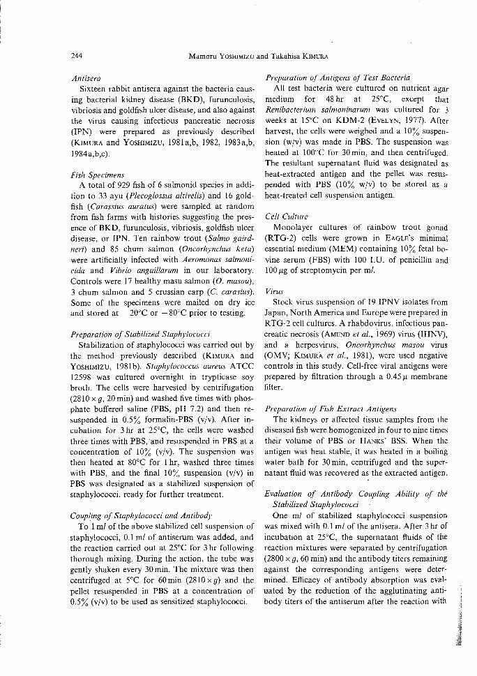
244 Mamoru Y OSHIMIZU and Takahisa KIMURA
Antisera Sixteen rabbit antisera against the bacteria caus
ing bacterial kidney disease (BKD), furunculosis, vibriosis and goldfish ulcer disease, and also against the virus causing infectious pancreatic necrosis (IPN) were prepared as previously described (KIMURA and YOSHIMIZU, 198Ia,b, 1982, 1983a,b, 1984a,b,c).
Fish Specimens A total of 929 fish of 6 salmonid species in addi
tion to 33 ayu (Plecoglossus altivelis) and 16 goldfish (Carassius auratus) were sampled at random from fish farms with histories suggesting the presence of BKD, furunculosis, vibriosis, goldfish ulcer disease, or IPN. Ten rainbow trout (Salmo gairdneri) and 85 chum salmon (Oncorhynchus keta) were artificially infected with Aeromonas salmon icida and Vibrio anguillarum in our laboratory. Controls were 17 healthy masu salmon (0. masou), 3 chum salmon and 5 crussian carp (c. carasius). Some of the specimens were mailed on dry ice and stored at - 20°C or - 80°C prior to testing.
Preparation of Stabilized Staphylococci Stabilization of staphylococci was carried out by
the method previously described (KIMURA and YOSHIMIZU, 1981 b). Staphylococcus aureus A TCC 12598 was cultured overnight in trypticase soy broth. The cells were harvested by centrifugation (28 lOx g, 20 min) and washed five times with phosphate buffered saline (PBS, pH 7.2) and then resuspended in 0.5% formalin-PBS (vjv). After incubation for 3 hr at 25°C, the cells were washed three times with PBS,and resuspended in PBS at a concentration of 10% (vjv). The suspension was then heated at 80°C for I hr, washed three times with PBS, and the final 10% suspension (vjv) in PBS was designated as a stabilized suspension of staphylococci, ready for further treatment.
Coupling of Staphylococci and Antibody To 1 ml of the above stabilized cell suspension of
staphylococci, 0.1 ml of antiserum was added, and the reaction carried out at 25°C for 3 hr following thorough mixing. During the action, the tube was gently shaken every 30min. The mixture was then centrifuged at 5°C for 60 min (2810 x g) and the pellet resuspended in PBS at a concentration of 0.5% (vjv) to be used as sensitize~ staphylococci.
Preparation of Antigens of Test Bacteria All test bacteria were cultured on nutrient agar
medium for 48 hr at 25°C, except that Renibacterium salmoninarum was cultured for 3 weeks at 15°C on KDM-2 (EVELYN, 1977). After harvest, the cells were weighed and a 10% suspension (wjv) was made in PBS. The suspension was heated at 100°C for 30 min, and then centrifuged. The resultant supernatant fluid was designated as heat-extracted antigen and the pellet was resuspended with PBS (10% wjv) to be stored as a heat-treated cell suspension antigen.
Cell Culture Monolayer cultures of rainbow trout gonad
(RTG-2) cells were grown in EAGLE'S minimal essential medium (MEM) containing 10% fetal bovine serum (FBS) with 100 LV. of penicillin and 100 J1g of streptomycin per m!.
Virus Stock virus suspension of 19 IPNV isolates from
Japan, North America and Europe were prepared in RTG-2 cell cultures. A rhabdovirus, infectious pancreatic necrosis (AMEND et at., i969) virus (lHNV), and a herpesvirus, Oncorhynchus masou virus (OMV; KIMURA et at., 1981), were used negative controls in this study. Cell-free viral antigens were prepared by filtration through a 0.45 J1 membrane filter.
Preparation of Fish Extract Antigens The kidneys or affected tissue samples from the
diseased fish were homogenized in four to nine times their volume of PBS or HANKS' BSS. When the antigen was heat stable, it was heated in a boiling water bath for 30 min, centrifuged and the supernatant fluid was recovered as the extracted antigen.
Evaluation of Antibody Coupling Ability of the Stabilized Staphylococci One ml of stabilized staphylococci suspension
was mixed with O. I ml of the antisera. After 3 hr of incubation at 25°C, the supernatant fluids of the reaction mixtures were separated by centrifugation (2800 x g, 60 min) and the antibody titers remaining against the corresponding antigens were determined. Efficacy of antibody absorption was evaluated by the reduction of the agglutinating antibody titers of the antiserum after the reaction with

A Coagglutination Test 245
0.05 ml of antigen 0.05 ml of sensitized
I
staphylococci suspension
--> Mix in depression slide .-1 1
Stand at 25°C in moist chamber
1 Observe the result with microscope (x 10)
after 30, 60, 120min
Fig. 1. Flow chart outlining the co agglutination test.
staphylococci.
The Coagglutination Test A volume of 0.05 ml of antibody-sensitized
staphylococci suspension and 0.05 ml of the test antigen were mixed on a glass slide, and the reaction was carried out in a moist chamber for 30, 60, or 120 min. At the end of the incubation period, the slide was examined by naked eye or under a microscope (x 10) for development of the coagglutination reaction. The overall reaction procedure is summarized in Fig. I.
Immunodijfusion Test and Fluorescent Antibody Test The immunodiffusion tests were carried out by
the ordinary micro-OucHTERLONY method (OUCHTERLONY and NILSSON, 1973). The BKD fluorescent antibody tests were carried out by the direct method using FITC conjugated anti-R. salmoninarum rabbit serum provided by C. BANNER, Oregon State University. For the vibrios, the indirect method was used (McDANIEL, 1975).
Results and Discussion
Antibody Binding Ability of Stabilized Staphylococci The antibody binding ability of the stabilized
staphylococci was tested using rabbit antisera against R. salmoninarum and A. salmonicida. Agglutinating antibody titers of these antisera were markedly reduced after incubation with the stabilized staphylococci (Table 1), indicating that most of the antibody had apparently been coupled to the protein A of these organisms.
Specificity of the Coagglutination Reaction for Detection of Bacterial and Viral Antigens Cross coagglutination tests were carried out with
rabbit antisera against R. salmoninarum, A. salmonicida and V. angui/!arum, and the corresponding heat-extracted and heat-treated cell suspension antigens. The results are shown in Table 2. Coagglutination reaction patterns are shown in Figs. 2 and 3. It was apparent that agglutination occurred only with specific combinations of antibody sensitized staphylococci and the antigen homologous for the antibody, and not with heterologous antigens or with staphylococci sensitized with normal rabbit serum. The results obtained with heat treated cell antigens were the· same as those observed with heat extracted antigens, although reactions with the cell antigens were more rapid and produced larger aggregations than when heat extracted antigens were used.
Specificity was further tested with staphylococci sensitized with the same 5 antibacterial sera. These preparations were tested for possible reactions against heat extracted antigens of Aeromonas
Table 1. Antibody binding ability of stabilized staphylococci
Antiserum
Anti-Aeromonas salmonicida ATCC 14174
Anti-BKD bacterium*l strain Otobe (0-1)
Agglutinin titer of antiserum before and after absorption with staphylococci
Before After
25,600 400
1,600 50
*1 Kidney disease bacterium; Renibacterium salmoninarum.
Percent of antibody bound
98.4
96.9

246 Mamoru YOSHIMIZU and Takahisa KIMURA
Table 2. Cross coagglutination tests using staphylococci sensitized with antibody specific for each of three bacterial pathogens and antigens prepared from cells of these organisms
Antisera used to sensitize staphylococci
Bacterial antigen used*! as heat treated cell
suspension
Anti-BKD bacterium _____________ Anti-Aeromonas Anti- Vibrio
salmonicida anguillarum ATCC 14174 KAY-2
Control*5 Strain Strain Strain
EFDL-2*2 AKD-3*3 OKD-3*4
BKD bacterium strain Otobe BKD bacterium strain Erimo Aeromonas salmonicida ATCC 14174 Vibrio anguilla rum strain KA Y-2
+ +
+ +
+ +
Heated in PBS at 100°C for 30 min. After centrifuging the pellet resuspended in PBS was the cell suspension antigen. Supernatant fluid was the heat extract antigen. All tests were conducted with each type of antigen and results were identical in each case. Antiserum provided by National Fish Health Research Lab., Leetown W. Va., U.S.A.
*3 Antiserum against strain Marimo. *4 Antiserum against strain Otobe. *5 Normal rabbit serum. *6 + indicates a positive agglutination reaction; - indicates a negative reaction.
Fig. 2. Coagglutination reaction patterns obtained with heated cell suspension of BKD bacterium strain Erimo, using a staphylococcal suspension coupled with anti-BKD serum, AKD-3 (3). The same cell suspension did not agglutinate with normal rabbit serum bound staphylococci (4), and neither staphylococcal suspension agglutinated with PBS (I, 2).
species, Vibrio species, R. salmoninarum, Escherichia coli, Pseudomonas jluorescens, Micrococcus lysodeikticus, and Bacillus subtilis. Once again, the only agglutination reactions observed occurred with antibody sensitized staphylococci and the antigen homologous for the antibody (Table 3). The evidence from all of these tests indicates a
Fig. 3. Coagglutination reaction patterns obtained with heat extracted antigen of Aeromonas salmonicida ATCC 14174 (Ar-3), using a staphylococcal suspension coupled with anti-A. salmonicida ATCC 14174 serum (3). The same extracted antigen did not agglutinate with normal rabbit serum bound staphylococci (4), and neither staphylococcal suspension agglutinated with PBS (I, 2),
high degree of specificity for this coagglutination reaction.
Cross coagglutination tests were carried out using rabbit antisera against IPNV (Buhl), IHNV (Yurappu) and OMV (00-7812). These were tested against cell free virus antigens, virus free culture medium from RTG-2 cells, MEM-IO and FBS.

A Coagglutination Test 247
Table 3. Specificity of coagglutination tests using staphylococci sensitized with antibody specific for each of .three bacterial pathogens
============================================================ Antisera used to sensitize staphylococci
Bacterial antigen used*l Anti-BKD bacterium
_____________ Anti-Aeromonas Anti- Vibrio salmonicida angllillarllm
ATCC 14174 KAY-2 Control*5
Strain Strain Strain EFDL-2*2 AKD-3*3 OKD-3*4
BKD bacterium strain Erimo BKD bacterium strain Otobe BKD bacterium strain Niigata BKD bacterium strain Shizuoka BKD bacterium strain Miyako Aeromonas hydrophila lAM 1018 A. punctata lAM 1646 A. liqllejaciens EFDL A. salmonicida NCMB 833 A. salmonicida NCMB 1102 .4. salmonicida ATCC 14174 .4. salmonicida subsp.
masoucida NCMB.2020 Vibrio metschnikovii lAM 1039 V. Iyrogenes lAM 1080 V. piscium var. japonicus TUF V. anguillaruIIJ NCME 6 V. anguillaruIIJ NCMB 828 V. anguillaruIIJ NCMB 829 V. anguillarum KA Y-2 Escherichia coli 0-6 Pseudomonas jlllorescens EFD L Micrococcus lysodeikticlls Mi 2 Bacillus subtilis NRRL 558
+*6
+ + + +
+ + + + +
*' Heat extracted antigen by PBS at 100°C 30 min.
+ + + + +
+ + +
+
+ + + +
*2 Antiserum provided by National Fish Health Research Lab., Leetown W. Va., U.S.A. *3 Antiserum against strain Marimo. *4 Antiserum against strain Otobe. *5 Normal rabbit serum.
+ indicates a positive agglutination reaction; - indicates a negative reaction.
Staphylococci sensitized with un adsorbed antisera showed positive reactions with all antigens employed, but staphylococci sensitized with antisera adsorbed with FBS and acetone powdered RTG-2 cells gave positive reactions only when anti-IPNV rabbit serum sensitized staphylococci and a cell free IPNV antigen were used. Heterologous antigens and staphylococci sensitized by normal rabbit serum gave negative results. Anti-IHNV and OMV rabbit sera sensitized staphylococci showed no reaction with either homologous or heterologous antigens (Table 4).
The specificity of the reaction was confirmed with a blocking test. The blocking test was carried out using staphylococci sensitized with antiserum against IPNV (Buh!) and a cell free IPNV (Buh!) antigen which had been mixed with anti-IPNV (Buhl) rabbit serum for 1 h at room temperature. A positive reaction occurred only when the antiIPNV sensitized staphylococci and unblocked IPNV antigen were used.
By solid phase immune electron microscopy (SPIEM), the agglutinated staphylococci sensitized with anti-IPNV serum would bind IPNV on the

248 Mamoru YOSHIMIZU and Takahisa KIMURA
Table 4. Cross coagglutination tests using staphylococci 'sensitized with IPNV (Buhl), IHNV (Yurappu) and OMV (00-7812) antisera against selected cell culture and viral antigens
Antisera used to sensitized staphylococci
Antigen used
Fetal Bovine Serum Minimal Essential Medium
(MEM-10) Medium from RTG-2 cell
culture Cell free IPNV (Buhl) Cell free IHNV (Yurappu) Cell free OMV (00-7812)
*' Normal rabbit serum. *2 Unadsorbed antiserum.
Anti-IPNV Buhl
A*2 B*3
+
+
+
+ + + +
Anti-IHNV Anti-OMV Control*'
Yurappu 00-7812
A B A B A
+ +
+ +
+ +
+ + + + + +
*3 Antiserum adsorbed with FBS and acetone powdered RTG-2 cell.
surface of the cells (Fig. 4).
Coagglutination Test for Rapid Serological Identification of Auto-agglutinating A. salmonicida Nine A. salmonicida, three Aeromonas species,
two strains of V. anguillarum, and P. jluorescens were prepared and observed for auto-agglutination. The results of the comparison of autoagglutination of the bacteria used and the specificity of coagglutination tests using staphylococci sensitized with anti-A. salmonicida serum are shown in Table 5. Sensitized staphylococci showed a positive reaction with culture broth and heattreated cell suspension antigens quickly and produced a large agglutination. The heat extracted antigen showed a positive reaction also, in less than 15min. Heat extracted antigen is suitable for use in the laboratory or field, because in this preparation causative bacteria are killed and antigen does not include the bacterial cells.
Coagglutination Test for Serological Typing of V. anguillarum For serological typing of V. anguillarum antigens,
the specificity of the coagglutination test was evaluated using staphylococci sensitized with three different antisera (EZURA, T AlIMA, YOSHIMIZU and KIMURA, 1981); V-6 (1-0-1 type), V-123 (J-0-2 type) and V-125 (1-0-3 type). Staphylococci sensitized
Fig. 4. IPNV adsorbed to S. aureus sensitized with homotypic antibody. Arrowheads indicated virus particles. Negatively stained with 0.5% uranyl acetate (pH 5). -- 200 nm.
with anti V. anguillarum V-6 rabbit sera showed cross reactions with other strains of the genus Vibrio, but staphylococci sensitized with serum adsorbed by these vibrios showed positive reactions only with the strains of V. anguillarum belonging to J-O-1. Anti V. anguillarum V-123 serum sensitized staphylococci agglutinated with the antigens prepared from the strains V-I23, V-114, belonging to the J-0-2 type and anti V. anguillarum V -125 serum sensitized staphylococci agglutinated with the anti-

A Coagglutination Test 249
Table 5. Comparison of auto-agglutination of bacteria used and specificity of coagglutination tests using staphylococci sensitized with anti-A. salmonicida ATCC 14174 serum
======================================================
Bacteria or bacterial antigens used
Aer0111011aS hydrophila lAM 1018 A. pUl1ctata lAM 1646 A. liquefaciens EFDL Aerol11onas sp. A. sall110nicida ATCC 14174 A. salmollicida NCMB 1102 A. salmollicida Nagano N-8 A. salmonicida Nagano N-17 A. sall11011icida Hokkaido I M
A. salmonicida Hokkaido 5 M
A. salmonicida Wakayama A. salmonicida lwate A. salmollicida subsp.
masoucida NCMB 2020 Vi/n'io anguillarul11 NCMB 6 Pseudomonas jluorescens EFD L
Auto-agglutination
Culture broth
*1
+ +*1 ++ ++ ++
+ ++ + ++ + ++
++ ++ ++ ++
++
Coagglutination test
Heated cell Heat extract of suspension culture broth
++ + ++ + ++ + ++ + ++ + ++ + ++ + ++ +
++ +
*' + indicates a positive agglutination reaction; - indicates a negative reaction.
gens prepared from the strains V-125, V-104, V-106, V-III these are J-0-3 type respectively (Table 6), as reported by NEWMAN, BLOOM and MAJNARICH ( 1982).
The Coagglutination Test for Serological Typing of Cell Free IPNV Cultured in RTG-2 Cells For serological typing of cell grown IPNV anti
gens, three different staphylococci sensitized with antisera against strains VR 299, Sp and Ab were tested against five IPNV strains grown in cell culture from North Am~rica, ten strains from Japan and four strains from Europe. The staphylococci sensitized with anti-VR 299 serum showed positive reactions with the strains from North America and Japan but not with the European strains. Staphylococci sensitized with anti-Sp or Ab sera showed positive reactions only with the strains from Europe after 30 min incubation. However, weak cross reactions were observed with the strains from North America and Japan after incubation periods exceeding 60 min (Table 7); the agglutinating particles were very fine and different from those in the reactions observed with Sp and Ab strains (Fig. 5). These results indicated the usefulness of the coag-
glutination test for rapid serological typing ofIPNV for the North American and European strains. The minimum amount of viral antigen needed to get a positive reaction varied between 105.9 and 107.7 TCIDso/ml.
The Coagglutination Test with Antibody Sensitized Staphylococci and Kidney Extract Antigen Prepared ji-om Fish with Bacterial Kidney Disease Heat extracted antigen was prepared from the
kidney tissue of a kokanee salmon and a chinook salmon with bacterial kidney disease. The extracts represented a I: 5 tissue suspension w/v in PBS. Antigen was similarly prepared from pooled kidney tissue of 17 healthy masu salmon. All three antigens were tested for coagglutination of staphylococci sensitized with antisera specific for the kidney disease bacterium. Two-fold dilutions of the antigens from 2- 1 to 2- 6 or higher were employed. Coagglutination reactions occurred with all dilutions of the antigens from the diseased fish. The more concentrated dilutions tended to give positive reactions within 30 min, while the higher dilutions required a longer period. The control antigens prepared from the kidney tissue of healthy fish

250 Mamoru YOSHIMIZU and Takahisa KIMURA
Table 6. Specificity of coagglutination tests using staphylococci sensitized with antibody specific for each three serotype V. anguillarwl1
Bacterial antigen used
Vibrio anguiliarum (V-6) NCMB 6 V. anguillarum (V-123) PTe-I V. anguillarum (V-125) PT-223
Vibrio anguillarum (V-8) NCMB 828 V. anguillarum (V-9) NCMB 829 V. anguilla rum (V-72) KA Y-3 V. anguillarum (V-lOS) NOAA 1669 V. anguillaru111 (V -113) PT -24 V. anguillarum (V -119) NP-I V. anguillarum (V-I 14) PT-514 V. anguillarum (V-104) NOAA 775 V. anguiliarwl1 (V-106) N-1 V. anguillarul11 (V-117) NCMB 571 V. metscl11likovii (V-I) lAM 1039 V. tyrogenes (V-2) lAM 1080 V. pisciu111 (V-5) TUF V. parahaemolyticus H-0-5 V.fischeri NCMB 1281 Vibrio sp. A-4-1 Lucibacteriu111 harveyi NCMB 1280 Aeromonas proteolytica NCMB 1326 Beneckea campbelli A TCC 25920
Anti
Anti V-6 J-O-I
a*2 b*3
+*4 + *4
+ + + + + + + + + + + +
± ±
± ± ± ±
± ± +
-V. anguillarum sera used to sensitize staphylococci
Anti V-6*1 Anti V-123 Anti V-125 Control
J-O-I J-0-2 J-0-3
a b a b a' b a b
+ + + +
+ +
+ + + + + + + + + + + +
+ + + + + + + +
Adsorbed serum using V. parahaemolyticus, V. fisclw'i, V. sp (A-4-l), L. harveyi, A. proteolytica and B. campbelli. Heat extracted antigen by PBS at 100°C for 30 min.
*3 Heated cell suspension antigen; heated in PBS at 100°C for 30 min, after centrifuging the pellet resuspended in PBS. + indicates a positive agglutination reaction, - indicates a negative reaction.
Fig. 5. Coagglutination reaction patterns obtained with MEMlO (3), cell free IPNV strains VR-299 (4), Sp (I) and Ab (2), using a staphylococcal suspension sensitized with anti-IPNV (Strain Sp) rabbit serum.
failed to agglutinate the sensitized staphylococci at any concentration tested, and staphylococci sensitized with normal rabbit serum also failed to react with any of the antigen preparations (Table 8).
Data from these experiments indicated the ability of the coagglutination test to detect the specific ant·igen of bacterial kidney disease in the kidney tissues from infected kokanee and chinook salmon at relatively high dilutions and within a test period of only thirty minutes.
Comparison of the Coagglutination Test with the Classical Diagnostic Methodsfor the Detection of Bacterial Kidney Disease in Salmon ids The commonly used or classical methods for

Tab
le 7
. Sp
ecif
icity
of
coag
glut
inat
ion
test
s us
ing
stap
hylo
cocc
i se
nsiti
zed
wit
h an
ti-I
PN
V r
abbi
t se
rum
Ant
iser
a us
ed t
o se
nsiti
ze s
taph
yloc
occi
IPN
V a
ntig
en u
sed
Ant
i-IP
NV
ser
um
Min
imum
tit
er
Con
trol
*'
show
ing
posi
tive
IPN
V
Tit
er
VR
-299
Sp
A
b re
acti
on
(TC
lDso
/m/)
(T
ClD
so/m
l)
30*2
60
90
30
60
90
30
60
90
30
60
90
VR
-299
7.
05
+
+
+
+
+
+
6.45
B
uhl
7.55
+
+
+
+
+
+
+
6.
95
Wes
t B
uxto
n 7.
80
+
+
+
+
+
+
7.20
:>
P
owde
r M
ills
7.80
+
+
+
+
+
+
+
7.
50
(")
Ren
o 7.
55
+
+
+
+
+
+
+
6.95
0 ~
(JQ
(J
Q
Nic
hiro
7.
05
+
+
+
+
+
+
6.45
g- 5'
M
atsu
hisa
6.
80
+
+
+
+
+
+
6.50
~
Oip
pe
8.30
+
+
+
+
+
+
+
7.
70
o· ::l
Eni
wa
8.30
+
+
+
+
+
+
+
7.
70
....,
Yam
amot
o 6.
80
+
+
+
+
+
+
5.90
f!
Gif
n R
T
7.55
+
+
+
+
+
6.
95
Gif
u C
O
7.05
+
+
+
+
+
+
6.
45
Gif
u A
M
7.05
+
+
+
+
+
+
+
6.
45
Tow
ada
7.80
+
+
+
+
+
+
7.
50
Sai
to
7.05
+
+
+
+
+
+
+
6.
75
Sp (
Den
mar
k)
8.55
+
+
+
+
+
+
7.
65
Bon
nam
y (F
ranc
e)
8.30
+
+
+
+
+
+
7.
70
d'H
onni
ncht
on (
Fra
nce)
8.
80
+
+
+
+
+
+
7.63
A
b (D
enm
ark)
8.
30
+
+
+
+
+
+
7.40
*,
Nor
mal
rab
bit
seru
m.
*2
Rea
ctio
n ti
me
(min
utes
).
N
v,

252 Mamoru YOSHIMIZU and Takahisa KIMURA
Table 8. Effects of concentration of antigen and the time of reaction on the co agglutination tests for detection of BKD antigen in heat extract of affected salmon kidney
Antisera used to sensitize staphylococci
Anti-BKD bacterium Control*3 Source of
Fish Concentration specimens
specimens of antigen
Strain EFDL-2*1 Strain AKD-3*2 (hatchery) Reaction time Reaction time Reaction time
(min) (min) (min)
30 60 120 30 60 120 30 60 120
Kokanee*4 Chitose 2-1 +*6 + + + + + *6
salmon 2- 2 + + + + + + No. 19 2-3 + + + + + +
2-4 + + + + + + 2- 5 + + + + + + Z-6 + +
Kokanee*4 Erimo 2- 1 + + + + + + salmon 2-2 + + + + + + No. 29 2- 3 + + + + + +
2-4 + + + + + + 2- 5 + + + + + 2-6 + + + + + 2- 7 + + + + 2-8 + +
Masu salmon Mori 2-0
No. 759- (Control) 2- 1
776 PBS 2-0
*1 Antiserum provided by National Fish Health Research. Lab., Leetown W. Va., U.S.A. *2 Antiserum against strain Marimo. *3 Normal rabbit serum. *4 Oncorhynchus nerka. *5 Oncorhynchus masou.
+ indicates a positive agglutination reaction;
diagnosis of bacterial kidney disease include observation of clinical signs, Gram staining of kidney smears, the immunodiffusion test and fluorescent antibody tests. Individual fish, negative by coag~ glutination, immunodiffusion and the Gram reaction and exhibiting no clinical signs of the disease, were found to be positive by the fluorescent antibody test. Detection of infection by FAT was the most sensitive with coagglutination tests being the next (Table 9). This indicated the effectiveness of FAT for detecting the kidney disease bacterium at very low cell concentrations. However, the increased sensitivity, as compared to immunodiffusion, and lack of a requirement for special U.V. equipment point to the usefulness of coaggluti-
indicates a negative reaction.
nation, especially under field or hatchery conditions (FRYER and SANDERS, 1981).
Diagnosis of Bacterial Kidney Disease by the Coagglutination Test in Fish Populations Undergoing Natural Epizootics of the Disease A total of six hundred and seventy-four fish
specimens were collected at random from nineteen salmonid fish farms thought to be experiencing epizootics of bacterial kidney disease. The number offish specimens found to be infected with bacterial kidney disease by the coagglutination test were at least as great in all groups of fish examined as the numbers detected by either Gram staining or observation of clinical signs (Table 10).

A Coagglutination Test
Table 9. Comparison of clinical signs, gram stain, coagglutination test, direct fluorescent antibody test and immunodiffusion for the detection of bacterial kidney disease or the causative agent in chinook salmon (FRYER and SANDERS, 1981)
253
Fish number*1
Clinical signs
Gram stain
Coagglutination test*2,4
Fluorescent antibody test*3
Immunodiffusion *4
2 3 4 5 6 7 8 9
10 II 12 13 14 15 16 17 18 19 20 21 22 2: 24 25 26 27 28 29 30 31 32 33
33 fish
± + + ± ± + + + ±
± ± + + + + ± ± ± + + + + +
14/33
+ +
+ +
+ + + + + + + + + + + + + + + + + + + + + + + +
28/33
*1 Fish 1-30 juvenile; 31-33 adult.
+ + + ± + +
+
+ + + + + + + + + + + + + + + + + + + + + + + +
30/33
++ ++ + +
++ ++ + + + +
++ ++ ++ ++ ++ ++ ++ ++ ++ ++ ++ +
++ ++ ++ ++ ++ ++ ++ ++ ++ ++ ++
33/33
+ +
+ +
+ + + + + + + + + + + + + + + + + + + + + +
26/33
*2 Results after 2 hours incubation at room temperature in a moist chamber. *3 Smears prepared by heat fixation. *4 Kidney diluted approximately I: 10 with PBS. Except fish 8-11 and 18, 1: 20 and 14, 1: 50.
Diagnosis of Furunculosis by the Coagglutination Test Fish from a total of 70 natural outbreaks of
furunculosis and 10 artificially infected fish specimens were examined. The results of comparisons of coagglutination tests, clinical signs and isolation of A. salmonicida for diagnosis of furunculosis are shown in Table 11. The number of fish specimens found to be infected with A. salmonicida by isola-
tion were greater than by coagglutination tests, but in the case of heat-extracted antigen prepared from furuncle tissue of artificially infected fish, the rate was the same.
Diagnosis of Vibriosis by Coagglutination Test. The comparison of coagglutination tests, clinical

254 Mamoru YOSHIMIZU and Takahisa KIMURA·
Table 10. Diagnosis of bacterial kidney disease by the coagglutination test in natural outbreaks in Japan, 1977-1980
-Number of specimens diagnosed
Fish Source of
Number of as positive for BKD
specimens specimens Date
specimens (name of farm) Clinical Coagglutination
sign Gram stain test*1
Coho salmon*3 Fujinomiya Aug. '77 20 13 18 18 Coho salmon Nakagawa Aug. '77 7 *2 6 Coho salmon Koide Dec. '77 5 4 4 4 Coho salmon Maebashi Oct. '78 2 2 2 2 Rainbow trout*4 Fuji Jap. '79 10 7 Amago*5 Fuji May '79 21 2 2 2 Coho salmon Koide June '79 5 4 4 4 Masu salmon*6 Otobe June '79 22 11 12 12 Coho $almon Miyako-M June '79 36 12 12 12 Coho salmon Otuti July '79 5 I Coho salmon Hamanaka Aug. '79 15 4 4 6 Masu salmon Otobe Sep. '79 10 7 7 8 Coho salmon Koide Oct. '79 153 21 65 76 Coho salmon Fujinomiya Oct. '79 66 5 9 16 Masu salmon Otobe Dec. '79 38 9 18 35 Coho salmon Kaida Dec. '79 3 2 2 3 Coho salmon Otuti May '80 5 2 2 Masu salmon Koide May '80 5 4 4 5 Coho salmon Kukizaki June '80 20 0 0 3 Masu salmon Mori June '80 22 11 18 Masu salmon Koide June '80 43 35 32 43 Masu salmon Urasa June '80 24 10 6 8 Kokanee salmon*7 Shikishima June '80 10 4· 3 9 Coho salmon Hamanaka July '80 26 2 0 5 Coho salmon Otuti July '80 5 2 3 Coho salmon Toyama July '80 2 Coho salmon Hamanaka Aug. '80 IS 13 13 15 Masu salmon Otobe Sep. '80 7 3 4 Coho salmon Miyako Oct. '80 5 2 5 5 Chum salmon*8 Miyako Oct. '80 47 24 26 34 Coho salmon Nikko Oct. '80 3 2 Masu salmon Shako tan Nov. '80 6 2 2 2 Coho salmon Nikko Dec. '80 11 2 2 5
*1 Anti-BKD sera; AKD-3, OKD-3, EFDL-l, or EFDL-2; used to sensitize staphylococci. *2 Indicates no data. *3 Oncorhynchus kisutch. *4 Salmo gairdneri. *s Oncorhynchus rhodurus var. macrostomus. *6 O. masou. *7 O. nerka. *8 O. keta.
signs, and isolation of V. anguil/arum for the de- infected with V. anguillarum by isolation and coag-tection of vibriosis was carried out using fish arti- glutination were the same and the specificity be-ficially infected with different serotypes of V. anguil- tween serotypes was clearly recognized (Table 12). larum. The number of fish specimens found to be Table 13 shows the results of detection of V.

A Coagglutination Test 255
Table 11. Comparison of coagglutination test, clinical signs, and isolation of A. salmonicida for diagnosis of furunculosis
Number of specimens with positive diagnosis
Fish specimens
Source of specimens
Date Number Clinical Isolation of
A. salmonicida Coagglutination
test*! of fish sign
Kidney Furuncle Kidney*2 Furuncle*2
Coho salmon*3 Iwate Nov. '79 28 23 19 *8 15 Amago salmon*4 Gifu Dec. '79 12 6 II 6 Masu salmon*5 Tokyo Dec. '79 30 17 28 21 Rainbow trout*6 Laboratory*7 Mar. '80 10 10 10 10 8 10
*' Staphylococci sensitized with antiserum against A. salmonicida ATCC 14174. *2 Heat extracted antigen by PBS (I: 9) at 100°C for 30min. *3 Oncorhynchus kisutch. *4 Oncorhynchus rhodurus. *5 Oncorhynchus masou. *6 Salmo gairdneri. *7 Artificially infected by intramuscular injection. *" - indicates no data.
angui//arum antigens by coagglutination testing for natural outbreaks of vibriosis. Except for one case of pen culture coho salmon, all specimens were found to have vibriosis caused by V. angui//arum serotype J -0-1. FAT Was more sensitive than coagglutination testing, but in some cases coagglutination tests were more sensitive.
Diagnosis of Goldfish Ulcer Disease by Coagg(utination Test Specificity of the coagglutination reaction for
detection of atypical A. salmonicida antigens was carried out by a cross coagglutination test using staphylococci sensitized with anti-atypical A. salmonicida and anti-typical A. salmonicida sera. Atypical A. salmonicida and typical A. salmonicida have one common antigen (KIMURA and YOSHIMIZU, 1984b). These three sensitized staphylococci all showed positive reactions with typical A. salmonicida and atypical A. salmonicida strains (Table 14).
Results of detection of an atypical A. salmonicida in affected tissues and kidneys of goldfish are shown in Table 15. All specimens prepared from diseased fish showed positive agglutination. However, except for one specimen, we failed to isolate the atypical A. salmonicida. Agglutination antibody titers of diseased fish were relatively high.
Diagnosis of IPN in Fish Undergoing Natural Epi::.ootics using the CoagglutinatiOI1 Test A ·total of 44 fish specimens consisting of three
salmonid species were collected at random from six fish farms. Three healthy chum salmon fry were used for controls. Staphylococci sensitized with antiserum against IPNV (strain Buhl) showed positive coagglutination reactions with tissue from rainbow trout, coho and am ago salmon cultured in Gifu Prefecture. The virus was isolated from all fish except the coho salmon Nos. Co-4 to 6 which were received by our laboratory chilled with dry ice. The antigens prepared from the rainbow trout 'cultured in Yamanashi and Niigata Prefectures, from which IPNV was not isolated, and from rainbow trout cultured in Niigata, from which IHNV was isolated, showed negative reactions. The control antigens prepared from healthy chum salmon also showed negative reactions (Table 16).
Conclusions
The application of the coagglutination test using staphylococci specifically sensitized with antibodies against the bacteria causing BKD, furunculosis, vibriosis and goldfish ulcer disease, and also against the virus causing lPN, for the diagnosis of these

Tab
le
12.
Com
pari
son
of
coag
glut
inat
ion
test
, cl
inic
al s
igns
, an
d is
olat
ion
of
V. a
ngui
llar
um f
or d
etec
tion
of
vibr
iosi
s
Num
ber
of
spec
imen
s w
ith
posi
tive
dia
gnos
es
Fis
h E
xam
inat
ion
spec
imen
s
I*3
chum
sal
mon
(J
uly
'80)
ch
um s
alm
on
chum
sal
mon
II*4
ch
um s
alm
on
(Jan
. '8
1)
chum
sal
mon
ch
um s
alm
on
Kid
ney
..
Liv
er.
Inje
ctio
n S
erot
ype
stra
in
of
stra
in
V-6
J-
O-I
V
-123
J-
0-2
V
-l06
J-
0-3
V-6
J-
O-I
V
-123
J-
0-2
V
-125
J-
0-3
Num
ber
Isol
atio
n o
f o
f fis
h C
lini
cal
V. a
ngui
llar
um
sign
K
idne
y L
iver
II
]0
7 7
10
5 5
5 34
28
7
7
10
8 8
]0
]0
10
]0
*3
In
trac
avit
y in
ject
ion;
V-6
, 2.
0 x
107
; V
-123
, 4.
8 x
106
; V
-I06
, 8.
0 x
]06
Intr
amus
cula
r in
ject
ion;
V-6
, 1.
8 x
]06
; V
-123
, 2.
0 x
106 ;
V-I
25,
4.7
x ]0
6.
Coa
gglu
tina
tion
tes
t; se
nsiti
zed
wit
h
Ant
i V
-6
Ant
i V
-123
A
nti
V-1
25
K*l
L*
2 K
L
K
L
7 4
0 0
0 0
0 0
5 5
0 0
0 0
0 0
7 7
8 0
0 0
0 0
0 10
Con
trol
K
L
0 0
0 0
0 0
0 0 0

Tab
le
13.
Dia
gn
osi
s o
f vi
brio
sis
by t
he c
oag
glu
tin
atio
n t
est
and
FA
met
ho
d i
n n
atu
ral
ou
tbre
aks
So
urc
e o
f sp
ecim
ens
Yam
anas
hi-
B
Yam
anas
hi-
B
Toc
higi
-S
To
chig
i-O
T
och
igi-
O
Toc
higi
-I
Miy
agi-
S*5
H
ok
kai
do
-M*
6
Fis
h
spec
ies
rain
bo
w t
rout
*2
rain
bo
w t
rou
t ay
u*
l
ayu
ay
u
ayu
co
ho
sal
mon
*3
chu
m s
alm
on*4
Ple
cogl
ossu
s al
tivel
is.
*2
Sa
lrno
gai
rdne
ri.
*3
Onc
orhy
nchu
s ki
sutc
h.
Dat
e
July
'81
Ju
ly '
81
July
'81
Ju
ly '
81
Aug
. '8
1 A
ug.
'81
Dec
. '8
1 D
ec.
'81
*4
Sea
wat
er f
ish,
ser
otyp
e o
f is
olat
es w
as J
-0-3
. O
. ke
ta.
Nu
mb
er o
f sp
ecim
ens
!O
10 6 8 4 15 3 9
Cli
nica
l si
gn
o 3 o 8 3
*6
Cu
ltu
red
in
aqu
ariu
m b
y s
ea w
ater
, se
roty
pe
of
isol
ates
was
J-O
-1.
Nu
mb
er o
f sp
ecim
ens
dia
gn
ose
d a
s po
siti
ve f
or
vibr
iosi
s
Co
agg
luti
nat
ion
tes
t; s
ensi
tize
d w
ith
An
ti V
-6
An
ti V
-123
A
nti
V-1
25
Co
ntr
ol
6 0
0 0
5 0
0 0
5 0
0 0
8 0
0 0
2 0
0 0
9 0
0 0
0 I
2 0
5 0
0 0
FA
met
ho
d;
ind
irec
t by
an
ti V
-6 s
eru
m
>-4
(")
0 2
'" (JQ
6 os.
c
8 S·
4 ~ o·
II
:;
>-l "
8 ~

258 Mamoru YOSHlMIZU and Takahisa KIMURA
Table 14. Specificity of coagglutination test using staphylococci sensitized with anti A. salmonicida and atypical A. salmonicida
===================================================== Antisera used to sensitize staphylococci
Bacterial antigen used* Anti-A. salmonicida Anti-atypical A. salmonicida
ATCC14174 V-76-65 V-76-134
Atypical A. salmonicida LO-2 LO-6
Atypical A. salmonicida TY-76192 TY-79057h TY-79058f TY-790591 TY-80003w
Atypical A. salmonicida V-76-65 V-76-134
A. salmonicida ATCC 14174 NCMB 1102
A. salmonicida subsp. masoucida NCMB 2020
A. hydrophila NCMB 86 A. punctata NCMB 74 A. liquefaciens A TCC 11715
* Heat extracted antigen by PBS at 100°C 30 min.
diseases in fish was studied. Stabilized Staphylococcus aureus (COWAN I) was
sensitized with rabbit anti-serum against R. salmoninarum, V. anguillarum, typical and atypical A. sal
monicida and IPNV. The antibody binding ability of the sensitized
staphylococci was tested and the agglutinating antibody titers of antisera were markedly reduced after incubation with the stabilized staphylococci.
Specificity of the coagglutination reaction for detection of bacterial and viral antigens in vitro were carried out by a cross coagglutination test. Agglutination occurred only with specific combinations of antibody sensitized staphylococci and the homologous antigen for the heat extracted bacteria, the heated bacteria and the non-treated cell free viral antigens, and not with heterologous antigens or with staphylococci sensitized with nor-
+ +
+ + + + +
+ +
+ +
+
+ +
+ + + + +
+ +
+ +
+
+ +
+ +-+ + +
+ +
+ +
+
mal rabbit serum. By SPIEM, the agglutinated staphylococci sensitized with anti-IPNV serum would bind IPNV on the surface of the cells.
In accordance with this high specificity, the coagglutination test is useful for rapid serological identification of auto-agglutinating A. salmonicida, as well as for serological typing of V. anguillarum using the antisera against three O-antigens (1-0-1. to 1-0-3) of V. anguillarum. Also it is useful for serological typing of IPNV of at least two groups, the North American and European strains.
The most important use of the coagglutination test in the diagnosis of fish disease would be to detect the specific bacterial and viral antigens in the tis~ue of infected fish. It was found that bacterial antigens could be detected in heat extracts of kidney, furuncles or affected tissues and that viral antigen could be detected in extracts of whole

A Co agglutination Test 259
Table 15. Diagnosis of ulcerative furunculosis by the co agglutination test
~=========================================================================
Fish No.
2 3 4
.5 6 7 8 9
10
11 12 13
14 15 16
17 18 19 20 21
Pond No.
A
B
C
D
Species
Gold fish
Gold fish
Gold fish
Crucian carp
Clinical sign
+ + + + + + + + + +
+ + +
+ + +
early
early
early
normal
Isolation of of atypical
A. salmonicida
+
Coagglutination test*1
Kidney*2
+ + + + + + + + + +
+ +
+ +
Affected*2 tissue
+ + + + + + + + + +
+ + +
+ + +
Agglutinating antibody
titer*3
1: 128 1: 128 1: 128 1: 512 1: 128 1: 256 1: 512 1 :256 1 :256 1: 128
1: 128 1: 8 1: 8
1: 128 1 : 8 1 : 8
ND*4
ND ND ND ND
*1 Anti A. salmonicida ATCC 14174 rabbit serum sensitized staphylococci. *2 Heat extracted antigen. *3 Heated cell suspension of atypical A. salmonicida V-76-134 was used for antigen. *4 Not determined.
viscera of infected fish at relatively high dilutions (5 or lOx 2 -I to 2 -6) and within a test period of 30min.
This method proved to be a simple, rapid and reliable diagnostic test suitable for use in the laboratory or field, and one which required no special apparatus.
Acknowledgements
The authors with to express their sincere gratitude to Dr. K. E. WOLF, National Fish Health Research Laboratory, Dr. R. P. HEDRICK, University of California Davis, C. BANNER, Oregon State University, U.S.A., and the Gifu, Yamanashi and Niigata Fisheries Experimental Stations for their assistance.
We would like to express our sincere thanks to Drs. J. L. FRYER and J. S. ROHOVEC, Oregon State University, and Dr. S. G. NEWMAN, BioMed Research Laboratories, Inc., U.S.A., for their critical review of the paper and their valuable suggestions.
This study was supported in part by a grant from the Japan-U.S. Cooperative Science Program under the Japan Society for the Promotion of Science, research grant No. 56560192 from Ministry of Education and by Research Fund of the Japan Fisheries Resource Conservation Association.
References
AMEND, D. F., W. T. YASUTAKE, and R. W. MEAD (1969): A hematopoietic virus disease of rainbow trout and

260 Mamoru YOSHIMIZU and Takahisa KIMURA
Table 16. Comparison of coagglutination test and isolation of IPNV for detection of IPN disease
= Species of Source of
Virus Coagglutination
Minimum titer Fish No. showing
fish fish Isolation Titer*1
test*2 positive reaction
R-I Rainbow trout Gifu + 9.05 + 7.15 R-2 Rainbow trout Gifu + 7.80 + 6.50
Co-I Coho salmon Gifu + 5.00 + 3.10 Co-2 Coho salmon Gifu + 5.03 + 3.35 Co-3 Coho salmon Gifu + 5.75 + 3.85 CO-4*3 Coho salmon Gifu <4.50 + ND*4 CO-5*3 Coho salmon Gifu <4.50 + ND CO-6*3 Coho salmon Gifu <4.50 + ND
A-I Amago salmon Gifu + 5.25 + 3.05 A-2 Amago salmon Gifu + 5.78 + 4.40 A-3 Amago salmon Gifu + 4.75 + 3.15
R-3 to R-16 Rainbow trout Yamanashi ND ND R-17 to R-25 Rainbow trout Yamanashi ND ND
R-26 to R-30 Rainbow trout Niigata + (lNNV)*5 3.05 ND R-31 to R-35 Rainbow trout Niigata + (IHNV)*5 1.05 ND
Ch-I Chum salmon Laboratory ND ND Ch-2 Chum salmon Laboratory ND ND Ch-3 Chum salmon Laboratory ND ND
*1 TCID50/g whole viscera. Staphylococci sensitized with antisera for IPNV (Buhl).
*3 Mailed with dry ice. *4 Not determined. *5 IHNV was isolated from one fish.
sockeye salmon. Trans. Amer. Fish. Soc., 98, 769-804. BOOTSMA, R., N. FIJAN, and J. BLOMMAERT (1977):
Isolation and preliminary identification of the causative agent of carp erythrodermatitis. Vet. Archiv., 6, 291-302.
CHRISTENSEN, P., G. KALMETER, S. GONSSON, and G. KRONVAL (1973): A new method for serological identification of Neisseria gonorrhoeae with anti-gonorrhoeal antibody absorbed to protein A-containing staphylococci. Infection and Immunity, 7, 881-885.
EARP, B. J., C. H. ELLIS, and E. J. ORDAL (1953): Kidney disease in young salmon. Wash. State, Dept. Fish. Spec. Rep. Ser., 1, 1-74.
EDWARD, E. A. and G. L. LARSON (1974): New method of grouping beta hemolytic streptococci directly on sheep blood agar plates by coagglutination of specifically sensitized protein A-containing staphylococci. Applied Microbiology, 28, 972-976.
EGUSA, S. (1978): Vibriosis. In Infectious Disease of Fish. Koseisha Koseikaku, Tokyo, pp. 101-128.
ELLIOT, D. G. and E. B. SHOTTS (1980): Aetiology of an ulcerative disease in goldfish. Microbiological examination of diseased fish from seven locations. J. Fish Disease, 3, 133-143.
EVELYN, T. P. T. (1977): An improved growth medium for the kidney disease bacterium and some notes on using the medium. Bull. Off. Int. Epizoot., 87, 511-513.
EZURA, Y., K. TAJlMA, M. YOSHIMIZU, and T. KIMURA (1980): Studies on the taxonomy and serology of causative organisms of fish vibriosis. Fish Pathology, 14, 167-179.
FORSGREEN, A. and J. SJOQUIST (1966): Protein A from S. aureus 1. Pseudoimmune reaction with human y-globlin. Journal of Immunology, 97, 822-827.
FRYER, J. L. and J. E. SANDERS (1981): Bacterial kidney disease of salmonid fish. Ann. Rev. Microbiol., 35, 273-298.
GRIFFIN, P. J., S. F. SNIESZKO, and S. B. FRIDDLE (1953): A more comprehensive description ·of Bacterium salmonicida. Trans. Amer. Fish. Soc., 82, 129-138.
A Coagglutination Test 261
KIMURA, T, M. YOSHIMIZU, and M. TANAKA (1981): Study on a new virus (OMV) from Oncorhynchus masou-L Characteristics and Pathogenicity. Fish Pathology, 5, 143-147.
KIMURA, T and M. YOSHIMIZU (1981a): Rapid method for detection of bacterial kidney disease of salmonid (BKD) by coagglutination of antibody sensitized protein Acontaining staphylococci. Bull. Japan. Soc. Sci. Fish., 47, 1173-1183.
KIMURA, T and M. YOSHIM!ZU (1981 b): A co agglutination test with antibody-sensitized staphylococci for rapid and simple diagnosis of bacterial kidney disease (BKD). Develop. bioi. Standard., 49, 135-148.
KIMURA, T and M. YOSHIMIZU (1982): Coagglutination test with antibody-sensitized staphylococci for rapid and simple serological diagnosis of fish vibriosis. Ann. Rep. Fish Dis. Control, FI IC 1-7.
KIMURA, T and M. YOSHIMIZU (1983a): Coagglutination test with antibody-sensitized staphylococci for rapid and simple serological diagnosis of fish furunculosis. Fish Pathology, 17, 259-262.
KIMURA, T and M. YOSHIMIZU (1983b): Coagglutination test and solidphase immun electron microscopy technique (SPIEM), using antibody-sensitized staphylococci for rapid identification of injectious pancreatic necrosis virus (iPNV) in infected cell cultures. Ann. Rep. Fish Dis. Control, FI IC 6-1 I.
KIMURA, T and M. YOSHIMIZU (1984a): Coagglutination test with antibody-sensitized staphylococci for rapid serological identification of smooth arid rough strain of Ae!'omonas salmonicida. Bull. Japan. Soc. Sci. Fish., 50, 439-442.
KIMURA, T and M. YOSHIMIZU (1984b): Coagglutination test with antibody-sensitized staphylococci for rapid
and simple serological diagnosis of goldfish ulcer disease. Ann. Rep. Fish Dis. Control, FI IC 8-13.
KIMURA, T, M. YOSHIMIZU, and H. YASUDA (1984c):· Rapid, simple serological diagnosis of infectious pancreatic necrosis by coagglutination test using antibodysensitized staphylococci. Fish Pathology, 19, 25-33.
KRONVAL, G. (1972): A rapid slide-agglutination method for typing pneumococci by means of specific antibody adsorbed to protein A~containing staphylococci. J. Med. Microbiol., 6, 187-190.
McDANIEL, D. (1975): Procedures for the detection and identification of certain fish pathogens. American Fish. Soc., Fish Health Section, 118 p.
NEWMAN, S. G., J. V. BLOOM, and J. MAJNARICH (1981): Rapid identification of selected etiologic agents of bacterial hemorrhagic septicemias in salmonids by coagglutination with protein A antiserum conjugates. Develop. bioi. Standard., 49, 159-162.
OUCHTERLONY, O. and L A. NILSSON (1973): Immunodiffusion and immunoelectrophoresis. In Handbook of Experimental Immunology (WEIR, D. M. ed.), Chap. 19, Blackwell.
RAJAGOPLAN, M. S. and T. JACOB-JOHN (1982): Sensitivity of passive bacterial agglutination for detection of hepatitis B surface antigen. J. Clinical Microbiol., 16, 549-551.
SAKSNONG, M. and A. S. DAJANI (1977): Detection of Haemophilus injluenzae type b antigen in body fluids, using specific antibody-coated staphylococci. J. Clinical Microbiology, 5,81-85.
WOLF, K., C. E. DUNBAR, and S. F. SNIESZKO (1960): Infectious pancreatic necrosis of trout. 1. Tissue culture study. Prog. Fish Cui., 22, 64--68.