en_0120-9957-rcg-35-03-269.pdf - SciELO Colombia
11
© 2020 Asociación Colombiana de Gastroenterología 269 Johanna Gastelbondo-Morales, MD, 1 William Otero-Regino, MD, 2* Martín Gómez-Zuleta, MD. 3 Diagnostic performance of the British Society of Gastroenterology predictive criteria for the diagnosis of choledocholithiasis in a Colombian population OPEN ACCESS Citation: Gastelbondo-Morales J, Otero-Regino W, Gómez-Zuleta M. Diagnostic performance of the British Society of Gastroenterology predictive criteria for the diagnosis of choledocholithiasis in a Colombian population. Rev Colomb Gastroenterol. 2020;35(3):269-279. https://doi. org/10.22516/25007440.365 ............................................................................ 1 Internist, Gastroenterology Fellow; Universidad Nacional de Colombia, Hospital Universitario de Colombia. Bogotá, Colombia. 2 Professor of Medicine, Gastroenterology Unit; Universidad Nacional de Colombia, Hospital Universitario Nacional de Colombia, Gastroenterologist at Clínica Fundadores. Bogotá, Colombia. 3 Associate Professor of Medicine, Gastroenterology Unit; Universidad Nacional de Colombia, Hospital Universitario Nacional de Colombia, Gastroenterologist at Hospital de Kennedy. Bogotá, Colombia. *Correspondence: William Otero-Regino, MD [email protected] ............................................................................ Received: 30/01/19 Accepted: 06/05/19 Abstract Introduction: Choledocholithiasis (CDL) may be difficult to diagnose. The rele- vance of making a timely diagnosis lies in its potential negative effects and the fact that treatment requires performing endoscopic retrograde cholangiopan- creatography (ERCP), which is a procedure with a high risk of complications. Several guidelines have been proposed for its diagnosis, including the ASGE Guidelines, which are the most widely used although they do not have an ideal performance, and the guidelines recently published by the BSG. The objective of this study was to compare the performance of both guidelines. Materials and methods: Prospective study carried out between August 1, 2017, and July 31, 2018. Results: 300 patients were included for analysis. 145 underwent ERCP and choledocholithiasis was confirmed in 124 of them (85.5%). Median AST and ALT levels were higher in patients with choledocholithiasis (207 mg/dL and 290 mg/dL). The rate of post-ERCP complications was 5.5%. Multivariate analysis found no significant association for any predictor of CDL. Regarding the “high pro- bability” score, the BSG guidelines had sensitivity of 65% and specificity of 33%, while the ASGE guidelines had sensitivity of 74% and specificity of 28%. Both guidelines were less efficient for “intermediate probability”. Conclusions: The ASGE and BSG criteria do not perform well in the population studied to determine whether they had CDL. The ASGE guidelines had a better overall performance than the BSG guidelines. Keywords Choledocholithiasis; Diagnostic performance; Predictors; Guidelines; ERCP. Original article DOI: https://doi.org/10.22516/25007440.365 INTRODUCTION Choledocholithiasis (CDL) is a disease that affects 3-16% of patients with symptomatic gallstones (1). When this pathology occurs, it can produce complications such as acute pancreatitis, rupture of the common bile duct, or cholangitis (2). In practice, diagnosing CDL is not an easy task since the different first-line tests, such as hepatobiliary ultrasound or abdominal computed tomography (CT) — alone or in combination with biochemical tests—, do not have the desired accuracy (3, 4). erefore, making an accurate diagnosis is essential because endoscopic retrograde cholangiopancreatography (ERCP), which is the treatment of choice, can cause com- plications in 5-10% of patients (5, 6). erefore, to reduce the performance of unnecessary ERCP, experts and scien- tific societies have proposed various methods to improve CDL diagnosis (7, 8).
-
Upload
khangminh22 -
Category
Documents
-
view
7 -
download
0
Transcript of en_0120-9957-rcg-35-03-269.pdf - SciELO Colombia
© 2020 Asociación Colombiana de Gastroenterología 269
Johanna Gastelbondo-Morales, MD,1 William Otero-Regino, MD,2* Martín Gómez-Zuleta, MD.3
Diagnostic performance of the British Society of Gastroenterology predictive criteria for the diagnosis of choledocholithiasis in a Colombian population
OPEN ACCESS
Citation:Gastelbondo-Morales J, Otero-Regino W, Gómez-Zuleta M. Diagnostic performance of the British Society of Gastroenterology predictive criteria for the diagnosis of choledocholithiasis in a Colombian population. Rev Colomb Gastroenterol. 2020;35(3):269-279. https://doi.org/10.22516/25007440.365
............................................................................
1 Internist, Gastroenterology Fellow; Universidad Nacional de Colombia, Hospital Universitario de Colombia. Bogotá, Colombia.
2 Professor of Medicine, Gastroenterology Unit; Universidad Nacional de Colombia, Hospital Universitario Nacional de Colombia, Gastroenterologist at Clínica Fundadores. Bogotá, Colombia.
3 Associate Professor of Medicine, Gastroenterology Unit; Universidad Nacional de Colombia, Hospital Universitario Nacional de Colombia, Gastroenterologist at Hospital de Kennedy. Bogotá, Colombia.
*Correspondence: William Otero-Regino, [email protected]
............................................................................
Received: 30/01/19 Accepted: 06/05/19
AbstractIntroduction: Choledocholithiasis (CDL) may be difficult to diagnose. The rele-vance of making a timely diagnosis lies in its potential negative effects and the fact that treatment requires performing endoscopic retrograde cholangiopan-creatography (ERCP), which is a procedure with a high risk of complications. Several guidelines have been proposed for its diagnosis, including the ASGE Guidelines, which are the most widely used although they do not have an ideal performance, and the guidelines recently published by the BSG. The objective of this study was to compare the performance of both guidelines. Materials and methods: Prospective study carried out between August 1, 2017, and July 31, 2018. Results: 300 patients were included for analysis. 145 underwent ERCP and choledocholithiasis was confirmed in 124 of them (85.5%). Median AST and ALT levels were higher in patients with choledocholithiasis (207 mg/dL and 290 mg/dL). The rate of post-ERCP complications was 5.5%. Multivariate analysis found no significant association for any predictor of CDL. Regarding the “high pro-bability” score, the BSG guidelines had sensitivity of 65% and specificity of 33%, while the ASGE guidelines had sensitivity of 74% and specificity of 28%. Both guidelines were less efficient for “intermediate probability”. Conclusions: The ASGE and BSG criteria do not perform well in the population studied to determine whether they had CDL. The ASGE guidelines had a better overall performance than the BSG guidelines.
KeywordsCholedocholithiasis; Diagnostic performance; Predictors; Guidelines; ERCP.
Original articleDOI: https://doi.org/10.22516/25007440.365
INTRODUCTION
Choledocholithiasis (CDL) is a disease that affects 3-16% of patients with symptomatic gallstones (1). When this pathology occurs, it can produce complications such as acute pancreatitis, rupture of the common bile duct, or cholangitis (2). In practice, diagnosing CDL is not an easy task since the different first-line tests, such as hepatobiliary ultrasound or abdominal computed tomography (CT) —
alone or in combination with biochemical tests—, do not have the desired accuracy (3, 4).
Therefore, making an accurate diagnosis is essential because endoscopic retrograde cholangiopancreatography (ERCP), which is the treatment of choice, can cause com-plications in 5-10% of patients (5, 6). Therefore, to reduce the performance of unnecessary ERCP, experts and scien-tific societies have proposed various methods to improve CDL diagnosis (7, 8).
Rev Colomb Gastroenterol. 2020;35(3):269-279. https://doi.org/10.22516/25007440.365270 Original article
Among these methods, the most frequently used are the American Society for Gastrointestinal Endoscopy (ASGE) criteria (7), which classify the probability of CDL in three groups: low (<10% of having CDL), intermediate (10-50%) and high (>50%). Thus, in patients with low probability of CDL, cholelithiasis (cholecystectomy) should be managed without performing further studies for choledocholithiasis, while in patients with intermediate probability, additional evaluation of the bile duct (biliopancreatic echoendos-copy, magnetic resonance imaging [MRI] or intraoperative cholangiography during laparoscopic cholecystectomy, if there is an indication for this surgery) is recommended. On the other hand, patients with a high probability of CDL need to undergo an ERCP.
However, when such criteria are applied in some popula-tions, a definitive diagnosis of CDL is achieved in only 60% of patients with a high probability. This means that 40% of them undergo an unnecessary ERCP (9, 10). Recently, the British Society of Gastroenterology (BSG) (8) designed a new scale to improve performance. However, to date, there are no studies on its true performance. The main difference between the ASGE and the BSG guidelines is that the BSG assigns a score to ultrasound findings and to the hepatic profile according to the alterations observed.
In Colombia, no research has been carried out using these scales and there are only a few works that address CDL, but with other purposes (11-13). Within this context, concor-dance between Magnetic Resonance Imaging (MRI) and ERCP was studied by Vargas et al. (11), while other studies have assessed isolated predictive factors for CDL (12, 13).
In turn, the study by Gómez et al. (12) found that, in patients with cholelithiasis, direct bilirubin >30% of the total represents a 9.7 risk for CDL and 4.3 for a positive fin-ding in ERCP (choledocholithiasis or a dilated bile duct). In addition, if that alteration occurs in patients over 55 years of age, the risk for CDL is 11.3.
In another study, also recently conducted at the Universidad Nacional, Yurgaky et al. (13) found that patients with biliary colic and an initial elevation of ALT and AST —with a drop in their values at 48 and 72 h— have a risk of 4.2 (95% confi-dence interval [CI]: 1.98-9.02) for CDL.
Finally, after reviewing the Latin American literature, including Colombia, no studies validating the recent BSG guidelines (8) performance to predict CDL were found. Therefore, the aim of the present study was to evaluate the diagnostic criteria of the BSG guidelines for CDL and to compare its performance with the ASGE guidelines.
MATERIALS AND METHODS
Prospective study conducted at the Hospital Universitario Nacional de Colombia between August 01, 2017 and July
31, 2018. Hospitalized patients, over 18 years old, with cli-nical or laboratory test results that indicated suspicion of CDL were included. CDL was confirmed by ERCP or mag-netic resonance cholangiography (MRC). However, ERCP was the reference method. The criteria for establishing the probabilities were based on ASGE guidelines (7) and on the BSG guidelines (8).
In these guidelines, the probability of CDL can be low, intermediate, or high. Thus, regarding the low probability, everything is normal for both guidelines, while, for the intermediate probability (BSG), a dilation of the common bile duct (CBD) is found during the ultrasound, while normal liver tests or altered liver tests show a normal CBD. When there is high probability (BSG), CBD stones can be observed through a hepatobiliary ultrasound, as well as cholangitis, biliary colic with jaundice, and CBD dilation.
The ASGE guidelines (7) stratify the probabilities accor-ding to predictors, which are classified in three categories: very strong (ultrasound evidence of stones, clinical ascen-ding cholangitis, or total bilirubin ≥4 mg/dL), strong (com-mon bile duct dilated >6 mm, total bilirubin between 1.8-4 mg/dL) and moderate (altered liver profile different from bilirubin, age over 55 years, or biliary pancreatitis). Based on these predictors, criteria are defined as intermediate proba-bility (strong or moderate predictor) and high probability (two strong predictors or any very strong predictor).
Exclusion criteria
Patients with a history of cholecystectomy (due to the alteration that can occur in the diameter of the bile duct), known or currently found biliary lesions, chronic liver disease due to clinical history or altered baseline liver profile, and individuals in which previous manipulation of the bile duct was reported, were excluded The research protocol and the informed consent were approved by the Research and Ethics Committees of the Faculty of Medicine of the Universidad Nacional de Colombia and the Hospital Universitario Nacional de Colombia. All patients signed and informed consent form.
Statistical analysis
The present research was methodologically conceived as an assessment of the performance of a diagnostic tool, so it was not necessary to calculate a specific number of patients to establish the sample size. Therefore, all patients who met the inclusion and exclusion criteria, and who were treated during the study period, were selected. This type of sampling is known as convenience (non-probability) sampling. Statistical analysis was performed using Stata v.15. Quantitative varia-bles with a normal distribution were presented as means
271Diagnostic performance of the British Society of Gastroenterology predictive criteria for the diagnosis of choledocholithiasis in a Colombian population
Patients with biliary pancreatitis
Of the 83 patients with biliary pancreatitis, in 71 (85.5%) an MRC was performed, and 14 patients (19.7%) had sto-nes in their bile duct. CDL was confirmed in all patients by means of ERCP. Likewise, 4 patients in which the MRC did not show CDL but had a high probability of having it, underwent an ERCP, confirming CDL in 2 of them (50%). In contrast, in 12 patients with a high probability of CDL, no MRI was performed, but CDL was confirmed through ERCP in 10 cases (83%).
Patients who underwent ERCP
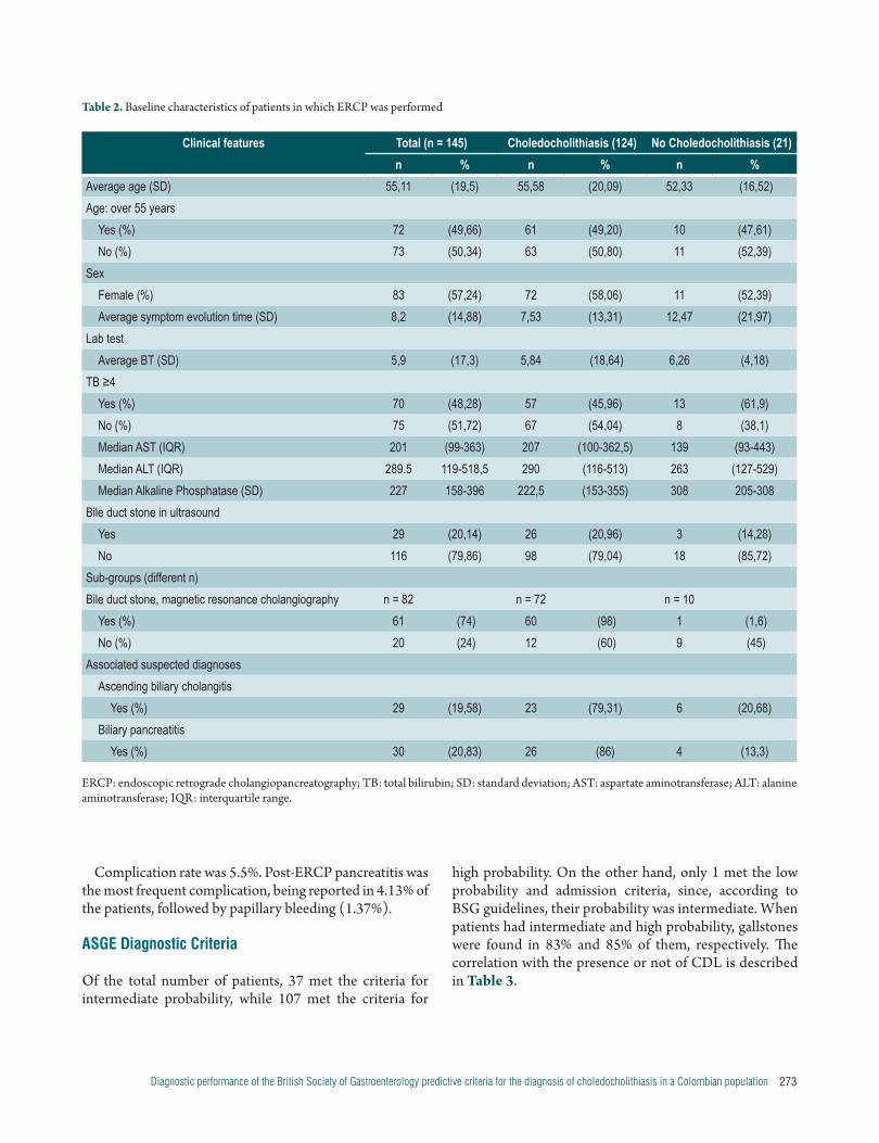
ERCP was performed in 145 patients. Their characteristics and imaging findings are described in Table 2. Patients who did not have CDL had a longer time of symptom evolution compared to those who had CDL (12 vs. 7.5 days). Liver profile values are reported using medians and interquartile ranges, as they did not have a normal distribution. Extreme values were found in each of them, resulting in higher median AST and ALT in patients with CDL compared to those without it (207 and 290 vs. 139 and 263, respectively).
Similarly, in 29 patients (20%) there was ultrasound evi-dence of a stone in the bile duct, which was confirmed in the ERCP in 89% of these cases. On the other hand, MRC was performed on 82 patients, and stones in the bile duct were observed in 74% of them. This was confirmed by ERCP in 98% of the cases. In contrast, when stones in the bile duct were not found in the MRC (24%), they were observed in the ERCP in 60% of these cases. Biliary pancreatitis was observed in 20% of patients and 86% of them had CDL.
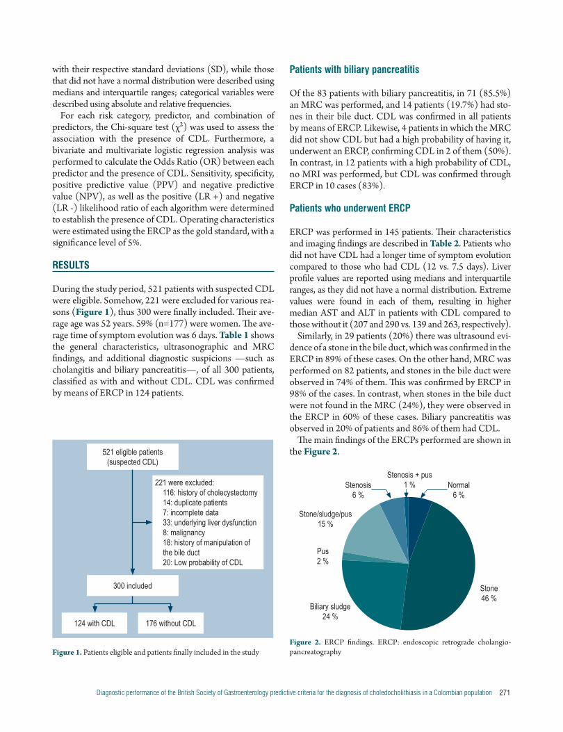
The main findings of the ERCPs performed are shown in the Figure 2.
with their respective standard deviations (SD), while those that did not have a normal distribution were described using medians and interquartile ranges; categorical variables were described using absolute and relative frequencies.
For each risk category, predictor, and combination of predictors, the Chi-square test (χ²) was used to assess the association with the presence of CDL. Furthermore, a bivariate and multivariate logistic regression analysis was performed to calculate the Odds Ratio (OR) between each predictor and the presence of CDL. Sensitivity, specificity, positive predictive value (PPV) and negative predictive value (NPV), as well as the positive (LR +) and negative (LR -) likelihood ratio of each algorithm were determined to establish the presence of CDL. Operating characteristics were estimated using the ERCP as the gold standard, with a significance level of 5%.
RESULTS
During the study period, 521 patients with suspected CDL were eligible. Somehow, 221 were excluded for various rea-sons (Figure 1), thus 300 were finally included. Their ave-rage age was 52 years. 59% (n=177) were women. The ave-rage time of symptom evolution was 6 days. Table 1 shows the general characteristics, ultrasonographic and MRC findings, and additional diagnostic suspicions —such as cholangitis and biliary pancreatitis—, of all 300 patients, classified as with and without CDL. CDL was confirmed by means of ERCP in 124 patients.
521 eligible patients (suspected CDL)
221 were excluded:116: history of cholecystectomy14: duplicate patients7: incomplete data33: underlying liver dysfunction8: malignancy18: history of manipulation of the bile duct20: Low probability of CDL
300 included
124 with CDL 176 without CDL
Figure 1. Patients eligible and patients finally included in the study
Stone/sludge/pus 15 %
Stenosis 6 %
Stenosis + pus1 % Normal
6 %
Pus2 %
Biliary sludge24 %
Stone46 %
Figure 2. ERCP findings. ERCP: endoscopic retrograde cholangio-pancreatography

Rev Colomb Gastroenterol. 2020;35(3):269-279. https://doi.org/10.22516/25007440.365272 Original article
Table 1. Baseline characteristics of patients with suspected choledocholithiasis
Clinical features Total (n = 300) With CDL (n = 124) Without CDL (n = 176)
n % n % n %
Average age (SD) 52 (18,6) 55,5 (20,09) 51 (17,4)
Age: over 55 years
Yes (%) 140 (46,7) 61 (20,3) 79 (26,3)
No (%) 160 (53,3) 63 (21,1) 97 (32,3)
Sex
Female (%) 177 (59) 72 (24) 105 (35)
Male (%) 123 (41) 52 (17,33) 71 (23,66)
Average symptom evolution time (SD) 6,11 (12) 7,3 (13,3) 5,1 (10,94)
Lab test
TB (SD) 4 (12) 5,8 (18,6) 2,72 (2,78)
TB ≥ 4
Yes (%) 95 (31,67) 57 (19) 38 (12,66)
No (%) 205 (68,33) 67 (22,33) 138 (46)
Average AST (SD) 280,22 (346,98) 268,40 (251,1) 288,33 (400,15)
Average ALT (SD) 328,93 (302,66) 356,37 (282,05) 310,26 (315,34)
Alkaline Phosphatase (SD) 244,4 (187,4) 281 (182,8) 218,79 (186,8)
Radiology
Bile duct stone in ultrasound
Yes (%) 31 (10,47) 26 (8,66) 5 (1,66)
No (%) 269 (89,19) 98 (32) 171 (63,66)
Choledochal stone in MRC (n = 236)
Yes (%) 63 (26,69) 61 (20,33) 2 (0,66)
No (%) 173 (73,31) 11 (3,66) 162 (54)
Associated suspected diagnoses
Ascending biliary cholangitis
Yes (%) 30 (10,03) 22 (7,33) 8 (2,66)
No (%) 269 (89,97) 101 (33,66) 168 (56)
Biliary pancreatitis
Yes (%) 83 (27,85) 26 (8,66) 57 (19)
No (%) 215 (72,15) 98 (32,66) 117 (39)
CDL: choledocholithiasis; TB: total bilirubin; SD: standard deviation; AST: aspartate aminotransferase; ALT: alanine aminotransferase; MRC: magnetic resonance cholangiography.
273Diagnostic performance of the British Society of Gastroenterology predictive criteria for the diagnosis of choledocholithiasis in a Colombian population
Table 2. Baseline characteristics of patients in which ERCP was performed
Clinical features Total (n = 145) Choledocholithiasis (124) No Choledocholithiasis (21)n % n % n %
Average age (SD) 55,11 (19,5) 55,58 (20,09) 52,33 (16,52)Age: over 55 years
Yes (%) 72 (49,66) 61 (49,20) 10 (47,61)No (%) 73 (50,34) 63 (50,80) 11 (52,39)
SexFemale (%) 83 (57,24) 72 (58,06) 11 (52,39)Average symptom evolution time (SD) 8,2 (14,88) 7,53 (13,31) 12,47 (21,97)
Lab testAverage BT (SD) 5,9 (17,3) 5,84 (18,64) 6,26 (4,18)
TB ≥4Yes (%) 70 (48,28) 57 (45,96) 13 (61,9)No (%) 75 (51,72) 67 (54,04) 8 (38,1)Median AST (IQR) 201 (99-363) 207 (100-362,5) 139 (93-443)Median ALT (IQR) 289.5 119-518,5 290 (116-513) 263 (127-529)Median Alkaline Phosphatase (SD) 227 158-396 222,5 (153-355) 308 205-308
Bile duct stone in ultrasound Yes 29 (20,14) 26 (20,96) 3 (14,28)No 116 (79,86) 98 (79,04) 18 (85,72)
Sub-groups (different n)Bile duct stone, magnetic resonance cholangiography n = 82 n = 72 n = 10
Yes (%) 61 (74) 60 (98) 1 (1,6)No (%) 20 (24) 12 (60) 9 (45)
Associated suspected diagnosesAscending biliary cholangitis
Yes (%) 29 (19,58) 23 (79,31) 6 (20,68)Biliary pancreatitis
Yes (%) 30 (20,83) 26 (86) 4 (13,3)
ERCP: endoscopic retrograde cholangiopancreatography; TB: total bilirubin; SD: standard deviation; AST: aspartate aminotransferase; ALT: alanine aminotransferase; IQR: interquartile range.
Complication rate was 5.5%. Post-ERCP pancreatitis was the most frequent complication, being reported in 4.13% of the patients, followed by papillary bleeding (1.37%).
ASGE Diagnostic Criteria
Of the total number of patients, 37 met the criteria for intermediate probability, while 107 met the criteria for
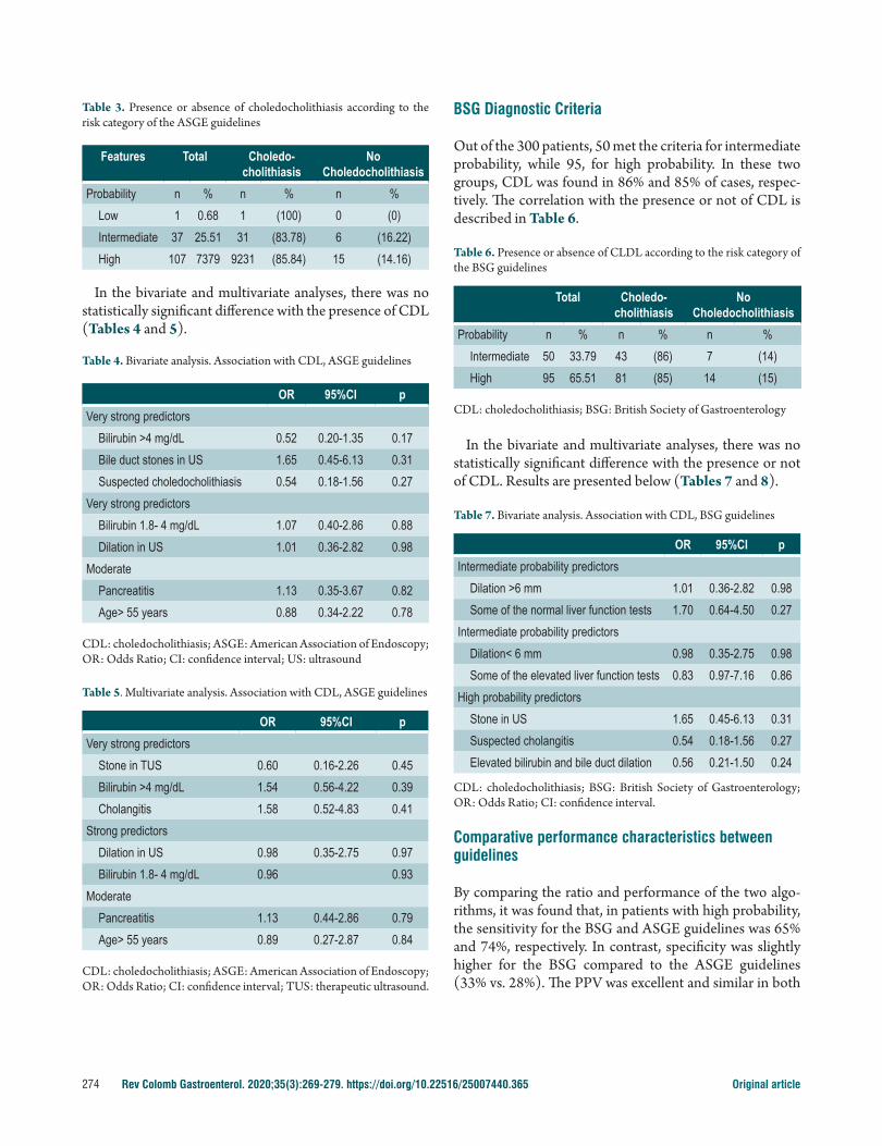
high probability. On the other hand, only 1 met the low probability and admission criteria, since, according to BSG guidelines, their probability was intermediate. When patients had intermediate and high probability, gallstones were found in 83% and 85% of them, respectively. The correlation with the presence or not of CDL is described in Table 3.
Rev Colomb Gastroenterol. 2020;35(3):269-279. https://doi.org/10.22516/25007440.365274 Original article
BSG Diagnostic Criteria
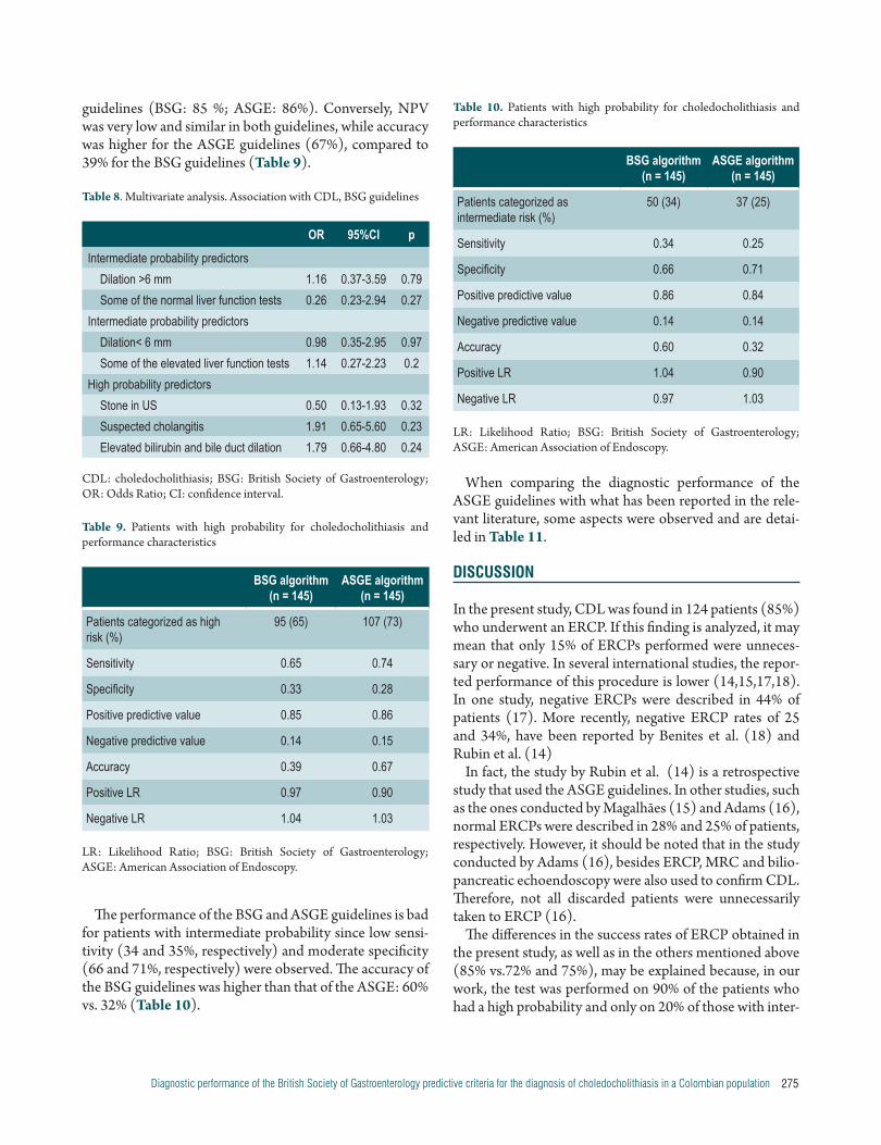
Out of the 300 patients, 50 met the criteria for intermediate probability, while 95, for high probability. In these two groups, CDL was found in 86% and 85% of cases, respec-tively. The correlation with the presence or not of CDL is described in Table 6.
Table 6. Presence or absence of CLDL according to the risk category of the BSG guidelines
Total Choledo-cholithiasis
No Choledocholithiasis
Probability n % n % n %Intermediate 50 33.79 43 (86) 7 (14)High 95 65.51 81 (85) 14 (15)
CDL: choledocholithiasis; BSG: British Society of Gastroenterology
In the bivariate and multivariate analyses, there was no statistically significant difference with the presence or not of CDL. Results are presented below (Tables 7 and 8).
Table 7. Bivariate analysis. Association with CDL, BSG guidelines
OR 95%CI pIntermediate probability predictors
Dilation >6 mm 1.01 0.36-2.82 0.98Some of the normal liver function tests 1.70 0.64-4.50 0.27
Intermediate probability predictorsDilation< 6 mm 0.98 0.35-2.75 0.98Some of the elevated liver function tests 0.83 0.97-7.16 0.86
High probability predictorsStone in US 1.65 0.45-6.13 0.31Suspected cholangitis 0.54 0.18-1.56 0.27Elevated bilirubin and bile duct dilation 0.56 0.21-1.50 0.24
CDL: choledocholithiasis; BSG: British Society of Gastroenterology; OR: Odds Ratio; CI: confidence interval.
Comparative performance characteristics between guidelines
By comparing the ratio and performance of the two algo-rithms, it was found that, in patients with high probability, the sensitivity for the BSG and ASGE guidelines was 65% and 74%, respectively. In contrast, specificity was slightly higher for the BSG compared to the ASGE guidelines (33% vs. 28%). The PPV was excellent and similar in both
Table 3. Presence or absence of choledocholithiasis according to the risk category of the ASGE guidelines
Features Total Choledo-cholithiasis
No Choledocholithiasis
Probability n % n % n %Low 1 0.68 1 (100) 0 (0)Intermediate 37 25.51 31 (83.78) 6 (16.22)High 107 7379 9231 (85.84) 15 (14.16)
In the bivariate and multivariate analyses, there was no statistically significant difference with the presence of CDL (Tables 4 and 5).
Table 4. Bivariate analysis. Association with CDL, ASGE guidelines
OR 95%CI pVery strong predictors
Bilirubin >4 mg/dL 0.52 0.20-1.35 0.17Bile duct stones in US 1.65 0.45-6.13 0.31Suspected choledocholithiasis 0.54 0.18-1.56 0.27
Very strong predictorsBilirubin 1.8- 4 mg/dL 1.07 0.40-2.86 0.88Dilation in US 1.01 0.36-2.82 0.98
ModeratePancreatitis 1.13 0.35-3.67 0.82Age> 55 years 0.88 0.34-2.22 0.78
CDL: choledocholithiasis; ASGE: American Association of Endoscopy; OR: Odds Ratio; CI: confidence interval; US: ultrasound
Table 5. Multivariate analysis. Association with CDL, ASGE guidelines
OR 95%CI pVery strong predictors
Stone in TUS 0.60 0.16-2.26 0.45Bilirubin >4 mg/dL 1.54 0.56-4.22 0.39Cholangitis 1.58 0.52-4.83 0.41
Strong predictorsDilation in US 0.98 0.35-2.75 0.97Bilirubin 1.8- 4 mg/dL 0.96 0.93
ModeratePancreatitis 1.13 0.44-2.86 0.79Age> 55 years 0.89 0.27-2.87 0.84
CDL: choledocholithiasis; ASGE: American Association of Endoscopy; OR: Odds Ratio; CI: confidence interval; TUS: therapeutic ultrasound.
275Diagnostic performance of the British Society of Gastroenterology predictive criteria for the diagnosis of choledocholithiasis in a Colombian population
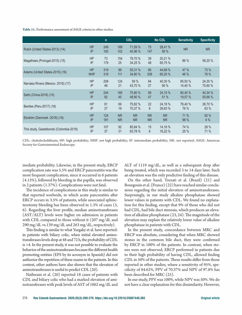
Table 10. Patients with high probability for choledocholithiasis and performance characteristics
BSG algorithm (n = 145)
ASGE algorithm (n = 145)
Patients categorized as intermediate risk (%)
50 (34) 37 (25)
Sensitivity 0.34 0.25
Specificity 0.66 0.71
Positive predictive value 0.86 0.84
Negative predictive value 0.14 0.14
Accuracy 0.60 0.32
Positive LR 1.04 0.90
Negative LR 0.97 1.03
LR: Likelihood Ratio; BSG: British Society of Gastroenterology; ASGE: American Association of Endoscopy.
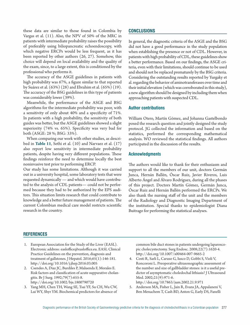
When comparing the diagnostic performance of the ASGE guidelines with what has been reported in the rele-vant literature, some aspects were observed and are detai-led in Table 11.
DISCUSSION
In the present study, CDL was found in 124 patients (85%) who underwent an ERCP. If this finding is analyzed, it may mean that only 15% of ERCPs performed were unneces-sary or negative. In several international studies, the repor-ted performance of this procedure is lower (14,15,17,18). In one study, negative ERCPs were described in 44% of patients (17). More recently, negative ERCP rates of 25 and 34%, have been reported by Benites et al. (18) and Rubin et al. (14)
In fact, the study by Rubin et al. (14) is a retrospective study that used the ASGE guidelines. In other studies, such as the ones conducted by Magalhães (15) and Adams (16), normal ERCPs were described in 28% and 25% of patients, respectively. However, it should be noted that in the study conducted by Adams (16), besides ERCP, MRC and bilio-pancreatic echoendoscopy were also used to confirm CDL. Therefore, not all discarded patients were unnecessarily taken to ERCP (16).
The differences in the success rates of ERCP obtained in the present study, as well as in the others mentioned above (85% vs.72% and 75%), may be explained because, in our work, the test was performed on 90% of the patients who had a high probability and only on 20% of those with inter-
guidelines (BSG: 85 %; ASGE: 86%). Conversely, NPV was very low and similar in both guidelines, while accuracy was higher for the ASGE guidelines (67%), compared to 39% for the BSG guidelines (Table 9).
Table 8. Multivariate analysis. Association with CDL, BSG guidelines
OR 95%CI p
Intermediate probability predictorsDilation >6 mm 1.16 0.37-3.59 0.79Some of the normal liver function tests 0.26 0.23-2.94 0.27
Intermediate probability predictorsDilation< 6 mm 0.98 0.35-2.95 0.97Some of the elevated liver function tests 1.14 0.27-2.23 0.2
High probability predictorsStone in US 0.50 0.13-1.93 0.32Suspected cholangitis 1.91 0.65-5.60 0.23Elevated bilirubin and bile duct dilation 1.79 0.66-4.80 0.24
CDL: choledocholithiasis; BSG: British Society of Gastroenterology; OR: Odds Ratio; CI: confidence interval.
Table 9. Patients with high probability for choledocholithiasis and performance characteristics
BSG algorithm (n = 145)
ASGE algorithm (n = 145)
Patients categorized as high risk (%)
95 (65) 107 (73)
Sensitivity 0.65 0.74
Specificity 0.33 0.28
Positive predictive value 0.85 0.86
Negative predictive value 0.14 0.15
Accuracy 0.39 0.67
Positive LR 0.97 0.90
Negative LR 1.04 1.03
LR: Likelihood Ratio; BSG: British Society of Gastroenterology; ASGE: American Association of Endoscopy.
The performance of the BSG and ASGE guidelines is bad for patients with intermediate probability since low sensi-tivity (34 and 35%, respectively) and moderate specificity (66 and 71%, respectively) were observed. The accuracy of the BSG guidelines was higher than that of the ASGE: 60% vs. 32% (Table 10).
Rev Colomb Gastroenterol. 2020;35(3):269-279. https://doi.org/10.22516/25007440.365276 Original article
ALT of 1119 mg/dL, as well as a subsequent drop after being treated, which was recorded 3 to 14 days later. Such an elevation was the only predictive finding of this disease.
On the other hand, Tozzati et al. (Brazil) (21) and Bourgouin et al. (France) (22) have reached similar conclu-sions regarding the initial elevation of aminotransferases. Surprisingly, in our study alkaline phosphatase showed lower values in patients with CDL. We found no explana-tion for this finding, except that 9% of those who did not had CDL, had bile duct stenosis, which produces an eleva-tion of alkaline phosphatase (23, 24). The magnitude of the elevation may explain the relatively lower value of alkaline phosphatase in patients with CDL.
In the present study, concordance between MRC and ERCP was absolute, considering that when MRC showed stones in the common bile duct, they were confirmed by ERCP in 100% of the patients. In contrast, when sto-nes were not observed, ERCP performed in patients due to their high probability of having CDL, allowed finding CDL in 50% of the patients. These results differ from those reported in other studies, where a sensitivity of 95%, spe-cificity of 84.62%, PPV of 70.37% and NPV of 97.8% has been described for MRC (25).
In our study, PPV was 100%, while NPV was 50%. We do not have a clear explanation for this dissimilarity. However,
mediate probability. Likewise, in the present study, ERCP complication rate was 5.5% and ERCP pancreatitis was the most frequent complication, since it occurred in 6 patients (4.13%), followed by bleeding in the papilla, was observed in 2 patients (1.37%). Complications were not fatal.
The incidence of complications in this study is similar to that reported worlwide, in which acute pancreatitis after ERCP occurs in 3.5% of patients, while associated sphinc-terotomy bleeding has been observed in 1.3% of cases (5, 6). Regarding the liver profile, median aminotransferases (AST/ALT) levels were higher on admission in patients with CDL compared to those without it (207 mg/dL and 290 mg/dL vs.139 mg/dL and 263 mg/dL, respectively).
This finding is similar to what Yurgaky et al. have reported: in patients with biliary colic, when initial elevated amino-transferases levels drop at 48 and 72 h, the probability of CDL is >4. In the present study, it was not possible to evaluate the behavior of the aminotransferases because the different health promoting entities (EPS by its acronym in Spanish) did not authorize the repetition of these exams in the patients. In this context, other authors have also shown that the elevation of aminotransferases is useful to predict CDL (20).
Nathwani et al. (20) reported 18 cases of patients with CDL and biliary colic who had a marked elevation of ami-notransferases with peak levels of AST of 1062 mg/dL and
Table 11. Performance assessment of ASGE criteria in other studies
n CDL No CDL Sensitivity Specificity
Rubin (United States-2013) (14) HPIP
249193
189102
71,59 %40,96 %
75147
28,41 %59 % NR NR
Magalhaes (Portugal-2015) (15) HPIP
73179
15425
79,70 %34,25 %
3948
20,21 %65,75 % 86 % 56,20 %
Adams (United States-2015) (16) HPNHP
319319
99111
55,31 %34,80 %
80208
44,69 %65,20 %
47 %46 %
73 %76 %
Narváez-Rivera (Mexico- 2016) (17) HPIP
20848
12421
59 %43,70 %
8427
40,30 %56 %
85,50 %14,40 %
24,30 %75,60 %
Sethi (China-2016) (10) HPIP
24492
18545
75,80 %48,90 %
5947
24,10 %51 %
80,40 %19,57 %
44,34 %55,66 %
Benites (Peru-2017) (18) HPIP
9127
6919
75,82 %70,37 %
228
24,18 %29,63 %
78,40 %76 %
26,70 %63 %
Ebrahim (Denmark -2018) (19) HPIP
124181
NRNR
NRNR
NRNR
NRNR
71 %98 %
50 %4 %
This study, Gastelbondo (Colombia-2018) HPIP
10737
9231
85,84 %83,78 %
156
14,16 %16,22 %
74 %25 %
28 %71 %
CDL: choledocholithiasis; HP: high probability; NHP: not high probability; IP: intermediate probability; NR: not reported; ASGE: American Society for Gastrointestinal Endoscopy.
277Diagnostic performance of the British Society of Gastroenterology predictive criteria for the diagnosis of choledocholithiasis in a Colombian population
CONCLUSIONS
In general, the diagnostic criteria of the ASGE and the BSG did not have a good performance in the study population when establishing the presence or not of CDL. However, in patients with high probability of CDL, these guidelines show a better performance. Based on our findings, the ASGE cri-teria, even with their limitations, should continue to be used and should not be replaced prematurely by the BSG criteria. Considering the outstanding results reported by Yurgaky et al. regarding the behavior of aminotransferases over time and their initial elevation (which was corroborated in this study), a new algorithm should be designed by including them when approaching patients with suspected CDL.
Author contributions
William Otero, Martín Gómez, and Johanna Gastelbondo posed the research question and jointly designed the study protocol. JG collected the information and based on the statistics, performed the corresponding mathematical analysis. WO reviewed the statistical findings. All authors participated in the discussion of the results.
Acknowledgments
The authors would like to thank for their enthusiasm and support to all the members of our unit, doctors Germán Junca, Hernán Ballén, Óscar Ruiz, Javier Riveros, Luis Alberto Ángel and Álvaro Rodríguez, during all the phases of this project. Doctors Martín Gómez, Germán Junca, Óscar Ruiz and Hernán Ballén performed the ERCPs. We also thank the nursing staff of the unit and the members of the Radiology and Diagnostic Imaging Department of the institution. Special thanks to epidemiologist Diana Buitrago for performing the statistical analyses.
these data are similar to those found in Colombia by Vargas et al. (11). Also, the NPV of 50% of the MRC in patients with intermediate probability raises the possibility of preferably using biliopancreatic echoendoscopy, with which negative ERCPs would be less frequent, as it has been reported by other authors (26, 27). Somehow, this choice will depend on local availability and the quality of the exam, since, to a large extent, this is conditioned by the professional who performs it.
The accuracy of the ASGE guidelines in patients with high probability was 67%, a figure similar to that reported by Suárez et al. (63%) (28) and Ebrahim et al. (65%) (19). The accuracy of the BSG guidelines in this type of patients was considerably lower (39%).
Meanwhile, the performance of the ASGE and BSG algorithms for the intermediate probability was poor, with a sensitivity of only about 40% and a specificity of 71%. In patients with a high probability, the sensitivity of both guides was better, but the ASGE guidelines showed a slight superiority (74% vs. 65%). Specificity was very bad for both (ASGE: 28 %; BSG: 33%).
When comparing our work with other studies, as descri-bed in Table 11, Sethi et al. (10) and Narvaez et al. (17) also report low sensitivity in intermediate probability patients, despite having very different populations. These findings reinforce the need to determine locally the best noninvasive test prior to performing ERCP.Our study has some limitations. Although it was carried out in a university hospital, some laboratory tests that were requested dynamically — and which would have contribu-ted to the analysis of CDL patients— could not be perfor-med because they had to be authorized by the EPS audi-tors. This situation limits research that could contribute to knowledge and a better future management of patients. The current Colombian medical care model restricts scientific research in the country.
REFERENCES
1. European Association for the Study of the Liver (EASL). Electronic address: [email protected]. EASL Clinical Practice Guidelines on the prevention, diagnosis and treatment of gallstones. J Hepatol. 2016;65(1):146-181. http://doi.org/10.1016/j.jhep.2016.03.005
2. Csendes A, Diaz JC, Burdiles P, Maluenda F, Morales E. Risk factors and classification of acute suppurative cholan-gitis. Br J Surg. 1992;79(7):655-8. http://doi.org/10.1002/bjs.1800790720
3. Yang MH, Chen TH, Wang SE, Tsai YF, Su CH, Wu CW, Lui WY, Shyr YM. Biochemical predictors for absence of
common bile duct stones in patients undergoing laparosco-pic cholecystectomy. Surg Endosc. 2008;22(7):1620-4. http://doi.org/10.1007/s00464-007-9665-2
4. Costi R, Sarli L, Caruso G, Iusco D, Gobbi S, Violi V, Roncoroni L. Preoperative ultrasonographic assessment of the number and size of gallbladder stones: is it a useful pre-dictor of asymptomatic choledochal lithiasis? J Ultrasound Med. 2002;21(9):971-6. http://doi.org/10.7863/jum.2002.21.9.971
5. Anderson MA, Fisher L, Jain R, Evans JA, Appalaneni V, Ben Menachem T, Cash BD, Anton G, Early DS, Fanelli
Rev Colomb Gastroenterol. 2020;35(3):269-279. https://doi.org/10.22516/25007440.365278 Original article
guidelines. Dig Liver Dis. 2013;45(9):744-9. http://doi.org/10.1016/j.dld.2013.02.005
15. Magalhães J, Rosa B, Cotter J. Endoscopic retrograde cho-langiopancreatography for suspected choledocholithiasis: From guidelines to clinical practice. World J Gastrointest Endosc. 2015;7(2):128-34. http://doi.org/10.4253/wjge.v7.i2.128
16. Adams MA, Hosmer AE, Wamsteker EJ, Anderson MA, Elta GH, Kubiliun NM, Kwon RS, Piraka CR, Scheiman JM, Waljee AK, Hussain HK, Elmunzer BJ. Predicting the likelihood of a persistent bile duct stone in patients with suspected choledocholithiasis: accuracy of existing guidelines and the impact of laboratory trends. Gastrointest Endosc. 2015;82(1):88-93. http://doi.org/10.1016/j.gie.2014.12.023
17. Narváez-Rivera RM, González-González JA, Monreal-Robles R, García-Compean D, Paz-Delgadillo J, Garza-Galindo AA, Maldonado-Garza HJ. Accuracy of ASGE criteria for the prediction of choledocholithiasis. Rev Esp Enfer. 2016;108(6):309-314. http://doi.org/10.17235/reed.2016.4212/2016
18. Benites HE, Palacios FV, Asencios JL, Aguilas R, Segovia NS. Rendimiento de los criterios predictivos de la ASGE en el diagnóstico de coledocolitiasis en el Hospital Edgardo Rebagliati Martins. Rev Gastroenterol. 2017;37(2):111-119.
19. Ebrahim M, Sorensen LT, Jorgensen LN, Kalaitzakis E. Current clinical algorithms for predicting common bile duct stones have only moderate accuracy. Dig Endosc. 2018;30(4):477-484. http://doi.org/10.1111/den.12994
20. Nathwani RA, Kumar SR, Reynolds TB, Kaplowitz N. Marked elevation in serum transaminases: an atypical presentation of choledocholithiasis. Am J Gastroenterol. 2005;100(2):295-298. http://doi.org/10.1111/j.1572-0241.2005.40793.x
21. Tozatti J, Mello AL, Frazon O. Predictor factors for chole-docholithiasis. Arq Bras Cir Dig. 2015;28(2):109-112. http://doi.org/10.1590/S0102-67202015000200006
22. Bourgouin S, Truchet X, Lamblin G, De Roulhac J, Platel JP, Balandraud P. Dynamic analysis of commonly used bio-chemical parameters to predict common bile duct stones in patients undergoing laparoscopic cholecystectomy. Surg Endosc. 2017;31(11):4725-4734. http://doi.org/10.1007/s00464-017-5549-2
23. Stepien M, Fedirko V, Duarte-Salles T, Ferrari P, Freisling H, Trepo E, Trichopoulou A, Bamia C, Weiderpass E, Olsen A, Tjønneland A, Overvad K, Boutron-Ruault MC, Fagherazzi G, Racine A, Kühn T, Kaaks R, Aleksandrova K, Boeing H, Lagiou P, Benetou V, Trichopoulos D, Palli D, Grioni S, Tumino R, Naccarati A, Panico S, Bueno-de-Mesquita HB, Peeters PH, Lund E, Quirós JR, Nápoles OC, Sánchez MJ, Dorronsoro M, Huerta JM, Ardanaz E, Ohlsson B, Sjöberg K, Werner M, Nystrom H, Khaw KT, Key TJ, Gunter M, Cross A, Riboli E, Romieu I, Jenab M. Prospective association of liver function biomarkers with development of hepatobiliary cancers. Cancer Epidemiol.
RD, Fisher DA, Fukami N, Ha Hwang J, Ikenberry SO, Jue TL, Khan KM, Krinsky ML, Malpas PM, Maple JT, Sharaf RN, Shergill AK, Dominitz JA. Complications of ERCP. Gastrointest Endosc. 2012;75(3):467-473. http://doi.org/10.1016/j.gie.2011.07.010
6. Masci E, Toti G, Mariani A, Curioni S, Lomazzi A, Dinelli M, Minoli G, Crosta C, Comin U, Fertitta A, Prada A, Passoni GR, Testoni PA. Complications of diagnostic and therapeutic ERCP: a prospective multicenter study. Am J Gastroenterol. 2001;96(2):417-23. http://doi.org/10.1111/j.1572-0241.2001.03594.x
7. ASGE Standards of Practice Committee, Maple JT, Ben-Menachem T, Anderson MA, Appalaneni V, Banerjee S, Cash BD, Fisher L, Harrison ME, Fanelli RD, Fukami N, Ikenberry SO, Jain R, Khan K, Krinsky ML, Strohmeyer L, Dominitz JA. The role of endoscopy in the evaluation of suspected choledocholithiasis. Gastrointest Endosc. 2010;71(1):1-9. http://doi.org/10.1016/j.gie.2009.09.041
8. Williams E, Beckingham I, El Sayed G, Gurusamy K, Sturgess R, Webster G, Young T. Updated guideline on the management of common bile duct stones (CBDS). Gut. 2017;66(5):765-782. http://doi.org/10.1136/gutjnl-2016-312317
9. Adams MA, Hosmer AE, Wamsteker EJ, Anderson MA, Elta GH, Kubiliun NM, Kwon RS, Piraka CR, Scheiman JM, Waljee AK, Hussain HK, Elmunzer BJ. Predicting the likelihood of a persistent bile duct stone in patients with suspected choledocholithiasis: accuracy of existing guidelines and the impact of laboratory trends. Gastrointest Endosc. 2015;82(1):88-93. http://doi.org/10.1016/j.gie.2014.12.023
10. Sethi S, Wang F, Korson AS, Krishnan S, Berzin TM, Chuttani R, Pleskow DK, Sawhney MS. Prospective assessment of consensus criteria for evaluation of patients with suspected choledocholithiasis. Dig Endosc. 2016;28(1):75-82. http://doi.org/10.1111/den.12506
11. Vargas RD, Córdoba CP, Uriza LF, Costa V, Mosquera-Klinger G, Ortega DA. Concordancia entre los hallazgos por colangiopancreatografía por resonancia magnética y los hallazgos por colangiopancreatografía endoscópica retro-grada en pacientes hospitalizados por enfermedad biliar litiásica en el Hospital Universitario San Ignacio (Bogotá-Colombia) entre los años 2005 a 2011. Rev Gastroenterol. 2015;35(3):226-230.
12. Gómez M, Pion J, Otero W. Predictores de coledocoli-tiasis en pacientes sometidos a colangiografía retrógrada endoscópica en el Hospital El Tunal de Bogotá. Rev Col Gastroenterol. 2011;26(4):243-252.
13. Jurgaky JM. Prevalencia y factores de riesgo de elevación de transaminasas en pacientes con coledocolitiasis. Bogotá: Universidad Nacional de Colombia; 2017.
14. Rubin MI, Thosani NC, Tanikella R, Wolf DS, Fallon MB, Lukens FJ. Endoscopic retrograde cholangiopancreatogra-phy for suspected choledocholithiasis: testing the current
279Diagnostic performance of the British Society of Gastroenterology predictive criteria for the diagnosis of choledocholithiasis in a Colombian population
Retrograde Cholangiopancreatography in Diagnosing Choledocholithiasis: The Indonesian Experience. Clin Endosc. 2017;50(5):486-490. http://doi.org/10.5946/ce.2016.159
27. Meeralam Y, Al-Shammari K, Yaghoobi M. Diagnostic accuracy of EUS compared with MRCP in detecting choledocholithiasis: a meta-analysis of diagnostic test accuracy in head-to-head studies. Gastrointest Endosc. 2017;86(6):986-993. http://doi.org/10.1016/j.gie.2017.06.009
28. Suárez AL, LaBarre NT, Cotton PB, Payne KM, Coté GA, Elmunzer BJ. An assessment of existing risk stratification guidelines for the evaluation of patients with suspected choledocholithiasis. Surg Endosc. 2016;30(10):4613-4618. http://doi.org/10.1007/s00464-016-4799-8
2016;40:179-87. http://doi.org/10.1016/j.canep.2016.01.002
24. Woo YS, Lee KH, Lee KT, Lee JK, Kim JM, Kwon CHD, Joh JW, Kang D, Cho J. Postoperative changes of liver enzymes can distinguish between biliary stricture and graft rejection after living donor liver transplantation: A longitu-dinal study. Medicine (Baltimore). 2017;96(40):e6892. http://doi.org/10.1097/MD.0000000000006892
25. Perales SR, Souza LRMF, Crema E. comparative evaluation of magnetic resonance cholangiopancreatography and perioperative cholangiography in patients with suspect choledocholithiasis. Arq Bras Cir Dig. 2019;32(1):e1416. http://doi.org/10.1590/0102-672020180001e1416
26. Makmun D, Fauzi A, Shatri H. Sensitivity and Specificity of Magnetic Resonance Cholangiopancreatography versus Endoscopic Ultrasonography against Endoscopic