VRSI Sep 2018 Final.cdr
64
-
Upload
khangminh22 -
Category
Documents
-
view
1 -
download
0
Transcript of VRSI Sep 2018 Final.cdr


www.vrsi.in 3
VITREO RETINAL SOCIETY - INDIA SEPTEMBER 2018
Editor-in-chief Deputy EditorDr Vishali Gupta, MS Dr Anand Rajendran, MDChandigarh, India Chennai, India
Deputy Editor Deputy EditorDr Thomas Cherian MD Dr Ramandeep Singh MSAngamaly, India Chandigarh, India
Guidelines for the manuscript submission in VRSI Newsletter
Original articles:
These include randomized controlled trials, intervention studies, studies of screening and diagnostic
test, outcome studies, cost effectiveness analyses, case-control series, and surveys with high response
rate. The text of original articles amounting to up to 3000 words (excluding Abstract, references and
Tables) should be divided into sections with the headings Abstract, Key-words, Introduction, Material
and Methods, Results, Discussion, References, Tables andFigure legends.
Case reports / Challenging case /Innovations / Instruments /Techniques
New, interesting, challenging, rare cases, innovations, instruments and techniques can be reported.
They should be unique and providing learning point for the readers. Manuscripts with clinical
significance or implications will be given priority. These communications could be of up to 1000 words
(excluding Abstract and references) and should have the following headings: Abstract (unstructured),
Key-words, Introduction, Case, Discussion, Reference, Tables and Legends in that order.
The manuscript could be of up to 1000 words (excluding references and abstract) and could be
supported with up to 10 references. Case Reports could be authored by up to four authors.
Mail to [email protected], [email protected]

www.vrsi.in4
From thePresident’s DeskDr. A. GiridharMedical DirectorGiridhar Eye Institute, [email protected]
Dear Members:
It gives me great pleasure to write a forward for the final issue of the newsletter for the year 2018 as my term as the President of the VRS-I is ending. The purpose of any scientific society is to disseminate latest scientific information, create an environment for effective scientific deliberations and finally improve fellowship and partnership between fellow Vitreo Retinal surgeons.
Over the years there has been a long debate within the governing council of the VRS-I about the need and purpose of this newsletter. In this context it is important for me to congratulate Dr Vishali Gupta and her team for having brought out systematically many useful issues of the newsletter over the last two years. Personally I feel this can be an avenue for young Vitreo Retinal surgeons to display their inherent talent. This newsletter can be a platform for many of them as a stepping stone for future publications in large peer reviewed journals. Our society is blessed with a large number of young dynamic and innovative surgeons. The mushrooming of various retina forums across the country is a clear indication that there is a phenomenal potential for our society to shine at international level.
As I lay down office as the President of the Society, it is my dream that this newsletter grows into a scientific journal of international repute in the coming years. It is necessary for young, vibrant and dynamically oriented retina specialists to take this forward. Let me conclude by once again thanking the governing council and its members for having participated effectively in the publication of this newsletter. One of the important achievements of this governing council has been involvement by each and every member in some form of activity. This should continue in future.
With kind regards and best wishes
Dr A GiridharPresident, VRS-I
VITREO RETINAL SOCIETY - INDIA SEPTEMBER 2018

From theHonorary Secretary Desk
Dr. Raja NarayananDirector-Head, Clinical Research Consultant,Smt. Kanuri Santhamma Centre for Vitreo Retinal DiseasesKallam Anji Reddy Campus, [email protected]
Dear Friends
Greetings from VRSI!The excitement of the VRSI annual
meeting is building up. I hope that you have booked your
travel and accommodation for the Meeting, with enough time
for leisure. The Scientific Program has been uploaded on the
website, which has been brilliantly compiled by Dr. Vishali
Gupta. The local organizing committee lead by Dr. Shorey and
Dr. Sharma is working tirelessly to ensure a perfect Meeting
for all of us.I am delighted to know that the lastissue of VRSI
newsletter in 2018 is being published. I am sure that you will
find thearticles extremely valuable for your daily practice, and
to provide the best care to your patients. I take this
opportunity to request you all to submit your interesting
cases, articles and innovations to the VRSI newsletter, which
will help improve the scientific knowledge base of our
members. I request you all to participate enthusiastically in
the activities of VRSI.
Dr Raja NarayananSecretary, VRS-I
www.vrsi.in 5
VITREO RETINAL SOCIETY - INDIA SEPTEMBER 2018

www.vrsi.in6
From the ConvenorScientific Committee Desk
Dr. Vishali Gupta MDProfessor (Retina, Vitreous and Uvea)Advanceed Eye Centre,Post Graduate Institute of Medical Educationand Research, Chandigarh-India- 160012Tel: +91-172-2747837 FAX: +91-172-2747837email: [email protected]@gmail.com
Dear Members
It gives me great pleasure in bringing out the third volume of VRSI
newsletter for the year 2018. The current issue focus on the controversies
and challenges that all of us face in our day-to-day practice. I am
extremely thankful to all my colleagues, who despite being busy vitreo-
retinal surgeons, have worked hard to contribute and share their
experiences and give expert tips that shall be extremely valuable for the
young surgeons. However, we are certainly missing submissions from our
members and do encourage them to submit their cases, images as well as
original articles to contribute to the newsletter and share their
experiences with other members of the society.
We are also looking forward to see you all for the annual meeting of VRSI th ndto be held in Jaipur between 29 Nov till 2 2018 with pre-conference
Retinal Imaging Symposium (RISHI 3). The participation by international
faculty as well societies is tremendous and we are certain that you will
truly enjoy both the scientific contents as well local hospitality. Our local
hosts Dr Raj Kumar Sharma and Dr Pavan Shorey are working extremely
hard to make sure that this is a memorable event and your participation
shall certainly make it one.
Once again, I sincerely hope that you enjoy reading this newsletter and
look forward to your comments and suggestions.
Dr Vishali GuptaScientific Committee, VRS-I
VITREO RETINAL SOCIETY - INDIA SEPTEMBER 2018

www.vrsi.in 7
Macular hole (MH) is one of the common causes of
reduced central vision. Internal limiting membrane (ILM)
peeling has increased the surgical success rateto 90-95% 1resulting in MH closure. Persistence of MH after pars
planavitrectomy remains the most common complication
seen in approximately 10-12% of the cases. Various causes for
a persistent MH are - persistent vitreomaculartraction (VMT),
insufficient tamponade or non- compliance to prone position.
Re-surgery in the form of repeat vitrectomy with enlargement
of the ILM rhexis, with or without autologous platelet
concentrate and additional gas tamponade or light/heavy
silicone oil tamponade have been tried for the closure of 2-5 persistent or recurrent macular holes. It is extremely
important to know when to intervene in a case of persistent
MH after the first surgery to achieve a higher chance of 6,7anatomical closure. We will discuss the preferred practice
pattern with a few of the leading vitreoretinal surgeons-A. Giridhar (AG), Cyrus M. Shroff (CMS), Hany Hamza (HH),
Pramod Bhende (PB) and Mahesh P. Shanmugam (MPS). The
questions and summarization of the answers by the experts
has been prepared by Manisha Agarwal (MA).
MA-Q1.What is your definition of a persistent macular hole?
How long after the primary surgery would you like to wait for
the hole to close?
AG: A macular hole which does not close after a primary
surgery is a persistent macular hole. Closure of macular hole
occurs during the first week after the surgery. Therefore any
hole that persists at the end of one week after surgery usually
does not close later.
CMS: We generally do an OCT one month after the primary
macular hole surgery and if the MH is still open we would term
it a failed or persistent MH. Recently with Swept source OCT
we have been able to identify MH closure as early as the first
post-operative day.
HH: Persistent macular hole is defined as a persistent full
thickness defect in the neurosensory retina 3 weeks after
vitrectomy. The persistent edema at the edge of the hole as
well as the slightly elevated edges help to differentiate
between non-closure of the hole after primary surgery and
type -2 closure.
PB: If the hole remains open 1-2 weeks after surgery, unlikely it
will close, though I would still prefer to wait for 6-8 weeks
before advising a re-surgery.
MPS: Persistent macular hole is one that has not closed after
absorption of the gas. I would intervene if there were
inadequate intravitreal volume of gas for effective tamponade
and a persistent hole. Hence intervention may be even as early
as 1 week or 1 month. If one is able to serially document
narrowing of the hole with progressing approximation of the
edges on OCT, there is a possibility of hole closure even in the
absence of gas. In such cases one may watch closely for
spontaneous resolution.
Summary (MA) - Persistent macular hole is a hole which
remains open after the primary macular hole surgery with
absorption of the gas tamponade or a maximum of two weeks
after silicone oil tamponade. We may have a persistent or a
recurrent macular hole. Recurrent MH is the one which had
closed after the primary macular hole surgery, but has
reopened. The results of the re-operation are better for 8 recurrent macular holes in comparison to persistent MH. The
best time for re-surgery is when the eye is not inflamed and
there is no effective gas tamponade with no possibility of
closure of MH.
MA -Q2. What factors in terms of BCVA, size and etiology of
the macular hole do you consider before you advise a re-
surgery for a persistent macular hole?
AG: Once we have a persistent macular hole, the decision to
re-operate is more based on the OCT characteristics rather
VITREO RETINAL SOCIETY - INDIA SEPTEMBER 2018
Current practice pattern for the management of persistent macular holes
Dr. Manisha AgarwalHead of Vitreoretina Services, Dr. Shroff's Charity Eye Hospital, New Delhi
Dr. A. GiridharMedical Director and Chief Vitreoretina services, Giridhar Eye Institute, Cochin, Kerala, India.
Dr. Cyrus M. Shroff Director of Shroff Eye Centre, Kailash Colony, East of Kailash, New Delhi, India.
Dr. Hany HamzaProfessor of Eye Surgery, Cairo university, Egypt
Dr. Pramod BhendeMedical Director, Sankara Nethralaya, Chennai, Tamil Nadu, India.
Dr Mahesh P. ShanmugamHead of Vitreoretina and Ocular Oncology Services, Sankara Eye Hospitals, Bengaluru, Karnataka, India.

than the visual acuity. Presuming that in all patients we have
done a proper ILM peeling, the etiology of failure after the
primary surgery are:
a)� Improper ILM peelingb)� Inadequate ILM peelingc)� Inadequate tamponaded)� A very large primary macular hole which fails to close.
Therefore we need to define what exactly the cause is before
we decide on further management.
CMS: We would generally advise a re-surgery for any
persistent MH especially if we are unsure about the amount of
ILM peeling during the primary surgery and of the compliance
with the prone positioning.
HH: In terms of visual acuity I have not encountered any
patient with a persistent open macular hole with vision better
than 0.2 and the persistence of the central scotoma can be
quite disturbing to the patient as an indicator of failure of the
first surgical procedure. Therefore vision has never been the
issue in deciding for reoperation. Persistent open macular
hole is clearly the cause of persistent visual symptoms that are
the basis of deciding for the primary surgery. With the use of
the inverted ILM flap technique we could achieve closure of
the macular holes larger than 1500 u in some cases. Therefore
with very few exceptions of very large macular hole size is not
the issue. On the other hand, etiology and associated
pathology can influence my decision to re-operate patients
with unhealthy RPE at the base of the hole caused by trauma
in traumatic macular holes as well as myopic macular holes
with extensive chorioretinal degeneration at the posterior
pole have very poor functional outcome. In these cases I
explain to the patient that surgery is not useful and that no
visual improvement is anticipated and we decide together not
to re-operate.
PB: If I have attempted surgery once and the patient comes
back with persistent MH, I generally ask the patients, if he/she
would like to take a second chance. But at the same time I
would explain about guarded visual prognosis. I still believe in
prone positioning and if patient is not compliant for prone
position, I would prefer not to take second chance. Eyes with
obvious traction (on OCT or clinically), smaller holes,
underlying healthy RPE, elevated edges of the hole probably
are the better candidates for re-surgery.
MPS: Smaller idiopathic holes with good pre-surgery vision
have a better prognosis if operated early. BCVA <6/60, holes
>1000 microns, associated with retinal atrophy (as in post
CME, myopia etc. ), failure despite a well performed primary
surgery (adequate ILM peel or inverse flap large holes,
appropriate tamponade etc.) would be factors against advising
a re-surgery.
Summary (MA): Visual acuity is an important deciding factor
for the primary macular hole surgery along with other factors
such as etiology, size and duration of the macular hole.
However these play a less important role while deciding for a
re-surgery. We need to evaluate the cause of failure of the
primary surgery including inadequate ILM peel, features on
OCT such as persistent VMT or a very large size of macular
hole. However if non-compliance to prone positioning has
been the cause of failure one needs to discuss with the patient
before deciding for a re-surgery or think of changing the
tamponade such as heavy silicone oil.9
Q3. What are the OCT features that you consider pre-
operatively which help you decide for a re- surgery ?
AG: If the edges of the macular hole are lifted and we find
cystoid changes we would immediately advise such eyes a
repeat surgery. Therefore my decision to re-operate on a failed
macular hole is based on the SD-OCT features.
CMS: Both the size and the configuration of the macular hole
are important. If the walls of the hole are sloping like a
drawbridge, then this generally indicates a better prognosis.
Holes with flat straight edges are more difficult to close and we
would consider an inverted ILM flap technique for these cases.
HH: The presence of persistent edema and elevated edges of
the macular hole are good prognostic indicators that favor
surgery. Also the presence of a regular layer of healthy RPE at
the base of the hole favors re-surgery.
PB:.Obvious traction, smaller holes, underlying healthy RPE,
elevated edges of the hole, probably are the better candidates
for re-surgery. Look for the presence of ILM on OCT suggestive
of an inadequate peel during the primary surgery.
MPS: Size of the hole (narrowest part of the hole), presence /
absence of retinal atrophy of the hole margins (thinning of the
retina with absence of intraretinal fluid), health of the
underlying RPE, redundancy of the hole (presence of fluid and
thickened margins) and associated factors such as presence of
epiretinal membrane .
Summary (MA): Evaluation of the OCT of a persistent macular
hole plays a vital role in prognosticating and deciding for a re-
surgery. The good prognostic factors are small size persistent
holes <1000 microns, swollen lifted edges of the hole, cuff of
SRF, healthy underlying RPE and poor prognostic signs are
large flat holes, RPE atrophy and disruption of the ellipsoid
zone. An obvious persistent traction on OCT warrants a re-
surgery.
VITREO RETINAL SOCIETY - INDIA
www.vrsi.in8
SEPTEMBER 2018

www.vrsi.in 9
Q4. If there has been a type-2 closure after the first surgery
with no improvement in vision ? Would you advise a re-
surgery
AG: Now-a-days I am not coming across cases where there is a type-2 closure. During the primary surgery itself I decide whether to do a routine ILM peeling or one of the flap techniques. Therefore the chances of a type-2 closure are very remote.
CMS: Generally not.
HH: Type 2– closure is a successfully repaired hole so I do not advise a re-surgery.
PB: No
MPS: Depends on factors such as hole size prior to the primary surgery, duration of the hole, health of the underlying RPE, presence / absence of retinal atrophy of the hole edges, age of
the patient, ability to patient's maintain prone position, inclination for further improvement, other eye status etc.
Most often the holes that achieve type 2 closure after a properly performed surgery are larger, may be associated with factors such as CME, resulting in tissue loss or may have been treated with techniques such as inverse flap already. Hence I would most often not intervene in such a situation.
Summary (MA) : Type-1 closure is more often encountered however a Type - 2 closure often occurs in larger macular holes secondary to trauma, CME etc. which have other factors associated and therefore unlikely to benefit by a re-surgery.
MA-Q5. What is the ideal time for intervention in case of a failed macular hole surgery ?
AG: The ideal time would be 3-4 weeks after primary surgery.
CMS: We would generally wait for the gas to absorb and would give a chance by strict prone positioning before re-operating, usually after a month.
HH: I usually wait for 4 weeks before doing an OCT after the primary surgery . If the hole is still open I re-operate within 4-6 weeks after the primary procedure. There is no point to wait and the sooner the intervention the better the visual outcome.
PB: Earlier the better, but if duration is more than 6 months, I would prefer not to offer the option of re-surgery.
MPS: The earlier the better – in a persistent hole in the early post-op period wherein there is no more intraocular gas for effective tamponade, a simple OPD - FGE (fluid gas exchange) may aid hole closure. Hence the intervention may be as early as a week (in case air tamponade had been used in the primary surgery).
Summary (MA): If the decision is made to re-operate then earlier the re-surgery better are the anatomical and visual
outcomes. Very often done between 4-6 weeks after the primary macular hole surgery.
MA-Q6. Is additional ILM peeling advised for repeat surgeries and if yes, what should be the extent of the re-peeling ?
AG: Additional ILM peeling is recommended in eyes where the initial ILM peeling has been very small. We have compared the size of the ILM peel and the closure rates and we have shown that for small and medium size holes what is more important is the removal of the ILM from the edge of the macular hole. We have shown a good closure rate with just a 3 mm ILM peel.In re-operation therefore if the initial ILM peel is around 3 to 4 mm, I would increase it to around 7 mm. Otherwise I would
just do a gas and we fluid air exchange ( FAE) and re-inject have achieved very good results in such cases.
CMS: In general yes. We generally peel from arcade to arcade at primary surgery. In case smaller area has been peeled we would enlarge the area during the re- surgery.
HH: First of all let me stress the point that there is no common consensus as to what is the proper size of ILM peeling. I am not an advocate of extensive ILM peeling during the primary procedure from arcade to arcade but my technique of ILM peeling entails peeling to 1.5 disc diameter around the edges of the hole. If during re-operation I find incomplete peeling was done during the previous vitrectomy then I enlarge it to my preferred size of 1.5 disc diameter. Keep in mind that the ILM can be your backup plan for closing the macular hole by using it as a graft or flap. So by doing an extensive peeling you lose this option.
PB: If adequate peeling is not done and ILM is available to peel, I would prefer to peel till the arcade. It is important to counter check the details of the surgery including the extent of the ILM peel if one has an access to the previous surgery notes.
MPS: ILM peel extending up to the arcades superiorly and inferiorly, to disc nasally and at least 2 disc diameters temporally.
Summary (MA) : It is important to review the previous surgery details and the extent of the ILM peeled. If adequate ILM peeling has not been done then it maybe further extended ,usually till the arcades. However if adequate ILM peel has been already done then one may have to use alternative methods for closing the macular hole.
MA-Q7. What is the technique of ILM peeling used by you for persistent macular holes after an adequate ILM peel has been done arcade to arcade during the first surgery? Temporal peel/hinge flap/free flap or any other technique of ILM peel. When to use which one ?
AG: If there is an adequate ILM peeling and the topography of the macular hole shows a cystoid change I will not indulge in any further ILM peeling and just do a FAE and inject gas (short acting 20 to 25% SF6).
VITREO RETINAL SOCIETY - INDIA SEPTEMBER 2018

CMS: In case ILM still remains close to the MH, then a hinge flap would be preferred, however if ILM has been extensively removed from around the hole then would require a free ILM flap or full thickness retinal graft.
HH: If I find enough ILM to do a hinged flap then I go for this option. Unfortunately this is only possible if during the primary vitrectomy the ILM has not been peeled properly and its remaining peeled edge is very close to the edge of the macular hole. More commonly I go for a free ILM graft.
PB: If ILM is already peeled up to the arcade, then there is not much choice left. If the hole edge is elevated, I can even try simple reinjection of C3F8 gas and prone positon. The other option would be free LM flap. We have now couple of reports attempting full-thickness retinal transplant or use of amniotic membrane or lens capsule to cover the macular hole. I do not have personal experience with any of these techniques.
MPS: Considering that adequate ILM peel has been done in the primary surgery, a free flap / graft is what would be feasible.
Summary (MA): The ILM peeling if done inadequately during the primary surgery then one may do a hinge flap technique of ILM peel during re-surgery. However if no ILM is available till the arcades and has been already peeled then the only options are retinal graft(autologous retinal transplant) or free ILM flap.
Q8. Your technique of doing a free ILM flap?
AG: I have tried removing a bit of the ILM from the edge of the previously removed ILM and I have placed it inside the macular hole. But I have not been very happy with the outcome and I have not done many cases.
CMS: We feel that bimanual surgery really helps in this maneuver as the flap has a tendency to get dislodged and may fly away. Once an area of ILM is located the free flap should be peeled carefully. It's better to take a generous flap as it tends to shrivel up and become smaller. It is best to do this under PFCL & slide the free flap to the MH. After placing the flap remove PFCL and then do a gentle fluid air exchange, keeping the backflush tip nasal to the disc.
HH: First step is to stain the ILM using brilliant peel dye outside the macular hole. Under perfulorocarbon liquid, I go for an area of the retina just below the inferior arcade and using a Tano scraper I start making an incision in the ILM marking the borders of the graft. Using an Eckardt ILM forceps, I peel the ILM starting from the periphery to the center all around . The behavior of the ILM is different outside the arcade and it is more fragile. Sometimes the graft tears and you have smaller fragments that you transfer one at a time to the macular hole. The ILM graft is usually sticking to the jaws of the forceps and using a bimanual technique and chandelier, I move the ILM forceps towards then macular hole and using a second forceps
with the jaws closed I try to disengage the ILM or ILM fragments and place them inside the hole. If you grasp the ILM using the second forceps it will stick to its jaws as the first one and you are in a vicious circle. Therefore keep the jaws of the forceps closed so that the out smooth surface of the tip of the forceps is used as a spatula to release the free graft. I also try to push the graft inside the hole and under its edges so as to minimize the possibility of it's displacement during FAE. During FAE aspirate the PFC liquid over the optic disc and you can move the globe so that the nasal retina is more dependent .Do not aspirate over the macular hole otherwise you will lose the free graft. I do not recommend the use of viscoelastic as sodium hyaluronate to keep the ILM graft in place as the viscoelastic itself can act as a barrier preventing hole closure.
PB: Harvest the flap preferably along the previously peeled edge and stuff it within the hole.
MPS: Staining under air is preferable to identify an appropriate area for harvesting the graft. An area proximal to the arcades is preferable (assuming that the ILM within the arcades has been peeled earlier) as the ILM is less fragile. An appropriately sized ILM is harvested with the aid of the forceps and gently inserted in to the hole taking care not to disturb the RPE beneath. A bimanual approach with chandelier illumination is preferable, but it is possible with a unimanual approach as well. I would switch off the infusion before the forceps is removed from the eye to minimize the fluid currents and place a drop of viscoelastic on the flap before turning the infusion back on. A careful and gentle (slow) FAE with the flute placed nasal to the optic disc will prevent the graft from being sucked out through the flute needle.
Placing the ILM flap at the hole can also be performed beneath a PFCL bubble by harvesting the ILM just adjacent to the bubble and dragging it close to the retina, beneath the bubble to the hole. and prevents The PFCL aids the placement displacement. However, there is a strong possibility of displacement of the graft when removing the PFCL and the visualization through PFCL may make placement of the graft difficult.
Caution: Some scissor action forceps generate suction at the tip acting as a flute needle – this may make the process of placing the ILM flap in the hole difficult. Similarly a rough edge at the tip of the forceps may adhere to the ILM making it difficult to transfer it to the hole.
Summary (MA): ILM is more fragile outside the arcades and less within, therefore preferable to take the ILM flap from within the arcades, if available. Bimanual technique may make the process easier of harvesting the ILM flap and placing it over the macular hole preferably under a bubble of PFCL. A very slow and careful FAE keeping the flute at the nasal edge of the optic disc is mandatory to avoid the inadvertent suction of the
9free flap of the ILM.
VITREO RETINAL SOCIETY - INDIA
www.vrsi.in10
SEPTEMBER 2018

Fig.1a Persistent macular hole after vitrectomy and ILM peel, silicone oil injection and silicone oil removal
VITREO RETINAL SOCIETY - INDIA SEPTEMBER 2018
www.vrsi.in 11
MA-Q9. Have you used lens capsule graft for persistent macular holes, if yes then what is your technique and do you combine with ILM peel ?
AG: No I have not used lens capsule graft for persistent macular hole.
CMS: No
HH: I have no experience with the use of lens capsule as a graft but it seems to be an interesting technique.
PB: No
MPS: Yes. If the patient is to undergo combined cataract surgery, the anterior lens capsule is preserved after capsulorhexis; in a pseudophakic patient a bimanual posterior capsulectomy can be performed. An adequately trimmed capsular fragment is then used to plug the hole. The capsular graft is used when an adequate ILM free flap is not possible – hence this would be in eyes wherein adequate ILM peeling has been done earlier and further ILM peeling is not possible due to the fragility of the extramacular ILM in the particular patient.
Summary (MA): This is better known as autologous lens capsular flap transplantation. The anterior or the posterior lens caplsule is harvested after a routine phacoemulsification surgery. The lens capsule is trimmed and kept a little larger than the size of the macular hole. Either side of the lens flap can be placed over the retinal pigment epithelium(RPE). The lens flap is tucked under the edges of the macular hole so that is not displaced. This is followed by a careful FAE and gas
10tamponade with post op prone positioning.
MA-Q10. Any experience with full retinal graft for a persistent macular hole? If yes then your technique of doing it and do you combine it with ILM peel? Any possible complications encountered by you?
AG: No
CMS: No experience with full thickness retinal graft.
HH: I have 2 cases of full thickness retinal grafts for closure of recurrent macular hole. I only used this technique after failure of ILM peeling and free ILM grafts so ILM peeling was already done in previous procedures. The technique starts by marking the area of the graft with endolaser. The graft should be 1.5 times larger than the size of the hole. I usually select an area in the upper nasal or lower nasal mid retinal periphery for ease of accessibility and dissection. Using a 41 g needle I inject BSS in the sub-retinal space in the center of the laser marked graft area . I inject LPFC in the posterior pole . Next using a bimanual technique with a scissor and forceps I cut the retinal graft free from the retina and then drag it with forceps under LPFC and place it over the macular hole. Releasing the graft from the forceps in the same way as previously described for ILM grafts but is much easier as the full thickness retina in not
as sticky as the free ILM graft. Next you do a limited fluid /air exchange until you reach the level of PFC and then start injecting silicone oil and continue with a direct LPFC-Silicone oil exchange. This is the last step to avoid displacement and loss of the flap which would happen if one was to do a LPFC-air exchange, and this is what happened to me in my first case and I had to take a second graft from the retina during the same procedure and close the hole. Please note that when you move the graft you keep its orientation as it was in its original position so you have the photoreceptors facing the RPE cells. Once the graft is displaced you will not be able to know its correct orientation anymore and that is the reason I had to take a second graft. Sometimes minor bleeding occurs from the bed of the graft or the edges of the cut retina but this is self- limiting and stops within minutes with elevation of intraocular pressure.
(Fig:-1a-d)
Fig:1b- Persistent macular hole after free ILM flap
Intraoperative Photo 1 at beginning of Surgery showing the open MH after free ILM Graft
Intraoperative Photo 2 Putting the full thickness Graft in place
Fig.:- 1c

PB: No experience. Though the early reports are promising, I
am not very comfortable with technique at this stage. Would
like to wait for the long term results.
MPS: No experience yet
Summary (MA)- Full thickness retinal graft is usually the last
option for patients who have a persistent macular after
everything has failed including a free ILM flap. This is a
relatively new technique described and not many articles are
published in literature. Wu AL et. al. have used the same
technique as described by Dr. Hany except that they have used
a drop of autologous fresh blood over the retinal graft to
stabilize it. The fresh blood soon becomes a clot on the
surface of the macula, and the retina graft and blood clot seals 11the hole in a few minutes as a macular plug
MA-Q11. What is the technique adopted by you for a
persistent macular hole in a myopic patient ?
AG: In a myopic patient with persistent macular hole, we need
to look into the topography of the hole and the anatomical
structure of the posterior pole. These are factors which may
be the cause for macular hole surgery failure. I usually go for
ILM flap during the primary surgery in such situations.
CMS: Technique remains the same. Prefer 23G in these long
eyes. Contrast is poor and often requires repeated ILM
staining.
HH: For a myopic patient I first assess the situation of the first
surgery. If the ILM has not been peeled I go for an inverted ILM
flap. I never recommend ILM peel in these cases. If the ILM
has been peeled before I go for a free ILM graft. The ILM in
myopic macular hole can provide the additional tissue
needed to bridge the defect and overcome the stretching
force on the retina caused by scleral expansion.
PB: Varies from case to case. Would like to ensure that all the
vitreous is removed, followed by ILM peeling if present. If
staphyloma is very deep, can consider combining with
macular buckle. If there is underlying severe RPE and
choroidal atrophy, I would defer the surgery.
MPS: Myopic macular holes may be associated with macular
schisis or a frank macular detachment.
In myopes, even an experienced surgeon may miss the schitic
layer of cortical vitreous and hence repeated staining with
triamcinolone and brilliant blue green dye is essential. I would
stain the ILM under air in myopes, allowing dye contact for 30
seconds. The stained ILM can be used as a handle to peel the
epiretinal proliferation / cortical vitreous.
In cases with intact ILM: An inverse flap may be necessary
considering myopic macular holes are usually larger and often
not associated with rolled edges (less redundant retina and
less tissue to bridge the hole).
In cases with adequately peeled ILM: A free ILM flap / anterior
/ posterior lens capsule / amniotic membrane graft may be
necessary.
In eyes with macular detachment: In eyes with persistent ILM,
ILM peeling under PFCL may be necessary. Repeated staining
and ensuring the absence of cortical vitreous is immensely
important in these eyes. In these eyes, the holes are usually
larger and the primary goal of the surgery is to attach the
retina, closure of the hole being a secondary objective. Hence
a free ILM flap / lens capsule / amniotic membrane graft may
be used after drainage of subretinal fluid preferably from a
extrafoveal retinotomy. The atrophic RPE and poor
visualization of the hole after FGE make the process of placing
the graft difficult in these cases. In cases with recurrent
detachment and persistent hole, additional steps such as a
macular buckle, laser photocoagulation of the hole margins
may be necessary to prevent recurrence of retinal
detachment.
Summary (MA): In myopic patients foveal retinal detachment
may precede the onset of MH formation. Myopic macular
holes behave differently from an idiopathic macular hole due
to associated factors such as posterior staphyloma and
macular schisis. Intraoperatively one may encounter problems
in reaching the posterior pole with instruments due to longer
axial length. This may be taken care by making the
sclerotomies slightly posterior and using 23 gauge instruments
which are longer. Less contrast is available for appreciating the
stained ILM which may require longer duration of staining
preferably under air. The most crucial step of complete
posterior vitreous detachment (PVD) is difficult to achieve due
to the presence of vitreoschisis and a posterior vitreous
cortex maybe found adherent to the retina. Multiple times of
staining with triamcinolone and brilliant blue green dye
Fig-1d Postoperative OCT at 8 weeks after Full thickness Graft
www.vrsi.in12
VITREO RETINAL SOCIETY - INDIA SEPTEMBER 2018

maybe required to ensure a complete PVD. Presence of a
persistent macular hole with retinal shortening and posterior 12,13staphyloma may warrant the need of a macular buckle.
MA-Q12. Your preferred tamponade for a persistent
macular hole surgery? Do you prefer using gas, silicone oil or
heavy silicone oil ?
AG: My preferred tamponade is again gas; I usually do not use
silicone oil. Most often I use 25% SF6 and in some cases where
I feel the longer tamponade is required I use C3F8.
CMS: C3F8 gas tamponade
HH: I either use SF6 gas or silicone oil. I have no experience
with heavy silicone oil. The choice of tamponade depends on
how well I achieved my surgical goal in terms of using an
adequate hinged ILM flap or put a large free graft inside the
hole rather than multiple tiny ILM fragments.
PB: First choice would be C3F8 gas with prone position for 16
hours a day for 2-3 weeks. If patient has to fly may consider
1300 CS oil. I do not think there is any added advantage of
heavy silicone oil. I still believe in prone position. And if
patient is unable to maintain prone position, I would not go
for re-surgery.
MPS: I would use long acting gas such as C3F8. I seldom use
silicone oil – in situations with inability of the patient to
maintain prone position.
Summary (MA)- Longer acting gas such as C3F8 is the
preferred tamponading agent used in re-surgeries for
persistent macular holes. However in situations like the
patient is unable to maintain a prone position or has to have
an air travel in near future then silicone oil maybe considered.
Silicone oil tamponade is essential for an autologous retinal
transplant surgery. The rationale for the use of heavy silicone
oil(Densiron 68) as described in literature is that it may
provide an advantage by following the foveal contour and the
edge of the posterior retina providing an effective tamponade 14leading to hole closure.
MA-Q12. What is the duration of the post-operative prone
positioning that you recommend after a re-surgery?
AG: I usually recommend 4-5 days post-operative prone
positioning.
CMS: 2 weeks
HH: In recurrent macular holes I recommend 5-7 days of
prone position.
PB: 16 hours a day for 2-3 weeks.
MPS: A longer acting gas tamponade is usually preferred and I
would advise a week of prone positioning, 10 hours / day with
a break of 10 minutes every hour.
Summary (MA): There is no uniform consensus on the
duration of the post op prone positioning however for the
primary surgery it is usually advised for 3-5 days and it is
usually longer than this after a repeat surgery for a persistent
macular hole.
MA-Q13. What has been the correlation between
anatomical and functional outcomes in cases of persistent
macular holes?
AG: In a recent report published by our group we have
analyzed 25 patients who were operated for persistent
macular holes. All cases have undergone re-operation in the
form of fluid air exchange with C3F8 gas injection followed by
face down positioning for 5-7 days. Based on the size of the
macular hole 17 of the 25 eyes had large persistent macular
hole. Statistically there was no correlation between the size of
macular hole and anatomical closure rate. The mean interval
between the primary and the secondary surgery was 4.3
months. Eyes with an interval of less than 3 months had better
closure rates when compared to eyes which were operated
more than 3 months after the first surgery. 16 of the 25 cases
showed closure after 2nd surgery. There was improvement in
BCVA after re-surgery in eyes where the macular hole was
closed. Eyes which had irregular or rounded edge in the OCT 15pre-operatively had better closure rates.
CMS: Anatomically fair, but the visual outcome is generally
poor.
HH: In many cases it is unpredictable. If you have a type 2
closure then vision is generally poor and function is not
regained. However patients with early postoperative type 1
closure of the hole can have significant visual improvement.
Generally patients continue to improve for up to 6 months.
Patients with delayed reoperations after the failure of the first
surgery also have less chance of improvement and this is the
reason I recommend early reoperation. Since closure of the
hole represents only one factor which is of course crucial for
visual improvement, the unpredictability of visual outcome is
simply due to the effect of other factors as etiology (traumatic,
myopic or idiopathic), condition of RPE, chronicity of the
macular hole and the ability to restore outer retinal layers
after successful closure which is still beyond our scope of
therapeutic options.
www.vrsi.in 13
VITREO RETINAL SOCIETY - INDIA SEPTEMBER 2018

PB: If hole is closed, there is a definite improvement in quality
of vision. Few patients even experience 2-3 lines
improvement. Visual improvement is relatively better
following re-surgery, in eyes with reopened MH than with
persistent MH. Other factors such as duration, underlying RPE
atrophy or scarring, etc. also are important contributors for
final visual outcome.
MPS: Persistent macular holes if operated early do have same
visual results as primary surgery. Long interval from primary
surgery, technique used such as inverse/free ILM flap,
iatrogenic macular damage arising from attempts to close the
hole may limit visual recovery.
Summary (MA): The anatomical closure of the persistent
macular holes maybe achieved by using one of the many
techniques described. However the visual outcomes are
unpredictable and depend on a number of factors and the
most important being the time interval between the primary
and the secondary surgery. The earlier the re-surgery the
better are the results. The other factors being the status of the 16underlying RPE, etiology and size of the macular hole etc.
References:
1. Christensen UC. Value of internal limiting membrane
peeling in surgery for idiopathic macular hole and the
c o r re l a t i o n b e t w e e n f u n c t i o n a n d re t i n a l
morphology. Acta Ophthalmol 2009;87 Thesis 2:1-23.
2. D'Souza M JJ, Chaudhary V, Devenyi R, Kertes P J, Lam WC
Reoperation of idiopathic full thickness macular h o l e s
af ter in i t ia l surgery with interna l l imit ing
membrane peel. BJO 2011;95:1564-1567.
3. Iwase T, Sugiyama K. Additional gas injection after failed
macular hole surgery with internal limiting membrane
peeling. Clinical and Experimental Ophthalmology
2007;35:214-219.
4. Purtskhvanidze K, Fruhsorger B, BartschS, Hedderich,
Roider J, Treumer F. Persistent full thickness idiopathic
m a c u l a r h o l e : A n a t o m i c a l a n d f u n c t i o n a l
outcome of re-vitrectomy with autologous platelet
c o n c e n t r a t e o r a u t o l o g o u s w h o l e b l o o d
Ophthalmologica.
5. Nowroozzadeh M H, Ashraf H, Zadmehr M, Farvardin M.
Out comes of Light silicone oil tamponade for failed
idiopathic macular hole surgery. J Ophthalmic VisRes
2018; 13(2):130-137.
6. Patel R, Giridhar A, Gopalkrishnan M. Persistent macular
hole: when to intervene. Kerela J Ophthalmol
2016;28:176-9.
7. Patel R, Gopalakrishnan M, Giridhar A. Timing and
outcome of surgery for persistent Macular Hole. Retina 0:
1-5, 2017
8. Abbey AM, Laere LV, Shah AR, Hassan TS. Recurrent
macular holes in the era of small gauge vitrectomy. Retina
37:921-924,2017.
9. Wong D, Steel DHW. Free ILM patch transplantation for
recalcitrant macular holes; should we save some internal
limiting membrane for later? Graefe's Archive for clinical
and experimental ophthalmology. Vol.254,Issue
11,p2093-2094.
10. Yepez JB, Murati FA, Yepez JD, Petitto MD, Arvalo F.
Anterior lens capsule in the management of chronic full
thickness macular hole. Retina cases and brief reports
0:1-5,2016.
11. Wu AL, Chuang LH, Wang NK et al. Refractory macular
hole repaired by autologous retinal graft and blood clot.
BMC Ophthalmol 2018;18:213.
12. Arumi JG, Boixadera A,Castillo VM et al. Surgery for
myopic macular hole without retinal detachment.
European Ophthalmic Review, 2012;6(4):204-7
13. Pradeep S, Vinay KS. Macular buckle in Myopia. Sci J Med
& Vis Res Foun 2015.23:71-74.
14. Cillino S, Cillino G et.al. Treatment of persistently open
macular holes with heavy silicone oil (DENSIRON 68)
versus C2F6. A prospective randomized study. Retina
2016.Apr 36(4);688-94.
15. Rutul P, Mahesh G, Anantharaman G. Timing and
outcome of surgery for persistent macular hole. Retina
Nov 2017.
16. Moisseiev E, FabianI D, Moisseiev, Barak A. Outcomes of
repeated pars planavitrectomy for persistent macular
holes. Retina 33;1137-1143,2013.
www.vrsi.in14
VITREO RETINAL SOCIETY - INDIA SEPTEMBER 2018

VITREO RETINAL SOCIETY - INDIA SEPTEMBER 2018
www.vrsi.in 15
Panel Discussion: Controversies in Surgical management of optic pit maculopathy
Dr. Mangat R. Dogra, Professor and Head, Advanced Eye centre, PGIMER, Chandigarh
Dr. Mahesh P Shanmugam,Head, Vitreoretinal and Oncology Service, Sankara Eye Hospital, Bangalore
Dr. Jay Chhablani, Consultant, Smt. Kanuri Santhamma Retina Vitreous Centre, L.V. Prasad Eye Institute, Hyderabad
Panel Discussant & address for correspondenceDhananjay Shukla, MS, MAMS
Senior Consultant & Director, Retina-Vitreous Service,Ratan Jyoti Netralaya, 18 Vikas Nagar, Gwalior-474002, INDIA
ebsite: http://rjneyeinstitute.orgEmail: [email protected]; W
Optic disc pit is a rare, isolated, unilateral, congenital
condition which is asymptomatic per se, but results in a
variable visual loss when the pit is situated on the temporal 1,2aspect of the optic nerve head. The visual loss follows the
dissection of fluid from the optic pit into central macula,
resulting in splitting of retinal layers (retinoschisis), and
macular detachment. The intra- and submacular fluid waxes
and wanes initially and finally settles spontaneously; often
with concomitant visual loss from outer retinal and pigment 1-3epithelial atrophy. Treatment is therefore required in most
symptomatic cases, though the modalities for treatment are
innumerable, reflecting the controversy about the best 2treatment option. We addressed three eminent vitreoretinal
specialists, with considerable clinical and research experience
at premier ophthalmic institutes in India, for their opinions on
this disputable subject.
1. Given the fluctuating course of maculopathy in optic pit and the young age of presentation, what are your criteria for surgery vis-à-vis:
a. Best-corrected visual acuity at presentation
b. Complaint/documentation of a significant recent decline in vision (about 2 Snellen lines)
c. Age of the patient
d. C l in ica l features of maculopathy (on OC T, autofluorescence etc.)
Dr. MR Dogra (MRD): Our Criteria for surgery in optic pit
maculopathy is significant visual loss or documentation of
progressive visual deterioration.
Dr. Mahesh P Shanmugam (MPS): Younger patient with
history of recent vision loss and schisis involving the fovea
would be an indication for surgery. Patients presenting with
squint and poor vision (<6/60) would be relative
contraindication for surgery considering possible poor visual
outcome.
I would watch a patient presenting with 6/6 vision and / or
those with fovea spared schisis until documented vision loss
occurs. It need not be 2 Snellen vision loss –any documented
vision loss will be an indication for surgery as optic disc
maculopathy is associated with poor vision without
treatment.
Dr. Jay Chhablani (JC): I usually advise surgery in the range of
20/50 or worse or with recent vision loss. Usually the age
group is in mid 30s. OCT indications for surgery are
neurosensory detachment or outer lamellar hole or gross
schisis extending the whole length.
Dr. Dhananjay Shukla (DS)[Panel summary]: The panelist s are
in agreement that a patient with excellent vision (6/6) should
be observed. Dr. MPS specifies that ANY definite, documented
visual loss (to 6/9, say) qualifies for surgery: this aggressive
approach is seconded by two of the largest recent studies,
which included patients with 6/6 vision with central scotoma
or metamorphopsia. Dr. JC points that the maculopathy 1-4
should correspond to the visual loss: indeed, multilayered
macular schisis, submacular fluid and outer lamellar hole are
poor prognostic signs and warrant early surgery, even at 6/12-
6/9 vision. Similarly hypoautofluorescent signals in fundus 1
autofluorescence (FAF) imaging – a sensitive marker of
pigment epithelial (RPE) atrophy, should alert the surgeon to
chronicity of maculopathy and need for intervention.
However, all macular fluid, even if symptomatic, is not
necessarily catastrophic, as pointed out by late presentations
of the congenital pits (Dr. JC). A young adult or child with a few
schitic changes in the macula (esp. if inner retinal) with good
vision (6/12 or better) may be observed on periodic follow-ups
with OCT and FAF (Figure 1A); delayed surgery in such a case
may not compromise outcomes. However, this is a 1,2
hypothetical situation: most such children are probably not
symptomatic and vigilant enough to alert the parents and visit
an ophthalmologist in the first place. Poor vision (<6/60) is
indeed poor prognostic marker (Dr. MPS) but probably not
contraindication to surgery as the most recent and largest

VITREO RETINAL SOCIETY - INDIA SEPTEMBER 2018
www.vrsi.in16
studies testify.1,2,4,5
A rare case of bilateral optic disc pits: both of which are
better left alone
A. The right eye (RE) of this 22-year old man has best-
corrected visual acuity (BCVA) of 6/6p. Note the
small pit with subretinal precipitates nasal to the
fovea, which appears flat clinically.
B. OCT RE shows a localized multilayered schisis along
the temporal margin of the optic pit (asterisk). The
central fovea shows a small cyst with preservation
of the outer retinal bands
C. Left eye (LE) shows a much worse BCVA of 3/60
with extensive and severe retinal pigment
epithelial atrophy, which indicates spontaneously
settled macular detachment resulting from a large
temporal pit.
D. OCT LE shows hollow schitic cavities with
widespread vertical and horizontal destruction of
most of the sensory retinal layers, and vertically
connecting Muller’s cells.
2. Please mention the basic steps (with brief explanation)
you feel are critical to surgical success in vitrectomy:
a. Core vitrectomy
b. Removal of the posterior hyaloid attachment
c. Air/gas injection
d. Any other steps you consider essential.
MRD: Critical step in vitrectomy for optic pit maculopathy is
meticulous removal of the posterior hyaloid attachment. This
step is crucial to close the conduit between optic pit and
macular detachment near the optic disc demonstrated on OCT
scan passing through this area. Other steps are optional and
may likely help in faster resolution of fluid but has no role in
primary management of optic pit maculopathy.
MPS: Removal of posterior hyaloid attachment and gas
tamponade – the traction exerted by the posterior hyaloid on
the retina adjacent to the pit keeps the tract open resulting in
schisis detachment. Hence removal of posterior hyaloid is
important. I would use triamcinolone repeatedly to identify
the vitreous and aid its removal.
JC: PVD induction with complete resolution of the hyaloid is
the most important step during the surgery. I look for the
membranes (glial tissue) over the disc on OCT scans pre-
operatively and ascertain the removal of this membrane
during surgery. Complete removal of posterior hyaloid needs
to ensured, therefore, ILM peeling become must. While
*

www.vrsi.in 17
VITREO RETINAL SOCIETY - INDIA SEPTEMBER 2018
removing ILM, just ensure that ILM is peeled completely along with thin membrane over the disc. I prefer to give 12% C3F8 tamponade with a week of prone positioning.
Summary (DS):There is panel consensus on the most essential step in vitrectomy for management of optic pit maculopathy: induction of posterior hyaloid detachment, presumably after core vitrectomy. Indeed, there is also consensus in the literature on culpability of vitreous traction on the passage of fluid from the optic pit into macula in spite of continued debate on the source of fluid. Drs. MPS and MRD 2
(see below) recommend intraoperative use of triamcinolone acetonide, more than once if required, to ensure identification and complete removal of posterior cortical vitreous, particularly difficult in small children; as also used in the 2 largest surgical study on pit maculopathy to date nd
(MACPIT study). Dr. MRD would stop at PVD induction in the 5
primary surgery, and this strategy is supported by the largest, and most recent studies with the longest follow-up. Dr. Jay 1,2,4,6
includes ILM peeling and longstanding gas tamponade in the list of essential procedures. ILM peeling indeed ensures completeness of PVD removal (though triamcinolone use could be a safer alternative), and gas tamponade (both short- 2
& long-term) is seconded by the MACPIT study. The high risk 5
of cataract, especially with prolonged tamponade, is however a deterrent to the use of a long-acting gas.
3. Please mention the additional steps you recommend (routinely/SOS, with explanation):
a. Laser barrage to the isthmus between optic nerve head and macula
b. ILM peeling
c. ILM peeling with foveal sparing
d. Stuffing the pit with ILM
e. Stuffing the pit with scleral plug
f. Stuffing the pit with…any other substance
g. Any other step.
MRD: We have given up Laser barrage to the isthmus between optic nerve head and macula as it leads to more damage and has no rationale. ILM peeling and stuffing is perhaps more damaging in the presence of thin cystic overlying retina. Stuffing optic pit with any tissue lacks rationale as conduit between optic pit and macular detachment is away from pit and would close by apposition of retina achieved by meticulous posterior hyaloid removal.
MPS: I do perform 2 rows of circumferential laser photocoagulation to the margin of the pit with minimal energy, preferably with red laser – this is to further augment the chances of surgical success and decrease recurrence of fluid. ILM peeling though not mandatory can be useful to
identify fine epiretinal membranes involving the peripapillary area. I would consider doing a fovea sparing ILM peeling if there is a risk of deroofing the fovea. Stuffing the pit with ILM or other material is reserved for recurrent cases as the maneuver of stuffing can result in damage to the nerve fiber layer. I have used ILM and lens capsule to stuff the pit in recurrent cases.
JC: As I said before, I perform ILM peeling involving fovea along with membrane removal over the disc. Along with I have tried stuffing the pit with ILM and fibrin glue. The results with glue have been very satisfying (Figure 2A-D). Just to be careful that the glue shouldn't be at the macula but just at the disc. The glue can be easily trimmed.
Summary (DS): As expected, the panelists' opinions begin to diverge as soon as adjunctive procedures come into play. Drs. MPS and JC routinely employ laser and ILM peeling respectively during vitrectomy; and stuff the pit with tissue from ILM, lens capsule and fibrin glue, occasionally or during re-surgery. Dr. MRD stays steadfastly at his original stand, irrespective of the circumstances. As already mentioned, for primary surgery there is a consensus in the recent literature on minimal surgery with short tamponade or none at all. And 1,2,4-6
it makes intuitive sense since laser demarcation doesn't affect fluid seepage throughinner retinal layers of the papillomacular bundle between the pit and macula, unless it reaches upwards from RPE and compacts ( thus damaging) the inner retina in this zoneof densely packed neural tissue. ILM peeling, though 2
my personal favorite step, may predispose the wafer-thin 7
outer lamellar holes to de-roofing, and should probably be reserved for re-surgery, since most studies appear to fare well without it. The same is apparently true for stuffing the optic pit.1,2,4-6
Early surgery for optic pit maculopathy
*Figure 2 courtesy Dr. Jay Chhablani, LVPEI
• A. A 27 year-old man presented with a baseline
BCVA RE of 6/15 and optic pit with central macular
schisis and large serous detachment (SRD) inferiorly
• In view of the SRD, vitrectomy with internal limiting membrane (ILM) peeling and fluid-air exchange was performed; fibrin glue was used at the margin of the pit.
A.

www.vrsi.in18
VITREO RETINAL SOCIETY - INDIA SEPTEMBER 2018
• B. Two weeks after vitrectomy, a slight worsening of the schisis, and extension of the SRD under central macula with a marginal drop in BCVA to 6/18 was observed.
• C. Over the next 4 weeks, BCVA gradually recovered, and then improved to 6/12, with re-settlement of SRD recorded on OCT
• D. A year after the surgery, nearly normal foveal contours have been restored with a couple of small non-foveal cystic spaces. BCVA has now improved to 6/9.
4. Have you tried any other surgical alternative in optic pit maculopathy?
a. Only gas injection with laser barrage
b. Posterior scleral buckling c. Any other surgery.
MRD:At present I am only doing triamcinolone acetonide
assisted meticulous posterior hyaloid removal without any air
fluid exchange or gas tamponade as primary procedure in
these cases. Procedure is short and simple and needs no
postoperative positioning. This simple procedure is showing
encouraging results which we are in process of publication
shortly.
MPS: I have tried only laser photocoagulation without gas
injection in cases where the peripapillary subretinal space
(separation between the retina and the scleral margin of the
optic disc is narrow in height and extent. The inflammatory
infiltrate / fibrin resulting from the laser photocoagulation
blocks the passage temporarily, ultimately resulting in
approximation of the retina to the underlying choroid / sclera
at the pit margin.
Trial of conservative treatment is an option in cases where the
visual prognosis is in doubt and in apprehensive patients. A
failed laser does not result in adverse effects unless the
photocoagulation is intense, thereby resulting in visual field
defects.
JC: I have done only gas injection with laser barrage but
unsatisfactory results. My preferred approach is laser
photocoagulation, i f no success, then PPV+ILM
peeling+glue/ILM graft filling.
Summary (DS): Another can of worms opens with this tricky
though essential question. Drs. MPS and JC have both
attempted non-surgical (or rather pre-surgical) barrage
photocoagulation of the disc margin, alone or with gas: Dr. JC
had disappointing results; Dr. MPS' outcomes are not known.
They both steadfastly stick to their guns (firing blanks most
probably); indeed, barrage laser doesn't harm in most cases
even if it fails, irrespective of the wavelength used. Their stand 2
receives unexpected support from a recent study which
showed remarkably successful outcomes with gas and laser
alone, and lack of adverse effect of primary failure on
subsequent vitrectomy. I have also tried laser barrage and gas 8
injection (with frustrating results), but now have settled on
vitrectomy as the only treatment for optic pit maculopathy (Dr.
MRD). Intuitively too, performing a precise barrage with low
power, as well as obtaining a large, intact gas bubble and
positioning it over pit margin (especially in a child or young
adult) appears to be more problematic than a simple
vitrectomy.
Not surprisingly, none of us has experience with posterior
scleral buckling for optic pit maculopathy. Though Greek
authors continue to demonstrate spectacular results over a
.

decade of follow-up, the surgery remains to the rest of 9 Greek
the world; and most of the vitreous surgeons (yours truly
included) loathe stepping out of their comfort zone!1,2
5. Given that maculopathy continues to improve
postoperatively for a year or more:
a. When do you decide to re-operate a case?
b. How do you re-operate (gas injection; re-vitrectomy
+adjuvant procedures, etc.)?
MRD: I re-operate if there is recurrence of maculopathy with
visual loss. I again look for any residual posterior hyaloid and
remove it. This is followed by air fluid exchange. Internal
drainage is done through the optic pit or separate small
retinotomy created in macular detachment temporally. This is
followed by SF6 gas tamponade.
MPS: The OCT gives a good idea about the possibility of
subsequent success of the procedure. Partial resolution of the
intra or subretinal fluid when compared to the pre-op OCT
and closure of the passage are indications of subsequent
success. No improvement in fluid / worsening and persistence
of the passage would be indication for re-intervention.
Significant subfoveal RPE atrophy as seen clinically and on
autofluorescence imaging, long-standing disease, elderly
patient and a large macular hole would be relative
contraindications to resurgery.
JC: The most important aspect in the management of this
disease is to understand and explain to the patient is that the
recovery of foveal contour and resolution of fluid takes long
time, upto 7-8 months. Re-appearance of schitic cavity after
complete resolution could be an indication for re-surgery.
Summary (DS): The key issue in re-surgery is to differentiate
between recurrence and delayed resolution of macular fluid.
While the former is a clear indication for repeat intervention,
the latter is the norm in the postoperative course of optic pit
maculopathy. As Dr. JC points out, the maculopathy may take
close to a year to settle. So when progressive flattening of
maculopathy is observed, it is prudent to withhold any
intervention up to a year after primary vitrectomy
1,2,5,6(Figure 2).
How to re-operate is the next issue, which depends partly on
the details of the first intervention. If triamcinolone wasn't
used to look for more-than-one layers of anomalous PVD,
residual vitreous may still be detected by using triamcinolone,
and must be removed first (Dr. MRD). If no residual vitreous is
found, additional procedures like laser demarcation of the pit
(if not done earlier), ILM peeling, plugging the pit, and long-2acting gas tamponade are legitimate options (Drs. MPS & JC).
ILM removal serves to eliminate the residual tangential
traction, ensures complete removal of cortical vitreous and
fine epiretinal membranes, should preferably be performed
up to the pit margin, and in a fovea-sparing manner if there is
risk of secondary macular hole formation (Drs. MPS & JC).
If the above procedures (complete vitreous removal, laser
barrage, ILM peeling, plugging the pit) have already been
performed during the primary vitrectomy, complete fluid-air
exchange and long-acting gas tamponade (silicone oil - normal
or heavy - is probably an overkill) with strict prone position for
10-14 days may still benefit some cases, if the RPE is healthy
and photoreceptors viable (assessed well by OCT and FAF).
6. When do you decide not to intervene in a case of pit
maculopathy?
MRD:I always try surgery foroptic pit maculopathy afterproper
counseling. All pros and cons including chances of recurrence
are explained in detail.
MPS: Response same as for the previous question.
JC: Early schitic change without any neurosensory detachment
with good visual acuity (up to 20/40) can be observed.
Presence of gross RPE changes in surrounding area defines
poor visual prognosis and can be best left alone.
Summary (DS): As Dr. JC points out, there are two
diametrically opposite scenarios for observing a case of optic
pit maculopathy: we have already discussed the deferral of
surgery on a young patient ( ). see question 1& discussion above
A more vexed issue is when to give up further intervention; or
when to say NO to surgery first time: a challenge most
surgeons are unable to cope with. Drs. JC & MPS refer to a key
parameter: Gross RPE atrophy, when clinically evident or on
FAF imaging, indicates the inability of RPE to dry up macula, as
well as the non-viability of photoreceptors, predicting poor
anatomical & functional outcomes Surrogate (Figure 1B).
prognostic markers include advanced age (reflects on RPE and
photoreceptor viability again) and a large macular hole,
generally consequent to a de-roofed extensive macular schisis
(Dr. MPS). Importantly, the lateral extension of schisis is more
important: (Dr. JC) we have reported excellent visual and
anatomical outcomes in presence of near-full thickness
macular holes in pit maculopathy if the vertical extension was
not aggravated by horizontal extension.7
www.vrsi.in 19
VITREO RETINAL SOCIETY - INDIA SEPTEMBER 2018

To sum up the panel discussion, surgical management of optic
pit maculopathy is a multi-layered issue, like the schisis itself.
The inclination for surgery has followed a parabolic curve. The
initial reluctance of surgeons to step into a precarious
situation resulted in laser barrage of the pit and injection of air
or gas… later followed by vitrectomy with increasing
complexity of adjunct procedures…till the minimally invasive
trend of vitrectomy arrived, and shrunk more than merely the
size of entry. Today, while there is a reasonable agreement on
the need for treatment and the basic surgical modality, there
is a dichotomy of opinion on the details like surgical adjuvants
and re-surgery. This lack of consensus is not necessarily a
cause for worry. Since success rates with the various surgical
alternatives are excellent, one has that many more options
(including non-surgical ones, to some extent) to choose
according to the patient profile, complexity of the case, and
the surgeon's comfort level. Consensus also prevails on
delaying re-surgery for about a year, if the slow anatomical
recovery is carefully monitored. It is also probably high time
for the Indian vitreoretinal fraternity to accumulate and
report the large collective Indian experience in high-impact
international literature, rather than frittering away personal
experiences on small-scale media.
References
1. Steel DHW, Suleman J, Murphy DC, et al. Optic Disc Pit
Maculopathy: A Two-Year Nationwide Prospective
Population-based Study. Ophthalmology. 2018 Jun 7.
p i i : S 0 1 6 1 - 6 4 2 0 ( 1 8 ) 3 0 7 7 4 - 7 .
doi:10.1016/j.ophtha.2018.05.009. [Epub ahead of
print]
2. Shukla D & Leila M. Surgical management of optic pit
maculopathy: controversies and caveats,Expert Review
of Ophthalmology.12:1,3-9,2017.
3. Sobol WM, Blodi CF, Folk JC, et al. Long-term visual
outcome in patients with optic nerve pit and serous
retinal detachment of the macula. Ophthalmology.
1990;97:1539–1542.
4. KKESH Study Group. Pars plana vitrectoy with
juxtapapillary laser photocoagulation versus vitrectomy
without juxtapapillary laser photocoagulation for the
treatment of optic disc maculopathy:the results of the
KKESH international collaborative retina study group. Br J
Ophthalmol. 2016;100:478–483.
5. Avci R, Kapran Z, Ozdek Ş, et al. Multicenter study of pars
plana vitrectomy for optic disc pit maculopathy: MACPIT
study. Eye 2017;31:1266-1273.
6. Bottoni F, Cereda M, Secondi R, et al. Vitrectomy for optic
disc pit maculopathy: a long-term follow-up study.
Graefes Arch ClinExp Ophthalmol. 2018;256:675-682.
7. Shukla D, Kalliath J, Tandon M, et al. Vitrectomy for optic
disk pit with macular schisis and outer retinal dehiscence.
Retina. 2012;32:1337–1342.
8. Lei L, Li T, Ding X, et al. Gas tamponade combined with
laser photocoagulation therapy for congenital optic disc
pit maculopathy. Eye. 2015;29:106–114.
9. Theodossiadis GP, Chatziralli IP, Theodossiadis PG.
Macular buckling in optic disc pit maculopathy in
association with the origin of macular elevation: 13-year
mean postoperative results. Eur J Ophthalmol.
2015;25:241–248.
www.vrsi.in20
VITREO RETINAL SOCIETY - INDIA SEPTEMBER 2018

VITREO RETINAL SOCIETY - INDIA
Anti VEGF in ROP: Pros and Cons
1Shivani Sinha, MS & Anand Vinekar , MS, FRCS, PhD
Department of Pediatric Retina, Narayana Nethralaya Eye Institute,
Bangalore, India
India and other middle-income countries are experiencing the 'third epidemic' of retinopathy of prematurity (ROP). ROP is responsible for blindness in over 50,000 children worldwide. India has over 3.5 million preterm births annually, between 30-54% of those 'at risk' develop ROP of which, approximately 5-15% require treatment.It is estimated that nearly 18,000 infants go blind every year in India from the disease.
Whereas the 'gold standard' for ROP treatment is still laser photoablation delivered by indirect ophthalmoscopy, which has a very high proportion of successful outcome if performed on time and adequately well, intravitreal injections of anti vascular endothelial growth (VEGF) agentsis a relatively newer mode of treatment that has been shown to be useful.
However, like any new treatment, the pros and the cons must be evaluated before it is widely accepted.The hitherto unresolved issues in the use of Anti-VEGF in ROP are:
1) Choice of anti VEGF agent (eg Avastin, Lucentis, EyeLea, Others)
2) Indications for its usage (Type 1 ROP and / or APROP?)
3) Dosing and timing of administration (adult dose, half- adult dose, one-third adult dose, others)
4) Schedule of follow-up (how often and for how long)
5) Detecting and managing recurrences
6) Safety – ocular and systemic (currently, the single most important unresolved issue)
7) Medico-legal aspects (especially its “off-label” use in the light of the recent DGCI ban (and subsequent recall) for Anti VEGF in adults and safety aspects)
This article is not a comprehensive guideline for the use of Anti-VEGF in ROP, but hopes to provide an overview of the current scenario. In the absence of evidence based data and published guidelines, we must tread cautiously in its use in infants.
Anti-VEGF biology:
Bevacizumab is a full-length monoclonal immunoglobulin G (IgG) antibodywhile Ranibizumab is an anti-VEGF-A monoclonal antibody fragment. Aflibercept is a fusion protein comprising of the second and third Ig domain of human VEGFR1 and VEGFR2respectively and the Fc region of a human IgG1. Both ranibizumab and bevacizumab bind to all human VEGF-A isoforms while aflibercept binds with multiple isoforms of human VEGF-A, VEGF-B and placental growth factor.
The physical properties of these agents are relevant in ROP, which is a disease that has a rapid tempo of progression and regression (Table1). Hence the drug may 'outlive' its requirement in the vitreous or systemic circulation.
How Anti-VEGF is absorbed and transported:
The mechanism of absorption of bevacizumab is unknown. Several mechanisms that have been postulated are: simple passive diffusion, an active transporter/receptor pathway, leakage through the extracellular space or absorptive endocytosis.In infants the retina does not contain tight junctions (TJs) and has 15- to 20-nm-wide intercellular spaces. In addition, intravitreal bevacizumab (IVB) can be transported across their is vascular endothelial and ciliary body non-pigment epithelial TJs into the bloodstream, and through conventional aqueous humor outflow pathways. Finally, in
NAMD: neovascular AMD
Table 1: Physical properties, vitreous and systemic half-life of commonly used anti VEGF in ROP.
www.vrsi.in 21
Cmax Cmin
Bevacizumab 149 6.7 20 9 fold 310 fold
Ranibizumab 48 9 0.09 1 1
Aflibercept 115 7.13 5-6 (avery) 5 fold 37.3 fold
Anti VEGF agentsMolecular
weight (kDa)
Vitreous half
life (days)
Systemic
half-life
(days)
Geometric mean ratio of
systemic exposure
(1st dose)
(nAMD)

ROP eyes the endothelial cell gap junction increases. This may allow more drug to pass through the BRB into the bloodstream.
Serum Bevacizumab appears after 2 days of intravitreal injection, peaks at 14 days and persistsuntil 60 days. Wu et al however showed that the IVB group had more significant serum VEGF suppression whereas the serum VEGF levels did not change s ign i f i cant ly af ter IV ran ib izumab treatment.Intravitreal Aflibercept (IVA) demonstrated significant reduction in serum levels of VEGF until 12 weeks of administration similar to IVB but the suppression with IVB was greater (Table 2).
Bevacizumab (Avastin) in ROP:
The BEAT ROP (Bevacizumab Eliminates the Angiogenic Threat of Retinopathy of Prematurity) study:
BEAT ROP was a prospective, randomized, stratified and multicenter trial to assess intravitreal bevacizumab monotherapy for zone I or zone II posterior stage 3+ ROP. It had two arms: infants receiving intravitreal bevacizumab (0.625 mg/0.025 ml) and conventional laser therapy, bilaterally. The primary ocular outcome defined was recurrence of retinopathy of prematurity in one or both eyes requiring retreatment before 54 weeks' postmenstrual age. 150 infants were enrolled (total sample of 300 eyes); 143 infants survived to 54 weeks' postmenstrual age, and the 7 infants who died were not included in the primary outcome analyses. Out of 150 infants,67 infants had zone I disease and 83 zone II posterior disease.
The rate of recurrence for zone I and posterior zone II combined was significantly higher with conventional laser therapy than with intravitreal bevacizumab 26% [19 of 73 infants] vs. 6% [4 of 70 infants]. A significant treatment effect was found for zone I retinopathy of prematurity (P=0.003) but not for zone II disease (P=0.27). The rate of recurrence with zone I disease alone was significantly higher with conventional laser therapy than with intravitreal bevacizumab 42% [14 of 33 infants] vs. 6% [2 of 31 infants]
The rate of recurrence with zone II posterior disease did not differ significantly between the laser-therapy group and the bevacizumab group (12% [5 of 40 infants] and 5% [2 of 39 infants]. After intravitreal bevacizumab therapy retinopathy recurred in four eyes (bilateral in two infants) with macular
Table 3:Anatomical outcome with different dose of Bevacizumab
dragging and retinal detachment in two eyes (bilateral). Retinopathy recurred in nine eyes (one unilateral and four bilateral), with macular dragging in six eyes but no retinal detachment after laser therapy.
Unfortunately, the study did not assess the safety of the drug. For an assessment of mortality at a 5% significance level and with 80% power, a sample of 2800 infants would have been required to determine whether intravitreal bevacizumab, as compared with conventional laser therapy, was associated with a significantly increased death rate. An assessment of local or systemic toxicity would require an even larger sample. However there were more deaths in the IVB group than the laser group in the BEAT-ROP study, but this was not statistically significant.
They concluded that intravitreal bevacizumab monotherapy as compared with laser therapy showed a significant benefit for zone I but not zone II disease in infants with stage 3+ retinopathy of prematurity.
Dose of the intravitreal injection in ROP:
The BEAT ROP study used a dose of 0.625 mg/0.025ml of bevacizumab but no rationale was provided for the same. The neonate's eye is approximately one-third smaller than the adult's eye and the volume of the vitreous is also smaller than calculated, due to the big size of the neonate's lens. Therefore, the size-adjusted dosage of a neonate has been calculated to be 0.4 mg. However, achieving dilution of this dose is a challenge. The outcomes of different doses have been summarized in Table 3.
Table 2:Serum VEGF level at different time intervals post intravitreal injection
Anti VEGF Baseline VEGF levels
(pg/ml)
VEGF levels at 2
weeks(pg/ml)
VEGF levels at 4
weeks(pg/ml)
Bevacizumab 693.70 ± 437.97 34.07 ± 28.15 25.34 ± 15.87
Aflibercept 406.98 ± 90.91 47.42 ± 22.59 71.17 ± 33.56
Ranibizumab 405.0 ± 161.4 395.6 ± 235.3 332.4 ± 207.7
Adapted from: Wallace DK, Dean TW, Hartnett ME et al. A Dosing Study of Bevacizumab for Retinopathy of Prematurity: Late Recurrences and Additional Treatments. Ophthalmology. 2018
Technique of injection:The problems encountered during intravitreal injection in preterm infantsare due to short axial length, thicker crystalline lens, anterior location of ciliary body and underdeveloped
www.vrsi.in22
VITREO RETINAL SOCIETY - INDIA SEPTEMBER 2018
Dose of intravitreal
bevacizumab0.625mg 0.250 mg 0.125 mg 0.063 mg 0.031 mg
Study eyes Study
eyes 11 16 24 10
Re-treated for early failure
(<4 wks)0 (0%) 0 (0%) 3 (13%) 0 (0%)
Re-treated for late recurrence
of ROP 2 (18%) 4 (25%) 5 (21%) 0 (0%)
Re-treated for persistent
avascular retina3 (27%) 2 (13%) 3 (13%) 3 (30%)
No retreatment required 6 (55%) 10 (63%) 13 (54%) 7 (70%)
Fellow eyes (injected dose
was 1 level higher than the
study eye)
10 16 23 8
Re-treated for early failure
(<4 wks)0(0%) 0(0%) 3 (13%) 0(0%)
Re-treated for late recurrence
of ROP 2 (20%) 4(25%) 5(22%) 0(0%)
Re-treated for persistent
avascular retina3(30%) 2 (13%) 3 (13%) 3 (38%)
Retreatment not required 5 (50%) 10 (63%) 12 (52%) 5 (63%)

pars plana. These predispose the infant to iatrogenic anterior retinal tears. The use of the standard long needle that is “half inch”may lead to contralateral retinal tears. Lens touch is a known complication. The underdeveloped immunity and the associated comorbidities in preterm infants predispose them to endophthalmitis.
Gauge of the needle:
th In BEAT ROP trial 5/16 inch (7.93 mm),31-gauge needle was used while the RAINBOW study used 1/2 inch (12.7 mm) 30-gauge needle. Wright et al reported in a histopathological analysis that when a standard 30g 1/2 inch (12.7 mm) needle was fully introduced in vitreous cavity, damage may be caused to the posterior pole in the infant eye. A 4 mm 32 G needle seemed to avoid these complications while effectively depositing the drug in the vitreous cavity.
The steps of intravitreal injection are as follows:
1. Anaesthesia: Choice of sedation depends on the treating ophthalmologist; while some prefer topical anaesthesia, othersuse sedation or even general anesthesia.
2. Instil betadine 5% in the conjunctival sac
3. Use a self-retaining infant speculum
4. Distance - 1.0 mm to 1.5 mm from temporal corneoscleralimbus (note that the BEAT ROP study reported 2.5 mm, which is likely to damage the retina)
5. While injecting the needle the tip should be kept parallel to the visual axis to avoid inadvertent injury to either the lens or the posterior retina. A stabilizing instrument should be used if the procedure is performed using topical anaesthesia to avoid injury due to sudden head movements. Care should be taken not to insert the needle till hub in case a 30 G is used
6. Instil topical antibiotic at the end of procedure
Recurrence after anti –VEGF:
Understanding recurrences in the BEAT ROP study:
Moshfeghi has elegantly argued that the increased recurrences in the laser arm compared to the injection arm in the BEAT-ROP study may have been wrongly interpreted. The final end point for assessment in the BEAT ROP study used was 54 weeks PMA for recurrence. The age of treatment was significantly different (34.5 +1.4) in the bevacizumab group when compared to (33.7+1.6 weeks) in laser group respectively. Assuming a normal distribution, if one standard deviation above the mean is taken for bevacizumab group, the infants could have been treated at 35.9 weeks and not recurred till 27.8 weeks after treatment- that is 63.7 weeks PMA. This is 13.7 weeks after the primary end point of 54 weeks. Similarly, for 2 SD, the recurrence would not have occurred until 72.3 weeks PMA that is 18.3 weeks after
primary outcome. This highlights that approximately 47.7% (2 SD) recurrences would have been missed in the bevacizumab arm. For zone 1 ROP, there was a significant difference between recurrence of retinopathy, 14 of 33 infants in the bevacizumab compared to 2 of 31 infants in the laser therapy group (p=0.003). In laser group if one SD is taken into account the treatment would have been at 35.3 weeks and would not have occurred till 13.1 weeks which is 48.4 weeks of PMA which is well within primary outcome. Thus the number of late recurrence in intravitreal arm would have been greater than what was reported.
Diagnosing a recurrence:
Recurrences are characterised by recurrence of plus disease and/or neovascularizationat both the initial ridge or new extraretinal proliferation at the advancing edge. APROP recurrences are seen more commonly at the advancing edge. The other characteristic of recurrence is decreased extent of retinal vascularization. These vessels can be mistaken as hemorrhage and thus difficult to identify. The broader vitreoretinal adhesions between both anterior and posterior ridges along with altered adhesion strength at the vitreoretinal interface leads to retinal detachment.
Timing of recurrence:
The period of recurrence of ROP after bevacizumab may be delayed with a critical 10-week recurrence window ranging from 45 to 55 weeks, adjusted age (AA), with a mean of 51.2 weeks AA. Sometimes, the recurrence can be as late as 65 weeks AA. In contrast, ranibizumab reactivation may be earlier at a mean of 5.9 weeks. Table 4 summarises various studies with respect to the age at recurrence and rate of recurrence with anti VEGF. Increased risk of recurrencesare seen in APROP, infants with increased duration of hospital stay and decreased birth weight. Infants with APROP were found to have a 5-fold increased risk of recurrent disease developing when compared to infants with stage 3 plus ROP.
Table 4: Summary of studies with rate and age of recurrence with anti VEGF
Authors Type of ROP Sample size
Castellanoset al Type 1 ROP 6 eyes of 3 infants (IVR)
Chen et al. Type 1 ROP 31 eyes of 16 infants (IVR)
41 eyes of 21 infants(IVB)
6 eyes of 4 infants (IVR)
4 eyes of 2 infants (IVB)
15 eyes of 8 infants (IVR)
21 eyes of 12 infants (IVB)
APROP 28 eyes of 14 infants (IVB)
ROP in zone 1 and 2 posterior with plus disease
30 eyes of 15 infants (IVB)
Mintz-Hittner et al Stage 3+ zone
1: 41 infants Stage 3+ zone 2: 181 infants APROP: 19 infants
471 eyes of 241 infants
Chan et al
Type 1 ROP: APROP 4 eyes of 2 infants, Poor pupillary dilatation and persistent iris vessels 5 eyes of 3
infants
10eyes of 6 infants (IVR)
Tandon et al
Erol et al Type 1 ROP
Wong et al Zone 1 or posterior Zone2 disease
PMA at treatment
(weeks)
PMA atRecurrence
RecurrenceRate
Subsequent treatment
33-42 0%
36.4 0% Laser
36.8
35.7
41.6
83% Laser
32.4 0%
34.4 ± 1.8 41 ± 3 40%
35.3+ 1.7 50 10%
33.5+2.3 35.0+2.8 6.80% Laser
36.3+1.7 0%
35.9+2.6 51.2+4.6 7.20%Repeat
intravitreal bevacizumab
36.2+1.9 44weeks 2 days
60% Laser, surgery
Laser
VITREO RETINAL SOCIETY - INDIA SEPTEMBER 2018
www.vrsi.in 23

Advantages of intravitreal anti VEGF injection:
Some of the potential advantages of intravitreal anti VEGF are:
Refractive Error:
A few studies (Table 5) suggest that the refractive error (i.e. myopia) is lower in injected eyes compared to laser treated eyes. However, long-term refractive changes, axial length changes and lenticular contribution to refractive error have not been assessed.
Complete vascularisation post treatment in ROP:
Leporeobserved that in laser treated eyes the overall risk of
abnormal angiogenesis is lower in the ablated retina
compared to eyes treated with anti-VEGF since there are large
avascular areas with abnormal arteriolar branching indicating
continued abnormal angiogenesis. Whereas it is widely
agreed that like peripheral ischemic retinopathies like FEVR,
ROP treated with anti-VEGF would also require peripheral
laser ablation, there is no consensus at this time on 'when' to
laser. If possible, angiography must be performed to
determine the extent of the peripheral avascular retina along
with the location of the leading and active edge that would
'leak' on angiography to aid the boundary for laser therapy.
Macular and Foveal development:
Vogel et al observed that bevacizumab treated eyes were
associated with more rapid outer retinal thickening whereas
earlier inner retinal layer extrusion and delayed EZ
development was associated more with laser treated eyes.
Foveal depression was found to be absent in 63.6% of the IVB-
treated eyes compared to 91.7% of laser- treated eyes.
However long term foveal photoreceptor remodelling,
abnormal foveal avascular zones, superficial and deep
capillary plexus changes (on OCT-angiography) in IVB eyes
suggest that these subclinical changes require future
consideration.
Complications:
Systemic complications:
VEGF is responsible for angiogenesis elsewhere in the body especially in a developing neonate. In the nervous system, it acts as anangiogenic and neurotrophic agent which has gliotrophic properties and promotes neuronal, neurite, and neuroprotection functions. It also maintains the blood brain barrier.
Lien et al reported asignificant mental and psychomotor impairment at 24 months in 'IVB with laser' group compared to 'laser alone' group. Similarly, the odds ratio of developing severe psychomotor defect was 5.3 times when compared to the laser group. There were increased odds of developing low mental development index in IVB with laser or IVB alone when compared to laser although not statistically significant. Morin et al reported increased odds of developing severe neurodevelopmental disabilities in infants treated with IVB when compared to laser alone. Another study by Castellanos et al however, reported no neurodevelopmental impact after 5 years.
Lung alveolarisation has been shown to be regulated by VEGF. Broncho-pulmonary dysplasia (BPD) has been reported in cases where there has been disruption in the angiogenesis signalling. Upto37.5 % of babies in the IVB group have required intubation, compared to a fewer in the laser group.
Ocular Complications:
Bilateral choroidal rupture, cilio choroidal ischemia, hypotony, exudative detachment and endophthalmitis have been reported after IVB therapy.
VRSI Guidelines and anti- VEGF for ROP:
The VRSI has laid down guidelines for 'off label' use of intravitreal bevacizumab in adults. . The guidelines encompass procurement and storage of bevacizumab, fractionation of bevacizumab and guidelines for intravitreal injection. The updated guidelines on intravitreal injection technique and monitoring have been proposed based on a review of existing literature and an expert panel discussions. It is of critical importance to note the VRSI society does not currently 'officially endorse' the use of Anti-VEGF in ROP or in infants.
After the ban on anti-VEGF use (in adults) was revoked, there is still considerable uncertainty on the legal stand of the use of these agents in infants. A recent Indian ROP Society (iROP) survey (unpublished data) showed that several centers have stopped treating both eyes on the same day, some have stopped bevacizumab altogether and use only ranibizumab and others have stopped all anti-VEGFagents for ROP. There are some centres however, that use anti-VEGF agents as the first line for monotherapy for all treatment requiring ROP. In the absence of guidelines, we are exposing ourselves to possible medicolegal scrutiny.
Table 5: Refractive outcome in various treatment modalities
www.vrsi.in24
VITREO RETINAL SOCIETY - INDIA SEPTEMBER 2018
Authors Number of eyes GAMean SE+SD (D),
anti-VEGF
Mean SE+SD (D),
laser+anti VEGFP value
IVB: 23eyes (12
infants)25.2+1.6
Laser: 26 eyes (
13
infants)25.3+1.8
IVB 26.6 ±1.7
IVB+Laser 24.7± 2.2
IVB 24.2±1.0 Zone 1: -4.3±3.4 -11.2±11.0 0.09
Laser 24.8 ± 1.2 Zone 2: 0.3±2.0 -5.5±4.6 0.004
IVB 27.33±2.94
Laser 27.43±2.93
IVB 26.6 ±1.6 Laser : -2.0 6 4.0
Laser 26.6 ±2.5 IVB+Laser: -1.5 6
IVB+Laser 24.8+1.9 3.2
IVB 27.06 ± 2.43 −0.65 ± 3.83
IVR 26.54 ± 2.28 −0.12 ± 1.12
IVB 26.50±2.14 3.26
IVR 26.15±2.08 0.46+1.36Lin et al 0.35
Lee et al
0.2±4.4
<0.001
Chen et al 0.19
Kuo et al –1.67 ± 1.69 –1.73 ± 1.29 >0.05
Harder et al -1.04+4.24 -4.41+5.50
Hwang et al
-0.98 ± 4.05 −2.40 ± 3.13 <0.001
0.02
Chen et al

With long term studies including the RAINBOW (Novartis sponsored trial for Ranibizumab in ROP) is still awaited, we must use this drug cautiously. Whereas it has significant utility in zone 1 or posterior zone 1 ('zone half' cases) APROP, its indiscriminate use must be avoided at all cost.
A special consent, long-term follow-up for ocular and systemic outcomes, early identification and laser therapy for recurrences or persistent avascularity of the peripheral retina and long term vision, refraction and neurodevelopmental assessment must form the cornerstone of anti-VEGF management in ROP until we have evidence based guidelines.
A suggested guideline for anti-VEGF in ROP is summarized below that may be practical in the Indian scenario:
References:
1. Mintz-Hittner HA, Kennedy KA, Chuang AZ. Efficacy of intravitreal bevacizumab for stage 3+ retinopathy of prematurity. N Engl J Med. 2011;364:603-15.
2. Avery RL, Castellarin AA, Steinle NC, et al Systemic pharmacokinetics and pharmacodynamics of intravitreal aflibercept, bevacizumab, and ranibizumab. Retina (Philadelphia, Pa.). 2017;37:1847.
3. Wu WC, Shih CP, Lien R, et al Serum vascular endothelial growth factor after bevacizumab or ranibizumab treatment for retinopathy of prematurity. Retina. 2017;37:694-701.
4. Zhou Y, Jiang Y, Bai Y, et al Vascular endothelial growth factor plasma levels before and after treatment of retinopathy of prematurity with ranibizumab. Graefes Arch Clin Exp Ophthalmol. 2016;254:31-6.
5. Connor AJ, Papastavrou VT, Hillier RJ, et al Ultra-low dose of intravitreal bevacizumab in the treatment of retinopathy of prematurity. J Pediatr Ophthalmol Strabismus. 2015.
6. Wright LM, Vrcek IM, Scribbick III FW, et al Technique for infant intravitreal injection in treatment of retinopathy of prematurity. Retina. 2017;37:2188-90.
7. Novartis Pharmaceuticals. RAINBOW study: a randomized, con- trolled study evaluating the efficacy and safety of RAnibizumab compared with laser therapy for the treatment of INfants BOrn prematurely With retinopathy of prematurity. In: Available at: h t t p s : / / c l i n i c a l t r i a l s . g o v / c t 2 / s h o w / s t u d y / NCT02375971.
8. Moshfeghi DM, Berrocal AM. Retinopathy of prematurity in the time of bevacizumab: incorporating the BEAT-ROP results into clinical practice. Ophthalmology. 2011;118:1227-8.
9. Tandon M, Vishal MY, Kohli P, et al Supplemental Laser for Eyes Treated with Bevacizumab Monotherapy in Severe Retinopathy of Prematurity. Ophthalmology Retina. 2018;2:623-8.
10. Hwang CK, Hubbard GB, Hutchinson AK, et al Outcomes after intrav i treal bevac izumab versus laser photocoagulation for retinopathy of prematurity: a 5-year retrospect ive analys is . Ophthalmology. 2015;122:1008-15.
11. Darlow BA, Gilbert C, Quinn GE, Azad R, et al Promise and potential pitfalls of anti-VEGF drugs in retinopathy of prematurity. British Journal of Ophthalmology. 2009 Jul 1;93(7):986-.
12. Azad R. Use of intravitreal anti-VEGF: Retinopathy of prematurity surgeons' in Hamlet's dilemma?. Indian J Ophthalmol. 2011;59:421.
www.vrsi.in 25
VITREO RETINAL SOCIETY - INDIA SEPTEMBER 2018

VRSI - 2018

Invited Faculty

VITREO RETINAL SOCIETY - INDIA
OCT ANGIOGRAPHY: Ornament or Armament?
Dr. Anand Rajendran FRCS, DNBProfessor & Head, Vitreo-Retinal Service, Aravind Eye Hospital, Chennai
Dr. Jayant Kumar MS DNBConsultant, Vitreo-Retinal Service, Aravind Eye Hospital, Madurai
SEPTEMBER 2018
Imaging in the field of retina has witnessed a sharp
development in the last few decades with the advent of
optical coherence tomography (OCT) which has enabled
retina specialists to make a detailed anatomical evaluation of
retina in a non-invasive manner[1].The next area of interest
was development of means to non-invasively study the
vasculature and blood flow of retina. The available non-
invasive techniques like ultrasound , blue field entoptoscopy,
and laser Doppler velocimetry had restricted use because of
poor reproducibility, difficulty of application and large
variation in parameters of blood flow among human
beings.[2,3] A major landmark was achieved in 2013 when
OCT angiography (OCTA) establisheditself as promising non-
invasive tool for evaluating vascular details of retina. Although
the initial period fetched criticism for OCTA on account of
multiple imaging artefacts, rectifications in algorithms were
soon brought about which overcame the initial limitations. As
of today, OCTA has definitely established its role in certain
specific diagnoses where the insights offered by it cannot be
matched by any other imaging modality.
Basics, Principles and Artefacts
OCTA effects a mapping of the blood flow of the retina and
choroid by performing serial B scan OCT images of the same
cross-sectional area. It then detects the difference of the
back-scattered signal intensities to deduce the movements of
erythrocytes in blood vessels. The decorrelation of signal
amplitude from the repeated consecutive B-Scans provides a
contrast between the static and non-static tissues, thus
enabling 3-dimensional visualisation of the retinal and
choroidal vasculature. [4-8] The scanning speed of OCT
machines hence directly correlates to the quality of imaging in
OCTA. The newer generation of OCT machines, including the
swept source OCTs with high scanning speeds of up to
1,00,000 scans per second, have provided a strong platform
for further advances in the field of OCTA in the times to come.
Figure 1: The Normal OCTA - A 6 x 6 m m O C T A n g i o g r a m a n d corresponding B scan images with s e g m e n t a t i o n o f n o r m a l fundus of a 45 years old maleon Carl
T MZ e i s s M e d i t e c C I R R U S HD-OCTshowing (a) Depth coded full thickness angiogram (b) OCT Angiogram of superficial inner retina(c)OCTA of deep inner retina (d)OCTAof outer retina showing absence of vasculature with some projection artefact from the vasculature of superficial layers(e) OCTA of choriocapillaris (f) OCTA of choroidal vasculature
A distinct advantage afforded by OCTA is its ability to provide
en face visualisation of vessels in different layers of retina by
providing segmentation of the layers of interest. This sole
feature bestows a distinct advantage over the conventional
angiography techniques by proving vascular details of retina,
layer by layer, enabling identification of isolated abnormalities
in individual vascular plexuses of retina. (Figure 1) When
compared to fluorescein angiography (FA) and indocyanine
green (ICG) angiography, the test is non-invasive and hence
safer, quicker, less cumbersome and much easier to perform. It
lacks the potential perils of using a dye,that range from minor
problems like nausea to issues as severe as anaphylaxis. The
limitations of the present models of the machine, and OCT
Angiography today, however, include a smaller field of view,
higher artefacts and inability of the machine to detect leakage
from vesselsas well as poor delineation of lesions with low
flow such as microaneurysms,fibrovascularchoroidal
neovascular membranes (CNVM).
The initial days of OCTA did witness multiple disturbing
artefacts due to eye movements, eye blinking and also due to
projection of the vasculature of superficial layers onto the
deeper layers resulting in interpretational errors.Any eye
movement was interpreted by the machine as a decorrelation
www.vrsi.in28

and depicted as a white line while any blink was interpreted as
no movement and depicted as a black line. Motion artefact
correction was soon introduced to counter artefacts resulting
from eye movements using the eye tracking system and
orthogonal registration which did improve the image
acquisition drastically, but also resulted in a few new artefacts
like doubling of the retinal vessels, stretching defects and
crisscross defects. These, however, were less of an issue
compared to motion artefacts.(Figure 2) Projection artefacts
have to some extent been dealt with by improved
segmentation and software updates like the slab subtraction
method and Projection Artefact Removal (PAR). [9]
As far as the field of view is concerned, the en face acquisition
areas range from 2x2 mm to 12 x 12 mm in different machines
available but the number of B scans per second remains the
same in all these slabs. The quality of the images thus is
highest with the smallest acquisition area and decreases as
the acquisition area increases. The future looks promising
with Carl Zeiss, Inc (Carl Zeiss Meditec, Dublin, CA) developing
an automatic wide-field montage software, which will employ
motion tracking to track the eyes and fusemultiple images
acquired together into a wide-field montage OCTA image. [10]
An important fact to be remembered is that various
commercially available machines for OCTA differ significantly
in the algorithm, scanning speed, signal spectrum,
wavelength, axial resolutionand method of motion artefact
correction used (table 1) and hence the images acquired from
different machines are not exactly identical. [11]
Figure 2: ARTEFACTS - OCT Angiogram and corresponding B scan
images with segmentation of two common artefacts on Carl Zeiss TMMeditec CIRRUS HD-OCT (a) OCTA of superficial inner retina
showing motion artefact (blue arrow head) due to eye motion
and black line defect (white arrow head) due to blinking (b) OCTA
of outer retina showing projection artefact over the area of PED
giving a false impression of presence of vasculature in the layer.
Table 1 Comparison of various parameters used by the three well established OCTA machines
OCTA, normal retina and the vascular plexuses
One of the most appreciated features of OCTA, today, is its
excellent ability to enable En Face visualisation of vascular
plexuses of retina individually. This includes the superficial and
deep capillary plexuses noted everywhere along with the
additional intermediate or middle capillary plexus at macula
and the peripapillary capillary plexus near disc. Furthermore,
the imaging system also provides distinct images of the
choriocapillaris as well as the choroidal vasculature. Although
Park et al [12] have documented the middle capillary plexus at
macula which is qualitatively and functionally distinct from the
superficial and deep capillary plexus, it has not yet been widely
adopted. All the commercially available machines provide
automatic segmentation only for the superficial, deep
vascular plexuses and the choriocapillaris. Each of the
available machines differ slightly from each other in the level
of automatic segmentation for visualising these plexuses
accounting for minor differences in the images from different
machines. The segmentations can also be changed by the user
while analysing the images. [13,14]
OCTA and Dry Age Related Macular Degeneration (ARMD)
The ability of OCTA to detect early signs of neovascular changes in cases with dry ARMD non-invasivelyand promptly. [4] This enables imaging on more frequent intervals, there by increasing the chances of early detection of choroidal neovascular membrane whenever it is formed. An important fact to be remembered is thatdrusens in dry ARMD cause projection artefact immediately above them by strongly reflecting the refracted waves giving a false impression of presence of vascularisation above them. The same also results in decreased signals from choriocapillaris immediately below the drusens. [9]The same holds true in the presence of pigment epithelial detachment and caution needs to be
VITREO RETINAL SOCIETY - INDIA SEPTEMBER 2018
www.vrsi.in 29

exercised whenever an interpretation of choroidal neovascular membrane (CNVM) is made in the presence of PED or drusens solely on the basis of OCTA. The question of whether the loss of choriocapillaris signal below the drusen also represents decreased flow or is completely due to shadowing from the contents of drusen remains to be answered and would need further elaborate studies.(Figure 3) Geographic atrophy on OCTA shows extensive areas of choriocapillaris loss which often extends beyond the margins of the atrophy suggesting propensity of progression. Serial OCTA in patients with dry ARMD does help detecting changes in the vasculature and early CNVM formation in these patients.
OCTA and Wet ARMD
OCTA has slowly established its role in the management of Wet ARMD by elegantly delineating CNVMs as networks of abnormal vessels noted in the layer of choriocapillaris and in the outer retina. (Figure 4) Although the sensitivity and specificity of OCTA is definitely less than that of fluorescein angiography in explicitly delineating a CNVM, onesignificant advantage of OCTA, borne from enface visualisation, is its
Figure 3: DRY ARMD - OCT Angiogram of a 60 year old male with Dry
ARMD. OCTA of superficial inner retina(a) and deep inner retina (b)
are normal. OCT Angiogram of outer retina (c) shows absence of
vasculature except for areas overlying the drusens which show
projection artefacts from the vasculature of superficial layers
(magnified view in inset) (e) OCT Angiogram of choriocapillaris
shows areas of shadowing defects below the drusens resulting
in a false negative flow pattern (f) OCTA Angiogram of choroidal
vasculature showing less severe false negative flow pattern below the
drusens.
ability to better distinguish type 1 and type 2 CNVM. The ability to detect CNVMs, though, gets compromised in the presence of haemorrhage as well as when the flow through the CNVM is less due to predominant scarring and fibrosis. In a comparative study of OCTA with FA in detecting CNVMs in 48 eyes, a sensitivity of 50 percent and a specificity of 91 percent indetecting CNVMs was noted, attributing a small sample size as well as large amount of haemorrhages in few cases as the underlying cause for limited detection by OCTA. [15] Nevertheless, the non-invasive nature of the investigation as well as its ease of performance and repeatability do favour its increasing use in the near future for diagnosis as well as monitoring of CNVMs secondary to wet ARMD as well as other causes.
OCTA and Diabetic Retinopathy
OCTA has been extensively evaluated in various studies for its
potential role in proving markers to predict the progression of
diabetic retinopathy in various stages of the disease. It helps
visualise microaneurysms, foveal avascular zone,
neovascularisations, retinal vessel density and non-perfusion
areas in the posterior retina thereby providing insights into the
stage and extent of retinopathy.
OCTA has helped localise microaneurysms and studies have
shown that a greater number of microaneurysms are located
in the deep vascular plexus compared to the superficial
capillary plexus.[16,17] Also, some lesions like small
neovascularisation tufts or areas of focal leaks which are seen
as hyperfluorescent dots on FA and interpreted as
microaneurysms can be better differentiated on OCTA. (Figure
5) On the other hand, OCTA may miss microaneurysms when
they undergo recanalization and sclerosis as they would then
become low flow lesions making it barely detectable.
Fluorescein angiography and OCTA are thus complementary to
each other in evaluating the extent and distribution of
microaneurysms.
Figure 4:WET ARMD –OCT Angiogram of a 62 year old male with a CNVM. OCTA of outer retina (a) shows thepresence of abnormal network of vasculature clearly delineating the CNVM. OCTA of choriocapillaris (b) andchoroidal vasculature (c) shows the extent and severity of the lesion.
VITREO RETINAL SOCIETY - INDIA SEPTEMBER 2018
www.vrsi.in30

Enlargement of the Foveal avascular zone (FAZ) has been
found in almost all studies on diabetic retinopathy. [18 – 22]
However, a clear correlation between this enlargement of FAZ
and visual acuity has not been found in the recent studies.
[23,24]. OCTA has proved to be an excellent tool to track
changing FAZ areas. Progressive enlargement of FAZ
represents advancement of the disease with degradation of
capillaries.
Vessel density of both SCP as well as DCP has been found to
reduce continually in patients with diabetic retinopathy and
hence serial measurement of the same is another useful tool
in monitoring the severity and progression of the disease.
[18,19,25,26] Projection artefact from the SCP into the DCP
was a problem in the past but with software updates,
characterisation of DCP is now possible without significant
artefacts from SCP.
Neovascularisation in diabetic retinopathy is, unquestionably,
best detected by FA due to the characteristic leak seen.
Inability of OCTA to detect leakage is a big limitation,
compared to OCTA when dealing with patients of proliferative
diabetic retinopathy (PDR). However, the structural details of
the neovascularisation are better delineated on OCTA
compared to FA due to the lack of leakage. In the present era,
when intravitreal pharmacotherapy is being looked at as an
alternative to pan retinal photocoagulation (PRP) for PDR,
OCTA may have a major role in monitoring the size of the
neovascularisation following every intravitreal injection.
Figure 5:MODERATE NPDR –OCT Angiogram of a 52 year old lady with moderate NPDR (a) OCTA of superficial inner retina shows the presence of microaneurysms scattered in the posterior pole and near the edge of FAZ (c) OCTA of deep inner retina shows the presence of microaneurysms as well as some projection artefacts due to the presence of hard exudates.
OCTA can also be used toevaluate and monitor the extent of
capillary non-perfusion areas in the various layers of the
posterior retina. [16,17, 25](Figure 6)
The role of OCTA in diabetic macular edema (DME) is
hampered by the inability of OCTA to detect leakage. Studies
have shown that the mean vessel density on OCTA is lower in
patients with DME compared to patients without DME
[19,26]. The extent of associated ischaemia in patients with
DME can be evaluated with OCTA.
OCTA and Paracentral Acute Middle Maculopathy (PAMM)
In recent years, a new entity Paracentral Acute Middle
Maculopathy or PAMM was described, where isolated
ischaemia occurs in the intermediate and deep capillary
plexus of retina, with the appearance of characteristic
hyperreflective bands in the middle layers of retina on
OCT.[27] These hyperreflective bands on OCT were thus found
to actually represent ischaemia. [28] It can occur as an isolated
event in otherwise healthy individuals or in association with
vascular disorders like central retinal vein occlusion and
central retinal artery occlusions. It can also be associated with
systemic co-morbidities. [29]The OCTA typically depicts
diffuse patchy attenuation and pruning of the DCP in these
cases compared to a relatively normal or a mild attenuation in
SCP. (Figure 7) [27] PAMM eventually results in thinning of the
inner nuclear layer (INL) and a permanent compromise in the
quality of vision in the eye. OCTA has thus been a useful tool in
the recognition of this entity and give the underlying
explanation for vision loss in these patients.
Figure 6:PDR –OCT Angiogram of a 66 year old male with PDR. (a) OCTA of superficial inner retina shows the extensive area of capillary dropout and non-perfusion (b) OCTA of deep inner retina shows the presence of severe neovascularisation of the disc.
VITREO RETINAL SOCIETY - INDIA SEPTEMBER 2018
www.vrsi.in 31

VITREO RETINAL SOCIETY - INDIA
www.vrsi.in32
SEPTEMBER 2018
OCTA and Macular Telangiectasia (MacTel)
Macular telangiectasia is characterised by the presence of capillary abnormalities around fovea.[30—34]FA has been the gold standard for diagnosing MacTel by highlighting the telangiectatic vessels showing diffuse hyper fluorescence in the late phase. A major limitation of FA has been its inability to show the fine details of capillary changes which are masked by the diffuse hyperfluorescence. Also, FA fails to give the details of vascular changes in the deeper layers of retina. These issues have been well addressed by OCTA and is a useful tool for detailed evaluation of the vascular changes in different layers of retina during the various stages of the disease.(Figure 8) The early stages of MacTel 2 show telangiectatic vessels with increased intervascular spaces, abnormal vascular anastomosisand the patchy loss of capillary network in the SCP and DCP that begin temporally and progress with advanced stages of the disease[35,36] Later stages also show brush-like localised branching of vessels with right-angled dipping around areas of RPE hyperplasia in the SCP and DCP. Advanced stage of the disease (stage V) with scarred CNVM reveals dense vascular network corresponding to the areas of scar along with the presence of
Figure 7: PAMM - OCT Angiogram on Heidelberg Spectralis of a 68 yearlady with PAMM (a) SD OCT done showing hyperreflective band in the middle layer of retina. (b &c) 6 x 6 mm OCTA and corresponding B scan images
TMwith segmentation of the same patient on Carl Zeiss Meditec CIRRUS (b) OCTA of superficial inner retina shows the presence of mild attenuation of the vessels of superficial capillary plexus (c) OCTA of deep inner retina shows the presence of diffuse patchy attenuation and pruning of the DCP.
Figure 8: MacTel – A 6 x 6 mm OCT Angiogram of a 56 year old lady with MacTel Type 2 done (a) Ful l thickness OCT angiogram (b) OCT Angiogram of superficial inner retina(c) OCTA of deep inner retina and (d) OCTA of outer retina show telangiectat ic vessels with increased intervascular spaces, abnormal vascular anastomosis and the patchy loss of capillary network (e) OCTA of choriocapillaris and (f) OCTA of choroidal vasculature show few areas of flow void.
complete distortion of foveal avascular zone, gross capillary rarefaction and dilated vessels that can be traced through different layers of retina with abnormal anastomosis. [35]
OCTA and Polypoidal Choroidal Vasculopathy (PCV):
Polypoidal choroidal vasculopathy presents with characteristic branch vessel networks (BVN) and polyps in the choroidal vasculature which is best detected on indocyanine green angiography (ICGA)[37]. Studies on OCTA in PCV have demonstrated capability to identify most of the polyps and all the BVNs when specifically looked for with manual segmentation [38,39]. However, the need for manual segmentation raises question about the ability of OCTA to independently identify these lesions in the absence of ICGA. Also, not all the polyps are detected by OCTA as the polyps with smaller calibre and low flow rate are missed. Nevertheless, it is a useful tool with potential to understand and monitor the vascular changes in PCV and has even provided major insights into the exact anatomical locations of BVNs and the polyps.
OCTA and Vascular Occlusions:
Retinal vascular occlusions are a leading cause of defective vision amongst the retinal diseases. The utility of OCTA in cases with vascular occlusions has been now well established.
In cases with retinal artery occlusion (RAO), OCTA has been shown to lucidly delineate areas of decreased vascular perfusion in the SCP and DCP, corresponding to areas of delayed perfusion on FA and also corresponding to the inner retinal changes noted on OCT in these patients. [40]Although

VITREO RETINAL SOCIETY - INDIA
www.vrsi.in 33
diagnosis of RAO is mainly done on the basis of clinical examination findings and FA, OCTA does have the potential to provide useful information in prognosticating these cases.
In central retinal vein occlusion (CRVO) and branch retinal vein occlusion (BRVO), OCTA has been found to provide details of the capillary non-perfusion (CNP) areas in the posterior pole.(Figure 9) It was noted that scans with larger acquisition area of 8mm x 8mm have been more efficient in picking up the extent of CNP areas in these cases compared to 3mm x 3mm scans, the utility is however limited by the inability to detect the peripheral CNP areas compared to FA[41]. Mastropasqua et al studied the vessel density in the SCP, DCP and choriocapillaris in the foveal and parafoveal zone and found that in patients with BRVO and CRVO having macular oedema, vessel density significantly decreased in these layers. The maximum effect was noted to be on the DCP which did not recover after intravitreal dexamethasone. Vessel density is thus another potential parameter to assess the extent of damage caused by vessel occlusion in these cases [42]. Neovascularisation of the optic disc and elsewhere in retinal vein occlusions can also be detected and monitored by OCTA, although not as efficiently as by FA.
OCTA and Retinal Angiomatous Proliferation (RAP)
Retinal angiomatous proliferation, also known as type III
CNVM, typically presents with intraretinal haemorrhage and
cystic retinal changes often accompanied by OCT findings of
an underlying pigment epithelial detachment (PED) or
intraretinal pigment migration. [43, 44]It is best confirmed by
FA and ICG which show the feeding and draining retinal
vessels associated with the leaking RAP lesion. The promise
provided by OCTA has been its ability to detect early vascular
changes in these cases before the progression to CNVM.
Projection artefactsare a major limitation of OCTA in detecting
the same as the presence of intraretinal pigment migration
amplifies this artefact. Bhavsar et al have demonstrated, in a
case report, the use of a Projection-resolved OCTA technique
to retrospectively identify the early vascular changes of RAP-
like vessel dilatation in the DCP five months before the
appearance of a fulminant RAP lesion in a patient.[45]
Figure 9: STBRVO - OCT Angiogram of a 42 year old lady with Superotemporal BRVO (a) OCTA of superficial inner retina and (b) deep inner retina shows the presence of extensive capillary non perfusion areas in the superotemporal quadrant. (c) OCTA of the choriocapillaris is normal.
CONCLUSION:
OCTA is definitely a technological boon which has the potential to revolutionize imaging in retinal vascular and degenerative diseases. It has provided insights into the intricate details of the vascular structures of retina with hitherto unseen precision. The most novel aspect of this imaging technique has been its ability to segment the retina and provide en face images of vascular details of the layer of choice. The accuracy of OCTA in delineating the vascular details has been found to come close to histopathology. Although FA and ICG are the current gold standard techniques for imaging vascular details of retina and choroid, OCTA has the definite advantages of being a non-invasive technique and lacking any of the side-effects associated with dye injection apart from being less time consuming. Nevertheless, OCTA is still evolving and the major challenges it faces today are its smaller field of view and its inability to detect leakage when compared to FA and ICG. Also, the imaging artefacts associated with OCTA are still an issue and need further honing in time to come. While OCTA, today, serves as a diagnostic to supplement structural OCT, FA and ICG, with continued advances, the day is not far when OCTA might take pole position amongst retinal imaging modalities.
References:
1. Fu j imoto J , Swanson E . The Deve lopment , Commercialization, and Impact of Optical Coherence To m o g r a p h y. I n v e s t O p h t h a l m o l V i s S c i 2016;57:OCT1OCT13.
2. Riva CE, Petrig B. Blue field entoptic phenomenon and blood velocity in the retinal capillaries. J Opt Soc Am 1980;70:1234-8.
3. Schmetterer L, Garhofer G. How can blood flow be measured? SurvOphthalmol 2007;52Suppl 2:S134-8.
4: de Carlo TE, Romano A, Waheed NK, Duker JS. A review of optical coherence tomography angiography (OCTA). Int J Retina Vitreous. 2015 Apr 15;1:5.eCollection 2015. Review. PubMed PMID: 27847598; PubMed Central PMCID: PMC5066513.
5. Kim DY, Fingler J, Zawadzki RJ, Park SS, Morse LS, Schwartz DM, et al. Optical Imaging of the chorioretinal vasculature in the living human eye. Proc Natl Acad Sci. 2013;110:14354–9.
6. Choi W, Mohler KJ, Potsaid B, Lu CD, Liu JJ, Jayaraman V, et
al. Choriocapillaris and Choroidal Microvasculature
Imaging with Ultrahigh Speed OCT Angiography. Plos
One. 2013;8:e81499.
7. Schwartz DM, Fingler J, Kim DY, Zawadzki RJ, Morse LS,
Park SS, et al. Phase-variance optical coherence
tomography: a technique for noninvasive angiography.
Ophthalmology. 2014;121:180–7.
SEPTEMBER 2018

VITREO RETINAL SOCIETY - INDIA
www.vrsi.in34
8. Spaide RF, Klancnik JM, Cooney MJ. Retinal Vascular
Layers Imaged by Fluorescein Angiography and Optical
Coherence Tomography Angiography. JAMA Ophthalmol.
2014; E1-6. doi:10.1001/ jamaophthalmol. 2014.3616.
9. Spaide RF, Fujimoto JG, Waheed NK. Image Artifacts In
Optical Coherencetomography Angiography. Retina.
2 0 1 5 N o v ; 3 5 ( 1 1 ) : 2 1 6 3 - 8 0 .
doi:10.1097/IAE.0000000000000765. PubMed PMID:
26428607; PubMed Central PMCID:PMC4712934.
10. de Carlo TE, Romano A, Waheed NK, Duker JS. A review of
optical coherencetomography angiography (OCTA). Int J
Retina Vitreous. 2015 Apr 15;1:5.eCollection 2015.
Review. PubMed PMID: 27847598; PubMed Central
PMCID:PMC5066513.
11. Munk MR, Giannakaki-Zimmermann H, Berger L, Huf W,
Ebneter A, Wolf S,Zinkernagel MS. OCT-angiography: A
qualitative and quantitative comparison of 4OCT-A
d e v i c e s . P L o S O n e . 2 0 1 7 M a y
1 0 ; 1 2 ( 5 ) : e 0 1 7 7 0 5 9 .
oi:10.1371/journal.pone.0177059. eCollection 2017.
PubMed PMID: 28489918; PubMedCentral PMCID:
PMC5425250.
12. Park JJ, Soetikno BT, Fawzi AA. Characterization Of
The Middle Capillary Plexus Using Optical Coherence
Tomography Angiography In Healthy And Diabetic Eyes.
Retina 2016;36:2039-50.
13. Garrity ST, Iafe NA, Phasukkijwatana N, Chen X, Sarraf D.
QuantitativeAnalysis of Three Distinct Retinal Capillary
Plexuses in Healthy Eyes UsingOptical Coherence
Tomography Angiography. Invest Ophthalmol Vis Sci.
2017 Oct1;58(12):5548-5555. doi: 10.1167/iovs.17
22036. PubMed PMID: 29075766.
14. Munk MR, Giannakaki-Zimmermann H, Berger L, Huf W,
Ebneter A, Wolf S,Zinkernagel MS. OCT-angiography: A
qual i tat ive and quant itat ive comparison of
4OCTA d e v i c e s . P L o S O n e . 2 0 1 7 M a y
10;12(5):e0177059. doi:10.1371/journal.pone.0177059.
eCollection 2017. PubMed PMID: 28489918;
PubMedCentral PMCID: PMC5425250.
15. de Carlo TE, BoniniFilho MA, Chin AT, Adhi M, Ferrara D,
Baumal CR, WitkinAJ, Reichel E, Duker JS, Waheed NK.
Spectral-domain optical coherence tomography
angiography of choroidal neovascularization.
Ophthalmology. 2015Jun;122(6):1228-38. doi: 10.
1016/j.ophtha.2015.01.029. Epub 2015 Mar 17.
PubMedPMID: 25795476.
16. Couturier A, Mane V, Bonnin S, et al. Capillary Plexus
Anomalies In Diabetic Retinopathy On Optical
Coherence Tomography Angiography. Retina
2015;35:2384-91.
17. Ishibazawa A, Nagaoka T, Takahashi A, et al. Optical Coherence Tomography Angiography in Diabetic Retinopathy: A Prospective Pilot Study. Am J Ophthalmol 2015;160:35-44.e1.
18. Bhanushali D, Anegondi N, Gadde SG, et al. Linking Retinal Microvasculature Features With Severity of Diabetic Retinopathy Using Optical Coherence Tomography A n g i o g r a p h y. I n v e s t O p h t h a l m o l V i s S c i 2016;57:OCT519-25.
19. Al-Sheikh M, Akil H, Pfau M, et al. Swept-Source OCT Angiography Imaging of the Foveal Avascular Zone and Macular Capillary Network Density in Diabetic Retinopathy. Invest Ophthalmol Vis Sci 2016;57:3907-13.
20. Freiberg FJ, Pfau M, Wons J, et al. Optical coherence tomography angiography of the foveal avascular zone in diabetic retinopathy. Graefes Arch ClinExpOphthalmol 2016;254:1051-8.
21. Kim DY, Fingler J, Zawadzki RJ, et al. Noninvasive Imaging of the Foveal Avascular Zone with High-Speed, Phase Variance Optical Coherence Tomography. Invest Ophthalmol Vis Sci 2012;53:85-92.
22. Di G, Weihong Y, Xiao Z, et al. A morphological study of the foveal avascular zone in patients with diabetes mellitus using optical coherence tomography angiography. Graefes Arch ClinExp Ophthalmol 2016;254:873-9.
23. Cennamo G, Romano MR, Nicoletti G, et al. Optical coherence tomography angiography versus fluorescein angiography in the diagnosis of ischaemic diabetic maculopathy. ActaOphthalmol 2017;95:e36-e42.
24. Takase N, Nozaki M, Kato A, et al. Enlargement Of Foveal Avascular Zone In Diabetic Eyes Evaluated By EnFace Optical Coherence Tomography Angiography. Retina 2015;35:2377-83.
25. Hwang TS, Gao SS, Liu L, et al. Automated Quantification of Capillary Nonperfusion Using Optical Coherence Tomography Angiography in Diabetic Retinopathy. JAMA ophthalmology 2016;134:367-73.
26. Kim AY, Chu Z, Shahidzadeh A, et al. Quantifying Microvascular Density and Morphology in Diabetic Retinopathy Using Spectral-Domain Optical Coherence Tomography Angiography. Invest Ophthalmol Vis Sci 2016;57:OCT362-70.
27: Casalino G, Williams M, McAvoy C, Bandello F, Chakravarthy U. Opticalcoherence tomography a n g i o g r a p h y i n p a r a c e n t r a l a c u t e m i d d l e maculopathysecondary to central retinal vein occlusion. E y e ( L o n d ) . 2 0 1 6 J u n ; 3 0 ( 6 ) : 8 8 8 - 9 3 . d o i : 10.1038/eye.2016.57. Epub 2016 Apr 1. PubMed PMID:
SEPTEMBER 2018

VITREO RETINAL SOCIETY - INDIA
www.vrsi.in 35
27034199; PubMed Central PMCID: PMC4906463.
28. Yu S, Wang F, Pang CE, Yannuzzi LA, Freund KB. Multimodal imaging findings in retinal deep capillary i s c h e m i a . R e t i n a . 2 0 1 4 A p r ; 3 4 ( 4 ) : 6 3 6 - 4 6 . doi:10.1097/IAE.0000000000000048. PubMed PMID:24240565.
29 . Rahimy E, Kuehlewein L, Sadda SR, Sarraf D. Paracentral Acute MiddleMaculopathy: What We Knew Then and What We Know Now. Retina. 2015Oct;35(10):1921-30. doi: 10.1097/IAE.0000000000000785. Review. PubMed PMID:26360227.
30. Yannuzzi LA, Bardal AM, Freund KB, Chen KJ, Eandi CM, Blodi B. Idiopathicmacular telangiectasia. Arch Ophthalmol. 2006 Apr;124(4):450-60. PubMed PMID:16606869.
31. Ryan S.Retina. 5 edn. London: Saunders, 2013, Chapter 42, Macular telangiectasia, 2015:1050–6.
32. Maruko I, Iida T, Sekiryu T, Fujiwara T. Early morphological changes andfunctional abnormalities in group 2A idiopathic juxtafoveolar retinaltelangiectasis using spectral domain optical coherence tomography and m i c r o p e r i m e t r y. B r J O p h t h a l m o l . 2 0 0 8 Nov;92(11):1488-91. doi:10.1136/bjo.2007.131409. Epub 2008 Aug 14. PubMed PMID: 18703550.
33. Sallo FB, Leung I, Clemons TE, Peto T, Bird AC, Pauleikhoff D.Multimodalimaging in type 2 idiopathic macular telangiectasia. Retina. 2015Apr;35(4):742-9. doi: 10.1097/IAE.0000000000000365. PubMed PMID: 25412037.
34. Charbel Issa P, Gillies MC, Chew EY, Bird AC, Heeren TF, Peto T, Holz FG,Scholl HP. Macular telangiectasia type 2. P r o g R e t i n Ey e R e s . 2 0 1 3 M a y ; 3 4 : 4 9 - 7 7 . doi:10.1016/j.preteyeres.2012.11.002. Epub 2012 Dec 3. Review. PubMed PMID:23219692; PubMed Central PMCID: PMC3638089.
35. Chidambara L, Gadde SG, Yadav NK, Jayadev C, Bhanushali D, Appaji AM, AkkaliM, Khurana A, Shetty R. Characteristics and quantification of vascular changes inmacular telangiectasia type 2 on optical coherence tomography angiography. Br JOphthalmol. 2016 Nov;100(11):1482-1488. doi:10.1136/bjophthalmol 2015-307941.Epub 2016 Jan 28. PubMed PMID: 26823394.
36. Spaide RF, Klancnik JM Jr, Cooney MJ. Retinal vascular layers in maculartelangiectasia type 2 imaged by optical coherence tomographic angiography. JAMAOphthalmol. 2 0 1 5 J a n ; 1 3 3 ( 1 ) : 6 6 - 7 3 . d o i : 10.1001/jamaophthalmol.2014.3950. PubMed PMID: 25317692.
37. Yannuzzi LA, Sorenson J, Spaide RF, Lipson B. Idiopathic polypoidal choroidal vasculopathy (IPCV). Retina.
1990;10(1):1-8. PubMed PMID: 1693009.
38. Tomiyasu T, Nozaki M, Yoshida M, Ogura Y. Characteristics of PolypoidalChoroidal Vasculopathy Evaluated by Optical Coherence Tomography Angiography. Invest Ophthalmol Vis Sci. 2016 Jul 1;57(9):OCT324-30. doi:10.1167/iovs.15-18898. PubMed PMID: 27409489.
39. Inoue M, Balaratnasingam C, Freund KB. Optical Coherence Tomographyangiography Of P o l y p o i d a l C h o r o i d a l V a s c u l o p a t h y A n d P o l y p o i d a l Choroidalneovascularization. Retina. 2015 Nov;35(11): 2265-74. doi:10.1097/IAE.0000000000000777. PubMed PMID: 26405770.
40. BoniniFilho MA, Adhi M, de Carlo TE, Ferrara D, Baumal CR, Witkin AJ, ReichelE, Kuehlewein L, Sadda SR, Sarraf D, Duker JS, Waheed NK. Optical Coherencetomography Angiography In Retinal Artery Occlusion. Retina. 2 0 1 5 N o v ; 3 5 ( 1 1 ) : 2 3 3 9 - 4 6 . d o i : 10.1097/IAE.0000000000000850. PubMed PMID: 26457398.
41. CasselholmdeSalles M, Kvanta A, Amrén U, Epstein D. Optical CoherenceTomography Angiography in Central Retinal Vein Occlusion: Correlation Between theFoveal Avascular Zone and Visual Acuity. Invest Ophthalmol Vis Sci. 2016 Jul1;57(9):OCT242-6. doi: 10.1167/iovs.15- 18819. PubMed PMID: 27409478.
42. Mastropasqua R, Toto L, Di Antonio L, Borrelli E, Senatore A, Di Nicola M, Di Martino G, Ciancaglini M, Carpineto P. Corrigendum: Optical coherence tomography angiography microvascular findings in macular edema due to central and branchretinal vein occlusions. Sci Rep. 2017 Feb 23;7:42570. doi: 10.1038/srep42570.PubMed P M I D : 2 8 2 3 0 0 5 5 ; P u b M e d C e n t ra l P M C I D : PMC5322569.
43. Yannuzzi LA, Negrão S, Iida T, Carvalho C, Rodriguez
Coleman H, Slakter J,Freund KB, Sorenson J, Orlock D,
Borodoker N. Retinal angiomatous proliferationin age-
related macular degeneration. Retina. 2001;21(5):416-
34. PubMed PMID:11642370.
44. Yannuzzi LA, Freund KB, Takahashi BS. Review of retinal
angiomatousproliferation or type 3 neovascularization.
R e t i n a . 2 0 0 8 M a r ; 2 8 ( 3 ) : 3 7 5 - 8 4 . d o i :
10.1097/IAE.0b013e3181619c55. Review. PubMed PMID:
18327130.
45. Bhavsar KV, Jia Y, Wang J, Patel RC, Lauer AK, Huang D,
Bailey ST.Projection-resolved optical coherence
tomography angiography exhibiting earlyflow prior to
clinically observed retinal angiomatous proliferation. Am
JOphthalmol Case Rep. 2017 Oct 6;8:53-57.
doi:10.1016/j.ajoc.2017.10.001.eCollection 2017 Dec.
P u b M e d P M I D : 2 9 2 6 0 1 1 8 ; P u b M e d C e nt ra l
SEPTEMBER 2018

www.vrsi.in36
VITREO RETINAL SOCIETY - INDIA SEPTEMBER 2018
ICG in clinical practice : Where does it help?
Dr. Shobhit ChawlaMedical Director and Chief Vitreoretinal Services
Prakash Netra Kendr, NH2 Vipul Khand 4, Gomtinagar, LucknowDr. Dipendra Shukla
Prakash Netra Kendr, NH2 Vipul Khand 4, Gomtinagar, LucknowDr. Prabhat Ranjan
Prakash Netra Kendr, NH2 Vipul Khand 4, Gomtinagar, Lucknow
Indocyanine green angiography was first used by cardiologists as an indicator of cardiac output. Subsequently it was used by hepatologists to study hepatic blood flow and hepatocellular function.
ICG was first used to image human choroid by Flower and 1Hochheimer in 1972 , however its use was limited due to poor
fluorescence and inability to get good quality images on infrared camera. Hayashi and de Laey developed filter combinations with sufficient sensitivity for near-infrared wavelengths. They were also instrumental in the transition from still-frame to dynamic imaging by introducing videoangiography.
In 1992, Guyer et al. introduced the use of a 1024 × 1024-line digital imaging system to produce high-resolution
2ICGA .Finally, Yannuzzi and coworkers described a system, which had appropriate flash synchronization and image storage capability thus permitting high-resolution and long-duration ICGA.
ICG is a tricarbocyanine dye. Its structural formula is 2,2'-indo-6,7,6',7'-dibenzocarbocyanine sodium salt with a molecular weight of 774.96 Da.ICG absorbs light in the near-infrared wavelength. The maximum absorption is at 790 nm, while the maximum emission occurs at 835 nm. These optical prop-erties allow penetration through macular pigment, melanin, blood, and pigment.
About 98% of ICG is bound to plasma protein. ICG is excreted mainly by liver. ICG disappears from vascular compartment at the rate of 18–24% per minute, and after 20 minutes less than 4% remains in plasma.
ICG's high molecular weight in combination with the high percentage of dye bound to plasma proteins, reduces the amount of dye that exits from fenestrations in choroidal vessels. This makes it very suitable for studying the choroidal vasculature.
The rate of side-effect is low: 0.15% with mild events (nausea,
vomit, sneezing, pruritus), 0.2% with moderate events
(urticarial, syncope, pyrexia, nerve palsy), 0.05% with severe
events (bronchospasm, laryngospasm, anaphylaxis). However
patients with a history of definite iodine allergy should not be
given the dye, because of possibility of anaphylaxis.
Food and Drug Administration has classified ICG as a preg-
nancy category C drug, meaning that adequate studies for its
safety have not been conducted.
INDOCYANINE GREEN ANGIOGRAPHY INTERPRETATION
Normal eye
ICGA, one can recognize an early phase when the retinal artery
is not yet filled, a midphase when both arteries and veins are
filled, and a late or recirculation phase more than 10 minutes
after injection. First the haler's layer gets filled followed by
satler's layer and then choriocapilaris. One can clearly visualise
the vortex veins in widefield angiography.
Exudative age-related macular degeneration
Indications of ICGA in ARMD
1. In case of non response to anti VEGF treatment
2. In presence of huge PED
3. In presence of subretinal haemorrhages
4. In cases of direct evidence of orange coloured polyp like
lesions on slit lamp biomicroscopy.
Type 1 choroidal neovascularization
The Macular Photocoagulation Study recognized two forms of
occult CNV:
(1) a fibrovascular pigment epithelial detachment (PED)
(2) a late-phase leakage of an undetermined source (LLUS).
In case offibrovascular PED, ICGA may delineate the presence
of a neovascular network usually located along the edges of
the PED. Moreover, dynamic ICGA may reveal a feeder vessel
that can be treated with laser photocoagulation if it is located
outside the foveal region.

In case of LLUS, which may represent 36–78% of occult
CNV,dynamic ICGA may differentiate an occult form of CNV 3from retinal angiomatous proliferation (RAP) . Considering
that one-fourth of patients with an LLUS have a RAP and that
an early diagnosis of these lesions is crucial for the functional
prognosis.Yannuzzi et al. found that 39% of lesions classified
as poorly demarcated occult lesions by fluorescein
angiography were well defined by ICGA.
Type 2 choroidal neovascularization
In classic CNV, ICGA improves visualization of the fine struc-
ture of the neovascular network allowing the choroidal and
retinal circulation to be distinguished. This high spatial and
temporal resolution permits identification of choroidal
vessels that feed into the CNV (Fig 1).
In early phases, ICGA shows a dark rim which corresponds to a
whitish ring on infrared imaging and a discrete neovascular
network surrounded by a hypocyanescent margin which is 4more visible after 15 minutes. Watzke et al. showed that 87%
of eyes with classic choroidal neovascular membranes were
hypercyanescent with distinct edges.
It has been reported that VEGF inhibitors are more effective in
controlling immature vessels, whereas a VEGFinhibitor along
with a platelet-derived growth factor (PDGF) inhibitor
appeared to show a synergistic effect for controlling the
growth of mature vessels.
Mature, larger choroidal vessels may be readily differentiated
from immature choroidal capillaries on ICGA. Thus, in patients
with chronic AMD or those who do not benefit from previous
treatments with anti-VEGF, ICGA helps to delineate a more
mature stage of CNV. This has potential implications for
therapeutic decision-making.
Fig 1. Case of CNVM showing abnormal branching vasculature over posterior pole with feeder vessel.
Fig.2.Case of PCV showing extramacular blocked cyanescence with hypercyanescent polyps(arrows).
Fig.3. Case of PCV showing ICG with hypercyanescent macularpolyps away from fovea to which focal laser was done.
Polypoidal choroidal vasculopathy
This disorder is associated with dilated tortuous choroidal
vasculature with polyp like sacculations at the end.It manifests
with multiple, recurrent, serosanguineous detachments of the
RPE and neurosensory retina secondary to leakage and
bleeding from the abnormal choroidal vasculature.
The early phase of the ICG angiogram shows a distinct network
of vessels within the choroid (Fig.2,3 & 4). Larger choroidal
vessels of the PCV network begin to fill before retinal vessels,
and PCV network fills also at a slower rate than retinal vessels.
Shortly after the network can be identified by the ICG
angiogram, small hypercyanescent “polyps” become 5visible .In dynamic angiography pulsation may also be noted in
these polyps.They appear to leak slowly as the surrounding
area becomes increasingly hypercyanescent. In the later
phase of the angiogram there is disappearance of dye
(“washout”) from these polypoidal lesions. ICGA guided
Photocoagulation of these polyps has been shown to be
helpful in regression of disease. ICG is also used to measure
the greatest linear dimension (GLD) of lesion and perform a
guided photodynamic therapy.
Type 3 choroidal neovascularization
Dynamic-ICGA takes up to 12 frames per second and captures
progressive filling of the lesion thus allowing detection of very
small and recent-onset cases of RAP.
VITREO RETINAL SOCIETY - INDIA
www.vrsi.in 37
SEPTEMBER 2018

Central serous chorioretinopathy
CSCR is characterized by multifocal areas of choroidal hyperpermeability which is visible on ICGA in the mid and late
6phases .Zones of choroidal hyperpermeability tend to persist in cases of severe and chronic CSC. ICG helps to localise these areas of hyperpermeability and carry out guided treatment with verteporfin photodynamic therapy or laser photocoagulation.Other findings demonstrated in CSC using ICGA include multiple “occult” serous PED, punctate hypercyanescent spots, delays in arterial filling of the choroidal arteries and choriocapillaris and venous congestion. ICGA is very useful in differentiating chronicCSC from Pachy choroid disorder.
Fig.5.Case of Chronic CSCR showing abnormal hypercyanescent choroidal vasculature in early phase with increasing intensity in late phase. This angiogram was used to measure GLD (encircled) and do a guided PDT.
Fig.6.A case of multifocal choroiditis showing hypocyanescent lesions in late phase of ICGA which were not visualised by fundus examination or autoflourescence. The lesions were with ill defined margins suggesting activity.
Choroidal tumors
Choroidal hemangioma
ICGA is the most useful study for demonstrating the intrinsic vascular pattern of circumscribed choroidal hemangioma.
The advantage of ICG dye over sodium fluorescein dye is that it diffuses very slowly out of fenestrated small choroidal vessels as compared to sodium fluorescein. By 1 minute, choroidal hemangiomas completely fill with the dye, showing brilliant
7hypecyanescence which is diagnostic of this tumor .Thetumor vasculature has low resistance and high flow property so it allows rapid flow in and out of tumor. The results in tumor emptying faster than the normal surrounding choroid and thus it appears hypocyanescent in late phase compared to surrounding choroid. This washout sign is very helpful in differentiating choroidal hemangiomas from amelanotic malignant melanoma and choroidal metastases.Choroidal melanoma
ICGA is capable of identifying tumor vessels which are usually irregularly tortuous, with anarchic branching, dilated and have
8a parallel course . ICGA is superior to fluorescein angiography as it clearly delineate these vessels.
Multiple evanescent white-dot syndrome
Multiple evanescent white-dot syndrome involves the choroid and the outer retina. ICGA shows a pattern of multiple hypocyanescent areas at the posterior pole and peripheral retina due to slow movement of dye through the inflamed vessels. These spots become visible in the mid to late phases,
9range in size between 50 and 1000 μm and are apparent in greater numbers in ICGA images than by fundus examination and fluorescein angiography.
Serpiginous choroidopathy
ICG allows better staging and identification of active lesions in
serpiginous chorioretinopathy. The active lesions are
characterized by hypocyanescent areas with poorly defined
margins.The lesions detected on ICGA may preceed the lesions
seen on FFA and may also be larger in size and number as
compared to FFA.
Acute multifocal placoid pigment epitheliopathy
ICG of acute posterior multifocal placoid pigment epitheliopa-
thy (AMPPE) shows areas of hypocyanescence in both early
and late phases that correlate with the placoid lesions. These
lesions may be caused by choroidal hypoperfusion, secondary
Fig.4.Simultaneous FFA and ICG of a patient of PCV with subretinal haemorrhage showing blocked flourescence on FFA and clearly deleniating abnormal vasculature on ICGA with knobbed polyp(arrow) like ending.
VITREO RETINAL SOCIETY - INDIA
www.vrsi.in38
SEPTEMBER 2018

to occlusive vasculitis(Fig.7). New, activeandhealed, inactive
lesions in AMPPE can both be imaged and differentiated using
ICGA.
Fig.7 Case of AMPPE showing hypocyanescence corresponding to the placoid lesions.
Acute zonal occult outer retinopathy
In acute zonal occult outer retinopathy, Spaide reported that
the peripapillary drusenoid material blocks the choroidal
cyanescence in ICG and therefore the involved areas appear 1 1hypocyanescent .The secondary atrophy of the
choriocapillaris produces hypocyanescence as well, which
does not affect the cyanescence from the underlying larger
choroidal vessels. In some casesICG may show
hypercyanescence from the affected areas, due to the lack of
photoreceptor outer segments and the lack of minor blocking
effect from this layer.
REFERENCES
1. Flower RW, Hochheimer BF. Clinical infrared absorption
ngiography of the choroid. Am J Ophthalmol.
1972;73:458–9.
2. Guyer DR, Puliafito CA, Mones JM, et al. Digital
indocyanine-green angiog raphy in chorioretinal
disorders. Ophthalmology 1992;99:287–91.19. Yannuzzi
LA, Slakter JS, Sorenson JA, et al. Digital indocyanine green
videoangiography and choroidal neovascularization.
Retina 1992;12:191–223.
3. Olsen TW, Feng X, Kasper TJ, et al. Fluorescein angiographic
lesion type frequency in neovascular age related macular
degeneration. Ophthalmology 2004;111:250–5.
4. Watzke RC, Klein ML, Hiner CJ, et al. A comparison of
stereoscopic fluorescein angiography with indocyanine
green videoangiography in age-related macular
degeneration. Ophthalmology 2000;107:1601–6.
5. Yannuzzi LA, Sorenson J, Spaide RF, et al. Idiopathic
polypoidal choroidal vasculopathy (IPCV). Retina
1990;10:1–8.
6. Guyer DR, Yannuzzi LA, Slakter JS, et al. Digital indocyanine
g r e e n v i d e o a n g i o g ra p hy o f c e n t ra l s e r o u s
chorioretinopathy. Arch Ophthalmol 1994;112:1057–62.
7. Arevalo JF, Shields CL, Shields JA, et al. Circumscribed
choroidal hemangioma: characteristic features with
indocyanine green videoangiography. Ophthalmology
2000;107:344–50.
8. Andersen MV, Scherfig E, Prause JU. Differential diagnosis
of choroidal melanomas and nevi using scanning laser
ophthalmoscopical indocyanine green angiography. Acta
Ophthalmol Scand 1995;73:453–6
9. Dell'omo R, Wong R, Marino M, et al. Relationship
between different fluorescein and indocyanine green
angiography features in multiple evanescent white dot
syndrome. Br J Ophthalmol 2010;94:59–63.
10. Slakter JS, Giovannini A, Yannuzzi LA et al. Indocyanin
green angiography of multifocal choroidit is.
Ophthalmology 1997;104:1813–9.
11. Spaide RF. Collateral damage in acute zonal occult outer
retinopathy. Am J Ophthalmol 2004;138:887–9.
VITREO RETINAL SOCIETY - INDIA
www.vrsi.in 39
SEPTEMBER 2018

www.vrsi.in40
VITREO RETINAL SOCIETY - INDIA SEPTEMBER 2018
Gene therapy in Inherited Retinal Diseases
1 1,2Dr Reena Rathod , Dr Rajani Battu
1. Eyestem Research Private Limited, Bangalore2. Centre for Eye Genetics and Research, Bangalore
Figure 1. Schematic
diagram showing the
outer retina and the
cellular location of
products of the genes
targeted by current
gene therapy trials (red)
and the genes explored
f o r f u t u r e t r i a l s
(black).Adapted from
Kumaran N et al. Retinal
gene therapy. Br Med
Bull. 2018;126:13-25.
Introduction
Retinal dystrophies or inherited retinal diseases (IRD) are a
group of heterogeneous diseases that are characterized by
progressive photoreceptor and/retinal pigment epithelial
dysfunction, cell loss and eventual atrophy of retinal tissue.
They are a significant cause of bilateral blindness worldwide.
The common IRDs include Retinitis Pigmentosa (RP),
Stargardt disease, Choroideremia and Leber Congenital
Amaurosis (LCA).
Management of IRDs has conventionally been limited to
genetic counseling and low-vision rehabilitation. However
with the recent surge in research into gene therapy, stem cell
therapy and retinal prosthesis, several treatment options are
on the horizon. Management of IRDs with gene therapy has
been of interest since the discovery that biological
information is passed on from one generation to the other by
discrete biological material called “genes” encoded in the
DNA of the cell and that mutations in various genes are
responsible for the occurrence of IRDs.
This article discusses the basics of gene therapy and its
current status in management of retinal dystrophies.
Gene therapy basics
The retina is a highly-specialized multilayered structure that
include the light-sensitive cone and rod photoreceptor cells
that initiate neuronal signaling in response to light stimulation
by phototransduction. The photoreceptor cells are supported
by the retinal pigment epithelium (RPE), in which the visual
cycle takes place.
IRDs are a group of disorders arising from genetic defects in
proteins that play a role in development, function, and
maintenance of either the photoreceptor/RPE or both.
Identification of the genetic defect causing the IRD allows
accurate diagnosis, prognosis, and counseling in affected
patients, and the exciting prospect of being able to offer
therapies as they become available.
IRDs are one of the most genetically heterogeneous diseases
in humans. Since the identification of Rhodopsin as the first
retinal dystrophy gene in 1990, over 250 genes responsible for
isolated(non-syndromic) or syndromic forms of the disease
have been characterized to date (https://sph.uth.edu/retnet/, thaccessed on 29 Aug 2018).
The majority of genes mutated in RP encode proteins that are
expressed either in photoreceptor cells or in the RPE. Gene
therapy involves gene replacement/gene silencing. Gene
replacement is employed for disorders due to loss-of-function
mutations and is based on the delivery of a correct copy of the
defective gene without removal of the endogenous mutant
one. Gene silencing inhibits the expression of the mutated
gene via modification of messenger RNA (mRNA) and is
applied to disorders caused by gain-of-function mutations.
Irrespective of mutation dependent and -independent
strategies, vectors are key for successful gene therapy in the
eye. Gene therapy therefore uses either a viral or non-viral
vector that replaces or silences the causative gene. (Figure 1)

Viral Vectors
Viruses are obligate parasites of the cells with specific tropism
for the cell type and have an efficient delivery system for
transfer of genetic material. Decades of research has helped
in development of safe and efficient viral vectors suitable for
the delivery of the genes invivo. Retroviruses, adenoviruses,
adeno associated viruses, herpes simplex viruses and more
recently with the advent of nano technology, nonviral gene
delivery systems are being considered for gene therapy.
Adeno associated viruses (AAV) have become a very popular
choice for the gene therapy delivery system. AAV vectors
presents specific characteristics such as low immunogenicity
and toxicity, lack of pathogenicity, long-term transgene
expression, and relative ease in manipulating genetic
elements, making them the safest and most effective viral
vector platform for gene delivery into the retina to date. They
are replication deficient in their wild type condition. They
need infection of another virus eg. Adenovirus (and hence the
name AAV) or DNA damage insult to undergo a lytic cycle in
the host cell. A variety of clinical trials have been initiated
world wide using different serotypes of AAV for various IRDs.
Depending on the capsid protein expressed, there are several
serotypes identified for the AAV virus of which AAV2 is the
most studied. These capsid proteins allow the AAV to infect a
specific cell type, thus making the in vivo cell type specificity
possible.
Two major limitations of AAVs in the retina are its slow onset
of transgene expression, and the vector's limited cargo
capacity of 4.7kb. Certain forms of IRDs like Stargardt disease,
Usher syndrome Type 1B and LCA10 are caused by mutations
in ABCA4, MYO7A, or CEP290 respectively that have cDNA
that exceeds 5kb, and these are difficult to treat with AAV.
Lentiviruses (LV) have a large capacity of transgenes and
sequences up to 8 kb can integrate their genomes into
chromosomesof target cells, supporting long-term
expression. Drawbacks of LV are potential insertional
mutagenesis, complexity of production, and the large
diameter.
Nonviral vectors using nanomaterial-based delivery system,
lipid based delivery and more recently magnetic nano
particles are being tried. Challenges of these include difficulty
in delivery, instability and short-term transient expression.
Genome editing techniques
Genome editing involves correcting the disease-causing
mutation within the genome, enabling the cells to produce
what is needed to have optimal phenotype outcome.
Providing a wild-type copy of the mutated allele via gene
therapy to restore a phenotype does not directly impact the
pathogenic host gene. In contrast, a genome-editing approach
has the potential of correcting the mutation directly in the
patient' s DNA.
There are potentially two different approachesto this- an in-
vivo approach where by the disease-causing mutations are
corrected directly in the retina and an ex-vivo approach in
which the mutation is corrected in the patient' s cells in view of
future cell transplantation.
Genome editing is especially useful in stem cell transplants
where the underlying genetic defect is corrected in the cells
using tools like zinc finger nucleases and transcription
activator-like effector nucleases (TALEN), and the recent use of
Clustered Regularly Interspaced Short Palindromic Repeats
(CRISPR) and CRISPR associated genes (Cas) system. Genome
editing uses has advanced at an exceptionally rapid rate,
creating huge impacts on biotechnology and biomedicine.
Diseases treated
Gene therapy for LCA
Leber Congenital Amaurosis (LCA) is an infantile, autosomal
recessive variant of RP that was first described by Leber in
1869. Characteristic features include severe vision loss from
infancy, nystagmus, and sluggish pupillary response. The
fundus usually has a normal appearance, although findings
such as salt and pepper pigment mottling, macular coloboma-
like appearance, and retinal flecks have been reported. High
hyperopia is a common finding.
Till date, mutations in 25 genes currently associated with LCA.
The most common mutations include CEP290 (15%), GUCY2D
(12%), CRB1 (10%) and RPE65 (6%).
RPE65 is a gene-encoding retinoid isomerohydrolase
expressed in the retinal pigment epithelium that plays a key
role in the visual cycle. Defective RPE65 gene leads to an early
onset of degeneration of rods, followed by cones, causing
significant visual loss. This leads to RP type 20 and LCA type 2-
both of which have been extensively evaluated in pre-clinical
models. The earliest animal experiments were with Swedish
Briard dog, a naturally occurring animal model with mutated
RPE65. The success of the preclinical murine and large-animal
models translated into a number of human trials evaluating
the safety of injecting AAV vectors containing the human
RPE65 codingsequence.
Although not a very common cause of IRD, certain factors have
made RPE65 a favourite target for gene therapy. 1. It is an
autosomal recessive disease, and therefore amenable to
therapy by gene replacement. 2. It was one of the earliest
discovered genes for IRDs. 3. RPE65 gene is relatively small in
size, and therefore it can be carried by AAV2. 4. The clinical
VITREO RETINAL SOCIETY - INDIA
www.vrsi.in 41
SEPTEMBER 2018

course of LCA in which photo receptor cell loss significantly
lags behind functional loss, provides a window in which
retinal function may be restored in theresidual cells. 5. The
existence of dogs with an RPE65 mutation as an animal model.
In 2008, three independent studies began phase I clinical
trials in order to assess the efficacy of unilateral subretinal
injection of the AAV-RPE65 vector in LCA patients carrying
RPE65 mutations- Bainbridge et al, Maguire et al and
Hauswirth et al. Visual function showed improvement in all
three trials with a subjective improvement in visual
perception, and a statistically significant improvement in the
dark-adapted ERG response. Subsequent phase 1/2 trials
showed improvement in light sensitivity, retinal sensitivity on
microperimetry and change in pupillary responses.
Following this, Spark Therapeutics conducted a phase III trial
enrolling 31 patients, with confirmed genetic diagnosis of
biallelic RPE65 mutations, sufficient viable retina, and ability
to perform standardized multi-luminance mobility testing
(MLMT). Intervention was bilateral, subretinal injection of 1·5 11x 10 vector genomes of voretigene neparvovec in 0·3 mL
total volume. The primary efficacy endpoint was1-year
change in MLMT performance, measuring functional vision at
specified light levels. At 1 year, mean bilateral MLMT change
score was significantly higher in the intervention group Vs
control group. No product-related serious adverse events or
deleterious immune responses were noted. In addition, a
subset of participants in this study were enrolled in a separate
functional magnetic resonance imaging study that showed
increased activation of the visual cortex and evidence of
improved function and structure of the visual pathways after
intervention.
Voretigene neparvovec-rzyl is composed of human RPE65c
DNA along with a cytomegalovirus enhancer and a hybrid
chicken b-actin promoter incorporated into a recombinant
AAV2. Following injection into the subretinal space, AAV2
enters RPE cells. While the viral vector remains in episomal
form in the nucleus, without integrating into the host DNA,
the enhancer and promoter facilitate expression of RPE65.
In December 2017, the USFDA approved the gene therapy
'Luxturna' ((voretigene neparvovec-rzyl), developed by Spark
Therapeutics, to treat children and adults with biallelic REP65
mutation-associated retinal dystrophy.
In addition to RPE 65, multiple other gene therapy trials for
LCA are underway for the following mutations: GUCY2D
(encodes retinal guanylylcyclase-1, a protein expressed in the
outer photoreceptors and leading to LCA type 1), RPGRIP1
(retinitis pigmentosa GTPase regulator protein gene that
encodes a structural protein that localizes to the connecting
cilium linking the inner to the outer segment), CEP290
(encodes a centrosomal protein involved in trafficking through
the connecting cilia of photoreceptor cells).
Gene therapy for Choroideremia
Choroideremia is a X-linked disorder that leads to progressive
degeneration of the photoreceptors, RPE and choriocapillaris,
mainly affecting males. Symptoms include night blindness and
progressive constriction of visual fields; most affected males
are legally blind by their midlife. The causative gene has been
mapped at Xq21.1-q21.3; mutations in this gene leads to the
loss of loss of function of a protein necessary for retinal cell
health, Rab Escort Protein 1 (REP1). The disease has
characteristic fundus features that include progressive
atrophy of the retina and choroid.
Choroideremia has several attributes that make it an ideal
target for gene augmentation therapy. A Phase I/II clinical trial
was conducted by MacLaren et al, in which six patients were
adminstered AAV2.REP1 genome particles in the subretinal
space. At 6 months follow up, all patients showed an
improvement in the visual acuity on the ETDRS chart and an
increase in the retinal senstivity on the microperimetry.
Spark Therapeutics has sponsored an open-label, dose
escalating phase 1/2 trial designed to assess the safety and
preliminary efficacy of subretinal administration of
investigationalSPK-7001 in choroideremia. The trial has
enrolled two dose cohorts of five patients with advanced
disease and initial efficacy analysis is expected soon. (Spark
Therapeutics. SPK-7001: choroideremia. 2017. Available from:
http://sparktx.com/scientific-platform-programs/)
Gene therapy for RP
RP is a heterogenous hereditary disorder resulting in
diffuse,progressive photoreceptor loss preferentially affecting
rod photo receptors with secondary loss of cones. A mutation
in RHO, which encodes rhodopsin,was the first one associated
with the disease, but numerous other genetic mutations have
subsequently been identified.
The prevalence of RP in India has been much higher than that
noted in the Western population. Its prevalence ranges from 1
in 3500 in the US to 1 in 1000 in China. Sen et al. estimated the
prevalence of RP as 1 in 930 in the urban population and 1 in
372 in the rural general population of Southern India, aged 40
years and above.Nangia et al. estimated the prevalence of RP
as 1 in 750 in the adult population of rural Central India.
MERTK is one of the genes encoding for a tyrosine kinase,
required for phagocytosisof photoreceptor outer segments by
the RPE and is associated with a rare form of autosomal
recessive RP. The genetic mutation and its resultant pathology
has been extensively studied in preclinical models mainly
because it is the gene mutation in Royal College of Surgeon
VITREO RETINAL SOCIETY - INDIA
www.vrsi.in42
SEPTEMBER 2018

(RCS) rats, a popular model for IRDs. A phase-1 clinical trial
utilizing an AAV2 vector with an RPE-specific promoter
(VMD2) driving MERTK in six patients was published in 2016.
However the results were not sustained and further trials are
awaited.
The RPGR gene is involved in 70% of X-linked RP and up to20% of all RP. RPGR encodes for a transporter protein in the cilium, connecting the inner and outer segments of photo receptors and suspected to play a role in transporting photo transduction components across the connecting cilium. X-linked RP due to RPGR mutations carries a poor prognosis associated with an aggressive course. Currently, there are two phase 1/2 clinical trials that are going on for gene therapy for X-linked RP with RPGR mutations.
Gene therapy for Stargardt disease
Stargardt disease or fundus flavimaculatus is the most common juvenile macular dystrophy and a common cause of central vision loss in adults under age of 50, with an estimated prevalence of 1:8,000 to 1:10,000. The vast majority of cases are autosomal recessive.
Pathogenesis of Stargardt disease is linked to an abnormal accumulation of lipofuscin in the RPE in the early stages, followed by slowing of the rod and cone retinoid cycles later in the disease. The commonest gene identified for Stargardt disease is ABCA4 (chromosome 1p13-21) for the ABCR retinal ATP transporter protein.
The ABCA4 gene (6.8 kb) exceeds the capacity of the AAV vector and therefore requires equine infectious anemia lentivirus (EIAV) for gene transfer. Subretinal injection of EIAV-ABCA4 was found to be effective in a knockout mouse model. Currently, Oxford Biomedica in coordination with Sanofi is sponsoring an escalating dose phase1/2 EIAV-ABCA4 clinical trial investigating its utility in Stargardtdisease. There are other cell replacement therapies that are underway for Stargardt disease.
Other diseases
There are several clinical trials for diseases including those for Achromatopsia, X-linked Retinoschisis and Usher syndrome (clinicaltrials.gov)
Advantages of using the eye as an end organ for gene therapy
1. Relative ocular immune privilege- limits an immune response to the genetic material injected
2. Tight blood-ocular barrier- limits the systemic spread of the injected material
3. Ease of accessibility directly to the cells of interest
4. Ability to monitor response to therapy noninvasively
5. Ability to use contralateral eye as in-vivo control
Methods of delivery (Figure 2)
There are two common methods being used to introduce the
gene therapy product into the eye:
1. Subretinal injection- This involves a parsplana vitrectomy,
followed by raising a subretinal bleb close to the macula
with the viral vector with the genetic material being
injected directly into the subretinal space. Although this
creates a temporary retinal detachment, this has the
advantage of direct delivery to the cells of interest.
Procedural complications include potential retinal
detachment and other complications associated with
vitrectomy.
2. Intravitreal injection: This involves direct injection into
the vitreous cavity, without the need for a vitrectomy.
Although the procedure is much simpler and associated
with lesser complications, the availability of the viral
vectors to the subretinal cells is difficult. In addition, an
induced humoral response to the intravitreally delivered
vectors, which has not been noted in subretinal injections.
Intravitreal injection allows a more widespread distribution of
the therapeutic agent over the retina than that of subretinal
delivery,using a less challenging and invasive procedure.
However, several physical barriers, such as the vitreous, the
ILM, and the inner retina limit diffusion of the therapeutic
agent to the PR and RPE after intravitreal delivery. Thus,
subretinal vector delivery is currently considered to be the
most efficient route for targeting PR and RPE cells in the outer
retina, the target cells for the treatment of most IRDs.
Figure. 2 Schematic diagram identifying ocular structures
and location of (A) intravitreal injection and (B) subretinal injection
VITREO RETINAL SOCIETY - INDIA
www.vrsi.in 43
SEPTEMBER 2018

Challenges
1. Gene therapy requires viable cells and hence will not
be effective in advanced stages of IRD with severe
photoreceptor degeneration.
2. It is not definite that gene therapy can prevent
progression of retinal degeneration.
3. Gene therapy is gene specific, and treatment for
each gene has to go through all the steps of a drug
development including animal studies, clinical trials
and regulatory processes.
4. Gene therapy is very expensive. Luxturna, the RPE-
65 gene therapy product currently costs about
$450,000 per injection in the US.
5. The ability of delivering genes to photo receptors is
limited by the cargo capacity of viral vectors.
6. The therapy is associated with the complications
inherent to the method of delivery of the product, be
it subretinal or intravitreal delivery.
7. Potential presence/development of anti AAV2
antibodies in the eye that may reduce the efficacy of
gene therapy.
8. Mutations in genes such as Rhodopsin that cause
autosomal dominant RP are associated with
dominant-negative effects. Treatment of these
conditions necessitates disruption of the mutated
allele along with the insertion of a functional copy of
the gene.
Future prospects
Science is at the cusp of being able to treat hither-to
untreatable diseases. This is a major breakthrough for
patients with IRDs. Future gene replacement therapy in IRDs
will target patients at an early disease stage when the retinal
architecture and function are still intact. Progress in retinal
imaging including Adaptive optics, microperimetry and OCT-
angiography should be able to identify clinical markers that
identify diseases and its progression very early in the disease,
so that maximum functional vision is preserved.
Stem cell science is advancing, and although early, offers
unprecedented opportunities for cell replacement strategies.
Although most of the early research was conducted using
embryonic stem cells (ESC), induced pluripotent cells (iPSC) is
making headway in current research. Obtained from
peripheral blood or a skin biopsy, iPSC offers an easy approach
compared to ESC. Derivation and differentiation of human
iPSC into RPE, photoreceptors and retinal organoids invitro
provides exciting opportunities for cell-replacement therapy
and screening small molecules for therapeutic potential.
This has several implications for the clinician. Current
counseling of patients with IRDs should include information
on current results of gene replacement/stem cell trials.
Furthermore, genetic testing and identification of the
underlying mutation should be used to support the clinical
diagnosis. In addition, it is important to establish a database of
patients with confirmed genetic mutations to inform patients
of forthcoming gene/cell replacement therapies and offer
them the possibility of participating in future trials.
References
1. Dryja TP, McGee TL, Reichel E, et al. A point mutation of
the rhodopsin gene in one form of retinitis pigmentosa.
Nature 1990;343:364–366
2. https://sph.uth.edu/retnet/
3. Dias MF, Joo KJ, Kemp JA, et al. Molecular genetics and
emerging therapies for retinitis pigmentosa: Basic
research and clinical perspectives. Prog Ret Eye Res 2018;
63: 107-131
4. Buch PK, Bainbridge JW, Ali RR. AAV-mediated
genetherapy for retinal disorders: from mouse to man.
GeneTher 2008;15:849–57. doi:10.1038/gt.2008.66.
5. Trapani I, Colella P, Sommella A, et al. Effective delivery of
large genes to the retina by dual AAV vectors. EMBO Mol
Med 2014;6:194–211. doi:10.1002/emmm. 201302948.
6. Ruan GX, Barry E, Yu D, et al. CRISPR/Cas9-mediated
genome editing as a therapeutic approach for Leber
congenital amaurosis 10. MolTher 2017;25:331–41.
7. Sanjurjo-Soriano C and Kalatzis V. Guiding Lights in
Genome Editing for Inherited Retinal Disorders:
Implications for Gene and Cell Therapy. Neural Plasticity
2018: 5056279. doi: 10.1155/2018/5056279.
eCollection 2018.
8. Kumaran N, Michaelides M, Smith AJ, et al. Retinal gene
therapy. British Medical Bulletin 2018, 1–13 doi:
10.1093/bmb/ldy005
9. Bainbridge JW, Smith AJ, Barker SS, et al. Effect of gene
therapy on visual function in Leber's congenital
amaurosis. N Engl J Med. 2008;358:2231–2239
10. Maguire AM, Simonelli F, Pierce EA, et al. Safety and
efficacy of gene transfer for Leber's congenital
amaurosis. N Engl J Med2008;358:2240–2248.
VITREO RETINAL SOCIETY - INDIA
www.vrsi.in44
SEPTEMBER 2018

11. Hauswirth WW, Aleman TS, Kaushal S, et al. Treatment of
Leber congenital amaurosis due to RPE65 mutations by
ocular subretinal injection of adeno-associated virus
gene vector: short-term results of a phase I trial. Hum
Gene Ther 2008;19:979–990
12. Russell S, Bennett J, Wellman JA et al. Efficacy and safety
of voretigene neparvovec (AAV2-hRPE65v2) in patients
with RPE65-mediated inherited retinal dystrophy: a
randomised, controlled, open-label, phase 3 trial. Lancet
2017; 390: 849–60.
13. Ameri H. Prospect of retinal gene therapy following
commercialization of voretigeneneparvovec-rzyl for
retinal dystrophy mediated by RPE65 mutation. Journal
of Current Ophthalmology 2018; 30: 1-2
14. Mihelec M, Pearson RA, Robbie SJ, et al. Long-term
preservation of cones and improvement in visual
function following gene therapy in a mouse model of
leber congenital amaurosis caused by guanylate cyclase-
1 deficiency. Hum Gene Ther 2011;22:1179–90. doi:10.
1089/hum.2011.069.
15. MacLaren RE, Groppe M, Barnard AR, et al. Retinal gene
therapy in patients with choroideremia: initial findings
f r o m a p h a s e 1 / 2 c l i n i c a l t r i a l . L a n c e t
2014;383:1129–1137
16. Bunker CH, Berson EL, Bromley WC, Hayes RP, Bunker C,
Berson E. Prevalence of retinitis pigmentosa in Maine.
Am J Ophthalmol 1984;97:357–65.
17. Xu L, Hu L, Ma K, Li J and Jonas JB. Prevalence of retinitis
pigmentosa in urban and rural adult Chinese: The Beijing
Eye Study. Eur J Ophthalmol 2006;16:865–6
18. Sen P, Bhargava A, George R et al. Prevalence of retinitis
pigmentosa in South Indian population aged above 40
years. Ophthalmic Epidemiol 2008;15:279–81.
19. Nangia V, Jonas JB, Khare A, Sinha A Prevalence of RP in
India: the central India Eye and Medical Study. Acta
Ophthalmol 2012; 1987:1-2
20. Ghazi NG, Abboud EB, Nowilaty SR, et al. Treatment of
retinitispigmentosa due to MERTK mutations by ocular
subretinal injectionof adeno-associated virus gene
vector: results of a phase I trial.Hum Genet
2016;135(3):327–343.
21. Pawlyk BS, Bulgakov OV, Sun X, et al. Photoreceptor
rescue by an abbreviated human RPGR gene in a murine
model of X-linked retinitis pigmentosa. Gene Ther
2016;23:196–204. doi:10.1038/gt.2015.93.
22. Genead MA, Fishman GA, Stone EM, Allikmets R. The
natural history of stargardt disease with specific
sequence mutation in the ABCA4 gene. Invest
Ophthalmol Vis Sci 2009;50:5867–71
23. Koenekoop RK. The gene for Stargardt disease, ABCA4, is
a major retinal gene: a mini-review. Ophthalmic Genet
2003;24:75–80.
24. Sears AE, Bernstein PS, Cideciyan AV, et al. Towards
Treatment of Stargardt Disease: Workshop Organized
and Sponsored by the Foundation Fighting
Blindness.Transl Vis Sci Technol 2017 Sep 14;6:6.
25. Han Z, Conley SM, Naash MI. Gene therapy for Stargardt
diseaseassociated with ABCA4 gene. AdvExp Med Biol
2014;801:719–724
26. Li Q, Miller R, Han PY, et al. Intraocular route of AAV2
vector administration defines humoral immune response
and therapeutic potential. Mol Vis ;14:1760–17692008
27. h t t p s : / / w w w. c n b c . c o m / 2 0 1 8 / 0 1 / 0 3 / s p a r k -
therapeutics-luxturna-gene-therapy-will-cost-about-
850000.html
28. Sten JH, Tian Y, Funderburgh J, et al. Regenerating eye
tissues to preserve and restore vision. Cell Stem Cell
2018; 22: 834-849
29. Canto-Soler V, Flores-Bellver M, and Vergara MN. Stem
cell sources and their potential for the treatment of
retinal degenerations. Invest Ophthalmol Vis Sci 2016;
57: ORSFd1-9
Corresponding Author:
Dr Rajani Battu
Centre for Eye Genetics and Research, Bangalore
www.cegr.org
www.eyestem.com
Email: [email protected]
VITREO RETINAL SOCIETY - INDIA
www.vrsi.in 45
SEPTEMBER 2018

www.vrsi.in46
VITREO RETINAL SOCIETY - INDIA SEPTEMBER 2018
Microincision vitreous surgery for intraocular foreign body removal
Mohit Dogra, Ankur Singh, Bruttendu Moharana, Ramandeep SinghAdvanced Eye Centre, Post Graduate Institute Medical Education
and Research, Chandigarh, India
1. In the era of MIVS, advantages of MIVS and its
instruments should be given to every case of RIOFB.
2. IOFB can be removed through limbus in case of
traumatic cataract with compromised posterior capsule.
3. Enlargement of one port i. e Hybrid MIVS is used to
remove IOFB through pars plana.
4. In our experience, making fourth port in hybrid MIVS is a
better option rather than enlarging the existing port.
5. Along with MIVS, the decision to put encircling band can
vary according to situation and surgeon’s preference. We
prefer encircling band in cases of zone 3 injuries with or
without retinal detachment
Retained intraocular foreign bodies (RIOFBs) in open globe
ocular injuries are seen in 18-40% of ocular trauma cases.[ ] 1,2
Conventionally, posterior segment RIOFBs were removed
after enlarging one of the sclerotomy ports during 20G
vitrectomy. Enlargement of sclerotomy is associated with
intraoperative complications such as hypotony, vitreous
hemorrhage, incarceration of the retina in the wound ,retinal
detachment and late onset complications such as macular
pucker, fibrovascular proliferations and proliferative
vitreoretinopathy. .The introduction of 25G [ 3 , 4 , 5 ]
transconjunctival vitrectomy system using micro-trocars and
cannulas by Fujii et al. in 2002 , 23G vitrectomy by Eckardt in [6]
2005 and 27G vitrectomy system by Oshima in 2010 , has [ ] ]7 [8
enabled retina specialists to shift from conventional 20G
systems to newer advanced micro incision vitreous surgery
(MIVS). MIVS has considerably reduced total surgical time,
minimised surgical trauma and post surgical inflammation,
induces relatively less postoperative astigmatism, early
postoperative recovery has better patient comfort and
patient satisfaction. [9]
Various techniques of RIOFB removal using MIVS have been [10-15]described. MIVS for RIOFB with traumatic cataracts can be
combined with lensectomy or cataract extraction with primary [16,17]intraocular lens (IOL) implantation. Limbal/scleral route in
aphakic eyes as well as different variants of sclero-corneal
incisional approaches in phakic eyes have been described for [12,14,15]externalization of IOFB .
Decision and Timing of IOFB Removal
Most of the IOFBs commands removal. IOFB of metallic and
vegetative nature and the ones with associated
endophthalmitis are the among that requires early surgical
removal. Glass IOFBs that do not endanger retinal tissue and
which are located in difficult areas can observed. Plastic be
and silver IOFBs can also be observed. There is a increased risk
of , if IOFB removal is delayed for endophthalmitis and PVR
more than 24 hours after the primary/initial surgical
procedure. The advantages of early removal of RIOFB are
reduced risk of PVR and endophthalmitis. The early removal of
RIOFB apart from reducing the risk of PVR and
endophthalmitis, also saves additional surgical procedure
required for RIOFB related complication. Primary surgical
repair, removal of RIOFB and definitive treatment can also be
done in one sitting. On the other hand there are also
advantages of delayed surgery which include fewer chances of
wound leak, reduced intraocular inflammation, clearer
corneal status, resorption of hemorrhage, adequate
instrumentation and operation theatre support and [5,18]establishment of a posterior vitreous detachment (PVD).
Routes of IOFB Removal
In patients with a clear crystalline lens, lensectomy should be
avoided while doing IOFB removal. In these cases active
sclerotomy can be enlarged with the help of a 20G MVR knife.
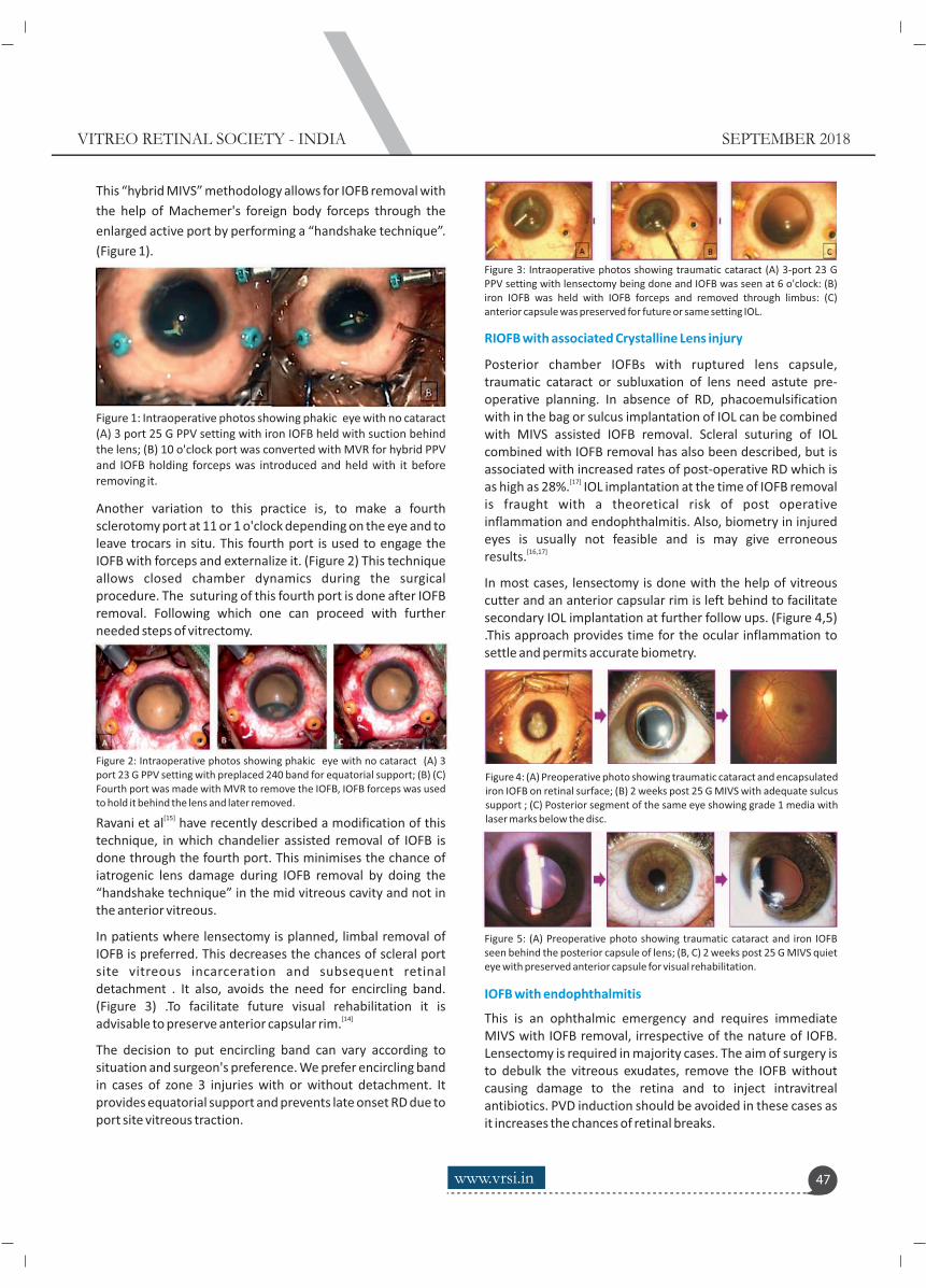
Another variation to this practice is, to make a fourth sclerotomy port at 11 or 1 o'clock depending on the eye and to leave trocars in situ. This fourth port is used to engage the IOFB with forceps and externalize it. (Figure 2) This technique allows closed chamber dynamics during the surgical procedure. The suturing of this fourth port is done after IOFB removal. Following which one can proceed with further needed steps of vitrectomy.
[15]Ravani et al have recently described a modification of this technique, in which chandelier assisted removal of IOFB is done through the fourth port. This minimises the chance of iatrogenic lens damage during IOFB removal by doing the “handshake technique” in the mid vitreous cavity and not in the anterior vitreous.
In patients where lensectomy is planned, limbal removal of IOFB is preferred. This decreases the chances of scleral port site vitreous incarceration and subsequent retinal detachment . It also, avoids the need for encircling band. (Figure 3) .To facilitate future visual rehabilitation it is
[14]advisable to preserve anterior capsular rim.
The decision to put encircling band can vary according to situation and surgeon's preference. We prefer encircling band in cases of zone 3 injuries with or without detachment. It provides equatorial support and prevents late onset RD due to port site vitreous traction.
This “hybrid MIVS” methodology allows for IOFB removal with
the help of Machemer's foreign body forceps through the
enlarged active port by performing a “handshake technique”.
(Figure 1).
Figure 1: Intraoperative photos showing phakic eye with no cataract (A) 3 port 25 G PPV setting with iron IOFB held with suction behind the lens; (B) 10 o'clock port was converted with MVR for hybrid PPV and IOFB holding forceps was introduced and held with it before removing it.
Figure 2: Intraoperative photos showing phakic eye with no cataract (A) 3 port 23 G PPV setting with preplaced 240 band for equatorial support; (B) (C) Fourth port was made with MVR to remove the IOFB, IOFB forceps was used to hold it behind the lens and later removed.
Figure 3: Intraoperative photos showing traumatic cataract (A) 3-port 23 G PPV setting with lensectomy being done and IOFB was seen at 6 o'clock: (B) iron IOFB was held with IOFB forceps and removed through limbus: (C) anterior capsule was preserved for future or same setting IOL.
Figure 4: (A) Preoperative photo showing traumatic cataract and encapsulated iron IOFB on retinal surface; (B) 2 weeks post 25 G MIVS with adequate sulcus support ; (C) Posterior segment of the same eye showing grade 1 media with laser marks below the disc.
Figure 5: (A) Preoperative photo showing traumatic cataract and iron IOFB seen behind the posterior capsule of lens; (B, C) 2 weeks post 25 G MIVS quiet eye with preserved anterior capsule for visual rehabilitation.
RIOFB with associated Crystalline Lens injury
Posterior chamber IOFBs with ruptured lens capsule, traumatic cataract or subluxation of lens need astute pre-operative planning. In absence of RD, phacoemulsification with in the bag or sulcus implantation of IOL can be combined with MIVS assisted IOFB removal. Scleral suturing of IOL combined with IOFB removal has also been described, but is associated with increased rates of post-operative RD which is
[17]as high as 28%. IOL implantation at the time of IOFB removal is fraught with a theoretical risk of post operative inflammation and endophthalmitis. Also, biometry in injured eyes is usually not feasible and is may give erroneous
[16,17]results.
In most cases, lensectomy is done with the help of vitreous cutter and an anterior capsular rim is left behind to facilitate secondary IOL implantation at further follow ups. (Figure 4,5) .This approach provides time for the ocular inflammation to settle and permits accurate biometry.
IOFB with endophthalmitis
This is an ophthalmic emergency and requires immediate MIVS with IOFB removal, irrespective of the nature of IOFB. Lensectomy is required in majority cases. The aim of surgery is to debulk the vitreous exudates, remove the IOFB without causing damage to the retina and to inject intravitreal antibiotics. PVD induction should be avoided in these cases as it increases the chances of retinal breaks.
VITREO RETINAL SOCIETY - INDIA
www.vrsi.in 47
SEPTEMBER 2018

IOFB with endophthalmitis and retinal detachment (RD) portends a poor prognosis. Encircling scleral band, lensectomy, PVD induction, removal of IOFB, silicone oil tamponade with injection of quarter dose intravitreal antibiotics can also be attempted. MIVS helps to maintain a closed chamber during instrument exchange thereby
[19,20]preventing hypotony and sclerotomy site complications.
IOFB with Retinal detachment
Trauma leading to IOFB with RD is best managed with lensectomy, encircling band, PVD induction, IOFB removal and silicone oil/gas tamponade. As described previously,
[19,21]MIVS is extremely useful in this situation.
REFERENCES
1. Shock JP, Adams D. Long term visual acuity results after
penetrating and perforating ocular injuries. Am J
Ophthalmol. 1985;100:714–8
2. Thompson JT, Parver LM, Enger CL, Mieler WF, Liggett PE.
Infectious endophthalmitis after penetrating injury with
retained intraocular foreign bodies. National Eye Trauma
Study. Ophthalmology.1993;100:1468–74
3. Wani VB, Al-Ajmi M, Thalib L, Azad RV, Abul M, Al-Ghanim
M, et al. Vitrectomy for posterior segment intraocular
foreign bodies: Visual results and prognostic factors.
Retina. 2003;23:654–60.
4. Cardillo JA, Stout JT, LaBree L, Azen SP. Post traumatic
proliferative vitreoretinopathy. The epidemiologic
profile, onset, risk factors and visual outcome.
Ophthalmology. 1997;104:1166–73.
5. Yeh S, Colyer MH, Weichel ED. Current trends in the
management of intraocular foreign bodies. Curr Opin
Ophthalmol. 2008;19:225–33
6. Fujii GY, De Juan E, Jr, Humayun MS, et al. Initial
experience using the transconjunctival sutureless
vitrectomy system for vitreoretinal surgery.
Ophthalmology 2002;109 (10):1814-20
7. Eckardt C. Transconjunctival sutureless 23-gauge
vitrectomy. Retina 2005;25(2):208–11
8. Oshima Y, Wakabayashi T, Sato T, Ohji M, Tano Y. A 27
gauge instrument system for transconjunctival
sutureless microincis ion v i trectomy surgery.
Ophthalmology 2010;117(1) :93-102
9. Mohamed S, Claes C, Tsang CW. Review of Small Gauge
Vitrectomy: Progress and Innovation. J O p h t h a l m o l .
2017; 2017 :6285869.
10. Eckardt C, Eckert T, Eckardt U. Memory snare for
e x t ra c t i o n o f i n t ra o c u l a r fo r e i g n b o d i e s .
Retina.2006;26:845-7
11. Erakgun T, Akkin C, Mentes J. Management of
the posterior segment foreign bodies with a simple
snare. Retina. 2003;23:858–60.
12. Dhoble P, Khodifad A. Combined cataract extraction with
pars plana vitrectomy and metallic intraocular foreign
body removal through sclerocorneal tunnel using a novel
“Magnet handshake” technique. Asia Pac J Ophthalmol
(Phila) 2018;7(2):114-8
13. Agarwal A, Trivedi RH, Jacob S, et al. Management of
posteriorly dislocated capsular tension ring with
vitrectomy using a “chandelier” illumination system. Clin
Ophthalmol. 2007;1:285-8
14. Singh R, Bhalekar S, Dogra MR, Gupta A. 23-gauge
vitrectomy with intraocular foreign body removal via the
limbus: An alternative approach for select cases. Indian J
Ophthalmol. 2014;62:707-10
15. Ravani R, Chawla R, Azad SV, et al. Midline sclerotomy
approach for intraocular foreign body removal in phakic
eyes using endoiluminator: a Novel technique. Indian J
Ophthalmol 2018;66(5):687-90
16. Lam DS, Tham CC, Kwok AK, Gopal L. Combined
phacoemulsification, pars plana vitrectomy,removalof
intraocular foreign body (IOFB), and primary intraocular
lens implantation for patients with IOF Band
traumaticcataract. Eye (Lond).1998;12 ( Pt 3a):395-8
17. Mahapatra SK, Rao NG. Visual outcome of pars plana
vitrectomy with intraocular foreign body removal through
sclerocorneal tunnel and sulcus-fixated intraocular lens
implantation as a single procedure, in cases of metallic
intraocular foreign body with traumaticcataract. Indian J
Ophthalmol. 2010;58(2):115-8
18. Kuhn F, Morris R. Posterior segment intraocular foreign
bodies: management in the vitrectomy era.
Ophthalmology 2000;107(5):821-2
19. Parke DW 3rd, Pathengay A, Flynn HW Jr, Albini T,
Schwartz SG. Risk factors forendophthalmitisand retinal
detachment with retained intraocular foreign bodies.J
Ophthalmol. 2012;2012:758526
20. Banker TP, McClellan AJ, Wilson BD, et al. Culture- Positive
Endophthalmitis After Open Globe Injuries With and
Without Retained Intraocular Foreign Bodies. Ophthalmic
Surg Lasers Imaging Retina. 2017;48(8):632-637
21. Nowomiejska K, Choragiewicz T, Borowicz D, et al. Surgical
Management of Traumatic Retinal Detachment with
Primary Vitrectomy in Adult Patients. J Ophthalmol.
2017;2017:5084319
VITREO RETINAL SOCIETY - INDIA
www.vrsi.in48
SEPTEMBER 2018

VITREO RETINAL SOCIETY - INDIA SEPTEMBER 2018
Micropulse laser : Principles and Current Indications
Ajay Aurora1Vision Plus Eye Centre, Kisan Tower, Golf Course Road, Sector 51, Noida
2Delhi Retina Centre, Eye 7 Chaudhary Eye Centre, Bharat Ram Road, 24 Daryaganj, New Delhi
Address for Correspondence
Vision Plus Eye Centre, Kisan Tower, Golf Course Road, Sector 51 , Noida 201301
email: [email protected]
www.vrsi.in 49
INTRODUCTION
Micropulse laser has recently started getting integrated in
clinical practice and no longer remains a research tool. This
article discusses the principles and practical aspects of
Micropulse Laser Therapy (MPLT) and its current indications
and personal experience. All patients presented here have
been treated with IridexIQ 577 ™ Yellow micropulse laser.
Laser photocoagulation has been in use for several decades
and its benefit has been well validated for numerous retinal
conditions like Proliferative Diabetic Retinopathy (PDR),
Diabetic Macular Edema (DME), Macular Edema associated
with Branch Retinal Venous Occlusion (BRVO), Central Serous
Chorioretinopathy (CSC), Choroidal Neovascularization,
retinopexy of retinal tears and others. Conventional laser
photocoagulation of the retina delivers a continuous wave
(CW) laser, which normally results in a full-thickness visible
lesion due to thermal damage of the retinal pigment
epithelium (RPE), neurosensory retina, and the choroid. This
typically results from coagulative necrosis beginning in the
RPE and spreading to involve the photoreceptors and
overlying retina and underlying choroid and results from
elevation of local temperature by 20-30°C above the body
temperature (Fig1). The resulting greyish white lesion may
spread further laterally due to involvement of the zone of
latent lethal damage, which is invisible during the laser 1treatment. Retinal whitening is the optical signature of
chorioretinal burn. More damage means less transparency
and whiter lesion. Hemorrhages occur at roughly three times
the exposure needed to produce ophthalmoscopically visible
lesions. Invisible lesions that are angiographically apparent
occur at approximately half the laser exposure needed for a
visible lesion. Maximum permissible exposure (MPE) levels
established by international laser safety standards represent
roughly one-tenth the laser exposure needed to produce a 2retinal effect.
With the advent of anti-vascular endothelial growth factor
(VEGF) therapy and its success in the treatment of various
retinal pathologies, the utilization of conventional laser has
declined. With pharmacotherapy, Laser induced retinal
damage is also avoided. Hence the role for laser therapy for
these entities in the anti-VEGF era is being questioned.
Recent studies have, however, suggested that it may be
possible to deliver a subthreshold laser that is above the
threshold of biochemical effect but below the threshold of a
visible, destructive lesion. This non-damaging retinal laser
therapy, also called subthreshold laser therapy, does not cause
collateral tissue damage and is not visible clinically or is
documented on the OCT. It is proposed that, subthreshold
laser treatment stimulates a metabolic effect whereby the
retinal tissue produces mediators with anti-angiogenic and
anti-edema effects, such as pigment epithelium derived factor
(PEDF), beta actin, and thrombospondin 1 (TSP1).
Subthreshold laser therapy is also hypothesized to improve
RPE function by stimulating the upregulation of heat-shock 3,4 proteins (HSPs). Advantages of subthreshold laser therapy
include the ability to apply treatment near the fovea or even
transfoveally and its ability to be applied multiple times
without a cumulative damage. This novel laser delivery
modality, called the Micropulse laser was developed by
Pankratov in 1990. His laser produced a train of millisecond
laser pulses separated by variable quiet intervals,
micropulsing allowed selective treatment of the retinal
pigment epithelium (RPE) and sparing of the neurosensory 5retina. This reduced the thermal retinal damage. However,
being invisible, micropulse laser failed to become popular
because of the belief that that no therapeutic effect is
produced without a visible thermal damage to the retina.Non-
destructive and thus non- inflammatory, SDML (Sub Threshold
Diode Micropulse Laser) has been reported effective for a
number of disorders and uniquely allows safe transfoveal 7,8,9 treatment in eyes with good visual acuity. SDML has also
been uniquely shown to increase, rather than decrease,
retinal sensitivity by microperimetry at the locus of laser 10 application.

Fig 4: Subthreshold CW Laser: 532nm; 200 μm spot 50 mW; 10 msec; Clinical
photo and FFA show no laser effect. OCT clearly shows the Laser induced damage (Yellow arrows)(courtesy Dr. Sam Mansour)
Continuous conventional laser can be delivered in a
subthreshold fashion using lower energy, which may not
produce clinical retinal whitening but will be detectable on an
angiogram or OCT (Fig 3,4) demonstrate that by restricting the
energy delivered to the RPE one can restrict the laser-induced
damage. One hundred millisecond laser exposures at
approximately half the threshold power of RPE damage
induced transcription of HSP70, an indication of cellular 13response to sublethal thermal stress. In a micropulse laser
the laser energy is delivered as a pulse train interrupted
energy with an “on” time when the laser is active and “ off”
time when the laser is off (Fig 5). The off time is the tissue
relaxation time allowing the tissue to recover. The total
amount of energy delivered may be the same as the CW laser
delivered as single pulse, but because of on and off time the
damage is restricted to the RPE alone. Hence it is not
detectable either on FFA or OCT. This absence of clinical
damage and hence absence of a clinical endpoint, has
prevented the micropulse laser becoming acceptable.
Principles of Operation and Terminology : Recent advances in
the understanding of laser photocoagulation indicate that
tissue or cellular events occur at far lower laser exposure
levels than needed to produce a clinically visible lesion. These
may occur at a half to a fourth of the exposure needed to
produce ophthalmoscopically obvious lesions. Additionally,
VEGF down regulation has been noted to occur at even lower
laser exposures. Therapeutic effects may thus be produced
with milder retinal irradiances causing lower temperature 3,6,7,14rises associated with less or no significant retinal damage.
Also, direct photocoagulation of the microvascular
abnormalities is not required for resorption of the associated 7macular edema. In fact, the RPE plays a significant role in the
repair of the outer and inner blood–retinal barrier, regardless 15of the type or location of the laser application . Laser
photocoagulation decreases the intraocular concentrations of
vascular endothelial growth factor and RPE derived
Fig 1: Temperature –Tissue interaction during CW laser photocoagulation.
The laser lesion is small (A) but the visible lesion at the end of laser session is 1larger than the selected spot size (B); This further expands with time ©
Fig2: If the laser energy is applied for 1 μsec the effect is restricted to RPE. With increasing duration of laser application for the same power there is
11increasing collateral damage
Table 1: Thermal Diffusion distance versus Pulse duration in 12Water
Fig 3 : Subthreshold CW Laser: 532nm; 200 μm spot 50 mW; 30 msec;
Clinical photo shows faint laser marks. FFA clearly shows the Laser induced damage ( courtesy Dr. Sam Mansour)
VITREO RETINAL SOCIETY - INDIA
www.vrsi.in50
SEPTEMBER 2018

transforming growth factor beta II (TGF-βII), thereby inhibiting active retinal neovascularization. On the contrary, pigment epithelium-derived factor is upregulated after laser
13photocoagulation.
In continuous wave mode, the laser energy is delivered as a
single pulse, with a “width” typically in the range of 0.1–0.5
sec that constitutes the exposure duration. In micropulse
mode, the laser energy is delivered with a train of repetitive
short pulses (typically 100–300 μsec in duration each) within
an “envelope” whose width is typically in the range of 0.1–0.5
sec, and this envelope duration constitutes the exposure
duration. The “ON” time is the duration of each micropulse.
The “OFF” time between successive micropulses reduces heat
in the tissues and regulates the thermal isolation of each
pulse contribution. The period T is the sum of the “ON” and
“OFF” times and its reciprocal, 1/T, is the repetition rate in
pulses per second (pps), also referred to as frequency f in
hertz (Hz). The ratio between the “ON” time and the period T
is the duty cycle in percent. (Figure 5) Repetition rate and duty
cycle determine “isolation” of the increase in temperature
produced by each single micropulse. Isolation of thermal rises
requires a relatively long “OFF” cooling time and this implies a
relatively low repetition rate. Dorin noted that the period T
should not be shorter than 2 millisecond (1/500 sec), the
“OFF” time should not be shorter than 1.7 millisecond, and
the duty cycle should not exceed 15%. (23)
Fig 5: Duty Cycle Concept: In CW laser when a single pulse is fired the laser is
on for the total duration of the pulse. In Micropulse laser with 5% Duty Cycle
and exposure of 200ms,the laser is on for only 10msec (100 repetitive pulses
each of 100usec is on time) and is off for 190msec (1900usec after each pulse
of 100usec is the off time)
Kim et al found that micropulse power as low as 10–25% of
the visible threshold power was sufficient to show consistent
RPE-confined damage with sparing of the neurosensory 16retina with light and electron microscopy . In Dutch belted
Rabbits, Lavinsky et al have shown that laser power less than
30% did not produce any damage on the RPE. The correlation
between the power of the laser needed for a visible threshold
retinal burn and for histologically RPE-confined damage at
various duty cycles can be used for determining the laser
power for micropulse subthreshold treatments, in the 17absence of a visible endpoint.
BIOPHYSICAL BASIS OF MICROPULSE LASER
Subthreshold micropulse laser intends to minimize the laser-
induced chorioretinal damage and spare the neurosensory
retina; and thus produce only sub-lethal thermal elevations,
with effects that are invisible during treatment and remain so
thereafter. It adheres to the following biophysical criteria:
Axial confinement of the thermal gradient at RPE
In retinal photocoagulation, the temperature first increases at
the RPE. From the RPE, it spreads by conduction to re-
equilibrate with adjacent cooler tissues. With the 100-msec
exposure duration that is normally used in traditional
photocoagulation, the heat production time is longer than the
heat diffusion time and heat production is continuously fueled
while heat diffuses. Therefore the RPE temperature
equilibrates in the inner retina blemishing its transparency
with the appearance of the “grayish” endpoint. Roider et al
showed that for exposures in the microsecond rather than in
the usual millisecond range, tissue damage is limited to
subcellular molecular structures not detectable by electron
microscopy, as the heat production ends before heat diffusion 18can occur . (Figure 2 and Table 1)
Control of the thermal gradient at the RPE (avoiding
excessive temperatures)
Even when the rise in temperature is confined around the RPE
during laser exposure, unavoidable thermal equilibration
could still damage the neurosensory retina. This can occur
when the temperature at the RPE creates a heat wave that
reaches the inner retina at lethal temperature levels. This
post-operative inner retinal damage must be avoided by
limiting the temperature rise at the RPE by adjusting the laser
“irradiance” (power density in W/cm2). Micropulse laser
damages only a small percentage of target tissue molecules
with each micropulse, and repetitive micropulses combine to
produce sub visible effects according to the semi empirical −1/4 -1/4damage additivity or N law. The N law says that the same
damage-threshold that can be achieved with one pulse of
duration t and energy E, can be accomplished by using N
repetitive pulses, each with the same duration t, but only a
fraction (N−1/4) of the energy E, provided that all pulses are
delivered in a brief time, shorter than any possible biological
repair. For example, if a given damage-threshold can be
achieved with a single laser pulse with E = 100 mJ, the same
can be accomplished with 100 repetitive pulses, each having
only E = 100 mJ × 100−1/4 = 31.6 mJ. Lower energy per pulse
VITREO RETINAL SOCIETY - INDIA
www.vrsi.in 51
SEPTEMBER 2018
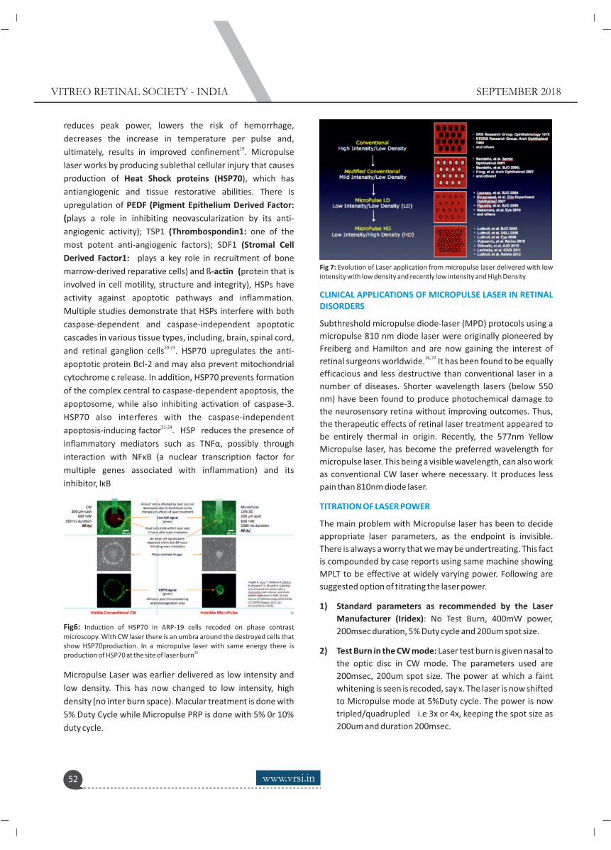
reduces peak power, lowers the risk of hemorrhage,
decreases the increase in temperature per pulse and, 19ultimately, results in improved confinement . Micropulse
laser works by producing sublethal cellular injury that causes
production of Heat Shock proteins (HSP70), which has
antiangiogenic and tissue restorative abilities. There is
upregulation of PEDF (Pigment Epithelium Derived Factor:
(plays a role in inhibiting neovascularization by its anti-
angiogenic activity); TSP1 (Thrombospondin1: one of the
most potent anti-angiogenic factors); SDF1 (Stromal Cell
Derived Factor1: plays a key role in recruitment of bone
marrow-derived reparative cells) and ß-actin (protein that is
involved in cell motility, structure and integrity), HSPs have
activity against apoptotic pathways and inflammation.
Multiple studies demonstrate that HSPs interfere with both
caspase-dependent and caspase-independent apoptotic
cascades in various tissue types, including, brain, spinal cord, 20-23and retinal ganglion cells . HSP70 upregulates the anti-
apoptotic protein Bcl-2 and may also prevent mitochondrial
cytochrome c release. In addition, HSP70 prevents formation
of the complex central to caspase-dependent apoptosis, the
apoptosome, while also inhibiting activation of caspase-3.
HSP70 also interferes with the caspase-independent 21-24apoptosis-inducing factor . HSP reduces the presence of
inflammatory mediators such as TNFα, possibly through
interaction with NFκB (a nuclear transcription factor for
multiple genes associated with inflammation) and its
inhibitor, IκB
Fig6: Induction of HSP70 in ARP-19 cells recoded on phase contrast
microscopy. With CW laser there is an umbra around the destroyed cells that show HSP70production. In a micropulse laser with same energy there is
25production of HSP70 at the site of laser burn
Micropulse Laser was earlier delivered as low intensity and
low density. This has now changed to low intensity, high
density (no inter burn space). Macular treatment is done with
5% Duty Cycle while Micropulse PRP is done with 5% 0r 10%
duty cycle.
Fig 7: Evolution of Laser application from micropulse laser delivered with low intensity with low density and recently low intensity and High Density
CLINICAL APPLICATIONS OF MICROPULSE LASER IN RETINAL DISORDERS
Subthreshold micropulse diode-laser (MPD) protocols using a
micropulse 810 nm diode laser were originally pioneered by
Freiberg and Hamilton and are now gaining the interest of 26, 27retinal surgeons worldwide. It has been found to be equally
efficacious and less destructive than conventional laser in a
number of diseases. Shorter wavelength lasers (below 550
nm) have been found to produce photochemical damage to
the neurosensory retina without improving outcomes. Thus,
the therapeutic effects of retinal laser treatment appeared to
be entirely thermal in origin. Recently, the 577nm Yellow
Micropulse laser, has become the preferred wavelength for
micropulse laser. This being a visible wavelength, can also work
as conventional CW laser where necessary. It produces less
pain than 810nm diode laser.
TITRATION OF LASER POWER
The main problem with Micropulse laser has been to decide
appropriate laser parameters, as the endpoint is invisible.
There is always a worry that we may be undertreating. This fact
is compounded by case reports using same machine showing
MPLT to be effective at widely varying power. Following are
suggested option of titrating the laser power.
1) Standard parameters as recommended by the Laser
Manufacturer (Iridex): No Test Burn, 400mW power,
200msec duration, 5% Duty cycle and 200um spot size.
2) Test Burn in the CW mode: Laser test burn is given nasal to
the optic disc in CW mode. The parameters used are
200msec, 200um spot size. The power at which a faint
whitening is seen is recoded, say x. The laser is now shifted
to Micropulse mode at 5%Duty cycle. The power is now
tripled/quadrupled i.e 3x or 4x, keeping the spot size as
200um and duration 200msec.
VITREO RETINAL SOCIETY - INDIA
www.vrsi.in52
SEPTEMBER 2018

3) Test Burn in Micropulse Mode: Laser is turned to
micropulse mode 5%Duty cycle. Power is raised to
1200mW, duration 200msec and spot size 200um. Laser test
burn is attempted nasal to the optic disc. till a faint
whitening is observed. This may happen anywhere
between 1200 to 2000mW.The power so determined is then
reduced to 30% and this is used for treatment purpose.
Other Methods: Some Micropulse Laser machines use End
Point Management system. Numerous authors have reduced
the spot size to 150um.
CHOICE OF DUTY CYCLE
Duty cycle (the frequency of the train of micropulses) decides
the duration of on and off time and the frequency with which
the laser gets on. The lower the duty cycle, longer the off time
between pulses (lower repetition rate) and lesser the heat
buildup. If the off time exceeds the thermal relaxation time of
RPE melanin (the target molecule), the average tissue
temperature rise in the RPE during micro pulsed laser
application can be maintained below levels lethal to the cell.
From the available evidence most studies use 5% duty cycle
for the treatment of macular diseases. There have been
studies that have used 10% or 15% DC. Present Clinical data
and computer modeling has demonstrated that treatment
safety is maximized by the use of 5% or lower duty cycles.
Higher duty cycles rapidly take on the clinical characteristics of
CW lasers.
Lutrull et al in a long term retrospective review of 274
consecutive eyes with Macular Edema due to DME/BRVO who
were treated with Heavy density MPLT at various duty cycles
(DC): 5%, 10% and 15% and followed up 10 years found 8% of
eyes (7/84) showed laser induced retinal damage when
treated with DC10-15% while no eye treated with 5% DC
(0/168) showed any Retinal Damage.
CURRENT INDICATIONS FOR MICROPULSE LASER AND PERSONAL EXPERIENCE (1-6)
1. Central Serous Chorioretinopathy
2. Diabetic Macular Edema
3. Proliferative Diabetic Retinopathy
4. Macular edema due to BRVO
5. Macular edema due to non-ischemic CRVO
6. Post ERM peel Macular Edema
7. CNVM not responsive to antiVEGF therapy
8. Inherited Macular Degenerations
1) CENTRAL SEROUS CHORIORETINOPATHY
Most cases of central serous chorioretinopathy (CSR) have a
self-limiting course, but patients who need rapid recovery of
visual acuity or those with a chronic course threatening visual
recovery may benefit from continuous-wave laser
photocoagulation. (37) Traditional laser treatment in CSR,
however, has several drawbacks, including symptomatic
scotomas, choroidal neovascularization, foveal distortion, and
subretinal fibrosis, especially in cases with juxtafoveal leaking
points. Enlargement of the laser spot may also threaten
central vision. Bandello et al first proposed MPLT for the
treatment of CSR in 2003. In their study, five out of five eyes
showed complete resorption within 1 month of MPLT and
maintained through 4 months of follow-up. No evidence of
retinal changes resulting from laser treatment was discernible 39with fluorescein angiography or fundus biomicroscopy.
All studies in which MPD laser treatment was used for CSR
have supported the hypothesis that MPD produces
therapeutic benefits comparable to those of conventional
photocoagulation with no detectable signs of laser-induced 39,40iatrogenic damage. The absence of a visible endpoint,
however, represents a challenge for the surgeon. To overcome
this problem, some investigators performed MPD treatment
over ICG-stained RPE cells to enhance the selectivity of the
treatment for the active leaking sites while sparing the
neurosensory retina. This also aided in the documentation of
the placement of laser applications seen as dark spots in the 41ICG background fluorescence.
Fig 8: (Top) 8 weeks after onset;(Middle) 7 days after MPLT; (Bottom) 3 weeks after MPLT
VITREO RETINAL SOCIETY - INDIA
www.vrsi.in 53
SEPTEMBER 2018

17
Fig 9a 58 years male Chronic CSR for 14 months : Fundus Photo and FFA
Fig 9 b: 58 years male Chronic CSR 14 months; Foveal flattening after single
MPLT
Over 30 studies for Micropulse Laser have been published
including retrospective and randomized controlled studies of
various sizes employing a variety of different laser wave-
lengths, treatment parameters, treatment protocols, and
patient populations. Despite such heterogeneity, in all studies
MPLT for CSR has been found to be effective: comparable to
half-fluence photodynamic therapy. Nearing completion is
the PLACE (Prospective Randomized Controlled Treatment
Trial for Chronic Central Serous Chorioretinopathy) trial
( : NCT01797861), a randomized prospective clinicaltrials.gov
clinical trial in the Netherlands comparing half-dose
verteporfin photodynamic therapy with MPL for CSR. Initial
data from this indicates that MPLT is as good as half fluence 42PDT, if not better .
2) DIABETIC MACULAR EDEMA
Diabetic macular edema (DME) is the leading cause of visual
impairment in patients with Diabetic Retinopathy. Before the
advent of antiVEGF therapy ETDRS laser treatment had
become the standard for DME, with focal photocoagulation
being done for localized areas of leakage and a grid pattern
being applied to areas of diffuse macular edema. The ETDRS
showed that increased treatment intensity was associated
with more adverse treatment effects, increased treatment
density improved clinical results, grid treatment was as
effective as focal treatment; and the location and extent of
macular angiographic leakage did not correlate with clinical
outcomes. This information provoked use of higher density
with lower intensity laser photocoagulation. However, laser
induced retinal damage was still the end point for most
ophthalmologists. In 1997, Freiberg and Karatza first reported
clinical application of MP 810-nm diode laser therapy for DME
They however used 15%DC. Lutrull et al with their report on
clinically significant macular edema set the benchmark of
using 5% Duty cycle for macular treatment. They showed that
benefit could be achieved without visible/invisible retinal
damage. This also supported contiguous treatment with no
interburn spacing. This new approach, called “low-
intensity/high-density subthreshold diode micropulse laser”
(HD-SDM), allowed effective and reliable sublethal retinal
laser treatment without any retinal damage or adverse effects.
This SDM strategy of sublethal retinal laser application defines
modern retinal laser treatment.
Levinski et al in their prospective, randomized, clinical trial in
previously untreated DME patients showed that at 1 year, the
clinical performance of HD-SDM was superior to that of the
mETDRS (modified ETDRS) photocoagulation technique, both
anatomically and functionally. They suggested that, as
experience grows, HD-SDM might become the preferred
treatment approach in these cases.
Today, in the antiVEGF era, pharmacotherapy is commonly
employed for Centre involving DME and laser is used for non-
Centre involving DME as per ETDRS protocol. Steroids are
added in pseudophakics and those who do not respond to
antiVEGF. This strategy involves multiple Intravitreal injections
and carries with it the risks associated with Intravitreal
injections. It is believed that MPLT will support
pharmacotherapy, reduce the intraocular procedures and
hence the complications associated with Intravitreal
injections. I personally use MPLT after the central macular
thickness has reduced to less than 450μm. (Table 2)
VITREO RETINAL SOCIETY - INDIA
www.vrsi.in54
SEPTEMBER 2018

17
VITREO RETINAL SOCIETY - INDIA
www.vrsi.in 55
Table 2: Central Foveal thickness based Interventions Algorithm
Fig 10: Non Centre Involving Diabetic Macular Edema treated with 5%DC MPLT and followed up after 10 weeks with cataract surgery
3) PROLIFERATIVE DIABETIC RETINOPATHY
Untreated PDR is the leading cause of blindness in PDR cases.
Protocol S has demonstrated that Intravitreal injection of
Ranibizumab is non inferior to PRP and at the end of two years
40-45% eyes treated in both groups (Ranibizumab and PRP)
had active Neovascularization.
Moorman and Hamilton using high duty cycle of MPL were
able to deliver minimal intensity MP PRP without causing
clinical retinal damage and avoid any other adverse events. In
10 of the 13 eyes treated, they noted regression of 27Neovascularisation . Lutrull et al used 15% DC of sublethal
panretinal micropulse laser (SDM PRP) in 99 eyes (61 with PDR
and 38 severe nonproliferative DR) of 69 consecutive patients
treated between 2000 and 2003, and followed for a median of 3512 months. Overall VA remained unchanged, but the
proportion of eyes with excellent VA (20/30 or better)
increased from 39% to 48%. SDM PRP was performed in a
single session with topical anesthesia, and no patient
reported postoperative pain or loss of visual acuity,
accommodation, night vision, or visual field. No eye progressed to a surgical traction retinal detachment or neovascular glaucoma. Only 3 of 38 eyes with severe non-PDR (7.9%) progressed to PDR. Compared to the expected annual rate of 50%, this reduction in DR progression was significant (P = 0.0001).
Till we have larger studies available, it may be best to explain the available options to the patient. He must understand the pros and cons of using antiVEGF agents or MPLT versus the time tested CW laser PRP.
Fig 11: Forty-three years male Diabetic for 10 years, non-hypertensive:
PDR with CIDME, good metabolic control, underwent CW Laser
demarcation of the area to be treated, Focal Laser with CW laser over the
areas of NVE. Remaining areas were treated with 10% DC MPLT. He has
been on follow-up for 2 years. Underwent Follow-up with multimodal
imaging and received supplementation of MPLT supplementation at 18
months. His VA is maintained at 6/6 and his fields are normal.
4) MACULAR EDEMA SECONDARY TO BRVO
Limited studies have described the use of MPD laser in the
treatment of macular edema secondary to branch retinal vein 43,44occlusion. Parodi et al have shown that MPD laser
treatment is as effective as conventional grid laser
photocoagulation in reducing macular edema (ME) and
improving the VA, even though the MPD acted more slowly.
Lutrull et al have demonstrated that 5%DC is ideal for treating
Macular edema due to BRVO. Since macular edema in BRVO
waxes and wanes, MPLT may actually be beneficial in these
cases as it can be repeated whenever indicated.
Fig 12a
SEPTEMBER 2018

VITREO RETINAL SOCIETY - INDIA
www.vrsi.in56
Fig 12 a and b: 23 years female, Inflammatory BRVO with Macular edema, VA 6/60; treated with systemic steroids and later with two Intravitreal injections of Bevacizumab. VA improved after first injection to 6/12 and later 6/36.Underwent third injection of Avastin followed by Sectorial MPLT (area marked out on Fundus photo) with 10%DC. VA improved to 6/9 with clearance of retinal hemorrhages. Underwent repeat Sectorial MPLT after 14 months and has maintained dry macula with VA of 6/6p for 36 months.
5) MACULAR EDEMA DUE TO NON-ISCHEMIC CRVO
There are no documented reports of MPLT for CRVO. Ischemic CRVOs may not respond to a MPLT intervention. I have treated Non Ischemic CRVO with initial VA 6/60 with MPLT 5%DC to the macula and 5% DC to extra macular areas.
Fig 13: Case 1: 76 years male, operated CA rectum, Hypertensive for 20 years, diabetic 8 years, Non Ischemic CRVO with VA of 6/60. Refused Intravitreal injection of antiVEGF and was treated with 5%DC of MPLT to the macula and 5%DC MPLT to extra macular areas of capillary non-perfusion. VA improved to 6/12 in 6 weeks with reduction of CMT on sequential OCT.
Fig 14: Case 2: Non Ischemic CRVO: 46 years male, 8 years Hypertensive, non-diabetic, VA 6/60. Received two injections of Intravitreal Avastin with minimal response. Macula treated with 5%DC MPLT. Responded with macular flattening and improvement of VA to 6/9
09/0 11/0 01/0 28/0
th 9 July2015
6. POST ERM PEEL MACULAR EDEMA
Fig 15: 72 year's male, Operated Vitreous Hemorrhage for BRVO, developed macular pucker. Underwent Re PPV with ERM peel and two Intravitreal injections Avastin and later IVTA without any improvement. Subsequently underwent MPLT at 5%DC .VA improved from 6/24 to 6/18 with reduction in macular edema (15a is before and 15b after treatment)
7. CNVM NOT RESPONSIVE TO ANTIVEGF THERAPY
In 2000 York and colleagues used indocyanine green
angiography guided micropulse laser to close choroidal feeder
vessels in neovascular age-related macular degeneration 45 (AMD). In 2015, Lutrull et al reported reversal of tolerance to
46 anti-VEGF drugs in neovascular AMD using SDM. Thirteen
eyes, 73-97 years old, having received 16-67 antiVEGF
injections and unresponsive to all anti-VEGF medications for at
least 6 months and 4 consecutive injections of aflibercept
were treated by MPL. One month later anti-VEGF therapy was
resumed. After rechallenge and resumption of aflibercept,
there was improvement in 12 of the 13 eyes and complete
resolution of macular exudation in 9 of 13 eyes. They believe
the reversal of drug tolerance in wet AMD by MPL was
predicted by “Reset to Default” theory, which describes the
clinical effects of RPE HSP activation. As the first report of drug
tolerance reversal in medicine, these findings lend support to
the reset postulate. In 2016, Lutrull and Margolis reported,
“functionally guided panmacular micropulse laser” as “retinal
protective therapy” in high-risk dry AMD and inherited 47retinopathies. Patients were evaluated before and after SDM
retinal protective therapy by pattern electroretinography
(PERG) and visual function tests. After SDM retinal protective
therapy, 139 of 158 eyes were improved by PERG (P = 0.0001).
VA was unchanged, but macular sensitivity by microperimetry
(40 eyes) and mesoptic contrast visual acuity (73 eyes)
improved (P = 0.0439, and P = 0.006, respectively). However,
SEPTEMBER 2018

there was no change in retinal morphology, such as drusen
number, size, or distribution. Eyes with the worst
preoperative status improved the most by all indices (P =
0.0001). This included eyes with extensive geographic
atrophy (GA). By maintaining improved retinal function
guided by early and periodic electrophysiological signals,
rather than late-imaging signals, the authors hope to reduce
the long-term risk of vision loss. In a subsequent retrospective
study of 454 consecutive eyes of 296 patients in the Age-
Related Eye Disease Study treated with panmacular SDM for
high-risk dry AMD and followed a minimum of 1 year (range: 1
to 7 years, average: 2 years), the incidence of new choroidal 48 neovascularization was found to be less than 1% per year.
This was despite an average patient age of 83 years and high
independent risk factors including fellow eye choroidal
neovascularization (26%), reticular pseudodrusen (38%), and
AMD severity (78% Age-Related Eye Disease Study categories
3 and 4), all significantly exceeding the Age-Related Eye
Disease Study. These findings suggest that panmacular SDM
may reduce the incidence of choroidal neovascularization in
dry AMD more than vitamin therapy alone. In addition, in 409
of these same eyes, best-corrected logMAR mesopic visual
acuity (P < 0.0001) and visual fields (P = 0.0007) were also
improved following pan- macular SDM. There were no
adverse treatment effects or retinal damage. These findings
suggest that retinal and visual function testing may be useful 47 surrogate indicators of longer-term treatment benefits .
8. INHERITED MACULAR DEGENERATIONS
In a small group of inherited retinal diseases (IRD) Luttrull and 50 Margolis reported PERGs improvement after SDM in 10
eyes of 8 patients (retinitis pigmentosa (4 eyes), cone
degeneration (3 eyes), and Stargardt disease 3 eyes). While
eyes with AMD improved most by a low-contrast PERG, IRD
eyes improved most by the widest-field (24 °) high-contrast
PERG protocols. In both AMD and IRD, measures of signal
latency improved most significantly. The authors note that
both the functional improvements and the distinct PERG
responses in AMD versus IRD were predicted by the reset 50 theory of retinal laser action.
CONCLUSIONS
Subthreshold micropulse laser is a relatively novel laser
modality that has good clinical efficacy and minimal risk of
iatrogenic side effects. The greatest limitation of MPLT is the
difficulty of titrating the treatment without the feedback of
an ophthalmoscopically visible endpoint. This is more of a
mind set issue. The available literature with multiple
randomized trials and the evidence of production of HSPs
after MPLT should be enough to convince the fence sitters.
Also considering a broad range in which MPLT is safely
effective should make it easier for clinicians to accept it as a
reliable modality in the treatment of selected cases.
REFERENCES
1. G. Dorin.Subthreshold and micropulse diode laser photocoagulation.Semin Ophthalmol 2003;18 (3): 147–153
2. Mainster MA,Turner PL.Retinal injuries from light:mechanisms,hazards and prevention,Chapter 109.In:Ryan J,Hinton DR,Schachat AP, Wilkinson P, e d s . Re t i n a , 4 t h e d . . 2 v o l . L o n d o n : E l s e v i e r Publishers;2006:1857-1870.
3. G. Dorin.Evolution of retinal laser therapy: minimum intensity photocoagulation (MIP). Can the laser heal the retina without harming it? Semin Ophthalmol 2004; 19 (1–2):62–68
4. Boutacoff TA, Buzawa D, Dorin G, Kelsoe W. Diode laserMicro-Pulse 'minimal impact photocoagulation' (DLMP) [unpublished data]. Iris Medical Instruments, 1997.
5. Pankratov MM. Pulsed delivery of laser energy in experimental thermal retinal photocoagulation. Proc Soc Photo Opt Instrum Eng. 1990;1202:205–13.
6. Mainster MA. Decreasing retinal photocoagulation damage: Principles and techniques. Semin. Ophthalmol. 1999;14:200–9.
7. Mainster MA. Laser-tissue interactions: future laser therapies. Diabetic Retinopathy: Approaches to a Global Epidemic. Association for Research in Vision and Ophthalmology Summer Research Conference 2010; 31 July; Natcher Center, National Institutes of Health, Bethesda MD. 2010.
8. Luttrull JK, Dorin G. Subthreshold diode micropulse photocoagulation as invisible retinal phototherapy for diabetic macular edema. A review. Curr. Diab Rev 2012;8:274–84.
9. Roider J, Hillenkamp F, Flotte TJ, Birngruber R. Microphotocoagulation: selective effects of repetitive short laser pulses. Proc Natl Acad Sci USA 1993;90(11):2037–8.
10. Vujosevic S, Bottega E, Casciano M, Pilotto E, Convento E, Midena E. Microperimetry and fundus autofluorescence in diabetic macular edema. Subthreshold micropulse diode laser versus modified Early Treatment Diabetic Retinopathy Study Laser photocoagulation. Retina 2010;30:908–16.
11. Mainster MA, White TJ, Tips JH, Wilson PW. Retinal-temperature increases produced by intense light sources. J Opt Soc Am. 1970;60:264-270.
12. Boutacoff TA, Buzawa D, Dorin G, Kelsoe W. Diode laserMicro-Pulse 'minimal impact photocoagulation'
VITREO RETINAL SOCIETY - INDIA
www.vrsi.in 57
SEPTEMBER 2018

(DLMP) [unpublished data]. Iris Medical Instruments, 1997.
13. Christopher Sramek, Mark Mackanos, Ryan Spitler, et al.
Non-damaging Retinal Phototherapy: Dynamic Range of
Heat Shock Protein Expression. IOVS, March 2011,Vol
52,No 3, 1780-1787
14. Lanzetta P, Dorin G, Piracchio A, et al. Theoretical bases of
n o n - o p h t h a l m o s c o p i c a l l y v i s i b l e e n d p o i n t
p h o t o c o a g u l a t i o n . S e m i n O p h t h a l m o l .
2001;16(1):8—11
15. Glaser BM, Campochiaro PA, Davis JL, et al. Retinal
pigment epithelial cells release inhibitors of
neovascularization. Ophthalmology. 1987;94:780--4
16. Kim SY, Sanislo SR, Dalal R, et al. The selective effect of
micropulse diode laser upon the retina. Invest
Ophthalmol Vis Sci. 1996;37(3):S779
17. Lavinsky D, Sramek C, Wang J, Huie P, Dalal R, Mandel Y,
Palanker D.Subvisible Retinal Laser Therapy: Titration
algorithm and Tissue response,Retina. 2014 Jan;
34(1):87-97
18. Roider J, Hillenkamp F, Flotte T, et al. Microphoto-
coagulation: selective effects of repetitive short laser
pulses. Proc Natl Acad Sci. 1993;90:8643-7
19. McDonald H.R., Schatz H. Visual loss following panretinal
photocoagulation for proliferative diabetic retinopathy.
Ophthalmology. 1985;92(3):388–393.
20. Franklin TB, Krueger-Naug AM, Clarke DB, Arrigo AP,
Currie RW. The role of heat shock proteins Hsp70 and
Hsp27 in cellular protection of the central nervous
system. Int J Hyperthermia. 2005;21:379–392.
21. Whitlock NA, Lindsey K, Agarwal N, Crosson CE, Ma JX.
Heatshock protein 27 delays Ca2_-induced cell death in a
caspasedependent and -independent manner in rat
retinal ganglion cells. Invest Ophthalmol Vis Sci.
2005;46:1085–1091.
22. Yenari MA, Liu J, Zheng Z, Vexler ZS, Lee JE, Giffard RG.
Antiapoptotic and anti-inflammatory mechanisms of
heat-shock protein protection. Ann N Y Acad Sci.
2005;1053:74–83
23. Soti C, Nagy E, Giricz Z, Vigh L, Csermely P, Ferdinandy P.
Heatshock proteins as emerging therapeutic targets. Br J
Pharmacol.2005;146:769–780.
24. Awasthi N, Wagner BJ. Upregulation of heat shock
protein expressionby proteasome inhibition: an
antiapoptotic mechanism in thenlens. Invest
Ophthalmol Vis Sci. 2005;46:2082–2091.
25. Keiji Inagaki, Takuya Shuo, Kanae Katakura, Nobuyuki
Ebihara, Akira Murakami and Kishiko Ohkoshi. Journal of
Ophthalmology. Sublethal Photothermal Stimulation
with a Micropulse Laser induces Heat Shock Protein
Expression in ARPE-19 cells ol. 2015;2015:729792. doi:
10.1155/2015/729792. Epub 2015 Nov 30.
26. Friberg TR, Karatza EC. The treatment of macular disease
using a micropulsed and continuous wave 810-nm diode
laser. Ophthalmology. 1997;104:2030--8
27. Moorman CM, Hamilton AMP. Clinical applications of the
micropulse diode laser. Eye. 1999;13:145—50
28. Diabetic Retinopathy Study Research Group.
Photocoagulation Treatment of Proliferative Diabetic
Retinopathy: Second Report of Diabetic Retinopathy
Study Findings. Ophthalmology.1978 ; 85: Issue1 p82-
106
29. BandelloF, Brancato R,Menchini U et al. Light panretinal
photocoagulation (LPRP) Versus classic panretinal
photocoagulation (CRPP) in proliferative Diabetic
Retinopathy. Semin. Ophthalmol.2001 Mar;16(1):8-12
30. Laursen Ml,Moeller F, Sander B,Sjoelie AK. Subthreshold
Micropulse diode laser treatment in Diabetic macular
oedema.Br J Ophthalmol.2004,Sep ,88(9);1173-9
31. Siavaprasad S, Sandhu R, Tandon A et al.Subthreshold
micropulse Diode Laser photocoagulation for Clinically
Significant Diabetic Macular oedema: a three year follow-
up. Clin Exp Ophthalmol.2007.Sep-Oct;35(7): 640-4
32. Figueira C, Khan J, Nunes S et al. Prospective randomized
controlled trial comparing subthreshold micropulse
diode laser photocoagulation and conventional green
laser for clinically significant diabetic macular oedema. Br
J Ophthalmol. 2009;93(10):1341--4
33. Nakamura Y,Ogata K,Arai M et al. Functional and
Morphological changes of macula after subthreshold
micropulse diode laser photocoagulation for diabetic
macular oedema.Eye (Lond) 2010 May;24(5):784-8
34. Luttrull JK, Musch DC, Mainster MA. Subthreshold diode
micropulse photocoagulation for the treatment of
clinically significant diabetic macular oedema. Br J
Ophthalmol. 2005;89:74—80
35. Luttrull JK, Musch DC, Spink CA. Subthreshold diode
micropulse panretinal photocoagulation for proliferative
diabetic retinopathy. Eye. 2008;22:607—12
36. Levinski D,Cardilli JALuiz A.S Meli Jr.et al.Randomized
Clinical Trial Evaluating mETDRS versus Normal or High-
VITREO RETINAL SOCIETY - INDIA
www.vrsi.in58
SEPTEMBER 2018
Density Micropulse Photocoagulationfor Diabetic
Macular Edema. Invest. Ophthalmol Vis Sci
2011;52,4314-4323
37. Luttrull JK, Sramek C, Palanker D, Spink CJ, Musch
DC.Long-term safety, high-resolution imaging, and tissue
temperature modeling of subvisible diode micropulse
photocoagulation for retinovascular macular edema.
Retina. 2012 Feb;32(2):375-86.
38. Wang MS, Sander B, Larsen M. Retinal atrophy in
idiopathic central serous chorioretinopathy. Am J
Ophthalmol. 2002;133:787—93
39. Bandello F, Lanzetta P, Furlan F, et al. Non visible
subthreshold micropulse diode laser treatment of
idiopathic central serous chorioretinopathy. A pilot
study. Invest Ophthalmol Vis Sci. 2003;44
40. Chen SN, Hwang JF, Tseng LF, Lin CJ. Subthreshold diode
micropulse photocoagulation for the treatment of
chronic central serous chorioretinopathy with
juxtafoveal leakage.Ophthalmology. 2008;115
(12):2229-34
41. Ricci F, Missiroli F, Regine F, et al. Indocyanine green
enhanced subthreshold diode-laser micropulse
photocoagulation treatment of chronic central serous
chorioretinopathy. Graefes Arch Clin Exp Ophthalmol.
2009;247(5):597-607
42. Breukink MB, Downes SM, Querques G, et al. Comparing
half-dose photodynamic therapy with high-density
subthreshold diode micropulse laser treatment in
patients with chronic central serous chorioretinopathy
(the PLACE trial): study protocol for a randomized
controlled trial. Trials. 2017;16:419
43. Parodi BM, Spasse S, Iacono P, et al. Subthreshold grid
laser treatment of macular edema secondary to branch
retinal vein occlusion with micropulse infrared
(810 nanometer) diode laser. Ophthalmology.
2006;113:2237- 42
44. Luttrull JK, Sramek C, Palanker D, Spink CJ, Musch
DC.Long-term safety, high-resolution imaging, and tissue
temperature modeling of subvisible diode micropulse
photocoagulation for retinovascular macular edema.
Retina. 2012 Feb;32(2):375-86
45. York J, Glaser B, Murphy R. High speed ICG. Used to
pinpoint laser treatment of feeder vessels in wet AMD. J
Ophthalmic Nurs Technol. 2000;19(2):66 -7
46. Luttrull JK, Chang DB, Margolis BW, et al. Laser re-
sensitization of medically unresponsive neovascular age-
related macular degeneration: Efficacy and Implications.
Retina. 2015;35:1184-94
47. J.K. Luttrull, S.H. Sinclair, S. Elmann, B.M. Glaser,
Panmacular subthreshold diode micropulse laser (SDM)
for high-risk dry AMD: CNV incidence and mesopic visual
function. Abstract and free paper presentation, American
Society of Retina Specialists, annual meeting, Boston,
August 8, 2017.
48. Age-Related Eye Disease Study Research Group. A
randomized, placebo-controlled, clinical trial of high-
dose supplementation with vitamins C and E, beta
carotene, and zinc for age-related macular degeneration
and vision loss: AREDS report number 8. Arch
Ophthalmol. 2001;119:1417 - 36
49. Chhablani J, Roh J Y, Jobling A I et al. Restorative retinal
laser therapy: Present state and future directions . Survey
of Ophthalmology 2018: 63: 307-328
50. Luttrull JK, Margolis BW. Functionally guided retinal
protective therapy for dry age-related macular and
inherited retinal degenerations: a pilot study. Invest
Ophthalmol Vis Sci. 2016;57:265-75
VITREO RETINAL SOCIETY - INDIA
www.vrsi.in 59
SEPTEMBER 2018


Notes

Notes


![arXiv:1801.06940v2 [cs.CV] 11 Sep 2018](https://static.fdokumen.com/doc/165x107/6335ad6b8502b620dc08efe4/arxiv180106940v2-cscv-11-sep-2018.jpg)


![arXiv:2109.09861v1 [cs.AI] 20 Sep 2021](https://static.fdokumen.com/doc/165x107/63146abfaca2b42b580d8799/arxiv210909861v1-csai-20-sep-2021.jpg)