Volume 10, Issue 2, March 2020 - Online Journal of Animal ...
35
Volume 10, Issue 2, March 2020 An International Peer-Reviewed Journal which Publishes in Electronic Format ONLINE JOURNAL OF ANIMAL AND FEED RESEARCH
-
Upload
khangminh22 -
Category
Documents
-
view
6 -
download
0
Transcript of Volume 10, Issue 2, March 2020 - Online Journal of Animal ...

Volume 10, Issue 2, March 2020
An International Peer-Reviewed Journal which Publishes in Electronic Format
ONLINE JOURNAL OF ANIMAL AND FEED RESEARCH
a | P a g e
Online J. Anim. Feed Res., 10 (2): March 25, 2020 Editorial Team
Editors-in-Chief
Habib Aghdam Shahryar, PhD, Professor of Animal Nutrition; Director of Department of Animal Science, IA University,
Shabestar, IRAN (Website, Google Scholar, SCOPUS, Email: [email protected])
Saeid Chekani Azar, PhD, Faculty of Veterinary Medicine, Animal Physiology, Atatürk University, TURKEY (Google
Scholar, SCOPUS, ORCID, Publons; Email: [email protected])
Managing Editor
Alireza Lotfi, PhD, Animal Physiology, IAU, IRAN (Google Scholar, SCOPUS, ResearchGate, Publons, Email:
Deputy Section Editors
Ana Isabel Roca Fernandez, PhD, Professor, Animal Production Department, Agrarian Research Centre of Mabegondo,
15080 La Coruña, SPAIN (Email: [email protected]); Dairy Science, Plant-Soil Science
Alireza Ahmadzadeh, PhD, Assistant Professor, Department of Animal Science, IAU, Shabestar, IRAN (Email:
[email protected]); Biometry - Plant Breeding (Biotechnology)
Arda Yildirim, PhD, Assistant Professor, Department of Animal Science, Faculty of Agriculture, Gaziosmanpasa
University, 60240 Tokat, TURKEY (Email: [email protected]); Animal Science, Nutrition-non Ruminants, Breeding, Nutritive Value
Ferdaus Mohd. Altaf Hossain, DVM, Sylhet Agricultural University, BANGLADESH (Email: [email protected]); Microbiology, Immunology, Poultry Science, and Public Health
John Cassius Moreki, PhD, Department of Animal Science and Production, College of Agriculture, BOTSWANA (Email: [email protected]); Nutrition - Non-Ruminants, Breeders, Livestock management
Mohamed Shakal, Professor & Head of Poultry Diseases Department, Faculty of Veterinary Medicine, Cairo University, EGYPT; Director of the Endemic and Emerging Poultry Diseases Research Center, Cairo University, Shek Zaed Branch,
EGYPT; Chairman of The Egyptian Poultry Forum Scientific Society. REPRESENTATIVE FOR EGYPT & MENA REGION. Email: [email protected]
Muhammad Saeed, PhD, Northwest A&F University, Yangling, 712100, CHINA (Email: [email protected]), Nutrition – Ruminants
Paola Roncada, PhD, Associate Professor, Veterinary Pharmacology and Toxicology, University of Bologna, ITALY (Email: [email protected]); Pharmacokinetics
Reviewers
Abdelfattah Y.M. Nour, DVM, PhD, Professor of Veterinary Physiology, Purdue University, USA (Email:
Adnan Yousaf, DVM, MPhil of Poultry Science (Gold Medalist), PhD of Avian Embryology; Sindh Agricultural University
Tandojam, PAKISTAN (Emails: [email protected]; [email protected])
Ahmad Yildiz, PhD, Professor, Animal Science and Production Department, Faculty of Veterinary Medicine, Atatürk
University, TURKEY (Email: [email protected]); Nutrition - Ruminants
Ali Halajian, PhD, DVM, Professor of Parasitology, Department of Biodiversity, Faculty of Science and Agriculture,
University of Limpopo, SOUTH AFRICA (Email: [email protected])
Alireza Radkhah, PhD, Department of Fisheries, Faculty of Natural Resources, University of Tehran, Karaj, IRAN (Email:
[email protected]); Aquatic Biology, Aquaculture and Fisheries Biotechnology

b | P a g e
Assamnen Tassew, Bahir Dar University, ETHIOPIA (Email: [email protected]); Animal Production and Production System
Behzad Shokati, PhD, Department of Agronomy & Plant Breeding, Faculty of Agriculture, Maragheh University, IRAN (Email: [email protected]); Agriculture, Nutritive value and utilization of feeds
Ekrem Laçin, PhD, Professor of Animal Science, Faculty of Veterinary Medicine, Atatürk University, TURKEY (Email: [email protected]); Nutrition - Non-Ruminants
Fazul Nabi Shar, PhD, Lecturer, Faculty of Veterinary & Animal Sciences, Lasbela University of Agriculture Water &
Marine Sciences, Uthal Balochistan, PAKISTAN (Email: [email protected]); Clinical Veterinary Medicine
Ferdaus Mohd. Altaf Hossain, DVM, Sylhet Agricultural University, BANGLADESH; not shah Jalal University of Science & Technology, BANGLADESH (Email: [email protected]); Microbiology, Immunology, Poultry Science
Firew Tegegn, Bahir Dar University, ETHIOPIA (Email: [email protected]); Animal Nutritionist
Hamid Mohammadzadeh, PhD, Assistant Professor, Department of Animal Science, Faculty of Agriculture, University of
Tabriz, IRAN (Email: [email protected]); Nutrition - Ruminants
Hazim Jabbar Al-Daraji, PhD, Professor, University of Baghdad, College of Agriculture, Abu-Ghraib, Baghdad, IRAQ
(Email: [email protected]); Avian Reproduction and Physiology
Manish Kumar, PhD, Professor, Society of Education, INDIA (Email: [email protected]); Pharmacology,
Ethnomedicine
Megiste Taye, PhD, Seoul National University, SOUTH KOREA (Email: [email protected]); Comparative
genomics and bioinformatics
Mohammed Yousuf Kurtu, Associate Professor, Animal Sciences Department, Haramaya University, Dire-Dawa,
ETHIOPIA (Email: [email protected]); Animal Science, Nutrition
Muhammad Saeed, PhD, Northwest A&F University, Yangling, 712100, CHINA (Email:
[email protected]), Nutrition - Ruminants
Nilüfer Sabuncuoğlu Çoban, PhD, Professor, Department of Animal Science and Production, Faculty of Veterinary
Medicine, Atatürk University, TURKEY (Website; Email: [email protected]); Animal Hygiene and Welfare, Physiology
Osman Erganiş, Professor, PhD, Veterinary Microbiology, Selcuk University, Konya, TURKEY (Website, Google Scholar;
Email: [email protected])
Ömer Çoban, PhD, Professor, Department of Animal Science and Production, Atatürk University, TURKEY (Website; [email protected]); Nutrition - Ruminants
Paola Roncada, PhD, Associate Professor, Veterinary Pharmacology and Toxicology, University of Bologna, ITALY (Email: [email protected]); Pharmacokinetics
Raga Mohamed Elzaki Ali, PhD, Assistant Professor, Department of Rural Economics and Development, University of Gezira, SUDAN (Email: [email protected]); Animal-feed interactions, Nutritive value
Rashid Habiballa Osman, PhD, Assistant Professor, Department of Poultry Production, Faculty of Animal Production, West Kordofan University, SUDAN (E-mail: [email protected]); Nutrition - Non-Ruminants
Sesotya Raka Pambuka, MSc, Sinta Prima Feedmill, Poultry and Aqua Feed Formulation, Sulaiman Rd 27A, West
Jakarta, INDONESIA
Shahin Eghbal-Saeid, PhD, Associate Professor, Department of Animal Science, IAU, Khorasgan (Isfahan), IRAN (Email: [email protected]); Animal Genetics and Breeding
Shigdaf Mekuriaw, Andassa livestock research center, ETHIOPIA (Email: [email protected]); Animal production and Nutrition
Terry Ansah, PhD, University for Development Studies-Ghana and Harper Adams University College, UK (Email:
[email protected]); Nutrition - Ruminants
Tohid Vahdatpour, PhD, Assistant Professor, Department of Physiology, IAU, Shabestar, IRAN (Scopus; Google
Scholar; Emails: [email protected]; [email protected]); Physiology and Functional Biology of Systems
Ümit Acar, PhD, Department of Aquaculture, Faculty of Fisheries, Muğla Sıtkı Koçman University, TURKEY (Email:
[email protected]); Aquaculture, Fish nutrition
Vassilis Papatsiros, PhD, Department of Porcine Medicine, University of Thessaly, Trikalon str 224, GR 43100, GREECE
(Email: [email protected]); Dietary input, Animal and Feed interactions
Wafaa Abd El-Ghany Abd El-Ghany, PhD, Associate Professor, Poultry and Rabbit Diseases Department, Cairo
University, Giza, EGYPT (Email: [email protected]); Poultry and Rabbit Diseases
Wesley Lyeverton Correia Ribeiro, MSc, DVM, College of Veterinary, Medicine, State University of Ceará, Av.
Paranjana, 1700, Fortaleza, BRAZIL (Email: [email protected]); Animal Health and Welfare, Veterinary Parasitology
Yadollah Bahrami, PhD of Biotechnology, Khorasgan Branch, IAU, Khorasgan, IRAN (Email: [email protected]); Nutrition - Non-Ruminants
Yavuz Gurbuz, Professor, University of Kahramanmaras Sutcu Imam, Department of Animal Nutrition, Campus of Avsar, Kahramanmaras, TURKEY (Email: [email protected]); Animal Nutrition, Feed Technology and Evaluation
Zohreh Yousefi, PhD, Department of Plant Biology, Atatürk University, Erzurum, TURKEY (Email: [email protected]); Plant Biology

c | P a g e
Zewdu Edea, Chungbuk National University, SOUTH KOREA (Email: [email protected]); Livestock Population Geneticist
Language Editors
Mehrdad Ehsani-Zad, MA in TEFL, Takestan-IA University, IRAN (Email: [email protected])
Samuel Stephen Oldershaw, Master of TESOL, The Humberston School & The Grimsby Institute, North East
Lincolnshire, UK (Email: [email protected])
Advisory Board
Ali Nobakht, PhD, Assistant Professor, Animal Science Department, IAU, Maragheh, IRAN (Email:
[email protected]); Nutrition - Non-Ruminants
Fikret Çelebi, PhD, Professor of Physiology, Faculty of Veterinary Medicine, Atatürk University, Erzurum, TURKEY
(Email: [email protected]); Physiology and Functional Biology of Systems
Mohamed Shakal, Professor, Poultry Diseases Department, Faculty of Veterinary Medicine, Cairo University, EGYPT;
Director of the Endemic and Emerging Poultry Diseases Research Center, Cairo University, Shek Zaed Branch, EGYPT; Chairman of The Egyptian Poultry Forum Scientific Society. REPRESENTATIVE FOR EGYPT & MENA REGION. Email:
Naser Maheri Sis, PhD, Assistant Professor, Dept. Anim. Sci., IAU, Shabestar, IRAN (Website; Emails:
[email protected]; [email protected]); Nutrition - Ruminants, Nutritive Value, Utilization of Feeds
Join OJAFR Team As an international journal we are always striving to add diversity to our editorial board and operations staff. Applicants
who have previous experience relevant to the position may be considered for more senior positions (Section Editor, SE) within OJAFR. All other members must begin as Deputy Section Editors (DSE) before progressing on to more senior roles.
Editor and editorial board members do not receive any remuneration. These positions are voluntary. If you are currently an undergraduate, MSc or PhD student at university and interested in working for OJAFR, please fill
out the application form below. Once your filled application form is submitted, the board will review your credentials and notify you within a week of an opportunity to membership in editorial board. If you are Ph.D., assistant or associate editors, distinguished professor, scholars or publisher of a reputed university, please rank the mentioned positions in order of your preference. Please send us a copy of your CV or ORCID ID or briefly
discuss any leadership positions and other experiences you have had that are relevant to applied Animal and Feed Researches or publications. This includes courses you have taken, editing, publishing, web design, layout design, and
event planning. If you would like to represent the OJAFR at your university, join our volunteer staff today! OJAFR representatives assist
students at their university to submit their work to the OJAFR. You can also, registered as a member of OJAFR for subsequent contacts by email and or invitation for a honorary reviewing articles. Download OJAFR Application Form

d | P a g e
Archive
Research Paper In vitro efficacy of Tylosin and Enrofloxacin in treatment of bovine mastitis causing bacteria in Omdurman locality.
Almobarak ME, Mohammed Salih RR and Gibreel HH.
Online J. Anim. Feed Res., 10(2): 53-58, 2020; pii: S222877012000007-10 DOI: https://dx.doi.org/10.36380/scil.2020.ojafr7 Abstract Dairy industry has recently grown as a very important economic national source of income. In Sudan, many dairy owners
introduced foreign blood. This might result in a progeny of mixed blood cows with lowered resistance to endogenous and
locally prevailing diseases such as mastitis. In this study 60 milk samples were obtained from Frisian cows in Elrudoan
and Elmouileh Convention in Omdurman, Khartoum State, Sudan. Samples positive for bacterial growth were identified using the gram stain and various conventional biochemical tests. Hundred species of bacteria were isolated from 60
samples of milk. A total of 70 (70%) were gram positive, and 30 (30%) were gram negative bacteria. Among the total of the gram positive isolates, 40 (57.1%) were Staphylococcus spp., 18 (25.7%) were Bacillus spp., 6 (8.6%) Streptococcus
spp., 4 (5.7%) Corynebacterium spp., and 2 (2.9%) were Actinomyces spp. and from gram negative isolates, 26 (86.7%) were Enterobacter spp. and 4 (13.3%) were E. coli. Antibiotic susceptibility tests to Tylosin and Enroflaxcin were
performed for the isolated bacteria (Staphylococcus aureus, Staph. epidermidis, Enterobacter aerogenes and Enterobacter faecalis). The isolated bacteria were found to be highly sensitive to Tylosin and Enrofloxacin. Keywords: Tylosin, Enroflxacin, Bovine, Mastitis, In vitro [Full text-PDF]
Research Paper Prevalence of bovine gastro intestinal parasitic infection in and around Kombolcha town, Ethiopia.
Ayele A, Abay M, Birhan M, Yayeh M, Erara M, Gessese T, Mohammed A and Demoze G. Online J. Anim. Feed Res., 10(2): 59-65, 2020; pii: S222877012000008-10 DOI: https://dx.doi.org/10.36380/scil.2020.ojafr8
Abstract A cross-sectional study was conducted in and around Kombolcha from October 2017 to April 2018 to determine the
prevalence of gastro-intestinal helminthes parasites in cattle. A total of 384 randomly selected cattle were sampled and examined using standard coprological procedure. The overall prevalence was 39.8% of gastrointestinal (GI) helminthes
and the prevalent helminthes eggs identified were 15.6% Paramphistomum species (spp), 10.4% strongly type eggs, 8.6% Fasciola spp., 3.1% Trichuris species and 2.1% Toxocaraspecies. This result indicated the highest prevalence of
Paramphistomum spp. eggs than other helminthes egg and the lowest prevalence of Toxocara species egg. There was statistically significant difference among the age groups in paramphistomum and strongly infection (χ2=24.960, p≤
0.001) and (χ2=17.047, p≤ 0.001) respectively. Higher prevalence rate was shown in 2-5 years age of cattle. Between body conditions there was also significant (p≤ 0.000 and p≤ 0.013) difference in paramphistomum and strongly and
which was higher in moderate animals and lower in animals with good body condition. Sex had no significant effect on the prevalence of helminthes parasite, except for strongly type of egg. The present study revealed that there is high
prevalence of GI helminthes infection in cattle in the study area. Therefore, strategic prevention should be advocated to
prevent the problem in and around Kombolcha.
Keywords: Cattle, Gastrointestinal, Prevalence, Helminthes parasites, Kombolcha [Full text-PDF]
TABLE OF CONTENT
Volume 10 (2); March 25, 2020V
e | P a g e
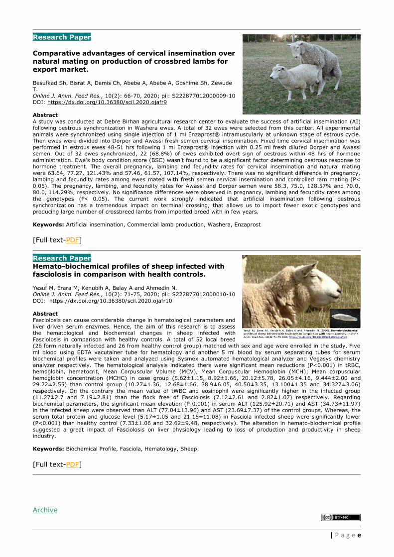
Research Paper Comparative advantages of cervical insemination over natural mating on production of crossbred lambs for export market.
Besufkad Sh, Bisrat A, Demis Ch, Abebe A, Abebe A, Goshime Sh, Zewude T. Online J. Anim. Feed Res., 10(2): 66-70, 2020; pii: S222877012000009-10 DOI: https://dx.doi.org/10.36380/scil.2020.ojafr9 Abstract A study was conducted at Debre Birhan agricultural research center to evaluate the success of artificial insemination (AI) following oestrous synchronization in Washera ewes. A total of 32 ewes were selected from this center. All experimental
animals were synchronized using single injection of 1 ml Enzaprost® intramuscularly at unknown stage of estrous cycle. Then ewes were divided into Dorper and Awassi fresh semen cervical insemination. Fixed time cervical insemination was
performed in estrous ewes 48-51 hrs following 1 ml Enzaprost® injection with 0.25 ml fresh diluted Dorper and Awassi semen. Out of 32 ewes synchronized, 22 (68.8%) of ewes exhibited overt sign of oestrous within 48 hrs of hormone
administration. Ewe’s body condition score (BSC) wasn’t found to be a significant factor determining oestrous response to hormone treatment. The overall pregnancy, lambing and fecundity rates for cervical insemination and natural mating
were 63.64, 77.27, 121.43% and 57.46, 61.57, 107.14%, respectively. There was no significant difference in pregnancy, lambing and fecundity rates among ewes mated with fresh semen cervical insemination and controlled ram mating (P<
0.05). The pregnancy, lambing, and fecundity rates for Awassi and Dorper semen were 58.3, 75.0, 128.57% and 70.0,
80.0, 114.29%, respectively. No significance differences were observed in pregnancy, lambing and fecundity rates among
the genotypes (P˂ 0.05). The current work strongly indicated that artificial insemination following oestrous
synchronization has a tremendous impact on terminal crossing, that allows us to import fewer exotic genotypes and producing large number of crossbreed lambs from imported breed with in few years.
Keywords: Artificial insemination, Commercial lamb production, Washera, Enzaprost [Full text-PDF]
Research Paper Hemato-biochemical profiles of sheep infected with fasciolosis in comparison with health controls.
Yesuf M, Erara M, Kenubih A, Belay A and Ahmedin N. Online J. Anim. Feed Res., 10(2): 71-75, 2020; pii: S222877012000010-10 DOI: https://dx.doi.org/10.36380/scil.2020.ojafr10
Abstract Fasciolosis can cause considerable change in hematological parameters and
liver driven serum enzymes. Hence, the aim of this research is to assess the hematological and biochemical changes in sheep infected with
Fasciolosis in comparison with healthy controls. A total of 52 local breed (26 form naturally infected and 26 from healthy control group) matched with sex and age were enrolled in the study. Five
ml blood using EDTA vacutainer tube for hematology and another 5 ml blood by serum separating tubes for serum biochemical profiles were taken and analyzed using Sysmex automated hematological analyzer and Vegasys chemistry
analyzer respectively. The hematological analysis indicated there were significant mean reductions (P<0.001) in tRBC, hemoglobin, hematocrit, Mean Corpuscular Volume (MCV), Mean Corpuscular Hemoglobin (MCH); Mean corpuscular
hemoglobin concentration (MCHC) in case group (5.62±1.15, 8.92±1.66, 20.12±5.78, 26.05±4.16, 9.444±2.00 and 29.72±2.55) than control group (10.27±1.36, 12.68±1.66, 38.9±6.05, 40.50±3.35, 13.100±1.35 and 34.327±3.06)
respectively. On the contrary the mean value of tWBC and eosinophil were significantly higher in the infected group (11.27±2.7 and 7.19±2.81) than the flock free of Fasciolosis (7.12±2.61 and 2.82±1.07) respectively. Regarding
biochemical parameters, the significant mean elevation (P 0.001) in serum ALT (125.92±20.71) and AST (34.73±11.97) in the infected sheep were observed than ALT (77.04±13.96) and AST (23.69±7.37) of the control groups. Whereas, the
serum total protein and glucose level (5.17±1.05 and 21.15±11.08) in Fasciola infected sheep were significantly lower (P<0.001) than healthy control (7.33±1.06 and 32.62±9.48, respectively). The alteration in hemato-biochemical profile
suggested a great impact of Fasciolosis on liver physiology leading to loss of production and productivity in sheep industry.
Keywords: Biochemical Profile, Fasciola, Hematology, Sheep.
[Full text-PDF]
Archive
.

f | P a g e
Online Journal of Animal and Feed Research
ISSN: 2228-7701
Frequency: Bimonthly
Current Issue: 2020, Vol: 10, Issue: 2 (March 25)
Publisher: SCIENCELINE Online Journal of Animal and Feed Research is an international peer-reviewed journal, publishes the full text of original scientific researches, reviews, and case reports in all fields of animal and feed sciences, bimonthly and freely on the internet ...view full aims and scope
www.ojafr.ir
» OJAFR indexed/covered by NLM Catalog, CABI, CAS, Ulrich's™, GALE, HINARI, NSD,
AKSTEM, BASE, ZDB, ICV, EZB ...details
» Open access full-text articles is available beginning with Volume 1, Issue 1.
» Full texts and XML articles are available in ISC-RICeST.
» This journal is in compliance with Budapest Open Access Initiative and International Committee
of Medical Journal Editors' Recommendations.
» High visibility of articles over the internet.
» Copyright & Publishing Rights Policy ...details
» Publisher Item Identifier ...details
» This journal encourage the academic institutions in low-income countries to publish high quality
scientific results, free of charges... view Review/Decisions/Processing/Policy
ABOUT US | CONTACT US | PRIVACY POLICY
Editorial Offices: Atatürk University, Erzurum 25100, Turkey
University of Manitoba, Winnipeg, Manitoba R3T 2N2, Canada
University of Maragheh, East Azerbaijan, Maragheh 55136, Iran
Homepage: www.science-line.com Phone: +98 914 420 7713 (Iran); +90 538 770 8824 (Turkey); +1 204 8982464 (Canada)
Emails: [email protected]
ABOUT JOURNAL

53 Citation: Almobarak ME, Mohammed Salih RR and Gibreel HH (2020). In vitro efficacy of Tylosin and Enrofloxacin in treatment of bovine mastitis causing bacteria in
Omdurman locality. Online J. Anim. Feed Res., 10(2): 53-58. DOI: https://dx.doi.org/10.36380/scil.2020.ojafr7
2020 SCIENCELINE
Online Journal of Animal and Feed Research
Volume 10, Issue 2: 53-57; March 25, 2020 ISSN 2228-7701
IN VITRO EFFICACY OF TYLOSIN AND ENROFLOXACIN IN
TREATMENT OF BOVINE MASTITIS CAUSING BACTERIA IN
OMDURMAN LOCALITY
Misoon Esam ALMOBARAK1, Reem Rabie MOHAMMED SALIH2 and Haytham Hashim GIBREEL3
1Faculty of Veterinary Medicine, University of Khartoum, Sudan 2Head of Department of Clinical Medicine, Faculty of Veterinary Medicine, University of Khartoum, Sudan 3Head of the Department of Silviculture, Faculty of Forestry, University of Khartoum, Sudan Email: [email protected]; : 0000-0001-6611-5562
Supporting Information
ABSTRACT: Dairy industry has recently grown as a very important economic national source of income. In Sudan,
many dairy owners introduced foreign blood. This might result in a progeny of mixed blood cows with lowered
resistance to endogenous and locally prevailing diseases such as mastitis. In this study 60 milk samples were
obtained from Frisian cows in Elrudoan and Elmouileh Convention in Omdurman, Khartoum State, Sudan.
Samples positive for bacterial growth were identified using the gram stain and various conventional biochemical
tests. Hundred species of bacteria were isolated from 60 samples of milk. A total of 70 (70%) were gram
positive, and 30 (30%) were gram negative bacteria. Among the total of the gram positive isolates, 40 (57.1%)
were Staphylococcus spp., 18 (25.7%) were Bacillus spp., 6 (8.6%) Streptococcus spp., 4 (5.7%)
Corynebacterium spp., and 2 (2.9%) were Actinomyces spp. and from gram negative isolates, 26 (86.7%) were
Enterobacter spp. and 4 (13.3%) were E. coli. Antibiotic susceptibility tests to Tylosin and Enroflaxcin were
performed for the isolated bacteria (Staphylococcus aureus, Staph. epidermidis, Enterobacter aerogenes and
Enterobacter faecalis). The isolated bacteria were found to be highly sensitive to Tylosin and Enrofloxacin.
Keywords: Tylosin, Enroflxacin, Bovine, Mastitis, In vitro
RE
SE
AR
CH
AR
TIC
LE
P
II: S2
22
87
70
12
00
00
07
-10
Re
ce
ive
d: A
ug
ust 0
6, 2
01
9
Re
vise
d: Ja
nu
ary
14
, 20
20
INTRODUCTION
Bovine Mastitis is a multi-etiological and complex disease, which is defined as inflammation of parenchyma of
mammary glands. Mastitis is considered the main disease in dairy herds (Kaneen and Bandhard, 1990). The
occurrence of disease is an outcome of interplay between three major factors: infectious agents, host resistance,
and environmental factors (Gera and Guha, 2011). It is characterized by physical, chemical and, usually,
bacteriological changes in milk and pathological changes in glandular tissues (Radostitis et al., 2000). Mastitis is a
global problem as it adversely affects animal health, quality of milk and the economics of milk production, affecting
every country, including developed ones and causes huge financial losses (Sharma et al., 2007). It is the most
important disease in dairy cattle and more affect in economic, the most damaging (Ashish et al. 2000; Sharma et
al. 2012; Elango et al. 2010; Mostert et al. 2004). Mastitis is caused by several species of common bacteria, fungi,
mycoplasmas and algae (Batavani et al., 2007). Most mastitis is of bacterial origin, with just a few of species of
bacteria accounting for most cases.
Mastitis pathogens are categorized as contagious or environmental (Kivaria, 2006). Contagious pathogens live
and multiply on and in the cow’s mammary gland and are spread from cow to cow, primarily during milking.
Contagious pathogens include: Staphylococcus aureus, Streptococcus agalactiae, Mycoplasma spp. and
Corynebacterium bovis (Radostitis et al., 2000). Environmental mastitis can be defined broadly as those intra-
mammary infections (IMI) caused by pathogens whose primary reservoir is the environment in which the cow lives
(Smith et al., 1985). The most frequently isolated environmental pathogens are Streptococci, other than S.
agalactiae, commonly referred to as environmental streptococci usually S. uberis and S. disagalactiae and gram-
negative bacteria such as Escherichia coli, Klebsiella spp. and Enterobacter spp. (Hogan et al., 1999).
Tylosin is macrolide antibiotic produced from Streptomyces fradiae and related structurally to erythromycin
(Plumb, 2002; Giguere, 2013). It is the first antimicrobial of the fluoroquinolones group available to veterinarians,
they are bactericidal, their wide spectrum of antimicrobial activity includes various microorganisms such as gram
positive, gram negative bacteria , mycoplasma, and chlamydiae (Pyorala et al., 1994). Enrofloxacin is alternative
DOI: https://dx.doi.org/10.36380/scil.2020.ojafr7

54 Citation: Almobarak ME, Mohammed Salih RR and Gibreel HH (2020). In vitro efficacy of Tylosin and Enrofloxacin in treatment of bovine mastitis causing bacteria in
Omdurman locality. Online J. Anim. Feed Res., 10(2): 53-58. DOI: https://dx.doi.org/10.36380/scil.2020.ojafr7
drug. The aim of this study to identify the most common causes of bacterial mastitis in cows in Omdurman locality
and to measure the effectiveness of Tylosin and Enrofloxacin in the treatment of bacterial mastitis in vitro.
MATERIALS AND METHODS
Study area
Study area is Omdurman city which located at the intersection of latitude 15 degrees 41 minutes north,
longitude 32 degrees 37 minutes east, on the west bank of the Nile opposite the coupler with a tributary of the
White Nile, and off both Khartoum and Khartoum North, which are linked by the bridge of the White Nile and
Shambat bridge. The numbers of dairy cows were more in this locality than the other sites of Khartoum province.
Sampling
A total of 30 suspected cows were examined clinically: took the body temperature, pulse, heart rate,
respiration, auscultation and palpation the last examination especially for mammary gland and supra mammary
lymph nodes for presence of mastitis. Sixty milk samples from mastitic cows were collected. Mastitis was
diagnosed when there were visible or palpable singes of udder, inflammatory changes in milk secretion, or through
bacteriological examination of milk. During the study 60 milk sample were encountered from 30 cows suffering
from clinical and subclinical mastitis. Milk sample were taken under from infected quarters only under aseptic
condition for bacteriological studies. The fore milk was stripped off and about 5 ml of milk were drawn in sterile
disposable bottle. All samples collected were immediately placed on ice in a thermo flask after collection.
Isolation and identification of bacteria
Culture. Milk samples were collected from mastitic cows were cultured in two media: Blood agar and
MacConkeys agar. After culturing the plates were incubated for 24 hours at 37ºC. Purification was achieved by
further subculturing on nutrient agar and incubated at 37ºC for 24 hours. After purification, a full loop from purified
culture was taken and a smear was made and stained with Gram´s stain to differentiate between Grams positive
and Grams negative bacteria and to see the shape of bacteria. Plates were examined for cultural characteristics
and biochemical reactions according to standard keys (Barrow and Feltham, 2003). Staphylococci were studied in
particular for haemolysis and coagulase production using human plasma. A positive coagulase test was judged as
any degree of clotting from a loose clot suspended in plasma to a solid clot (Barrow and Feltham, 2003).
Purification of cultures. Purification of culture was made by sub-culturing a part of a typical and well isolated
colony on nutrient agar. This process was repeated twice. The resulting of growth was checked for purity by
examining smears stained with Gram´s stain method.
Identification of bacteria. The purified isolated bacteria were identified according to criteria outlined by Barrow
and Feltham (2003) which included of: Reaction of Grams stain, shape of the bacterial colonies, presence or
absence of spores, motility, the colonial characteristics on different media, haemolysis of blood agar and
biochemical tests. All biochemical tests for identification of isolated bacteria were performed according to Barrow
and Feltham (2003).
Antibiotic sensitivity test. Some of the bacteria that isolated through microbiological procedures were
subjected to antimicrobial susceptibility test by disc diffusion method to identify the effectiveness of the Tylosin and
Enrofloxacin. The sensitivity against Tylosin and Enrofloxacin were determined on Mueller Hinton agar as described
by National Committee for Clinical Laboratory Standards (NCCLS, 2002). The results were obtained by measuring
the diameter of the growth inhibition zone around the antibiotic disc for each isolated bacterial strain and recorded
as sensitive, intermediate and resistant.
RESULTS
Identification of isolates
Out of the 100 isolates 70 (70%) were gram positive, and 30 (30%) were gram negative bacteria. Among the
total of the gram positive isolates, 40 (57.1%) were Staphylococcu spp., Bacillus spp. were 18 (25.7%),
Streptococcus spp. were 6 (8.6%), Corynebacterium spp. were 4 (5.7%), Actinomyces spp. were 2 (2.9%) and from
gram negative isolates, Enterobacter spp. were 26 (86.7%) and E. coli were 4 (13.3%) (Figure, 1). The isolated
Staphylococcus spp. in this study was divided into two groups: Coagulase – positive included Staph. aureus and
Staph. hyicus. Coagulase – negative were Staph. epidermidis, Staph. chromogens, Staph. simulans and Staph.

55 Citation: Almobarak ME, Mohammed Salih RR and Gibreel HH (2020). In vitro efficacy of Tylosin and Enrofloxacin in treatment of bovine mastitis causing bacteria in
Omdurman locality. Online J. Anim. Feed Res., 10(2): 53-58. DOI: https://dx.doi.org/10.36380/scil.2020.ojafr7
hominis. The total 70 isolates: 18 (25.7%) were found to be bacillus species. There were (6%) isolates of
Streptococcus spp obtained in this study. Other isolates were: Corynebacterium spp., Actinomyces spp.,
Enterobacter spp. and E. coli. Table 1 shows the quality control limits for antibiotics. The results of sensitivity tests
by used Tylosin and Enrofloxacin are shown in Tables 2 and 3. They were affected in Staph. aureus, Staph.
epidermidis, Enterobacter aerogenosa, Enterococcus faecalis.
Table 1 - Quality control limits for antibiotics
Antimicrobial agent Zone diameter in mm Staph. aureus
Potency Code S I R
Enrofloxacin 10Mg EX >23 22-17 <16 27-34
Tylosin 15Mg TY >26 25-23 <23 24-31
S: sensitive; I: intermediate; R: resistant
Table 2 - The efficacy of Tylosin against different types of bacteria.
Isolated bacteria Zone of inhibition Remarked
Staph. aureus 30mm S
Staph. epidermidis 23mm I
Enterobacter aerogenosa 24mm I
Enterococcus faecalis 27mm S
S: Sensitive; I: Intermediate
Table 3 - The efficacy of Enrofloxacin against different types of bacteria
Isolated bacteria Zone of inhibition Remarked
Staph. aureus 34 mm S
Staph. epidermidis 26 mm S
Enterobacter aerogenosa 30 mm S
Enterococcus faecalis 25 mm S
S: sensitive
Figure 1 - Total of different isolated bacteria from mastitic milk samples
40
12 14 18 6 4 4 2
100

56 Citation: Almobarak ME, Mohammed Salih RR and Gibreel HH (2020). In vitro efficacy of Tylosin and Enrofloxacin in treatment of bovine mastitis causing bacteria in
Omdurman locality. Online J. Anim. Feed Res., 10(2): 53-58. DOI: https://dx.doi.org/10.36380/scil.2020.ojafr7
Figure 2 - Species of Staphylococci isolates
DISCUSSION
Bovine mastitis is a common disease entity of dairy cows, accompanied by physical, chemical, pathological and
bacteriological changes in milk and glandular tissue (Samad, 2008). It is a harmful disease affecting the dairy
industry worldwide and is a matter of great concern for leading milk producing country because of the losses
incurred due to high morbidity, discarded milk, treatment costs and reduced milk production, thus drawing in more
attention towards its treatment and control (Mohanty et al., 2013).
Apart from the economic losses, mastitis can have serious implications on public health. Mastitis which is
mostly caused by the interaction of multiple pathogenic agents (primarily bacteria), can expose human beings to
various organisms through infected milk, thus serving as a media for transmission of various zoonotic diseases like
T.B, brucellosis, diphtheria, scarlet fever and Q fever (Mahantesh and Kaliwal, 2011).
In fact, S. aureus was one of the most frequently isolated staphylococci, supporting the assertion that this
microorganism numbers among the main mastitis pathogens in the Czech Republic (Rysanek et al., 2007). In this
study the isolation of E. coli, Bacillus spp., and Enterobacter spp., might be attributed to poor or absence of hygiene.
This suggestion was supported by the statement of Quinn et al. (2004) who mentioned that Bacillus cereus and E.
coli were isolated from mastitic milk of bovine. These results collectively support our results in this study also the
result is in agreement with Sudhan et al. (2005).
The detection of Actinomyces bovis in the mastitic milk in this study was in agreement with Quinn et al. (2004)
who mentioned that this bacterium among a rarely Gram- positive rod – shaped causing bovine mastitis.
In this study found the isolates were sensitive to tylosin and enrofloxacin sensitive, these antibiotics are used
either for treatment of clinical cases to avoid the spreading of the causative agent, in prophylactic measures to
eliminate the susceptibility of animals for prevention of new infection or growth promotion in weight gain for
fattening programmes. This study is an agreement with Anon (2011), who reported that indicated for the treatment
of local signs (inflammation, milk quality and yield) associated with per acute/acute mastitis in lactating dairy
cattle. Also the isolates in this study were affected with Tylosin and this is an agreement with (Pyorala et al., 1994).
CONCLUSION
In this study we have showed that the incidence of bovine mastitis is high in Omdurman locality. The most frequent
isolated bacteria are Staphylococci. In addition, other opportunistic and environmental organisms were isolated
from mastitic milk samples. Inadequate stall or pasture management e.g. dirty and wet bedding material or muddy
areas as well as in proper milking procedures lead to an increased infection risk.
Recommendation
1- In dairy farm hygienic procedure must be from Practice sustainable.
2- Antimicrobial sensitivity testing should be practiced before treatment of mastitis with antibiotics.
3- Farmers should be aware about what suitable antibiotics to be used for specific mastitis case.
4- The usage of antibiotics in dairy farm should be under supervision of veterinarian to avoid missed used
which leading to the development of antibiotic resistance bacteria.
25
7
3 3 1 1
40
Staph aureusStaph
epidermidis
Staph
chromogens
Staph
simulans
Staph hominisStaph hyicusTotal

57 Citation: Almobarak ME, Mohammed Salih RR and Gibreel HH (2020). In vitro efficacy of Tylosin and Enrofloxacin in treatment of bovine mastitis causing bacteria in
Omdurman locality. Online J. Anim. Feed Res., 10(2): 53-58. DOI: https://dx.doi.org/10.36380/scil.2020.ojafr7
DECLARATIONS
Corresponding author
E-mail: [email protected] ORCİD: 0000-0001-6611-5562
Authors’ contribution
All authors contributed equally to this work.
Availability of data
The data can be available to the journal upon request.
Consent to publish
Not applicable
Conflict of interest
The authors declare they have no competing of interests.
Acknowledgement
The authors would wish to acknowledge the Department of Microbilology, Faculty of Veterinary Medicine, University
of Khartoum for their support through the whole process of developing this publication.
REFERENCES
Anon (2011). Special precautions for the disposal of unused veterinary medicinal product or waste materials. 1.
Batavani RA, Asri S and Naebzadeh H (2007). The effect of sub-clinical mastitis on milk composition in dairy cows. Iranian Journal of
Veterinary Research. 8 (3): 205–211. DOI: https://dx.doi.org/10.22099/ijvr.2007.925
Elango A, Doraisamy KA, Rajarajan G and Kumaresan G (2010). Bacteriology of sub-clinical mastitis and anti-biogram of isolates
recovered from cross-bred cows. Indian Journal of Animal Research. 44 (4): 280–284. Google Scholar
Gera S and Guha A (2011). Assessment of acute phase proteins and nitric oxideas indicator of subclinical mastitis in Holstein × Haryana
cattle. Indian Journal of Animal Sciences. 81 (10): 1029–1031. Google Scholar
Giguere S (2013). Macrolides, azalides and ketolides. In: Antimicrobial Therapy in Veterinary Medicine, 4th ed. (Giguere SJF, Prescott JD,
Baggot RD, Walker PM, Dowling Eds.) Blackwell Publishing, London. Wiley Online Library, 191-205. Google Scholar
Hogan SJ, Gonzales RN, Harmon JR, Nickerson SC, Oliver SP, Pankey JW and Smith LK (1999). Laboratory Handbook on Bovine
Mastitis.Published by National Mastitis Council, Inc., Verona, WI 53593, USA. Google Scholar
Kaneene JB, Hurd HS. (1990). The national animal health monitoring system in Michigan. III. Cost estimates of selected dairy cattle
diseases. Preventive Veterinary Medicine. 8(2-3):127-40. Google Scholar, DOI: https://doi.org/10.1016/0167-5877(90)90006-4
Kivaria FM (2006). Epidemiological studies on bovine mastitis in smallholder dairy herds in the Dares Salaam Region, Tanzania. Doctoral
thesis, Utrecht University. The Netherlands. Google Scholar | Direct Link
Mahantesh MK and Basappa BK (2011). Prevalence and antimicrobial susceptibility of bacteria isolated from bovine mastitis. Adv. Appl.
Sci. Res. 228 (6): 229-235. Direct Link
Mostert BE, Banga C, Groeneveld E and Kanfer FHJ (2004). Breeding value estimation for somatic cell score in South African dairy cattle.
South African Journal of Animal Science. 34 (2): 32–34. Google Scholar, DOI: https://dx.doi.org/10.4314/sajas.v34i6.3823
Mohanty NN, Das P, Pany SS, Sarangi LN, Ranabijuli S and Panda HK (2013) Isolation and antibiogram of Staphylococcus, Streptococcus
and E. coli isolates from clinical and subclinical cases of bovine mastitis, Veterinary World 6(10): 739-743. Google Scholar, DOI:
https://dx.doi.org/10.14202/vetworld.2013.739-743
Plumb DC (2002). Veterinary Drug Handbook. Iowa State Press, Ames, IA. 631- 633.
Pyorala S, jousimies-somer H and Mero M (1994). Clinical, bacteriological and therapeutic aspects of bovine mastitis caused by aerobic
and anaerobic pathogens. British Veterinary Journal. 148(1): 54-62. DOI: https://doi.org/10.1016/0007-1935(92)90067-B
Quinn PJ, Carter ME, Markey B and Carter GR (2004). Clinical Veterinary Microbiology. Mosby Publishing, London. 43 (55):327-344. ISBN
0-7234-1711-3, Academia
Radostits OM, Gay CC, Blood DC, Hinchcliff KW. (2000). A textbook of the diseases of cattle, sheep, pigs, goats and horses. Veterinary
medicine. 9: 603-700. Google Scholar
Rysanek D, Babak, V and Zouharova M (2007). Bulk tank milk somatic cell count and sources of raw milk contamination with mastitis
pathogens. Veterinarni Medicina. 52 (6): 223–230. Google Scholar
Samad MA (2008). Animal Husbandry and Veterinary Science, volume II, LEP pub no.11, Bangladesh. Google Scholar
Sharma N, Maiti SK and Sharma KK (2007). Prevalence, etiology and antiobiogram of micro-organisms associated with sub-clinical
mastitis in buffaloes in Durg, Chhattisgrh State (India). International Journal of Dairy Science. 2 (2): 145–151. Google Scholar, DOI:
https://dx.doi.org/10.3923/ijds.2007.145.151
Sharma N, Rho GY, Hong YH, Lee TY, Hur TY and Jeong DK. (2012). Bovine mastitis: an Asian perspective. Asian Journal of Animal and
Veterinary Advances. 7: 454–476. Google Scholar
Smith KL, Todhunter DA and Schoenberger PS (1985). Environmental mastitis: cause, prevalence, prevention. Journal of Dairy Science. 68
(6): 1531–1553. Google Scholar, DOI: https://doi.org/10.3168/jds.S0022-0302(85)80993-0
Sudhan NA, Singh R, Singh M and Soodan JS (2005). Studies on prevalence, aetiology and diagnosis of sub clinical mastitis among cross
bred cows. Indian Journal of Animal Research. 39(2): 127-130. Google Scholar | Direct Link

58 Citation: Almobarak ME, Mohammed Salih RR and Gibreel HH (2020). In vitro efficacy of Tylosin and Enrofloxacin in treatment of bovine mastitis causing bacteria in
Omdurman locality. Online J. Anim. Feed Res., 10(2): 53-58. DOI: https://dx.doi.org/10.36380/scil.2020.ojafr7
Ashish A, Sisodia RS, Sharma RK, Misraulia KS and Garg UK (2000). Incidence of sub-clinical mastitis in cows of Malwa Region of Madhya
Pradesh. Indian Journal of Dairy Science. 53 (4): 328–331. Google Scholar

59 Citation: Ayele A, Abay M, Birhan M, Yayeh M, Erara M, Gessese T, Mohammed A and Demoze G (2020). Prevalence of bovine gastro intestinal parasitic infection in and
around Kombolcha town, Ethiopia. Online J. Anim. Feed Res., 10(2): 59-65. DOI: https://dx.doi.org/10.36380/scil.2020.ojafr8
2020 SCIENCELINE
Online Journal of Animal and Feed Research
Volume 10, Issue 2: 59-65; March 25, 2020 ISSN 2228-7701
PREVALENCE OF BOVINE GASTRO INTESTINAL PARASITIC
INFECTION IN AND AROUND KOMBOLCHA TOWN
Abraham AYELE1, Murad ABAY2, Mastewal BIRHAN2, Muluken YAYEH2, Maryie ERARA2, Tilahun
GESSESE2, Addisu MOHAMMED3 and Gedefaw DEMOZE2
College of Veterinary Medicine and Animal science, Department veterinary Paraclinical studies, University of Gondar, Gondar, Ethiopia
Email: [email protected]; : 0000-0002-0984-5582
Supporting Information
ABSTRACT: A cross-sectional study was conducted in and around Kombolcha from October 2017 to April 2018
to determine the prevalence of gastro-intestinal helminthes parasites in cattle. A total of 384 randomly selected
cattle were sampled and examined using standard coprological procedure. The overall prevalence was 39.8% of
gastrointestinal (GI) helminthes and the prevalent helminthes eggs identified were 15.6% Paramphistomum
species (spp), 10.4% strongly type eggs, 8.6% Fasciola spp., 3.1% Trichuris species and 2.1% Toxocaraspecies.
This result indicated the highest prevalence of Paramphistomum spp. eggs than other helminthes egg and the
lowest prevalence of Toxocara species egg. There was statistically significant difference among the age groups
in paramphistomum and strongly infection (χ2=24.960, p≤0.001) and (χ2=17.047, p≤0.001) respectively.
Higher prevalence rate was shown in 2-5 years age of cattle. Between body conditions there was also significant
(p≤0.000 and p≤0.013) difference in paramphistomum and strongly and which was higher in moderate animals
and lower in animals with good body condition. Sex had no significant effect on the prevalence of helminthes
parasite, except for strongly type of egg. The present study revealed that there is high prevalence of GI
helminthes infection in cattle in the study area. Therefore, strategic prevention should be advocated to prevent
the problem in and around Kombolcha.
Keywords: Cattle, Gastrointestinal, Prevalence, Helminthes parasites, Kombolcha
RE
SE
AR
CH
AR
TIC
LE
P
II: S2
22
87
70
12
00
00
08
-10
Re
ce
ive
d: A
pril 0
8, 2
01
9
Re
vise
d: F
eb
rua
ry 0
5, 2
02
0
INTRODUCTION
Ethiopia is a home for about 54 million cattle, 25.5 million sheep, 24.06 million goats, 7 million equines, 1.25 million
camels and 42.1 million poultry. From the total cattle population 98.95% is local breeds with the remaining bear hybrid
and exotic breeds (CSA, 2013). In Ethiopia, livestock play an important role in the livelihood of poor farmers as it provides
a vast range of services and products such as meat, milk, skin, hair, horns, bones, manure and urine, security, gifts,
religious rituals and medicine (Yami and Merkel, 2008). In spite of the large population of cattle, productivity in Ethiopia is
low. According to studies in the country, this is due to poor nutrition, reproduction familiarity, management constraints
and prevailing animal diseases (Alsan, 2012).
Gastrointestinal helminthes are one of the main problems causing economic losses and diseases in animals. The
effect of infection is determined by a combination of factors of which the varying susceptibility of the host species, the
pathogenicity of the parasite species, the host/parasites interaction and the infective dose are the most important. The
direct losses caused by these parasites are attributed to acute illness and death, premature slaughter and rejection of
some parts during meat inspection. Indirect losses include the diminution of productive potential such as reduction of
milk production in dairy cow, decreased growth rate, weight loss in young growing calves and late maturity of slaughter
stock (Hansen and Perry, 1994).
The most important helminthes parasites in cattle include nematodes (round worms), trematodes (flukes) and
cestodes (tape worms). These parasitic infections are problem for both small- and large-scale farmers worldwide, but their
impact is greater in sub-Saharan Africa in general and Ethiopia in particular due to the availability of a wide range of agro-
ecological factors suitable for diversified hosts and parasite species (Tesfaye, 2006). A number of helminthes species are
known to infect cattle worldwide. The most important ones include nematodes like Strongyle species (Haemonchus,
Ostartagia, Trichostrongylus, Cooperia) and trematodes of economic importance Fasciola species (Fasciola hepatica and
Fasciolagigantica) and Paraphistomum species (Paraphistomumcervei), while cestodes like Monezia species
(Moneziabenideniand Moneziaexpanza) could also be important constraints in animal production (Onah and Nawa, 2000).
There are many risk factors influencing the prevalence and severity of gastro intestinal (GI) helminthes. These
include age, sex, weather condition and husbandry or management practices of host species (Khan et al., 2009). Young
animals are most susceptible. The effect of these parasites is strongly dependent on the number of parasites and the
nutritional status of the animals they are infecting. The major clinical signs are weight loss, reduced feed intake, diarrhea,
DOI: https://dx.doi.org/10.36380/scil.2020.ojafr8

60 Citation: Ayele A, Abay M, Birhan M, Yayeh M, Erara M, Gessese T, Mohammed A and Demoze G (2020). Prevalence of bovine gastro intestinal parasitic infection in and
around Kombolcha town, Ethiopia. Online J. Anim. Feed Res., 10(2): 59-65. DOI: https://dx.doi.org/10.36380/scil.2020.ojafr8
and mortality reduced carcass quality and reduced wool production or quality (Radiostits et al., 2000). Young animals do
not have a great deal of immunity to parasites during their first year at pasture. The second year, they have partial
immunity and, although they may appear healthy, they eliminate many eggs. Adult animals are much less susceptible to
most parasites, unless they are in poor living conditions (Hansen and Perry, 1994).
Animals are sometimes kept in conditions that make them highly susceptible to parasites. In the case of recently
dewormed animals, internal parasites no longer exist. There is thus no equilibrium and such an animal put into a
contaminated pasture may be seriously affected. Animals in poor condition (e.g., recent illness, food shortages) are also
highly susceptible (Keyyu et al., 2003).
Previous reports on prevalence of helminthes parasites of cattle in different areas of Ethiopia showed that 71%,
82.8%, 50.2%, 54.4%, 47.1% and 77.6% which is reported by Manaye, 2002 from highlands of Asella and its surrounding,
Etsehiwot, 2004 in dairy cows in and around Holeta, (Regassa et al., 2006) in Western region of Oromia, Berhanu, 2008
in West Shoa zone, Ephrem (Ephrem, 2007) in Addis Ababa dairy farms and Cherinet, 2009 in small holder dairy farms of
Jimma town, respectively. A study conducted in and around Holeta in Ormamia region, Ethiopia, indicated that the overall
prevalence parasitic infections of cattle were 82.8%. The predominant helminthes egg identified were trematodes
(Fasciola and Paraphistomum species) (80.6%), Strongyle (66.25%), mixed infection (trematodes and Strongyle) 63.12%,
while others such as Trichuris and Monezia 1.5% (Etsehiwot, 2004). Therefore, the aim of this study was to determine the
current prevalence of Gastro intestinal helminthes parasites of cattle and its associated risk factors.
To identify and determine the major GI helminthes, and its potential risk factors associated with the occurrence of
gastro intestinal parasites in affecting cattle’s in and around Kombolcha.
MATERIALS AND METHODS
Study area
The study was conducted in Kombolcha town. It is situated in North Eastern part of Ethiopia, at 11o4’ 37”N and
39o44’42”E at a distance of about 375 km from Addis Ababa, the capital of Ethiopia, at south Wollo administration zone
of Amhara national regional state. The area has an altitude range of 1500-1840 meter above sea level with three
topographic categories 14% high altitude-Dega, 34% mid highland-weinadega, and 52% of low altitude-kola. The area
experiences a bimodal rain fall with a minimum annual rain fall of 750-950 mm and a relative humidity from 25-80%.
The average monthly recorded minimum and maximum temperature is 11.7oc and 27oc respectively (CSA, 2008).
Study animals
The study was performed on cattle which were randomly selected from those that were brought to the three
veterinary clinics that are found in and around Kombolcha. A total of 384 heads of cattle were examined during the study
period.
Sample size and sampling methods
Sample was taken from all animals come to three veterinary clinics available in and around Kombolcha town
starting from October, 2017 to April 2018 to examine the prevalence of GI parasite infections of bovine in the area. The
sample size was determined according to Thrusfield (2005). The other determinants considered in sample size
determination were 95% confidence interval and 5% desired absolute precision. Based on the formula a total of 384
cattle were taken as total sample size. Hence the sample size is estimated as:
parasite infection and to identify possible risk factors for the occurrence of GI parasite infection in the area.
Study methodology
Fecal samples were collected directly from rectum of animals using disposable plastic globe. The samples were
transferred into a clean fecal sampling bottle carefully and each sample was labeled accordingly and transported to
Kombolcha regional veterinary laboratory for parasitological examination. Samples were kept in refrigerator at 4°C if
immediate processing was not possible; however, all samples were processed within 48 hours. During the sample
collection different factors like the breeds of animals, age and code given for individual animals as well as sample
collection date were recorded for each sampled animals. Also their body conditions were registered. Parasitological
N = 1.96 [P (1-Pexp)]
d² where;
N = required sample size
Pexp = Expected prevalence of nematode parasites
d2 = desired absolute precision
1.96 = the value of “z” at 95% level of confidence
d =5%=0.5
Study design
A cross sectional study was carried out from October, 2017 to April 2018 to estimate the prevalence of bovine GI

61 Citation: Ayele A, Abay M, Birhan M, Yayeh M, Erara M, Gessese T, Mohammed A and Demoze G (2020). Prevalence of bovine gastro intestinal parasitic infection in and
around Kombolcha town, Ethiopia. Online J. Anim. Feed Res., 10(2): 59-65. DOI: https://dx.doi.org/10.36380/scil.2020.ojafr8
techniques including direct fecal smear, sedimentation and floatation techniques were used to identify the eggs in feces
microscopically for presence of parasite ova following their procedures. Identification of the eggs was made on the basis
of their morphology. The presence of at least one parasite egg in either of the tests revealed as positive.
statistics was used to determine the prevalence, while Chi-square analysis was employed to test the presence of variation
between ages, sex, breed and body conditions of the animals involved in the study. Confidence level was set at 95% with
statistical significance tested at p< 0.05 was set for significance. All statistical analysis was performed using Statistical
Package for Social Sciences (SPSS) software package version 20.0.
RESULTS
A total of 384 cattle were sampled and examined for GI helminthes parasites and 153 (39.8%) were found to infected
with one and/or more parasites. The prevalence of different type of parasites in cattle recorded were 60 (15.6%)
Paramphistomum spp., 40 (10.4%) Strongyle type eggs, 33 (8.6%) Fasciola spp., 12 (3.1%) Trichuris species and 8 (2.1%)
Toxocara species. The present study indicated that a higher prevalence of Paramphistomum species.
The results showed association between prevalence of GI helminthes parasite and gender of the animal. Out of 130
male animals examined, prevalence of Paraphistomum, Strongyle, Trichuris Fasciola, and Toxocara1 were reported as 25
(6.5%), 9 (2.3%),7 (1.8%), 3 (0.8%), (0.3%); whereas 254 female animals examined were infected with Paraphistomum
35 (9.1%), Strongyle 31 (8.1%), Fasciola 26 (6.8%), Trichuris 9 (2.3%), and Toxocara 7 (1.8%) parasite. There was
relatively higher occurrence of all GI helminthes in female animals than male animals. But sex had no significant effect on
the prevalence of helminthes parasite, except for strongyle type egg. P≤0.001. The prevalence study in the different age
groups was also conducted and it was observed to be 9.6%, 20.9% and 9.3% in age categories of less than 2 years, 2 year
to 5 year, and greater than 5 years respectively (Table 1). Higher prevalence rate was shown in 2- 5 years age of cattle.
There was statistically significant p-value difference among the age groups in paramphistomum and strongyle
(χ2=24.960, P≤0.001) and (χ2=17.047, P≤0.001). Comparison of different breeds of animals showed that there was
significance difference only in strongyle type eggs (χ2=6.163, P≤0.001) with the prevalence of helminthes parasites
(Table 3).
Table 1 - Prevalence of the helminthes parasite between sexes of animals
Items Male Female Total χ2 p value
Paramphistomum 25 35 60
1.938 0.164 -6.50% -9.10% -15.60%
Strongyle 9 31 40
17.047 0 -2.30% -8.10% -10.40%
Fasciola 7 26 33
2.577 0.108 -1.80% -6.80% -8.60%
Trichurus 3 9 12
0.434 0.51 -0.80% -2.30% -3.10%
Toxocara 1 7 8
1.664 0.197 -0.30% -1.80% -2.10%
Total 45 108 153
-- -- -11.70% -28.10% -39.80%
Table 2 - Prevalence of helminthes parasite in different age groups
Items <2 age 2-5 age >5 age Total χ2 p value
Paramphistomum 9 43 8 60
24.96 0 -2.30% -11.20% -2.10% -15.60%
Strongyle 15 17 8 40
17.047 0 -3.90% -4.40% -2.10% -10.40%
Fasciola 9 16 8 33
5.138 0.077 -2.30% -4.20% -2.10% -8.60%
Trichurus 3 3 6 12
1.939 0.379 -0.80% -0.80% -1.50% -3.10%
Toxocara 1 1 6 8
4.445 0.108 -0.30% -0.30% -1.50% -2.10%
Total 37 80 36 153
-- -- -9.60% -20.90% -9.30% -39.80%
Data analysis
The collected data during the study periods were recorded carefully into MS- Excel spread sheet and descriptive

62 Citation: Ayele A, Abay M, Birhan M, Yayeh M, Erara M, Gessese T, Mohammed A and Demoze G (2020). Prevalence of bovine gastro intestinal parasitic infection in and
around Kombolcha town, Ethiopia. Online J. Anim. Feed Res., 10(2): 59-65. DOI: https://dx.doi.org/10.36380/scil.2020.ojafr8
Table 3 – Prevalence of different GI helminthes in animals of different body condition
Items Poor Moderate Good Total Total χ2
Paramphistomum 20 35 5 60
18.512 0 -5.20% -9.10% -1.30% -15.60%
Strongyle 20 20 0 40
6.163 0.013 -5.20% -5.20% 0.00% -10.40%
Fasciola 5 26 2 33
5.867 0.053 -1.30% -6.80% -0.50% -8.60%
Trichurus 1 9 2 12
1 0.607 -0.30% -2.30% -0.50% -3.10%
Toxocara 2 5 1 8
0.697 0.706 -0.50% -1.30% -0.30% -2.10%
Total 48 95 10 153
-- -- -12.50% -24.70% -2.60% -39.80%
Table 4 - Prevalence of different GI helminthes between cattle breeds
Items Male Female Total χ2 p value
Paramphistomum 51 9 60
2.67 0.102 -13.30% -2.30% -15.60%
Strongyle 37 3 40
6.163 0.001 -9.60% -0.80% -10.40%
Fasciola 22 11 33
2.091 0.148 -5.70% -2.90% -8.60%
Trichurus 7 5 12
2.378 0.123 -1.80% -1.30% -3.10%
Toxocara 6 2 8
0.015 0.902 -1.60% -0.50% -2.10%
Total 123 30 153
-- -- -11.70% -28.10% -39.80%
DISCUSSION
The overall prevalence of helminthes infection of cattle in the present study was 39.8%. This result is very close to the
report on gastrointestinal helminthes prevalence rate of 41.2% (Ephrem, 2007) and 26.3% (Darsema, 2009) in Western
Amhara region, Ethiopia respectively. In addition, Keyyu et al. (2006) reported an overall prevalence of 44.4 and 37.0%
for large and small scale dairy cattle, respectively in Tanzania. In contrast, the present study was lower as compared to
the prevalence of GI helminthes obtained in dairy cows by Cherinet (2009) and Etsehiwot (2004) who indicated 77.6% in
small holder dairy farms of Jimma town and 82.8% in dairy cows in and around Holeta respectively. Differences in the
prevalence of GI parasite (Table 4) between the different studies could be due to variation in management system,
topography, deworming practices, and climatic condition that favor the survival of infective stage of the parasite and
intermediate hosts.
According to the current study result which indicated the prevalent helminthes egg were 60 (15.6%)
Paramphistomum spp., 40 (10.4%) Strongyle type eggs, 33 (8.6%) Fasciola spp. 12 (3.1%) Trichuris species and 8 (2.1%)
Toxocara spp. In this result, the Paramphistomum species were highly prevalent than other parasite.
The present study showed that, there was higher occurrence of all GI helminthes in female 108 (70.6%) animals
than male 45 (29.4%) animals. But sex (Table 1) had no influence on the prevalence of helminthes parasite. Insignificant
difference between sexes is similar with previous results reported (Teka, 2008; Manaye, 2002) except significant
difference between sexes on the prevalence of strongyle species which was 31 (12.2%) in females and 9 (6.9%) in males.
A significant variation was observed between different age (Table 2) groups in which young animals were higher
number of eggs than adults particularly for paramphistomum and strongyle. This might be due to a limited previous
exposure and immaturity of the immune system that resulted in higher development of the parasite. This finding is in
harmony with reports of (Manaye, 2002) on bovine GI helminthes in Asella and its surrounding highlands. Watson and Gill
(1991) reflected common ground which young animals are believed to be more susceptible to parasitic and non-parasitic
infections.
The coprological examination of collected fecal sample revealed there was significant difference among body
condition) of paramphistomum and strongyle. Which was higher in lower body condition animal’s and lower in good body
condition animals. This finding contradicts the findings of Manaye (2002) who reported absence of significant difference
on the prevalence of helminthes in animals of different body condition. This might be that the animal in previous study

63 Citation: Ayele A, Abay M, Birhan M, Yayeh M, Erara M, Gessese T, Mohammed A and Demoze G (2020). Prevalence of bovine gastro intestinal parasitic infection in and
around Kombolcha town, Ethiopia. Online J. Anim. Feed Res., 10(2): 59-65. DOI: https://dx.doi.org/10.36380/scil.2020.ojafr8
done by Manaye (2002) could be in the good plane of nutrition that enables them to support parasite infection without
showing clinical helminthiosis. But animals in the current study were possibly feed on crop residue like wheat and teff
straw that is less nutritious, and infected animals can easily show clinical helminthiosis.
CONCLUSION AND RECOMMENDATIONS
Based on the current study the most predominant GI helminth parasites identified in this study were paramphistomum, st
rongyle, Fasciola, Trichuris and Toxocara. Geographical location of the study area, body condition, age, sex, and
anthelmintic therapy status considered as risk factors for helminthes infection; and had a varying degree of contribution
for helminthes infection. The overall prevalence and the prevalence of the different types of parasites of cattle recorded in
the current study are high enough to limit and constraint cattle production of the district. Based on the above conclusion,
the following recommendations are forwarded:
Intensive emphasis should be given for prevention of GI helminthes parasites in and around Kombolcha as the
prevalence was found high.
Cattle should be treated with effective broad spectrum anthelmintic as there were many co-infection cases in the
study areas.
Young cattle should receive great attention as they were found significantly susceptible categories to
helminthiosis.
This study did not consider the management and feeding systems, seasonal helminthes dynamics, and
identification of parasite to species level. Therefore, future detailed works should be undertaken.
DECLARATIONS
Consent to publish
Not applicable.
Competing interests
The authors declare that they have no competing interests.
Funding
This study was funded by the University of Gondar. The views presented in the article are of the authors and do not
necessarily express the views of the funding organization. The University of Gondar was not involved in the design of the
study, data collection, analysis, and interpretation.
Authors' contributions
MB conceived the study, coordinated the overall activity, and carried out the statistical analysis, drafted the
manuscript and participated in the design of the study, and reviewed the manuscript. All authors read and approved the
final manuscript. AY participated in drafting and reviewing the manuscript. MA conceived the study, coordinated the
overall activity, and reviewed the manuscript and participated in drafting and reviewing the manuscript.
Availability of data and materials
Data will be made available up on request of the primary author
Acknowledgment
The authors’ heartfelt thanks the University of Gondar, Research and Community Service V/President for the
Abebayehu A, Mammo F, & Kibret B (2016). Isolation and characterization of terpene from leaves of Croton macrostachyus
(Bissana). Journal of Medicinal Plants Research, 10(19): 256–260. http://doi.org/10.5897/JMPR2016.6082
Adeyinka A, Owolabi B, and Isiaka A (2013). Effects of seed coat absence on the chemical composition of croton (Croton
penduliflorus) seed and its oil. International Journal of Science and Research, 2(11): 132–136. Google Scholar | Direct
Link
Ahmadi R, Mangunwidjaja D, Suparno O, and Pradono I (2017). Extraction process optimization of kamandrah (Croton
tiglium l.) seed with expression and identification of active ingredient as botanical larvacide of dengue fever
preventive. J. Tek. Ind. Pert. Vol., 21(3), 154–162. Google Scholar | Direct Link
Alsan M (2012). The effect of the tsetse fly on African development. National Bureau of Economic Research, 105
Massachusetts, Avenue, Suite 418, Cambridge, MA 02138, USA. Google Scholar | Direct Link
financially supporting in the study
REFERENCE
64 Citation: Ayele A, Abay M, Birhan M, Yayeh M, Erara M, Gessese T, Mohammed A and Demoze G (2020). Prevalence of bovine gastro intestinal parasitic infection in and
around Kombolcha town, Ethiopia. Online J. Anim. Feed Res., 10(2): 59-65. DOI: https://dx.doi.org/10.36380/scil.2020.ojafr8
Bassetto C, Da silva F, Fernandes S and Do Amarante F (2009). Pasture contamination with infective larvae of
gastrointestinal nematodes after grazing by sheep resistant or susceptible to parasitic infection. The Brazilian Journal
of Veterinary Parasitology, 18: 63-8. Google Scholar | DOI: https://doi.org/10.4322/rbpv.01804012
Berhanu G (2008). A cross sectional study of major gastrointestinal parasites of cattle in Bako District of West Shoa. DVM
Thesis, JUCAVM, Jimma, Ethiopia.
Central Statistical Agency (CSA) of Ethiopia, (2008): www.csa.gov.et
Central Statistical Agency (CSA) of Ethiopia, (2013): www.csa.gov.et
Cherinat A (2009). Prevalence of bovine gastrointestinal helminthes parasites and socio economic survey in small holder
dairy farms of Jimmatown. DVM Thesis, JUCAVM, Jimma, Ethiopia.
Darsema G (2009). Epidemiological study on major gastrointestinal helminthes parasites of calves in three cattle farms in
the western part of Amhara Region, Ethiopia. Ethiopian Veterinary Journal, 2: 9-18. Google Scholar | Direct Link
Domke V, Chartier C, Gjerde B, Leine N, Vatn S, Osteras O and Stuen S (2011). Worm control practice against gastro-
intestinal parasites in Norwegian sheep and goat flocks. Acta Veterinary Scandinavica, 53: 29. Google Scholar | DOI:
https://doi.org/10.1186/1751-0147-53-29
Dorchies P, Lacroux C and Navetal H (2006). A retrospective study on the metacercarial production of Fasciola hepatica
from experimentally infected Galba truncatula in central France. Parasitology Research, 98: 162-166. Google Scholar
| DOI: https://doi.org/10.1007/s00436-005-0048-0
Ephrem W (2007). Prevalence of Bovine GI helminthes in selected Dairy farms of Addis Ababa, DVM Thesis, JUCAVM,
Jimma, Ethiopia.
Etsehiwot W (2004). Study on bovine gastrointestinal helminthes in dairy cows in and around Holeta, DVM thesis, FVM,
AAU, DebreZeit, Ethiopia. Google Scholar
Gunn A and Irvine J (2003). Subclinical parasitism and ruminant foraging strategies: A Review. Wildlife Society Bulletin,
31(1): 117-126. Google Scholar | Direct Link
Hansen J and Perry B (1994). The Epidemiology, Diagnosis and Control of Helminthes Parasite Ruminants, A handbook.2nd
ed. ILRAD (International Laboratory for Research on Animal Diseases), Nairobi, Kenya. Google Scholar | Direct Link
Hendrix M (1998). Diagnostic Veterinary Parasitology, 2nd edition. U.S.A: Mosby. p. 239 260. Google Scholar | Direct Link
Hoste H and Dorchies H (2000). Bovine Strongylosis: Pathophysiology and immunity. Journal of French Society of Animal
Production, SFB-Paris, p. 13.
Houdijk G and Athanasiadou L (2003). Direct and indirect effects of host nutrition on ruminant gastrointestinal nematodes:
In matching Herbivore Nutrition to Ecosystem Bio diversity, p. 213-236. Google Scholar
Kahn M (2005).The Merck veterinary manual. 10th ed. White-house Station, NJ: Merck and Co., Inc., p. 273-1036. Google
Scholar
Keyyu D, Kassuku A, Kyvsgaard C and Willingham L (2003). Gastrointestinal nematodes in indigenous Zebu cattle under
pastoral and nomadic management systems in the lower plain of the southern highlands of Tanzania. Veterinary
Research Communication, 27: 371-380. Google Scholar | DOI: https://doi.org/10.1023/A:1024706120270
Keyyu D, Kassuku A, Msaliilwa L, Monrad J and Kyvsgaard C (2006). Cross-sectional Prevalence of Helminthes Infections in
Cattle on Traditional, small-scale and Large-scale Dairy Farms in Iringa District, Tanzania. Veterinary Research
Communication, 30: 45-55. Google Scholar | DOI: https://doi.org/10.1007/s11259-005-3176-1
Khan M, Ijaz A, Shraf K, Ali M and Khan U (2009). Infection Rate and Chemotherapy of Various Helminthes in Diarrheic
Sheep in and Around Lahore, Department of Clinical Medicine and Surgery, University of Veterinary and Animal
Science, Lahore. Journal of Animal Plant Science, Pakistan, 19: 13-16. Google Scholar | Direct Link
Knox R and Steel W (1996).Nutritional enhancement of parasite control in small ruminant production system in developing
countries of Southeast Asia and the pacific. International Journal for Parasitology, 26: 963-970. Google Scholar | DOI:
https://doi.org/10.1016/S0020-7519(96)80072-5
Kohler P (2001). The biochemical basis of anthelmintic action and resistance. International Journal for Parasitology, 31:
336-345. Google Scholar | DOI: https://doi.org/10.1016/S0020-7519(01)00131-X
Lefevre P, Blancou J, Chermette R and Uilenberg G (2010). Infectious disease of livestock. 1st ed. CABI Publishers, Paris: p,
1561-1588. Google Scholar | Direct Link
Manaye M (2002). Study on bovine gastrointestinal helminthes in Asella and its surrounding highland areas in the Oromia
regional state. DVM Thesis DebreZeit, Ethiopia.
Morgan E (2011). Water quality of cattle. Veterinary Clinic of North America: Food Animal Practice, 27: 285. Google
Scholar | Direct Link
Niezen H, Charleston G, Hodgson J, Mc Kay D and Leathwick M (1996).Controlling internal parasites in grazing ruminants
without recourse to anthelmintics approaches, experiences, prospects. International Journal of Parasitology, 26: 983–
992. Google Scholar | DOI: https://doi.org/10.1016/S0020-7519(96)80076-2
Onah N and Nawa Y (2000). Mucosal Immunity against Parasitic Gastrointestinal Nematodes. Korean Journal of
Parasitology, 38: 209-236. Google Scholar | PMID: 11138315 | DOI: https://dx.doi.org/10.3347/kjp.2000.38.4.209
Radiostits M, Blood C and Gay C (2000). A Text book of the disease of cattle, sheep, pigs, goats, and horses. 9th ed.
BaillerTindall, London, p. 563-613. Google Scholar
Regassa F, Sori T, Dhuguma R and Kiros Y (2006). Epidemiology of Gastrointestinal Parasites of Ruminants in Western
Oromia, Ethiopia.TheInternationalJournalofApplied Research in Veterinary Medicine, 4: 51-57. Google Scholar

65 Citation: Ayele A, Abay M, Birhan M, Yayeh M, Erara M, Gessese T, Mohammed A and Demoze G (2020). Prevalence of bovine gastro intestinal parasitic infection in and
around Kombolcha town, Ethiopia. Online J. Anim. Feed Res., 10(2): 59-65. DOI: https://dx.doi.org/10.36380/scil.2020.ojafr8
Rodriguez-Palacios A, Pickworth C, Loerch S and Lejeune T (2011). Transient Fecal Shedding and Limited Animal-to-Animal
Transmission of Clostridium difficile by Naturally Infected Finishing Feedlot Cattle. Applied Environmental
Microbiology, 77: 3391-7. Google Scholar | DOI: https://dx.doi.org/10.1128/AEM.02736-10
Teka M (2008). A study on prevalence of Gastrointestinal Helminthes in Cattle with Patent Natural Schistosoma Infection
in and Around Bahir Dar, DVM thesis, FVM, AAU, DebreZeit Ethiopia.
Tesfaye H (2006). Ovine and bovine helminthiosis in Kelela, South Wollo: In proceedings of EVA conference, Addis Ababa,
Ethiopia, p., 30-34.
Thrusfield M (2005). Veterinary epidemiology. 2nd ed. Oxford. Blackwell Science tropics. Google Scholar | Direct Link
Urquhart M, Armour J, Duncan L, Dunn M and Jennings W (1996). Veterinary Parasitology, 2nd ed. Blackwell Science, United
Kingdom.
Waller J (1999). International approaches to the concept of integrated control of nematode parasites of livestock,
International Journal for Parasitology, 29, p.155–164. Google Scholar | DOI: https://doi.org/10.1016/S0020-
7519(98)00178-7
Watson L and Gill S (1991). Effect of Weaning on Antibody Responses and Nematode Parasitism in Merino Lambs.
Research on Veterinary Science, 51: 128-132. Google Scholar | DOI: https://doi.org/10.1016/0034-5288(91)90002-
6
Yami A and Merkel C (2008). Sheep and Goat Production Hand Book for Ethiopia. Ethiopia Sheep and Goat Productivity
Improvement Program (ESGPIP). Google Scholar
Zajac M (2006). Gastro-intestinal nematodes of small ruminants: life cycle, anthelmintics, and diagnosis: Veterinary Clinics,
Food Animal Practice, 22: 529-541. Google Scholar | DOI: https://doi.org/10.1016/j.cvfa.2006.07.006
66 Citation: Besufkad Sh, Bisrat A, Demis Ch, Abebe A, Abebe A, Goshime Sh, Zewude T (2020). Comparative advantages of cervical insemination over natural mating on
production of crossbred lambs for export market. Online J. Anim. Feed Res., 10(2): 66-70. DOI: https://dx.doi.org/10.36380/scil.2020.ojafr9
2020 SCIENCELINE
Online Journal of Animal and Feed Research
Volume 10, Issue 2: 66-70; March 25, 2020 ISSN 2228-7701
COMPARATIVE ADVANTAGES OF CERVICAL INSEMINATION
OVER NATURAL MATING ON PRODUCTION OF CROSSBRED
LAMBS FOR EXPORT MARKET Shanbel BESUFKAD, Asfaw BISRAT, Chekol DEMIS, Ayele ABEBE, Aschalew ABEBE, Shenkute GOSHIME
and Tesfaye ZEWUDE Debre Birhan Agricultural Research Center, P.O.Box 112, Debre Birhan, Ethiopia
Email: [email protected]
Supporting Information
ABSTRACT: A study was conducted at Debre Birhan agricultural research center to evaluate the success of
artificial insemination (AI) following oestrous synchronization in Washera ewes. A total of 32 ewes were selected
from this center. All experimental animals were synchronized using single injection of 1 ml Enzaprost®
intramuscularly at unknown stage of estrous cycle. Then ewes were divided into Dorper and Awassi fresh semen
cervical insemination. Fixed time cervical insemination was performed in estrous ewes 48-51 hrs following 1 ml
Enzaprost® injection with 0.25 ml fresh diluted Dorper and Awassi semen. Out of 32 ewes synchronized, 22
(68.8%) of ewes exhibited overt sign of oestrous within 48 hrs of hormone administration. Ewe’s body condition
score (BSC) wasn’t found to be a significant factor determining oestrous response to hormone treatment. The
overall pregnancy, lambing and fecundity rates for cervical insemination and natural mating were 63.64, 77.27,
121.43% and 57.46, 61.57, 107.14%, respectively. There was no significant difference in pregnancy, lambing
and fecundity rates among ewes mated with fresh semen cervical insemination and controlled ram mating
(P<0.05). The pregnancy, lambing, and fecundity rates for Awassi and Dorper semen were 58.3, 75.0, 128.57%
and 70.0, 80.0, 114.29%, respectively. No significance differences were observed in pregnancy, lambing and
fecundity rates among the genotypes (P˂0.05). The current work strongly indicated that artificial insemination
following oestrous synchronization has a tremendous impact on terminal crossing, that allows us to import fewer
exotic genotypes and producing large number of crossbreed lambs from imported breed with in few years.
Keywords: Artificial insemination, Commercial lamb production, Washera, Enzaprost
RE
SE
AR
CH
AR
TIC
LE
P
II: S2
22
87
70
12
00
00
09
-10
Re
ce
ive
d: Ju
ly 2
6, 2
01
9
Re
vise
d: F
eb
rua
ry 1
5, 2
02
0
INTRODUCTION
Ethiopia is home for diverse small ruminant population, numbering 30.7 million sheep and 30.2 million goat head (CSA,
2017). They account for 40% of cash income earned by farm households, 19% of the total value of subsistence food
derived from all livestock production, and 25% of total domestic meat consumption (Hirpa and Abebe, 2008). Despite the
large numbers and the multiple roles small ruminant productivity in Ethiopia, is considered low as compared to
productivity levels in developed livestock industries of the world (Gizaw et al., 2010). This low level of productivity could be
attributed to several factors, chief among them is low genetic potential of the animals (Abebe et al., 2010; Kosgey et al.,
2006; Tibbo et al., 2006).
Ethiopia meat exporters are not competent in the world market due to lack of supply from the traditional sheep
husbandry that meets the export market standard. Since 2012 G.C., Debre Birhan agricultural research center (DBARC)
has conducted research to evaluate combining ability of local ewes (Washera and Bonga sheep) with Awassi rams in
growth and carcass traits. The experiment was designed to utilize the fast growth and larger carcass of exotic breed to
satisfy the required export market standard. The study revealed that crossbred lambs that were finished at early age (on
seven month of age) could satisfy the required export market standard (DBARC unpublished data).
However, reproduction data recorded in DBARC from 2013 to 2017 G.C., under terminal crossing of Washera ewes
using Awassi rams showed that lambing was occurred in scatter manner (within 140 days of the commencement of
lambing). This has great impact on effectiveness of terminal crossing and management of lambs to finish at early age.
However, artificial insemination (AI) following oestrous synchronization could be a solution for achieving planned and
concentrated lambing without significantly affecting pregnancy. In addition AI could be a solution to import fewer exotic
genotypes and allows us to produce large number of crossbreed lambs from imported exotic sheep breed. Therefore, the
objective of this study was to evaluate success of artificial insemination following oestrous synchronization in Washera
sheep.
DOI: https://dx.doi.org/10.36380/scil.2020.ojafr9
67 Citation: Besufkad Sh, Bisrat A, Demis Ch, Abebe A, Abebe A, Goshime Sh, Zewude T (2020). Comparative advantages of cervical insemination over natural mating on
production of crossbred lambs for export market. Online J. Anim. Feed Res., 10(2): 66-70. DOI: https://dx.doi.org/10.36380/scil.2020.ojafr9
MATERIALS AND METHODS
Experimental protocol was approved by the Amhara Agricultural Research center (ARARI), Bahir Dar, Ethiopia. This
experiment was conducted on on-station, called Debre Birhan Agricultural research Center (DBARC), Debre Birhan,
Ethiopia.
Location of study area
DBARC is located 120 km North-east of Addis Ababa at an altitude of 2,765 m.a.s.l. and at a latitude of 09°36′23′′N
and longitude of 39°39′10′′E. Debre Birhan is characterized by a bi-modal rainfall pattern, where the main rainy season is
from June–September accounting for 75% of the total rainfall, an erratic unreliable short rainy season is February,
March–April, May and a dry season October–January. Based on the meteorological data obtained from DBARC, the
average annual rainfall is 923 mm. The mean annual maximum temperature is 19.87°C occurs between March and June,
while the mean annual minimum temperature is 6.59°C occurs between October and January.
Experimental animals and their management
A total of 32 Washera ewes were used for AI experiment. Ewe’s age ranges from 4 to 6 years and that gave at least
one birth, which their body condition score (BCS) are 2.5 and above and ewes not suckling were used in this trial.
Whereas, in natural controlled mating system all ewes were sired by pure Awassi in a group of 26-30 ewes per ram and
mating were lasted on average between 51 and 90 days. All experimental ewes were housed in the night and allowed to
graze during the day on natural pasture daily for 6 hrs. In addition, the experimental animals were supplemented with
200g head/day mixed commercial concentrate consists of 33% noug (Guizotia abyssinica) cake, 65.5% wheat bran, 1%
limestone and 0.5% salt. The animals had free access to fresh water twice a day. As a routine flock health management
practice of the research center the experimental animals were drenched against internal parasites (Rafoxanide at
1ml/4kg body weight, Chanelle pharmaceuticals manufacturing Ltd., Ireland and Tetraclozash-900® at 1 bolus/30kg
body weight, Ashish life Science Pvt. Ltd., India) and were vaccinated against Ovine Pasteurellosis, Peste des Petitis
Ruminants (PPR), Sheep and Goat Pox, Blackleg and Anthrax (National veterinary institute, Debrezeit, Ethiopia) in the
different seasons of the year before the beginning of the experiment. Moreover, the experimental animals were sprayed
against ectoparasites (Diazinol 60% E.C at 1ml/1Lit. of clean water, Kafr EL Zayat pestisides and chemicals CO., Egypt) in
July 2017.
Oestrous synchronization
All experimental ewes received single injection of 5 mg of the PGF2α analogue dinoprost (1 ml Enzaprost®; CEVA
laboratories, Libourne, France) intramuscularly at unknown stage of estrous cycle. Then ewes were subdivided into two
groups randomly by blocking the animals for BCS and allocated to Awassi and Dorper fresh semen cervical insemination.
Ewes were observed for oestrous at 4 hrs intervals for a period of 48 hrs. Ewes were kept indoor the whole days during
oestrous identification. Ewes at heat were identified using proven ram fitted with an apron. Standing to be mounted was
the key sign used to determine oestrous response.
Semen collection and artificial insemination
Semen was collected from Awassi and Dorper rams trained to serve an artificial vagina (AV), after allowing the rams
to mount in estrus ewes. Collected semen was immediately evaluated for volume, appearance (color and contamination),
sperm cell concentration and mass motility. The volume of semen was measured with a calibrated collection glass and
sperm cell concentration was evaluated using AccuRead IMV Technologies SA, 232 Spectrophotometer. Sperm mass
motility was estimated subjectively by using phase contrast microscope (Scope Technology Scope Photo 3.0.12). The
qualified semen to be used for AI was diluted with OviXcell extender (preservation medium for ovine semen, IMV
Technologies, France). Fixed time cervical AI was performed in estrus ewes 48-51 hrs following 1 ml Enzaprost®
administration with 0.25 ml diluted fresh semen (approximately 150×106 sperm/straw and its mass motility score 3 and
above) using a speculum equipped with a light source and an insemination gun, slowly releasing the semen into the first
fold of the cervix.
Statistical analysis
The data were analyzed using Statistical Package for the Social Sciences (IBM SPSS version 20). Effects were
considered to be significant when the level of probability was 5% or less.
Oestrous response, pregnancy, lambing, and fecundity rate were calculated by using the following formula:-
Oestrous response
Pregnancy rate
Lambing rate
Fecundity rate

68 Citation: Besufkad Sh, Bisrat A, Demis Ch, Abebe A, Abebe A, Goshime Sh, Zewude T (2020). Comparative advantages of cervical insemination over natural mating on
production of crossbred lambs for export market. Online J. Anim. Feed Res., 10(2): 66-70. DOI: https://dx.doi.org/10.36380/scil.2020.ojafr9
RESULTS AND DISCUSSION
Oestrous response
As indicated in Table 1, out of 32 ewes treated with single injection of PGF2α analogue dinoprost (1 ml Enzaprost®),
22 (68.8%) ewes exhibited overt signs of oestrous within 48 hrs. of hormone administration. The current result revealed
that oestrous could be effectively synchronized using single injection of PGF2α analogue dinoprost (1 ml Enzaprost®) for
achieving concentrated lambing and producing large number of crossbred lambs using AI within few years. Oestrous
response attained in this trial was comparable to values (65%) reported by Mekuriaw et al. (2015) in Menz sheep
synchronized with single injection of PGF2α (2 ml Lutalyse® and 1 ml Synchromate®). Findings of the current study were
however, higher than those obtained by Gizaw et al. (2016) who reported 57.5% oestrous response in Tigray highland
sheep synchronized with single injection of PGF2α (2 ml lutalyse®) within 96 hrs. post hormone administration. However,
it was lower than values reported by Gizaw et al. (2016) who recorded oestrous response of 80% in Washera sheep
treated with single injections of PGF2α (2 ml Lutylase®). The differences in oestrous responses reported in different
studies might be due to differences in breed, season and overall management conditions of the animals. In the current
study, ewe’s BSC wasn’t found to be a significant factor determining oestrous response to hormone treatment. This may
be due to ewes included in this experiment had a BCS of 2.5 and above. Ewes BCS of 2.5 and above are recommended
BCS for achieving good reproduction in sheep. Santoralia et al. (2011) reviewed the factors affecting efficiency of
synchronization indicated that high BCS has been associated with an increase of ovulation, with recommended BCS of
2.5–3.0 and a score of <2 resulting lowest pregnancy rates in sheep.
Table 1 - Estrus response of Washera ewes to estrus synchronization protocol
Parameter n Oestrous response (%) X2 P value
Body condition
1.663 0.265
Between 2.5 and 3.0 17 58.8
3.5 and above 15 80.0
Overall 32 68.8
Pregnancy, lambing and fecundity rates
To evaluate the comparative advantages of cervical insemination following oestrous synchronization over natural
mating on production of crossbreed lambs, five years reproduction data (n=268) recorded under controlled natural mating
at DBARC were used as a comparison. The overall pregnancy, lambing and fecundity rates recorded in the natural mating
were 57.46, 61.57 and 107.14% respectively, while the overall pregnancy, lambing and fecundity rates recorded following
oestrous synchronization and AI were 63.64, 77.27 and 121.43%, respectively. There was no significant difference in
pregnancy, lambing and fecundity rates among ewes mated with fresh semen cervical insemination and controlled ram
mating (P < 0.05). Moreover pregnancy, lambing and fecundity rates for Awassi × Washera and Dorper × Washera were
58.3, 75.0, 128.57% and 70.0, 80.0, 114.29%, respectively. No significance differences were observed in pregnancy,
lambing and fecundity rates among the genotypes (P ˂ 0.05).
The current pregnancy, lambing and fecundity rate falls in an acceptable range. According to Allaoui et al. (2014) in
sheep, fertility rates ranges from 60 and 100% qualified as acceptable performance, in this regard the present study
revealed that implementation of fresh semen cervical insemination in Awassi × Washera terminal crossing could be a
solution for achieving concentrated lambing and also allows us to producing large number of crossbreed lambs from
imported breed. The overall pregnancy and lambing rates achieved in the current study under fresh semen cervical
insemination was comparable to values reported by Allaoui et al. (2014); Kumar et al. (2015); Pervag et al. (2010); Najafi
et al. (2014). However, it was lower than values obtained by Mekuriaw et al. (2005) who reported pregnancy rates of 70.6
and 70.4% in Dorper sheep synchronized with MAP and FGA sponges respectively. Similarly Fornazari et al. (2018)
reported pregnancy rates of 76.5% in Assaf sheep synchronized with progestagen sponges. However, the current result
was higher than value obtained by Olivera-muzante et al. (2011) who reported a pregnancy rate of 51% synchronized with
two doses of PGF2α 7 days apart and inseminated cervically with fresh semen. Pregnancy rate under fresh semen
cervical insemination was not significantly influenced by BCS of ewes on the other hand, ewes BCS was found to be a
significant factor determining lambing and fecundity rates (Table 2). Study in Suffolk ewes showed that BCS of ewes was
not significantly affected pregnancy rate (Fukui et al., 2010). However, it is contrary to the findings of Gizaw et al. (2016)
who reported the significant implication of BCS on fertility rates of ewes. Under natural mating experiment, effect of ewe’s
BCS on pregnancy, lambing and fecundity rates were not considered because in case of natural mating ewes were
allocated to rams based on their live body weight without scoring their BCS. However, as routine flock management of the
research center ewes that had poor BCS were not included in the mating group.

69 Citation: Besufkad Sh, Bisrat A, Demis Ch, Abebe A, Abebe A, Goshime Sh, Zewude T (2020). Comparative advantages of cervical insemination over natural mating on
production of crossbred lambs for export market. Online J. Anim. Feed Res., 10(2): 66-70. DOI: https://dx.doi.org/10.36380/scil.2020.ojafr9
Table 2 - Pregnancy, lambing and fecundity rates (%) in Washera sheep
Factors n Pregnancy rates Lambing rates Fecundity rates
P-value ns ns ns
Natural mating 268 57.46 61.57 107.14
Artificial insemination 22 63.64 77.27 121.43
Genotype ns ns ns
Awassi × Washera 12 58.3 75.0 128.57
Dorper × Washera 10 70.0 80.0 114.29
Body condition score ns * *
Between 2.5 and 3.0 10 70.0 110.0b 157.14b
3.5 and above 12 58.3 50.0a 85.71a
a, b On the same column, numbers bearing the same superscript are not statistically different at p ˂ .05; ns: not significant.
CONCLUSION
The current work strongly indicated that oestrous could be effectively synchronized using single injection of 1ml
Enzaprost®) for achieving concentrated lambing in Washera sheep. This study revealed that no statistical significant
difference was observed in pregnancy, lambing and fecundity rates among ewes mated with fresh semen cervical
insemination and controlled ram mating. AI could be thought possible as a strategy in production of crossbred lambs for
export market. Fresh semen AI has a tremendous impact on terminal crossing, allows us to import fewer exotic genotypes
and producing large number of crossbreed lambs from imported breed within few years.
Recommendations
For successful oestrous synchronization application ewes that have a body condition score of 2.5 or above should be
considered. In addition to the use of fresh semen AI, the success rate of AI with chiled and frozen ram semen in Washera
sheep needs to be further investigated.
DECLARATIONS
Authors’ contribution
Shanbel B contributed to the research design, analysis, interpretation of the data and writing the manuscript. Chekol
D contributed to prostaglandin administration. Asfaw B, Ayele A, Aschalew A, Shenkute G and Tesfaye Z contributed to
field implementation of the research work.
Acknowledgements
I am indebted to express my deepest respect and heartfelt thanks to the staff of livestock department of DBARC for
their ultimate support to facilitate research animals and laboratory equipments. I am very grateful to my partner Mekibeb
Worku and Deribew Bekele for their unreserved support during research works.
Conflict of interest
None of the authors have any conflict of interest to declare.
REFERENCES
Abebe G, Kannan G and Goetsch AL (2010). Effects of small ruminant species and origin (highland and lowland) and length of rest
and feeding period on harvest measurements in Ethiopia. African Journal of Agricultural Research, 5.(9): 834-847.
https://academicjournals.org/journal/AJAR/article-stat/BED280D28053
Allaoui A, Tlidjane M, Safsaf B and Laghrour W (2014). Comparative Study between Ovine Artificial Insemination and Free Mating
in Ouled Djellal Breed. Apcbee Procedia, 8 December, 2014, Pp. 254–259.
https://www.sciencedirect.com/science/article/pii/S221267081400116X
CSA (Central Statistical Agency) (2017). Agricultural sample survey 2016/17. Report on livestock and livestock characteristics
(private peasant holdings). Vol. II, Addis Ababa, Ethiopia. Pp. 13.
Fornazari RR, Mateus Ó, Correia TM, Quintas H, Maurício R, Conradi A and Valentim R (2018). Estrus synchronization and artificial
insemination with fresh and chilled semen in Assaf ewes. Agricultural Sciences, 9 (1): 8-22.
https://bibliotecadigital.ipb.pt/handle/10198/17339
Fukui Y, Kohno H, Okabe K, Katsuki S, Yoshizawa M, Togari T and Watanabe H (2010). Factors affecting the fertility of ewes after
intrauterine insemination with frozen-thawed semen during the non-breeding season. The Journal of Reproduction and
Development,56 (4): 460–466. https://www.jstage.jst.go.jp/article/jrd/advpub/0/advpub_10-015T/_article/-char/ja/
Gizaw S, Tegegne A, Gebremedhin B and Hoekstra D (2010). Sheep and goat production and marketing systems in Ethiopia:
Characteristics and strategies for improvement. IPMS (Improving Productivity and Market Success) of Ethiopian Farmers
Project Working Paper 23. ILRI (International Livestock Research Institute), Nairobi, Kenya. Pp. 58.
https://cgspace.cgiar.org/bitstream/handle/10568/2238/IPMS_Working_?sequence=1
Gizaw S, Tesfay T, Mekasha Y, Mekuriaw Z, Gugsa T, Ebro A, Gebremedhin B, Hoekstra D and Tegegne A (2016). Hormonal oestrus
synchronization in four sheep breeds in Ethiopia: impacts on genetic improvement and flock productivity. LIVES Working

70 Citation: Besufkad Sh, Bisrat A, Demis Ch, Abebe A, Abebe A, Goshime Sh, Zewude T (2020). Comparative advantages of cervical insemination over natural mating on
production of crossbred lambs for export market. Online J. Anim. Feed Res., 10(2): 66-70. DOI: https://dx.doi.org/10.36380/scil.2020.ojafr9
Paper 25. Nairobi, Kenya: International Livestock Research Institute (ILRI).
https://cgspace.cgiar.org/bitstream/handle/10568/78637/LIVES_wp_25.pdf?sequence=1
Hirpa A and Abebe G (2008). Economic Significance of Sheep and Goats. In In Yami A., Merkel R.C. (eds), Sheep and goat
production handbook for Ethiopia. Ethiopian Sheep and Goat Productivity Improvement Program (ESGPIP). Pp. 1–4.
http://agris.fao.org/agris-search/search.do?recordID=ET2008000088
Kosgey IS, Baker RL, Udo HM and Van Arendonk JA (2006). Successes and failures of small ruminant breeding programmes in the
tropics: a review. Small Ruminant Research, 61(1), 13-28.
https://www.sciencedirect.com/science/article/abs/pii/S0921448805000258
Kumar KP (2015). Evaluation of estrus synchronization with different doses of Prostaglandin f2α in Nellore Jodipi ewes. Doctoral
dissertation, Sri Venkateswara Veterinary University, TirupaTI–517 502, AP.
https://krishikosh.egranth.ac.in/handle/1/5810012934
Mekuriaw Z, Greyling JP, Schwalbach LM, Muller T and Erasmus JA (2005). Effect of progestagen and PMSG on oestrous
synchronization and fertility in Dorper ewes during the transition period. Small Ruminant Research56 (1-3): 47-53.
https://www.sciencedirect.com/science/article/abs/pii/S0921448804000057
Mekuriaw Z, Assefa H, Tegegne A and Muluneh D (2015). Estrus response and fertility of Menz and crossbred ewes to single
prostaglandin injection protocol. Tropical animal health and production, 48 (1): 53-57.
https://link.springer.com/article/10.1007/s11250-015-0919-z
Najafi G, Cedden F, Kohram H and Akbari Sharif A (2014). The effects of using artificial insemination techniques on reproductive
performance in Ghezel sheep. International Journal of Advanced Biological and Biomedical Research 2 (12): 2898-2904.
https://pdfs.semanticscholar.org/d3b1/305feb2bd6c05875251eb6d589e72f3329fe.pdf
Olivera-Muzante J, Fierro S, López V and Gil J (2011). Comparison of prostaglandin-and progesterone-based protocols for timed
artificial insemination in sheep. Theriogenology 75 (7):1232-1238.
https://www.sciencedirect.com/science/article/abs/pii/S0093691X10006291
Pervage S, Hassan M, Ershaduzzaman M and Khandoker M (2010). Preservation of liquid semen and Artificial Insemination in
native sheep. Journal of the Bangladesh Agricultural University, 7(2).
https://www.banglajol.info/index.php/JBAU/article/view/4739
Santolaria P, Palacin I and Yániz J (2011). Management Factors Affecting Fertility in SheepIn: Artificial Insemination in farm
animals. http://www.intechopen.com/books/artificial-insemination-in-farm-animals/management-factors-affectingfertility-
in-sheep
Tibbo M (2006). Productivity and health of indigenous sheep breeds and crossbreds in central Ethiopian Highlands (Doctoral
dissertation, Swedish University of Agricultural Sciences).
https://cgspace.cgiar.org/bitstream/handle/10568/4074/produc_health.pdf?...1

71 Citation: Yesuf M, Erara M, Kenubih A, Belay A and Ahmedin N (2020). Hemato-biochemical profiles of sheep infected with fasciolosis in comparison with health
controls. Online J. Anim. Feed Res., 10(2): 71-75. DOI: https://dx.doi.org/10.36380/scil.2020.ojafr10
2020 SCIENCELINE
Online Journal of Animal and Feed Research
Volume 10, Issue 2: 71-75; March 25, 2020 ISSN 2228-7701
HEMATO-BIOCHEMICAL PROFILES OF SHEEP INFECTED WITH
FASCIOLOSIS IN COMPARISON WITH HEALTHY CONTROLS
Mohammed YESUF1, Marye ERARA2, Ambaye KENUBIH1, Azalech BELAY1 and Nurlign AHMEDIN3
1Department of Veterinary Paraclinical Studies, College of Veterinary Medicine and Animal Science, University of Gondar, Ethiopia 2Department of Livestock Agencies, Ambagiorgis District Office of Agriculture, North Gondar, Ethiopia 3Department of Livestock Agencies, Dabat District Office of Agriculture, North Gondar, Ethiopia
Email: [email protected]; : 0000-0003-0708-027X
Supporting Information
ABSTRACT: Fasciolosis can cause considerable change in hematological parameters and liver driven serum
enzymes. Hence, the aim of this research is to assess the hematological and biochemical changes in sheep
infected with Fasciolosis in comparison with healthy controls. A total of 52 local breed (26 form naturally
infected and 26 from healthy control group) matched with sex and age were enrolled in the study. Five ml blood
using EDTA vacutainer tube for hematology and another 5 ml blood by serum separating tubes for serum
biochemical profiles were taken and analyzed using Sysmex automated hematological analyzer and Vegasys
chemistry analyzer respectively. The hematological analysis indicated there were significant mean reductions
(P<0.001) in tRBC, hemoglobin, hematocrit, Mean Corpuscular Volume (MCV), Mean Corpuscular Hemoglobin
(MCH); Mean corpuscular hemoglobin concentration (MCHC) in case group (5.62±1.15, 8.92±1.66, 20.12±5.78,
26.05±4.16, 9.444±2.00 and 29.72±2.55) than control group (10.27±1.36, 12.68±1.66, 38.9±6.05,
40.50±3.35, 13.100±1.35 and 34.327±3.06) respectively. On the contrary the mean value of tWBC and
eosinophil were significantly higher in the infected group (11.27±2.7 and 7.19±2.81) than the flock free of
Fasciolosis (7.12±2.61 and 2.82±1.07) respectively. Regarding biochemical parameters, the significant mean
elevation (P 0.001) in serum ALT (125.92±20.71) and AST (34.73±11.97) in the infected sheep were observed
than ALT (77.04±13.96) and AST (23.69±7.37) of the control groups. Whereas, the serum total protein and
glucose level (5.17±1.05 and 21.15±11.08) in Fasciola infected sheep were significantly lower (P<0.001) than
healthy control (7.33±1.06 and 32.62±9.48, respectively). The alteration in hemato-biochemical profile
suggested a great impact of Fasciolosis on liver physiology leading to loss of production and productivity in
sheep industry.
Keywords: Biochemical Profile, Fasciola, Hematology, Sheep.
RE
SE
AR
CH
AR
TIC
LE
P
II: S2
22
87
70
12
00
00
10
-10
Re
ce
ive
d: A
ug
29
, 20
19
Re
vise
d: M
ar 2
0, 2
02
0
INTRODUCTION
The liver plays a central role in the body of organisms. Hepatic pathology may result in significant disturbances and
changes in blood components, carbohydrate and protein metabolisms as well as bile flow and composition. Certain
hemato-biochemical changes are essential feature hepatocytes infection (Grunwaldt et al., 2005).
In Fasciollosis, the metabolic processes of the liver are gradually reduced (Doaa et al., 2007). As a result, the
biochemical molecules like blood glucose, liver derived serum enzyme, serum proteins, and others are altered following
injury and damage of hepatocytes (Behm and Sangster, 2006).
Liver enzymes are known to have intracellular action and their levels in the blood are very low under normal
condition. Any increments in the systemic circulation are evidence of enzyme release due to tissues damage (Grunwaldt
et al., 2005). The migration of larvae in the liver parenchyma is the primary cause for hemorrhage and severe tissue
damage responsible for alteration of hematological and biochemical parameter related to the Fasciollosis (Mas-Coma et
al., 2009). Consequently, exposed animals are predisposed to secondary bacterial infections (Anosike et al., 2005).
The aim of the study was to assess the hematological and biochemical changes in sheep naturally infected with
Fasciolosis in comparison with health control groups.
MATERIALS AND METHODS
Study population
Fifty-two local breed sheep above 2 years of age from both sex reared under extensive farming system enrolled to
this study. Sheep naturally infected with Fasciolosis were considered as case group while animas which were free of
Fasciolosis during the study period were taken as healthy control group.
DOI: https://dx.doi.org/10.36380/scil.2020.ojafr10
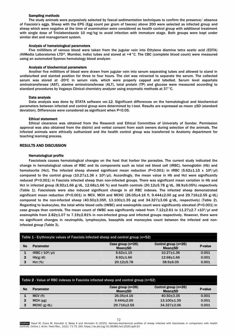
72 Citation: Yesuf M, Erara M, Kenubih A, Belay A and Ahmedin N (2020). Hemato-biochemical profiles of sheep infected with fasciolosis in comparison with health
controls. Online J. Anim. Feed Res., 10(2): 71-75. DOI: https://dx.doi.org/10.36380/scil.2020.ojafr10
Sampling methods
The study animals were purposively selected by faecal sedimentation techniques to confirm the presence/ absence
of Fasciola’s eggs. Sheep with the EPG (Egg count per gram of faeces) above 200 were selected as infected group and
sheep which were negative at the time of examination were considered as health control group with additional treatment
with single dose of Triclabendazole 10 mg/kg to avoid infection with immature stage. Both groups were kept under
similar diet and management system.
Analysis of hematological parameters
Five milliliters of venous blood were taken from the jugular vein into Ethylene diamine tetra acetic acid (EDTA)
(HiMedia Laboratories LTD®, Mumbai, India) tubes and stored at +4°C. The CBC (complete blood count) were measured
using an automated Sysmex hematology blood analyzer.
Analysis of biochemical parameters
Another five milliliters of blood were drawn from jugular vein into serum separating tubes and allowed to stand in
undisturbed and slanted position for three to four hours. The clot was retracted to separate the serum. The collected
serum was stored at -20oC in serum vials, which were properly capped and labelled. Serum level aspartate
aminotransferase (AST), alanine aminotransferase (ALT), total protein (TP) and glucose were measured according to
standard procedures by Vegasys Clinical chemistry analyzer using enzymatic methods at 37°C.
Data analysis
Data analysis was done by STATA software ver.12. Significant differences on the hematological and biochemical
parameters between infected and control group were determined by t-test. Results are expressed as mean ±SD (standard
deviation). Differences were considered as significant when P<0.05.
Ethical statement
Ethical clearance was obtained from the Research and Ethical Committee of University of Gondar. Permission
approval was also obtained from the district and verbal consent from each owners during selection of the animals. The
infected animals were ethically euthanized and the health control group was transferred to Anatomy department for
teaching learning process.
RESULTS AND DISCUSSION
Hematological profile
Fasciolosis causes hematological changes on the host that harbor the parasites. The current study indicated the
change in hematological values of RBC and its components such as total red blood cell (tRBC), hemoglobin (Hb) and
hemotocrite (Hct). The infected sheep showed significant mean reduction (P<0.001) in tRBC (5.62±1.15 x 10⁶/µl)
compared to the control group (10.27±1.36 x 10⁶/µl). Accordingly, the mean value in Hb and Hct were significantly
reduced (P<0.001) in Fasciola infected sheep than non-infected groups. There was significant mean variation in Hb and
Hct in infected group (8.92±1.66 g/dL, 12.68±1.66 %) and health controls (20.12±5.78 g/dL, 38.9±6.05%) respectively
(Table 1). Fasciolosis were also induced significant change in all RBC indexes. The infected sheep demonstrated
significant mean reduction (P<0.001) in MCV, MCH and MCHC (26.05±4.16 fl, 9.444±2.00 pg and 29.716±2.55 g/dL)
compared to the non-infected sheep (40.50±3.35fl, 13.100±1.35 pg and 34.327±3.06 g/dL, respectively) (Table 2).
Regarding to leukocytes, the total white blood cells (tWBC) and eosinophils count were significantly elevated (P<0.001) in
case groups than controls. The mean count of tWBC was significantly raised from 7.12±2.61 to 11.27±2.7 x10³/µl and
eosinophils from 2.82±1.07 to 7.19±2.81% in non-infected group and infected groups respectively. However, there were
no significant changes in neutrophils, lymphocytes, basophils and monocytes count between the infected and non-
infected group (Table 3).
Table 1 - Erythrocyte values of Fasciola infected sheep and control group (n=52)
No Parameter Case group (n=26)
Mean±SD
Control group (n=26)
Mean±SD P-value
1 tRBC (/106/µl) 5.62±1.15 10.27±1.36 0.001
2 Hb(g/dl) 8.92±1.66 12.68±1.66 0.001
3 Hct (%) 20.12±5.78 38.9±6.05 0.001
Table 2 - Value of RBC indexes in Fasciola infected sheep and control group (n=52)
No Parameter Case group (n=26)
Mean±SD
Control group (n=26)
Mean±SD P-value
1 MCV (fl) 26.05±4.16 40.50±3.35 0.001
2 MCH (pg) 9.444±2.00 13.100±1.35 0.001
3 MCHC (g/dL) 29.716±2.55 34.327±3.06 0.001

73 Citation: Yesuf M, Erara M, Kenubih A, Belay A and Ahmedin N (2020). Hemato-biochemical profiles of sheep infected with fasciolosis in comparison with health
controls. Online J. Anim. Feed Res., 10(2): 71-75. DOI: https://dx.doi.org/10.36380/scil.2020.ojafr10
Biochemical test results
The biochemical analysis of the present study showed the serum level of AST and ALT were significantly increased
(P<0.001) in sheep naturally infected with Fasciolosis than the control group. The mean value of serum AST and ALT in
animals infected with Fasciolosis were (125.92±20.71 and 34.73±11.97 IU/L) indicated higher elevation compared to
the mean value in health controls (77.04±13.96 and 23.69±7.37 IU/L) respectively (Table 4). In contrast, the total protein
and glucose determination revealed there were significant mean reduction (P<0.001) in total protein and glucose in case
group as compared to the controls. The mean reduction in total protein value from 7.33±1.06 to 5.17±1.05 g/dL and
glucose from 32.62±9.48 to 21.15±11.08 mg/dL were observed in comparison between health control and infected
groups respectively (Table 4).
Table 3 - Leukocytes values of Fasciola infected sheep and control group (n=52)
No Parameter Case group (n=26)
Mean±SD
Control group (n=26)
Mean±SD P-value
1 WBC (103/µl) 11.27±2.7 7.12±2.61 0.001
2 Neutrophils % 65.65±6.33 68.64±3.78 0.73
3 Lymphocytes % 21.03±4.81 20.43±3.67 0.61
4 Basophils % 0.77±0.48 0.77±0.60 0.98
5 Eosinophil % 5.16±1.22 1.82±1.07 0.001
6 Monocytes % 7.19±2.81 8.34±0.90 0.97
Table 4 - Biochemical profiles of Fasciola infected and control sheep (n=52)
No Parameter Case group (n=26)
Mean±SD
Control group (n=26)
Mean±SD P-value
1 AST(IU/L) 125.92±20.71 77.04±13.96 0.001
2 ALT(IU/L) 34.73±11.97 23.69±7.37 0.001
3 Total protein (g/dl) 5.17±1.05 7.33±1.06 0.001
4 Glucose (mg/dl) 21.15±11.08 32.62±9.48 0.001
DISCUSSION
Liver fluke infestation in ruminants has important implications on animal health and welfare, farming economics and
food production all over the world. It causes heavy loss in production and productivity since the animals became under
stress condition.
The current study indicated various changes in hematological parameters. The tRBC, Hb, Hct, showed significant
mean reduction in infected groups than non-infected groups. This result is supported by other study conducted by Pandya
et al. (2015) who reported severe anemia in sheep infected with Fasciolosis. The lower mean values of total erythrocytes,
Hct and Hb in infected sheep might be due to hemorrhage following severe damage by extensive migration of young fluke
and blood sucking activity of the adult fluke (Radostits et al., 2007). Furthermore, the continuous drainage of iron stores
and depression of erythropoiesis due to chronic inflammation of liver were thought to be responsible for reduction in total
red blood cell and related components (Berry and Dagie, 1978; Chandra et al., 2011).
The infected sheep demonstrated significant mean reduction in MCV, MCH and MCHC compared to those of control.
The finding is in line with the result of Pandya et al., (2015) who recorded microcytic and hypochromic anemia in Fasciola
infected animals. However, the current result contrasts with the study conducted by Egbu et al. (2013) who reported high
level of MCV, MCH and MCHC in the infected group. The variation could be due to ecological and nutritional differences
that greatly affect the hematological profiles. The possible reason for microcytic and hypochromic anemia is related to
depletion of iron stores due to damage of liver parenchyma (Beesley et al., 2018).
The eosinophilia observed in the present study supported by various researchers Egbu et al. (2013); Pandya et al.
(2015) and Matanovic et al. (2007) who had similar observation. The elevation of eosinophils is an indication of body
defense mechanism against parasitic infection (Duffus et al., 1980). This is due to inflammation and reaction by secretory
antigenic substances released by fluke and cell mediated immunity (Radostits et al., 2007).
In the present study, the presence of leukocytosis is supported by previous findings reported by Sykes et al. (1980)
and Zhang et al. (2005). The net increase in total leukocytes count (WBC) in Fasciola infection may also be due to marked
eosinophilia which is associated with parasitic infection. The present study also revealed that there were certain
biochemical and enzymatic change related to Fasciolosis. The serum level of liver enzyme (ALT and AST) significantly
increased in case group than the control. The findings are in line with corresponding studies conducted by Pandya et al.
(2015); Edith et al. (2012); Yasuda (1988) and Sheikh et al. (2006) who mentioned significant increase in mean value of

74 Citation: Yesuf M, Erara M, Kenubih A, Belay A and Ahmedin N (2020). Hemato-biochemical profiles of sheep infected with fasciolosis in comparison with health
controls. Online J. Anim. Feed Res., 10(2): 71-75. DOI: https://dx.doi.org/10.36380/scil.2020.ojafr10
AST and ALT in Fasciola infected animals. The possible cause is that Fasciola can release reactive oxygen species that can
produce damage to cell wall and hepatic tissue which is responsible to the release of intracellular enzyme to the
circulation that can increase their serum level (Hodzic et al., 2013).
The significant mean reduction of serum glucose and protein level in sheep infected with Fasciolosis was observed
than the flock free of Fasciolosis in this study. This finding agreed with the report of Phiri et al. (2007), who revealed
serum glucose and protein values were lower in infected animals than normal. The lower glucose level could be because
of inhibition of hepatic glucogenic pathways due to the migration of flukes that causes severe liver pathology and death of
hepatocytes (Sharon, 2013).
Similarly, the reduction in total protein level attributed to the damage of hepatocytes by the parasite. The flukes
reside in the liver disruption the protein synthesis leading to the development of hypoproteinemia (Martina and Jozica,
2012).
DECLARATIONS
Corresponding authors
MY: [email protected]; ME: [email protected]; AK: [email protected]; AB:
[email protected]; NA: [email protected]
Authors’ contribution
All the five authors reviewed the paper and contributed in developing the content.
Availability of data
The data can be availed to the journal upon request.
Consent to publish
Not applicable
Conflict of interest
The authors declare they have no competing of interests.
Acknowledgement
The authors would wish to acknowledge University of Gondar, Office of Vice President for Research and Community
service and College of Veterinary Medicine and Animal Sciences for their support through the whole process of developing
this publication.
REFERENCES
Anosike J, Opara C and Okoli I. (2005). Prevalence of parasitic helminthes among ruminants in Etiti area of Imo State,
Nigeria. Animal Production Research Advances, 1:13-19.
DOI: http://dx.doi.org/10.4314/apra.v1i1.36285
Beesley NJ, Caminade C, Charlier J, Flynn RJ, Hodgkinson JE, Martinez-Moreno A, Martinez-Valladares M, Perez J, Rinaldi L,
Williams D (2018). Fasciola and fasciolosis in ruminants in Europe: Identifying research needs. Transboundary and
emerging diseases, 65 Suppl 1(Suppl 1): 199–216. DOI: http://dx.doi.org/10.1111/tbed.1268
Behm CA and Sangster NC (2006). Pathology, pathophysiology and clinical aspects, In: Fasciolosis, ALTON Journal
parasitology, CABI Publishing, Wallingford Oxon, UK, Pp.185-224. https://researchers.anu.edu.au/researchers
Berry CI and Dargie JD (1978). Pathophysiology of ovine Fasciolasis: the influence of dietary protein and iron on the
erythrokinetics of sheep experimentally infected with Fasciola Hepatica. Veterinary Parasite, 4:27-339. DOI:
http://dx.doi.org/0.1016/0304-4017(78)90019-5
Chandra D, Singh K, Rathore R, Raina O and Varghese A (2011). Acute Fasciolosis in cattle and buffaloes in Bareilly
district. Indian Journal Veterinary Pathology, 35(2):133-135. http://agris.fao.org/agris-
search/search.do?recordID=IN2012000072 ISSN: 0250-4758
Craig AM, Pearson EG and Rowe KT (2002). Serum bile-acid concentrations in clinically normal cattle comparison by type,
age, and stage of lactation. American Journal of Veterinary Research, 53(10):1784 - 1786. DOI: http://dx.doi.org/10.1111/j.1939-1676.1994.tb03263
Doaa FT, Soliman EK and Abd El- khalek TM (2007). Effect of Fascioliasis on hematological, serum biochemical and
histopathological changes in sheep. Egyptian Journal of Sheep and Goat Sciences, 2(2):15–34.
https://www.ncbi.nlm.nih.gov/pmc/articles/PMC3655246/
Duffus W, Torne K and Oliver R (1980). Killing of juvenile Fasciola hepatica by purified bovine eosinophils proteins. Clinical
Experimental Immunology, 40:336-344.
https://www.researchgate.net/publication/15759643_Killing_of_juvenile_Fasciola_hepatica_by_purified_bovine_eos
inophil_proteins
Edith R, Godara R, Sharma L and Thilagar M (2012). Fasciola gigantica induced adrenal dysfunctions and its
pathophysiological Significance in riverine buffaloes (Bubalusbubalis). Buffalo Bulletin, 31(1):51-62.
https://www.net/publication/286698885
Egbu FM, Ubachukwu PO and Okoye IC (2013). Haematological changes due to bovine Fascioliosis. African Journal
Biotechnology, 12(15):1828-1835. DOI: http://dx.doi.org/10.5897/AJB12.2716

75 Citation: Yesuf M, Erara M, Kenubih A, Belay A and Ahmedin N (2020). Hemato-biochemical profiles of sheep infected with fasciolosis in comparison with health
controls. Online J. Anim. Feed Res., 10(2): 71-75. DOI: https://dx.doi.org/10.36380/scil.2020.ojafr10
Grunwaldt E, Guevara J, Estevez O, Vicente A, Rousselle H, Alcunten N, Aguerregaray D and Stasi C (2005). Biochemical
and haematological measurements in beef cattle in Mendoza plain rangelands (Agentina). Tropical Animal Health and
Production, 37(6): 527 –540. https://www.ncbi.nlm.nih.gov/pubmed/16248224
Hodzic A, Zuko A, Avdic R, Alic A, Omeragic J and Jazic A (2013). Influence of Fasciola hepatica on serum biochemical
parameters and vascular and biliary system of sheep liver. Iraq Journal Parasitology, 8(1): 92-98.
https://www.ncbi.nlm.nih.gov/pmc/articles/PMC3655246/
Martina K and Jožica J (2012). Values of Blood Variables in Calves, A Bird's-Eye View of Veterinary Medicine, Dr. Carlos C.
Perez-Marin (Ed.), ISBN: 978-953-51-0031-7, In Tech, Available from: http://www.intechopen.com/books/a-bird-s-eye-
view-of-veterinary-medicine/values-of-blood-variables-in-calves
Mas-Coma S, Valero M and Bargues M (2009). Fasciola, lymnaeids and Human Fascioliasis, with a global overview on
disease transmission, epidemiology, evolutionary genetics, molecular epidemiology and control. Advanced
Parasitology, 69: 41–146. DOI: http://dx.doi.org/10.1016/S0065-308X(09)69002-3
Matanovic K, Severin F, Martinkovic M, Simpraga Z and Janicki J (2007). Hematological and biochemical changes in
organically farmed sheep naturally infected with Fasciola hepatica. Parasitology Research, 101:1658–1659. DOI: http://dx.doi.org/10.1007/s00436-007-0709-2
Milbourne EA and Howell MJ (1990). Eosinophil response to F. hepatica in rodents. International journal for parasitology,
20:705-708. DOI: http://dx.doi.org/10.1016/0020-7519(90)90135-A
Pandya SS, Hasnani JJ, Patel PV, Dave CJ and Ravi SN (2015). Study on Haemato-Biochemical Alterations occurred in
Fasciola spp. infected Buffaloes. International Journal of Multidisciplinary Research and Development, 2(3):756-759.
DOI: http://dx.doi.org/10.1080/09712119.2011.607703
Phiri IK, Phiri AM and Harrison LJ (2007). The serum glucose and B- hydro-xybutyrate levels in sheep with experimental
Fasciola hebatica and Fasciola gigantica infection. Veterinary Parasitology, 143:287-293.
https://www.ncbi.nlm.nih.gov/pubmed/17011709
Radostits OM, Gay CC, Blood DC, Hinchliff KW (2007). A textbook of the diseases of cattle, sheep, pigs, goats and horses.
(10th ed.). W.B. Saunders, London. https://www.elsevier.com/books/veterinary-medicine/radostits/978-0-7020-2777-
2
Sharon A (2013). Enzyme activity in hepatic disease in small animals. In: Merck Sharp and Dohme Corp., a subsidiary of
Merck and Co., inc., White house Station, N.J., U.S.A. pp. 145. https://www.msdvetmanual.com/digestive-
system/hepatic-diseasein
Sheikh GN, Qadri SG, Willayat MM and Gunjan HD (2006). Biochemical profile of cattle naturally infected with Fasciola
species. Journal Veterinary Parasitology, 20(1):41-43. DOI: http://dx.doi.org/10.3923/pjbs.2007.2756.2759
Sykes A, Coop R and Rushton B. (1980). Chronic subclinical Fascioliasis in sheep: effects on food intake, food utilization
and blood constituents. Research Veterinary Science, 28:63–70. DOI: http://dx.doi.org/10.1016/S0034-
5288(18)32774-7
Yasuda J (1988). Overview: Fascioliasis and liver damage. Japan Journal veterinary Science, 50: 71.
https://www.sciencedirect.com/topics/fascioliasis
Zhang Y, Moreau E, Hope J, Howard C, Huang W and Chauvin A (2005). Fasciola hepatica and Fasciola gigantica:
comparison of cellular response to experimental infection in sheep. Experimental Parasitology. 111:154–59. DOI: http://dx.doi.org/10.1016/j.exppara.2005.06.005
g | P a g e
OJAFR EndNote Style | Manuscript Template | Sample Articles | Declaration form
Manuscripts as Original Research Paper, Review, Short Communication and Case Reports are invited for rapid peer-review publishing in the Online Journal of Animal and Feed Research (ISSN 2228-7701). Papers can be in any relevant fields of Animal Sciences (Animal Nutrition, Physiology, Reproduction, Genetics and Breeding,
Behavior, Health, Husbandry and its economic, Animal products and Veterinary medicines of domestic animals) and relative topics.
The journal does encourage papers with emphasis on the nutritive value and utilization of feeds that is depended to methods o f Improvement, Assessment, Conserving and Processing feeds, Agronomic and climatic factors, Metabolic, Production, Reproduction
and Health responses to dietary inputs (e.g., Feeds, Feed Additives, Specific Feed Components, Mycotoxins). Also, Mathematica l models relating directly to animal-feed interactions, Analytical and experimental methods for Feed Evaluation as well as Animal
Production studies with a focus on Animal Nutrition that do have link to a feed (Food Science and Technology) are acceptable relative topics for OJAFR.
Submission The manuscripts should be submitted using our online submission system. For facile submission, please embed all figures and tables at the end of the manuscript to become one single file for submission. Once submission is complete, the system will
generate a manuscript ID and password sent to author's contact email. If you have any difficulty in submitting the manuscript, kindly send via email: [email protected]. All manuscripts must be checked (by English native speaker) and submitted in English for
evaluation in totally confidential and impartial way. Supplementary information:
Author guidelines are specific for each journal. Our MS Word template can assist you by modifying your page layout, text
formatting, headings, title page, image placement, and citations/references such that they agree with the guidelines of journal. If
you believe your article is fully edited per journal style, please use our Word template before submission. Supplementary materials may include figures, tables, methods, videos, and other materials. They are available online linked to the
original published article. Supplementary tables and figures should be labeled with a "S", e.g. "Table S1" and "Figure S1". The maximum file size for supplementary materials is 10MB each. Please keep the files as small as possible to avoid the frustrations
experienced by readers with downloading large files.
Submission to the Journal is on the understanding that: 1. The article has not been previously published in any other form and is not under consideration for publication elsewhere;
2. All authors have approved the submission and have obtained permission for publish work. 3. Researchers have proper regard for conservation and animal welfare considerations. Attention is drawn to the 'Guidelines for the
Treatment of Animals in Research and Teaching'. Any possible adverse consequences of the work for populations or individual organisms must be weighed against the possible gains in knowledge and its practical applications. If the approval of an ethics
committee is required, please provide the name of the committee and the approval number obtained.
Ethics Committee Approval Experimental research involving human or animals should have been approved by author's institutional review board or ethics
committee. This information can be mentioned in the manuscript including the name of the board/committee that gave the approval. Investigations involving humans will have been performed in accordance with the principles of Declaration of Helsinki.
And the use of animals in experiments will have observed the Interdisciplinary Principles and Guidelines for the Use of Animals in Research, Testing, and Education by the New York Academy of Sciences, Ad Hoc Animal Research Committee. If the manuscript
contains photos or parts of photos of patients, informed consent from each patient should be obtained. Patient's identities and privacy should be carefully protected in the manuscript.
Presentation of the article
Main Format: First page of the manuscripts must be properly identified by the title and the name(s) of the author(s). It should be typed in Times
New Roman (font sizes: 12pt in capitalization for the title and the main text, double spaced, in A4 format with 2cm margins. All pages and lines of the main text should be numbered consecutively throughout the manuscript. The manuscript must be saved in a
.doc format, (not .docx files). Abbreviations in the article title are not allowed except the well-known ones.
Manuscripts should be arranged in the following order: a. TITLE (brief, attractive and targeted)
b. Name(s) and Affiliation(s) of author(s) (including post code) and corresponding E-mail
c. ABSTRACT
d. Key words (separate by semicolons; or comma,)
e. Abbreviations (used in the manuscript)
f. INTRODUCTION;
g. MATERIALS AND METHODS
h. RESULTS
i. DISCUSSION
j. CONCLUSION
k. DECLARATIONS
l. REFERENCES
m. Tables;
n. Figures The sections "RESULTS AND DISCUSSION" can be presented jointly. The sections "DISCUSSION AND DISCUSSION" can be presented jointly.
Instructions for Authors

h | P a g e
Article Sections Format: Title should be a brief phrase describing the contents of the paper. Title Page should include full names and affiliations of the author(s), the name of the corresponding author along with phone and e-mail information. Present address(es) of author(s) should
appear as a footnote. Abstract should be informative and completely self-explanatory, briefly present the topic, state the scope of the experiments, indicate significant data, and point out major findings and conclusions. The abstract should be 150 to 300 words in length.
Complete sentences, active verbs, and the third person should be used, and the abstract should be written in the past tense. Standard nomenclature should be used and abbreviations should be avoided. No literature should be cited. Following the abstract, about 3 to 7 key words should be listed. Introduction should provide a clear statement of the problem, the relevant literature on the subject, and the proposed approach or solution. It should be understandable to colleagues from a broad range of scientific disciplines. Materials and Methods should be complete enough to allow experiments to be reproduced. However, only truly new procedures
should be described in detail; previously published procedures should be cited, and important modifications of published procedures should be mentioned briefly. Capitalize trade names and include the manufacturer's name and address. Subheadings
should be used. Methods in general use need not be described in detail. Results should be presented with clarity and precision. The results should be written in the past tense when describing findings in the author(s)'s experiments. Previously published findings should be written in the present tense. Results should be explained, but
largely without referring to the literature. Discussion, speculation and detailed interpretation of data should not be included in the results but should be put into the discussion section. Discussion should interpret the findings in view of the results obtained in this and in past studies on this topic. State the conclusions in a few sentences at the end of the paper. The Results and Discussion sections can include subheadings, and when
appropriate, both sections can be combined. Conclusion should be brief and tight, providing a few specific tasks to accomplish: 1-Re-assert/Reinforce the Thesis; 2-Review the Main Points; 3- Close Effectively. The Conclusion section should not be similar to the Abstract content. Declarations including Ethics, Consent to publish, Competing interests, Authors' contributions, and Availability of data and
materials are necessary. Acknowledgments of persons, grants, funds, etc should be brief. Tables should be kept to a minimum and be designed to be as simple as possible. Tables are to be typed double-spaced
throughout, including headings and footnotes. Each table should be on a separate page, numbered consecutively in Arabic numerals and supplied with a heading and a legend. Tables should be self-explanatory without reference to the text. The details of
the methods used in the experiments should preferably be described in the legend instead of in the text. The same data should not be presented in both table and graph forms or repeated in the text. The Figure legends should be typed in numerical order on a separate sheet. Graphics should be prepared using applications
capable of generating high resolution GIF, TIFF, JPEG or PowerPoint before pasting in the Microsoft Word manuscript file. Use Arabic numerals to designate figures and upper case letters for their parts (Figure 1). Begin each legend with a title and include
sufficient description so that the figure is understandable without reading the text of the manuscript. Information given in legends should not be repeated in the text.
DECLARATIONS Please ensure that the sections: Ethics (and consent to participate), Consent to publish, Competing interests, Authors'
contributions, and Availability of data and materials are included at the end of your manuscript in a Declarations section. Consent to Publish
Please include a „Consent for publication‟ section in your manuscript. If your manuscript contains any individual person‟s data in any form (including individual details, images or videos), consent to publish must be obtained from that person, or in the case of
children, their parent or legal guardian. All presentations of case reports must have consent to publish. You can use your institutional consent form or our consent form if you prefer. You should not send the form to us on submission, but we may
request to see a copy at any stage (including after publication). If your manuscript does not contain any individual person‟s data, please state “Not applicable” in this section. Authors’ Contributions For manuscripts with more than one author, OJAFR require an Authors' Contributions section to be placed after the Competing
Interests section. An 'author' is generally considered to be someone who has made substantive intellectual contributions to a published study. To qualify as an author one should 1) have made substantial contributions to conception and design, or
acquisition of data, or analysis and interpretation of data; 2) have been involved in drafting the manuscript or revising it critically for important intellectual content; and 3) have given final approval of the version to be published. Each author should have
participated sufficiently in the work to take public responsibility for appropriate portions of the content. Acquisition of funding, collection of data, or general supervision of the research group, alone, does not justify authorship.
We suggest the following format (please use initials to refer to each author's contribution): AB carried out the molecular genetic studies, participated in the sequence alignment and drafted the manuscript. JY carried out the immunoassays. MT participated in
the sequence alignment. ES participated in the design of the study and performed the statistical analysis. FG conceived of the study, and participated in its design and coordination and helped to draft the manuscript. All authors read and approved the final
manuscript. For authors that equally participated in a study please write 'All/Both authors contributed equally to this work.' Contributors who do not meet the criteria for authorship should be listed in an acknowledgements section. Competing Interests Competing interests that might interfere with the objective presentation of the research findings contained in the manuscript
should be declared in a paragraph heading "Competing interests" (after Acknowledgment section and before References). Examples of competing interests are ownership of stock in a company, commercial grants, board membership, etc. If there is no
competing interest, please use the statement "The authors declare that they have no competing interests.". Online Journal of Animal and Feed Research adheres to the definition of authorship set up by the International Committee of
Medical Journal Editors (ICMJE). According to the ICMJE authorship criteria should be based on 1) substantial contributions to conception and design of, or acquisition of data or analysis and interpretation of data, 2) drafting the article or revising it critically
for important intellectual content and 3) final approval of the version to be published. Authors should meet conditions 1, 2 and 3. It is a requirement that all authors have been accredited as appropriate upon submission of the manuscript. Contributors who do
not qualify as authors should be mentioned under Acknowledgements. Change in authorship
We do not allow any change in authorship after provisional acceptance. We cannot allow any addition, deletion or change in sequence of author name. We have this policy to prevent the fraud.

i | P a g e
Acknowledgements We strongly encourage you to include an Acknowledgements section between the Authors‟ contributions section and Reference list. Please acknowledge anyone who contributed towards the study by making substantial contributions to conception, design,
acquisition of data, or analysis and interpretation of data, or who was involved in drafting the manuscript or revising it critically for important intellectual content, but who does not meet the criteria for authorship. Please also include their source(s) of funding.
Please also acknowledge anyone who contributed materials essential for the study. Authors should obtain permission to acknowledge from all those mentioned in the Acknowledgements. Please list the source(s) of
funding for the study, for each author, and for the manuscript preparation in the acknowledgements section. Authors must describe the role of the funding body, if any, in study design; in the collection, analysis, and interpretation of data; in the writing
of the manuscript; and in the decision to submit the manuscript for publication. Data Deposition
Nucleic acid sequences, protein sequences, and atomic coordinates should be deposited in an appropriate database in time for the accession number to be included in the published article. In computational studies where the sequence information is unacceptable
for inclusion in databases because of lack of experimental validation, the sequences must be published as an additional file with the article.
References
An OJAFR reference style for EndNote may be found here. 1. All references to publications made in the text should be presented in a list with their full bibliographical description. 2. In the text, a reference identified by means of an author„s name should be followed by the date of the reference in
parentheses. When there are more than two authors, only the first author„s surname should be mentioned, followed by ‟et al„. In the event that an author cited has had two or more works published during the same year, the reference, both in the
text and in the reference list, should be identified by a lower case letter like ‟a„ and ‟b„ after the date to distinguish the works. 3. References in the text should be arranged chronologically (e.g. Kelebeni, 1983; Usman and Smith, 1992 and Agindotan et al.,
2003). 'et al.' should not be italic. The list of references should be arranged alphabetically on author's surnames, and
chronologically per author. If an author's name in the list is also mentioned with co-authors, the following order should be
used: Publications of the single author, arranged according to publication dates - publications of the same author with one co-author - publications of the author with more than one co-author. Publications by the same author(s) in the same year
should be listed as 1992a, l992b, etc. 4. Names of authors and title of journals, published in non-Latin alphabets should be transliterated in English. 5. A sample of standard reference is "1th Author surname A, 2th Author surname B and 3th Author surname C (2013). Article title
should be regular, in sentence case form, and 9 pt. Online Journal of Animal and Feed Research, Volume No. (Issue No.): 00-00." (Journal titles should be full and not italic.)
6. If available please add DOI numbers or the link of articles at the end of each reference. Examples (at the text): Abayomi (2000), Agindotan et al. (2003), (Kelebeni, 1983), (Usman and Smith, 1992), (Chege, 1998; Chukwura, 1987a,b; Tijani, 1993,1995), (Kumasi et al., 2001).
Examples (at References section): a) For journal:
Lucy MC (2000). Regulation of ovarian follicular growth by somatotropin and insulin- like growth factors in cattle. Journal of Dairy Science, 83: 1635-1647. Link, DOI
Kareem SK (2001). Response of albino rats to dietary level of mango cake. Journal of Agricultural Research and Development, Pp. 31-38. Link, DOI Chikere CB, Omoni VT and Chikere BO (2008). Distribution of potential nosocomial pathogens in a hospital environment. African Journal of
Biotechnology, 7: 3535-3539. Link, DOI b) For symposia reports and abstracts:
Cruz EM, Almatar S, Aludul EK and Al-Yaqout A (2000). Preliminary Studies on the Performance and Feeding Behaviour of Silver Pomfret (Pampus argentens euphrasen) Fingerlings fed with Commercial Feed and Reared in Fibreglass Tanks. Asian Fisheries Society Manila, Phi lippine, 13: 191-199. Link, DOI
c) For edited symposia, special issues, etc., published in a journal: Korevaar H (1992). The nitrogen balance on intensive Dutch dairy farms: a review. In: A. A. Jongebreur et al. (Editors), Effects of Cattle and Pig
Production Systems on the Environment: Livestock Production Science, 31: 17-27. Link, DOI d) For books:
AOAC (1990). Association of Official Analytical Chemists. Official Methods of Analysis, 15th Edition. Washington D.C. pp. 69-88. Pelczar JR, Harley JP, Klein DA (1993). Microbiology: Concepts and Applications. McGraw-Hill Inc., New York, pp. 591-603. Link, DOI
e) Books, containing sections written by different authors: Kunev M (1979). Pig Fattening. In: A. Alexiev (Editor), Farm Animal Feeding. Vol. III. Feeding of Different Animal Species, Zemizdat, Sofia, p. 233-
243 (Bg). Link, DOI
In referring to a personal communication the two words are followed by the year, e.g. (Brown, J. M., personal communication,
1982). In this case initials are given in the text. Where available, URLs for the references should be provided.
Formulae, numbers and symbols 1. Typewritten formulae are preferred. Subscripts and superscripts are important. Check disparities between zero (0) and the
letter 0, and between one (1) and the letter I. 2. Describe all symbols immediately after the equation in which they are first used. 3. For simple fractions, use the solidus (/), e.g. 10 /38. 4. Equations should be presented into parentheses on the right-hand side, in tandem. 5. Levels of statistical significance which can be used without further explanations are *P < 0.05, **P < 0.01, and ***P<0.001. 6. In the English articles, a decimal point should be used instead of a decimal comma. 7. Use Symbol fonts for "±"; "≤" and "≥" (avoid underline). 8. In chemical formulae, valence of ions should be given, e.g. Ca2+ and CO32-, not as Ca++ or CO3. 9. Numbers up to 10 should be written in the text by words. Numbers above 1000 are recommended to be given as 10 powered
x. 10. Greek letters should be explained in the margins with their names as follows: Αα - alpha, Ββ - beta, Γγ - gamma, Γδ - delta,
Δε - epsilon, Εδ - zeta, Ζε - eta, Θζ - theta, Ηη - iota, Θθ - kappa, Ιι - lambda, Κκ - mu, Λλ - nu, Μμ - xi, Νν - omicron, Ξπ -
pi, Οξ - rho, Πζ - sigma, Ρη - tau, υ - ipsilon, Φθ - phi, Σχ - chi, Τψ - psi, Υω - omega. Please avoid using math equations in
Word whenever possible, as they have to be replaced by images in xml full text.

j | P a g e
Abbreviations
Abbreviations should be presented in one paragraph, in the format: "term: definition". Please separate the items by ";". E.g. ANN: artificial neural network; CFS: closed form solution; ....
Graphical Abstract: Authors of accepted articles should provide a graphical abstract (a beautifully designed feature figure) to represent the paper aiming to catch the attention and interest of
readers. Graphical abstract will be published online in the table of content. The graphical abstract should be colored, and kept within an area of 12 cm (width) x 6 cm
(height) or with similar format. Image should have a minimum resolution of 300 dpi and line art 1200dpi.
Note: Height of the image should be no more than the width. Please avoid putting too much information into the graphical abstract as it occupies only a small space. Authors
can provide the graphical abstract in the format of PDF, Word, PowerPoint, jpg, or png, after a manuscript is accepted for publication.
If you have decided to provide a Professional Graphical Abstract, please click here.
Review/Decisions/Processing
Firstly, all manuscripts will be checked by Docol©c, a plagiarism finding tool. The received papers with plagiarism rate of more than 40% will be rejected. Manuscripts that are judged to be of insufficient quality or unlikely to be competitive enough for
publication will be returned to the authors at the initial stage. The remaining manuscripts go through a double-blind review process by two reviewers selected by section editor (SE) or deputy SE of OJAFR, who are research workers specializing in the relevant field
of study. We always try to avoid delays in the reviewing process, but it relies on the time and cooperation of the referees that
works without any remuneration, hence, it may take 2 weeks to 2 months. One unfavorable review means that the paper will not
be published and possible decisions are: accept as is, minor revision, major revision, or reject. The corresponding authors should submit back their revisions within 14 days in the case of minor revision, or 30 days in the case of major revision.
To submit a revision please click here, fill out the form, and mark " Revised", mention the article code (for example OJAFR-1108), attach the revision (MS word) and continue submission. Manuscripts with significant results are typically reviewed and published at the highest priority. After review and editing the article, a final formatted proof is sent to the corresponding author
once again to apply all suggested corrections during the article process. The editor who received the final revisions from the
corresponding authors shall not be hold responsible for any mistakes shown in the final publication.
The submissions will be processed free of charge for invited authors, authors of hot papers, and corresponding authors who are editorial board members of the Online Journal of Animal and Feed Research. This journal encourages the academic institutions in
low-income countries to publish high quality scientific results, free of charges.
Plagiarism: There is an instant policy towards plagiarism (including self-plagiarism) in our journals. Manuscripts are screened for plagiarism by Docol©c, before or during publication, and if found they will be rejected at any stage of processing. Date of issue
All accepted articles are published bimonthly around 25th of January, March, May, July, September and November, each year in full text on the Internet.
The OA policy Online Journal of Animal and Feed Research is an Open Access journal which means that all content is freely available without charge to the user or his/her institution. Users are allowed to read, download, copy, distribute, print, search, or link to the full
texts of the articles, or use them for any other lawful purpose, without asking prior permission from the publisher or the author.
This is in accordance with the BOAI definition of Open Access. Submission Preparation Checklist Authors are required to check off their submission's compliance with all of the following items, and submissions may be returned to authors that do not adhere to the following guidelines: The submission has not been previously published, nor is it before another journal for consideration (or an explanation has been
provided in -Comments to the Editor). The submission file is in Microsoft Word, RTF, or PDF document file format. Where available, URLs for the references have been provided. The text is double-spaced; uses a 12-point font; and all illustrations, figures, and tables are placed within the text at the
appropriate points, rather than at the end. The text adheres to the stylistic and bibliographic requirements outlined in the Author Guidelines.
ABOUT US | CONTACT US | PRIVACY POLICY
Editorial Offices: Atatürk University, Erzurum 25100, Turkey
University of Manitoba, Winnipeg, Manitoba R3T 2N2, Canada
University of Maragheh, East Azerbaijan, Maragheh 55136, Iran
Homepage: www.science-line.com Phone: +98 914 420 7713 (Iran); +90 538 770 8824 (Turkey); +1 204 8982464 (Canada)
Emails: [email protected] ; [email protected]

Scienceline Publication Ltd is a limited liability non-profit non-stock corporation incorporated in Turkey.
Scienceline journals that concurrently belong to many societies, universities and research institutes, publishes internationally peer-reviewed open access articles and believe in sharing of new scientific knowledge and vital research in the fields of life and natural sciences, animal sciences, engineering, art, linguistic, management, social and economic sciences all over the world. Scienceline journals include:
. Online Journal of Animal and Feed Research
Journal of Civil Engineering and Urbanism
Journal of Life Sciences and Biomedicine
Asian Journal of Medical and Pharmaceutical Researches
ISSN 2228-7701; Bi-monthly
View Journal l Editorial Board Email: [email protected] Submit Online >>
ISSN 2252-0430; Bi-monthly View Journal I Editorial Board Email: [email protected]
Submit Online >>
ISSN: 2251-9939; Bi-monthly
View Journal I Editorial Board Email: [email protected] Submit Online >>
ISSN: 2322-4789; Quarterly
View Journal I Editorial Board Email: [email protected] Submit Online >>
. Journal of World's Poultry
Research
World's Veterinary Journal
Journal of Educational and Management Studies
Journal of World's Electrical Engineering and Technology
ISSN: 2322-455X; Quarterly
View Journal I Editorial Board Email: [email protected] Submit Online >>
ISSN: 2322-4568; Quarterly View Journal I Editorial Board Email: [email protected]
Submit Online >>
ISSN: 2322-4770; Quarterly
View Journal I Editorial Board Email: [email protected] Submit Online >>
ISSN: 2322-5114; Irregular View Journal I Editorial Board Email: [email protected]
Submit Online >> .
Journal of Art and Architecture Studies
Asian Journal of Social and Economic Sciences
Journal of Applied Business and Finance Researches
Scientific Journal of Mechanical and Industrial Engineering
ISSN: 2383-1553; Irregular View Journal I Editorial Board Email: [email protected]
Submit Online >>
ISSN: 2383-0948; Quarterly View Journal I Editorial Board Email: [email protected]
Submit Online >>
ISSN: 2382-9907; Quarterly View Journal I Editorial Board Email: [email protected]
Submit Online >>
ISSN: 2383-0980; Quarterly
View Journal I Editorial Board Email: [email protected] Submit Online >>
.
Copyright © 2020.
Scienceline Journals Email: [email protected]
SCIENCELINE PUBLISHING CORPORATION