Voltage-gated potassium channels in multiple sclerosis: Overview and new implications for treatment...
11
111 JRRD JRRD Volume 43, Number 1, Pages 111–122 January/February 2006 Journal of Rehabilitation Research & Development Voltage-gated potassium channels in multiple sclerosis: Overview and new implications for treatment of central nervous system inflammation and degeneration Susan I. V. Judge, PhD; 1–3* Jennifer M. Lee, BS; 2 Christopher T. Bever Jr, MD; 1–3 Paul M. Hoffman, MD 1–3 1 Department of Veterans Affairs (VA) Multiple Sclerosis Center of Excellence East, Baltimore, MD; 2 VA Maryland Health Care System, Baltimore VA Medical Center, Baltimore, MD; 3 Department of Neurology, University of Mary- land School of Medicine, Baltimore, MD Abstract—Inflammatory tissue damage and the presence of reactive immunocompetent T lymphocytes, macrophages, microglia, and dendritic cells (DCs) are characteristic features in the human chronic inflammatory demyelinating disease, multiple sclerosis (MS). Together, these cells orchestrate the inflammation and immunopathogenesis underlying the MS autoimmune disease processes and all up-regulate the same voltage-gated potassium (K v ) channel, K v 1.3, when fully acti- vated. Only microglia, which mediate central nervous system (CNS) inflammatory processes (possibly playing a dual role of CNS protection and mediation of neuroinflammation/ neurode- generation), and DC, which are pivotal to the induction of T cell responses, express the distinct K v 1.5 prior to K v 1.3 up- regulation. Although the precise functional roles of first K v 1.5 and then K v 1.3 channels are unclear, their differential expres- sion is likely a common mechanism used by both microglia and DC, revealing K v 1.5 (in addition to K v 1.3) as a potentially important target for the development of new immunomodula- tory therapies in MS. Key words: 3,4 diaminopyridine, 4-aminopyridine, blood- brain barrier, central nervous system, dendritic cells, experi- mental allergic encephalomyelitis, multiple sclerosis, murine leukemia virus, voltage-gated potassium channels. INTRODUCTION Multiple sclerosis (MS) is a chronic and progressive neurodegenerative disease for which no cure exists. Con- sidered a primary inflammatory disease of central nervous system (CNS) white matter, pathological lesions in MS are characterized by inflammatory demyelination with relative sparing of axons [1], perivascular/parenchymal infiltration of T lymphocytes (T cells) and macrophages Abbreviations: 3,4-DAP = 3,4 diaminopyridine, 4-AP = 4- aminopyridine, BBB = blood-brain barrier, CD = clusters of differentiation, CNS = central nervous system, CSF = cere- brospinal fluid, DC = dendritic cell, EAE = experimental aller- gic encephalomyelitis, EAN = experimental allergic neuritis, IL = interleukin, K ir = inward rectifying potassium (channel), K v = voltage-gated potassium channel, MBP = myelin basic protein, MHC II = major histocompatibility class II, MS = multiple sclerosis, MuLV = murine leukemia virus, NADPH = nicotinamide adenosine dinucleotide phosphate, NO = nitric oxide, NOS = NO synthase, PNS = peripheral nervous system, T cells = T lymphocytes. * Address all correspondence to Susan I. V. Judge, PhD; Department of Neurology, University of Maryland School of Medicine, BRB 12-040, 655 West Baltimore Street, Balti- more, Maryland 21201; 410-706-4481; fax: 410-706-0186. Email: [email protected] DOI: 10.1682/JRRD.2004.09.0116
-
Upload
independent -
Category
Documents
-
view
2 -
download
0
Transcript of Voltage-gated potassium channels in multiple sclerosis: Overview and new implications for treatment...

JRRDJRRD Volume 43, Number 1, Pages 111–122
January/February 2006
Journal of Rehabil itation Research & Development
Voltage-gated potassium channels in multiple sclerosis: Overview and new implications for treatment of central nervous system inflammation and degeneration
Susan I. V. Judge, PhD;1–3* Jennifer M. Lee, BS;2 Christopher T. Bever Jr, MD;1–3 Paul M. Hoffman, MD1–31Department of Veterans Affairs (VA) Multiple Sclerosis Center of Excellence East, Baltimore, MD; 2VA Maryland Health Care System, Baltimore VA Medical Center, Baltimore, MD; 3Department of Neurology, University of Mary-land School of Medicine, Baltimore, MD
Abstract—Inflammatory tissue damage and the presence ofreactive immunocompetent T lymphocytes, macrophages,microglia, and dendritic cells (DCs) are characteristic featuresin the human chronic inflammatory demyelinating disease,multiple sclerosis (MS). Together, these cells orchestrate theinflammation and immunopathogenesis underlying the MSautoimmune disease processes and all up-regulate the samevoltage-gated potassium (Kv) channel, Kv1.3, when fully acti-vated. Only microglia, which mediate central nervous system(CNS) inflammatory processes (possibly playing a dual role ofCNS protection and mediation of neuroinflammation/ neurode-generation), and DC, which are pivotal to the induction ofT cell responses, express the distinct Kv1.5 prior to Kv1.3 up-regulation. Although the precise functional roles of first Kv1.5and then Kv1.3 channels are unclear, their differential expres-sion is likely a common mechanism used by both microgliaand DC, revealing Kv1.5 (in addition to Kv1.3) as a potentiallyimportant target for the development of new immunomodula-tory therapies in MS.
Key words: 3,4 diaminopyridine, 4-aminopyridine, blood-brain barrier, central nervous system, dendritic cells, experi-mental allergic encephalomyelitis, multiple sclerosis, murineleukemia virus, voltage-gated potassium channels.
INTRODUCTION
Multiple sclerosis (MS) is a chronic and progressiveneurodegenerative disease for which no cure exists. Con-sidered a primary inflammatory disease of central nervoussystem (CNS) white matter, pathological lesions in MSare characterized by inflammatory demyelination withrelative sparing of axons [1], perivascular/parenchymalinfiltration of T lymphocytes (T cells) and macrophages
Abbreviations: 3,4-DAP = 3,4 diaminopyridine, 4-AP = 4-aminopyridine, BBB = blood-brain barrier, CD = clusters ofdifferentiation, CNS = central nervous system, CSF = cere-brospinal fluid, DC = dendritic cell, EAE = experimental aller-gic encephalomyelitis, EAN = experimental allergic neuritis,IL = interleukin, Kir = inward rectifying potassium (channel),Kv = voltage-gated potassium channel, MBP = myelin basicprotein, MHC II = major histocompatibility class II, MS =multiple sclerosis, MuLV = murine leukemia virus, NADPH =nicotinamide adenosine dinucleotide phosphate, NO = nitricoxide, NOS = NO synthase, PNS = peripheral nervous system,T cells = T lymphocytes.*Address all correspondence to Susan I. V. Judge, PhD;Department of Neurology, University of Maryland Schoolof Medicine, BRB 12-040, 655 West Baltimore Street, Balti-more, Maryland 21201; 410-706-4481; fax: 410-706-0186.Email: [email protected]: 10.1682/JRRD.2004.09.0116
111
112
JRRD, Volume 43, Number 1, 2006
[1–3], and proliferation and activation of resident micro-glia and astrocytes [4], as well as peripheral dendritic cells(DCs) [5]. In addition to inflammation and demyelination(white and gray matter), axonal damage and loss are nowrecognized as contributing to irreversible deficits in MS[6]. Clinical symptoms include blurred vision, unstablebalance, poor coordination, tremors, numbness, andslurred speech, for which the underlying physiologicalimpairment is believed to be conduction block arisingfrom demyelination and inflammation.
Current approaches to treating MS patients includesymptomatic treatment of neurological deficits andimmunomodulatory therapy to treat neuroinflammationand possibly limit neurodegeneration. Voltage-gatedpotassium (Kv) channels are potential targets for bothtypes of therapies. As symptomatic therapies, only tworelatively nonspecific blockers of Kv channels, 4-amino-pyridine (4-AP) and 3,4 diaminopyridine (3,4-DAP),have been tested clinically for their efficacy in the treat-ment of patients with MS [7–15]. To date, in vivo immu-nosuppressive treatments that use nonspecific (4-AP andquinidine) and various highly selective Kv channel block-ers (margatoxin, correolide, kaliotoxin, ShK, and Sh-Dap22) have been restricted to miniswine [16–17] androdent experimental allergic encephalomyelitis (EAE)[18–20] animal models for MS.
The first study implicating a Kv blocker (quinidine)as a successful therapeutic treatment in an inflammatorydemyelinating disease was an animal model performedin rats with experimental allergic neuritis (EAN), anaccepted animal model for the human Guillain-Barresyndrome that is the peripheral nervous system (PNS)counterpart of EAE in the CNS. Mix and colleaguesdemonstrated that injecting EAN rats with quinidine ame-liorated symptoms of clinical EAN [21]. These neuro-logical benefits were accompanied with reducedinflammatory infiltrates in target tissue but not improvedperipheral nerve conduction, thus foreshadowing theemerging view that Kv blockers may primarily exert theirneurological benefits in MS through immunomodulatoryeffects.
TARGETING KV CHANNELS AS SYMPTOMATICTREATMENT IN MULTIPLE SCLEROSIS
The original clinical rationale for using Kv channelblockers to improve neurological function in the symp-
tomatic treatment of patients with MS stemmed fromphysiological demonstrations in the PNS in which block-ing paranodal or internodal Kv channels prolonged actionpotential and potentiated synaptic transmission [22–25].Many intact nonconducting axons in MS lesions can butdo not conduct because their safety factor for conductionis fractionally below unity [26]. The recruitment of suchaxons by simply reducing body temperature [27] orchanging serum-ionized calcium [28] raised hope thatmany more axons could be recruited pharmacologicallywith the use of Kv channel blockers. Waxman gives acurrent review of underlying disease processes and neu-ronal injury in MS [29]. Judge and Bever provide a currentreview of Kv channels as symptomatic targets in MS [30].
Although clearly beneficial, both 4-AP and 3,4-DAPare potent convulsants with narrow therapeutic windowsthat have limited their widespread clinical use in MStreatments. Toxic, epileptogenic side effects likely arisefrom the indiscriminate blockade of widely distributedand varied CNS Kv channels rather than Kv channelsalong demyelinated nerve fibers. Initially, the clinicalimprovements achieved in MS patients with 4-AP prima-rily were viewed as likely arising from blockage of Kvchannels exposed on demyelinated nodes. In an experi-mental in vitro CNS study, Perreault and Avoli showedthat seizure induction by 4-AP results from block of asynaptic channel [31]. More recently, Smith et al. under-took the first and only in vivo CNS studies in rats of 4-APon experimental demyelination [26]. Their studies indi-cated that clinical doses of 4-AP probably produced bene-ficial neurological effects, not by blocking Kv channels indemyelinated axons, but by blocking Kv channels thatpromote synaptic transmission and increase skeletal mus-cle twitch tension, independent of demyelination. Under-standing the clinical/therapeutic effects of 4-AP iscomplicated: (1) 4-AP blocks a wide variety of Kv chan-nels that are distributed across multiple cell types in theCNS (neurons and microglia) and in the immune system(T cells, macrophages, and DCs) and (2) the molecularidentities of the Kv channels actually targeted by 4-AP,clinically, remain unknown.
KV CHANNELS IN IMMUNE CELLS INTEGRAL TO MULTIPLE SCLEROSIS
Together, reactive immunocompetent T cells, macro-phages, microglia, and DCs orchestrate the inflammationand immunopathogenesis underlying MS autoimmune
113
JUDGE et al. Potassium channels in MS immunomodulation
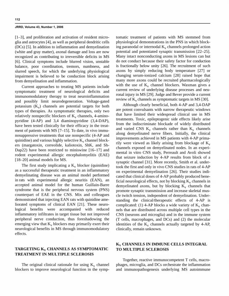
disease processes. Each immune cell type is characterizedby a cell-specific repertoire of ionic channels, a state-specific/differential expression of distinct Kv channels(Figure 1).
Macrophages are involved in chemotaxis, activemyelin breakdown, phagocytosis of myelin proteins,myelin antigen presentation, and cytokine secretion.Microglia mediate proinflammatory immune responses,generate nitric oxide (NO)/elevated NO synthase (NOS)in MS lesions, and are active in myelin breakdown,phagocytosis of myelin proteins, and myelin antigen pre-sentation. DCs initiate and regulate T cell responses andmay contribute to inflammation relapses and chronicityand breakdown of tolerance to autoantigens.
Activated, immunocompetent T cells [18–19,32–37],macrophages [38–39], microglia [40–46], and DCs [47]up-regulate the same Kv channel, Kv1.3. Resting DCs[48], macrophages [49–54], and microglia [41,55–56]transiently exhibit an inwardly rectifying Kv channel(Kir). However, only DCs [48,57] and microglia [58]express the distinct Kv1.5 channel in addition to Kv1.3following stimulation; microglia express Kv1.5 betweentheir unstimulated and fully activated states, but DCsexpress a mix with Kv1.5 predominant. While murinebone marrow-derived macrophages have been shown toexpress Kv1.5 messenger ribonucleic acid [39], to date,no Kv1.5 currents have been recorded in macrophages.Figure 2 outlines functional roles of various potassiumchannels in T cells, macrophages, microglia, and DCs.
Apart from the known central role of cell-mediatedimmune responses in MS, accumulating evidence indi-cates that humoral immune responses (i.e., effector Blymphocytes) may also contribute to the pathogenesis ofMS. Such evidence includes the identification of antimy-elin antibodies in MS lesions, serum and cerebrospinalfluid (CSF) [59–63], and clinical observations consistentwith antibody-mediated demyelination in an MS patient[64]. Furthermore, serum antimyelin antibodies inpatients initially presenting with a clinically isolated syn-drome may predict early conversion to clinically definiteMS [65–66]. While the clinical and pathological signifi-cance of antimyelin antibodies in MS remains to bedefinitively characterized, activated B cells, like T cells,macrophages, microglia, and DCs, also up-regulateKv1.3 channels that are already recognized as putativetherapeutic targets in MS. First recorded in B cells byChoquet and Korn [67–68], Kv1.3 currents have beenshown to be functionally important [69–75], indicatingthat B cells may constitute yet another immune cell targetfor putative immunomodulatory therapies designed to actvia effects on Kv1.3 channels.
TARGETING KV CHANNELS ASIMMUNOSUPPRESSIVE THERAPY
While the toxic, epileptogenic side effects resultingfrom 4-AP likely arise from the indiscriminate blockade
Figure 1.Complement of distinct voltage-gated potassium (Kv) channels expressed in immune cells integral to multiple sclerosis. The 2002 InternationalUnion of Pharmacology (IUPHAR) and American Society for Pharmacology and Experimental Therapeutics standardized nomenclature for Kv
channels is used. Earlier Kv channel names and Human Gene nomenclature developed by Human Genome Organization are listed in parentheses.For a more detailed listing of earlier names and standardized nomenclatures, check IUPHAR Web site (http://www.iuphar-db.org/iuphar-ic/) andJudge SI, Bever CT Jr. Potassium channel blockers in multiple sclerosis: Neuronal K(v) channels and effects of symptomatic treatment.Pharmacol Ther. Epub 2006 Feb 8. [PMID: 16472864]
114
JRRD, Volume 43, Number 1, 2006
of various CNS Kv channels, blockade of 4-AP-sensitiveKv channels in immune cells has emerged as a promisingcandidate because of its neurological benefits. Beneficial4-AP effects could arise not only from blockade of CNSsynaptic channels [26] but also from effects on microglia[43,58] and/or T cells [33–34]. Notably, the Kv1.3 is thepredominant Kv channel in both activated T cells [34,76]and activated microglia [58]. The identification of Kv1.3in mature antigen-presenting DCs [47] implicates thesecells as an additional likely candidate contributing to thebeneficial neurological effects of 4-AP or 3,4-DAP treat-ment in MS patients. Recently, high Kv1.3 expressionwas demonstrated in the perivenular and parenchymalinflammatory infiltrates in postmortem MS brain, as wellas on CSF T cells from MS patients [77].
T CellsStudies of Kv1.3 in activated T cells predate the clon-
ing of Kv channels. Dating back to the mid-1980s, the firstrecordings in human peripheral blood T cells showedinhibition of mitogen-stimulated activation by nonspecificKv channel blockers [32–33,37]. This finding wasfollowed by the first studies in myelin basic protein(MBP)-reactive rat T cells [35–36,78–80] and the firstdemonstration that 4-AP and other nonspecific Kv chan-nel blockers (e.g., tetraethylammonium, methoxyvera-pamil) could inhibit the adoptive transfer of relapsing-remitting EAE in rats [35–36,80].
More recent studies have determined the molecularidentities of T cell Kv channels and shown differentialexpression of these channels in response to acute versus
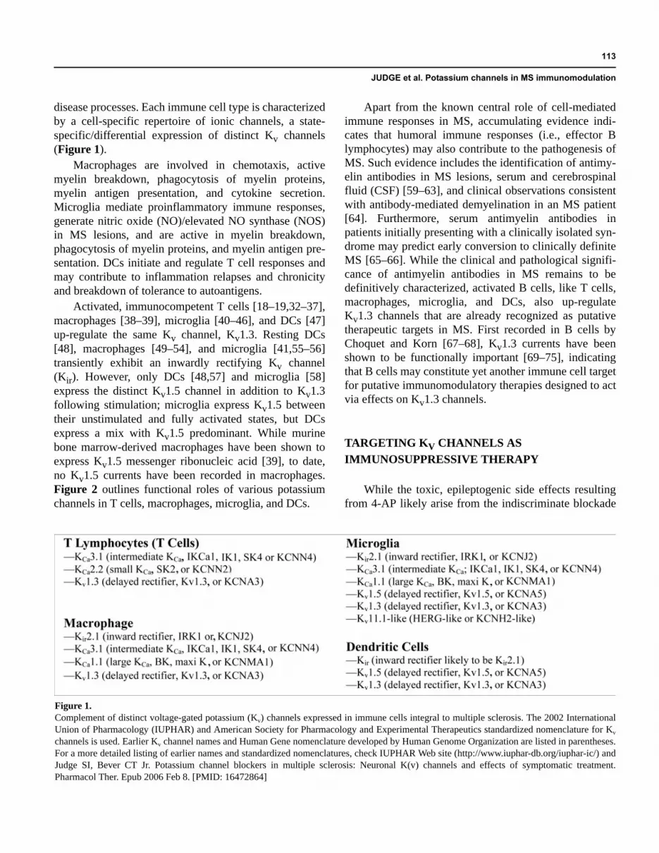
Figure 2.Known function roles of distinct voltage-gated potassium (Kv) channels in immune cells integral to multiple sclerosis (MS). Reference numbersrefer to Appendix, available online only at www.rehab.research.va.gov.*For review, see Chandy KG, Wulff H, Beeton C, Pennington M, Gutman GA, Cahalan MD. K+ channels as targets for specificimmunomodulation. Trends Pharmacol Sci. 2004;25(5):280-89 [PMID: 15120495] and Gallin EK. Ion channels in leukocytes. Physiol Rev.1991;71(3):775–811. [PMID: 1711700]†For review, see Gallin EK. Calcium- and voltage-activated potassium channels in human macrophages. Biophys J. 1984;46(6):821–25 [PMID:6097318] and Gallin EK. Ion channels in leukocytes. Physiol Rev. 1991;71(3):775–811. [PMID: 1711700]‡For review, see Eder C. Ion channels in microglia (brain macrophages). Am J Physiol. 1998;275(2 Pt 1):C327–42. [PMID: 9688586], Eder C.Regulation of microglial behavior by ion channel activity. J Neurosci Rev. 2005;81(3):314–21 [PMID: 1592907], and Farber K, Kettenmann H.Physiology of microglial cells. Brain Res Rev. 2005;48(2):133–43. [PMID: 15850652]NADPH = nicotinamide adenosine dinucleotide phosphate, TNF-α = tumor necrosis factorα.
115
JUDGE et al. Potassium channels in MS immunomodulation
chronic MBP stimulation [18–19,81]. Considerableadvances have been made in identifying potent toxins[34,79,82–83] that are highly selective blockers for theT cell Kv1.3 channel, with better selectivity/potencyprofiles and experimental therapeutic effects [18–19]than 4-AP [35–36,78,80]. While proving successful,systemic administration of highly selective Kv1.3 block-ing agents in EAE still has not shown whether it producesbeneficial neurological effects by blocking Kv1.3 in Tcells, microglia, macrophages, neurons, and/or DCs.
Dendritic CellsDCs are a major component of the innate immune
system and play a pivotal role in the adaptive immuneresponse by providing necessary costimulatory signalsfor the induction of T cell responses, surface-expressedcomplexes of antigen peptide, and major histocompatibil-ity class II (MHC II) molecules. Immature DCs are profi-cient at antigen endocytosis and processing but poor atstimulating T cells. Terminally mature DCs are proficientantigen-presenting cells highly specialized for stimulat-ing T cells to initiate antigen-specific effector cell func-tion [84–85]. During the functional maturation process,in response to inflammatory or microbial stimuli,changes occur in the profile of DC surface markers andcellular immune functions that define distinct immatureversus mature immunofunctional phenotypes. Kv chan-nels number among state-specific up-regulated trans-membrane proteins known to play prominent roles in thecellular activation of a wide variety of immune systemcells of both lymphoid and myeloid lineage.
In spite of the importance of DCs as immunoregula-tors of T cells, studies of DC Kv channels have only justbegun. The presence of functioning Kv1.3 channels [47]was first described in murine DCs that were terminallymatured and exhibited a high surface-membrane expres-sion of MHC II molecules. Studies are currently underway examining human DCs throughout the full processmaturation. Preliminary results indicated that a sequentialand state-specific up- and down-regulation of three dis-tinct Kv channels: first Kir, followed by Kv1.5, and ulti-mately Kv1.3 [57]. More detailed studies have sincerevealed that stimulated DCs express a mix of both Kv1.3and Kv1.5 channels, with Kv1.5 predominating inmatured DCs. Furthermore, these studies demonstratedthat blockade of Kv1.3 and Kv1.5 impaired clusters of dif-ferentiation 83 (CD83), CD80, and CD86 up-regulationand interleukin 12 (IL12) and IL6 production, indicating
that these channels play a functional role in DC matura-tion [48]. DCs are attractive alternate MS therapeutic tar-gets to T cells for two reasons. First, DC stimulation andmaturation precede DC-initiated stimulation of T cells.Second, DCs constitute a peripheral systemic (CSF,meninges, choroid plexus, and deep cervical lymphnodes), as well as a CNS (MS lesions) target for thedevelopment of future clinical treatments in MS. Thus,targeting select DC Kv channels to interfere with DC mat-uration may offer an early and unique opportunity toinhibit T cell effector function by aborting the inductionof T cells as autoimmune effector cells in MS.
TARGETING KV CHANNELS AS ANTI-INFLAMMATORY THERAPY
The hallmark of neuroinflammation is a microglial ormicroglial/macrophage response that has been observed inseveral neurodegenerative diseases, including MS, mak-ing it reasonable to consider anti-inflammatory therapyfor MS to inhibit microglial activation. Specifically, clini-cal benefits following anti-inflammatory treatment havebeen demonstrated in mice with a genetic motor-neurondisease in which microglia are prominent [86–88]. Inanother model of neuroinflammatory disease, PVC-211murine leukemia virus (MuLV)-induced spongiform neu-rodegenerative disease in rats, a highly reactive micro-glial/macrophage response is associated with severe free-radical injury, motor neuron injury, and death. Vitamin Epretreatment of rat pups delays the appearance of free-rad-ical injury and delays but does not inhibit disease expres-sion [89]. Furthermore, minocycline, an antibiotic withinhibitory effects on macrophages and microglia, inhibitsthe reactive microglial/macrophage response and delaysthe expression of PVC-211 MuLV disease [90] and iseffective in slowing the disease course in superoxide dis-mustase (SODI)G93a mutant motor-neuron disease [88].The presumed mechanism is inhibition of microglial/mac-rophage function. More recently, the cycloxygenase-2inhibitor celecoxib has been effective in slowing the dis-ease course in SOD1G93a mice. This has led to an ongoingclinical trial of this compound in patients with Lou Geh-rig’s disease. While these broadly reactive anti-inflamma-tory compounds may show partial effects in animalmodels and, we hope, in clinical trials, a need clearlyexists for more targeted therapy. Thus, microglial and/or

116
JRRD, Volume 43, Number 1, 2006
macrophage Kv channels may represent a possible targetfor intervention.
Of the immune system cells considered integral to MSautoimmune processes, the study of Kv1.3 in activatedmicroglia and macrophages has only recently garneredattention. Microglia play a central role in mediating CNSinflammatory processes and as the only resident brainimmune system cells, activated microglia can proliferate,migrate to sites of injury, present antigen, phagocytize,secrete proinflammatory cytokines and cytotoxins, andundergo a nicotinamide adenosine dinucleotide phosphate(NADPH)-mediated respiratory burst producing cytotoxicreactive oxygen and nitrogen species.
Three lines of evidence suggest a central role formicroglia in the disease processes leading to demyelina-tion and irreversible axonal damage underlying conduc-tion deficits in MS. First, active MS lesions containreactive microglia [91–92]. Second, throughout activedemyelinating lesions and along the borders of chronicactive lesions [93], NOS catalytic activity is elevated, asare levels of NO, a proinflammatory reactive nitrogen-free radical generated by activated microglia [94–99].Third, NO donors can produce reversible conductionblock in normal and experimentally demyelinated axonsand morphological changes consistent with acute Walle-rian degeneration [100–101]. Thus, reactive microgliaand a proinflammatory microglial activation product areimplicated in the long-established conduction deficits andnewly recognized axonal damage associated with MS.
As seen in other immune system cells (T cells andmacrophages), Kv channels appear to regulate prolifera-tion and cellular activation in microglia. Two distinct Kvchannels are expressed differentially in microglia: Kv1.5in resting, nonproliferating cells and Kv1.3 in activated,proliferating cells [58,102]. While Kv1.3 up-regulationhas been associated with various effector cell functionsfollowing microglial activation [40,44,55], the preciserole of Kv1.3 versus Kv1.5 channels in microglial func-tion remains unclear. To date, Kv1.3 up-regulation isassociated with granulocyte macrophage-colony stimu-lating factor, interferon-γ, and lipopolysaccharide-stimulated activation [40,44,55], transforming growthfactor-β stimulated microglial deactivation [56], and theNADPH-mediated respiratory burst [43], a metaboliccascade, the products of which have been identified inMS [43,103–104].
CONCLUSION: FUTURE POTENTIAL FORTARGETING KV CHANNELS IN MULTIPLE SCLEROSIS
Two mononuclear phagocytes, CNS microglia andperipheral DC, are critical players in CNS inflammation.As such, microglia and DCs are important immune celltargets for new MS therapies aimed at modulating cellfunction by blocking Kv channels. In the CNS, activatedmicroglia are the primary effector cells underlying theimmune-mediated pathogenesis of inflammation, demy-elination, and breakdown of the blood-brain barrier (BBB)leading to neuronal injury and dysfunction [103]. Periph-erally, mature DCs are essential for initiating and regulat-ing primary T cell responses, which require peripheralstimulation to cross the BBB [105].
Given the known preferential Kv1.3 up-regulation ineffector T cells, activated microglia and macrophages,and mature DCs, beneficial therapeutic effects resultingfrom the use of highly selective Kv1.3 blockers couldarise from modulation of any or all of these immune cells.Even though highly selective peptide toxins have beenidentified that are better blockers of the Kv1.3 channelthan 4-AP or 3,4-DAP, they are, at present, handicappedas viable therapeutics because of their short half-life ofapproximately 20 min [19]; synthetic toxin analogues arebeing developed to overcome such limitations [106].
Distinct from T cells and macrophages followingstimulation, microglia up-regulate Kv1.5 during earlystages of cellular activation prior to the up-regulation ofKv1.3 at terminal stages of activation, while DCs predomi-nantly up-regulate Kv1.5 over Kv1.3 in their mature immu-nocompetent state. Although the precise functional roles ofthe Kv1.5 and Kv1.3 Kv channels remain unclear, their dif-ferential expression reveals Kv1.5 as an earlier and,thereby, potentially more important therapeutic target thanKv1.3 in microglia, and a primary target in DCs that distin-guishes them from T cells. Studies to modulate the immuneand neuroinflammatory response by affecting Kv1.5 andKv1.3 activation are in progress in animal models. Trans-lating these studies to MS offers a new therapeuticapproach to this inflammatory neurodegenerative disease.
ACKNOWLEDGMENT
This material was based on work supported by the Department ofVeterans Affairs (VA) MS Center of Excellence East, individual
117
JUDGE et al. Potassium channels in MS immunomodulation
VA merit review funding to S. I. V. Judge and C. T. Bever Jr, andseparate pilot funding from the VA MS Center of Excellence Eastand National MS Society pilot grant PP0997 to S. I. V. Judge.
REFERENCES
1. Prineas JW, Wright RG. Macrophages, lymphocytes, andplasma cells in the perivascular compartment in chronicmultiple sclerosis. Lab Invest. 1997;38(4):409–21.[PMID: 205724]
2. Hauser SL, Bhan AK, Gilles FH, Hoban CJ, Reinherz EL,Schlossman SF, Weiner HL. Immunohistochemical stainingof human brain with monoclonal antibodies that identifylymphocytes, monocytes, and the Ia antigen. J Neuroimmu-nol. 1983;5(2):197–205. [PMID: 6413533]
3. Prineas JW. Pathology of the early lesion in multiple scle-rosis. Human Pathol. 1975;6(5):531–54. [PMID: 170186]
4. Raine CS. The Dale E. McFarlin Memorial Lecture: Theimmunology of the multiple sclerosis lesion. Ann Neurol.1994;36 Suppl:61–72. [PMID: 8017891]
5. Link H, Huang YM, Masterman T, Xiao BG. Vaccinationwith autologous dendritic cells: From experimental autoim-mune encephalomyelitis to multiple sclerosis. J Neuroim-munol. 2001;114(1–2):1–7. [PMID: 11240009]
6. Lassmann H. Pathology of neurons in multiple sclerosis.In: Waxman SG, editor. Multiple sclerosis as a neuronaldisease. Burlington (MA): Elsevier Academic Press; 2005.p. 153–64.
7. Bever CT Jr. 4-Aminopyridine: Use in multiple sclerosis.CNS Drug Rev. 1994;1(2):261–79.
8. Bever CT Jr. Clinical pharmacology of abnormal potassiumchannel organization in demyelinated axons. In: WaxmanSG, editor. Multiple sclerosis as a neuronal disease. Burl-ington (MA): Elsevier Academic Press; 2005. p. 145–52.
9. Bever CT Jr, Anderson PA, Leslie J, Panitch HS, Dhib-Jalbut S, Khan OA, Milo R, Hebel JR, Conway KL, Katz E,Johnson KP. Treatment with oral 3,4 diaminopyridineimproves leg strength in multiple sclerosis patients: Resultsof a randomized, double-blind, placebo-controlled, cross-over trial. Neurology. 1996;47(6):1457–62.[PMID: 8960727]
10. Bever CT Jr, Leslie J, Camenga D, Panitch HS, JohnsonKP. Preliminary trial of 3,4-diaminopyridine in patientswith multiple sclerosis. Ann Neurol. 1990;27(4):421–27.[PMID: 2353797]
11. Bever CT Jr, Young D, Anderson PA, Krumholz A, Con-way K, Leslie J, Eddington N, Plaisance KI, Panitch HS,Dhib-Jalbut S, Fossler MJ, Devane J, Johnson KP. Theeffects of 4-aminopyridine in multiple sclerosis patients:Results of a randomized placebo-controlled, double-blind,
concentration-controlled crossover trial. Neurology. 1994;44(6):1054–59. [PMID: 8208399]
12. Davis FA, Stefoski DF, Rush J. Orally administered4-aminopyridine improves clinical signs in multiple scle-rosis. Ann Neurol. 1990;27(2):186–92. [PMID: 2317014]
13. Fujihara K, Miyoshi T. The effects of 4-aminopyridine onmotor evoked potentials in multiple sclerosis. J NeurolSci. 1998;159(1):102–6. [PMID: 9700711]
14. Jones RE, Heron JR, Foster DH, Snelgar RS, Mason RJ.Effects of 4-aminopyridine in patients with multiple scle-rosis. J Neurol Sci. 1983;60(3):353–62. [PMID: 6631441]
15. Stefoski DF, Davis FA, Faut M, Schauf CL. 4-Amino-pyridine improves clinical signs in multiple sclerosis. AnnNeurol. 1987;21(1):71–77. [PMID: 2435223]
16. Koo GC, Blake JT, Shah K, Staruch MJ, Dumont F,Wunderler D, Sanchez M, McManus OB, Sirotina-Meisher A, Fischer P, Boltz RC, Goetz MA, Baker R, BaoJ, Kayser F, Rupprecht KM, Parsons WH, Tong XC, ItaIE, Pivnichny J, Vincent S, Cunningham P, Hora D Jr,Feeney W, Kaczorowski G, Springer MS. Correolide andderivatives are novel immunosuppressants blocking thelymphocyte Kv1.3 potassium channels. Cell Immunol.1999;197(2):99–107. [PMID: 10607427]
17. Koo GC, Blake JT, Talento A, Nguyen M, Lin S, SirotinaA, Shah K, Mulvany K, Hora D Jr, Cunningham P,Wunderler DL, McManus OB, Slaughter R, Bugianesi R,Felix J, Garcia M, Williamson J, Kaczorowski G, SigalNH, Springer MS, Feeney W. Blockade of the voltagegated potassium channel Kv1.3 inhibits immune responsesin vivo. J Immunol. 1997;158(11):5120–28.[PMID: 9164927]
18. Beeton C, Barbaria J, Giraud P, Devaux J, Benoliel AM,Gola M, Sabatier JM, Bernard D, Crest M, Beraud E.Selective blocking of voltage-gated K+ channels improvesexperimental autoimmune encephalomyelitis and inhibitsT cell activation. J Immunol. 2001;166(2):936–44.[PMID: 11145670]
19. Beeton C, Wulff H, Barbaria J, Clot-Faybesse O, Penning-ton M, Bernard D, Cahalan MD, Chandy KG, Beraud E.Selective blockade of T lymphocyte K(+) channels amelio-rates experimental autoimmune encephalomyelitis, a modelfor multiple sclerosis. Proc Natl Acad Sci USA. 2001;98(24):13942–47. [PMID: 11717451]
20. Uitdehaag BM, Polman CH, de Groot CJ, Dijkstra CD.Effect of K+ channel blockers on the clinical course andhistological features of experimental allergic encephalo-myelitis. Acta Neurol Scand. 1994;90(4):299–301.[PMID: 7839818]
21. Mix E, Olsson T, Solders G, Link H. Effect of ion channelblockers on immune response and course of experimentalallergic neuritis. Brain. 1989;112(Pt 6):1405–18.[PMID: 2480831]
http://www.ncbi.nlm.nih.gov/entrez/query.fcgi?cmd=Retrieve&db=pubmed&dopt=Abstract&list_uids=6413533
http://www.ncbi.nlm.nih.gov/entrez/query.fcgi?cmd=Retrieve&db=pubmed&dopt=Abstract&list_uids=8017891
http://www.ncbi.nlm.nih.gov/entrez/query.fcgi?cmd=Retrieve&db=pubmed&dopt=Abstract&list_uids=8960727
http://www.ncbi.nlm.nih.gov/entrez/query.fcgi?cmd=Retrieve&db=pubmed&dopt=Abstract&list_uids=2353797
http://www.ncbi.nlm.nih.gov/entrez/query.fcgi?cmd=Retrieve&db=pubmed&dopt=Abstract&list_uids=8208399
http://www.ncbi.nlm.nih.gov/entrez/query.fcgi?cmd=Retrieve&db=pubmed&dopt=Abstract&list_uids=2317014
http://www.ncbi.nlm.nih.gov/entrez/query.fcgi?cmd=Retrieve&db=pubmed&dopt=Abstract&list_uids=9700711
http://www.ncbi.nlm.nih.gov/entrez/query.fcgi?cmd=Retrieve&db=pubmed&dopt=Abstract&list_uids=6631441
http://www.ncbi.nlm.nih.gov/entrez/query.fcgi?cmd=Retrieve&db=pubmed&dopt=Abstract&list_uids=2435223
http://www.ncbi.nlm.nih.gov/entrez/query.fcgi?cmd=Retrieve&db=pubmed&dopt=Abstract&list_uids=9164927
118
JRRD, Volume 43, Number 1, 2006
22. Bostock H, Sears TA, Sherratt RM. The effects of 4-amino-pyridine and tetraethylammonium ions on normal anddemyelinated mammalian nerve fibers. J Physiol. 1981;313:301–15. [PMID: 7277221]
23. Sherratt RM, Bostock H, Sears TA. Effects of 4-amino-pyridine on normal and demyelinated mammalian nervefibers. Nature. 1980;283(5747):570–72. [PMID: 7354839]
24. Targ EF, Kocsis JD. 4-Aminopyridine leads to restorationof conduction in demyelinated rat sciatic nerve. BrainRes. 1985;328(2):358–61. [PMID: 2985185]
25. Thesleff S. Aminopyridines and synaptic transmission.Neuroscience. 1980;5(8):1413–19. [PMID: 6250099]
26. Smith KJ, Felts PA, John GR. Effects of 4-aminopyridineon demyelinated axons, synapses and muscle tension.Brain. 2000;123(Pt 1):171–84. [PMID: 10611131]
27. Watson CW. Effect of lowering of body temperature onthe symptoms and signs of multiple sclerosis. N EnglJ Med. 1959;261:1253–59. [PMID: 13843117]
28. Schauf CL, Davis FA. Impulse conduction in multiplesclerosis: A theoretical basis for modification by tempera-ture and pharmacological agents. J Neurol Neurosurg Psy-chiatry. 1974;37(2):152–61. [PMID: 4362242]
29. Waxman SG. Multiple sclerosis as a neuronal disease.Burlington (MA): Elsevier Academic Press; 2005.
30. Judge SI, Bever CT Jr. Potassium channel blockers in mul-tiple sclerosis: Neuronal K(v) channels and effects ofsymptomatic treatment. Pharmacol Ther. Epub 2006 Feb 8.[PMID: 16472864]
31. Perreault R, Avoli M. Physiology and pharmacology ofepileptiform activity induced by 4-aminopyridine in rathippocampal slices. J Neurophysiol. 1991;65:771–85.[PMID: 1675671]
32. Chandy KG, DeCoursey TE, Cahalan MD, McLaughlinC, Gupta S. Voltage-gated potassium channels requiredfor human T lymphocyte activation. J Exp Med. 1984;160(2):369–85. [PMID: 6088661]
33. DeCoursey TE, Chandy KG, Gupta S, Cahalan MD. Volt-age gated K+ channels in human T lymphocytes: A role inmitogenesis. Nature. 1984;307(5950):465–68.[PMID: 6320007]
34. Grissmer S, Dethlefs B, Wasmuth JJ, Goldin AL, GutmanGA, Cahalan MD, Chandy KG. Expression and chromo-somal localization of la lymphocyte K+ channel gene.Proc Natl Acad Sci USA. 1990;87(23):9411–15.[PMID: 2251283]
35. Judge SI, Yeh JZ, Mannie MD, Paterson PY. Potassiumchannels in T lymphocytes mediating experimental aller-gic encephalomyelitis (EAE) in rats. Soc Neurosci Abtrs.1986;12:1342.
36. Judge SI, Yeh JZ, Mannie MD, Paterson PY. Potassiumchannels in encephalitogenic rat lymphocytes: Characteris-
tics of inactivation and role in cell activation. Soc NeurosciAbtrs. 1987;13:531.
37. Matteson DR, Deutsch C. K channels in T lymphocytes:A patch clamp study using monoclonal antibody adhe-sion. Nature. 1984;307(5950):468–71. [PMID: 6320008]
38. Mackenzie AB, Chirakkal H, North RA. Kv1.3 potassiumchannels in human alveolar macrophages. Am J PhysiolLung Cell Mol Physiol. 2003;285(4):L862–68.[PMID: 12909584]
39. Vicente R, Escalada A, Coma M, Fuster G, Sanchez-TilloE, Lopez-Iglesias C, Soler C, Solsona C, Celada A, FelipeA. Differential voltage-dependant K+ channel responsesduring proliferation and activation in macrophages. J BiolChem. 2003;278(47):46307–20. [PMID: 12923194] Erra-tum in: J Biol Chem. 2005;280(13):13204.
40. Eder C, Fischer HG, Hadding U, Heinemann U. Propertiesof voltage-gated potassium currents of microglia differenti-ated with granulocyte/macrophage colony-stimulating fac-tor. J Membr Biol. 1995;147(2):137–46.[PMID: 8568850]
41. Eder C, Schilling T, Heinemann U, Haas D, Hailer N,Nitsch R. Morphological, immunophenotypical and elec-trophysiological properties of resting microglia in vitro. EurJ Neurosci. 1999;11(12):4251–61. [PMID: 10594651]
42. Farber K, Kettenmann H. Physiology of microglial cells.Brain Res Brain Res Rev. 2005;48(2):133–43.[PMID: 15850652]
43. Khanna R, Roy L, Zhu X, Schlichter LC. K+ channels andthe microglial respiratory burst. Am J Physiol. 2001;280(4):C796–806. [PMID: 11245596]
44. Pyo H, Chung S, Jou I, Gwag B, Joe EH. Expression andfunction of outward K+ channels induced by lipopolysac-charide in microglia. Mol Cells. 1997;7(5):610–14.[PMID: 9387147]
45. Schilling T, Stock C, Schwab A, Eder C. Functionalimportance of Ca2+-activated K+ channels for lysophos-phatidic acid-induced microglial migration. Eur J Neuro-sci. 2004;19(6):1469–74. [PMID: 15066143]
46. McLarnon JG, Xu R, Lee YB, Kim SU. Ion channelsof human microglia in culture. Neuroscience. 1997;78(4):1217–28. [PMID: 9174088]
47. Fischer HG, Eder C. Voltage-gated K+ currents of mousedendritic cells. FEBS Lett. 1995;373(2):127–30.[PMID: 7589450]
48. Mullen KM, Rozycka M, Hu L, Rus H, Graber J, Pen-nington MW, Johns DC, Judge SIV, Calabresi PA. Potas-sium channels Kir, Kv1.5, and Kv1.3 are expressed onhuman dendritic cells and have a functional role in matu-ration. Ann Neurol. In press 2006.
49. DeCoursey TE, Kim SY, Silver MR, Quandt FN. Ionchannel expression in PMA-differentiated human THP-1
http://www.ncbi.nlm.nih.gov/entrez/query.fcgi?cmd=Retrieve&db=pubmed&dopt=Abstract&list_uids=7277221
http://www.ncbi.nlm.nih.gov/entrez/query.fcgi?cmd=Retrieve&db=pubmed&dopt=Abstract&list_uids=7354839
http://www.ncbi.nlm.nih.gov/entrez/query.fcgi?cmd=Retrieve&db=pubmed&dopt=Abstract&list_uids=2985185
http://www.ncbi.nlm.nih.gov/entrez/query.fcgi?cmd=Retrieve&db=pubmed&dopt=Abstract&list_uids=6250099
http://www.ncbi.nlm.nih.gov/entrez/query.fcgi?cmd=Retrieve&db=pubmed&dopt=Abstract&list_uids=4362242
http://www.ncbi.nlm.nih.gov/entrez/query.fcgi?cmd=Retrieve&db=pubmed&dopt=Abstract&list_uids=1675671
http://www.ncbi.nlm.nih.gov/entrez/query.fcgi?cmd=Retrieve&db=pubmed&dopt=Abstract&list_uids=6088661
http://www.ncbi.nlm.nih.gov/entrez/query.fcgi?cmd=Retrieve&db=pubmed&dopt=Abstract&list_uids=6320007
http://www.ncbi.nlm.nih.gov/entrez/query.fcgi?cmd=Retrieve&db=pubmed&dopt=Abstract&list_uids=2251283
http://www.ncbi.nlm.nih.gov/entrez/query.fcgi?cmd=Retrieve&db=pubmed&dopt=Abstract&list_uids=6320008
http://www.ncbi.nlm.nih.gov/entrez/query.fcgi?cmd=Retrieve&db=pubmed&dopt=Abstract&list_uids=8568850
http://www.ncbi.nlm.nih.gov/entrez/query.fcgi?cmd=Retrieve&db=pubmed&dopt=Abstract&list_uids=9387147

119
JUDGE et al. Potassium channels in MS immunomodulation
macrophages. J Membr Biol. 1996;152(2):141–57.[PMID: 9139125]
50. Gallin EK, Livengood DR. Inward rectification in mousemacrophages: Evidence for a negative resistance region.Am J Physiol. 1981;241(1):C9–17. [PMID: 7246764]
51. Gallin EK, McKinney LC. Patch-clamp studies in humanmacrophages: Single-channel and whole-cell character-ization of two K+ conductances. J Membr Biol. 1988;103(1):55–66. [PMID: 2460627]
52. Gallin EK, Sheehy PA. Differential expression of inwardand outward potassium currents in the macrophage-likecell line J774.1. J Physiol. 1985;369:475–99.[PMID: 2419551]
53. Judge SI, Montcalm-Mazzilli E, Gallin EK. IKir regula-tion in murine macrophages: Whole cell and perforatedpatch studies. Am J Physiol. 1994;267(6 Pt 1):C1691–98.[PMID: 7810612]
54. McKinney LC, Gallin EK. Inwardly rectifying whole-celland single-channel K currents in the murine macrophagecell line J774.1. J Membr Biol. 1988;103(1):41–53.[PMID: 3184170]
55. Fischer HG, Eder C, Hadding U, Heinemann U. Cytokine-dependent K+ channel profile of microglia at immunologi-cally defined functional states. Neuroscience. 1995;64(1):183–91. [PMID: 7535902]
56. Schilling T, Quandt FN, Cherney VV, Zhou W, Heine-mann U, DeCoursey TE, Eder C. Upregulation of Kv1.3K(+) channels in microglia deactivated by TGF-beta. AmJ Physiol Cell Physiol. 2000;279(4):C1123–34.[PMID: 11003593]
57. Mullen K, Rozycka M, Rus H, Graber J, Pennington M,Judge S, Calabresi P. Maturation of dendritic cells ismarked by functionally relevant expression of K+ chan-nels Kv1.5 and Kv1.3 [abstract]. Johns Hopkins MedicalInstitute Rare Neuroimmunologic Disorders Symposium.2004 Aug 18–22; Baltimore (MD).
58. Kotecha SA, Schlichter LC. A Kv1.5 to Kv1.3 switch inendogenous hippocampal microglia and a role in prolifera-tion. J Neurosci. 1999;19(24):10680–93. [PMID: 10594052]
59. Cepok S, Rosche B, Grummel V, Vogel F, Zhou D, SaynJ, Sommer N, Hartung HP, Hemmer B. Short-lived plasmablasts are the main B cell effector subset during the courseof multiple sclerosis. Brain. 2005;128(Pt 7):1667–76.[PMID: 15800022]
60. Genain CP, Cannella B, Hauser SL, Raine CS. Identifica-tion of autoantibodies associated with myelin damage inmultiple sclerosis. Nat Med. 1999;5(2):170–75.[PMID: 9930864]
61. Lucchinetti C, Bruck W, Parisi J, Scheithauer B, RodriguezM, Lassmann H. Heterogeneity of multiple sclerosislesions: Implications for the pathogenesis of demyelination.Ann Neurol. 2000;47(6):707–17. [PMID: 10852536]
62. Reindl M, Linington C, Brehm U, Egg R, Dilitz E,Deisenhammer F, Poewe W, Berger T. Antibodies againstthe myelin oligodendrocyte glycoprotein and the myelinbasic protein in multiple sclerosis and other neurologicaldiseases: A comparative study. Brain. 1999;122(Pt 11):2047–56. [PMID: 10545390]
63. Storch MK, Piddlesden S, Haltia M, Iivanainen M, Mor-gan P, Lassmann H. Multiple sclerosis: In situ evidencefor antibody- and complement-mediated demyelination.Ann Neurol. 1998;43(4):465–71. [PMID: 9546327]
64. Bruck W, Neubert K, Berger T, Weber JR. Clinical, radio-logical, immunological and pathological findings in inflam-matory CNS demyelination—possible markers for anantibody-mediated process. Mult Scler. 2001;7(3):173–77.[PMID: 11475441]
65. Berger T, Rubner P, Schautzer F, Egg R, Ulmer H, May-ringer I, Dilitz E, Deisenhammer F, Reindl M. Antimyelinantibodies as a predictor of clinically definite multiplesclerosis after a first demyelinating event. N Engl J Med.2003;349(2):139–45. [PMID: 12853586]
66. Lim ET, Berger T, Reindl M, Dalton CM, Fernando K, KeirG, Thompson EJ, Miller DH, Giovannoni G. Antimyelinantibodies do not allow earlier diagnosis of multiple sclero-sis. Mult Scler. 2005;11(4):492–94. [PMID: 16042235]
67. Choquet D, Korn H. Modulation of voltage-dependentpotassium channels in B lymphocytes. Biochem Pharma-col. 1988;37(20):3797–3802. [PMID: 3056414]
68. Choquet D, Sarthou P, Primi D, Cazenave PA, Korn H.Cyclic AMP-modulated potassium channels in murineB cells and their precursors. Science. 1987;235(4793):1211–14. [PMID: 2434998]
69. Amigorena S, Choquet D, Teillaud JL, Korn H, FridmanWH. Ion channel blockers inhibit B cell activation at aprecise stage of the G1 phase of the cell cycle. Possibleinvolvement of K+ channels. J Immunol. 1990;144(6):2038–45. [PMID: 2313087]
70. Brent LH, Butler JL, Woods WT Jr, Bubien JK. Transmem-brane ion conductance in human B lymphocyte activation.J Immunol. 1990;145(8):2381–89. [PMID: 1698853]
71. Partiseti M, Choquet D, Diu A, Korn H. Differential regu-lation of voltage- and calcium-activated potassium chan-nels in human B lymphocytes. J Immunol. 1992;148(11):3361–68. [PMID: 1588037]
72. Partiseti M, Korn H, Choquet D. Pattern of potassium chan-nel expression in proliferating B lymphocytes dependsupon the mode of activation. J Immunol. 1993;151(5):2462–70. [PMID: 8360473]
73. Spencer RH, Chandy KG, Gutman GA. Immunologicalidentification of the Shaker-related Kv1.3 potassiumchannel protein in T and B lymphocytes, and detection ofrelated proteins in flies and yeast. Biochem Biophys ResCommun. 1993;191(1):201–6. [PMID: 8447822]
http://www.ncbi.nlm.nih.gov/entrez/query.fcgi?cmd=Retrieve&db=pubmed&dopt=Abstract&list_uids=3056414
http://www.ncbi.nlm.nih.gov/entrez/query.fcgi?cmd=Retrieve&db=pubmed&dopt=Abstract&list_uids=2434998
http://www.ncbi.nlm.nih.gov/entrez/query.fcgi?cmd=Retrieve&db=pubmed&dopt=Abstract&list_uids=2313087
http://www.ncbi.nlm.nih.gov/entrez/query.fcgi?cmd=Retrieve&db=pubmed&dopt=Abstract&list_uids=1698853
http://www.ncbi.nlm.nih.gov/entrez/query.fcgi?cmd=Retrieve&db=pubmed&dopt=Abstract&list_uids=1588037
http://www.ncbi.nlm.nih.gov/entrez/query.fcgi?cmd=Retrieve&db=pubmed&dopt=Abstract&list_uids=8360473
http://www.ncbi.nlm.nih.gov/entrez/query.fcgi?cmd=Retrieve&db=pubmed&dopt=Abstract&list_uids=8447822
http://www.ncbi.nlm.nih.gov/entrez/query.fcgi?cmd=Retrieve&db=pubmed&dopt=Abstract&list_uids=9139125
http://www.ncbi.nlm.nih.gov/entrez/query.fcgi?cmd=Retrieve&db=pubmed&dopt=Abstract&list_uids=7246764
http://www.ncbi.nlm.nih.gov/entrez/query.fcgi?cmd=Retrieve&db=pubmed&dopt=Abstract&list_uids=2460627
http://www.ncbi.nlm.nih.gov/entrez/query.fcgi?cmd=Retrieve&db=pubmed&dopt=Abstract&list_uids=2419551
http://www.ncbi.nlm.nih.gov/entrez/query.fcgi?cmd=Retrieve&db=pubmed&dopt=Abstract&list_uids=7810612
http://www.ncbi.nlm.nih.gov/entrez/query.fcgi?cmd=Retrieve&db=pubmed&dopt=Abstract&list_uids=3184170
http://www.ncbi.nlm.nih.gov/entrez/query.fcgi?cmd=Retrieve&db=pubmed&dopt=Abstract&list_uids=7535902
http://www.ncbi.nlm.nih.gov/entrez/query.fcgi?cmd=Retrieve&db=pubmed&dopt=Abstract&list_uids=9930864
120
JRRD, Volume 43, Number 1, 2006
74. Sutro JB, Vayuvegula BS, Gupta S, Cahalan MD. Voltagesensitive ion channels in human B lymphocytes. Adv ExpMed Biol. 1989;254:113–22. [PMID: 2816543]
75. Wulff H, Knaus HG, Pennington M, Chandy KG. K+channel expression during B cell differentiation: Implica-tions for immunomodulation and autoimmunity. J Immu-nol. 2004;173(2):776–86. [PMID: 15240664]
76. Spencer RH, Sokolov Y, Li H, Takenaka B, Milici AJ,Aiyar J, Nguyen A, Park H, Jap BK, Hall JE, Gutman GA,Chandy KG. Purification, visualization, and biophysicalcharacterization of Kv1.3 tetramers. J Biol Chem. 1997;272(4):2389–95. [PMID: 8999950]
77. Rus H, Pardo CA, Hu L, Darrah E, Cudrici C, Niculescu T,Niculescu F, Mullen KM, Allie R, Guo L, Wulff H, BeetonC, Judge SI, Kerr DA, Knaus HG, Chandy KG, CalabresiPA. The voltage-gated potassium channel Kv1.3 is highlyexpressed on inflammatory infiltrates in multiple sclerosisbrain. Proc Natl Acad Sci USA. 2005;102(31):11094–99.[PMID: 16043714]
78. Judge SI, Paterson PY, Mannie MD, Yeh JZ. Modulationof outward K(+) conductance is a post-activational eventin rat T lymphocytes responsible for the adoptive transferof experimental allergic encephalomyelitis. J Biomed Sci.1997;4(2–3):98–110. [PMID: 11725140]
79. Kalman K, Pennington MW, Lanigan MD, Nguyen A,Rauer H, Mahnir V, Paschetto K, Kem WR, Grissmer S,Gutman GA, Christian EP, Cahalan MD, Norton RS,Chandy KG. ShK-Dap22, a potent Kv1.3-specific immuno-suppressive polypeptide. J Biol Chem. 1998;273(49):32697–707. [PMID: 9830012]
80. Judge SI, Yeh JZ, Mannie MD, Seifert LP, Paterson PY.Potassium channel blockers inhibit adoptive transferof experimental allergic encephalomyelitis by myelin-basicprotein-stimulated rat T lymphocytes. J Biomed Sci.1997;4(4):169–78. [PMID: 11725150]
81. Wulff H, Calabresi PA, Allie R, Yun S, Pennington M,Beeton C, Chandy KG. The voltage-gated Kv1.3 K(+)channel in effector memory T cells as new target for MS.J Clin Invest. 2003;111(11):1703–13. [PMID: 12782673]
82. Vennekamp J, Wulff H, Beeton C, Calabresi PA, Griss-mer S, Hansel W, Chandy KG. Kv1.3-blocking 5-pheny-lalkoxypsoralens: A new class of immunomodulators.Mol Pharmacol. 2004;65(6):1364–74. [PMID: 15155830]
83. Wulff H, Miller MJ, Hansel W, Grissmer S, Cahalan MD,Chandy KG. Design of potent and selective inhibitor ofthe intermediate-conductance Ca2+-activated, IKCa1: Apotential immunosuppressant. Proc Natl Acad Sci USA.2000;97(14):8151–56. [PMID: 10884437]
84. Gallucci S, Lolkema M, Matzinger P. Natural adjuvants:Endogenous activators of dendritic cells. Nat Med. 1999;5(11):1249–55. [PMID: 10545990]
85. Sallusto F, Lanzavecchia A. Efficient presentation of sol-uble antigen by cultured human dendritic cells is main-tained by granulocyte/macrophage colony-stimulatingfactor plus interleukin 4 and downregulated by tumornecrosis factor alpha. J Exp Med. 1994;179(4):1109–18.[PMID: 8145033]
86. Cleveland DW, Rothstein JD. From Charcot to Lou Geh-rig: Deciphering selective motor neuron death in ALS. NatRev Neurosci. 2001;2(11):806–19. [PMID: 11715057]
87. Drachman DB, Frank K, Dykes-Hoberg M, Teismann P,Almer G, Przedborski S, Rothstein JD. Cyclooxygenase 2inhibition protects motor neurons and prolongs survival ina transgenic mouse model of ALS. Ann Neurol. 2002;52(6):771–78. [PMID: 12447931]
88. Zhu S, Stavrovskaya IG, Drozda M, Kim BY, Ona V, LiM, Sarang S, Liu AS, Hartley DM, Wu du C, Gullans S,Ferrante RJ, Przedborski S, Kristal BS, Friedlander RM.Minocycline inhibits cytochrome c release and delaysprogression of amyotrophic lateral sclerosis in mice.Nature. 2002;417(6884):74–78. [PMID: 11986668]
89. Hoffman PM, Cimino EF, Robbins DS, Broadwell RD,Powers JM, Ruscetti SK. Cellular tropism and localiza-tion in the rodent nervous system of a neuropathogenicvariant of Friend murine leukemia virus. Lab Invest.1992;67(3):314–21. [PMID: 1405490]
90. Wilt SG, Yiannoulos GC, Smarik J, Ruscetti SK.Minocyline delays onset and microglial activation in a ratretroviral neuroinflammatory disease. Program No. 103.10.2003 Abstract Viewer/Itinerary Planner. Washington (DC):Society for Neuroscience, 2003.
91. Schluesener HJ, Seid K, Kretzschmar J, Meyermann R.Leukocyte chemotactic factor, a natural ligand to CD4, isexpressed lymphocytes and microglial cells of the MSplaque. J Neurosci Res. 1996;44(6):606–11.[PMID: 8794952]
92. Ulvestad E, Williams K, Vedeler C, Antel J, Nyland H,Mork S, Matre R. Reactive microglia in multiple sclerosislesions have an increased expression of receptors for theFc part of IgG. J Neurol Sci. 1994;121(2):125–31.[PMID: 8158203]
93. Trapp BD, Bo L, Mork S, Chang A. Pathogenesis of tis-sue injury in MS lesions. J Neuroimmunol. 1999;98(1):49–56. [PMID: 10426362]
94. Bagasra O, Michaels FH, Zheng YM, Bobroski LE,Spitsin SV, Fu ZF, Tawadros R, Koprowski H. Activationof the inducible form of nitric oxide synthase in the brainsof patients with multiple sclerosis. Proc Natl Acad SciUSA. 1995;92(26):12041–45. [PMID: 8618840]
95. Bo L, Dawson TM, Wesselingh S, Mork S, Choi S, KongPA, Hanley D, Trapp BD. Induction of nitric oxide synthasein demyelinating regions of multiple sclerosis brains. AnnNeurol. 1994;36(5):778–86. [PMID: 7526776]
http://www.ncbi.nlm.nih.gov/entrez/query.fcgi?cmd=Retrieve&db=pubmed&dopt=Abstract&list_uids=2816543
http://www.ncbi.nlm.nih.gov/entrez/query.fcgi?cmd=Retrieve&db=pubmed&dopt=Abstract&list_uids=8999950
http://www.ncbi.nlm.nih.gov/entrez/query.fcgi?cmd=Retrieve&db=pubmed&dopt=Abstract&list_uids=9830012
http://www.ncbi.nlm.nih.gov/entrez/query.fcgi?cmd=Retrieve&db=pubmed&dopt=Abstract&list_uids=8145033
http://www.ncbi.nlm.nih.gov/entrez/query.fcgi?cmd=Retrieve&db=pubmed&dopt=Abstract&list_uids=1405490
http://www.ncbi.nlm.nih.gov/entrez/query.fcgi?cmd=Retrieve&db=pubmed&dopt=Abstract&list_uids=8794952
http://www.ncbi.nlm.nih.gov/entrez/query.fcgi?cmd=Retrieve&db=pubmed&dopt=Abstract&list_uids=8158203
121
JUDGE et al. Potassium channels in MS immunomodulation
96. Cross AH, Manning PT, Keeling RM, Schmidt RE, MiskoTP. Peroxynitrite formation within the central nervoussystem in active multiple sclerosis. J Neuroimmunol.1998;88(1–2):45–56. [PMID: 9688323]
97. Giovannoni G, Heales SJ, Silver NC, O’Riordan J, MillerRF, Land JM, Clark JB, Thompson EJ. Raised serumnitrate and nitrite levels in patients with multiple sclero-sis. J Neurol Sci. 1997;145(1):77–81. [PMID: 9073032]
98. Johnson AW, Land JM, Thompson EJ, Bolanos JP, ClarkJB, Heales SJ. Evidence of increased nitric oxide produc-tion in multiple sclerosis. J Neurol Neurosurg Psychiatry.1995;58(1):107. [PMID: 7823050]
99. Yamashita T, Ando Y, Obayashi K, Uchino M, Ando M.Changes in nitrite and nitrate (NO2-/NO3-) levels in cere-brospinal fluid of patients with multiple sclerosis. J Neu-rol Sci. 1997;153(1):32–34. [PMID: 9455975]
100. Redford EJ, Kapoor R, Smith KJ. Nitric oxide donorsreversibly block axonal conduction: Demyelinated axonsare especially susceptible. Brain. 1997;120(Pt 12):2149–57.[PMID: 9448570]
101. Smith KJ, Kapoor R, Hall SM, Davies M. Electricallyactive axons degenerate when exposed to nitric oxide.Ann Neurol. 2001;49(4):470–76. [PMID: 11310624]
102. Lee J, Judge S, Dugger N, Li Z, Hoffman P. Role ofpotassium channels in microglial activation [abstract].Presented at Department of Veterans Affairs MarylandHealthcare System, Baltimore (MD), April 24, 2003.
103. Benveniste EN. Role of macrophages/microglia in multiplesclerosis and experimental allergic encephalomyelitis. J MolMed. 1997;75(3):165–73. [PMID: 9106073]
104. Gonzalez-Scarano F, Baltuch G. Microglia as mediatorsof inflammatory and degenerative diseases. Annu RevNeurosci. 1999;22:219–40. [PMID: 10202538]
105. Hickey WF. Migration of hematogenous cells through theblood-brain barrier and the initiation of CNS inflammation.Brain Pathol. 1991;1(2):97–105. [PMID: 1669702]
106. Beeton C, Pennington MW, Wulff H, Singh S, Nugent D,Crossley G, Khaytin I, Calabresi PA, Chen CY, GutmanGA, Chandy KG. Targeting effector memory T cells witha selective peptide inhibitor of Kv1.3 channels for therapyof autoimmune diseases. Mol Pharmacol. 2005;67(4):1369–81. [PMID: 15665253]
Submitted for publication September 1, 2004. Acceptedin revised form February 8, 2005.
http://www.ncbi.nlm.nih.gov/entrez/query.fcgi?cmd=Retrieve&db=pubmed&dopt=Abstract&list_uids=9688323
http://www.ncbi.nlm.nih.gov/entrez/query.fcgi?cmd=Retrieve&db=pubmed&dopt=Abstract&list_uids=9073032
http://www.ncbi.nlm.nih.gov/entrez/query.fcgi?cmd=Retrieve&db=pubmed&dopt=Abstract&list_uids=7823050
http://www.ncbi.nlm.nih.gov/entrez/query.fcgi?cmd=Retrieve&db=pubmed&dopt=Abstract&list_uids=9455975
http://www.ncbi.nlm.nih.gov/entrez/query.fcgi?cmd=Retrieve&db=pubmed&dopt=Abstract&list_uids=9448570
http://www.ncbi.nlm.nih.gov/entrez/query.fcgi?cmd=Retrieve&db=pubmed&dopt=Abstract&list_uids=9106073