vol. xxxvi no. 11 - J-Stage
12
VOL. XXXVI NO. 11 THE JOURNAL OF ANTIBIOTICS 1439 NEW FLUORINATED ERYTHROMYCINS OBTAINED BY MUTASYNTHESIS LUCIANO TOSCANO*,GIUSEPPE FIORIELLO Department of Chemistry ROBERTO SPAGNOLIt, LEONARDOCAPPELLETTI" Department of Microbiology GIOVANNA ZANUSO Department of Biology Pierrel S.p.A., Research Laboratories Via Degli Artigianelli, 10, 20159 Milan, Italy (Received for publication April 27, 1983) Following the previously described semisynthetic preparation of new aglycones (8S)-8- fluoroerythronolide A (I), (8S)-8-fluoroerythronolide B (II) and the monoglycoside 3-0- mycarosyl-(8S)-8-fluoroerythronolide B (III), their conversion into new fluoroerythromycins was attempted by mutational biosynthesis. The strain Streptomyces erythraeus ATCC 31772, a mutant blocked in the biosynthesis of erythromycin, was employed in the present investiga- tion. Four new antibiotics, (8S)-8-fluoroerythromycin A (IV), (8S)-8-fluoroerythromycin B (V), (8S)-8-fluoroerythromycin C (VI) and (8S)-8-fluoroerythromycin D (VII) were successfully derived by such an approach. The result is also discussed in terms of the substrate specificity of the enzymes involved in the biosynthesis of erythromycins. The new antibiotics exhibited promising biological properties. During the last twenty years advances in organic fluorine chemistry have been responsible for the development of a large number of new compounds of importance in biology and medicine. Replace- ment of an atom of hydrogen by one of fluorine in organic molecules may profoundly change their bio- logical properties','). The rationale employed is that, since hydrogen and fluorine are nearly isosteric, the fluoro analog would be expected to have little difficulty in fitting onto active sites of receptors. At the same time, the strong electronegativity of the fluorine atom, when strategically placed, could be used to make defined alterations in the biological activity. As part of a general strategy of our laboratories aimed at the synthesis of fluorinated organic deriva- tives designed as potential drugs, we have undertaken a program involving the synthesis and the biologi- cal evaluation of new fluoroerythromycins. In previous papers3.4) we have reported the fluorination of enol ether groups in 8,9-anhydroery- thronolide A and B 6,9-hemiketals and 3-O-mycarosyl-8,9-anhydroerythronolide B 6,9-hemiketal with trifluoromethyl hypofluorite. This reaction, already employed for the introduction of fluorine into naturally occurring products'-10), was of interest per se and also as model to be used in the erythromycin series. These fluorinations were achieved under mild conditions to give (8S)-8-fluoroerythronolides A (I) and B (II) and 3-0-mycarosyl-(8S)-8-fluoroerythronolide B (III). Moreover, in contrast to ery- Present addresses: t Roussel Uclaf , Biotechnology Department, 93230 Romainville, France. tt Bristol-Myers Industrial Division , Syracuse, NY13201, USA.
-
Upload
khangminh22 -
Category
Documents
-
view
0 -
download
0
Transcript of vol. xxxvi no. 11 - J-Stage

VOL. XXXVI NO. 11 THE JOURNAL OF ANTIBIOTICS 1439
NEW FLUORINATED ERYTHROMYCINS OBTAINED
BY MUTASYNTHESIS
LUCIANO TOSCANO*, GIUSEPPE FIORIELLO
Department of Chemistry
ROBERTO SPAGNOLIt, LEONARDO CAPPELLETTI"
Department of Microbiology
GIOVANNA ZANUSO
Department of Biology Pierrel S.p.A., Research Laboratories
Via Degli Artigianelli, 10, 20159 Milan, Italy
(Received for publication April 27, 1983)
Following the previously described semisynthetic preparation of new aglycones (8S)-8- fluoroerythronolide A (I), (8S)-8-fluoroerythronolide B (II) and the monoglycoside 3-0-
mycarosyl-(8S)-8-fluoroerythronolide B (III), their conversion into new fluoroerythromycins was attempted by mutational biosynthesis. The strain Streptomyces erythraeus ATCC 31772, a mutant blocked in the biosynthesis of erythromycin, was employed in the present investiga-tion. Four new antibiotics, (8S)-8-fluoroerythromycin A (IV), (8S)-8-fluoroerythromycin B
(V), (8S)-8-fluoroerythromycin C (VI) and (8S)-8-fluoroerythromycin D (VII) were successfully derived by such an approach. The result is also discussed in terms of the substrate specificity of the enzymes involved in the biosynthesis of erythromycins. The new antibiotics exhibited
promising biological properties.
During the last twenty years advances in organic fluorine chemistry have been responsible for the
development of a large number of new compounds of importance in biology and medicine. Replace-
ment of an atom of hydrogen by one of fluorine in organic molecules may profoundly change their bio-
logical properties','). The rationale employed is that, since hydrogen and fluorine are nearly isosteric,
the fluoro analog would be expected to have little difficulty in fitting onto active sites of receptors. At the
same time, the strong electronegativity of the fluorine atom, when strategically placed, could be used to
make defined alterations in the biological activity.
As part of a general strategy of our laboratories aimed at the synthesis of fluorinated organic deriva-
tives designed as potential drugs, we have undertaken a program involving the synthesis and the biologi-
cal evaluation of new fluoroerythromycins.
In previous papers3.4) we have reported the fluorination of enol ether groups in 8,9-anhydroery-
thronolide A and B 6,9-hemiketals and 3-O-mycarosyl-8,9-anhydroerythronolide B 6,9-hemiketal with
trifluoromethyl hypofluorite. This reaction, already employed for the introduction of fluorine into
naturally occurring products'-10), was of interest per se and also as model to be used in the erythromycin
series. These fluorinations were achieved under mild conditions to give (8S)-8-fluoroerythronolides
A (I) and B (II) and 3-0-mycarosyl-(8S)-8-fluoroerythronolide B (III). Moreover, in contrast to ery-
Present addresses: t Roussel Uclaf , Biotechnology Department, 93230 Romainville, France. tt Bristol-Myers Industrial Division , Syracuse, NY13201, USA.

1440 THE JOURNAL OF ANTIBIOTICS NOV. 1983
Scheme 1.
F CH3
O
H3C
HO
38
12
H3C
OH
E
OH
CHI
OH
CH3OH
0
CH3 1
O
CH3
S. erythraeus
ATCC 31772
H3CN
CH3
CH3O
HO
F CH3
O
H3CHO
OH
H3C
OH
CH;0
CH3
0 0 CH3O
CH3 t
0
CH3
OH
5
E
5
12
H3C ORIV R = CH3
V I R = H
1
F CHI
O
H3C
HO
H3C
CH3
O.
0
OH
CH3OH
CH3
OH
CH31
12
11
S. erythraeus
ATCC 31772
F CHI
OH
C H 3OH
O
H3C
HO
H3C
CH30
0
CH3
CH300
CH3
OH
H3C OH
E
1.-
111
S. erythraeus ATCC 31772
H3CN
CH3
F CH3HO
0
H3CHO
H3C
OH
C H 30 0 CH3
CH3
CH30O0
CH3
0
CH3
OH
ORH3C
E
12
5
1
V R =CH 3
VII R = H
thronolide A, (8S)-8-fluoroerythronolide A (I) was shown to be stable in mineral acid solution3). Since
the acid degradation pattern of erythromycin A11) closely resembles that of erythronolide A12), we
thought to synthesize the (8S)-8-fluoroerythromycin A (IV) with the aim of obtaining a compound with
same antibacterial activity as erythromycin A but with an enhanced stability to acids13). However,
when we carried out an analogous fluorination4) on the enol ether group of 8,9-anhydroerythromycin A
and B 6,9-hemiketals11), the complete range of the reaction products could not be characterized because
of difficulties encountered in the attempted isolation.
Instead, the new (8S)-8-fluoroerythromycins IV, V, VI and VII (Scheme 1) could be obtained by a
mutasynthetic approach14), individually adding the fluoro-compounds I,II,III, in the fermentation me-
dium of Streptomyces erythraeus ATCC 31772, a mutant blocked in erythromycin biosynthesis. This
strain was previously15) shown to produce erythromycins A and B when fed erythronolides A and B.
In this paper the fermentative production, isolation, physico-chemical properties, antibacterial
activity and some preliminary absorption studies in rats of these antibiotics are presented.
Results and Discussion
Bioconversion of (8S)-8-Fluoroerythronolide A (I)
Two active compounds IV and VI (Scheme 1) were obtained when (8S)-8-fluoroerythronolide A (I)
was added to the fermentation broth of S. erythraeus ATCC 31772, a blocked mutant of an erythromycin-
producing strain. The structures of IV and VI were determined to be (8S)-8-fluoroerythromycin A and
(8S)-8-fluoroerythromycin C, respectively, on the basis of elemental analyses, fast atom bombardment
(FAB) mass spectral data and behavior to the acids. FAB-mass spectrometry (MS) has been
shown16-18) to be the method of choice in determining molecular weights of a number of non-volatile or
VOL. XXXVI NO. 11 THE JOURNAL OF ANTIBIOTICS 1441
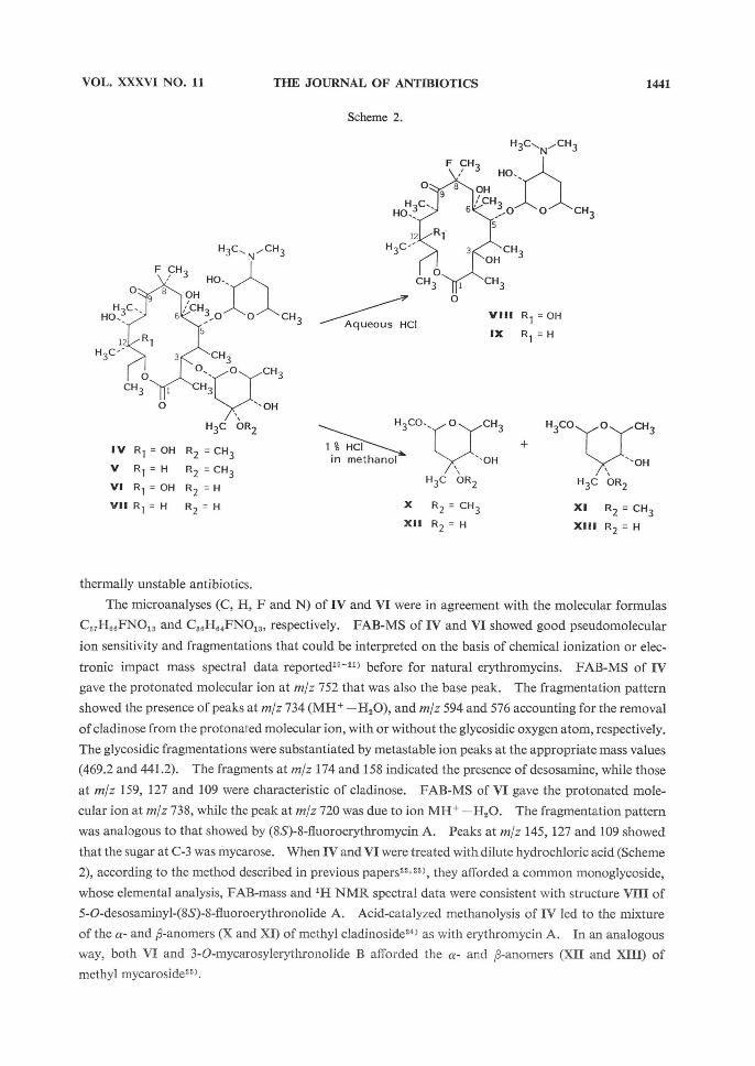
Scheme 2.
H3CN
CH3
F CH3HO
0
H,CHO
R1
H3C
OH
CH3O 0' CH3
CH3
CH30 0
OH
CH3
0
CH3
0
H3C OR2
8'9
E
5
3
12
1
IV R1 = OH R2 = CH3
V R1 = H R2 = CH3
VI R1 = OH R2 = H
VII R1=H R2=H
Aqueous HCI
H3CN
CH3
HOCH3F
0
H,CHO
R1
H3C
OH
CH30 O CH3
CH3
OH
CH3
0
0.CH3
8-9
61
5
3
12
1
VIII R1=OH
IX R1 = H
1 % HCI
in methanol
H3CO 0 CH3
OH
H3C OR2
X R2 = CH3
XII R2 = H
+
H3CO O CH3
OH
H3C OR2
XI R2 = CH3
XIII R2 = H
thermally unstable antibiotics.
The microanalyses (C, H, F and N) of IV and VI were in agreement with the molecular formulas
C37H88FNO13 and C88H84FN01S, respectively. FAB-MS of IV and VI showed good pseudomolecular
ion sensitivity and fragmentations that could be interpreted on the basis of chemical ionization or elec-
tronic impact mass spectral data reported19-21) before for natural erythromycins. FAB-MS of IV
gave the protonated molecular ion at m/z 752 that was also the base peak. The fragmentation pattern showed the presence of peaks at m/z 734 (MH+ -H2O), and m/z 594 and 576 accounting for the removal
of cladinose from the protonated molecular ion, with or without the glycosidic oxygen atom, respectively.
The glycosidic fragmentations were substantiated by metastable ion peaks at the appropriate mass values
(469.2 and 441.2). The fragments at m/z 174 and 158 indicated the presence of desosamine, while those
at m/z 159, 127 and 109 were characteristic of cladinose. FAB-MS of VI gave the protonated mole-
cular ion at m/z 738, while the peak at m/z 720 was due to ion MH+ -H20. The fragmentation pattern
was analogous to that showed by (8S)-8-fluoroerythromycin A. Peaks at m/z 145, 127 and 109 showed
that the sugar at C-3 was mycarose. When IV and VI were treated with dilute hydrochloric acid (Scheme
2), according to the method described in previous papers22,23), they afforded a common monoglycoside,
whose elemental analysis, FAB-mass and 1H NMR spectral data were consistent with structure VIII of
5-O-desosaminyl-(8S)-8-fluoroerythronolide A. Acid-catalyzed methanolysis of IV led to the mixture
of the a- and p-anomers (X and XI) of methyl cladinoside243 as with erythromycin A. In an analogous
way, both VI and 3-O-mycarosylerythronolide B afforded the a- and p-anomers (XII and XIII) of
methyl mycaroside25).
1442 THE JOURNAL OF ANTIBIOTICS NOV. 1983
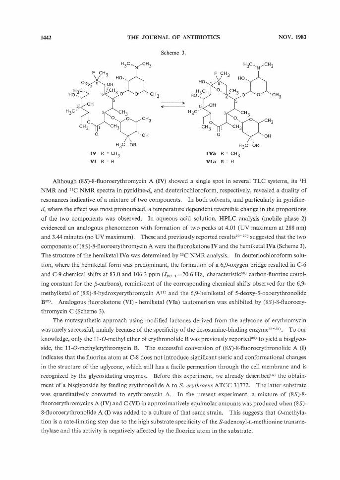
Scheme 3.
H 3 CN
CH3
HC
CH3F
O
H3C
HO
OH
H3C
OH
CH3
OO
1 CH3
CH3
OO
CH3
O
CH3
O
CH3
OH
H3C OR
9
6
5
3
12
1
IV R = CH3
VI R =H
H 3CN
CH3
F CH3
HOHO
H3CHO
OH
O
CH3
OO
CH3
H3C CH3
CH3
O
CH,
OOO
CH3
OH
ORH3C
9. 8'
65
12
3
1
IVa R=CH3
Via R = H
Although (8S)-8-fluoroerythromycin A (IV) showed a single spot in several TLC systems, its 1H
NMR and 13C NMR spectra in pyridine-d, and deuteriochloroform, respectively, revealed a duality of
resonances indicative of a mixture of two components. In both solvents, and particularly in pyridine-
d5 where the effect was most pronounced, a temperature dependent reversible change in the proportions
of the two components was observed. In aqueous acid solution, HPLC analysis (mobile phase 2)
evidenced an analogous phenomenon with formation of two peaks at 4.01 (UV maximum at 288 nm)
and 3.44 minutes (no UV maximum). These and previously reported results26-29) suggested that the two
components of (8S)-8-fluoroerythromycin A were the fluoroketone IV and the hemiketal IVa (Scheme 3).
The structure of the hemiketal IVa was determined by 13C NMR analysis. In deuteriochloroform solu-
tion, where the hemiketal form was predominant, the formation of a 6,9-oxygen bridge resulted in C-6
and C-9 chemical shifts at 83.0 and 106.3 ppm (JFC-9=20.6 Hz, characteristic30) carbon-fluorine coupl-
ing constant for the ~-carbons), reminiscent of the corresponding chemical shifts observed for the 6,9-
methylketal of (8S)-8-hydroxyerythromycin A311 and the 6,9-hemiketal of 5-deoxy-5-oxoerythronolide
B321. Analogous fluoroketone (VI) - hemiketal (VIa) tautomerism was exhibited by (8S)-8-fluoroery-
thromycin C (Scheme 3).
The mutasynthetic approach using modified lactones derived from the aglycone of erythromycin
was rarely successful, mainly because of the specificity of the desosamine-binding enzyme33-35). To our
knowledge, only the 11 -O-methyl ether of erythronolide B was previously reported36) to yield a bisglyco-
side, the 11-O-methylerythromycin B. The successful conversion of (8S)-8-fluoroerythronolide A (I)
indicates that the fluorine atom at C-8 does not introduce significant steric and conformational changes
in the structure of the aglycone, which still has a facile permeation through the cell membrane and is
recognized by the glycosidating enzymes. Before this experiment, we already described" the obtain-
ment of a bisglycoside by feeding erythronolide A to S. erythraeus ATCC 31772. The latter substrate
was quantitatively converted to erythromycin A. In the present experiment, a mixture of (8S)-8-
fluoroerythromycins A (IV) and C (VI) in approximatively equimolar amounts was produced when (8S)-
8-fluoroerythronolide A (I) was added to a culture of that same strain. This suggests that O-methyla-
tion is a rate-limiting step due to the high substrate specificity of the S-adenosyl-L-methionine transme-
thylase and this activity is negatively affected by the fluorine atom in the substrate.

VOL. XXXVI NO. 11 THE JOURNAL OF ANTIBIOTICS 1443
Bioconversion of (8S)-8-Fluoroerythronolide B (II) and 3-O-Mycarosyl-(8S)-
8-fluoroerythronolide B (III)
Two active bioconvertants V and VII (Scheme 1) were produced when either (8S)-8-fluoroerythrono-
lide B (II) or 3-O-mycarosyl-(8S)-8-fluoroerythronolide B (III) was added to the culture of S. erythraeus
ATCC 31772. On TLC and HPLC analysis of the broth culture fed II, it was possible to detect a com-
pound with same chromatographic behavior as III, which disappeared with increasing time of incuba-tion. In analogy to the corresponding biosynthesis of erythromycin 37), it is reasonable to assume that
III is a biosynthetic intermediate in the transformation of II to V (Scheme 1). The structures of V and
VII were deduced from their elemental analyses, FAB-mass spectral data and behavior to the acids.
The elemental analysis (C, H, F and N) of V accorded with the formula C37H66FNO12.
Its FAB-MS gave the protonated molecular ion at m/z 736. A prominent ion at m/z 718 attributed
to MH+ -H2O was present. Metastable ion peaks at the appropriate mass values (453.9 and 426.0)
were also present, indicating the direct loss of cladinose from the protonated molecular ion, either with
(m/z 578) or without (m/z 560, base peak) its glycosidic oxygen atom. The occurrence of peaks cor-responding to those observed for (8S)-8-fluoroerythromycin A, proved that the sugars at C-3 and C-5
were cladinose and desosamine, respectively.
The elemental analysis (C, H, F and N) of VII was consistent with the formula C36H64FNO12.
Its FAB-MS showed the protonated molecular ion at m/z 722 and the ion MH+ -H2O at m/z 704.
The mass fragmentation pattern was similar to that showed by (8S)-8-fluoroerythromycin B. Peaks at
m/z 145, 127 and 109 showed that the sugar at C-3 was mycarose.
Treatment of V and VII in dilute hydrochloric acid (Scheme 2) yielded the same monoglycoside,
whose structure was inferred to be 5-O-desosaminyl-(8S)-8-fluoroerythronolide B (IX) on the basis of
elemental analysis, FAB-mass and 1H NMR spectral data. Complementarily, the same mixture of a-
and 88-anomers (X and XI) of methyl cladinoside was obtained by acid-catalyzed methanolysis of V and
erythromycin A, whereas the a- and ~-anomers (XII and XIII) of methyl mycaroside were derived from
both VU and 3-O-mycarosylerythronolide B. In contrast to (8S)-8-fluoroerythromycins A (IV) and
C (VI), which exist as an interconvertible mixture of tautomers, (8S)-8-fluoroerythromycins B (V) and D
(VII) exist exclusively as the ketonic forms.
In agreement with the results of the biotransformation of (8S)-8-fluoroerythronolide A (I), we de-
monstrate that the glycosidases accept both (8S)-8-fluoroerythronolide B (II) and 3-O-mycarosyl-(8S)-
8-fluoroerythronolide B (III) as possible substrates. It is worth noting that, also in this case, the O-
methylation step is rate-limiting, leading to accumulation of (8S)-8-fluoroerythromycin D (VII) which is
not completely converted to (8S)-8-fluoroerythromycin B (V). Moreover, another enzyme, the C-12
hydroxylase1 ), is clearly shown to possess a very strict substrate specificity, since (8S)-8-fluoroerythro-
mycin C (VI) and (8S)-8-fluoroerythromycin A (IV) are not produced. It is obvious from the results
that this enzyme either does not recognize (8S)-8-fluoroerythromycin D (VII) or is not induced by it.
This specificity, which seems to be the strictest among those of the enzymes of the last steps in the bio-
synthesis of erythromycins, also came out from the before mentioned conversion of 11-O-methyl ether of
erythronolide B yielding only the 11-O-methylerythromycin B, and not the A-form.
Acid Stability
The importance of the fluorine atom at C-8 is clearly illustrated by the data of Table 1 where the acid
stability at pH 2, 3 and 4 of the new (8S)-8-fluoroerythromycins is compared with that of erythromycin

1444 THE JOURNAL OF ANTIBIOTICS NOV. 1983
Table 1. Acid stability at 25°C.
Compounds
IV**
V
VI**
VII
t *
pH 2
9
3
1.65
0.4
0.05
pH 3
82.5
45
41.5
10
0.1
pH 4
>100
>100
>100
>100
2
* The half-life in hours . ** Quantitative data were calculated by summing
the areas of the two peaks corresponding to the ketonic and hemiketalic forms (HPLC analysis).
Fig. 1. Average values of serum levels of erythro-mycin A and IV in rats after a single oral dose of 100 mg/kg.
The standard error of the mean is represented by the vertical bars.
IV
Erythromycin A
Time (hours)
A13). As reported before, (8S)-8-fluoroerythro-
mycins A (IV) and C (VI) display a fluoroketone-
hemiketal tautomerism in these environments
(Scheme 3).
Biological Activity of the Conversion Products
Tables 2-4 summarize the antimicrobial activity in vitro of the new fluoroerythromycins in com-
Table 2. Antibacterial activity of compounds IV, V, VI, VII compared to that of erythromycin A and erythromycin B (agar dilution method).
Organism
Staphylococcus aureus ATCC 6538P Staphylococcus aureus ATCC 14154* Staphylococcus aureus PRL 14** Streptococcus pyogenes ATCC 8668 Streptococcus pneumoniae ATCC 6303 Streptococcus faecalis subsp. zymogenes
ATCC 12958 Corynebacterium diphteriae PRL 24 Micrococcus luteus ATCC 9341 Micrococcus luteus ATCC 15957* Bacillus subtilis ATCC 6633 Haetnophilus influenzae ATCC 19418 Neisseria gonorrhoeae ATCC 19424 Escherichia coli PRL 50 Klebsiella pneumoniae PRL 54 Proteus vulgaris ATCC 6380 Salmonella typhi PRL 8 Shigella sonnei PRL 5 Pseudomonas aeruginosa PRL 9 Clostridium perfringens ATCC 3624 Bacteroides fi-agilis ATCC 23745 Fusobacterium necrophorrrrn ATCC 27852
MIC (ug/ml)
IV
0.097
>25
0.097
0.012
0.024
0.195
0.012
0.006
>25
0.049
3.12
0.049
12.5
12.5
>25
12.5
25
>25
0.78
0.195
1.56
V
0.097
>25
0.097
0.024
0.049
0.195
0.006
0.012
>25
0.049
6.25
0.097
25
>25
>25
25
25
>25
0.39
0.195
3.12
VI
0.097
>25
0.097
0.024
0.024
0.097
0.012
0.012
>25
0.097
12.5
0.195
12.5
25
25
12.5
25
25
6.25
0.78
12.5
VII
0.097
>25
0.097
0.024
0.024
0.097
0.006
0.006
>25
0.049
6.25
0.097
25
>25
>25
12.5
>25
25
3.12
0.39
6.25
Erythro-mycin
A
0.049
>25
0.049
0.012
0.012
0.097
0.006
0.006
>25
0.049
3.12
0.049
6.25
12.5
>25
12.5
12.5
>25
1.56
0.195
3.12
Erythro-mycin
B
0.097
>25
0.195
0.024
0.024
0.195
0.012
0.006
>25
0.049
6.25
0.097
25
25
>25
25
>25
>25
1.56
0.195
6.25
* Erythromycin-resistant
** Penicillin-resistant

VOL. XXXVI NO. 11 THE JOURNAL OF ANTIBIOTICS 1445
Table 3. Antibacterial activity of compounds IV, V, VI, VII compared to that of erythromycin A and erythromycin B (broth dilution method).
Organism
Staphylococcus aureus ATCC 6538P Streptococcus pyogenes ATCC 8668 Streptococcus pneumoniae ATCC 6303 Streptococcus faecalis subsp. zymogenes
ATCC 12958, Corynebacterium diphteriae PRL24
MIC (µg/ml)
IV
0.195
0.097
0.049
0.39
0.012
V
0.097
0.024
0.049
0.39
0.012
VI
0.195
0.049
0.097
0.39
0.049
VII
0.195
0.049
0.049
0.39
0.012
Erythro-mycin
A
0.097
0.024
0.024
0.39
0.012
Erythro-mycin
B
0.195
0.024
0.049
0.39
0.012
Table 4. Bactericidal activity of compounds IV, V, VI, VII compared to that of erythromycin A and ery-
thromycin B.
Organism
Staphylococcus aureus ATCC 6538 P Streptococcus pyogenes ATCC 8668 Streptococcus pneumoniae ATCC 6303 Streptococcus faecalis subsp. zymogenes
ATCC 12958 Corynebacterium diphteriae PRL 24
MBC (µg/ml)
IV
3.12
0.78
0.195
6.25
0.097
V
3.12
0.195
0.049
3.12
0.049
VI
3.12
0.78
0.195
3.12
0.39
VII
3.12
0.39
0.195
3.12
0.195
Erythro-mycin
A
1.56
0.39
0.097
6.25
0.049
Erythro-mycin
B
6.25
0.195
0.195
6.25
0.195
parison with erythromycins A and B. The convertants (8S)-8-fluoroerythromycins A (IV) and B (V) showed the same spectrum and were active to the same extent as compared with the natural occurring
erythromycins. When their potency was estimated") against Micrococcus luteus ATCC 9341, (8S)-8-
fluoroerythromycin B (V) had about 110 % of the activity of erythromycin B, whereas (8S)-8-fluoroery-
thromycin A (IV) had about 70% of the activity of erythromycin A. The lower potency in this latter
case may be attributable to the fluoroketone-hemiketal tautomerism, should a minor activity of IVa be
hypothesized. Comparison between V and erythromycin B shows that replacement of hydrogen by
fluorine at C-8 does not affect the antibacterial activity, contrary to what happens with a corresponding
hydroxyl substituent28,39). To our knowledge, this is the first time that macrolide antibiotics derived by
mutasynthesis are as active as the analogous natural antibiotics.
The high acid stability of (8S)-8-fluoroerythromycins prompted us to further biological investiga-
tions. In Fig. 1 some preliminary data of the serum levels of (8S)-8-fluoroerythromycin A (IV) in rats are
reported in comparison with those of erythromycin A. In view of these results, (8S)-8-fluoroerythromy-
cin A (IV) could be an attractive alternative to erythromycin A 2'-esters which, despite their good oral
absorption, possess undesirable side effects40) and must be hydrolyzed in vivo to be therapeutically ef-
fective41). More complete studies in vivo will be presented in a forthcoming paper.
Experimental
Materials
Erythromycin A was purified from a commercial product by crystallization from chloroform. Ery-
thromycin B was prepared as reported previously39) from a mother liquor concentrate. The latter was

1446 THE JOURNAL OF ANTIBIOTICS NOV. 1983
obtained from an industrial strain of S. erythraeus after crystallization and removal of the majority of the erythromycin A. 3-O-Mycarosylerythronolide B37) was isolated from the fermentation beers of LMC 1198, a blocked mutant of S. erythraeus.
Analysis
Elemental analyses were performed by Alfred Bernhardt Microanalytical Laboratories, Elbach
fiber Engelskirchen, West Germany. All melting points were taken in open capillary tubes using a Tot-toli apparatus (N. Buchi, Flawil, Switzerland) and are uncorrected. Optical rotations were determined at 20°C in 1 % methanol solutions with a Schmidt-Haentsch polarimeter.
UV spectra were measured in methanol using a Varian Cary 210 spectrophotometer. IR spectra
were obtained on a Perkin-Elmer 577 spectrophotometer for KBr disks (0.001 g of substance in 0.2 g of KBr). 1H NMR spectra were obtained on a Varian T-60A spectrometer at room temperature in pyri-dine-d, (c 0.05 g/ml). Chemical shifts are reported in ppm from tetramethylsilane (TMS) as internal reference. 13C NMR spectra were recorded with a Varian XL-200-FT spectrometer at 50.3 MHz in
proton decoupled conditions at room temperature. Samples were dissolved in deuteriochloroform (c 0.2 g/ml) containing TMS as internal reference. FAB-MS were carried out on VG Analytical 7070E mass spectrometer equipped with a FAB source and operating with a 6kV accelerating potential. Ap-
proximatively 0.5 - 1 tog of sample dissolved in glycerol was used.Thin-layer chromatography (TLC) was carried out on precoated silica gel 60 F254 plates (Merck)
using acetone - chloroform - methanol - 10 % ammonium hydroxide (50: 50: 1.4: 2) as developing solvent system (three runs). Compounds were visualized by spraying the plates with anisaldehyde - acetic acid -methanol - sulfuric acid (1: 5: 90: 2). Colors developed after a few minutes at 80°C. Antibiotics were also detected by bioautography on Micrococcus luteus ATCC 9341 seeded agar. This same strain was used as test organism to determine the antibiotic levels in the cultured broths and the biological potency of the isolated compounds by a standard agar diffusion method.
High performance liquid chromatography (HPLC) analyses were carried out according to a modi-fication of a described procedure42). A Hewlett-Packard 1084 B liquid chromatograph equipped with a variable-wavelength detector at 210 nm and a Lichrosorb RP8 10 tom stainless steel column, 250 x 4.6 mm i.d., was used. Flow rate of the mobile phase was 2.0 ml/minute and the column was operated at 40°C. Two mobile phases were employed: mobile phase 1 consisted of acetonitrile - 0.01 M phosphate buffer
pH 7.0 (40: 60); mobile phase 2 consisted of acetonitrile - 0.01 M phosphate buffer pH 7.0 (64: 36). Acid stability trials of new antibiotics were carried out according to the method described in a previous
paper43). Gas liquid chromatographies (GLC) were carried out on a Perkin-Elmer Model 900 B equip-ped with a glass column, 2,000 x 2 mm i.d., packed with 4.3 % Silicone OV 25 on 80-100 mesh HP Chro-mosorb Wand heated from 110°C to 160°C (6°C/minute). Nitrogen gas was used as a carrier at 30 ml/ minute and 2.8 kg/cm2 inlet pressure.
Column Chromatography
Partition column chromatography was carried out in conformity with a reported method44) using
a column (740 x 24 mm i.d.) packed with silica gel 60,70-230 mesh (Merck). Fifteen-milliliter frac-tions were collected at a flow rate of 1.0 ml per minute and were tested by TLC and HPLC (mobile pha-se 2). Fractions containing one product only were combined and concentrated to dryness under re-duced pressure. Residual buffer salts were removed from products by washing their chloroform solu-tions with water. Sephadex LH-20 (Pharmacia Fine Chemicals), particle size 25- 100,um, was refluxed three times for 30 minutes in a mixture of chloroform and methanol (1: 1), filtered and dried at 40°C before beeing used for column chromatography45). Preparation and elution of the column was perform-ed with a mixture of chloroform - hexane (1: 1). Homogeneous fractions were combined and evaporat-ed under reduced pressure.
Microorganism and Fermentation
The strain employed in the present investigation was Streptomyces erythraeus ATCC 31772 which
was cultured as described in a previous paper"). After 24 hours of cultivation, 500 tcg/ml of either sub-strate, (8S)-8-fluoroerythronolide A (I)3) or B (II)3) or 3-O-mycarosyl-(8S)-8-fluoroerythronolide B
(III)4), was added into the culture and the cultivation was continued for a further 96 hours. Antibiotic

VOL. XXXVI NO. 11 THE JOURNAL OF ANTIBIOTICS 1447
production generally reached a maximum after 120 hours (HPLC, mobile phase 2), corresponding to the total conversion of the added substrate (HPLC, mobile phase 1). TLC and bioassay were also used to monitor the process.
Isolation of (8S)-8-Fluoroerythromycins A (IV) and C (VI)
The broth (2.1 liters) to which 1.0 g of (8S)-8-fluoroerythronolide A (I) had been added, was filter-ed over Celite to remove mycelia and the filtrate was clarified by the addition of equal volumes of a 10 aqueous solution of zinc sulfate and a 4 % solution of sodium hydroxide. After centrifugation, the clear supernatant was extracted with ethyl acetate at pH 9.8. The organic extract was washed with water and dried on anhydrous sodium sulfate. Evaporation of the ethyl acetate under reduced pressure left 1.36 g of yellow foam. Partition column chromatography of this material gave 0.35 g of (8S)-8-fluoroery-thromycin A (IV, fractions 90-174) and 0.345 g of (8S)-8-fluoroerythromycin C (VI, fractions 280-400). Crystallization from ethanol yielded 0.230 g of pure IV, as prisms: mp 183-184'C; [a],, -54.9°; UV 283 nm (r 17.9); 1H NMR 8 2.15 (NMe2 of IV), 2.20 (NMe2 of IVa), 3.36 (OMe of IVa), 3.45 (OMe of IV); IR 3520, 3480 (sh.), 3250 (broad), 1735, 1720, 1460, 1380, 1345, 1330, 1305, 1170 cm-1; FAB-MS m/z 752 (MH+).
Anal. Calcd. for C37H66FNO13: C 59.10, H 8.85, F 2.52, N 1.86.
Found : C 59.09, H 8.89, F 2.59, N 1.88.
Pure VI (0.145 g) was obtained as prisms by crystallization from ethanol: mp 217"218°C; [a]D-42 .4°; UV 284 nm (s 23.2); 1H NMR 3 2.10 (NMe, of VI), 2.18 (NMe, of VIa); IR 3550, 3500, 3440
(sh), 3300 (broad), 1730, 1455, 1410, 1380, 1360, 1340, 1330, 1305, 1170 (broad) cm-1; FAB-MS m/z 738 (MH+).
Anal. Calcd. for C36H64FNO13: C 58.60, H 8.74, F 2.57, N 1.90.
Found: C 58.47, H 8.87, F 2.60, N 1.82.
Isolation of (8S)-8-Fluoroerythromycins B (V) and D (VII)
The broth (2.1 liters) to which 1.0 g of (8S)-8-fiuoroerythronolide B (II) had been added, was clari-
fied, extracted and purified by partition column chromatography as reported before. Fractions 18 - 32
(0.35 g) were crystallized from ethanol to give 0.15 g of pure (8S)-8-fluoroerythromycin B (V) as pri-sms: mp 164-j166°C; [a]D -63.1°; UV 285 nm (s 29.5); 1H NMR 6 2.15 (NMe,) 3.45 (OMe); IR 3480 (broad), 1735, 1465, 1385, 1330, 1305, 1170 cm-1; FAB-MS m/z 736 (MH+).
Anal. Calcd. for C37H66FNO12: C 60.39, H 9.04, F 2.58, N 1.90.
Found: C 60.31, H 9.09, F 2.60, N 1.88.
(8S)-8-Fluoroervthromvcin D (VII) was eluted in subsequent fractions (55 - 105) and isolated as a
glass (0.32 g). Crystallization from ethanol afforded 0.15 g of an analytical sample: mp 213-215°C; [a]D -59.9°; UV 285 nm (s 30.8); 1H NMR 3 2.13 (NMe,); IR 3600, 3520, 3300 (broad), 1730, 1460, 1420, 1385, 1370, 1310, 1160 cm-1: FAB-MS m/z 722 (MH+).
Anal. Calcd. for C37H66FNO12: C 59.89, H 8.94, F 2.63, N 1.94.
Found: C 59.87, H 8.85, F 2.63, N 1.88.
The culture (1.0 liter) fed 0.50 g of 3-O-mycarosyl-(8S)-8-fluoroerythronolide B (III), was processedin the same way as reported above. Partition column chromatography of the residue from the solvent extraction, gave 0.115 g of (8S)-8-fluoroerythromycin B (V) and 0.095 g of (8S)-8-fluoroerythromycin D (VII), identical with those prepared as described before.
Acid Cleavage
1) Isolation of 5-O-Desosaminyl-(8S)-8-fluoroerythronolide A (VIII): A solution of 0.752 g (0.001mol) of (8S)-8-fluoroerythromycin A (IV) or 0.738 g (0.001 mol) of (8S)-8-fluoroerythromycin C (VI) in 75 ml of a pH 2 hydrochloric acid buffer, was allowed to stand at room temperature for 24 hours. The complete disappearance of IV and VI was monitored by TLC and HPLC (mobile phase 2). The reaction mixture was poured into a saturated sodium hydrogen carbonate solution, and the product was extracted with ethyl acetate. The combined organic phase was dried over anhydrous sodium sulfate and concentrated under reduced pressure. The resulting solid residue was purified on Sephadex LH-20 to
yield, after crystallization from ethyl ether - hexane, 0.145 g of 5-O-desosaminyl-(8S)-8-fluoroerythrono-lide A (VIII): mp 120~ 124°C; [a]D -18.5°; UV 287 nm (s 22.5); 1H NMR 8 2.12 (NMe2); IR 3460, 1725, 1460, 1380, 1350, 1325, 1170 cm-1; FAB-MS m/z 594 (MH+).

1448 THE JOURNAL OF ANTIBIOTICS NOV. 1983
Anal. Calcd. for C29H52FNO10: C 58.67, H 8.83, F 3.20, N 2.36.
Found: C 58.65, H 8.91, F 3.03, N 2.28.
2) Isolation of 5-O-Desosaminyl-(8S)-8-fluoroerythronolide B (IX): A solution prepared from0.736 g (0.001 mol) of (8S)-8-fluoroerythromycin B (V) or 0.722 g (0.001 mol) of (8S)-8-fluoroerythromy-cin D (VII) and 75 ml of a pH 2 hydrochloric acid buffer, was kept at room temperature 19 hours, until the complete disappearance of the starting product was checked by TLC and HPLC (mobile phase 2). The reaction solution was adjusted to pH 9.0 with 5 % sodium hydroxide and immediately extracted with ethyl acetate. The organic layers were combined, washed with water, dried over anhydrous sodium sulfate and evaporated under reduced pressure. The resulting colorless foam was purified on Sephadex LH-20 to yield 0.215 g of pure 5-O-desosaminyl-(8S)-8-fluoroerythronolide B (IX): mp 116-120'C;
[a], -43.1°; UV 284 nm (e 35.6); 114 NMR d 2.10 (NMe,); IR 3450 (broad), 1725, 1460, 1380, 1330, 1170 cm-1; FAB-MS m/z 578 (MH+).
Anal. Calcd. for C29H52FNO9: C 60.29, H 9.07, F 3.29, N 2.42.
Found: C 60.23, H 9.09, F 3.26, N 2.31.
3) Methanolysis of (8S)-8-Fluoroerythromycins A (IV) and B (V): A solution of 0.037 g (0.05
mmol) of (8S)-8-fluoroerythromycin A (IV) or B (V) in 2 ml of methanol containing 1 % hydrogen chlori-de, was allowed to stand at room temperature for 22 hours. The reaction solution was poured into a saturated sodium hydrogen carbonate solution, concentrated under reduced pressure to remove methanol and then extracted with ethyl acetate. After drying on anhydrous sodium sulfate, the extract was con-centrated under reduced pressure. GLC analysis of the residue showed two components with retention times of 3.8 (18 %) and 4.5 (82 %) minutes, identical to those obtained from the corresponding cleavage of erythromycin A.
4) Methanolysis of (8S)-8-Fluoroerythromycins C (VI) and D (VII): GLC analysis of the mix-
ture obtained by methanolysis of (8S)-8-fluoroerythromycins C (VI) and D (VII) showed two components with retention times of 3.2 (35 %) and 6.7 (65 %) minutes, identical to those obtained from the cor-responding cleavage of 3-O-mycarosylerythronolide B.
Antibacterial Activity In Vitro
Minimal inhibitory concentrations (MIC) were determined by the standard two-fold dilution me-thod using Muller Hinton (MH) medium (Difco). For the genera Streptococcus and Corynebacterium the MH medium was added with 5 % horse serum. The genera Haemophilus, Neisseria and the anaero-bes were tested on GC Completed Medium (Difco). Incubation was performed in gas pack anaerobic
jar (BBL) for anaerobes, in candle jar for the others. Bacterial strains used for susceptibility determina-tions were cultures regularly employed in our screening tests. The strains coded PRL (Pierrel Research Laboratories) have been collected and identified by standard criteria in these laboratories. Bacterial cultures containing approximatively 10° -101 viable cells/ml were prepared from overnight cultures. One loopful of the cultures was inoculated on the agar plates containing antibiotics. The agar plates were incubated at 37°C for 24 hours, and MIC was defined as the lowest concentration that prevented visible growth. The usual diffusion cylinder method") was used to estimate the potency of the new antibiotics against Micrococcus luteus ATCC 9341. For a selected number of bacterial strains the MIC were determined in MH broth. For these strains the minimal bactericidal concentrations (MBC) were also determined by further subculturing in the medium without antibiotic the cultures that after 24 hours did not show visible growth.
Absorption Studies in Rats Female Sprague-Dawley rats weighing 160-j 180 g were given 100 mg/kg of antibiotic orally. Six
rats were sacrificed at each interval, blood samples were withdrawn and sera separated by centrifugation.
The sera were stored at -20°C until assay. Serum levels were determined by the usual diffusion cylinder
plate method" using Micrococcus luteus ATCC 9341.
Acknowledgments
We wish to thank Mr. S. SILINGARDI and Mr. M. Di BITETTO for HPLC and GLC determinations, Mr. R.
PEZZALI for his support in biological activity studies. Our gratitude is also due to Dr. S. BRADAMANTE (Institute

VOL. XXXVI NO. 11 THE JOURNAL OF ANTIBIOTICS 1449
of Industrial Chemistry, Milan University, Italy) and Dr. P. BROOKS (VG Analytical) for their kind permission to use Varian XL-200-FT and VG 7070E instruments, respectively.
References
1) GOLDMAN, P.: The carbon-fluorine bond in compounds of biological interest. Science 164: 1123- 1130, 1969
2) STERN, A. M.; B. M. FOXMAN, A. H. TASHJIAN, Jr. & R. H. ABELES: DL-threo-Q-Fluoroaspartate and DL-threo-~-fluoroasparagine: Selective cytotoxic agents for mammalian cells in culture. J. Med. Chem. 25: 544 550, 1982, and references cited therein
3) ToscANO, L.; G. FIORIELLO, S. SILINGARDI & M. INGLESI: Preparation of (8S)-8-fluoroerythronolide A and
(8S)-8-fluoroerythronolide B, potential substrates for the biological synthesis of new macrolide antibiotics. Tetrahedron, in press
4) TOSCANO, L. & E. SEGHETTI: Transformation of 3-O-mycarosylerythronolide B, an intermediate of ery-thromycin biogenesis, into 3-O-mycarosyl-(8S)-8-fluoroerythronolide B using trifluoromethyl hypofluorite
(CF3OF). Tetrahedron Lett., in press. 5) GERSTENBERGER, M. R. C. & A. HAAS: Methods of fluorination inorganic chemistry. Angew. Chem. Int.
Ed. Engl. 20: 647 - 667, 1981 6) PENGLIS, A. A. E.: Fluorinated carbohydrates. In Advances in Carbohydrate Chemistry and Biochemistry.
Ed. by R. S. TiPSON & D. HORTON, pp. 195 - 285, Academic Press, New York, 1981 7) BARTON, D. H. R.: New methods of specific fluorination. Pure Appl. Chem. 21: 285-293, 1970 8) FOSTER, A. B. & J. H. WESTWOOD: The synthesis of fluorinated carbohydrates. Pure Appl. Chem. 35:
147 " 168, 1973 9) BARTON, D. H. R.: The invention of reactions useful for the synthesis of specifically fluorinated natural
products. Pure Appl. Chem. 49: 1241-1249, 1977 10) BARTON, D. H. R.; R. H. HESSE, L. OGUNKOYA, N. D. WESTCOTT & M. M. PECHET: Organic reactions of
fluoro-oxy-compounds. Fluorination of griseofulvin. J. Chem. Soc. Perkin 11972: 2889-2891, 1972 11) KIRATH, P.; P. H. JoNEs, R. S. EGAN & T. J. PERUN: Acid degradation of erythromycin A and erythromycin
B. Experientia 27: 362, 1971 12) TOSCANO, L.; E. SEGHETTI, M. INGLESI & G. FIORIELLO: Acid-catalized modifications of erythronolide A
and erythronolide B. Preparation of 9,10-anhydroerythronolide A and B 6,9-hemiketals. Gazz. Chim. Ital., in press
13) KAVANAGH, F. & L. J. DENNIN: Erythromycin. In Analytical Microbiology. Ed. by F. KAVANAGH, pp. 289 " 294, Academic Press, New York, 1963
14) DAUM, S. J. & J. R. LEMKE: Mutational biosynthesis of new antibiotics. Ann. Rev. Microbiol. 33: 241-' 265, 1979
15) SPAGNOLI, R. & L. TOSCANO: Erythronolide A glycosidation to erythromycin A by a blocked mutant of Streptomyces erythraeus. J. Antibiotics 36: 435-437, 1983
16) BARBER, M.; R. S. BORDOLI, R. D. SEDGWICK & A. T. TYLER: Fast atom bombardment of solids (F.A.B.): anew ion source for mass spectrometry. J. Chem. Soc., Chem. Comm. 1981: 325-327,1981
17) PARR, V. C.; B. N. GREEN, R. H. BATEMAN & J. C. BILL: Structural studies of cephalosporins using a f ast atom bombardment source. Presented at the 29th Ann. Conf. of Mass Spectrom. & Allied Topics, Minneapolis, 1981
18) BARBER, M.; R. S. BORDOLI, R. D. SEDGWICK, A. N. TYLER & B. W. BYCROFT: Fast atom bombardment mass spectrometry of bleomycin A2 and B2 and their metal complexes. Biochem. Biophys. Res. Comm. 101: 632 - 638, 1981
19) MITSCHER, L. A. & H. D. H. SHOWALTER: Chemical ionization mass spectra of macrolide antibiotics. J. Chem. Soc., Chem. Comm. 1972: 796 - 797, 1972
20) RINEHART, K. L. &J. C. COOK, Jr.: Field desorption mass spectra of antibiotics. J Antibiotics 27: 1 -13, 1974
21) MAJER, J.; J. R. MARTIN, R. S. EGAN & J. W. CORCORAN: Antibiotic glycosides. 8. Erythromycin D, a new macrolide antibiotic. J. Am. Chem. Soc. 99: 1620 "' 1622, 1977
22) WILEY, P. F.; M. V. SIGAL, Jr., O. WEAVER, R. MONAHAN & K. GERZON: Erythromycin. XI. Structure of erythromycin B. J. Am. Chem. Soc. 79: 6070-6074,1957
23) LEMAHIEU, R. A.; M. CARSON & R. W. KIERSTEAD: Glycoside cleavage reactions on erythromycin A. Preparation of erythronolide A. J. Med. Chem. 17: 953-956,1974
24) JONES, P. H. & E. K. ROWLEY: Chemical modifications of erythromycin antibiotics. I. 3'-De(dimethyl-

1450 THE JOURNAL OF ANTIBIOTICS NOV. 1983
amino)erythromycin A and B. J. Org. Chem. 33: 665-670,1968 25) WILEY, P. F.; R. GALE, C. W. PETrINGA & K. GERZON: Erythromycin. XII. The isolation, properties and
partial structure of erythromycin C. J. Am. Chem. Soc. 79: 60746077, 1957 26) TADANIER, J.; J. R. MARTIN, R. S. EGAN, A. W. GOLDSTEIN, R. S. STANASZEK, E. HIRNER & F. FISCHER:
Some chemical and stereochemical modifications of the erythromycin lactone rings. J. Org. Chem. 39: 2495-.2501, 1974
27) TADANIER, J.; P. KURATH, J. R. MARTIN, J. B. MCALPINE, R. S. EGAN, A. W. GOLDSTEIN, S. L. MUELLER & D. A. DUNNIGAN : C (8) Epimeric 8-hydroxy-erythromycins-A. Hely. Chim. Acta 56: 2711 - 2719, 1973
28) KURATH, P.; J. R. MARTIN, J. TADANIER, A. W. GOLDSTEIN, R. S. EGAN & D. A. DUNNIGAN: C(8) Epimeric 8-hydroxy-erythromycins-B. Helv. Chim. Acta 56: 1557 - 1565, 1973
29) JONES, P. H.; T. J. PERUN, E. K. ROWLEY & E. J. BAKER: Chemical modifications of erythromycin anti-biotics. 3. Synthesis of 4" and 11 esters of erythromycin A and B. J. Med. Chem. 15: 631 - 634, 1972
30) BREITMAIER, E. & W. VOELTER: 13C NMR Spectroscopy. Vol. 5 of Monographs in Modern Chemistry. Ed. by H. F. EBEL, pp. 141-144, Verlag Chemie GmbH, Weinheim/Bergstr., 1974
31) EGAN, R. S.; J. R. MARTIN, J. B. MCALPINE, P. KURATH, R. S. STANASZEK & A. W. GOLDSTEIN: The structures of the m-chloroperbenzoic acid oxidation products of 8,9-anhydroerythromycin A- and B-6,9-hemiacetal and of (8S)-8-hydroxyerythromycin B. J. Antibiotics 31: 55-62, 1978
32) NOURSE, J. G. & J. D. RoBERTS: Nuclear magnetic resonance spectroscopy. Carbon-13 spectra of some macrolide antibiotics and derivatives. Substituent and conformational effects. J. Am. Chem. Soc. 97: 4584-4594, 1975
33) PERUN, T. J.; J. R. MARTIN & R. S. EGAN: Cyclic phenylboronates as hydroxyl protecting groups in the synthesis of monoesters of macrolide aglycones. J. Org. Chem. 39: 1490- 1493, 1974
34) MARTIN, J. R.; R. S. EGAN, T. J. PERUN & A. W. GOLDSTEIN: 8-epi-Erythronolide B and its metabolic fate in fermentations of Streptomyces erythreus. Tetrahedron 29: 935 - 940, 1973
35) KURATH, P. & R. S. EGAN (Abbott Labs.): Erythronolide B derivatives. U. S. Patent: 3,697,547, Oct. 10, 1972. [Chem. Abstr. 77: 164536 v, 1972]
36) CORCORAN, J. W.: Biochemical mechanisms in the biosynthesis of the erythromycins. In Antibiotics, Vol. IV. Biosynthesis. Ed. by J. W. CORCORAN, pp. 132 - 174, Springer-Verlag, Berlin, 1981
37) MARTIN, J. R.; T. J. PERUN & R. L. GIROLAMI: Studies on the biosynthesis of the erythromycins. I. Isola-tion and structure of an intermediate glycoside, 3-a-L-mycarosylerythronolide B. Biochemistry 5: 2852-2856,1966
38) GROVE, D. C. & W. A. RANDALL: Assay Methods of Antibiotics. A Laboratory Manual, pp. 9698, Medical Encyclopedia Inc., New York, 1955
39) KROWICKI, K. & A. ZAMOJSKI: Chemical modification of erythromycins. IV. 8-Hydroxyerythromycin B. J. Antibiotics 26: 587-592, 1973
40) WOOD SELL, S. H. & J. T. WILSON: Erythromycin and troleandomycin. In Antimicrobial Therapy. Ed. by B. M. KAGAN, pp. 71 ti 82, W. B. Saunders Co., Philadelphia, 1974
41) TARDREW, P. L.; J. C. H. MAO & D. KENNEY: Antibacterial activity of 2'-esters of erythromycin. Appl. Microbiol. 18: 159-165, 1969
42) TSUJI, K. & J. F. GOETZ: High-performance liquid chromatographic determination of erythromycin. J. Chromatogr. 147: 359 - 367, 1978
43) SPAGNOLI, R.; L. CAPPELLETTI & L. TOSCANO: Biological conversion of erythronolide B, an intermediate of erythromycin biogenesis, into new "hybrid" macrolide antibiotics. J. Antibiotics 36: 365 - 375, 1983
44) OLEINICK, N. L. & J. W. CORCORAN: Two types of binding of erythromycin to ribosomes from antibiotic-sensitive and -resistant Bacillus subtilis 168. J. Biol. Chem. 244: 727 - 735, 1969
45) ANGGARD, E. & H. BERGKVIST: Group separation of prostaglandins on Sephadex LH-20. J. Chromatogr. 48: 542- 544, 1970