
Contributions of Spatial Working Memory to Visuomotor Learning

J. Hyönä, D.P. Munoz, W. Heide and R. Radach (Eds.)Progress in Brain Research, Vol. 140© 2002 Elsevier Science B.V. All rights reserved
CHAPTER 22
Visuomotor transformations for eye–hand coordination
D.Y.P. Henriques, W.P. Medendorp, A.Z. Khan and J.D. Crawford *
York University, Centre for Visual Research, Departments of Psychology and Biology, 4700 Keele St., BSB, rm 291,Toronto, ON M3J 1P3, Canada
Abstract: In recent years the scientific community has come to appreciate that the early cortical representations for visuallyguided arm movements are probably coded in a visual frame, i.e. relative to retinal landmarks. While this scheme accountsfor many behavioral and neurophysiological observations, it also poses certain problems for manual control. For example,how are these oculocentric representations updated across eye movements, and how are they then transformed into usefulcommands for accurate movements of the arm relative to the body? Also, since we have two eyes, which is used as thereference point in eye–hand alignment tasks like pointing? We show that patterns of errors in human pointing suggest thatearly oculocentric representations for arm movement are remapped relative to the gaze direction during each saccade. Tothen transform these oculocentric representations into useful commands for accurate movements of the arm relative to thebody, the brain correctly incorporates the three-dimensional, rotary geometry of the eyes when interpreting retinal images.We also explore the possibility that the eye–hand coordination systems uses a strategy like ocular dominance, but switchesalignment between the left and right eye in order to maximize eye–hand coordination in the best field of view. Finally,we describe the influence of eye position on eye–hand alignment, and then consider how head orientation influences thelinkage between oculocentric visual frames and bodycentric motor frames. These findings are framed in terms of our‘conversion-on-demand’ model, which suggests a virtual representation of egocentric space, i.e. one in which only thoserepresentations selected for action are put through the complex visuomotor transformations required for interaction withactual objects in personal space.
Keywords: Eye–hand coordination; Visuospatial updating; Eye dominance; Retinal geometry; Eye position; Visuomotortransformation; Head position; Body geometry
Introduction
Eye–hand (or hand–eye) coordination is a topic thatjust about everyone can relate to. We need good eye–hand coordination to reach out and pick up a coffeecup, press a doorbell, or catch a ball. Indeed manyof the skilled activities that distinguish humans fromother species involve good eye–hand coordination.
∗ Correspondence to: J.D. Crawford, York University,Centre for Visual Research, Departments of Psychologyand Biology, 4700 Keele St., BSB, rm 291, Toronto, ONM3J 1P3, Canada. Tel.: +1-416-736-2100 ext. 88641;Fax: +1-416-736-5814; E-mail: [email protected]
Most of these we take for granted — only in casesof extreme athletic prowess do we notice eye–handcoordination. But these more basic abilities becomeall too significant when they are impaired, as in theneurological patients studied by Carey et al. (2002,this volume).
Given its centrality in our lives, it is not surprisingthat the study of this behavior has a long history (asearly as Rene Descartes). Clearly this has remained amajor topic of study, with various scientific contribu-tions made in major laboratories of the 20th century.And yet at this time one will find no erudite ‘Soci-ety for Eye–Hand Coordination’, or ‘Annual Meetingfor Eye–hand Coordination’. However, as the contri-butions to this section of the current volume testify,
CICERO/GALAYAA B.V./HYÖNÄ22: pp. 329-340
330
it would seem that it is a topic which has reachedits day, perhaps because we know just enough aboutthese various sub-systems to now try putting themaltogether.
Our particular interest is in the geometric aspectsof the forward serial transformations within the brainthat use vision to guide movement of the hand. Al-though many studies of eye–hand coordination lookat the input (vision) and the output (hand movement),our focus is on the transformations that account forall of the linkages in between — from eye, to head,to body, to arm, to hand. Most of our studies areon pointing or ‘touching’, a pared-down version ofeye–hand coordination minus the grasp element. Ourgoal is to build up a rigorous model — the kind thatcould actually control such a system if one were tobuild it from scratch. Our belief is that in building upsuch a model, we will gain a clearer understandingof the neural processes that one should look for inthe brain. The following is not a general review ofthe topic: in the short space allowed here our aim ismainly to summarize some of our own work in thisarea, and to show how this work has motivated ourthinking in this area.
Eyes on the prize
Many studies, including those described in the othercontributions to this section and elsewhere (e.g. Gie-len et al., 1984; Fisk and Goodale, 1985; Vercheret al., 1994; Engel et al., 2000) have demonstratedthe intimate coupling between movements of theeyes and hand. But why do we need this? What isthe advantage of coupling the eye and hand? Oneway to find out is to de-couple the system and seewhat happens, i.e. ask people to reach or point to-ward objects that they are not looking at. Underthese conditions, Roland Johansson and colleagueshave shown how reaching/manipulating movementsthat are normally precise become fumbling approx-imations. We wanted to quantify the effects of un-coupling on pointing performance (Henriques et al.,1998; Henriques and Crawford, 2000). This workfollowed from earlier observations by Bock (1986)and Enright (1995), showing that people point pastremembered targets located in their visual periphery,which we confirmed and expanded on.
We found that when forced to point toward a
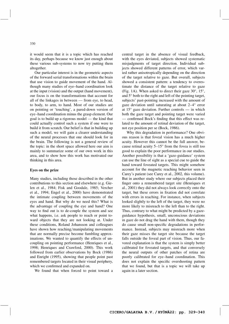
central target in the absence of visual feedback,with the eyes deviated, subjects showed systematicmisjudgments of target direction. Individual sub-jects showed different patterns of error, which var-ied rather anisotropically depending on the directionof the target relative to gaze. But overall, subjectsshowed a consistent pattern: a tendency to overes-timate the distance of the target relative to gaze(Fig. 1A). When asked to direct their gaze 30°, 15°,and 5° both to the right and left of the pointing target,subjects’ past-pointing increased with the amount ofgaze deviation until saturating at about 2–4° errorat 15° gaze deviation. Further controls — in whichboth the gaze target and pointing target were varied— confirmed Bock’s finding that this effect was re-lated to the amount of retinal deviation of the target,not eye position per se (Bock, 1986).
Why this degradation in performance? One obvi-ous reason is that foveal vision has a much higheracuity. However this cannot be the full answer, be-cause retinal acuity 5–15° from the fovea is still toogood to explain the poor performance in our studies.Another possibility is that a ‘gaze-guidance’ systemcan use the line of sight as a special cue to guide thehand toward foveated targets. This might somehowaccount for the magnetic reaching behavior seen inCarey’s patient (see Carey et al., 2002, this volume).But in another study where our subjects placed theirfinger onto a remembered target site (Henriques etal., 2001) they did not always look correctly onto thetarget, but these errors in fixation did not correlatewith errors in reaching. For instance, when subjectslooked slightly to the left of the target, they were nomore likely to misreach to the left than to the right.Thus, contrary to what might be predicted by a gaze-guidance hypothesis, small, unconscious deviationsin gaze do not drag the hand with them, though theydo cause small non-specific degradations in perfor-mance. Instead, subjects may misreach more whentheir gaze misses the target site because the targetfalls outside the foveal part of vision. Thus, our fa-vored explanation is that the system is simply bettercalibrated for foveated targets, and that converselythe neural outputs of other patches of retina arepoorly calibrated for eye–hand coordination. Thisdoes not explain the specific overshooting patternthat we found, but that is a topic we will take upagain in a later section.
CICERO/GALAYAA B.V./HYÖNÄ22: pp. 329-340
331
o
o o
o o
o
o
o
Fig. 1. Final 2-D pointing directions (squares) and gaze direction (circles) relative to central target for a head-fixed subject. Left column:the two tasks, where subjects either view the target peripherally (A) or foveates the target (T1) before looking also to the left (T2)(B). Right column: in the top task (A), the subject past-points in the direction opposite to gaze; this is due to an overestimation of theretinal eccentricity of the target. In the bottom task (B), the subject also past-points in the direction opposite to their final gaze direction,although they fixated the target first (this paradigm is further illustrated in Fig. 2, middle column). Open symbols indicate 15° rightwardfixation trials; solid symbols indicate 15° leftward fixation trials. Modified from Henriques et al., 1998.
Now you see it, now you don’t
A visuomotor puzzle is how it is that we can lookat a target, look away from it so that it is out ofsight, and still know where the target is (Hallett andLightstone, 1976; Mays and Sparks, 1980). Clearly, afixed retinal impression of the target location wouldbe insufficient and downright misleading, so thebrain must be doing something more sophisticated.
One idea was that the brain builds up representa-tions of space by comparing vision with eye position,head position, and so on (Flanders et al., 1992). Un-fortunately, the neurophysiological evidence for thismechanism, at least in visuomotor transformations,remains somewhat sketchy, boiling down to somefairly subtle eye position signals (Andersen andMountcastle, 1983; Schlag et al., 1992; Graziano etal., 1994; Bremmer et al., 1999; Jouffrais and Bous-saoud, 1999) with no clear maps of head-centeredor body-centered space. Perhaps these maps are dis-tributed in some way (Bremmer et al., 1999). Buta more recent suggestion, consistent with certainsignals recorded in the visuomotor structures of thebrain (Duhamel et al., 1992; Mazzoni et al., 1996),
suggests that each time the eyes move, an internalcopy of this movement is used to remap our internalon-line representations of visual space in a retinalframe.
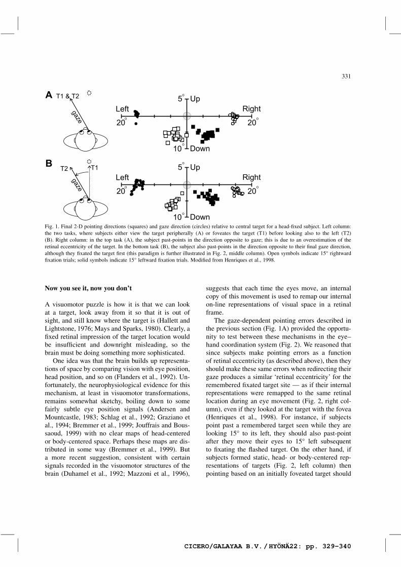
The gaze-dependent pointing errors described inthe previous section (Fig. 1A) provided the opportu-nity to test between these mechanisms in the eye–hand coordination system (Fig. 2). We reasoned thatsince subjects make pointing errors as a functionof retinal eccentricity (as described above), then theyshould make these same errors when redirecting theirgaze produces a similar ‘retinal eccentricity’ for theremembered fixated target site — as if their internalrepresentations were remapped to the same retinallocation during an eye movement (Fig. 2, right col-umn), even if they looked at the target with the fovea(Henriques et al., 1998). For instance, if subjectspoint past a remembered target seen while they arelooking 15° to its left, they should also past-pointafter they move their eyes to 15° left subsequentto fixating the flashed target. On the other hand, ifsubjects formed static, head- or body-centered rep-resentations of targets (Fig. 2, left column) thenpointing based on an initially foveated target should
CICERO/GALAYAA B.V./HYÖNÄ22: pp. 329-340
332
o z
Fig. 2. Predictions of the headcentric (left) and oculocentric (right) models of visuospatial memory on subjects’ pointing to a rememberedtarget. The test paradigm (shown in the middle, as well as in Fig. 1B), has subjects look at the target (time 1) and then look 30° leftafter the target disappears (time 2) before pointing to its remembered location (time 3). The key feature of this test is that during thevisuomotor transformation for pointing, subjects usually exaggerate the retinal eccentricity of the remembered direction of non-fovealtargets (top row of Fig. 1, also Bock, 1986; Enright, 1995). The headcentric model holds that we compute target direction relative to thehead (by combining retinal signals with eye position) as soon as we fixate the target (in time 1). Note that retinal eccentricity at time1 is zero and therefore is not subject to the exaggeration effect. According to the headcentric model, this head-centered memory traceremains stable during the intervening eye movement at time 2, so that accurate pointing is predicted at time 3. The oculocentric modelholds that the target is coded relative to current gaze direction and as a result the leftward eye movement at time 2 must be compensatedfor, by the counter-rotating of the retinotopic memory trace 30° to the right (time 2, right panel). Now the subject must point based on aperipherally shifted retinotopic memory trace, which is susceptible to the exaggeration effect. Therefore, the oculocentric model predictssubject will past-point in the direction opposite to the final gaze direction. Modified from Henriques et al., 1998.
not be affected by subsequent eye movements (that,indeed is the point of this model).
In summary, in the paradigm illustrated both inFig. 1BFig. 2, where people redirect their gaze awayfrom the fixated target before pointing to its remem-bered location, a head-centered model would predictaccurate open-loop pointing (Fig. 2, bottom left),whereas an eye-centered remapping model wouldpredict that subjects past-point in the direction op-posite to gaze (Fig. 2, bottom right) like they do
when they point to the remembered location of aperipherally viewed target (as shown by the datain Fig. 1A). Note that neither model explains past-pointing in the peripherally viewed target conditionshown in Fig. 1A, but given this phenomena, thetwo models predict different pointing performancefor the paradigm shown in Fig. 2. Thus, the resultsin Fig. 1A merely provides the necessary control forpointing based on peripheral retinotopic representa-tions.
CICERO/GALAYAA B.V./HYÖNÄ22: pp. 329-340
333
Our results clearly favored the eye-centered re-mapping model (Fig. 1B). When subjects foveateda briefly flashed target in complete darkness, andthen deviated their eyes, they did not point accu-rately as they did when they maintained gaze on theremembered target site throughout the trial. Instead,they made the same errors in pointing as if theyhad viewed the target in the new retinally periph-eral location (compare Fig. 1B to Fig. 1A). In otherwords, it looks like they were pointing based ona shifted, retinotopic representation. Based on thisresult, we concluded that the eye–hand coordina-tion system uses this same mechanism (Henriques etal., 1998), which had previously been proposed anddescribed for the oculomotor system. Shortly after-wards Andersen and colleagues (Batista et al., 1999)discovered that single-unit responses are consistentwith such a mechanism in the parietal reach region(PRR) — an arm control center with retinotopicallyorganized receptive fields.
Near vs. far
The previously described study by Henriques et al.(1998) was done exclusively with pointing targetsthat were well beyond reach, in so-called extra-personal space. However, a number of neuropsycho-logical studies have suggested that different neuralmechanisms are used for coding near (peripersonal)space (for a review see Colby and Goldberg, 1999).This makes some sense for eye–hand coordination.Anything within reach is coded by preparatory ac-tivity in primary motor cortex (M1), whose signalsare clearly not organized in eye-centered coordinates(Fu et al., 1993; Riehle and Requin, 1995; Mushiakeet al., 1997; Crammond and Kalaska, 2000). Whynot code for near targets using the stable, muscle-centered, eye-movement independent codes of M1?
To test which spatial mechanism dominates hu-man reaching behavior, across eye movements, innear space, we repeated the paradigm in Henriqueset al. (1998) (Fig. 2), but this time using three sets ofarm pointing/reaching targets — one beyond reach,one at 42 cm, and one at 15 cm. According to thehypothesis that near and far space are coded differ-ently, subjects should have shown the same result asbefore for the far target, but should have shown amore stable reaching response for the near targets,
unaffected by the intervening eye movement. Butthis is not what happened (Medendorp and Craw-ford, 2002). Instead, subjects showed the same effectfor all three-target sets: that predicted by the eye-centered remapping model. It would be a mistaketo conclude that this means that the muscle/bodycentered representations are never used to code nearspace in M1 and other structures, but this result doessuggest structures like PRR which do utilize remap-ping override those responses, updating them aftereach eye movement. Perhaps, the near–far distinc-tion may be more relevant for perception than foraction. If so, our results support the notion that tar-get locations are remembered by shifting retinotopicrepresentations as a function of each eye movement,and suggest that this is a general spatial mechanismfor near and far space.
Cyclops had it easy
When we speak of eye–hand coordination, we im-mediately propagate an implicit error: only a myth-ical one-armed Cyclops has eye–hand coordination.Most of us have to coordinate two eyes with twohands, but one generally chooses one hand in reach-ing, often the dominant hand. But which eye ischosen? Or is it some synthesis of the two?
This gets us into the sticky territory of oculardominance and the cyclopean eye. Ocular dominancehas many meanings. Here we consider just alignmentof the hand with the eye. For example, in our monoc-ular pointing studies, we have found that subjectstend to align the fingertip between the eye and thetarget, as if they were reaching out to touch it (asopposed to aligning the line of the arm to the target).So, which eye do they choose? In neurophysiolog-ical terms, which eye dominates the eye-centeredrepresentations in the brain described above?
One appealing idea, put forward in the 19th cen-tury by Wells and more recently championed byOno (Wells, 1792; Ono and Barbeito, 1982), is thatthe inputs from the two eyes are synthesized andreferenced to a virtual ‘cyclopean eye’ positionedbetween them. But, even this theory would requirethe fingertip to be positioned along the line of sightof one eye or the other to be perceived as dead aheadof the cyclopean eye (this may sound contradictory,but makes sense in terms of Wells’ original exper-
CICERO/GALAYAA B.V./HYÖNÄ22: pp. 329-340
334
iments). Thus, the question arises again, which eyedoes the hand coordinate with?
A number of classical studies suggest that thehand prefers to align with a dominant eye (Miles,1930; Crider, 1944; Coren and Kaplan, 1973; Po-rac and Coren, 1976). However, we noted that allof these studies were done with targets locatedstraight ahead. But what about more peripheral tar-gets? Would it not make sense for the system to bemore flexible, and chose either eye, depending onwhich one had the better field of view?
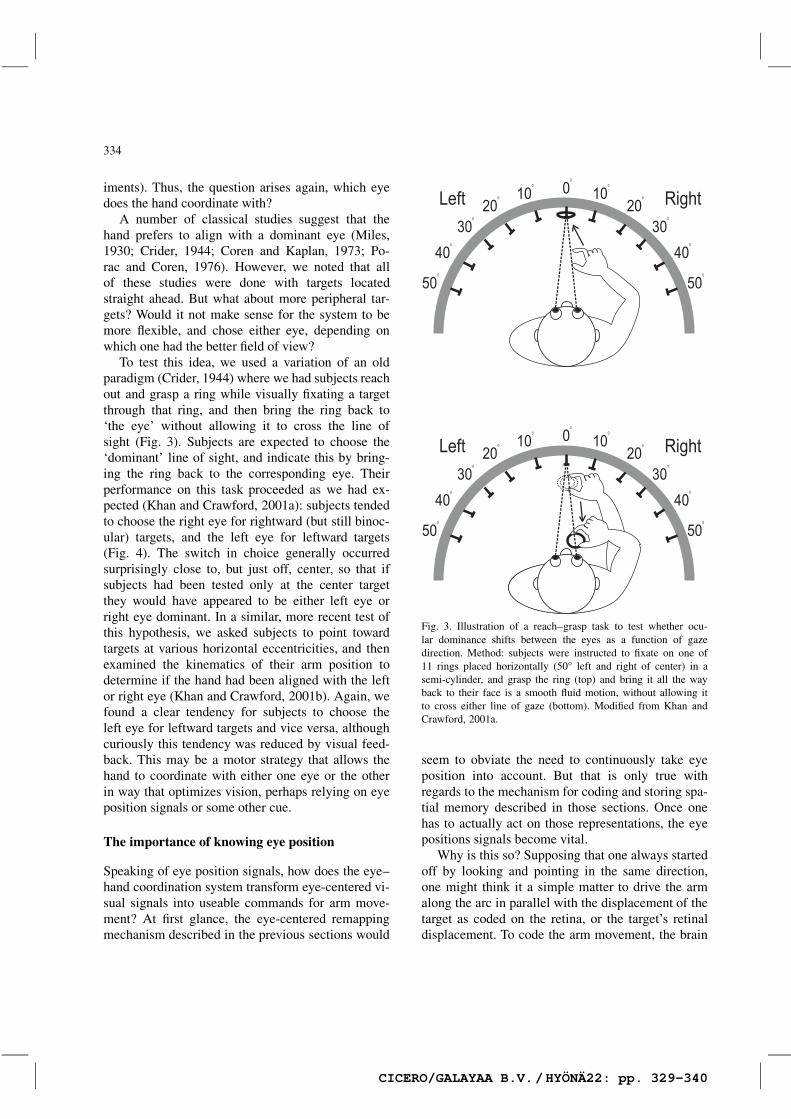
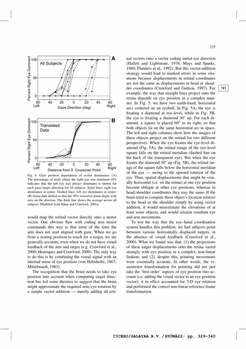
To test this idea, we used a variation of an oldparadigm (Crider, 1944) where we had subjects reachout and grasp a ring while visually fixating a targetthrough that ring, and then bring the ring back to‘the eye’ without allowing it to cross the line ofsight (Fig. 3). Subjects are expected to choose the‘dominant’ line of sight, and indicate this by bring-ing the ring back to the corresponding eye. Theirperformance on this task proceeded as we had ex-pected (Khan and Crawford, 2001a): subjects tendedto choose the right eye for rightward (but still binoc-ular) targets, and the left eye for leftward targets(Fig. 4). The switch in choice generally occurredsurprisingly close to, but just off, center, so that ifsubjects had been tested only at the center targetthey would have appeared to be either left eye orright eye dominant. In a similar, more recent test ofthis hypothesis, we asked subjects to point towardtargets at various horizontal eccentricities, and thenexamined the kinematics of their arm position todetermine if the hand had been aligned with the leftor right eye (Khan and Crawford, 2001b). Again, wefound a clear tendency for subjects to choose theleft eye for leftward targets and vice versa, althoughcuriously this tendency was reduced by visual feed-back. This may be a motor strategy that allows thehand to coordinate with either one eye or the otherin way that optimizes vision, perhaps relying on eyeposition signals or some other cue.
The importance of knowing eye position
Speaking of eye position signals, how does the eye–hand coordination system transform eye-centered vi-sual signals into useable commands for arm move-ment? At first glance, the eye-centered remappingmechanism described in the previous sections would
Fig. 3. Illustration of a reach–grasp task to test whether ocu-lar dominance shifts between the eyes as a function of gazedirection. Method: subjects were instructed to fixate on one of11 rings placed horizontally (50° left and right of center) in asemi-cylinder, and grasp the ring (top) and bring it all the wayback to their face is a smooth fluid motion, without allowing itto cross either line of gaze (bottom). Modified from Khan andCrawford, 2001a.
seem to obviate the need to continuously take eyeposition into account. But that is only true withregards to the mechanism for coding and storing spa-tial memory described in those sections. Once onehas to actually act on those representations, the eyepositions signals become vital.
Why is this so? Supposing that one always startedoff by looking and pointing in the same direction,one might think it a simple matter to drive the armalong the arc in parallel with the displacement of thetarget as coded on the retina, or the target’s retinaldisplacement. To code the arm movement, the brain
CICERO/GALAYAA B.V./HYÖNÄ22: pp. 329-340
335
Fig. 4. Gaze position dependence of ocular dominance. (A)The percentage of trials where the right eye was dominant (0%indicates that the left eye was always dominant) is shown foreach gaze/target direction for 10 subjects. Solid lines: right eyedominance at center. Dashed lines: left eye dominance at center.(B) Same data shifted so that the 50% crossover point aligns withzero on the abscissa. The thick line shows the average across allsubjects. Modified from Khan and Crawford, 2001a.
would map the retinal vector directly onto a motorvector. One obvious flaw with coding arm motorcommands this way is that most of the time thearm does not start aligned with gaze. When we gofrom a resting position to reach for a target, we aregenerally accurate, even when we do not have visualfeedback of the arm and target (e.g. Crawford et al.,2000; Henriques and Crawford, 2000). The only wayto do this is by combining the visual signal with aninternal sense of eye position (von Helmholtz, 1867;Mittelstaedt, 1983).
The recognition that the brain needs to take eyeposition into account when computing target direc-tion has led some theories to suggest that the brainmight approximate the required arm/eye rotation bya simple vector addition — merely adding all reti-
nal vectors onto a vector coding initial eye direction(Hallett and Lightstone, 1976; Mays and Sparks,1980; Flanders et al., 1992). But this vector-additionstrategy would lead to marked errors in some situ-ations because displacements in retinal coordinatesare not the same as displacements in head or shoul-der coordinates (Crawford and Guitton, 1997). For ?#1example, the way that straight lines project onto theretina depends on eye position in a complex man-ner. In Fig. 5, we have two earth-fixed, horizontalarcs centered on an eyeball. In Fig. 5A, the eye isfixating a diamond at eye-level, while in Fig. 5B,the eye is fixating a diamond 30° up. For each di-amond, a square is placed 60° to its right, so thatboth objects lie on the same horizontal arc in space.The left and right columns show how the images ofthese objects project on the retinal for two differentperspectives. When the eye fixates the eye-level di-amond (Fig. 5A), the retinal image of the eye-levelsquare falls on the retinal meridian (dashed line onthe back of the transparent eye). But when the eyefixates the diamond 30° up (Fig. 5B), the retinal im-age of the square falls below the horizontal meridianof the eye — owing to the upward rotation of theeye. Thus, spatial displacements that might be visu-ally horizontal (i.e. on the retina) at one eye positionbecome oblique at other eye positions, whereas inhead/shoulder coordinates they stay the same. If thebrain tried to compute these object’s location relativeto the head or the shoulder simply by using vectoraddition, it would misestimate the elevations of atleast some objects, and would misaim resultant eyeand arm movements.
To test the way that the eye–hand coordinationsystem handles this problem, we had subjects pointbetween various horizontally displaced targets, inthe absence of visual feedback (Crawford et al.,2000). What we found was that: (1) the projectionsof these target displacements onto the retina variedstrongly with eye position in a complex, non-linearfashion; and (2), despite this, pointing movementswere essentially accurate. In other words, the vi-suomotor transformation for pointing did not justtake the ‘first-order’ aspects of eye position into ac-count (i.e. adding the visual vector to an eye positionvector), it in effect accounted for 3-D eye rotationand performed the correct non-linear reference frametransformation.
CICERO/GALAYAA B.V./HYÖNÄ22: pp. 329-340

336
o
o o
Fig. 5. Simulated eye position-dependent geometry of retinal stimulation. Two views of the eyeballs and targets are shown: one directlybehind edge-on (left column) and one behind and slightly up and to the right (right column). The eyeball is transparent, so we can see itsnodal point (small circle), its horizontal meridian (dashed sphere), the two external objects (closed symbols) and their retinal projectionson the back of the eye (open symbols). For both eye positions (A, eye directed straight ahead; and B, eye rotated 30° upward), the eyeis fixating a diamond target. A square is located 60° to the right of the diamond at the same elevation, on a horizontal arc (wrappedaround the eye) at: eye level (A) and 30° up (B). When the eye looks at the eye-level diamond (A), the retinal image of the square lieson the horizontal meridian. But when the eye looks up at the diamond on the top arc (B), the image of the square no longer falls on thehorizontal meridian.
Using your head
Similar arguments hold for taking head orientationinto account. For example, if one were to purpose-fully keep the eyes centered while looking around,the head moves pretty much like an eye (Ceylan etal., 2000) and at least for far targets, the geometrydescribed in the previous section holds pretty muchthe same: one needs to take head orientation intoaccount in a non-linear fashion.
But head movement has other, special implica-tions for eye–hand coordination. This is because thecenters of rotation of the eye, head and shoulder donot coincide. As a result, each time the head rotates,it causes the eyes to translate through space relativeto the shoulder, which changes the angular directionof targets relative to the eye (but not the shoulder).One could rely on visual feedback of the hand —during pointing or reaching — to make up for anydifferences this might make, but we were interestedto see if the system took this into account withoutsuch feedback.
To do this, we had subjects point toward distanttargets with the head in different horizontal posi-tions (Henriques and Crawford, 2002). This causedsignificant changes in the line from the eye to thetarget (recall that this is the line that subjects usefor aligning pointing when looking straight ahead).Nevertheless, subjects were able to re-calculate thisline, and accurately place the fingertip at the right lo-cation for any head position. In a related experiment(Henriques et al., 2001) we found that subjects weresimilarly able to account for head orientation andthe resulting translation of eye location when com-puting the visual angle of near targets for reachingmovements. These results show not only that headorientation is taken into account, but also, moreover,that the eye–hand coordination system possesses asophisticated representation of body geometry thattakes into account the differences in the centers of ro-tation of the eyes, head, and shoulder in the absenceof on-line visual feedback. Such a representation islikely constructed or at least fine-tuned by learning.
CICERO/GALAYAA B.V./HYÖNÄ22: pp. 329-340

337
Arms and the man
Eye–hand coordination must end in a movementof the hand. But at what point in the brain is thetransition from mechanisms specific to eye–hand co-ordination to those purely associated with arm con-trol? A reasonable dividing line would be the pointwhere gaze and head signals no longer influenceneural firing rates, where these are purely modulatedby the kinematics and dynamics of the arm move-ment. Most of the current evidence suggests that thishas not yet occurred at the level of premotor cor-tex (Mushiake et al., 1997), but is completed by orwithin M1 (Mushiake et al., 1997; Crammond andKalaska, 2000).
By corollary, one might not expect eye–hand co-ordination to have much to do with the details of armkinematics that are not related to aiming the handor conforming grasp to the target. A prime examplewould be the neural constraints to determine redun-dant degrees of freedom in the arm control system(Bernstein, 1967). Nevertheless, (without going intodetail) there are considerable similarities betweensome of the three-dimensional kinematics constraintsobserved in the upper arm and in the eye (Straumannet al., 1991; Theeuwen et al., 1993). Based on this,it was once suggested that these constraints were de-veloped in unison in order to facilitate coordinationof the eye and arm in the workspace (Straumann etal., 1991).
In order to test this, we dissociated gaze fromarm movements and measured the three-dimensionalconstraints in the latter (Medendorp et al., 2000).In a similar situation, we had previously found thatdissociating gaze from head movement disrupted thenormal 3-D control of the head, demonstrating theintimate coupling of these two systems (Ceylan etal., 2000). However, the same did not occur: al-though dissociating gaze from the arm again ledto a disruption in movement accuracy, it had noeffect on the three-dimensional constraints on armorientation (Medendorp et al., 2000). Based on thesefindings, we conclude that such constraints are or-ganized in neural structures downstream from thosewhich employ gaze signals to compute arm move-ment direction.
Synthesis and conclusion
As stated in the introduction, the first aim of thiswork is to create a model of eye–hand coordinationthat can help us to understand visually directed armmovements, and help guide our neurophysiologicalinvestigations. The working model that we have beenusing for the past few years is illustrated in Fig. 6.Admittedly it is cartoonish in several respects: inits reliance on forward serial transformations (weall know the brain is really heavily parallel andrecurrent in organization), in its representation ofsignals (no one should expect such discrete, explicitrepresentations in the brain), and in its simplicity(even many aspects of the work described here havebeen left out). And yet in outline, it agrees withmuch of the known physiology.
This model might also account for some symp-toms observed in the neuropsychology literature. Forexample, Carey et al. (2000) report a patient who ?#2reaches ‘magnetically’ toward the direction of fixa-tion. Our model would predict this if the extra-fovealportions of the early gaze-centered representationfor arm movement (i.e. perhaps the parietal reachregion) were lost (a condition not unlikely giventhe tendency to over-represent extra-foveal space).If only activity in the foveal zone were possible,the downstream transformations would automaticallyconvert this into a reaching movement toward thefixation object (and conversely would be unable toreach toward non-foveal objects).
A key feature of this model is its use of earlyrepresentations of attended target locations in a reti-nal frame, representations that must be updated witheach eye movement. Only the target that is selectedfor action is put through the subsequent transforma-tions: comparisons with eye and head orientations,computation of inverse kinematics and dynamics ofthe arm and so on. From a biological perspective, thisis a space and energy saving feature: there would bean enormous cost to try to perform these operationson every point in space, and very little advantage.This goes a long way toward explaining why theearly stages of arm control seem to be organizedin eye-centered coordinates (Henriques et al., 1998).But it also has a surprising implication. Whereasintrospection may suggest that we have a completemap of visuomotor space, this model suggests that
CICERO/GALAYAA B.V./HYÖNÄ22: pp. 329-340

338
Fig. 6. Conversion-on-demand hypothesis of visuomotor representation and visuomotor control. Target representations are initially heldin an eye-centered frame, and can be shifted in this frame to compensate for intervening eye movements through dynamic remapping (A).Once a target has been selected for action, its final retinal representation is put through a reference frame transformation (by comparing itwith 3-D orientation of eyes and head) to generate an accurate 3-D motor signal in motor reference frame (B). Modified from Henriqueset al., 1998.
this is an illusion — only the overall transformationprovides such a map, and only on those representa-tions we choose to act on. The catch is that everytime we test our intuition by choosing to act on arepresentation, it confirms our intuition of a com-plete map. In this sense, this is a ‘virtual’ map ofspace, similar to ideas that have been proposed forvisual perception (Howard, 1982).
A less realistic aspect of the cartoon in Fig. 6 isthe suggestion of explicit signals of desired target lo-cation relative to the head, etc. These are clearly notfound in actual physiology, which tends to employretinal codes (Gnadt and Andersen, 1988; Sparks,1989; Duhamel et al., 1992). However, when one?#3simulates the same transformations by training aneural net to transform a visual vector into a motorvector (Smith and Crawford, 2001a,b), one finds thatthe network learns to do so through fairly simpleposition modulations, resulting in an organizationthat captures the basic conceptual flow of Fig. 6,while using signals that resemble actual physiol-
ogy. Thus, as originally suggested by Andersen andcolleagues, reference frame transformations can beexplicit (Zipser and Andersen, 1988), but in fact onecan entirely get rid of the need for position maps fora more realistic model. Indeed, models that do notrely on explicit position codes turn out to be moreflexible and powerful (Ceylan et al., 2000). Incorpo-rating these ideas, and all of the findings describedabove in this article into a complete computationalmodel following the outline provided in Fig. 6 will ?#4be a major goal of our future work.
Acknowledgements
This research is supported by CIHR group grant,Canadian NSERC and PREA. J.D.C is supported bythe Canadian Research Chair program, D.Y.P.H is sup-ported by an E.A Baker Foundation CIHR ResearchDoctorate Award, and W.P.M. is supported by the Hu-man Frontiers Science Program.
CICERO/GALAYAA B.V./HYÖNÄ22: pp. 329-340
339
References
Andersen, R.A. and Mountcastle, V.B. (1983) The influence ofthe angle of gaze upon the excitability of the light-sensitiveneurons of the posterior parietal cortex. J. Neurosci., 3: 532–548.
Batista, A.P., Buneo, C.A., Snyder, L.H. and Andersen, R.A.(1999) Reach plans in eye-centered coordinates. Science, 285:257–260.
Bernstein, N. (1967) The Coordination and Regulation of Move-ments. Pergamon Press, Oxford.
Bock, O. (1986) Contribution of retinal versus extraretinal sig-nals towards visual localization in goal-directed movements.Exp. Brain Res., 64: 467–482.
Bremmer, F., Graf, W., Ben-Hamed, S. and Duhamel, J.R. (1999)Eye position encoding in the macaque ventral intraparietalarea (VIP). NeuroReport, 10: 873–878.
Carey, D.P., Della Sala, S. and Ietswart, M. (2002) Neuropsy-chological perspectives on eye–hand coordination in visuallyguided reaching. In: J. Hyönä, D.P. Munoz, W. Heide and R.Radach (Eds.), The Brain’s Eye: Neurobiological and ClinicalAspects of Oculomotor Research. Progress in Brain Research,Vol. 140. Elsevier, Amsterdam, pp. 000–000.
Ceylan, M.Z., Henriques, D.Y.P., Tweed, D.B. and Crawford,J.D. (2000) Task-dependent constraints in motor control: pin-hole goggles make the head move like an eye. J. Neurosci.,20: 2719–2730.
Colby, C.L. and Goldberg, M.E. (1999) Space and attention inparietal cortex. Annu. Rev. Neurosci., 22: 319–349.
Coren, S. and Kaplan, C.P. (1973) Patterns of ocular dominance.Am. J. Optom. Arch. Am. Acad. Optom., 50: 283–292.
Crammond, D.J. and Kalaska, J.F. (2000) Prior information inmotor and premotor cortex: activity during the delay periodand effect on pre-movement activity. J. Neurophysiol., 84:986–1005.
Crawford, J.D., Henriques, D.Y.P. and Vilis, T. (2000) Curvatureof visual space under vertical eye rotation: implications forspatial vision and visuomotor control. J. Neurosci., 20: 2360–2368.
Crider, B.A. (1944) A battery of tests for the dominant eye. J.Gen. Psychol., 31: 179–190.
Duhamel, J.-R., Colby, C.L. and Goldberg, M.E. (1992) Theupdating of the representation of visual space in parietal cortexby intended eye movements. Science, 255: 90–92.
Engel, K.C., Anderson, J.H. and Soechting, J.F. (2000) Similarityin the response of smooth pursuit and manual tracking to achange in the direction of target motion. J. Neurophysiol., 84:1149–1156.
Enright, J.P. (1995) The non-visual impact of eye orientation oneye–hand coordination. Vision Res., 35: 1611–1618.
Fisk, J.D. and Goodale, M.A. (1985) The organization of eyeand limb movements during unrestricted reaching to targets incontralateral and ipsilateral visual space. Exp. Brain Res., 60:159–178.
Flanders, M., Helms-Tillery, S.I. and Soechting, J.F. (1992) Earlystages in a sensorimotor transformation. Behav. Brain Sci., 15:309–362.
Fu, Q.F., Suarez, J.I. and Ebner, T.J. (1993) Neuronal specifi-cation of direction and distance during reaching movementsin the superior precentral premotor area and primary motorcortex of monkeys. J. Neurophysiol., 70: 2097–2116.
Gielen, C.C., van den Heuvel, P.J. and van Gisbergen, J.A.(1984) Coordination of fast eye and arm movements in atracking task. Exp. Brain Res., 56: 154–161.
Gnadt, J.W. and Andersen, R.A. (1988) Memory related motorplanning activity in posterior parietal cortex of macaque. Exp.Brain Res., 70: 216–220.
Graziano, M.S., Yap, G.S. and Gross, C.G. (1994) Coding ofvisual space by premotor neurons. Science, 152: 1603–1608.
Hallett, P.E. and Lightstone, A.D. (1976) Saccadic eye move-ments to flashed targets. Vision Res., 16: 107–114.
Henriques, D.Y.P. and Crawford, J.D. (2000) Direction dependentdistortions of retinocentric space in the visuomotor transfor-mation for pointing. Exp. Brain Res., 132(2): 179–194.
Henriques, D.Y.P. and Crawford, J.D. (2002) Role of eye, headand shoulder geometry in the planning of accurate arm move-ments. J. Neurophys., 87: 1677–1685.
Henriques, D.Y.P., Klier, E.M., Smith, M.A., Lowey, D. andCrawford, J.D. (1998) Gaze-centered re-mapping of remem-bered visual space in an open-loop pointing task. J. Neurosci.,18: 1583–1594.
Henriques, D.Y.P., Crawford, J.D., Medendorp, W.P. and Gielen,C.C.A.M. (2001) The eye–hand coordination system accountsfor head orientation and target depth during reaching towardnear targets. Abstr. Soc. Neurosci., 27(2).
Howard, I.P. (1982) Human Visual Orientation. Wiley, NewYork.
Jouffrais, C. and Boussaoud, D. (1999) Neuronal activity relatedto eye–hand coordination in the primate premotor cortex. Exp.Brain Res., 128: 205–209.
Khan, A.Z. and Crawford, J.D. (2001a) Ocular dominance re-verses as a function of gaze angle. Vision Res., 41: 1743–1748.
Khan, A.Z. and Crawford, J.D. (2001b) Coordinating one handwith two eyes: gaze-dependent reversal of ocular dominancein a pointing task. Abstr. Soc. Neurosci., 940.12.
Mays, L.E. and Sparks, D.L. (1980) Saccades are spatially, notretinotopically coded. Science, 208: 1163–1164.
Mazzoni, P., Bracewell, R.M., Barash, S. and Andersen, R.A.(1996) Spatially tuned auditory responses in area LIP ofmacaques performing delayed memory saccades to acoustictargets. J. Neurophysiol., 75: 1233–1241.
Medendorp, W.P. and Crawford, J.D. (2002) Visuospatial updat-ing of reaching target in near and far space. NeuroReport, 13:633–636.
Medendorp, W.P., Crawford, J.D., Henriques, D.Y.P., Van Gisber-gen, J.A.M. and Gielen, C.C.A.M. (2000) Kinematic strategiesfor upper arm — forearm coordination in three dimensions. J.Neurophysiol., 84: 2302–2316.
Miles, W.R. (1930) Ocular dominance in human adults. J. Gen.Psychol., 3: 412–420.
Mittelstaedt, H. (1983) A new solution to the problem of thesubjective vertical. Naturwissenschaften, 70: 272–281.
Mushiake, H., Tanatsugu, Y. and Tanji, J. (1997) Neuronal activ-
CICERO/GALAYAA B.V./HYÖNÄ22: pp. 329-340

340
ity in the ventral part of premotor cortex during target–reachmovement is modulated by direction of gaze. J. Neurophysiol.,78: 567–571.
Ono, H. and Barbeito, R. (1982) The cyclopean eye vs. thesighting-dominant eye as the center of visual direction. Per-cept. Psychophys., 32(3): 201–210.
Porac, C. and Coren, S. (1976) The dominant eye. Psychol. Bull.,83(5): 880–897.
Riehle, A. and Requin, J. (1995) Neuronal correlates of thespecification of movement direction and force in four corticalareas of the monkey. Brain Behav. Res., 70: 1–13.
Schlag, J., Schlag-Rey, M. and Pigarev, I. (1992) Supplemen-tary eye field: influence of eye position on neural signals offixation. Exp. Brain Res., 90: 302–306.
Smith, M.A. and Crawford, J.D. (2001a) Network properties in aphysiologically realistic model of the 2-D to 3-D visuomotortransformation for saccades. Abstr. Soc. Neurosci., 27(1).
Smith, M.A. and Crawford, J.D. (2001b) Self-organizing taskmodules in explicit coordinate systems in a neural networkmodel for 3-D saccades. J. Comput. Neurosci., 10: 127–150.
Sparks, D.L. (1989) The neural encoding of the location of targetsfor saccadic eye movements. J. Exp. Biol., 146: 195–207.
QUERIES:
?#1: Crawford and Guitton, 1997 not in reference list (page 335)?#2: Carey et al. (2000) not in reference list (page 337)?#3: Sparks, ‘1991’ → ‘1989’. If incorrect, please supply ref.for 1991 and cite 1989 (page 338)?#4: von Holst and Mittelstaedt, 1950: not referred to in text.(page 338)
Straumann, D., Haslwanter, T., Hepp-Reymond, M.C. and Hepp,K. (1991) Listing’s law for eye, head and arm movements andtheir synergistic control. Exp. Brain Res., 86: 209–215.
Theeuwen, M., Miller, L.E. and Gielen, C.C.A.M. (1993) Is theorientation of head and arm coupled during pointing move-ments?. J. Motor Behav., 25: 242–250.
Vercher, J.-L., Magenes, G., Prablanc, C. and Gauthier, G.M.(1994) Eye–head–hand coordination in pointing at visual tar-gets: spatial and temporal analysis. Exp. Brain Res., 99: 507–523.
von Helmholtz, H. (1867) Handbuch der Physiologischen Optik,Bd. 3. Voss, Hamburg, Germany.
von Holst, E. and Mittelstaedt, H. (1950) Das Reafferenzprinzip(Wechselwirkungen zwischen Zentralnervensystem und Pe-ripherie). Naturwissenschaften, 37: 464–476.
Wells, W.C. (1792) An Essay Upon Single Vision with Two Eyes:Together with Experiments and Observations on Several OtherSubjects in Optics. Cadell, London.
Zipser, D. and Andersen, R.A. (1988) A back-propagation pro-grammed network that simulates response properties of asubset of posterior parietal neurons. Nature, 331: 679–684.
CICERO/GALAYAA B.V./HYÖNÄ22: pp. 329-340
Copyright © 2022 FDOKUMEN