Visual inspection and dermoscopy, alone or in combination ...
287
University of Birmingham Visual inspection and dermoscopy, alone or in combination, for diagnosing keratinocyte skin cancers in adults Dinnes, Jacqueline; Deeks, Jonathan; Chuchu, Naomi; Matin, Rubeta N.; Wong, Kai Yuen ; Aldridge, Roger Benjamin ; Durack, Alana ; Gulati, Abha; Chan, Sue Ann ; Johnston, Louise ; Bayliss, Susan; Leonardi-Bee, Jo; Takwoingi, Yemisi; Davenport, Clare; O'Sullivan, Colette; Tehrani, Hamid ; Williams, Hywel C. DOI: 10.1002/14651858.CD011901.pub2 Document Version Publisher's PDF, also known as Version of record Citation for published version (Harvard): Dinnes, J, Deeks, J, Chuchu, N, Matin, RN, Wong, KY, Aldridge, RB, Durack, A, Gulati, A, Chan, SA, Johnston, L, Bayliss, S, Leonardi-Bee, J, Takwoingi, Y, Davenport, C, O'Sullivan, C, Tehrani, H & Williams, HC 2018, 'Visual inspection and dermoscopy, alone or in combination, for diagnosing keratinocyte skin cancers in adults', Cochrane Database of Systematic Reviews, no. 12, CD011901. https://doi.org/10.1002/14651858.CD011901.pub2 Link to publication on Research at Birmingham portal General rights Unless a licence is specified above, all rights (including copyright and moral rights) in this document are retained by the authors and/or the copyright holders. The express permission of the copyright holder must be obtained for any use of this material other than for purposes permitted by law. • Users may freely distribute the URL that is used to identify this publication. • Users may download and/or print one copy of the publication from the University of Birmingham research portal for the purpose of private study or non-commercial research. • User may use extracts from the document in line with the concept of ‘fair dealing’ under the Copyright, Designs and Patents Act 1988 (?) • Users may not further distribute the material nor use it for the purposes of commercial gain. Where a licence is displayed above, please note the terms and conditions of the licence govern your use of this document. When citing, please reference the published version. Take down policy While the University of Birmingham exercises care and attention in making items available there are rare occasions when an item has been uploaded in error or has been deemed to be commercially or otherwise sensitive. If you believe that this is the case for this document, please contact [email protected] providing details and we will remove access to the work immediately and investigate. Download date: 17. Mar. 2022
-
Upload
khangminh22 -
Category
Documents
-
view
1 -
download
0
Transcript of Visual inspection and dermoscopy, alone or in combination ...
University of Birmingham
Visual inspection and dermoscopy, alone or incombination, for diagnosing keratinocyte skincancers in adultsDinnes, Jacqueline; Deeks, Jonathan; Chuchu, Naomi; Matin, Rubeta N.; Wong, Kai Yuen ;Aldridge, Roger Benjamin ; Durack, Alana ; Gulati, Abha; Chan, Sue Ann ; Johnston, Louise ;Bayliss, Susan; Leonardi-Bee, Jo; Takwoingi, Yemisi; Davenport, Clare; O'Sullivan, Colette;Tehrani, Hamid ; Williams, Hywel C.DOI:10.1002/14651858.CD011901.pub2
Document VersionPublisher's PDF, also known as Version of record
Citation for published version (Harvard):Dinnes, J, Deeks, J, Chuchu, N, Matin, RN, Wong, KY, Aldridge, RB, Durack, A, Gulati, A, Chan, SA, Johnston,L, Bayliss, S, Leonardi-Bee, J, Takwoingi, Y, Davenport, C, O'Sullivan, C, Tehrani, H & Williams, HC 2018,'Visual inspection and dermoscopy, alone or in combination, for diagnosing keratinocyte skin cancers in adults',Cochrane Database of Systematic Reviews, no. 12, CD011901.https://doi.org/10.1002/14651858.CD011901.pub2
Link to publication on Research at Birmingham portal
General rightsUnless a licence is specified above, all rights (including copyright and moral rights) in this document are retained by the authors and/or thecopyright holders. The express permission of the copyright holder must be obtained for any use of this material other than for purposespermitted by law.
•Users may freely distribute the URL that is used to identify this publication.•Users may download and/or print one copy of the publication from the University of Birmingham research portal for the purpose of privatestudy or non-commercial research.•User may use extracts from the document in line with the concept of ‘fair dealing’ under the Copyright, Designs and Patents Act 1988 (?)•Users may not further distribute the material nor use it for the purposes of commercial gain.
Where a licence is displayed above, please note the terms and conditions of the licence govern your use of this document.
When citing, please reference the published version.
Take down policyWhile the University of Birmingham exercises care and attention in making items available there are rare occasions when an item has beenuploaded in error or has been deemed to be commercially or otherwise sensitive.
If you believe that this is the case for this document, please contact [email protected] providing details and we will remove access tothe work immediately and investigate.
Download date: 17. Mar. 2022
Cochrane Database of Systematic Reviews
Visual inspection and dermoscopy, alone or in combination,
for diagnosing keratinocyte skin cancers in adults (Review)
Dinnes J, Deeks JJ, Chuchu N, Matin RN, Wong KY, Aldridge RB, Durack A, Gulati A, Chan SA,
Johnston L, Bayliss SE, Leonardi-Bee J, Takwoingi Y, Davenport C, O’Sullivan C, Tehrani H,
Williams HC, Cochrane Skin Cancer Diagnostic Test Accuracy Group
Dinnes J, Deeks JJ, ChuchuN,MatinRN,WongKY, AldridgeRB, Durack A, Gulati A, ChanSA, Johnston L, Bayliss SE, Leonardi-Bee J, Takwoingi
Y, Davenport C, O’Sullivan C, Tehrani H, Williams HC, Cochrane Skin Cancer Diagnostic Test Accuracy Group.
Visual inspection and dermoscopy, alone or in combination, for diagnosing keratinocyte skin cancers in adults.
Cochrane Database of Systematic Reviews 2018, Issue 12. Art. No.: CD011901.
DOI: 10.1002/14651858.CD011901.pub2.
www.cochranelibrary.com
Visual inspection and dermoscopy, alone or in combination, for diagnosing keratinocyte skin cancers in adults (Review)
Copyright © 2018 The Cochrane Collaboration. Published by John Wiley & Sons, Ltd.
T A B L E O F C O N T E N T S
1HEADER . . . . . . . . . . . . . . . . . . . . . . . . . . . . . . . . . . . . . . .
1ABSTRACT . . . . . . . . . . . . . . . . . . . . . . . . . . . . . . . . . . . . . .
2PLAIN LANGUAGE SUMMARY . . . . . . . . . . . . . . . . . . . . . . . . . . . . . .
5SUMMARY OF FINDINGS FOR THE MAIN COMPARISON . . . . . . . . . . . . . . . . . . .
9BACKGROUND . . . . . . . . . . . . . . . . . . . . . . . . . . . . . . . . . . . .
Figure 1. . . . . . . . . . . . . . . . . . . . . . . . . . . . . . . . . . . . . . 9
Figure 2. . . . . . . . . . . . . . . . . . . . . . . . . . . . . . . . . . . . . . 13
Figure 3. . . . . . . . . . . . . . . . . . . . . . . . . . . . . . . . . . . . . . 15
17OBJECTIVES . . . . . . . . . . . . . . . . . . . . . . . . . . . . . . . . . . . . .
18METHODS . . . . . . . . . . . . . . . . . . . . . . . . . . . . . . . . . . . . . .
22RESULTS . . . . . . . . . . . . . . . . . . . . . . . . . . . . . . . . . . . . . . .
Figure 4. . . . . . . . . . . . . . . . . . . . . . . . . . . . . . . . . . . . . . 23
Figure 5. . . . . . . . . . . . . . . . . . . . . . . . . . . . . . . . . . . . . . 25
Figure 6. . . . . . . . . . . . . . . . . . . . . . . . . . . . . . . . . . . . . . 26
Figure 7. . . . . . . . . . . . . . . . . . . . . . . . . . . . . . . . . . . . . . 27
Figure 8. . . . . . . . . . . . . . . . . . . . . . . . . . . . . . . . . . . . . . 28
Figure 9. . . . . . . . . . . . . . . . . . . . . . . . . . . . . . . . . . . . . . 31
Figure 10. . . . . . . . . . . . . . . . . . . . . . . . . . . . . . . . . . . . . . 31
Figure 11. . . . . . . . . . . . . . . . . . . . . . . . . . . . . . . . . . . . . . 32
Figure 12. . . . . . . . . . . . . . . . . . . . . . . . . . . . . . . . . . . . . . 33
Figure 13. . . . . . . . . . . . . . . . . . . . . . . . . . . . . . . . . . . . . . 34
Figure 14. . . . . . . . . . . . . . . . . . . . . . . . . . . . . . . . . . . . . . 35
Figure 15. . . . . . . . . . . . . . . . . . . . . . . . . . . . . . . . . . . . . . 37
Figure 16. . . . . . . . . . . . . . . . . . . . . . . . . . . . . . . . . . . . . . 38
Figure 17. . . . . . . . . . . . . . . . . . . . . . . . . . . . . . . . . . . . . . 42
Figure 18. . . . . . . . . . . . . . . . . . . . . . . . . . . . . . . . . . . . . . 43
Figure 19. . . . . . . . . . . . . . . . . . . . . . . . . . . . . . . . . . . . . . 43
Figure 20. . . . . . . . . . . . . . . . . . . . . . . . . . . . . . . . . . . . . . 44
Figure 21. . . . . . . . . . . . . . . . . . . . . . . . . . . . . . . . . . . . . . 45
47DISCUSSION . . . . . . . . . . . . . . . . . . . . . . . . . . . . . . . . . . . . .
48AUTHORS’ CONCLUSIONS . . . . . . . . . . . . . . . . . . . . . . . . . . . . . . .
49ACKNOWLEDGEMENTS . . . . . . . . . . . . . . . . . . . . . . . . . . . . . . . .
49REFERENCES . . . . . . . . . . . . . . . . . . . . . . . . . . . . . . . . . . . . .
78CHARACTERISTICS OF STUDIES . . . . . . . . . . . . . . . . . . . . . . . . . . . . .
196DATA . . . . . . . . . . . . . . . . . . . . . . . . . . . . . . . . . . . . . . . .
Test 1. BCC-Visual Inspection (in-person). . . . . . . . . . . . . . . . . . . . . . . . . . . 200
Test 2. BCC-Visual Inspection (image-based). . . . . . . . . . . . . . . . . . . . . . . . . . 200
Test 3. BCC-VI+Dermoscopy (in-person). . . . . . . . . . . . . . . . . . . . . . . . . . . 201
Test 4. BCC-Dermoscopy alone (image-based). . . . . . . . . . . . . . . . . . . . . . . . . 201
Test 5. BCC-VI - no algorithm at any threshold (in-person). . . . . . . . . . . . . . . . . . . . . 202
Test 6. BCC-VI - no algorithm at BCC possible (in-person). . . . . . . . . . . . . . . . . . . . . 202
Test 7. BCC-VI - ABCD at threshold NR (in-person). . . . . . . . . . . . . . . . . . . . . . . 203
Test 8. BCC-VI - Schwartzberg algorithm (in-person). . . . . . . . . . . . . . . . . . . . . . . 203
Test 9. BCC-VI - no algorithm at any threshold (image-based). . . . . . . . . . . . . . . . . . . . 204
Test 10. BCC-VI - no algorithm at BCC possible (image-based). . . . . . . . . . . . . . . . . . . 204
Test 11. BCC- VI+Dermoscopy no algorithm at NR (in-person). . . . . . . . . . . . . . . . . . . 205
Test 12. BCC-VI+Dermoscopy pattern analysis_obs_dx (in-person). . . . . . . . . . . . . . . . . . 205
Test 13. BCC- VI+Dermoscopy 3 point at >= (in-person). . . . . . . . . . . . . . . . . . . . . . 206
Test 14. BCC-VI+Dermoscopy Two step_obs_dx (in-person). . . . . . . . . . . . . . . . . . . . 206
Test 15. BCC-Dermoscopy - no algorithm at any threshold (image-based). . . . . . . . . . . . . . . . 207
Test 16. BCC-Dermoscopy - pattern analysis at NR (image-based). . . . . . . . . . . . . . . . . . 207
iVisual inspection and dermoscopy, alone or in combination, for diagnosing keratinocyte skin cancers in adults (Review)
Copyright © 2018 The Cochrane Collaboration. Published by John Wiley & Sons, Ltd.
Test 17. BCC-Dermoscopy - Menzies for BCC(rev)_obsdx (image-based). . . . . . . . . . . . . . . . 208
Test 18. BCC-Dermoscopy - Menzies for BCC(new) - 1 char absent&>=1 other +ve (image-based). . . . . . . 208
Test 19. BCC-Dermoscopy - 3 point checklist at >= 2 (image-based). . . . . . . . . . . . . . . . . . 208
Test 20. BCC-Dermoscopy - new SWS at >=1 (image-based). . . . . . . . . . . . . . . . . . . . 209
Test 21. BCC-Dermoscopy - Chaos/clues (image-based). . . . . . . . . . . . . . . . . . . . . . 209
Test 22. cSCC-Visual inspection (in-person). . . . . . . . . . . . . . . . . . . . . . . . . . 210
Test 23. cSCC-Dermoscopy alone (image-based). . . . . . . . . . . . . . . . . . . . . . . . . 210
Test 24. cSCC-VI - no algorithm at NR (in-person). . . . . . . . . . . . . . . . . . . . . . . 211
Test 25. cSCC-Dermoscopy - no algorithm at NR (image-based). . . . . . . . . . . . . . . . . . . 211
Test 26. cSCC-Dermoscopy - SWS at >1 char (image-based). . . . . . . . . . . . . . . . . . . . 211
Test 27. Any -Visual inspection (in-person). . . . . . . . . . . . . . . . . . . . . . . . . . 212
Test 28. Any -Visual inspection (image-based). . . . . . . . . . . . . . . . . . . . . . . . . . 212
Test 29. Any -VI+Dermoscopy (in-person). . . . . . . . . . . . . . . . . . . . . . . . . . . 213
Test 30. Any-Dermoscopy alone (image-based). . . . . . . . . . . . . . . . . . . . . . . . . 213
Test 31. KER-VI - no algorithm at NR (in-person). . . . . . . . . . . . . . . . . . . . . . . . 214
Test 32. KER-VI - ABCD at NR (in-person). . . . . . . . . . . . . . . . . . . . . . . . . . 214
Test 33. KER-VI - no algorithm at NR (image-based). . . . . . . . . . . . . . . . . . . . . . . 215
Test 34. KER- VI+Dermoscopy no algorithm at NR (in-person). . . . . . . . . . . . . . . . . . . 215
Test 35. KER-VI+Dermoscopy - 3 point at >=2 (in-person). . . . . . . . . . . . . . . . . . . . . 215
Test 36. KER-Dermoscopy - no algorithm at any threshold (image-based). . . . . . . . . . . . . . . . 216
Test 37. KER-Dermoscopy - no algorithm at excise (image-based). . . . . . . . . . . . . . . . . . . 216
Test 38. KER- Dermoscopy - pattern at NR (image-based). . . . . . . . . . . . . . . . . . . . . 217
Test 39. KER-Dermoscopy- SWS (image-based). . . . . . . . . . . . . . . . . . . . . . . . . 217
Test 40. KER-Dermoscopy - Chaos/Clues (image-based). . . . . . . . . . . . . . . . . . . . . . 217
Test 41. KER-Dermoscopy - Menzies for BCC(rev)_obsdx (image-based). . . . . . . . . . . . . . . . 218
Test 42. BCC-VI - experience - high (in-person). . . . . . . . . . . . . . . . . . . . . . . . . 218
Test 43. BCC-VI - experience - mixed (in-person). . . . . . . . . . . . . . . . . . . . . . . . 219
Test 44. BCC-VI - experience - NR (in-person). . . . . . . . . . . . . . . . . . . . . . . . . 219
Test 45. BCC-VI - experience - high (image-based). . . . . . . . . . . . . . . . . . . . . . . . 220
Test 46. BCC-VI - experience - mixed (image-based). . . . . . . . . . . . . . . . . . . . . . . 220
Test 47. BCC-VI - experience - NR (image-based). . . . . . . . . . . . . . . . . . . . . . . . 220
Test 48. BCC-VI+Dermoscopy - experience - high (in-person). . . . . . . . . . . . . . . . . . . . 221
Test 49. BCC-VI+Dermsocopy - experience - NR (in-person). . . . . . . . . . . . . . . . . . . . 221
Test 50. BCC-Dermoscopy - experience - high (image-based). . . . . . . . . . . . . . . . . . . . 222
Test 51. BCC-Dermoscopy - experience - mixed (image-based). . . . . . . . . . . . . . . . . . . . 222
Test 52. BCC-Dermoscopy - experience - trained (image-based). . . . . . . . . . . . . . . . . . . 223
Test 53. BCC-Dermoscopy - experience - NR (image-based). . . . . . . . . . . . . . . . . . . . . 223
Test 54. BCC-VI - qualification - Consultant expert (in-person). . . . . . . . . . . . . . . . . . . 224
Test 55. BCC-VI - qualification - Consultant (in-person). . . . . . . . . . . . . . . . . . . . . . 224
Test 56. BCC-VI - qualification - Mixed (Secondary care) (in-person). . . . . . . . . . . . . . . . . 225
Test 57. BCC-VI - qualification - Consultant expert (image-based). . . . . . . . . . . . . . . . . . 225
Test 58. BCC-VI - qualification - Consultant (image-based). . . . . . . . . . . . . . . . . . . . . 225
Test 59. BCC-VI+Dermoscopy - qualification - Consultant expert (in-person). . . . . . . . . . . . . . 226
Test 60. BCC-VI+Dermoscopy - qualification - Consultant (in-person). . . . . . . . . . . . . . . . . 226
Test 61. BCC-Dermoscopy - qualification - Consultant expert (image-based). . . . . . . . . . . . . . . 227
Test 62. BCC-Dermoscopy - qualification - Consultant (image-based). . . . . . . . . . . . . . . . . 227
Test 63. BCC-Dermoscopy - qualification - Resident (image-based). . . . . . . . . . . . . . . . . . 228
Test 64. BCC-Dermoscopy - qualification - Mixed (dermoscopy trained) (image-based). . . . . . . . . . . 228
Test 65. cSCC-VI - experience - mixed (in-person). . . . . . . . . . . . . . . . . . . . . . . . 228
Test 66. cSCC-VI - experience - NR (in-person). . . . . . . . . . . . . . . . . . . . . . . . . 229
Test 67. cSCC-Dermoscopy - experience - trained (image-based). . . . . . . . . . . . . . . . . . . 229
Test 68. cSCC-Dermoscopy - experience - NR (image-based). . . . . . . . . . . . . . . . . . . . 230
Test 73. KER-VI - experience - high (in-person). . . . . . . . . . . . . . . . . . . . . . . . . 230
iiVisual inspection and dermoscopy, alone or in combination, for diagnosing keratinocyte skin cancers in adults (Review)
Copyright © 2018 The Cochrane Collaboration. Published by John Wiley & Sons, Ltd.

Test 74. KER-VI - experience - mixed (in-person). . . . . . . . . . . . . . . . . . . . . . . . 230
Test 75. KER-VI - experience - NR (in-person). . . . . . . . . . . . . . . . . . . . . . . . . 231
Test 76. KER-VI - experience - high (image-based). . . . . . . . . . . . . . . . . . . . . . . . 231
Test 77. KER-VI - experience - NR (image-based). . . . . . . . . . . . . . . . . . . . . . . . 232
Test 78. KER-VI+Dermoscopy - experience - trained (in-person). . . . . . . . . . . . . . . . . . . 232
Test 80. KER-VI+Dermoscopy - experience - NR (in-person). . . . . . . . . . . . . . . . . . . . 232
Test 81. KER-Dermoscopy - experience - high (image-based). . . . . . . . . . . . . . . . . . . . 233
Test 82. KER-Dermoscopy - experience - trained (image-based). . . . . . . . . . . . . . . . . . . 233
Test 83. KER-Dermoscopy - experience - NR (image-based). . . . . . . . . . . . . . . . . . . . . 234
234ADDITIONAL TABLES . . . . . . . . . . . . . . . . . . . . . . . . . . . . . . . . . .
240APPENDICES . . . . . . . . . . . . . . . . . . . . . . . . . . . . . . . . . . . . .
Figure 22. . . . . . . . . . . . . . . . . . . . . . . . . . . . . . . . . . . . . . 278
Figure 23. . . . . . . . . . . . . . . . . . . . . . . . . . . . . . . . . . . . . . 279
279CONTRIBUTIONS OF AUTHORS . . . . . . . . . . . . . . . . . . . . . . . . . . . . .
280DECLARATIONS OF INTEREST . . . . . . . . . . . . . . . . . . . . . . . . . . . . . .
280SOURCES OF SUPPORT . . . . . . . . . . . . . . . . . . . . . . . . . . . . . . . . .
281DIFFERENCES BETWEEN PROTOCOL AND REVIEW . . . . . . . . . . . . . . . . . . . . .
iiiVisual inspection and dermoscopy, alone or in combination, for diagnosing keratinocyte skin cancers in adults (Review)
Copyright © 2018 The Cochrane Collaboration. Published by John Wiley & Sons, Ltd.
[Diagnostic Test Accuracy Review]
Visual inspection and dermoscopy, alone or in combination,for diagnosing keratinocyte skin cancers in adults
Jacqueline Dinnes1 , Jonathan J Deeks1, Naomi Chuchu1, Rubeta N Matin2, Kai Yuen Wong3 , Roger Benjamin Aldridge4, Alana
Durack5 , Abha Gulati6, Sue Ann Chan7 , Louise Johnston8, Susan E Bayliss1, Jo Leonardi-Bee9, Yemisi Takwoingi1 , Clare Davenport1, Colette O’Sullivan10, Hamid Tehrani11, Hywel C Williams12, Cochrane Skin Cancer Diagnostic Test Accuracy Group1
1Institute of Applied Health Research, University of Birmingham, Birmingham, UK. 2Department of Dermatology, Churchill Hospital,
Oxford, UK. 3Department of Plastic and Reconstructive Surgery, Oxford University Hospitals NHS Foundation Trust, Oxford, UK.4Department of Plastic Surgery, NHS Lothian/University of Edinburgh, Edinburgh, UK. 5Dermatology, Addenbrooke’s Hospital,
Cambridge University Hospitals NHS Foundation Trust, Cambridge, UK. 6Department of Dermatology, Barts Health NHS Trust,
London, UK. 7Birmingham Skin Centre, City Hospital, Birmingham, UK. 8NIHR Diagnostic Evidence Co-operative Newcastle,
Newcastle upon Tyne, UK. 9Division of Epidemiology and Public Health, The University of Nottingham, Nottingham, UK. 10c/o
Cochrane Skin Group, The University of Nottingham, Nottingham, UK. 11Department of Plastic and Reconstructive Surgery, Whiston
Hospital, Liverpool, UK. 12Centre of Evidence Based Dermatology, University of Nottingham, Nottingham, UK
Contact address: Jacqueline Dinnes, Institute of Applied Health Research, University of Birmingham, Birmingham, B15 2TT, UK.
Editorial group: Cochrane Skin Group.
Publication status and date: New, published in Issue 12, 2018.
Citation: Dinnes J, Deeks JJ, Chuchu N, Matin RN, Wong KY, Aldridge RB, Durack A, Gulati A, Chan SA, Johnston L, Bayliss SE,
Leonardi-Bee J, Takwoingi Y, Davenport C, O’Sullivan C, Tehrani H, Williams HC, Cochrane Skin Cancer Diagnostic Test Accuracy
Group. Visual inspection and dermoscopy, alone or in combination, for diagnosing keratinocyte skin cancers in adults. CochraneDatabase of Systematic Reviews 2018, Issue 12. Art. No.: CD011901. DOI: 10.1002/14651858.CD011901.pub2.
Copyright © 2018 The Cochrane Collaboration. Published by John Wiley & Sons, Ltd.
A B S T R A C T
Background
Early accurate detection of all skin cancer types is important to guide appropriate management, to reduce morbidity and to improve
survival. Basal cell carcinoma (BCC) is almost always a localised skin cancer with potential to infiltrate and damage surrounding tissue,
whereas a minority of cutaneous squamous cell carcinomas (cSCCs) and invasive melanomas are higher-risk skin cancers with the
potential to metastasise and cause death. Dermoscopy has become an important tool to assist specialist clinicians in the diagnosis of
melanoma, and is increasingly used in primary-care settings. Dermoscopy is a precision-built handheld illuminated magnifier that
allows more detailed examination of the skin down to the level of the superficial dermis. Establishing the value of dermoscopy over
and above visual inspection for the diagnosis of BCC or cSCC in primary- and secondary-care settings is critical to understanding its
potential contribution to appropriate skin cancer triage, including referral of higher-risk cancers to secondary care, the identification
of low-risk skin cancers that might be treated in primary care and to provide reassurance to those with benign skin lesions who can be
safely discharged.
Objectives
To determine the diagnostic accuracy of visual inspection and dermoscopy, alone or in combination, for the detection of (a) BCC and
(b) cSCC, in adults. We separated studies according to whether the diagnosis was recorded face-to-face (in person) or based on remote
(image-based) assessment.
1Visual inspection and dermoscopy, alone or in combination, for diagnosing keratinocyte skin cancers in adults (Review)
Copyright © 2018 The Cochrane Collaboration. Published by John Wiley & Sons, Ltd.
Search methods
We undertook a comprehensive search of the following databases from inception up to August 2016: Cochrane Central Register of
Controlled Trials; MEDLINE; Embase; CINAHL; CPCI; Zetoc; Science Citation Index; US National Institutes of Health Ongoing
Trials Register; NIHR Clinical Research Network Portfolio Database; and the World Health Organization International Clinical Trials
Registry Platform. We studied reference lists and published systematic review articles.
Selection criteria
Studies of any design that evaluated visual inspection or dermoscopy or both in adults with lesions suspicious for skin cancer, compared
with a reference standard of either histological confirmation or clinical follow-up.
Data collection and analysis
Two review authors independently extracted all data using a standardised data extraction and quality assessment form (based on
QUADAS-2). We contacted authors of included studies where information related to the target condition or diagnostic thresholds
were missing. We estimated accuracy using hierarchical summary ROC methods. We undertook analysis of studies allowing direct
comparison between tests. To facilitate interpretation of results, we computed values of sensitivity at the point on the SROC curve with
80% fixed specificity and values of specificity with 80% fixed sensitivity. We investigated the impact of in-person test interpretation;
use of a purposely-developed algorithm to assist diagnosis; and observer expertise.
Main results
We included 24 publications reporting on 24 study cohorts, providing 27 visual inspection datasets (8805 lesions; 2579 malignancies)
and 33 dermoscopy datasets (6855 lesions; 1444 malignancies). The risk of bias was mainly low for the index test (for dermoscopy
evaluations) and reference standard domains, particularly for in-person evaluations, and high or unclear for participant selection,
application of the index test for visual inspection and for participant flow and timing. We scored concerns about the applicability of
study findings as of ‘high’ or ’unclear’ concern for almost all studies across all domains assessed. Selective participant recruitment, lack
of reproducibility of diagnostic thresholds and lack of detail on observer expertise were particularly problematic.
The detection of BCC was reported in 28 datasets; 15 on an in-person basis and 13 image-based. Analysis of studies by prior testing of
participants and according to observer expertise was not possible due to lack of data. Studies were primarily conducted in participants
referred for specialist assessment of lesions with available histological classification. We found no clear differences in accuracy between
dermoscopy studies undertaken in person and those which evaluated images. The lack of effect observed may be due to other sources
of heterogeneity, including variations in the types of skin lesion studied, in dermatoscopes used, or in the use of algorithms and varying
thresholds for deciding on a positive test result.
Meta-analysis found in-person evaluations of dermoscopy (7 evaluations; 4683 lesions and 363 BCCs) to be more accurate than visual
inspection alone for the detection of BCC (8 evaluations; 7017 lesions and 1586 BCCs), with a relative diagnostic odds ratio (RDOR)
of 8.2 (95% confidence interval (CI) 3.5 to 19.3; P < 0.001). This corresponds to predicted differences in sensitivity of 14% (93%
versus 79%) at a fixed specificity of 80% and predicted differences in specificity of 22% (99% versus 77%) at a fixed sensitivity of 80%.
We observed very similar results for the image-based evaluations.
When applied to a hypothetical population of 1000 lesions, of which 170 are BCC (based on median BCC prevalence across studies),
an increased sensitivity of 14% from dermoscopy would lead to 24 fewer BCCs missed, assuming 166 false positive results from both
tests. A 22% increase in specificity from dermoscopy with sensitivity fixed at 80% would result in 183 fewer unnecessary excisions,
assuming 34 BCCs missed for both tests. There was not enough evidence to assess the use of algorithms or structured checklists for
either visual inspection or dermoscopy.
Insufficient data were available to draw conclusions on the accuracy of either test for the detection of cSCCs.
Authors’ conclusions
Dermoscopy may be a valuable tool for the diagnosis of BCC as an adjunct to visual inspection of a suspicious skin lesion following
a thorough history-taking including assessment of risk factors for keratinocyte cancer. The evidence primarily comes from secondary-
care (referred) populations and populations with pigmented lesions or mixed lesion types. There is no clear evidence supporting the
use of currently-available formal algorithms to assist dermoscopy diagnosis.
P L A I N L A N G U A G E S U M M A R Y
2Visual inspection and dermoscopy, alone or in combination, for diagnosing keratinocyte skin cancers in adults (Review)
Copyright © 2018 The Cochrane Collaboration. Published by John Wiley & Sons, Ltd.
Does dermoscopy improve the accuracy of diagnosing basal cell or squamous cell skin cancer (BCC or cSCC) compared to
using the naked eye alone?
What is the aim of the review?
We wanted to find out whether using a handheld illuminated microscope (dermatoscope or ‘dermoscopy’) is any better at diagnosing
basal cell carcinoma (BCC) or cutaneous squamous cell carcinoma (cSCC) compared to just looking at the skin with the naked eye.
We included 24 studies to answer this question.
Why is improving diagnosis of BCC or cSCC important?
There are a number of different types of skin cancer. BCC and cSCC are less serious than melanoma skin cancer, because they usually
grow more slowly and BCC does not spread to other organs in the body. Making the correct diagnosis of BCC or cSCC is still important,
because their treatment may differ. A missed BCC (known as a false negative result) can result in disfigurement and the need for more
major surgery. A missed cSCC can spread to other parts of the body. Diagnosing BCC or cSCC when they are not actually present (a
false positive result) may mean unnecessary treatment, e.g. surgical removal which may result in a disfiguring scar, and worry to patients
if the lesion (a mole or area of skin with an unusual appearance in comparison with the surrounding skin) is benign (not a cancer), or
may result in wrong treatment, e.g. a non-surgical therapy, being used if the lesion is misdiagnosed.
What was studied in the review?
A dermatoscope is a handheld magnifier that includes a light source. Dermoscopy is often used by skin specialists to help diagnose skin
cancer. It is also being used more by community doctors.
As well as seeing whether dermoscopy added anything to visual inspection alone overall, we also wanted to find out whether dermoscopy
accuracy was different when used in a face-to-face consultation or when used on images of skin lesions sent to specialists. We also tried
to find out whether the accuracy of dermoscopy was improved by use of a checklist, or if it was better when used by a skin specialist
compared to a non-specialist.
What are the main results of the review?
The review included 24 studies reporting information for people with lesions suspected of skin cancer.
Diagnosis of BCC with the patient present
We found 11 relevant studies. Eight studies (including 7017 suspicious skin lesions) investigated the accuracy of visual inspection on
its own and seven studies (with 4683 suspicious skin lesions) investigated the accuracy of dermoscopy added to visual inspection (four
of which reported data for both visual inspection on its own and for dermoscopy added to visual inspection). The results suggest that
dermoscopy is more accurate than visual inspection on its own, both for identifying BCC correctly and for excluding things that are
not BCCs.
The results can be illustrated using a group of 1000 lesions, of which 170 (17%) are BCC. In order to see how much better dermoscopy
is in identifying BCC correctly when compared to just looking at the skin, we have to assume that both lead to the same number of
lesions being falsely diagnosed as BCC (we assumed that 166 of the 830 lesions without BCC would have an incorrect diagnosis of
BCC). In this fixed situation, adding dermoscopy to visual inspection would correctly identify an extra 24 BCCs (158 compared with
134) that would have been missed by just looking at the skin alone. In other words, more BCC cancers would be correctly identified.
In order to see how much better dermoscopy is in deciding if a skin lesion is not a BCC when compared to just looking at the skin, we
have to assume that both lead to the same number of BCCs being correctly diagnosed (in this case we assumed that 136 out of the 170
BCCs would be correctly diagnosed). In this situation, adding in dermoscopy to visual inspection would reduce the number of lesions
being wrongly diagnosed as being BCC by 183 (a reduction from 191 in the visual inspection group to eight people in the dermoscopy
group). In other words, more lesions that were not BCC would be correctly identified, and fewer people would end up being sent for
surgery.
Image-based diagnosis of BCC
Eleven studies concerning BCC diagnosis using either clinical photographs or magnified images from a dermatoscope were included.
Four studies, (including 853 suspicious skin lesions) used visual inspection of photographs and nine studies (including 2271 suspicious
lesions) used dermoscopic images (two studies reported data for diagnosis using both photographs and using dermoscopic images).
Results were very similar to the in-person studies.
3Visual inspection and dermoscopy, alone or in combination, for diagnosing keratinocyte skin cancers in adults (Review)
Copyright © 2018 The Cochrane Collaboration. Published by John Wiley & Sons, Ltd.
Value of checklists and observer expertise
There was no evidence that use of a checklist to help visual inspection or dermoscopy interpretation improved diagnostic accuracy.
There was not enough evidence to examine the effect of clinical expertise and training.
Diagnosis of cSCC
There was not enough evidence to reliably comment on the accuracy of either test for the detection of cSCCs.
How reliable are the results of the studies of this review?
Most of our studies made a reliable final diagnosis by lesion biopsy and by following people up over time to make sure the skin lesion
remained negative for skin cancer. Some studies used expert diagnosis to confirm the absence of skin cancer, which is less reliable*. Poor
reporting of what was done in the studies made it difficult for us to judge how reliable they were. Some studies excluded certain types
of skin lesion and some did not describe how a positive test result to trigger referral to a specialist or treatment was defined.
Who do the results of this review apply to?
Eleven studies were done in Europe (46%), and the rest in North America (n = 3), Asia (n = 5), Oceania (n = 2), or multiple countries (n
= 3). People included in the studies were on average between 30 and 74 years old. The percentage of people with BCC ranged between
1% and 61% for in-person studies and between 2% and 63% in studies using images. Almost all studies were done with people referred
from primary care to specialist skin clinics. Over half of studies considered the ability of dermoscopy and visual inspection to diagnose
any skin cancer, including melanoma and BCC, while 10 (42%) focused on just BCC. Variation in the expertise of doctors doing the
examinations and differences in the definitions used to decide when a test was positive make it unclear how dermoscopy should be
carried out and what level of training is needed in order to achieve the accuracy observed in studies.
What are the implications of this review?
When used by specialists, dermoscopy may be a useful tool to help diagnose BCC correctly when compared with visual inspection
alone. It is not clear whether dermoscopy should be used by general practitioners to correctly identify people with suspicious lesions
who need to be seen by a specialist. Checklists to help interpret dermoscopy do not seem to help improve accuracy for BCC. Further
research is needed, to see if dermoscopy is useful in primary care.
How up-to-date is this review?
The review authors searched for and used studies published up to August 2016.
*In these studies biopsy, clinical follow-up or specialist clinician diagnosis were the reference standards (means of establishing the final
diagnosis).
4Visual inspection and dermoscopy, alone or in combination, for diagnosing keratinocyte skin cancers in adults (Review)
Copyright © 2018 The Cochrane Collaboration. Published by John Wiley & Sons, Ltd.
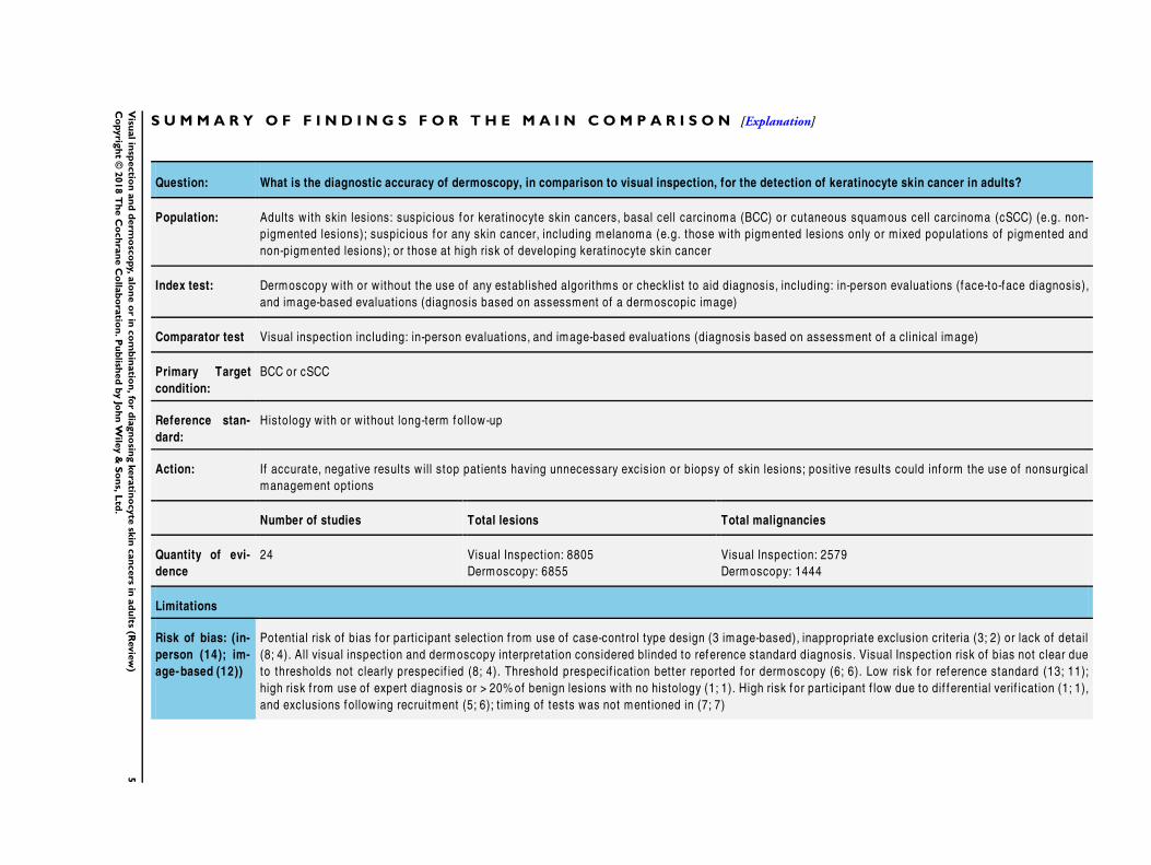
S U M M A R Y O F F I N D I N G S F O R T H E M A I N C O M P A R I S O N [Explanation]
Question: What is the diagnostic accuracy of dermoscopy, in comparison to visual inspection, for the detection of keratinocyte skin cancer in adults?
Population: Adults with skin lesions: suspicious for kerat inocyte skin cancers, basal cell carcinoma (BCC) or cutaneous squamous cell carcinoma (cSCC) (e.g. non-
pigmented lesions); suspicious for any skin cancer, including melanoma (e.g. those with pigmented lesions only or mixed populat ions of pigmented and
non-pigmented lesions); or those at high risk of developing kerat inocyte skin cancer
Index test: Dermoscopy with or without the use of any established algorithms or checklist to aid diagnosis, including: in-person evaluat ions (face-to-face diagnosis),
and image-based evaluat ions (diagnosis based on assessment of a dermoscopic image)
Comparator test Visual inspect ion including: in-person evaluat ions, and image-based evaluat ions (diagnosis based on assessment of a clinical image)
Primary Target
condition:
BCC or cSCC
Reference stan-
dard:
Histology with or without long-term follow-up
Action: If accurate, negat ive results will stop pat ients having unnecessary excision or biopsy of skin lesions; posit ive results could inform the use of nonsurgical
management opt ions
Number of studies Total lesions Total malignancies
Quantity of evi-
dence
24 Visual Inspect ion: 8805
Dermoscopy: 6855
Visual Inspect ion: 2579
Dermoscopy: 1444
Limitations
Risk of bias: (in-
person (14); im-
age-based (12))
Potent ial risk of bias for part icipant select ion f rom use of case-control type design (3 image-based), inappropriate exclusion criteria (3; 2) or lack of detail
(8; 4). All visual inspect ion and dermoscopy interpretat ion considered blinded to reference standard diagnosis. Visual Inspect ion risk of bias not clear due
to thresholds not clearly prespecif ied (8; 4). Threshold prespecif icat ion better reported for dermoscopy (6; 6). Low risk for reference standard (13; 11);
high risk f rom use of expert diagnosis or > 20%of benign lesions with no histology (1; 1). High risk for part icipant f low due to dif ferent ial verif icat ion (1; 1),
and exclusions following recruitment (5; 6); t im ing of tests was not mentioned in (7; 7)
5V
isualin
spectio
nan
dd
erm
osc
opy,
alo
ne
or
inco
mb
inatio
n,fo
rd
iagn
osin
gkera
tino
cyte
skin
can
cers
inad
ults
(Revie
w)
Co
pyrig
ht
©2018
Th
eC
och
ran
eC
olla
bo
ratio
n.P
ub
lished
by
Joh
nW
iley
&S
on
s,L
td.

Applicabil-
ity of evidence to
question: (in-per-
son (14); image-
based (12))
High concern for part icipants (14; 12) due to restrict ion to those with histopathology results (13; 11) and including mult iple lesions per part icipant (9; 2)
. High concern for Visual Inspect ion (7; 4) f rom lack of descript ion of diagnost ic thresholds. High concern for dermoscopy (3; 9) f rom no descript ion of
diagnost ic thresholds (2; 4) or report ing of average or consensus diagnoses (2; 7). Dermoscopic image interpretat ion blinded to clinical images (10 image-
based). Unclear applicability of reference standard due to insuf f icient information concerning the expert ise of the histopathologist (13; 11)
FINDINGS:
We included 24 studies. 14 studies reported data for in-person visual inspect ion (n = 11) or in-person dermoscopy (n = 8); 12 studies reported data for image-based visual
inspect ion (n = 4) or image-based dermoscopy (n = 10). Two studies report both in-person and image-based data. The f indings presented are based on results for the 21
studies report ing data for BCC alone or for cSCC alone. Due to the observed heterogeneity between studies, the results presented are points est imated f rom summary ROC
curves rather than average sensit ivity and specif icity operat ing points. These are presented for illustrat ive purposes and should not be quoted as the actual performance of
visual inspect ion or dermoscopy. We did not undertake analyses of studies by degree of prior test ing due to a lack of relevant information provided in the study publicat ions,
most studies apparent ly being conducted in referred populat ions, and small study subgroups. There was not enough evidence to assess the use of algorithms or structured
checklists for dermoscopy (or visual inspect ion)
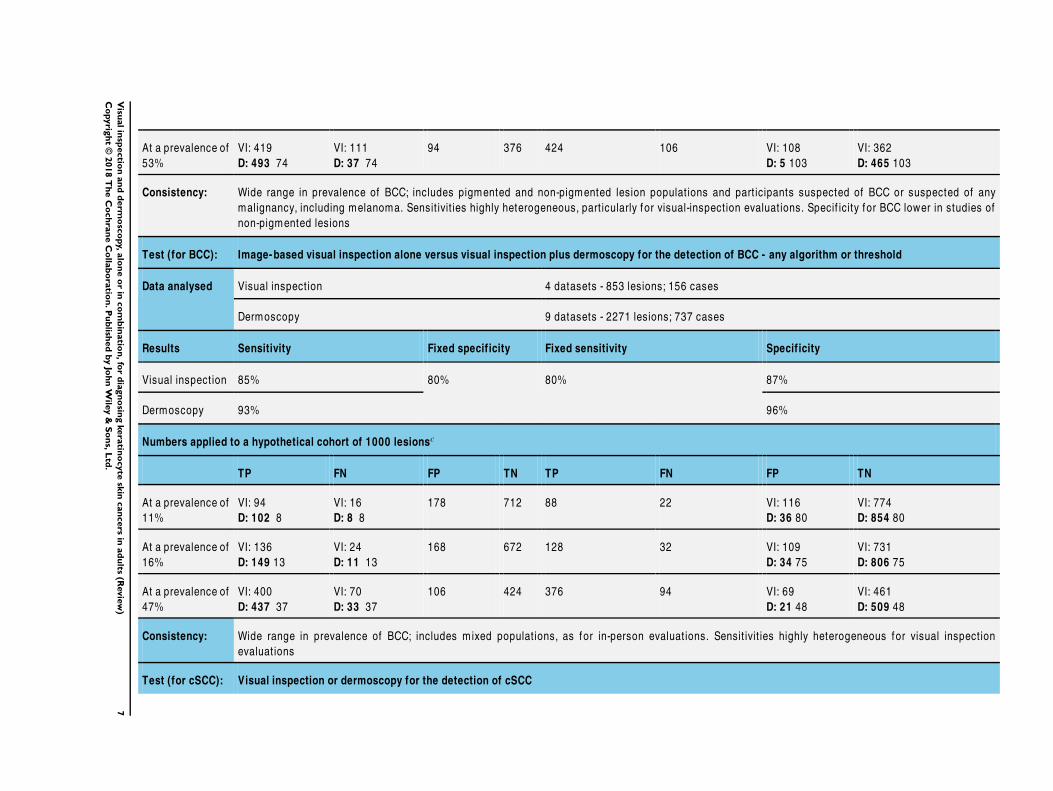
Test (for BCC): In-person visual inspection alone versus visual inspection plus dermoscopy for the detection of BCC - any algorithm or threshold
Data analysed Visual inspect ion 8 datasets - 7017 lesions; 1586 cases
Dermoscopy 7 datasets - 4683 lesions; 363 cases
Resultsa Sensitivity Fixed specificity Fixed sensitivity Specificity
Visual inspect ion 79% 80% 80% 77%
Dermoscopy 93% 99%
Numbers applied to a hypothetical cohort of 1000 lesionsb
TP FN FP TN TP FN FP TN
At a prevalence of
10%
VI: 79
D: 93 14
VI: 21
D: 7 14
180 720 80 20 VI: 207
D: 9 198
VI: 693
D: 891 198
At a prevalence of
17%
VI: 134
D: 158 24
VI: 36
D: 12 24
166 664 136 34 VI: 191
D: 8 183
VI: 639
D: 822 183
6V
isualin
spectio
nan
dd
erm
osc
opy,
alo
ne
or
inco
mb
inatio
n,fo
rd
iagn
osin
gkera
tino
cyte
skin
can
cers
inad
ults
(Revie
w)
Co
pyrig
ht
©2018
Th
eC
och
ran
eC
olla
bo
ratio
n.P
ub
lished
by
Joh
nW
iley
&S
on
s,L
td.
At a prevalence of
53%
VI: 419
D: 493 74
VI: 111
D: 37 74
94 376 424 106 VI: 108
D: 5 103
VI: 362
D: 465 103
Consistency: Wide range in prevalence of BCC; includes pigmented and non-pigmented lesion populat ions and part icipants suspected of BCC or suspected of any
malignancy, including melanoma. Sensit ivit ies highly heterogeneous, part icularly for visual-inspect ion evaluat ions. Specif icity for BCC lower in studies of
non-pigmented lesions
Test (for BCC): Image-based visual inspection alone versus visual inspection plus dermoscopy for the detection of BCC - any algorithm or threshold
Data analysed Visual inspect ion 4 datasets - 853 lesions; 156 cases
Dermoscopy 9 datasets - 2271 lesions; 737 cases
Results Sensitivity Fixed specificity Fixed sensitivity Specificity
Visual inspect ion 85% 80% 80% 87%
Dermoscopy 93% 96%
Numbers applied to a hypothetical cohort of 1000 lesionsc
TP FN FP TN TP FN FP TN
At a prevalence of
11%
VI: 94
D: 102 8
VI: 16
D: 8 8
178 712 88 22 VI: 116
D: 36 80
VI: 774
D: 854 80
At a prevalence of
16%
VI: 136
D: 149 13
VI: 24
D: 11 13
168 672 128 32 VI: 109
D: 34 75
VI: 731
D: 806 75
At a prevalence of
47%
VI: 400
D: 437 37
VI: 70
D: 33 37
106 424 376 94 VI: 69
D: 21 48
VI: 461
D: 509 48
Consistency: Wide range in prevalence of BCC; includes mixed populat ions, as for in-person evaluat ions. Sensit ivit ies highly heterogeneous for visual inspect ion
evaluat ions
Test (for cSCC): Visual inspection or dermoscopy for the detection of cSCC
7V
isualin
spectio
nan
dd
erm
osc
opy,
alo
ne
or
inco
mb
inatio
n,fo
rd
iagn
osin
gkera
tino
cyte
skin
can
cers
inad
ults
(Revie
w)
Co
pyrig
ht
©2018
Th
eC
och
ran
eC
olla
bo
ratio
n.P
ub
lished
by
Joh
nW
iley
&S
on
s,L
td.
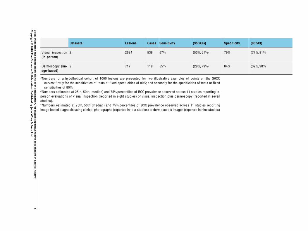
Datasets Lesions Cases Sensitivity (95%CIs) Specificity (95%CI)
Visual inspect ion
(in-person)
2 2684 538 57% (53%, 61%) 79% (77%, 81%)
Dermoscopy (im-
age-based)
2 717 119 55% (29%, 79%) 84% (32%, 98%)
aNumbers for a hypothet ical cohort of 1000 lesions are presented for two illustrat ive examples of points on the SROC
curves: f irst ly for the sensit ivit ies of tests at f ixed specif icit ies of 80%; and secondly for the specif icit ies of tests at f ixed
sensit ivit ies of 80%.bNumbers est imated at 25th, 50th (median) and 75% percent iles of BCC prevalence observed across 11 studies report ing in-
person evaluat ions of visual inspect ion (reported in eight studies) or visual inspect ion plus dermoscopy (reported in seven
studies).cNumbers est imated at 25th, 50th (median) and 75% percent iles of BCC prevalence observed across 11 studies report ing
image-based diagnosis using clinical photographs (reported in four studies) or dermoscopic images (reported in nine studies)
8V
isualin
spectio
nan
dd
erm
osc
opy,
alo
ne
or
inco
mb
inatio
n,fo
rd
iagn
osin
gkera
tino
cyte
skin
can
cers
inad
ults
(Revie
w)
Co
pyrig
ht
©2018
Th
eC
och
ran
eC
olla
bo
ratio
n.P
ub
lished
by
Joh
nW
iley
&S
on
s,L
td.
B A C K G R O U N D
This review is one of a series of Cochrane Diagnostic Test Accu-
racy (DTA) Reviews on the diagnosis and staging of melanoma
and keratinocyte skin cancers as part of the National Institute
for Health Research (NIHR) Cochrane Systematic Reviews Pro-
gramme. Appendix 1 shows the content and structure of the pro-
gramme.
Target condition being diagnosed
The commonest skin cancers in white populations are those aris-
ing from keratinocyte cells: basal cell carcinoma (BCC) and cu-
taneous squamous cell carcinoma (cSCC) (Gordon 2013; Madan
2010). BCC is the more common of the two keratinocyte carci-
nomas, and approximately one-third of people with a BCC will
subsequently develop a second (Flohill 2013). In 2003, the World
Health Organization (WHO) estimated that between two and
three million ‘non-melanoma’ skin cancers (of which BCC and
cSCC are estimated to account for around 80% and 16% of cases
respectively) and 132,000 melanoma skin cancers occur globally
each year (WHO 2003).
Rather than defining BCC and cSCC by what they are not (i.e.
non-melanoma skin cancer), we collectively refer to these condi-
tions using the preferred and more accurate term of ’keratinocyte
carcinoma’ in this DTA review (Karimkhani 2015). We define (a)
BCC and (b) cSCC as the primary target conditions for this re-
view. We also examine accuracy for the target condition of (c) any
skin cancer, including keratinocyte skin cancer, melanoma or in-
tra-epidermal melanocytic variants and any other skin cancer. We
have examined the accuracy of visual inspection for the diagnosis
of melanoma in a previous review (Dinnes 2018a) and in a further
review, we examine the potential benefit of dermoscopy added to
visual inspection for the diagnosis of melanoma (Dinnes 2018b).
Appendix 2 provides a glossary of terms used.
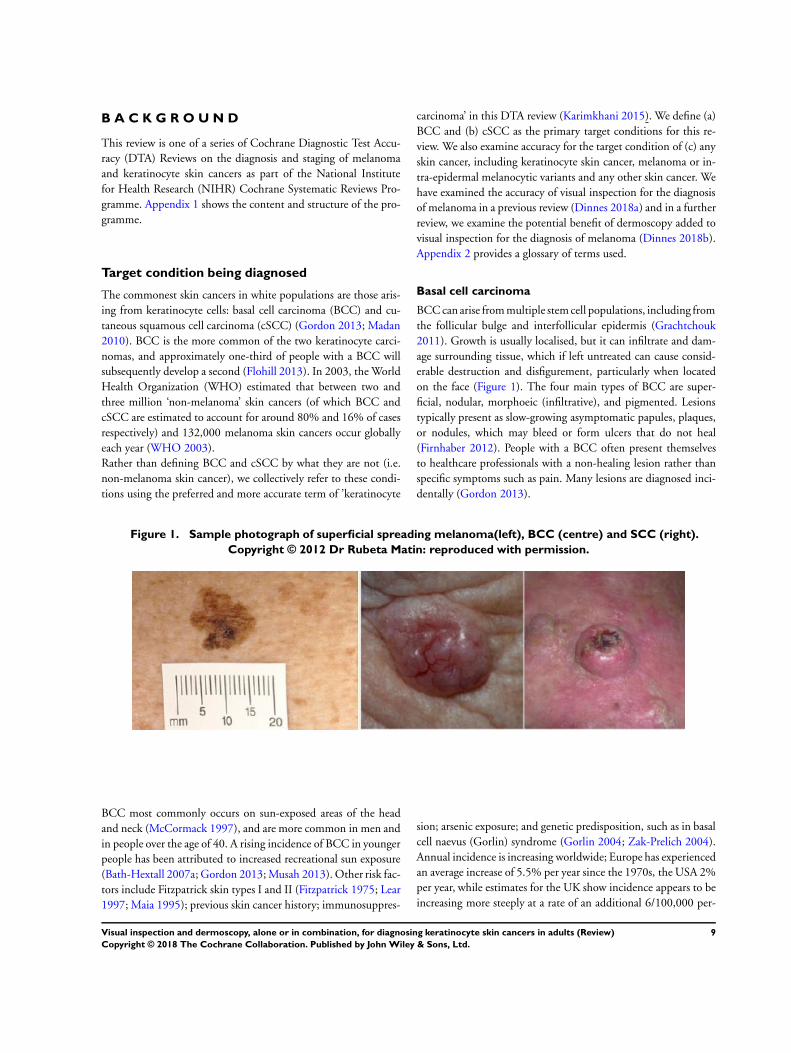
Basal cell carcinoma
BCC can arise from multiple stem cell populations, including from
the follicular bulge and interfollicular epidermis (Grachtchouk
2011). Growth is usually localised, but it can infiltrate and dam-
age surrounding tissue, which if left untreated can cause consid-
erable destruction and disfigurement, particularly when located
on the face (Figure 1). The four main types of BCC are super-
ficial, nodular, morphoeic (infiltrative), and pigmented. Lesions
typically present as slow-growing asymptomatic papules, plaques,
or nodules, which may bleed or form ulcers that do not heal
(Firnhaber 2012). People with a BCC often present themselves
to healthcare professionals with a non-healing lesion rather than
specific symptoms such as pain. Many lesions are diagnosed inci-
dentally (Gordon 2013).
Figure 1. Sample photograph of superficial spreading melanoma(left), BCC (centre) and SCC (right).
Copyright © 2012 Dr Rubeta Matin: reproduced with permission.
BCC most commonly occurs on sun-exposed areas of the head
and neck (McCormack 1997), and are more common in men and
in people over the age of 40. A rising incidence of BCC in younger
people has been attributed to increased recreational sun exposure
(Bath-Hextall 2007a; Gordon 2013; Musah 2013). Other risk fac-
tors include Fitzpatrick skin types I and II (Fitzpatrick 1975; Lear
1997; Maia 1995); previous skin cancer history; immunosuppres-
sion; arsenic exposure; and genetic predisposition, such as in basal
cell naevus (Gorlin) syndrome (Gorlin 2004; Zak-Prelich 2004).
Annual incidence is increasing worldwide; Europe has experienced
an average increase of 5.5% per year since the 1970s, the USA 2%
per year, while estimates for the UK show incidence appears to be
increasing more steeply at a rate of an additional 6/100,000 per-
9Visual inspection and dermoscopy, alone or in combination, for diagnosing keratinocyte skin cancers in adults (Review)
Copyright © 2018 The Cochrane Collaboration. Published by John Wiley & Sons, Ltd.
sons a year (Lomas 2012). The rising incidence has been attributed
to an ageing population, changes in the distribution of known risk
factors, particularly ultraviolet radiation, and improved detection
due to the increased awareness amongst both practitioners and the
general population (Verkouteren 2017). Hoorens 2016 points to
evidence for a gradual increase in the size of BCCs over time, with
delays in diagnosis ranging from 19 to 25 months.
According to National Institute for Health and Care Excellence
(NICE) guidance (NICE 2010), low-risk BCCs are nodular le-
sions occurring in people older than 24 years who are not im-
munosuppressed and do not have Gorlin syndrome. Further-
more, lesions should be located below the clavicle; should be
small (less than 1 cm) with clinically well-defined margins; not
recurrent following incomplete excision or other treatment; and
not in awkward or highly-visible locations (NICE 2010). Super-
ficial BCCs are also typically low risk and may be amenable to
medical treatments such as cryotherapy, photodynamic therapy or
topical immunomodulatory therapy, e.g. 5% Imiquimod cream
(Kelleners-Smeets 2017). Assigning BCCs as low or high risk in-
fluences the management options (Batra 2002; Randle 1996).
Advanced locally-destructive BCC can be found on the H-area of
the face (Lear 2014), can arise from long-standing untreated le-
sions, or from a recurrence of aggressive basal cell carcinoma after
primary treatment (Lear 2012). Very rarely, BCC may metasta-
sise to regional and distant sites resulting in death; this is particu-
larly true for large neglected lesions in those who are immunosup-
pressed, or those with Gorlin syndrome (McCusker 2014). Rates
of metastasis are reported at 0.0028% to 0.55% with very poor
survival rates (Lo 1991). It is recognised that basosquamous car-
cinoma (more like a high-risk SCC in behaviour and not consid-
ered a true BCC) is likely to have accounted for many cases of
apparent metastases of BCC, hence, the spuriously high reported
incidence in some studies of up to 0.55%, which is not seen in
clinical practice (Garcia 2009).
Squamous cell carcinoma of the skin
Primary cSCC arises from the keratinising cells of the epidermis
or its appendages. cSCC typically presents with an ulcer or firm
(indurated) papule, plaque, or nodule (Griffin 2016), often with
an adherent crust (Madan 2010) (Figure 1). cSCC can arise in the
absence of a precursor lesion, or may develop from pre-existing
actinic keratosis or Bowen’s disease (considered by some clinicians
to be cSCC in situ); the estimated annual risk of progression is
less than 1% to 20% for newly-arising lesions (Alam 2001) and
5% for pre-existing lesions (Kao 1986). It remains locally invasive
for a variable length of time, but has the potential to spread to the
regional lymph nodes or via the bloodstream to distant sites, espe-
cially in immunosuppressed individuals (Lansbury 2010). High-
risk lesions are those arising on the lip or ear; recurrent cSCC;
lesions arising on non-exposed sites; within scars or chronic ulcers;
tumours more than 20 mm in diameter and those with a histo-
logical depth of invasion exceeding 4 mm; and poor differentia-
tion status on pathological examination (Motley 2009). Perineural
nerve invasion (PNI) of at least 0.1 mm in diameter is a further
documented risk factor for high-risk cSCC (Carter 2013).
Chronic ultraviolet light exposure through recreation or occupa-
tion is strongly linked to cSCC occurrence (Alam 2001). It is
particularly common in people with fair skin and in less com-
mon genetic disorders of pigmentation, such as albinism, xero-
derma pigmentosum, and recessive dystrophic epidermolysis bul-
losa (RDEB) (Alam 2001). Other recognised risk factors include
immunosuppression; chronic wounds; arsenic or radiation expo-
sure; certain drug treatments, such as voriconazole and BRAF mu-
tation inhibitors; and previous skin cancer history (Baldursson
1993; Chowdri 1996; Dabski 1986; Fasching 1989; Lister 1997;
Maloney 1996; O’Gorman 2014). In solid organ transplant re-
cipients, cSCC is the most common form of skin cancer; the risk
of developing cSCC has been estimated at 65 to 253 times that of
the general population (Hartevelt 1990; Jensen 1999; Lansbury
2010). Overall, local and metastatic recurrence of cSCC at five
years is estimated at 8% and 5% respectively. The five-year sur-
vival rate of metastatic cSCC of the head and neck is around 60%
(Moeckelmann 2018).
Treatment
Treatment options for BCC and cSCC include surgery, other
destructive techniques such as cryotherapy or electrodesiccation
and topical chemotherapy. A Cochrane Review of 27 randomised
controlled trials (RCTs) of interventions for BCC found very
little good-quality evidence for any of the interventions used
(Bath-Hextall 2007b). Complete surgical excision of primary BCC
has a reported five-year recurrence rate of less than 2% (Griffiths
2005; Walker 2006), leading to significantly fewer recurrences
than treatment with radiotherapy (Bath-Hextall 2007b). After ap-
parent clear histopathological margins (serial vertical sections) af-
ter standard excision biopsy with 4 mm surgical peripheral mar-
gins taken, there is a five-year reported recurrence rate of around
4% (Drucker 2017). Mohs micrographic surgery, whereby hor-
izontal sections of the excised specimen are microscopically ex-
amined perioperatively, and re-excision is undertaken until the
margins are tumour-free, can be considered for high-risk lesions
where standard wider excision margins might lead to incomplete
excision or considerable functional and/or cosmetic impairment
(Bath-Hextall 2007b; Motley 2009; Lansbury 2010; Stratigos
2015). Bath-Hextall 2007b found a single trial comparing Mohs
micrographic surgery with a 3 mm surgical margin excision in
BCC (Smeets 2004), showing non-significantly lower recurrence
at 10 years with Mohs micrographic surgery (4.4% compared to
12.2% after surgical excision, P = 0.10) (Van Loo 2014).
The main treatments for high-risk BCC are wide local excision,
Mohs micrographic surgery and radiotherapy. For low-risk or su-
perficial subtypes of BCC, or for small and/or multiple BCCs at
low-risk sites (Marsden 2010), destructive techniques other than
10Visual inspection and dermoscopy, alone or in combination, for diagnosing keratinocyte skin cancers in adults (Review)
Copyright © 2018 The Cochrane Collaboration. Published by John Wiley & Sons, Ltd.
excisional surgery may be used (e.g. electrodesiccation and curet-
tage or cryotherapy (Alam 2001; Bath-Hextall 2007b)). Alterna-
tively, non-surgical (or non-destructive) treatments may be con-
sidered (Bath-Hextall 2007b; Drew 2017; Kim 2014), including
topical chemotherapy such as imiquimod (Williams 2017), 5-flu-
orouracil (5-FU) (Arits 2013), ingenol mebutate (Nart 2015) and
photodynamic therapy (PDT) (Roozeboom 2016). Non-surgical
treatments are most frequently used for superficial forms of BCC,
with one head-to-head trial suggesting topical imiquimod is su-
perior to PDT and 5-FU (Jansen 2018). Although non-surgical
techniques are increasingly used, they do not allow histological
confirmation of tumour clearance, and their efficacy is dependent
on accurate characterisation of the histological subtype and depth
of tumour, and so a baseline diagnostic biopsy can be helpful. The
2007 systematic review of BCC interventions found limited evi-
dence from very small RCTs for these approaches (Bath-Hextall
2007b), which have only partially been filled by subsequent studies
(Bath-Hextall 2014; Kim 2014; Roozeboom 2012). Most BCC
trials have compared interventions within the same treatment class,
and few have compared medical versus surgical treatments (Kim
2014).
Vismodegib, a first-in-class Hedgehog signalling pathway in-
hibitor, is now available for the treatment of metastatic or lo-
cally-advanced BCC based on the pivotal study ERIVANCE BCC
(Sekulic 2012). It is licensed for use in people with BCC where
surgery or radiotherapy is inappropriate, e.g. for treating locally-
advanced periocular and orbital BCCs with orbital salvage of pa-
tients who otherwise would have required exenteration (Wong
2017). However, NICE has recently recommended against the use
of vismodegib based on cost effectiveness and uncertainty of evi-
dence (NICE 2017).
A systematic review of interventions for primary cSCC found only
one RCT eligible for inclusion (Lansbury 2010). Current practice
therefore relies on evidence from observational studies, as reviewed
in Lansbury 2013, for example. Surgical excision with predeter-
mined margins is usually the first-line treatment (Motley 2009;
Stratigos 2015). Estimates of recurrence after Mohs micrographic
surgery, surgical excision, or radiotherapy, which are likely to have
been evaluated in higher-risk populations, have shown pooled re-
currence rates of 3%, 5.4% and 6.4%, respectively, with overlap-
ping confidence intervals; the review authors advise caution when
comparing results across treatments (Lansbury 2013).
Index test(s)
For the purposes of our series of reviews, each component of the
diagnostic process, including visual inspection during clinical ex-
amination, is considered a diagnostic or index ‘test’, the accuracy
of which can be established in comparison with a reference stan-
dard of diagnosis, either alone or in combination with other avail-
able technologies that may assist the diagnostic process. In this
review, two index tests are under consideration: visual inspection
and dermoscopy, both of which can be undertaken in person (in a
face-to-face consultation) or image-based (remote diagnosis using
images). As dermoscopy is effectively added to visual inspection of
a skin lesion when it is undertaken in person, we effectively have
three index tests: visual inspection alone (in person or using im-
ages), visual inspection plus dermoscopy (in-person dermoscopy),
and dermoscopy alone (image-based dermoscopy).
Visual inspection
Clinical history-taking and visual inspection (and palpation) of
the lesion, surrounding skin and comparison with other lesions
identified on complete examination of the body, is fundamental
to the diagnosis of skin cancer. In the UK, clinical examination is
typically done at two decision points: first in primary care where
a decision is made to refer, treat (if low-risk BCC is suspected),
or reassure, and then a second time by a dermatologist or other
secondary-care clinician where a treatment decision is made if
appropriate.
Visual inspection of a lesion involves clinical reasoning based on
both non-analytical and analytical pattern recognition strategies
(Elstein 2002; Norman 1989; Norman 2009). Non-analytical pat-
tern recognition uses subconscious intuitive processes, while an-
alytical pattern recognition uses more explicit rules based on hy-
pothetico-deductive reasoning (Norman 2009). The balance be-
tween non-analytical and analytical reasoning varies between clin-
icians, according to factors such as constitutional reasoning style
preference, experience and familiarity with the diagnostic ques-
tion.
Unlike for melanoma, where a number of diagnostic algorithms
or checklists have been developed to help recognise melanomas
(Friedman 1985; MacKie 1985; MacKie 1990; Nachbar 1994;
Pehamberger 1993; Sober 1979; Steiner 1987; Stolz 1994), visual
inspection for keratinocyte skin cancers relies primarily on pattern
recognition. Accuracy has been shown to vary according to the
expertise of the clinician. Primary-care physicians have been re-
ported to miss over half of BCCs (Offidani 2002) and to inappro-
priately diagnose one-third of BCCs (Gerbert 2000). In contrast,
an Australian study found that skin-cancer specialists were able to
detect 89% of BCCs compared to 79% for general practitioners
(GPs), with corresponding specificities of 79% (specialists) and
83% (GPs) (Youl 2007b).
Visual inspection of a digital photograph or ‘macroscopic’ image
of a suspicious skin lesion can also be undertaken as part of a
teledermatology consultation, whereby clinical photographs, der-
moscopic images, or both, are taken by non-specialist clinicians
and forwarded to a dermatologist, to obtain a specialist opinion
(Chuchu 2018a). Images can also be encompassed in a store-and-
forward smartphone application whereby a photograph of a con-
cerning lesion is taken by the smartphone user and forwarded for
an assessment of skin-cancer risk by a specialist clinician (Chuchu
2018b). Images are often accompanied by a summary of the medi-
cal history and demographic information as part of a consultation
11Visual inspection and dermoscopy, alone or in combination, for diagnosing keratinocyte skin cancers in adults (Review)
Copyright © 2018 The Cochrane Collaboration. Published by John Wiley & Sons, Ltd.
package (Ndegwa 2010). According to UK guidelines, both clin-
ical and dermoscopic images must be sent for ‘full dermatology’,
i.e. as a replacement for a face-to-face consultation, whereas for
‘triage teledermatology’ dermoscopic images should be sent where
facilities permit (BAD 2013).
Dermoscopy
Dermoscopy (also referred to as dermatoscopy or epiluminescence
microscopy (ELM)) has become a widely-used tool for the spe-
cialist clinician and is also increasingly being used in primary-care
settings. It uses a hand-held microscope and incident light (with or
without oil immersion) to reveal subsurface images of the skin at
increased magnification of x10 to x100 (Kittler 2011) (Figure 2).
It is particularly useful for the identification of melanoma when
used by specialists (Dinnes 2018b), but its role in the diagnosis of
keratinocyte skin cancers is less clearly established.
12Visual inspection and dermoscopy, alone or in combination, for diagnosing keratinocyte skin cancers in adults (Review)
Copyright © 2018 The Cochrane Collaboration. Published by John Wiley & Sons, Ltd.
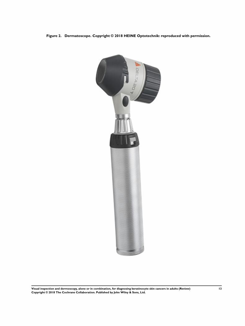
Figure 2. Dermatoscope. Copyright © 2018 HEINE Optotechnik: reproduced with permission.
13Visual inspection and dermoscopy, alone or in combination, for diagnosing keratinocyte skin cancers in adults (Review)
Copyright © 2018 The Cochrane Collaboration. Published by John Wiley & Sons, Ltd.
The visual nature of dermoscopic interpretation means that when
used on an in-person basis, dermoscopy is essentially added to vi-
sual inspection of a skin lesion and similar non-analytical and an-
alytical pattern recognition strategies are employed to reach a der-
moscopic diagnosis. Dermoscopic histological correlations have
been established for the diagnosis of melanoma, allowing a number
of diagnostic algorithms to be developed based on lesion colour,
aspect, pigmentation pattern, and skin vessels (Dinnes 2018b).
However, the diagnosis of keratinocyte skin cancers using der-
moscopy again relies predominantly on subjective pattern recogni-
tion. Features of BCC on dermoscopy include arborising (branch-
ing of ) blood vessels, superficial fine telangiectasia (abnormally
tortuous and dilated blood vessels), grey-blue ovoid nests and glob-
ules, in-focus dots, spoke wheels and maple-leaf-like areas, con-
centric structures, ulceration, multiple small erosions, shiny white-
red structureless areas, and short white streaks (Tzellos 2014). Fea-
tures favouring cSCC on dermoscopy include the presence of ker-
atin, white circles, radial telangiectasia and blood spots (Rosendahl
2012a; Zalaudek 2012).
In modern practice, dermoscopic images are frequently obtained
for skin lesions that are recommended for excision and are also
obtained for lesions that have not yet met the diagnostic threshold
for excision but are to be monitored over time in case of any further
suspicious changes. Dermoscopic images are also a key component
of teledermatology consultations, usually accompanied by digital
photographs and other pertinent information (Chuchu 2018a),
as discussed above.
Clinical pathway
The diagnosis of skin lesions occurs in primary-, secondary-, and
tertiary-care settings by both generalist and specialist healthcare
providers. In the UK, people with concerns about a new or chang-
ing lesion will present to their general practitioner rather than di-
rectly to a specialist in secondary care. If the general practitioner
has concerns, then a referral is usually made to a specialist in sec-
ondary care - usually a dermatologist, but sometimes to a surgi-
cal specialist such as a plastic surgeon or an ophthalmic surgeon.
Suspicious skin lesions may also be identified in a referral setting,
for example by a general surgeon, and referred for a consultation
with a skin cancer specialist (Figure 3). Skin cancers identified by
other specialist surgeons (such as an ear, nose, and throat (ENT)
specialist or maxillofacial surgeon) will usually be diagnosed and
treated without further referral.
14Visual inspection and dermoscopy, alone or in combination, for diagnosing keratinocyte skin cancers in adults (Review)
Copyright © 2018 The Cochrane Collaboration. Published by John Wiley & Sons, Ltd.
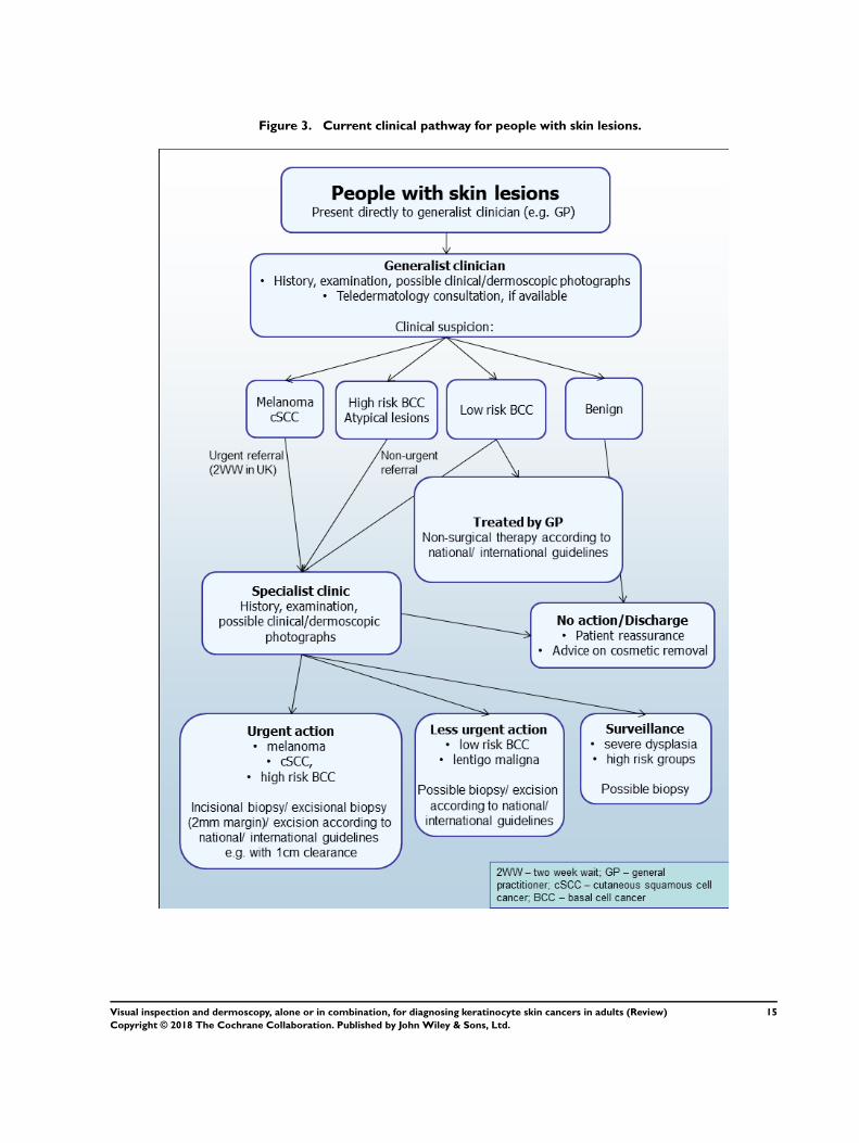
Figure 3. Current clinical pathway for people with skin lesions.
15Visual inspection and dermoscopy, alone or in combination, for diagnosing keratinocyte skin cancers in adults (Review)
Copyright © 2018 The Cochrane Collaboration. Published by John Wiley & Sons, Ltd.
Current UK guidelines recommend that all suspicious pigmented
lesions presenting in primary care should be assessed by taking a
clinical history and visual inspection using the seven-point check-
list (MacKie 1990); lesions suspected to be melanoma or cSCC
should be referred for appropriate specialist assessment within
two weeks (Chao 2013; Marsden 2010; NICE 2015). Evidence is
emerging, however, to suggest that excision of melanoma by GPs
is not associated with increased risk compared with outcomes in
secondary care (Murchie 2017). In the UK, low-risk BCCs are
usually recommended for routine referral, with urgent referral for
those in whom a delay could have a significant impact on out-
comes, for example due to large lesion size or critical site (NICE
2015). Appropriately-qualified generalist care providers increas-
ingly undertake management of low-risk BCCs in the UK, such
as by excision of low-risk lesions (NICE 2010). Similar guidance
is in place in Australia (CCAAC Network 2008).
For referred lesions, the specialist clinician will use history-taking,
visual inspection of the lesion (in conjunction with other skin le-
sions), palpation of the lesion and associated regional nodal basins
in conjunction with dermoscopic examination to inform a clinical
decision. If melanoma is suspected, then urgent 2 mm excision
biopsy is recommended (Lederman 1985; Lees 1991); for cSCC
predetermined surgical margin excision or a diagnostic biopsy may
be considered. BCCs and pre-malignant lesions potentially eligible
for nonsurgical treatment may undergo a diagnostic biopsy before
initiation of therapy if there is diagnostic uncertainty. Equivocal
melanocytic lesions for which a definitive clinical diagnosis cannot
be reached may undergo surveillance to identify any lesion changes
that would indicate excision biopsy or reassurance and discharge
for those lesions that remain stable over a period of time.
Theoretically, teledermatology consultations may aid appropriate
triage of lesions into urgent referral; non-urgent secondary-care
referral (e.g. for suspected basal cell carcinoma); or where available,
referral to an intermediate care setting, e.g. clinics run by GPs
with a special interest in dermatology. The distinction between
setting and examiner qualifications and experience is important,
as specialist clinicians might work in primary-care settings (for
example, in the UK, GPs with a special interest in dermatology
and skin surgery who have undergone appropriate training), and
generalists might practice in secondary-care settings (for example,
plastic surgeons who do not specialise in skin cancer). The level
of skill and experience in skin cancer diagnosis will vary for both
generalist and specialist care providers and will also impact on test
accuracy.
Prior test(s)
Although smartphone applications and community-based teled-
ermatology services can increasingly be directly accessed by people
who have concerns about a skin lesion (Chuchu 2018b), visual
inspection of a suspicious lesion by a clinician is usually the first
in a series of tests to diagnose skin cancer. In the UK this usually
takes place in primary care, but in many countries people with
suspicious lesions can present directly to a specialist setting. Al-
though dermoscopy is frequently combined with visual inspection
of a lesion in secondary-care settings, it is also increasingly used
in primary care, particularly in countries such as Australia (Youl
2007a).
Consideration of the degree of prior testing that study participants
have undergone is key to interpretation of test accuracy indices, as
these are known to vary according to the disease spectrum (or case-
mix) of included participants (Lachs 1992; Leeflang 2013; Moons
1997; Usher-Smith 2016). Spectrum effects are often observed
when tests that are developed further down the referral pathway
have lower sensitivity and higher specificity when applied in set-
tings with participants with limited prior testing (Usher-Smith
2016). Studies of individuals with suspicious lesions at the initial
clinical presentation stage (’test-naïve’) are likely to have a wider
range of differential diagnoses and include a higher proportion of
people with benign diagnoses compared with studies of partici-
pants who have been referred for a specialist opinion on the basis
of visual inspection (with or without dermoscopy) by a generalist
practitioner. Furthermore, studies in more specialist settings may
focus on equivocal or difficult-to-diagnose lesions rather than le-
sions with a more general level of clinical suspicion. However this
direction of effect is not consistent across tests and diseases, the
mechanisms in action often being more complex than prevalence
alone, and can be difficult to identify (Leeflang 2013). A simple
categorisation of studies according to primary, secondary or spe-
cialist setting may therefore not always adequately reflect these key
differences in disease spectrum that can affect test performance.
Role of index test(s)
When diagnosing potentially life-threatening conditions, the con-
sequences of falsely reassuring a person that they do not have skin
cancer can be serious and potentially fatal, as the resulting delay
to diagnosis means that the window for successful early treatment
may be missed. To minimise these false-negative diagnoses, a good
diagnostic test will demonstrate high sensitivity and a high nega-
tive predictive value (NPV), i.e. so that very few of those with a
negative test result will actually have a malignant lesion. Giving
falsely-positive test results (meaning the test has poor specificity
and a high false-positive rate) resulting in the removal of lesions
that turn out to be benign is arguably less of an error than miss-
ing a potentially fatal lesion, but is not cost-free. False-positive
diagnoses not only cause unnecessary scarring from the biopsy or
excision procedure, but also increase anxiety (particularly during
the time that people wait for results) and increase healthcare costs
as the number of lesions that need to be removed to yield one
16Visual inspection and dermoscopy, alone or in combination, for diagnosing keratinocyte skin cancers in adults (Review)
Copyright © 2018 The Cochrane Collaboration. Published by John Wiley & Sons, Ltd.
malignant diagnosis increases.
Delay in diagnosis of a BCC as a result of a false-negative test is
not as serious as for melanoma, because BCCs are usually slow-
growing and very unlikely to metastasise (Betti 2017). However,
delayed diagnosis can result in a larger and more complex excision
with consequent greater morbidity. Very sensitive diagnostic tests
for BCC, however, may compromise on lower specificity leading
to a higher false-positive rate, and an enormous burden of skin
surgery, such that a balance between sensitivity and specificity is
needed. The situation for cSCC is more similar to melanoma in
that the consequences of falsely reassuring a person that they do
not have skin cancer can be serious and potentially fatal, given
that removal of an early cSCC is usually curative. Thus, a good
diagnostic test for cSCC should demonstrate high sensitivity and
a corresponding high negative predictive value. A test that can also
reduce false positive clinical diagnoses without missing true cases
of cSCC has patient and resource benefits.
Alternative test(s)
A number of other tests have been reviewed as part of our se-
ries of Cochrane DTA Reviews on the diagnosis of keratinocyte
skin cancers, including reflectance confocal microscopy (RCM)
(Dinnes 2018c), computer-assisted diagnosis (CAD) or artificial
intelligence-based techniques using dermoscopic or spectroscopic
images (Ferrante di Ruffano 2018a), optical coherence tomogra-
phy (OCT) (Ferrante di Ruffano 2018b), high-frequency ultra-
sonography (Dinnes 2018d) and exfoliative cytology (Ferrante di
Ruffano 2018c). Evidence permitting, we will compare the accu-
racy of available tests in an overview review, exploiting within-
study comparisons of tests and allowing the analysis and compar-
ison of commonly-used diagnostic strategies where tests may be
used singly or in combination.
We also considered and excluded a number of tests from this re-
view, such as tests used for monitoring people (e.g. total body pho-
tography of those with large numbers of pigmented lesions). We
also did not assess histopathological confirmation following lesion
excision, because it is the established reference standard for skin
cancer diagnosis and will be one of the standards against which
the index tests are evaluated in these reviews.
Rationale
This series of reviews of diagnostic tests used to assist the clinical
diagnosis of BCC and cSCC in clinical practice or research settings,
aims to identify the most accurate approaches to diagnosis, and
to provide clinical and policy decision-makers with the highest
possible standard of evidence on which to base diagnostic and
treatment decisions. With the increasing availability of a wider
range of tests, there is a need to differentiate and appropriately
triage keratinocyte skin cancers to avoid sending too many people
with benign or low-risk lesions for a specialist opinion whilst not
missing those people who have lesions that require treatment.
There is a lack of systematic reviews in the field. A 2007 review
of a range of tests for diagnosis of BCC did not report the use of
systematic methods for study inclusion or extraction and did not
appear to apply any quality assessment (Mogensen 2007). Critical
questions of comparative test accuracy and the impact of examiner,
prior testing, and underlying risk status remain unanswered for the
NHS. With the increasing availability of digital imaging systems
and computerised instruments, there is a further need for an up-to-
date analysis of their accuracy in comparison with visual inspection
or dermoscopy.
This review follows a generic protocol which covers the full series
of Cochrane DTA Reviews for the diagnosis of keratinocyte skin
cancer (Dinnes 2015a). The Background and Methods sections of
this review therefore use some text that was originally published
in the protocol (Dinnes 2015a) and text that overlaps some of our
other reviews (Dinnes 2018a; Dinnes 2018b).
O B J E C T I V E S
To determine the diagnostic accuracy of visual inspection and
dermoscopy, alone or in combination, for the detection of BCC
in adults.
To determine the diagnostic accuracy of visual inspection and
dermoscopy, alone or in combination, for the detection of cSCC
in adults.
For both visual inspection and dermoscopy, we estimated accuracy
separately according to whether the diagnosis was based on a face-
to-face (in person) encounter or based on remote (image-based)
assessment. We therefore aimed to compare tests in the following
way:
• To estimate incremental accuracy for the diagnosis of BCC
in adults, (a) from dermoscopy added to in-person visual
inspection of a skin lesion, or (b) from dermoscopic image-based
assessment in comparison to visual inspection of a clinical
photograph.
• To estimate incremental accuracy for the diagnosis of cSCC
in adults, (a) from dermoscopy added to in-person visual
inspection of a skin lesion, or (b) from dermoscopic image-based
assessment in comparison to visual inspection of a clinical
photograph.
We also proposed to analyse data according to the prior testing
undergone by study participants (comparing those with limited
prior testing with those referred for further evaluation of a suspi-
cious skin lesion). However, this was not possible due to limited
data.
17Visual inspection and dermoscopy, alone or in combination, for diagnosing keratinocyte skin cancers in adults (Review)
Copyright © 2018 The Cochrane Collaboration. Published by John Wiley & Sons, Ltd.
Secondary objectives
For the identification of BCC or cSCC:
• To compare the accuracy of dermoscopy added to in-person
visual inspection versus visual inspection alone, where both tests
have been evaluated in the same studies (direct test comparisons);
• To compare the accuracy of image-based dermoscopy versus
visual inspection of digital photographs, where both tests have
been evaluated in the same studies (direct test comparisons);
• To determine the diagnostic accuracy of individual
algorithms used to assist visual inspection;
• To determine the diagnostic accuracy of individual
algorithms used to assist dermoscopy;
• To determine the effect of observer experience on
diagnostic accuracy.
To assess an alternative target condition:
• To determine the diagnostic accuracy of visual inspection or
dermoscopy, alone or in combination, for the detection of any
skin cancer, and to compare the accuracy of dermoscopy with
that of visual inspection alone.
Investigation of sources of heterogeneity
We set out to address a range of potential sources of heterogeneity
for investigation across our series of reviews, as outlined in our
generic protocol (Dinnes 2015a) and as described in Appendix 3;
however, our ability to investigate these was necessarily limited by
the available data on each individual test reviewed.
The sources of heterogeneity that we investigated for this review
were:
• In-person versus image-based evaluations
• Use of a diagnostic algorithm: no algorithm reported versus
any named algorithm used
• Disease prevalence: 0% to 25%; > 25%
• Observer expertise.
M E T H O D S
Criteria for considering studies for this review
Types of studies
We included test-accuracy studies that allow comparison of the
result of the index test with that of a reference standard, including
the following:
• studies where all participants receive a single index test and
a reference standard;
• studies where all participants receive more than one index
test(s) and reference standard;
• studies where participants are allocated (by any method) to
receive different index tests or combinations of index tests and all
receive a reference standard (between-person comparative studies
(BPC));
• studies that recruit series of participants unselected by true
disease status (referred to as case series for the purposes of this
review);
• diagnostic case-control studies that separately recruit
diseased and non-diseased groups (see Rutjes 2005); however, we
did not include studies that compared results for malignant
lesions to those for healthy skin (i.e. with no lesion present);
• both prospective and retrospective studies;
• studies where previously-acquired clinical or dermoscopic
images were retrieved and prospectively interpreted for study
purposes.
We excluded studies from which we could not extract 2 x 2 con-
tingency data or if they included fewer than five cases of basal cell
carcinoma (BCC) or cutaneous squamous cell carcinoma (cSCC),
or fewer than five benign lesions. The size threshold of five is ar-
bitrary. However, such small studies are unlikely to add precision
to estimates of accuracy.
Studies available only as conference abstracts were excluded; how-
ever, attempts were made to identify full papers for potentially
relevant conference abstracts (Searching other resources).
Participants
We included studies in adults with lesions suspicious for skin can-
cer. These could include participants:
• with lesion characteristics suspicious for keratinocyte skin
cancers, including BCC or cSCC
• with lesion characteristics suspicious for any skin cancer,
including melanoma (e.g. restricted to those with pigmented
lesions only, or including both pigmented and non-pigmented
lesion types);
• those at high risk of developing BCC or cSCC
We excluded studies that recruited only participants with malig-
nant or benign final diagnoses.
We excluded studies conducted in children or which clearly re-
ported inclusion of more than 50% of participants aged 16 and
under.
Index tests
Studies reporting accuracy data for visual inspection or der-
moscopy, or both, with diagnosis made either in person (face-to-
face diagnosis) or image-based (diagnosis based on photographs
or dermoscopic images, remotely from the study participant) were
eligible for inclusion. We included all established algorithms or
checklists to assist diagnosis.
Studies developing new algorithms or methods of diagnosis (i.e.
derivation studies) wereincluded if they:
18Visual inspection and dermoscopy, alone or in combination, for diagnosing keratinocyte skin cancers in adults (Review)
Copyright © 2018 The Cochrane Collaboration. Published by John Wiley & Sons, Ltd.
• used a separate independent ’test set’ of participants or
images to evaluate the new approach; or
• investigated lesion characteristics that had previously been
suggested as associated with BCC or cSCC, and the study
reported accuracy based on the presence or absence of specific
combinations of characteristics.
Studies were excluded if they:
• used a statistical model to produce a data-driven equation,
or algorithm based on multiple diagnostic features, with no
separate test set
• used cross-validation approaches such as ’leave-one-out’
cross-validation (Efron 1983)
• evaluated the accuracy of the presence or absence of
individual lesion characteristics or morphological features, with
no overall diagnosis of malignancy
• reported accuracy data for ‘clinical diagnosis’ with no clear
description of whether the reported data related to visual
inspection alone or included dermoscopy in all study participants
• were based on the experience of a skin cancer-specific clinic,
where dermoscopy may or may not have been used on an
individual basis.
Although primary-care clinicians can have a specialist interest in
skin cancer, for the purposes of this review we considered primary-
care physicians as generalist practitioners and dermatologists as
specialists. Within each group, we extracted any reporting of spe-
cial interest or accreditation in skin cancer.
Target conditions
The primary target conditions were the detection of:
• BCC, including all subtypes;
• Invasive cSCC (we did not consider cutaneous SCC in situ,
such as Bowen’s disease, as disease-positive)
We considered an additional target condition in secondary analy-
ses, namely the detection of:
• any skin cancer, including BCC, cSCC, melanoma or any
rare skin cancer (e.g. Merkel cell cancer), as long as skin cancers
other than melanoma made up more than 50% of the disease-
positive group. Data from studies in which melanoma accounted
for more than 50% of skin cancers were included in our reviews
of visual inspection and of dermoscopy compared to visual
inspection for the diagnosis of melanoma (Dinnes 2018a;
Dinnes 2018b).
Reference standards
The ideal reference standard was histopathological diagnosis in
all eligible lesions. A qualified pathologist or dermatopathologist
should perform histopathology. Ideally, reporting should be stan-
dardised, detailing a minimum dataset to include the type of skin
cancer (BCC, cSCC) and subtype of BCC, and may also refer to
the tumour, node, and metastasis (TNM) classification of staging
for cSCC (Royal College of Pathologists 2014). We did not apply
the reporting standard as a necessary inclusion criterion, but ex-
tracted any pertinent information.
Partial verification (applying the reference test only to a subset of
those undergoing the index test) was of concern, given that lesion
excision or biopsy are unlikely to be carried out for all clinically-
benign skin lesions within a representative population sample. We
therefore accepted clinical follow-up of benign lesions as an eligi-
ble reference standard, whilst recognising the risk of differential
verification bias (as misclassification rates of histopathology and
follow-up will differ).
Additional eligible reference standards included cancer registry
follow-up and ’expert opinion’ with no histology or clinical fol-
low-up. Cancer registry follow-up is considered less desirable than
active clinical follow-up, as follow-up is not carried out within
the control of the study investigators. Furthermore, if participant-
based analyses are presented as opposed to lesion-based analyses,
it may be difficult to determine whether the detection of a ma-
lignant lesion during follow-up is the same lesion that originally
tested negative on the index test.
All of the above are eligible reference standards, with the following
caveats:
• all study participants with a final diagnosis of the target
disorder must have a histological diagnosis, either subsequent to
the application of the index test or after a period of clinical
follow-up, and
• at least 50% of all participants with benign lesions must
have either a histological diagnosis or clinical follow-up to
confirm benignity.
Search methods for identification of studies
Electronic searches
The Information Specialist (SB) carried out a comprehensive
search for published and unpublished studies. A single large liter-
ature search was conducted to cover all topics in the programme
grant (see Appendix 1 for a summary of reviews included in the
programme grant). This allowed for the screening of search results
for potentially relevant papers for all reviews at the same time.
A search combining disease related terms with terms related to
the test names, using both text words and subject headings was
formulated. The search strategy was designed to capture studies
evaluating tests for the diagnosis or staging of skin cancer. As the
majority of records were related to the searches for tests for stag-
ing of disease, a filter using terms related to cancer staging and
to accuracy indices was applied to the staging test search, to try
to eliminate irrelevant studies, for example, those using imaging
tests to assess treatment effectiveness. A sample of 300 records that
would be missed by applying this filter was screened and the filter
19Visual inspection and dermoscopy, alone or in combination, for diagnosing keratinocyte skin cancers in adults (Review)
Copyright © 2018 The Cochrane Collaboration. Published by John Wiley & Sons, Ltd.
adjusted to include potentially relevant studies. When piloted on
MEDLINE, inclusion of the filter for the staging tests reduced the
overall numbers by around 6000. The final search strategy, incor-
porating the filter, was subsequently applied to all bibliographic
databases as listed below (Appendix 4). The final search result was
cross-checked against the list of studies included in five systematic
reviews; our search identified all but one of the studies, and this
study was not indexed on MEDLINE. The Information Special-
ist devised the search strategy, with input from the Information
Specialist from Cochrane Skin. No additional limits were used.
We searched the following bibliographic databases to 29 August
2016 for relevant published studies:
• MEDLINE via OVID (from 1946);
• MEDLINE In-Process & Other Non-Indexed Citations via
OVID; and
• Embase via OVID (from 1980).
We searched the following bibliographic databases to 30 August
2016 for relevant published studies:
• Cochrane Central Register of Controlled Trials
(CENTRAL) Issue 7, 2016, in the Cochrane Library;
• Cochrane Database of Systematic Reviews (CDSR) Issue 8,
2016 in the Cochrane Library;
• Cochrane Database of Abstracts of Reviews of Effects
(DARE) Issue 2, 2015;
• CRD Health Technology Assessment (HTA) database Issue
3, 2016; and
• Cumulative Index to Nursing and Allied Health Literature
(CINAHL) (via EBSCO from 1960).
We searched the following databases for relevant unpublished stud-
ies using a strategy based on the MEDLINE search:
• CPCI (Conference Proceedings Citation Index), via Web of
Science™ (from 1990; searched 28 August 2016); and
• SCI Science Citation Index Expanded™ via Web of
Science™ (from 1900, using the ’Proceedings and Meetings
Abstracts’ Limit function; searched 29 August 2016).
We searched the following trials registers using the search terms
’melanoma’, ’squamous cell’, ’basal cell’ and ’skin cancer’ combined
with ’diagnosis’:
• Zetoc (from 1993; searched 28 August 2016).
• The US National Institutes of Health Ongoing Trials
Register (www.clinicaltrials.gov); searched 29 August 2016.
• NIHR Clinical Research Network Portfolio Database (
www.nihr.ac.uk/research-and-impact/nihr-clinical-research-
network-portfolio/); searched 29 August 2016.
• The World Health Organization International Clinical
Trials Registry Platform ( apps.who.int/trialsearch/); searched 29
August 2016.
We aimed to identify all relevant studies regardless of language
or publication status (published, unpublished, in press, or in
progress). We applied no date limits.
Searching other resources
We have screened any relevant systematic reviews identified by the
searches for their included primary studies, and have included any
missed by our searches. We have checked the reference lists of all
included papers, and subject experts within the author team have
reviewed the final list of included studies. We have conducted no
electronic citation searching.
Data collection and analysis
Selection of studies
At least one review author (JDi or NC), screened all titles and
abstracts, with any queries discussed and resolved by consensus.
A pilot screen of 539 MEDLINE references showed good agree-
ment (89% with a kappa of 0.77) between screeners. We included
primary test accuracy studies and test accuracy reviews (for scan-
ning of reference lists) of any test used to investigate suspected
melanoma, BCC, or cSCC at initial screening. Inclusion crite-
ria (Appendix 5) were applied independently by both a clinical
reviewer (from one of a team of 12 clinician reviewers) and a
methodologist reviewer (JDi or NC) to all full-text articles, with
disagreements resolved by consensus or by a third party (JDe, CD,
HW, or RM). We contacted authors of eligible studies when in-
sufficient data were presented, to allow for the construction of 2
x 2 contingency tables.
Data extraction and management
One clinical (as detailed above) and one methodologist reviewer
(JDi, NC or LFR) independently extracted data for details of the
study design, participants, index test(s) or test combinations and
criteria for index test positivity, reference standards, and data re-
quired to populate a 2 x 2 diagnostic contingency table for each
index test, using a piloted data extraction form. We extracted data
at all available index test thresholds, resolving disagreements by
consensus or by a third party (JDe, CD, HW, and RM).
We contacted authors of included studies where information re-
lating to the diagnostic threshold was missing. We contacted au-
thors of conference abstracts published from 2013 to 2015 to ask
whether full data were available. If we could not identify a full
paper, we marked conference abstracts as ’pending’ and will revisit
them in a future review update.
Dealing with multiple publications and companion papers
Where we found multiple reports of a primary study, we max-
imised yield of information by collating all available data. Where
there were inconsistencies in reporting or overlapping study pop-
ulations, we contacted study authors for clarification in the first
20Visual inspection and dermoscopy, alone or in combination, for diagnosing keratinocyte skin cancers in adults (Review)
Copyright © 2018 The Cochrane Collaboration. Published by John Wiley & Sons, Ltd.
instance. If this contact with authors was unsuccessful, we used
the most complete and up-to-date data source where possible.
Assessment of methodological quality
We assessed risks of bias and applicability of included studies using
the QUADAS-2 checklist (Whiting 2011), tailored to the topic of
skin cancer (see Appendix 6). We piloted the modified QUADAS-
2 tool on a small number of full-text articles included across the
full series of diagnostic test accuracy reviews. One clinical and one
methodologist reviewer (JDi, NC or LFR) independently assessed
quality for the remaining studies, resolving any disagreement by
consensus or by a third party where necessary (JDe, CD, HW, and
RM).
Statistical analysis and data synthesis
We planned separate analyses according to the point that study
participants have reached in the clinical pathway, the clarity with
which the pathway could be determined, and the evaluation of in-
person versus image-based diagnosis.
Our unit of analysis was the lesion rather than the person. This is
because (i) in skin cancer initial treatment is directed to the lesion
rather than systemically (thus it is important to be able to cor-
rectly identify cancerous lesions for each person), and (ii) it is the
most common way in which the primary studies reported data.
Although there is a theoretical possibility of correlations of test
errors when the same people contribute data for multiple lesions,
most studies include very few people with multiple lesions and
any potential impact on findings is likely to be very small, par-
ticularly in comparison with other concerns regarding risk of bias
and applicability. For each analysis, we included only one dataset
per study, to avoid multiple counting of lesions. We retrieved few
studies comparing algorithms, but where we assessed multiple al-
gorithms in an individual study, we selected datasets on the fol-
lowing preferential basis:
• ‘no algorithm’ reported; data presented for clinician’s overall
diagnosis or management decision
• pattern analysis or pattern recognition
• ABCD algorithm (or derivatives of ) or other established
algorithm such as seven-point checklist, Menzies algorithm or
three-point checklist
• New algorithm developed by study authors
For the diagnosis of BCC (or cSCC), we considered any
melanomas or cSCCs (BCCs) that were positively identified in the
‘disease-negative’ group (i.e. that were mistaken for BCCs) false-
positive results. The clinical management of a lesion considered
to be a BCC might be quite different from that for a melanoma
or cSCC, and could potentially lead to a negative outcome for
the participants concerned; for example, if a treatment other than
excision was initiated.
For each index test, algorithm or checklist under consideration,
we plotted estimates of sensitivity and specificity on coupled for-
est plots and in receiver operating characteristic (ROC) space. For
tests where commonly-used thresholds were reported we estimated
summary operating points (summary sensitivities and specificities)
with 95% confidence and prediction regions using the bivariate
hierarchical model (Chu 2006; Reitsma 2005). Where inadequate
data were available for the model to converge, we simplified the
model, first by assuming no correlation between estimates of sen-
sitivity and specificity and secondly by setting estimates of near-
zero variance terms to zero (Takwoingi 2017). Where all studies
reported 100% sensitivity (or 100% specificity) we summed the
number with disease (or no disease) across studies and used them
to compute a binomial exact 95% confidence interval.
We drew comparisons between visual inspection and dermoscopy
results with:
a. all visual inspection and all dermoscopy data from all studies,
and then
b. only using data from studies that reported both visual inspection
data and dermoscopy data for the same lesions, to enable a robust
direct comparison (Takwoingi 2013).
We made comparisons between tests by comparing summary ROC
curves using the hierarchical summary receiver-operator curves
(HSROC) model (Rutter 2001) rather than by estimating average
operating points, as this approach allows incorporation of data
at different thresholds as could arise with different algorithms or
checklists. We used an HSROC model that assumed a constant
SROC shape between tests and subgroups, but allowed for dif-
ferences in threshold and accuracy by the addition of covariates.
We assessed the significance of the differences between tests by the
likelihood ratio test (LR test) assessing differences in both accu-
racy and threshold, and by a Wald test on the parameter estimate
testing for differences in accuracy alone. We provide the P values
from both tests in the Tables with the results from the LR test
cited in the text, on the basis that differences in threshold between
tests is likely. We fitted simpler models when convergence was not
achieved due to small numbers of studies, first assuming symmet-
ric SROC curves (setting the shape term to zero), and then setting
random-effects variance estimates to zero.
We present estimates of accuracy from HSROC models as di-
agnostic odds ratios (DORs) (estimated where the SROC curve
crosses the sensitivity = specificity line) with 95% confidence in-
tervals. We present differences between tests and subgroups from
HSROC analyses as relative diagnostic odds ratios (RDORs) with
95% confidence intervals. To facilitate interpretation in terms of
rates of false-positive and false-negative diagnoses, we have com-
puted values of sensitivity at the point on the SROC curve with
80% specificity and of specificity at the point on the SROC curve
with 80% sensitivity. We chose these 80% values as they lie within
the estimates for most of the analyses. These results should only
be considered as illustrative examples of possible sensitivities (and
specificities) and differences in sensitivities (and specificities) that
21Visual inspection and dermoscopy, alone or in combination, for diagnosing keratinocyte skin cancers in adults (Review)
Copyright © 2018 The Cochrane Collaboration. Published by John Wiley & Sons, Ltd.
could be expected.
Where data were insufficient to estimate HSROC curves (e.g. for
the analysis of cSCC),we estimated summary operating points
(summary sensitivities and specificities) with 95% confidence and
prediction regions using the bivariate hierarchical model (Chu
2006; Reitsma 2005).
For computation of likely numbers of true-positive, false-positive,
false-negative and true-negative findings in the ’Summary of find-
ings’ table, we applied these indicative values to the lower quar-
tile, median and upper quartiles of the prevalence observed in the
study groups.
We fitted bivariate models using the xtmelogit command in
STATA 15, and HSROC models using the NLMIXED procedure
in the SAS statistical software package (SAS 2012) and the metadas
macro (Takwoingi 2010).
Investigations of heterogeneity
We investigated heterogeneity, comparisons between algorithms
and according to observer experience by comparing summary
ROC curves using the HSROC model (Rutter 2001), with addi-
tional covariates for differences in threshold and accuracy as used
for comparing tests.
Sensitivity analyses
We did not conduct any sensitivity analyses.
Assessment of reporting bias
Because of uncertainty about the determinants of publication bias
for diagnostic accuracy studies and the inadequacy of tests for de-
tecting funnel plot asymmetry (Deeks 2005), we did not perform
tests to detect publication bias.
R E S U L T S
Results of the search
We identified and screened 34,517 unique references for inclusion.
Of these, we reviewed 1051 full-text papers for eligibility for any
one of the suite of reviews of tests to assist in the diagnosis of
melanoma or keratinocyte skin cancer. Of the 1051 full-text papers
assessed, we eliminated 848 from all reviews in our series (see
Figure 4 PRISMA flow diagram of search and eligibility results).
22Visual inspection and dermoscopy, alone or in combination, for diagnosing keratinocyte skin cancers in adults (Review)
Copyright © 2018 The Cochrane Collaboration. Published by John Wiley & Sons, Ltd.
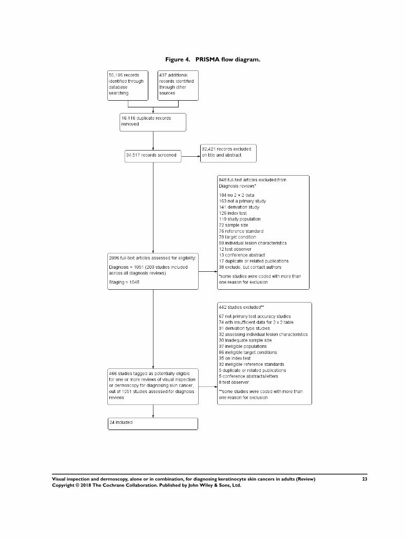
Figure 4. PRISMA flow diagram.
23Visual inspection and dermoscopy, alone or in combination, for diagnosing keratinocyte skin cancers in adults (Review)
Copyright © 2018 The Cochrane Collaboration. Published by John Wiley & Sons, Ltd.
Of the 466 studies tagged as potentially eligible for any of our
reviews of visual inspection or dermoscopy, we include 24 publi-
cations in this review. Exclusions were mainly due to the inability
to construct a 2 x 2 contingency table based on the data presented
(n = 74); the use of ineligible index tests (n = 35; for example:
reporting of data for ‘clinical diagnosis’ or for serial use of the in-
dex test in a follow-up context); assessment of individual lesion
characteristics (n = 32); or derivation-type studies developing new
algorithms or checklists without a separate training and test set of
lesions (n = 31). Other reasons for exclusion included not meeting
our requirements for an eligible reference standard (n = 32), ineli-
gible study populations (n = 37) (for example, recruiting only ma-
lignant or only benign lesions), inadequate sample size (n = 30), in-
eligible definition of the target condition (n = 86; including those
eligible only for reviews of the detection of melanoma) or with test
interpretation by medical students or laypersons (n = 8). A list of
the 442 publications excluded from this review with reasons for
exclusion is available in Characteristics of excluded studies, with a
list of all studies excluded from the full series of reviews available
as a separate pdf (please contact skin.cochrane.org for a copy of
the pdf ).
We contacted the authors of 17 publications concerned with the
evaluation of visual inspection or dermoscopy for further data to
allow study inclusion; we received responses from four authors
with regard to seven publications. Two authors provided additional
data but these were insufficient to allow inclusion of the stud-
ies (Cabrijan 2008; Warshaw 2009a; Warshaw 2009b; Warshaw
2010a), one replied indicating that dermoscopy was not necessar-
ily used in all study participants (Youl 2007a; Youl 2007b) and
one replied but was unable to access the data needed (Fabbrocini
2008). We contacted the authors of a further seven included stud-
ies for further details of study methods, and received a responses
for four studies; three provided further information about the di-
agnostic thresholds used (Amirnia 2016; Durdu 2011; Stanganelli
2000) and one provided full anonymised study data (Rosendahl
2011).
The 24 included study publications report on a total of 24 cohorts
of lesions and provide 27 visual inspection datasets (8805 lesions;
2579 malignancies) and 33 dermoscopy datasets (6855 lesions;
1444 malignancies). We provide a summary of the tests and target
conditions evaluated in each study in Appendix 7. Six studies con-
tributed data for in-person visual inspection alone (Chang 2013;
Cooper 2002; Ek 2005; Hacioglu 2013; Schwartzberg 2005;
Steiner 1987); three for dermoscopy added to visual inspection
(Amirnia 2016; Durdu 2011; Gokdemir 2011); and five for both
in-person visual inspection alone and combined with dermoscopy
(Argenziano 2006; Carli 2002a; Markowitz 2015; Stanganelli
2000; Ulrich 2015). Two studies contributed data for image-based
visual inspection of clinical photographs alone (Lorentzen 1999;
Nori 2004); eight for image-based dermoscopy (Altamura 2010;
Carli 2002a; Hacioglu 2013 ; Lorentzen 2008; Menzies 2000;
Navarrete Dechent 2016; Witkowski 2016; Zalaudek 2006); and
two for both image-based visual inspection and image-based der-
moscopy (Carli 2002b; Rosendahl 2011). Five studies compared
the accuracy of visual inspection with or without dermoscopy to
other tests, including: exfoliative cytology (Durdu 2011); com-
puter-assisted diagnosis (CAD) (Hacioglu 2013); optical coher-
ence tomography (OCT) (Markowitz 2015; Ulrich 2015); and ra-
diographic contrast medium (RCM) (Witkowski 2016). Thirteen
studies also contributed data to our reviews of visual inspection
(n = 9) and/or dermoscopy (n = 9) for the detection of melanoma
(Dinnes 2018a; Dinnes 2018b).
Methodological quality of included studies
We summarise the overall methodological quality of all included
studies according to in-person or image-based approaches to der-
moscopy or to visual inspection. We present 14 studies reporting
data for in-person visual inspection (n = 11) and/or in-person der-
moscopy (added to visual inspection) (n = 8) in Figure 5, with
results by study presented in Figure 6. Twelve studies reporting
data for image-based visual inspection (n = 4) and/or image-based
dermoscopy (n = 10) are presented in Figure 7, with results by
study presented in Figure 8. Two studies appear in both sets of
figures: Carli 2002a evaluated the accuracy of image-based der-
moscopy as well as in-person visual inspection and dermoscopy,
while Hacioglu 2013 reported data for in-person visual inspection
and image-based dermoscopy.
24Visual inspection and dermoscopy, alone or in combination, for diagnosing keratinocyte skin cancers in adults (Review)
Copyright © 2018 The Cochrane Collaboration. Published by John Wiley & Sons, Ltd.
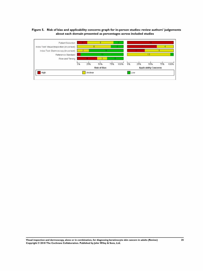
Figure 5. Risk of bias and applicability concerns graph for in-person studies: review authors’ judgements
about each domain presented as percentages across included studies
25Visual inspection and dermoscopy, alone or in combination, for diagnosing keratinocyte skin cancers in adults (Review)
Copyright © 2018 The Cochrane Collaboration. Published by John Wiley & Sons, Ltd.
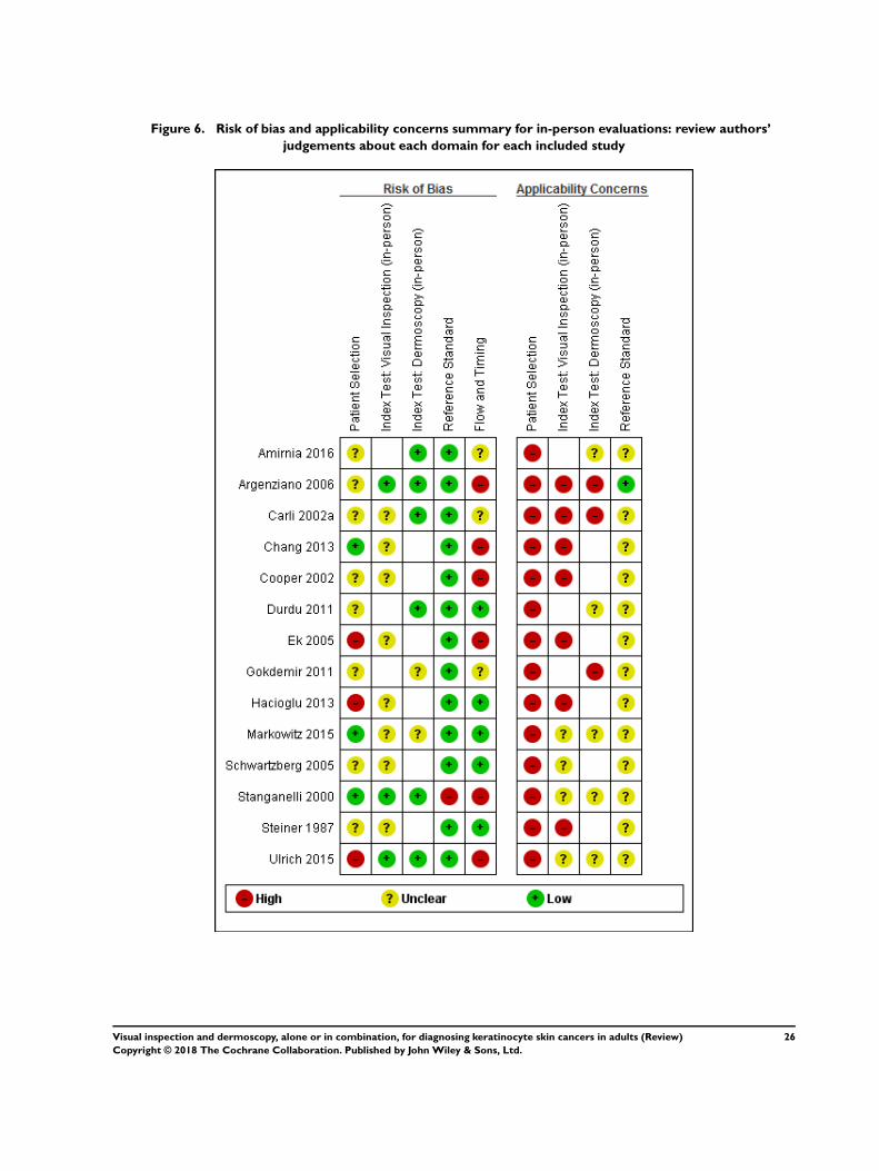
Figure 6. Risk of bias and applicability concerns summary for in-person evaluations: review authors’
judgements about each domain for each included study
26Visual inspection and dermoscopy, alone or in combination, for diagnosing keratinocyte skin cancers in adults (Review)
Copyright © 2018 The Cochrane Collaboration. Published by John Wiley & Sons, Ltd.
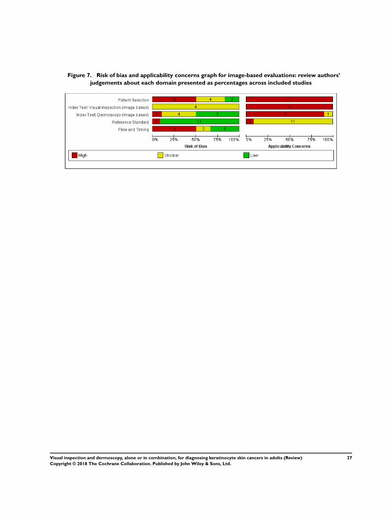
Figure 7. Risk of bias and applicability concerns graph for image-based evaluations: review authors’
judgements about each domain presented as percentages across included studies
27Visual inspection and dermoscopy, alone or in combination, for diagnosing keratinocyte skin cancers in adults (Review)
Copyright © 2018 The Cochrane Collaboration. Published by John Wiley & Sons, Ltd.
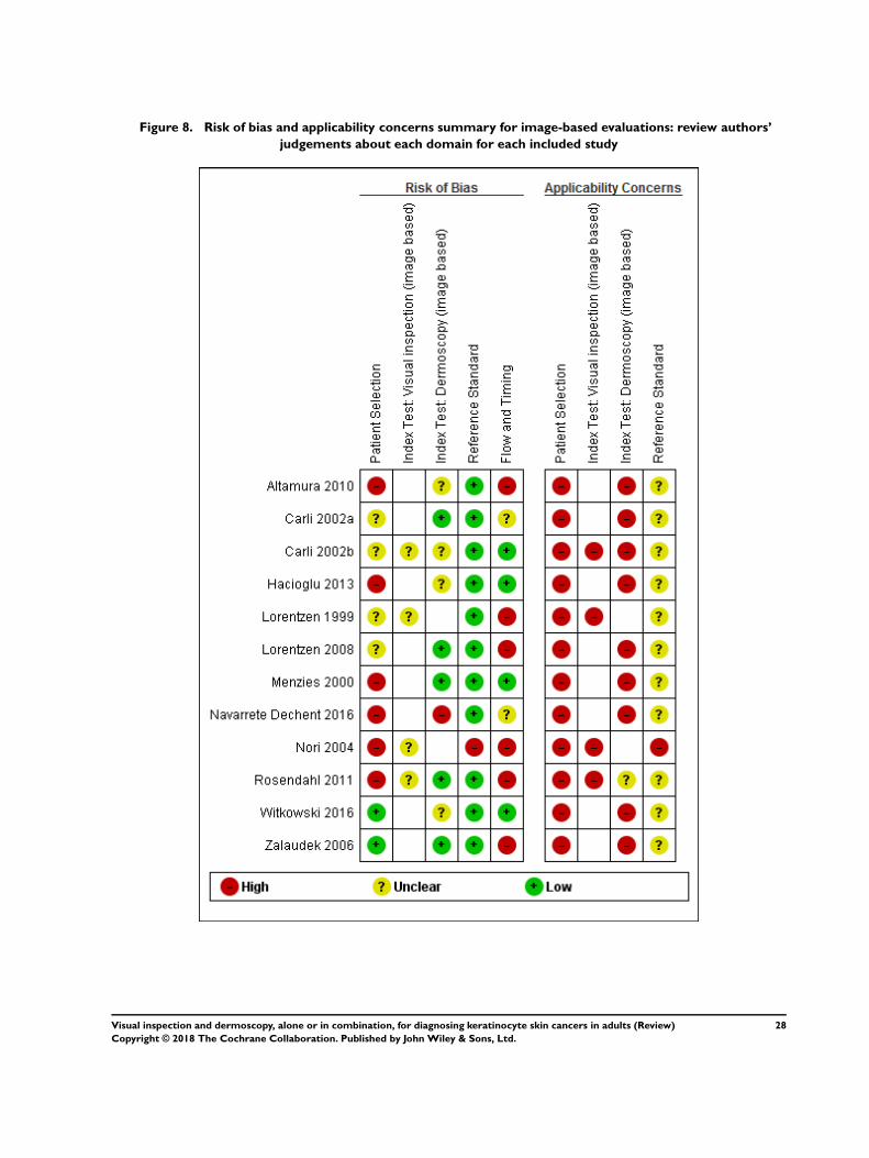
Figure 8. Risk of bias and applicability concerns summary for image-based evaluations: review authors’
judgements about each domain for each included study
28Visual inspection and dermoscopy, alone or in combination, for diagnosing keratinocyte skin cancers in adults (Review)
Copyright © 2018 The Cochrane Collaboration. Published by John Wiley & Sons, Ltd.
In-person evaluations
We judged the risk of bias to be low for most of the studies in
only two of five quality domains assessed (dermoscopy index test,
reference standard); we judged risk of bias to be high or unclear
for most of the studies for participant selection, visual inspection
index test, and flow and timing (Figure 5). We rated applicability
of study findings as of high or unclear concern in all four domains
(participant selection, dermoscopy index tests, visual inspection
index tests, reference standards) assessed for all studies apart from
one.
For participant selection: we rated three of the 14 studies (21%) at
low risk of bias, and three (21%) at high risk (Figure 5) due to
exclusion of lesions by size (Hacioglu 2013), or because of miss-
ing (Ulrich 2015) or equivocal pathology (Ek 2005). Five studies
(36%) did not report the method of participant selection and eight
(57%) did not clearly describe exclusions from the study. We rated
all studies at high concern for applicability of participants, primar-
ily due to inclusion of lesions selected for biopsy or excision based
on the clinical or dermoscopic diagnosis. We judged only one
to have included a representative population (Stanganelli 2000).
Nine cohorts (64%) also included multiple lesions per participant
(Chang 2013; Cooper 2002; Durdu 2011; Ek 2005; Gokdemir
2011; Markowitz 2015; Schwartzberg 2005; Stanganelli 2000;
Ulrich 2015) and three did not clearly report the number of in-
cluded participants (Argenziano 2006; Carli 2002a; Steiner 1987).
For the index test domain: there are eight evaluations of in-per-
son dermoscopy and 11 evaluations of in-person visual inspection
(Figure 5). For dermoscopy, we rated six evaluations (75%) at low
risk of bias, and two did not provide sufficient information to al-
low us to fully judge the risk of bias. We rated all studies to have
made the diagnosis blinded to the reference standard result, given
that this is always undertaken prior to histology; six (75%) also
clearly reported prespecification of the diagnostic threshold (all
using named algorithms or pattern). We judged that all 11 visual-
inspection evaluations had made the diagnosis blinded to the ref-
erence standard result. Only three clearly reported prespecification
of the threshold used, with two reporting use of formal algorithms
(Argenziano 2006; Stanganelli 2000) and one describing the pro-
cess by which the diagnosis was reached (Ulrich 2015).
We recorded high concern for the applicability of the index tests
for three in-person evaluations of dermoscopy (37%) and for seven
evaluations of visual inspection (64%) (Figure 5). For the der-
moscopy evaluations this was due to the presentation of average
(Argenziano 2006) or consensus diagnoses (Carli 2002a), as op-
posed to the diagnosis of a single observer, and a lack of description
of the diagnostic threshold used (Gokdemir 2011). Only two stud-
ies provided sufficient information on which to judge the level of
observer expertise in dermoscopy (Carli 2002a; Gokdemir 2011).
For visual inspection, we noted high concerns due to the presen-
tation of average (Argenziano 2006) or consensus (Carli 2002a;
Steiner 1987) diagnoses, or lack of detail about the threshold for
diagnosis (Carli 2002a; Chang 2013; Cooper 2002; Ek 2005;
Hacioglu 2013; Steiner 1987). Most studies (7/11) did not pro-
vide sufficient information on which to judge the level of observer
expertise in lesion diagnosis.
For the reference standard: We judged all studies except Stanganelli
2000 at low risk of bias due to the use of an acceptable reference
standard (73%) (Figure 5). In Stanganelli 2000 only 8% of in-
cluded lesions underwent excision, with the remaining 3110 ‘be-
nign’ diagnosed assumed to be benign based on cancer registry
follow-up. Blinding of the reference standard to the index test was
recorded but did not contribute to the overall risk of bias for this
domain. Blinding of the reference standard was reported in only
one study (Amirnia 2016). The applicability of the reference stan-
dard was of low concern in one evaluation reporting pathology
review by an expert histopathologist (Argenziano 2006), and we
rated the remaining 13 (93%) as unclear.
For participant flow and timing: We rated five studies at low risk
of bias (36%), three as unclear (21%), and six at high risk of bias
(43%) (Figure 5). Of those at high risk, one did not use the same
reference standard for all participants (Stanganelli 2000), and five
did not include all participants in the analysis. Seven studies were
unclear on the interval between the application of the index test
and excision for histology.
Image-based evaluations
Across the 12 studies providing image-based data, we rated risk
of bias to be high or unclear for at least half of the studies in all
domains, apart from the reference standard domain (Figure 7). We
also scored applicability of study findings as of high concern in
almost all studies, apart from for the reference standard domain.
For participant selection: We judged six of the 12 evaluations (50%)
at high risk of bias, four did not provide sufficient information
to judge this domain, and two were at low risk of bias (Figure 7).
Three studies (25%) used a case-control design with separate sam-
pling of malignant and benign lesions (Altamura 2010; Menzies
2000; Nori 2004), and two (17%) excluded lesions on the basis of
size (Hacioglu 2013) or type of lesion (Navarrete Dechent 2016,
excluding seborrhoeic keratosis). Five evaluations (42%) did not
report the method of participant selection and six (50%) did not
clearly describe exclusions from the study. We rated all evaluation
cohorts at high concern for applicability of participants, primarily
due to the restricted inclusion of lesions selected for excision or
biopsy. Two studies also reported including multiple lesions per
participant (Navarrete Dechent 2016; Rosendahl 2011).
For the index test domain: There are 10 evaluations of image-based
dermoscopy and four evaluations of visual inspection of clinical
29Visual inspection and dermoscopy, alone or in combination, for diagnosing keratinocyte skin cancers in adults (Review)
Copyright © 2018 The Cochrane Collaboration. Published by John Wiley & Sons, Ltd.
images (Figure 7). Insufficient information was provided on which
to judge the risk of bias for visual inspection, due to unclear pre-
specification of the threshold for diagnosis of skin cancer. For der-
moscopy, we rated five evaluations (50%) at low risk of bias, four
as unclear (36%) and one at high risk. The high-risk study devel-
oped a new algorithm for dermoscopy using characteristics pre-
viously suggested to be associated with BCC, but did not use a
separate training set to develop the algorithm (Navarrete Dechent
2016). Four studies did not clearly report prespecification of the
diagnostic threshold used (Altamura 2010; Carli 2002b; Hacioglu
2013; Witkowski 2016).
We had high concern for the applicability of the index tests for
all four visual-inspection and nine of 10 dermoscopy evaluations,
due to the use of image-based interpretations. None of the visual-
inspection evaluations provided further information on the partic-
ipants concerned, and two presented average (Lorentzen 1999) or
consensus (Carli 2002b) diagnoses. None of the four provided suf-
ficient detail about the diagnostic threshold used. For dermoscopy,
nine studies reported blinded interpretation of dermoscopic im-
ages and six reported average (Lorentzen 2008; Zalaudek 2006) or
consensus (Carli 2002a; Carli 2002b; Navarrete Dechent 2016)
diagnoses, or were not clear on the data provided (Menzies 2000).
One study reported presentation of the clinical photograph of
the lesion alongside the dermoscopic image (Rosendahl 2011),
and also presented data for a single observer. Four studies pro-
vide insufficient information on the diagnostic threshold (Carli
2002b; Hacioglu 2013; Lorentzen 2008; Witkowski 2016) and
four did not provide details of the observer expertise (Hacioglu
2013; Menzies 2000; Witkowski 2016; Zalaudek 2006).
For the reference standard: We judged 11 (92%) of the 12 included
image-based studies at low risk of bias (Figure 7). We considered
Nori 2004 to be at high risk, as it did not meet our criteria for an
adequate reference standard (histology or clinical follow-up in at
least 80% of benign lesions). Blinding of the reference standard to
the original clinical diagnosis was not reported in any study. We
judged the applicability of the reference standard to be of unclear
concern in 11 studies, due to a lack of detail about the expertise of
the histopathologist or by a dermatopathologist. Nori 2004 was
of high concern, due to the use of expert opinion for classifying
the final diagnosis of some lesions.
For participant flow and timing: Six studies were at high risk of bias
(50%), four at low risk (33%) and two (17%) did not provide
enough information on which to judge this domain (Figure 7).
Of those at high risk, one evaluations did not use the same refer-
ence standard for all participants (differential verification) (Nori
2004), and none of the six included all participants in the analy-
sis. Seven studies (58%) were unclear on the interval between the
application of the index test and lesion excision, with only five
(42%) considered to report consecutive diagnosis and excision or
biopsy (Carli 2002b; Hacioglu 2013; Lorentzen 1999; Menzies
2000; Witkowski 2016).
Findings
1. Target condition: BCC
Twenty-one studies reported accuracy data for the detection of
BCC. Twelve studies provided data for visual inspection alone;
eight evaluations were conducted in person and four were image-
based. Fifteen studies reported accuracy data for the detection of
BCC by using dermoscopy; seven evaluations were in person and
nine were image-based. One study reported dermoscopy data for
both in-person and image based dermoscopy (Carli 2002a).
We provide summary details of the in-person and image-based
studies in Appendix 8. We present results for the primary analyses
in Table 1, with heterogeneity investigations presented in Table
2 and Table 3. Forest plots of study data for each analysis are
shown in Figure 9 and Figure 10; summary estimates for in-person
comparisons are depicted in Figure 11 and Figure 12, and for
image-based comparisons in Figure 13 and Figure 14.
30Visual inspection and dermoscopy, alone or in combination, for diagnosing keratinocyte skin cancers in adults (Review)
Copyright © 2018 The Cochrane Collaboration. Published by John Wiley & Sons, Ltd.
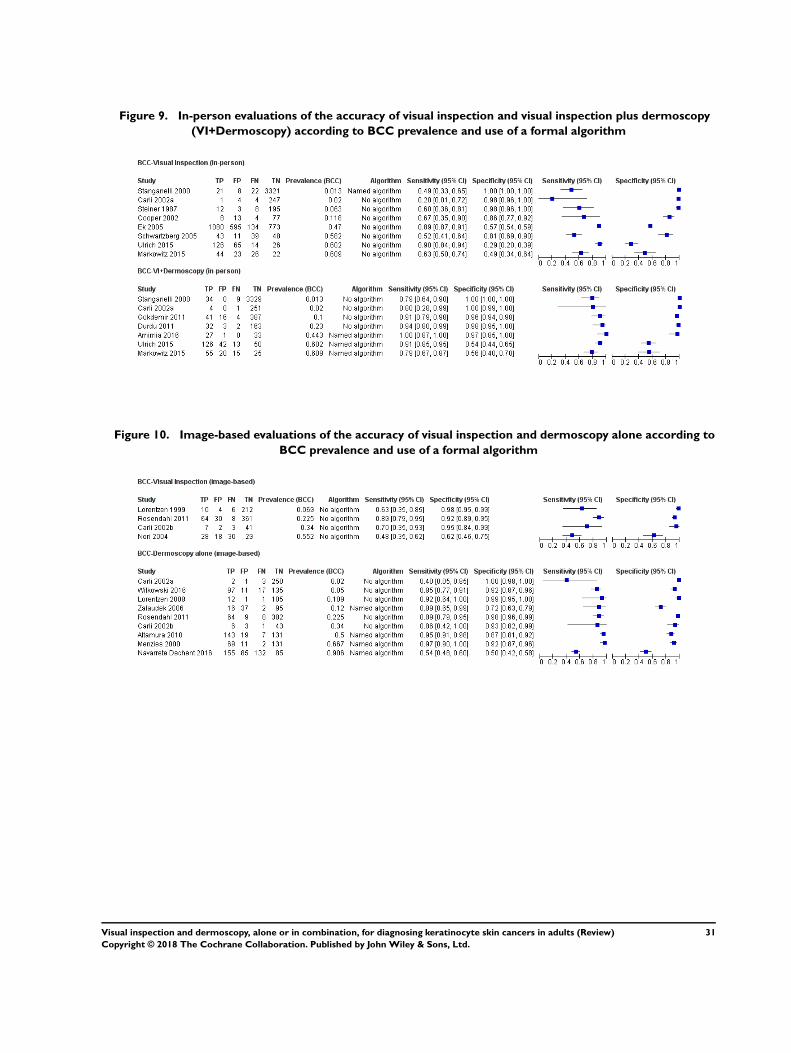
Figure 9. In-person evaluations of the accuracy of visual inspection and visual inspection plus dermoscopy
(VI+Dermoscopy) according to BCC prevalence and use of a formal algorithm
Figure 10. Image-based evaluations of the accuracy of visual inspection and dermoscopy alone according to
BCC prevalence and use of a formal algorithm
31Visual inspection and dermoscopy, alone or in combination, for diagnosing keratinocyte skin cancers in adults (Review)
Copyright © 2018 The Cochrane Collaboration. Published by John Wiley & Sons, Ltd.
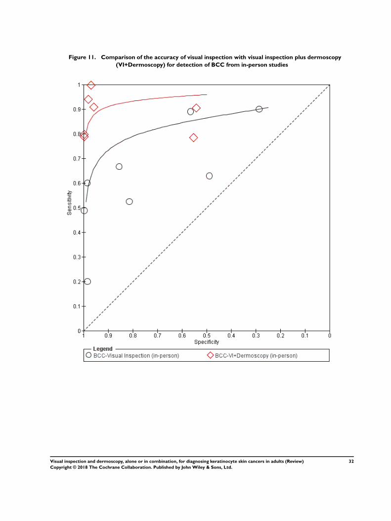
Figure 11. Comparison of the accuracy of visual inspection with visual inspection plus dermoscopy
(VI+Dermoscopy) for detection of BCC from in-person studies
32Visual inspection and dermoscopy, alone or in combination, for diagnosing keratinocyte skin cancers in adults (Review)
Copyright © 2018 The Cochrane Collaboration. Published by John Wiley & Sons, Ltd.
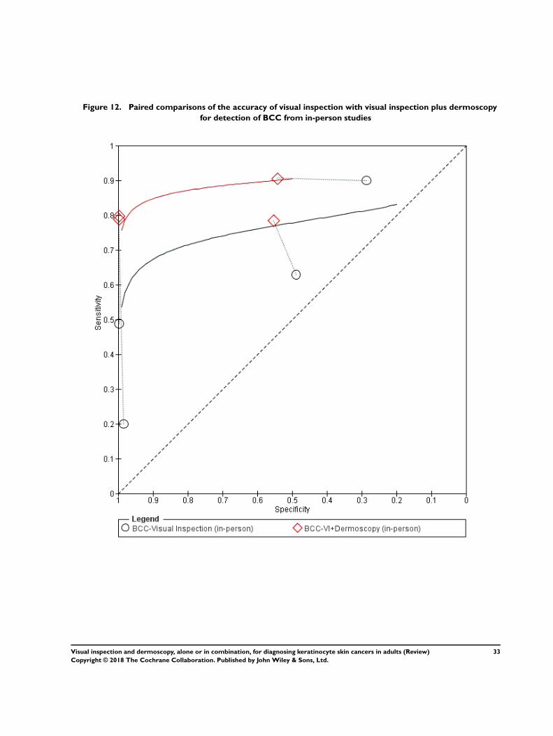
Figure 12. Paired comparisons of the accuracy of visual inspection with visual inspection plus dermoscopy
for detection of BCC from in-person studies
33Visual inspection and dermoscopy, alone or in combination, for diagnosing keratinocyte skin cancers in adults (Review)
Copyright © 2018 The Cochrane Collaboration. Published by John Wiley & Sons, Ltd.
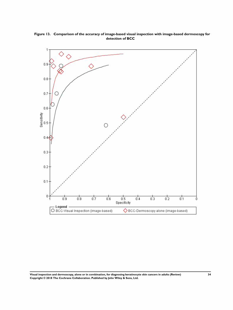
Figure 13. Comparison of the accuracy of image-based visual inspection with image-based dermoscopy for
detection of BCC
34Visual inspection and dermoscopy, alone or in combination, for diagnosing keratinocyte skin cancers in adults (Review)
Copyright © 2018 The Cochrane Collaboration. Published by John Wiley & Sons, Ltd.
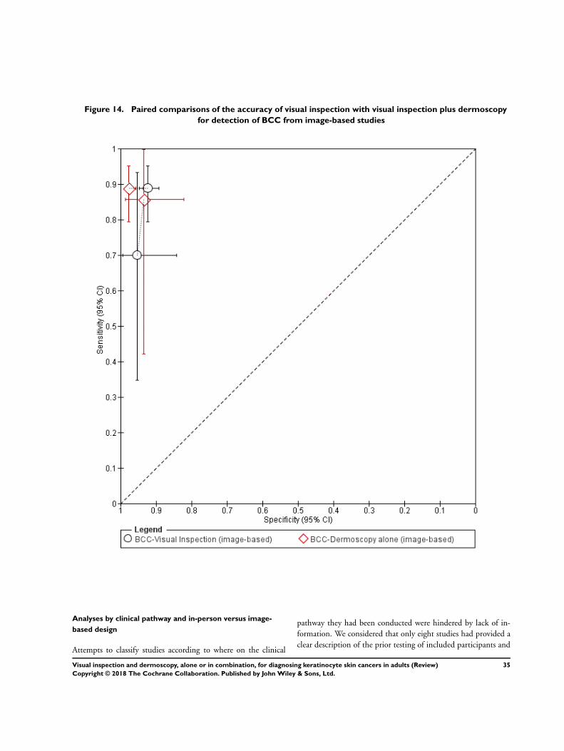
Figure 14. Paired comparisons of the accuracy of visual inspection with visual inspection plus dermoscopy
for detection of BCC from image-based studies
Analyses by clinical pathway and in-person versus image-
based design
Attempts to classify studies according to where on the clinical
pathway they had been conducted were hindered by lack of in-
formation. We considered that only eight studies had provided a
clear description of the prior testing of included participants and
35Visual inspection and dermoscopy, alone or in combination, for diagnosing keratinocyte skin cancers in adults (Review)
Copyright © 2018 The Cochrane Collaboration. Published by John Wiley & Sons, Ltd.
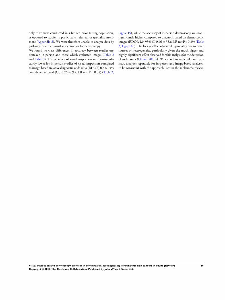
only three were conducted in a limited prior testing population,
as opposed to studies in participants referred for specialist assess-
ment (Appendix 8). We were therefore unable to analyse data by
pathway for either visual inspection or for dermoscopy.
We found no clear differences in accuracy between studies un-
dertaken in person and those which evaluated images (Table 2
and Table 3). The accuracy of visual inspection was non-signifi-
cantly lower for in-person studies of visual inspection compared
to image-based (relative diagnostic odds ratio (RDOR) 0.45, 95%
confidence interval (CI) 0.26 to 9.2, LR test P = 0.88) (Table 2;
Figure 15), while the accuracy of in-person dermoscopy was non-
significantly higher compared to diagnosis based on dermoscopic
images (RDOR 4.0, 95% CI 0.46 to 33.8; LR test P = 0.39) (Table
3; Figure 16). The lack of effect observed is probably due to other
sources of heterogeneity, particularly given the much bigger and
highly-significant effect observed for this analysis for the detection
of melanoma (Dinnes 2018a). We elected to undertake our pri-
mary analyses separately for in-person and image-based analyses,
to be consistent with the approach used in the melanoma review.
36Visual inspection and dermoscopy, alone or in combination, for diagnosing keratinocyte skin cancers in adults (Review)
Copyright © 2018 The Cochrane Collaboration. Published by John Wiley & Sons, Ltd.
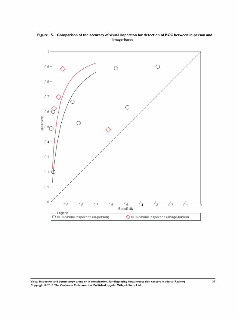
Figure 15. Comparison of the accuracy of visual inspection for detection of BCC between in-person and
image-based
37Visual inspection and dermoscopy, alone or in combination, for diagnosing keratinocyte skin cancers in adults (Review)
Copyright © 2018 The Cochrane Collaboration. Published by John Wiley & Sons, Ltd.
Figure 16. Comparison of the accuracy of dermoscopy for detection of BCC between in-person
(VI+Dermoscopy) and image-based (Dermoscopy alone)
In-person evaluations
The 11 studies reporting in-person evaluations of visual inspection
alone (n = 4; Cooper 2002; Ek 2005; Schwartzberg 2005; Steiner
1987), for visual inspection plus dermoscopy (n = 3; Amirnia
2016; Durdu 2011; Gokdemir 2011) or for both (n=4; Carli
38Visual inspection and dermoscopy, alone or in combination, for diagnosing keratinocyte skin cancers in adults (Review)
Copyright © 2018 The Cochrane Collaboration. Published by John Wiley & Sons, Ltd.
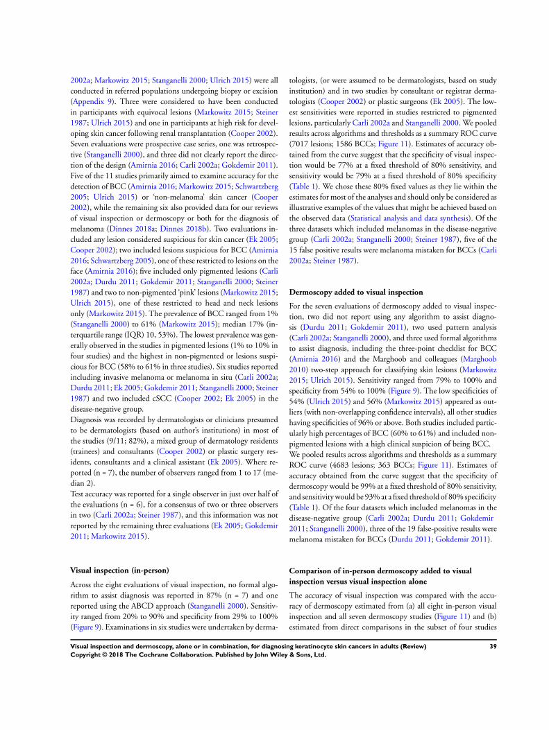
2002a; Markowitz 2015; Stanganelli 2000; Ulrich 2015) were all
conducted in referred populations undergoing biopsy or excision
(Appendix 9). Three were considered to have been conducted
in participants with equivocal lesions (Markowitz 2015; Steiner
1987; Ulrich 2015) and one in participants at high risk for devel-
oping skin cancer following renal transplantation (Cooper 2002).
Seven evaluations were prospective case series, one was retrospec-
tive (Stanganelli 2000), and three did not clearly report the direc-
tion of the design (Amirnia 2016; Carli 2002a; Gokdemir 2011).
Five of the 11 studies primarily aimed to examine accuracy for the
detection of BCC (Amirnia 2016; Markowitz 2015; Schwartzberg
2005; Ulrich 2015) or ‘non-melanoma’ skin cancer (Cooper
2002), while the remaining six also provided data for our reviews
of visual inspection or dermoscopy or both for the diagnosis of
melanoma (Dinnes 2018a; Dinnes 2018b). Two evaluations in-
cluded any lesion considered suspicious for skin cancer (Ek 2005;
Cooper 2002); two included lesions suspicious for BCC (Amirnia
2016; Schwartzberg 2005), one of these restricted to lesions on the
face (Amirnia 2016); five included only pigmented lesions (Carli
2002a; Durdu 2011; Gokdemir 2011; Stanganelli 2000; Steiner
1987) and two to non-pigmented ‘pink’ lesions (Markowitz 2015;
Ulrich 2015), one of these restricted to head and neck lesions
only (Markowitz 2015). The prevalence of BCC ranged from 1%
(Stanganelli 2000) to 61% (Markowitz 2015); median 17% (in-
terquartile range (IQR) 10, 53%). The lowest prevalence was gen-
erally observed in the studies in pigmented lesions (1% to 10% in
four studies) and the highest in non-pigmented or lesions suspi-
cious for BCC (58% to 61% in three studies). Six studies reported
including invasive melanoma or melanoma in situ (Carli 2002a;
Durdu 2011; Ek 2005; Gokdemir 2011; Stanganelli 2000; Steiner
1987) and two included cSCC (Cooper 2002; Ek 2005) in the
disease-negative group.
Diagnosis was recorded by dermatologists or clinicians presumed
to be dermatologists (based on author’s institutions) in most of
the studies (9/11; 82%), a mixed group of dermatology residents
(trainees) and consultants (Cooper 2002) or plastic surgery res-
idents, consultants and a clinical assistant (Ek 2005). Where re-
ported (n = 7), the number of observers ranged from 1 to 17 (me-
dian 2).
Test accuracy was reported for a single observer in just over half of
the evaluations (n = 6), for a consensus of two or three observers
in two (Carli 2002a; Steiner 1987), and this information was not
reported by the remaining three evaluations (Ek 2005; Gokdemir
2011; Markowitz 2015).
Visual inspection (in-person)
Across the eight evaluations of visual inspection, no formal algo-
rithm to assist diagnosis was reported in 87% (n = 7) and one
reported using the ABCD approach (Stanganelli 2000). Sensitiv-
ity ranged from 20% to 90% and specificity from 29% to 100%
(Figure 9). Examinations in six studies were undertaken by derma-
tologists, (or were assumed to be dermatologists, based on study
institution) and in two studies by consultant or registrar derma-
tologists (Cooper 2002) or plastic surgeons (Ek 2005). The low-
est sensitivities were reported in studies restricted to pigmented
lesions, particularly Carli 2002a and Stanganelli 2000. We pooled
results across algorithms and thresholds as a summary ROC curve
(7017 lesions; 1586 BCCs; Figure 11). Estimates of accuracy ob-
tained from the curve suggest that the specificity of visual inspec-
tion would be 77% at a fixed threshold of 80% sensitivity, and
sensitivity would be 79% at a fixed threshold of 80% specificity
(Table 1). We chose these 80% fixed values as they lie within the
estimates for most of the analyses and should only be considered as
illustrative examples of the values that might be achieved based on
the observed data (Statistical analysis and data synthesis). Of the
three datasets which included melanomas in the disease-negative
group (Carli 2002a; Stanganelli 2000; Steiner 1987), five of the
15 false positive results were melanoma mistaken for BCCs (Carli
2002a; Steiner 1987).
Dermoscopy added to visual inspection
For the seven evaluations of dermoscopy added to visual inspec-
tion, two did not report using any algorithm to assist diagno-
sis (Durdu 2011; Gokdemir 2011), two used pattern analysis
(Carli 2002a; Stanganelli 2000), and three used formal algorithms
to assist diagnosis, including the three-point checklist for BCC
(Amirnia 2016) and the Marghoob and colleagues (Marghoob
2010) two-step approach for classifying skin lesions (Markowitz
2015; Ulrich 2015). Sensitivity ranged from 79% to 100% and
specificity from 54% to 100% (Figure 9). The low specificities of
54% (Ulrich 2015) and 56% (Markowitz 2015) appeared as out-
liers (with non-overlapping confidence intervals), all other studies
having specificities of 96% or above. Both studies included partic-
ularly high percentages of BCC (60% to 61%) and included non-
pigmented lesions with a high clinical suspicion of being BCC.
We pooled results across algorithms and thresholds as a summary
ROC curve (4683 lesions; 363 BCCs; Figure 11). Estimates of
accuracy obtained from the curve suggest that the specificity of
dermoscopy would be 99% at a fixed threshold of 80% sensitivity,
and sensitivity would be 93% at a fixed threshold of 80% specificity
(Table 1). Of the four datasets which included melanomas in the
disease-negative group (Carli 2002a; Durdu 2011; Gokdemir
2011; Stanganelli 2000), three of the 19 false-positive results were
melanoma mistaken for BCCs (Durdu 2011; Gokdemir 2011).
Comparison of in-person dermoscopy added to visual
inspection versus visual inspection alone
The accuracy of visual inspection was compared with the accu-
racy of dermoscopy estimated from (a) all eight in-person visual
inspection and all seven dermoscopy studies (Figure 11) and (b)
estimated from direct comparisons in the subset of four studies
39Visual inspection and dermoscopy, alone or in combination, for diagnosing keratinocyte skin cancers in adults (Review)
Copyright © 2018 The Cochrane Collaboration. Published by John Wiley & Sons, Ltd.
that evaluated both visual inspection and dermoscopy on an in-
person basis (3974 lesions; 258 BCCs; Figure 12). In both com-
parisons the accuracy of dermoscopy in addition to visual inspec-
tion exceeded that of visual inspection alone (Table 1). In (a) the
diagnostic odds ratio (DOR) for dermoscopy was 8.2 (95% CI
3.5 to 19.3; LR test P < 0.001) times that of visual inspection
alone; in (b) it was 7.5 (95% CI 2.7 to 21.3; LR test P < 0.001)
times that of visual inspection alone. These effects correspond to
predicted differences in specificity of (a) 22% (99% versus 77%)
and (b) 61% (97% versus 36%) at a fixed sensitivity of 80% (Table
1) and predicted differences in sensitivity of (a) 14% (93% versus
79%) and (b) 16% (87% versus 71%) at a fixed specificity of 80%
(Table 1).
Image-based evaluations
The 11 studies reporting image-based diagnosis using clinical pho-
tographs (n = 2; Lorentzen 1999; Nori 2004), dermoscopic images
(n = 7; Altamura 2010; Carli 2002a; Lorentzen 2008; Menzies
2000; Navarrete Dechent 2016; Witkowski 2016; Zalaudek 2006)
or both (n = 2; Carli 2002b; Rosendahl 2011) were primarily
conducted in referred populations undergoing biopsy or excision
(Appendix 9). Two studies were conducted in a limited prior test-
ing setting, recruiting participants from primary care (Rosendahl
2011) or from a private dermatology practice (Navarrete Dechent
2016). Of the remaining nine, one was conducted in partici-
pants with equivocal lesions (Witkowski 2016). Two evaluations
used a case-control design, separately recruiting diseased and non-
diseased participants (Altamura 2010; Menzies 2000), one was
a prospective case series (Lorentzen 1999), five retrospectively
selected series of images for prospective interpretation within
the context of the study (Navarrete Dechent 2016; Nori 2004;
Rosendahl 2011; Witkowski 2016; Zalaudek 2006), and three did
not clearly report the direction of the design (Carli 2002a; Carli
2002b; Lorentzen 2008).
Five of the 11 studies primarily aimed to examine accuracy for
the detection of BCC (Altamura 2010; Menzies 2000; Navarrete
Dechent 2016; Nori 2004; Witkowski 2016), while the remaining
six also provided data for our reviews of visual inspection or der-
moscopy or both for the diagnosis of melanoma (Dinnes 2018a;
Dinnes 2018b). Four evaluations included any lesion, pigmented
or non-pigmented (Altamura 2010; Lorentzen 1999; Lorentzen
2008; Zalaudek 2006); four included only pigmented lesions
(Carli 2002a; Carli 2002b; Menzies 2000; Rosendahl 2011); two
included non-pigmented lesions only (Navarrete Dechent 2016;
Witkowski 2016), and one included biopsy-confirmed BCCs and
lesions with a range of common diagnoses (Nori 2004). The preva-
lence of BCC ranged from 2% (Carli 2002a) to 63% (Navarrete
Dechent 2016); median 16% (IQR 11, 47%). The highest preva-
lence was generally observed in the studies in non-pigmented le-
sions or lesions suspicious for BCC (44% to 63% in four studies,
one of which used a case-control design; Altamura 2010). All stud-
ies apart from Nori 2004 reported including invasive melanoma
or melanoma in situ, and five also included cSCC in the disease-
negative group (Altamura 2010; Navarrete Dechent 2016; Nori
2004; Rosendahl 2011; Witkowski 2016).
Diagnosis was recorded by dermatologists or clinicians presumed
to be dermatologists (based on author’s institutions) in most of
the studies (9/11; 73%), or by a mixed group of clinicians in two
(Lorentzen 1999; Zalaudek 2006). Where reported (n = 9), the
number of observers ranged from two (reported for five studies)
to 150 (median 2).
Test accuracy was reported for a single observer in four studies,
for a consensus of two observers in three (Carli 2002a; Carli
2002b; Navarrete Dechent 2016), the average across observers in
three (Lorentzen 1999; Lorentzen 2008; Zalaudek 2006), and this
information was not reported by one (Menzies 2000).
Visual inspection of clinical photographs
The four evaluations of image-based visual inspection reported no
formal algorithm to have been used to assist diagnosis. Sensitivity
ranged from 48% to 89%, and specificity from 62% to 98% (
Figure 10). We pooled results as a summary ROC curve (853
lesions; 156 BCCs; Figure 13). Estimates of accuracy obtained
from the curve suggest that the specificity of image-based visual
inspection would be 87% at a fixed threshold of 80% sensitivity,
and sensitivity would be 85% at a fixed threshold of 80% specificity
(Table 1). Of the three datasets which included melanoma in the
disease-negative group (Carli 2002b; Lorentzen 1999; Rosendahl
2011), three of 39 false-positive results were melanoma mistaken
for BCCs (Rosendahl 2011).
Dermoscopic image-based diagnosis
Of the nine evaluations of image-based dermoscopy, two did
not report using any algorithm to assist diagnosis (Carli 2002b;
Witkowski 2016), three used pattern analysis (Carli 2002a;
Lorentzen 2008; Rosendahl 2011), and four used formal algo-
rithms to assist diagnosis, including the three-point checklist
(Zalaudek 2006), the Menzies algorithm for BCC (Menzies 2000)
or a modification thereof (Altamura 2010), or a new algorithm
‘shiny white blotches and strands’ (Navarrete Dechent 2016). Only
one study provided the clinical photograph alongside the dermo-
scopic image (Rosendahl 2011), with the rest reporting blinded
dermoscopy interpretations. Sensitivity ranged from 40% to 97%
and specificity from 50% to 100% (Figure 10). We observed par-
ticularly low sensitivities in Carli 2002a and Navarrete Dechent
2016 (which respectively had the lowest (2%) and highest (63%)
prevalence of BCC), the latter also reporting the lowest specificity
(50%). All other studies reported sensitivities of 85% or above and
specificities of 72% or more.
We pooled results across algorithms and thresholds as a summary
ROC curve (2271 lesions; 737 BCCs; Figure 13). Estimates of
40Visual inspection and dermoscopy, alone or in combination, for diagnosing keratinocyte skin cancers in adults (Review)
Copyright © 2018 The Cochrane Collaboration. Published by John Wiley & Sons, Ltd.
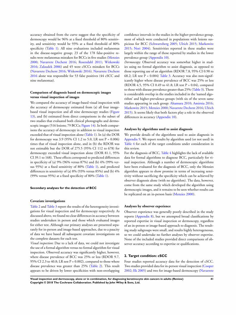
accuracy obtained from the curve suggest that the specificity of
dermoscopy would be 96% at a fixed threshold of 80% sensitiv-
ity, and sensitivity would be 93% at a fixed threshold of 80%
specificity (Table 1). All nine evaluations included melanomas
in the disease-negative group; 23 of the 178 false-positive re-
sults were melanomas mistaken for BCCs in five studies (Menzies
2000; Navarrete Dechent 2016; Rosendahl 2011; Witkowski
2016; Zalaudek 2006) and 45 were cSCCs mistaken for BCCs
(Navarrete Dechent 2016; Witkowski 2016). Navarrete Dechent
2016 alone was responsible for 53 false positives (44 cSCC and
nine melanomas).
Comparison of diagnosis based on dermoscopic images
versus visual inspection of images
We compared the accuracy of image-based visual inspection with
the accuracy of dermoscopy estimated from (a) all four image-
based visual inspection and all nine dermoscopy studies (Figure
13), and (b) estimated from direct comparisons in the subset of
two studies that evaluated both clinical photographs and dermo-
scopic images (516 lesions; 79 BCCs; Figure 14). In both compar-
isons the accuracy of dermoscopy in addition to visual inspection
exceeded that of visual inspection alone (Table 1). In (a) the DOR
for dermoscopy was 3.9 (95% CI 1.2 to 5.0, LR test P = 0.006)
times that of visual inspection alone, and in (b) the RDOR was
not estimable but the DOR of 275.5 (95% CI 112 to 678) for
dermoscopy exceeded visual inspection alone (DOR 81.1, 95%
CI 39.1 to 168). These effects correspond to predicted differences
in specificity of (a) 9% (96% versus 87%) and (b) 4% (99% ver-
sus 95%) at a fixed sensitivity of 80% (Table 1), and predicted
differences in sensitivity of (a) 8% (93% versus 85%) and (b) 4%
(99% versus 95%) at a fixed specificity of 80% (Table 1).
Secondary analyses for the detection of BCC
Covariate investigations
Table 2 and Table 3 report the results of the heterogeneity investi-
gations for visual inspection and for dermoscopy respectively. As
discussed above, we found no clear differences in accuracy between
studies undertaken in person and those which evaluated images
for either test. Although our primary analyses are presented sepa-
rately for in-person and image-based approaches, due to a paucity
of data we have based all subsequent covariate investigations on
the complete datasets for each test.
Visual inspection: Due to a lack of data, we could not investigate
the use of a formal algorithm versus no formal algorithm for visual
inspection. Observed accuracy was significantly higher, however,
where disease prevalence of BCC was 25% or less (RDOR 9.7,
95% CI 2.3 to 40.8; LR test P = 0.002), compared to those where
disease prevalence was greater than 25% (Table 2). This result
appears to be driven by lower specificities with non-overlapping
confidence intervals in the studies in the higher-prevalence group,
most of which were conducted in populations with lesions sus-
picious for BCC (Schwartzberg 2005; Ulrich 2015; Markowitz
2015; Nori 2004). Sensitivities reported in these studies were
largely within the range of those reported by studies in the lower
prevalence group (Appendix 10).
Dermoscopy: Observed accuracy was somewhat higher in stud-
ies using no formal algorithm to assist diagnosis, as opposed to
those reporting use of an algorithm (RDOR 7.8, 95% CI 0.90 to
68.2; LR test P = 0.004) Table 3. Accuracy was also non-signif-
icantly higher where disease prevalence of BCC was 25% or less
(RDOR 4.5, 95% CI 0.49 to 41.8; LR test P = 0.04), compared
to those with disease prevalence greater than 25% (Table 3). There
is considerable overlap in the studies included in the ‘named algo-
rithm’ and higher-prevalence groups (with six of the seven same
studies appearing in each group: Altamura 2010; Amirnia 2016;
Markowitz 2015; Menzies 2000; Navarrete Dechent 2016; Ulrich
2015). It seems likely that both factors play a role in the observed
differences in accuracy (Appendix 10).
Analyses by algorithms used to assist diagnosis
We provide details of the algorithms used to assist diagnosis in
Appendix 9. We report results by algorithm used (or not used) in
Table 4 for each of the target conditions under consideration in
this review.
For the diagnosis of BCC, Table 4 highlights the lack of available
data for formal algorithms to diagnose BCC, particularly for vi-
sual inspection. Although a number of dermoscopic algorithms
have been evaluated for the diagnosis of BCC, only the Menzies
algorithm appears to show promise in terms of increasing sensi-
tivity without sacrificing the specificity which can be achieved by
observer diagnosis alone (with no algorithm). The data, however,
come from the same study which developed the algorithm using
dermoscopic images, and it remains to be seen whether results can
be replicated on an in-person basis (Menzies 2000).
Analyses by observer experience
Observer experience was generally poorly described in the study
reports (Appendix 8), but we attempted broad classifications by
reported expertise in visual inspection or dermoscopy, regardless
of an in-person or image-based approach to diagnosis. The result-
ing study subgroups were small, and results highly heterogeneous,
so we could undertake no further analyses by observer expertise.
None of the included studies provided direct comparisons of ob-
server accuracy according to expertise or qualifications.
2. Target condition: cSCC
Four studies reported accuracy data for the detection of cSCC.
Two studies provided data for in-person visual inspection (Cooper
2002; Ek 2005) and two for image-based dermoscopy (Navarrete
41Visual inspection and dermoscopy, alone or in combination, for diagnosing keratinocyte skin cancers in adults (Review)
Copyright © 2018 The Cochrane Collaboration. Published by John Wiley & Sons, Ltd.
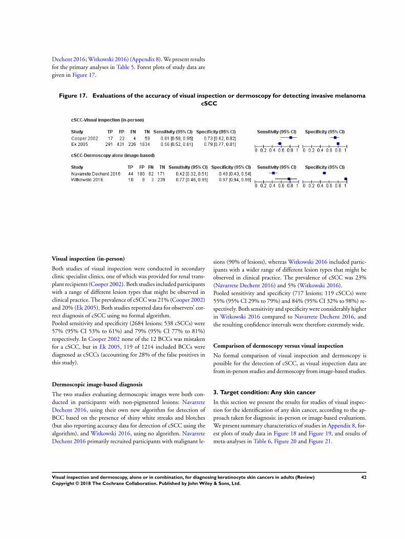
Dechent 2016; Witkowski 2016) (Appendix 8). We present results
for the primary analyses in Table 5. Forest plots of study data are
given in Figure 17.
Figure 17. Evaluations of the accuracy of visual inspection or dermoscopy for detecting invasive melanoma
cSCC
Visual inspection (in-person)
Both studies of visual inspection were conducted in secondary
clinic specialist clinics, one of which was provided for renal trans-
plant recipients (Cooper 2002). Both studies included participants
with a range of different lesion types that might be observed in
clinical practice. The prevalence of cSCC was 21% (Cooper 2002)
and 20% (Ek 2005). Both studies reported data for observers’ cor-
rect diagnosis of cSCC using no formal algorithm.
Pooled sensitivity and specificity (2684 lesions; 538 cSCCs) were
57% (95% CI 53% to 61%) and 79% (95% CI 77% to 81%)
respectively. In Cooper 2002 none of the 12 BCCs was mistaken
for a cSCC, but in Ek 2005, 119 of 1214 included BCCs were
diagnosed as cSCCs (accounting for 28% of the false positives in
this study).
Dermoscopic image-based diagnosis
The two studies evaluating dermoscopic images were both con-
ducted in participants with non-pigmented lesions: Navarrete
Dechent 2016, using their own new algorithm for detection of
BCC based on the presence of shiny white streaks and blotches
(but also reporting accuracy data for detection of cSCC using the
algorithm), and Witkowski 2016, using no algorithm. Navarrete
Dechent 2016 primarily recruited participants with malignant le-
sions (90% of lesions), whereas Witkowski 2016 included partic-
ipants with a wider range of different lesion types that might be
observed in clinical practice. The prevalence of cSCC was 23%
(Navarrete Dechent 2016) and 5% (Witkowski 2016).
Pooled sensitivity and specificity (717 lesions; 119 cSCCs) were
55% (95% CI 29% to 79%) and 84% (95% CI 32% to 98%) re-
spectively. Both sensitivity and specificity were considerably higher
in Witkowski 2016 compared to Navarrete Dechent 2016, and
the resulting confidence intervals were therefore extremely wide.
Comparison of dermoscopy versus visual inspection
No formal comparison of visual inspection and dermoscopy is
possible for the detection of cSCC, as visual inspection data are
from in-person studies and dermoscopy from image-based studies.
3. Target condition: Any skin cancer
In this section we present the results for studies of visual inspec-
tion for the identification of any skin cancer, according to the ap-
proach taken for diagnosis: in-person or image-based evaluations.
We present summary characteristics of studies in Appendix 8, for-
est plots of study data in Figure 18 and Figure 19, and results of
meta-analyses in Table 6, Figure 20 and Figure 21.
42Visual inspection and dermoscopy, alone or in combination, for diagnosing keratinocyte skin cancers in adults (Review)
Copyright © 2018 The Cochrane Collaboration. Published by John Wiley & Sons, Ltd.
Figure 18. Forest plot of tests: 27 Any -Visual inspection (in-person), 29 Any -VI+Dermoscopy (in-person).
Figure 19. Forest plot of tests: 28 Any -Visual inspection (image-based), 30 Any-Dermoscopy alone (image-
based).
43Visual inspection and dermoscopy, alone or in combination, for diagnosing keratinocyte skin cancers in adults (Review)
Copyright © 2018 The Cochrane Collaboration. Published by John Wiley & Sons, Ltd.
Figure 20. Comparison of the accuracy of visual inspection with visual inspection plus dermoscopy
(VI+Dermoscopy) for detection of any skin cancer (Any). SROC curve estimated only for in-person visual
inspection.
44Visual inspection and dermoscopy, alone or in combination, for diagnosing keratinocyte skin cancers in adults (Review)
Copyright © 2018 The Cochrane Collaboration. Published by John Wiley & Sons, Ltd.
Figure 21. Comparison of the accuracy of image-based visual inspection with image-based dermoscopy
(Dermoscopy alone) for detection of any skin cancer (Any)
In-person evaluations
Five studies evaluated the accuracy of in-person visual inspection
for the detection of any skin cancer (Argenziano 2006; Chang
2013; Cooper 2002; Ek 2005; Hacioglu 2013) and two evaluated
in-person dermoscopy (Argenziano 2006; Durdu 2011). Three of
these also reported accuracy data separately for BCC alone (Cooper
45Visual inspection and dermoscopy, alone or in combination, for diagnosing keratinocyte skin cancers in adults (Review)
Copyright © 2018 The Cochrane Collaboration. Published by John Wiley & Sons, Ltd.
2002; Durdu 2011; Ek 2005) or for cSCC (Cooper 2002; Ek
2005).
All studies were based in secondary care or specialist referral clin-
ics, apart from Argenziano 2006 which recruited participants from
primary care (although only lesions selected for excision by an ex-
pert could be included). The prevalence of skin cancer ranged from
20% (Chang 2013) to 68% (Ek 2005). Studies included any lesion
type, apart from Durdu 2011 which restricted inclusion to pig-
mented lesions only. Diagnoses were recorded by GPs (Argenziano
2006), dermatologists or assumed to be dermatologists based on
study institution (Chang 2013; Durdu 2011; Hacioglu 2013) or
by a clinician with mixed experience (Cooper 2002; Ek 2005). All
studies used a histological reference standard.
Visual inspection
Studies either used no algorithm to aid diagnosis, or reported us-
ing the ABCD approach to diagnosis (Argenziano 2006). Sensi-
tivities ranged from 57% to 98%; specificities ranged from 13%
to 86% (Figure 18). In meta-analysis the DOR was 28.7 (95%
CI 5.0 to 166) (3618 lesions; 2021 skin cancer cases). Estimates
of accuracy obtained from the curve suggest that the specificity
of visual inspection would be 88% at a fixed threshold of 80%
sensitivity, and sensitivity would be 84% at a fixed threshold of
80% specificity (Table 6).
Dermoscopy added to visual inspection
The two studies of in-person dermoscopy reported data using the
three-point checklist (Argenziano 2006) and the ABCD approach
(Durdu 2011) (Figure 18). In Argenziano 2006, GPs’ diagnosis
had a sensitivity of 85% (95% CI 69% to 94%) and specificity of
26% (95% CI 13% to 43%) for the subgroup of lesions selected
for excision by an expert clinician. Of the six malignancies missed
by GPs, four were BCCs, one cSCC and one melanoma. Durdu
2011 reported a sensitivity of 98% (95% CI 88% to 100%) and
specificity 98% (95% CI 94% to 100%) for their sample of pig-
mented lesions which could not be diagnosed by a dermatologist
with visual inspection alone.
In meta-analysis the DOR was 126 (95% CI 9.1 to 1751) (277
lesions; 85 skin cancer cases) (Table 6). We could not obtain es-
timates of accuracy from the SROC curve due to extreme differ-
ences in results between the two studies (evidenced by the very
wide range in confidence intervals around the DOR).
Comparison of in-person dermoscopy versus visual
inspection alone
No formal comparison of visual inspection and dermoscopy added
to visual inspection was possible, due to the observed heterogeneity
in results for the two dermoscopy studies (Figure 20).
Image-based evaluations
Six studies reported data for image-based diagnosis for the detec-
tion of any skin cancer. Two evaluated the accuracy of image-based
visual inspection (Carli 2002b; Rosendahl 2011) and all six evalu-
ated diagnosis using dermoscopic images (Carli 2002b; Hacioglu
2013; Menzies 2000; Navarrete Dechent 2016; Rosendahl 2011;
Witkowski 2016). Five of these also reported accuracy data sep-
arately for BCC alone (Carli 2002b; Menzies 2000; Navarrete
Dechent 2016; Rosendahl 2011; Witkowski 2016) or for cSCC
(Navarrete Dechent 2016; Witkowski 2016).
Two studies were conducted in a limited prior testing setting, re-
cruiting participants from primary care (Rosendahl 2011) or from
a private dermatology practice (Navarrete Dechent 2016). Of the
remaining four, one was considered to have been conducted in
participants with equivocal lesions (Witkowski 2016). Four of the
six studies primarily aimed to examine accuracy for the detec-
tion of BCC (Menzies 2000; Navarrete Dechent 2016; Witkowski
2016) or ‘non-melanoma’ skin cancer (Hacioglu 2013), with the
remaining two also providing data for the diagnosis of melanoma
(Carli 2002b; Rosendahl 2011). Three studies included only pig-
mented lesions (Carli 2002b; Menzies 2000; Rosendahl 2011);
two included only non-pigmented lesions (Navarrete Dechent
2016; Witkowski 2016) and one described lesions as ‘suspicious
for malignancy’ (Hacioglu 2013). All studies apart from Hacioglu
2013 reported including invasive melanoma or melanoma in situ
as disease-negative and four also included cSCC (all apart from
Carli 2002b and Menzies 2000) in the disease-negative group. Di-
agnosis was recorded by dermatologists or by dermatology trainees
(Navarrete Dechent 2016). All studies used a histological reference
standard.
Visual inspection of images
The two included studies used no algorithm to aid diagnosis and
both included pigmented lesions only (Carli 2002b; Rosendahl
2011). Sensitivities were 80% (95% CI 56% to 94%) and 76%
(95% CI 67% to 84%) and specificities 74% (95% CI 56% to
87%) and 85% (95% CI 81% to 88%) in Carli 2002b and
Rosendahl 2011, respectively (Figure 19).
In meta-analysis the DOR was 16.3 (95%CI 4.4 to 59.9) (517 le-
sions; 124 skin cancer cases). Estimates of accuracy obtained from
the curve suggest that the specificity of visual inspection would be
79% at a fixed threshold of 80% sensitivity, and sensitivity would
be 78% at a fixed threshold of 80% specificity (Table 6).
Dermoscopic image-based diagnosis
The six studies used no algorithm to assist diagnosis in three (Carli
2002b; Hacioglu 2013; Witkowski 2016), pattern analysis in one
(Rosendahl 2011), and new algorithms for detection of BCC in
two (Menzies 2000; Navarrete Dechent 2016).
46Visual inspection and dermoscopy, alone or in combination, for diagnosing keratinocyte skin cancers in adults (Review)
Copyright © 2018 The Cochrane Collaboration. Published by John Wiley & Sons, Ltd.
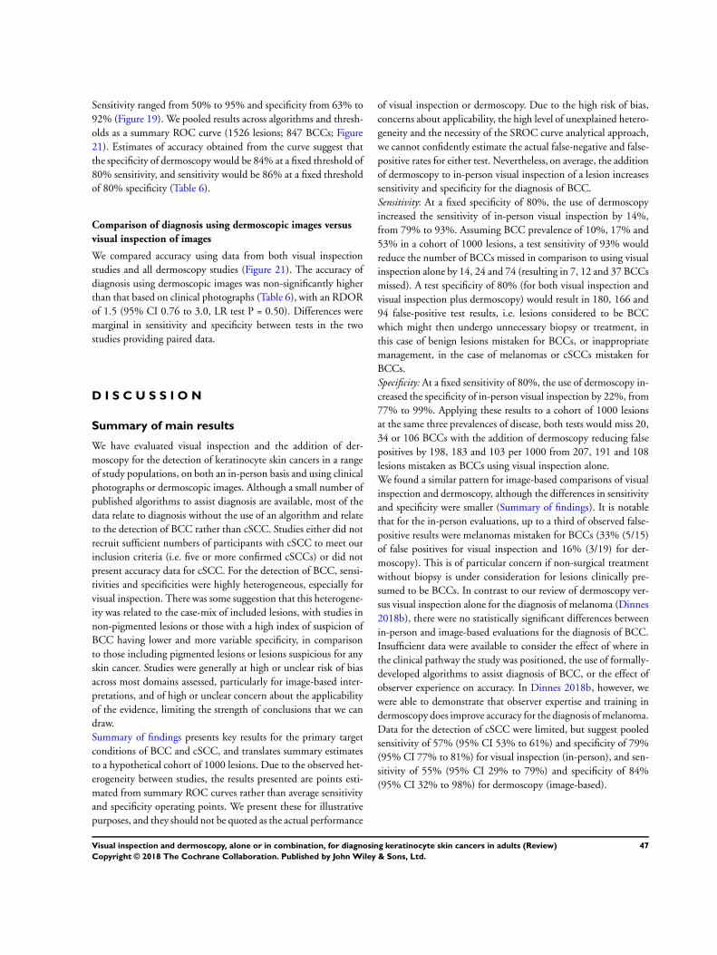
Sensitivity ranged from 50% to 95% and specificity from 63% to
92% (Figure 19). We pooled results across algorithms and thresh-
olds as a summary ROC curve (1526 lesions; 847 BCCs; Figure
21). Estimates of accuracy obtained from the curve suggest that
the specificity of dermoscopy would be 84% at a fixed threshold of
80% sensitivity, and sensitivity would be 86% at a fixed threshold
of 80% specificity (Table 6).
Comparison of diagnosis using dermoscopic images versus
visual inspection of images
We compared accuracy using data from both visual inspection
studies and all dermoscopy studies (Figure 21). The accuracy of
diagnosis using dermoscopic images was non-significantly higher
than that based on clinical photographs (Table 6), with an RDOR
of 1.5 (95% CI 0.76 to 3.0, LR test P = 0.50). Differences were
marginal in sensitivity and specificity between tests in the two
studies providing paired data.
D I S C U S S I O N
Summary of main results
We have evaluated visual inspection and the addition of der-
moscopy for the detection of keratinocyte skin cancers in a range
of study populations, on both an in-person basis and using clinical
photographs or dermoscopic images. Although a small number of
published algorithms to assist diagnosis are available, most of the
data relate to diagnosis without the use of an algorithm and relate
to the detection of BCC rather than cSCC. Studies either did not
recruit sufficient numbers of participants with cSCC to meet our
inclusion criteria (i.e. five or more confirmed cSCCs) or did not
present accuracy data for cSCC. For the detection of BCC, sensi-
tivities and specificities were highly heterogeneous, especially for
visual inspection. There was some suggestion that this heterogene-
ity was related to the case-mix of included lesions, with studies in
non-pigmented lesions or those with a high index of suspicion of
BCC having lower and more variable specificity, in comparison
to those including pigmented lesions or lesions suspicious for any
skin cancer. Studies were generally at high or unclear risk of bias
across most domains assessed, particularly for image-based inter-
pretations, and of high or unclear concern about the applicability
of the evidence, limiting the strength of conclusions that we can
draw.
Summary of findings presents key results for the primary target
conditions of BCC and cSCC, and translates summary estimates
to a hypothetical cohort of 1000 lesions. Due to the observed het-
erogeneity between studies, the results presented are points esti-
mated from summary ROC curves rather than average sensitivity
and specificity operating points. We present these for illustrative
purposes, and they should not be quoted as the actual performance
of visual inspection or dermoscopy. Due to the high risk of bias,
concerns about applicability, the high level of unexplained hetero-
geneity and the necessity of the SROC curve analytical approach,
we cannot confidently estimate the actual false-negative and false-
positive rates for either test. Nevertheless, on average, the addition
of dermoscopy to in-person visual inspection of a lesion increases
sensitivity and specificity for the diagnosis of BCC.
Sensitivity: At a fixed specificity of 80%, the use of dermoscopy
increased the sensitivity of in-person visual inspection by 14%,
from 79% to 93%. Assuming BCC prevalence of 10%, 17% and
53% in a cohort of 1000 lesions, a test sensitivity of 93% would
reduce the number of BCCs missed in comparison to using visual
inspection alone by 14, 24 and 74 (resulting in 7, 12 and 37 BCCs
missed). A test specificity of 80% (for both visual inspection and
visual inspection plus dermoscopy) would result in 180, 166 and
94 false-positive test results, i.e. lesions considered to be BCC
which might then undergo unnecessary biopsy or treatment, in
this case of benign lesions mistaken for BCCs, or inappropriate
management, in the case of melanomas or cSCCs mistaken for
BCCs.
Specificity: At a fixed sensitivity of 80%, the use of dermoscopy in-
creased the specificity of in-person visual inspection by 22%, from
77% to 99%. Applying these results to a cohort of 1000 lesions
at the same three prevalences of disease, both tests would miss 20,
34 or 106 BCCs with the addition of dermoscopy reducing false
positives by 198, 183 and 103 per 1000 from 207, 191 and 108
lesions mistaken as BCCs using visual inspection alone.
We found a similar pattern for image-based comparisons of visual
inspection and dermoscopy, although the differences in sensitivity
and specificity were smaller (Summary of findings). It is notable
that for the in-person evaluations, up to a third of observed false-
positive results were melanomas mistaken for BCCs (33% (5/15)
of false positives for visual inspection and 16% (3/19) for der-
moscopy). This is of particular concern if non-surgical treatment
without biopsy is under consideration for lesions clinically pre-
sumed to be BCCs. In contrast to our review of dermoscopy ver-
sus visual inspection alone for the diagnosis of melanoma (Dinnes
2018b), there were no statistically significant differences between
in-person and image-based evaluations for the diagnosis of BCC.
Insufficient data were available to consider the effect of where in
the clinical pathway the study was positioned, the use of formally-
developed algorithms to assist diagnosis of BCC, or the effect of
observer experience on accuracy. In Dinnes 2018b, however, we
were able to demonstrate that observer expertise and training in
dermoscopy does improve accuracy for the diagnosis of melanoma.
Data for the detection of cSCC were limited, but suggest pooled
sensitivity of 57% (95% CI 53% to 61%) and specificity of 79%
(95% CI 77% to 81%) for visual inspection (in-person), and sen-
sitivity of 55% (95% CI 29% to 79%) and specificity of 84%
(95% CI 32% to 98%) for dermoscopy (image-based).
47Visual inspection and dermoscopy, alone or in combination, for diagnosing keratinocyte skin cancers in adults (Review)
Copyright © 2018 The Cochrane Collaboration. Published by John Wiley & Sons, Ltd.
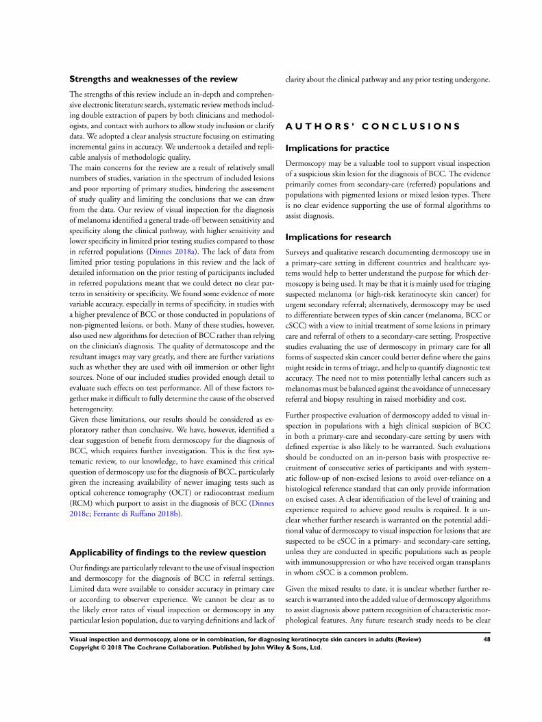
Strengths and weaknesses of the review
The strengths of this review include an in-depth and comprehen-
sive electronic literature search, systematic review methods includ-
ing double extraction of papers by both clinicians and methodol-
ogists, and contact with authors to allow study inclusion or clarify
data. We adopted a clear analysis structure focusing on estimating
incremental gains in accuracy. We undertook a detailed and repli-
cable analysis of methodologic quality.
The main concerns for the review are a result of relatively small
numbers of studies, variation in the spectrum of included lesions
and poor reporting of primary studies, hindering the assessment
of study quality and limiting the conclusions that we can draw
from the data. Our review of visual inspection for the diagnosis
of melanoma identified a general trade-off between sensitivity and
specificity along the clinical pathway, with higher sensitivity and
lower specificity in limited prior testing studies compared to those
in referred populations (Dinnes 2018a). The lack of data from
limited prior testing populations in this review and the lack of
detailed information on the prior testing of participants included
in referred populations meant that we could detect no clear pat-
terns in sensitivity or specificity. We found some evidence of more
variable accuracy, especially in terms of specificity, in studies with
a higher prevalence of BCC or those conducted in populations of
non-pigmented lesions, or both. Many of these studies, however,
also used new algorithms for detection of BCC rather than relying
on the clinician’s diagnosis. The quality of dermatoscope and the
resultant images may vary greatly, and there are further variations
such as whether they are used with oil immersion or other light
sources. None of our included studies provided enough detail to
evaluate such effects on test performance. All of these factors to-
gether make it difficult to fully determine the cause of the observed
heterogeneity.
Given these limitations, our results should be considered as ex-
ploratory rather than conclusive. We have, however, identified a
clear suggestion of benefit from dermoscopy for the diagnosis of
BCC, which requires further investigation. This is the first sys-
tematic review, to our knowledge, to have examined this critical
question of dermoscopy use for the diagnosis of BCC, particularly
given the increasing availability of newer imaging tests such as
optical coherence tomography (OCT) or radiocontrast medium
(RCM) which purport to assist in the diagnosis of BCC (Dinnes
2018c; Ferrante di Ruffano 2018b).
Applicability of findings to the review question
Our findings are particularly relevant to the use of visual inspection
and dermoscopy for the diagnosis of BCC in referral settings.
Limited data were available to consider accuracy in primary care
or according to observer experience. We cannot be clear as to
the likely error rates of visual inspection or dermoscopy in any
particular lesion population, due to varying definitions and lack of
clarity about the clinical pathway and any prior testing undergone.
A U T H O R S ’ C O N C L U S I O N S
Implications for practice
Dermoscopy may be a valuable tool to support visual inspection
of a suspicious skin lesion for the diagnosis of BCC. The evidence
primarily comes from secondary-care (referred) populations and
populations with pigmented lesions or mixed lesion types. There
is no clear evidence supporting the use of formal algorithms to
assist diagnosis.
Implications for research
Surveys and qualitative research documenting dermoscopy use in
a primary-care setting in different countries and healthcare sys-
tems would help to better understand the purpose for which der-
moscopy is being used. It may be that it is mainly used for triaging
suspected melanoma (or high-risk keratinocyte skin cancer) for
urgent secondary referral; alternatively, dermoscopy may be used
to differentiate between types of skin cancer (melanoma, BCC or
cSCC) with a view to initial treatment of some lesions in primary
care and referral of others to a secondary-care setting. Prospective
studies evaluating the use of dermoscopy in primary care for all
forms of suspected skin cancer could better define where the gains
might reside in terms of triage, and help to quantify diagnostic test
accuracy. The need not to miss potentially lethal cancers such as
melanomas must be balanced against the avoidance of unnecessary
referral and biopsy resulting in raised morbidity and cost.
Further prospective evaluation of dermoscopy added to visual in-
spection in populations with a high clinical suspicion of BCC
in both a primary-care and secondary-care setting by users with
defined expertise is also likely to be warranted. Such evaluations
should be conducted on an in-person basis with prospective re-
cruitment of consecutive series of participants and with system-
atic follow-up of non-excised lesions to avoid over-reliance on a
histological reference standard that can only provide information
on excised cases. A clear identification of the level of training and
experience required to achieve good results is required. It is un-
clear whether further research is warranted on the potential addi-
tional value of dermoscopy to visual inspection for lesions that are
suspected to be cSCC in a primary- and secondary-care setting,
unless they are conducted in specific populations such as people
with immunosuppression or who have received organ transplants
in whom cSCC is a common problem.
Given the mixed results to date, it is unclear whether further re-
search is warranted into the added value of dermoscopy algorithms
to assist diagnosis above pattern recognition of characteristic mor-
phological features. Any future research study needs to be clear
48Visual inspection and dermoscopy, alone or in combination, for diagnosing keratinocyte skin cancers in adults (Review)
Copyright © 2018 The Cochrane Collaboration. Published by John Wiley & Sons, Ltd.
about the diagnostic pathway followed by study participants prior
to study enrolment, and should conform to the updated Stan-
dards for Reporting of Diagnostic Accuracy (STARD) guideline
(Bossuyt 2015).
A C K N O W L E D G E M E N T S
Members of the Cochrane Skin Cancer Diagnostic Test Accuracy
Group include:
• the full project team (Susan Bayliss, Naomi Chuchu, Clare
Davenport, Jonathan Deeks, Jacqueline Dinnes, Lavinia Ferrante
di Ruffano, Kathie Godfrey, Rubeta Matin, Colette O’Sullivan,
Yemisi Takwoingi, Hywel Williams)
• our 12 clinical reviewers (Rachel Abbott, Ben Aldridge,
Oliver Bassett, Sue Ann Chan, Alana Durack, Monica Fawzy,
Abha Gulati, Jacqui Moreau, Lopa Patel, Daniel Saleh, David
Thompson, Kai Yuen Wong) and two methodologists (Lavinia
Ferrante di Ruffano and Louise Johnston), who assisted with full-
text screening, data extraction and quality assessment across the
entire suite of reviews of diagnosis and staging and skin cancer;
• our expert advisor and co-author on the review, Hamid
Tehrani, and on the protocol for keratinocyte skin cancer, Fiona
Bath-Hextall; and
• all members of our Advisory Group (Jonathan Bowling,
Seau Tak Cheung, Colin Fleming, Matthew Gardiner, Abhilash
Jain, Susan O’Connell, Pat Lawton, John Lear, Mariska Leeflang,
Richard Motley, Paul Nathan, Julia Newton-Bishop, Miranda
Payne, Rachael Robinson, Simon Rodwell, Julia Schofield, Neil
Shroff, Hamid Tehrani, Zoe Traill, Fiona Walter, Angela
Webster).
Cochrane Skin editorial base wishes to thank Robert Dellavalle,
who was the Dermatology Editor for this review; and the clinical
referee, Andrew Affleck. We also wish to thank the Cochrane DTA
editorial base and colleagues, as well as Kate Cahill, who copy-
edited this review.
R E F E R E N C E S
References to studies included in this review
Altamura 2010 {published data only}
Altamura D, Menzies SW, Argenziano G, Zalaudek I, Soyer
HP, Sera F, et al. Dermatoscopy of basal cell carcinoma:
morphologic variability of global and local features and
accuracy of diagnosis. Journal of the American Academy
of Dermatology 2010;62(1):67–75. ER4:15465845;
PUBMED: 19828209]
Amirnia 2016 {published data only}
Amirnia M, Ranjkesh MR, Azimpouran M, Karkon-Shayan
F, Alikhah H, Jafari-Asl M, et al. Comparative study of
dermatoscopic and histopathologic results in facial basal
cell carcinoma and melanocytic nevi. Asian Pacific Journal
of Cancer Prevention 2016;17(1):425–9. PUBMED:
26838250]
Argenziano 2006 {published data only}
Argenziano G, Puig S, Zalaudek I, Sera F, Corona R,
Alsina M, et al. Dermoscopy improves accuracy of primary
care physicians to triage lesions suggestive of skin cancer.
Journal of Clinical Oncology 2006;24(12):1877–82. ER4:
17940973; PUBMED: 16622262]
Carli 2002a {published data only}
Carli P, De Giorgi V, Argenziano G, Palli D, Giannotti
B. Pre-operative diagnosis of pigmented skin lesions: in
vivo dermoscopy performs better than dermoscopy on
photographic images. Journal of the European Academy ofDermatology and Venereology : JEADV 2002;16(4):339–46.
ER4:15465882; PUBMED: 12224689]
Carli 2002b {published data only}
Carli P, De Giorgi V, Salvini C, Mannone F, Chiarugi
A. The gold standard for photographing pigmented skin
lesions for diagnostic purposes: contact versus distant
imaging. Skin Research and Technology 2002;8(4):255–9.
PUBMED: 12423545]
Chang 2013 {published data only}
Chang WY, Huang A, Yang CY, Lee CH, Chen YC,
Wu TY, et al. Computer-aided diagnosis of skin lesions
using conventional digital photography: a reliability and
feasibility study. PloS One 2013;8(11):e76212. ER4:
15465893; PUBMED: 24223698]
Cooper 2002 {published data only}
Cooper SM, Wojnarowska F. The accuracy of clinical
diagnosis of suspected premalignant and malignant
skin lesions in renal transplant recipients. Clinical and
Experimental Dermatology 2002;27(6):436–8. ER4:
20569444; PUBMED: 12372077]
Durdu 2011 {published data only}
Durdu M, Baba M, Seckin D. Dermatoscopy versus
Tzanck smear test: a comparison of the value of two tests
in the diagnosis of pigmented skin lesions. Journal of theAmerican Academy of Dermatology 2011;65(5):972–82.
ER4:15465910; PUBMED: 21565420]
Ek 2005 {published data only}
Ek EW, Giorlando F, Su SY, Dieu T. Clinical diagnosis of skin
tumours: how good are we?. ANZ Journal of Surgery 2005;
75(6):415–20. DOI: 10.1111/j.1445-2197.2005.03394.x;
ER4:20569451; PUBMED: 15943729
49Visual inspection and dermoscopy, alone or in combination, for diagnosing keratinocyte skin cancers in adults (Review)
Copyright © 2018 The Cochrane Collaboration. Published by John Wiley & Sons, Ltd.
Gokdemir 2011 {published data only}
Gokdemir A, Guler Ozden M, Bek Y, Aydin F, Senturk
N, Canturk T, et al. Dermoscopic and histopathological
correlation in melanocytic and non-melanocytic lesions
[Melanositik ve non–melanositik lezyonlarda dermoskopik
ve histopatolojik tani korelasyonu]. Turkiye KlinikleriDermatoloji 2011;21(1):7–16.
Hacioglu 2013 {published data only}
Hacioglu S, Saricaoglu H, Baskan EB, Uner SI, Aydogan K,
Tunali S. The value of spectrophotometric intracutaneous
analysis in the noninvasive diagnosis of nonmelanoma skin
cancers. Clinical and Experimental Dermatology 2013;38(5):
464–9. ER4:15465947; PUBMED: 23777487]
Lorentzen 1999 {published data only}
Lorentzen H, Weismann K, Petersen CS, Larsen FG,
Secher L, Skodt V. Clinical and dermatoscopic diagnosis of
malignant melanoma. Assessed by expert and non-expert
groups. Acta Dermato-Venereologica 1999;79(4):301–4.
ER4:17941062; PUBMED: 10429989]
Lorentzen 2008 {published data only}
Lorentzen HF, Eefsen RL, Weismann K. Comparison
of classical dermatoscopy and acrylic globe magnifier
dermatoscopy. Acta Dermato-Venereologica 2008;88(2):
139–42. ER4:15465993; PUBMED: 18311441]
Markowitz 2015 {published data only}
Markowitz O, Schwartz M, Feldman E, Bienenfeld A,
Bieber AK, Ellis J, et al. Evaluation of optical coherence
tomography as a means of identifying earlier stage basal cell
carcinomas while reducing the use of diagnostic biopsy.
Journal of Clinical and Aesthetic Dermatology 2015;8(10):
14–20. ER4:25012306; PUBMED: 26557214]
Menzies 2000 {published data only}
Menzies SW, Westerhoff K, Rabinovitz H, Kopf AW,
McCarthy WH, Katz B. Surface microscopy of pigmented
basal cell carcinoma. Archives of Dermatology 2000;136(8):
1012–6. PUBMED: 10926737]
Navarrete Dechent 2016 {published data only}
Navarrete-Dechent C, Bajaj S, Marchetti MA, Rabinovitz
H, Dusza SW, Marghoob AA. Association of shiny
white blotches and strands with nonpigmented basal
cell carcinoma: evaluation of an additional dermoscopic
diagnostic criterion. JAMA Dermatology 2016;152(5):
546–52. ER4:25233592; PUBMED: 26792406]
Nori 2004 {published data only}
Nori S, Rius-Diaz F, Cuevas J, Goldgeier M, Jaen P, Torres
A, et al. Sensitivity and specificity of reflectance-mode
confocal microscopy for in vivo diagnosis of basal cell
carcinoma: a multicenter study. Journal of the AmericanAcademy of Dermatology 2004;51(6):923–30. ER4:
15466027; PUBMED: 15583584]
Rosendahl 2011 {published data only}
Rosendahl C, Tschandl P, Cameron A, Kittler H.
Diagnostic accuracy of dermatoscopy for melanocytic and
nonmelanocytic pigmented lesions. Journal of the AmericanAcademy of Dermatology 2011;64(6):1068–73. ER4:
15466083; PUBMED: 21440329]
Schwartzberg 2005 {published data only}
Schwartzberg JB, Elgart GW, Romanelli P, Ma F, Federman
DG, Kirsner RS. Accuracy and predictors of basal cell
carcinoma diagnosis. Dermatologic Surgery 2005;31(5):
534–7. ER4:20569493; PUBMED: 15962736]
Stanganelli 2000 {published data only}
Stanganelli I, Serafini M, Bucch L. A cancer-registry-assisted
evaluation of the accuracy of digital epiluminescence
microscopy associated with clinical examination of
pigmented skin lesions. Dermatology 2000;200(1):11–6.
ER4:15466129; PUBMED: 10681607]
Steiner 1987 {published data only}
Steiner A, Pehamberger H, Wolff K. In vivo epiluminescence
microscopy of pigmented skin lesions. II. Diagnosis
of small pigmented skin lesions and early detection of
malignant melanoma. Journal of the American Academyof Dermatology 1987;17(4):584–91. ER4:17940992;
PUBMED: 3668003]
Ulrich 2015 {published data only}
Ulrich M, Von Braunmuehl T, Kurzen H, Dirschka T,
Kellner C, Sattler E, et al. The sensitivity and specificity of
optical coherence tomography for the assisted diagnosis of
nonpigmented basal cell carcinoma: an observational study.
British Journal of Dermatology 2015;173(2):428–35. ER4:
25012284; PUBMED: 25904111]
Witkowski 2016 {published data only}
Witkowski AM, Ludzik J, DeCarvalho N, Ciardo S, Longo
C, DiNardo A, et al. Non-invasive diagnosis of pink basal
cell carcinoma: how much can we rely on dermoscopy
and reflectance confocal microscopy?. Skin Research andTechnology 2016;22(2):230–7. ER4:25012281; PUBMED:
26338448]
Zalaudek 2006 {published data only}
Zalaudek I, Argenziano G, Soyer HP, Corona R, Sera F,
Blum A, et al. Three-point checklist of dermoscopy: an
open internet study. British Journal of Dermatology 2006;
154(3):431–7. ER4:15466171; PUBMED: 16445771]
References to studies excluded from this review
Abbasi 2004 {published data only}
Abbasi NR, Shaw HM, Rigel DS, Friedman RJ, McCarthy
WH, Osman I, et al. Early diagnosis of cutaneous
melanoma: revisiting the ABCD criteria. JAMA 2004;292
(22):2771–6.
Ahnlide 2013 {published data only}
Ahnlide I, Bjellerup M. Accuracy of clinical skin tumour
diagnosis in a dermatological setting. Acta Dermato-Venereologica 2013;93(3):305–8.
Ahnlide 2016 {published data only}
Ahnlide I, Bjellerup M, Nilsson F, Nielsen K. Validity
of ABCD rule of dermoscopy in clinical practice.
Acta Dermato-Venereologica 2016;96(3):367–72. ER4:
25012370; PUBMED: 26351008]
Akasu 1996 {published data only}
Akasu R, Sugiyama H, Araki M, Ohtake N, Furue
M, Tamaki K. Dermatoscopic and videomicroscopic
50Visual inspection and dermoscopy, alone or in combination, for diagnosing keratinocyte skin cancers in adults (Review)
Copyright © 2018 The Cochrane Collaboration. Published by John Wiley & Sons, Ltd.
features of melanocytic plantar nevi. American Journal of
Dermatopathology 1996;18(1):10–8.
Alarcon 2014 {published data only}
Alarcon I, Carrera C, Palou J, Alos L, Malvehy J, Puig S.
Impact of in vivo reflectance confocal microscopy on the
number needed to treat melanoma in doubtful lesions.
British Journal of Dermatology 2014;170(4):802–8. ER4:
17941078; PUBMED: 24124911]
Aldridge 2011a {published data only}
Aldridge RB, Glodzik D, Ballerini L, Fisher RB, Rees JL.
Utility of non-rule-based visual matching as a strategy
to allow novices to achieve skin lesion diagnosis. Acta
Dermato-Venereologica 2011;91(3):279–83.
Aldridge 2011b {published data only}
Aldridge RB, Zanotto M, Ballerini L, Fisher RB, Rees
JL. Novice identification of melanoma: not quite as
straightforward as the ABCDs. Acta Dermato-Venereologica2011;91(2):125–30.
Aldridge 2013 {published data only}
Aldridge RB, Naysmith L, Ooi ET, Murray CS, Rees JL.
The importance of a full clinical examination: assessment of
index lesions referred to a skin cancer clinic without a total
body skin examination would miss one in three melanomas.
Acta Dermato-Venereologica 2013;93(6):689–92.
Alendar 2009 {published data only}
Alendar F, Drljevic I, Drljevic K, Alendar T. Early detection
of melanoma skin cancer. Bosnian Journal of Basic Medical
Sciences 2009;9(1):77–80.
Al Jalbout 2013 {published data only}
Al Jalbout S, Moscarella E, Longo C, Argenziano G, Piana
S, Zalaudek I. Dermoscopy should always be performed...
even in clear-cut cases!. Journal of the American Academy ofDermatology 2013;69(4):e159–60.
Altamura 2006 {published data only}
Altamura D, Altobelli E, Micantonio T, Piccolo D, Fargnoli
MC, Peris K. Dermoscopic patterns of acral melanocytic
nevi and melanomas in a white population in central Italy.
Archives of Dermatology 2006;142(9):1123–8.
Annessi 2007 {published data only}
Annessi G, Bono R, Sampogna F, Faraggiana T, Abeni D.
Sensitivity, specificity, and diagnostic accuracy of three
dermoscopic algorithmic methods in the diagnosis of
doubtful melanocytic lesions: the importance of light brown
structureless areas in differentiating atypical melanocytic
nevi from thin melanomas. Journal of the American Academyof Dermatology 2007;56(5):759–67. ER4:15465846;
PUBMED: 17316894]
Antonio 2013 {published data only}
Antonio JR, Soubhia RM, D’Avila SC, Caldas AC, Tridico
LA, Alves FT. Correlation between dermoscopic and
histopathological diagnoses of atypical nevi in a dermatology
outpatient clinic of the Medical School of Sao Jose do Rio
Preto, SP, Brazil. Anais Brasileiros de Dermatologia 2013;88
(2):199–203.
Antoszewski 2015 {published data only}
Antoszewski B, Fijalkowska M, Stabryla P, Kasielska-Trojan
A. Dermatoscopy as a helpful tool in plastic surgeon’s
practice - a preliminary study. Polski Przeglad Chirurgiczny2015;87(12):609–13.
Aoyagi 2010 {published data only}
Aoyagi S, Hata H, Izumi K, Iitani MM, Shimizu H.
Diagnostic pitfalls of using dermoscopic features to
differentiate between malignant melanoma and pigmented
seborrhoeic keratosis. Acta Dermato-Venereologica 2010;90
(4):440–1.
Arevalo 2008 {published data only}
Arevalo A, Altamura D, Avramidis M, Blum A,
Menzies S. The significance of eccentric and central
hyperpigmentation, multifocal hyper/hypopigmentation,
and the multicomponent pattern in melanocytic lesions
lacking specific dermoscopic features of melanoma. Archives
of Dermatology 2008;144(11):1440–4. ER4:19728335;
PUBMED: 19015418]
Argenziano 1997 {published data only}
Argenziano G, Fabbrocini G, Carli P, De Giorgi V, Delfino
M. Epiluminescence microscopy: criteria of cutaneous
melanoma progression. Journal of the American Academy ofDermatology 1997;37(1):68–74.
Argenziano 1998 {published data only}
Argenziano G, Fabbrocini G, Carli P, De Giorgi V,
Sammarco E, Delfino M. Epiluminescence microscopy
for the diagnosis of doubtful melanocytic skin lesions.
Comparison of the ABCD rule of dermatoscopy and a
new 7-point checklist based on pattern analysis. Archivesof Dermatology 1998;134(12):1563–70. ER4:15465850;
PUBMED: 9875194]
Argenziano 1999 {published data only}
Argenziano G, Fabbrocini G, Carli P, De Giorgi V, Delfino
M. Clinical and dermatoscopic criteria for the preoperative
evaluation of cutaneous melanoma thickness. Journal of theAmerican Academy of Dermatology 1999;40(1):61–8.
Argenziano 2002 {published data only}
Argenziano G, Soyer HP, Chimenti S, Argenziano
G, Ruocco V. Impact of dermoscopy on the clinical
management of pigmented skin lesions. Clinics inDermatology 2002;20(3):200–2.
Argenziano 2003 {published data only}
Argenziano G, Soyer HP, Chimenti S, Talamini R, Corona
R, Sera F, et al. Dermoscopy of pigmented skin lesions:
results of a consensus meeting via the Internet. Journal ofthe American Academy of Dermatology 2003;48(5):679–93.
Argenziano 2004a {published data only}
Argenziano G, Zalaudek I, Corona R, Sera F, Cicale L,
Petrillo G, et al. Vascular structures in skin tumors: a
dermoscopy study. Archives of Dermatology 2004;140(12):
1485–9.
Argenziano 2004b {published data only}
Argenziano G, Zalaudek I, Soyer HP. Which is the most
reliable method for teaching dermoscopy for melanoma
51Visual inspection and dermoscopy, alone or in combination, for diagnosing keratinocyte skin cancers in adults (Review)
Copyright © 2018 The Cochrane Collaboration. Published by John Wiley & Sons, Ltd.

diagnosis to residents in dermatology?. British Journal of
Dermatology 2004;151(2):512–3.
Argenziano 2008 {published data only}
Argenziano G, Mordente I, Ferrara G, Sgambato A, Annese
P, Zalaudek I. Dermoscopic monitoring of melanocytic
skin lesions: clinical outcome and patient compliance
vary according to follow-up protocols. British Journal of
Dermatology 2008;159(2):331–6.
Argenziano 2010 {published data only}
Argenziano G, Kittler H, Ferrara G, Rubegni P, Malvehy
J, Puig S, et al. Slow-growing melanoma: a dermoscopy
follow-up study. British Journal of Dermatology 2010;162
(2):267–73.
Argenziano 2011 {published data only}
Argenziano G, Catricala C, Ardigo M, Buccini P, De Simone
P, Eibenschutz L, et al. Dermoscopy of patients with
multiple nevi: Improved management recommendations
using a comparative diagnostic approach. Archives ofDermatology 2011;147(1):46–9.
Argenziano 2011a {published data only}
Argenziano G, Longo C, Cameron A, Cavicchini S,
Gourhant J Y, Lallas A, et al. Blue-black rule: a simple
dermoscopic clue to recognize pigmented nodular
melanoma. British Journal of Dermatology 2011;165(6):
1251–5.
Argenziano 2011b {published data only}∗ Argenziano G, Catricala C, Ardigo M, Buccini P, De
Simone P, Eibenschutz L, et al. Seven-point checklist of
dermoscopy revisited. British Journal of Dermatology 2011;
164(4):785–90. ER4:15465848; PUBMED: 21175563]
Argenziano 2012 {published data only}
Argenziano G, Zalaudek I, Hofmann-Wellenhof R,
Bakos RM, Bergman W, Blum A, et al. Total body skin
examination for skin cancer screening in patients with
focused symptoms. Journal of the American Academy ofDermatology 2012;66(2):212–9.
Argenziano 2014 {published data only}
Argenziano G, Moscarella E, Annetta A, Battarra VC,
Brunetti B, Buligan C, et al. Melanoma detection in Italian
pigmented lesion clinics. Giornale Italiano di Dermatologia
e Venereologia 2014;149(2):161–6.
Armstrong 2011 {published data only}
Armstrong A. Dermoscopy: An Evidence-Based Approach
for the Early Detection of Melanoma. UNF Theses and
Dissertations (Available at digitalcommons.unf.edu/etd/
302/) 2011.
Ascierto 1998 {published data only}
Ascierto PA, Satriano RA, Palmieri G, Parasole R, Bosco
L, Castello G. Epiluminescence microscopy as a useful
approach in the early diagnosis of cutaneous malignant
melanoma. Melanoma Research 1998;8(6):529–37.
Ascierto 2000 {published data only}
Ascierto PA, Palmieri G, Celentano E, Parasole R,
Caraco C, Daponte A, et al. Sensitivity and specificity of
epiluminescence microscopy: evaluation on a sample of
2731 excised cutaneous pigmented lesions. The Melanoma
Cooperative Study. British Journal of Dermatology 2000;142
(5):893–8.
Ascierto 2003 {published data only}
Ascierto PA, Palmieri G, Botti G, Satriano RA, Stanganelli
I, Bono R, et al. Early diagnosis of malignant melanoma:
Proposal of a working formulation for the management
of cutaneous pigmented lesions from the Melanoma
Cooperative Group. International Journal of Oncology 2003;
22(6):1209–15.
Ascierto 2010 {published data only}
Ascierto PA, Palla M, Ayala F, De Michele I, Caraco C,
Daponte A, et al. The role of spectrophotometry in the
diagnosis of melanoma. BMC Dermatology 2010;10:5.
ER4:19728329; PUBMED: 20707921]
Badertscher 2015 {published data only}
Badertscher N, Tandjung R, Senn O, Kofmehl R, Held U,
Rosemann T, et al. A multifaceted intervention: no increase
in general practitioners’ competence to diagnose skin cancer
(minSKIN) - randomized controlled trial. Journal of theEuropean Academy of Dermatology & Venereology 2015;29
(8):1493–9.
Bafounta 2001 {published data only}
Bafounta ML, Beauchet A, Aegerter P, Saiag P. Is
dermoscopy (epiluminescence microscopy) useful for the
diagnosis of melanoma? Results of a meta-analysis using
techniques adapted to the evaluation of diagnostic tests.
Archives of Dermatology 2001;137(10):1343–50.
Bajaj 2016 {published data only}
Bajaj S, Marchetti MA, Navarrete-Dechent C, Dusza SW,
Kose K, Marghoob AA. The role of color and morphologic
characteristics in dermoscopic diagnosis. JAMA Dermatology
2016;152(6):676–82.
Banky 2005 {published data only}
Banky JP, Kelly JW, English DR, Yeatman JM, Dowling
JP. Incidence of new and changed nevi and melanomas
detected using baseline images and dermoscopy in patients
at high risk for melanoma. Archives of Dermatology 2005;
141(8):998–1006.
Barzegari 2005 {published data only}
Barzegari M, Ghaninezhad H, Mansoori P, Taheri A,
Naraghi ZS, Asgari M. Computer-aided dermoscopy for
diagnosis of melanoma. BMC Dermatology 2005;5:8. ER4:
15465860; PUBMED: 16000171]
Basarab 1996 {published data only}
Basarab T, Munn SE, Jones RR. Diagnostic accuracy
and appropriateness of general practitioner referrals
to a dermatology out-patient clinic. British Journal of
Dermatology 1996;135(1):70–3.
Bauer 2000 {published data only}
Bauer P, Cristofolini P, Boi S, Burroni M, Dell’Eva G,
Micciolo R, et al. Digital epiluminescence microscopy:
usefulness in the differential diagnosis of cutaneous
pigmentary lesions. A statistical comparison between visual
and computer inspection. Melanoma Research 2000;10(4):
345–9.
52Visual inspection and dermoscopy, alone or in combination, for diagnosing keratinocyte skin cancers in adults (Review)
Copyright © 2018 The Cochrane Collaboration. Published by John Wiley & Sons, Ltd.
Bauer 2005 {published data only}
Bauer J, Blum A, Strohhacker U, Garbe C. Surveillance of
patients at high risk for cutaneous malignant melanoma
using digital dermoscopy. British Journal of Dermatology2005;152(1):87–92.
Bauer 2006 {published data only}
Bauer J, Leinweber B, Metzler G, Blum A, Hofmann-
Wellenhof R, Leitz N, et al. Correlation with digital
dermoscopic images can help dermatopathologists to
diagnose equivocal skin tumours. British Journal ofDermatology 2006;155(3):546–51.
Becker 1954 {published data only}
Becker S. PItfalls in the diagnosis and treatment of
melanoma. A.M.A. Archives of Dermatology and Syphilology
1954;69(1):11–30.
Benati 2015 {published data only}
Benati E, Argenziano G, Kyrgidis A, Moscarella E, Ciardo
S, Bassoli S, et al. Melanoma and naevi with a globular
pattern: confocal microscopy as an aid for diagnostic
differentiation. British Journal of Dermatology 2015;173(5):
1232–8.
Benelli 1999 {published data only}
Benelli C, Roscetti E, Pozzo VD, Gasparini G, Cavicchini S.
The dermoscopic versus the clinical diagnosis of melanoma.
European Journal of Dermatology 1999;9(6):470–6. ER4:
18375029; PUBMED: 10491506]
Benelli 2000a {published data only}
Benelli C, Roscetti E, Dal Pozzo V. The dermoscopic
(7FFM) versus the clinical (ABCDE) diagnosis of small
diameter melanoma. European Journal of Dermatology 2000;
10(4):282–7.
Benelli 2000b {published data only}
Benelli C, Roscetti E, Dal Pozzo V. Reproducibility of a
dermoscopic method (7FFM) for the diagnosis of malignant
melanoma. European Journal of Dermatology 2000;10(2):
110–4.
Benelli 2001 {published data only}
Benelli C, Roscetti E, Dal Pozzo V. Reproducibility of the
clinical criteria (ABCDE rule) and dermatoscopic features
(7FFM) for the diagnosis of malignant melanoma. European
Journal of Dermatology 2001;11(3):234–9. ER4:18375028;
PUBMED: 11358731]
Benvenuto-Andrade 2006 {published data only}
Benvenuto-Andrade C, Dusza SW, Hay JL, Agero AL,
Halpern AC, Kopf AW, et al. Level of confidence
in diagnosis: clinical examination versus dermoscopy
examination. Dermatologic Surgery 2006;32(5):738–44.
Benvenuto-Andrade 2007 {published data only}
Benvenuto-Andrade C, Dusza SW, Agero AL, Scope
A, Rajadhyaksha M, Halpern AC, et al. Differences
between polarized light dermoscopy and immersion contact
dermoscopy for the evaluation of skin lesions. Archives of
Dermatology 2007;143(3):329–38.
Binder 1994 {published data only}
Binder M, Steiner A, Schwarz M, Knollmayer S, Wolff
K, Pehamberger H. Application of an artificial neural
network in epiluminescence microscopy pattern analysis
of pigmented skin lesions: a pilot study. British Journalof Dermatology 1994;130(4):460–5. ER4:18375032;
PUBMED: 8186110]
Binder 1995 {published data only}
Binder M, Schwarz M, Winkler A, Steiner A, Kaider A,
Wolff K, et al. Epiluminescence microscopy. A useful tool
for the diagnosis of pigmented skin lesions for formally
trained dermatologists. Archives of Dermatology 1995;131
(3):286–91. ER4:18375031; PUBMED: 7887657]
Binder 1997 {published data only}
Binder M, Puespoeck-Schwarz M, Steiner A, Kittler H,
Muellner M, Wolff K, et al. Epiluminescence microscopy
of small pigmented skin lesions: short-term formal training
improves the diagnostic performance of dermatologists.
Journal of the American Academy of Dermatology 1997;36(2
Pt 1):197–202.
Binder 1999 {published data only}
Binder M, Kittler H, Steiner A, Dawid M, Pehamberger
H, Wolff K. Reevaluation of the ABCD rule for
epiluminescence microscopy. Journal of the American
Academy of Dermatology 1999;40(2 Pt 1):171–6. ER4:
15465864; PUBMED: 10025741]
Blum 2003a {published data only}
Blum A. Amelanotic/hypomelanotic melanoma--is
dermatoscopy useful for diagnosis?. Journal der DeutschenDermatologischen Gesellschaft 2003;1(8):666–7.
Blum 2003b {published data only}
Blum A, Rassner G, Garbe C. Modified ABC-point list of
dermoscopy: A simplified and highly accurate dermoscopic
algorithm for the diagnosis of cutaneous melanocytic lesions.
Journal of the American Academy of Dermatology 2003;48(5):
672–8. ER4:15465867; PUBMED: 12734495]
Blum 2003c {published data only}
Blum A, Soyer HP, Garbe C, Kerl H, Rassner G, Hofmann-
Wellenhof R. The dermoscopic classification of atypical
melanocytic naevi (Clark naevi) is useful to discriminate
benign from malignant melanocytic lesions. British Journalof Dermatology 2003;149(6):1159–64. ER4:15465868;
PUBMED: 14674892]
Blum 2004a {published data only}
Blum A, Hofmann-Wellenhof R, Luedtke H, Ellwanger
U, Steins A, Roehm S, et al. Value of the clinical history
for different users of dermoscopy compared with results of
digital image analysis. Journal of the European Academy of
Dermatology and Venereology : JEADV 2004;18(6):665–9.
ER4:15465865; PUBMED: 15482291]
Blum 2004b {published data only}
Blum A. Pattern analysis, not simplified algorithms, is
the most reliable method for teaching dermoscopy for
melanoma diagnosis to residents in dermatology. British
Journal of Dermatology 2004;151(2):511–2.
Blum 2004c {published data only}
Blum A, Clemens J, Argenziano G. Three-colour test in
dermoscopy: a re-evaluation. British Journal of Dermatology
2004;150(5):1040.
53Visual inspection and dermoscopy, alone or in combination, for diagnosing keratinocyte skin cancers in adults (Review)
Copyright © 2018 The Cochrane Collaboration. Published by John Wiley & Sons, Ltd.

Blum 2004d {published data only}
Blum A, Luedtke H, Ellwanger U, Schwabe R, Rassner G,
Garbe C. Digital image analysis for diagnosis of cutaneous
melanoma. Development of a highly effective computer
algorithm based on analysis of 837 melanocytic lesions.
British Journal of Dermatology 2004;151(5):1029–38. ER4:
15465866; PUBMED: 15541081]
Blum 2004e {published data only}
Blum A, Hofmann-Wellenhof R. Simplified dermoscopic
diagnosis of acral melanocytic lesions: mountains and
valleys. Australasian Journal of Dermatology 2004;45(4):
235–6.
Blum 2006 {published data only}
Blum A, Clemens J, Argenziano G. Modified dermoscopic
algorithm for the differentiation between melanocytic and
nonmelanocytic skin tumors. Journal of Cutaneous Medicineand Surgery 2006;10(2):73–8.
Blum 2011 {published data only}
Blum A, Simionescu O, Argenziano G, Braun R, Cabo H,
Eichhorn A, et al. Dermoscopy of pigmented lesions of
the mucosa and the mucocutaneous junction: results of a
multicenter study by the International Dermoscopy Society
(IDS). Archives of Dermatology 2011;147(10):1181–7.
Blum 2014 {published data only}
Blum A, Ellwanger U, Luedtke H. Features Amplifying
Dermoscopy (FAD) for better evaluation in difficult
pigmented and non-pigmented melanocytic skin tumors.
Journal der Deutschen Dermatologischen Gesellschaft 2014;12
(1):77–9.
Boespflug 2015 {published data only}
Boespflug A, Guerra J, Dalle S, Thomas L. Enhancement of
customary dermoscopy education with spaced education e-
learning: a prospective controlled trial. JAMA Dermatology2015;151(8):847–53.
Bolognia 1990 {published data only}
Bolognia JL, Berwick M, Fine JA. Complete follow-up
and evaluation of a skin cancer screening in Connecticut.
Journal of the American Academy of Dermatology 1990;23(6
Pt 1):1098–106.
Bono 1996 {published data only}
Bono A, Tomatis S, Bartoli C, Cascinelli N, Clemente C,
Cupeta C, et al. The invisible colours of melanoma. A
telespectrophotometric diagnostic approach on pigmented
skin lesions. European Journal of Cancer 1996;32(4):727–9.
DOI: http://dx.doi.org/10.1016/0959-8049(95)00649-4;
ER4:20569437
Bono 2001 {published data only}
Bono A, Maurichi A, Moglia D, Camerini T, Tragni G,
Lualdi M, et al. Clinical and dermatoscopic diagnosis of
early amelanotic melanoma. Melanoma Research 2001;11
(5):491–4.
Bono 2002a {published data only}
Bono A, Bartoli C, Cascinelli N, Lualdi M, Maurichi A,
Moglia D, et al. Melanoma detection. A prospective study
comparing diagnosis with the naked eye, dermatoscopy and
telespectrophotometry. Dermatology 2002;205(4):362–6.
ER4:15465870; PUBMED: 12444332]
Bono 2002b {published data only}
Bono A, Bartoli C, Baldi M, Tomatis S, Bifulco C, Santinami
M. Clinical and dermatoscopic diagnosis of small pigmented
skin lesions. European Journal of Dermatology 2002;12(6):
573–6. ER4:18375034; PUBMED: 12459531]
Bono 2006 {published data only}
Bono A, Tolomio E, Trincone S, Bartoli C, Tomatis
S, Carbone A, et al. Micro-melanoma detection: a
clinical study on 206 consecutive cases of pigmented skin
lesions with a diameter < or = 3 mm. British Journalof Dermatology 2006;155(3):570–3. ER4:15465872;
PUBMED: 16911283]
Borsari 2010 {published data only}
Borsari S, Longo C, Ferrari C, Benati E, Bassoli S,
Schianchi S, et al. Dermoscopic island: a new descriptor
for thin melanoma. Archives of Dermatology 2010;146(11):
1257–62.
Borsari 2015 {published data only}
Borsari S, Longo C, Piana S, Moscarella E, Lallas A, Alfano
R, et al. When the ’Ugly Duckling’ Loses Brothers, It
Becomes the ’Only Son of a Widowed Mother’. Dermatology
2015;231(3):222–3.
Borve 2012 {published data only}
Borve A, Holst A, Gente-Lidholm A, Molina-Martinez R,
Paoli J. Use of the mobile phone multimedia messaging
service for teledermatology. Journal of Telemedicine and
Telecare 2012;18(5):292–6.
Bourne 2012 {published data only}
Bourne P, Rosendahl C, Keir J, Cameron A. BLINCK-A
diagnostic algorithm for skin cancer diagnosis combining
clinical features with dermatoscopy findings. DermatologyPractical & Conceptual 2012;2(2):202a12. ER4:17941081;
PUBMED: 23785600]
Bowns 2006 {published data only}
Bowns IR, Collins K, Walters SJ, McDonagh AJ.
Telemedicine in dermatology: a randomised controlled
trial. Health Technology Assessment (Winchester, England)2006;10(43):iii-iv, ix-xi, 1-39.
Braun 2000 {published data only}
Braun RP, Krischer J, Saurat JH. The “wobble sign”
in epiluminescence microscopy as a novel clue to the
differential diagnosis of pigmented skin lesions. Archives of
Dermatology 2000;136(7):940–2.
Braun 2007 {published data only}
Braun RP, Gaide O, Oliviero M, Kopf AW, French LE,
Saurat JH, et al. The significance of multiple blue-grey dots
(granularity) for the dermoscopic diagnosis of melanoma.
British Journal of Dermatology 2007;157(5):907–13.
Braun-Falco 1990 {published data only}
Braun-Falco O, Stolz W, Bilek P, Merkle T, Landthaler M.
The dermatoscope. A simplification of epiluminescent
microscopy of pigmented skin changes. Hautarzt 1990;41
(3):131–6.
54Visual inspection and dermoscopy, alone or in combination, for diagnosing keratinocyte skin cancers in adults (Review)
Copyright © 2018 The Cochrane Collaboration. Published by John Wiley & Sons, Ltd.
Broganelli 2005 {published data only}
Broganelli P, Chiaretta A, Sacerdote C, Pippione M.
The epiluminescence microscopy in the ambulatory
clinical practice: Diagnostic accuracy and usefulness of
videodermatoscopic monitoring [L’epiluminescenza nella
pratica clinica ambulatoriale: Accuratezza diagnostica ed
utilita del monitoraggio videodermatoscopico]. Giornale
Italiano di Dermatologia e Venereologia 2005;140(1):15–25.
ER4:18375073]
Brown 2000 {published data only}
Brown N. Exploration of diagnostic techniques for
malignant melanoma: an integrative review. ClinicalExcellence for Nurse Practitioners 2000;4(5):263–71.
Brown 2009 {published data only}
Brown NH, Robertson KM, Bisset YC, Rees JL. Using a
structured image database, how well can novices assign skin
lesion images to the correct diagnostic grouping?. Journal ofInvestigative Dermatology 2009;129(10):2509–12.
Buhl 2012 {published data only}
Buhl T, Hansen-Hagge C, Korpas B, Kaune KM, Haas
E, Rosenberger A, et al. Integrating static and dynamic
features of melanoma: the DynaMel algorithm. Journal ofthe American Academy of Dermatology 2012;66(1):27–36.
Burki 2015 {published data only}
Burki TK. Total body exam or lesion detection screening
for skin cancer?. Lancet Oncology 2015;16(16):e590.
Burr 2015 {published data only}
Burr S. The assessment, history taking and differential
diagnosis of pigmented skin lesions. Dermatological Nursing2015;14(4):(5p).
Burton 1998 {published data only}
Burton RC, Howe C, Adamson L, Reid AL, Hersey
P, Watson A, et al. General practitioner screening for
melanoma: sensitivity, specificity, and effect of training.
Journal of Medical Screening 1998;5(3):156–61.
Bystryn 2003 {published data only}
Bystryn JC. Dermoscopic and histopathologic diagnosis
of equivocal melanocytic skin lesions: an interdisciplinary
study on 107 cases. Cancer 2003;97(7):1817; author reply
1817-8.
Cabrijan 2008 {published data only}
Cabrijan L, Lipozencic J, Batinac T, Lenkovic M, Gruber F,
Stanic ZZ. Correlation between clinical-dermatoscopic and
histopathologic diagnosis of skin tumors in our patients.
Collegium Antropologicum 2008;32(Suppl 2):195–7.
Canpolat 2011 {published data only}
Canpolat F, Ak HK, Akay BN, Erdem C. Dermoscopic
features of acral melanocytic nevi [Akral melanositik
nevüslerin dermoskopik özellikleri]. Archives of the Turkish
Dermatology and Venerology / Turkderm 2011;45(4):193–7.
DOI: 10.4274/turkderm.04568
Cardenas 2009 {published data only}
Cardenas E, Sosa A, Bezaury P, La Madrid JV, Reyes E,
Topete RO. Usefulness of high resolution ultrasound of 17
Mhz in palpable skin lesions. An analysis of 27 patients
[Utilidad del ultrasonido de alta resolucion de 17 MHz
en lesiones cutaneas palpables. Analisis de 27 pacientes].
Dermatologia Revista Mexicana 2009;53(3):119–24.
Carli 1994 {published data only}
Carli P, De Giorgi V, Donati E, Pestelli E, Giannotti B.
Epiluminescence microscopy reduces the risk of removing
clinically atypical, but histologically common, melanocytic
lesions [La microscopia a epiluminescenza (Elm) riduce
il rischio di asportare lesioni melanocitarie clinicamente
sospette ma istologicamente comuni]. Giornale Italiano
di Dermatologia e Venereologia 1994;129(12):599–605.
EMBASE: 25118646; ER4:18375075]
Carli 1998 {published data only}
Carli P, De Giorgi V, Naldi L, Dosi G. Reliability and inter-
observer agreement of dermoscopic diagnosis of melanoma
and melanocytic naevi. Dermoscopy Panel. European
Journal of Cancer Prevention 1998;7(5):397–402.
Carli 2000 {published data only}
Carli P, De Giorgi V, Massi D, Giannotti B. The role of
pattern analysis and the ABCD rule of dermoscopy in the
detection of histological atypia in melanocytic naevi. BritishJournal of Dermatology 2000;143(2):290–7.
Carli 2003a {published data only}
Carli P, De Giorgi V, Giannotti B, Seidenari S, Pellacani G,
Peris K, et al. Skin cancer day in Italy: method of referral to
open access clinics and tumor prevalence in the examined
population. European Journal of Dermatology 2003;13(1):
76–9.
Carli 2003b {published data only}
Carli P, Mannone F, De Giorgi V, Nardini P, Chiarugi A,
Giannotti B. The problem of false-positive diagnosis in
melanoma screening: the impact of dermoscopy. MelanomaResearch 2003;13(2):179–82.
Carli 2003c {published data only}
Carli P, Quercioli E, Sestini S, Stante M, Ricci L, Brunasso
G, et al. Pattern analysis, not simplified algorithms,
is the most reliable method for teaching dermoscopy
for melanoma diagnosis to residents in dermatology.
British Journal of Dermatology 2003;148(5):981–4. ER4:
15465890; PUBMED: 12786829]
Carli 2003d {published data only}
Carli P, De Giorgi V, Chiarugi A, Nardini P, Mannone
F, Stante M, et al. Effect of lesion size on the diagnostic
performance of dermoscopy in melanoma detection.
Dermatology 2003;206(4):292–6. ER4:15465883;
PUBMED: 12771468]
Carli 2004a {published data only}
Carli P, De Giorgi V, Chiarugi A, Nardini P, Weinstock MA,
Crocetti E, et al. Addition of dermoscopy to conventional
naked-eye examination in melanoma screening: a
randomized study. Journal of the American Academy of
Dermatology 2004;50(5):683–9.
Carli 2004b {published data only}
Carli P, De Giorgi V, Crocetti E, Mannone F, Massi D,
Chiarugi A, et al. Improvement of malignant/benign
55Visual inspection and dermoscopy, alone or in combination, for diagnosing keratinocyte skin cancers in adults (Review)
Copyright © 2018 The Cochrane Collaboration. Published by John Wiley & Sons, Ltd.
ratio in excised melanocytic lesions in the ’dermoscopy
era’: a retrospective study 1997-2001. British Journal ofDermatology 2004;150(4):687–92.
Carli 2004c {published data only}
Carli P, Nardini P, Crocetti E, De Giorgi V, Giannotti B.
Frequency and characteristics of melanomas missed at a
pigmented lesion clinic: a registry-based study. Melanoma
Research 2004;14(5):403–7.
Carli 2005 {published data only}
Carli P, Chiarugi A, De Giorgi V. Examination of lesions
(including dermoscopy) without contact with the patient
is associated with improper management in about 30% of
equivocal melanomas. Dermatologic Surgery 2005;31(2):
169–72.
Carlos-Ortega 2007 {published data only}
Carlos-Ortega B, Sanchez-Alva ME, Ysita-Morales A,
Angeles-Garay U. Correlation among simple observation
and dermoscopy in the study of pigmented lesions of the
skin. Revista Medica del Instituto Mexicano del Seguro Social
2007;45(6):541–8.
Carrera 2016 {published data only}
Carrera C, Marchetti MA, Dusza SW, Argenziano G,
Braun RP, Halpern AC, et al. Validity and reliability
of dermoscopic criteria used to differentiate nevi from
melanoma aweb-based international dermoscopy society
study. JAMA Dermatology 2016;152(7):798–806. ER4:
25233595]
Carroll 1998 {published data only}
Carroll DM, Billingsley EM, Helm KF. Diagnosing basal
cell carcinoma by dermatoscopy. Journal of CutaneousMedicine and Surgery 1998;3(2):62–7.
Chen 2001 {published data only}
Chen SC, Bravata DM, Weil E, Olkin I. A comparison
of dermatologists’ and primary care physicians’ accuracy
in diagnosing melanoma (Structured abstract). Archives of
Dermatology 2001;137(12):1627–34.
Chen 2006 {published data only}
Chen SC, Pennie ML, Kolm P, Warshaw EM, Weisberg EL,
Brown KM, et al. Diagnosing and managing cutaneous
pigmented lesions: primary care physicians versus
dermatologists. Journal of General Internal Medicine 2006;
21(7):678–82.
Chen 2013 {published data only}
Chen LL, Liebman TN, Soriano RP, Dusza SW, Halpern
AC, Marghoob AA. One-year follow-up of dermoscopy
education on the ability of medical students to detect skin
cancer. Dermatology 2013;226(3):267–73.
Chiaravalloti 2014 {published data only}
Chiaravalloti AJ, Laduca JR. Melanoma screening by means
of complete skin exams for all patients in a dermatology
practice reduces the thickness of primary melanomas at
diagnosis. Journal of Clinical and Aesthetic Dermatology2014;7(8):18–22.
Ciudad-Blanco 2014 {published data only}
Ciudad-Blanco C, Aviles-Izquierdo JA, Lazaro-Ochaita P,
Suarez-Fernandez R. Dermoscopic findings for the early
detection of melanoma: an analysis of 200 cases. Actas
Dermo-Sifiliograficas 2014;105(7):683–93.
Collas 1999 {published data only}
Collas H, Delbarre M, De Preville PA, Courville P, Neveu
C, Dompmartin A, et al. Evaluation of the diagnosis of
pigmented tumors of the skin and factors leading to a
decision to excise [Evaluation du diagnostic des tumeurs
pigmentées de la peau et des éléments conduisant à
une décision d’exérèse]. Annales de Dermatologie et deVénéréologie 1999;126(6-7):494–500. ER4:21450600]
Coras 2003 {published data only}
Coras B, Glaessl A, Kinateder J, Klovekorn W, Braun R,
Lepski U, et al. Teledermatoscopy in daily routine--results
of the first 100 cases. Current Problems in Dermatology2003;32:207–12.
Cornell 2015 {published data only}
Cornell E, Robertson K, McIntosh RD, Rees JL. Viewing
exemplars of melanomas and benign mimics of melanoma
modestly improves diagnostic skills in comparison with the
ABCD method and other image-based methods for lay
identification of melanoma. Acta Dermato-Venereologica2015;95(6):681–5.
Cox 2008 {published data only}
Cox NH, Madan V, Sanders T. The U.K. skin cancer ’two-
week rule’ proforma: assessment of potential modifications
to improve referral accuracy. British Journal of Dermatology2008;158(6):1293–8.
Cristofolini 1994 {published data only}
Cristofolini M, Zumiani G, Bauer P, Cristofolini P, Boi S,
Micciolo R. Dermatoscopy: usefulness in the differential
diagnosis of cutaneous pigmentary lesions. Melanoma
Research 1994;4(6):391–4. ER4:15465898; PUBMED:
7703719]
Cristofolini 1997 {published data only}
Cristofolini M, Bauer P, Boi S, Cristofolini P, Micciolo R,
Sicher MC. Diagnosis of cutaneous melanoma: Accuracy
of a computerized image analysis system (Skin View). Skin
Research and Technology 1997;3(1):23–7. ER4:17941039;
PUBMED: 27333169]
Dal Pozzo 1999 {published data only}
Dal Pozzo V, Benelli C, Roscetti E. The seven features for
melanoma: a new dermoscopic algorithm for the diagnosis
of malignant melanoma. European Journal of Dermatology1999;9(4):303–8. ER4:18375041; PUBMED: 10356410]
DeCoste 1993 {published data only}
DeCoste SD, Stern RS. Diagnosis and treatment of
nevomelanocytic lesions of the skin: A community-based
study. Archives of Dermatology 1993;129(1):57–62.
De Giorgi 2006 {published data only}
De Giorgi V, Trez E, Salvini C, Duquia R, De Villa D,
Sestini S, et al. Dermoscopy in black people. British Journal
of Dermatology 2006;155(4):695–9.
De Giorgi 2011 {published data only}
De Giorgi V, Grazzini M, Rossari S, Gori A, Alfaioli
B, Papi F, et al. Adding dermatoscopy to naked eye
56Visual inspection and dermoscopy, alone or in combination, for diagnosing keratinocyte skin cancers in adults (Review)
Copyright © 2018 The Cochrane Collaboration. Published by John Wiley & Sons, Ltd.
examination of equivocal melanocytic skin lesions: effect
on intention to excise by general dermatologists. Clinicaland Experimental Dermatology 2011;36(3):255–9. ER4:
15465901; PUBMED: 21091756]
De Giorgi 2012 {published data only}
De Giorgi V, Savarese I, Rossari S, Gori A, Grazzini M,
Crocetti E, et al. Features of small melanocytic lesions:
does small mean benign? A clinical-dermoscopic study.
Melanoma Research 2012;22(3):252–6. ER4:18375042;
PUBMED: 22430838]
Delfino 1997 {published data only}
Delfino M, Fabbrocini G, Argenziano G, Magliocchetti
N, Nofroni I. A statistical analysis of the characteristics of
pigmented skin lesions using epiluminescence microscopy.
Journal of the European Academy of Dermatology andVenereology 1997;9(3):243–8.
De Troya-Martin 2008 {published data only}
De Troya-Martin M, Blazquez-Sanchez N, Fernandez-
Canedo I, Frieyro-Elicegui M, Funez-Liebana R, Rivas-Ruiz
F. Dermoscopic study of cutaneous malignant melanoma:
descriptive analysis of 45 cases. Actas Dermo-Sifiliograficas2008;99(1):44–53.
Di Carlo 2014 {published data only}
Di Carlo A, Elia F, Desiderio F, Catricala C, Solivetti FM,
Laino L. Can video thermography improve differential
diagnosis and therapy between basal cell carcinoma and
actinic keratosis?. Dermatologic Therapy 2014;27(5):290–7.
Di Chiacchio 2010 {published data only}
Di Chiacchio N, Hirata SH, Enokihara MY, Michalany
NS, Fabbrocini G, Tosti A. Dermatologists’ accuracy in
early diagnosis of melanoma of the nail matrix. Archives ofDermatology 2010;146(4):382–7.
Di Meo 2016 {published data only}
Di Meo N, Stinco G, Bonin S, Gatti A, Trevisini S,
Damiani G, et al. CASH algorithm versus 3-point checklist
and its modified version in evaluation of melanocytic
pigmented skin lesions: The 4-point checklist. Journalof Dermatology 2016;43(6):682–5. ER4:25012343;
PUBMED: 26589251]
Di Stefani 2007 {published data only}
Di Stefani A, Zalaudek I, Argenziano G, Chimenti S, Soyer
HP. Feasibility of a two-step teledermatologic approach for
the management of patients with multiple pigmented skin
lesions. Dermatologic Surgery 2007;33(6):686–92.
Dolianitis 2005 {published data only}
Dolianitis C, Kelly J, Wolfe R, Simpson P. Comparative
performance of 4 dermoscopic algorithms by nonexperts for
the diagnosis of melanocytic lesions. Archives of Dermatology
2005;141(8):1008–14. ER4:15465906; PUBMED:
16103330]
Dreiseitl 2009 {published data only}
Dreiseitl S, Binder M, Hable K, Kittler H. Computer versus
human diagnosis of melanoma: evaluation of the feasibility
of an automated diagnostic system in a prospective clinical
trial. Melanoma Research 2009;19(3):180–4.
Duff 2001 {published data only}
Duff CG, Melsom D, Rigby HS, Kenealy JM, Townsend
PL. A 6 year prospective analysis of the diagnosis of
malignant melanoma in a pigmented-lesion clinic: even the
experts miss malignant melanomas, but not often. British
Journal of Plastic Surgery 2001;54(4):317–21.
Dummer 1993 {published data only}
Dummer W, Doehnel KA, Remy W. Videomicroscopy
in differential diagnosis of skin tumors and secondary
prevention of malignant melanoma. Hautarzt 1993;44(12):
772–6. ER4:18375044; PUBMED: 8113040]
Dummer 1995 {published data only}
Dummer W, Blaheta HJ, Bastian BC, Schenk T, Brocker
EV, Remy W. Preoperative characterization of pigmented
skin lesions by epiluminescence microscopy and high-
frequency ultrasound. Archives of Dermatology 1995;131(3):
279–85.
Edmondson 1999 {published data only}
Edmondson PC, Curley RK, Marsden RA, Robinson
D, Allaway SL, Willson CD. Screening for malignant
melanoma using instant photography. Journal of MedicalScreening 1999;6(1):42–6.
Elwan 2016 {published data only}
Elwan NM, Eltatawy RA, Elfar NN, Elsakka OM.
Dermoscopic features of acral pigmented lesions in Egyptian
patients: a descriptive study. International Journal of
Dermatology 2016;55(2):187–92. PUBMED: 26341359]
Emmons 2011 {published data only}
Emmons KM, Geller AC, Puleo E, Savadatti SS, Hu
SW, Gorham S, et al. Skin cancer education and early
detection at the beach: a randomized trial of dermatologist
examination and biometric feedback. Journal of the
American Academy of Dermatology 2011;64(2):282–9.
Engelberg 1999 {published data only}
Engelberg D, Gallagher RP, Rivers JK. Follow-up and
evaluation of skin cancer screening in British Columbia.
Journal of the American Academy of Dermatology 1999;41(1):
37–42.
English 2003 {published data only}
English DR, Burton RC, Del Mar CB, Donovan RJ, Ireland
PD, Emery G. Evaluation of aid to diagnosis of pigmented
skin lesions in general practice: controlled trial randomised
by practice. BMJ 2003;327(7411):375.
English 2004 {published data only}
English DR, Del Mar C, Burton RC. Factors influencing
the number needed to excise: excision rates of pigmented
lesions by general practitioners. Medical Journal of Australia
2004;180(1):16–9.
Fabbrocini 2008 {published data only}
Fabbrocini G, Balato A, Rescigno O, Mariano M, Scalvenzi
M, Brunetti B. Telediagnosis and face-to-face diagnosis
reliability for melanocytic and non-melanocytic ’pink’
lesions. Journal of the European Academy of Dermatology and
Venereology 2008;22(2):229–34.
57Visual inspection and dermoscopy, alone or in combination, for diagnosing keratinocyte skin cancers in adults (Review)
Copyright © 2018 The Cochrane Collaboration. Published by John Wiley & Sons, Ltd.
Feci 2015 {published data only}
Feci L, Cevenini G, Nami N, Fagiolini A, Perotti R, Miracco
C, et al. Influence of ambient stressors and time constraints
on diagnostic accuracy of borderline pigmented skin lesions.
Dermatology 2015;231(3):269–73. ER4:25012339]
Federman 1995 {published data only}
Federman D, Hogan D, Taylor JR, Caralis P, Kirsner RS.
A comparison of diagnosis, evaluation, and treatment
of patients with dermatologic disorders. Journal of the
American Academy of Dermatology 1995;32(5, Part 1):
726–9.
Feldmann 1998 {published data only}
Feldmann R, Fellenz C, Gschnait F. The ABCD rule
in dermatoscopy: analysis of 500 melanocytic lesions.
Hautarzt 1998;49(6):473–6. ER4:15465916; PUBMED:
9675574]
Ferrara 2002 {published data only}
Ferrara G, Argenziano G, Soyer HP, Corona R, Sera
F, Brunetti B, et al. Dermoscopic and histopathologic
diagnosis of equivocal melanocytic skin lesions: an
interdisciplinary study on 107 cases. Cancer 2002;95(5):
1094–100.
Ferrari 2015 {published data only}
Ferrari B, Pupelli G, Farnetani F, De Carvalho NT, Longo
C, Reggiani C, et al. Dermoscopic difficult lesions: an
objective evaluation of reflectance confocal microscopy
impact for accurate diagnosis. Journal of the EuropeanAcademy of Dermatology and Venereology 2015;29(6):
1135–40. DOI: 10.1111/jdv.12769; ER4:20569458
Ferris 2015 {published data only}
Ferris LK, Harkes JA, Gilbert B, Winger DG, Golubets
K, Akilov O, et al. Computer-aided classification of
melanocytic lesions using dermoscopic images. Journal ofthe American Academy of Dermatology 2015;73(5):769–76.
ER4:25012337; PUBMED: 26386631]
Fidalgo 2003 {published data only}
Fidalgo A, Caldas Lopes L, Macedo Ferreira A. Digital
dermatoscopy: One-year experience with the DANAOS
system. Skin Cancer 2003;18(4):211–8.
Fikrle 2013 {published data only}
Fikrle T, Pizinger K, Szakos H, Panznerova P, Divisova
B, Pavel S. Digital dermatoscopic follow-up of 1027
melanocytic lesions in 121 patients at risk of malignant
melanoma. Journal of the European Academy of Dermatology
and Venereology 2013;27(2):180–6.
Freeman 1963 {published data only}
Freeman RG, Knox JM. Clinical accuracy in diagnosis
of skin tumors. Geriatrics 1963;18:546–51. PUBMED:
13959467]
Friedman 1985 {published data only}
Friedman RJ, Rigel DS. The clinical features of malignant
melanoma. Dermatologic Clinics 1985;3(2):271–83.
Friedman 2008 {published data only}
Friedman RJ, Gutkowicz-Krusin D, Farber MJ, Warycha
M, Schneider-Kels L, Papastathis N, et al. The diagnostic
performance of expert dermoscopists vs a computer-
vision system on small-diameter melanomas. Archivesof Dermatology 2008;144(4):476–82. ER4:15465921;
PUBMED: 18427041]
Fruhauf 2012 {published data only}
Fruhauf J, Leinweber B, Fink-Puches R, Ahlgrimm-Siess V,
Richtig E, Wolf IH, et al. Patient acceptance and diagnostic
utility of automated digital image analysis of pigmented
skin lesions. Journal of the European Academy of Dermatologyand Venereology 2012;26(3):368–72.
Fueyo-Casado 2009 {published data only}
Fueyo-Casado A, Vazquez-Lopez F, Sanchez-Martin J,
Garcia-Garcia B, Perez-Oliva N. Evaluation of a program
for the automatic dermoscopic diagnosis of melanoma in a
general dermatology setting. Dermatologic Surgery 2009;35
(2):257-9; discussion 260-2.
Funt 1963 {published data only}
Funt TR. Early recognition of cutaneous malignant
melanoma in adults. Journal of the Florida Medical
Association 1963;50:280–2.
Gachon 2005 {published data only}
Gachon J, Beaulieu P, Sei JF, Gouvernet J, Claudel JP,
Lemaitre M, et al. First prospective study of the recognition
process of melanoma in dermatological practice. Archivesof Dermatology 2005;141(4):434–8. ER4:15465924;
PUBMED: 15837860]
Gerbert 1996 {published data only}
Gerbert B, Maurer T, Berger T, Pantilat S, McPhee SJ,
Wolff M, et al. Primary care physicians as gatekeepers in
managed care: Primary care physicians’ and dermatologists’
skills at secondary prevention of skin cancer. Archives of
Dermatology 1996;132(9):1030–8.
Gerbert 1998 {published data only}
Gerbert B, Bronstone A, Wolff M, Maurer T, Berger
T, Pantilat S, et al. Improving primary care residents’
proficiency in the diagnosis of skin cancer. Journal of
General Internal Medicine 1998;13(2):91–7.
Gereli 2010 {published data only}
Gereli MC, Onsun N, Atilganoglu U, Demirkesen C.
Comparison of two dermoscopic techniques in the diagnosis
of clinically atypical pigmented skin lesions and melanoma:
seven-point and three-point checklists. InternationalJournal of Dermatology 2010;49(1):33–8. ER4:15465929;
PUBMED: 20465608]
Giacomel 2005 {published data only}
Giacomel J, Zalaudek I. Dermoscopy of superficial basal cell
carcinoma. Dermatologic Surgery 2005;31(12):1710–3.
Giacomel 2014 {published data only}
Giacomel J, Lallas A, Zalaudek I, Argenziano G.
Dermoscopic “signature” pattern of pigmented and
nonpigmented lentigo maligna. Journal of the American
Academy of Dermatology 2014;70(2):e33–5.
Giannotti 2004 {published data only}
Giannotti B, Carli P. Improvement of early diagnosis of
melanoma in a mediterranean population: The experience
58Visual inspection and dermoscopy, alone or in combination, for diagnosing keratinocyte skin cancers in adults (Review)
Copyright © 2018 The Cochrane Collaboration. Published by John Wiley & Sons, Ltd.

of the Florence melanoma clinic [Novita in tema di diagnosi
precoce del melanoma cutaneo: L’esperienza del gruppo
Fiorentino]. Giornale Italiano di Dermatologia e Venereologia
2004;139(2):89–96.
Gill 2015 {published data only}
Gill L, Wang S, Mancebo SE, Lim HW, Kohen LL.
Dermoscopic features of acral melanocytic nevi in patients
with skin types V and VI: A cross-sectional study. Journal ofthe American Academy of Dermatology 2015;73(6):1059–61.
Gilmore 2009 {published data only}
Gilmore S, Hofmann-Wellenhof R, Muir J, Soyer HP.
Lacunarity analysis: a promising method for the automated
assessment of melanocytic naevi and melanoma. PLoS One2009;4(10):e7449.
Gilmore 2010 {published data only}
Gilmore S, Hofmann-Wellenhof R, Soyer HP. A support
vector machine for decision support in melanoma
recognition. Experimental Dermatology 2010;19(9):830–5.
ER4:15465935; PUBMED: 20629732]
Glud 2009 {published data only}
Glud M, Gniadecki R, Drzewiecki KT. Spectrophotometric
intracutaneous analysis versus dermoscopy for the diagnosis
of pigmented skin lesions: prospective, double-blind study
in a secondary reference centre. Melanoma Research 2009;
19(3):176–9. ER4:18375045; PUBMED: 19319002]
Grana 2003 {published data only}
Grana C, Pellacani G, Cucchiara R, Seidenari S. A new
algorithm for border description of polarized light surface
microscopic images of pigmented skin lesions. IEEETransactions on Medical Imaging 2003;22(8):959–64.
Green 1991 {published data only}
Green A, Martin N, McKenzie G, Pfitzner J, Quintarelli F,
Thomas BW, et al. Computer image analysis of pigmented
skin lesions. Melanoma Research 1991;1(4):231–6. ER4:
17941055; PUBMED: 1823631]
Green 1994 {published data only}
Green A, Martin N, Pfitzner J, O’Rourke M, Knight N.
Computer image analysis in the diagnosis of melanoma.
Journal of the American Academy of Dermatology 1994;31(6):
958–64. ER4:15465938; PUBMED: 7962777]
Grichnik 2003 {published data only}
Grichnik JM. Dermoscopy of melanocytic neoplasms:
subpatterns of dysplastic/atypical nevi. Archives of
Dermatology 2003;139(12):1696.
Grichnik 2004 {published data only}
Grichnik JM. Dermoscopy of melanocytic neoplasms:
familial patterns. Archives of Dermatology 2004;140(5):642.
Grimaldi 2009 {published data only}
Grimaldi L, Silvestri A, Brandi C, Nisi G, Brafa A,
Calabro M, et al. Digital epiluminescence dermoscopy for
pigmented cutaneous lesions, primary care physicians, and
telediagnosis: a useful tool?. Journal of Plastic, Reconstructive& Aesthetic Surgery 2009;62(8):1054–8. ER4:15465940;
PUBMED: 18547883]
Grob 1998 {published data only}
Grob JJ, Bonerandi JJ. The ’ugly duckling’ sign:
identification of the common characteristics of nevi in an
individual as a basis for melanoma screening. Archives ofDermatology 1998;134(1):103–4.
Guibert 2000 {published data only}
Guibert P, Mollat F, Ligen M, Dreno B. Melanoma
screening: report of a survey in occupational medicine.
Archives of Dermatology 2000;136(2):199–202.
Guillod 1996 {published data only}
Guillod JF, Schmid P, Fischer S, Salomon D, Saurat JH.
Detection and classification of pigmented skin lesions by
dermatoscopic digital image processing. Dermatology 1996;
193(2):169.
Gunduz 2003 {published data only}
Gunduz K, Koltan S, Sahin MT, E Filiz E. Analysis of
melanocytic naevi by dermoscopy during pregnancy. Journal
of the European Academy of Dermatology and Venereology2003;17(3):349–51.
Gutierrez 2013 {published data only}
Gutierrez R, Rueda A, Romero E. Learning semantic
histopathological representation for basal cell carcinoma
classification. SPIE Proceedings: Medical Imaging 2013:
Digital Pathology. 29 March 2013; Vol. 8676. DOI:
10.1117/12.2007117
Haenssle 2006 {published data only}
Haenssle HA, Krueger U, Vente C, Thoms KM, Bertsch
HP, Zutt M, et al. Results from an observational trial:
digital epiluminescence microscopy follow-up of atypical
nevi increases the sensitivity and the chance of success of
conventional dermoscopy in detecting melanoma. Journal
of Investigative Dermatology 2006;126(5):980–5.
Haenssle 2010a {published data only}
Haenssle HA, Korpas B, Hansen-Hagge C, Buhl T, Kaune
KM, Rosenberger A, et al. Seven-point checklist for
dermatoscopy: performance during 10 years of prospective
surveillance of patients at increased melanoma risk. Journal
of the American Academy of Dermatology 2010;62(5):
785–93.
Haenssle 2010b {published data only}
Haenssle HA, Korpas B, Hansen-Hagge C, Buhl T, Kaune
KM, Johnsen S, et al. Selection of patients for long-term
surveillance with digital dermoscopy by assessment of
melanoma risk factors. Archives of Dermatology 2010;146
(3):257–64.
Hallock 1998 {published data only}
Hallock GG, Lutz DA. Prospective study of the accuracy of
the surgeon’s diagnosis in 2000 excised skin tumors. Plastic
and Reconstructive Surgery 1998;101(5):1255–61.
Haniffa 2007 {published data only}
Haniffa MA, Lloyd JJ, Lawrence CM. The use of a
spectrophotometric intracutaneous analysis device in
the real-time diagnosis of melanoma in the setting of a
melanoma screening clinic. British Journal of Dermatology
2007;156(6):1350–2.
59Visual inspection and dermoscopy, alone or in combination, for diagnosing keratinocyte skin cancers in adults (Review)
Copyright © 2018 The Cochrane Collaboration. Published by John Wiley & Sons, Ltd.

Har-Shai 2001 {published data only}
Har-Shai Y, Hai N, Taran A, Mayblum S, Barak A, Tzur E,
et al. Sensitivity and positive predictive values of presurgical
clinical diagnosis of excised benign and malignant skin
tumors: a prospective study of 835 lesions in 778 patients.
Plastic and Reconstructive Surgery 2001;108(7):1982–9.
Haspeslagh 2016 {published data only}
Haspeslagh M, Vossaert K, Lanssens S, Noe M, Hoorens
I, Chevolet I, et al. Comparison of ex vivo and in vivo
dermoscopy in dermatopathologic evaluation of skin
tumors. JAMA Dermatology 2016;152(3):312–7.
Hauschild 2014 {published data only}
Hauschild A, Chen SC, Weichenthal M, Blum A, King HC,
Goldsmith J, et al. To excise or not: impact of MelaFind on
German dermatologists’ decisions to biopsy atypical lesions.
Journal der Deutschen Dermatologischen Gesellschaft 2014;12
(7):606–14. ER4:17941085; PUBMED: 24944011]
Heal 2008 {published data only}
Heal CF, Raasch BA, Buettner PG, Weedon D. Accuracy
of clinical diagnosis of skin lesions. British Journal ofDermatology 2008;159(3):661–8.
Healsmith 1994 {published data only}
Healsmith MF, Bourke JF, Osborne JE, Graham-Brown RA.
An evaluation of the revised seven-point checklist for the
early diagnosis of cutaneous malignant melanoma. British
Journal of Dermatology 1994;130(1):48–50.
Henning 2007 {published data only}
Henning JS, Dusza SW, Wang SQ, Marghoob AA,
Rabinovitz HS, Polsky D, et al. The CASH (color,
architecture, symmetry, and homogeneity) algorithm
for dermoscopy. Journal of the American Academy ofDermatology 2007;56(1):45–52.
Henning 2008 {published data only}
Henning JS, Stein JA, Yeung J, Dusza SW, Marghoob AA,
Rabinovitz HS, et al. CASH algorithm for dermoscopy
revisited. Archives of Dermatology 2008;144(4):554–5.
PUBMED: 18427058]
Herschorn 2012 {published data only}
Herschorn A. Dermoscopy for melanoma detection in
family practice. Canadian Family Physician 2012;58(7):
740-5, e372-8.
Higgins 1992 {published data only}
Higgins EM, Hall P, Todd P, Murthi R, Du Vivier AW. The
application of the seven-point check-list in the assessment
of benign pigmented lesions. Clinical and Experimental
Dermatology 1992;17(5):313–5.
Hirata 2011 {published data only}
Hirata SH, Yamada S, Enokihara MY, Di Chiacchio N, De
Almeida FA, Enokihara MM, et al. Patterns of nail matrix
and bed of longitudinal melanonychia by intraoperative
dermatoscopy. Journal of the American Academy ofDermatology 2011;65(2):297–303. PUBMED: 21531039]
Hoffmann 2003 {published data only}
Hoffmann K, Gambichler T, Rick A, Kreutz M, Anschuetz
M, Grunendick T, et al. Diagnostic and neural analysis of
skin cancer (DANAOS). A multicentre study for collection
and computer-aided analysis of data from pigmented
skin lesions using digital dermoscopy. British Journal of
Dermatology 2003;149(4):801–9.
Hoorens 2016 {published data only}
Hoorens I, Vossaert K, Pil L, Boone B, De Schepper S,
Ongenae K, et al. Total-body examination vs lesion-directed
skin cancer screening. JAMA Dermatology 2016;152(1):
27–34.
Huang 1996 {published data only}
Huang CL, Wasti Q, Marghoob AA, Kopf AW, De David
M, Rao BK, et al. Border irregularity: Atypical moles versus
melanoma. European Journal of Dermatology 1996;6(4):
270–3.
Hubener 1956 {published data only}
Hubener LF, McMullan FH. Malignant melanoma: A
statistical review of clinical and histological diagnoses.
A.M.A. Archives of Dermatology 1956;74(6):618–9.
PUBMED: 13371918]
Ishioka 2009 {published data only}
Ishioka P, Tenorio JM, Lopes PR, Yamada S, Michalany NS,
Amaral MB, et al. A comparative study of teledermatoscopy
and face-to-face examination of pigmented skin lesions.
Journal of Telemedicine and Telecare 2009;15(5):221–5.
Iyatomi 2006 {published data only}
Iyatomi H, Oka H, Saito M, Miyake A, Kimoto M,
Yamagami J, et al. Quantitative assessment of tumour
extraction from dermoscopy images and evaluation of
computer-based extraction methods for an automatic
melanoma diagnostic system. Melanoma Research 2006;16
(2):183–90.
Iyatomi 2008 {published data only}
Iyatomi H, Oka H, Celebi ME, Ogawa K, Argenziano
G, Soyer HP, et al. Computer-based classification of
dermoscopy images of melanocytic lesions on acral volar
skin. Journal of Investigative Dermatology 2008;128(8):
2049–54.
Jamora 2003 {published data only}
Jamora MJ, Wainwright BD, Meehan SA, Bystryn JC.
Improved identification of potentially dangerous pigmented
skin lesions by computerized image analysis. Archives ofDermatology 2003;139(2):195–8.
Janda 2014 {published data only}
Janda M, Loescher LJ, Banan P, Horsham C, Soyer HP.
Lesion selection by melanoma high-risk consumers during
skin self-examination using mobile teledermoscopy. JAMADermatology 2014;150(6):656–8.
Jensen 2015 {published data only}
Jensen JD, Elewski BE. The ABCDEF rule: Combining the
’ABCDE rule’ and the “ugly duckling sign” in an effort
to improve patient self-screening examinations. Journal ofClinical and Aesthetic Dermatology 2015;8(2):15.
Johr 2002 {published data only}
Johr RH. Dermoscopy: alternative melanocytic algorithms-
the ABCD rule of dermatoscopy, Menzies scoring method,
60Visual inspection and dermoscopy, alone or in combination, for diagnosing keratinocyte skin cancers in adults (Review)
Copyright © 2018 The Cochrane Collaboration. Published by John Wiley & Sons, Ltd.
and 7-point checklist. Clinics in Dermatology 2002;20(3):
240–7.
Jolliffe 2001 {published data only}
Jolliffe VM, Harris DW, Whittaker SJ. Can we safely
diagnose pigmented lesions from stored video images? A
diagnostic comparison between clinical examination and
stored video images of pigmented lesions removed for
histology. Clinical and Experimental Dermatology 2001;26
(1):84–7.
Jonna 1998 {published data only}
Jonna BP, Delfino RJ, Newman WG, Tope WD. Positive
predictive value for presumptive diagnoses of skin cancer
and compliance with follow-up among patients attending a
community screening program. Preventive Medicine 1998;
27(4):611–6.
Kaddu 1997 {published data only}
Kaddu S, Soyer HP, Wolf IH, Rieger E, Kerl H. Reticular
lentigo. Hautarzt 1997;48(3):181–5.
Kawabata 1998 {published data only}
Kawabata Y, Tamaki K. Distinctive dermatoscopic features
of acral lentiginous melanoma in situ from plantar
melanocytic nevi and their histopathologic correlation.
Journal of Cutaneous Medicine and Surgery 1998;2(4):
199–204.
Kawabata 2001 {published data only}
Kawabata Y, Ohara K, Hino H, Tamaki K. Two kinds
of Hutchinson’s sign, benign and malignant. Journal ofthe American Academy of Dermatology 2001;44(2, Part 1):
305–7.
Keefe 1990 {published data only}
Keefe M, Dick DC, Wakeel RA. A study of the value of the
seven-point checklist in distinguishing benign pigmented
lesions from melanoma. Clinical and Experimental
Dermatology 1990;15(3):167–71.
Kefel 2012 {published data only}
Kefel S, Guvenc P, LeAnder R, Stricklin SM, Stoecker WV.
Discrimination of basal cell carcinoma from benign lesions
based on extraction of ulcer features in polarized-light
dermoscopy images. Skin Research and Technology 2012;18
(4):471–5.
Kelly 1986 {published data only}
Kelly JW, Crutcher WA, Sagebiel RW. Clinical diagnosis of
dysplastic melanocytic nevi. A clinicopathologic correlation.
Journal of the American Academy of Dermatology 1986;14(6):
1044–52.
Kenet 1994 {published data only}
Kenet RO, Fitzpatrick TB. Reducing mortality and
morbidity of cutaneous melanoma: a six year plan. B).
Identifying high and low risk pigmented lesions using
epiluminescence microscopy. Journal of Dermatology 1994;
21(11):881–4.
Kittler 1998 {published data only}
Kittler H, Seltenheim M, Pehamberger H, Wolff K, Binder
M. Diagnostic informativeness of compressed digital
epiluminescence microscopy images of pigmented skin
lesions compared with photographs. Melanoma Research
1998;8(3):255–60. ER4:17941060; PUBMED: 9664147]
Kittler 1999 {published data only}
Kittler H, Seltenheim M, Dawid M, Pehamberger H,
Wolff K, Binder M. Morphologic changes of pigmented
skin lesions: a useful extension of the ABCD rule
for dermatoscopy. Journal of the American Academy
of Dermatology 1999;40(4):558–62. ER4:15465976;
PUBMED: 10188673]
Kittler 2001 {published data only}
Kittler H, Binder M. Risks and benefits of sequential
imaging of melanocytic skin lesions in patients with
multiple atypical nevi. Archives of Dermatology 2001;137
(12):1590–5. ER4:20569472; PUBMED: 11735709]
Kittler 2002 {published data only}
Kittler H, Pehamberger H, Wolff K, Binder M. Diagnostic
accuracy of dermoscopy. Lancet Oncology 2002;3(3):
159–65.
Kittler 2006 {published data only}
Kittler H. Value of follow-up of pigmented skin lesions by
digital dermatoscopy. Journal of Investigative Dermatology2006;126(Suppl 2):S20.
Koga 2011 {published data only}
Koga H, Saida T. Revised 3-step dermoscopic algorithm for
the management of acral melanocytic lesions. Archives ofDermatology 2011;147(6):741–3.
Koh 1990 {published data only}
Koh HK, Caruso A, Gage I, Geller AC, Prout MN, White
H, et al. Evaluation of melanoma/skin cancer screening in
Massachusetts. Preliminary results. Cancer 1990;65(2):
375–9.
Kopf 1975 {published data only}
Kopf AW, Mintzis M, Bart RS. DIagnostic
accuracy in malignant melanoma. Archives ofDermatology 1975;111(10):1291–2. DOI: 10.1001/
archderm.1975.01630220055001; ER4:21450617
Korotkov 2012 {published data only}
Korotkov K, Garcia R. Computerized analysis of pigmented
skin lesions: a review. Artificial Intelligence in Medicine
2012;56(2):69–90.
Krahn 1998 {published data only}
Krahn G, Gottlober P, Sander C, Peter RU. Dermatoscopy
and high frequency sonography: two useful non-invasive
methods to increase preoperative diagnostic accuracy in
pigmented skin lesions. Pigment Cell Research 1998;11(3):
151–4. ER4:15465981; PUBMED: 9730322]
Kreusch 1992 {published data only}
Kreusch J, Rassner G, Trahn C, Pietsch-Breitfeld B, Henke
D, Selbmann HK. Epiluminescent microscopy: a score of
morphological features to identify malignant melanoma.
Pigment Cell Research 1992;Suppl 2:295–8. PUBMED:
1409432]
Kroemer 2011 {published data only}
Kroemer S, Fruhauf J, Campbell TM, Massone C,
Schwantzer G, Soyer HP, et al. Mobile teledermatology for
61Visual inspection and dermoscopy, alone or in combination, for diagnosing keratinocyte skin cancers in adults (Review)
Copyright © 2018 The Cochrane Collaboration. Published by John Wiley & Sons, Ltd.

skin tumour screening: diagnostic accuracy of clinical and
dermoscopic image tele-evaluation using cellular phones.
British Journal of Dermatology 2011;164(5):973–9.
Krol 1991 {published data only}
Krol S, Keijser LM, Van der Rhee HJ, Welvaart K. Screening
for skin cancer in The Netherlands. Acta Dermato-Venereologica 1991;71(4):317–21.
Kurvers 2015 {published data only}
Kurvers RH, Krause J, Argenziano G, Zalaudek I, Wolf M.
Detection accuracy of collective intelligence assessments for
skin cancer diagnosis. JAMA Dermatology 2015;151(12):
1346–53.
Kvedar 1997 {published data only}
Kvedar JC, Edwards RA, Menn ER, Mofid M, Gonzalez
E, Dover J, et al. The substitution of digital images for
dermatologic physical examination. Archives of Dermatology1997;133(2):161–7. PUBMED: 9041828]
Lallas 2015 {published data only}
Lallas A, Kyrgidis A, Koga H, Moscarella E, Tschandl P,
Apalla Z, et al. The BRAAFF checklist: a new dermoscopic
algorithm for diagnosing acral melanoma. British Journal of
Dermatology 2015;173(4):1041–9.
Langley 2001 {published data only}
Langley RG, Rajadhyaksha M, Dwyer PJ, Sober AJ, Flotte
TJ, Anderson RR. Confocal scanning laser microscopy
of benign and malignant melanocytic skin lesions in
vivo. Journal of the American Academy of Dermatology2001;45(3):365–76. DOI: http://dx.doi.org/10.1067/
mjd.2001.117395; ER4:20569473; PUBMED: 11511832
Langley 2007 {published data only}
Langley RG, Walsh N, Sutherland AE, Propperova I,
Delaney L, Morris SF, et al. The diagnostic accuracy of
in vivo confocal scanning laser microscopy compared to
dermoscopy of benign and malignant melanocytic lesions: a
prospective study. Dermatology 2007;215(4):365–72. ER4:
15465985; PUBMED: 17912001]
Lechner 2015 {published data only}
Lechner SC, Pereira LC, Reategui E, Gordon C, Byrne M,
Hooper MW, et al. Erratum to : Acceptability of a rinse
screening test for diagnosing head and neck squamous cell
carcinoma among black Americans. Journal of Racial andEthnic Health Disparities 2015;2(Numb 1):68.
Lewis 1999 {published data only}
Lewis K, Gilmour E, Harrison PV, Patefield S, Dickinson
Y, Manning D, et al. Digital teledermatology for skin
tumours: a preliminary assessment using a receiver operating
characteristics (ROC) analysis. Journal of Telemedicine and
Telecare 1999;5(Suppl 1):S57–8.
Liebman 2011 {published data only}
Liebman TN, Scope A, Rabinovitz H, Braun RP, Marghoob
AA. Rosettes may be observed in a range of conditions.
Archives of Dermatology 2011;147(12):1468.
Liebman 2012 {published data only}
Liebman TN, Rabinovitz HS, Balagula Y, Jaimes-Lopez N,
Marghoob AA. White shiny structures in melanoma and
BCC. Archives of Dermatology 2012;148(1):146.
Lindelöf 1994 {published data only}
Lindelöf B, Hedblad MA. Accuracy in the clinical diagnosis
and pattern of malignant melanoma at a dermatological
clinic. Journal of Dermatology 1994;21(7):461–4.
Lipoff 2008 {published data only}
Lipoff JB, Scope A, Dusza SW, Marghoob AA, Oliveria SA,
Halpern AC. Complex dermoscopic pattern: a potential
risk marker for melanoma. British Journal of Dermatology
2008;158(4):821–4.
Liu 2012 {published data only}
Liu Z, Sun J, Smith L, Smith M, Warr R. Distribution
quantification on dermoscopy images for computer-assisted
diagnosis of cutaneous melanomas. Medical & BiologicalEngineering & Computing 2012;50(5):503–13.
Lorentzen 2000 {published data only}
Lorentzen H, Weismann K, Kenet RO, Secher L, Larsen
FG. Comparison of dermatoscopic ABCD rule and risk
stratification in the diagnosis of malignant melanoma. Acta
Dermato-Venereologica 2000;80(2):122–6.
Luttrell 2012 {published data only}
Luttrell MJ, McClenahan P, Hofmann-Wellenhof R, Fink-
Puches R, Soyer HP. Laypersons’ sensitivity for melanoma
identification is higher with dermoscopy images than
clinical photographs. British Journal of Dermatology 2012;
167(5):1037–41.
Machet 2005 {published data only}
Machet L, Nemeth-Normand F, Giraudeau B, Perrinaud A,
Tiguemounine J, Ayoub J, et al. Is ultrasound lymph node
examination superior to clinical examination in melanoma
follow-up? A monocentre cohort study of 373 patients.
British Journal of Dermatology 2005;152(1):66–70.
MacKenzie-Wood 1998 {published data only}
MacKenzie-Wood AR, Milton GW, De Launey JW.
Melanoma: accuracy of clinical diagnosis. Australasian
Journal of Dermatology 1998;39(1):31–3.
MacKie 1971 {published data only}
MacKie RM. An aid to the preoperative assessment of
pigmented lesions of the skin. British Journal of Dermatology
1971;85(3):232–8. PUBMED: 5111687]
MacKie 1990 {published data only}
MacKie RM. Clinical recognition of early invasive
malignant melanoma. BMJ 1990;301(6759):1005–6.
MacKie 1991 {published data only}
MacKie RM, Doherty VR. Seven-point checklist for
melanoma. Clinical and Experimental Dermatology 1991;16
(2):151–3.
MacKie 2002 {published data only}
MacKie RM, Fleming C, McMahon AD, Jarrett P. The use
of the dermatoscope to identify early melanoma using the
three-colour test. British Journal of Dermatology 2002;146
(3):481–4.
62Visual inspection and dermoscopy, alone or in combination, for diagnosing keratinocyte skin cancers in adults (Review)
Copyright © 2018 The Cochrane Collaboration. Published by John Wiley & Sons, Ltd.

Mahendran 2005 {published data only}
Mahendran R, Goodfield MJ, Sheehan-Dare RA. An
evaluation of the role of a store-and-forward teledermatology
system in skin cancer diagnosis and management. Clinicaland Experimental Dermatology 2005;30(3):209–14.
Mahon 1997 {published data only}
Mahon SM. A comparison of findings from two checklists
for the early detection of skin cancer. Missouri Nurse 1997;
66(2):12.
Malvehy 2014 {published data only}
Malvehy J, Hauschild A, Curiel-Lewandrowski C, Mohr
P, Hofmann-Wellenhof R, Motley R, et al. Clinical
performance of the Nevisense system in cutaneous
melanoma detection: An international, multicentre,
prospective and blinded clinical trial on efficacy and safety.
British Journal of Dermatology 2014;171(5):1099–107.
Marghoob 1995 {published data only}
Marghoob AA, Slade J, Kopf AW, Rigel DS, Friedman RJ,
Perelman RO. The ABCDs of melanoma: why change?.
Journal of the American Academy of Dermatology 1995;32(4):
682–4.
Marghoob 2007 {published data only}
Marghoob AA, Korzenko AJ, Changchien L, Scope A,
Braun RP, Rabinovitz H. The beauty and the beast sign in
dermoscopy. Dermatologic Surgery 2007;33(11):1388–91.
Marghoob 2010 {published data only}
Marghoob AA, Braun R. Proposal for a revised 2-step
algorithm for the classification of lesions of the skin using
dermoscopy. Archives of Dermatology 2010;146(4):426–8.
PUBMED: 20404234]
Massi 2001 {published data only}
Massi D, De Giorgi V, Carli P, Santucci M. Diagnostic
significance of the blue hue in dermoscopy of melanocytic
lesions: a dermoscopic-pathologic study. American Journal
of Dermatopathology 2001;23(5):463–9.
Mayer 1997 {published data only}
Mayer J. Systematic review of the diagnostic accuracy of
dermatoscopy in detecting malignant melanoma. Medical
Journal of Australia 1997;167(4):206–10.
McCarthy 1995 {published data only}
McCarthy JT. ABCDs of melanoma. Cutis 1995;56(6):313.
McGovern 1992 {published data only}
McGovern TW, Litaker MS. Clinical predictors of
malignant pigmented lesions. A comparison of the Glasgow
seven-point checklist and the American Cancer Society’s
ABCDs of pigmented lesions. Journal of Dermatologic
Surgery and Oncology 1992;18(1):22–6. ER4:18375119;
PUBMED: 1740563]
Menzies 1996a {published data only}
Menzies SW, Ingvar C, Crotty KA, McCarthy WH.
Frequency and morphologic characteristics of invasive
melanomas lacking specific surface microscopic features.
Archives of Dermatology 1996;132(10):1178–82. ER4:
21450627; PUBMED: 8859028]
Menzies 1996b {published data only}
Menzies SW, Ingvar C, McCarthy WH. A sensitivity and
specificity analysis of the surface microscopy features of
invasive melanoma. Melanoma Research 1996;6(1):55–62.
Menzies 1999 {published data only}
Menzies SW. Automated epiluminescence microscopy:
human vs machine in the diagnosis of melanoma. Archivesof Dermatology 1999;135(12):1538–40.
Menzies 2001 {published data only}
Menzies SW, Gutenev A, Avramidis M, Batrac A, McCarthy
WH. Short-term digital surface microscopic monitoring
of atypical or changing melanocytic lesions. Archives of
Dermatology 2001;137(12):1583–9.
Menzies 2005 {published data only}
Menzies SW, Bischof L, Talbot H, Gutenev A, Avramidis
M, Wong L, et al. The performance of SolarScan: an
automated dermoscopy image analysis instrument for the
diagnosis of primary melanoma.[Erratum appears in Arch
Dermatol. 2006 May;142(5):558 Note: Virol, Alexandra
[corrected to Varol, Alexandra]]. Archives of Dermatology2005;141(11):1388–96. ER4:20569478; PUBMED:
16301386]
Menzies 2008 {published data only}
Menzies SW, Kreusch J, Byth K, Pizzichetta MA, Marghoob
A, Braun R, et al. Dermoscopic evaluation of amelanotic
and hypomelanotic melanoma. Archives of Dermatology2008;144(9):1120–7.
Menzies 2009 {published data only}
Menzies SW, Emery J, Staples M, Davies S, McAvoy B,
Fletcher J, et al. Impact of dermoscopy and short-term
sequential digital dermoscopy imaging for the management
of pigmented lesions in primary care: a sequential
intervention trial. British Journal of Dermatology 2009;161
(6):1270–7. DOI: 10.1111/j.1365-2133.2009.09374.x;
ER4:15466005; PUBMED: 19747359
Menzies 2011 {published data only}
Menzies SW, Stevenson ML, Altamura D, Byth K. Variables
predicting change in benign melanocytic nevi undergoing
short-term dermoscopic imaging. Archives of Dermatology
2011;147(6):655–9.
Menzies 2013 {published data only}
Menzies SW, Moloney FJ, Byth K, Avramidis M, Argenziano
G, Zalaudek I, et al. Dermoscopic evaluation of nodular
melanoma. JAMA Dermatology 2013;149(6):699–709.
Moffatt 2006 {published data only}
Moffatt CR, Green AC, Whiteman DC. Diagnostic
accuracy in skin cancer clinics: the Australian experience.
International Journal of Dermatology 2006;45(6):656–60.
Mohammad 2015 {published data only}
Mohammad EA, Mansour M, Parichehr K, Farideh D,
Amirhossein R, Ahmad SA. Assessment of clinical diagnostic
accuracy compared with pathological diagnosis of basal cell
carcinoma. Indian Dermatology Online Journal 2015;6(4):
258–62. PUBMED: 26225330]
63Visual inspection and dermoscopy, alone or in combination, for diagnosing keratinocyte skin cancers in adults (Review)
Copyright © 2018 The Cochrane Collaboration. Published by John Wiley & Sons, Ltd.

Morales Callaghan 2008 {published data only}
Morales-Callaghan AM, Castrodeza-Sanz J, Martinez-
Garcia G, Peral-Martinez I, Miranda-Romero A. Correlation
between clinical, dermatoscopic, and histopathologic
variables in atypical melanocytic nevi. Actas Dermo-
Sifiliograficas 2008;99(5):380–9. ER4:17941068;
PUBMED: 18501170]
Morrison 2001 {published data only}
Morrison A, O’Loughlin S, Powell FC. Suspected
skin malignancy: a comparison of diagnoses of family
practitioners and dermatologists in 493 patients.
International Journal of Dermatology 2001;40(2):104–7.
Morton 1998 {published data only}
Morton CA, MacKie RM. Clinical accuracy of the
diagnosis of cutaneous malignant melanoma. British Journalof Dermatology 1998;138(2):283–7. ER4:20569481;
PUBMED: 9602875]
Mun 2016 {published data only}
Mun JH, Ohn J, Kim WI, Park SM, Kim MB. Dermoscopy
of melanomas on the trunk and extremities in Asians.
(Erratum appears in PLoS One. 2016;11(8):e0161419).
PLoS One 2016;11(7):e0158374.
Nachbar 1994 {published data only}
Nachbar F, Stolz W, Merkle T, Cognetta AB, Vogt T,
Landthaler M, et al. The ABCD rule of dermatoscopy. High
prospective value in the diagnosis of doubtful melanocytic
skin lesions. Journal of the American Academy of Dermatology1994;30(4):551–9. ER4:15466022; PUBMED: 8157780]
Nathansohn 2007 {published data only}
Nathansohn N, Orenstein A, Trau H, Liran A, Schachter J.
Pigmented lesions clinic for early detection of melanoma:
preliminary results. Israel Medical Association Journal 2007;
9(10):708–12.
Nilles 1994 {published data only}
Nilles M, Boedeker RH, Schill WB. Surface microscopy of
naevi and melanomas--clues to melanoma. British Journal of
Dermatology 1994;130(3):349–55.
Osborne 1998 {published data only}
Osborne JE, Bourke JF, Holder J, Colloby P, Graham-
Brown RA. The effect of the introduction of a pigmented
lesion clinic on the interval between referral by family
practitioner and attendance at hospital. British Journal ofDermatology 1998;138(3):418–21.
Osborne 1999 {published data only}
Osborne JE, Bourke JF, Graham-Brown RA, Hutchinson
PE. False negative clinical diagnoses of malignant melanoma.
British Journal of Dermatology 1999;140(5):902–8.
Pagnanelli 2003 {published data only}
Pagnanelli G, Soyer HP, Argenziano G, Talamini R, Barbati
R, Bianchi L, et al. Diagnosis of pigmented skin lesions
by dermoscopy: web-based training improves diagnostic
performance of non-experts. British Journal of Dermatology2003;148(4):698–702. ER4:15466036; PUBMED:
12752126]
Pan 2008 {published data only}
Pan Y, Chamberlain AJ, Bailey M, Chong AH, Haskett M,
Kelly JW. Dermatoscopy aids in the diagnosis of the solitary
red scaly patch or plaque-features distinguishing superficial
basal cell carcinoma, intraepidermal carcinoma, and
psoriasis. Journal of the American Academy of Dermatology2008;59(2):268–74.
Panasiti 2009 {published data only}
Panasiti V, Devirgiliis V, Curzio M, Roberti V, Gobbi
S, Masciangelo R, et al. The reticular point of view
in dermatoscopy. Journal of the American Academy ofDermatology 2009;61(4):605–10.
Parslew 1997 {published data only}
Parslew RA, Rhodes LE. Accuracy of diagnosis of benign
skin lesions in hospital practice: a comparison of clinical
and histological findings. Journal of the European Academyof Dermatology and Venereology 1997;9(2):137–41.
Pazzini 1996 {published data only}
Pazzini C, Pozzi M, Betti R, Vergani R, Crosti C.
Improvement of diagnostic accuracy in the clinical diagnosis
of pigmented skin lesions by epiluminescence microscopy.
Skin Cancer 1996;11(2):159–61.
Pehamberger 1987 {published data only}
Pehamberger H, Steiner A, Wolff K. In vivo epiluminescence
microscopy of pigmented skin lesions. I. Pattern analysis of
pigmented skin lesions. Journal of the American Academy of
Dermatology 1987;17(4):571–83.
Pellacani 2002 {published data only}
Pellacani G, Seidenari S. Comparison between
morphological parameters in pigmented skin lesion images
acquired by means of epiluminescence surface microscopy
and polarized-light videomicroscopy. Clinics in Dermatology2002;20(3):222–7.
Pellacani 2006 {published data only}
Pellacani G, Grana C, Seidenari S. Algorithmic reproduction
of asymmetry and border cut-off parameters according to
the ABCD rule for dermoscopy. Journal of the EuropeanAcademy of Dermatology & Venereology 2006;20(10):
1214–9.
Pellacani 2007 {published data only}
Pellacani G, Bassoli S, Longo C, Cesinaro AM, Seidenari S.
Diving into the blue: in vivo microscopic characterization
of the dermoscopic blue hue. Journal of the AmericanAcademy of Dermatology 2007;57(1):96–104.
Pellacani 2009 {published data only}
Pellacani G, Longo C, Ferrara G, Cesinaro AM, Bassoli S,
Guitera P, et al. Spitz nevi: In vivo confocal microscopic
features, dermatoscopic aspects, histopathologic correlates,
and diagnostic significance. Journal of the American Academy
of Dermatology 2009;60(2):236–47.
Perednia 1992 {published data only}
Perednia DA, Gaines JA, Rossum AC. Variability in
physician assessment of lesions in cutaneous images and
its implications for skin screening and computer-assisted
diagnosis. Archives of Dermatology 1992;128(3):357–64.
64Visual inspection and dermoscopy, alone or in combination, for diagnosing keratinocyte skin cancers in adults (Review)
Copyright © 2018 The Cochrane Collaboration. Published by John Wiley & Sons, Ltd.

Peris 2002 {published data only}
Peris K, Altobelli E, Ferrari A, Fargnoli MC, Piccolo D,
Esposito M, et al. Interobserver agreement on dermoscopic
features of pigmented basal cell carcinoma. DermatologicSurgery 2002;28(7):643–5.
Perrinaud 2007 {published data only}
Perrinaud A, Gaide O, French LE, Saurat JH, Marghoob
AA, Braun RP. Can automated dermoscopy image analysis
instruments provide added benefit for the dermatologist? A
study comparing the results of three systems. British Journal
of Dermatology 2007;157(5):926–33.
Phan 2010 {published data only}
Phan A, Dalle S, Touzet S, Ronger-Savle S, Balme B,
Thomas L. Dermoscopic features of acral lentiginous
melanoma in a large series of 110 cases in a white
population. British Journal of Dermatology 2010;162(4):
765–71.
Piccolo 2000 {published data only}
Piccolo D, Smolle J, Argenziano G, Wolf IH, Braun R,
Cerroni L, et al. Teledermoscopy--results of a multicentre
study on 43 pigmented skin lesions. Journal of Telemedicineand Telecare 2000;6(3):132–7.
Piccolo 2002 {published data only}
Piccolo D, Peris K, Chimenti S, Argenziano G, Soyer HP.
Jumping into the future using teledermoscopy. SKINmed
2002;1(1):20–4.
Piccolo 2002a {published data only}∗ Piccolo D, Ferrari A, Peris K, Diadone R, Ruggeri B,
Chimenti S. Dermoscopic diagnosis by a trained clinician
vs. a clinician with minimal dermoscopy training vs.
computer-aided diagnosis of 341 pigmented skin lesions: a
comparative study. British Journal of Dermatology 2002;147
(3):481–6. ER4:15466057; PUBMED: 12207587]
Piccolo 2004 {published data only}
Piccolo D, Soyer HP, Chimenti S, Argenziano G,
Bartenjev I, Hofmann-Wellenhof R, et al. Diagnosis
and categorization of acral melanocytic lesions using
teledermoscopy. Journal of Telemedicine & Telecare 2004;10
(6):346–50.
Piccolo 2006 {published data only}
Piccolo D, Fargnoli MC, Ferrara G, Lozzi GP, Altamura
D, Ventura T, et al. Hypoepiluminescence microscopy
of pigmented skin lesions: new approach to improve
recognition of dermoscopic structures. Dermatologic Surgery
2006;32(11):1391–7.
Piccolo 2014 {published data only}
Piccolo D, Crisman G, Schoinas S, Altamura D, Peris
K. Computer-automated ABCD versus dermatologists
with different degrees of experience in dermoscopy.
European Journal of Dermatology 2014;24(4):477–81. ER4:
17941089; PUBMED: 24721784]
Pizzichetta 2001a {published data only}
Pizzichetta MA, Argenziano G, Talamini R, Piccolo D, Gatti
A, Trevisan G, et al. Dermoscopic criteria for melanoma in
situ are similar to those for early invasive melanoma. Cancer
2001;91(5):992–7.
Pizzichetta 2001b {published data only}
Pizzichetta MA, Talamini R, Piccolo D, Argenziano G,
Pagnanelli G, Burgdorf T, et al. The ABCD rule of
dermatoscopy does not apply to small melanocytic skin
lesions. Archives of Dermatology 2001;137(10):1376–8.
Pizzichetta 2002 {published data only}
Pizzichetta MA, Talamini R, Piccolo D, Trevisan G,
Veronesi A, Carbone A, et al. Interobserver agreement
of the dermoscopic diagnosis of 129 small melanocytic
skin lesions. Tumori 2002;88(3):234–8. ER4:18375049;
PUBMED: 12195762]
Pizzichetta 2004 {published data only}
Pizzichetta MA, Talamini R, Stanganelli I, Puddu P,
Bono R, Argenziano G, et al. Amelanotic/hypomelanotic
melanoma: clinical and dermoscopic features. British
Journal of Dermatology 2004;150(6):1117–24. ER4:
15466066; PUBMED: 15214897]
Pizzichetta 2007 {published data only}
Pizzichetta MA, Stanganelli I, Bono R, Soyer HP, Magi
S, Canzonieri V, et al. Dermoscopic features of difficult
melanoma. Dermatologic Surgery 2007;33(1):91–9.
Pizzichetta 2010 {published data only}
Pizzichetta MA, Canzonieri V, Massarut S, Baresic T,
Borsatti E, Menzies SW. Pitfalls in the dermoscopic
diagnosis of amelanotic melanoma. Journal of the American
Academy of Dermatology 2010;62(5):893–4.
Pizzichetta 2013 {published data only}
Pizzichetta MA, Talamini R, Marghoob AA, Soyer HP,
Argenziano G, Bono R, et al. Negative pigment network:
an additional dermoscopic feature for the diagnosis of
melanoma. Journal of the American Academy of Dermatology2013;68(4):552–9.
Pralong 2012 {published data only}
Pralong P, Bathelier E, Dalle S, Poulalhon N, Debarbieux
S, Thomas L. Dermoscopy of lentigo maligna melanoma:
report of 125 cases. British Journal of Dermatology 2012;
167(2):280–7.
Provost 1998 {published data only}
Provost N, Kopf AW, Rabinovitz HS, Stolz W, DeDavid M,
Wasti Q, et al. Comparison of conventional photographs
and telephonically transmitted compressed digitized images
of melanomas and dysplastic nevi. Dermatology 1998;196
(3):299–304.
Pupelli 2013 {published data only}
Pupelli G, Longo C, Veneziano L, Cesinaro AM, Ferrara
G, Piana S, et al. Small-diameter melanocytic lesions:
morphological analysis by means of in vivo confocal
microscopy. British Journal of Dermatology 2013;168(5):
1027–33. ER4:15466070; PUBMED: 23301553]
Quéreux 2011 {published data only}
Quéreux G, Lequeux Y, Cary M, Jumbou O, Nguyen
JM, Dreno B. Feasibility and effectiveness of a melanoma
targeted screening strategy. Melanoma Research 2011;21:
e1–2.
65Visual inspection and dermoscopy, alone or in combination, for diagnosing keratinocyte skin cancers in adults (Review)
Copyright © 2018 The Cochrane Collaboration. Published by John Wiley & Sons, Ltd.

Rader 2014 {published data only}
Rader RK, Payne KS, Guntupalli U, Rabinovitz HS,
Oliviero MC, Drugge RJ, et al. The pink rim sign: location
of pink as an indicator of melanoma in dermoscopic images.
Journal of Skin Cancer 2014;2014:719740. PUBMED:
24639898]
Rajpara 2009 {published data only}
Rajpara SM, Botello AP, Townend J, Ormerod AD.
Systematic review of dermoscopy and digital dermoscopy/
artificial intelligence for the diagnosis of melanoma. BritishJournal of Dermatology 2009;161(3):591–604.
Rallan 2006 {published data only}
Rallan D, Dickson M, Bush NL, Harland CC, Mortimer P,
Bamber JC. High-resolution ultrasound reflex transmission
imaging and digital photography: potential tools for the
quantitative assessment of pigmented lesions. Skin Researchand Technology 2006;12(1):50–9.
Rampen 1988 {published data only}
Rampen FH, Rumke P. Referral pattern and accuracy of
clinical diagnosis of cutaneous melanoma. Acta Dermato-venereologica 1988;68(1):61–4.
Rao 1997 {published data only}
Rao BK, Marghoob AA, Stolz W, Kopf AW, Slade J, Wasti
Q, et al. Can early malignant melanoma be differentiated
from atypical melanocytic nevi by in vivo techniques? Part
I. Clinical and dermoscopic characteristics. Skin Researchand Technology 1997;3(1):8–14. EMBASE: 27145858;
ER4:17941048]
Reeck 1999 {published data only}
Reeck MC, Chuang TY, Eads TJ, Faust HB, Farmer ER,
Hood AF. The diagnostic yield in submitting nevi for
histologic examination. Journal of the American Academy ofDermatology 1999;40(4):567–71.
Reggiani 2015 {published data only}
Reggiani C, Manfredini M, Mandel VD, Farnetani F,
Ciardo S, Bassoli S, et al. Update on non-invasive imaging
techniques in early diagnosis of non-melanoma skin cancer.
Giornale Italiano di Dermatologia e Venereologia 2015;150
(4):393–405.
Riddell 1961 {published data only}
Riddell JM Jr. A report of 300 patients with skin
cancer. Texas State Journal of Medicine 1961;57:588–92.
PUBMED: 13741469]
Rigel 1993 {published data only}
Rigel DS, Friedman RJ. The rationale of the ABCDs of early
melanoma. Journal of the American Academy of Dermatology1993;29(6):1060–1.
Rigel 1997 {published data only}
Rigel DS. Epiluminescence microscopy in clinical diagnosis
of pigmented skin lesions?. Lancet 1997;349(9065):
1566–7.
Rigel 2012 {published data only}
Rigel DS, Roy M, Yoo J, Cockerell CJ, Robinson JK,
White R. Impact of guidance from a computer-aided
multispectral digital skin lesion analysis device on decision
to biopsy lesions clinically suggestive of melanoma. Archives
of Dermatology 2012;148(4):541–3. ER4:15466080;
PUBMED: 22351788]
Robati 2014 {published data only}
Robati RM, Toossi P, Karimi M, Ayatollahi A, Esmaeli M.
Screening for skin cancer: a pilot study in Tehran, Iran.
Indian Journal of Dermatology 2014;59(1):1–4.
Robinson 2010 {published data only}
Robinson JK, Turrisi R, Mallett K, Stapleton J, Pion M.
Comparing the efficacy of an in-person intervention with a
skin self-examination workbook. Archives of Dermatology2010;146(1):91–4.
Ronger 2002 {published data only}
Ronger S, Touzet S, Ligeron C, Balme B, Viallard AM,
Barrut D, et al. Dermoscopic examination of nail
pigmentation. Archives of Dermatology 2002;138(10):
1327–33.
Rosado 2003 {published data only}
Rosado B, Menzies S, Harbauer A, Pehamberger H, Wolff
K, Binder M, et al. Accuracy of computer diagnosis
of melanoma: a quantitative meta-analysis. Archives ofDermatology 2003;139(3):361-7; discussion 366.
Rosendahl 2012a {published data only}
Rosendahl C, Cameron A, Argenziano G, Zalaudek I,
Tschandl P, Kittler H. Dermoscopy of squamous cell
carcinoma and keratoacanthoma. Archives of Dermatology
2012;148(12):1386–92.
Rosendahl 2012b {published data only}
Rosendahl C, Cameron A, McColl I, Wilkinson D.
Dermatoscopy in routine practice: ’Chaos and Clues’.
Australian Family Physician 2012;41(7):482–7.
Rossi 2000 {published data only}
Rossi CR, Vecchiato A, Bezze G, Mastrangelo G, Montesco
MC, Mocellin S, et al. Early detection of melanoma: an
educational campaign in Padova, Italy. Melanoma Research
2000;10(2):181–7.
Roush 1986 {published data only}
Roush GC, Kirkwood JM, Ernstoff M, Somma SJ, Duray
PH, Klaus SN, et al. Reproducibility and validity in the
clinical diagnosis of the nonfamilial dysplastic nevus: work
in progress. Recent Results in Cancer Research 1986;102:
154–8.
Rubegni 2002 {published data only}
Rubegni P, Burroni M, Dell’eva G, Andreassi L. Digital
dermoscopy analysis for automated diagnosis of pigmented
skin lesions. Clinics in Dermatology 2002;20(3):309–12.
Rubegni 2005 {published data only}
Rubegni P, Burroni M, Andreassi A, Fimiani M. The role
of dermoscopy and digital dermoscopy analysis in the
diagnosis of pigmented skin lesions. Archives of Dermatology2005;141(11):1444–6.
Rubegni 2010 {published data only}
Rubegni P, Cevenini G, Burroni M, Bono R, Sbano
P, Biagioli M, et al. Objective follow-up of atypical
66Visual inspection and dermoscopy, alone or in combination, for diagnosing keratinocyte skin cancers in adults (Review)
Copyright © 2018 The Cochrane Collaboration. Published by John Wiley & Sons, Ltd.
melanocytic skin lesions: a retrospective study. Archives of
Dermatological Research 2010;302(7):551–60.
Rubegni 2012 {published data only}
Rubegni P, Cevenini G, Nami N, Argenziano G, Saida T,
Burroni M, et al. Dermoscopy and digital dermoscopy
analysis of palmoplantar ’equivocal’ pigmented skin lesions
in Caucasians. Dermatology 2012;225(3):248–55. ER4:
15466088; PUBMED: 23182753]
Rubegni 2016 {published data only}
Rubegni P, Tognetti L, Argenziano G, Nami N, Brancaccio
G, Cinotti E, et al. A risk scoring system for the
differentiation between melanoma with regression and
regressing nevi. Journal of Dermatological Science 2016;83
(2):138–44. ER4:25012293; PUBMED: 27157925]
Sahin 2004 {published data only}
Sahin MT, Oztürkcan S, Ermertcan AT, Güne AT. A
comparison of dermoscopic features among lentigo senilis/
initial seborrheic keratosis, seborrheic keratosis, lentigo
maligna and lentigo maligna melanoma on the face. Journal
of Dermatology 2004;31(11):884–9. PUBMED: 15729860]
Saida 2002 {published data only}
Saida T, Oguchi S, Miyazaki A. Dermoscopy for acral
pigmented skin lesions. Clinics in Dermatology 2002;20(3):
279–85.
Saida 2004 {published data only}
Saida T, Miyazaki A, Oguchi S, Ishihara Y, Yamazaki Y,
Murase S, et al. Significance of dermoscopic patterns in
detecting malignant melanoma on acral volar skin: results
of a multicenter study in Japan. Archives of Dermatology2004;140(10):1233–8.
Sakakibara 2010 {published data only}
Sakakibara A, Kamijima M, Shibata S, Yasue S, Kono M,
Tomita Y. Dermoscopic evaluation of vascular structures
of various skin tumors in Japanese patients. Journal ofDermatology 2010;37(4):316–22.
Salerni 2011 {published data only}
Salerni G, Lovatto L, Carrera C, Palou J, Alos L, Puig-
Butille JA, et al. Correlation among dermoscopy, confocal
reflectance microscopy, and histologic features of melanoma
and basal cell carcinoma collision tumor. Dermatologic
Surgery 2011;37(2):275–9.
Salerni 2012 {published data only}
Salerni G, Carrera C, Lovatto L, Puig-Butille JA, Badenas
C, Plana E, et al. Benefits of total body photography and
digital dermatoscopy (“two-step method of digital follow-
up”) in the early diagnosis of melanoma in patients at high
risk for melanoma. Journal of the American Academy of
Dermatology 2012;67(1):e17–27.
Salerni 2013 {published data only}
Salerni G, Teran T, Puig S, Malvehy J, Zalaudek I,
Argenziano G, et al. Meta-analysis of digital dermoscopy
follow-up of melanocytic skin lesions: a study on behalf
of the International Dermoscopy Society. Journal of theEuropean Academy of Dermatology and Venereology 2013;27
(7):805–14.
Salvio 2011 {published data only}
Salvio AG, Assumpcao Junior A, Segalla JG, Panfilo BL,
Nicolini HR, Didone R. One year experience of a model
for melanoma continuous prevention in the city of Jau (Sao
Paulo), Brazil. Anais Brasileiros de Dermatologia 2011;86(4):
669–74.
Sanchez-Martin 2012 {published data only}
Sanchez-Martin J, Vazquez-Lopez F, Perez-Oliva N,
Argenziano G. Dermoscopy of small basal cell carcinoma:
study of 100 lesions 5 mm or less in diameter. DermatologicSurgery 2012;38(6):947–50.
Savk 2004 {published data only}
Savk E, Sahinkaras E, Okyay P, Karaman G, Erkek M,
Sendur N. Interobserver agreement in the use of the ABCD
rule for dermoscopy. Journal of Dermatology 2004;31(12):
1041–3.
Sawada 2013 {published data only}
Sawada M, Tanaka M. Self-assembly of a simple low-cost
dermoscope for examination of skin lesions. DermatologyPractical and Conceptual 2013;3(4):35.
Sboner 2003 {published data only}
Sboner A, Eccher C, Blanzieri E, Bauer P, Cristofolini M,
Zumiani G, et al. A multiple classifier system for early
melanoma diagnosis. Artificial Intelligence in Medicine2003;27(1):29–44.
Sboner 2004 {published data only}
Sboner A, Bauer P, Zumiani G, Eccher C, Blanzieri E, Forti
S, et al. Clinical validation of an automated system for
supporting the early diagnosis of melanoma. Skin Researchand Technology 2004;10(3):184–92. ER4:15466104;
PUBMED: 15225269]
Schindewolf 1994 {published data only}
Schindewolf T, Schiffner R, Stolz W, Albert R, Abmayr
W, Harms H. Evaluation of different image acquisition
techniques for a computer vision system in the diagnosis of
malignant melanoma. Journal of the American Academy ofDermatology 1994;31(1):33–41.
Schmoeckel 1987 {published data only}
Schmoeckel C, Braun-Falco O. Diagnosis of early malignant
melanoma: sensitivity and specificity of clinical and
histological criteria. In: Elder DE editor(s). Pathobiology ofMalignant Melanoma. Pigment Cell. Vol. 8, Philadelphia,
PA: Karger, 1987:96–106. ISBN: 978–3–8055–4348–4]
Schulz 2001 {published data only}
Schulz H. Epiluminescent microscopy aspects of initial
cutaneous melanoma metastases. Hautarzt 2001;52(1):
21–5.
Scope 2008 {published data only}
Scope A, Dusza SW, Halpern AC, Rabinovitz H, Braun RP,
Zalaudek I, et al. The “ugly duckling” sign: agreement
between observers. Archives of Dermatology 2008;144(1):
58–64. ER4:15465911; PUBMED: 18209169]
Scope 2015 {published data only}
Scope A, Braun RP. The recognition process in dermoscopy:
analytic approach vs heuristic approach. JAMA Dermatology
2015;151(7):704–6.
67Visual inspection and dermoscopy, alone or in combination, for diagnosing keratinocyte skin cancers in adults (Review)
Copyright © 2018 The Cochrane Collaboration. Published by John Wiley & Sons, Ltd.

Segura 2009 {published data only}
Segura S, Puig S, Carrera C, Palou J, Malvehy J.
Development of a two-step method for the diagnosis of
melanoma by reflectance confocal microscopy. Journal ofthe American Academy of Dermatology 2009;61(2):216–29.
Seidenari 1998 {published data only}
Seidenari S, Pellacani G, Pepe P. Digital videomicroscopy
improves diagnostic accuracy for melanoma. Journal of theAmerican Academy of Dermatology 1998;39(2 Pt 1):175–81.
ER4:15466116; PUBMED: 9704824]
Seidenari 2004 {published data only}
Seidenari S, Pellacani G, Righi E, Di Nardo A. Is JPEG
compression of videomicroscopic images compatible with
telediagnosis? Comparison between diagnostic performance
and pattern recognition on uncompressed TIFF images and
JPEG compressed ones. Telemedicine Journal and E-health
2004;10(3):294–303.
Seidenari 2005 {published data only}
Seidenari S, Pellacani G, Martella A. Acquired melanocytic
lesions and the decision to excise: role of color variegation
and distribution as assessed by dermoscopy. Dermatologic
Surgery 2005;31(2):184–9. ER4:15466115; PUBMED:
15762212]
Seidenari 2006a {published data only}
Seidenari S, Longo C, Giusti F, Pellacani G. Clinical
selection of melanocytic lesions for dermoscopy decreases
the identification of suspicious lesions in comparison with
dermoscopy without clinical preselection. British Journal ofDermatology 2006;154(5):873–9.
Seidenari 2006b {published data only}
Seidenari S, Pellacani G, Grana C. Asymmetry in
dermoscopic melanocytic lesion images: a computer
description based on colour distribution. Acta Dermato-Venereologica 2006;86(2):123–8.
Seidenari 2007 {published data only}
Seidenari S, Grana C, Pellacani G. Colour clusters for
computer diagnosis of melanocytic lesions. Dermatology
2007;214(2):137–43. ER4:15466111; PUBMED:
17341863]
Seidenari 2012 {published data only}
Seidenari S, Ferrari C, Borsari S, Bassoli S, Cesinaro AM,
Giusti F, et al. The dermoscopic variability of pigment
network in melanoma in situ. Melanoma Research 2012;22
(2):151–7.
Seidenari 2013 {published data only}
Seidenari S, Arginelli F, Dunsby C, French PM, Konig
K, Magnoni C, et al. Multiphoton laser tomography and
fluorescence lifetime imaging of melanoma: morphologic
features and quantitative data for sensitive and specific non-
invasive diagnostics. PLoS One 2013;8(7):e70682.
Serrao 2006 {published data only}
Serrao VV, Baptista J, Paris F, Lopes LC, Fidalgo A, Ferreira
A. Digital dermoscopy. Review of 652 lesions analysed by
the DANAOS system. Skin Cancer 2006;21(4):185–98.
Sgouros 2014 {published data only}
Sgouros D, Lallas A, Julian Y, Rigopoulos D, Zalaudek
I, Longo C, et al. Assessment of SIAscopy in the triage
of suspicious skin tumours. Skin Research and Technology2014;20(4):440–4.
Shakya 2012 {published data only}
Shakya NM, LeAnder RW, Hinton KA, Stricklin SM, Rader
RK, Hagerty J, et al. Discrimination of squamous cell
carcinoma in situ from seborrheic keratosis by color analysis
techniques requires information from scale, scale-crust and
surrounding areas in dermoscopy images. Computers inBiology and Medicine 2012;42(12):1165–9.
Shariff 2010 {published data only}
Shariff Z, Roshan A, Williams AM, Platt AJ. 2-Week wait
referrals in suspected skin cancer: does an instructional
module for general practitioners improve diagnostic
accuracy?. Surgeon 2010;8(5):247–51.
Shitara 2014 {published data only}
Shitara D, Ishioka P, Alonso-Pinedo Y, Palacios-Bejarano L,
Carrera C, Malvehy J, et al. Shiny white streaks: a sign of
malignancy at dermoscopy of pigmented skin lesions. Acta
Dermato-Venereologica 2014;94(2):132–7.
Shitara 2015 {published data only}
Shitara D, Nascimento M, Ishioka P, Carrera C, Alos
L, Malvehy J, et al. Dermoscopy of naevus-associated
melanomas. Acta Dermato-Venereologica 2015;95(6):671–5.
Skvara 2005 {published data only}
Skvara H, Teban L, Fiebiger M, Binder M, Kittler H.
Limitations of dermoscopy in the recognition of melanoma.
Archives of Dermatology 2005;141(2):155–60. ER4:
20569495; PUBMED: 15724011]
Sondak 2015 {published data only}
Sondak VK, Glass LF, Geller AC. Risk-stratified screening
for detection of melanoma. JAMA 2015;313(6):616–7.
Soyer 1987 {published data only}
Soyer HP, Smolle J, Kerl H, Stettner H. Early diagnosis of
malignant melanoma by surface microscopy. Lancet 1987;2
(8562):803.
Soyer 1995 {published data only}
Soyer HP, Smolle J, Leitinger G, Rieger E, Kerl H.
Diagnostic reliability of dermoscopic criteria for detecting
malignant melanoma. Dermatology 1995;190(1):25–30.
ER4:18375054; PUBMED: 7894091]
Soyer 2001 {published data only}
Soyer HP, Argenziano G, Talamini R, Chimenti S. Is
dermoscopy useful for the diagnosis of melanoma?. Archives
of Dermatology 2001;137(10):1361–3.
Soyer 2004 {published data only}
Soyer HP, Argenziano G, Zalaudek I, Corona R, Sera F,
Talamini R, et al. Three-point checklist of dermoscopy. A
new screening method for early detection of melanoma.
Dermatology 2004;208(1):27–31.
Stanganelli 1998a {published data only}
Stanganelli I, Serafini M, Cainelli T, Cristofolini M,
Baldassari L, Staffa M, et al. Accuracy of epiluminescence
68Visual inspection and dermoscopy, alone or in combination, for diagnosing keratinocyte skin cancers in adults (Review)
Copyright © 2018 The Cochrane Collaboration. Published by John Wiley & Sons, Ltd.

microscopy among practical dermatologists: a study from
the Emilia-Romagna region of Italy. Tumori 1998;84(6):
701–5. ER4:18375055; PUBMED: 10080681]
Stanganelli 1998b {published data only}
Stanganelli I, Bucchi L. Epiluminescence microscopy
versus clinical evaluation of pigmented skin lesions: effects
of Operator’s training on reproducibility and accuracy.
Dermatology and Venereology Society of the Canton of
Ticino. Dermatology 1998;196(2):199–203.
Stanganelli 1999 {published data only}
Stanganelli I, Seidenari S, Serafini M, Pellacani G, Bucchi
L. Diagnosis of pigmented skin lesions by epiluminescence
microscopy: determinants of accuracy improvement in a
nationwide training programme for practical dermatologists.
Public Health 1999;113(5):237–42. ER4:15466128;
PUBMED: 10557118]
Stanganelli 2005 {published data only}
Stanganelli I, Brucale A, Calori L, Gori R, Lovato A,
Magi S, et al. Computer-aided diagnosis of melanocytic
lesions. Anticancer Research 2005;25(6C):4577–82. ER4:
15466126; PUBMED: 16334145]
Stanganelli 2015 {published data only}
Stanganelli I, Longo C, Mazzoni L, Magi S, Medri M,
Lanzanova G, et al. Integration of reflectance confocal
microscopy in sequential dermoscopy follow-up improves
melanoma detection accuracy. British Journal of Dermatology2015;172(2):365–71. ER4:20569496]
Stanley 2003 {published data only}
Stanley RJ, Moss RH, Van Stoecker W, Aggarwal C.
A fuzzy-based histogram analysis technique for skin
lesion discrimination in dermatology clinical images.
Computerized Medical Imaging and Graphics 2003;27(5):
387–96.
Stathopoulos 2015 {published data only}
Stathopoulos P, Ghaly G, Sisodia B, Harrop C. Positive
predictive value of clinical diagnosis of head and neck non-
melanoma skin malignancies. How accurate are we?. Oraland Maxillofacial Surgery 2015;19(Numb 4):387–90.
Steiner 1993 {published data only}
Steiner A, Binder M, Schemper M, Wolff K, Pehamberger
H. Statistical evaluation of epiluminescence microscopy
criteria for melanocytic pigmented skin lesions. Journal ofthe American Academy of Dermatology 1993;29(4):581–8.
Stephens 2013 {published data only}
Stephens A, Fraga-Braghiroli N, Oliviero M, Rabinovitz
H, Scope A. Spoke wheel-like structures in superficial
basal cell carcinoma: a correlation between dermoscopy,
histopathology, and reflective confocal microscopy. Journalof the American Academy of Dermatology 2013;69(5):
e219–21.
Stoecker 2009 {published data only}
Stoecker WV, Gupta K, Shrestha B, Wronkiewiecz M,
Chowdhury R, Stanley RJ, et al. Detection of basal
cell carcinoma using color and histogram measures of
semitranslucent areas. Skin Research and Technology 2009;
15(3):283–7.
Stoecker 2011 {published data only}
Stoecker WV, Wronkiewiecz M, Chowdhury R, Stanley
RJ, Xu J, Bangert A, et al. Detection of granularity in
dermoscopy images of malignant melanoma using color
and texture features. Computerized Medical Imaging and
Graphics 2011;35(2):144–7.
Stolz 1994 {published data only}
Stolz W, Riemann A, Cognetta AB, Pillet L, Abmayer
W, Holzel D, et al. ABCD rule of dermatoscopy: A
new practical method for early recognition of malignant
melanoma. European Journal of Dermatology 1994;4(7):
521–7. EMBASE: 24349113; ER4:18375098]
Stolz 2002 {published data only}
Stolz W, Schiffner R, Burgdorf WH. Dermatoscopy for
facial pigmented skin lesions. Clinics in Dermatology 2002;
20(3):276–8.
Stratigos 2007 {published data only}
Stratigos A, Nikolaou V, Kedicoglou S, Antoniou C,
Stefanaki I, Haidemenos G, et al. Melanoma/skin cancer
screening in a Mediterranean country: results of the
Euromelanoma Screening Day Campaign in Greece. Journalof the European Academy of Dermatology and Venereology
2007;21(1):56–62.
Stricklin 2011 {published data only}
Stricklin SM, Stoecker WV, Oliviero MC, Rabinovitz
HS, Mahajan SK. Cloudy and starry milia-like cysts:
how well do they distinguish seborrheic keratoses from
malignant melanomas?. Journal of the European Academy ofDermatology and Venereology 2011;25(10):1222–4.
Strumia 2003 {published data only}
Strumia R, Montanari A. Low positive predictive value
of ABCD-E rule for dermatoscopy of small melanocytic
naevi. Melanoma Research 2003;13(6):631–2. PUBMED:
14646628]
Tan 2009 {published data only}
Tan E, Levell NJ. Regular clinical dermatoscope use with
training improves melanoma diagnosis by dermatologists.
Clinical and Experimental Dermatology 2009;34(8):e876–8.
ER4:17941000; PUBMED: 20055853]
Tandjung 2015 {published data only}
Tandjung R, Badertscher N, Kleiner N, Wensing M,
Rosemann T, Braun RP, et al. Feasibility and diagnostic
accuracy of teledermatology in Swiss primary care: process
analysis of a randomized controlled trial. Journal ofEvaluation in Clinical Practice 2015;21(2):326–31.
Tasli 2012 {published data only}
Tasli L, Kacar N, Argenziano G. A scientometric analysis of
dermoscopy literature over the past 25 years. Journal of theEuropean Academy of Dermatology and Venereology 2012;26
(9):1142–8.
Teban 2003 {published data only}
Teban L, Pehamberger H, Wolff K, Binder M, Kittler H.
Clinical value of a dermatoscopic classification of Clark
nevi. Journal der Deutschen Dermatologischen Gesellschaft
2003;1(4):292–6.
69Visual inspection and dermoscopy, alone or in combination, for diagnosing keratinocyte skin cancers in adults (Review)
Copyright © 2018 The Cochrane Collaboration. Published by John Wiley & Sons, Ltd.
Tenenhaus 2010 {published data only}
Tenenhaus A, Nkengne A, Horn JF, Serruys C, Giron
A, Fertil B. Detection of melanoma from dermoscopic
images of naevi acquired under uncontrolled conditions.
Skin Research and Technology 2010;16(1):85–97. ER4:
17941001]
Terrill 2009 {published data only}
Terrill PJ, Fairbanks S, Bailey M. Is there just one lesion?
The need for whole body skin examination in patients
presenting with non-melanocytic skin cancer. ANZ Journal
of Surgery 2009;79(10):707–12.
Terstappen 2007 {published data only}
Terstappen K, Larko O, Wennberg AM. Pigmented basal
cell carcinoma--comparing the diagnostic methods of
SIAscopy and dermoscopy. Acta Dermato-Venereologica
2007;87(3):238–42.
Terushkin 2010a {published data only}
Terushkin V, Braga JC, Dusza SW, Scope A, Busam K,
Marghoob AA, et al. Agreement on the clinical diagnosis
and management of cutaneous squamous neoplasms.
Dermatologic Surgery 2010;36(10):1514–20.
Terushkin 2010b {published data only}
Terushkin V, Warycha M, Levy M, Kopf AW, Cohen DE,
Polsky D. Analysis of the benign to malignant ratio of
lesions biopsied by a general dermatologist before and after
the adoption of dermoscopy. Archives of Dermatology 2010;
146(3):343–4.
Thomas 1998 {published data only}
Thomas L, Tranchand P, Berard F, Secchi T, Colin C,
Moulin G. Semiological value of ABCDE criteria in the
diagnosis of cutaneous pigmented tumors. Dermatology1998;197(1):11–7. ER4:15466141; PUBMED: 9693179]
Thomson 2005 {published data only}
Thomson MA, Loffeld A, Marsden JR. More skin cancer
detected from nonurgent referrals. British Journal of
Dermatology 2005;153(2):453–4.
Torrey 1941 {published data only}
Torrey FA, Levin EA. Comparison of the clinical and the
pathologic diagnoses of malignant conditions of the skin.
Archives of Dermatology 1941;43(3):532. ER4:21450650]
Tromme 2012 {published data only}
Tromme I, Sacre L, Hammouch F, Legrand C, Marot L,
Vereecken P, et al. Availability of digital dermoscopy in
daily practice dramatically reduces the number of excised
melanocytic lesions: results from an observational study.
British Journal of Dermatology 2012;167(4):778–86.
Troyanova 2003 {published data only}
Troyanova P. A beneficial effect of a short-term formal
training course in epiluminescence microscopy on the
diagnostic performance of dermatologists about cutaneous
malignant melanoma. Skin Research and Technology 2003;9
(3):269–73. ER4:17941004; PUBMED: 12877690]
Tschandl 2012 {published data only}
Tschandl P, Rosendahl C, Kittler H. Accuracy of the first
step of the dermatoscopic 2-step algorithm for pigmented
skin lesions. Dermatology Practical and Conceptual 2012;2
(3):203a08.
Tschandl 2015 {published data only}
Tschandl P, Kittler H, Schmid K, Zalaudek I, Argenziano
G. Teaching dermatoscopy of pigmented skin tumours to
novices: comparison of analytic vs. heuristic approach.
Journal of the European Academy of Dermatology and
Venereology 2015;29(6):1198–204.
Unlu 2014 {published data only}
Unlu E, Akay BN, Erdem C. Comparison of dermatoscopic
diagnostic algorithms based on calculation: The ABCD
rule of dermatoscopy, the seven-point checklist, the three-
point checklist and the CASH algorithm in dermatoscopic
evaluation of melanocytic lesions. Journal of Dermatology2014;41(7):598–603. ER4:15466145; PUBMED:
24807635]
Van der Leest 2011 {published data only}
Van der Leest RJ, De Vries E, Bulliard JL, Paoli J, Peris
K, Stratigos AJ, et al. The Euromelanoma skin cancer
prevention campaign in Europe: characteristics and results
of 2009 and 2010. Journal of the European Academy of
Dermatology and Venereology 2011;25(12):1455–65.
Van der Rhee 2010 {published data only}
Van der Rhee JI, Bergman W, Kukutsch NA. The impact
of dermoscopy on the management of pigmented lesions
in everyday clinical practice of general dermatologists: a
prospective study. British Journal of Dermatology 2010;162
(3):563–7.
Van der Rhee 2011 {published data only}
Van der Rhee JI, Bergman W, Kukutsch NA. Impact of
dermoscopy on the management of high-risk patients from
melanoma families: a prospective study. Acta Dermato-Venereologica 2011;91(4):428–31.
Vasili 2010 {published data only}
Vasili E, Shkodrani E, Harja D, Labinoti L, Zoto A.
Retrospective study of 70 patients with NMSC. MelanomaResearch 2010;20:e63.
Verduzco-Martinez 2013 {published data only}
Verduzco-Martinez AP, Quinones-Venegas R, Guevara-
Gutierrez E, Tlacuilo-Parra A. Correlation of dermoscopic
findings with histopathologic variants of basal cell
carcinoma. International Journal of Dermatology 2013;52
(6):718–21.
Vestergaard 2008 {published data only}
Vestergaard ME, Macaskill P, Holt PE, Menzies SW.
Dermoscopy compared with naked eye examination for
the diagnosis of primary melanoma: a meta-analysis of
studies performed in a clinical setting. British Journal of
Dermatology 2008;159(3):669–76.
Viglizzo 2004 {published data only}
Viglizzo G, Rongioletti F. Clinical, dermoscopic and
pathologic correlation of pigmentary lesions observed
in a dermoscopy service in the year 2003 [Correlazione
clinico–dermoscopico–patologica delle lesioni cutanee
pigmentate osservate in un servizio di dermoscopia
70Visual inspection and dermoscopy, alone or in combination, for diagnosing keratinocyte skin cancers in adults (Review)
Copyright © 2018 The Cochrane Collaboration. Published by John Wiley & Sons, Ltd.
nell’anno 2003]. Giornale Italiano di Dermatologia e
Venereologia 2004;139(4):339–44. EMBASE: 39456561;
ER4:18375099]
Wagner 1985 {published data only}
Wagner RF, Wagner D, Tomich JM, Wagner KD,
Grande DJ. Residents’corner: diagnoses of skin disease:
dermatologists vs. nondermatologists. Journal ofDermatologic Surgery and Oncology 1985;11(5):476–9.
Walter 2010 {published data only}
Walter FM, Morris HC, Humphrys E, Hall PN, Kinmonth
AL, Prevost AT, et al. Protocol for the MoleMate UK Trial:
a randomised controlled trial of the MoleMate system in
the management of pigmented skin lesions in primary care
[ISRCTN 79932379]. BMC Family Practice 2010;11:36.
PUBMED: 20459846]
Walter 2012 {published data only}
Walter FM, Morris HC, Humphrys E, Hall PN, Prevost
AT, Burrows N, et al. Effect of adding a diagnostic aid to
best practice to manage suspicious pigmented lesions in
primary care: randomised controlled trial. BMJ 2012;345:
e4110. ER4:15466154; PUBMED: 22763392]
Walter 2013 {published data only}
Walter FM, Prevost AT, Vasconcelos J, Hall PN, Burrows
NP, Morris HC, et al. Using the 7-point checklist as a
diagnostic aid for pigmented skin lesions in general practice:
a diagnostic validation study. British Journal of GeneralPractice 2013;63(610):e345–53.
Wang 2008 {published data only}
Wang SQ, Dusza SW, Scope A, Braun RP, Kopf AW,
Marghoob AA. Differences in dermoscopic images from
nonpolarized dermoscope and polarized dermoscope
influence the diagnostic accuracy and confidence level: a
pilot study. Dermatologic Surgery 2008;34(10):1389–95.
Warshaw 2009a {published data only}
Warshaw EM, Lederle FA, Grill JP, Gravely AA, Bangerter
AK, Fortier LA, et al. Accuracy of teledermatology for
pigmented neoplasms.[Erratum appears in J Am Acad
Dermatol. 2010 Feb;62(2):319]. Journal of the AmericanAcademy of Dermatology 2009;61(5):753–65.
Warshaw 2009b {published data only}
Warshaw EM, Lederle FA, Grill JP, Gravely AA, Bangerter
AK, Fortier LA, et al. Accuracy of teledermatology for
nonpigmented neoplasms. Journal of the American Academyof Dermatology 2009;60(4):579–88.
Warshaw 2010a {published data only}
Warshaw EM, Gravely AA, Bohjanen KA, Chen K, Lee PK,
Rabinovitz HS, et al. Interobserver accuracy of store and
forward teledermatology for skin neoplasms. Journal of the
American Academy of Dermatology 2010;62(3):513–6.
Warshaw 2010b {published data only}
Warshaw, EM, Gravely AA, Nelson DB. Accuracy
of teledermatology/teledermoscopy and clinic-based
dermatology for specific categories of skin neoplasms.
Journal of the American Academy of Dermatology 2010;63
(255):348–52.
Weismann 2002 {published data only}
Weismann K, Lorentzen HF, Larsen FG. Diagnostic pearl:
bright field globe magnifier diascopy for large pigmented
skin lesions: a practical approach to epiluminescence
microscopy. Journal of the American Academy of Dermatology
2002;47(2):304–6.
Wells 2012 {published data only}
Wells R, Gutkowicz-Krusin D, Veledar E, Toledano A, Chen
SC. Comparison of diagnostic and management sensitivity
to melanoma between dermatologists and MelaFind: a pilot
study. Archives of Dermatology 2012;148(9):1083–4. ER4:
15466163]
Westbrook 2006 {published data only}
Westbrook RH, Goyal N, Gawkrodger DJ. Diagnostic
accuracy for skin cancer: comparison of general practitioner
with dermatologist and dermatopathologist. Journal ofDermatological Treatment 2006;17(1):57–8.
Westerhoff 2000 {published data only}
Westerhoff K, McCarthy WH, Menzies SW. Increase in
the sensitivity for melanoma diagnosis by primary care
physicians using skin surface microscopy. British Journal ofDermatology 2000;143(5):1016–20. ER4:15466164]
Whitaker-Worth 1998 {published data only}
Whitaker-Worth DL, Susser WS, Grant-Kels JM. Clinical
dermatologic education and the diagnostic acumen of
medical students and primary care residents. International
Journal of Dermatology 1998;37(11):855–9.
Whited 1998 {published data only}
Whited JD, Mills BJ, Hall RP, Drugge RJ, Grichnik JM,
Simel DL. A pilot trial of digital imaging in skin cancer.
Journal of Telemedicine and Telecare 1998;4(2):108–12.
Wilkes 2010 {published data only}
Wilkes D. The use of dermoscopy in medical photography
for the early detection of skin cancer. Journal of Visual
Communication in Medicine 2010;33(4):169–73.
Williams 1991 {published data only}
Williams HC, Smith D, Du Vivier A. Melanoma:
differences observed by general surgeons and dermatologists.
International Journal of Dermatology 1991;30(4):257–61.
Winkelmann 2015a {published data only}
Winkelmann RR, Hauschild A, Tucker N, White R, Rigel
DS. The impact of multispectral digital skin lesion analysis
on German dermatologist decisions to biopsy atypical
pigmented lesions with clinical characteristics of melanoma.
Journal of Clinical and Aesthetic Dermatology 2015;8(10):
27–9.
Winkelmann 2015b {published data only}
Winkelmann RR, Yoo J, Tucker N, White R, Rigel DS.
Impact of guidance provided by a multispectral digital skin
lesion analysis device following dermoscopy on decisions to
biopsy atypical melanocytic lesions. Journal of Clinical andAesthetic Dermatology 2015;8(9):21–4.
Winkelmann 2016 {published data only}
Winkelmann RR, Farberg AS, Tucker N, White R, Rigel DS.
Enhancement of international dermatologists’ pigmented
71Visual inspection and dermoscopy, alone or in combination, for diagnosing keratinocyte skin cancers in adults (Review)
Copyright © 2018 The Cochrane Collaboration. Published by John Wiley & Sons, Ltd.
skin lesion biopsy decisions following dermoscopy with
subsequent integration of multispectral digital skin lesion
analysis. Journal of Clinical and Aesthetic Dermatology 2016;
9(7):53–55. ER4:25701735]
Wolf 1998 {published data only}
Wolf IH, Smolle J, Soyer HP, Kerl H. Sensitivity in the
clinical diagnosis of malignant melanoma. Melanoma
Research 1998;8(5):425–9.
Yadav 1993 {published data only}
Yadav S, Vossaert KA, Kopf AW, Silverman M, Grin-
Jorgensen C. Histopathologic correlates of structures seen
on dermoscopy (epiluminescence microscopy). American
Journal of Dermatopathology 1993;15(4):297–305.
Yamaura 2005 {published data only}
Yamaura M, Takata M, Miyazaki A, Saida T. Specific
dermoscopy patterns and amplifications of the cyclin D1
gene to define histopathologically unrecognizable early
lesions of acral melanoma in situ. Archives of Dermatology
2005;141(11):1413–8.
Yelamos 2016 {published data only}
Yelamos O, Nehal KS. Integrating clinical information,
dermoscopy and reflectance confocal microscopy to improve
the diagnostic accuracy and confidence of amelanotic
and lightly pigmented melanomas. British Journal ofDermatology 2016;175(6):1147–8.
Yoo 2015 {published data only}
Yoo J, Tucker N, White R, Rigel D. The impact of
probability of melanoma information provided by a
multispectral digital skin lesion analysis device (MSDSLA)
on resident dermatologists’ decisions to biopsy clinical
atypical lesions. Journal of the American Academy of
Dermatology 2015;72(5 Suppl 1):AB177. EMBASE:
71895455]
Youl 2007a {published data only}
Youl PH, Raasch BA, Janda M, Aitken JF. The effect of
an educational programme to improve the skills of general
practitioners in diagnosing melanocytic/pigmented lesions.
Clinical and Experimental Dermatology 2007;32(4):365–70.
PUBMED: 17433042]
Youl 2007b {published data only}
Youl PH, Baade PD, Janda M, Del Mar CB, Whiteman
DC, Aitken JF. Diagnosing skin cancer in primary care:
how do mainstream general practitioners compare with
primary care skin cancer clinic doctors?. Medical Journal of
Australia 2007;187(4):215–20.
Zaballos 2013 {published data only}
Zaballos P, Banuls J, Cabo H, Llambrich A, Salsench E, Puig
S, et al. The usefulness of dermoscopy for the recognition
of basal cell carcinoma--seborrhoeic keratosis compound
tumours. Australasian Journal of Dermatology 2013;54(3):
208–12.
Zalaudek 2010 {published data only}
Zalaudek I, Argenziano G, Marghoob AA, Pellacani G,
Soyer HP. Dermoscopy and skin cancer. DermatologyResearch and Practice 2010;2010:867059. PUBMED:
21789037]
Zaumseil 1983 {published data only}
Zaumseil RP, Fiedler H, Gstöttner R. Clinical diagnostic
accuracy of the malignant melanoma of the skin
[Klinisch–diagnostische treffsicherheit beim malignen
melanom der haut]. Dermatologische Monatsschrift 1983;
169(2):101–5. ER4:21450660; PUBMED: 6840366]
Zell 2008 {published data only}
Zell D, Kim N, Olivero M, Elgart G, Rabinovitz H. Early
diagnosis of multiple primary amelanotic/hypomelanotic
melanoma using dermoscopy. Dermatologic Surgery 2008;
34(9):1254–7.
Zortea 2014 {published data only}
Zortea M, Schopf TR, Thon K, Geilhufe M, Hindberg K,
Kirchesch H, et al. Performance of a dermoscopy-based
computer vision system for the diagnosis of pigmented skin
lesions compared with visual evaluation by experienced
dermatologists. Artificial Intelligence in Medicine 2014;60
(1):13–26.
Zou 2001 {published data only}
Zou KH. Comparison of correlated receiver operating
characteristic curves derived from repeated diagnostic test
data. Academic Radiology 2001;8(3):225–33.
Additional references
Alam 2001
Alam M, Ratner D. Cutaneous squamous-cell carcinoma
[Review]. New England Journal of Medicine 2001;344(13):
975–83. [PUBMED: 11274625]
Arits 2013
Arits AH, Mosterd K, Essers BA, Spoorenberg E,
Sommer A, De Rooij MJ, et al. Photodynamic therapy
versus topical imiquimod versus topical fluorouracil for
treatment of superficial basal-cell carcinoma: a single
blind, non-inferiority, randomised controlled trial. LancetOncology 2013;14(7):647–54. DOI: 10.1016/S1470-2045
(13)70143-8; PUBMED: 23683751
BAD 2013
British Association of Dermatology. Quality standards
for Teledermatology using ‘store and forward’ images.
www.bad.org.uk/shared/get-file.ashx?itemtype=document&
id=794. London: British Association of Dermatology,
(accessed 30 July 2018).
Baldursson 1993
Baldursson B, Sigurgeirsson B, Lindelof B. Leg ulcers and
squamous cell carcinoma. An epidemiological study and a
review of the literature. Acta Dermato-Venereologica 1993;
73(3):171–4. [PUBMED: 8105611]
Bath-Hextall 2007a
Bath-Hextall F, Leonardi-Bee J, Smith C, Meal A, Hubbard
R. Trends in incidence of skin basal cell carcinoma.
Additional evidence from a UK primary care database
study. International Journal of Cancer 2007;121(9):2105–8.
[PUBMED: 17640064]
Bath-Hextall 2007b
Bath-Hextall FJ, Perkins W, Bong J, Williams HC.
Interventions for basal cell carcinoma of the skin. Cochrane
72Visual inspection and dermoscopy, alone or in combination, for diagnosing keratinocyte skin cancers in adults (Review)
Copyright © 2018 The Cochrane Collaboration. Published by John Wiley & Sons, Ltd.
Database of Systematic Reviews 2007, Issue 1. DOI:
10.1002/14651858.CD003412.pub2
Bath-Hextall 2014
Bath-Hextall F, Ozolins M, Armstrong SJ, Colver GB,
Perkins W, Miller PS, et al. Surgical excision versus
imiquimod 5% cream for nodular and superficial basal-
cell carcinoma (SINS): a multicentre, non-inferiority,
randomised controlled trial. Lancet Oncology 2014;15(1):
96–105. [PUBMED: 24332516]
Batra 2002
Batra RS, Kelley LC. A risk scale for predicting
extensive subclinical spread of nonmelanoma skin cancer.
Dermatologic Surgery 2002;28(2):107-12; discussion 112.
[PUBMED: 11860418]
Betti 2017
Betti R, Moneghini L, Mapelli ET, Bulfamante G, Cerri
A. Growth rate of different basal cell carcinoma subtypes.
European Journal of Dermatology 2017;27(5):544–5.
[PUBMED: 29084641]
Bossuyt 2015
Bossuyt PM, Reitsma JB, Bruns DE, Gatsonis CA, Glasziou
PP, Irwig L, et al. STARD 2015: an updated list of essential
items for reporting diagnostic accuracy studies. BMJ 2015;
351:h5527. DOI: 10.1136/bmj.h5527
Braun 2005
Braun RP, Rabinovitz HS, Oliviero M, Kopf AW, Saurat
JH. Dermoscopy of pigmented skin lesions. Journal of the
American Academy of Dermatology 2005;52(1):109–21.
[PUBMED: 15627088]
Carter 2013
Carter JB, Johnson MM, Chua TL, Karia PS, Schmults
CD. Outcomes of primary cutaneous squamous cell
carcinoma with perineural invasion: an 11-year cohort
study. JAMA Dermatology 2013;149(1):35–41. DOI:
10.1001/jamadermatol.2013.746; PUBMED: 23324754
CCAAC Network 2008
Cancer Council Australia & Australian Cancer
Network. Basal Cell Carcinoma, Squamous Cell
Carcinoma (and related lesions) - a guide to clinical
management in Australia. www.cancer.org.au/
content/pdf/HealthProfessionals/ClinicalGuidelines/
Basal cell carcinoma Squamous cell carcinoma Guide Nov 2008-
Final with Corrigendums.pdf. Sydney: Cancer Council
Australia & Australian Cancer Network, (accessed 30 July
2018).
Chao 2013
Chao D, London Cancer North and East. London
Cancer, Guidelines for Cutaneous Malignant Melanoma
Management August 2014. www.londoncancer.org/media/
76373/london-cancer-melanoma-guidelines-2013-v0.pdf.
London: London Cancer North and East Alliance, (accessed
30 July 2018).
Chowdri 1996
Chowdri NA, Darzi MA. Postburn scar carcinomas in
Kashmiris. Burns 1996;22(6):477–82. [PUBMED:
8884010]
Chu 2006
Chu H, Cole S. Bivariate meta-analysis for sensitivity
and specificity with sparse data: a generalized linear
mixed model approach (letter to the Editor). Journal ofClinical Epidemiology 2006;59(12):1331–3. [PUBMED:
17098577]
Chuchu 2018a
Chuchu N, Dinnes J, Takwoingi Y, Matin RN, Bayliss SE,
Davenport C, et al. Teledermatology for diagnosising skin
cancer in adults. Cochrane Database of Systematic Reviews
(in press).
Chuchu 2018b
Chuchu N, Takwoingi Y, Dinnes J, Matin RN, Bassett O,
Moreau JF, et al. Smartphone applications for triaging
adults with skin lesions that are suspicious for melanoma.
Cochrane Database of Systematic Reviews (in press).
Dabski 1986
Dabski K, Stoll HL Jr, Milgrom H. Squamous cell
carcinoma complicating late chronic discoid lupus
erythematosus. Journal of Surgical Oncology 1986;32(4):
233–7. [PUBMED: 3736067]
Deeks 2005
Deeks JJ, Macaskill P, Irwig L. The performance of tests of
publication bias and other sample size effects in systematic
reviews of diagnostic test accuracy was assessed. Journal of
Clinical Epidemiology 2005;58(9):882–93. [PUBMED:
16085191]
Dinnes 2018a
Dinnes J, Deeks JJ, Grainge MJ, Chuchu N, Ferrante
di Ruffano L, Matin RN, et al. Visual inspection for
diagnosing cutaneous melanoma in adults. Cochrane
Database of Systematic Reviews (in press).
Dinnes 2018b
Dinnes J, Deeks JJ, Chuchu N, Ferrante di Ruffano L,
Matin RN, Thomson DR, et al. Dermoscopy, with and
without visual inspection, for diagnosing melanoma in
adults. Cochrane Database of Systematic Reviews (in press).
Dinnes 2018c
Dinnes J, Deeks JJ, Chuchu N, Saleh D, Bayliss SE,
Takwoingi Y, et al. Reflectance confocal microscopy for
diagnosing keratinocyte skin cancers in adults. Cochrane
Database of Systematic Reviews (in press).
Dinnes 2018d
Dinnes J, Bamber J, Chuchu N, Bayliss SE, Takwoingi
Y, Davenport C, et al. High-frequency ultrasound for
diagnosing skin cancer in adults. Cochrane Database of
Systematic Reviews (in press).
Drew 2017
Drew BA, Karia PS, Mora AN, Liang CA, Schmults CD.
Treatment patterns, outcomes, and patient satisfaction of
primary epidermally limited nonmelanoma skin cancer.
Dermatologic Surgery 2017;43(12):1423–30. DOI:
10.1097/DSS.0000000000001225; PUBMED: 28661992
Drucker 2017
Drucker A, Adam GP, Langberg V, Gazula A, Smith B,
Moustafa F, et al. Treatments for Basal Cell and Squamous
73Visual inspection and dermoscopy, alone or in combination, for diagnosing keratinocyte skin cancers in adults (Review)
Copyright © 2018 The Cochrane Collaboration. Published by John Wiley & Sons, Ltd.
Cell Carcinoma of the Skin. Comparative Effectiveness
Reviews, No. 199. Rockville (MD): Agency for Healthcare
Research and Quality (US), 2017.
Efron 1983
Efron B. Estimating the error rate of a prediction rule:
improvement on cross-validation. Journal of the AmericanStatistical Association 1983;78(382):316–31.
Elstein 2002
Elstein AS, Schwartz A. Clinical problem solving and
diagnostic decision making: selective review of the cognitive
literature. BMJ (Clinical Research Ed.) 2002;324(7339):
729–32. [PUBMED: 11909793]
Fasching 1989
Fasching MC, Meland NB, Woods JE, Wolff BG. Recurrent
squamous-cell carcinoma arising in pilonidal sinus tract--
multiple flap reconstructions. Report of a case. Diseases
of the Colon and Rectum 1989;32(2):153–8. [PUBMED:
2914529]
Ferrante di Ruffano 2018a
Ferrante di Ruffano L, Takwoingi Y, Dinnes J, Chuchu
N, Bayliss SE, Davenport C, et al. Computer-assisted
diagnosis techniques (dermoscopy and spectroscopy-based)
for diagnosing skin cancer in adults. Cochrane Database of
Systematic Reviews (in press).
Ferrante di Ruffano 2018b
Ferrante di Ruffano L, Dinnes J, Deeks JJ, Chuchu
N, Bayliss SE, Davenport C, et al. Optical coherence
tomography for diagnosing skin cancer in adults. Cochrane
Database of Systematic Reviews (in press).
Ferrante di Ruffano 2018c
Ferrante di Ruffano L, Dinnes J, Chuchu N, Bayliss SE,
Takwoingi Y, Davenport C, et al. Exfoliative cytology for
diagnosing basal cell carcinoma and other skin cancers in
adults. Cochrane Database of Systematic Reviews (in press).
Firnhaber 2012
Firnhaber JM. Diagnosis and treatment of basal cell and
squamous cell carcinoma. American Family Physician 2012;
86(2):161–8. [PUBMED: 22962928]
Fitzpatrick 1975
Fitzpatrick TB. Soleil et peau. Journal de MédecineEsthétique 1975;2:33–4.
Flohill 2013
Flohil SC, Van der Leest RJ, Arends LR, De Vries E, Nijsten
T. Risk of subsequent cutaneous malignancy in patients
with prior keratinocyte carcinoma: a systematic review and
meta-analysis. European Journal of Cancer 2013;49(10):
2365–75. [PUBMED: 23608733]
Garcia 2009
Garcia C, Poletti E, Crowson AN. Basosquamous
carcinoma. Journal of the American Acadademy ofDermatology 2009;60(1):137–43. [PUBMED: 19103364]
Gerbert 2000
Gerbert B, Bronstone A, Maurer T, Hofmann R, Berger T.
Decision support software to help primary care physicians
triage skin cancer: a pilot study. Archives of Dermatology
2000;136(2):187–92. [PUBMED: 10677094]
Gordon 2013
Gordon R. Skin cancer: an overview of epidemiology and
risk factors. Seminars in Oncology Nursing 2013;29(3):
160–9. [PUBMED: 23958214]
Gorlin 2004
Gorlin RJ. Nevoid basal cell carcinoma (Gorlin) syndrome.
Genetics in Medicine 2004;6(6):530–9. [PUBMED:
15545751]
Grachtchouk 2011
Grachtchouk M, Pero J, Yang SH, Ermilov AN, Michael
LE, Wang A, et al. Basal cell carcinomas in mice arise from
hair follicle stem cells and multiple epithelial progenitor
populations. Journal of Clinical Investigation 2011;121(5):
1768–81. [PUBMED: 21519145]
Griffin 2016
Griffin LL, Ali FR, Lear JT. Non-melanoma skin
cancer. Clinical Medicine 2016;16(1):62–5. [PUBMED:
26833519]
Griffiths 2005
Griffiths RW, Suvarna SK, Stone J. Do basal cell carcinomas
recur after complete conventional surgical excision?. British
Journal of Plastic Surgery 2005;58(6):795–805. [PUBMED:
16086990]
Guitera 2016
Guitera, P, Menzies, S. W, Argenziano, G, Longo, A,
Drummond, M, Scolyer, R. A, Pellacani, G. Dermoscopy
and in vivo confocal microscopy are complimentary
techniques for the diagnosis of difficult amelanotic and light
colored skin lesions. Br J Dermatol 2016;175(6):1311–19.
DOI: 10.1111/bjd.14749
Hartevelt 1990
Hartevelt MM, Bavinck JN, Kootte AM, Vermeer BJ,
Vandenbroucke JP. Incidence of skin cancer after renal
transplantation in The Netherlands. Transplantation 1990;
49(3):506–9. [PUBMED: 2316011]
Jansen 2018
Jansen MH, Mosterd K, Arits AH, Roozeboom MH,
Sommer A, Essers BA, et al. Five-year results of a
randomized controlled trial comparing effectiveness of
photodynamic therapy, topical imiquimod, and topical 5-
fluorouracil in patients with superficial basal cell carcinoma.
Journal of Investigative Dermatology 2018;138(3):527–33.
DOI: 10.1016/j.jid.2017.09.033; PUBMED: 29045820
Jensen 1999
Jensen P, Hansen S, Moller B, Leivestad T, Pfeffer P, Geiran
O, et al. Skin cancer in kidney and heart transplant
recipients and different long-term immunosuppressive
therapy regimens. Journal of the American Academy ofDermatology 1999;40(2 Pt 1):177–86. [PUBMED:
10025742]
Kao 1986
Kao GF. Carcinoma arising in Bowen’s disease. Archives of
Dermatology 1986;122(10):1124–6. [PUBMED: 3767398]
74Visual inspection and dermoscopy, alone or in combination, for diagnosing keratinocyte skin cancers in adults (Review)
Copyright © 2018 The Cochrane Collaboration. Published by John Wiley & Sons, Ltd.
Karimkhani 2015
Karimkhani C, Boyers LN, Dellavalle RP, Weinstock MA.
It’s time for “keratinocyte carcinoma” to replace the term
“nonmelanoma skin cancer”. Journal of the AmericanAcademy of Dermatology 2015;72(1):186–7. [PUBMED:
25497921]
Kelleners-Smeets 2017
Kelleners-Smeets NW, Mosterd K, Nelemans PJ. Treatment
of low-risk basal cell carcinoma. Journal of InvestigativeDermatology 2017;137(3):539–40. [PUBMED: 28235442]
Kenet 2001
Kenet RO, Kenet BJ. Risk stratification. A practical
approach to using epiluminescence microscopy/dermoscopy
in melanoma screening. Dermatologic Clinics 2001;19(2):
327–35. [PUBMED: 11556241]
Kim 2014
Kim DD, Tang JY, Ioannidis JP. Network geometry
shows evidence sequestration for medical vs. surgical
practices: treatments for basal cell carcinoma. Journal ofClinical Epidemiology 2014;67(4):391–400. [PUBMED:
24491794]
Kittler 2007
Kittler H. Dermatoscopy: introduction of a new algorithmic
method based on pattern analysis for diagnosis of pigmented
skin lesions. Dermatopathology: Practical and Conceptual
2007;13(1):–.
Kittler 2011
Kittler H, Rosendahl C, Cameron A, Tschandl P.
Dermatoscopy. An Algorithmic Method Based on PatternAnalysis. Austria: Facultas.WUV, 2011. [ISBN–10:
3708907175]
Lachs 1992
Lachs MS, Nachamkin I, Edelstein PH, Goldman J,
Feinstein AR, Schwartz JS. Spectrum bias in the evaluation
of diagnostic tests: lessons from the rapid dipstick test for
urinary tract infection. Annals of Internal Medicine 1992;
117(2):135–40. [PUBMED: 1605428]
Lansbury 2010
Lansbury L, Leonardi-Bee J, Perkins W, Goodacre T, Tweed
JA, Bath-Hextall FJ. Interventions for non-metastatic
squamous cell carcinoma of the skin. Cochrane Databaseof Systematic Reviews 2010, Issue 4. DOI: 10.1002/
14651858.CD007869.pub2
Lansbury 2013
Lansbury L, Bath-Hextall F, Perkins W, Stanton W,
Leonardi-Bee J. Interventions for non-metastatic squamous
cell carcinoma of the skin: systematic review and pooled
analysis of observational studies. BMJ 2013;347:f6153.
[PUBMED: 24191270]
Lear 1997
Lear JT, Tan BB, Smith AG, Bowers W, Jones PW, Heagerty
AH, et al. Risk factors for basal cell carcinoma in the
UK: case-control study in 806 patients. Journal of theRoyal Society of Medicine 1997;90(7):371–4. [PUBMED:
9290417]
Lear 2012
Lear JT. Oral hedgehog-pathway inhibitors for basal-cell
carcinoma. New England Journal of Medicine 2012;366(23):
2225–6. [PUBMED: 22670909]
Lear 2014
Lear JT, Corner C, Dziewulski P, Fife K, Ross GL, Varma S,
et al. Challenges and new horizons in the management of
advanced basal cell carcinoma: a UK perspective. BritishJournal of Cancer 2014;111(8):1476–81. DOI: 10.1038/
bjc.2014.270; PUBMED: 25211660
Lederman 1985
Lederman JS, Sober AJ. Does biopsy type influence survival
in clinical stage I cutaneous melanoma?. Journal of theAmerican Academy of Dermatology 1985;13(6):983–7.
[PUBMED: 4078105]
Leeflang 2013
Leeflang MM, Rutjes AW, Reitsma JB, Hooft L, Bossuyt
PM. Variation of a test’s sensitivity and specificity with
disease prevalence. CMAJ 2013;185(11):E537–44.
[PUBMED: 23798453]
Lees 1991
Lees VC, Briggs JC. Effect of initial biopsy procedure
on prognosis in stage I invasive cutaneous malignant
melanoma: review of 1086 patients. British Journal ofSurgery 1991;78(9):1108–10. [PUBMED: 1933198]
Lister 1997
Lister RK, Black MM, Calonje E, Burnand KG. Squamous
cell carcinoma arising in chronic lymphoedema. BritishJournal of Dermatology 1997;136(3):384–7. [PUBMED:
9115922]
Lo 1991
Lo JS, Snow SN, Reizner GT, Mohs FE, Larson PO,
Hruza GJ. Metastatic basal cell carcinoma: report of
twelve cases with a review of the literature. Journal of the
American Academy of Dermatology 1991;24(5 Pt 1):715–9.
[PUBMED: 1869642]
Lomas 2012
Lomas A, Leonardi-Bee J, Bath-Hextall F. A systematic
review of worldwide incidence of nonmelanoma skin cancer.
British Journal of Dermatology 2012;166(5):1069–80.
[PUBMED: 22251204]
MacKie 1985
MacKie RM, English J, Aitchison TC, Fitzsimons
CP, Wilson P. The number and distribution of benign
pigmented moles (melanocytic naevi) in a healthy British
population. British Journal of Dermatology 1985;113(2):
167–74. [PUBMED: 4027184]
Madan 2010
Madan V, Lear JT, Szeimies RM. Non-melanoma skin
cancer. Lancet 2010;375(9715):673–85. [PUBMED:
20171403]
Maia 1995
Maia M, Proenca NG, De Moraes JC. Risk factors for
basal cell carcinoma: a case-control study. Revista de Saude
Publica 1995;29(1):27–37. [PUBMED: 8525311]
75Visual inspection and dermoscopy, alone or in combination, for diagnosing keratinocyte skin cancers in adults (Review)
Copyright © 2018 The Cochrane Collaboration. Published by John Wiley & Sons, Ltd.
Maloney 1996
Maloney ME. Arsenic in dermatology. Dermatologic Surgery1996;22(3):301–4. [PUBMED: 8599743]
Malvehy 2002
Malvehy J, Puig S. Follow-up of melanocytic skin lesions
wtih digital total-body photography and digital dermoscopy:
a two step method. Clinics in Dermatology 2002;20(3):
297–304. [PUBMED: 12074871]
Marghoob 2012
Marghoob AA, Malvehy J, Braun R, editor(s). An Atlas of
Dermoscopy. Second Edition. London: Informa Healthcare,
2012. [ISBN 9780415458955]
Marsden 2010
Marsden JR, Newton-Bishop JA, Burrows L, Cook M,
Corrie PG, Cox NH, et al. BAD Guidelines: Revised UK
guidelines for the management of cutaneous melanoma
2010. British Journal of Dermatology 2010;163(2):238–56.
[PUBMED: 20608932]
McCormack 1997
McCormack CJ, Kelly JW, Dorevitch AP. Differences in age
and body site distribution of the histological subtypes of
basal cell carcinoma. A possible indicator of differing causes.
Archives of Dermatology 1997;133(5):593–6. [PUBMED:
9158412]
McCusker 2014
McCusker M, Basset-Seguin N, Dummer R, Lewis K,
Schadendorf D, Sekulic A, et al. Metastatic basal cell
carcinoma: Prognosis dependent on anatomic site and
spread of disease. European Journal of Cancer 2014;50(4):
774–83. [PUBMED: 24412051]
Moeckelmann 2018
Moeckelmann N, Ebrahimi A, Dirven R, Liu J, Low
TH, Gupta R, et al. Analysis and comparison of the 8th
edition American Joint Committee on Cancer (AJCC)
nodal staging system in cutaneous and oral squamous cell
cancer of the head and neck. Annals of Surgical Oncology2018;25(6):1730–6. DOI: 10.1245/s10434-018-6340-x;
PUBMED: 29352431
Mogensen 2007
Mogensen M, Jemec GB. Diagnosis of nonmelanoma skin
cancer/keratinocyte carcinoma: a review of diagnostic
accuracy of nonmelanoma skin cancer diagnostic tests and
technologies. Dermatologic Surgery 2007;33(10):1158–74.
[PUBMED: 17903149]
Moons 1997
Moons KG, Van Es GA, Deckers JW, Habbema JD,
Grobbee DE. Limitations of sensitivity, specificity,
likelihood ratio, and bayes’ theorem in assessing diagnostic
probabilities: a clinical example [Review]. Epidemiology
1997;8(1):12–7. [PUBMED: 9116087]
Motley 2009
Motley RJ, Preston PW, Lawrence CM. Multi-professional
guidelines for the management of the patient with primary
cutaneous squamous cell carcinoma. www.bad.org.uk/
library-media%5Cdocuments%5CSCC 2009.pdf (accessed
30 July 2018).
Murchie 2017
Murchie P, Amalraj Raja E, Brewster DH, Iversen L,
Lee AJ. Is initial excision of cutaneous melanoma by
General Practitioners (GPs) dangerous? Comparing patient
outcomes following excision of melanoma by GPs or in
hospital using national datasets and meta-analysis. EuropeanJournal of Cancer 2017;86:373–84. [PUBMED: 29100192]
Musah 2013
Musah A, Gibson JE, Leonardi-Bee J, Cave MR, Ander EL,
Bath-Hextall F. Regional variations of basal cell carcinoma
incidence in the U.K. using The Health Improvement
Network database (2004-10). British Journal of Dermatology2013;169(5):1093–9. [PUBMED: 23701520]
Nart 2015
Nart IF, Armayones SG, Medina FV, Orti MB, Orpinell
XB. Basal cell carcinoma treated with ingenol mebutate.
Journal of the American Academy of Dermatology 2015;5
(Suppl 1):AB180.
Ndegwa 2010
Ndegwa S, Prichett-Pejic W, McGill S, Murphy G, Severn M.
Teledermatology services: rapid review of diagnostic, clinical
management, and economic outcomes. www.cadth.ca/
media/pdf/H0502 Teledermatology Report e.pdf. Ottawa:
Canadian Agency for Drugs and Technologies in Health
(CADTH), (accessed 30 July 2018).
NICE 2010
National Institute of Health and Care Excellence. NICE
Guidance on Cancer Services. Improving outcomes for
people with skin tumours including melanoma (update).
www.nice.org.uk/guidance/csgstim. NICE, (accessed 30
July 2018).
NICE 2015
National Institute for Health and Care Excellence.
Suspected cancer: recognition and referral [NG12].
www.nice.org.uk/guidance/ng12. London: National
Institute for Health and Clinical Excellence, (accessed 30
July 2018).
NICE 2017
National Institute for Health and Care Excellence.
Vismodegib for treating basal cell carcinoma.
www.nice.org.uk/guidance/ta489. London: NICE,
(accessed 30 July 2018).
Norman 1989
Norman GR, Rosenthal D, Brooks LR, Allen SW, Muzzin
LJ. The development of expertise in dermatology. Archives ofDermatology 1989;125(8):1063–8. [PUBMED: 2757402]
Norman 2009
Norman G, Barraclough K, Dolovich L, Price D. Iterative
diagnosis. BMJ 2009;339:b3490. DOI: 10.1136/
bmj.b3490
O’Gorman 2014
O’Gorman SM, Murphy GM. Photosensitizing
medications and photocarcinogenesis. Photodermatology,Photoimmunology & Photomedicine 2014;30(1):8–14.
[PUBMED: 24393207]
76Visual inspection and dermoscopy, alone or in combination, for diagnosing keratinocyte skin cancers in adults (Review)
Copyright © 2018 The Cochrane Collaboration. Published by John Wiley & Sons, Ltd.

Offidani 2002
Offidani A, Simonetti O, Bernardini ML, Alpagut A,
Cellini A, Bossi G. General practitioners’ accuracy in
diagnosing skin cancers. Dermatology 2002;205(2):127–30.
[PUBMED: 12218226]
Pehamberger 1993
Pehamberger H, Steiner A, Wolff K. In vivo epiluminescence
microscopy of pigmented skin lesions. I. Pattern analysis of
pigmented skin lesions. Journal of the American Academy ofDermatology 1987;17(4):571–83. [PUBMED: 3668002]
Randle 1996
Randle HW. Basal cell carcinoma. Identification and
treatment of the high-risk patient. Dermatologic Surgery1996;22(3):255–61. [PUBMED: 8599737]
Reitsma 2005
Reitsma JB, Glas AS, Rutjes AW, Scholten RJ, Bossuyt
PM, Zwinderman AH. Bivariate analysis of sensitivity and
specificity produces informative summary measures in
diagnostic reviews. Journal of Clinical Epidemiology 2005;
58(10):982–90. [PUBMED: 16168343]
Roozeboom 2012
Roozeboom MH, Arits AH, Nelemans PJ, Kelleners-
Smeets NW. Overall treatment success after treatment of
primary superficial basal cell carcinoma: a systematic review
and meta-analysis of randomized and nonrandomized
trials. British Journal of Dermatology 2012;167(4):733–56.
[PUBMED: 22612571]
Roozeboom 2016
Roozeboom MH, Arits AH, Mosterd K, Sommer A, Essers
BA, De Rooij MJ, et al. Three-year follow-up results of
photodynamic therapy vs. imiquimod vs. fluorouracil
for treatment of superficial basal cell carcinoma: a single-
blind, noninferiority, randomized controlled trial. Journal
of Investigative Dermatology 2016;136(8):1568–74.
[PUBMED: 27113429]
Royal College of Pathologists 2014
Royal College of Pathologists. Standards and datasets
for reporting cancers. Dataset for the histological
reporting of primary invasive cutaneous squamous cell
carcinoma and regional lymph nodes. www.rcpath.org/
Resources/RCPath/Migrated%20Resources/Documents/
G/G124 DatasetSquamous May14.pdf. London: Royal
College of Pathologists, 2014 (accessed 30 July 2018).
Rutjes 2005
Rutjes AW, Reitsma JB, Vandenbroucke JP, Glas AS, Bossuyt
PM. Case-control and two-gate designs in diagnostic
accuracy studies. Clinical Chemistry 2005;51(8):1335–41.
[PUBMED: 15961549]
Rutter 2001
Rutter CM, Gatsonis CA. A hierarchical regression approach
to meta-analysis of diagnostic test accuracy evaluations.
Statistics in Medicine 2001;20(19):2865–84. [PUBMED:
11568945]
SAS 2012 [Computer program]
SAS Institute Inc.. SAS 2012. Version 9.3. Cary, NC,
USA: SAS Institute Inc., 2012.
Sekulic 2012
Sekulic A, Migden MR, Oro AE, Dirix L, Lewis KD,
Hainsworth JD, et al. Efficacy and safety of vismodegib
in advanced basal-cell carcinoma. New England Journal ofMedicine 2012;366(23):2171–9. [PUBMED: 22670903]
Smeets 2004
Smeets NW, Krekels GA, Ostertag JU, Essers BA, Dirksen
CD, Nieman FH, et al. Surgical excision vs Mohs’
micrographic surgery for basal-cell carcinoma of the face:
randomised controlled trial. Lancet 2004;364(9447):
1766–72. [PUBMED: 15541449]
Sober 1979
Sober AJ, Fitzpatrick TB, Mihm MC, Wise TG, Pearson BJ,
Clark WH, et al. Early recognition of cutaneous melanoma.
JAMA 1979;242(25):2795–9.
STATA 15 [Computer program]
StataCorp. Stata Statistical Software: Release 15. College
Station, TX: StataCorp LLC, 2017.
Stratigos 2015
Stratigos A, Garbe C, Lebbe C, Malvehy J, Del Marmol V,
Pehamberger H, et al. Diagnosis and treatment of invasive
squamous cell carcinoma of the skin: European consensus-
based interdisciplinary guideline. European Journal ofCancer 2015;51(14):1989–2007.
Takwoingi 2010
Takwoingi Y, Deeks J. MetaDAS: a SAS macro for
meta-analysis of diagnostic accuracy studies. User
Guide Version 1.3. 2010. www.methods.cochrane.org/
sites/methods.cochrane.org.sdt/files/public/uploads/
MetaDAS%20Readme%20v1.3%20May%202012.pdf
(accessed 30 July 2018).
Takwoingi 2013
Takwoingi Y, Leeflang MM, Deeks JJ. Empirical evidence
of the importance of comparative studies of diagnostic test
accuracy. Annals of Internal Medicine 2013;158(7):544–54.
[PUBMED: 23546566]
Takwoingi 2017
Takwoingi Y, Guo B, Riley RD, Deeks JJ. Performance
of methods for meta-analysis of diagnostic test accuracy
with few studies or sparse data. Statistical Methods in
Medical Research 2017;26(4):1896–1911. DOI: 10.1177/
0962280215592269; PUBMED: 26116616
Tzellos 2014
Tzellos T, Kyrgidis A, Mocellin S, Chan AW, Pilati P, Apalla
Z. Interventions for melanoma in situ, including lentigo
maligna. Cochrane Database of Systematic Reviews 2014,
Issue 12. DOI: 10.1002/14651858.CD010308
Usher-Smith 2016
Usher-Smith JA, Sharp SJ, Griffin SJ. The spectrum effect
in tests for risk prediction, screening, and diagnosis. BMJ2016;353:i3139. DOI: 10.1136/bmj.i3139
Van Loo 2014
Van Loo E, Mosterd K, Krekels GA, Roozeboom MH,
Ostertag JU, Dirksen CD, et al. Surgical excision versus
Mohs’ micrographic surgery for basal cell carcinoma of the
77Visual inspection and dermoscopy, alone or in combination, for diagnosing keratinocyte skin cancers in adults (Review)
Copyright © 2018 The Cochrane Collaboration. Published by John Wiley & Sons, Ltd.

face: A randomised clinical trial with 10 year follow-up.
European Journal of Cancer 2014;50(17):3011–20.
Verkouteren 2017
Verkouteren JA, Ramdas KH, Wakkee M, Nijsten T.
Epidemiology of basal cell carcinoma: scholarly review.
British Journal of Dermatology 2017;177(2):359–72. DOI:
10.1111/bjd.15321.
Walker 2006
Walker P, Hill D. Surgical treatment of basal cell carcinomas
using standard postoperative histological assessment.
Australasian Journal of Dermatology 2006;47(1):1–12.
[PUBMED: 16405477]
Whiting 2011
Whiting PF, Rutjes AW, Westwood ME, Mallett S, Deeks JJ,
Reitsma JB, et al. QUADAS-2: a revised tool for the quality
assessment of diagnostic accuracy studies. Annals of Internal
Medicine 2011;155(8):529–36. [PUBMED: 22007046]
WHO 2003
WHO. Skin cancer FAQ. www.who.int/uv/faq/skincancer/
en/index2.html (accessed 3rd February 2018).
Williams 2017
Williams HC, Bath-Hextall F, Ozolins M, Armstrong SJ,
Colver GB, Perkins W, et al. Surgery versus 5% imiquimod
for nodular and superficial basal cell carcinoma: 5-year
results of the SINS randomized controlled trial. Journalof Investigative Dermatology 2017;137(3):614–9. DOI:
10.1016/j.jid.2016.10.019; PUBMED: 27932240
Wong 2017
Wong KY, Fife K, Lear JT, Price RD, Durrani AJ.
Vismodegib for locally advanced periocular and orbital basal
cell carcinoma: A review of 15 consecutive cases. Plastic
and Reconstructive Surgery Global Open 2017;5(7):e1424.
[PUBMED: 28831360]
Zak-Prelich 2004
Zak-Prelich M, Narbutt J, Sysa-Jedrzejowska A.
Environmental risk factors predisposing to the development
of basal cell carcinoma. Dermatologic Surgery 2004;30(2 Pt
2):248-252. [PUBMED: 14871217]
Zalaudek 2008
Zalaudek I, Giacomel J, Cabo H, Di Stefani A, Ferrara G,
Hofmann-Wellenhof R, et al. Entodermoscopy: a new tool
for diagnosing skin infections and infestations. Dermatology
2008;216(1):14–23. [PUBMED: 18032894]
Zalaudek 2012
Zalaudek I, Giacomel J, Schmid K, Bondino S, Rosendahl
C, Cavicchini S, et al. Dermatoscopy of facial actinic
keratosis, intraepidermal carcinoma, and invasive squamous
cell carcinoma: a progression model. Journal of the AmericanAcademy of Dermatology 2012;66(4):589–97. [PUBMED:
21839538]
References to other published versions of this review
Dinnes 2015a
Dinnes J, Wong KY, Gulati A, Chuchu N, Leonardi-
Bee J, Bayliss SE, et al. Tests to assist in the diagnosis of
keratinocyte skin cancers in adults: a generic protocol.
Cochrane Database of Systematic Reviews 2015, Issue 10.
DOI: 10.1002/14651858.CD011901
Dinnes 2015b
Dinnes J, Matin RN, Moreau JF, Patel L, Chan SA, Wong
KY, et al. Tests to assist in the diagnosis of cutaneous
melanoma in adults: a generic protocol. Cochrane Databaseof Systematic Reviews 2015, Issue 10. DOI: 10.1002/
14651858.CD011902∗ Indicates the major publication for the study
78Visual inspection and dermoscopy, alone or in combination, for diagnosing keratinocyte skin cancers in adults (Review)
Copyright © 2018 The Cochrane Collaboration. Published by John Wiley & Sons, Ltd.
C H A R A C T E R I S T I C S O F S T U D I E S
Characteristics of included studies [ordered by study ID]
Altamura 2010
Study characteristics
Patient sampling Study design: Case control
Data collection: Retrospective
Period of data collection January 1991 - May 2007
Country Italy, Australia and Austria
Test set derived. BCC characteristics assessed on a random sample of BCC lesions; observer accuracy
for diagnosis of BCC assessed on a separately-derived random sample of 4 lesion types
Patient characteristics and set-
ting
Inclusion criteria: Skin lesions randomly selected from digital image databases of all lesions excised;
separately sampled BCCs, melanomas, 50 melanocytic naevi, and nonmelanocytic skin lesions
Setting: Secondary; Departments of Dermatology of the University of L’Aquila. Specialist unit;
tertiary referral centre of the Sydney Melanoma Diagnostic Center (Sydney, Australia)
Prior testing: Unclear; all selected for excision
Setting for prior testing: Unspecified
Exclusion criteria: Poor-quality images excluded (considered under Flow and Timing)
Sample size (patients): Not reported
Sample size (lesions): No. included: 300
Participant characteristics: Not reported for test set of images
Lesion characteristics: Not reported in full for test set of images. BCC included 38 pigmented,
38 heavily pigmented, 37 nonpigmented, and 37 lightly pigmented); median Breslow thickness for
melanomas 0.4 mm; range 0 - 2.7 mm. Non-BCC lesions reportedly had “a similar degree and
distribution of pigmentation”
Index tests Dermoscopy Modified version of Menzies algorithm for BCC (Menzies 2000)
Method of diagnosis: Dermoscopic images
Prior test data: No further information used; images were scored “without knowledge of any clinical
data of the patients and lesions”
Diagnostic threshold: Observer diagnosis of BCC. On diagnosis of a BCC, observer was asked to
report the presence or absence of ’classic’ and ’nonclassic’ BCC dermatoscopic patterns as identified
in the first phase of the study (assessment of 609 confirmed BCCs for global and local dermatoscopic
features as described in Menzies 2000 and Menzies 1996a; ’classic’ BCC patterns were defined as
those associated with pigmented BCC (i.e. ulceration, multiple blue/grey globules, leaflike areas,
large blue/grey ovoid nests, spoke-wheel areas, and arborising telangiectasia), ’nonclassic’ patterns
were dermoscopic features “representing a possible variation on the theme of the (classic) patterns
... (i.e. short fine superficial telangiectasia, multiple small erosions, concentric structures, multiple
in-focus blue/gray dots)”
Diagnosis based on: Single observer (n = 3)
Observer qualifications: Likely dermatologists; described as “3 observers experienced in dermato-
scopic evaluation”. It is unclear whether the same observer participated in the first phase of the study
Experience in practice: Assumed high “experienced in dermatoscopic evaluation”
Experience with index test: Assumed high
79Visual inspection and dermoscopy, alone or in combination, for diagnosing keratinocyte skin cancers in adults (Review)
Copyright © 2018 The Cochrane Collaboration. Published by John Wiley & Sons, Ltd.
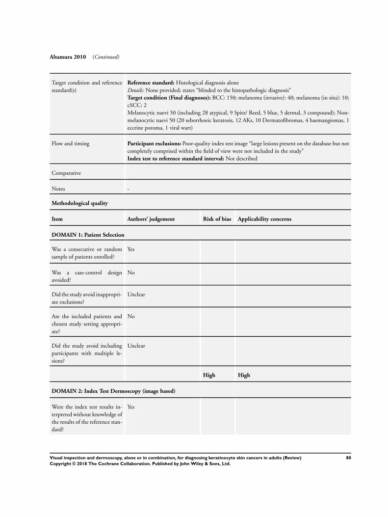
Altamura 2010 (Continued)
Target condition and reference
standard(s)
Reference standard: Histological diagnosis alone
Details: None provided; states “blinded to the histopathologic diagnosis”
Target condition (Final diagnoses): BCC: 150; melanoma (invasive): 40; melanoma (in situ): 10;
cSCC: 2
Melanocytic naevi 50 (including 28 atypical, 9 Spitz/ Reed, 5 blue, 5 dermal, 3 compound); Non-
melanocytic naevi 50 (20 seborrhoeic keratosis, 12 AKs, 10 Dermatofibromas, 4 haemangiomas, 1
eccrine poroma, 1 viral wart)
Flow and timing Participant exclusions: Poor-quality index test image “large lesions present on the database but not
completely comprised within the field of view were not included in the study”
Index test to reference standard interval: Not described
Comparative
Notes -
Methodological quality
Item Authors’ judgement Risk of bias Applicability concerns
DOMAIN 1: Patient Selection
Was a consecutive or random
sample of patients enrolled?
Yes
Was a case-control design
avoided?
No
Did the study avoid inappropri-
ate exclusions?
Unclear
Are the included patients and
chosen study setting appropri-
ate?
No
Did the study avoid including
participants with multiple le-
sions?
Unclear
High High
DOMAIN 2: Index Test Dermoscopy (image based)
Were the index test results in-
terpreted without knowledge of
the results of the reference stan-
dard?
Yes
80Visual inspection and dermoscopy, alone or in combination, for diagnosing keratinocyte skin cancers in adults (Review)
Copyright © 2018 The Cochrane Collaboration. Published by John Wiley & Sons, Ltd.
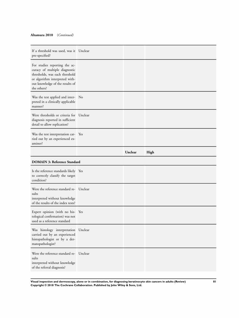
Altamura 2010 (Continued)
If a threshold was used, was it
pre-specified?
Unclear
For studies reporting the ac-
curacy of multiple diagnostic
thresholds, was each threshold
or algorithm interpreted with-
out knowledge of the results of
the others?
Was the test applied and inter-
preted in a clinically applicable
manner?
No
Were thresholds or criteria for
diagnosis reported in sufficient
detail to allow replication?
Unclear
Was the test interpretation car-
ried out by an experienced ex-
aminer?
Yes
Unclear High
DOMAIN 3: Reference Standard
Is the reference standards likely
to correctly classify the target
condition?
Yes
Were the reference standard re-
sults
interpreted without knowledge
of the results of the index tests?
Unclear
Expert opinion (with no his-
tological confirmation) was not
used as a reference standard
Yes
Was histology interpretation
carried out by an experienced
histopathologist or by a der-
matopathologist?
Unclear
Were the reference standard re-
sults
interpreted without knowledge
of the referral diagnosis?
Unclear
81Visual inspection and dermoscopy, alone or in combination, for diagnosing keratinocyte skin cancers in adults (Review)
Copyright © 2018 The Cochrane Collaboration. Published by John Wiley & Sons, Ltd.
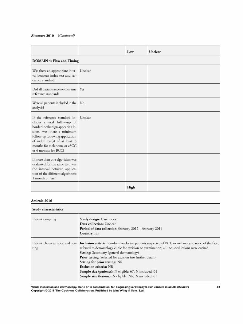
Altamura 2010 (Continued)
Low Unclear
DOMAIN 4: Flow and Timing
Was there an appropriate inter-
val between index test and ref-
erence standard?
Unclear
Did all patients receive the same
reference standard?
Yes
Were all patients included in the
analysis?
No
If the reference standard in-
cludes clinical follow-up of
borderline/benign appearing le-
sions, was there a minimum
follow-up following application
of index test(s) of at least: 3
months for melanoma or cSCC
or 6 months for BCC?
Unclear
If more than one algorithm was
evaluated for the same test, was
the interval between applica-
tion of the different algorithms
1 month or less?
High
Amirnia 2016
Study characteristics
Patient sampling Study design: Case series
Data collection: Unclear
Period of data collection February 2012 - February 2014
Country Iran
Patient characteristics and set-
ting
Inclusion criteria: Randomly-selected patients suspected of BCC or melanocytic naevi of the face,
referred to dermatology clinic for excision or examination; all included lesions were excised
Setting: Secondary (general dermatology)
Prior testing: Selected for excision (no further detail)
Setting for prior testing: NR
Exclusion criteria: NR
Sample size (patients): N eligible: 67; N included: 61
Sample size (lesions): N eligible: NR; N included: 61
82Visual inspection and dermoscopy, alone or in combination, for diagnosing keratinocyte skin cancers in adults (Review)
Copyright © 2018 The Cochrane Collaboration. Published by John Wiley & Sons, Ltd.
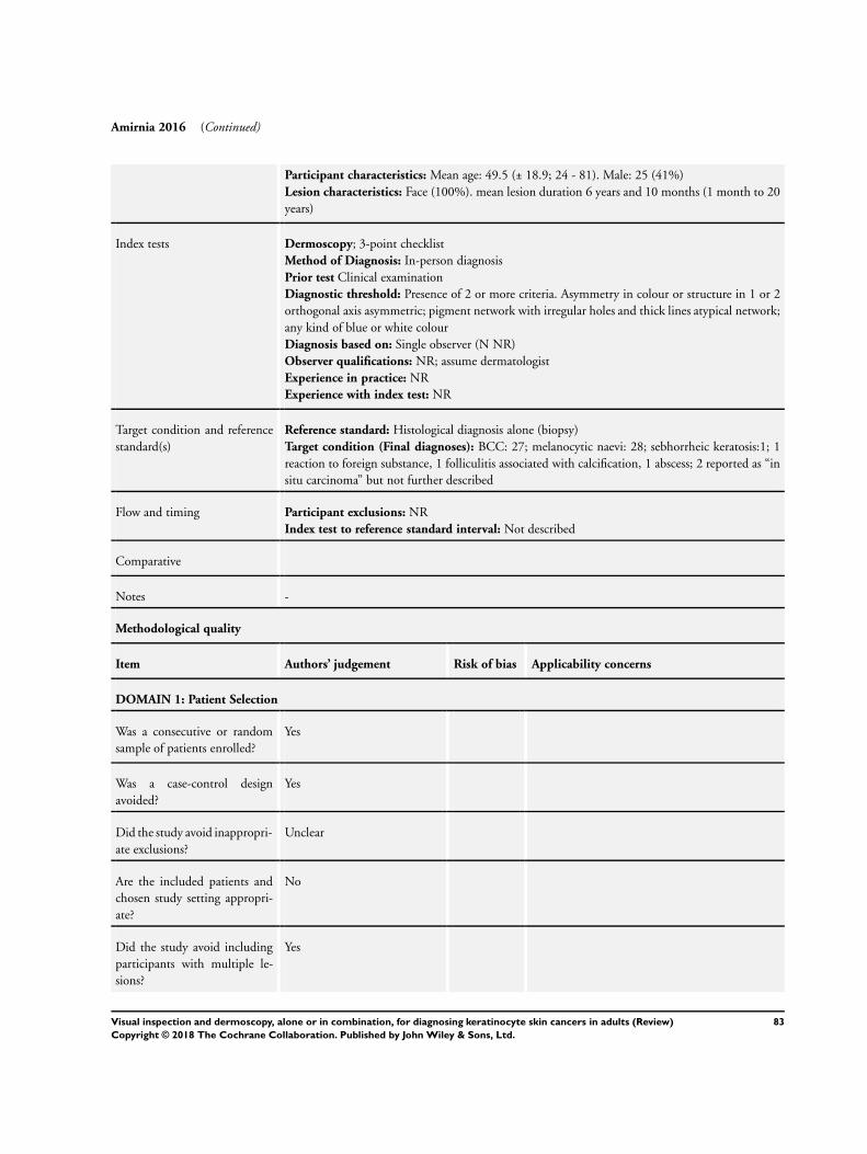
Amirnia 2016 (Continued)
Participant characteristics: Mean age: 49.5 (± 18.9; 24 - 81). Male: 25 (41%)
Lesion characteristics: Face (100%). mean lesion duration 6 years and 10 months (1 month to 20
years)
Index tests Dermoscopy; 3-point checklist
Method of Diagnosis: In-person diagnosis
Prior test Clinical examination
Diagnostic threshold: Presence of 2 or more criteria. Asymmetry in colour or structure in 1 or 2
orthogonal axis asymmetric; pigment network with irregular holes and thick lines atypical network;
any kind of blue or white colour
Diagnosis based on: Single observer (N NR)
Observer qualifications: NR; assume dermatologist
Experience in practice: NR
Experience with index test: NR
Target condition and reference
standard(s)
Reference standard: Histological diagnosis alone (biopsy)
Target condition (Final diagnoses): BCC: 27; melanocytic naevi: 28; sebhorrheic keratosis:1; 1
reaction to foreign substance, 1 folliculitis associated with calcification, 1 abscess; 2 reported as “in
situ carcinoma” but not further described
Flow and timing Participant exclusions: NR
Index test to reference standard interval: Not described
Comparative
Notes -
Methodological quality
Item Authors’ judgement Risk of bias Applicability concerns
DOMAIN 1: Patient Selection
Was a consecutive or random
sample of patients enrolled?
Yes
Was a case-control design
avoided?
Yes
Did the study avoid inappropri-
ate exclusions?
Unclear
Are the included patients and
chosen study setting appropri-
ate?
No
Did the study avoid including
participants with multiple le-
sions?
Yes
83Visual inspection and dermoscopy, alone or in combination, for diagnosing keratinocyte skin cancers in adults (Review)
Copyright © 2018 The Cochrane Collaboration. Published by John Wiley & Sons, Ltd.
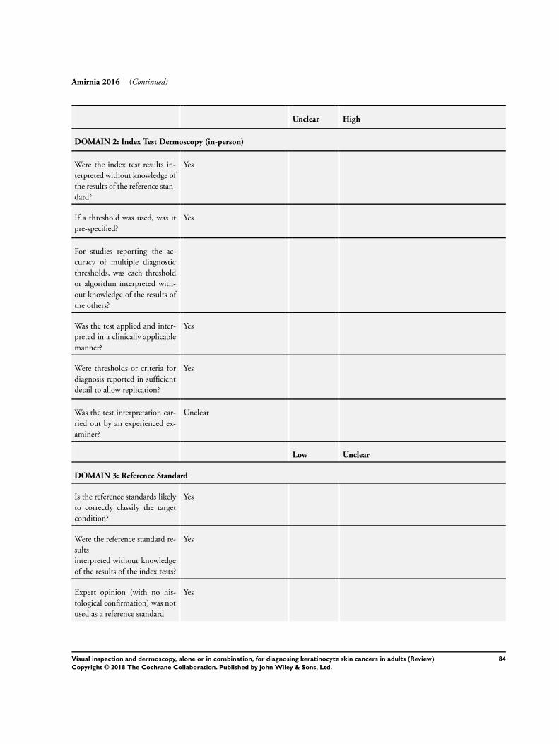
Amirnia 2016 (Continued)
Unclear High
DOMAIN 2: Index Test Dermoscopy (in-person)
Were the index test results in-
terpreted without knowledge of
the results of the reference stan-
dard?
Yes
If a threshold was used, was it
pre-specified?
Yes
For studies reporting the ac-
curacy of multiple diagnostic
thresholds, was each threshold
or algorithm interpreted with-
out knowledge of the results of
the others?
Was the test applied and inter-
preted in a clinically applicable
manner?
Yes
Were thresholds or criteria for
diagnosis reported in sufficient
detail to allow replication?
Yes
Was the test interpretation car-
ried out by an experienced ex-
aminer?
Unclear
Low Unclear
DOMAIN 3: Reference Standard
Is the reference standards likely
to correctly classify the target
condition?
Yes
Were the reference standard re-
sults
interpreted without knowledge
of the results of the index tests?
Yes
Expert opinion (with no his-
tological confirmation) was not
used as a reference standard
Yes
84Visual inspection and dermoscopy, alone or in combination, for diagnosing keratinocyte skin cancers in adults (Review)
Copyright © 2018 The Cochrane Collaboration. Published by John Wiley & Sons, Ltd.
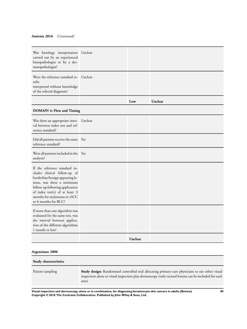
Amirnia 2016 (Continued)
Was histology interpretation
carried out by an experienced
histopathologist or by a der-
matopathologist?
Unclear
Were the reference standard re-
sults
interpreted without knowledge
of the referral diagnosis?
Unclear
Low Unclear
DOMAIN 4: Flow and Timing
Was there an appropriate inter-
val between index test and ref-
erence standard?
Unclear
Did all patients receive the same
reference standard?
Yes
Were all patients included in the
analysis?
Yes
If the reference standard in-
cludes clinical follow-up of
borderline/benign appearing le-
sions, was there a minimum
follow-up following application
of index test(s) of at least: 3
months for melanoma or cSCC
or 6 months for BCC?
If more than one algorithm was
evaluated for the same test, was
the interval between applica-
tion of the different algorithms
1 month or less?
Unclear
Argenziano 2006
Study characteristics
Patient sampling Study design: Randomised controlled trial allocating primary-care physicians to use either visual
inspection alone or visual inspection plus dermoscopy (only excised lesions can be included for each
arm)
85Visual inspection and dermoscopy, alone or in combination, for diagnosing keratinocyte skin cancers in adults (Review)
Copyright © 2018 The Cochrane Collaboration. Published by John Wiley & Sons, Ltd.
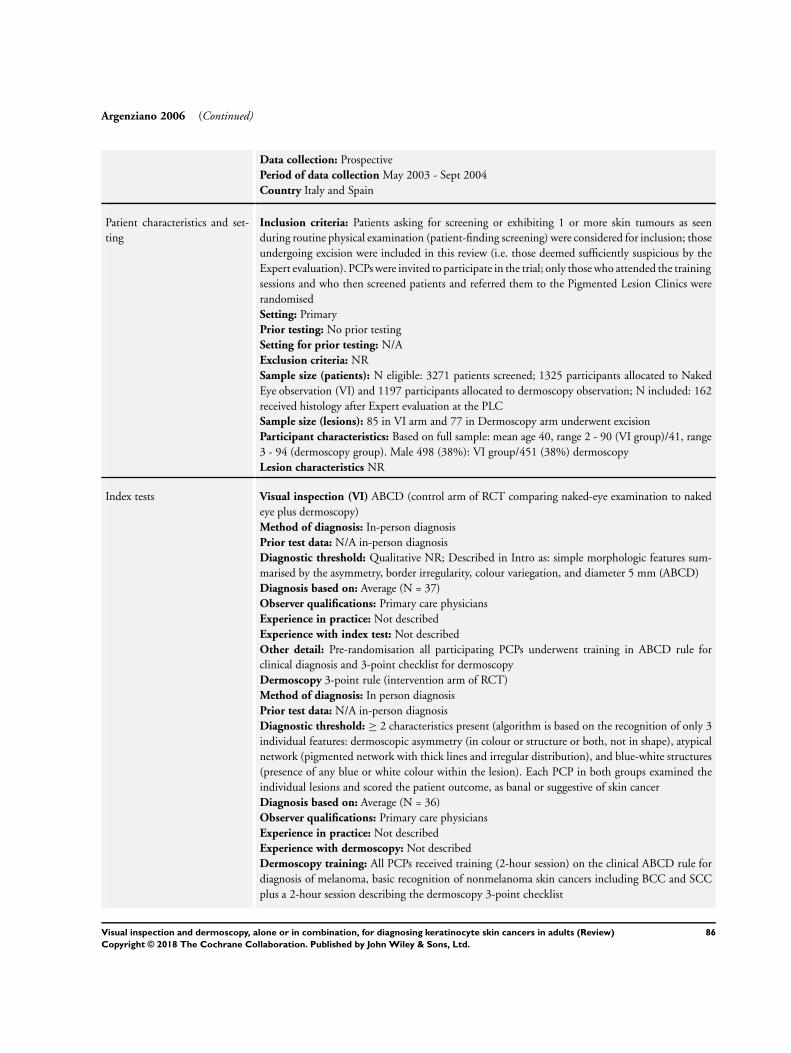
Argenziano 2006 (Continued)
Data collection: Prospective
Period of data collection May 2003 - Sept 2004
Country Italy and Spain
Patient characteristics and set-
ting
Inclusion criteria: Patients asking for screening or exhibiting 1 or more skin tumours as seen
during routine physical examination (patient-finding screening) were considered for inclusion; those
undergoing excision were included in this review (i.e. those deemed sufficiently suspicious by the
Expert evaluation). PCPs were invited to participate in the trial; only those who attended the training
sessions and who then screened patients and referred them to the Pigmented Lesion Clinics were
randomised
Setting: Primary
Prior testing: No prior testing
Setting for prior testing: N/A
Exclusion criteria: NR
Sample size (patients): N eligible: 3271 patients screened; 1325 participants allocated to Naked
Eye observation (VI) and 1197 participants allocated to dermoscopy observation; N included: 162
received histology after Expert evaluation at the PLC
Sample size (lesions): 85 in VI arm and 77 in Dermoscopy arm underwent excision
Participant characteristics: Based on full sample: mean age 40, range 2 - 90 (VI group)/41, range
3 - 94 (dermoscopy group). Male 498 (38%): VI group/451 (38%) dermoscopy
Lesion characteristics NR
Index tests Visual inspection (VI) ABCD (control arm of RCT comparing naked-eye examination to naked
eye plus dermoscopy)
Method of diagnosis: In-person diagnosis
Prior test data: N/A in-person diagnosis
Diagnostic threshold: Qualitative NR; Described in Intro as: simple morphologic features sum-
marised by the asymmetry, border irregularity, colour variegation, and diameter 5 mm (ABCD)
Diagnosis based on: Average (N = 37)
Observer qualifications: Primary care physicians
Experience in practice: Not described
Experience with index test: Not described
Other detail: Pre-randomisation all participating PCPs underwent training in ABCD rule for
clinical diagnosis and 3-point checklist for dermoscopy
Dermoscopy 3-point rule (intervention arm of RCT)
Method of diagnosis: In person diagnosis
Prior test data: N/A in-person diagnosis
Diagnostic threshold: ≥ 2 characteristics present (algorithm is based on the recognition of only 3
individual features: dermoscopic asymmetry (in colour or structure or both, not in shape), atypical
network (pigmented network with thick lines and irregular distribution), and blue-white structures
(presence of any blue or white colour within the lesion). Each PCP in both groups examined the
individual lesions and scored the patient outcome, as banal or suggestive of skin cancer
Diagnosis based on: Average (N = 36)
Observer qualifications: Primary care physicians
Experience in practice: Not described
Experience with dermoscopy: Not described
Dermoscopy training: All PCPs received training (2-hour session) on the clinical ABCD rule for
diagnosis of melanoma, basic recognition of nonmelanoma skin cancers including BCC and SCC
plus a 2-hour session describing the dermoscopy 3-point checklist
86Visual inspection and dermoscopy, alone or in combination, for diagnosing keratinocyte skin cancers in adults (Review)
Copyright © 2018 The Cochrane Collaboration. Published by John Wiley & Sons, Ltd.
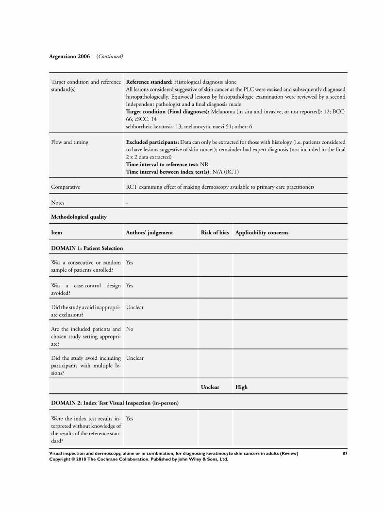
Argenziano 2006 (Continued)
Target condition and reference
standard(s)
Reference standard: Histological diagnosis alone
All lesions considered suggestive of skin cancer at the PLC were excised and subsequently diagnosed
histopathologically. Equivocal lesions by histopathologic examination were reviewed by a second
independent pathologist and a final diagnosis made
Target condition (Final diagnoses): Melanoma (in situ and invasive, or not reported): 12; BCC:
66; cSCC: 14
sebhorrheic keratosis: 13; melanocytic naevi 51; other: 6
Flow and timing Excluded participants: Data can only be extracted for those with histology (i.e. patients considered
to have lesions suggestive of skin cancer); remainder had expert diagnosis (not included in the final
2 x 2 data extracted)
Time interval to reference test: NR
Time interval between index test(s): N/A (RCT)
Comparative RCT examining effect of making dermoscopy available to primary care practitioners
Notes -
Methodological quality
Item Authors’ judgement Risk of bias Applicability concerns
DOMAIN 1: Patient Selection
Was a consecutive or random
sample of patients enrolled?
Yes
Was a case-control design
avoided?
Yes
Did the study avoid inappropri-
ate exclusions?
Unclear
Are the included patients and
chosen study setting appropri-
ate?
No
Did the study avoid including
participants with multiple le-
sions?
Unclear
Unclear High
DOMAIN 2: Index Test Visual Inspection (in-person)
Were the index test results in-
terpreted without knowledge of
the results of the reference stan-
dard?
Yes
87Visual inspection and dermoscopy, alone or in combination, for diagnosing keratinocyte skin cancers in adults (Review)
Copyright © 2018 The Cochrane Collaboration. Published by John Wiley & Sons, Ltd.
Argenziano 2006 (Continued)
If a threshold was used, was it
pre-specified?
Yes
For studies reporting the ac-
curacy of multiple diagnostic
thresholds, was each threshold
or algorithm interpreted with-
out knowledge of the results of
the others?
Was the test applied and inter-
preted in a clinically applicable
manner?
No
Were thresholds or criteria for
diagnosis reported in sufficient
detail to allow replication?
Yes
Was the test interpretation car-
ried out by an experienced ex-
aminer?
Unclear
Low High
DOMAIN 2: Index Test Dermoscopy (in-person)
Were the index test results in-
terpreted without knowledge of
the results of the reference stan-
dard?
Yes
If a threshold was used, was it
pre-specified?
Yes
For studies reporting the ac-
curacy of multiple diagnostic
thresholds, was each threshold
or algorithm interpreted with-
out knowledge of the results of
the others?
Was the test applied and inter-
preted in a clinically applicable
manner?
No
Were thresholds or criteria for
diagnosis reported in sufficient
detail to allow replication?
Yes
88Visual inspection and dermoscopy, alone or in combination, for diagnosing keratinocyte skin cancers in adults (Review)
Copyright © 2018 The Cochrane Collaboration. Published by John Wiley & Sons, Ltd.
Argenziano 2006 (Continued)
Was the test interpretation car-
ried out by an experienced ex-
aminer?
Unclear
Low High
DOMAIN 3: Reference Standard
Is the reference standards likely
to correctly classify the target
condition?
Yes
Were the reference standard re-
sults
interpreted without knowledge
of the results of the index tests?
Unclear
Expert opinion (with no his-
tological confirmation) was not
used as a reference standard
Yes
Was histology interpretation
carried out by an experienced
histopathologist or by a der-
matopathologist?
Yes
Were the reference standard re-
sults
interpreted without knowledge
of the referral diagnosis?
Low Low
DOMAIN 4: Flow and Timing
Was there an appropriate inter-
val between index test and ref-
erence standard?
Unclear
Did all patients receive the same
reference standard?
Yes
Were all patients included in the
analysis?
No
If the reference standard in-
cludes clinical follow-up of
borderline/benign appearing le-
sions, was there a minimum
89Visual inspection and dermoscopy, alone or in combination, for diagnosing keratinocyte skin cancers in adults (Review)
Copyright © 2018 The Cochrane Collaboration. Published by John Wiley & Sons, Ltd.
Argenziano 2006 (Continued)
follow-up following application
of index test(s) of at least: 3
months for melanoma or cSCC
or 6 months for BCC?
If more than one algorithm was
evaluated for the same test, was
the interval between applica-
tion of the different algorithms
1 month or less?
High
Carli 2002a
Study characteristics
Patient sampling Study design: Case series
Data collection: Unclear. Visual inspection and in-vivo dermoscopy diagnoses recorded at time of
patient consultation; Ex vivo (image-based) dermoscopy interpretation undertaken retrospectively
Period of data collection June 1997 - December 1998
Country Italy
Patient characteristics and set-
ting
Inclusion criteria: Clinically equivocal or suspicious pigmented skin lesions subjected to excisional
biopsy at the Institute of Dermatology
Setting: Secondary (not further specified)
Prior testing: Clinical or dermatoscopic suspicion, or both
Setting for prior testing: Secondary
Exclusion criteria: NR
Sample size (patients): NR
Sample size (lesions): 256
Participant characteristics: NR
Lesion characteristics Of the cutaneous melanomas, 14 (25.9%) were in situ melanoma (Clark level
I); 18 (33.3%) were invasive with < 0.75 mm thickness; 19 (35.3%) were of intermediate thickness
(0.76 - 1.50 mm); and 3 (5.5%) were > 1.5 mm. The median thickness of invasive melanomas was
0.94 mm ± 0.5 (SD) (range 0.2 - 6)
Index tests Visual inspection (VI) No algorithm
Method of diagnosis: In-person diagnosis
Prior test data: Unclear
Other test data: Clinical examination and in vivo dermoscopy were performed before excision by
2 trained dermatologists and diagnosis reached
Diagnostic threshold: NR
Diagnosis based on: Consensus (2 observers); final clinical diagnosis was based on agreement
between the 2 observers. In case of disagreement, the opinion of a third observer (BG) was considered
to be the judge for the diagnosis
Observer qualifications: Dermatologist
Experience in practice: High experience or ‘Expert’; described as “dermatologists with extensive
experience in both clinical and dermoscopic diagnosis of pigmented skin lesions”
90Visual inspection and dermoscopy, alone or in combination, for diagnosing keratinocyte skin cancers in adults (Review)
Copyright © 2018 The Cochrane Collaboration. Published by John Wiley & Sons, Ltd.

Carli 2002a (Continued)
Dermoscopy Pattern analysis
Method of diagnosis: In-person diagnosis and image-based diagnosis. Clinical examination and in
vivo dermoscopy were performed before excision by 2 trained dermatologists and diagnosis reached.
Dermoscopic images were re-analysed by the same 2 observers at the end of the inclusion period
(December 1998), blind to the previous clinical and histological diagnoses
Prior test data: N/A for in person; For image-based: slides of dermoscopic images were evaluated
using a viewer that made it impossible to analyse the clinical features of the lesion; both observers
had access to clinical information, including the age of the participant, the site of the lesion, the
history of change over time as reported by the participant at the time of in vivo examination
Diagnostic threshold: Dermoscopic diagnosis was based on the ELM pattern analysis criteria,
using the same diagnostic categories used for clinical diagnosis; characteristics investigated included
pigment network, pigmentation, hypopigmentation, brown globules, black dots, pseudopods, radial
streaming, grey-blue veil, atypical vascular pattern
Test observers as described for Visual Inspection (above)
Target condition and reference
standard(s)
Reference standard: Histological diagnosis alone
Target condition (final diagnoses): Melanoma (invasive): 40; Melanoma (in situ): 14; BCC: 5;
Sebhorrheic keratosis: 4; Common melanocytic naevi: 90; Melanocytic naevi: 78; Blue naevi: 9;
Spitz reed naevi: 16
Flow and timing Excluded participants: NR
Time interval to reference test: NR
Comparative In person clinical examination and dermoscopy
Time interval between index test(s): the interval between the time in-vivo dermoscopy and re-
evaluation of dermoscopic images was reported as 1 year
Notes -
Methodological quality
Item Authors’ judgement Risk of bias Applicability concerns
DOMAIN 1: Patient Selection
Was a consecutive or random
sample of patients enrolled?
Yes
Was a case-control design
avoided?
Yes
Did the study avoid inappropri-
ate exclusions?
Unclear
Are the included patients and
chosen study setting appropri-
ate?
No
91Visual inspection and dermoscopy, alone or in combination, for diagnosing keratinocyte skin cancers in adults (Review)
Copyright © 2018 The Cochrane Collaboration. Published by John Wiley & Sons, Ltd.
Carli 2002a (Continued)
Did the study avoid including
participants with multiple le-
sions?
Unclear
Unclear High
DOMAIN 2: Index Test Visual Inspection (in-person)
Were the index test results in-
terpreted without knowledge of
the results of the reference stan-
dard?
Yes
If a threshold was used, was it
pre-specified?
Unclear
For studies reporting the ac-
curacy of multiple diagnostic
thresholds, was each threshold
or algorithm interpreted with-
out knowledge of the results of
the others?
Was the test applied and inter-
preted in a clinically applicable
manner?
No
Were thresholds or criteria for
diagnosis reported in sufficient
detail to allow replication?
Yes
Was the test interpretation car-
ried out by an experienced ex-
aminer?
Yes
Unclear High
DOMAIN 2: Index Test Dermoscopy (in-person)
Were the index test results in-
terpreted without knowledge of
the results of the reference stan-
dard?
Yes
If a threshold was used, was it
pre-specified?
Yes
92Visual inspection and dermoscopy, alone or in combination, for diagnosing keratinocyte skin cancers in adults (Review)
Copyright © 2018 The Cochrane Collaboration. Published by John Wiley & Sons, Ltd.
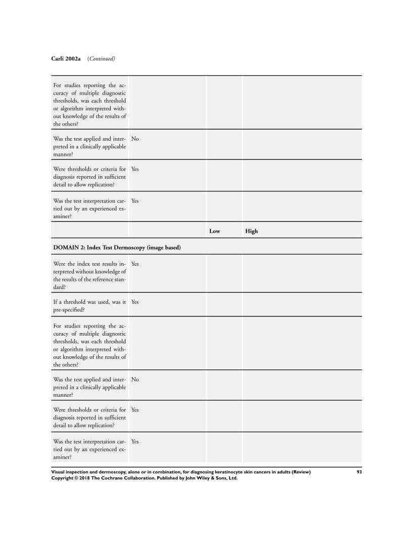
Carli 2002a (Continued)
For studies reporting the ac-
curacy of multiple diagnostic
thresholds, was each threshold
or algorithm interpreted with-
out knowledge of the results of
the others?
Was the test applied and inter-
preted in a clinically applicable
manner?
No
Were thresholds or criteria for
diagnosis reported in sufficient
detail to allow replication?
Yes
Was the test interpretation car-
ried out by an experienced ex-
aminer?
Yes
Low High
DOMAIN 2: Index Test Dermoscopy (image based)
Were the index test results in-
terpreted without knowledge of
the results of the reference stan-
dard?
Yes
If a threshold was used, was it
pre-specified?
Yes
For studies reporting the ac-
curacy of multiple diagnostic
thresholds, was each threshold
or algorithm interpreted with-
out knowledge of the results of
the others?
Was the test applied and inter-
preted in a clinically applicable
manner?
No
Were thresholds or criteria for
diagnosis reported in sufficient
detail to allow replication?
Yes
Was the test interpretation car-
ried out by an experienced ex-
aminer?
Yes
93Visual inspection and dermoscopy, alone or in combination, for diagnosing keratinocyte skin cancers in adults (Review)
Copyright © 2018 The Cochrane Collaboration. Published by John Wiley & Sons, Ltd.

Carli 2002a (Continued)
Low High
DOMAIN 3: Reference Standard
Is the reference standards likely
to correctly classify the target
condition?
Yes
Were the reference standard re-
sults
interpreted without knowledge
of the results of the index tests?
Unclear
Expert opinion (with no his-
tological confirmation) was not
used as a reference standard
Yes
Was histology interpretation
carried out by an experienced
histopathologist or by a der-
matopathologist?
Unclear
Were the reference standard re-
sults
interpreted without knowledge
of the referral diagnosis?
Unclear
Low Unclear
DOMAIN 4: Flow and Timing
Was there an appropriate inter-
val between index test and ref-
erence standard?
Unclear
Did all patients receive the same
reference standard?
Yes
Were all patients included in the
analysis?
Yes
If the reference standard in-
cludes clinical follow-up of
borderline/benign appearing le-
sions, was there a minimum
follow-up following application
of index test(s) of at least: 3
months for melanoma or cSCC
or 6 months for BCC?
94Visual inspection and dermoscopy, alone or in combination, for diagnosing keratinocyte skin cancers in adults (Review)
Copyright © 2018 The Cochrane Collaboration. Published by John Wiley & Sons, Ltd.
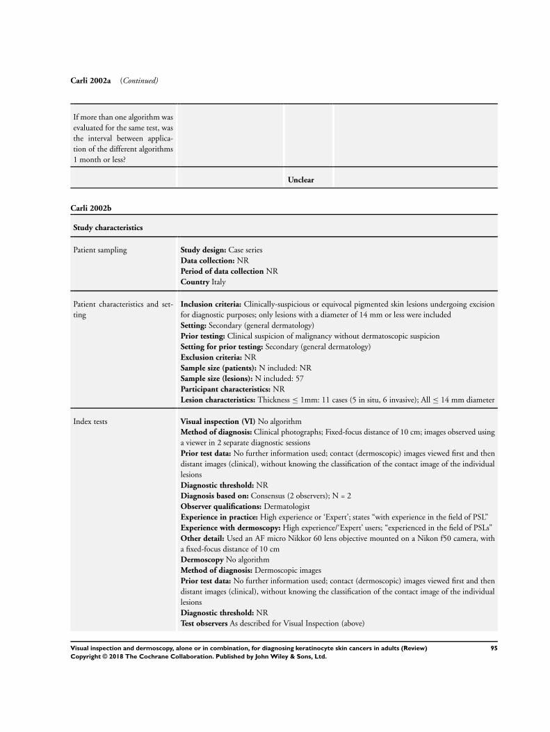
Carli 2002a (Continued)
If more than one algorithm was
evaluated for the same test, was
the interval between applica-
tion of the different algorithms
1 month or less?
Unclear
Carli 2002b
Study characteristics
Patient sampling Study design: Case series
Data collection: NR
Period of data collection NR
Country Italy
Patient characteristics and set-
ting
Inclusion criteria: Clinically-suspicious or equivocal pigmented skin lesions undergoing excision
for diagnostic purposes; only lesions with a diameter of 14 mm or less were included
Setting: Secondary (general dermatology)
Prior testing: Clinical suspicion of malignancy without dermatoscopic suspicion
Setting for prior testing: Secondary (general dermatology)
Exclusion criteria: NR
Sample size (patients): N included: NR
Sample size (lesions): N included: 57
Participant characteristics: NR
Lesion characteristics: Thickness ≤ 1mm: 11 cases (5 in situ, 6 invasive); All ≤ 14 mm diameter
Index tests Visual inspection (VI) No algorithm
Method of diagnosis: Clinical photographs; Fixed-focus distance of 10 cm; images observed using
a viewer in 2 separate diagnostic sessions
Prior test data: No further information used; contact (dermoscopic) images viewed first and then
distant images (clinical), without knowing the classification of the contact image of the individual
lesions
Diagnostic threshold: NR
Diagnosis based on: Consensus (2 observers); N = 2
Observer qualifications: Dermatologist
Experience in practice: High experience or ‘Expert’; states “with experience in the field of PSL”
Experience with dermoscopy: High experience/‘Expert’ users; “experienced in the field of PSLs”
Other detail: Used an AF micro Nikkor 60 lens objective mounted on a Nikon f50 camera, with
a fixed-focus distance of 10 cm
Dermoscopy No algorithm
Method of diagnosis: Dermoscopic images
Prior test data: No further information used; contact (dermoscopic) images viewed first and then
distant images (clinical), without knowing the classification of the contact image of the individual
lesions
Diagnostic threshold: NR
Test observers As described for Visual Inspection (above)
95Visual inspection and dermoscopy, alone or in combination, for diagnosing keratinocyte skin cancers in adults (Review)
Copyright © 2018 The Cochrane Collaboration. Published by John Wiley & Sons, Ltd.
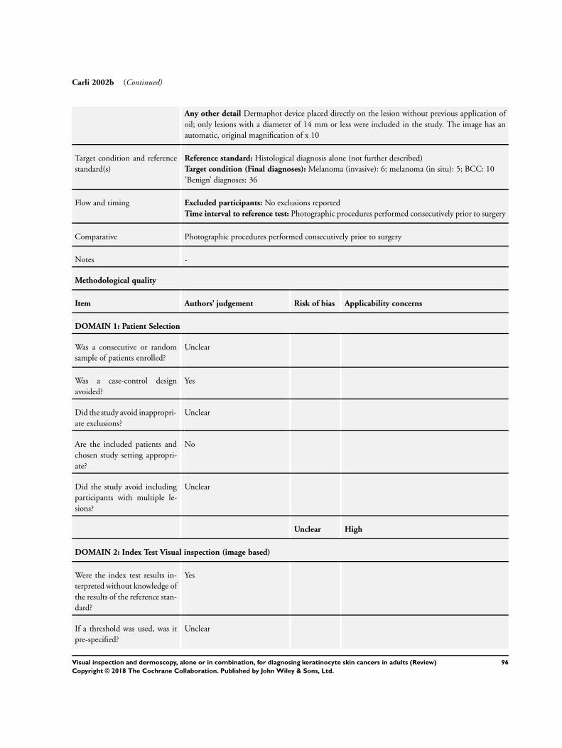
Carli 2002b (Continued)
Any other detail Dermaphot device placed directly on the lesion without previous application of
oil; only lesions with a diameter of 14 mm or less were included in the study. The image has an
automatic, original magnification of x 10
Target condition and reference
standard(s)
Reference standard: Histological diagnosis alone (not further described)
Target condition (Final diagnoses): Melanoma (invasive): 6; melanoma (in situ): 5; BCC: 10
’Benign’ diagnoses: 36
Flow and timing Excluded participants: No exclusions reported
Time interval to reference test: Photographic procedures performed consecutively prior to surgery
Comparative Photographic procedures performed consecutively prior to surgery
Notes -
Methodological quality
Item Authors’ judgement Risk of bias Applicability concerns
DOMAIN 1: Patient Selection
Was a consecutive or random
sample of patients enrolled?
Unclear
Was a case-control design
avoided?
Yes
Did the study avoid inappropri-
ate exclusions?
Unclear
Are the included patients and
chosen study setting appropri-
ate?
No
Did the study avoid including
participants with multiple le-
sions?
Unclear
Unclear High
DOMAIN 2: Index Test Visual inspection (image based)
Were the index test results in-
terpreted without knowledge of
the results of the reference stan-
dard?
Yes
If a threshold was used, was it
pre-specified?
Unclear
96Visual inspection and dermoscopy, alone or in combination, for diagnosing keratinocyte skin cancers in adults (Review)
Copyright © 2018 The Cochrane Collaboration. Published by John Wiley & Sons, Ltd.

Carli 2002b (Continued)
For studies reporting the ac-
curacy of multiple diagnostic
thresholds, was each threshold
or algorithm interpreted with-
out knowledge of the results of
the others?
Was the test applied and inter-
preted in a clinically applicable
manner?
No
Were thresholds or criteria for
diagnosis reported in sufficient
detail to allow replication?
No
Was the test interpretation car-
ried out by an experienced ex-
aminer?
Yes
Unclear High
DOMAIN 2: Index Test Dermoscopy (image based)
Were the index test results in-
terpreted without knowledge of
the results of the reference stan-
dard?
Yes
If a threshold was used, was it
pre-specified?
Unclear
For studies reporting the ac-
curacy of multiple diagnostic
thresholds, was each threshold
or algorithm interpreted with-
out knowledge of the results of
the others?
Was the test applied and inter-
preted in a clinically applicable
manner?
No
Were thresholds or criteria for
diagnosis reported in sufficient
detail to allow replication?
No
Was the test interpretation car-
ried out by an experienced ex-
aminer?
Yes
97Visual inspection and dermoscopy, alone or in combination, for diagnosing keratinocyte skin cancers in adults (Review)
Copyright © 2018 The Cochrane Collaboration. Published by John Wiley & Sons, Ltd.
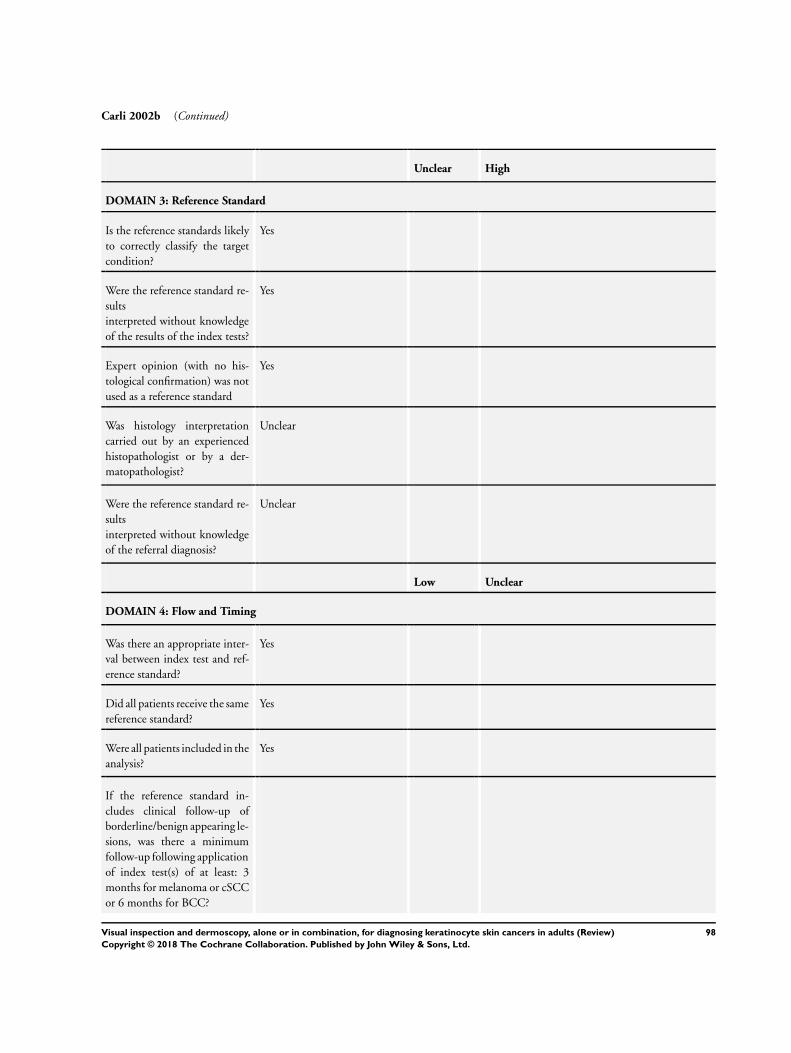
Carli 2002b (Continued)
Unclear High
DOMAIN 3: Reference Standard
Is the reference standards likely
to correctly classify the target
condition?
Yes
Were the reference standard re-
sults
interpreted without knowledge
of the results of the index tests?
Yes
Expert opinion (with no his-
tological confirmation) was not
used as a reference standard
Yes
Was histology interpretation
carried out by an experienced
histopathologist or by a der-
matopathologist?
Unclear
Were the reference standard re-
sults
interpreted without knowledge
of the referral diagnosis?
Unclear
Low Unclear
DOMAIN 4: Flow and Timing
Was there an appropriate inter-
val between index test and ref-
erence standard?
Yes
Did all patients receive the same
reference standard?
Yes
Were all patients included in the
analysis?
Yes
If the reference standard in-
cludes clinical follow-up of
borderline/benign appearing le-
sions, was there a minimum
follow-up following application
of index test(s) of at least: 3
months for melanoma or cSCC
or 6 months for BCC?
98Visual inspection and dermoscopy, alone or in combination, for diagnosing keratinocyte skin cancers in adults (Review)
Copyright © 2018 The Cochrane Collaboration. Published by John Wiley & Sons, Ltd.
Carli 2002b (Continued)
If more than one algorithm was
evaluated for the same test, was
the interval between applica-
tion of the different algorithms
1 month or less?
Low
Chang 2013
Study characteristics
Patient sampling Study design: Case series
Data collection: Retrospective
Period of data collection: Jan 2006 - Jul 2009
Country: Taiwan
Patient characteristics and set-
ting
Inclusion criteria: Potentially malignant biopsied or excised skin lesions (non-tumour specimens
excluded)
Setting: Secondary (general dermatology)
Prior testing: Selected for excision (no further detail)
Setting for prior testing: Secondary (general dermatology)
Exclusion criteria: Prior surgery; image mis-registered or poor-quality images (unfocused or con-
taining a motion artefact) (considered under Flow and Timing)
Sample size (patients): N eligible: 3964; N included: 676
Sample size (lesions): N eligible: 4192; N included: 769
Participant characteristics: Mean age: 47.6 (SD 21.0); Male: 296; 43.8%
Lesion characteristics: NR
Index tests Visual inspection (VI) No algorithm
Method of diagnosis: In-person diagnosis
Prior test data: N/A in-person diagnosis
Diagnostic threshold: NR; clinicians’ impressions prior to biopsy were classified as “benign”,
“malignant”, or “indeterminate”. When the clinicians were not confident enough to make a definite
benign or malignant diagnosis, the clinical impression was considered as “indeterminate” data
extracted for malignant vs rest and malignant/indeterminate vs rest
Diagnosis based on: Single observer; board-certified staff dermatologists from institute; N = 25
Observer qualifications: Dermatologist
Experience in practice: Board certified
Experience with index test: High
Target condition and reference
standard(s)
Reference standard: Histology (not further described)
Target condition (Final diagnoses): Melanoma (invasive): 4; melanoma (in situ): 4; BCC: 110;
cSCC: 20
’Benign’ diagnoses: 595
99Visual inspection and dermoscopy, alone or in combination, for diagnosing keratinocyte skin cancers in adults (Review)
Copyright © 2018 The Cochrane Collaboration. Published by John Wiley & Sons, Ltd.
Chang 2013 (Continued)
Flow and timing Excluded participants: Mis-registered or poor-quality images (unfocused or containing a motion
artefact) as a study inclusion criterion
Time interval to reference test: Not described
Comparative
Notes -
Methodological quality
Item Authors’ judgement Risk of bias Applicability concerns
DOMAIN 1: Patient Selection
Was a consecutive or random
sample of patients enrolled?
Yes
Was a case-control design
avoided?
Yes
Did the study avoid inappropri-
ate exclusions?
Yes
Are the included patients and
chosen study setting appropri-
ate?
No
Did the study avoid including
participants with multiple le-
sions?
No
Low High
DOMAIN 2: Index Test Visual Inspection (in-person)
Were the index test results in-
terpreted without knowledge of
the results of the reference stan-
dard?
Yes
If a threshold was used, was it
pre-specified?
Unclear
For studies reporting the ac-
curacy of multiple diagnostic
thresholds, was each threshold
or algorithm interpreted with-
out knowledge of the results of
the others?
100Visual inspection and dermoscopy, alone or in combination, for diagnosing keratinocyte skin cancers in adults (Review)
Copyright © 2018 The Cochrane Collaboration. Published by John Wiley & Sons, Ltd.
Chang 2013 (Continued)
Was the test applied and inter-
preted in a clinically applicable
manner?
Yes
Were thresholds or criteria for
diagnosis reported in sufficient
detail to allow replication?
No
Was the test interpretation car-
ried out by an experienced ex-
aminer?
Yes
Unclear High
DOMAIN 3: Reference Standard
Is the reference standards likely
to correctly classify the target
condition?
Yes
Were the reference standard re-
sults
interpreted without knowledge
of the results of the index tests?
Unclear
Expert opinion (with no his-
tological confirmation) was not
used as a reference standard
Yes
Was histology interpretation
carried out by an experienced
histopathologist or by a der-
matopathologist?
Unclear
Were the reference standard re-
sults
interpreted without knowledge
of the referral diagnosis?
Low Unclear
DOMAIN 4: Flow and Timing
Was there an appropriate inter-
val between index test and ref-
erence standard?
Unclear
Did all patients receive the same
reference standard?
Yes
101Visual inspection and dermoscopy, alone or in combination, for diagnosing keratinocyte skin cancers in adults (Review)
Copyright © 2018 The Cochrane Collaboration. Published by John Wiley & Sons, Ltd.
Chang 2013 (Continued)
Were all patients included in the
analysis?
No
If the reference standard in-
cludes clinical follow-up of
borderline/benign appearing le-
sions, was there a minimum
follow-up following application
of index test(s) of at least: 3
months for melanoma or cSCC
or 6 months for BCC?
If more than one algorithm was
evaluated for the same test, was
the interval between applica-
tion of the different algorithms
1 month or less?
High
Cooper 2002
Study characteristics
Patient sampling Study design: Case series
Data collection: Prospective
Period of data collection May 2000 - September 2000
Country UK
Patient characteristics and set-
ting
Inclusion criteria: Patients attending the open-access dermatology renal transplant clinic with
lesions suspicious for malignancy or premalignancy and booked for biopsy
Setting: Specialist unit; dermatology renal transplant clinic
Prior testing: Clinical suspicion
Setting for prior testing: Specialist unit
Exclusion criteria: NR
Sample size (patients): N eligible: 70; N included: NR
Sample size (lesions): N eligible: 125; N included: 102
Participant characteristics: Mean age: 60; Male: 75%
Lesion characteristics Head/neck: 43; 34.4%; Limbs: 21; 16.8%; 3 genitals; 2.4%
Index tests Visual inspection (VI) No algorithm
Method of diagnosis: In-person diagnosis
Prior test data: N/A in-person diagnosis
Diagnostic threshold: Observer provisional diagnosis
Diagnosis based on: Single observer (N = 2)
Observer qualifications: Consultant dermatologist and a registrar
Experience in practice: Not described
Experience with index test: Not described
102Visual inspection and dermoscopy, alone or in combination, for diagnosing keratinocyte skin cancers in adults (Review)
Copyright © 2018 The Cochrane Collaboration. Published by John Wiley & Sons, Ltd.
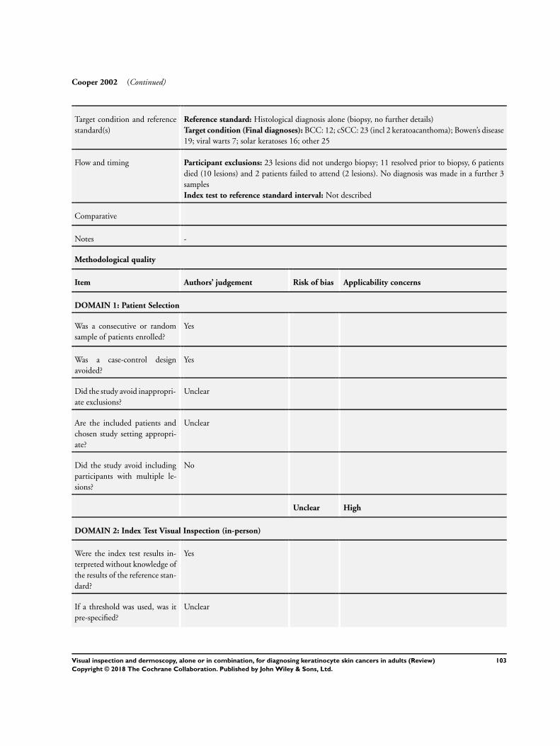
Cooper 2002 (Continued)
Target condition and reference
standard(s)
Reference standard: Histological diagnosis alone (biopsy, no further details)
Target condition (Final diagnoses): BCC: 12; cSCC: 23 (incl 2 keratoacanthoma); Bowen’s disease
19; viral warts 7; solar keratoses 16; other 25
Flow and timing Participant exclusions: 23 lesions did not undergo biopsy; 11 resolved prior to biopsy, 6 patients
died (10 lesions) and 2 patients failed to attend (2 lesions). No diagnosis was made in a further 3
samples
Index test to reference standard interval: Not described
Comparative
Notes -
Methodological quality
Item Authors’ judgement Risk of bias Applicability concerns
DOMAIN 1: Patient Selection
Was a consecutive or random
sample of patients enrolled?
Yes
Was a case-control design
avoided?
Yes
Did the study avoid inappropri-
ate exclusions?
Unclear
Are the included patients and
chosen study setting appropri-
ate?
Unclear
Did the study avoid including
participants with multiple le-
sions?
No
Unclear High
DOMAIN 2: Index Test Visual Inspection (in-person)
Were the index test results in-
terpreted without knowledge of
the results of the reference stan-
dard?
Yes
If a threshold was used, was it
pre-specified?
Unclear
103Visual inspection and dermoscopy, alone or in combination, for diagnosing keratinocyte skin cancers in adults (Review)
Copyright © 2018 The Cochrane Collaboration. Published by John Wiley & Sons, Ltd.
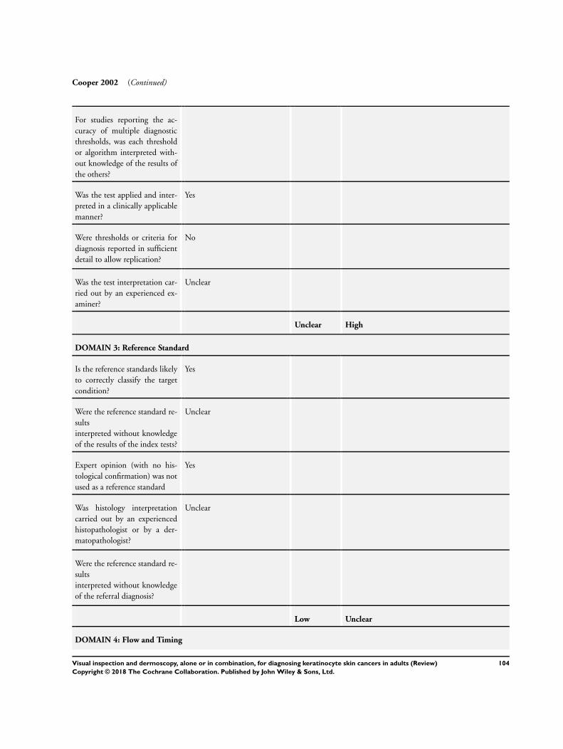
Cooper 2002 (Continued)
For studies reporting the ac-
curacy of multiple diagnostic
thresholds, was each threshold
or algorithm interpreted with-
out knowledge of the results of
the others?
Was the test applied and inter-
preted in a clinically applicable
manner?
Yes
Were thresholds or criteria for
diagnosis reported in sufficient
detail to allow replication?
No
Was the test interpretation car-
ried out by an experienced ex-
aminer?
Unclear
Unclear High
DOMAIN 3: Reference Standard
Is the reference standards likely
to correctly classify the target
condition?
Yes
Were the reference standard re-
sults
interpreted without knowledge
of the results of the index tests?
Unclear
Expert opinion (with no his-
tological confirmation) was not
used as a reference standard
Yes
Was histology interpretation
carried out by an experienced
histopathologist or by a der-
matopathologist?
Unclear
Were the reference standard re-
sults
interpreted without knowledge
of the referral diagnosis?
Low Unclear
DOMAIN 4: Flow and Timing
104Visual inspection and dermoscopy, alone or in combination, for diagnosing keratinocyte skin cancers in adults (Review)
Copyright © 2018 The Cochrane Collaboration. Published by John Wiley & Sons, Ltd.
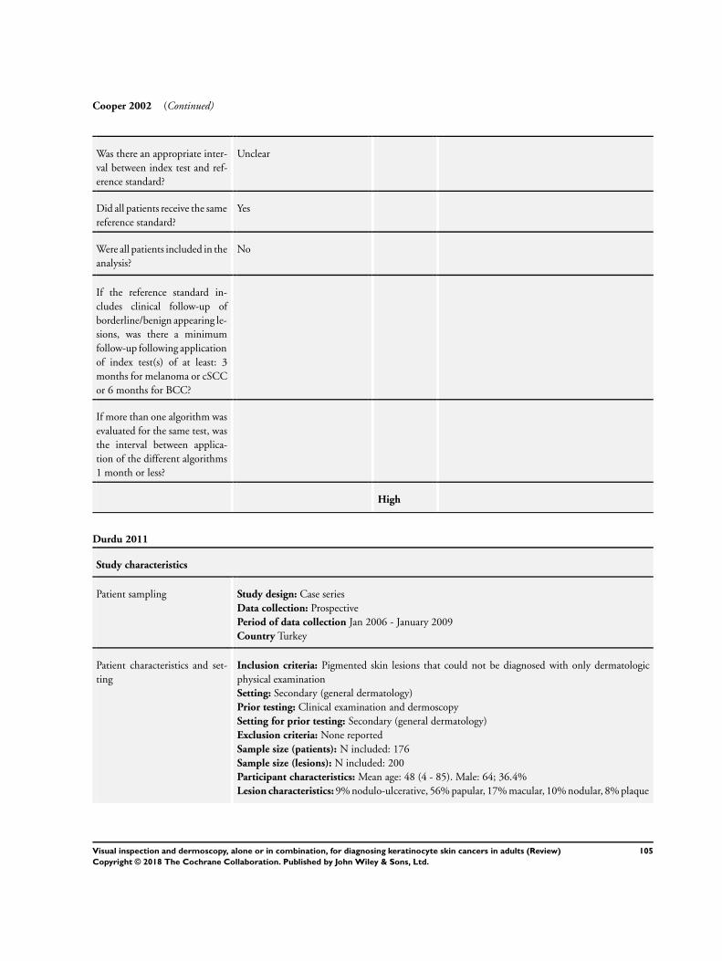
Cooper 2002 (Continued)
Was there an appropriate inter-
val between index test and ref-
erence standard?
Unclear
Did all patients receive the same
reference standard?
Yes
Were all patients included in the
analysis?
No
If the reference standard in-
cludes clinical follow-up of
borderline/benign appearing le-
sions, was there a minimum
follow-up following application
of index test(s) of at least: 3
months for melanoma or cSCC
or 6 months for BCC?
If more than one algorithm was
evaluated for the same test, was
the interval between applica-
tion of the different algorithms
1 month or less?
High
Durdu 2011
Study characteristics
Patient sampling Study design: Case series
Data collection: Prospective
Period of data collection Jan 2006 - January 2009
Country Turkey
Patient characteristics and set-
ting
Inclusion criteria: Pigmented skin lesions that could not be diagnosed with only dermatologic
physical examination
Setting: Secondary (general dermatology)
Prior testing: Clinical examination and dermoscopy
Setting for prior testing: Secondary (general dermatology)
Exclusion criteria: None reported
Sample size (patients): N included: 176
Sample size (lesions): N included: 200
Participant characteristics: Mean age: 48 (4 - 85). Male: 64; 36.4%
Lesion characteristics: 9% nodulo-ulcerative, 56% papular, 17% macular, 10% nodular, 8% plaque
105Visual inspection and dermoscopy, alone or in combination, for diagnosing keratinocyte skin cancers in adults (Review)
Copyright © 2018 The Cochrane Collaboration. Published by John Wiley & Sons, Ltd.
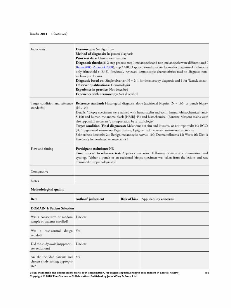
Durdu 2011 (Continued)
Index tests Dermoscopy: No algorithm
Method of diagnosis: In-person diagnosis
Prior test data: Clinical examination
Diagnostic threshold: 2-step process: step 1 melanocytic and non-melanocytic were differentiated (
Braun 2005; Zalaudek 2008); step 2 ABCD applied to melanocytic lesions for diagnosis of melanoma
only (threshold > 5.45). Previously reviewed dermoscopic characteristics used to diagnose non-
melanocytic lesions
Diagnosis based on: Single observer; N = 2; 1 for dermoscopy diagnosis and 1 for Tzanck smear
Observer qualifications: Dermatologist
Experience in practice: Not described
Experience with dermoscopy: Not described
Target condition and reference
standard(s)
Reference standard: Histological diagnosis alone (excisional biopsies (N = 166) or punch biopsy
(N = 34)
Details: “Biopsy specimens were stained with hematoxylin and eosin. Immunohistochemical (anti-
S-100 and human melanoma black [HMB]-45) and histochemical (Fontana-Masson) stains were
also applied, if necessary”; interpretation by a ’pathologist’
Target condition (Final diagnoses): Melanoma (in situ and invasive, or not reported): 10; BCC:
34; 1 pigmented mammary Paget disease; 1 pigmented metastatic mammary carcinoma
Sebhorrheic keratosis: 24; Benign melanocytic naevus: 100; Dermatofibroma 12; Warts 16; Dirt 1;
hereditary hemorrhagic telangiectasia 1
Flow and timing Participant exclusions: NR
Time interval to reference test: Appears consecutive. Following dermoscopic examination and
cytology “either a punch or an excisional biopsy specimen was taken from the lesions and was
examined histopathologically”
Comparative
Notes -
Methodological quality
Item Authors’ judgement Risk of bias Applicability concerns
DOMAIN 1: Patient Selection
Was a consecutive or random
sample of patients enrolled?
Unclear
Was a case-control design
avoided?
Yes
Did the study avoid inappropri-
ate exclusions?
Unclear
Are the included patients and
chosen study setting appropri-
ate?
Yes
106Visual inspection and dermoscopy, alone or in combination, for diagnosing keratinocyte skin cancers in adults (Review)
Copyright © 2018 The Cochrane Collaboration. Published by John Wiley & Sons, Ltd.
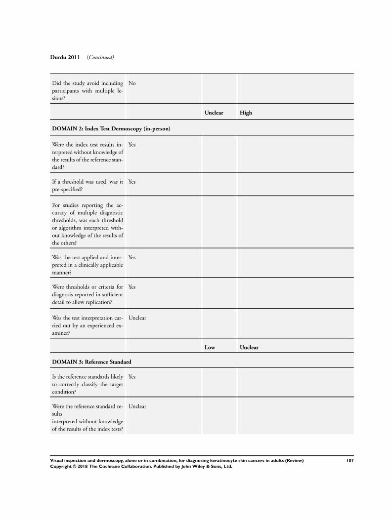
Durdu 2011 (Continued)
Did the study avoid including
participants with multiple le-
sions?
No
Unclear High
DOMAIN 2: Index Test Dermoscopy (in-person)
Were the index test results in-
terpreted without knowledge of
the results of the reference stan-
dard?
Yes
If a threshold was used, was it
pre-specified?
Yes
For studies reporting the ac-
curacy of multiple diagnostic
thresholds, was each threshold
or algorithm interpreted with-
out knowledge of the results of
the others?
Was the test applied and inter-
preted in a clinically applicable
manner?
Yes
Were thresholds or criteria for
diagnosis reported in sufficient
detail to allow replication?
Yes
Was the test interpretation car-
ried out by an experienced ex-
aminer?
Unclear
Low Unclear
DOMAIN 3: Reference Standard
Is the reference standards likely
to correctly classify the target
condition?
Yes
Were the reference standard re-
sults
interpreted without knowledge
of the results of the index tests?
Unclear
107Visual inspection and dermoscopy, alone or in combination, for diagnosing keratinocyte skin cancers in adults (Review)
Copyright © 2018 The Cochrane Collaboration. Published by John Wiley & Sons, Ltd.

Durdu 2011 (Continued)
Expert opinion (with no his-
tological confirmation) was not
used as a reference standard
Yes
Was histology interpretation
carried out by an experienced
histopathologist or by a der-
matopathologist?
Unclear
Were the reference standard re-
sults
interpreted without knowledge
of the referral diagnosis?
Unclear
Low Unclear
DOMAIN 4: Flow and Timing
Was there an appropriate inter-
val between index test and ref-
erence standard?
Yes
Did all patients receive the same
reference standard?
Yes
Were all patients included in the
analysis?
Yes
If the reference standard in-
cludes clinical follow-up of
borderline/benign appearing le-
sions, was there a minimum
follow-up following application
of index test(s) of at least: 3
months for melanoma or cSCC
or 6 months for BCC?
If more than one algorithm was
evaluated for the same test, was
the interval between applica-
tion of the different algorithms
1 month or less?
Low
108Visual inspection and dermoscopy, alone or in combination, for diagnosing keratinocyte skin cancers in adults (Review)
Copyright © 2018 The Cochrane Collaboration. Published by John Wiley & Sons, Ltd.
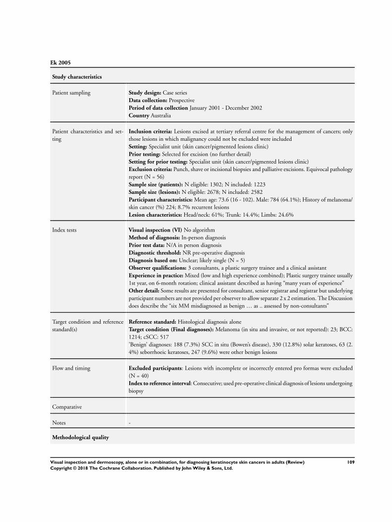
Ek 2005
Study characteristics
Patient sampling Study design: Case series
Data collection: Prospective
Period of data collection January 2001 - December 2002
Country Australia
Patient characteristics and set-
ting
Inclusion criteria: Lesions excised at tertiary referral centre for the management of cancers; only
those lesions in which malignancy could not be excluded were included
Setting: Specialist unit (skin cancer/pigmented lesions clinic)
Prior testing: Selected for excision (no further detail)
Setting for prior testing: Specialist unit (skin cancer/pigmented lesions clinic)
Exclusion criteria: Punch, shave or incisional biopsies and palliative excisions. Equivocal pathology
report (N = 56)
Sample size (patients): N eligible: 1302; N included: 1223
Sample size (lesions): N eligible: 2678; N included: 2582
Participant characteristics: Mean age: 73.6 (16 - 102). Male: 784 (64.1%); History of melanoma/
skin cancer (%) 224; 8.7% recurrent lesions
Lesion characteristics: Head/neck: 61%; Trunk: 14.4%; Limbs: 24.6%
Index tests Visual inspection (VI) No algorithm
Method of diagnosis: In-person diagnosis
Prior test data: N/A in person diagnosis
Diagnostic threshold: NR pre-operative diagnosis
Diagnosis based on: Unclear; likely single (N = 5)
Observer qualifications: 3 consultants, a plastic surgery trainee and a clinical assistant
Experience in practice: Mixed (low and high experience combined); Plastic surgery trainee usually
1st year, on 6-month rotation; clinical assistant described as having “many years of experience”
Other detail: Some results are presented for consultant, senior registrar and registrar but underlying
participant numbers are not provided per observer to allow separate 2 x 2 estimation. The Discussion
does describe the “six MM misdiagnosed as benign … as .. assessed by non-consultants”
Target condition and reference
standard(s)
Reference standard: Histological diagnosis alone
Target condition (Final diagnoses): Melanoma (in situ and invasive, or not reported): 23; BCC:
1214; cSCC: 517
’Benign’ diagnoses: 188 (7.3%) SCC in situ (Bowen’s disease), 330 (12.8%) solar keratoses, 63 (2.
4%) seborrhoeic keratoses, 247 (9.6%) were other benign lesions
Flow and timing Excluded participants: Lesions with incomplete or incorrectly entered pro formas were excluded
(N = 40)
Index to reference interval: Consecutive; used pre-operative clinical diagnosis of lesions undergoing
biopsy
Comparative
Notes -
Methodological quality
109Visual inspection and dermoscopy, alone or in combination, for diagnosing keratinocyte skin cancers in adults (Review)
Copyright © 2018 The Cochrane Collaboration. Published by John Wiley & Sons, Ltd.
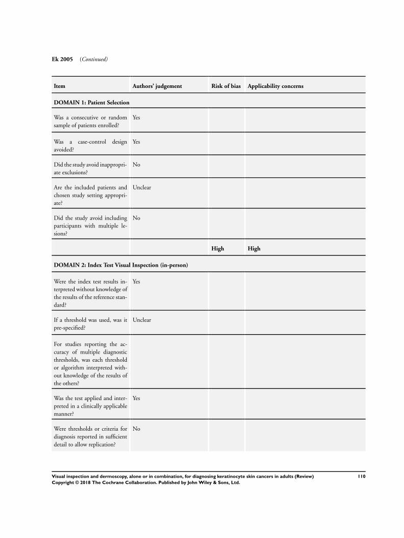
Ek 2005 (Continued)
Item Authors’ judgement Risk of bias Applicability concerns
DOMAIN 1: Patient Selection
Was a consecutive or random
sample of patients enrolled?
Yes
Was a case-control design
avoided?
Yes
Did the study avoid inappropri-
ate exclusions?
No
Are the included patients and
chosen study setting appropri-
ate?
Unclear
Did the study avoid including
participants with multiple le-
sions?
No
High High
DOMAIN 2: Index Test Visual Inspection (in-person)
Were the index test results in-
terpreted without knowledge of
the results of the reference stan-
dard?
Yes
If a threshold was used, was it
pre-specified?
Unclear
For studies reporting the ac-
curacy of multiple diagnostic
thresholds, was each threshold
or algorithm interpreted with-
out knowledge of the results of
the others?
Was the test applied and inter-
preted in a clinically applicable
manner?
Yes
Were thresholds or criteria for
diagnosis reported in sufficient
detail to allow replication?
No
110Visual inspection and dermoscopy, alone or in combination, for diagnosing keratinocyte skin cancers in adults (Review)
Copyright © 2018 The Cochrane Collaboration. Published by John Wiley & Sons, Ltd.
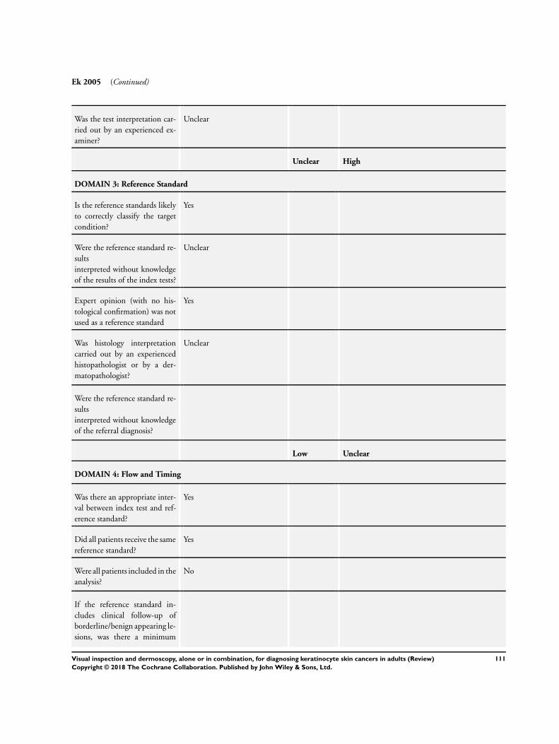
Ek 2005 (Continued)
Was the test interpretation car-
ried out by an experienced ex-
aminer?
Unclear
Unclear High
DOMAIN 3: Reference Standard
Is the reference standards likely
to correctly classify the target
condition?
Yes
Were the reference standard re-
sults
interpreted without knowledge
of the results of the index tests?
Unclear
Expert opinion (with no his-
tological confirmation) was not
used as a reference standard
Yes
Was histology interpretation
carried out by an experienced
histopathologist or by a der-
matopathologist?
Unclear
Were the reference standard re-
sults
interpreted without knowledge
of the referral diagnosis?
Low Unclear
DOMAIN 4: Flow and Timing
Was there an appropriate inter-
val between index test and ref-
erence standard?
Yes
Did all patients receive the same
reference standard?
Yes
Were all patients included in the
analysis?
No
If the reference standard in-
cludes clinical follow-up of
borderline/benign appearing le-
sions, was there a minimum
111Visual inspection and dermoscopy, alone or in combination, for diagnosing keratinocyte skin cancers in adults (Review)
Copyright © 2018 The Cochrane Collaboration. Published by John Wiley & Sons, Ltd.
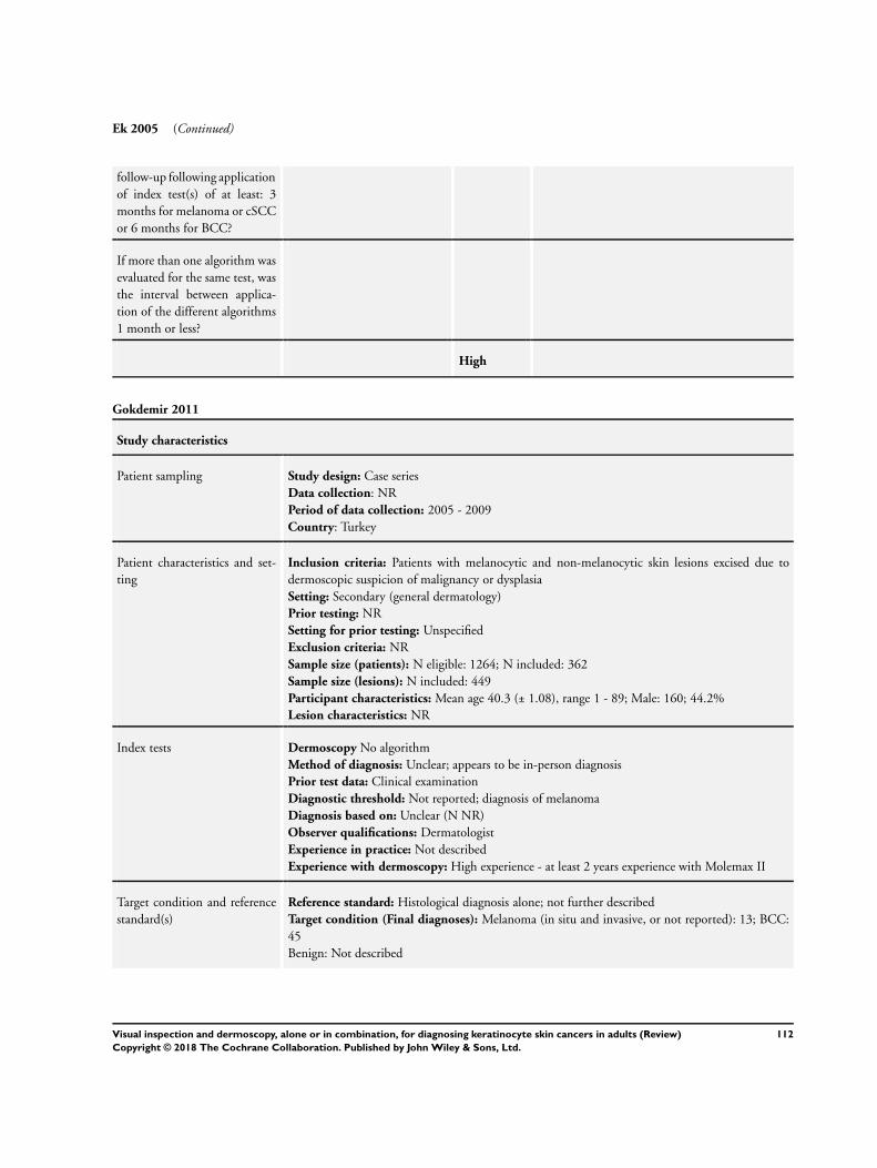
Ek 2005 (Continued)
follow-up following application
of index test(s) of at least: 3
months for melanoma or cSCC
or 6 months for BCC?
If more than one algorithm was
evaluated for the same test, was
the interval between applica-
tion of the different algorithms
1 month or less?
High
Gokdemir 2011
Study characteristics
Patient sampling Study design: Case series
Data collection: NR
Period of data collection: 2005 - 2009
Country: Turkey
Patient characteristics and set-
ting
Inclusion criteria: Patients with melanocytic and non-melanocytic skin lesions excised due to
dermoscopic suspicion of malignancy or dysplasia
Setting: Secondary (general dermatology)
Prior testing: NR
Setting for prior testing: Unspecified
Exclusion criteria: NR
Sample size (patients): N eligible: 1264; N included: 362
Sample size (lesions): N included: 449
Participant characteristics: Mean age 40.3 (± 1.08), range 1 - 89; Male: 160; 44.2%
Lesion characteristics: NR
Index tests Dermoscopy No algorithm
Method of diagnosis: Unclear; appears to be in-person diagnosis
Prior test data: Clinical examination
Diagnostic threshold: Not reported; diagnosis of melanoma
Diagnosis based on: Unclear (N NR)
Observer qualifications: Dermatologist
Experience in practice: Not described
Experience with dermoscopy: High experience - at least 2 years experience with Molemax II
Target condition and reference
standard(s)
Reference standard: Histological diagnosis alone; not further described
Target condition (Final diagnoses): Melanoma (in situ and invasive, or not reported): 13; BCC:
45
Benign: Not described
112Visual inspection and dermoscopy, alone or in combination, for diagnosing keratinocyte skin cancers in adults (Review)
Copyright © 2018 The Cochrane Collaboration. Published by John Wiley & Sons, Ltd.
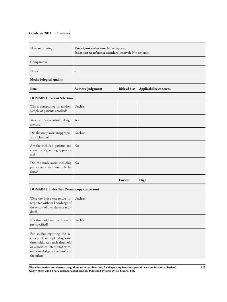
Gokdemir 2011 (Continued)
Flow and timing Participant exclusions: None reported
Index test to reference standard interval: Not reported
Comparative
Notes -
Methodological quality
Item Authors’ judgement Risk of bias Applicability concerns
DOMAIN 1: Patient Selection
Was a consecutive or random
sample of patients enrolled?
Unclear
Was a case-control design
avoided?
Yes
Did the study avoid inappropri-
ate exclusions?
Unclear
Are the included patients and
chosen study setting appropri-
ate?
No
Did the study avoid including
participants with multiple le-
sions?
No
Unclear High
DOMAIN 2: Index Test Dermoscopy (in-person)
Were the index test results in-
terpreted without knowledge of
the results of the reference stan-
dard?
Unclear
If a threshold was used, was it
pre-specified?
Unclear
For studies reporting the ac-
curacy of multiple diagnostic
thresholds, was each threshold
or algorithm interpreted with-
out knowledge of the results of
the others?
113Visual inspection and dermoscopy, alone or in combination, for diagnosing keratinocyte skin cancers in adults (Review)
Copyright © 2018 The Cochrane Collaboration. Published by John Wiley & Sons, Ltd.
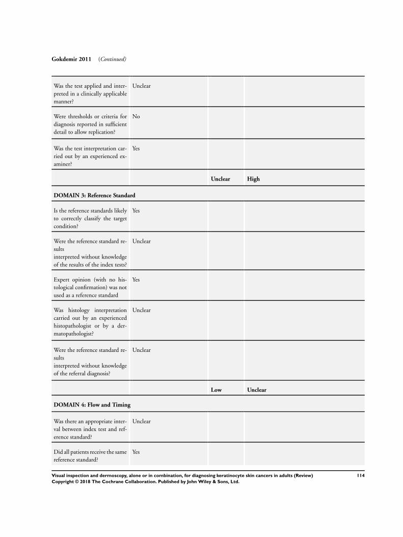
Gokdemir 2011 (Continued)
Was the test applied and inter-
preted in a clinically applicable
manner?
Unclear
Were thresholds or criteria for
diagnosis reported in sufficient
detail to allow replication?
No
Was the test interpretation car-
ried out by an experienced ex-
aminer?
Yes
Unclear High
DOMAIN 3: Reference Standard
Is the reference standards likely
to correctly classify the target
condition?
Yes
Were the reference standard re-
sults
interpreted without knowledge
of the results of the index tests?
Unclear
Expert opinion (with no his-
tological confirmation) was not
used as a reference standard
Yes
Was histology interpretation
carried out by an experienced
histopathologist or by a der-
matopathologist?
Unclear
Were the reference standard re-
sults
interpreted without knowledge
of the referral diagnosis?
Unclear
Low Unclear
DOMAIN 4: Flow and Timing
Was there an appropriate inter-
val between index test and ref-
erence standard?
Unclear
Did all patients receive the same
reference standard?
Yes
114Visual inspection and dermoscopy, alone or in combination, for diagnosing keratinocyte skin cancers in adults (Review)
Copyright © 2018 The Cochrane Collaboration. Published by John Wiley & Sons, Ltd.
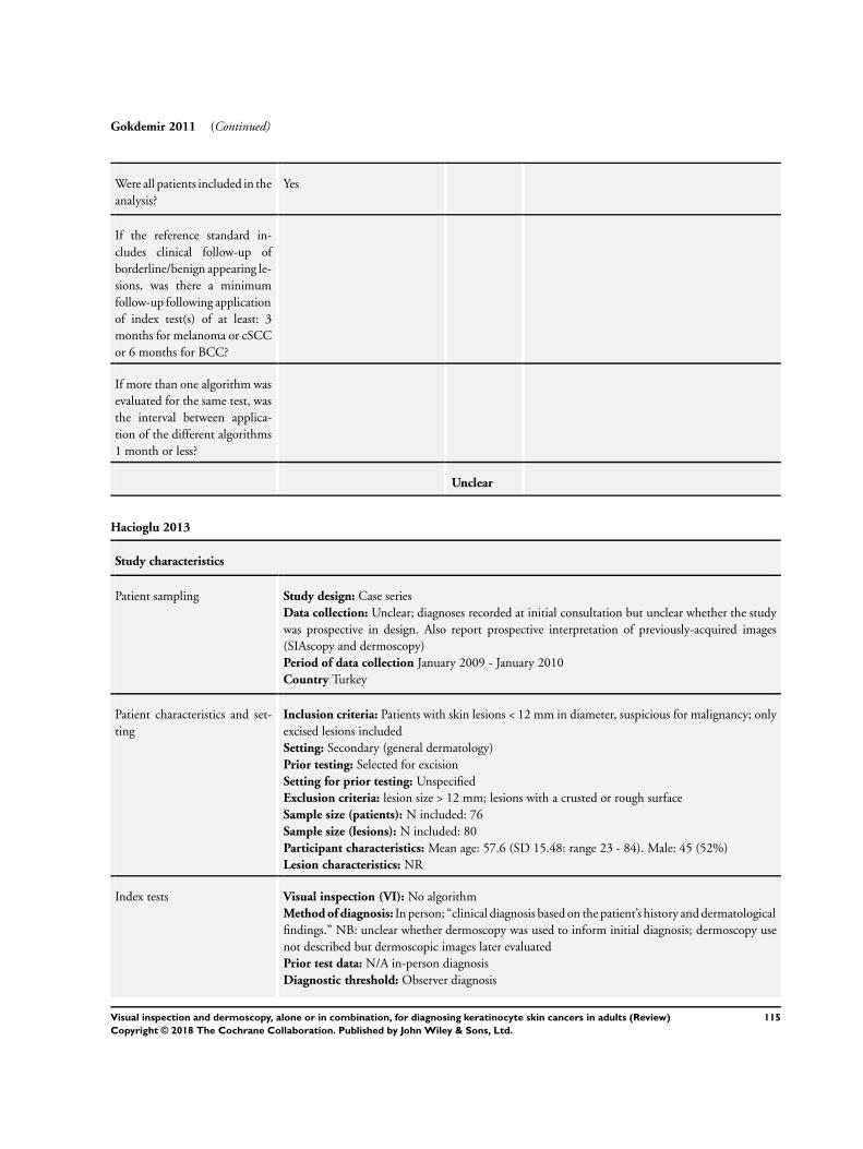
Gokdemir 2011 (Continued)
Were all patients included in the
analysis?
Yes
If the reference standard in-
cludes clinical follow-up of
borderline/benign appearing le-
sions, was there a minimum
follow-up following application
of index test(s) of at least: 3
months for melanoma or cSCC
or 6 months for BCC?
If more than one algorithm was
evaluated for the same test, was
the interval between applica-
tion of the different algorithms
1 month or less?
Unclear
Hacioglu 2013
Study characteristics
Patient sampling Study design: Case series
Data collection: Unclear; diagnoses recorded at initial consultation but unclear whether the study
was prospective in design. Also report prospective interpretation of previously-acquired images
(SIAscopy and dermoscopy)
Period of data collection January 2009 - January 2010
Country Turkey
Patient characteristics and set-
ting
Inclusion criteria: Patients with skin lesions < 12 mm in diameter, suspicious for malignancy; only
excised lesions included
Setting: Secondary (general dermatology)
Prior testing: Selected for excision
Setting for prior testing: Unspecified
Exclusion criteria: lesion size > 12 mm; lesions with a crusted or rough surface
Sample size (patients): N included: 76
Sample size (lesions): N included: 80
Participant characteristics: Mean age: 57.6 (SD 15.48: range 23 - 84). Male: 45 (52%)
Lesion characteristics: NR
Index tests Visual inspection (VI): No algorithm
Method of diagnosis: In person; “clinical diagnosis based on the patient’s history and dermatological
findings.” NB: unclear whether dermoscopy was used to inform initial diagnosis; dermoscopy use
not described but dermoscopic images later evaluated
Prior test data: N/A in-person diagnosis
Diagnostic threshold: Observer diagnosis
115Visual inspection and dermoscopy, alone or in combination, for diagnosing keratinocyte skin cancers in adults (Review)
Copyright © 2018 The Cochrane Collaboration. Published by John Wiley & Sons, Ltd.
Hacioglu 2013 (Continued)
Diagnosis based on: Single observer (N = 3)
Observer qualifications: NR; likely dermatologist
Experience in practice: Not described; 3 investigators - 1 made preliminary clinic diagnosis and
evaluated Siascope images 8 months later; second investigator evaluated all Siascope images; a third
investigator evaluated dermoscopic images
Experience with index test: Not described
Dermoscopy: No algorithm
Method of diagnosis: Dermoscopic images
Prior test data: No further information used; “a third investigator (EBB), also blinded to the
previous diagnoses, evaluated all the lesions using dermatoscopic images only.”
Diagnostic threshold: Observer diagnosis
Observers: As described above.
Target condition and reference
standard(s)
Reference standard: Histological diagnosis alone
Details: Skin biopsies (3 or 4 mm in size)
Target condition (Final diagnoses): BCC: 24; melanoma (in situ and invasive, or not reported):
cSCC 3; Basosquamous cancer 2; sebhorrhoeic keratosis: 19; actinic keratosis 8; intradermal naevus
4; dermatofibroma 3; keratoacanthoma 2; Other 12 - including: epidermal proliferation, pseudoep-
ithelial hyperplasia, solar degeneration, lichen simplex chronicus, compound naevus, dysplastic nae-
vus, prurigo nodularis, chronic inflammatory granulation, dysplastic junctional naevus
Flow and timing Participant exclusions: NR
Index test to reference standard interval: Appears consecutive; “Images ... were obtained ... and
skin biopsies ... were taken”
Comparative 3. Time interval between index test(s): 8 months between visual and SIAscopetime between visual/
SIAscope and dermatoscopy not reported
Notes -
Methodological quality
Item Authors’ judgement Risk of bias Applicability concerns
DOMAIN 1: Patient Selection
Was a consecutive or random
sample of patients enrolled?
Unclear
Was a case-control design
avoided?
Yes
Did the study avoid inappropri-
ate exclusions?
No
Are the included patients and
chosen study setting appropri-
ate?
No
116Visual inspection and dermoscopy, alone or in combination, for diagnosing keratinocyte skin cancers in adults (Review)
Copyright © 2018 The Cochrane Collaboration. Published by John Wiley & Sons, Ltd.
Hacioglu 2013 (Continued)
Did the study avoid including
participants with multiple le-
sions?
Yes
High High
DOMAIN 2: Index Test Visual Inspection (in-person)
Were the index test results in-
terpreted without knowledge of
the results of the reference stan-
dard?
Yes
If a threshold was used, was it
pre-specified?
Unclear
For studies reporting the ac-
curacy of multiple diagnostic
thresholds, was each threshold
or algorithm interpreted with-
out knowledge of the results of
the others?
Was the test applied and inter-
preted in a clinically applicable
manner?
Yes
Were thresholds or criteria for
diagnosis reported in sufficient
detail to allow replication?
No
Was the test interpretation car-
ried out by an experienced ex-
aminer?
Unclear
Unclear High
DOMAIN 2: Index Test Dermoscopy (image based)
Were the index test results in-
terpreted without knowledge of
the results of the reference stan-
dard?
Yes
If a threshold was used, was it
pre-specified?
Unclear
117Visual inspection and dermoscopy, alone or in combination, for diagnosing keratinocyte skin cancers in adults (Review)
Copyright © 2018 The Cochrane Collaboration. Published by John Wiley & Sons, Ltd.
Hacioglu 2013 (Continued)
For studies reporting the ac-
curacy of multiple diagnostic
thresholds, was each threshold
or algorithm interpreted with-
out knowledge of the results of
the others?
Was the test applied and inter-
preted in a clinically applicable
manner?
No
Were thresholds or criteria for
diagnosis reported in sufficient
detail to allow replication?
No
Was the test interpretation car-
ried out by an experienced ex-
aminer?
Unclear
Unclear High
DOMAIN 3: Reference Standard
Is the reference standards likely
to correctly classify the target
condition?
Yes
Were the reference standard re-
sults
interpreted without knowledge
of the results of the index tests?
Unclear
Expert opinion (with no his-
tological confirmation) was not
used as a reference standard
Yes
Was histology interpretation
carried out by an experienced
histopathologist or by a der-
matopathologist?
Unclear
Were the reference standard re-
sults
interpreted without knowledge
of the referral diagnosis?
Unclear
Low Unclear
DOMAIN 4: Flow and Timing
118Visual inspection and dermoscopy, alone or in combination, for diagnosing keratinocyte skin cancers in adults (Review)
Copyright © 2018 The Cochrane Collaboration. Published by John Wiley & Sons, Ltd.
Hacioglu 2013 (Continued)
Was there an appropriate inter-
val between index test and ref-
erence standard?
Yes
Did all patients receive the same
reference standard?
Yes
Were all patients included in the
analysis?
Yes
If the reference standard in-
cludes clinical follow-up of
borderline/benign appearing le-
sions, was there a minimum
follow-up following application
of index test(s) of at least: 3
months for melanoma or cSCC
or 6 months for BCC?
If more than one algorithm was
evaluated for the same test, was
the interval between applica-
tion of the different algorithms
1 month or less?
Low
Lorentzen 1999
Study characteristics
Patient sampling Study design: Case series
Data collection: Prospective
Period of data collection 1994 - 1997
Country Denmark
Patient characteristics and set-
ting
Inclusion criteria: Patients with lesions suspicious for CMM referred to outpatients clinic; only
excised included
Setting: NR
Prior testing: Clinical suspicion of malignancy without dermatoscopic suspicion
Setting for prior testing: NR
Exclusion criteria: Poor-quality index test image (considered under flow/timing)
Sample size (patients): N eligible: 242; N included: 232
Sample size (lesions): N eligible: 242; N included: 232
NB: Not all cases were assessed by all observers; 2 x 2 are based on presented sensitivity and specificity
estimates for full dataset of lesions; “the dermatoscopy experts assessed almost all cases (98 ± 100%)
, whereas the non-expert group completed fewer assessments, from 76 to 98%”
Participant characteristics: NR
Lesion characteristics: NR
119Visual inspection and dermoscopy, alone or in combination, for diagnosing keratinocyte skin cancers in adults (Review)
Copyright © 2018 The Cochrane Collaboration. Published by John Wiley & Sons, Ltd.
Lorentzen 1999 (Continued)
Index tests Visual inspection (VI) No algorithm
Method of diagnosis: Clinical photographs
Prior test data: No further information used; no option to change clinical diagnosis after viewing
dermoscopic image
Other test data: Dermoscopic images presented to observer subsequent to diagnosis using clinical
images alone; clinical images presented before dermoscopic images
Diagnostic threshold: NR; clinical diagnosis
Diagnosis based on: Average; N = 9
Observer qualifications: Dermatologist
Experience in practice: High; moderate; mixed (average reported); 4 ’experienced dermatologists’
(4 - 5 years daily experience) & 5 ’non-expert dermatology residents’ (1 - 2 years interest and formal
training in dermatoscopy
Experience with index test: High; moderate; mixed
Target condition and reference
standard(s)
Reference standard: Histological diagnosis alone
Details: a co-author from Dept of Pathology “re-evaluated all cases to confirm the pathology diag-
nosis, which was used as the gold standard in this study”
Target condition (Final diagnoses): Melanoma (invasive): 49 ’malignant melanoma’; BCC: 16;
sebhorrheic keratosis: 12; benign naevus: 137 (pigmented naevi = 116; blue naevi = 16; atypical
naevi = 5); Other: 18 (Spitz naevi, Bowen’s disease, sarcoid, naevus spilus, hemangioma, and others)
Flow and timing Excluded participants: 10 cases were “considered unfit for evaluation” due to poor-quality image
Reference interval: “biopsy specimens...were obtained after the clinical and dermatoscopic pho-
tographs had been performed”
Comparative tbc
Notes -
Methodological quality
Item Authors’ judgement Risk of bias Applicability concerns
DOMAIN 1: Patient Selection
Was a consecutive or random
sample of patients enrolled?
Unclear
Was a case-control design
avoided?
Yes
Did the study avoid inappropri-
ate exclusions?
Yes
Are the included patients and
chosen study setting appropri-
ate?
No
120Visual inspection and dermoscopy, alone or in combination, for diagnosing keratinocyte skin cancers in adults (Review)
Copyright © 2018 The Cochrane Collaboration. Published by John Wiley & Sons, Ltd.
Lorentzen 1999 (Continued)
Did the study avoid including
participants with multiple le-
sions?
Unclear
Unclear High
DOMAIN 2: Index Test Visual inspection (image based)
Were the index test results in-
terpreted without knowledge of
the results of the reference stan-
dard?
Yes
If a threshold was used, was it
pre-specified?
Unclear
For studies reporting the ac-
curacy of multiple diagnostic
thresholds, was each threshold
or algorithm interpreted with-
out knowledge of the results of
the others?
Was the test applied and inter-
preted in a clinically applicable
manner?
No
Were thresholds or criteria for
diagnosis reported in sufficient
detail to allow replication?
No
Was the test interpretation car-
ried out by an experienced ex-
aminer?
Yes
Unclear High
DOMAIN 3: Reference Standard
Is the reference standards likely
to correctly classify the target
condition?
Yes
Were the reference standard re-
sults
interpreted without knowledge
of the results of the index tests?
Unclear
121Visual inspection and dermoscopy, alone or in combination, for diagnosing keratinocyte skin cancers in adults (Review)
Copyright © 2018 The Cochrane Collaboration. Published by John Wiley & Sons, Ltd.
Lorentzen 1999 (Continued)
Expert opinion (with no his-
tological confirmation) was not
used as a reference standard
Yes
Was histology interpretation
carried out by an experienced
histopathologist or by a der-
matopathologist?
Unclear
Were the reference standard re-
sults
interpreted without knowledge
of the referral diagnosis?
Unclear
Low Unclear
DOMAIN 4: Flow and Timing
Was there an appropriate inter-
val between index test and ref-
erence standard?
Yes
Did all patients receive the same
reference standard?
Yes
Were all patients included in the
analysis?
No
If the reference standard in-
cludes clinical follow-up of
borderline/benign appearing le-
sions, was there a minimum
follow-up following application
of index test(s) of at least: 3
months for melanoma or cSCC
or 6 months for BCC?
If more than one algorithm was
evaluated for the same test, was
the interval between applica-
tion of the different algorithms
1 month or less?
High
122Visual inspection and dermoscopy, alone or in combination, for diagnosing keratinocyte skin cancers in adults (Review)
Copyright © 2018 The Cochrane Collaboration. Published by John Wiley & Sons, Ltd.
Lorentzen 2008
Study characteristics
Patient sampling Study design: Case series
Data collection: NR
Period of data collection: NR
Country: Denmark
Patient characteristics and set-
ting
Inclusion criteria: Patients referred to the specialist naevus clinic for lesion excision
Setting: Specialist unit (skin cancer/pigmented lesions clinic)
Prior testing: NR
Setting for prior testing: NR
Exclusion criteria: Not specified
Sample size (patients): N eligible: 120; N included: 119
Sample size (lesions): N included: 119
Participant characteristics: NR
Lesion characteristics: NR
Index tests Dermoscopy: Mixed/no algorithm; describes using “the risk stratification and pattern analysis
procedure as described by Kenet 2001 and Lorentzen 2000”.
Method of diagnosis: Dermoscopic images; compared accuracy using standard dermoscopy images
(Dermaphot) and images obtained using a globe magnifier. Slides were randomised and evaluated
on 2 different occasions with 3-week intervals
Prior test data: No further information used
Diagnostic threshold: Observer correct diagnosis of each lesion type
Diagnosis based on: Unclear (assumed average) (N NR)
Observer qualifications: Dermatologist
Experience in practice: High; “dermatologists who have performed dermatoscopy for 5-10 years,
published scientific papers on dermatoscopy and carried out pre- and post specialist training in
dermatoscopy”
Experience with dermoscopy: High
Target condition and reference
standard(s)
Reference standard: Histological diagnosis alone
Details: used haematoxylin-eosin staining as well as histochemistry performed using S-100 and
HMB-45 on suspect melanoma lesions
Target condition (Final diagnoses): Melanoma (invasive): 24; BCC: 13;
mild/moderate dysplasia: 2; sebhorrheic keratosis: 9; haemangioma: 2; naevus pigmentosus: 69
Flow and timing Excluded participants: 1 dermatofibroma excluded
Time interval to reference test: Not described
Comparative
Notes -
Methodological quality
Item Authors’ judgement Risk of bias Applicability concerns
DOMAIN 1: Patient Selection
123Visual inspection and dermoscopy, alone or in combination, for diagnosing keratinocyte skin cancers in adults (Review)
Copyright © 2018 The Cochrane Collaboration. Published by John Wiley & Sons, Ltd.
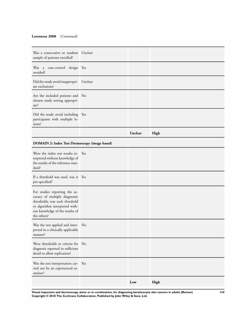
Lorentzen 2008 (Continued)
Was a consecutive or random
sample of patients enrolled?
Unclear
Was a case-control design
avoided?
Yes
Did the study avoid inappropri-
ate exclusions?
Unclear
Are the included patients and
chosen study setting appropri-
ate?
No
Did the study avoid including
participants with multiple le-
sions?
Yes
Unclear High
DOMAIN 2: Index Test Dermoscopy (image based)
Were the index test results in-
terpreted without knowledge of
the results of the reference stan-
dard?
Yes
If a threshold was used, was it
pre-specified?
Yes
For studies reporting the ac-
curacy of multiple diagnostic
thresholds, was each threshold
or algorithm interpreted with-
out knowledge of the results of
the others?
Was the test applied and inter-
preted in a clinically applicable
manner?
No
Were thresholds or criteria for
diagnosis reported in sufficient
detail to allow replication?
No
Was the test interpretation car-
ried out by an experienced ex-
aminer?
Yes
Low High
124Visual inspection and dermoscopy, alone or in combination, for diagnosing keratinocyte skin cancers in adults (Review)
Copyright © 2018 The Cochrane Collaboration. Published by John Wiley & Sons, Ltd.
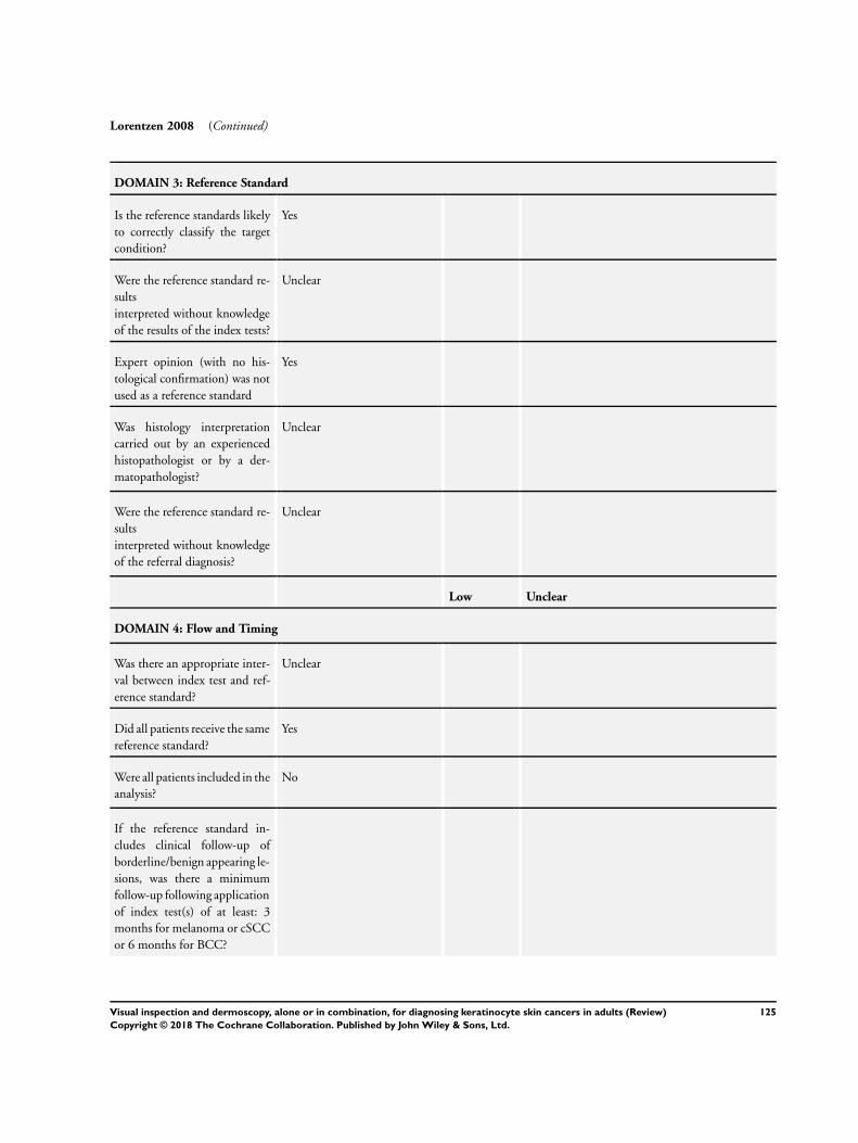
Lorentzen 2008 (Continued)
DOMAIN 3: Reference Standard
Is the reference standards likely
to correctly classify the target
condition?
Yes
Were the reference standard re-
sults
interpreted without knowledge
of the results of the index tests?
Unclear
Expert opinion (with no his-
tological confirmation) was not
used as a reference standard
Yes
Was histology interpretation
carried out by an experienced
histopathologist or by a der-
matopathologist?
Unclear
Were the reference standard re-
sults
interpreted without knowledge
of the referral diagnosis?
Unclear
Low Unclear
DOMAIN 4: Flow and Timing
Was there an appropriate inter-
val between index test and ref-
erence standard?
Unclear
Did all patients receive the same
reference standard?
Yes
Were all patients included in the
analysis?
No
If the reference standard in-
cludes clinical follow-up of
borderline/benign appearing le-
sions, was there a minimum
follow-up following application
of index test(s) of at least: 3
months for melanoma or cSCC
or 6 months for BCC?
125Visual inspection and dermoscopy, alone or in combination, for diagnosing keratinocyte skin cancers in adults (Review)
Copyright © 2018 The Cochrane Collaboration. Published by John Wiley & Sons, Ltd.
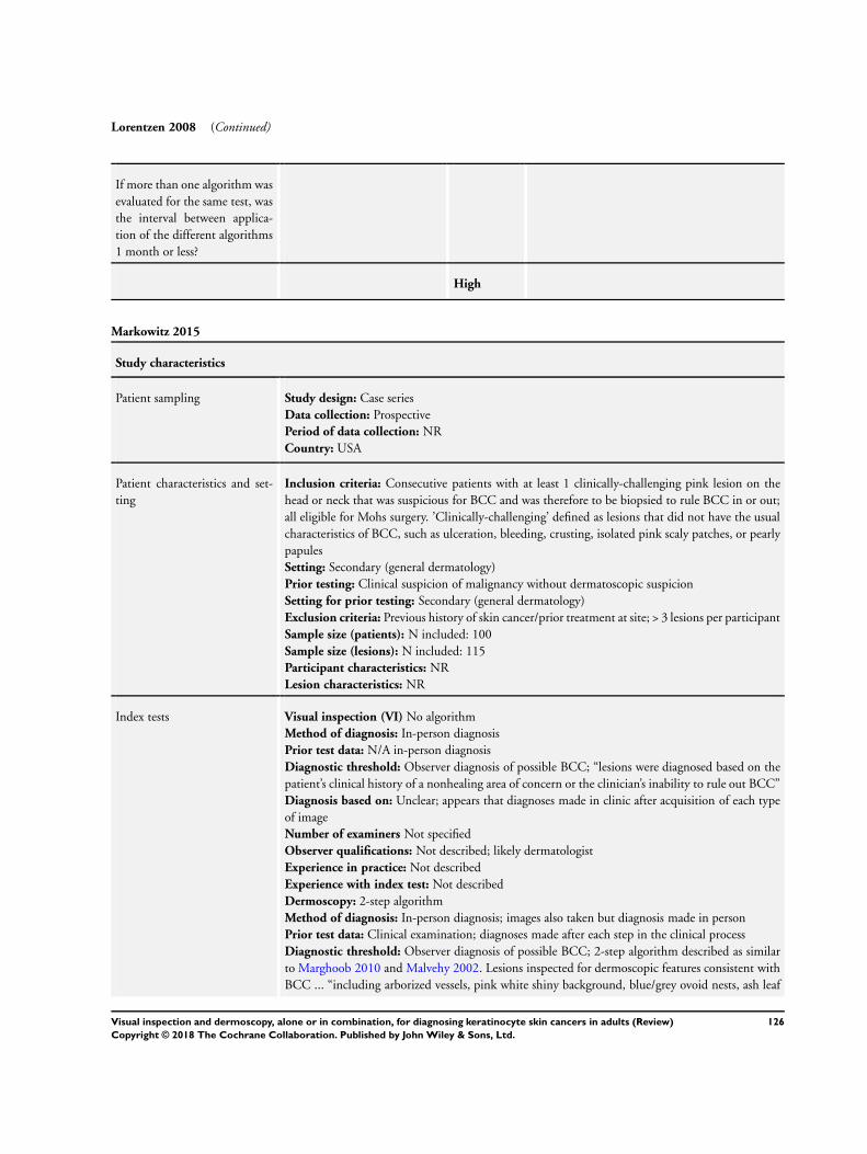
Lorentzen 2008 (Continued)
If more than one algorithm was
evaluated for the same test, was
the interval between applica-
tion of the different algorithms
1 month or less?
High
Markowitz 2015
Study characteristics
Patient sampling Study design: Case series
Data collection: Prospective
Period of data collection: NR
Country: USA
Patient characteristics and set-
ting
Inclusion criteria: Consecutive patients with at least 1 clinically-challenging pink lesion on the
head or neck that was suspicious for BCC and was therefore to be biopsied to rule BCC in or out;
all eligible for Mohs surgery. ’Clinically-challenging’ defined as lesions that did not have the usual
characteristics of BCC, such as ulceration, bleeding, crusting, isolated pink scaly patches, or pearly
papules
Setting: Secondary (general dermatology)
Prior testing: Clinical suspicion of malignancy without dermatoscopic suspicion
Setting for prior testing: Secondary (general dermatology)
Exclusion criteria: Previous history of skin cancer/prior treatment at site; > 3 lesions per participant
Sample size (patients): N included: 100
Sample size (lesions): N included: 115
Participant characteristics: NR
Lesion characteristics: NR
Index tests Visual inspection (VI) No algorithm
Method of diagnosis: In-person diagnosis
Prior test data: N/A in-person diagnosis
Diagnostic threshold: Observer diagnosis of possible BCC; “lesions were diagnosed based on the
patient’s clinical history of a nonhealing area of concern or the clinician’s inability to rule out BCC”
Diagnosis based on: Unclear; appears that diagnoses made in clinic after acquisition of each type
of image
Number of examiners Not specified
Observer qualifications: Not described; likely dermatologist
Experience in practice: Not described
Experience with index test: Not described
Dermoscopy: 2-step algorithm
Method of diagnosis: In-person diagnosis; images also taken but diagnosis made in person
Prior test data: Clinical examination; diagnoses made after each step in the clinical process
Diagnostic threshold: Observer diagnosis of possible BCC; 2-step algorithm described as similar
to Marghoob 2010 and Malvehy 2002. Lesions inspected for dermoscopic features consistent with
BCC ... “including arborized vessels, pink white shiny background, blue/grey ovoid nests, ash leaf
126Visual inspection and dermoscopy, alone or in combination, for diagnosing keratinocyte skin cancers in adults (Review)
Copyright © 2018 The Cochrane Collaboration. Published by John Wiley & Sons, Ltd.
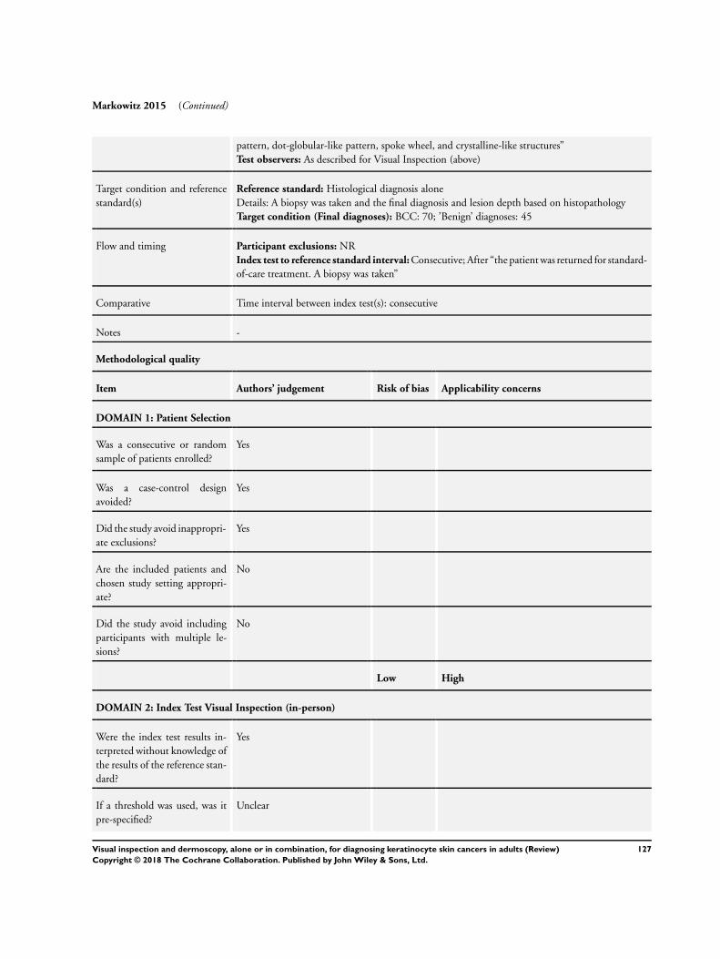
Markowitz 2015 (Continued)
pattern, dot-globular-like pattern, spoke wheel, and crystalline-like structures”
Test observers: As described for Visual Inspection (above)
Target condition and reference
standard(s)
Reference standard: Histological diagnosis alone
Details: A biopsy was taken and the final diagnosis and lesion depth based on histopathology
Target condition (Final diagnoses): BCC: 70; ’Benign’ diagnoses: 45
Flow and timing Participant exclusions: NR
Index test to reference standard interval: Consecutive; After “the patient was returned for standard-
of-care treatment. A biopsy was taken”
Comparative Time interval between index test(s): consecutive
Notes -
Methodological quality
Item Authors’ judgement Risk of bias Applicability concerns
DOMAIN 1: Patient Selection
Was a consecutive or random
sample of patients enrolled?
Yes
Was a case-control design
avoided?
Yes
Did the study avoid inappropri-
ate exclusions?
Yes
Are the included patients and
chosen study setting appropri-
ate?
No
Did the study avoid including
participants with multiple le-
sions?
No
Low High
DOMAIN 2: Index Test Visual Inspection (in-person)
Were the index test results in-
terpreted without knowledge of
the results of the reference stan-
dard?
Yes
If a threshold was used, was it
pre-specified?
Unclear
127Visual inspection and dermoscopy, alone or in combination, for diagnosing keratinocyte skin cancers in adults (Review)
Copyright © 2018 The Cochrane Collaboration. Published by John Wiley & Sons, Ltd.

Markowitz 2015 (Continued)
For studies reporting the ac-
curacy of multiple diagnostic
thresholds, was each threshold
or algorithm interpreted with-
out knowledge of the results of
the others?
Was the test applied and inter-
preted in a clinically applicable
manner?
Yes
Were thresholds or criteria for
diagnosis reported in sufficient
detail to allow replication?
No
Was the test interpretation car-
ried out by an experienced ex-
aminer?
Unclear
Unclear Unclear
DOMAIN 2: Index Test Dermoscopy (in-person)
Were the index test results in-
terpreted without knowledge of
the results of the reference stan-
dard?
Yes
If a threshold was used, was it
pre-specified?
Unclear
For studies reporting the ac-
curacy of multiple diagnostic
thresholds, was each threshold
or algorithm interpreted with-
out knowledge of the results of
the others?
Was the test applied and inter-
preted in a clinically applicable
manner?
Yes
Were thresholds or criteria for
diagnosis reported in sufficient
detail to allow replication?
Yes
Was the test interpretation car-
ried out by an experienced ex-
aminer?
Unclear
128Visual inspection and dermoscopy, alone or in combination, for diagnosing keratinocyte skin cancers in adults (Review)
Copyright © 2018 The Cochrane Collaboration. Published by John Wiley & Sons, Ltd.

Markowitz 2015 (Continued)
Unclear Unclear
DOMAIN 3: Reference Standard
Is the reference standards likely
to correctly classify the target
condition?
Yes
Were the reference standard re-
sults
interpreted without knowledge
of the results of the index tests?
Unclear
Expert opinion (with no his-
tological confirmation) was not
used as a reference standard
Yes
Was histology interpretation
carried out by an experienced
histopathologist or by a der-
matopathologist?
Unclear
Were the reference standard re-
sults
interpreted without knowledge
of the referral diagnosis?
Unclear
Low Unclear
DOMAIN 4: Flow and Timing
Was there an appropriate inter-
val between index test and ref-
erence standard?
Yes
Did all patients receive the same
reference standard?
Yes
Were all patients included in the
analysis?
Yes
If the reference standard in-
cludes clinical follow-up of
borderline/benign appearing le-
sions, was there a minimum
follow-up following application
of index test(s) of at least: 3
months for melanoma or cSCC
or 6 months for BCC?
129Visual inspection and dermoscopy, alone or in combination, for diagnosing keratinocyte skin cancers in adults (Review)
Copyright © 2018 The Cochrane Collaboration. Published by John Wiley & Sons, Ltd.
Markowitz 2015 (Continued)
If more than one algorithm was
evaluated for the same test, was
the interval between applica-
tion of the different algorithms
1 month or less?
Low
Menzies 2000
Study characteristics
Patient sampling Study design Case control
Data collection Retrospective image selection/Prospective interpretation
Period of data collection: NR
Country: Australia and USA
Test set derived: Sample randomly divided into training and test sets
Patient characteristics and set-
ting
Inclusion criteria: Pigmented skin lesions with dermoscopic images and histological diagnoses;
BCCs, invasive melanomas and clinically atypical ’nonmelanoma’ lesions separately sampled
Study setting: Specialist unit; Sydney Melanoma Unit and Florida Skin and Cancer Unit databases
Prior testing: Selected for excision (no further detail)
Exclusion criteria: NR
Sample size (patients): NR
Sample size (lesions) N included: 213
Participants Characteristics: NR
Lesion characteristics: Median Breslow thickness for invasive melanoma (71/213) was 0.67 mm
for the test set
Index tests Dermoscopy: Own new algorithm (Menzies) for diagnosis of pigmented BCC
Method of diagnosis: Dermoscopic images; images studies on a viewer
Prior test: No further information used
Diagnostic threshold: Pigment network absent with at least 1 positive feature present: ulceration,
large blue-grey ovoid nests, multiple blue-grey globules, maple leaflike areas, spoke wheel areas,
arborising (treelike) telangiectasia (all defined in detail)
Diagnosis based on: Unclear; training set images assessed by 2 observers; unclear if consensus or
average and whether same observers also assessed the test set images; N = 2
Observer qualification: NR: likely dermatologists
Observer experience in practice: NR
Observer experience with index test: NR
Derivation aspect: Training set was assessed for the presence/absence of 45 dermoscopic features
and a simple model constructed using negative features with low sensitivity and high specificity for
invasive melanoma and benign nonmelanoma lesions. The optimal model was then evaluated on
the test set of images
Target condition and reference
standard(s)
Reference standard: Histological diagnosis alone (not further described)
Target condition (Final diagnoses): Test set: BCC: 71; melanoma (invasive): 71; sebhorrheic
keratosis: 5; ephelis 1; solar lentigo 3; common naevus 19; dysplastic naevus 38; blue naevus 2;
130Visual inspection and dermoscopy, alone or in combination, for diagnosing keratinocyte skin cancers in adults (Review)
Copyright © 2018 The Cochrane Collaboration. Published by John Wiley & Sons, Ltd.
Menzies 2000 (Continued)
dermatofibroma 1; haemangioma 1; Other 1
Flow and timing Participant exclusions: NR
Index test to reference standard interval: PSLs photographed prior to excision
Comparative
Notes -
Methodological quality
Item Authors’ judgement Risk of bias Applicability concerns
DOMAIN 1: Patient Selection
Was a consecutive or random
sample of patients enrolled?
Unclear
Was a case-control design
avoided?
No
Did the study avoid inappropri-
ate exclusions?
Unclear
Are the included patients and
chosen study setting appropri-
ate?
No
Did the study avoid including
participants with multiple le-
sions?
Unclear
High High
DOMAIN 2: Index Test Dermoscopy (image based)
Were the index test results in-
terpreted without knowledge of
the results of the reference stan-
dard?
Yes
If a threshold was used, was it
pre-specified?
Yes
For studies reporting the ac-
curacy of multiple diagnostic
thresholds, was each threshold
or algorithm interpreted with-
out knowledge of the results of
131Visual inspection and dermoscopy, alone or in combination, for diagnosing keratinocyte skin cancers in adults (Review)
Copyright © 2018 The Cochrane Collaboration. Published by John Wiley & Sons, Ltd.
Menzies 2000 (Continued)
the others?
Was the test applied and inter-
preted in a clinically applicable
manner?
No
Were thresholds or criteria for
diagnosis reported in sufficient
detail to allow replication?
Yes
Was the test interpretation car-
ried out by an experienced ex-
aminer?
Unclear
Low High
DOMAIN 3: Reference Standard
Is the reference standards likely
to correctly classify the target
condition?
Yes
Were the reference standard re-
sults
interpreted without knowledge
of the results of the index tests?
Unclear
Expert opinion (with no his-
tological confirmation) was not
used as a reference standard
Yes
Was histology interpretation
carried out by an experienced
histopathologist or by a der-
matopathologist?
Unclear
Were the reference standard re-
sults
interpreted without knowledge
of the referral diagnosis?
Unclear
Low Unclear
DOMAIN 4: Flow and Timing
Was there an appropriate inter-
val between index test and ref-
erence standard?
Yes
132Visual inspection and dermoscopy, alone or in combination, for diagnosing keratinocyte skin cancers in adults (Review)
Copyright © 2018 The Cochrane Collaboration. Published by John Wiley & Sons, Ltd.
Menzies 2000 (Continued)
Did all patients receive the same
reference standard?
Yes
Were all patients included in the
analysis?
Yes
If the reference standard in-
cludes clinical follow-up of
borderline/benign appearing le-
sions, was there a minimum
follow-up following application
of index test(s) of at least: 3
months for melanoma or cSCC
or 6 months for BCC?
If more than one algorithm was
evaluated for the same test, was
the interval between applica-
tion of the different algorithms
1 month or less?
Low
Navarrete Dechent 2016
Study characteristics
Patient sampling Study design: Case series
Data collection: Retrospective image selection/Prospective interpretation
Period of data collection: 2009 - 2012
Country: USA
Patient characteristics and set-
ting
Inclusion criteria: Consecutively-excised nonpigmented lesions with no discernible pigment on
clinical or dermoscopic images
Setting: Specialist unit; Memorial Sloane Kettering Cancer Centre
Prior testing: Selected for excision (no further detail)
Setting for prior testing: Specialist unit
Exclusion criteria: Collision tumours, dermatofibromas and seborrhoeic keratoses were excluded
Sample size (patients): N eligible: 2375; N included: NR
Sample size (lesions): N eligible: 2891; N included: 457
Participant characteristics: Mean age: 64.3 (SD 14.1); Male: 282; 61.7%
Lesion characteristics: Head/neck: 134; 29.3%; trunk: 124; 27.1%; upper extremity 84; 18.4%;
lower extremity 113; 24.7%; genitalia 1; 0.2%; missing 1; 0.2%
Index tests Dermoscopy: Own new algorithm (shiny white streaks (SWSs))
Method of diagnosis: Dermoscopic images; Each individual lesion’s close-up clinical (cropped
images without patient identifiers) and dermoscopic images were reviewed for inclusion by a single
author
133Visual inspection and dermoscopy, alone or in combination, for diagnosing keratinocyte skin cancers in adults (Review)
Copyright © 2018 The Cochrane Collaboration. Published by John Wiley & Sons, Ltd.
Navarrete Dechent 2016 (Continued)
Prior test data: No further information used
Diagnostic threshold: Presence of any SWSs; these were classified as (1) blotches (also known as
clods; discrete, small or large structureless areas); (2) strands (long thick or thin lines, randomly
distributed or parallel, and not orthogonally oriented); (3) rosettes (cluster of 4 white dots in a 4-leaf
clover-like arrangement); and (4) short white lines (also known as crystalline structures and chrysalis;
fine lines that intersect or are oriented orthogonally to each other) (Liebman 2012; Liebman 2011)
. Shiny white structures that could not be classified into one of these specific morphologies were
categorised as nonspecified. (All lesions were also evaluated for Menzies criteria (Menzies 2000);
those without Menzies criteria were considered featureless and were further evaluated for presence
of: SFT; multiple in-focus, blue-grey dots; multiple small erosions;and concentric structures)
Diagnosis based on: Consensus (2 observers); N = 2
Observer qualifications: 1 observer appears to be a dermatologist and the other was a medical
student (based on authors’ institutions); both trained by a third observer (expert dermoscopist) who
also acted as arbitrator in case of any disagreement
Experience in practice: Not described
Experience with index test: Trained; Described as “trained in dermoscopic analysis by an expert
dermoscopist”
Any other detail: Images were captured with a Nikon 1 camera (Nikon USA, Inc) using Dermlite
DL2 pro HR for polarized images and Dermlitefluid for nonpolarised images at 10-fold magnifi-
cation(3Gen, LLC)
Target condition and reference
standard(s)
Reference standard: Histological diagnosis alone
Target condition (Final diagnoses): BCC: 287; cSCC: 106; melanoma (in situ and invasive, or
not reported): 21; lichen planus-like keratosis 39; naevus 4
Flow and timing Participant exclusions: NR
Index test to reference standard interval: Appears consecutive; “Standard procedures in this prac-
tice included capturing clinical and dermoscopic images of all lesions selected for biopsy”
Comparative
Notes -
Methodological quality
Item Authors’ judgement Risk of bias Applicability concerns
DOMAIN 1: Patient Selection
Was a consecutive or random
sample of patients enrolled?
Yes
Was a case-control design
avoided?
Yes
Did the study avoid inappropri-
ate exclusions?
No
134Visual inspection and dermoscopy, alone or in combination, for diagnosing keratinocyte skin cancers in adults (Review)
Copyright © 2018 The Cochrane Collaboration. Published by John Wiley & Sons, Ltd.
Navarrete Dechent 2016 (Continued)
Are the included patients and
chosen study setting appropri-
ate?
No
Did the study avoid including
participants with multiple le-
sions?
No
High High
DOMAIN 2: Index Test Dermoscopy (image based)
Were the index test results in-
terpreted without knowledge of
the results of the reference stan-
dard?
Yes
If a threshold was used, was it
pre-specified?
No
For studies reporting the ac-
curacy of multiple diagnostic
thresholds, was each threshold
or algorithm interpreted with-
out knowledge of the results of
the others?
Was the test applied and inter-
preted in a clinically applicable
manner?
No
Were thresholds or criteria for
diagnosis reported in sufficient
detail to allow replication?
Yes
Was the test interpretation car-
ried out by an experienced ex-
aminer?
No
High High
DOMAIN 3: Reference Standard
Is the reference standards likely
to correctly classify the target
condition?
Yes
135Visual inspection and dermoscopy, alone or in combination, for diagnosing keratinocyte skin cancers in adults (Review)
Copyright © 2018 The Cochrane Collaboration. Published by John Wiley & Sons, Ltd.
Navarrete Dechent 2016 (Continued)
Were the reference standard re-
sults
interpreted without knowledge
of the results of the index tests?
Unclear
Expert opinion (with no his-
tological confirmation) was not
used as a reference standard
Yes
Was histology interpretation
carried out by an experienced
histopathologist or by a der-
matopathologist?
Unclear
Were the reference standard re-
sults
interpreted without knowledge
of the referral diagnosis?
Unclear
Low Unclear
DOMAIN 4: Flow and Timing
Was there an appropriate inter-
val between index test and ref-
erence standard?
Yes
Did all patients receive the same
reference standard?
Yes
Were all patients included in the
analysis?
Yes
If the reference standard in-
cludes clinical follow-up of
borderline/benign appearing le-
sions, was there a minimum
follow-up following application
of index test(s) of at least: 3
months for melanoma or cSCC
or 6 months for BCC?
If more than one algorithm was
evaluated for the same test, was
the interval between applica-
tion of the different algorithms
1 month or less?
Unclear
136Visual inspection and dermoscopy, alone or in combination, for diagnosing keratinocyte skin cancers in adults (Review)
Copyright © 2018 The Cochrane Collaboration. Published by John Wiley & Sons, Ltd.
Nori 2004
Study characteristics
Patient sampling Study design: Case control
Data collection: Retrospective image selection/Prospective interpretation
Period of data collection 2 years - date range not specified
Country USA and Spain
Patient characteristics and set-
ting
Inclusion criteria: Biopsy-confirmed BCC and convenience sample of non-BCC with “’range
of common diagnoses”; of these images with superior clinical quality were selected for clinical
assessment
Setting: Secondary (general dermatology); Private care
Prior testing: Most underwent biopsy but no detail of selection process
Setting for prior testing: Unspecified
Exclusion criteria: NR
Sample size (patients): N included: 145
Sample size (lesions): N included: 152; 105 in VI analysis
Participant characteristics: Male: 98; 64%
Lesion characteristics: Face/ears: 35%; trunk: 13%; limbs: extremities 45%; back 7%; only 7 of
69 non-BCC lesions “had BCC on the list of possible differential diagnoses”
Index tests Visual inspection (VI): No algorithm
Method of diagnosis: Clinical photographs; “set of randomised clinical images was ... analysed in
a blinded fashion by two dermatologists”
Prior test data: No further information used
Diagnostic threshold: High and high/medium probability of BCC. Lesions assigned to: high
probability (BCC until proven otherwise), medium probability (would biopsy to rule out BCC),
and low probability (no biopsy needed)
Diagnosis based on: Single observer (N = 2)
Observer qualifications: Dermatologist
Experience in practice: Not described
Experience with index test: Not described
Target condition and reference
standard(s)
Reference standard Histological diagnosis plus other. Histology not further described
Expert opinion: 15 lesions were not biopsied (e.g. lesions like seborrhoeic keratosis) because the
clinical diagnosis was considered diagnostic
Target condition (Final diagnoses): BCC: 83; 58 in VI analysis; cSCC: 4
’Benign’ diagnoses: 65
Flow and timing Participant exclusions: 47 lesions were not included because of poor clinical image quality
Index test to reference standard interval: Not described
Comparative
Notes -
Methodological quality
Item Authors’ judgement Risk of bias Applicability concerns
137Visual inspection and dermoscopy, alone or in combination, for diagnosing keratinocyte skin cancers in adults (Review)
Copyright © 2018 The Cochrane Collaboration. Published by John Wiley & Sons, Ltd.
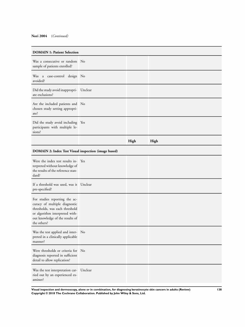
Nori 2004 (Continued)
DOMAIN 1: Patient Selection
Was a consecutive or random
sample of patients enrolled?
No
Was a case-control design
avoided?
No
Did the study avoid inappropri-
ate exclusions?
Unclear
Are the included patients and
chosen study setting appropri-
ate?
No
Did the study avoid including
participants with multiple le-
sions?
Yes
High High
DOMAIN 2: Index Test Visual inspection (image based)
Were the index test results in-
terpreted without knowledge of
the results of the reference stan-
dard?
Yes
If a threshold was used, was it
pre-specified?
Unclear
For studies reporting the ac-
curacy of multiple diagnostic
thresholds, was each threshold
or algorithm interpreted with-
out knowledge of the results of
the others?
Was the test applied and inter-
preted in a clinically applicable
manner?
No
Were thresholds or criteria for
diagnosis reported in sufficient
detail to allow replication?
No
Was the test interpretation car-
ried out by an experienced ex-
aminer?
Unclear
138Visual inspection and dermoscopy, alone or in combination, for diagnosing keratinocyte skin cancers in adults (Review)
Copyright © 2018 The Cochrane Collaboration. Published by John Wiley & Sons, Ltd.

Nori 2004 (Continued)
Unclear High
DOMAIN 3: Reference Standard
Is the reference standards likely
to correctly classify the target
condition?
No
Were the reference standard re-
sults
interpreted without knowledge
of the results of the index tests?
Unclear
Expert opinion (with no his-
tological confirmation) was not
used as a reference standard
No
Was histology interpretation
carried out by an experienced
histopathologist or by a der-
matopathologist?
Unclear
Were the reference standard re-
sults
interpreted without knowledge
of the referral diagnosis?
Unclear
High High
DOMAIN 4: Flow and Timing
Was there an appropriate inter-
val between index test and ref-
erence standard?
Unclear
Did all patients receive the same
reference standard?
No
Were all patients included in the
analysis?
No
If the reference standard in-
cludes clinical follow-up of
borderline/benign appearing le-
sions, was there a minimum
follow-up following application
of index test(s) of at least: 3
months for melanoma or cSCC
or 6 months for BCC?
139Visual inspection and dermoscopy, alone or in combination, for diagnosing keratinocyte skin cancers in adults (Review)
Copyright © 2018 The Cochrane Collaboration. Published by John Wiley & Sons, Ltd.
Nori 2004 (Continued)
If more than one algorithm was
evaluated for the same test, was
the interval between applica-
tion of the different algorithms
1 month or less?
High
Rosendahl 2011
Study characteristics
Patient sampling Study design: Case series
Data collection: Retrospective image selection/Prospective interpretation
Period of data collection 30-month period; dates NR
Country Australia
Patient characteristics and set-
ting
Inclusion criteria: Consecutive series of pigmented lesions submitted for histology from the pri-
mary-care skin cancer practice of 1 author
Setting: Primary-care skin cancer practice
Prior testing: Selected for excision (no further detail)
Setting for prior testing: Primary
Exclusion criteria: Poor image quality (considered under Flow and Timing)
Sample size (patients): N included: 389
Sample size (lesions): N eligible: 466 pigmented lesions out of 1959 lesions excised or biopsied;
N included: 463
Participant characteristics: Mean age: 57 (SD 17). Male: 67.4%
Lesion characteristics: (53.1%) melanocytic. Lesion site: 17.7% head or face; trunk: 52.1%; 27.
6% extremities; 2.2% palms or soles. melanoma thickness: ≤ 1 mm: 1/29 melanoma (3.4%)
Index tests Visual inspection (VI) No algorithm
Method of diagnosis: Clinical photographs overview and close-up image presented
Prior test data: No further information used
Other test data: Dermoscopic images presented to observer subsequent to diagnosis using clinical
images alone
Diagnostic threshold: Clinical diagnosis/subjective impression. Observers gave a diagnosis with
level of confidence (from 0 for definitely benign to 100 for definitely malignant) after viewing
the clinical images. (NB used authors’ threshold for detection of any skin cancer which includes
lesions clinically considered to be MM, BCC pigmented epithelial carcinoma including SCC,
keratoacanthoma, actinic keratosis and Bowen’s disease as test positive; review only considered
histologically-confirmed MM, BCC or invasive SCC to be disease-positive)
Diagnosis based on: Single observer (N NR)
Observer qualifications: Expert dermatologist (based on author communication).
Experience in practice: Expert
Experience with dermoscopy: Expert
Dermoscopy Pattern analysis; new algorithm - Chaos and clues
Method of diagnosis: Clinical photographs (1 overview and 1 close-up), followed by 1 dermoscopic
image presented to a blinded observer on a computer screen
140Visual inspection and dermoscopy, alone or in combination, for diagnosing keratinocyte skin cancers in adults (Review)
Copyright © 2018 The Cochrane Collaboration. Published by John Wiley & Sons, Ltd.
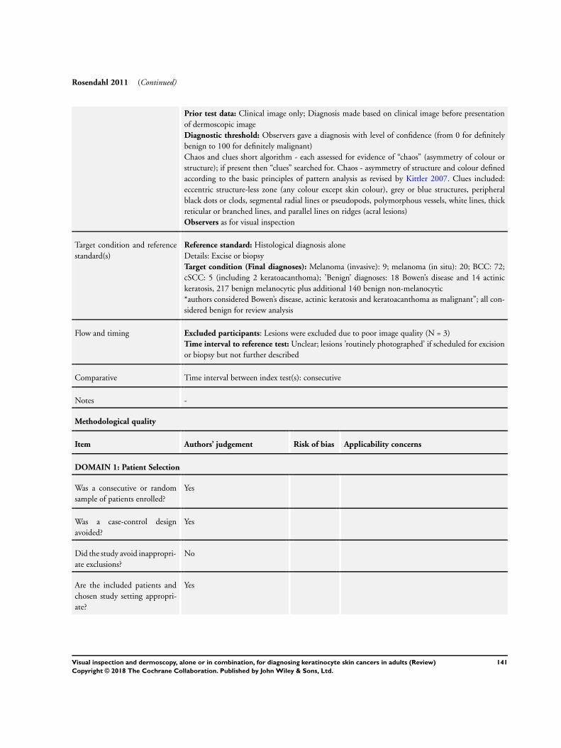
Rosendahl 2011 (Continued)
Prior test data: Clinical image only; Diagnosis made based on clinical image before presentation
of dermoscopic image
Diagnostic threshold: Observers gave a diagnosis with level of confidence (from 0 for definitely
benign to 100 for definitely malignant)
Chaos and clues short algorithm - each assessed for evidence of “chaos” (asymmetry of colour or
structure); if present then “clues” searched for. Chaos - asymmetry of structure and colour defined
according to the basic principles of pattern analysis as revised by Kittler 2007. Clues included:
eccentric structure-less zone (any colour except skin colour), grey or blue structures, peripheral
black dots or clods, segmental radial lines or pseudopods, polymorphous vessels, white lines, thick
reticular or branched lines, and parallel lines on ridges (acral lesions)
Observers as for visual inspection
Target condition and reference
standard(s)
Reference standard: Histological diagnosis alone
Details: Excise or biopsy
Target condition (Final diagnoses): Melanoma (invasive): 9; melanoma (in situ): 20; BCC: 72;
cSCC: 5 (including 2 keratoacanthoma); ’Benign’ diagnoses: 18 Bowen’s disease and 14 actinic
keratosis, 217 benign melanocytic plus additional 140 benign non-melanocytic
*authors considered Bowen’s disease, actinic keratosis and keratoacanthoma as malignant”; all con-
sidered benign for review analysis
Flow and timing Excluded participants: Lesions were excluded due to poor image quality (N = 3)
Time interval to reference test: Unclear; lesions ’routinely photographed’ if scheduled for excision
or biopsy but not further described
Comparative Time interval between index test(s): consecutive
Notes -
Methodological quality
Item Authors’ judgement Risk of bias Applicability concerns
DOMAIN 1: Patient Selection
Was a consecutive or random
sample of patients enrolled?
Yes
Was a case-control design
avoided?
Yes
Did the study avoid inappropri-
ate exclusions?
No
Are the included patients and
chosen study setting appropri-
ate?
Yes
141Visual inspection and dermoscopy, alone or in combination, for diagnosing keratinocyte skin cancers in adults (Review)
Copyright © 2018 The Cochrane Collaboration. Published by John Wiley & Sons, Ltd.
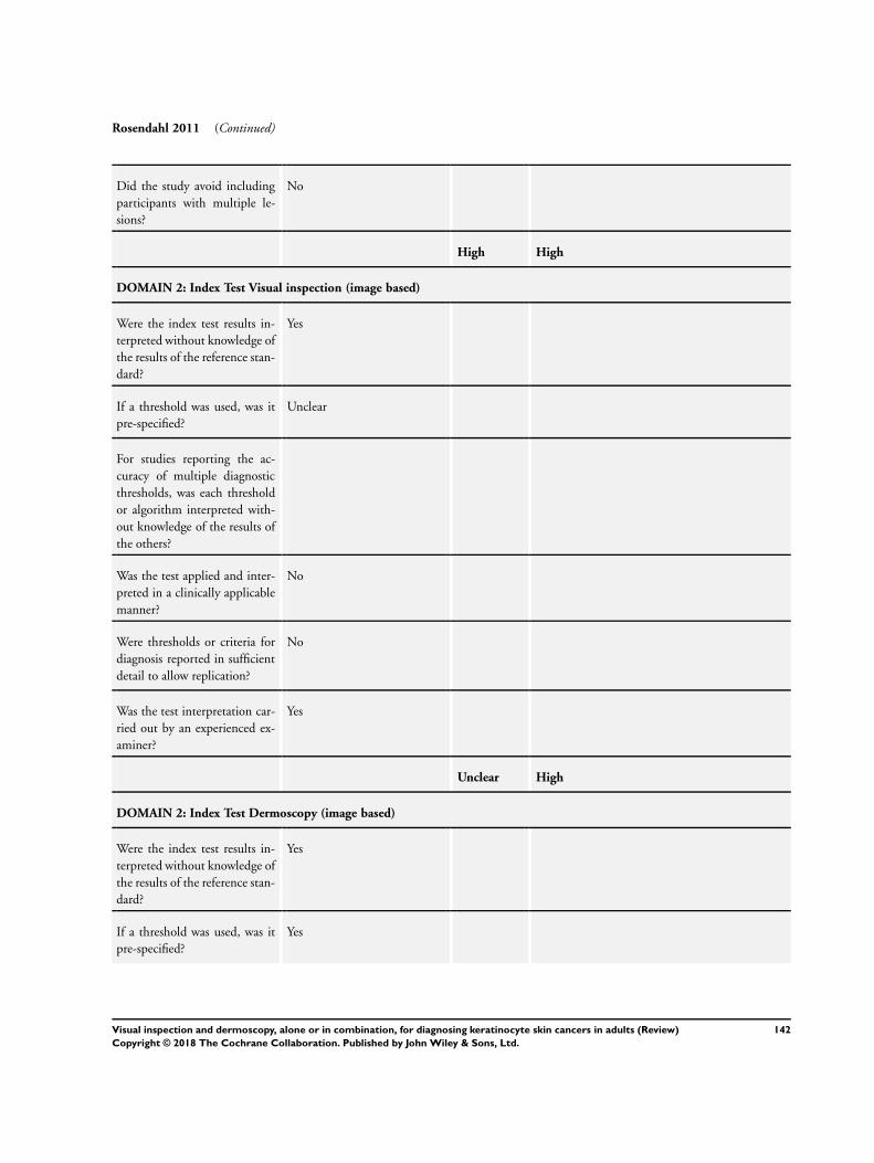
Rosendahl 2011 (Continued)
Did the study avoid including
participants with multiple le-
sions?
No
High High
DOMAIN 2: Index Test Visual inspection (image based)
Were the index test results in-
terpreted without knowledge of
the results of the reference stan-
dard?
Yes
If a threshold was used, was it
pre-specified?
Unclear
For studies reporting the ac-
curacy of multiple diagnostic
thresholds, was each threshold
or algorithm interpreted with-
out knowledge of the results of
the others?
Was the test applied and inter-
preted in a clinically applicable
manner?
No
Were thresholds or criteria for
diagnosis reported in sufficient
detail to allow replication?
No
Was the test interpretation car-
ried out by an experienced ex-
aminer?
Yes
Unclear High
DOMAIN 2: Index Test Dermoscopy (image based)
Were the index test results in-
terpreted without knowledge of
the results of the reference stan-
dard?
Yes
If a threshold was used, was it
pre-specified?
Yes
142Visual inspection and dermoscopy, alone or in combination, for diagnosing keratinocyte skin cancers in adults (Review)
Copyright © 2018 The Cochrane Collaboration. Published by John Wiley & Sons, Ltd.
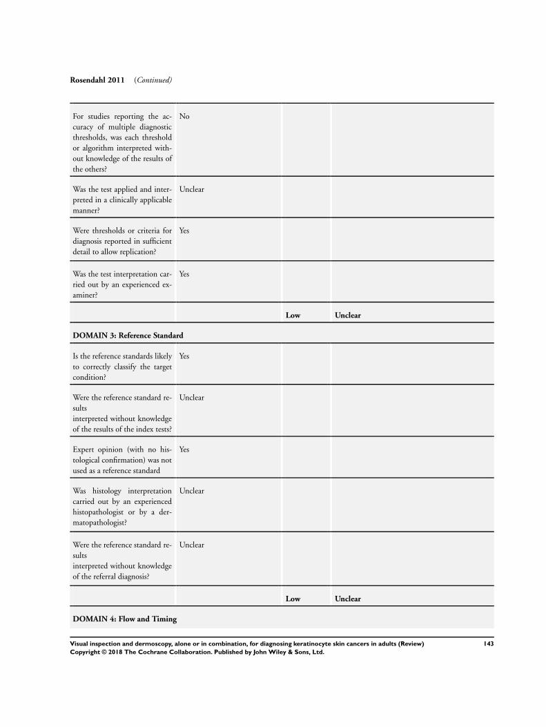
Rosendahl 2011 (Continued)
For studies reporting the ac-
curacy of multiple diagnostic
thresholds, was each threshold
or algorithm interpreted with-
out knowledge of the results of
the others?
No
Was the test applied and inter-
preted in a clinically applicable
manner?
Unclear
Were thresholds or criteria for
diagnosis reported in sufficient
detail to allow replication?
Yes
Was the test interpretation car-
ried out by an experienced ex-
aminer?
Yes
Low Unclear
DOMAIN 3: Reference Standard
Is the reference standards likely
to correctly classify the target
condition?
Yes
Were the reference standard re-
sults
interpreted without knowledge
of the results of the index tests?
Unclear
Expert opinion (with no his-
tological confirmation) was not
used as a reference standard
Yes
Was histology interpretation
carried out by an experienced
histopathologist or by a der-
matopathologist?
Unclear
Were the reference standard re-
sults
interpreted without knowledge
of the referral diagnosis?
Unclear
Low Unclear
DOMAIN 4: Flow and Timing
143Visual inspection and dermoscopy, alone or in combination, for diagnosing keratinocyte skin cancers in adults (Review)
Copyright © 2018 The Cochrane Collaboration. Published by John Wiley & Sons, Ltd.
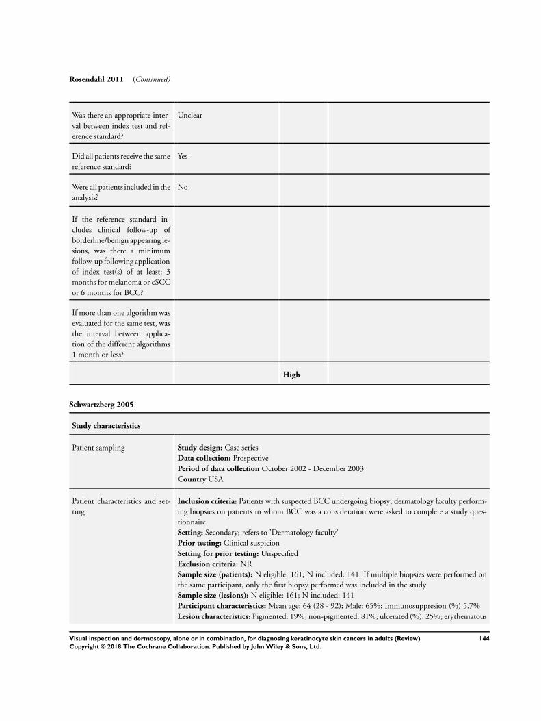
Rosendahl 2011 (Continued)
Was there an appropriate inter-
val between index test and ref-
erence standard?
Unclear
Did all patients receive the same
reference standard?
Yes
Were all patients included in the
analysis?
No
If the reference standard in-
cludes clinical follow-up of
borderline/benign appearing le-
sions, was there a minimum
follow-up following application
of index test(s) of at least: 3
months for melanoma or cSCC
or 6 months for BCC?
If more than one algorithm was
evaluated for the same test, was
the interval between applica-
tion of the different algorithms
1 month or less?
High
Schwartzberg 2005
Study characteristics
Patient sampling Study design: Case series
Data collection: Prospective
Period of data collection October 2002 - December 2003
Country USA
Patient characteristics and set-
ting
Inclusion criteria: Patients with suspected BCC undergoing biopsy; dermatology faculty perform-
ing biopsies on patients in whom BCC was a consideration were asked to complete a study ques-
tionnaire
Setting: Secondary; refers to ’Dermatology faculty’
Prior testing: Clinical suspicion
Setting for prior testing: Unspecified
Exclusion criteria: NR
Sample size (patients): N eligible: 161; N included: 141. If multiple biopsies were performed on
the same participant, only the first biopsy performed was included in the study
Sample size (lesions): N eligible: 161; N included: 141
Participant characteristics: Mean age: 64 (28 - 92); Male: 65%; Immunosuppresion (%) 5.7%
Lesion characteristics: Pigmented: 19%; non-pigmented: 81%; ulcerated (%): 25%; erythematous
144Visual inspection and dermoscopy, alone or in combination, for diagnosing keratinocyte skin cancers in adults (Review)
Copyright © 2018 The Cochrane Collaboration. Published by John Wiley & Sons, Ltd.
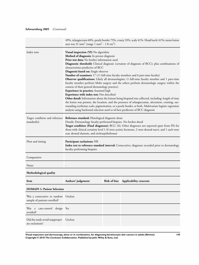
Schwartzberg 2005 (Continued)
49%, telangiectasis 60%, pearly border 75%, crusty 33%, scaly 41%. Head/neck: 61%; mean lesion
area was 31 mm2 (range 1 mm2 - 1.8 cm2)
Index tests Visual inspection (VI) No algorithm
Method of diagnosis: In-person diagnosis
Prior test data: No further information used
Diagnostic threshold: Clinical diagnosis (certainty of diagnosis of BCC); plus combinations of
characteristics predictive of BCC
Diagnosis based on: Single observer
Number of examiners 17 (11 full-time faculty members and 6 part-time faculty)
Observer qualifications: Likely all dermatologists; (1 full-time faculty member and 1 part-time
faculty member perform Mohs surgery and the others perform dermatologic surgery within the
context of their general dermatology practice)
Experience in practice: Assumed high
Experience with index test: Not described
Other detail: Information about the lesions being biopsied was collected, including: length of time
the lesion was present, the location, and the presence of telangiectasias, ulceration, crusting, sur-
rounding erythema, scale, pigmentation, or a pearly border, or both. Multivariate logistic regression
analysis using backward selection used to id best predictors of BCC diagnosis
Target condition and reference
standard(s)
Reference standard: Histological diagnosis alone
Details: Dermatology faculty performed biopsies. No further detail
Target condition (Final diagnoses): BCC: 82; Other diagnoses not reported apart from FPs for
those with clinical certainty level 1 (6 were actinic keratoses, 2 were dermal naevi, and 1 each were
scar, dermal elastosis, and trichoepithelioma)
Flow and timing Participant exclusions: NR
Index test to reference standard interval: Consecutive; diagnoses recorded prior to dermatology
faculty performing biopsies
Comparative
Notes -
Methodological quality
Item Authors’ judgement Risk of bias Applicability concerns
DOMAIN 1: Patient Selection
Was a consecutive or random
sample of patients enrolled?
Unclear
Was a case-control design
avoided?
Yes
Did the study avoid inappropri-
ate exclusions?
Unclear
145Visual inspection and dermoscopy, alone or in combination, for diagnosing keratinocyte skin cancers in adults (Review)
Copyright © 2018 The Cochrane Collaboration. Published by John Wiley & Sons, Ltd.
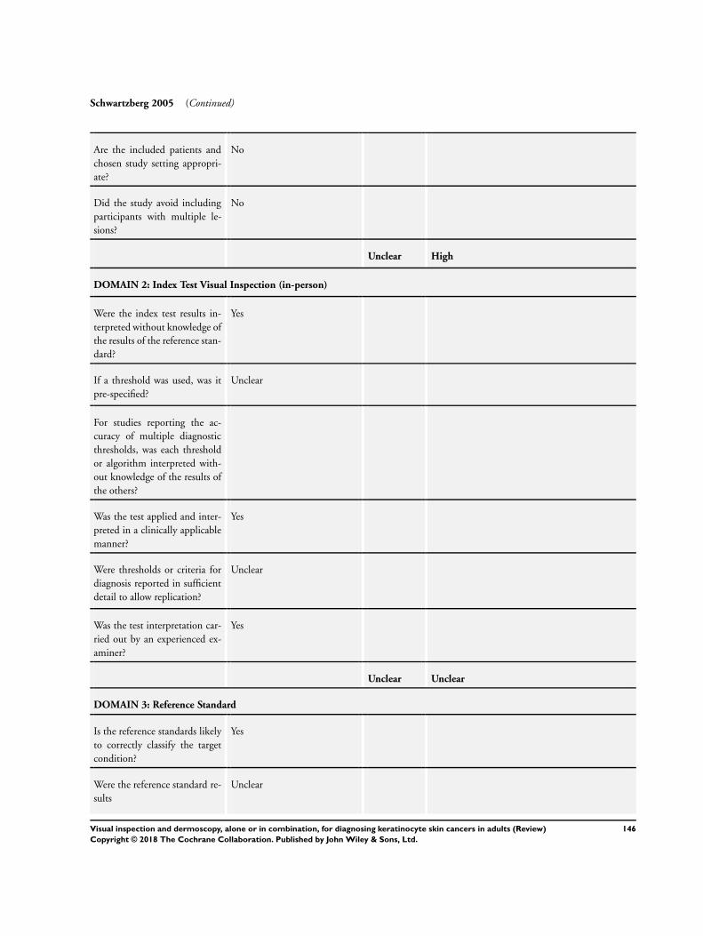
Schwartzberg 2005 (Continued)
Are the included patients and
chosen study setting appropri-
ate?
No
Did the study avoid including
participants with multiple le-
sions?
No
Unclear High
DOMAIN 2: Index Test Visual Inspection (in-person)
Were the index test results in-
terpreted without knowledge of
the results of the reference stan-
dard?
Yes
If a threshold was used, was it
pre-specified?
Unclear
For studies reporting the ac-
curacy of multiple diagnostic
thresholds, was each threshold
or algorithm interpreted with-
out knowledge of the results of
the others?
Was the test applied and inter-
preted in a clinically applicable
manner?
Yes
Were thresholds or criteria for
diagnosis reported in sufficient
detail to allow replication?
Unclear
Was the test interpretation car-
ried out by an experienced ex-
aminer?
Yes
Unclear Unclear
DOMAIN 3: Reference Standard
Is the reference standards likely
to correctly classify the target
condition?
Yes
Were the reference standard re-
sults
Unclear
146Visual inspection and dermoscopy, alone or in combination, for diagnosing keratinocyte skin cancers in adults (Review)
Copyright © 2018 The Cochrane Collaboration. Published by John Wiley & Sons, Ltd.
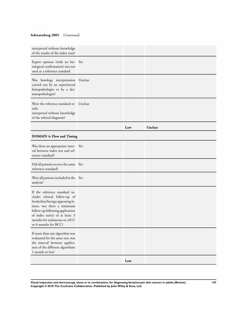
Schwartzberg 2005 (Continued)
interpreted without knowledge
of the results of the index tests?
Expert opinion (with no his-
tological confirmation) was not
used as a reference standard
Yes
Was histology interpretation
carried out by an experienced
histopathologist or by a der-
matopathologist?
Unclear
Were the reference standard re-
sults
interpreted without knowledge
of the referral diagnosis?
Unclear
Low Unclear
DOMAIN 4: Flow and Timing
Was there an appropriate inter-
val between index test and ref-
erence standard?
Yes
Did all patients receive the same
reference standard?
Yes
Were all patients included in the
analysis?
Yes
If the reference standard in-
cludes clinical follow-up of
borderline/benign appearing le-
sions, was there a minimum
follow-up following application
of index test(s) of at least: 3
months for melanoma or cSCC
or 6 months for BCC?
If more than one algorithm was
evaluated for the same test, was
the interval between applica-
tion of the different algorithms
1 month or less?
Low
147Visual inspection and dermoscopy, alone or in combination, for diagnosing keratinocyte skin cancers in adults (Review)
Copyright © 2018 The Cochrane Collaboration. Published by John Wiley & Sons, Ltd.
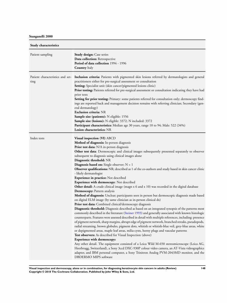
Stanganelli 2000
Study characteristics
Patient sampling Study design: Case series
Data collection: Retrospective
Period of data collection 1994 - 1996
Country Italy
Patient characteristics and set-
ting
Inclusion criteria: Patients with pigmented skin lesions referred by dermatologists and general
practitioners either for pre-surgical assessment or consultation
Setting: Specialist unit (skin cancer/pigmented lesions clinic)
Prior testing: Patients referred for pre-surgical assessment or consultation indicating they have had
prior tests
Setting for prior testing: Primary: some patients referred for consultation only; dermoscopy find-
ings are reported back and management decision remains with referring clinician; Secondary (gen-
eral dermatology)
Exclusion criteria: NR
Sample size (patients): N eligible: 1556
Sample size (lesions): N eligible: 3372; N included: 3372
Participant characteristics: Median age 30 years, range 10 to 94; Male: 522 (34%)
Lesion characteristics: NR
Index tests Visual inspection (VI) ABCD
Method of diagnosis: In-person diagnosis
Prior test data: N/A in-person diagnosis
Other test data: Dermoscopic and clinical images subsequently presented separately to observer
subsequent to diagnosis using clinical images alone
Diagnostic threshold: NR
Diagnosis based on: Single observer; N = 1
Observer qualifications: NR; described as 1 of the co-authors and study based in skin cancer clinic
- likely dermatologist
Experience in practice: Not described
Experience with dermoscopy: Not described
Other detail: A crude clinical image (magn x 6 and x 10) was recorded in the digital database
Dermoscopy: Pattern analysis
Method of diagnosis: Unclear; participants seen in person but dermoscopic diagnosis made based
on digital ELM image (by same clinician as in-person clinical dx)
Prior test data: Combined clinical/dermoscopy diagnosis
Diagnostic threshold: Diagnosis described as based on an integrated synopsis of the patterns most
commonly described in the literature (Steiner 1993) and generally associated with known histologic
counterparts. Features were assessed described in detail with multiple references, including: presence
of pigment network, sharp margins, abrupt edge of pigment network, branched streaks, pseudopods,
radial streaming, brown globules, pigment dots, whitish or whitish-blue veil, grey-blue areas, white
or depigmented areas, maple leaf areas, milia-cysts, horny plugs and vascular patterns
Test observers: As described for Visual Inspection (above)
Experience with dermoscopy:
Any other detail. The equipment consisted of a Leica Wild M-650 stereomicroscope (Leica AG,
Heerbrugg, Switzerland), a Sony 3ccd DXC-930P colour video camera, an AT-Vista videographics
adapter, and IBM personal computer, a Sony Trinitron Analog PVM-2043MD monitor, and the
DBDERMO MIPS software
148Visual inspection and dermoscopy, alone or in combination, for diagnosing keratinocyte skin cancers in adults (Review)
Copyright © 2018 The Cochrane Collaboration. Published by John Wiley & Sons, Ltd.
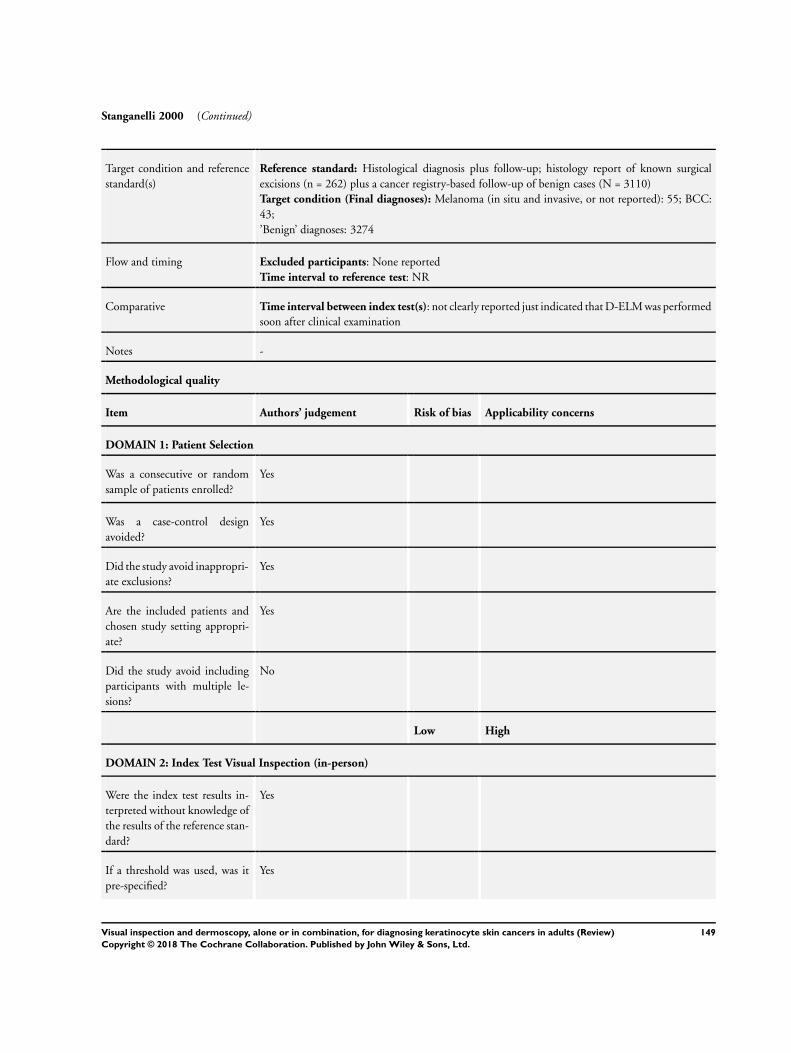
Stanganelli 2000 (Continued)
Target condition and reference
standard(s)
Reference standard: Histological diagnosis plus follow-up; histology report of known surgical
excisions (n = 262) plus a cancer registry-based follow-up of benign cases (N = 3110)
Target condition (Final diagnoses): Melanoma (in situ and invasive, or not reported): 55; BCC:
43;
’Benign’ diagnoses: 3274
Flow and timing Excluded participants: None reported
Time interval to reference test: NR
Comparative Time interval between index test(s): not clearly reported just indicated that D-ELM was performed
soon after clinical examination
Notes -
Methodological quality
Item Authors’ judgement Risk of bias Applicability concerns
DOMAIN 1: Patient Selection
Was a consecutive or random
sample of patients enrolled?
Yes
Was a case-control design
avoided?
Yes
Did the study avoid inappropri-
ate exclusions?
Yes
Are the included patients and
chosen study setting appropri-
ate?
Yes
Did the study avoid including
participants with multiple le-
sions?
No
Low High
DOMAIN 2: Index Test Visual Inspection (in-person)
Were the index test results in-
terpreted without knowledge of
the results of the reference stan-
dard?
Yes
If a threshold was used, was it
pre-specified?
Yes
149Visual inspection and dermoscopy, alone or in combination, for diagnosing keratinocyte skin cancers in adults (Review)
Copyright © 2018 The Cochrane Collaboration. Published by John Wiley & Sons, Ltd.
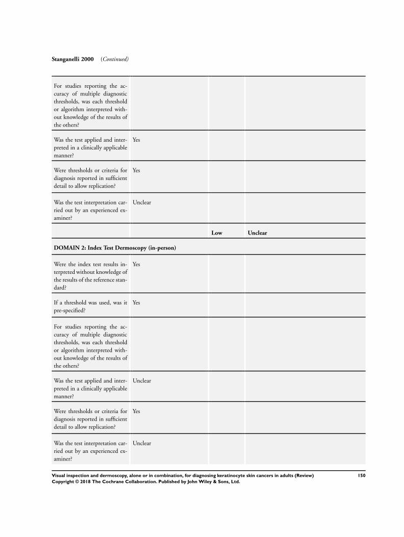
Stanganelli 2000 (Continued)
For studies reporting the ac-
curacy of multiple diagnostic
thresholds, was each threshold
or algorithm interpreted with-
out knowledge of the results of
the others?
Was the test applied and inter-
preted in a clinically applicable
manner?
Yes
Were thresholds or criteria for
diagnosis reported in sufficient
detail to allow replication?
Yes
Was the test interpretation car-
ried out by an experienced ex-
aminer?
Unclear
Low Unclear
DOMAIN 2: Index Test Dermoscopy (in-person)
Were the index test results in-
terpreted without knowledge of
the results of the reference stan-
dard?
Yes
If a threshold was used, was it
pre-specified?
Yes
For studies reporting the ac-
curacy of multiple diagnostic
thresholds, was each threshold
or algorithm interpreted with-
out knowledge of the results of
the others?
Was the test applied and inter-
preted in a clinically applicable
manner?
Unclear
Were thresholds or criteria for
diagnosis reported in sufficient
detail to allow replication?
Yes
Was the test interpretation car-
ried out by an experienced ex-
aminer?
Unclear
150Visual inspection and dermoscopy, alone or in combination, for diagnosing keratinocyte skin cancers in adults (Review)
Copyright © 2018 The Cochrane Collaboration. Published by John Wiley & Sons, Ltd.

Stanganelli 2000 (Continued)
Low Unclear
DOMAIN 3: Reference Standard
Is the reference standards likely
to correctly classify the target
condition?
No
Were the reference standard re-
sults
interpreted without knowledge
of the results of the index tests?
Unclear
Expert opinion (with no his-
tological confirmation) was not
used as a reference standard
Yes
Was histology interpretation
carried out by an experienced
histopathologist or by a der-
matopathologist?
Unclear
Were the reference standard re-
sults
interpreted without knowledge
of the referral diagnosis?
Unclear
High Unclear
DOMAIN 4: Flow and Timing
Was there an appropriate inter-
val between index test and ref-
erence standard?
Unclear
Did all patients receive the same
reference standard?
No
Were all patients included in the
analysis?
Yes
If the reference standard in-
cludes clinical follow-up of
borderline/benign appearing le-
sions, was there a minimum
follow-up following application
of index test(s) of at least: 3
months for melanoma or cSCC
or 6 months for BCC?
Yes
151Visual inspection and dermoscopy, alone or in combination, for diagnosing keratinocyte skin cancers in adults (Review)
Copyright © 2018 The Cochrane Collaboration. Published by John Wiley & Sons, Ltd.
Stanganelli 2000 (Continued)
If more than one algorithm was
evaluated for the same test, was
the interval between applica-
tion of the different algorithms
1 month or less?
High
Steiner 1987
Study characteristics
Patient sampling Study design: Case series
Data collection: Prospective
Period of data collection: Not specified
Country: Austria
Patient characteristics and set-
ting
Inclusion criteria: Small (< 10 mm) pigmented skin lesions considered diagnostically equivocal in
that there was no absolute agreement on the clinical diagnosis among investigating clinicians at a
pigmented lesions clinic
Setting: Specialist unit (skin cancer/pigmented lesions clinic)
Prior testing: Clinical suspicion of malignancy without dermatoscopic suspicion
Setting for prior testing: Specialist unit (skin cancer/pigmented lesions clinic)
Exclusion criteria: > 10 mm diameter
Sample size (patients): NR
Sample size (lesions): 318
Participant characteristics: NR
Lesion characteristics: NR
Index tests Visual inspection (VI): No algorithm
Method of diagnosis: In-person diagnosis
Prior test data: N/A
Other test data: Dermoscopy undertaken by same clinician(s) subsequent to clinical evaluation
Diagnostic threshold: NR
Diagnosis based on: Consensus (3 observers) “All lesions were independently seen and diagnosed
by the three investigators, and the diagnosis that appeared most probable to at least two of the three
investigators was recorded as the clinical”; N = 3
Observer qualifications: Dermatologist
Experience in practice: High experience or ‘Expert’; “experienced dermatologists”
Experience with dermoscopy: Unclear; not explicitly described. Discussion describes ELM as
standard procedure in clinic
Study reported data for dermoscopy, but a breakdown of incorrect diagnoses by final diagnosis was
not provided to allow a 2 x 2 to be estimated
Target condition and reference
standard(s)
Reference standard: Histological diagnosis alone
Target condition (Final diagnoses): Melanoma (invasive): 49; melanoma (in situ): 15; BCC: 20;
lentigo maligna 9 (also includes lentigo maligna melanoma);
Sebhorrheic keratosis: 20; junctional naevi 39; blue naevus 29; dysplastic naevus 75; lentigo simplex
152Visual inspection and dermoscopy, alone or in combination, for diagnosing keratinocyte skin cancers in adults (Review)
Copyright © 2018 The Cochrane Collaboration. Published by John Wiley & Sons, Ltd.
Steiner 1987 (Continued)
and naevoid lentigo 19; angioma/ angiokeratoma 15
Flow and timing Excluded participants: None reported
Time interval to reference test: Assumed consecutive; following diagnosis, lesions subsequently
excised
Comparative Time interval between index test(s): consecutive
Notes -
Methodological quality
Item Authors’ judgement Risk of bias Applicability concerns
DOMAIN 1: Patient Selection
Was a consecutive or random
sample of patients enrolled?
Unclear
Was a case-control design
avoided?
Yes
Did the study avoid inappropri-
ate exclusions?
Unclear
Are the included patients and
chosen study setting appropri-
ate?
No
Did the study avoid including
participants with multiple le-
sions?
Unclear
Unclear High
DOMAIN 2: Index Test Visual Inspection (in-person)
Were the index test results in-
terpreted without knowledge of
the results of the reference stan-
dard?
Yes
If a threshold was used, was it
pre-specified?
Unclear
For studies reporting the ac-
curacy of multiple diagnostic
thresholds, was each threshold
or algorithm interpreted with-
153Visual inspection and dermoscopy, alone or in combination, for diagnosing keratinocyte skin cancers in adults (Review)
Copyright © 2018 The Cochrane Collaboration. Published by John Wiley & Sons, Ltd.
Steiner 1987 (Continued)
out knowledge of the results of
the others?
Was the test applied and inter-
preted in a clinically applicable
manner?
No
Were thresholds or criteria for
diagnosis reported in sufficient
detail to allow replication?
No
Was the test interpretation car-
ried out by an experienced ex-
aminer?
Yes
Unclear High
DOMAIN 3: Reference Standard
Is the reference standards likely
to correctly classify the target
condition?
Yes
Were the reference standard re-
sults
interpreted without knowledge
of the results of the index tests?
Unclear
Expert opinion (with no his-
tological confirmation) was not
used as a reference standard
Yes
Was histology interpretation
carried out by an experienced
histopathologist or by a der-
matopathologist?
Unclear
Were the reference standard re-
sults
interpreted without knowledge
of the referral diagnosis?
Unclear
Low Unclear
DOMAIN 4: Flow and Timing
Was there an appropriate inter-
val between index test and ref-
erence standard?
Yes
154Visual inspection and dermoscopy, alone or in combination, for diagnosing keratinocyte skin cancers in adults (Review)
Copyright © 2018 The Cochrane Collaboration. Published by John Wiley & Sons, Ltd.
Steiner 1987 (Continued)
Did all patients receive the same
reference standard?
Yes
Were all patients included in the
analysis?
Yes
If the reference standard in-
cludes clinical follow-up of
borderline/benign appearing le-
sions, was there a minimum
follow-up following application
of index test(s) of at least: 3
months for melanoma or cSCC
or 6 months for BCC?
If more than one algorithm was
evaluated for the same test, was
the interval between applica-
tion of the different algorithms
1 month or less?
Low
Ulrich 2015
Study characteristics
Patient sampling Study design: Case series
Data collection: Prospective
Period of data collection: April 2013 - March 2014
Country: Germany
Patient characteristics and set-
ting
Inclusion criteria: Patients with non-pigmented pink lesions with clinical suspicion of BCC re-
quiring biopsy for diagnostic confirmation. Pink lesions defined as clinically-unclear erythematous
papule or plaque; either reddish macules, patches or small papules with or without scale
Setting: Multicentre study; authors’ institutions included Dermatology departments (N = 4) and
private dermatology offices (N = 3)
Prior testing: Clinical suspicion of malignancy
Setting for prior testing: Unspecified
Exclusion criteria: Lesions with the typical clinical appearance of BCC on clinical examination
(such as the presence of a pearly border, central ulceration and obvious telangiectasias), as well
as pigmented lesions, were excluded from the protocol. Patients with unstable or uncontrolled
clinically-significant medical conditions were excluded. Lesions with missing histology also excluded
(N = 21)
Sample size (patients): N eligible: 164; N included: 155
Sample size (lesions): N eligible: 256; N included: 235 (different sets of 231 lesions were available
for each test)
Participant characteristics: Median age: 70 (33 - 90)
155Visual inspection and dermoscopy, alone or in combination, for diagnosing keratinocyte skin cancers in adults (Review)
Copyright © 2018 The Cochrane Collaboration. Published by John Wiley & Sons, Ltd.
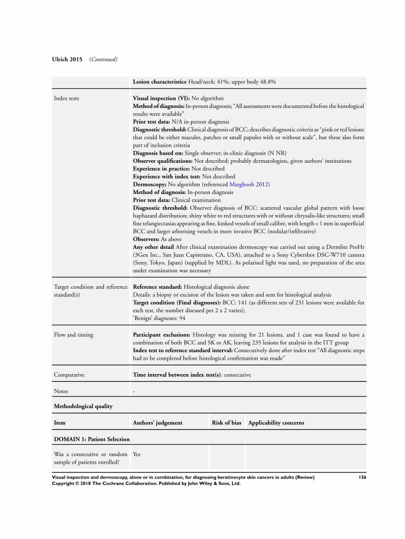
Ulrich 2015 (Continued)
Lesion characteristics Head/neck: 41%; upper body 48.8%
Index tests Visual inspection (VI): No algorithm
Method of diagnosis: In-person diagnosis; “All assessments were documented before the histological
results were available”
Prior test data: N/A in-person diagnosis
Diagnostic threshold: Clinical diagnosis of BCC; describes diagnostic criteria as “pink or red lesions
that could be either macules, patches or small papules with or without scale”, but these also form
part of inclusion criteria
Diagnosis based on: Single observer; in-clinic diagnosis (N NR)
Observer qualifications: Not described; probably dermatologists, given authors’ institutions
Experience in practice: Not described
Experience with index test: Not described
Dermoscopy; No algorithm (referenced Marghoob 2012)
Method of diagnosis: In-person diagnosis
Prior test data: Clinical examination
Diagnostic threshold: Observer diagnosis of BCC: scattered vascular global pattern with loose
haphazard distribution; shiny white to red structures with or without chrysalis-like structures; small
fine telangiectasias appearing as fine, kinked vessels of small calibre, with length < 1 mm in superficial
BCC and larger arborising vessels in more invasive BCC (nodular/infiltrative)
Observers: As above
Any other detail After clinical examination dermoscopy was carried out using a Dermlite ProHr
(3Gen Inc., San Juan Capistrano, CA, USA), attached to a Sony Cybershot DSC-W710 camera
(Sony, Tokyo, Japan) (supplied by MDL). As polarised light was used, no preparation of the area
under examination was necessary
Target condition and reference
standard(s)
Reference standard: Histological diagnosis alone
Details: a biopsy or excision of the lesion was taken and sent for histological analysis
Target condition (Final diagnoses): BCC: 141 (as different sets of 231 lesions were available for
each test, the number diseased per 2 x 2 varies);
’Benign’ diagnoses: 94
Flow and timing Participant exclusions: Histology was missing for 21 lesions, and 1 case was found to have a
combination of both BCC and SK or AK, leaving 235 lesions for analysis in the ITT group
Index test to reference standard interval: Consecutively done after index test “All diagnostic steps
had to be completed before histological confirmation was made”
Comparative Time interval between index test(s): consecutive
Notes -
Methodological quality
Item Authors’ judgement Risk of bias Applicability concerns
DOMAIN 1: Patient Selection
Was a consecutive or random
sample of patients enrolled?
Yes
156Visual inspection and dermoscopy, alone or in combination, for diagnosing keratinocyte skin cancers in adults (Review)
Copyright © 2018 The Cochrane Collaboration. Published by John Wiley & Sons, Ltd.
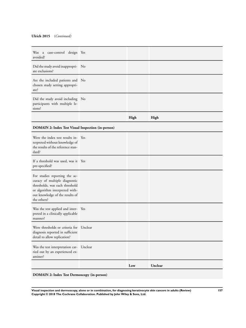
Ulrich 2015 (Continued)
Was a case-control design
avoided?
Yes
Did the study avoid inappropri-
ate exclusions?
No
Are the included patients and
chosen study setting appropri-
ate?
No
Did the study avoid including
participants with multiple le-
sions?
No
High High
DOMAIN 2: Index Test Visual Inspection (in-person)
Were the index test results in-
terpreted without knowledge of
the results of the reference stan-
dard?
Yes
If a threshold was used, was it
pre-specified?
Yes
For studies reporting the ac-
curacy of multiple diagnostic
thresholds, was each threshold
or algorithm interpreted with-
out knowledge of the results of
the others?
Was the test applied and inter-
preted in a clinically applicable
manner?
Yes
Were thresholds or criteria for
diagnosis reported in sufficient
detail to allow replication?
Unclear
Was the test interpretation car-
ried out by an experienced ex-
aminer?
Unclear
Low Unclear
DOMAIN 2: Index Test Dermoscopy (in-person)
157Visual inspection and dermoscopy, alone or in combination, for diagnosing keratinocyte skin cancers in adults (Review)
Copyright © 2018 The Cochrane Collaboration. Published by John Wiley & Sons, Ltd.
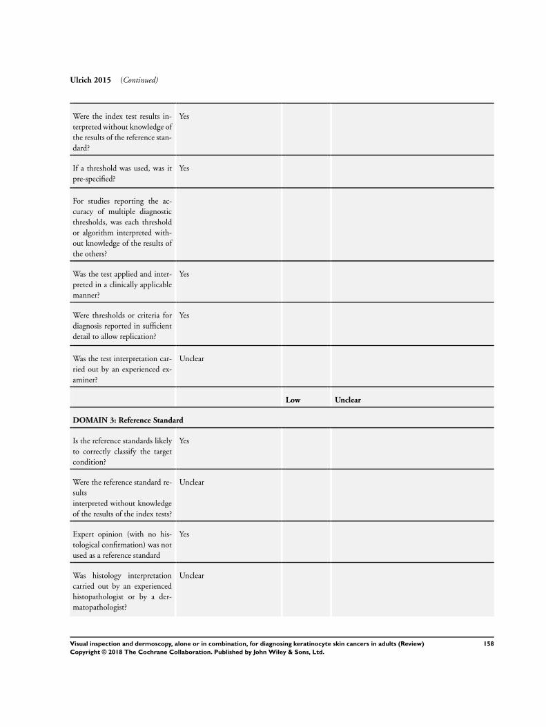
Ulrich 2015 (Continued)
Were the index test results in-
terpreted without knowledge of
the results of the reference stan-
dard?
Yes
If a threshold was used, was it
pre-specified?
Yes
For studies reporting the ac-
curacy of multiple diagnostic
thresholds, was each threshold
or algorithm interpreted with-
out knowledge of the results of
the others?
Was the test applied and inter-
preted in a clinically applicable
manner?
Yes
Were thresholds or criteria for
diagnosis reported in sufficient
detail to allow replication?
Yes
Was the test interpretation car-
ried out by an experienced ex-
aminer?
Unclear
Low Unclear
DOMAIN 3: Reference Standard
Is the reference standards likely
to correctly classify the target
condition?
Yes
Were the reference standard re-
sults
interpreted without knowledge
of the results of the index tests?
Unclear
Expert opinion (with no his-
tological confirmation) was not
used as a reference standard
Yes
Was histology interpretation
carried out by an experienced
histopathologist or by a der-
matopathologist?
Unclear
158Visual inspection and dermoscopy, alone or in combination, for diagnosing keratinocyte skin cancers in adults (Review)
Copyright © 2018 The Cochrane Collaboration. Published by John Wiley & Sons, Ltd.
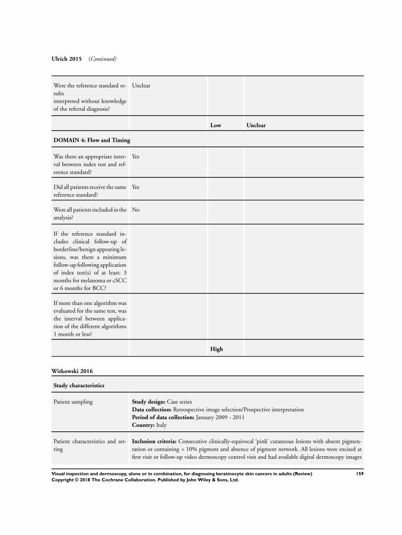
Ulrich 2015 (Continued)
Were the reference standard re-
sults
interpreted without knowledge
of the referral diagnosis?
Unclear
Low Unclear
DOMAIN 4: Flow and Timing
Was there an appropriate inter-
val between index test and ref-
erence standard?
Yes
Did all patients receive the same
reference standard?
Yes
Were all patients included in the
analysis?
No
If the reference standard in-
cludes clinical follow-up of
borderline/benign appearing le-
sions, was there a minimum
follow-up following application
of index test(s) of at least: 3
months for melanoma or cSCC
or 6 months for BCC?
If more than one algorithm was
evaluated for the same test, was
the interval between applica-
tion of the different algorithms
1 month or less?
High
Witkowski 2016
Study characteristics
Patient sampling Study design: Case series
Data collection: Retrospective image selection/Prospective interpretation
Period of data collection: January 2009 - 2011
Country: Italy
Patient characteristics and set-
ting
Inclusion criteria: Consecutive clinically-equivocal ‘pink’ cutaneous lesions with absent pigmen-
tation or containing < 10% pigment and absence of pigment network. All lesions were excised at
first visit or follow-up video dermoscopy control visit and had available digital dermoscopy images
159Visual inspection and dermoscopy, alone or in combination, for diagnosing keratinocyte skin cancers in adults (Review)
Copyright © 2018 The Cochrane Collaboration. Published by John Wiley & Sons, Ltd.
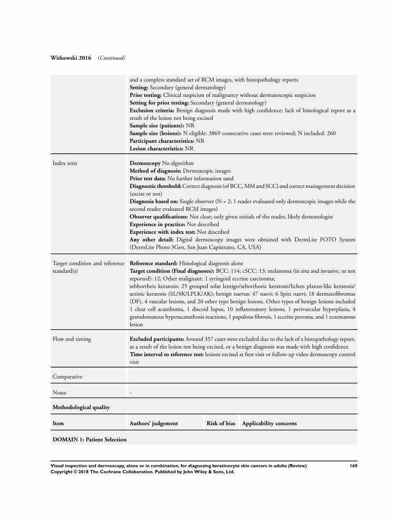
Witkowski 2016 (Continued)
and a complete standard set of RCM images, with histopathology reports
Setting: Secondary (general dermatology)
Prior testing: Clinical suspicion of malignancy without dermatoscopic suspicion
Setting for prior testing: Secondary (general dermatology)
Exclusion criteria: Benign diagnosis made with high confidence; lack of histological report as a
result of the lesion not being excised
Sample size (patients): NR
Sample size (lesions): N eligible: 3869 consecutive cases were reviewed; N included: 260
Participant characteristics: NR
Lesion characteristics: NR
Index tests Dermoscopy No algorithm
Method of diagnosis: Dermoscopic images
Prior test data: No further information used
Diagnostic threshold: Correct diagnosis (of BCC, MM and SCC) and correct management decision
(excise or not)
Diagnosis based on: Single observer (N = 2; 1 reader evaluated only dermoscopic images while the
second reader evaluated RCM images)
Observer qualifications: Not clear; only given initials of the reader, likely dermatologist
Experience in practice: Not described
Experience with index test: Not described
Any other detail: Digital dermoscopy images were obtained with DermLite FOTO System
(DermLite Photo 3Gen, San Juan Capistrano, CA, USA)
Target condition and reference
standard(s)
Reference standard: Histological diagnosis alone
Target condition (Final diagnoses): BCC: 114; cSCC: 13; melanoma (in situ and invasive, or not
reported): 12; Other malignant: 1 syringoid eccrine carcinoma;
sebhorrheic keratosis: 25 grouped solar lentigo/seborrhoeic keratosis/lichen planus-like keratosis/
actinic keratosis (SL/SK/LPLK/AK); benign naevus: 47 naevi; 6 Spitz naevi; 18 dermatofibromas
(DF), 4 vascular lesions, and 20 other type benign lesions. Other types of benign lesions included
1 clear cell acanthoma, 1 discoid lupus, 10 inflammatory lesions, 1 perivascular hyperplasia, 4
granulomatous hyperacanathosis reactions, 1 papulous fibrosis, 1 eccrine poroma, and 1 eczematous
lesion
Flow and timing Excluded participants: Around 357 cases were excluded due to the lack of a histopathology report,
as a result of the lesion not being excised, or a benign diagnosis was made with high confidence
Time interval to reference test: lesions excised at first visit or follow-up video dermoscopy control
visit
Comparative
Notes -
Methodological quality
Item Authors’ judgement Risk of bias Applicability concerns
DOMAIN 1: Patient Selection
160Visual inspection and dermoscopy, alone or in combination, for diagnosing keratinocyte skin cancers in adults (Review)
Copyright © 2018 The Cochrane Collaboration. Published by John Wiley & Sons, Ltd.
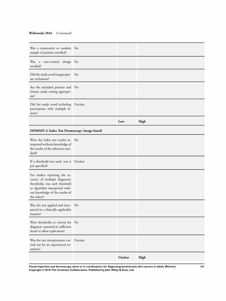
Witkowski 2016 (Continued)
Was a consecutive or random
sample of patients enrolled?
Yes
Was a case-control design
avoided?
Yes
Did the study avoid inappropri-
ate exclusions?
Yes
Are the included patients and
chosen study setting appropri-
ate?
No
Did the study avoid including
participants with multiple le-
sions?
Unclear
Low High
DOMAIN 2: Index Test Dermoscopy (image based)
Were the index test results in-
terpreted without knowledge of
the results of the reference stan-
dard?
Yes
If a threshold was used, was it
pre-specified?
Unclear
For studies reporting the ac-
curacy of multiple diagnostic
thresholds, was each threshold
or algorithm interpreted with-
out knowledge of the results of
the others?
Was the test applied and inter-
preted in a clinically applicable
manner?
No
Were thresholds or criteria for
diagnosis reported in sufficient
detail to allow replication?
No
Was the test interpretation car-
ried out by an experienced ex-
aminer?
Unclear
Unclear High
161Visual inspection and dermoscopy, alone or in combination, for diagnosing keratinocyte skin cancers in adults (Review)
Copyright © 2018 The Cochrane Collaboration. Published by John Wiley & Sons, Ltd.
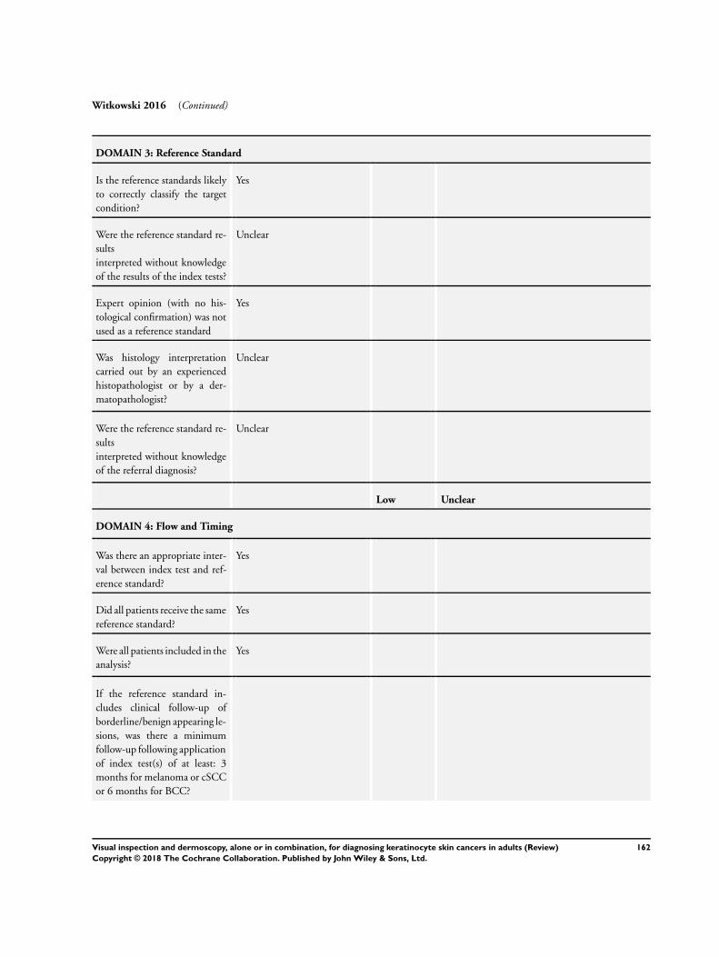
Witkowski 2016 (Continued)
DOMAIN 3: Reference Standard
Is the reference standards likely
to correctly classify the target
condition?
Yes
Were the reference standard re-
sults
interpreted without knowledge
of the results of the index tests?
Unclear
Expert opinion (with no his-
tological confirmation) was not
used as a reference standard
Yes
Was histology interpretation
carried out by an experienced
histopathologist or by a der-
matopathologist?
Unclear
Were the reference standard re-
sults
interpreted without knowledge
of the referral diagnosis?
Unclear
Low Unclear
DOMAIN 4: Flow and Timing
Was there an appropriate inter-
val between index test and ref-
erence standard?
Yes
Did all patients receive the same
reference standard?
Yes
Were all patients included in the
analysis?
Yes
If the reference standard in-
cludes clinical follow-up of
borderline/benign appearing le-
sions, was there a minimum
follow-up following application
of index test(s) of at least: 3
months for melanoma or cSCC
or 6 months for BCC?
162Visual inspection and dermoscopy, alone or in combination, for diagnosing keratinocyte skin cancers in adults (Review)
Copyright © 2018 The Cochrane Collaboration. Published by John Wiley & Sons, Ltd.
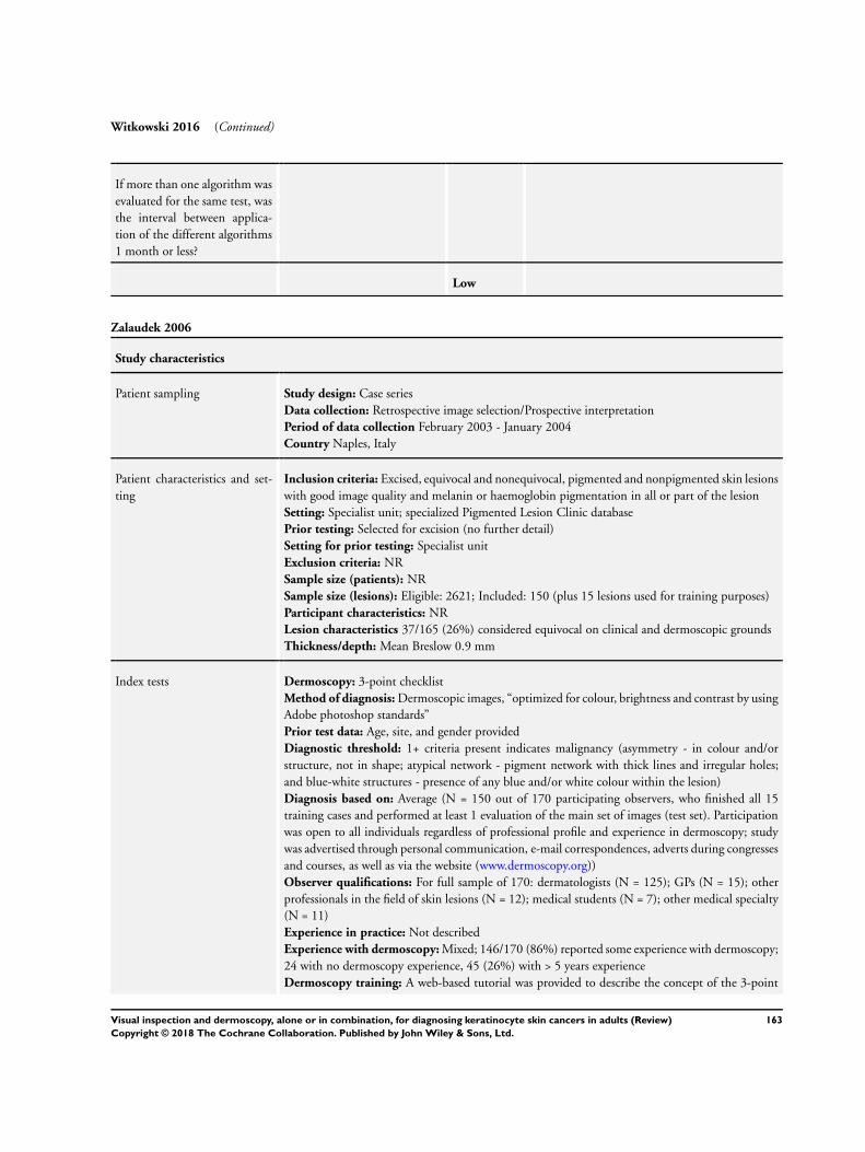
Witkowski 2016 (Continued)
If more than one algorithm was
evaluated for the same test, was
the interval between applica-
tion of the different algorithms
1 month or less?
Low
Zalaudek 2006
Study characteristics
Patient sampling Study design: Case series
Data collection: Retrospective image selection/Prospective interpretation
Period of data collection February 2003 - January 2004
Country Naples, Italy
Patient characteristics and set-
ting
Inclusion criteria: Excised, equivocal and nonequivocal, pigmented and nonpigmented skin lesions
with good image quality and melanin or haemoglobin pigmentation in all or part of the lesion
Setting: Specialist unit; specialized Pigmented Lesion Clinic database
Prior testing: Selected for excision (no further detail)
Setting for prior testing: Specialist unit
Exclusion criteria: NR
Sample size (patients): NR
Sample size (lesions): Eligible: 2621; Included: 150 (plus 15 lesions used for training purposes)
Participant characteristics: NR
Lesion characteristics 37/165 (26%) considered equivocal on clinical and dermoscopic grounds
Thickness/depth: Mean Breslow 0.9 mm
Index tests Dermoscopy: 3-point checklist
Method of diagnosis: Dermoscopic images, “optimized for colour, brightness and contrast by using
Adobe photoshop standards”
Prior test data: Age, site, and gender provided
Diagnostic threshold: 1+ criteria present indicates malignancy (asymmetry - in colour and/or
structure, not in shape; atypical network - pigment network with thick lines and irregular holes;
and blue-white structures - presence of any blue and/or white colour within the lesion)
Diagnosis based on: Average (N = 150 out of 170 participating observers, who finished all 15
training cases and performed at least 1 evaluation of the main set of images (test set). Participation
was open to all individuals regardless of professional profile and experience in dermoscopy; study
was advertised through personal communication, e-mail correspondences, adverts during congresses
and courses, as well as via the website (www.dermoscopy.org))
Observer qualifications: For full sample of 170: dermatologists (N = 125); GPs (N = 15); other
professionals in the field of skin lesions (N = 12); medical students (N = 7); other medical specialty
(N = 11)
Experience in practice: Not described
Experience with dermoscopy: Mixed; 146/170 (86%) reported some experience with dermoscopy;
24 with no dermoscopy experience, 45 (26%) with > 5 years experience
Dermoscopy training: A web-based tutorial was provided to describe the concept of the 3-point
163Visual inspection and dermoscopy, alone or in combination, for diagnosing keratinocyte skin cancers in adults (Review)
Copyright © 2018 The Cochrane Collaboration. Published by John Wiley & Sons, Ltd.
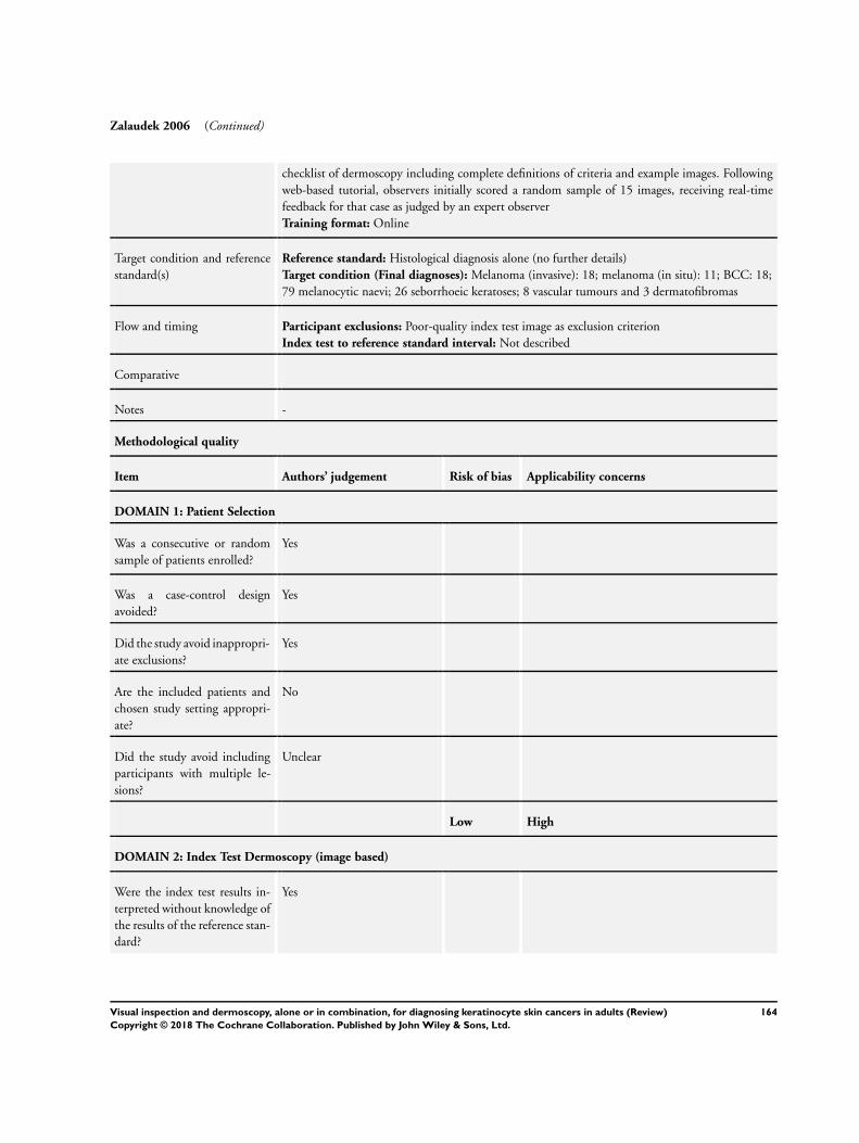
Zalaudek 2006 (Continued)
checklist of dermoscopy including complete definitions of criteria and example images. Following
web-based tutorial, observers initially scored a random sample of 15 images, receiving real-time
feedback for that case as judged by an expert observer
Training format: Online
Target condition and reference
standard(s)
Reference standard: Histological diagnosis alone (no further details)
Target condition (Final diagnoses): Melanoma (invasive): 18; melanoma (in situ): 11; BCC: 18;
79 melanocytic naevi; 26 seborrhoeic keratoses; 8 vascular tumours and 3 dermatofibromas
Flow and timing Participant exclusions: Poor-quality index test image as exclusion criterion
Index test to reference standard interval: Not described
Comparative
Notes -
Methodological quality
Item Authors’ judgement Risk of bias Applicability concerns
DOMAIN 1: Patient Selection
Was a consecutive or random
sample of patients enrolled?
Yes
Was a case-control design
avoided?
Yes
Did the study avoid inappropri-
ate exclusions?
Yes
Are the included patients and
chosen study setting appropri-
ate?
No
Did the study avoid including
participants with multiple le-
sions?
Unclear
Low High
DOMAIN 2: Index Test Dermoscopy (image based)
Were the index test results in-
terpreted without knowledge of
the results of the reference stan-
dard?
Yes
164Visual inspection and dermoscopy, alone or in combination, for diagnosing keratinocyte skin cancers in adults (Review)
Copyright © 2018 The Cochrane Collaboration. Published by John Wiley & Sons, Ltd.
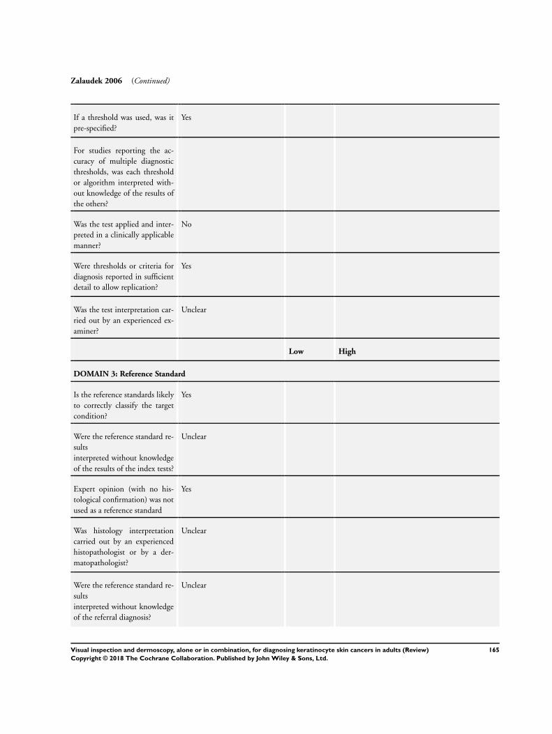
Zalaudek 2006 (Continued)
If a threshold was used, was it
pre-specified?
Yes
For studies reporting the ac-
curacy of multiple diagnostic
thresholds, was each threshold
or algorithm interpreted with-
out knowledge of the results of
the others?
Was the test applied and inter-
preted in a clinically applicable
manner?
No
Were thresholds or criteria for
diagnosis reported in sufficient
detail to allow replication?
Yes
Was the test interpretation car-
ried out by an experienced ex-
aminer?
Unclear
Low High
DOMAIN 3: Reference Standard
Is the reference standards likely
to correctly classify the target
condition?
Yes
Were the reference standard re-
sults
interpreted without knowledge
of the results of the index tests?
Unclear
Expert opinion (with no his-
tological confirmation) was not
used as a reference standard
Yes
Was histology interpretation
carried out by an experienced
histopathologist or by a der-
matopathologist?
Unclear
Were the reference standard re-
sults
interpreted without knowledge
of the referral diagnosis?
Unclear
165Visual inspection and dermoscopy, alone or in combination, for diagnosing keratinocyte skin cancers in adults (Review)
Copyright © 2018 The Cochrane Collaboration. Published by John Wiley & Sons, Ltd.
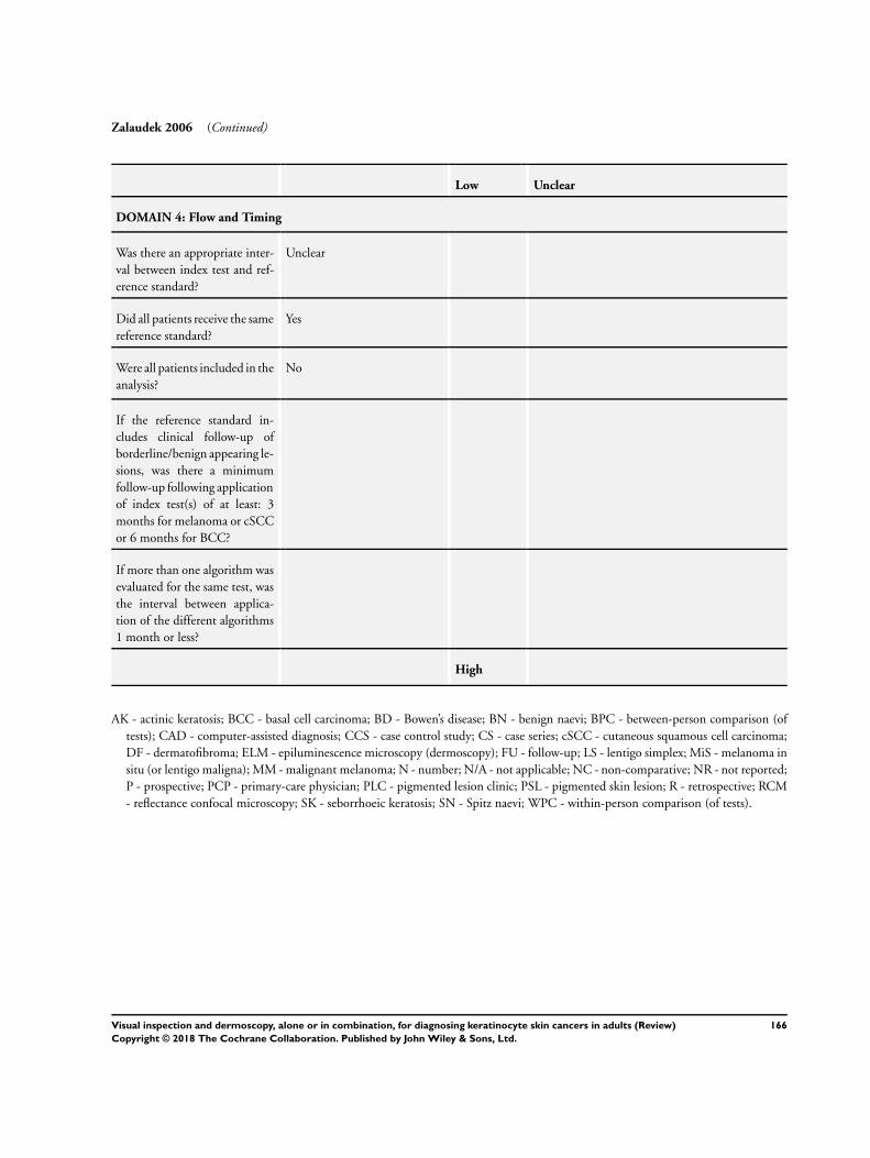
Zalaudek 2006 (Continued)
Low Unclear
DOMAIN 4: Flow and Timing
Was there an appropriate inter-
val between index test and ref-
erence standard?
Unclear
Did all patients receive the same
reference standard?
Yes
Were all patients included in the
analysis?
No
If the reference standard in-
cludes clinical follow-up of
borderline/benign appearing le-
sions, was there a minimum
follow-up following application
of index test(s) of at least: 3
months for melanoma or cSCC
or 6 months for BCC?
If more than one algorithm was
evaluated for the same test, was
the interval between applica-
tion of the different algorithms
1 month or less?
High
AK - actinic keratosis; BCC - basal cell carcinoma; BD - Bowen’s disease; BN - benign naevi; BPC - between-person comparison (of
tests); CAD - computer-assisted diagnosis; CCS - case control study; CS - case series; cSCC - cutaneous squamous cell carcinoma;
DF - dermatofibroma; ELM - epiluminescence microscopy (dermoscopy); FU - follow-up; LS - lentigo simplex; MiS - melanoma in
situ (or lentigo maligna); MM - malignant melanoma; N - number; N/A - not applicable; NC - non-comparative; NR - not reported;
P - prospective; PCP - primary-care physician; PLC - pigmented lesion clinic; PSL - pigmented skin lesion; R - retrospective; RCM
- reflectance confocal microscopy; SK - seborrhoeic keratosis; SN - Spitz naevi; WPC - within-person comparison (of tests).
166Visual inspection and dermoscopy, alone or in combination, for diagnosing keratinocyte skin cancers in adults (Review)
Copyright © 2018 The Cochrane Collaboration. Published by John Wiley & Sons, Ltd.
Characteristics of excluded studies [ordered by study ID]
Study Reason for exclusion
Abbasi 2004 Not a primary study
Systematic review
Ahnlide 2013 Ineligible index test
’clinical diagnosis’ study
Ahnlide 2016 Ineligible target condition; does not present data for detection of BCC or cSCC
Akasu 1996 Insufficient data for 2 x 2 table
No 2 x 2 data only describing the dermoscopic features present in the lesions
Al Jalbout 2013 Inadequate sample size
Case study
Alarcon 2014 Ineligible target condition; does not present data for detection of BCC or cSCC
Aldridge 2011a Ineligible test observer
Medical students and lay persons
Aldridge 2011b Ineligible test observer
Aldridge 2013 Insufficient data for 2 x 2 table
Not test accuracy study
Alendar 2009 Ineligible reference standard
Only 7 reported verified histologically
Altamura 2006 Assesses individual lesion characteristics only
Insufficient data for 2 x 2 table
Looking for characteristics associated with acral melanoma; does not give 2 x 2 for overall diagnosis
Annessi 2007 Ineligible target condition; does not report data for BCC or cSCC
Antonio 2013 Ineligible target condition
Atypical naevi does not fall within our definition of D+
Antoszewski 2015 Inadequate sample size
All excised lesions were benign.
Insufficient data for 2 x 2 table
Aoyagi 2010 Inadequate sample size
Arevalo 2008 Ineligible target condition; does not present data for detection of BCC or cSCC
167Visual inspection and dermoscopy, alone or in combination, for diagnosing keratinocyte skin cancers in adults (Review)
Copyright © 2018 The Cochrane Collaboration. Published by John Wiley & Sons, Ltd.

(Continued)
Argenziano 1997 Wrong study population
Only melanoma included
Argenziano 1998 Ineligible target condition; does not present data for detection of BCC or cSCC
Argenziano 1999 Wrong study population
Only includes melanoma
Argenziano 2002 Not a primary study
Argenziano 2003 Insufficient data for 2 x 2 table
Table V gives se/sp data for 108 lesions but cannot derive the number of melanoma for this subset of
the original 128
Contact authors; contacted 10 May 2016 and 24 June 2016
Argenziano 2004a Assesses individual lesion characteristics only
Only lesions with vascular structures included; presence of 10 different characteristics assessed. 2 x 2
would be possible
Argenziano 2004b Not a primary study
Letter
Argenziano 2008 Ineligible index test
Surveillance/monitoring study
Argenziano 2010 Ineligible index test
Test used for follow-up looking at dermoscopic features of melanomas diagnosed 1 yr after follow-up
Insufficient data for 2 x 2 table
Argenziano 2011 Ineligible target condition
Inadequate sample size
Only 2 melanomas
Argenziano 2011a Ineligible target condition
5 melanoma metastases included as D+
Argenziano 2011b Ineligible target condition; does not present data for detection of BCC or cSCC
Argenziano 2012 Ineligible reference standard
no follow-up of test negatives
Argenziano 2014 Insufficient data for 2 x 2 table
Armstrong 2011 Ineligible reference standard
No reference standard results presented for the screened lesions; just compares naked eye judgements
with dermoscopy
168Visual inspection and dermoscopy, alone or in combination, for diagnosing keratinocyte skin cancers in adults (Review)
Copyright © 2018 The Cochrane Collaboration. Published by John Wiley & Sons, Ltd.
(Continued)
Ascierto 1998 Insufficient data for 2 x 2 table
The data presented do not contribute to the review
Duplicate or related publication. Data included in Ascierto 2003
Ascierto 2000 Insufficient data for 2 x 2 table
Contact authors
For excised lesions, study cross-tabulates ELM high/very high-risk classification against some histological
classification (Table 2). Number D+ = 580 (2 x 2: 504, 79, 76, 2072); 580 not mentioned anywhere
else in paper (contacted 10 May 2016 and 24 June 2016)
Ascierto 2003 Not a primary study
Ascierto 2010 Ineligible target condition; does not present data for detection of BCC or cSCC
Badertscher 2015 Insufficient data for 2 x 2 table
Bafounta 2001 Not a primary study
Systematic review
Bajaj 2016 Ineligible reference standard
Unclear ref standard for benign diagnoses
Banky 2005 Ineligible target condition
Ineligible index test
Barzegari 2005 Ineligible target condition; does not present data for detection of BCC or cSCC
Basarab 1996 Wrong study population
Not all suspected of skin cancer
Insufficient data for 2 x 2 table
Bauer 2000 Ineligible index test
Does not provide 2 x 2 data for visual inspection alone
Bauer 2005 Ineligible index test
Follow-up/monitoring study
Bauer 2006 Ineligible index test
Dermoscopy used to improve histopathology diagnosis
Becker 1954 Not a primary study
Benati 2015 Assesses individual lesion characteristics only
Benelli 1999 Ineligible target condition; does not present data for detection of BCC or cSCC
Benelli 2000a Ineligible target condition; does not present data for detection of BCC or cSCC
169Visual inspection and dermoscopy, alone or in combination, for diagnosing keratinocyte skin cancers in adults (Review)
Copyright © 2018 The Cochrane Collaboration. Published by John Wiley & Sons, Ltd.
(Continued)
Benelli 2000b Insufficient data for 2 x 2 table
Only inter-rater reliability data given (n = 25); authors have published much larger evaluations of 7FFM
and ABCD
Benelli 2001 Ineligible target condition; does not present data for detection of BCC or cSCC
Benvenuto-Andrade 2006 Insufficient data for 2 x 2 table
Diagnostic confidence rather than accuracy
Benvenuto-Andrade 2007 Insufficient data for 2 x 2 table
Agreement on lesion characterisation; not test accuracy
Binder 1994 Ineligible target condition; does not present data for detection of BCC or cSCC
Binder 1995 Ineligible target condition; does not present data for detection of BCC or cSCC
Binder 1997 Insufficient data for 2 x 2 table
Training study; only ROC curves/AUC presented pre- and post-training
Contact authors (contacted 10 May 2016 and 24 June 2016)
Binder 1999 Ineligible target condition; does not present data for detection of BCC or cSCC
Blum 2003a Not a primary study
Blum 2003b Ineligible target condition; does not present data for detection of BCC or cSCC
Blum 2003c Ineligible target condition; does not present data for detection of BCC or cSCC
Blum 2004a Ineligible target condition; does not present data for detection of BCC or cSCC
Blum 2004b Not a primary study
Comment paper
Blum 2004c Not a primary study
Letter
Letter only; limited data presented - evaluates ’3-colour’ rule as developed By MacKie 2002 (excluded
as assessment of individual lesion features only)
Blum 2004d Ineligible target condition; does not present data for detection of BCC or cSCC
Blum 2004e Not a primary study
Letter
Blum 2006 Ineligible target condition
Differentiates melanocytic from non-melanocytic lesions only
170Visual inspection and dermoscopy, alone or in combination, for diagnosing keratinocyte skin cancers in adults (Review)
Copyright © 2018 The Cochrane Collaboration. Published by John Wiley & Sons, Ltd.
(Continued)
Blum 2011 Wrong study population
Mucosal lesions only
Blum 2014 Inadequate sample size
case studies
Boespflug 2015 Wrong study population
Study aim is estimate the efficacy of an online spaced educational training for dermoscopy
Bolognia 1990 Ineligible reference standard
No ref standard diagnosis for index test negatives
Bono 1996 Ineligible target condition; does not present data for detection of BCC or cSCC
Bono 2001 Insufficient data for 2 x 2 table
Aim of the study is to determine what features are present in amelanotic cutaneous melanoma
Bono 2002a Ineligible target condition; does not present data for detection of BCC or cSCC
Bono 2002b Ineligible target condition; does not present data for detection of BCC or cSCC
Bono 2006 Ineligible target condition; does not present data for detection of BCC or cSCC
Borsari 2010 Assesses individual lesion characteristics only
Contact authors
Paper focuses on diagnostic prediction of dermoscopic island for early melanoma, however the Methods
describe the calculation of the total dermoscopy score and the 7-point checklist score; mean scores on
each checklist per lesion type are then presented (no reply from authors)
Borsari 2015 Assesses individual lesion characteristics only
Borve 2012 Wrong study population
Includes participants without skin lesions
Inadequate sample size
< 5 BCC
Bourne 2012 Ineligible target condition; does not present data for detection of BCC or cSCC
Bowns 2006 Ineligible index test; teledermatology study
Braun 2000 Derivation study
This is a pilot study on the new “wobble sign” in ELM no training/test sets used
Braun 2007 Assesses individual lesion characteristics only
Braun-Falco 1990 Insufficient data for 2 x 2 table
Not a test accuracy study
171Visual inspection and dermoscopy, alone or in combination, for diagnosing keratinocyte skin cancers in adults (Review)
Copyright © 2018 The Cochrane Collaboration. Published by John Wiley & Sons, Ltd.
(Continued)
Broganelli 2005 Ineligible target condition; does not present data for detection of BCC or cSCC
Brown 2000 Not a primary study
Systematic review
Brown 2009 Ineligible test observer
lay persons
Buhl 2012 Ineligible index test
Follow-up/monitoring
Duplicate or related publication.
Same participants as Haenssle 2010a #191
Burki 2015 Not a primary study
Burr 2015 Not a primary study
Burton 1998 Ineligible reference standard
Can only get 2 x 2 data for referral accuracy
Insufficient data for 2 x 2 table
Bystryn 2003 Not a primary study
Letter
Cabrijan 2008 Insufficient data for 2 x 2 table
Cannot get 2 x 2; reports % correct diagnoses for each different lesion classification and not % misdi-
agnosed as melanoma or melanomas missed
Contact authors
Study states “Dermatoscopic diagnosis were conformable with pathohistological diagnosis in 75 cases
(72.82%) out of 103. The highest conformation was in diagnosing melanoma, in 5 out of 6 cases (83.
3%).” which would give us sensitivity; do you have data on numbers mis classified as melanoma, i.e false
positives? (author replied 5 July 2016 with some data but not sufficient to allow 2 x 2)
Canpolat 2011 Derivation study
Looks at dermoscopic characteristics of acral lesions; only 4 suspicious lesions excised
Cardenas 2009 Wrong study population
Includes participants with palpable lesions; not all suspected of having skin cancer
Carli 1994 Ineligible target condition; does not present data for detection of BCC or cSCC
Carli 1998 Inadequate sample size
se/sp data are based on sample with only 4 MM
Carli 2000 Ineligible target condition
Only lesions histologically classified as common naevi or naevi with architectural disorder with/without
cytological atypia were considered for the study
172Visual inspection and dermoscopy, alone or in combination, for diagnosing keratinocyte skin cancers in adults (Review)
Copyright © 2018 The Cochrane Collaboration. Published by John Wiley & Sons, Ltd.
(Continued)
Carli 2003a Ineligible reference standard
Only 39/1042 with ref test
Carli 2003b Inadequate sample size
Carli 2003c Ineligible target condition; does not present data for detection of BCC or cSCC
Carli 2003d Ineligible target condition; does not present data for detection of BCC or cSCC
Carli 2004a Inadequate sample size
< 5 MM per arm
Insufficient data for 2 x 2 table
Carli 2004b Ineligible index test; can only estimate 2 x 2 for the full time period 1997 to 2001 across all observers, but
dermoscopy was only introduced routinely in 1998, so some diagnoses prior to that will have been with
visual inspection alone, and observers were classed as dermoscopy ’users’ (those working in pigmented
lesion clinics) and nonusers (general dermatology)
Contact authors
Author passed away; unable to make contact with co-authors
Carli 2004c Ineligible index test
’Clinical diagnosis’ - Dataset covers 1997-2001, but dermoscopy routinely introduced 1998; authors
contacted but no response
Carli 2005 Insufficient data for 2 x 2 table
Contact authors
Study presents % MM correctly classified by naked eye ± dermoscopy but does not give any detail on
FPs, is this available anywhere and/or are these lesions included in any subsequent publications? Author
passed away; unable to make contact with co-authors
Carlos-Ortega 2007 Insufficient data for 2 x 2 table
Gives se/sp for visual inspection and dermoscopy in the English abstract. 68 participants/70 lesions were
included but only 36 seem to have had visual inspection results and all underwent dermoscopy. Two
observers performed each test blinded to each other. Table I gives 22 with BCC and 11 with melanoma
overall (N D+ not reported for those with VI results), but using either or both of these numbers with
the se/sp provided does not give the same PPV and NPV as given by the authors
Contact authors
Data not clearly presented for 2 x 2; translator suggested alternative but still does not work out to what
is in paper; tried contacting authors twice, no reply as of 28 July 2016
Carrera 2016 Ineligible target condition; does not present data for detection of BCC or cSCC
Carroll 1998 Derivation study
Derivation study; proposes new dermoscopic criteria for dx of BCC
Insufficient data for 2 x 2 table
Chen 2001 Not a primary study
Systematic review comparing PCP accuracy with dermatologist accuracy
173Visual inspection and dermoscopy, alone or in combination, for diagnosing keratinocyte skin cancers in adults (Review)
Copyright © 2018 The Cochrane Collaboration. Published by John Wiley & Sons, Ltd.
(Continued)
Chen 2006 Insufficient data for 2 x 2 table
Only given AUC
Chen 2013 Ineligible test observer
Chiaravalloti 2014 Wrong study population
Includes melanoma only
Ciudad-Blanco 2014 Wrong study population
Includes melanoma only
Assesses individual lesion characteristics only
Insufficient data for 2 x 2 table
Collas 1999 Ineligible target condition; does not present data for detection of BCC or cSCC
Coras 2003 Ineligible target condition; does not present data for detection of BCC or cSCC
Cornell 2015 Ineligible test observer
Cox 2008 Ineligible reference standard
Se and sp estimates for diagnosis of melanoma for both the seven-point checklist and the revised (10-
point) checklist; reference standard not reported for any of the 381 TWR referrals for melanoma
Contact authors
Author contacted 10 May 2016; co-author contacted 24 June 2016
Cristofolini 1994 Ineligible target condition; does not present data for detection of BCC or cSCC
Cristofolini 1997 Ineligible target condition; does not present data for detection of BCC or cSCC
Dal Pozzo 1999 Ineligible target condition; does not present data for detection of BCC or cSCC
De Giorgi 2006 Inadequate sample size
< 5 cases of participants with a final melanoma diagnosis
De Giorgi 2011 Duplicate or related publication.
Assesses same lesions as in Carli 2003c but different observers
De Giorgi 2012 Ineligible target condition; does not present data for detection of BCC or cSCC
De Troya-Martin 2008 Wrong study population
Only MM included
DeCoste 1993 Insufficient data for 2 x 2 table
Not given the total number of D+/D- or total number of lesions included. Just given the sens/spec values
Delfino 1997 Assesses individual lesion characteristics only
Derivation study
Insufficient data for 2 x 2 table
174Visual inspection and dermoscopy, alone or in combination, for diagnosing keratinocyte skin cancers in adults (Review)
Copyright © 2018 The Cochrane Collaboration. Published by John Wiley & Sons, Ltd.
(Continued)
Only reports association of each characteristics with D+/D-, not 2 x 2
Di Carlo 2014 Ineligible index test. Videothermography not relevant for the review and there are no 2x2 data for
dermoscopy
Derivation study. Only includes AK and BCC; no 2x2 for dermoscopy
Di Chiacchio 2010 Ineligible target condition
Excluding nail bed melanoma
Insufficient data for 2 x 2 table
There are insufficient data to extract for a 2 x 2 table
Di Meo 2016 Ineligible target condition; does not present data for detection of BCC or cSCC
Di Stefani 2007 Inadequate sample size
< 5 malignant
Dolianitis 2005 Ineligible target condition; does not present data for detection of BCC or cSCC
Dreiseitl 2009 Ineligible target condition; does not present data for detection of BCC or cSCC
Duff 2001 Ineligible index test
Does not evaluate visual inspection alone
Dummer 1993 Ineligible target condition; does not present data for detection of BCC or cSCC
Dummer 1995 Assesses individual lesion characteristics only
Edmondson 1999 Ineligible reference standard
It seems that the reference standard here is expert diagnosis. This is not a teledermatology paper
Elwan 2016 Inadequate sample size
Derivation study
Insufficient data for 2 x 2 table
Emmons 2011 Insufficient data for 2 x 2 table
Not test accuracy study; promoting primary prevention
Engelberg 1999 Inadequate sample size
Only 1 confirmed melanoma and 3 BCC
English 2003 Insufficient data for 2 x 2 table
No accuracy data given
English 2004 Insufficient data for 2 x 2 table
No accuracy data
175Visual inspection and dermoscopy, alone or in combination, for diagnosing keratinocyte skin cancers in adults (Review)
Copyright © 2018 The Cochrane Collaboration. Published by John Wiley & Sons, Ltd.

(Continued)
Fabbrocini 2008 Insufficient data for 2 x 2 table
There is insufficient data provided for each index test to populate 2 x 2 table
Contact authors
As we can only include DTA studies - Do you have a cross tabulation of each clinician’s diagnosis (e.g. at
threshold of 3 or more on 7-point checklist) against the histological diagnosis and/or a cross-tabulation
of the remote diagnosis against the face-to-face diagnoses? (author reply; 30 June 2016 cannot access
data needed)
Feci 2015 Ineligible target condition; does not present data for detection of BCC or cSCC
Federman 1995 Insufficient data for 2 x 2 table
Not test accuracy
Feldmann 1998 Ineligible target condition; does not present data for detection of BCC or cSCC
Ferrara 2002 Ineligible index test
This study looks at histopathological and dermoscopic disagreements not necessarily looking at how
well dermoscopy differentiates between benign and malignant diagnosis
Ferrari 2015 Ineligible target condition; does not present data for detection of BCC or cSCC
Ferris 2015 Ineligible target condition; does not present data for detection of BCC or cSCC
Fidalgo 2003 Insufficient data for 2 x 2 table
Duplicate or related publication.
Appears to be superseded by Serrao 2006
Contact authors
Paper provides % of MM and of DN with DNAOS scores of >=5.5 and >7, is it possible for you to
provide the same information for the remaining 127 lesions in the study? Also can you advise as to
whether any of the 247 lesions included in this study, overlap with the 652 reported in Serrao 2006 (#
1144)? (author contacted 10 May 2016; 24 June 2016)
Fikrle 2013 Ineligible reference standard
Follow-up study < 50% of study participants have their final diagnosis reached by histopathology
Freeman 1963 Insufficient data for 2 x 2 table
Only gives % correct for each lesion type
Contact authors
Tables 2 and 3 appear to give % correct diagnoses per lesion type, but do not give data on numbers
misclassified as melanoma, or other malignancy, i.e. FPs. Author responded; paper too old, cannot
provide data
Friedman 1985 Not a primary study
Friedman 2008 Ineligible target condition; does not present data for detection of BCC or cSCC
Fruhauf 2012 Ineligible reference standard
35/219 underwent histology; 13 followed up; 171 expert clinical Dx
176Visual inspection and dermoscopy, alone or in combination, for diagnosing keratinocyte skin cancers in adults (Review)
Copyright © 2018 The Cochrane Collaboration. Published by John Wiley & Sons, Ltd.

(Continued)
Fueyo-Casado 2009 Ineligible reference standard
< 50% of the study population received histology as a test. No information given on those who were
followed up
Funt 1963 Ineligible index test
Insufficient data for 2 x 2 table
No 2 x 2 data
Gachon 2005 Ineligible target condition; does not present data for detection of BCC or cSCC
Gerbert 1996 Ineligible target condition
No breakdown of final diagnoses for included lesions
Insufficient data for 2 x 2 table
Only gives % correct for each lesion type; not sens/spec
Gerbert 1998 Insufficient data for 2 x 2 table
Gereli 2010 Ineligible target condition; does not present data for detection of BCC or cSCC
Giacomel 2005 Wrong study population
Only BCC included
Giacomel 2014 Inadequate sample size
Giannotti 2004 Not a primary study
A review
Gill 2015 Inadequate sample size
Derivation study
Gilmore 2009 Derivation study
Principle of lacunarity has been looked at before but not this particular application/approach to it
Ineligible reference standard
It is possible to get 2 x 2 for ’standard dermoscopy criteria’ but dermoscopy-negative were not excised
and assumed benign; 201/312 underwent excision so theoretically eligible
Gilmore 2010 Ineligible target condition; does not present data for detection of BCC or cSCC
Glud 2009 Ineligible target condition; does not present data for detection of BCC or cSCC
Grana 2003 Ineligible index test
Assesses individual lesion characteristics only
Only looking at lesion border
Green 1991 Ineligible target condition; does not present data for detection of BCC or cSCC
Green 1994 Ineligible target condition; does not present data for detection of BCC or cSCC
177Visual inspection and dermoscopy, alone or in combination, for diagnosing keratinocyte skin cancers in adults (Review)
Copyright © 2018 The Cochrane Collaboration. Published by John Wiley & Sons, Ltd.
(Continued)
Grichnik 2003 Inadequate sample size
Grichnik 2004 Not a primary study
Editorial
Grimaldi 2009 Ineligible target condition; does not present data for detection of BCC or cSCC
Grob 1998 Not a primary study
Guibert 2000 Ineligible reference standard
Not designed as an accuracy study, only observational. Cannot get 2 x 2 data > 50% of study participants
did not receive histology as reference standard
Guillod 1996 Derivation study
Gunduz 2003 Inadequate sample size
Case study
Gutierrez 2013 Ineligible index test
Test to improve histopathology diagnosis
Haenssle 2006 Ineligible index test
Surveillance study estimating accuracy of different approaches to follow-up
Haenssle 2010a Ineligible target condition; does not present data for detection of BCC or cSCC
Haenssle 2010b Insufficient data for 2 x 2 table
Does not report specificity
Duplicate or related publication.
Same participants as Haenssle 2010a #191
Hallock 1998 Ineligible index test
’clinical diagnosis’; dermoscopy used for 3 of 4 years
Haniffa 2007 Ineligible reference standard
Looks like approximately 20% of participants received a final diagnosis by histology. 179 biopsies were
performed. Total sample was 881 lesions
Har-Shai 2001 Ineligible index test
’clinical diagnosis’
Haspeslagh 2016 Assesses individual lesion characteristics only
Insufficient data for 2 x 2 table
Hauschild 2014 Ineligible target condition; does not present data for detection of BCC or cSCC
178Visual inspection and dermoscopy, alone or in combination, for diagnosing keratinocyte skin cancers in adults (Review)
Copyright © 2018 The Cochrane Collaboration. Published by John Wiley & Sons, Ltd.
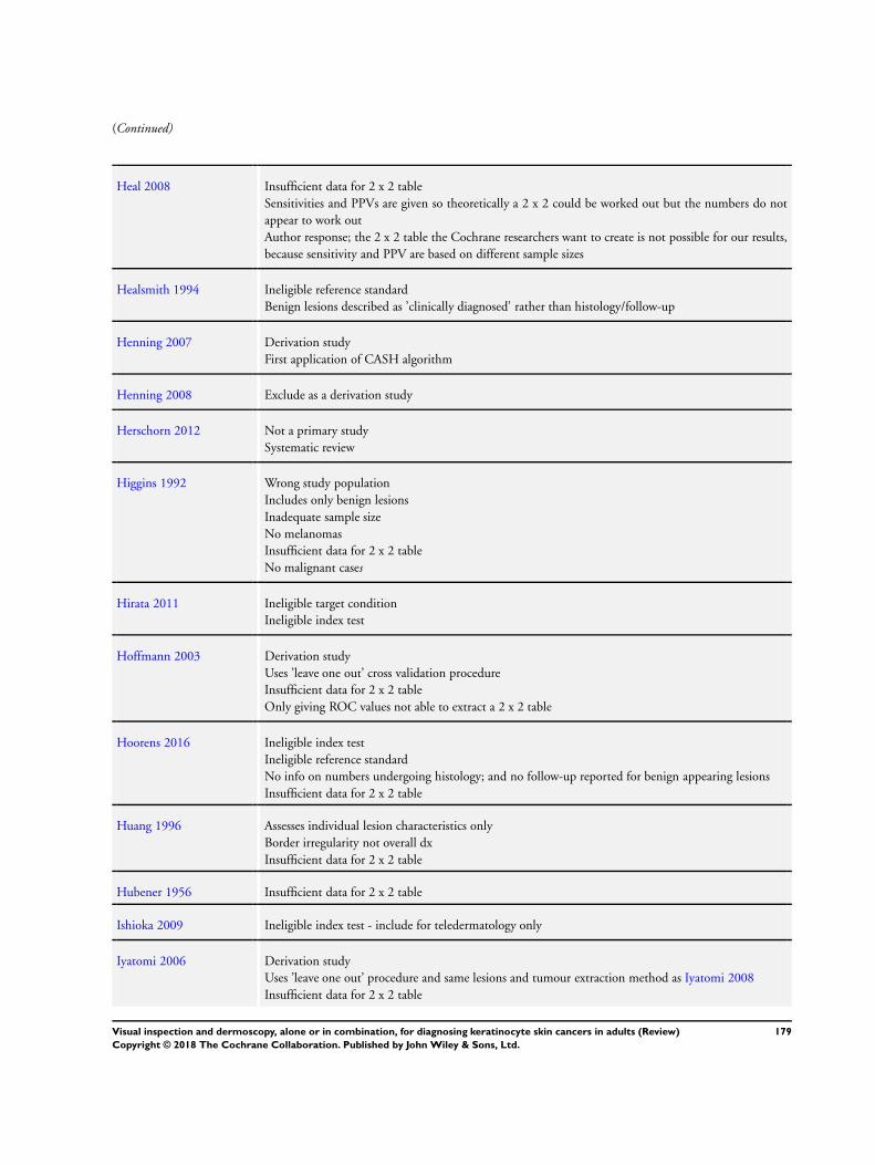
(Continued)
Heal 2008 Insufficient data for 2 x 2 table
Sensitivities and PPVs are given so theoretically a 2 x 2 could be worked out but the numbers do not
appear to work out
Author response; the 2 x 2 table the Cochrane researchers want to create is not possible for our results,
because sensitivity and PPV are based on different sample sizes
Healsmith 1994 Ineligible reference standard
Benign lesions described as ’clinically diagnosed’ rather than histology/follow-up
Henning 2007 Derivation study
First application of CASH algorithm
Henning 2008 Exclude as a derivation study
Herschorn 2012 Not a primary study
Systematic review
Higgins 1992 Wrong study population
Includes only benign lesions
Inadequate sample size
No melanomas
Insufficient data for 2 x 2 table
No malignant cases
Hirata 2011 Ineligible target condition
Ineligible index test
Hoffmann 2003 Derivation study
Uses ’leave one out’ cross validation procedure
Insufficient data for 2 x 2 table
Only giving ROC values not able to extract a 2 x 2 table
Hoorens 2016 Ineligible index test
Ineligible reference standard
No info on numbers undergoing histology; and no follow-up reported for benign appearing lesions
Insufficient data for 2 x 2 table
Huang 1996 Assesses individual lesion characteristics only
Border irregularity not overall dx
Insufficient data for 2 x 2 table
Hubener 1956 Insufficient data for 2 x 2 table
Ishioka 2009 Ineligible index test - include for teledermatology only
Iyatomi 2006 Derivation study
Uses ’leave one out’ procedure and same lesions and tumour extraction method as Iyatomi 2008
Insufficient data for 2 x 2 table
179Visual inspection and dermoscopy, alone or in combination, for diagnosing keratinocyte skin cancers in adults (Review)
Copyright © 2018 The Cochrane Collaboration. Published by John Wiley & Sons, Ltd.
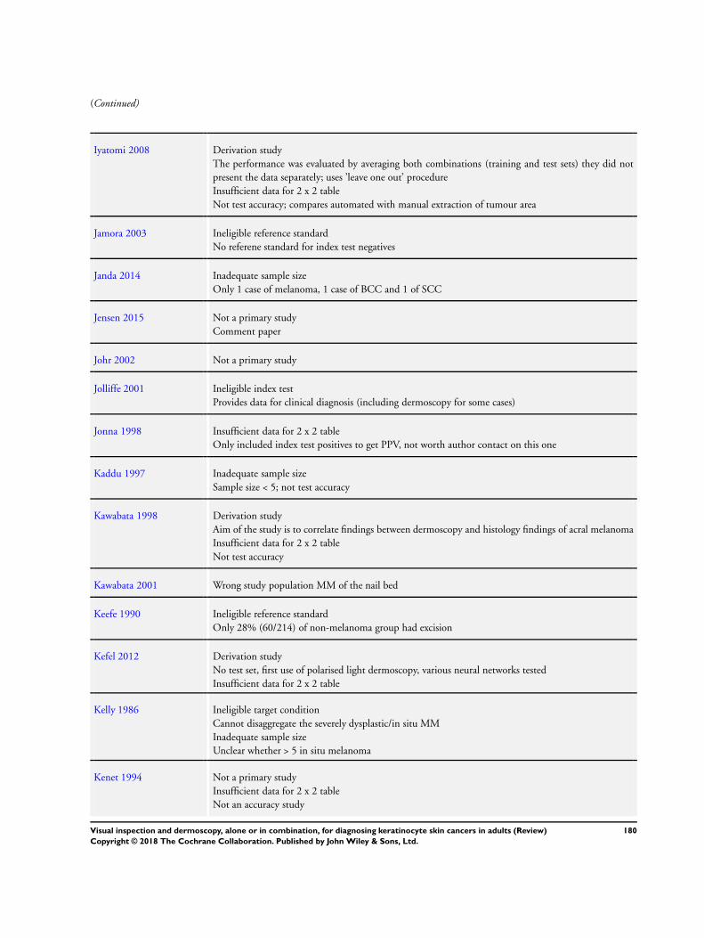
(Continued)
Iyatomi 2008 Derivation study
The performance was evaluated by averaging both combinations (training and test sets) they did not
present the data separately; uses ’leave one out’ procedure
Insufficient data for 2 x 2 table
Not test accuracy; compares automated with manual extraction of tumour area
Jamora 2003 Ineligible reference standard
No referene standard for index test negatives
Janda 2014 Inadequate sample size
Only 1 case of melanoma, 1 case of BCC and 1 of SCC
Jensen 2015 Not a primary study
Comment paper
Johr 2002 Not a primary study
Jolliffe 2001 Ineligible index test
Provides data for clinical diagnosis (including dermoscopy for some cases)
Jonna 1998 Insufficient data for 2 x 2 table
Only included index test positives to get PPV, not worth author contact on this one
Kaddu 1997 Inadequate sample size
Sample size < 5; not test accuracy
Kawabata 1998 Derivation study
Aim of the study is to correlate findings between dermoscopy and histology findings of acral melanoma
Insufficient data for 2 x 2 table
Not test accuracy
Kawabata 2001 Wrong study population MM of the nail bed
Keefe 1990 Ineligible reference standard
Only 28% (60/214) of non-melanoma group had excision
Kefel 2012 Derivation study
No test set, first use of polarised light dermoscopy, various neural networks tested
Insufficient data for 2 x 2 table
Kelly 1986 Ineligible target condition
Cannot disaggregate the severely dysplastic/in situ MM
Inadequate sample size
Unclear whether > 5 in situ melanoma
Kenet 1994 Not a primary study
Insufficient data for 2 x 2 table
Not an accuracy study
180Visual inspection and dermoscopy, alone or in combination, for diagnosing keratinocyte skin cancers in adults (Review)
Copyright © 2018 The Cochrane Collaboration. Published by John Wiley & Sons, Ltd.
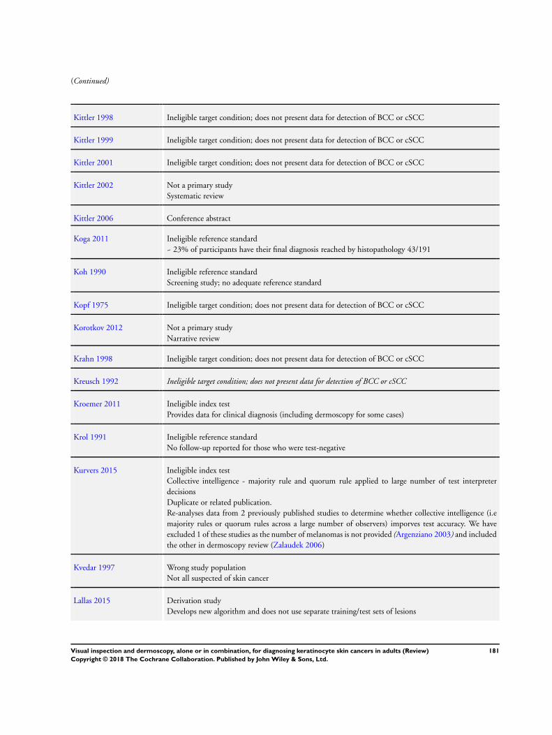
(Continued)
Kittler 1998 Ineligible target condition; does not present data for detection of BCC or cSCC
Kittler 1999 Ineligible target condition; does not present data for detection of BCC or cSCC
Kittler 2001 Ineligible target condition; does not present data for detection of BCC or cSCC
Kittler 2002 Not a primary study
Systematic review
Kittler 2006 Conference abstract
Koga 2011 Ineligible reference standard
~ 23% of participants have their final diagnosis reached by histopathology 43/191
Koh 1990 Ineligible reference standard
Screening study; no adequate reference standard
Kopf 1975 Ineligible target condition; does not present data for detection of BCC or cSCC
Korotkov 2012 Not a primary study
Narrative review
Krahn 1998 Ineligible target condition; does not present data for detection of BCC or cSCC
Kreusch 1992 Ineligible target condition; does not present data for detection of BCC or cSCC
Kroemer 2011 Ineligible index test
Provides data for clinical diagnosis (including dermoscopy for some cases)
Krol 1991 Ineligible reference standard
No follow-up reported for those who were test-negative
Kurvers 2015 Ineligible index test
Collective intelligence - majority rule and quorum rule applied to large number of test interpreter
decisions
Duplicate or related publication.
Re-analyses data from 2 previously published studies to determine whether collective intelligence (i.e
majority rules or quorum rules across a large number of observers) imporves test accuracy. We have
excluded 1 of these studies as the number of melanomas is not provided (Argenziano 2003) and included
the other in dermoscopy review (Zalaudek 2006)
Kvedar 1997 Wrong study population
Not all suspected of skin cancer
Lallas 2015 Derivation study
Develops new algorithm and does not use separate training/test sets of lesions
181Visual inspection and dermoscopy, alone or in combination, for diagnosing keratinocyte skin cancers in adults (Review)
Copyright © 2018 The Cochrane Collaboration. Published by John Wiley & Sons, Ltd.
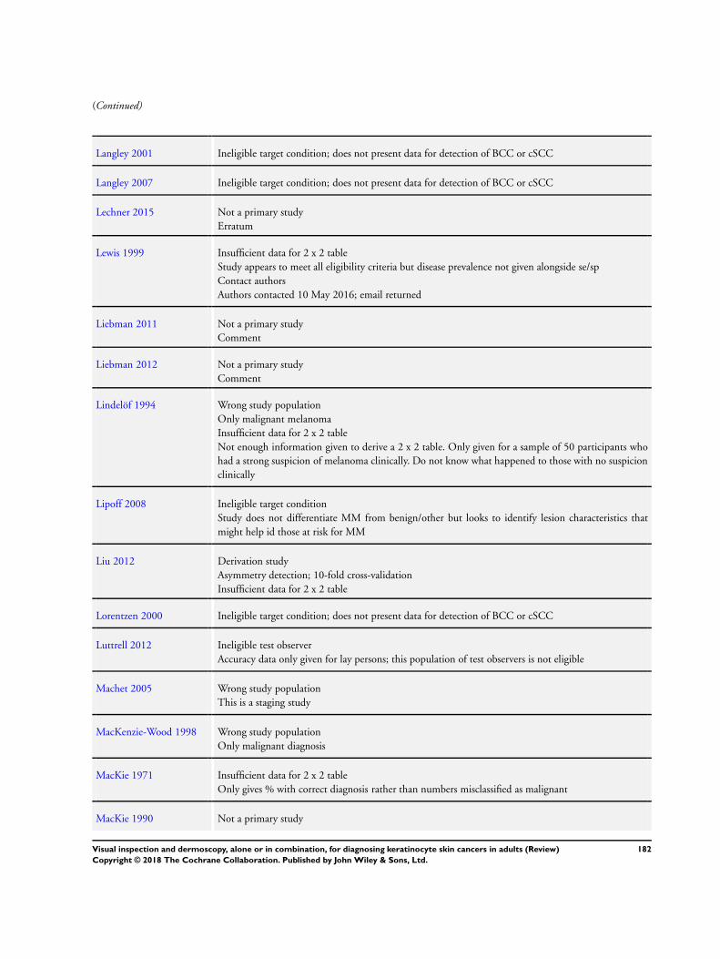
(Continued)
Langley 2001 Ineligible target condition; does not present data for detection of BCC or cSCC
Langley 2007 Ineligible target condition; does not present data for detection of BCC or cSCC
Lechner 2015 Not a primary study
Erratum
Lewis 1999 Insufficient data for 2 x 2 table
Study appears to meet all eligibility criteria but disease prevalence not given alongside se/sp
Contact authors
Authors contacted 10 May 2016; email returned
Liebman 2011 Not a primary study
Comment
Liebman 2012 Not a primary study
Comment
Lindelöf 1994 Wrong study population
Only malignant melanoma
Insufficient data for 2 x 2 table
Not enough information given to derive a 2 x 2 table. Only given for a sample of 50 participants who
had a strong suspicion of melanoma clinically. Do not know what happened to those with no suspicion
clinically
Lipoff 2008 Ineligible target condition
Study does not differentiate MM from benign/other but looks to identify lesion characteristics that
might help id those at risk for MM
Liu 2012 Derivation study
Asymmetry detection; 10-fold cross-validation
Insufficient data for 2 x 2 table
Lorentzen 2000 Ineligible target condition; does not present data for detection of BCC or cSCC
Luttrell 2012 Ineligible test observer
Accuracy data only given for lay persons; this population of test observers is not eligible
Machet 2005 Wrong study population
This is a staging study
MacKenzie-Wood 1998 Wrong study population
Only malignant diagnosis
MacKie 1971 Insufficient data for 2 x 2 table
Only gives % with correct diagnosis rather than numbers misclassified as malignant
MacKie 1990 Not a primary study
182Visual inspection and dermoscopy, alone or in combination, for diagnosing keratinocyte skin cancers in adults (Review)
Copyright © 2018 The Cochrane Collaboration. Published by John Wiley & Sons, Ltd.
(Continued)
MacKie 1991 Not a primary study
Letter
MacKie 2002 Assesses individual lesion characteristics only
Presence of 3 or more colours on dermoscopy
Mahendran 2005 Ineligible index test
Face-to-face is ’clinical diagnosis’, i.e. visual inspection ± use of dermoscopy
Mahon 1997 Not a primary study
A summary of a comparison of two screening checklists
Malvehy 2014 Ineligible target condition; does not present data for detection of BCC or cSCC
Marghoob 1995 Not a primary study
Letter
Marghoob 2007 Not a primary study
Marghoob 2010 Not a primary study
Massi 2001 Assesses individual lesion characteristics only
Mayer 1997 Not a primary study
Systematic review
McCarthy 1995 Not a primary study
Leaflet
McGovern 1992 Ineligible target condition; does not present data for detection of BCC or cSCC
Menzies 1996a Ineligible target condition; does not present data for detection of BCC or cSCC
Menzies 1996b Assesses individual lesion characteristics only
Only given the SE/SP of individual characteristics; lesions make up the training set for Menzies 1996a
(#1971)
Menzies 1999 Not a primary study
Menzies 2001 Ineligible index test
Monitoring purposes
Menzies 2005 Ineligible target condition; does not present data for detection of BCC or cSCC
Menzies 2008 Ineligible target condition; does not present data for detection of BCC or cSCC
Menzies 2009 Ineligible target condition; does not present data for detection of BCC or cSCC
183Visual inspection and dermoscopy, alone or in combination, for diagnosing keratinocyte skin cancers in adults (Review)
Copyright © 2018 The Cochrane Collaboration. Published by John Wiley & Sons, Ltd.
(Continued)
Menzies 2011 Ineligible index test
Surveillance study; data used to id factors predictive of lesion changes
Menzies 2013 Ineligible target condition; does not present data for detection of BCC or cSCC
Moffatt 2006 Ineligible index test
’clinical diagnosis’
Mohammad 2015 Wrong study population
Only includes BCC
Morales Callaghan 2008 Ineligible target condition; does not present data for detection of BCC or cSCC
Morrison 2001 Insufficient data for 2 x 2 table
Study gives % correct diagnosis within each histology group and then gives the % ‘correct’ diagnosis of
skin cancer as 22% for FP and 87% for dermatologist. But these statistics appear to have been reached by
taking the mean of the % correct diagnoses across the malignant groups and do not equate to sensitivity,
i.e. If you take the mean of the FP correct (%) for the 4 malignant groups you get: (40 + 22 + 25 + 0)
/4 = 21.75% and then the same for the ’dermatologist correct’ (%) column: (95 + 77 + 75 + 100)/4 =
86.75%
Morton 1998 Ineligible target condition; does not present data for detection of BCC or cSCC
Mun 2016 Ineligible reference standard
Only 37% of benign group underwent adequate reference standard
Nachbar 1994 Ineligible target condition; does not present data for detection of BCC or cSCC
Nathansohn 2007 Insufficient data for 2 x 2 table
Not test accuracy; follow-up study
Nilles 1994 Ineligible target condition; does not present data for detection of BCC or cSCC
Osborne 1998 Ineligible reference standard
Not clear what the ref standard is
Insufficient data for 2 x 2 table
Osborne 1999 Wrong study population
Only participants with melanoma included
Pagnanelli 2003 Ineligible target condition; does not present data for detection of BCC or cSCC
Pan 2008 Derivation study
Looking to id characteristics assoc with superficial BCC; 2 x 2 could be extracted for combination of 3
selected characteristics. Dermoscopic features selected based on prior studies but only participants with
3 diagnoses included: BCC, intra-ep carcinoma and psoriasis
184Visual inspection and dermoscopy, alone or in combination, for diagnosing keratinocyte skin cancers in adults (Review)
Copyright © 2018 The Cochrane Collaboration. Published by John Wiley & Sons, Ltd.
(Continued)
Panasiti 2009 Assesses individual lesion characteristics only
Ineligible reference standard
Of the 1543 lesions analysed on 321 received histopathology diagnosis. The accuracy data is based on
this (only 20%); unclear what happened to the 80% of participants as no mention of follow-up
Parslew 1997 Wrong study population
Not all suspected of skin cancer
Pazzini 1996 Insufficient data for 2 x 2 table
Pehamberger 1987 Insufficient data for 2 x 2 table
Not test accuracy. This is a descriptive paper defining dermoscopic criteria. It is not a study testing
accuracy of dermoscopy. From the authors final sign-off it looks like part 2 of this paper may have details
on accuracy(Steiner 1987).
Pellacani 2002 Not a primary study
Pellacani 2006 Derivation study
Looks at detection of asymmetry between clinicians and computer
Insufficient data for 2 x 2 table
2 x 2 could be derived for overall asymmetry or border cut-off but not overall diagnosis
Pellacani 2007 Assesses individual lesion characteristics only
Derivation study
Looking at blue hue
Pellacani 2009 Ineligible target condition
Focus is on identifying Spitz naevi from melanoma and ‘clark’ naevi and is looking to derive useful RCM
characteristics. Although some data are given in the text for an RCM score > 3 it is difficult to work out
which are FP and which FN
Perednia 1992 Insufficient data for 2 x 2 table
Not test accuracy
Peris 2002 Wrong study population
Only participants with BCC diagnosis included
Perrinaud 2007 Ineligible index test
Does not provide data for visual inspection alone
Phan 2010 Insufficient data for 2 x 2 table
Not test accuracy investigating dermoscopic features of acral melanoma including of the nail apparatus;
no accuracy data given
Piccolo 2000 Ineligible target condition; does not present data for detection of BCC or cSCC
185Visual inspection and dermoscopy, alone or in combination, for diagnosing keratinocyte skin cancers in adults (Review)
Copyright © 2018 The Cochrane Collaboration. Published by John Wiley & Sons, Ltd.
(Continued)
Piccolo 2002 Not a primary study
Insufficient data for 2 x 2 table
Not enough data to populate 2 x 2 table. No breakdown of index test results and ref standard
Piccolo 2002a Ineligible target condition; does not present data for detection of BCC or cSCC
Piccolo 2004 Ineligible index test; include for teledermatology anyway
Piccolo 2006 Inadequate sample size
3 MMs, but also 1 lentigo and 14 dysplastic nevus; data not presented to allow se/sp estimation
Assesses individual lesion characteristics only
Derivation study
Derivation for hypoluminescence microscopy
Piccolo 2014 Ineligible target condition; does not present data for detection of BCC or cSCC
Pizzichetta 2001a Wrong study population
Population in study only those with malignant disease
Pizzichetta 2001b Insufficient data for 2 x 2 table
Observer agreement only
Pizzichetta 2002 Ineligible target condition; does not present data for detection of BCC or cSCC
Pizzichetta 2004 Ineligible target condition; does not present data for detection of BCC or cSCC
Pizzichetta 2007 Wrong study population
Only participants with melanoma included
Pizzichetta 2010 Inadequate sample size
Case study
Pizzichetta 2013 Assesses individual lesion characteristics only
Presence of negative pigmented network
Pralong 2012 Wrong study population
Only melanoma participants included
Provost 1998 Insufficient data for 2 x 2 table
Not test accuracy; only reports concordance
Pupelli 2013 Ineligible target condition; does not present data for detection of BCC or cSCC
Quéreux 2011 Ineligible index test
Self-administered questions to patients attending a GP surgery before their appointment to determine
whether they are at high risk of melanoma, which is meant to highlight to the GP which patient to
examine during their consultation
186Visual inspection and dermoscopy, alone or in combination, for diagnosing keratinocyte skin cancers in adults (Review)
Copyright © 2018 The Cochrane Collaboration. Published by John Wiley & Sons, Ltd.
(Continued)
Rader 2014 Assesses individual lesion characteristics only
Insufficient data for 2 x 2 table
Rajpara 2009 Not a primary study
Systematic review
Rallan 2006 Ineligible index test
No data can be extracted for visual inspection alone
Rampen 1988 Wrong study population
Only melanoma included
Rao 1997 Ineligible target condition; does not present data for detection of BCC or cSCC
Reeck 1999 Wrong study population
Only includes index test negatives, i.e. those considered benign by referring clinician
Ineligible target condition
Reggiani 2015 Not a primary study
Systematic review of kerationcyte skin cancer
Riddell 1961 Wrong study population
All malignant
Rigel 1993 Not a primary study
Rigel 1997 Not a primary study
Rigel 2012 Ineligible target condition; does not present data for detection of BCC or cSCC
Robati 2014 Ineligible reference standard
No follow-up of patients not referred to dermatology clinics, who did not receive histopathology
Robinson 2010 Ineligible index test
Self-examination
Ronger 2002 Assesses individual lesion characteristics only
Rosado 2003 Not a primary study
Systematic review
Rosendahl 2012a Assesses individual lesion characteristics only
Rosendahl 2012b Not a primary study
Rossi 2000 Ineligible reference standard
Unclear reference standard in disease-negative
187Visual inspection and dermoscopy, alone or in combination, for diagnosing keratinocyte skin cancers in adults (Review)
Copyright © 2018 The Cochrane Collaboration. Published by John Wiley & Sons, Ltd.
(Continued)
Roush 1986 Ineligible target condition
Only dysplastic naevus
Rubegni 2002 Not a primary study
Rubegni 2005 Not a primary study
Editorial
Rubegni 2010 Derivation study
Uses ’leave one out’ procedure
Insufficient data for 2 x 2 table
Rubegni 2012 Ineligible target condition; does not present data for detection of BCC or cSCC
Rubegni 2016 Ineligible target condition; does not present data for detection of BCC or cSCC
Sahin 2004 Assesses individual lesion characteristics only
Insufficient data for 2 x 2 table
No accuracy data given, study looking at dermoscopic features of LM
Saida 2002 Assesses individual lesion characteristics only
Descriptive study looking at presence (%) of certain features. Not looking at accuracy. Has paragraph
on diagnostic value of this specific feature quoting sens & spec but this is based upon unpublished
observations and the data are not given in this paper
Saida 2004 Assesses individual lesion characteristics only
Sakakibara 2010 Assesses individual lesion characteristics only
Only looking at different vascular structures
Salerni 2011 Inadequate sample size
< 5 cases
Salerni 2012 Ineligible index test
Surveillance study
Insufficient data for 2 x 2 table
Salerni 2013 Not a primary study
Systematic review of surveillance with digital dermoscopy
Salvio 2011 Not a primary study
Inadequate sample size
Sanchez-Martin 2012 Wrong study population
Only BCC cases
Savk 2004 Not a primary study
Letter
188Visual inspection and dermoscopy, alone or in combination, for diagnosing keratinocyte skin cancers in adults (Review)
Copyright © 2018 The Cochrane Collaboration. Published by John Wiley & Sons, Ltd.
(Continued)
Sawada 2013 Not a primary study
Sboner 2003 Derivation study
Describes 10-fold cross-validation process for training/testing classifier
Sboner 2004 Ineligible target condition; does not present data for detection of BCC or cSCC
Schindewolf 1994 Ineligible index test
Evaluates CAD not VI
Schmoeckel 1987 Not a primary study
Schulz 2001 Ineligible target condition
Melanoma metastases
Scope 2008 Ineligible target condition; does not present data for detection of BCC or cSCC
Scope 2015 Not a primary study
Segura 2009 Ineligible index test; RCM evaluation
Seidenari 1998 Ineligible target condition; does not present data for detection of BCC or cSCC
Seidenari 2004 Insufficient data for 2 x 2 table
No data to populate 2 x 2 table, just ROC curve values given
Contact authors
TABLE 5 provides AUC values for each diagnosis for both formats and observers; we are particularly
interested in accuracy for the diagnosis of melanoma, are you able to provide data in 2 x 2 format, e.g.
for melanoma ’certain’ against final diagnosis and for melanoma ’certain or fairly certain’ against final
diagnosis? (no reply from authors)
Seidenari 2005 Ineligible target condition; does not present data for detection of BCC or cSCC
Seidenari 2006a Wrong study population
Assessing best means of follow-up in patients with previous melanoma - total body exam versus only
lesions > 2 cm. No melanoma identified
Seidenari 2006b Assesses individual lesion characteristics only
Looks like this study is only looking at asymmetry judgement
Seidenari 2007 Ineligible target condition; does not present data for detection of BCC or cSCC
Seidenari 2012 Assesses individual lesion characteristics only
Looks at individual lesion characteristics to distinguish melanoma in situ, also gives mean ABCD and
7-point scores
Insufficient data for 2 x 2 table
Contact authors
Table 3 provides mean ABCD and 7-point checklist scores, are you able to provide us with a cross-
189Visual inspection and dermoscopy, alone or in combination, for diagnosing keratinocyte skin cancers in adults (Review)
Copyright © 2018 The Cochrane Collaboration. Published by John Wiley & Sons, Ltd.
(Continued)
tabulation of results with each checklist at ’standard’ thresholds against final diagnosis? e.g. ABCD > 4.
75 and > 5.45 for MIS and benign groups 7-point checklist: presence of 2or more characteristics and 3
or more characteristics? (no reply)
Seidenari 2013 Ineligible index test
Serrao 2006 Ineligible index test; include for CAD review only
Sgouros 2014 Ineligible index test; include for CAD review only
Shakya 2012 Ineligible target condition
SCC in situ is not included in target condition
Shariff 2010 Ineligible reference standard
Shitara 2014 Assesses individual lesion characteristics only
Shitara 2015 Wrong study population
Includes only melanoma
Skvara 2005 Ineligible target condition; does not present data for detection of BCC or cSCC
Sondak 2015 Not a primary study
Comment paper
Soyer 1987 Insufficient data for 2 x 2 table
Not test accuracy
Soyer 1995 Ineligible target condition; does not present data for detection of BCC or cSCC
Soyer 2001 Not a primary study
Editorial
Soyer 2004 Ineligible target condition; does not present data for detection of BCC or cSCC
Stanganelli 1998a Ineligible target condition; does not present data for detection of BCC or cSCC
Stanganelli 1998b Insufficient data for 2 x 2 table
Cannot derive specificity; only gives ’exact diagnoses’ for MM and 2 benign categories and not number
benign misdiagnosed as MM
Stanganelli 1999 Ineligible target condition; does not present data for detection of BCC or cSCC
Stanganelli 2005 Ineligible target condition; does not present data for detection of BCC or cSCC
Stanganelli 2015 Ineligible target condition; does not present data for detection of BCC or cSCC
190Visual inspection and dermoscopy, alone or in combination, for diagnosing keratinocyte skin cancers in adults (Review)
Copyright © 2018 The Cochrane Collaboration. Published by John Wiley & Sons, Ltd.
(Continued)
Stanley 2003 Assesses individual lesion characteristics only
Fuzzy histogram is based on the lesion’s colour, which is an individual lesion characteristic
Stathopoulos 2015 Insufficient data for 2 x 2 table
Only includes index test-positive participants, i.e. no FN or TN results
Steiner 1993 Assesses individual lesion characteristics only
Derivation study
Stephens 2013 Inadequate sample size
Stoecker 2009 Derivation study
Translucency
Insufficient data for 2 x 2 table
Data presented only as ROC curve and AUC
Stoecker 2011 Assesses individual lesion characteristics only
Derivation study
Uses ’leave one out’ procedure
Insufficient data for 2 x 2 table
Data presented only as ROC curve and AUC
Stolz 1994 Ineligible target condition; does not present data for detection of BCC or cSCC
Stolz 2002 Not a primary study
Stratigos 2007 Ineligible reference standard
Insufficient data for 2 x 2 table
Stricklin 2011 Assesses individual lesion characteristics only
Strumia 2003 Conference abstract; letter only
Tan 2009 Ineligible target condition; does not present data for detection of BCC or cSCC
Tandjung 2015 Ineligible target condition
’Malignant’ includes: AK, Bowen’s, dysplastic naevus, lentigo maligna, SCC, BCC, MM, keratoacan-
thoma
Ineligible index test
GPs sent images for telederm opinion; then free to send for biopsy or not; results shown are only for
those that wer biopsied, according to TD advice
Tasli 2012 Not a primary study
Systematic review looking at frequency of publications ion dermoscopy
Teban 2003 Wrong study population
Classification of Clark naevi into 12 types
Insufficient data for 2 x 2 table
191Visual inspection and dermoscopy, alone or in combination, for diagnosing keratinocyte skin cancers in adults (Review)
Copyright © 2018 The Cochrane Collaboration. Published by John Wiley & Sons, Ltd.
(Continued)
No 2 x 2 data; classification of Clark naevi into 12 types
Tenenhaus 2010 Ineligible target condition; does not present data for detection of BCC or cSCC
Terrill 2009 Ineligible index test
Whole-body skin examination after participants referred on for further assessment by a specialist
Insufficient data for 2 x 2 table
Terstappen 2007 Wrong study population
Includes only BCC - looking for BCC characteristics on Siascope
Derivation study
Derivation study; first application of Siascope to pigmented BCC; 21/25 lesions were BCCs
Terushkin 2010a Inadequate sample size
Only 2 invasive SCCs
Insufficient data for 2 x 2 table
Terushkin 2010b Insufficient data for 2 x 2 table
Not test accuracy - reports final diagnoses of those excised over a number of time periods and benign-
malignant ratio
Thomas 1998 Ineligible target condition; does not present data for detection of BCC or cSCC
Thomson 2005 Not a primary study
Letter
Torrey 1941 Ineligible target condition
Includes non-cutaneous lesions
Tromme 2012 Ineligible reference standard
Inadequate reference test for disease-negatives; expert dx only
Troyanova 2003 Ineligible target condition; does not present data for detection of BCC or cSCC
Tschandl 2012 Ineligible index test
Differentiating melanocytic from non-melanocytic lesions
Tschandl 2015 Ineligible test observer
Medical students
Unlu 2014 Ineligible target condition; does not present data for detection of BCC or cSCC
Van der Leest 2011 Ineligible reference standard
Inadequate reference test for test-negatives; expert dx only
Van der Rhee 2010 Ineligible reference standard
< 50% of disease-negative have an adequate reference standard
192Visual inspection and dermoscopy, alone or in combination, for diagnosing keratinocyte skin cancers in adults (Review)
Copyright © 2018 The Cochrane Collaboration. Published by John Wiley & Sons, Ltd.
(Continued)
Van der Rhee 2011 Inadequate sample size
< 5 cases
Vasili 2010 Conference abstract
Verduzco-Martinez 2013 Wrong study population
Only BCC
Vestergaard 2008 Not a primary study
Systematic review; check reference list
Viglizzo 2004 Ineligible target condition; does not present data for detection of BCC or cSCC
Wagner 1985 Insufficient data for 2 x 2 table
Walter 2010 Not a primary study
Clinical trial protocol
Walter 2012 Ineligible target condition; does not present data for detection of BCC or cSCC
Walter 2013 Ineligible reference standard
Final diagnosis reached by histology or expert opinion; no follow-up of non-excised lesions reported
in this paper. Walter 2012 does report follow-up for enough benign lesions for control arm (weighted
7PCL) data to be included. Authors contacted and confirmed calculations (02 March 2016)
Wang 2008 Insufficient data for 2 x 2 table
Not test accuracy; no details of misdiagnoses of benign lesions as malignant
Warshaw 2009a Insufficient data for 2 x 2 table
Duplicate or related publication.
Subgroup of participants from Warshaw 2010a
Contact authors
Study presents diagnostic accuracy of teledermatology and clinic diagnosis in comparison to histopathol-
ogy; we need the underlying 2 x 2 contingency tables (see Warshaw 2010a for author response)
Warshaw 2009b Insufficient data for 2 x 2 table
Duplicate or related publication.
Subgroup of participants from Warshaw 2010a
Contact authors
Study presents diagnostic accuracy of teledermatology and clinic diagnosis in comparison to histopathol-
ogy; we need the underlying 2 x 2 contingency tables (see Warshaw 2010afor author response)]
Warshaw 2010a Insufficient data for 2 x 2 table
Contact authors
Study presents diagnostic accuracy of teledermatology and clinic diagnosis in comparison to histopathol-
ogy. Author only able to provide numbers test-positive and -negative for melanoma and not for the final
2 cells of the 2 x 2; data provided showed higher sensitivity for melanoma as the primary diagnosis rather
than as the ‘aggregate’ diagnosis and the 2 x 2 using the authors’ data and the accuracy figures from the
193Visual inspection and dermoscopy, alone or in combination, for diagnosing keratinocyte skin cancers in adults (Review)
Copyright © 2018 The Cochrane Collaboration. Published by John Wiley & Sons, Ltd.
(Continued)
paper showed more T+ from the primary diagnosis as opposed to the aggregate
Warshaw 2010b Insufficient data for 2 x 2 table
As per Warshaw 2009a; this 2010 paper presents combined data for pigmented and nonpigmented
lesions
Weismann 2002 Not a primary study
Wells 2012 Ineligible target condition; does not present data for detection of BCC or cSCC
Westbrook 2006 Insufficient data for 2 x 2 table
Westerhoff 2000 Ineligible target condition; does not present data for detection of BCC or cSCC
Whitaker-Worth 1998 Wrong study population
Ineligible test observer
Mixed medical student/clinicians
Insufficient data for 2 x 2 table
Not test accuracy study
Whited 1998 Inadequate sample size
Wilkes 2010 Not a primary study
Williams 1991 Insufficient data for 2 x 2 table
Winkelmann 2015a Duplicate or related publication.
Winkelmann 2015b Duplicate or related publication.
Winkelmann 2016 Ineligible target condition; does not present data for detection of BCC or cSCC
Wolf 1998 Ineligible index test
Clinical diagnosis study; test clearly described - “concerning the clinical diagnosis, we were not able
to ascertain from the clinical data sheet whether the referring physicians used additional diagnostics
techniques such as dermoscopy”
Yadav 1993 Insufficient data for 2 x 2 table
Not test accuracy
Yamaura 2005 Derivation study
Gene amplification in acral lesions
Yelamos 2016 Not a primary study. Commentary on Guitera 2016
Yoo 2015 Conference abstract
194Visual inspection and dermoscopy, alone or in combination, for diagnosing keratinocyte skin cancers in adults (Review)
Copyright © 2018 The Cochrane Collaboration. Published by John Wiley & Sons, Ltd.
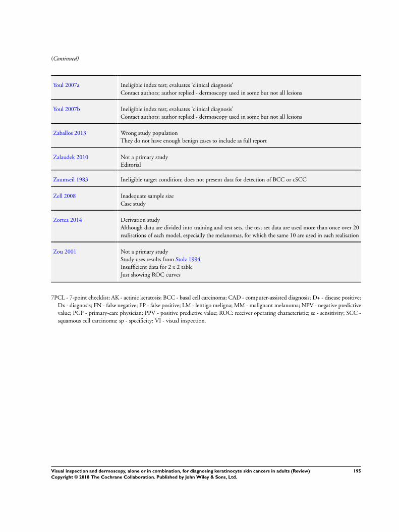
(Continued)
Youl 2007a Ineligible index test; evaluates ’clinical diagnosis’
Contact authors; author replied - dermoscopy used in some but not all lesions
Youl 2007b Ineligible index test; evaluates ’clinical diagnosis’
Contact authors; author replied - dermoscopy used in some but not all lesions
Zaballos 2013 Wrong study population
They do not have enough benign cases to include as full report
Zalaudek 2010 Not a primary study
Editorial
Zaumseil 1983 Ineligible target condition; does not present data for detection of BCC or cSCC
Zell 2008 Inadequate sample size
Case study
Zortea 2014 Derivation study
Although data are divided into training and test sets, the test set data are used more than once over 20
realisations of each model, especially the melanomas, for which the same 10 are used in each realisation
Zou 2001 Not a primary study
Study uses results from Stolz 1994
Insufficient data for 2 x 2 table
Just showing ROC curves
7PCL - 7-point checklist; AK - actinic keratosis; BCC - basal cell carcinoma; CAD - computer-assisted diagnosis; D+ - disease positive;
Dx - diagnosis; FN - false negative; FP - false positive; LM - lentigo meligna; MM - malignant melanoma; NPV - negative predictive
value; PCP - primary-care physician; PPV - positive predictive value; ROC: receiver operating characteristic; se - sensitivity; SCC -
squamous cell carcinoma; sp - specificity; VI - visual inspection.
195Visual inspection and dermoscopy, alone or in combination, for diagnosing keratinocyte skin cancers in adults (Review)
Copyright © 2018 The Cochrane Collaboration. Published by John Wiley & Sons, Ltd.
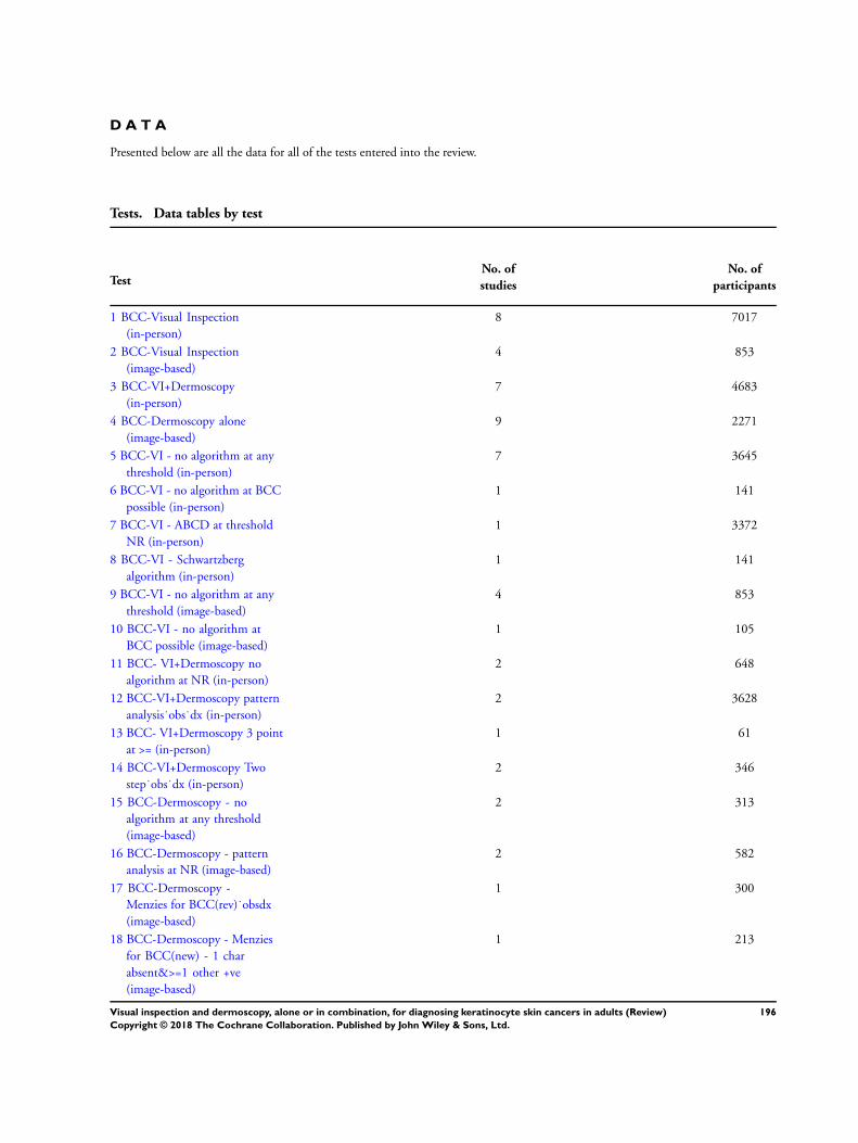
D A T A
Presented below are all the data for all of the tests entered into the review.
Tests. Data tables by test
TestNo. of
studies
No. of
participants
1 BCC-Visual Inspection
(in-person)
8 7017
2 BCC-Visual Inspection
(image-based)
4 853
3 BCC-VI+Dermoscopy
(in-person)
7 4683
4 BCC-Dermoscopy alone
(image-based)
9 2271
5 BCC-VI - no algorithm at any
threshold (in-person)
7 3645
6 BCC-VI - no algorithm at BCC
possible (in-person)
1 141
7 BCC-VI - ABCD at threshold
NR (in-person)
1 3372
8 BCC-VI - Schwartzberg
algorithm (in-person)
1 141
9 BCC-VI - no algorithm at any
threshold (image-based)
4 853
10 BCC-VI - no algorithm at
BCC possible (image-based)
1 105
11 BCC- VI+Dermoscopy no
algorithm at NR (in-person)
2 648
12 BCC-VI+Dermoscopy pattern
analysis˙obs˙dx (in-person)
2 3628
13 BCC- VI+Dermoscopy 3 point
at >= (in-person)
1 61
14 BCC-VI+Dermoscopy Two
step˙obs˙dx (in-person)
2 346
15 BCC-Dermoscopy - no
algorithm at any threshold
(image-based)
2 313
16 BCC-Dermoscopy - pattern
analysis at NR (image-based)
2 582
17 BCC-Dermoscopy -
Menzies for BCC(rev)˙obsdx
(image-based)
1 300
18 BCC-Dermoscopy - Menzies
for BCC(new) - 1 char
absent&>=1 other +ve
(image-based)
1 213
196Visual inspection and dermoscopy, alone or in combination, for diagnosing keratinocyte skin cancers in adults (Review)
Copyright © 2018 The Cochrane Collaboration. Published by John Wiley & Sons, Ltd.
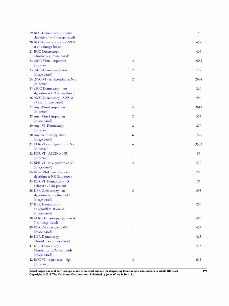
19 BCC-Dermoscopy - 3 point
checklist at >= 2 (image-based)
1 150
20 BCC-Dermoscopy - new SWS
at >=1 (image-based)
1 457
21 BCC-Dermoscopy -
Chaos/clues (image-based)
1 463
22 cSCC-Visual inspection
(in-person)
2 2684
23 cSCC-Dermoscopy alone
(image-based)
2 717
24 cSCC-VI - no algorithm at NR
(in-person)
2 2684
25 cSCC-Dermoscopy - no
algorithm at NR (image-based)
1 260
26 cSCC-Dermoscopy - SWS at
>1 char (image-based)
1 457
27 Any -Visual inspection
(in-person)
5 3618
28 Any -Visual inspection
(image-based)
2 517
29 Any -VI+Dermoscopy
(in-person)
2 277
30 Any-Dermoscopy alone
(image-based)
6 1526
31 KER-VI - no algorithm at NR
(in-person)
4 3533
32 KER-VI - ABCD at NR
(in-person)
1 85
33 KER-VI - no algorithm at NR
(image-based)
2 517
34 KER- VI+Dermoscopy no
algorithm at NR (in-person)
1 200
35 KER-VI+Dermoscopy - 3
point at >=2 (in-person)
1 77
36 KER-Dermoscopy - no
algorithm at any threshold
(image-based)
3 393
37 KER-Dermoscopy -
no algorithm at excise
(image-based)
1 260
38 KER- Dermoscopy - pattern at
NR (image-based)
1 463
39 KER-Dermoscopy- SWS
(image-based)
1 457
40 KER-Dermoscopy -
Chaos/Clues (image-based)
1 463
41 KER-Dermoscopy -
Menzies for BCC(rev)˙obsdx
(image-based)
1 213
42 BCC-VI - experience - high
(in-person)
3 615
197Visual inspection and dermoscopy, alone or in combination, for diagnosing keratinocyte skin cancers in adults (Review)
Copyright © 2018 The Cochrane Collaboration. Published by John Wiley & Sons, Ltd.
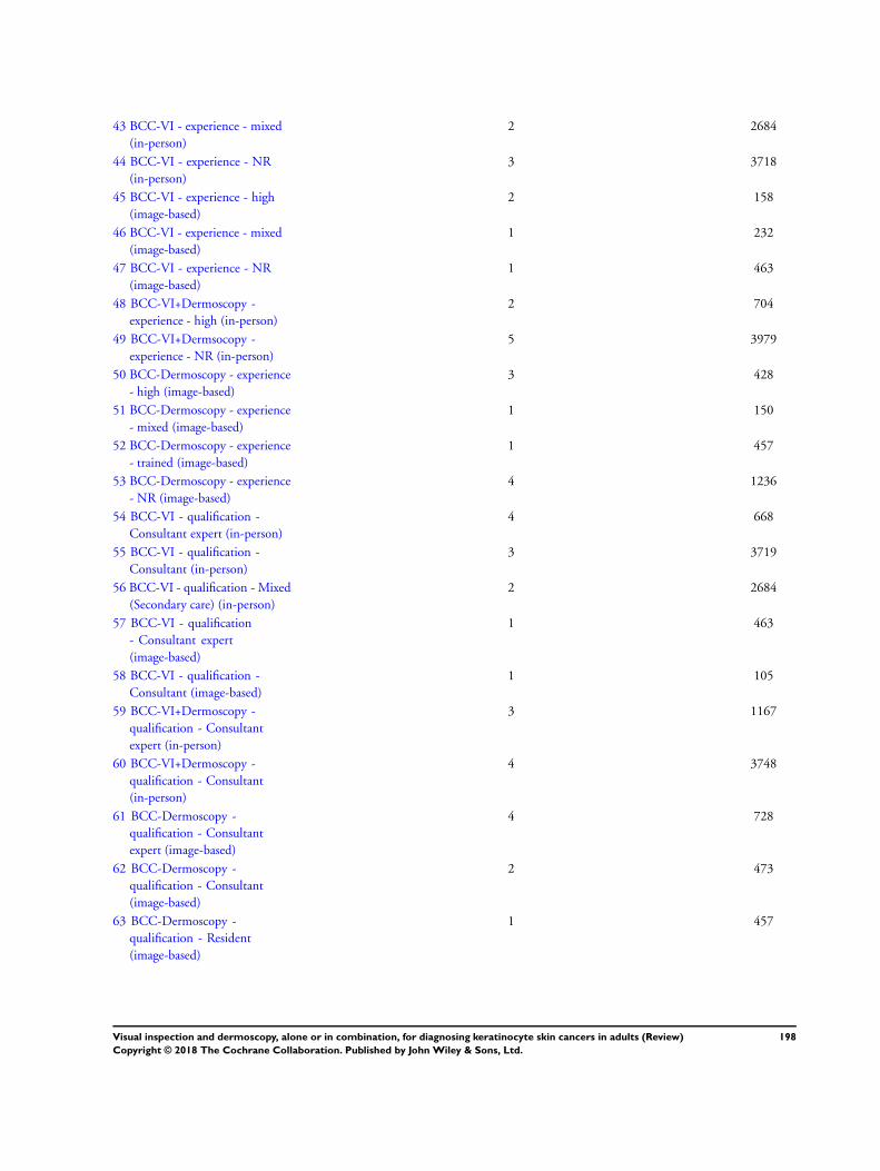
43 BCC-VI - experience - mixed
(in-person)
2 2684
44 BCC-VI - experience - NR
(in-person)
3 3718
45 BCC-VI - experience - high
(image-based)
2 158
46 BCC-VI - experience - mixed
(image-based)
1 232
47 BCC-VI - experience - NR
(image-based)
1 463
48 BCC-VI+Dermoscopy -
experience - high (in-person)
2 704
49 BCC-VI+Dermsocopy -
experience - NR (in-person)
5 3979
50 BCC-Dermoscopy - experience
- high (image-based)
3 428
51 BCC-Dermoscopy - experience
- mixed (image-based)
1 150
52 BCC-Dermoscopy - experience
- trained (image-based)
1 457
53 BCC-Dermoscopy - experience
- NR (image-based)
4 1236
54 BCC-VI - qualification -
Consultant expert (in-person)
4 668
55 BCC-VI - qualification -
Consultant (in-person)
3 3719
56 BCC-VI - qualification - Mixed
(Secondary care) (in-person)
2 2684
57 BCC-VI - qualification
- Consultant expert
(image-based)
1 463
58 BCC-VI - qualification -
Consultant (image-based)
1 105
59 BCC-VI+Dermoscopy -
qualification - Consultant
expert (in-person)
3 1167
60 BCC-VI+Dermoscopy -
qualification - Consultant
(in-person)
4 3748
61 BCC-Dermoscopy -
qualification - Consultant
expert (image-based)
4 728
62 BCC-Dermoscopy -
qualification - Consultant
(image-based)
2 473
63 BCC-Dermoscopy -
qualification - Resident
(image-based)
1 457
198Visual inspection and dermoscopy, alone or in combination, for diagnosing keratinocyte skin cancers in adults (Review)
Copyright © 2018 The Cochrane Collaboration. Published by John Wiley & Sons, Ltd.
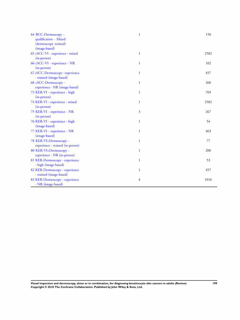
64 BCC-Dermoscopy -
qualification - Mixed
(dermoscopy trained)
(image-based)
1 150
65 cSCC-VI - experience - mixed
(in-person)
1 2582
66 cSCC-VI - experience - NR
(in-person)
1 102
67 cSCC-Dermoscopy - experience
- trained (image-based)
1 457
68 cSCC-Dermoscopy -
experience - NR (image-based)
1 260
73 KER-VI - experience - high
(in-person)
1 769
74 KER-VI - experience - mixed
(in-person)
1 2582
75 KER-VI - experience - NR
(in-person)
3 267
76 KER-VI - experience - high
(image-based)
1 54
77 KER-VI - experience - NR
(image-based)
1 463
78 KER-VI+Dermoscopy -
experience - trained (in-person)
1 77
80 KER-VI+Dermoscopy -
experience - NR (in-person)
1 200
81 KER-Dermoscopy - experience
- high (image-based)
1 53
82 KER-Dermoscopy - experience
- trained (image-based)
1 457
83 KER-Dermoscopy - experience
- NR (image-based)
4 1016
199Visual inspection and dermoscopy, alone or in combination, for diagnosing keratinocyte skin cancers in adults (Review)
Copyright © 2018 The Cochrane Collaboration. Published by John Wiley & Sons, Ltd.
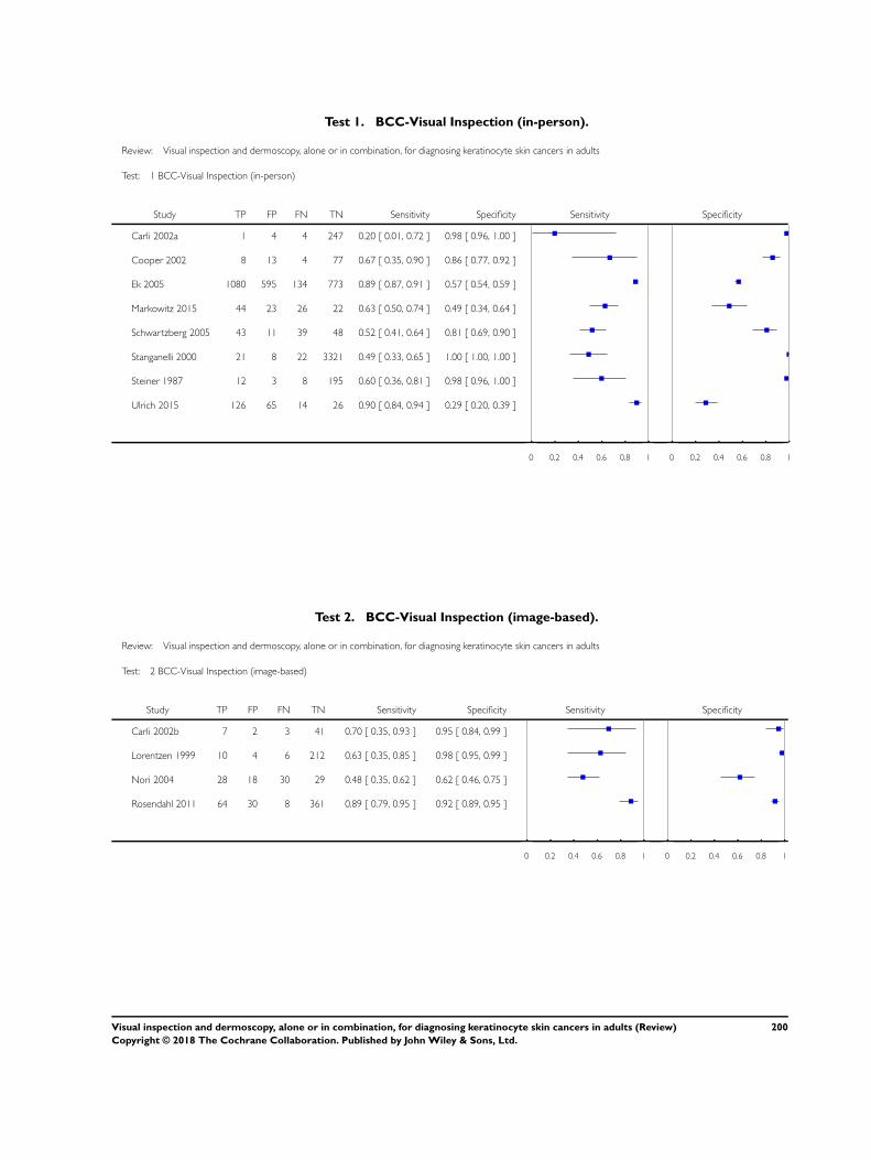
Test 1. BCC-Visual Inspection (in-person).
Review: Visual inspection and dermoscopy, alone or in combination, for diagnosing keratinocyte skin cancers in adults
Test: 1 BCC-Visual Inspection (in-person)
Study TP FP FN TN Sensitivity Specificity Sensitivity Specificity
Carli 2002a 1 4 4 247 0.20 [ 0.01, 0.72 ] 0.98 [ 0.96, 1.00 ]
Cooper 2002 8 13 4 77 0.67 [ 0.35, 0.90 ] 0.86 [ 0.77, 0.92 ]
Ek 2005 1080 595 134 773 0.89 [ 0.87, 0.91 ] 0.57 [ 0.54, 0.59 ]
Markowitz 2015 44 23 26 22 0.63 [ 0.50, 0.74 ] 0.49 [ 0.34, 0.64 ]
Schwartzberg 2005 43 11 39 48 0.52 [ 0.41, 0.64 ] 0.81 [ 0.69, 0.90 ]
Stanganelli 2000 21 8 22 3321 0.49 [ 0.33, 0.65 ] 1.00 [ 1.00, 1.00 ]
Steiner 1987 12 3 8 195 0.60 [ 0.36, 0.81 ] 0.98 [ 0.96, 1.00 ]
Ulrich 2015 126 65 14 26 0.90 [ 0.84, 0.94 ] 0.29 [ 0.20, 0.39 ]
0 0.2 0.4 0.6 0.8 1 0 0.2 0.4 0.6 0.8 1
Test 2. BCC-Visual Inspection (image-based).
Review: Visual inspection and dermoscopy, alone or in combination, for diagnosing keratinocyte skin cancers in adults
Test: 2 BCC-Visual Inspection (image-based)
Study TP FP FN TN Sensitivity Specificity Sensitivity Specificity
Carli 2002b 7 2 3 41 0.70 [ 0.35, 0.93 ] 0.95 [ 0.84, 0.99 ]
Lorentzen 1999 10 4 6 212 0.63 [ 0.35, 0.85 ] 0.98 [ 0.95, 0.99 ]
Nori 2004 28 18 30 29 0.48 [ 0.35, 0.62 ] 0.62 [ 0.46, 0.75 ]
Rosendahl 2011 64 30 8 361 0.89 [ 0.79, 0.95 ] 0.92 [ 0.89, 0.95 ]
0 0.2 0.4 0.6 0.8 1 0 0.2 0.4 0.6 0.8 1
200Visual inspection and dermoscopy, alone or in combination, for diagnosing keratinocyte skin cancers in adults (Review)
Copyright © 2018 The Cochrane Collaboration. Published by John Wiley & Sons, Ltd.
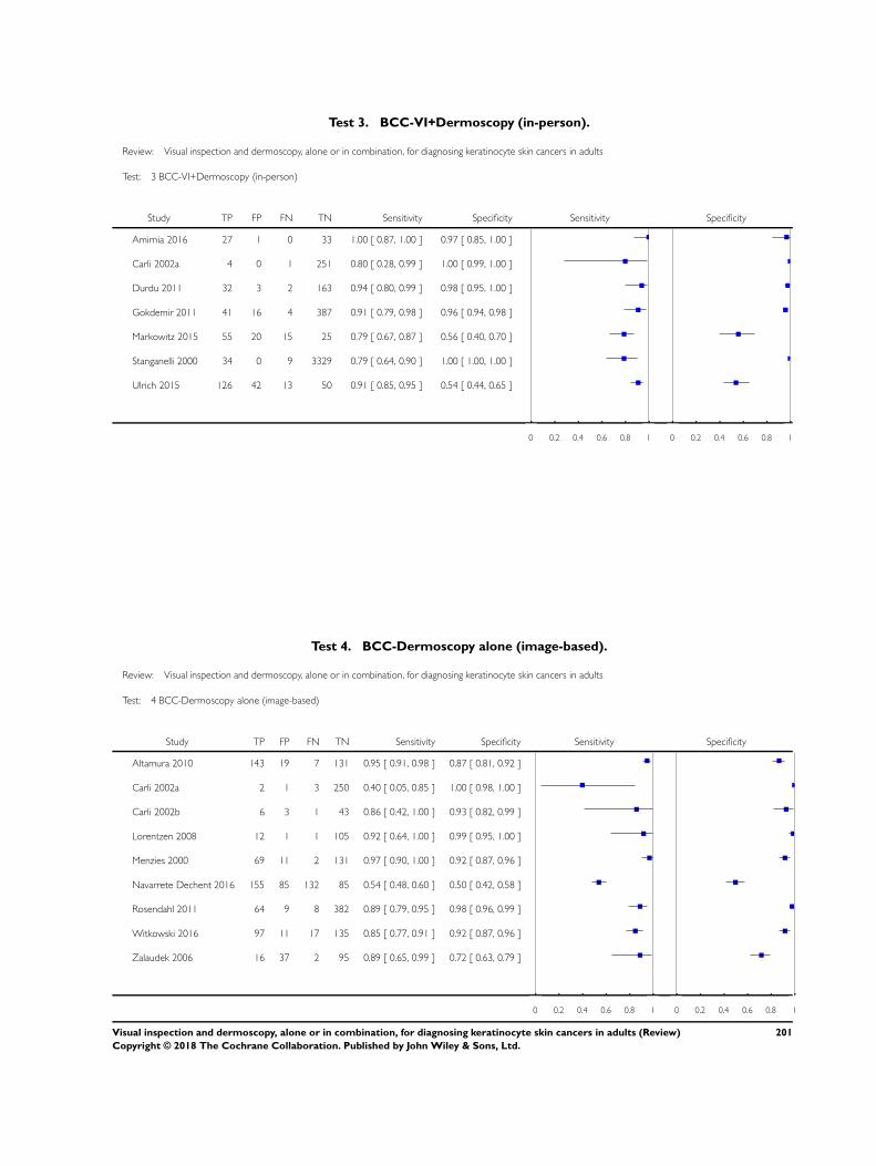
Test 3. BCC-VI+Dermoscopy (in-person).
Review: Visual inspection and dermoscopy, alone or in combination, for diagnosing keratinocyte skin cancers in adults
Test: 3 BCC-VI+Dermoscopy (in-person)
Study TP FP FN TN Sensitivity Specificity Sensitivity Specificity
Amirnia 2016 27 1 0 33 1.00 [ 0.87, 1.00 ] 0.97 [ 0.85, 1.00 ]
Carli 2002a 4 0 1 251 0.80 [ 0.28, 0.99 ] 1.00 [ 0.99, 1.00 ]
Durdu 2011 32 3 2 163 0.94 [ 0.80, 0.99 ] 0.98 [ 0.95, 1.00 ]
Gokdemir 2011 41 16 4 387 0.91 [ 0.79, 0.98 ] 0.96 [ 0.94, 0.98 ]
Markowitz 2015 55 20 15 25 0.79 [ 0.67, 0.87 ] 0.56 [ 0.40, 0.70 ]
Stanganelli 2000 34 0 9 3329 0.79 [ 0.64, 0.90 ] 1.00 [ 1.00, 1.00 ]
Ulrich 2015 126 42 13 50 0.91 [ 0.85, 0.95 ] 0.54 [ 0.44, 0.65 ]
0 0.2 0.4 0.6 0.8 1 0 0.2 0.4 0.6 0.8 1
Test 4. BCC-Dermoscopy alone (image-based).
Review: Visual inspection and dermoscopy, alone or in combination, for diagnosing keratinocyte skin cancers in adults
Test: 4 BCC-Dermoscopy alone (image-based)
Study TP FP FN TN Sensitivity Specificity Sensitivity Specificity
Altamura 2010 143 19 7 131 0.95 [ 0.91, 0.98 ] 0.87 [ 0.81, 0.92 ]
Carli 2002a 2 1 3 250 0.40 [ 0.05, 0.85 ] 1.00 [ 0.98, 1.00 ]
Carli 2002b 6 3 1 43 0.86 [ 0.42, 1.00 ] 0.93 [ 0.82, 0.99 ]
Lorentzen 2008 12 1 1 105 0.92 [ 0.64, 1.00 ] 0.99 [ 0.95, 1.00 ]
Menzies 2000 69 11 2 131 0.97 [ 0.90, 1.00 ] 0.92 [ 0.87, 0.96 ]
Navarrete Dechent 2016 155 85 132 85 0.54 [ 0.48, 0.60 ] 0.50 [ 0.42, 0.58 ]
Rosendahl 2011 64 9 8 382 0.89 [ 0.79, 0.95 ] 0.98 [ 0.96, 0.99 ]
Witkowski 2016 97 11 17 135 0.85 [ 0.77, 0.91 ] 0.92 [ 0.87, 0.96 ]
Zalaudek 2006 16 37 2 95 0.89 [ 0.65, 0.99 ] 0.72 [ 0.63, 0.79 ]
0 0.2 0.4 0.6 0.8 1 0 0.2 0.4 0.6 0.8 1
201Visual inspection and dermoscopy, alone or in combination, for diagnosing keratinocyte skin cancers in adults (Review)
Copyright © 2018 The Cochrane Collaboration. Published by John Wiley & Sons, Ltd.
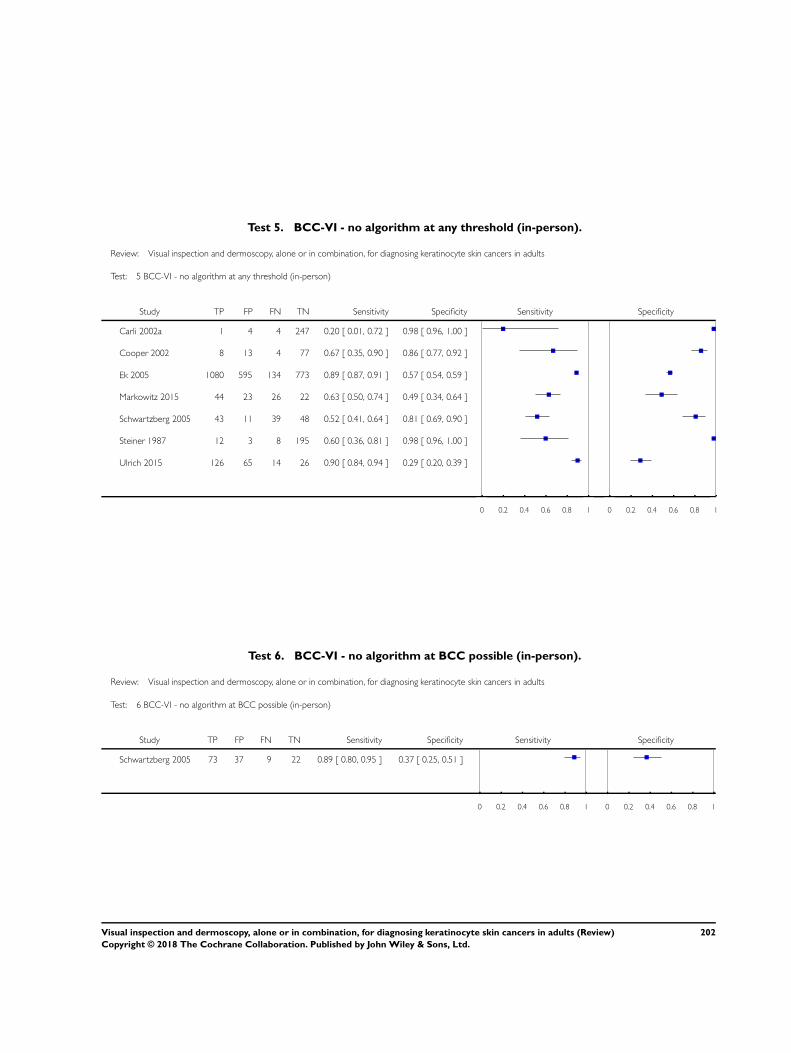
Test 5. BCC-VI - no algorithm at any threshold (in-person).
Review: Visual inspection and dermoscopy, alone or in combination, for diagnosing keratinocyte skin cancers in adults
Test: 5 BCC-VI - no algorithm at any threshold (in-person)
Study TP FP FN TN Sensitivity Specificity Sensitivity Specificity
Carli 2002a 1 4 4 247 0.20 [ 0.01, 0.72 ] 0.98 [ 0.96, 1.00 ]
Cooper 2002 8 13 4 77 0.67 [ 0.35, 0.90 ] 0.86 [ 0.77, 0.92 ]
Ek 2005 1080 595 134 773 0.89 [ 0.87, 0.91 ] 0.57 [ 0.54, 0.59 ]
Markowitz 2015 44 23 26 22 0.63 [ 0.50, 0.74 ] 0.49 [ 0.34, 0.64 ]
Schwartzberg 2005 43 11 39 48 0.52 [ 0.41, 0.64 ] 0.81 [ 0.69, 0.90 ]
Steiner 1987 12 3 8 195 0.60 [ 0.36, 0.81 ] 0.98 [ 0.96, 1.00 ]
Ulrich 2015 126 65 14 26 0.90 [ 0.84, 0.94 ] 0.29 [ 0.20, 0.39 ]
0 0.2 0.4 0.6 0.8 1 0 0.2 0.4 0.6 0.8 1
Test 6. BCC-VI - no algorithm at BCC possible (in-person).
Review: Visual inspection and dermoscopy, alone or in combination, for diagnosing keratinocyte skin cancers in adults
Test: 6 BCC-VI - no algorithm at BCC possible (in-person)
Study TP FP FN TN Sensitivity Specificity Sensitivity Specificity
Schwartzberg 2005 73 37 9 22 0.89 [ 0.80, 0.95 ] 0.37 [ 0.25, 0.51 ]
0 0.2 0.4 0.6 0.8 1 0 0.2 0.4 0.6 0.8 1
202Visual inspection and dermoscopy, alone or in combination, for diagnosing keratinocyte skin cancers in adults (Review)
Copyright © 2018 The Cochrane Collaboration. Published by John Wiley & Sons, Ltd.
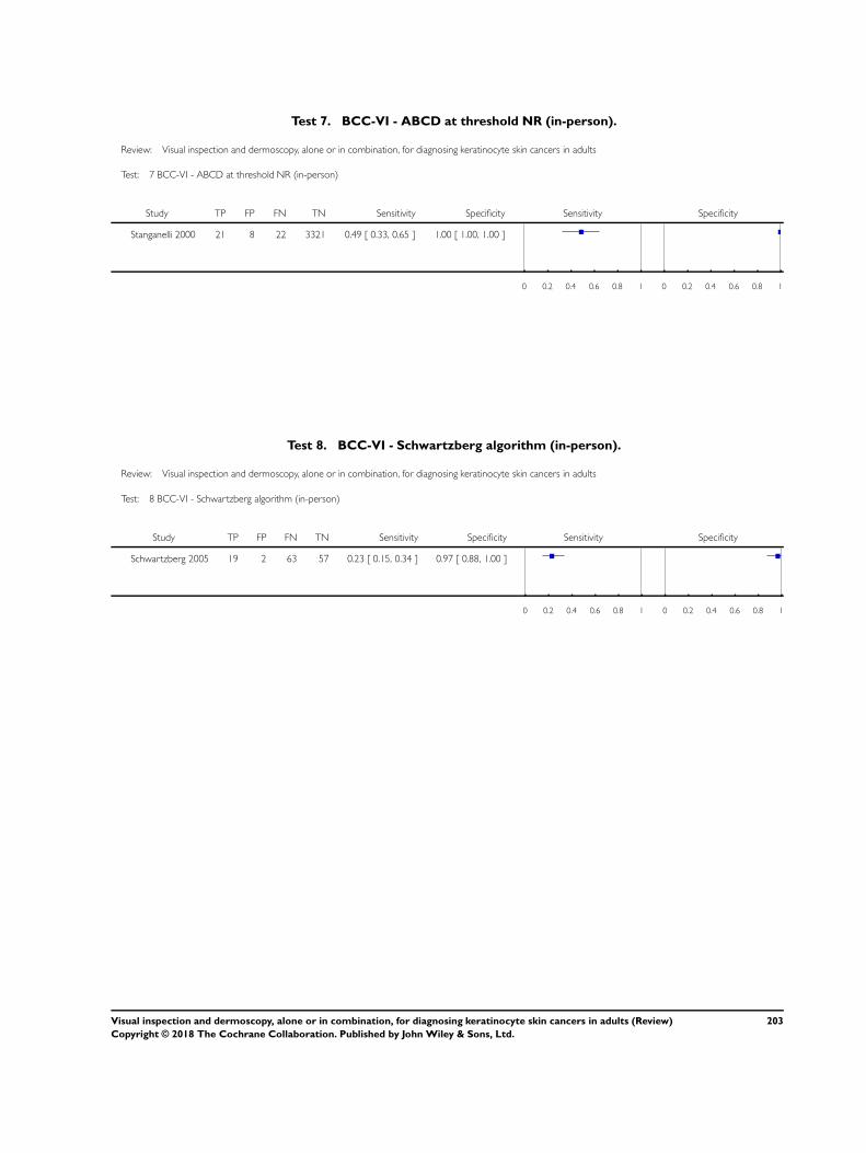
Test 7. BCC-VI - ABCD at threshold NR (in-person).
Review: Visual inspection and dermoscopy, alone or in combination, for diagnosing keratinocyte skin cancers in adults
Test: 7 BCC-VI - ABCD at threshold NR (in-person)
Study TP FP FN TN Sensitivity Specificity Sensitivity Specificity
Stanganelli 2000 21 8 22 3321 0.49 [ 0.33, 0.65 ] 1.00 [ 1.00, 1.00 ]
0 0.2 0.4 0.6 0.8 1 0 0.2 0.4 0.6 0.8 1
Test 8. BCC-VI - Schwartzberg algorithm (in-person).
Review: Visual inspection and dermoscopy, alone or in combination, for diagnosing keratinocyte skin cancers in adults
Test: 8 BCC-VI - Schwartzberg algorithm (in-person)
Study TP FP FN TN Sensitivity Specificity Sensitivity Specificity
Schwartzberg 2005 19 2 63 57 0.23 [ 0.15, 0.34 ] 0.97 [ 0.88, 1.00 ]
0 0.2 0.4 0.6 0.8 1 0 0.2 0.4 0.6 0.8 1
203Visual inspection and dermoscopy, alone or in combination, for diagnosing keratinocyte skin cancers in adults (Review)
Copyright © 2018 The Cochrane Collaboration. Published by John Wiley & Sons, Ltd.
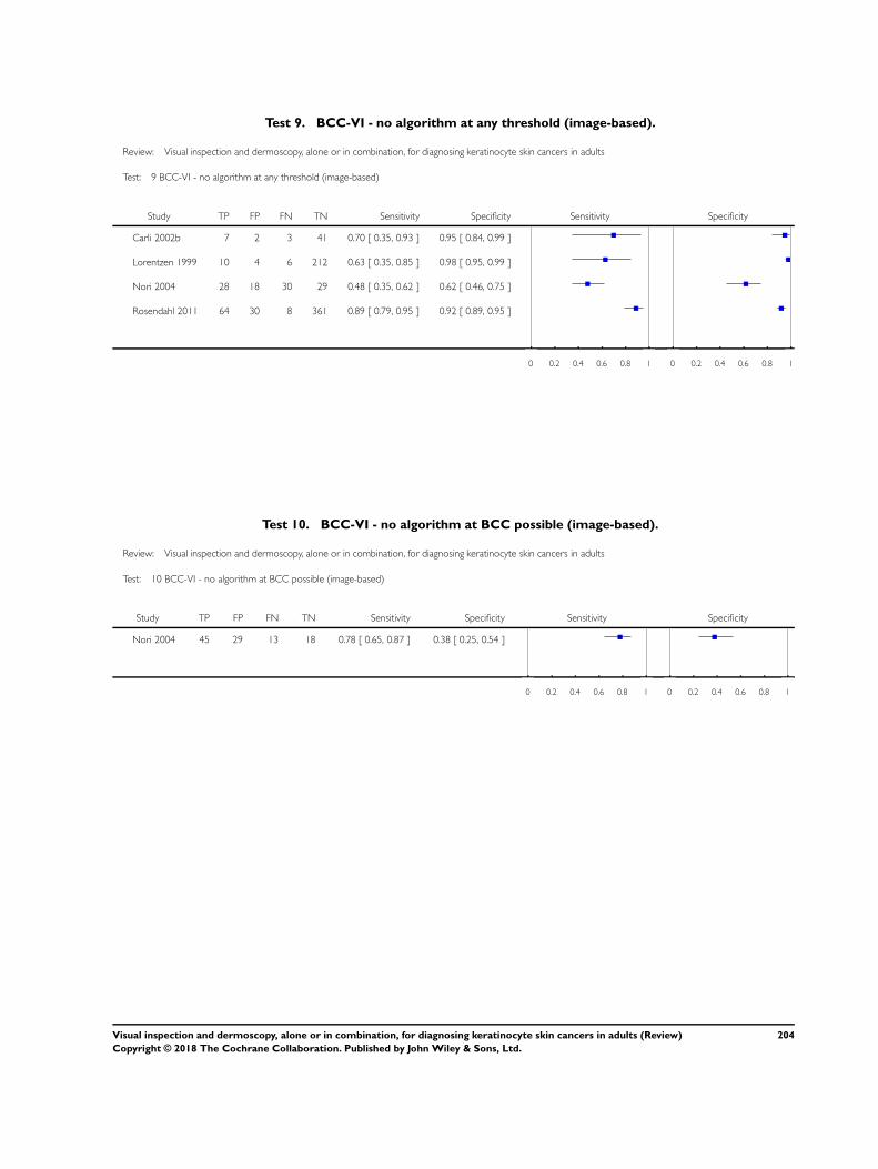
Test 9. BCC-VI - no algorithm at any threshold (image-based).
Review: Visual inspection and dermoscopy, alone or in combination, for diagnosing keratinocyte skin cancers in adults
Test: 9 BCC-VI - no algorithm at any threshold (image-based)
Study TP FP FN TN Sensitivity Specificity Sensitivity Specificity
Carli 2002b 7 2 3 41 0.70 [ 0.35, 0.93 ] 0.95 [ 0.84, 0.99 ]
Lorentzen 1999 10 4 6 212 0.63 [ 0.35, 0.85 ] 0.98 [ 0.95, 0.99 ]
Nori 2004 28 18 30 29 0.48 [ 0.35, 0.62 ] 0.62 [ 0.46, 0.75 ]
Rosendahl 2011 64 30 8 361 0.89 [ 0.79, 0.95 ] 0.92 [ 0.89, 0.95 ]
0 0.2 0.4 0.6 0.8 1 0 0.2 0.4 0.6 0.8 1
Test 10. BCC-VI - no algorithm at BCC possible (image-based).
Review: Visual inspection and dermoscopy, alone or in combination, for diagnosing keratinocyte skin cancers in adults
Test: 10 BCC-VI - no algorithm at BCC possible (image-based)
Study TP FP FN TN Sensitivity Specificity Sensitivity Specificity
Nori 2004 45 29 13 18 0.78 [ 0.65, 0.87 ] 0.38 [ 0.25, 0.54 ]
0 0.2 0.4 0.6 0.8 1 0 0.2 0.4 0.6 0.8 1
204Visual inspection and dermoscopy, alone or in combination, for diagnosing keratinocyte skin cancers in adults (Review)
Copyright © 2018 The Cochrane Collaboration. Published by John Wiley & Sons, Ltd.
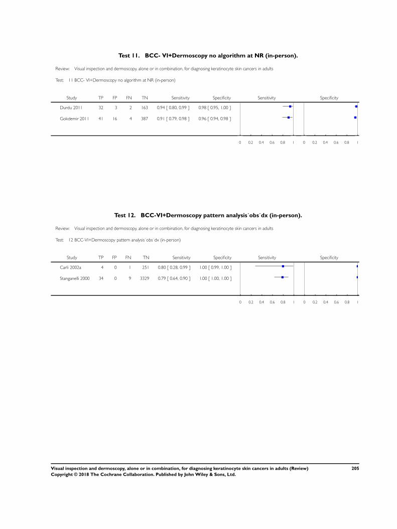
Test 11. BCC- VI+Dermoscopy no algorithm at NR (in-person).
Review: Visual inspection and dermoscopy, alone or in combination, for diagnosing keratinocyte skin cancers in adults
Test: 11 BCC- VI+Dermoscopy no algorithm at NR (in-person)
Study TP FP FN TN Sensitivity Specificity Sensitivity Specificity
Durdu 2011 32 3 2 163 0.94 [ 0.80, 0.99 ] 0.98 [ 0.95, 1.00 ]
Gokdemir 2011 41 16 4 387 0.91 [ 0.79, 0.98 ] 0.96 [ 0.94, 0.98 ]
0 0.2 0.4 0.6 0.8 1 0 0.2 0.4 0.6 0.8 1
Test 12. BCC-VI+Dermoscopy pattern analysis˙obs˙dx (in-person).
Review: Visual inspection and dermoscopy, alone or in combination, for diagnosing keratinocyte skin cancers in adults
Test: 12 BCC-VI+Dermoscopy pattern analysis˙obs˙dx (in-person)
Study TP FP FN TN Sensitivity Specificity Sensitivity Specificity
Carli 2002a 4 0 1 251 0.80 [ 0.28, 0.99 ] 1.00 [ 0.99, 1.00 ]
Stanganelli 2000 34 0 9 3329 0.79 [ 0.64, 0.90 ] 1.00 [ 1.00, 1.00 ]
0 0.2 0.4 0.6 0.8 1 0 0.2 0.4 0.6 0.8 1
205Visual inspection and dermoscopy, alone or in combination, for diagnosing keratinocyte skin cancers in adults (Review)
Copyright © 2018 The Cochrane Collaboration. Published by John Wiley & Sons, Ltd.
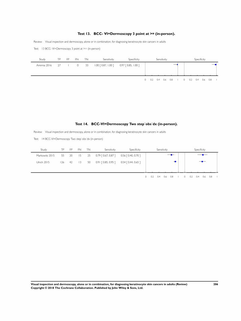
Test 13. BCC- VI+Dermoscopy 3 point at >= (in-person).
Review: Visual inspection and dermoscopy, alone or in combination, for diagnosing keratinocyte skin cancers in adults
Test: 13 BCC- VI+Dermoscopy 3 point at >= (in-person)
Study TP FP FN TN Sensitivity Specificity Sensitivity Specificity
Amirnia 2016 27 1 0 33 1.00 [ 0.87, 1.00 ] 0.97 [ 0.85, 1.00 ]
0 0.2 0.4 0.6 0.8 1 0 0.2 0.4 0.6 0.8 1
Test 14. BCC-VI+Dermoscopy Two step˙obs˙dx (in-person).
Review: Visual inspection and dermoscopy, alone or in combination, for diagnosing keratinocyte skin cancers in adults
Test: 14 BCC-VI+Dermoscopy Two step˙obs˙dx (in-person)
Study TP FP FN TN Sensitivity Specificity Sensitivity Specificity
Markowitz 2015 55 20 15 25 0.79 [ 0.67, 0.87 ] 0.56 [ 0.40, 0.70 ]
Ulrich 2015 126 42 13 50 0.91 [ 0.85, 0.95 ] 0.54 [ 0.44, 0.65 ]
0 0.2 0.4 0.6 0.8 1 0 0.2 0.4 0.6 0.8 1
206Visual inspection and dermoscopy, alone or in combination, for diagnosing keratinocyte skin cancers in adults (Review)
Copyright © 2018 The Cochrane Collaboration. Published by John Wiley & Sons, Ltd.
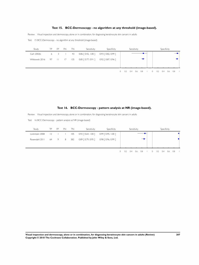
Test 15. BCC-Dermoscopy - no algorithm at any threshold (image-based).
Review: Visual inspection and dermoscopy, alone or in combination, for diagnosing keratinocyte skin cancers in adults
Test: 15 BCC-Dermoscopy - no algorithm at any threshold (image-based)
Study TP FP FN TN Sensitivity Specificity Sensitivity Specificity
Carli 2002b 6 3 1 43 0.86 [ 0.42, 1.00 ] 0.93 [ 0.82, 0.99 ]
Witkowski 2016 97 11 17 135 0.85 [ 0.77, 0.91 ] 0.92 [ 0.87, 0.96 ]
0 0.2 0.4 0.6 0.8 1 0 0.2 0.4 0.6 0.8 1
Test 16. BCC-Dermoscopy - pattern analysis at NR (image-based).
Review: Visual inspection and dermoscopy, alone or in combination, for diagnosing keratinocyte skin cancers in adults
Test: 16 BCC-Dermoscopy - pattern analysis at NR (image-based)
Study TP FP FN TN Sensitivity Specificity Sensitivity Specificity
Lorentzen 2008 12 1 1 105 0.92 [ 0.64, 1.00 ] 0.99 [ 0.95, 1.00 ]
Rosendahl 2011 64 9 8 382 0.89 [ 0.79, 0.95 ] 0.98 [ 0.96, 0.99 ]
0 0.2 0.4 0.6 0.8 1 0 0.2 0.4 0.6 0.8 1
207Visual inspection and dermoscopy, alone or in combination, for diagnosing keratinocyte skin cancers in adults (Review)
Copyright © 2018 The Cochrane Collaboration. Published by John Wiley & Sons, Ltd.
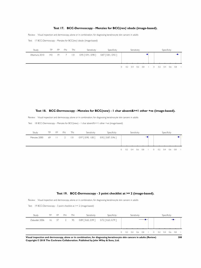
Test 17. BCC-Dermoscopy - Menzies for BCC(rev)˙obsdx (image-based).
Review: Visual inspection and dermoscopy, alone or in combination, for diagnosing keratinocyte skin cancers in adults
Test: 17 BCC-Dermoscopy - Menzies for BCC(rev)˙obsdx (image-based)
Study TP FP FN TN Sensitivity Specificity Sensitivity Specificity
Altamura 2010 143 19 7 131 0.95 [ 0.91, 0.98 ] 0.87 [ 0.81, 0.92 ]
0 0.2 0.4 0.6 0.8 1 0 0.2 0.4 0.6 0.8 1
Test 18. BCC-Dermoscopy - Menzies for BCC(new) - 1 char absent&>=1 other +ve (image-based).
Review: Visual inspection and dermoscopy, alone or in combination, for diagnosing keratinocyte skin cancers in adults
Test: 18 BCC-Dermoscopy - Menzies for BCC(new) - 1 char absent%>=1 other +ve (image-based)
Study TP FP FN TN Sensitivity Specificity Sensitivity Specificity
Menzies 2000 69 11 2 131 0.97 [ 0.90, 1.00 ] 0.92 [ 0.87, 0.96 ]
0 0.2 0.4 0.6 0.8 1 0 0.2 0.4 0.6 0.8 1
Test 19. BCC-Dermoscopy - 3 point checklist at >= 2 (image-based).
Review: Visual inspection and dermoscopy, alone or in combination, for diagnosing keratinocyte skin cancers in adults
Test: 19 BCC-Dermoscopy - 3 point checklist at >= 2 (image-based)
Study TP FP FN TN Sensitivity Specificity Sensitivity Specificity
Zalaudek 2006 16 37 2 95 0.89 [ 0.65, 0.99 ] 0.72 [ 0.63, 0.79 ]
0 0.2 0.4 0.6 0.8 1 0 0.2 0.4 0.6 0.8 1
208Visual inspection and dermoscopy, alone or in combination, for diagnosing keratinocyte skin cancers in adults (Review)
Copyright © 2018 The Cochrane Collaboration. Published by John Wiley & Sons, Ltd.
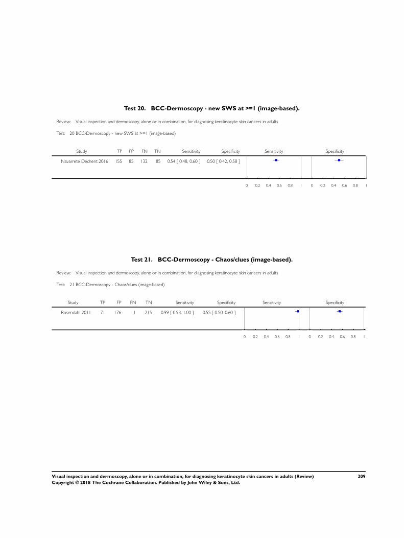
Test 20. BCC-Dermoscopy - new SWS at >=1 (image-based).
Review: Visual inspection and dermoscopy, alone or in combination, for diagnosing keratinocyte skin cancers in adults
Test: 20 BCC-Dermoscopy - new SWS at >=1 (image-based)
Study TP FP FN TN Sensitivity Specificity Sensitivity Specificity
Navarrete Dechent 2016 155 85 132 85 0.54 [ 0.48, 0.60 ] 0.50 [ 0.42, 0.58 ]
0 0.2 0.4 0.6 0.8 1 0 0.2 0.4 0.6 0.8 1
Test 21. BCC-Dermoscopy - Chaos/clues (image-based).
Review: Visual inspection and dermoscopy, alone or in combination, for diagnosing keratinocyte skin cancers in adults
Test: 21 BCC-Dermoscopy - Chaos/clues (image-based)
Study TP FP FN TN Sensitivity Specificity Sensitivity Specificity
Rosendahl 2011 71 176 1 215 0.99 [ 0.93, 1.00 ] 0.55 [ 0.50, 0.60 ]
0 0.2 0.4 0.6 0.8 1 0 0.2 0.4 0.6 0.8 1
209Visual inspection and dermoscopy, alone or in combination, for diagnosing keratinocyte skin cancers in adults (Review)
Copyright © 2018 The Cochrane Collaboration. Published by John Wiley & Sons, Ltd.
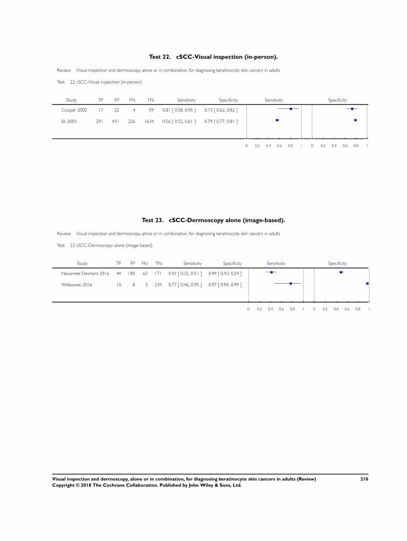
Test 22. cSCC-Visual inspection (in-person).
Review: Visual inspection and dermoscopy, alone or in combination, for diagnosing keratinocyte skin cancers in adults
Test: 22 cSCC-Visual inspection (in-person)
Study TP FP FN TN Sensitivity Specificity Sensitivity Specificity
Cooper 2002 17 22 4 59 0.81 [ 0.58, 0.95 ] 0.73 [ 0.62, 0.82 ]
Ek 2005 291 431 226 1634 0.56 [ 0.52, 0.61 ] 0.79 [ 0.77, 0.81 ]
0 0.2 0.4 0.6 0.8 1 0 0.2 0.4 0.6 0.8 1
Test 23. cSCC-Dermoscopy alone (image-based).
Review: Visual inspection and dermoscopy, alone or in combination, for diagnosing keratinocyte skin cancers in adults
Test: 23 cSCC-Dermoscopy alone (image-based)
Study TP FP FN TN Sensitivity Specificity Sensitivity Specificity
Navarrete Dechent 2016 44 180 62 171 0.42 [ 0.32, 0.51 ] 0.49 [ 0.43, 0.54 ]
Witkowski 2016 10 8 3 239 0.77 [ 0.46, 0.95 ] 0.97 [ 0.94, 0.99 ]
0 0.2 0.4 0.6 0.8 1 0 0.2 0.4 0.6 0.8 1
210Visual inspection and dermoscopy, alone or in combination, for diagnosing keratinocyte skin cancers in adults (Review)
Copyright © 2018 The Cochrane Collaboration. Published by John Wiley & Sons, Ltd.
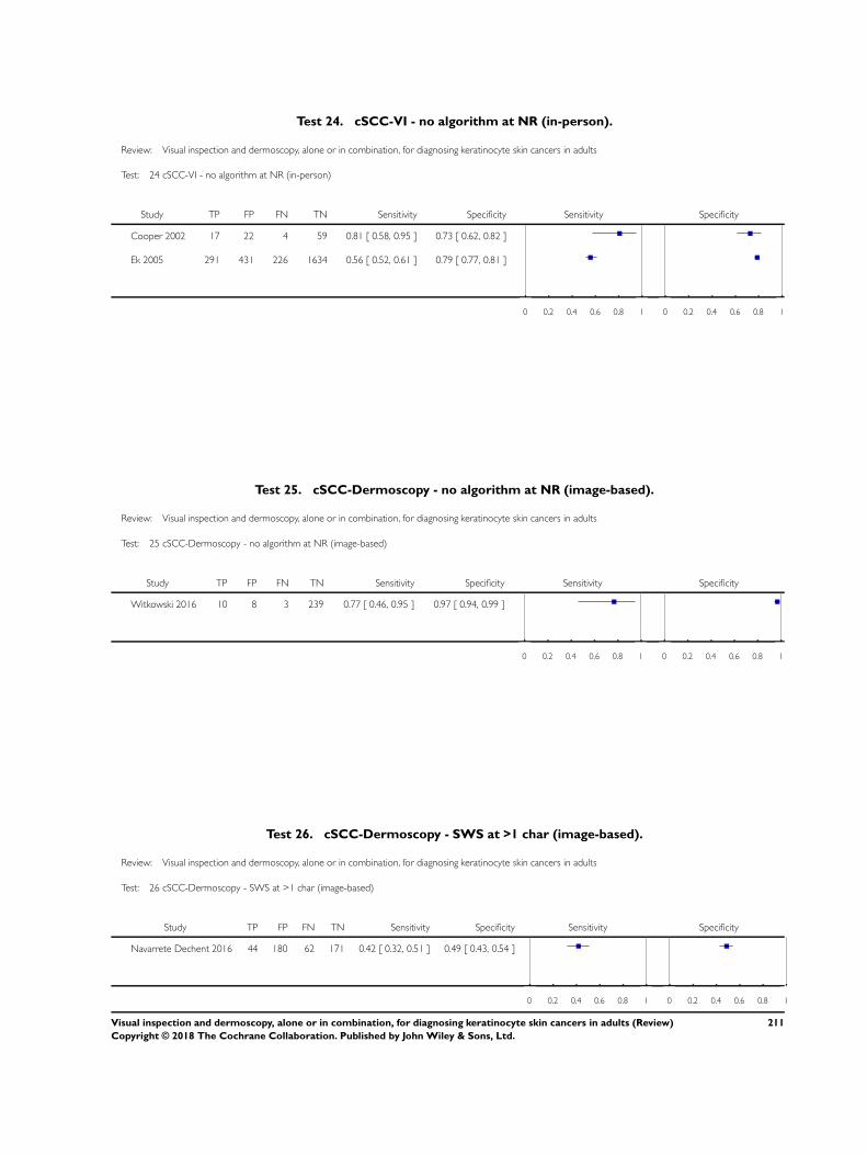
Test 24. cSCC-VI - no algorithm at NR (in-person).
Review: Visual inspection and dermoscopy, alone or in combination, for diagnosing keratinocyte skin cancers in adults
Test: 24 cSCC-VI - no algorithm at NR (in-person)
Study TP FP FN TN Sensitivity Specificity Sensitivity Specificity
Cooper 2002 17 22 4 59 0.81 [ 0.58, 0.95 ] 0.73 [ 0.62, 0.82 ]
Ek 2005 291 431 226 1634 0.56 [ 0.52, 0.61 ] 0.79 [ 0.77, 0.81 ]
0 0.2 0.4 0.6 0.8 1 0 0.2 0.4 0.6 0.8 1
Test 25. cSCC-Dermoscopy - no algorithm at NR (image-based).
Review: Visual inspection and dermoscopy, alone or in combination, for diagnosing keratinocyte skin cancers in adults
Test: 25 cSCC-Dermoscopy - no algorithm at NR (image-based)
Study TP FP FN TN Sensitivity Specificity Sensitivity Specificity
Witkowski 2016 10 8 3 239 0.77 [ 0.46, 0.95 ] 0.97 [ 0.94, 0.99 ]
0 0.2 0.4 0.6 0.8 1 0 0.2 0.4 0.6 0.8 1
Test 26. cSCC-Dermoscopy - SWS at >1 char (image-based).
Review: Visual inspection and dermoscopy, alone or in combination, for diagnosing keratinocyte skin cancers in adults
Test: 26 cSCC-Dermoscopy - SWS at >1 char (image-based)
Study TP FP FN TN Sensitivity Specificity Sensitivity Specificity
Navarrete Dechent 2016 44 180 62 171 0.42 [ 0.32, 0.51 ] 0.49 [ 0.43, 0.54 ]
0 0.2 0.4 0.6 0.8 1 0 0.2 0.4 0.6 0.8 1
211Visual inspection and dermoscopy, alone or in combination, for diagnosing keratinocyte skin cancers in adults (Review)
Copyright © 2018 The Cochrane Collaboration. Published by John Wiley & Sons, Ltd.
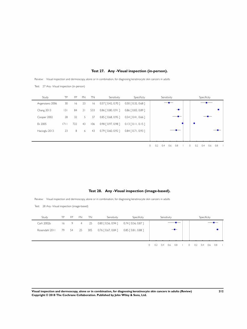
Test 27. Any -Visual inspection (in-person).
Review: Visual inspection and dermoscopy, alone or in combination, for diagnosing keratinocyte skin cancers in adults
Test: 27 Any -Visual inspection (in-person)
Study TP FP FN TN Sensitivity Specificity Sensitivity Specificity
Argenziano 2006 30 16 23 16 0.57 [ 0.42, 0.70 ] 0.50 [ 0.32, 0.68 ]
Chang 2013 131 84 21 533 0.86 [ 0.80, 0.91 ] 0.86 [ 0.83, 0.89 ]
Cooper 2002 28 32 5 37 0.85 [ 0.68, 0.95 ] 0.54 [ 0.41, 0.66 ]
Ek 2005 1711 722 43 106 0.98 [ 0.97, 0.98 ] 0.13 [ 0.11, 0.15 ]
Hacioglu 2013 23 8 6 43 0.79 [ 0.60, 0.92 ] 0.84 [ 0.71, 0.93 ]
0 0.2 0.4 0.6 0.8 1 0 0.2 0.4 0.6 0.8 1
Test 28. Any -Visual inspection (image-based).
Review: Visual inspection and dermoscopy, alone or in combination, for diagnosing keratinocyte skin cancers in adults
Test: 28 Any -Visual inspection (image-based)
Study TP FP FN TN Sensitivity Specificity Sensitivity Specificity
Carli 2002b 16 9 4 25 0.80 [ 0.56, 0.94 ] 0.74 [ 0.56, 0.87 ]
Rosendahl 2011 79 54 25 305 0.76 [ 0.67, 0.84 ] 0.85 [ 0.81, 0.88 ]
0 0.2 0.4 0.6 0.8 1 0 0.2 0.4 0.6 0.8 1
212Visual inspection and dermoscopy, alone or in combination, for diagnosing keratinocyte skin cancers in adults (Review)
Copyright © 2018 The Cochrane Collaboration. Published by John Wiley & Sons, Ltd.
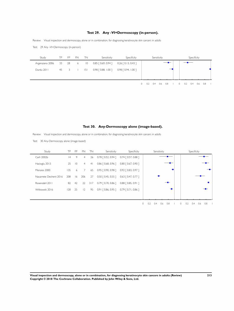
Test 29. Any -VI+Dermoscopy (in-person).
Review: Visual inspection and dermoscopy, alone or in combination, for diagnosing keratinocyte skin cancers in adults
Test: 29 Any -VI+Dermoscopy (in-person)
Study TP FP FN TN Sensitivity Specificity Sensitivity Specificity
Argenziano 2006 33 28 6 10 0.85 [ 0.69, 0.94 ] 0.26 [ 0.13, 0.43 ]
Durdu 2011 45 3 1 151 0.98 [ 0.88, 1.00 ] 0.98 [ 0.94, 1.00 ]
0 0.2 0.4 0.6 0.8 1 0 0.2 0.4 0.6 0.8 1
Test 30. Any-Dermoscopy alone (image-based).
Review: Visual inspection and dermoscopy, alone or in combination, for diagnosing keratinocyte skin cancers in adults
Test: 30 Any-Dermoscopy alone (image-based)
Study TP FP FN TN Sensitivity Specificity Sensitivity Specificity
Carli 2002b 14 9 4 26 0.78 [ 0.52, 0.94 ] 0.74 [ 0.57, 0.88 ]
Hacioglu 2013 25 10 4 41 0.86 [ 0.68, 0.96 ] 0.80 [ 0.67, 0.90 ]
Menzies 2000 135 6 7 65 0.95 [ 0.90, 0.98 ] 0.92 [ 0.83, 0.97 ]
Navarrete Dechent 2016 208 16 206 27 0.50 [ 0.45, 0.55 ] 0.63 [ 0.47, 0.77 ]
Rosendahl 2011 82 42 22 317 0.79 [ 0.70, 0.86 ] 0.88 [ 0.85, 0.91 ]
Witkowski 2016 128 25 12 95 0.91 [ 0.86, 0.95 ] 0.79 [ 0.71, 0.86 ]
0 0.2 0.4 0.6 0.8 1 0 0.2 0.4 0.6 0.8 1
213Visual inspection and dermoscopy, alone or in combination, for diagnosing keratinocyte skin cancers in adults (Review)
Copyright © 2018 The Cochrane Collaboration. Published by John Wiley & Sons, Ltd.
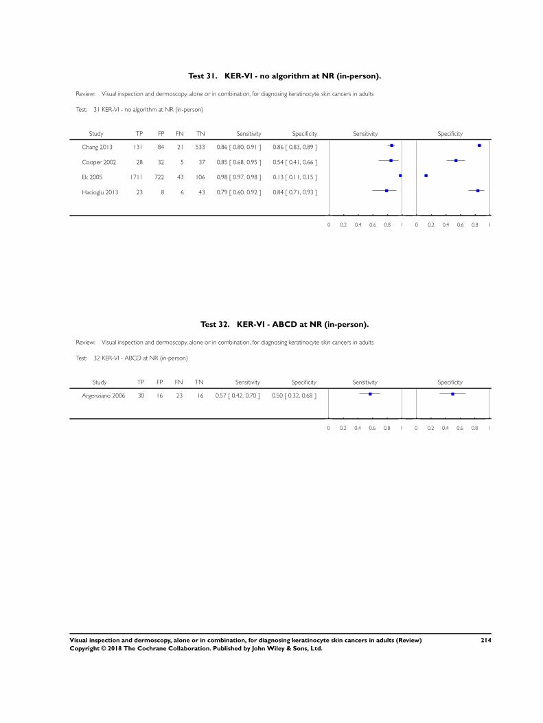
Test 31. KER-VI - no algorithm at NR (in-person).
Review: Visual inspection and dermoscopy, alone or in combination, for diagnosing keratinocyte skin cancers in adults
Test: 31 KER-VI - no algorithm at NR (in-person)
Study TP FP FN TN Sensitivity Specificity Sensitivity Specificity
Chang 2013 131 84 21 533 0.86 [ 0.80, 0.91 ] 0.86 [ 0.83, 0.89 ]
Cooper 2002 28 32 5 37 0.85 [ 0.68, 0.95 ] 0.54 [ 0.41, 0.66 ]
Ek 2005 1711 722 43 106 0.98 [ 0.97, 0.98 ] 0.13 [ 0.11, 0.15 ]
Hacioglu 2013 23 8 6 43 0.79 [ 0.60, 0.92 ] 0.84 [ 0.71, 0.93 ]
0 0.2 0.4 0.6 0.8 1 0 0.2 0.4 0.6 0.8 1
Test 32. KER-VI - ABCD at NR (in-person).
Review: Visual inspection and dermoscopy, alone or in combination, for diagnosing keratinocyte skin cancers in adults
Test: 32 KER-VI - ABCD at NR (in-person)
Study TP FP FN TN Sensitivity Specificity Sensitivity Specificity
Argenziano 2006 30 16 23 16 0.57 [ 0.42, 0.70 ] 0.50 [ 0.32, 0.68 ]
0 0.2 0.4 0.6 0.8 1 0 0.2 0.4 0.6 0.8 1
214Visual inspection and dermoscopy, alone or in combination, for diagnosing keratinocyte skin cancers in adults (Review)
Copyright © 2018 The Cochrane Collaboration. Published by John Wiley & Sons, Ltd.
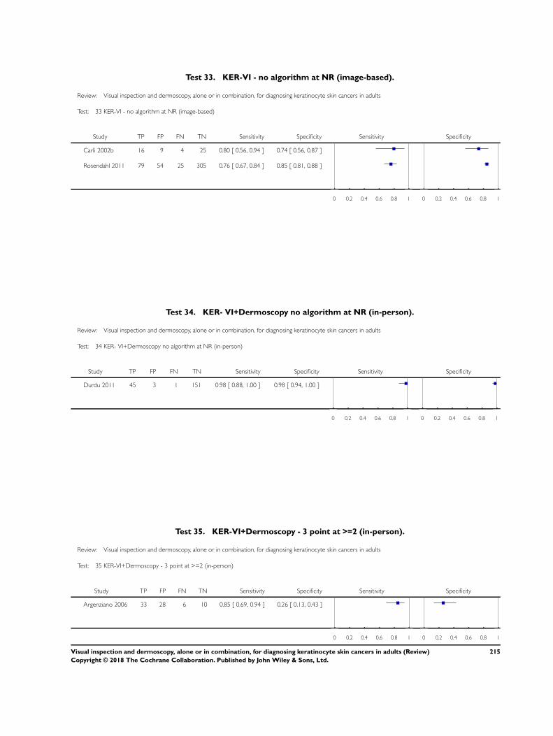
Test 33. KER-VI - no algorithm at NR (image-based).
Review: Visual inspection and dermoscopy, alone or in combination, for diagnosing keratinocyte skin cancers in adults
Test: 33 KER-VI - no algorithm at NR (image-based)
Study TP FP FN TN Sensitivity Specificity Sensitivity Specificity
Carli 2002b 16 9 4 25 0.80 [ 0.56, 0.94 ] 0.74 [ 0.56, 0.87 ]
Rosendahl 2011 79 54 25 305 0.76 [ 0.67, 0.84 ] 0.85 [ 0.81, 0.88 ]
0 0.2 0.4 0.6 0.8 1 0 0.2 0.4 0.6 0.8 1
Test 34. KER- VI+Dermoscopy no algorithm at NR (in-person).
Review: Visual inspection and dermoscopy, alone or in combination, for diagnosing keratinocyte skin cancers in adults
Test: 34 KER- VI+Dermoscopy no algorithm at NR (in-person)
Study TP FP FN TN Sensitivity Specificity Sensitivity Specificity
Durdu 2011 45 3 1 151 0.98 [ 0.88, 1.00 ] 0.98 [ 0.94, 1.00 ]
0 0.2 0.4 0.6 0.8 1 0 0.2 0.4 0.6 0.8 1
Test 35. KER-VI+Dermoscopy - 3 point at >=2 (in-person).
Review: Visual inspection and dermoscopy, alone or in combination, for diagnosing keratinocyte skin cancers in adults
Test: 35 KER-VI+Dermoscopy - 3 point at >=2 (in-person)
Study TP FP FN TN Sensitivity Specificity Sensitivity Specificity
Argenziano 2006 33 28 6 10 0.85 [ 0.69, 0.94 ] 0.26 [ 0.13, 0.43 ]
0 0.2 0.4 0.6 0.8 1 0 0.2 0.4 0.6 0.8 1
215Visual inspection and dermoscopy, alone or in combination, for diagnosing keratinocyte skin cancers in adults (Review)
Copyright © 2018 The Cochrane Collaboration. Published by John Wiley & Sons, Ltd.
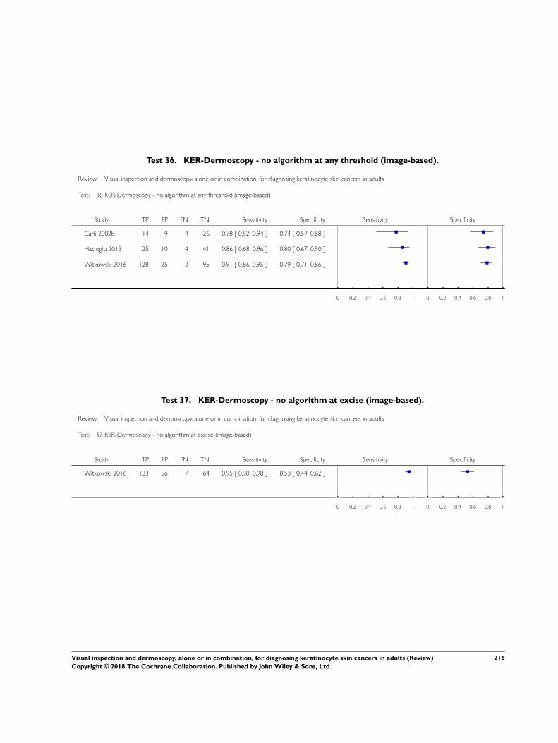
Test 36. KER-Dermoscopy - no algorithm at any threshold (image-based).
Review: Visual inspection and dermoscopy, alone or in combination, for diagnosing keratinocyte skin cancers in adults
Test: 36 KER-Dermoscopy - no algorithm at any threshold (image-based)
Study TP FP FN TN Sensitivity Specificity Sensitivity Specificity
Carli 2002b 14 9 4 26 0.78 [ 0.52, 0.94 ] 0.74 [ 0.57, 0.88 ]
Hacioglu 2013 25 10 4 41 0.86 [ 0.68, 0.96 ] 0.80 [ 0.67, 0.90 ]
Witkowski 2016 128 25 12 95 0.91 [ 0.86, 0.95 ] 0.79 [ 0.71, 0.86 ]
0 0.2 0.4 0.6 0.8 1 0 0.2 0.4 0.6 0.8 1
Test 37. KER-Dermoscopy - no algorithm at excise (image-based).
Review: Visual inspection and dermoscopy, alone or in combination, for diagnosing keratinocyte skin cancers in adults
Test: 37 KER-Dermoscopy - no algorithm at excise (image-based)
Study TP FP FN TN Sensitivity Specificity Sensitivity Specificity
Witkowski 2016 133 56 7 64 0.95 [ 0.90, 0.98 ] 0.53 [ 0.44, 0.62 ]
0 0.2 0.4 0.6 0.8 1 0 0.2 0.4 0.6 0.8 1
216Visual inspection and dermoscopy, alone or in combination, for diagnosing keratinocyte skin cancers in adults (Review)
Copyright © 2018 The Cochrane Collaboration. Published by John Wiley & Sons, Ltd.
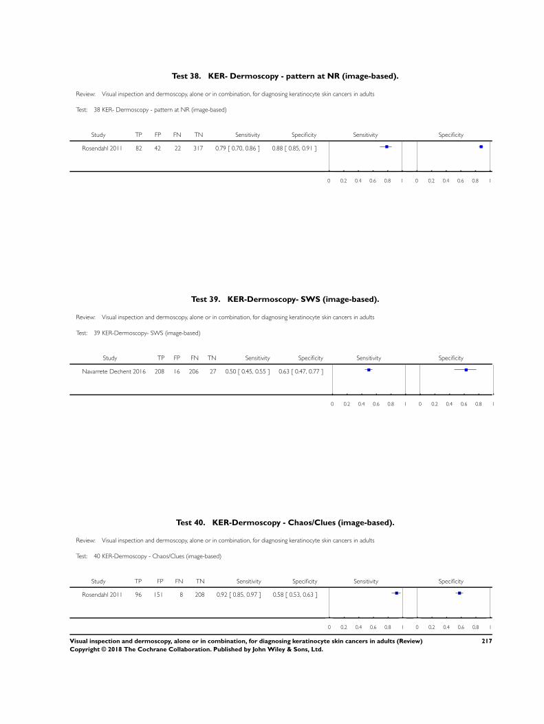
Test 38. KER- Dermoscopy - pattern at NR (image-based).
Review: Visual inspection and dermoscopy, alone or in combination, for diagnosing keratinocyte skin cancers in adults
Test: 38 KER- Dermoscopy - pattern at NR (image-based)
Study TP FP FN TN Sensitivity Specificity Sensitivity Specificity
Rosendahl 2011 82 42 22 317 0.79 [ 0.70, 0.86 ] 0.88 [ 0.85, 0.91 ]
0 0.2 0.4 0.6 0.8 1 0 0.2 0.4 0.6 0.8 1
Test 39. KER-Dermoscopy- SWS (image-based).
Review: Visual inspection and dermoscopy, alone or in combination, for diagnosing keratinocyte skin cancers in adults
Test: 39 KER-Dermoscopy- SWS (image-based)
Study TP FP FN TN Sensitivity Specificity Sensitivity Specificity
Navarrete Dechent 2016 208 16 206 27 0.50 [ 0.45, 0.55 ] 0.63 [ 0.47, 0.77 ]
0 0.2 0.4 0.6 0.8 1 0 0.2 0.4 0.6 0.8 1
Test 40. KER-Dermoscopy - Chaos/Clues (image-based).
Review: Visual inspection and dermoscopy, alone or in combination, for diagnosing keratinocyte skin cancers in adults
Test: 40 KER-Dermoscopy - Chaos/Clues (image-based)
Study TP FP FN TN Sensitivity Specificity Sensitivity Specificity
Rosendahl 2011 96 151 8 208 0.92 [ 0.85, 0.97 ] 0.58 [ 0.53, 0.63 ]
0 0.2 0.4 0.6 0.8 1 0 0.2 0.4 0.6 0.8 1
217Visual inspection and dermoscopy, alone or in combination, for diagnosing keratinocyte skin cancers in adults (Review)
Copyright © 2018 The Cochrane Collaboration. Published by John Wiley & Sons, Ltd.
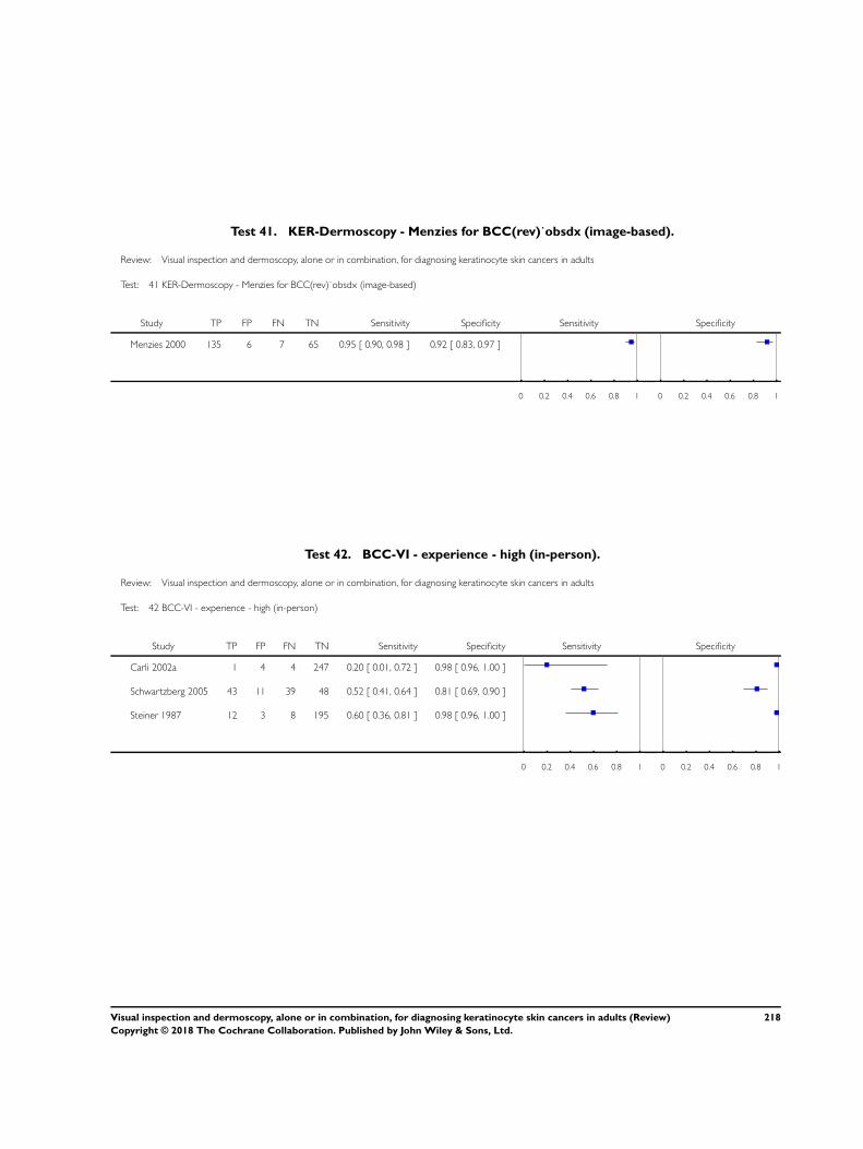
Test 41. KER-Dermoscopy - Menzies for BCC(rev)˙obsdx (image-based).
Review: Visual inspection and dermoscopy, alone or in combination, for diagnosing keratinocyte skin cancers in adults
Test: 41 KER-Dermoscopy - Menzies for BCC(rev)˙obsdx (image-based)
Study TP FP FN TN Sensitivity Specificity Sensitivity Specificity
Menzies 2000 135 6 7 65 0.95 [ 0.90, 0.98 ] 0.92 [ 0.83, 0.97 ]
0 0.2 0.4 0.6 0.8 1 0 0.2 0.4 0.6 0.8 1
Test 42. BCC-VI - experience - high (in-person).
Review: Visual inspection and dermoscopy, alone or in combination, for diagnosing keratinocyte skin cancers in adults
Test: 42 BCC-VI - experience - high (in-person)
Study TP FP FN TN Sensitivity Specificity Sensitivity Specificity
Carli 2002a 1 4 4 247 0.20 [ 0.01, 0.72 ] 0.98 [ 0.96, 1.00 ]
Schwartzberg 2005 43 11 39 48 0.52 [ 0.41, 0.64 ] 0.81 [ 0.69, 0.90 ]
Steiner 1987 12 3 8 195 0.60 [ 0.36, 0.81 ] 0.98 [ 0.96, 1.00 ]
0 0.2 0.4 0.6 0.8 1 0 0.2 0.4 0.6 0.8 1
218Visual inspection and dermoscopy, alone or in combination, for diagnosing keratinocyte skin cancers in adults (Review)
Copyright © 2018 The Cochrane Collaboration. Published by John Wiley & Sons, Ltd.
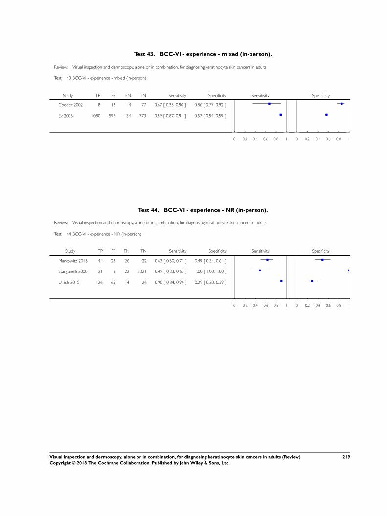
Test 43. BCC-VI - experience - mixed (in-person).
Review: Visual inspection and dermoscopy, alone or in combination, for diagnosing keratinocyte skin cancers in adults
Test: 43 BCC-VI - experience - mixed (in-person)
Study TP FP FN TN Sensitivity Specificity Sensitivity Specificity
Cooper 2002 8 13 4 77 0.67 [ 0.35, 0.90 ] 0.86 [ 0.77, 0.92 ]
Ek 2005 1080 595 134 773 0.89 [ 0.87, 0.91 ] 0.57 [ 0.54, 0.59 ]
0 0.2 0.4 0.6 0.8 1 0 0.2 0.4 0.6 0.8 1
Test 44. BCC-VI - experience - NR (in-person).
Review: Visual inspection and dermoscopy, alone or in combination, for diagnosing keratinocyte skin cancers in adults
Test: 44 BCC-VI - experience - NR (in-person)
Study TP FP FN TN Sensitivity Specificity Sensitivity Specificity
Markowitz 2015 44 23 26 22 0.63 [ 0.50, 0.74 ] 0.49 [ 0.34, 0.64 ]
Stanganelli 2000 21 8 22 3321 0.49 [ 0.33, 0.65 ] 1.00 [ 1.00, 1.00 ]
Ulrich 2015 126 65 14 26 0.90 [ 0.84, 0.94 ] 0.29 [ 0.20, 0.39 ]
0 0.2 0.4 0.6 0.8 1 0 0.2 0.4 0.6 0.8 1
219Visual inspection and dermoscopy, alone or in combination, for diagnosing keratinocyte skin cancers in adults (Review)
Copyright © 2018 The Cochrane Collaboration. Published by John Wiley & Sons, Ltd.
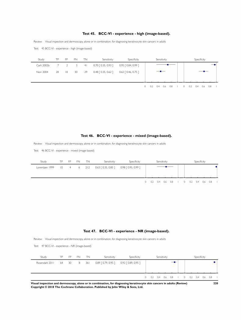
Test 45. BCC-VI - experience - high (image-based).
Review: Visual inspection and dermoscopy, alone or in combination, for diagnosing keratinocyte skin cancers in adults
Test: 45 BCC-VI - experience - high (image-based)
Study TP FP FN TN Sensitivity Specificity Sensitivity Specificity
Carli 2002b 7 2 3 41 0.70 [ 0.35, 0.93 ] 0.95 [ 0.84, 0.99 ]
Nori 2004 28 18 30 29 0.48 [ 0.35, 0.62 ] 0.62 [ 0.46, 0.75 ]
0 0.2 0.4 0.6 0.8 1 0 0.2 0.4 0.6 0.8 1
Test 46. BCC-VI - experience - mixed (image-based).
Review: Visual inspection and dermoscopy, alone or in combination, for diagnosing keratinocyte skin cancers in adults
Test: 46 BCC-VI - experience - mixed (image-based)
Study TP FP FN TN Sensitivity Specificity Sensitivity Specificity
Lorentzen 1999 10 4 6 212 0.63 [ 0.35, 0.85 ] 0.98 [ 0.95, 0.99 ]
0 0.2 0.4 0.6 0.8 1 0 0.2 0.4 0.6 0.8 1
Test 47. BCC-VI - experience - NR (image-based).
Review: Visual inspection and dermoscopy, alone or in combination, for diagnosing keratinocyte skin cancers in adults
Test: 47 BCC-VI - experience - NR (image-based)
Study TP FP FN TN Sensitivity Specificity Sensitivity Specificity
Rosendahl 2011 64 30 8 361 0.89 [ 0.79, 0.95 ] 0.92 [ 0.89, 0.95 ]
0 0.2 0.4 0.6 0.8 1 0 0.2 0.4 0.6 0.8 1
220Visual inspection and dermoscopy, alone or in combination, for diagnosing keratinocyte skin cancers in adults (Review)
Copyright © 2018 The Cochrane Collaboration. Published by John Wiley & Sons, Ltd.
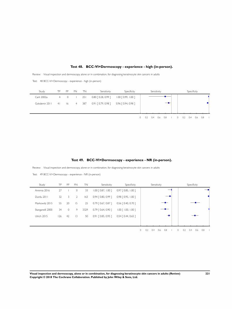
Test 48. BCC-VI+Dermoscopy - experience - high (in-person).
Review: Visual inspection and dermoscopy, alone or in combination, for diagnosing keratinocyte skin cancers in adults
Test: 48 BCC-VI+Dermoscopy - experience - high (in-person)
Study TP FP FN TN Sensitivity Specificity Sensitivity Specificity
Carli 2002a 4 0 1 251 0.80 [ 0.28, 0.99 ] 1.00 [ 0.99, 1.00 ]
Gokdemir 2011 41 16 4 387 0.91 [ 0.79, 0.98 ] 0.96 [ 0.94, 0.98 ]
0 0.2 0.4 0.6 0.8 1 0 0.2 0.4 0.6 0.8 1
Test 49. BCC-VI+Dermsocopy - experience - NR (in-person).
Review: Visual inspection and dermoscopy, alone or in combination, for diagnosing keratinocyte skin cancers in adults
Test: 49 BCC-VI+Dermsocopy - experience - NR (in-person)
Study TP FP FN TN Sensitivity Specificity Sensitivity Specificity
Amirnia 2016 27 1 0 33 1.00 [ 0.87, 1.00 ] 0.97 [ 0.85, 1.00 ]
Durdu 2011 32 3 2 163 0.94 [ 0.80, 0.99 ] 0.98 [ 0.95, 1.00 ]
Markowitz 2015 55 20 15 25 0.79 [ 0.67, 0.87 ] 0.56 [ 0.40, 0.70 ]
Stanganelli 2000 34 0 9 3329 0.79 [ 0.64, 0.90 ] 1.00 [ 1.00, 1.00 ]
Ulrich 2015 126 42 13 50 0.91 [ 0.85, 0.95 ] 0.54 [ 0.44, 0.65 ]
0 0.2 0.4 0.6 0.8 1 0 0.2 0.4 0.6 0.8 1
221Visual inspection and dermoscopy, alone or in combination, for diagnosing keratinocyte skin cancers in adults (Review)
Copyright © 2018 The Cochrane Collaboration. Published by John Wiley & Sons, Ltd.
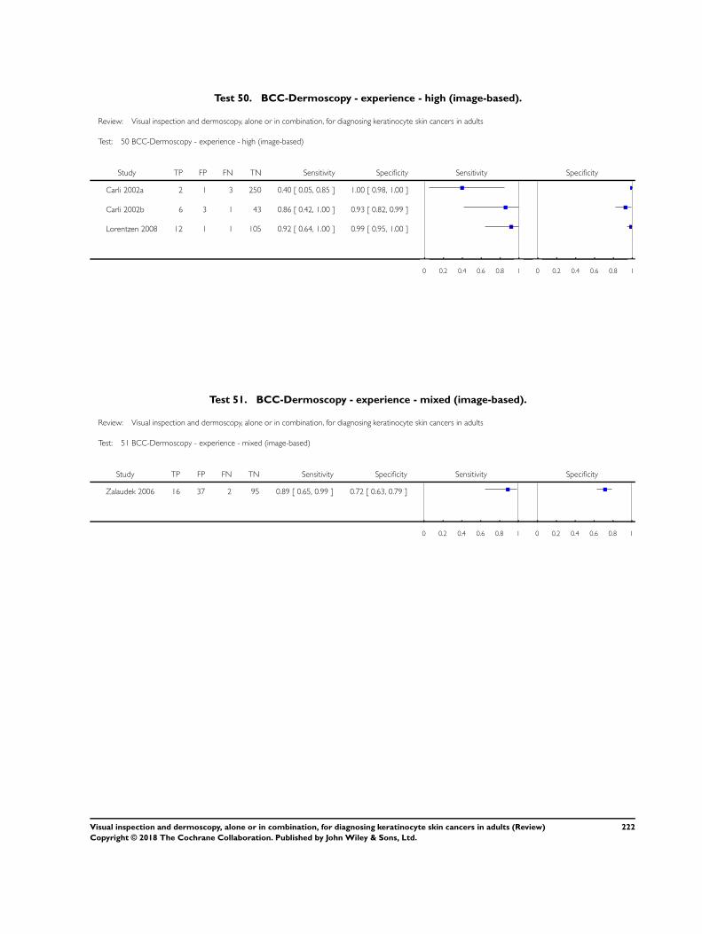
Test 50. BCC-Dermoscopy - experience - high (image-based).
Review: Visual inspection and dermoscopy, alone or in combination, for diagnosing keratinocyte skin cancers in adults
Test: 50 BCC-Dermoscopy - experience - high (image-based)
Study TP FP FN TN Sensitivity Specificity Sensitivity Specificity
Carli 2002a 2 1 3 250 0.40 [ 0.05, 0.85 ] 1.00 [ 0.98, 1.00 ]
Carli 2002b 6 3 1 43 0.86 [ 0.42, 1.00 ] 0.93 [ 0.82, 0.99 ]
Lorentzen 2008 12 1 1 105 0.92 [ 0.64, 1.00 ] 0.99 [ 0.95, 1.00 ]
0 0.2 0.4 0.6 0.8 1 0 0.2 0.4 0.6 0.8 1
Test 51. BCC-Dermoscopy - experience - mixed (image-based).
Review: Visual inspection and dermoscopy, alone or in combination, for diagnosing keratinocyte skin cancers in adults
Test: 51 BCC-Dermoscopy - experience - mixed (image-based)
Study TP FP FN TN Sensitivity Specificity Sensitivity Specificity
Zalaudek 2006 16 37 2 95 0.89 [ 0.65, 0.99 ] 0.72 [ 0.63, 0.79 ]
0 0.2 0.4 0.6 0.8 1 0 0.2 0.4 0.6 0.8 1
222Visual inspection and dermoscopy, alone or in combination, for diagnosing keratinocyte skin cancers in adults (Review)
Copyright © 2018 The Cochrane Collaboration. Published by John Wiley & Sons, Ltd.
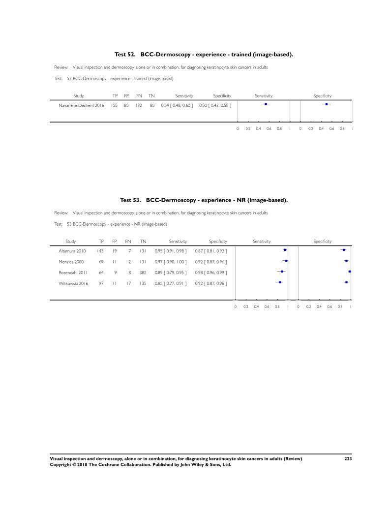
Test 52. BCC-Dermoscopy - experience - trained (image-based).
Review: Visual inspection and dermoscopy, alone or in combination, for diagnosing keratinocyte skin cancers in adults
Test: 52 BCC-Dermoscopy - experience - trained (image-based)
Study TP FP FN TN Sensitivity Specificity Sensitivity Specificity
Navarrete Dechent 2016 155 85 132 85 0.54 [ 0.48, 0.60 ] 0.50 [ 0.42, 0.58 ]
0 0.2 0.4 0.6 0.8 1 0 0.2 0.4 0.6 0.8 1
Test 53. BCC-Dermoscopy - experience - NR (image-based).
Review: Visual inspection and dermoscopy, alone or in combination, for diagnosing keratinocyte skin cancers in adults
Test: 53 BCC-Dermoscopy - experience - NR (image-based)
Study TP FP FN TN Sensitivity Specificity Sensitivity Specificity
Altamura 2010 143 19 7 131 0.95 [ 0.91, 0.98 ] 0.87 [ 0.81, 0.92 ]
Menzies 2000 69 11 2 131 0.97 [ 0.90, 1.00 ] 0.92 [ 0.87, 0.96 ]
Rosendahl 2011 64 9 8 382 0.89 [ 0.79, 0.95 ] 0.98 [ 0.96, 0.99 ]
Witkowski 2016 97 11 17 135 0.85 [ 0.77, 0.91 ] 0.92 [ 0.87, 0.96 ]
0 0.2 0.4 0.6 0.8 1 0 0.2 0.4 0.6 0.8 1
223Visual inspection and dermoscopy, alone or in combination, for diagnosing keratinocyte skin cancers in adults (Review)
Copyright © 2018 The Cochrane Collaboration. Published by John Wiley & Sons, Ltd.
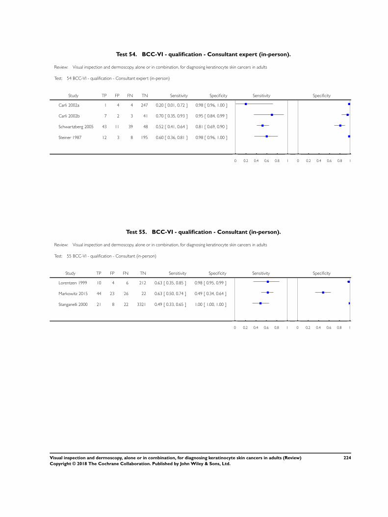
Test 54. BCC-VI - qualification - Consultant expert (in-person).
Review: Visual inspection and dermoscopy, alone or in combination, for diagnosing keratinocyte skin cancers in adults
Test: 54 BCC-VI - qualification - Consultant expert (in-person)
Study TP FP FN TN Sensitivity Specificity Sensitivity Specificity
Carli 2002a 1 4 4 247 0.20 [ 0.01, 0.72 ] 0.98 [ 0.96, 1.00 ]
Carli 2002b 7 2 3 41 0.70 [ 0.35, 0.93 ] 0.95 [ 0.84, 0.99 ]
Schwartzberg 2005 43 11 39 48 0.52 [ 0.41, 0.64 ] 0.81 [ 0.69, 0.90 ]
Steiner 1987 12 3 8 195 0.60 [ 0.36, 0.81 ] 0.98 [ 0.96, 1.00 ]
0 0.2 0.4 0.6 0.8 1 0 0.2 0.4 0.6 0.8 1
Test 55. BCC-VI - qualification - Consultant (in-person).
Review: Visual inspection and dermoscopy, alone or in combination, for diagnosing keratinocyte skin cancers in adults
Test: 55 BCC-VI - qualification - Consultant (in-person)
Study TP FP FN TN Sensitivity Specificity Sensitivity Specificity
Lorentzen 1999 10 4 6 212 0.63 [ 0.35, 0.85 ] 0.98 [ 0.95, 0.99 ]
Markowitz 2015 44 23 26 22 0.63 [ 0.50, 0.74 ] 0.49 [ 0.34, 0.64 ]
Stanganelli 2000 21 8 22 3321 0.49 [ 0.33, 0.65 ] 1.00 [ 1.00, 1.00 ]
0 0.2 0.4 0.6 0.8 1 0 0.2 0.4 0.6 0.8 1
224Visual inspection and dermoscopy, alone or in combination, for diagnosing keratinocyte skin cancers in adults (Review)
Copyright © 2018 The Cochrane Collaboration. Published by John Wiley & Sons, Ltd.
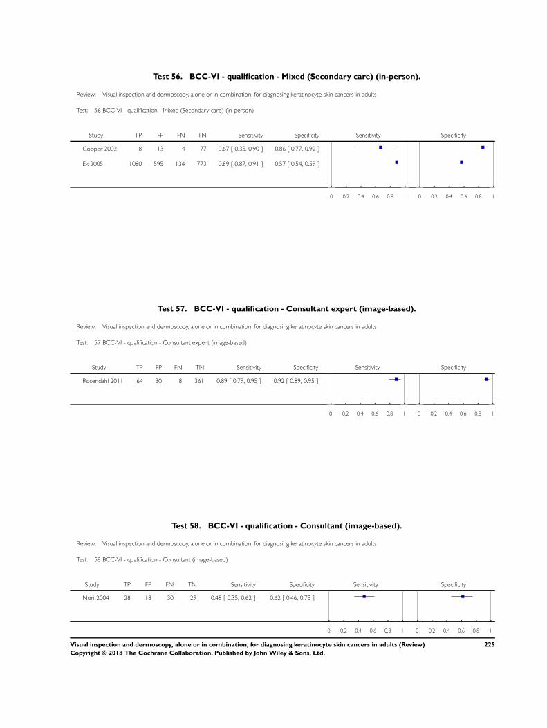
Test 56. BCC-VI - qualification - Mixed (Secondary care) (in-person).
Review: Visual inspection and dermoscopy, alone or in combination, for diagnosing keratinocyte skin cancers in adults
Test: 56 BCC-VI - qualification - Mixed (Secondary care) (in-person)
Study TP FP FN TN Sensitivity Specificity Sensitivity Specificity
Cooper 2002 8 13 4 77 0.67 [ 0.35, 0.90 ] 0.86 [ 0.77, 0.92 ]
Ek 2005 1080 595 134 773 0.89 [ 0.87, 0.91 ] 0.57 [ 0.54, 0.59 ]
0 0.2 0.4 0.6 0.8 1 0 0.2 0.4 0.6 0.8 1
Test 57. BCC-VI - qualification - Consultant expert (image-based).
Review: Visual inspection and dermoscopy, alone or in combination, for diagnosing keratinocyte skin cancers in adults
Test: 57 BCC-VI - qualification - Consultant expert (image-based)
Study TP FP FN TN Sensitivity Specificity Sensitivity Specificity
Rosendahl 2011 64 30 8 361 0.89 [ 0.79, 0.95 ] 0.92 [ 0.89, 0.95 ]
0 0.2 0.4 0.6 0.8 1 0 0.2 0.4 0.6 0.8 1
Test 58. BCC-VI - qualification - Consultant (image-based).
Review: Visual inspection and dermoscopy, alone or in combination, for diagnosing keratinocyte skin cancers in adults
Test: 58 BCC-VI - qualification - Consultant (image-based)
Study TP FP FN TN Sensitivity Specificity Sensitivity Specificity
Nori 2004 28 18 30 29 0.48 [ 0.35, 0.62 ] 0.62 [ 0.46, 0.75 ]
0 0.2 0.4 0.6 0.8 1 0 0.2 0.4 0.6 0.8 1
225Visual inspection and dermoscopy, alone or in combination, for diagnosing keratinocyte skin cancers in adults (Review)
Copyright © 2018 The Cochrane Collaboration. Published by John Wiley & Sons, Ltd.
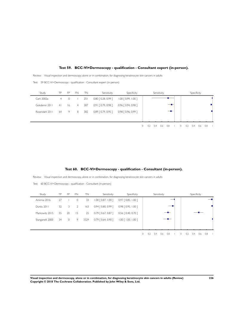
Test 59. BCC-VI+Dermoscopy - qualification - Consultant expert (in-person).
Review: Visual inspection and dermoscopy, alone or in combination, for diagnosing keratinocyte skin cancers in adults
Test: 59 BCC-VI+Dermoscopy - qualification - Consultant expert (in-person)
Study TP FP FN TN Sensitivity Specificity Sensitivity Specificity
Carli 2002a 4 0 1 251 0.80 [ 0.28, 0.99 ] 1.00 [ 0.99, 1.00 ]
Gokdemir 2011 41 16 4 387 0.91 [ 0.79, 0.98 ] 0.96 [ 0.94, 0.98 ]
Rosendahl 2011 64 9 8 382 0.89 [ 0.79, 0.95 ] 0.98 [ 0.96, 0.99 ]
0 0.2 0.4 0.6 0.8 1 0 0.2 0.4 0.6 0.8 1
Test 60. BCC-VI+Dermoscopy - qualification - Consultant (in-person).
Review: Visual inspection and dermoscopy, alone or in combination, for diagnosing keratinocyte skin cancers in adults
Test: 60 BCC-VI+Dermoscopy - qualification - Consultant (in-person)
Study TP FP FN TN Sensitivity Specificity Sensitivity Specificity
Amirnia 2016 27 1 0 33 1.00 [ 0.87, 1.00 ] 0.97 [ 0.85, 1.00 ]
Durdu 2011 32 3 2 163 0.94 [ 0.80, 0.99 ] 0.98 [ 0.95, 1.00 ]
Markowitz 2015 55 20 15 25 0.79 [ 0.67, 0.87 ] 0.56 [ 0.40, 0.70 ]
Stanganelli 2000 34 0 9 3329 0.79 [ 0.64, 0.90 ] 1.00 [ 1.00, 1.00 ]
0 0.2 0.4 0.6 0.8 1 0 0.2 0.4 0.6 0.8 1
226Visual inspection and dermoscopy, alone or in combination, for diagnosing keratinocyte skin cancers in adults (Review)
Copyright © 2018 The Cochrane Collaboration. Published by John Wiley & Sons, Ltd.
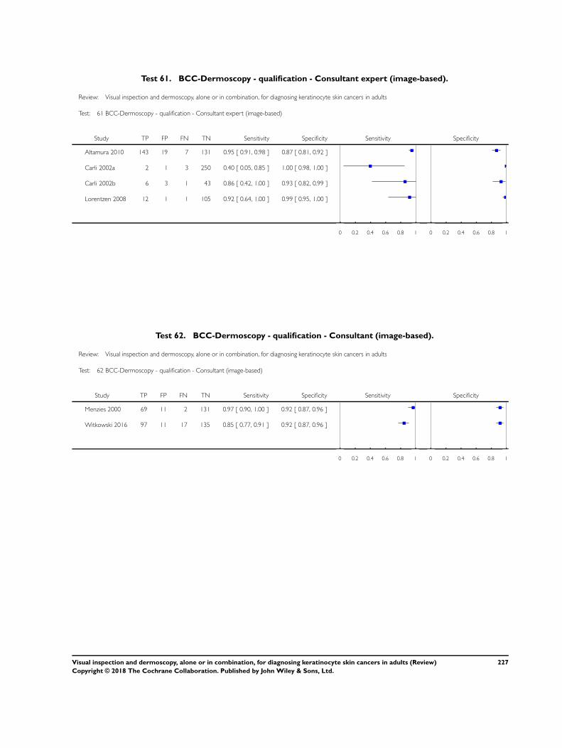
Test 61. BCC-Dermoscopy - qualification - Consultant expert (image-based).
Review: Visual inspection and dermoscopy, alone or in combination, for diagnosing keratinocyte skin cancers in adults
Test: 61 BCC-Dermoscopy - qualification - Consultant expert (image-based)
Study TP FP FN TN Sensitivity Specificity Sensitivity Specificity
Altamura 2010 143 19 7 131 0.95 [ 0.91, 0.98 ] 0.87 [ 0.81, 0.92 ]
Carli 2002a 2 1 3 250 0.40 [ 0.05, 0.85 ] 1.00 [ 0.98, 1.00 ]
Carli 2002b 6 3 1 43 0.86 [ 0.42, 1.00 ] 0.93 [ 0.82, 0.99 ]
Lorentzen 2008 12 1 1 105 0.92 [ 0.64, 1.00 ] 0.99 [ 0.95, 1.00 ]
0 0.2 0.4 0.6 0.8 1 0 0.2 0.4 0.6 0.8 1
Test 62. BCC-Dermoscopy - qualification - Consultant (image-based).
Review: Visual inspection and dermoscopy, alone or in combination, for diagnosing keratinocyte skin cancers in adults
Test: 62 BCC-Dermoscopy - qualification - Consultant (image-based)
Study TP FP FN TN Sensitivity Specificity Sensitivity Specificity
Menzies 2000 69 11 2 131 0.97 [ 0.90, 1.00 ] 0.92 [ 0.87, 0.96 ]
Witkowski 2016 97 11 17 135 0.85 [ 0.77, 0.91 ] 0.92 [ 0.87, 0.96 ]
0 0.2 0.4 0.6 0.8 1 0 0.2 0.4 0.6 0.8 1
227Visual inspection and dermoscopy, alone or in combination, for diagnosing keratinocyte skin cancers in adults (Review)
Copyright © 2018 The Cochrane Collaboration. Published by John Wiley & Sons, Ltd.
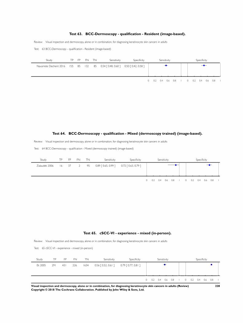
Test 63. BCC-Dermoscopy - qualification - Resident (image-based).
Review: Visual inspection and dermoscopy, alone or in combination, for diagnosing keratinocyte skin cancers in adults
Test: 63 BCC-Dermoscopy - qualification - Resident (image-based)
Study TP FP FN TN Sensitivity Specificity Sensitivity Specificity
Navarrete Dechent 2016 155 85 132 85 0.54 [ 0.48, 0.60 ] 0.50 [ 0.42, 0.58 ]
0 0.2 0.4 0.6 0.8 1 0 0.2 0.4 0.6 0.8 1
Test 64. BCC-Dermoscopy - qualification - Mixed (dermoscopy trained) (image-based).
Review: Visual inspection and dermoscopy, alone or in combination, for diagnosing keratinocyte skin cancers in adults
Test: 64 BCC-Dermoscopy - qualification - Mixed (dermoscopy trained) (image-based)
Study TP FP FN TN Sensitivity Specificity Sensitivity Specificity
Zalaudek 2006 16 37 2 95 0.89 [ 0.65, 0.99 ] 0.72 [ 0.63, 0.79 ]
0 0.2 0.4 0.6 0.8 1 0 0.2 0.4 0.6 0.8 1
Test 65. cSCC-VI - experience - mixed (in-person).
Review: Visual inspection and dermoscopy, alone or in combination, for diagnosing keratinocyte skin cancers in adults
Test: 65 cSCC-VI - experience - mixed (in-person)
Study TP FP FN TN Sensitivity Specificity Sensitivity Specificity
Ek 2005 291 431 226 1634 0.56 [ 0.52, 0.61 ] 0.79 [ 0.77, 0.81 ]
0 0.2 0.4 0.6 0.8 1 0 0.2 0.4 0.6 0.8 1
228Visual inspection and dermoscopy, alone or in combination, for diagnosing keratinocyte skin cancers in adults (Review)
Copyright © 2018 The Cochrane Collaboration. Published by John Wiley & Sons, Ltd.
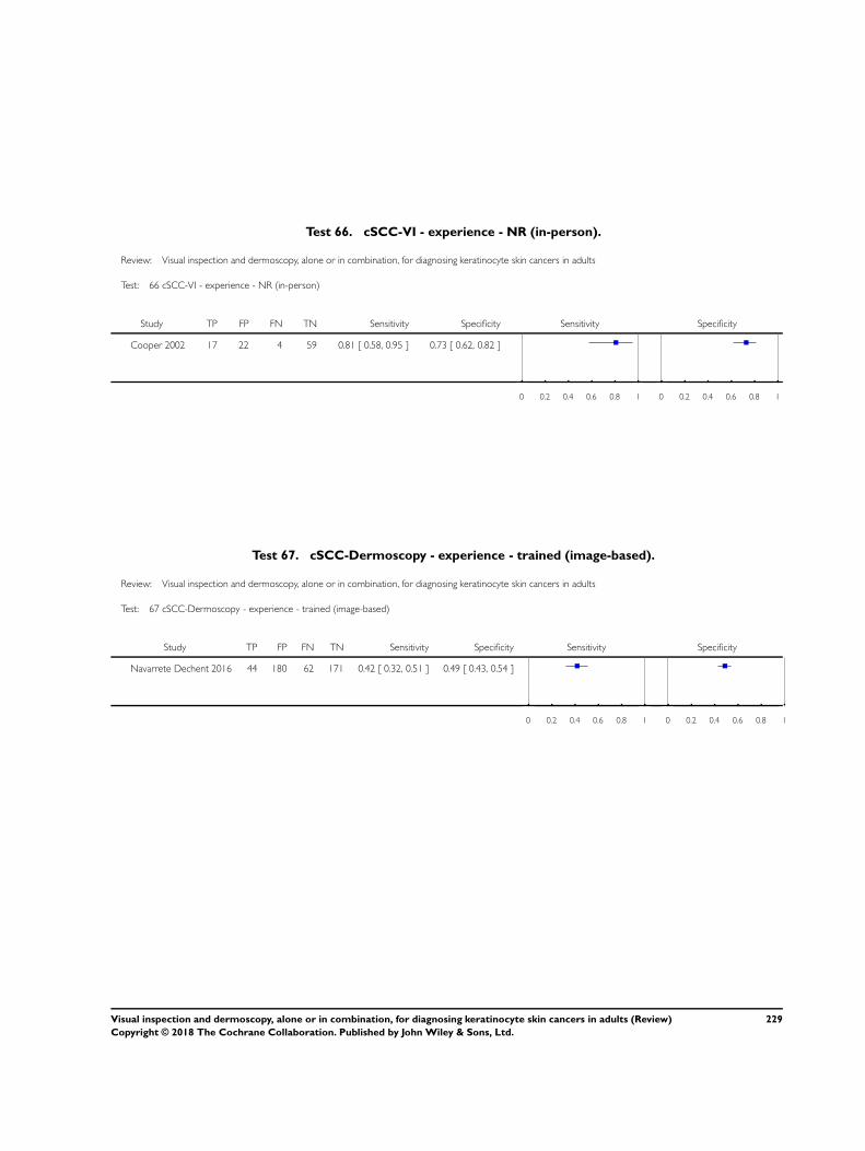
Test 66. cSCC-VI - experience - NR (in-person).
Review: Visual inspection and dermoscopy, alone or in combination, for diagnosing keratinocyte skin cancers in adults
Test: 66 cSCC-VI - experience - NR (in-person)
Study TP FP FN TN Sensitivity Specificity Sensitivity Specificity
Cooper 2002 17 22 4 59 0.81 [ 0.58, 0.95 ] 0.73 [ 0.62, 0.82 ]
0 0.2 0.4 0.6 0.8 1 0 0.2 0.4 0.6 0.8 1
Test 67. cSCC-Dermoscopy - experience - trained (image-based).
Review: Visual inspection and dermoscopy, alone or in combination, for diagnosing keratinocyte skin cancers in adults
Test: 67 cSCC-Dermoscopy - experience - trained (image-based)
Study TP FP FN TN Sensitivity Specificity Sensitivity Specificity
Navarrete Dechent 2016 44 180 62 171 0.42 [ 0.32, 0.51 ] 0.49 [ 0.43, 0.54 ]
0 0.2 0.4 0.6 0.8 1 0 0.2 0.4 0.6 0.8 1
229Visual inspection and dermoscopy, alone or in combination, for diagnosing keratinocyte skin cancers in adults (Review)
Copyright © 2018 The Cochrane Collaboration. Published by John Wiley & Sons, Ltd.
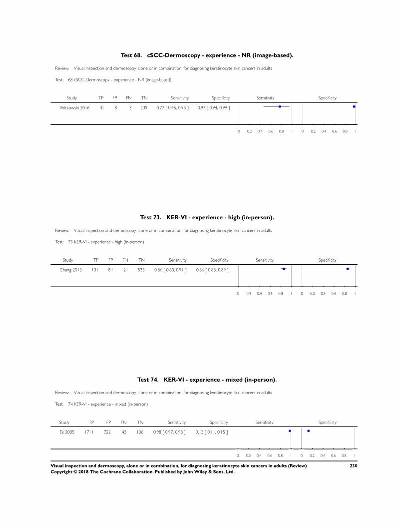
Test 68. cSCC-Dermoscopy - experience - NR (image-based).
Review: Visual inspection and dermoscopy, alone or in combination, for diagnosing keratinocyte skin cancers in adults
Test: 68 cSCC-Dermoscopy - experience - NR (image-based)
Study TP FP FN TN Sensitivity Specificity Sensitivity Specificity
Witkowski 2016 10 8 3 239 0.77 [ 0.46, 0.95 ] 0.97 [ 0.94, 0.99 ]
0 0.2 0.4 0.6 0.8 1 0 0.2 0.4 0.6 0.8 1
Test 73. KER-VI - experience - high (in-person).
Review: Visual inspection and dermoscopy, alone or in combination, for diagnosing keratinocyte skin cancers in adults
Test: 73 KER-VI - experience - high (in-person)
Study TP FP FN TN Sensitivity Specificity Sensitivity Specificity
Chang 2013 131 84 21 533 0.86 [ 0.80, 0.91 ] 0.86 [ 0.83, 0.89 ]
0 0.2 0.4 0.6 0.8 1 0 0.2 0.4 0.6 0.8 1
Test 74. KER-VI - experience - mixed (in-person).
Review: Visual inspection and dermoscopy, alone or in combination, for diagnosing keratinocyte skin cancers in adults
Test: 74 KER-VI - experience - mixed (in-person)
Study TP FP FN TN Sensitivity Specificity Sensitivity Specificity
Ek 2005 1711 722 43 106 0.98 [ 0.97, 0.98 ] 0.13 [ 0.11, 0.15 ]
0 0.2 0.4 0.6 0.8 1 0 0.2 0.4 0.6 0.8 1
230Visual inspection and dermoscopy, alone or in combination, for diagnosing keratinocyte skin cancers in adults (Review)
Copyright © 2018 The Cochrane Collaboration. Published by John Wiley & Sons, Ltd.
Test 75. KER-VI - experience - NR (in-person).
Review: Visual inspection and dermoscopy, alone or in combination, for diagnosing keratinocyte skin cancers in adults
Test: 75 KER-VI - experience - NR (in-person)
Study TP FP FN TN Sensitivity Specificity Sensitivity Specificity
Argenziano 2006 30 16 23 16 0.57 [ 0.42, 0.70 ] 0.50 [ 0.32, 0.68 ]
Cooper 2002 28 32 5 37 0.85 [ 0.68, 0.95 ] 0.54 [ 0.41, 0.66 ]
Hacioglu 2013 23 8 6 43 0.79 [ 0.60, 0.92 ] 0.84 [ 0.71, 0.93 ]
0 0.2 0.4 0.6 0.8 1 0 0.2 0.4 0.6 0.8 1
Test 76. KER-VI - experience - high (image-based).
Review: Visual inspection and dermoscopy, alone or in combination, for diagnosing keratinocyte skin cancers in adults
Test: 76 KER-VI - experience - high (image-based)
Study TP FP FN TN Sensitivity Specificity Sensitivity Specificity
Carli 2002b 16 9 4 25 0.80 [ 0.56, 0.94 ] 0.74 [ 0.56, 0.87 ]
0 0.2 0.4 0.6 0.8 1 0 0.2 0.4 0.6 0.8 1
231Visual inspection and dermoscopy, alone or in combination, for diagnosing keratinocyte skin cancers in adults (Review)
Copyright © 2018 The Cochrane Collaboration. Published by John Wiley & Sons, Ltd.
Test 77. KER-VI - experience - NR (image-based).
Review: Visual inspection and dermoscopy, alone or in combination, for diagnosing keratinocyte skin cancers in adults
Test: 77 KER-VI - experience - NR (image-based)
Study TP FP FN TN Sensitivity Specificity Sensitivity Specificity
Rosendahl 2011 79 54 25 305 0.76 [ 0.67, 0.84 ] 0.85 [ 0.81, 0.88 ]
0 0.2 0.4 0.6 0.8 1 0 0.2 0.4 0.6 0.8 1
Test 78. KER-VI+Dermoscopy - experience - trained (in-person).
Review: Visual inspection and dermoscopy, alone or in combination, for diagnosing keratinocyte skin cancers in adults
Test: 78 KER-VI+Dermoscopy - experience - trained (in-person)
Study TP FP FN TN Sensitivity Specificity Sensitivity Specificity
Argenziano 2006 33 28 6 10 0.85 [ 0.69, 0.94 ] 0.26 [ 0.13, 0.43 ]
0 0.2 0.4 0.6 0.8 1 0 0.2 0.4 0.6 0.8 1
Test 80. KER-VI+Dermoscopy - experience - NR (in-person).
Review: Visual inspection and dermoscopy, alone or in combination, for diagnosing keratinocyte skin cancers in adults
Test: 80 KER-VI+Dermoscopy - experience - NR (in-person)
Study TP FP FN TN Sensitivity Specificity Sensitivity Specificity
Durdu 2011 45 3 1 151 0.98 [ 0.88, 1.00 ] 0.98 [ 0.94, 1.00 ]
0 0.2 0.4 0.6 0.8 1 0 0.2 0.4 0.6 0.8 1
232Visual inspection and dermoscopy, alone or in combination, for diagnosing keratinocyte skin cancers in adults (Review)
Copyright © 2018 The Cochrane Collaboration. Published by John Wiley & Sons, Ltd.
Test 81. KER-Dermoscopy - experience - high (image-based).
Review: Visual inspection and dermoscopy, alone or in combination, for diagnosing keratinocyte skin cancers in adults
Test: 81 KER-Dermoscopy - experience - high (image-based)
Study TP FP FN TN Sensitivity Specificity Sensitivity Specificity
Carli 2002b 14 9 4 26 0.78 [ 0.52, 0.94 ] 0.74 [ 0.57, 0.88 ]
0 0.2 0.4 0.6 0.8 1 0 0.2 0.4 0.6 0.8 1
Test 82. KER-Dermoscopy - experience - trained (image-based).
Review: Visual inspection and dermoscopy, alone or in combination, for diagnosing keratinocyte skin cancers in adults
Test: 82 KER-Dermoscopy - experience - trained (image-based)
Study TP FP FN TN Sensitivity Specificity Sensitivity Specificity
Navarrete Dechent 2016 208 16 206 27 0.50 [ 0.45, 0.55 ] 0.63 [ 0.47, 0.77 ]
0 0.2 0.4 0.6 0.8 1 0 0.2 0.4 0.6 0.8 1
233Visual inspection and dermoscopy, alone or in combination, for diagnosing keratinocyte skin cancers in adults (Review)
Copyright © 2018 The Cochrane Collaboration. Published by John Wiley & Sons, Ltd.
Test 83. KER-Dermoscopy - experience - NR (image-based).
Review: Visual inspection and dermoscopy, alone or in combination, for diagnosing keratinocyte skin cancers in adults
Test: 83 KER-Dermoscopy - experience - NR (image-based)
Study TP FP FN TN Sensitivity Specificity Sensitivity Specificity
Hacioglu 2013 25 10 4 41 0.86 [ 0.68, 0.96 ] 0.80 [ 0.67, 0.90 ]
Menzies 2000 135 6 7 65 0.95 [ 0.90, 0.98 ] 0.92 [ 0.83, 0.97 ]
Rosendahl 2011 82 42 22 317 0.79 [ 0.70, 0.86 ] 0.88 [ 0.85, 0.91 ]
Witkowski 2016 128 25 12 95 0.91 [ 0.86, 0.95 ] 0.79 [ 0.71, 0.86 ]
0 0.2 0.4 0.6 0.8 1 0 0.2 0.4 0.6 0.8 1
A D D I T I O N A L T A B L E S
Table 1. Comparison of visual inspection and dermoscopy for detection of BCC
Test Datasets Lesions
(BCCs)
DOR
(95% CI)
Specificity
at 80%
sensitivity
Sensitiv-
ity at 80%
specificity
Relative
DOR
(95% CI)
P value
(LR)aP value
(Wald)b
In-person evaluations
Visual in-
spection
8 7017
(1586)
19.9
(7.8 to 51.
2)
77% 79% 8.2
(3.5 to 19.
3)
< 0.001 < 0.001
Visual in-
spection
+ Der-
moscopy
7 4683
(363)
164
(56.8 to
475)
99% 93%
In-person evaluations (direct studies)
Visual in-
spection
4 3974
(257)
12.8
(3.3 to 48.
8)
36% 71% 7.5
(2.7 to 21.
3)
< 0.001 < 0.001
Visual in-
spection
+ Der-
moscopy
4 3974
(258)
96.2
(21.1 to
439)
97% 87%
234Visual inspection and dermoscopy, alone or in combination, for diagnosing keratinocyte skin cancers in adults (Review)
Copyright © 2018 The Cochrane Collaboration. Published by John Wiley & Sons, Ltd.
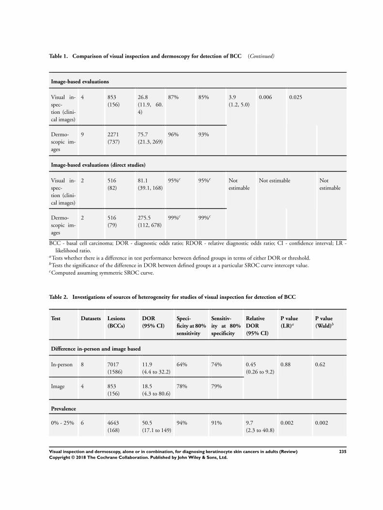
Table 1. Comparison of visual inspection and dermoscopy for detection of BCC (Continued)
Image-based evaluations
Visual in-
spec-
tion (clini-
cal images)
4 853
(156)
26.8
(11.9, 60.
4)
87% 85% 3.9
(1.2, 5.0)
0.006 0.025
Dermo-
scopic im-
ages
9 2271
(737)
75.7
(21.3, 269)
96% 93%
Image-based evaluations (direct studies)
Visual in-
spec-
tion (clini-
cal images)
2 516
(82)
81.1
(39.1, 168)
95%c 95%c Not
estimable
Not estimable Not
estimable
Dermo-
scopic im-
ages
2 516
(79)
275.5
(112, 678)
99%c 99%c
BCC - basal cell carcinoma; DOR - diagnostic odds ratio; RDOR - relative diagnostic odds ratio; CI - confidence interval; LR -
likelihood ratio.aTests whether there is a difference in test performance between defined groups in terms of either DOR or threshold.bTests the significance of the difference in DOR between defined groups at a particular SROC curve intercept value.cComputed assuming symmetric SROC curve.
Table 2. Investigations of sources of heterogeneity for studies of visual inspection for detection of BCC
Test Datasets Lesions
(BCCs)
DOR
(95% CI)
Speci-
ficity at 80%
sensitivity
Sensitiv-
ity at 80%
specificity
Relative
DOR
(95% CI)
P value
(LR)aP value
(Wald)b
Difference in-person and image based
In-person 8 7017
(1586)
11.9
(4.4 to 32.2)
64% 74% 0.45
(0.26 to 9.2)
0.88 0.62
Image 4 853
(156)
18.5
(4.3 to 80.6)
78% 79%
Prevalence
0% - 25% 6 4643
(168)
50.5
(17.1 to 149)
94% 91% 9.7
(2.3 to 40.8)
0.002 0.002
235Visual inspection and dermoscopy, alone or in combination, for diagnosing keratinocyte skin cancers in adults (Review)
Copyright © 2018 The Cochrane Collaboration. Published by John Wiley & Sons, Ltd.
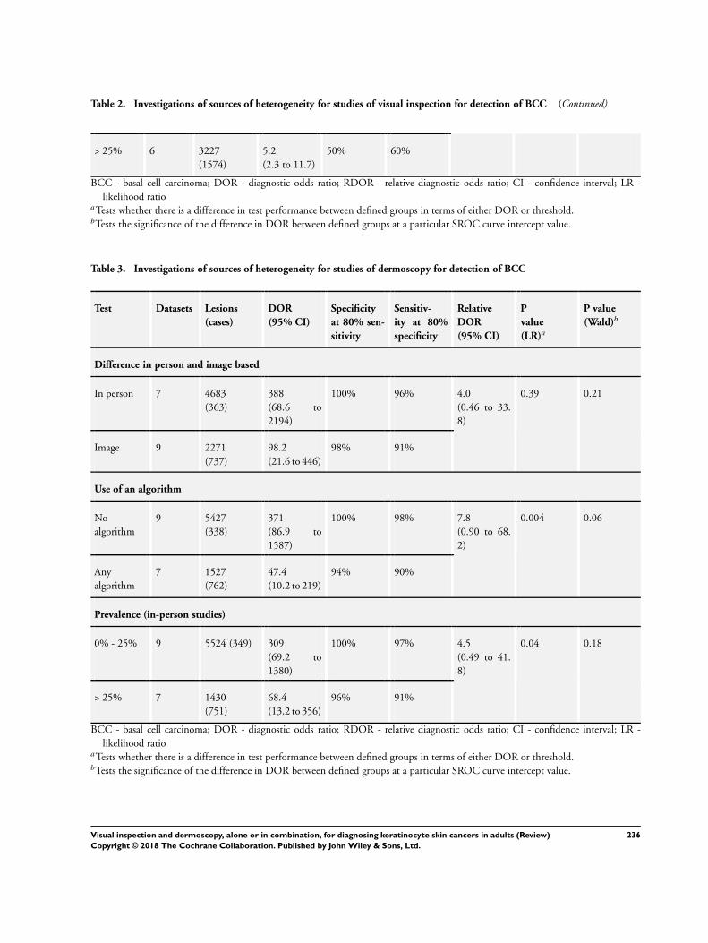
Table 2. Investigations of sources of heterogeneity for studies of visual inspection for detection of BCC (Continued)
> 25% 6 3227
(1574)
5.2
(2.3 to 11.7)
50% 60%
BCC - basal cell carcinoma; DOR - diagnostic odds ratio; RDOR - relative diagnostic odds ratio; CI - confidence interval; LR -
likelihood ratioaTests whether there is a difference in test performance between defined groups in terms of either DOR or threshold.bTests the significance of the difference in DOR between defined groups at a particular SROC curve intercept value.
Table 3. Investigations of sources of heterogeneity for studies of dermoscopy for detection of BCC
Test Datasets Lesions
(cases)
DOR
(95% CI)
Specificity
at 80% sen-
sitivity
Sensitiv-
ity at 80%
specificity
Relative
DOR
(95% CI)
P
value
(LR)a
P value
(Wald)b
Difference in person and image based
In person 7 4683
(363)
388
(68.6 to
2194)
100% 96% 4.0
(0.46 to 33.
8)
0.39 0.21
Image 9 2271
(737)
98.2
(21.6 to 446)
98% 91%
Use of an algorithm
No
algorithm
9 5427
(338)
371
(86.9 to
1587)
100% 98% 7.8
(0.90 to 68.
2)
0.004 0.06
Any
algorithm
7 1527
(762)
47.4
(10.2 to 219)
94% 90%
Prevalence (in-person studies)
0% - 25% 9 5524 (349) 309
(69.2 to
1380)
100% 97% 4.5
(0.49 to 41.
8)
0.04 0.18
> 25% 7 1430
(751)
68.4
(13.2 to 356)
96% 91%
BCC - basal cell carcinoma; DOR - diagnostic odds ratio; RDOR - relative diagnostic odds ratio; CI - confidence interval; LR -
likelihood ratioaTests whether there is a difference in test performance between defined groups in terms of either DOR or threshold.bTests the significance of the difference in DOR between defined groups at a particular SROC curve intercept value.
236Visual inspection and dermoscopy, alone or in combination, for diagnosing keratinocyte skin cancers in adults (Review)
Copyright © 2018 The Cochrane Collaboration. Published by John Wiley & Sons, Ltd.
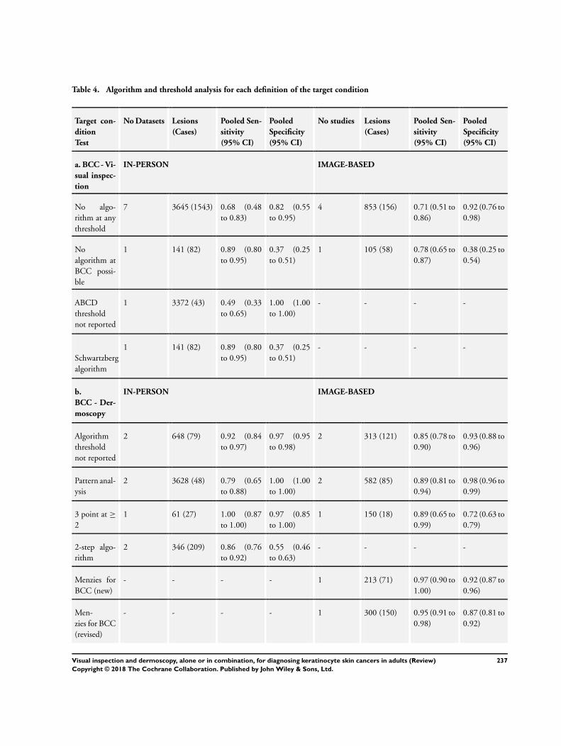
Table 4. Algorithm and threshold analysis for each definition of the target condition
Target con-
dition
Test
No Datasets Lesions
(Cases)
Pooled Sen-
sitivity
(95% CI)
Pooled
Specificity
(95% CI)
No studies Lesions
(Cases)
Pooled Sen-
sitivity
(95% CI)
Pooled
Specificity
(95% CI)
a. BCC - Vi-
sual inspec-
tion
IN-PERSON IMAGE-BASED
No algo-
rithm at any
threshold
7 3645 (1543) 0.68 (0.48
to 0.83)
0.82 (0.55
to 0.95)
4 853 (156) 0.71 (0.51 to
0.86)
0.92 (0.76 to
0.98)
No
algorithm at
BCC possi-
ble
1 141 (82) 0.89 (0.80
to 0.95)
0.37 (0.25
to 0.51)
1 105 (58) 0.78 (0.65 to
0.87)
0.38 (0.25 to
0.54)
ABCD
threshold
not reported
1 3372 (43) 0.49 (0.33
to 0.65)
1.00 (1.00
to 1.00)
- - - -
Schwartzberg
algorithm
1 141 (82) 0.89 (0.80
to 0.95)
0.37 (0.25
to 0.51)
- - - -
b.
BCC - Der-
moscopy
IN-PERSON IMAGE-BASED
Algorithm
threshold
not reported
2 648 (79) 0.92 (0.84
to 0.97)
0.97 (0.95
to 0.98)
2 313 (121) 0.85 (0.78 to
0.90)
0.93 (0.88 to
0.96)
Pattern anal-
ysis
2 3628 (48) 0.79 (0.65
to 0.88)
1.00 (1.00
to 1.00)
2 582 (85) 0.89 (0.81 to
0.94)
0.98 (0.96 to
0.99)
3 point at ≥
2
1 61 (27) 1.00 (0.87
to 1.00)
0.97 (0.85
to 1.00)
1 150 (18) 0.89 (0.65 to
0.99)
0.72 (0.63 to
0.79)
2-step algo-
rithm
2 346 (209) 0.86 (0.76
to 0.92)
0.55 (0.46
to 0.63)
- - - -
Menzies for
BCC (new)
- - - - 1 213 (71) 0.97 (0.90 to
1.00)
0.92 (0.87 to
0.96)
Men-
zies for BCC
(revised)
- - - - 1 300 (150) 0.95 (0.91 to
0.98)
0.87 (0.81 to
0.92)
237Visual inspection and dermoscopy, alone or in combination, for diagnosing keratinocyte skin cancers in adults (Review)
Copyright © 2018 The Cochrane Collaboration. Published by John Wiley & Sons, Ltd.
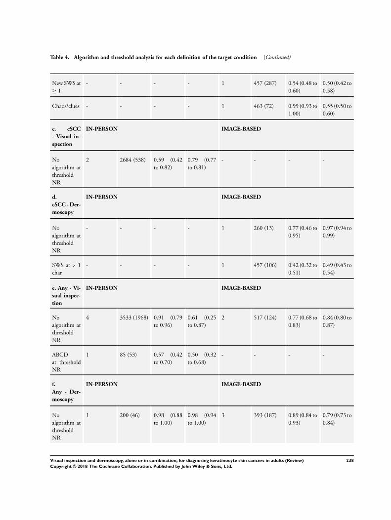
Table 4. Algorithm and threshold analysis for each definition of the target condition (Continued)
New SWS at
≥ 1
- - - - 1 457 (287) 0.54 (0.48 to
0.60)
0.50 (0.42 to
0.58)
Chaos/clues - - - - 1 463 (72) 0.99 (0.93 to
1.00)
0.55 (0.50 to
0.60)
c. cSCC
- Visual in-
spection
IN-PERSON IMAGE-BASED
No
algorithm at
threshold
NR
2 2684 (538) 0.59 (0.42
to 0.82)
0.79 (0.77
to 0.81)
- - - -
d.
cSCC - Der-
moscopy
IN-PERSON IMAGE-BASED
No
algorithm at
threshold
NR
- - - - 1 260 (13) 0.77 (0.46 to
0.95)
0.97 (0.94 to
0.99)
SWS at > 1
char
- - - - 1 457 (106) 0.42 (0.32 to
0.51)
0.49 (0.43 to
0.54)
e. Any - Vi-
sual inspec-
tion
IN-PERSON IMAGE-BASED
No
algorithm at
threshold
NR
4 3533 (1968) 0.91 (0.79
to 0.96)
0.61 (0.25
to 0.87)
2 517 (124) 0.77 (0.68 to
0.83)
0.84 (0.80 to
0.87)
ABCD
at threshold
NR
1 85 (53) 0.57 (0.42
to 0.70)
0.50 (0.32
to 0.68)
- - - -
f.
Any - Der-
moscopy
IN-PERSON IMAGE-BASED
No
algorithm at
threshold
NR
1 200 (46) 0.98 (0.88
to 1.00)
0.98 (0.94
to 1.00)
3 393 (187) 0.89 (0.84 to
0.93)
0.79 (0.73 to
0.84)
238Visual inspection and dermoscopy, alone or in combination, for diagnosing keratinocyte skin cancers in adults (Review)
Copyright © 2018 The Cochrane Collaboration. Published by John Wiley & Sons, Ltd.
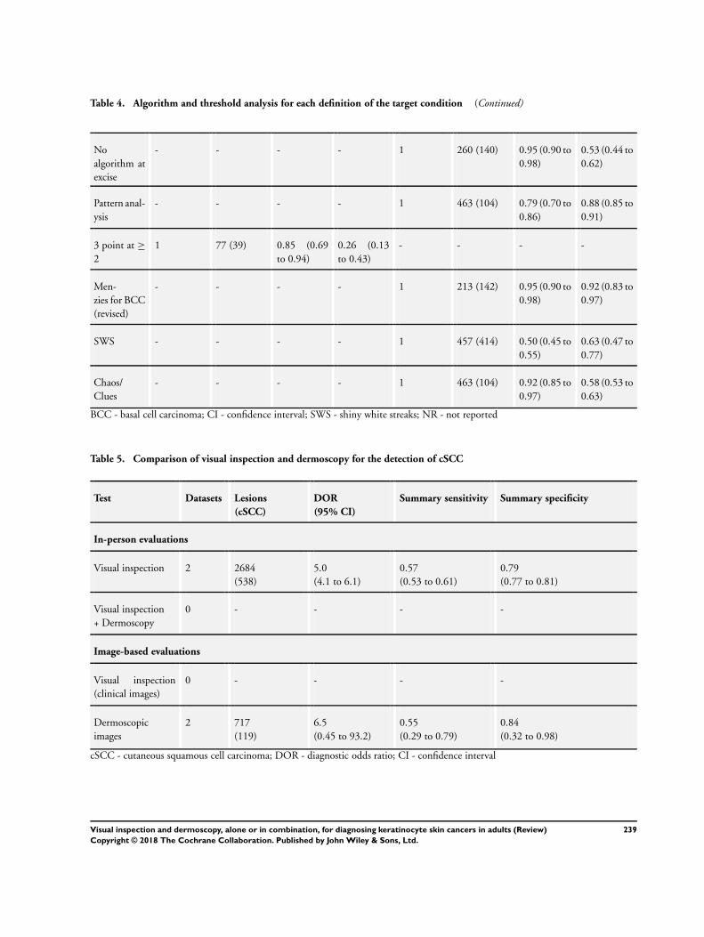
Table 4. Algorithm and threshold analysis for each definition of the target condition (Continued)
No
algorithm at
excise
- - - - 1 260 (140) 0.95 (0.90 to
0.98)
0.53 (0.44 to
0.62)
Pattern anal-
ysis
- - - - 1 463 (104) 0.79 (0.70 to
0.86)
0.88 (0.85 to
0.91)
3 point at ≥
2
1 77 (39) 0.85 (0.69
to 0.94)
0.26 (0.13
to 0.43)
- - - -
Men-
zies for BCC
(revised)
- - - - 1 213 (142) 0.95 (0.90 to
0.98)
0.92 (0.83 to
0.97)
SWS - - - - 1 457 (414) 0.50 (0.45 to
0.55)
0.63 (0.47 to
0.77)
Chaos/
Clues
- - - - 1 463 (104) 0.92 (0.85 to
0.97)
0.58 (0.53 to
0.63)
BCC - basal cell carcinoma; CI - confidence interval; SWS - shiny white streaks; NR - not reported
Table 5. Comparison of visual inspection and dermoscopy for the detection of cSCC
Test Datasets Lesions
(cSCC)
DOR
(95% CI)
Summary sensitivity Summary specificity
In-person evaluations
Visual inspection 2 2684
(538)
5.0
(4.1 to 6.1)
0.57
(0.53 to 0.61)
0.79
(0.77 to 0.81)
Visual inspection
+ Dermoscopy
0 - - - -
Image-based evaluations
Visual inspection
(clinical images)
0 - - - -
Dermoscopic
images
2 717
(119)
6.5
(0.45 to 93.2)
0.55
(0.29 to 0.79)
0.84
(0.32 to 0.98)
cSCC - cutaneous squamous cell carcinoma; DOR - diagnostic odds ratio; CI - confidence interval
239Visual inspection and dermoscopy, alone or in combination, for diagnosing keratinocyte skin cancers in adults (Review)
Copyright © 2018 The Cochrane Collaboration. Published by John Wiley & Sons, Ltd.
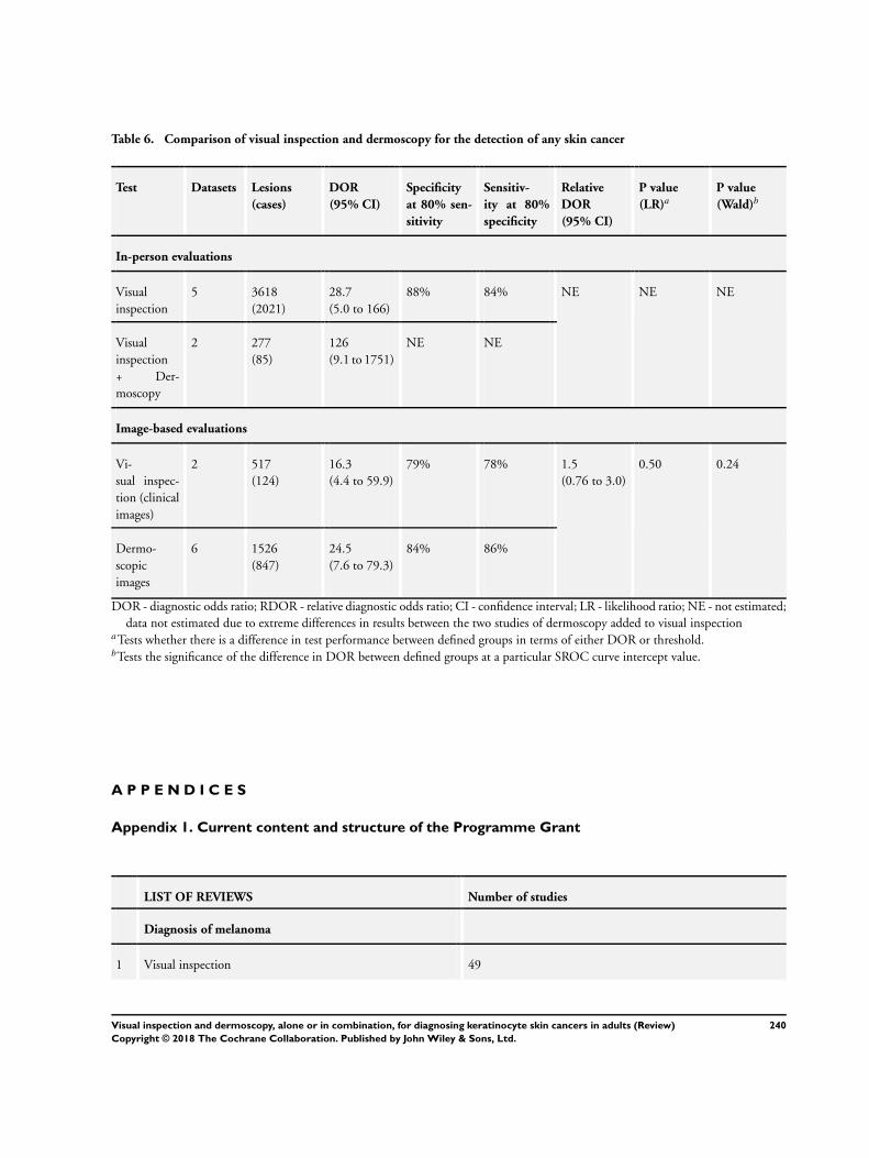
Table 6. Comparison of visual inspection and dermoscopy for the detection of any skin cancer
Test Datasets Lesions
(cases)
DOR
(95% CI)
Specificity
at 80% sen-
sitivity
Sensitiv-
ity at 80%
specificity
Relative
DOR
(95% CI)
P value
(LR)aP value
(Wald)b
In-person evaluations
Visual
inspection
5 3618
(2021)
28.7
(5.0 to 166)
88% 84% NE NE NE
Visual
inspection
+ Der-
moscopy
2 277
(85)
126
(9.1 to 1751)
NE NE
Image-based evaluations
Vi-
sual inspec-
tion (clinical
images)
2 517
(124)
16.3
(4.4 to 59.9)
79% 78% 1.5
(0.76 to 3.0)
0.50 0.24
Dermo-
scopic
images
6 1526
(847)
24.5
(7.6 to 79.3)
84% 86%
DOR - diagnostic odds ratio; RDOR - relative diagnostic odds ratio; CI - confidence interval; LR - likelihood ratio; NE - not estimated;
data not estimated due to extreme differences in results between the two studies of dermoscopy added to visual inspectionaTests whether there is a difference in test performance between defined groups in terms of either DOR or threshold.bTests the significance of the difference in DOR between defined groups at a particular SROC curve intercept value.
A P P E N D I C E S
Appendix 1. Current content and structure of the Programme Grant
LIST OF REVIEWS Number of studies
Diagnosis of melanoma
1 Visual inspection 49
240Visual inspection and dermoscopy, alone or in combination, for diagnosing keratinocyte skin cancers in adults (Review)
Copyright © 2018 The Cochrane Collaboration. Published by John Wiley & Sons, Ltd.
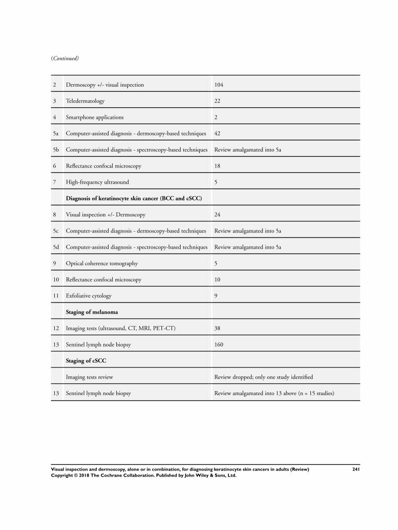
(Continued)
2 Dermoscopy +/- visual inspection 104
3 Teledermatology 22
4 Smartphone applications 2
5a Computer-assisted diagnosis - dermoscopy-based techniques 42
5b Computer-assisted diagnosis - spectroscopy-based techniques Review amalgamated into 5a
6 Reflectance confocal microscopy 18
7 High-frequency ultrasound 5
Diagnosis of keratinocyte skin cancer (BCC and cSCC)
8 Visual inspection +/- Dermoscopy 24
5c Computer-assisted diagnosis - dermoscopy-based techniques Review amalgamated into 5a
5d Computer-assisted diagnosis - spectroscopy-based techniques Review amalgamated into 5a
9 Optical coherence tomography 5
10 Reflectance confocal microscopy 10
11 Exfoliative cytology 9
Staging of melanoma
12 Imaging tests (ultrasound, CT, MRI, PET-CT) 38
13 Sentinel lymph node biopsy 160
Staging of cSCC
Imaging tests review Review dropped; only one study identified
13 Sentinel lymph node biopsy Review amalgamated into 13 above (n = 15 studies)
241Visual inspection and dermoscopy, alone or in combination, for diagnosing keratinocyte skin cancers in adults (Review)
Copyright © 2018 The Cochrane Collaboration. Published by John Wiley & Sons, Ltd.
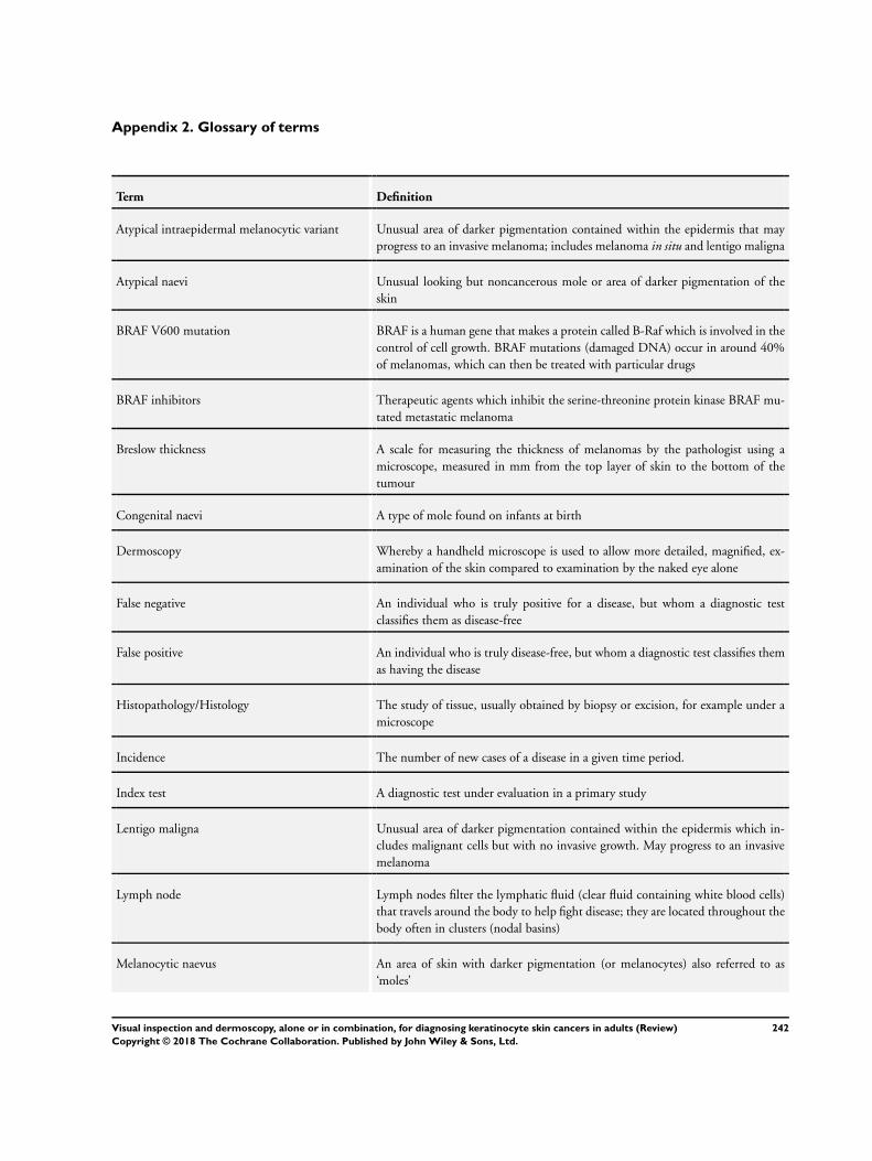
Appendix 2. Glossary of terms
Term Definition
Atypical intraepidermal melanocytic variant Unusual area of darker pigmentation contained within the epidermis that may
progress to an invasive melanoma; includes melanoma in situ and lentigo maligna
Atypical naevi Unusual looking but noncancerous mole or area of darker pigmentation of the
skin
BRAF V600 mutation BRAF is a human gene that makes a protein called B-Raf which is involved in the
control of cell growth. BRAF mutations (damaged DNA) occur in around 40%
of melanomas, which can then be treated with particular drugs
BRAF inhibitors Therapeutic agents which inhibit the serine-threonine protein kinase BRAF mu-
tated metastatic melanoma
Breslow thickness A scale for measuring the thickness of melanomas by the pathologist using a
microscope, measured in mm from the top layer of skin to the bottom of the
tumour
Congenital naevi A type of mole found on infants at birth
Dermoscopy Whereby a handheld microscope is used to allow more detailed, magnified, ex-
amination of the skin compared to examination by the naked eye alone
False negative An individual who is truly positive for a disease, but whom a diagnostic test
classifies them as disease-free
False positive An individual who is truly disease-free, but whom a diagnostic test classifies them
as having the disease
Histopathology/Histology The study of tissue, usually obtained by biopsy or excision, for example under a
microscope
Incidence The number of new cases of a disease in a given time period.
Index test A diagnostic test under evaluation in a primary study
Lentigo maligna Unusual area of darker pigmentation contained within the epidermis which in-
cludes malignant cells but with no invasive growth. May progress to an invasive
melanoma
Lymph node Lymph nodes filter the lymphatic fluid (clear fluid containing white blood cells)
that travels around the body to help fight disease; they are located throughout the
body often in clusters (nodal basins)
Melanocytic naevus An area of skin with darker pigmentation (or melanocytes) also referred to as
‘moles’
242Visual inspection and dermoscopy, alone or in combination, for diagnosing keratinocyte skin cancers in adults (Review)
Copyright © 2018 The Cochrane Collaboration. Published by John Wiley & Sons, Ltd.
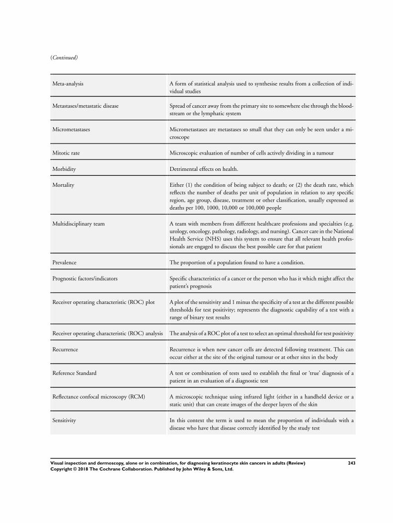
(Continued)
Meta-analysis A form of statistical analysis used to synthesise results from a collection of indi-
vidual studies
Metastases/metastatic disease Spread of cancer away from the primary site to somewhere else through the blood-
stream or the lymphatic system
Micrometastases Micrometastases are metastases so small that they can only be seen under a mi-
croscope
Mitotic rate Microscopic evaluation of number of cells actively dividing in a tumour
Morbidity Detrimental effects on health.
Mortality Either (1) the condition of being subject to death; or (2) the death rate, which
reflects the number of deaths per unit of population in relation to any specific
region, age group, disease, treatment or other classification, usually expressed as
deaths per 100, 1000, 10,000 or 100,000 people
Multidisciplinary team A team with members from different healthcare professions and specialties (e.g.
urology, oncology, pathology, radiology, and nursing). Cancer care in the National
Health Service (NHS) uses this system to ensure that all relevant health profes-
sionals are engaged to discuss the best possible care for that patient
Prevalence The proportion of a population found to have a condition.
Prognostic factors/indicators Specific characteristics of a cancer or the person who has it which might affect the
patient’s prognosis
Receiver operating characteristic (ROC) plot A plot of the sensitivity and 1 minus the specificity of a test at the different possible
thresholds for test positivity; represents the diagnostic capability of a test with a
range of binary test results
Receiver operating characteristic (ROC) analysis The analysis of a ROC plot of a test to select an optimal threshold for test positivity
Recurrence Recurrence is when new cancer cells are detected following treatment. This can
occur either at the site of the original tumour or at other sites in the body
Reference Standard A test or combination of tests used to establish the final or ‘true’ diagnosis of a
patient in an evaluation of a diagnostic test
Reflectance confocal microscopy (RCM) A microscopic technique using infrared light (either in a handheld device or a
static unit) that can create images of the deeper layers of the skin
Sensitivity In this context the term is used to mean the proportion of individuals with a
disease who have that disease correctly identified by the study test
243Visual inspection and dermoscopy, alone or in combination, for diagnosing keratinocyte skin cancers in adults (Review)
Copyright © 2018 The Cochrane Collaboration. Published by John Wiley & Sons, Ltd.

(Continued)
Specificity The proportion of individuals without the disease of interest (in this case with
benign skin lesions) who have that absence of disease correctly identified by the
study test
Staging Clinical description of the size and spread of a patient’s tumour, fitting into inter-
nationally agreed categories
Subclinical (disease) Disease that is usually asymptomatic and not easily observable, e.g. by clinical or
physical examination
Systemic treatment Treatment, usually given by mouth or by injection, that reaches and affects cancer
cells throughout the body rather than targeting one specific area
Appendix 3. Proposed sources of heterogeneity
i. Population characteristics
• general versus higher-risk populations
• patient population: Primary/secondary/specialist unit
• lesion suspicion: general suspicion/atypical/equivocal/NR
• lesion type: any pigmented; melanocytic
• inclusion of multiple lesions per participant
• ethnicity
ii. Index test characteristics
• the nature of and definition of criteria for test positivity
• observer experience with the index test
• approaches to lesion preparation (e.g. the use of oil or antiseptic gel for dermoscopy)
iii. Reference standard characteristics
• reference standard used
• whether histology-reporting meets pathology-reporting guidelines
• use of excisional versus diagnostic biopsy
• whether two independent dermatopathologists reviewed histological diagnosis
iv. Study quality
• consecutive or random sample of participants recruited
• index test interpreted blinded to the reference standard result
• index test interpreted blinded to the result of any other index test
• presence of partial or differential verification bias (whereby only a sample of those subject to the index test are verified by the
reference test or by the same reference test with selection dependent on the index test result)
• use of an adequate reference standard
• overall risk of bias
244Visual inspection and dermoscopy, alone or in combination, for diagnosing keratinocyte skin cancers in adults (Review)
Copyright © 2018 The Cochrane Collaboration. Published by John Wiley & Sons, Ltd.

Appendix 4. Final search strategies
Melanoma search strategies to August 2016
Database: Ovid MEDLINE(R) 1946 to August week 3 2016
Search strategy:
1 exp melanoma/
2 exp skin cancer/
3 exp basal cell carcinoma/
4 basalioma$1.ti,ab.
5 ((basal cell or skin) adj2 (cancer$1 or carcinoma$1 or mass or masses or tumour$1 or tumor$1 or neoplasm$1 or adenoma$1 or
epithelioma$1 or lesion$1 or malignan$ or nodule$1)).ti,ab.
6 (pigmented adj2 (lesion$1 or mole$ or nevus or nevi or naevus or naevi or skin)).ti,ab.
7 (melanom$1 or nonmelanoma$1 or non-melanoma$1 or melanocyt$ or non-melanocyt$ or nonmelanocyt$ or keratinocyt$).ti,ab.
8 nmsc.ti,ab.
9 (squamous cell adj2 (cancer$1 or carcinoma$1 or mass or masses or tumor$1 or tumour$1 or neoplasm$1 or adenoma$1 or
epithelioma$1 or epithelial or lesion$1 or malignan$ or nodule$1) adj2 (skin or epiderm$ or cutaneous)).ti,ab.
10 (BCC or CSCC or NMSC).ti,ab.
11 keratinocy$.ti,ab.
12 Keratinocytes/
13 or/1-12
14 dermoscop$.ti,ab.
15 dermatoscop$.ti,ab.
16 photomicrograph$.ti,ab.
17 exp epiluminescence microscopy/
18 (epiluminescence adj2 microscop$).ti,ab.
19 (confocal adj2 microscop$).ti,ab.
20 (incident light adj2 microscop$).ti,ab.
21 (surface adj2 microscop$).ti,ab.
22 (visual adj (inspect$ or examin$)).ti,ab.
23 ((clinical or physical) adj examin$).ti,ab.
24 3 point.ti,ab.
25 three point.ti,ab.
26 pattern analys$.ti,ab.
27 ABCD$.ti,ab.
28 menzies.ti,ab.
29 7 point.ti,ab.
30 seven point.ti,ab.
31 (digital adj2 (dermoscop$ or dermatoscop$)).ti,ab.
32 artificial intelligence.ti,ab.
33 AI.ti,ab.
34 computer assisted.ti,ab.
35 computer aided.ti,ab.
36 neural network$.ti,ab.
37 exp diagnosis, computer-assisted/
38 MoleMax.ti,ab.
39 image process$.ti,ab.
40 automatic classif$.ti,ab.
41 image analysis.ti,ab.
42 SIAscop$.ti,ab.
43 Aura.ti,ab.
44 (optical adj2 scan$).ti,ab.
45 MelaFind.ti,ab.
46 SIMSYS.ti,ab.
245Visual inspection and dermoscopy, alone or in combination, for diagnosing keratinocyte skin cancers in adults (Review)
Copyright © 2018 The Cochrane Collaboration. Published by John Wiley & Sons, Ltd.
47 MoleMate.ti,ab.
48 SolarScan.ti,ab.
49 VivaScope.ti,ab.
50 (high adj3 ultraso$).ti,ab.
51 (canine adj2 detect$).ti,ab.
52 ((mobile or cell or cellular or smart) adj ((phone$1 adj2 app$1) or application$1)).ti,ab.
53 smartphone$.ti,ab.
54 (DermoScan or SkinVision or DermLink or SpotCheck).ti,ab.
55 Mole Detective.ti,ab.
56 Spot Check.ti,ab.
57 (mole$1 adj2 map$).ti,ab.
58 (total adj2 body).ti,ab.
59 exfoliative cytolog$.ti,ab.
60 digital analys$.ti,ab.
61 (image$1 adj3 software).ti,ab.
62 (teledermatolog$ or tele-dermatolog$ or telederm or tele-derm or teledermoscop$ or tele-dermoscop$ or teledermatoscop$ or tele-
dermatoscop$).ti,ab.
63 (optical coherence adj (technolog$ or tomog$)).ti,ab.
64 (computer adj2 diagnos$).ti,ab.
65 exp sentinel lymph node biopsy/
66 (sentinel adj2 node).ti,ab.
67 nevisense.mp. or HFUS.ti,ab.
68 electrical impedance spectroscopy.ti,ab.
69 history taking.ti,ab.
70 patient history.ti,ab.
71 (naked eye adj (exam$ or assess$)).ti,ab.
72 (skin adj exam$).ti,ab.
73 physical examination/
74 ugly duckling.mp. or UD.ti,ab.
75 ((physician$ or clinical or physical) adj (exam$ or triage or recog$)).ti,ab.
76 ABCDE.mp. or VOC.ti,ab.
77 clinical accuracy.ti,ab.
78 Family Practice/ or Physicians, Family/ or clinical competence/
79 (confocal adj2 microscop$).ti,ab.
80 diagnostic algorithm$1.ti,ab.
81 checklist$.ti,ab.
82 virtual imag$1.ti,ab.
83 volatile organic compound$1.ti,ab.
84 dog$1.ti,ab.
85 gene expression analy$.ti,ab.
86 reflex transmission imag$.ti,ab.
87 thermal imaging.ti,ab.
88 elastography.ti,ab.
89 or/14-88
90 (CT or PET).ti,ab.
91 PET-CT.ti,ab.
92 (FDG or F18 or Fluorodeoxyglucose or radiopharmaceutical$).ti,ab.
93 exp Deoxyglucose/
94 deoxy-glucose.ti,ab.
95 deoxyglucose.ti,ab.
96 CATSCAN.ti,ab.
97 exp Tomography, Emission-Computed/
98 exp Tomography, X-ray computed/
246Visual inspection and dermoscopy, alone or in combination, for diagnosing keratinocyte skin cancers in adults (Review)
Copyright © 2018 The Cochrane Collaboration. Published by John Wiley & Sons, Ltd.
99 positron emission tomograph$.ti,ab.
100 exp magnetic resonance imaging/
101 (MRI or fMRI or NMRI or scintigraph$).ti,ab.
102 exp echography/
103 Doppler echography.ti,ab.
104 sonograph$.ti,ab.
105 ultraso$.ti,ab.
106 doppler.ti,ab.
107 magnetic resonance imag$.ti,ab.
108 or/90-107
109 (stage$ or staging or metasta$ or recurrence or sensitivity or specificity or false negative$ or thickness$).ti,ab.
110 “Sensitivity and Specificity”/
111 exp cancer staging/
112 or/109-111
113 108 and 112
114 89 or 113
115 13 and 114
Database: Ovid MEDLINE(R) In-Process & Other Non-Indexed Citations 29 August 2016
Search strategy:
1 basalioma$1.ti,ab.
2 ((basal cell or skin) adj2 (cancer$1 or carcinoma$1 or mass or masses or tumour$1 or tumor$1 or neoplasm$1 or adenoma$1 or
epithelioma$1 or lesion$1 or malignan$ or nodule$1)).ti,ab.
3 (pigmented adj2 (lesion$1 or mole$ or nevus or nevi or naevus or naevi or skin)).ti,ab.
4 (melanom$1 or nonmelanoma$1 or non-melanoma$1 or melanocyt$ or non-melanocyt$ or nonmelanocyt$ or keratinocyt$).ti,ab.
5 nmsc.ti,ab.
6 (squamous cell adj2 (cancer$1 or carcinoma$1 or mass or masses or tumor$1 or tumour$1 or neoplasm$1 or adenoma$1 or
epithelioma$1 or epithelial or lesion$1 or malignan$ or nodule$1) adj2 (skin or epiderm$ or cutaneous)).ti,ab.
7 (BCC or CSCC or NMSC).ti,ab.
8 keratinocy$.ti,ab.
9 or/1-8
10 dermoscop$.ti,ab.
11 dermatoscop$.ti,ab.
12 photomicrograph$.ti,ab.
13 (epiluminescence adj2 microscop$).ti,ab.
14 (confocal adj2 microscop$).ti,ab.
15 (incident light adj2 microscop$).ti,ab.
16 (surface adj2 microscop$).ti,ab.
17 (visual adj (inspect$ or examin$)).ti,ab.
18 ((clinical or physical) adj examin$).ti,ab.
19 3 point.ti,ab.
20 three point.ti,ab.
21 pattern analys$.ti,ab.
22 ABCD$.ti,ab.
23 menzies.ti,ab.
24 7 point.ti,ab.
25 seven point.ti,ab.
26 (digital adj2 (dermoscop$ or dermatoscop$)).ti,ab.
27 artificial intelligence.ti,ab.
28 AI.ti,ab.
29 computer assisted.ti,ab.
30 computer aided.ti,ab.
31 neural network$.ti,ab.
32 MoleMax.ti,ab.
247Visual inspection and dermoscopy, alone or in combination, for diagnosing keratinocyte skin cancers in adults (Review)
Copyright © 2018 The Cochrane Collaboration. Published by John Wiley & Sons, Ltd.
33 image process$.ti,ab.
34 automatic classif$.ti,ab.
35 image analysis.ti,ab.
36 SIAscop$.ti,ab.
37 Aura.ti,ab.
38 (optical adj2 scan$).ti,ab.
39 MelaFind.ti,ab.
40 SIMSYS.ti,ab.
41 MoleMate.ti,ab.
42 SolarScan.ti,ab.
43 VivaScope.ti,ab.
44 (high adj3 ultraso$).ti,ab.
45 (canine adj2 detect$).ti,ab.
46 ((mobile or cell or cellular or smart) adj ((phone$1 adj2 app$1) or application$1)).ti,ab.
47 smartphone$.ti,ab.
48 (DermoScan or SkinVision or DermLink or SpotCheck).ti,ab.
49 Mole Detective.ti,ab.
50 Spot Check.ti,ab.
51 (mole$1 adj2 map$).ti,ab.
52 (total adj2 body).ti,ab.
53 exfoliative cytolog$.ti,ab.
54 digital analys$.ti,ab.
55 (image$1 adj3 software).ti,ab.
56 (teledermatolog$ or tele-dermatolog$ or telederm or tele-derm or teledermoscop$ or tele-dermoscop$ or teledermatoscop$ or tele-
dermatoscop$).ti,ab.
57 (optical coherence adj (technolog$ or tomog$)).ti,ab.
58 (computer adj2 diagnos$).ti,ab.
59 (sentinel adj2 node).ti,ab.
60 nevisense.mp. or HFUS.ti,ab.
61 electrical impedance spectroscopy.ti,ab.
62 history taking.ti,ab.
63 patient history.ti,ab.
64 (naked eye adj (exam$ or assess$)).ti,ab.
65 (skin adj exam$).ti,ab.
66 ugly duckling.mp. or UD.ti,ab.
67 ((physician$ or clinical or physical) adj (exam$ or triage or recog$)).ti,ab.
68 ABCDE.mp. or VOC.ti,ab.
69 clinical accuracy.ti,ab.
70 (Family adj (Practice or Physicians)).ti,ab.
71 (confocal adj2 microscop$).ti,ab.
72 clinical competence.ti,ab.
73 diagnostic algorithm$1.ti,ab.
74 checklist$.ti,ab.
75 virtual imag$1.ti,ab.
76 volatile organic compound$1.ti,ab.
77 dog$1.ti,ab.
78 gene expression analy$.ti,ab.
79 reflex transmission imag$.ti,ab.
80 thermal imaging.ti,ab.
81 elastography.ti,ab.
82 or/10-81
83 (CT or PET).ti,ab.
84 PET-CT.ti,ab.
248Visual inspection and dermoscopy, alone or in combination, for diagnosing keratinocyte skin cancers in adults (Review)
Copyright © 2018 The Cochrane Collaboration. Published by John Wiley & Sons, Ltd.
85 (FDG or F18 or Fluorodeoxyglucose or radiopharmaceutical$).ti,ab.
86 deoxy-glucose.ti,ab.
87 deoxyglucose.ti,ab.
88 CATSCAN.ti,ab.
89 positron emission tomograph$.ti,ab.
90 (MRI or fMRI or NMRI or scintigraph$).ti,ab.
91 Doppler echography.ti,ab.
92 sonograph$.ti,ab.
93 ultraso$.ti,ab.
94 doppler.ti,ab.
95 magnetic resonance imag$.ti,ab.
96 or/83-95
97 (stage$ or staging or metasta$ or recurrence or sensitivity or specificity or false negative$ or thickness$).ti,ab.
98 96 and 97
99 82 or 98
100 9 and 99
Database: Embase 1974 to 29 August 2016
Search strategy:
1 *melanoma/
2 *skin cancer/
3 *basal cell carcinoma/
4 basalioma$.ti,ab.
5 ((basal cell or skin) adj2 (cancer$1 or carcinoma$1 or mass or masses or tumour$1 or tumor$1 or neoplasm$ or adenoma$ or
epithelioma$ or lesion$ or malignan$ or nodule$)).ti,ab.
6 (pigmented adj2 (lesion$1 or mole$ or nevus or nevi or naevus or naevi or skin)).ti,ab.
7 (melanom$1 or nonmelanoma$1 or non-melanoma$1 or melanocyt$ or non-melanocyt$ or nonmelanocyt$ or keratinocyt$).ti,ab.
8 nmsc.ti,ab.
9 (squamous cell adj2 (cancer$1 or carcinoma$1 or mass or tumor$1 or tumour$1 or neoplasm$1 or adenoma$1 or epithelioma$1 or
epithelial or lesion$1 or malignan$ or nodule$1) adj2 (skin or epiderm$ or cutaneous)).ti,ab.
10 (BCC or cscc).mp. or NMSC.ti,ab.
11 keratinocyte.ti,ab.
12 keratinocy$.ti,ab.
13 or/1-12
14 dermoscop$.ti,ab.
15 dermatoscop$.ti,ab.
16 photomicrograph$.ti,ab.
17 *epiluminescence microscopy/
18 (epiluminescence adj2 microscop$).ti,ab.
19 (confocal adj2 microscop$).ti,ab.
20 (incident light adj2 microscop$).ti,ab.
21 (surface adj2 microscop$).ti,ab.
22 (visual adj (inspect$ or examin$)).ti,ab.
23 ((clinical or physical) adj examin$).ti,ab.
24 3 point.ti,ab.
25 three point.ti,ab.
26 pattern analys$.ti,ab.
27 ABCD$.ti,ab.
28 menzies.ti,ab.
29 7 point.ti,ab.
30 seven point.ti,ab.
31 (digital adj2 (dermoscop$ or dermatoscop$)).ti,ab.
32 artificial intelligence.ti,ab.
33 AI.ti,ab.
249Visual inspection and dermoscopy, alone or in combination, for diagnosing keratinocyte skin cancers in adults (Review)
Copyright © 2018 The Cochrane Collaboration. Published by John Wiley & Sons, Ltd.
34 computer assisted.ti,ab.
35 computer aided.ti,ab.
36 neural network$.ti,ab.
37 MoleMax.ti,ab.
38 exp diagnosis, computer-assisted/
39 image process$.ti,ab.
40 automatic classif$.ti,ab.
41 image analysis.ti,ab.
42 SIAscop$.ti,ab.
43 (optical adj2 scan$).ti,ab.
44 Aura.ti,ab.
45 MelaFind.ti,ab.
46 SIMSYS.ti,ab.
47 MoleMate.ti,ab.
48 SolarScan.ti,ab.
49 VivaScope.ti,ab.
50 confocal microscop$.ti,ab.
51 (high adj3 ultraso$).ti,ab.
52 (canine adj2 detect$).ti,ab.
53 ((mobile or cell$ or cellular or smart) adj ((phone$1 adj2 app$1) or application$1)).ti,ab.
54 smartphone$.ti,ab.
55 (DermoScan or SkinVision or DermLink or SpotCheck).ti,ab.
56 Spot Check.ti,ab.
57 Mole Detective.ti,ab.
58 (mole$1 adj2 map$).ti,ab.
59 (total adj2 body).ti,ab.
60 exfoliative cytolog$.ti,ab.
61 digital analys$.ti,ab.
62 (image$1 adj3 software).ti,ab.
63 (optical coherence adj (technolog$ or tomog$)).ti,ab.
64 (teledermatolog$ or tele-dermatolog$ or telederm or tele-derm or teledermoscop$ or tele-dermoscop$ or teledermatoscop$).mp. or
tele-dermatoscop$.ti,ab.
65 (computer adj2 diagnos$).ti,ab.
66 *sentinel lymph node biopsy/
67 (sentinel adj2 node).ti,ab.
68 nevisense.ti,ab.
69 HFUS.ti,ab.
70 electrical impedance spectroscopy.ti,ab.
71 history taking.ti,ab.
72 patient history.ti,ab.
73 (naked eye adj (exam$ or assess$)).ti,ab.
74 (skin adj exam$).ti,ab.
75 *physical examination/
76 ugly duckling.ti,ab.
77 UD sign$.ti,ab.
78 ((physician$ or clinical or physical) adj (exam$ or recog$ or triage)).ti,ab.
79 ABCDE.ti,ab.
80 clinical accuracy.ti,ab.
81 *general practice/
82 (confocal adj2 microscop$).ti,ab.
83 clinical competence/
84 diagnostic algorithm$.ti,ab.
85 checklist$1.ti,ab.
250Visual inspection and dermoscopy, alone or in combination, for diagnosing keratinocyte skin cancers in adults (Review)
Copyright © 2018 The Cochrane Collaboration. Published by John Wiley & Sons, Ltd.
86 virtual image$1.ti,ab.
87 volatile organic compound$1.ti,ab.
88 VOC.ti,ab.
89 dog$1.ti,ab.
90 gene expression analys$.ti,ab.
91 reflex transmission imaging.ti,ab.
92 thermal imaging.ti,ab.
93 elastography.ti,ab.
94 dog$1.ti,ab.
95 gene expression analys$.ti,ab.
96 reflex transmission imaging.ti,ab.
97 thermal imaging.ti,ab.
98 elastography.ti,ab.
99 or/14-93
100 PET-CT.ti,ab.
101 (CT or PET).ti,ab.
102 (FDG or F18 or Fluorodeoxyglucose or radiopharmaceutical$).ti,ab.
103 exp Deoxyglucose/
104 CATSCAN.ti,ab.
105 deoxyglucose.ti,ab.
106 deoxy-glucose.ti,ab.
107 *positron emission tomography/
108 *computer assisted tomography/
109 positron emission tomograph$.ti,ab.
110 *nuclear magnetic resonance imaging/
111 (MRI or fMRI or NMRI or scintigraph$).ti,ab.
112 *echography/
113 Doppler.ti,ab.
114 sonograph$.ti,ab.
115 ultraso$.ti,ab.
116 magnetic resonance imag$.ti,ab.
117 or/100-116
118 (stage$ or staging or metasta$ or recurrence or sensitivity or specificity or false negative$ or thickness$).ti,ab.
119 “Sensitivity and Specificity”/
120 *cancer staging/
121 or/118-120
122 117 and 121
123 99 or 122
124 13 and 123
Database: Cochrane Library (Wiley) 2016 searched 30 August 2016 CDSR Issue 8 of 12 2016 CENTRAL Issue 7 of 12 2016
HTA Issue 3 of 4 July 2016 DARE Issue 3 of 4 2015
Search strategy:
#1 melanoma* or nonmelanoma* or non-melanoma* or melanocyt* or non-melanocyt* or nonmelanocyt* or keratinocyte*
#2 MeSH descriptor: [Melanoma] explode all trees
#3 “skin cancer*”
#4 MeSH descriptor: [Skin Neoplasms] explode all trees
#5 skin near/2 (cancer* or carcinoma* or mass or masses or tumour* or tumor* or neoplasm* or adenoma* or epithelioma* or lesion*
or malignan* or nodule*)
#6 nmsc
#7 “squamous cell” near/2 (cancer* or carcinoma* or mass or masses or tumour* or tumor* or neoplasm* or adenoma* or epithelioma*
or lesion* or malignan* or nodule*) near/2 (skin or epiderm* or cutaneous)
#8 “basal cell” near/2 (cancer* or carcinoma* or mass or masses or tumour* or tumor* or neoplasm* or adenoma* or epithelioma* or
lesion* or malignan* or nodule*)
251Visual inspection and dermoscopy, alone or in combination, for diagnosing keratinocyte skin cancers in adults (Review)
Copyright © 2018 The Cochrane Collaboration. Published by John Wiley & Sons, Ltd.
#9 pigmented near/2 (lesion* or nevus or mole* or naevi or naevus or nevi or skin)
#10 #1 or #2 or #3 or #4 or #5 or #6 or #7 or #8 or #9
#11 dermoscop*
#12 dermatoscop*
#13 Photomicrograph*
#14 MeSH descriptor: [Dermoscopy] explode all trees
#15 confocal near/2 microscop*
#16 epiluminescence near/2 microscop*
#17 incident next light near/2 microscop*
#18 surface near/2 microscop*
#19 “visual inspect*”
#20 “visual exam*”
#21 (clinical or physical) next (exam*)
#22 “3 point”
#23 “three point”
#24 “pattern analys*”
#25 ABDC
#26 menzies
#27 “7 point”
#28 “seven point”
#29 digital near/2 (dermoscop* or dermatoscop*)
#30 “artificial intelligence”
#31 “AI”
#32 “computer assisted”
#33 “computer aided”
#34 AI
#35 “neural network*”
#36 MoleMax
#37 “computer diagnosis”
#38 “image process*”
#39 “automatic classif*”
#40 SIAscope
#41 “image analysis”
#42 “optical near/2 scan*”
#43 Aura
#44 MelaFind
#45 SIMSYS
#46 MoleMate
#47 SolarScan
#48 Vivascope
#49 “confocal microscopy”
#50 high near/3 ultraso*
#51 canine near/2 detect*
#52 Mole* near/2 map*
#53 total near/2 body
#54 mobile* or smart near/2 phone*
#55 cell next phone*
#56 smartphone*
#57 “mitotic index”
#58 DermoScan or SkinVision or DermLink or SpotCheck
#59 “Mole Detective”
#60 “Spot Check”
#61 mole* near/2 map*
252Visual inspection and dermoscopy, alone or in combination, for diagnosing keratinocyte skin cancers in adults (Review)
Copyright © 2018 The Cochrane Collaboration. Published by John Wiley & Sons, Ltd.
#62 total near/2 body
#63 “exfoliative cytolog*”
#64 “digital analys*”
#65 image near/3 software
#66 teledermatolog* or tele-dermatolog* or telederm or tele-derm or teledermoscop* or tele-dermoscop* or teledermatoscop* or tele-
dermatolog*
#67 “optical coherence” next (technolog* or tomog*)
#68 computer near/2 diagnos*
#69 sentinel near/2 node*
#70 #11 or #12 or #13 or #14 or #15 or #16 or #17 or #18 or #19 or #20 or #21 or #22 or #23 or #24 or #25 or #26 or #27 or #28
or #29 or #30 or #31 or #32 or #33 or #34 or #35 or #36 or #37 or #38 or #39 or #40 or #41 or #42 or #43 or #44 or #45 or #46 or
#47 or #48 or #49 or #50 or #51 or #52 or #53 or #54 or #55 or #56 or #57 or #58 or #59 or #60 or #61 or #62 or #63 or #64 or #
65 or #66 or #67 or #68 or #69
#71 ultraso*
#72 sonograph*
#73 MeSH descriptor: [Ultrasonography] explode all trees
#74 Doppler
#75 CT or PET or PET-CT
#76 “CAT SCAN” or “CATSCAN”
#77 MeSH descriptor: [Positron-Emission Tomography] explode all trees
#78 MeSH descriptor: [Tomography, X-Ray Computed] explode all trees
#79 MRI
#80 MeSH descriptor: [Magnetic Resonance Imaging] explode all trees
#81 MRI or fMRI or NMRI or scintigraph*
#82 “magnetic resonance imag*”
#83 MeSH descriptor: [Deoxyglucose] explode all trees
#84 deoxyglucose or deoxy-glucose
#85 “positron emission tomograph*”
#86 #71 or #72 or #73 or #74 or #75 or #76 or #77 or #78 or #79 or #80 or #81 or #82 or #83 or #84 or #85
#87 stage* or staging or metasta* or recurrence or sensitivity or specificity or “false negative*” or thickness*
#88 MeSH descriptor: [Neoplasm Staging] explode all trees
#89 #87 or #88
#90 #89 and #86
#91 #70 or #90
#92 #10 and #91
#93 BCC or CSCC or NMCS
#94 keratinocy*
#95 #93 or #94
#96 #10 or #95
#97 nevisense
#98 HFUS
#99 “electrical impedance spectroscopy”
#100 “history taking”
#101 “patient history”
#102 naked next eye near/1 (exam* or assess*)
#103 skin next exam*
#104 “ugly duckling” or (UD sign*)
#105 MeSH descriptor: [Physical Examination] explode all trees
#106 (physician* or clinical or physical) near/1 (exam* or recog* or triage*)
#107 ABCDE
#108 “clinical accuracy”
#109 MeSH descriptor: [General Practice] explode all trees
#110 confocal near microscop*
253Visual inspection and dermoscopy, alone or in combination, for diagnosing keratinocyte skin cancers in adults (Review)
Copyright © 2018 The Cochrane Collaboration. Published by John Wiley & Sons, Ltd.
#111 “diagnostic algorithm*”
#112 MeSH descriptor: [Clinical Competence] explode all trees
#113 checklist*
#114 “virtual image*”
#115 “volatile organic compound*”
#116 dog or dogs
#117 VOC
#118 “gene expression analys*”
#119 “reflex transmission imaging”
#120 “thermal imaging”
#121 elastography
#122 #97 or #98 or #99 or #100 or #101 or #102 or #103 or #104 or #105 or #106 or #107 or #108 or #109 or #110 or #111 or #
112 or #113 or #114 or #115 or #116 or #117 or #118 or #119 or #120 or #121
#123 #70 or #122
#124 #96 and #123
#125 #96 and #90
#126 #125 or #124
#127 #10 and #126
Database: CINAHL Plus (EBSCO) 1937 to 30 August 2016
Search strategy:
S1 (MH “Melanoma”) OR (MH “Nevi and Melanomas+”)
S2 (MH “Skin Neoplasms+”)
S3 (MH “Carcinoma, Basal Cell+”)
S4 basalioma*
S5 (basal cell) N2 (cancer* or carcinoma* or mass or masses or tumor* or tumour* or neoplasm* or adenoma* or epithelioma* or
lesion* or malignan* or nodule*)
S6 (pigmented) N2 (lesion* or mole* or nevus or nevi or naevus or naevi or skin)
S7 melanom* or nonmelanoma* or non-melanoma* or melanocyt* or non-melanocyt* or nonmelanocyt*
S8 nmsc
S9 TX BCC or cscc or NMSC
S10 (MH “Keratinocytes”)
S11 keratinocyt*
S12 S1 OR S2 OR S3 OR S4 OR S5 OR S6 OR S7 OR S8 OR S9 OR S10 OR S11
S13 dermoscop* or dermatoscop* or photomicrograph* or (3 point) or (three point) or ABCD* or menzies or (7 point) or (seven
point) or AI or Molemax or SIASCOP* or Aura or MelaFind or SIMSYS or MoleMate or SolarScan or smartphone* or DermoScan
or SkinVision or DermLink or SpotCheck
S14 (epiluminescence or confocal or incident or surface) N2 (microscop*)
S15 visual N1 (inspect* or examin*)
S16 (clinical or physical) N1 (examin*)
S17 pattern analys*
S18 (digital) N2 (dermoscop* or dermatoscop*)
S19 (artificial intelligence)
S20 (computer) N2 (assisted or aided)
S21 (neural network*)
S22 (MH “Diagnosis, Computer Assisted+”)
S23 (image process*)
S24 (automatic classif*)
S25 (image analysis)
S26 SIAScop*
S27 (optical) N2 (scan*)
S28 (high) N3 (ultraso*)
S29 elastography
S30 (mobile or cell or cellular or smart) N2 (phone*) N2 (app or application*)
254Visual inspection and dermoscopy, alone or in combination, for diagnosing keratinocyte skin cancers in adults (Review)
Copyright © 2018 The Cochrane Collaboration. Published by John Wiley & Sons, Ltd.
S31 (mole*) N2 (map*)
S32 total N2 body
S33 exfoliative cytolog*
S34 digital analys*
S35 image N3 software
S36 teledermatolog* or tele-dermatolog* or telederm or tele-derm or teledermoscop* or tele-dermoscop* or teledermatoscop* or tele-
dermatoscop* teledermatolog* or tele-dermatolog* or telederm or tele-derm or teledermoscop*
S37 (optical coherence) N1 (technolog* or tomog*)
S38 computer N2 diagnos*
S39 sentinel N2 node
S40 (MH “Sentinel Lymph Node Biopsy”)
S41 nevisense or HFUS or checklist* or VOC or dog*
S42 electrical impedance spectroscopy
S43 history taking
S44 “Patient history”
S45 naked eye
S46 skin exam*
S47 physical exam*
S48 ugly duckling
S49 UD sign*
S50 (physician* or clinical or physical) N1 (exam*)
S51 clinical accuracy
S52 general practice
S53 (physician* or clinical or physical) N1 (recog* or triage)
S54 confocal microscop*
S55 clinical competence
S56 diagnostic algorithm*
S57 checklist*
S58 virtual image*
S59 volatile organic compound*
S60 gene expression analys*
S61 reflex transmission imag*
S62 thermal imaging
S63 S13 or S14 or S15 OR S16 OR S17 OR S18 OR S19 OR S20 OR S21 OR S22 OR S23 OR S24 OR S25 OR S26 OR S27 OR
S28 OR S29 OR S30 OR S31 OR S32 OR S33 OR S34 OR S35 OR S36 OR S37 OR S38 OR S39 OR S40 OR S41 OR S42 OR
S43 OR S44 OR S45 OR S46 OR S47 OR S48 OR S49 OR S50 OR S51 OR S52 OR S53 OR S54 OR S55 OR S56 OR S57 OR
S58 OR S59 OR S60 OR S61 OR S62
S64 CT or PET
S65 PET-CT
S66 FDG or F18 or Fluorodeoxyglucose or radiopharmaceutical*
S67 (MH “Deoxyglucose+”)
S68 deoxy-glucose or deoxyglucose
S69 CATSCAN
S70 CAT-SCAN
S71 (MH “Deoxyglucose+”)
S72 (MH “Tomography, Emission-Computed+”)
S73 (MH “Tomography, X-Ray Computed”)
S74 positron emission tomograph*
S75 (MH “Magnetic Resonance Imaging+”)
S76 MRI or fMRI or NMRI or scintigraph*
S77 echography
S78 doppler
S79 sonograph*
255Visual inspection and dermoscopy, alone or in combination, for diagnosing keratinocyte skin cancers in adults (Review)
Copyright © 2018 The Cochrane Collaboration. Published by John Wiley & Sons, Ltd.
S80 ultraso*
S81 magnetic resonance imag*
S82 S64 OR S65 OR S66 OR S67 OR S68 OR S69 OR S70 OR S71 OR S72 OR S73 OR S74 OR S75 OR S76 OR S77 OR S78
OR S79 OR S80 OR S81
S83 stage* or staging or metasta* or recurrence or sensitivity or specificity or (false negative*) or thickness
S84 (MH “Neoplasm Staging”)
S85 S83 OR S84
S86 S82 AND S85
S87 S63 OR S86
S88 S12 AND S87
Database: Science Citation Index SCI Expanded (Web of Science) 1900 to 30 August 2016
Conference Proceedings Citation Index (Web of Science) 1900 to 1 September 2016
Search strategy:
#1 (melanom* or nonmelanom* or non-melanoma* or melanocyt* or non-melanocyt* or nonmelanocyt* or keratinocyt*)
#2 (basalioma*)
#3 ((skin) near/2 (cancer* or carcinoma or mass or masses or tumour* or tumor* or neoplasm* or adenoma* or epithelioma* or lesion*
or malignan* or nodule*))
#4 ((basal) near/2 (cancer* or carcinoma* or mass or masses or tumour* or tumor* or neoplasm* or adenoma* or epithelioma* or
lesion* or malignan* or nodule*))
#5 ((pigmented) near/2 (lesion* or mole* or nevus or nevi or naevus or naevi or skin))
#6 (nmsc or BCC or NMSC or keratinocy*)
#7 ((squamous cell (cancer* or carcinoma* or mass or masses or tumour* or tumor* or neoplasm* or adenoma* or epithelioma* or
lesion* or malignan* or nodule*))
#8 (skin or epiderm* or cutaneous)
#9 #8 AND #7
#10 #9 OR #6 OR #5 OR #4 OR #3 OR #2 OR #1
#11 ((dermoscop* or dermatoscop* or photomicrograph* or epiluminescence or confocal or “incident light” or “surface microscop*”
or “visual inspect*” or “physical exam*” or 3 point or three point or pattern analy* or ABCDE or menzies or 7 point or seven point
or dermoscop* or dermatoscop* or AI or artificial or computer aided or computer assisted or neural network* or Molemax or image
process* or automatic classif* or image analysis or siascope or optical scan* or Aura or melafind or simsys or molemate or solarscan or
vivascope or confocal microscop* or high ultraso* or canine detect* or cellphone* or mobile* or phone* or smartphone or dermoscan
or skinvision or dermlink or spotcheck or spot check or mole detective or mole map* or total body or exfoliative psychology or digital
or image software or optical coherence or teledermatology or telederm* or teledermoscop* or teledermatoscop* or computer diagnos*
or sentinel))
#12 ((nevisense or HFUS or impedance spectroscopy or history taking or patient history or naked eye or skin exam* or physical exam*
or ugly duckling or UD sign* or physician* exam* or physical exam* or ABCDE or clinical accuracy or general practice or confocal
microscop* or clinical competence or diagnostic algorithm* or checklist* or virtual image* or volatile organic or VOC or dog* or gene
expression or reflex transmission or thermal imag* or elastography))
#13 #11 or #12
#14 ((PET or CT or FDG or deoxyglucose or deoxy-glucose or fluorodeoxy* or radiopharma* or CATSCAN or positron emission or
computer assisted or nuclear magnetic or MRI or FMRI or NMRI or scintigraph* or echograph* or Doppler or sonograph* or ultraso*
or magnetic reson*))
#15 ((stage* or staging or metast* or recurrence or sensitivity or specificity or false negative* or thickness*))
#16 #14 AND #15
#17 #16 OR #13
#18 #10 AND #17
Refined by: DOCUMENT TYPES: (MEETING ABSTRACT OR PROCEEDINGS PAPER)
256Visual inspection and dermoscopy, alone or in combination, for diagnosing keratinocyte skin cancers in adults (Review)
Copyright © 2018 The Cochrane Collaboration. Published by John Wiley & Sons, Ltd.
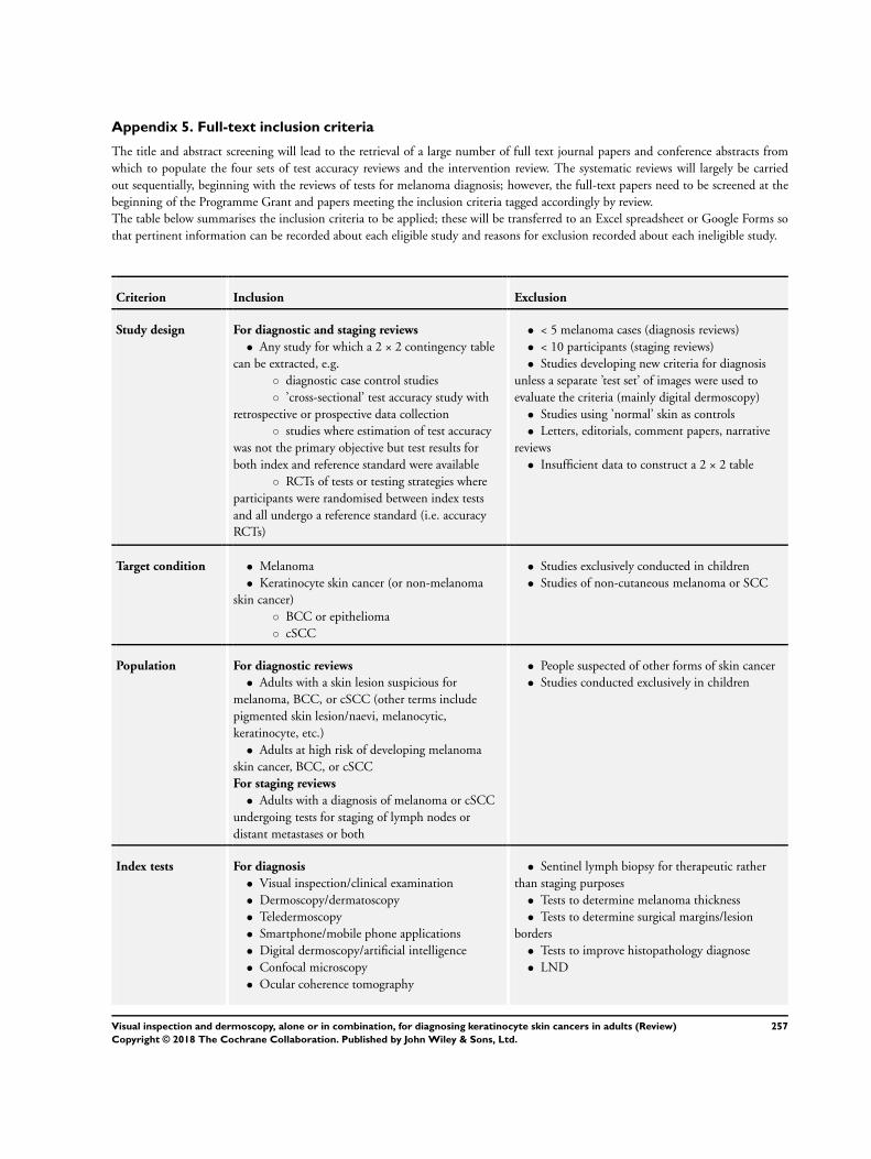
Appendix 5. Full-text inclusion criteria
The title and abstract screening will lead to the retrieval of a large number of full text journal papers and conference abstracts from
which to populate the four sets of test accuracy reviews and the intervention review. The systematic reviews will largely be carried
out sequentially, beginning with the reviews of tests for melanoma diagnosis; however, the full-text papers need to be screened at the
beginning of the Programme Grant and papers meeting the inclusion criteria tagged accordingly by review.
The table below summarises the inclusion criteria to be applied; these will be transferred to an Excel spreadsheet or Google Forms so
that pertinent information can be recorded about each eligible study and reasons for exclusion recorded about each ineligible study.
Criterion Inclusion Exclusion
Study design For diagnostic and staging reviews
• Any study for which a 2 × 2 contingency table
can be extracted, e.g.
◦ diagnostic case control studies
◦ ’cross-sectional’ test accuracy study with
retrospective or prospective data collection
◦ studies where estimation of test accuracy
was not the primary objective but test results for
both index and reference standard were available
◦ RCTs of tests or testing strategies where
participants were randomised between index tests
and all undergo a reference standard (i.e. accuracy
RCTs)
• < 5 melanoma cases (diagnosis reviews)
• < 10 participants (staging reviews)
• Studies developing new criteria for diagnosis
unless a separate ’test set’ of images were used to
evaluate the criteria (mainly digital dermoscopy)
• Studies using ’normal’ skin as controls
• Letters, editorials, comment papers, narrative
reviews
• Insufficient data to construct a 2 × 2 table
Target condition • Melanoma
• Keratinocyte skin cancer (or non-melanoma
skin cancer)
◦ BCC or epithelioma
◦ cSCC
• Studies exclusively conducted in children
• Studies of non-cutaneous melanoma or SCC
Population For diagnostic reviews
• Adults with a skin lesion suspicious for
melanoma, BCC, or cSCC (other terms include
pigmented skin lesion/naevi, melanocytic,
keratinocyte, etc.)
• Adults at high risk of developing melanoma
skin cancer, BCC, or cSCC
For staging reviews
• Adults with a diagnosis of melanoma or cSCC
undergoing tests for staging of lymph nodes or
distant metastases or both
• People suspected of other forms of skin cancer
• Studies conducted exclusively in children
Index tests For diagnosis
• Visual inspection/clinical examination
• Dermoscopy/dermatoscopy
• Teledermoscopy
• Smartphone/mobile phone applications
• Digital dermoscopy/artificial intelligence
• Confocal microscopy
• Ocular coherence tomography
• Sentinel lymph biopsy for therapeutic rather
than staging purposes
• Tests to determine melanoma thickness
• Tests to determine surgical margins/lesion
borders
• Tests to improve histopathology diagnose
• LND
257Visual inspection and dermoscopy, alone or in combination, for diagnosing keratinocyte skin cancers in adults (Review)
Copyright © 2018 The Cochrane Collaboration. Published by John Wiley & Sons, Ltd.
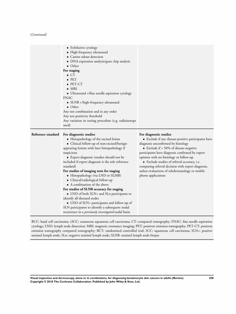
(Continued)
• Exfoliative cytology
• High-frequency ultrasound
• Canine odour detection
• DNA expression analysis/gene chip analysis
• Other
For staging
• CT
• PET
• PET-CT
• MRI
• Ultrasound +/fine needle aspiration cytology
FNAC
• SLNB +/high-frequency ultrasound
• Other
Any test combination and in any order
Any test positivity threshold
Any variation in testing procedure (e.g. radioisotope
used)
Reference standard For diagnostic studies
• Histopathology of the excised lesion
• Clinical follow-up of non-excised/benign-
appearing lesions with later histopathology if
suspicious
• Expert diagnosis (studies should not be
included if expert diagnosis is the sole reference
standard)
For studies of imaging tests for staging
• Histopathology (via LND or SLMB)
• Clinical/radiological follow-up
• A combination of the above
For studies of SLNB accuracy for staging
• LND of both SLN+ and SLn participants to
identify all diseased nodes
• LND of SLN+ participants and follow-up of
SLN participants to identify a subsequent nodal
recurrence in a previously investigated nodal basin
For diagnostic studies
• Exclude if any disease-positive participants have
diagnosis unconfirmed by histology
• Exclude if > 50% of disease-negative
participants have diagnosis confirmed by expert
opinion with no histology or follow-up
• Exclude studies of referral accuracy, i.e.
comparing referral decision with expert diagnosis,
unless evaluations of teledermatology or mobile
phone applications
BCC: basal cell carcinoma; cSCC: cutaneous squamous cell carcinoma; CT: computed tomography; FNAC: fine needle aspiration
cytology; LND: lymph node dissection; MRI: magnetic resonance imaging; PET: positron emission tomography; PET-CT: positron
emission tomography computed tomography; RCT: randomised controlled trial; SCC: squamous cell carcinoma; SLN+: positive
sentinel lymph node; SLn: negative sentinel lymph node; SLNB: sentinel lymph node biopsy
258Visual inspection and dermoscopy, alone or in combination, for diagnosing keratinocyte skin cancers in adults (Review)
Copyright © 2018 The Cochrane Collaboration. Published by John Wiley & Sons, Ltd.
Appendix 6. Quality assessment (based on QUADAS-2)
The following tables use text that was originally published in the QUADAS-2 tool by Whiting and colleagues (Whiting 2011).
Item Response (delete as required)
PARTICIPANT SELECTION (1) - RISK OF BIAS
1) Was a consecutive or random sample of participants or images
enrolled?
Yes - if paper states consecutive or random
No - if paper describes other method of sampling
Unclear - if participant sampling not described
2) Was a case-control design avoided? Yes - if consecutive or random or case-control design clearly not
used
No - if study described as case-control or describes sampling spe-
cific numbers of participants with particular diagnoses
Unclear - if not described
3) Did the study avoid inappropriate exclusions, e.g.,
• ’difficult to diagnose’ lesions not excluded
• lesions not excluded on basis of disagreement between
evaluators
Yes - if inappropriate exclusions were avoided
No - if lesions were excluded that might affect test accuracy, e.
g., ’difficult to diagnose’ lesions, or where disagreement between
evaluators was observed
Unclear - if not clearly reported but there is suspicion that difficult
to diagnose lesions may have been excluded
4) For between-person comparative studies only (i.e., allocating
different tests to different study participants):
• A) were the same participant selection criteria used for
those allocated to each test?
• B) was the potential for biased allocation between tests
avoided through adequate generation of a randomised sequence?
• C) was the potential for biased allocation between tests
avoided through concealment of allocation prior to assignment?
For A)
• Yes - if same selection criteria were used for each index test,
No - if different selection criteria were used for each index test,
Unclear - if selection criteria per test were not described, N/A -
if only 1 index test was evaluated or all participants received all
tests
For B)
• Yes - if adequate randomisation procedures are described,
No - if inadequate randomisation procedures are described,
Unclear - if the method of allocation to groups is not described
(a description of ’random’ or ’randomised’ is insufficient), N/A -
if only 1 index test was evaluated or all participants received all
tests
For C)
• Yes - if appropriate methods of allocation concealment are
described, No - if appropriate methods of allocation concealment
are not described, Unclear - if the method of allocation
concealment is not described (sufficient detail to allow a definite
judgement is required), N/A - if only 1 index test was evaluated
Could the selection of participants have introduced bias?
For non-comparative and within-person comparative studies
1. If answers to all of questions 1), 2), and 3) ’Yes’:
2. If answers to any 1 of questions 1), 2), or 3) ’No’:
3. If answers to any 1 of questions 1), 2), or 3) ’Unclear’:
For between-person comparative studies
For non-comparative and within-person comparative studies
1. Risk is low
2. Risk is high
3. Risk unclear
For between-person comparative studies
1. Risk is low
259Visual inspection and dermoscopy, alone or in combination, for diagnosing keratinocyte skin cancers in adults (Review)
Copyright © 2018 The Cochrane Collaboration. Published by John Wiley & Sons, Ltd.
(Continued)
1. If answers to all of questions 1), 2), 3), and 4) ’Yes’:
2. If answers to any 1 of questions 1), 2), 3), or 4) ’No’:
3. If answers to any 1 of questions 1), 2), 3), or 4) ’Unclear’:
2. Risk is high
3. Risk unclear
PARTICIPANT SELECTION (1) - CONCERNS REGARDING APPLICABILITY
1) Are the included participants and chosen study setting appro-
priate to answer the review question, i.e., are the study results gen-
eralisable?
• This item is not asking whether exclusion of certain
participant groups might bias the study’s results (as in Risk of
Bias above), but is asking whether the chosen study participants
and setting are appropriate to answer our review question.
Because we are looking to establish test accuracy in both primary
presentation and referred participants, a study could be
appropriate for 1 setting and not for the other, or it could be
unclear as to whether the study can appropriately answer either
question
• For each study assessed, please consider whether it is more
relevant for A) participants with a primary presentation of a skin
lesion or B) referred participants, and respond to the questions
in either A) or B) accordingly. If the study gives insufficient
details, please respond Unclear to both parts of the question
A) For studies that will contribute to the analysis of partici-
pants with a primary presentation of a skin lesion (i.e., test
naive)
Yes - if participants included in the study appear to be generally
representative of those who might present in a usual practice set-
ting
No - if study participants appear to be unrepresentative of usual
practice, e.g., in terms of severity of disease, demographic features,
presence of differential diagnosis or co-morbidity, setting of the
study, and previous testing protocols
Unclear - if insufficient details are provided to determine the
generalisability of study participants
B) For studies that will contribute to the analysis of referred
participants (i.e., who have already undergone some form of
testing)
Yes - if study participants appear to be representative of those who
might be referred for further investigation. If the study focuses
only on those with equivocal lesions, for example, we would sug-
gest that this is not representative of the wider referred population
No - if study participants appear to be unrepresentative of usual
practice, e.g., if a particularly high proportion of participants have
been self-referred or referred for cosmetic reasons. Other factors
to consider include severity of disease, demographic features, pres-
ence of differential diagnosis or co-morbidity, setting of the study,
and previous testing protocols
Unclear - if insufficient details are provided to determine the
generalisability of study participants
2) Did the study avoid including participants with multiple le-
sions?
Yes - if the difference between the number of included lesions and
number of included participants is less than 5%
No - if the difference between the number of included lesions and
number of included participants is greater than 5%
Unclear - if it is not possible to assess
Is there concern that the included participants do not match the
review question?
1. If the answer to question 1) or 2) ’Yes’:
2. If the answer to question 1) or 2) ’No’:
3. If the answer to question 1) or 2) ’Unclear’:
1. Concern is low
2. Concern is high
3. Concern is unclear
INDEX TEST (2) - RISK OF BIAS (to be completed per test evaluated)
260Visual inspection and dermoscopy, alone or in combination, for diagnosing keratinocyte skin cancers in adults (Review)
Copyright © 2018 The Cochrane Collaboration. Published by John Wiley & Sons, Ltd.
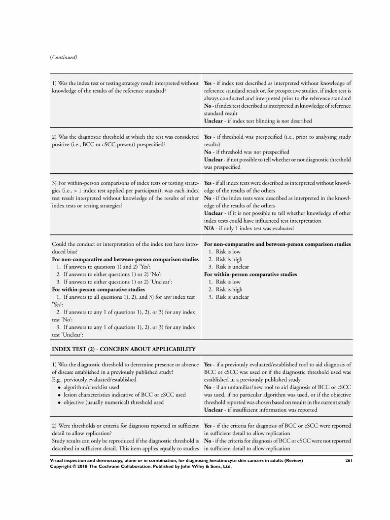
(Continued)
1) Was the index test or testing strategy result interpreted without
knowledge of the results of the reference standard?
Yes - if index test described as interpreted without knowledge of
reference standard result or, for prospective studies, if index test is
always conducted and interpreted prior to the reference standard
No - if index test described as interpreted in knowledge of reference
standard result
Unclear - if index test blinding is not described
2) Was the diagnostic threshold at which the test was considered
positive (i.e., BCC or cSCC present) prespecified?
Yes - if threshold was prespecified (i.e., prior to analysing study
results)
No - if threshold was not prespecified
Unclear - if not possible to tell whether or not diagnostic threshold
was prespecified
3) For within-person comparisons of index tests or testing strate-
gies (i.e., > 1 index test applied per participant): was each index
test result interpreted without knowledge of the results of other
index tests or testing strategies?
Yes - if all index tests were described as interpreted without knowl-
edge of the results of the others
No - if the index tests were described as interpreted in the knowl-
edge of the results of the others
Unclear - if it is not possible to tell whether knowledge of other
index tests could have influenced test interpretation
N/A - if only 1 index test was evaluated
Could the conduct or interpretation of the index test have intro-
duced bias?
For non-comparative and between-person comparison studies
1. If answers to questions 1) and 2) ’Yes’:
2. If answers to either questions 1) or 2) ’No’:
3. If answers to either questions 1) or 2) ’Unclear’:
For within-person comparative studies
1. If answers to all questions 1), 2), and 3) for any index test
’Yes’:
2. If answers to any 1 of questions 1), 2), or 3) for any index
test ’No’:
3. If answers to any 1 of questions 1), 2), or 3) for any index
test ’Unclear’:
For non-comparative and between-person comparison studies
1. Risk is low
2. Risk is high
3. Risk is unclear
For within-person comparative studies
1. Risk is low
2. Risk is high
3. Risk is unclear
INDEX TEST (2) - CONCERN ABOUT APPLICABILITY
1) Was the diagnostic threshold to determine presence or absence
of disease established in a previously published study?
E.g., previously evaluated/established
• algorithm/checklist used
• lesion characteristics indicative of BCC or cSCC used
• objective (usually numerical) threshold used
Yes - if a previously evaluated/established tool to aid diagnosis of
BCC or cSCC was used or if the diagnostic threshold used was
established in a previously published study
No - if an unfamiliar/new tool to aid diagnosis of BCC or cSCC
was used, if no particular algorithm was used, or if the objective
threshold reported was chosen based on results in the current study
Unclear - if insufficient information was reported
2) Were thresholds or criteria for diagnosis reported in sufficient
detail to allow replication?
Study results can only be reproduced if the diagnostic threshold is
described in sufficient detail. This item applies equally to studies
Yes - if the criteria for diagnosis of BCC or cSCC were reported
in sufficient detail to allow replication
No - if the criteria for diagnosis of BCC or cSCC were not reported
in sufficient detail to allow replication
261Visual inspection and dermoscopy, alone or in combination, for diagnosing keratinocyte skin cancers in adults (Review)
Copyright © 2018 The Cochrane Collaboration. Published by John Wiley & Sons, Ltd.
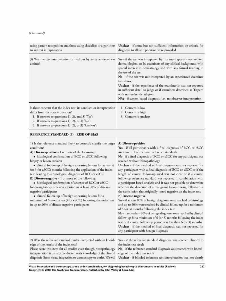
(Continued)
using pattern recognition and those using checklists or algorithms
to aid test interpretation
Unclear - if some but not sufficient information on criteria for
diagnosis to allow replication were provided
3) Was the test interpretation carried out by an experienced ex-
aminer?
Yes - if the test was interpreted by 1 or more speciality-accredited
dermatologists, or by examiners of any clinical background with
special interest in dermatology and with any formal training in
the use of the test
No - if the test was not interpreted by an experienced examiner
(see above)
Unclear - if the experience of the examiner(s) was not reported
in sufficient detail to judge or if examiners described as ’Expert’
with no further detail given
N/A - if system-based diagnosis, i.e., no observer interpretation
Is there concern that the index test, its conduct, or interpretation
differ from the review question?
1. If answers to questions 1), 2), and 3) ’Yes’:
2. If answers to questions 1), 2), or 3) ’No’:
3. If answers to questions 1), 2), or 3) ’Unclear’:
1. Concern is low
2. Concern is high
3. Concern is unclear
REFERENCE STANDARD (3) - RISK OF BIAS
1) Is the reference standard likely to correctly classify the target
condition?
A) Disease-positive - 1 or more of the following:
• histological confirmation of BCC or cSCC following
biopsy or lesion excision
• clinical follow-up of benign-appearing lesions for at least 6
(or 3 for cSCC) months following the application of the index
test, leading to a histological diagnosis of BCC or cSCC
B) Disease-negative - 1 or more of the following:
• histological confirmation of absence of BCC or cSCC
following biopsy or lesion excision in at least 80% of disease-
negative participants
• clinical follow-up of benign-appearing lesions for a
minimum of 6 months (or 3 for cSCC) following the index test
in up to 20% of disease-negative participants
A) Disease-positive
Yes - if all participants with a final diagnosis of BCC or cSCC
underwent 1 of the listed reference standards
No - if a final diagnosis of BCC or cSCC for any participant was
reached without histopathology
Unclear - if the method of final diagnosis was not reported for
any participant with a final diagnosis of BCC or cSCC or if the
length of clinical follow-up used was not clear or if a clinical
follow-up reference standard was reported in combination with
a participant-based analysis and it was not possible to determine
whether the detection of a malignant lesion during follow-up is
the same lesion that originally tested negative on the index test
B) Disease-negative
Yes - if at least 80% of benign diagnoses were reached by histology
and up to 20% were reached by clinical follow-up for a minimum
of 6 (or 3) months following the index test
No - if more than 20% of benign diagnoses were reached by clinical
follow-up for a minimum of 6 (or 3) months following the index
test or if clinical follow-up period was less than 6 (or 3) months
Unclear - if the method of final diagnosis was not reported for
any participant with benign diagnosis
2) Were the reference standard results interpreted without knowl-
edge of the results of the index test?
Please score this item for all studies even though histopathology
interpretation is usually conducted with knowledge of the clinical
diagnosis (from visual inspection or dermoscopy or both). We will
Yes - if the reference standard diagnosis was reached blinded to
the index test result
No - if the reference standard diagnosis was reached with knowl-
edge of the index test result
Unclear - if blinded reference test interpretation was not clearly
262Visual inspection and dermoscopy, alone or in combination, for diagnosing keratinocyte skin cancers in adults (Review)
Copyright © 2018 The Cochrane Collaboration. Published by John Wiley & Sons, Ltd.
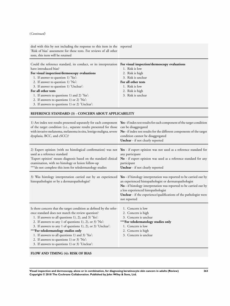
(Continued)
deal with this by not including the response to this item in the
’Risk of bias’ assessment for these tests. For reviews of all other
tests, this item will be retained
reported
Could the reference standard, its conduct, or its interpretation
have introduced bias?
For visual inspection/dermoscopy evaluations
1. If answer to question 1) ’Yes’:
2. If answer to question 1) ’No’:
3. If answer to question 1) ’Unclear’:
For all other tests
1. If answers to questions 1) and 2) ’Yes’:
2. If answers to questions 1) or 2) ’No’:
3. If answers to questions 1) or 2) ’Unclear’:
For visual inspection/dermoscopy evaluations
1. Risk is low
2. Risk is high
3. Risk is unclear
For all other tests
1. Risk is low
2. Risk is high
3. Risk is unclear
REFERENCE STANDARD (3) - CONCERN ABOUT APPLICABILITY
1) Are index test results presented separately for each component
of the target condition (i.e., separate results presented for those
with invasive melanoma, melanoma in situ, lentigo maligna, severe
dysplasia, BCC, and cSCC)?
Yes - if index test results for each component of the target condition
can be disaggregated
No - if index test results for the different components of the target
condition cannot be disaggregated
Unclear - if not clearly reported
2) Expert opinion (with no histological confirmation) was not
used as a reference standard
’Expert opinion’ means diagnosis based on the standard clinical
examination, with no histology or lesion follow-up
***do not complete this item for teledermatology studies
Yes - if expert opinion was not used as a reference standard for
any participant
No - if expert opinion was used as a reference standard for any
participant
Unclear - if not clearly reported
3) Was histology interpretation carried out by an experienced
histopathologist or by a dermatopathologist?
Yes - if histology interpretation was reported to be carried out by
an experienced histopathologist or dermatopathologist
No - if histology interpretation was reported to be carried out by
a less experienced histopathologist
Unclear - if the experience/qualifications of the pathologist were
not reported
Is there concern that the target condition as defined by the refer-
ence standard does not match the review question?
1. If answers to all questions 1), 2), and 3) ’Yes’:
2. If answers to any 1 of questions 1), 2), or 3) ’No’:
3. If answers to any 1 of questions 1), 2), or 3) ’Unclear’:
***For teledermatology studies only
1. If answers to all questions 1) and 3) ’Yes’:
2. If answers to questions 1) or 3) ’No’:
3. If answers to questions 1) or 3) ’Unclear’:
1. Concern is low
2. Concern is high
3. Concern is unclear
***For teledermatology studies only
1. Concern is low
2. Concern is high
3. Concern is unclear
FLOW AND TIMING (4): RISK OF BIAS
263Visual inspection and dermoscopy, alone or in combination, for diagnosing keratinocyte skin cancers in adults (Review)
Copyright © 2018 The Cochrane Collaboration. Published by John Wiley & Sons, Ltd.
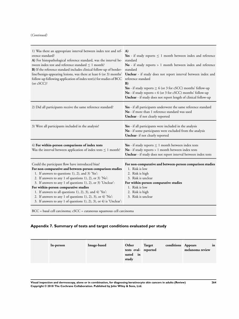
(Continued)
1) Was there an appropriate interval between index test and ref-
erence standard?
A) For histopathological reference standard, was the interval be-
tween index test and reference standard ≤ 1 month?
B) If the reference standard includes clinical follow-up of border-
line/benign-appearing lesions, was there at least 6 (or 3) months’
follow-up following application of index test(s) for studies of BCC
(or cSCC)?
A)
Yes - if study reports ≤ 1 month between index and reference
standard
No - if study reports > 1 month between index and reference
standard
Unclear - if study does not report interval between index and
reference standard
B)
Yes - if study reports ≥ 6 (or 3 for cSCC) months’ follow-up
No - if study reports < 6 (or 3 for cSCC) months’ follow-up
Unclear - if study does not report length of clinical follow-up
2) Did all participants receive the same reference standard? Yes - if all participants underwent the same reference standard
No - if more than 1 reference standard was used
Unclear - if not clearly reported
3) Were all participants included in the analysis? Yes - if all participants were included in the analysis
No - if some participants were excluded from the analysis
Unclear- if not clearly reported
4) For within-person comparisons of index tests
Was the interval between application of index tests ≤ 1 month?
Yes - if study reports ≤ 1 month between index tests
No - if study reports > 1 month between index tests
Unclear - if study does not report interval between index tests
Could the participant flow have introduced bias?
For non-comparative and between-person comparison studies
1. If answers to questions 1), 2), and 3) ’Yes’:
2. If answers to any 1 of questions 1), 2), or 3) ’No’:
3. If answers to any 1 of questions 1), 2), or 3) ’Unclear’:
For within-person comparative studies
1. If answers to all questions 1), 2), 3), and 4) ’Yes’:
2. If answers to any 1 of questions 1), 2), 3), or 4) ’No’:
3. If answers to any 1 of questions 1), 2), 3), or 4) is ’Unclear’:
For non-comparative and between-person comparison studies
1. Risk is low
2. Risk is high
3. Risk is unclear
For within-person comparative studies
1. Risk is low
2. Risk is high
3. Risk is unclear
BCC = basal cell carcinoma; cSCC = cutaneous squamous cell carcinoma
Appendix 7. Summary of tests and target conditions evaluated per study
In-person Image-based Other
tests eval-
uated in
study
Target conditions
reported
Appears in
melanoma review
264Visual inspection and dermoscopy, alone or in combination, for diagnosing keratinocyte skin cancers in adults (Review)
Copyright © 2018 The Cochrane Collaboration. Published by John Wiley & Sons, Ltd.
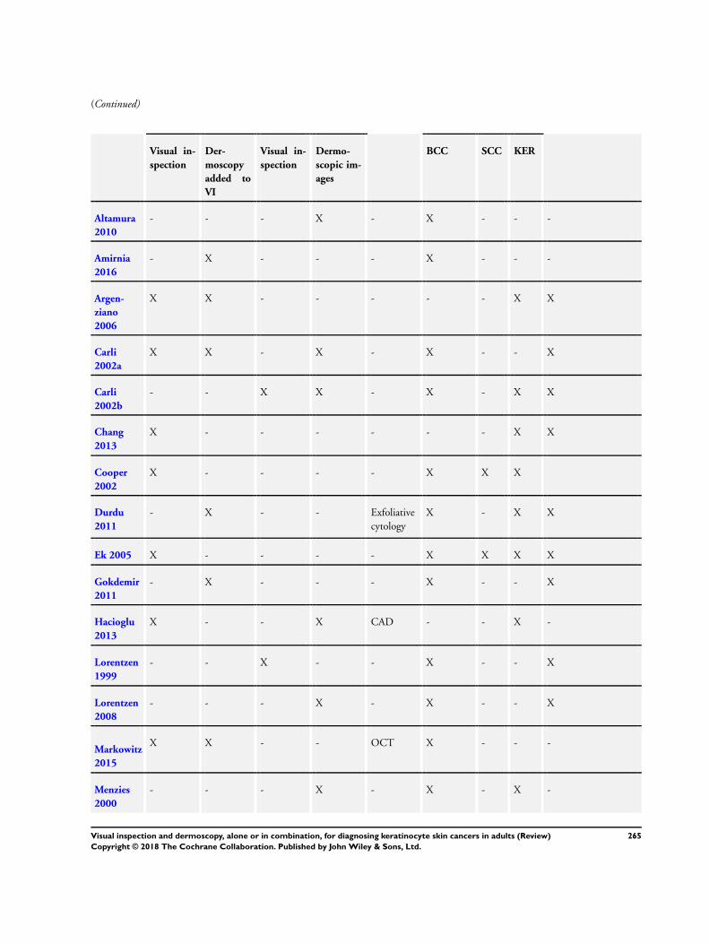
(Continued)
Visual in-
spection
Der-
moscopy
added to
VI
Visual in-
spection
Dermo-
scopic im-
ages
BCC SCC KER
Altamura
2010
- - - X - X - - -
Amirnia
2016
- X - - - X - - -
Argen-
ziano
2006
X X - - - - - X X
Carli
2002a
X X - X - X - - X
Carli
2002b
- - X X - X - X X
Chang
2013
X - - - - - - X X
Cooper
2002
X - - - - X X X
Durdu
2011
- X - - Exfoliative
cytology
X - X X
Ek 2005 X - - - - X X X X
Gokdemir
2011
- X - - - X - - X
Hacioglu
2013
X - - X CAD - - X -
Lorentzen
1999
- - X - - X - - X
Lorentzen
2008
- - - X - X - - X
Markowitz
2015
X X - - OCT X - - -
Menzies
2000
- - - X - X - X -
265Visual inspection and dermoscopy, alone or in combination, for diagnosing keratinocyte skin cancers in adults (Review)
Copyright © 2018 The Cochrane Collaboration. Published by John Wiley & Sons, Ltd.
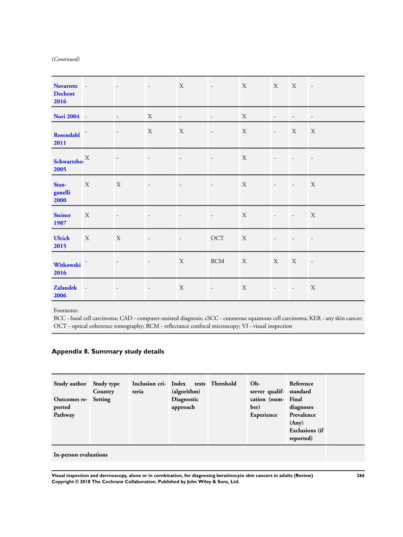
(Continued)
Navarrete
Dechent
2016
- - - X - X X X -
Nori 2004 - - X - - X - - -
Rosendahl
2011
- - X X - X - X X
Schwartzberg
2005
X - - - - X - - -
Stan-
ganelli
2000
X X - - - X - - X
Steiner
1987
X - - - - X - - X
Ulrich
2015
X X - - OCT X - - -
Witkowski
2016
- - - X RCM X X X -
Zalaudek
2006
- - - X - X - - X
Footnotes:
BCC - basal cell carcinoma; CAD - computer-assisted diagnosis; cSCC - cutaneous squamous cell carcinoma; KER - any skin cancer;
OCT - optical coherence tomography; RCM - reflectance confocal microscopy; VI - visual inspection
Appendix 8. Summary study details
Study author
Outcomes re-
ported
Pathway
Study type
Country
Setting
Inclusion cri-
teria
Index tests
(algorithm)
Diagnostic
approach
Threshold Ob-
server qualif-
cation (num-
ber)
Experience
Reference
standard
Final
diagnoses
Prevalence
(Any)
Exclusions (if
reported)
In-person evaluations
266Visual inspection and dermoscopy, alone or in combination, for diagnosing keratinocyte skin cancers in adults (Review)
Copyright © 2018 The Cochrane Collaboration. Published by John Wiley & Sons, Ltd.
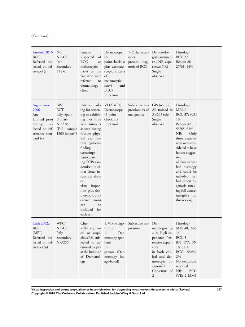
(Continued)
Amirnia 2016
BCC
Referred (se-
lected on ref-
erence) (c)
NC
NR-CS
Iran
Secondary
61 / 61
Patients
suspected of
BCC or
melanocytic
naevi of the
face who were
referred to
dermatology
clinic
Dermoscopy
(3-
point checklist
plus dermato-
scopic criteria
of
melanocytic
naevi and
BCC)
In person
≥ 2 character-
istics
present; diag-
nosis of BCC
Dermatolo-
gist (assumed)
(n = NR; expe-
rience NR)
Single
observer
Histology
BCC 27
Benign 28
27/61; 44%
Argenziano
2006
Any
Limited prior
testing; se-
lected on ref-
ererence stan-
dard (c)
BPC
RCT
Italy, Spain
Primary
NR / 85
(Full sample
1203 lesions*)
Patients ask-
ing for screen-
ing or exhibit-
ing 1 or more
skin tumours
as seen during
routine phys-
ical examina-
tion (patient-
finding
screening)
Participat-
ing PCPs ran-
domised to ei-
ther visual in-
spection alone
or
visual inspec-
tion plus der-
moscopy; only
excised lesions
can be
included for
each arm
VI (ABCD)
Dermoscopy
(3-point
checklist)
In person
Subjective im-
pression; dx of
malignancy
GPs (n = 37)
All trained in
ABCD rule
Single
observer
Histology
MEL 6
BCC 37; SCC
10
Benign 32
53/85; 62%
NB: Only
those patients
who were con-
sidered to have
lesions sugges-
tive
of skin cancer
had histology
and could be
included; rest
had expert di-
agnosis (mak-
ing full dataset
ineligible for
this review)
Carli 2002a
BCC
(MEL)
Referred (se-
lected on ref-
erence) (u)
WPC
NR-CS
Italy
Secondary
NR/256
Clin-
ically equivo-
cal or suspi-
cious PSL sub-
jected to ex-
cisional biopsy
at the Institute
of Dermatol-
ogy
1. VI (no algo-
rithm)
2. Der-
moscopy (pat-
tern)
In-
person (Der-
moscopy - im-
age-based)
Subjective im-
pression
Der-
matologist (n
= 2; High ex-
perience - “ex-
tensive experi-
ence
in both clin-
ical and der-
moscopic di-
agnosis”)
Consensus of
2
Histology
MM 40; MiS
14
BCC 5
BN 177; SN
16; SK 4
BCC: 5/256;
2%
No exclusions
reported
NB: BCC
(VI): 2 MMS
267Visual inspection and dermoscopy, alone or in combination, for diagnosing keratinocyte skin cancers in adults (Review)
Copyright © 2018 The Cochrane Collaboration. Published by John Wiley & Sons, Ltd.
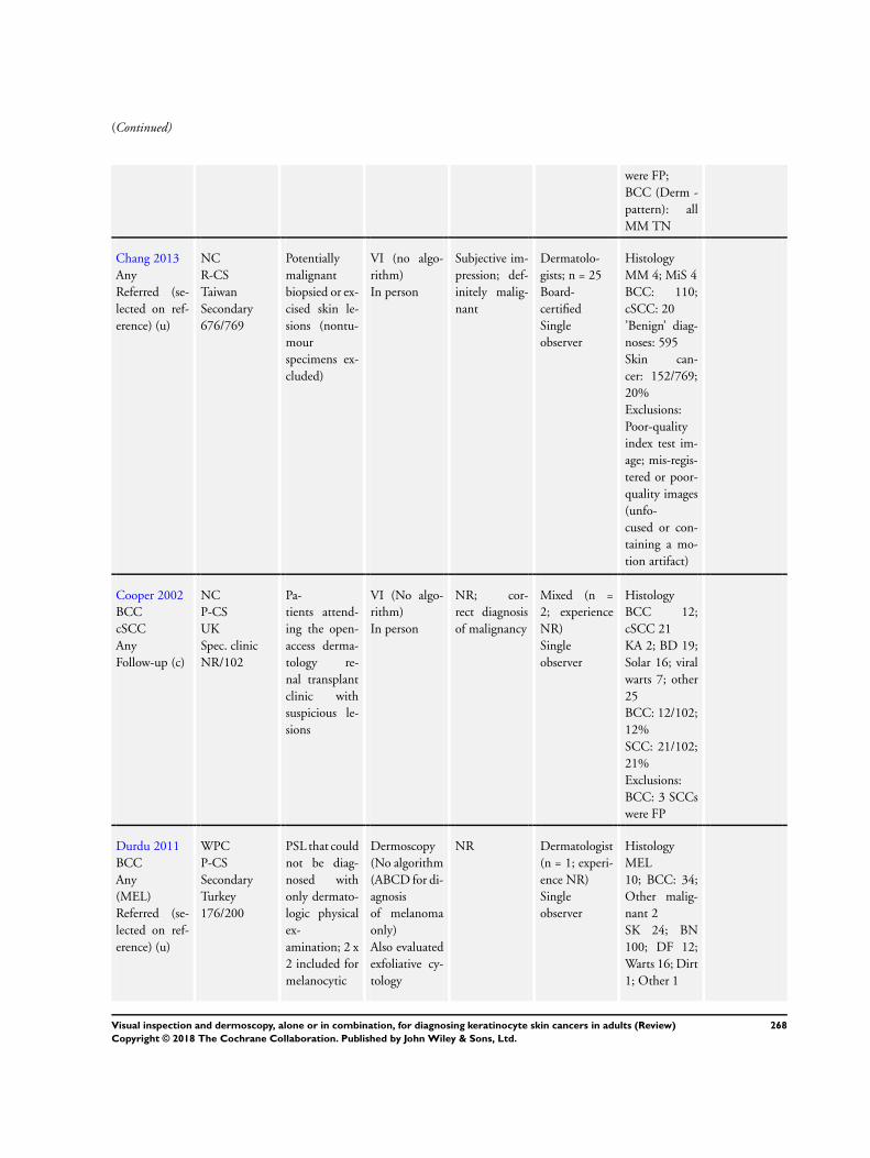
(Continued)
were FP;
BCC (Derm -
pattern): all
MM TN
Chang 2013
Any
Referred (se-
lected on ref-
erence) (u)
NC
R-CS
Taiwan
Secondary
676/769
Potentially
malignant
biopsied or ex-
cised skin le-
sions (nontu-
mour
specimens ex-
cluded)
VI (no algo-
rithm)
In person
Subjective im-
pression; def-
initely malig-
nant
Dermatolo-
gists; n = 25
Board-
certified
Single
observer
Histology
MM 4; MiS 4
BCC: 110;
cSCC: 20
’Benign’ diag-
noses: 595
Skin can-
cer: 152/769;
20%
Exclusions:
Poor-quality
index test im-
age; mis-regis-
tered or poor-
quality images
(unfo-
cused or con-
taining a mo-
tion artifact)
Cooper 2002
BCC
cSCC
Any
Follow-up (c)
NC
P-CS
UK
Spec. clinic
NR/102
Pa-
tients attend-
ing the open-
access derma-
tology re-
nal transplant
clinic with
suspicious le-
sions
VI (No algo-
rithm)
In person
NR; cor-
rect diagnosis
of malignancy
Mixed (n =
2; experience
NR)
Single
observer
Histology
BCC 12;
cSCC 21
KA 2; BD 19;
Solar 16; viral
warts 7; other
25
BCC: 12/102;
12%
SCC: 21/102;
21%
Exclusions:
BCC: 3 SCCs
were FP
Durdu 2011
BCC
Any
(MEL)
Referred (se-
lected on ref-
erence) (u)
WPC
P-CS
Secondary
Turkey
176/200
PSL that could
not be diag-
nosed with
only dermato-
logic physical
ex-
amination; 2 x
2 included for
melanocytic
Dermoscopy
(No algorithm
(ABCD for di-
agnosis
of melanoma
only)
Also evaluated
exfoliative cy-
tology
NR Dermatologist
(n = 1; experi-
ence NR)
Single
observer
Histology
MEL
10; BCC: 34;
Other malig-
nant 2
SK 24; BN
100; DF 12;
Warts 16; Dirt
1; Other 1
268Visual inspection and dermoscopy, alone or in combination, for diagnosing keratinocyte skin cancers in adults (Review)
Copyright © 2018 The Cochrane Collaboration. Published by John Wiley & Sons, Ltd.
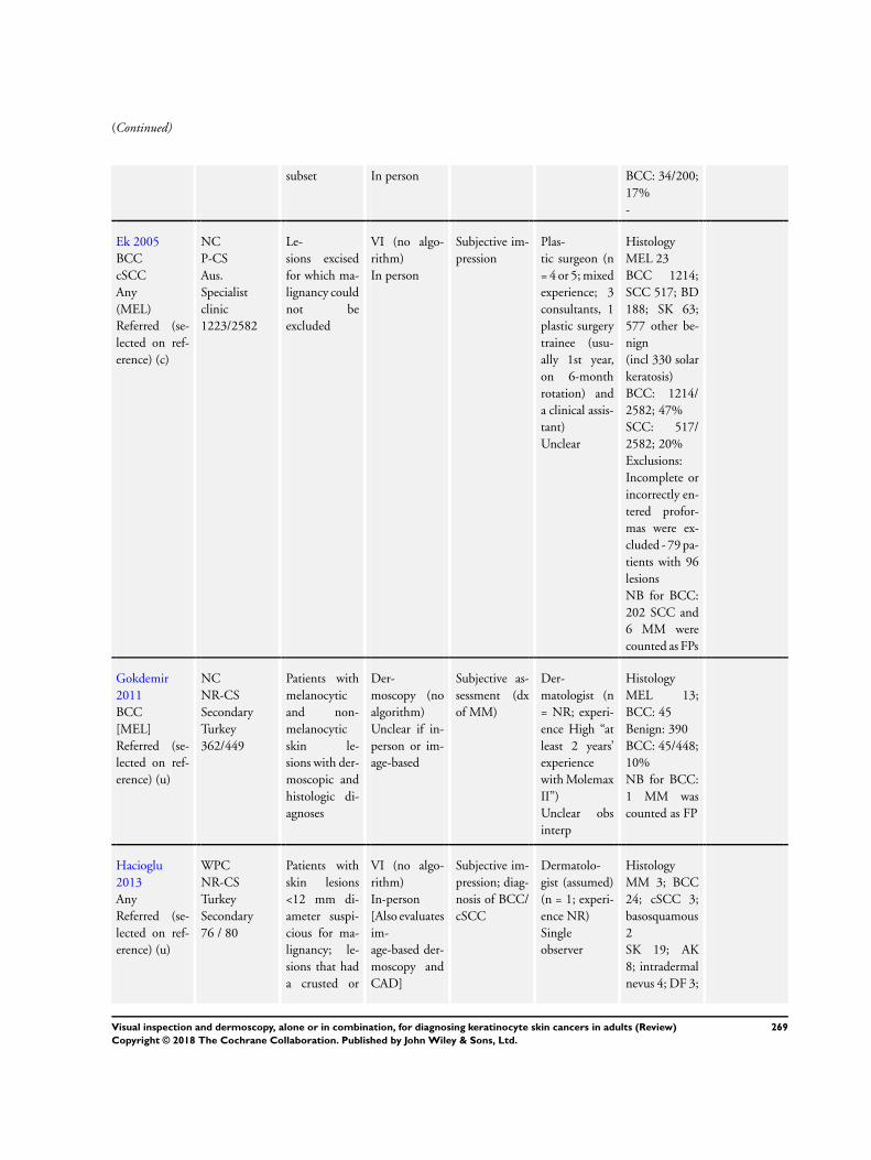
(Continued)
subset In person BCC: 34/200;
17%
-
Ek 2005
BCC
cSCC
Any
(MEL)
Referred (se-
lected on ref-
erence) (c)
NC
P-CS
Aus.
Specialist
clinic
1223/2582
Le-
sions excised
for which ma-
lignancy could
not be
excluded
VI (no algo-
rithm)
In person
Subjective im-
pression
Plas-
tic surgeon (n
= 4 or 5; mixed
experience; 3
consultants, 1
plastic surgery
trainee (usu-
ally 1st year,
on 6-month
rotation) and
a clinical assis-
tant)
Unclear
Histology
MEL 23
BCC 1214;
SCC 517; BD
188; SK 63;
577 other be-
nign
(incl 330 solar
keratosis)
BCC: 1214/
2582; 47%
SCC: 517/
2582; 20%
Exclusions:
Incomplete or
incorrectly en-
tered profor-
mas were ex-
cluded - 79 pa-
tients with 96
lesions
NB for BCC:
202 SCC and
6 MM were
counted as FPs
Gokdemir
2011
BCC
[MEL]
Referred (se-
lected on ref-
erence) (u)
NC
NR-CS
Secondary
Turkey
362/449
Patients with
melanocytic
and non-
melanocytic
skin le-
sions with der-
moscopic and
histologic di-
agnoses
Der-
moscopy (no
algorithm)
Unclear if in-
person or im-
age-based
Subjective as-
sessment (dx
of MM)
Der-
matologist (n
= NR; experi-
ence High “at
least 2 years’
experience
with Molemax
II”)
Unclear obs
interp
Histology
MEL 13;
BCC: 45
Benign: 390
BCC: 45/448;
10%
NB for BCC:
1 MM was
counted as FP
Hacioglu
2013
Any
Referred (se-
lected on ref-
erence) (u)
WPC
NR-CS
Turkey
Secondary
76 / 80
Patients with
skin lesions
<12 mm di-
ameter suspi-
cious for ma-
lignancy; le-
sions that had
a crusted or
VI (no algo-
rithm)
In-person
[Also evaluates
im-
age-based der-
moscopy and
CAD]
Subjective im-
pression; diag-
nosis of BCC/
cSCC
Dermatolo-
gist (assumed)
(n = 1; experi-
ence NR)
Single
observer
Histology
MM 3; BCC
24; cSCC 3;
basosquamous
2
SK 19; AK
8; intradermal
nevus 4; DF 3;
269Visual inspection and dermoscopy, alone or in combination, for diagnosing keratinocyte skin cancers in adults (Review)
Copyright © 2018 The Cochrane Collaboration. Published by John Wiley & Sons, Ltd.
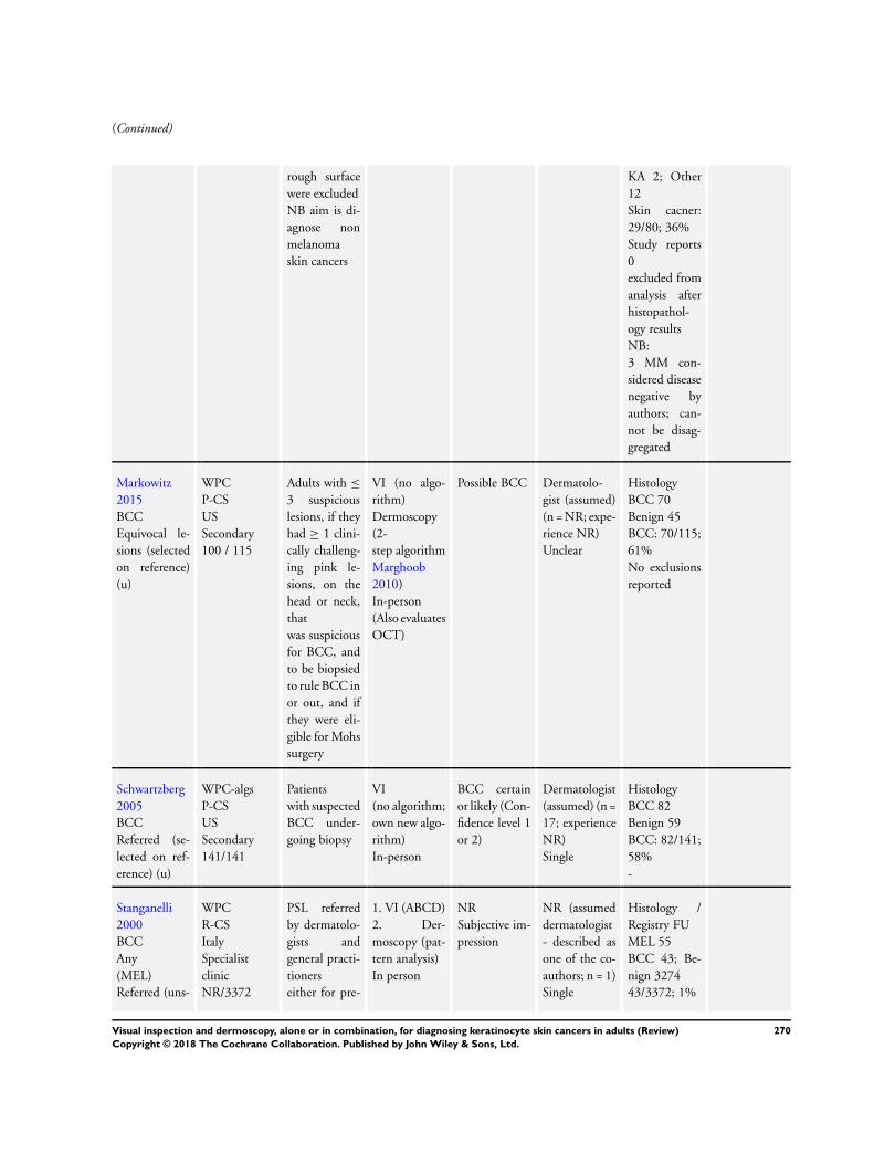
(Continued)
rough surface
were excluded
NB aim is di-
agnose non
melanoma
skin cancers
KA 2; Other
12
Skin cacner:
29/80; 36%
Study reports
0
excluded from
analysis after
histopathol-
ogy results
NB:
3 MM con-
sidered disease
negative by
authors; can-
not be disag-
gregated
Markowitz
2015
BCC
Equivocal le-
sions (selected
on reference)
(u)
WPC
P-CS
US
Secondary
100 / 115
Adults with ≤
3 suspicious
lesions, if they
had ≥ 1 clini-
cally challeng-
ing pink le-
sions, on the
head or neck,
that
was suspicious
for BCC, and
to be biopsied
to rule BCC in
or out, and if
they were eli-
gible for Mohs
surgery
VI (no algo-
rithm)
Dermoscopy
(2-
step algorithm
Marghoob
2010)
In-person
(Also evaluates
OCT)
Possible BCC Dermatolo-
gist (assumed)
(n = NR; expe-
rience NR)
Unclear
Histology
BCC 70
Benign 45
BCC: 70/115;
61%
No exclusions
reported
Schwartzberg
2005
BCC
Referred (se-
lected on ref-
erence) (u)
WPC-algs
P-CS
US
Secondary
141/141
Patients
with suspected
BCC under-
going biopsy
VI
(no algorithm;
own new algo-
rithm)
In-person
BCC certain
or likely (Con-
fidence level 1
or 2)
Dermatologist
(assumed) (n =
17; experience
NR)
Single
Histology
BCC 82
Benign 59
BCC: 82/141;
58%
-
Stanganelli
2000
BCC
Any
(MEL)
Referred (uns-
WPC
R-CS
Italy
Specialist
clinic
NR/3372
PSL referred
by dermatolo-
gists and
general practi-
tioners
either for pre-
1. VI (ABCD)
2. Der-
moscopy (pat-
tern analysis)
In person
NR
Subjective im-
pression
NR (assumed
dermatologist
- described as
one of the co-
authors; n = 1)
Single
Histology /
Registry FU
MEL 55
BCC 43; Be-
nign 3274
43/3372; 1%
270Visual inspection and dermoscopy, alone or in combination, for diagnosing keratinocyte skin cancers in adults (Review)
Copyright © 2018 The Cochrane Collaboration. Published by John Wiley & Sons, Ltd.
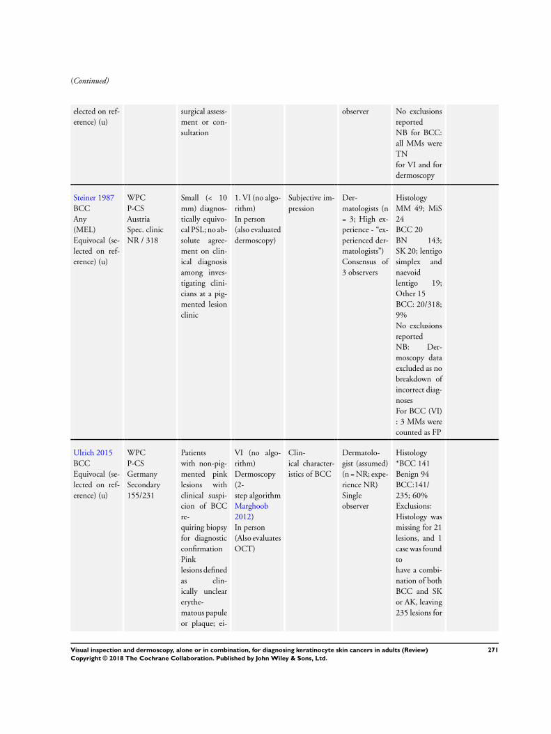
(Continued)
elected on ref-
erence) (u)
surgical assess-
ment or con-
sultation
observer No exclusions
reported
NB for BCC:
all MMs were
TN
for VI and for
dermoscopy
Steiner 1987
BCC
Any
(MEL)
Equivocal (se-
lected on ref-
erence) (u)
WPC
P-CS
Austria
Spec. clinic
NR / 318
Small (< 10
mm) diagnos-
tically equivo-
cal PSL; no ab-
solute agree-
ment on clin-
ical diagnosis
among inves-
tigating clini-
cians at a pig-
mented lesion
clinic
1. VI (no algo-
rithm)
In person
(also evaluated
dermoscopy)
Subjective im-
pression
Der-
matologists (n
= 3; High ex-
perience - “ex-
perienced der-
matologists”)
Consensus of
3 observers
Histology
MM 49; MiS
24
BCC 20
BN 143;
SK 20; lentigo
simplex and
naevoid
lentigo 19;
Other 15
BCC: 20/318;
9%
No exclusions
reported
NB: Der-
moscopy data
excluded as no
breakdown of
incorrect diag-
noses
For BCC (VI)
: 3 MMs were
counted as FP
Ulrich 2015
BCC
Equivocal (se-
lected on ref-
erence) (u)
WPC
P-CS
Germany
Secondary
155/231
Patients
with non-pig-
mented pink
lesions with
clinical suspi-
cion of BCC
re-
quiring biopsy
for diagnostic
confirmation
Pink
lesions defined
as clin-
ically unclear
erythe-
matous papule
or plaque; ei-
VI (no algo-
rithm)
Dermoscopy
(2-
step algorithm
Marghoob
2012)
In person
(Also evaluates
OCT)
Clin-
ical character-
istics of BCC
Dermatolo-
gist (assumed)
(n = NR; expe-
rience NR)
Single
observer
Histology
*BCC 141
Benign 94
BCC:141/
235; 60%
Exclusions:
Histology was
missing for 21
lesions, and 1
case was found
to
have a combi-
nation of both
BCC and SK
or AK, leaving
235 lesions for
271Visual inspection and dermoscopy, alone or in combination, for diagnosing keratinocyte skin cancers in adults (Review)
Copyright © 2018 The Cochrane Collaboration. Published by John Wiley & Sons, Ltd.
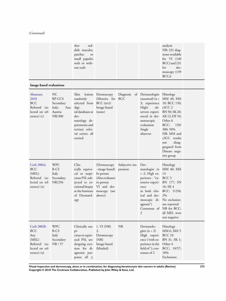
(Continued)
ther red-
dish macules,
patches or
small papules
with or with-
out scale
analysis
NB: 231 diag-
noses available
for VI (140
BCC) and 231
for der-
moscopy (139
BCCs)
Image-based evaluations
Altamura
2010
BCC
Referred (se-
lected on ref-
erence) (c)
NC
RP-CCS
Secondary
Italy; Aus;
Austria
NR/300
Skin lesions
randomly
selected from
digi-
tal databases at
der-
matology de-
partments and
tertiary refer-
ral centre; all
excised
Dermoscopy
(Menzies for
BCC (rev))
Image-based
(none)
Diagnosis of
BCC
Dermatologist
(assumed) (n =
3; experience
High) ob-
servers experi-
enced in der-
matoscopic
evaluation
Single
observer
Histology
MM 40; MiS
10; BCC 150;
cSCC 2
BN 50; SK 20;
AK 12; DF 10;
Other 6
BCC: 150/
300; 50%
NB: MM and
cSCC results
not disag-
gregated from
Disease nega-
tive group
Carli 2002a
BCC
(MEL)
Referred (se-
lected on ref-
erence) (u)
WPC
R-CS
Italy
Secondary
NR/256
Clin-
ically equivo-
cal or suspi-
cious PSL sub-
jected to ex-
cisional biopsy
at the Institute
of Dermatol-
ogy
(Dermoscopy
- image-based)
In person
(Also evaluates
in-person
VI and der-
moscopy (see
above))
Subjective im-
pression
Der-
matologist (n
= 2; High ex-
perience - “ex-
tensive experi-
ence
in both clin-
ical and der-
moscopic di-
agnosis”)
Consensus of
2
Histology
MM 40; MiS
14
BCC 5
BN 177; SN
16; SK 4
BCC: 5/256;
2%
No exclusion-
sne reported
NB for BCC:
all MEL were
test negative
Carli 2002b
BCC
Any
(MEL)
Referred (se-
lected on ref-
erence) (u)
WPC
R-CS
Italy
Secondary
NR / 57
Clinically sus-
pi-
cious or equiv-
ocal PSL un-
dergoing exci-
sion for di-
agnostic pur-
poses; all ≤
1. VI (NR)
2.
Dermoscopy
(NR)
Image-based
(blinded)
NR Dermatolo-
gists (n = 2)
High experi-
ence (’with ex-
perience in the
field of ’); con-
sensus of 2
Histology
MM 6, MiS 5
BCC 10
BN 31, SK 1;
Other 4
BCC; 10/57;
18%
Exclusions:
272Visual inspection and dermoscopy, alone or in combination, for diagnosing keratinocyte skin cancers in adults (Review)
Copyright © 2018 The Cochrane Collaboration. Published by John Wiley & Sons, Ltd.
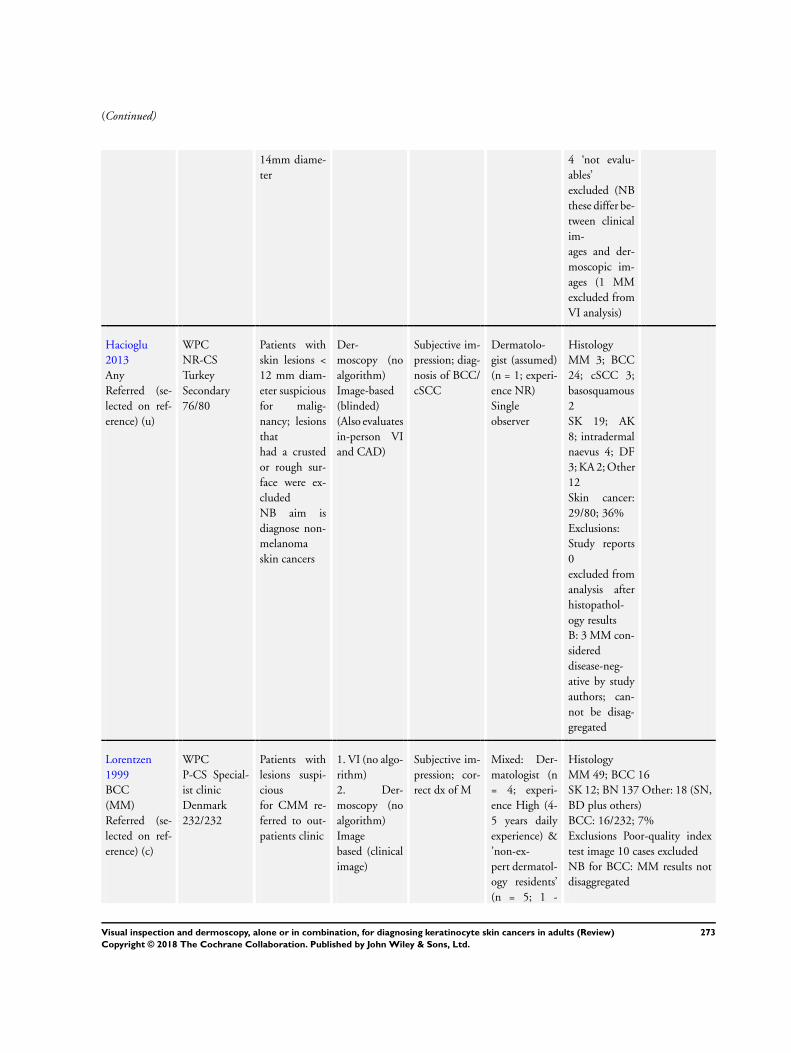
(Continued)
14mm diame-
ter
4 ‘not evalu-
ables’
excluded (NB
these differ be-
tween clinical
im-
ages and der-
moscopic im-
ages (1 MM
excluded from
VI analysis)
Hacioglu
2013
Any
Referred (se-
lected on ref-
erence) (u)
WPC
NR-CS
Turkey
Secondary
76/80
Patients with
skin lesions <
12 mm diam-
eter suspicious
for malig-
nancy; lesions
that
had a crusted
or rough sur-
face were ex-
cluded
NB aim is
diagnose non-
melanoma
skin cancers
Der-
moscopy (no
algorithm)
Image-based
(blinded)
(Also evaluates
in-person VI
and CAD)
Subjective im-
pression; diag-
nosis of BCC/
cSCC
Dermatolo-
gist (assumed)
(n = 1; experi-
ence NR)
Single
observer
Histology
MM 3; BCC
24; cSCC 3;
basosquamous
2
SK 19; AK
8; intradermal
naevus 4; DF
3; KA 2; Other
12
Skin cancer:
29/80; 36%
Exclusions:
Study reports
0
excluded from
analysis after
histopathol-
ogy results
B: 3 MM con-
sidered
disease-neg-
ative by study
authors; can-
not be disag-
gregated
Lorentzen
1999
BCC
(MM)
Referred (se-
lected on ref-
erence) (c)
WPC
P-CS Special-
ist clinic
Denmark
232/232
Patients with
lesions suspi-
cious
for CMM re-
ferred to out-
patients clinic
1. VI (no algo-
rithm)
2. Der-
moscopy (no
algorithm)
Image
based (clinical
image)
Subjective im-
pression; cor-
rect dx of M
Mixed: Der-
matologist (n
= 4; experi-
ence High (4-
5 years daily
experience) &
’non-ex-
pert dermatol-
ogy residents’
(n = 5; 1 -
Histology
MM 49; BCC 16
SK 12; BN 137 Other: 18 (SN,
BD plus others)
BCC: 16/232; 7%
Exclusions Poor-quality index
test image 10 cases excluded
NB for BCC: MM results not
disaggregated
273Visual inspection and dermoscopy, alone or in combination, for diagnosing keratinocyte skin cancers in adults (Review)
Copyright © 2018 The Cochrane Collaboration. Published by John Wiley & Sons, Ltd.
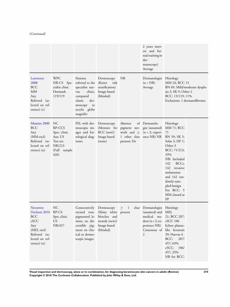
(Continued)
2 years inter-
est and for-
mal training in
der-
matoscopy)
Average
Lorentzen
2008
BCC
MM
Any
Referred (se-
lected on ref-
erence) (c)
WPC
NR-CS Spe-
cialist clinic
Denmark
119/119
Patients
referred to the
specialist nae-
vus clinic;
compared
classic der-
moscopy to
acrylic globe
magnifer
Dermoscopy
(Kenet risk
stratification)
Image-based
(blinded)
NR Dermatologist
(n = NR)
Average
Histology
MM 24; BCC 13
BN 69; Mild/moderate dyspla-
sia 2; SK 9; Other 2
BCC: 13/119; 11%
Exclusions: 1 dermatofibroma
Menzies 2000
BCC
Any
(MM-excl)
Referred (se-
lected on ref-
erence) (u)
NC
RP-CCS
Spec. clinic
Aus; US
Test set:
NR/213
(Full sample
426)
PSL with der-
moscopic im-
ages and his-
tological diag-
noses
Dermoscopy
(Menzies for
BCC (new))
Image-based
(none)
Absence of
pigment net-
work and ≥
1 other char
present; Dx
Dermatolo-
gist (assumed)
(n = 2; experi-
ence NR) NR
Histology
MM 71; BCC
71
BN 59; SK 5;
Solar 3; DF 1;
Other 3
BCC: 71/213;
33%
NB: Included
142 BCCs,
142 invasive
melanomas
and 142 ran-
domly-sam-
pled benign
For BCC: 5
MM classed as
FP
Navarrete
Dechent 2016
BCC
cSCC
Any
(MEL excl)
Referred (se-
lected on ref-
erence) (u)
NC
RP-CS
Spec clinic
US
NR/457
Consecutively
excised non-
pigmented le-
sions; no dis-
cernible pig-
ment on clin-
ical or dermo-
scopic images
Dermoscopy
(Shiny white
blotches and
strands (new))
Image-based
(blinded)
≥ 1 char
present
Dermatologist
(assumed) and
medical stu-
dent (n = 2; ex-
perience NR)
Consensus of
2
Histology
MEL
21; BCC 287;
cSCC 106
lichen planus-
like keratosis
39; Naevus 4
BCC: 287/
457; 63%
cSCC: 106/
457; 23%
NB for BCC:
274Visual inspection and dermoscopy, alone or in combination, for diagnosing keratinocyte skin cancers in adults (Review)
Copyright © 2018 The Cochrane Collaboration. Published by John Wiley & Sons, Ltd.
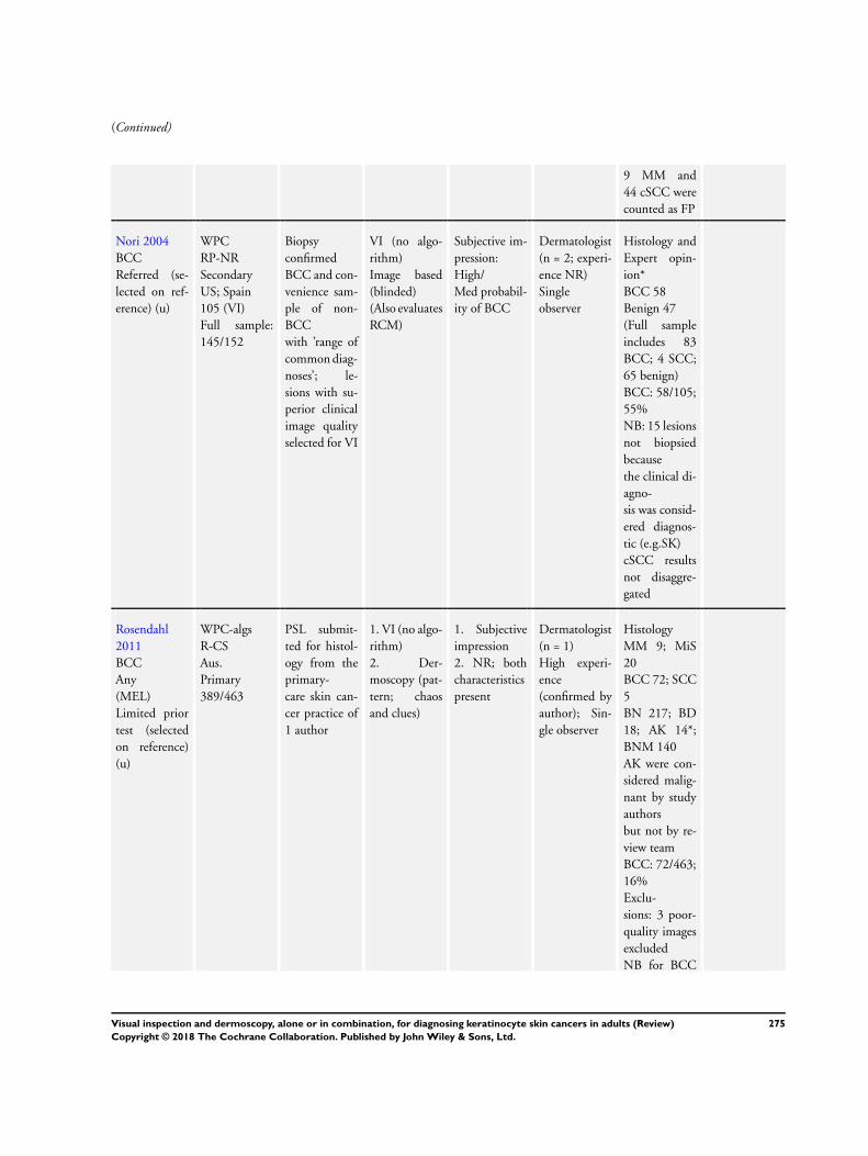
(Continued)
9 MM and
44 cSCC were
counted as FP
Nori 2004
BCC
Referred (se-
lected on ref-
erence) (u)
WPC
RP-NR
Secondary
US; Spain
105 (VI)
Full sample:
145/152
Biopsy
confirmed
BCC and con-
venience sam-
ple of non-
BCC
with ’range of
common diag-
noses’; le-
sions with su-
perior clinical
image quality
selected for VI
VI (no algo-
rithm)
Image based
(blinded)
(Also evaluates
RCM)
Subjective im-
pression:
High/
Med probabil-
ity of BCC
Dermatologist
(n = 2; experi-
ence NR)
Single
observer
Histology and
Expert opin-
ion*
BCC 58
Benign 47
(Full sample
includes 83
BCC; 4 SCC;
65 benign)
BCC: 58/105;
55%
NB: 15 lesions
not biopsied
because
the clinical di-
agno-
sis was consid-
ered diagnos-
tic (e.g.SK)
cSCC results
not disaggre-
gated
Rosendahl
2011
BCC
Any
(MEL)
Limited prior
test (selected
on reference)
(u)
WPC-algs
R-CS
Aus.
Primary
389/463
PSL submit-
ted for histol-
ogy from the
primary-
care skin can-
cer practice of
1 author
1. VI (no algo-
rithm)
2. Der-
moscopy (pat-
tern; chaos
and clues)
1. Subjective
impression
2. NR; both
characteristics
present
Dermatologist
(n = 1)
High experi-
ence
(confirmed by
author); Sin-
gle observer
Histology
MM 9; MiS
20
BCC 72; SCC
5
BN 217; BD
18; AK 14*;
BNM 140
AK were con-
sidered malig-
nant by study
authors
but not by re-
view team
BCC: 72/463;
16%
Exclu-
sions: 3 poor-
quality images
excluded
NB for BCC
275Visual inspection and dermoscopy, alone or in combination, for diagnosing keratinocyte skin cancers in adults (Review)
Copyright © 2018 The Cochrane Collaboration. Published by John Wiley & Sons, Ltd.
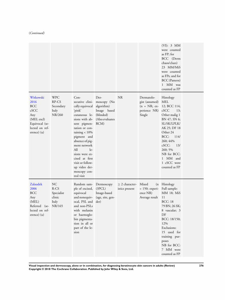
(Continued)
(VI): 3 MM
were counted
as FP; for
BCC (Derm
chaos/clues)
23 MM/MiS
were counted
as FPs; and for
BCC (Pattern)
1 MM was
counted as FP
Witkowski
2016
BCC
cSCC
Any
(MEL excl)
Equivocal (se-
lected on ref-
erence) (u)
WPC
RP-CS
Secondary
Italy
NR/260
Con-
secutive clini-
cally equivocal
‘pink’
cutaneous le-
sions with ab-
sent pigmen-
tation or con-
taining < 10%
pigment and
absence of pig-
ment network
All le-
sions were ex-
cised at first
visit or follow-
up video der-
moscopy con-
trol visit
Der-
moscopy (No
algorithm)
Image based
(blinded)
(Also evaluates
RCM)
NR Dermatolo-
gist (assumed)
(n = NR; ex-
perience NR)
Single
Histology
MEL
12; BCC 114;
cSCC 13;
Other malig 1
BN 47; SN 6;
SL/SK/LPLK/
AK 25; DF 18
Other 24
BCC: 114/
260; 44%
cSCC: 13/
260; 5%
NB for BCC:
1 MM and
1 cSCC were
counted as FP
Zalaudek
2006
BCC
Any
(MEL)
Referred (se-
lected on ref-
erence) (u)
NC
R-CS
Specialist
clinic
Italy
NR/165
Random sam-
ple of excised,
equivocal
and nonequiv-
ocal, PSL and
and non-PSLs
with melanin
or haemoglo-
bin pigmenta-
tion in all or
part of the le-
sion
Dermoscopy
(3PCL)
Image-based
(age, site, gen-
der)
≥ 2 character-
istics present
Mixed (n
= 150; experi-
ence NR)
Average result
Histology
Full sample:
MM 18; MiS
11
BCC: 18
79 BN; 26 SK;
8 vascular; 3
DF
BCC: 18/150;
12%
Exclusions:
15 used for
training pur-
poses
NB for BCC:
7 MM were
counted as FP
276Visual inspection and dermoscopy, alone or in combination, for diagnosing keratinocyte skin cancers in adults (Review)
Copyright © 2018 The Cochrane Collaboration. Published by John Wiley & Sons, Ltd.
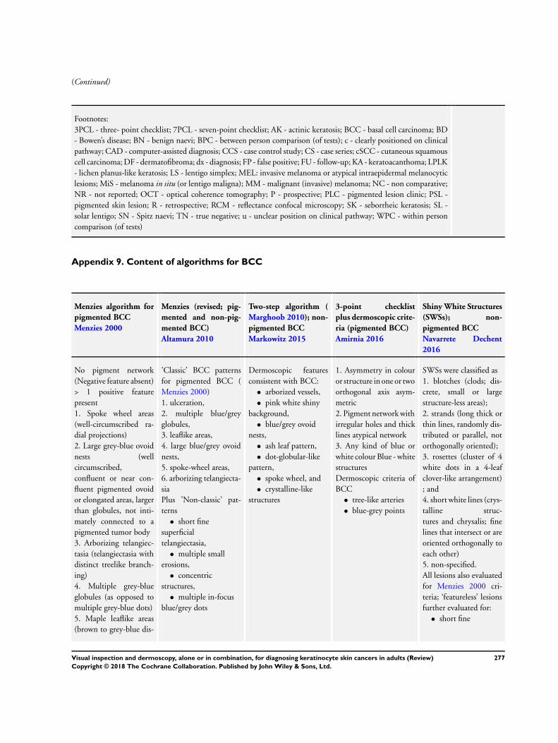
(Continued)
Footnotes:
3PCL - three- point checklist; 7PCL - seven-point checklist; AK - actinic keratosis; BCC - basal cell carcinoma; BD
- Bowen’s disease; BN - benign naevi; BPC - between person comparison (of tests); c - clearly positioned on clinical
pathway; CAD - computer-assisted diagnosis; CCS - case control study; CS - case series; cSCC - cutaneous squamous
cell carcinoma; DF - dermatofibroma; dx - diagnosis; FP - false positive; FU - follow-up; KA - keratoacanthoma; LPLK
- lichen planus-like keratosis; LS - lentigo simplex; MEL: invasive melanoma or atypical intraepidermal melanocytic
lesions; MiS - melanoma in situ (or lentigo maligna); MM - malignant (invasive) melanoma; NC - non comparative;
NR - not reported; OCT - optical coherence tomography; P - prospective; PLC - pigmented lesion clinic; PSL -
pigmented skin lesion; R - retrospective; RCM - reflectance confocal microscopy; SK - seborrheic keratosis; SL -
solar lentigo; SN - Spitz naevi; TN - true negative; u - unclear position on clinical pathway; WPC - within person
comparison (of tests)
Appendix 9. Content of algorithms for BCC
Menzies algorithm for
pigmented BCC
Menzies 2000
Menzies (revised; pig-
mented and non-pig-
mented BCC)
Altamura 2010
Two-step algorithm (
Marghoob 2010); non-
pigmented BCC
Markowitz 2015
3-point checklist
plus dermoscopic crite-
ria (pigmented BCC)
Amirnia 2016
Shiny White Structures
(SWSs); non-
pigmented BCC
Navarrete Dechent
2016
No pigment network
(Negative feature absent)
> 1 positive feature
present
1. Spoke wheel areas
(well-circumscribed ra-
dial projections)
2. Large grey-blue ovoid
nests (well
circumscribed,
confluent or near con-
fluent pigmented ovoid
or elongated areas, larger
than globules, not inti-
mately connected to a
pigmented tumor body
3. Arborizing telangiec-
tasia (telangiectasia with
distinct treelike branch-
ing)
4. Multiple grey-blue
globules (as opposed to
multiple grey-blue dots)
5. Maple leaflike areas
(brown to grey-blue dis-
’Classic’ BCC patterns
for pigmented BCC (
Menzies 2000)
1. ulceration,
2. multiple blue/grey
globules,
3. leaflike areas,
4. large blue/grey ovoid
nests,
5. spoke-wheel areas,
6. arborizing telangiecta-
sia
Plus ’Non-classic’ pat-
terns
• short fine
superficial
telangiectasia,
• multiple small
erosions,
• concentric
structures,
• multiple in-focus
blue/grey dots
Dermoscopic features
consistent with BCC:
• arborized vessels,
• pink white shiny
background,
• blue/grey ovoid
nests,
• ash leaf pattern,
• dot-globular-like
pattern,
• spoke wheel, and
• crystalline-like
structures
1. Asymmetry in colour
or structure in one or two
orthogonal axis asym-
metric
2. Pigment network with
irregular holes and thick
lines atypical network
3. Any kind of blue or
white colour Blue - white
structures
Dermoscopic criteria of
BCC
• tree-like arteries
• blue-grey points
SWSs were classified as
1. blotches (clods; dis-
crete, small or large
structure-less areas);
2. strands (long thick or
thin lines, randomly dis-
tributed or parallel, not
orthogonally oriented);
3. rosettes (cluster of 4
white dots in a 4-leaf
clover-like arrangement)
; and
4. short white lines (crys-
talline struc-
tures and chrysalis; fine
lines that intersect or are
oriented orthogonally to
each other)
5. non-specified.
All lesions also evaluated
for Menzies 2000 cri-
teria; ‘featureless’ lesions
further evaluated for:
• short fine
277Visual inspection and dermoscopy, alone or in combination, for diagnosing keratinocyte skin cancers in adults (Review)
Copyright © 2018 The Cochrane Collaboration. Published by John Wiley & Sons, Ltd.
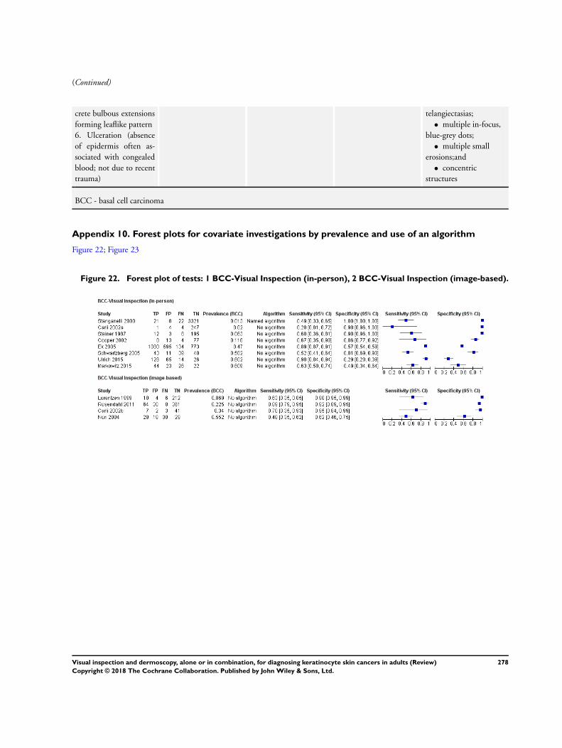
(Continued)
crete bulbous extensions
forming leaflike pattern
6. Ulceration (absence
of epidermis often as-
sociated with congealed
blood; not due to recent
trauma)
telangiectasias;
• multiple in-focus,
blue-grey dots;
• multiple small
erosions;and
• concentric
structures
BCC - basal cell carcinoma
Appendix 10. Forest plots for covariate investigations by prevalence and use of an algorithm
Figure 22; Figure 23
Figure 22. Forest plot of tests: 1 BCC-Visual Inspection (in-person), 2 BCC-Visual Inspection (image-based).
278Visual inspection and dermoscopy, alone or in combination, for diagnosing keratinocyte skin cancers in adults (Review)
Copyright © 2018 The Cochrane Collaboration. Published by John Wiley & Sons, Ltd.
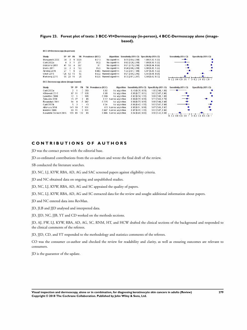
Figure 23. Forest plot of tests: 3 BCC-VI+Dermoscopy (in-person), 4 BCC-Dermoscopy alone (image-
based).
C O N T R I B U T I O N S O F A U T H O R S
JD was the contact person with the editorial base.
JD co-ordinated contributions from the co-authors and wrote the final draft of the review.
SB conducted the literature searches.
JD, NC, LJ, KYW, RBA, AD, AG and SAC screened papers against eligibility criteria.
JD and NC obtained data on ongoing and unpublished studies.
JD, NC, LJ, KYW, RBA, AD, AG and SC appraised the quality of papers.
JD, NC, LJ, KYW, RBA, AD, AG and SC extracted data for the review and sought additional information about papers.
JD and NC entered data into RevMan.
JD, JLB and JJD analysed and interpreted data.
JD, JJD, NC, JJB, YT and CD worked on the methods sections.
JD, AJ, FW, LJ, KYW, RBA, AD, AG, SC, RNM, HT, and HCW drafted the clinical sections of the background and responded to
the clinical comments of the referees.
JD, JJD, CD, and YT responded to the methodology and statistics comments of the referees.
CO was the consumer co-author and checked the review for readability and clarity, as well as ensuring outcomes are relevant to
consumers.
JD is the guarantor of the update.
279Visual inspection and dermoscopy, alone or in combination, for diagnosing keratinocyte skin cancers in adults (Review)
Copyright © 2018 The Cochrane Collaboration. Published by John Wiley & Sons, Ltd.
Disclaimer
This project was supported by the National Institute for Health Research, via Cochrane Infrastructure funding to the Cochrane Skin
Group and Cochrane Programme Grant funding. The views and opinions expressed therein are those of the authors and do not
necessarily reflect those of the Systematic Reviews Programme, NIHR, NHS or the Department of Health.
D E C L A R A T I O N S O F I N T E R E S T
Jacqueline Dinnes: nothing to declare.
Jonathan J Deeks: nothing to declare.
Naomi Chuchu: nothing to declare.
Rubeta N Matin: “my institution received a grant for a Barco NV commercially sponsored study to evaluate digital dermoscopy in
the skin cancer clinic. My institution also received Oxfordshire Health Services Research Charitable Funds for carrying out a study of
feasibility of using the Skin Cancer Quality of Life Impact Tool (SCQOLIT) in non melanoma skin cancer. I have received royalties
for the Oxford Handbook of Medical Dermatology (Oxford University Press). I have received payment from Public Health England
for the ”Be Clear on Cancer“ skin cancer report. I have no conflicts of interest to declare that directly relate to the publication of this
work.”
Kai Yuen Wong: nothing to declare.
Roger Benjamin Aldridge: nothing to declare.
Alana Durack: nothing to declare.
Abha Gulati: nothing to declare.
Sue Ann Chan: nothing to declare.
Louise Johnston: nothing to declare.
Susan E Bayliss: nothing to declare.
Jo Leonardi-Bee: nothing to declare.
Yemisi Takwoingi: nothing to declare.
Clare Davenport: nothing to declare.
Colette O’Sullivan: nothing to declare.
Hamid Tehrani: nothing to declare.
Hywel C Williams: I am director of the NIHR HTA Programme. HTA is part of the NIHR which also supports the NIHR systematic
reviews programme from which this work is funded.
S O U R C E S O F S U P P O R T
280Visual inspection and dermoscopy, alone or in combination, for diagnosing keratinocyte skin cancers in adults (Review)
Copyright © 2018 The Cochrane Collaboration. Published by John Wiley & Sons, Ltd.
Internal sources
• No sources of support supplied
External sources
• The National Institute for Health Research (NIHR), UK.
The NIHR, UK, is the largest single funder of the Cochrane Skin Group
• NIHR Systematic Review Programme, UK.
• NIHR clinical fellowship, UK.
D I F F E R E N C E S B E T W E E N P R O T O C O L A N D R E V I E W
The proposed primary objective to analyse studies according to the prior testing undergone by study participants (comparing those
with limited prior testing with those referred for further evaluation of a suspicious skin lesion) was not possible due to limited data.
The primary objectives were also amended to conduct separate analyses by in-person/image-based diagnosis rather than to investigate
the effect on accuracy as a secondary objective, as originally proposed in the generic protocol. We took this decision very early in the
review process and based it on the fact that a diagnosis based on a dermoscopic image or clinical photograph cannot approximate the
context of a face-to-face patient/clinician consultation, and was not based on observed results.
We expanded the secondary objectives for the detection of BCC or cSCC to include: test comparisons restricted to studies where both
tests were evaluated in the same studies (direct test comparisons); and investigations of the accuracy of individual algorithms used to
assist visual inspection or dermoscopy, and any effect from observer experience on diagnostic accuracy.
The secondary objective has been changed from “for the detection of any skin cancer” to “for the detection of any skin cancer in adults,
where keratinocyte skin cancers make up at least 50% of included skin cancers” in order to keep the focus on keratinocyte skin cancers
for this review and in order not to replicate analyses conducted for the review of RCM for melanoma. These changes also affect the
definition of the secondary target condition in the Methods section.
Sources of heterogeneity that could be investigated were restricted due to lack of data.
We amended the text to clarify that studies available only as conference abstracts would be excluded from the review unless full
papers could be identified; studies available only as conference abstracts do not allow a comprehensive assessment of study methods or
methodological quality.
We clarified the participant inclusion criteria to make it clear that studies of only malignant or benign lesions would be excluded.
To improve clarity of methods, this text from the protocol “We will include studies developing new algorithms or methods of diagnosis
(i.e. derivation studies) if they use a separate independent ’test set’ of participants or images to evaluate the new approach.We will also
include studies using other forms of cross validation, such as ’leave-one-out’ cross-validation (Efron 1983). We will note for future
reference (but not extract) any data on the accuracy of lesion characteristics individually, e.g. the presence or absence of a pigment
network or detection of asymmetry.”
has been replaced with “Studies developing new algorithms or methods of diagnosis (i.e. derivation studies) were included if they:
• used a separate independent ’test set’ of participants or images to evaluate the new approach, or
• investigated lesion characteristics that had previously been suggested as associated with melanoma and the study reported
accuracy based on the presence or absence of particular combinations of characteristics.
Studies were excluded if they:
• used a statistical model to produce a data driven equation, or algorithm based on multiple diagnostic features, with no separate
test set
• used cross-validation approaches such as ’leave-one-out’ cross-validation (Efron 1983)
281Visual inspection and dermoscopy, alone or in combination, for diagnosing keratinocyte skin cancers in adults (Review)
Copyright © 2018 The Cochrane Collaboration. Published by John Wiley & Sons, Ltd.

• evaluated the accuracy of the presence or absence of individual lesion characteristics or morphological features, with no overall
diagnosis of malignancy
• reported accuracy data for ‘clinical diagnosis’ with no clear description as to whether the reported data related to visual
inspection alone or included dermoscopy in all study participants
• were based on the experience of a skin cancer-specific clinic, where dermoscopy may or may not have been used on an individual
patient basis.“
We proposed to supplement the database searches by searching the annual meetings of appropriate organisations (e.g. British Association
of Dermatologists Annual Meeting, American Academy of Dermatology Annual Meeting, European Academy of Dermatology and
Venereology Meeting, Society for Melanoma Research Congress, World Congress of Dermatology, European Association of Dermato
Oncology), but due to the volume of evidence retrieved from database searches and time restrictions we were unable to do this.
As per the change to secondary objectives, this text from the protocol ”For our secondary objective, the target condition will include
any skin lesion requiring excision. We will include studies reporting data for keratinocyte skin cancer combined, and not differentiated
according to BCC or cSCC, in this analysis, along with any melanoma or rare skin cancer (e.g. Merkel or amelanotic melanoma) that
may be detected. We will not consider in situ cancers or actinic keratosis as disease-positive“ has been changed to:
”An additional definition of the target condition was considered in secondary analysis, the detection of:
• any skin cancer, including BCC, cSCC, melanoma, or any rare skin cancer (e.g. Merkel cell cancer), as long as skin cancers other
than melanoma made up more than 50% of the disease positive group. Data from studies in which melanoma accounted for more
than 50% of skin cancers were included in the reviews of visual inspection and dermoscopy with and without visual inspection for the
diagnosis of melanoma (Dinnes 2018a; Dinnes 2018b).“
For quality assessment, we further tailored the QUADAS-2 tool according to the review topic.
In terms of analysis, we did not restrict analysis of per-patient data, due to lack of data. We did not perform heterogeneity investigations
or sensitivity analyses as planned, due to lack of data.
282Visual inspection and dermoscopy, alone or in combination, for diagnosing keratinocyte skin cancers in adults (Review)
Copyright © 2018 The Cochrane Collaboration. Published by John Wiley & Sons, Ltd.