Uterine Rbpj is required for embryonic-uterine orientation and decidual remodeling via Notch...
19
Transcript of Uterine Rbpj is required for embryonic-uterine orientation and decidual remodeling via Notch...


ORIGINAL ARTICLEnpgCell Research (2014) 24:925-942.
© 2014 IBCB, SIBS, CAS All rights reserved 1001-0602/14www.nature.com/cr
Uterine Rbpj is required for embryonic-uterine orientation and decidual remodeling via Notch pathway-independent and -dependent mechanismsShuang Zhang1, *, Shuangbo Kong1, 2, *, Bingyan Wang1, Xiaohong Cheng1, 3, Yongjie Chen1, 2, Weiwei Wu1, 2, Qiang Wang1, Junchao Shi1, 2, Ying Zhang1, Shumin Wang1, Jinhua Lu1, John P Lydon4, Francesco DeMayo4, Warren S Pear5, Hua Han6, 7, Haiyan Lin1, Lei Li1, Hongmei Wang1, Yan-ling Wang1, Bing Li3, Qi Chen1, Enkui Duan1, Haibin Wang1
1State Key Laboratory of Reproductive Biology, Institute of Zoology, Chinese Academy of Sciences, Beijing 100101, China; 2Uni-versity of Chinese Academy of Sciences, Beijing 100101, China; 3Reproductive Medical Center, The Third Affiliated Hospital of Guangzhou Medical College, Key Laboratory for Major Obstetric Diseases of Guangdong Province, Guangzhou, Guangdong, China; 4Department of Molecular and Cellular Biology, Baylor College of Medicine, Houston, TX 77030, USA; 5Department of Pathology, Perelman School of Medicine, University of Pennsylvania, Philadelphia, PA 19104, USA; 6Department of Dermatology, Xijing Hospital, 7State Key Laboratory of Cancer Biology, Department of Medical Genetics and Developmental Biology, Fourth Military Medical University, Xi’an, Shanxi 710032, China
Coordinated uterine-embryonic axis formation and decidual remodeling are hallmarks of mammalian post-im-plantation embryo development. Embryonic-uterine orientation is determined at initial implantation and syn-chronized with decidual development. However, the molecular mechanisms controlling these events remain elusive despite its discovery a long time ago. In the present study, we found that uterine-specific deletion of Rbpj, the nucle-ar transducer of Notch signaling, resulted in abnormal embryonic-uterine orientation and decidual patterning at post-implantation stages, leading to substantial embryo loss. We further revealed that prior to embryo attachment, Rbpj confers on-time uterine lumen shape transformation via physically interacting with uterine estrogen receptor (ERα) in a Notch pathway-independent manner, which is essential for the initial establishment of embryo orientation in alignment with uterine axis. While at post-implantation stages, Rbpj directly regulates the expression of uterine matrix metalloproteinase in a Notch pathway-dependent manner, which is required for normal post-implantation decidual remodeling. These results demonstrate that uterine Rbpj is essential for normal embryo development via in-structing the initial embryonic-uterine orientation and ensuring normal decidual patterning in a stage-specific man-ner. Our data also substantiate the concept that normal mammalian embryonic-uterine orientation requires proper guidance from developmentally controlled uterine signaling. Keywords: Rbpj; embryo orientation; decidual remodeling; ERα; MMPs; DNMAMLCell Research (2014) 24:925-942. doi:10.1038/cr.2014.82; published online 27 June 2014
*These two authors contributed equally to this work.Correspondence: Haibin Wanga, Enkui Duanb, Qi Chenc
aTel: 86-01-64807868E-mail: [email protected], cTel: 86-01-64807182bE-mail: [email protected]: [email protected] 27 January 2014; revised 14 April 2014; accepted 13 May 2014; published online 27 June 2014
Introduction
During implantation, rodent blastocysts show re-markably consistent embryonic orientation with respect to the uterine environment; the entire blastocyst is lo-cated at the antimesometrial pole of the uterine vertical axis, while the inner cell mass (ICM) is positioned at the mesometrial side of the implantation chamber [1-3]. Simultaneously, the ICM-abembryonic axis is nearly coincident with the uterine mesometrial-antimesometrial

926Uterine Rbpj directs early embryo developmentnpg
Cell Research | Vol 24 No 8 | August 2014
(M-AM) axis in the vertical direction [4]. The embryonic orientation at the time of initial implantation directs the development of future uterus-embryo alignment during post-implantation stages [5], which is synchronized with the uterine shape transformation that is driven by patterned stromal decidualization [6]. However, despite the morphological implications, the precise molecular mechanisms that guide proper embryonic orientation and the signaling pathways by which uterine-embryonic axis progression is coordinated with the uterine decidualiza-tion remain largely unexplored [4, 7, 8].
Structurally, the uterine lining at the time of initial embryo implantation intimately interacts with the blas-tocyst, providing the opportunity for an instructive role in the blastocyst development, especially regarding the positional and orientation clues that would blueprint the subsequent uterine-embryonic axis formation [8-10]. Be-fore the initial uterine-embryo physical attachment, the endometrium undergoes substantial changes under the coordination of ovarian estrogen and progesterone. The overall shape of the uterine lumen transforms from ini-tially containing many randomly formed epithelial folds, to a highly regulated slit-like structure, with its long axis in a M-AM direction; this lumen shape transformation process is usually termed “luminal closure” [4, 11, 12]. Since the axis of the lumen is in alignment with future embryonic axis, we have recently proposed that the on-time luminal closure is a critical factor that would guide and facilitate the correct embryonic orientation at initial implantation [8]. If the luminal closure could not happen normally, the embryo would lose positional clues in the uterine lumen and fail to establish the correct initial em-bryo orientation at implantation. The differential decid-ual remodeling and vasculature development within the stromal bed at post-implantation stages may also play an active role in the final establishment of uterine boundary and thus essential for uterine-embryonic axis optimi-zation and embryo survival [13, 14]. We hypothesized that developmentally controlled uterine gene expression during the peri-implantation period may play essential roles in guiding these events.
The Notch signaling pathway is highly evolutionarily conserved and has been demonstrated to play diverse roles in multiple cellular and organ functions. Notch sig-naling is initiated by the interaction between the Notch receptor (Notch 1, 2, 3 and 4) and its ligand, leading to the release of the Notch intra-cellular domain (NICD), which translocates to the nucleus and interacts direct-ly with Rbpsuh (Rbpj), together with Mastermind-like protein (MAML) to exert transcriptional activity [15, 16]. Rbpj could also function independently of Notch signaling and form complexes with other transcription
factors [17, 18]. A wealth of data have demonstrated the importance of Rbpj in stem cell maintenance, cell fate decisions, cell proliferation, differentiation, and embryo patterning such as dorsal-ventral axis establishment and left-right asymmetry determination [19-23]. Systemic Rbpj deletion in mice results in embryonic lethality prior to 10.5 days of gestation due to the accumulation of mul-tiple abnormalities, such as growth retardation, defective somitogenesis, and abnormal placental development [24]. However, it is unclear whether uterine Rbpj play any roles in peri-implantation events and post-implantation embryo-uterus interactions.
In the current study, by using a uterine-specific de-letion mouse model, we provided novel evidence that although on-time embryo implantation occurs in the ab-sence of uterine Rbpj, the normal embryonic orientation is disrupted at the time of initial implantation and results in a progressive uterine-embryonic disorientation asso-ciated with aberrant decidual patterning. We further re-vealed that Rbpj influences the initial embryonic orienta-tion by regulating on-time uterine lumen transformation (luminal closure), through physical interaction with and suppression of the overactive function of uterine estrogen receptor α (ERα) in a Notch pathway-independent man-ner. At post-implantation stages, Rbpj directly regulates the expression of matrix metalloproteinase (MMP) genes in a Notch pathway-dependent manner, which is essential for decidual remodeling. These data demonstrate the crit-ical roles of uterine Rbpj in directing peri-implantation uterine transformation and thus embryonic development, reinforcing a concept that developmentally regulated uterine signaling is required to establish proper uter-ine-embryonic orientation and decidual patterning that are essential for embryo development.
Results
Rbpj is spatiotemporally expressed in peri-implantation mouse uteri
In our attempts to screen potential candidate genes involved in peri-implantation uterine transformation, we found that the Notch signaling effector, Rbpj, is spa-tiotemporally expressed in the peri-implantation mouse uteri as revealed by in situ hybridization analysis (Figure 1A). On day 1 of pregnancy, Rbpj was weakly expressed in the luminal epithelial cells; however, on day 4 (prior to embryo attachment), Rbpj localization shifted to the sub-epithelial uterine stroma region. With the onset of implantation on day 5, the expression expanded to the entire stromal bed. Accompanied by the progression of embryo implantation, Rbpj was intensely expressed in the decidualizing cells on the antimesometrial half of

Shuang Zhang et al.927
npg
www.cell-research.com | Cell Research
Figure 1 Rbpj is expressed in a spatiotemporal manner in the uterus and is critical for normal pregnancy. (A) In situ hybrid-ization showing the spatiotemporal expression of Rbpj in WT uteri on days 1, 4, 5, 6 and 8 of pregnancy. Bl, blastocyst; Le, luminal epithelium; Pdz, primary decidual zone; Sdz, secondary decidual zone; S, stroma. (B) Real-time quantitative PCR analysis of uterine Rbpj mRNA in Rbpjf/f and Rbpjd/d mice. Data shown represent the mean ± SEM. ***P < 0.01. (C) RT-PCR analysis of Rbpj expression in the spleen, ovary and uterine stromal cells of Rbpjf/f and Rbpjd/d mice. Rbpj mRNA expression was efficiently deleted in the uteri of the Rbpjd/d mice but still abundant in the spleen and ovary. (D) Immunoblotting analysis of Rbpj protein in the ovaries and uteri on day 4 of pregnancy dissected from Rbpjf/f and Rbpjd/d mice. (E) Pregnancy outcomes in Rbpjf/f and Rbpjd/d mice. (F) Average litter sizes in Rbpjf/f and Rbpjd/d mice. **P < 0.01. (G) Morphologically normal implanta-tion in Rbpjd/d mice compared with Rbpjf/f mice as determined by blue dye injection on day 5. The average number of implan-tation sites is comparable between the Rbpjf/f and Rbpjd/d mice. IS, implantation site; NS, not significant. (H) The weight of the implantation sites and representative uteri from Rbpjf/f and Rbpjd/d females on day 8 of implantation. IS, implantation site; NS, not significant. (I) Resorption rate and representative uteri from Rbpjf/f and Rbpjd/d females on day 12. The black arrowheads denote the resorption sites. In I, the numbers within bars indicate the number of resorption events divided by the total number of implantation sites. In F-H, the numbers within bars indicate number of females examined for each group.
928Uterine Rbpj directs early embryo developmentnpg
Cell Research | Vol 24 No 8 | August 2014
day 6 uteri, while the expression shifted to the second-ary decidual zone and the mesometrial portion on day 8 (Figure 1A). We also found that the expression pattern of Notch ligands and receptors in peri-implantation uteri is overlapping with that of Rbpj in a spatiotemporal manner (Supplementary information, Figure S1). Given the cen-tral role of Rbpj in Notch signaling, the dynamic uterine expression pattern of Rbpj led us to study its potential roles in regulating both pre- and post-implantation uter-ine functions.
Uterine-specific Rbpj deletion results in embryo resorp-tion and miscarriage at mid-gestation
To examine the potential functions of Rbpj in mouse uteri, we generated a mouse model harboring a uter-ine-specific deletion of Rbpj (Rbpjd/d) by crossing Rb-pj-loxp mice (Rbpjf/f) with PR-Cre mice (PR-Cre+/−) [25-27]. PR-Cre-mediated recombination in uterus is of high efficiency (Supplementary information, Figure S2). In Rbpjd/d mice, Rbpj expression in uteri was specifically abolished at both the mRNA and protein levels, while its expression in other tissues remained unaffected (Fig-ure 1B-1D). To test the role of Rbpj in female fertility, Rbpjf/f and Rbpjd/d females were mated with wild-type (WT) males. Females of both genotypes mated normally and formed vaginal plugs. However, the pregnancy rate and litter size were markedly lower in the Rbpjd/d females compared with the Rbpjf/f females (Figure 1E and 1F), demonstrating that uterine Rbpj is crucial for normal fe-male fertility.
To identify the specific stage by which the pregnancy defects occurred, we examined the pregnancy status on days 5, 8 and 12, which represent the timing of embryo implantation, decidualization, and mid-gestation, re-spectively. In the Rbpjd/d mice, while the pregnant uteri seemed normal on days 5 and 8 as indexed by the gross number and weight of the implantation sites, respectively (Figure 1G and 1H), clear defects were observed on day 12 of pregnancy, showing that a substantial number of implantation sites had already been completely absorbed (Figure 1I). These data demonstrate that the major pregnancy defects leading to embryonic lethality may have originated around the time of post-implantation to mid-gestation.
Uterine-embryonic disorientation and abnormal decid-ual patterning lead to defective embryo development in Rbpj mutant mice
To elucidate the underlying causes of the observed pregnancy loss in Rbpjd/d mice, we next carefully exam-ined the post-implantation events from day 6 to day 8. The data showed that the gross morphology and weight
of implantation sites were comparable in Rbpjd/d and Rb-pjf/f females (Figure 1G and 1H). However, upon closer investigation, the histological appearance of the tissue sections revealed a post-implantation phenotype with a substantial number (~35%) of Rbpjd/d implantation sites harboring a severely deflected uterine-embryonic axis (> 10° angle) from day 6 to day 8) (Figure 2A-2D), sug-gesting that this defect has an even earlier developmental origin.
In addition to the deflected uterine-embryonic axis in the Rbpjd/d females, we also observed an aberrant uterine boundary and decidual shape characterized by a short-ened uterine long/short axis ratio; this phenotype was not obvious on day 6, but it became progressively exag-gerated with the decidualization progression beyond day 7 (Figure 2A and 2E), suggesting that these defects are interdependent in the post-implantation uterine decidu-alization process. Moreover, these changes in decidual pattern are also well reflected in the expression patterns of several decidual marker genes such as IL-11Ra, Wnt4 and Bmp2 [28-31]. As shown in Supplementary informa-tion, Figure S3, while the intensity of these genes may not change significantly, the regional expression pattern has become apparently abnormal due to the defects in uterine architecture.
These embryonic orientation and decidual pattern defects also caused severe abnormal embryonic develop-ment characterized by a significantly reduced embryonic length and area (Figure 2F-2H), indicating restrained em-bryonic growth. This defect might be in part due to ab-normal mechanical forces caused by changed embryonic orientation in alignment with uterine architecture, limit-ing the embryo to “stretch out”. Moreover, by examining the anterior visceral endoderm (AVE) marker Cerl1 in both tissue sections and isolated embryos, which would indicate the progression of embryonic anterior-posterior (AP) axis establishment [32, 33], we found that although the embryos in mutant uteri express Cerl1 and some positive cells seem to have localized to the right location as compared to the asymmetrical expression in normal mice, the overall development of embryo is retarded and the shape of the embryo is apparently constrained (Figure 2I), which are most possibly accounting for the subse-quent embryonic lethality.
Uterine-embryonic disorientation is caused by deflect-ed embryonic axis established within abnormal uterine crypts, due to defective luminal closure
To further elucidate the origin of the defective embryo orientation at post-implantation stages, we performed detailed histological examinations coupled with immu-nofluorescence staining of E-cadherin or cytokeratin

Shuang Zhang et al.929
npg
www.cell-research.com | Cell Research
Figure 2 Aberrant embryonic-uterine orientation and decidual patterning in Rbpjd/d females. (A) Representative cross-sec-tions of Rbpjf/f and Rbpjd/d implantation sites on days 6 and 8. The narrow columns show a magnified embryonic image of the left pictures. The black dashed line shows the uterine long axis, and the yellow dashed line shows the embryonic long axis. AM, antimesometrial pole; M, mesometrial pole. Scale bar, 500 µm. (B) Schematic diagram illustrating the embryonic orien-tation within the uterus and the relative position of the section plane. The embryonic axis is parallel to the uterine M-AM axis in the Rbpjf/f uteri, while the embryonic axis (delineated by the red dashed line) is deflected from the uterine axis in the Rbpjd/d uteri. The angles of deviation are indicated as ∠θ. (C) The percentage of uterine-embryo disorientation on day 8 for Rbpjf/f and Rbpjd/d implantation sites. The threshold of ∠θ analyzed is > 10°. (D) The value of ∠θ during post-implantation stages in Rbpjf/f and Rbpjd/d implantation sites. n = 80-90 implantation sites for each time point. (E) The ratio of the uterine short (X) and long (Y) axes during days 6-8 in the Rbpjf/f and Rbpjd/d implantation sites, n= 80-90 implantation sites for each time point. Data shown represent the mean ± SEM. *P < 0.05. (F) Illustration for measurements of embryo length and embryo area (including ectopla-cental cone (epc)) in Rbpjf/f and Rbpjd/d implantation sites. (G, H) Average embryo length (G) and embryo area (H) in Rbpjf/f and Rbpjd/d implantation sites. Data shown represent the mean ± SEM. ***P < 0.01. (I) In situ hybridization of Cerl1 in day 8 implantation sites and embryos isolated from Rbpjf/f and Rbpjd/d mice.

930Uterine Rbpj directs early embryo developmentnpg
Cell Research | Vol 24 No 8 | August 2014
(epithelial markers) at the pre-implantation and initial implantation stages. As illustrated in Figure 3A and 3B, the day 3 and day 4 pregnant uteri showed normal lumi-nal closure in Rbpjf/f mice; the luminal structure changed from a randomly folded shape to a highly regulated slit-like structure with the long axis parallel to the uterine M-AM direction. However, the Rbpjd/d uteri showed incomplete luminal closure on day 4, which was indi-cated by the epithelial folds that remained at both the
mesometrial and antimesometrial sides (Figure 3A and 3B). These abnormally formed epithelial folds, therefore, served as many “pitfalls” for the blastocyst at the time of initial implantation. If a blastocyst implants in an abnor-mal epithelial fold and then establishes its embryo ori-entation (deflected with respect to the M-AM axis), this slight deflection at initial implantation will lead to a pro-gressively disoriented uterine-embryonic axis at post-im-plantation stages. As shown in Figure 3C and Supple-
Figure 3 Rbpjd/d females show defective luminal closure and deflected embryo orientation at the time of initial implantation. (A) Abnormal luminal closure and increased epithelial branching in Rbpjd/d females evidenced by the immunofluorescence of E-cadherin in day 3 and day 4 uteri. Cy3-labeled E-cadherin is shown in red; Hoechst 33342-labeled nuclei are shown in blue. (B) Diagram illustrating the process of luminal closure from day 3 to day 4 in Rbpjf/f and Rbpjd/d uteri. (C) Co-immuno-fluorescence of CK and SMA and the immunohistochemistry of COX2 in Rbpjf/f and Rbpjd/d implantation sites on day 5. SMA, smooth muscle actin; CK, cytokeratin. (D) Diagram of embryo implantation in day 5 implantation sites in Rbpjf/f and Rbpjd/d mice.

Shuang Zhang et al.931
npg
www.cell-research.com | Cell Research
mentary information, Figure S4, and delineated in Figure 3D, the implanting blastocysts in the Rbpjd/d uteri used an abnormal epithelial fold as the implantation chamber. The embryonic disorientation was further demonstrated by the expression of COX2, an implantation marker that is typically localized around the antimesometrial side of the normal implantation chamber [34, 35], which is ab-normally located along with the deflected embryo orien-tation in Rbpjd/d mice (Figure 3C).
Notably, in some implantation sites, while there are also branched crypts, the embryo ended up in the rela-tively normal crypt and established an axis that is similar to the normal condition (Supplementary information, Figure S5). Such situation could be explained as a chance event that the embryo could end up into any one of the several potential crypts in the Rbpj mutant uteri (while in normal uteri, there is only one crypt with its axis parallel to uterine M-AM direction), establishing an embryonic axis that is dependent on the direction of the crypt that it accommodates with.
Collectively, these results indicate that the observed uterine-embryonic disorientation is originated from ab-normal luminal closure prior to embryonic attachment, and subsequent deflected embryonic axis in alignment with abnormal uterine crypts.
Defective luminal closure in Rbpjd/d uteri is associated with enhanced uterine estrogen response
The process of luminal closure is regulated by a progesterone (P4)-primed uterine environment in mice. Since the ovary is the primary source of plasma P4 at this stage, we examined ovarian histology, the expression of P4-producing enzymes, and the serum P4 level in both Rbpjd/d and Rbpjf/f mice. As shown in Supplementary in-formation, Figure S6A and S6B, the ovarian histology, P4 levels, and the expression of the P4 biosynthetic enzymes cytochrome P450 cholesterol side-chain cleavage en-zyme (P450scc) and 3β-hydroxysteroid dehydrogenase (3β-HSD) were similar in both groups. Additionally, the expression of the progesterone receptor (PR) and the P4
target genes Indian hedgehog (Ihh) and amphiregulin (Areg) [26, 36] was also comparable (Supplementary information, Figure S6C and S6D), suggesting that Rbpj deficiency has no adverse effects on P4 function in the null uterus.
As P4 exerts its effects both synergistically and antag-onistically with estrogen through their receptors in the uterus [12], we hypothesized that the abnormal luminal closure in the Rbpjd/d uteri may have resulted from an abnormal estrogenic response. To test this hypothesis, we first examined the expression of stromal estrogen-re-sponsive genes. As shown in Figure 4A, the levels of Igf1,
Fgf2, 8, and 18 [37, 38] on day 4 were significantly upreg-ulated in the Rbpjd/d mice compared with the Rbpjf/f mice. Interestingly, the levels of Lactoferrin (Ltf) and Mucin-1 (Muc1), which are estrogen-sensitive markers in the uter-ine epithelium [39], were also significantly upregulated (Figure 4A). The enhanced uterine estrogen response was further evidenced by the increased epithelial prolif-eration demonstrated by Ki67 staining and the aberrant expression of the phospho-estrogen receptor α (p-ERα) (Figure 4B), while the serum level of 17β-estradiol (E2) and the expression of ERα did not show obvious changes (Supplementary information, Figure S6A and S6D). Fur-ther analysis of luminal epithelial structure by light and electron microscopy revealed that the Rbpjd/d epithelial surface failed to undergo membrane flattening on day 4 (Figure 4C), suggesting an impaired epithelial trans-formation from proliferation to differentiation in the ab-sence of Rbpj. Collectively, these results suggest that the defective luminal closure is due to an enhanced uterine estrogenic response, which is characterized by increased epithelial proliferation and defective surface transforma-tion.
Rbpj physically interacts with uterine ERα and suppress-es hyper-estrogenic response in a Notch pathway-inde-pendent manner
To explore the potential causes underlying the en-hanced uterine estrogenic response in the absence of uterine Rbpj, we examined the relationship between Rbpj and ERα. Co-immunoprecipitation analyses revealed that Rbpj physically interacts with ERα both in vivo and in vitro (Figure 4D and 4E). This result prompted us to fur-ther pursue the correlation between Rbpj and estrogenic effects in Ishikawa cell line, a human endometrial cell line that expresses ERα and responds sensitively to E2.
Rbpj, or together with DNMAML, a dominant-nega-tive form of MAML which blocks the Notch-dependent Rbpj funciton [40] was transfected into Ishikawa cells. Rbpj-transfected cells showed significantly suppressed estrogenic responses, as evidenced by the downregula-tion of ERα target genes (IGF1 and cMyc) [41] follow-ing E2 treatment (Figure 4F); these cells also showed decreased cell proliferation during in vitro culture after Rbpj transfection (Figure 4G). Co-transfection of DN-MAML did not reverse these effects (Figure 4F and 4G), indicating that the suppression of hyper-estrogenic response by Rbpj is exerted in a Notch pathway-indepen-dent manner. To further explore whether the interaction of Rbpj with ERα affects the DNA binding activity of ERα, we performed ChIP-qPCR analysis of ERα-binding elements on IGF-1 and cMyc genes (Figure 4H and 4J) in Ishikawa cells with or without Rbpj transfection under

932Uterine Rbpj directs early embryo developmentnpg
Cell Research | Vol 24 No 8 | August 2014

Shuang Zhang et al.933
npg
www.cell-research.com | Cell Research
E2 treatment. As shown in Figure 4I and 4K, the expres-sion of Rbpj did not affect the binding affinity of ERα on the target genes, suggesting that the suppression effects of Rbpj on ERα activity involve other layers of regula-tion, such as affecting protein-protein interactions.
Defective post-implantation uterine patterning is asso-ciated with abnormal vasculature remodeling and de-creased MMP expression
The histological examination performed on days 6-8 revealed an abnormal decidual shape in the Rbpjd/d uteri (Figure 2A and 2E), suggesting that this defect is relat-ed to aberrant decidual remodeling at post-implantation stages. Indeed, by using alkaline phosphatase (ALP) staining, which is a conventional method to reveal the decidualizing stroma [26], we found that the Rbpjd/d uteri showed an abnormal pattern characterized by diffused ALP staining and the lack of clear polarization that is normally observed in Rbpjf/f mice (Figure 5A). The ex-pression of Vegf, which is important for stimulating vas-culogenesis and angiogenesis, also showed an abnormal pattern (Supplementary information, Figure S7). Besides the abnormal uterine shape and decidual pattern, we also found that vasculature within decidual bed is abnormal in Rbpjd/d mice. Immunostaining of CD31, an endotheli-al marker, showed a disorganized pattern coupled with significantly decreased intensity around the implanting embryo (Figure 5A). It is likely that the abnormal vascu-lature system in Rbpj mutant uteri would fail to provide sufficient nutrient supply to the growing embryo, which might be a major cause for the compromised embryo de-velopment.
The pattern of extracellular laminin deposition is an-other hallmark of remodeling of uterine decidua [42]. By employing immunofluorescence staining, we found that
the laminin organization within the mesometrial zone was also different in the Rbpjd/d uteri; the organization was marked by the presence of only small, round sinuses that failed to elongate as in Rbpjf/f uteri (Supplementary information, Figure S7C and S7D). As previous studies have reported that inhibition of uterine proteinases also resulted in defects in post-implantation decidual pat-terning and vasculature remodeling [42, 43] similar to the phenotype of Rbpj mutant mice, we next examined the expression of Mmp2 and Mmp9, the two major pro-teinases expressed in decidualizing uteri and developing conceptus. Quantitative PCR and in situ hybridization experiments both revealed that the expression of Mmp2 and Mmp9 was significantly downregulated in Rbpjd/d
uteri during days 6-8 compared with Rbpjf/f uteri (Figure 5B, 5C and Supplementary information, Figure S8). Ad-ditionally, overall activities of MMP in post-implantation uteri were significantly decreased in Rbpjd/d females (Fig-ure 5D).
Rbpj transcriptionally regulates Mmp2 gene expression in a Notch pathway-dependent manner
As Mmp2 is primarily expressed in the decidual bed and overlapped with Rbpj, we further explored the po-tential mechanisms by which Rbpj regulates Mmp2 ex-pression through ChIP analysis. The DNA fragments that were immunoprecipitated with the anti-Rbpj antibody were PCR-amplified with eight pairs of primers flanking the potential Rbpj-binding sequence [44] within the reg-ulatory region of Mmp2 (Figure 5E). The results showed that with the P5 (–101 F/R primers) primers, an amplicon was detected from the input DNA and from DNA frag-ments that were immunoprecipitated by the anti-Rbpj antibody, indicating that Rbpj binds to the –101 site of the Mmp2 promoter (Figure 5F). To further confirm the
Figure 4 Rbpj suppresses the estrogenic response by physically interacting with ERα in a Notch pathway-independent man-ner. (A) Relative expression levels of estrogen-regulated genes in Rbpjf/f and Rbpjd/d mice on day 4 of pregnancy. The values are normalized to the GAPDH expression level and indicated as the mean ± SEM. n = 3. *P < 0.05, **P < 0.01. (B) Immuno-histochemistry of Ki67 and p-ERα in Rbpjf/f and Rbpjd/d uteri on day 4 of pregnancy. The black arrows indicate positive signals in the uterine epithelium. (C) HE staining and electron microscopy analysis of the uterine epithelial surface of day 4 uteri. E.M., electron microscopy. (D, E) Immunoprecipitation assays for detecting the interaction of Rbpj and ER. In D, day 4 uterine tissue lysates were subjected to immunoprecipitation using an anti-Rbpj antibody or goat IgG, followed by immunoblotting with anti-Rbpj and anti-ERα antibodies. In E, Ishikawa cell lysates (after transfection with c-Myc-tagged Rbpj vectors) were subjected to immunoprecipitation using an anti-c-Myc antibody or goat IgG, followed by immunoblotting with anti-c-Myc and anti-ERα antibodies. (F) E2-regulated genes IGF1 and cMyc were measured by quantitative RT-PCR for transfected Ishikawa cells. Results are normalized to the level of GAPDH and indicated as the mean ± SEM. n = 3. **P < 0.01. (G) Measurements of cell proliferation by MTS assay after 12 h, 24 h, and 48 h transfection. The values represent the mean ± SEM of six repli-cates from three independent experiments. **P < 0.01, ***P < 0.01. (H, I) Schematic representation of promoter region for IGF1 with ERE site indicated by a red arrowhead (H); ChIP-qPCR result showed enriched binding of ERα to the ERE site (I). (J, K) Schematic representation of promoter region for cMyc with ERE site indicated by a red arrowhead (J); ChIP-qPCR result showed enriched binding of ERα to the ERE site (K). In I and K, different letters represent statistical significance (a vs b and a vs c: P < 0.01; b vs c: P < 0.05).

934Uterine Rbpj directs early embryo developmentnpg
Cell Research | Vol 24 No 8 | August 2014

Shuang Zhang et al.935
npg
www.cell-research.com | Cell Research
Figure 5 Uterine deletion of Rbpj affects uterine decidual patterning by directly regulating Mmp2 in a Notch pathway-depen-dent manner. (A) ALP staining and CD31 immunohistochemistry of day 8 implantation sites in Rbpjf/f and Rbpjd/d mice. Note the diffused staining without clear polarization in the Rbpjd/d uteri, and disorganized vasculature compared with Rbpjf/f uteri. Scale bar, 500 µm. (B, C) Quantitative RT-PCR analysis revealed the decreased expression levels of Mmp2 (B) and Mmp9 (C) mRNAs in Rbpjf/f and Rbpjd/d implantation sites on days 6-8 of pregnancy. The values are normalized to the GAPDH ex-pression level and are indicated as the mean ± SEM. n = 3. *P < 0.05, **P < 0.01. (D) Representative zymographic analysis of MMP activities from Rbpjf/f and Rbpjd/d day 8 implantation sites. Similar results were obtained in three independent exper-iments. (E) Schematic representation of the genomic structure of Mmp2. The triangles indicate potential Rbpj-binding ele-ments within a region spanning 5 kb upstream of transcription starting site and intron-1. EX, Exon. (F) ChIP assay showing enriched binding of Rbpj to the Mmp2 promoter region. The locations of eight primer sets (P1 to P8) were designed to detect the Rbpj-binding sites. The P5 region, which is highlighted in red in E, represents an enriched binding region for Rbpj in the Mmp2 gene. (G) Schematic representation of the luc-Mmp2 and luc-Mmp2-mut reporter constructs for the enhancer assays. (H) Cis-activation potential of the region containing P5 or its mutant evaluated by luciferase assays. The values are shown as the mean ± SEM. **P < 0.01. n = 3. (I) Average litter sizes in DNMAMLtg/+ and DNMAMLtg/+/PR-Cre+/− mice. **P < 0.01. Num-bers above bars represent number of mice gave birth/number of plugged mice. (J) Representative uteri from DNMAMLtg/+ and DNMAMLtg/+/PR-Cre+/− mice on pregnant day 12. (K) The percentage of uterine-embryo disorientation in day 8 implantation sites. The threshold of ∠θ analyzed is > 10°. (L) The value of ∠θ during post-implantation stages examined in day 8 implan-tation sites. n = 80-90 implantation sites for each time point. (M) The ratio of the uterine short (X) and long (Y) axes in days 6-8 implantation sites, n = 80-90 implantation sites for each time point. Data shown represent the mean ± SEM. *P < 0.05. (N) Representative cross-sections of DNMAMLtg/+ and DNMAMLtg/+/PR-Cre+/− implantation sites on day 8. Note the shortened Y axis in DNMAMLtg/+/PR-Cre+/− mice. Scale bar, 500 µm. (O) Quantitative RT-PCR analysis revealed the decreased expression levels of Mmp2 and Mmp9 mRNAs in the DNMAMLtg/+ and DNMAMLtg/+/PR-Cre+/− implantation sites on days 6-8 of pregnan-cy. Data shown represent the mean ± SEM. n = 3. *P < 0.05, **P < 0.01.
interaction between Rbpj and the Mmp2 promoter, a du-al-luciferase assay using reporter constructs containing the P5 locus of the Mmp2 promoter was performed (Figure 5G). Rbpj, or together with NICD or with both NICD and DNMAML were transfected into 293T cells. The transfection of Rbpj or together with NICD significant-ly enhanced luciferase activity, while co-expression of DNMAML efficiently reversed the effects (Figure 5H), demonstrating that Mmp2 transcription is directly regu-lated by Rbpj through the canonical Notch pathway.
DNMAMLtg+ mouse mimics the phenotype of Rbpj dele-tion in decidual remodeling, but not embryonic-uterine disorientation
To further confirm the scenario of Rbpj function re-garding Notch pathway-independent and -dependent mechanisms, we crossed DNMAML-transgenic (DN-MAMLtg+) mice, in which DNMAML gene is knocked into the ROSA26 locus [45], with PR-Cre+/− mice, generating DNMAMLtg+/PR-Cre+/− mice with DNMAML specifical-ly overexpressed in the mouse uterus. We observed that DNMAMLtg+/PR-Cre+/− females showed reduced fertility, but not did not reach the severity of Rbpj mutant mice (Figure 5I and 5J). Further examination of DNMAMLtg+/PR-Cre+/− pregnant mice at post-implantation stages re-vealed that these mice showed a phenotype of abnormal decidual patterning and decreased expression of MMPs, similar to RBPJ mutant mice (Figure 5M-5O), but the embryonic-uterine orientation was not disrupted (Figure 5K and 5L). These in vivo data well-recapitulate the con-
clusion that Rbpj-mediated uterine functions include both Notch pathway-independent mechanism that governs embryonic-uterine orientation and Notch pathway-depen-dent mechanism that regulates decidual remodeling.
Pharmacologically tuning down ERa activity corrected embryonic-uterine disorientation, but not defective de-cidual remodeling
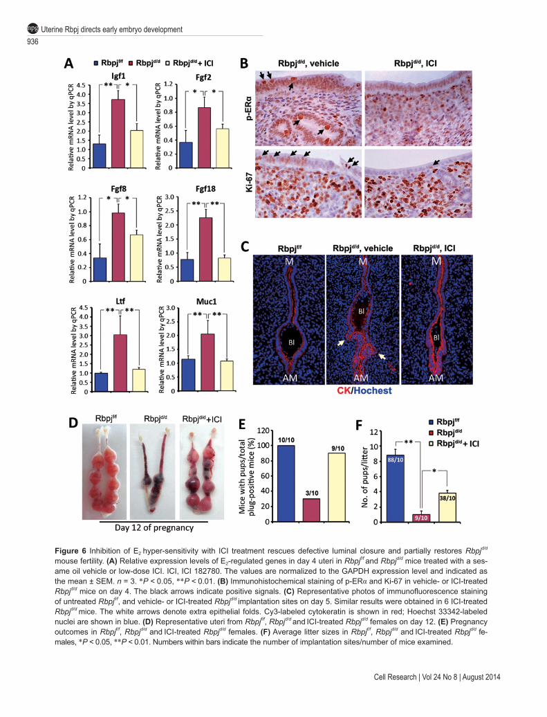
Given that the abnormal luminal closure and resulting embryonic-uterine disorientation are due to enhanced uterine estrogenic response in Rbpjd/d uteri, we further tested whether the suppression of the estrogenic response by pharmacologically antagonizing ERα could rescue the defective luminal closure in Rbpjd/d uteri. We treated Rbpjf/f and Rbpjd/d mice with a low dose of ER antagonist (ICI 182,780) at midnight on day 3, which represents a timing prior to pre-implantation E2 secretion. The ICI treatment strikingly reduced the expression of E2 target genes on day 4 (Figure 6A). Additionally, the abnormal expression of the p-ERα and Ki67 was largely corrected following the ER antagonist treatment (Figure 6B). Most importantly, the abnormal luminal closure process was corrected in the Rbpjd/d mice following the ICI treatment, and the blastocyst was located at the normal implanta-tion chamber with M-AM orientation on day 5 (Figure 6C), and the fertility of Rbpjd/d mice after ICI treatment showed a significant improvement regarding pregnancy rate and average litter size (Figure 6D-6F). By judging the histology of day 8 implantation sites after ICI treat-ment, although the orientation problem has been rescued,
936Uterine Rbpj directs early embryo developmentnpg
Cell Research | Vol 24 No 8 | August 2014
Figure 6 Inhibition of E2 hyper-sensitivity with ICI treatment rescues defective luminal closure and partially restores Rbpjd/d mouse fertility. (A) Relative expression levels of E2-regulated genes in day 4 uteri in Rbpjf/f and Rbpjd/d mice treated with a ses-ame oil vehicle or low-dose ICI. ICI, ICI 182780. The values are normalized to the GAPDH expression level and indicated as the mean ± SEM. n = 3. *P < 0.05, **P < 0.01. (B) Immunohistochemical staining of p-ERα and Ki-67 in vehicle- or ICI-treated Rbpjd/d mice on day 4. The black arrows indicate positive signals. (C) Representative photos of immunofluorescence staining of untreated Rbpjf/f, and vehicle- or ICI-treated Rbpjd/d implantation sites on day 5. Similar results were obtained in 6 ICI-treated Rbpjd/d mice. The white arrows denote extra epithelial folds. Cy3-labeled cytokeratin is shown in red; Hoechst 33342-labeled nuclei are shown in blue. (D) Representative uteri from Rbpjf/f, Rbpjd/d and ICI-treated Rbpjd/d females on day 12. (E) Pregnancy outcomes in Rbpjf/f, Rbpjd/d and ICI-treated Rbpjd/d females. (F) Average litter sizes in Rbpjf/f, Rbpjd/d and ICI-treated Rbpjd/d fe-males, *P < 0.05, **P < 0.01. Numbers within bars indicate the number of implantation sites/number of mice examined.

Shuang Zhang et al.937
npg
www.cell-research.com | Cell Research
the abnormal decidual patterning (Y/X ratio) remained (Supplementary information, Figure S9), thus reinforcing the conclusion that Rbpj functions in pregnant uteri are stage-specific with different regulatory mechanisms.
Discussion
The molecular mechanisms that control mammalian embryo orientation are enigmatic. The present study showed a surprising phenotype that uterine deletion of Rbpj signaling results in a progressively disorientat-ed uterine-embryonic axis at post-implantation stages, which stems from the defective luminal closure before embryo-uterine attachment that caused by uterine hy-per-estrogenic response. We further revealed that Rbpj physically interacts with ERα to ensure normal estrogen-ic effects within uterine context via Notch pathway-inde-pendent mechanisms. At post-implantation stages, Rbpj directly regulates uterine Mmp2 expression via Notch pathway-dependent mechanisms, which ensures normal decidual remodeling. The uterine deletion of Rbpj results in embryonic-uterine disorientation and abnormal decid-ual remodeling, which lead to restrained embryo growth and substantial embryo loss.
The process of pre-implantation uterine luminal clo-sure, in which the randomly shaped luminal folds change to a highly regulated slit-like structure with a fixed M-AM direction, provides a directional landscape that guides proper embryo orientation at the time of initial implantation, therefore directing subsequent uterus-em-bryo alignments. On day 4 of pregnancy in mice, the reg-ulation of pre-implantation luminal closure is controlled by a P4-dominated uterine environment in coordination with a small surge of pre-implantation estrogen secretion [46]. A successful luminal closure results from regulated stromal edema and nonproliferating opposing epithelia that are closely attached to one another [12, 47]. In our study, we found that the uterine deletion of Rbpj dis-turbed this regulated process by unleashing ERα activity, which resulted in enhanced estrogenic response in the uterine context. Although the incomplete luminal closure in Rbpjd/d uteri was not sufficiently severe to abrogate the implantation process, it produced abnormal luminal folds that had axes that were not parallel to the uterine M-AM axis (Figure 7). When a blastocyst is faced with multiple luminal folds, the extra luminal folds exists as many “pitfalls”; once a blastocyst falls into one fold by chance, it will establish an embryonic axis that depends on the direction of the crypt that it accommodates with, and an initial misorientation (with regard to the uterine A-AM axis) will then lead to a catastrophically disori-ented uterine-embryonic axis. A recent study of uterine Msx1 deletion also reported defects in luminal closure
that were characterized by the presence of extra uterine crypts for blastocyst attachment; the authors also report-ed increased pregnancy loss during mid-gestation [48]. Our current findings strongly suggest that normal estab-lishment of embryonic-uterine orientation in mammals is not only guided by intrinsically programmed embryonic signals, but also depends on the coordinated transforma-tion of uterine lumen as a proper guide.
Another interesting result of the present study is that although Rbpj is primarily expressed in the uterine stroma during the peri-implantation period, its deletion seems to cause not only an enhanced estrogenic response in the stroma but also a profound effect on the uterine ep-ithelial layer, as evidenced by significantly increased ep-ithelial proliferation, E2-responsive gene expression and an altered apical surface structure. This interesting phe-nomenon further indicates an intimate stromal-epithelial interaction during the peri-implantation period, which depends on the synergistic and antagonistic effects of P4 and E2 on uterine cell proliferation and differentiation through their receptors [12, 49]. Emerging evidence has indicated that paracrine factors (such as IGF and FGF family members) that are generated in the stroma cells and induced by estrogen through ER receptors function as critical intermediate signaling molecules that stim-ulate epithelial proliferation [37, 38]. Our data showed that uterine deletion of Rbpj, which is localized in the stroma, results in substantial increased expression of IGFs and FGFs, which subsequently enhances estrogenic responses in the epithelia layer. To search for the causes, we further demonstrated that Rbpj can physically interact with ERα to ensure a normal estrogen activity in uterine contexts prior to embryo-uterus attachment, which is Notch pathway-independent. The detailed mechanism by which Rbpj suppresses ERα function in uterine context is currently unclear and remains an interesting topic that warrants further investigation.
MMPs are the most prominent family of proteinases associated with multiple physiological and pathological processes [50, 51]. Previous studies have indicated that at post-implantation stages, the normal process of decidual remodeling into an egg-shaped uterine pattern depends on developmentally expressed MMP genes, as their ab-rogation would lead to abnormal uterine shape remod-eling characterized by a shortened length of the uterine long-axis and a less-strictly regulated embryo orientation [42, 43, 52]. These data suggest that normal uterine stro-mal remodeling is synchronized with the process of the formation of a proper uterine-embryonic axis and at least plays a role in optimizing this process [8]. Additionally, because Mmp2 is dynamically expressed in the uterine context [52], its dynamic expression pattern should be regulated by upstream transcription factors that are de-

938Uterine Rbpj directs early embryo developmentnpg
Cell Research | Vol 24 No 8 | August 2014
Figure 7 Illustrative model of uterine Rbpj’s role during the peri-implantation period for embryo development. In normal situ-ation (left), the embryonic axis is consistent with the uterine M-AM axis. In uteri containing a specific deletion of Rbpj (right), the process of uterine luminal closure is defective during the pre-implantation period, leading to the formation of extra epithe-lial folds that “trap” the implanting blastocyst, which is caused by unleashed ERα activity due to the loss of physical interac-tion of Rbpj and ERα(Notch pathway-independent). After the blastocyst establishes the embryonic axis in an abnormal uterine fold, the development of the embryonic axis will deflect from the uterine axis. During post-implantation development, the loss of Rbpj results in abnormal decidual patterning due to decreased Mmp2 expression that is transcriptionally regulated by Rbpj (Notch pathway-dependent). The combined defects in uterine-embryonic orientation and decidual patterning in Rbpjd/d mice lead to subsequent embryonic lethality.
velopmentally regulated by the progression of implanta-tion and decidualization. In the present study, we demon-strated that in Rbpj mutant uteri, there are decreased expression and enzymatic activity of MMPs, and we further revealed that Rbpj directly binds to the promoter region of Mmp2 in post-implantation uteri and promotes its transcriptional activity in a Notchpathway-dependent manner. The decreased activities of MMPs are supposed
to exaggerate the progressively disoriented uterine-em-bryonic axis as previous studies have shown that when there is an aberrant decidual growth, uterine M-AM orientation is usually deviated with regards to polarized gene expression [13, 14, 53], supporting a hypothesis that deferential decidual patterning could help optimizing the uterine-embryo orientation during post-implantation stages development. In the present study, it is also no-

Shuang Zhang et al.939
npg
www.cell-research.com | Cell Research
table that while Rbpj is deleted in the uterine part, the trophoblastic MMP9 from embryonic part also seems to be downregulated (Supplementary information, Figure S8), which is most probably a result of retarded embryo growth due to either the misoriented embryonic axis or the abnormal decidual vasculature, or both. In such case, the reduced trophoblastic MMP9 expression further limits the ability of trophoblast invasion and vascular re-modeling, creating a situation that the defects from both decidual and embryonic part become mutually reinforc-ing, which results in a vicious circle that finally leads to the death of embryo in the Rbpj mutant uteri. Notably, similar scenario might also exist in a recently published paper showing that both maternal and embryonic MMP9 are important for normal placenta formation at fetal-ma-ternal interface and thus for embryo development [54], recapitulating our point that the development of embryo is inextricably linked to the uterine signaling, which, in this case, is governed by uterine Rbpj.
In summary, we provide the first genetic evidence that uterine Rbpj, functioning through both Notch path-way-independent and -dependent mechanisms, is essen-tial for instructing the initial uterine-embryonic orienta-tion and ensuring normal decidual patterning for normal embryo development, which, in our belief, substantiates a novel concept that normal mammalian embryonic-uter-ine orientation requires proper guidance from develop-mentally controlled uterine signaling.
Materials and Methods
Animals and treatmentsRbpjloxp/loxp mice were generated as previously described [25]
and kindly provided by Dr Tasuku Honjo. DNMAMLtg/+ mice were generated as previously described [45]. Uterine-specific mutant mice were generated by crossing Rbpjloxp/loxp or DNMAMLtg/+ mice with PR-Cre+/− mice [27]. All mice were housed in the Institu-tional Animal Care Facility according to institutional guidelines for laboratory animals. Female mice were mated with fertile WT males to induce pregnancy (vaginal plug = day 1 of pregnancy). To examine implantation, pregnant dams were killed on day 5 (0900 h). Implantation sites were visualized by an intravenous injection of Chicago blue dye solution, and the number of implantation sites, demarcated by distinct blue bands, was recorded. For ICI treatment, each mouse was intraperitoneally injected with 10 ng in 200 µl of sesame oil at 10 pm on day 3. Implantation sites were analyzed on day 5.
ImmunostainingImmunohistochemistry was performed in 5-µm-thick par-
affin-embedded sections using specific antibodies as indicated. Tissues were fixed in 10% neutral buffered formalin. Microwave heating was used for antigen retrieval. For immunohistochemistry analysis, antibodies specific to ERα (Dako, 1:100), p-ERα (Santa Cruz Biotechnology, 1:100), progesterone receptor (PR) (DAKO,
1:100), Ki-67 (Epitomics, 1:200), COX2 (Santa Cruz Biotechnolo-gy, 1:200), CD31 (BD, 1:50) were used in 5-µm thick paraffin-em-bedded sections. A Histostain-SP Kit (Zhongshan Golden Bridge Biotechnology) was used to visualize the antigen. For immuno-fluorescence, double-immunofluorescence staining for SMA (Bio Genex, 1:100) and cytokeratin (DAKO, 1:100) was performed in paraffin-fixed sections and secondary antibodies conjugated with Cy2 and Cy3 dyes were used, respectively. Immunofluores-cence for Laminin (Sigma, 1:200) and cytokeratin was performed in fresh-frozen sections. Nuclear staining was performed using Hochest 33343 (Sigma, 0.1 µg/ml). All frozen sections were fixed in 10% neutral buffered formalin and incubated with primary an-tibody at 4 °C overnight, followed by incubation with secondary antibody at room temperature for 1 h. Immunofluorescence images were captured in a Zeiss LSM 510 confocal scanning laser micro-scope.
In situ hybridizationIn situ hybridization with isotopes or digoxygenin (DIG) was
performed as previously described [55]. Mouse-specific cRNA probes for Rbpj, Bmp2, IL11Rα, Wnt4, Mmp2 and Mmp9 were used for hybridization. Cryosections hybridized with sense probes served as negative controls. Whole mount in situ hybridization of Cerl1 on isolated embryos was performed as previously described [56].
Co-immunoprecipitationDecidual cells were isolated as described previously [57]. Nu-
clear protein was extracted by using nuclear extraction kit accord-ing to the manufacturer’s instructions (Millipore). Protein lysates (1 mg) from decidual cells or Ishikawa cells transfected with c-Myc-tagged Rbpj plasmid were incubated with 4 µg of anti-Rb-pj antibody (Santa Cruz Biotechnology) or anti-c-Myc antibody (ABclonal) and rotated overnight at 4 °C. Protein Aagarose beads (Thermo) were washed and incubated with protein lysates over-night at 4˚C. Immunoprecipitated proteins were separated by SDS-PAGE and detected by immunoblotting using antibody specific to ERα (Dako), Rbpj or c-Myc. The control immunoprecipitation was performed by incubating the lysates with goat IgG (Zhongshan Golden Bridge Biotechnology).
Real-time RT-PCR analysisRT-PCR was performed as described [58]. Total RNA was iso-
lated from uterine tissues or cells with Trizol reagent following the manufacturer’s protocol. A total of 1-3 µg RNA was used to syn-thesize cDNA. The expression levels of different genes were val-idated by RT-PCR analysis using the ABI 7500 sequence detector system according to manufacturer’s instructions (Applied Biosys-tems). All primers for real-time PCR were listed in Supplementary information, Table S1. Assays were performed at least three times with each in duplicate.
Western blottingProtein extraction and western blotting were performed as pre-
viously described [55]. Antibodies to Rbpj and actin (Sigma) were used. Actin served as a loading control.
Serum P4 and E2 level measurementMouse blood samples were collected on day 4 (0900 h). Serum

940Uterine Rbpj directs early embryo developmentnpg
Cell Research | Vol 24 No 8 | August 2014
P4 and E2 levels were measured by radioimmunoassay (RIA).
ChIP analysisChIP analyses were performed as previously described [59].
The binding of Rbpj to Mmp2 was assessed by ChIP assay using decidual tissues of day 6 implantation sites. The binding of ERα to IGF-1 and cMyc was assessed by ChIP assay using Ishikawa cell line. The ERE sites on Igf-1 and cMyc were previously described [60, 61]. In brief, the ChIP assay was performed according to the instructions of ChIP assay Kit (Millipore) with slight modifica-tions. On day 6 of pregnancy, the decidua was isolated from the uterus and cut into small pieces (3-4 mm in length). Tissues were then suspended in 1% formaldehyde (Sigma)-PBS solution for 10 min at room temperature. The crosslinking was terminated by adding glycine to a final concentration 0.125 M. Tissues were then collected by centrifugation at 5 000× g for 5 min at 4 °C and washed twice with ice-cold PBS. Cell pellets were lysed in 1% SDS buffer containing a protease inhibitor. Chromatin was sheared by sonication until the average length of DNA was ~500 bp as evaluated by agarose gel electrophoresis. The sheared chromatin was diluted in ChIP dilution buffer to a final SDS concentration of 0.1%. Salmon sperm DNA/protein Aagarose slurry was added to preclear the chromatin solution. 1% of the chromatin fragments were stored at –20 °C to be used later for non-precipitated total chromatin (input) for normalization. 99% of chromatin fragments were incubated with 4 µg anti-Rbpj antibody overnight at 4 °C. Normal goat IgG (Santa Cruz Biotechnology) was used as a neg-ative control for nonspecific immunoprecipitation. The chroma-tin-antibody complex was incubated with protein A/G beads for 1 h at 4 °C. The antibody/DNA complex on the agarose beads was then collected by centrifugation. The beads were washed in the or-der of low salt immune complex buffer, high salt immune complex buffer, LiCl immune complex buffer, and TE buffer provided by the supplier. The beads were suspended in elution buffer and the precipitated protein/DNA complexes were eluted from the antibod-ies/beads. The resulting protein/DNA complexes were subjected to cross-link reversal in 5 M NaCl at 65 °C for 4 h followed by the addition of 0.5 M EDTA, 1 M Tris-HCl, and 10 µg/ml proteinase K (Sigma) at 45 °C for 1 h. DNA was purified by phenol/chloro-form extraction and ethanol precipitation. Specific primers were used to detect immunoprecipitated chromatin fragments, as well as input chromatin (Supplementary information, Table S1).
ZymographyDecidual cells were isolated from Rbpjf/f and Rbpjd/d females on
day 8 of pregnancy. Lysates of decidual fractions were prepared by homogenizing tissue pieces in substrate gel sample buffer for 10 min at 37 °C. Zymography and reverse zymography were per-formed as previously described [62].
Cell transfection, MTS and luciferase assayIshikawa cells were transfected with plasmids using lipo-
fectamine 2000 (Invitrogen) according to the manufacturer’s instructions. The Ishikawa cells were maintained at 37 °C in an atmosphere of 5% CO2/95% air in DMEM medium supplement-ed with 10% (v/v) FBS. Cells were plated in six-well plates and transfected with plasmid DNA (with amounts shown in each fig-ure legend, brought up to 2 μg in total with empty vectors) using Lipofectamine 2000 the next day at a cell confluence of about
80%. Treatment with 0.1% (v/v) DMSO (vehicle) or 100 nM E2 in medium was performed 12 h after transfection and cells were collected after another 12 h for RNA and protein extraction. For proliferation study, cells were seeded in 96-well plates at a density of 5 × 10⁴ cells/well and processed following plasmid transfection procedures. Treatment with E2 in medium was performed for the indicated times as described for each experiment. Cell prolifera-tion was measured as a function of 3-(4, 5-dimethylthiazolyl-2)-2, 5-diphenyltetrazolium bromide oxidation (MTS Cell Proliferation Assay Kit) according to the manufacturer’s instructions. Experi-ments were performed on three separate occasions.
For luciferase assay, the upstream region of the Mmp2 gene relative to the transcription start site was generated by PCR (Sense: 5′-AGAGATCTA GGAGCTCAGCGTCCTGATTC-3′; anti-sense: 5′-ATAAGCTTGGCTCAGGAG CTCATAACGGG-3′) using uterus genomic DNA as template. The amplified fragments were cloned into pGL3-Basic vectors (Promega). Mutations of the Rbpj-binding sites were achieved by Fast Mutagenesis System (Stratagene). HEK293T cells were kept in DMEM medium plus 10% FBS. All constructs were transiently transfected into 293T cells using Lipofectamine 2000 according to the manufacturer’s instructions. pRL-TK, internal control plasmid expressing Renilla (Promega) was co-transfected into the cells to normalize firefly luciferase activity of the reporter plasmids. Cells were collected 48 h after transfection of reporter plasmids and luciferase activities were measured using the Dual-Luciferase Reporter Assay System. Assays were performed at least three times with each in duplicate.
Statistical analysisStatistical analysis was performed with SPSS11.5 program.
Comparison of means was performed using student’s t-test. Data were shown as means ± SEM.
Acknowledgments
We are grateful to Dr Tasuku Honjo (Kyoto University) for pro-viding us the Rbpjf/f mice. We thank Dr Sudhansu K Dey (University of Cincinnati College of Medicine) for proofreading the manu-script. This work was supported by the National Basic Research Program of China (973 Program; 2011CB944400 to HBW, HL and YW), the National Natural Science Foundation of China (81130009 and 81330017 to HBW, 31200879 to QC, and 31300957 to YZ) and the Strategic Priority Research Program of the Chinese Acade-my of Sciences (XDA04020202-20 and XDA04020419 to ED).
References
1 Assheton R. On the Causes which lead to the attachment of the mammalian embryo to the walls of the uterus. Q J Mic Sci 1894; 37:173-190.
2 Wimsatt WA. Some comparative aspects of implantation. Biol Reprod 1975; 12:1-40.
3 Rossant J, Tam PP. Emerging asymmetry and embryonic pat-terning in early mouse development. Dev Cell 2004; 7:155-164.
4 Wang H, Dey SK. Roadmap to embryo implantation: clues from mouse models. Nat Rev Genet 2006; 7:185-199.
5 Smith LJ. Embryonic axis orientation in the mouse and its

Shuang Zhang et al.941
npg
www.cell-research.com | Cell Research
correlation with blastocyst relationships to the uterus. Part 1. Relationships between 82 hours and 4 1/4 days. J Embryol Exp Morphol 1980; 55:257-277.
6 Smith LJ. Embryonic axis orientation in the mouse and its correlation with blastocyst relationships to the uterus. II. Re-lationships from 4 1/4 to 9 1/2 days. J Embryol Exp Morphol 1985; 89:15-35.
7 Cha J, Sun X, Dey SK. Mechanisms of implantation: strate-gies for successful pregnancy. Nat Med 2012; 18:1754-1767.
8 Chen Q, Zhang Y, Elad D, et al. Navigating the site for em-bryo implantation: Biomechanical and molecular regulation of intrauterine embryo distribution. Mol Aspects Med 2013; 34:1024-1042.
9 Mesnard D, Filipe M, Belo JA, Zernicka-Goetz M. The ante-rior-posterior axis emerges respecting the morphology of the mouse embryo that changes and aligns with the uterus before gastrulation. Curr Biol 2004; 14:184-196.
10 Weber RJ, Pedersen RA, Wianny F, Evans MJ, Zernic-ka-Goetz M. Polarity of the mouse embryo is anticipated be-fore implantation. Development 1999; 126:5591-5598.
11 Tranguch S, Cheung-Flynn J, Daikoku T, et al. Cochaper-one immunophilin FKBP52 is critical to uterine receptivity for embryo implantation. Proc Natl Acad Sci USA 2005; 102:14326-14331.
12 Zhang S, Lin H, Kong S, et al. Physiological and molecular determinants of embryo implantation. Mol Aspects Med 2013; 34:939-980.
13 Daikoku T, Song H, Guo Y, et al. Uterine Msx-1 and Wnt4 signaling becomes aberrant in mice with the loss of leukemia inhibitory factor or Hoxa-10: evidence for a novel cyto-kine-homeobox-Wnt signaling in implantation. Mol Endocri-nol 2004; 18:1238-1250.
14 Paria BC, Ma W, Tan J, et al. Cellular and molecular respons-es of the uterus to embryo implantation can be elicited by locally applied growth factors. Proc Natl Acad Sci USA 2001; 98:1047-1052.
15 Bray SJ. Notch signalling: a simple pathway becomes com-plex. Nat Rev Mol Cell Biol 2006; 7:678-689.
16 Guruharsha KG, Kankel MW, Artavanis-Tsakonas S. The Notch signalling system: recent insights into the complexity of a conserved pathway. Nat Rev Genet 2012; 13:654-666.
17 Johnson JE, Macdonald RJ. Notch-independent functions of CSL. Curr Top Dev Biol 2011; 97:55-74.
18 Hori K, Cholewa-Waclaw J, Nakada Y, et al. A nonclassical bHLH Rbpj transcription factor complex is required for spec-ification of GABAergic neurons independent of Notch signal-ing. Genes Dev 2008; 22:166-178.
19 Kato Y. The multiple roles of Notch signaling during left-right patterning. Cell Mol Life Sci 2011; 68:2555-2567.
20 Krebs LT, Iwai N, Nonaka S, et al. Notch signaling regulates left-right asymmetry determination by inducing Nodal expres-sion. Genes Dev 2003; 17:1207-1212.
21 Lopes SS, Lourenco R, Pacheco L, et al. Notch signalling regulates left-right asymmetry through ciliary length control. Development 2010; 137:3625-3632.
22 Rock JR, Gao X, Xue Y, et al. Notch-dependent differenti-ation of adult airway basal stem cells. Cell Stem Cell 2011; 8:639-648.
23 Tanigaki K, Tsuji M, Yamamoto N, et al. Regulation of alpha-
beta/gammadelta T cell lineage commitment and peripheral T cell responses by Notch/RBP-J signaling. Immunity 2004; 20:611-622.
24 Oka C, Nakano T, Wakeham A, et al. Disruption of the mouse RBP-J kappa gene results in early embryonic death. Develop-ment 1995; 121:3291-3301.
25 Han H, Tanigaki K, Yamamoto N, et al. Inducible gene knockout of transcription factor recombination signal binding protein-J reveals its essential role in T versus B lineage deci-sion. Int Immunol 2002; 14:637-645.
26 Lee K, Jeong J, Kwak I, et al. Indian hedgehog is a major mediator of progesterone signaling in the mouse uterus. Nat Genet 2006; 38:1204-1209.
27 Soyal SM, Mukherjee A, Lee KY, et al. Cre-mediated recom-bination in cell lineages that express the progesterone recep-tor. Genesis 2005; 41:58-66.
28 Bilinski P, Roopenian D, Gossler A. Maternal IL-11Ralpha function is required for normal decidua and fetoplacental de-velopment in mice. Genes Dev 1998; 12:2234-2243.
29 Franco HL, Dai D, Lee KY, et al. WNT4 is a key regulator of normal postnatal uterine development and progesterone sig-naling during embryo implantation and decidualization in the mouse. FASEB J 2011; 25:1176-1187.
30 Kurihara I, Lee DK, Petit FG, et al. COUP-TFII mediates progesterone regulation of uterine implantation by controlling ER activity. PLoS Genet 2007; 3:e102.
31 Li Q, Kannan A, Wang W, et al. Bone morphogenetic pro-tein 2 functions via a conserved signaling pathway involving Wnt4 to regulate uterine decidualization in the mouse and the human. J Biol Chem 2007; 282:31725-31732.
32 Morris SA, Grewal S, Barrios F, et al. Dynamics of anteri-or-posterior axis formation in the developing mouse embryo. Nat Commun 2012; 3:673.
33 Yamamoto M, Saijoh Y, Perea-Gomez A, et al. Nodal antag-onists regulate formation of the anteroposterior axis of the mouse embryo. Nature 2004; 428:387-392.
34 Lim H, Paria BC, Das SK, et al. Multiple female reproduc-tive failures in cyclooxygenase 2-deficient mice. Cell 1997; 91:197-208.
35 Sun X, Zhang L, Xie H, et al. Kruppel-like factor 5 (KLF5) is critical for conferring uterine receptivity to implantation. Proc Natl Acad Sci USA 2012; 109:1145-1150.
36 Xie H, Wang H, Tranguch S, et al. Maternal heparin-bind-ing-EGF deficiency limits pregnancy success in mice. Proc Natl Acad Sci USA 2007; 104:18315-18320.
37 Li Q, Kannan A, DeMayo FJ, et al. The antiproliferative action of progesterone in uterine epithelium is mediated by Hand2. Science 2011; 331:912-916.
38 Zhu L, Pollard JW. Estradiol-17beta regulates mouse uterine epithelial cell proliferation through insulin-like growth factor 1 signaling. Proc Natl Acad Sci USA 2007; 104 (40):15847-15851.
39 Kawagoe J, Li Q, Mussi P, et al. Nuclear receptor coactiva-tor-6 attenuates uterine estrogen sensitivity to permit embryo implantation. Dev Cell 2012; 23:858-865.
40 Weng AP, Nam Y, Wolfe MS, et al. Growth suppression of pre-T acute lymphoblastic leukemia cells by inhibition of notch signaling. Mol Cell Biol 2003; 23:655-664.
41 Shah YM, Rowan BG. The Src kinase pathway promotes

942Uterine Rbpj directs early embryo developmentnpg
Cell Research | Vol 24 No 8 | August 2014
tamoxifen agonist action in Ishikawa endometrial cells through phosphorylation-dependent stabilization of estrogen receptor (alpha) promoter interaction and elevated steroid receptor coactivator 1 activity. Mol Endocrinol 2005; 19:732-748.
42 Afonso S, Romagnano L, Babiarz B. The expression and function of cystatin C and cathepsin B and cathepsin L during mouse embryo implantation and placentation. Development 1997; 124:3415-3425.
43 Alexander CM, Hansell EJ, Behrendtsen O, et al. Expression and function of matrix metalloproteinases and their inhibitors at the maternal-embryonic boundary during mouse embryo implantation. Development 1996; 122:1723-1736.
44 Mumm JS, Kopan R. Notch signaling: from the outside in. Dev Biol 2000; 228:151-165.
45 Tu L, Fang TC, Artis D, et al. Notch signaling is an important regulator of type 2 immunity. J Exp Med 2005; 202:1037-1042.
46 Dey SK, Lim H, Das SK, et al. Molecular cues to implanta-tion. Endocr Rev 2004; 25:341-373.
47 Tranguch S, Smith DF, Dey SK. Progesterone receptor re-quires a co-chaperone for signalling in uterine biology and implantation. Reprod Biomed Online 2006; 13:651-660.
48 Daikoku T, Cha J, Sun X, et al. Conditional deletion of Msx homeobox genes in the uterus inhibits blastocyst implantation by altering uterine receptivity. Dev Cell 2011; 21:1014-1025.
49 Cooke PS, Buchanan DL, Young P, et al. Stromal estrogen receptors mediate mitogenic effects of estradiol on uterine epithelium. Proc Natl Acad Sci USA 1997; 94:6535-6540.
50 Kessenbrock K, Plaks V, Werb Z. Matrix metalloproteinases: regulators of the tumor microenvironment. Cell 2010; 141:52-67.
51 Sbardella D, Fasciglione GF, Gioia M, et al. Human matrix metalloproteinases: an ubiquitarian class of enzymes involved in several pathological processes. Mol Aspects Med 2012; 33:119-208.
52 Das SK, Yano S, Wang J, et al. Expression of matrix metallo-proteinases and tissue inhibitors of metalloproteinases in the mouse uterus during the peri-implantation period. Dev Genet 1997; 21:44-54.
53 Rahman MA, Li M, Li P, et al. Hoxa-10 deficiency alters region-specific gene expression and perturbs differentiation
of natural killer cells during decidualization. Dev Biol 2006; 290:105-117.
54 Plaks V, Rinkenberger J, Dai J, et al. Matrix metalloprotein-ase-9 deficiency phenocopies features of preeclampsia and intrauterine growth restriction. Proc Natl Acad Sci USA 2013; 110:11109-11114.
55 Lu J, Zhang S, Nakano H, et al. A positive feedback loop in-volving Gcm1 and Fzd5 directs chorionic branching morpho-genesis in the placenta. PLoS Biol 2013; 11:e1001536.
56 Morris SA, Grewal S, Barrios F, et al. Dynamics of anteri-or-posterior axis formation in the developing mouse embryo. Nat Commun 2012; 3:673.
57 Hirota Y, Daikoku T, Tranguch S, et al. Uterine-specific p53 deficiency confers premature uterine senescence and promotes preterm birth in mice. J Clin Invest 2010; 120:803-815.
58 Chen Q, Zhang Y, Peng H, et al. Transient {beta}2-adreno-ceptor activation confers pregnancy loss by disrupting embryo spacing at implantation. J Biol Chem 2011; 286:4349-4356.
59 Shi M, Hu ZL, Zheng MH, et al. Notch-Rbpj signaling is required for the development of noradrenergic neurons in the mouse locus coeruleus. J Cell Sci 2012; 125(Part 18):4320-4332.
60 Kang HJ, Yi YW, Kim HJ, et al. BRCA1 negatively regulates IGF-1 expression through an estrogen-responsive element-like site. Cell Death Dis 2012; 3:e336.
61 Wang C, Mayer JA, Mazumdar A, et al. Estrogen induces c-myc gene expression via an upstream enhancer activated by the estrogen receptor and the AP-1 transcription factor. Mol Endocrinol 2011; 25:1527-1538.
62 Kuang H, Chen Q, Zhang Y, et al. The cytokine gene CXCL14 restricts human trophoblast cell invasion by sup-pressing gelatinase activity. Endocrinology 2009; 150:5596-5605.
(Supplementary information is linked to the online version of the paper on the Cell Research website.)
This work is licensed under the Creative Commons Attribution-NonCommercial-No Derivative Works
3.0 Unported License. To view a copy of this license, visit http://creativecommons.org/licenses/by-nc-nd/3.0