Innervation of the larynx, pharynx, and upper esophageal sphincter of the rat
Upload
khangminh22Category
view
2download
0
Thesis for doctoral degree (Ph.D.)2008
CHANGES IN HUMAN UTERINEINNERVATION IN TERM
PREGNANCY AND LABOR
OCCURRENCE AND ROLES OFNEUROTROPHINS AND TRPV1
Berith Karlsson Tingåker
Thesis for doctoral degree (Ph.D.) 2008
Berith K
arlsson TingåkerC
HA
NG
ES IN
HU
MA
N U
TE
RIN
E IN
NE
RV
AT
ION
IN T
ER
M P
RE
GN
AN
CY
AN
D LA
BO
R

DEPARTMENT OF WOMAN AND CHILD HEALTH Karolinska Institutet, Stockholm, Sweden
CHANGES IN HUMAN UTERINE INNERVATION IN TERM
PREGNANCY AND LABOR OCCURRENCE AND ROLES OF NEUROTROPHINS AND TRPV1
Berith Karlsson Tingåker
Stockholm 2008

2008
Gårdsvägen 4, 169 70 Solna
Printed by
B Karlsson Tingåker
All previously published papers were reproduced with permission from the publisher.
Published by Karolinska Institutet.Lay-out Ringvor Hägglöf
© Berith Karlsson Tingåker, 2008ISBN 978-91-7357-544-7

I have always held firmly to the thought that each one of us can do a little to bring some portion of misery to an end. Albert Schweitzer
In loving memory of my dear Mother

B Karlsson Tingåker

ABSTRACT
Objective: Cervical ripening, is a prerequisite for successful labor, which is often very painful and involves an inflammatory reaction. However, little is presently known con-cerning nerve-related changes in the human uterus during labor and the fundamental hy-pothesis tested here was that labor pain emanates primarily from the cervix uteri. To this end, the general innervation of the corpus and cervix uteri, as well as the possible pres-ence of neurotrophins and TPRV1 (major factors involved in nociception) and of sensory corpuscles in these same two tissues were examined in term pregnancy prior to and after the onset of labor and compared to the non-pregnant state.
Methods: Biopsies were collected from the corpus and cervix uteri of non-pregnant wom-en after hysterectomy and women who, following term pregnancy, either had a caesarean section prior to onset of labor or an emergency caesarean section during established labor. Serial sections prepared from the frozen biopsies were analyzed immunohistochemically employing antibodies directed specifically against PGP 9.5, S-100, NGF, BDNF, NGFR p75, Trk B, NFILS and TRPV1. Moreover, the levels of the mRNA species encoding NGFβ, NGFR p75, TrkA and TrkB in these same tissue samples were determined by Real-Time PCR.
Results: The innervation of the corpus in term pregnancy, both prior to and after the onset of labor, differed profoundly from that of the cervix under the same conditions, as well as from that of the corresponding non-pregnant tissues. Thus, in term pregnancy, most nerve fibers staining positively for PGP 9.5, NGFR p75, NFILS and TRPV1-IR had virtually disappeared from the corpus, but remained in the cervix. At the same time, the numbers of nerve fibers expressing BDNF-IR, as well as of NGF-positive cells increased in both the corpus and cervix during labor. Corpus levels of NGFβ, NGFR p75 and TrkB mRNA were lower in term pregnancy with and without labor than in the non-pregnant control tissue, whereas the level of TrkA mRNA in the corpus was elevated. In the case of the cer-vix, expression of both NGFβ and TrkA mRNA was up-regulated during labor. Moreover, sensory corpuscles were detected in both the non-pregnant corpus and cervix and also in the term pregnant cervix but only prior to the onset of labor.
Conclusion: The observations documented here indicate unambiguously that during la-bor, the numbers of nerve fibers and levels of neurotrophins and TRPV1 in the human cervix are considerably higher than in the corpus. These findings lend support to our hypothesis that the cervix uteri is the major site from which labor pain emanates. Further-more, the sensory corpuscles identified in the non-distended corpus and cervix probably convey mechanosensitive (proprioceptive) impulses.
Key words: Uterus, corpus uteri, cervix uteri, pregnancy, labor pain, neurotrophins, NGF, TRPV1, sensory corpuscles, human.

B Karlsson Tingåker

LIST OF PUBLICATIONS
This thesis is based on the following articles and manuscript, which will be referred to in the text by their Roman numerals
Berith K Tingåker, Olle Johansson, Ann Hjelm Cluff, Gunvor Ekman-Ordeberg I. Unaltered innervation of the human cervix uteri in contrast to the corpus during pregnancy and labor as revealed by PGP 9.5 immunohistochemistry European Journal of Obstetrics & Gynecology and Reproductive Biology 2006; 125 :66-71
Berith K Tingåker, Gunvor Ekman-Ordeberg, Sture Forsgren II. Presence of sensory nerve corpuscles in the human corpus and cervix uteri during pregnancy and labor as revealed by immunohistochemistry Reproductive Biology and Endocrinology 2006;4:45-52
Berith K Tingåker, Gunvor Ekman-Ordeberg, Paul Facer, Lars Irestedt, Praveen III. Anand Influence of pregnancy and labor on the occurrence of nerve fibers expressing the capsaicin receptor TRPV1in human corpus and cervix uteri Reproductive Biology and Endocrinlogy 2008; 6:8-15
Berith K Tingåker, Birgitta Byström, Lars Irestedt, Margareta Norman, Sture IV. Forsgren, Gunvor Ekman-Ordeberg The expression and localization of NGF and BDNF and their associated receptors in human corpus and cervix uteri at term pregnancy and in labour Submitted

B Karlsson Tingåker

CONTENTS
ABBREVIATIONS.............................................................................. .......... 11
BACKGROUND........................................................................................... 13 Introduction............................................................................................. 13 Labor Pain............................................................................................. 14 The corpus uteri....................................................................................... 16 The cervix uteri........................................................................................ 16 Cervical ripening is an inflammatory process........................................... 17
AIMS OF THE STUDY.................................................................................. 19
MATERIAL AND METHODS.................................................................... 21 Subjects.................................................................................................... 21 Samplings procedures.............................................................................. 21 The corpus uteri............................................................................... 21 The cervix uteri............................................................................... 21 Methods.................................................................................................... 22 Immunohistochemistry ( Paper I - IV).................................... 22 Real-Time PCR Paper IV)............................................................... 23 Statistical analysis.................................................................................... 23
RESULTS......................................................................................................... 24 Patterns of innervation.............................................................................. 24 The corpus uteri............................................................................... 24 The cervix uteri................................................................................ 24 Sensory corpuscles................................................................................... 26 The corpus uteri.............................................................................. 26 The cervix uteri................................................................................ 26 TRPV1 and Neurofilaments (NFILS)....................................................... 26 Expression of TRPV1 in the corpus uteri....................................... 26 Expression of TRPV1 in the cervix uteri ........................................ 26 Expression of NFILS in the corpus uteri......................................... 28 Expression of NFILS in the cervix uteri......................................... 28 Neurotrophins and neurotrophin receptors............................................... 29 Immunohistochemistry..................................................................... 29 Expression of NGF and BDNF ligands in the corpus uteri..... 29 Expression of NGF and BDNF ligands in the cervix uteri..... 29 Expression of NGFR p75 and TrkB receptors in the corpus uteri.................................................................... 29 Expression of NGFR p75 and TrkB receptors in the cervix uteri..................................................................... 29

B Karlsson Tingåker
Real-Time PCR: mRNA expression......................................................... 31 Corpus levels of mRNA encodingNGFβ and BDNF ligands......... 31 Cervical levels of mRNA encoding NGFβ and BDNF ligands....... 31 Corpus levels of mRNA encoding NGFR p75 and , TrkA and TrkB receptors........................................................................... 31 Cervical levels of mRNA encoding NGFR p75 and , TrkA and TrkB receptors.......................................................................... 31
DISUSSION................................................................................................ 35
CONCLUSION........................................................................................... 43 Future Perspectives........................................................................... 45
ACKNOWLEDGEMENTS....................................................................... 47
REFERENCES............................................................................................51
PAPER I-IV
10

LIST OF ABBREVIATIONS
BDNF Brain-derived neurotrophic factor
CGRP Calcitonin gene-related peptide
CSF Cerebrospinal fluid
cDNA Complementary DNA
CNB Central neuroaxial blockades
CS Cesarean section
DRG Dorsal root ganglion
ECM Extracellular matrix
Htx Hematoxylin-eosin
IHP Inferior hypogastric plexus
IR Immunoreactive, immunoreactivity
MHP Middle hypogastric plexus
mRNA Messenger RNA
NFILS Neurofilaments
NGF Nerve growth factor
NGFR p75 Nerve growth factor receptor p75
NO Nitric oxide
PAP Peroxidase antiperoxidase
PGP 9.5 Protein gene product 9.5
SHP Superior hypogastric plexus
SP Substance P
TrkA Tyrosine kinase receptor A
TrkB Tyrosine kinase receptor B
TrkC Tyrosine kinase receptor C
TRPV1 Transient receptor potential vanilloid receptor subtype
RT-PCR Reverse transcriptase polymerase chain reaction
List of Abbreviations 11

B Karlsson Tingåker12
Background 13
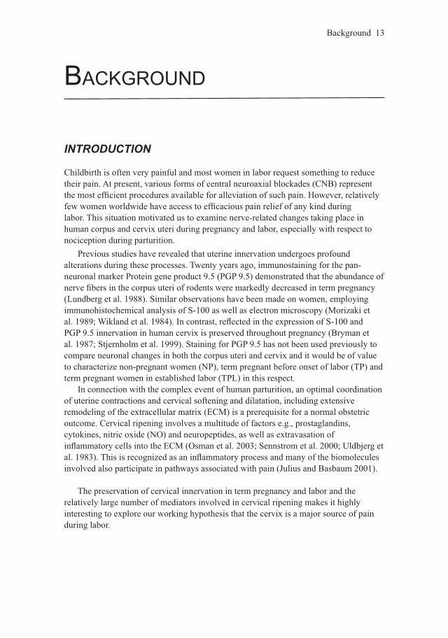
BACKGROUND
INTRODUCTION
Childbirth is often very painful and most women in labor request something to reduce their pain. At present, various forms of central neuroaxial blockades (CNB) represent the most efficient procedures available for alleviation of such pain. However, relatively few women worldwide have access to efficacious pain relief of any kind during labor. This situation motivated us to examine nerve-related changes taking place in human corpus and cervix uteri during pregnancy and labor, especially with respect to nociception during parturition.
Previous studies have revealed that uterine innervation undergoes profound alterations during these processes. Twenty years ago, immunostaining for the pan-neuronal marker Protein gene product 9.5 (PGP 9.5) demonstrated that the abundance of nerve fibers in the corpus uteri of rodents were markedly decreased in term pregnancy (Lundberg et al. 1988). Similar observations have been made on women, employing immunohistochemical analysis of S-100 as well as electron microscopy (Morizaki et al. 1989; Wikland et al. 1984). In contrast, reflected in the expression of S-100 and PGP 9.5 innervation in human cervix is preserved throughout pregnancy (Bryman et al. 1987; Stjernholm et al. 1999). Staining for PGP 9.5 has not been used previously to compare neuronal changes in both the corpus uteri and cervix and it would be of value to characterize non-pregnant women (NP), term pregnant before onset of labor (TP) and term pregnant women in established labor (TPL) in this respect.
In connection with the complex event of human parturition, an optimal coordination of uterine contractions and cervical softening and dilatation, including extensive remodeling of the extracellular matrix (ECM) is a prerequisite for a normal obstetric outcome. Cervical ripening involves a multitude of factors e.g., prostaglandins, cytokines, nitric oxide (NO) and neuropeptides, as well as extravasation of inflammatory cells into the ECM (Osman et al. 2003; Sennstrom et al. 2000; Uldbjerg et al. 1983). This is recognized as an inflammatory process and many of the biomolecules involved also participate in pathways associated with pain (Julius and Basbaum 2001).
The preservation of cervical innervation in term pregnancy and labor and the relatively large number of mediators involved in cervical ripening makes it highly interesting to explore our working hypothesis that the cervix is a major source of pain during labor.

B Karlsson Tingåker14
LABOR PAINLabor pain arises as a consequence of numerous complex molecular interactions, both excitatory and inhibitory, but its source has not yet been elucidated fully. Moreover, our knowledge concerning the possible role of mechanoreceptors in such pain remains scant, e.g., it is not known whether sensory corpuscles are present.
The cell bodies of the nerves which innervate the corpus uteri and cervix are located in the dorsal root ganglia (DRG) of the Th10-L1 segments of the spinal cord and nociceptive impulses are transmitted via their Aδ and C-fibers. These fibers accompany the sympathetic nerves in the inferior (IHP), middle and superior (SHP) hypogastric plexus as well as the aortic plexi. The nociceptive afferents transverse the lumbar and lower thoracic sympathetic chain to the posterior roots of Th10-L1 nerves and establish synaptic contact with the interneurones in the dorsal horn (Berkley et al. 1993; McMahon and Koltzenburg 2006; Rowlands and Permezel 1998) (Fig 1a). The vagina and the pelvic floor including the perineum are supplied by the pudendal nerve together with smaller nerves originating from S2-S4 (Fig 1b). All nociceptive impulses travel first to the DRG and thereafter via the spinothalamic tract to the brain (Berkley et al. 1993; McMahon and Koltzenburg 2006; Rowlands and Permezel 1998).
Pain is the single most predominant sign of the onset of labor and for many women, this pain is the most severe they will ever experience. In a study by Melzac (1984), it is determined that 65%-68% of women in labor rate their pain as severe or very severe. Moreover, 23% of primiparas and 11% of multiparas rate their pain as “horrible” (McMahon and Koltzenburg 2006). Similar observations have been confirmed by other studies (Ranta et al. 1996). Clearly, there is a great need for novel and simpler strategies for the relief of labor pain.
As is the case for research on other types of pain, appropriate models involving experimental animals are necessary for elucidation of the neurophysiological mechanisms underlying labor pain. Certain studies on such models have revealed that the secondary elevation of the expression of cFos in the spinal cord, in response to
Fig 1. The peripheral nociceptive pathways involved in labor pain (from Wall and Melzack’s Textbook of Pain 2006 with the kind permission of the publisher).

Background 15
cervical distension and labor, can be attenuated dramatically by intrathecal analgesia (Catheline et al. 2006; Tong et al. 2003). Moreover, behavioral analysis has shown that the dams display a larger and larger number of abnormal postures as labor progresses to birth of the pulps (Catheline et al. 2006).
Unfortunately, such findings cannot be applied directly to human labor. Moreover, because of the difficulties involved in examining the central nervous system of human parturients, interest ought to be focused primarily on alterations occurring in peripheral nervous tissues. Accordingly, we have chosen to investigate nerve-related changes in the corpus and cervix uteri during pregnancy and labor, with particular emphasis on neurotrophins and the transient receptor potential vanilloid receptor subtype 1 (TRPV1).
Four members of the neurotrophin family have so far been identified in mammals: nerve growth factor (NGF), brain-derived neurotrophic factor (BDNF), neurotrophin-3 (NT-3) and neurotrophin-4/5 (NT-4/5) (Huang and Reichardt 2001). These all bind with higher or lower affinity to cell surface receptors with tyrosine kinase (Trk) activity. NGF binds preferentially to TrkA, BDNF and NT-4/5 to TrkB and NT-3 to TrkC and, moreover, all of these factors exhibit low affinity with the nerve growth factor receptor p75 (NGFR p75) (Huang and Reichardt 2001).
In addition to their essential roles in regulating the normal maintenance, survival and specificity of sensory neurons , these mediators also participate in inflammatory reactions and nociception (Huang and Reichardt 2001; Levi-Montalcini et al. 1996; Pezet and McMahon 2006). TRPV1 is expressed at a particularly high level by small-diameter sensory neurones, i.e, nociceptors (Fig 2) and has also been detected in non-neuronal tissues, such as smooth muscle, polymorphonuclear cells and macrophages (Cortright and Szallasi 2004; Di Marzo et al. 2002). Regarded as a key participant in peripheral nociception, TRPV1 acts as a cation channel, that can be activated by capsaicin, heat, various lipids and protons released in tissues during inflammatory processes (Caterina and Julius 2001; Tominaga and Tominaga 2005; Tympanidis et al. 2004). We wished to compare the possible presence of these mediators in the corpus and cervix that are the two functional units of the uterus, in order to help provide a basis for understanding of nerve-related changes that occur in these two tissues in term pregnancy and labor.
Fig 2. Diagram of a peripheral nerve illustrating the thin sensory neurones refered to as Nociceptors (from Nociceptiv och neurogen smärta, Per Hansson, with the kind permission of the publisher)
Peripheral nerve
A- fiber
Myelin
C-fiber
Schwann cell
Nerve bundle
Nociceptors
B Karlsson Tingåker16
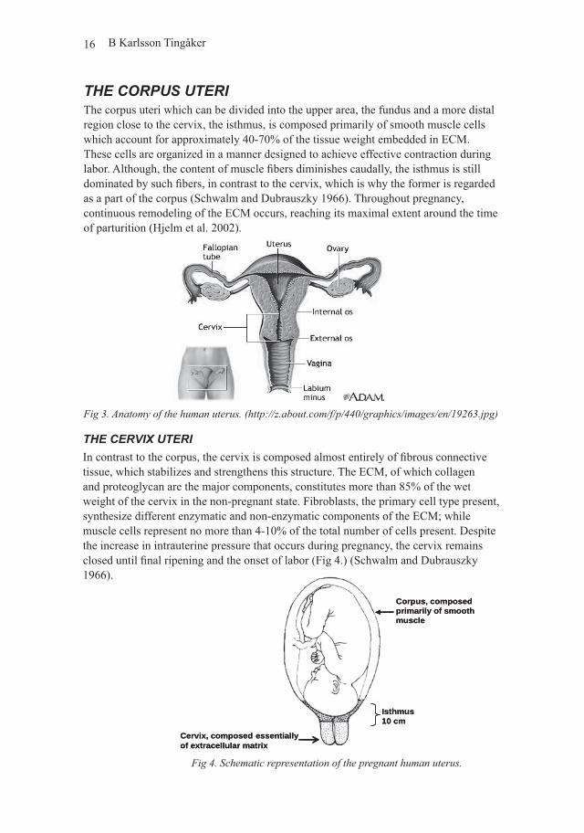
THE CORPUS UTERIThe corpus uteri which can be divided into the upper area, the fundus and a more distal region close to the cervix, the isthmus, is composed primarily of smooth muscle cells which account for approximately 40-70% of the tissue weight embedded in ECM. These cells are organized in a manner designed to achieve effective contraction during labor. Although, the content of muscle fibers diminishes caudally, the isthmus is still dominated by such fibers, in contrast to the cervix, which is why the former is regarded as a part of the corpus (Schwalm and Dubrauszky 1966). Throughout pregnancy, continuous remodeling of the ECM occurs, reaching its maximal extent around the time of parturition (Hjelm et al. 2002).
THE CERVIX UTERIIn contrast to the corpus, the cervix is composed almost entirely of fibrous connective tissue, which stabilizes and strengthens this structure. The ECM, of which collagen and proteoglycan are the major components, constitutes more than 85% of the wet weight of the cervix in the non-pregnant state. Fibroblasts, the primary cell type present, synthesize different enzymatic and non-enzymatic components of the ECM; while muscle cells represent no more than 4-10% of the total number of cells present. Despite the increase in intrauterine pressure that occurs during pregnancy, the cervix remains closed until final ripening and the onset of labor (Fig 4.) (Schwalm and Dubrauszky 1966).
Fig 3. Anatomy of the human uterus. (http://z.about.com/f/p/440/graphics/images/en/19263.jpg)
Fig 4. Schematic representation of the pregnant human uterus.
Isthmus10 cm
Cervix, composed essentiallyof extracellular matrix
Corpus, composedprimarily of smoothmuscle
Isthmus10 cm
Cervix, composed essentiallyof extracellular matrix
Corpus, composedprimarily of smoothmuscle

Background 17
CERVICAL RIPENING IS AN INFLAMMATORY PROCESSClinically, cervical ripening is characterized by softening, effacement and dilatation of this tissue, a process that involves profound remodelling of the ECM and is a prerequisite for effective labor and normal parturition. Cervical ripening can be divided into two phases i.e. a slow ripening throughout the course of pregnancy, associated with a gradual reduction in content of collagen, followed by the very rapid changes that take place immediately prior to the onset of labor (Granstrom et al. 1989; Uldbjerg et al. 1983).
As early as 1978, Liggins, a pioneer in this field, proposed that the latter rapid phase was an inflammatory reaction (Liggins 1978), which has been subsequently confirmed by a number of investigations. This final phase of cervical ripening, is associated with a 100 - fold increase in the local concentrations of cytokines and an influx of inflammatory cells, as well as with changes in other mediators of inflammation such as prostaglandins, NO and neuropeptides (Chwalisz and Garfield 1998; Collins et al. 2002; Gibb 1998; Osman et al. 2003; Sennstrom et al. 2000; Tornblom et al. 2004). Interestingly, these same chemical mediators appear to participate in nociceptive pathways and most also interact with NGF and TRPV1 under inflammatory conditions (Bennett 2001; Julius and Basbaum 2001; McMahon and Koltzenburg 2006; Pezet and McMahon 2006).
The intensity of pain associated with childbirth is one reason that has caused more and more women to choose to have cesarean sections (CS), the frequency of which in Swedish university hospitals during the past few years has thereby increased from 11-13% to 18-22%. In addition, the use of central neuroaxial blocks in connection with vaginal deliveries is also becoming more common. Clearly, our understanding of the mechanisms underlying labor pain needs to be improved, which will require more detailed information concerning the uterine innervation concentrated to the cervix during term pregnancy and labor (Bryman et al. 1987).

B Karlsson Tingåker18

AIMS OF THE PRESENT THESIS
The fundamental hypothesis that the present investigation was designed to test is that labor pain primarily emanates from the cervix uteri. Numerous studies have demonstrated the occurrence of an inflammatory process in the uterus, particularly in the cervix in connection with cervical ripening and labor. Since inflammatory processes often elicit pain, we thought it was important to compare nerve-related changes in the corpus and cervix associated with term pregnancy and labor to the situation in the non-pregnant uterus. In this context the following questions were posed:
I. How does the general innervation of the corpus compared to the cervix uteri in term pregnancy and during labor differ from that of the corresponding non- pregnant tissues?
II. Do the profound structural changes which the uterus undergoes during pregnancy and parturition, involve, e.g. sensory corpuscles that convey mechanosensitive (proprioceptive) impulses?
III. Is TRPV1 expressed in the uterus during labor and if so, do the levels of expression in the corpus and cervix differ?
IV. Are NGF and BDNF and their receptors involved in the process of labor and if so, are these factors present at different levels in the corpus and cervix during this event?
Aims of the present thesis 19

B Karlsson Tingåker20

Material and Methods 21
MATERIAL AND METHODS
SUBJECTSOur female subjects consisted of three groups, i.e. non-pregnant women (NP), and women experiencing term pregnancy followed by elective CS (TP) or in labor terminated by emergency CS (TPL). The 36 non-pregnant women were all having regular menstrual periods prior to undergoing hysterectomy for benign conditions and none was taking any hormone medication. The 37 healthy term pregnant women all experienced an uncomplicated pregnancy and had an elective CS prior to onset of labor. Finally, after undergoing a normal pregnancy and spontaneously entering labor, the 32 healthy TPL women were subjected to emergency CS due to arrest of labor or fetal malpresentation or distress.
SAMPLING PROCEDURES
The corpus uteriBiopsies (400-500 mg) excised from the isthmus (the upper edge of the lower uterine segment incision), hereafter referred to simply as the corpus, were obtained in connection with the CS or, in the case of the NP group following hysterectomy. These tissue samples were immediately fixed (Papers I, II, IV) or frozen on dry ice (Paper III) and subsequently stored at -70°C until the performance of immunohistochemical analyses. Samples to be employed for extraction of mRNA were stored in RNA-later solution until freezing.
The cervix uteri.Biopsies including all layers of the cervix (150-300 mg) were taken transvaginally
from the anterior lip (at the 12 o’clock position), following CS or hysterectomy and then handled in the same manner as described above for the corpus samples.

B Karlsson Tingåker22
METHODS
Immunohistochemical examination (Paper I - IV)In Paper I, the tissue samples were immersed in 4% paraformaldehyde and 14% saturated picric acid dissolved in 0.1M phosphate buffer (pH 6.9). After rinsing, 14 µm thick serial cryostat sections of these fixed samples were thawed onto slides and subsequently processed for performance of indirect immunofluorescent analysis according to protocols employed routinely in our laboratory. Polyclonal rabbit anti-PGP 9.5 antiserum was utilized as the primary antibody for this analysis. The sections were incubated overnight with either this specific antiserum or a control serum and thereafter exposed to rhodamine (TRITC)-labeled donkey antiserum directed against rabbit IgG. Control sections were stained in the same manner but, without the primary antibody. Finally, all sections were evaluated independently in a double-blind manner by two separate investigators (for additional details, see Paper I).
In Paper II and IV, the tissue samples were fixed in 4% paraformaldehyde alone, dissolved in 0.1 M phosphate buffer (pH 7.0).and thereafter rinsed, following which 8 µm serial cryostat sections were processed for immunohistochemical analysis in accordance with standard protocols. Tissue staining with hematoxylin-eosin (Htx) was utilized for examination of tissue morphology. In the case of peroxidase-anti peroxidase (PAP) staining, the slides were first incubated overnight with polyclonal rabbit antibodies towards PGP 9.5, S-100, nerve growth factor (NGF), the nerve growth factor receptor p75 (NGFR p75), brain-derived neurotrophic factor (BDNF) or the tyrosine kinase receptor B (TrkB), followed by exposure to swine anti-rabbit antibody. After further rinsing, the slides were exposed to peroxidase-labeled IgG immunoglobulin (PAP rabbit). For control purposes, the primary antibodies were omitted or replaced with normal serum and in the case of analysis of NGF, BDNF and TrkB, the primary antisera were preabsorbed with the appropriate antigen (for additional details see Papers II and IV).
Immunofluorescent staining was achieved with the appropriate primary polyclonal rabbit antibodies (60 minutes at 37°C) and tetramethylrhodamine isothiocyanate (TRITC)-conjugated swine anti-rabbit IgG as the secondary antibody (for additional details see Paper II).
In Paper III, PAP-staining was performed on frozen tissue sections placed on glass slides and thereafter fixed in paraformaldehyde. After inhibiting endogenous peroxidase activity and rinsing, these sections were incubated with primary antibodies directed against TRPV1 or a “cocktail” of neurofilament proteins (NFILS) and peripherin. The sites of antibody binding were visualized employing the nickel-enhanced, immunoperoxidase procedure. Negative controls were performed by omitting the primary antibody or replacing it with non-immune serum and nuclei were counterstained with 0.1% (w/v) aqueous neutral red (for further methodological details, see Paper III).
All sections were evaluated independently and in a double-blind manner by two separate observers.

Material and Methods 23
Real-Time PCR (Paper IV)For this procedure, frozen biopsies were placed onto a blocks of dry ice, cut into slices and then frozen in liquid nitrogen, following which this frozen tissue was pulverized with a dismembranation apparatus in order to obtain a powder from which RNA could be extracted. Extraction of total RNA achieved with the Trizol reagent and the concentration of RNA thus obtained was determined on the basis of its absorbance at OD260 as measured using an Eppendorf Bio Photometer. Only samples exhibiting an OD260/OD280 ratio > 1.7 were employed for Reverse Transcriptase-PCR (RT-PCR) and the quality of these samples was further evaluated by electrophoresis on 1.5% agarose gels, followed by visualization under ultraviolet light after ethidium bromide staining. Total RNA was stored at -70ºC until further analysis.
Reverse transcription of this total RNA was performed with SuperscriptTM Rnase H-Reverse Transcriptase and the produced cDNA stored at -70ºC until further use.
Real-Time PCR: In order to quantitate in triplicates the levels of mRNA encoding NGFβ, BDNF, NGFr p75, TrkA and TrkB, the Applied Biosystems 7300 Real-Time PCR system (Applied Biosystems, Foster City, CA, USA) was utilized together with the Taqman Universal PCR Master Mix (Applied Biosystems) and 96-well optical PCR plates. Appropriate primers and probes were purchased (Taqman® gene expression assays, Applied Biosystems) and the levels of expression of the two housekeeping genes, 18S and β-actin were used as internal standards. Since the levels of β-actin mRNA turned out to differ significantly between two of the groups it could not be used for this purpose
The Real-Time PCR involved 40 cycles of denaturation-annealing performed in accordance with a standard manufacturer’s protocol. The first cycle following which the reporter fluorescence was significantly above the baseline signal threshold, cycle Ct was determined for each mRNA species, with a Ct of >40 being considered undetectable. The Ct for mRNA 18S and β-actin mRNA was subtracted from that of interest to give ΔCt as a reflection of the expression of this mRNA. Since a high ΔCt value corresponds to a low level of mRNA, these values were inverted as 10/ΔCt. As a control for the specificity of the primers, serial dilutions of cDNA prepared from total hippocampal RNA (Ambion) were used. (For further details and the primer sequences see paper IV).
STATISTICAL ANALYSISAlmost all data were analyzed employing the Kruskal Wallis ANOVA by Ranks and the Kruskal-Wallis test, with a p-value of <0.05 being considered statistically significant. However, the immunohistochemical evaluations in Paper IV are documented as the relative number of immunoreactive (IR) structures compared to the NP control group, defined as equal to one. Therefore, statistical analysis of these semi-quantitative data could not be carried out.

B Karlsson Tingåker
Results
Patterns of innervationThe patterns of innervation in the corpus and cervix were elucidated by staining immu-nohistochemically for PGP 9.5.which is generally expressed by nerve cells.
The corpus uteriPaper I documents the presence of PGP 9.5 immunoreactive (IR) nerve fibers in the corpus uteri of all three groups of women as well as a profound reduction in the abun-dance of such following term pregnancy, both before and after the onset of labor, in comparison to the non-pregnant state (p<0.05) (Fig 5a). PGP 9.5-positive staining in NP muscle tissue was about 40-fold more common than in the case of TP and TPL biopsies. Immunoreactive nerve fibers were also observed in the stroma and near blood vessels. (Fig 6a)
The cervix uteriIn contrast to the pattern demonstrated by the corpus, cervical innervation was preser-ved in term pregnancy and labor with single or bundles of PGP 9.5 IR nerve fibers being detected in all samples (Fig 5b). These nerve fibers were especially abundant in the stroma, as well as in the vicinity of blood vessels (Fig 6b), but strikingly, there was a distinct layer of PGP 9.5 positive nerve fibers located just beneath the squamous epithe-lium, although not actually within the epithelium itself (Fig 6c).
24
Fig 5a. Histogram illustrating the total number of PGP 9.5-IR nerve fibres in the corpus. There is a significant decrease of PGP 9.5-IR nerve fibers in TP and TPL compared to NP (p<0.05)Fig 5b. Histogram illustrating the total number of PGP 9.5-IR nerve fibres in the cervix. Differences between the groups did not reach statistical significance
0
200
400
600
800
1000
1200
NP TP TPL
Num
ber o
f PG
P 9.
5-IR
ner
ve fi
ber p
rofile
s
0
200
400
600
800
1000
1200
NP TP TPL
Num
ber o
f PG
P 9.
5-IR
ner
ve fi
ber p
rofile
s
a b

Results 25
Fig 6 (a-c). Immunofluorescence micrographs of nerve bundles (arrow head) and single fibers (arrows) in the uterus that stain positively red for PGP 9.5. (A) In the non-pregnant corpus. (B). In the labouring cervix, in the vicinity of a blood vessel (star) and scattered throughout the stroma. (C) In the labouring cervix, present in the subepithelial region but absent from the squamous epithelium (SE). Scale bars = 100µm

B Karlsson Tingåker26
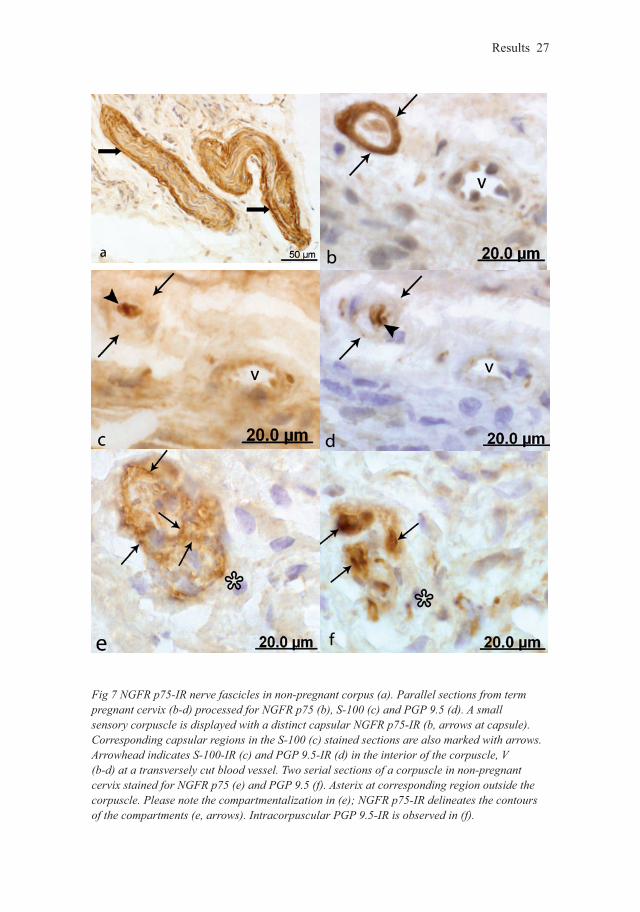
Sensory corpusclesSensory corpuscles which were round or oval in shape, had a diameter of 25-75 µm and which were frequently located in the vicinity of blood vessels stained positively for NGFR p75, S-100 and PGP 9.5. On the basis of this pattern of immunoreactivity and their morphological appearance, these sensory corpuscles could be clearly distinguis-hed from nerve fascicles present in the same tissue (Fig 7a). The capsule of some of the corpuscles was well-defined exhibiting distinct IR for NGFR p75 but not for PGP 9.5 or S-100 (Fig 7b) whereas the central regions stained positively for S-100 (Fig 7c) and the central axons exhibited pronounced expression of PGP 9.5 (Fig 7d). Other sensory corpuscles, which demonstrated a diffuse staining for NGFR p75 in their capsule, were frequently seen to harbor small, clearly NGFR p75-positive intra-capsular structures with a clearly defined NGFR p75 IR at their peripheries (Fig 7e), i.e. they displayed a compartmentalized appearance (Fig 7f).
The corpus uteriSensory corpuscles were observed only in the non-pregnant corpus, and were not detec-table from this tissue during term pregnancy.
The cervix uteriIn contrast to the corpus, sensory corpuscles were displayed not only in the non preg-nant state but also in term pregnant specimens of the cervix. However, no such structu-res were detectable during cervical dilatation (labor) (Fig 7b-d).
TRPV1 and neurofilaments (NFILS)Immunostaining for both TRPV1 (Fig 8a, b) and NFILS (Fig 8c) was observed in the corpus and cervix, albeit with somewhat different patterns. Thus, in contrast to NFILS-IR nerve fibers, TRPV1-positive nerve fibers could not be detected in the corpus during labor. On the other hand, both of these types of fibers were observed in term pregnant corpus, although at levels that were markedly attenuated in comparison to the non-preg-nant state.
Expression of TRPV1 in the corpus uteriTRPV1-IR nerve fibers were present within nerve fascicles in the non-pregnant corpus (Fig 8a). but only rarely in the term pregnant tissue prior to labor and not at all during labor. Their very low frequency in term pregnancy precluded statistical analysis.
Expression of TRPV1 in the cervix uteri Medium- to fine-caliber sized TRPV1-IR nerve fibers were observed in all cervical samples, as either single fibers or groups of fibers within nerve fascicles, scattered th-roughout the stroma and in the vicinity of blood vessels (Fig 8b). There were no statis-tically significant differences between the three groups with respect to the abundance or location of such nerve fibers.
Results 27
Fig 7 NGFR p75-IR nerve fascicles in non-pregnant corpus (a). Parallel sections from term pregnant cervix (b-d) processed for NGFR p75 (b), S-100 (c) and PGP 9.5 (d). A small sensory corpuscle is displayed with a distinct capsular NGFR p75-IR (b, arrows at capsule). Corresponding capsular regions in the S-100 (c) stained sections are also marked with arrows. Arrowhead indicates S-100-IR (c) and PGP 9.5-IR (d) in the interior of the corpuscle, V (b-d) at a transversely cut blood vessel. Two serial sections of a corpuscle in non-pregnant cervix stained for NGFR p75 (e) and PGP 9.5 (f). Asterix at corresponding region outside the corpuscle. Please note the compartmentalization in (e); NGFR p75-IR delineates the contours of the compartments (e, arrows). Intracorpuscular PGP 9.5-IR is observed in (f).

B Karlsson Tingåker28
Expression of NFILS in the corpus uteriThe NFILS-IR nerve fibers and fascicles were detected throughout all of the non-pregnant samples of corpus tissue but were much less abundant (fibers/mm2) in samp-les taken at term (p<0.0001)and during labor (p<0.01) (Fig 8c-d). In addition the total numbers of such nerve fibers were also lower in term pregnancy (p<0.001) and during labor (p< 0.05) but there was no significant differences between these values for the TP and TPL groups.
Expression of NFILS in the cervix uteriNFILS-IR nerve fibers, which were scattered throughout the stroma with only rare penetration into the basal epithelium, were present in equal numbers in the cervical tissue from all three groups.
Fig 8. TRPV1-IR nerve fascicle in non-pregnant corpus (a) and a TRPV1-IR nerve fiber in term pregnant cervix (b). NFILS-IR nerve fascicle in non-pregnant corpus (c), parallel section to (a). Scale bars = 50µm. In (d) a histogram illustrating the profound decrease of NFILS-IR nerve fibers in TP corpus compared to NP (p<0.0001) and TPL in comparison with NP (p<0.01)
NP TP TPL
d
Results 29
Neurotrophins and neurotrophin receptorsExpression of the neurotrophins NGF and BDNF and their specific receptors NGFR p75 and TrkB in uterine tissue was examined employing immunohistochemistry. In addition, the levels of mRNA encoding NGFβ, BDNF, and the receptors NGFR p75, TrkA and TrkB in these same samples were determined by Real-Time PCR.
Immunohistochemical analysis
Expression of the NGF and BDNF ligands in the corpus uteriBinding of antibodies towards NGF was observed in corpus tissue from all three groups, frequently by cells exhibiting a fibroblast-like appearance, with a higher number of such cells being present in the TPL (Fig 9a, 11a). Moreover, BDNF-IR nerve fascicles and cells were seen in the stroma of all three groups as well, with a larger number of such nerve fascicles in the TPL than in the NP group (Fig 10a-c, 12a).
Expression of the NGF and BDNF ligands in the cervix uteri NGF-IR was expressed strongly by cells located in the ECM of all three types of cer-vical tissue, being more frequent in the TPL group (Fig 9b, 11b). Similarly BDNF-IR nerve fascicles and cells (Fig 10c) were also detected in all three groups. These nerve fascicles which were most frequently located immediately beneath the squamous epi-thelium, were considerably more numerous in TPL tissue than in cervical samples of NP and TP (Fig 10b). A striking additional finding in this connection was the presence BDNF immunoreactivity within epithelial cells.
Expression of the NGFR p75 and TrkB receptors in the corpus uteriNerve fibers expressing NGFR p75-IR were present in corpus tissue from all three groups, often grouped into nerve fascicles and in larger numbers in NP than in the preg-nant tissue samples (Fig 7a). Furthermore, the nerve fascicles of this type in the non-pregnant corpus exhibited distinct NGFR p75-immunoreaction in the perineurium but only indistinct immunoreaction during pregnancy and labor (Fig 7a). In addition, nerve fibers from all three groups stained weakly for TrkB.
Expression of the NGFR p75 and TrkB receptors in the cervix uteri In the case of all three groups of cervical tissue, nerve fascicles and fibers as well as cells, stained positively for NGFR p75. The immunoreactive nerve fascicles were both more common and more distinctly outlined in NP than in TP or TPL tissue. The TrkB-IR nerve fibers and cells detected in all three types of tissue samples stained very weakly, so that reliable counting of their numbers was not possible.

B Karlsson Tingåker30
Fig 9. NGF-IR cells in the laboring corpus (a) and cervix (b).
Fig 10. Section from NP corpus showing a BDNF-IR nerve fascicle (a). Nerve fascicles were more diffusely outlined in TP corpus (b) compared to NP. In the cervix opposed to the corpus BDNF-IR cells were detected intraepithelially (arrows, c), (SE= squamous epithelium).
Fig 11 a-b Histogram showing the relative increase of NGF-IR cells during labor in the corpus (a) and cervix (b) compared to NP.
0123456789
NP TP TPL
Rel
ativ
e nu
mbe
rs o
f N
GF-
IR c
ells
0
0,5
1
1,5
2
2,5
3
NP TP TPL
Rel
ativ
e nu
mbe
rs o
f N
GF-
IR c
ells
SE
a b

Results 31
Real-Time PCR analysis
Corpus levels of mRNA encoding NGFβ and BDNF ligandsNGFβ mRNA was detected in all three types of tissue, at similar levels in the pregnant groups, but at significantly lower levels than in non-pregnant samples. In TP the NGFβ was down-regulated (p<0.001) in comparison to NP and in TPL (p=0.013) in compari-son to NP (Fig 13a). BDNF mRNA expression was not detectable in any of the samples.
Cervical levels of mRNA encoding NGFβ and BDNF ligands The cervical level of NGFβ mRNA was significantly higher in the samples from term pregnant women in established labor than from those who underwent elective CS prior to the onset of labor (p=0.013), but, there were no significant differences in this respect between NP and the pregnant groups (Fig 13b).BDNF mRNA could not be detected in any of our samples.
Corpus levels of mRNA encoding the NGFR p75, TrkA and TrkB receptorsThe level of NGFR p75 mRNA was significantly attenuated in term pregnancy in com-parison to the non-pregnant state (p<0.001), and there was a similar trend with labor (p=0.051) for NP versus TPL, but no statistically significant differences in this level bet-ween the TP and TPL groups. The level of TrkA mRNA increased significantly in term pregnancy (p<0.01) and during labor (p<0.03) compared to the non-pregnant state (Fig 14a). However there were no significant differences between the pregnant groups.
TrkB mRNA expression was significantly down-regulated at term (p<0.001) and during labor (p=0.014) compared to the non-pregnant tissue, but there were no significant differences between the pregnant groups.
Cervical levels of mRNA encoding the NGFR p75, TrkA and TrkB receptorsThe mRNA species coding for NGFR p75, TrkA (Fig14b) and TrkB did not differ signi-ficantly between the groups. However, the level of TrkA mRNA expression showed an upward tendency in labor (Fig 14b).
0
0,5
1
1,5
2
2,5
NP TP TPL
Fig 12. Histogram showing the relative increase of BDNF-IR nerve fibers in the corpus during labor, TPL (a) and cervix, TPL (b) as compared to NP
Rel
ativ
e nu
mbe
rs o
f B
DN
F- IR
ner
ve fa
scic
les
00,5
11,5
22,5
3
Rel
ativ
e nu
mbe
rs o
f B
DN
F- IR
ner
ve fa
scic
les
NP TP TPLNP TP TPL
a b

B Karlsson Tingåker32
Together, these findings reveal a noteworthy pattern involving elevated numbers of NGF-IR cells together with significant up-regulation of the expression of NGFβ mRNA expression in the cervix of women in established labor, in comparison to those prior to the onset of labor. In a similar manner, a pronounced increase in the number of nerve fascicles staining positively for BDNF was observed in both the corpus and cervix of subjects in labor. BDNF mRNA could easily be detected when hippocampal cDNA was used as control for the primer. However, we could not detect any mRNA expression in samples from either corpus or cervix uteri.
The receptors NGFR p75, TrkA and TrkB did not reveal any significant differences in mRNA expression between the cervical groups. Cervical NGFR p75-IR nerve fibers decreased in NP and TPL. In the case of the corpus the levels of NGFβ, NGFR p75, and TrkB mRNA were all lower in the pregnant than in the non-pregnant tissue.

Results 33
Fig 13. Box plots illustrating the significant decrease of NGFβ mRNA expression in the term pregnant corpus compared to non-pregnant (a, p<0.001) and also comparing corpus in TPL with NP (a, p=0.013). Note the significant up-regulation of NGFβ mRNA expression in the cervix during labor compared to TP (b, p=0.013).
NP TP TPL NP TP TPL
Fig 14. Box plots showing the increase of TrkA mRNA expression in the corpus in pregnancy and labor compared with NP (a) TP, p=0.01, TPL p<0.03). The differences between the cervical groups (b) did not reach significance.
NP TP TPL NP TP TPL
13 b
14 b
13 a
14 a
10/∆
Ct T
RK
A-1
8S
10/∆
Ct T
RK
A-1
8S10
/∆ C
t NG
F(β)
-18S
10/∆
Ct N
GF(
β)-1
8S
Median
25%-75%
Non-Outliar Range
Outliers
* Extremers

B Karlsson Tingåker34
DISCUSSION
Labor pain is a complex, multidimensional experience in response to the sensory stimuli generated during parturition. Despite extensive research much still remains to be eluci-dated concerning alterations in the innervation of the human uterus in association with term pregnancy and labor. Improved understanding of such alterations will help reveal the underlying mechanisms of labor pain.
The main findings described in this thesis concern 1, the profound differences in the innervation of the human corpus uteri and cervix following term pregnancy and during labor, 2, the preservation of TRPV1 expression in the cervix but not in the corpus under these conditions, 3, elevated uterine levels of NGF and BDNF immunoreactions and 4, an elevated cervical level of NGFβ mRNA during labor in comparison to non-pregnant controls. In addition, the presence of sensory nerve corpuscles in the human uterus is here documented for the first time.
Our examinations were performed on corporal biopsies excised from the upper edge of the lower uterine segment, i.e., the area in which incision is performed for cesarean section and from the corresponding site after hysterectomy in NP. The question often arises as to whether this region is representative for the entire corpus or resembles instead the cervix in its structure and function. Luckas and Wray have observed no significant differences in the extent and rate of contractile force generated by comparing the upper and lower uterine segment (Luckas and Wray 2000). Furthermore, the isthmus consists predominantly of muscle fibers, in similarity with the rest of the corpus and in contrast to the cervix (Schwalm and Dubrauszky 1966). In certain studies, the innervation of so-called cervical biopsies taken from the lower edge of the CS incision has been reported to differ from that of the fundus (Bryman et al. 1987). However, here we document clear differences in the innervation of the isthmic region and the anterior cervical lip.
As revealed by staining for PGP 9.5 of the corpus, but not of the cervix, a pronounced reduction in the numbers of positively stained nerve fibers was displayed following term pregnancy and during labor (Paper I). Moreover, a similar pattern was observed upon staining immunohistochemically for TRPV1 (Paper III) and NGFR p75 (Paper IV). These observations are consistent with earlier findings concerning the general uterine innervation in association with pregnancy and labor in rodents (Klukovits et al. 2004; Lundberg et al. 1988). Furthermore, in humans a similar pattern was obtained using S-100 as a neuronal marker (Bryman et al. 1987; Wikland et al. 1984). Stjernholm and co-workers (1999) demonstrated that cervical staining for PGP 9.5 is preserved both during term pregnancy and labor (Stjernholm et al. 1999).
Discussion 35

B Karlsson Tingåker
This profound decrease in the number of nerve fibers in the pregnant corpus appears to involve several different types of fibers. Employing electron microscopy Morizaki and colleagues (1989) observed lower numbers of both adrenergic and cholinergic nerves fibers in the term pregnant corpus than in the corresponding non-pregnant tissue, but these investigators did not examine the pregnant cervix or samples from women in labor (Morizaki et al. 1989). To the best of our knowledge, the study documented in Paper I is the first to demonstrate alterations in the numbers of PGP 9.5-IR nerve fibers in both the corpus and cervix uteri during labor.
Although the functional reason for this selective denervation of the corpus during pregnancy remains to be determined, this phenomenon indicates clearly that the corpus and cervix function separately, at least to some extent, despite being regarded as parts of a single organ. During pregnancy the corpus distends in order to accommodate the growing fetus. In contrast, the unripe cervix does not distend and remains closed until cervical ripening and the onset of labor despite the increasing pressure. Under optimal conditions, the separate functions of these two entities are coordinated to achieve successful delivery of the infant.
Uterine distention and contraction as well as cervical effacement and dilatation represent changes that might be recognized by mechanosensory neurons, e.g., sensory copuscles. Sensory corpuscles are present in a variety of tissues including the skin, joint capsules and ligaments (Del Valle et al. 1998; Hagert et al. 2004; Malinovsky 1996; Zimny 1988) where they convey proprioceptive information. Corpuscle-like structures have also been observed in the colon, urinary bladder and external male genitalia (Halata and Munger 1986; Landon and Wiseman 2001; Wills et al. 2003).
Here, we demonstrate for the first time the presence of structures identified as sensory corpuscles in the human corpus and cervix uteri. The morphology of these structures clearly distinguishes them from the nerve fascicles in these same tissues. Their identification in the present investigation employing parallel staining for NGFR p75, S-100 and PGP 9.5 is the same approach commonly used in previous studies designed to identify sensory corpuscles (Hagert et al. 2005; Hagert et al. 2004; Lopez et al. 1998).
The uterine corpuscles observed here fall into two major categories, i.e., those displaying a well-developed capsule with distinct staining for NGFR p75 (Fig 7b), and others with diffuse NGFR p75-IR in their capsule and, frequently, an interior that appears compartmentalized (Fig 7e). These same two types of corpuscles have also been seen in other locations, including the skin, and periodontium (Byers 1990; Maeda et al. 1999; Vega 1996; Vega et al. 1996). Our examinations revealed PGP 9.5-IR nerve fibers (Fig 7f) together with S-100 immunoreactivity in the interior of the corpuscular structures. These findings are consistent with previous reports that the central arborizing sensory axons of sensory corpuscles in the skin, as well as the posterior crucial and the scapholunate interossus ligaments are surrounded by Schwann cells that express the S-100 protein (Albuerne et al. 1998; Del Valle et al. 1998; Gonzalez-Martinez et al. 2003; Hagert et al. 2004).
Sensory corpuscles are categorized in different ways by different investigators and even the characteristics of a given type can vary. The corpuscles with distinct capsules exhibiting pronounced staining for NGFR p75 observed here, bear a close resemblance to small Pacini corpuscles (Vega 1996; Vega et al. 1996) whereas the other
36

type of uterine sensory corpuscles show similarities to Ruffini corpuscles (Halata et al. 1999). Although both of these types of corpuscles are specialized for the perception of alterations in pressure, tension and vibrations the Pacini corpuscles are regarded as exhibiting a low threshold and rapid adaptation, while the Ruffini type is a slowly adapting mechanoreceptor that responds to relatively slight changes in tension (Halata et al. 1999; Johansson et al. 1991; Zimny 1988).
We detected sensory corpuscles in the non-pregnant corpus and cervix and in the term pregnant cervix before the onset of labor, but not in any uterine biopsy taken during labor. Although their functions in the uterus are presently unknown, it is tempting to propose that these sensory corpuscles act as structures in protective reflexes and they participate in the intricate coordination required between the functioning of the cervix and the corpus. It has been suggested that the density of corpuscles in ligaments is directly related to the occurrence of protective reflexes in connection with extreme movements and that Pacini corpuscles protect and stabilize joints (Backenkohler et al. 1997; Johansson et al. 1991).
The non-pregnant corpus uteri, which is composed primarily of smooth muscle, responds to elevated pressure and distension with pain and small contractions (Schwalm and Dubrauszky 1966). It might be speculated that sensory corpuscles protect and stabilize trying to regulate the muscle tonus and the size of the non-pregnant uterus. In contrast, during pregnancy, when the corpus must enlarge and distend without contracting a parallel disappearance of discernible sensory corpuscles occurs. Whether this disappearance is involved in the prevention of premature contractions is an interesting possibility that remains to be studied. Moreover, since sensory denervation leads to the degeneration of sensory corpuscles, the pronounced reduction in the total number of nerve fibers in the pregnant corpus may be related to the disappearance of sensory corpuscles (Albuerne et al. 1998; Kopp et al. 1997).
In contrast to the corpus, sensory corpuscles were observed in the cervix both in the non-pregnant tissue and in term pregnancy, but not after the onset of labor. These sensory corpuscles were thus not detected when the profound stretching, changes in pressure and displacement associated with cervical ripening and dilatation occurred. Accordingly, the cervical sensory corpuscles may be involved in the prevention of premature ripening and dilatation, although further investigations designed to elucidate the morphological characteristics and functions of the uterine sensory corpuscles in detail are clearly required.
The complex processes that result in softening and effacement of the cervix involve a multitude of factors, including prostaglandins, cytokines, nitric oxide and neuropeptides, as well as infiltration of the ECM by inflammatory cells (Chwalisz and Garfield 1998; Collins et al. 2002; Gibb 1998; Kelly 2002; Kirby et al. 2005; Liggins 1978; Osman et al. 2003; Sennstrom et al. 2000; Tornblom et al. 2005; Winkler et al. 1999). All these factors are known to play a role in inflammatory reactions. Cervical ripening is generally considered to be an inflammatory process associated with profound remodeling of the ECM. It is also noteworthy that all of these same mediators participate in nociception (Griffis et al. 2006; McDougall 2006; McMahon and Koltzenburg 2006; Rowlands and Permezel 1998).
Discussion 37

B Karlsson Tingåker
Neurotrophins and TRPV1 have been reported to be additional mediators of inflammatory pain and for this reason we performed the first examination of the possible presence of these factors in the corpus and cervix uteri following term pregnancy and during labor, with comparison to the corresponding non-pregnant tissues (Pezet and McMahon 2006);(Bennett 2001; Julius and Basbaum 2001; Levi-Montalcini et al. 1996). Concerning NGF mRNA and the protein levels, they are both considerably elevated in the inflamed tissues associated with various conditions, e.g., arthritis, pneumonitis and interstitial cystitis (Huang and Reichardt 2001; Levi-Montalcini et al. 1996; Nockher and Renz 2005; Rihl et al. 2005; Tortorolo et al. 2005), an increase that may be mediated by an initial rise in cytokine levels (Levi-Montalcini et al. 1996; Pezet and McMahon 2006). In addition, uterine immunohistochemical staining for NGF has been reported to be enhanced in connection with the painful condition of endometriosis (Anaf et al. 2002).
We observed that the numbers of NGF-positive cells in the cervix during labor were clearly higher than in term pregnancy prior to the onset of labor or in the non-pregnant state (Fig 11b). This change occurs in parallel with the substantial increase in cytokine levels associated with in the inflammatory process of cervical ripening (Osman et al. 2003; Sennstrom et al. 2000). In addition to its production in neuronal tissue, NGF is synthesized by a variety of other types of cells, including fibroblasts, macrophages and mast cells all of which participate in cervical ripening (Huang and Reichardt 2001; Levi-Montalcini et al. 1996; Malmstrom et al. 2007; Nockher and Renz 2005; Osman et al. 2003; Winkler et al. 1999). Our finding that, in parallel with the increase in the NGF-positive cell population, the expression of the corresponding NGFβ mRNA is also up-regulated in the cervix during labor (p=0.013) compared to the TP group, suggests local synthesis of this factor, possibly under the regulation of cytokines as well as its involvement in cervical ripening (Bennett 2001; Levi-Montalcini et al. 1996).
In contrast to the cervix, the NGFβ mRNA expression in the laboring corpus was not elevated. Osman et al have reported an inflammatory reaction in the myometrium during labor (Osman et al. 2003). Therefore we had expected an increase in the NGFβ mRNA expression in parallel with the increase of NGF-IR cells. Nevertheless, it is tempting to speculate that the weak upward tendency in the NGFβ mRNA expression in TPL compared to TP might have reached significance with analyses of a larger number of subjects (Fig 13a) Similar findings with a decrease of NGFβ expression in corpus during pregnancy have been observed in rodents (Lobos et al. 2005; Varol et al. 2000).
The NGFβ mRNA expression was down-regulated in the pregnant corpus as was the levels of mRNA encoding the receptors NGFR p75 and TrkA. However, in the cervix, the NGFβ mRNA was up-regulated during labor in contrast to NGFR p75 and TrkA mRNA expression. Nonetheless, NGFR p75-IR cells were present in the cervix, especially during labor, whereas the numbers of NGFR p75-IR nerve fibers in both the corpus and cervix were reduced in association with term pregnancy both prior to and after the onset of labor. Together, these findings show that the patterns of NGF and NGFR p75-IR cells, as well as NGFR p75 positive nerve fibers change in parallel with cervical ripening and denervation of the pregnant corpus indicating that these factors may participate in both of these processes.
38

BDNF is a neuromodulator synthesized primarily by a subpopulation of small and medium-sized sensory neurons in the DRG (Bennett 2001; Pezet et al. 2002). In experimental models of inflammatory pain, expression of BDNF is dramatically up-regulated at both the mRNA and protein levels (Pezet et al. 2002). Expression of this factor has also been observed in connection with inflammatory conditions such as spondyloarthritis, as well as in lungs infected by respiratory syncytial virus (Rihl et al. 2005; Tortorolo et al. 2005).
In the present investigation, a pronounced elevation in the numbers of BDNF-IR nerve fascicles was seen in both the corpus and cervix during labor in strong contrast to the reduction in the total number of immunoreactive nerve fibers in the pregnant corpus (as revealed after staining for PGP 9.5). Possibly the barrage of nociceptive impulses accompanying labor enhances the expression of BDNF. This increase coincides with the inflammatory processes involved in cervical ripening and labor, as well as with the elevations in the number of NGF-IR cells and in the expression of NGFβ mRNA. In TrkA dependent sensory neurons, BDNF expression has been reported to be up-regulated under inflammatory conditions via an NGF-dependent mechanism (Apfel et al. 1996; Pezet and McMahon 2006).
Morover, cells staining weakly for BDNF were also present in the stroma of both the corpus and cervix. Unexpectedly, BDNF-IR cells were seen intraepithelialy in addition to subepithelial cellular immunoreactions in cervical tissue, possibly indicating a role for this factor in the epithelial signaling process that brings about cervical ripening. At the same time, BDNF mRNA expression could not be detected in any of our biopsies. Perhaps, the uterus contains a particular splice variant of this factor whose mRNA cannot be amplified employing the two primers we chose (Paper IV). It should be remembered that BDNF is produced primarily in the DRG, from where it is transported to target tissues via sensory nerve fibers.
In addition to the roles they play in inflammatory processes, NGF, BDNF and their receptors act as important mediators and modulators of nociceptive pain (Pezet and McMahon 2006). Enhanced expression of NGF and BDNF in inflammatory conditions leads to recruitment of neuropeptides such as substance P (SP) and calcitonin gene-related peptide (CGRP), as well as elevated levels of ion channels (e.g. TRPV1) and other pain-associated receptors (Bennett 2001; Julius and Basbaum 2001; Levi-Montalcini et al. 1996; McMahon and Koltzenburg 2006; Pezet et al. 2002; Pezet and McMahon 2006). Pre-synaptic stimulation of afferent nociceptive fibers induces the release of BDNF and activation of TrkB receptors, leading to post-synaptic excitability (McMahon and Koltzenburg 2006; Pezet et al. 2002).
Although the cation channel TRPV1 is expressed primarily in nociceptors, this channel is also expressed by non-neuronal tissues such as smooth muscle, the bladder urothelium, polymorphonuclear granulocytes and macrophages (Caterina and Julius 2001; Cortright and Szallasi 2004; Julius and Basbaum 2001). The activation and/or modulation of TRPV1 by various inflammatory mediators gives rise to nociceptive signals (Cortright and Szallasi 2004; Di Marzo et al. 2002; Ma and Quirion 2007). Together, these observations make it likely that TRPV1 participates in eliciting the pain caused by inflammatory mediators in the ripe and laboring cervix. This proposal gains further support from findings in connection with painful inflammatory diseases, such as inflamed human oesophagus, non-erosive reflux disease, painful bladder syndrome
Discussion 39

B Karlsson Tingåker
and vulvodynia (Bhat and Bielefeldt 2006; Matthews et al. 2004; Mukerji et al. 2006; Tympanidis et al. 2004). In these conditions, the number of nerve fibers staining positively for TRPV1 is enhanced.
In this investigation the TRPV1-IR nerve fascicles present in the non-pregnant corpus uteri were found to have disappeared almost completely following term pregnancy and could not be detected at all in biopsies of the corpus taken during labor. In contrast, medium to fine TRPV1-IR nerve fibers were exhibited by all of our cervical tissue samples (NP, TP, TPL) as single fibers or fascicles. They were located beneath the squamous epithelium, scattered throughout the stroma and in the vicinity of blood vessels. This reduction in the number of TRPV1-immunoreactive nerve fibers in the corpus following term pregnancy is similar to the decreases in immunostaining for PGP 9.5, NFILS and NGFR p75-IR.
Although, immunohistochemical reactivity and levels of mRNA are not directly comparable, it is interesting to note that the levels of NGFβ and TrkA mRNA increased in the same manner. Overall, the up-regulation of the level of NGFβ mRNA together with the increases in numbers of NGF and NGFR p75-positive cells and BDNF-nerve fibers in the laboring cervix indicate that TRPV1 plays a role in cervical ripening.TRPV1, which plays a complex multifunctional role in regulating pathways of inflammatory pain is activated directly or indirectly by a number of inflammatory mediators involved in cervical ripening, including prostaglandins, neuropeptides, cytokines and neurotrophins. (Cortright and Szallasi 2004; Ma and Quirion 2007). Moreover, prostaglandins also lower the threshold temperature required for TRPV1 activation (Caterina et al. 1997; Ma and Quirion 2007). In connection with various painful inflammatory diseases, the levels of cytokines and TRPV1 increase in parallel (Bhat and Bielefeldt 2006; Matthews et al. 2004). Furthermore, TRPV1 has been proposed to trigger the release of the neuropeptides SP and CGRP from peripheral nerve terminals that occurs under inflammatory conditions (Caterina and Julius 2001). In addition, the algogenic mediator bradykinin enhances the sensitivity of nerve cells to TRPV1 via pathways involving protein kinase C or phospholipase C (Julius and Basbaum 2001; Ma and Quirion 2007; Premkumar and Ahern 2000).
Another observation of interest in this context is that NGF and BDNF interact with TRPV1 and here we document the presence of NGF, BDNF and TrkA in the pregnant cervix uteri (Di Marzo et al. 2002; Ji et al. 2002; Julius and Basbaum 2001; Pezet and McMahon 2006). In the labouring cervix, the numbers of NGF-positive cells and BDNF-immunoreactive nerve fibers, as well as the level of NGFβ mRNA were elevated. From previous studies parallel increases in the levels of TRPV1 and neurotrophins are known to occur in this same situation. Such elevated levels of TRPV1 in peripheral nociceptor terminals commonly restricted to those that express TrkA and are located in an inflamed tissue, result from NGF-mediated activation of the p38 MAPK system (Di Marzo et al. 2002; Ji et al. 2002; Julius and Basbaum 2001; Lever et al. 2001; Pezet and McMahon 2006). Although the TrkA mRNA was not significantly up-regulated during labor, there was an upward tendency which maybe could have reached significance with a larger number of subjects (Fig 14b). Our finding that the levels of inflammatory mediators and TRPV1 in the cervix are enhanced during cervical ripening and labor indicates that TRPV1 plays a role in connection with labor pain.
40

Labor pain is unique as being a visceral, nociceptive intermittent type of temporary pain which is, moreover, linked to a normal physiological event. Although this pain is usually described as being mediated by mechanical distension of the lower uterine segment and the cervix during contractions, the source of uterine nociception has not yet been elucidated in detail (Brownridge 1995; Lowe 2002; McMahon and Koltzenburg 2006; Rowlands and Permezel 1998). During early stages of pregnancy, women often complain of diffuse lower abdominal pain, which usually subsides after the initial few months. This phenomenon would seem to be in line with the observations reported in Paper I concerning the rich innervation of the non-pregnant corpus and the “denervation” that occurs during the later stages of pregnancy.
The fact that a relatively small increase in intrauterine pressure in the non-pregnant uterus causes pain suggests that mechanosensitive nociceptors are present in the corpus. However, as pregnancy proceeds and the uterus grows, the type of lower abdominal pain described above disappears until the onset of labor. Possibly, certain of the PGP 9.5-IR nerve fibers observed in non-pregnant uterus are mechanosensitive nociceptors that “disappear” or become “silent” as pregnancy proceeds. The sensory corpuscles described in Paper II can be assumed to be primarily mechanosensitive (proprioceptive) rather than nociceptive, since these are not detectable in the laboring cervix, which receives a barrage of afferent nociceptive impulses.
The intensity of labor pain commonly increases as cervical dilatation progresses and as the pressure escalates as a consequence of contractions and the descent of the fetus. Here, we demonstrate that the levels of both neurotrophins (as reflected in the levels of NGFβ mRNA, NGF-positive cells and BDNF-positive nerve fibers) and TRPV1 are higher in the laboring cervix than in NP and TP. These findings lend support to our hypothesis that to a large extent, labor pain originates from the combined action of cervical nociceptors sensitized by the inflammatory process and the activation of mechanoreceptors that were previously silent. Since these mechanosensitive nociceptors are activated by the increase in pressure caused by contractions, the pain subsides between contractions.
This proposal is consistent with a number of previous findings. It is generally accepted that nociceptors are polymodal in nature and mechanically insensitive nociceptors have been reported to be sensitized by tissue injury, in the present context due to inflammation (Gebhart 2000; McMahon and Koltzenburg 2006). In addition, the analgesic effect achieved with paracervical blockade indirectly support our conclusion that labor pain originates primarily from the cervical region (Ranta et al. 1995; Rosen 2002).
Considered together, the observations reported here support our hypothesis that labor pain emanates primarily from the cervix. The intermittent nature of labor pain is most readily explained as a consequence of the combined action of pressure and inflammation on previously silent nociceptors in the cervix and isthmic region of the corpus, but activated during the process of labor.
Discussion 41

B Karlsson Tingåker42

CONCLUSIONS
Immunohistochemical staining for PGP 9.5, a general marker of innervation, • revealed profound reduction in the number of immunoreactive nerve fibers in term pregnant human corpus both prior to and after the onset of labor in comparison to the non-pregnant tissue. In contrast, the cervical innervation is preserved during pregnancy and parturition.
Sensory corpuscles were detected in the non-pregnant and term pregnant human • cervix prior to the onset of labor but only in the non-pregnant state in the case of the corpus.
Nerve fibers expressing TRPV1 were identified in the human uterus for the first • time. In the corpus, such nerve fibers are abundant in the non-pregnant state but rare in term pregnancy and undetectable during labor. In contrast, the density of these nerve fibers in the cervix is unaltered during either term pregnancy or parturition.
Expression of NGFβ mRNA was found to be up-regulated in the cervix during • labor. In addition, the numbers of NGF-IR cells and BDNF-IR nerve fibers in both the cervix and the corpus are higher during labor than in term pregnancy prior to the onset of labor or in the non-pregnant state.
In summary, these changes in the corpus and cervix that occur during term pregnancy and labor, together with the differences between these two tissues, reveal unambiguously that labor pain emanates primarily from the cervix.
Conclusions 43

B Karlsson Tingåker44

FUTURE PERSPECTIVES
Future Perspectives 45
Since we could not detect BDNF mRNA in our biopsies it would be worthwhile to exa-mine the reason for this apparent absence by employing other PCR primers. In addition, it would be of value to examine the BDNF protein itself in the cerebrospinal fluid (CSF) from the same subjects in order to disclose possible interaction between the peripheral and central nervous system. Furthermore, it would be of interest to look for the presen-ce of NGF, and TRPV1 in the same CSF samples as well.
The identity of the cervical cells that stain positively for NGF, BDNF, NGFR p75 and TrkB should be probed in more detail utilizing specific markers.
The finding of TRPV1-IR nerve fibers in the human cervix and the absence of such nerve fibers in the corpus in term pregnancy and during labor, further support our hypothesis that labor pain primarily emanates from the cervix. Paracervical blockade is an established method for alleviation of labor pain, but with undesired side-effects, e.g. fetal bradycardia and with variable efficacy. The findings presented in this thesis motivate investigations to further elucidate the mechanisms involved in the origin of cervical nociception. This could provide a basis for the development of simpler, and more effective peripheral strategies for alleviating this pain. From a worldwide perspective, very few women have access to proper labor pain relief. However, further research resulting in new methods to treat labor pain, could bring help for those many women requireing pain relief. In addition, procedures for effective alleviation of labor pain might also exert a beneficial effect on other types of pain emanating from similar nociceptive pathways, as well as caused by pain during obstetric and gynecological procedures.

B Karlsson Tingåker46

Acknowledgements
Acknowledgements 47
I would like to offer my sincere gratitude to all of the many people who have supported me throughout this study and made important contributions to its completion. In particular I would like to thank:
Professor Gunvor Ekman-Ordeberg, my supervisor, for outstanding guidance, for piloting me through the tricky forest of clinical research, never-failing enthusiasm, constant encouragement and helping me to realize the joy of combining clinical work with research.
Associate Professor Sture Forsgren, my co-supervisor, for sharing your great knowledge, for excellent supervision, including teaching me the importance of patience and scientific accuracy, for always being available and ready to answer all my questions.
Associate Professor Lars Irestedt, my co- supervisor, for setting an excellent example in your work as an obstetric anesthesiologist, for our productive clinical and scientific collaboration, for valuable support and encouragement, as well as for helping me to learn to think ”outside the box”.
Dr Margareta Norman, my co- supervisor, for our extensive collaboration at the delivery ward of Danderyd Hospital and for the different ways in which you have helped me find time to complete my thesis research.
Associate Professor Anders Ekblom, for your impressively creative way of thinking, for stimulating discussions and for always being available to provide me with good advices, from the very beginning of this project.
Professor Bo von Schoultz, former Head of the Department of Obstetrics and Gynecology at Karolinska University Hospital, for introducing me to this specialty when I was a medical student and, later, for your friendly encouragement of my research efforts and clinical work
Associate Professor Sven-Eric Olsson present Head of the Department of Obstetrics and Gynecology at Danderyd Hospital, for kind support with my clinical work as well as research.
Associate Professor Oddvar Bakos, former Head of the Department of Obstetrics and Gynecology at Karolinska University Hospital, for invaluable support and encouragement in connection with both my clinical and research work.

B Karlsson Tingåker48
Professor Praveen Anand, co-author for sharing your extensive knowledge about TRPV1 with me and by stimulating collaboration making it possible for me to study this protein.
Paul Facer, co-author, for your excellent professional assistance in analyzing TRPV1, as well as for always being ready to offer help, encouragement and good advices.
Associate Professor Olle Johansson, co-author, for your invaluable help in transforming the results of our microscopic analyses into my first scientific article, as well as for always being willing to share your knowledge with me.
Birgitta Byström, co-author, for your highly professional work in the laboratory, exceptional ingenuity in solving problems, endless patience and constant help and encouragement.
Ann Hjelm Cluff, colleague and co-author, for introducing me to the approaches employed in recruiting patients and collecting biopsies, for your support and encouragement, for sharing the joys and hardships associated with being an obstetrician, as well as for your close friendship.
Ulla Hedlund (Umeå), Lena Jonsson (Umeå), Berit Ståbi and Marianne Ekman for your excellent work with the immunohistochemical preparations and for always being supportive and encouraging. Furthermore, the staff at the FoU-lab for help and support and for creating such a warm and friendly atmosphere at the lab.
Associate Professor Folke Flam and his staff at St.Görans Hospital, for assistance when collecting biopsies.
Ringvor Hägglöf, technician, for your fantastic last-minute assistance with the layout and figures in my thesis, helping me to produce a printable book.
Susanne Abelin, colleague, for our productive and enjoyable collaboration, for your generous willingness to shoulder a larger work load so that I could complete my thesis, and for true friendship.
Colleagues, midwives and staff at the delivery, operating and maternity wards of Karolinska and Danderyd Hospitals, for all your invaluable help and support.
All of the brave women who kindly agreed to participate in this study
Birgitta and Roland Eriksson, for welcoming me into your loving and generous home during my research visits to Umeå and for your many years of care and friendship.
Agneta, Anitha and other friends, who, while encouraging me, have also reminded me that there are more aspects to life than research alone.

Acknowledgements 49
Ingrid Hansson, a true friend since the days of our early childhood, for always believing in me and for sharing all facets of my life.
Eva Tingåker Johansson, my wonderful sister-in-law, and her family, for all of the encouraging e-mails and telephone calls and your different ways of caring.
Einar och Marianne Tingåker, my parents-in-law, for your support, warmth and care.
Björn, my husband, for all of your unconditional love, consideration and support in all aspects of life.
Viktor and David, the ”fountains of joy” in my life, David especially for your help with figures and computer problems in general, and both of you for encouragement, support and teaching me something new about life every single day.
This study was supported by grants from
The Swedish Research CouncilKarolinska Institutet FundsALF FundThe Swedish Medical Research Council

B Karlsson Tingåker50

REFERENCES
Albuerne M, Lopez S, Naves FJ, Martinez-Almagro A, Represa J, Vega JA. 1998. S100alpha and S100beta proteins in human cutaneous sensory corpuscles: effects of nerve and spinal cord injury. Anat Rec 251(3):351-359.
Anaf V, Simon P, El Nakadi I, Fayt I, Simonart T, Buxant F, Noel JC. 2002. Hyperalgesia, nerve infiltration and nerve growth factor expression in deep adenomyotic nodules, peritoneal and ovarian endometriosis. Hum Reprod 17(7):1895-1900.
Apfel SC, Wright DE, Wiideman AM, Dormia C, Snider WD, Kessler JA. 1996. Nerve growth factor regulates the expression of brain-derived neurotrophic factor mRNA in the peripheral nervous system. Mol Cell Neurosci 7(2):134-142.
Backenkohler U, Strasmann TJ, Halata Z. 1997. Topography of mechanoreceptors in the shoulder joint region--a computer-aided 3D reconstruction in the laboratory mouse. Anat Rec 248(3):433-441.
Bennett DL. 2001. Neurotrophic factors: important regulators of nociceptive function. Neuroscientist 7(1):13-17.
Berkley KJ, Robbins A, Sato Y. 1993. Functional differences between afferent fibers in the hypogastric and pelvic nerves innervating female reproductive organs in the rat. J Neurophysiol 69(2):533-544.
Bhat YM, Bielefeldt K. 2006. Capsaicin receptor (TRPV1) and non-erosive reflux disease. Eur J Gastroenterol Hepatol 18(3):263-270.
Brownridge P. 1995. The nature and consequences of childbirth pain. Eur J Obstet Gynecol Reprod Biol 59 Suppl:S9-15.
Bryman I, Norstrom A, Dahlstrom A, Lindblom B. 1987. Immunohistochemical evidence for preserved innervation of the human cervix during pregnancy. Gynecol Obstet Invest 24(2):73-79.
Byers MR. 1990. Segregation of NGF receptor in sensory receptors, nerves and local cells of teeth and periodontium demonstrated by EM immunocytochemistry. J Neurocytol 19(5):765-775.
Caterina MJ, Julius D. 2001. The vanilloid receptor: a molecular gateway to the pain pathway. Annu Rev Neurosci 24:487-517.
Caterina MJ, Schumacher MA, Tominaga M, Rosen TA, Levine JD, Julius D. 1997. The capsaicin receptor: a heat-activated ion channel in the pain pathway. Nature 389(6653):816-824.
Catheline G, Touquet B, Besson JM, Lombard MC. 2006. Parturition in the rat: a physiological pain model. Anesthesiology 104(6):1257-1265.
Chwalisz K, Garfield RE. 1998. Nitric oxide as the final metabolic mediator of cervical ripening. Hum Reprod 13(2):245-248.
Collins JJ, Usip S, McCarson KE, Papka RE. 2002. Sensory nerves and neuropeptides in uterine cervical ripening. Peptides 23(1):167-183.
Cortright DN, Szallasi A. 2004. Biochemical pharmacology of the vanilloid receptor TRPV1. An update. Eur J Biochem 271(10):1814-1819.
References 51
B Karlsson Tingåker
Del Valle ME, Harwin SF, Maestro A, Murcia A, Vega JA. 1998. Immunohistochemical analysis of mechanoreceptors in the human posterior cruciate ligament: a demonstration of its proprioceptive role and clinical relevance. J Arthroplasty 13(8):916-922.
Di Marzo V, Blumberg PM, Szallasi A. 2002. Endovanilloid signaling in pain. Curr Opin Neurobiol 12(4):372-379.
Gebhart GF. 2000. Pathobiology of visceral pain: molecular mechanisms and therapeutic implications IV. Visceral afferent contributions to the pathobiology of visceral pain. Am J Physiol Gastrointest Liver Physiol 278(6):G834-838.
Gibb W. 1998. The role of prostaglandins in human parturition. Ann Med 30(3):235-241.Gonzalez-Martinez T, Perez-Pinera P, Diaz-Esnal B, Vega JA. 2003. S-100 proteins in
the human peripheral nervous system. Microsc Res Tech 60(6):633-638.Granstrom L, Ekman G, Ulmsten U, Malmstrom A. 1989. Changes in the connective
tissue of corpus and cervix uteri during ripening and labour in term pregnancy. Br J Obstet Gynaecol 96(10):1198-1202.
Griffis CA, Compton P, Doering L. 2006. The effect of pain on leukocyte cellular adhesion molecules. Biol Res Nurs 7(4):297-312.
Hagert E, Forsgren S, Ljung BO. 2005. Differences in the presence of mechanoreceptors and nerve structures between wrist ligaments may imply differential roles in wrist stabilization. J Orthop Res 23(4):757-763.
Hagert E, Ljung BO, Forsgren S. 2004. General innervation pattern and sensory corpuscles in the scapholunate interosseous ligament. Cells Tissues Organs 177(1):47-54.
Halata Z, Munger BL. 1986. The neuroanatomical basis for the protopathic sensibility of the human glans penis. Brain Res 371(2):205-230.
Halata Z, Wagner C, Baumann KI. 1999. Sensory nerve endings in the anterior cruciate ligament (Lig. cruciatum anterius) of sheep. Anat Rec 254(1):13-21.
Hjelm AM, Barchan K, Malmstrom A, Ekman-Ordeberg GE. 2002. Changes of the uterine proteoglycan distribution at term pregnancy and during labour. Eur J Obstet Gynecol Reprod Biol 100(2):146-151.
Huang EJ, Reichardt LF. 2001. Neurotrophins: roles in neuronal development and function. Annu Rev Neurosci 24:677-736.
Ji RR, Samad TA, Jin SX, Schmoll R, Woolf CJ. 2002. p38 MAPK activation by NGF in primary sensory neurons after inflammation increases TRPV1 levels and maintains heat hyperalgesia. Neuron 36(1):57-68.
Johansson H, Sjolander P, Sojka P. 1991. Receptors in the knee joint ligaments and their role in the biomechanics of the joint. Crit Rev Biomed Eng 18(5):341-368.
Julius D, Basbaum AI. 2001. Molecular mechanisms of nociception. Nature 413(6852):203-210.
Kelly RW. 2002. Inflammatory mediators and cervical ripening. J Reprod Immunol 57(1-2):217-224.
Kirby LS, Kirby MA, Warren JW, Tran LT, Yellon SM. 2005. Increased innervation and ripening of the prepartum murine cervix. J Soc Gynecol Investig 12(8):578-585.
Klukovits A, Gaspar R, Santha P, Jancso G, Falkay G. 2004. Role of capsaicin-sensitive nerve fibers in uterine contractility in the rat. Biol Reprod 70(1):184-190.
Kopp DM, Trachtenberg JT, Thompson WJ. 1997. Glial growth factor rescues Schwann cells of mechanoreceptors from denervation-induced apoptosis. J Neurosci 17(17):6697-6706.
52

Landon DN, Wiseman OJ. 2001. A Pacinian corpuscle in the human bladder lamina propria. J Neurocytol 30(6):457-464.
Lever IJ, Bradbury EJ, Cunningham JR, Adelson DW, Jones MG, McMahon SB, Marvizon JC, Malcangio M. 2001. Brain-derived neurotrophic factor is released in the dorsal horn by distinctive patterns of afferent fiber stimulation. J Neurosci 21(12):4469-4477.
Levi-Montalcini R, Skaper SD, Dal Toso R, Petrelli L, Leon A. 1996. Nerve growth factor: from neurotrophin to neurokine. Trends Neurosci 19(11):514-520.
Liggins GC. 1978. Ripening of the cervix. Semin Perinatol 2(3):261-271.Lobos E, Gebhardt C, Kluge A, Spanel-Borowski K. 2005. Expression of nerve growth
factor (NGF) isoforms in the rat uterus during pregnancy: accumulation of precursor proNGF. Endocrinology 146(4):1922-1929.
Lopez SM, Perez-Perez M, Marquez JM, Naves FJ, Represa J, Vega JA. 1998. p75 and TrkA neurotrophin receptors in human skin after spinal cord and peripheral nerve injury, with special reference to sensory corpuscles. Anat Rec 251(3):371-383.
Lowe NK. 2002. The nature of labor pain. Am J Obstet Gynecol 186(5 Suppl Nature):S16-24.
Luckas MJ, Wray S. 2000. A comparison of the contractile properties of human myometrium obtained from the upper and lower uterine segments. Bjog 107(10):1309-1311.
Lundberg LM, Alm P, Wharton J, Polak JM. 1988. Protein gene product 9.5 (PGP 9.5). A new neuronal marker visualizing the whole uterine innervation and pregnancy-induced and developmental changes in the guinea pig. Histochemistry 90(1):9-17.
Ma W, Quirion R. 2007. Inflammatory mediators modulating the transient receptor potential vanilloid 1 receptor: therapeutic targets to treat inflammatory and neuropathic pain. Expert Opin Ther Targets 11(3):307-320.
Maeda T, Ochi K, Nakakura-Ohshima K, Youn SH, Wakisaka S. 1999. The Ruffini ending as the primary mechanoreceptor in the periodontal ligament: its morphology, cytochemical features, regeneration, and development. Crit Rev Oral Biol Med 10(3):307-327.
Malinovsky L. 1996. Sensory nerve formations in the skin and their classification. Microsc Res Tech 34(4):283-301.
Malmstrom E, Sennstrom M, Holmberg A, Frielingsdorf H, Eklund E, Malmstrom L, Tufvesson E, Gomez MF, Westergren-Thorsson G, Ekman-Ordeberg G, Malmstrom A. 2007. The importance of fibroblasts in remodelling of the human uterine cervix during pregnancy and parturition. Mol Hum Reprod 13(5):333-341.
Matthews PJ, Aziz Q, Facer P, Davis JB, Thompson DG, Anand P. 2004. Increased capsaicin receptor TRPV1 nerve fibres in the inflamed human oesophagus. Eur J Gastroenterol Hepatol 16(9):897-902.
McDougall JJ. 2006. Arthritis and pain. Neurogenic origin of joint pain. Arthritis Res Ther 8(6):220.
McMahon SB, Koltzenburg M. 2006. Wall and Melzack’s Textbook of Pain.Morizaki N, Morizaki J, Hayashi RH, Garfield RE. 1989. A functional and structural
study of the innervation of the human uterus. Am J Obstet Gynecol 160(1):218-228.
References 53

B Karlsson Tingåker
Mukerji G, Yiangou Y, Agarwal SK, Anand P. 2006. Transient receptor potential vanilloid receptor subtype 1 in painful bladder syndrome and its correlation with pain. J Urol 176(2):797-801.
Nockher WA, Renz H. 2005. Neurotrophins in clinical diagnostics: pathophysiology and laboratory investigation. Clin Chim Acta 352(1-2):49-74.
Osman I, Young A, Ledingham MA, Thomson AJ, Jordan F, Greer IA, Norman JE. 2003. Leukocyte density and pro-inflammatory cytokine expression in human fetal membranes, decidua, cervix and myometrium before and during labour at term. Mol Hum Reprod 9(1):41-45.
Pezet S, Malcangio M, McMahon SB. 2002. BDNF: a neuromodulator in nociceptive pathways? Brain Res Brain Res Rev 40(1-3):240-249.
Pezet S, McMahon SB. 2006. Neurotrophins: Mediators and Modulators of Pain. Annu Rev Neurosci.
Premkumar LS, Ahern GP. 2000. Induction of vanilloid receptor channel activity by protein kinase C. Nature 408(6815):985-990.
Ranta P, Jouppila P, Jouppila R. 1996. The intensity of labor pain in grand multiparas. Acta Obstet Gynecol Scand 75(3):250-254.
Ranta P, Jouppila P, Spalding M, Kangas-Saarela T, Jouppila R. 1995. Paracervical block--a viable alternative for labor pain relief? Acta Obstet Gynecol Scand 74(2):122-126.
Rihl M, Kruithof E, Barthel C, De Keyser F, Veys EM, Zeidler H, Yu DT, Kuipers JG, Baeten D. 2005. Involvement of neurotrophins and their receptors in spondyloarthritis synovitis: relation to inflammation and response to treatment. Ann Rheum Dis 64(11):1542-1549.
Rosen MA. 2002. Paracervical block for labor analgesia: a brief historic review. Am J Obstet Gynecol 186(5 Suppl Nature):S127-130.
Rowlands S, Permezel M. 1998. Physiology of pain in labour. Baillieres Clin Obstet Gynaecol 12(3):347-362.
Schwalm H, Dubrauszky V. 1966. The structure of the musculature of the human uterus--muscles and connective tissue. Am J Obstet Gynecol 94(3):391-404.
Sennstrom MB, Ekman G, Westergren-Thorsson G, Malmstrom A, Bystrom B, Endresen U, Mlambo N, Norman M, Stabi B, Brauner A. 2000. Human cervical ripening, an inflammatory process mediated by cytokines. Mol Hum Reprod 6(4):375-381.
Stjernholm Y, Sennstrom M, Granstrom L, Ekman G, Johansson O. 1999. Protein gene product 9.5-immunoreactive nerve fibers and cells in human cervix of late pregnant, postpartal and non-pregnant women. Acta Obstet Gynecol Scand 78(4):299-304.
Tominaga M, Tominaga T. 2005. Structure and function of TRPV1. Pflugers Arch 451(1):143-150.
Tong C, Ma W, Shin SW, James RL, Eisenach JC. 2003. Uterine cervical distension induces cFos expression in deep dorsal horn neurons of the rat spinal cord. Anesthesiology 99(1):205-211.
Tornblom SA, Maul H, Klimaviciute A, Garfield RE, Bystrom B, Malmstrom A, Ekman-Ordeberg G. 2005. mRNA expression and localization of bNOS, eNOS and iNOS in human cervix at preterm and term labour. Reprod Biol Endocrinol 3:33.
54

Tornblom SA, Patel FA, Bystrom B, Giannoulias D, Malmstrom A, Sennstrom M, Lye SJ, Challis JR, Ekman G. 2004. 15-hydroxyprostaglandin dehydrogenase and cyclooxygenase 2 messenger ribonucleic acid expression and immunohistochemical localization in human cervical tissue during term and preterm labor. J Clin Endocrinol Metab 89(6):2909-2915.
Tortorolo L, Langer A, Polidori G, Vento G, Stampachiacchere B, Aloe L, Piedimonte G. 2005. Neurotrophin overexpression in lower airways of infants with respiratory syncytial virus infection. Am J Respir Crit Care Med 172(2):233-237.
Tympanidis P, Casula MA, Yiangou Y, Terenghi G, Dowd P, Anand P. 2004. Increased vanilloid receptor VR1 innervation in vulvodynia. Eur J Pain 8(2):129-133.
Uldbjerg N, Ekman G, Malmstrom A, Olsson K, Ulmsten U. 1983. Ripening of the human uterine cervix related to changes in collagen, glycosaminoglycans, and collagenolytic activity. Am J Obstet Gynecol 147(6):662-666.
Varol FG, Duchemin AM, Neff NH, Hadjiconstantinou M. 2000. Nerve growth factor (NGF) and NGF mRNA change in rat uterus during pregnancy. Neurosci Lett 294(1):58-62.
Vega JA. 1996. Cutaneous sensory nerve formations: an overview. Microsc Res Tech 34(4):281-282.
Vega JA, Haro JJ, Del Valle ME. 1996. Immunohistochemistry of human cutaneous Meissner and pacinian corpuscles. Microsc Res Tech 34(4):351-361.
Wikland M, Lindblom B, Dahlstrom A, Haglid KG. 1984. Structural and functional evidence for the denervation of human myometrium during pregnancy. Obstet Gynecol 64(4):503-509.
Wills EJ, Croker J, Brammah S. 2003. Tactile corpuscle-like bodies in colonic mucosa. Ultrastruct Pathol 27(2):79-86.
Winkler M, Fischer DC, Ruck P, Marx T, Kaiserling E, Oberpichler A, Tschesche H, Rath W. 1999. Parturition at term: parallel increases in interleukin-8 and proteinase concentrations and neutrophil count in the lower uterine segment. Hum Reprod 14(4):1096-1100.
Zimny ML. 1988. Mechanoreceptors in articular tissues. Am J Anat 182(1):16-32.
References 55

Copyright © 2022 FDOKUMEN