Research Article Personality Profiles Identify Depressive ...
Upload
khangminh22Category
view
1download
0
2018
UNIVERSIDADE DE LISBOA
FACULDADE DE CIÊNCIAS
Using a Systems Approach to Identify the Mechanism of Action of Correctors
Doutoramento em Biologia
Especialidade de Biologia de Sistemas
Nikhil T Awatade
Tese orientada por:
Professora Margarida D. Amaral e Dr. Rainer Pepperkok
Documento especialmente elaborado para a obtenção do grau de Doutor
2018
UNIVERSIDADE DE LISBOA
FACULDADE DE CIÊNCIAS
Using a Systems Approach to Identify the Mechanism of Action of Correctors
Doutoramento em Biologia
Especialidade de Biologia de Sistemas
Nikhil T Awatade
Tese orientada por:
Professora Margarida D. Amaral e Dr. Rainer Pepperkok
Júri: Presidente:
● Doutor Rui Manuel dos Santos Malhó, Professor Catedrático Faculdade de Ciências da Universidade de Lisboa
Vogais:
● Doutor Karl Kunzelmann, Professor Faculty of Biology and Pre-Clinical Medicine da University of Regensburg, Alemanha; ● Doutora Ana Colette Pereira de Castro Osório Maurício, Professora Associada com Agregação Instituto de Ciências Biomédicas Abel Salazar (ICBAS) da Universidade do Porto; ● Doutora Margarida Sofia Pereira Duarte Amaral, Professora Catedrática Faculdade de Ciências da Universidade de Lisboa (orientadora); ● Doutora Maria Margarida Perestrello Ramos, Professora Auxiliar Faculdade de Ciências da Universidade de Lisboa.
Documento especialmente elaborado para a obtenção do grau de Doutor
Fundação para a Ciência e Tecnologia do Ministério da Educação e Ciência (FCT/ SFRH / BD / 52487 / 2014)

I Would Like to Dedicate This Thesis To My PARENTS....
"Nothing is as important as passion. No matter what you want to do with
your life, be passionate."
-Jon Bon Jovi
i
Summary
Cystic Fibrosis (CF), the most common life-shortening genetic disorder among Caucasians, is caused by
mutations in the gene encoding the Cystic Fibrosis Transmembrane Conductance Regulator (CFTR)
protein, an ion channel expressed at the apical membrane of epithelial cells.
High-throughput screens (HTS) identified several novel molecules potentially targeting the underlying
CFTR defect but only for some patients: potentiator VX-770 (Ivacaftor/Kalydeco), for subjects bearing
G551D and other gating mutations, the combination corrector/potentiator VX-809 (Lumacaftor)/VX-
770 (Orkambi) for F508del-homozygous patients and another similar combination VX-661
(Tezacaftor)/VX-770 is under approval.
The main objective of this PhD work was to study new compounds that correct the basic CF defect, by
rescuing CFTR protein traffic and function, focusing both on individual responses of CF patients with
different CFTR mutations to these new drugs, and their mechanism of action.
Chapter 1 focusses on the measurement of functional responses on human bronchial epithelial cells
(HBE’s) derived from CF lung explants bearing different CFTR mutations to VX-809 namely: A561E,
N1303K, G542X and Y1092X. Our data showed a positive response of A561E/A561E to VX-809 and
F508del/Y1092X but not F508del/G542X.
In Chapter 2, we evaluated the efficacy of CFTR modulators (correctors/potentiators) in physiologically
relevant tissues, namely rectal biopsies, intestinal organoids, (HBE’s) and human nasal epithelial cells
(HNE’s), from CF patients with rare CFTR mutations. Data obtained here showed that neither R560S
nor H1079P-could not be rescued by any of the CFTR modulators, but 3849+10kbC>T and R334W and
c.120del23-CFTR were rescued by VX-770 alone or with VX-809.
In Chapter 3 we evaluated the efficacy of two novel CFTR correctors (B9, E12) in primary HBE cells, and
three novel compounds E-act mimics (C2, C5, and C7) as enhancers of alternative Cl- channel TMEM16A
in human intestinal organoids.
In Chapter 4 (final) we assessed the effect of CFTR modulators and their possible additivity with
F508del-CFTR genetic revertants 4RK, R1070W, and G550E to understand the mechanism of action of
small molecule correctors and another variant diacidic ER exit code DD/AA in CFBE mCherry cells
expressing these varinats by Ussing chamber analysis with or without CFTR modulators. Our data show
that C18 and VX-661 and low temperature (But not VX-809) rescued DD/AA to the cell surface and
genetic revertants restore the channel function without any CFTR modulator.
Altogether, results from this work bring new insights into how the CFTR genotype may influence CFTR
function and response to CFTR modulators and how each patient should be assessed individually for
the responsiveness to the CFTR modulators towards personalized therapeutics.
ii
Resumo
A Fibrose Quística (FQ) é a doença autossómica recessiva letal mais comum na população Caucasiana,
afetando cerca de 1 em 2500-6000 nados vivos, e com uma frequência de portadores de 1 em cada 25
indivíduos. Esta doença é causada por mutações no gene CFTR (do inglês Cystic Fibrosis
Transmembrane Conductance Regulator), localizado no braço longo cromossoma 7. A proteína CFTR é
expressa na membrana apical das células epiteliais, onde funciona como um canal de cloreto (Cl-),
regulando o transporte de sal e de água. A CFTR também é descrita como uma proteína reguladora de
outros canais iónicos. Até à data já foram descritas mais de 2000 mutações no gene CFTR, para a mioria
das quais porém, as consequências funcionais continuam por esclarecer. A mutação F508del
(representando a deleção do aminoácido fenilalanina na posição 508 da proteína) é responsável por
85% dos cromossomas FQ a nível mundial, sendo assim a mutação mais comum nestes doentes. A FQ
é uma doença caracterizada por múltiplas manifestações em diferentes órgãos, tais como vias
respiratórias superiores e inferiores, pâncreas, ductos biliares, trato gastrointestinal, ductos
deferentes, glândulas sudoríparas, algumas células do sistema imunológico e outros tecidos, sendo,
contudo, a doença pulmonar a principal causa de morbilidade e mortalidade. Os doentes com FQ
podem apresentar quadros clínicos muito diferentes, mas têm em comum a presença dum muco
espesso, que impede que o transporte/limpeza mucociliar seja eficiente, levando à retenção de
bactérias (principalmente Pseudomonas aeruginosa) que originam infeções respiratórias recorrentes
e inflamação crónica, e à perda progressiva da função pulmonar. Além dos sintomas respiratórios,
outros sintomas clássicos de FQ incluem uma elevada concentração de electrólitos no suor (parâmetro
utilizado no principal teste de diagnóstico), insuficiência pancreática exócrina, cirrose hepática,
obstrução intestinal e infertilidade masculina. A esperança média de vida para indivíduos recém-
nascidos com FQ em 2010 com a mutação mais comum, foi estimada em 37 anos.
O desenvolvimento de metodologias de alto rendimento (“High-throughput screens”, HTS)
transformou de forma significativa o panorama terapêutico, passando duma visão terapêutica
uniformizada - "one-size-fits-all" - para uma visão de medicina personalizada, utilizando terapias
específicas para cada mutação. Através destas metodologias de HTS foi possível identificar diversas
novas moléculas com potencial de correção dos defeitos da CFTR ao nível do DNA, do RNA, ou da
proteína. Entre elas, o potenciador, VX-770 (Ivacaftor/Kalydeco), direcionado a indivíduos com a
mutação G551D ou outras mutações que afetem a regulação da abertura ("gating") do canal CFTR; a
combinação corretor/potenciador VX-809 (Lumacaftor)/VX-770 (conhecida como Orkambi), para
indivíduos homozigóticos para a mutação F508del; e mais recentemente (ainda em aprovação) a
combinação VX-661 (Tezacaftor)/VX-770 para estes mesmos indivíduos e outros com a mutação
F508del e uma segunda outras mutação com atividade residual. Contudo, a combinação VX-809/VX-
770 apenas demonstrou ter um efeito moderado na melhoria da doença pulmonar. O mecanismo de
ação destes compostos ainda não é totalmente conhecido e, apesar dos avanços na terapêutica da FQ,
nem todas as mutações da CFTR são passiveis de correção farmacológica. Por isso, torna-se pertinente
avaliar as respostas a novos moduladores da CFTR e aos existentes por parte de outras mutações em
modelos ex vivo, como tecidos/células primárias dos pacientes (por exemplo células epiteliais nasais
humanas, células brônquicas humanas) ou como biópsias retais, permitindo assim não só avaliar a
eficácia do modelador para um determinado genótipo da CFTR, como também permitir prever o
benefício clínico de forma individualizada para cada paciente. A ativação, através de fármacos, de
outros canais de Cloreto (como por exemplo o TMEM16A) continua a ser uma estratégia atrativa para
compensar a ausência de atividade da CFTR para compensar a ausência de canais CFTR. A ativação
iii
funcional do TMEM16A poderá ajudar em particular os pacientes com FQ cujas mutações são
insensíveis a terapias moduladoras da CFTR.
O principal objetivo deste trabalho de doutoramento consistiu no estudo de novos compostos que
corrigem o defeito básico desta doença, resgatando a proteína CFTR (Cystic Fibrosis Transmembrane
Conductance Regulator) que se encontra mutada na doença genética Fibrose Quística (FQ), focando-
se quer nas respostas individuais de pacientes com FQ e diferentes mutações CFTR a estes novos
fármacos, quer no seu mecanismo de ação.
O primeiro objetivo (Capítulo 1) desta tese focou-se na quantificação da atividade da CFTR em culturas
primárias de células epiteliais brônquicas (Human bronchial epithelial cells - HBE’s), derivadas de
pulmões de pacientes com FQ. Com este intuito, foi avaliado o efeito do VX-809 em mutações já
descritas como tendo defeitos semelhantes à F508del (classe II) - A561E e N1303K - e mutações de
classe I, como as G542X e Y1092X. Para tal, utilizámos monocamadas de células HBEs cultivadas em
filtros porosos e posteriormente analisadas em micro câmaras de Ussing com perfusão continua. Os
resultados mostraram que a mutação A561E em homozigotia responde positivamente ao tratamento
com VX-809, com um aumento de 7 vezes comparativamente a células controlo incubadas com DMSO,
representando cerca de 6% de recuperação quando comparadas com células sujeitos controlo
(indivíduos sem FQ). O VX-809 também mostrou ter um impacto positivo no genótipo F508del/Y1092X,
observando-se uma resposta de 7% comparativamente a células controlo, e quase o dobro do
observado para o genótipo F508del/G542X (cerca de 4% vs células controlo). Deste estudo também
podemos observar que as células que apresentam somente uma cópia da mutação F508del têm uma
resposta menor ao tratamento com VX-809. Adicionalmente, observámos diferenças significativas nas
respostas ao VX-809 entre células de diferentes pacientes homozigóticos para a mutação F508del (por
exemplo, Dador 1 – cerca de 5%, e Dador 2 – 15% de recuperação da atividade da CFTR vs células
normais). Estes estudos demonstram a importância de análises de modelos ex vivo para uma
terapêutica mais personalizada na FQ e reforça o tópico principal deste estudo: que cada paciente
deveria ter uma avaliação individual da capacidade de resposta aos diferentes moduladores da CFTR.
No Capítulo 2, o principal objetivo foi avaliar a eficácia dos moduladores da CFTR
(Corretores/Potenciadores) em tecidos fisiologicamente relevantes. Para isso utilizámos diversos
modelos celulares, como biópsias rectais (realizando o mesmo protocolo usado no diagnóstico de FQ),
organoides intestinais derivados dessas bióipsias, e culturas primárias de células brônquicas e de
células nasais. Estes estudos focaram-se principalmente em indivíduos com mutações no gene da CFTR
extremamente raras (chamadas mutações órfãs). As mutações raras estudadas neste trabalho foram:
R560S, H1079P, 3849+10kbC>T, R334W e c.120del23.
Resumidamente, para caracterizar e avaliar a eficácia dos moduladores da CFTR em células com o
genótipo R560S, usámos material de pacientes - organoides intestinais – assim como linhas celulares
CFBE expressando esta mutação de forma estável. Os resultados mostram que não houve correção da
mutação R560S com nenhum dos moduladores testados, nem nas células CFBE nem nos organoides
intestinais, ao contrário do que foi observado para a mutação F508del. Estudos funcionais e
bioquímicos da CFTR com a mutação H1079P revelaram que esta proteína mutante não apresenta
função como canal de cloreto. O uso dos moduladores CFTR e agentes “read through” (para ultrapassar
codões stop prematuros) não aumentaram a função da CFTR em materiais de pacientes com o
genótipo H1079P/W1282X e a respetiva medição da atividade basal quer por camara de Ussing quer
em organoides intestinais mostrou ausência de atividade da proteína CFTR. A caracterização funcional
iv
de organoides intestinais com o genótipo 3849+10kbC>T mostrou que esta mutação apresenta função
residual da CFTR. O tratamento com VX-809 não teve qualquer efeito na atividade residual da mutação
3849+10kbC>T mas esta foi significativamente melhorada pelo potenciador, clinicamente aprovado,
VX-770, sozinho ou combinado com o VX-809. A análise da atividade basal da CFTR em três tecidos
diferentes (biópsia rectal, organoides do intestino e células do epitélio nasal) de pacientes com a
mutação R334W demonstrou atividade residual de CFTR com esta mutação. Além disso, a combinação
do corretor VX-809 e dos potenciadores Genisteina ou VX-770 (com ou sem VX-809) teve um efeito
positivo na atividade da CFTR tanto nos organoides intestinais como nas células respiratórias. No
entanto verificou-se alguma variabilidade na resposta dos 3 diferentes pacientes com esta mutação. A
avaliação funcional da atividade da CFTR em linhas celulares com a mutação c.120del23 mostrou uma
resposta positiva ao tratamente apenas com potenciador ou qualquer combinação de
corretor/potenciador face à resposta basal (células tratadas com DMSO).
Com este objetivo, conseguimos estabelecer metodologias para a análise da CFTR em culturas de
células humanas do epitélio nasal e organoides intestinais, duas ferramentas extremamente
importantes para analisar a atividade da CFTR de forma personalizada para cada paciente.
Estes estudos evidenciam a importância de avaliar os efeitos dos moduladores da CFTR em diferentes
modelos fisiológicos, para uma melhor caracterização da resposta de cada modelador antes da sua
administração a pacientes com FQ.
O terceiro objetivo (Capítulo 3) deste trabalho focou-se na avaliação da eficácia de dois novos
corretores da CFTR (B9 e E12) em céulas derivadas de pulmões explantados de pacientes com FQ.
Adicionalmente, identificámos três novos compostos - análogos ao E-act, um estimulador do canal
alternativo de cloreto TMEM16A (C2, C5 e C7) que parecem ter um efeito positivo na função deste
canal. Com efeito, os resultados obtidos mostram que o composto E12, em conjunto com o VX-809,
tem um efeito aditivo significativo. Estudos em organoides intestinais com os análogos do E-act
levaram à identificação de três estimuladores da função do canal TMEM16A. Mostrámos que o
composto C2 leva a um aumento significativo da resposta ao ATP em organoides intestinais e,
consequentemente, na função do canal TMEM16A. Os resultados obtidos demonstraram assi que
estas células são uma boa ferramenta para a descoberta de novos corretores da CFTR, assim como
estimuladores do canal TMEM16A.
No quarto e último objetivo (Capítulo 4) deste trabalho pretendia-se avaliar o efeito dos moduladores
da CFTR e a possível aditividade com os revertentes genéticos da F508del 4RK, R1070W, G550E – e da
variante DA/AA em linhas celulares CFBE que os expressam estavelmente. Os resultados obtidos
mostraram que o defeito de tráfego da variante DA/AA-CFTR foi corrigido pelos corretores C18 e VX-
661, bem como pela incubação das células a baixa temperatura. Porém, o VX-809 teve um efeito
moderado nesta variante. Demonstrou-se ainda que os revertentes F508del-4RK, F508del-R1070W e
F508del-G550E restabeleciam alguma função do canal mesmo na ausência dos moduladores. Os
compostos aqui estudados demonstram que existe variabilidade no efeito aditivo com os revertentes
genéticos, fornecendo assim pistas para um possível mecanismo de ação, exercido através da ligação
a motivos estruturais específicos.
Em resumo, os resultados obtidos durante o presente trabalho de doutoramento trouxeram novos
conhecimentos sobre a influência do genótipo CFTR na função deste canal e na resposta a
moduladores, e como sobre a importância, quer de sistemas celulares heterólogos, quer de materiais

v
derivados dos pacientes (e fisiologicamente mais relevantes) para a análise das respostas funcionais
desta proteína com diferentes mutações, em abordagens de medicina personalizada.
vi
Acknowledgements
Foremost, I would like to express my sincere gratitude to my Supervisor Professor Margarida D Amaral
for the continuous support of my PhD study and related research, for her patience, motivation, and
immense knowledge. Besides this, I want to thank Professor Margarida Ramos for introducing me to
the world of Physiology and partly supervising my progress and giving valuable inputs during PhD.
I want to thank present and past members of the Amaral lab for making my five years stay in Portugal
memorable. Thanks to Marisa Sousa for sharing all the knowledge about Ussing chamber. Thanks to
Simao Luz, Veronica Felício, Goncalo Prista, Susana Igreja, Sara Canato, Sara Afonso, Joao Fernandes,
Joao Amorim, Ana Marta Romao, Inna Uliyakina, Ana Cachaco and Hugo Botelho who made my initial
stay in Lisbon (Junior Fellowship period) very pleasant and comfortable. Thanks to Joao Santos, Ines
Pankonien, Sofia Ramalho, Iris Lameiro, Iris Silva, Joana Lérias, Margarida Quaresma, Madalena Pinto,
Catarina Baptista, Luis Sousa, Filipa Simoes, Arsénia Massinga, Karina Mendes and Miqueias Lopes.
Thanks to my Lab managers Marta Palma, Jose Murias (for sharing all the useful information about
Photography) and Sofia Correia for providing all the necessary lab reagents whenever required. Thanks
to Lab secretary Renata Vincent, Filipa Coutinho, Carolina Varela and Andreia Reis for handling all
administrative and beaurocracy related issues during my stay at FCUL. Thanks to Luis Marques for IGC
trips for Organoids swelling assays. My sincere thanks to Professor Jeffery Beekman for providing me
an opportunity to spend six weeks in his lab in Utrecht and kindly sharing all the technical aspects
related to Organoids technique.
Thanks to PI’s from the lab, Professor Luka Clarke (for CF subject sample collection and Thesis
proofreading), Professor Paulo Matos, Professor Anabela Ramalho and Professor Carlos Farinha for all
the scientific discussions. Thanks to Doctors and Nurses from Santa Maria and D Estefania hospital for
providing all the patient related samples. Big thanks to CF Subjects for contributing to CF research by
providing necessary material. Thanks to our collaborators from Valencia and Prague for sending very
valuable CF/non-CF lung explants. Thanks to my colleagues from BioSys PhD program. Thanks to IGC
microscope unit for providing all the facilities for Organoids imaging. Thanks to C8 building security
staff for letting me work during weekends. Thanks to FCT for my PhD Fellowship. Finally, thanks to my
Flat partners Mohammad, Andre, and Joao for sharing an apartment with me for 5 years.
Last but not the least, I want to thank my Family; my parents and to my brother Nandan and Sister
Neelima, Monali and my Dog Lili for supporting me throughout my PhD.
vii
According to the provisions of article 31 of the Regulation of Postgraduate Studies of the University
of Lisboa, Dispatch no. 2950/2025, published in the Diário da República - 2nd Series - nº 57 - March
23, 2015, results were used in this dissertation were included in the following articles:
1. Awatade NT, Ramalho S, Felício V, Silva IAL, Botelho HM, De Poel E, Vonk A, Beekman JM, Farinha CM, Amaral MD (2018) R560S: a class II CFTR mutation that is not rescued by current modulators. Manuscript submitted to Journal of Cystic Fibrosis.
2. Awatade NT, Uliyakina I, Farinha CM, Clarke LA, Mendes K, Solé A, Pastor J, Ramos MM, Amaral MD (2015) Measurements of Functional Responses in Human Primary Lung Cells as a Basis for Personalised Therapy for Cystic Fibrosis. E-Biomedicine 2: 147-153. [PMID: 26137539]
In compliance with the provisions of the aforementioned regulation, the author clarifies that the ex-
periments that led the elaboration of the results presented here, as well as the interpretation and
discussion thereof were his responsibility, except when stated otherwise. The results obtained by
other authors were included with their authorization to facilitate the understanding of the works and
are indicated in the respective figures and methodologies.
In addition to the above, there were additional articles published in international journals containing
results obtained during the present PhD:
1. Liu J, Bihler H, Farinha CM, Awatade NT, Romão AM, Mercadante D, Cheng Y, Musisi I, Jantarajit W, Wang Y, Cai Z, Amaral MD, Mense M, Sheppard DN (2018) Partial rescue of F508del-CFTR chan-nel gating with modest improvement of protein processing, but not stability by a dual-acting small molecule. Br J Pharmacol. [DOI: 10.1111/bph.14141] [PMID: 29318594].
2. Lérias JR*, Pinto MC*, Botelho HM, Awatade NT, Quaresma M, Silva IAL, Wanitchakool P, Schreiber R, Pepperkok P, Kunzelmann K, Amaral MD (2017) A Novel Microscopy-Based Assay Identifies Ex-tended Synaptotagmin-1 (ESYT1) as a Regulator of Anoctamin 1 Traffic. BBA- Mol Cell Res 1865: 421-431. (*1st co-authorship) [PMID: 29154949]
3. Pereira JFS, Awatade NT, Loureiro CA, Matos P, Amaral MD, Jordan P (2016) The third dimension: new developments in cell culture models for colorectal research. Cell Mol Life Sci 73: 3971–3989 [PMID: 27147463]
4. Srivastava JK*, Awatade NT*, Bhat HR, Kmit A, Mendes K, Ramos M, Amaral MD, Singh UP (2015) Pharmacological evaluation of Hybrid thiazolidin-4-one-1,3,5-triazines for NF-κB, biofilm and CFTR activity. RSC Adv 5: 88710. (*1st co-authorship) [DOI: 10.1039/c5ra09250g].
5. Botelho HM, Uliyakina I, Awatade NT, Proença MC, Tischer C, Sirianant L, Kunzelmann K, Pepper-kok P, Amaral MD (2015) Protein Traffic Disorders: an Effective High-Throughput Fluorescence Mi-croscopy Pipeline for Drug Discovery. Sci Rep 5: 9038. [PMID: 25762484].
viii
Table of Contents
Summary ............................................................................................................................ i
Resumo ............................................................................................................................. ii
Acknowledgements ......................................................................................................... vi
List of Abbreviations ...............................................................................................................xi
I. GENERAL INTRODUCTION ....................................................................................................... 1
1 Cystic Fibrosis Overview ...................................................................................................... 2
1.1 A Brief History ............................................................................................................. 2
1.2 Pathophysiology of Cystic Fibrosis .............................................................................. 2
2 The CFTR gene, Mutations, and Protein ............................................................................. 3
2.1 Functional Classes of CFTR mutations ........................................................................ 4
2.2 CFTR protein – Biosynthesis, Trafficking, Degradation and Endoplasmic Reticulum Quality Control ...................................................................................................................... 5
2.3 The Cl- Channel Function of CFTR ............................................................................... 7
2.4 Regulation and Activation of the CFTR Cl- channel ..................................................... 7
2.5 Recent Advances in CFTR structure ............................................................................ 8
3 Electrolyte transport in CF................................................................................................... 9
3.1 Sweat glands ............................................................................................................. 10
3.2 Airways ...................................................................................................................... 11
3.3 Intestinal tract ........................................................................................................... 11
3.3 CFTR as Regulator of Epithelial Ion Trasnport .......................................................... 12
3.4 CFTR as regulator of other ion channels ................................................................... 13
4 CFTR Function Measurements to Establish a Diagnosis of CF .......................................... 14
4.1 Sweat Test ................................................................................................................. 14
4.2 Nasal Potential Difference ........................................................................................ 15
4.3 Voltage/Current Measurements in Rectal Biopsies ................................................. 16
4.4 Evaporimetry Test ..................................................................................................... 17
5 Novel Biomarkers Based on CFTR Function Measurements ............................................. 18
5.1 Human Bronchial Epithelial Cells (HBE’s) ................................................................. 18
5.2 Human Nasal Epithelial cells (HNE’s) ........................................................................ 19
5.3 Intestinal Organoids .................................................................................................. 19
6 Cystic Fibrosis Therapeutic Approaches ............................................................................ 21
6.1 CFTR modulators – Personalised Medicine .............................................................. 21
6.2 Repair of CFTR protein synthesis .............................................................................. 21
6.3 Repair of CFTR protein folding and trafficking ......................................................... 22
6.4 Repair of defective CFTR gating ................................................................................ 23

ix
6.5 Repair of Defective CFTR Splicing ............................................................................. 24
6.6 Alternative non-CFTR Cl- secretory pathways .......................................................... 24
7. Objectives of Present PhD Work ................................................................................. 27
II. MATERIALS AND METHODS ................................................................................................. 28
2.1 Chemicals, Compounds, and Statistical Analysis ...................................................... 29
........................................................................................................................................ 30
2.2 Primary cell Culture .................................................................................................. 31
2.3 Stably Expressing cell lines ........................................................................................ 33
2.4 Functional analysis .................................................................................................... 33
2.4.2 Ussing chamber ..................................................................................................... 34
2.5 The Forskolin Induced swelling assay in Primary Intestinal Organoids .................... 35
2.6 Western Blot ............................................................................................................. 36
2.7 Rectal biopsy immunostaining .................................................................................. 36
2.8 mRNA and extraction from native cells, cDNA synthesis and transcript analyses ... 36
III. RESULTS AND DISCUSSION .................................................................................................. 37
Chapter 1. Measurement of Functional Responses on Human Primary Lung Cells as a Basis for Personalized Therapy for Cystic Fibrosis .............................................................. 38
1.1 Abstract ..................................................................................................................... 38
1.2. Introduction ............................................................................................................. 39
1.3 Results ....................................................................................................................... 40
1.4. Discussion ................................................................................................................ 43
Chapter 2. Correlations among different CFTR biomarkers in patient-derived materials ............................................................................................................................................. 46
2.1 R560S is a class II mutation that is not rescued by current modulators ........................ 46
2.1.1. Abstract ................................................................................................................. 46
2.1.2. Introduction .......................................................................................................... 47
2.1.3. Results ................................................................................................................... 48
2.1.4 Discussion .............................................................................................................. 52
2.2. Functional Assessment of Rare CFTR H1079P Mutation .............................................. 54
2.3. Functional Assessment of Correctors and Potentiators on Organoids with the 3849+10kbC>T/F508del CFTR Genotype ............................................................................. 63
2.4 CFTR Modulators Enhance Function R334W-CFTR both in Intestinal Organoids and Conditionally Reprogrammed Human Nasal Epithelial Cells............................................... 69
2.5 Assessment of the Efficacy of Correctors and Potentiators on Rare CFTR 120del23 (Class VI) Mutation .............................................................................................................. 78
Chapter 3. Assessment of Novel Compounds Rescuing F508del-CFTR and Enhancing TMEM16A Function in Human Epithelial Cells/ Tissues ...................................................... 84

x
Chapter 4. Additivity of CFTR modulators with Genetic Revertants of F508del-CFTR ... 90
IV. CONCLUSIONS AND FUTURE PERSPECTIVES ....................................................................... 99
IV.1 Patient-derived models used to study CFTR modulators ...................................... 100
IV.2. Personalized therapies: Repurposing approved drugs for rare CFTR mutations . 102
IV.3 Assessment of the effect of Compounds as potential therapeutic agent for CF .. 104
IV.4 Additivity of CFTR modulators with Genetic revertants of F508del-CFTR ............ 104
IV.5 Future Perspectives ............................................................................................... 104
V. REFERENCES ....................................................................................................................... 105
SUPPLEMENTARY FIGURES ........................................................................................... 123
xi
List of Abbreviations
aa Amino acid ABC ATP-binding cassette ACTV Amphotericin, ceftazidime, tobramycin, and vancomycin (antibiotics cocktail) ALI Air liquid interface (culture) ASL Airway surface liquid ATP Adenosine 5 Trisphosphate AUC Area under the curve BEGM Bronchial epithelial growth medium BHK Baby hamster kidney cell line Ca2+ Calcium ion CaCC Calcium-activated Cl- channel cAMP Cyclic Adenosine 5’ Monophosphate CBAVD Congenital bilateral absence of the vas deferens CCH Carbachol CF Cystic fibrosis CFBE41o Cystic fibrosis human bronchial epithelial cell line CFTR Cystic fibrosis transmembrane conductance regulator Cl- Cl- ion COP Coat protein I/II CRC Conditionally reprogrammed cells CRISPR Clustered regularly interspaced short palindromic repeats DD/AA Di-acidic code DIOS Distal intestinal obstruction syndrome DMSO Dimethyl sulfoxide DMEM Dulbecco’s modified eagle medium DMEM-F12 1:1 mixture of DMEM with F12 Ham’s medium DTT Dithiothreitol EDTA Ethylenediamine tetraaceticacid ENaC Epithelium sodium channel ER Endoplasmic reticulum ERQC Endoplasmic reticulum quality control FDA Food and drug administration FEV1 Forced expiratory volume in first second FIS Forskolin induced swelling assay Fsk Forskolin FVC Forced vital capacity Gen Genistein H+ Hydrogen ion HBE Human bronchial epithelial cells HCO3
- Bicarbonate ion Hdj Human DnaJ homologue HEPES N-(2-hydroxyethyl)-piperazine-N-(2-etanesulphonic acid) HNE Human nasal epithelial cells Hsp Heat shock protein HTE Human tracheal epithelial cells HTS High throughput screening IBMX 3-isobutyl-1-methylxanthine Ieq-sc Equivalent short-circuit current K+ Potassium ion MCC Mucociliary clearance
xii
MI Meconium ileus MoA Mechanism of action MSD Membrane spanning domain Na+ Sodium ion NBCS New born calf serum NBD Nucleotide binding domain NCM Noggin conditioning medium NHEJ Non-homologous end joining NMD Non-sense mediated decay NPD Nasal potential difference Orkambi Lumacaftor and Ivacaftor combination ORCC Outwardly rectifying Cl- channel PCL Periciliary layer PCR Polymerase chain reaction PBS Phosphate buffer saline PDE Phosphodiesterases PGE Prostaglandins PK Protein kinase PI Pancreatic Insufficiency PS Pancreatic Sufficiency PTC Premature termination codon QPIT Quantitative pilocarpine iontophoresis RCM Rspondin conditioning medium RD Regulatory domain ROCK Rho A kinase ROMK Renal outer medullary K+ channel RT Room Temperature Rte Transepithelial resistance SDS Sodium Dodecyl Sulfate SLC26 Solute carrier 26 family UTP Uridine 5’Triphosphate Vte Transepithelial voltage % v/v Percentage expressed in volume/volume WCM Wnt-3a conditioning medium Wt-CFTR Wild-type CFTR

1
I. GENERAL INTRODUCTION
2
1 Cystic Fibrosis Overview
1.1 A Brief History
“Woe to the child which when kissed on the forehead tastes salty. He is bewitched and soon must die”.
[This text from Northern European folklore is the first reference to the disease known today as Cystic Fibrosis]
Cystic Fibrosis (CF) is the most common life-shortening genetic disease among Caucasians with an
incidence of 1 in 2500-6000 live births and affects approximately 80,000 people worldwide (Bobadilla
et al, 2002, Farrel, 2008, Rodrigues et al, 2009). In 1936, Fanconi et al were probably the first to refer
to the disease as “Cystic fibrosis with bronchiectasis” (Fanconi et al, 1936). In 1938, Dr Dorothy
Andersen (US) gave the first clear pathophysiological description of CF: she noted that most
destruction occurred in the pancreas and so called it “Cystic Fibrosis of Pancreas” (Andersen DH, 1938).
In 1946, Andersen and Hodges presented the first conclusive evidence that CF was a genetic disorder,
by studying the pattern of disease inheritance in families. They concluded that this disease resulted
from an autosomal recessive mutation whereby two copies of the mutant gene were needed to cause
the disease (Anderson & Hodges, 1946). A heat wave in New York in 1949, resulted in an increase of
the treatment for dehydration of children with CF compared to other children (Kessler & Andersen,
1951). Surprised by this event Paul di Saint Agnese noticed that children with CF lost an excessive
amount of salt in sweat (Di Sant’Agnese et al, 1953). This significant observation had clinical benefits
and resulted in the establishment of a sweat chloride (Cl-) test by Gibson & Cooke (1959), the first
method of diagnosis, which is still used today (for more details refer 4.1).
In the 1980’s more knowledge was gained about the molecular basis of the disease by Quinton PM,
who used sweat ducts to identify altered Cl- transport (Quinton, 1983). About the same time, Knowles
and Boucher identified increased sodium reabsorption (Knowles et al, 1983). In 1989, the CF gene was
discovered by the joint efforts of three research groups - Lap-Chee Tsui’s in Toronto, Francis Collins’ in
Michigan and Robert Williamson’s in London - and its identity was verified using cells derived from
sweat ducts (Kerem et al, 1989, Riordan et al, 1989, Rommens et al, 1989). This gene encodes a cAMP-
regulated Cl- channel, the Cystic Fibrosis Transmembrane Regulator (CFTR). Finally, the cause of the CF
was known to be linked to mutations in this protein. The first mutation identified was F508del-CFTR,
corresponding to a three-base pair deletion that leads to an absence of phenylalanine at position 508
of the CFTR protein. In 1990, Drumm et al performed patch-clamp analysis of whole-cell clones and
showed that the anion efflux responses were due to cAMP stimulation of Cl- conductance (Drumm et
al, 1990).
1.2 Pathophysiology of Cystic Fibrosis
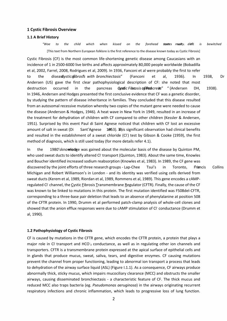
CF is caused by mutations in the CFTR gene, which encodes the CFTR protein, a protein that plays a
major role in Cl- transport and HCO-3 conductance, as well as in regulating other ion channels and
transporters. CFTR is a transmembrane protein expressed at the apical surface of epithelial cells and
in glands that produce mucus, sweat, saliva, tears, and digestive enzymes. CF causing mutations
prevent the channel from proper functioning, leading to abnormal ion transport a process that leads
to dehydration of the airway surface liquid (ASL) (Figure I.1.1). As a consequence, CF airways produce
abnormally thick, sticky mucus, which impairs mucociliary clearance (MCC) and obstructs the smaller
airways, causing disseminated bronchiectasis - a characteristic feature of CF. The thick mucus and
reduced MCC also traps bacteria (eg. Pseudomonas aeruginosa) in the airways originating recurrent
respiratory infections and chronic inflammation, which leads to progressive loss of lung function.
3
Besides these respiratory symptoms, classical forms of CF also include a high Cl- concentration in the
sweat (used for CF diagnosis), exocrine pancreatic insufficiency (PI) - present in 85% of subjects with
CF, hepatic cirrhosis, intestinal obstruction (includes distal intestinal obstruction syndrome at a later
age and meconium ileus that occurs in 10-17% of subjects within the first days of life) and male
infertility due to azoospermia attributed to congenital absence of vas deferens, although at variable
presentations (Welsh & Smith, 1995; Zielesnki & Tsui 1995; Rowe et al 2001; Bell et al 2013).
Figure – I.1.1 A cascade of pathophysiology in CF lung disease. The mechanism of CF dysfunction starts with
the primary CFTR gene defect and ultimate leads to severe lung deficiency. CFTR, cystic fibrosis
transmembrane conductance regulator; ASL, airway surface liquid; ENaC, epithelial Na+ channel. [Adapted
from Amaral MD, 2015]
2 The CFTR gene, Mutations, and Protein
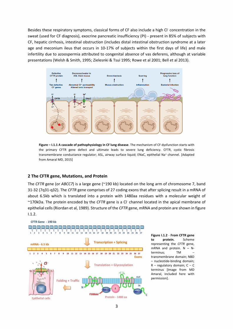
The CFTR gene (or ABCC7) is a large gene (~190 kb) located on the long arm of chromosome 7, band
31-32 (7q31-q32). The CFTR gene comprises of 27 coding exons that after splicing result in a mRNA of
about 6.5kb which is translated into a protein with 1480aa residues with a molecular weight of
~170kDa. The protein encoded by the CFTR gene is a Cl- channel located in the apical membrane of
epithelial cells (Riordan et al, 1989). Structure of the CFTR gene, mRNA and protein are shown in figure
I.1.2.
Figure I.1.2 - From CFTR gene to protein. Scheme representing the CFTR gene, mRNA and protein. N – N-terminus; TM – transmembrane domain; NBD – nucleotide-binding domain; R – regulatory domain; C – C terminus [Image from MD Amaral, included here with permission].
4
2.1 Functional Classes of CFTR mutations
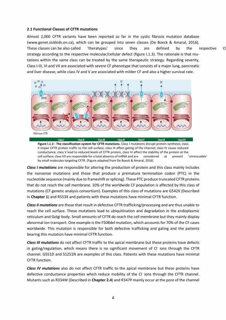
Almost 2,000 CFTR variants have been reported so far in the cystic fibrosis mutation database
(www.genet.sickkids.on.ca), which can be grouped into seven classes (De Boeck & Amaral, 2016).
These classes can be also called ‘theratypes’ since they are defined by the respective CFTR modulator
strategy according to the respective molecular/cellular defect (figure I.1.3). The rationale is that mu-
tations within the same class can be treated by the same therapeutic strategy. Regarding severity,
Class I-III, VI and VII are associated with severe CF phenotype that consists of a major lung, pancreatic
and liver disease, while class IV and V are associated with milder CF and also a higher survival rate.
Figure I.1.3 - The classification system for CFTR mutations. Class I mutations disrupt protein synthesis; class II impair CFTR protein traffic to the cell surface; class III affect gating of the channel; class IV cause reduced conductance; class V lead to reduced levels of CFTR protein, class VI affect the stability of the protein at the cell surface; class VII are responsible for a total absence of mRNA and are considered at present ‘unrescuable’ by small molecules targeting CFTR. [Figure adapted from De Boeck & Amaral, 2016]
Class I mutations are responsible for altering the production of protein and this class mainly includes
the nonsense mutations and those that produce a premature termination codon (PTC) in the
nucleotide sequence (mainly due to frameshift or splicing). These PTC produce truncated CFTR proteins
that do not reach the cell membrane. 10% of the worldwide CF population is affected by this class of
mutations (CF genetic analysis consortium). Examples of this class of mutations are G542X (Described
in Chapter 1) and R553X and patients with these mutations have minimal CFTR function.
Class II mutations are those that result in defective CFTR trafficking/processing and are thus unable to
reach the cell surface. These mutations lead to ubiquitination and degradation in the endoplasmic
reticulum and Golgi body. Small amounts of CFTR do reach the cell membrane but they mainly display
abnormal ion transport. One example is the F508del mutation, which accounts for 70% of the CF cases
worldwide. This mutation is responsible for both defective trafficking and gating and the patients
bearing this mutation have minimal CFTR function.
Class III mutations do not affect CFTR traffic to the apical membrane but these proteins have defects
in gating/regulation, which means there is no significant movement of Cl- ions through the CFTR
channel. G551D and S1251N are examples of this class. Patients with these mutations have minimal
CFTR function.
Class IV mutations also do not affect CFTR traffic to the apical membrane but these proteins have
defective conductance properties which reduce mobility of the Cl- ions through the CFTR channel.
Mutants such as R334W (Described in Chapter 2.4) and R347P mainly occur at the pore of the channel
5
and are located within the membrane spanning domains. Patients with these mutations have some
residual CFTR activity.
Class V mutations produce a remarkably reduced amount of CFTR protein however it preserves the
normal function at the apical plasma membrane. These mutations are caused by alternative splicing
defects that result in improper processing of mRNA. For example, 3849+10kbC>T (Described in Chapter
2.3) and 2789+5G>A are two class V mutations and patients with these mutations have residual CFTR
activity.
Class VI mutations create increased protein instability and shorter residence time at the cell surface,
mainly caused by C-terminal truncation. In this class of mutations biosynthesis, processing and
macroscopic Cl- channel function of truncated CFTRs are essentially normal, however the degradation
rate of the mature, complex-glycosylated form is 5- to 6-fold faster than the wild type CFTR (Haardt et
al., 1999). Rescued F508del and c120del23 (Described in Chapter 2.5) belong to this class.
Class VII mutations were proposed (Boeck & Amaral, 2016) and this class arose from the division of
the traditional class I mutations into a new class I (with stop-codon mutations) and a new class VII (with
no mRNA transcription). Mutations from this class are called unrescuable because they cannot be
rescued by pharmacological agents. N1303K (described in Chapter 1) and R560S (described in Chapter
2.1) mutations are two examples and these mutations remained unresponsive to available CFTR
pharmacological agents.
Although this classification is helpful to rationalize therapies, for a high number of CFTR mutations
(especially for rare ones) it is still unknown what are the associated defects and the respective
mutation classes. Variation in severity of disease within patients strongly influenced by factors such as
polymorphisms in the CFTR gene, modifier genes, environmental factors and nutritional status may
also worsen the severity of different CF mutations. This explains why it is difficult to predict the clinical
outcome of an individual patient based only on the CFTR genotype. The most common Class II mutation
- F508del-CFTR - alters the CFTR mRNA structure and reduces the translational efficiency. However
even knowing that F508del is a typical class II mutation, other detailed studies also show that it belongs
to at least two additional classes - (Lazrak, et al 2013). Moreover, it is often found that patients with
the same CFTR genotype (e.g., F508del/F508del) have significantly different clinical responses to CFTR
modulating drugs (Wainwright et al, 2015; Awatade et al, 2015).
2.2 CFTR protein – Biosynthesis, Trafficking, Degradation and Endoplasmic Reticulum Quality Con-trol
CFTR protein has to undergo a number of cellular processes and quality checks before it reaches the
apical membrane. Like other integral membrane glycoproteins, CFTR assembly begins with the for-
mation in the endoplasmic reticulum (ER) where it is core glycosylated (Cheng et al, 1990).
6
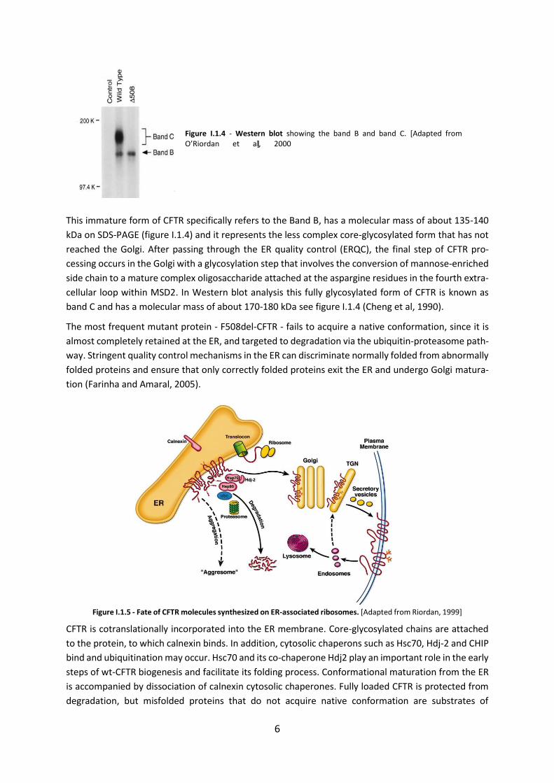
Figure I.1.4 - Western blot showing the band B and band C. [Adapted from O’Riordan et al, 2000]
This immature form of CFTR specifically refers to the Band B, has a molecular mass of about 135-140
kDa on SDS-PAGE (figure I.1.4) and it represents the less complex core-glycosylated form that has not
reached the Golgi. After passing through the ER quality control (ERQC), the final step of CFTR pro-
cessing occurs in the Golgi with a glycosylation step that involves the conversion of mannose-enriched
side chain to a mature complex oligosaccharide attached at the aspargine residues in the fourth extra-
cellular loop within MSD2. In Western blot analysis this fully glycosylated form of CFTR is known as
band C and has a molecular mass of about 170-180 kDa see figure I.1.4 (Cheng et al, 1990).
The most frequent mutant protein - F508del-CFTR - fails to acquire a native conformation, since it is
almost completely retained at the ER, and targeted to degradation via the ubiquitin-proteasome path-
way. Stringent quality control mechanisms in the ER can discriminate normally folded from abnormally
folded proteins and ensure that only correctly folded proteins exit the ER and undergo Golgi matura-
tion (Farinha and Amaral, 2005).
Figure I.1.5 - Fate of CFTR molecules synthesized on ER-associated ribosomes. [Adapted from Riordan, 1999]
CFTR is cotranslationally incorporated into the ER membrane. Core-glycosylated chains are attached
to the protein, to which calnexin binds. In addition, cytosolic chaperons such as Hsc70, Hdj-2 and CHIP
bind and ubiquitination may occur. Hsc70 and its co-chaperone Hdj2 play an important role in the early
steps of wt-CFTR biogenesis and facilitate its folding process. Conformational maturation from the ER
is accompanied by dissociation of calnexin cytosolic chaperones. Fully loaded CFTR is protected from
degradation, but misfolded proteins that do not acquire native conformation are substrates of
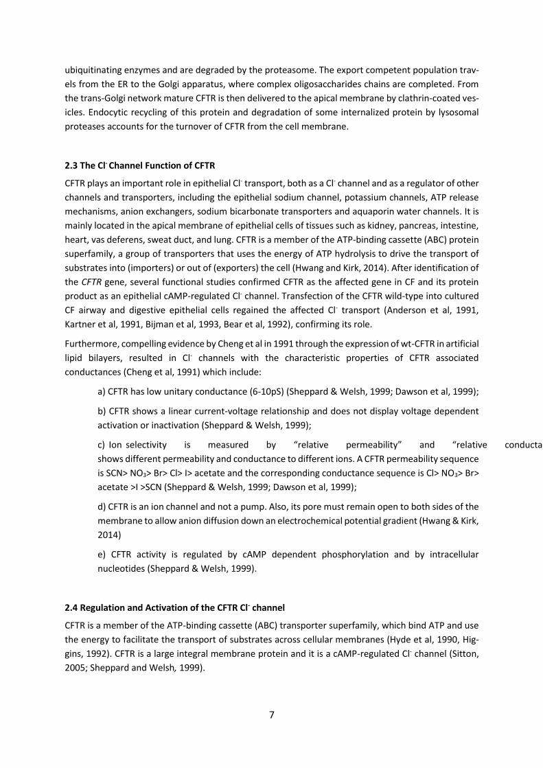
7
ubiquitinating enzymes and are degraded by the proteasome. The export competent population trav-
els from the ER to the Golgi apparatus, where complex oligosaccharides chains are completed. From
the trans-Golgi network mature CFTR is then delivered to the apical membrane by clathrin-coated ves-
icles. Endocytic recycling of this protein and degradation of some internalized protein by lysosomal
proteases accounts for the turnover of CFTR from the cell membrane.
2.3 The Cl- Channel Function of CFTR
CFTR plays an important role in epithelial Cl- transport, both as a Cl- channel and as a regulator of other
channels and transporters, including the epithelial sodium channel, potassium channels, ATP release
mechanisms, anion exchangers, sodium bicarbonate transporters and aquaporin water channels. It is
mainly located in the apical membrane of epithelial cells of tissues such as kidney, pancreas, intestine,
heart, vas deferens, sweat duct, and lung. CFTR is a member of the ATP-binding cassette (ABC) protein
superfamily, a group of transporters that uses the energy of ATP hydrolysis to drive the transport of
substrates into (importers) or out of (exporters) the cell (Hwang and Kirk, 2014). After identification of
the CFTR gene, several functional studies confirmed CFTR as the affected gene in CF and its protein
product as an epithelial cAMP-regulated Cl- channel. Transfection of the CFTR wild-type into cultured
CF airway and digestive epithelial cells regained the affected Cl- transport (Anderson et al, 1991,
Kartner et al, 1991, Bijman et al, 1993, Bear et al, 1992), confirming its role.
Furthermore, compelling evidence by Cheng et al in 1991 through the expression of wt-CFTR in artificial
lipid bilayers, resulted in Cl- channels with the characteristic properties of CFTR associated
conductances (Cheng et al, 1991) which include:
a) CFTR has low unitary conductance (6-10pS) (Sheppard & Welsh, 1999; Dawson et al, 1999);
b) CFTR shows a linear current-voltage relationship and does not display voltage dependent
activation or inactivation (Sheppard & Welsh, 1999);
c) Ion selectivity is measured by “relative permeability” and “relative conductance”. CFTR
shows different permeability and conductance to different ions. A CFTR permeability sequence
is SCN> NO3> Br> Cl> I> acetate and the corresponding conductance sequence is Cl> NO3> Br>
acetate >I >SCN (Sheppard & Welsh, 1999; Dawson et al, 1999);
d) CFTR is an ion channel and not a pump. Also, its pore must remain open to both sides of the
membrane to allow anion diffusion down an electrochemical potential gradient (Hwang & Kirk,
2014)
e) CFTR activity is regulated by cAMP dependent phosphorylation and by intracellular
nucleotides (Sheppard & Welsh, 1999).
2.4 Regulation and Activation of the CFTR Cl- channel
CFTR is a member of the ATP-binding cassette (ABC) transporter superfamily, which bind ATP and use
the energy to facilitate the transport of substrates across cellular membranes (Hyde et al, 1990, Hig-
gins, 1992). CFTR is a large integral membrane protein and it is a cAMP-regulated Cl- channel (Sitton,
2005; Sheppard and Welsh, 1999).

8
CFTR protein structure is composed of five functional domains: Two hydrophobic membrane spanning
domains (MSD1 and MSD2) each consisting of six transmembrane (TM) helixes (figure I.1.6) form the
anion-conducting pore (Sheppard and Welsh, 1999). Two hydrophobic membrane associated domains
form the Nucleotide binding domains (NBD 1 and NBD 2) and Regulatory (R) domain.
Figure I.1.6 - Model of the proposed structural do-
mains of CFTR. MSD: membrane spanning domains,
NBD: nucleotide binding domains, R: regulatory do-
main, PKA: cAMP dependent protein kinase A. [Adapted
from Sheppard and Welsh, 1999]
R domain is a unique feature of CFTR among all ABC transporters and contains many charged residues
and multiple consensus phosphorylation sites. The regulatory R domain contains Protein Kinase A
(PKA) and Protein Kinase C (PKC) phosphorylation sites and phosphorylation by PKA results in nucleo-
tide binding and hydrolysis at the NBD 1 and NBD 2, which in turn causes opening of the channel al-
lowing Cl- to move out of the cell (McCarty et al, 2000). Both the amino (N) and carboxyl (C) terminal
tails of CFTR channel are located in the cytoplasm and play a pivotal role in mediating interactions with
a variety of binding proteins. These physical interactions are tightly regulated and are important in the
compartmentalized regulation of CFTR function. Apart from PKA, there are other kinases reported to
be involved in CFTR mediated Cl- secretion such as PKC, src kinase, AMP-dependent protein kinase,
Casein kinase 2 (Seibert et al, 1999; Treharne et al, 2009; Kongsuphol et al, 2009), SYK-spleen tyrosine
kinase (Luz et al, 2011) and Serine/threonine kinase WNK4 kinase (Mendes et al, 2011). Apart from
cAMP regulation of CFTR Cl- channel, both the β2-adrenergic and the Adenosine 2b receptor have an
important role in regulating CFTR mediated Cl- secretion in human airways. These two different signal-
ling pathways mainly act through G-protein coupled receptors that release Gs, stimulate AC and raise
cAMP which in turns activates CFTR (Hentchel-Franks et al, 2004, Stanton and Guggino, 2006).
2.5 Recent Advances in CFTR structure
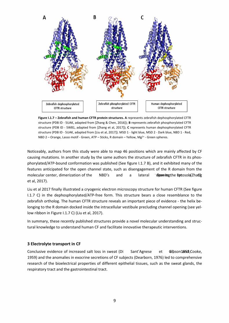
Recently Zhang & Chen, 2016 reported the structure of the zebrafish CFTR protein in the dephosphor-
ylated state (See figure I.1.7 A) and in the absence of ATP, by cryogenic electron microscopy. Zebrafish
CFTR shares about 55% overall sequence identity to human CFTR. The interesting feature of this model
was a novel interfacial motif, which was called “lasso motif” because of its shape (shown as a green
ribbon in figure I.1.7 A). The lasso motif is located in the vicinity of the R domain (Zhang & Chen, 2016).
The main structural features include funnel shaped ion conduction pathway which consists of a large
vestibule, positively charged residues in the entire length of the funnel and closure of the channel in
the extracellular surface by a single gate.
9
Figure I.1.7 – Zebrafish and human CFTR protein structures. A represents zebrafish dephosphorylated CFTR
structure (PDB ID - 5UAK, adapted from [Zhang & Chen, 2016]); B represents zebrafish phosphorylated CFTR
structure (PDB ID - 5W81, adapted from [Zhang et al, 2017]); C represents human dephosphorylated CFTR
structure (PDB ID - 5UAK, adapted from [Liu et al, 2017]). MSD 1 - light blue, MSD 2 - Dark blue, NBD 1 - Red,
NBD 2 – Orange, Lasso motif - Green, ATP – Sticks, R domain – Yellow, Mg2+ - Green spheres.
Noticeably, authors from this study were able to map 46 positions which are mainly affected by CF
causing mutations. In another study by the same authors the structure of zebrafish CFTR in its phos-
phorylated/ATP-bound conformation was published (See figure I.1.7 B), and it exhibited many of the
features anticipated for the open channel state, such as disengagement of the R domain from the
molecular center, dimerization of the NBD’s and a lateral opening for a Cl- flow to the cytosol (Zhang
et al, 2017).
Liu et al 2017 finally illustrated a cryogenic electron microscopy structure for human CFTR (See figure
I.1.7 C) in the dephosphorylated/ATP-free form. This structure bears a close resemblance to the
zebrafish ortholog. The human CFTR structure reveals an important piece of evidence - the helix be-
longing to the R domain docked inside the intracellular vestibule precluding channel opening (see yel-
low ribbon in Figure I.1.7 C) (Liu et al, 2017).
In summary, these recently published structures provide a novel molecular understanding and struc-
tural knowledge to understand human CF and facilitate innovative therapeutic interventions.
3 Electrolyte transport in CF
Conclusive evidence of increased salt loss in sweat (Di Sant’Agnese et al, 1953; Gibson and Cooke,
1959) and the anomalies in exocrine secretions of CF subjects (Dearborn, 1976) led to comprehensive
research of the bioelectrical properties of different epithelial tissues, such as the sweat glands, the
respiratory tract and the gastrointestinal tract.
10
3.1 Sweat glands
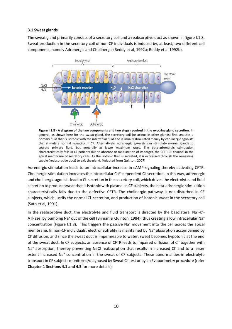
The sweat gland primarily consists of a secretory coil and a reabsorptive duct as shown in figure I.1.8.
Sweat production in the secretory coil of non-CF individuals is induced by, at least, two different cell
components, namely Adrenergic and Cholinergic (Reddy et al, 1992a; Reddy et al 1992b).
Figure I.1.8 - A diagram of the two components and two steps required in the exocrine gland secretion. In general, as shown here for the sweat gland, the secretory coil (or acinus in other glands) first secretes a primary fluid that is isotonic with the interstitial fluid and is usually stimulated mainly by cholinergic agonists that stimulate normal sweating in CF. Alternatively, adrenergic agonists can stimulate normal glands to secrete primary fluid, but generally at lower maximum rates. The beta-adrenergic stimulation characteristically fails in CF patients due to absence or malfunction of its target, the CFTR Cl- channel in the apical membrane of secretory cells. As the isotonic fluid is secreted, it is expressed through the remaining tubule (reabsorptive duct) to exit the gland. [Adapted from Quinton, 2007]
Adrenergic stimulation leads to an intracellular increase in cAMP signaling thereby activating CFTR.
Cholinergic stimulation increases the intracellular Ca2+ dependent Cl- secretion. In this way, adrenergic
and cholinergic agonists lead to Cl- secretion in the secretory coil, which drives the electrolyte and fluid
secretion to produce sweat that is isotonic with plasma. In CF subjects, the beta-adrenergic stimulation
characteristically fails due to the defective CFTR. The cholinergic pathway is not disturbed in CF
subjects, which justify the normal Cl- secretion, and production of isotonic sweat in the secretory coil
(Sato et al, 1991).
In the reabsorptive duct, the electrolyte and fluid transport is directed by the basolateral Na+-K+-
ATPase, by pumping Na+ out of the cell (Bijman & Quinton, 1984), thus creating a low intracellular Na+
concentration (Figure I.1.8). This triggers the passive Na+ movement into the cell across the apical
membrane. In non-CF individuals, electroneutrality is maintained by Na+ absorption accompanied by
Cl- diffusion, and since the sweat duct is impermeable to water, sweat becomes hypotonic at the end
of the sweat duct. In CF subjects, an absence of CFTR leads to impaired diffusion of Cl- together with
Na+ absorption, thereby preventing NaCl reabsorption that results in increased Cl- and to a lesser
extent increased Na+ concentration in the sweat of CF subjects. These abnormalities in electrolyte
transport in CF subjects monitored/diagnosed by Sweat Cl- test or by an Evaporimetry procedure (refer
Chapter 1 Sections 4.1 and 4.3 for more details).
11
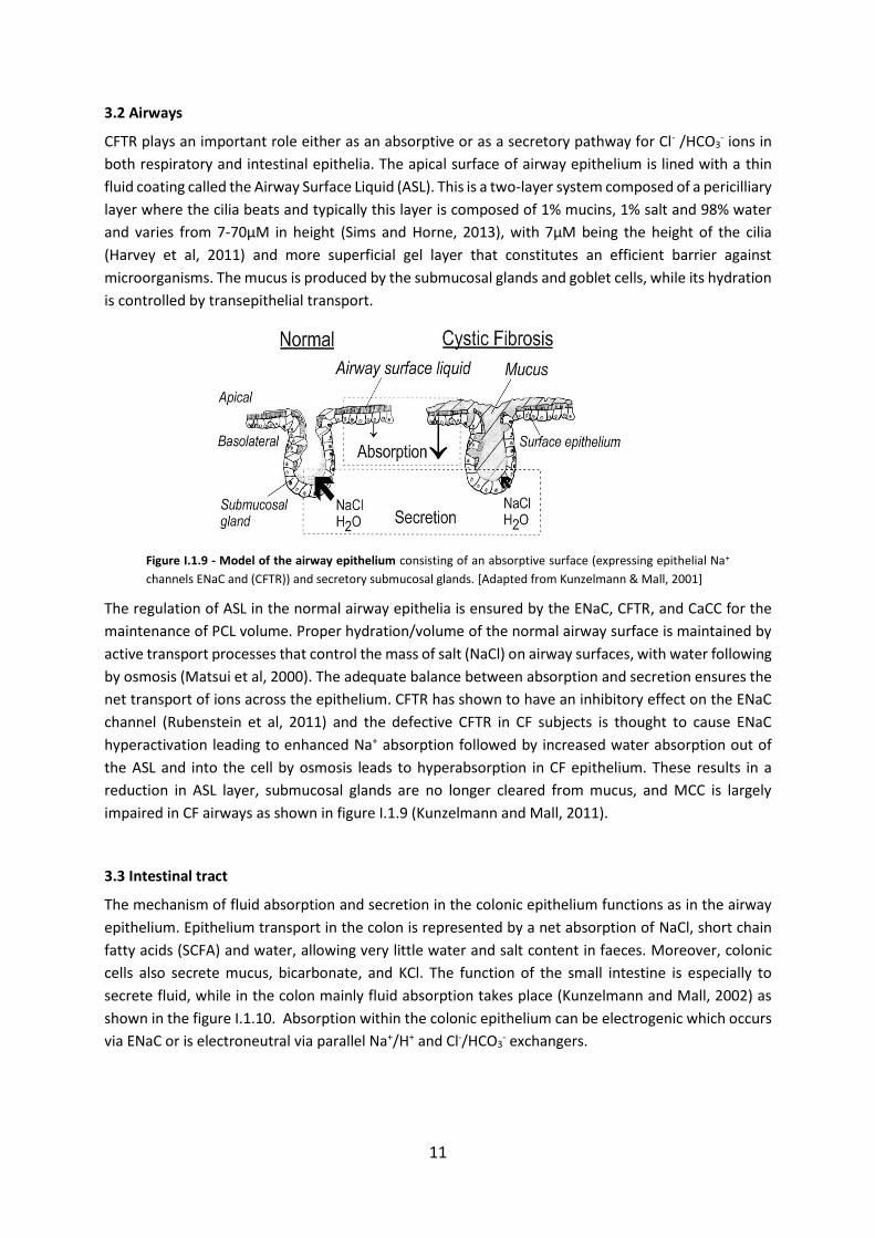
3.2 Airways
CFTR plays an important role either as an absorptive or as a secretory pathway for Cl- /HCO3- ions in
both respiratory and intestinal epithelia. The apical surface of airway epithelium is lined with a thin
fluid coating called the Airway Surface Liquid (ASL). This is a two-layer system composed of a pericilliary
layer where the cilia beats and typically this layer is composed of 1% mucins, 1% salt and 98% water
and varies from 7-70µM in height (Sims and Horne, 2013), with 7µM being the height of the cilia
(Harvey et al, 2011) and more superficial gel layer that constitutes an efficient barrier against
microorganisms. The mucus is produced by the submucosal glands and goblet cells, while its hydration
is controlled by transepithelial transport.
Figure I.1.9 - Model of the airway epithelium consisting of an absorptive surface (expressing epithelial Na+
channels ENaC and (CFTR)) and secretory submucosal glands. [Adapted from Kunzelmann & Mall, 2001]
The regulation of ASL in the normal airway epithelia is ensured by the ENaC, CFTR, and CaCC for the
maintenance of PCL volume. Proper hydration/volume of the normal airway surface is maintained by
active transport processes that control the mass of salt (NaCl) on airway surfaces, with water following
by osmosis (Matsui et al, 2000). The adequate balance between absorption and secretion ensures the
net transport of ions across the epithelium. CFTR has shown to have an inhibitory effect on the ENaC
channel (Rubenstein et al, 2011) and the defective CFTR in CF subjects is thought to cause ENaC
hyperactivation leading to enhanced Na+ absorption followed by increased water absorption out of
the ASL and into the cell by osmosis leads to hyperabsorption in CF epithelium. These results in a
reduction in ASL layer, submucosal glands are no longer cleared from mucus, and MCC is largely
impaired in CF airways as shown in figure I.1.9 (Kunzelmann and Mall, 2011).
3.3 Intestinal tract
The mechanism of fluid absorption and secretion in the colonic epithelium functions as in the airway
epithelium. Epithelium transport in the colon is represented by a net absorption of NaCl, short chain
fatty acids (SCFA) and water, allowing very little water and salt content in faeces. Moreover, colonic
cells also secrete mucus, bicarbonate, and KCl. The function of the small intestine is especially to
secrete fluid, while in the colon mainly fluid absorption takes place (Kunzelmann and Mall, 2002) as
shown in the figure I.1.10. Absorption within the colonic epithelium can be electrogenic which occurs
via ENaC or is electroneutral via parallel Na+/H+ and Cl-/HCO3- exchangers.
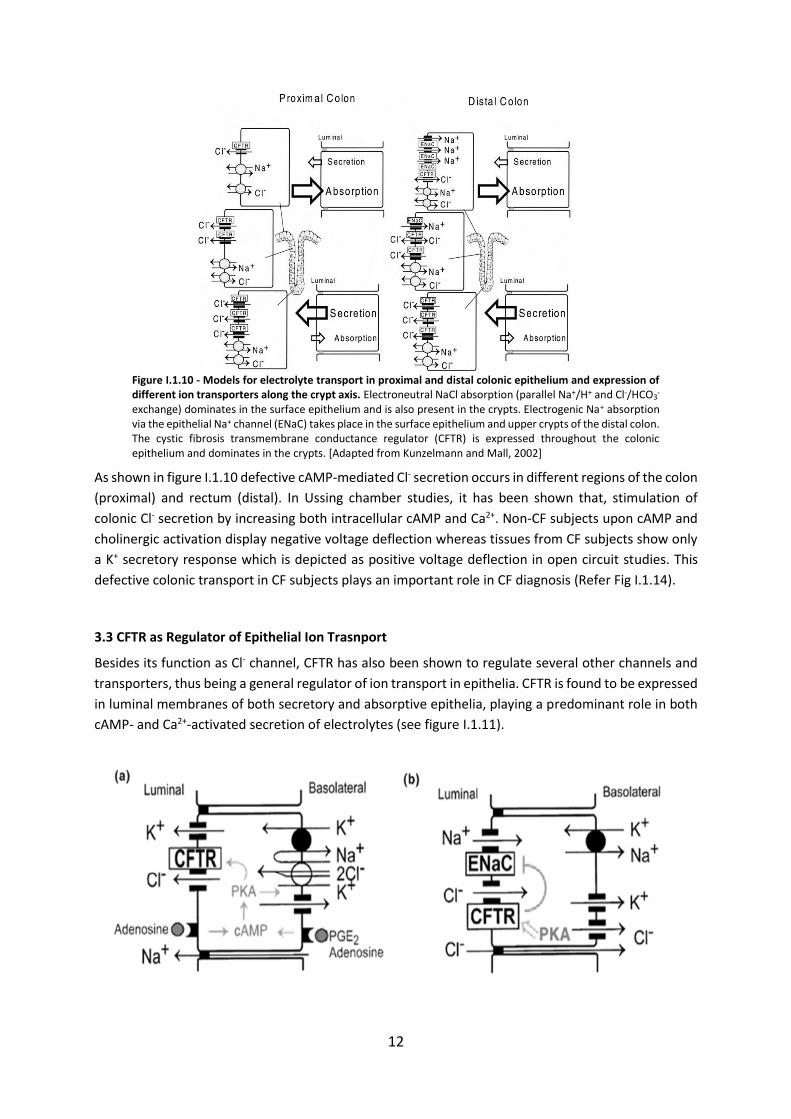
12
Figure I.1.10 - Models for electrolyte transport in proximal and distal colonic epithelium and expression of different ion transporters along the crypt axis. Electroneutral NaCl absorption (parallel Na+/H+ and Cl-/HCO3
- exchange) dominates in the surface epithelium and is also present in the crypts. Electrogenic Na+ absorption via the epithelial Na+ channel (ENaC) takes place in the surface epithelium and upper crypts of the distal colon. The cystic fibrosis transmembrane conductance regulator (CFTR) is expressed throughout the colonic epithelium and dominates in the crypts. [Adapted from Kunzelmann and Mall, 2002]
As shown in figure I.1.10 defective cAMP-mediated Cl- secretion occurs in different regions of the colon
(proximal) and rectum (distal). In Ussing chamber studies, it has been shown that, stimulation of
colonic Cl- secretion by increasing both intracellular cAMP and Ca2+. Non-CF subjects upon cAMP and
cholinergic activation display negative voltage deflection whereas tissues from CF subjects show only
a K+ secretory response which is depicted as positive voltage deflection in open circuit studies. This
defective colonic transport in CF subjects plays an important role in CF diagnosis (Refer Fig I.1.14).
3.3 CFTR as Regulator of Epithelial Ion Trasnport
Besides its function as Cl- channel, CFTR has also been shown to regulate several other channels and
transporters, thus being a general regulator of ion transport in epithelia. CFTR is found to be expressed
in luminal membranes of both secretory and absorptive epithelia, playing a predominant role in both
cAMP- and Ca2+-activated secretion of electrolytes (see figure I.1.11).
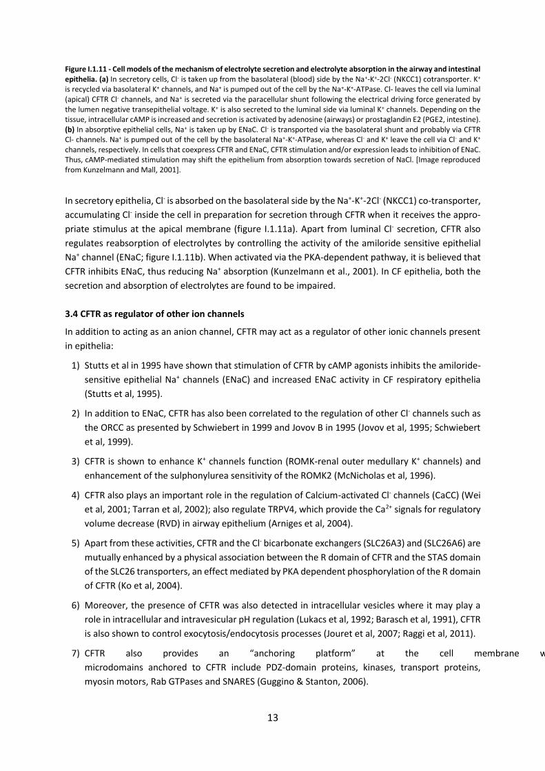
13
Figure I.1.11 - Cell models of the mechanism of electrolyte secretion and electrolyte absorption in the airway and intestinal epithelia. (a) In secretory cells, Cl- is taken up from the basolateral (blood) side by the Na+-K+-2Cl- (NKCC1) cotransporter. K+ is recycled via basolateral K+ channels, and Na+ is pumped out of the cell by the Na+-K+-ATPase. Cl- leaves the cell via luminal (apical) CFTR Cl- channels, and Na+ is secreted via the paracellular shunt following the electrical driving force generated by the lumen negative transepithelial voltage. K+ is also secreted to the luminal side via luminal K+ channels. Depending on the tissue, intracellular cAMP is increased and secretion is activated by adenosine (airways) or prostaglandin E2 (PGE2, intestine). (b) In absorptive epithelial cells, Na+ is taken up by ENaC. Cl- is transported via the basolateral shunt and probably via CFTR Cl- channels. Na+ is pumped out of the cell by the basolateral Na+-K+-ATPase, whereas Cl- and K+ leave the cell via Cl- and K+ channels, respectively. In cells that coexpress CFTR and ENaC, CFTR stimulation and/or expression leads to inhibition of ENaC. Thus, cAMP-mediated stimulation may shift the epithelium from absorption towards secretion of NaCl. [Image reproduced from Kunzelmann and Mall, 2001].
In secretory epithelia, Cl- is absorbed on the basolateral side by the Na+-K+-2Cl- (NKCC1) co-transporter,
accumulating Cl- inside the cell in preparation for secretion through CFTR when it receives the appro-
priate stimulus at the apical membrane (figure I.1.11a). Apart from luminal Cl- secretion, CFTR also
regulates reabsorption of electrolytes by controlling the activity of the amiloride sensitive epithelial
Na+ channel (ENaC; figure I.1.11b). When activated via the PKA-dependent pathway, it is believed that
CFTR inhibits ENaC, thus reducing Na+ absorption (Kunzelmann et al., 2001). In CF epithelia, both the
secretion and absorption of electrolytes are found to be impaired.
3.4 CFTR as regulator of other ion channels
In addition to acting as an anion channel, CFTR may act as a regulator of other ionic channels present
in epithelia:
1) Stutts et al in 1995 have shown that stimulation of CFTR by cAMP agonists inhibits the amiloride-
sensitive epithelial Na+ channels (ENaC) and increased ENaC activity in CF respiratory epithelia
(Stutts et al, 1995).
2) In addition to ENaC, CFTR has also been correlated to the regulation of other Cl- channels such as
the ORCC as presented by Schwiebert in 1999 and Jovov B in 1995 (Jovov et al, 1995; Schwiebert
et al, 1999).
3) CFTR is shown to enhance K+ channels function (ROMK-renal outer medullary K+ channels) and
enhancement of the sulphonylurea sensitivity of the ROMK2 (McNicholas et al, 1996).
4) CFTR also plays an important role in the regulation of Calcium-activated Cl- channels (CaCC) (Wei
et al, 2001; Tarran et al, 2002); also regulate TRPV4, which provide the Ca2+ signals for regulatory
volume decrease (RVD) in airway epithelium (Arniges et al, 2004).
5) Apart from these activities, CFTR and the Cl- bicarbonate exchangers (SLC26A3) and (SLC26A6) are
mutually enhanced by a physical association between the R domain of CFTR and the STAS domain
of the SLC26 transporters, an effect mediated by PKA dependent phosphorylation of the R domain
of CFTR (Ko et al, 2004).
6) Moreover, the presence of CFTR was also detected in intracellular vesicles where it may play a
role in intracellular and intravesicular pH regulation (Lukacs et al, 1992; Barasch et al, 1991), CFTR
is also shown to control exocytosis/endocytosis processes (Jouret et al, 2007; Raggi et al, 2011).
7) CFTR also provides an “anchoring platform” at the cell membrane where specialized
microdomains anchored to CFTR include PDZ-domain proteins, kinases, transport proteins,
myosin motors, Rab GTPases and SNARES (Guggino & Stanton, 2006).
14
4 CFTR Function Measurements to Establish a Diagnosis of CF
Traditionally, the diagnosis of CF is based on clinical symptoms suggestive of the disease and/or a
positive family history. Such symptoms include mostly those affecting the airways and the
gastrointestinal tract, but also those affecting other systems (see above).
Nevertheless, to confirm a diagnosis of CF, it is necessary to obtain evidence of CFTR dysfunction
through the identification of two CFTR gene mutations previously assigned as CF disease causing, two
tests showing a high Cl- concentration in sweat (>60 mEq/L), distinctive transepithelial nasal potential
difference measurements and/or assessment of CFTR (dys) function in native colonic epithelia ex vivo
(Ratjen & Doring 2003; Rosenstein & Cutting 1998; Farrell et al 2008). For individuals with symptoms
suggestive of CF but intermediate sweat Cl- values (30 - 60 mEq/L), the need for additional proof of
CFTR function (through NPD measurements or CFTR functional assessment in rectal biopsies) is
particularly important.
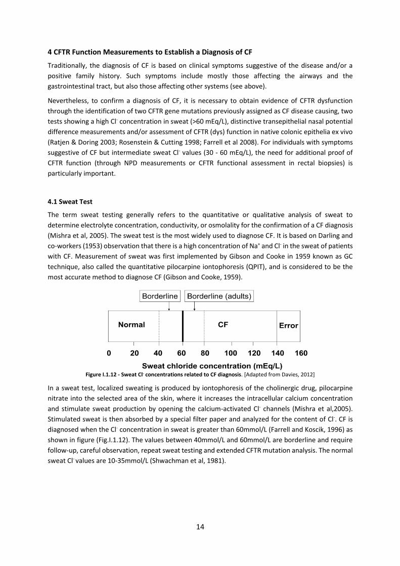
4.1 Sweat Test
The term sweat testing generally refers to the quantitative or qualitative analysis of sweat to
determine electrolyte concentration, conductivity, or osmolality for the confirmation of a CF diagnosis
(Mishra et al, 2005). The sweat test is the most widely used to diagnose CF. It is based on Darling and
co-workers (1953) observation that there is a high concentration of Na+ and Cl- in the sweat of patients
with CF. Measurement of sweat was first implemented by Gibson and Cooke in 1959 known as GC
technique, also called the quantitative pilocarpine iontophoresis (QPIT), and is considered to be the
most accurate method to diagnose CF (Gibson and Cooke, 1959).
Figure I.1.12 - Sweat Cl- concentrations related to CF diagnosis. [Adapted from Davies, 2012]
In a sweat test, localized sweating is produced by iontophoresis of the cholinergic drug, pilocarpine
nitrate into the selected area of the skin, where it increases the intracellular calcium concentration
and stimulate sweat production by opening the calcium-activated Cl- channels (Mishra et al,2005).
Stimulated sweat is then absorbed by a special filter paper and analyzed for the content of Cl-. CF is
diagnosed when the Cl- concentration in sweat is greater than 60mmol/L (Farrell and Koscik, 1996) as
shown in figure (Fig.I.1.12). The values between 40mmol/L and 60mmol/L are borderline and require
follow-up, careful observation, repeat sweat testing and extended CFTR mutation analysis. The normal
sweat Cl- values are 10-35mmol/L (Shwachman et al, 1981).
15
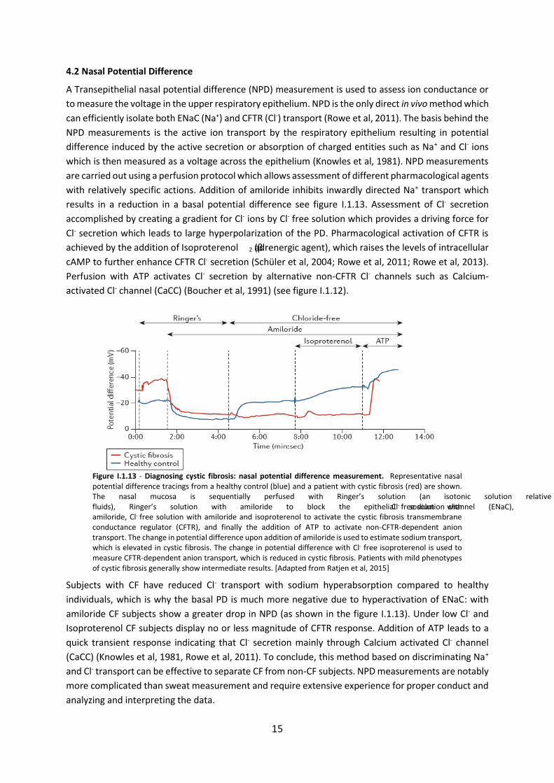
4.2 Nasal Potential Difference
A Transepithelial nasal potential difference (NPD) measurement is used to assess ion conductance or
to measure the voltage in the upper respiratory epithelium. NPD is the only direct in vivo method which
can efficiently isolate both ENaC (Na+) and CFTR (Cl-) transport (Rowe et al, 2011). The basis behind the
NPD measurements is the active ion transport by the respiratory epithelium resulting in potential
difference induced by the active secretion or absorption of charged entities such as Na+ and Cl- ions
which is then measured as a voltage across the epithelium (Knowles et al, 1981). NPD measurements
are carried out using a perfusion protocol which allows assessment of different pharmacological agents
with relatively specific actions. Addition of amiloride inhibits inwardly directed Na+ transport which
results in a reduction in a basal potential difference see figure I.1.13. Assessment of Cl- secretion
accomplished by creating a gradient for Cl- ions by Cl- free solution which provides a driving force for
Cl- secretion which leads to large hyperpolarization of the PD. Pharmacological activation of CFTR is
achieved by the addition of Isoproterenol (β2 adrenergic agent), which raises the levels of intracellular
cAMP to further enhance CFTR Cl- secretion (Schüler et al, 2004; Rowe et al, 2011; Rowe et al, 2013).
Perfusion with ATP activates Cl- secretion by alternative non-CFTR Cl- channels such as Calcium-
activated Cl- channel (CaCC) (Boucher et al, 1991) (see figure I.1.12).
Figure I.1.13 - Diagnosing cystic fibrosis: nasal potential difference measurement. Representative nasal potential difference tracings from a healthy control (blue) and a patient with cystic fibrosis (red) are shown. The nasal mucosa is sequentially perfused with Ringer’s solution (an isotonic solution relative to the bodily fluids), Ringer’s solution with amiloride to block the epithelial sodium channel (ENaC), Cl- free solution with amiloride, Cl- free solution with amiloride and isoproterenol to activate the cystic fibrosis transmembrane conductance regulator (CFTR), and finally the addition of ATP to activate non-CFTR-dependent anion transport. The change in potential difference upon addition of amiloride is used to estimate sodium transport, which is elevated in cystic fibrosis. The change in potential difference with Cl- free isoproterenol is used to measure CFTR-dependent anion transport, which is reduced in cystic fibrosis. Patients with mild phenotypes of cystic fibrosis generally show intermediate results. [Adapted from Ratjen et al, 2015]
Subjects with CF have reduced Cl- transport with sodium hyperabsorption compared to healthy
individuals, which is why the basal PD is much more negative due to hyperactivation of ENaC: with
amiloride CF subjects show a greater drop in NPD (as shown in the figure I.1.13). Under low Cl- and
Isoproterenol CF subjects display no or less magnitude of CFTR response. Addition of ATP leads to a
quick transient response indicating that Cl- secretion mainly through Calcium activated Cl- channel
(CaCC) (Knowles et al, 1981, Rowe et al, 2011). To conclude, this method based on discriminating Na+
and Cl- transport can be effective to separate CF from non-CF subjects. NPD measurements are notably
more complicated than sweat measurement and require extensive experience for proper conduct and
analyzing and interpreting the data.
16
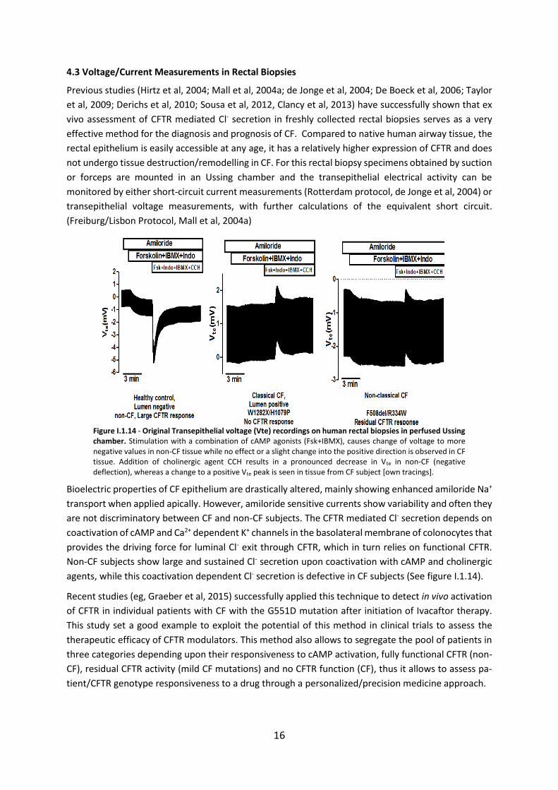
4.3 Voltage/Current Measurements in Rectal Biopsies
Previous studies (Hirtz et al, 2004; Mall et al, 2004a; de Jonge et al, 2004; De Boeck et al, 2006; Taylor
et al, 2009; Derichs et al, 2010; Sousa et al, 2012, Clancy et al, 2013) have successfully shown that ex
vivo assessment of CFTR mediated Cl- secretion in freshly collected rectal biopsies serves as a very
effective method for the diagnosis and prognosis of CF. Compared to native human airway tissue, the
rectal epithelium is easily accessible at any age, it has a relatively higher expression of CFTR and does
not undergo tissue destruction/remodelling in CF. For this rectal biopsy specimens obtained by suction
or forceps are mounted in an Ussing chamber and the transepithelial electrical activity can be
monitored by either short-circuit current measurements (Rotterdam protocol, de Jonge et al, 2004) or
transepithelial voltage measurements, with further calculations of the equivalent short circuit.
(Freiburg/Lisbon Protocol, Mall et al, 2004a)
Figure I.1.14 - Original Transepithelial voltage (Vte) recordings on human rectal biopsies in perfused Ussing chamber. Stimulation with a combination of cAMP agonists (Fsk+IBMX), causes change of voltage to more negative values in non-CF tissue while no effect or a slight change into the positive direction is observed in CF tissue. Addition of cholinergic agent CCH results in a pronounced decrease in Vte in non-CF (negative deflection), whereas a change to a positive Vte peak is seen in tissue from CF subject [own tracings].
Bioelectric properties of CF epithelium are drastically altered, mainly showing enhanced amiloride Na+
transport when applied apically. However, amiloride sensitive currents show variability and often they
are not discriminatory between CF and non-CF subjects. The CFTR mediated Cl- secretion depends on
coactivation of cAMP and Ca2+ dependent K+ channels in the basolateral membrane of colonocytes that
provides the driving force for luminal Cl- exit through CFTR, which in turn relies on functional CFTR.
Non-CF subjects show large and sustained Cl- secretion upon coactivation with cAMP and cholinergic
agents, while this coactivation dependent Cl- secretion is defective in CF subjects (See figure I.1.14).
Recent studies (eg, Graeber et al, 2015) successfully applied this technique to detect in vivo activation
of CFTR in individual patients with CF with the G551D mutation after initiation of Ivacaftor therapy.
This study set a good example to exploit the potential of this method in clinical trials to assess the
therapeutic efficacy of CFTR modulators. This method also allows to segregate the pool of patients in
three categories depending upon their responsiveness to cAMP activation, fully functional CFTR (non-
CF), residual CFTR activity (mild CF mutations) and no CFTR function (CF), thus it allows to assess pa-
tient/CFTR genotype responsiveness to a drug through a personalized/precision medicine approach.
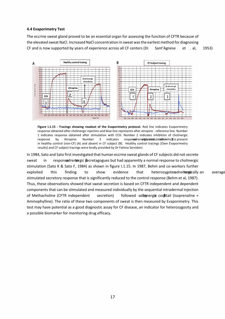
17
4.4 Evaporimetry Test
The eccrine sweat gland proved to be an essential organ for assessing the function of CFTR because of
the elevated sweat NaCl. Increased NaCl concentration in sweat was the earliest method for diagnosing
CF and is now supported by years of experience across all CF centers (Di Sant’Agnese et al, 1953).
Figure I.1.15 - Tracings showing readout of the Evaporimetry protocol. Red line indicates Evaporimetry response obtained after cholinergic injection and blue line represents after atropine - reference line. Number 1 indicates response obtained after stimulation with CCH. Number 2 indicates inhibition of cholinergic response by Atropine. Number 3 indicates response obtained after β-adrenergic stimulation which is present in healthy control (non-CF) (A) and absent in CF subject (B). Healthy control tracings [Own Evaporimetry results] and CF subject tracings were kindly provided by Dr Fatima Servidoni.
In 1984, Sato and Sato first investigated that human eccrine sweat glands of CF subjects did not secrete
sweat in response to β-adrenergic secretagogues but had apparently a normal response to cholinergic
stimulation (Sato K & Sato F, 1984) as shown in figure I.1.15. In 1987, Behm and co-workers further
exploited this finding to show evidence that heterozygotes have an average β-adrenergically
stimulated secretory response that is significantly reduced to the control response (Behm et al, 1987).
Thus, these observations showed that sweat secretion is based on CFTR independent and dependent
components that can be stimulated and measured individually by the sequential intradermal injection
of Methacholine (CFTR independent secretion) followed by a β-adrenergic cocktail (Isoprenaline +
Aminophylline). The ratio of these two components of sweat is then measured by Evaporimetry. This
test may have potential as a good diagnostic assay for CF disease, an indicator for heterozygosity and
a possible biomarker for monitoring drug efficacy.
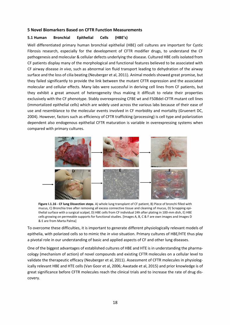
18
5 Novel Biomarkers Based on CFTR Function Measurements
5.1 Human Bronchial Epithelial Cells (HBE’s)
Well differentiated primary human bronchial epithelial (HBE) cell cultures are important for Cystic
Fibrosis research, especially for the development of CFTR modifier drugs, to understand the CF
pathogenesis and molecular & cellular defects underlying the disease. Cultured HBE cells isolated from
CF patients display many of the morphological and functional features believed to be associated with
CF airway disease in vivo, such as abnormal ion fluid transport leading to dehydration of the airway
surface and the loss of cilia beating (Neuberger et al, 2011). Animal models showed great promise, but
they failed significantly to provide the link between the mutant CFTR expression and the associated
molecular and cellular effects. Many labs were successful in deriving cell lines from CF patients, but
they exhibit a great amount of heterogeneity thus making it difficult to relate their properties
exclusively with the CF phenotype. Stably overexpressing CFBE wt and F508del-CFTR mutant cell lines
(immortalized epithelial cells) which are widely used across the various labs because of their ease of
use and resemblance to the molecular events involved in CF morbidity and mortality (Gruenert DC,
2004). However, factors such as efficiency of CFTR trafficking (processing) is cell type and polarization
dependent also endogenous epithelial CFTR maturation is variable in overexpressing systems when
compared with primary cultures.
Figure I.1.16 - CF lung Dissection steps. A) whole lung transplant of CF patient, B) Piece of bronchi filled with mucus, C) Bronchia tree after removing all excess connective tissue and cleaning of mucus, D) Scrapping epi-thelial surface with a surgical scalpel, D) HBE cells from CF individual 24h after plating in 100-mm dish, E) HBE cells growing on permeable supports for functional studies. [Images A, B, C & F are own images and Images D & E are from Marta Palma]
To overcome these difficulties, it is important to generate different physiologically relevant models of
epithelia, with polarized cells so to mimic the in vivo situation. Primary cultures of HBE/HTE thus play
a pivotal role in our understanding of basic and applied aspects of CF and other lung diseases.
One of the biggest advantages of established cultures of HBE and HTE is in understanding the pharma-
cology (mechanism of action) of novel compounds and existing CFTR molecules on a cellular level to
validate the therapeutic efficacy (Neuberger et al, 2011). Assessment of CFTR molecules in physiolog-
ically relevant HBE and HTE cells (Van Goor et al, 2006; Awatade et al, 2015) and prior knowledge is of
great significance before CFTR molecules reach the clinical trials and to increase the rate of drug dis-
covery.
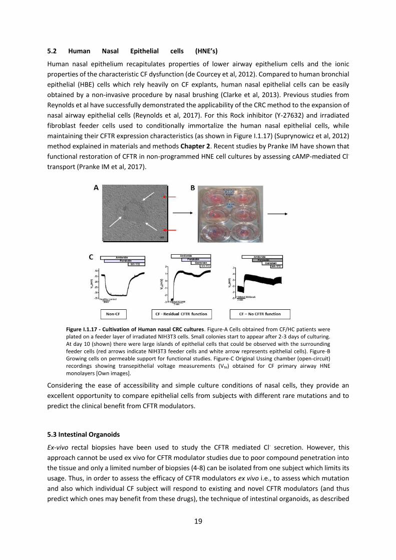
19
5.2 Human Nasal Epithelial cells (HNE’s)
Human nasal epithelium recapitulates properties of lower airway epithelium cells and the ionic
properties of the characteristic CF dysfunction (de Courcey et al, 2012). Compared to human bronchial
epithelial (HBE) cells which rely heavily on CF explants, human nasal epithelial cells can be easily
obtained by a non-invasive procedure by nasal brushing (Clarke et al, 2013). Previous studies from
Reynolds et al have successfully demonstrated the applicability of the CRC method to the expansion of
nasal airway epithelial cells (Reynolds et al, 2017). For this Rock inhibitor (Y-27632) and irradiated
fibroblast feeder cells used to conditionally immortalize the human nasal epithelial cells, while
maintaining their CFTR expression characteristics (as shown in Figure I.1.17) (Suprynowicz et al, 2012)
method explained in materials and methods Chapter 2. Recent studies by Pranke IM have shown that
functional restoration of CFTR in non-programmed HNE cell cultures by assessing cAMP-mediated Cl-
transport (Pranke IM et al, 2017).
Figure I.1.17 - Cultivation of Human nasal CRC cultures. Figure-A Cells obtained from CF/HC patients were plated on a feeder layer of irradiated NIH3T3 cells. Small colonies start to appear after 2-3 days of culturing. At day 10 (shown) there were large islands of epithelial cells that could be observed with the surrounding feeder cells (red arrows indicate NIH3T3 feeder cells and white arrow represents epithelial cells). Figure-B Growing cells on permeable support for functional studies. Figure-C Original Ussing chamber (open-circuit) recordings showing transepithelial voltage measurements (Vte) obtained for CF primary airway HNE monolayers [Own images].
Considering the ease of accessibility and simple culture conditions of nasal cells, they provide an
excellent opportunity to compare epithelial cells from subjects with different rare mutations and to
predict the clinical benefit from CFTR modulators.
5.3 Intestinal Organoids
Ex-vivo rectal biopsies have been used to study the CFTR mediated Cl- secretion. However, this
approach cannot be used ex vivo for CFTR modulator studies due to poor compound penetration into
the tissue and only a limited number of biopsies (4-8) can be isolated from one subject which limits its
usage. Thus, in order to assess the efficacy of CFTR modulators ex vivo i.e., to assess which mutation
and also which individual CF subject will respond to existing and novel CFTR modulators (and thus
predict which ones may benefit from these drugs), the technique of intestinal organoids, as described
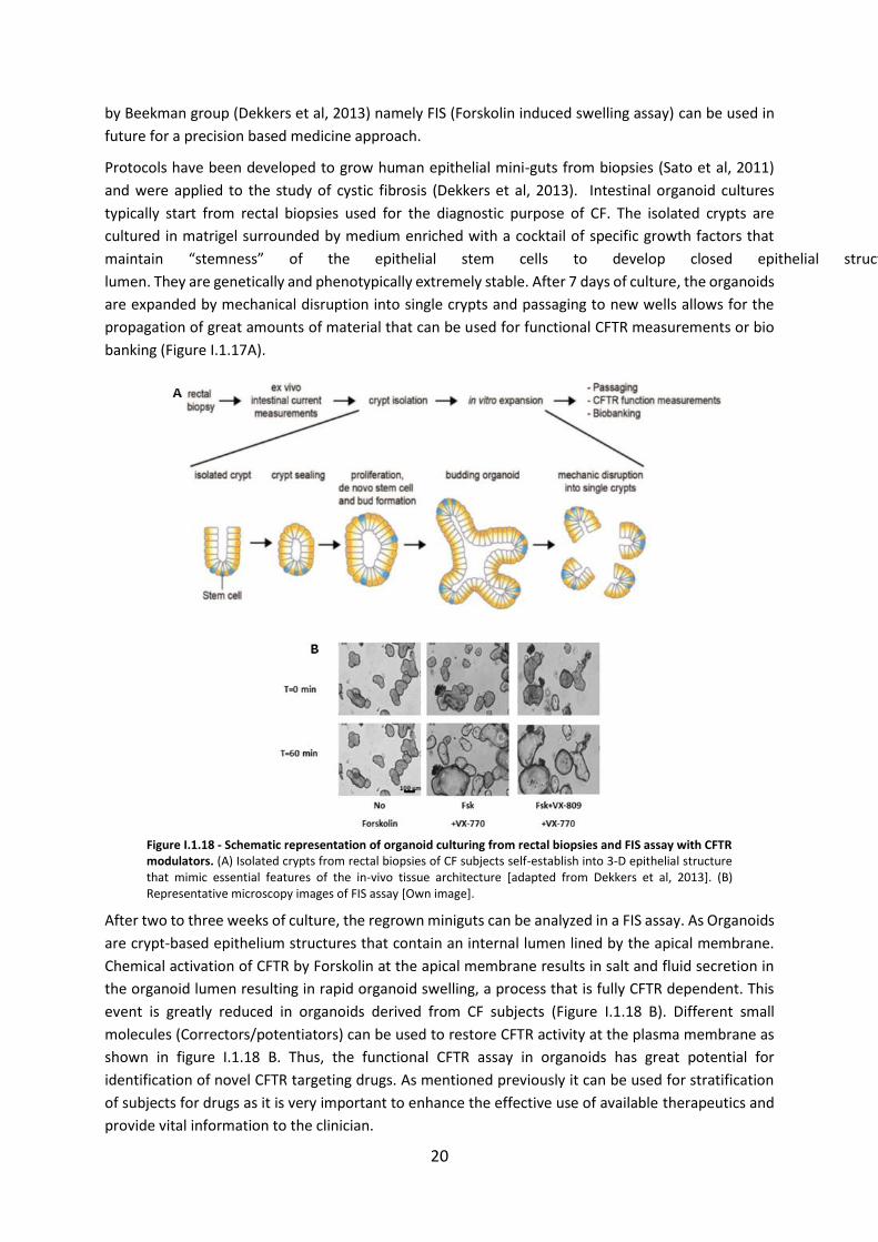
20
by Beekman group (Dekkers et al, 2013) namely FIS (Forskolin induced swelling assay) can be used in
future for a precision based medicine approach.
Protocols have been developed to grow human epithelial mini-guts from biopsies (Sato et al, 2011)
and were applied to the study of cystic fibrosis (Dekkers et al, 2013). Intestinal organoid cultures
typically start from rectal biopsies used for the diagnostic purpose of CF. The isolated crypts are
cultured in matrigel surrounded by medium enriched with a cocktail of specific growth factors that
maintain “stemness” of the epithelial stem cells to develop closed epithelial structures with an internal
lumen. They are genetically and phenotypically extremely stable. After 7 days of culture, the organoids
are expanded by mechanical disruption into single crypts and passaging to new wells allows for the
propagation of great amounts of material that can be used for functional CFTR measurements or bio
banking (Figure I.1.17A).
Figure I.1.18 - Schematic representation of organoid culturing from rectal biopsies and FIS assay with CFTR modulators. (A) Isolated crypts from rectal biopsies of CF subjects self-establish into 3-D epithelial structure that mimic essential features of the in-vivo tissue architecture [adapted from Dekkers et al, 2013]. (B) Representative microscopy images of FIS assay [Own image].
After two to three weeks of culture, the regrown miniguts can be analyzed in a FIS assay. As Organoids
are crypt-based epithelium structures that contain an internal lumen lined by the apical membrane.
Chemical activation of CFTR by Forskolin at the apical membrane results in salt and fluid secretion in
the organoid lumen resulting in rapid organoid swelling, a process that is fully CFTR dependent. This
event is greatly reduced in organoids derived from CF subjects (Figure I.1.18 B). Different small
molecules (Correctors/potentiators) can be used to restore CFTR activity at the plasma membrane as
shown in figure I.1.18 B. Thus, the functional CFTR assay in organoids has great potential for
identification of novel CFTR targeting drugs. As mentioned previously it can be used for stratification
of subjects for drugs as it is very important to enhance the effective use of available therapeutics and
provide vital information to the clinician.
21
6 Cystic Fibrosis Therapeutic Approaches
6.1 CFTR modulators – Personalised Medicine
Personalised medicine is “a form of medicine that uses information about a person’s genes, proteins,
and environment to prevent, diagnose, and treat disease” (National Cancer Institute, 2011). Recent
advances in the understanding of CF at the molecular level helped significantly in the development of
curative CF therapies that target the root cause of CFTR defect at the DNA, RNA, or protein level. De-
velopment of Pharmacotherapy agents that are tailored toward specific mutations is considered as the
most promising approach to treat CF so far. These pharmacotherapy agents are called “correctors and
potentiators”. Correctors are small molecules used to repair defective folding and rescue the traffick-
ing of the mature CFTR to the plasma membrane, thereby restoring channel function. Potentiators are
drugs that increase channel open probability and restore cAMP dependent Cl- channel function (see
figure I.1.19). The current scenario of the most important mutation-specific therapies that target the
underlying defect in CF is discussed below.
6.2 Repair of CFTR protein synthesis
PTC read through agents might be beneficial for CF patients bearing Class I CFTR mutations. These
agents have been clinically assessed and can be orally administered for treatment of nonsense muta-
tions leading to PTCs, carried by 10% of CF patient population (CF Genetic analysis consortium, 1994).
Aminoglycoside antibiotics are also known for their established read-through capacity (Howard et al,
1996). As reported previously Gentamycin and tobramycin promoted full-length CFTR at the cell sur-
face and restored partially its Cl- secretion in cell lines and transgenic mice (Howard et al, 1996, Du et
al, 2002, Wilschanski et al, 2003). However, a gentamycin trial in patients in 2007, bearing Class I mu-
tations was unable to demonstrate a significant change in NPD following 28 days nasal administration
of gentamycin (Clancy et al, 2007). Moreover, long-term use of gentamycin showed nephrotoxicity and
ototoxicity in CF patients (Prayle et al, 2010). Ataluren (PTC-124) was discovered by high through put
screening (HTS), as a non-aminoglycoside, which is able to read through premature stop codons but
not conventional normal stop codons (Kerem et al, 2008).
In preclinical studies CF mouse model expressing the human CFTR-G542X transgene, oral administra-
tion of PTC-124 suppressed the G542X nonsense mutation, restoring CFTR functional expression at the
plasma membrane (Du et al, 2008). Phase II trials with PTC-124 showed good tolerance and mild side
effects with some improvements in FEV1 of CF patients bearing nonsense mutations. However, phase
III clinical studies reported limited efficacy and failed to reach their primary end point of improvement
of FEV1 at 48 weeks (Kerem et al, 2008, Sermet-Gaudelus et al, 2010, Wilschanski et al, 2011), except
in a subset of individuals who were not simultaneously taking nebulised aminoglycoside antibiotic
(Kerem et al, 2014). Very recently, the read-through efficacy of PTC124 and G418 was tested using
human intestinal organoids from CF patients bearing at least one PTC mutation, including the
W1282X/F508del genotype (Zomer-van Ommen et al, 2016). Whereas these authors found partial
functional restoration of CFTR mediated by G418 read-through activity, the same was not observed
for PTC124, suggesting that better read-through agents are needed for robust treatment of CFTR PTC
mutations.
22
6.3 Repair of CFTR protein folding and trafficking
CF patients bearing class II mutation F508del-CFTR have been the principal target for the development
of small molecule-corrector strategies. F508del-CFTR can be rescued to the plasma membrane by low
temperature culturing at 27⁰C (Denning et al, 1992) however this approach is not practical for patients,
introducing second site suppressor mutations and pharmacological correction. Repair of CFTR protein
can be achieved at chemical, molecular or pharmacological chaperone level. Chemical compounds
such as glycerol (Sato et al, 1996), DMSO (Bebok et al, 1998) and organic solutes (Zhang et al, 2003)
have been shown to stabilize F508del-CFTR. Pharmacological chaperones bind directly to the mutated
protein. However, most of these compounds have not yet been given to the CF subjects because of
their inherent toxicity and poorly understood mechanism of action. Correctors can exert their action
as Pharmacological chaperones or as Proteostasis regulators. Correctors based on their possible mech-
anism of action as a pharmacological chaperone divided into three groups 1) correctors that stabilize
the interactions between NBD1 and intracellular loops 1 and 4 (e.g., C3, C18 and VX-809); 2) correctors
that restore NBD2 stability and its interfaces with other CFTR domains (e.g., C4); and 3) correctors that
directly stabilize NBD1 (Okiyoneda et al, 2013).
Extensive high throughput screening of small molecules by Vertex pharmaceuticals using F508del-CFTR
expressing cells yielded the discovery of the Corrector VX-809 (Lumacaftor) (Van Goor et al, 2011,
Clancy et al, 2011). Reportedly VX-809 restored the F508del-CFTR channel activity to 15% of wild-type
CFTR in primary airway epithelium (Van Goor et al, 2011). A phase II clinical trial with VX-809 mono-
therapy in F508del-CFTR homozygotes showed a modest but significant improvement only in sweat Cl-
level and no improvement in FEV1 (Clancy, 2012). As discussed before, F508del CFTR protein expressed
at the cell surface exhibits defects in both function and stability. Thus, the combined effect of corrector
and potentiator (Lumacaftor/Ivacaftor - Orkambi) is significantly greater than either agent alone. Phase
III clinical studies demonstrated good clinical benefits with combination therapy when administered
to patients homozygous for F508del-CFTR as shown by modest improvement in lung function, reduced
CF exacerbations and improved BMI (Wainwright et al, 2015). It is important to note that in vitro stud-
ies published by Cholon D and Veit G in 2014 have revealed that chronic Ivacaftor exposure over a
longer period destabilize the F508del-CFTR protein that is corrected by treatment with Lumacaftor and
therefore antagonize the effect of Lumacaftor, this observation may partly explain the modest clinical
improvement. However, Lumacaftor/Ivacaftor combination therapy did not improve lung function in
patients with CF who are heterozygous for F508del-CFTR (Rowe et al, 2017). New phase 3 results of
Ivacaftor alone or in combination with Tezacaftor (VX-661), a CFTR corrector with better pharmacoki-
netic profile than Lumacaftor, in 218 patients 12 years of age or older who are heterozygous for the
F508del-CFTR mutation and a CFTR mutation associated with residual CFTR function, reported the
mean difference versus placebo with respect to the absolute change in the percentage of predicted
FEV1 was 6.8 percentage points for Tezacaftor–Ivacaftor and 4.7 percentage points for Ivacaftor alone
(P<0.001 for both comparisons) (Rowe et al, 2017). The same combination, Tezacaftor–Ivacaftor in
F508del-CFTR homozygous CF patients showed the effects on the absolute and relative changes in the
percentage of the predicted FEV1 in favor of Tezacaftor–Ivacaftor over placebo were 4.0 percentage
points and 6.8%, respectively (P<0.001 for both comparisons). The rate of pulmonary exacerbation was
35% lower in the Tezacaftor– Ivacaftor group than in the placebo group (P=0.005) (Taylor-Cousar et al,
2017).
Moreover, it is often found that subjects with same CFTR genotype (F508del-CFTR homozygous) have
a significantly different clinical response to Lumacaftor in primary cultures (Awatade et al, 2015,
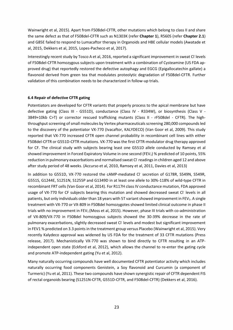
23
Wainwright et al, 2015). Apart from F508del-CFTR, other mutations which belong to class II and share
the same defect as that of F508del-CFTR such as N1303K (refer Chapter 1), R560S (refer Chapter 2.1)
and G85E failed to respond to Lumacaftor therapy in Organoids and HBE cellular models (Awatade et
al, 2015, Dekkers et al, 2015, Lopes-Pacheco et al, 2017).
Interestingly recent study by Tosco A et al, 2016, reported a significant improvement in sweat Cl- levels
of F508del-CFTR homozygous subjects upon treatment with a combination of Cysteamine (US FDA ap-
proved drug) that reportedly restored the defective autophagy and EGCG (Epigallocatechin gallate) a
flavonoid derived from green tea that modulates proteolytic degradation of F508del-CFTR. Further
validation of this combination needs to be characterized in follow-up trials.
6.4 Repair of defective CFTR gating
Potentiators are developed for CFTR variants that properly process to the apical membrane but have
defective gating (Class III - G551D), conductance (Class IV - R334W), or biosynthesis (Class V -
3849+10kb C>T) or corrector rescued trafficking mutants (Class II - rF508del - CFTR). The high-
throughput screening of small molecules by Vertex pharmaceuticals screening 280,000 compounds led
to the discovery of the potentiator VX-770 (Ivacaftor, KALYDECO) (Van Goor et al, 2009). This study
reported that VX-770 increased CFTR open channel probability in recombinant cell lines with either
F508del-CFTR or G551D-CFTR mutations. VX-770 was the first CFTR-modulator drug therapy approved
for CF. The clinical study with subjects bearing least one G551D allele conducted by Ramsey et al
showed improvement in Forced Expiratory Volume in one second (FEV1) % predicted of 10 points, 55%
reduction in pulmonary exacerbations and normalised sweat Cl- readings in children aged 12 and above
after study period of 48 weeks. (Accurso et al, 2010, Ramsey et al, 2011, Davies et al, 2013)
In addition to G551D, VX-770 restored the cAMP-mediated Cl- secretion of G178R, S549N, S549R,
G551S, G1244E, S1251N, S1255P and G1349D in at least one allele to 30%-118% of wild-type CFTR in
recombinant FRT cells (Van Goor et al, 2014). For R117H class IV conductance mutation, FDA approved
usage of VX-770 for CF subjects bearing this mutation and showed decreased sweat Cl- levels in all
patients, but only individuals older than 18 years with 5T variant showed improvement in FEV1. A single
treatment with VX-770 or VX-809 in F508del homozygotes showed limited clinical outcome in phase II
trials with no improvement in FEV1 (Moss et al, 2015). However, phase III trials with co-administration
of VX-809/VX-770 in F508del homozygous subjects showed the 30-39% decrease in the rate of
pulmonary exacerbations, slightly decreased sweat Cl- levels and modest but significant improvement
in FEV1 % predicted on 3.3 points in the treatment group versus Placebo (Wainwright et al, 2015). Very
recently Kalydeco approval was widened by US FDA for the treatment of 33 CFTR mutations (Press
release, 2017). Mechanistically VX-770 was shown to bind directly to CFTR resulting in an ATP-
independent open state (Eckford et al, 2012), which allows the channel to re-enter the gating cycle
and promote ATP-independent gating (Yu et al, 2012).
Many naturally occurring compounds have well documented CFTR potentiator activity which includes
naturally occurring food components Genistein, a Soy flavonoid and Curcumin (a component of
Turmeric) (Yu et al, 2011). These two compounds have shown synergistic repair of CFTR dependent FIS
of rectal organoids bearing (S1251N-CFTR, G551D-CFTR, and F508del-CFTR) (Dekkers et al, 2016).
24
6.5 Repair of Defective CFTR Splicing
Repair of defective splicing can be achieved by increasing the levels of correctly spliced transcripts.
About 10% of mutations causing CF occur by incorrect splicing. Antisense oligonucleotide-based
therapy to cells appears to be promising to modulate the splicing and restore normal full-length CFTR
transcript as well as rescue of functional CFTR protein as reported by (Igreja et al, 2016). Approved
Lumacaftor-Ivacaftor therapy is also likely to provide benefit for subjects with splicing mutations by
increasing trafficking of CFTR to the plasma membrane and restoring channel activity. Unpublished in
vitro data from our lab (refer Chapter 2.3) with splicing mutations 3849+10kbC>T and 2789+5G>A
demonstrated positive response to Ivacaftor/Lumacaftor treatment in primary intestinal organoids.
Recent studies by (Sanj et al, 2017) reported the use of a CRISPR/Cas 9 - based non-homologous end
joining (NHEJ) approach to correct defective splicing.
6.6 Alternative non-CFTR Cl- secretory pathways
Lessons learned from recent clinical trials explain that the efficacy of pharmacological rescue of
F508del-CFTR, as well as other mutations that affect folding (Class II) and synthesis (Class I), remains
limited and warrants further development (Boyle et al, 2014, Wainwright et al, 2015). Therefore,
activation of alternative Cl- channels such as TMEM16A, SLC26A9, and inhibition of ENaC remain
attractive strategies to compensate for defective CFTR in the airways see figure I.1.18.
Figure I.1.19 - Pharmacological strategies targeting ion channels in CF. (A) Depending on the underlying mo-lecular mechanisms, pharmacological rescue of mutant CFTR function can be achieved by CFTR potentiators (gating mutations), CFTR correctors (folding mutations) and readthrough agents (premature termination co-don mutations). (B, C) Strategies to compensate CFTR malfunction include activation of the alternative Cl- channels TMEM16A (B) and SLC26A9 (C). In principle, these alternative targets could be activated by traffick-ing modulators that increase the number of Cl- channels, and/or by activators that increase the open proba-bility of TMEM16A (B) and SLC26A9 (C) channels in the apical plasma membrane. (D) In addition, inhibition of ENaC-mediated sodium/fluid absorption, either by preventing its proteolytic activation by protease inhibitors, or by direct inhibition of the channel by new generation ENaC blockers, can be used as a strategy to counteract airway surface dehydration in CF airways. [Adapted from Mall & Galietta, 2015]
25
TMEM16A or Anoctamin-1 (ANO1)
Previous functional studies carried out by Mason (1991) & Knowles (1991) have demonstrated that
airway epithelium cells possess a secondary mechanism of Cl- secretion independent of CFTR (Mason
et al, 1991; Knowles et al, 1991). Activation of cells in vitro or nasal epithelia in vivo, with calcium (Ca2+)
agonists (UTP/ATP) resulted in a large transient stimulation of Cl- transport. This response was probably
due to the Ca2+-dependent activation of non-CFTR Cl- channels since it was also observed in subjects
with CF. This observation led to clinical studies in which activation of Ca2+-dependent Cl- secretion was
achieved by denufosol as an aerosol (Clunes et al, 2008). This molecule is an UTP analogue and works
by increasing intracellular Ca2+ levels by binding to purinergic receptors (P2Y2) localized on the apical
membrane of epithelial cells. Unfortunately, results obtained in phase 3 trials with denufosol were
discouraging, as it failed to improve lung function or reduce pulmonary exacerbations (Ratjen et al,
2012). The reason for the failure of mainly attributed to the short half-life of the compound on the
airway surface (Moss RB, 2013). The discovery of compounds that activate alternative CaCCs directly
was restricted by the fact that their molecular identity remained uncertain.
In 2008, three research groups identified TMEM16A as the protein responsible for calcium-activated
Cl- transport in many types of cells (Caputo et al, 2008, Schroeder et al, 2008, Yang et al, 2008).
TMEM16A is a homodimer (see figure I.1.19 B) with each subunit containing at least 10 membrane
spanning helices and a Ca2+ binding site (Brunner et al, 2014). TMEM16A is expressed in the airway
epithelium mucus secreting goblet cells (Huang et al, 2012; Scudieri et al, 2012) and in submucosal
glands of the bronchi (Caci et al, 2015). Physiologically, TMEM16A is activated by purinergic agonists
such as ATP/UTP. Recently, Benedetto et al reported that tissue-specific knockout of the TMEM16A
gene in mouse intestine and airways not only wipe out CaCC currents but unexpectedly abrogates CFTR
mediated Cl- secretion and completely abolishes cAMP-activated whole cell currents (Benedetto et al,
2017). This regulatory relationship may explain the functional crosstalk between two channels.
Pharmacological activation of TMEM16A function could help individuals carrying CF mutations
unresponsive to CFTR modulators therapy (Class VII mutations), also it can be useful to normalize the
properties of ASL and mucus by enhancing Cl- and HCO3- secretion with the help of TMEM16A
activators/potentiators.
In summary, a better understanding of the pathophysiological role of TMEM16A in the respiratory
system is required to utilize as an alternative therapeutic target to circumvent CFTR dysfunction in the
airway epithelium of CF subjects.
SLC26A9
SLC26A9 (see figure I.1.18 C) belongs to the solute carrier 26 (SLC26) family of anion transporters and
it is expressed in epithelia of the lung and the stomach, where it functions as a Cl- and HCO3- transporter
(minimal HCO3- conductance) involved in pH regulation (Lohi et al, 2002; Xu et al, 2008). The predicted
topology of SLC26A9 includes a PDZ binding motif that involves in the formation of the Cl- channel pore
and a STAS domains in the C-terminus through which SLC26A9 can interact with CFTR (Chang et al,
2009; Khouri & Toure et al, 2014).
Noticeably it has been demonstrated that SLC26A9 contributes to constitutive and cAMP-dependent
Cl- secretion in HBE cells (Bertrand et al, 2009; Avella et al, 2011). Moreover, SLC26A9 might enhance
the residual function of the pharmacological rescue of CF gating mutation G551D-CFTR (Strug et al,

26
2016). With this notion, Strug et al have shown an association between a single nucleotide
polymorphism in the SLC26A9 gene and the magnitude of treatment response in CF subjects with
gating mutations treated with the Ivacaftor therapy. Furthermore, these authors also found improved
transepithelial Cl- transport in F508del-homozygous primary HBE cultures treated with Lumacaftor
(Strug et al, 2016). These results provide some evidence that SLC26A9 acts as a modifier of CF lung
disease severity and the response to CFTR modulator therapy.
Taken together, compelling evidence from these studies suggests that besides TMEM16A, SLC26A9
may also serve as an alternative Cl- channel that may compensate for CFTR dysfunction in CF subjects
figure I.1.18 B & C.
ENaC
The amiloride-sensitive epithelial sodium channel (ENaC) is primarily expressed in the luminal
membrane of absorptive epithelia (see figure I.1.19 D) including the conducting airways, alveolar
airspaces, the distal colon, the sweat duct, and the distal nephron of the kidney (Mall & Galietta, 2015).
ENaC is multimeric transmembrane protein mainly consists of three homologous subunits (α, β & γ)
that form the channel pore and have a small conductance (~ 4 to 5 pS) (Mall et al, 1996; Canessa et al,
1994). ENaC plays an important role in absorptive epithelial cells by apical Na+ uptake; in sweat duct
coordinated activation of CFTR and ENaC in epithelial cells is essential for transcellular absorption of
Na+ and Cl- that result in a hypotonic sweat and retention of salt in the body during sweating (Reddy
et al, 1999). This coordination in the sweat duct is impaired which leads to increased NaCl
concentration in sweat of CF subjects.
In airway epithelium, the transepithelial potential difference generated by active ENaC mediated Na+
absorption provides the driving force for absorption of Cl- and water through the paracellular shunt
pathway (Boucher RC, 1994) therefore ENaC becomes the limiting pathway that plays an important
role in volume regulation of ASL and is essential for effective MCC (Mall, 2008; Knowles et al, 2002). In
CF airway epithelial cells, defective CFTR leads to an imbalance between secretion and absorption of
ions and fluid that makes dehydrated CF airway surfaces. It has been shown that CFTR regulates ENaC
through Cl- secretion and water balance across epithelia (Konig et al, 2001). While some studies have
also reported that lack of CFTR leads to ENaC hyperactivity by a direct (Suaud et al, 2007) or indirect
interaction between the two channels (Guggino & Stanton, 2006). Altogether, ENaC is an attractive
target to improve airway surface hydration and mucus clearance in all subjects with CF irrespective of
their CFTR genotype. Also, CF subjects who are not responsive to CFTR modulators therapy may benefit
from this approach. Therefore, it may be alluring to use ENaC blockers in conjunction with CFTR
modulators to enhance Cl- secretion through pharmacologically rescued mutant CFTR.
Targeting apical membrane Cl- channels is not the only possible approach to circumvent the lack of
CFTR activity. Other approaches such as, activating basolateral membrane K+ channels which provide
the driving force for Cl- exit across the apical membrane of CF epithelia (Devor et al, 2000; Mall et al,
2000). Inhibiting nongastric H+/K+-ATPase (ATP12) was shown to prevent deleterious acidification of
ASL which promotes bacterial growth (Shah et al, 2016) and finally artificial anion channels and
transporters (e.g., cholapods) might be delivered to CF epithelia to restore Cl- secretion (Davis et al,
2007), although the lack of regulation of these molecules makes them still of very limited application
at present.
27
7. Objectives of Present PhD Work
The main objective of this PhD work was to study new compounds that correct the basic CF defect,
by rescuing CFTR protein traffic and function, focusing both on individual responses of CF patients
with different CFTR mutations to these new drugs, and in their mechanism of action.
In particular, it had the following specific objectives:
1) To assess the efficacy of CFTR modulators (correctors and potentiators) in rescuing CFTR in
Primary Human Bronchial Epithelial (HBE) cells, derived from explanted CF lung, from patients
with rare mutations;
2) To establish correlations among different CFTR biomarkers, by comparison of basal CFTR
activity (and response to modulators) in patient-derived tissues with different mutations,
namely: i) fresh human rectal biopsies (by Ussing chamber measurements); ii) intestinal
organoids (by Forskolin induced swelling assay, FIS); and iii) conditionally reprogrammed (CRC)
primary cultures of human nasal epithelial cells (HNEs);
3) To assess the efficacy of novel correctors of F508del-CFTR traffic and novel enhancers of
TMEM16A function in epithelial cells/non-CF intestinal organoids;
4) To evaluate the effect of CFTR modulators on cAMP-induced Ieq-sc in CFBE cells stably
expressing inducible (Tet-on) double tagged mCherry-Flag-CFTR with F508del genetic
revertants or traffic wt-CFTR variant namely: i) two second site mutations previously shown to
correct structural features of F508del-CFTR folding - G550E or R1070W; ii) a variant that
bypasses the ER quality control - 4RK; iii) another variant (on wt-CFTR background) missing the
diacidic ER exit code (NBD1 located at the 565DAD567 motif), a cargo signal for Sec-24 mediated
COPII packing – DD/AA.
Results towards each of these specific objectives are described in each of the subsections of the Results
chapter.

28
II. MATERIALS AND METHODS
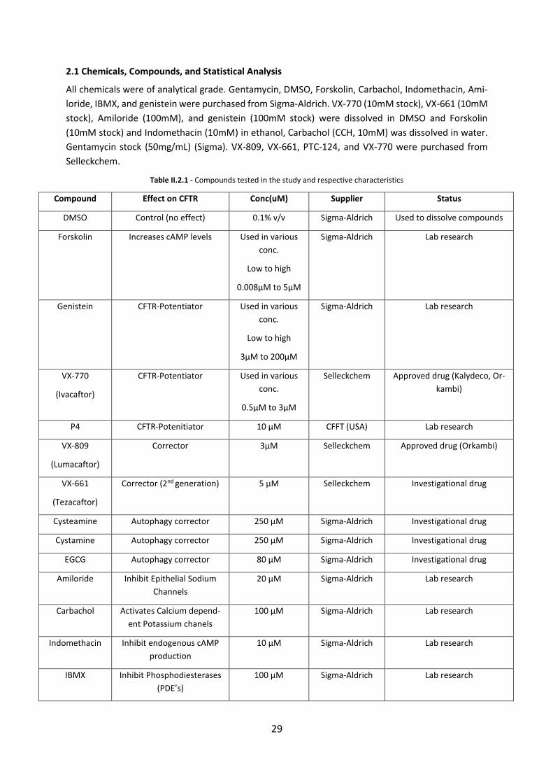
29
2.1 Chemicals, Compounds, and Statistical Analysis
All chemicals were of analytical grade. Gentamycin, DMSO, Forskolin, Carbachol, Indomethacin, Ami-
loride, IBMX, and genistein were purchased from Sigma-Aldrich. VX-770 (10mM stock), VX-661 (10mM
stock), Amiloride (100mM), and genistein (100mM stock) were dissolved in DMSO and Forskolin
(10mM stock) and Indomethacin (10mM) in ethanol, Carbachol (CCH, 10mM) was dissolved in water.
Gentamycin stock (50mg/mL) (Sigma). VX-809, VX-661, PTC-124, and VX-770 were purchased from
Selleckchem.
Table II.2.1 - Compounds tested in the study and respective characteristics
Compound Effect on CFTR Conc(uM) Supplier Status
DMSO Control (no effect) 0.1% v/v Sigma-Aldrich Used to dissolve compounds
Forskolin Increases cAMP levels Used in various
conc.
Low to high
0.008µM to 5µM
Sigma-Aldrich Lab research
Genistein CFTR-Potentiator Used in various
conc.
Low to high
3µM to 200µM
Sigma-Aldrich Lab research
VX-770
(Ivacaftor)
CFTR-Potentiator Used in various
conc.
0.5µM to 3µM
Selleckchem Approved drug (Kalydeco, Or-
kambi)
P4 CFTR-Potenitiator 10 µM CFFT (USA) Lab research
VX-809
(Lumacaftor)
Corrector 3µM Selleckchem Approved drug (Orkambi)
VX-661
(Tezacaftor)
Corrector (2nd generation) 5 µM Selleckchem Investigational drug
Cysteamine Autophagy corrector 250 µM Sigma-Aldrich Investigational drug
Cystamine Autophagy corrector 250 µM Sigma-Aldrich Investigational drug
EGCG Autophagy corrector 80 µM Sigma-Aldrich Investigational drug
Amiloride Inhibit Epithelial Sodium
Channels
20 µM Sigma-Aldrich Lab research
Carbachol Activates Calcium depend-
ent Potassium chanels
100 µM Sigma-Aldrich Lab research
Indomethacin Inhibit endogenous cAMP
production
10 µM Sigma-Aldrich Lab research
IBMX Inhibit Phosphodiesterases
(PDE’s)
100 µM Sigma-Aldrich Lab research
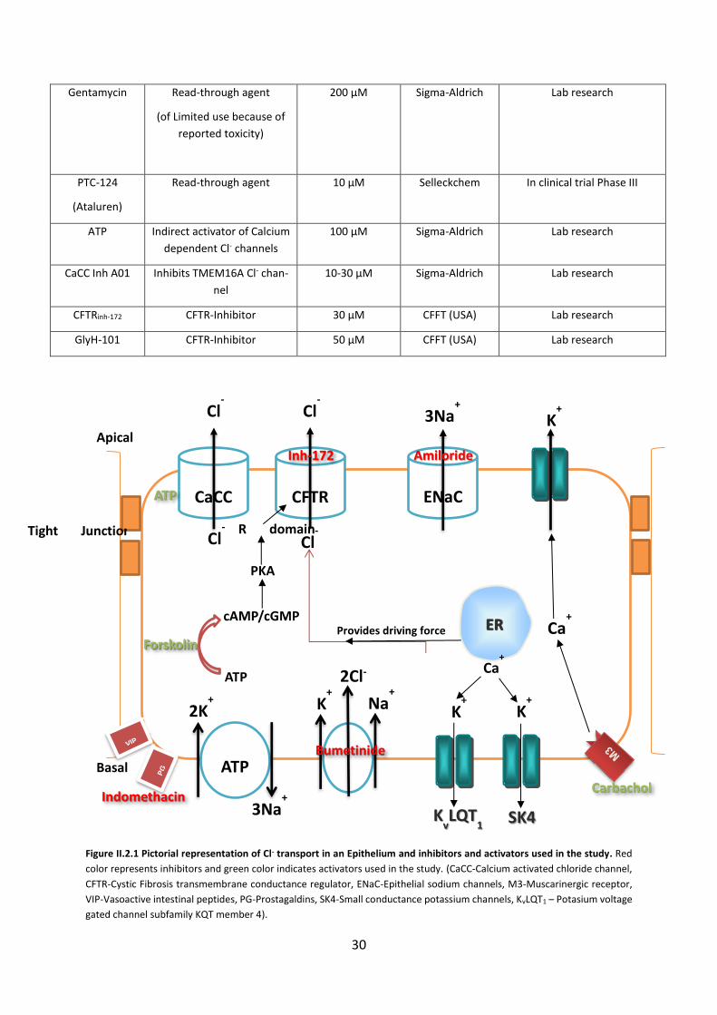
30
Gentamycin Read-through agent
(of Limited use because of
reported toxicity)
200 µM Sigma-Aldrich Lab research
PTC-124
(Ataluren)
Read-through agent
10 µM Selleckchem In clinical trial Phase III
ATP Indirect activator of Calcium
dependent Cl- channels
100 µM Sigma-Aldrich Lab research
CaCC Inh A01 Inhibits TMEM16A Cl- chan-
nel
10-30 µM Sigma-Aldrich Lab research
CFTRinh-172 CFTR-Inhibitor 30 µM CFFT (USA) Lab research
GlyH-101 CFTR-Inhibitor 50 µM CFFT (USA) Lab research
Figure II.2.1 Pictorial representation of Cl- transport in an Epithelium and inhibitors and activators used in the study. Red
color represents inhibitors and green color indicates activators used in the study. (CaCC-Calcium activated chloride channel,
CFTR-Cystic Fibrosis transmembrane conductance regulator, ENaC-Epithelial sodium channels, M3-Muscarinergic receptor,
VIP-Vasoactive intestinal peptides, PG-Prostagaldins, SK4-Small conductance potassium channels, KvLQT1 – Potasium voltage
gated channel subfamily KQT member 4).
CaCC CFTR
Cl- Cl
-
ENaC
3Na+
Apical
Basal ATP
Tight Junctions
2K+
3Na+
K+ Na
+
2Cl-
KvLQT
1 SK4
K+ K
+
ER Ca+
Ca+
K+
Cl-
Inh-172 Amiloride
Bumetinide
Carbachol
Forskolin
Cl-
Provides driving force
Indomethacin
cAMP/cGMP
ATP
PKA
R domain
ATP
31
Statistical Analyses
Statistical analysis was performed by Graph pad prism 5.0 using two-tailed paired student’s t-tests or
unpaired samples. Statistical significance was considered for p ≤ 0.05.
2.2 Primary cell Culture
2.2.1 Primary Cultures of HBE’s – Isolation and Culture of Human Bronchial Epithelial Cells from CF and non-CF patient lung explants
The access to explanted lungs was established through collaboration with the Thoracic Surgery unit of
University Hospital La Fe, Valencia, Spain (Dr. Amparo sole), and Dr. Tereza Doušová, Prague, which
received approval from the Ethics committee of both hospitals. Isolation and cultivation of primary
HBE cells were carried out as described below (Fulcher et al, 2005; Fulcher et al, 2009). Whole lungs
were obtained from non-CF, CF and patient subjects following lung transplant. After explantd, lungs
were packed and transported to the laboratory in Dulbecco’s phosphate buffered saline (DPBS)
maintained at 4°C. All dissection procedures were performed in class II, biological safety cabinet with
all precautionary measures. Dissecting material was sterilized before usage. The first step of the
dissection involves dissecting the airways by removing the excess of connective tissue and slicing them
into small 2-5 cm segments see figure I.1.15. Then the tissue segments are further cleaned, by removal
of any additional connective tissue and lymph nodes and rinsing in PBS solution. Segments were slit
longitudinally and cut into 1-2 cm portions with a scalpel. Finally, they were transferred to specimen
cups containing chilled PBS solution. Human tissue samples are likely to contain yeasts, bacteria, or
fungi. Because of these microbial contaminants treatment with a cocktail of common antibiotics such
as amphotericin, ceftazidime, tobramycin, and vancomycin (ACTV cocktail) should begin as soon as
possible. Dissected tissues were then washed three times in “Wash medium” prepared by adding the
above-mentioned antibiotics to Joklik’s minimal essential medium (J-MEM). Tissues from chronically
infected patients containing abundant secretions are further treated to remove thick mucus and other
debris with a “Soak solution containing supplemental antibiotics, dithiothreitol (DTT, 0.5mg/mL) and
DNase (10µg/mL) and washed again in the wash medium.
After washing, tissue segments were transferred into 50ml conical tubes containing 30mL wash
medium plus 4mL protease/DNase solution – “Dissociation solution”. The tubes were placed on a
rocking platform (at 60 cycles/min), in the cold room (4°C) for 18-24h. The day after this procedure,
cells were harvested following the standard tissue-culture techniques in a laminar flow hood. To stop
tissue dissociation, we transfer the contents of 50mL tubes into a 150mm tissue culture dish, FBS was
added to a final concentration of 10% v/v to neutralize protease and epithelial surface was scrapped
with a scalpel. Tissue surfaces were then rinsed, and cells collected from the dish with J-MEM.
Solutions containing dissociated cells were then pooled into 50ml conical tubes. Finally, we centrifuged
the latter at 500g for 5min at 4°C, wash cells once in media, resuspended in a volume calculated to be
approx. 5X10⁶ cells/mL, and counted the cells using a hemocytometer.
Primary HBE cells were then plated in BEGM and seeded on fibronectin-collagen coated plastic dishes
at a density of 2-6X10⁶ per 100mm dish. Primary cell media (BEGM) was supplemented with additional
antibiotics (ACTV) for the first 3 days after plating and changed every 2-3 days to prevent acidification.
After reaching 70-90% confluence, cells were trypsinized (passage 1) to collagen IV-coated porous
32
membranes (Snapwell Inserts 12mm, from Corning-Costar, New York, NY, USA) and grown in Air Liquid
Interface (ALI) for four to five weeks prior to the functional or biochemical assays.
The isolation and production of primary HBE cells were done in collaboration with Veronica Felício,
Jose Murias and Sofia Correia (Faculty of Sciences, Univ. of Lisboa, Portugal).
2.2.2 Establishing Primary cultures of HNE’s – Isolation and Culture of Human Nasal Epithelial cells from Nasal brushings of non-CF and CF patients.
Feeder layer technique
The study was conducted at the Faculty of Sciences of the University of Lisboa with samples collected
at the CF Clinics of the Hospital de Santa Maria in Lisboa and was approved by the Santa Maria Hospital
Ethical Review Board. Bilateral nasal epithelial cell collection was performed using interdental brushes
(Paro Isola Long 3mm, Paro, Esro AG, Switzerland), which were used to obtain cells from the inferior
nasal turbinates, as previously described (Clarke et al, 2013). Following brushing the cell samples were
washed three times in growth medium and pelleted by gentle centrifugation (5 mins at 250 x g) for
removal of debris and mucus, before addition to a feeder layer of irradiated NIH-3T3 fibroblast cells
(ATCC Catalog #CRL-1658) in T25 flasks. Fibroblasts were grown to near confluence and irradiated by
exposure to a Cobalt 60 gamma-ray source (1.60 kGy/h) with a total dose of 31 Gy (3100 rads).
After obtaining nasal cells from CF/HC patients cells were resuspended and cultured and maintained
on irradiated NIH3T3 fibroblast in F12 medium containing a 10µM Y-27632 inhibitor. After one week
of culturing small islands of epithelial cells appeared among the feeder layer cells, see figure I.1.16.
When these epithelial cell islands had grown to cover 90% of the culture dish surface area, differential
Trypsinization was performed to isolate the CRC epithelial cells. To establish ALI cultures at 70-90%
confluence HNE-CRC cells were trypsinized and transferred to 0.4µM Costar Transwell permeable sup-
port filters (Corning) pre-coated with human placental type IV collagen. The cells were seeded at a
density of 3x10⁵cells/cm2 and cultured in F12 medium devoid of Rho K inhibitor and without a feeder
layer. Upon confluence after 5-7 days, cells were maintained at an ALI by removing apical media and
supplying media to the basal compartment only.
The isolation and culture of primary HNE cells were done in collaboration with Inna Uliyakina and Luka
Clarke (Faculty of Sciences, Univ. of Lisboa, Portugal).
2.2.3 Establishing Primary cultures of Human Intestinal Organoids – Crypt isolation from CF and non-CF rectal biopsies and Organoids culture conditions
Crypt isolation and human organoid culturing were carried out as described previously (Sato et al,
2009; Sato et al, 2011; Dekkers et al, 2013; Sato & Clevers, 2013). Briefly, 3-4 superficial rectal mucosa
specimens (3-4 mm in diameter) were recovered with colon forceps and immediately placed into the
culture medium. Then, the biopsies were washed with PBS and treated with 10mM EDTA for 90-
120min at 4ºC. Crypts were isolated by centrifugation and isolated crypts cultured in 50% matrigel
(growth factor free, phenol-free, BD Biosciences). Isolated crypts were seeded (~10-30 crypts in 3x10ul
matrigel droplets per well) in pre-warmed 24-well plates. The matrigel was polymerized for 10-15mins
at 37ºC and surrounded by complete culture medium consisting of: advanced DMEM/F12
supplemented with penicillin/streptomycin, 10mM HEPES, Glutamax, N2, B27 (all purchased from
33
Invitrogen), 1μM N-acetylcysteine (Sigma) and growth factors: 50ng ml-1 mEGF, 50% Wnt-3a-
conditioning medium (WCM) and 10% Noggin-conditioned medium, 20%Rspo1-conditioned medium
(RCM), 10μM nicotinamide (Sigma), 500nm A83-01 (Tocris) and 10μM SB 202190 (Sigma). Antibiotics
were added in growth medium (Primocin, 1:500; Invivogen), Vancomycin and Gentamycin (Sigma)
were added during the first few weeks. The medium was changed every alternate day and organoids
were passaged after 7-9 days of culturing.
2.3 Stably Expressing cell lines
2.3.1 BHK cells
Baby Hamster Kidney (BHK) cells stably expressing either wt- or F508del-CFTR proteins (Farinha et al,
2002), were used in Iodide Efflux experiments (Chapter 2.5). BHK cells were cultured in a 1:1 mixture
of Dulbecco’s Modified Eagle Medium (DMEM) and Ham’s F-12 nutrient medium supplemented with
5% FBS and 200 µg/ml methotrexate (AAH Pharmaceuticals Ltd., Coventry, UK) at 37°C in a humidified
atmosphere of 5% CO2.
2.3.2 NIH3T3 Cells
The NIH3T3 cells cultured in 25cm2 tissue cultured treated flask containing Dulbecco’s modified Eagle’s
medium F12 (Lonza), supplemented with 10% new born calf serum (Sigma), 100U/ml streptomycin
penicillin (GIBCO) solution under humidified 95% O2 5% CO2 atmosphere at 37° C.
2.3.3 CFBE cells stably expressing F508del and wt-CFTR
Cystic Fibrosis Bronchial Epithelial (CFBE41o-) cells stably expressing wt-CFTR or F508del-CFTR were a
generous gift from Dr. J.P. Clancy (University of Alabama at Birmingham, Birmingham, Alabama). CFBE
cells were grown in Modified Eagle Medium (MEM) with Earle’s salts supplemented with 10% FBS and
at 37°C in a humidified atmosphere of 5% CO2.
2.3.4 CFBE mCherry cells stably expressing F508del, wt-CFTR and genetic revertants
CFBE mCherry-Flag-CFTR cells (wt or F508del variants) were cultured in DMEM high glucose (Gibco
#41965) supplemented with 10% (v/v) heat inactivated fetal calf serum (Gibco #10106), 2mM
glutamine (Gibco #25030), 1mM pyruvate (Gibco #11360), 10µg/ml blasticidin (Invivogen #ant-bl) and
2µg/ml puromycin(Invivogen#ant-pr-1) at 37°C and 5% CO2 (Botelho et al, 2015).
2.4 Functional analysis
2.4.1 Iodide efflux technique
For the functional assessment of CFTR, BHK cells with stably expressed constructs (120del23, F508del
and wt-CFTR) cells were pre-incubated with DMSO (0.1%v/v), VX-809 (3μM) and VX-661 (5μM) for 48h
at 37°C, before the assay. The concentration of FBS was kept at 0.5% for all the experiments, due to a
binding affinity of small molecules to serum proteins. CFTR-mediated iodide efflux was measured at
room temperature (RT) using cAMP cocktail i.e. Forskolin and in some experiments performed with
34
Forskolin (10μM) + IBMX (100μM) and/or potentiators P4 (10μM), Genistein (50μM) and VX-770
(10μM) concentration. BHK cells were first incubated for 1 h in loading buffer containing (mM): 136
NaI, 3 KNO3, 2 Ca(NO3)2, 20 HEPES and 11 glucose, adjusted to pH 7.4 with 1 M NaOH. Next, BHK cells
were washed thoroughly with efflux buffer (136 mM NaNO3 replacing 136 mM NaI in the loading
buffer) before treatment with agonists. The amount of CFTR-mediated cumulative Iodide (I-) efflux in
each sample was measured as described previously (Schmidt et al, 2008 and Norez et al, 2004) using
an I- selective electrode (MP225, ThermoElectron Corporation, Waltham, MA).
2.4.2 Ussing chamber
2.4.2.1 Measurement of CFTR mediated Cl- secretion in rectal biopsies
Rectal biopsy specimens were mounted and analyzed in modified micro-Ussing chambers as previously
described under open-circuit conditions (Mall et al, 1998; Mall et al, 2000; Hirtz et al, 2004). Values for
the transepithelial voltage (Vte) were referred to the serosal surface of the epithelium. Transepithelial
resistance (Rte) was determined by applying intermittent (1s) current pulses (ΔI = 0.5 μA). The equiva-
lent short-circuit current (Isc) was calculated according to Ohm’s law (Isc = Vte / Rte), after appropriate
correction for fluid resistance and after subtracting the resistance of the empty chamber. The corre-
sponding changes in Vte and basal Vte were recorded continuously. Briefly, the luminal and basolateral
surfaces of the epithelium were continuously perfused (5 ml/min) with Ringer solution of the following
composition (mmol/l): NaCl 145, KH2PO4 0.4, K2HPO4 1.6, D-glucose 5, MgCl2 1, Ca-gluconate 1.3, pH
7.40, at 37° C at a rate of 5ml/min. HCO3- free buffer solutions were used to exclude a possible contri-
bution of CFTR-independent electrogenic HCO3- secretion, which would be indistinguishable from elec-
trogenic Cl- secretion and thus may mimic residual Cl- channel function in CF colonic epithelia (Mall et
al, 1998). Tissues were equilibrated in the micro-Ussing chambers for 30 min in perfused Ringer solu-
tion before measurements (Mall et al, 2000). Amiloride (Amil, 20 μM, luminal) was added to block
electrogenic sodium (Na+) absorption through the epithelial Na+ channel (ENaC). Indomethacin (Indo,
10 μM, basolateral) was applied for 40-60 min to inhibit endogenous cAMP formation through prosta-
glandins. cAMP-dependent and cholinergic Cl- secretion in human rectal tissues relies on functional
CFTR. Thus, we used 3-isobutyl-1-methylxanthine (IBMX, 100 μM, basolateral) and forskolin (2 μM,
basolateral) to activate cAMP-dependent Cl- secretion and carbachol (CCH, 100 μM, basolateral) for
cholinergic co-activation (Mall et al, 1998; Mall et al, 2000; Hirtz et al, 2004).
2.4.2.2 Subjects and Ethics approval
Rectal biopsies were obtained by small superficial rectal forceps. The ethical committee of Santa Maria
Hospital, Lisboa, Portugal approved this study and the patients signed informed consent forms.
2.4.2.3 Rectal biopsies procedure
Colon preparation (cleaning) was done by applying an enema of saline solution (0.9% NaCl) or 12%
glycerine solution or by oral mannitol. Superficial 5-6 rectal mucosa specimens (3-4 mm in diameter)
were obtained with or without sedation (depending on individuals’ will or collaboration) by colon for-
ceps with visual examination, avoiding the risk of bleeding or of collecting damaged tissue, and imme-
diately stored in ice-cold DMEM F12 with 5% (v/v) Fetal Bovine Serum (FBS).
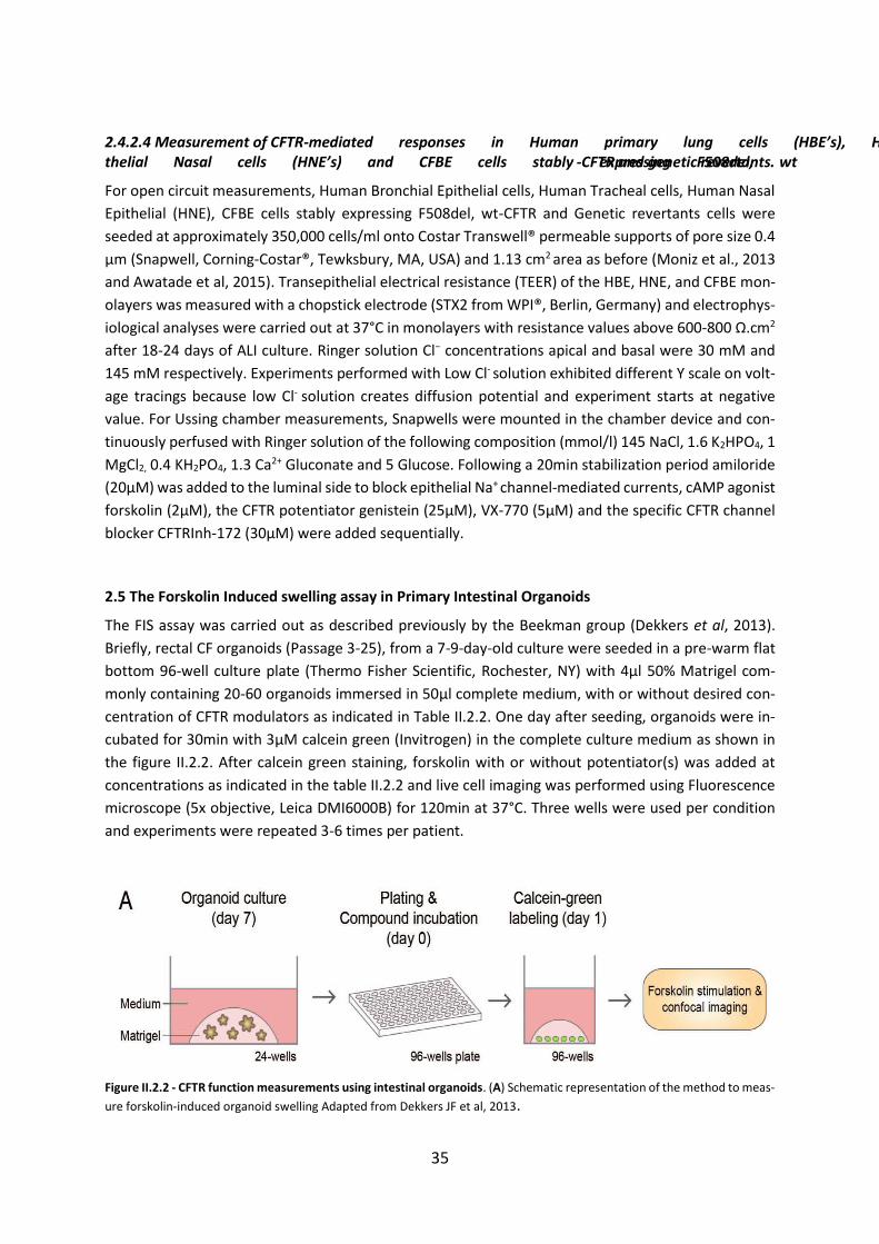
35
2.4.2.4 Measurement of CFTR-mediated responses in Human primary lung cells (HBE’s), Human Epi-thelial Nasal cells (HNE’s) and CFBE cells stably expressing F508del, wt-CFTR and genetic revertants.
For open circuit measurements, Human Bronchial Epithelial cells, Human Tracheal cells, Human Nasal
Epithelial (HNE), CFBE cells stably expressing F508del, wt-CFTR and Genetic revertants cells were
seeded at approximately 350,000 cells/ml onto Costar Transwell® permeable supports of pore size 0.4
µm (Snapwell, Corning-Costar®, Tewksbury, MA, USA) and 1.13 cm2 area as before (Moniz et al., 2013
and Awatade et al, 2015). Transepithelial electrical resistance (TEER) of the HBE, HNE, and CFBE mon-
olayers was measured with a chopstick electrode (STX2 from WPI®, Berlin, Germany) and electrophys-
iological analyses were carried out at 37°C in monolayers with resistance values above 600-800 Ω.cm2
after 18-24 days of ALI culture. Ringer solution Cl− concentrations apical and basal were 30 mM and
145 mM respectively. Experiments performed with Low Cl- solution exhibited different Y scale on volt-
age tracings because low Cl- solution creates diffusion potential and experiment starts at negative
value. For Ussing chamber measurements, Snapwells were mounted in the chamber device and con-
tinuously perfused with Ringer solution of the following composition (mmol/l) 145 NaCl, 1.6 K2HPO4, 1
MgCl2, 0.4 KH2PO4, 1.3 Ca2+ Gluconate and 5 Glucose. Following a 20min stabilization period amiloride
(20µM) was added to the luminal side to block epithelial Na+ channel-mediated currents, cAMP agonist
forskolin (2µM), the CFTR potentiator genistein (25µM), VX-770 (5µM) and the specific CFTR channel
blocker CFTRInh-172 (30µM) were added sequentially.
2.5 The Forskolin Induced swelling assay in Primary Intestinal Organoids
The FIS assay was carried out as described previously by the Beekman group (Dekkers et al, 2013).
Briefly, rectal CF organoids (Passage 3-25), from a 7-9-day-old culture were seeded in a pre-warm flat
bottom 96-well culture plate (Thermo Fisher Scientific, Rochester, NY) with 4μl 50% Matrigel com-
monly containing 20-60 organoids immersed in 50μl complete medium, with or without desired con-
centration of CFTR modulators as indicated in Table II.2.2. One day after seeding, organoids were in-
cubated for 30min with 3μM calcein green (Invitrogen) in the complete culture medium as shown in
the figure II.2.2. After calcein green staining, forskolin with or without potentiator(s) was added at
concentrations as indicated in the table II.2.2 and live cell imaging was performed using Fluorescence
microscope (5x objective, Leica DMI6000B) for 120min at 37°C. Three wells were used per condition
and experiments were repeated 3-6 times per patient.
Figure II.2.2 - CFTR function measurements using intestinal organoids. (A) Schematic representation of the method to meas-
ure forskolin-induced organoid swelling Adapted from Dekkers JF et al, 2013.
36
2.5.1 Quantification of Forskolin-Induced swelling assay in Primary intestinal organoids
The forskolin-induced swelling was quantified by using Cell profiler software. The area under the curve
(AUC; t=60; baseline=100%) was calculated using GraphPad Prism version 5.01. A paired t-test was
used to calculate statistical differences.
2.6 Western Blot
To analyze the effect of compounds upon CFTR processing by WB, cells were incubated with each com-
pound at a chosen concentration for 48hrs at 37ºC, as described previously (Farinha et al, 2004). After
incubation cells were lysed and extracts were analyzed by densitometry, using the anti-CFTR antibody
596 (Kindly provided by CFF foundation). Percentage of band C to total CFTR was calculated by nor-
malizing the ratio of band C/ total CFTR (bands B+C) relative to calnexin.
2.7 Rectal biopsy immunostaining
Rectal biopsies (obtained as previously described) were embedded in a cryoprotective embedding me-
dium, Tissue-Tek optimal cutting temperature (O.C.T) compound (Sakura) in the appropriate orienta-
tion. Samples were frozen in liquid nitrogen-chilled isopentane. Tissue sections were cut using a Leica
CM1850 UV cryostat. Cryosections 15μm thick were collected on silane-prep slides (glass slides coated
with aminoalkylsilane, Sigma-Aldrich) and used for immunostaining against CFTR, using a previously
described protocol, with adaptations. Slides were warmed to RT for 5min and fixed with a PFA (EM
grade, VWR) 4% (v/v) solution (10min at RT). Permeabilization was performed in 0.2% (v/v) Triton X-
100 (Amersham Biosciences) for 10min and sections were blocked with 1% (w/v) BSA (Sigma-Aldrich)
for 30 min. Tissues were incubated with the primary antibody CFTR 570 (from JR Riordan, through
CFFT), raised against the R domain (Gentzsch et al, 2003) at 1:500, overnight at 4⁰C. The following day
secondary antibody (AlexaFluor488, Life Technologies) and nuclear dye ToPro3 (Life Technologies)
were added (1:500 and 1:750, respectively - 2h, 4⁰C). Slides were mounted with Vectashield Antifade
Mounting Medium (Vector Laboratories). Solutions were diluted in PBS1x, which was also used in all
washing steps (3 times for 10 min).
Z-stack images were acquired with a Leica TCS SPE confocal microscope with a 20x or 63x oil objective
as indicated. Stacks were reconstructed for orthoslice generation, using Visage Imaging Amira 5.3.3,
kindly made available by Professor Sólveig Thorsteindóttir (FCUL, CE3C).
2.8 mRNA and extraction from native cells, cDNA synthesis and transcript analyses
RNA was isolated from rectal biopsies (Servidoni et al, 2013) using the NucleoSpin RNAII Kit (Macherey-
Nagel, Duren, Germany) according to the manufacturer’s instructions. CFTR cDNA was generated from
1 μg mRNA using the M-MuLV reverse transcriptase (NZYTech, Lisboa, Portugal) as previously. RT-PCR
using three different reactions was performed with the following pairs of primers B3R-Ex11L, CF10N-
561AL, C1R-CF14a.R as previously described (Felicio et al, 2016). Products were analyzed by agarose
gel electrophoresis and identity confirmed by sequencing.

37
III. RESULTS AND DISCUSSION
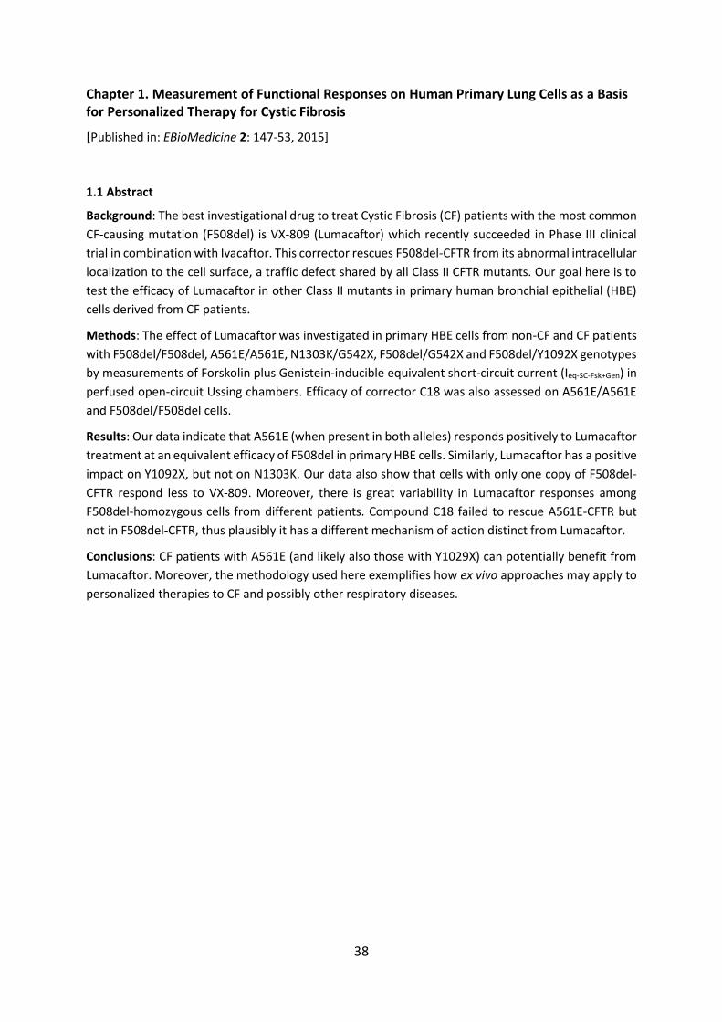
38
Chapter 1. Measurement of Functional Responses on Human Primary Lung Cells as a Basis for Personalized Therapy for Cystic Fibrosis
[Published in: EBioMedicine 2: 147-53, 2015]
1.1 Abstract
Background: The best investigational drug to treat Cystic Fibrosis (CF) patients with the most common
CF-causing mutation (F508del) is VX-809 (Lumacaftor) which recently succeeded in Phase III clinical
trial in combination with Ivacaftor. This corrector rescues F508del-CFTR from its abnormal intracellular
localization to the cell surface, a traffic defect shared by all Class II CFTR mutants. Our goal here is to
test the efficacy of Lumacaftor in other Class II mutants in primary human bronchial epithelial (HBE)
cells derived from CF patients.
Methods: The effect of Lumacaftor was investigated in primary HBE cells from non-CF and CF patients
with F508del/F508del, A561E/A561E, N1303K/G542X, F508del/G542X and F508del/Y1092X genotypes
by measurements of Forskolin plus Genistein-inducible equivalent short-circuit current (Ieq-SC-Fsk+Gen) in
perfused open-circuit Ussing chambers. Efficacy of corrector C18 was also assessed on A561E/A561E
and F508del/F508del cells.
Results: Our data indicate that A561E (when present in both alleles) responds positively to Lumacaftor
treatment at an equivalent efficacy of F508del in primary HBE cells. Similarly, Lumacaftor has a positive
impact on Y1092X, but not on N1303K. Our data also show that cells with only one copy of F508del-
CFTR respond less to VX-809. Moreover, there is great variability in Lumacaftor responses among
F508del-homozygous cells from different patients. Compound C18 failed to rescue A561E-CFTR but
not in F508del-CFTR, thus plausibly it has a different mechanism of action distinct from Lumacaftor.
Conclusions: CF patients with A561E (and likely also those with Y1029X) can potentially benefit from
Lumacaftor. Moreover, the methodology used here exemplifies how ex vivo approaches may apply to
personalized therapies to CF and possibly other respiratory diseases.
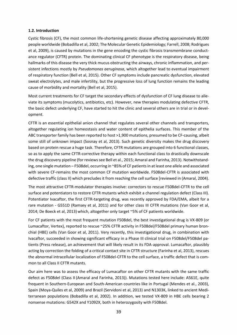
39
1.2. Introduction
Cystic fibrosis (CF), the most common life-shortening genetic disease affecting approximately 80,000
people worldwide (Bobadilla et al, 2002; The Molecular Genetic Epidemiology; Farrell, 2008; Rodrigues
et al, 2009), is caused by mutations in the gene encoding the cystic fibrosis transmembrane conduct-
ance regulator (CFTR) protein. The dominating clinical CF phenotype is the respiratory disease, being
hallmarks of this disease the very thick mucus obstructing the airways, chronic inflammation, and per-
sistent infections mostly by Pseudomonas aeruginosa, which altogether lead to eventual impairment
of respiratory function (Bell et al, 2015). Other CF symptoms include pancreatic dysfunction, elevated
sweat electrolytes, and male infertility, but the progressive loss of lung function remains the leading
cause of morbidity and mortality (Bell et al, 2015).
Most current treatments for CF target the secondary effects of dysfunction of CF lung disease to alle-
viate its symptoms (mucolytics, antibiotics, etc). However, new therapies modulating defective CFTR,
the basic defect underlying CF, have started to hit the clinic and several others are in trial or in devel-
opment.
CFTR is an essential epithelial anion channel that regulates several other channels and transporters,
altogether regulating ion homeostasis and water content of epithelia surfaces. This member of the
ABC transporter family has been reported to host >1,900 mutations, presumed to be CF-causing, albeit
some still of unknown impact (Sosnay et al, 2013). Such genetic diversity makes the drug discovery
based on protein rescue a huge task. Therefore, CFTR mutations are grouped into 6 functional classes,
so as to apply the same CFTR-corrective therapy within each functional class to drastically downscale
the drug discovery pipeline (for reviews see Bell et al., 2015; Amaral and Farinha, 2013). Notwithstand-
ing, one single mutation – F508del, occurring in ~85% of CF patients in at least one allele and associated
with severe CF-remains the most common CF mutation worldwide. F508del-CFTR is associated with
defective traffic (class II) which precludes it from reaching the cell surface [reviewed in (Amaral, 2004).
The most attractive CFTR-modulator therapies involve: correctors to rescue F508del-CFTR to the cell
surface and potentiators to restore CFTR mutants which exhibit a channel regulation defect (Class III).
Potentiator Ivacaftor, the first CFTR-targeting drug, was recently approved by FDA/EMA, albeit for a
rare mutation - G551D (Ramsey et al, 2011) and for other class III CFTR mutations (Van Goor et al,
2014; De Boeck et al, 2013) which, altogether only target ~5% of CF patients worldwide.
For CF patients with the most frequent mutation F508del, the best investigational drug is VX-809 (or
Lumacaftor, Vertex), reported to rescue ~25% CFTR activity in F508del/F508del primary human bron-
chial (HBE) cells (Van Goor et al, 2011). Very recently, this investigational drug, in combination with
Ivacaftor, succeeded in showing significant efficacy in a Phase III clinical trial on F508del/F508del pa-
tients (Press release), an achievement that will likely result in its FDA-approval. Lumacaftor, plausibly
acting by correction the folding of a critical contact site in CFTR structure (Farinha et al, 2013), rescues
the abnormal intracellular localization of F508del-CFTR to the cell surface, a traffic defect that is com-
mon to all Class II CFTR mutants.
Our aim here was to assess the efficacy of Lumacaftor on other CFTR mutants with the same traffic
defect as F508del (Class II (Amaral and Farinha, 2013)). Mutations tested here include: A561E, quite
frequent in Southern-European and South-American countries like in Portugal (Mendes et al., 2003),
Spain (Moya-Quiles et al, 2009) and Brazil (Servidoni et al, 2013) and N1303K, linked to ancient Medi-
terranean populations (Bobadilla et al, 2002). In addition, we tested VX-809 in HBE cells bearing 2
nonsense mutations: G542X and Y1092X, both in heterozygosity with F508del.
40
Our data in primary HBE cells show that Lumacaftor rescues A561E at the equivalent efficacy of
F508del, but N1303K is not significantly rescued. Data also show that VX-809 rescues F508del in cells
from different patients with great variability. Compound C18 (Lumacaftor analogue, also reported to
rescue F508del) failed to rescue A561E-CFTR, thus plausibly rescuing CFTR by a different mechanism
of action than Lumacaftor. We conclude that CF patients with the A561E mutation can potentially ben-
efit from Lumacaftor and personalized medicine is the way forward to tackle CF.
1.3 Results
Response to Lumacaftor for Class II Mutants Assessed by CFTR-mediated Cl- Secretion
The effects of 24h-treatment with Lumacaftor were assessed here by determining CFTR-mediated Cl-
secretion in HBE cells from CF patients with the following genotypes (Fig.III.1.1): wt/wt control (a,b);
F508del/F508del-Patient 1 (c,d); F508del/F508del-Patient 2 (e,f); A561E/A561E (Fig.III.1.1g,h) and also
on the additional genotypes (Fig.III.1.2): N1303K/G542X (a,b), F508del/G542X (c,d); F508del/Y1092X
(e,f). Since G542X is a "null" variant (i.e.,
generating no protein) results on the latter
are representative of the N1303K variant,
albeit in a single dose. The equivalent short-
circuit current (Ieq-SC) as a measurement of
CFTR-mediated Cl- secretion (see Methods
2.4.2) was determined for cAMP-stimula-
tion by both Forskolin (Ieq-SC-Fsk) alone or
with Genistein (Ieq-SC-Fsk+Gen).
Figure III.1.1 - Effect of Lumacaftor (VX-809) on cAMP-induced Isc-eq in primary cultures of HBE cells from CF patients with class II mu-tations. Original Ussing chamber (open-cir-cuit) recordings showing transepithelial volt-age measurements (Vte) obtained for CF pri-mary airway HBE monolayers with different CFTR genotypes: wt/wt control (a,b); F508del/F508del-patient 1 (c,d); F508del/F508del-patient 2 (e,f); and A561E/A561E (g,h). Cells were preincubated for 24h with either 3µM/ 24h Lumacaftor/VX-809 (b,d,f,h) or DMSO (0.1%v/v) vehicle con-trol (a,c,e,g). Amiloride (20 µM) was kept dur-ing the whole experiment duration to avoid interference of ENaC-mediated Na+ currents. Negative transepithelial voltage (Vte) deflec-tions are observed following the application of luminal forskolin alone (Fsk, 2µM) or with genistein (Gen, 25µM). The latter are fully re-verted by application of 30µM Inh172, a spe-cific CFTR inhibitor (see also values in Table S1.1) (n=3-5).
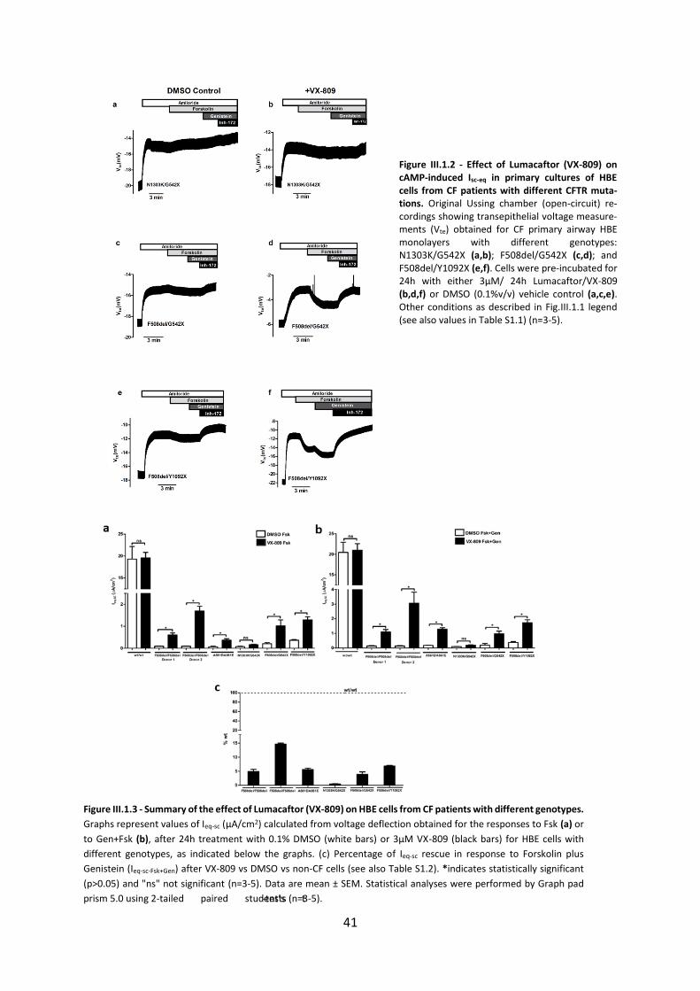
41
Figure III.1.2 - Effect of Lumacaftor (VX-809) on cAMP-induced Isc-eq in primary cultures of HBE cells from CF patients with different CFTR muta-tions. Original Ussing chamber (open-circuit) re-cordings showing transepithelial voltage measure-ments (Vte) obtained for CF primary airway HBE monolayers with different genotypes: N1303K/G542X (a,b); F508del/G542X (c,d); and F508del/Y1092X (e,f). Cells were pre-incubated for 24h with either 3µM/ 24h Lumacaftor/VX-809 (b,d,f) or DMSO (0.1%v/v) vehicle control (a,c,e). Other conditions as described in Fig.III.1.1 legend (see also values in Table S1.1) (n=3-5).
Figure III.1.3 - Summary of the effect of Lumacaftor (VX-809) on HBE cells from CF patients with different genotypes.
Graphs represent values of Ieq-sc (µA/cm2) calculated from voltage deflection obtained for the responses to Fsk (a) or
to Gen+Fsk (b), after 24h treatment with 0.1% DMSO (white bars) or 3µM VX-809 (black bars) for HBE cells with
different genotypes, as indicated below the graphs. (c) Percentage of Ieq-sc rescue in response to Forskolin plus
Genistein (Ieq-sc-Fsk+Gen) after VX-809 vs DMSO vs non-CF cells (see also Table S1.2). *indicates statistically significant
(p>0.05) and "ns" not significant (n=3-5). Data are mean ± SEM. Statistical analyses were performed by Graph pad
prism 5.0 using 2-tailed paired student’s t-tests (n=3-5).
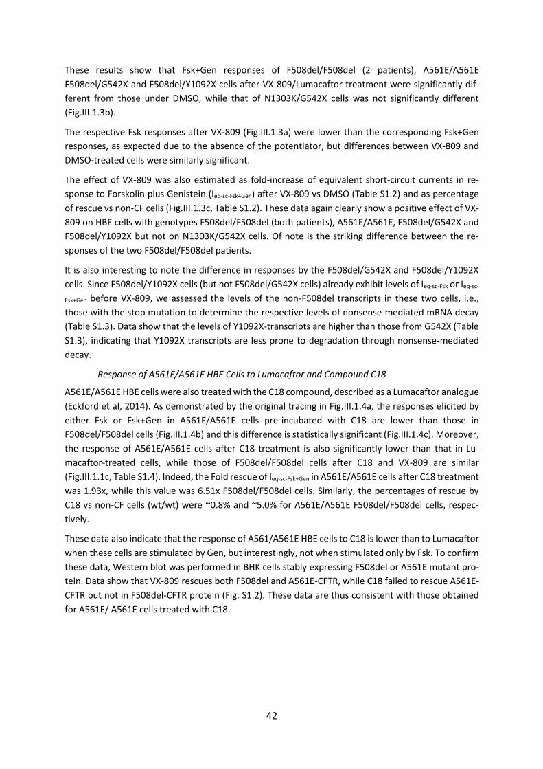
42
These results show that Fsk+Gen responses of F508del/F508del (2 patients), A561E/A561E
F508del/G542X and F508del/Y1092X cells after VX-809/Lumacaftor treatment were significantly dif-
ferent from those under DMSO, while that of N1303K/G542X cells was not significantly different
(Fig.III.1.3b).
The respective Fsk responses after VX-809 (Fig.III.1.3a) were lower than the corresponding Fsk+Gen
responses, as expected due to the absence of the potentiator, but differences between VX-809 and
DMSO-treated cells were similarly significant.
The effect of VX-809 was also estimated as fold-increase of equivalent short-circuit currents in re-
sponse to Forskolin plus Genistein (Ieq-sc-Fsk+Gen) after VX-809 vs DMSO (Table S1.2) and as percentage
of rescue vs non-CF cells (Fig.III.1.3c, Table S1.2). These data again clearly show a positive effect of VX-
809 on HBE cells with genotypes F508del/F508del (both patients), A561E/A561E, F508del/G542X and
F508del/Y1092X but not on N1303K/G542X cells. Of note is the striking difference between the re-
sponses of the two F508del/F508del patients.
It is also interesting to note the difference in responses by the F508del/G542X and F508del/Y1092X
cells. Since F508del/Y1092X cells (but not F508del/G542X cells) already exhibit levels of Ieq-sc-Fsk or Ieq-sc-
Fsk+Gen before VX-809, we assessed the levels of the non-F508del transcripts in these two cells, i.e.,
those with the stop mutation to determine the respective levels of nonsense-mediated mRNA decay
(Table S1.3). Data show that the levels of Y1092X-transcripts are higher than those from G542X (Table
S1.3), indicating that Y1092X transcripts are less prone to degradation through nonsense-mediated
decay.
Response of A561E/A561E HBE Cells to Lumacaftor and Compound C18
A561E/A561E HBE cells were also treated with the C18 compound, described as a Lumacaftor analogue
(Eckford et al, 2014). As demonstrated by the original tracing in Fig.III.1.4a, the responses elicited by
either Fsk or Fsk+Gen in A561E/A561E cells pre-incubated with C18 are lower than those in
F508del/F508del cells (Fig.III.1.4b) and this difference is statistically significant (Fig.III.1.4c). Moreover,
the response of A561E/A561E cells after C18 treatment is also significantly lower than that in Lu-
macaftor-treated cells, while those of F508del/F508del cells after C18 and VX-809 are similar
(Fig.III.1.1c, Table S1.4). Indeed, the Fold rescue of Ieq-sc-Fsk+Gen in A561E/A561E cells after C18 treatment
was 1.93x, while this value was 6.51x F508del/F508del cells. Similarly, the percentages of rescue by
C18 vs non-CF cells (wt/wt) were ~0.8% and ~5.0% for A561E/A561E F508del/F508del cells, respec-
tively.
These data also indicate that the response of A561/A561E HBE cells to C18 is lower than to Lumacaftor
when these cells are stimulated by Gen, but interestingly, not when stimulated only by Fsk. To confirm
these data, Western blot was performed in BHK cells stably expressing F508del or A561E mutant pro-
tein. Data show that VX-809 rescues both F508del and A561E-CFTR, while C18 failed to rescue A561E-
CFTR but not in F508del-CFTR protein (Fig. S1.2). These data are thus consistent with those obtained
for A561E/ A561E cells treated with C18.
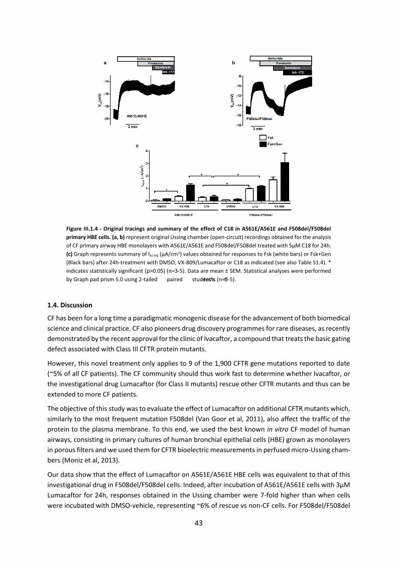
43
Figure III.1.4 - Original tracings and summary of the effect of C18 in A561E/A561E and F508del/F508del
primary HBE cells. (a, b) represent original Ussing chamber (open-circuit) recordings obtained for the analysis
of CF primary airway HBE monolayers with A561E/A561E and F508del/F508del treated with 5µM C18 for 24h.
(c) Graph represents summary of Isc-eq (µA/cm2) values obtained for responses to Fsk (white bars) or Fsk+Gen
(Black bars) after 24h-treatment with DMSO, VX-809/Lumacaftor or C18 as indicated (see also Table S1.4). *
indicates statistically significant (p>0.05) (n=3-5). Data are mean ± SEM. Statistical analyses were performed
by Graph pad prism 5.0 using 2-tailed paired student’s t-tests (n=3-5).
1.4. Discussion
CF has been for a long time a paradigmatic monogenic disease for the advancement of both biomedical
science and clinical practice. CF also pioneers drug discovery programmes for rare diseases, as recently
demonstrated by the recent approval for the clinic of Ivacaftor, a compound that treats the basic gating
defect associated with Class III CFTR protein mutants.
However, this novel treatment only applies to 9 of the 1,900 CFTR gene mutations reported to date
(~5% of all CF patients). The CF community should thus work fast to determine whether Ivacaftor, or
the investigational drug Lumacaftor (for Class II mutants) rescue other CFTR mutants and thus can be
extended to more CF patients.
The objective of this study was to evaluate the effect of Lumacaftor on additional CFTR mutants which,
similarly to the most frequent mutation F508del (Van Goor et al, 2011), also affect the traffic of the
protein to the plasma membrane. To this end, we used the best known in vitro CF model of human
airways, consisting in primary cultures of human bronchial epithelial cells (HBE) grown as monolayers
in porous filters and we used them for CFTR bioelectric measurements in perfused micro-Ussing cham-
bers (Moniz et al, 2013).
Our data show that the effect of Lumacaftor on A561E/A561E HBE cells was equivalent to that of this
investigational drug in F508del/F508del cells. Indeed, after incubation of A561E/A561E cells with 3µM
Lumacaftor for 24h, responses obtained in the Ussing chamber were 7-fold higher than when cells
were incubated with DMSO-vehicle, representing ~6% of rescue vs non-CF cells. For F508del/F508del
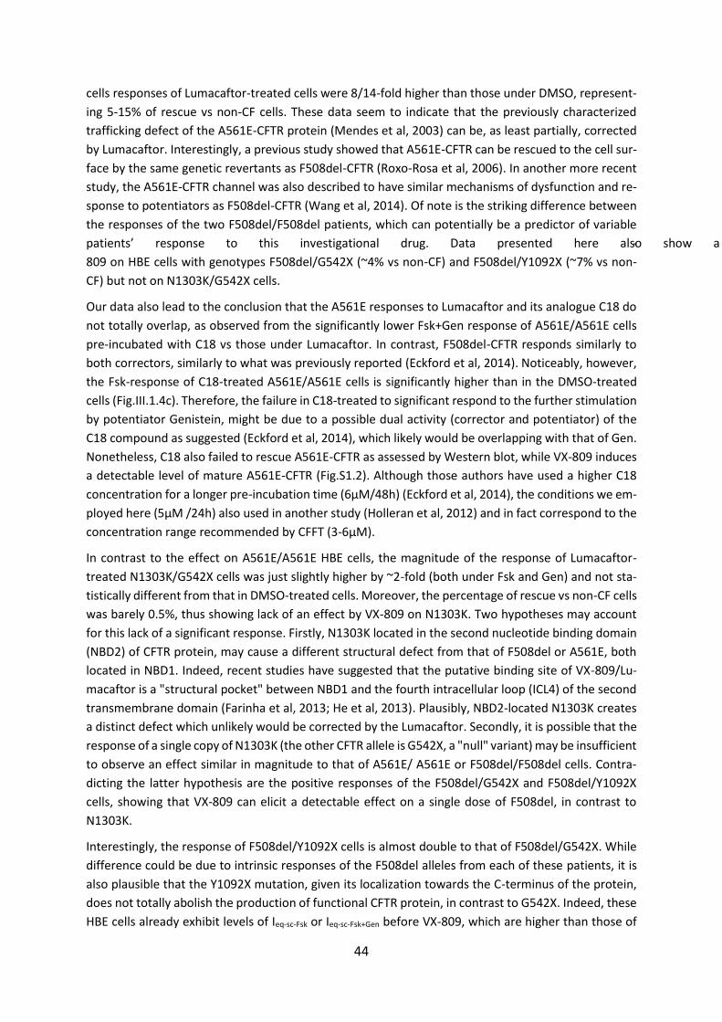
44
cells responses of Lumacaftor-treated cells were 8/14-fold higher than those under DMSO, represent-
ing 5-15% of rescue vs non-CF cells. These data seem to indicate that the previously characterized
trafficking defect of the A561E-CFTR protein (Mendes et al, 2003) can be, as least partially, corrected
by Lumacaftor. Interestingly, a previous study showed that A561E-CFTR can be rescued to the cell sur-
face by the same genetic revertants as F508del-CFTR (Roxo-Rosa et al, 2006). In another more recent
study, the A561E-CFTR channel was also described to have similar mechanisms of dysfunction and re-
sponse to potentiators as F508del-CFTR (Wang et al, 2014). Of note is the striking difference between
the responses of the two F508del/F508del patients, which can potentially be a predictor of variable
patients’ response to this investigational drug. Data presented here also show a positive effect of VX-
809 on HBE cells with genotypes F508del/G542X (~4% vs non-CF) and F508del/Y1092X (~7% vs non-
CF) but not on N1303K/G542X cells.
Our data also lead to the conclusion that the A561E responses to Lumacaftor and its analogue C18 do
not totally overlap, as observed from the significantly lower Fsk+Gen response of A561E/A561E cells
pre-incubated with C18 vs those under Lumacaftor. In contrast, F508del-CFTR responds similarly to
both correctors, similarly to what was previously reported (Eckford et al, 2014). Noticeably, however,
the Fsk-response of C18-treated A561E/A561E cells is significantly higher than in the DMSO-treated
cells (Fig.III.1.4c). Therefore, the failure in C18-treated to significant respond to the further stimulation
by potentiator Genistein, might be due to a possible dual activity (corrector and potentiator) of the
C18 compound as suggested (Eckford et al, 2014), which likely would be overlapping with that of Gen.
Nonetheless, C18 also failed to rescue A561E-CFTR as assessed by Western blot, while VX-809 induces
a detectable level of mature A561E-CFTR (Fig.S1.2). Although those authors have used a higher C18
concentration for a longer pre-incubation time (6µM/48h) (Eckford et al, 2014), the conditions we em-
ployed here (5µM /24h) also used in another study (Holleran et al, 2012) and in fact correspond to the
concentration range recommended by CFFT (3-6µM).
In contrast to the effect on A561E/A561E HBE cells, the magnitude of the response of Lumacaftor-
treated N1303K/G542X cells was just slightly higher by ~2-fold (both under Fsk and Gen) and not sta-
tistically different from that in DMSO-treated cells. Moreover, the percentage of rescue vs non-CF cells
was barely 0.5%, thus showing lack of an effect by VX-809 on N1303K. Two hypotheses may account
for this lack of a significant response. Firstly, N1303K located in the second nucleotide binding domain
(NBD2) of CFTR protein, may cause a different structural defect from that of F508del or A561E, both
located in NBD1. Indeed, recent studies have suggested that the putative binding site of VX-809/Lu-
macaftor is a "structural pocket" between NBD1 and the fourth intracellular loop (ICL4) of the second
transmembrane domain (Farinha et al, 2013; He et al, 2013). Plausibly, NBD2-located N1303K creates
a distinct defect which unlikely would be corrected by the Lumacaftor. Secondly, it is possible that the
response of a single copy of N1303K (the other CFTR allele is G542X, a "null" variant) may be insufficient
to observe an effect similar in magnitude to that of A561E/ A561E or F508del/F508del cells. Contra-
dicting the latter hypothesis are the positive responses of the F508del/G542X and F508del/Y1092X
cells, showing that VX-809 can elicit a detectable effect on a single dose of F508del, in contrast to
N1303K.
Interestingly, the response of F508del/Y1092X cells is almost double to that of F508del/G542X. While
difference could be due to intrinsic responses of the F508del alleles from each of these patients, it is
also plausible that the Y1092X mutation, given its localization towards the C-terminus of the protein,
does not totally abolish the production of functional CFTR protein, in contrast to G542X. Indeed, these
HBE cells already exhibit levels of Ieq-sc-Fsk or Ieq-sc-Fsk+Gen before VX-809, which are higher than those of
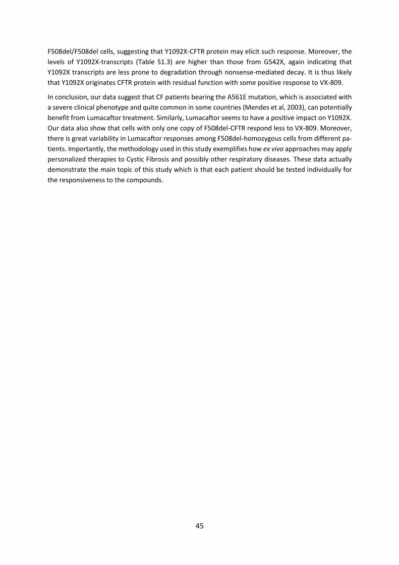
45
F508del/F508del cells, suggesting that Y1092X-CFTR protein may elicit such response. Moreover, the
levels of Y1092X-transcripts (Table S1.3) are higher than those from G542X, again indicating that
Y1092X transcripts are less prone to degradation through nonsense-mediated decay. It is thus likely
that Y1092X originates CFTR protein with residual function with some positive response to VX-809.
In conclusion, our data suggest that CF patients bearing the A561E mutation, which is associated with
a severe clinical phenotype and quite common in some countries (Mendes et al, 2003), can potentially
benefit from Lumacaftor treatment. Similarly, Lumacaftor seems to have a positive impact on Y1092X.
Our data also show that cells with only one copy of F508del-CFTR respond less to VX-809. Moreover,
there is great variability in Lumacaftor responses among F508del-homozygous cells from different pa-
tients. Importantly, the methodology used in this study exemplifies how ex vivo approaches may apply
personalized therapies to Cystic Fibrosis and possibly other respiratory diseases. These data actually
demonstrate the main topic of this study which is that each patient should be tested individually for
the responsiveness to the compounds.
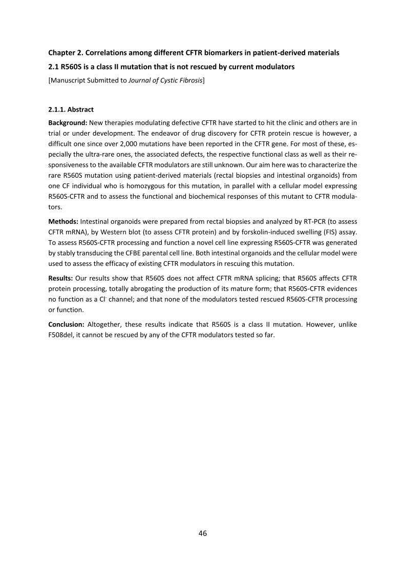
46
Chapter 2. Correlations among different CFTR biomarkers in patient-derived materials
2.1 R560S is a class II mutation that is not rescued by current modulators
[Manuscript Submitted to Journal of Cystic Fibrosis]
2.1.1. Abstract
Background: New therapies modulating defective CFTR have started to hit the clinic and others are in
trial or under development. The endeavor of drug discovery for CFTR protein rescue is however, a
difficult one since over 2,000 mutations have been reported in the CFTR gene. For most of these, es-
pecially the ultra-rare ones, the associated defects, the respective functional class as well as their re-
sponsiveness to the available CFTR modulators are still unknown. Our aim here was to characterize the
rare R560S mutation using patient-derived materials (rectal biopsies and intestinal organoids) from
one CF individual who is homozygous for this mutation, in parallel with a cellular model expressing
R560S-CFTR and to assess the functional and biochemical responses of this mutant to CFTR modula-
tors.
Methods: Intestinal organoids were prepared from rectal biopsies and analyzed by RT-PCR (to assess
CFTR mRNA), by Western blot (to assess CFTR protein) and by forskolin-induced swelling (FIS) assay.
To assess R560S-CFTR processing and function a novel cell line expressing R560S-CFTR was generated
by stably transducing the CFBE parental cell line. Both intestinal organoids and the cellular model were
used to assess the efficacy of existing CFTR modulators in rescuing this mutation.
Results: Our results show that R560S does not affect CFTR mRNA splicing; that R560S affects CFTR
protein processing, totally abrogating the production of its mature form; that R560S-CFTR evidences
no function as a Cl- channel; and that none of the modulators tested rescued R560S-CFTR processing
or function.
Conclusion: Altogether, these results indicate that R560S is a class II mutation. However, unlike
F508del, it cannot be rescued by any of the CFTR modulators tested so far.
47
2.1.2. Introduction
Cystic fibrosis (CF), the most common life-shortening autosomal recessive disease in Caucasians is
caused by mutations in the gene encoding the CF transmembrane conductance regulator (CFTR) pro-
tein which plays a major role in Chloride (Cl-) and bicarbonate (HCO3-) conductances in epithelia, as
well as in regulating other ion channels and transporters (Bobadilla JL et al, 2002). CF-causing muta-
tions prevent the channel from proper functioning, leading to abnormal ion transport and causing de-
hydration of the airway surface liquid (Rowe SM et al, 2005). Most current CF therapies target its symp-
toms (mucolytics, antibiotics, etc). However, innovative therapies modulating defective CFTR, the basic
defect underlying CF, have started to hit the clinic and new ones are in trial or under development
(DeBoeck K & Amaral M, 2016). The endeavor of drug discovery to rescue mutant CFTR protein is how-
ever, a difficult one since to date >2,000 mutations have been reported in the CFTR gene, most of
which still with unknown impact, both in terms of their underlying molecular/cellular defect and dis-
ease liability (Sosnay et al, 2013). To tackle this very high number of mutations, a grouping of CFTR
mutations into functional classes has been proposed. These original classes are continuously updated
(DeBoeck K & Amaral M, 2016) especially as they evolve into ‘theratypes’ (Cutting GR, 2015) since they
are becoming increasingly defined by the respective therapeutic strategy designed to rescue the re-
spective molecular/cellular defect. The rationale is that mutations within the same class can be treated
by the same therapeutic strategy. Although this classification is helpful to rationalize therapies, the
underlying defect (and hence the respective mutation class) is still unknown for the majority of CFTR
mutations, in particular for the rare ones. Moreover, there are several examples of mutations within
the same class/47theratypes which however do not equally respond to the same CFTR modulator
(Awatade NT et al, 2015). It is also often found that patients with the same CFTR genotype (such as
those who are homozygous for F508del, the most common disease-causing variant) have significantly
different clinical responses to CFTR modulating drugs (Wainwright et al, 2015; Awatade NT et al, 2015).
There is thus an unmet need to test these novel CFTR modulators directly ex vivo on the patient’s own
tissues/primary cells with robust biomarkers that not only assess modulators efficacy for a given CFTR
genotype but also predict clinical benefit for an individual patient.
The p.Arg560Ser mutation (legacy name: R560S) is caused by a A>C transversion at the cDNA nucleo-
tide position c.1680 (legacy: 1812 A>C), located in exon 13, leading to the exchange of the amino acid
Arginine by a Serine at position 560, being thus a missense mutation (Liechti-Gallatis et al, 1999;
Malone G et al, 1998). However, the c.1680 A>C nucleotide change occurs at the intron11/ exon12
boundary (first nucleotide of exon 12), being thus plausible to envisage that this mutation may also
affect splicing. R560S is described as CF-causing when combined with another CF-causing mutation, in
CFTR2 (www.cftr2.org) mutation database. R560S causes pancreatic insufficiency (PI), when combined
with another PI-causing mutation. This mutation was originally reported in the CFTR Mutation Data-
base by B Costes in a Pakistani 11-year old male patient diagnosed at 4m, PI, with moderate lung dis-
ease and positive sweat Cl-. To this date, there are 8 patients reported with this mutation in the CFTR2
database.
R560 is located in the first nucleotide-binding domain (NBD1) which is involved in ATP binding and
playing a role in channel opening (i.e., gating) of CFTR. NBD1 is also a “mutational hotspot” for CF since
many CF-causing mutations localize to this domain, including the most frequent one (F508del) which
occurs in ~85% of CF patients worldwide and is described to affect CFTR protein processing (class II).
48
Our aim here was to characterize the rare CFTR-R560S mutation and its response to existing modula-
tors both in patient-derived materials from one individual with CF who is homozygous for this mutation
and also in a cell line model.
2.1.3. Results
mRNA analysis
Our first aim here was to characterize the molecular consequences of the R560S mutation, in terms of
the possible effects of this mutation on mRNA splicing, given that the c.1680A>C nucleotide change
occurs at the first nucleotide of exon 12 (see Introduction). To this end, we isolated RNA from the
patient’s organoids which, after cDNA synthesis were analyzed by RT-PCR in the region of the muta-
tion, to assess for possible exon skipping of nearby exons (Fig.III.2.1.1 A). Three different PCR reactions
were thus used covering exons 8-11, 10-12 and 11-14a, as previously described (Felício V et al, 2016).
Data show that all RT-PCR products have the expected size, thus corresponding to correctly spliced
CFTR transcripts for all regions analyzed (Fig.III.2.1.1 A), demonstrating that there is no exon skipping
in the exon 8-14a region and thus showing that c.1680A>C is not a splicing mutation.
Figure III.2.1.1 - Impact of the R560S mutation on mRNA splicing and protein processing. (A) RT-PCR analysis of CFTR transcripts extracted from organoids derived from rectal biopsies (RB) from CF patient with the R560S/R560S genotype in the regions between exons 8 and 14a (n=3). P - RNA from patient (R560S/R560S) organoids, NC - Negative control. (B) Representative WB analysis of CFTR protein expressed in rectal organ-oids from a non-CF control (wt) or from the R560S homozygous patient. WB was performed using the anti-CFTR 596 antibody. Images were acquired using ChemiDoc XRS+ imaging system BioRad and further processed by Image lab 4.0 software (n-3).
To assess whether R560S affects CFTR processing, total protein was extracted from rectal organoids
and Western blot (WB) was used to assess the presence of immature (band B) and mature (band C)
forms of the protein. Results obtained here (Fig.III.2.1.1 B) clearly show that the protein resulting from
R560S mutation only appears in its immature (core-glycosylated, band B) form, different from the pro-
tein in samples from non-CF individuals that present both the mature (fully-glycosylated, band C) form
and some as immature form (band B). Altogether, these results indicate that this CFTR mutation causes
a defect in CFTR processing.
Assessment of rescue of R560S-CFTR protein processing by correctors
We assessed the efficacy of different small molecule compounds in rescuing R60S-CFTR. For this, we
produced a novel cell line expressing R560S-CFTR by stably transducing CFBE parental cells and the
following compounds were tested: VX-809/lumacaftor - corrector drug approved for F508del/F508del
patients (in combination with VX-770); VX-661/Tezacaftor (second generation corrector which already

49
succeeded in phase III clinical trials); Cysteamine (an FDA-approved drug for the treatment of Cysti-
nosis) alone and in combination with green tea component EGCG, described to rescue F508del-CFTR
(Tosco A et al, 2016).
Cells were thus incubated for 24h with: VX-809 3μM, VX-661 5μM, DMSO 0.1% v/v as the vehicle con-
trol, cysteamine 250μM alone or in combination with EGCG 50μM, and at low temperature (27ºC) also
described to rescue processing of some class II mutants, namely F508del. In parallel, these compounds
were also tested on F508del-CFTR in CFBE cells as a control. As above, we performed WB to assess the
maturation status/rescue of F508del and R560S-CFTR (Fig.III.2.1.2).
As shown in Fig.III.2.1.2, wt-CFTR is detected in both its immature (band B) and mature forms (Band
C). However, R560S-CFTR can only be detected in their immature form (band B) indicating that similarly
to data obtained in organoids (Fig.III.2.1.2 B), this protein does not reach the cell surface, similarly to
F508del-CFTR. However, in contrast to F508del-CFTR, for which VX-809, VX-661, and low temperature
promote the appearance of mature CFTR, for R560S-CFTR, neither treatment nor incubation at low
temperature (Fig.III.2.1.2 A, B) lead to the appearance of band C. Treatment with cysteamine however,
either alone or when combined to ECGC, did not rescue R560S- nor F508del-CFTR, the latter in contrast
to what was previously described (Tosco A et al, 2016). Strikingly, the combined treatment of cysteam-
ine and EGCG leads to the total disappearance of CFTR, be it wt-, F508del or R560S (Fig.III.2.1.2 B, F).
Figure III.2.1.2 – Effect of correctors on processing of R560S-CFTR and F508del- (control) CFTR protein as-sessed by WB. (A) Representative WB analysis of CFBE cells stably expressing R560S-, F508del or wt-CFTR, following treatment with VX-809 (3 μM), VX-661(5 μM), DMSO (0.1% v/v) or incubation at 27ºC, for 24h. (B) Representative WB analysis of CFBE cells stably expressing R560S-, F508del- or wt-CFTR, following the treat-ment with cysteamine (250 μM) and the combination of cysteamine and EGCG (50 μM) for 24h, as indicated above lanes. (C),(E) For each condition, densitometry was used to calculate the percentage of mature CFTR (band C) vs total CFTR expressed, following image acquisition as in Fig.1B. Data were normalized to the effi-ciency of processing of wt-CFTR and are shown as mean±SEM (D),(F) For each condition, densitometry was used to calculate the total amount of CFTR normalized to the loading control and to the amount of CFTR in the non-treated sample and are shown as mean±SD (n=3).
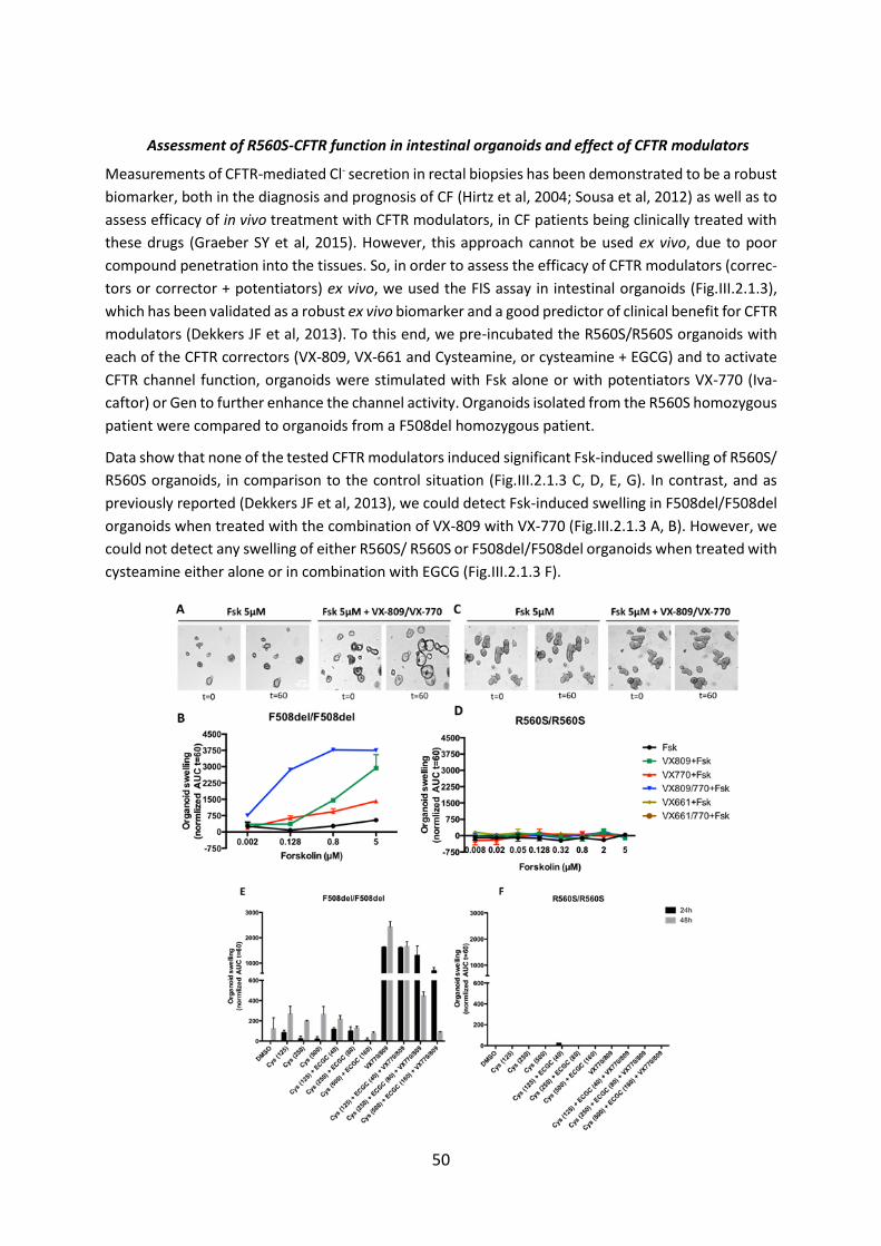
50
Assessment of R560S-CFTR function in intestinal organoids and effect of CFTR modulators
Measurements of CFTR-mediated Cl- secretion in rectal biopsies has been demonstrated to be a robust
biomarker, both in the diagnosis and prognosis of CF (Hirtz et al, 2004; Sousa et al, 2012) as well as to
assess efficacy of in vivo treatment with CFTR modulators, in CF patients being clinically treated with
these drugs (Graeber SY et al, 2015). However, this approach cannot be used ex vivo, due to poor
compound penetration into the tissues. So, in order to assess the efficacy of CFTR modulators (correc-
tors or corrector + potentiators) ex vivo, we used the FIS assay in intestinal organoids (Fig.III.2.1.3),
which has been validated as a robust ex vivo biomarker and a good predictor of clinical benefit for CFTR
modulators (Dekkers JF et al, 2013). To this end, we pre-incubated the R560S/R560S organoids with
each of the CFTR correctors (VX-809, VX-661 and Cysteamine, or cysteamine + EGCG) and to activate
CFTR channel function, organoids were stimulated with Fsk alone or with potentiators VX-770 (Iva-
caftor) or Gen to further enhance the channel activity. Organoids isolated from the R560S homozygous
patient were compared to organoids from a F508del homozygous patient.
Data show that none of the tested CFTR modulators induced significant Fsk-induced swelling of R560S/
R560S organoids, in comparison to the control situation (Fig.III.2.1.3 C, D, E, G). In contrast, and as
previously reported (Dekkers JF et al, 2013), we could detect Fsk-induced swelling in F508del/F508del
organoids when treated with the combination of VX-809 with VX-770 (Fig.III.2.1.3 A, B). However, we
could not detect any swelling of either R560S/ R560S or F508del/F508del organoids when treated with
cysteamine either alone or in combination with EGCG (Fig.III.2.1.3 F).
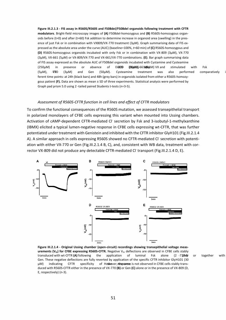
51
Figure III.2.1.3 - FIS assay in R560S/R560S and F508del/F508del organoids following treatment with CFTR
modulators. Bright-field microscopy images of (A) F508del-homozygous and (B) R560S-homozygous organ-
oids before (t=0) and after (t=60) Fsk addition to determine increase in organoid area (swelling) in the pres-
ence of just Fsk or in combination with VX809/VX-770 treatment (3µM). Graph summarising data of FIS ex-
pressed as the absolute area under the curve (AUC) (baseline=100%, t=60 min) of (C) R560S-homozygous and
(D) R560S-homozygous organoids incubated with only Fsk or in combination with VX-809 (3µM), VX-770
(3µM), VX-661 (5µM) or VX-809/VX-770 and VX-661/VX-770 combinations. (E). Bar graph summarising data
of FIS assay expressed as the absolute AUC of F508del organoids incubated with Cystamine and Cysteamine
(250μM) in presence or absence of EGCG (80μM) and VX-809 (3μM), VX-661 (5μM) and stimulated with Fsk
(5μM), VX-770 (3μM) and Gen (50μM). Cysteamine treatment was also performed comparatively in two dif-
ferent time points: at 24h (black bars) and 48h (grey bars) in organoids isolated from either a R560S-homozy-
gous patient (F). Data are shown as mean ± SD of three experiments. Statistical analysis were performed by
Graph pad prism 5.0 using 2 -tailed paired Students t-tests (n=3-5).
Assessment of R560S-CFTR function in cell lines and effect of CFTR modulators
To confirm the functional consequences of the R560S mutation, we assessed transepithelial transport
in polarized monolayers of CFBE cells expressing this variant when mounted into Ussing chambers.
Activation of cAMP-dependent CFTR-mediated Cl- secretion by Fsk and 3-isobutyl-1-methylxanthine
(IBMX) elicited a typical lumen-negative response in CFBE cells expressing wt-CFTR, that was further
potentiated under treatment with Genistein and inhibited with the CFTR inhibitor GlyH101 (Fig.III.2.1.4
A). A similar approach in cells expressing R560S showed no CFTR-mediated Cl- secretion with potenti-
ation with either VX-770 or Gen (Fig.III.2.1.4 B, C), and, consistent with WB data, treatment with cor-
rector VX-809 did not produce any detectable CFTR-mediated Cl- transport (Fig.III.2.1.4 D, E).
Figure III.2.1.4 - Original Ussing chamber (open-circuit) recordings showing transepithelial voltage meas-urements (Vte) for CFBE expressing R560S-CFTR. Negative Vte deflections are observed in CFBE cells stably transduced with wt-CFTR (A) following the application of luminal Fsk alone (2 μM) or together with VX-770 or Gen. These negative deflections are fully reverted by application of the specific CFTR inhibitor GlyH101 (30 μM) indicating CFTR specificity of the response. However, the same is not observed in CFBE cells stably trans-duced with R560S-CFTR either in the presence of VX-770 (B) or Gen (C) alone or in the presence of VX-809 (D, E, respectively) (n-3).
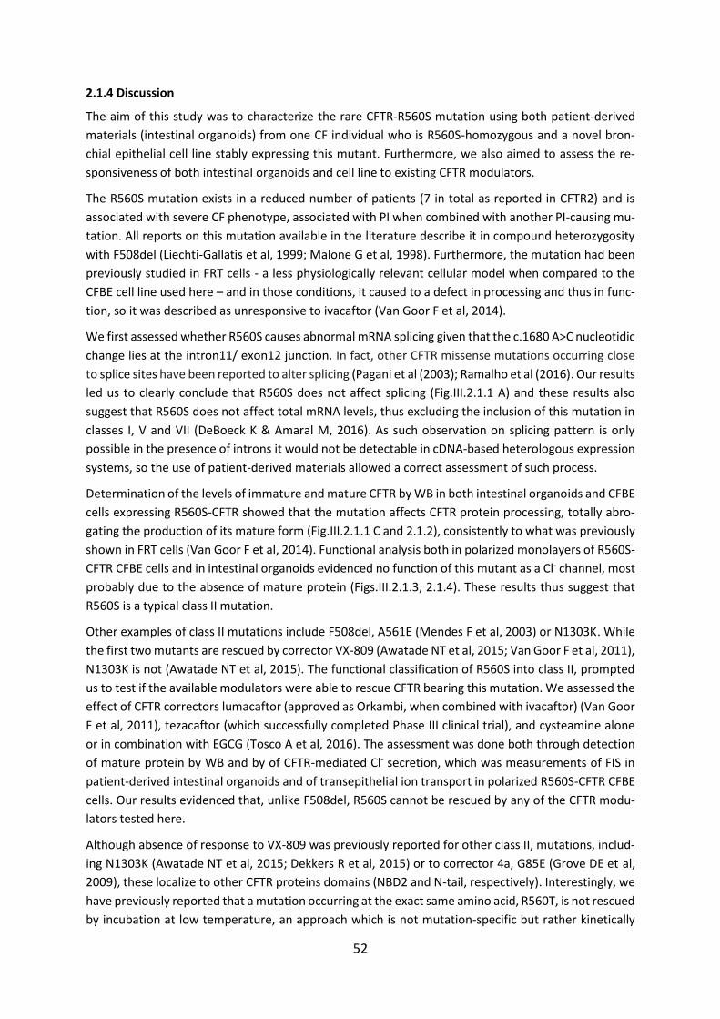
52
2.1.4 Discussion
The aim of this study was to characterize the rare CFTR-R560S mutation using both patient-derived
materials (intestinal organoids) from one CF individual who is R560S-homozygous and a novel bron-
chial epithelial cell line stably expressing this mutant. Furthermore, we also aimed to assess the re-
sponsiveness of both intestinal organoids and cell line to existing CFTR modulators.
The R560S mutation exists in a reduced number of patients (7 in total as reported in CFTR2) and is
associated with severe CF phenotype, associated with PI when combined with another PI-causing mu-
tation. All reports on this mutation available in the literature describe it in compound heterozygosity
with F508del (Liechti-Gallatis et al, 1999; Malone G et al, 1998). Furthermore, the mutation had been
previously studied in FRT cells - a less physiologically relevant cellular model when compared to the
CFBE cell line used here – and in those conditions, it caused to a defect in processing and thus in func-
tion, so it was described as unresponsive to ivacaftor (Van Goor F et al, 2014).
We first assessed whether R560S causes abnormal mRNA splicing given that the c.1680 A>C nucleotidic
change lies at the intron11/ exon12 junction. In fact, other CFTR missense mutations occurring close
to splice sites have been reported to alter splicing (Pagani et al (2003); Ramalho et al (2016). Our results
led us to clearly conclude that R560S does not affect splicing (Fig.III.2.1.1 A) and these results also
suggest that R560S does not affect total mRNA levels, thus excluding the inclusion of this mutation in
classes I, V and VII (DeBoeck K & Amaral M, 2016). As such observation on splicing pattern is only
possible in the presence of introns it would not be detectable in cDNA-based heterologous expression
systems, so the use of patient-derived materials allowed a correct assessment of such process.
Determination of the levels of immature and mature CFTR by WB in both intestinal organoids and CFBE
cells expressing R560S-CFTR showed that the mutation affects CFTR protein processing, totally abro-
gating the production of its mature form (Fig.III.2.1.1 C and 2.1.2), consistently to what was previously
shown in FRT cells (Van Goor F et al, 2014). Functional analysis both in polarized monolayers of R560S-
CFTR CFBE cells and in intestinal organoids evidenced no function of this mutant as a Cl- channel, most
probably due to the absence of mature protein (Figs.III.2.1.3, 2.1.4). These results thus suggest that
R560S is a typical class II mutation.
Other examples of class II mutations include F508del, A561E (Mendes F et al, 2003) or N1303K. While
the first two mutants are rescued by corrector VX-809 (Awatade NT et al, 2015; Van Goor F et al, 2011),
N1303K is not (Awatade NT et al, 2015). The functional classification of R560S into class II, prompted
us to test if the available modulators were able to rescue CFTR bearing this mutation. We assessed the
effect of CFTR correctors lumacaftor (approved as Orkambi, when combined with ivacaftor) (Van Goor
F et al, 2011), tezacaftor (which successfully completed Phase III clinical trial), and cysteamine alone
or in combination with EGCG (Tosco A et al, 2016). The assessment was done both through detection
of mature protein by WB and by of CFTR-mediated Cl- secretion, which was measurements of FIS in
patient-derived intestinal organoids and of transepithelial ion transport in polarized R560S-CFTR CFBE
cells. Our results evidenced that, unlike F508del, R560S cannot be rescued by any of the CFTR modu-
lators tested here.
Although absence of response to VX-809 was previously reported for other class II, mutations, includ-
ing N1303K (Awatade NT et al, 2015; Dekkers R et al, 2015) or to corrector 4a, G85E (Grove DE et al,
2009), these localize to other CFTR proteins domains (NBD2 and N-tail, respectively). Interestingly, we
have previously reported that a mutation occurring at the exact same amino acid, R560T, is not rescued
by incubation at low temperature, an approach which is not mutation-specific but rather kinetically

53
favours protein folding, thus allowing them to overcome the endoplasmic reticulum quality control
(Farinha CM et al, 2013; Roxo Rosa M et al, 2006). A third CF-causing mutation was occurring at the
same amino acid, R560K, was also reported (Roxo Rosa M et al, 2006). The three mutations have in
common causing severe CF (Sosnay PR et al, 2013), and our results here and previously reported
(Farinha CM et al, 2013; Roxo Rosa M et al, 2006) indicate that at least two of these variants are difficult
to rescue. The recent publication of CFTR structure at high-resolution identified these 3 mutations as
causing a defect in the interaction between NBD1 and the TMDs (Zhang Z et al, 2016), being thus plau-
sible to consider this is a very sensitive site for inter-domain folding.
Our results also very strikingly showed a total lack of response for cysteamine, either alone or in com-
bination with EGCG, in contrast to what was previously reported (Tosco A et al, 2016). This was ob-
served for all the models tested – organoids and cell lines – for both genotypes – R560S and F508del
(in homozygosity for the patient-derived materials). These observations may suggest that intestinal
organoids may not be adequate to detect the previously reported corrective effect of cysteam-
ine/EGCG (Tosco A et al, 2016). However, results from cell lines (directly assessing the presence of
CFTR) also failed to detect CFTR for the combined treatment cysteamine/EGCG, suggesting that what-
ever additional conditions used in the previous studies are not present in our system, and thus these
compounds do not elicit a general correction of F508del-CFTR in all the models/systems.
Altogether, the work presented here evidencing the lack of correction for the R560S mutation, illus-
trates how difficult it is to rescue all CFTR mutations, even if included in a theratype for which correc-
tion is already available for some mutations. Such observations reinforce the need for continuous ef-
fort in the search for better modulators or alternative therapeutic strategies that can ameliorate dis-
ease phenotype for patients bearing rare mutations.
54
2.2. Functional Assessment of Rare CFTR H1079P Mutation
2.2.1. Abstract
Background: Reportedly H1079P-CFTR mutation is very rare and has been identified in 2 patients
worldwide so far (these 2 patients analyzed here in this chapter). The biochemical and functional con-
sequences related with this mutation are unknown and thus making it difficult to categorise this mu-
tation into functional classification system of CFTR mutations.
Objective: Main objective of this study was to characterize biochemically and functionally rare CFTR-
H1079P mutation using patient-derived materials (rectal biopsies, nasal cells, and intestinal organoids)
from two sibling CF individuals who are compound heterozygous with the W1282X mutation.
Methods: CFTR activity was investigated in different tissues/cells from two CF siblings with
H1079P/W1282X genotype, namely in i) rectal biopsies by Ussing chamber measurements of CFTR-
mediated Cl- secretion; ii) intestinal organoids by forskolin induced swelling (FIS) assay to assess the
efficacy of clinically approved CFTR modulators and iii) immunofluorescence to assess whether
H1079P-CFTR is associated with a defect in intracellular traffic.
Results and Conclusions: Data obtained here show that H1079P-CFTR exhibited no function as a Cl-
channel. Clinically approved CFTR modulators VX-809/VX-770 and VX-661/VX-770 failed significantly
to increase H1079P-CFTR function. Readthrough agents PTC-124 and Gentamycin failed to provoke a
significant response for both patients. Altogether, results from this study may help to provide a better
understanding of rare CFTR-H1079P mutation. Our functional findings correlate very well with the clin-
ical parameters and may have pivotal clinical implications for personalized therapeutic intervention.
55
2.2.2. Introduction
Two subjects with the H1079P/W1282X genotype came to our lab for the functional assessment of
CFTR and to assess the drug responses on intestinal organoids. The W1282X mutation is described as
CF-causing when combined with another CF-causing mutation, both in the CFTR (www.genet.sick-
kids.on.ca/cftr) and CFTR2 (www.cftr2.org) mutation databases. It has been described as the most
common mutation in the Ashkenazi Jewish CF patients in Israel, with presentation of severe disease
(Shoshani et al, 1992) and also found at relatively high frequency in Italy (Castaldo et al, 1999). W1282X
causes PI when combined with another PI-causing mutation.
W1282X is located in the second nucleotide binding domain (NBD2) of the CFTR protein and in exon
20 at the gene level. It originates a premature termination codon (PTC) which generates a putative
CFTR protein lacking a significant part of NBD2 and the C-terminus. Usually, PTC mutations trigger a
cellular process called nonsense-mediated decay (NMD) of the respective PTC-bearing messenger RNA
(mRNA). Indeed, W1282X has been described to lead to NMD (Linde et al, 2007). Nevertheless, since
this mutation leads to a PTC close to the C-terminus of the CFTR protein, it is also plausible to envisage
that the truncated protein may still be partially functional, since a shorter version of the protein, i.e.
until residue D835 (D835X), was described to generate Cl- currents with properties typical of wt-CFTR
(Schwiebert et al, 1998). Consistently, we have previously analysed primary human bronchial epithelial
(HBE) cells from a CF patient with the Y1092X/F508del genotype (Awatade et al, 2015) and we found
some residual activity in these HBE cells which nevertheless could result from the F508del CFTR allele,
which in some patients leads to very residual channel activity (Hirtz et al, 2004).
Aminoglycoside antibiotics (gentamycin/G418, tobramycin) and the compound Ataluren (also known
PTC-124) have been described to have the ability to read-through PTCs, permitting translation of full-
length proteins and these compounds have been under clinical trial however without effect, being able
to prove no benefit in patients so far (Linde & Kerem, 2008; Keeling et al, 2014). Very recently, the
read-through efficacy of PTC124 and G418 was tested using human intestinal organoids from CF pa-
tients bearing at least one PTC mutation, including the W1282X/F508del genotype (Zomer-van Ommen
et al, 2016). Whereas these authors found partial functional restoration of CFTR mediated by G418
read-through activity, the same was not observed for PTC124, suggesting that better read-through
agents are needed for robust treatment of CFTR PTC mutations.
Regarding the H1079P mutation (exon 17b, intracellular loop 3), no information is available, since, to
our knowledge, this mutation only occurs worldwide in these 2 CF patients.
1. Our first objective here is to characterize rare CFTR-H1079P mutation using patient-derived tis-
sues (rectal biopsies and nasal cells) from the two sibling CF individuals who have this mutation in
compound heterozygosity with W1282X. To this end we propose:
a) to assess whether H1079P-CFTR is associated with a defect in intracellular traffic by deter-
mining its presence at the plasma membrane (by immunofluorescence), and
b) to determine whether there is residual CFTR-mediated Cl- secretion (Ussing chamber stud-
ies).
2. Our second and main objective here is to assess the efficacy of correctors and read-through agents
alone or with and potentiators on rectal organoids, namely by testing:
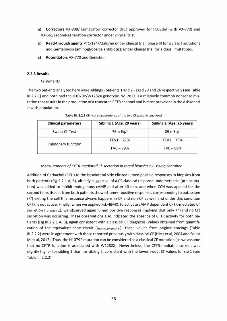
56
a) Correctors VX-809/ Lumacaftor corrector drug approved for F508del (with VX-770) and
VX-661 second-generation corrector under clinical trial;
b) Read-through agents PTC-124/Ataluren under clinical trial, phase III for a class I mutations
and Gentamycin (aminoglycoside antibiotic): under clinical trial for a class I mutations.
c) Potentiators VX-770 and Genistein
2.2.3 Results
CF patients
The two patients analyzed here were siblings - patients 1 and 2 - aged 29 and 26 respectively (see Table
III.2.2.1) and both had the H1079P/W1282X genotype. W1282X is a relatively common nonsense mu-
tation that results in the production of a truncated CFTR channel and is most prevalent in the Ashkenazi
Jewish population.
Table III. 2.2.1 Clinical characteristics of the two CF patients analysed
Clinical parameters Sibling 1 (Age: 29 years) Sibling 2 (Age: 26 years)
Sweat Cl- Test 76m Eq/l 89 mEq/l
Pulmonary function FEV1 – 71%
FVC – 79%
FEV1 – 79%
FVC – 89%
Measurements of CFTR-mediated Cl- secretion in rectal biopsies by Ussing chamber
Addition of Carbachol (CCH) to the basolateral side elicited lumen positive responses in biopsies from
both patients (Fig.2.2.1 A, B), already suggestive of a CF classical response. Indomethacin (preincuba-
tion) was added to inhibit endogenous cAMP and after 40 min, and when CCH was applied for the
second time, tissues from both patients showed lumen-positive responses corresponding to potassium
(K+) exiting the cell this response always happens in CF and non-CF as well and under this condition
CFTR is not active. Finally, when we applied Fsk+IBMX, to activate cAMP dependent CFTR-mediated Cl-
secretion (Isc-IBMX/Fsk), we observed again lumen positive responses implying that only K+ (and no Cl-)
secretion was occurring. These observations also indicated the absence of CFTR activity for both pa-
tients (Fig.III.2.2.1 A, B), again consistent with a classical CF diagnosis. Values obtained from quantifi-
cation of the equivalent short-circuit (Ieq-sc-CCH/IBMX/Fsk). These values from original tracings (Table
III.2.2.2) were in agreement with those reported previously with classical CF (Hirtz et al, 2004 and Sousa
M et al, 2012). Thus, the H1079P mutation can be considered as a classical CF mutation (as we assume
that no CFTR function is associated with W1282X). Nevertheless, the CFTR-mediated current was
slightly higher for sibling 1 than for sibling 2, consistent with the lower sweat Cl- values for sib 1 (see
Table III.2.2.2).
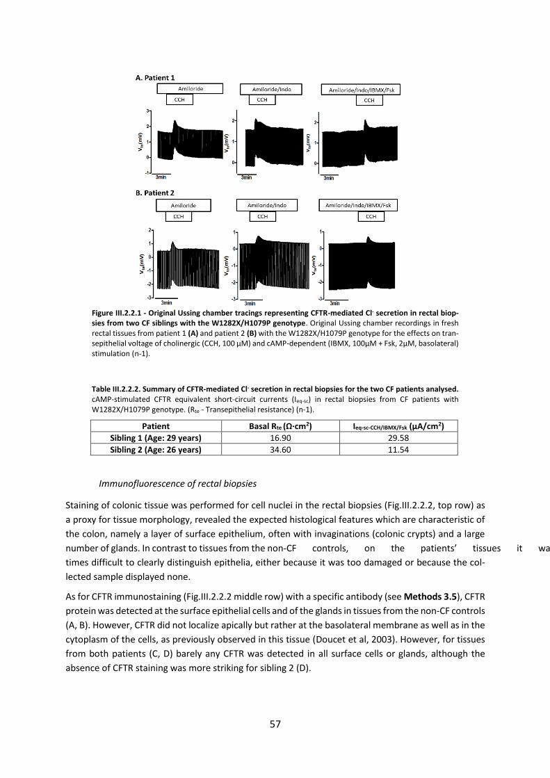
57
Figure III.2.2.1 - Original Ussing chamber tracings representing CFTR-mediated Cl- secretion in rectal biop-sies from two CF siblings with the W1282X/H1079P genotype. Original Ussing chamber recordings in fresh rectal tissues from patient 1 (A) and patient 2 (B) with the W1282X/H1079P genotype for the effects on tran-sepithelial voltage of cholinergic (CCH, 100 µM) and cAMP-dependent (IBMX, 100µM + Fsk, 2µM, basolateral) stimulation (n-1).
Table III.2.2.2. Summary of CFTR-mediated Cl- secretion in rectal biopsies for the two CF patients analysed. cAMP-stimulated CFTR equivalent short-circuit currents (Ieq-sc) in rectal biopsies from CF patients with W1282X/H1079P genotype. (Rte - Transepithelial resistance) (n-1).
Patient Basal Rte (Ω∙cm2) Ieq-sc-CCH/IBMX/Fsk (µA/cm2)
Sibling 1 (Age: 29 years) 16.90 29.58
Sibling 2 (Age: 26 years) 34.60 11.54
Immunofluorescence of rectal biopsies
Staining of colonic tissue was performed for cell nuclei in the rectal biopsies (Fig.III.2.2.2, top row) as
a proxy for tissue morphology, revealed the expected histological features which are characteristic of
the colon, namely a layer of surface epithelium, often with invaginations (colonic crypts) and a large
number of glands. In contrast to tissues from the non-CF controls, on the patients’ tissues it was some-
times difficult to clearly distinguish epithelia, either because it was too damaged or because the col-
lected sample displayed none.
As for CFTR immunostaining (Fig.III.2.2.2 middle row) with a specific antibody (see Methods 3.5), CFTR
protein was detected at the surface epithelial cells and of the glands in tissues from the non-CF controls
(A, B). However, CFTR did not localize apically but rather at the basolateral membrane as well as in the
cytoplasm of the cells, as previously observed in this tissue (Doucet et al, 2003). However, for tissues
from both patients (C, D) barely any CFTR was detected in all surface cells or glands, although the
absence of CFTR staining was more striking for sibling 2 (D).

58
Figure III.2.2.2 - Immunostaining of rectal biopsies in non-CF controls and in the two CF subjects. Top row represents staining of nuclei (green), middle row correspond to CFTR immune staining and lower row are the merge images of samples from: (A, B) two non-CF controls; and (C, D) siblings 1 and 2 under study, respec-tively. Scale bar represents 20µM. [Data from Margarida Quaresma, included here with permission]
Although the present results are representative of the intestinal morphology and of the low levels of
CFTR expression in tissues from these two patients, further optimization to increase sensitivity would
be required to arrive at firm conclusions on the plasma membrane localization of H1079P-CFTR, par-
ticular at these very low abundance levels. Although the protein resulting from the W1282X allele was
expected to be reduced, it was nevertheless striking and unexpected to observe almost no CFTR pro-
tein from the H1079P allele. This could be due, at least partially, to the damaged state of the patients’
tissues which would also destroy the rather sensitive CFTR epitope.
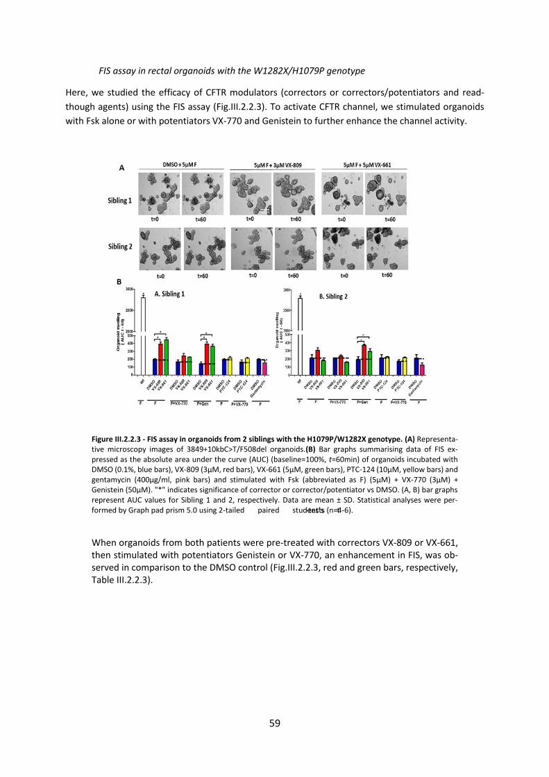
59
FIS assay in rectal organoids with the W1282X/H1079P genotype
Here, we studied the efficacy of CFTR modulators (correctors or correctors/potentiators and read-
though agents) using the FIS assay (Fig.III.2.2.3). To activate CFTR channel, we stimulated organoids
with Fsk alone or with potentiators VX-770 and Genistein to further enhance the channel activity.
Figure III.2.2.3 - FIS assay in organoids from 2 siblings with the H1079P/W1282X genotype. (A) Representa-tive microscopy images of 3849+10kbC>T/F508del organoids.(B) Bar graphs summarising data of FIS ex-pressed as the absolute area under the curve (AUC) (baseline=100%, t=60min) of organoids incubated with DMSO (0.1%, blue bars), VX-809 (3µM, red bars), VX-661 (5µM, green bars), PTC-124 (10µM, yellow bars) and gentamycin (400µg/ml, pink bars) and stimulated with Fsk (abbreviated as F) (5µM) + VX-770 (3µM) + Genistein (50µM). "*" indicates significance of corrector or corrector/potentiator vs DMSO. (A, B) bar graphs represent AUC values for Sibling 1 and 2, respectively. Data are mean ± SD. Statistical analyses were per-formed by Graph pad prism 5.0 using 2-tailed paired student’s t-tests (n=4-6).
When organoids from both patients were pre-treated with correctors VX-809 or VX-661, then stimulated with potentiators Genistein or VX-770, an enhancement in FIS, was ob-served in comparison to the DMSO control (Fig.III.2.2.3, red and green bars, respectively, Table III.2.2.3).
60
Table III.2.2.3 FIS values for organoids of the 2 patients with the genotype under various treatments.
Treatment Sibling 1
(Mean±SD)
Sibling 2
(Mean±SD)
DMSO+Fsk 210.3±8.800 207.8±46.75
DMSO+Fsk+770 169.3±22.16 212.0±2.00
DMSO+Fsk+Genistein 146.3±21.10 191.0±37.35
Correctors
VX-809+Fsk 395.6±24.58 309±26.89
VX-661+Fsk 449.7±29.45 180.0±11.10
Correctors + Potentiators
VX-809+Fsk+Gen 395.1±26.74 368.8±12.45
VX-661+Fsk+Gen 366.7±26.03 293.3±32.83
VX-809+Fsk+VX-770 243.5±32.14 235.8±12.25
VX-661+Fsk+VX-770 229.9±3.800 157.2±9.39
Read-through Agents
PTC-124+Fsk 219.8±18.45 225.7±6.100
Gentamycin+Fsk 155.5±24.40 126.0±25.0
Read-through Agents + Potentiator
PTC-124+Fsk+Vx-770 240.3±15.56 215.5±10.09
However, only treatments with correctors and genistein (but not VX-770) potentiation were signifi-
cantly different from the control for both siblings. Interestingly, for sibling 1, both VX-809 or VX-661
enhanced FIS significantly when compared to the DMSO control even without potentiator. The lack of
significance of both correctors under VX-770 may be due to the inhibitory effect that VX-770 has been
described to exert on the pharmacological correction of F508del-CFTR by these two correctors, includ-
ing (Cholon et al, 2014; Veit et al, 2014).
As to read-through agent Gentamycin (pink bars in Fig.III.2.2.3), it failed to significantly rescue CFTR
function, as measured by FIS, in comparison to DMSO control in both CF patients. However, some
enhancement in FIS could be detected for PTC-124, in particular with VX-770 and slightly more for
sibling 1 (Table III.2.2.3) although not sufficient to restore the full channel function.
The hierarchy of the potency of compounds tested per individual was:
Sibling 1: VX-661 > VX-809 >VX-809+Gen > VX-661/Gen > VX-809+VX-770 > PTC-124 > Gentamycin
Sibling 2: VX-809 + Gen > VX-661+Gen > VX-809> VX-661 > VX-809+VX-770 > PTC-124 > Gentamycin
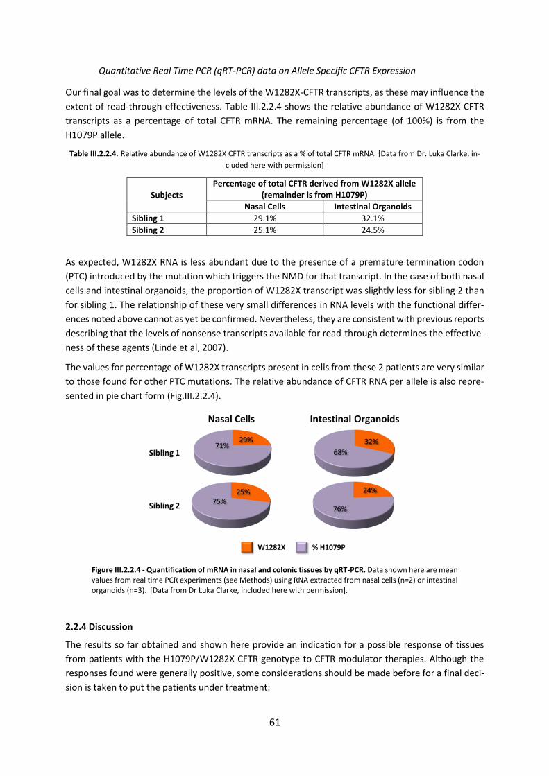
61
Quantitative Real Time PCR (qRT-PCR) data on Allele Specific CFTR Expression
Our final goal was to determine the levels of the W1282X-CFTR transcripts, as these may influence the
extent of read-through effectiveness. Table III.2.2.4 shows the relative abundance of W1282X CFTR
transcripts as a percentage of total CFTR mRNA. The remaining percentage (of 100%) is from the
H1079P allele.
Table III.2.2.4. Relative abundance of W1282X CFTR transcripts as a % of total CFTR mRNA. [Data from Dr. Luka Clarke, in-
cluded here with permission]
Subjects
Percentage of total CFTR derived from W1282X allele (remainder is from H1079P)
Nasal Cells Intestinal Organoids
Sibling 1 29.1% 32.1%
Sibling 2 25.1% 24.5%
As expected, W1282X RNA is less abundant due to the presence of a premature termination codon
(PTC) introduced by the mutation which triggers the NMD for that transcript. In the case of both nasal
cells and intestinal organoids, the proportion of W1282X transcript was slightly less for sibling 2 than
for sibling 1. The relationship of these very small differences in RNA levels with the functional differ-
ences noted above cannot as yet be confirmed. Nevertheless, they are consistent with previous reports
describing that the levels of nonsense transcripts available for read-through determines the effective-
ness of these agents (Linde et al, 2007).
The values for percentage of W1282X transcripts present in cells from these 2 patients are very similar
to those found for other PTC mutations. The relative abundance of CFTR RNA per allele is also repre-
sented in pie chart form (Fig.III.2.2.4).
Figure III.2.2.4 - Quantification of mRNA in nasal and colonic tissues by qRT-PCR. Data shown here are mean values from real time PCR experiments (see Methods) using RNA extracted from nasal cells (n=2) or intestinal organoids (n=3). [Data from Dr Luka Clarke, included here with permission].
2.2.4 Discussion
The results so far obtained and shown here provide an indication for a possible response of tissues
from patients with the H1079P/W1282X CFTR genotype to CFTR modulator therapies. Although the
responses found were generally positive, some considerations should be made before for a final deci-
sion is taken to put the patients under treatment:
62
1) Although treatments with both correctors VX-809 and VX-661 with potentiator Genistein were sig-
nificantly different from the control for both siblings, this drug combination (with genistein) is not ap-
proved for the clinic for any CFTR mutation.
2) Although for sibling 1, both VX-809 and VX-661 enhanced FIS significantly when compared to the
DMSO control without potentiator, the usage of these correctors alone (i.e., without a potentiator) is
not clinically approved so it cannot be recommended to the clinic.
3) The currently approved drug combination VX-809 (Lumacaftor) /VX-770 (Ivacaftor) did not exhibit
significant responses for H1079P/W1282X organoids from both patients; neither did the VX-660/VX-
770 combination. This lack of significance of both correctors under VX-770 may be due to the reported
inhibitory effect of this potentiator on the pharmacological correction of F508del-CFTR by VX-809 and
VX-661 (Cholon et al, 2014; Veit et al, 2014).
4) Efficacy tests with read-through compound gentamycin alone or with potentiators showed that this
read-through agent failed to provoke a significant response for both patients, in the case of PTC-124,
it seemed to trigger a small increase in FIS, although not significant. Some patients do better with PTC-
124 (Personal communication Dr. Silvia Gartner). Sibling 1 with superior clinical parameters and better
responsiveness to the CFTR modulators than sibling 2, was under clinical trial with a PTC-124 clinical
trial for 6 months. This may contribute to the better clinical parameters for Sibling 1.
5) Importantly, at this stage, it was not possible to compare the current responses to CFTR modulators
with those of F508del-CFTR or G551D-CFTR under CFTR modulators.
For the above reasons, at this stage, it is difficult to provide a final recommendation on whether or not
the patients should go into CFTR modulator treatment. We thus propose the continuation of these
studies by:
1) Carrying out tests on the triple combination of CFTR modulators, namely: correctors, potentiators
and read-through agents.
2) Obtaining comparative data with the VX-809/VX-770 combination in organoids F508del/F508del.
In case the responses observed here as significant are comparable in magnitude to those of
F508del/F508del organoids under the VX-809/VX-770 combination (i.e., Orkambi) or G551D/any mu-
tation under VX-770, we would have some guarantee of clinical benefit for the current patients.
63
2.3. Functional Assessment of Correctors and Potentiators on Organoids with the 3849+10kbC>T/F508del CFTR Genotype
2.3.1. Abstract
Background: The CFTR 3849+10kbC>T is a aplice site mutation leading to abnormal mRNA and reduced synthesis of CFTR protein and it represents milder phenotype of CF.
Objective: The main objective of this work was to predict patients’ in vivo response to therapeutic
CFTR modulators by testing their ex vivo effect on patient-derived intestinal organoids. Also, we as-
sessed the efficacy of potentiator VX-770 alone or with corrector VX-809 on intestinal organoids from
a patient having the 3849+10kbC->T/F508del genotype.
Methods: Intestinal organoids were prepared from rectal biopsies and analyzed by Forskolin induced
swelling (FIS) assay.
Results and Conclusion: Results obatained here show that CFTR-3849+10kbC>T genotype has a resid-
ual activity which was detected in organoids by the FIS assay, VX-809 alone did not affect this residual
activity as assessed by lack of significant organoid swelling. The residual activity was however signifi-
cantly enhanced by clinically approved potentiator VX-770 (Ivacaftor). Similarly, the addition of VX-770
after VX-809 incubation also significantly enhanced organoid residual activity.
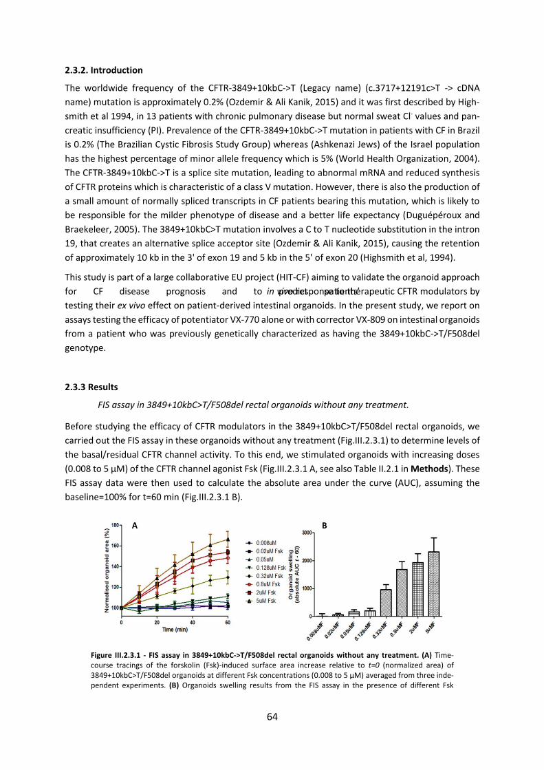
64
2.3.2. Introduction
The worldwide frequency of the CFTR-3849+10kbC->T (Legacy name) (c.3717+12191c>T -> cDNA
name) mutation is approximately 0.2% (Ozdemir & Ali Kanik, 2015) and it was first described by High-
smith et al 1994, in 13 patients with chronic pulmonary disease but normal sweat Cl- values and pan-
creatic insufficiency (PI). Prevalence of the CFTR-3849+10kbC->T mutation in patients with CF in Brazil
is 0.2% (The Brazilian Cystic Fibrosis Study Group) whereas (Ashkenazi Jews) of the Israel population
has the highest percentage of minor allele frequency which is 5% (World Health Organization, 2004).
The CFTR-3849+10kbC->T is a splice site mutation, leading to abnormal mRNA and reduced synthesis
of CFTR proteins which is characteristic of a class V mutation. However, there is also the production of
a small amount of normally spliced transcripts in CF patients bearing this mutation, which is likely to
be responsible for the milder phenotype of disease and a better life expectancy (Duguépéroux and
Braekeleer, 2005). The 3849+10kbC>T mutation involves a C to T nucleotide substitution in the intron
19, that creates an alternative splice acceptor site (Ozdemir & Ali Kanik, 2015), causing the retention
of approximately 10 kb in the 3' of exon 19 and 5 kb in the 5' of exon 20 (Highsmith et al, 1994).
This study is part of a large collaborative EU project (HIT-CF) aiming to validate the organoid approach
for CF disease prognosis and to predict patients’ in vivo response to therapeutic CFTR modulators by
testing their ex vivo effect on patient-derived intestinal organoids. In the present study, we report on
assays testing the efficacy of potentiator VX-770 alone or with corrector VX-809 on intestinal organoids
from a patient who was previously genetically characterized as having the 3849+10kbC->T/F508del
genotype.
2.3.3 Results
FIS assay in 3849+10kbC>T/F508del rectal organoids without any treatment.
Before studying the efficacy of CFTR modulators in the 3849+10kbC>T/F508del rectal organoids, we
carried out the FIS assay in these organoids without any treatment (Fig.III.2.3.1) to determine levels of
the basal/residual CFTR channel activity. To this end, we stimulated organoids with increasing doses
(0.008 to 5 µM) of the CFTR channel agonist Fsk (Fig.III.2.3.1 A, see also Table II.2.1 in Methods). These
FIS assay data were then used to calculate the absolute area under the curve (AUC), assuming the
baseline=100% for t=60 min (Fig.III.2.3.1 B).
Figure III.2.3.1 - FIS assay in 3849+10kbC->T/F508del rectal organoids without any treatment. (A) Time-course tracings of the forskolin (Fsk)-induced surface area increase relative to t=0 (normalized area) of 3849+10kbC>T/F508del organoids at different Fsk concentrations (0.008 to 5 µM) averaged from three inde-pendent experiments. (B) Organoids swelling results from the FIS assay in the presence of different Fsk
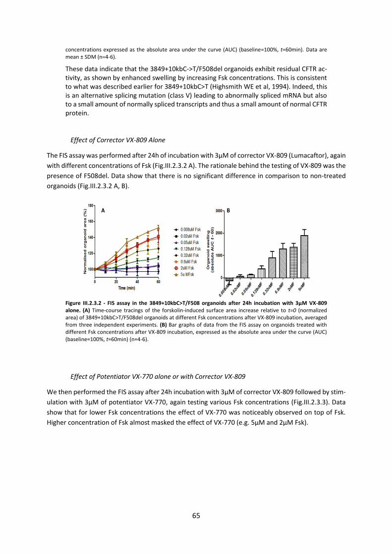
65
concentrations expressed as the absolute area under the curve (AUC) (baseline=100%, t=60min). Data are mean ± SDM (n=4-6).
These data indicate that the 3849+10kbC->T/F508del organoids exhibit residual CFTR ac-tivity, as shown by enhanced swelling by increasing Fsk concentrations. This is consistent to what was described earlier for 3849+10kbC>T (Highsmith WE et al, 1994). Indeed, this is an alternative splicing mutation (class V) leading to abnormally spliced mRNA but also to a small amount of normally spliced transcripts and thus a small amount of normal CFTR protein.
Effect of Corrector VX-809 Alone
The FIS assay was performed after 24h of incubation with 3µM of corrector VX-809 (Lumacaftor), again
with different concentrations of Fsk (Fig.III.2.3.2 A). The rationale behind the testing of VX-809 was the
presence of F508del. Data show that there is no significant difference in comparison to non-treated
organoids (Fig.III.2.3.2 A, B).
Figure III.2.3.2 - FIS assay in the 3849+10kbC>T/F508 organoids after 24h incubation with 3µM VX-809 alone. (A) Time-course tracings of the forskolin-induced surface area increase relative to t=0 (normalized area) of 3849+10kbC>T/F508del organoids at different Fsk concentrations after VX-809 incubation, averaged from three independent experiments. (B) Bar graphs of data from the FIS assay on organoids treated with different Fsk concentrations after VX-809 incubation, expressed as the absolute area under the curve (AUC) (baseline=100%, t=60min) (n=4-6).
Effect of Potentiator VX-770 alone or with Corrector VX-809
We then performed the FIS assay after 24h incubation with 3µM of corrector VX-809 followed by stim-
ulation with 3µM of potentiator VX-770, again testing various Fsk concentrations (Fig.III.2.3.3). Data
show that for lower Fsk concentrations the effect of VX-770 was noticeably observed on top of Fsk.
Higher concentration of Fsk almost masked the effect of VX-770 (e.g. 5µM and 2µM Fsk).
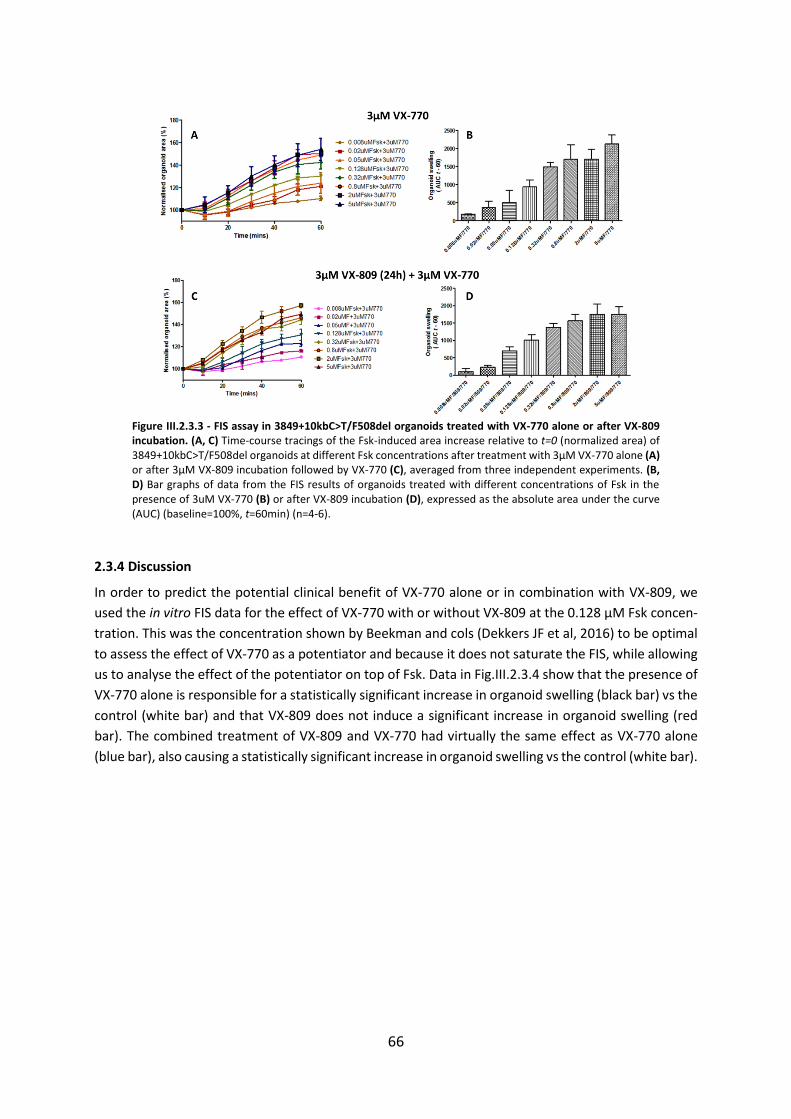
66
Figure III.2.3.3 - FIS assay in 3849+10kbC>T/F508del organoids treated with VX-770 alone or after VX-809 incubation. (A, C) Time-course tracings of the Fsk-induced area increase relative to t=0 (normalized area) of 3849+10kbC>T/F508del organoids at different Fsk concentrations after treatment with 3µM VX-770 alone (A) or after 3µM VX-809 incubation followed by VX-770 (C), averaged from three independent experiments. (B, D) Bar graphs of data from the FIS results of organoids treated with different concentrations of Fsk in the presence of 3uM VX-770 (B) or after VX-809 incubation (D), expressed as the absolute area under the curve (AUC) (baseline=100%, t=60min) (n=4-6).
2.3.4 Discussion
In order to predict the potential clinical benefit of VX-770 alone or in combination with VX-809, we
used the in vitro FIS data for the effect of VX-770 with or without VX-809 at the 0.128 µM Fsk concen-
tration. This was the concentration shown by Beekman and cols (Dekkers JF et al, 2016) to be optimal
to assess the effect of VX-770 as a potentiator and because it does not saturate the FIS, while allowing
us to analyse the effect of the potentiator on top of Fsk. Data in Fig.III.2.3.4 show that the presence of
VX-770 alone is responsible for a statistically significant increase in organoid swelling (black bar) vs the
control (white bar) and that VX-809 does not induce a significant increase in organoid swelling (red
bar). The combined treatment of VX-809 and VX-770 had virtually the same effect as VX-770 alone
(blue bar), also causing a statistically significant increase in organoid swelling vs the control (white bar).
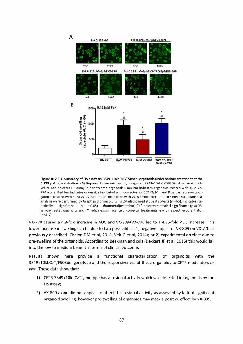
67
Figure III.2.3.4. Summary of FIS assay on 3849+10kbC>T/F508del organoids under various treatment at the 0.128 µM concentration. (A) Representative microscopy images of 3849+10kbC>T/F508del organoids. (B) White bar indicates FIS assay in non-treated organoids Black bar indicates organoids treated with 3µM VX-770 alone; Red bar indicates organoids incubated with corrector VX-809 (3µM); and Blue bar represents or-ganoids treated with 3µM VX-770 after 24h incubation with VX-809corrector. Data are mean±SD. Statistical analysis were performed by Graph pad prism 5.0 using 2-tailed paired students t-tests (n=4-5). Indicates sta-tistically significant (p ≤0.05) from the no-treatment (white bar). "#" indicates statistical significance (p<0.05) vs non-treated organoids and "*" indicates significance of corrector treatments vs with respective potentiator (n=3-5)
VX-770 caused a 4.8-fold increase in AUC and VX-809+VX-770 led to a 4.25-fold AUC increase. This
lower increase in swelling can be due to two possibilities: 1) negative impact of VX-809 on VX-770 as
previously described (Cholon DM et al, 2014; Veit G et al, 2014); or 2) experimental artefact due to
pre-swelling of the organoids. According to Beekman and cols (Dekkers JF et al, 2016) this would fall
into the low to medium benefit in terms of clinical outcome.
Results shown here provide a functional characterization of organoids with the
3849+10kbC>T/F508del genotype and the responsiveness of these organoids to CFTR modulators ex
vivo. These data show that:
1) CFTR-3849+10kbC>T genotype has a residual activity which was detected in organoids by the
FIS assay;
2) VX-809 alone did not appear to affect this residual activity as assessed by lack of significant
organoid swelling, however pre-swelling of organoids may mask a positive effect by VX-809;

68
3) The residual activity was however significantly enhanced by clinically approved potentiator VX-
770 (Ivacaftor) as shown by the 4.8-fold increase in organoid swelling;
4) Similarly, the addition of VX-770 after VX-809 incubation also significantly enhanced organoid
residual activity by a 4.25-fold increase in organoid swelling.
Very recently Kalydeco (VX-770) approval was expanded by US FDA for the treatment of 33 CFTR mu-
tations. However, 3849+10kbC>T mutation is not in the list of approved Kalydeco regimen. Based on
the present data, it is very likely that patients bearing 3849+10kbC>T get potential benefit from Kaly-
deco in the medium-low range.
69
2.4 CFTR Modulators Enhance Function R334W-CFTR both in Intestinal Organoids and Con-ditionally Reprogrammed Human Nasal Epithelial Cells
2.4.1 Abstract
Background: More than 2,000 mutations have been reported in the CFTR gene, albeit most still of
unknown functional impact (Sosnay et al, 2013). R334W is classified under Class IV mutation which has
defective conductance and this mutation has residual activity as reported before (Sheppard et al,
1993). The main goal of this work is to establish correlations among different CFTR activity-based bi-
omarkers, assessed namely in: fresh rectal biopsies, human intestinal organoids, and conditionally re-
programmed (CR) human nasal epithelial (HNE) cells from three subjects with the R334W/F508del
genotype and to determine the efficacy of CFTR modulators on the latter two models.
Methods: CFTR activity was investigated in different tissues/cells from three CF patients with the
F508del/R334W genotype, namely in: i) rectal biopsies by Ussing chamber using a standard diagnosis
protocol (Sousa et al, 2012); i) intestinal organoids by the forskolin-induced swelling (FIS) assay (Dek-
kers et al, 2013) and quantified by fluorescence microscopy and imaging software; and ii) CR-HNE cells
by open-circuit measurements in Ussing chamber. The response to CFTR modulators -potentiators VX-
770 and Genistein alone or with corrector VX-809 - was also assessed but just on the latter two models,
since rectal biopsies cannot be used to study CFTR modulators.
Results and Conclusion: Residual CFTR activity was detected in tissues/cells from these three subjects
and an increase in CFTR function was also observed both in organoids and CR-HNECs systems by VX-
770 and Genistein. This increase by VX-770 and Gen potentiators was further enhanced in the presence
of VX-809. All three biomarkers measuring CFTR basal activity in different tissues from patients with
the R334W/F508del genotype (rectal biopsies, intestinal organoids and CR-HNE’s) evidenced CFTR re-
sidual activity. Moreover, the effect to CFTR corrector VX-809 and potentiators Genistein and VX-770
evidenced a good correlation between responses in intestinal and respiratory tissues but also variable
responses among different patients with the same genotype.
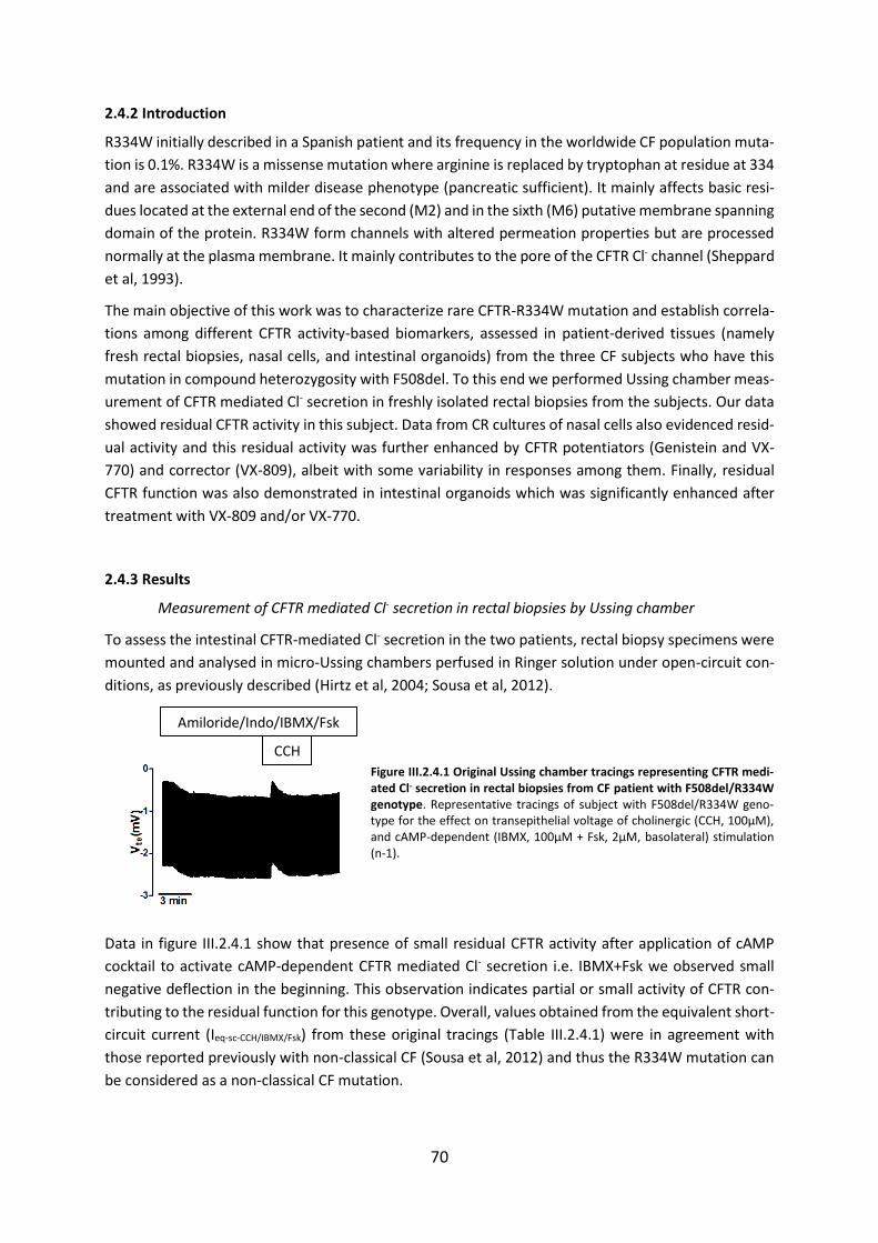
70
2.4.2 Introduction
R334W initially described in a Spanish patient and its frequency in the worldwide CF population muta-
tion is 0.1%. R334W is a missense mutation where arginine is replaced by tryptophan at residue at 334
and are associated with milder disease phenotype (pancreatic sufficient). It mainly affects basic resi-
dues located at the external end of the second (M2) and in the sixth (M6) putative membrane spanning
domain of the protein. R334W form channels with altered permeation properties but are processed
normally at the plasma membrane. It mainly contributes to the pore of the CFTR Cl- channel (Sheppard
et al, 1993).
The main objective of this work was to characterize rare CFTR-R334W mutation and establish correla-
tions among different CFTR activity-based biomarkers, assessed in patient-derived tissues (namely
fresh rectal biopsies, nasal cells, and intestinal organoids) from the three CF subjects who have this
mutation in compound heterozygosity with F508del. To this end we performed Ussing chamber meas-
urement of CFTR mediated Cl- secretion in freshly isolated rectal biopsies from the subjects. Our data
showed residual CFTR activity in this subject. Data from CR cultures of nasal cells also evidenced resid-
ual activity and this residual activity was further enhanced by CFTR potentiators (Genistein and VX-
770) and corrector (VX-809), albeit with some variability in responses among them. Finally, residual
CFTR function was also demonstrated in intestinal organoids which was significantly enhanced after
treatment with VX-809 and/or VX-770.
2.4.3 Results
Measurement of CFTR mediated Cl- secretion in rectal biopsies by Ussing chamber
To assess the intestinal CFTR-mediated Cl- secretion in the two patients, rectal biopsy specimens were
mounted and analysed in micro-Ussing chambers perfused in Ringer solution under open-circuit con-
ditions, as previously described (Hirtz et al, 2004; Sousa et al, 2012).
Figure III.2.4.1 Original Ussing chamber tracings representing CFTR medi-ated Cl- secretion in rectal biopsies from CF patient with F508del/R334W genotype. Representative tracings of subject with F508del/R334W geno-type for the effect on transepithelial voltage of cholinergic (CCH, 100µM), and cAMP-dependent (IBMX, 100µM + Fsk, 2µM, basolateral) stimulation (n-1).
Data in figure III.2.4.1 show that presence of small residual CFTR activity after application of cAMP
cocktail to activate cAMP-dependent CFTR mediated Cl- secretion i.e. IBMX+Fsk we observed small
negative deflection in the beginning. This observation indicates partial or small activity of CFTR con-
tributing to the residual function for this genotype. Overall, values obtained from the equivalent short-
circuit current (Ieq-sc-CCH/IBMX/Fsk) from these original tracings (Table III.2.4.1) were in agreement with
those reported previously with non-classical CF (Sousa et al, 2012) and thus the R334W mutation can
be considered as a non-classical CF mutation.
CCH
Amiloride/Indo/IBMX/Fsk
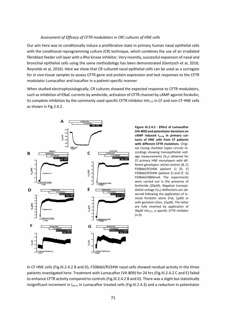
71
Assessment of Efficacy of CFTR modulators in CRC cultures of HNE cells
Our aim here was to conditionally induce a proliferative state in primary human nasal epithelial cells
with the conditional reprogramming culture (CR) technique, which combines the use of an irradiated
fibroblast feeder cell layer with a Rho kinase inhibitor. Very recently, successful expansion of nasal and
bronchial epithelial cells using the same methodology has been demonstrated (Gentzsch et al, 2016;
Reynolds et al, 2016). Here we show that CR cultured nasal epithelial cells can be used as a surrogate
for in vivo tissue samples to assess CFTR gene and protein expression and test responses to the CFTR
modulator Lumacaftor and Ivacaftor in a patient-specific manner
When studied electrophysiologically, CR cultures showed the expected response to CFTR modulators,
such as inhibition of ENaC currents by amiloride, activation of CFTR channel by cAMP agonist forskolin,
its complete inhibition by the commonly used specific CFTR inhibitor Inh172 in CF and non-CF HNE cells
as shown in Fig 2.4.2.
Figure III.2.4.2 - Effect of Lumacaftor (VX-809) and potentiator Genistein on cAMP induced Isc-eq in primary cul-tures of HNE cells from CF patients with different CFTR mutations. Origi-nal Ussing chamber (open circuit) re-cordings showing transepithelial volt-age measurements (Vte) obtained for CF primary HNE monolayers with dif-ferent genotypes: wt/wt control, (B, C) F508del/R334W (patient 1) (D, E) F508del/R334W (patient 2) and (F, G) F508del/3866insA. The experiments were carried out in the presence of Amiloride (20μM). Negative transepi-thelial voltage (Vte) deflections are ob-served following the application of lu-minal forskolin alone (Fsk, 2μM) or with genistein (Gen, 25μM). The latter are fully reverted by application of 30μM Inh172, a specific CFTR inhibitor (n-3).
In CF HNE cells (Fig.III.2.4.2 B and D), F508del/R334W nasal cells showed residual activity in the three
patients investigated here. Treatment with Lumacaftor (VX-809) for 24 hrs (Fig.III.2.4.2 C and E) failed
to enhance CFTR activity compared to controls (Fig.III.2.4.2 B and D). There was a slight but statistically
insignificant increment in Ieq-sc in Lumacaftor treated cells (Fig.III.2.4.3) and a reduction in potentiator
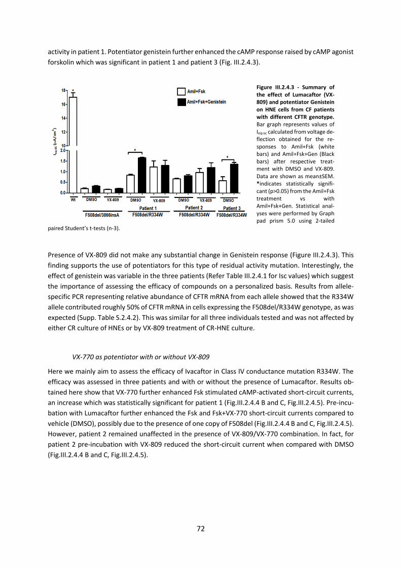
72
activity in patient 1. Potentiator genistein further enhanced the cAMP response raised by cAMP agonist
forskolin which was significant in patient 1 and patient 3 (Fig. III.2.4.3).
Figure III.2.4.3 - Summary of the effect of Lumacaftor (VX-809) and potentiator Genistein on HNE cells from CF patients with different CFTR genotype. Bar graph represents values of Ieq-sc calculated from voltage de-flection obtained for the re-sponses to Amil+Fsk (white bars) and Amil+Fsk+Gen (Black bars) after respective treat-ment with DMSO and VX-809. Data are shown as mean±SEM. *indicates statistically signifi-cant (p>0.05) from the Amil+Fsk treatment vs with Amil+Fsk+Gen. Statistical anal-yses were performed by Graph pad prism 5.0 using 2-tailed
paired Student’s t-tests (n-3).
Presence of VX-809 did not make any substantial change in Genistein response (Figure III.2.4.3). This
finding supports the use of potentiators for this type of residual activity mutation. Interestingly, the
effect of genistein was variable in the three patients (Refer Table III.2.4.1 for Isc values) which suggest
the importance of assessing the efficacy of compounds on a personalized basis. Results from allele-
specific PCR representing relative abundance of CFTR mRNA from each allele showed that the R334W
allele contributed roughly 50% of CFTR mRNA in cells expressing the F508del/R334W genotype, as was
expected (Supp. Table S.2.4.2). This was similar for all three individuals tested and was not affected by
either CR culture of HNEs or by VX-809 treatment of CR-HNE culture.
VX-770 as potentiator with or without VX-809
Here we mainly aim to assess the efficacy of Ivacaftor in Class IV conductance mutation R334W. The
efficacy was assessed in three patients and with or without the presence of Lumacaftor. Results ob-
tained here show that VX-770 further enhanced Fsk stimulated cAMP-activated short-circuit currents,
an increase which was statistically significant for patient 1 (Fig.III.2.4.4 B and C, Fig.III.2.4.5). Pre-incu-
bation with Lumacaftor further enhanced the Fsk and Fsk+VX-770 short-circuit currents compared to
vehicle (DMSO), possibly due to the presence of one copy of F508del (Fig.III.2.4.4 B and C, Fig.III.2.4.5).
However, patient 2 remained unaffected in the presence of VX-809/VX-770 combination. In fact, for
patient 2 pre-incubation with VX-809 reduced the short-circuit current when compared with DMSO
(Fig.III.2.4.4 B and C, Fig.III.2.4.5).
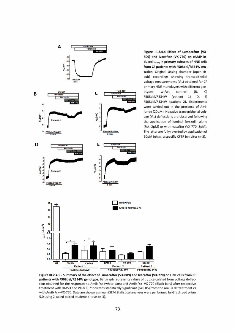
73
Figure III.2.4.4 Effect of Lumacaftor (VX-
809) and Ivacaftor (VX-770) on cAMP in-
duced Isc-eq in primary cultures of HNE cells
from CF patients with F508del/R334W mu-
tation. Original Ussing chamber (open-cir-
cuit) recordings showing transepithelial
voltage measurements (Vte) obtained for CF
primary HNE monolayers with different gen-
otypes: wt/wt control, (B, C)
F508del/R334W (patient 1) (D, E)
F508del/R334W (patient 2). Experiments
were carried out in the presence of Ami-
loride (20μM). Negative transepithelial volt-
age (Vte) deflections are observed following
the application of luminal forskolin alone
(Fsk, 2μM) or with Ivacaftor (VX-770, 3μM).
The latter are fully reverted by application of
30μM Inh172, a specific CFTR inhibitor (n-3).
Figure III.2.4.5 - Summary of the effect of Lumacaftor (VX-809) and Ivacaftor (VX-770) on HNE cells from CF patients with F508del/R334W genotype. Bar graph represents values of Ieq-sc calculated from voltage deflec-tion obtained for the responses to Amil+Fsk (white bars) and Amil+Fsk+VX-770 (Black bars) after respective treatment with DMSO and VX-809. *indicates statistically significant (p>0.05) from the Amil+Fsk treatment vs with Amil+Fsk+VX-770. Data are shown as mean±SEM.Statistical analyses were performed by Graph pad prism 5.0 using 2-tailed paired students t-tests (n-3).
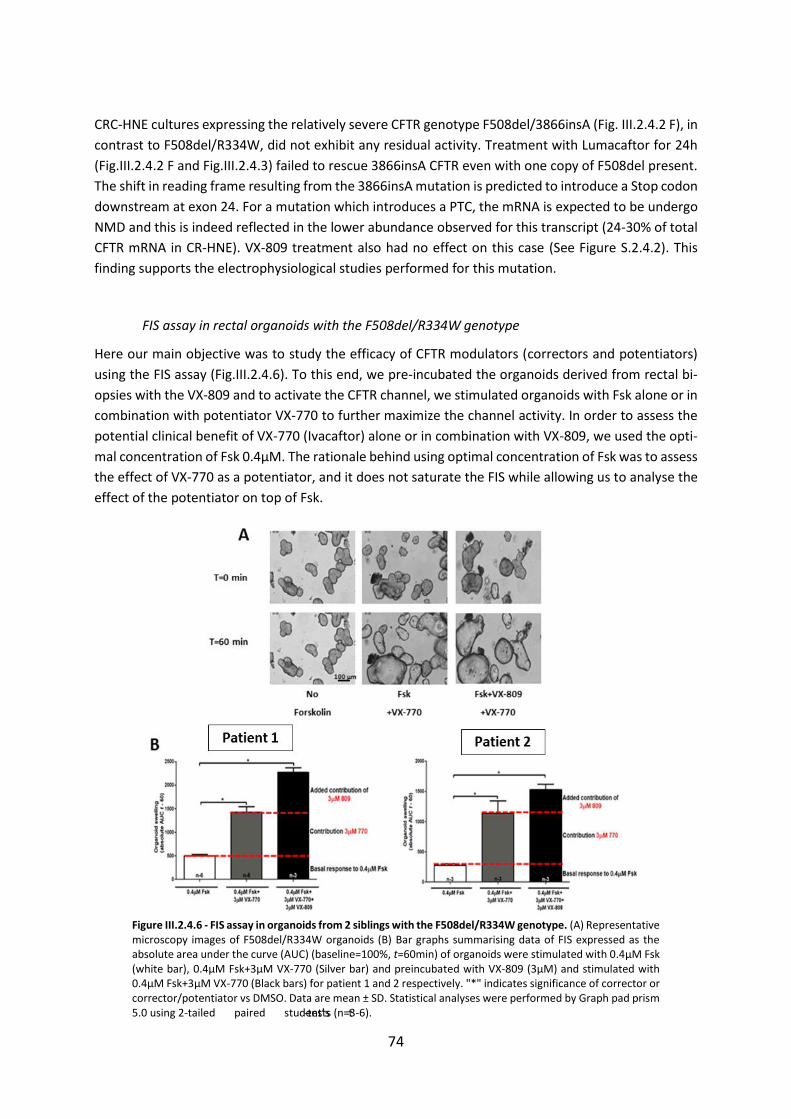
74
CRC-HNE cultures expressing the relatively severe CFTR genotype F508del/3866insA (Fig. III.2.4.2 F), in
contrast to F508del/R334W, did not exhibit any residual activity. Treatment with Lumacaftor for 24h
(Fig.III.2.4.2 F and Fig.III.2.4.3) failed to rescue 3866insA CFTR even with one copy of F508del present.
The shift in reading frame resulting from the 3866insA mutation is predicted to introduce a Stop codon
downstream at exon 24. For a mutation which introduces a PTC, the mRNA is expected to be undergo
NMD and this is indeed reflected in the lower abundance observed for this transcript (24-30% of total
CFTR mRNA in CR-HNE). VX-809 treatment also had no effect on this case (See Figure S.2.4.2). This
finding supports the electrophysiological studies performed for this mutation.
FIS assay in rectal organoids with the F508del/R334W genotype
Here our main objective was to study the efficacy of CFTR modulators (correctors and potentiators)
using the FIS assay (Fig.III.2.4.6). To this end, we pre-incubated the organoids derived from rectal bi-
opsies with the VX-809 and to activate the CFTR channel, we stimulated organoids with Fsk alone or in
combination with potentiator VX-770 to further maximize the channel activity. In order to assess the
potential clinical benefit of VX-770 (Ivacaftor) alone or in combination with VX-809, we used the opti-
mal concentration of Fsk 0.4µM. The rationale behind using optimal concentration of Fsk was to assess
the effect of VX-770 as a potentiator, and it does not saturate the FIS while allowing us to analyse the
effect of the potentiator on top of Fsk.
Figure III.2.4.6 - FIS assay in organoids from 2 siblings with the F508del/R334W genotype. (A) Representative microscopy images of F508del/R334W organoids (B) Bar graphs summarising data of FIS expressed as the absolute area under the curve (AUC) (baseline=100%, t=60min) of organoids were stimulated with 0.4µM Fsk (white bar), 0.4µM Fsk+3µM VX-770 (Silver bar) and preincubated with VX-809 (3µM) and stimulated with 0.4µM Fsk+3µM VX-770 (Black bars) for patient 1 and 2 respectively. "*" indicates significance of corrector or corrector/potentiator vs DMSO. Data are mean ± SD. Statistical analyses were performed by Graph pad prism 5.0 using 2-tailed paired student’s t-tests (n=3-6).
75
As shown in the figure III.2.4.6 B, the presence of VX-770 alone has higher AUC and is responsible for
a statistically significant increase in organoid swelling (silver bar) vs the control (white bar). The broken
red line indicates the basal response to 0.4µM Fsk and contribution 3µM VX-770 on top of Fsk. Com-
bination of VX-809 and VX-770 (Black bar) also statistically significant as compared to control (white
bar), which is shown as the added contribution of 3uM VX-809 on the bar graph for both patient 1 and
patient 2 respectively. The combined effect of VX-809/VX-770 may be due to the presence of one copy
of F508del on the second allele, so that VX-809 acts as a corrector rescuing this allele which is then
potentiated by VX-770. VX-770 alone caused a 3-fold increase in AUC and the VX-809/VX-770 combi-
nation led to a 4-fold AUC increase.
Table III.2.4.1 – Values obtained for different biomarkers used in the study
Rectal Biopsies ΔIeq-SC-Fsk
(µA/cm2) Organoids (ΔAUC) CR-HNECs ΔIeq-SC-Fsk (µA/cm2)
Patient 1 2 3 1 2 3 1 2 3
Fsk - Basal: 12.41±4.92 Isc CCH (IBMX/Fsk): 7.005±1.69
- 274.2±33.15 496.2±37.11 0.837±0.04 0.57±0.10
0.658±0.03 0.744±0.30
0.576±0.18 0.729±0.01
Fsk+Gen - - - - - 1.66±0.03* 0.808±0.05 1.349±0.14*
Fsk+VX-770 - - - 1138±203.5* 1427±113.8* 1.35±0.14* 0.880±0.36 1.261±0.17
Fsk+VX-809 - - - - - 1.227±0.28 0.88±0.41
0.968±0.18 0.414±0.04
-
Fsk+Gen+VX-809
- - - - - 1.290±0.25 1.194±0.23 -
Fsk+VX-770+VX-809
- - - 1531±89.19* 2271±96.91* 1.32±0.37* 0.553±0.09 -
(Values of CFTR activity (average ±SEM for Ussing chamber and average ±SD for Organoids))
(n=3, * indicates statistically significant from activity under DMSO, p≤ 0.05)
2.4.4 Discussion
The aim of this in vitro study was to evaluate the effect of CFTR modulators on the R334W mutation
(in patients with F508del in the other allele) which is described to mainly lead to defective channel
conductance. Our second goal was to establish a correlation between three biomarkers measuring
CFTR activity as well as to use them to determine the response to CFTR modulators. To this end, we
measured Cl- transport in rectal biopsies (Ussing chamber), primary intestinal organoids derived from
the rectal biopsies of the same patient (FIS assay), and CR cultures of nasal epithelial cells (also Ussing
chamber). Previously reported studies (Reynolds et al, 2016) have shown that conventional use of
BEGM to grow nasal cells did not yield much success with the expansion of cells. In our study, we also
tried to grow the nasal cells without a feeder layer and ROCK inhibitor, using conventional BEGM me-
dia, but nasal cells failed to proliferate and thus there was no increase in cell number. On the other
hand, cells grown in co-culture with irradiated NIH3T3 cells in F12 medium supplemented with Y-27632
grew relatively quickly.
ALI cultures of CR-HNE reached good transepithelial resistance values (Supp. Fig. S.2.4.1) in 3-4 weeks,
indicating the presence of functional tight junctions and a sealed epithelial monolayer which make
them amenable for functional studies. Thus, the combination of fibroblast feeder cells and ROCK in-
hibitor Y-27632 can be used to conditionally immortalize human nasal epithelial cells, while maintain-
ing their CFTR expression characteristics, thereby providing a useful model for studies of personalized
medicine (Suprynowicz et al, 2012; Liu et al, 2012). The increased proliferation rate and doubling time
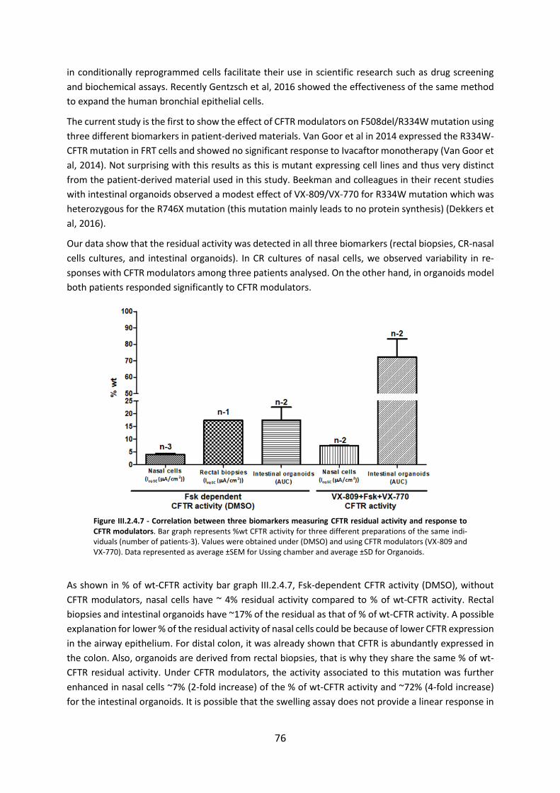
76
in conditionally reprogrammed cells facilitate their use in scientific research such as drug screening
and biochemical assays. Recently Gentzsch et al, 2016 showed the effectiveness of the same method
to expand the human bronchial epithelial cells.
The current study is the first to show the effect of CFTR modulators on F508del/R334W mutation using
three different biomarkers in patient-derived materials. Van Goor et al in 2014 expressed the R334W-
CFTR mutation in FRT cells and showed no significant response to Ivacaftor monotherapy (Van Goor et
al, 2014). Not surprising with this results as this is mutant expressing cell lines and thus very distinct
from the patient-derived material used in this study. Beekman and colleagues in their recent studies
with intestinal organoids observed a modest effect of VX-809/VX-770 for R334W mutation which was
heterozygous for the R746X mutation (this mutation mainly leads to no protein synthesis) (Dekkers et
al, 2016).
Our data show that the residual activity was detected in all three biomarkers (rectal biopsies, CR-nasal
cells cultures, and intestinal organoids). In CR cultures of nasal cells, we observed variability in re-
sponses with CFTR modulators among three patients analysed. On the other hand, in organoids model
both patients responded significantly to CFTR modulators.
Figure III.2.4.7 - Correlation between three biomarkers measuring CFTR residual activity and response to CFTR modulators. Bar graph represents %wt CFTR activity for three different preparations of the same indi-viduals (number of patients-3). Values were obtained under (DMSO) and using CFTR modulators (VX-809 and VX-770). Data represented as average ±SEM for Ussing chamber and average ±SD for Organoids.
As shown in % of wt-CFTR activity bar graph III.2.4.7, Fsk-dependent CFTR activity (DMSO), without
CFTR modulators, nasal cells have ~ 4% residual activity compared to % of wt-CFTR activity. Rectal
biopsies and intestinal organoids have ~17% of the residual as that of % of wt-CFTR activity. A possible
explanation for lower % of the residual activity of nasal cells could be because of lower CFTR expression
in the airway epithelium. For distal colon, it was already shown that CFTR is abundantly expressed in
the colon. Also, organoids are derived from rectal biopsies, that is why they share the same % of wt-
CFTR residual activity. Under CFTR modulators, the activity associated to this mutation was further
enhanced in nasal cells ~7% (2-fold increase) of the % of wt-CFTR activity and ~72% (4-fold increase)
for the intestinal organoids. It is possible that the swelling assay does not provide a linear response in
77
terms of the response to CFTR activity, given that swelling occurs for a 3D-structure, of which we only
capture two dimensions (as the area under the curve).
In conclusion, based on organoid responses, it is evident that R334W-CFTR responds to VX-770. All
three biomarkers measuring basal CFTR activity in different tissues from a patient with the
F508del/R334W genotype, (rectal biopsies, Intestinal organoids, and CR-HNE cultures) demonstrated
residual CFTR activity. Hierarchy of sensitivity for CFTR activity: Intestinal organoids > Rectal biopsies >
CR-HNE cultures. Moreover, the effects of CFTR corrector VX-809 and potentiators Genistein and VX-
770 evidenced a good correlation between responses measured in intestinal and respiratory tissues
but also variable responses among different patients with the same genotype.
78
2.5 Assessment of the Efficacy of Correctors and Potentiators on Rare CFTR 120del23 (Class VI) Mu-tation
2.5.1 Abstract
Background: Cystic fibrosis is a fatal autosomal recessive disorder caused by mutations in the CFTR
gene (Riordan et al, 1989). Almost, more than 2000 mutations in the CFTR gene have been identified,
that cause the loss of function phenotypes by impairing its protein synthesis, trafficking, and or chlo-
ride channel gating. The CFTR mutation c120del23 abrogate the normal translation initiation codon,
which occurs in very few patients worldwide.
Objective: The main aim of this work was to assess functionally (by iodide efflux assay) the efficacy of
correctors and potentiators on 120del23-CFTR and biochemically (by Western blot) on an heterologous
cell line expressing this variant.
Methods: The effect of CFTR modulators (correctors: VX-809 and VX-661, potentiators: VX-770,
Genistein, and P4) was assessed in BHK cells stably expressing c120del23 (and F508del-CFTR as a con-
trol) by CFTR-mediated iodide efflux. Also, Western blot was performed to assess the rescue of F508del
and c120del23-CFTR by correctors VX-809 and VX-661.
Results and Conclusion: Iodide efflux functional data show that using just potentiator or any combi-
nation corrector/potentiator significantly enhanced the Fsk or Fsk+IBMX responses of 120del23-CFTR
cells from the baseline (DMSO) response. These results may help bringing therapies to patients with
rare mutations by testing efficacy of currently available CFTR modulators on the rare 120del23-CFTR
mutation.
79
2.5.2 Introduction
We have previously characterized the 120del23 mutation in exon 1 (Ramalho et al, 2009), which cor-
responds to the deletion of 23 nucleotides (120-142) thus abolishing the translation initiation codon
(AUG pos.133-135). However, our studies in vitro also revealed that each of the codons
M150/M152/M156 (exon 4) can mediate CFTR alternative translation. We also showed that at least
one of these reaches the cell membrane, but it is unstable and has drastically reduced Cl- channel
activity (Ramalho et al, 2009). In our previous analysis of CFTR function in the colon of this patient, we
consistently detected no cAMP-mediated Cl- secretion (Hirtz et al, 2004).
Our main objective here is to assess the efficacy of correctors (VX-809 and VX-661) and potentiators
(VX-770, Genistein and P4) on CFTR 120del23 mutation (detected in a Portuguese CF patient in com-
pound heterozygosity with F508del). We demonstrate biochemically and functionally that this mutant
can be rescued to the plasma membrane by using correctors.
Our results and previous findings indicate that 120del23-CFTR has a defect in intracellular processing
and reduced function, which were partially rescued by corrector VX-809. This study helps bring thera-
pies to patients with rare CFTR mutations and may prove pivotal in clinical implications for mutation-
specific therapy.
2.5.3 Results
VX-809 and VX-661 partially rescue F508del, whereas only VX-661 rescues c120del23
Based on previous findings (Ramalho et al, 2009) 120del23-CFTR exhibits a defect in processing (see
also Fig.III.2.5.1 B), which is a characteristic of Class II mutations. Examples of class II mutations include
F508del and A561E (Mendes et al, 2003). Both these mutants have been shown to be rescued by cor-
rector VX-809 (Van Goor et al, 2011 and Awatade et al, 2015). Here, we assessed the efficacy of these
correctors on rescuing 120del23-CFTR. BHK cells expressing this mutant were thus incubated for 48h
with: VX-809, VX-661, and DMSO as the vehicle control. In parallel, these compounds were also tested
on wt- and F508del-CFTR in BHK cells as controls. To this end, we performed Western blot (WB, see
Methods Section 2.6) to assess the maturation status/rescue of F508del and 120del23 CFTR by VX-809
and VX-661.
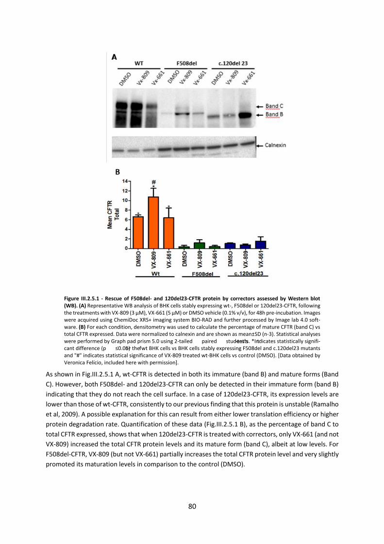
80
Figure III.2.5.1 - Rescue of F508del- and 120del23-CFTR protein by correctors assessed by Western blot (WB). (A) Representative WB analysis of BHK cells stably expressing wt-, F508del or 120del23-CFTR, following the treatments with VX-809 (3 μM), VX-661 (5 μM) or DMSO vehicle (0.1% v/v), for 48h pre-incubation. Images were acquired using ChemiDoc XRS+ imaging system BIO-RAD and further processed by Image lab 4.0 soft-ware. (B) For each condition, densitometry was used to calculate the percentage of mature CFTR (band C) vs total CFTR expressed. Data were normalized to calnexin and are shown as mean±SD (n-3). Statistical analyses were performed by Graph pad prism 5.0 using 2-tailed paired student’s t-tests. *Indicates statistically signifi-cant difference (p ≤0.05) for the wt BHK cells vs BHK cells stably expressing F508del and c.120del23 mutants and "#" indicates statistical significance of VX-809 treated wt-BHK cells vs control (DMSO). [Data obtained by Veronica Felício, included here with permission].
As shown in Fig.III.2.5.1 A, wt-CFTR is detected in both its immature (band B) and mature forms (Band
C). However, both F508del- and 120del23-CFTR can only be detected in their immature form (band B)
indicating that they do not reach the cell surface. In a case of 120del23-CFTR, its expression levels are
lower than those of wt-CFTR, consistently to our previous finding that this protein is unstable (Ramalho
et al, 2009). A possible explanation for this can result from either lower translation efficiency or higher
protein degradation rate. Quantification of these data (Fig.III.2.5.1 B), as the percentage of band C to
total CFTR expressed, shows that when 120del23-CFTR is treated with correctors, only VX-661 (and not
VX-809) increased the total CFTR protein levels and its mature form (band C), albeit at low levels. For
F508del-CFTR, VX-809 (but not VX-661) partially increases the total CFTR protein level and very slightly
promoted its maturation levels in comparison to the control (DMSO).
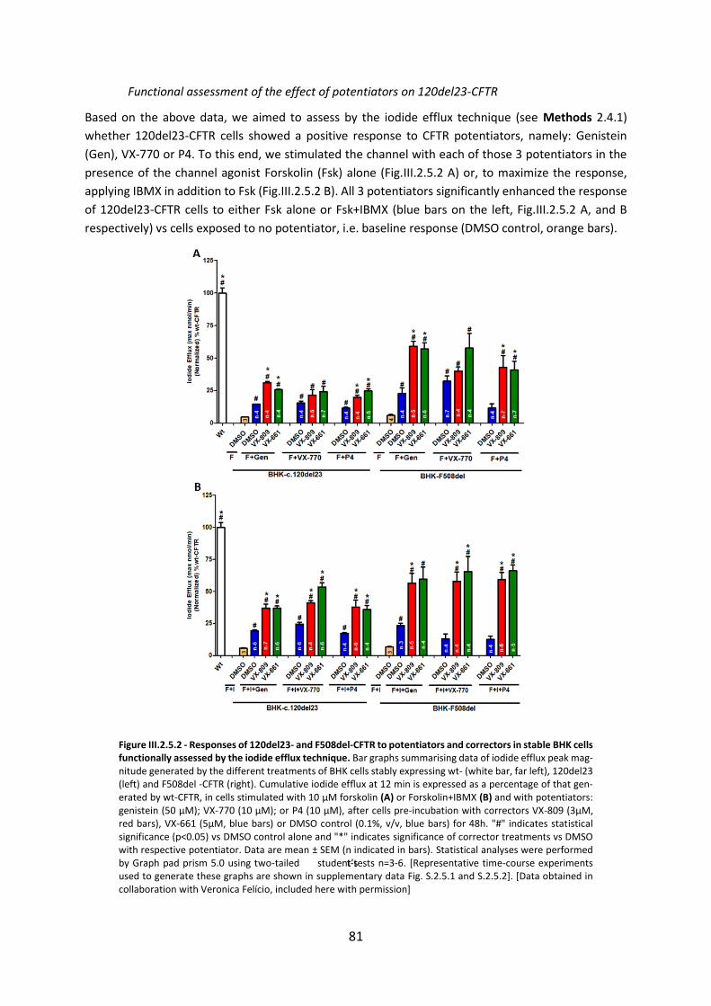
81
Functional assessment of the effect of potentiators on 120del23-CFTR
Based on the above data, we aimed to assess by the iodide efflux technique (see Methods 2.4.1)
whether 120del23-CFTR cells showed a positive response to CFTR potentiators, namely: Genistein
(Gen), VX-770 or P4. To this end, we stimulated the channel with each of those 3 potentiators in the
presence of the channel agonist Forskolin (Fsk) alone (Fig.III.2.5.2 A) or, to maximize the response,
applying IBMX in addition to Fsk (Fig.III.2.5.2 B). All 3 potentiators significantly enhanced the response
of 120del23-CFTR cells to either Fsk alone or Fsk+IBMX (blue bars on the left, Fig.III.2.5.2 A, and B
respectively) vs cells exposed to no potentiator, i.e. baseline response (DMSO control, orange bars).
Figure III.2.5.2 - Responses of 120del23- and F508del-CFTR to potentiators and correctors in stable BHK cells functionally assessed by the iodide efflux technique. Bar graphs summarising data of iodide efflux peak mag-nitude generated by the different treatments of BHK cells stably expressing wt- (white bar, far left), 120del23 (left) and F508del -CFTR (right). Cumulative iodide efflux at 12 min is expressed as a percentage of that gen-erated by wt-CFTR, in cells stimulated with 10 μM forskolin (A) or Forskolin+IBMX (B) and with potentiators: genistein (50 μM); VX-770 (10 μM); or P4 (10 μM), after cells pre-incubation with correctors VX-809 (3μM, red bars), VX-661 (5μM, blue bars) or DMSO control (0.1%, v/v, blue bars) for 48h. "#" indicates statistical significance (p<0.05) vs DMSO control alone and "*" indicates significance of corrector treatments vs DMSO with respective potentiator. Data are mean ± SEM (n indicated in bars). Statistical analyses were performed by Graph pad prism 5.0 using two-tailed student’s t-tests n=3-6. [Representative time-course experiments used to generate these graphs are shown in supplementary data Fig. S.2.5.1 and S.2.5.2]. [Data obtained in collaboration with Veronica Felício, included here with permission]
82
However, for F508del-CFTR cells this was not the case, only potentiators Gen and VX-770 (but not P4)
significantly enhanced the response to Fsk alone (blue bars on the right, Fig.III.2.5.2 A), and only Gen
significantly (but not VX-770 nor P4) enhanced the F508del-CFTR response to Fsk+IBMX (blue bars on
the right, Fig.III.2.5.2 B) vs cells exposed to no potentiator, i.e. baseline response (DMSO control, or-
ange bars).
Our data also show that responses obtained for 120del23-CFTR cells under potentiator were indeed
maximized by applying IBMX, compared to those under Fsk alone (compare blue bars for left part of
Fig.III.2.5.2 and B). Again, this was not the case for F508del-CFTR cells.
Functional assessment of the effect of correctors on 120del23-CFTR
Next, we aimed to assess whether the response of 120del23-CFTR cells to potentiators could be further
enhanced by pre-treating the cells with correctors VX-809 or VX-661. When 120del23-CFTR cells were
pre-treated with these correctors, only potentiators Gen and P4 (but not VX-770) significantly in-
creased the CFTR response to Fsk stimulation (red and green bars, respectively, on the left, Fig.III.2.5.2
A), vs the response of these cells to the same potentiator without any corrector (DMSO control, re-
spective blue bars). However, CFTR stimulation by Fsk+IBMX was significantly increased by all 3 poten-
tiators (Gen, P4 and VX-770) after pre-treatment of 120del23-CFTR cells with both correctors VX-809
or VX-661 (red and green bars on the left, Fig.III.2.5.2 B), vs the response to the same potentiator
without any corrector (DMSO control, respective blue bars).
For F508del-CFTR cells, all three potentiators (Gen, VX-770, and P4) also significantly enhanced their
responses after treatment with both correctors (red and green bars, respectively, on the right,
Fig.III.2.5.2 A), as compared to the DMSO control (DMSO control, respective blue bars).
Again, our data also show that responses obtained for 120del23-CFTR cells under potentiator were
indeed maximized by applying IBMX, compared to those under Fsk alone (compare red and green bars
for left part of Fig.III.2.5.2 A and B). Once more this was not the case for F508del (compare red and
green bars for right part of Fig.III.2.5.2 A and B).
2.5.4 Discussion
The results shown here provide an indication for a possible response of the 120del23 mutation to CFTR
modulator therapies. Although the responses found are generally positive, some considerations should
be made before for a final decision to go into patient treatment:
1) The cellular model used here (BHK - "Baby Hamster Kidney" cells) is not epithelial, these cells are
in fact fibroblasts. It is thus expected that the responses assessed in epithelial cells (the physio-
logically relevant system) will be somewhat different from these cells, as indeed was shown be-
fore (Farinha et al, 2015). For example, the response of the F508del mutation to the VX-809 (Lu-
macaftor) /VX-770 (Ivacaftor) combination under Forskolin alone was not found to be significant
here, while we (Awatade et al, 2015) and others (Van Goor et al, 2011) found it to be significant
in primary cultures of human bronchial epithelial cells.
83
2) For the currently approved drug combination VX-809 (Lumacaftor) /VX-770 (Ivacaftor)1 for
120del23-CFTR, responses comparable to those of F508del-CFTR were only observed for the max-
imal stimulation (i.e., when IBMX is added to Fsk) and this maximal stimulation is rather unlikely
to be achieved in vivo (i.e., in the patient).
3) Statistically significant responses were observed under Fsk alone (i.e., likely to reflect better the
in vivo situation, i.e., in the patient) for 120del23-CFTR, but the maximal responses were observed
for non-approved drug combinations, i.e.: VX-809/Gen or VX-661/Gen, VX-661/P4 (lumacaftor,
approved for CF only in combination with VX-770; VX-661 is currently in clinical trial for CF;
Genistein is not approved for CF; P4 is just a laboratory reagent).
4) Importantly, it is not possible to predict from the current data (obtained separately for 120del23
and F508del) what would be the response in cells from a patient having both 120del23 and
F508del mutations present. Although both mutations are expected to be responsive, it is not pos-
sible to predict the magnitude of the response of the combined genotype.
Based on these data, the following conclusions can be drawn from 120del23-CFTR cells:
1) Applying just potentiators or any combination corrector/potentiator tested here, significantly in-
creased the Fsk or Fsk+IBMX responses of 120del23-CFTR cells from the "baseline" response (i.e.,
under DMSO, which would correspond to the situation in the patient with no CFTR modulators).
2) The application of IBMX, in addition to Fsk, increased the cumulative iodide efflux generated un-
der potentiators and correctors.
3) Regarding increase of Fsk responses (no IBMX) vs the "baseline" response (DMSO):
a. VX-770 was the most effective potentiator, followed by Gen and then by P4;
b. under VX-809: Gen was the most effective, followed by VX-770 and then by P4;
c. under VX-661: all 3 potentiators (Gen, VX-770 and P4) were equally effective.
4) Regarding increase of Fsk+IBMX responses vs the "baseline" response (DMSO):
a. VX-770 was the most effective potentiator, followed by Gen and then by P4;
b. under VX-809: VX-770 was the most effective, followed by Gen and then by P4;
c. under VX-661: VX-770 was the most effective, followed by Gen and P4 equally.
5) CFTR potentiators Gen, VX-770 and P4 restored more function to F508del- than to 120del23-CFTR.
6) Similarly, the magnitude of the stimulation by all potentiator/ corrector combinations was always
lower for 120del23-CFTR than for F508del-CFTR.
7) Most CFTR potentiators restored some function to F508del- and 120del23, but none of the mod-
ulators achieved activity levels of wt-CFTR.
For the above reasons, these results before a final decision to go into treatment, it would be recom-
mended to test these compounds in intestinal organoids which is a technology currently available in
our lab as well as in Utrecht (The Netherlands) and Leuven (Belgium).
1 Combination drug known as Orkambi
84
Chapter 3. Assessment of Novel Compounds Rescuing F508del-CFTR and Enhancing TMEM16A Function in Human Epithelial Cells/ Tissues
3.1 Abstract
Background: Although VX-809 exhibited ~25% rescuing efficacy in CFTR activity in vitro, there was a
modest improvement in lung function in Phase III clinical studies of Orkambi (VX-809/VX-770). There-
fore, a compound correcting F508del-CFTR with an additive profile to VX-809 would be of significant
impact on the majority of CF patients. On the other hand, an attractive strategy for CF patients, which
is independent of the CFTR genotype consists in the activation of TMEM16A (non-CFTR Cl- channel) ion
channel as an alternative chloride channel in airway epithelium, to compensate for the absence of
CFTR in CF.
Objective: Our goal here is two-fold: i) to assess the effects of novel F508del-CFTR correctors (B9, E12,
Sygnature Discovery) and their possible additivity to VX-809 for a possible ultimate, use in triple-drug
combination therapy (two correctors and one potentiator); ii) to assess the effects of mimics of a pre-
viously described TMEM16A enhancer - E-act (C2, C5, and C7) for their effects on TMEM16A activity in
non-CF intestinal organoids with ATP.
Methods: The effects of 2 novel correctors (B9, E12) and their additivity to VX-809 were investigated
in F508del-CFTR-transduced CFBE cells and primary cultures of human bronchial epithelial cells
F508del- or A561E- homozygous patients by measurements of genistein-inducible equivalent short-
circuit current (Ieq-SC-Gen) in Ussing chamber. The effect of E-act mimics (C2, C5, and C7) was assessed in
non-CF intestinal organoids by Forskolin-induced swelling assay (FIS).
Results and Conclusions: Data for Ieq-SC-Gen in F508del-cells were (mean ± SEM µA/cm2): 0.83±0.09 (VX-
809, n=3); 0.27±0.0 (B9, n=3); 0.46±0.05 (E12, n=3); 0.10±0.04 (DMSO, n=3). Additivity of novel com-
pounds with VX-809 were (mean Ieq-SC-Gen ± SEM µA/cm2): 1.46±0.13 (B9, n=3); 2.76±0.48* (E12, n=3).
The latter is statistically significant vs VX-809 alone. Our results for three out of ten E-act mimics (C2,
C5, and C7) enhancing TMEM16A activity in non-CF intestinal organoids when used with ATP. Only
compound C2 with ATP led to the significant organoids swelling than only ATP. Compounds, when
assessed on their own, caused no swelling.
85
3.2 Introduction
Novel CFTR Correctors in Human Bronchial Epithelial Cells by Ussing chamber
The use of cell-based high throughput screens (HTS) yielded a number of candidate compounds which
mainly aim to correct the basic defects in cellular processing and Cl- channel function of CF-causing
mutant CFTR. The first defect is rescued by molecules termed correctors which are principally targeted
at correcting F508del cellular misprocessing, and the second by potentiators which restore cAMP-de-
pendent Cl- channel activity. Correctors and potentiators include many distinct chemical classes like
aminoarylthiazoles, quinazolinylaminopyrimidinones, bisaminomethylthiazoles (Corr-4a), 1,4 dihydro-
pyridines, quinazolines (VRT-325), sildenafil mimics KM11060, galfenine, FDA-approved potentiator
VX-770, corrector VX-809 and VX-661 (Tezacaftor in Clinical trial phase III) (Pedemonte et al, 2005b;
Loo et al, 2005; Robert et al, 2008; Van Goor et al, 2006). The mechanism of action for many of these
compounds is however poorly understood. Among the above componds, VX-770 is approved for sub-
jects bearing mutations causing a gating defect. Also, VX-809/VX-770 combination is approved for
F508del-homozygous subjects and VX-661/VX-770 succeeded in phase III trials for F508del-homozy-
gous and is under approval. Although the VX-809/VX-770 combination exhibited ~25% rescuing effi-
cacy in CFTR activity in vitro, it showed modest improvement in lung function and pulmonary exacer-
bations in CF subjects.
It has been suggested that single agents may have limited therapeutic efficacy (i.e., there is a “ceiling”
in their efficacy), because of the complex multiple defects in F508del-CFTR folding (Sloane and Rowe,
2010). Also, previous studies by Bridges 2010; Lin et al, 2010 and Okiyoneda et al, 2013 Farinha et al,
2013 have demonstrated that combination of F508del-CFTR correctors can have additive or even syn-
ergistic effect. Therefore, a compound correcting F508del-CFTR with an additive/synergistic profile to
VX-809 would be of significant impact on the majority of CF subjects.
Within the EU-funded TargetScreen2 project (FP6-2005-LH-7-037365), we have screened a 250-com-
pound library in a collaboration with Sygnature Discovery, (UK) by Western blot in human respiratory
epithelial cells and differential scanning fluorimetry to assess F508del-NBD1 folding. This medium-
throughput screen identified four novel small molecules ("correctors") rescuing the membrane traffic
of F508del-CFTR (C5, C9, B9 and E12), which we further functionally characterized here in primary HBE
cells.
Our main objective here was thus to assess the effects of novel F508del-CFTR correctors (B9, E12) and
their additivity to VX-809. Ultimately, these compounds can be potentially explored in triple-drug com-
bination therapy (two correctors and one potentiator).
Data obtained here for the efficacy of novel correctors in primary human bronchial epithelial cells show
that on their own B9 and E12 rescue F508del-CFTR activity to a lesser extent than VX-809. However,
E12 significantly adds to VX-809 in rescuing F508del-CFTR, thus appearing to act by a different mech-
anism of action than VX-809. It thus has potential to be further developed into a corrector drug.
Novel Compounds Enhancing TMEM16A Function in Human Intestinal Organoids.
Calcium-activated Cl- channels (CaCC’s) are activated by a rise in the free intracellular Ca2+ concentra-
tion. CaCC’s are widely expressed in epithelial cell types, where they participate in epithelial fluid se-
cretion, smooth muscle contraction, neurosensory signalling, and other functions (Hartzell et al, 2005;
Verkman & Galietta, 2009; Eggermont J, 2004). TMEM16A (ANO1) was identified as a CaCC encoding
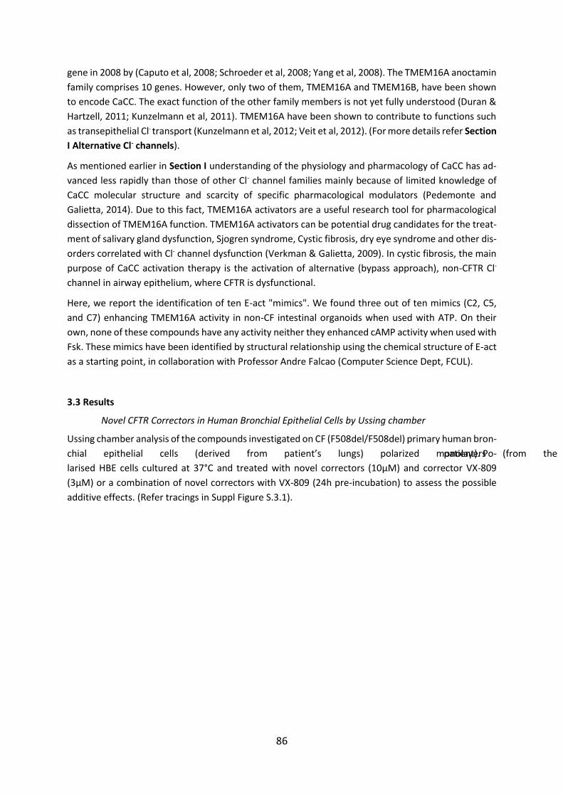
86
gene in 2008 by (Caputo et al, 2008; Schroeder et al, 2008; Yang et al, 2008). The TMEM16A anoctamin
family comprises 10 genes. However, only two of them, TMEM16A and TMEM16B, have been shown
to encode CaCC. The exact function of the other family members is not yet fully understood (Duran &
Hartzell, 2011; Kunzelmann et al, 2011). TMEM16A have been shown to contribute to functions such
as transepithelial Cl- transport (Kunzelmann et al, 2012; Veit et al, 2012). (For more details refer Section
I Alternative Cl- channels).
As mentioned earlier in Section I understanding of the physiology and pharmacology of CaCC has ad-
vanced less rapidly than those of other Cl- channel families mainly because of limited knowledge of
CaCC molecular structure and scarcity of specific pharmacological modulators (Pedemonte and
Galietta, 2014). Due to this fact, TMEM16A activators are a useful research tool for pharmacological
dissection of TMEM16A function. TMEM16A activators can be potential drug candidates for the treat-
ment of salivary gland dysfunction, Sjogren syndrome, Cystic fibrosis, dry eye syndrome and other dis-
orders correlated with Cl- channel dysfunction (Verkman & Galietta, 2009). In cystic fibrosis, the main
purpose of CaCC activation therapy is the activation of alternative (bypass approach), non-CFTR Cl-
channel in airway epithelium, where CFTR is dysfunctional.
Here, we report the identification of ten E-act "mimics". We found three out of ten mimics (C2, C5,
and C7) enhancing TMEM16A activity in non-CF intestinal organoids when used with ATP. On their
own, none of these compounds have any activity neither they enhanced cAMP activity when used with
Fsk. These mimics have been identified by structural relationship using the chemical structure of E-act
as a starting point, in collaboration with Professor Andre Falcao (Computer Science Dept, FCUL).
3.3 Results
Novel CFTR Correctors in Human Bronchial Epithelial Cells by Ussing chamber
Ussing chamber analysis of the compounds investigated on CF (F508del/F508del) primary human bron-
chial epithelial cells (derived from patient’s lungs) polarized monolayers (from the same patient). Po-
larised HBE cells cultured at 37°C and treated with novel correctors (10μM) and corrector VX-809
(3μM) or a combination of novel correctors with VX-809 (24h pre-incubation) to assess the possible
additive effects. (Refer tracings in Suppl Figure S.3.1).
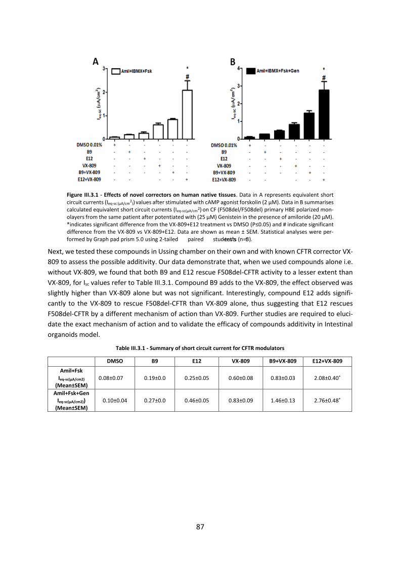
87
Figure III.3.1 - Effects of novel correctors on human native tissues. Data in A represents equivalent short circuit currents (Ieq-sc (µA/cm
2)) values after stimulated with cAMP agonist forskolin (2 μM). Data in B summarises
calculated equivalent short circuit currents (Ieq-sc(µA/cm2) on CF (F508del/F508del) primary HBE polarized mon-
olayers from the same patient after potentiated with (25 μM) Genistein in the presence of amiloride (20 μM). *indicates significant difference from the VX-809+E12 treatment vs DMSO (P≤0.05) and # indicate significant difference from the VX-809 vs VX-809+E12. Data are shown as mean ± SEM. Statistical analyses were per-formed by Graph pad prism 5.0 using 2-tailed paired student’s t-tests (n=3).
Next, we tested these compounds in Ussing chamber on their own and with known CFTR corrector VX-
809 to assess the possible additivity. Our data demonstrate that, when we used compounds alone i.e.
without VX-809, we found that both B9 and E12 rescue F508del-CFTR activity to a lesser extent than
VX-809, for Isc values refer to Table III.3.1. Compound B9 adds to the VX-809, the effect observed was
slightly higher than VX-809 alone but was not significant. Interestingly, compound E12 adds signifi-
cantly to the VX-809 to rescue F508del-CFTR than VX-809 alone, thus suggesting that E12 rescues
F508del-CFTR by a different mechanism of action than VX-809. Further studies are required to eluci-
date the exact mechanism of action and to validate the efficacy of compounds additivity in Intestinal
organoids model.
Table III.3.1 - Summary of short circuit current for CFTR modulators
DMSO B9 E12 VX-809 B9+VX-809 E12+VX-809
Amil+Fsk Ieq-sc(µA/cm2)
(Mean±SEM)
0.08±0.07
0.19±0.0
0.25±0.05
0.60±0.08
0.83±0.03
2.08±0.40*
Amil+Fsk+Gen Ieq-sc(µA/cm2))
(Mean±SEM)
0.10±0.04
0.27±0.0
0.46±0.05
0.83±0.09
1.46±0.13
2.76±0.48*
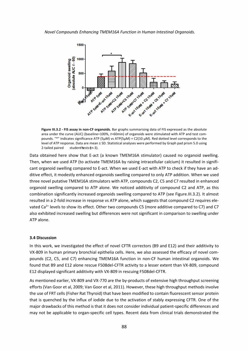
88
Novel Compounds Enhancing TMEM16A Function in Human Intestinal Organoids.
Figure III.3.2 - FIS assay in non-CF organoids. Bar graphs summarising data of FIS expressed as the absolute area under the curve (AUC) (baseline=100%, t=60min) of organoids were stimulated with ATP and test com-pounds. "*" indicates significance ATP (5µM) vs ATP(5µM) + C2(10 µM). Red dotted level corresponds to the level of ATP response. Data are mean ± SD. Statistical analyses were performed by Graph pad prism 5.0 using 2-tailed paired student’s t-tests (n-3).
Data obtained here show that E-act (a known TMEM16A stimulator) caused no organoid swelling.
Then, when we used ATP (to activate TMEM16A by raising intracellular calcium) it resulted in signifi-
cant organoid swelling compared to E-act. When we used E-act with ATP to check if they have an ad-
ditive effect, it modestly enhanced organoids swelling compared to only ATP addition. When we used
three novel putative TMEM16A stimulators with ATP, compounds C2, C5 and C7 resulted in enhanced
organoid swelling compared to ATP alone. We noticed additivity of compound C2 and ATP, as this
combination significantly increased organoids swelling compared to ATP (see Figure.III.3.2). It almost
resulted in a 2-fold increase in response vs ATP alone, which suggests that compound C2 requires ele-
vated Ca2+ levels to show its effect. Other two compounds C5 (more additive compared to C7) and C7
also exhibited increased swelling but differences were not significant in comparison to swelling under
ATP alone.
3.4 Discussion
In this work, we investigated the effect of novel CFTR correctors (B9 and E12) and their additivity to
VX-809 in human primary bronchial epithelia cells. Here, we also assessed the efficacy of novel com-
pounds (C2, C5, and C7) enhancing TMEM16A function in non-CF human intestinal organoids. We
found that B9 and E12 alone rescue F508del-CFTR activity to a lesser extent than VX-809, compound
E12 displayed significant additivity with VX-809 in rescuing F508del-CFTR.
As mentioned earlier, VX-809 and VX-770 are the by-products of extensive high throughput screening
efforts (Van Goor et al, 2009; Van Goor et al, 2011). However, these high throughput methods involve
the use of FRT cells (Fisher Rat Thyroid) that have been modified to contain fluorescent sensor protein
that is quenched by the influx of iodide due to the activation of stably expressing CFTR. One of the
major drawbacks of this method is that it does not consider individual patient-specific differences and
may not be applicable to organ-specific cell types. Recent data from clinical trials demonstrated the
89
limited success of Orkambi combination in CF subjects. Therefore, combination therapies may be re-
quired to achieve complementary rescue to attain full F508del-CFTR correction.
Previous studies by Namkung et al reported cell-based screen of small molecule collections revelaled
several chemical classes of TMEM16A activators that produced strong and more sustained Cl- currents
than Ca2+- elevating purinergic agonists without elevating cytoplasmic Ca2+. E-act was the outcome of
this screen reported by those authors.
When we evaluated the effect of E-act on organoids it resulted in no swelling. This is possibly due to
the absence of TMEM16A expression in human adult colon. Presence of CACCs in organoids has not
been confirmed. Another possible explanation could be that CaCC’s may be located on the apical side
and in organoids, being it difficult to get access to apical side. Also in this study, we used minimal
concentration of ATP which is 5µM to asses the effect of compounds on top of ATP activation.
However, assessment of putative TMEM16A activators in intestinal organoids exhibited increased
swelling when we used them with ATP. We found that compound C2 with ATP led to a significant dif-
ference in organoid swelling vs ATP alone. These compounds, when assessed on their own, displayed
no swelling. It is possible that other TMEM16 family members (e.g., TMEM16F) are responsible for the
observed effects.
In conclusion, it is important to assess the effect of compounds in physiologically relevant cell models
such as HBE and organoids which are physiologically relevant in drug discovery, also in preclinical vali-
dation of potential drugs.
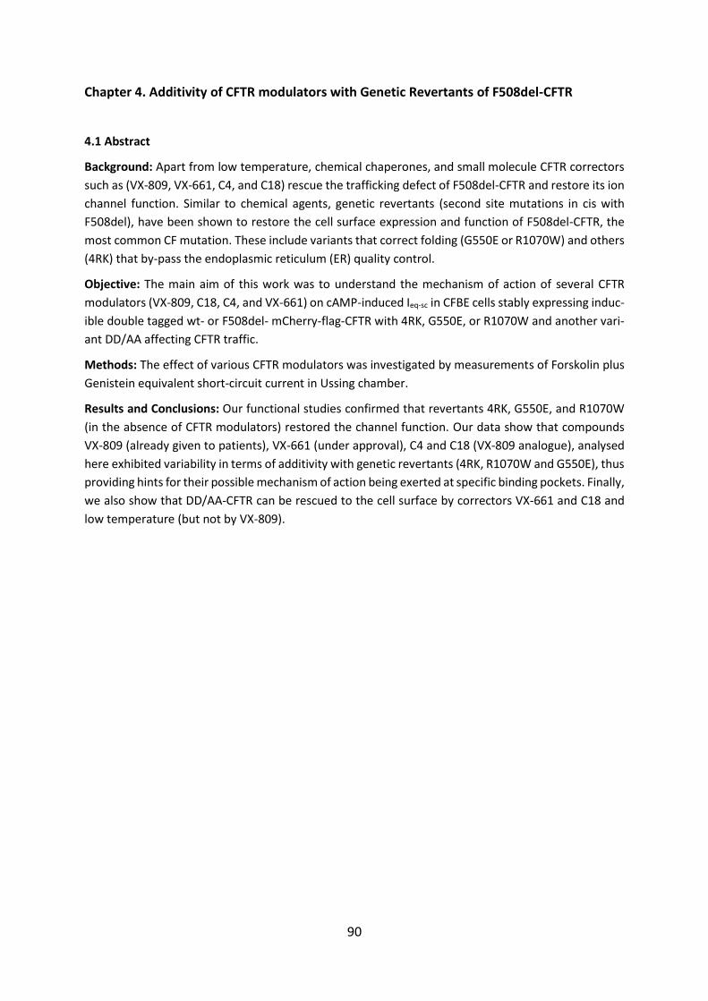
90
Chapter 4. Additivity of CFTR modulators with Genetic Revertants of F508del-CFTR
4.1 Abstract
Background: Apart from low temperature, chemical chaperones, and small molecule CFTR correctors
such as (VX-809, VX-661, C4, and C18) rescue the trafficking defect of F508del-CFTR and restore its ion
channel function. Similar to chemical agents, genetic revertants (second site mutations in cis with
F508del), have been shown to restore the cell surface expression and function of F508del-CFTR, the
most common CF mutation. These include variants that correct folding (G550E or R1070W) and others
(4RK) that by-pass the endoplasmic reticulum (ER) quality control.
Objective: The main aim of this work was to understand the mechanism of action of several CFTR
modulators (VX-809, C18, C4, and VX-661) on cAMP-induced Ieq-sc in CFBE cells stably expressing induc-
ible double tagged wt- or F508del- mCherry-flag-CFTR with 4RK, G550E, or R1070W and another vari-
ant DD/AA affecting CFTR traffic.
Methods: The effect of various CFTR modulators was investigated by measurements of Forskolin plus
Genistein equivalent short-circuit current in Ussing chamber.
Results and Conclusions: Our functional studies confirmed that revertants 4RK, G550E, and R1070W
(in the absence of CFTR modulators) restored the channel function. Our data show that compounds
VX-809 (already given to patients), VX-661 (under approval), C4 and C18 (VX-809 analogue), analysed
here exhibited variability in terms of additivity with genetic revertants (4RK, R1070W and G550E), thus
providing hints for their possible mechanism of action being exerted at specific binding pockets. Finally,
we also show that DD/AA-CFTR can be rescued to the cell surface by correctors VX-661 and C18 and
low temperature (but not by VX-809).
91
4.2 Introduction
Arginine-framed motifs (AFT’s) are transient retention/retrieval motifs coupling the export from the
endoplasmic reticulum (ER) of multimeric membrane proteins to their folding assembly (Michelsen et
al, 2005). Chang et al reported first in 1990 the rescue of the trafficking and function of F508del-CFTR
with the simultaneous mutation of the four AFT’s (R29QR31, R516YR518, R553AR555 and R764 RR766) present
in CFTR and termed 4RK (R29K, R516K, R555K & R766K). As shown by Roxo-Rosa et al in 2006, 4RK
allows F508del-CFTR to escape ERQC and promotes its traffic to the cell surface by a mechanism which
is different from that of other genetic revertants (e.g., G550E) (Roxo-Rosa et al, 2006). The G550E re-
vertant introduces a negatively charged amino acid in the ATP-binding site 2 (conserved LSGGQ core
signature motif of CFTR NBD1) (DeCarvalho et al, 2002). The introduction of G550E improves the mat-
uration of F508del-CFTR and increased CFTR mediated Cl- permeability (DeCarvalho et al, 2002). The
R1070W revertant rescues the NBD1:ICL4 interaction (disrupted in F508del-CFTR) by filing the cavity
created by the absence of the phenyl side chain of the phenylalanine in F508del-CFTR (Thibodeau et
al, 2010). But these revertants (4RK, G550E and R1070W) rescue F508del-CFTR by causing different
effects: some of these second-site mutations were shown to correct structural features of F508del-
CFTR folding - G550E (Roxo-Rosa et al, 2006) or R1070W (Thibodeau et al. 2010; Farinha et al, 2013),
and the 4RK variant bypasses the ER quality control (Roxo-Rosa et al, 2006).
The vesicle mediated export of cargo proteins from the ER relies on the coatomer complex II (COP-II)
(Barlowe et al, 1994), whereas COPI binds di-lysine motifs and functions to direct retrograde transport
of proteins from the ERGIC and Golgi compartments (Pelham HR, 1994). In CFTR, a similar diacidic ER
exit code motif 565Asp-Ala-Asp567 (or DAD) was identified within NBD1. Substitution of the second as-
partic acid with alanine (resulting in DAA) disrupts the association of CFTR with Sec24 and dramatically
reduces the kinetic processing of wt-CFTR from the ER (Wang et al, 2004). The di-acidic motif 565Asp-
Ala-Asp567 is a commonly used cargo concentration signal for COP-II mediated ER export (Nishimura
and Balch, 1997). The Sec24 subunit of COP-II is the one mainly associated with recognising this di-
acidic code (Miller et al, 2002; Mossessova et al, 2003). The functional link between F508del-CFTR and
the DAD exit code is still missing. Roy et al in 2010 demonstrated that temperature-dependent export
of F508del-CFTR relies on the presence of the di-acidic code within the NBD1, implying that DAD has a
functional role in the temperature dependent rescue of F508del-CFTR. Moreover, F508del-CFTR rescue
by the simultaneous disruption of two of the arginine framed tripeptides R561K and R555K (AFT’s) is
also dependent on the DD/AA exit code (Roy et al, 2010).
Our goal here was to explore the effect of various CFTR correctors (VX-809, VX-661, C18, and C4) on
cAMP-induced Ieq-sc in CFBE cells stably expressing inducible (Tet-ON) double-tagged wt- or F508del-
mCherry-Flag-CFTR (Botelho et al, 2015) with distinct genetic revertants and on the DD/AA CFTR vari-
ant (on wt background) missing the diacidic ER exit code but with no folding defect (Farinha et al, 2013,
Roy et al, 2010).
Our data show that small-molecule correctors VX-809, C18, C4, and VX-661 exhibited variable effects
on the genetic revertants. Interestingly, DD/AA was rescued to the cell surface by correctors VX-661,
C18, and low temperature (but not significantly by VX-809).
92
4.3 Results
F508del-CFTR rescued by VX-809, VX-661, and C18
To characterize the efficacy of CFTR modulators on F508del-CFTR we performed Ussing chamber anal-
yses (See Methods 2.3.4 and 2.4.2.2). To this this end, we preincubated the cells for 48hrs with cor-
rectors (after doxycycline -Dox – to induce CFTR expression), then stimulated with CFTR agonists,
namely Forskolin (Fsk), 2µM and Genistein, 25µM to assess CFTR-mediated Cl- secretion (see tracings
in Supp Figure S.4.1).
Figure III.4.1 - Effect of different correctors on mCherry-Flag-F508del-CFTR stably expressed in CFBE cells under an inducible promoter (Tet-on). (A) Bar graphs represent values of Ieq-sc (μA/cm2) calculated from volt-age deflection obtained for the responses to Fsk+IBMX (white bars) or to Gen+Fsk (black bars), after 48h induction by Doxycycline to induce F508del-CFTR expression from the Tet promoter treatment with DMSO, VX-809, C18, C4, or VX-661 (B) Bar graph shows the %wt activity of F508del-CFTR for each modulator analysed. * indicates significant difference from the modulator treatment vs DMSO (P≤0.05) and red dotted line corre-sponds to the level of Control among all CFTR modulators treatment. Data are shown as mean±SEM. Statistical analyses were performed by Graph pad prism 5.0 using 2-tailed paired students t-tests (n=4-6).
Data obtained here show that correctors VX-809, C18 and VX-661 but not C4, rescued the F508del-
CFTR function response to the cAMP agonist Fsk+IBMX alone or with the CFTR potentiator Genistein
(Fig.III.4.1 A), with almost identical efficacy which is 25% of WT-CFTR (Fig.III.4.1 B). The Fsk+Genistein
inducible equivalent short-circuit currents (Ieq-sc-Fsk+Gen) for correctors VX-809, C18, and VX-661 were of
similar magnitude (See Table III.4.1). While corrector C4 failed to rescue F508del-CFTR currents, show-
ing the same values as those of DMSO (~8% of WT-CFTR).
Table III.4.1 - Summary of short circuit current for F508del-CFTR treated with correctors
DMSO VX-809 C18 C4 VX-661
Fsk+IBMX Ieq-sc(µA/cm
2)
(Mean±SEM)
3.26±0.33
6.81±0.68*
6.06±1.01*
2.45±0.50
6.61±1.23*
Fsk+IBMX+Gen Ieq-sc(µA/cm
2)
(Mean±SEM)
3.46±0.72
10.91±0.88*
10.54±1.46*
3.76±0.82 10.81±2.03*
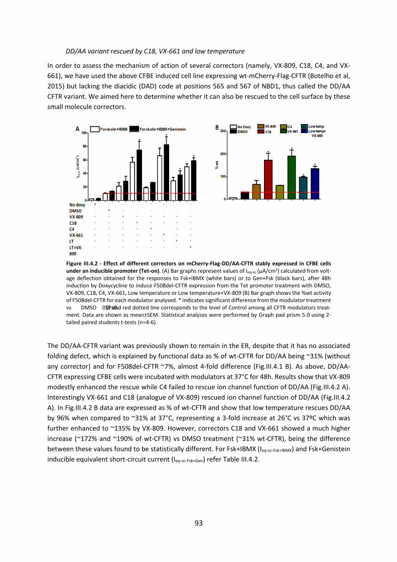
93
DD/AA variant rescued by C18, VX-661 and low temperature
In order to assess the mechanism of action of several correctors (namely, VX-809, C18, C4, and VX-
661), we have used the above CFBE induced cell line expressing wt-mCherry-Flag-CFTR (Botelho et al,
2015) but lacking the diacidic (DAD) code at positions 565 and 567 of NBD1, thus called the DD/AA
CFTR variant. We aimed here to determine whether it can also be rescued to the cell surface by these
small molecule correctors.
Figure III.4.2 - Effect of different correctors on mCherry-Flag-DD/AA-CFTR stably expressed in CFBE cells under an inducible promoter (Tet-on). (A) Bar graphs represent values of Ieq-sc (μA/cm2) calculated from volt-age deflection obtained for the responses to Fsk+IBMX (white bars) or to Gen+Fsk (black bars), after 48h induction by Doxycycline to induce F508del-CFTR expression from the Tet promoter treatment with DMSO, VX-809, C18, C4, VX-661, Low temperature or Low temperature+VX-809 (B) Bar graph shows the %wt activity of F508del-CFTR for each modulator analysed. * indicates significant difference from the modulator treatment vs DMSO (P≤0.05) and red dotted line corresponds to the level of Control among all CFTR modulators treat-ment. Data are shown as mean±SEM. Statistical analyses were performed by Graph pad prism 5.0 using 2-tailed paired students t-tests (n=4-6).
The DD/AA-CFTR variant was previously shown to remain in the ER, despite that it has no associated
folding defect, which is explained by functional data as % of wt-CFTR for DD/AA being ~31% (without
any corrector) and for F508del-CFTR ~7%, almost 4-fold difference (Fig.III.4.1 B). As above, DD/AA-
CFTR expressing CFBE cells were incubated with modulators at 37°C for 48h. Results show that VX-809
modestly enhanced the rescue while C4 failed to rescue ion channel function of DD/AA (Fig.III.4.2 A).
Interestingly VX-661 and C18 (analogue of VX-809) rescued ion channel function of DD/AA (Fig.III.4.2
A). In Fig.III.4.2 B data are expressed as % of wt-CFTR and show that low temperature rescues DD/AA
by 96% when compared to ~31% at 37°C, representing a 3-fold increase at 26°C vs 37ºC which was
further enhanced to ~135% by VX-809. However, correctors C18 and VX-661 showed a much higher
increase (~172% and ~190% of wt-CFTR) vs DMSO treatment (~31% wt-CFTR), being the difference
between these values found to be statistically different. For Fsk+IBMX (Ieq-sc-Fsk+IBMX) and Fsk+Genistein
inducible equivalent short-circuit current (Ieq-sc-Fsk+Gen) refer Table III.4.2.
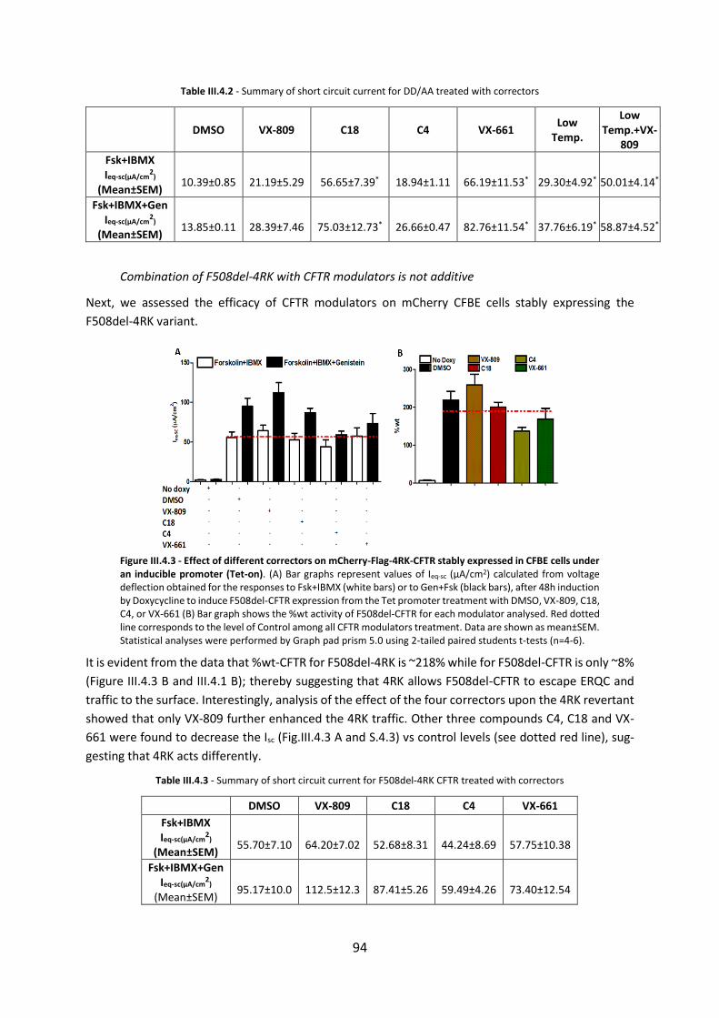
94
Table III.4.2 - Summary of short circuit current for DD/AA treated with correctors
DMSO VX-809 C18 C4 VX-661 Low
Temp.
Low Temp.+VX-
809
Fsk+IBMX Ieq-sc(µA/cm
2)
(Mean±SEM)
10.39±0.85
21.19±5.29
56.65±7.39*
18.94±1.11
66.19±11.53*
29.30±4.92*
50.01±4.14*
Fsk+IBMX+Gen Ieq-sc(µA/cm
2)
(Mean±SEM)
13.85±0.11
28.39±7.46
75.03±12.73*
26.66±0.47
82.76±11.54*
37.76±6.19*
58.87±4.52*
Combination of F508del-4RK with CFTR modulators is not additive
Next, we assessed the efficacy of CFTR modulators on mCherry CFBE cells stably expressing the
F508del-4RK variant.
Figure III.4.3 - Effect of different correctors on mCherry-Flag-4RK-CFTR stably expressed in CFBE cells under an inducible promoter (Tet-on). (A) Bar graphs represent values of Ieq-sc (μA/cm2) calculated from voltage deflection obtained for the responses to Fsk+IBMX (white bars) or to Gen+Fsk (black bars), after 48h induction by Doxycycline to induce F508del-CFTR expression from the Tet promoter treatment with DMSO, VX-809, C18, C4, or VX-661 (B) Bar graph shows the %wt activity of F508del-CFTR for each modulator analysed. Red dotted line corresponds to the level of Control among all CFTR modulators treatment. Data are shown as mean±SEM. Statistical analyses were performed by Graph pad prism 5.0 using 2-tailed paired students t-tests (n=4-6).
It is evident from the data that %wt-CFTR for F508del-4RK is ~218% while for F508del-CFTR is only ~8%
(Figure III.4.3 B and III.4.1 B); thereby suggesting that 4RK allows F508del-CFTR to escape ERQC and
traffic to the surface. Interestingly, analysis of the effect of the four correctors upon the 4RK revertant
showed that only VX-809 further enhanced the 4RK traffic. Other three compounds C4, C18 and VX-
661 were found to decrease the Isc (Fig.III.4.3 A and S.4.3) vs control levels (see dotted red line), sug-
gesting that 4RK acts differently.
Table III.4.3 - Summary of short circuit current for F508del-4RK CFTR treated with correctors
DMSO VX-809 C18 C4 VX-661
Fsk+IBMX Ieq-sc(µA/cm
2)
(Mean±SEM)
55.70±7.10
64.20±7.02
52.68±8.31
44.24±8.69
57.75±10.38
Fsk+IBMX+Gen Ieq-sc(µA/cm
2)
(Mean±SEM)
95.17±10.0
112.5±12.3
87.41±5.26
59.49±4.26
73.40±12.54
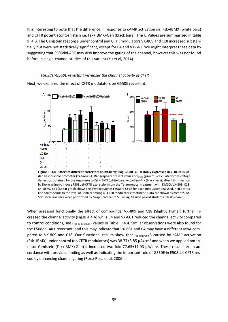
95
It is interesting to note that the difference in response to cAMP activation i.e. Fsk+IBMX (white bars)
and CFTR potentiator Genistein i.e. Fsk+IBMX+Gen (black bars). The Isc Values are summarised in table
III.4.3. The Genistein response under control and CFTR modulators VX-809 and C18 increased substan-
tially but were not statistically significant, except for C4 and VX-661. We might interpret these data by
suggesting that F508del-4RK may also improve the gating of the channel, however this was not found
before in single-channel studies of this variant (Xu et al, 2014).
F508del-G550E revertant increases the channel activity of CFTR
Next, we explored the effect of CFTR modulators on G550E revertant.
Figure III.4.4 - Effect of different correctors on mCherry-Flag-G550E-CFTR stably expressed in CFBE cells un-der an inducible promoter (Tet-on). (A) Bar graphs represent values of Ieq-sc (μA/cm2) calculated from voltage deflection obtained for the responses to Fsk+IBMX (white bars) or to Gen+Fsk (black bars), after 48h induction by Doxycycline to induce F508del-CFTR expression from the Tet promoter treatment with DMSO, VX-809, C18, C4, or VX-661 (B) Bar graph shows the %wt activity of F508del-CFTR for each modulator analysed. Red dotted line corresponds to the level of Control among all CFTR modulators treatment. Data are shown as mean±SEM. Statistical analyses were performed by Graph pad prism 5.0 using 2-tailed paired students t-tests (n=4-6).
When assessed functionally the effect of compounds, VX-809 and C18 (Slightly higher) further in-
creased the channel activity (Fig.III.4.4 A) while C4 and VX-661 reduced the channel activity compared
to control conditions, see (Ieq-sc-Fsk+Gen) values in Table III.4.4. Similar observations were also found for
the F508del-4RK revertant, and this may indicate that VX-661 and C4 may have a different MoA com-
pared to VX-809 and C18. Our functional results show that Ieq-sc(µA/cm2
) caused by cAMP activation
(Fsk+IBMX) under control (no CFTR modulators) was 38.77±3.85 µA/cm2 and when we applied poten-
tiator Genistein (Fsk+IBMX+Gen) it increased two-fold 77.65±11.93 µA/cm2. These results are in ac-
cordance with previous finding as well as indicating the important role of G550E in F508del-CFTR res-
cue by enhancing channel gating (Roxo-Rosa et al, 2006).
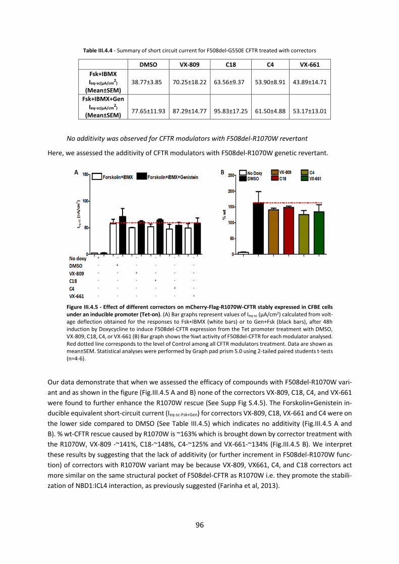
96
Table III.4.4 - Summary of short circuit current for F508del-G550E CFTR treated with correctors
DMSO VX-809 C18 C4 VX-661
Fsk+IBMX Ieq-sc(µA/cm
2)
(Mean±SEM)
38.77±3.85
70.25±18.22
63.56±9.37 53.90±8.91 43.89±14.71
Fsk+IBMX+Gen Ieq-sc(µA/cm
2)
(Mean±SEM)
77.65±11.93
87.29±14.77
95.83±17.25
61.50±4.88
53.17±13.01
No additivity was observed for CFTR modulators with F508del-R1070W revertant
Here, we assessed the additivity of CFTR modulators with F508del-R1070W genetic revertant.
Figure III.4.5 - Effect of different correctors on mCherry-Flag-R1070W-CFTR stably expressed in CFBE cells under an inducible promoter (Tet-on). (A) Bar graphs represent values of Ieq-sc (μA/cm2) calculated from volt-age deflection obtained for the responses to Fsk+IBMX (white bars) or to Gen+Fsk (black bars), after 48h induction by Doxycycline to induce F508del-CFTR expression from the Tet promoter treatment with DMSO, VX-809, C18, C4, or VX-661 (B) Bar graph shows the %wt activity of F508del-CFTR for each modulator analysed. Red dotted line corresponds to the level of Control among all CFTR modulators treatment. Data are shown as mean±SEM. Statistical analyses were performed by Graph pad prism 5.0 using 2-tailed paired students t-tests (n=4-6).
Our data demonstrate that when we assessed the efficacy of compounds with F508del-R1070W vari-
ant and as shown in the figure (Fig.III.4.5 A and B) none of the correctors VX-809, C18, C4, and VX-661
were found to further enhance the R1070W rescue (See Supp Fig S.4.5). The Forskolin+Genistein in-
ducible equivalent short-circuit current (Ieq-sc-Fsk+Gen) for correctors VX-809, C18, VX-661 and C4 were on
the lower side compared to DMSO (See Table III.4.5) which indicates no additivity (Fig.III.4.5 A and
B). % wt-CFTR rescue caused by R1070W is ~163% which is brought down by corrector treatment with
the R1070W, VX-809 -~141%, C18-~148%, C4-~125% and VX-661-~134% (Fig.III.4.5 B). We interpret
these results by suggesting that the lack of additivity (or further increment in F508del-R1070W func-
tion) of correctors with R1070W variant may be because VX-809, VX661, C4, and C18 correctors act
more similar on the same structural pocket of F508del-CFTR as R1070W i.e. they promote the stabili-
zation of NBD1:ICL4 interaction, as previously suggested (Farinha et al, 2013).
97
Table III.4.5 - Summary of short circuit current for F508del-R1070W CFTR treated with correctors
DMSO VX-809 C18 C4 VX-661
Fsk+IBMX Ieq-sc(µA/cm
2)
(Mean±SEM)
57.60±8.17
50.00±0.86
51.75±5.08
47.60±8.93
49.57±8.37
Fsk+IBMX+Gen Ieq-sc(µA/cm
2)
(Mean±SEM)
70.99±14.56
61.36±2.21
64.71±1.80
54.59±5.79
58.47±9.78
4.4 Discussion
In this study, we investigated the efficacy of CFTR modulators in order to understand their mechanism
of action, with genetic revertants with different F508del-CFTR effects (4RK allows F508del-CFTR to es-
cape ERQC through its two NBD1 changes: G550E probably facilitates correct F508del-NBD1:NBD2
folding with the result that the mutant protein is delivered to the cell surface with improved gating;
R1070W rescues the NBD1:ICL4 interface and finally DD/AA diacidic ER exit code which impairs Sec24-
mediated traffic but not folding (Roxo-Rosa et al, 2006, Roy et al, 2009, Farinha et al, 2013, Farinha &
Amaral, 2005 & Hoelen et al, 2010).
It is important to mention that compounds used in this study had heterogeneous effects on the genetic
revertants, which may help to elucidate their MoA. As shown earlier, VX-809 in contrast to C18 (to a
lesser extent), VX-661 and C4 add to the rescue caused by F508del-4RK (not significantly), probably
indicating that this compound does not affect AFT-mediated dominant ER retention. For G550E, we
found that C18 was additive in comparison to (VX-809 less extent), while C4 and VX-661 reduced the
short-circuit current and %wt-CFTR drastically when compared to the control situation (DMSO). One
possible explanation for this reduction could be, VX-661 and C4 possibly binding on the same site as
that of G550 i.e. NBD1:NBD2 dimer interface. Finally, for R1070W none of the correctors was found to
be additive, remarkably all of them decreased the Isc.
Table III.4.6 - Comparison of current functional data with previous work
Authors and Genetic revertants used
Cell Model used Functional analysis (In Agree-
ment) (Yes/No)
Biochemical analysis (In Agreement)
(Yes/No)
DeCarvalho et al, 2002 (G550E)
Fisher rat thyroid (FRT) epithelia
Ussing chamber (Yes)
-
Roxo-Rosa et al, 2006 (G550E and 4RK)
BHK cells Iodide efflux technique
(Yes) -
Farinha et al, 2013 (4RK, G550E, or
R1070W) BHK cells Iodide efflux technique Western Blot
Xu et al, 2014 G550E and 4RK
BHK cells Single channel recording
(Yes) -
Current work (4RK, G550E, or
R1070W) mCherry CFBE cells Ussing chamber -

98
Xu et al in 2014 discovered that G550E and 4RK were without effect on G551D-CFTR protein pro-
cessing, but markedly improved the channel gating when G551D expressed with 4RK and G550E vari-
ants.
To conclude, our data suggest that G550E and 4RK improved F508del-CFTR channel gating by potenti-
ator Genistein. As shown previously by Roy et al and Farinha et al, low temperature rescues DD/AA-
CFTR. Our functional studies also showed rescue of DD/AA-CFTR ion channel function, also when we
used VX-809 together with low temperature, the channel function was further, but modestly enhanced
by the presence of VX-809 (Fig.III.4.2 A, B). Interestingly, when we used a corrector, VX-809 it modestly
enhanced the channel function (consistent with the previous finding by Farinha et al, 2013), while VX-
661 and C18 significantly increased the channel function (Fig.III.4.2 A, B), suggesting that these correc-
tors rescue the DD/AA variant more significantly. Mechanistically, low temperature and correctors al-
low DD/AA variant to exit the ER and possibly this could occur through the conventional ER-to-Golgi
pathway (Farinha et al, 2013).
Major findings
Within the current work, we discovered that:
i) The DD/AA variant can be rescued to the cell surface by small molecule correctors such as C18 and
VX-661, while VX-809 had only a modest effect on this variant. DD/AA was rescued by low tempera-
ture, as reported before by (Roy et al, 2009; Farinha et al, 2013).
ii) Some additive effect was observed for correctors VX-809 and C18 with 4RK and G550E genetic re-
vertants, suggesting that the compounds and these two revertants probably correct different confor-
mational defects of F508del-CFTR as proposed by (Roxo-Rosa et al, 2006).
iii) In contrast, no additive effect was seen for correctors with the R1070W variant, indicating that
corrector VX-809, VX-661, C18, and C4 acts more similarly to R1070W.

99
IV. CONCLUSIONS AND FUTURE PERSPECTIVES
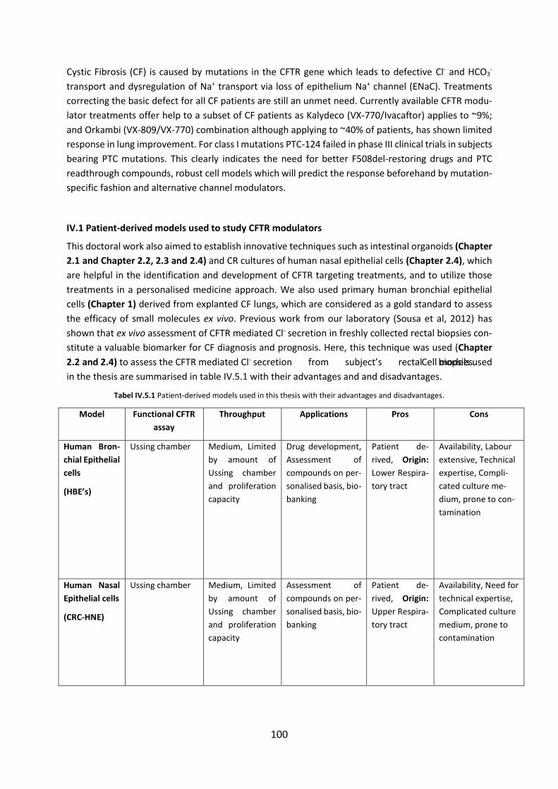
100
Cystic Fibrosis (CF) is caused by mutations in the CFTR gene which leads to defective Cl- and HCO3-
transport and dysregulation of Na+ transport via loss of epithelium Na+ channel (ENaC). Treatments
correcting the basic defect for all CF patients are still an unmet need. Currently available CFTR modu-
lator treatments offer help to a subset of CF patients as Kalydeco (VX-770/Ivacaftor) applies to ~9%;
and Orkambi (VX-809/VX-770) combination although applying to ~40% of patients, has shown limited
response in lung improvement. For class I mutations PTC-124 failed in phase III clinical trials in subjects
bearing PTC mutations. This clearly indicates the need for better F508del-restoring drugs and PTC
readthrough compounds, robust cell models which will predict the response beforehand by mutation-
specific fashion and alternative channel modulators.
IV.1 Patient-derived models used to study CFTR modulators
This doctoral work also aimed to establish innovative techniques such as intestinal organoids (Chapter
2.1 and Chapter 2.2, 2.3 and 2.4) and CR cultures of human nasal epithelial cells (Chapter 2.4), which
are helpful in the identification and development of CFTR targeting treatments, and to utilize those
treatments in a personalised medicine approach. We also used primary human bronchial epithelial
cells (Chapter 1) derived from explanted CF lungs, which are considered as a gold standard to assess
the efficacy of small molecules ex vivo. Previous work from our laboratory (Sousa et al, 2012) has
shown that ex vivo assessment of CFTR mediated Cl- secretion in freshly collected rectal biopsies con-
stitute a valuable biomarker for CF diagnosis and prognosis. Here, this technique was used (Chapter
2.2 and 2.4) to assess the CFTR mediated Cl- secretion from subject’s rectal biopsies. Cell models used
in the thesis are summarised in table IV.5.1 with their advantages and and disadvantages.
Tabel IV.5.1 Patient-derived models used in this thesis with their advantages and disadvantages.
Model Functional CFTR
assay
Throughput Applications Pros Cons
Human Bron-
chial Epithelial
cells
(HBE’s)
Ussing chamber Medium, Limited
by amount of
Ussing chamber
and proliferation
capacity
Drug development,
Assessment of
compounds on per-
sonalised basis, bio-
banking
Patient de-
rived, Origin:
Lower Respira-
tory tract
Availability, Labour
extensive, Technical
expertise, Compli-
cated culture me-
dium, prone to con-
tamination
Human Nasal
Epithelial cells
(CRC-HNE)
Ussing chamber Medium, Limited
by amount of
Ussing chamber
and proliferation
capacity
Assessment of
compounds on per-
sonalised basis, bio-
banking
Patient de-
rived, Origin:
Upper Respira-
tory tract
Availability, Need for
technical expertise,
Complicated culture
medium, prone to
contamination
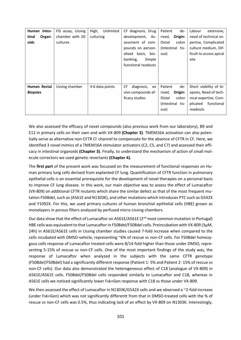
101
Human Intes-
tinal Organ-
oids
FIS assay, Ussing
chamber with 2D
cultures
High, Unlimited
culturing
CF diagnosis, Drug
development, As-
sessment of com-
pounds on person-
alised basis, bio-
banking, Simple
functional readouts
Patient de-
rived, Origin:
Distal colon
(Intestinal tis-
sue)
Labour extensive,
need of technical ex-
pertise, Complicated
culture medium, Dif-
ficult to access apical
site
Human Rectal
Biopsies
Ussing chamber 4-6 data points CF diagnosis, ex
vivo compounds ef-
ficacy studies
Patient de-
rived, Origin:
Distal colon
(Intestinal tis-
sue)
Short viability of bi-
opsies, Need of tech-
nical expertise, Com-
plicated functional
readouts
We also assessed the efficacy of novel compounds (also previous work from our laboratory), B9 and
E12 in primary cells on their own and with VX-809 (Chapter 3). TMEM16A activation can also poten-
tially serve as alternative non-CFTR Cl- channel to compensate for the absence of CFTR in CF. Here, we
identified 3 novel mimics of a TMEM16A stimulator activators (C2, C5, and C7) and assessed their effi-
cacy in intestinal organoids (Chapter 3). Finally, to understand the mechanism of action of small mol-
ecule correctors we used genetic revertants (Chapter 4).
The first part of the present work was focussed on the measurement of functional responses on Hu-
man primary lung cells derived from explanted CF lung. Quantification of CFTR function in pulmonary
epithelial cells is an essential prerequisite for the development of novel therapies on a personal basis
to improve CF lung disease. In this work, our main objective was to assess the effect of Lumacaftor
(VX-809) on additional CFTR mutants which share the similar defect as that of the most frequent mu-
tation F508del, such as (A561E and N1303K), and other mutations which introduces PTC such as G542X
and Y1092X. For this, we used primary cultures of human bronchial epithelial cells (HBE) grown as
monolayers in porous filters analyzed by perfused micro-Ussing chambers.
Our data show that the effect of Lumacaftor on A561E/A561E (2nd most common mutation in Portugal)
HBE cells was equivalent to that Lumacaftor in F508del/F508del cells. Preincubation with VX-809 (3µM,
24h) in A561E/A561E cells in Ussing chamber studies caused 7-fold increase when compared to the
cells incubated with DMSO-vehicle, representing ~6% of rescue vs non-CF cells. For F508del homozy-
gous cells response of Lumacaftor treated cells were 8/14-fold higher than those under DMSO, repre-
senting 5-15% of rescue vs non-CF cells. One of the most important findings of the study was, the
response of Lumacaftor when analyzed in the subjects with the same CFTR genotype
(F508del/F508del) had a significantly different response (Patient 1- 5% and Patient 2- 15% of rescue vs
non-CF cells). Our data also demonstrated the heterogeneous effect of C18 (analogue of VX-809) in
A561E/A561E cells. F508del/F508del cells responded similarly to Lumacaftor and C18, whereas in
A561E cells we noticed significantly lower Fsk+Gen response with C18 vs those under VX-809.
We then assessed the effect of Lumacaftor in N1303K/G542X cells and we observed a ~2-fold increase
(under Fsk+Gen) which was not significantly differentt from that in DMSO-treated cells with the % of
rescue vs non-CF cells was 0.5%, thus indicating lack of an effect by VX-809 on N1303K. Interestingly,
102
the response of Lumacaftor in F508del/Y1092X cells (~7% vs non-CF cells) was almost double to that
of F508del/G542X (~4% vs non-CF cells).
In summary, our main findings show that i) A561E homozygous cells responded positively to Lu-
macaftor treatment at equivalent efficacy of F508del; ii) Lumacaftor seems to have a positive impact
on Y1092X; iii) cells with only one copy of F508del-CFTR respond less to VX-809; iv) significant differ-
ences in lumacaftor responses among F508del homozygous cells form different patients; v) importance
of ex vivo approaches to personalise the therapies in CF and possibly other respiratory diseases and vi)
These data also highlights the main topic of this study which is each patient should be tested individu-
ally for the responsiveness to the compounds.
IV.2. Personalized therapies: Repurposing approved drugs for rare CFTR mutations
The second part of this thesis was to evaluate the efficacy of CFTR modulators in physiological relevant
tissues and ultimately to be applied in vivo. For this, we performed studies with CFTR modulators (cor-
rectors and potentiators) in distinct cellular models such as, intestinal organoids (FIS assay), CRC cul-
tures of nasal epithelial cells (Ussing chamber), rectal biopsies from CF and non-CF subjects (Ussing
chamber-diagnosis protocol) and Iodide efflux measurements in BHK cells expressing F508del, wt and
c.120del23-CFTR. These studies primarily involved subjects with very rare CFTR mutations which occur
in a small CF population compared to F508del. This consist of mutations such as, R560S-CFTR (7 sub-
jects reported in CFTR2 database), H1079P-CFTR (2 subjects worldwide), R334W-CFTR (0.1%),
3849+10kbC>T-CFTR (0.2%) and c.120del23 (2 CF subjects, Azores Islands). So, our main objective here
was to characterize these rare CFTR mutations by Functional and Biochemical analysis, described
briefly in following sections.
IV.2.1 R560S
To characterize rare R560S-CFTR genotype we used patient-derived material (intestinal organoids) and
bronchial epithelial cells lines expressing R560S mutant to assess the responsiveness of both intestinal
organoids and cell line to existing and currently available CFTR modulators. Our functional data from
both polarized monolayers of R560S-CFTR CFBE cells and in intestinal organoids evidenced no function
of this mutant as a Cl- channel, mostly probably due to the absence of mature protein. Surprisingly our
data showed a lack of response for Cysteamine, either alone or in combination with EGCG in contrast
what was previously reported by Stefano et al in 2016. One of the important objectives of this work
was to validate the organoids model across two labs (Lisbon and Utrecht). For this, we shipped intes-
tinal organoids to Utrecht lab to see whether results obtained are reproducible. Data from Utrecht lab
also showed the absence of response to CFTR modulators which represents robustness and reproduc-
ibility of this technique.
In summary, our main findings are, i) lack of correction for the R560S mutation in CFBE cells and intes-
tinal organoids; ii) unlike F508del, R560S cannot be rescued by any of the CFTR modulators and iii)
consistency organoids data across two labs.
103
IV.2.2 H1079P
Next, we characterized rare H1079P-CFTR genotype using patient-derived tissues (rectal biopsies and
nasal cells) from the two siblings CF individuals who have this mutation in compound heterozygosity.
To this end, we performed Ussing chamber measurements in freshly isolated rectal biopsies from 2
patients. Our data showed extremely low levels of CFTR activity for both patients, being the equivalent
short-circuit current slightly higher for sibling 1 compared to sibling 2. Later, we evaluated the efficacy
of CFTR modulators on tissues from the 2 subjects. For this, we used intestinal organoids (FIS assay).
Results obtained by FIS demonstrated that for sibling 1 both VX-809 and VX-661 increased FIS signifi-
cantly when comparted to the DMSO control even without potentiator. Readthrough compounds
alone or with potentiator failed significantly to provoke any response for both siblings. Finally, RT-PCR
data from nasal cells and intestinal organoids showed lower abundance of W1282X transcripts vs non-
PTC transcript, which again was marginally lower for sibling 2 as compared to sibling 1. In conclusion,
evidence from functional and organoids studies show that i) sibling 1 has slightly better CFTR function
and was a better responder to CFTR modulators in FIS assay compared to sibling 2, and ii) Basal activity
of CFTR in both Ussing chamber and in intestinal organoids showed low levels of CFTR activity.
IV.2.3 3849+10kbC>T
Here, we provide a functional characterization of organoids with the 3849+10kbC>T/F508del genotype
and the responsiveness of these organoids to CFTR modulators ex vivo. Results obtained with organ-
oids studies shows that, i) CFTR 3849+10kbC>T genotype has a residual activity; ii) VX-809 alone did
not affect this residual activity; iii) residual activity of 3849+10kbC>T genotype was significantly en-
hanced by clinically approved potentiator VX-770 and iv) combined effect of VX-809 and VX-770 also
significantly enhanced residual activity in organoids swelling.
IV.2.4 R334W
Here, we characterized rare R334W-CFTR genotype using patient-derived tissues (rectal biopsies, CRC
cultures of HNE and intestinal organoids) from three CF subjects who have this genotype in compound
heterozygosity with F508del. We also aimed here to establish a correlation between three biomarkers
used here. To this end, we performed Ussing chamber measurements in rectal biopsies and we de-
tected residual activity in these subjects. Next, we established CRC cultures of human nasal epithelial
cells to assess CFTR modulators. Remarkably, we also noticed residual activity in this model; this resid-
ual activity was further enhanced by CFTR modulators with some variability in responses among them.
Finally, intestinal organoids studies from these subjects also exhibited residual activity which was sig-
nificantly increased in the presence of CFTR modulators. In conclusion, all three biomarkers measuring
basal CFTR activity demonstrated residual CFTR activity and variable responses among different sub-
jects with the same genotype.
IV.2.5 120del23
Here, we assessed the efficacy of CFTR modulators on rare 120del23-CFTR mutation in BHK cells stably
expressing this mutation. For this, we used Iodide efflux methodology. Results obtained here show
that application of just potentiator or any combination corrector/potentiator tested here, significantly
increased the Fsk or Fsk+IBMX responses of 120del23-CFTR cells from the baseline response.
104
Altogether, results from this chapter help to acquire a better understanding of rare CFTR mutations
which occur in a minority population and thus may prove decisive in clinical implications for precision-
based medicine approach.
IV.3 Assessment of the effect of Compounds as potential therapeutic agent for CF
The third part of this thesis led to the identification of novel CFTR correctors B9 and E12, which we
characterized here functionally in primary HBE cells. Our main objective here was to assess the effect
of this compounds and their additivity to VX-809. To this end, we performed Ussing chamber analysis
in primary HBE monolayers after preincubating them for 24h with compounds. Results obtained here
show that compound B9 and E12 rescued F508del-CFTR function to a lesser extent than VX-809. Inter-
estingly, compound E12 showed significant additivity with VX-809 in rescuing F508del-CFTR. E12 seems
to rescue F508del-CFTR by a different mechanism of action than VX-809. Further characterization is
required to validate their efficacy as a corrector. Organoids studies with E-act mimics identified three
compounds (C2, C5, and C7) enhancing TMEM16A activity in intestinal organoids with ATP. Here, we
observed significant swelling with a combination of compound C2 & ATP when compared to ATP alone.
IV.4 Additivity of CFTR modulators with Genetic revertants of F508del-CFTR
The last part of this thesis was aimed at assessing the CFTR modulators and their possible additivity
with F508del-CFTR genetic revertants 4RK, R1070W and G550E and diacidic ER exit code DD/AA in
CFBE cells stably expressing inducible double tagged mCherry-flag-wt or F508del-CFTR. For this, we
performed Ussing chamber analysis with CFBE mCherry cells after preincubating them for 48h with
chemical correctors and doxycycline, then stimulated with CFTR agonist to assess the CFTR function.
Results obtained in this study show that i) when we assessed small molecules in CFBE-mCherry-flag-
F508del cells, VX-809, C18 and VX-661 caused 25% of rescue vs non-CF/wt cells; ii) DA2 variant rescued
to the cell surface by chemical correctors such as C18 and VX-661 and low temperature, VX-809 had
modest effect on the DD/AA; iii) revertants F508del-4RK, F508del-R1070W and F508del-G550E (with-
out CFTR modulators) restored the channel function as reported before and iv) compounds investi-
gated in this study displayed variability in terms of additivity with genetic revertants.
IV.5 Future Perspectives
The overall goal of the present work was to gain knowledge about CFTR modulators (correctors and
potentiators) in the different physiological models such as rectal biopsies, nasal epithelial cells, Human
Bronchial Epithelial cells, and intestinal organoids to better understand relationships between the
CFTR genotype, residual CFTR function, and responses to CFTR modulators therapy. Future work
should be primarily emphasized on establishing a correlation between different physiological models.
The models used in this work have a great potential to stratify CF subjects based on their responsive-
ness to the compounds. In future, more n-of-1 clinical trials should be conducted for CF patients with
very rare CF mutations to validate the clinical efficacy on a more personalized basis. A further investi-
gation is required to determine the mechanism of action of CFTR modulators and TMEM16A activators.
The results obtained in present work will definitely play an important role in the development of CFTR-
mutation specific therapies that aim at bringing curative treatment to all Cystic Fibrosis subjects.

105
V. REFERENCES
106
Accurso FJ, Rowe S M, Clancy JP, Boyle M P, Dunitz J M, Durie P R, et al (2010). Effect of VX-770 in
persons with cystic fibrosis and the G551D- CFTR mutation. N. Engl.J.Med. 363:1991–2003.
Amaral MD (2015). Novel personalized therapy for cystic fibrosis: treating the basic defect in all pa-
tients. J Intern Med 2015; 277: 155-166.
Amaral MD, Farinha CM (2013) Rescuing Mutant CFTR: A Multi-task Approach to a Better Outcome in
Treating Cystic Fibrosis. Curr Pharm Des 19:3497-508.
Andersen D H (1938). Cystic fibrosis of the pancreas and its relation to celiac disease. Am. J. Dis. Child.
56:344–399.
Andersen D H, Hodges R. G. (1946). Celiac syndrome. V. Genetics of cystic fibrosis of the pancreas with
a consideration of the etiology. Am. J. Dis. Child. 72:62-80.
Anderson MP, Rich DP, Gregory RJ, Smith AE, Welsh MJ (1991). Generation of cAMP-activated Cl- cur-
rents by expression of CFTR. Science, 251:679-82.
Arniges M, Vazquez E, Fernandez-Fernandez JM, Valverde M (2004). Swelling-activated Ca2+ entry via
TRPV4 channel is defective in Cystic Fibrosis Airway Epithelia. J Biol Chem, 279(52):54062-8.
Avella M, Loriol C, Boulukos K, Borgese F, Ehrenfeld J (2011). SLC26A9 stimulates CFTR expression and
function in human bronchial cell lines. J Cell Physiol, 226:212-23.
Awatade NT, Uliyakina I, Farinha CM, Clarke LA, Mendes K, Solé A, Pastor J, Ramos MM, Amaral MD
(2015). Measurements of Functional Responses in Human Primary Lung Cells as a Basis for Personal-
ized Therapy for Cystic Fibrosis. EBioMedicine 2:147-53.
Barasch J, Kiss B, Prince A, Saiman L, Gruenert D, al-Awqati Q (1991). Defective acidification of intra-
cellular organelles in cystic fibrosis. Nature 352:70–3.
Bear CE, Li CH, Kartner N, Bridges RJ, Jensen TJ, Ramjeesingh M, et al (1992). Purification and functional
reconstitution of the cystic fibrosis transmembrane conductance regulator (CFTR). Cell; 68:809–18.
Bebok Z, Venglarik CJ, Panczel Z, Jilling T, Kirk KL, Sorscher EJ (1998). Activation of Delta F508 CFTR in
an epithelial monolayer. Am J Physiol Cell Physiol 275:C599-607.
Behm J K, Hagiwara G, Lewiston N J, Quinton P M, Wine J J (1987). Hypo- secretion of β2 adrenergically
induced sweating in CF heterozygotes. Pediatr Res 22:271-276.
Bell SC, De Boeck K, Amaral MD (2015). New pharmacological approaches for cystic fibrosis: Promises,
progress, pitfalls. Pharmacol Ther 145:19-34.
Bertrand CA, Zhang R, Pilewski JM, Frizzell RA (2009). SLC26A9 is a constitutively active, CFTR-regulated
anion conductance in human bronchial epithelia. J Gen Physiol 133:421-38.
Bijman J, Dalemans W, Kansen M, Keulemans J, Verbeek E, Hoogeveen A, et al (1993). Low-conduct-
ance Cl- channels in IEC-6 and CF nasal cells expressing CFTR. Am J Physiol; 264: L229-35.
Bijman J, Quinton PM (1984). Influence of abnormal Cl- impermeability on sweating in cystic fibrosis.
Am J Physiol 247:C3-C9.
Bobadilla JL, Macek M Jr, Fine JP, Farrell PM (2002) Cystic fibrosis: a worldwide analysis of CFTR muta-
tions-correlation with incidence data and application to screening. Hum Mutat 19:575-606.
107
Botelho HM, Uliyakina I, Awatade NT, Proença MC, Tischer C, Sirianant L, Kunzelmann K, Pepperkok R,
Amaral MD (2015). Protein Traffic Disorders: an Effective High-Throughput Fluorescence Microscopy
Pipeline for Drug Discovery. Scientific Reports; 5:9038.
Boucher RC, Chinet T, Willumsen N, Knowles MR, & Stutts MJ (1991). Ion transport in normal and CF
airway epithelia. Adv. Exp. Med. Biol, 290:105-115.
Boucher RC (1994). Human airway ion transport. Part one. Am J Respir Crit Care Med 150:271-81.
Boyle MP, Bell SC, Konstan MW, McColley SA, Rowe SM, Rietschel E, et al (2014). A CFTR corrector
(lumacaftor) and a CFTR potentiator (ivacaftor) for treatment of patients with cystic fibrosis who have
a phe508del CFTR mutation: a phase 2 randomised controlled trial. Lancet Respir Med 2:527-38.
Bridges RJ (2010). Capitalizing on corrector mechanistic differences to achieve synergy in F508del CFTR
expression. Ped Pulmonol Suppl 45:119-120.
Brunner JD, Lim NK, Schenck S, Duerst A, Dutzler R (2014). X-ray structure of a calcium-activated
TMEM16 lipid scramblase. Nature 516:207-212.
Caci E, Scudieri P, Di Carlo E, Morelli P, Bruno S, De Fino I, Bragonzi A, Gianotti A, Sondo E, Ferrera L et
al (2015) Upregulation of TMEM16A protein in bronchial epithelial cells by bacterial pyocyanin. PLOS
ONE 10:e0131775.
Canessa CM, Schild L, Buell G, Thorens B, Gautschl I, Horisberger JD, et al (1994). Amiloride-sensitive
epithelial Na+ channel is made of three homologous subunits. Nature 367:463-7.
Caputo A, Caci E, Ferrera L, Pedemonte N, Barsanti C, Sondo E, et al (2008). TMEM16A, a membrane
protein associated with calcium-dependent Cl- channel activity. Science 322:590-4.
Castaldo et al (1999) Detection of five rare cystic fibrosis mutations peculiar to Southern Italy: implica-
tions in screening for the disease and phenotype characterization for patients with homozygote muta-
tions. Clin Chem 45:957-62.
Chang MH, Plata C, Sindic A, Ranatunga WK, Chen AP, Zandi-Nejad K, et al (2009). Slc26a9 is inhibited
by the R-region of the cystic fibrosis transmembrane conductance regulator via the STAS domain. J Biol
Chem 284:28306-18.
Chang XB, Cui L, Hou YX, Jensen TJ, Aleksandrov AA, Mengos A, and Riordan JR (1999). Removal of
multiple arginine-framed trafficking signals overcomes misprocessing of delta F508 CFTR present in
most patients with cystic fibrosis. Mol. Cell 4:137–142.
Cheng SH, Rich DP, Marshall J, Gregory RJ, Welsh MJ, Smith AE (1991). Phosphorylation of the R domain
by cAMP-dependent protein kinase regulates the CFTR Cl- channel. Cell; 66:1027-36.
Cheng SH, Gregory RJ, Marshall J, Paul S, Souza DW, White GA, O’Riordan CR et al (1990). Defective
intracellular transport and processing of CFTR is the molecular basis of most cystic fibrosis. Cell.,
63(4):827-834.
Cholon DM, Quinney NL, Fulcher ML, Esther CR Jr, Das J, Dokholyan NV, Randell SH, Boucher RC,
Gentzsch M (2014). Potentiator ivacaftor abrogates pharmacological correction of ΔF508 CFTR in cystic
fibrosis. Sci Transl Med 6:(246)ra96.
108
Clancy JP, Rowe SM, Bebok Z, Aitken ML, Gibson R, Zeitlin P L et al. (2007). No detectable improve-
ments in cystic fibrosis transmembrane conductance regulator by nasal aminoglycosides in patients
with cystic fibrosis with stop mutations. Am. J.Respir.Cell.Mol.Biol. 37:57-66.
Clancy JP, Szczesniak RD, Ashlock MA, Ernst SE, Fan L, Hornick DB, et al. (2013) Multicenter Intestinal
Current Measurements in Rectal Biopsies from CF and Non-CF Subjects to Monitor CFTR Function. PLoS
ONE 8(9): e73905.
Clancy JP et al (2011). Results of a phase IIa study of VX- 809, an investigational CFTR corrector com-
pound, in subjects with cystic fibrosis homozygous for the F508del-CFTR mutation. Thorax 67:12-8.
Clarke LA, Sousa L, Barreto C, Amaral M D (2013). Changes in transcriptome of native nasal epithelium
expressing F508del-CFTR and intersecting data from comparable studies. Respir Res, 28; 14:38.
Clunes MT, Boucher RC (2008). Front-runners for pharmacotherapeutic correction of the airway ion
transport defect in cystic fibrosis. Curr Opin Pharmacol 8:292-9.
Cutting GR (2015). Cystic fibrosis genetics: from molecular understanding to clinical application. Nat
Rev Genet.16:45-56.
Dalemans W, Barbry P, Champigny G, Jallat S, Dott K, Dreyer D, Crystal RG, Pavirani A, Lecocq JP,
Lazdunski M (1991). Altered Cl- ion channel kinetics associated with the Δ508 cystic fibrosis mutation.
Nature 354: 526-528.
Davies JC. et al (2013). Efficacy and safety of ivacaftor in patients aged 6 to 11 years with cystic fibrosis
with a G551D mutation. Am. J. Respir. Crit. Care Med. 187:1219-1225.
Davis AP, Sheppard DN, Smith BD (2007). Development of synthetic membrane transporters for anions.
Chem Soc Rev, 36:348-357.
Dawson DC, Smith SS, Mansoura MK (1999). CFTR: mechanism of anion conduction. Physiol Rev.;79:
S47-75.
De Boeck K, Amaral MD (2016) Progress in therapies for cystic fibrosis. Lancet Respir Med. 4(8):662-
674.
de Courcey F, Zholos AV, Atherton-Watson H, Williams MTS, Canning P, Danahay HL, Elbron JS, Ennis
M (2012). Development of primary human nasal epithelial cell cultures for the study of cystic fibrosis
pathophysiology. Am. J. Physiol. Cell Physiol., 303:C1173-1179.
De Jonge HR, Ballmann M, Veeze H, Bronsveld I, Stanke F, Tummler B & Sinaasappel, M (2004). Ex vivo
CF diagnosis by intestinal current measurements (ICM) in small aperture, circulating Ussing chambers.
J. Cyst. Fibros., 3(2):159-163.
Dearborn DG (1976). Water and electrolytes of exocrine secretions. In: Mangos J, Talamo RC, eds.
Cystic fibrosis: projections into the future. New York, Stratton Intercontinental, 179-191.
DeCarvalho AC, Gansheroff LJ, and Teem JL (2002). Mutations in the nucleotide binding domain 1 sig-
nature motif region rescue processing and functional defects of cystic fibrosis transmembrane con-
ductance regulator delta f508. J. Biol. Chem. 277:35896-35905.
Dekkers JF, Berkers G, Kruisselbrink E, Vonk A, De Jonge HR, Janssens HM, Bronsveld I, Van de Graaf
EA, Nieuwenhuis EES, Houwen RHJ, Vleggaar FP, Escher JC, De Rijke YB, Majoor CJ, Heijerman HGM,
De Winter–de Groot KM, Clevers H, Van der Ent CK, Beekman JM (2016). Characterizing responses to

109
CFTR-modulating drugs using rectal organoids derived from subjects with cystic fibrosis. Sci Transl Med
8:344ra84
Dekkers JF, Wiegerinck CL, de Jonge HR, Bronsveld I, Janssens HM, de Winter-de Groot KM, Brandsma
AM, de Jong NW, Bijvelds MJ, Scholte BJ, Nieuwenhuis EE, van den Brink S, Clevers H, van der Ent CK,
Middendorp S, Beekman JM (2013) A functional CFTR assay using primary cystic fibrosis intestinal or-
ganoids. Nat Med 19: 939-945.
Dekkers JF, VanMourik P, Vonk AM, Kruisselbrink E, Berkers G, deWinter de Groot KM, et al. (2016).
Potentiator synergy in rectal organoids carrying S1251N, G551D, or F508del CFTR mutation. J. Cystc.Fi-
bros. 15(5):568-78.
Dekkers R, Vijftigschild LA, Vonk AM, Kruisselbrink E, de Winter-de Groot KM, Janssens HM, et al
(2015). A bioassay using intestinal organoids to measure CFTR modulators in human plasma. J Cyst
Fibros.14:178-81.
Denning GM, Anderson MP, Amara JF, Marshall J, Smith AE, Welsh MJ (1992). Processing of mutant
cystic fibrosis transmembrane conductance regulator is temperature-sensitive. Nature 358:761–764.
Derichs N (2013) Targeting a genetic defect: cystic fibrosis transmembrane conductance regulator
modulators in cystic fibrosis. Eur Respir Rev 22:58-65.
Derichs N, Sanz J, Von Kanel T, Stolpe C, Zapf A, Tummler B, Gallati S, et al (2010). Intestinal current
measurement for diagnostic classification of patients with questionable cystic fibrosis: validation and
reference data. Thorax, 65(7):594-9.
Devor DC, Bridges RJ, Pilewski JM (2000). Pharmacological modulation of ion transport across wild-
type and DF508 CFTR-expressing human bronchial epithelia. Am J Physiol Cell Physiol 279:C461-C479.
Di Sant’Agnese P, Darling RC, Perara GA, Shea E (1953). Abnormal electrolyte composition of sweat in
cystic fibrosis of the pancreas. AMA Am J Dis Child. 86(5):618-9.
Doucet L, Mendes F, Montier T, Delépine P, Penque D, Férec C, Amaral MD (2003) Applicability of dif-
ferent antibodies for the immunohistochemical localization of CFTR in respiratory and intestinal tissues
of human and murine origin. J Histochem Cytochem 51:1191-9.
Du K, Sharma M, Lukacs GL (2005). The ΔF508 cystic fibrosis mutation impairs domain-domain inter-
actions and arrests post-translational folding of CFTR. Nat Struct Mol Biol 12:17-25.
Du M, Jones J R, Lanier J, Keeling K M, Lindsey J R, Tousson A, et al. (2002). Aminoglycoside suppression
of a premature stop mutation in a Cftr-/- mouse carryingahumanCFTR-G542X transgene. J. Mol.Med.
80:595-604.
Du M et al (2008). PTC 124 is an orally bioavailable compound that promotes suppression of the human
CFTR-G542X nonsense allele in a CF mouse model. Proc. Natl. Acad. Sci. U.S.A. 105:2064–2069.
Duguépéroux I, Braekeleer De M, The CFTR 3849+10kbC->T and 2789+5G->A alleles are associated
with a mild CF phenotype. European Respiratory Journal 2005 25:468-473.
Duran C, Hartzell HC (2011). Physiological roles and diseases of Tmem16/anoctamin proteins: are they
all Cl- channels? Acta Pharmacol Sin 32:685-692.
110
Eckford PD, Li C, Ramjeesingh M, and Bear CE (2012). Cystic fibrosis transmembrane conductance reg-
ulator (CFTR) potentiator VX-770 (ivacaftor) opens the defective channel gate of mutant CFTR in a
phosphorylation- dependent but ATP-independent manner. J. Biol.Chem. 287:36639-36649.
Eggermont J. (2004) Calcium-activated Cl- channels: (un) known, (un)loved? Proc. Am. Thorac. Soc.
1:22-27.
El Khouri E, Toure A (2014). Functional interaction of the cystic fibrosis transmembrane conductance
regulator with members of the SLC26 family of anion transporters (SLC26A8 and SLC26A9): physiolog-
ical and pathophysiological relevance. Int J Biochem Cell Biol 52:58-67.
Fanconi G, Uehlinger E, Knauer C. (1936) Das coeliakiesyndrom bei angeborener zysticher pankreasfi-
bromatose und bronchiectasien. Wiener Klin. Wochen. 86:753
Farinha CM, Penque D, Roxo Rosa M, Lukacs GL, Dormer R, McPherson M, Pereira M, Bot AGM, Jorna
H, Willemsen R, DeJonge R, Heda GD, Marino CR, Fanen P, Hinzpeter A, Lipecka J, Fritsch J, Gentzsch
M, Jensen T, Aleksandrov A, Aleksandrov L, Riordan JR, Mengos A, Rhim AD, Stoykova LI, Trindade AJ,
Glick MC, Scanlin T, Ollero M, Edelman A, Amaral MD (2004) Biochemical methods to assess CFTR ex-
pression and membrane localization. J Cyst Fibros 3S2:73-77.
Farinha CM, King-Underwood J, Sousa M, Correia AR, Henriques BJ, Roxo-Rosa M, Da Paula AC,Williams
J, Hirst S, Gomes CM & Amaral MD (2013a). Revertants, low temperature, and correctors reveal the
mechanism of F508del-CFTR rescue by VX-809 and suggest multiple agents for full correction. Chem
Biol 20:943-955.
Farinha CM, Sousa M, Canato S, Schmidt A, Uliyakina I, Amaral MD (2015) Increased efficacy of VX-809
in different cellular systems results from an early stabilization effect of F508del-CFTR. Pharmacol Res
Perspect 3:e00152
Farinha CM, Nogueira P, Mendes F, Penque D & Amaral MD (2002). The human DnaJ homologue (Hdj)-
1/heat-shock protein (Hsp) 40 co-chaperone is required for the in vivo stabilization of the cystic fibrosis
transmembrane conductance regulator by Hsp70. Biochem.J.,366 (3):797-806.
Farrell PM (2008). The prevalence of cystic fibrosis in the European Union. J Cyst Fibros 7:450-3.
Farrell PM, Rosenstein BJ, White TB, Accurso FJ, Castellani C, Cutting GR, Durie PR, Legrys VA, Massie
J, Parad RB, Rock MJ, Campbell PW 3rd; Cystic Fibrosis Foundation (2008). Guidelines for diagnosis of cystic fibrosis in newborns through older adults: Cystic Fibrosis Foundation consensus report. J Pediatr.
153(2): S4-S14.
Farrell PM, Koscik RE (1996). Sweat Cl- concentrations in infants homozygous or heterozygous for F508
cystic fibrosis. Pediatrics 97:524-8.
Felicio V, Ramalho AS, Igreja S, Amaral MD (2016). mRNA-based Detection of Rare CFTR Mutations
Improves Genetic Diagnosis of Cystic Fibrosis in Populations with High Genetic Heterogeneity. Clin
Genet.91(3):476-481.
Gentzsch M, Cui L, Mengos A, Chang X B, Chen JH and Riordan JR (2003). The PDZ-binding Cl- channel
ClC-3B localizes to the Golgi and associates with cystic fibrosis transmembrane conductance regulator-
interacting PDZ proteins. J Biol Chem 278:6440-9.
111
Gentzsch M, Susan E. Boyles SE, Cheluvaraju C, Chaudhry IG, Quinney NL, Cho C, Dang H, Liu X, Schlegel
R and Randell SH, (2016). Pharmacological Rescue of Conditionally Reprogrammed Cystic Fibrosis Bron-
chial Epithelial Cells. Am J Respir Cell mol Biol. 56(5):568-574.
Gibson LE, Cooke RE (1959). A test for concentration of electrolytes in sweat in cystic fibrosis of the
pancreas utilizing pilocarpine by iontophoresis. Pediatrics 23:545-9.
Graeber SY, Hug MJ, Sommerburg O, Hirtz S, Hentschel J, Heinzmann A, Dopfer C, Schulz A, Mainz JG,
Tümmler B, Mall MA (2015). Intestinal Current Measurements Detect Activation of Mutant CFTR in
Patients with Cystic Fibrosis with the G551D Mutation Treated with Ivacaftor. Am J Respir Crit Care
Med 192:1252-5.
Grove DE, Rosser MF, Ren HY, Naren AP, Cyr DM (2009). Mechanisms for rescue of correctable folding
defects in CFTRDelta F508. Mol Biol Cell. 20:4059-69.
Gruenert DC, Willems M, Cassiman JJ, Frizzell RA (2004). Established cell lines used in cystic fibrosis
research. Journal of Cystic Fibrosis, 3:191-196.
Guggino BW, Stanton AB (2006). New insights into cystic fibrosis: molecular switches that regulate
CFTR, Molecular cell biology, 7:426-436.
Haardt M, Benharouga M, Lechardeur D, Kartner N, Lukacs GL (1999). C-terminal Truncations Destabi-
lize the Cystic Fibrosis Transmembrane Conductance Regulator without Impairing Its Biogenesis. Jour-
nal of Biological Chemistry. 274(31):21873-21877
Hartzell C, Putzier I, Arreola J. (2005) Calcium-activated Cl- channels. Annu. Rev. Physiol. 67, 719-758
Harvey P R, Tarran R, Garoff S, & Myerburg MM (2011). Measurement of the airway surface liquid
volume with simple light refraction microscopy. American journal of respiratory cell and molecular bi-
ology, 45(3), 592-9.
Hentchel-Franks K, Lozano D, Eubanks-Tarn V, Cobb C, Lijuan Fan L, Oster R, Sorscher E, and Clancy JP
(2004). Activation of Airway Cl- Secretion in Human Subjects by Adenosine. Am. J. Respir. Cell Mol. Biol.
31:140-146.
Higgins CF (1992). ABC transporters: from microorganisms to man. Annu. Rev. Cell Biol., 8:67-113.
Highsmith WE, Burch LH, Zhou Z, Olsen JC, Boat TE, Spock A, Gorvoy JD, Quittell L, Friedman KJ, Silver-
man LM, Boucher RC, Knowles MR (1994) A novel mutation in the cystic fibrosis gene in patients with
pulmonary disease but normal sweat Cl- concentrations. N Engl J Med.331:974-980.
Hirtz S, Gonska T, Seydewitz HH, Thomas J, Greiner P, Kuehr J, Brandis M, Eichler I, Rocha H, Lopes AI,
Barreto C, Ramalho A, Amaral MD, Kunzelmann K, Mall M (2004). CFTR Cl- channel function in native
human colon correlates with the genotype and phenotype in cystic fibrosis. Gastroenterology 127:
1085-1095.
Hoelen H, Kleizen B, Schmidt A, Richardson J, Charitou P, Thomas PJ & Braakman I (2010). The primary
folding defect and rescue of F508 CFTR emerge during translation of the mutant domain. PLoS One 5,
e15458.
Howard M, Frizzell RA & Bedwell DM (1996). Aminoglycoside antibiotics restore CFTR function by over-
coming premature stop mutations. Nat Med 2, 467-469.
112
Huang F, Zhang H, Wu M, Yang H, Kudo M, Peters CJ, Woodruff PG, Solberg OD, Donne ML, Huang X et
al (2012). Calcium- activated Cl- channel TMEM16A modulates mucin secretion and airway smooth
muscle contraction. Proc Natl Acad Sci USA 109:16354-16359.
Hwang TC and Kirk KL (2013). The CFTR Ion Channel: Gating, Regulation, and Anion Permeation. Cold
Spring Harb Perspect Med; 3(1): a009498.
Hyde SC et al (1990). Structural model of AT P-binding proteins associated with cystic fibrosis, multi-
drug resistance and bacterial transport. Nature 346:362-365.
Igreja S, Clarke LA, Botelho HM, Marques L, and Amaral MD (2016). Correction of a cystic fibrosis splic-
ing mutation by antisense oligonucleotides. Hum. Mutat. 37:209-215.
Jensen TJ, Loo MA, Pind S, Williams DB, Goldberg AL, and Riordan JR (1995). Multiple proteolytic sys-
tems, including the proteasome, contribute to CFTR processing. Cell 83:129-135.
Jouret F, Bernard A, Hermans C, Dom G, Terryn S, Leal T, et al (2007). Cystic fibrosis is associated with
a defect in apical receptor-mediated endocytosis in mouse and human kidney. J Am Soc Nephrol
18:707-18.
Jovov B, Ismailov II, Berdiev BK, Fuller CM, Sorscher EJ, Dedman JR, et al (1995). Interaction between
cystic fibrosis transmembrane conductance regulator and outwardly rectified Cl- channels. J Biol Chem
270:29194-200.
Kartner N, Hanrahan JW, Jensen TJ, Naismith AL, Sun SZ, Ackerley CA, et al (1991). Expression of the
cystic fibrosis gene in non-epithelial invertebrate cells produces a regulated anion conductance. Cell;
64:681-91.
Keeling KM, Xue X, Gunn G, Bedwell DM (2014) Therapeutics based on stop codon readthrough. Annu
Rev Genomics Hum Genet 15: 371-94.
Kerem E. et al (2008). Effectiveness of PTC 124 treatment of cystic fibrosis caused by nonsense muta-
tions: a prospective phase II trial. Lancet 372:719-727.
Kessler WR & Andersen DH (1951). Heat prostration in fibrocystic disease of the pancreas and other
conditions. Pediatrics 8:648-655.
Knowles M, Gatzy J, & Boucher R (1981). Increased bioelectric potential difference across respiratory
epithelia in cystic fibrosis. N Engl J Med, 305(25), 1489-95.
Knowles MR, Boucher RC (2002). Mucus clearance as a primary innate defense mechanism for mam-
malian airways. J Clin Invest 109:571-7.
Knowles MR, Clarke LL, Boucher RC (1991). Activation by extracellular nucleotides of Cl- secretion in
the airway epithelia of patients with cystic fibrosis. New Engl J Med 325:533-8.
Ko SB, Zeng W, Dorwart MR, Luo X, Kim KH, Millen L, Goto H, Naruse S, Soyombo A, Thomas PJ et
al. (2004). Gating of CFTR by the STAS domain of SLC26 transporters. Nat. Cell Biol., 6(4): 343-50.
Kongsuphol P, Cassidy D, Hieke B, Treharne KJ, Schreiber R, Mehta A, and Kunzelmann K (2009). Mech-
anistic insight into control of CFTR by AMPK. J Biol Chem, 284(9): 5645-5653.
Konig J, Schreiber R, Voelcker T, Mall M, Kunzelmann K & Konig J (2001). The cystic fibrosis transmem-
brane conductance regulator (CFTR) inhibits ENaC through an increase in the intracellular Cl- concen-
tration. EMBO Rep., 2(11):1047-1051.
113
Kreda SM, Gentzsch M (2011). Imaging of CFTR protein localization in cultured cells and tissue. Meth-
ods Mol Biol 742:15-33.
Kreis TE, Lowe M, Pepperkok R (1995). COPs regulating membrane traffic. Annu Rev Cell Dev Biol.
11:677-706
Kunzelmann K, Tian Y, Martins J, Faria D, Kongsuphol P, Ousingsawat J et al. (2011). Anoctamins. Pflug
Arch Eur J Phy 462: 195-208.
Kunzelmann K, Tian Y, Martins JR, Faria D, Kongsuphol P, Ousingsawat J et al. (2012). Airway epithelial
cells–functional links between CFTR and anoctamin dependent Cl-secretion. Int J Biochem Cell Biol
44:1897-1900.
Kunzelmann K & Mall M. (2001a). Pharmacotherapy of the ion transport defect in cystic fibrosis.
Clin.Exp.Pharmacol.Physiol., 28(11):857-867.
Lazrak, A. et al (2013). The silent codon change I507- ATC ->ATT contributes to the severity of the ΔF508
CFTR channel dysfunction. FASEB J. 27:4630-4645.
Liechti-Gallati S, Schneider V, Neeser D, Kraemer R (1999). Two buffer PAGE system-based SSCP/HD
analysis: a general protocol for rapid and sensitive mutation screening in cystic fibrosis and any other
human genetic disease. Eur J Hum Genet, 7:590-8.
Lin S, Sui J, Cotard S, Fung B, Andersen J, Zhu P, El Messadi N, Lehar J, Lee M, Staunton J. (2010). Iden-
tification of synergistic combinations of F508del cystic fibrosis transmembrane conductance regulator
(CFTR) modulators. Assay Drug Dev Technol 8:669-684.
Linde L, Boelz S, Nissim-Rafinia M, Oren YS, Wilschanski M, Yaacov Y, Virgilis D, Neu-Yilik G, Kulozik AE,
Kerem E, Kerem B (2007) Nonsense-mediated mRNA decay affects nonsense transcript levels and gov-
erns response of cystic fibrosis patients to gentamicin. J Clin Invest 117: 683-92.
Linde L, Kerem B (2008) Introducing sense into nonsense in treatments of human genetic diseases.
Trends Genet 24: 552-63.
Liu F, Zhang Z, Csanady L, Gadsby DC, Chen J (2017). Molecular structure of the human CFTR ion chan-
nel. Cell 169(1):85-95.
Liu X, Ory V, Chapman S, Yuan H, Albanese C, Kallakury B, et al (2012). ROCK inhibitor and feeder cells
induce the conditional reprogramming of epithelial cells. Am J Pathol 180:599–607.
Lohi H, Kujala M, Makela S, Lehtonen E, Kestila M, Saarialho-Kere U, et al (2002). Functional character-
ization of three novel tissue-specific anion exchangers SLC26A7, -A8, and -A9. J Biol Chem 277:14246–
54.
Lopes-Pacheco M, Boinot C, Sabirzhanova I, Rapino D, Cebotaru L (2017). Combination of Correctors
Rescues CFTR Transmembrane-Domain Mutants by Mitigating their Interactions with Proteostasis. Cell
Physiol Biochem 41:2194-2210.
Lukacs GL, Chang XB, Bear C, Kartner N, Mohamed A, Riordan JR, Grinstein S (1993). The ΔF508 muta-
tion decreases the stability of cystic fibrosis transmembrane conductance regulator in the plasma
membrane. Determination of functional half-lives on transfected cells. J Biol Chem 268:21592–21598.
114
Lukacs GL, Chang XB, Kartner N, Rotstein OD, Riordan JR, Grinstein S (1992). The cystic fibrosis trans-
membrane regulator is present and functional in endosomes. Role as a determinant of endosomal pH.
J Biol Chem 267:14568-72.
Luz S, Kongsuphol P, Mendes AI, Romeiras F, Sousa M, Schreiber R, Matos P, Jordan P, Mehta A, Amaral
MD, Kunzelmann K, and Farinha CM (2011). Contribution of casein kinase 2 and spleen tyrosine kinase
to CFTR trafficking and protein kinase A-induced activity. Molecular and cellular biology, vol.
31(22),4392-404.
Mall M, Bleich M, Greger R, Schreiber R, Kunzelmann K (1998) Detection of defective cholinergic Cl-
secretion in human rectal biopsies for the diagnosis of Cystic Fibrosis. J Clin Invest 102:15-21.
Mall M, Hipper A, Greger R, Kunzelmann K (1996). Wild type but not ΔF508 CFTR inhibits Na+ conduct-
ance when coexpressed in Xenopus oocytes. FEBS Lett 381:47-52.
Mall M, Hirtz S, Gonska T & Kunzelmann K (2004a). Assessment of CFTR function in rectal biopsies for
the diagnosis of cystic fibrosis. J.Cyst.Fibros., 3 Suppl 2: 165-169.
Mall M, Wissner A, Schreiber R, Kuehr J, Seydewitz HH, Brandis M, Greger R, Kunzelmann K (2000).
Role of KVLQT1 in cyclic adenosine monophosphate-mediated Cl- secretion in human airway epithelia.
Am J Respir Cell Mol Biol, 23:283-289.
Mall M, Wissner A, Seydewitz HH, Kuehr J, Brandis M, Greger R, Kunzelmann K (2000) Defective cho-
linergic Cl- secretion and detection of K+ secretion in rectal biopsies from cystic fibrosis patients. Am J
Physiol Gastrointest Liver Physiol 278: G617-G624
Mall MA (2008). Role of cilia, mucus, and airway surface liquid in mucociliary dysfunction: lessons from
mouse models. J Aerosol Med Pulm Drug Deliv 21:13-24.
Malone G, Haworth A, Schwarz MJ, Cuppens H, Super M (1998). Detection of five novel mutations of
the cystic fibrosis transmembrane regulator (CFTR) gene in Pakistani patients with cystic fibrosis:
Y569D, Q98X, 296+12(T>C), 1161delC and 621+2(T>C). HumMutat. 11:152-7.
Mason SJ, Paradiso AM, Boucher RC (1991). Regulation of transepithelial ion transport and intracellular
calcium by extracellular ATP in human normal and cystic fibrosis airway epithelium. Br J Pharmacol
103:1649-56.
Matsui H, Davis CW, Tarran R, & Boucher RC (2000). Osmotic water permeabilities of cultured, well-
differentiated normal and cystic fibrosis airway epithelia. The Journal of clinical investigation, 105(10),
1419-27.
McCarty AN (2000). Permeation through the CFTR Cl- channel, The Journal of Experimental Biology,
203:1947-1962.
McNicholas CM, Guggino WB, Schwiebert EM, Hebert SC, Giebisch G, Egan ME (1996). Sensitivity of a
renal K+ channel (ROMK2) to the inhibitory sulfonylurea compound glibenclamide is enhanced by co-
expression with the ATP-binding cassette transporter cystic fibrosis transmembrane regulator. Proc
Natl Acad Sci USA 93:8083-8.
Mendes AI, Matos P, Moniz S, Luz S, Amaral MD, Farinha CM, and Jordan P (2011). Antagonistic Regu-
lation of Cystic Fibrosis Transmembrane Conductance Regulator Cell Surface Expression by Protein Ki-
nases WNK4 and Spleen Tyrosine Kinase. Mol.Cell Biol., 31:4076-4086,
115
Mendes F, Roxo-Rosa M, Dragomir A, Farinha CM, Roomans GM, Amaral MD, Penque D (2003). Unu-
sually common cystic fibrosis mutation in Portugal encodes a misprocessed protein. Biochem Biophys
Res Commun 311: 665-671.
Michelsen K, Yuan H, Schwappach B (2005). Hide and run. Arginine-based endoplasmic-reticulum-sort-
ing motifs in the assembly of heteromultimeric membrane proteins. EMBO Rep. 6(8):717-22.
Miller E, Antonny B, Hamamoto S, and Schekman R (2002). Cargo selection into COPII vesicles is driven
by the Sec24p subunit. EMBO J. 21:6105-6113.
Mishra A, Greaves R, Massie J (2005). The relevance of sweat testing for the Diagnosis of Cystic Fibrosis
in the Genomic Era. Clin Biochem Rev 26(4):135-153.
Moniz S, Sousa M, Moraes B, Mendes AI, Palma M, Barreto C, Fragata Jose I, Jordan P, Amaral MD and
Matos P (2013). HGF stimulation of Rac1 signalling enhances pharmacological correction of the most
prevalent cystic fibrosis mutant F508del-CFTR. ACS Chem. Biol. 8(2):432-442.
Moss RB (2013). Pitfalls of drug development: lessons learned from trials of denufosol in cystic fibrosis.
J Pediatr 162:676-80.
Moss RB, Flume PA, Elborn JS, Cooke J, Rowe SM, McColley SA et al. (2015). Efficacy and safety of
ivacaftor in patients with cystic fibrosis who have an Arg117His-CFTR mutation: a double-blind ran-
domised controlle dtrial. Lancet Respir.Med. 3, 524–533.
Mossessova E, Bickford LC, and Goldberg J (2003). SNARE selectivity of the COPII coat. Cell 114:483-
495.
Namkung W, Yao Z, Finkbeiner WE, Verkman AS (2011). Small-molecule activators of TMEM16A, a cal-
cium-activated Cl- channel, stimulate epithelial Cl- secretion and intestinal contraction. FASEB J 25:
4048-4062.
National Cancer Institute, USNIH (2011) http://www.cancer.gov/ dictionary/?CdrID=561717.
Neuberger T, Burton B, Clark H, Van Goor F (2011). Use of primary cultures of human bronchial epithe-
lial cells isolated from cystic fibrosis patients for the preclinical testing of CFTR modulators. Methods
in molecular biology (Clifton, N.J.), 741:39-54.
Norez C, Heda GD, Jensen T, Kogan I, Hughes LK, Auzanneau C, Derand R, Bulteau-Pignoux L, Li C, Ram-
jeesingh M, Li H, Sheppard DN, Bear CE, Riordan JR, Becq F (2004) Determination of CFTR Cl- channel
activity and pharmacology using radiotracer flux methods. J Cyst Fibrosis 3S2:119-21.
O’Riordan, CR., Lachapelle, a L., Marshall, J., Higgins, E. a, & Cheng, S. H. (2000). Characterization of
the oligosaccharide structures associated with the cystic fibrosis transmembrane conductance regula-
tor. Glycobiology, 10(11):1225-33.
O’Sullivan BP, Freedman SD. (2009). Cystic Fibrosis. Lancet. 373: 1891-1904.
Okiyoneda T, Veit G, Dekkers JF, Bagdany M, Soya N, Xu H et al (2013). Mechanism based corrector
combination restores F508-CFTR folding and function. Nat.Chem.Biol. 9:444-454.
Ozdemir R, Ali Kanik A (2015). A Case of Cystic Fibrosis With a Rare Mutation (3849 + 10 kbC > T) and
Normal Sweat Cl- Levels. Iran J Pediatr.25(2): e369
116
Pagani F, Stuani C, Tzetis M, Kanavakis E, Efthymiadou A, Doudounakis S, Casals T, Baralle FE (2003).
New type of disease causing mutations: the example of the composite exonic regulatory elements of
splicing in CFTR exon 12. Human Molecular Genetics, 12:1111-1120.
Pedemonte N, Galietta LJ (2014). Structure and function of TMEM16 proteins (anoctamins). Physiol
Rev 94: 419-459.
Pelham HR (1994). About turn for the COPs? Cell 79(7):1125-1127.
Population variation of common cystic fibrosis mutations (1994). The Cystic Fibrosis Genetic Analysis
Consortium. Hum. Mutat. 4:167-177.
Pranke I M, Hatton A, Simonin J, Jais J P, Le Pimpec-Barthes F, Carsin A, Bonnette P, Fayon M, Stremler-
Le Bel N, Grenet D, Thumerel M, Mazeng J, Urbach V, Mesbahi M, Girodon-Boulandet E, Hinzpeter A,
Edelman A, Sermet-Gaudelus I (2017). Correction of CFTR function in nasal epithelial cells from cystic
fibrosis patients predicts improvement of respiratory function by CFTR modulators. Scientific Reports,
7:7375.
Quinton PM (1983). Cl- impermeability in cystic fibrosis. Nature 301:421-422
Quinton PM (2007). Cystic Fibrosis: Lessons from the Sweat Gland. Physiology 22: 212-225.
Quinton PM, Bijman J (1983). Higher bioelectric potentials due to decreased Cl- absorption in the sweat
glands of patients with cystic fibrosis. N Engl J Med 308: 1185-1189.
Raggi C, Fujiwara K, Leal T, Jouret F, Devuyst O, Terryn S (2011). Decreased renal accumulation of ami-
noglycoside reflects defective receptor-mediated endocytosis in cystic fibrosis and Dent's disease. Pflu-
gers Arch 462:851-60.
Ramalho AS, Lewandowska MA, Farinha CM, Mendes F, Gonçalves J, Barreto C, Harris A, Amaral MD
(2009) Deletion of CFTR translation start site reveals functional isoforms of the protein in CF patients.
Cell Physiol Biochem 24: 335-46.
Ramalho AS, Clarke LA, Sousa M, Felicio V, Barreto C, Lopes C, Amaral MD (2016). Comparative ex vivo,
in vitro nd in silico analyses of CFTR spilcing mutation: Importance of Functional studies to establish
disease liability of mutation. Journal of Cystic Fibrosis 15:21-33
Ramsey, B. W. et al (2011). A CFTR potentiator in patients with cystic fibrosis and the G551D mutation.
N. Engl. J. Med. 365: 1663-1672.
Ratjen F, Döring G (2003). Cystic fibrosis. Lancet, 361(9358):681-9.
Ratjen F, Bell SC, Rowe SM, Goss CH, Quittner AL & Bush A (2015). Cystic fibrosis. Nature Reviews Dis-
ease Primers 1:1-19.
Ratjen F, Durham T, Navratil T, Schaberg A, Accurso FJ, Wainwright C, et al (2012). Long term effects
of denufosol tetrasodium in patients with cystic fibrosis. J Cyst Fibros 11:539-49.
Reddy MM, BcH CL, Quinton PM (1992a). Evidence of two distinct epithelial ceIl types in primary cul-
tures from human sweat gland secretory coil. Am J Physiol 262:C891-898.
Reddy MM, Light MJ, Quinton PM (1999). Activation of the epithelial Na+ channel (ENaC) requires CFTR
Cl- channel function. Nature 402:301-4.
117
Reddy MM, Quinton PM (1992b). Electrophysiologically distinct ceIl types in human sweat gland se-
cretory coil. Am J Physiol 262:C287-292.
Reynolds SD, Rios C, Wesolowska-Andersen A, Zhuang Y, Pinter M, Happoldt C, Hill CL, Lallier SW, Cos-
grove GP, Solomon GM, Nichols DP, Seibold MA (2016). Airway Progenitor Clone Formation Is En-
hanced by Y-27632-Dependent Changes in the Transcriptome. Am J Respir Cell Mol Biol. 55(3):323-36.
Rich DP, Anderson MP, Gregory RJ, Cheng SH, Paul S, Jefferson DM, McCann JD et al. (1990). Expression
of cystic fibrosis transmembrane conductance regulator corrects defective Cl- channel regulation in
cystic fibrosis airway epithelial cells. Nature, 347(6291), 358-363.
Riordan JR (1999). Cystic fibrosis as a disease of misprocessing of the cystic fibrosis transmembrane
conductance regulator glycoprotein. Am.J.Hum.Genet, 64(6), 1499-1504.
Rodrigues R, Magalhaes PK, Fernandes MI, Gabetta CS, Ribeiro AF, Pedro KP, Valdetaro F, Santos JL,
Souza RM, Pazin Filho A, Maciel LM (2009) Neonatal screening for cystic fibrosis in Sao Paulo State,
Brazil: a pilot study. Braz J Med Biol Res 42: 973-8.
Rosenstein BJ, Cutting GR (1998). The diagnosis of cystic fibrosis: a consensus statement. Cystic Fibro-sis Foundation Consensus Panel. J Pediatr. 132(4):589-95.
Rowe SM, Clancy JP, Wilschanski M (2011). NPD measurements to assess CFTR ion channel activity.
Methods Mol Biol 741: 69-86.
Rowe SM, Liu B, Hill A, Hathorne H, Cohen M, Beamer JR, Accurso J F, Dong Q, Ordonez C L, Stone J A,
Olson E R, Clancy J P (2013). Optimizing Nasal Potential Difference Analysis for CFTR Modulator Devel-
opment: Assessment of Ivacaftor in CF Subjects with the G551D-CFTR Mutation. PLoS ONE 8(7):
e66955.
Rowe SM, Daines, Cori D and Ringshausen, FC, Kerem E and Wilson J and Tullis E, Nair N and Simard C,
Han L, Ingenito, EP, McKee C Lekstrom-Himes J and Davies J (2017). Tezacaftor–Ivacaftor in Residual-
Function Heterozygotes with Cystic Fibrosis. N Engl J Med 377:2024-2035.
Rowe SM, McColley SA, Rietschel E, Li X, Bell SC, Konstan MW, Marigowda G, Waltz D, Boyle MP; VX09-
809-102 Study Group (2017). Lumacaftor/Ivacaftor Treatment of Patients with Cystic Fibrosis Hetero-
zygous for F508del-CFTR. Ann Am Thorac Soc 14:213-219.
Rowe SM, Miller S, Sorscher EJ (2005) Cystic Fibrosis. N Engl J Med 352: 1992-2001.
Roxo-Rosa M, Xu Z, Schmidt A, Neto M, Cai Z, Soares CM et al (2006) Revertant mutants G550E and
4RK rescue cystic fibrosis mutants in the first nucleotide-binding domain of CFTR by different mecha-
nisms. Proc Natl Acad Sci USA 103(47):17891-6.
Roy G, Chalfin ME, Saxena S, Xiodong Wang (2010). Interplay between ER Exit code and Domain con-
formation in CFTR misprocessing and rescue. Mol. Bio of Cell, 15; (21):597-609.
Sanz DJ, Hollywood JA, Scallan MF, Harrison PT (2017). Cas9/gRNA targeted excision of cystic fibrosis-
causing deep-intronic splicing mutations restores normal splicing of CFTR mRNA. PLOS ONE 12(9):
e0184009
Sato K, Ohtsuyama M, Suzuki Y, Samman G, Sato KT, Sato F (1991). Roles of Ca2+ and cAMP on Cl-
channel activity in cystic fibrosis sweat clear cells as studied by microsuperfusion and ceIl volume anal-
ysis. Adv Exp Med Biol 290:145-158.
118
Sato K, Sato F (1984). Defective b-adrenergic response of cystic fibrosis sweat glands in vivo and in
vitro. J Clin Invest 73:1763–1771.
Sato S, Ward CL, Krouse ME, Wine JJ, Kopito RR (1996). Glycerol Reverses the Misfolding Phenotype of
the Most Common Cystic Fibrosis Mutation. Journal of Biological Chemistry 271:635-638.
Sato T & Clevers H (2013). Growing self-organizing mini-guts from a single intestinal stem cell: mecha-
nism and applications. Science 340:1190-4.
Sato T, Stange DE, Ferrante M, Vries R G, Van Es JH, Van den Brink S, Van Houdt WJ, Pronk A, Van Gorp
J, Siersema PD, Clevers H (2011). Long-term Expansion of Epithelial Organoids from Human Colon, Ad-
enoma, Adenocarcinoma, and Barrett’s Epithelium. Gastroenterology 141:1762–1772.
Sato T, Vries RG, Snippert HJ, van de Wetering M, Barker N, Stange DE, van Es JH, Abo A, Kujala P,
Peters PJ, Clevers H (2009) Single Lgr5 stem cells build crypt-villus structures in vitro without a
mesenchymal niche. Nature 459: 262–265.
Schmidt A, Hughes LK, Cai Z, Mendes F, Li H, Sheppard DN, Amaral MD (2008). Prolonged treatment of
cells with genistein modulates the expression and function of the cystic fibrosis transmembrane con-
ductance regulator. Br J Pharmacol 153: 1311-23.
Schroeder BC, Cheng T, Jan YN, Jan LY (2008). Expression cloning of TMEM16A as a calcium-activated
Cl- channel subunit. Cell 134(6): 1019–29.
Schüler D, Sermet-Gaudelus I, Wilschanski M, Ballmann M, Dechaux M, Edelman A, Hug M, Leal T,
Lebacq J, Lebecque P, Lenoir G, Stanke F, Wallemacq P, Tümmler B, Michael R. Knowlesi (2004). Basic
Protocol for transepithelial nasal potential difference measurement. J.Cyst.Fibrosis 3, 151-155.
Schwiebert EM, Benos DJ, Egan ME, Stutts MJ and Guggino WB (1999). CFTR is a oonductance regulator
as well as a Cl- channel. Physiol Rev 79, S145-S166.
Scudieri P, Caci E, Bruno S, Ferrera L, Schiavon M, Sondo E, Tomati V, Gianotti A, Zegarra-Moran O,
Pedemonte N et al (2012). Association of TMEM16A Cl- channel overexpression with airway goblet cell
metaplasia. J Physiol 590: 6141-6155.
Seibert FS, Chang X, Aleksandrov AA, Clarke DM, Hanrahan JW, and Riordan JR (1999). Influence of
phosphorylation by protein kinase A on CFTR at the cell surface and end plasmic reticulum. Biochim.Bi-
ophys.Acta, vol. 1461(2): 275-283.
Sermet-Gaudelus I. et al (2010). Ataluren (PTC 124) induces cystic fibrosis transmembrane conduct-
ance regulator protein expression and activity in children with nonsense mutation cystic fibrosis. Am.
J. Respir. Crit. Care Med. 182, 1262-1272.
Servidoni MF, Sousa M, Vinagre AM, Cardoso SR, Ribeiro MA, Meirelles LR, et al (2013). Rectal forceps
biopsy procedure in cystic fibrosis: technical aspects and patient’s perspective for clinical trials feasi-
bility. BMC Gastroenterol, 13:91.
Shah VS, Meyerholz DK, Tang XX, Reznikov L, Abou Alaiwa M, Ernst SE, Karp PH, Wohlford-Lenane CL,
Heilmann KP, Leidinger MR et al (2016). Airway acidification initiates host defense abnormalities in
cystic fibrosis mice. Science, 351: 503-507.
Sharma M, Pampinella F, Nemes C, Benharouga M, So J, Du K, Bache K G, et al. (2004). Misfolding
diverts CFTR from recycling to degradation: quality control at early endosomes. J.Cell Biol., 164(6): 923-
933.
119
Sheppard DN, Rich DP, Ostedgaard LS, Gregory RJ, Smith AE, Welsh MJ (1993). Mutations in CFTR as-
sociated with mild-disease-form Cl- channels with altered pore properties. Nature 362(6416):160-4
Sheppard DN, Welsh MJ (1999). Structure and function of the CFTR Cl- channel. Phsyiol Rev.;253: S23-
45.
Shoshani T, Augarten A, Gazit E, Bashan N, Yahav Y, Rivlin Y, Tal A, Seret H, Yaar L, Kerem E, Kerem B
(1992). Association of a nonsense mutation (W1282X), the most common mutation in the Ashkenazi
Jewish cystic fibrosis patients in Israel, with presentation of severe disease. Am J Hum Genet 50: 222-
8.
Shwachman H, Mahmoodian A, Neff RK (1981). The sweat test: sodium and Cl- values. J Pediatr;
98:576–8.
Sims DE & Horne MM (1997). Heterogeneity of the composition and thickness of tracheal mucus in
rats Heterogeneity of the composition and thickness of tracheal mucus in rats. Am. J. Physiol.
273:1036-1041.
Sitton B N, (2005), Ion channels and Epithelia tissue function, Modulator, Spring, 2-8.
Sloane PA, Rowe SM (2010). Cystic fibrosis transmembrane conductance regulator protein repair as a
therapeutic strategy in cystic fibrosis. Curr Opin Pulm Med 16:591–597.
Sosnay PR, Siklosi KR, Van Goor F, Kaniecki K, Yu H, Sharma N, Ramalho AS, Amaral MD, Dorfman R,
Zielenski J, Masica DL, Karchin R, Millen L, Thomas PJ, Patrinos GP, Corey M, Lewis MH, Rommens JM,
Castellani C, Penland CM, Cutting GR (2013) Defining the disease liability of variants in the cystic fibro-
sis transmembrane conductance regulator gene. Nat Genet 45:1160-7.
Sousa M, Servidoni M F, Vinagre A M, Ramalho A S, Bonadia L C, Felício V, Ribeiro M A, Uliyakina I,
Marson F A, Kmit A, Cardoso SR, Ribeiro JD, Bertuzzo C S, Sousa L, Kunzelmann K, Ribeiro A F, Amaral
M D (2012) CFTR-mediated Cl- Secretion in Human Rectal Biopsies is a Robust Biomarker for Cystic
Fibrosis Diagnosis and Prognosis. PLoS One 7:e47708.
Steu S, Baucamp M, von Dach G, Bawohl M, Dettwiler S, Storz M, Moch H, Schraml P (2008) A proce-
dure for tissue freezing and processing applicable to both intra-operative frozen section diagnosis and
tissue banking in surgical pathology. Virchows Arch 452:305-312.
Strug LJ, Gonska T, He G, Keenan K, Ip W, Boe¨ lle P-Y, Lin F, Panjwani N, Gong J, Li W et al (2016). Cystic
fibrosis gene modifier SLC26A9 modulates airway response to CFTR-directed therapeutics. Hum Mol
Genet 25:4590-4600.
Stutts MJ, Canessa CM, Olsen JC, Hamrick M, Cohn JA, Rossier BC, et al (1995). CFTR as a cAMP-de-
pendent regulator of sodium channels. Science 269:847-50.
Suaud L, Yan W, Carattino MD, Robay A, Kleyman TR, & Rubenstein RC (2007). Regulatory interactions
of N1303K-CFTR and ENaC in Xenopus oocytes: evidence that Cl- transport is not necessary for inhibi-
tion of ENaC. Am.J.Physiol Cell Physiol., 292(4),1553-61.
Suprynowicz FA, Upadhyay G, Krawczyk E, Kramer SC, Hebert JD, Liu X, Yuan H, Cheluvaraju C, Clapp
PW, Boucher RC Jr, Kamonjoh CM, Randell SH, Schlege R (2012). Conditionally reprogrammed cells
represent a stem-like state of adult epithelial cells. Proc Natl Acad Sci USA; 109:200 35–40.
120
Tarran R, Loewen ME, Paradiso AM, Olsen JC, Gray MA, Argent BE, Boucher RC et al. (2002). Regulation
of murine airway surface liquid volume by CFTR and Ca2+-activated Cl- conductances. J.Gen.Physiol.,
120(3), 407-418.
Taylor CJ, Hardcastle J, & Southern KW (2009). Physiological Measurements Transepithelial Potential
Difference. Paediatr Respir Rev, 10(4): 220-6.
Taylor-Cousar, JL, Munck A, McKone EF, van der Ent CK, Moeller A, Simard C, Wang LT, Ingenito EP,
McKee C, Lu Y, Lekstrom-Himes J and Elborn JS (2017). Tezacaftor-Ivacaftor in Patients with Cystic Fi-
brosis Homozygous for Phe508del. New England Journal of Medicine, 377: 2013-2023.
Teem JL, Berger HA, Ostedgaard LS, Rich DP, Tsui L-C & Welsh MJ (1993). Identification of revertants
for the cystic fibrosis ΔF508 mutation using STE6-CFTR chimeras in yeast. Cell 73, 335-346.
Teem JL, Carson MR & We lsh MJ (1996). Mutation of R555 in CFTR-ΔF508 enhances function and
partially corrects defective processing. Recept Channels 4(1) :63-72.
The Molecular Genetic Epidemiology of Cystic Fibrosis (2004) Report of a joint meeting of
WHO/ECFTN/ICF(M)A/ECFS. World Health Organization [www.cfww.org/WHO_index.asp]
Thibodeau PH, Richardson JM, 3rd, Wang W, Millen L, Watson JM, Mendoza JL, Du K, Fischman S,
Senderowitz H, Lukacs GL, et al (2010). The cystic fibrosis-causing mutation deltaF508 affects multiple
steps in cystic fibrosis transmembrane conductance regulator biogenesis. J. Biol. Chem. 285:35825-
35835.
Tosco A, DeGregorio F, Esposito S, DeStefano D, Sana I, Ferrari E et al (2016). A novel treatment of
cystic fibrosis acting on-target: Cysteamine plus epigallocatechin gallate for the autophagy-dependent
rescue of class II-mutated CFTR. Cell Death Differ. 23:1380-1393.
Treharne KJ, Xu Z, Chen JH, Best OG, Cassidy D, Gruenert DC, Hegyi P, Gray L, Sheppard DN, Kunzelmann
K, and Mehta A (2009). Inhibition of protein kinase CK2 closes the CFTR Cl- channel, but has no effect
on the cystic fibrosis mutant F508- CFTR. Cell Physiol Biochem, 24:347-360.
Van Goor F, Hadida S, Grootenhuis PD, Burton B, Stack JH, Straley KS, Decker CJ, Miller M, McCartney
J, Olson ER, Wine JJ, Frizzell RA, Ashlock M, Negulescu PA (2011) Correction of the F508del-CFTR pro-
tein processing defect in vitro by the investigational drug VX-809. PNAS USA 108:18843-8.
Van Goor F, Straley K S, Cao D, Gonzalez J, Hadida S, Hazlewood A, Joubran J, et al. (2006). Rescue of
DeltaF508-CFTR trafficking and gating in human cystic fibrosis airway primary cultures by small mole-
cules. Am. J. Physiol Lung Cell Mol.Physiol, 290(6), 1117-30.
Van Goor F, Yu H, Burton B & Hoffman B J (2014). Effect of ivacaftor on CFTR forms with missense
mutations associated with defects in protein processing or function. J. Cyst. Fibros. 13:29-36.
Van Goor F, Hadida S, Grootenhuis PD, Burton B, Cao D, Neuberger T et al. (2009). Rescue of CF airway
cell function invitro by a CFTR potentiator,VX-770. Proc. Natl.Acad.Sci.U.S.A. 106:18825-18830.
Veit G, Avramescu RG, Perdomo D, Phuan PW, Bagdany M, Apaja PM, Borot F, Szollosi D, Wu YS, Fink-
beiner WE, Hegedus T, Verkman AS, Lukacs GL (2014) Some gating potentiators, including VX-770, di-
minish ΔF508-CFTR functional expression. Sci Transl Med 6:246ra97.
Veit G, Bossard F, Goepp J, Verkman AS, Galietta LJ, Hanrahan JW et al. (2012). Proinflammatory cyto-
kine secretion is suppressed by TMEM16A or CFTR channel activity in human cystic fibrosis bronchial
epithelia. Mol Biol Cell 23:4188–4202
121
Verkman A. S., Galietta L. J. (2009) Chloride channels as drug targets. Nat. Rev. Drug. Discov. 8(2), 153-
71.
Wainwright CE, Elborn JS, Ramsey BW, Marigowda G, Huang X, Cipolli M, Colombo C, Davies JC, De
Boeck K, Flume PA, Konstan MW, McColley SA, McCoy K, McKone EF, Munck A, Ratjen F, Rowe SM,
Waltz D, Boyle MP; TRAFFIC Study Group; TRANSPORT Study Group (2015). Lumacaftor-ivacaftor in
patients with cystic fibrosis homozygous for Phe508del CFTR. N Engl J Med, 373:220-231.
Wang X, Matteson J, An Y, Moyer B, Yoo J S, Bannykh S, Wilson I A, Riordan J R, and Balch E W (2004).
COPII-dependent export of cystic fibrosis transmembrane conductance regulator from the ER uses a
di-acidic exit code. J Cell Biol 167(1):65-74.
Wang F, Zeltwanger S, Hu S, & Hwang TC. (2000). Deletion of phenylalanine 508 causes attenuated
phosphorylation-dependent activation of CFTR Cl- channels. J.Physiol., 524(3): 637-648.
Welsh MJ & Smith AE (1995). Molecular mechanisms of CFTR Cl- channel dysfunction in cystic fibrosis.
Cell, 73(7):1251-4.
Wilschanski M, Miller L L, Shoseyov D, Blau H, Rivlin J, Aviram M et al. (2011). Chronic ataluren(PTC124)
treatment of nonsense mutation cystic fibrosis. Eur. Respir.J. 38:59-69.
Wilschanski, M, Yahav Y, Yaacov Y, Blau H, Bentur L, Rivlin J et al. (2003). Gentamicin-induced correc-
tion of CFTR function in patients with cystic fibrosis and CFTR stop mutations. N. Engl.J.Med. 349:1433–
1441.
Xu J, Song P, Miller ML, Borgese F, Barone S, Riederer B, et al (2008). Deletion of the Cl- transporter
Slc26a9 causes loss of tubulovesicles in parietal cells and impairs acid secretion in the stomach. Proc
Natl Acad Sci U S A 105:17955-60.
Xu Z, Pissarra L, Farinha CM, Liu J, Cai Z, Thibodeau PH, Amaral MD and Sheppard DN (2014). Revertant
mutants modify, but do not rescue, the gating defect of the cystic fibrosis mutant G551D-CFTR. J Phys-
iol 592(9):1931-47.
Yang YD, Cho H, Koo JY, Tak MH, Cho Y, Shim WS, et al (2008). TMEM16A confers receptor-activated
calcium-dependent Cl- conductance. Nature 455:1210-5.
Yu H, Burton B, Huang CJ, Worley J, Cao D, Johnson JPJr et al (2012). Ivacaftor potentiation of multiple
CFTR channels with gating mutations. J. Cyst. Fibros. 11:237-245.
Yu YC, Miki H, Nakamura Y, Hanyuda A, Matsuzaki Y, Abe Y et al (2011). Curcumin and genistein addi-
tively potentiate G551D-CFTR. J. Cystc.Fibros. 10:243-252.
Zhang XM, Wang XT, Yue H, Leung SW, Thibodeau PH, Thomas PJ, Guggino SE (2003). Organic Solutes
Rescue the Functional Defect in ΔF508 Cystic Fibrosis Transmembrane Conductance Regulator. Journal
of Biological Chemistry 278:51232-51242.
Zhang Z, Chen J (2016). Atomic structure of the cystic fibrosis transmembrane conductance regulator.
Cell 167 (6):1586-1597.
Zhang Z, Liu F, Chen J (2017). Conformational changes of CFTR upon Phosphorylation and ATP binding.
Cell 170:483-491.
Zielenski J & Tsui LC (1995). Cystic fibrosis: genotypic and phenotypic variations. Annu Rev.Genet.,
29:777-807.

122
Zomer-van Ommen DD, Vijftigschild LA, Kruisselbrink E, Vonk AM, Dekkers JF, Janssens HM, de Winter-
de Groot KM, van der Ent CK, Beekman JM (2016). Limited premature termination codon suppression
by read-through agents in cystic fibrosis intestinal organoids. J Cyst Fibros 15:158-62.
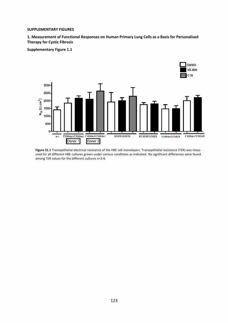
123
SUPPLEMENTARY FIGURES
1. Measurement of Functional Responses on Human Primary Lung Cells as a Basis for Personalised Therapy for Cystic Fibrosis
Supplementary Figure 1.1
Figure S1.1 Transepithelial electrical resistance of the HBE cell monolayers. Transepithelial resistance (TER) was meas-ured for all different HBE cultures grown under various conditions as indicated. No significant differences were found among TER values for the different cultures n=3-6.
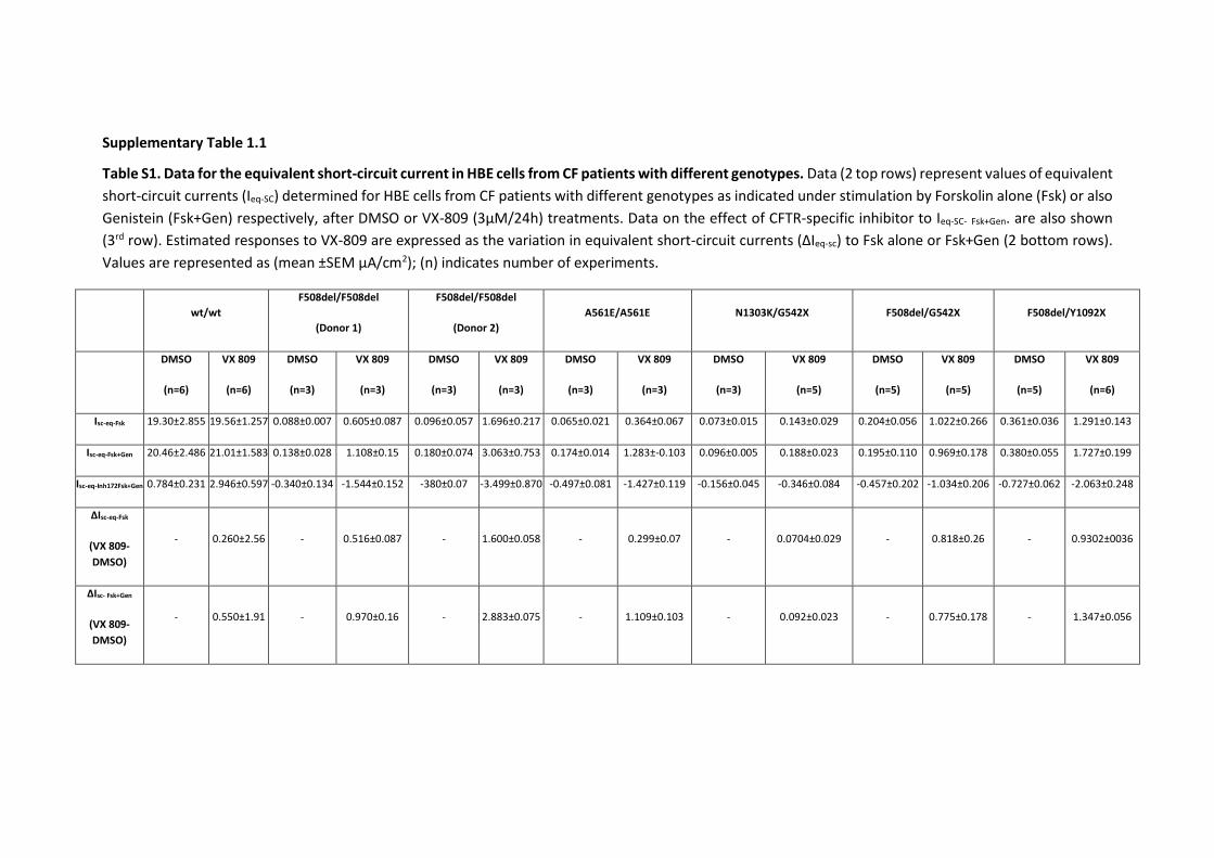
Supplementary Table 1.1
Table S1. Data for the equivalent short-circuit current in HBE cells from CF patients with different genotypes. Data (2 top rows) represent values of equivalent
short-circuit currents (Ieq-SC) determined for HBE cells from CF patients with different genotypes as indicated under stimulation by Forskolin alone (Fsk) or also
Genistein (Fsk+Gen) respectively, after DMSO or VX-809 (3µM/24h) treatments. Data on the effect of CFTR-specific inhibitor to Ieq-SC- Fsk+Gen. are also shown
(3rd row). Estimated responses to VX-809 are expressed as the variation in equivalent short-circuit currents (ΔIeq-sc) to Fsk alone or Fsk+Gen (2 bottom rows).
Values are represented as (mean ±SEM µA/cm2); (n) indicates number of experiments.
wt/wt
F508del/F508del
(Donor 1)
F508del/F508del
(Donor 2)
A561E/A561E N1303K/G542X F508del/G542X F508del/Y1092X
DMSO
(n=6)
VX 809
(n=6)
DMSO
(n=3)
VX 809
(n=3)
DMSO
(n=3)
VX 809
(n=3)
DMSO
(n=3)
VX 809
(n=3)
DMSO
(n=3)
VX 809
(n=5)
DMSO
(n=5)
VX 809
(n=5)
DMSO
(n=5)
VX 809
(n=6)
Isc-eq-Fsk 19.30±2.855 19.56±1.257 0.088±0.007 0.605±0.087 0.096±0.057 1.696±0.217 0.065±0.021 0.364±0.067 0.073±0.015 0.143±0.029 0.204±0.056 1.022±0.266 0.361±0.036 1.291±0.143
Isc-eq-Fsk+Gen 20.46±2.486 21.01±1.583 0.138±0.028 1.108±0.15 0.180±0.074 3.063±0.753 0.174±0.014 1.283±-0.103 0.096±0.005 0.188±0.023 0.195±0.110 0.969±0.178 0.380±0.055 1.727±0.199
Isc-eq-Inh172Fsk+Gen 0.784±0.231 2.946±0.597 -0.340±0.134 -1.544±0.152 -380±0.07 -3.499±0.870 -0.497±0.081 -1.427±0.119 -0.156±0.045 -0.346±0.084 -0.457±0.202 -1.034±0.206 -0.727±0.062 -2.063±0.248
ΔIsc-eq-Fsk
(VX 809-
DMSO)
- 0.260±2.56 - 0.516±0.087 - 1.600±0.058 - 0.299±0.07 - 0.0704±0.029 - 0.818±0.26 - 0.9302±0036
ΔIsc- Fsk+Gen
(VX 809-
DMSO)
- 0.550±1.91 - 0.970±0.16 - 2.883±0.075 - 1.109±0.103 - 0.092±0.023 - 0.775±0.178 - 1.347±0.056
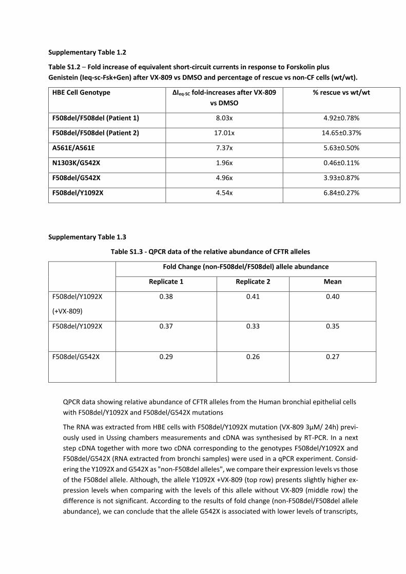
Supplementary Table 1.2
Table S1.2 – Fold increase of equivalent short-circuit currents in response to Forskolin plus
Genistein (Ieq-sc-Fsk+Gen) after VX-809 vs DMSO and percentage of rescue vs non-CF cells (wt/wt).
HBE Cell Genotype ΔIeq-SC fold-increases after VX-809
vs DMSO
% rescue vs wt/wt
F508del/F508del (Patient 1) 8.03x 4.92±0.78%
F508del/F508del (Patient 2) 17.01x 14.65±0.37%
A561E/A561E 7.37x 5.63±0.50%
N1303K/G542X 1.96x 0.46±0.11%
F508del/G542X 4.96x 3.93±0.87%
F508del/Y1092X 4.54x 6.84±0.27%
Supplementary Table 1.3
Table S1.3 - QPCR data of the relative abundance of CFTR alleles
Fold Change (non-F508del/F508del) allele abundance
Replicate 1 Replicate 2 Mean
F508del/Y1092X
(+VX-809)
0.38 0.41 0.40
F508del/Y1092X
0.37 0.33 0.35
F508del/G542X
0.29 0.26 0.27
QPCR data showing relative abundance of CFTR alleles from the Human bronchial epithelial cells
with F508del/Y1092X and F508del/G542X mutations
The RNA was extracted from HBE cells with F508del/Y1092X mutation (VX-809 3µM/ 24h) previ-
ously used in Ussing chambers measurements and cDNA was synthesised by RT-PCR. In a next
step cDNA together with more two cDNA corresponding to the genotypes F508del/Y1092X and
F508del/G542X (RNA extracted from bronchi samples) were used in a qPCR experiment. Consid-
ering the Y1092X and G542X as "non-F508del alleles", we compare their expression levels vs those
of the F508del allele. Although, the allele Y1092X +VX-809 (top row) presents slightly higher ex-
pression levels when comparing with the levels of this allele without VX-809 (middle row) the
difference is not significant. According to the results of fold change (non-F508del/F508del allele
abundance), we can conclude that the allele G542X is associated with lower levels of transcripts,
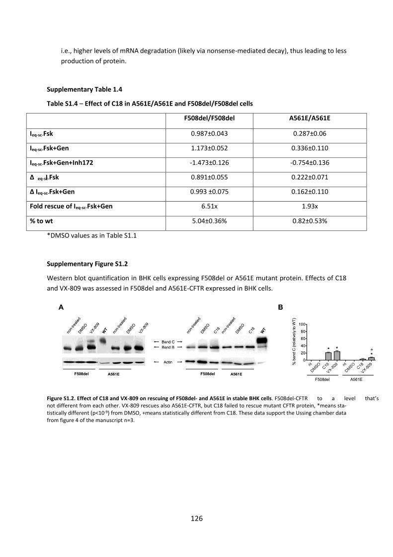
126
i.e., higher levels of mRNA degradation (likely via nonsense-mediated decay), thus leading to less
production of protein.
Supplementary Table 1.4
Table S1.4 – Effect of C18 in A561E/A561E and F508del/F508del cells
F508del/F508del A561E/A561E
Ieq-sc-Fsk 0.987±0.043 0.287±0.06
Ieq-sc-Fsk+Gen 1.173±0.052 0.336±0.110
Ieq-sc-Fsk+Gen+Inh172 -1.473±0.126 -0.754±0.136
Δ Ieq-sc-Fsk 0.891±0.055 0.222±0.071
Δ Ieq-sc-Fsk+Gen 0.993 ±0.075 0.162±0.110
Fold rescue of Ieq-sc-Fsk+Gen 6.51x 1.93x
% to wt 5.04±0.36% 0.82±0.53%
*DMSO values as in Table S1.1
Supplementary Figure S1.2
Western blot quantification in BHK cells expressing F508del or A561E mutant protein. Effects of C18
and VX-809 was assessed in F508del and A561E-CFTR expressed in BHK cells.
Figure S1.2. Effect of C18 and VX-809 on rescuing of F508del- and A561E in stable BHK cells. F508del-CFTR to a level that’s not different from each other. VX-809 rescues also A561E-CFTR, but C18 failed to rescue mutant CFTR protein, *means sta-tistically different (p<10-9) from DMSO, +means statistically different from C18. These data support the Ussing chamber data from figure 4 of the manuscript n=3.
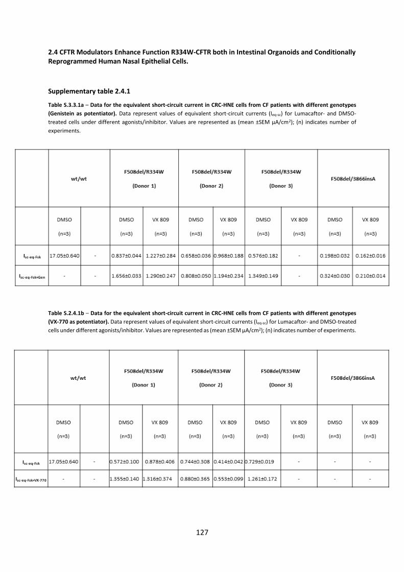
127
2.4 CFTR Modulators Enhance Function R334W-CFTR both in Intestinal Organoids and Conditionally Reprogrammed Human Nasal Epithelial Cells.
Supplementary table 2.4.1
Table S.3.3.1a – Data for the equivalent short-circuit current in CRC-HNE cells from CF patients with different genotypes
(Genistein as potentiator). Data represent values of equivalent short-circuit currents (Ieq-sc) for Lumacaftor- and DMSO-
treated cells under different agonists/inhibitor. Values are represented as (mean ±SEM µA/cm2); (n) indicates number of
experiments.
Table S.2.4.1b – Data for the equivalent short-circuit current in CRC-HNE cells from CF patients with different genotypes
(VX-770 as potentiator). Data represent values of equivalent short-circuit currents (Ieq-sc) for Lumacaftor- and DMSO-treated
cells under different agonists/inhibitor. Values are represented as (mean ±SEM µA/cm2); (n) indicates number of experiments.
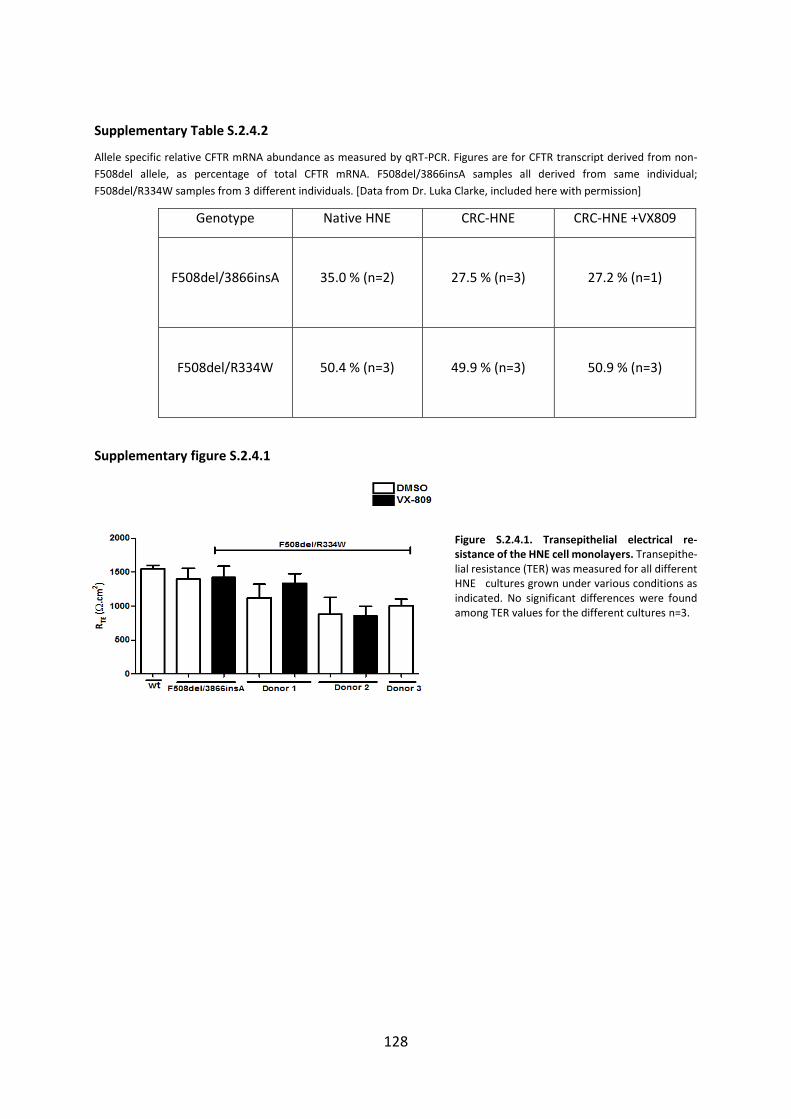
128
Supplementary Table S.2.4.2
Allele specific relative CFTR mRNA abundance as measured by qRT-PCR. Figures are for CFTR transcript derived from non-
F508del allele, as percentage of total CFTR mRNA. F508del/3866insA samples all derived from same individual;
F508del/R334W samples from 3 different individuals. [Data from Dr. Luka Clarke, included here with permission]
Genotype Native HNE CRC-HNE CRC-HNE +VX809
F508del/3866insA
35.0 % (n=2)
27.5 % (n=3)
27.2 % (n=1)
F508del/R334W
50.4 % (n=3)
49.9 % (n=3)
50.9 % (n=3)
Supplementary figure S.2.4.1
Figure S.2.4.1. Transepithelial electrical re-sistance of the HNE cell monolayers. Transepithe-lial resistance (TER) was measured for all different HNE cultures grown under various conditions as indicated. No significant differences were found among TER values for the different cultures n=3.
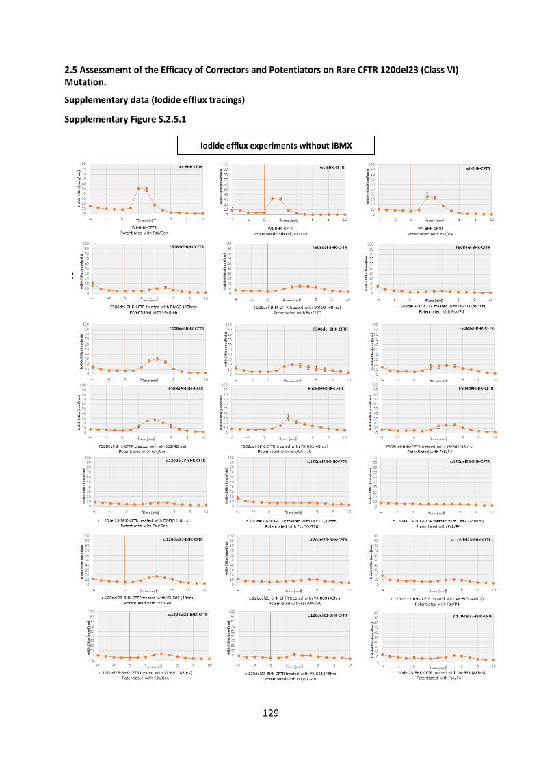
129
2.5 Assessmemt of the Efficacy of Correctors and Potentiators on Rare CFTR 120del23 (Class VI) Mutation.
Supplementary data (Iodide efflux tracings)
Supplementary Figure S.2.5.1
Supplementary Figure S.II.2.5.2
Iodide efflux experiments without IBMX
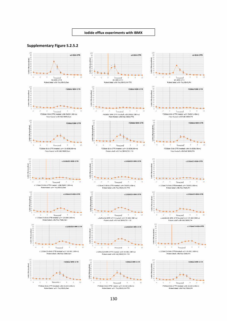
130
Supplementary Figure S.2.5.2
Iodide efflux experiments with IBMX
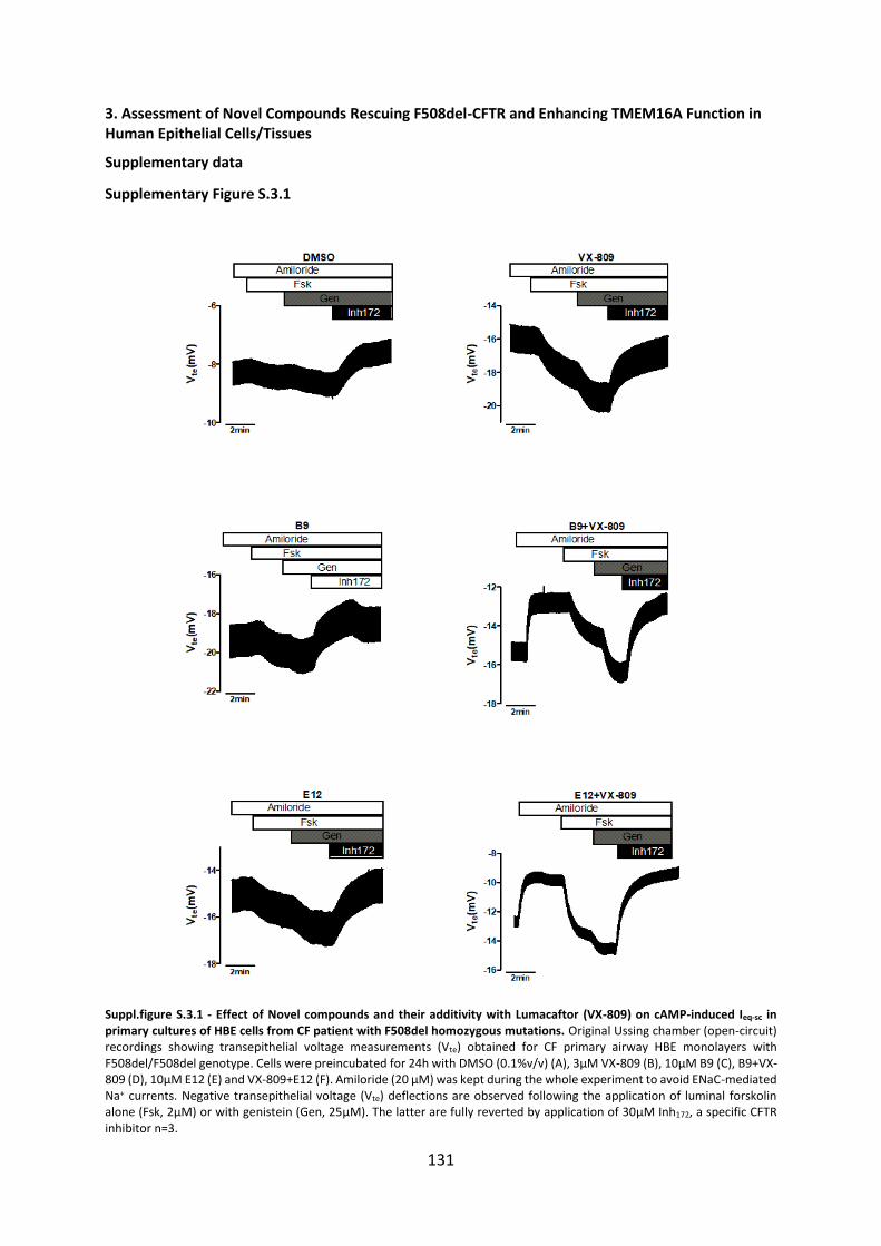
131
3. Assessment of Novel Compounds Rescuing F508del-CFTR and Enhancing TMEM16A Function in Human Epithelial Cells/Tissues
Supplementary data
Supplementary Figure S.3.1
Suppl.figure S.3.1 - Effect of Novel compounds and their additivity with Lumacaftor (VX-809) on cAMP-induced Ieq-sc in primary cultures of HBE cells from CF patient with F508del homozygous mutations. Original Ussing chamber (open-circuit) recordings showing transepithelial voltage measurements (Vte) obtained for CF primary airway HBE monolayers with F508del/F508del genotype. Cells were preincubated for 24h with DMSO (0.1%v/v) (A), 3µM VX-809 (B), 10µM B9 (C), B9+VX-809 (D), 10µM E12 (E) and VX-809+E12 (F). Amiloride (20 µM) was kept during the whole experiment to avoid ENaC-mediated Na+ currents. Negative transepithelial voltage (Vte) deflections are observed following the application of luminal forskolin alone (Fsk, 2µM) or with genistein (Gen, 25µM). The latter are fully reverted by application of 30µM Inh172, a specific CFTR inhibitor n=3.
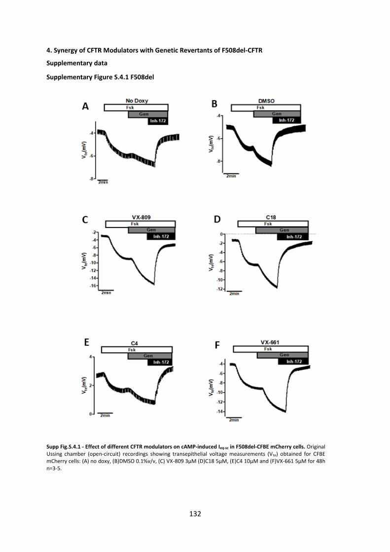
132
4. Synergy of CFTR Modulators with Genetic Revertants of F508del-CFTR
Supplementary data
Supplementary Figure S.4.1 F508del
Supp Fig.S.4.1 - Effect of different CFTR modulators on cAMP-induced Ieq-sc in F508del-CFBE mCherry cells. Original Ussing chamber (open-circuit) recordings showing transepithelial voltage measurements (Vte) obtained for CFBE mCherry cells: (A) no doxy, (B)DMSO 0.1%v/v, (C) VX-809 3µM (D)C18 5µM, (E)C4 10µM and (F)VX-661 5µM for 48h n=3-5.
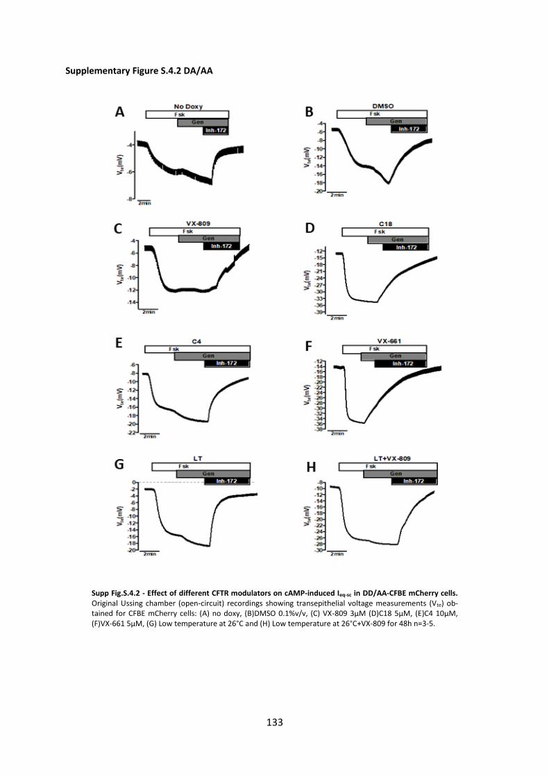
133
Supplementary Figure S.4.2 DA/AA
Supp Fig.S.4.2 - Effect of different CFTR modulators on cAMP-induced Ieq-sc in DD/AA-CFBE mCherry cells. Original Ussing chamber (open-circuit) recordings showing transepithelial voltage measurements (Vte) ob-tained for CFBE mCherry cells: (A) no doxy, (B)DMSO 0.1%v/v, (C) VX-809 3µM (D)C18 5µM, (E)C4 10µM, (F)VX-661 5µM, (G) Low temperature at 26°C and (H) Low temperature at 26°C+VX-809 for 48h n=3-5.
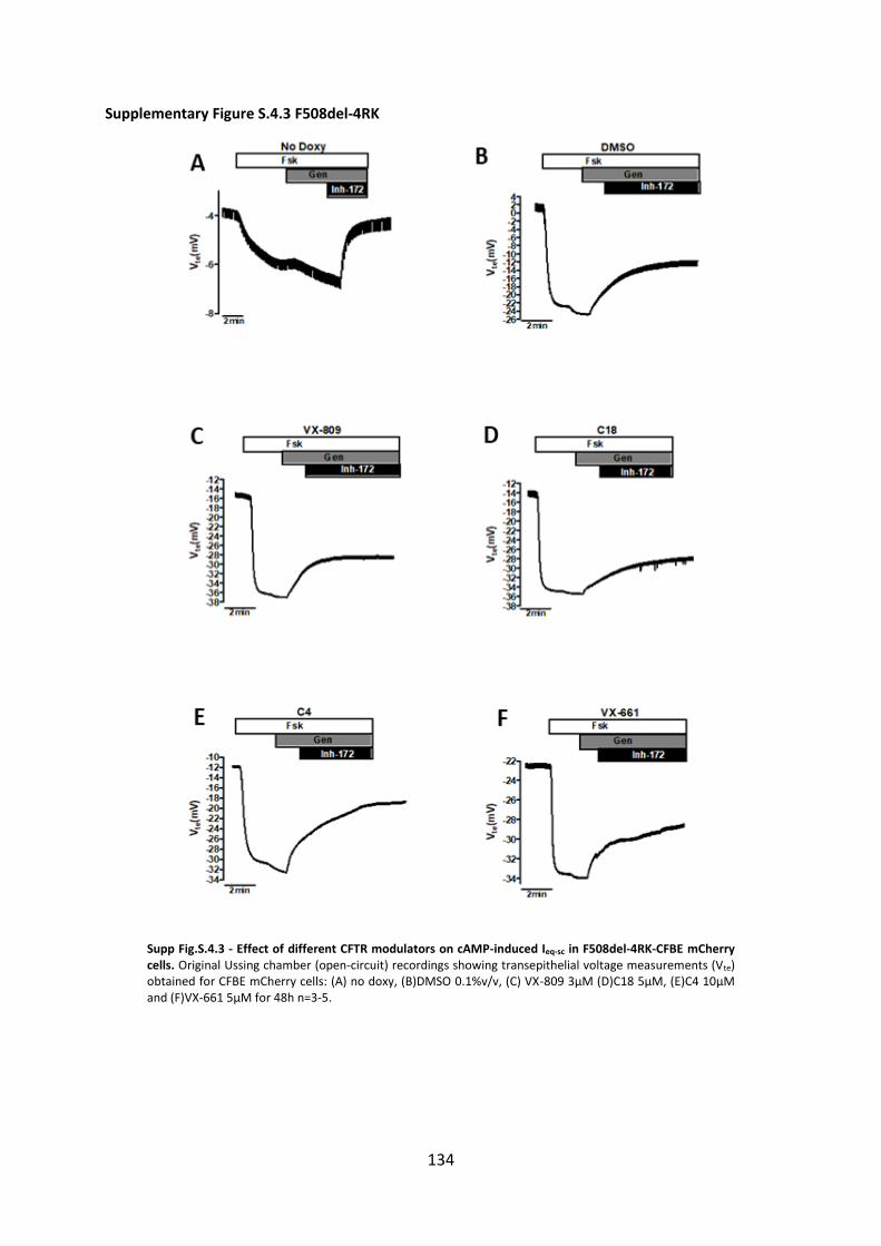
134
Supplementary Figure S.4.3 F508del-4RK
Supp Fig.S.4.3 - Effect of different CFTR modulators on cAMP-induced Ieq-sc in F508del-4RK-CFBE mCherry cells. Original Ussing chamber (open-circuit) recordings showing transepithelial voltage measurements (Vte) obtained for CFBE mCherry cells: (A) no doxy, (B)DMSO 0.1%v/v, (C) VX-809 3µM (D)C18 5µM, (E)C4 10µM and (F)VX-661 5µM for 48h n=3-5.
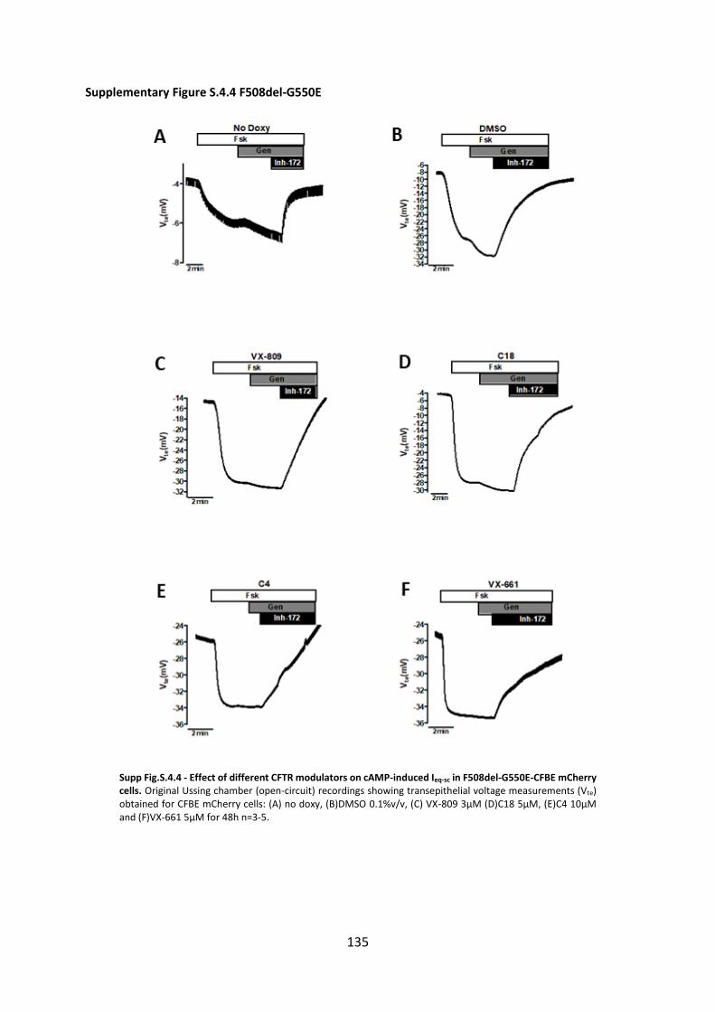
135
Supplementary Figure S.4.4 F508del-G550E
Supp Fig.S.4.4 - Effect of different CFTR modulators on cAMP-induced Ieq-sc in F508del-G550E-CFBE mCherry cells. Original Ussing chamber (open-circuit) recordings showing transepithelial voltage measurements (Vte) obtained for CFBE mCherry cells: (A) no doxy, (B)DMSO 0.1%v/v, (C) VX-809 3µM (D)C18 5µM, (E)C4 10µM and (F)VX-661 5µM for 48h n=3-5.
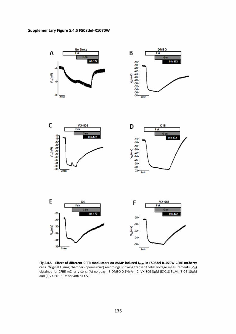
136
Supplementary Figure S.4.5 F508del-R1070W
Fig.S.4.5 - Effect of different CFTR modulators on cAMP-induced Ieq-sc in F508del-R1070W-CFBE mCherry cells. Original Ussing chamber (open-circuit) recordings showing transepithelial voltage measurements (Vte) obtained for CFBE mCherry cells: (A) no doxy, (B)DMSO 0.1%v/v, (C) VX-809 3µM (D)C18 5µM, (E)C4 10µM and (F)VX-661 5µM for 48h n=3-5.
Copyright © 2022 FDOKUMEN


![3-H-[1,2]Dithiole as a New Anti-Trypanosoma cruzi Chemotype: Biological and Mechanism of Action Studies](https://static.fdokumen.com/doc/165x107/634164fed20aa4a11606194e/3-h-12dithiole-as-a-new-anti-trypanosoma-cruzi-chemotype-biological-and-mechanism.jpg)