Latest view on the mechanism of action of deep brain stimulation
11
Reviews Latest View on the Mechanism of Action of Deep Brain Stimulation Constance Hammond, PhD, 1 * Rachida Ammari, 1,2 Bernard Bioulac, MD, PhD, 2 Liliana Garcia, PhD 2 1 Institut de Neurobiologie de la Me´diterrane´e (INMED, U901), Unite´mixte Inserm-Universite´Aix Marseille II, Marseille Ce´dex 9, France 2 UMR 5227 CNRS-Universite´Bordeaux 2, Bordeaux Ce´dex, France Abstract: How does deep brain stimulation (DBS) applied at high frequency (100 Hz and above, HFS) in diverse points of cortico-basal ganglia thalamo-cortical loops alleviate symp- toms of neurological disorders such as Parkinson’s disease, dystonia, and obsessive compulsive disorders? Do the effects of HFS stem solely or even largely from local effects on the stimulated brain structure or are they also mediated by actions of HFS on distal structures? Indeed, HFS as an extracellular stimulation is expected to activate subsets of both afferent and efferent axons, leading to antidromic spikes that collide with ongoing spontaneous ones and orthodromic spikes that evoke synaptic responses in target neurons. The present review sug- gests that HFS interfere with spontaneous pathological patterns by introducing a regular activity in several nodal points of the network. Therefore, the best site of implantation of the HFS electrode may be in a region where the HFS-driven activity spreads to most of the identified, dysrhythmic, neuronal popula- tions without causing additional side effects. This should help tackling the most difficult issue namely, how does the regular HFS-driven activity that dampens the spontaneous pathological one, restore neuronal processing along cortico-basal ganglia- thalamo-cortical loops? Ó 2008 Movement Disorder Society Key words: DBS; electrophysiology; antidromic spikes; Parkinson; dystonia; OCD Deep brain stimulation at high frequency (HFS) has the potential to provide substantial benefit for various neuro- logic and neuropsychiatric diseases. HFS is an intracere- bral, extracellular stimulation consisting of short pulses (in the order of 100 ls) regularly applied at a frequency of at least 100 Hz over a period of several years. First observed to alleviate tremor in ventral thalamus and pallidum, 1 HFS is now widely used in ventral thalamus for essential tremor, 2 in the internal pallidal segment (GPi) or subthala- mic nucleus (STN) for Parkinson’s disease (PD), 3–5 in the GPi for generalized dystonia, 6,7 and more recently for other diseases such as treatment-resistant obsessive com- pulsive disorder (OCD), 8,9 Tourette syndrome, 10,11 and depression. 12 Sites of stimulation are located inside the cortico-basal ganglia-thalamo-cortical loops, in motor or limbic regions, depending on the clinical signs. Two major and nonexclusive explanations have been proposed for the effect of HFS: (1) it silences stimu- lated neurons and (2) it introduces a new activity in the network. The first theory stems from the observa- tion that, functionally, HFS produces the same effect as a lesion of the stimulated area. The second hypothe- sis proposes that HFS injects in a point of the circuit a HFS-driven activity that propagates and consequently modifies the pathological spontaneous activity in many nuclei. The clarification of the mechanisms of action of HFS is imperative to avoid implanting electrodes in regions having a low impact on clinical signs and/or leading to activation of undesirable regions and incapa- citating side effects. In this review, we focus on results obtained within the last 4 years from multiunit and sin- gle cell electrophysiological recordings. This article is part of the journal’s online CME program. The CME activity including form, can be found online at http://www. movementdisorders.org/education/journalcme/ Dr. Constance Hammond, INMED Inserm, 163 route de Luminy, BP13, 13273 Marseille Ce ´dex 9, France. E-mail: [email protected] Received 10 January 2008; Revised 7 April 2008; Accepted 8 April 2008 Published online 10 September 2008 in Wiley InterScience (www. interscience.wiley.com). DOI: 10.1002/mds.22120 2111 Movement Disorders Vol. 23, No. 15, 2008, pp. 2111–2121 Ó 2008 Movement Disorder Society CME
-
Upload
independent -
Category
Documents
-
view
1 -
download
0
Transcript of Latest view on the mechanism of action of deep brain stimulation
Reviews
Latest View on the Mechanism of Action of DeepBrain Stimulation
Constance Hammond, PhD,1* Rachida Ammari,1,2 Bernard Bioulac, MD, PhD,2 Liliana Garcia, PhD2
1Institut de Neurobiologie de la Mediterranee (INMED, U901), Unite mixte Inserm-Universite Aix Marseille II,Marseille Cedex 9, France
2UMR 5227 CNRS-Universite Bordeaux 2, Bordeaux Cedex, France
Abstract: How does deep brain stimulation (DBS) applied athigh frequency (100 Hz and above, HFS) in diverse points ofcortico-basal ganglia thalamo-cortical loops alleviate symp-toms of neurological disorders such as Parkinson’s disease,dystonia, and obsessive compulsive disorders? Do the effectsof HFS stem solely or even largely from local effects on thestimulated brain structure or are they also mediated by actionsof HFS on distal structures? Indeed, HFS as an extracellularstimulation is expected to activate subsets of both afferent andefferent axons, leading to antidromic spikes that collide withongoing spontaneous ones and orthodromic spikes that evokesynaptic responses in target neurons. The present review sug-
gests that HFS interfere with spontaneous pathological patternsby introducing a regular activity in several nodal points of thenetwork. Therefore, the best site of implantation of the HFSelectrode may be in a region where the HFS-driven activityspreads to most of the identified, dysrhythmic, neuronal popula-tions without causing additional side effects. This should helptackling the most difficult issue namely, how does the regularHFS-driven activity that dampens the spontaneous pathologicalone, restore neuronal processing along cortico-basal ganglia-thalamo-cortical loops? � 2008 Movement Disorder SocietyKey words: DBS; electrophysiology; antidromic spikes;
Parkinson; dystonia; OCD
Deep brain stimulation at high frequency (HFS) has thepotential to provide substantial benefit for various neuro-logic and neuropsychiatric diseases. HFS is an intracere-bral, extracellular stimulation consisting of short pulses (inthe order of 100 ls) regularly applied at a frequency of atleast 100 Hz over a period of several years. First observedto alleviate tremor in ventral thalamus and pallidum,1 HFSis now widely used in ventral thalamus for essentialtremor,2 in the internal pallidal segment (GPi) or subthala-mic nucleus (STN) for Parkinson’s disease (PD),3–5 in theGPi for generalized dystonia,6,7 and more recently forother diseases such as treatment-resistant obsessive com-
pulsive disorder (OCD),8,9 Tourette syndrome,10,11 anddepression.12 Sites of stimulation are located inside thecortico-basal ganglia-thalamo-cortical loops, in motor orlimbic regions, depending on the clinical signs.
Two major and nonexclusive explanations have been
proposed for the effect of HFS: (1) it silences stimu-
lated neurons and (2) it introduces a new activity in
the network. The first theory stems from the observa-
tion that, functionally, HFS produces the same effect
as a lesion of the stimulated area. The second hypothe-
sis proposes that HFS injects in a point of the circuit a
HFS-driven activity that propagates and consequently
modifies the pathological spontaneous activity in many
nuclei. The clarification of the mechanisms of action
of HFS is imperative to avoid implanting electrodes in
regions having a low impact on clinical signs and/or
leading to activation of undesirable regions and incapa-
citating side effects. In this review, we focus on results
obtained within the last 4 years from multiunit and sin-
gle cell electrophysiological recordings.
This article is part of the journal’s online CME program. TheCME activity including form, can be found online at http://www.movementdisorders.org/education/journalcme/
Dr. Constance Hammond, INMED Inserm, 163 route de Luminy,BP13, 13273 Marseille Cedex 9, France.E-mail: [email protected]
Received 10 January 2008; Revised 7 April 2008; Accepted 8April 2008
Published online 10 September 2008 in Wiley InterScience (www.
interscience.wiley.com). DOI: 10.1002/mds.22120
2111
Movement DisordersVol. 23, No. 15, 2008, pp. 2111–2121� 2008 Movement Disorder Society CME
MECHANISMS OF STN-HFS IN THE CASE
OF PARKINSON’S DISEASE
Increased synchronization and the appearance of
pathological oscillations in the activity patterns of popu-
lations of STN and GP neurons but also in motor corti-
cal networks are salient aspects of Parkinsonism. Patho-
logical synchronization has been observed in human PD
patients,13 1-methyl-4-phenyl-1,2,3,6-tetrahydropyridine
(MPTP)-treated monkeys14 and rodents with 6-hydroxy-
dopamine (6-OHDA) lesions,15 suggesting functional
alterations in the basal ganglia network. In particular,
increased coherence in the b-band (13–30 Hz) is corre-
lated with the severity of symptoms in humans.16–18
Mechanisms of STN-HFS have long been reduced to
a lesion-like or inhibition hypothesis until biochemical,
metabolic, and electrophysiological data in experimen-
tal models and patients together with modeling stud-
ies19–23 provided consistent evidence in favor of an
activation.
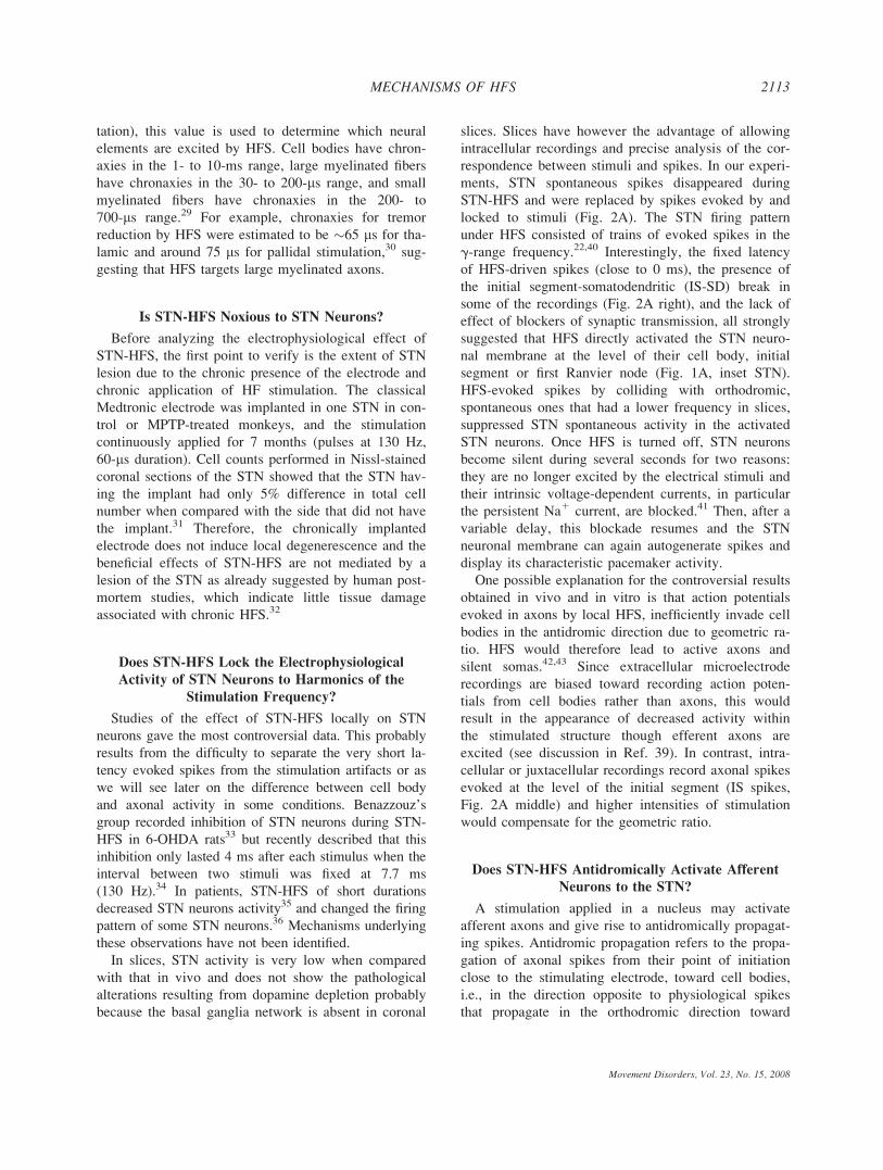
Basal ganglia are made up of different neuronal pop-
ulations scattered in five different nuclei (see Fig. 1):
caudate and putamen (striatum in rodents), globus pal-
lidus (external and internal), subthalamic nucleus
(STN), and substantia nigra pars reticulata (SNr) and
compacta (SNc). The degeneration of the dopaminergic
neurons of the SNc and the consequent loss of dopa-
mine in the striatum leads to ‘‘typical’’ PD. STN neu-
rons occupy a strategic position as they are the only
glutamatergic neurons of the basal ganglia network,
receive afferents from motor-related cortical areas, pro-
ject to all nuclei of the basal ganglia (though to a lesser
extent to striatum), and are reciprocally connected with
GPe and brainstem neurons of the pedunculopontine nu-
cleus (PPN).25 Therefore, STN stimulation may activate
diverse pathways and have widespread effects.
Preparations, Parameters of Stimulation, and
Excitation of Neural Elements
Different types of preparations have been used to
study HFS mechanisms from anesthetized in vivo mod-
els of PD to in vitro slices. Recordings have been per-
formed in patients as well. Each preparation has its
own advantages and pitfalls, but their combination
should allow understanding HFS mechanisms as long
as we are aware of the limitations of the technique
used. In this review, HFS refers to high frequency
stimulations in vivo and in vitro.
The question of the mechanisms of action of HFS
relies on the analysis of what does a high frequency
and long duration stimulation of neuronal elements
(HFS is applied for years). Electrophysiologists are
used to study potential changes in response to single
stimulations but here the question is far more complex
mainly for technical reasons as recordings have to be
maintained for minutes or hours and spikes identified
among artifacts. If all studies on HFS mechanisms test
high frequency (100–180 Hz) stimuli, they rarely apply
them for long durations. Considering synaptic plasticity
(potentiation or depression) that usually occurs in syn-
aptic transmission after tetanic stimulation,26 ultrashort
(ms, s) and long (days, years) duration stimulations
could evoke very different responses. Even though
some of the symptoms are immediately improved,27
when describing the electrophysiological effects of
STN-HFS one must keep in mind whether it is a short-
or a long-term effect. To compare results from differ-
ent studies, intensity of stimulation is not as informa-
tive as charge density (lC/cm2/phase).28 This parame-
ter depends on the diameter of the stimulation elec-
trode or contact and is not always available from
papers. For this reason, the value of current intensity is
not mentioned. All protocols use short pulses (60–200
ls) of stimulation. Since cell bodies and axons have
different chronaxies (chronaxy is the shortest duration
of an effective electrical stimulus having a strength
equal to twice the minimum strength required for exci-
FIG. 1. STN and the basal ganglia network STN-HFS preferentiallyactivates axons thus generating spikes that propagate in the anti-dromic (toward STN, motor cortex, GPe and PPN somas) and ortho-dromic (toward GPe, GPi, SNr, SNc, PPN) directions. Passing fiberscan also be activated. As a result, basal ganglia nuclei such as SNand GP together with motor cortical areas are directly affected bySTN-HFS. The striatum is mainly indirectly affected via the modula-tion of dopaminergic SNc neurons and cortical afferents. When anti-dromic spikes propagate back to a structure, they may invade somasand axon collaterals and thus activate other projection neurons andlocal interneurons when they exist. Insets show simplified cortical24
(top) and GPe (bottom) networks. , neuronal populations directlyaffected by STN-DBS; , neuronal populations indirectly affected bySTN-DBS; , antidromic activation of axons, , orthodromic acti-vation of axons; , GABAergic neuron; , glutamatergic neuron.
2112 C. HAMMOND ET AL.
Movement Disorders, Vol. 23, No. 15, 2008
tation), this value is used to determine which neural
elements are excited by HFS. Cell bodies have chron-
axies in the 1- to 10-ms range, large myelinated fibers
have chronaxies in the 30- to 200-ls range, and small
myelinated fibers have chronaxies in the 200- to
700-ls range.29 For example, chronaxies for tremor
reduction by HFS were estimated to be �65 ls for tha-lamic and around 75 ls for pallidal stimulation,30 sug-
gesting that HFS targets large myelinated axons.
Is STN-HFS Noxious to STN Neurons?
Before analyzing the electrophysiological effect of
STN-HFS, the first point to verify is the extent of STN
lesion due to the chronic presence of the electrode and
chronic application of HF stimulation. The classical
Medtronic electrode was implanted in one STN in con-
trol or MPTP-treated monkeys, and the stimulation
continuously applied for 7 months (pulses at 130 Hz,
60-ls duration). Cell counts performed in Nissl-stained
coronal sections of the STN showed that the STN hav-
ing the implant had only 5% difference in total cell
number when compared with the side that did not have
the implant.31 Therefore, the chronically implanted
electrode does not induce local degenerescence and the
beneficial effects of STN-HFS are not mediated by a
lesion of the STN as already suggested by human post-
mortem studies, which indicate little tissue damage
associated with chronic HFS.32
Does STN-HFS Lock the Electrophysiological
Activity of STN Neurons to Harmonics of the
Stimulation Frequency?
Studies of the effect of STN-HFS locally on STN
neurons gave the most controversial data. This probably
results from the difficulty to separate the very short la-
tency evoked spikes from the stimulation artifacts or as
we will see later on the difference between cell body
and axonal activity in some conditions. Benazzouz’s
group recorded inhibition of STN neurons during STN-
HFS in 6-OHDA rats33 but recently described that this
inhibition only lasted 4 ms after each stimulus when the
interval between two stimuli was fixed at 7.7 ms
(130 Hz).34 In patients, STN-HFS of short durations
decreased STN neurons activity35 and changed the firing
pattern of some STN neurons.36 Mechanisms underlying
these observations have not been identified.
In slices, STN activity is very low when compared
with that in vivo and does not show the pathological
alterations resulting from dopamine depletion probably
because the basal ganglia network is absent in coronal
slices. Slices have however the advantage of allowing
intracellular recordings and precise analysis of the cor-
respondence between stimuli and spikes. In our experi-
ments, STN spontaneous spikes disappeared during
STN-HFS and were replaced by spikes evoked by and
locked to stimuli (Fig. 2A). The STN firing pattern
under HFS consisted of trains of evoked spikes in the
g-range frequency.22,40 Interestingly, the fixed latency
of HFS-driven spikes (close to 0 ms), the presence of
the initial segment-somatodendritic (IS-SD) break in
some of the recordings (Fig. 2A right), and the lack of
effect of blockers of synaptic transmission, all strongly
suggested that HFS directly activated the STN neuro-
nal membrane at the level of their cell body, initial
segment or first Ranvier node (Fig. 1A, inset STN).
HFS-evoked spikes by colliding with orthodromic,
spontaneous ones that had a lower frequency in slices,
suppressed STN spontaneous activity in the activated
STN neurons. Once HFS is turned off, STN neurons
become silent during several seconds for two reasons:
they are no longer excited by the electrical stimuli and
their intrinsic voltage-dependent currents, in particular
the persistent Na1 current, are blocked.41 Then, after a
variable delay, this blockade resumes and the STN
neuronal membrane can again autogenerate spikes and
display its characteristic pacemaker activity.
One possible explanation for the controversial results
obtained in vivo and in vitro is that action potentials
evoked in axons by local HFS, inefficiently invade cell
bodies in the antidromic direction due to geometric ra-
tio. HFS would therefore lead to active axons and
silent somas.42,43 Since extracellular microelectrode
recordings are biased toward recording action poten-
tials from cell bodies rather than axons, this would
result in the appearance of decreased activity within
the stimulated structure though efferent axons are
excited (see discussion in Ref. 39). In contrast, intra-
cellular or juxtacellular recordings record axonal spikes
evoked at the level of the initial segment (IS spikes,
Fig. 2A middle) and higher intensities of stimulation
would compensate for the geometric ratio.
Does STN-HFS Antidromically Activate Afferent
Neurons to the STN?
A stimulation applied in a nucleus may activate
afferent axons and give rise to antidromically propagat-
ing spikes. Antidromic propagation refers to the propa-
gation of axonal spikes from their point of initiation
close to the stimulating electrode, toward cell bodies,
i.e., in the direction opposite to physiological spikes
that propagate in the orthodromic direction toward
2113MECHANISMS OF HFS
Movement Disorders, Vol. 23, No. 15, 2008
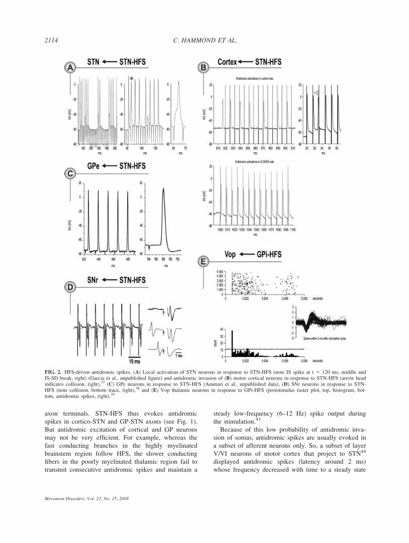
axon terminals. STN-HFS thus evokes antidromic
spikes in cortico-STN and GP-STN axons (see Fig. 1).
But antidromic excitation of cortical and GP neurons
may not be very efficient. For example, whereas the
fast conducting branches in the highly myelinated
brainstem region follow HFS, the slower conducting
fibers in the poorly myelinated thalamic region fail to
transmit consecutive antidromic spikes and maintain a
steady low-frequency (6–12 Hz) spike output during
the stimulation.43
Because of this low probability of antidromic inva-
sion of somas, antidromic spikes are usually evoked in
a subset of afferent neurons only. So, a subset of layer
V/VI neurons of motor cortex that project to STN44
displayed antidromic spikes (latency around 2 ms)
whose frequency decreased with time to a steady state
FIG. 2. HFS-driven antidromic spikes. (A) Local activation of STN neurons in response to STN-HFS (note IS spike at t 5 120 ms, middle andIS-SD break, right) (Garcia et al., unpublished figure) and antidromic invasion of (B) motor cortical neurons in response to STN-HFS (arrow headindicates collision, right),37 (C) GPe neurons in response to STN-HFS (Ammari et al., unpublished data), (D) SNr neurons in response to STN-HFS (note collision, bottom trace, right),38 and (E) Vop thalamic neurons in response to GPi-HFS (poststimulus raster plot, top, histogram, bot-tom, antidromic spikes, right).39
2114 C. HAMMOND ET AL.
Movement Disorders, Vol. 23, No. 15, 2008

at around 40 Hz in response to 120 Hz STN stimula-
tion37 (Fig. 2B). Recordings of short latency evoked
potentials over the motor cortex during STN-HFS also
indicated that cortico-STN axons were most likely acti-
vated.45 As glutamatergic cortico-STN neurons give
off many axon collaterals in deep and superficial
layers46 that contact other projection neurons and local
GABAergic interneurons, antidromic invasion of a sub-
set of neurons may retrogradely affect cortical circuits
in complex ways (Fig. 1, inset cortex).47
STN-HFS in slices (130 Hz, 90 ls) evoked anti-
dromic spikes in a subset of GPe neurons with a mean
latency of around 2 ms (Fig. 2C) that collided with the
ongoing activity in the recorded neurons. If antidromic
spikes propagate in the complex network of local
GABAergic collaterals that synapse onto other GP neu-
rons, this may have consequences on the activity of
neighboring GP neurons48 (Fig. 1, inset GP).
STN-HFS also activates axons passing through and
near the STN. Thus short STN-HFS (130 Hz, 60 ls, 30s trains) in control rats in vivo antidromically activated
a subpopulation of SNr neurons with a latency of
around 1 ms,38 probably as a result of the activation of
ascending SNr axons. This decreased the spontaneous
activity of the antidromically activated SNr neurons and
inhibited other SNr neurons as shown in recordings38
probably via the activation of the complex network of
intranigral collaterals between GABAergic SNr cells.49
In conclusion, STN-HFS antidromically activates
subsets of neurons in the different nuclei that send
axons to or close to the stimulated site. In that context,
STN-HFS probably also antidromically activates PPN
neurons that project to STN.50,51 Once evoked, anti-
dromic axonal spikes propagate to their corresponding
cell bodies at the stimulation frequency or its subhar-
monics. They may also propagate in recurrent axonal
collaterals and activate synaptic transmission that
impinges onto other projection neurons (GP, SNr) or
local interneurons (see Fig. 1, insets cortex and GPe).
The overall result on a network needs to know the
probability of propagation of antidromic spikes in axo-
nal branches47 and how these spikes evoke synaptic
responses at a long term.
Does STN-HFS Orthodromically Activate Target
Neurons of the STN?
From their point of initiation, axonal spikes also
propagate in the orthodromic direction toward axon
terminals, i.e., along STN efferent axons (see Fig. 1).
Do these spikes evoke postsynaptic responses? STN-
HFS parameters that improved spontaneous move-
ments and muscle tone (130, 210 ls, 25 s–5 min) in
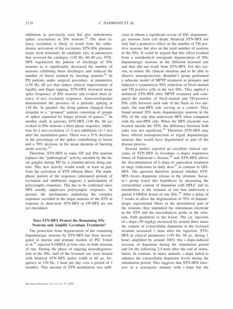
MPTP-treated monkeys evoked stimulus-locked dou-
ble excitatory responses at latencies around 4 and 6
ms in the globus pallidus (GPe and GPi)23 (Fig. 3A).
It thus shifted the firing pattern of GPe and GPi neu-
rons from irregular to stimulus-synchronized. The
most probable mechanism underlying the earlier exci-
tation is the orthodromic activation of STN efferent
axons projecting to GP, the release of glutamate and
the monosynaptic excitation of postsynaptic GPe or
GPi neurons. In contrast, in control monkeys, bursts
of 100 Hz stimuli (10 pulses) induced powerful exci-
tatory responses in the GPe but inhibition in the GPi
attributed to the activation of the disynaptic STN-
GPe-GPi pathway.52
Does STN-HFS similarly affect GP and SNr neu-
rons? Deniau’s group studied the spontaneous and
evoked SNr activity before and during STN-HFS in
control rats or rats treated with neuroleptics to block
dopaminergic transmission. In control rats, STN-HFS
(130 Hz, 60 ls, 30 s) evoked antidromic spikes and
FIG. 3. HFS-driven orthodromicresponses. Orthodromic responsesof (A) GPi neurons in response toSTN-HFS (complex sequence ofexcitation-inhibition-excitation),23
(B) Vop thalamic neurons inresponse to GPi-HFS (antidromicactivation followed by a complexsequence of excitation-inhibition-excitation, poststimulus raster,top, and histogram, bottom).39
2115MECHANISMS OF HFS
Movement Disorders, Vol. 23, No. 15, 2008
inhibition as previously seen but also orthodromic
spikes (excitation) in SNr neurons.38 The short la-
tency excitation is likely to result from the ortho-
dromic activation of the excitatory STN-SNr glutama-
tergic axon terminals. In cataleptic rats, at parameters
that reversed the catalepsy (130 Hz, 60–80 ls), STN-HFS regularized the pattern of discharge of SNr
neurons as it significantly decreased the number of
neurons exhibiting burst discharges and reduced the
number of bursts emitted by bursting neurons.63 In
PD patients under surgical procedure, at parameters
(130 Hz, 60 ls) that induce clinical improvement of
rigidity and finger tapping, STN-HFS increased mean
spike frequency of SNr neurons and evoked short la-
tency (4 ms) excitatory responses. Autocorrelograms
demonstrated the presence of a periodic spiking at
130 Hz. In parallel, the firing pattern changed from
irregular to a ‘‘grouped’’ pattern consisting of groups
of spikes separated by longer periods of pauses.53 In
another study in patients, STN-HFS (140 Hz, 60 ls)evoked in SNr neurons a three-phase sequence, inhibi-
tion (0–2 ms)-excitation (2–4 ms)-inhibition (4–7 ms)
after the stimulation pulse. There was a 51% decrease
in the percentage of the spikes contributing to bursts
and a 70% decrease in the mean duration of bursting
mode activity.54
Therefore, STN-HFS in some GP and SNr neurons
replaces the ‘‘pathological’’ activity encoded by the ba-
sal ganglia during PD by a stimulus-driven firing pat-
tern. This new activity would result, at least in part,
from the activation of STN efferent fibers. The multi-
phasic pattern of the responses (alternated periods of
excitation and inhibition) suggests a participation of
polysynaptic responses. This has to be confirmed since
HFS usually suppresses polysynaptic responses. At
present, the mechanisms underlying the complex
responses recorded in the target neurons of the STN in
response to short-term STN-HFS or GP-HFS are not
yet elucidated.
Does STN-HFS Protect the Remaining SNc
Neurons and Amplify Levodopa Treatment?
The protection from degeneration of the remaining
dopaminergic neurons by STN-HFS has been investi-
gated in murine and primate models of PD. Temel
et al.55 injected 6-OHDA at four sites in both striatum
of rats. During the phase of ongoing neurodegenera-
tion in the SNc, half of the lesioned rats were treated
with bilateral STN-HFS (pulse width at 60 ls, fre-
quency at 130 Hz, 1 hour per day over a period of 3
months). This amount of STN modulation was suffi-
cient to obtain a significant rescue of SNc dopaminer-
gic neurons from cell death. Bilateral STN-HFS not
only had a protective effect on the number of TH pos-
itive neurons but also on the total number of neurons
in the SNc. It could be argued that this effect resulted
from a nonidentical retrograde degeneration of SNc
dopaminergic neurons in the different lesioned rats
and thus did not result from STN-HFS. For this rea-
son, to mimic the clinical situation and to be able to
observe neuroprotection, Benabid’s group performed
a subacute model of MPTP treatment in primates and
induced a symmetrical 50% reduction of Nissl-stained
and TH positive cells in the two SNc. They applied a
unilateral STN-HFS after MPTP treatment and com-
pared the number of Nissl-stained and TH-positive
SNc cells between each side of the brain in two ani-
mals, the non-HFS side serving as a control. They
found around 20% more dopaminergic neurons in the
SNc of the side that underwent HFS when compared
with the non-HFS side. When the HFS electrode was
located outside the STN, the difference between both
sides was not significant.31 Therefore STN-HFS may
have offered neuroprotection to nigral dopaminergic
neurons that would have degenerated as part of the
disease process.
Several studies reported an excellent clinical out-
come of STN-HFS in levodopa (L-dopa) responsive
forms of Parkinson’s disease,56 and STN-HFS allows
the discontinuation of L-dopa or equivalent treatment
or large reductions in daily dose57 in contrast to GPi-
HFS. The question therefore aroused whether STN-
HFS favors dopamine release in the striatum. Savas-
ta’s group tested this hypothesis by measuring the
extracellular content of dopamine with HPLC and its
metabolites in the striatum of rats that underwent a
partial 6-OHDA lesion of one SNc.58 After a delay of
3 weeks to allow the degeneration of 70% of dopami-
nergic nigrostriatal fibers in the dorsolateral part of
the striatum, they implanted the stimulation electrode
in the STN and the microdialysis probe in the stria-
tum, both ipsilateral to the lesion. The i.p. injection
of L-dopa (50 mg/kg) increased by around three times
the content of extracellular dopamine in the lesioned
striatum measured 1 hour after the injection. STN-
HFS at clinical parameters (130 Hz, 60 ls, during 1
hour) amplified by around 100% this L-dopa-induced
increase of dopamine during the stimulation period
and for the following 2.5-hour after the end of stimu-
lation. In contrast, in intact animals, L-dopa failed to
enhance the extracellular dopamine levels during the
stimulation period. This suggests that STN-HFS inter-
acts in a synergistic manner with L-dopa but the
2116 C. HAMMOND ET AL.
Movement Disorders, Vol. 23, No. 15, 2008
underlying mechanisms have not been elucidated. A
simple explanation would be that STN-HFS acts by
directly modulating the firing rate of the remaining
dopaminergic neurones.59,60
Which of the STN-HFS-Induced
Electrophysiological Effects Are Related
to Clinical Efficacy?
Ideally, to test this question, we should specifically
block one by one the identified responses to HFS and
analyze the consequences on motor behavior. This
experiment is difficult to perform. We will propose
some hypotheses as follows: a therapeutic electrophysi-
ological effect is likely to (i) reduce pathological pat-
terns in the basal ganglia, (ii) be recorded from output
nuclei of the basal ganglia whichever site of the cor-
tico-basal ganglia-thalamo-cortical loops is stimulated
(in the same animal models), and (iii) allow re-estab-
lishment of a control response of output GPi and SNr
neurons to cortical stimulation (triphasic response).
We have seen that HFS introduces a stimulation-
locked, complex activity in subsets of neurons from
many sites of the basal ganglia network (STN, motor
cortex, GPe, GPi, SNr), and thus decreases ongoing
pathological activity in these neurons. This may also
be valid for PPN and the good results obtained on
axial motor signs with STN-HFS in association with
PPN-HFS61 may result from a change of PPN dis-
charge pattern. Direct activation of passing fibers dor-
sal to the STN and in particular nigro-striatal and pal-
lido-thalamic axons may also participate to the benefi-
cial effect as the best position of the HFS electrode
active contact is in the dorsal part of the STN.
To answer the second hypothesis, we can take the
example of GPi-HFS and STN-HFS that both amelio-
rate clinical signs of PD. Do they have similar electro-
physiological effects on GPi neurons in the primate
model of PD? Bar-Gad et al.62 recorded the activity of
GPi neurons in MPTP-treated monkeys in response to
microstimulations applied in GPi (135 Hz, 200 ls,600–3000 trains of 10 stimuli separated by 500 ms).
They reported a double excitation with latencies of 3
and 6 ms, separated by a short period of inhibition.
Overall 70% of the GPi neurons displayed a locked ac-
tivity, i.e., they lost their basic firing pattern and
switched to a predicted, orderly discharge that was
locked to the stimulus. These results show striking
similarities with those recorded in GPi with STN-HFS
in MPTP-treated monkeys23 (see earlier).
The third point was shown in SNr where STN-HFS
reversed to control the classical triphasic response to
motor cortex stimulation63 but the mechanisms have
not been elucidated.
HFS FOR OTHER NEUROLOGICAL
DISORDERS
HFS in Motor Cortico-Basal Ganglia-
Thalamocortical Loop for Essential Tremor
HFS of ventral nuclei of the thalamus can dramati-
cally relieve essential tremor in the majority of
patients.2,64 Essential tremor is thought to arise from
dysfunction of the glutamatergic olivocerebellar path-
way, which projects to ventral thalamic (VL) nuclei.65
VL-HFS in rat brain slices silenced or suppressed the
activity of thalamic relay neurons after a transient pe-
riod of intense depolarization.66 The authors hypothe-
sized that VL-HFS introduced a functional deafferenta-
tion of stimulated neurons, thereby stopping tremor
from propagating to thalamo-cortical loops. To test
whether this depression of afferent synaptic transmis-
sion is selective, they stimulated at 5 Hz in two differ-
ent loci within the VL to mimic afferent stimuli at
tremor frequency. Both stimulations evoked excitatory
postsynaptic potentials at 5 Hz in the recorded VL neu-
ron. A concomitant short-duration HFS (125 Hz, for
10 s) in one locus or totally suppressed the 5 Hz EPSPs
in the HFS-stimulated pathway but not in the nonstimu-
lated one, suggesting that HFS selectively disrupts affer-
ent synaptic transmission.67 One of the underlying
mechanisms could include the depression of excitatory
glutamatergic transmission in the ventral thalamus by
activation of the presynaptic A1 receptors. HFS releases
ATP, the precursor of adenosine and local adenosine
infusion suppresses tremor in the harmaline-treated
mice.68 However, these mechanisms have been identi-
fied during very short-term HFS (10 seconds) and may
not sustain the long-term beneficial effects of VL-HFS.
HFS in Motor Cortico-Basal Ganglia-
Thalamocortical Loop for Dystonia
Neuronal activity is altered in basal ganglia and ven-
tral thalamic nuclei in dystonia.69 The firing pattern of
GPi neurons known to be regular in monkeys70 con-
sists in patients of irregular grouped discharges with
intermittent pauses and a third of the neurons discharge
at the frequency of the electromyogram.71,72 Neurons
in ventral oralis posterior/intermediate nuclei of the
thalamus (Vop/Vim) have a sustained activity at 130 to
150 Hz, organized in bursts lasting from 500 ms to 5
seconds and recurring at a frequency similar to that of
dystonia frequency.71
2117MECHANISMS OF HFS
Movement Disorders, Vol. 23, No. 15, 2008
GPi-HFS is currently used for primary generalized
DYT-1 positive dystonia and idiopathic cervical dys-
tonia.7,73,74 In contrast to Parkinson’s disease, the
beneficial effects of HFS in dystonia are not immediate
but progressive over weeks to months. However, record-
ings in patients can only be performed during the surgi-
cal procedure, i.e. at t0, or in control animals, owing to
the lack of reliable animal models of dystonia.
During short duration GPi-HFS, 50 to 70% of Vop
neurons of the thalamus reduced their average dis-
charge frequency with a delay of a few milliseconds in
control monkeys75 or dystonic patients,39 suggesting
that HFS activates GPi efferent axons that are
GABAergic and inhibitory onto thalamic neurons (Fig.
3B). Moreover, 88% of Vop neurons were antidromi-
cally activated with 1-ms latency probably as a result
of the activation of axons originating in Vop and pass-
ing in the vicinity of the GPi-HFS electrode (Fig. 2E).
HFS in Limbic Cortico-Basal Ganglia-
Thalamocortical Loop for Obsessive
Compulsive Disorder
Obsessive compulsive disorder has been consistently
associated with metabolic hyperactivity in the caudate
nucleus, medial thalamus, and orbitofrontal cortex in
patients at rest.76–78 Recently, a dramatic increase in
neuronal activity of the ventral caudate nucleus was
identified and correlated to the patients’ self-evaluated
obsessions.79 HFS of the ventral anterior internal cap-
sule,80 accumbens,8 or limbic STN81 are therapeutic
approaches for treatment-resistant OCD. HFS mecha-
nisms were studied with imaging techniques in patients
and electrophysiological techniques in control rats as
robust animal models of OCD are lacking.
HFS of the accumbens (130 Hz, 200 ls, during 30
min) in control rats induced the inhibition of nearly all
the recorded orbitofrontal neurons probably as a result
of the antidromic activation of cortico-accumbens
axons and other corticofugal axons.24 The authors sug-
gest that antidromic spikes propagate in axonal collat-
erals of cortical neurons and thus evoke inhibitory
responses in neighboring neurons via GABAergic inter-
neurons (Fig. 1, inset cortex). But this still has to be
demonstrated as antidromic axonal spikes often ineffi-
ciently invade axon collaterals and somas.43
CONCLUSION
The studies explained so far have focused on the
effects of STN-HFS at the site of stimulation or of the
first order neurons immediately downstream (ortho-
dromic effect) or upstream (antidromic effects) the
STN and in resting conditions. This last point is of im-
portance since results obtained at rest cannot be extrap-
olated to what may occur during the behavior.82
Electrical stimulation of a nucleus with short dura-
tion pulses (less than 1 ms) preferentially activates
axons rather than somas.29,83 This results in the genera-
tion of axonal spikes and the consequent antidromic
and orthodromic activation of subsets of distant neu-
rons that send axons to the stimulated structure or are
synaptically connected to it (see Fig. 1).
HFS-driven antidromic spikes collide with spontane-
ous orthodromic ones leading to the blockade of
ongoing (pathological) activity in subpopulations of ba-
sal ganglia neurons, as long as the orthodromically
propagated, spontaneous activity has a lower frequency
than the HFS-driven one. This dual effect has been
clearly shown in the STN,22 motor cortex,37 GPe-GPi
(Ammari et al. personal communication), and SNr38
during STN-HFS, in ventral neurons of the thalamus
during GPi-HFS39 and suggested in the orbitofrontal
cortex during accumbens-HFS.24 An additional compli-
cation stems from the fact that activated axons also
propagate spikes in the orthodromic direction and give
rise to sustained neurotransmitter release.84–86 How
postsynaptic responses (glutamatergic or GABAergic)
follow a high frequency and long duration stimulation
such as HFS is a question that still remains open, as
the electrophysiological studies performed so far have
only focused on relatively short-term stimula-
tions.23,39,62,75 The overall consequence of HFS on
stimulated networks appears to be the generation of a
new regular activity, locked to the stimulation but in a
complex way. We propose that this HFS-driven activ-
ity decreases spontaneous pathological patterns, exacer-
bates the responsiveness to L-dopa and reverses several
markers to control,58,87,88 yet preserves the transmis-
sion of cortical information.63,81
Acknowledgments: This work was supported by InstitutNational de la Recherche Medicale (Inserm), Fondation deFrance, Association France Parkinson, and Conseil Regionald’Aquitaine.
REFERENCES
1. Hassler R, Riechert T, Mundinger F, Umbach W, GanglbergerJA. Physiological observations in stereotaxic operations in ex-trapyramidal motor disturbances. Brain 1960;83:337–350.
2. Benabid AL, Pollak P, Gervason C, et al. Long-term suppressionof tremor by chronic stimulation of the ventral intermediate tha-lamic nucleus. Lancet 1991;337:403–406.
3. Limousin P, Pollak P, Benazzouz A, et al. Effect of parkinsoniansigns and symptoms of bilateral subthalamic nucleus stimulation.Lancet 1995;345:91–95.
2118 C. HAMMOND ET AL.
Movement Disorders, Vol. 23, No. 15, 2008

4. Benabid AL, Deuschl G, Lang AE, Lyons KE, Rezai AR. Deepbrain stimulation for Parkinson’s disease. Mov Disord 2006;21(Suppl 14):S168–S170.
5. Rezai AR, Kopell BH, Gross RE, et al. Deep brain stimulationfor Parkinson’s disease: surgical issues. Mov Disord 2006;21(Suppl 14):S197–S218.
6. Kumar R, Dagher A, Hutchison WD, Lang AE, Lozano AM.Globus pallidus deep brain stimulation for generalized dystonia:clinical and PET investigation. Neurology 1999;53:871–874.
7. Coubes P, Cif L, El FH, et al. Electrical stimulation of theglobus pallidus internus in patients with primary generalized dys-tonia: long-term results. J Neurosurg 2004;101:189–194.
8. Aouizerate B, Cuny E, Martin-Guehl C, et al. Deep brain stimu-lation of the ventral caudate nucleus in the treatment of obses-sive-compulsive disorder and major depression. Case report. JNeurosurg 2004;101:682–686.
9. Lipsman N, Neimat JS, Lozano AM. Deep brain stimulation fortreatment-refractory obsessive-compulsive disorder: the searchfor a valid target. Neurosurgery 2007;61:1–11.
10. Houeto JL, Karachi C, Mallet L, et al. Tourette’s syndrome anddeep brain stimulation. J Neurol Neurosurg Psychiatry 2005;76:992–995.
11. Visser-Vandewalle V. DBS in Tourette syndrome: rationale, cur-rent status and future prospects. Acta Neurochir Suppl 2007;97(Part 2):215–222.
12. Ressler KJ, Mayberg HS. Targeting abnormal neural circuits inmood and anxiety disorders: from the laboratory to the clinic.Nat Neurosci 2007;10:1116–1124.
13. Priori A, Foffani G, Pesenti A, et al. Rhythm-specific pharmaco-logical modulation of subthalamic activity in Parkinson’s disease.Exp Neurol 2004;189:369–379.
14. Raz A, Vaadia E, Bergman H. Firing patterns and correlationsof spontaneous discharge of pallidal neurons in the normaland the tremulous 1-methyl-4-phenyl-1,2,3,6-tetrahydropyri-dine vervet model of parkinsonism. J Neurosci 2000;20:8559–8571.
15. Sharott A, Magill PJ, Harnack D, Kupsch A, Meissner W, BrownP. Dopamine depletion increases the power and coherence of b-oscillations in the cerebral cortex and subthalamic nucleus of theawake rat. Eur J Neurosci 2005;21:1413–1422.
16. Gatev P, Darbin O, Wichmann T. Oscillations in the basal gan-glia under normal conditions and in movement disorders. MovDisord 2006;21:1566–1577.
17. Uhlhaas PJ, Singer W. Neural synchrony in brain disorders: rele-vance for cognitive dysfunctions and pathophysiology. Neuron2006;52:155–168.
18. Hammond C, Bergman H, Brown P. Pathological synchronizationin Parkinson’s disease: networks, models and treatments. TrendsNeurosci 2007;30:357–364.
19. McIntyre CC, Grill WM. Excitation of central nervous systemneurons by nonuniform electric fields. Biophys J 1999;76:878–888.
20. Montgomery EB, Jr, Baker KB. Mechanisms of deep brain stim-ulation and future technical developments. Neurol Res 2000;22:259–266.
21. Windels F, Bruet N, Poupard A, et al. Effects of high frequencystimulation of subthalamic nucleus on extracellular glutamateand GABA in substantia nigra and globus pallidus in the normalrat. Eur J Neurosci 2000;12:4141–4146.
22. Garcia L, Audin J, D’Alessandro G, Bioulac B, Hammond C.Dual effect of high-frequency stimulation on subthalamic neuronactivity. J Neurosci 2003;23:8743–8751.
23. Hashimoto T, Elder CM, Okun MS, Patrick SK, Vitek JL. Stimu-lation of the subthalamic nucleus changes the firing pattern ofpallidal neurons. J Neurosci 2003;23:1916–1923.
24. McCracken CB, Grace AA. High-frequency deep brain stimula-tion of the nucleus accumbens region suppresses neuronal activ-ity and selectively modulates afferent drive in rat orbitofrontalcortex in vivo. J Neurosci 2007;27:12601–12610.
25. Bolam JP, Hanley JJ, Booth PA, Bevan MD. Synaptic organisa-tion of the basal ganglia. J Anat 2000;196 (Part 4):527–542.
26. Luscher C, Nicoll RA, Malenka RC, Muller D. Synaptic plastic-ity and dynamic modulation of the postsynaptic membrane. NatNeurosci 2000;3:545–550.
27. Rizzone M, Lanotte M, Bergamasco B, et al. Deep brain stimula-tion of the subthalamic nucleus in Parkinson’s disease: effects ofvariation in stimulation parameters. J Neurol Neurosurg Psychia-try 2001;71:215–219.
28. Kuncel AM, Grill WM. Selection of stimulus parameters fordeep brain stimulation. Clin Neurophysiol 2004;115:2431–2441.
29. Ranck JB. Which elements are excited in electrical stimulationof mammalian central nervous system: a review. Brain Res 1975;98:417–440.
30. Holsheimer J, Dijkstra EA, Demeulemeester H, Nuttin B. Chron-axie calculated from current-duration and voltage-duration data. JNeurosci Methods 2000;97:45–50.
31. Wallace BA, Ashkan K, Heise CE, et al. Survival of midbraindopaminergic cells after lesion or deep brain stimulation of thesubthalamic nucleus in MPTP-treated monkeys. Brain 2007;130(Part 8):2129–2145.
32. Haberler C, Alesch F, Mazal PR, et al. No tissue damage bychronic deep brain stimulation in Parkinson’s disease. Ann Neu-rol 2000;48:372–376.
33. Tai CH, Boraud T, Bezard E, Bioulac B, Gross C, Benazzouz A.Electrophysiological and metabolic evidence that high-frequencystimulation of the subthalamic nucleus bridles neuronal activityin the subthalamic nucleus and the substantia nigra reticulata.FASEB J 2003;17:1820–1830.
34. Meissner W, Leblois A, Hansel D, et al. Subthalamic high fre-quency stimulation resets subthalamic firing and reduces abnor-mal oscillations. Brain 2005;128 (Part 10):2372–2382.
35. Filali M, Hutchison WD, Palter VN, Lozano AM, DostrovskyJO. Stimulation-induced inhibition of neuronal firing in humansubthalamic nucleus. Exp Brain Res 2004;156:274–281.
36. Welter ML, Houeto JL, Bonnet AM, et al. Effects of high-fre-quency stimulation on subthalamic neuronal activity in Parkinso-nian patients. Arch Neurol 2004;61:89–96.
37. Li S, Arbuthnott GW, Jutras MJ, Goldberg JA, Jaeger D. Reso-nant antidromic cortical circuit activation as a consequence ofhigh-frequency subthalamic deep-brain stimulation. J Neurophy-siol 2007;98:3525–3537.
38. Maurice N, Thierry AM, Glowinski J, Deniau JM. Spontaneousand evoked activity of substantia nigra pars reticulata neuronsduring high-frequency stimulation of the subthalamic nucleus. JNeurosci 2003;23:9929–9936.
39. Montgomery EB, Jr. Effects of GPi stimulation on human tha-lamic neuronal activity. Clin Neurophysiol 2006;117:2691–2702.
40. Garcia L, D’Alessandro G, Fernagut PO, Bioulac B, HammondC. The impact of high frequency stimulation parameters on thepattern of discharge of subthalamic neurons. J Neurophysiol 2005;94:3662–3669.
41. Beurrier C, Bioulac B, Audin J, Hammond C. High-frequencystimulation produces a transient blockade of voltage-gated currentsin subthalamic neurons. J Neurophysiol 2001;85:1351–1356.
42. McIntyre CC, Grill WM, Sherman DL, Thakor NV. Cellulareffects of deep brain stimulation: model-based analysis of activa-tion and inhibition. J Neurophysiol 2004;91:1457–1469.
43. Chomiak T, Hu B. Axonal and somatic filtering of antidromicallyevoked cortical excitation by simulated deep brain stimulation inrat brain. J Physiol 2007;579 (Part 2):403–412.
44. Canteras NS, Shammah-Lagnado SJ, Silva BA, Ricardo JA.Afferent connections of the subthalamic nucleus: a combined ret-rograde and anterograde horseradish peroxidase study in the rat.Brain Res 1990;513:43–59.
45. Baker KB, Montgomery EB, Jr, Rezai AR, Burgess R, LudersHO. Subthalamic nucleus deep brain stimulus evoked potentials:physiological and therapeutic implications. Mov Disord 2002;17:969–983.
2119MECHANISMS OF HFS
Movement Disorders, Vol. 23, No. 15, 2008
46. Cowan RL, Wilson CJ. Spontaneous firing patterns and axonalprojections of single corticostriatal neurons in the rat medialagranular cortex. J Neurophysiol 1994;71:17–32.
47. Grill WM, Cantrell MB, Robertson MS. Antidromic propagationof action potentials in branched axons: implications for themechanisms of action of deep brain stimulation. J Comput Neu-rosci 2008;24:81–93.
48. Sadek AR, Magill PJ, Bolam JP. A single-cell analysis of intrin-sic connectivity in the rat globus pallidus. J Neurosci 2007;27:6352–6362.
49. Mailly P, Charpier S, Menetrey A, Deniau JM. Three-dimen-sional organization of the recurrent axon collateral network ofthe substantia nigra pars reticulata neurons in the rat. J Neurosci2003;23:5247–5257.
50. Hammond C, Rouzaire-Dubois B, Feger J, Jackson A, CrossmanAR. Anatomical and electrophysiological studies on the reci-procal projections between the subthalamic nucleus and nucleustegmenti pedunculopontinus in the rat. Neuroscience 1983;9:41–52.
51. Florio T, Scarnati E, Confalone G, et al. High-frequency stimula-tion of the subthalamic nucleus modulates the activity of pedun-culopontine neurons through direct activation of excitatory fibresas well as through indirect activation of inhibitory pallidal fibresin the rat. Eur J Neurosci 2007;25:1174–1186.
52. Kita H, Tachibana Y, Nambu A, Chiken S. Balance of monosy-naptic excitatory and disynaptic inhibitory responses of theglobus pallidus induced after stimulation of the subthalamic nu-cleus in the monkey. J Neurosci 2005;25:8611–8619.
53. Galati S, Mazzone P, Fedele E, et al. Biochemical and electro-physiological changes of substantia nigra pars reticulata drivenby subthalamic stimulation in patients with Parkinson’s disease.Eur J Neurosci 2006;23:2923–2928.
54. Maltete D, Jodoin N, Karachi C, et al. Subthalamic stimulationand neuronal activity in the substantia nigra in Parkinson’s dis-ease. J Neurophysiol 2007;97:4017–4022.
55. Temel Y, Visser-Vandewalle V, Kaplan S, et al. Protection ofnigral cell death by bilateral subthalamic nucleus stimulation.Brain Res 2006;1120:100–105.
56. Welter ML, Houeto JL, Tezenas du MS, et al. Clinical predictivefactors of subthalamic stimulation in Parkinson’s disease. Brain2002;125 (Part 3):575–583.
57. Moro E, Scerrati M, Romito LM, Roselli R, Tonali P, AlbaneseA. Chronic subthalamic nucleus stimulation reduces medicationrequirements in Parkinson’s disease. Neurology 1999;53:85–90.
58. Lacombe E, Carcenac C, Boulet S, et al. High-frequency stimula-tion of the subthalamic nucleus prolongs the increase in striataldopamine induced by acute l-3,4-dihydroxyphenylalanine indopaminergic denervated rats. Eur J Neurosci 2007;26:1670–1680.
59. Meissner W, Harnack D, Reese R, et al. High-frequency stimu-lation of the subthalamic nucleus enhances striatal dopaminerelease and metabolism in rats. J Neurochem 2003;85:601–609.
60. Lee KH, Chang SY, Roberts DW, Kim U. Neurotransmitterrelease from high-frequency stimulation of the subthalamic nu-cleus. J Neurosurg 2004;101:511–517.
61. Stefani A, Lozano AM, Peppe A, et al. Bilateral deep brain stim-ulation of the pedunculopontine and subthalamic nuclei in severeParkinson’s disease. Brain 2007;130 (Part 6):1596–1607.
62. Bar-Gad I, Elias S, Vaadia E, Bergman H. Complex lockingrather than complete cessation of neuronal activity in the globuspallidus of a 1-methyl-4-phenyl-1,2,3,6-tetrahydropyridine-treatedprimate in response to pallidal microstimulation. J Neurosci 2004;24:7410–7419.
63. Degos B, Deniau JM, Thierry AM, Glowinski J, Pezard L, Mau-rice N. Neuroleptic-induced catalepsy: electrophysiological mech-anisms of functional recovery induced by high-frequency stimu-lation of the subthalamic nucleus. J Neurosci 2005;25:7687–7696.
64. Hariz MI, Krack P, Alesch F, et al. Multicentre European studyof thalamic stimulation for Parkinsonian tremor, a 6-year follow-up. J Neurol Neurosurg Psychiatry (in press).
65. Deuschl G, Bergman H. Pathophysiology of nonparkinsoniantremors. Mov Disord 2002;17 (Suppl 3):S41–S48.
66. Anderson T, Hu B, Pittman Q, Kiss ZH. Mechanisms of deepbrain stimulation: an intracellular study in rat thalamus. J Physiol2004;559 (Part 1):301–313.
67. Anderson TR, Hu B, Iremonger K, Kiss ZH. Selective attenua-tion of afferent synaptic transmission as a mechanism of thalamicdeep brain stimulation-induced tremor arrest. J Neurosci 2006;26:841–850.
68. Bekar L, Libionka W, Tian GF, et al. Adenosine is crucial fordeep brain stimulation-mediated attenuation of tremor. Nat Med2008;14:75–80.
69. Lozano AM, Kumar R, Gross RE, et al. Globus pallidus internuspallidotomy for generalized dystonia. Mov Disord 1997;12:865–870.
70. Wichmann T, Bergman H, Starr PA, Subramanian T, Watts RL,DeLong MR. Comparison of MPTP-induced changes in sponta-neous neuronal discharge in the internal pallidal segment and inthe substantia nigra pars reticulata in primates. Exp Brain Res1999;125:397–409.
71. Zhuang P, Li Y, Hallett M. Neuronal activity in the basal gangliaand thalamus in patients with dystonia. Clin Neurophysiol 2004;115:2542–2557.
72. Tang JK, Moro E, Mahant N, et al. Neuronal firing rates and pat-terns in the globus pallidus internus of patients with cervical dys-tonia differ from those with Parkinson’s disease. J Neurophysiol2007;98:720–729.
73. Kupsch A, Benecke R, Muller J, et al. Pallidal deep-brain stimu-lation in primary generalized or segmental dystonia. N Engl JMed 2006;355:1978–1990.
74. Vidailhet M, Vercueil L, Houeto JL, et al. Bilateral, pallidal,deep-brain stimulation in primary generalised dystonia: aprospective 3 year follow-up study. Lancet Neurol 2007;6:223–229.
75. Anderson ME, Postupna N, Ruffo M. Effects of high-frequencystimulation in the internal globus pallidus on the activity of tha-lamic neurons in the awake monkey. J Neurophysiol 2003;89:1150–1160.
76. Baxter LR, Jr, Phelps ME, Mazziotta JC, Guze BH, SchwartzJM, Selin CE. Local cerebral glucose metabolic rates in obses-sive-compulsive disorder. A comparison with rates in unipolardepression and in normal controls. Arch Gen Psychiatry 1987;44:211–218.
77. Swedo SE, Schapiro MB, Grady CL, et al. Cerebral glucose me-tabolism in childhood-onset obsessive-compulsive disorder. ArchGen Psychiatry 1989;46:518–523.
78. Saxena S, Brody AL, Schwartz JM, Baxter LR. Neuroimagingand frontal-subcortical circuitry in obsessive-compulsive disorder.Br J Psychiatry Suppl 1998;35:26–37.
79. Guehl D, Benazzouz A, Aouizerate B, et al. Neuronal correlatesof obsessions in the caudate nucleus. Biol Psychiatry 2008;63:557–562.
80. Nuttin B, Cosyns P, Demeulemeester H, Gybels J, Meyerson B.Electrical stimulation in anterior limbs of internal capsules inpatients with obsessive-compulsive disorder. Lancet 1999;354:1526.
81. Mallet L, Schupbach M, N’Diaye K, et al. Stimulation of subter-ritories of the subthalamic nucleus reveals its role in the integra-tion of the emotional and motor aspects of behavior. Proc NatlAcad Sci USA 2007;104:10661–10666.
82. Montgomery EB, Jr, Gale JT. Mechanisms of action of deepbrain stimulation (DBS). Neurosci Biobehav Rev 2008;32:388–407.
83. Nowak LG, Bullier J. Axons, but not cell bodies, are activatedby electrical stimulation in cortical gray matter. I. Evidence fromchronaxie measurements Exp Brain Res 1998;118:477–488.
2120 C. HAMMOND ET AL.
Movement Disorders, Vol. 23, No. 15, 2008
84. Windels F, Bruet N, Poupard A, Feuerstein C, Bertrand A,Savasta M. Influence of the frequency parameter on extracellularglutamate and g-aminobutyric acid in substantia nigra and globuspallidus during electrical stimulation of subthalamic nucleus inrats. J Neurosci Res 2003;72:259–267.
85. Windels F, Carcenac C, Poupard A, Savasta M. Pallidal origin ofGABA release within the substantia nigra pars reticulata duringhigh-frequency stimulation of the subthalamic nucleus. J Neuro-sci 2005;25:5079–5086.
86. Lee KH, Kristic K, van HR, et al. High-frequency stimulation ofthe subthalamic nucleus increases glutamate in the subthalamic
nucleus of rats as demonstrated by in vivo enzyme-linked gluta-mate sensor. Brain Res 2007;1162:121–129.
87. Meissner W, Guigoni C, Cirilli L, et al. Impact of chronic sub-thalamic high-frequency stimulation on metabolic basal gangliaactivity: a 2-deoxyglucose uptake and cytochrome oxidasemRNA study in a macaque model of Parkinson’s disease. Eur JNeurosci 2007;25:1492–1500.
88. Oueslati A, Sgambato-Faure V, Melon C, et al. High-frequencystimulation of the subthalamic nucleus potentiates L-dopa-induced neurochemical changes in the striatum in a rat model ofParkinson’s disease. J Neurosci 2007;27:2377–2386.
2121MECHANISMS OF HFS
Movement Disorders, Vol. 23, No. 15, 2008