Platelet-Derived Biomaterials Exert Chondroprotective ... - MDPI

Phil. Trans. R. Soc. A (2010) 368, 1963–1980doi:10.1098/rsta.2010.0008
Use of unnatural amino acids for design of novelorganomodified clays as components of
nanocomposite biomaterialsBY KALPANA S. KATTI*, AVINASH H. AMBRE, NICHOLAS PETERKA
AND DINESH R. KATTI
Department of Civil Engineering, North Dakota State University,Fargo, ND 58105, USA
Sodium montmorillonite (Na-MMT) clay was modified with three different unnaturalamino acids in order to design intercalated clay structures that may be used for bonebiomaterials applications. Prior work on polymer–clay nanocomposites (PCNs) hasindicated the effect of the appropriate choice of modifiers on enhancing properties ofPCNs. Our X-ray diffraction results indicate an increase in the d-spacing of Na-MMTclay after it was modified with the three unnatural amino acids. Transmission Fouriertransform infrared spectroscopy experiments were carried out on the unmodified andmodified MMT clay samples to study the molecular interactions between the aminoacids used as modifiers and the Na-MMT clay. Cell culture experiments showed thatthe Na-MMT clay modified with the three amino acids was biocompatible as were themodified clay-incorporated films of chitosan/polygalacturonic acid/hydroxyapatite.
Keywords: Na-montmorillonite; (±)-2-aminopimelic acid; 5-aminovaleric acid;DL-2-aminocaprylic acid; d-spacing; biocompatibility
1. Introduction
Tissue engineering is ‘an interdisciplinary field of research that applies theprinciples of engineering and life sciences towards the development of biologicalsubstitutes that restore, maintain or improve tissue function’ (Langer & Vacanti1993). The main focus of tissue engineering is the development of materialswith adequate mechanical properties that favour tissue formation, differentiationand regeneration. In this regard, there has been a continuous thrust inrecent years for developing composite systems using polymers and fillers inthe micrometre–nanometre range that can satisfy the requirements such asbiocompatibility, biodegradability, surface properties and mechanical propertiesneeded for materials used for tissue engineering.
In addition, extensive research on polymer–clay nanocomposites (PCNs) since1990 when the Toyota research group (Okada et al. 1990) reported significantfindings for nylon–clay nanocomposites has paved the way for developingadvanced composite materials based on polymer–clay composite systems. PCNs*Author for correspondence ([email protected]).
One contribution of 14 to a Theme Issue ‘Advanced processing of biomaterials’.
This journal is © 2010 The Royal Society1963

1964 K. S. Katti et al.
tetrahedral
tetrahedral
interlayer
Z
X
octahedral
Figure 1. Model for the atomic structure of Na-MMT.
are composites that consist of clay particles having at least one of their dimensionsin the nanometre range dispersed in a polymer matrix (Alexandre & Dubois2000). PCNs show significant improvement in properties when compared withneat polymer and polymers containing micron-sized fillers. Several studies havereported that PCNs show improvement in mechanical properties (Okada et al.1990; Giannelis 1996; Chen et al. 2002; Maity et al. 2002; Lim et al. 2003; Ma et al.2003; Park et al. 2003; Pramanik et al. 2003; Pramoda et al. 2003), a decreasein gas permeability (Yano et al. 1993; Messersmith & Giannelis 1995; Bharadwaj2001; Xu et al. 2001) and flammability (Gilman et al. 1997, 2000; Gilman 1999)and also affect the biodegradability of biodegradable polymers (Ray et al. 2002).There has also been considerable interest in modelling and in the simulation anddevelopment of new theories for PCNs. A recent simulation study has reportedthe development of an altered phase concept for PCNs (Sikdar et al. 2008a–c).According to this study, molecular-level interactions between polymer and clay inPCNs create an ‘altered polymer phase’, with different elastic properties from theneat polymer. This ‘altered polymeric phase’ was responsible for the increase inthe mechanical properties of PCNs. In another simulation study related to PCNs,it was found that the properties of the clay and polymer and the interactions atthe interfaces between them change the properties of both the polymer and theclay, which played an important role in the mechanical behaviour of PCNs (Sikdaret al. 2008a–c).
Nanosized montmorillonite (MMT) clay has been an important constituentof PCNs and is responsible for their significantly improved properties. SodiumMMT (Na-MMT) is a layered silicate from the family of 2 : 1 phyllosilicates(Alexandre & Dubois 2000; Ray et al. 2003; Pavlidou & Papaspyrides 2008). Thereis one octahedral alumina sheet between two silica tetrahedral sheets in each layerof MMT (figure 1). The thickness of each layer is in the nanometre range, and itslateral dimension is in the micrometre range. Isomorphic substitutions take placein the layers of Na-MMT that generate negative charge, which is balanced by
Phil. Trans. R. Soc. A (2010)

Organomodified nanoclays as biomaterials 1965
O(a)
(b)
(c)
O
O
O
HO OH
OH
OH
NH2
NH2
H2N
CH3(CH2)4CH2
Figure 2. Molecular structure of (a) (±)-2-aminopimelic acid, (b) 5-aminovaleric acid and(c) DL-2-aminocaprylic acid.
the exchangeable cations such as sodium, calcium and magnesium present in theinterlayer spacing. Pure MMT clay is hydrophilic and can be made organophilicby exchanging the cations present in the interlayer with cationic surfactantssuch as alkylammonium or alkylphosphonium ions. The cationic surfactants mayalso increase the interlayer spacing and thus facilitate the intercalation of thepolymeric species in the interlayer (Alexandre & Dubois 2000; Ray et al. 2003;Paiva et al. 2008; Pavlidou & Papaspyrides 2008).
MMT has medicinal properties because it has the ability to adsorb dietarytoxins, bacterial toxins associated with gastrointestinal disturbance, hydrogenions in acidosis and metabolic toxins; possesses mucoadhesive capability to crossthe gastrointestinal barrier; and also has drug-carrying capacity (Forni et al. 1989;Lee & Fu 2003; Dong & Feng 2005; Lee et al. 2006; Viseras et al. 2007). It is usedas an inactive substance for carrying the active ingredients of a medication andis also used as an active substance for pharmaceutical applications (Wang et al.2008). Several studies involving the use of MMT clay for drug delivery and drugrelease have been reported (Takahashi et al. 2005; Rieux et al. 2006; Bingfeg et al.2008; Wang et al. 2008; Depan et al. 2009).
Although MMT clay has medicinal properties and several studies involving theuse of MMT clay for drug release and drug delivery applications have been carriedout to date, a limited number of studies related to the utilization of MMT forstructural application in biomaterials have been reported. Few studies related tothe use of chitosan and MMT have been reported (Darder et al. 2005; Wang et al.2005, 2006; Gunister et al. 2007; Wang et al. 2007). Xu et al. (2006) preparedchitosan–MMT nanocomposite films with an improved tensile strength. Wanget al. (2005) prepared exfoliated–intercalated chitosan–MMT nanocompositesand found that the presence of MMT clay improved the thermal propertiesand increased the hardness and elastic modulus of the composites. Lin et al.(2005) developed chitosan–clay nanocomposites that showed an improvement intensile strength and a decrease in in vitro degradation. In our previous work, we
Phil. Trans. R. Soc. A (2010)

1966 K. S. Katti et al.
synthesized a novel chitosan–MMT–hydroxyapatite (HAP) nanocomposite thatshowed a significant increase in nanomechanical property when compared withthe chitosan–HAP and chitosan–MMT composites (Katti et al. 2008). Also, poly-(3-caprolactone)–MMT nanocomposites were prepared by electrospinning, whichexhibited improved stiffness when compared with neat poly-(3-caprolactone)without any significant decrease in the ductility of the polymer (Marras et al.2008). Zheng et al. (2007) developed a gelatin–MMT–chitosan scaffold thatshowed an improvement in the mechanical properties owing to the additionof MMT and also found that the degradation rate was affected by the MMTclay content.
Modification of clays plays an important role in the preparation ofnanocomposites since it can affect the final properties of the nanocomposites.The extent of intercalation of the polymer in the interlayer spacing of clay isaffected by the interaction between the polymer and the functional groups ofthe modifier used for clay modification. Our prior simulation study in usingamino acids to intercalate clays has indicated that clays may be intercalatedwith amino acids (Katti et al. 2005). In addition, our work involving PCNsindicated the role of chain length and functionality of a modifier on the ability tointercalate the clay (Sikdar et al. 2009). Hence, we chose amino acids with longerchains as modifiers in this work. Molecular dynamics simulation studies relatedto PCNs have shown the importance of the effect of interactions among polymer,organic modifiers and clay on the crystallinity and nanomechanical propertiesof PCNs (Sikdar et al. 2008a–c). Also, we found that organic modifiers affectthe crystallinity and nanomechanical properties of the prepared PCNs (Sikdaret al. 2007). Therefore, it is essential to use an appropriate modifier to obtain therequired properties in the composites. In order to use MMT clay for structuralapplication in biomaterials, it is necessary to increase its interlayer spacing and tosimultaneously maintain or improve its biocompatibility. Unnatural amino acidspossess the potential to be used as modifiers, which can increase the interlayerspacing of clay and also maintain or improve its biocompatibility. These are the‘non-genetically coded amino acids’ (Ma 2003) that are either natural or syntheticand are gaining significance in drug research. They find use as chiral buildingblocks, molecular scaffolds in constructing combinatorial libraries and molecularprobes for the better understanding of biological systems. (Dougherty 2000; Ma2003). In this work, three different unnatural amino acids (figure 2) were usedfor modifying the MMT clay. X-ray diffraction (XRD) was used to study theintercalation of amino acids in the MMT clay. Clay interactions with water,fluids, modifiers and polymers have been investigated extensively in our grouppreviously using Fourier transform infrared spectroscopy (FTIR) (Katti & Katti2006; Amarasinghe et al. 2008, 2009). The modified clays were characterized byFTIR, and the biocompatibility of the modified MMT clay was assessed throughcell culture experiments.
2. Materials
Na-MMT (Swy-2, Crook County, WY, USA) with a cationic exchange capacityof 76.4 mequiv per 100 g was obtained from the Clay Minerals Repositoryat the University of Missouri, Columbia, MO, USA. The three amino acids,
Phil. Trans. R. Soc. A (2010)

Organomodified nanoclays as biomaterials 1967
namely (±)-2-aminopimelic acid, 5-aminovaleric acid and DL-2-aminocaprylicacid, were obtained from Sigma-Aldrich. Silver nitrate (0.1 N) was purchased fromAnachemia Chemicals, and 1 N hydrochloric acid (HCl) was purchased from VWRInternational. Chitosan (more than 85% deacetylated) and polygalacturonicacid (PgA) (approx. 95% enzymatic) were obtained from Sigma-Aldrich. Forconducting cell culture experiments, HyQ Dulbecco’s modified Eagle’s medium(DMEM)-12 (1 : 1) from Hyclone and G418 solution (antibiotic) from JR Scientificwere used. Osteoblast cells (cell line number CRL-11372) and foetal bovine serum(FBS) were purchased from American Type Culture Collection.
3. Experiments
(a) Preparation of modified MMT clays
Na-MMT was ground into fine powder and then screened through a no. 325 sieve(45 mm). About 5 g of this fine and sieved sodium MMT was placed in an ovenfor heating for 12 h at 60◦C. Thus, the amino acid-processed sodium MMT wasthen dispersed into 400 ml of deionized (DI) water pre-heated to 60◦C. In anotherbeaker, 1.9 g of amino acid was added to 100 ml of DI water pre-heated to 60◦C.Further, the pH of the amino acid solution was maintained at 1.8 by addition of0.1 N HCl. The amino acid solution was then added to the MMT clay suspension,and the resulting solution was stirred vigorously for 1 h at a pH of 1.8 and at 60◦C.The modified MMT was separated by centrifuging and further washed severaltimes with DI water until the Cl− ions were removed completely from the clay.The filtrate obtained after centrifuging was titrated with 0.1 N silver nitrate. Thetitration procedure was repeated until no white precipitate was formed, whichindicated the complete removal of chloride ions. Finally, the modified clay wasplaced in an oven for 24 h at 70◦C. The clay was then ground and passed througha no. 325 sieve (45 mm). The aforementioned method was used for the preparationof modified clays with the three amino acids used in this study.
(b) Preparation of chitosan–PgA–HAP–MMT composite films
Chitosan (Chi) and PgA solutions were prepared separately by dissolving 1 geach of chitosan and PgA in 100 ml of DI water. The fabrication of ChiPgAHAPcomposites has been described previously (Verma et al. 2008a,b, 2009). Aceticacid was used for dissolving chitosan in DI water, and diluted sodium hydroxidesolution was used for dissolving PgA in DI water. Chitosan solution was addeddropwise to PgA solution, and the resulting mixture was sonicated to obtainChiPgA polyelectrolyte complex solution. HAP was prepared by using the wetprecipitation method (Katti et al. 2006). HAP (20 wt%) and MMT clay modifiedwith 5-aminovaleric acid (10 wt%) were dispersed in DI water by sonicationseparately and added to the ChiPgA polyelectrolyte complex solution. Theresulting mixture was sonicated for proper mixing of HAP and modified clay.Films were prepared by adding the ChiPgAHAPMMT solution (1 : 10 dilution)to tissue culture polystyrene Petri dishes and subsequent evaporation underatmospheric conditions. These films were used for determining biocompatibilitythrough cell culture.
Phil. Trans. R. Soc. A (2010)

1968 K. S. Katti et al.
(c) XRD characterization
An X-ray diffractometer (Philips X’pert, Almelo, The Netherlands) equippedwith a secondary monochromator and Cu-tube using CuKa radiation ofwavelength 1.54056 Å was used to obtain XRD data on powdered clay samples.The d-spacing of the clay structure was calculated from the XRD data usingBragg’s diffraction law. A scan range of 2q = 2–30◦ and the scan rate of 2◦ min−1
were used. Powdered MMT clay and modified clay samples were placed in analuminium mount prior to performing the XRD scan.
(d) FTIR characterization
FTIR experiments were conducted using a ThermoNicolet, Nexus, 870spectrometer with a KBr beam splitter in the spectral range of 4000–400 cm−1
at a spectral resolution of 4 cm−1 using a mirror velocity of 0.158 cm s−1. FTIRexperiments were performed by using the transmission accessory. Samples forFTIR experiments in the form of pellets were prepared from the mixture ofKBr and powdered clay. The samples were placed in the universal sample holderfor conducting the FTIR experiments.
(e) Cell culture
Powdered MMT and modified clays (1.5 mg each) were placed in tissue culturepolystyrene Petri dishes and then sterilized by placing these Petri dishes for 2.5 hunder ultraviolet (UV) light. Tissue culture polystyrene Petri dishes without claysamples were also sterilized under UV light. About 40 000 osteoblast cells werethen seeded into these Petri dishes, which have a growth area of 19.5 cm2. Cellswere allowed to grow in the Petri dishes in the presence of 1.5 ml of cell culturemedium, which consisted of 90 per cent HyQ DMEM-12 (1 : 1), 10 per cent FBSand 0.6 per cent G418 solution (antibiotic) for each sample. The Petri dishescontaining the samples were then placed in an incubator at 37◦C and under 5per cent CO2. The growth of the cells in Petri dishes containing the clays wasinvestigated by taking photographs after 48 h of seeding the cells on the powderedclay samples by using an inverted microscope.
For determining the biocompatibility of the ChiPgAHAPMMT films,osteoblast cells were seeded on these films in the Petri dishes. Osteoblast cellsat 95 per cent confluence were obtained from T-flasks. An aliquot of 0.5 ml cellsolution was added to the Petri dishes containing the films along with 1.5 mlof cell culture medium. The behaviour of the osteoblast cells on the films wasobserved using an inverted microscope.
4. Results and discussion
(a) XRD results
The XRD patterns of MMT clay and MMT clay modified with the three aminoacids used in this study are shown in figure 3a–c. The peak corresponding tothe d001 plane of pure MMT clay is observed at 2q = 8.842◦. The d001 spacing
Phil. Trans. R. Soc. A (2010)

Organomodified nanoclays as biomaterials 1969
arbi
trar
y in
tens
ity
7.94
4 d001 spacing = 11.121 Å
(a) (b)
(c)
d001 spacing = 12.771 Å
d001 spacing = 9.992 Åd001 spacing = 9.992 Å
d001 spacing = 9.992 Å
d001 spacing = 13.070 Å
aminopimelic/MMTaminopimelic/MMT
aminopimelic/MMT
MMTMMT
MMT
8.84
2
8.84
2
6.75
7
8.84
2
6.91
2
arbi
trar
y in
tens
ity
2q
5 10 15 20 25
2q
5 10 15 20 25
2q
5 10 15 20 25
Figure 3. (a) XRD of MMT and MMT modified with (±)-2-aminopimelic acid, (b) XRD patternof MMT and MMT modified with 5-aminovaleric acid and (c) XRD pattern of MMT and claymodified with DL-2-aminocaprylic acid.
at this value of 2q is 9.992 Å. In the case of MMT clay modified with (±)-2-aminopimelic acid, the peak appears at 2q = 7.943◦, which corresponds to a d001spacing of 11.121 Å. For MMT clay modified with 5-aminovaleric acid, the peakappears at 2q = 6.912◦, which corresponds to a d001 spacing of 12.771 Å. Thepeak for MMT clay modified with DL-2-aminocaprylic acid is seen at 2q = 6.757◦,which corresponds to a d001 spacing of 13.070 Å. Thus, the d001 spacing of MMTclay shows an increase after modification with amino acids, which indicates theformation of an intercalated structure. Figure 4 shows the comparative increasein the interlayer spacing of MMT clay after modification with the amino acids. Itis observed that the increase in the interlayer spacing is the highest in the case ofMMT clay modified with DL-2-aminocaprylic acid. The difference in the extentof increase in interlayer spacing may be due to differences in the positions ornumber of the −COOH and −NH2 groups in the three amino acids. The moleculeDL-2-aminocaprylic acid has one extra −CH2 group besides the methyl −CH3group when compared with (±)-2-aminopimelic acid and 5-aminovaleric acid.This suggests that the increase in the d-spacing of MMT clay can be affected bythe length of the hydrocarbon chain and the position of the functional groups inthe intercalating molecule.
Phil. Trans. R. Soc. A (2010)

1970 K. S. Katti et al.
3.5
3.0
2.5
2.0
1.5
inte
rlay
er s
paci
ng (
Å)
amin
opim
elic
/M
MT
amin
oval
eric
/MM
T
amin
ocap
rylic
/MM
T
1.0
0.5
0
Figure 4. Comparative increase in interlayer spacing of MMT clay after modification withamino acids.
(b) FTIR results of (±)-2-aminopimelic acid, 5-aminovaleric acid,DL-2-aminocaprylic acid, MMT clay and MMT clay modified with
(±)-2-aminopimelic acid, 5-aminovaleric acid and DL-2-aminocaprylic acid
Figure 5a,b shows the FTIR spectra for the three amino acids within the4000–400 and 2000–1260 cm−1 regions, respectively. Table 1 shows the bandassignments seen in this region. The bands in the 1680–1682 cm−1 region aredue to the intramolecular hydrogen bonding owing to the carboxylic groupsin the three amino acids. Figure 6a–f shows the FTIR spectra for MMT clayand MMT clay modified with the three amino acids. The corresponding bandassignments are given in table 2. Figure 6a shows the FTIR spectra of themodified clays within the 4000–400 cm−1 range. Figure 6b shows the spectrafor the clays modified with the three amino acids within the 3800–2100 cm−1
range. The clays modified with amino acids show a shift in the band positioncorresponding to the structural OH group when compared with pure MMT clay.In figure 6c, it is observed that there are differences in the FTIR spectra ofthe modified clays and the pure MMT clay. In the case of the clays modifiedwith the amino acids, bands are seen at 1733, 1716 and 1731 cm−1, whichindicate the presence of the amino acids in the modified clays. These bands areattributed to the C=O stretching bands of the carboxylic group in the aminoacid. There is a shift in the position of the C=O stretching band to a lowerfrequency by 15 cm−1 in the case of MMT clay modified with 5-aminovalericacid, which suggests hydrogen bonding interactions between 5-aminovaleric acidand MMT clay. Figure 6c also shows that there is a shift in the band positionsassociated with H−O−H deformation in the case of clays modified with thethree amino acids. Also, the bands in the 1681–1680 cm−1 region observed inthe case of the three amino acids are absent in the case of all the three modifiedclays, which indicates that the intramolecular hydrogen bonding of the threeamino acids is broken due to the possible interaction of the amino acids with
Phil. Trans. R. Soc. A (2010)

Organomodified nanoclays as biomaterials 1971
(a)
(b)
aminopimelic acid
aminopimelic acid
abso
rban
ceab
sorb
ance
aminovaleric acid
aminovaleric acid
1900 1800 1700
1728
1731
17321681 1588
1534 1333
1680
3500 3000 2500 2000 1500 1000 500
1680
1584
1504 14681429
1397
1489
1600 1500 1400 1300
aminocaprylic acid
aminocaprylic acid
wavenumber (cm–1)
wavenumber (cm–1)
aminocaprylic acid
Figure 5. FTIR spectra of the three amino acids within the (a) 4000–400 cm−1 range and(b) 2000–1260 cm−1 range.
Table 1. Band assignment for amino acids.
band position (cm−1) band assignment
3777, 3754, 3752 O−H and N−H stretching3723, 3713, 3706 O−H and N−H stretching2369, 2338, 2319 N−H+ stretching1681, 1680 C=O stretching vibration (intramolecular hydrogen bonding in
carboxylic acids)1732, 1731, 1728 C=O stretching1584, 1588 R−COO− symmetric stretching1534, 1504, 1489, 1468 N−H vibrations
Phil. Trans. R. Soc. A (2010)

1972 K. S. Katti et al.
(a)
(b)
(c)
aminopimelic/MMT
aminovaleric/MMT
aminocaprylic/MMT
wavenumber (cm–1)
wavenumber (cm–1)
wavenumber (cm–1)
MMT
aminopimelic/MMT
aminovaleric/MMT
aminocaprylic/MMT
MMT
aminopimelic/MMT
aminovaleric/MMT
aminocaprylic/MMT
MMT
3500
3646
3628
3626
3259
36272934
2860
2932
3000 2500 2000 1500 1000 500
3800
2000 1900 1800 1700
1733
1716
1731
1635
1634
1635
1638
1600
3600 3400 3200 3000 2800 2600 2400 2200
abso
rban
ceab
sorb
ance
abso
rban
ce
Figure 6. FTIR spectra of MMT clay modified with amino acids within the (a) 4000–400 cm−1
range, (b) 3800–2100 cm−1 range and (c) 2040–1535 cm−1 range. (d) Second derivative FTIRspectra of MMT clay modified with amino acids within the 1655–1595 cm−1 range. (e) FTIRspectra of MMT clay modified with amino acids within the 1394–835 cm−1 range. (f ) Secondderivative FTIR spectra of MMT clay modified with amino acids in the 1394–835 cm−1
range.
Phil. Trans. R. Soc. A (2010)
Organomodified nanoclays as biomaterials 1973
abso
rban
ce
abso
rban
ceab
sorb
ance
wavenumber (cm–1)
wavenumber (cm–1)
wavenumber (cm–1)
A
MMT
MMT
1210
1210
1207
1207
1054
1047
989 910
874
915
882916
987
98410501227
1227
1243
1243 1050 1020 984913
879
883
882
874
910
915
93910221054
916
9871023
1047
883
913
879
B
1638
1616
1650 1640 1630 1620 1610 1600
1300
1250 1200 1150 1100 1050 1000 950 900
1200 1100 1000 900
C
D
aminopimelic/MMT
aminopimelic/MMT
aminovaleric/MMT
aminovaleric/MMT
aminocaprylic/MMT
aminocaprylic/MMT
(d )
(e)
( f )
Figure 6. (Continued).
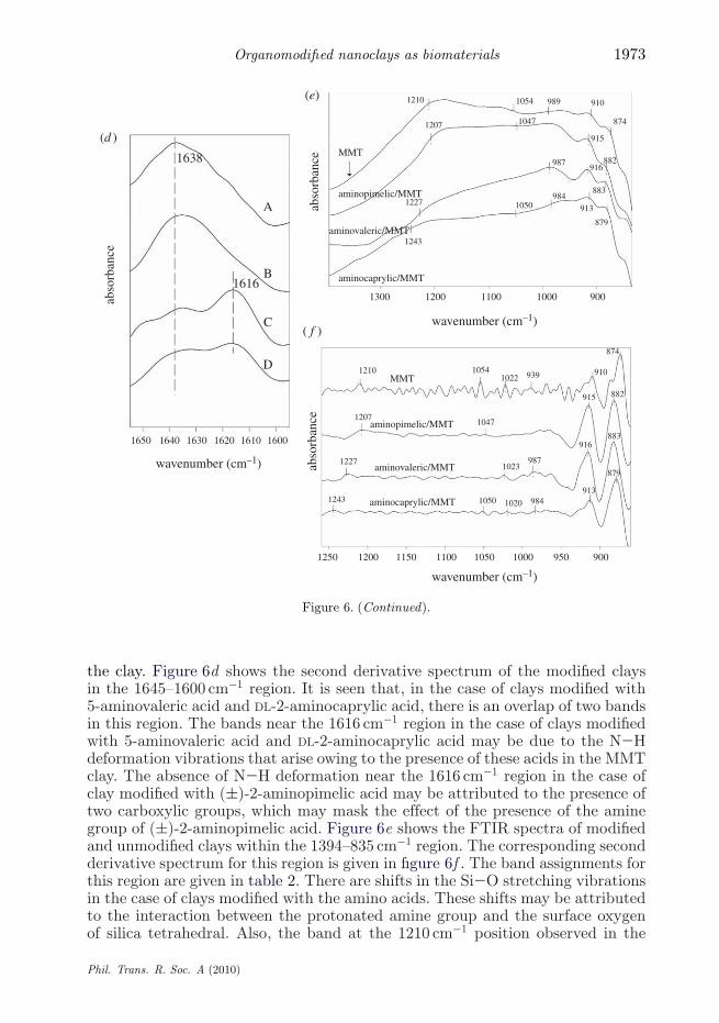
the clay. Figure 6d shows the second derivative spectrum of the modified claysin the 1645–1600 cm−1 region. It is seen that, in the case of clays modified with5-aminovaleric acid and DL-2-aminocaprylic acid, there is an overlap of two bandsin this region. The bands near the 1616 cm−1 region in the case of clays modifiedwith 5-aminovaleric acid and DL-2-aminocaprylic acid may be due to the N−Hdeformation vibrations that arise owing to the presence of these acids in the MMTclay. The absence of N−H deformation near the 1616 cm−1 region in the case ofclay modified with (±)-2-aminopimelic acid may be attributed to the presence oftwo carboxylic groups, which may mask the effect of the presence of the aminegroup of (±)-2-aminopimelic acid. Figure 6e shows the FTIR spectra of modifiedand unmodified clays within the 1394–835 cm−1 region. The corresponding secondderivative spectrum for this region is given in figure 6f . The band assignments forthis region are given in table 2. There are shifts in the Si−O stretching vibrationsin the case of clays modified with the amino acids. These shifts may be attributedto the interaction between the protonated amine group and the surface oxygenof silica tetrahedral. Also, the band at the 1210 cm−1 position observed in the
Phil. Trans. R. Soc. A (2010)

1974 K. S. Katti et al.
Table 2. Band assignment for MMT clay and MMT clay modified with amino acids.
band position (cm−1) sample band assignment
3646 MMT clay O−H stretching of structural OH group1638 H−O−H deformation1211 Si−O out-of-plane stretching1179, 1054, 989 Si−O stretching910 Al−OH deformation874 Al−FeOH deformation3628 MMT clay modified with O−H stretching of structural OH group2932 (±)-2-aminopimelic acid asymmetric C−H stretching1635 H−O−H deformation1207 Si−O out-of-plane stretching1047 Si−O stretching915 Al−OH deformation882 Al−FeOH deformation3626 MMT clay modified with O−H stretching of structural OH group3259 5-aminovaleric acid N−H symmetric stretching1716 C=O stretching1635 H−O−H deformation1616 N−H deformation987 Si−O stretching916 Al−OH deformation883 Al−FeOH deformation3627 MMT clay modified with O−H stretching of structural OH group2934 DL-2-aminocaprylic acid asymmetric C−H stretching2860 symmetric C−H stretching1731 C=O stretching1634 H−O−H deformation1615 N−H deformation1050, 984 Si−O stretching914 Al−OH deformation879 Al−FeOH deformation
case of pure MMT clay seems to be absent in the case of clays modified with5-aminovaleric acid and DL-2-aminocaprylic acid. This may be an implicationthat the crystal structure of MMT clay has changed due to intercalation of aminoacids in MMT clay. Therefore, the unpolarized infrared beam is unable to causeSi−O out-of-plane stretching vibration in the case of the modified clays.
(c) Cell culture results
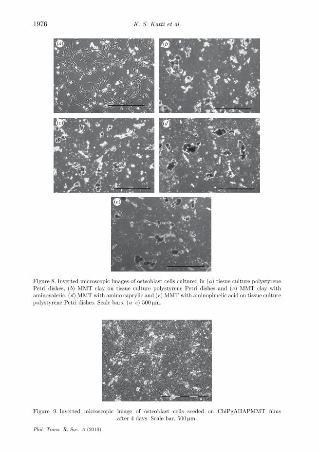
The number of osteoblast cells grown in the presence of MMT clay and MMTclay modified with amino acids was counted manually from the images takenwith an inverted microscope after 48 hours of seeding the osteoblast cells. The celldensity (cells mm−2) for the modified clay samples was calculated and plotted asshown in figure 7a,b. The images in figure 8 show that the osteoblast cells growin the presence of MMT clay and MMT clay modified with the amino acids.The growth of osteoblast cells is comparatively high in the case of MMT clay
Phil. Trans. R. Soc. A (2010)

Organomodified nanoclays as biomaterials 1975
18(a)
(b)
16
14
12
MM
TM
MT po
lyst
yren
e
amin
oval
eric
/MM
Tam
inov
aler
ic/M
MT
amin
opim
elic
/M
MT
amin
opim
elic
/MM
T
amin
ocap
rylic
/M
MT
amin
ocap
rylic
/MM
T
cell
dens
ity (
cells
mm
–2)
10
8
6
4
2
0
90
80
70
60
50
40
cell
dens
ity (
cell
mm
–2)
30
20
10
0
Figure 7. Comparative cell density data after 48 h for (a) MMT clay and MMT clay modified withthe three amino acids and (b) MMT clay, MMT clay modified with the three amino acids andtissue culture polystyrene.
modified with 5-aminovaleric acid and comparatively low in the case of MMTclay modified with DL-2-aminocaprylic acid. Figure 9 shows that osteoblast cellsattach to ChiPgAHAPMMT films. This indicates that the MMT clay modifiedwith 5-aminovaleric acid has potential for biomaterial applications.
5. Conclusions
MMT clay was intercalated with three unnatural amino acids: (±)-2-aminopimelicacid, 5-aminovaleric acid and DL-2-aminocaprylic acid. The intercalation of theclays by all the three amino acids was confirmed by XRD. Transmission FTIRexperiments of clays modified with the three amino acids showed shifts inband positions of C=O vibration that were indicative of significant molecularinteractions between the amino acids and MMT clay. Our prior work on molecular
Phil. Trans. R. Soc. A (2010)
1976 K. S. Katti et al.
(a) (b)
(c) (d )
(e)
Figure 8. Inverted microscopic images of osteoblast cells cultured in (a) tissue culture polystyrenePetri dishes, (b) MMT clay on tissue culture polystyrene Petri dishes and (c) MMT clay withaminovaleric, (d) MMT with amino caprylic and (e) MMT with aminopimelic acid on tissue culturepolystyrene Petri dishes. Scale bars, (a–e) 500 mm.
Figure 9. Inverted microscopic image of osteoblast cells seeded on ChiPgAHAPMMT filmsafter 4 days. Scale bar, 500 mm.
Phil. Trans. R. Soc. A (2010)

Organomodified nanoclays as biomaterials 1977
dynamics simulations of interactions between constituents of PCNs indicatesthat significant non-bonded interactions, such as observed here between clayand modifier, have a large impact on the final mechanical response of thenanocomposite. Cell culture experiments showed that all the three modified clayswere biocompatible. Thus, the MMT clays modified with the three amino acidsused in this study show potential for structural application in biomaterials, asindicated by the cell behaviour on the ChiPgAHAPMMT films prepared by usingthe modified clay.
The authors would like to acknowledge partial support from NSF CAREER0132768. Equipmentused in this research was procured with a grant from NSF MRI 0320657. A.H.A. would like to alsoacknowledge support from NDSU graduate school.
References
Alexandre, M. & Dubois, P. 2000 Polymer layered silicate nanocomposites: preparation,properties and uses of new class of materials. Mater. Sci. Eng. 28, 1–63. (doi:10.1016/S0927-796X(00)00012-7)
Amarasinghe, P. M., Katti, K. S. & Katti, D. R. 2008 Molecular hydraulic property ofmontmorillonite: a polarized FTIR spectroscopic study. Appl. Spectrosc. 62, 1303–1313.(doi:10.1366/000370208786822269)
Amarasinghe, P. M., Katti, K. S. & Katti, D. R. 2009 Nature of organic fluid–montmorillonite interactions: an FTIR spectroscopic study. J. Colloid Interf. Sci. 337, 97–105.(doi:10.1016/j.jcis.2009.05.011)
Bharadwaj, R. K. 2001 Modeling the barrier properties of polymer-layered silicate nanocomposites.Macromolecules 34, 9189–9192. (doi:10.1021/ma010780b)
Bingfeg, S., Ranganathan, B. & Feng, S. S. 2008 Multifunctional poly(D,L-lactide-co-glycolide)/montmorillonite (PLGA/MMT) nanoparticles decorated by trastuzumab for targetedchemotherapy of breast cancer. Biomaterials 29, 475–486. (doi:10.1016/j.biomaterials.2007.09.038)
Chen, G. X., Hao, G. J., Guo, T. Y., Song, M. D. & Zhang, B. H. 2002 Structureand mechanical properties of poly(3-hydroxybutyrate-co-3-hydroxyvalerate) PHBV/claynanocomposites. J. Mater. Sci. Lett. 21, 1587–1589. (doi:10.1023/A:1020309330371)
Darder, M., Colilla, M. & Ruiz-Hitzky, E. 2005 Chitosan–clay nanocomposites: application aselectrochemical sensors. Appl. Clay Sci. 28, 199–208. (doi:10.1016/j.clay.2004.02.009)
Depan, D., Kumar, A. P. & Singh, R. P. 2009 Cell proliferation and controlled drug release studiesof nanohybrids based on chitosan-g-lactic acid and montmorillonite. Acta Biomater. 5, 93–100.(doi:10.1016/j.actbio.2008.08.007)
Dong, Y. C. & Feng, S. S. 2005 Poly (D,L-lactide-co-glycolide)/montmorillonite nanoparticlesfor oral delivery of anticancer drugs. Biomaterials 26, 6068–6076. (doi:10.1016/j.biomaterials.2005.03.021)
Dougherty, D. A. 2000 Unnatural amino acids as probes of protein structure and function. Curr.Opin. Chem. Biol. 6, 645–652. (doi:10.1016/S1367-5931(00)00148-4)
Forni, F., Iannuccelli, V., Coppi, G. & Bernabei, M. T. 1989 Effect of montmorilloniteon drug release from polymeric matrices. Arch. Pharm. 322, 789–793. (doi:10.1002/ardp.19893221103)
Giannelis, E. P. 1996 Polymer layered silicate nanocomposites. Adv. Mater. 8, 29–35. (doi:10.1002/adma.19960080104)
Gilman, J. W. 1999 Flammability and thermal stability studies of polymer-layered silicate (clay)nanocomposites. Appl. Clay Sci. 15, 31–49. (doi:10.1016/S0169-1317(99)00019-8)
Gilman, J. W., Kashiwagi, T. & Lichtenhan, J. D. 1997 Flammability studies of polymer-layeredsilicate nanocomposites. SAMPE J. 33, 40–45.
Gilman, J. W., Jackson, C. L., Morgan, A. B., Harris Jr, R., Manias, E., Giannelis, E. P.,Wuthenow, M., Hilton, D. & Philips, S. H. 2000 Flammability properties of polymer-layered
Phil. Trans. R. Soc. A (2010)

1978 K. S. Katti et al.
silicate nanocomposites. Polypropylene and polystyrene nanocomposites. Chem. Mater. 12,1866–1873. (doi:10.1021/cm0001760)
Gunister, E., Pestreli, D., Ciineyet, C. H., Atici, O. & Gungor, N. 2007 Synthesis andcharacterization of chitosan–MMT biocomposite systems. Carbohydr. Polym. 67, 358–365.(doi:10.1016/j.carbpol.2006.06.004)
Katti, K. S. & Katti, D. R. 2006 Silica–water interactions in montmorillonite using Fouriertransform infrared spectroscopy: relationship to swelling and swelling pressure. Langmuir 22,532–537. (doi:10.1021/la051533u)
Katti, D. R., Ghosh, P., Schmidt, S. & Katti, K. S. 2005 Mechanical properties of sodiummontmorillonite interlayer intercalated with amino acids. Biomacromolecules 6, 3276–3282.(doi:10.1021/bm0503219)
Katti, K. S., Turlapati, P., Verma, D., Gujjula, P. K. & Katti, D. R. 2006 Static and dynamicmechanical behavior of hydroxyapatite–polyacrylic acid composites under simulated body fluid.Am. J. Biochem. Biotechnol. 2, 73–79. (doi:10.3844/ajbbsp.2006.73.79)
Katti, K. S., Katti, D. R. & Dash, R. 2008 Synthesis and characterization of a novelchitosan/montmorillonite/hydroxyapatite nanocomposite for bone tissue engineering. Biomed.Mater. 3, 034 122. (doi:10.1088/1748-6041/3/3/034122)
Langer, R. & Vacanti, J. P. 1993 Tissue engineering. Science 260, 920–926. (doi:10.1126/science.8493529)
Lee, W. F. & Fu, Y. T. 2003 Effect of montmorillonite on the swelling behavior and drugrelease behavior of nanocomposite hydrogels. J. Appl. Polym. Sci. 89, 3652–3660. (doi:10.1002/app.12624)
Lee, Y. H., Chen, C. H., Cheng, W. T. & Kuo, T. F. 2006 Modified montmorilloniteas vector for gene delivery. Biomaterials 27, 3333–3338. (doi:10.1016/j.biomaterials.2005.12.029)
Lim, S. T., Lee, C. H., Choi, H. J. & Jhon, M. S. 2003 Solidlike transition of melt-intercalatedbiodegradable polymer/clay nanocomposites. J. Polym. Sci. B Polym. Phys. 41, 2052–2061.(doi:10.1002/polb.10570)
Lin, K. F., Hsu, C. Y., Huang, T. S., Chiu, W. Y., Lee, Y. H. & Young, T. H. 2005 A novelmethod to prepare chitosan/montmorillonite nanocomposites. J. Polym. Sci. 98, 2042–2047.(doi:10.1002/app.22401)
Ma, J. S. 2003 Unnatural amino acids in drug discovery. Chim. Oggi Chem. Today 21, 65–68.Ma, C. C. M., Kuo, C. T., Kuan, H. C. & Chiang, C. L. 2003 Effects of swelling agents on the
crystallization behavior and mechanical properties of polyamide 6/clay nanocomposites. J. Appl.Polym. Sci. 88, 1686–1693. (doi:10.1002/app.11897)
Maity, P., Yamada, K., Okamoto, M., Udea, K. & Okamoto, K. 2002 New polylactide/layeredsilicate nanocomposites: role of organoclays. Chem. Mater. 14, 4654–4661. (doi:10.1021/cm020391b)
Marras, S., Kladi, K. P., Tsivintzelis, I., Zuburtikudis, I. & Panayiotou, C. 2008 Biodegradablepolymer nanocomposites: the role of nanoclays on thermomechanical characteristics and theelectrospun fibrous structure. Acta Biomater. 4, 756–764. (doi:10.1016/j.actbio.2007.12.005)
Messersmith, P. B. & Giannelis, E. P. 1995 Synthesis and barrier properties of poly (3-caprolactone)-layered silicate nanocomposites. J. Polym. Sci. A Polym. Chem. 33, 1047–1057.(doi:10.1002/pola.1995.080330707)
Okada, A., Kawasumi, M., Usuki, A., Kojima, Y., Kurauchi, T. & Kamigaito, O. 1990 Synthesisand properties of nylon-6/clay hybrids. In Polymer based molecular composites. MRS SymposiumProceedings, vol. 171 (eds D. W. Schaefer & J. E. Mark), pp. 45–50. Pittsburgh, PA: MaterialsResearch Society.
Paiva, L. B., Morales, A. R. & Diaz, F. R. V. 2008 Organoclays: properties, preparation andapplications. Appl. Clay Sci. 42, 8–24. (doi:10.1016/j.clay.2008.02.006)
Park, H. M., Lee, W. K., Park, C. Y., Cho, W. J. & Ha, C. S. 2003 Environmentally friendlypolymer hybrids. Part 1. Mechanical, thermal and barrier properties of thermoplastic starch/claynanocomposites. J. Mater. Sci. 38, 909–915. (doi:10.1023/A:1022308705231)
Pavlidou, S. & Papaspyrides, C. D. 2008 A review on polymer-layered silicate nanocomposites.Prog. Polym. Sci. 33, 1119–1198. (doi:10.1016/j.progpolymsci.2008.07.008)
Phil. Trans. R. Soc. A (2010)

Organomodified nanoclays as biomaterials 1979
Pramanik, M., Srivastava, S. K., Biswas, K. S. & Bhowmick, A. K. 2003 Rubber–claynanocomposite by solution blending. J. Appl. Polym. Sci. 87, 2216–2220. (doi:10.1002/app.11475)
Pramoda, K. P., Liu, T., Liu, Z., He, C. & Sue, H. J. 2003 Synthesis and characterizationof polycarbonate/ABS/montmorillonite nanocomposites. Polym. Degrad. Stab. 80, 157–161.(doi:10.1016/S0141-3910(02)00397-X)
Ray, S. S., Yamada, K., Okamoto, M. & Ueda, K. 2002 New polylactide/layered silicate nano-composite: a novel biodegradable material. Nano Lett. 2, 1093–1096. (doi:10.1021/nl0202152)
Ray, S. S., Okamoto, M. & Ueda, K. 2003 Polymer/layered silicate nanocomposites: a reviewfrom preparation to processing. Prog. Polym. Sci. 28, 1539–1641. (doi:10.1016/j.progpolymsci.2003.08.002)
Rieux, A., Fievez, V., Garinot, M., Schneider, Y. & Preat, V. 2006 Nanoparticles as potentialoral delivery systems of proteins and vaccines: a mechanistic approach. J . Control. Release 116,1–27. (doi:10.1016/j.jconrel.2006.08.013)
Sikdar, D., Katti, D. R. & Katti, K. S. 2007 Effect of organic modifiers on static anddynamic nanomechanical properties and crystallinity of intercalated clay–polycaprolactumnanocomposites. J. Appl. Polym. Sci. 105, 790–802 (doi:10.1002/app.26284)
Sikdar, D., Katti, K. S. & Katti, D. R. 2008a Molecular interactions alter clay andpolymer structure in polymer clay nanocomposites. J. Nanosci. Nanotechnol. 8, 1638–1657.(doi:10.1166/jnn.2008.032)
Sikdar, D., Katti, D. R. & Katti, K. S. 2008b The role of interfacial interactions on the crystallinityand nanomechanical properties of clay–polymer nanocomposites: a molecular dynamics study.J. Appl. Polym. Sci. 107, 3137–3148. (doi:10.1002/app.27504)
Sikdar, D., Pradhan, S. M., Katti, D. R., Katti, K. S. & Mohanty, B. 2008c Altered phase modelfor polymer clay nanocomposites. Langmuir 24, 5599–5607. (doi:10.1021/la800583h)
Sikdar, D., Katti, D. R., Katti, K. S. & Mohanty, B. 2009 Influence of backbone chainlength and functional groups of organic modifiers on crystallinity and nanomechanicalproperties of intercalated clay-polycaprolactum. Int. J. Nanotechnol. 6, 468–492. (doi:10.1504/IJNT.2009.024641)
Takahashi, T., Yamada, Y., Kataoka, K. & Nagasaki, Y. 2005 Preparation of a novel PEG-clayhybrid as a DDS material: dispersion stability and sustained release profiles. J. Control. Release107, 408–416. (doi:10.1016/j.jconrel.2005.03.031)
Verma, D., Katti, K. S. & Katti, D. R. 2008a Effect of biopolymers on structure of hydroxyapatiteand interfacial interactions in biomimetically synthesized hydroxyapatite/biopolymernanocomposites. Ann. Biomed. Eng. 36, 1024–1032. (doi:10.1007/s10439-008-9483-2)
Verma, D., Katti, K. S., Katti, D. R. & Mohanty, B. 2008b Mechanical response and multilevelstructure of biomimetic hydroxyapatite/polygalacturonic/chitosan nanocomposites. Mater. Sci.Eng. C 28, 399–405. (doi:10.1016/j.msec.2007.04.026)
Verma, D., Katti, K. S. & Katti, D. R. 2009 Polyelectrolyte-complex nanostructured fibrousscaffolds for tissue engineering. Mater. Sci. Eng. C 29, 2079–2084. (doi:10.1016/j.msec.2009.04.006)
Viseras, C., Aguzzi, C., Cerezo, P. & Lopez-Galindo, A. 2007 Uses of clay minerals in semisolidhealth care and therapeutic products. Appl. Clay Sci. 36, 37–50. (doi:10.1016/j.clay.2006.07.006)
Wang, S. F., Shen, L., Tong, Y. J., Chen, L., Phang, I. Y., Lim, P. Q. & Liu, T. 2005 Biopolymerchitosan/montmorillonite nanocomposites: preparation and characterization. Polym. Degrad.Stab. 90, 123–131. (doi:10.1016/j.polymdegradstab.2005.03.001)
Wang, S., Chen, L. & Tong, Y. 2006 Structure–property relationship in chitosan-basedbiopolymer/montmorillonite nanocomposites. J. Polym. Sci. A 144, 686–696. (doi:10.1002/pola.20941)
Wang, X., Du, Y., Luo, J., Lin, B. & Kennedy, J. F. 2007 Chitosan/organic rectorite nanocompositefilms: structure, characteristic and drug delivery behavior. Carbohydr. Polym. 69, 41–49.(doi:10.1016/j.carbpol.2006.08.025)
Phil. Trans. R. Soc. A (2010)

1980 K. S. Katti et al.
Wang, X., Du, Y. & Luo, J. 2008 Biopolymer/montmorillonite nanocomposite: preparation,drug-controlled release property and cytotoxicity. Nanotechnology 19, 065 707. (doi:10.1088/0957-4484/19/6/065707)
Xu, R., Manias, E., Snyder, A. J. & Runt, J. 2001 New biomedical poly (urethane-urea)-layeredsilicate nanocomposites. Macromolecules 34, 337–339. (doi:10.1021/ma0013657)
Xu, Y., Ren, X. & Hanna, M. A. 2006 Chitosan/clay nanocomposite film preparation andcharacterization. J. Appl. Polym. Sci. 99, 1684–1691. (doi:10.1002/app.22664)
Yano, K., Usuki, A., Okada, A., Karauchi, T. & Kamigaito, O. 1993 Synthesis and propertiesof polyimide–clay hybrid. J. Polym. Sci. A Polym. Chem. 31, 2493–2498. (doi:10.1002/pola.1993.080311009)
Zheng, J. P., Wang, C. Z., Wang, X. X., Wang, H. Y., Zhuang, H. & Yao, K. D. 2007 Preparation ofbiomimetic three-dimensional gelatin/montmorillonite–chitosan scaffold for tissue engineering.React. Funct. Polym. 67, 780–788. (doi:10.1016/j.reactfunctpolym.2006.12.002)
Phil. Trans. R. Soc. A (2010)
Copyright © 2022 FDOKUMEN